US7795019B2 - Stable cell lines and methods for evaluating gastrointestinal absorption of chemicals - Google Patents
Stable cell lines and methods for evaluating gastrointestinal absorption of chemicals Download PDFInfo
- Publication number
- US7795019B2 US7795019B2 US11/938,065 US93806507A US7795019B2 US 7795019 B2 US7795019 B2 US 7795019B2 US 93806507 A US93806507 A US 93806507A US 7795019 B2 US7795019 B2 US 7795019B2
- Authority
- US
- United States
- Prior art keywords
- cells
- cell
- expression
- protein
- shrna
- Prior art date
- Legal status (The legal status is an assumption and is not a legal conclusion. Google has not performed a legal analysis and makes no representation as to the accuracy of the status listed.)
- Active, expires
Links
Images


Classifications
-
- C—CHEMISTRY; METALLURGY
- C12—BIOCHEMISTRY; BEER; SPIRITS; WINE; VINEGAR; MICROBIOLOGY; ENZYMOLOGY; MUTATION OR GENETIC ENGINEERING
- C12N—MICROORGANISMS OR ENZYMES; COMPOSITIONS THEREOF; PROPAGATING, PRESERVING, OR MAINTAINING MICROORGANISMS; MUTATION OR GENETIC ENGINEERING; CULTURE MEDIA
- C12N15/00—Mutation or genetic engineering; DNA or RNA concerning genetic engineering, vectors, e.g. plasmids, or their isolation, preparation or purification; Use of hosts therefor
- C12N15/09—Recombinant DNA-technology
- C12N15/11—DNA or RNA fragments; Modified forms thereof; Non-coding nucleic acids having a biological activity
- C12N15/113—Non-coding nucleic acids modulating the expression of genes, e.g. antisense oligonucleotides; Antisense DNA or RNA; Triplex- forming oligonucleotides; Catalytic nucleic acids, e.g. ribozymes; Nucleic acids used in co-suppression or gene silencing
- C12N15/1138—Non-coding nucleic acids modulating the expression of genes, e.g. antisense oligonucleotides; Antisense DNA or RNA; Triplex- forming oligonucleotides; Catalytic nucleic acids, e.g. ribozymes; Nucleic acids used in co-suppression or gene silencing against receptors or cell surface proteins
-
- C—CHEMISTRY; METALLURGY
- C12—BIOCHEMISTRY; BEER; SPIRITS; WINE; VINEGAR; MICROBIOLOGY; ENZYMOLOGY; MUTATION OR GENETIC ENGINEERING
- C12N—MICROORGANISMS OR ENZYMES; COMPOSITIONS THEREOF; PROPAGATING, PRESERVING, OR MAINTAINING MICROORGANISMS; MUTATION OR GENETIC ENGINEERING; CULTURE MEDIA
- C12N15/00—Mutation or genetic engineering; DNA or RNA concerning genetic engineering, vectors, e.g. plasmids, or their isolation, preparation or purification; Use of hosts therefor
- C12N15/09—Recombinant DNA-technology
- C12N15/11—DNA or RNA fragments; Modified forms thereof; Non-coding nucleic acids having a biological activity
- C12N15/111—General methods applicable to biologically active non-coding nucleic acids
-
- G—PHYSICS
- G01—MEASURING; TESTING
- G01N—INVESTIGATING OR ANALYSING MATERIALS BY DETERMINING THEIR CHEMICAL OR PHYSICAL PROPERTIES
- G01N33/00—Investigating or analysing materials by specific methods not covered by groups G01N1/00 - G01N31/00
- G01N33/48—Biological material, e.g. blood, urine; Haemocytometers
- G01N33/50—Chemical analysis of biological material, e.g. blood, urine; Testing involving biospecific ligand binding methods; Immunological testing
- G01N33/5005—Chemical analysis of biological material, e.g. blood, urine; Testing involving biospecific ligand binding methods; Immunological testing involving human or animal cells
- G01N33/5008—Chemical analysis of biological material, e.g. blood, urine; Testing involving biospecific ligand binding methods; Immunological testing involving human or animal cells for testing or evaluating the effect of chemical or biological compounds, e.g. drugs, cosmetics
- G01N33/502—Chemical analysis of biological material, e.g. blood, urine; Testing involving biospecific ligand binding methods; Immunological testing involving human or animal cells for testing or evaluating the effect of chemical or biological compounds, e.g. drugs, cosmetics for testing non-proliferative effects
-
- C—CHEMISTRY; METALLURGY
- C12—BIOCHEMISTRY; BEER; SPIRITS; WINE; VINEGAR; MICROBIOLOGY; ENZYMOLOGY; MUTATION OR GENETIC ENGINEERING
- C12N—MICROORGANISMS OR ENZYMES; COMPOSITIONS THEREOF; PROPAGATING, PRESERVING, OR MAINTAINING MICROORGANISMS; MUTATION OR GENETIC ENGINEERING; CULTURE MEDIA
- C12N2310/00—Structure or type of the nucleic acid
- C12N2310/10—Type of nucleic acid
- C12N2310/14—Type of nucleic acid interfering N.A.
-
- C—CHEMISTRY; METALLURGY
- C12—BIOCHEMISTRY; BEER; SPIRITS; WINE; VINEGAR; MICROBIOLOGY; ENZYMOLOGY; MUTATION OR GENETIC ENGINEERING
- C12N—MICROORGANISMS OR ENZYMES; COMPOSITIONS THEREOF; PROPAGATING, PRESERVING, OR MAINTAINING MICROORGANISMS; MUTATION OR GENETIC ENGINEERING; CULTURE MEDIA
- C12N2320/00—Applications; Uses
- C12N2320/10—Applications; Uses in screening processes
- C12N2320/12—Applications; Uses in screening processes in functional genomics, i.e. for the determination of gene function
Definitions
- the invention relates generally to the field of pharmacology. More specifically, the invention features stable cell lines, kits, and methods for predicting the absorption of chemicals such as drugs, nutritional supplements, and environmental chemicals upon administration to animals or humans.
- Drug absorption is the sum total of the effects of various mechanisms by which drugs pass from the point of entry into the bloodstream.
- the rate and efficiency of drug absorption affects the rate and extent to which a drug reaches its intended site of action.
- Gastrointestinal absorption of orally administered drugs is, in part, a function of the permeability of mucosa in the gastrointestinal tract, particularly in the intestines, and also, in part, a function of the transit rate through the various organs of the gastrointestinal tract as the transit rate establishes the length of time the drug is localized to an absorption site.
- Intestinal absorption of drugs can occur via different routes. Many orally administered drugs are absorbed by passive transcellular diffusion through the cell membrane of enterocytes (Van Asperen J et al. (1998) Pharm. Res. 37:429-35) or by passive paracellular diffusion through the tight junctions in the intestinal epithelium (Watson C J et al. (2001) Am. J. Physiol. Cell Physiol. 281:C388-C397). Epithelial drug absorption is also mediated by membrane transport proteins (Lee V H (2001) J. Natl. Cancer Inst. Monogr. 29:41-4). Such transport proteins can also serve as an impediment to drug absorption.
- efflux transporters have been described, including members of the multidrug resistance protein (MRP) family (Borst P et al. (2000) J. Natl. Cancer Inst. 92:1295-1302), P-glycoprotein (P-gp) (Germann U A (1996) Eur. J. Cancer 32A:927-44), and breast cancer resistance protein (BCRP) (Staud F et al. (2005) Int. J. Biochem. Cell Biol. 374:720-5; Doyle LA et al. (1998) Proc. Natl. Acad. Sci. USA. 9526:15665-70), among others.
- MRP multidrug resistance protein
- P-gp P-glycoprotein
- BCRP breast cancer resistance protein
- efflux transport proteins are believed to be primarily responsible for low or variable absorption of orally administered drugs (Stephens R H et al. (2001) J. Pharmacol. Exp. Ther. 296:584-91).
- PAMPA parallel artificial membrane permeability assay
- MDCK Madin-Darby canine kidney cells
- Caco-2 cells Bomane P V et al. (2006) AAPS J. 8:E1-13.
- Caco-2 cells which were derived from a human colon adenocarcinoma, are a widely used model for intestinal absorption studies. When grown and allowed to differentiate, Caco-2 cells are morphologically similar to enterocytes and express many of the enzymes present in the small intestinal brush border, and thus closely resemble the environment and functions of the small intestine. Caco-2 cells provide an additional advantage for intestinal absorption studies in that they express at least three drug efflux transporter proteins, including P-gp (Hunter J et al. (1993) J. Biol. Chem. 268:14991-7), MRP proteins (Hirohashi T et al. (2000) J. Pharmacol. Exp. Ther. 292:265-70; Gutmann H et al. (1999) Pharm. Res. (NY) 16:402-7), and BCRP (Xia C Q et al. (2005) Drug Metab. Dispos. 33:637-43).
- P-gp Heunter J et al. (1993) J.
- RNAi multidrug resistant gene 1
- the invention features isolated nucleic acid molecules for inhibiting expression of at least one membrane efflux transport protein, the nucleic acid molecule comprising the nucleotide sequence of SEQ ID NO: 1, 2, 3, 4, 5, 17, 18, 19, 20, 21, 22, 23, 24, 25, or 26 or allelic variants thereof.
- the nucleic acid molecules can be single, double, or triple stranded.
- the nucleic acid molecules are RNA.
- Vectors comprising nucleic acid sequences encoding a nucleic acid molecule for inhibiting expression of a membrane efflux transport protein, wherein said nucleic acid molecule comprises SEQ ID NO: 1, 2, 3, 4, 5, 17, 18, 19, 20, 21, 22, 23, 24, 25, or 26, or allelic variants thereof are also provided.
- the nucleic acid molecule can be operably linked to one or more regulatory elements such as an inducible or constitutive promoter.
- the vectors can be viral vectors, and in some preferred embodiments are lentivirus vectors.
- Host cells transformed with such vectors are also provided by this invention. It is preferred that such host cells chosen for transformation with the vectors express at least one membrane efflux transport protein such that transformation will result in inhibition of the expression of the protein.
- the membrane efflux transport protein can be any such protein. Examples of membrane efflux transport proteins include P-glycoprotein, Multidrug Resistance-Associated Protein 2, and Breast Cancer Resistance Protein.
- Host cells can be epithelial cells, and are preferably intestinal epithelial cells, and are more preferably human intestinal epithelial cells. Examples of suitable cells include Caco-2 cells, C2BBe1 cells, HT-29 cells, and T-84 cells. Host cell cultures are also provided.
- Also featured are methods for screening compounds for gastrointestinal absorption in an animal such as a human comprising stably inhibiting the expression of at least one membrane efflux transport protein in a cell, contacting the cell with a test compound, measuring transcellular transport of the test compound, and comparing the transcellular transport measurements with reference values for transcellular transport of compounds with no gastrointestinal absorption, low gastrointestinal absorption, moderate gastrointestinal absorption, or high gastrointestinal absorption.
- the measurements relative to the reference values are indicative of the gastrointestinal absorption of the compound in the body of the animal.
- membrane efflux transport proteins include P-glycoprotein, Multidrug Resistance-Associated Protein 2, and Breast Cancer Resistance Protein.
- the inhibiting can comprise stably transforming the cell with a nucleic acid molecule that interferes with the expression of the at least one membrane efflux transport protein.
- suitable cells include Caco-2 cells, C2BBe1 cells, HT-29 cells, and T-84 cells.
- Suitable nucleic acid molecules include those having SEQ ID NO: 1, 2, 3, 4, 5, 17, 18, 19, 20, 21, 22, 23, 24, 25, or 26, and conservatively modified variants or allelic variants thereof.
- stable transformation involves the use of viral, and preferably lentiviral vectors that comprise the nucleic acid molecule.
- a single nucleic acid molecule can inhibit the expression of two or more membrane efflux transport proteins.
- Also featured are methods for screening compounds for gastrointestinal absorption in an animal such as a human comprising stably inhibiting the expression of a first membrane efflux transport protein in a cell, stably inhibiting the expression of a second membrane efflux transport protein in a second cell, contacting the first and second cells with a test compound, measuring transcellular transport of the test compound in the first and second cells, and comparing the transcellular transport measurements with reference values for transcellular transport of compounds with no gastrointestinal absorption, low gastrointestinal absorption, moderate gastrointestinal absorption, or high gastrointestinal absorption.
- the methods further comprise stably inhibiting the expression of a third membrane efflux transport protein in a third cell, contacting the first, second, and third cells with a test compound, measuring transcellular transport of the test compound in the first, second, and third cells, and comparing the transcellular transport measurements with reference values for transcellular transport of compounds with no gastrointestinal absorption, low gastrointestinal absorption, moderate gastrointestinal absorption, or high gastrointestinal absorption. For each comparison, the measurements indicate the relative contribution of the first, second, and third membrane efflux transport protein to transcellular transport of the test compound, and the measurements relative to the reference values are predictive of the gastrointestinal absorption of the compound in the body of the animal.
- membrane efflux transport proteins include P-glycoprotein, Multidrug Resistance-Associated Protein 2, and Breast Cancer Resistance Protein.
- the inhibition of the first, second, and/or third membrane efflux transport protein can comprise transforming the cell with a nucleic acid molecule that interferes with the expression of the at least one membrane efflux transport protein.
- suitable cells include Caco-2 cells, C2BBe1 cells, HT-29 cells, and T-84 cells.
- Suitable nucleic acid molecules include those having SEQ ID NO: 1, 2, 3, 4, 5, 17, 18, 19, 20, 21, 22, 23, 24, 25, or 26, and conservatively modified variants or allelic variants thereof.
- stable inhibition involves the use of viral, and preferably lentiviral vectors that comprise the nucleic acid molecule.
- a single nucleic acid molecule can inhibit the expression of two or more membrane efflux transport proteins.
- the invention also provides methods for inhibiting the expression of a membrane efflux transport protein.
- the methods comprise stably transforming a cell with a vector comprising a nucleic acid sequence encoding a nucleic acid molecule that interferes with the expression of a membrane efflux transport protein, and expressing said nucleic acid molecule in the cell, wherein expression of the nucleic acid molecule inhibits the expression of the membrane efflux transport protein.
- Lentivirus vectors are preferred.
- the membrane efflux transport protein can be P-glycoprotein, Multidrug Resistance-Associated Protein 2, or Breast Cancer Resistance Protein.
- the nucleic acid sequence can comprise SEQ ID NO: 1, 2, 3, 4, 5, 17, 18, 19, 20, 21, 22, 23, 24, 25, or 26, or conservatively modified variants or allelic variants thereof.
- stable transformation involves the use of viral, and preferably lentiviral vectors that comprise the nucleic acid molecule.
- a single nucleic acid molecule can inhibit the expression of two or more membrane efflux transport proteins.
- kits for screening compounds for gastrointestinal absorption in animals are provided by the invention.
- such kits can comprise, in one or more containers, a cell stably transformed with at least one nucleic acid molecule for inhibiting expression of at least one membrane efflux transport protein and instructions for using the kit in a method for screening compounds for gastrointestinal absorption in animals.
- the kits can further comprise additional cells stably transformed with a nucleic acid molecule that interferes with the expression of additional membrane efflux transport protein.
- membrane efflux transport proteins include P-glycoprotein, Multidrug Resistance-Associated Protein 2, and Breast Cancer Resistance Protein.
- suitable cells include Caco-2 cells, C2BBe1 cells, HT-29 cells, and T-84 cells.
- Suitable nucleic acid molecules include those having SEQ ID NO: 1, 2, 3, 4, 5, 17, 18, 19, 20, 21, 22, 23, 24, 25, or 26, and conservatively modified variants or allelic variants thereof.
- the invention also features methods for identifying compounds that inhibit the efflux activity of a membrane efflux transport protein.
- Such methods comprise stably inhibiting the expression of a membrane efflux transport protein in a first cell, contacting the first cell with a substrate of the membrane efflux transport protein, contacting a second cell expressing the membrane efflux transport protein with a test compound and a substrate of the membrane efflux transport protein, determining the efflux activity of the membrane efflux transport protein in the first cell, and in the second cell in the presence and absence of the test compound, and comparing the determined efflux activities, wherein a decrease in the efflux activity in the presence of the test compound relative to the efflux activity in the absence of the test compound, and at least partial identity of the efflux activity in the presence of the test compound with the efflux activity in the first cell indicates that the test compound specifically inhibits the membrane efflux transport protein.
- the stable inhibition of the membrane efflux transport protein in the first cell can comprise transforming the cell with a nucleic acid molecule that interferes with the expression of the at least one membrane efflux transport protein.
- membrane efflux transport proteins include P-glycoprotein, Multidrug Resistance-Associated Protein 2, and Breast Cancer Resistance Protein.
- Suitable nucleic acid molecules include those having SEQ ID NO: 1, 2, 3, 4, 5, 17, 18, 19, 20, 21, 22, 23, 24, 25, or 26, and conservatively modified variants or allelic variants thereof.
- Examples of cells that can serve as the first and/or second cell include Caco-2 cells, C2BBe1 cells, HT-29 cells, and T-84 cells.
- stable inhibition involves the use of viral, and preferably lentiviral vectors that comprise the nucleic acid molecule.
- a single nucleic acid molecule can inhibit the expression of two or more membrane efflux transport proteins.
- any known substrate of the membrane efflux transport protein of interest can be used in the methods.
- substrates include digoxin for P-glycoprotein, vinblastine or dinitrophenyl-5-glutathione for Multidrug Resistance-Associated Protein 2, and mitoxantrone or estrone-3-sulfate for Breast Cancer Resistance Protein.
- FIG. 1 shows P-gp function was significantly decreased in cells transduced with lentiviruses containing SEQ ID NOs: 1, 2, 3, 4, or 5 (shRNA/P-gp 83, 84, 85, 86, or 87, respectively) as described herein, as determined by intracellular calcein fluorescence.
- shRNA/P-gp clone cells KD
- CsA Cyclosporin A
- CsA was used as a positive control (C2BBe1+CsA)
- Un-KD non-transduced C2BBe1 cells
- FIG. 2 shows gel electrophoretic analysis of RT-PCR amplified human P-gp mRNA from shRNA/P-gp clone cells and control cells.
- Non-transduced C2BBe1 un-knockdown C2BBE1 cells
- MDR1/MDCK cells shown in lanes labeled 1 and 2, respectively, prominently display a 208 base pair (bp) band corresponding to P-gp (top panel).
- cells transduced with lentiviruses containing SEQ ID NOs: 1, 2, 3, 4, or 5 show significantly reduced expression of P-gp mRNA.
- ⁇ -actin is shown as a positive control (bottom panel).
- M1 100 bp DNA marker.
- FIG. 3 shows the efflux ratio of digoxin in shRNA/P-gp clone cells.
- the efflux ratio (Papp B-A:A-B) is significantly reduced in cells transduced with lentiviruses containing SEQ ID NOs: 1, 2, 3, 4, or 5 (shRNA/P-gp 83, 84, 85, 86, or 87, respectively) relative to non-transformed C2BBe1 cells (Un-KD).
- FIG. 4 shows a Western blot analysis of P-gp protein expression in C2BBe1 cells transduced with lentiviruses containing SEQ ID NOs: 1, 2, 3, 4, or 5 (shRNA/P-gp 83, 84, 85, 86, or 87, respectively).
- Transduced cells demonstrated significantly reduced P-gp protein expression relative to non-transduced C2BBe1 cells (C) run in parallel.
- P-gp expressed in insect cell microsomes was used as a positive control (P-gp).
- MDCK cell extract which does not express human P-gp, was used as a negative control (M).
- M negative control
- ⁇ -actin was blotted as a standard for normalization of the amount of proteins transferred to the blotting membrane.
- FIG. 5 shows the calculated percent inhibition of P-gp protein expression in C2BBe1 cells transduced with lentiviruses containing SEQ ID NOs: 1, 2, 3, 4, or 5 (shRNA/P-gp 83, 84, 85, 86, or 87, respectively), relative to non-transduced C2BBe1 cells, as determined by Western blot.
- FIG. 6 shows MRP2 mRNA expression in C2BBe1 shRNA/MRP2 clone cells #3, 4, 5, 6, and 7 (transduced with SEQ ID NOs 17, 18, 19, 20, and 21, respectively).
- Vector control (VC) cells were transduced with a control shRNA vector described in Example 7.
- Total cellular RNA from cells with passages ranging from 2 to 5 was isolated after 5 to 7 days of growth, amplified with MRP2-specific primers (SEQ ID NOs: 11 and 12), and resolved by agarose gel electrophoresis.
- ⁇ -actin was included to account for varying efficiencies of total RNA extraction from the cell extracts.
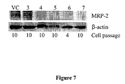
- FIG. 7 shows Western blot analysis of MRP2 protein levels in C2BBe1 shRNA/MRP2 clone cells #3, 4, 5, 6, and 7 (transduced with SEQ ID NOs: 17, 18, 19, 20, and 21, respectively).
- Vector control (VC) cells were transduced with a control shRNA vector described in Example 7.
- the ⁇ -actin was included to account for variations in the amount of total protein applied to the electrophoresis gel.
- FIG. 8 shows BCRP mRNA expression in C2BBe1 cells transduced with lentiviruses containing SEQ ID NOs: 22, 23, 24, 25, or 26 (shRNA/BCRP 798, 799, 800, 801, or 802, respectively).
- Transduced C2BBe1 cells were cultured and grown, and total cellular RNA was extracted. Extracted RNA was amplified by RT-PCR with BCRP-specific primers (SEQ ID NOs: 13 and 14), and the products were resolved by agarose gel electrophoresis. All five shRNA/BCRP inserts significantly reduced BCRP mRNA expression relative to C2BBe1 cells transduced with a control lentivirus containing non-interfering shRNA (VC). ⁇ -actin mRNA production was also assessed to account for varying efficiencies of total RNA extraction from the cell extracts.
- VC non-interfering shRNA
- FIG. 9 shows relative levels of BCRP mRNA expression in C2BBe1 cells transduced with lentiviruses containing SEQ ID NOs: 22, 23, 24, 25, or 26 (shRNA/BCRP 798, 799, 800, 801, or 802, respectively).
- Transduced C2BBe1 cells were cultured and grown, and total cellular RNA was extracted. RT-PCR was carried out, and the products were resolved by agarose gel electrophoresis ( FIG. 8 ).
- BCRP mRNA expression levels were compared with the level of BCRP mRNA expression in C2BBe1 cells transduced with a control lentivirus containing non-interfering shRNA (VC) to determine the degree of inhibition of BCRP mRNA expression caused by interfering shRNAs. Results are illustrated as percent inhibition of BCRP mRNA expression.
- VC non-interfering shRNA
- FIG. 10 shows expression of BCRP protein in C2BBe1 cells transduced with lentiviruses containing SEQ ID NOs: 22, 23, 24, 25, or 26 (shRNA/BCRP 798, 799, 800, 801, or 802, respectively).
- Western blot analysis indicated a substantial decrease in the amount of BCRP protein present in shRNA/BCRP clone cells #798, 799, 800, 801, and 802 (cell passage 15 to 16) as compared to C2BBe1 cells transduced with a control lentivirus containing non-interfering shRNA (VC).
- the ⁇ -actin was included to account for variations in the amount of total protein applied to the electrophoresis gel.
- FIG. 11 shows percent inhibition of BCRP protein expression in shRNA/BCRP clone cells #798, 799, 800, 801, and 802 (as shown in FIG. 10 ).
- the ratio of optical densities for P-gp and beta-actin bands shown in FIG. 10 was used to determine percent inhibition of BCRP protein expression in shRNA/BCRP clone cells relative to expression levels in C2BBe1 cells transduced with a control lentivirus containing non-interfering shRNA.
- FIG. 12 shows expression of BCRP mRNA in shRNA/BCRP clone cell line #801 from cell passages 5 to 20 as measured by RT-PCR. Expression of BCRP mRNA decreased from passage 5 to passage 20. Amplified mRNAs were separated and visualized as described in Example 1. ⁇ -actin was included to account for varying efficiencies of total RNA extraction from the cell extracts.
- FIG. 13 shows expression of P-gp mRNA and MRP2 mRNA in shRNA/BCRP clone 801 at passages 10 and 20.
- Expression of P-gp mRNA in shRNA/BCRP clone 801 was increased compared to vector control cells (VC), non-transduced C2BBe1 (Wt) at both passages 10 and 20.
- VC vector control cells
- Wt non-transduced C2BBe1
- MRP2 mRNA showed significantly decreased expression in clone #801 cells compared to the other cell lines tested only at passage 20.
- ⁇ -actin was included to account for varying efficiencies of total RNA extraction from the cell extracts.
- FIG. 14 shows the corresponding Western blot to FIG. 13 for the expression of BCRP, P-gp and MRP2 proteins in shRNA/BCRP clone cell line #801 for cell passages 10 and 20.
- the MRP2 band is present in shRNA/BCRP clone 801 passage 10, but not passage 20.
- the ⁇ -actin was included to account for variations in the amount of total protein applied to the electrophoresis gel.
- Papp Permeability coefficient
- PappA-B Permeability coefficient in the apical (A) to basolateral (B) direction
- PappB-A Permeability coefficient in the basolateral (B) to apical (A) direction
- ER Efflux ratio, PappB-A/PappA-B ratio
- P-gp P-glycoprotein
- MDR1 Multi-drug resistance protein 1
- BCRP Breast cancer resistance protein
- MRP Multi-drug resistance-associated protein
- MRP2 Multi-drug resistance-associated protein 2
- DMEM Dulbecco's Modified Eagle Medium
- FBS fetal bovine serum
- FTC Fumitremorgin C
- CsA Cyclosporin A
- MK571 MRP Inhibitor
- TEER transepithelial electrical resistance
- KD Knocked-down
- WT Wild-type, unmodified parental cell line
- C2BBe1W Permeability coefficient in the basolateral (B
- isolated means altered “by the hand of man” from the natural state. If a molecule or composition occurs in nature, it has been “isolated” if it has been changed or removed from its original environment, or both. For example, a polynucleotide or a polypeptide naturally present in a living plant or animal is not “isolated,” but the same polynucleotide or polypeptide separated from the coexisting materials of its natural state is “isolated” as the term is employed herein.
- Polynucleotide synonymously referred to as “nucleic acid molecule,” refers to any polyribonucleotide or polydeoxyribonucleotide, which may be unmodified RNA or DNA or modified RNA or DNA.
- Polynucleotides include, without limitation single- and double-stranded DNA, DNA that is a mixture of single- and double-stranded regions, single- and double-stranded RNA, and RNA that is mixture of single- and double-stranded regions, hybrid molecules comprising DNA and RNA that may be single-stranded or, more typically, double-stranded or a mixture of single- and double-stranded regions.
- polynucleotide refers to triple-stranded regions comprising RNA or DNA or both RNA and DNA.
- the term polynucleotide also includes DNAs or RNAs containing one or more modified bases and DNAs or RNAs with backbones modified for stability or for other reasons.
- Modified bases include, for example, tritylated bases and unusual bases such as inosine.
- polynucleotide embraces chemically, enzymatically or metabolically modified forms of polynucleotides as typically found in nature, as well as the chemical forms of DNA and RNA characteristic of viruses and cells.
- Polynucleotide also embraces relatively short nucleic acid chains, often referred to as oligonucleotides.
- a “vector” is a replicon, such as plasmid, phage, cosmid, or virus to which another nucleic acid segment may be operably inserted so as to bring about the replication or expression of the segment.
- RNAi RNAi or shRNA
- operably linked means that the regulatory sequences necessary for expression of the coding sequence are placed in a nucleic acid molecule in the appropriate positions relative to the coding sequence so as to enable expression of the coding sequence.
- a promoter is operably linked with a coding sequence when the promoter is capable of controlling the transcription or expression of that coding sequence.
- Coding sequences can be operably linked to promoters or regulatory sequences in a sense or antisense orientation.
- operably linked is sometimes applied to the arrangement of other transcription control elements (e.g., enhancers) in an expression vector.
- a “heterologous” region of a nucleic acid construct is an identifiable segment (or segments) of the nucleic acid molecule within a larger molecule that is not found in association with the larger molecule in nature.
- the heterologous region encodes a mammalian gene
- the gene will usually be flanked by DNA that does not flank the mammalian genomic DNA in the genome of the source organism.
- a cell has been “transformed” or “transduced” by exogenous or heterologous nucleic acids such as DNA when such DNA has been introduced inside the cell.
- the transforming DNA may or may not be integrated (covalently linked) into the genome of the cell.
- the transforming DNA may be maintained on an episomal element such as a plasmid.
- a stably transformed cell, or “stable cell” is one in which the transforming DNA has become integrated into a chromosome so that it is inherited by daughter cells through chromosome replication.
- a “clone” is a population of cells derived from a single cell or common ancestor by mitosis.
- a “cell line” is a clone of a primary cell that is capable of stable growth in vitro for many generations.
- test compound refers to any purified molecule, substantially purified molecule, molecules that are one or more components of a mixture of compounds, or a mixture of a compound with any other material that can be analyzed using the methods of the present invention.
- Test compounds can be organic or inorganic chemicals, or biomolecules, and all fragments, analogs, homologs, conjugates, and derivatives thereof.
- Biomolecules include proteins, polypeptides, nucleic acids, lipids, monosaccharides, polysaccharides, and all fragments, analogs, homologs, conjugates, and derivatives thereof.
- Test compounds can be of natural or synthetic origin, and can be isolated or purified from their naturally occurring sources, or can be synthesized de novo.
- Test compounds can be defined in terms of structure or composition, or can be undefined.
- the compound can be an isolated product of unknown structure, a mixture of several known products, or an undefined composition comprising one or more compounds.
- undefined compositions include cell and tissue extracts, growth medium in which prokaryotic, eukaryotic, and archaebacterial cells have been cultured, fermentation broths, protein expression libraries, and the like.
- Membrane efflux transport protein refers to any protein transporters localized to a cell membrane. Such transport proteins can have as one of their biological functions the ability to mediate the removal of compounds from the cell interior, herein referred to as “efflux activity.” Efflux activity can result in broad substrate specificity resistance to multiple structure-unrelated therapeutic agents, i.e., multidrug resistance (MDR). The ability of membrane efflux transport protein to confer clinical MDR has generated considerable interest in identifying the substrates and/or inhibitors of such protein and so reversing innate or acquired drug resistance (N. Mizuno, et al. (2003) Pharmacological Rev. 55:425-61).
- the term “modulate” means any change, enhancement or inhibition in the amount, quality, or activity of a particular biomolecule or pathway. “Inhibit” or “inhibition” or “interfere” means to reduce, decrease, block, prevent, delay, inactivate, desensitize, stop, or downregulate the biological activity or expression of a molecule, protein or pathway of interest.
- the level of the expression or biological activity of a protein or pathway of interest refers to a decrease (inhibition or downregulation) or increase (upregulation) of greater than from about 50% to about 99%, and more specifically, about 50%, 51%, 52%, 53%, 54%, 55%, 56%, 57%, 58%, 59%, 60%, 61%, 62%, 63%, 64%, 65%, 66%, 67%, 68%, 69% 70%, 71%, 72%, 73%, 74%, 75%, 76%, 77%, 78%, 79%, 80%, 81%, 82%, 83%, 84%, 85%, 86%, 87%, 88%, 89%, 90%, 91%, 92%, 93%, 94%, 95%, 96%, 97%, 98%, 99%, or more.
- the inhibition may be direct, i.e., operate on the molecule or pathway of interest itself, or indirect, i.e.
- “Knockdown” refers to a cell or organism having reduced expression of one or more genes. As will be appreciated by those skilled in the art, a knockdown will exhibit at least about a 20% reduction in expression, preferably will exhibit at least about a 50% reduction in expression, and more preferably will exhibit at least about a 75% reduction in expression, although higher reductions are possible, including at least about a 80%, 81%, 82%, 83%, 84%, 85%, 86%, 87%, 88%, 89%, 90%, 91%, 92%, 93%, 94%, 95%, 96%, 97%, 98%, 99%, or more reduction in expression.
- percent identity refers to the percentage of sequence identity found in a comparison of two or more nucleic acid sequences.
- “Gastrointestinal absorption” refers to the uptake of chemicals, including biomolecules and test compounds, into or across tissues that comprise the gastrointestinal tract.
- absorption includes, but is not limited to, uptake of compounds from the apical side of a cell and the release of compounds from the basolateral side of a cell.
- the gastrointestinal tract comprises the stomach, small intestine, and large intestine.
- No gastrointestinal absorption means 0% of the compound is absorbed.
- “Low gastrointestinal absorption” means that more than 0%, but less than 25% of a compound is absorbed.
- “Moderate gastrointestinal absorption” means that greater than or equal to 25% but less than 85% of a compound is absorbed.
- “High gastrointestinal absorption” means that greater than or equal to 85% of a compound is absorbed.
- Transcellular transport refers to the movement of a compound across a layer of epithelial cells whereby the compound is moved through the cells and not the spaces between cells such as tight junctions.
- paracellular transport refers to the movement of a compound across a layer of epithelial cell whereby the compound is moved through the tight junctions between cells.
- Efflux transport proteins can limit the absorption of substrate drugs, and can mediate drug-drug interactions that alter, frequently reducing, drug availability or efficacy.
- understanding the role of the various efflux transporters in drug absorption is important in the development of lead compounds.
- the study of efflux transporters has largely relied on the use of chemical inhibitors.
- the use of chemical inhibitors is problematic as the inhibitors are not specific and can affect other cellular processes thereby blurring the overall picture of drug absorption. It has been discovered in accordance with the present invention that the expression of the various efflux transport proteins can be stably inhibited at the genetic level using virally-transformed cells.
- knockdown of the various transporters permits the identification of the particular transporters involved in drug transport with a high degree of certainty.
- the knockdown approach described herein provides stable, sequence-specific silencing of membrane efflux transport proteins induced by endogenous expression of shRNA by lentiviral vectors.
- the invention is advantageous over previous knockdown attempts as the transformation with lentiviral vectors provides permanent, stable inhibition of gene expression, and provides the additional advantage of circumventing non-specific inhibition of other cellular functions that would be expected from chemical inhibition.
- the invention features methods for screening test compounds for gastrointestinal absorption in animals.
- the methods comprise modulating, and preferably inhibiting, the expression of at least one membrane efflux transport protein in a cell, contacting the cell with a test compound, measuring transcellular transport of the test compound, and comparing the transcellular transport measurements with reference values for transcellular transport of compounds with no gastrointestinal absorption, low gastrointestinal absorption, moderate gastrointestinal absorption, or high gastrointestinal absorption.
- the values obtained by experiments measuring the transcellular transport of the compound indicate the degree to which the test compound is likely to be absorbed upon administration to the animal. The methods are thus useful as in vitro models, among other things.
- Membrane efflux transport proteins generally are comprised of one or more subunits that span the plasma membrane of mammalian cells, including mammalian epithelial cells.
- Mammalian epithelial cells are often polarized cells, meaning that their membrane composition differs between the apical or outward facing portion of the cell, and the basolateral or inward portion of the cell.
- apical or outward it is meant that such portions of the cell face a body compartment connected with the outside environment, such as the lumen of the intestine or the lining of the urinary tract or the bile duct.
- Epithelial cells play a role in absorption and elimination of nutrients, drugs and environmental toxins as well as metabolites derived from compounds in each of these categories. Epithelial cells can grow together in sheets in which neighboring cells are linked together by tight junctions, which are intercellular connections that limit the diffusion of ions and larger molecules between cells, called paracellular transport.
- Membrane efflux transporters can be distributed asymmetrically in epithelial cells. This asymmetrical distribution can lead to vectorial transport of compounds that are substrates for such transporters. Vectorial transport means that the rate of transport of a compound differs significantly depending on whether the compound is applied to the apical or basolateral surface of an epithelial cell layer.
- Mammalian membrane transporters can be divided into two general families based on their gene sequence relationships and modes of compound transport.
- the first class is referred to as ABC transporters.
- ABC refers to a common structural feature of this family of transporters, the presence of an Adenosine triphosphate (A) binding (B) cassette (C) structural motif.
- A Adenosine triphosphate
- B B cassette
- These transport proteins typically move chemicals from inside of a cell or from within the phospholipid bilayer of the cell membrane to outside the cell against an unfavorable concentration gradient by using the energy provided by the hydrolytic cleavage of adenosine triphosphate (ATP) to adenosine diphosphate (ADP) and phosphate ion (Pi).
- ATP adenosine triphosphate
- ADP adenosine diphosphate
- Pi phosphate ion
- SLC The second major family of transporters present in mammals is referred to as SLC family where the abbreviation “SLC” refers to the major characteristics of this family of “Solute Linked Carriers,” namely that transport of drugs into or out of a cell is linked to transport of a physiological solute, such as sodium ion, proton or metabolic product, in either the opposite direction, via solute exchange, or in the same direction, via co-transport.
- a physiological solute such as sodium ion, proton or metabolic product
- solute exchange such as sodium ion, proton or metabolic product
- co-transport Unlike the ABC family of transporters, the SLC family generally does not require energy derived from ATP hydrolysis to function. Instead, such transporters utilize the concentration gradients of the co-transported and/or exchanged molecules or ions as an energy source for transporting drugs against an unfavorable concentration gradient.
- SLC frequently mediate compound uptake into cells.
- SLC and ABC transporters are present on opposite sides of the polarized epithelium and work in concert to deliver compounds from the blood into the urine against unfavorable concentration gradients (Wright S. et al. (2004) Physiol. Rev 84:987-1049).
- Table 1 lists human members of the ABC transporter family and some of their known substrates and inhibitors (Zhang L, et al. (2006) Molecular Pharmaceutics 3: 62-9). The list of substrates and inhibitors is for illustration purposes, and is not intended to be exhaustive.
- the drug rifampin and the herbal supplement St. John's wort are established inducers of increased ABCB1 gene expression.
- the ABC transporter family mediates transport of drugs from a wide variety of therapeutic classes, including antineoplastic drugs, such as cisplatin and topotecan, anti-viral drugs, such as indinavir and adefovir and the anti-hyperlipidemic drug, rosuvastatin.
- antineoplastic drugs such as cisplatin and topotecan
- anti-viral drugs such as indinavir and adefovir
- the anti-hyperlipidemic drug rosuvastatin.
- substrates for different transporters For example, indinavir is a substrate for ABCB1, ABCC1, and ABCC2.
- topotecan is a substrate for ABCB1 and ABCG2.
- the inventive methods are applicable for analyzing test compounds for their gastrointestinal absorption in any animal, preferably are applicable to mammals, including companion animals such as dogs, cats, rabbits, and are most preferably applicable to humans.
- the methods can be carried out in any cell that is representative of an animal or group of animals of interest.
- the cells can be freshly isolated, established cell lines, or can be cell lines produced de novo.
- the cell preferably expresses at least one membrane efflux transport protein, although in some embodiments, the cell expresses 2, 3, 4, 5, 6, 7, 8, 9, 10, or more such transport proteins.
- the cell can be engineered specifically to express a particular membrane efflux transport protein, and can be engineered specifically to express two or more particular membrane efflux transport proteins.
- such cells can be engineered to express the particular membrane efflux transport proteins at a particular density, or, in polarized cells, at a particular location on the cell surface, for example, at the basal surface, at the apical surface, both the basal and apical surfaces, or neither the basal or apical surfaces.
- Methods for transforming cells to express a particular efflux protein transgene are known in the art and are routinely practiced, including those that are described and exemplified herein.
- Non-limiting examples of membrane efflux transport proteins that are suitable for analysis using the claimed methods and described cells include P-glycoprotein, Multidrug Resistance-Associated Protein 2, and Breast Cancer Resistance Protein.
- Such transporters can have SEQ ID NOs: 6, 7, or 8, or allelic variants, homologs, and analogs thereof.
- the cell is preferably isolated from or alternatively has the characteristics of a cell isolated from the gastrointestinal tract of the animal.
- the cell can be isolated from the stomach, the small intestine, or the large intestine, including from any subpart of these organs.
- the cells are intestinal cells, particularly intestinal epithelial cells.
- the cells are intestinal cell lines.
- the cells are Caco-2 cells, C2BBe1 cells, HT-29 cells, or T-84 cells.
- MDCK cells Madin-Darby Canine Kidney (MDCK) cells, a cell type known to approximate many of the characteristics of polarized epithelial cells of the gastrointestinal tract of animals, could also be used as one embodiment of the present invention (Maksymowych, A B and Simpson L L, J. Biol. Chem. 273:21950-57 (1998)). MDCK cells (MDR-MDCK) have already been used to assess human P-gp mediated transport of blood-brain barrier compounds (Wang, Q. et al., Int. J. Pharmaceutics 288:349-59 (2005)).
- Modulation of the expression of the at least one membrane efflux transport protein can occur by any means suitable in the art.
- the expression of the transport proteins is inhibited.
- the inhibition is effectuated on the genetic level.
- the transgene in cells specifically engineered to express a transgene encoding a particular efflux transport protein, the transgene can be placed under control of an inducible promoter. Inducible promoters suitable for use in this invention will be known to those of skill in the art.
- genes encoding membrane efflux transport proteins such as P-glycoprotein, Multidrug Resistance-Associated Protein 2, and Breast Cancer Resistance Protein can be inhibited through the use of a variety of other post-transcriptional gene silencing (RNA silencing) techniques.
- RNA silencing involves the processing of double-stranded RNA (dsRNA) into small 21-28 nucleotide fragments by an RNase H-based enzyme (“Dicer” or “Dicer-like”).
- the cleavage products which are siRNA (small interfering RNA) or miRNA (micro-RNA) are incorporated into protein effector complexes that regulate gene expression in a sequence-specific manner.
- RNA interference is a mechanism of post-transcriptional gene silencing mediated by double-stranded RNA (dsRNA), which is distinct from antisense and ribozyme-based approaches (see Jain K K Pharmacogenomics (2004) 5:239-42, for a review of RNAi and siRNA).
- RNA interference is useful in a method for inhibiting the expression of a membrane efflux transport protein in an animal such as a human by administering to the animal a nucleic acid (e.g., dsRNA) that hybridizes under stringent conditions to a gene encoding a membrane efflux transport protein, and attenuates expression of the target gene.
- RNA interference provides shRNA or siRNA that comprise multiple sequences that target one or more regions of the membrane efflux transport protein target gene.
- dsRNA molecules shRNA or siRNA
- DICER RNase III-like enzyme
- shRNA or siRNA are believed to direct sequence-specific degradation of mRNA in cells of various types after first undergoing processing by an RNase III-like enzyme called DICER (Bernstein E et al. (2001) Nature 409:363-366) into smaller dsRNA molecules comprised of two 21 nt strands, each of which has a 5′ phosphate group and a 3′ hydroxyl, and includes a 19 nt region precisely complementary with the other strand, so that there is a 19 nt duplex region flanked by 2 nt-3′ overhangs.
- RNAi is thus mediated by short interfering RNAs (siRNA), which typically comprise a double-stranded region approximately 19 nucleotides in length with 1-2 nucleotide 3′ overhangs on each strand, resulting in a total length of between approximately 21 and 23 nucleotides.
- siRNA short interfering RNAs
- dsRNA longer than approximately 30 nucleotides typically induces nonspecific mRNA degradation via the interferon response.
- the presence of siRNA in mammalian cells rather than inducing the interferon response, results in sequence-specific gene silencing.
- Viral vectors or DNA vectors encode short hairpin RNA (shRNA) which are processed in the cell cytoplasm to short interfering RNA (siRNA).
- a short, interfering RNA comprises an RNA duplex that is preferably approximately 19 basepairs long and optionally further comprises one or two single-stranded overhangs or loops.
- An siRNA may comprise two RNA strands hybridized together, or may alternatively comprise a single RNA strand that includes a self-hybridizing portion.
- siRNAs may include one or more free strand ends, which may include phosphate and/or hydroxyl groups.
- siRNAs typically include a portion that hybridizes under stringent conditions with a target transcript.
- One strand of the siRNA (or, the self-hybridizing portion of the siRNA) is typically precisely complementary with a region of the target transcript, meaning that the siRNA hybridizes to the target transcript without a single mismatch. In certain embodiments of the invention in which perfect complementarity is not achieved, it is generally preferred that any mismatches be located at or near the siRNA termini.
- siRNAs have been shown to downregulate gene expression when transferred into mammalian cells by such methods as transfection, electroporation, cationic liposome-mediated transfection, or microinjection, or when expressed in cells via any of a variety of plasmid-based approaches.
- RNA interference using siRNA is reviewed in, e.g., Tuschl T (2002) Nat. Biotechnol. 20:446-8; Yu J-Y et al. (2002) Proc. Natl. Acad. Sci. 99:6047-52; Sui G et al. (2002) Proc. Natl. Acad. Sci. USA., 99:5515-20; Paddison P J et al. (2002) Genes and Dev.
- the siRNA may consist of two individual nucleic acid strands or of a single strand with a self-complementary region capable of forming a hairpin (stem-loop) structure.
- a hairpin stem-loop
- siRNA capable of effectively mediating gene silencing.
- intracellular processing e.g., by DICER
- target exons rather than introns, and it may also be preferable to select sequences complementary to regions within the 3′ portion of the target transcript.
- sequences that contain approximately equimolar ratio of the different nucleotides and to avoid stretches in which a single residue is repeated multiple times.
- siRNAs may thus comprise RNA molecules having a double-stranded region approximately 19 nucleotides in length with 1-2 nucleotide 3′ overhangs on each strand, resulting in a total length of between approximately 21 and 23 nucleotides.
- siRNAs also include various RNA structures that may be processed in vivo to generate such molecules. Such structures include RNA strands containing two complementary elements that hybridize to one another to form a stem, a loop, and optionally an overhang, preferably a 3′ overhang.
- the stem is approximately 19 bp long, the loop is about 1-20, more preferably about 4-10, and most preferably about 6-8 nt long and/or the overhang is about 1-20, and more preferably about 2-15 nt long.
- the stem is minimally 19 nucleotides in length and may be up to approximately 29 nucleotides in length. Loops of 4 nucleotides or greater are less likely subject to steric constraints than are shorter loops and therefore may be preferred.
- the overhang may include a 5′ phosphate and a 3′ hydroxyl. The overhang may, but need not comprise a plurality of U residues, e.g., between 1 and 5 U residues.
- RNAs are referred to as microRNAs (miRNAs) and are typically between approximately 20 and 26 nucleotides in length, e.g., 22 nt in length.
- stRNAs small temporal RNAs
- mRNA precursors typically approximately 70 nt long with an approximately 4-15 nt loop
- stRNAs small temporal RNAs
- mRNA precursors typically approximately 70 nt long with an approximately 4-15 nt loop
- endogenous RNAs of this type have been identified in a number of organisms including mammals, suggesting that this mechanism of post-transcriptional gene silencing may be widespread (Lagos-Quintana M et al.
- MicroRNAs have been shown to block translation of target transcripts containing target sites in mammalian cells (Zeng Y et al (2002) Mol. Cell. 9:1327-33).
- siRNAs such as naturally occurring or artificial (i.e., designed by humans) mRNAs that bind within the 3′ UTR (or elsewhere in a target transcript) and inhibit translation may tolerate a larger number of mismatches in the siRNA/template duplex, and particularly may tolerate mismatches within the central region of the duplex.
- some mismatches may be desirable or required as naturally occurring stRNAs frequently exhibit such mismatches as do mRNAs that have been shown to inhibit translation in vitro.
- siRNAs when hybridized with the target transcript such siRNAs frequently include two stretches of perfect complementarity separated by a region of mismatch.
- the mRNA may include multiple areas of nonidentity (mismatch).
- the areas of nonidentity need not be symmetrical in the sense that both the target and the mRNA include nonpaired nucleotides.
- the stretches of perfect complementarity are at least 5 nucleotides in length, e.g., 6, 7, or more nucleotides in length, while the regions of mismatch may be, for example, 1, 2, 3, or 4 nucleotides in length.
- Hairpin structures designed to mimic siRNAs and mRNA precursors are processed intracellularly into molecules capable of reducing or inhibiting expression of target transcripts (McManus M T et al. (2002) RNA 8:842-50). These hairpin structures, which are based on classical siRNAs consisting of two RNA strands forming a 19 bp duplex structure are classified as class I or class II hairpins. Class I hairpins incorporate a loop at the 5′ or 3′ end of the antisense siRNA strand (i.e., the strand complementary to the target transcript whose inhibition is desired) but are otherwise identical to classical siRNAs.
- Class II hairpins resemble mRNA precursors in that they include a 19 nt duplex region and a loop at either the 3′ or 5′ end of the antisense strand of the duplex in addition to one or more nucleotide mismatches in the stem. These molecules are processed intracellularly into small RNA duplex structures capable of mediating silencing. They appear to exert their effects through degradation of the target mRNA rather than through translational repression as is thought to be the case for naturally occurring mRNAs and stRNAs.
- any such RNA one portion of which binds to a target transcript and reduces its expression, whether by triggering degradation, by inhibiting translation, or by other means, is considered to be an siRNA, and any structure that generates such an siRNA (i.e., serves as a precursor to the RNA) is useful in the practice of the present invention.
- RNA interference for use in the present invention is the use of short hairpin RNAs (shRNA).
- shRNA short hairpin RNAs
- a plasmid containing a DNA sequence encoding for a particular desired siRNA sequence is delivered into a target cell via transfection or virally-mediated infection. Once in the cell, the DNA sequence is continuously transcribed into RNA molecules that loop back on themselves and form hairpin structures through intramolecular base pairing. These hairpin structures, once processed by the cell, are equivalent to transfected siRNA molecules and are used by the cell to mediate RNAi of the desired protein.
- shRNA has an advantage over siRNA transfection as the former can lead to stable, long-term inhibition of protein expression.
- siRNA-encoding vectors are constructs comprising a promoter, a sequence of the target gene to be silenced in the “sense” orientation, a spacer, the antisense of the target gene sequence, and a terminator.
- Inhibition of the expression of the membrane efflux transport proteins can also be effectuated by other means that are known and readily practiced in the art.
- antisense nucleic acids can be used.
- Antisense RNA transcripts have a base sequence complementary to part or all of any other RNA transcript in the same cell. Such transcripts have been shown to modulate gene expression through a variety of mechanisms including the modulation of RNA splicing, the modulation of RNA transport and the modulation of the translation of mRNA (Denhardt DT (1992) Ann. N Y Acad. Sci. 660:70-6, 1992; Nellen W et al. (1993) Trends Biochem. Sci. 18:419-23; and, Baker B F et al. (1999) Biochim. Biophys. Acta. 1489: 3-18). Accordingly, in certain embodiments of the invention, inhibition of one or more membrane efflux transport proteins in a cell is accomplished by expressing an antisense nucleic acid molecule in the cell.
- Antisense nucleic acids are generally single-stranded nucleic acids (DNA, RNA, modified DNA, or modified RNA) complementary to a portion of a target nucleic acid (e.g., an mRNA transcript) and therefore able to bind to the target to form a duplex.
- a target nucleic acid e.g., an mRNA transcript
- they are oligonucleotides that range from 15 to 35 nucleotides in length but may range from 10 up to approximately 50 nucleotides in length. Binding typically reduces or inhibits the function of the target nucleic acid, such as a gene encoding a membrane efflux transport protein.
- antisense oligonucleotides may block transcription when bound to genomic DNA, inhibit translation when bound to mRNA, and/or lead to degradation of the nucleic acid. Inhibition of the expression of a membrane efflux transport protein can be achieved by the administration of antisense nucleic acids or peptide nucleic acids comprising sequences complementary to those of the mRNA that encodes the membrane efflux transport protein.
- Antisense technology and its applications are well known in the art and are described in Phillips, M. I. (ed.) Antisense Technology, Methods Enzymol., 2000, Volumes 313 and 314, Academic Press, San Diego, and references mentioned therein. See also Crooke, S. (ed.) “A NTISENSE D RUG T ECHNOLOGY : P RINCIPLES , S TRATEGIES, AND A PPLICATIONS ” (1 st Edition) Marcel Dekker; and references cited therein.
- Antisense oligonucleotides can be synthesized with a base sequence that is complementary to a portion of any RNA transcript in the cell. Antisense oligonucleotides can modulate gene expression through a variety of mechanisms including the modulation of RNA splicing, the modulation of RNA transport and the modulation of the translation of mRNA.
- antisense oligonucleotides including stability, toxicity, tissue distribution, and cellular uptake and binding affinity may be altered through chemical modifications including (i) replacement of the phosphodiester backbone (e.g., peptide nucleic acid, phosphorothioate oligonucleotides, and phosphoramidate oligonucleotides), (ii) modification of the sugar base (e.g., 2′-O-propylribose and 2′-methoxyethoxyribose), and (iii) modification of the nucleoside (e.g., C-5 propynyl U, C-5 thiazole U, and phenoxazine C) (Wagner R W (1995) Nat.
- the phosphodiester backbone e.g., peptide nucleic acid, phosphorothioate oligonucleotides, and phosphoramidate oligonucleotides
- modification of the sugar base e.
- RNA and DNA enzymes can be designed to cleave to any RNA molecule, thereby increasing its rate of degradation (Cotten M et al. (1989) EMBO J. 8: 3861-6, 1989; and, Usman N et al. (1996) Curr. Opin. Struct. Biol. 1:527-33).
- the cells used in the inventive methods can be specifically transformed with transcription-silencing nucleic acids such as shRNA or siRNA, or can be transformed with vectors encoding such nucleic acids such that the cell expresses the inhibitory nucleic acid molecules. Transformation of the cells can be carried out according to any means suitable in the art, including those described and exemplified herein.
- the inhibitory nucleic acid molecules comprise SEQ ID NO: 1, 2, 3, 4, 5, 17, 18, 19, 20, 21, 22, 23, 24, 25, or 26, or analogs, homologs, derivatives, or allelic variants thereof.
- test compounds can be screened at a single dose, or with multiple doses.
- the test compound is evaluated at multiple dosages ranging from the compound's free maximal therapeutic plasma concentration (Cmax) to a concentration equal to or greater than 500-fold over the compound's Cmax.
- the test compound is evaluated at multiple dosages ranging from the compound's Cmax to a concentration equal to or greater than 250-fold over the compound's Cmax.
- the test compound is evaluated at multiple dosages ranging from the compound's Cmax to a concentration equal to or greater than 100-fold over the compound's Cmax.
- the test compound is evaluated at multiple dosages ranging from the compound's Cmax to a concentration equal to or greater than 50-fold over the compound's Cmax. In some embodiments, the test compound is evaluated at multiple dosages ranging from the compound's Cmax to a concentration equal to or greater than 30-fold over the compound's Cmax. In some embodiments, the test compound is evaluated at multiple dosages ranging from the compound's Cmax to a concentration equal to or greater than 10-fold over the compound's Cmax.
- Cmax can be determined according to any means available in the art. The skilled artisan will appreciate that such means are known and routine in the art. The compound can be tested at 1, 2, 3, 4, 5, 6, 7, 8, 9, 10, 11, 12, 13, 14, 15, or more concentrations within this range.
- multiple test compounds are contacted with the cell to evaluate drug-drug interactions.
- Drug-drug interactions are defined as influences of one drug on the pharmacokinetics or pharmacodynamics of a second drug co-administered to the same subject.
- Pharmacokinetics refers to the influence of varying drug doses and methods of administration on the concentration of drug in various body tissues, such as blood, blood plasma, brain, etc., as a function of time after drug administration.
- Pharmacodynamics refers to the study of the influence of drug dose and route of administration on the pharmacological response, such as blood pressure, blood lipid level, number of infectious viral particles in a tissue, etc., to drug administration.
- Drug-drug interactions typically occur by one of three mechanisms: 1) two or more drugs compete for the same limited quantity of an enzyme or transport protein responsible for their metabolism, uptake or efflux in the body; 2) one drug inhibits an enzyme, uptake or efflux transporter that mediates the metabolism, uptake or excretion of one or more other drugs by the body; or 3) one drug enhances or inhibits the production of an enzyme, uptake or efflux transporter responsible for the metabolism, uptake or efflux of one or more other drugs in the body.
- the interacting substance is not a drug, but rather a natural component of a dietary product, such as a component of a fruit juice, such as grapefruit juice, or an herbal supplement, such as Saint John's wort.
- Also featured are methods for screening compounds for gastrointestinal absorption that utilize a parallel analysis of the inhibition of different membrane efflux transport proteins in separate cells.
- a given test compound can be screened in a panel of cells, each cell in the panel being a knockdown for expression of a different membrane efflux transport protein, or a different combination of membrane efflux transport proteins.
- those of skill in the art can advantageously determine the contribution of each individual transport protein or of particular combinations of transport proteins on the gastrointestinal absorption of a test compound.
- the inventive methods comprise inhibiting the expression of a first membrane efflux transport protein in a first cell and inhibiting the expression of a second membrane efflux transport protein in a second cell, contacting the first and second cells with a test compound, and measuring transcellular transport of the test compound in the first and second cells.
- the transcellular transport measurements from each of the first and second cells can then be compared with reference values for transcellular transport of compounds with no gastrointestinal absorption, low gastrointestinal absorption, moderate gastrointestinal absorption, or high gastrointestinal absorption.
- the measurements indicate the role of the first and second membrane efflux transport proteins in transcellular transport of the test compound.
- the measurements relative to the reference values are predictive of the gastrointestinal absorption of the compound in the body of the animal of interest.
- the methods comprise inhibiting additional membrane efflux transport proteins such as a third, fourth, fifth, sixth, or more transport proteins, or combinations thereof, in separate cells, thereby expanding the number of cells in the panel.
- the methods comprise inhibiting the expression of a first membrane efflux transport protein in a first cell, inhibiting the expression of a second membrane efflux transport protein in a second cell, and inhibiting the expression of a third membrane efflux transport protein in a third cell, contacting the first, second, and third cells with a test compound, and measuring transcellular transport of the test compound in the first, second, and third cells.
- the transcellular transport measurements from each of the first, second, and third cells can then be compared with reference values for transcellular transport of compounds with no gastrointestinal absorption, low gastrointestinal absorption, moderate gastrointestinal absorption, or high gastrointestinal absorption.
- the measurements indicate the role of the first, second, and third membrane efflux transport proteins in transcellular transport of the test compound.
- the measurements relative to the reference values are predictive of the gastrointestinal absorption of the compound in the body of the animal of interest.
- Variations on such methods comprise inhibiting the expression of two or more membrane efflux transport proteins in a first cell, and at least one membrane efflux transport protein in a second cell.
- the expression of two or more membrane efflux transport proteins can be inhibited, for example, by means of a single nucleic acid molecule that can inhibit the expression of two or more membrane efflux transport proteins.
- the membrane efflux transport protein in the second cell can be the same as one of the membrane efflux transport proteins in the first cell, or can be a different membrane efflux transport protein.
- the second, third, fourth (and the like) cells can be freshly isolated, established cell lines, or can be cell lines produced de novo.
- the cells preferably expresses at least one membrane efflux transport protein, although in some embodiments, the cell expresses 2, 3, 4, 5, 6, 7, 8, 9, 10, or more such transport proteins.
- the cells can be engineered specifically to express a particular membrane efflux transport protein, and can be engineered specifically to express two or more particular membrane efflux transport proteins.
- such cells can be engineered to express the particular membrane efflux transport proteins at a particular density, or, in polarized cells, at a particular location on the cell surface, for example, at the basal surface, at the apical surface, both the basal and apical surfaces, or neither the basal or apical surfaces.
- Non-limiting examples of membrane efflux transport proteins that are contemplated for analysis using panels of cells include P-glycoprotein, Multidrug Resistance-Associated Protein 2, and Breast Cancer Resistance Protein.
- the cells are preferably isolated from, or alternatively have the characteristics of a cell isolated from the gastrointestinal tract of the animal of interest.
- the cells can be isolated from the stomach, the small intestine, or the large intestine, including from any subpart of these organs.
- the cells are intestinal cells, particularly intestinal epithelial cells.
- the cells are intestinal cell lines, which can be neoplastically transformed, or otherwise immortalized.
- the cells are Caco-2 cells, C2BBe1 cells, HT-29 cells, or T-84 cells.
- Transcellular transport of the test compound in any of the first, second, third, or more cells can be measured according to any means suitable in the art.
- Transepithelial electrical resistance (TEER) measurements which are routinely carried out, can also be used.
- Liquid chromatography-mass spectrometry (LC-MS) and LC-tandem mass spectrometry (LC-MS-MS) can be used (van Breemen R B, et al. (2005) Expert Opin. Drug Metab. Toxicol. 1:175-85).
- fluorescent dyes or radioisotope can be used, for example, by tagging the test compound with an acceptable dye or isotope as the labels can be conveniently detected by fluorescence or liquid scintillation counting.
- Non-limiting examples include fluorescent Rhodamine 123, and radiolabeled cyclosporine A, digoxin, ritonavir, taxol, verapamil, and vinblastine (Troutman M T (2003) Pharm. Res. 20:1210-24).
- Unidirectional (mucosal-to-serosal transport) or bidirectional (mucosal-to-serosal and serosal-to-mucosal transport) permeability of the cells can be measured.
- a drug solution is added to the apical (mucosal) side of the cell monolayers, samples are collected from the basolateral (serosal) side, and a permeability coefficient is determined by the accumulative drug transported across divided by the time, surface area and dose concentration.
- Measurements of the transcellular transport of the test compound can be directly compared with reference values for the transcellular transport, i.e., gastrointestinal absorption rate, efficiency, capacity, etc., of compounds in which transcellular transport has been previously characterized.
- reference values for the transcellular transport i.e., gastrointestinal absorption rate, efficiency, capacity, etc.
- measurements obtained from the test compound can be compared to reference values for compounds with no gastrointestinal absorption, with low gastrointestinal absorption, with moderate gastrointestinal absorption, with high gastrointestinal absorption, or with any combination of such reference values.
- Non-limiting examples of such reference values are provided by Table 2 (Artursson P. et al. (1991) Biochem. Biophys. Res. Commun. 175:880-5).
- test compound measurement values experimentally obtained relative to the reference values is indicative, and at least predictive, of the test compound's absorption in the gastrointestinal tract of the animal of interest. It is contemplated that compounds characterized according to the methods of the invention can serve as reference compounds, representing the relative degree of gastrointestinal absorption, against which additional test compounds can be compared.
- the invention also features methods for inhibiting the expression of membrane efflux transport proteins in cells.
- the methods comprise stably transforming a cell with a nucleic acid molecule that interferes with the expression of the membrane efflux transport protein.
- the nucleic acid molecule can inhibit the expression by inhibiting the transcription of the gene encoding the membrane efflux transport protein, or can inhibit the expression by inhibiting the translation of mRNA into the protein.
- the nucleic acid molecule can be any regulatory gene or fragment of a gene whose expression or presence in the cell inhibits transcription or translation of the efflux transport protein gene product.
- the nucleic acid molecule is RNA.
- the nucleic acid molecule is interfering RNA, and is preferably double stranded.
- Non-limiting examples of interfering RNA include siRNA and shRNA.
- nucleic acid molecules can inhibit the expression of two or more membrane efflux transport proteins. Accordingly, such nucleic acid molecules can be advantageously used in any of the inventive methods described and exemplified herein.
- a non-limiting example of an individual nucleic acid molecule that can inhibit two or more membrane efflux transport proteins is SEQ ID NO: 25. This nucleic acid molecule has been demonstrated to inhibit the expression of at least both BCRP and MRP2. The observation that a single nucleic acid molecule can inhibit two or more membrane efflux transport proteins represents a significant advance for the determination of the relative contribution of select membrane efflux transport proteins on cellular absorption and transport of compounds.
- a cell can be transformed with such nucleic acid molecules according to any means available in the art such as those describe or exemplified herein. It is preferred that cells are stably transformed with a vector comprising a nucleic acid sequence encoding such regulatory nucleic acid molecules. Any vector suitable for transformation of the particular cell of interest can be used in the present invention.
- the vector is a viral vector.
- the vector is a lentivirus vector.
- the regulatory nucleic acid molecule can comprise any sequence complementary to, or otherwise amenable to hybridization to and/or interference with the expression of a gene encoding the membrane efflux transport protein of interest.
- Non-limiting examples of such nucleic acid sequences include SEQ ID NOs: 1, 2, 3, 4, 5, 17, 18, 19, 20, 21, 22, 23, 24, 25, and 26, and allelic variants thereof.
- Preferred, but non-limiting examples of membrane efflux transport proteins include P-glycoprotein, Multidrug Resistance-Associated Protein-2, and Breast Cancer Resistance Protein.
- Preferred cells that can be targeted for modulation, particularly inhibition, of the expression of membrane efflux transport proteins can be any cell that expresses such transport proteins.
- Such cells can express the transport proteins naturally, or the cells can be engineered to express the transport proteins.
- the cells can be isolated fresh from a host organism, or can be cell lines. It is preferred that such cells be of a gastrointestinal lineage, and it is particularly preferred that such cells be intestinal epithelial cells.
- Non-limiting examples of cell lines amenable to genetic regulation according to the inventive methods include Caco-2 cells, C2BBe1 cells, HT-29 cells, T-84 cells, and HRT-18 cells.
- the invention also features isolated nucleic acid molecules for the genetic regulation of membrane efflux transport expression.
- the nucleic acid molecules of the invention that encode regulatory, particularly inhibitory, sequences include SEQ ID NOs: 1, 2, 3, 4, 5, 17, 18, 19, 20, 21, 22, 23, 24, 25, and 26, and allelic variants, homologs, and natural mutants of SEQ ID NOs: 1, 2, 3, 4, 5, 17, 18, 19, 20, 21, 22, 23, 24, 25, and 26.
- this invention provides isolated polynucleotides that have at least about 60%, preferably at least about 61%, 62%, 63%, 64%, 65%, 66%, 67%, 68%, 69% or 70%, more preferably at least about 71%, 72%, 73%, 74%, 75%, 76%, 77%.
- Nucleic acid molecules of the invention may be prepared by two general methods: (1) they may be synthesized from appropriate nucleotide triphosphates, or (2) they may be isolated from biological sources. Both methods utilize protocols well known in the art.
- nucleotide sequence information such as the entire nucleic acid sequence of the membrane efflux transport protein, for example, SEQ ID NOs: 6-8, enables preparation of an isolated nucleic acid molecule of the invention by oligonucleotide synthesis.
- Synthetic oligonucleotides may be prepared by the phosphoramadite method employed in the Applied Biosystems 38A DNA Synthesizer or similar devices.
- the resultant construct may be purified according to methods known in the art, such as high performance liquid chromatography (HPLC).
- HPLC high performance liquid chromatography
- Nucleic acids of the present invention may be maintained as DNA in any convenient cloning vector.
- clones are maintained in plasmid cloning/expression vector, either of which can be propagated in a suitable prokaryotic or eukaryotic host cell.
- Nucleic acid molecules of the invention include cDNA, genomic DNA, RNA, and fragments thereof which may be single-, double-, or even triple-stranded.
- this invention provides oligonucleotides (sense or antisense strands of DNA or RNA) having sequences capable of hybridizing with at least one sequence of a nucleic acid molecule of the present invention, in particular, SEQ ID NOs: 6-8.
- Such oligonucleotides are useful as probes for detecting genes encoding membrane efflux transport proteins, or for the positive or negative regulation of expression of genes encoding a membrane efflux transport protein at or before translation of the mRNA into proteins.
- oligonucleotides or polynucleotides may be utilized as probes for such assays include, but are not limited to: (1) in situ hybridization; (2) Southern hybridization (3) northern hybridization; and (4) assorted amplification reactions such as polymerase chain reactions (PCR) and ligase chain reaction (LCR).
- PCR polymerase chain reactions
- LCR ligase chain reaction
- vectors and kits for producing transgenic host cells that comprise a polynucleotide encoding a regulatory sequence for inhibiting the expression of a membrane efflux transport protein, or homolog, analog or variant thereof in a sense or antisense orientation, or a construct under control of cell or tissue-specific promoters and/or other regulatory sequences.
- Such vectors are suitable for modulating, and preferably inhibiting, the expression of any membrane efflux transport protein.
- the membrane efflux transport protein is P-glycoprotein, Multidrug Resistance-Associated Protein 2, or Breast Cancer Resistance Protein.
- Suitable host cells include, but are not limited to, plant cells, bacterial cells, yeast and other fungal cells, insect cells and mammalian cells. More preferred are human cells. Even more preferred are human intestinal epithelial cells. Most preferred are Caco-2 cells, C2BBe1 cells, HT-29 cells, or T-84 cells.
- Vectors for transforming a wide variety of these host cells are well known to those of skill in the art. They include, but are not limited to, plasmids, phagemids, cosmids, baculoviruses, bacmids, bacterial artificial chromosomes (BACs), yeast artificial chromosomes (YACs), as well as other bacterial, yeast and viral vectors.
- viral vectors are used. It is particularly preferred that lentiviral vectors are used.
- kits for producing transgenic host cells will contain one or more appropriate vectors and instructions for producing the transgenic cells using the vector.
- Kits may further include one or more additional components, such as culture media for culturing the cells, reagents for performing transformation of the cells and reagents for testing the transgenic cells for gene expression or regulation, to name only a few.
- the coding region of the regulatory sequence is placed under a powerful constitutive promoter, such as the promoters for the following genes: hypoxanthine phosphoribosyl transferase (HPRT), adenosine deaminase, pyruvate kinase, ⁇ -actin, human myosin, human hemoglobin, human muscle creatine, and others.
- a powerful constitutive promoter such as the promoters for the following genes: hypoxanthine phosphoribosyl transferase (HPRT), adenosine deaminase, pyruvate kinase, ⁇ -actin, human myosin, human hemoglobin, human muscle creatine, and others.
- HPRT hypoxanthine phosphoribosyl transferase
- adenosine deaminase pyruvate kinase
- ⁇ -actin human myosin
- human hemoglobin human muscle creatine
- Such viral promoters include without limitation, Cytomegalovirus (CMV) immediate early promoter, the early and late promoters of SV40, the Mouse Mammary Tumor Virus (MMTV) promoter, the long terminal repeats (LTRs) of Maloney leukemia virus, Human Immunodeficiency Virus (HIV), Epstein Barr Virus (EBV), Rous Sarcoma Virus (RSV), and other retroviruses, and the thymidine kinase promoter of Herpes Simplex Virus.
- CMV Cytomegalovirus
- MMTV Mouse Mammary Tumor Virus
- LTRs long terminal repeats
- HCV Human Immunodeficiency Virus
- EBV Epstein Barr Virus
- RSV Rous Sarcoma Virus
- thymidine kinase promoter Herpes Simplex Virus
- the coding region of the regulatory sequence is placed under an inducible promoter such as the metallothionein promoter, tetracycline-inducible promoter, doxycycline-inducible promoter, promoters that contain one or more interferon-stimulated response elements (ISRE) such as protein kinase R 2′,5′-oligoadenylate synthetases, Mx genes, ADAR1, and the like.
- ISRE interferon-stimulated response elements
- the vectors of the invention can be used to transform various cells with the various regulatory nucleic acid sequences of the invention.
- another aspect of the invention features host cells transformed with vectors comprising a nucleic acid sequence encoding a nucleic acid molecule for modulating, preferably inhibiting, the expression of a membrane efflux transport protein.
- Numerous techniques are known in the art for the introduction of foreign genes into cells and may be used to construct the recombinant cells for purposes of carrying out the inventive methods, in accordance with the various embodiments of the invention.
- the technique used should provide for the stable transfer of the heterologous gene sequence to the host cell, such that the heterologous gene sequence is heritable and expressible by the cell progeny, and so that the necessary development and physiological functions of the recipient cells are not disrupted.
- Techniques which may be used include, but are not limited to, chromosome transfer (e.g., cell fusion, chromosome-mediated gene transfer, micro cell-mediated gene transfer), physical methods (e.g., transfection, spheroplast fusion, microinjection, electroporation, liposome carrier), viral vector transfer (e.g., recombinant DNA viruses, recombinant RNA viruses) and the like (described in Cline M J (1985) Pharmac. Ther. 29:69-92). It is preferable that a viral vector be used to transform the cells of the invention. It is more preferable that the viral vector be a lentiviral vector.
- Knockdown cells with inhibited expression of membrane efflux transport proteins can be created by inhibiting the translation of mRNA encoding the transport protein by “post-transcriptional gene silencing.”
- the gene from the species targeted for down-regulation, or a fragment thereof, may be utilized to control the production of the encoded protein.
- Full-length antisense molecules can be used for this purpose.
- antisense oligonucleotides targeted to specific regions of the mRNA that are critical for translation may be utilized.
- the use of antisense molecules to decrease expression levels of a pre-determined gene is known in the art.
- Antisense molecules may be provided in situ by transforming cells with a DNA construct which, upon transcription, produces the antisense RNA sequences.
- Such constructs can be designed to produce full-length or partial antisense sequences.
- This gene silencing effect can be enhanced by transgenically over-producing both sense and antisense RNA of the gene coding sequence so that a high amount of dsRNA is produced (for example see Waterhouse et al. (1998) Proc. Natl. Acad. Sci. U.S.A. 95:13959-64).
- dsRNA containing sequences that correspond to part or all of at least one intron have been found particularly effective.
- part or all of the coding sequence antisense strand is expressed by a transgene.
- hybridizing sense and antisense strands of part or all of the coding sequence for one or more membrane efflux transport proteins are transgenically expressed.
- Cells of the invention C2BBE1cells transduced with lentiviruses encoding interfering nucleic acid molecules (SEQ ID NOs: 1, 3, 21, 24, and 25), have been placed with the Amer. Type Cult. Coll. (10801 University Boulevard., Manassas, Va. 20110-2209) on Nov. 6, 2007 and have been assigned Access. Nos. PTA-8752, PTA-8753, PTA-8751, PTA-8754, and PTA-8755, respectively.
- kits for screening compounds for gastrointestinal absorption in animals comprise a cell that has been transformed with at least one nucleic acid molecule that inhibits expression of at least one membrane efflux transport protein, as well as instructions for using the kit in a method for screening compounds for gastrointestinal absorption in animals.
- the kits of the invention can further comprise a second cell transformed with a nucleic acid molecule that interferes with the expression of a second membrane efflux transport protein.
- the kit of the invention can also further comprise a second and third cell transformed with a nucleic acid molecule that interferes with the expression of a second and third membrane efflux transport protein.
- the cells of the kits can be Caco-2 cells, and preferably are C2BBe1 cells. However, any cell that stably expresses at least one membrane efflux transport protein of interest can be used.
- the cells can be transformed with any nucleic acid molecule that inhibits the expression of the membrane efflux transport protein of interest, such as but not limited to, those that are described and exemplified herein.
- the nucleic acid molecule used to transform the cells can have at least one of SEQ ID NO: 1, 2, 3, 4, 5, 17, 18, 19, 20, 21, 22, 23, 24, 25, or 26, or allelic variants thereof.
- the membrane efflux transport protein of interest whose expression is inhibited by transformation of the cell with the interfering nucleic acid can be any efflux transport protein known in the art, or later discovered.
- the membrane efflux transport protein is at least one of P-glycoprotein, Multidrug Resistance-Associated Protein 2, or Breast Cancer Resistance Protein.
- the inventive kits comprise a cell that expresses a membrane transport efflux protein, a lentivirus vector for transformation of the cell, and optionally comprises at least one nucleic acid molecule that inhibits the expression of at least one membrane efflux transport protein expressed by the cell.
- said nucleic acid molecule can be subcloned into the lentivirus vector, and the vector can be used to transform the cell.
- any nucleic acid that can inhibit the expression of the membrane efflux transport protein can be subcloned into the lentivirus vector, and used to transform the cell to inhibit the expression of the efflux transport protein.
- the kits of this embodiment can comprise instructions for using the kit in a method for screening compounds for gastrointestinal absorption in animals. The instructions can also teach how to subclone an inhibitory nucleic acid into the lentivirus vector.
- the invention also features methods for identifying compounds that inhibit the biological activity of a membrane efflux transport protein. It is preferred that the biological activity that is inhibited is the efflux activity of the transport protein. In some embodiments, the methods comprise inhibiting the expression of a membrane efflux transport protein in a first cell, contacting the first cell with a substrate of the membrane efflux transport protein, and determining the biological activity of the membrane efflux transport protein in the first cell.
- the methods also comprise contacting a second cell in which expression of the membrane efflux transport protein is not inhibited with a test compound and a substrate of the membrane efflux transport protein, and in parallel, contacting a second cell in which expression of the membrane efflux transport protein is not inhibited with a substrate of the membrane efflux transport protein, and determining the biological activity of the membrane efflux transport protein in the second cell in the presence and absence of the test compound. After the biological activity of the membrane transport protein is determined for the first cell, the second cell in the presence of the test compound, and the second cell in the absence of the test compound, the biological activities can be compared.
- a decrease in the biological activity in the second cell in the presence of the test compound relative to the biological activity in the second cell in the absence of the test compound, and at least partial identity of the determined value for the biological activity of the membrane transport protein in the second cell in the presence of the test compound with the determined value for the biological activity in the first cell is indicative that the test compound specifically inhibits the membrane efflux transport protein.
- Genetic knockdown of the expression of membrane efflux transport proteins inhibits about 20% of the efflux activity of the transport protein, frequently inhibits about 50% of the activity, and can inhibit 80% or more of the activity, as exemplified herein.
- at least partial identity it is meant that chemical inhibition of a transport protein by a test compound can match or exceed the levels of inhibition of efflux activity determined for knockdown cells, or can fall below the levels of inhibition the efflux activity of knockdown cells. Identity pertains to the inhibition of efflux activity.
- genetic inhibition of a first cell may provide for 90% inhibition of efflux activity in the first cell, relative to controls.
- the test compound may provide for 80% inhibition of the efflux activity, relative to controls.
- the test compound in this scenario provides less inhibition than the genetic knockdown, the 80% inhibition provides at least partial identity to the 90% inhibition of the genetic knockdown. In this example, 90% inhibition by the test compound would be full or complete identity.
- inhibiting the expression of a membrane efflux transport protein in the first cell comprises transforming the first cell with a nucleic acid molecule that interferes with the expression of the membrane efflux transport protein.
- the nucleic acid molecule can be an interfering nucleic acid molecule as described and exemplified herein.
- the nucleic acid molecule is RNA, and in more preferred embodiments, the nucleic acid molecule comprises SEQ ID NO: 1, 2, 3, 4, 5, 17, 18, 19, 20, 21, 22, 23, 24, 25, or 26 or allelic variants thereof.
- the membrane efflux transport protein of interest is P-glycoprotein, Multidrug Resistance-Associated Protein 2, or Breast Cancer Resistance Protein.
- the P-glycoprotein, Multidrug Resistance-Associated Protein 2, or Breast Cancer Resistance Protein can be the target of genetic knockdown in the first cell, and the target of chemical inhibition being screened for in the second cell.
- the first cell or second cell is a Caco-2 cell, or a C2BBe1 cell, or combinations thereof.
- Any known substrate of the membrane efflux transport protein of interest can be used in the screening methods.
- Non-limiting examples of such substrates include digoxin for P-glycoprotein, vinblastine or dinitrophenyl-5-glutathione for Multidrug Resistance-Associated Protein 2, and mitoxantrone or estrone-3-sulfate for Breast Cancer Resistance Protein.
- the inventive test compound screening methods can be modified and adapted to screen for multiple test compounds.
- two or more membrane efflux transport proteins can be inhibited in the first cell, and at least one, preferably two or more test compounds are contacted with the second cell.
- the biological activity, such as efflux activity, of each membrane efflux protein that is inhibited in the first cell, and that is expressed in the second cell is then determined in the presence or absence of a test compound, and the determined values of the biological activity are compared as described above.
- a decrease in the biological activity in the second cell in the presence of the test compound(s) relative to the biological activity in the second cell in the absence of the test compound(s), and at least partial identity of the determined value for the biological activity of the membrane transport protein(s) in the second cell in the presence of the test compound(s) with the determined value for the biological activity in the first cell is indicative that the test compound(s) specifically inhibit(s) the membrane efflux transport protein(s).
- Such compounds are preferably inhibitors of membrane efflux transport proteins, and more preferably are inhibitors of P-gp, MRP2, or BCRP.
- C2BBe1 ATCC Accession Number CRL-2102
- the C2BBe1 cell line was derived from the Caco-2 cell line in 1988 by limiting dilution. The clone was selected on the basis of morphological homogeneity and exclusive apical villin localization. C2BBe1 cells form a polarized monolayer with an apical brush border (BB) morphologically comparable to that of the human colon. Isolated BBs contain the microvillar proteins villin, fimbrin, sucrase-isomaltase, BB myosin-1, and the terminal web proteins fodrin and myosin II.
- BB apical brush border
- the cells express substantial levels of BB myosin I similar to that of the human enterocyte. Although clonal, and far more homogenous than the parental Caco-2 cell line with respect to BB expression, these cells are still heterogeneous for microvillar length, microvillar aggregation, and levels of expression of certain BB proteins.
- Transwell® devices (Corning, Inc., Corning, N.Y.) containing monolayers of cells were prepared as follows: Up to one or two dozen 12-well Transwell® devices were prepared at one time from the same parent stock flask. Any cells not used for seeding Transwell® devices were recultured in T-150 stock tissue culture flasks. Each insert of a 12-well Transwell® device was pretreated with rat tail collagen to promote cell attachment. Then, 1.5 mL of cell culture media (90% Dulbecco's Modified Eagle Medium supplemented with 10% fetal bovine serum) was added to the bottom wells of a 12-well Transwell® device.
- cell culture media 90% Dulbecco's Modified Eagle Medium supplemented with 10% fetal bovine serum
- the cells were detached from T-150 tissue culture flasks by trypsinization, and resuspended in cell culture media. Clumps of cells were broken up by repeated pipetting to generate a uniform suspension of cells. The number of cells in suspension was counted using a hemocytometer. Supplemental cell culture medium was added to the cell suspension to bring the cell count to approximately 136,000 cells per mL. 0.5 mL of cell suspension, containing approximately 68,000 cells, was added to each upper well of the 12-well Transwell® device. The cells were allowed to attach overnight and fed with fresh cell culture medium the following day by adding 0.5 mL of medium to the upper chamber and 1.5 mL of medium to the lower chamber of each well. Medium was changed every other day for at least 20 days prior to testing a randomly selected device from each lot for cell confluence and transporter function.
- QC testing of a batch of Transwell® devices was carried out as follows: Randomly selected inserts from a batch of Transwell® devices were removed for the QC assay. The cells were placed in a blank Transwell® bottom plate containing Hank's Balanced Salt Solution (HBSS) pH 7.4 containing 10 mM HEPES and 15 mM glucose (HBSSg) pre-warmed to 37° C. The medium was aspirated from the wells and HBSSg was used to rinse the cell monolayers on the inserts.
- HBSS Hank's Balanced Salt Solution
- TEER transepithelial electrical resistance
- Monolayers having a TEER value above 100 ohm/cm 2 are considered acceptable for use in permeability studies. If the TEER value fell below this range, the rest of the batch was re-cultured with fresh medium for additional time and retested. If the batch failed three times, the entire batch was rejected. Sample wells from the batch that were above the acceptable limit were tested for permeability of reference compounds as follows: The following pre-warmed QC solution was added to the upper chamber of the Transwell® insert; 0.5 mM Lucifer Yellow, 10 ⁇ M Atenolol, 10 ⁇ M Propranolol, 10 ⁇ M Pindolol and 10 ⁇ M Digoxin in HBSSg pH 7.4 ⁇ 0.2.
- the bottom wells contained HBSSg pre-warmed to 37° C.
- the Transwell device was placed in a humidified incubator and incubated for 2 hours at 37° C. in an atmosphere containing 5% CO 2 . At end of the incubation period, samples were withdrawn from the bottom well for analysis of Lucifer Yellow content by fluorescence detection, and the other compounds by LC/MS/MS.
- Permeability values were calculated from the donor (upper well) concentrations and net increases in receiver (bottom well) concentrations at the 2 hour sampling interval.
- Ct2 ⁇ Ct1 is the cumulative concentration difference in the receiver (bottom) compartment at each time interval in ⁇ M, (in this example Ct1 is assumed to be “0” because the entire time interval (120 min) is used in the calculation);
- Vr is the volume of the receiver compartment (in cm 3 );
- A is the area of the cell monolayer (1.13 cm 2 for 12-well Transwell®), and
- Cd is concentration in the donor sample compartment (upper well) in ⁇ M, which is equal to the concentration in the donor solution described above.
- the apparent permeability for each compound used for QC purposes was the average of all Papp values calculated for all replicates tested, typically 3 replicate inserts per assay condition.
- This assay was repeated by adding the QC solution to the bottom well of the Transwell® and measuring the digoxin concentration in the upper well after 2 hours.
- a calculated Papp for digoxin transport from the lower to the upper well that is at least 3 times higher than that calculated for transport from the upper to the lower well indicates functional expression of P-gp in the cell monolayers.
- shRNA To knockdown P-gp expression and activity in the C2BBe1 parental cell line, RNAi technology was used. The goal was long term silencing of the P-gp gene.
- Five 21 nucleotide shRNA duplexes (SEQ ID NOs: 1-5) from five different parts of the human P-gp genome (Gen Bank Accession No. NM — 000927) were designed using the MISSION® search database of the Sigma-AldrichTM website, which is produced and distributed under license from the Massachusetts Institute of Technology.
- Calcein-AM assay To determine activity of the P-gp protein, calcein-AM assays were conducted. Calcein-AM(3′,6′-Di(O-acetyl-2′,7′-bis[N,N-bis(carboxymethyl)-aminomethyl]fluorescein tetraacetoxymethyl ester) is a hydrophobic ester of the fluorescent molecule calcein. Calcein-AM is converted to the fluorescent parent calcein by intracellular sterases. Calcein-AM is also a substrate of P-gp (U.S. Pat. No. 5,872,014). Calcein-AM cannot readily enter cells when P-gp is present in the cell membrane and functional.
- P-gp activity cells were plated in 96-well tissue culture plates and cultured for 48. After 48 hours, cell culture media was removed and the cells were washed with phosphate buffered saline solution (PBS). After washing, cells were incubated with 1 ⁇ M calcein-AM for 30 minutes. Parental cells treated with Cyclosporin A, an established P-gp inhibitor, were used as a positive control to verify the P-gp inhibition increased the intracellular fluorescence in parental cell lines.
- PBS phosphate buffered saline solution
- the supernatant was transferred to a fresh, sterile 1.5 ml centrifuge tube, and supplemented with 0.5 ml of 2-propanaol per 1 ml of TRIzol reagent, and incubated for 10 minutes at room temperature. The mixture was then centrifuged at 12,000 ⁇ g for an additional 12 minutes at 4° C. After centrifugation, the supernatant was removed and the pellet was washed with 75% ethanol. The sample was re-centrifuged at 7500 ⁇ g for 5 minutes at 4° C., followed by the removal of the supernatant. The pellet was allowed to air dry at room temperature.
- RNA yield and purity were quantified by absorbance at 260 nm and 280 nm.
- RT-PCR mixtures were set up as 50 ⁇ l samples containing buffer, 1-2 ⁇ g of RNA, 10 ⁇ M of Forward primer, 10 ⁇ M of Reverse primer, Taq polymerase, and water.
- Primers to amplify human P-gylcoprotein were as follows: P-gp Forward 5′-GCTCCTGACTATGCCAAAGC-3′ (SEQ ID NO: 9); and, P-gp Reverse 5′-TCTTCACCTCCAGGCTCAGT-3′ (SEQ ID NO: 10).
- Primes to amplify human MRP2 were as follows: MRP2 Forward 5′-CTGGTTGGGAACCTGACTGT-3′ (SEQ ID NO: 11); and, MRP2 Reverse 5′-CAACAGCCACAATGTTGGTC-3′ (SEQ ID NO: 12).
- Primes to amplify human BCRP were as follows: BCRP Forward 5′-GTGGCCTTGGCTTGTATGAT 3′ (SEQ ID NO: 13); and, BCRP Reverse 5′-GATGGCAAGGGAACAGAAAA 3′ (SEQ ID NO: 14).
- Human ⁇ -actin was amplified in parallel as a positive control using the following primers: ⁇ -actin Forward 5′-ACTATCGGCAATGAGCGGTTC-3′ (SEQ ID NO: 15); and, ⁇ -actin Reverse 5′-AGAGCCACCAATCCACACAGA-3′ (SEQ ID NO: 16).
- PCR cycles proceeded in a programmable thermocycler with the following parameters: cDNA synthesis, 1 cycle, 55° C. for 30 minutes; denaturation, 1 cycle, 94° C. for 2 minutes; PCR amplification, 40 cycles of 94° C. for 15 seconds, 62° C. for 30 seconds, 72° C. for 1 minute; and, final extension, 1 cycle, 72° C. for 5 minutes. Amplification was confirmed by electrophoresis in 2% agarose gels.
- Protein extracts were subject to SDS PAGE as follows. About 25 to 50 ⁇ g of protein sample was loaded onto 8% SDS polyacrylamide gels, and run at 130 volts for 1 hour. Following electrophoresis, proteins were transferred to PVDF membranes using a Bio-Rad MiniProtean 3 electrophoresis cell following a protocol provided by Bio-Rad.
- the membranes were blocked with 0.2% Iblock solution (Applied Biosystems, CA. catalog #T2015) in Tris-Buffered Saline (TBS) containing 0.05% Tween-20TM (TBST) for 1 hour. Blocked membranes were incubated with primary antibody; for P-gp mouse anti-human P-gp, C494, Abcam, Cambridge, Mass., catalog# ab2265 diluted 1:500 in blocking solution overnight at 4° C.; for MRP2 polyclonal rabbit anti-human MRP2 antibody (Abcam, Cambridge, Mass., Cat# 50213) diluted 1:1,000 and; for BCRP mouse anti-human BCRP antibody (Abcam, Cambridge, Mass., Cat# ab3380).
- Unbound antibody was removed by washing 3 ⁇ in TBST. After washing, the membranes were reacted with the secondary antibody, goat anti-mouse IgG linked to horse radish peroxidase, (goat anti-mouse IgG-HRP, Chemicon catalog #AP124) diluted 1:25,000 in block solution for 1 hour. Unbound secondary antibody was removed by washing 3 times with TBST. Bound antibody was visualized using Super Signal West Femto Chemiluminescent Substrate (Pierce, catalog #34094), and viewed with Epi Chem II darkroom from UVP, Inc (Upland, Calif.) following the manufacturer's protocols.
- the permeability assay buffer for the test articles was Hank's Balanced Salt Solution containing 10 mM HEPES, 15 mM glucose, at a pH of 7.4 ⁇ 0.1.
- the dosing solution concentrations in assay buffer varied with the test compound. Typical concentrations of test compound were 10 ⁇ M in Hank's Balanced Salt Solution containing 10 mM HEPES, 15 mM glucose, at a pH of 7.4 ⁇ 0.1.
- a 200- ⁇ L aliquot was taken from the receiver chamber (the bottom or basolateral chamber for apical to basolateral [A-to-B] permeability determinations or the top or apical chamber for basolateral to apical permeability [B-to-A] determinations) and replaced with fresh assay buffer.
- Cells were dosed on the apical side (A-to-B) or basolateral side (B-to-A) and incubated at 37° C. with 5% CO 2 and 90% relative humidity. Each determination was performed in duplicate.
- the permeability of Lucifer Yellow, a monolayer integrity marker compound, was also measured for each monolayer using a fluorescence assay.
- Lucifer Yellow Papp values are examined to determine whether monolayer integrity was impaired during the permeation study. Monolayers exhibiting abnormally high Lucifer Yellow permeability values were excluded from further analysis. All other compounds were assayed by LC/MS using electrospray ionization as summarized below.
- dCr/dt cumulative concentration in the receiver compartment versus time
- Vr is the volume of the receiver compartment in cm 3
- Vd is the volume of the donor compartment in cm 3
- A is the area of the cell monolayer (1.13 cm 2 for 12-well Transwell)
- Cd0 is the concentration of the dosing solution at time 0
- Crfinal is the cumulative receiver concentration at the end of the incubation period
- Cdfinal is the concentration of the donor at the end of the incubation period.
- LC/MS Analytical Methods A liquid chromatography instrument (LC) capable of generating a gradient of eluting buffer (mobile) phase was used.
- a chromatography column Keystone Hypersil BDS C18 30 ⁇ 2.0 mm i.d., 3 ⁇ m, with guard column
- a 10 ⁇ L sample of buffer from the transport assay was injected into the column by the autosampler connected to the LC.
- Two mobile phases were continuously mixed in various proportions to establish a compositional gradient.
- Typical mobile phases used for this assay are an aqueous buffer, such as 25 mM Ammonium Formate Buffer, pH 3.5, and an organic solvent, such as acetonitrile.
- the elution gradient was formed by mixing appropriate proportions of mobile phases from two mobile phase reservoirs.
- one reservoir contained the aqueous buffer and the second reservoir contained a mixture of acetonitrile and aqueous buffer in the proportion of 9:1 (volume:volume).
- the gradient program in the LC can be set to form a variety of gradients from linear, in which the composition changes from buffer to acetonitrile plus buffer at a fixed rate, to ballistic, in which the composition changes suddenly from buffer to acetonitrile plus buffer at a specific time in the analysis.
- % A refers to the fraction of aqueous buffer in the gradient and % B refers to the fraction of acetonitrile-buffer mixture in the gradient.
- the time column refers to the time after the sample is injected with 0.0 minutes being the sample injection point.
- the gradient is a ballistic gradient, suddenly changing composition at 1.5 minutes after sample injection.
- the LC autosampler syringe was rinsed with 0.2% formic acid in water/acetonitrile/2-propanol: 1/1/1 (v/v/v) between injections.
- the eluant from the chromatographic column was directed to the electrospray interface of a triple quadropole mass spectrometer (MS/MS), where the solvent and buffer were evaporated, and compounds eluted from the chromatographic column were ionized to form positive or negative ions.
- MS/MS triple quadropole mass spectrometer
- MRM Multiple Reaction Monitoring
- Q1 refers to the mass selection setting of the first quadropole magnet and Q3 refers to the mass selection setting of the second quadropole magnet.
- the “+” sign refers to the sign of the charge on the ions being monitored.
- dwell time refers to the time period in which the two quadropoles are set to select and detect a particular combination of parent and fragment (daughter) ions.
- Multiple compounds can be detected in the same chromatographic analysis by appropriate adjustment of the chromatographic conditions and the mass spectrometer dwell times. Typical dwell times range from about 10 to about 100 milliseconds per ion pair combination. Skilled analysts can usually determine a combination of chromatographic conditions and dwell times that will allow detection and quantification of up to about 6 compounds in the same sample, provided that their ion masses differ by at least 5 atomic mass units.
- Analytical standards with concentrations ranging from about 1 ng/mL up to about 1,000 ng/mL were prepared in the same matrix as used for transport assay samples.
- a standard curve was prepared by plotting the MS/MS detector response versus the standard concentration.
- the standard curve was fitted to a linear or polynomial response curve using software provided by the instrument manufacturer.
- the concentration of compound in the unknowns was determined by back calculating from the detector response.
- the ratio of detector responses between the compounds of interest and a reference standard compound added to the standards and samples at a fixed concentration is used to construct the standard curve and quantify unknowns. This is known as the internal standard method of sample quantification.
- Interfering nucleic acid sequences were subcloned into MISSIONTM shRNA Human Tumor Suppressor Lentiviral Transduction Particles (Sigma-Aldrich, St. Louis, Mo.). Lentiviruses containing the interfering sequences (SEQ ID NOs 1, 2, 3, 4, or 5) were used to transduce C2BBe1 cells according to the manufacturer's protocols.
- C2BBe1 cells were seeded into a 96 well plate at 1.6 ⁇ 10 4 cells per well in cell culture media (90% DMEM +10% FBS), and incubated at 37° C. for 24 hours prior to transduction.
- lentiviral particles were added to the wells at 0.5, 1.0, or 5.0 MOI (multiplicity of infection) and incubated with the cells for 24 hours at 37° C. Media containing unbound lentiviral particles was removed after this incubation, and fresh media was supplied. At 48 hours post-transduction, selective medium containing 10 ⁇ g/mL puromycin was added and changed every 3-4 days thereafter to select for transduced cells.
- shRNA/P-gp clones #83, 84, 85, 86, and 87 are prepared as individual cell clones, giving rise to shRNA/P-gp clones #83, 84, 85, 86, and 87 (these clones correspond to transduction with lentiviruses containing SEQ ID NOs 1, 2, 3, 4, or 5, respectively).
- shRNA/P-gp clones #83 and 85 have been placed with the Amer. Type Cult. Coll. (10801 University Boulevard., Manassas, Va. 20110-2209) on Nov. 6, 2007 and have been assigned Access. No. PTA -8752 and Access. No. PTA-8753, respectively.
- a bidirectional transport experiment was carried out to determine the efflux ratio of digoxin, a known P-gp substrate, on shRNA/P-gp clone cells. The results are shown in FIG. 3 .
- Each of the shRNA/P-gp clone cells demonstrated an increase in apical to basal flux, and further demonstrated that the efflux ratio of Papp (B-A/A-B) was significantly lower than for un-knockdown control cells (Un-KD).
- Un-KD un-knockdown control cells
- protein was obtained from cell lysates, quantified, and separated on an 8% Precise® SDS-PAGE protein gel. After separation, proteins were transferred to a PVDF membrane and evaluated by immunobloting as described in Example 1. After immunoblotting was complete, membranes were exposed to a chemiluminescent horseradish peroxidase substrate and protein content was visualized using a luminometer. As shown in FIG. 4 , Western blot analysis indicated a substantial decrease in the amount of P-gp present in shRNA/P-gp clone cells, as compared to the control cells.
- Table 5 provides the optical density for each of the P-gp and ⁇ -actin bands on the Western blot shown in FIG. 4 .
- the ratio of optical densities for P-gp and ⁇ -actin were calculated and used to determine percent knockdown.
- a graphical representation of the percent inhibition is provided in FIG. 5 .
- P-gp protein activity in shRNA/P-gp clone cells was determined using a calcein-AM assay as previously described in Example 1. Results are shown in FIG. 1 . The results demonstrate that calcein fluorescence is substantially increased in all of the shRNA/P-gp genetic knockdown cells (KD), relative to the unknockdown control cells (Un-KD). Each shRNA chosen was able to significantly inhibit P-gp activity in the cells. In contrast with the results of the RT-PCR analysis, the calcein-AM assay results show that P-gp knockdown efficiency ranged from about 70% to about 90% among the different shRNAs.
- shRNA/P-gp clone cells were also treated with a known chemical inhibitor of P-gp activity, cyclosporin A (CsA).
- CsA cyclosporin A
- a small enhancement of calcein fluorescence corresponding to an increase in P-gp inhibition, was observed upon treatment with CsA.
- none of the increases in fluorescence was determined to be statistically significant (Student's “t” test) over the genetic knockdown alone ( FIG. 1 ), these results suggest that some residual P-gp activity remains.
- gene expression of MRP2 in C2BBe1 cells was determined by observing both the MRP2 mRNA content as well as the protein content of the cells following viral transduction.
- total cellular RNA was harvested and amplified by RT-PCR using primers specific to the MRP2 gene (SEQ ID NO: 11 and 12). The RT-PCR products were then analyzed after separation by electrophoresis on a 2% agarose gel.
- shRNA targeted to MRP2 required higher MOIs and longer incubation times with the lentivirus than shRNA targeted to BCRP (shRNA/BCRP) and P-gp (shRNA/P-gp) in order to achieve measurable MRP2 knockdown.
- MOIs multiplicity of infection
- RT-PCR analysis showed that an MOI of 10 is optimal for high-efficiency MRP2 knockdown.
- the incubation time of shRNA/MRP2 viral particles also was extended to 2 days in the transduction experiments. All other experimental conditions were the same as described in Example 2.
- shRNA/MRP2 clones #3, 4, 5, 6, and 7 Five puromycin-resistant isolates were prepared as individual cell clones, giving rise to shRNA/MRP2 clones #3, 4, 5, 6, and 7 (these clones correspond to transduction with lentiviruses containing SEQ ID NOs 17, 18, 19, 20, or 21, respectively). Each of the five cell clones were expanded and evaluated using the various assays described herein.
- shRNA/MRP2 clone #7 has been placed with the Amer. Type Cult. Coll. (10801 University Boulevard., Manassas, Va. 20110-2209) on Nov. 6, 2007 and has been assigned Access. No. PTA-8751.
- FIG. 6 shows expression of MRP2 mRNA in C2BBe1 cells following viral transduction with shRNA/MRP2 sequences (SEQ ID NOs 17, 18, 19, 20, or 21, respectively).
- Vector control (VC) cells were prepared from cells transducted with lentiviruses encoding a non-interfering nucleic acid sequence (SEQ ID NO 27), as described in Example 7. Methods were described in Example 1. Total cellular RNA from cells with passage numbers ranging from 2 to 5 was isolated after 5 to 7 days of growth. Parallel RT-PCR results showed inhibition of expression of MRP2 mRNA in shRNA/MRP2 clones #3, #4 and #7 ( FIG. 6 ). Analysis of the RT-PCR results based on the ratio of MRP2 mRNA band intensity to ⁇ -actin mRNA band intensity (see Table 6) indicated MRP2 mRNA expression was decreased from 15% to 19% by genetic MRP2 knockdown.
- BCECF (2′,7′-bis(2-carboxyethyl)-5(6)-carboxyfluorescein); Bilirubin-diglucuronide; BQ123 (cyclic pentapeptide, endothelin receptor antagonist); Chrysin; CPT11 (irinotecan); Furosemide; Genistin; Glutathione-S—S-glutathione; Glutathion-methylfluorescein; Grepafloxacin; Methotrexate; Nethylmaleimide-5-glutathione; SN 38 (CPT11 active metabolite); Sulfotaurolithocholic acid (STLC); Telmisaltan (BIBR 277) and its glucuronide metabolite; Temocaprilat; Vinblastine; Cis
- BCRP BCRP in knockdown cells
- the parental cell line, C2BBe1 ATCC Accession Number CRL-2102
- C2BBe1 ATCC Accession Number CRL-2102
- shRNA/BCRP transformants Five stable shRNA/BCRP transformants, produced by transduction, at an MOI of 1.0, with lentiviruses containing SEQ ID NOs: 22, 23, 24, 25, or 26, were cloned, giving rise to shRNA/BCRP clones #798, 799, 800, 801, and 802, respectively.
- shRNA/BCRP clones #800 and 801 have been placed with the Amer. Type Cult. Coll. (10801 University Boulevard., Manassas, Va. 20110-2209) on Nov. 6, 2007 and have been assigned Access. Nos. PTA-8754 and PTA-8755, respectively.
- ShRNA/BCRP clones were evaluated for knockdown expression of the BCRP gene.
- Expression of BCRP was determined by observing BCRP mRNA content by RT-PCR amplification as described above, as well as by observing the protein content as measured by Western blotting.
- RNA was harvested as described in Examples 1 and 3 and amplified by RT-PCR using primers specific to BCRP mRNA (SEQ ID NOs: 13 and 14).
- the RT-PCR products were separated by electrophoresis on a 2% agarose gel and analyzed to determine whether shRNA/BCRP clone cells produced substantially less BCRP mRNA than C2BBe1 cells transduced with a non-interfering shRNA vector control.
- FIGS. 8 and 9 all five nucleic acid inserts specific for BCRP significantly reduced BCRP mRNA expression relative to the level expression observed for shRNA vector control C2BBe1 cells (VC).
- RNA production in shRNA/BCRP clone #801 cells was also examined by RT-PCR to determine whether inhibition of BCRP mRNA was stable ( FIG. 12 ). The results indicate that mRNA knockdown steadily increased between passages 5 and 20.
- Atenolol and propranolol (10 ⁇ M) were used as passively permeable reference standards for the permeability assay.
- the permeability assay was conducted on the same cell monolayers used for the assay of E3S efflux. Atenolol and propranolol Papp values were not obviously different from that observed for vector control cells (described in Example 7), implying that BCRP knockdown did not affect the transcellular passive diffusion pathway (Table 8). Atenolol Papp in clone #801 cells was much lower than vector control cells.
- TEER values, determined before the permeability assay were also similar to control cells except clone #801 cells, which, unexpectedly, had a TEER value 5 to 10-fold higher.
- MRP2 P-gp and BCRP are very important transporters located on the cell surface of C2BBe1 cells. Expression and function of these transporters were studied in shRNA/BCRP clone cells.
- P-gp function was examined in shRNA/BCRP clone cells by determining the digoxin efflux ratio across 21-day old monolayers plated into 12-well Transwell® transport plates. Experimental conditions were the same as those described in Example 1. The assay was carried out after each clone had been passaged 9 times. The digoxin efflux ratios were 72.67, 64.47 and 54.26 for clones #799, 800, 801 and parental cell monolayers, respectively, indicating higher efflux of this P-gp-specific substrate across monolayers of BCRP-knockdown cells. The results are presented in Table 9. The results suggest that P-gp function may be enhanced in shRNA/BCRP-clones #799, 800 and 801.
- the calcein-AM uptake assay described in Example 1 was repeated with shRNA/BCRP clone cells. Calcein-AM efflux is not mediated by BCRP. Therefore, any increase in calcein-AM uptake by the cells can be attributed to P-gp inhibition.
- the cells were plated in 96-well tissue culture plates and cultured for 48 hours. After 48 hours, cell culture media was removed and the cells were washed with phosphate buffered saline solution (PBS). After washing, cells were incubated with 1 ⁇ M calcein-AM for 30 minutes.
- PBS phosphate buffered saline solution
- Clone #801 showed the highest P-gp functional activity in the calcein-AM assay.
- a calcein-AM assay was conducted using shRNA/BCRP clone #801 cells from a higher cell passage, passage 20, to determine whether the P-gp inductive effect, suggested by the results in Table 10, persisted over numerous cell passages. Results showed that after cell passage 20 the activity of P-gp in #801 cells was increased up to 73% compared to vector control cells, results are normalized for total protein. (Table 11).
- TEER was determined for this cell line (Table 12) at various times after plating into Transwells®. Results showed TEER values were over 900 Ohm/cm 2 after 6 days, indicating that tight cell monolayers develop rapidly on Transwell® devices.
- shRNA/BCRP clone #801 was found to grow faster and present TEER values much higher than the other four clones.
- RT-PCR results suggested that the expression of P-gp mRNA and protein was higher than in control cells (Table 9), Therefore, the transport of digoxin, which is a substrate of P-gp was conducted in the shRNA/BCRP clone #801 cell monolayers (see Table 13) to determine whether the apparent increase in P-gp mRNA levels was reflected in increased efflux transporter function. Transport of E3S, passively absorbed reference compounds and TEER were also determined at same time.
- mRNA levels of P-gp and MRP2 as well as protein levels were determined in shRNA/BCRP clone #801 cells from cell passage 10 to 20. The results indicated that expression of P-gp mRNA was increased and MRP2 was reduced at cell passage 20 (see FIGS. 13 and 14 ).
- the E3S efflux ratio (see Table 15) was kept below 3.0 from cell passage 9 to 25 in clone #801 cells compared to vector control cells (13.96).
- the TEER values of the clone #801 cell monolayers increased from 347 to 1589 ohms/cm 2 from cell passage 9 to 25, and was higher than vector control cells.
- the following prophetic example provides an experimental approach to determine the identity of a transporter(s) responsible for handling a drug of interest within the gastrointestinal tract of animals.
- This experimental approach comprises a combination of (a) cell lines (e.g., MDCK) expressing single (transfected) transporters, (b) C2BBe1 cells with selectively knocked-down transporters and (c) chemical inhibitors of transporters. Together, these tools will facilitate the determination of the identity of the transporters involved in a drug transport process with a high degree of certainty.
- an ER ⁇ 3 indicates that permeation does not involve membrane efflux transport proteins. Under this scenario, no additional testing for transporters would be required, except for possible confirmatory experiments.
- an ER ⁇ 3 indicates that permeation involves at least one membrane efflux transport protein present in C2BBe1 cells (e.g., P-gp, MRP2 or BCRP). To identify which of these transporter(s) is involved in the efflux, the following experiments will be performed.
- bidirectional permeation experiments can be carried out. Such bidirectional permeation experiments will be used to screen the following permutations: (a) test compound alone in C2BBe1 WT monolayers; (b) test compound alone in C2BBe1 P-gp-knockdown monolayers; (c) test compound with MRP inhibitor (e.g., MK571) in C2BBe1 P-gp-knockdown monolayers; (d) test compound with BCRP inhibitor (e.g., FTC) in C2BBe1 P-gp-knockdown monolayers; (e) test compound alone in MDR1-MDCK monolayers; and, (f) test compound with a P-gp inhibitor (e.g., CsA) in MDR1-MDCK monolayers
- bidirectional permeation experiments As described and exemplified herein, can be carried out. Such bidirectional permeation experiments will be used to screen the following permutations: (a) test compound alone in C2BBe1 WT monolayers; (b) test compound alone in C2BBe1 BCRP-knockdown monolayers; (c) test compound alone in BCRP-MDCK monolayers; and, (d) test compound with BCRP inhibitor (e.g., FTC) in BCRP-MDCK monolayers.
- BCRP inhibitor e.g., FTC
- bidirectional permeation experiments As described and exemplified herein, can be carried out. Such bidirectional permeation experiments will be used to screen the following permutations: (a) test compound alone in C2BBe1 WT monolayers; (b) test compound alone in C2BBe1 MRP2-knockdown monolayers; (c) test compound alone in MRP2-MDCK monolayers; and, (d) test compound with MRP2 inhibitor (e.g., MK571) in MRP2-MDCK monolayers
- Antipyrine is not a substrate for P-gp because it does not show efflux in either cell line. Etoposide is a substrate because it shows efflux in both cell lines to a similar extent. Sulfasalazine is not a P-gp substrate because it is not effluxed by the shRNA/BCRP clone#801 cells even though it is effluxed by the C2BBe1 cells. This result suggests that sulfasalazine is a substrate for either BCRP and/or MRP2 which are expressed at high levels in the C2BBe1 cells, but not in shRNA/BCRP clone #801 cells. In fact, sulfasalazine is known to be a BCRP substrate (Table 1).
- the efflux ratio of sulfasalazine in the shRNA/BCRP clone #801 cells is 0.22, suggesting that these cells possess an uptake transporter for this compound whose activity is revealed by BCRP knockdown.
- Furosemide shows a similar pattern to sulfasalazine, but without evidence of the presence of an uptake transporter.
- Diphenhydramine is not highly effluxed by either cell line, while desipramine is, indicating that desipramine is a P-gp substrate.
- parental C2BBe1 cells may not be appropriate positive controls for studying transport function of the knockdown cell lines, because of differences in media composition, growth rates and other possible effects produced by the presence of lentiviral gene products in the cells.
- proper controls are a key element of experimental design to permit accurate interpretation of knockdown results and provide assurance of the specificity of responses observed.
- MISSION® non-target shRNA control vector containing SEQ ID NO: 27 available from Sigma-Aldrich, St. Louis, Mo. is a useful negative control that will activate the RISC and the RNAi pathway, but does not target any human genes.
- Non-target shRNA MISSION® shRNA Lentiviral Transduction Particles (cat. no. SHC002V) were obtained from Sigma-Aldrich, St. Louis, Mo. and used to generate C2BBe1 cell lines transduced with non-target shRNA. Lentiviruses containing the non-target shRNA were used to transform C2BBe1 cells according to the manufacturer's protocols. In brief, C2BBe1 cells were seeded into a 96 well plate at 1.6 ⁇ 10 4 cells per well in cell culture media (90% DMEM+10% FBS), and incubated at 37° C. for 24 hours prior to transduction.
- C2BBe1 cells were transformed with a shRNA lentiviral vector containing SEQ ID NO. 27 as described in Example 2.
- MOI multiplicity of infection
- three MOIs were used to transduce the cells (0.5, 1.0 and 5.0).
- the molecular and functional assays were done as describe in Examples 3 to 5 for the same assays in gene knockdown cells.
- Expression of BCRP, MRP2 and P-gp in shRNA vector control cells was examined by RT-PCR and Western blot, which were conducted by the same methods used to examine knockdown cells. The results indicate that the vector control cells still express the transporter mRNAs (Table 17).
- Vector control and C2BBe1 cell lines were cultured on 12-well Transwell® plates for 21 days prior to conducting the transport assays indicated in Table 18.
- the assays were performed under conditions described in Example 1. The results indicate that the vector control cells form monolayers on Transwell® devices with barrier properties suitable for in vitro drug transport assays and that they express efflux activity against digoxin, a P-gp substrate, and estrone-3-sulfate, a BCRP substrate (Table 18).
- shRNA vector Papp & Substrate control Parental C2BBe1 Papp A-B (E3S) ( ⁇ 10 6 cm/sec) 0.74 0.63 Papp B-A ( ⁇ 10 6 cm/sec) 10.36 10.69 Ratio of Papp(B-A/A-B) 14 17 Papp A-B (Digoxin) ( ⁇ 10 6 cm/sec) 0.33 0.2 Papp B-A ( ⁇ 10 6 cm/sec) 8.24 3.69 Ratio of Papp(B-A/A-B) 25 19 Papp A-B (Atenolol) ( ⁇ 10 6 cm/sec) 0.39 0.35 Papp A-B (Propranolol) 13.88 22.4 ( ⁇ 10 6 cm/sec) TEER (ohms ⁇ cm 2 ) 446 477
Landscapes
- Health & Medical Sciences (AREA)
- Engineering & Computer Science (AREA)
- Life Sciences & Earth Sciences (AREA)
- Biomedical Technology (AREA)
- Genetics & Genomics (AREA)
- Chemical & Material Sciences (AREA)
- Molecular Biology (AREA)
- Biotechnology (AREA)
- Bioinformatics & Cheminformatics (AREA)
- Organic Chemistry (AREA)
- Zoology (AREA)
- General Engineering & Computer Science (AREA)
- Wood Science & Technology (AREA)
- Immunology (AREA)
- Microbiology (AREA)
- Physics & Mathematics (AREA)
- Biochemistry (AREA)
- General Health & Medical Sciences (AREA)
- Urology & Nephrology (AREA)
- Plant Pathology (AREA)
- Hematology (AREA)
- Biophysics (AREA)
- Cell Biology (AREA)
- Toxicology (AREA)
- General Physics & Mathematics (AREA)
- Analytical Chemistry (AREA)
- Medicinal Chemistry (AREA)
- Food Science & Technology (AREA)
- Tropical Medicine & Parasitology (AREA)
- Pathology (AREA)
- Measuring Or Testing Involving Enzymes Or Micro-Organisms (AREA)
- Micro-Organisms Or Cultivation Processes Thereof (AREA)
- Investigating Or Analysing Biological Materials (AREA)
- Medicines That Contain Protein Lipid Enzymes And Other Medicines (AREA)
- Medicines Containing Antibodies Or Antigens For Use As Internal Diagnostic Agents (AREA)
- Investigating Or Analysing Materials By Optical Means (AREA)
Priority Applications (4)
| Application Number | Priority Date | Filing Date | Title |
|---|---|---|---|
| US11/938,065 US7795019B2 (en) | 2006-11-10 | 2007-11-09 | Stable cell lines and methods for evaluating gastrointestinal absorption of chemicals |
| US12/843,536 US8105828B2 (en) | 2006-11-10 | 2010-07-26 | Stable cell lines and methods for evaluating gastrointestinal absorption of chemicals |
| US12/843,706 US8110398B2 (en) | 2006-11-10 | 2010-07-26 | Stable cell lines and methods for evaluating gastrointestinal absorption of chemicals |
| US13/347,544 US20120142008A1 (en) | 2006-11-10 | 2012-01-10 | Methods for evaluating gastrointestinal absorption of chemicals |
Applications Claiming Priority (3)
| Application Number | Priority Date | Filing Date | Title |
|---|---|---|---|
| US85793806P | 2006-11-10 | 2006-11-10 | |
| US89266507P | 2007-03-02 | 2007-03-02 | |
| US11/938,065 US7795019B2 (en) | 2006-11-10 | 2007-11-09 | Stable cell lines and methods for evaluating gastrointestinal absorption of chemicals |
Related Parent Applications (1)
| Application Number | Title | Priority Date | Filing Date |
|---|---|---|---|
| US12/843,706 Division US8110398B2 (en) | 2006-11-10 | 2010-07-26 | Stable cell lines and methods for evaluating gastrointestinal absorption of chemicals |
Related Child Applications (2)
| Application Number | Title | Priority Date | Filing Date |
|---|---|---|---|
| US12/843,536 Division US8105828B2 (en) | 2006-11-10 | 2010-07-26 | Stable cell lines and methods for evaluating gastrointestinal absorption of chemicals |
| US12/843,706 Division US8110398B2 (en) | 2006-11-10 | 2010-07-26 | Stable cell lines and methods for evaluating gastrointestinal absorption of chemicals |
Publications (2)
| Publication Number | Publication Date |
|---|---|
| US20080124734A1 US20080124734A1 (en) | 2008-05-29 |
| US7795019B2 true US7795019B2 (en) | 2010-09-14 |
Family
ID=39402218
Family Applications (4)
| Application Number | Title | Priority Date | Filing Date |
|---|---|---|---|
| US11/938,065 Active 2028-04-04 US7795019B2 (en) | 2006-11-10 | 2007-11-09 | Stable cell lines and methods for evaluating gastrointestinal absorption of chemicals |
| US12/843,706 Active US8110398B2 (en) | 2006-11-10 | 2010-07-26 | Stable cell lines and methods for evaluating gastrointestinal absorption of chemicals |
| US12/843,536 Active US8105828B2 (en) | 2006-11-10 | 2010-07-26 | Stable cell lines and methods for evaluating gastrointestinal absorption of chemicals |
| US13/347,544 Abandoned US20120142008A1 (en) | 2006-11-10 | 2012-01-10 | Methods for evaluating gastrointestinal absorption of chemicals |
Family Applications After (3)
| Application Number | Title | Priority Date | Filing Date |
|---|---|---|---|
| US12/843,706 Active US8110398B2 (en) | 2006-11-10 | 2010-07-26 | Stable cell lines and methods for evaluating gastrointestinal absorption of chemicals |
| US12/843,536 Active US8105828B2 (en) | 2006-11-10 | 2010-07-26 | Stable cell lines and methods for evaluating gastrointestinal absorption of chemicals |
| US13/347,544 Abandoned US20120142008A1 (en) | 2006-11-10 | 2012-01-10 | Methods for evaluating gastrointestinal absorption of chemicals |
Country Status (10)
| Country | Link |
|---|---|
| US (4) | US7795019B2 (de) |
| EP (1) | EP2084529B1 (de) |
| JP (3) | JP5324456B2 (de) |
| AT (1) | ATE485512T1 (de) |
| CA (1) | CA2669013C (de) |
| DE (1) | DE602007010019D1 (de) |
| DK (1) | DK2084529T3 (de) |
| PA (1) | PA8755401A1 (de) |
| SI (1) | SI2084529T1 (de) |
| WO (1) | WO2008060489A2 (de) |
Families Citing this family (2)
| Publication number | Priority date | Publication date | Assignee | Title |
|---|---|---|---|---|
| WO2010022151A1 (en) * | 2008-08-19 | 2010-02-25 | Absorption Systems Group Llc | Methods of transporting epithelial cell monolayers |
| WO2012092379A2 (en) | 2010-12-29 | 2012-07-05 | Sigma-Aldrich Co. Llc | Cells having disrupted expression of proteins involved in adme and toxicology processes |
Citations (3)
| Publication number | Priority date | Publication date | Assignee | Title |
|---|---|---|---|---|
| US5872014A (en) | 1994-08-31 | 1999-02-16 | Sarkadi; Balazs | Assay for multi-drug resistance |
| US20050048464A1 (en) * | 1999-03-17 | 2005-03-03 | The University Of North Carolina At Chapel Hill | Method of screening candidate compounds for susceptibility to biliary excretion |
| WO2006021894A2 (en) | 2004-08-26 | 2006-03-02 | Engeneic Gene Therapy Pty Limited | Delivering functional nucleic acids to mammalian cells via bacterially derived, intact minicells |
Family Cites Families (2)
| Publication number | Priority date | Publication date | Assignee | Title |
|---|---|---|---|---|
| US20050048468A1 (en) * | 2003-08-27 | 2005-03-03 | Bio-Rad Laboratories, Inc. | Multiconstituent liquid lgG and IgM calibrators |
| US20070270488A1 (en) * | 2004-03-12 | 2007-11-22 | The Queen's University Of Belfast | Treatment and Assays |
-
2007
- 2007-11-09 DK DK07867410.8T patent/DK2084529T3/da active
- 2007-11-09 JP JP2009536326A patent/JP5324456B2/ja not_active Expired - Fee Related
- 2007-11-09 EP EP07867410A patent/EP2084529B1/de active Active
- 2007-11-09 US US11/938,065 patent/US7795019B2/en active Active
- 2007-11-09 SI SI200730479T patent/SI2084529T1/sl unknown
- 2007-11-09 DE DE602007010019T patent/DE602007010019D1/de active Active
- 2007-11-09 WO PCT/US2007/023688 patent/WO2008060489A2/en active Search and Examination
- 2007-11-09 CA CA2669013A patent/CA2669013C/en active Active
- 2007-11-09 AT AT07867410T patent/ATE485512T1/de not_active IP Right Cessation
- 2007-11-12 PA PA20078755401A patent/PA8755401A1/es unknown
-
2010
- 2010-07-26 US US12/843,706 patent/US8110398B2/en active Active
- 2010-07-26 US US12/843,536 patent/US8105828B2/en active Active
-
2012
- 2012-01-10 US US13/347,544 patent/US20120142008A1/en not_active Abandoned
-
2013
- 2013-07-17 JP JP2013148506A patent/JP2013240341A/ja active Pending
- 2013-07-17 JP JP2013148504A patent/JP2013240340A/ja active Pending
Patent Citations (4)
| Publication number | Priority date | Publication date | Assignee | Title |
|---|---|---|---|---|
| US5872014A (en) | 1994-08-31 | 1999-02-16 | Sarkadi; Balazs | Assay for multi-drug resistance |
| US20050048464A1 (en) * | 1999-03-17 | 2005-03-03 | The University Of North Carolina At Chapel Hill | Method of screening candidate compounds for susceptibility to biliary excretion |
| US7601494B2 (en) * | 1999-03-17 | 2009-10-13 | The University Of North Carolina At Chapel Hill | Method of screening candidate compounds for susceptibility to biliary excretion |
| WO2006021894A2 (en) | 2004-08-26 | 2006-03-02 | Engeneic Gene Therapy Pty Limited | Delivering functional nucleic acids to mammalian cells via bacterially derived, intact minicells |
Non-Patent Citations (63)
Also Published As
| Publication number | Publication date |
|---|---|
| JP5324456B2 (ja) | 2013-10-23 |
| WO2008060489A2 (en) | 2008-05-22 |
| US20080124734A1 (en) | 2008-05-29 |
| US8110398B2 (en) | 2012-02-07 |
| US20100311082A1 (en) | 2010-12-09 |
| WO2008060489A3 (en) | 2008-10-30 |
| EP2084529B1 (de) | 2010-10-20 |
| JP2013240340A (ja) | 2013-12-05 |
| JP2010508849A (ja) | 2010-03-25 |
| ATE485512T1 (de) | 2010-11-15 |
| US20120142008A1 (en) | 2012-06-07 |
| CA2669013A1 (en) | 2008-05-22 |
| PA8755401A1 (es) | 2009-07-23 |
| SI2084529T1 (sl) | 2011-02-28 |
| WO2008060489A9 (en) | 2009-06-18 |
| US8105828B2 (en) | 2012-01-31 |
| JP2013240341A (ja) | 2013-12-05 |
| EP2084529A2 (de) | 2009-08-05 |
| DE602007010019D1 (de) | 2010-12-02 |
| US20100317049A1 (en) | 2010-12-16 |
| DK2084529T3 (da) | 2011-02-07 |
| CA2669013C (en) | 2015-04-14 |
Similar Documents
| Publication | Publication Date | Title |
|---|---|---|
| Ludlow et al. | NOVA1 regulates hTERT splicing and cell growth in non-small cell lung cancer | |
| Lee et al. | Systematic analysis of cis-elements in unstable mRNAs demonstrates that CUGBP1 is a key regulator of mRNA decay in muscle cells | |
| US8354384B2 (en) | Anti-aging micrornas | |
| US20110081362A1 (en) | Treatment of cancer | |
| JP5841332B2 (ja) | 細胞増殖阻害剤 | |
| US20050266552A1 (en) | Reagents and methods for identification of RNAi pathway genes and chemical modulators of RNAi | |
| US8933043B2 (en) | Methods for regulation of p53 translation and function | |
| EP2972322B1 (de) | Zielmoleküle und -verbindungen, und verfahren zur identifikation davon, zur behandlung von fibrotischen erkrankungen | |
| Martin et al. | Phosphomimetic substitution of heterogeneous nuclear ribonucleoprotein A1 at serine 199 abolishes AKT-dependent internal ribosome entry site-transacting factor (ITAF) function via effects on strand annealing and results in mammalian target of rapamycin complex 1 (mTORC1) inhibitor sensitivity | |
| JP2006509504A (ja) | 脂肪細胞へのsiRNAの導入方法 | |
| Xu et al. | Functional comparison of single-and double-stranded siRNAs in mammalian cells | |
| US8105828B2 (en) | Stable cell lines and methods for evaluating gastrointestinal absorption of chemicals | |
| Zhang et al. | Silencing the breast cancer resistance protein expression and function in caco-2 cells using lentiviral vector-based short hairpin RNA | |
| ES2354602T3 (es) | Líneas celulares estables y métodos para evaluar la absorción gastrointestinal de productos químicos. | |
| Cheng et al. | Optimization of transfection conditions and analysis of siRNA potency using real-time PCR | |
| WO2006128063A2 (en) | Methods and compositions for inhibiting glioma growth | |
| KR101466661B1 (ko) | Tmc5 유전자의 신규한 용도 | |
| Walgren et al. | Small inhibitory RNA–a tool for credentialing candidate genes | |
| US20090215876A1 (en) | Mitotic Progression Genes and Methods of Modulating Mitosis | |
| Wang | Identification and characterization of novel kinases that regulate BRCA1 expression and function | |
| MacKeigan et al. | Role of RNA Interference in Understanding the Molecular Basis of Cancer | |
| KR20170049024A (ko) | ANKs1a 단백질 발현 또는 활성 억제제를 유효성분으로 포함하는 암 예방 또는 치료용 조성물 및 이의 스크리닝 방법 |
Legal Events
| Date | Code | Title | Description |
|---|---|---|---|
| AS | Assignment |
Owner name: ABSORPTION SYSTEMS GROUP, LLC, PENNSYLVANIA Free format text: ASSIGNMENT OF ASSIGNORS INTEREST;ASSIGNORS:OWEN, ALBERT J., III;HIDALGO, ISMAEL J.;LI, JIBIN;AND OTHERS;REEL/FRAME:020378/0254 Effective date: 20080111 |
|
| FEPP | Fee payment procedure |
Free format text: PAYOR NUMBER ASSIGNED (ORIGINAL EVENT CODE: ASPN); ENTITY STATUS OF PATENT OWNER: LARGE ENTITY |
|
| STCF | Information on status: patent grant |
Free format text: PATENTED CASE |
|
| FPAY | Fee payment |
Year of fee payment: 4 |
|
| MAFP | Maintenance fee payment |
Free format text: PAYMENT OF MAINTENANCE FEE, 8TH YR, SMALL ENTITY (ORIGINAL EVENT CODE: M2552) Year of fee payment: 8 |
|
| AS | Assignment |
Owner name: ABSORPTION SYSTEMS LLC, PENNSYLVANIA Free format text: ASSIGNMENT OF ASSIGNORS INTEREST;ASSIGNOR:ABSORPTION SYSTEMS GROUP, LLC;REEL/FRAME:052098/0174 Effective date: 20171213 |
|
| AS | Assignment |
Owner name: ABSORPTION SYSTEMS LLC, PENNSYLVANIA Free format text: ASSIGNMENT OF ASSIGNORS INTEREST;ASSIGNOR:ABSORPTION SYSTEMS GROUP, LLC;REEL/FRAME:052210/0520 Effective date: 20171213 |
|
| FEPP | Fee payment procedure |
Free format text: ENTITY STATUS SET TO UNDISCOUNTED (ORIGINAL EVENT CODE: BIG.); ENTITY STATUS OF PATENT OWNER: LARGE ENTITY |
|
| MAFP | Maintenance fee payment |
Free format text: PAYMENT OF MAINTENANCE FEE, 12TH YEAR, LARGE ENTITY (ORIGINAL EVENT CODE: M1553); ENTITY STATUS OF PATENT OWNER: LARGE ENTITY Year of fee payment: 12 |
|
| AS | Assignment |
Owner name: PHARMARON (EXTON) LAB SERVICES LLC, PENNSYLVANIA Free format text: CHANGE OF NAME;ASSIGNOR:ABSORPTION SYSTEMS LLC;REEL/FRAME:064615/0410 Effective date: 20221130 |