US7345157B2 - Fluorescent protein and chromoprotein - Google Patents
Fluorescent protein and chromoprotein Download PDFInfo
- Publication number
- US7345157B2 US7345157B2 US10/525,365 US52536505A US7345157B2 US 7345157 B2 US7345157 B2 US 7345157B2 US 52536505 A US52536505 A US 52536505A US 7345157 B2 US7345157 B2 US 7345157B2
- Authority
- US
- United States
- Prior art keywords
- present
- dna
- protein
- seq
- fluorescent protein
- Prior art date
- Legal status (The legal status is an assumption and is not a legal conclusion. Google has not performed a legal analysis and makes no representation as to the accuracy of the status listed.)
- Expired - Lifetime
Links
- 0 CCC(C)(CC1)*O*11***(C)CC1 Chemical compound CCC(C)(CC1)*O*11***(C)CC1 0.000 description 1
Images


Classifications
-
- G—PHYSICS
- G01—MEASURING; TESTING
- G01N—INVESTIGATING OR ANALYSING MATERIALS BY DETERMINING THEIR CHEMICAL OR PHYSICAL PROPERTIES
- G01N33/00—Investigating or analysing materials by specific methods not covered by groups G01N1/00 - G01N31/00
- G01N33/48—Biological material, e.g. blood, urine; Haemocytometers
- G01N33/50—Chemical analysis of biological material, e.g. blood, urine; Testing involving biospecific ligand binding methods; Immunological testing
- G01N33/53—Immunoassay; Biospecific binding assay; Materials therefor
- G01N33/536—Immunoassay; Biospecific binding assay; Materials therefor with immune complex formed in liquid phase
- G01N33/542—Immunoassay; Biospecific binding assay; Materials therefor with immune complex formed in liquid phase with steric inhibition or signal modification, e.g. fluorescent quenching
-
- C—CHEMISTRY; METALLURGY
- C07—ORGANIC CHEMISTRY
- C07K—PEPTIDES
- C07K14/00—Peptides having more than 20 amino acids; Gastrins; Somatostatins; Melanotropins; Derivatives thereof
- C07K14/37—Peptides having more than 20 amino acids; Gastrins; Somatostatins; Melanotropins; Derivatives thereof from fungi
-
- G—PHYSICS
- G01—MEASURING; TESTING
- G01N—INVESTIGATING OR ANALYSING MATERIALS BY DETERMINING THEIR CHEMICAL OR PHYSICAL PROPERTIES
- G01N33/00—Investigating or analysing materials by specific methods not covered by groups G01N1/00 - G01N31/00
- G01N33/48—Biological material, e.g. blood, urine; Haemocytometers
- G01N33/50—Chemical analysis of biological material, e.g. blood, urine; Testing involving biospecific ligand binding methods; Immunological testing
- G01N33/53—Immunoassay; Biospecific binding assay; Materials therefor
- G01N33/531—Production of immunochemical test materials
- G01N33/532—Production of labelled immunochemicals
- G01N33/533—Production of labelled immunochemicals with fluorescent label
-
- G—PHYSICS
- G01—MEASURING; TESTING
- G01N—INVESTIGATING OR ANALYSING MATERIALS BY DETERMINING THEIR CHEMICAL OR PHYSICAL PROPERTIES
- G01N33/00—Investigating or analysing materials by specific methods not covered by groups G01N1/00 - G01N31/00
- G01N33/48—Biological material, e.g. blood, urine; Haemocytometers
- G01N33/50—Chemical analysis of biological material, e.g. blood, urine; Testing involving biospecific ligand binding methods; Immunological testing
- G01N33/58—Chemical analysis of biological material, e.g. blood, urine; Testing involving biospecific ligand binding methods; Immunological testing involving labelled substances
- G01N33/582—Chemical analysis of biological material, e.g. blood, urine; Testing involving biospecific ligand binding methods; Immunological testing involving labelled substances with fluorescent label
-
- C—CHEMISTRY; METALLURGY
- C07—ORGANIC CHEMISTRY
- C07K—PEPTIDES
- C07K2319/00—Fusion polypeptide
-
- G—PHYSICS
- G01—MEASURING; TESTING
- G01N—INVESTIGATING OR ANALYSING MATERIALS BY DETERMINING THEIR CHEMICAL OR PHYSICAL PROPERTIES
- G01N21/00—Investigating or analysing materials by the use of optical means, i.e. using sub-millimetre waves, infrared, visible or ultraviolet light
- G01N21/62—Systems in which the material investigated is excited whereby it emits light or causes a change in wavelength of the incident light
- G01N21/63—Systems in which the material investigated is excited whereby it emits light or causes a change in wavelength of the incident light optically excited
- G01N21/64—Fluorescence; Phosphorescence
- G01N21/6428—Measuring fluorescence of fluorescent products of reactions or of fluorochrome labelled reactive substances, e.g. measuring quenching effects, using measuring "optrodes"
- G01N2021/6439—Measuring fluorescence of fluorescent products of reactions or of fluorochrome labelled reactive substances, e.g. measuring quenching effects, using measuring "optrodes" with indicators, stains, dyes, tags, labels, marks
-
- G—PHYSICS
- G01—MEASURING; TESTING
- G01N—INVESTIGATING OR ANALYSING MATERIALS BY DETERMINING THEIR CHEMICAL OR PHYSICAL PROPERTIES
- G01N2500/00—Screening for compounds of potential therapeutic value
- G01N2500/10—Screening for compounds of potential therapeutic value involving cells
Definitions
- the present invention relates to a novel chromoprotein. More specifically, the present invention relates to a novel chromoprotein derived from Anthopleura inornata , and the use thereof.
- the present invention relates to a novel fluorescent protein. More specifically, the present invention relates to a novel fluorescent protein derived from Trachyphyllia geoffioyi and Scolymia vitiensis and the use thereof.
- Green fluorescent protein (GFP) derived from Aequorea victoria , a jellyfish, has many purposes in biological systems. Recently, various GFP mutants have been produced based on the random mutagenesis and semi-rational mutagenesis, wherein a color is changed, a folding property is improved, luminance is enhanced, or pH sensitivity is modified. Fluorescent proteins such as GFP are fused with other proteins by gene recombinant technique, and monitoring of the expression and transportation of the fusion proteins is carried out.
- YFP Yellow fluorescent protein
- Aequorea-derived GFP mutants exhibits the fluorescence with the longest wavelength.
- the values ⁇ and ⁇ of the majority of YEPs are 60,000 to 100,000 M ⁇ 1 cm ⁇ 1 and 0.6 to 0.8, respectively (Tsien, R. Y. (1998). Ann. Rev. Biochem. 67, 509-544). These values are comparable to those of the general fluorescent group (fluorescein, rhodamine, etc.). Accordingly, improvement of the absolute luminance of YFP is nearly approaching its limit.
- cyan fluorescent protein is another example of the GFP mutant.
- ECFP enhanced cyan fluorescent protein
- RFP red fluorescent protein
- DasRed 4 types of fluorescent proteins, that are, green fluorescent protein, yellow fluorescent protein, cyan fluorescent protein, and red fluorescent protein, have successively been developed. The range of the spectrum has significantly been expanded.
- a chromoprotein is a protein regarding which the quantum yield of the conventional fluorescent protein is reduced to close to zero. Since such a chromoprotein is capable of introducing into cells molecules that convert light energy into another type of energy, it can be applied to various purposes. However, there have been only a few reports regarding the absorption wavelength properties of such a chromoprotein.
- the present inventors have conducted intensive studies directed towards achieving the aforementioned object. They have designed suitable primers based on information regarding the amino acid sequences of known fluorescent proteins. Using these primers, they have succeeded in the amplification and cloning of genes encoding novel chromoproteins from the cDNA library of Anthopleura inornata exhibiting a green color. The present inventors have further analyzed the light-absorbing properties and pH sensitivity of the obtained chromoprotein derived from Anthopleura inornata.
- the present inventors have performed expression cloning by using cDNA library derived from Trachyphyllia geoffroyi and Scolymia vitiensis , and have succeeded in cloning genes encoding novel fluorescent proteins.
- the present inventors have examined the fluorescent properties of the obtained fluorescent proteins, and as a result, they have found that these fluorescent proteins have particular fluorescent properties.
- the present invention has been completed based on these findings.
- the present invention provides a chromoprotein derived from Anthopleura inornata having the following properties:
- chromoprotein having either one of the following amino acid sequences:
- chromoprotein derived from Anthopleura inornata having the following properties:
- chromoprotein having either one of the following amino acid sequences:
- the present invention provides a recombinant vector having the DNA of the present invention.
- the present invention provides a transformant having the DNA or recombinant vector of the present invention.
- the present invention provides a fusion protein composed of the chromoprotein of the present invention and another protein.
- the present invention provides a method for analyzing a physiologically active substance, which is characterized in that the FRET (fluorescence resonance energy transfer) method is applied using the chromoprotein of the present invention as an acceptor protein.
- FRET fluorescence resonance energy transfer
- the present invention provides a light-absorbing reagent kit comprising the chromoprotein, DNA, recombinant vector, transformant, or fusion protein of the present invention.
- the present invention provides a fluorescent protein derived from Trachyphyllia geoffroyi , which has the following properties:
- a fluorescent protein having either one of the following amino acid sequences:
- a fluorescent protein having an amino acid sequence shown in SEQ ID NO: 7.
- a fluorescent protein derived from Scolymia vitiensis which has the following properties:
- a fluorescent protein having either one of the following amino acid sequences:
- a fluorescent protein having an amino acid sequence shown in any of SEQ ID NO: 11, 13, 15 or 17.
- a recombinant vector having any of the DNA of the present invention.
- transformant having the DNA or recombinant vector of the present invention.
- a fusion fluorescent protein consisting of the fluorescent protein of the present invention and another protein.
- said another protein is one that localizes in the cell, and more preferably one specific to an intracellular organella.
- a method for analyzing the localization or dynamics of a protein in cells characterized in that the fusion protein of the present invention is allowed to be expressed in cells.
- a fluorescent reagent kit which comprises the fluorescent proteins, DNAs, recombinant vector, transformant or fusion protein of the present invention.
- FIG. 1 shows the results obtained by measuring the absorption spectrum of the chromoprotein (Be-G) derived from Anthopleura inornata of the present invention.
- the horizontal axis represents the wavelength of a light absorbed.
- the longitudinal axis represents absorbance.
- FIG. 2 shows the pH sensitivity of the absorption spectrum of the chromoprotein (Be-G) derived from Anthopleura inornata of the present invention.
- the horizontal axis represents pH value, and the longitudinal axis represents absorbance.
- 605 nm shows an absorbance specific for the chromoprotein (Be-G) derived from Anthopleura inornata of the present invention.
- 277 nm shows an absorbance that is generally used in quantification of protein (absorbance of aromatic amino acids).
- the value at 277 nm shows that the protein mass is constant, and the value at 605 nm shows that the absorbance specific for the chromoprotein (Be-G) derived from Anthopleura inornata of the present invention hardly changes in the range between pH 5 and pH 10.
- FIG. 3 shows the results obtained by measuring the absorption spectrum of the chromoprotein (Be-R) derived from Anthopleura inornata of the present invention.
- the horizontal axis represents the wavelength of a light absorbed.
- the longitudinal axis represents absorbance.
- FIG. 4 shows the pH sensitivity of the absorption spectrum of the chromoprotein (Be-R) derived from Anthopleura inornata of the present invention.
- the horizontal axis represents pH value, and the longitudinal axis represents absorbance.
- 553 nm shows an absorbance specific for the chromoprotein (Be-R) derived from Anthopleura inornata of the present invention.
- 277 nm shows an absorbance that is generally used in quantification of protein (absorbance of aromatic amino acids).
- the value at 277 nm shows that the protein mass is constant, and the value at 553 nm shows that the absorbance specific for the chromoprotein (Be-R) derived from Anthopleura inornata of the present invention hardly changes in the range between pH 5 and pH 10.
- FIG. 5 is the absorption spectrum of the fluorescent protein (Kaede) of the present invention.
- FIG. 6 is the fluorescence spectrum of the fluorescent protein (Kaede) of the present invention.
- FIG. 7 shows the results obtained by exciting at 470 nm, HeLa cells into which the gene of the fluorescent protein (Kaede) of the present invention has been introduced, and measuring them with the fluorescence at 510 nm.
- FIG. 8 is the electrophoretic patterns of Kaede and Kaede 2 in 12.5% acrylamide.
- Kaede 2 band appears as one having a lower molecular weight than that of Kaede.
- FIG. 9 is an expression pattern obtained when Kaede (upper figure) and Kaede 2 (lower figure) were targeted to the plasma membrane of HeLa cells.
- FIG. 10 shows the experimental results showing the ultraviolet ray-irradiation-dependent cleavage of a Kaede protein.
- the upper figure shows a transition in the ultraviolet ray-irradiation-dependent cleavage of a peptide chain in the form of an electropherogram on 12.5% acrylamide gel.
- a Kaede protein solution was irradiated with light at 365 nm, sampling was conducted every 20 minutes, and SDS/PAGE was carried out.
- the middle figure shows the absorption spectrum observed before irradiation with light at 365 nm (0 minute).
- the lower figure shows the absorption spectrum observed 140 minutes after irradiation with light at 365 nm.
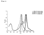
- FIG. 11 shows the excitation spectrum and fluorescence spectrum of the fluorescent protein (Momiji) of the present invention.
- FIG. 12 shows the pH sensitivity of the fluorescent protein (Momiji) of the present invention.
- FIG. 13 shows a change in the fluorescence spectrum when the fluorescent protein (Momiji) of the present invention is irradiated with 365 nm UV.
- FIG. 14 shows a change in the absorption spectrum when the fluorescent protein (Momiji) of the present invention is irradiated with 365 nm UV.
- FIG. 15 shows a change in the absorption spectrum before and after irradiation with UV (Momiji 2).
- FIG. 16 shows a change in the absorption spectrum before and after irradiation with UV (Momiji 4).
- FIG. 17 shows a change in the absorption spectrum due to pH before and after irradiation with UV (Momiji). Before irradiation: 508 nm; and after irradiation: 578 nm.
- FIG. 18 shows a change in the absorption spectrum due to pH before and after irradiation with UV (Momiji 2). Before irradiation: 508 nm; and after irradiation: 576 nm.
- FIG. 19 shows a change in the absorption spectrum due to pH before and after irradiation with UV (Momiji 4). Before irradiation: 508 nm; and after irradiation: 583 nm.
- FIG. 20 shows a transition in a change in fluorescence properties depending on irradiation time with light at 365 nm (Momiji). Measurement was carried out on a 6 ⁇ g/ml protein solution. In order to clearly differentiate the situation from that of a mutant, light exchange was carried out with extremely weak light at 365 nm, and the fluorescence spectrum from 470 nm to 650 nm was measured.
- FIG. 21 shows a transition in a change in fluorescence properties depending on irradiation time with light at 365 nm (Momiji 2). Measurement was carried out on a 6 ⁇ g/ml protein solution. In order to clearly differentiate the situation from that of a mutant, light exchange was carried out with extremely weak light at 365 nm, and the fluorescence spectrum from 470 nm to 650 nm was measured.
- FIG. 22 shows a transition in a change in fluorescence properties depending on irradiation time with light at 365 nm (Momiji 4). Measurement was carried out on a 6 ⁇ g/ml protein solution. In order to clearly differentiate the situation from that of a mutant, light exchange was carried out with extremely weak light at 365 nm, and the fluorescence spectrum from 470 nm to 650 nm was measured.
- FIG. 23 shows a transition in fluorescence properties in HeLa cells.
- Irradiation at 410 nm, measurement of the green fluorescence, and measurement of the red fluorescence were carried out at intervals of 3 seconds.
- FIG. 24 shows the results obtained by exciting with a blue light the electrophoretic pattern on 12.5% acrylamide gel (Pseudo-native SDS/PAGE) and then photographing the resultant with a digital camera.
- FIG. 25 shows the fluorescence spectrum (d16) before and after irradiation with light at 365 nm.
- FIG. 26 shows the fluorescence spectrum (m16) before and after irradiation with light at 365 nm.
- the first chromoprotein of the present invention is characterized in that it is derived from Anthopleura inornata , and has the following properties:
- the second chromoprotein of the present invention is characterized in that it is derived from Anthopleura inornata , and has the following properties:
- Anthopleura inornata has 96 tentacles, which are regularly aligned.
- this species is characterized in that it has an oral disc having the same color as that of the tentacles and that the surrounding portion of the mouth has a reddish brown color.
- 98 lines of adhesive warts are found on the body wall, and such wards are distributed up to the lower end of the body wall.
- the color of the body wall is largely varied, and brown, blue, and pink body colors have been known.
- Such Anthopleura inornata ranges from the southern part of Hokkaido to Kyushu, and many of them live in the intertidal zone.
- a chromoprotein having the aforementioned properties was isolated from Anthopleura inornata that was used as a starting material.
- the chromoprotein of the present invention can be obtained from a sea anemone other than Anthopleura inornata .
- the thus obtained chromoprotein is also included in the scope of the present invention.
- the first chromoprotein (Be-G) of the present invention has an absorption maximum wavelength of 605 nm, and the molar absorption coefficient is 47,550 at 605 nm.
- the second chromoprotein (Be-R) of the present invention has an absorption maximum wavelength of 553 nm, and the molar absorption coefficient is 25,300 at 553 nm.
- the molar absorption coefficient represents the amount of absorbed photons per mole of molecule
- the quantum yield represents a numerical value showing the amount of the absorbed photons that can be emitted as fluorescence. Since the chromoproteins of the present invention have an extremely low quantum yield, they hardly emit fluorescence at all. Due to this property, the chromoproteins of the present invention can be used: (1) as an acceptor molecule (energy receptor) in FRET; (2) in development of a system for converting the energy of applied light into energy other than the light; and (3) in introduction of a mutation into the amino acid sequence of the protein to modify it so that it emits fluorescence.
- the chromoproteins of the present invention are characterized in that the pH sensitivity of light-absorbing properties is stable at between pH 5 and pH 10. That is to say, in the case of the chromoproteins of the present invention, the peak value of the absorption spectrum does not significantly fluctuate in the range between pH 5 and pH 10. Accordingly, even under the same conditions, the chromoproteins of the present invention can be used in a broad range of pH environments, and thus, the use of the chromoproteins in vivo have few restrictions.
- chromoprotein of the present invention include a chromoprotein having either one of the following amino acid sequences:
- an amino acid sequence comprising a deletion, substitution and/or addition of one or several amino acids is not particularly limited in the present specification. For example, it means 1 to 20, preferably 1 to 10, more preferably 1 to 7, further preferably 1 to 5, and particularly preferably 1 to 3.
- light-absorbing properties is used in the present specification to mean properties capable of absorbing light having a certain wavelength.
- an absorption maximum wavelength may be 605 nm or 553 nm as in the case of the chromoproteins described in the present specification, or the value of the absorption maximum wavelength may also be shifted. It is preferable that the pH sensitivity of light-absorbing properties is stable at between pH 5 and pH 10.
- the chromoproteins of the present invention having the amino acid sequence shown in SEQ ID NO: 1 or 3 in the sequence listing hardly emit fluorescence at all.
- one or several amino acids are deleted, substituted, and/or added with respect to the amino acid sequence shown in SEQ ID NO: 1 or 3, so as to produce a protein having stronger fluorescence.
- the thus produced proteins are also included in the scope of the present invention.
- the method of obtaining the chromoproteins of the present invention is not particularly limited.
- the proteins may be either a protein synthesized by chemosynthesis, or recombinant protein produced by a gene recombination technique.
- a recombinant protein is produced, it is necessary to obtain DNA encoding the protein.
- Appropriate primers are designed by using information regarding the amino acid sequence shown in SEQ ID NO: 1 or 3 of the sequence listing of the present specification and the nucleotide sequence shown in SEQ ID NO: 2 or 4 thereof. Using these primers, PCR is carried out by using cDNA library derived from Anthopleura inornata as a template, so that DNA encoding the chromoprotein of the present invention can be obtained.
- the chromoprotein of the present invention can be produced by introducing this DNA into an appropriate expression system. Expression in an expression system will be described later in the present specification.
- Trachyphyllia geoffroyi is one type of cnidarian sea anemone, and it is characterized in that it emits extremely colorful fluorescence. Trachyphyllia geoffroyi mainly ranges over the area below the midland of Honshu Island, Japan. This sea anemone lives in the sludge in the gulf. At night, it extends its tentacles to capture plankton and the like. In terms of a color variation, green, brown, and red examples are found.
- the color of the first fluorescent protein of the present invention is changed from green to red by irradiation of ultraviolet ray; the excitation maximum wavelength is 508 nm (green) and 572 nm (red); and the fluorescence maximum wavelength is 518 nm (green) and 581 nm (red).
- the molar absorption coefficient (green) at 508 nm is 98800 M ⁇ 1 cm ⁇ 1 ; and the molar absorption coefficient (red) at 572 nm is 60400 M ⁇ 1 cm ⁇ 1 ;
- the first fluorescent protein of the present invention is characterized in that its color changes due to ultraviolet rays.
- optical marking can be carried out on specific cells or organs thereof.
- the examples of the first fluorescent protein of the present invention include a fluorescent protein having either one of the following amino acid sequences:
- first fluorescent protein of the present invention examples include a fluorescent protein having an amino acid sequence shown in SEQ ID NO: 7.
- Scolymia vitiensis is a sessile single coral.
- a small individual has a pot-like round shape. However, after it has matured, it has an oval shape.
- a large spongy columella is found in the center of a coral individual, and a long barrier extends at an almost constant slope from the theca towards the center.
- a large serration is located on the barrier, and such serration can be seen from the outside.
- This coral does not open its polyp during the day. It generally has a dark green color, but it also has a red color in rare cases.
- Approximately 4 types of Scolymia vitiensis have been known. Among them, only one type ranges in the ocean areas surrounding Japan.
- the color of the second fluorescent protein of the present invention is changed from green to red by irradiation of ultraviolet ray; the excitation maximum wavelength is 508 nm (green) and 578 nm (red); and the fluorescence maximum wavelength is 518 nm (green) and 588 nm (red).
- the molar absorption coefficient (green) at 508 nm is 102250 M ⁇ 1 cm ⁇ 1 ; and the molar absorption coefficient (red) at 578 nm is 76950 M ⁇ 1 cm ⁇ 1 .
- the first fluorescent protein of the present invention is characterized in that its color changes due to ultraviolet rays.
- optical marking can be carried out on specific cells or organs thereof.
- the examples of the second fluorescent protein of the present invention include a fluorescent protein having either one of the following amino acid sequences:
- first fluorescent protein of the present invention examples include a fluorescent protein having an amino acid sequence shown in any of SEQ ID NO: 11, 13, 15 or 17.
- an amino acid sequence comprising a deletion, substitution and/or addition of one or several amino acids is not particularly limited in the present specification. For example, it means 1 to 20, preferably 1 to 10, more preferably 1 to 7, further preferably 1 to 5, and particularly preferably 1 to 3.
- the term “having fluorescent properties” covers all of the cases where any fluorescence is given. Various properties such as fluorescence intensity, excitation wavelength, fluorescence wavelength or pH sensitivity, may be changed or may remain unchanged.
- the method of obtaining the fluorescent protein of the present invention is not particularly limited.
- the protein may be either a protein synthesized by chemosynthesis, or recombinant protein produced by a gene recombination technique.
- a recombinant protein is produced, it is necessary to obtain DNA encoding the protein.
- Appropriate primers are designed by using information regarding the amino acid sequence shown in SEQ ID NOS: 5, 7, 9, 11, 13, 15 or 17 of the sequence listing of the present specification and the nucleotide sequence shown in SEQ ID NOS: 6, 8, 10, 12, 14, 16 or 18 thereof. Using these primers, PCR is carried out by using cDNA clones of the above-described various types of known fluorescent proteins as a template, so that DNA encoding the fluorescent protein of the present invention can be obtained.
- a partial fragment of DNA encoding the fluorescent protein of the present invention are obtained by the above-described PCR, the produced DNA fragments are ligated to one another by a gene recombination technique, so that DNA encoding the desired fluorescent protein can be obtained.
- the fluorescent protein of the present invention can be produced by introducing this DNA into an appropriate expression system. Expression in an expression system will be described later in the present specification.
- genes encoding the chromoproteins and fluorescent proteins of the present invention are provided.
- DNA encoding the first chromoprotein of the present invention may include either one of the following DNAs:
- DNA encoding the first chromoprotein of the present invention may include either one of the following DNAs:
- DNA encoding the second chromoprotein of the present invention may include either one of the following DNAs:
- DNA encoding the second chromoprotein of the present invention may include either one of the following DNAs:
- DNA encoding the first fluorescent protein of the present invention may include either one of the following DNAs:
- DNA encoding the first fluorescent protein of the present invention may include either one of the following DNAs:
- DNA encoding the second fluorescent protein of the present invention may include either one of the following DNAs:
- DNA encoding the second fluorescent protein of the present invention may include either one of the following DNAs:
- the DNA of the present invention can be synthesized by, for example, the phosphoamidite method, or it can also be produced by polymerase chain reaction (PCR) using specific primers.
- the DNA of the present invention is produced by the method described above in the specification.
- a method of introducing a desired mutation into a certain nucleic acid sequence is known to a person skilled in the art.
- known techniques such as a site-directed mutagenesis, PCR using degenerated oligonucleotides, or the exposure of cells containing nucleic acid to mutagens or radioactive rays, are appropriately used, so as to construct DNA having a mutation.
- Such known techniques are described in, for example, Molecular Cloning: A Laboratory Manual, 2 nd Ed., Cold Spring Harbor Laboratory, Cold Spring Harbor, N.Y., 1989; and Current Protocols in Molecular Biology, Supplements 1 to 38, John Wiley & Sons (1987-1997).
- the DNA of the present invention can be inserted into a suitable vector and used.
- the type of a vector used in the present invention is not particularly limited. For example, it may be either a vector that can autonomously replicate (e.g., a plasmid, etc.), or vector that is incorporated into the genomes of host cells when it is introduced into the host cells and is then replicated together with the chromosome into which it is incorporated.
- the vector used in the present invention is preferably an expression vector.
- elements necessary for transcription e.g., a promoter, etc.
- the promoter is a DNA sequence which shows a transcriptional activity in host cells, and it is appropriately selected depending on the type of host cells.
- Examples of a promoter which can operate in bacterial cells may include a Bacillus stearothermophilus maltogenic amylase gene promoter, a Bacillus licheniformis alpha-amylase gene promoter, a Bacillus amyloliquefaciens BAN amylase gene promoter, a Bacillus subtilis alkaline protease gene promoter, a Bacillus pumilus xylosidase gene promoter, P R and P L promoters of phage rhamda, and lac, trp and tac promoters of Escherichia coli.
- Examples of a promoter which can operate in mammalian cells may include an SV40 promoter, an MT-1 (metallothionein gene) promoter, and an adenovirus-2 major late promoter.
- Examples of a promoter which can operate in insect cells may include a polyhedrin promoter, a P10 promoter, an Autographa californica polyhedrosis basic protein promoter, a baculovirus immediate-early gene 1 promoter, and a baculovirus 39K delayed-early gene promoter.
- Examples of a promoter which can be operate in yeast host cells may include promoters derived from yeast glycolytic genes, an alcohol dehydrogenase gene promoter, a TPI1 promoter, and an ADH2-4c promoter.
- Examples of a promoter which can operate in filamentous cells may include an ADH3 promoter and a tpiA promoter.
- an appropriate terminator such as a human growth hormone terminator, or a TPI1 terminator or ADH3 terminator for fungal cells, may be functionally bound to the DNA of the present invention, as necessary.
- the recombinant vector of the present invention may further have elements such as a polyadenylation signal (e.g., one derived from SV40 or the adenovirus 5E1b region), a transcription enhancer sequence (e.g., an SV40 enhancer), or a translation enhancer sequence (e.g., one encoding the adenovirus VA RNA).
- the recombinant vector of the present invention may further comprise a DNA sequence which enables the replication of the recombinant vector in host cells.
- SV40 replication origin is an example of such a sequence (when the host cells are mammalian cells).
- the recombinant vector of the present invention may further comprise a selective marker.
- a selective marker may include genes, complements of which are absent from host cells, such as a dihydrofolate reductase (DHFR) gene or a Shizosaccharomyces pombe TPI gene, and drug resistant genes such as ampicillin, kanamycin, tetracycline, chloramphenicol, neomycin or hygromycin-resistant genes.
- DHFR dihydrofolate reductase
- Shizosaccharomyces pombe TPI Shizosaccharomyces pombe TPI gene
- a method for ligating the DNA of the present invention, a promoter and, as desired, a terminator and/or a secretory signal sequence to one another and inserting these items into a suitable vector is known to a person skilled in the art.
- a transformant can be produced by introducing the DNA or recombinant vector of the present invention into a suitable host.
- Any cell can be used as a host cell into which the DNA or recombinant vector of the present invention is introduced, as long as the DNA construct of the present invention can be expressed therein.
- Examples of such a cell may include bacteria, yeasts, fungal cells, and higher eukaryotic cells.
- bacteria may include Gram-positive bacteria such as Bacillus or Streptomyces , and Gram-negative bacteria such as Escherichia coli . These bacteria may be transformed by the protoplast method or other known methods, using competent cells.
- mammalian cells may include HEK 293 cells, HeLa cells, COS cells, BHK cells, CHL cells, and CHO cells.
- a method of transforming mammalian cells and expressing the introduced DNA sequence in the cells is also known. Examples of such a method may include the electroporation, the calcium phosphate method, and the lipofection method.
- yeast cells may include those belonging to Saccharomyces or Shizosaccharomyces .
- Examples of such cells may include Saccharomyces cerevisiae and Saccharomyces kluyveri .
- Examples of a method of introducing a recombinant vector into yeast host cells may include the electroporation, the spheroplast method, and the lithium acetate method.
- Examples of other fungal cells may include those belonging to Filamentous fungi such as Aspergillus, Neurospora, Fusarium or Trichoderma . Where Filamentous fungi are used as host cells, transformation can be carried out by incorporating DNA constructs into host chromosomes, so as to obtain recombinant host cells. Incorporation of DNA constructs into the host chromosomes is carried out by known methods, and such known methods may include homologous recombination and heterologous recombination.
- both a vector into which a recombinant gene is introduced and a baculovirus are co-introduced into insect cells, and a recombinant virus is obtained in the culture supernatant of the insect cells. Thereafter, insect cells are infected with the recombinant virus, so as to allow the cells to express proteins (described in, for example, Baculovirus Expression Vectors, A Laboratory Manual; and Current Protocols in Molecular Biology, Bio/Technology, 6, 47 (1988)).
- the Autographa califo mica nuclear polyhedrosis virus which is a virus infecting to insects belonging to Barathra brassicae , can be used as baculovirus.
- insect cells used herein may include Sf9 and Sf21, which are Spodoptera frugiperda ovarian cells [Baculovirus Expression Vectors, A Laboratory Manual, W. H. Freeman & Company, New York, (1992)], and HiFive (manufactured by Invitrogen), which are Trichoplusia ni ovarian cells.
- Examples of the method of co-introducing both a vector into which a recombinant gene has been introduced and the above baculovirus into insect cells to prepare a recombinant virus may include the calcium phosphate method and the lipofection method.
- the above transformant is cultured in an appropriate nutritive medium under conditions enabling the introduced DNA construct to be expressed.
- common methods of isolating and purifying proteins may be used.
- the protein of the present invention is expressed in a state dissolved in cells
- cells are recovered by centrifugal separation, and the recovered cells are suspended in a water type buffer. Thereafter, the cells are disintegrated using an ultrasonic disintegrator or the like, so as to obtain a cell-free extract.
- a supernatant is obtained by centrifuging the cell-free extract, and then, a purified sample can be obtained from the supernatant by applying, singly or in combination, the following ordinary protein isolation and purification methods: the solvent extraction, the salting-out method using ammonium sulfate or the like, the desalting method, the precipitation method using an organic solvent, the anion exchange chromatography using resins such as diethylaminoethyl (DEAE) sepharose, the cation exchange chromatography using resins such as S-Sepharose FF (manufactured by Pharmacia), the hydrophobic chromatography using resins such as butyl sepharose or phenyl sepharose, the gel filtration method using a molecular sieve, the affinity chromatography, the chromatofocusing method, and the electrophoresis such as isoelectric focusing.
- the solvent extraction such as diethylaminoethyl (DEAE) sepharose
- the chromoprotein of the present invention can be fused with another protein, so as to construct a fusion protein.
- the type of said another protein to be fused to the chromoprotein of the present invention is not particularly limited, and preferred examples may include a protein which interacts with another molecule.
- the examples may include a receptor protein or ligand thereof, antigen, antibody and the like.
- a method of obtaining the fusion protein of the present invention is not particularly limited. It may be either a protein synthesized by chemosynthesis, or recombinant protein produced by a gene recombination technique.
- DNA encoding the chromoprotein of the present invention and the DNA encoding the another protein to be fused to the chromoprotein can be obtained by the method as mentioned above in this specification or by the method similar to it. Then, these DNA fragments are ligated to one another by a gene recombination technique, so that DNA encoding the desired fusion protein can be obtained. This DNA is then introduced into an appropriate expression system, so that the fusion protein of the present invention can be produced.
- FRET fluorescence resonance energy transfer
- FRET fluorescence resonance energy transfer
- a first molecule labeled with a cyan fluorescent protein (CFP) acting as a first fluorescent protein is allowed to coexist with a second molecule labeled with a yellow fluorescent protein (YFP) acting as a second fluorescent protein, so as to allow the yellow fluorescent protein (YFP) to act as an acceptor molecule and to allow the cyan fluorescent protein (CFP) to act as a donor molecule.
- FRET fluorescence resonance energy transfer
- FRET fluorescence resonance energy transfer
- One dyes with a higher energy level (a donor molecule) is selectively excited, and the fluorescence of the dye is measured. Long-wavelength fluorescence from the other dye (an acceptor molecule) is also measured. The interaction between the molecules is visualized by using the difference between the amounts of both fluorescences. Only when both dyes are adjacent to each other due to the interaction of the two types of molecules, a decrease in the fluorescence of the donor molecule and an increase in the fluorescence of the acceptor molecule are observed by single wavelength excitation dual wavelength photometry. However, in a case where a chromoprotein is used as an acceptor molecule, a decrease in the fluorescence of the donor molecule occurs only when both dyes are adjacent to each other by the interaction of the two types of molecules. Such a decrease can be observed by single wavelength excitation single wavelength photometry. Thus, the use of the chromoprotein of the present invention enables facilitation of measurement apparatuses.
- the chromoprotein of the present invention is particularly advantageous when it is used as an acceptor molecule in FRET (fluorescence resonance energy transfer). That is to say, a fused form (a first fused form) of the chromoprotein of the present invention and a test substance is first produced. Then, a fused form (a second fused form) of another test substance interacting with the above test substance and another fluorescent protein is produced. Thereafter, the first fused form is allowed to interact with the second fused form, and the generated fluorescence is analyzed, so that the interaction between the aforementioned two types of test substances can be analyzed.
- FRET fluorescence resonance energy transfer
- using the chromoprotein of the present invention may be carried out either in a test tube or in a cell.
- the fluorescent protein of the present invention can be fused with another protein, so as to construct a fusion fluorescent protein.
- a method of obtaining the fusion fluorescent protein of the present invention is not particularly limited. It may be either a protein synthesized by chemosynthesis, or recombinant protein produced by a gene recombination technique.
- DNA encoding the protein is produced, it is necessary to obtain DNA encoding the protein.
- Appropriate primers are designed using the information regarding the amino acid sequence shown in SEQ ID NO: 5, 7, 9, 11, 13, 15 or 17 of the sequence listing of the present specification and the nucleotide sequence shown in SEQ ID NO: 6, 8, 10, 12, 14, 16 or 18 thereof. Using these primers, PCR is carried out using a DNA fragment containing the gene of the fluorescent protein of the present invention as a template, so as to produce DNA fragments necessary for construction of the DNA encoding the fluorescent protein of the present invention. Moreover, DNA fragment encoding a protein to be fused is also obtained in the same above manner.
- DNA fragments are ligated to one another by a gene recombination technique, so that DNA encoding the desired fusion fluorescent protein can be obtained.
- This DNA is then introduced into an appropriate expression system, so that the fusion fluorescent protein of the present invention can be produced.
- the fluorescent protein of the present invention has an extremely high utility value as a marker.
- the fluorescent protein of the present invention is purified as a fusion protein with an amino acid sequence to be tested, and the fusion protein is introduced into cells by methods such as the microinjection. By observing the distribution of the fusion protein over time, targeting activity of the amino acid sequence to be tested can be detected in the cells.
- the type of another protein (an amino acid sequence to be tested) with which the fluorescent protein of the present invention is fused is not particularly limited. Preferred examples may include proteins localizing in cells, proteins specific for intracellular organelles, and targeting signals (e.g., a nuclear transport signal, a mitochondrial presequence, etc.).
- the fluorescent protein of the present invention can be expressed in cells and used, as well as being introduced into cells by the microinjection or the like. In this case, a vector into which the DNA encoding the fluorescent protein of the present invention is inserted in such a way that it can be expressed, is introduced into host cells.
- the fluorescent protein of the present invention can also be used as a reporter protein to determine promoter activity.
- a vector is constructed such that DNA encoding the fluorescent protein of the present invention is located downstream of a promoter to be tested, and the vector is then introduced into host cells. By detecting the fluorescence of the fluorescent protein of the present invention which is emitted from the cells, the activity of the promoter to be tested can be determined.
- the type of a promoter to be tested is not particularly limited, as long as it operates in host cells.
- a vector used to detect the targeting activity of the above amino acid sequence to be tested or to determine promoter activity is not particularly limited.
- Examples of a vector preferably used for animal cells may include pNEO (P. Southern, and P. Berg (1982) J. Mol. Appl. Genet. 1: 327), pCAGGS (H. Niwa, K. Yamamura, and J. Miyazaki, Gene 108, 193-200 (1991)), pRc/CMV (manufactured by Invitrogen), and pCDM8 (manufactured by Invitrogen).
- Examples of a vector preferably used for yeasts may include pRS303, pRS304, pRS305, pRS306, pRS313, pRS314, pRS315, pRS316 (R. S. Sikorski and P. Hieter (1989) Genetics 122: 19-27), pRS423, pRS424, pRS425, pRS426 (T. W. Christianson, R. S. Sikorski, M. Dante, J. H. Shero, and P. Hieter (1992) Gene 110: 119-122).
- the type of cells used herein is also not particularly limited.
- Various types of animal cells such as L cells, BalbC-3T3 cells, NIH3T3 cells, CHO (Chinese hamster ovary) cells, HeLa cells or NRK (normal rat kidney) cells, yeast cells such as Saccharomyces cerevisiae, Escherichia coli cells, or the like can be used.
- Vector can be introduced into host cells by common methods such as the calcium phosphate method or the electroporation.
- the above obtained fusion fluorescent protein of the present invention wherein the fluorescent protein of the present invention is fused with another protein (referred to as a protein X) is allowed to be expressed in cells.
- a protein X another protein
- a protein specific for an intracellular organella as a protein X, the distribution and movement of a nucleus, a mitochondria, an endoplasmic reticulum, a Golgi body, a secretory vesicle, a peroxisome, etc., can be observed.
- axis cylinders or dendrites of the nerve cells show an extremely complicated change in strikes in an individual who is under development. Accordingly, fluorescent labeling of these sites enables a dynamic analysis.
- the fluorescence of the fluorescent protein of the present invention can be detected with a viable cell.
- detection can be carried out using, for example, a fluorescence microscope (Axiophoto Filter Set 09 manufactured by Carl Zeiss) or an image analyzer (Digital Image Analyzer manufactured by ATTO).
- the type of a microscope can be appropriately selected depending on purposes. Where frequent observation such as pursuit of a change over time is carried out, an ordinary incident-light fluorescence microscope is preferable. Where observation is carried out while resolution is emphasized, for example, in the case of searching localization in cells specifically, a confocal laser scanning microscope is preferable. In terms of maintenance of the physiological state of cells and prevention from contamination, an inverted microscope is preferable as a microscope system. When an erecting microscope with a high-powered lens is used, a water immersion lens can be used.
- a filter set can be appropriately selected depending on the fluorescence wavelength of a fluorescent protein.
- a filter having an excitation light between approximately 490 and 510 nm and a fluorescence between approximately 510 and 530 nm can be preferably used.
- red having the excitation maximum wavelength of 572 nm and the fluorescence maximum wavelength of 581 nm is detected, a filter having an excitation light between approximately 560 and 575 nm and a fluorescence between approximately 575 and 590 nm can be preferably used.
- a filter having an excitation light between approximately 490 and 510 nm and a fluorescence between approximately 510 and 530 nm can be preferably used.
- a filter having an excitation light between approximately 570 and 580 nm and a fluorescence between approximately 580 and 595 nm can be preferably used.
- a high sensitive cooled CCD camera is used, since photography is carried out in a short time.
- CCD is cooled to decrease thermal noise, so that a weak fluorescence image can be clearly photographed by exposure in a short time.
- the present invention provides a light-absorbing reagent kit comprising at least one which is selected from the chromoprotein, fusion protein, DNA, recombinant vector or transformant, which are described in the present specification. Further, the present invention provides a kit for analyzing the localization of intracellular components and/or analyzing physiologically active substances, which is characterized in that it comprises at least one selected from the fluorescent protein, the fusion fluorescent protein, the DNA, the recombinant vector, or the transformant, which are described in the present specification.
- the kit of the present invention can be produced from commonly used materials that are known per se, by using common methods.
- Reagents such as the chromoprotein, the fluorescent protein or the DNA are dissolved in an appropriate solvent, so that the reagents can be prepared in a form suitable for con servation.
- Water, ethanol, various types of buffer solution, etc. can be used as such a solvent.
- a chromoprotein gene was isolated from sea anemone.
- One individual of Anthopleura inornata emitting a green color was used as a material.
- Frozen Anthopleura inornata was crushed in a mortar.
- 7.5 ml of “TRIzol” (GIBCO BRL) was added to 1 g (wet weight) of Anthopleura inornata , and the mixture was homogenized, followed by centrifugation at 1,500 ⁇ g for 10 minutes.
- 1.5 ml of chloroform was added to the supernatant. The mixture was stirred for 15 seconds and then left at rest for 3 minutes. The resultant product was centrifuged at 7,500 ⁇ g for 15 minutes.
- RNA dissolved in the DEPC water was 100 times diluted, and the values of O.D.260 and O.D.280 were measured, so as to determine the concentration of RNA. 2.2 mg of the total RNA was obtained.
- cDNA (33 ⁇ l) was synthesized from 4 ⁇ g of the total RNA using a kit for synthesizing first strand cDNA, “Ready To Go” (Amersham Pharmacia).
- the annealing temperature was decreased 0.3° C. per cycle. That is to say, the annealing temperature in the 30 th cycle was 43° C.
- the purified DNA fragment was ligated to a pT7-blue vector (Novagen). Escherichia coli (TG1) was transformed with the vector, and the obtained transformants were subjected to blue white selection. Thereafter, plasmid DNA was purified from white colonies of Escherichia coli .
- the nucleotide sequence of the inserted DNA fragment was determined by a DNA sequencer.
- the obtained nucleotide sequence was compared with the nucleotide sequences of other fluorescent protein genes to confirm that the nucleotide sequence of the DNA was derived from a fluorescent protein. 5′-RACE and 3′-RACE methods were applied to a gene that had been confirmed to be a part of a fluorescent protein gene, so as to carry out the cloning of a full-length gene.
- the 5′-RACE method was applied using 5′-RACE System for Rapid Amplification of cDNA Ends, Version 2.0 (GIBCO BRL). 4 ⁇ g of the total RNA prepared in (1) above was used as a template.
- the following primers were used:
- the 200-bp amplified band was cut out by agarose gel electrophoresis and then purified.
- the purified DNA fragment was ligated to a pT7-blue vector (Novagen).
- Escherichia coli (TG1) was transformed with the vector, and the obtained transformants were subjected to blue white selection. Thereafter, plasmid DNA was purified from white colonies of Escherichia coli .
- the nucleotide sequence of the inserted DNA fragment was determined by a DNA sequencer.
- the 3′-terminal portion of the DNA fragment obtained by degenerated PCR was obtained by PCR, using the primer produced based on the information obtained by sequencing of the nucleotide sequence in (4) above and an oligo dT primer. 3 ⁇ l of the first strand cDNA prepared in (2) above was used as a template.
- the produced primer is shown below:
- An amplified band of approximately 1,000 bp was cut out by agarose gel electrophoresis and then purified.
- the purified DNA fragment was ligated to a pT7-blue vector (Novagen).
- Escherichia coli (TG1) was transformed with the vector, and the obtained transformants were subjected to blue white selection. Thereafter, plasmid DNA was purified from white colonies of Escherichia coli .
- the nucleotide sequence of the inserted DNA fragment was determined by a DNA sequencer.
- the obtained full-length nucleotide sequence is shown in SEQ ID NO: 2, and the obtained full-length amino acid sequence is shown in SEQ ID NO: 1.
- a primer corresponding to the N-terminal of the protein was produced from the obtained full-length nucleotide sequence, and an oligo dT primer was used for C-terminal. PCR was carried out using these primers and the first strand cDNA prepared in Example A-1 (2) as a template.
- the used primers are as follows:
- An amplified band of approximately 1100 bp was cut out by agarose gel electrophoresis and then purified.
- the purified DNA fragment was subcloned into the BamHI-EcoRI site of a pRSET vector (Invitrogen), and it was then allowed to express in Escherichia coli (JM109-DE3). Since the expressed protein was constructed such that His-tag was attached to the N-terminus thereof, it was purified with Ni-Agarose gel (QIAGEN). Purification was carried out in accordance with the attached protocols.
- This chromoprotein was named as Be-G In the following Example A-3, the property of the purified protein was analyzed.
- the absorption spectrum of the protein was measured in the following 50 mM buffer solution ( FIG. 2 ).
- a chromoprotein gene was isolated from sea anemone.
- One individual of Anthopleura inornata emitting a red color was used as a material.
- Frozen Anthopleura inornata was crushed in a mortar.
- 7.5 ml of “TRIzol” (GIBCO BRL) was added to 1 g (wet weight) of Anthopleura inornata , and the mixture was homogenized, followed by centrifugation at 1,500 ⁇ g for 10 minutes.
- 1.5 ml of chloroform was added to the supernatant. The mixture was stirred for 15 seconds and then left at rest for 3 minutes. The resultant product was centrifuged at 7,500 ⁇ g for 15 minutes.
- cDNA (33 ⁇ l) was synthesized from 4 ⁇ g of the total RNA using a kit for synthesizing first strand cDNA, “Ready To Go” (Amersham Pharmacia).
- the annealing temperature was decreased 0.3° C. per cycle. That is to say, the annealing temperature in the 30 th cycle was 43° C.
- the purified DNA fragment was ligated to a pT7-blue vector (Novagen). Escherichia coli (TG1) was transformed with the vector, and the obtained transformants were subjected to blue white selection. Thereafter, plasmid DNA was purified from white colonies of Escherichia coli .
- the nucleotide sequence of the inserted DNA fragment was determined by a DNA sequencer.
- the obtained nucleotide sequence was compared with the nucleotide sequences of other fluorescent protein genes to confirm that the nucleotide sequence of the DNA was derived from a fluorescent protein. 5′-RACE and 3′-RACE methods were applied to a gene that had been confirmed to be a part of a fluorescent protein gene, so as to carry out the cloning of a full-length gene.
- the 5′-RACE method was applied using 5′-RACE System for Rapid Amplification of cDNA Ends, Version 2.0 (GIBCO BRL). 4 ⁇ g of the total RNA prepared in (1) above was used as a template.
- p For the first amplification of DC-tailed cDNA of the red individual, the following primers were used:
- the 200-bp amplified band was cut out by agarose gel electrophoresis and then purified.
- the purified DNA fragment was ligated to a pT7-blue vector (Novagen).
- Escherichia coli (TG1) was transformed with the vector, and the obtained transformants were subjected to blue white selection. Thereafter, plasmid DNA was purified from white colonies of Escherichia coli .
- the nucleotide sequence of the inserted DNA fragment was determined by a DNA sequencer.
- the 3′-terminal portion of the DNA fragment obtained by degenerated PCR was obtained by PCR, using the primer produced based on the information obtained by sequencing of the nucleotide sequence in (4) above and an oligo dT primer. 3 ⁇ l of the first strand cDNA prepared in (2) above was used as a template.
- the produced primer is shown below:
- An amplified band of approximately 1,000 bp was cut out by agarose gel electrophoresis and then purified.
- the purified DNA fragment was ligated to a pT7-blue vector (Novagen).
- Escherichia coli (TG1) was transformed with the vector, and the obtained transformants were subjected to blue white selection. Thereafter, plasmid DNA was purified from white colonies of Escherichia coli .
- the nucleotide sequence of the inserted DNA fragment was determined by a DNA sequencer.
- the obtained full-length nucleotide sequence is shown in SEQ ID NO: 4, and the obtained full-length amino acid sequence is shown in SEQ ID NO: 3.
- a primer corresponding to the N-terminal of the protein was produced from the obtained full-length nucleotide sequence, and an oligo dT primer was used for C-terminal. PCR was carried out using these primers and the first strand cDNA prepared in Example B-1 (2) as a template.
- the used primers are as follows:
- the absorption spectrum of the protein was measured in the following 50 mM buffer solution ( FIG. 4 ).
- a fluorescent protein gene was isolated from Trachyphyllia geoffroyi which emits fluorescence with rich colors by the following procedures.
- Total RNA was extracted by acidic guanidium/phenol/chloroform method.
- RNA from Trachyphyllia geoffroyi was crushed in a denaturation solution by using a Multi-Beads Shocker (Yasui Kikai), and then phenol/chloroform was added thereto, followed by centrifugation to separate RNA from protein and DNA.
- a water phase containing RNA was added to isopropanol, and the mixture was centrifuged, so as to obtain total RNA as a precipitate.
- Oligotex-dT30 ⁇ super> was added to the total RNA, and the mixture was then heated, so as to destroy the secondary structure of the RNA. Thereafter, the RNA was bound to Oligotex-dT at 37° C. After washing, the resultant product was heated and centrifuged, so as to obtain a supernatant eluted from the mRNA. Oligotex-dt was eliminated from the supernatant, and then, mRNA was allowed be precipitated with ethanol and NaCl. The mRNA was then dissolved in water.
- a cDNA fragment was prepared using TimeSaver and Directional Cloning Toolbox (both of which were manufactured by Amersham Pharmacia).
- the mRNA was heated to destroy the secondary structure thereof. Thereafter, the mRNA, DTT, and a NotI-dT primer were added to First-Strand Reaction Mix, so as to synthesize a first strand. This was then added to Second-Strand Reaction Mix, so as to synthesize a second strand.
- the synthesized second strand was purified with a span column attached with the kit. EcoRI adaptors were added to both termini of the purified double-stranded cDNA, and only the 3′-side thereof was cleaved with NotI. It was purified again with the span column, so as to obtain a cDNA fragment (EcoRI-NotI).
- Kaede The amino acid sequence of Kaede is shown in SEQ ID NO: 5 of the sequence listing, and the nucleotide sequence of Kaede is shown in SEQ ID NO: 6.
- a BamHI site was added to the N-terminus of the obtained full-length cDNA, and an EcoRI site was added to the C-terminus thereof. Thereafter, it was subcloned in frame into pRSETB (manufactured by Invitrogen), and it was then expressed in Escherichia coli JM109 DE3. The expressed protein was purified with Ni-Agarose gel (manufactured by QIAGEN), utilizing an His-tag at the N-terminus thereof.
- FIG. 5 shows the absorption spectrum of a purified protein before and after UV irradiation (the solid line represents the absorption spectrum before UV irradiation, and the dotted line represents the absorption spectrum after UV irradiation).
- the molar absorption coefficient was obtained from the concentration of the protein and the absorbance at absorption maximum (Table 3).
- the fluorescence spectrum was measured by exciting the protein at 480 nm before and after UV irradiation ( FIG. 6 ), and the quantum yield was calculated by comparison with Fluorescein (manufactured by Molecular Probes) (Table 3).
- FIG. 7 shows the results obtained by exciting the cells at 470 nm and measuring them with the fluorescence at 510 nm. Fluorescence can be confirmed approximately 9 hours after the introduction. Fluorescence shifts to a long wavelength even in mammalian cells, when the cells are irradiated with UV.
- Kaede forms a tetramer, there may be cases where its expression pattern becomes abnormal when another protein is fused with Kaede.
- threonine (T) at position 158 was substituted with arginine (R), and alanine (A) at position 160 was substituted with glutamic acid (E), so as to produce a dimeric mutant.
- This mutant was named as Kaede 2.
- the amino acid sequence and nucleotide sequence of Kaede 2 are shown in SEQ ID NOS: 7 and 8 in the sequence listing, respectively.
- Kaede 2 In electrophoresis on 12.5% acrylamide gel where sample was not boiled (Pseudonative SDS/PAGE), a band of Kaede 2 was detected as a molecule with a lower molecular weight than that of Kaede ( FIG. 8 ). In addition, when Kaede and Kaede 2 were allowed to express in the plasma membrane of HeLa cells, Kaede exhibited an apparently abnormal expression pattern. However, Kaede 2 exhibited a normal expression pattern showing that it was properly targeted into the plasma membrane ( FIG. 9 ).
- Such properties can be used for a technique of controlling the cleavage of a protein by light.
- a technique of cleaving a protein by light has not yet been reported.
- such a cleavage is associated with a ⁇ -elimination reaction in a protein.
- such a ⁇ -elimination reaction in a protein, or a ⁇ -elimination reaction in which an amide group acts as a leaving group has not yet been reported.
- a fluorescent protein gene was isolated from coral emitting fluorescence. Scolymia vitiensis was used as a material. Scolymia vitiensis was crushed by hammer, and 15 ml of “TRIzol” (GIBCO BRL) was added to 10 g of the crushed coral, and the mixture was stirred, followed by centrifugation at 1,500 ⁇ g for 10 minutes. 3 ml of chloroform was added to the supernatant. The mixture was stirred for 15 seconds and then left at rest for 3 minutes. The resultant product was centrifuged at 7,500 ⁇ g for 15 minutes. 7.5 ml of isopropanol was added to the supernatant. The mixture was stirred for 15 seconds and then left at rest for 10 minutes.
- the resultant product was centrifuged at 17,000 ⁇ g for 10 minutes. The supernatant was discarded, and 6 ml of 70% ethanol was added thereto. The obtained mixture was centrifuged at 17,000 ⁇ g for 10 minutes. The supernatant was discarded, and the precipitate was dissolved in 200 ⁇ l of DEPC water. Total RNA dissolved in the DEPC water was 100 times diluted, and the values of O.D.260 and O.D.280 were measured, so as to determine the concentration of RNA. 230 ⁇ g of the total RNA was obtained.
- cDNA (33 ⁇ l) was synthesized from 3 ⁇ g of the total RNA using a kit for synthesizing first strand cDNA, “Ready To Go” (Amersham Pharmacia).
- the annealing temperature was decreased 0.3° C. per cycle. That is to say, the annealing temperature in the 30 th cycle was 43° C.
- the purified DNA fragment was ligated to pT7-blue vector (Novagen). Escherichia coli (TG1) was transformed with the vector, and the obtained transformants were subjected to blue white selection in the presence of X-gal. Thereafter, plasmid DNA was purified from white colonies of Escherichia coli .
- the nucleotide sequence of the inserted DNA fragment was determined by a DNA sequencer. The obtained nucleotide sequence was compared with the nucleotide sequences of other fluorescent protein genes to confirm that the nucleotide sequence of the DNA was derived from a fluorescent protein. 5′-RACE and 3′-RACE methods were applied to a gene that had been confirmed to be a part of a fluorescent protein gene, so as to carry out the cloning of a full-length gene.
- the 5′-RACE method was applied using 5′-RACE System for Rapid Amplification of cDNA Ends, Version 2.0 (GIBCO BRL). 3 ⁇ g of the total RNA prepared in (1) above was used as a template.
- the 400-bp amplified band was cut out by agarose gel electrophoresis and then purified.
- the purified DNA fragment was ligated to a pT7-blue vector (Novagen).
- Escherichia coli (TG1) was transformed with the vector, and the obtained transformants were subjected to blue white selection in the presence of X-gal. Thereafter, plasmid DNA was purified from white colonies of Escherichia coli .
- the nucleotide sequence of the inserted DNA fragment was determined by a DNA sequencer.
- the 3′-terminal portion of the DNA fragment obtained by degenerated PCR was obtained by PCR, using the primer produced based on the information obtained by sequencing of the nucleotide sequence in (4) above and an oligo dT primer. 3 ⁇ l of the first strand cDNA prepared in (2) above was used as a template.
- the produced primer is shown below:
- An amplified band of approximately 500 bp was cut out by agarose gel electrophoresis and then purified.
- the purified DNA fragment was ligated to a pT7-blue vector (Novagen).
- Escherichia coli (TG1) was transformed with the vector, and the obtained transformants were subjected to blue white selection in the presence of X-gal. Thereafter, plasmid DNA was purified from white colonies of Escherichia coli .
- the nucleotide sequence of the inserted DNA fragment was determined by a DNA sequencer.
- the primers used are shown below:
- Example D-1 A band of 700 bp amplified in Example D-1 (7) was cut out by agarose gel electrophoresis and then purified.
- the purified DNA fragment was subcloned into the NcoI-XhoI site of a pET28 vector (Novagen), and it was then allowed to express in Escherichia coli (JM109-DE3). Since the expressed protein was constructed such that His-tag was attached to the N-terminus thereof, it was purified with Ni-Agarose gel (QIAGEN). Purification was carried out in accordance with the attached protocols. In the following Example D-3, the property of the purified protein (Momiji) was analyzed.
- the absorption spectrum of a solution consisting of a 20 ⁇ M fluorescent protein (Momiji), 50 mM HEPES (pH 7.4), and 150 mM KCl was measured. Thereafter, the solution was 20 times diluted with the aforementioned buffer solution, and the fluorescence spectrum and excitation spectrum thereof were measured. Separately, molar absorption coefficients at 508 nm and at 578 nm were calculated based on the protein concentration obtained by the Bradford method and the peak value of the absorption spectrum.
- the fluorescent protein was diluted with the same above buffer solution such that the absorption at 450 nm became 0.002, and the fluorescence spectrum obtained by excitation at 450 nm was measured.
- EGFP (CLONTECH) was diluted such that the absorption at 450 nm became 0.002, and the fluorescence spectrum was measured. Thereafter, the quantum yield of fluorescence in the cloned fluorescent protein was obtained from the area ratio between both the spectra. The quantum yield of fluorescence in EGFP was set at 0.6. The measurement results are shown in Table 4 and FIG. 11 .
- the fluorescence and absorption spectra of the fluorescent protein (Momiji) of the present invention change by irradiation with UV (around 365 nm). Such changes were measured.
- the fluorescent protein was diluted with a solution consisting of 50 mM HEPES (pH 7.4) and 150 mM KCl. The diluted solution was irradiated with light at 365 nm, and the fluorescence spectrum obtained by excitation at 365 nm was then measured.
- a solution consisting of a 20 ⁇ M fluorescent protein, 50 mM HEPES (pH 7.4), and 150 mM KCl was used, and the absorption spectrum thereof was measured after irradiation with light at 365 nm.
- the measurement results are shown in FIGS. 13 and 14 . Since the amount of light at 365 nm was different between the measurement of the fluorescence spectrum and that of the absorption spectrum, change and time in the fluorescent spectrum did not correspond to those in the absorption spectrum.
- the absorption spectrum was obtained in the buffer solutions of pH 4 to 11, and the pH sensitivity (pKa) was measured.
- the molecule emitting red fluorescence whose fluorescence properties ultraviolet ray-irradiation-dependently changed, had pH resistivity that was lower than that of the wild type (Momiji) ( FIGS. 17 and 18 ).
- This mutant was named as Momiji 2.
- the amino acid sequence and nucleotide sequence of Momiji 2 are shown in SEQ ID NOS: 11 and 12, respectively.
- Momiji 4 isoleucine (I) at position 197 was substituted with methionine (M). This mutant was named as Momiji 4.
- the amino acid sequence and nucleotide sequence of Momiji 4 are shown in SEQ ID NOS: 13 and 14, respectively.
- An ultraviolet ray-irradiation-dependent change in the fluorescence from green to red was observed in Momiji 4 from 1 minute after the irradiation with light at 365 nm.
- 10 minutes after the irradiation with light at 365 nm corresponded to 1 minute after the irradiation therewith in Momiji 4.
- Momiji 4 had a light-irradiation-dependent fluorescence properties conversion efficiency that was clearly higher than that of the wild type (Momiji) ( FIGS. 16 , 20 , and 22 ).
- Momiji and Momiji 4 were allowed to express in HeLa cells, and a change in fluorescence properties from green to red (red/green) by irradiation with light at 410 nm was measured in the cells.
- Momiji 4 had higher light-irradiation-dependent fluorescence property conversion efficiency than the wild type.
- the molecules emitting red fluorescence, whose fluorescence properties ultraviolet ray-irradiation-dependently changed, had pH resistivity lower than that of the wild type (Momiji).
- the molecules emitting green fluorescence before irradiation with light for conversion of the properties had low pH sensitivity ( FIGS. 17 and 19 ).
- the present invention provides novel chromoproteins derived from Anthopleura inornata .
- the chromoprotein of the present invention has certain absorption properties and has low pH sensitivity. Thus, it is useful for molecular biology analysis.
- the absorbance (molecular absorption coefficient) of the chromoprotein of the present invention is significantly high, it enables highly efficient light energy conversion.
- the present invention provides a fluorescent protein having a novel primary structure derived from Trachyphyllia geoffroyi .
- the fluorescent protein of the present invention is characterized in that its color changes from green to red by ultraviolet rays. It is possible to carry out optical marking on specific cells or organs by light.
- the present invention provides a fluorescent protein having a novel primary structure derived from Scolymia vitiensis .
- the fluorescent protein of the present invention is characterized in that its color changes from green to red by ultraviolet rays.
- the fluorescent protein of the present invention is capable of changing its color from green to red by ultraviolet rays, extremely simply, efficiently, and specifically. This protein is practical because it is extremely stable and bright in both the green and red states before and after the conversion of the color.
Priority Applications (3)
| Application Number | Priority Date | Filing Date | Title |
|---|---|---|---|
| US12/016,903 US7547528B2 (en) | 2002-08-23 | 2008-01-18 | Fluorescent protein and chromoprotein |
| US12/463,271 US7892791B2 (en) | 2002-08-23 | 2009-05-08 | Fluorescent protein and chromoprotein |
| US12/976,579 US7981637B2 (en) | 2002-08-23 | 2010-12-22 | Fluorescent protein and chromoprotein |
Applications Claiming Priority (9)
| Application Number | Priority Date | Filing Date | Title |
|---|---|---|---|
| JP2002243338 | 2002-08-23 | ||
| JP2002243337 | 2002-08-23 | ||
| JP2002-243338 | 2002-08-23 | ||
| JP2002-243337 | 2002-08-23 | ||
| JP2002274266 | 2002-09-20 | ||
| JP2002-274266 | 2002-09-20 | ||
| JP2002-280118 | 2002-09-26 | ||
| JP2002280118 | 2002-09-26 | ||
| PCT/JP2003/010628 WO2004018671A1 (ja) | 2002-08-23 | 2003-08-22 | 色素蛋白質及び蛍光蛋白質 |
Related Parent Applications (1)
| Application Number | Title | Priority Date | Filing Date |
|---|---|---|---|
| PCT/JP2003/010628 Division WO2004018671A1 (ja) | 2002-08-23 | 2003-08-22 | 色素蛋白質及び蛍光蛋白質 |
Related Child Applications (1)
| Application Number | Title | Priority Date | Filing Date |
|---|---|---|---|
| US12/016,903 Division US7547528B2 (en) | 2002-08-23 | 2008-01-18 | Fluorescent protein and chromoprotein |
Publications (2)
| Publication Number | Publication Date |
|---|---|
| US20060160990A1 US20060160990A1 (en) | 2006-07-20 |
| US7345157B2 true US7345157B2 (en) | 2008-03-18 |
Family
ID=31950739
Family Applications (4)
| Application Number | Title | Priority Date | Filing Date |
|---|---|---|---|
| US10/525,365 Expired - Lifetime US7345157B2 (en) | 2002-08-23 | 2003-08-22 | Fluorescent protein and chromoprotein |
| US12/016,903 Expired - Fee Related US7547528B2 (en) | 2002-08-23 | 2008-01-18 | Fluorescent protein and chromoprotein |
| US12/463,271 Expired - Fee Related US7892791B2 (en) | 2002-08-23 | 2009-05-08 | Fluorescent protein and chromoprotein |
| US12/976,579 Expired - Fee Related US7981637B2 (en) | 2002-08-23 | 2010-12-22 | Fluorescent protein and chromoprotein |
Family Applications After (3)
| Application Number | Title | Priority Date | Filing Date |
|---|---|---|---|
| US12/016,903 Expired - Fee Related US7547528B2 (en) | 2002-08-23 | 2008-01-18 | Fluorescent protein and chromoprotein |
| US12/463,271 Expired - Fee Related US7892791B2 (en) | 2002-08-23 | 2009-05-08 | Fluorescent protein and chromoprotein |
| US12/976,579 Expired - Fee Related US7981637B2 (en) | 2002-08-23 | 2010-12-22 | Fluorescent protein and chromoprotein |
Country Status (8)
| Country | Link |
|---|---|
| US (4) | US7345157B2 (de) |
| EP (3) | EP2314683B1 (de) |
| JP (3) | JP4452619B2 (de) |
| AT (1) | ATE495247T1 (de) |
| AU (1) | AU2003262274B2 (de) |
| CA (1) | CA2499755A1 (de) |
| DE (1) | DE60335726D1 (de) |
| WO (1) | WO2004018671A1 (de) |
Cited By (7)
| Publication number | Priority date | Publication date | Assignee | Title |
|---|---|---|---|---|
| US20080227097A1 (en) * | 2002-08-23 | 2008-09-18 | Riken | Fluorescent protein and chromoprotein |
| US20090170073A1 (en) * | 2003-06-16 | 2009-07-02 | Riken | Fluorescent protein and chromoprotein |
| US20090176211A1 (en) * | 2004-11-15 | 2009-07-09 | Riken | Fluorescent protein |
| US20110152502A1 (en) * | 2004-05-20 | 2011-06-23 | Riken | Fluorescent protein |
| US8378077B2 (en) | 2003-06-16 | 2013-02-19 | Riken | Fluorescent protein |
| US8420781B2 (en) | 2003-12-03 | 2013-04-16 | Riken | Fluorescent protein |
| US10501658B2 (en) | 2016-12-21 | 2019-12-10 | Winton Osaka Co., Ltd. | Bird-repellent coating material |
Families Citing this family (11)
| Publication number | Priority date | Publication date | Assignee | Title |
|---|---|---|---|---|
| WO2003033693A1 (fr) * | 2001-10-11 | 2003-04-24 | Riken | Proteine fluorescente |
| JP4270508B2 (ja) | 2002-06-10 | 2009-06-03 | 独立行政法人理化学研究所 | 色素蛋白質 |
| JP5019771B2 (ja) * | 2005-03-29 | 2012-09-05 | 独立行政法人理化学研究所 | 蛍光蛋白質を用いた蛋白質の相互作用の分析方法 |
| JP2007085927A (ja) * | 2005-09-22 | 2007-04-05 | Olympus Corp | 生体関連情報の画像化方法および生体内の相互作用を撮像する撮像方法、装置、装置を実行するためのプログラム、ソフトウェア、解析方法、試薬キット |
| WO2009107876A1 (ja) * | 2008-02-29 | 2009-09-03 | 独立行政法人科学技術振興機構 | 酸化ストレスを測定するためのプローブ試薬 |
| AU2010207880B2 (en) | 2009-01-29 | 2012-03-29 | Commonwealth Scientific Industrial Research Organisation | Measuring G protein coupled receptor activation |
| JP4743329B2 (ja) | 2009-03-31 | 2011-08-10 | 株式会社デンソー | ステータコイル用導体線の成形装置および成形方法 |
| MX2009013486A (es) | 2009-12-10 | 2011-06-14 | Univ Guanajuato | Sintesis de 8-aminoborodipirrometenos con fluorescencia azul. |
| WO2012099279A1 (ja) | 2011-01-21 | 2012-07-26 | 独立行政法人理化学研究所 | 酸化ストレスインジケーター発現用核酸構築物とその使用 |
| US8975293B2 (en) | 2011-05-26 | 2015-03-10 | Regents Of The University Of Michigan | Epigenetic co-repressors of the gamma-globin gene and methods of using same |
| US10370420B2 (en) | 2014-02-11 | 2019-08-06 | The Governors Of The University Of Alberta | Genetically encoded photocleavable proteins |
Citations (10)
| Publication number | Priority date | Publication date | Assignee | Title |
|---|---|---|---|---|
| WO2000034526A1 (en) | 1998-12-11 | 2000-06-15 | Clontech Laboratories, Inc. | Fluorescent proteins from non-bioluminescent species of class anthozoa, genes encoding such proteins and uses thereof |
| WO2001027150A2 (en) | 1999-10-14 | 2001-04-19 | Clontech Laboratories Inc. | Anthozoa derived chromo/fluoroproteins and methods for using the same |
| WO2002030965A2 (en) | 2000-10-12 | 2002-04-18 | Clontech Laboratories, Inc. | Nucleic acids encoding stichodactylidae chromoproteins |
| WO2002059309A2 (en) | 2000-12-13 | 2002-08-01 | Clontech Laboratories, Inc. | Anthozoa derived chromoproteins, fluorescent mutants thereof and methods for using the same |
| WO2003033693A1 (fr) | 2001-10-11 | 2003-04-24 | Riken | Proteine fluorescente |
| WO2003054191A1 (fr) | 2001-12-20 | 2003-07-03 | Riken | Protéines fluorescentes |
| WO2004111236A1 (ja) | 2003-06-16 | 2004-12-23 | Riken | 蛍光蛋白質 |
| WO2004111235A1 (ja) | 2003-06-16 | 2004-12-23 | Riken | 蛍光蛋白質及び色素蛋白質 |
| WO2005054464A1 (ja) | 2003-12-03 | 2005-06-16 | Riken | 蛍光蛋白質 |
| US6933375B2 (en) * | 2001-09-14 | 2005-08-23 | Rutgers, The State University Of New Jersey | mcFP encoding nucleic acids, polypepetides, antibodies and methods of use thereof |
Family Cites Families (5)
| Publication number | Priority date | Publication date | Assignee | Title |
|---|---|---|---|---|
| JP4270508B2 (ja) | 2002-06-10 | 2009-06-03 | 独立行政法人理化学研究所 | 色素蛋白質 |
| WO2003104461A1 (ja) | 2002-06-10 | 2003-12-18 | 理化学研究所 | 色素蛋白質 |
| EP2314683B1 (de) | 2002-08-23 | 2012-10-24 | Riken | Fluoreszierendes Protein und Chromoprotein |
| US7496658B2 (en) * | 2003-07-08 | 2009-02-24 | Hewlett-Packard Development Company, L.P. | Systems and methods for testing network services |
| JP5019771B2 (ja) | 2005-03-29 | 2012-09-05 | 独立行政法人理化学研究所 | 蛍光蛋白質を用いた蛋白質の相互作用の分析方法 |
-
2003
- 2003-08-22 EP EP10182121A patent/EP2314683B1/de not_active Expired - Lifetime
- 2003-08-22 AU AU2003262274A patent/AU2003262274B2/en not_active Ceased
- 2003-08-22 AT AT03792798T patent/ATE495247T1/de not_active IP Right Cessation
- 2003-08-22 WO PCT/JP2003/010628 patent/WO2004018671A1/ja active Application Filing
- 2003-08-22 EP EP10182220A patent/EP2314684A1/de not_active Withdrawn
- 2003-08-22 DE DE60335726T patent/DE60335726D1/de not_active Expired - Lifetime
- 2003-08-22 EP EP03792798A patent/EP1548107B1/de not_active Expired - Lifetime
- 2003-08-22 JP JP2004530602A patent/JP4452619B2/ja not_active Expired - Fee Related
- 2003-08-22 US US10/525,365 patent/US7345157B2/en not_active Expired - Lifetime
- 2003-08-22 CA CA002499755A patent/CA2499755A1/en not_active Abandoned
-
2008
- 2008-01-18 US US12/016,903 patent/US7547528B2/en not_active Expired - Fee Related
-
2009
- 2009-05-08 US US12/463,271 patent/US7892791B2/en not_active Expired - Fee Related
- 2009-10-05 JP JP2009231168A patent/JP5117464B2/ja not_active Expired - Lifetime
- 2009-10-05 JP JP2009231169A patent/JP5117465B2/ja not_active Expired - Fee Related
-
2010
- 2010-12-22 US US12/976,579 patent/US7981637B2/en not_active Expired - Fee Related
Patent Citations (13)
| Publication number | Priority date | Publication date | Assignee | Title |
|---|---|---|---|---|
| WO2000034319A1 (en) | 1998-12-11 | 2000-06-15 | Clontech Laboratories, Inc. | Fluorescent proteins from non-bioluminescent species of class anthozoa, genes encoding such proteins and uses thereof |
| WO2000034526A1 (en) | 1998-12-11 | 2000-06-15 | Clontech Laboratories, Inc. | Fluorescent proteins from non-bioluminescent species of class anthozoa, genes encoding such proteins and uses thereof |
| WO2001027150A2 (en) | 1999-10-14 | 2001-04-19 | Clontech Laboratories Inc. | Anthozoa derived chromo/fluoroproteins and methods for using the same |
| WO2002030965A2 (en) | 2000-10-12 | 2002-04-18 | Clontech Laboratories, Inc. | Nucleic acids encoding stichodactylidae chromoproteins |
| WO2002059309A2 (en) | 2000-12-13 | 2002-08-01 | Clontech Laboratories, Inc. | Anthozoa derived chromoproteins, fluorescent mutants thereof and methods for using the same |
| US6933375B2 (en) * | 2001-09-14 | 2005-08-23 | Rutgers, The State University Of New Jersey | mcFP encoding nucleic acids, polypepetides, antibodies and methods of use thereof |
| US20050106661A1 (en) | 2001-10-11 | 2005-05-19 | Atsushi Miyawaki | Fluorescent protein |
| WO2003033693A1 (fr) | 2001-10-11 | 2003-04-24 | Riken | Proteine fluorescente |
| WO2003054191A1 (fr) | 2001-12-20 | 2003-07-03 | Riken | Protéines fluorescentes |
| US20050090642A1 (en) | 2001-12-20 | 2005-04-28 | Atsushi Miyawaki | Fluorescent protein |
| WO2004111235A1 (ja) | 2003-06-16 | 2004-12-23 | Riken | 蛍光蛋白質及び色素蛋白質 |
| WO2004111236A1 (ja) | 2003-06-16 | 2004-12-23 | Riken | 蛍光蛋白質 |
| WO2005054464A1 (ja) | 2003-12-03 | 2005-06-16 | Riken | 蛍光蛋白質 |
Non-Patent Citations (13)
| Title |
|---|
| Ando et al., PNAS USA, 99(20), 12651-6, 2002. * |
| Gurskaya, N.G. et al.: FEBS Letters, vol. 507, pp. 16-20 (Oct. 19, 2001). |
| Tsien, R.Y. (1998) Ann. Rev. Biochem., vol. 67, pp. 509-544. |
| U.S. Appl. No. 10/492,081 (Miyawaki et al.), filed Oct. 25, 2004 and entitled "Fluorescent Protein". |
| U.S. Appl. No. 10/498,505 (Miyawaki et al.), filed Nov. 22, 2004 and entitled "Fluorescent Protein". |
| U.S. Appl. No. 10/516,314 to Miyawaki et al., filed Dec. 10, 2004. |
| U.S. Appl. No. 10/516,317 to Miyawaki et al., filed Dec. 10, 2004. |
| U.S. Appl. No. 10/561,040 (Miyawaki et al.), filed Jun. 16, 2004 and entitled "Fluorescent Protein". |
| U.S. Appl. No. 10/561,041 (Miyawaki et al.), filed Jun. 16, 2004 and entitled "Fluorescent Protein and Pigment Protein". |
| U.S. Appl. No. 10/581,551 (Miyawaki et al.), filed Dec. 3, 2004 and entitled "Fluorescent Proteins". |
| U.S. Appl. No. 11/390,215 (Miyawaki et al.), filed Mar. 28, 2006 and entitled "A Method for Analysis of Protein Interaction using Fluorescent Protein". |
| Wiedenmann J. et al., Cracks in the beta-can: fluorescent proteins from Anemonia sulcata (Anthozoa, Actinaria, Proc. Natl. Acad. Sci. USA 2000, vol. 97 (26), pp. 14091-14096. |
| Yanushevicha, Y.G. et al.: FEBS Letters, vol. 511, pp. 11-14, (Jan. 30, 2002). |
Cited By (17)
| Publication number | Priority date | Publication date | Assignee | Title |
|---|---|---|---|---|
| US7547528B2 (en) | 2002-08-23 | 2009-06-16 | Riken | Fluorescent protein and chromoprotein |
| US20100304497A1 (en) * | 2002-08-23 | 2010-12-02 | Riken | Fluorescent protein and chromoprotein |
| US7892791B2 (en) | 2002-08-23 | 2011-02-22 | Riken | Fluorescent protein and chromoprotein |
| US20110104740A1 (en) * | 2002-08-23 | 2011-05-05 | Medical & Biological Laboratories Co., Ltd. | Fluorescent protein and chromoprotein |
| US20080227097A1 (en) * | 2002-08-23 | 2008-09-18 | Riken | Fluorescent protein and chromoprotein |
| US7981637B2 (en) | 2002-08-23 | 2011-07-19 | Medical & Biological Laboratories Co., Ltd. | Fluorescent protein and chromoprotein |
| US8207322B2 (en) | 2003-06-16 | 2012-06-26 | Riken | Fluorescent protein and chromoprotein |
| US20090170073A1 (en) * | 2003-06-16 | 2009-07-02 | Riken | Fluorescent protein and chromoprotein |
| US7956172B2 (en) * | 2003-06-16 | 2011-06-07 | Riken | Fluorescent protein and chromoprotein |
| US8378077B2 (en) | 2003-06-16 | 2013-02-19 | Riken | Fluorescent protein |
| US20110223636A1 (en) * | 2003-06-16 | 2011-09-15 | Riken | Fluorescent protein and chromoprotein |
| US8420781B2 (en) | 2003-12-03 | 2013-04-16 | Riken | Fluorescent protein |
| US20110152502A1 (en) * | 2004-05-20 | 2011-06-23 | Riken | Fluorescent protein |
| US8034614B2 (en) | 2004-05-20 | 2011-10-11 | Riken | Fluorescent protein |
| US8013119B2 (en) | 2004-11-15 | 2011-09-06 | Riken | Fluorescent protein |
| US20090176211A1 (en) * | 2004-11-15 | 2009-07-09 | Riken | Fluorescent protein |
| US10501658B2 (en) | 2016-12-21 | 2019-12-10 | Winton Osaka Co., Ltd. | Bird-repellent coating material |
Also Published As
| Publication number | Publication date |
|---|---|
| US7892791B2 (en) | 2011-02-22 |
| US20100304497A1 (en) | 2010-12-02 |
| US20080227097A1 (en) | 2008-09-18 |
| US20060160990A1 (en) | 2006-07-20 |
| EP2314684A1 (de) | 2011-04-27 |
| CA2499755A1 (en) | 2004-03-04 |
| WO2004018671A1 (ja) | 2004-03-04 |
| JP2010035558A (ja) | 2010-02-18 |
| EP1548107B1 (de) | 2011-01-12 |
| ATE495247T1 (de) | 2011-01-15 |
| AU2003262274B2 (en) | 2008-03-20 |
| JPWO2004018671A1 (ja) | 2005-12-08 |
| EP1548107A1 (de) | 2005-06-29 |
| US20110104740A1 (en) | 2011-05-05 |
| JP2010035559A (ja) | 2010-02-18 |
| JP4452619B2 (ja) | 2010-04-21 |
| JP5117464B2 (ja) | 2013-01-16 |
| DE60335726D1 (de) | 2011-02-24 |
| EP2314683A1 (de) | 2011-04-27 |
| JP5117465B2 (ja) | 2013-01-16 |
| US7981637B2 (en) | 2011-07-19 |
| EP2314683B1 (de) | 2012-10-24 |
| EP1548107A4 (de) | 2006-07-26 |
| AU2003262274A1 (en) | 2004-03-11 |
| US7547528B2 (en) | 2009-06-16 |
Similar Documents
| Publication | Publication Date | Title |
|---|---|---|
| US7547528B2 (en) | Fluorescent protein and chromoprotein | |
| US8420781B2 (en) | Fluorescent protein | |
| US8207322B2 (en) | Fluorescent protein and chromoprotein | |
| US7541451B2 (en) | Fluorescent proteins from Fungia | |
| EP1452591B1 (de) | Fluoreszenzprotein | |
| AU2008200732A1 (en) | Fluorescent protein and chromoprotein |
Legal Events
| Date | Code | Title | Description |
|---|---|---|---|
| AS | Assignment |
Owner name: MEDICAL & BIOLOGICAL LABORATORIES CO., LTD., JAPAN Free format text: ASSIGNMENT OF ASSIGNORS INTEREST;ASSIGNORS:MIYAWAKI, ATSUSHI;ANDO, RYOKO;KARASAWA, SATOSHI;AND OTHERS;REEL/FRAME:017075/0664 Effective date: 20050830 Owner name: RIKEN, JAPAN Free format text: ASSIGNMENT OF ASSIGNORS INTEREST;ASSIGNORS:MIYAWAKI, ATSUSHI;ANDO, RYOKO;KARASAWA, SATOSHI;AND OTHERS;REEL/FRAME:017075/0664 Effective date: 20050830 |
|
| STCF | Information on status: patent grant |
Free format text: PATENTED CASE |
|
| FEPP | Fee payment procedure |
Free format text: PAYOR NUMBER ASSIGNED (ORIGINAL EVENT CODE: ASPN); ENTITY STATUS OF PATENT OWNER: LARGE ENTITY |
|
| FPAY | Fee payment |
Year of fee payment: 4 |
|
| FPAY | Fee payment |
Year of fee payment: 8 |
|
| MAFP | Maintenance fee payment |
Free format text: PAYMENT OF MAINTENANCE FEE, 12TH YEAR, LARGE ENTITY (ORIGINAL EVENT CODE: M1553); ENTITY STATUS OF PATENT OWNER: LARGE ENTITY Year of fee payment: 12 |