US11680253B2 - Transposase-mediated imaging of the accessible genome - Google Patents
Transposase-mediated imaging of the accessible genome Download PDFInfo
- Publication number
- US11680253B2 US11680253B2 US16/081,381 US201716081381A US11680253B2 US 11680253 B2 US11680253 B2 US 11680253B2 US 201716081381 A US201716081381 A US 201716081381A US 11680253 B2 US11680253 B2 US 11680253B2
- Authority
- US
- United States
- Prior art keywords
- cells
- transposase
- seq
- population
- atac
- Prior art date
- Legal status (The legal status is an assumption and is not a legal conclusion. Google has not performed a legal analysis and makes no representation as to the accuracy of the status listed.)
- Active, expires
Links
- 108010020764 Transposases Proteins 0.000 title claims abstract description 154
- 102000008579 Transposases Human genes 0.000 title claims abstract description 154
- 238000003384 imaging method Methods 0.000 title claims abstract description 35
- 230000001404 mediated effect Effects 0.000 title description 3
- 238000000034 method Methods 0.000 claims abstract description 125
- 230000001105 regulatory effect Effects 0.000 claims abstract description 16
- 210000004027 cell Anatomy 0.000 claims description 235
- 108020004414 DNA Proteins 0.000 claims description 182
- 239000002773 nucleotide Substances 0.000 claims description 114
- 125000003729 nucleotide group Chemical group 0.000 claims description 114
- 108010077544 Chromatin Proteins 0.000 claims description 103
- 210000003483 chromatin Anatomy 0.000 claims description 103
- 108091034117 Oligonucleotide Proteins 0.000 claims description 98
- FWBHETKCLVMNFS-UHFFFAOYSA-N 4',6-Diamino-2-phenylindol Chemical compound C1=CC(C(=N)N)=CC=C1C1=CC2=CC=C(C(N)=N)C=C2N1 FWBHETKCLVMNFS-UHFFFAOYSA-N 0.000 claims description 25
- 238000012163 sequencing technique Methods 0.000 claims description 19
- 238000004458 analytical method Methods 0.000 claims description 17
- 230000027455 binding Effects 0.000 claims description 13
- 150000007523 nucleic acids Chemical class 0.000 claims description 13
- 230000002438 mitochondrial effect Effects 0.000 claims description 12
- 108020004707 nucleic acids Proteins 0.000 claims description 11
- 102000039446 nucleic acids Human genes 0.000 claims description 11
- 238000010186 staining Methods 0.000 claims description 11
- 108010012306 Tn5 transposase Proteins 0.000 claims description 10
- 238000006243 chemical reaction Methods 0.000 claims description 10
- 239000003550 marker Substances 0.000 claims description 8
- 108010047956 Nucleosomes Proteins 0.000 claims description 7
- 238000010166 immunofluorescence Methods 0.000 claims description 7
- 210000001623 nucleosome Anatomy 0.000 claims description 7
- 238000013507 mapping Methods 0.000 claims description 5
- 230000002441 reversible effect Effects 0.000 claims description 5
- 230000017105 transposition Effects 0.000 claims description 5
- 108091023040 Transcription factor Proteins 0.000 claims description 4
- 102000040945 Transcription factor Human genes 0.000 claims description 4
- 210000003855 cell nucleus Anatomy 0.000 claims description 4
- 238000001914 filtration Methods 0.000 claims description 4
- FWMNVWWHGCHHJJ-SKKKGAJSSA-N 4-amino-1-[(2r)-6-amino-2-[[(2r)-2-[[(2r)-2-[[(2r)-2-amino-3-phenylpropanoyl]amino]-3-phenylpropanoyl]amino]-4-methylpentanoyl]amino]hexanoyl]piperidine-4-carboxylic acid Chemical group C([C@H](C(=O)N[C@H](CC(C)C)C(=O)N[C@H](CCCCN)C(=O)N1CCC(N)(CC1)C(O)=O)NC(=O)[C@H](N)CC=1C=CC=CC=1)C1=CC=CC=C1 FWMNVWWHGCHHJJ-SKKKGAJSSA-N 0.000 claims description 3
- 108091028043 Nucleic acid sequence Proteins 0.000 claims description 2
- 230000021736 acetylation Effects 0.000 claims 1
- 238000006640 acetylation reaction Methods 0.000 claims 1
- 230000002934 lysing effect Effects 0.000 claims 1
- 238000003780 insertion Methods 0.000 abstract description 19
- 230000037431 insertion Effects 0.000 abstract description 19
- 238000002372 labelling Methods 0.000 abstract description 15
- 239000002923 metal particle Substances 0.000 abstract description 9
- 239000003446 ligand Substances 0.000 abstract description 8
- 230000000155 isotopic effect Effects 0.000 abstract description 6
- YBJHBAHKTGYVGT-ZKWXMUAHSA-N (+)-Biotin Chemical compound N1C(=O)N[C@@H]2[C@H](CCCCC(=O)O)SC[C@@H]21 YBJHBAHKTGYVGT-ZKWXMUAHSA-N 0.000 abstract description 4
- 238000001493 electron microscopy Methods 0.000 abstract description 4
- 238000000799 fluorescence microscopy Methods 0.000 abstract description 4
- -1 haptens Substances 0.000 abstract description 4
- 230000001588 bifunctional effect Effects 0.000 abstract description 3
- 239000000126 substance Substances 0.000 abstract description 3
- 102000004190 Enzymes Human genes 0.000 abstract description 2
- 108090000790 Enzymes Proteins 0.000 abstract description 2
- 229960002685 biotin Drugs 0.000 abstract description 2
- 235000020958 biotin Nutrition 0.000 abstract description 2
- 239000011616 biotin Substances 0.000 abstract description 2
- 239000007850 fluorescent dye Substances 0.000 abstract description 2
- 239000000758 substrate Substances 0.000 abstract description 2
- 108010090804 Streptavidin Proteins 0.000 abstract 1
- 239000003112 inhibitor Substances 0.000 abstract 1
- 210000000440 neutrophil Anatomy 0.000 description 55
- WSFSSNUMVMOOMR-UHFFFAOYSA-N Formaldehyde Chemical compound O=C WSFSSNUMVMOOMR-UHFFFAOYSA-N 0.000 description 33
- 210000004940 nucleus Anatomy 0.000 description 29
- 239000000523 sample Substances 0.000 description 26
- FAPWRFPIFSIZLT-UHFFFAOYSA-M Sodium chloride Chemical compound [Na+].[Cl-] FAPWRFPIFSIZLT-UHFFFAOYSA-M 0.000 description 18
- 210000004369 blood Anatomy 0.000 description 18
- 239000008280 blood Substances 0.000 description 18
- 239000000203 mixture Substances 0.000 description 18
- 208000037265 diseases, disorders, signs and symptoms Diseases 0.000 description 16
- 230000008520 organization Effects 0.000 description 16
- 238000002509 fluorescent in situ hybridization Methods 0.000 description 15
- PEDCQBHIVMGVHV-UHFFFAOYSA-N Glycerine Chemical compound OCC(O)CO PEDCQBHIVMGVHV-UHFFFAOYSA-N 0.000 description 12
- 201000010099 disease Diseases 0.000 description 12
- 108090000623 proteins and genes Proteins 0.000 description 12
- 210000001519 tissue Anatomy 0.000 description 12
- ZHNUHDYFZUAESO-UHFFFAOYSA-N Formamide Chemical compound NC=O ZHNUHDYFZUAESO-UHFFFAOYSA-N 0.000 description 10
- 108010033040 Histones Proteins 0.000 description 10
- 241000283973 Oryctolagus cuniculus Species 0.000 description 10
- 239000000872 buffer Substances 0.000 description 10
- 238000000684 flow cytometry Methods 0.000 description 10
- 239000012139 lysis buffer Substances 0.000 description 10
- 230000007257 malfunction Effects 0.000 description 10
- PHEDXBVPIONUQT-RGYGYFBISA-N phorbol 13-acetate 12-myristate Chemical compound C([C@]1(O)C(=O)C(C)=C[C@H]1[C@@]1(O)[C@H](C)[C@H]2OC(=O)CCCCCCCCCCCCC)C(CO)=C[C@H]1[C@H]1[C@]2(OC(C)=O)C1(C)C PHEDXBVPIONUQT-RGYGYFBISA-N 0.000 description 10
- 238000009826 distribution Methods 0.000 description 9
- 238000001943 fluorescence-activated cell sorting Methods 0.000 description 9
- 239000011780 sodium chloride Substances 0.000 description 9
- KCXVZYZYPLLWCC-UHFFFAOYSA-N EDTA Chemical compound OC(=O)CN(CC(O)=O)CCN(CC(O)=O)CC(O)=O KCXVZYZYPLLWCC-UHFFFAOYSA-N 0.000 description 8
- TWRXJAOTZQYOKJ-UHFFFAOYSA-L Magnesium chloride Chemical compound [Mg+2].[Cl-].[Cl-] TWRXJAOTZQYOKJ-UHFFFAOYSA-L 0.000 description 8
- 210000001744 T-lymphocyte Anatomy 0.000 description 8
- 239000003795 chemical substances by application Substances 0.000 description 8
- 230000006378 damage Effects 0.000 description 8
- 239000011521 glass Substances 0.000 description 8
- 239000000463 material Substances 0.000 description 8
- 238000012360 testing method Methods 0.000 description 8
- 241000124008 Mammalia Species 0.000 description 7
- 208000024191 minimally invasive lung adenocarcinoma Diseases 0.000 description 7
- JYCQQPHGFMYQCF-UHFFFAOYSA-N 4-tert-Octylphenol monoethoxylate Chemical compound CC(C)(C)CC(C)(C)C1=CC=C(OCCO)C=C1 JYCQQPHGFMYQCF-UHFFFAOYSA-N 0.000 description 6
- 241000699666 Mus <mouse, genus> Species 0.000 description 6
- 206010028980 Neoplasm Diseases 0.000 description 6
- 108091035715 XIST (gene) Proteins 0.000 description 6
- 230000004913 activation Effects 0.000 description 6
- 239000012082 adaptor molecule Substances 0.000 description 6
- 201000011510 cancer Diseases 0.000 description 6
- 230000000295 complement effect Effects 0.000 description 6
- 230000002596 correlated effect Effects 0.000 description 6
- 239000000539 dimer Substances 0.000 description 6
- 230000000694 effects Effects 0.000 description 6
- 210000003743 erythrocyte Anatomy 0.000 description 6
- 239000012634 fragment Substances 0.000 description 6
- 239000006249 magnetic particle Substances 0.000 description 6
- 210000003470 mitochondria Anatomy 0.000 description 6
- 238000000746 purification Methods 0.000 description 6
- 239000000243 solution Substances 0.000 description 6
- 238000011282 treatment Methods 0.000 description 6
- 238000005406 washing Methods 0.000 description 6
- PWZJEXGKUHVUFP-UHFFFAOYSA-N ATTO 590 meta-isomer Chemical compound [O-]Cl(=O)(=O)=O.C1=2C=C3C(C)=CC(C)(C)N(CC)C3=CC=2OC2=CC3=[N+](CC)C(C)(C)C=C(C)C3=CC2=C1C1=CC=C(C(O)=O)C=C1C(O)=O PWZJEXGKUHVUFP-UHFFFAOYSA-N 0.000 description 5
- 238000000116 DAPI staining Methods 0.000 description 5
- LFQSCWFLJHTTHZ-UHFFFAOYSA-N Ethanol Chemical compound CCO LFQSCWFLJHTTHZ-UHFFFAOYSA-N 0.000 description 5
- JLCPHMBAVCMARE-UHFFFAOYSA-N [3-[[3-[[3-[[3-[[3-[[3-[[3-[[3-[[3-[[3-[[3-[[5-(2-amino-6-oxo-1H-purin-9-yl)-3-[[3-[[3-[[3-[[3-[[3-[[5-(2-amino-6-oxo-1H-purin-9-yl)-3-[[5-(2-amino-6-oxo-1H-purin-9-yl)-3-hydroxyoxolan-2-yl]methoxy-hydroxyphosphoryl]oxyoxolan-2-yl]methoxy-hydroxyphosphoryl]oxy-5-(5-methyl-2,4-dioxopyrimidin-1-yl)oxolan-2-yl]methoxy-hydroxyphosphoryl]oxy-5-(6-aminopurin-9-yl)oxolan-2-yl]methoxy-hydroxyphosphoryl]oxy-5-(6-aminopurin-9-yl)oxolan-2-yl]methoxy-hydroxyphosphoryl]oxy-5-(6-aminopurin-9-yl)oxolan-2-yl]methoxy-hydroxyphosphoryl]oxy-5-(6-aminopurin-9-yl)oxolan-2-yl]methoxy-hydroxyphosphoryl]oxyoxolan-2-yl]methoxy-hydroxyphosphoryl]oxy-5-(5-methyl-2,4-dioxopyrimidin-1-yl)oxolan-2-yl]methoxy-hydroxyphosphoryl]oxy-5-(4-amino-2-oxopyrimidin-1-yl)oxolan-2-yl]methoxy-hydroxyphosphoryl]oxy-5-(5-methyl-2,4-dioxopyrimidin-1-yl)oxolan-2-yl]methoxy-hydroxyphosphoryl]oxy-5-(5-methyl-2,4-dioxopyrimidin-1-yl)oxolan-2-yl]methoxy-hydroxyphosphoryl]oxy-5-(6-aminopurin-9-yl)oxolan-2-yl]methoxy-hydroxyphosphoryl]oxy-5-(6-aminopurin-9-yl)oxolan-2-yl]methoxy-hydroxyphosphoryl]oxy-5-(4-amino-2-oxopyrimidin-1-yl)oxolan-2-yl]methoxy-hydroxyphosphoryl]oxy-5-(4-amino-2-oxopyrimidin-1-yl)oxolan-2-yl]methoxy-hydroxyphosphoryl]oxy-5-(4-amino-2-oxopyrimidin-1-yl)oxolan-2-yl]methoxy-hydroxyphosphoryl]oxy-5-(6-aminopurin-9-yl)oxolan-2-yl]methoxy-hydroxyphosphoryl]oxy-5-(4-amino-2-oxopyrimidin-1-yl)oxolan-2-yl]methyl [5-(6-aminopurin-9-yl)-2-(hydroxymethyl)oxolan-3-yl] hydrogen phosphate Polymers Cc1cn(C2CC(OP(O)(=O)OCC3OC(CC3OP(O)(=O)OCC3OC(CC3O)n3cnc4c3nc(N)[nH]c4=O)n3cnc4c3nc(N)[nH]c4=O)C(COP(O)(=O)OC3CC(OC3COP(O)(=O)OC3CC(OC3COP(O)(=O)OC3CC(OC3COP(O)(=O)OC3CC(OC3COP(O)(=O)OC3CC(OC3COP(O)(=O)OC3CC(OC3COP(O)(=O)OC3CC(OC3COP(O)(=O)OC3CC(OC3COP(O)(=O)OC3CC(OC3COP(O)(=O)OC3CC(OC3COP(O)(=O)OC3CC(OC3COP(O)(=O)OC3CC(OC3COP(O)(=O)OC3CC(OC3COP(O)(=O)OC3CC(OC3COP(O)(=O)OC3CC(OC3COP(O)(=O)OC3CC(OC3COP(O)(=O)OC3CC(OC3CO)n3cnc4c(N)ncnc34)n3ccc(N)nc3=O)n3cnc4c(N)ncnc34)n3ccc(N)nc3=O)n3ccc(N)nc3=O)n3ccc(N)nc3=O)n3cnc4c(N)ncnc34)n3cnc4c(N)ncnc34)n3cc(C)c(=O)[nH]c3=O)n3cc(C)c(=O)[nH]c3=O)n3ccc(N)nc3=O)n3cc(C)c(=O)[nH]c3=O)n3cnc4c3nc(N)[nH]c4=O)n3cnc4c(N)ncnc34)n3cnc4c(N)ncnc34)n3cnc4c(N)ncnc34)n3cnc4c(N)ncnc34)O2)c(=O)[nH]c1=O JLCPHMBAVCMARE-UHFFFAOYSA-N 0.000 description 5
- 210000000601 blood cell Anatomy 0.000 description 5
- 238000004113 cell culture Methods 0.000 description 5
- 230000001413 cellular effect Effects 0.000 description 5
- 230000006329 citrullination Effects 0.000 description 5
- 238000010790 dilution Methods 0.000 description 5
- 239000012895 dilution Substances 0.000 description 5
- 239000000975 dye Substances 0.000 description 5
- 239000003623 enhancer Substances 0.000 description 5
- 239000012091 fetal bovine serum Substances 0.000 description 5
- 230000006870 function Effects 0.000 description 5
- 238000011065 in-situ storage Methods 0.000 description 5
- 210000005155 neural progenitor cell Anatomy 0.000 description 5
- 239000008188 pellet Substances 0.000 description 5
- 235000018102 proteins Nutrition 0.000 description 5
- 102000004169 proteins and genes Human genes 0.000 description 5
- SGTNSNPWRIOYBX-UHFFFAOYSA-N 2-(3,4-dimethoxyphenyl)-5-{[2-(3,4-dimethoxyphenyl)ethyl](methyl)amino}-2-(propan-2-yl)pentanenitrile Chemical compound C1=C(OC)C(OC)=CC=C1CCN(C)CCCC(C#N)(C(C)C)C1=CC=C(OC)C(OC)=C1 SGTNSNPWRIOYBX-UHFFFAOYSA-N 0.000 description 4
- 239000012099 Alexa Fluor family Substances 0.000 description 4
- DHMQDGOQFOQNFH-UHFFFAOYSA-N Glycine Chemical compound NCC(O)=O DHMQDGOQFOQNFH-UHFFFAOYSA-N 0.000 description 4
- 101100005713 Homo sapiens CD4 gene Proteins 0.000 description 4
- 102100026517 Lamin-B1 Human genes 0.000 description 4
- 102000007079 Peptide Fragments Human genes 0.000 description 4
- 108010033276 Peptide Fragments Proteins 0.000 description 4
- 150000001413 amino acids Chemical group 0.000 description 4
- 238000013459 approach Methods 0.000 description 4
- 238000003556 assay Methods 0.000 description 4
- 210000003719 b-lymphocyte Anatomy 0.000 description 4
- 239000003153 chemical reaction reagent Substances 0.000 description 4
- 238000003745 diagnosis Methods 0.000 description 4
- 208000035475 disorder Diseases 0.000 description 4
- 210000005260 human cell Anatomy 0.000 description 4
- 208000015181 infectious disease Diseases 0.000 description 4
- 108010052263 lamin B1 Proteins 0.000 description 4
- 210000004698 lymphocyte Anatomy 0.000 description 4
- 229910001629 magnesium chloride Inorganic materials 0.000 description 4
- 238000012083 mass cytometry Methods 0.000 description 4
- 210000001616 monocyte Anatomy 0.000 description 4
- 239000002245 particle Substances 0.000 description 4
- 244000052769 pathogen Species 0.000 description 4
- 230000001717 pathogenic effect Effects 0.000 description 4
- 230000008569 process Effects 0.000 description 4
- 238000004393 prognosis Methods 0.000 description 4
- 230000000638 stimulation Effects 0.000 description 4
- OZFAFGSSMRRTDW-UHFFFAOYSA-N (2,4-dichlorophenyl) benzenesulfonate Chemical compound ClC1=CC(Cl)=CC=C1OS(=O)(=O)C1=CC=CC=C1 OZFAFGSSMRRTDW-UHFFFAOYSA-N 0.000 description 3
- 241000894006 Bacteria Species 0.000 description 3
- 108091003079 Bovine Serum Albumin Proteins 0.000 description 3
- 239000012591 Dulbecco’s Phosphate Buffered Saline Substances 0.000 description 3
- 239000006144 Dulbecco’s modified Eagle's medium Substances 0.000 description 3
- 241000588724 Escherichia coli Species 0.000 description 3
- 108010034791 Heterochromatin Proteins 0.000 description 3
- 108010001336 Horseradish Peroxidase Proteins 0.000 description 3
- 241000725303 Human immunodeficiency virus Species 0.000 description 3
- 229940122298 Peptidyl arginine deiminase IV inhibitor Drugs 0.000 description 3
- 102100035731 Protein-arginine deiminase type-4 Human genes 0.000 description 3
- 102000009572 RNA Polymerase II Human genes 0.000 description 3
- 108010009460 RNA Polymerase II Proteins 0.000 description 3
- 230000018199 S phase Effects 0.000 description 3
- 229920004890 Triton X-100 Polymers 0.000 description 3
- 239000013504 Triton X-100 Substances 0.000 description 3
- 210000001185 bone marrow Anatomy 0.000 description 3
- 150000001768 cations Chemical class 0.000 description 3
- 230000022131 cell cycle Effects 0.000 description 3
- 210000000349 chromosome Anatomy 0.000 description 3
- 230000000875 corresponding effect Effects 0.000 description 3
- 238000000921 elemental analysis Methods 0.000 description 3
- 239000012149 elution buffer Substances 0.000 description 3
- 238000002474 experimental method Methods 0.000 description 3
- PCHJSUWPFVWCPO-UHFFFAOYSA-N gold Chemical compound [Au] PCHJSUWPFVWCPO-UHFFFAOYSA-N 0.000 description 3
- 210000003714 granulocyte Anatomy 0.000 description 3
- 210000004458 heterochromatin Anatomy 0.000 description 3
- 238000011534 incubation Methods 0.000 description 3
- 230000010354 integration Effects 0.000 description 3
- 238000002955 isolation Methods 0.000 description 3
- 239000010410 layer Substances 0.000 description 3
- 210000000265 leukocyte Anatomy 0.000 description 3
- 239000002609 medium Substances 0.000 description 3
- 229910052751 metal Inorganic materials 0.000 description 3
- 239000002184 metal Substances 0.000 description 3
- 210000003643 myeloid progenitor cell Anatomy 0.000 description 3
- 230000008823 permeabilization Effects 0.000 description 3
- 108090000765 processed proteins & peptides Proteins 0.000 description 3
- 102000004196 processed proteins & peptides Human genes 0.000 description 3
- 230000002062 proliferating effect Effects 0.000 description 3
- 239000002096 quantum dot Substances 0.000 description 3
- 239000011347 resin Substances 0.000 description 3
- 229920005989 resin Polymers 0.000 description 3
- 210000000952 spleen Anatomy 0.000 description 3
- 238000002560 therapeutic procedure Methods 0.000 description 3
- 230000002103 transcriptional effect Effects 0.000 description 3
- IKYJCHYORFJFRR-UHFFFAOYSA-N Alexa Fluor 350 Chemical compound O=C1OC=2C=C(N)C(S(O)(=O)=O)=CC=2C(C)=C1CC(=O)ON1C(=O)CCC1=O IKYJCHYORFJFRR-UHFFFAOYSA-N 0.000 description 2
- ZAINTDRBUHCDPZ-UHFFFAOYSA-M Alexa Fluor 546 Chemical compound [H+].[Na+].CC1CC(C)(C)NC(C(=C2OC3=C(C4=NC(C)(C)CC(C)C4=CC3=3)S([O-])(=O)=O)S([O-])(=O)=O)=C1C=C2C=3C(C(=C(Cl)C=1Cl)C(O)=O)=C(Cl)C=1SCC(=O)NCCCCCC(=O)ON1C(=O)CCC1=O ZAINTDRBUHCDPZ-UHFFFAOYSA-M 0.000 description 2
- IGAZHQIYONOHQN-UHFFFAOYSA-N Alexa Fluor 555 Chemical compound C=12C=CC(=N)C(S(O)(=O)=O)=C2OC2=C(S(O)(=O)=O)C(N)=CC=C2C=1C1=CC=C(C(O)=O)C=C1C(O)=O IGAZHQIYONOHQN-UHFFFAOYSA-N 0.000 description 2
- 101100406797 Arabidopsis thaliana PAD4 gene Proteins 0.000 description 2
- 102100022005 B-lymphocyte antigen CD20 Human genes 0.000 description 2
- 238000009010 Bradford assay Methods 0.000 description 2
- 101100533230 Caenorhabditis elegans ser-2 gene Proteins 0.000 description 2
- 241000283707 Capra Species 0.000 description 2
- BPWATVWOHQZVRP-NSHDSACASA-N Cl-Amidine Chemical compound ClCC(=N)NCCC[C@@H](C(=O)N)NC(=O)C1=CC=CC=C1 BPWATVWOHQZVRP-NSHDSACASA-N 0.000 description 2
- 101100477411 Dictyostelium discoideum set1 gene Proteins 0.000 description 2
- 108010067770 Endopeptidase K Proteins 0.000 description 2
- 201000008808 Fibrosarcoma Diseases 0.000 description 2
- 102100037858 G1/S-specific cyclin-E1 Human genes 0.000 description 2
- 239000004471 Glycine Substances 0.000 description 2
- 102000006947 Histones Human genes 0.000 description 2
- 101000897405 Homo sapiens B-lymphocyte antigen CD20 Proteins 0.000 description 2
- 101000738568 Homo sapiens G1/S-specific cyclin-E1 Proteins 0.000 description 2
- 101000917858 Homo sapiens Low affinity immunoglobulin gamma Fc region receptor III-A Proteins 0.000 description 2
- 101000917839 Homo sapiens Low affinity immunoglobulin gamma Fc region receptor III-B Proteins 0.000 description 2
- 108091006905 Human Serum Albumin Proteins 0.000 description 2
- 102000008100 Human Serum Albumin Human genes 0.000 description 2
- 206010061218 Inflammation Diseases 0.000 description 2
- 102100029185 Low affinity immunoglobulin gamma Fc region receptor III-B Human genes 0.000 description 2
- 108060001084 Luciferase Proteins 0.000 description 2
- 239000005089 Luciferase Substances 0.000 description 2
- 206010027476 Metastases Diseases 0.000 description 2
- 101150094373 Padi4 gene Proteins 0.000 description 2
- 229920001213 Polysorbate 20 Polymers 0.000 description 2
- 239000012980 RPMI-1640 medium Substances 0.000 description 2
- 241000700605 Viruses Species 0.000 description 2
- 108091007416 X-inactive specific transcript Proteins 0.000 description 2
- 239000011149 active material Substances 0.000 description 2
- 230000001464 adherent effect Effects 0.000 description 2
- 235000001014 amino acid Nutrition 0.000 description 2
- 238000003782 apoptosis assay Methods 0.000 description 2
- 210000003651 basophil Anatomy 0.000 description 2
- 210000001124 body fluid Anatomy 0.000 description 2
- 210000000988 bone and bone Anatomy 0.000 description 2
- 239000006285 cell suspension Substances 0.000 description 2
- 210000001175 cerebrospinal fluid Anatomy 0.000 description 2
- 230000008859 change Effects 0.000 description 2
- 238000002512 chemotherapy Methods 0.000 description 2
- 238000004624 confocal microscopy Methods 0.000 description 2
- 238000004132 cross linking Methods 0.000 description 2
- 238000011161 development Methods 0.000 description 2
- 238000000502 dialysis Methods 0.000 description 2
- 229940079593 drug Drugs 0.000 description 2
- 239000003814 drug Substances 0.000 description 2
- 238000005516 engineering process Methods 0.000 description 2
- 210000003979 eosinophil Anatomy 0.000 description 2
- 230000001973 epigenetic effect Effects 0.000 description 2
- MHMNJMPURVTYEJ-UHFFFAOYSA-N fluorescein-5-isothiocyanate Chemical compound O1C(=O)C2=CC(N=C=S)=CC=C2C21C1=CC=C(O)C=C1OC1=CC(O)=CC=C21 MHMNJMPURVTYEJ-UHFFFAOYSA-N 0.000 description 2
- 108091006047 fluorescent proteins Proteins 0.000 description 2
- 102000034287 fluorescent proteins Human genes 0.000 description 2
- 125000002485 formyl group Chemical class [H]C(*)=O 0.000 description 2
- 230000014509 gene expression Effects 0.000 description 2
- 229910052737 gold Inorganic materials 0.000 description 2
- 239000010931 gold Substances 0.000 description 2
- 238000009396 hybridization Methods 0.000 description 2
- 238000012744 immunostaining Methods 0.000 description 2
- 230000004054 inflammatory process Effects 0.000 description 2
- 239000012212 insulator Substances 0.000 description 2
- 230000016507 interphase Effects 0.000 description 2
- 238000005304 joining Methods 0.000 description 2
- 210000004185 liver Anatomy 0.000 description 2
- 230000004807 localization Effects 0.000 description 2
- 210000004072 lung Anatomy 0.000 description 2
- 238000002826 magnetic-activated cell sorting Methods 0.000 description 2
- 238000005259 measurement Methods 0.000 description 2
- 230000002503 metabolic effect Effects 0.000 description 2
- 230000009401 metastasis Effects 0.000 description 2
- 239000003607 modifier Substances 0.000 description 2
- 210000003205 muscle Anatomy 0.000 description 2
- 230000035772 mutation Effects 0.000 description 2
- 210000000822 natural killer cell Anatomy 0.000 description 2
- 239000013642 negative control Substances 0.000 description 2
- 239000000137 peptide hydrolase inhibitor Substances 0.000 description 2
- BASFCYQUMIYNBI-UHFFFAOYSA-N platinum Chemical compound [Pt] BASFCYQUMIYNBI-UHFFFAOYSA-N 0.000 description 2
- 108091033319 polynucleotide Proteins 0.000 description 2
- 102000040430 polynucleotide Human genes 0.000 description 2
- 239000002157 polynucleotide Substances 0.000 description 2
- 239000000256 polyoxyethylene sorbitan monolaurate Substances 0.000 description 2
- 235000010486 polyoxyethylene sorbitan monolaurate Nutrition 0.000 description 2
- 229920001184 polypeptide Polymers 0.000 description 2
- GUUBJKMBDULZTE-UHFFFAOYSA-M potassium;2-[4-(2-hydroxyethyl)piperazin-1-yl]ethanesulfonic acid;hydroxide Chemical compound [OH-].[K+].OCCN1CCN(CCS(O)(=O)=O)CC1 GUUBJKMBDULZTE-UHFFFAOYSA-M 0.000 description 2
- 230000005522 programmed cell death Effects 0.000 description 2
- 239000012474 protein marker Substances 0.000 description 2
- 230000005855 radiation Effects 0.000 description 2
- 230000008521 reorganization Effects 0.000 description 2
- 238000012552 review Methods 0.000 description 2
- 210000003491 skin Anatomy 0.000 description 2
- 210000004872 soft tissue Anatomy 0.000 description 2
- 238000000527 sonication Methods 0.000 description 2
- 210000000130 stem cell Anatomy 0.000 description 2
- 239000006228 supernatant Substances 0.000 description 2
- 238000001356 surgical procedure Methods 0.000 description 2
- WGTODYJZXSJIAG-UHFFFAOYSA-N tetramethylrhodamine chloride Chemical compound [Cl-].C=12C=CC(N(C)C)=CC2=[O+]C2=CC(N(C)C)=CC=C2C=1C1=CC=CC=C1C(O)=O WGTODYJZXSJIAG-UHFFFAOYSA-N 0.000 description 2
- 230000001225 therapeutic effect Effects 0.000 description 2
- 238000013518 transcription Methods 0.000 description 2
- 230000035897 transcription Effects 0.000 description 2
- XLYOFNOQVPJJNP-UHFFFAOYSA-N water Substances O XLYOFNOQVPJJNP-UHFFFAOYSA-N 0.000 description 2
- HNXRLRRQDUXQEE-ALURDMBKSA-N (2s,3r,4s,5r,6r)-2-[[(2r,3s,4r)-4-hydroxy-2-(hydroxymethyl)-3,4-dihydro-2h-pyran-3-yl]oxy]-6-(hydroxymethyl)oxane-3,4,5-triol Chemical compound O[C@@H]1[C@@H](O)[C@@H](O)[C@@H](CO)O[C@H]1O[C@@H]1[C@@H](CO)OC=C[C@H]1O HNXRLRRQDUXQEE-ALURDMBKSA-N 0.000 description 1
- 108091032973 (ribonucleotides)n+m Proteins 0.000 description 1
- FQQREHKSHAYSMG-UHFFFAOYSA-N 1,2-dimethylacridine Chemical compound C1=CC=CC2=CC3=C(C)C(C)=CC=C3N=C21 FQQREHKSHAYSMG-UHFFFAOYSA-N 0.000 description 1
- IDLISIVVYLGCKO-UHFFFAOYSA-N 6-carboxy-4',5'-dichloro-2',7'-dimethoxyfluorescein Chemical compound O1C(=O)C2=CC=C(C(O)=O)C=C2C21C1=CC(OC)=C(O)C(Cl)=C1OC1=C2C=C(OC)C(O)=C1Cl IDLISIVVYLGCKO-UHFFFAOYSA-N 0.000 description 1
- BZTDTCNHAFUJOG-UHFFFAOYSA-N 6-carboxyfluorescein Chemical compound C12=CC=C(O)C=C2OC2=CC(O)=CC=C2C11OC(=O)C2=CC=C(C(=O)O)C=C21 BZTDTCNHAFUJOG-UHFFFAOYSA-N 0.000 description 1
- CJIJXIFQYOPWTF-UHFFFAOYSA-N 7-hydroxycoumarin Natural products O1C(=O)C=CC2=CC(O)=CC=C21 CJIJXIFQYOPWTF-UHFFFAOYSA-N 0.000 description 1
- WNDDWSAHNYBXKY-UHFFFAOYSA-N ATTO 425-2 Chemical compound CC1CC(C)(C)N(CCCC(O)=O)C2=C1C=C1C=C(C(=O)OCC)C(=O)OC1=C2 WNDDWSAHNYBXKY-UHFFFAOYSA-N 0.000 description 1
- 241000251468 Actinopterygii Species 0.000 description 1
- 102100030379 Acyl-coenzyme A synthetase ACSM2A, mitochondrial Human genes 0.000 description 1
- 239000012103 Alexa Fluor 488 Substances 0.000 description 1
- 239000012110 Alexa Fluor 594 Substances 0.000 description 1
- 239000012114 Alexa Fluor 647 Substances 0.000 description 1
- 102000002260 Alkaline Phosphatase Human genes 0.000 description 1
- 108020004774 Alkaline Phosphatase Proteins 0.000 description 1
- 108700028369 Alleles Proteins 0.000 description 1
- 241000271566 Aves Species 0.000 description 1
- 239000012583 B-27 Supplement Substances 0.000 description 1
- 102100024222 B-lymphocyte antigen CD19 Human genes 0.000 description 1
- 102100026189 Beta-galactosidase Human genes 0.000 description 1
- IERHLVCPSMICTF-XVFCMESISA-N CMP group Chemical group P(=O)(O)(O)OC[C@@H]1[C@H]([C@H]([C@@H](O1)N1C(=O)N=C(N)C=C1)O)O IERHLVCPSMICTF-XVFCMESISA-N 0.000 description 1
- 101100167280 Caenorhabditis elegans cin-4 gene Proteins 0.000 description 1
- 102100025470 Carcinoembryonic antigen-related cell adhesion molecule 8 Human genes 0.000 description 1
- 208000024172 Cardiovascular disease Diseases 0.000 description 1
- 102100033668 Cartilage matrix protein Human genes 0.000 description 1
- 229920002101 Chitin Polymers 0.000 description 1
- 208000034657 Convalescence Diseases 0.000 description 1
- 238000011537 Coomassie blue staining Methods 0.000 description 1
- 241000938605 Crocodylia Species 0.000 description 1
- 230000033616 DNA repair Effects 0.000 description 1
- 230000004543 DNA replication Effects 0.000 description 1
- 102000052510 DNA-Binding Proteins Human genes 0.000 description 1
- 108700020911 DNA-Binding Proteins Proteins 0.000 description 1
- 206010012289 Dementia Diseases 0.000 description 1
- AHCYMLUZIRLXAA-SHYZEUOFSA-N Deoxyuridine 5'-triphosphate Chemical compound O1[C@H](COP(O)(=O)OP(O)(=O)OP(O)(O)=O)[C@@H](O)C[C@@H]1N1C(=O)NC(=O)C=C1 AHCYMLUZIRLXAA-SHYZEUOFSA-N 0.000 description 1
- QRLVDLBMBULFAL-UHFFFAOYSA-N Digitonin Natural products CC1CCC2(OC1)OC3C(O)C4C5CCC6CC(OC7OC(CO)C(OC8OC(CO)C(O)C(OC9OCC(O)C(O)C9OC%10OC(CO)C(O)C(OC%11OC(CO)C(O)C(O)C%11O)C%10O)C8O)C(O)C7O)C(O)CC6(C)C5CCC4(C)C3C2C QRLVDLBMBULFAL-UHFFFAOYSA-N 0.000 description 1
- 241000196324 Embryophyta Species 0.000 description 1
- 102100038595 Estrogen receptor Human genes 0.000 description 1
- 108010022894 Euchromatin Proteins 0.000 description 1
- 102000008857 Ferritin Human genes 0.000 description 1
- 108050000784 Ferritin Proteins 0.000 description 1
- 238000008416 Ferritin Methods 0.000 description 1
- 230000010190 G1 phase Effects 0.000 description 1
- 230000004707 G1/S transition Effects 0.000 description 1
- 230000010337 G2 phase Effects 0.000 description 1
- 108010010803 Gelatin Proteins 0.000 description 1
- 102100035716 Glycophorin-A Human genes 0.000 description 1
- 239000007995 HEPES buffer Substances 0.000 description 1
- 239000012981 Hank's balanced salt solution Substances 0.000 description 1
- 206010019233 Headaches Diseases 0.000 description 1
- 102100031573 Hematopoietic progenitor cell antigen CD34 Human genes 0.000 description 1
- 102000003893 Histone acetyltransferases Human genes 0.000 description 1
- 108090000246 Histone acetyltransferases Proteins 0.000 description 1
- 241000282412 Homo Species 0.000 description 1
- 101100054737 Homo sapiens ACSM2A gene Proteins 0.000 description 1
- 101000980825 Homo sapiens B-lymphocyte antigen CD19 Proteins 0.000 description 1
- 101000914320 Homo sapiens Carcinoembryonic antigen-related cell adhesion molecule 8 Proteins 0.000 description 1
- 101000882584 Homo sapiens Estrogen receptor Proteins 0.000 description 1
- 101001074244 Homo sapiens Glycophorin-A Proteins 0.000 description 1
- 101000777663 Homo sapiens Hematopoietic progenitor cell antigen CD34 Proteins 0.000 description 1
- 101001053270 Homo sapiens Insulin gene enhancer protein ISL-2 Proteins 0.000 description 1
- 101001078143 Homo sapiens Integrin alpha-IIb Proteins 0.000 description 1
- 101001046686 Homo sapiens Integrin alpha-M Proteins 0.000 description 1
- 101001015004 Homo sapiens Integrin beta-3 Proteins 0.000 description 1
- 101000946889 Homo sapiens Monocyte differentiation antigen CD14 Proteins 0.000 description 1
- 101000934338 Homo sapiens Myeloid cell surface antigen CD33 Proteins 0.000 description 1
- 101000581981 Homo sapiens Neural cell adhesion molecule 1 Proteins 0.000 description 1
- 101000622137 Homo sapiens P-selectin Proteins 0.000 description 1
- 206010062717 Increased upper airway secretion Diseases 0.000 description 1
- 102100024390 Insulin gene enhancer protein ISL-2 Human genes 0.000 description 1
- 102100025306 Integrin alpha-IIb Human genes 0.000 description 1
- 102100022338 Integrin alpha-M Human genes 0.000 description 1
- 102100022297 Integrin alpha-X Human genes 0.000 description 1
- 102100032999 Integrin beta-3 Human genes 0.000 description 1
- RHGKLRLOHDJJDR-BYPYZUCNSA-N L-citrulline Chemical compound NC(=O)NCCC[C@H]([NH3+])C([O-])=O RHGKLRLOHDJJDR-BYPYZUCNSA-N 0.000 description 1
- ZDXPYRJPNDTMRX-VKHMYHEASA-N L-glutamine Chemical compound OC(=O)[C@@H](N)CCC(N)=O ZDXPYRJPNDTMRX-VKHMYHEASA-N 0.000 description 1
- 229930182816 L-glutamine Natural products 0.000 description 1
- 108010021101 Lamin Type B Proteins 0.000 description 1
- 108010047294 Lamins Proteins 0.000 description 1
- PEEHTFAAVSWFBL-UHFFFAOYSA-N Maleimide Chemical compound O=C1NC(=O)C=C1 PEEHTFAAVSWFBL-UHFFFAOYSA-N 0.000 description 1
- 241001465754 Metazoa Species 0.000 description 1
- 108020005196 Mitochondrial DNA Proteins 0.000 description 1
- 102000006404 Mitochondrial Proteins Human genes 0.000 description 1
- 108010058682 Mitochondrial Proteins Proteins 0.000 description 1
- 102100035877 Monocyte differentiation antigen CD14 Human genes 0.000 description 1
- 241000699670 Mus sp. Species 0.000 description 1
- 102100025243 Myeloid cell surface antigen CD33 Human genes 0.000 description 1
- ACFIXJIJDZMPPO-NNYOXOHSSA-N NADPH Chemical compound C1=CCC(C(=O)N)=CN1[C@H]1[C@H](O)[C@H](O)[C@@H](COP(O)(=O)OP(O)(=O)OC[C@@H]2[C@H]([C@@H](OP(O)(O)=O)[C@@H](O2)N2C3=NC=NC(N)=C3N=C2)O)O1 ACFIXJIJDZMPPO-NNYOXOHSSA-N 0.000 description 1
- RHGKLRLOHDJJDR-UHFFFAOYSA-N Ndelta-carbamoyl-DL-ornithine Natural products OC(=O)C(N)CCCNC(N)=O RHGKLRLOHDJJDR-UHFFFAOYSA-N 0.000 description 1
- 102100027347 Neural cell adhesion molecule 1 Human genes 0.000 description 1
- 101100384865 Neurospora crassa (strain ATCC 24698 / 74-OR23-1A / CBS 708.71 / DSM 1257 / FGSC 987) cot-1 gene Proteins 0.000 description 1
- 102100023472 P-selectin Human genes 0.000 description 1
- 102100026531 Prelamin-A/C Human genes 0.000 description 1
- 241000288906 Primates Species 0.000 description 1
- 206010036790 Productive cough Diseases 0.000 description 1
- 229940124158 Protease/peptidase inhibitor Drugs 0.000 description 1
- 208000028017 Psychotic disease Diseases 0.000 description 1
- 101100273253 Rhizopus niveus RNAP gene Proteins 0.000 description 1
- 240000004808 Saccharomyces cerevisiae Species 0.000 description 1
- BQCADISMDOOEFD-UHFFFAOYSA-N Silver Chemical compound [Ag] BQCADISMDOOEFD-UHFFFAOYSA-N 0.000 description 1
- 208000000453 Skin Neoplasms Diseases 0.000 description 1
- 108010011834 Streptolysins Proteins 0.000 description 1
- RTAQQCXQSZGOHL-UHFFFAOYSA-N Titanium Chemical compound [Ti] RTAQQCXQSZGOHL-UHFFFAOYSA-N 0.000 description 1
- 108700019146 Transgenes Proteins 0.000 description 1
- 229910052770 Uranium Inorganic materials 0.000 description 1
- 241000607618 Vibrio harveyi Species 0.000 description 1
- 210000001766 X chromosome Anatomy 0.000 description 1
- DZZDTRZOOBJSSG-UHFFFAOYSA-N [Ta].[W] Chemical compound [Ta].[W] DZZDTRZOOBJSSG-UHFFFAOYSA-N 0.000 description 1
- 230000005856 abnormality Effects 0.000 description 1
- 108010076089 accutase Proteins 0.000 description 1
- 230000009471 action Effects 0.000 description 1
- 101150063416 add gene Proteins 0.000 description 1
- 210000004100 adrenal gland Anatomy 0.000 description 1
- 230000016571 aggressive behavior Effects 0.000 description 1
- 210000004381 amniotic fluid Anatomy 0.000 description 1
- 239000012491 analyte Substances 0.000 description 1
- 239000000538 analytical sample Substances 0.000 description 1
- 238000000137 annealing Methods 0.000 description 1
- FOYVTVSSAMSORJ-UHFFFAOYSA-N atto 655 Chemical compound OC(=O)CCCN1C(C)(C)CC(CS([O-])(=O)=O)C2=C1C=C1OC3=CC4=[N+](CC)CCCC4=CC3=NC1=C2 FOYVTVSSAMSORJ-UHFFFAOYSA-N 0.000 description 1
- MHHMNDJIDRZZNT-UHFFFAOYSA-N atto 680 Chemical compound OC(=O)CCCN1C(C)(C)C=C(CS([O-])(=O)=O)C2=C1C=C1OC3=CC4=[N+](CC)CCCC4=CC3=NC1=C2 MHHMNDJIDRZZNT-UHFFFAOYSA-N 0.000 description 1
- 230000001580 bacterial effect Effects 0.000 description 1
- 230000003542 behavioural effect Effects 0.000 description 1
- 230000008901 benefit Effects 0.000 description 1
- 108010005774 beta-Galactosidase Proteins 0.000 description 1
- 238000000876 binomial test Methods 0.000 description 1
- 230000031018 biological processes and functions Effects 0.000 description 1
- 230000033228 biological regulation Effects 0.000 description 1
- 238000001574 biopsy Methods 0.000 description 1
- OWMVSZAMULFTJU-UHFFFAOYSA-N bis-tris Chemical compound OCCN(CCO)C(CO)(CO)CO OWMVSZAMULFTJU-UHFFFAOYSA-N 0.000 description 1
- 210000002798 bone marrow cell Anatomy 0.000 description 1
- 210000004556 brain Anatomy 0.000 description 1
- 230000006931 brain damage Effects 0.000 description 1
- 231100000874 brain damage Toxicity 0.000 description 1
- 208000029028 brain injury Diseases 0.000 description 1
- 210000000481 breast Anatomy 0.000 description 1
- 125000003178 carboxy group Chemical group [H]OC(*)=O 0.000 description 1
- 230000003197 catalytic effect Effects 0.000 description 1
- 125000002091 cationic group Chemical group 0.000 description 1
- 230000032823 cell division Effects 0.000 description 1
- 239000013592 cell lysate Substances 0.000 description 1
- 230000006037 cell lysis Effects 0.000 description 1
- 210000000170 cell membrane Anatomy 0.000 description 1
- 210000003169 central nervous system Anatomy 0.000 description 1
- 238000005119 centrifugation Methods 0.000 description 1
- 238000012512 characterization method Methods 0.000 description 1
- 239000013043 chemical agent Substances 0.000 description 1
- 238000007385 chemical modification Methods 0.000 description 1
- 229960002173 citrulline Drugs 0.000 description 1
- 235000013477 citrulline Nutrition 0.000 description 1
- 238000003776 cleavage reaction Methods 0.000 description 1
- 238000003759 clinical diagnosis Methods 0.000 description 1
- 230000008045 co-localization Effects 0.000 description 1
- 210000001072 colon Anatomy 0.000 description 1
- 238000007398 colorimetric assay Methods 0.000 description 1
- 238000005056 compaction Methods 0.000 description 1
- 238000010226 confocal imaging Methods 0.000 description 1
- 239000013317 conjugated microporous polymer Substances 0.000 description 1
- 210000002808 connective tissue Anatomy 0.000 description 1
- 210000004748 cultured cell Anatomy 0.000 description 1
- 230000009089 cytolysis Effects 0.000 description 1
- 125000001295 dansyl group Chemical group [H]C1=C([H])C(N(C([H])([H])[H])C([H])([H])[H])=C2C([H])=C([H])C([H])=C(C2=C1[H])S(*)(=O)=O 0.000 description 1
- 238000007418 data mining Methods 0.000 description 1
- 230000007423 decrease Effects 0.000 description 1
- 238000012350 deep sequencing Methods 0.000 description 1
- 210000004443 dendritic cell Anatomy 0.000 description 1
- 238000001514 detection method Methods 0.000 description 1
- 239000003599 detergent Substances 0.000 description 1
- 229960000633 dextran sulfate Drugs 0.000 description 1
- 230000004069 differentiation Effects 0.000 description 1
- UVYVLBIGDKGWPX-KUAJCENISA-N digitonin Chemical compound O([C@@H]1[C@@H]([C@]2(CC[C@@H]3[C@@]4(C)C[C@@H](O)[C@H](O[C@H]5[C@@H]([C@@H](O)[C@@H](O[C@H]6[C@@H]([C@@H](O[C@H]7[C@@H]([C@@H](O)[C@H](O)CO7)O)[C@H](O)[C@@H](CO)O6)O[C@H]6[C@@H]([C@@H](O[C@H]7[C@@H]([C@@H](O)[C@H](O)[C@@H](CO)O7)O)[C@@H](O)[C@@H](CO)O6)O)[C@@H](CO)O5)O)C[C@@H]4CC[C@H]3[C@@H]2[C@@H]1O)C)[C@@H]1C)[C@]11CC[C@@H](C)CO1 UVYVLBIGDKGWPX-KUAJCENISA-N 0.000 description 1
- UVYVLBIGDKGWPX-UHFFFAOYSA-N digitonine Natural products CC1C(C2(CCC3C4(C)CC(O)C(OC5C(C(O)C(OC6C(C(OC7C(C(O)C(O)CO7)O)C(O)C(CO)O6)OC6C(C(OC7C(C(O)C(O)C(CO)O7)O)C(O)C(CO)O6)O)C(CO)O5)O)CC4CCC3C2C2O)C)C2OC11CCC(C)CO1 UVYVLBIGDKGWPX-UHFFFAOYSA-N 0.000 description 1
- 239000012470 diluted sample Substances 0.000 description 1
- 238000002224 dissection Methods 0.000 description 1
- 230000009977 dual effect Effects 0.000 description 1
- 230000002124 endocrine Effects 0.000 description 1
- 210000002889 endothelial cell Anatomy 0.000 description 1
- 238000010201 enrichment analysis Methods 0.000 description 1
- 230000007613 environmental effect Effects 0.000 description 1
- 210000002919 epithelial cell Anatomy 0.000 description 1
- 230000003628 erosive effect Effects 0.000 description 1
- 210000000632 euchromatin Anatomy 0.000 description 1
- 230000007717 exclusion Effects 0.000 description 1
- 230000001747 exhibiting effect Effects 0.000 description 1
- 210000001723 extracellular space Anatomy 0.000 description 1
- 238000001125 extrusion Methods 0.000 description 1
- 239000012530 fluid Substances 0.000 description 1
- GNBHRKFJIUUOQI-UHFFFAOYSA-N fluorescein Chemical compound O1C(=O)C2=CC=CC=C2C21C1=CC=C(O)C=C1OC1=CC(O)=CC=C21 GNBHRKFJIUUOQI-UHFFFAOYSA-N 0.000 description 1
- 238000013467 fragmentation Methods 0.000 description 1
- 238000006062 fragmentation reaction Methods 0.000 description 1
- 230000004927 fusion Effects 0.000 description 1
- 108020001507 fusion proteins Proteins 0.000 description 1
- 102000037865 fusion proteins Human genes 0.000 description 1
- 210000001035 gastrointestinal tract Anatomy 0.000 description 1
- 239000000499 gel Substances 0.000 description 1
- 239000008273 gelatin Substances 0.000 description 1
- 229920000159 gelatin Polymers 0.000 description 1
- 235000019322 gelatine Nutrition 0.000 description 1
- 235000011852 gelatine desserts Nutrition 0.000 description 1
- 238000001415 gene therapy Methods 0.000 description 1
- BBKFSSMUWOMYPI-UHFFFAOYSA-N gold palladium Chemical compound [Pd].[Au] BBKFSSMUWOMYPI-UHFFFAOYSA-N 0.000 description 1
- 238000011194 good manufacturing practice Methods 0.000 description 1
- 210000000224 granular leucocyte Anatomy 0.000 description 1
- 239000008187 granular material Substances 0.000 description 1
- 230000005484 gravity Effects 0.000 description 1
- 230000012010 growth Effects 0.000 description 1
- 150000004820 halides Chemical class 0.000 description 1
- 231100000869 headache Toxicity 0.000 description 1
- 230000036541 health Effects 0.000 description 1
- 210000003958 hematopoietic stem cell Anatomy 0.000 description 1
- 239000005556 hormone Substances 0.000 description 1
- 229940088597 hormone Drugs 0.000 description 1
- 210000002758 humerus Anatomy 0.000 description 1
- 238000010191 image analysis Methods 0.000 description 1
- 210000002865 immune cell Anatomy 0.000 description 1
- 230000036039 immunity Effects 0.000 description 1
- 238000009169 immunotherapy Methods 0.000 description 1
- 238000000338 in vitro Methods 0.000 description 1
- 208000027866 inflammatory disease Diseases 0.000 description 1
- 230000005764 inhibitory process Effects 0.000 description 1
- 230000000977 initiatory effect Effects 0.000 description 1
- 230000017730 intein-mediated protein splicing Effects 0.000 description 1
- JDNTWHVOXJZDSN-UHFFFAOYSA-N iodoacetic acid Chemical compound OC(=O)CI JDNTWHVOXJZDSN-UHFFFAOYSA-N 0.000 description 1
- BPHPUYQFMNQIOC-NXRLNHOXSA-N isopropyl beta-D-thiogalactopyranoside Chemical compound CC(C)S[C@@H]1O[C@H](CO)[C@H](O)[C@H](O)[C@H]1O BPHPUYQFMNQIOC-NXRLNHOXSA-N 0.000 description 1
- 210000003734 kidney Anatomy 0.000 description 1
- 210000005053 lamin Anatomy 0.000 description 1
- 229910052747 lanthanoid Inorganic materials 0.000 description 1
- 150000002602 lanthanoids Chemical class 0.000 description 1
- 210000002429 large intestine Anatomy 0.000 description 1
- 239000011133 lead Substances 0.000 description 1
- 230000003902 lesion Effects 0.000 description 1
- 230000000670 limiting effect Effects 0.000 description 1
- 238000012417 linear regression Methods 0.000 description 1
- 238000009630 liquid culture Methods 0.000 description 1
- 238000011068 loading method Methods 0.000 description 1
- HWYHZTIRURJOHG-UHFFFAOYSA-N luminol Chemical compound O=C1NNC(=O)C2=C1C(N)=CC=C2 HWYHZTIRURJOHG-UHFFFAOYSA-N 0.000 description 1
- 210000002751 lymph Anatomy 0.000 description 1
- 210000001165 lymph node Anatomy 0.000 description 1
- 210000004324 lymphatic system Anatomy 0.000 description 1
- 239000006166 lysate Substances 0.000 description 1
- 210000002540 macrophage Anatomy 0.000 description 1
- 210000004962 mammalian cell Anatomy 0.000 description 1
- 238000004519 manufacturing process Methods 0.000 description 1
- 239000012913 medium supplement Substances 0.000 description 1
- 150000002739 metals Chemical class 0.000 description 1
- 238000000386 microscopy Methods 0.000 description 1
- 230000011278 mitosis Effects 0.000 description 1
- 238000002156 mixing Methods 0.000 description 1
- 230000004048 modification Effects 0.000 description 1
- 238000012986 modification Methods 0.000 description 1
- 210000002433 mononuclear leukocyte Anatomy 0.000 description 1
- 230000000877 morphologic effect Effects 0.000 description 1
- 239000004570 mortar (masonry) Substances 0.000 description 1
- 210000000066 myeloid cell Anatomy 0.000 description 1
- 210000000441 neoplastic stem cell Anatomy 0.000 description 1
- 230000001537 neural effect Effects 0.000 description 1
- 210000002569 neuron Anatomy 0.000 description 1
- 229930027945 nicotinamide-adenine dinucleotide Natural products 0.000 description 1
- 229910000510 noble metal Inorganic materials 0.000 description 1
- 238000010899 nucleation Methods 0.000 description 1
- 229920002113 octoxynol Polymers 0.000 description 1
- 230000003287 optical effect Effects 0.000 description 1
- 210000000056 organ Anatomy 0.000 description 1
- 229910052763 palladium Inorganic materials 0.000 description 1
- KDLHZDBZIXYQEI-UHFFFAOYSA-N palladium Substances [Pd] KDLHZDBZIXYQEI-UHFFFAOYSA-N 0.000 description 1
- 210000005259 peripheral blood Anatomy 0.000 description 1
- 239000011886 peripheral blood Substances 0.000 description 1
- 210000004976 peripheral blood cell Anatomy 0.000 description 1
- 230000002093 peripheral effect Effects 0.000 description 1
- 208000022821 personality disease Diseases 0.000 description 1
- 208000026435 phlegm Diseases 0.000 description 1
- 230000026731 phosphorylation Effects 0.000 description 1
- 238000006366 phosphorylation reaction Methods 0.000 description 1
- 210000002381 plasma Anatomy 0.000 description 1
- 239000013612 plasmid Substances 0.000 description 1
- 229910052697 platinum Inorganic materials 0.000 description 1
- 210000004910 pleural fluid Anatomy 0.000 description 1
- 229920000136 polysorbate Polymers 0.000 description 1
- 229910001848 post-transition metal Inorganic materials 0.000 description 1
- 230000002980 postoperative effect Effects 0.000 description 1
- 238000007781 pre-processing Methods 0.000 description 1
- 239000002243 precursor Substances 0.000 description 1
- 238000002360 preparation method Methods 0.000 description 1
- 125000002924 primary amino group Chemical group [H]N([H])* 0.000 description 1
- 238000012545 processing Methods 0.000 description 1
- 210000002307 prostate Anatomy 0.000 description 1
- 238000003908 quality control method Methods 0.000 description 1
- 238000011002 quantification Methods 0.000 description 1
- 230000002829 reductive effect Effects 0.000 description 1
- 230000008439 repair process Effects 0.000 description 1
- 230000010076 replication Effects 0.000 description 1
- 230000001718 repressive effect Effects 0.000 description 1
- 238000011160 research Methods 0.000 description 1
- 210000002345 respiratory system Anatomy 0.000 description 1
- 230000004044 response Effects 0.000 description 1
- 230000000717 retained effect Effects 0.000 description 1
- 210000000880 retinal rod photoreceptor cell Anatomy 0.000 description 1
- PYWVYCXTNDRMGF-UHFFFAOYSA-N rhodamine B Chemical compound [Cl-].C=12C=CC(=[N+](CC)CC)C=C2OC2=CC(N(CC)CC)=CC=C2C=1C1=CC=CC=C1C(O)=O PYWVYCXTNDRMGF-UHFFFAOYSA-N 0.000 description 1
- 210000003296 saliva Anatomy 0.000 description 1
- 230000007017 scission Effects 0.000 description 1
- 238000005204 segregation Methods 0.000 description 1
- 210000000582 semen Anatomy 0.000 description 1
- 230000001568 sexual effect Effects 0.000 description 1
- 230000035939 shock Effects 0.000 description 1
- 229910052709 silver Inorganic materials 0.000 description 1
- 239000004332 silver Substances 0.000 description 1
- 238000004088 simulation Methods 0.000 description 1
- 239000002356 single layer Substances 0.000 description 1
- 201000000849 skin cancer Diseases 0.000 description 1
- 210000000813 small intestine Anatomy 0.000 description 1
- 241000894007 species Species 0.000 description 1
- 210000003802 sputum Anatomy 0.000 description 1
- 208000024794 sputum Diseases 0.000 description 1
- 210000002784 stomach Anatomy 0.000 description 1
- CCEKAJIANROZEO-UHFFFAOYSA-N sulfluramid Chemical group CCNS(=O)(=O)C(F)(F)C(F)(F)C(F)(F)C(F)(F)C(F)(F)C(F)(F)C(F)(F)C(F)(F)F CCEKAJIANROZEO-UHFFFAOYSA-N 0.000 description 1
- 238000010869 super-resolution microscopy Methods 0.000 description 1
- 239000013589 supplement Substances 0.000 description 1
- 230000004083 survival effect Effects 0.000 description 1
- 238000003239 susceptibility assay Methods 0.000 description 1
- 230000002459 sustained effect Effects 0.000 description 1
- 208000024891 symptom Diseases 0.000 description 1
- 208000011580 syndromic disease Diseases 0.000 description 1
- 210000001179 synovial fluid Anatomy 0.000 description 1
- 210000001138 tear Anatomy 0.000 description 1
- MPLHNVLQVRSVEE-UHFFFAOYSA-N texas red Chemical compound [O-]S(=O)(=O)C1=CC(S(Cl)(=O)=O)=CC=C1C(C1=CC=2CCCN3CCCC(C=23)=C1O1)=C2C1=C(CCC1)C3=[N+]1CCCC3=C2 MPLHNVLQVRSVEE-UHFFFAOYSA-N 0.000 description 1
- ANRHNWWPFJCPAZ-UHFFFAOYSA-M thionine Chemical compound [Cl-].C1=CC(N)=CC2=[S+]C3=CC(N)=CC=C3N=C21 ANRHNWWPFJCPAZ-UHFFFAOYSA-M 0.000 description 1
- 210000002303 tibia Anatomy 0.000 description 1
- 239000010936 titanium Substances 0.000 description 1
- 229910052719 titanium Inorganic materials 0.000 description 1
- 230000009466 transformation Effects 0.000 description 1
- 229910052723 transition metal Inorganic materials 0.000 description 1
- 150000003624 transition metals Chemical class 0.000 description 1
- 238000011277 treatment modality Methods 0.000 description 1
- 230000001960 triggered effect Effects 0.000 description 1
- WFKWXMTUELFFGS-UHFFFAOYSA-N tungsten Chemical compound [W] WFKWXMTUELFFGS-UHFFFAOYSA-N 0.000 description 1
- 229910052721 tungsten Inorganic materials 0.000 description 1
- 239000010937 tungsten Substances 0.000 description 1
- 238000002525 ultrasonication Methods 0.000 description 1
- ORHBXUUXSCNDEV-UHFFFAOYSA-N umbelliferone Chemical compound C1=CC(=O)OC2=CC(O)=CC=C21 ORHBXUUXSCNDEV-UHFFFAOYSA-N 0.000 description 1
- HFTAFOQKODTIJY-UHFFFAOYSA-N umbelliferone Natural products Cc1cc2C=CC(=O)Oc2cc1OCC=CC(C)(C)O HFTAFOQKODTIJY-UHFFFAOYSA-N 0.000 description 1
- 210000000689 upper leg Anatomy 0.000 description 1
- JFALSRSLKYAFGM-UHFFFAOYSA-N uranium(0) Chemical compound [U] JFALSRSLKYAFGM-UHFFFAOYSA-N 0.000 description 1
- 210000003932 urinary bladder Anatomy 0.000 description 1
- 210000002700 urine Anatomy 0.000 description 1
- 229960005486 vaccine Drugs 0.000 description 1
- 238000012800 visualization Methods 0.000 description 1
Images


Classifications
-
- C—CHEMISTRY; METALLURGY
- C12—BIOCHEMISTRY; BEER; SPIRITS; WINE; VINEGAR; MICROBIOLOGY; ENZYMOLOGY; MUTATION OR GENETIC ENGINEERING
- C12N—MICROORGANISMS OR ENZYMES; COMPOSITIONS THEREOF; PROPAGATING, PRESERVING, OR MAINTAINING MICROORGANISMS; MUTATION OR GENETIC ENGINEERING; CULTURE MEDIA
- C12N9/00—Enzymes; Proenzymes; Compositions thereof; Processes for preparing, activating, inhibiting, separating or purifying enzymes
- C12N9/14—Hydrolases (3)
- C12N9/16—Hydrolases (3) acting on ester bonds (3.1)
- C12N9/22—Ribonucleases RNAses, DNAses
-
- C—CHEMISTRY; METALLURGY
- C07—ORGANIC CHEMISTRY
- C07K—PEPTIDES
- C07K1/00—General methods for the preparation of peptides, i.e. processes for the organic chemical preparation of peptides or proteins of any length
- C07K1/13—Labelling of peptides
-
- C—CHEMISTRY; METALLURGY
- C12—BIOCHEMISTRY; BEER; SPIRITS; WINE; VINEGAR; MICROBIOLOGY; ENZYMOLOGY; MUTATION OR GENETIC ENGINEERING
- C12N—MICROORGANISMS OR ENZYMES; COMPOSITIONS THEREOF; PROPAGATING, PRESERVING, OR MAINTAINING MICROORGANISMS; MUTATION OR GENETIC ENGINEERING; CULTURE MEDIA
- C12N15/00—Mutation or genetic engineering; DNA or RNA concerning genetic engineering, vectors, e.g. plasmids, or their isolation, preparation or purification; Use of hosts therefor
- C12N15/09—Recombinant DNA-technology
- C12N15/87—Introduction of foreign genetic material using processes not otherwise provided for, e.g. co-transformation
- C12N15/90—Stable introduction of foreign DNA into chromosome
-
- C—CHEMISTRY; METALLURGY
- C12—BIOCHEMISTRY; BEER; SPIRITS; WINE; VINEGAR; MICROBIOLOGY; ENZYMOLOGY; MUTATION OR GENETIC ENGINEERING
- C12Q—MEASURING OR TESTING PROCESSES INVOLVING ENZYMES, NUCLEIC ACIDS OR MICROORGANISMS; COMPOSITIONS OR TEST PAPERS THEREFOR; PROCESSES OF PREPARING SUCH COMPOSITIONS; CONDITION-RESPONSIVE CONTROL IN MICROBIOLOGICAL OR ENZYMOLOGICAL PROCESSES
- C12Q1/00—Measuring or testing processes involving enzymes, nucleic acids or microorganisms; Compositions therefor; Processes of preparing such compositions
- C12Q1/68—Measuring or testing processes involving enzymes, nucleic acids or microorganisms; Compositions therefor; Processes of preparing such compositions involving nucleic acids
- C12Q1/6806—Preparing nucleic acids for analysis, e.g. for polymerase chain reaction [PCR] assay
-
- C—CHEMISTRY; METALLURGY
- C12—BIOCHEMISTRY; BEER; SPIRITS; WINE; VINEGAR; MICROBIOLOGY; ENZYMOLOGY; MUTATION OR GENETIC ENGINEERING
- C12Q—MEASURING OR TESTING PROCESSES INVOLVING ENZYMES, NUCLEIC ACIDS OR MICROORGANISMS; COMPOSITIONS OR TEST PAPERS THEREFOR; PROCESSES OF PREPARING SUCH COMPOSITIONS; CONDITION-RESPONSIVE CONTROL IN MICROBIOLOGICAL OR ENZYMOLOGICAL PROCESSES
- C12Q2521/00—Reaction characterised by the enzymatic activity
- C12Q2521/50—Other enzymatic activities
- C12Q2521/507—Recombinase
-
- C—CHEMISTRY; METALLURGY
- C12—BIOCHEMISTRY; BEER; SPIRITS; WINE; VINEGAR; MICROBIOLOGY; ENZYMOLOGY; MUTATION OR GENETIC ENGINEERING
- C12Q—MEASURING OR TESTING PROCESSES INVOLVING ENZYMES, NUCLEIC ACIDS OR MICROORGANISMS; COMPOSITIONS OR TEST PAPERS THEREFOR; PROCESSES OF PREPARING SUCH COMPOSITIONS; CONDITION-RESPONSIVE CONTROL IN MICROBIOLOGICAL OR ENZYMOLOGICAL PROCESSES
- C12Q2525/00—Reactions involving modified oligonucleotides, nucleic acids, or nucleotides
- C12Q2525/10—Modifications characterised by
- C12Q2525/191—Modifications characterised by incorporating an adaptor
-
- C—CHEMISTRY; METALLURGY
- C12—BIOCHEMISTRY; BEER; SPIRITS; WINE; VINEGAR; MICROBIOLOGY; ENZYMOLOGY; MUTATION OR GENETIC ENGINEERING
- C12Q—MEASURING OR TESTING PROCESSES INVOLVING ENZYMES, NUCLEIC ACIDS OR MICROORGANISMS; COMPOSITIONS OR TEST PAPERS THEREFOR; PROCESSES OF PREPARING SUCH COMPOSITIONS; CONDITION-RESPONSIVE CONTROL IN MICROBIOLOGICAL OR ENZYMOLOGICAL PROCESSES
- C12Q2535/00—Reactions characterised by the assay type for determining the identity of a nucleotide base or a sequence of oligonucleotides
- C12Q2535/122—Massive parallel sequencing
-
- C—CHEMISTRY; METALLURGY
- C07—ORGANIC CHEMISTRY
- C07K—PEPTIDES
- C07K2319/00—Fusion polypeptide
- C07K2319/60—Fusion polypeptide containing spectroscopic/fluorescent detection, e.g. green fluorescent protein [GFP]
Definitions
- Eukaryotic genomes are extensively compacted in chromatin, except for active regulatory elements whose access control gene activity. These accessible elements comprise approximately 1% of the genome in any given cell types and include enhancers, promoters, and other regulatory sequences critical in development and disease. Nuclear architecture and 3D genome organization are tightly linked to gene expression, replication and DNA repair. Despite recent advances, current epigenomic methods extract regulatory DNA outside of the native context of the nucleus and reconstruct regulation on an imaginary linear genome, divorced from the intricate spatio-temporal organization evident in movies of living cells.
- This disclosure provides, among other things, methods for labeling and analyzing the accessible genome using a transposase.
- the methods of the invention can be used to capture spatial information and map the positions of regulatory DNA in an intact live cell or fixed cell.
- DNA from accessible chromatin regions can be both imaged and sequenced from a single sample.
- cells with their chromatin labeled by the methods of the invention can be sorted based on the status of their regulatory DNA.
- a method for detecting accessible chromatin may comprise obtaining a transposase complex comprising a transposase bound to at least one DNA adapter, either of which comprises a detectable label; contacting chromatin with the transposase complex under conditions suitable for binding of the transposase to accessible regions in the chromatin; and detecting the detectable label.
- the method may comprise: obtaining a transposase complex comprising a transposase bound to at least one DNA adapter that comprises a recognition sequence for the transposase and a detectable label, contacting chromatin with the transposase complex under conditions suitable for transposition, thereby joining the at least one DNA adapter with the chromatin at accessible regions in the chromatin; and detecting the label of the inserted DNA adapter.
- the method may comprise: obtaining a transposase complex comprising a transposase comprising a detectable label, wherein the transposase is bound to at least one DNA adapter that comprises a recognition sequence for the transposase; contacting chromatin with the transposase complex under conditions suitable for binding of the transposase to accessible regions in the chromatin; and detecting the label of the transposase.
- the method may be performed on chromatin in situ inside a living cell or a fixed cell. Alternatively, the method may be performed on chromatin isolated from a cell.
- the transposase is a hyperactive transposase (i.e., comprising one or more mutations or chemical modifications that enhance its catalytic activity). In some embodiments, the transposase is a hyperactive Tn5 transposase.
- the DNA adapter comprises: i) a first oligonucleotide comprising the nucleotide sequence of SEQ ID NO:1 or a nucleotide sequence having at least 95% identity to the sequence of SEQ ID NO:1, and ii) a second oligonucleotide comprising a sequence sufficiently complementary to and capable of hybridizing with a portion of the first oligonucleotide such that the DNA adapter comprises at least a portion that is double-stranded, wherein the double stranded portion comprises the recognition sequence for the transposase.
- the second oligonucleotide comprises a nucleotide sequence selected from the group consisting of SEQ ID NO:2 and SEQ ID NO:3, or a variant thereof comprising a sequence having at least about 80-100% sequence identity thereto, including any percent identity within this range, such as 81, 82, 83, 84, 85, 86, 87, 88, 89, 90, 91, 92, 93, 94, 95, 96, 97, 98, or 99% sequence identity thereto, wherein the DNA adapter is capable of transposase catalyzed insertion into the accessible chromatin.
- the transposase complex comprises a transposase dimer bound to i) a first DNA adapter, wherein the first DNA adapter comprises a first recognition sequence for the transposase and a first detectable label, and ii) a second DNA adapter, wherein the second DNA adapter comprises a second recognition sequence for the transposase and a second detectable label.
- the first DNA adapter comprises a first oligonucleotide comprising the nucleotide sequence of SEQ ID NO:1 or a variant thereof comprising a sequence having at least about 80-100% sequence identity thereto, including any percent identity within this range, such as 81, 82, 83, 84, 85, 86, 87, 88, 89, 90, 91, 92, 93, 94, 95, 96, 97, 98, or 99% sequence identity thereto, and a second oligonucleotide comprising the nucleotide sequence of SEQ ID NO:2 or a variant thereof comprising a sequence having at least about 80-100% sequence identity thereto, including any percent identity within this range, such as 81, 82, 83, 84, 85, 86, 87, 88, 89, 90, 91, 92, 93, 94, 95, 96, 97, 98, or 99% sequence identity thereto.
- the second DNA adapter comprises a first oligonucleotide comprising the nucleotide sequence of SEQ ID NO:1 or a variant thereof comprising a sequence having at least about 80-100% sequence identity thereto, including any percent identity within this range, such as 81, 82, 83, 84, 85, 86, 87, 88, 89, 90, 91, 92, 93, 94, 95, 96, 97, 98, or 99% sequence identity thereto, and a second oligonucleotide comprising the nucleotide sequence of SEQ ID NO:3 or a variant thereof comprising a sequence having at least about 80-100% sequence identity thereto, including any percent identity within this range, such as 81, 82, 83, 84, 85, 86, 87, 88, 89, 90, 91, 92, 93, 94, 95, 96, 97, 98, or 99% sequence identity thereto, wherein the first DNA adapter and
- the first DNA adapter comprises a first oligonucleotide comprising the nucleotide sequence of SEQ ID NO:1 and a second oligonucleotide comprising the nucleotide sequence of SEQ ID NO:2, and the second DNA adapter comprises a first oligonucleotide comprising the nucleotide sequence of SEQ ID NO:1 and a second oligonucleotide comprising the nucleotide sequence of SEQ ID NO:3.
- the detectable label is a fluorophore, a metal particle, a magnetic particle, a mass tag, a chemiluminescent label, a ligand, a quantum dot, or a hapten.
- Detection of the labeled DNA adapter after its insertion into chromatin depends on the choice of label and can be performed in any number of ways. For example, if the DNA adapter is detectably labeled with a fluorophore, insertion of the DNA adapter at accessible sites in chromatin can be detected by performing fluorescence imaging.
- the DNA adapter may be detectably labeled with a metal particle, in which case, insertion of the DNA adapter at accessible sites in chromatin can be detected by performing electron microscopy or mass cytometry.
- the DNA adapter may be detectably labeled with a ligand, in which case, insertion of the DNA adapter at accessible sites in chromatin can be detected using its binding partner (e.g., biotin-streptavidin, hapten-antibody, or enzyme-substrate).
- the DNA adapter may be detectably labeled with an enzyme, in which case, insertion of the DNA adapter at accessible sites in chromatin can be detected using a colorimetric or chemiluminescent assay (e.g., colorimetric assay with beta-galactosidase, chemiluminescent assay with horseradish peroxidase or alkaline phosphatase).
- a colorimetric or chemiluminescent assay e.g., colorimetric assay with beta-galactosidase, chemiluminescent assay with horseradish peroxidase or alkaline phosphatase.
- the method further comprises identifying regulatory DNA (e.g., site of a promoter, enhancer, or insulator), a transcription factor binding site, or a nucleosome binding site in the accessible chromatin.
- regulatory DNA e.g., site of a promoter, enhancer, or insulator
- the method further comprises mapping the positions of the sites that the DNA adapter inserts into the accessible chromatin.
- the method further comprises sequencing DNA at the sites where the DNA adapter inserts into the accessible chromatin.
- the method further comprises sorting cells based on labeling of their accessible chromatin by a method described herein.
- transposase complex may comprise a transposase bound to a DNA adapter, wherein either the transposase or DNA adaptor comprises a detectable label
- the DNA adapter may comprise i) a first oligonucleotide comprising the nucleotide sequence of SEQ ID NO:1 or a variant thereof comprising a sequence having at least about 80-100% sequence identity thereto, including any percent identity within this range, such as 81, 82, 83, 84, 85, 86, 87, 88, 89, 90, 91, 92, 93, 94, 95, 96, 97, 98, or 99% sequence identity thereto, and ii) a second oligonucleotide comprising a sequence sufficiently complementary to and capable of hybridizing with a portion of the first oligonucleotide such that the DNA adapter comprises at least a portion that is double-stranded, wherein the double stranded portion comprises a recognition sequence for the transposase, wherein the DNA adapter is capable of transposase catalyzed insertion into accessible chromatin.
- a first oligonucleotide compris
- the DNA adapter comprises a detectable label selected from the group consisting of a fluorophore, a metal particle, a magnetic particle, an isotopic label, a quantum dot, a chemiluminescent label, a ligand, and a hapten.
- the transposase comprises a detectable label selected from the group consisting of a fluorophore, a metal particle, a magnetic particle, an isotopic label, a chemiluminescent label, a quantum dot, a ligand, and a hapten.
- the detectable label may be directly lined to the transposase by reacting the transposase with an activated label (e.g., a maleimide-, iodoacetate-, succinimidyl-ester or isothiocyanate-label conjugate) or by adding an aldehyde tag to the transposase and then reacting the aldehyde tag with the fluorophore (see, e.g., Carrico et al. Nat Chem Biol 2007 3, 321-322 and Hudak Angew. Chem. Int. Ed. 2012 51, 4161-4165), for example.
- the transposase may be fused to a fluorescent protein or luciferase.
- the transposase complex comprises a transposase dimer bound to two DNA adapters, wherein the first DNA adapter comprises a first recognition sequence for the transposase and a first detectable label, and the second DNA adapter comprises a second recognition sequence for the transposase and a second detectable label.
- the first DNA adapter comprises a first oligonucleotide comprising the nucleotide sequence of SEQ ID NO:1 or a variant thereof comprising a sequence having at least about 80-100% sequence identity thereto, including any percent identity within this range, such as 81, 82, 83, 84, 85, 86, 87, 88, 89, 90, 91, 92, 93, 94, 95, 96, 97, 98, or 99% sequence identity thereto, and a second oligonucleotide comprising the nucleotide sequence of SEQ ID NO:2 or a variant thereof comprising a sequence having at least about 80-100% sequence identity thereto, including any percent identity within this range, such as 81, 82, 83, 84, 85, 86, 87, 88, 89, 90, 91, 92, 93, 94, 95, 96, 97, 98, or 99% sequence identity thereto; and the second DNA adapter comprises the
- the first DNA adapter comprises a first oligonucleotide comprising the nucleotide sequence of SEQ ID NO:1 and a second oligonucleotide comprising the nucleotide sequence of SEQ ID NO:2, and the second DNA adapter comprises a first oligonucleotide comprising the nucleotide sequence of SEQ ID NO:1 and a second oligonucleotide comprising the nucleotide sequence of SEQ ID NO:3.
- kits comprising a transposase and at least one DNA adapter that comprises a recognition sequence for the transposase.
- either the DNA adaptor or the recognition sequence comprises a detectable label.
- the kit may further comprise written instructions for detecting accessible chromatin.
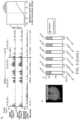
- FIG. 1 ATAC-see visualizes the accessible genome in situ.
- Panel a Schematic of ATAC-see.
- Panel b Genome browser tracks of ATAC-seq libraries from GM12878 cells (GM) generated by using different Tn5 transposases: green:Nextera Tn5, blue: Atto-Tn5, orange: previously published data.
- Panel c Left panel: Genome-wide comparison of ATAC-seq reads of GM12878 libraries (GM)prepared by either using Nextera Tn5 or Atto-Tn5.
- Panel d Genome-wide comparison of ATAC-seq reads of HT1080 library prepared in no fixation and with fixation. Left: Scatter plot of all data points.
- FIG. 2 ATAC-see enables imaging and sequencing the accessible genome in the same cells.
- Panel c Genomic tracks of ATAC-seq data from standard protocol and Atto-Tn5 on slide after imaging.
- X-axis is genomic coordinates; Y-axis is normalized ATAC-seq read counts.
- Panel d Genome-wide comparisons of standard ATAC-seq data and Atto-Tn5 on slide after imaging. Left: Scatter plot of all data points. Right: Metagene analysis centered on transcriptional start sites (TSS).
- FIG. 3 Cell type specific accessible chromatin organization in the intact nucleus.
- Panel b Unique feature of ATAC-seq from human neutrophil (Neuts) after imaging.
- Left Genome browser track of ATAC-seq in human neutrophil, H3K27Ac layer from ENCODE 7 cell lines, and NKI Lamin associated domains (LADs).
- X-axis is genomic coordinates;
- Y-axis is ATAC-seq normalized read counts.
- the grey square indicates the location of BAC chosen for DNA FISH in c.
- FIG. 4 ATAC-see and -seq reveal the dynamic chromatin organization during human NETosis.
- Panel b Epigenomic landscape of NETosis. Left column: Genomic tracks of ATAC-seq data from the indicated conditions. Locations of NKI Lamin associated domains (LADs) are indicated. X-axis is genomic coordinates; Y-axis is ATAC-seq normalized read counts. Middle: Metagene plot of ATAC-seq signal centered on the boundary between NKI LADs and neighboring sequences. The top plot is the same as FIG.
- Panel c Proposed model of NETosis illustrates the coordinated dynamics of nuclear architecture, accessible genome reorganization, and genome disassembly.
- FIG. 5 ATAC-see reveals cell cycle specific genome accessibility.
- Panel a Flow cytometry with ATAC-see. Dot plot of signal intensity in dual staining for DAPI and ATAC-see of GM12878 cells, and results showed four groups of cells: G1 low, G1 high, S phase and G2.
- Panel b Quantitation of DAPI (left) and ATAC-see (right) signals from different groups.
- Panel d Heatmap shows cluster of different ATAC-seq accessible regions between G1 high and G1 low group (FD>2, FDR ⁇ 0.05), and each group has one replicate.
- Panel e The volcano plot represents genome-wide comparisons of accessible regions in G1 high vs. G1 low.
- Panel f The density bars represent the distribution of the more accessible regions in G1 high and G1 low group across the transcription starting sites (TSS). The more accessible regions in two groups were color-coded.
- nucleic acids are written left to right in 5′ to 3′ orientation; amino acid sequences are written left to right in amino to carboxy orientation, respectively.
- transposase includes two or more transposases, and the like.
- the method may comprise obtaining a transposase complex that comprises a transposase bound to at least one DNA adapter (which may be 40 to 150 bases in length, e.g., 50 to 120 bases, although adaptors outside of this range are envisioned) that comprises a recognition sequence for the transposase, where either the transposase or DNA adaptor comprises a detectable label.
- the transposon recognition sequence also known as a transposon end sequence
- a transposase e.g., a Tn5 transposase or variant thereof
- the Tn5 transposon recognition sequence is 19 bp in length (see, e.g., Vaezeslami et al, J. Bacteriol. 2007 189 20: 7436-7441), although many others are known and may be 18-20 bp, e.g., 19 bp in length.
- the transposase complex comprises a transposase loaded with either a single adaptor molecule that contains a recognition sequence for the transposase at both ends, or two adaptor molecule that each contain a recognition sequence for the transposase at one end.
- the latter type can be used of the chromatin is going to be sequenced by ATAC-seq (Buenrostro et al, Nature Methods 2013 10: 1213-1218).
- ATAC-seq Busenrostro et al, Nature Methods 2013 10: 1213-1218.
- Such complexes can be combined with chromatin to add the adaptor molecule to the chromatin at accessible sites. If a transposase complex contains a single adaptor molecule that contains a transposon recognition sequence at both ends, the adaptor molecule will be inserted into the chromatin.
- a transposase complex contains a two adaptor molecules that contains a transposon recognition sequence at one end
- the transposase catalyzes simultaneous fragmentation of the chromatin and tagging of the fragments with sequences that are adjacent to the transposon recognition sequence (i.e., by “tagmentation”).
- tags Systems for tagmentation are described in a variety of publications, including Caruccio (Methods Mol. Biol. 2011 733: 241-55) and US20100120098, which are incorporated by reference herein.
- the transposase enzyme can insert the nucleic acid sequence into the polynucleotide in a substantially sequence-independent manner.
- the transposase can be prokaryotic, eukaryotic or from a virus.
- Methods for tagmenting, as well as transposon end sequences, are well known in the art (see, e.g., Picelli et al, Genome Res. 2014 24: 2033-40; Adey et al, Genome Biol. 2010 11:R119 and Caruccio et al, Methods Mol. Biol. 2011 733: 241-55, US20100120098 and US20130203605).
- Kits for performing tagmentation are commercially sold under the tradename NEXTERATM by Illumina (San Diego, Calif.). This initial step of the method may be done by loading a transposase with oligonucleotides that have been annealed together so that at least the transposase recognition sequence is double stranded.
- the adaptors used in the method are typically made of oligonucleotides that have been annealed together, where an oligonucleotide is a single-stranded multimer of nucleotide that is from about 2 to 200 nucleotides in length, up to 500 nucleotides in length. Oligonucleotides may be synthetic or may be made enzymatically, and, in some embodiments, are 30 to 150 nucleotides in length. An oligonucleotide may be 10 to 20, 21 to 30, 31 to 40, 41 to 50, 51 to 60, 61 to 70, 71 to 80, 80 to 100, 100 to 150 or 150 to 200 nucleotides in length, for example.
- the DNA adapter may comprise i) a nucleotide sequence having at least 95% identity to the sequence of SEQ ID NO:1, and ii) a second oligonucleotide comprising a sequence sufficiently complementary to and capable of hybridizing with a portion of the first oligonucleotide such that the DNA adapter comprises at least a portion that is double-stranded, wherein the double stranded portion comprises the recognition sequence for the transposase.
- the second oligonucleotide may comprise a nucleotide sequence having at least 95% identity to a sequence selected from the group consisting of SEQ ID NO:2 and SEQ ID NO:3, wherein the DNA adapter is capable of transposase catalyzed insertion into the accessible chromatin.
- the transposase complex may comprise a transposase dimer bound to i) a first DNA adapter, wherein the first DNA adapter comprises a first recognition sequence for the transposase and a first detectable label, and ii) a second DNA adapter, wherein the second DNA adapter comprises a second recognition sequence for the transposase and a second detectable label.
- the first DNA adapter may comprises a first oligonucleotide comprising a nucleotide sequence having at least 95% identity to the sequence of SEQ ID NO:1 and a second oligonucleotide comprising a nucleotide sequence having at least 95% identity to the sequence of SEQ ID NO:2; and the second DNA adapter comprises the first oligonucleotide comprising a nucleotide sequence having at least 95% identity to the sequence of SEQ ID NO:1 and a third oligonucleotide comprising a nucleotide sequence having at least 95% identity to the sequence of SEQ ID NO:3; wherein the first DNA adapter and the second DNA adapter are capable of transposase catalyzed insertion into the accessible chromatin.
- the first DNA adapter may comprise a first oligonucleotide comprising the nucleotide sequence of SEQ ID NO:1 and a second oligonucleotide comprising the nucleotide sequence of SEQ ID NO:2, and the second DNA adapter comprises a first oligonucleotide comprising the nucleotide sequence of SEQ ID NO:1 and a second oligonucleotide comprising the nucleotide sequence of SEQ ID NO:3.
- the adaptor is linked to a detectable label, e.g., at a 3′ end, at a 5′ end, or anywhere in between.
- the detectable label may be linked to a nucleotide in the transposase recognition sequence. In other embodiments, the detectable label may be linked to a nucleotide that is not in the transposase recognition sequence.
- the transposase may comprise a detectable label.
- the transposase may be labeled with a detectable label or may be a fusion protein, where the fusion partner may be a fluorescent protein or luciferase.
- a detectable label is a moiety that is readily detected by optical means, e.g., light-generating or fluorescent labels.
- Detectable labels may be directly detectable and, as such, may be fluorescent or luminescent (e.g., chemiluminescent, etc.) or indirectly detectable and, as such, may contain a detectable moiety such as e.g., biotin.
- Fluorphores are capable of exhibiting fluorescence in the detectable range.
- mass tagged refers to a molecule that is tagged with either a single kind of stable isotope that is identifiable by its unique mass or mass profile or a combination of the same.
- An element may exist as one or more isotopes, and this term also includes isotopes of positively and negatively metals.
- the terms “mass tagged” and “elementally tagged” may be used interchangeably herein.
- a mass tag may include transition metals, post transition metals, halides, noble metal or lanthanide, that is identifiable by its mass, distinguishable from other mass tags, and used to tag a biologically active material or analyte.
- a mass tag has an atomic mass that is distinguishable from the atomic masses present in the analytical sample and in the particle of interest.
- the term “monoisotopic” means that a tag contains a single type of metal isotope (although any one tag may contain multiple metal atoms of the same type).
- mass cytometry including methods by which single cell suspensions can be made, methods by which cells can be labeled using, e.g., mass-tagged antibodies, methods for atomizing particles and methods for performing elemental analysis on particles, as well as hardware that can be employed in mass cytometry, including flow cells, ionization chambers, reagents, mass spectrometers and computer control systems are known and are reviewed in a variety of publications including, but not limited to Bandura et al Analytical Chemistry 2009 81 6813-6822), Tanner et al (Pure Appl. Chem 2008 80: 2627-2641), U.S. Pat. No.
- transposases include, but are not limited to, Tn transposase (e.g. Tn3, Tn5, Tn7, Tn10, Tn552, Tn903), a MuA transposase, a Vibhar transposase (e.g.
- a transposase related to and/or derived from a parent transposase can comprise a peptide fragment with at least about 50%, about 55%, about 60%, about 65%, about 70%, about 75%, about 80%, about 85%, about 90%, about 91%, about 92%, about 93%, about 94%, about 95%, about 96%, about 97%, about 98%, or about 99% amino acid sequence homology to a corresponding peptide fragment of the parent transposase.
- the peptide fragment can be at least about 10, about 15, about 20, about 25, about 30, about 35, about 40, about 45, about 50, about 60, about 70, about 80, about 90, about 100, about 150, about 200, about 250, about 300, about 400, or about 500 amino acids in length.
- a transposase derived from Tn5 can comprise a peptide fragment that is 50 amino acids in length and about 80% homologous to a corresponding fragment in a parent Tn5 transposase.
- the insertion can be facilitated and/or triggered by addition of one or more cations.
- the cations can be divalent cations such as, for example, Ca 2+ , Mg 2+ and Mn 2+ .
- the transposes complex is contacted with chromatin under conditions suitable for binding of the transposase to the chromatin (i.e., a suitable temperature, time and conditions that result in a reaction, e.g., binding and/or cleavage/ligation).
- the contacting may be done under conditions suitable for transposition, the thereby joining the at least one DNA adapter with the chromatin at sites that are accessible to the transposase.
- chromatin accessibility refers to how accessible a nucleic acid site is within a polynucleotide, such as in genomic DNA, i.e., how “open” the chromatin is.
- a nucleic acid site associated with a polypeptide such as with genomic DNA in nucleosomes, is usually inaccessible.
- a nucleic acid site not complexed with a polypeptide is generally accessible, such as with genomic DNA between nucleosomes (with the exception of nucleic acid sites complexed with transcription factors and other DNA binding proteins).
- a cell sample can be optionally fixed and/or permeabilized to allow access to its chromatin. The permeabilization can be performed in a way to minimally perturb the nuclei in the cell.
- the cell sample can be permeabilized using a permeabilization agent, examples of which include, but are not limited to, NP40, digitonin, tween, streptolysin, and cationic lipids.
- a permeabilization agent examples of which include, but are not limited to, NP40, digitonin, tween, streptolysin, and cationic lipids.
- the calls can be permeabilized using hypotonic shock and/or ultrasonication.
- the transposase can be highly charged, which may allow it to pass through cell membranes.
- the method may comprise detecting the label of the inserted DNA adapter or the transposase. This may be done using any suitable method, including by flow cytometry, fluorescent microscopy, electron microscopy or mass cytometry. If labels are detected by flow cytometry, the data may be shown as a scatter plot. If labels are detected by fluorescence imaging, the data may be shown as an image of at least the nucleus of a cell. Analysis of such data may reveal a pattern of labeling, e.g., a pattern within a cell or a population of cells.
- the method may be performed on a population of cells.
- the cells may be on a planar surface, e.g., of a microscope slide.
- the cells may have been grown as a monolayer on the surface, grown in a liquid culture and then placed on the surface, or may in a tissue section that has been placed on the surface.
- the population of cells used in the may be composed of any number of cells, e.g., about 500 to about 10 6 or more cells, about 500 to about 100,000 cells, about 500 to about 50,000 cells, about 500 to about 10,000 cells, about 50 to 1000 cells, about 1 to 500 cells, about 1 to 100 cells, about 1 to 50 cells, or a single cell.
- the cell sample can consist of less than about 1000, about 2000, about 3000, about 4000, about 5000, about 6000, about 7000, about 8000, about 9000, about 10,000, about 15,000, about 20,000, about 25,000, about 30,000, about 40,000, about 50,000, about 60,000, about 70,000, about 80,000, about 90,000, about 100,000, about 120,000, about 140,000, about 160,000, about 180,000, about 200,000, about 250,000, about 300,000, about 350,000, about 400,000, about 450,000, about 500,000, about 600,000, about 700,000, about 800,000, about 900,000, or about 1,000,000 cells.
- the cell sample can consist of more than about 1000, about 2000, about 3000, about 4000, about 5000, about 6000, about 7000, about 8000, about 9000, about 10,000, about 15,000, about 20,000, about 25,000, about 30,000, about 40,000, about 50,000, about 60,000, about 70,000, about 80,000, about 90,000, about 100,000, about 120,000, about 140,000, about 160,000, about 180,000, about 200,000, about 250,000, about 300,000, about 350,000, about 400,000, about 450,000, about 500,000, about 600,000, about 700,000, about 800,000, about 900,000, or about 1,000,000 cells.
- the cells can be from any source.
- the cells may be obtained from a culture of cells, e.g., a cell line.
- the cells may be isolated from an individual (e.g., a patient or the like).
- the cells may be isolated from a soft tissue or from a bodily fluid, or from a cell culture that is grown in vitro.
- the chromatin may be isolated from a soft tissue such as brain, adrenal gland, skin, lung, spleen, kidney, liver, spleen, lymph node, bone marrow, bladder stomach, small intestine, large intestine or muscle, etc.
- Bodily fluids include blood, plasma, saliva, mucous, phlegm, cerebral spinal fluid, pleural fluid, tears, lactal duct fluid, lymph, sputum, cerebrospinal fluid, synovial fluid, urine, amniotic fluid, and semen, etc.
- the chromatin analyzed in the method may be from blood cells, wherein blood cells refers to a sample of whole blood or a sub-population of cells in whole blood.
- Sub-populations of cells in whole blood include platelets, red blood cells (erythrocytes), platelets and white blood cells (i.e., peripheral blood leukocytes, which are made up of neutrophils, lymphocytes, eosinophils, basophils and monocytes).
- white blood cells can be further divided into two groups, granulocytes (which are also known as polymorphonuclear leukocytes and include neutrophils, eosinophils and basophils) and mononuclear leukocytes (which include monocytes and lymphocytes). Lymphocytes can be further divided into T cells, B cells and NK cells. Peripheral blood cells are found in the circulating pool of blood and not sequestered within the lymphatic system, spleen, liver, or bone marrow. Other cells are present in blood that can be isolated. If blood is first contacted with an agent and then a sample of the blood is used in an assay, then a portion or all of the contacted blood may be used in the method.
- granulocytes which are also known as polymorphonuclear leukocytes and include neutrophils, eosinophils and basophils
- mononuclear leukocytes which include monocytes and lymphocytes
- Lymphocytes can be further divided into T cells
- the cells can be isolated directly from a primary source.
- the cell sample can be isolated directly from fresh tissues.
- the cell sample can be isolated directly from frozen tissues.
- the cell sample can be isolated directly from fixed tissues.
- primary sources of cell samples include, but are not limited to, cells dissociated from tissues, blood cells, cells in an FFPE section, etc.
- the transposase complex may be directly contacted with a tissue sample,
- the cells analyzed are of the same cell type.
- the population of cells may be cultured cells or selected by MACS or FACS from a heterogeneous population of cells, e.g., blood, by known methods using labeled antibodies to cells surface markers.
- a wide variety of cells can be isolated using these methods, including stem cells, cancer stem cells and subsets of blood cells.
- the following cells may be isolated from blood by FACS or MACS; T cells (CD3 + CD4 + CD8 + ), B cells (CD19 + CD20 + ), dendritic cells (CD11c + CD20 + ), NK Cell (CD56 + ), stem cells/precursor cells (CD34 + ; hematopoietic stem cells only), macrophage/monocytes (CD14 + CD33 + ), granulocytes (CD66b + ), platelet (CD41 + CD61 + CD62 + ), erythrocytes (CD235a + ), endothelial cells (CD146 + ) and epithelial cells (CD326 + ). Subsets of these cells can be isolated using antibodies to further cell surface markers.
- the method can be used to compare two samples.
- the method may comprise analyzing a first population of cells using the above-described method to produce a first data set; and analyzing a second population of cells using the above-described method to produce a second data set; and comparing the first data set to the second data set, e.g., to see if there are any changes in in the localization of accessible chromatin, for example.
- the first population of cells and the second population of cells are collected from the same individual at different times. In other embodiments, the first population of cells and the second population of cells are different populations of cells collected from tissues or different individuals.
- Exemplary cell types that can be used in the method include, for example, cells isolated from a tissue biopsy (e.g., from a tissue having a disease such as colon, breast, prostate, lung, skin cancer, or infected with a pathogen etc.) and normal cells from the same tissue, e.g., from the same patient; cells grown in tissue culture that are immortal (e.g., cells with a proliferative mutation or an immortalizing transgene), infected with a pathogen, or treated (e.g., with environmental or chemical agents such as peptides, hormones, altered temperature, growth condition, physical stress, cellular transformation, etc.), and normal cells (e.g., cells that are otherwise identical to the experimental cells except that they are not immortalized, infected, or treated, etc.); cells isolated from a mammal with a cancer, a disease, a geriatric mammal, or a mammal exposed to a condition, and cells from a mammal of the same species, e.g.,
- cells of different types e.g., neuronal and non-neuronal cells, or cells of different status (e.g., before and after a stimulus on the cells) may be compared.
- the experimental material is cells susceptible to infection by a pathogen such as a virus, e.g., human immunodeficiency virus (HIV), etc.
- the control material is cells resistant to infection by the pathogen.
- the sample pair is represented by undifferentiated cells, e.g., stem cells, and differentiated cells. Cells from yeast, plants and animals, such as fish, birds, reptiles, amphibians and mammals may be used in the subject methods.
- mammalian cells i.e., cells from mice, rabbits, primates, or humans, or cultured derivatives thereof, may be used.
- the method may be used to identify the effect of a test agent, e.g., a drug, or to determine if there are differences in the effect of two or more different test agents.
- a test agent e.g., a drug
- two or more identical populations of cells may be prepared and, depending on how the experiment is to be performed, one or more of the populations of cells may be incubated with the test agent for a defined period of time. After incubation with the test agent, the chromatin of the populations of cells can be analyzed using the methods set forth above, and the results can be compared.
- the cells may be blood cells, and the cells can be incubated with the test agent ex vivo. These methods can be used to determine the mode of action of a test agent, to identify changes in chromatin accessability in response to the drug, for example.
- the method described above may also be used as a diagnostic (which term is intended to include methods that provide a diagnosis as well as methods that provide a prognosis). These methods may comprise, e.g., analyzing chromatin from a patient using the method described above to produce data; and providing a diagnosis or prognosis based on the data.
- the method set forth herein may be used to provide a reliable diagnostic to any condition associated with, e.g., altered chromatin accessability.
- the method can be applied to the characterization, classification, differentiation, grading, staging, diagnosis, or prognosis of a condition characterized by an epigenetic pattern (e.g., a pattern of chromatin accessibility).
- the method can be used to determine whether the pattern of labeling of a sample from an individual suspected of being affected by a disease or condition is the same or different compared to a pattern of labeling for a sample that is considered “normal” with respect to the disease or condition.
- the method can be directed to diagnosing an individual with a condition that is characterized by particular pattern of labeling, where the pattern is correlated with the condition.
- the methods can also be used for predicting the susceptibility of an individual to a condition.
- Exemplary conditions that are suitable for analysis using the methods set forth herein can be, for example, cell proliferative disorder or predisposition to cell proliferative disorder; metabolic malfunction or disorder; immune malfunction, damage or disorder; CNS malfunction, damage or disease; symptoms of aggression or behavioral disturbance; clinical, psychological and social consequences of brain damage; psychotic disturbance and personality disorder; dementia or associated syndrome; cardiovascular disease, malfunction and damage; malfunction, damage or disease of the gastrointestinal tract; malfunction, damage or disease of the respiratory system; lesion, inflammation, infection, immunity and/or convalescence; malfunction, damage or disease of the body as an abnormality in the development process; malfunction, damage or disease of the skin, the muscles, the connective tissue or the bones; endocrine and metabolic malfunction, damage or disease; headache or sexual malfunction, and combinations thereof.
- the method can provide a prognosis, e.g., to determine if a patient is at risk for recurrence.
- Cancer recurrence is a concern relating to a variety of types of cancer.
- the prognostic method can be used to identify surgically treated patients likely to experience cancer recurrence so that they can be offered additional therapeutic options, including preoperative or postoperative adjuncts such as chemotherapy, radiation, biological modifiers and other suitable therapies.
- the methods are especially effective for determining the risk of metastasis in patients who demonstrate no measurable metastasis at the time of examination or surgery.
- the method can also be used as a theranostic, i.e., to provide a recommendation for a course of treatment for a patient having a disease or condition, e.g., a patient that has cancer.
- a course of treatment refers to the therapeutic measures taken for a patient after diagnosis or after treatment.
- a determination of the likelihood for recurrence, spread, or patient survival can assist in determining whether a more conservative or more radical approach to therapy should be taken, or whether treatment modalities should be combined.
- cancer recurrence it can be advantageous to precede or follow surgical treatment with chemotherapy, radiation, immunotherapy, biological modifier therapy, gene therapy, vaccines, and the like, or adjust the span of time during which the patient is treated.
- a lab will receive a sample (e.g., blood) from a remote location (e.g., a physician's office or hospital), the lab will analyze cells in the sample as described above to produce data, and the data may be forwarded to the remote location for analysis.
- a sample e.g., blood
- a remote location e.g., a physician's office or hospital
- An adaptor comprising a double-stranded transposase recognition sequence and a detectable label are also provided.
- One of the strands of the transposase recognition sequence may have the sequence of SEQ ID NO: 1.
- the adaptor may be linked to the detectable label, e.g., at a 3′ end, at a 5′ end, or anywhere in between.
- the detectable label may be linked to a nucleotide in the transposase recognition sequence.
- the detectable label may be linked to a nucleotide that is not in the transposase recognition sequence.
- composition comprising a transposase complex comprising a transposase bound to a DNA adapter, wherein either the transposase or DNA adaptor comprises a detectable label.
- the transposase complex may comprise a transposase that comprises a detectable label
- the transposase complex may comprise a transposase bound to a DNA adapter comprising a detectable label.
- the DNA adapter may comprises i) a first oligonucleotide comprising the nucleotide sequence of SEQ ID NO:1 or a nucleotide sequence having at least 95% identity to the sequence of SEQ ID NO:1, and ii) a second oligonucleotide comprising a sequence sufficiently complementary to and capable of hybridizing with a portion of the first oligonucleotide such that the DNA adapter comprises at least a portion that is double-stranded, wherein the double stranded portion comprises a recognition sequence for the transposase, wherein the DNA adapter is capable of transposase catalyzed insertion into accessible chromatin.
- the transposase complex comprises a transposase dimer bound to two DNA adapters, wherein the first DNA adapter comprises a first recognition sequence for the transposase and a first detectable label, and the second DNA adapter comprises a second recognition sequence for the transposase and a second detectable label.
- THe first DNA adapter may comprise a first oligonucleotide comprising the nucleotide sequence of SEQ ID NO:1 or a nucleotide sequence having at least 95% identity to the sequence of SEQ ID NO:1 and a second oligonucleotide comprising the nucleotide sequence of SEQ ID NO:2 or a nucleotide sequence having at least 95% identity to the sequence of SEQ ID NO:2; and the second DNA adapter comprises the first oligonucleotide comprising the nucleotide sequence of SEQ ID NO:1 or a nucleotide sequence having at least 95% identity to the sequence of SEQ ID NO:1 and a third oligonucleotide comprising the nucleotide sequence of SEQ ID NO:3 or a nucleotide sequence having at least 95% identity to the sequence of SEQ ID NO:3; wherein the first DNA adapter and the second DNA adapter are capable of transposase catalyzed
- the first DNA adapter comprises a first oligonucleotide comprising the nucleotide sequence of SEQ ID NO:1 and a second oligonucleotide comprising the nucleotide sequence of SEQ ID NO:2
- the second DNA adapter comprises a first oligonucleotide comprising the nucleotide sequence of SEQ ID NO:1 and a second oligonucleotide comprising the nucleotide sequence of SEQ ID NO:3.
- the detectable label is a fluorophore, a metal particle, a magnetic particle, an isotopic label, a chemiluminescent label, a ligand, or a hapten.
- kits that comprise a transposase and at least one DNA adapter that comprises a recognition sequence for the transposase, wherein either the DNA adaptor or the recognition sequence comprises a detectable label.
- the kit may comprise instruction for detecting accessible chromatin.
- the kit may comprise a hyperactive transposase and at least one DNA adapter, said DNA adapter comprising a recognition sequence for the transposase and a detectable label.
- the at least one DNA adapter may comprise a first DNA adapter comprising a first oligonucleotide comprising the nucleotide sequence of SEQ ID NO:1 or a nucleotide sequence having at least 95% identity to the sequence of SEQ ID NO:1 and a second oligonucleotide comprising the nucleotide sequence of SEQ ID NO:2 or a nucleotide sequence having at least 95% identity to the sequence of SEQ ID NO:2, and a second DNA adapter comprising a first oligonucleotide comprising the nucleotide sequence of SEQ ID NO:1 or a nucleotide sequence having at least 95% identity to the sequence of SEQ ID NO:1 and a second oligonucleotide comprising the nucleotide sequence of SEQ ID NO:3 or a nucleotide sequence having at least 95% identity to the sequence of SEQ ID NO:3.
- the at least one DNA adapter may comprise a first DNA adapter comprising a first oligonucleotide comprising the nucleotide sequence of SEQ ID NO:1 and a second oligonucleotide comprising the nucleotide sequence of SEQ ID NO:2, and a second DNA adapter comprising a first oligonucleotide comprising the nucleotide sequence of SEQ ID NO:1 and a second oligonucleotide comprising the nucleotide sequence of SEQ ID NO:3.
- kit may be present in separate containers or certain compatible components may be precombined into a single container, as desired.
- the kit may contain any of the additional components used in the method described above, e.g., a buffer, etc.
- the subject kits may further include instructions for using the components of the kit to practice the subject methods, i.e., instructions for sample analysis.
- a method for detecting accessible chromatin comprising: a) providing a transposase complex comprising a transposase bound to at least one DNA adapter, wherein the DNA adapter comprises a recognition sequence for the transposase and a detectable label; b) contacting chromatin with the transposase complex, whereby said at least one DNA adapter is inserted into the chromatin at sites that are accessible to the transposase; and c) detecting the label of the inserted DNA adapter.
- the DNA adapter comprises i) a first oligonucleotide comprising the nucleotide sequence of SEQ ID NO:1 or a nucleotide sequence having at least 95% identity to the sequence of SEQ ID NO:1, and ii) a second oligonucleotide comprising a sequence sufficiently complementary to and capable of hybridizing with a portion of the first oligonucleotide such that the DNA adapter comprises at least a portion that is double-stranded, wherein the double stranded portion comprises the recognition sequence for the transposase.
- the second oligonucleotide comprises a nucleotide sequence selected from the group consisting of SEQ ID NO:2 and SEQ ID NO:3, or a nucleotide sequence having at least 95% identity to a sequence selected from the group consisting of SEQ ID NO:2 and SEQ ID NO:3, wherein the DNA adapter is capable of transposase catalyzed insertion into the accessible chromatin.
- the transposase complex comprises a transposase dimer bound to i) a first DNA adapter, wherein the first DNA adapter comprises a first recognition sequence for the transposase and a first detectable label, and ii) a second DNA adapter, wherein the second DNA adapter comprises a second recognition sequence for the transposase and a second detectable label.
- the first DNA adapter comprises a first oligonucleotide comprising the nucleotide sequence of SEQ ID NO:1 or a nucleotide sequence having at least 95% identity to the sequence of SEQ ID NO:1 and a second oligonucleotide comprising the nucleotide sequence of SEQ ID NO:2 or a nucleotide sequence having at least 95% identity to the sequence of SEQ ID NO:2; and the second DNA adapter comprises the first oligonucleotide comprising the nucleotide sequence of SEQ ID NO:1 or a nucleotide sequence having at least 95% identity to the sequence of SEQ ID NO:1 and a third oligonucleotide comprising the nucleotide sequence of SEQ ID NO:3 or a nucleotide sequence having at least 95% identity to the sequence of SEQ ID NO:3; wherein the first DNA adapter and the second DNA adapter are capable of transposase cataly
- the first DNA adapter comprises a first oligonucleotide comprising the nucleotide sequence of SEQ ID NO:1 and a second oligonucleotide comprising the nucleotide sequence of SEQ ID NO:2
- the second DNA adapter comprises a first oligonucleotide comprising the nucleotide sequence of SEQ ID NO:1 and a second oligonucleotide comprising the nucleotide sequence of SEQ ID NO:3.
- the detectable label is a fluorophore, a metal particle, a magnetic particle, an isotopic label, a chemiluminescent label, a ligand, or a hapten.
- invention 1 further comprising identifying regulatory DNA, a transcription factor binding site, or a nucleosome binding site in the accessible chromatin.
- the regulatory DNA comprises a promoter, enhancer, or insulator.
- a composition comprising a transposase complex comprising a transposase bound to a DNA adapter comprising a detectable label, wherein the DNA adapter comprises i) a first oligonucleotide comprising the nucleotide sequence of SEQ ID NO:1 or a nucleotide sequence having at least 95% identity to the sequence of SEQ ID NO:1, and ii) a second oligonucleotide comprising a sequence sufficiently complementary to and capable of hybridizing with a portion of the first oligonucleotide such that the DNA adapter comprises at least a portion that is double-stranded, wherein the double stranded portion comprises a recognition sequence for the transposase, wherein the DNA adapter is capable of transposase catalyzed insertion into accessible chromatin.
- composition of embodiment 19, wherein the transposase complex comprises a transposase dimer bound to two DNA adapters, wherein the first DNA adapter comprises a first recognition sequence for the transposase and a first detectable label, and the second DNA adapter comprises a second recognition sequence for the transposase and a second detectable label.
- the first DNA adapter comprises a first oligonucleotide comprising the nucleotide sequence of SEQ ID NO:1 or a nucleotide sequence having at least 95% identity to the sequence of SEQ ID NO:1 and a second oligonucleotide comprising the nucleotide sequence of SEQ ID NO:2 or a nucleotide sequence having at least 95% identity to the sequence of SEQ ID NO:2; and the second DNA adapter comprises the first oligonucleotide comprising the nucleotide sequence of SEQ ID NO:1 or a nucleotide sequence having at least 95% identity to the sequence of SEQ ID NO:1 and a third oligonucleotide comprising the nucleotide sequence of SEQ ID NO:3 or a nucleotide sequence having at least 95% identity to the sequence of SEQ ID NO:3; wherein the first DNA adapter and the second DNA adapter are capable of transposase cataly
- composition of embodiment 19, wherein the detectable label is a fluorophore, a metal particle, a magnetic particle, an isotopic label, a chemiluminescent label, a ligand, or a hapten.
- a kit comprising the composition of embodiment 19 and instructions for detecting accessible chromatin.
- a kit comprising a hyperactive transposase and at least one DNA adapter, said DNA adapter comprising a recognition sequence for the transposase and a detectable label.
- said at least one DNA adapter comprises a first DNA adapter comprising a first oligonucleotide comprising the nucleotide sequence of SEQ ID NO:1 or a nucleotide sequence having at least 95% identity to the sequence of SEQ ID NO:1 and a second oligonucleotide comprising the nucleotide sequence of SEQ ID NO:2 or a nucleotide sequence having at least 95% identity to the sequence of SEQ ID NO:2, and a second DNA adapter comprising a first oligonucleotide comprising the nucleotide sequence of SEQ ID NO:1 or a nucleotide sequence having at least 95% identity to the sequence of SEQ ID NO:1 and a second oligonucleotide comprising the nucleotide sequence of SEQ ID NO:3 or a nucleotide sequence having at least 95% identity to the sequence of SEQ ID NO:3.
- said at least one DNA adapter comprises a first DNA adapter comprising a first oligonucleotide comprising the nucleotide sequence of SEQ ID NO:1 and a second oligonucleotide comprising the nucleotide sequence of SEQ ID NO:2, and a second DNA adapter comprising a first oligonucleotide comprising the nucleotide sequence of SEQ ID NO:1 and a second oligonucleotide comprising the nucleotide sequence of SEQ ID NO:3.
- ATAC-see Assay of Transposase-Accessible Chromatin with visualization
- NETosis a transposase-mediated imaging technology that enables direct imaging of the accessible genome in situ, cell sorting, and deep sequencing to reveal the identity of the imaged elements.
- ATAC-see revealed the cell type-specific spatial organization of the accessible genome and the coordinated process of neutrophil extrusion of its chromatin (NETosis).
- ATAC-see unveils the spatial organization of the accessible genome in its native context for any cell type by covalently inserting fluorophore molecules in the genome.
- ATAC-see is compatible with multimodal imaging of landmark proteins, flow cytometry, and enables sophisticated regulome analysis.
- the compatibility of ATAC-see with fixed samples will allow it to be readily applied to human clinical specimens.
- the ability to apply ATAC-see in FACS sorting should enhance the dissection of the chromatin basis of cellular heterogeneity.
- ATAC-see provides rich spatial and epigenomic information that together gives a molecular portrait of the cell.
- ATAC-see can be used to identify the unique spatial organization of the accessible genome in different human cell types and organs that comprise the human cell atlas, and reveal fine structure of accessible chromatin organization in clinical diagnosis by using super-resolution microscopy.
- transposome engineering facilitated the successful combination of imaging and epigenomics; extending this approach may facilitate novel modes of genome interrogation and control.
- GM12878 cells were grown in RPMI 1640 (11875-093, Gibco), 2 mM L-glutamine (25030-081, Gibco), 15% fetal bovine serum (07905, Gibco) and 1% Pen/Strep (15140-122, Gibco).
- HT1080 cells were cultured in DMEM/F-12, GlutaMAXTM supplement (10565-018, Gibco), 10% fetal bovine serum (07905, Gibco), and 1% Pen/Strep (15140-122, Gibco).
- HeLa cells were maintained in DMEM (11965-092, Gibco), 10% fetal bovine serum (07905, Gibco), and 1% Pen/Strep (15140-122, Gibco).
- Neural progenitor cells were cultured in N2B27 media (DMEM/F12, Invitrogen 11320-033; Neurobasal, Gibco 21103-049; NDiff Neuro-2 Medium Supplement Millipore, SCM012; B27 Supplement, Gibco 17504-044) supplemented with EGF and FGF (10 ng/mL each) (315-09 and 100-18B, Peprotech). NPCs were passaged every other day with Accutase (SCR005, Millipore) and seeded on gelatin-coated plates. All cells were kept under standard 37° C. and 5% CO 2 conditions.
- Human CD4 + T cells were isolated from whole blood of healthy donors under a Stanford University IRB-approved protocol.
- RosetteSep® Human CD4 + T Cell Enrichment Cocktail was added into the whole blood with a final concentration of 50 ⁇ L/mL, and the mixture was incubated 20 min at room temperature.
- the whole blood was diluted with equal volume of PBS+2% FBS (07905, Gibco). Then the diluted samples were layered on top of the density medium and centrifuged for 20 min at 1200 ⁇ g at room temperature with the brake off. The enriched cells were collected into a new tube and washed with PBS+2% FBS twice.
- the residual red blood cells are lysed with buffer EL (79217, Qiagen) 5 min at room temperature.
- Human neutrophils were isolated from healthy donor's blood following as described under a Stanford University IRB-approved protocol 23 .
- 7 ml of heparinized blood was layered on 7 ml Histopaque 1119 (11191-100ML, Sigma Aldrich) in a 15 ml Falcon tube, and the mixture was spun 20 min at 800 ⁇ g at room temperature.
- the interphase (lymphocytes and monocytes) was discarded and the granulocyte-rich layer of Histopaque 1119 (diffuse, red phase above RBC pellet) was collected in a new tube.
- 5 ml of cells were washed with 10 ml of medium (HBSS+), and spun 10 min at 200 ⁇ g at room temperature.
- the cells were resuspended in 3 ml DPBS with 0.5% human serum albumin (101-15-50, Lee BioSolutions), and the cell suspension was layered on 10 ml of a discontinuous Percoll gradient (85% to 65% Percoll) and spun 20 min at 800 ⁇ g at room temperature. The interphase between 70% and 75% Percoll layers were collected in a new 15 ml tube. The tube was added with DPBS with 0.5% human serum albumin up to 15 ml and spun for 10 min at 200 ⁇ g at room temperature. The cell pellet was resuspended in RPMI-HEPES (10 mM) at 1.5 ⁇ 10 6 cells per ml for neutrophil activation.
- RPMI-HEPES 10 mM
- Human neutrophils were stimulated by Phorbol myristate acetate (PMA) as described 23 . Briefly, the purified neutrophils were suspended in 3 ml RPMI 1640 with final concentration of 1.5 ⁇ 10 6 per ml in loosen cap 15 ml falcon tube, and incubated in cell culture CO 2 incubator (5% CO 2 , 37° C.) with final concentration of 30 nM PMA for 1 h, 3 h, and 5 h respectively. After 2 h of simulation, protease inhibitor cocktail was added at a 1:200 dilution to both control and simulated tubes. At the respective time points, neutrophils were fixed with 1% formaldehyde for 10 min at room temperature, and fixed neutrophil cells spun down on the glass slide with cytospin 1000 rpm 5 min at room temperature for ATAC-see or immuno-staining.
- PMA Phorbol myristate acetate
- the PAD4 inhibition was performed as previously described 25 .
- the isolated human neutrophils were incubated with 200 ⁇ M Cl-amidine for 30 min before PMA stimulation.
- Hyperactive Tn5 was produced as previously described 30 .
- coli pellet was resuspended in lysis buffer (20 mM HEPES-KOH pH 7.2, 0.8 M NaCl, 1 mM EDTA, 10% glycerol, 0.2% triton X-100, complete proteinase inhibitor (11697498001, Roche)) and lysed by sonication. 10% PEI was added to supernatant of lysate to remove bacterial genomic DNA. 10 ml chitin resin (S6651L, NEB) was added to the supernatant and incubated with rotating for 1 hr at 4° C. The resin washed by lysis buffer extensively.
- lysis buffer containing 100 mM DTT was added to the resin and stored in 4° C. After 48 hrs, protein was eluted by gravity flow and collected in 1 ml fractions. 1 ul of each fraction was added to detergent compatible Bradford assay (23246, ThermoFisher Scientific) and peaked fractions were pooled and dialyzed against 2 ⁇ dialysis buffer (100 mM HEPE-KOH pH7.2, 0.2 M NaCl, 0.2 mM EDTA, 2 mM DTT, 0.2% triton X-100, 20% glycerol).
- Tn5 protein was concentrated by using ultracel 30-K column (UFC903024, Millipore) and the quantity of Tn5 was measured by Bradford assay and visualized on NuPAGE Novex 4-12% Bis-Tris gel (NP0321, ThermoFisher Scientific) followed by Coomassie blue staining.
- Atto-590N labeled oligonucleotides for Tn5 transposome adaptor were synthesized at INTERGATED DNA TECHNOLOGIES (IDT), and the sequences of oligonucleotide are as follows: Tn5MErev, 5′-[phos]CTGTCTCTTATACACATCT-3′ (SEQ ID NO: 1); Tn5ME-A-ATTO590, 5′-/5ATTO590/TCGTCGGCAGCGTCAGATGTGTATAAGAGACAG-3′ (SEQ ID NO: 2); Tn5ME-B-ATTO590: 5′-/ATTO590/GTCTCGTGGGCTCGGAGATGTG TATAAGAGACAG-3′ (SEQ ID NO: 3).
- Tn5 transposome The assembly of Tn5 transposome was performed as described 30 . Briefly, oligonucleotides (Tn5ME-A-ATTO590, Tn5ME-B-ATTO590, Tn5MErev) were resuspended in water to a final concentration of 100 ⁇ M each. Equimolar amounts of Tn5MErev/Tn5ME-A-ATTO590 and Tn5MErev/Tn5ME-B-ATTO590 were mixed in separate 200 ⁇ l PCR tubes. These two tubes of oligos mixtures were denatured on a thermocycler for 5 min at 95° C., and cooled down slowly on the thermocycler by turning off the thermocycler.
- the Tn5 transposome was assembled with the following components: 0.25 vol Tn5MErev/Tn5ME-A-ATTO590+Tn5MErev/Tn5ME-B-ATTO590 (final concentration of each double strand oligo is now 50 ⁇ M each), 0.4 vol glycerol (100% solution), 0.12 vol 2 ⁇ dialysis buffer (100 mM HEPES-KOH at pH 7.2, 0.2 M NaCl, 0.2 mM EDTA, 2 mM DTT, 0.2% Triton X-100, 20% glycerol), 0.1 vol SL-Tn5 (50 ⁇ M), 0.13 vol water. The reagents were mixed thoroughly but gently and the solution was left on the bench at room temperature for 1 h to allow annealing of oligos to Tn5.
- HT1080 cells and HeLa cells were grown on secureSlip cell culture system (0515073, GRACE BIO-LABS) until 80-90% confluent and fixed with 1% formaldehyde (Sigma-Aldrich) for 10 min at room temperature.
- GM12878 cells were fixed with 1% formaldehyde (Sigma) 10 min at room temperature and centrifuged on the glass slide with Cytospin, 1000 rpm for 5 min, 30-50,000 cells per slide.
- the formaldehyde fixed slide was rinsed in PBS and permeabilized in PBS 0.5% Triton-X 100 for 10 mins at room temperature. Then, the slide was blocked with antibody dilution reagent (00-3218, ThermoFisher Scientific) for 1 h at room temperature.
- the ATAC-see was performed first and then immune-staining was done on the same slide. All the primary antibodies was diluted in 1:500 ratio, and the antibodies information as follows: rabbit anti-H3K9 me3 (SAB4800027-100UL, Sigma); rabbit anti-H3K4 me3 (ab8580, Abcam); rabbit anti H3K27acetylation (25238, BPS Bioscience); mouse anti-Phospho RNA Polymerase II CTD (Ser5) mAb (MABI0603, MBL International); mouse anti-H3K27 me3 (ab6147, Abcam); rabbit anti-RNA polymerase II CTD (Ser-5) antibody (ab5095, Abcam).
- the cells (either growing on slide or centrifuged on glass slide with cytospin) were permeabilized with lysis buffer (10 mM Tris-Cl, pH 7.4, 10 mM NaCl, 3 mM MgCl2, 0.01% Igepal CA-630) for 10 min at room temperature. After the permeabilization, the slides were rinsed in PBS twice, and put in humid chamber box at 37° C. The transposase mixture solution (25 ⁇ l 2 ⁇ TD buffer, final concentration of 100 nM Tn5-ATTO-59ON, adding dH2O up to 50 ⁇ l) was added on the slide and incubated for 30 min at 37° C.
- lysis buffer 10 mM Tris-Cl, pH 7.4, 10 mM NaCl, 3 mM MgCl2, 0.01% Igepal CA-630
- NPCs Female mouse neural progenitor cells (NPCs) were grown on secureSlip cell culture system (0515073, GRACE BIO-LABS) until 80-90% confluent and fixed with 1% formaldehyde (Sigma-Aldrich) for 10 min at RT. After washed with PBS three time (5 mins each), the slide was permeabilized with PBS 0.1% NP-40 10 mins at RT. ATAC-see (with Atto-480 labeled Tn5) were performed under standard protocol as stated above. After the ATAC-see, the slide was dehydrate in 70% EtOH for 1 hr at 4° C., and re-hydrated in 2 ⁇ SSC+10% formamide for 5 mins at RT.
- RNA FISH probes (Stellaris mXist) were hybridized on the slide overnight at 37° C. The washing was conducted in following conditions: 2 ⁇ 30 mins in 2 ⁇ SSC+10% formamide at 37° C. and 5 mins in 2 ⁇ SSC at 37° C. After RNA FISH washing, the slide were mounted with Vectashield (H-1200, Vector labs).
- GM12878 cells were washed with PBS, then fixed with 1% formaldehyde for 10 min at room temperature. After the fixation, cells were permeabilized with lysis buffer (10 mM Tris-Cl, pH 7.4, 10 mM NaCl, 3 mM MgCl2, 0.01% Igepal CA-630) and centrifuged at 700 g for 10 min at room temperature. 5 ⁇ 10 6 cells were then either stained with DAPI (for negative control), or transposed for 30 min at 37° C. using Atto-594 labeled in-houseTn5 (see materials and methods above).
- lysis buffer (10 mM Tris-Cl, pH 7.4, 10 mM NaCl, 3 mM MgCl2, 0.01% Igepal CA-630) and centrifuged at 700 g for 10 min at room temperature. 5 ⁇ 10 6 cells were then either stained with DAPI (for negative control), or transposed for 30 min at 37° C. using Atto-594 labeled in-houseTn
- the sorted ATAC-See G1 high and G1 low cells were collected and centrifuged by using Cytospin, and Anti-Cyclin E1 antibody [EP435E] (Alexa Fluor® 488) (ab194068, Abcam) were stained on different groups.
- EP435E Anti-Cyclin E1 antibody
- cells were harvested from femur, tibia, and humerus. Bones were fragmented by mortar and pestle, and debris was removed by gradient centrifugation using Histopaque 1119 (Sigma-Aldrich). Cells were passed through a 70-uM strainer, and red blood cells were lysed with ACK lysis buffer. 5-10 ⁇ 10 ⁇ circumflex over ( ) ⁇ 6 cells were stained at 4° C.
- ATAC-seq with Illumina Tn5 transposase (Nextera) and Atto-590 labeled homemade Tn5 in human GM-12878 cells was performed as described 3 . Briefly, 50,000 cells were centrifuged 500 g 5 min at room temperature. The cell pellet was resuspended in 50 ⁇ l lysis buffer (10 mM Tris-Cl, pH 7.4, 10 mM NaCl, 3 mM MgCl2, 0.01% Igepal CA-630) and centrifuged immediately 500 g for 10 min at 4° C.
- 50 lysis buffer (10 mM Tris-Cl, pH 7.4, 10 mM NaCl, 3 mM MgCl2, 0.01% Igepal CA-630
- the cell pellet was resuspended in 50 ⁇ l transposase mixture (25 ⁇ l 2 ⁇ TD buffer, 22.5 ⁇ l dH2O and 2.5 ⁇ l Illumina Tn5 transposase or with final concentration of 100 nM Atto-590 labeled in-houseTn5) and incubated at 37° C. 30 min. After transposition, the mixture was purified with Qiagen Mini-purification kit and eluted in 10 ⁇ l Qiagen EB elution buffer. Sequencing libraries were prepared following the original ATAC-seq protocol 3 . The sequencing was performed on Illumina NextSeq at the Stanford Functional Genomics Facility.
- Human HT1080 cells were fixed with 1% formaldehyde (Sigma, USA) for 10 min and quenched with 0.125 M glycine for 5 min at room temperature. After the fixation, cells were counted and 50,000 cells were used per ATAC-seq reaction.
- the transposed reaction is same as normal ATAC-seq except with 0.05% Igepal CA-630 in the lysis buffer. After the transposase reaction, a reverse crosslink solution was added (with final concentration of 50 mM Tris-Cl, 1 mM EDTA, 1% SDS, 0.2M NaCl, 5 ng/ml proteinase K) up to 200 ⁇ l. The mixture was incubated at 65° C.
- 80% confluent Human HT1080 cells (30-50,000 cells) grown in 8 well chamber slides were fixed with 1% formaldehyde (Sigma, USA) for 10 min and quenched with 0.125 M glycine for 5 min at room temperature. After fixation, slides were put in PBS at 4° C. Cells on slides were permeabilized with lysis buffer (10 mM Tris-Cl, pH 7.4, 10 mM NaCl, 3 mM MgCl2, 0.05% Igepal CA-630) for 10 min. The 50 ⁇ l transposase reaction solution was added onto the slide, and the cells on slide were incubated at 37° C. for 30 min.
- lysis buffer (10 mM Tris-Cl, pH 7.4, 10 mM NaCl, 3 mM MgCl2, 0.05% Igepal CA-630
- DNA FISH probes labeling was performed as previous described 31 .
- BAC clones (ThermoFisher Scientific), RP11-626N18, RP11-832P24, RP1163J14, RP11637D5, RP11-368K11, RP11-116A9, were cultured based on standard protocol and purified with BACMAXTM DNA Purification Kit (BMAX044, Epicenter). After the purification, BACs were sheared to 300-800 bp with sonication.
- the sheared BAC DNA was labeled with BioPrime® Array CGH Genomic Labeling System (18095-011, Thermo Fisher scientific) by mixing with either Cy3-dCTP (RP11-626N18, RP1163J14) (PA53021, GE healthcare life science, USA), Green 496 dUTP (RP11-832P24, RP11637D5) (ENZ-42831L-0050, Enzo life science) or Cy5-dCTP (RP11-368K11, RP11-116A9) (PA55021, GE healthcare life science) respectively.
- the labeled probes were purified with MinElute PCR Purification Kit (28006, Qiagen).
- DNA FISH was done as previously described 31 .
- purified human neutrophils were fixed with 1% formaldehyde for 10 min at room temperature, centrifuged on glass slides with cytospin and stored in 70% ethanol.
- the neutrophils on slides were permeabilized in PBS 0.5% Triton-x 100 for 10 min at room temperature, rinsed in PBS, denatured in 2 ⁇ SSC/50% formamide for 30 min at 80° C., and put in ice cold 2 ⁇ SSC for 5 min.
- Cell nuclei were identified and segmented by Gaussian filtering raw DAPI image data, and then applying an intensity threshold to generate nuclear outlines. Nuclei were randomly selected, and over ⁇ 30 nuclei were chosen for each cell line. All nuclear outlines were verified by eye. Regions of the cell containing mitochondria were excluded from all analysis using a mitochondrial mask, similarly generated by gaussian filtering Mito-Tracker image data, and applying an intensity threshold.
- Correlations between DAPI and ATAC-see signals were measured on a per-pixel basis within the nucleus, with each channel normalized to its mean intensity per nucleus. Correlation data was fit to a linear regression on both a per-cell basis, and a cell population basis. Intensity distributions of DAPI and ATAC-see signals were measured as a function of radial distance from the nuclear rim toward the nuclear center by measuring the average intensity within a series of 1 pixel wide annuli generated through iterative erosion of each nuclear outline. Each channel was again normalized to its mean intensity per nucleus.
- Bright regions of ATAC-see signal within each nucleus were identified by Gaussian filtering the raw data, and then thresholding to find all regions more than 50% brighter than the mean filtered intensity. The fraction of area within a bright region was calculated per cell as the total bright spot area over the total nuclear area. All analysis was performed using a custom-written python and C++ code, drawing heavily on NumPy and SciPy.
- ATAC-seq paired-end reads were trimmed for Illumina adapter sequences and transposase sequences using an in-house script and mapped to hg19 using Bowtie2 32 v2.1.0 with parameters—very sensitive.
- Over ⁇ 11 million mapped reads were generated in each sequencing library and used for downstream data mining, and there are over ⁇ 35 million mapped reads in each sequencing library from human neutrophil and NETosis.
- Duplicate reads were removed with Picard (http://picard.sourceforge.net) v1.79. Peak calling was performed by MACS2 33 narrow peak mode with parameters—q 0.01—nomodel—shift 0.
- a 2 kb window centered on TSS was divided into 40 equal sized bins of 50 bp.
- the number of unique-mapped and properly paired ATAC-Seq tags overlapping each bin was counted.
- the average fragment count plotted in each bin was normalized to total 10 millions of reads.
- LADs The human Lamin Associated Domains (LADs) were downloaded. All 1,302 LADs were aligned by their left or right border and calculated average profiles of ATAC-Seq read coverage across the combined 2,604 borders. In detail, a 200 kb window centered on 2,604 LAD borders was divided into 40 equal sized bins of 50 kb. The order of the bins for the right borders of LADs is the reverse of that for the left borders. The number of unique-mapped and properly paired ATAC-seq tags was counted in each bin. The average fragment count plotted in each bin was normalized to total 10 millions of reads.
- Differentially accessible peaks from the union peak list were identified with edgeR 35 using raw counts of each sample in the overlapping peak list.
- edgeR was run with default settings, with a fold change threshold of 2, and FDR ⁇ 0.05.
- the distribution of differential peaks on the human genome were further investigated using a functional genomic annotation based on 15 chromHMM states 36 derived from ENCODE histone markers on the GM12878 cell line.
- Tn5 transposome with fluorescent adaptors was designed, produced, and optimized.
- Recombinant purified hyperactive Tn5 transposase loaded with Atto590 fluorophore-conjugated adaptors (Atto-Tn5) retained the activity and selectivity of the standard unlabeled Tn5 transposome (Nextera Tn5) ( FIG. 1 , panels b, c).
- ATAC-see experiments revealed the three dimensional spatial organization of accessible DNA in situ.
- Adherent HT1080 cells were fixed by formaldehyde per standard immunofluorescence (IF) on glass slides, and reacted with Atto-Tn5. Samples were then stringently washed, counterstained for total DNA by DAPI (4′,6-diamidino-2-phenylindole), and imaged with confocal microscopy. The images showed that the accessible chromatin is heterogeneously distributed throughout the nucleus and distinct from DAPI signal (which demarcates tightly packed DNA). ATAC-see signal was concentrated into several nuclear foci in some but not all cells ( FIG. 2 panel a).
- the accessible genome forms higher order structure in the intact nucleus; these foci of accessible genome may reflect the open-chromatin rich “A compartment” detected by chromosome conformation capture techniques 6 .
- the ATAC-see signal reflects the integration of fluorophore-labeled adaptors into accessible genomic DNA.
- ATAC-seq is known to capture mitochondria DNA (which is not chromatinized).
- DAPI DAPI
- IF IF of lamin B1 and a mitochondrial protein marker clearly delineated the nuclear accessible genome, and revealed a strong overlap between mitochondria and ATAC-see signal outside of nucleus ( FIG. 2 panel b).
- ATAC-see specifically labeling accessible chromatin ATAC-see was combined with Xist RNA FISH, which marks the inactive X chromosome, in female cells.
- differential peaks analysis of ATAC-seq peaks shows the variation between Atto-Tn5 and Nextera Tn5 is no greater than the variation between technical replicates of Nextera Tn5.
- On-slide and fixed ATAC-see with serial cell dilution showed highly reproducible mapping of DNA accessibility with input ranging from 50,000 to 500 cells. Sequential imaging and accurate mapping of the open chromatin landscape by sequencing suggest that ATAC-see captures a comprehensive portrait of the spatial organization of the accessible genome.
- ATAC-see of five human cell types revealed that chromatin accessibility is spatially organized in a cell type-specific fashion with overlaid single cell variation ( FIG. 3 panel a).
- Nuclear architecture is hierarchically organized into distinct compartments, topological domains, and chromosome loops 1, 14 .
- Heterochromatin is believed to be physically condensed, DAPI-avid, and typically resides near the nuclear periphery 15 .
- euchromatin contains accessible regulatory elements and active genes, and tends to be located in the nuclear interior 1, 10, 16 . It is possible that ATAC-see signal would be more prominent in nuclear interior and be anti-correlated with the DAPI signal in individual nuclei. Mitochondrial contribution to ATAC-see signal was masked out.
- analyses of additional cell types with the same strategy showed multiple exceptions to this simplistic picture.
- some but not all of CD4+ T cells had a cruciform pattern of clustered foci of ATAC-see signal.
- B-lymphoblastoid GM12878 cells also exhibited single cell ATAC-see variation, but lacked intranuclear clusters of ATAC-see foci.
- adherent HT1080 fibrosarcoma cells more closely resembled HeLa cells in the segregation of DAPI-rich heterochromatin to the nuclear periphery and ATAC-see to nuclear interior, with the notable distinction of prominent nuclear clusters of ATAC-see foci.
- Neutrophils are abundant immune cells that play important roles in infection control and in inflammation 18 ; they also have distinctive multi-lobulated nuclei.
- ATAC-see of primary human neutrophils revealed a striking and unique organization of the accessible genome—the vast majority of the ATAC-see signal in neutrophils is located at the nucleus periphery to form a rim structure ( FIG. 3 panel a.
- the rim pattern of ATAC-see signal is not due to mitochondria, as evidenced by the distinct staining pattern of a mitochondrial marker in the same cells.
- ATAC-seq of the same samples after imaging confirmed that greater than 99% of ATAC-see signal arise from genomic DNA.
- neutrophils have ⁇ 50-fold fewer focal peaks of chromatin accessibility than other cell types with similar sequencing depth and alignment rates 19 .
- DNA fluorescent in situ hybridization (FISH) validated that six of six genomic loci indicated by ATAC-see are at the nuclear periphery in neutrophils ( FIG. 3 panel c).
- neutrophil extracellular traps NETosis 23 .
- NETosis neutrophil extracellular traps 23 .
- NETosis is also believed to contribute to human inflammatory diseases, but it is not known whether the genome is randomly fragmented or processed in an organized manner for NET release. It was reasoned that prepositioning of open DNA at the nuclear periphery may prepare neutrophils for initiation of NETosis.
- Molecular imaging and epigenomic sequencing by ATAC-see and ATAC-seq of the same cells were combined and discovered two key steps in NETosis ( FIG. 4 ).
- the LADs located at nuclear periphery serve as focal points for chromatin disassembly into mononucleosomes.
- Primary neutrophils were stimulated with phorbol myristate acetate (PMA) to trigger NETosis 25 for 1, 3, and 5 hrs.
- PMA phorbol myristate acetate
- ATAC-see reveals that the accessible genome is now fragmented into coarse granules, and extends into the nuclear interior ( FIG. 4 panel a).
- ATAC-seq of the same cells showed a gain of chromatin accessibility at genomic loci adjacent to LADs that were previously closed; these accessible sites are predominantly in the form of mononucleosomes ( FIG. 4 panel b).
- NETosis initiates with a major reorganization of chromatin accessibility around LADs, and spreads from nuclear periphery to the remainder of the genome.
- Histone citrullination by PAD4 enzyme has been reported to mediate NETosis and chromatin decondensation 26, 27 .
- Concurrent treatment of neutrophils with PAD4 inhibitor Cl-amidine (PAD4i) and PMA inhibited histone citrullination and NETosis.
- PAD4i-treated active neutrophils initiate the gain in chromatin accessibility around LADs and process the chromatin to mononucleosomes, but fail to release free DNA from mononucleosomes even after 5 hrs of neutrophil activation ( FIG. 4 panels a, b).
- NETosis is separable into two distinct steps, and PAD4-mediated histone citrullination is only required in the second step for disassembling nucleosomes to release DNA as NETs.
- NETosis is a programmed genome disassembly that is precisely spatially organized and sequence programmed ( FIG. 4 panel c). NETosis occurs outside-in, proceeding from the edge to the interior of the nucleus. Chromatin accessibility of LADs, situated at the nuclear periphery, serve as the initial trigger point to process chromatin to nucleosomes, and then histone citrullination disassembles DNA from histones. The ability to image and sequence the accessible genome of the same cells (“see and seq”) greatly facilitated our ability to decipher the spatial choreography of this unique form of programmed cell death.
- ATAC-see was integrated with flow cytometry to explore quantitative cell counting and prospective cell sorting as a function of chromatin accessibility.
- a central question in epigenetics is how chromatin organization is disassembled and reassembled during the cell cycle 28 .
- ATAC-see and DAPI staining of DNA content were employed for fluorescence activated cell sorting (FACS) in human B-cell line GM12878. It was found four groups of cells—G1 low, G1 high, S phase and G2—with approximately half of the cells in G1 (2N DNA content) having a lower level of ATAC-see signal ( FIG. 5 panels a, b).
- ATAC-seq further showed that the two G1 populations have differential DNA accessibility profiles compared to each other and to S and G2 phase cells. Focusing on G1 low vs. G1 high cells, 96 loci that have relatively higher accessibility (fold change (FC)>2, FDR ⁇ 0.05) in G1 low cells were identified; conversely, 2067 loci have increased DNA accessibility (FC>2, FDR ⁇ 0.05) in G1 high compared to G1 low cells ( FIG. 5 panels d-e). Notably, a majority (59.3%) of the more accessible loci in G1 low cells are enhancers, and the largest category (57.4%) of the more accessible loci in G1 high cells are promoters ( FIG. 5 panel f).
Priority Applications (1)
| Application Number | Priority Date | Filing Date | Title |
|---|---|---|---|
| US16/081,381 US11680253B2 (en) | 2016-03-10 | 2017-03-09 | Transposase-mediated imaging of the accessible genome |
Applications Claiming Priority (3)
| Application Number | Priority Date | Filing Date | Title |
|---|---|---|---|
| US201662306504P | 2016-03-10 | 2016-03-10 | |
| US16/081,381 US11680253B2 (en) | 2016-03-10 | 2017-03-09 | Transposase-mediated imaging of the accessible genome |
| PCT/US2017/021677 WO2017156336A1 (fr) | 2016-03-10 | 2017-03-09 | Imagerie médiée par une transposase du génome accessible |
Related Parent Applications (1)
| Application Number | Title | Priority Date | Filing Date |
|---|---|---|---|
| PCT/US2017/021677 A-371-Of-International WO2017156336A1 (fr) | 2016-03-10 | 2017-03-09 | Imagerie médiée par une transposase du génome accessible |
Related Child Applications (1)
| Application Number | Title | Priority Date | Filing Date |
|---|---|---|---|
| US18/337,160 Division US20240076637A1 (en) | 2016-03-10 | 2023-06-19 | Transposase-mediated imaging of the accessible genome |
Publications (2)
| Publication Number | Publication Date |
|---|---|
| US20190071656A1 US20190071656A1 (en) | 2019-03-07 |
| US11680253B2 true US11680253B2 (en) | 2023-06-20 |
Family
ID=59789896
Family Applications (2)
| Application Number | Title | Priority Date | Filing Date |
|---|---|---|---|
| US16/081,381 Active 2037-12-27 US11680253B2 (en) | 2016-03-10 | 2017-03-09 | Transposase-mediated imaging of the accessible genome |
| US18/337,160 Pending US20240076637A1 (en) | 2016-03-10 | 2023-06-19 | Transposase-mediated imaging of the accessible genome |
Family Applications After (1)
| Application Number | Title | Priority Date | Filing Date |
|---|---|---|---|
| US18/337,160 Pending US20240076637A1 (en) | 2016-03-10 | 2023-06-19 | Transposase-mediated imaging of the accessible genome |
Country Status (5)
| Country | Link |
|---|---|
| US (2) | US11680253B2 (fr) |
| EP (2) | EP3426774A4 (fr) |
| CN (1) | CN109072206A (fr) |
| SG (1) | SG11201807444PA (fr) |
| WO (1) | WO2017156336A1 (fr) |
Families Citing this family (113)
| Publication number | Priority date | Publication date | Assignee | Title |
|---|---|---|---|---|
| US10752949B2 (en) | 2012-08-14 | 2020-08-25 | 10X Genomics, Inc. | Methods and systems for processing polynucleotides |
| US9951386B2 (en) | 2014-06-26 | 2018-04-24 | 10X Genomics, Inc. | Methods and systems for processing polynucleotides |
| US9567631B2 (en) | 2012-12-14 | 2017-02-14 | 10X Genomics, Inc. | Methods and systems for processing polynucleotides |
| US9388465B2 (en) | 2013-02-08 | 2016-07-12 | 10X Genomics, Inc. | Polynucleotide barcode generation |
| US10323279B2 (en) | 2012-08-14 | 2019-06-18 | 10X Genomics, Inc. | Methods and systems for processing polynucleotides |
| US11591637B2 (en) | 2012-08-14 | 2023-02-28 | 10X Genomics, Inc. | Compositions and methods for sample processing |
| US10221442B2 (en) | 2012-08-14 | 2019-03-05 | 10X Genomics, Inc. | Compositions and methods for sample processing |
| CA2881685C (fr) | 2012-08-14 | 2023-12-05 | 10X Genomics, Inc. | Compositions de microcapsule et procedes |
| US10584381B2 (en) | 2012-08-14 | 2020-03-10 | 10X Genomics, Inc. | Methods and systems for processing polynucleotides |
| US10273541B2 (en) | 2012-08-14 | 2019-04-30 | 10X Genomics, Inc. | Methods and systems for processing polynucleotides |
| US9701998B2 (en) | 2012-12-14 | 2017-07-11 | 10X Genomics, Inc. | Methods and systems for processing polynucleotides |
| US10533221B2 (en) | 2012-12-14 | 2020-01-14 | 10X Genomics, Inc. | Methods and systems for processing polynucleotides |
| US9824068B2 (en) | 2013-12-16 | 2017-11-21 | 10X Genomics, Inc. | Methods and apparatus for sorting data |
| AU2015243445B2 (en) | 2014-04-10 | 2020-05-28 | 10X Genomics, Inc. | Fluidic devices, systems, and methods for encapsulating and partitioning reagents, and applications of same |
| EP4053292A1 (fr) | 2014-06-26 | 2022-09-07 | 10X Genomics, Inc. | Procédés d'analyse d'acides nucléiques provenant de cellules individuelles ou de populations de cellules |
| WO2016069939A1 (fr) | 2014-10-29 | 2016-05-06 | 10X Genomics, Inc. | Procédés et compositions de séquençage ciblé d'acides nucléiques |
| US9975122B2 (en) | 2014-11-05 | 2018-05-22 | 10X Genomics, Inc. | Instrument systems for integrated sample processing |
| CA2972969A1 (fr) | 2015-01-12 | 2016-07-21 | 10X Genomics, Inc. | Procedes et systemes de preparation de librairies de sequencage d'acide nucleique et librairies preparees au moyen de ceux-ci |
| WO2016138148A1 (fr) | 2015-02-24 | 2016-09-01 | 10X Genomics, Inc. | Procédés pour la couverture ciblée de séquences d'acides nucléiques |
| EP3262407B1 (fr) | 2015-02-24 | 2023-08-30 | 10X Genomics, Inc. | Procédés et systèmes de traitement de cloisonnement |
| EP4321627A3 (fr) | 2015-04-10 | 2024-04-17 | 10x Genomics Sweden AB | Analyse de plusieurs acides nucléiques spatialement différenciés de spécimens biologiques |
| US11408890B2 (en) | 2015-04-14 | 2022-08-09 | Massachusetts Institute Of Technology | Iterative expansion microscopy |
| US11371094B2 (en) | 2015-11-19 | 2022-06-28 | 10X Genomics, Inc. | Systems and methods for nucleic acid processing using degenerate nucleotides |
| CN115369161A (zh) | 2015-12-04 | 2022-11-22 | 10X 基因组学有限公司 | 用于核酸分析的方法和组合物 |
| JP6735348B2 (ja) | 2016-02-11 | 2020-08-05 | 10エックス ジェノミクス, インコーポレイテッド | 全ゲノム配列データのデノボアセンブリのためのシステム、方法及び媒体 |
| WO2017197338A1 (fr) | 2016-05-13 | 2017-11-16 | 10X Genomics, Inc. | Systèmes microfluidiques et procédés d'utilisation |
| WO2018018008A1 (fr) * | 2016-07-22 | 2018-01-25 | Oregon Health & Science University | Bibliothèques de génome entier de cellules uniques et procédés d'indexage combinatoire pour leur fabrication |
| US10815525B2 (en) | 2016-12-22 | 2020-10-27 | 10X Genomics, Inc. | Methods and systems for processing polynucleotides |
| US10550429B2 (en) | 2016-12-22 | 2020-02-04 | 10X Genomics, Inc. | Methods and systems for processing polynucleotides |
| US10011872B1 (en) | 2016-12-22 | 2018-07-03 | 10X Genomics, Inc. | Methods and systems for processing polynucleotides |
| EP4029939B1 (fr) | 2017-01-30 | 2023-06-28 | 10X Genomics, Inc. | Procédés et systèmes de codage à barres de cellules individuelles sur la base de gouttelettes |
| US10995333B2 (en) | 2017-02-06 | 2021-05-04 | 10X Genomics, Inc. | Systems and methods for nucleic acid preparation |
| WO2018157048A1 (fr) | 2017-02-24 | 2018-08-30 | Massachusetts Institute Of Technology | Procédés d'examen de processus de pied de podocytes dans des échantillons rénaux humains à l'aide d'une microscopie optique classique |
| SG11201901822QA (en) | 2017-05-26 | 2019-03-28 | 10X Genomics Inc | Single cell analysis of transposase accessible chromatin |
| US20180340169A1 (en) | 2017-05-26 | 2018-11-29 | 10X Genomics, Inc. | Single cell analysis of transposase accessible chromatin |
| WO2019023214A1 (fr) * | 2017-07-25 | 2019-01-31 | Massachusetts Institute Of Technology | Séquençage atac in situ |
| ES2924185T3 (es) * | 2017-09-25 | 2022-10-05 | Fred Hutchinson Cancer Center | Perfiles in situ de alta eficiencia dirigidos a todo el genoma |
| EP3692152A4 (fr) | 2017-10-04 | 2021-12-01 | The Broad Institute, Inc. | Procédés et compositions permettant de modifier la fonction et la structure de boucles et/ou de domaines de chromatine |
| US10837047B2 (en) | 2017-10-04 | 2020-11-17 | 10X Genomics, Inc. | Compositions, methods, and systems for bead formation using improved polymers |
| US10590244B2 (en) | 2017-10-04 | 2020-03-17 | 10X Genomics, Inc. | Compositions, methods, and systems for bead formation using improved polymers |
| WO2019084055A1 (fr) | 2017-10-23 | 2019-05-02 | Massachusetts Institute Of Technology | Classification de variation génétique à partir de transcriptomes unicellulaires |
| WO2019084043A1 (fr) | 2017-10-26 | 2019-05-02 | 10X Genomics, Inc. | Méthodes et systèmes de préparation d'acide nucléique et d'analyse de chromatine |
| US20190127731A1 (en) * | 2017-10-26 | 2019-05-02 | 10X Genomics, Inc. | Methods for preparing nucleic acid molecules |
| CN114525273A (zh) | 2017-10-27 | 2022-05-24 | 10X基因组学有限公司 | 用于样品制备和分析的方法和系统 |
| SG11201913654QA (en) | 2017-11-15 | 2020-01-30 | 10X Genomics Inc | Functionalized gel beads |
| US10829815B2 (en) | 2017-11-17 | 2020-11-10 | 10X Genomics, Inc. | Methods and systems for associating physical and genetic properties of biological particles |
| WO2019108851A1 (fr) | 2017-11-30 | 2019-06-06 | 10X Genomics, Inc. | Systèmes et procédés de préparation et d'analyse d'acides nucléiques |
| WO2019113506A1 (fr) | 2017-12-07 | 2019-06-13 | The Broad Institute, Inc. | Procédés et compositions pour multiplexer un séquençage de noyaux isolés et de cellules isolées |
| WO2019156957A1 (fr) | 2018-02-06 | 2019-08-15 | Massachusetts Institute Of Technology | Hydrogels gonflables et structurellement homogènes et leurs procédés d'utilisation |
| WO2019157529A1 (fr) | 2018-02-12 | 2019-08-15 | 10X Genomics, Inc. | Procédés de caractérisation d'analytes multiples à partir de cellules individuelles ou de populations cellulaires |
| US11639928B2 (en) | 2018-02-22 | 2023-05-02 | 10X Genomics, Inc. | Methods and systems for characterizing analytes from individual cells or cell populations |
| US11841371B2 (en) | 2018-03-13 | 2023-12-12 | The Broad Institute, Inc. | Proteomics and spatial patterning using antenna networks |
| EP3775271A1 (fr) | 2018-04-06 | 2021-02-17 | 10X Genomics, Inc. | Systèmes et procédés de contrôle de qualité dans un traitement de cellules uniques |
| WO2019209657A1 (fr) * | 2018-04-23 | 2019-10-31 | The Board Of Trustees Of The Leland Stanford Junior University | Marquage de sondes moléculaires avec des isotopes non métalliques et métalloïdes pour une analyse multiplexée à haute résolution |
| US11957695B2 (en) | 2018-04-26 | 2024-04-16 | The Broad Institute, Inc. | Methods and compositions targeting glucocorticoid signaling for modulating immune responses |
| JP7407128B2 (ja) | 2018-05-03 | 2023-12-28 | ベクトン・ディキンソン・アンド・カンパニー | ハイスループットマルチオミクスサンプル解析 |
| WO2019213660A2 (fr) | 2018-05-04 | 2019-11-07 | The Broad Institute, Inc. | Compositions et procédés de modulation de la signalisation du cgrp pour réguler les réponses inflammatoires des cellules lymphoïdes innées |
| US11414701B2 (en) | 2018-05-24 | 2022-08-16 | The Broad Institute, Inc. | Multimodal readouts for quantifying and sequencing nucleic acids in single cells |
| US11932899B2 (en) | 2018-06-07 | 2024-03-19 | 10X Genomics, Inc. | Methods and systems for characterizing nucleic acid molecules |
| US20210246503A1 (en) | 2018-06-11 | 2021-08-12 | The Broad Institute, Inc. | Lineage tracing using mitochondrial genome mutations and single cell genomics |
| US11703427B2 (en) | 2018-06-25 | 2023-07-18 | 10X Genomics, Inc. | Methods and systems for cell and bead processing |
| US20200032335A1 (en) | 2018-07-27 | 2020-01-30 | 10X Genomics, Inc. | Systems and methods for metabolome analysis |
| US20210324376A1 (en) * | 2018-08-14 | 2021-10-21 | Trustees Of Dartmouth College | Arrays targeting differentially accessible chromatin regions |
| US11519033B2 (en) * | 2018-08-28 | 2022-12-06 | 10X Genomics, Inc. | Method for transposase-mediated spatial tagging and analyzing genomic DNA in a biological sample |
| CN113366117A (zh) * | 2018-08-28 | 2021-09-07 | 10X基因组学股份有限公司 | 用于生物样品中转座酶介导的空间标记和分析基因组dna的方法 |
| WO2020077236A1 (fr) | 2018-10-12 | 2020-04-16 | The Broad Institute, Inc. | Procédés d'extraction de noyaux et de cellules à partir de tissus fixés au formol et inclus en paraffine |
| US11459607B1 (en) | 2018-12-10 | 2022-10-04 | 10X Genomics, Inc. | Systems and methods for processing-nucleic acid molecules from a single cell using sequential co-partitioning and composite barcodes |
| WO2020123309A1 (fr) | 2018-12-10 | 2020-06-18 | 10X Genomics, Inc. | Résolution de réseaux spatiaux par déconvolution basée sur la proximité |
| US11926867B2 (en) | 2019-01-06 | 2024-03-12 | 10X Genomics, Inc. | Generating capture probes for spatial analysis |
| US11649485B2 (en) | 2019-01-06 | 2023-05-16 | 10X Genomics, Inc. | Generating capture probes for spatial analysis |
| US11845983B1 (en) | 2019-01-09 | 2023-12-19 | 10X Genomics, Inc. | Methods and systems for multiplexing of droplet based assays |
| US20220119871A1 (en) | 2019-01-28 | 2022-04-21 | The Broad Institute, Inc. | In-situ spatial transcriptomics |
| US11467153B2 (en) | 2019-02-12 | 2022-10-11 | 10X Genomics, Inc. | Methods for processing nucleic acid molecules |
| US11851683B1 (en) | 2019-02-12 | 2023-12-26 | 10X Genomics, Inc. | Methods and systems for selective analysis of cellular samples |
| SG11202108788TA (en) | 2019-02-12 | 2021-09-29 | 10X Genomics Inc | Methods for processing nucleic acid molecules |
| US11655499B1 (en) | 2019-02-25 | 2023-05-23 | 10X Genomics, Inc. | Detection of sequence elements in nucleic acid molecules |
| CN113767178A (zh) | 2019-03-11 | 2021-12-07 | 10X基因组学有限公司 | 用于处理光学标签化珠粒的系统和方法 |
| US20220154282A1 (en) | 2019-03-12 | 2022-05-19 | The Broad Institute, Inc. | Detection means, compositions and methods for modulating synovial sarcoma cells |
| US20220143148A1 (en) | 2019-03-14 | 2022-05-12 | The Broad Institute, Inc. | Compositions and methods for modulating cgrp signaling to regulate intestinal innate lymphoid cells |
| EP3942023A1 (fr) | 2019-03-18 | 2022-01-26 | The Broad Institute, Inc. | Compositions et méthodes de modulation de régulateurs métaboliques de pathogénicité de lymphocytes t |
| WO2020191069A1 (fr) | 2019-03-18 | 2020-09-24 | The Broad Institute, Inc. | Modulation de l'immunité de type 2 par ciblage de la signalisation clec-2 |
| CN109897885A (zh) * | 2019-03-20 | 2019-06-18 | 嘉兴菲沙基因信息有限公司 | 一种适用于细胞系和动物组织ATAC-seq测序技术的文库构建方法 |
| CN112176041A (zh) * | 2019-07-01 | 2021-01-05 | 深圳华大生命科学研究院 | 一种表观遗传修饰的检测方法、试剂和应用 |
| US20220282333A1 (en) | 2019-08-13 | 2022-09-08 | The General Hospital Corporation | Methods for predicting outcomes of checkpoint inhibition and treatment thereof |
| US11793787B2 (en) | 2019-10-07 | 2023-10-24 | The Broad Institute, Inc. | Methods and compositions for enhancing anti-tumor immunity by targeting steroidogenesis |
| CN112824534A (zh) * | 2019-11-20 | 2021-05-21 | 武汉华大医学检验所有限公司 | 对核酸的目标区域进行扩增的方法、建库和测序方法及试剂盒 |
| US11802822B2 (en) | 2019-12-05 | 2023-10-31 | Massachusetts Institute Of Technology | Multiplexed expansion (MultiExM) pathology |
| WO2021127610A1 (fr) | 2019-12-20 | 2021-06-24 | EDWARD Via COLLEGE OF OSTEOPATHIC MEDICINE | Signatures de cancer, procédés de génération de signatures de cancer et leurs utilisations |
| CN114885610A (zh) | 2019-12-23 | 2022-08-09 | 10X基因组学有限公司 | 使用rna模板化连接进行空间分析的方法 |
| US11732299B2 (en) | 2020-01-21 | 2023-08-22 | 10X Genomics, Inc. | Spatial assays with perturbed cells |
| US11702693B2 (en) | 2020-01-21 | 2023-07-18 | 10X Genomics, Inc. | Methods for printing cells and generating arrays of barcoded cells |
| US11898205B2 (en) | 2020-02-03 | 2024-02-13 | 10X Genomics, Inc. | Increasing capture efficiency of spatial assays |
| US11732300B2 (en) | 2020-02-05 | 2023-08-22 | 10X Genomics, Inc. | Increasing efficiency of spatial analysis in a biological sample |
| US11891654B2 (en) | 2020-02-24 | 2024-02-06 | 10X Genomics, Inc. | Methods of making gene expression libraries |
| CN111487399B (zh) * | 2020-03-26 | 2021-09-17 | 湖南师范大学 | 一种蛋白分子标记在鱼类生殖细胞发育研究中的应用 |
| WO2021216708A1 (fr) | 2020-04-22 | 2021-10-28 | 10X Genomics, Inc. | Procédés d'analyse spatiale utilisant un appauvrissement d'arn ciblée |
| US11851700B1 (en) | 2020-05-13 | 2023-12-26 | 10X Genomics, Inc. | Methods, kits, and compositions for processing extracellular molecules |
| WO2021237087A1 (fr) | 2020-05-22 | 2021-11-25 | 10X Genomics, Inc. | Analyse spatiale pour détecter des variants de séquence |
| EP4153775A1 (fr) | 2020-05-22 | 2023-03-29 | 10X Genomics, Inc. | Mesure spatio-temporelle simultanée de l'expression génique et de l'activité cellulaire |
| WO2021242834A1 (fr) | 2020-05-26 | 2021-12-02 | 10X Genomics, Inc. | Procédé de réinitialisation d'un réseau |
| EP4162074B1 (fr) | 2020-06-08 | 2024-04-24 | 10X Genomics, Inc. | Méthodes de détermination de marge chirurgicale et méthodes d'utilisation associées |
| EP4165207A1 (fr) | 2020-06-10 | 2023-04-19 | 10X Genomics, Inc. | Procédés de détermination d'un emplacement d'un analyte dans un échantillon biologique |
| WO2021263111A1 (fr) | 2020-06-25 | 2021-12-30 | 10X Genomics, Inc. | Analyse spatiale de la méthylation de l'adn |
| US11761038B1 (en) | 2020-07-06 | 2023-09-19 | 10X Genomics, Inc. | Methods for identifying a location of an RNA in a biological sample |
| CN111996598A (zh) * | 2020-08-11 | 2020-11-27 | 杭州瀚因生命科技有限公司 | 一种单细胞染色质可及性的建库方法 |
| CA3191834A1 (fr) * | 2020-09-11 | 2022-03-17 | Steven Henikoff | Profilage pangenomique in situ cible hautement efficace ameliore |
| US11926822B1 (en) | 2020-09-23 | 2024-03-12 | 10X Genomics, Inc. | Three-dimensional spatial analysis |
| US11827935B1 (en) | 2020-11-19 | 2023-11-28 | 10X Genomics, Inc. | Methods for spatial analysis using rolling circle amplification and detection probes |
| WO2022140028A1 (fr) | 2020-12-21 | 2022-06-30 | 10X Genomics, Inc. | Procédés, compositions et systèmes pour capturer des sondes et/ou des codes à barres |
| EP4298244A1 (fr) | 2021-02-23 | 2024-01-03 | 10X Genomics, Inc. | Analyse à base de sonde d'acides nucléiques et de protéines |
| WO2023034489A1 (fr) | 2021-09-01 | 2023-03-09 | 10X Genomics, Inc. | Procédés, compositions et kits pour bloquer une sonde de capture sur un réseau spatial |
| CN113774052A (zh) * | 2021-09-30 | 2021-12-10 | 首都医科大学附属北京安贞医院 | mtDNA的提取和建库试剂 |
| WO2024076728A1 (fr) | 2022-10-06 | 2024-04-11 | Dana-Farber Cancer Institute, Inc. | Nucléotides cycliques et leurs utilisations |
Citations (15)
| Publication number | Priority date | Publication date | Assignee | Title |
|---|---|---|---|---|
| CN1230226A (zh) | 1996-06-06 | 1999-09-29 | 林克斯治疗公司 | 通过连接经编码的衔接子进行测序 |
| US20060040382A1 (en) | 1999-05-26 | 2006-02-23 | Oregon Health & Science University | Tagged epitope protein transposable element |
| US7135296B2 (en) | 2000-12-28 | 2006-11-14 | Mds Inc. | Elemental analysis of tagged biologically active materials |
| US7316903B2 (en) | 2003-03-28 | 2008-01-08 | United States Of America As Represented By The Department Of Health And Human Services | Detection of nucleic acid sequence variations using phase Mu transposase |
| US20080046194A1 (en) | 2006-08-15 | 2008-02-21 | Alexei Antonov | Apparatus and method for elemental analysis of particles by mass spectrometry |
| US7479630B2 (en) | 2004-03-25 | 2009-01-20 | Bandura Dmitry R | Method and apparatus for flow cytometry linked with elemental analysis |
| US20100120098A1 (en) * | 2008-10-24 | 2010-05-13 | Epicentre Technologies Corporation | Transposon end compositions and methods for modifying nucleic acids |
| WO2012061832A1 (fr) | 2010-11-05 | 2012-05-10 | Illumina, Inc. | Liaison entre des lectures de séquences à l'aide de codes marqueurs appariés |
| US20130203605A1 (en) | 2011-02-02 | 2013-08-08 | University Of Washington Through Its Center For Commercialization | Massively parallel contiguity mapping |
| US20150291942A1 (en) * | 2014-04-15 | 2015-10-15 | Illumina, Inc. | Modified transposases for improved insertion sequence bias and increased dna input tolerance |
| WO2015179706A1 (fr) * | 2014-05-23 | 2015-11-26 | Fluidigm Corporation | Détermination de l'haploïdome par transposons numérisés |
| US20160060691A1 (en) * | 2013-05-23 | 2016-03-03 | The Board Of Trustees Of The Leland Stanford Junior University | Transposition of Native Chromatin for Personal Epigenomics |
| US20160209319A1 (en) * | 2015-01-15 | 2016-07-21 | Massachusetts Institute Of Technology | Systems, methods, and apparatus for in vitro single-cell identification and recovery |
| WO2017034970A1 (fr) * | 2015-08-21 | 2017-03-02 | The General Hospital Corporation | Analyse de molécule unique combinatoire de la chromatine |
| WO2017075294A1 (fr) * | 2015-10-28 | 2017-05-04 | The Board Institute Inc. | Dosages utilisés pour le profilage de perturbation massivement combinatoire et la reconstruction de circuit cellulaire |
Family Cites Families (1)
| Publication number | Priority date | Publication date | Assignee | Title |
|---|---|---|---|---|
| CN105658310A (zh) | 2013-09-13 | 2016-06-08 | 斯坦福大学托管董事会 | 使用质量标签和次级离子质谱术的组织复用成像 |
-
2017
- 2017-03-09 CN CN201780021788.6A patent/CN109072206A/zh active Pending
- 2017-03-09 US US16/081,381 patent/US11680253B2/en active Active
- 2017-03-09 EP EP17764148.7A patent/EP3426774A4/fr not_active Withdrawn
- 2017-03-09 EP EP21153694.1A patent/EP3868879A1/fr active Pending
- 2017-03-09 WO PCT/US2017/021677 patent/WO2017156336A1/fr active Application Filing
- 2017-03-09 SG SG11201807444PA patent/SG11201807444PA/en unknown
-
2023
- 2023-06-19 US US18/337,160 patent/US20240076637A1/en active Pending
Patent Citations (15)
| Publication number | Priority date | Publication date | Assignee | Title |
|---|---|---|---|---|
| CN1230226A (zh) | 1996-06-06 | 1999-09-29 | 林克斯治疗公司 | 通过连接经编码的衔接子进行测序 |
| US20060040382A1 (en) | 1999-05-26 | 2006-02-23 | Oregon Health & Science University | Tagged epitope protein transposable element |
| US7135296B2 (en) | 2000-12-28 | 2006-11-14 | Mds Inc. | Elemental analysis of tagged biologically active materials |
| US7316903B2 (en) | 2003-03-28 | 2008-01-08 | United States Of America As Represented By The Department Of Health And Human Services | Detection of nucleic acid sequence variations using phase Mu transposase |
| US7479630B2 (en) | 2004-03-25 | 2009-01-20 | Bandura Dmitry R | Method and apparatus for flow cytometry linked with elemental analysis |
| US20080046194A1 (en) | 2006-08-15 | 2008-02-21 | Alexei Antonov | Apparatus and method for elemental analysis of particles by mass spectrometry |
| US20100120098A1 (en) * | 2008-10-24 | 2010-05-13 | Epicentre Technologies Corporation | Transposon end compositions and methods for modifying nucleic acids |
| WO2012061832A1 (fr) | 2010-11-05 | 2012-05-10 | Illumina, Inc. | Liaison entre des lectures de séquences à l'aide de codes marqueurs appariés |
| US20130203605A1 (en) | 2011-02-02 | 2013-08-08 | University Of Washington Through Its Center For Commercialization | Massively parallel contiguity mapping |
| US20160060691A1 (en) * | 2013-05-23 | 2016-03-03 | The Board Of Trustees Of The Leland Stanford Junior University | Transposition of Native Chromatin for Personal Epigenomics |
| US20150291942A1 (en) * | 2014-04-15 | 2015-10-15 | Illumina, Inc. | Modified transposases for improved insertion sequence bias and increased dna input tolerance |
| WO2015179706A1 (fr) * | 2014-05-23 | 2015-11-26 | Fluidigm Corporation | Détermination de l'haploïdome par transposons numérisés |
| US20160209319A1 (en) * | 2015-01-15 | 2016-07-21 | Massachusetts Institute Of Technology | Systems, methods, and apparatus for in vitro single-cell identification and recovery |
| WO2017034970A1 (fr) * | 2015-08-21 | 2017-03-02 | The General Hospital Corporation | Analyse de molécule unique combinatoire de la chromatine |
| WO2017075294A1 (fr) * | 2015-10-28 | 2017-05-04 | The Board Institute Inc. | Dosages utilisés pour le profilage de perturbation massivement combinatoire et la reconstruction de circuit cellulaire |
Non-Patent Citations (35)
| Title |
|---|
| Adey et al, Rapid, low-input, low-biasRapid, low-input, low-bias construction of shotgun fragment libraries by high-density in vitro transposition. Genome Biol. 2010;11(12):R119. |
| Aleem et al., Cdc2-cyclin E complexes regulate the G1/S phase transition. Nat Cell Biol. Aug. 2005;7(8):831-6. |
| Amendola et al., Nuclear lamins are not required for lamina-associated domain organization in mouse embryonic stem cells. EMBO Rep. May 2015;16(5):610-7. |
| Amini et al., Haplotype-resolved whole genome sequencing by contiguity preserving transposition and combinatorial indexing. Nat Genet. 2014; 46(12): 1343-1349. |
| Bandura et al., Mass cytometry: technique for real time single cell multitarget immunoassay based on inductively coupled plasma time-of-flight mass spectrometry. Anal Chem. Aug. 15, 2009;81(16):6813-22. |
| Bickmore et al., Genome architecture: domain organization of interphase chromosomes. Cell. Mar. 14, 2013;152(6):1270-84. |
| Brinkmann et al., Neutrophil Extracellular Traps Kill Bacteria. Science. 2004; 303: 1532-1535. |
| Buenostro et al., Single-cell chromatin accessibility reveals principles of regulatory variation. Nature. Jul. 23, 2015;523(7561):486-90. |
| Buenrostro et al ("ATAC-seq: A Method for Assaying Chromatin Accessibility Genome-Wide" Curr Protoc Mol Biol; vol. 109: pp. 1-10, published Jan. 5, 2016). (Year: 2016). * |
| Buenrostro et al., "ATAC-seq: A Method for Assaying Chromatin Accessibility Genome-Wide", Curr Protoc Mol Biol., 2016, 109:21.29.1-21.29.9. doi:10.1002/0471142727.mb2129s109. |
| Buenrostro et al., 2013, "Transposition of native chromatin for fast and sensitive epigenomic profiling of open chromatin, DNA-binding proteins and nucleosome position," (Nature Methods 10, 1213-1218). (Year: 2013). * |
| Buenrostro et al., Transposition of native chromatin for fast and sensitive epigenomic profiling of open chromatin, DNA-binding proteins and nucleosome position. Nat Methods. Dec. 2013;10(12):1213-8. |
| Caruccio et al., Preparation of next-generation sequencing libraries using Nextera™ technology: simultaneous DNA fragmentation and adaptor tagging by in vitro transposition. Methods Mol Biol. 2011;733:241-55. |
| Chen et al., "ATAC-see reveals the 1-15 accessible genome by transposase-mediated imaging and sequencing". Nature Methods. 2016; 13(12): 1013-1020. |
| Christophorou et al., Citrullination regulates pluripotency and histone H1 binding to chromatin. Nature. Mar. 6, 2014;507(7490):104-8. |
| Cusanovich et al., Multiplex single-cell profiling of chromatin accessibility by combinatorial cellular indexing. Science. 2015; 348(6237): 910-914. |
| Extended European Search Report for 21153694.1. dated Jul. 27, 2021. 10 pages. |
| Extended European Search Report for PCT/US2017021677. dated Jul. 17, 2019. 6 pages. |
| Gibcus et al., The hierarchy of the 3D genome. Mol Cell. Mar. 7, 2013;49(5):773-82. |
| Gloeckner et al Score report for instant SEQ No. 1 (for US 2015/0291942 published Oct. 2015). (Year: 2015). * |
| Gloeckner et al Score report for instant SEQ No. 2 (for US 2015/0291942 published Oct. 2015). (Year: 2015). * |
| Gloeckner et al Score report for instant SEQ No. 3 (for US 2015/0291942 published Oct. 2015). (Year: 2015). * |
| Hinde et al., Changes in chromatin compaction during the cell cycle revealed by micrometer-scale measurement of molecular flow in the nucleus. Biophys J. Feb. 8, 2012;102(3):691-7. |
| International Search Report and Written Opinion for PCT/US2017/0211677. dated May 25, 2017. 14 pages. |
| Kolaczkowska et al., Neutrophil recruitment and function in health and inflammation. Nat Rev Immunol. Mar. 2013;13(3):159-75. |
| Lanctot et al., Dynamic genome architecture in the nuclear space: regulation of gene expression in three dimensions. Nat Rev Genet. Feb. 2007;8(2):104-15. |
| Lewis et al., Inhibition of PAD4 activity is sufficient to disrupt mouse and human NET formation. Nat Chem Biol. Mar. 2015;11(3):189-91. |
| Li et al., PAD4 is essential for antibacterial innate immunity mediated by neutrophil extracellular traps. J Exp Med. Aug. 30, 2010;207(9):1853-62. |
| Lieberman-Aiden et al., Comprehensive mapping of long-range interactions reveals folding principles of the human genome. Science. Oct. 9, 2009;326(5950):289-93. |
| Office Action for CN App. No. 201780021788.6. dated Aug. 26, 2022. 6 pages. |
| Picelli et al., Tn5 transposase and tagmentation procedures for massively scaled sequencing projects. Genome Res. Dec. 2014;24(12):2033-40. |
| Solovei et al., Nuclear architecture of rod photoreceptor cells adapts to vision in mammalian evolution. Cell. Apr. 17, 2009;137(2):356-68. |
| Sos et al., "Characterization 1-15 of chromatin accessibility with a transposome hypersensitive sites sequencing (THS-seq) assay". Genome Biology. 2016; 17(20): 1-15. |
| Tanner et al., Flow cytometer with mass spectrometer detection for massively multiplexed single-cell biomarker assay. Pure Appl. Chem. 2008: 80: 2627-2641. |
| Vaezeslami et al., Site-directed mutagenesis studies of tn5 transposase residues involved in synaptic complex formation. J Bacteriol. Oct. 2007;189(20):7436-41. |
Also Published As
| Publication number | Publication date |
|---|---|
| US20240076637A1 (en) | 2024-03-07 |
| WO2017156336A1 (fr) | 2017-09-14 |
| EP3868879A1 (fr) | 2021-08-25 |
| SG11201807444PA (en) | 2018-09-27 |
| US20190071656A1 (en) | 2019-03-07 |
| CN109072206A (zh) | 2018-12-21 |
| EP3426774A1 (fr) | 2019-01-16 |
| EP3426774A4 (fr) | 2019-08-14 |
Similar Documents
| Publication | Publication Date | Title |
|---|---|---|
| US20240076637A1 (en) | Transposase-mediated imaging of the accessible genome | |
| AU2021229232B2 (en) | Transposition into native chromatin for personal epigenomics | |
| Chen et al. | ATAC-see reveals the accessible genome by transposase-mediated imaging and sequencing | |
| Winick-Ng et al. | Cell-type specialization is encoded by specific chromatin topologies | |
| US11840741B2 (en) | Analysis of chromatin using a nicking enzyme | |
| US10526639B2 (en) | Genome architecture mapping | |
| Denholtz et al. | Upon microbial challenge, human neutrophils undergo rapid changes in nuclear architecture and chromatin folding to orchestrate an immediate inflammatory gene program | |
| Shigeoka et al. | Axon-TRAP-RiboTag: affinity purification of translated mRNAs from neuronal axons in mouse in vivo | |
| KR20210134598A (ko) | 고-처리량 단일-핵 및 단일-세포 라이브러리 및 이의 제조 및 사용 방법 | |
| Chen et al. | MyoD is a structure organizer for emergent property of 3D genome architecture in muscle cells | |
| Denholtz et al. | Upon microbial challenge human neutrophils undergo rapid changes in nuclear architecture to orchestrate an immediate inflammatory gene program | |
| He | Genome 3D structure regulation of Immune cell development and activation | |
| Kurihara et al. | Genomic profiling of PML bodies reveals transcriptional regulation by PML bodies through the DNMT3A exclusion | |
| Papucci | Study of position effect as a mechanism arising from chromosomal translocations in leukaemia |
Legal Events
| Date | Code | Title | Description |
|---|---|---|---|
| AS | Assignment |
Owner name: THE BOARD OF TRUSTEES OF THE LELAND STANFORD JUNIOR UNIVERSITY, CALIFORNIA Free format text: ASSIGNMENT OF ASSIGNORS INTEREST;ASSIGNORS:CHANG, HOWARD Y.;GREENLEAF, WILLIAM J.;CHEN, XINGQI;AND OTHERS;SIGNING DATES FROM 20170406 TO 20170623;REEL/FRAME:046761/0287 Owner name: THE BOARD OF TRUSTEES OF THE LELAND STANFORD JUNIO Free format text: ASSIGNMENT OF ASSIGNORS INTEREST;ASSIGNORS:CHANG, HOWARD Y.;GREENLEAF, WILLIAM J.;CHEN, XINGQI;AND OTHERS;SIGNING DATES FROM 20170406 TO 20170623;REEL/FRAME:046761/0287 |
|
| FEPP | Fee payment procedure |
Free format text: ENTITY STATUS SET TO UNDISCOUNTED (ORIGINAL EVENT CODE: BIG.); ENTITY STATUS OF PATENT OWNER: SMALL ENTITY |
|
| FEPP | Fee payment procedure |
Free format text: ENTITY STATUS SET TO SMALL (ORIGINAL EVENT CODE: SMAL); ENTITY STATUS OF PATENT OWNER: SMALL ENTITY |
|
| STPP | Information on status: patent application and granting procedure in general |
Free format text: DOCKETED NEW CASE - READY FOR EXAMINATION |
|
| STPP | Information on status: patent application and granting procedure in general |
Free format text: NON FINAL ACTION MAILED |
|
| STPP | Information on status: patent application and granting procedure in general |
Free format text: RESPONSE TO NON-FINAL OFFICE ACTION ENTERED AND FORWARDED TO EXAMINER |
|
| STPP | Information on status: patent application and granting procedure in general |
Free format text: FINAL REJECTION MAILED |
|
| STPP | Information on status: patent application and granting procedure in general |
Free format text: DOCKETED NEW CASE - READY FOR EXAMINATION |
|
| STPP | Information on status: patent application and granting procedure in general |
Free format text: NON FINAL ACTION MAILED |
|
| STPP | Information on status: patent application and granting procedure in general |
Free format text: RESPONSE TO NON-FINAL OFFICE ACTION ENTERED AND FORWARDED TO EXAMINER |
|
| STPP | Information on status: patent application and granting procedure in general |
Free format text: NOTICE OF ALLOWANCE MAILED -- APPLICATION RECEIVED IN OFFICE OF PUBLICATIONS |
|
| STCF | Information on status: patent grant |
Free format text: PATENTED CASE |
|
| AS | Assignment |
Owner name: THE BOARD OF TRUSTEES OF THE LELAND STANFORD JUNIOR UNIVERSITY, CALIFORNIA Free format text: ASSIGNMENT OF ASSIGNORS INTEREST;ASSIGNORS:GREENLEAF, WILLIAM J.;CHEN, XINGQI;BUENROSTRO, JASON;SIGNING DATES FROM 20170406 TO 20170623;REEL/FRAME:066099/0155 |
|
| AS | Assignment |
Owner name: HOWARD HUGHES MEDICAL INSTITUTE, MARYLAND Free format text: ASSIGNMENT OF ASSIGNORS INTEREST;ASSIGNOR:CHANG, HOWARD Y.;REEL/FRAME:066300/0286 Effective date: 20190606 |
|
| AS | Assignment |
Owner name: THE BOARD OF TRUSTEES OF THE LELAND STANFORD JUNIOR UNIVERSITY, CALIFORNIA Free format text: ASSIGNMENT OF ASSIGNORS INTEREST;ASSIGNOR:HOWARD HUGHES MEDICAL INSTITUTE;REEL/FRAME:066406/0827 Effective date: 20240131 |