RU2517536C1 - Method for surgical reconstruction of knee extension in high-position patella in children suffering from infantile cerebral paralysis - Google Patents
Method for surgical reconstruction of knee extension in high-position patella in children suffering from infantile cerebral paralysis Download PDFInfo
- Publication number
- RU2517536C1 RU2517536C1 RU2013111560/14A RU2013111560A RU2517536C1 RU 2517536 C1 RU2517536 C1 RU 2517536C1 RU 2013111560/14 A RU2013111560/14 A RU 2013111560/14A RU 2013111560 A RU2013111560 A RU 2013111560A RU 2517536 C1 RU2517536 C1 RU 2517536C1
- Authority
- RU
- Russia
- Prior art keywords
- patella
- ligament
- tendon
- tunnel
- along
- Prior art date
Links
- 210000004417 patella Anatomy 0.000 title claims abstract description 43
- 238000000034 method Methods 0.000 title claims abstract description 20
- 206010033799 Paralysis Diseases 0.000 title 1
- 230000002490 cerebral effect Effects 0.000 title 1
- 210000003127 knee Anatomy 0.000 title 1
- 210000003041 ligament Anatomy 0.000 claims abstract description 21
- 210000002435 tendon Anatomy 0.000 claims abstract description 15
- 210000000988 bone and bone Anatomy 0.000 claims abstract 2
- 210000003205 muscle Anatomy 0.000 claims description 13
- 210000000426 patellar ligament Anatomy 0.000 claims description 10
- 206010008129 cerebral palsy Diseases 0.000 claims description 8
- 210000001015 abdomen Anatomy 0.000 claims description 7
- 210000001699 lower leg Anatomy 0.000 claims description 7
- 210000002303 tibia Anatomy 0.000 claims description 7
- 210000000629 knee joint Anatomy 0.000 abstract description 6
- 239000011505 plaster Substances 0.000 abstract description 4
- 239000003814 drug Substances 0.000 abstract description 3
- 206010052428 Wound Diseases 0.000 abstract description 2
- 208000027418 Wounds and injury Diseases 0.000 abstract description 2
- 210000000689 upper leg Anatomy 0.000 abstract description 2
- 210000004349 growth plate Anatomy 0.000 abstract 1
- 239000000126 substance Substances 0.000 abstract 1
- 238000001356 surgical procedure Methods 0.000 description 6
- 208000037265 diseases, disorders, signs and symptoms Diseases 0.000 description 3
- 208000035475 disorder Diseases 0.000 description 3
- 210000002414 leg Anatomy 0.000 description 3
- 230000000399 orthopedic effect Effects 0.000 description 3
- 238000003745 diagnosis Methods 0.000 description 2
- 238000006073 displacement reaction Methods 0.000 description 2
- 235000006760 Acer pensylvanicum Nutrition 0.000 description 1
- 241000219312 Chenopodium Species 0.000 description 1
- 206010067130 Spastic diplegia Diseases 0.000 description 1
- 201000010814 Synostosis Diseases 0.000 description 1
- 244000309464 bull Species 0.000 description 1
- 229940079593 drug Drugs 0.000 description 1
- 210000003195 fascia Anatomy 0.000 description 1
- 239000000835 fiber Substances 0.000 description 1
- 239000012634 fragment Substances 0.000 description 1
- 230000001771 impaired effect Effects 0.000 description 1
- 210000003141 lower extremity Anatomy 0.000 description 1
- 239000000463 material Substances 0.000 description 1
- 239000002184 metal Substances 0.000 description 1
- 238000010606 normalization Methods 0.000 description 1
- 230000007704 transition Effects 0.000 description 1
- 230000017105 transposition Effects 0.000 description 1
- 230000000472 traumatic effect Effects 0.000 description 1
Images

Landscapes
- Surgical Instruments (AREA)
- Prostheses (AREA)
Abstract
Description
Изобретение относится к медицине, а именно к травматологии и ортопедии, и может быть использовано для оперативной коррекции нарушения активного разгибания голени у больных детским церебральным параличом.The invention relates to medicine, namely to traumatology and orthopedics, and can be used for the operative correction of disorders of active extension of the leg in patients with cerebral palsy.
Известны способы хирургического лечения нарушения активного разгибания голени при высоком стоянии надколенника.Known methods for surgical treatment of disorders of the active extension of the leg with a high standing of the patella.
Известен способ тонизации собственной связки надколенника гофрирующим швом (2). Недостатком данного способа является то, что он приводит к рубцовым изменениям собственной связки надколенника.A known method of toning your own ligament of the patella with a corrugating suture (2). The disadvantage of this method is that it leads to cicatricial changes in the patellar ligament.
Известен способ транспозиции нижнего полюса надколенника, включающий выделение надколенника и его собственной связки, отсечение дистального фрагмента надколенника с местом прикрепления собственной связки, смещение последней проксимально с последующей фиксацией субпериостальными швами (3). Недостатком данного способа является то, что собственная связка надколенника остается перерастянутой.A known method of transposition of the lower pole of the patella, including the selection of the patella and its own ligament, cutting off the distal patella fragment with the attachment of its own ligament, displacement of the latter proximal with subsequent fixation by subperiosteal sutures (3). The disadvantage of this method is that its own ligament of the patella remains overstretched.
Известен способ низведения бугристости большеберцовой кости, заключающийся в ее дистальном смещении вместе с собственной связкой надколенника с последующей фиксацией винтом (1). Данный способ взят за прототип. Недостатком способа является то, что при отсечении бугристости большеберцовой кости повреждается ростковая зона. Это делает невозможным применение данного способа у детей с незавершенным синостозом проксимальной ростковой зоны; фиксация винтом предполагает повторное оперативное вмешательство по его удалению, что удлиняет сроки лечения.There is a method of reducing the tuberosity of the tibia, which consists in its distal displacement together with its own ligament of the patella with subsequent fixation with a screw (1). This method is taken as a prototype. The disadvantage of this method is that when cutting off the tuberosity of the tibia, the germ zone is damaged. This makes it impossible to use this method in children with incomplete synostosis of the proximal sprout zone; screw fixation involves repeated surgery to remove it, which lengthens the treatment time.
При высоком стоянии надколенника его собственная связка перерастягивается. В описанном способе связка остается ослабленной, что может ухудшить результаты лечения.When the patella is high, its own ligament is overstretched. In the described method, the ligament remains weakened, which may worsen the results of treatment.
Целью настоящего изобретения является создание менее травматичного и более эффективного способа хирургического восстановления активного разгибания голени при высоком стоянии надколенника у больных детским церебральным параличом, без повреждения ростковой зоны, без необходимости повторного оперативного вмешательства с нормализацией тонуса собственной связки надколенника.The aim of the present invention is to provide a less traumatic and more effective method of surgical restoration of active extension of the lower leg with a high standing of the patella in patients with cerebral palsy, without damage to the germ zone, without the need for repeated surgery with the normalization of the tone of the own patellar ligament.
Эта цель достигается тем, что сухожилие полусухожильной мышцы отсекают от брюшка, которое сшивают с брюшком нежной мышцы, последовательно проводят по внутреннему краю собственной связки надколенника, через костный поперечный тоннель надколенника изнутри кнаружи, по наружному краю собственной связки надколенника, надколенник сдвигают книзу, трансплантат натягивают, фиксируют к собственной связке надколенника, конец трансплантата погружают в поперечный тоннель большеберцовой кости.This goal is achieved by the fact that the tendon of the semitendinosus muscle is cut off from the abdomen, which is sutured to the abdomen of the tender muscle, sequentially carried out along the inner edge of the patellar ligament from the inside outward, along the outer edge of the patellar ligament, the patella is pulled down, the graft is pulled down, the graft is pulled down , fixed to the patellar ligament of its own, the end of the graft is immersed in the transverse tunnel of the tibia.
Способ осуществляют следующим образом.The method is as follows.
Первый разрез кожи производят по внутренней поверхности бедра и коленного сустава до бугристости большеберцовой кости. Выделяют сухожилие полусухожильной мышцы у места перехода в так называемую «гусиную лапку», где часть сухожильных волокон вплетаются в фасцию голени. Сухожилие отсекают от мышечного брюшка, последнее подшивают к нежной мышце. Второй разрез кожи производят по передней поверхности коленного сустава от верхнего полюса надколенника книзу до уровня на 2 см ниже бугристости большеберцовой кости. Выполняют доступ к надколеннику и его собственной связке. В надколеннике формируют поперечный тоннель. Сухожилие полусухожильной мышцы последовательно проводят по внутреннему краю собственной связки надколенника, через костный поперечный тоннель надколенника изнутри кнаружи, по наружному краю собственной связки надколенника. Надколенник сдвигают книзу, трансплантат натягивают, фиксируют к собственной связке, конец трансплантата погружают в предварительно сформированный поперечный тоннель большеберцовой кости.The first skin incision is made on the inner surface of the thigh and knee joint to the tibial tuberosity. A tendon of the semi-tendon muscle is isolated at the point of transition into the so-called "goose foot", where part of the tendon fibers are woven into the fascia of the lower leg. The tendon is cut off from the muscle abdomen, the latter is sutured to the tender muscle. The second skin incision is made along the front surface of the knee joint from the upper pole of the patella down to a
Послойно ушивают раны наглухо. Накладывают гипсовую повязку в среднефизиологическом положении от кончиков пальцев до ягодичной складки на 6 недель.Wounds are sutured in layers tightly. A plaster cast is applied in the physiological position from the fingertips to the gluteal fold for 6 weeks.
Способ поясняется графическим материалом.The method is illustrated in graphic material.
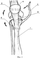
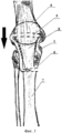
На фигуре 1 изображены: выделенное сухожилие 1 полусухожильной мышцы 2, брюшко полусухожильной мышцы 2, подшитое к брюшку нежной мышцы 3. На фигуре 2 изображены: выделенное сухожилие 1, надколенник 4, сформированный в нем тоннель 5, сформированный канал 6 в большеберцовой кости 7, собственная связка 8 надколенника 4. Стрелками указано последовательное проведение выделенного сухожилия 1 по внутреннему краю собственной связки 8 надколенника 4, через тоннель 5 надколенника 4, по наружному краю собственной связки 8 надколенника 4. На фигуре 3 изображено выделенное сухожилие 1, проведенное через тоннель 5 надколенника 4, по наружному краю собственной связки 8 надколенника 4, погруженное в канал 6 большеберцовой кости 7 и подшитое к собственной связке 8 надколенника 4. Стрелкой указанно направление низведения надколенника 4.The figure 1 shows: the selected
Принципиальное отличие данного способа от прототипа заключается в том, что производится пластика растянутой связки надколенника сухожилием полусухожильной мышцы.The fundamental difference between this method and the prototype is that plastic is made of the stretched ligament of the patella with the tendon of the semi-tendon muscle.
Преимущество предложенного способа перед известным заключается в том, что производится низведение надколенника и тонизация его собственной связки сухожилием полусухожильной мышцы, тем самым повышается эффективность оперативного лечения нарушения активного разгибания голени при высоком стояние надколенника у детей с детским церебральным параличом. Не нужно выполнять повторную операцию по удалению металлофиксатора, что сокращает время лечения. Не повреждается ростковая зона.The advantage of the proposed method over the known one consists in lowering the patella and toning its own ligament with a tendon of the semitendinosus muscle, thereby increasing the efficiency of surgical treatment of disorders of active extension of the leg with high standing of the patella in children with cerebral palsy. No need to perform a second operation to remove the metal fixator, which reduces the treatment time. The sprout zone is not damaged.
Предложенный способ рекомендован для восстановления активного разгибания голени при высоком стоянии надколенника у больных детским церебральным параличом.The proposed method is recommended to restore active extension of the lower leg with a high standing of the patella in patients with cerebral palsy.
Клинический примерClinical example
Больная Т., 11 лет поступила в клинику травматологии и ортопедии СамГМУ с диагнозом: ДЦП, ранняя резидуальная стадия, спастическая диплегия, нарушение активного разгибания голени при высоком стоянии надколенника. Наблюдается у невролога с рождения. Диагноз ДЦП поставлен в 1 год. Регулярно проходит курсы консервативного лечения. Объективно определяется нарушение активного разгибания голени, снижение объема активных движений в коленных суставах (ограничение разгибания: справа - 170°, слева - 165°).Patient T., aged 11, was admitted to the clinic of traumatology and orthopedics of Samara State Medical University with a diagnosis of cerebral palsy, early residual stage, spastic diplegia, impaired active extension of the lower leg with a high patella position. Observed by a neurologist from birth. The diagnosis of cerebral palsy was made in 1 year. Regularly takes courses of conservative treatment. Objectively determined is the violation of the active extension of the lower leg, a decrease in the volume of active movements in the knee joints (limitation of extension: to the right - 170 °, to the left - 165 °).
Выполнено оперативное лечение описанным способом. Гипсовая иммобилизация обеих нижних конечностей в среднефизиологическом положении от кончиков пальцев до ягодичной складки сроком на 6 недель.Performed surgical treatment in the described manner. Plaster immobilization of both lower extremities in the mid-physiological position from the fingertips to the gluteal fold for a period of 6 weeks.
После снятия гипсовых повязок и курса восстановительного лечения наблюдали увеличение объема активного разгибания голени в коленных суставах: справа - 180°, слева - 177°. Тонус собственной связки надколенника нормализован, ростковая зона не повреждена. В настоящее время достигнутый после оперативного лечения объем движения в коленных суставах сохранен.After removing the plaster casts and the course of rehabilitation treatment, an increase in the volume of active extension of the lower leg in the knee joints was observed: 180 ° on the right and 177 ° on the left. The tone of the patellar ligament is normalized, the germ zone is not damaged. Currently, the volume of movement in the knee joints achieved after surgical treatment has been preserved.
ИСТОЧНИКИ ИНФОРМАЦИИINFORMATION SOURCES
1. Бойчев Б., Конфорти Б., Чоканов К. Оперативна ортопедия и травматология. - София: Медицина и физкультура, 1958. - 832 с.1. Boychev B., Conforti B., Chokanov K. Operative orthopedics and traumatology. - Sofia: Medicine and Physical Education, 1958. - 832 p.
2. Кенис В.М. Восстановление активного разгибания голени у детей с ДЦП. - ФГУ «НИДОИ им Г.И.Турнера Росздрава»: Методические рекомендации. СПб., 2005 г.- 16 с.2. Kenis V.M. Restoring active extension of the lower leg in children with cerebral palsy. - FGU "NIDOI named after G.I. Turner of Roszdrav": Methodical recommendations. SPb., 2005 - 16 p.
3. Патент на изобретение №2192192 от 10.11.2002 по заявке №2000127240/14, опубл. 10.11.2002, бюл. №31. (РФ). Способ лечения высокого стояния надколенника у детей. Кенис В.М., Поздникин Ю.И.3. Patent for the invention No. 2192192 from 10.11.2002 by application No.2000127240 / 14, publ. 11/10/2002, bull. No. 31. (RF). A method for the treatment of high standing of the patella in children. Kenis V.M., Pozdnikin Yu.I.
Claims (1)
Priority Applications (1)
| Application Number | Priority Date | Filing Date | Title |
|---|---|---|---|
| RU2013111560/14A RU2517536C1 (en) | 2013-03-14 | 2013-03-14 | Method for surgical reconstruction of knee extension in high-position patella in children suffering from infantile cerebral paralysis |
Applications Claiming Priority (1)
| Application Number | Priority Date | Filing Date | Title |
|---|---|---|---|
| RU2013111560/14A RU2517536C1 (en) | 2013-03-14 | 2013-03-14 | Method for surgical reconstruction of knee extension in high-position patella in children suffering from infantile cerebral paralysis |
Publications (1)
| Publication Number | Publication Date |
|---|---|
| RU2517536C1 true RU2517536C1 (en) | 2014-05-27 |
Family
ID=50779572
Family Applications (1)
| Application Number | Title | Priority Date | Filing Date |
|---|---|---|---|
| RU2013111560/14A RU2517536C1 (en) | 2013-03-14 | 2013-03-14 | Method for surgical reconstruction of knee extension in high-position patella in children suffering from infantile cerebral paralysis |
Country Status (1)
| Country | Link |
|---|---|
| RU (1) | RU2517536C1 (en) |
Cited By (1)
| Publication number | Priority date | Publication date | Assignee | Title |
|---|---|---|---|---|
| RU2822017C1 (en) * | 2023-10-05 | 2024-06-28 | федеральное государственное бюджетное учреждение "Национальный медицинский исследовательский центр детской травматологии и ортопедии имени Г.И. Турнера" Минздрава России (ФГБУ "НМИЦ детской травматологии и ортопедии имени Г.И. Турнера" Минздрава России) | Method for surgical management of active knee extension deficiency in high patellar position in patients with infantile cerebral paralysis |
Citations (2)
| Publication number | Priority date | Publication date | Assignee | Title |
|---|---|---|---|---|
| RU2192192C2 (en) * | 2000-10-30 | 2002-11-10 | Научно-исследовательский детский ортопедический институт им. Г.И.Турнера | Method for treating high position of the patella in children |
| RU2391065C1 (en) * | 2009-03-10 | 2010-06-10 | Федеральное государственное учреждение "Уральский научно-исследовательский институт травматологии и ортопедии имени В.Д. Чаклина Федерального агентства по высокотехнологичной медицинской помощи" | Method of treatment of old lacerations of extensor apparatus of knee joint |
-
2013
- 2013-03-14 RU RU2013111560/14A patent/RU2517536C1/en not_active IP Right Cessation
Patent Citations (2)
| Publication number | Priority date | Publication date | Assignee | Title |
|---|---|---|---|---|
| RU2192192C2 (en) * | 2000-10-30 | 2002-11-10 | Научно-исследовательский детский ортопедический институт им. Г.И.Турнера | Method for treating high position of the patella in children |
| RU2391065C1 (en) * | 2009-03-10 | 2010-06-10 | Федеральное государственное учреждение "Уральский научно-исследовательский институт травматологии и ортопедии имени В.Д. Чаклина Федерального агентства по высокотехнологичной медицинской помощи" | Method of treatment of old lacerations of extensor apparatus of knee joint |
Non-Patent Citations (2)
| Title |
|---|
| БОЙЧЕВ Б. и др. Оперативная ортопедия и травматология. София, 1958, с.576-577. * |
| ОРТОПЕДИЯ. Национальное руководство. ГЭОТАР-Медиа, М., 2008, с. 401-402. Lakshmanan P. et al. Created Patella in Total Knee Arthroplasty. Surg. Techol.int. 2005, 14:275-278 (Abstract) * |
Cited By (1)
| Publication number | Priority date | Publication date | Assignee | Title |
|---|---|---|---|---|
| RU2822017C1 (en) * | 2023-10-05 | 2024-06-28 | федеральное государственное бюджетное учреждение "Национальный медицинский исследовательский центр детской травматологии и ортопедии имени Г.И. Турнера" Минздрава России (ФГБУ "НМИЦ детской травматологии и ортопедии имени Г.И. Турнера" Минздрава России) | Method for surgical management of active knee extension deficiency in high patellar position in patients with infantile cerebral paralysis |
Similar Documents
| Publication | Publication Date | Title |
|---|---|---|
| Carmont et al. | Less invasive Achilles tendon reconstruction | |
| RU2517536C1 (en) | Method for surgical reconstruction of knee extension in high-position patella in children suffering from infantile cerebral paralysis | |
| RU2445015C1 (en) | Method of restoring injured ligaments of ankle joint | |
| RU2537067C1 (en) | Method of treating tibial intercondyloid eminence fracture | |
| RU2537888C1 (en) | Method for surgical management of old achilles tendon rupture | |
| RU2391065C1 (en) | Method of treatment of old lacerations of extensor apparatus of knee joint | |
| RU2443394C1 (en) | Method of treating slipping patella | |
| RU2727744C1 (en) | Method for preparation of anterior cruciate ligament autograft | |
| RU2344782C1 (en) | Method of surgical treatment of longitudinal platypodia for adults | |
| RU2306891C1 (en) | Method for low-invasive surgical therapy of chronic median instability of knee joint at applying arthroscopic technologies | |
| RU2645616C1 (en) | Fixation method of distal tendon of arm biceps muscle to radiai bone tuberosity | |
| RU2295311C2 (en) | Method for treating foot deformations in children | |
| RU2302214C1 (en) | Surgical method for treating post-traumatic lateral patellar dislocation cases with artroscopic techniques being used | |
| RU2343862C2 (en) | Method of autoplasty of own ligament of whirlbone after endoprosthesis replacement of knee joint | |
| RU2612100C1 (en) | Method of surgery of multifragmental fractures of patella | |
| RU2216289C2 (en) | Method for surgical rehabilitation of patients with equinovalgus and equinovarus deformations of feet | |
| RU2054897C1 (en) | Method for carrying out plastic reconstruction of cicatricial changed fibular collateral ligament of knee joint | |
| RU2456952C1 (en) | Method of eliminating equine-hollow foot deformity | |
| RU2602935C1 (en) | Method for treating flat foot valgus in children | |
| RU2761744C1 (en) | Method for treatment of patients with patellofemoral arthrosis | |
| RU2423953C1 (en) | Method of preventing dislocation of hip joint endoprosthesis head | |
| Vohra et al. | Bridle Procedure Revisited | |
| RU2654593C1 (en) | Surgical access to the lateral curve of the large-curved bone fassioplastic with osteotomy of zherdi tubercule | |
| RU2734992C1 (en) | Method for surgical correction of equino-flat-valgus foot deformation in children with spastic icp forms | |
| RU2446762C1 (en) | Method of eliminating patella dislocation in children |
Legal Events
| Date | Code | Title | Description |
|---|---|---|---|
| MM4A | The patent is invalid due to non-payment of fees |
Effective date: 20160315 |