JP5610761B2 - X線画像処理装置、x線画像処理システム、x線画像処理方法、及びコンピュータプログラム - Google Patents
X線画像処理装置、x線画像処理システム、x線画像処理方法、及びコンピュータプログラム Download PDFInfo
- Publication number
- JP5610761B2 JP5610761B2 JP2009285763A JP2009285763A JP5610761B2 JP 5610761 B2 JP5610761 B2 JP 5610761B2 JP 2009285763 A JP2009285763 A JP 2009285763A JP 2009285763 A JP2009285763 A JP 2009285763A JP 5610761 B2 JP5610761 B2 JP 5610761B2
- Authority
- JP
- Japan
- Prior art keywords
- image
- ray image
- image processing
- subtraction
- edge
- Prior art date
- Legal status (The legal status is an assumption and is not a legal conclusion. Google has not performed a legal analysis and makes no representation as to the accuracy of the status listed.)
- Expired - Fee Related
Links
- 238000004590 computer program Methods 0.000 title claims description 8
- 238000003672 processing method Methods 0.000 title claims description 4
- 239000002872 contrast media Substances 0.000 claims description 44
- 238000000034 method Methods 0.000 claims description 42
- 230000008569 process Effects 0.000 claims description 25
- 238000003708 edge detection Methods 0.000 claims description 23
- 238000000605 extraction Methods 0.000 claims description 21
- 238000003384 imaging method Methods 0.000 claims description 16
- 210000004204 blood vessel Anatomy 0.000 claims description 14
- 238000001514 detection method Methods 0.000 claims description 8
- 238000002347 injection Methods 0.000 claims description 3
- 239000007924 injection Substances 0.000 claims description 3
- 238000006243 chemical reaction Methods 0.000 description 13
- 230000010365 information processing Effects 0.000 description 11
- 230000008859 change Effects 0.000 description 10
- 238000003745 diagnosis Methods 0.000 description 7
- 238000004458 analytical method Methods 0.000 description 4
- 238000004364 calculation method Methods 0.000 description 4
- 230000009467 reduction Effects 0.000 description 4
- 239000000284 extract Substances 0.000 description 3
- 230000014509 gene expression Effects 0.000 description 3
- 238000007781 pre-processing Methods 0.000 description 3
- 238000002583 angiography Methods 0.000 description 2
- 238000010586 diagram Methods 0.000 description 2
- 230000000694 effects Effects 0.000 description 2
- 238000011946 reduction process Methods 0.000 description 2
- 230000001629 suppression Effects 0.000 description 2
- 230000005540 biological transmission Effects 0.000 description 1
- 238000004891 communication Methods 0.000 description 1
- 239000000470 constituent Substances 0.000 description 1
- 238000007796 conventional method Methods 0.000 description 1
- 230000007423 decrease Effects 0.000 description 1
- 230000004069 differentiation Effects 0.000 description 1
- 238000005516 engineering process Methods 0.000 description 1
- 230000002708 enhancing effect Effects 0.000 description 1
- 238000002594 fluoroscopy Methods 0.000 description 1
- 230000006872 improvement Effects 0.000 description 1
- 239000004973 liquid crystal related substance Substances 0.000 description 1
- 230000002093 peripheral effect Effects 0.000 description 1
- 238000000926 separation method Methods 0.000 description 1
- 230000001360 synchronised effect Effects 0.000 description 1
- 210000003462 vein Anatomy 0.000 description 1
Images


Classifications
-
- A—HUMAN NECESSITIES
- A61—MEDICAL OR VETERINARY SCIENCE; HYGIENE
- A61B—DIAGNOSIS; SURGERY; IDENTIFICATION
- A61B6/00—Apparatus for radiation diagnosis, e.g. combined with radiation therapy equipment
-
- G06T5/92—
-
- G—PHYSICS
- G06—COMPUTING; CALCULATING OR COUNTING
- G06T—IMAGE DATA PROCESSING OR GENERATION, IN GENERAL
- G06T5/00—Image enhancement or restoration
- G06T5/50—Image enhancement or restoration by the use of more than one image, e.g. averaging, subtraction
-
- G—PHYSICS
- G06—COMPUTING; CALCULATING OR COUNTING
- G06T—IMAGE DATA PROCESSING OR GENERATION, IN GENERAL
- G06T7/00—Image analysis
-
- G—PHYSICS
- G06—COMPUTING; CALCULATING OR COUNTING
- G06T—IMAGE DATA PROCESSING OR GENERATION, IN GENERAL
- G06T7/00—Image analysis
- G06T7/10—Segmentation; Edge detection
- G06T7/13—Edge detection
-
- A—HUMAN NECESSITIES
- A61—MEDICAL OR VETERINARY SCIENCE; HYGIENE
- A61B—DIAGNOSIS; SURGERY; IDENTIFICATION
- A61B6/00—Apparatus for radiation diagnosis, e.g. combined with radiation therapy equipment
- A61B6/48—Diagnostic techniques
- A61B6/481—Diagnostic techniques involving the use of contrast agents
-
- A—HUMAN NECESSITIES
- A61—MEDICAL OR VETERINARY SCIENCE; HYGIENE
- A61B—DIAGNOSIS; SURGERY; IDENTIFICATION
- A61B6/00—Apparatus for radiation diagnosis, e.g. combined with radiation therapy equipment
- A61B6/48—Diagnostic techniques
- A61B6/486—Diagnostic techniques involving generating temporal series of image data
-
- A—HUMAN NECESSITIES
- A61—MEDICAL OR VETERINARY SCIENCE; HYGIENE
- A61B—DIAGNOSIS; SURGERY; IDENTIFICATION
- A61B6/00—Apparatus for radiation diagnosis, e.g. combined with radiation therapy equipment
- A61B6/50—Clinical applications
- A61B6/504—Clinical applications involving diagnosis of blood vessels, e.g. by angiography
-
- G—PHYSICS
- G06—COMPUTING; CALCULATING OR COUNTING
- G06T—IMAGE DATA PROCESSING OR GENERATION, IN GENERAL
- G06T2207/00—Indexing scheme for image analysis or image enhancement
- G06T2207/10—Image acquisition modality
- G06T2207/10016—Video; Image sequence
-
- G—PHYSICS
- G06—COMPUTING; CALCULATING OR COUNTING
- G06T—IMAGE DATA PROCESSING OR GENERATION, IN GENERAL
- G06T2207/00—Indexing scheme for image analysis or image enhancement
- G06T2207/10—Image acquisition modality
- G06T2207/10116—X-ray image
- G06T2207/10121—Fluoroscopy
-
- G—PHYSICS
- G06—COMPUTING; CALCULATING OR COUNTING
- G06T—IMAGE DATA PROCESSING OR GENERATION, IN GENERAL
- G06T2207/00—Indexing scheme for image analysis or image enhancement
- G06T2207/20—Special algorithmic details
- G06T2207/20172—Image enhancement details
- G06T2207/20192—Edge enhancement; Edge preservation
-
- G—PHYSICS
- G06—COMPUTING; CALCULATING OR COUNTING
- G06T—IMAGE DATA PROCESSING OR GENERATION, IN GENERAL
- G06T2207/00—Indexing scheme for image analysis or image enhancement
- G06T2207/20—Special algorithmic details
- G06T2207/20212—Image combination
- G06T2207/20224—Image subtraction
-
- G—PHYSICS
- G06—COMPUTING; CALCULATING OR COUNTING
- G06T—IMAGE DATA PROCESSING OR GENERATION, IN GENERAL
- G06T2207/00—Indexing scheme for image analysis or image enhancement
- G06T2207/30—Subject of image; Context of image processing
- G06T2207/30004—Biomedical image processing
- G06T2207/30101—Blood vessel; Artery; Vein; Vascular
- G06T2207/30104—Vascular flow; Blood flow; Perfusion
Description
CPU1010は、RAM1020やROM1030に格納されているコンピュータプログラムやデータを用いて情報処理装置1000全体的な制御を行うとともに、コンピュータプログラムの実行により予め定められたX線画像処理に関する演算処理を実行することが可能である。
次に、撮像装置2000について説明する。撮像装置2000は、例えば、X線透視装置のような、造影剤流入中の動画像を撮像することが可能であり、撮像された画像データは情報処理装置1000に送信される。なお、画像データは複数をまとめて情報処理装置1000に送信するようにしても良いし、撮像の都度、順次、画像データの送信を行うようにしてもよい。
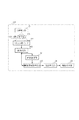
108 階調処理部
109 画像表示部
201 第1の特徴量抽出部
202 第2の特徴量抽出部
203 階調曲線作成部
204 階調変換処理部
401 画像縮小処理部
402 エッジ検出部
403 代表画素値算出部
Claims (20)
- 造影剤が流入している血管領域を撮影して得られる第一のX線画像と、前記血管領域を造影剤が流入していないタイミングで撮影して得られる第二のX線画像との間で減算処理を行うことでサブトラクション画像を取得する減算手段と、
前記サブトラクション画像の各フレームからエッジを検出する検出手段と、
前記サブトラクション画像の各フレームから検出されたエッジの位置に対応する画素値に基づいて第1の特徴量を取得する第1の特徴量取得手段と、
前記サブトラクション画像の各フレームから背景領域に対応する画素値に基づいて第2の特徴量を取得する第2の特徴量取得手段と、
前記第1の特徴量及び前記第2の特徴量で特定される階調曲線を用いて前記サブトラクション画像の各フレームに対して階調を変換する処理を行う処理手段と、を備えることを特徴とするX線画像処理装置。 - 前記第1の特徴量取得手段は、前記検出されたエッジに基づいて造影剤流入領域と背景領域との境界を表す領域から第1の特徴量を取得することを特徴とする請求項1に記載のX線画像処理装置。
- 前記第1の特徴量取得手段は、前記検出手段によるエッジの情報から前記サブトラクション画像において前記第一の特徴量の抽出対象領域を取得していることを特徴とする請求項1又は2に記載のX線画像処理装置。
- 前記検出手段は、前記エッジの情報として、サブトラクション画像における画素間の画素値の差から一次微分値を算出し、
前記特徴量取得手段は、該一次微分値に基づいて第1の特徴量を抽出する領域を取得していることを特徴とする請求項1乃至3のいずれか一項に記載のX線画像処理装置。 - 前記第1の特徴量取得手段は、前記検出されたエッジに基づき定められる造影剤流入領域の画素値の絶対値の値に基づき前記一次微分値を算出する画素間の間隔を変更することを特徴とする請求項4に記載のX線画像処理装置。
- 前記検出手段は、キャニーエッジ検出手法を用いることを特徴とする請求項1乃至5のいずれか1項に記載のX線画像処理装置。
- 前記検出手段は、複数のエッジ検出オペレータを持ち、造影剤の流入状況に応じて1つを選択することを特徴とする請求項1乃至6のいずれか1項に記載のX線画像処理装置。
- 前記第1の特徴量取得手段は、前記被写体のX線画像の撮影時におけるフレームレートに応じて前記第一の特徴量の取得の方法を変更することを特徴とする請求項1乃至5のいずれか1項に記載のX線画像処理装置。
- 前記第1の特徴量取得手段は、前記サブトラクション画像において前記検出されたエッジに対応する領域の平均画素値を前記第1の画素値として算出することを特徴とする請求項1乃至8のいずれか1項に記載のX線画像処理装置。
- 前記第2の特徴量取得手段は、前記サブトラクション画像のヒストグラムを作成し、該ヒストグラムに基づいて前記第2の特徴量を算出することを特徴とする請求項1乃至9のいずれか1項に記載のX線画像処理装置。
- 前記第2の特徴量取得手段は、前記第一及び第二のX線画像における撮影条件が同じであれば前記第2の特徴量としてゼロを出力するものであることを特徴とする請求項1乃至10のいずれか1項に記載のX線画像処理装置。
- 前記検出手段は、造影剤流入領域のコントラストに応じてエッジの検出方法を変更することを特徴とする請求項1乃至11のいずれか1項に記載のX線画像処理装置。
- 前記検出手段は、造影剤の注入時からの経過時間に応じてエッジの検出方法を変更することを特徴とする請求項1乃至12のいずれか1項に記載のX線画像処理装置。
- 前記検出手段は、造影剤の流入状況に応じてエッジの検出方法を変更することを特徴とする請求項1乃至13のいずれか1項に記載のX線画像処理装置。
- 血管領域の大きさに応じてエッジの検出方法を変更することを特徴とする請求項1乃至14のいずれか1項に記載のX線画像処理装置。
- 造影剤が流入している血管領域を撮影して得られる第一のX線画像と、前記血管領域を造影剤が流入していないタイミングで撮影して得られる第二のX線画像との間で減算処理を行うことでサブトラクション画像を取得する減算手段と、
前記サブトラクション画像の各フレームからエッジを検出する検出手段と、
前記サブトラクション画像の各フレームから検出されたエッジの位置に対応する画素値に基づいて第1の特徴量を取得する第1の特徴量取得手段と、
前記サブトラクション画像の各フレームから背景領域に対応する画素値に基づいて第2の特徴量を取得する第2の特徴量取得手段と、
前記第1の特徴量及び前記第2の特徴量で特定される階調曲線を用いて前記サブトラクション画像の各フレームに対して階調を変換する処理を行う処理手段と、
を備えることを特徴とするX線画像処理システム。 - X線を検出することにより、前記第一および第二のX線画像を含むX線動画像を得るX線センサをさらに有することを特徴とする請求項16に記載のX線画像処理システム。
- X線発生装置をさらに有することを特徴とする請求項16に記載のX線画像処理システム。
- 造影剤が流入している血管領域を撮影して得られる第一のX線画像と、前記血管領域を造影剤が流入していないタイミングで撮影して得られる第二のX線画像との間で減算処理を行うことでサブトラクション画像を取得するステップと、
前記サブトラクション画像の各フレームからエッジを検出するステップと、
前記サブトラクション画像の各フレームから検出されたエッジの位置に対応する画素値に基づいて第1の特徴量を取得するステップと、
前記サブトラクション画像の各フレームから背景領域に対応する画素値に基づいて第2の特徴量を取得すステップと、
前記第1の特徴量及び前記第2の特徴量で特定される階調曲線を用いて前記サブトラクション画像の各フレームに対して階調を変換する処理を行うステップと、を有することを特徴とするX線画像処理方法。 - 請求項19に記載の画像処理方法をコンピュータに実行させるためのコンピュータプログラム。
Priority Applications (6)
| Application Number | Priority Date | Filing Date | Title |
|---|---|---|---|
| JP2009285763A JP5610761B2 (ja) | 2009-12-16 | 2009-12-16 | X線画像処理装置、x線画像処理システム、x線画像処理方法、及びコンピュータプログラム |
| EP10837681.5A EP2512344A4 (en) | 2009-12-16 | 2010-12-10 | X-RAY PROCESSING DEVICE, X-RAY PROCESSING METHOD AND STORAGE MEDIUM FOR A COMPUTER PROGRAM |
| KR1020127018514A KR20120102777A (ko) | 2009-12-16 | 2010-12-10 | X선 화상처리장치, x선 화상처리방법, 및 컴퓨터 프로그램의 기억매체 |
| CN201080057728.8A CN102665557A (zh) | 2009-12-16 | 2010-12-10 | X射线图像处理设备、x射线图像处理方法和计算机程序的存储介质 |
| US13/515,677 US8805041B2 (en) | 2009-12-16 | 2010-12-10 | X-ray image processing apparatus, X-ray image processing method, and storage medium for computer program |
| PCT/JP2010/072732 WO2011074657A1 (en) | 2009-12-16 | 2010-12-10 | X-ray image processing apparatus, x-ray image processing method, and storage medium for computer program |
Applications Claiming Priority (1)
| Application Number | Priority Date | Filing Date | Title |
|---|---|---|---|
| JP2009285763A JP5610761B2 (ja) | 2009-12-16 | 2009-12-16 | X線画像処理装置、x線画像処理システム、x線画像処理方法、及びコンピュータプログラム |
Publications (3)
| Publication Number | Publication Date |
|---|---|
| JP2011125462A JP2011125462A (ja) | 2011-06-30 |
| JP2011125462A5 JP2011125462A5 (ja) | 2013-02-07 |
| JP5610761B2 true JP5610761B2 (ja) | 2014-10-22 |
Family
ID=44167407
Family Applications (1)
| Application Number | Title | Priority Date | Filing Date |
|---|---|---|---|
| JP2009285763A Expired - Fee Related JP5610761B2 (ja) | 2009-12-16 | 2009-12-16 | X線画像処理装置、x線画像処理システム、x線画像処理方法、及びコンピュータプログラム |
Country Status (6)
| Country | Link |
|---|---|
| US (1) | US8805041B2 (ja) |
| EP (1) | EP2512344A4 (ja) |
| JP (1) | JP5610761B2 (ja) |
| KR (1) | KR20120102777A (ja) |
| CN (1) | CN102665557A (ja) |
| WO (1) | WO2011074657A1 (ja) |
Families Citing this family (13)
| Publication number | Priority date | Publication date | Assignee | Title |
|---|---|---|---|---|
| JP6021420B2 (ja) * | 2012-05-07 | 2016-11-09 | キヤノン株式会社 | 画像処理装置、画像処理方法およびプログラム |
| JP5981220B2 (ja) * | 2012-05-21 | 2016-08-31 | 東芝メディカルシステムズ株式会社 | 医用画像処理装置及びx線撮影装置 |
| JP6422198B2 (ja) * | 2012-11-29 | 2018-11-14 | オリンパス株式会社 | 画像処理装置、画像処理方法、及び画像処理プログラム |
| JP6173751B2 (ja) * | 2013-04-09 | 2017-08-02 | 東芝メディカルシステムズ株式会社 | 医用画像処理装置、x線診断装置及び医用画像処理プログラム |
| CN112950747A (zh) * | 2013-09-13 | 2021-06-11 | 斯特拉克斯私人有限公司 | 给图像分配颜色的方法和系统、计算机可读存储介质 |
| JP6254813B2 (ja) * | 2013-10-10 | 2017-12-27 | キヤノン株式会社 | X線画像処理装置および方法、並びにx線撮像装置、プログラム |
| JP6383145B2 (ja) * | 2013-11-18 | 2018-08-29 | キヤノン株式会社 | 画像処理装置、画像処理装置の制御方法およびプログラム |
| JP6654397B2 (ja) * | 2015-10-09 | 2020-02-26 | 株式会社イシダ | X線検査装置 |
| EP3413264B1 (en) * | 2017-06-06 | 2020-01-29 | Siemens Healthcare GmbH | Providing a normalized image |
| US10751128B2 (en) | 2017-11-22 | 2020-08-25 | Canon U.S.A., Inc. | Devices, systems, and methods for ablation-zone simulation and visualization |
| US10918441B2 (en) | 2017-11-22 | 2021-02-16 | Canon U.S.A., Inc. | Devices, systems, and methods for ablation-zone simulation and visualization |
| WO2021112988A1 (en) * | 2019-12-02 | 2021-06-10 | SG Devices LLC | Augmented reality display of surgical imaging |
| JP7307033B2 (ja) * | 2020-06-05 | 2023-07-11 | 富士フイルム株式会社 | 処理装置、処理装置の作動方法、処理装置の作動プログラム |
Family Cites Families (19)
| Publication number | Priority date | Publication date | Assignee | Title |
|---|---|---|---|---|
| JPH0628411B2 (ja) * | 1984-08-30 | 1994-04-13 | 株式会社島津製作所 | デジタルサブトラクシヨンシステム |
| JPS6417631A (en) * | 1987-07-14 | 1989-01-20 | Toshiba Corp | X-ray diagnostic apparatus |
| JPH01109483A (ja) * | 1987-10-22 | 1989-04-26 | Toshiba Corp | 画像処理装置 |
| JPH02114776A (ja) * | 1988-10-25 | 1990-04-26 | Toshiba Corp | X線診断装置 |
| JPH04364677A (ja) * | 1991-06-12 | 1992-12-17 | Toshiba Corp | 放射線診断のための画像処理装置 |
| JPH05167927A (ja) * | 1991-12-12 | 1993-07-02 | Toshiba Corp | 画像処理装置 |
| DE69312257T2 (de) * | 1993-02-11 | 1998-01-29 | Agfa Gevaert Nv | Strahlungsfelderkennungsverfahren |
| JP4822607B2 (ja) * | 2001-05-11 | 2011-11-24 | キヤノン株式会社 | 画像処理装置、画像処理システム、記憶媒体、プログラム及び画像処理方法 |
| US7403646B2 (en) * | 2002-10-24 | 2008-07-22 | Canon Kabushiki Kaisha | Image processing apparatus, image processing method, program, and recording medium for generating a difference image from a first radiographic image and second radiographic image |
| JP2004173076A (ja) * | 2002-11-21 | 2004-06-17 | Canon Inc | 階調変換処理方法およびこれを用いた画像処理システム |
| DE10311628B4 (de) * | 2003-03-14 | 2006-04-13 | Siemens Ag | Bildgebungsverfahren |
| CN100462050C (zh) * | 2004-06-11 | 2009-02-18 | 株式会社东芝 | X射线ct装置和心肌灌注图像产生系统 |
| DE102005062445A1 (de) | 2005-12-27 | 2007-07-05 | Siemens Ag | Verfahren zum Erzeugen eines Röntgenbildes |
| DE102006025917A1 (de) * | 2006-06-02 | 2007-12-06 | Siemens Ag | Verfahren zur Konturermittlung in einem medizinischen digitalen Durchleuchtungsbild |
| US20080051648A1 (en) * | 2006-08-25 | 2008-02-28 | Suri Jasjit S | Medical image enhancement system |
| JP2008061763A (ja) * | 2006-09-06 | 2008-03-21 | Toshiba Corp | X線画像診断装置 |
| US8111895B2 (en) * | 2006-12-06 | 2012-02-07 | Siemens Medical Solutions Usa, Inc. | Locally adaptive image enhancement for digital subtraction X-ray imaging |
| US8355595B2 (en) | 2007-05-15 | 2013-01-15 | Xerox Corporation | Contrast enhancement methods and apparatuses |
| US8094903B2 (en) * | 2007-06-28 | 2012-01-10 | Siemens Aktiengesellschaft | System and method for coronary digital subtraction angiography |
-
2009
- 2009-12-16 JP JP2009285763A patent/JP5610761B2/ja not_active Expired - Fee Related
-
2010
- 2010-12-10 CN CN201080057728.8A patent/CN102665557A/zh active Pending
- 2010-12-10 EP EP10837681.5A patent/EP2512344A4/en not_active Withdrawn
- 2010-12-10 WO PCT/JP2010/072732 patent/WO2011074657A1/en active Application Filing
- 2010-12-10 KR KR1020127018514A patent/KR20120102777A/ko not_active Application Discontinuation
- 2010-12-10 US US13/515,677 patent/US8805041B2/en not_active Expired - Fee Related
Also Published As
| Publication number | Publication date |
|---|---|
| US20120257809A1 (en) | 2012-10-11 |
| EP2512344A1 (en) | 2012-10-24 |
| JP2011125462A (ja) | 2011-06-30 |
| KR20120102777A (ko) | 2012-09-18 |
| US8805041B2 (en) | 2014-08-12 |
| WO2011074657A1 (en) | 2011-06-23 |
| CN102665557A (zh) | 2012-09-12 |
| EP2512344A4 (en) | 2014-01-08 |
Similar Documents
| Publication | Publication Date | Title |
|---|---|---|
| JP5610761B2 (ja) | X線画像処理装置、x線画像処理システム、x線画像処理方法、及びコンピュータプログラム | |
| JP5645399B2 (ja) | X線画像処理装置、x線画像処理方法、及びコンピュータプログラム | |
| JP5828649B2 (ja) | 画像処理装置、画像処理方法、及びコンピュータプログラム | |
| US9922409B2 (en) | Edge emphasis in processing images based on radiation images | |
| JP6021420B2 (ja) | 画像処理装置、画像処理方法およびプログラム | |
| EP2267655A2 (en) | Image processing method and image processing apparatus | |
| JP2012208553A (ja) | 画像処理装置、および画像処理方法、並びにプログラム | |
| JP5839710B2 (ja) | 解析点設定装置および方法、並びに体動検出装置および方法 | |
| Kroon et al. | Coherence filtering to enhance the mandibular canal in cone-beam CT data | |
| US10217230B2 (en) | X-ray image processing apparatus, X-ray image processing method, and storage medium | |
| JP4571378B2 (ja) | 画像処理方法および装置並びにプログラム | |
| JP6383145B2 (ja) | 画像処理装置、画像処理装置の制御方法およびプログラム | |
| JP6120913B2 (ja) | 画像処理装置、画像処理方法、及びコンピュータプログラム | |
| JP2019016865A (ja) | 画像処理装置、画像処理方法 | |
| CN113989171A (zh) | 减影图生成方法及装置、存储介质、计算机设备 |
Legal Events
| Date | Code | Title | Description |
|---|---|---|---|
| A521 | Request for written amendment filed |
Free format text: JAPANESE INTERMEDIATE CODE: A523 Effective date: 20121213 |
|
| A621 | Written request for application examination |
Free format text: JAPANESE INTERMEDIATE CODE: A621 Effective date: 20121213 |
|
| A131 | Notification of reasons for refusal |
Free format text: JAPANESE INTERMEDIATE CODE: A131 Effective date: 20131203 |
|
| A521 | Request for written amendment filed |
Free format text: JAPANESE INTERMEDIATE CODE: A523 Effective date: 20140203 |
|
| A131 | Notification of reasons for refusal |
Free format text: JAPANESE INTERMEDIATE CODE: A131 Effective date: 20140430 |
|
| A521 | Request for written amendment filed |
Free format text: JAPANESE INTERMEDIATE CODE: A523 Effective date: 20140630 |
|
| TRDD | Decision of grant or rejection written | ||
| A01 | Written decision to grant a patent or to grant a registration (utility model) |
Free format text: JAPANESE INTERMEDIATE CODE: A01 Effective date: 20140805 |
|
| A61 | First payment of annual fees (during grant procedure) |
Free format text: JAPANESE INTERMEDIATE CODE: A61 Effective date: 20140902 |
|
| R151 | Written notification of patent or utility model registration |
Ref document number: 5610761 Country of ref document: JP Free format text: JAPANESE INTERMEDIATE CODE: R151 |
|
| LAPS | Cancellation because of no payment of annual fees |