EP4501960A1 - Monoklonaler antikörper gegen hla-g-moleküle und verwendung davon - Google Patents
Monoklonaler antikörper gegen hla-g-moleküle und verwendung davon Download PDFInfo
- Publication number
- EP4501960A1 EP4501960A1 EP22934556.6A EP22934556A EP4501960A1 EP 4501960 A1 EP4501960 A1 EP 4501960A1 EP 22934556 A EP22934556 A EP 22934556A EP 4501960 A1 EP4501960 A1 EP 4501960A1
- Authority
- EP
- European Patent Office
- Prior art keywords
- sequence
- amino acid
- acid sequence
- hla
- seq
- Prior art date
- Legal status (The legal status is an assumption and is not a legal conclusion. Google has not performed a legal analysis and makes no representation as to the accuracy of the status listed.)
- Pending
Links
Images



Classifications
-
- A—HUMAN NECESSITIES
- A61—MEDICAL OR VETERINARY SCIENCE; HYGIENE
- A61P—SPECIFIC THERAPEUTIC ACTIVITY OF CHEMICAL COMPOUNDS OR MEDICINAL PREPARATIONS
- A61P35/00—Antineoplastic agents
-
- C—CHEMISTRY; METALLURGY
- C07—ORGANIC CHEMISTRY
- C07K—PEPTIDES
- C07K16/00—Immunoglobulins [IG], e.g. monoclonal or polyclonal antibodies
- C07K16/18—Immunoglobulins [IG], e.g. monoclonal or polyclonal antibodies against material from animals or humans
- C07K16/28—Immunoglobulins [IG], e.g. monoclonal or polyclonal antibodies against material from animals or humans against receptors, cell surface antigens or cell surface determinants
- C07K16/2803—Immunoglobulins [IG], e.g. monoclonal or polyclonal antibodies against material from animals or humans against receptors, cell surface antigens or cell surface determinants against the immunoglobulin superfamily
- C07K16/2833—Immunoglobulins [IG], e.g. monoclonal or polyclonal antibodies against material from animals or humans against receptors, cell surface antigens or cell surface determinants against the immunoglobulin superfamily against MHC-molecules, e.g. HLA-molecules
-
- G—PHYSICS
- G01—MEASURING; TESTING
- G01N—INVESTIGATING OR ANALYSING MATERIALS BY DETERMINING THEIR CHEMICAL OR PHYSICAL PROPERTIES
- G01N33/00—Investigating or analysing materials by specific methods not covered by groups G01N1/00 - G01N31/00
- G01N33/48—Biological material, e.g. blood, urine; Haemocytometers
- G01N33/50—Chemical analysis of biological material, e.g. blood, urine; Testing involving biospecific ligand binding methods; Immunological testing
- G01N33/53—Immunoassay; Biospecific binding assay; Materials therefor
- G01N33/577—Immunoassay; Biospecific binding assay; Materials therefor involving monoclonal antibodies binding reaction mechanisms characterised by the use of monoclonal antibodies
-
- C—CHEMISTRY; METALLURGY
- C07—ORGANIC CHEMISTRY
- C07K—PEPTIDES
- C07K2317/00—Immunoglobulins specific features
- C07K2317/30—Immunoglobulins specific features characterized by aspects of specificity or valency
- C07K2317/34—Identification of a linear epitope shorter than 20 amino acid residues or of a conformational epitope defined by amino acid residues
-
- C—CHEMISTRY; METALLURGY
- C07—ORGANIC CHEMISTRY
- C07K—PEPTIDES
- C07K2317/00—Immunoglobulins specific features
- C07K2317/50—Immunoglobulins specific features characterized by immunoglobulin fragments
- C07K2317/56—Immunoglobulins specific features characterized by immunoglobulin fragments variable (Fv) region, i.e. VH and/or VL
- C07K2317/565—Complementarity determining region [CDR]
-
- C—CHEMISTRY; METALLURGY
- C07—ORGANIC CHEMISTRY
- C07K—PEPTIDES
- C07K2317/00—Immunoglobulins specific features
- C07K2317/50—Immunoglobulins specific features characterized by immunoglobulin fragments
- C07K2317/56—Immunoglobulins specific features characterized by immunoglobulin fragments variable (Fv) region, i.e. VH and/or VL
- C07K2317/567—Framework region [FR]
-
- G—PHYSICS
- G01—MEASURING; TESTING
- G01N—INVESTIGATING OR ANALYSING MATERIALS BY DETERMINING THEIR CHEMICAL OR PHYSICAL PROPERTIES
- G01N2474/00—Immunochemical assays or immunoassays characterised by detection mode or means of detection
- G01N2474/10—Immunoblots, e.g. Western blot or Dot blot
-
- G—PHYSICS
- G01—MEASURING; TESTING
- G01N—INVESTIGATING OR ANALYSING MATERIALS BY DETERMINING THEIR CHEMICAL OR PHYSICAL PROPERTIES
- G01N2474/00—Immunochemical assays or immunoassays characterised by detection mode or means of detection
- G01N2474/20—Immunohistochemistry assay
Definitions
- the present invention belongs to the field of biomedicine and relates to the following aspects of an antibody YWHG-1 against HLA-G molecule: a monoclonal antibody YWHG-1 against HLA-G molecule prepared by an immunogen with amino acids which are common to seven known HLA-G isoform molecules (HLA-G1, HLA-G2, HLA-G3, HLA-G4, HLA-G5, HLA-G6 and HLA-G7) and are located at positions 72-91 (QTDRMNLQTLRGYYNQSEAS) in the heavy chain ⁇ 1 domain of the HLA-G molecules,; nucleotides encoding the antibody YWHG-1 and an encoded amino acid sequences thereof, and use of the antibody YWHG-1 for immunohistochemistry, immunoblotting, flow cytometry and other assays.
- HLA-G1, HLA-G2, HLA-G3, HLA-G4, HLA-G5, HLA-G6 and HLA-G7 are located at positions 72-91 (QTD
- HLA-G human leukocyte antigen-G
- exon 1 of HLA-G mRNA encodes the signal peptide
- exons 2, 3, and 4 encode the ⁇ 1, ⁇ 2, and ⁇ 3 domains of the extracellular region, respectively
- exon 5 encodes the transmembrane region
- exon 6 encodes the intracellular segment of HLA-G molecule containing only 6 amino acid residues. Since exon 6 contains a stop codon, exon 7 is not transcribed; exon 8 corresponds to the 3'UTR of the HLA-G gene.
- the initial transcript of HLA-G can be alternatively spliced to produce 7 types of mature mRNA, encoding 7 isoforms with different molecular weights (HLA-G1, -G2, -G3, -G4, -G5, -G6 and -G7).
- HLA-G1, HLA-G2, HLA-G3 and HLA-G4 contain transmembrane regions and are membrane-bound isoforms
- HLA-G5, HLA-G6 and HLA-G7 lack transmembrane structures and are soluble isoforms.
- the molecular weights of HLA-G1 ⁇ -G7 isoforms are 39kD, 31kD, 22kD, 30kD, 34kD, 23kD and 16kD respectively.
- HLA-G1 is encoded by full-length HLA-G mRNA and consists of extracellular ⁇ 1, ⁇ 2 and ⁇ 3 domains, a transmembrane region and an intracellular domain.
- HLA-G2 lacks ⁇ 2 domain and consists of extracellular ⁇ 1 and ⁇ 3 domains, a transmembrane region and an intracellular domain;
- HLA-G3 lacks ⁇ 2 and ⁇ 3 domains and consists of an extracellular ⁇ 1 domain, a transmembrane region and an intracellular domain;
- HLA-G4 lacks ⁇ 3 domain and consists of extracellular ⁇ 1 and ⁇ 2 domains, a transmembrane region and an intracellular domain extracellular domain is the same as HLA-G1 and HLA-G2 respectively.
- HLA-G mRNA containing intron 4 contains a stop codon, the encoded protein molecules lack the transmembrane region encoded by exon 5, forming soluble HLA-G molecules. Since the mRNA encoding HLA-G7 contains a stop codon in intron 2, the extracellular region only contains the ⁇ 1 domain and is connected by the two amino acid residues encoded by intron 2 ( FIG. 1 ).
- HLA-G molecule Under normal physiological conditions, HLA-G molecule is only expressed in extravillous cytotrophoblast cells at the maternal-fetal interface, maintaining maternal-fetal immune tolerance during pregnancy. Under pathological conditions, HLA-G molecule expression can be induced in pathological tissues and cells such as virus-infected cells and tumor cells which are closely related to the occurrence and progression of the diseases. HLA-G molecule is an important immune checkpoint molecule.
- the immunosuppressive function of HLA-G is mainly by binding to the immunosuppressive receptors immunoglobulin-like transcript-2 (ILT2/LILRB1/CD85j) and immunoglobulin-like transcript-4 (II,T4/LII,RB2/CD85d).
- HLA-G/ILT signaling The underlying mechanisms of immunoinhibitory function induced by HLA-G/ILT signaling are as follows: 1 bind to ILT-2 expressed on T cells, NK cells and B cells, inhibit the cytotoxicity of T cells and NK cells, and inhibit the proliferation and antibody secretion of B cells, etc.; 2 bind to ILT-2 and ILT-4 expressed on dendritic cells (DC), inhibit the maturation and differentiation of DC cells, and induce the generation of tolerant DC cells; (3) bind to ILT-2 and ILT-4 expressed on myeloid-derived suppressor cells (MDSC) and macrophages (M ⁇ ) to induce the differentiation of pro-inflammatory and anti-tumor M1 macrophages into M2 macrophages. Therefore, HLA-G plays an important role in the development and progression of tumors and other diseases. A number of HLA-G-targeted immunotherapy for solid cancers have been entered phase I clinical trials.
- HLA-G binds to receptors ILT-2 and ILT-4 and has molecular structure specificity.
- the ⁇ 3 domain of HLA-G extracellular region is the site where receptors ILT-2 and ILT-4 bind to HLA-G.
- ILT-2 only binds to the HLA-G/ ⁇ 2 m complex, while ILT-4 can not only bind to HLA-G/ ⁇ 2 m, but also bind to free HLA-G molecules that do not contain ( ⁇ 2 m.
- ILT-2 can bind to HLA-G1 and HLA-G5, while ILT-4 can bind to HLA-G1, HLA-G2, HLA-G5 and HLA-G6 isoforms.
- ILT-4 can bind to HLA-G1, HLA-G2, HLA-G5 and HLA-G6 isoforms.
- the extracellular regions of HLA-G3, -G4, and HLA-G7 isoforms contain no ⁇ 3 domain and cannot bind to ILT-2 and ILT-4.
- Different HLA-G isoforms can exert specific immunological effects in pathophysiological processes.
- HLA-G isoforms Under different pathological conditions, especially in tumor tissue cells, there is extensive heterogeneity in the expression of HLA-G isoforms, and the expressions of different HLA-G isoforms have specific clinical significance. Therefore, analyzing the expression of specific HLA-G isoforms and the expression profiles of different HLA-G isoforms is of great significance for elucidating the biological functions and clinical significance of specific HLA-G isoform.
- antibodies used for immunohistochemistry and immunoblotting of HLA-G molecule include 4H84, MEM-G1 and MEM-G2.
- the antibody 4H84 has a recognition site located in the extracellular region ⁇ 1 domain for all ⁇ 1 domain containing HLA-G isoforms. It can detect the seven HLA-G isoforms (HLA-G1, HLA-G2, HLA-G3, HLA-G4, HLA-G5, HLA-G6 and HLA-G7) that are currently known to contain ⁇ 1 domains.
- Antibodies MEM-G1 and MEM-G2 are obtained from mice immunized with the full-length HLA-G heavy chain. The specific recognition sites cannot be predicted.
- the two antibodies are similar to the antibody 4H84 and can recognize the aforementioned HLA-G isoforms.
- the aforementioned antibodies 4H84, MEM-G1 and MEM-G2 have limitations in their detection specificity, and there are cross-reactions used for immunohistochemistry and immunoblotting, resulting in false positives. Meanwhile, the above three antibodies cannot be used for flow cytometry.
- an object of the present invention is to provide a specific monoclonal antibody YWHG-1 against HLA-G molecules, a nucleotide sequence encoding the antibody YWHG-1 and the encoded amino acid sequence thereof; and use of the YWHG-1 antibody for immunohistochemistry, immunoblotting, flow cytometry and other assays.
- amino acid residues which are common to seven known HLA-G isoforms (HLA-G1, HLA-G2, HLA-G3, HLA-G4, HLA-G5, HLA-G6 and HLA-G7) and are located at positions 72-91 in the heavy chain ⁇ 1 domain of the HLA-G molecule is used as an immunogen.
- the immunogen amino acid sequence is QTDRMNLQTLRGYYNQSEAS as shown in SEQ ID No. 19.
- the peptide (QTDRMNLQTLRGYYNQSEAS) is located in the heavy chain ⁇ 1 domain of the HLA-G molecule (the area shown in the dashed box in FIG. 1 ), and based on the peptide fragment, the specific monoclonal antibody YWHG-1 against HLA-G molecules is prepared.
- the present invention provides a monoclonal antibody YWHG-1 against HLA-G molecule, which at least includes one or more of light chain hypervariable regions CDR1, CDR2 and CDR3, or/and one or more of heavy chain hypervariable regions CDR1, CDR2 and CDR3;
- An antibody light chain amino acid sequence of the monoclonal antibody YWHG-1 is as shown in SEQ ID No. 1 or an amino acid sequence with equivalent functions to the sequence shown in SEQID No. 1 formed by substituting, deleting or adding one or more amino acids;
- an amino acid sequence of the antibody light chain hypervariable region CDR1 is the sequence QSFVHSNGNIY shown in SEQ ID No.2 or an amino acid sequence with equivalent functions to the sequence shown in SEQID No.2 formed by substituting, deleting or adding one or more amino acids;
- an amino acid sequence of the antibody light chain hypervariable region CDR2 is the sequence KVS shown in SEQ ID No.3 or an amino acid sequence with equivalent functions to the sequence shown in SEQID No.3 formed by substituting, deleting or adding one or more amino acids;
- an amino acid sequence of the antibody light chain hypervariable region CDR3 is the sequence FQGSHVPPT shown in SEQ ID No.4 or an amino acid sequence with equivalent functions to the sequence shown in SEQID No.4 formed by
- the monoclonal antibody YWHG-1 includes a light chain framework region and a heavy chain framework region; wherein, the light chain framework region (FR) includes one or more of light chain FR1, FR2, FR3 and FR4, and an amino acid sequence of the light chain FR1 is the sequence DIVITQDELSLTVSLGDQASISCRTS shown in SEQ ID No.9 or an amino acid sequence with equivalent functions to the sequence shown in SEQID No.9 formed by substituting, deleting or adding one or more amino acids; an amino acid sequence of the light chain FR2 is the sequence LEWFLQKPGQSPKLLIY shown in SEQ ID No. 10 or an amino acid sequence with equivalent functions to the sequence shown in SEQID No.
- an amino acid sequence of the light chain FR3 is the sequence SRFSGVPDRFSGSGSGTDFTLKISRVEAEDLGIYYC shown in SEQ ID No.11 or an amino acid sequence with equivalent functions to the sequence shown in SEQID No. 11 formed by substituting, deleting or adding one or more amino acids
- an amino acid sequence of the light chain FR4 is the sequence FGGGTKLEIK shown in SEQ ID No. 12 or an amino acid sequence with equivalent functions to the sequence shown in SEQID No.
- the heavy chain framework region includes one or more of heavy chains FR1, FR2, FR3 and FR4, wherein, an amino acid sequence of the heavy chain FR1 is the sequence QLQESGTVLVRPGASVKLSCKAS shown in SEQ ID No. 13 or an amino acid sequence with equivalent functions to the sequence shown in SEQID No. 13 formed by substituting, deleting or adding one or more amino acids; an amino acid sequence of the heavy chain FR2 is the sequence INWVKQRPGQGLEWIGN shown in SEQ ID No. 14 or an amino acid sequence with equivalent functions to the sequence shown in SEQID No.
- an amino acid sequence of the heavy chain FR3 is the sequence NFNQKFEDKATLTVDTSSSTAYMQFSSPTSEDSAVYYC shown in SEQ ID No. 15 or an amino acid sequence with equivalent functions to the sequence shown in SEQID No. 15 formed by substituting, deleting or adding one or more amino acids
- an amino acid sequence of the heavy chain FR4 is the sequence WGQGTTLTVSS shown in SEQ ID No. 16 or an amino acid sequence with equivalent functions to the sequence shown in SEQID No. 16 formed by substituting, deleting or adding one or more amino acids.
- nucleotide sequence encoding the light chain in the monoclonal antibody YWHG-1 is the sequence shown in SEQ ID No. 17 or a nucleotide sequence with equivalent functions to the sequence shown in SEQID No. 17 formed by substituting, deleting or adding one or more nucleotides;
- a nucleotide sequence encoding the heavy chain in the monoclonal antibody YWHG-1 is the sequence shown in SEQ ID No. 18 or a nucleotide sequence with equivalent functions to the sequence shown in SEQID No. 18 formed by substituting, deleting or adding one or more nucleotides.
- a preferred monoclonal antibody YWHG-1 against HLA-G molecules is provided.
- the monoclonal antibody YWHG-1 is produced by the hybridoma deposited under CCTCC NO: 202120, the depository institution is China Center for Type Culture Collection, located at Wuhan University, Wuhan, Hubei province, P.R. China (postal code: 430072), and the deposit time is on 11 th August, 2021.
- the present invention further provides use of the antibody YWHG-1 against HLA-G molecule for immunohistochemistry, immunoblotting, flow cytometry and other assays for HLA-G isoform molecules.
- the "hypervariable region” is also called a complementarity determining region (CDR).
- CDR complementarity determining region
- sequence mentioned herein may refer to a sequence containing certain biologically equivalent amino acids or "conservative substitutions", and "other sequence” may include functionally non-equivalent amino acids or "non-conservative substitutions", which are genetically modified to improve the properties of CDRs or CDR-containing antibodies.
- sequences in the present application i.e., substituting, adding and/or deleting one or more (for example, 1, 2, 3, 4, 5, 6 , 7, 8, 9 or 10 or more) amino acids to obtain variants of the antibody or functional fragment sequence thereof. They should be considered to be included in the scope of protection of the present application. For example, amino acids with similar properties are substituted in a variable region.
- sequence of the variant mentioned in the present invention may be at least 95%, 96%, 97%, 98% or 99% identical to its source sequence. Sequence identity can be measured using sequence analysis software. For example, a computer program BLAST with default parameters, especially BLASTP or TBLASTN. The sequences of a variety of amino acids are detailed in the Sequence Listing.
- the immunogen peptide located at the positions 72-91 of the amino acid sequence in the heavy chain ⁇ 1 domain of the HLA-G molecule was synthesized, with SEQ No. 19 QTDRMNLQTLRGYYNQSEAS.
- mice Four SPF grade BALB/c female mice were initially immunized at 60 ⁇ g/mouse, received the first booster immunization at 30 ⁇ g/mouse, the second booster immunization at 30 ⁇ g/mouse, and the third booster immunization at 30 ⁇ g/mouse. Blood was taken from the orbit to measure the serum titer. The plate was coated with "SEQ No. 19 QTDRMNLQTLRGYYNQSEAS", and then the titer of the immunized mice was measured by ELISA. The plate was coated with "SEQ No.
- mice were shocked with 50 ⁇ g of "SEQ No. 19 QTDRMNLQTLRGYYNQSEAS" immunogen.
- mouse spleen cells and SP2/0 cells were taken and fused using the PEG method. After the fusion, the cells were screened and cultured in semi-solid medium (containing HAT).
- the hybidoma (10 plates ⁇ 93 cells) were picked and cultured in a 96-well culture plate (previously plated with thymocytes, 100 ⁇ L/well).
- the plate was coated with "SEQ No. 19 QTDRMNLQTLRGYYNQSEAS", and the "SEQ No. 19 QTDRMNLQTLRGYYNQSEAS” was diluted with the coating solution to a final concentration of 2 ⁇ g/ml, 100 ⁇ L/well, 4°C, overnight; then washed with the washing solution for 3 times, and blocked with 2% milk blocking solution, 200 ⁇ L/well, incubated at 37°C incubator for 2 hours; then washed 3 times with the washing solution.
- the primary antibody (cell culture supernatant), negative control (SP2/0 culture supernatant), blank control (PBS), and positive control (1000-fold dilution of positive serum in PBS) were added at 100 ⁇ L/well, and incubated in a 37°C incubator for 1 hour; then washed 3 times with the washing solution.
- the selected clones were screened using ELISA for the first time to obtain a positive hybridoma cell line.
- the positive cell line was plated again with "SEQ No. 19 QTDRMNLQTLRGYYNQSEAS", and the ELISA method was used for a second screening to obtain a positive hybridoma cell line.
- YWHG-1 deposit number: CCTCC NO: 202120.
- the isotype of 10 screened positive cell lines was identified.
- the coated antibody was diluted with 100 mM PBS (pH7.4) to 0.5 ⁇ g/ml, 0.1 ml was added to each well, 4°C, overnight; washed twice with PBS-T, then 200 ⁇ L of blocking solution was added to each well, and incubated at 37°C for 2 hours; washed 3 times with PBS-T, then 100 ⁇ L of hybridoma supernatant was added to each well, and incubated at 37°C for 1 hour; washed 3 times with PBS-T, then 0.1 ml of HRP-labeled antibody diluted with the blocking solution 1: 10000 ( ⁇ , ⁇ ) or 1:20000 (others) was added to the appropriate wells, and incubated at 37°C for 1 hour; washed 3 times with PBS-T; 50 ⁇ L of the substrate solution was added to each well, and the absorbance was measured at dual wavelengths (450, 630).
- the antibody isotype was identified using Thermo's Pierce Rapid Isotyping Kits (Catalog #26178), to determine that the isotype of the monoclonal antibody produced by the cell line was heavy chain IgG1 type and light chain kappa ( ⁇ ) type ( FIG.2 ).
- Sample pretreatment the sample was diluted with the corresponding coupling buffer in 1:3, and centrifuged at 12,000 rpm and 4°C for 10 min, and filtered with a 0.22 ⁇ m filter membrane to remove fats, cell debris, and small particulate matters.
- Equilibrate the column was equilibrated with the corresponding coupling buffer at 10-fold column volume, maintaining the flow rate at 1 ml/min.
- Sample loading The sample was injected into the upper port of the column, and the effluent was collected, keeping the flow rate at 1 ml/min. Washing: the coupling buffer at 5-fold column volume passed through the column, keeping the flow rate at 1 ml/min.
- Elution the antibody was eluted using the elution buffer at 5-fold column volume, and collected into the above EP tubes, keeping the flow rate at 1 ml/min.
- the pH was adjusted to 7.0 with 1 M Tris-Hcl buffer.
- Equilibrate the column was equilibrated with the coupling buffer at 10-fold column volume to pH 7.0, maintaining the flow rate at 1 ml/min.
- Dialysis the antibody was dialyzed overnight using 0.01 M PBS buffer, which was changed 3 times.
- the SDS-PAGE gel was prepared, with the concentration of the separating gel of 12%.
- Sample preparation the loading buffer was added to the sample, and boiled in boiling water for 10 minutes.
- Sample loading 10 ⁇ L per well.
- Gel electrophoresis 80V for stacking gel, 30 min; 120V for separating gel, 60 min. When the front of the bromophenol blue reached the bottom of the glass plate, the electrophoresis was stopped and the gel was taken out.
- Staining and destaining the gel was immersed in Coomassie Brilliant Blue staining solution and slowly shaken on a shaker for more than 30 minutes (the staining time should be adjusted appropriately according to the gel thickness).
- the gel was taken out and rinsed in water several times, then Coomassie Brilliant Blue destaining solution was added again and shaken. The gel was destained for 1 hour until the bands are roughly visible. To destain completely, the destaining solution should be changed for 2-3 times while shaking for more than 24 hours. After destaining, the gel could be scanned and recorded by the ECL gel imaging system. The purity of the purified antibody YWHG-1 was higher than 90% ( FIG.3 ).
- the antigen (SEQ No. 19 QTDRMNLQTLRGYYNQSEAS) was diluted with coating solution to a final concentration of 2 ⁇ g/ml, 10 ⁇ L/well, overnight at 4°C; then washed twice with the washing solution; blocked with the blocking solution at 200 ⁇ L/well, incubated at 37°C for 2 hours; then washed once with the washing solution.
- the purified antibody was diluted gradiently with PBS by 2-fold starting from 200-fold, using PBS as the blank control, both 100 ⁇ L/well, incubated at 37°C for 1 hour; and then washed 3 times with the washing solution.
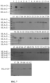
- HLA-G isoform standard proteins HLA-G1, HLA-G2, HLA-G3, HLA-G4, HLA-G5, HLA-G6, HLA-G7 and HLA-G molecular standard proteins lacking ⁇ 1 domain were blocked with 5% skimmed milk powder at room temperature for 4 hours, and washed with 0.2% TBS (Tewen-20 PBS).
- the antibody (YWHG-1, 1.0 ⁇ g/ml) was added, incubated at 4°C overnight, and washed; then HRP-labeled rabbit anti-mouse IgG antibody was added and incubated at room temperature for 30 minutes, washed, and incubated with Dako REAL TM EnVision TM testing system (DAKO) for 1-3 minutes.
- DAKO Dako REAL TM EnVision TM testing system
- K562, K562-HLA-G1, K562-HLA-G2, K562-HLA-G3, K562-HLA-G4, K562-HLA-G5, K562-HLA-G6 and K562-HLA-G7 cells were resuspended with 100 ⁇ L of BD Perm/Wash TM , and then 1 ⁇ L (1.0 mg/ml) of FITC-labeled purified antibody (YWHG-1) was added, incubated at 4°C for 30 minutes, and centrifuged and washed 3 times with 1 ml of 1 ⁇ BD Perm/Wash TM (250g ); the cells were resuspended to 300 ⁇ L of cell suspension with 1 ⁇ BD Perm/Wash TM for flow cytometry.
- the results showed that the antibody (YWHG-1) could specifically recognize the HLA-G1, HLA-G2, HLA-G3, HLA-G4, HLA-G5, HLA-G6, and HLA-G7 molecules ( FIG
- Preparation of single cell suspensions of frozen gastric cancer tissues from 62 patients mechanical separation method and enzymatic digestion method were adopted. The steps were as follows: take out the sample, equilibrate to room temperature, wash with HBSS buffer for three times, and then cut into tissue fragments with a size of 5-6 mm. Wash the tissue fragments with HBSS buffer for three times, then digest with type IV collagenase (1 mg/ml) and hyaluronidase (10 ng/ml) at 37°C in 2% fetal bovine serum (FBS) HBSS buffer for 4 hours. After digestion, filter the cells using a 70 ⁇ m cell filter to obtain a cell suspension. After centrifugation, use a tissue cell lysis buffer to lyse at 4°C for 2 hours, centrifuge at 12,000 rpm for 30 minutes, and collect the lysis supernatant for later use.
- FBS fetal bovine serum
- the gastric cancer tissues were fixed in 10%-12% formalin and embedded in paraffin.
- the conventional process for making tissue sections was carried out, such as baking, dewaxing, hydration and antigen retrieval.
- On the tissues 1% BSA was added dropwise to cover the tissue and tissue edges by 2 mm, and incubated at room temperature for 10 minutes and then blocked.
- the antibody YWHG-1 (1 mg/mL, 1:500 dilution) was added dropwise and kept in a wet box of the refrigerator at 4°C overnight (16-20 h).
- the secondary antibody (the antibody goat anti-mouse diluted with TBS in a ratio of 1 :300) was added, incubated in a 37°C incubator for 30 minutes; washed with TBS buffer, and the developing agent DAB working solution was added; when the tissue color development was completed, the slides was rinsed in running water for 5 minutes and soaked in distilled water for 5 minutes. After HE counterstaining, dehydration, transparency, and sealing, the tissue sections were observed under optical microscope. Brown-colored cells indicated positive expression of HLA-G molecule in gastric cancer cells. The expression intensity of HLA-G molecules could be judged according to the depth of the brown-colored cells.
- FIG.8 showed high expression of HLA-G molecules in the gastric cancer tissue samples (A), (B), (C) and (D) ( FIG.8 ).
Landscapes
- Health & Medical Sciences (AREA)
- Immunology (AREA)
- Chemical & Material Sciences (AREA)
- Life Sciences & Earth Sciences (AREA)
- Molecular Biology (AREA)
- Engineering & Computer Science (AREA)
- Medicinal Chemistry (AREA)
- Organic Chemistry (AREA)
- General Health & Medical Sciences (AREA)
- Biomedical Technology (AREA)
- Biochemistry (AREA)
- Hematology (AREA)
- Urology & Nephrology (AREA)
- Chemical Kinetics & Catalysis (AREA)
- Analytical Chemistry (AREA)
- Biotechnology (AREA)
- Food Science & Technology (AREA)
- Microbiology (AREA)
- Cell Biology (AREA)
- General Physics & Mathematics (AREA)
- Pathology (AREA)
- Proteomics, Peptides & Aminoacids (AREA)
- Genetics & Genomics (AREA)
- Physics & Mathematics (AREA)
- Biophysics (AREA)
- Animal Behavior & Ethology (AREA)
- Public Health (AREA)
- Veterinary Medicine (AREA)
- Pharmacology & Pharmacy (AREA)
- Nuclear Medicine, Radiotherapy & Molecular Imaging (AREA)
- General Chemical & Material Sciences (AREA)
- Peptides Or Proteins (AREA)
- Preparation Of Compounds By Using Micro-Organisms (AREA)
Applications Claiming Priority (2)
| Application Number | Priority Date | Filing Date | Title |
|---|---|---|---|
| CN202210336599.0A CN115819583B (zh) | 2022-03-31 | 2022-03-31 | 一种抗hla-g分子的单克隆抗体及其用途 |
| PCT/CN2022/100232 WO2023184733A1 (zh) | 2022-03-31 | 2022-06-21 | 一种抗hla-g分子的单克隆抗体及其用途 |
Publications (2)
| Publication Number | Publication Date |
|---|---|
| EP4501960A1 true EP4501960A1 (de) | 2025-02-05 |
| EP4501960A4 EP4501960A4 (de) | 2026-04-01 |
Family
ID=85522498
Family Applications (1)
| Application Number | Title | Priority Date | Filing Date |
|---|---|---|---|
| EP22934556.6A Pending EP4501960A4 (de) | 2022-03-31 | 2022-06-21 | Monoklonaler antikörper gegen hla-g-moleküle und verwendung davon |
Country Status (4)
| Country | Link |
|---|---|
| US (1) | US20250171543A1 (de) |
| EP (1) | EP4501960A4 (de) |
| CN (1) | CN115819583B (de) |
| WO (1) | WO2023184733A1 (de) |
Families Citing this family (2)
| Publication number | Priority date | Publication date | Assignee | Title |
|---|---|---|---|---|
| CN122055386A (zh) * | 2023-10-12 | 2026-05-15 | Im生技股份有限公司 | 特异性结合hla-g的抗体及其用途 |
| CN121005780A (zh) * | 2024-05-24 | 2025-11-25 | 合肥天港免疫药物有限公司 | 靶向hla-g的抗体或其抗原结合片段及其应用 |
Family Cites Families (10)
| Publication number | Priority date | Publication date | Assignee | Title |
|---|---|---|---|---|
| CA2213620A1 (en) * | 1995-04-07 | 1996-10-10 | The Regents Of The University Of California | Antibodies for the detection of hla-g |
| EP1224465B1 (de) * | 1999-09-27 | 2005-12-21 | Clifford L. Librach | Nachweis von hla-g |
| CN1312182C (zh) * | 2004-10-26 | 2007-04-25 | 四川新创生物科技有限公司 | 抗hla-g的单克隆抗体及分泌它的杂交瘤细胞株、癌症诊断试剂盒及其应用 |
| EP2730588A1 (de) * | 2012-11-12 | 2014-05-14 | Intelectys | Antikörper und Fragmente davon gegen die alpha-3-Domäne von HLA-G-Protein, Verfahren und Mittel zur Herstellung und Verwendungen davon |
| EA201892793A1 (ru) * | 2016-06-03 | 2019-06-28 | Инвектис | Анти-hla-g специфические антитела |
| TW201829463A (zh) * | 2016-11-18 | 2018-08-16 | 瑞士商赫孚孟拉羅股份公司 | 抗hla-g抗體及其用途 |
| CR20210197A (es) * | 2018-09-27 | 2021-06-03 | Tizona Therapeutics | Anticuerpos anti-hla-g, composiciones que comprenden anticuerpos anti-hla-g y métodos de uso de anticuerpos anti-hla-g |
| CN113045656B (zh) * | 2020-07-27 | 2022-03-08 | 台州恩泽医疗中心(集团) | 抗hla-g异构体分子hla-g5及hla-g6的单克隆抗体及其用途 |
| US11827708B2 (en) * | 2020-07-29 | 2023-11-28 | Janssen Biotech, Inc. | Proteins comprising HLA-G antigen binding domains and their uses |
| CN112794908A (zh) * | 2020-12-31 | 2021-05-14 | 杭州冰湖生物科技有限公司 | 一种抗hla-g抗体的制备及分析方法 |
-
2022
- 2022-03-31 CN CN202210336599.0A patent/CN115819583B/zh active Active
- 2022-06-21 US US18/714,019 patent/US20250171543A1/en active Pending
- 2022-06-21 EP EP22934556.6A patent/EP4501960A4/de active Pending
- 2022-06-21 WO PCT/CN2022/100232 patent/WO2023184733A1/zh not_active Ceased
Also Published As
| Publication number | Publication date |
|---|---|
| EP4501960A4 (de) | 2026-04-01 |
| CN115819583B (zh) | 2026-03-03 |
| US20250171543A1 (en) | 2025-05-29 |
| CN115819583A (zh) | 2023-03-21 |
| WO2023184733A1 (zh) | 2023-10-05 |
Similar Documents
| Publication | Publication Date | Title |
|---|---|---|
| KR101785117B1 (ko) | Her2 수용체 또는 그의 말단절단된 변이체를 발현하는 암의 진단 방법 | |
| CN113045656B (zh) | 抗hla-g异构体分子hla-g5及hla-g6的单克隆抗体及其用途 | |
| CN106661110B (zh) | 抗muc1抗体或其抗原结合片段及其用途 | |
| JP2015030666A (ja) | 抗ヒトcd26モノクローナル抗体又はその抗原結合性断片 | |
| KR20160065876A (ko) | 췌장 종양의 검출 방법, 항체 및 췌장 종양 검출용 키트 | |
| US20250171543A1 (en) | Monoclonal antibody against hla-g molecules and use thereof | |
| CN114605543B (zh) | 一种抗hla-g异构体分子hla-g5及hla-g6的单克隆抗体及其用途 | |
| CN113880948B (zh) | 抗ca724抗体或其抗原结合片段及其制备方法和应用 | |
| EP4428152A1 (de) | Monoklonaler antikörper gegen hla-g1, hla-g4 und hla-g5-isomermoleküle und verwendung davon | |
| EP4501961A1 (de) | Monoklonaler antikörper gegen hla-g1, -g2, -g5 und hla-g6 isomere moleküle und deren verwendung | |
| CA2820590A1 (en) | Anti single-strand type-iv collagen polypeptide antibody, and pharmaceutical, or agent for diagnosing, preventing or treating tumours, containing same | |
| CN116284387B (zh) | 一种抗hla-g2及hla-g6分子的单克隆抗体及用途 | |
| CN111434686B (zh) | 一种抗人pbx1单克隆抗体,其制备方法及其在复发性流产临床诊断中的用途 | |
| AU2022255908A9 (en) | Immunohistochemistry methods and kir3dl2-specific reagents | |
| KR102949436B1 (ko) | Ct-26 종양 세포 특이적 항체 | |
| CN119529093B (zh) | 一种抗人cd68单克隆抗体及其制备方法和应用 | |
| CN117510636B (zh) | Gprc5d抗体及其应用 | |
| CN109320610B (zh) | 抗人cd83单克隆抗体及其制备、鉴定和应用 | |
| KR20250122027A (ko) | 고양이 혈청 아밀로이드 a 단백질에 대한 단일클론항체, 이를 생산하는 융합 세포주 및 이의 용도 | |
| KR20250122026A (ko) | 고양이 혈청 아밀로이드 a 단백질에 대한 단일클론항체, 이를 생산하는 융합 세포주 및 이의 용도 | |
| CN120699146A (zh) | Aβ1-40或Aβ1-42重组抗体的制备及其在阿尔茨海默症联检试剂盒中的应用 | |
| CN116535511A (zh) | 一种用于检测pd-l1的免疫组化抗体及其应用 | |
| CN121851161A (zh) | α-syn特异性抗体及其在帕金森辅助诊断试剂盒中的应用 | |
| CN118184784A (zh) | 一种抗人cd36的单克隆抗体和重组抗体及其应用 | |
| JP2004331633A (ja) | モノクローナル抗体及びその作製方法 |
Legal Events
| Date | Code | Title | Description |
|---|---|---|---|
| STAA | Information on the status of an ep patent application or granted ep patent |
Free format text: STATUS: THE INTERNATIONAL PUBLICATION HAS BEEN MADE |
|
| PUAI | Public reference made under article 153(3) epc to a published international application that has entered the european phase |
Free format text: ORIGINAL CODE: 0009012 |
|
| STAA | Information on the status of an ep patent application or granted ep patent |
Free format text: STATUS: REQUEST FOR EXAMINATION WAS MADE |
|
| 17P | Request for examination filed |
Effective date: 20240918 |
|
| AK | Designated contracting states |
Kind code of ref document: A1 Designated state(s): AL AT BE BG CH CY CZ DE DK EE ES FI FR GB GR HR HU IE IS IT LI LT LU LV MC MK MT NL NO PL PT RO RS SE SI SK SM TR |
|
| DAV | Request for validation of the european patent (deleted) | ||
| DAX | Request for extension of the european patent (deleted) | ||
| A4 | Supplementary search report drawn up and despatched |
Effective date: 20260303 |
|
| RIC1 | Information provided on ipc code assigned before grant |
Ipc: C07K 16/28 20060101AFI20260226BHEP Ipc: C12N 15/13 20060101ALI20260226BHEP Ipc: G01N 33/577 20060101ALI20260226BHEP |