EP4428152A1 - Monoklonaler antikörper gegen hla-g1, hla-g4 und hla-g5-isomermoleküle und verwendung davon - Google Patents
Monoklonaler antikörper gegen hla-g1, hla-g4 und hla-g5-isomermoleküle und verwendung davon Download PDFInfo
- Publication number
- EP4428152A1 EP4428152A1 EP22888772.5A EP22888772A EP4428152A1 EP 4428152 A1 EP4428152 A1 EP 4428152A1 EP 22888772 A EP22888772 A EP 22888772A EP 4428152 A1 EP4428152 A1 EP 4428152A1
- Authority
- EP
- European Patent Office
- Prior art keywords
- hla
- sequence
- seq
- amino acid
- acid sequence
- Prior art date
- Legal status (The legal status is an assumption and is not a legal conclusion. Google has not performed a legal analysis and makes no representation as to the accuracy of the status listed.)
- Pending
Links
Images



Classifications
-
- C—CHEMISTRY; METALLURGY
- C07—ORGANIC CHEMISTRY
- C07K—PEPTIDES
- C07K16/00—Immunoglobulins [IG], e.g. monoclonal or polyclonal antibodies
- C07K16/18—Immunoglobulins [IG], e.g. monoclonal or polyclonal antibodies against material from animals or humans
- C07K16/28—Immunoglobulins [IG], e.g. monoclonal or polyclonal antibodies against material from animals or humans against receptors, cell surface antigens or cell surface determinants
- C07K16/2803—Immunoglobulins [IG], e.g. monoclonal or polyclonal antibodies against material from animals or humans against receptors, cell surface antigens or cell surface determinants against the immunoglobulin superfamily
- C07K16/2833—Immunoglobulins [IG], e.g. monoclonal or polyclonal antibodies against material from animals or humans against receptors, cell surface antigens or cell surface determinants against the immunoglobulin superfamily against MHC-molecules, e.g. HLA-molecules
-
- C—CHEMISTRY; METALLURGY
- C12—BIOCHEMISTRY; BEER; SPIRITS; WINE; VINEGAR; MICROBIOLOGY; ENZYMOLOGY; MUTATION OR GENETIC ENGINEERING
- C12N—MICROORGANISMS OR ENZYMES; COMPOSITIONS THEREOF; PROPAGATING, PRESERVING, OR MAINTAINING MICROORGANISMS; MUTATION OR GENETIC ENGINEERING; CULTURE MEDIA
- C12N5/00—Undifferentiated human, animal or plant cells, e.g. cell lines; Tissues; Cultivation or maintenance thereof; Culture media therefor
- C12N5/10—Cells modified by introduction of foreign genetic material
- C12N5/12—Fused cells, e.g. hybridomas
- C12N5/16—Animal cells
- C12N5/163—Animal cells one of the fusion partners being a B or a T lymphocyte
-
- G—PHYSICS
- G01—MEASURING; TESTING
- G01N—INVESTIGATING OR ANALYSING MATERIALS BY DETERMINING THEIR CHEMICAL OR PHYSICAL PROPERTIES
- G01N15/00—Investigating characteristics of particles; Investigating permeability, pore-volume or surface-area of porous materials
- G01N15/10—Investigating individual particles
- G01N15/14—Optical investigation techniques, e.g. flow cytometry
-
- G—PHYSICS
- G01—MEASURING; TESTING
- G01N—INVESTIGATING OR ANALYSING MATERIALS BY DETERMINING THEIR CHEMICAL OR PHYSICAL PROPERTIES
- G01N33/00—Investigating or analysing materials by specific methods not covered by groups G01N1/00 - G01N31/00
- G01N33/48—Biological material, e.g. blood, urine; Haemocytometers
- G01N33/50—Chemical analysis of biological material, e.g. blood, urine; Testing involving biospecific ligand binding methods; Immunological testing
- G01N33/53—Immunoassay; Biospecific binding assay; Materials therefor
- G01N33/536—Immunoassay; Biospecific binding assay; Materials therefor with immune complex formed in liquid phase
- G01N33/537—Immunoassay; Biospecific binding assay; Materials therefor with immune complex formed in liquid phase with separation of immune complex from unbound antigen or antibody
- G01N33/539—Immunoassay; Biospecific binding assay; Materials therefor with immune complex formed in liquid phase with separation of immune complex from unbound antigen or antibody involving precipitating reagent, e.g. ammonium sulfate
- G01N33/541—Double or second antibody, i.e. precipitating antibody
-
- G—PHYSICS
- G01—MEASURING; TESTING
- G01N—INVESTIGATING OR ANALYSING MATERIALS BY DETERMINING THEIR CHEMICAL OR PHYSICAL PROPERTIES
- G01N33/00—Investigating or analysing materials by specific methods not covered by groups G01N1/00 - G01N31/00
- G01N33/48—Biological material, e.g. blood, urine; Haemocytometers
- G01N33/50—Chemical analysis of biological material, e.g. blood, urine; Testing involving biospecific ligand binding methods; Immunological testing
- G01N33/53—Immunoassay; Biospecific binding assay; Materials therefor
- G01N33/543—Immunoassay; Biospecific binding assay; Materials therefor with an insoluble carrier for immobilising immunochemicals
- G01N33/54366—Apparatus specially adapted for solid-phase testing
- G01N33/54386—Analytical elements
-
- G—PHYSICS
- G01—MEASURING; TESTING
- G01N—INVESTIGATING OR ANALYSING MATERIALS BY DETERMINING THEIR CHEMICAL OR PHYSICAL PROPERTIES
- G01N33/00—Investigating or analysing materials by specific methods not covered by groups G01N1/00 - G01N31/00
- G01N33/48—Biological material, e.g. blood, urine; Haemocytometers
- G01N33/50—Chemical analysis of biological material, e.g. blood, urine; Testing involving biospecific ligand binding methods; Immunological testing
- G01N33/53—Immunoassay; Biospecific binding assay; Materials therefor
- G01N33/569—Immunoassay; Biospecific binding assay; Materials therefor for microorganisms, e.g. protozoa, bacteria, viruses
- G01N33/56966—Animal cells
- G01N33/56977—HLA or MHC typing
-
- C—CHEMISTRY; METALLURGY
- C07—ORGANIC CHEMISTRY
- C07K—PEPTIDES
- C07K2317/00—Immunoglobulins specific features
- C07K2317/30—Immunoglobulins specific features characterized by aspects of specificity or valency
- C07K2317/34—Identification of a linear epitope shorter than 20 amino acid residues or of a conformational epitope defined by amino acid residues
-
- C—CHEMISTRY; METALLURGY
- C07—ORGANIC CHEMISTRY
- C07K—PEPTIDES
- C07K2317/00—Immunoglobulins specific features
- C07K2317/50—Immunoglobulins specific features characterized by immunoglobulin fragments
- C07K2317/56—Immunoglobulins specific features characterized by immunoglobulin fragments variable (Fv) region, i.e. VH and/or VL
- C07K2317/565—Complementarity determining region [CDR]
-
- C—CHEMISTRY; METALLURGY
- C07—ORGANIC CHEMISTRY
- C07K—PEPTIDES
- C07K2317/00—Immunoglobulins specific features
- C07K2317/50—Immunoglobulins specific features characterized by immunoglobulin fragments
- C07K2317/56—Immunoglobulins specific features characterized by immunoglobulin fragments variable (Fv) region, i.e. VH and/or VL
- C07K2317/567—Framework region [FR]
-
- C—CHEMISTRY; METALLURGY
- C07—ORGANIC CHEMISTRY
- C07K—PEPTIDES
- C07K2317/00—Immunoglobulins specific features
- C07K2317/90—Immunoglobulins specific features characterized by (pharmaco)kinetic aspects or by stability of the immunoglobulin
- C07K2317/92—Affinity (KD), association rate (Ka), dissociation rate (Kd) or EC50 value
-
- G—PHYSICS
- G01—MEASURING; TESTING
- G01N—INVESTIGATING OR ANALYSING MATERIALS BY DETERMINING THEIR CHEMICAL OR PHYSICAL PROPERTIES
- G01N2333/00—Assays involving biological materials from specific organisms or of a specific nature
- G01N2333/435—Assays involving biological materials from specific organisms or of a specific nature from animals; from humans
- G01N2333/705—Assays involving receptors, cell surface antigens or cell surface determinants
- G01N2333/70503—Immunoglobulin superfamily, e.g. VCAMs, PECAM, LFA-3
- G01N2333/70539—MHC-molecules, e.g. HLA-molecules
-
- G—PHYSICS
- G01—MEASURING; TESTING
- G01N—INVESTIGATING OR ANALYSING MATERIALS BY DETERMINING THEIR CHEMICAL OR PHYSICAL PROPERTIES
- G01N2474/00—Immunochemical assays or immunoassays characterised by detection mode or means of detection
- G01N2474/10—Immunoblots, e.g. Western blot or Dot blot
-
- G—PHYSICS
- G01—MEASURING; TESTING
- G01N—INVESTIGATING OR ANALYSING MATERIALS BY DETERMINING THEIR CHEMICAL OR PHYSICAL PROPERTIES
- G01N2474/00—Immunochemical assays or immunoassays characterised by detection mode or means of detection
- G01N2474/20—Immunohistochemistry assay
Definitions
- HLA-G human leukocyte antigen-G
- exon 1 of HLA-G mRNA encodes the signal peptide
- exons 2, 3, and 4 encode the ⁇ 1, ⁇ 2, and ⁇ 3 domains of the extracellular region, respectively
- exon 5 encodes a transmembrane region
- exon 6 encodes the intracellular domain of HLA-G molecule containing only 6 amino acid residues
- exon 8 corresponds to the 3'UTR of the HLA-G gene.
- HLA-G1, -G2, -G3, -G4, - G5, -G6 and HLA -G7 The initial transcript of HLA-G is alternatively spliced to produce 7 types of mature mRNAs, which encode 7 isoforms with different molecular weights (HLA-G1, -G2, -G3, -G4, - G5, -G6 and HLA -G7).
- HLA-G1, HLA-G2, HLA-G3 and HLA-G4 contain a transmembrane region and are membrane-bound isoforms;
- HLA-G5, HLA-G6 and HLA-G7 lack transmembrane structures and are soluble isoforms.
- the molecular weights of HLA-G1 ⁇ -G7 isoforms are 39 kD, 31 kD, 22 kD, 30 kD, 34 kD, 23 kD and 16 kD, respectively.
- HLA-G1 is encoded by full-length HLA-G mRNA and consists of extracellular ⁇ 1, ⁇ 2 and ⁇ 3 domains, a transmembrane region and an intracellular domain.
- HLA-G2 lacks ⁇ 2 domain and consists of extracellular ⁇ 1 and ⁇ 3 domains, a transmembrane region and an intracellular domain;
- HLA-G3 lacks ⁇ 2 and ⁇ 3 domains and consists of an extracellular ⁇ 1 domain, a transmembrane region and an intracellular domain;
- HLA-G4 lacks ⁇ 3 domain and consists of extracellular ⁇ 1 and ⁇ 2 domains, a transmembrane region and an intracellular domain; and the extracellular domains of HLA-G5 and HLA-G6 are the same as those of HLA-G1 and HLA-G2 respectively.
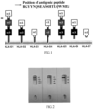
- HLA-G mRNA containing intron 4 contains a stop codon, the encoded protein molecules lack the transmembrane region encoded by exon 5, forming soluble HLA-G molecules. Since the mRNA encoding HLA-G7 contains a stop codon in intron 2, the extracellular region only contains the ⁇ 1 domain and is connected by the two amino acid residues encoded by intron 2 ( FIG. 1 ).
- HLA-G molecules Under normal physiological conditions, HLA-G molecules are only expressed in extravillous trophoblast cells at the maternal-fetal interface, maintaining maternal-fetal immune tolerance during pregnancy. Under pathological conditions, HLA-G molecules can be inducibly expressed in tumor cells and virus-infected tissues and cells, and are closely related to the development and progression of the diseases.
- the HLA-G molecule is an important immunotolerant molecule and an important immune checkpoint molecule. Its immunosuppressive function is to transmit inhibitory signals and induce immune tolerance primarily through binding to the immunoinhibitory receptor immunoglobulin-like transcript-2, ILT2/LILRB1/CD85j) and immunoglobulin-like transcript-4 (ELT4/LILRB2/CD85d).
- HLA-G/ILT signaling The major mechanisms of immunosuppression induced by HLA-G/ILT signaling are as follows: 1 bind to ILT2 expressed on T cells, NK cells and B cells, inhibit the immune killing activity of T cells and NK cells, and inhibit the proliferation of B cells and the secretion of antibodies; 2 bind to ILT2 and ILT4 expressed on dendritic cells (DC), inhibit the maturation and differentiation of DC cells, and induce the generation of tolerant DC cells; 3 bind to ILT2 and ILT4 expressed on myeloid-derived suppressor cells (MDSC) and macrophages (M ⁇ ), induce the differentiation of pro-inflammatory and anti-tumor M1 macrophages into immune-resistant M2 cells, etc. Therefore, HLA-G plays an important role in the development of tumors and other diseases.
- the tumor immunotherapy targeting HLA-G have been underwent phase I or II clinical trials in the United States.
- ILT2 and ILT4 engagement with HLA-G has the HLA-G isoform molecular structure specificity.
- the site where receptors ILT2 and ILT4 bind to HLA-G is the HLA-G extracellular ⁇ 3 domain.
- ILT2 only binds to the HLA-G/ ⁇ 2 m complex, while ILT4 can not only bind to HLA-G/ ⁇ 2 m, but also bind to free HLA-G molecules that do not contain ⁇ 2 m.
- HLA-G1, -G2, -G3, -G4, -G5, -G6 and HLA-G7 isoforms Due to differences in expression mechanism and molecular structure of HLA-G1, -G2, -G3, -G4, -G5, -G6 and HLA-G7 isoforms, for example, the extracellular regions of HLA-G3, -G4, and HLA-G7 isoforms do not contain ⁇ 3 domain and cannot bind to ILT2 and ILT4.
- Different HLA-G isoforms can exert specific immunological effects in the pathophysiological processes. Under different pathological conditions, especially in tumor tissues and cells, there is extensive heterogeneity in the expression of HLA-G isoforms, and the expression of different isoforms of HLA-G molecules has specific clinical significance. Therefore, analyzing the expression of specific HLA-G isoforms and the expression profiles of different HLA-G isoforms is of great significance for elucidating the biological functions and clinical significance of
- the available antibodies used for HLA-G immunohistochemistry and immunoblotting mainly include 4H84, MEM-G1, MEM-G2, 2A12 and 5A6G7.
- the antibody 4H84 whose recognition site is located in the extracellular region ⁇ 1 domain that is shared by all 7 HLA-G isoforms, can detect all 7 HLA-G isoforms (HLA-G1, HLA-G2, HLA-G3, HLA-G4, HLA-G5, HLA-G6 and HLA-G7) that are currently known to contain ⁇ 1 domain, but the expression of specific HLA-G isoforms cannot be distinguished in immunohistochemistry.
- Antibodies MEM-G1 and MEM-G2 are obtained from mice immunized with the full-length HLA-G heavy chain, and the specific recognition sites cannot be predicted. Theoretically, the two antibodies are similar to antibody 4H84 and can recognize all 7 HLA-G isoforms (HLA-G1, HLA-G2, HLA-G3, HLA-G4, HLA-G5, HLA-G6 and HLA-G7), but cannot distinguish the expression of specific HLA-G isoforms.
- Antibodies 2A12 and 5A6G7 are obtained from mice immunized with the 22 amino acid residues located at the carboxyl terminus of HLA-G5 and HLA-G6 molecules, and can recognize HLA-G5 and HLA-G6 molecules.
- antibodies 2A12 and 5A6G7 can also recognize other unknown non-HLA-G5 and HLA-G6 molecules with molecular weights in the range of 36-175 kDa. When they are applied to immunohistochemistry, cross-reaction may be present and false positives may occur.
- the monoclonal antibodies used for flow cytometry to detect HLA-G molecules include 87G and MEM-G/9, but they can only detect HLA-G1 and HLA-G5 molecules.
- an object of the present invention is to provide a specific monoclonal antibody (YWHG-4) against HLA-G1, HLA-G4 and HLA-G5 isoforms, a nucleotide sequence encoding the antibody (YWHG-4) of the present invention and an encoded amino acid sequence thereof; and use of the YWHG-4 antibody in the detection of HLA-G1, HLA-G4 and HLA-G5 by means of immunoblotting, immunohistochemistry, flow cytometry and other assays.
- an antigenic peptide of amino acid sequences at positions 106-126 of a heavy chain of HLA-G molecules is used as an immunogen, and its amino acid sequence is the amino acid sequence (RGYYNQSEASSHTLQWMIGC) shown in SEQ ID No. 17.
- the RGYYNQSEASSHTLQWMIGC peptide fragment is located at the ⁇ 1 and ⁇ 2 hinge regions shared by HLA-G1, HLA-G4 and HLA-G5 isoforms, and based on this, a specific monoclonal antibody (YWHG-4) against HLA-G1, HLA-G4 and HLA-G5 isoforms is prepared ( FIG. 1 ).
- the present invention provides a monoclonal antibody (YWHG-4) against HLA-G isoforms (HLA-G1, HLA-G4, HLA-G5), including at least one or more of heavy chain hypervariable regions CDR1, CDR2 and CDR3, or/and one or more of light chain hypervariable regions CDR1, CDR2 and CDR3;
- the monoclonal antibody further comprises a light chain framework region (FR) and a heavy chain framework region; wherein, the light chain framework region comprises one or more of light chains FR1, FR2, and FR3, and an amino acid sequence of the light chain FR1 is the sequence GDIVITQDELSLPVSLGDQAAISCRS shown in SEQ ID No. 9 or an amino acid sequence with equivalent functions to the sequence shown in SEQ ID No. 9 formed by substituting, deleting or adding one or more amino acids; an amino acid sequence of the light chain FR2 is the sequence LEWYLQKPGQSPKLLIY shown in SEQ ID No. 10 or an amino acid sequence with equivalent functions to the sequence shown in SEQ ID No.
- an amino acid sequence of the light chain FR3 is the sequence NRFSGVPDRF RGSGSGTDFTLKISRVEAEDLGVYYC shown in SEQ ID No. 11 or an amino acid sequence with equivalent functions to the sequence shown in SEQ ID No. 11 formed by substituting, deleting or adding one or more amino acids;
- the heavy chain framework region comprises one or more of the heavy chains FR1, FR2, FR3 and FR4, wherein, an amino acid sequence of the heavy chain FR1 is the sequence VQLQQSGAEVVRPGTSVKVSCKAS shown in SEQ ID No. 12 or an amino acid sequence with equivalent functions to the sequence shown in SEQ ID No.
- an amino acid sequence of the heavy chain FR2 is the sequence VEWIKQRPGQGLEWIGV shown in SEQ ID No. 13 or an amino acid sequence with equivalent functions to the sequence shown in SEQ ID No. 13 formed by substituting, deleting or adding one or more amino acids
- an amino acid sequence of the heavy chain FR3 is the sequence YYSEKFKGKTTLTADKSSSN AYMQLSSLTSDDSAVYFC shown in SEQ ID No. 14 or an amino acid sequence with equivalent functions to the sequence shown in SEQ ID No. 14 formed by substituting, deleting or adding one or more amino acids.
- a nucleotide sequence encoding the light chain variable region in the monoclonal antibody is a sequence shown in SEQ ID No. 15 or a nucleotide sequence with equivalent functions to the sequence shown in SEQ ID No. 15 formed by substituting, deleting or adding one or more nucleotides
- a nucleotide sequence encoding the heavy chain variable region in the monoclonal antibody is a sequence shown in SEQ ID No. 16 or a nucleotide sequence with equivalent functions to the sequence shown in SEQ ID No. 16 formed by substituting, deleting or adding one or more nucleotides.
- the present invention provides a preferred monoclonal antibody (YWHG-4) against HLA-G isoforms (HLA-G1, HLA-G4 and HLA-G5).
- the monoclonal antibody (YWHG-4) is produced by the hybridoma deposited under CCTCC NO: 2021204, the depositary institution is China Center for Type Culture Collection (CCTCC) and the depositary time is August 11, 2021.
- the present invention further provides use of the antibody (YWHG-4) against isoforms (HLA-G1, HLA-G4 and HLA-G5) in the detection of the HLA-G isoforms (HLA-G1, HLA-G4, HLA-G5) by means of immunoblotting, immunohistochemistry, ELISA and flow cytometry, which has the characteristics of high specificity and strong affinity, etc.
- a “hypervariable region” is also called as a complementarity determining region (CDR).
- CDR complementarity determining region
- the antigenic peptide located at positions 106-126 in the heavy chain of the HLA-G molecules was synthesized, with the SEQ No.17 RGYYNQSEASSHTLQWMIGC.
- mice Four SPF-grade BALB/c female mice were immunized initially, 60 ug/mouse.
- the dose of the first booster immunization was 30ug/mouse
- the dose of the second booster immunization was 30ug/mouse
- the dose of the third booster immunization was 30ug/mouse.
- Blood was taken from the orbits to measure the serum titer.
- the plate was coated with "SEQ No.17 RGYYNQSEASSHTLQWMIGC", and the titer of immunized mice was determined by ELISA.
- the "SEQ No.17 RGYYNQSEASSHTLQWMIGC” was used for coating at 2 ug/ml and 4°C overnight; blocked at 37°C for 2 hours; the serum was diluted by 2-fold starting from 200-fold, and the blank control (blank) was PBS, the negative control (negative) was negative serum diluted by 200-fold.
- the "SEQ No.17 RGYYNQSEASSHTLQWMIGC” immunogen 50 ug was used to shock immunized mice.
- mouse spleen cells and SP2/0 cells were taken and fused using the PEG method. After fusion, cells were screened and cultured in a semi-solid medium (containing HAT).
- the primary antibody (cell culture supernatant), the negative control (SP2/0 culture supernatant), the blank control (PBS), and the positive control (positive serum diluted with PBS by 1000-fold) were added, 100 ul/well, incubated in a 37°C incubator for 1 hour; later washed 3 times with the wash buffer.
- the selected clones were screened using ELISA for the first time to obtain positive hybridoma cell lines.
- the positive cell lines were coated again with "SEQ No.17 RGYYNQSEASSHTLQWMIGC", and the ELISA method was used for a second screening to obtain positive hybridoma cell lines.
- YWHG-4 monoclonal antibody against SEQ No.17 RGYYNQSEASSHTLQWMIGC fragment was obtained, named YWHG-4 (deposit number: CCTCC NO: 2021204).
- the isotyping was performed on the screened 10 positive cell lines.
- the coated antibody was diluted to 0.5 ug/ml with 100 mM PBS (pH 7.4), 0.1 ml was added to each well at 4°C overnight; washed twice with PBS-T, then 200 ul blocking buffer was added to each well, and incubated at 37°C for 2 hours; washed three times with PBS-T; 100 ul hybridoma supernatant was added to each well, and incubated at 37°C for 1 hour; washed three times with PBS-T; 0.1 ml of HRP-labeled antibody that was diluted with blocking buffer 1:10000 ( ⁇ , ⁇ ) or 1:20000 (others) was added to each well respectively, and incubated at 37°C for 1 hour; washed three times with PBS-T; 50 ul of substrate solution was added to each well, and then the absorbance was measured at dual wavelengths (450, 630) within 10-20 minutes.
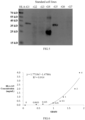
- Thermo's mouse antibody rapid isotyping method (Pierce Rapid Isotyping Kits - Mouse, Catalog" #26178) was used to determine the isotype of the monoclonal antibody produced by the cell line to be of IgG1 type heavy chain and kappa ( ⁇ ) type light chain ( FIG. 2 ) .
- Sample pretreatment the sample was diluted by 1:3 with the corresponding coupling buffer, centrifuged at 12,000 rpm for 10 minutes at 4°C, and filtered with a 0.22 ⁇ m filter membrane to remove the fat, cell debris, and small particulate matter.
- Equilibration the column was equilibrated with the corresponding coupling buffer equivalent to 10-fold column volume, and the flow rate was maintained at 1 ml/min.
- Sample loading The sample was injected to the upper port of the column, and the effluent was collected, the flow rate was maintained at 1 ml/min.
- Impurity washing the column was washed with the coupling buffer equivalent to 5-fold column volume, and the flow rate was maintained at 1 ml/min.
- Elution The antibody was eluted with the elution buffer equivalent to 5-fold column volume, and then collected in the above EP tubes; the flow rate was maintained at 1 ml/min. The pH was adjusted to 7.0 with 1 M Tris-Hcl buffer immediately. Equilibration: the column was equilibrated with the coupling buffer equivalent to 10-fold column volume to pH 7.0, and the flow rate was maintained at 1 ml/min. Dialysis: the antibody was dialyzed overnight with 0.01 M PBS buffer, and the fluid was changed for 3 times.
- the SDS-PAGE gel was prepared, with the concentration of the separating gel of 12%.
- Sample preparation the sample was added a loading buffer and boiled in boiling water for 10 minutes.
- Sample loading 10 ul per well.
- Gel separation 80 V for stacking gel, 30 min; and 120 V for separating gel, 60 min.
- Staining and destaining the gel was immersed in Coomassie Brilliant Blue staining solution and slowly shaken on a shaker for more than 30 minutes (the staining time was adjusted appropriately according to the thickness of the gel).
- the gel was taken out and rinsed in water for several times, and then Coomassie Brilliant Blue destaining solution was added and shaken. It would take about 1 hour when the gel was destained to see the bands, for complete destaining, the destaining solution should be replaced 2-3 times and shaken for more than 24 hours. After destaining, the gel was scanned and recorded by the ECL gel imaging system. The purity of the purified antibody YWHG-4 was >90% ( FIG. 3 ).
- the antigen (SEQ No. 17 RGYYNQSEASSHTLQWMIGC) was diluted with the coating buffer, with the final concentration of 2 ug/ml, 100 ul/well, and placed at 4°C overnight; washed with the wash buffer twice, blocked with the blocking buffer at 200 ul/well, incubated at 37°C for 2 hours; then washed once with the wash buffer.
- the purified antibody was diluted by 2-fold starting from 200-fold (PBS), and the blank control (blank) was PBS, 100 ul/well, and incubated at 37°C for 1 hour; and then washed 3 times with the wash buffer.
- the secondary antibody diluted with PBS by 20,000-fold was added, 100 ul/well, and incubated in a 37°C incubator for 1 hour; taken out and washed 3 times with a wash buffer. Then the color development was performed with a developing solution of 100 ul/well for 5-15 minutes. Then 50 ul of stop solution was added to each well to stop. The absorbance value was measured at dual wavelengths (450, 630), and a chart was drawn for analysis.
- Example 2.1 Detection of HLA-G1, HLA-G4 and HLA-G5 isoforms of the antibody (YWHG-4) by immunoblotting
- HLA-G The denaturing PAGE electrophoresis of the standard proteins of HLA-G isoforms (HLA-G1, HLA-G2, HLA-G3, HLA-G4, HLA-G5, HLA-G6 and HLA-G7) was performed. After semi-dry electroporating, 5% skimmed milk powder was used to block at room temperature for 4 hours, and washed with 0.2% TBS (Tewen-20 PBS).
- the antibody (YWHG-4) was added to detect its recognition specificity, incubated at 4°C overnight, and washed; and then HRP-labeled rabbit anti-mouse IgG antibody was added, incubated at room temperature for 30 minutes, and washed, then incubated with Dako REAL TM EnVision TM detection system (DAKO) for 1-3 minutes.
- DAKO Dako REAL TM EnVision TM detection system
- Example 2.2 Detection of concentrations of HLA-G5 molecules of the antibody (YWHG-4) by ELISA
- the plate was coated in 100 ul/well, and placed at 4°C overnight; then washed twice with the wash buffer; blocked with 1% BSA at 200ul/well, and incubated at 37°C for 2 hours; then washed 3 times with the wash buffer.
- the purified antibody (YWHG-4) with a final concentration of 1.0 ug/ml was added at 100 ul/well, and incubated at 37°C for 1 hour; then washed 3 times with the wash buffer.
- the color development was performed with TMB developing solution at 100ul/well for 5-15 minutes, and then 50 ul of stop solution was added to each well to stop.
- the absorbance valued was measured at the wavelength of 450, and a chart was drawn for analysis.
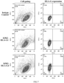
- the OD 450 values of the solutions at 4ug/ml, 2ug/ml, 1ug/ml, 0.5ug/ml, 0.25ug/ml, 0.125ug/ml, 0.0625ug/ml and the coating buffer blank control were 1.855, 1.699, 1.318, 1.213, 0.982, 0.644, 0.424 and 0.06, respectively.
- Example 2.3 Detection of HLA-G1, HLA-G4 and HLA-G5 molecules of the antibody (YWHG-4) by flow cytometry
- HLA-G1 and HLA-G4 were molecules expressing on cell membrane surface
- HLA-G5 was a soluble molecule
- Flow cytometry for detection of HLA-G1 and HLA-G4 molecules on the cell membrane surface The cell cultures of K562-HLA-G1 and K562-HLA-G4 were centrifuged and washed twice with PBS/BSA (250 g), and prepared into a 2 ⁇ 10 6 cell suspension with PBS/BSA.
- HLA-G5 Flow cytometry for detection of intracellular molecular expression of HLA-G5: The K562-HLA-G5 cells were resuspended with 50 ul of BD Perm/Wash TM , then 2 ul (1.0 mg/ml) of FITC-labeled purified antibody (YWHG-4) was added, incubated at 4°C for 30 minutes, centrifuged and washed with 1 ml of 1 ⁇ BD Perm/Wash TM (250 g); then cells were resuspended to 200 ul of cell suspension with 1 ⁇ BD Perm/Wash TM and detected by flow cytometry ( FIG. 8 ).
- YWHG-4 FITC-labeled purified antibody
- Example 2.4 Use of the antibody (YWHG-4) for detection of HLA-G molecules (HLA-G1, HLA-G4 and HLA-G5) in different tissues by immunohistochemistry
- the tissues were fixed in 10%-12% neutral formalin and embedded in paraffin. Tissue sections underwent the conventional production processes such as baking, dewaxing, hydration and antigen retrieval. An appropriate amount of 1% BSA was added dropwise to the tissues to cover the tissue and tissue edges by 2 mm, and incubated at room temperature for 10 minutes for blocking.
- the antibody (YWHG-4) against HLA-G molecules (HLA-G1, HLA-G4 and HLA-G5) (1 mg/mL, 1:500 dilution) was added dropwise and placed in a humidified box at 4°C overnight (16-20 h), washed with TBS buffer, then the secondary antibody was added dropwise (goat anti-mouse ratio of the TBS diluted antibody at 1:300), and incubated in a 37°C incubator for 30 minutes. After washed with TBS buffer, the developing agent DAB working solution was added dropwise; when the color development of tissues was completed, the slides were washed with running water for 5 minutes, and soaked in distilled water for 5 minutes.
- HLA-G HLA-G1, HLA-G4 and HLA-G5
- the expression intensity of HLA-G isoforms could be determined based on the brown coloring of cells ( FIG. 9 ).
Landscapes
- Health & Medical Sciences (AREA)
- Immunology (AREA)
- Life Sciences & Earth Sciences (AREA)
- Chemical & Material Sciences (AREA)
- Engineering & Computer Science (AREA)
- Molecular Biology (AREA)
- Biomedical Technology (AREA)
- General Health & Medical Sciences (AREA)
- Biochemistry (AREA)
- Hematology (AREA)
- Urology & Nephrology (AREA)
- Organic Chemistry (AREA)
- Medicinal Chemistry (AREA)
- Cell Biology (AREA)
- Biotechnology (AREA)
- Genetics & Genomics (AREA)
- Analytical Chemistry (AREA)
- General Physics & Mathematics (AREA)
- Pathology (AREA)
- Physics & Mathematics (AREA)
- Microbiology (AREA)
- Food Science & Technology (AREA)
- Zoology (AREA)
- Biophysics (AREA)
- Proteomics, Peptides & Aminoacids (AREA)
- Bioinformatics & Cheminformatics (AREA)
- Wood Science & Technology (AREA)
- Dispersion Chemistry (AREA)
- Tropical Medicine & Parasitology (AREA)
- Virology (AREA)
- General Engineering & Computer Science (AREA)
- Peptides Or Proteins (AREA)
- Preparation Of Compounds By Using Micro-Organisms (AREA)
Applications Claiming Priority (2)
| Application Number | Priority Date | Filing Date | Title |
|---|---|---|---|
| CN202111300914.6A CN115925930B (zh) | 2021-11-04 | 2021-11-04 | 一种抗hla-g1、hla-g4及hla-g5异构体分子的单克隆抗体及其用途 |
| PCT/CN2022/087315 WO2023077733A1 (zh) | 2021-11-04 | 2022-04-18 | 一种抗hla-g1、hla-g4及hla-g5异构体分子的单克隆抗体及其用途 |
Publications (2)
| Publication Number | Publication Date |
|---|---|
| EP4428152A1 true EP4428152A1 (de) | 2024-09-11 |
| EP4428152A4 EP4428152A4 (de) | 2025-11-05 |
Family
ID=85833349
Family Applications (1)
| Application Number | Title | Priority Date | Filing Date |
|---|---|---|---|
| EP22888772.5A Pending EP4428152A4 (de) | 2021-11-04 | 2022-04-18 | Monoklonaler antikörper gegen hla-g1, hla-g4 und hla-g5-isomermoleküle und verwendung davon |
Country Status (4)
| Country | Link |
|---|---|
| US (1) | US20250289892A1 (de) |
| EP (1) | EP4428152A4 (de) |
| CN (1) | CN115925930B (de) |
| WO (1) | WO2023077733A1 (de) |
Family Cites Families (8)
| Publication number | Priority date | Publication date | Assignee | Title |
|---|---|---|---|---|
| CN101967191A (zh) * | 2009-07-28 | 2011-02-09 | 广州天美生物技术有限公司 | Hla-g抗体制备方法及其在医学中的应用 |
| CN107533051B (zh) * | 2015-03-27 | 2020-11-13 | 南加利福尼亚大学 | Hla-g作为car t细胞免疫疗法的新靶标 |
| CN105675883B (zh) * | 2016-02-22 | 2018-08-31 | 叶尚勉 | 一种检测人类白细胞抗原hla-g的方法 |
| EA201892793A1 (ru) * | 2016-06-03 | 2019-06-28 | Инвектис | Анти-hla-g специфические антитела |
| CR20210197A (es) * | 2018-09-27 | 2021-06-03 | Tizona Therapeutics | Anticuerpos anti-hla-g, composiciones que comprenden anticuerpos anti-hla-g y métodos de uso de anticuerpos anti-hla-g |
| US12590171B2 (en) * | 2019-09-27 | 2026-03-31 | Nanjing GenScript Biotech Co., Ltd. | Anti-VHH domain antibodies and use thereof |
| CN113045656B (zh) * | 2020-07-27 | 2022-03-08 | 台州恩泽医疗中心(集团) | 抗hla-g异构体分子hla-g5及hla-g6的单克隆抗体及其用途 |
| US11827708B2 (en) * | 2020-07-29 | 2023-11-28 | Janssen Biotech, Inc. | Proteins comprising HLA-G antigen binding domains and their uses |
-
2021
- 2021-11-04 CN CN202111300914.6A patent/CN115925930B/zh active Active
-
2022
- 2022-04-18 US US18/573,187 patent/US20250289892A1/en active Pending
- 2022-04-18 WO PCT/CN2022/087315 patent/WO2023077733A1/zh not_active Ceased
- 2022-04-18 EP EP22888772.5A patent/EP4428152A4/de active Pending
Also Published As
| Publication number | Publication date |
|---|---|
| US20250289892A1 (en) | 2025-09-18 |
| CN115925930A (zh) | 2023-04-07 |
| WO2023077733A1 (zh) | 2023-05-11 |
| EP4428152A4 (de) | 2025-11-05 |
| CN115925930B (zh) | 2025-10-31 |
Similar Documents
| Publication | Publication Date | Title |
|---|---|---|
| CN113045656B (zh) | 抗hla-g异构体分子hla-g5及hla-g6的单克隆抗体及其用途 | |
| JP7215759B2 (ja) | 4-1bb抗体およびその製造方法と使用 | |
| CN106661110B (zh) | 抗muc1抗体或其抗原结合片段及其用途 | |
| KR20240018579A (ko) | B7h3 단클론 항체군 및 그 의약적 용도 | |
| CN112955471A (zh) | Cd3抗体及其药物用途 | |
| CN112969476A (zh) | 多特异性蛋白分子 | |
| WO2023109061A1 (zh) | 一种抗hla-g异构体分子hla-g5及hla-g6的单克隆抗体及其用途 | |
| US20250171543A1 (en) | Monoclonal antibody against hla-g molecules and use thereof | |
| CN113004406B (zh) | 抗cd47抗体及其应用 | |
| KR20230124001A (ko) | Cd47을 표적으로 하는 항체 및 이의 응용 | |
| EP4501961A1 (de) | Monoklonaler antikörper gegen hla-g1, -g2, -g5 und hla-g6 isomere moleküle und deren verwendung | |
| US20250289892A1 (en) | Monoclonal antibody against hla-g1, hla-g4 and hla-g5 isoforms and use thereof | |
| Masuko et al. | Towards therapeutic antibodies to membrane oncoproteins by a robust strategy using rats immunized with transfectants expressing target molecules fused to green fluorescent protein | |
| CN116284387B (zh) | 一种抗hla-g2及hla-g6分子的单克隆抗体及用途 | |
| CN109593135B (zh) | 抗人pd-l1单克隆抗体及其应用 | |
| KR20230132450A (ko) | 항trpv6 단일 클론 항체 및 그 응용 | |
| CN111434686B (zh) | 一种抗人pbx1单克隆抗体,其制备方法及其在复发性流产临床诊断中的用途 | |
| CN117510636B (zh) | Gprc5d抗体及其应用 | |
| KR102949436B1 (ko) | Ct-26 종양 세포 특이적 항체 | |
| KR101752280B1 (ko) | 영장류 cd154에 특이적인 항체 및 이를 생산하는 융합 세포주 | |
| TW202241958A (zh) | 抗pd-l1抗體及其應用 | |
| WO2025073052A1 (en) | Antibodies for parathyroid hormone related protein and methods related thereto | |
| WO2022238459A1 (en) | New humanized anti-vegfc antibodies and uses thereof | |
| CN116535511A (zh) | 一种用于检测pd-l1的免疫组化抗体及其应用 | |
| CN116547301A (zh) | 使用抗体进行的细胞表面mica和micb检测 |
Legal Events
| Date | Code | Title | Description |
|---|---|---|---|
| STAA | Information on the status of an ep patent application or granted ep patent |
Free format text: STATUS: THE INTERNATIONAL PUBLICATION HAS BEEN MADE |
|
| PUAI | Public reference made under article 153(3) epc to a published international application that has entered the european phase |
Free format text: ORIGINAL CODE: 0009012 |
|
| STAA | Information on the status of an ep patent application or granted ep patent |
Free format text: STATUS: REQUEST FOR EXAMINATION WAS MADE |
|
| 17P | Request for examination filed |
Effective date: 20231220 |
|
| AK | Designated contracting states |
Kind code of ref document: A1 Designated state(s): AL AT BE BG CH CY CZ DE DK EE ES FI FR GB GR HR HU IE IS IT LI LT LU LV MC MK MT NL NO PL PT RO RS SE SI SK SM TR |
|
| DAV | Request for validation of the european patent (deleted) | ||
| DAX | Request for extension of the european patent (deleted) | ||
| A4 | Supplementary search report drawn up and despatched |
Effective date: 20251009 |
|
| RIC1 | Information provided on ipc code assigned before grant |
Ipc: C07K 16/28 20060101AFI20251002BHEP Ipc: C12N 15/13 20060101ALI20251002BHEP Ipc: G01N 33/569 20060101ALI20251002BHEP Ipc: G01N 33/577 20060101ALI20251002BHEP Ipc: G01N 33/574 20060101ALI20251002BHEP Ipc: C12N 5/20 20060101ALI20251002BHEP |