EP4497756A2 - Antikörper, kit, nachweisverfahren und probenanalysator zum nachweis von schilddrüsenstimulationshormon - Google Patents
Antikörper, kit, nachweisverfahren und probenanalysator zum nachweis von schilddrüsenstimulationshormon Download PDFInfo
- Publication number
- EP4497756A2 EP4497756A2 EP24190963.9A EP24190963A EP4497756A2 EP 4497756 A2 EP4497756 A2 EP 4497756A2 EP 24190963 A EP24190963 A EP 24190963A EP 4497756 A2 EP4497756 A2 EP 4497756A2
- Authority
- EP
- European Patent Office
- Prior art keywords
- cdr
- amino acid
- acid sequence
- seq
- variants
- Prior art date
- Legal status (The legal status is an assumption and is not a legal conclusion. Google has not performed a legal analysis and makes no representation as to the accuracy of the status listed.)
- Pending
Links
Images


Classifications
-
- C—CHEMISTRY; METALLURGY
- C07—ORGANIC CHEMISTRY
- C07K—PEPTIDES
- C07K16/00—Immunoglobulins [IGs], e.g. monoclonal or polyclonal antibodies
- C07K16/18—Immunoglobulins [IGs], e.g. monoclonal or polyclonal antibodies against material from animals or humans
- C07K16/26—Immunoglobulins [IGs], e.g. monoclonal or polyclonal antibodies against material from animals or humans against hormones ; against hormone releasing or inhibiting factors
-
- A—HUMAN NECESSITIES
- A61—MEDICAL OR VETERINARY SCIENCE; HYGIENE
- A61P—SPECIFIC THERAPEUTIC ACTIVITY OF CHEMICAL COMPOUNDS OR MEDICINAL PREPARATIONS
- A61P5/00—Drugs for disorders of the endocrine system
-
- C—CHEMISTRY; METALLURGY
- C12—BIOCHEMISTRY; BEER; SPIRITS; WINE; VINEGAR; MICROBIOLOGY; ENZYMOLOGY; MUTATION OR GENETIC ENGINEERING
- C12N—MICROORGANISMS OR ENZYMES; COMPOSITIONS THEREOF; PROPAGATING, PRESERVING, OR MAINTAINING MICROORGANISMS; MUTATION OR GENETIC ENGINEERING; CULTURE MEDIA
- C12N15/00—Mutation or genetic engineering; DNA or RNA concerning genetic engineering, vectors, e.g. plasmids, or their isolation, preparation or purification; Use of hosts therefor
- C12N15/09—Recombinant DNA-technology
- C12N15/63—Introduction of foreign genetic material using vectors; Vectors; Use of hosts therefor; Regulation of expression
- C12N15/64—General methods for preparing the vector, for introducing it into the cell or for selecting the vector-containing host
-
- G—PHYSICS
- G01—MEASURING; TESTING
- G01J—MEASUREMENT OF INTENSITY, VELOCITY, SPECTRAL CONTENT, POLARISATION, PHASE OR PULSE CHARACTERISTICS OF INFRARED, VISIBLE OR ULTRAVIOLET LIGHT; COLORIMETRY; RADIATION PYROMETRY
- G01J11/00—Measuring the characteristics of individual optical pulses or of optical pulse trains
-
- G—PHYSICS
- G01—MEASURING; TESTING
- G01N—INVESTIGATING OR ANALYSING MATERIALS BY DETERMINING THEIR CHEMICAL OR PHYSICAL PROPERTIES
- G01N21/00—Investigating or analysing materials by the use of optical means, i.e. using sub-millimetre waves, infrared, visible or ultraviolet light
- G01N21/75—Systems in which material is subjected to a chemical reaction, the progress or the result of the reaction being investigated
- G01N21/76—Chemiluminescence; Bioluminescence
-
- G—PHYSICS
- G01—MEASURING; TESTING
- G01N—INVESTIGATING OR ANALYSING MATERIALS BY DETERMINING THEIR CHEMICAL OR PHYSICAL PROPERTIES
- G01N33/00—Investigating or analysing materials by specific methods not covered by groups G01N1/00 - G01N31/00
- G01N33/48—Biological material, e.g. blood, urine; Haemocytometers
- G01N33/50—Chemical analysis of biological material, e.g. blood, urine; Testing involving biospecific ligand binding methods; Immunological testing
- G01N33/53—Immunoassay; Biospecific binding assay; Materials therefor
- G01N33/5302—Apparatus specially adapted for immunological test procedures
-
- G—PHYSICS
- G01—MEASURING; TESTING
- G01N—INVESTIGATING OR ANALYSING MATERIALS BY DETERMINING THEIR CHEMICAL OR PHYSICAL PROPERTIES
- G01N33/00—Investigating or analysing materials by specific methods not covered by groups G01N1/00 - G01N31/00
- G01N33/48—Biological material, e.g. blood, urine; Haemocytometers
- G01N33/50—Chemical analysis of biological material, e.g. blood, urine; Testing involving biospecific ligand binding methods; Immunological testing
- G01N33/53—Immunoassay; Biospecific binding assay; Materials therefor
- G01N33/543—Immunoassay; Biospecific binding assay; Materials therefor with an insoluble carrier for immobilising immunochemicals
- G01N33/54313—Immunoassay; Biospecific binding assay; Materials therefor with an insoluble carrier for immobilising immunochemicals the carrier being characterised by its particulate form
- G01N33/54326—Magnetic particles
-
- G—PHYSICS
- G01—MEASURING; TESTING
- G01N—INVESTIGATING OR ANALYSING MATERIALS BY DETERMINING THEIR CHEMICAL OR PHYSICAL PROPERTIES
- G01N33/00—Investigating or analysing materials by specific methods not covered by groups G01N1/00 - G01N31/00
- G01N33/48—Biological material, e.g. blood, urine; Haemocytometers
- G01N33/50—Chemical analysis of biological material, e.g. blood, urine; Testing involving biospecific ligand binding methods; Immunological testing
- G01N33/53—Immunoassay; Biospecific binding assay; Materials therefor
- G01N33/574—Immunoassay; Biospecific binding assay; Materials therefor for cancer
- G01N33/57407—Specifically defined cancers
-
- G—PHYSICS
- G01—MEASURING; TESTING
- G01N—INVESTIGATING OR ANALYSING MATERIALS BY DETERMINING THEIR CHEMICAL OR PHYSICAL PROPERTIES
- G01N33/00—Investigating or analysing materials by specific methods not covered by groups G01N1/00 - G01N31/00
- G01N33/48—Biological material, e.g. blood, urine; Haemocytometers
- G01N33/50—Chemical analysis of biological material, e.g. blood, urine; Testing involving biospecific ligand binding methods; Immunological testing
- G01N33/53—Immunoassay; Biospecific binding assay; Materials therefor
- G01N33/574—Immunoassay; Biospecific binding assay; Materials therefor for cancer
- G01N33/57484—Immunoassay; Biospecific binding assay; Materials therefor for cancer involving compounds serving as markers for tumor, cancer, neoplasia, e.g. cellular determinants, receptors, heat shock/stress proteins, A-protein, oligosaccharides, metabolites
-
- G—PHYSICS
- G01—MEASURING; TESTING
- G01N—INVESTIGATING OR ANALYSING MATERIALS BY DETERMINING THEIR CHEMICAL OR PHYSICAL PROPERTIES
- G01N33/00—Investigating or analysing materials by specific methods not covered by groups G01N1/00 - G01N31/00
- G01N33/48—Biological material, e.g. blood, urine; Haemocytometers
- G01N33/50—Chemical analysis of biological material, e.g. blood, urine; Testing involving biospecific ligand binding methods; Immunological testing
- G01N33/68—Chemical analysis of biological material, e.g. blood, urine; Testing involving biospecific ligand binding methods; Immunological testing involving proteins, peptides or amino acids
- G01N33/6854—Immunoglobulins
-
- G—PHYSICS
- G01—MEASURING; TESTING
- G01N—INVESTIGATING OR ANALYSING MATERIALS BY DETERMINING THEIR CHEMICAL OR PHYSICAL PROPERTIES
- G01N33/00—Investigating or analysing materials by specific methods not covered by groups G01N1/00 - G01N31/00
- G01N33/48—Biological material, e.g. blood, urine; Haemocytometers
- G01N33/50—Chemical analysis of biological material, e.g. blood, urine; Testing involving biospecific ligand binding methods; Immunological testing
- G01N33/68—Chemical analysis of biological material, e.g. blood, urine; Testing involving biospecific ligand binding methods; Immunological testing involving proteins, peptides or amino acids
- G01N33/6893—Chemical analysis of biological material, e.g. blood, urine; Testing involving biospecific ligand binding methods; Immunological testing involving proteins, peptides or amino acids related to diseases not provided for elsewhere
-
- G—PHYSICS
- G01—MEASURING; TESTING
- G01N—INVESTIGATING OR ANALYSING MATERIALS BY DETERMINING THEIR CHEMICAL OR PHYSICAL PROPERTIES
- G01N33/00—Investigating or analysing materials by specific methods not covered by groups G01N1/00 - G01N31/00
- G01N33/48—Biological material, e.g. blood, urine; Haemocytometers
- G01N33/50—Chemical analysis of biological material, e.g. blood, urine; Testing involving biospecific ligand binding methods; Immunological testing
- G01N33/74—Chemical analysis of biological material, e.g. blood, urine; Testing involving biospecific ligand binding methods; Immunological testing involving hormones or other non-cytokine intercellular protein regulatory factors such as growth factors, including receptors to hormones and growth factors
- G01N33/76—Human chorionic gonadotropin including luteinising hormone, follicle stimulating hormone, thyroid stimulating hormone or their receptors
-
- C—CHEMISTRY; METALLURGY
- C07—ORGANIC CHEMISTRY
- C07K—PEPTIDES
- C07K2317/00—Immunoglobulins specific features
- C07K2317/20—Immunoglobulins specific features characterized by taxonomic origin
- C07K2317/24—Immunoglobulins specific features characterized by taxonomic origin containing regions, domains or residues from different species, e.g. chimeric, humanized or veneered
-
- C—CHEMISTRY; METALLURGY
- C07—ORGANIC CHEMISTRY
- C07K—PEPTIDES
- C07K2317/00—Immunoglobulins specific features
- C07K2317/30—Immunoglobulins specific features characterized by aspects of specificity or valency
- C07K2317/33—Crossreactivity, e.g. for species or epitope, or lack of said crossreactivity
-
- C—CHEMISTRY; METALLURGY
- C07—ORGANIC CHEMISTRY
- C07K—PEPTIDES
- C07K2317/00—Immunoglobulins specific features
- C07K2317/50—Immunoglobulins specific features characterized by immunoglobulin fragments
- C07K2317/52—Constant or Fc region; Isotype
-
- C—CHEMISTRY; METALLURGY
- C07—ORGANIC CHEMISTRY
- C07K—PEPTIDES
- C07K2317/00—Immunoglobulins specific features
- C07K2317/50—Immunoglobulins specific features characterized by immunoglobulin fragments
- C07K2317/56—Immunoglobulins specific features characterized by immunoglobulin fragments variable (Fv) region, i.e. VH and/or VL
- C07K2317/565—Complementarity determining region [CDR]
-
- C—CHEMISTRY; METALLURGY
- C07—ORGANIC CHEMISTRY
- C07K—PEPTIDES
- C07K2317/00—Immunoglobulins specific features
- C07K2317/90—Immunoglobulins specific features characterized by (pharmaco)kinetic aspects or by stability of the immunoglobulin
-
- C—CHEMISTRY; METALLURGY
- C07—ORGANIC CHEMISTRY
- C07K—PEPTIDES
- C07K2317/00—Immunoglobulins specific features
- C07K2317/90—Immunoglobulins specific features characterized by (pharmaco)kinetic aspects or by stability of the immunoglobulin
- C07K2317/92—Affinity (KD), association rate (Ka), dissociation rate (Kd) or EC50 value
-
- C—CHEMISTRY; METALLURGY
- C12—BIOCHEMISTRY; BEER; SPIRITS; WINE; VINEGAR; MICROBIOLOGY; ENZYMOLOGY; MUTATION OR GENETIC ENGINEERING
- C12N—MICROORGANISMS OR ENZYMES; COMPOSITIONS THEREOF; PROPAGATING, PRESERVING, OR MAINTAINING MICROORGANISMS; MUTATION OR GENETIC ENGINEERING; CULTURE MEDIA
- C12N2510/00—Genetically modified cells
-
- C—CHEMISTRY; METALLURGY
- C12—BIOCHEMISTRY; BEER; SPIRITS; WINE; VINEGAR; MICROBIOLOGY; ENZYMOLOGY; MUTATION OR GENETIC ENGINEERING
- C12R—INDEXING SCHEME ASSOCIATED WITH SUBCLASSES C12C - C12Q, RELATING TO MICROORGANISMS
- C12R2001/00—Microorganisms ; Processes using microorganisms
- C12R2001/01—Bacteria or Actinomycetales ; using bacteria or Actinomycetales
- C12R2001/185—Escherichia
- C12R2001/19—Escherichia coli
-
- G—PHYSICS
- G01—MEASURING; TESTING
- G01N—INVESTIGATING OR ANALYSING MATERIALS BY DETERMINING THEIR CHEMICAL OR PHYSICAL PROPERTIES
- G01N2333/00—Assays involving biological materials from specific organisms or of a specific nature
- G01N2333/435—Assays involving biological materials from specific organisms or of a specific nature from animals; from humans
- G01N2333/575—Hormones
- G01N2333/59—Follicle-stimulating hormone [FSH]; Chorionic gonadotropins, e.g. HCG; Luteinising hormone [LH]; Thyroid-stimulating hormone [TSH]
-
- G—PHYSICS
- G01—MEASURING; TESTING
- G01N—INVESTIGATING OR ANALYSING MATERIALS BY DETERMINING THEIR CHEMICAL OR PHYSICAL PROPERTIES
- G01N2800/00—Detection or diagnosis of diseases
- G01N2800/04—Endocrine or metabolic disorders
- G01N2800/046—Thyroid disorders
-
- G—PHYSICS
- G01—MEASURING; TESTING
- G01N—INVESTIGATING OR ANALYSING MATERIALS BY DETERMINING THEIR CHEMICAL OR PHYSICAL PROPERTIES
- G01N2800/00—Detection or diagnosis of diseases
- G01N2800/70—Mechanisms involved in disease identification
- G01N2800/7023—(Hyper)proliferation
- G01N2800/7028—Cancer
Definitions
- the present application belongs to the field of molecular immunology, and specifically relates to a thyroid stimulating hormone (TSH)-specific antibody, and a kit, detection method and sample analyzer for detecting TSH.
- TSH thyroid stimulating hormone
- Thyroid-dysfunction is common in adults, and mainly includes hypothyroidism and hyperthyroidism.
- Thyroid stimulating hormone (TSH) in blood is an accurate marker reflecting thyroid function.
- the normal range of TSH in serum is 0.58 to 4.1 ⁇ IU/ml.
- TSH concentration in blood increases in patients with hypothyroidism and decreases in patients with hyperthyroidism.
- Sandwich-immunoassay method is used for the determination of TSH concentration in human serum samples.
- Most of the current TSH assays are third-generation assays with a functional sensitivity of 0.01 to 0.02 ⁇ IU/mL. These assays reliably distinguish patients with hyperthyroidism from those with euthyroidism. However, in some patients, TSH concentration in serum is below the limit of detection of third-generation TSH assay.
- the fourth-generation TSH assays provide 10-fold increase in the functional sensitivity (0.001 to 0.002 ⁇ IU/mL) and therefore make it possible to quantify the concentration of TSH in serum from these patients. Functional sensitivity is determined by the affinity of the antibodies used and the sensitivity that the detection system can provide. For the fourth-generation TSH assays high affinity antibodies are needed which can provide such a high sensitivity.
- TSH is a member of glycoprotein hormones family consisting of thyroid-stimulating hormone, luteinizing hormone (LH), follicle-stimulating hormone (FSH), and human chorionic gonadotropin (hCG).
- TSH is a heterodimer consisting of ⁇ - and ⁇ -subunits.
- the ⁇ -subunit of TSH is similar to the ⁇ -subunits of LH, FSH, and hCG, and the ⁇ -subunit is specific for TSH.
- high degree of homology between human TSH, LH, FSH, and CG ⁇ -subunit sequences exists. In order to accurately determine the concentration of TSH in serum samples, it is required that the pair of antibodies used in the assays does not recognize other glycoprotein hormones.
- both antibodies used in the immunoassay should not have cross-reaction with LH, FSH, and hCG. Therefore, antibodies specific to TSH dimer or TSH ⁇ -subunit without cross-reactivity to other glycoprotein hormones should be developed.
- An object of the present application is to provide a new monoclonal antibody or antigen-binding antibody fragment thereof that is capable of specifically binding to TSH dimer or TSH ⁇ -subunit and does not cross-react with the ⁇ -subunit shared by human LH, FSH, CG and free glycoprotein hormone. It is suitable for developing a fourth-generation functionally sensitive TSH assay.
- Another object of the present application is to provide a monoclonal antibody or antigen-binding antibody fragment thereof that recognizes TSH variant R55G.
- Antibody nucleic acid molecule, expression vector, host cell
- the present application provides an antibody or antigen-binding fragment thereof that specifically binds to TSH or a mutant thereof, which comprises:
- the variant comprise an amino acid mutation as compared to the amino acid sequence from which it is derived, and the amino acid mutation is a substitution, deletion or addition of one or several amino acids (e.g., a substitution, deletion or addition of 1, 2, 3, 4 or 5 amino acids). In some embodiments, the substitution is a conservative substitution.
- the variant has an identity of at least 80%, at least 85%, at least 90%, at least 91%, at least 92%, at least 93%, at least 94%, at least 95%, at least 96%, at least 97%, at least 98%, at least 99%, or 100% as compared to the amino acid sequence from which it is derived.
- the three CDRs comprised in the heavy chain variable region and/or the three CDRs contained in the light chain variable region are defined by the Kabat, Chothia or IMGT numbering system.
- the antibody or antigen-binding fragment thereof comprises:
- the heavy chain variable region further comprises a heavy chain framework region (FR).
- the light chain variable region further comprises a light chain framework region (FR).
- the framework region is highly conserved, and it provides a scaffold for the six CDRs in three-dimensional space to form an antigen binding surface.
- variable domains of naturally occurring heavy and light chains each comprises four FR regions (FR1, FR2, FR3, and FR4), and these four FR regions are mainly based on a ⁇ -folded conformation, and three complementary determining regions are connected in the form of FR1-CDR1-FR2-CDR2-FR3-CDR3-FR4, wherein the CDR regions are mainly based on loop conformation, and in some cases form a part of ⁇ -folded structure.
- the complementary determining regions in each chain are closely together through FR, and together with the complementary determining regions from another chain, thereby contributing to the formation of the antigen binding side end (see, Kabat et al., loc. cit.).
- the heavy chain framework region and/or the light chain framework region can be independently derived from the framework region of any species immunoglobulin.
- the heavy chain framework region and the light chain framework region are each independently derived from the heavy chain framework region and the light chain framework region of a rabbit or sheep immunoglobulin.
- the heavy chain framework region and the light chain framework region are each independently derived from the heavy chain framework region and the light chain framework region of a human or mouse immunoglobulin.
- the heavy chain framework region and the light chain framework region each independently comprise a sequence derived from the heavy chain framework region and the light chain framework region of a mouse immunoglobulin, the heavy chain framework region and the light chain framework region of a human immunoglobulin, or a combination thereof.
- the heavy chain framework region and the light chain framework region each independently comprise a sequence derived from the heavy chain framework region and the light chain framework region of a rabbit or sheep immunoglobulin, the heavy chain framework region and the light chain framework region of a human immunoglobulin, or a combination thereof. In some embodiments, the heavy chain framework region and the light chain framework region each independently comprise a sequence derived from the heavy chain framework region and the light chain framework region of a human germline antibody.
- the antibody or antigen-binding fragment thereof comprises a heavy chain constant region (CH) and a light chain constant region (CL). In some embodiments, the antibody or antigen-binding fragment thereof comprises a rabbit or sheep heavy chain constant region and a rabbit or sheep light chain constant region. In some embodiments, the antibody or antigen-binding fragment thereof comprises a human heavy chain constant region and a human light chain constant region. In some embodiments, the antibody or antigen-binding fragment thereof is an IgG, IgM, IgE, IgD or IgA antibody. In some embodiments, the heavy chain constant region is an IgG heavy chain constant region, such as an IgG1, IgG2, IgG3 or IgG4 heavy chain constant region. In some embodiments, the light chain constant region is a ⁇ or ⁇ light chain constant region (e.g., a human ⁇ light chain constant region).
- the antigen-binding fragment is selected from the group consisting of scFv, Fab, Fab', (Fab') 2 , Fd, Fv, CDR fragment, nanobody, disulfide-linked Fv (dsFv), diabody, bispecific antibody and multispecific antibody; and/or, the antibody is a rabbit or sheep antibody, a chimeric antibody or a humanized antibody.
- the antibody or antigen-binding fragment thereof further has a detectable label.
- the detectable label can be any substance that can be detected by fluorescent, spectrospic, photochemical, biochemical, immunological, electrical, optical or chemical means. It is particularly preferred that such labels are suitable for immunological detection (e.g., enzyme-linked immunosorbent assay, radioimmunoassay, fluorescent immunoassay, chemiluminescent immunoassay, etc.).
- immunological detection e.g., enzyme-linked immunosorbent assay, radioimmunoassay, fluorescent immunoassay, chemiluminescent immunoassay, etc.
- Such labels include, but are not limited to, enzymes (e.g., oxidase, microperoxidase, horseradish peroxidase, alkaline phosphatase, ⁇ -galactosidase, urease, glucose oxidase, etc.), radionuclides (e.g., 3 H, 125 I, 35 S, 14 C or 32 P), fluorescent dyes (e.g., fluorescein isothiocyanate (FITC), fluorescein, tetramethylrhodamine isothiocyanate (TRITC), phycoerythrin (PE), Texas Red, rhodamine, quantum dots or cyanine dye derivatives (e.g., Cy7, Alexa 750), europium and green fluorescent protein, etc.), chemiluminescence (e.g., luminol, isoluminol, phenanthridinium, acridinium esters, etc.), and biofluorescence (e.
- the labels covered in the present application can be detected by methods known in the art.
- radioactive labels can be detected using photographic film or a scintillation counter
- fluorescent labels can be detected using a photodetector to detect the emitted light.
- Enzyme labels are generally detected by providing a substrate to the enzyme and detecting the reaction product produced by the action of the enzyme on the substrate.
- Chemiluminescent substances e.g., acridinium esters
- Biotin is generally detected by providing biotin with avidin (e.g., streptavidin) modified with the above-mentioned label and detecting the label carried by the avidin linked to biotin.
- avidin e.g., streptavidin
- the detectable labels as described above can be linked to the antibody or antigen-binding fragment thereof of the present application through linkers of different lengths to reduce potential steric hindrance.
- the label is selected from the group consisting of fluorescein, chemiluminescence (e.g., acridinium esters), enzyme (e.g., horseradish peroxidase, alkaline phosphatase), radioactive isotope, biotin, colloidal gold, and magnetic particle.
- fluorescein e.g., acridinium esters
- enzyme e.g., horseradish peroxidase, alkaline phosphatase
- radioactive isotope e.g., biotin, colloidal gold, and magnetic particle.
- the antibody or antigen-binding fragment thereof specifically recognizes TSH dimer or TSH ⁇ -subunit. In some embodiments, the antibody or antigen-binding fragment thereof can also recognize TSH R55G mutant.
- the present application provides an isolated nucleic acid molecule, which encodes the antibody or antigen-binding fragment thereof as described in any of the foregoing.
- the present application provides an expression vector, which comprises the isolated nucleic acid molecule as described in any of the foregoing.
- the vector is a plasmid, a virus, a phage, a bacterium or a viroid.
- the present application provides a host cell, which comprises the isolated nucleic acid molecule or the expression vector as described in any of the foregoing.
- the host cell is a eukaryotic cell, preferably a mammalian cell.
- the host cell is a prokaryotic cell, preferably Escherichia coli.
- the present application provides an antibody or antigen-binding fragment thereof that specifically binds to TSH or a mutant thereof, wherein the antibody or antigen-binding fragment thereof is:
- the antibody of the present application has excellent specificity and affinity for TSH. On the one hand, it does not cross-react with the ⁇ -subunit shared by LH, FSH, CG and free glycoprotein hormones, and on the other hand, it can recognize the TSH variant R55G.
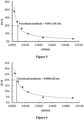
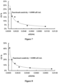
- the kit developed based on the antibody of the present application can achieve a fourth-generation functional sensitivity of ⁇ 0.002 ⁇ IU/mL.
- the present application provides a TSH detection kit for detecting the presence or level of TSH or a mutant thereof in a sample.
- the detection sensitivity of the kit for TSH is ⁇ 0.0015 ⁇ IU/mL, preferably ⁇ 0.001 ⁇ IU/mL.
- the TSH or a mutant thereof is selected from the group consisting of TSH dimer, ⁇ subunit of TSH and/or R55G mutant of TSH.
- the kit comprises a first reagent and a second reagent, wherein the first reagent comprises magnetic beads coated with a first antibody capable of recognizing the TSH or a mutant thereof; and the second reagent comprises a second antibody connected with a chemiluminescent label, wherein a sandwich complex of the first antibody-an antigen-the second antibody can be formed after the sample is mixed with the first reagent and the second reagent, and the antigen is the TSH or mutant thereof.
- the label is well known to those skilled in the art, and may be, for example, an enzyme such as oxidase, microperoxidase, horseradish peroxidase, alkaline phosphatase (ALP), ⁇ -galactosidase, glucose oxidase, and glucose 6-phosphate dehydrogenase; a fluorescent substance such as fluorescein isothiocyanate, tetramethylrhodamine isothiocyanate, fluorescein, rhodamine, europium, and green fluorescent protein; a chemiluminescence such as luminol, isoluminol, phenanthridinium, and acridinium ester.
- an enzyme such as oxidase, microperoxidase, horseradish peroxidase, alkaline phosphatase (ALP), ⁇ -galactosidase, glucose oxidase, and glucose 6-phosphate dehydrogenase
- the first antibody and/or the second antibody is selected from an antibody or antigen-binding fragment thereof that is that specifically binds to TSH or a mutant thereof, comprising:
- the first antibody and/or the second antibody comprises:
- the first antibody is an antibody or antigen-binding fragment thereof having a heavy chain variable region comprising the following three CDRs: CDR-H1 having an amino acid sequence selected from SEQ ID NO: 31 and variants thereof, CDR-H2 having an amino acid sequence selected from SEQ ID NO: 32 and variants thereof, and CDR-H3 having an amino acid sequence selected from SEQ ID NO: 33 and variants thereof; and, a light chain variable region comprising the following three CDRs: CDR-L1 having an amino acid sequence selected from SEQ ID NO: 34 and variants thereof, CDR-L2 having an amino acid sequence selected from SEQ ID NO: 35 and variants thereof, and CDR-L3 having an amino acid sequence selected from SEQ ID NO: 36 and variants thereof; and, the second antibody is an antibody or antigen-binding fragment thereof having a heavy chain variable region comprising the following three CDRs: CDR-H1 having an amino acid sequence selected from SEQ ID NO: 49 and variants thereof, CDR-H2 having an amino acid sequence selected from
- the first antibody is an antibody or antigen-binding fragment thereof having a heavy chain variable region comprising the following three CDRs: CDR-H1 having an amino acid sequence selected from SEQ ID NO: 37 and variants thereof, CDR-H2 having an amino acid sequence selected from SEQ ID NO: 38 and variants thereof, and CDR-H3 having an amino acid sequence selected from SEQ ID NO: 39 and variants thereof; and, a light chain variable region comprising the following three CDRs: CDR-L1 having an amino acid sequence selected from SEQ ID NO: 40 and variants thereof, CDR-L2 having an amino acid sequence selected from SEQ ID NO: 41 and variants thereof, and CDR-L3 having an amino acid sequence selected from SEQ ID NO: 42 and variants thereof; and, the second antibody is an antibody or antigen-binding fragment thereof having a heavy chain variable region comprising the following three CDRs: CDR-H1 having an amino acid sequence selected from SEQ ID NO: 43 and variants thereof, CDR-H2 having an amino acid sequence selected from
- the first antibody is an antibody or antigen-binding fragment thereof having a heavy chain variable region comprising the following three CDRs: CDR-H1 having an amino acid sequence selected from SEQ ID NO: 25 and variants thereof, CDR-H2 having an amino acid sequence selected from SEQ ID NO: 26 and variants thereof, and CDR-L3 having an amino acid sequence selected from SEQ ID NO: 27 and variants thereof; and, a light chain variable region comprising the following three CDRs: CDR-L1 having an amino acid sequence selected from SEQ ID NO: 28 and variants thereof, CDR-L2 having an amino acid sequence selected from SEQ ID NO: 29 and variants thereof, and CDR-L3 having an amino acid sequence selected from SEQ ID NO: 30 and variants thereof; and, the second antibody is an antibody or antigen-binding fragment thereof having a heavy chain variable region comprising the following three CDRs: CDR-H1 having an amino acid sequence selected from SEQ ID NO: 49 and variants thereof, CDR-H2 having an amino acid sequence selected from
- the first antibody is an antibody or antigen-binding fragment thereof having a heavy chain variable region comprising the following three CDRs: CDR-H1 having an amino acid sequence selected from SEQ ID NO: 19 and variants thereof, CDR-H2 having an amino acid sequence selected from SEQ ID NO: 20 and variants thereof, and CDR-H3 having an amino acid sequence selected from SEQ ID NO: 21 and variants thereof; and, a light chain variable region comprising the following three CDRs: CDR-L1 having an amino acid sequence selected from SEQ ID NO: 22 and variants thereof, CDR-L2 having an amino acid sequence selected from SEQ ID NO: 23 and variants thereof, and CDR-L3 having an amino acid sequence selected from SEQ ID NO: 24 and variants thereof; and, the second antibody is an antibody or antigen-binding fragment thereof having a heavy chain variable region comprising the following three CDRs: CDR-H1 having an amino acid sequence selected from SEQ ID NO: 61 and variants thereof, CDR-H2 having an amino acid sequence selected
- the first antibody is an antibody or antigen-binding fragment thereof having a heavy chain variable region comprising the following three CDRs: CDR-H1 having an amino acid sequence selected from SEQ ID NO: 19 and variants thereof, CDR-H2 having an amino acid sequence selected from SEQ ID NO: 20 and variants thereof, and CDR-H3 with an amino acid sequence selected from SEQ ID NO: 21 and variants thereof; and, a light chain variable region comprising the following three CDRs: CDR-L1 having an amino acid sequence selected from SEQ ID NO: 22 and variants thereof, CDR-L2 having an amino acid sequence selected from SEQ ID NO: ID NO: 23 and variants thereof, and CDR-L3 having an amino acid sequence selected from SEQ ID NO: 24 and variants thereof; and, the second antibody is an antibody or antigen-binding fragment thereof having a heavy chain variable region comprising the following three CDRs: CDR-H1 having an amino acid sequence selected from SEQ ID NO: 43 and variants thereof, CDR-H2
- composition or TSH detection kit which comprises the antibody or antigen-binding fragment thereof as described in any of the foregoing.
- composition or TSH detection kit comprises:
- composition or TSH detection kit in the composition or TSH detection kit,
- composition or TSH detection kit in the composition or TSH detection kit:
- composition or TSH detection kit comprises:
- composition or TSH detection kit comprises:
- the first antibody is a capture antibody and the second antibody is a detection antibody.
- the immunoassay kit further comprises a solid phase carrier.
- the solid phase carrier is selected from the group consisting of magnetic particles or microtiter plates (e.g., microtiter plates or ELISA plates).
- the first antibody is coated on the surface of the solid phase carrier, preferably fixed on the surface of the magnetic particles.
- the second antibody has a detectable label.
- the label is selected from the group consisting of fluorescein, chemiluminescent label (e.g., acridinium ester compound), enzyme (e.g., horseradish peroxidase, alkaline phosphatase), radioisotope, biotin and colloidal gold.
- fluorescein e.g., acridinium ester compound
- enzyme e.g., horseradish peroxidase, alkaline phosphatase
- radioisotope e.g., biotin and colloidal gold.
- the second antibody is labeled with alkaline phosphatase.
- the kit further comprises a first antibody diluent and a second antibody diluent, wherein,
- the first antibody diluent and the first antibody are placed in the same or different formulation units.
- the second antibody diluent and the second antibody are placed in the same or different formulation units.
- the buffer solution is selected from the group consisting of Tris, Hepes and PBS buffer solutions.
- the inorganic salt is selected from the group consisting of NaCl, KCl, CaCl 2 , MgCl 2 and ZnCl 2 .
- the preservative is selected from the group consisting of Proclin300, PC-300, BND (e.g., BND-10 or BND-99) and BIT (e.g., BIT-10).
- the surfactant is selected from the group consisting of Tween-20, TritonX-100 and S9.
- the stabilizer is selected from the group consisting of calf serum, bovine serum albumin and gelatin.
- the immunoassay kit further comprises one or more of the following: a TSH calibrator, a substrate of the detection label, a washing solution, a stop solution and an instruction for use.
- the immunoassay kit is used to detect the presence or level of TSH or a mutant thereof in a sample.
- the TSH or mutant thereof is selected from the group consisting of TSH dimer, TSH ⁇ -subunit or TSH R55G mutant.
- the sample is from a human, such as a human body fluid (e.g., blood, serum or plasma).
- a human body fluid e.g., blood, serum or plasma.
- the immunoassay kit has a detection sensitivity of ⁇ 0.0015 ⁇ IU/mL, preferably ⁇ 0.001 ⁇ IU/mL, for TSH or mutant thereof.
- the present application further provides a method for detecting the presence or level of TSH or a mutant thereof in a sample, which comprises the step of using the antibody or antigen-binding fragment thereof or the immunoassay kit as described in any one of the above items for detection.
- the method comprises the following steps:
- At least one of the two or more kinds of the antibody or antigen-binding fragment thereof is a capture antibody, and at least another is a detection antibody.
- the capture antibody is the first antibody as defined in any one of the preceding items.
- the detection antibody is the second antibody as defined in any one of the preceding items.
- the method comprises the following steps:
- step (1) and step (2) can be performed separately or simultaneously.
- the method is an ELISA immunoassay, such as a double antibody sandwich ELISA immunoassay, preferably a one-step double antibody sandwich ELISA immunoassay.
- the present application provides a use of the antibody or antigen-binding fragment thereof in the manufacture of a kit, or a use of the immunoassay kit as described in any one of the preceding items, wherein the kit is used for:
- the present application provides a method, which comprises steps of detecting a sample from a subject using the antibody or antigen-binding fragment thereof or the kit described in any one of the preceding items, or detecting a sample from a subject using the method as described in any one of the preceding items, and quantifying the TSH or mutant thereof in the sample, wherein the method is used for:
- the thyroid-related disease is hyperthyroidism or thyroid cancer.
- the sample is from a human, such as a human body fluid (e.g., blood, serum, or plasma).
- a human body fluid e.g., blood, serum, or plasma.
- the photon counting method of the existing photometric device can accurately detect the number of photons in the case of Figure 14 , but in the case of Figure 15 , the accumulated multiple electrical pulses will be identified as one electrical pulse, resulting in a low counting result in the higher light intensity segment, and the upper limit of the linear range of the photometric device is insufficient.
- the upper limit of the linear range of the existing photometric device is only 30 million photons per second.
- the measured luminescence value of the sample e.g., a high-value sample of TSH
- the test result is unreliable, and the sample usually needs to be diluted and retested, but this seriously affects the measurement speed, resulting in a longer reporting time for the sample.
- the immunoassay kit of the present application can achieve ultra-sensitive detection of samples.
- one sample measurement cannot simultaneously meet the ultra-sensitive detection of low-value samples and the detection of high-value samples.
- the present application further provides a sample analyzer with a high linear range, which can detect photosignals in a high linear range and can be adapted to the kit of the present application, so that the sample can be detected ultra-sensitively and accurately in one detection.
- the processing component is further configured to: output the first photon counting result as the final photon counting result when the first photon counting result is lower than a first threshold; output the second photon counting result as the final photon counting result when the first photon counting result is higher than a second threshold, wherein the second threshold is greater than or equal to the first threshold.
- the second photon counting module comprises an integration circuit and a second counting circuit which are electrically connected to each other, the integration circuit is configured to integrate the electrical signal within a predetermined time period to obtain a DC component signal, the parameter for characterizing photon number comprises a parameter related to the DC component signal, and the second counting circuit is configured to calculate the second photon counting result based on the parameter related to the DC component signal and the preset calibration function.
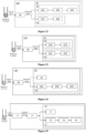
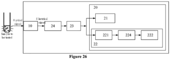
- an embodiment of the present application further provides a sample analyzer 1, and the sample analyzer 1 comprises a loading device 200, a reaction incubation device 300, and a photometric device 100 according to any of the above embodiments.
- the loading device 200 is configured to load a sample to be tested, such as a blood sample, and a reagent required for chemiluminescent reaction into a reaction container.
- the reaction incubation device 300 is configured to provide a reaction and incubation site for the reaction container containing the sample to be tested and the reagent required for chemiluminescent reaction, so that the sample to be tested in the reaction container and the reagent required for chemiluminescent reaction form a sample liquid to be tested.
- the photometric device 100 is configured to perform photometry on the sample liquid to be tested in the reaction container that has completed the reaction and incubation.
- the sample analyzer 1 is configured as a chemiluminescent immunoassay analyzer for analyzing a sample, such as a blood sample.
- the loading device 200 comprises a sample supply unit 210 and a reagent supply unit 220, the sample supply unit 210 is configured to pipette the sample to be tested and supply it to the reaction container, and the reagent supply unit 220 is configured to pipette an immune reagent (e.g., a labeled antigen or antibody) and a magnetic bead reagent and supply them to the reaction container.
- an immune reagent e.g., a labeled antigen or antibody
- the chemiluminescent immunoassay analyzer further comprises a magnetic separation device 400, a substrate supply device (not shown) and a transport device 500, the magnetic separation device 400 is configured to perform a magnetic separation operation on the reaction container containing the sample to be tested, the immune reagent and the magnetic bead reagent, the substrate supply device is configured to supply a luminescent substrate to the reaction container that has completed the magnetic separation operation, and the transport device 500 is configured to transport the reaction container that has completed the magnetic separation operation from the magnetic separation device 500 to the reaction incubation device 300. After incubation, the reaction container is transported to the photometric device 100 for photometry.
- the reagent supply unit 220 comprises a reagent tray 221 and a reagent pipette 222
- the reagent tray 221 is used to place reagent bottles containing the immune reagent and the magnetic bead reagent and has the function of refrigerating the reagent bottles.
- the reagent pipette 222 is used to pipette a reagent from a reagent bottle at a reagent pipetting position and discharge the pipetted reagent into the reaction container at a sample loading position.
- the chemiluminescent immunoassay analyzer further comprises a mixer 700 disposed between a reaction tray and a magnetic separation tray, the mixer is configured to perform mixing in the reaction container into which the reagent and sample have been added.
- the mixer can be configured, for example, as a vortex mixer for non-contact mixing, which can effectively avoid cross contamination.
- the chemiluminescent immunoassay analyzer further comprises a first manipulator 600 for transporting the reaction container, the first manipulator is configured to be able to move in three dimensions and to be able to clamp the reaction container.
- the first manipulator is configured to be able to load a new reaction container into the sample loading position so that the sample pipette can inject the sample.
- the first manipulator is further configured to be able to discard an empty reaction cup.
- the first manipulator is further configured to be able to transport the reaction container between the sample loading position, the reaction incubation device 300, and a dilution position.
- the upper limit of the linear range of photosignal detected by the photometric device 100 is greater than or equal to 10 8 (i.e., 100 million) photons/second; under the condition that the upper limit of the linear range of the photometric device 100 is greater than or equal to 10 8 photons/second, the upper limit of the detection range of the aforementioned kit can reach 100 ⁇ IU/mL, which can achieve accurate results for high-value samples in one measurement; preferably, the linear range of photosignal detected by the photometric device 100 is greater than or equal to 120 million photons/second; further preferably, the linear range of photosignal detected by the photometric device 100 is greater than or equal to 150 million photons/second; more preferably, the linear range of photosignal detected by the photometric device 100 is greater than or equal to 200 million photons/second, and the linear detection range refers to the linear detection range of a single detection, that is, the linear detection range that can be achieved in one detection of a sample.
- the photometric device 100 comprises a receiving component 10 and a processing component 20.
- the processing component 20 is configured to be electrically connected to the receiving component 10 so as to receive the electrical signal from the receiving component 10, and to process the received electrical signal to obtain the number of photons.
- two photon counting modules are used to count photons simultaneously, wherein the first photon counting module adopts a first counting method for weak light segments to obtain a first photon counting result, and the second photon counting module adopts a second counting method for strong light segments to obtain a second photon counting result, and finally the first photon counting result and the second photon counting result are integrated and a final counting result is output.
- This can greatly expand the linear photometric range of the photometric device.
- curve S1 represents the photometric linear characteristics of the photometric device of the prior art
- curve S2 represents the photometric linear characteristics of the photometric device according to the present application.
- the present application can realize the expansion of the linear photometric range of the photometric device from the original linear photometric range of 0 to 30 million photons/second (i.e., 0 to 30 million photons per second) to 0 to 200 million photons/second (i.e., 0 to 200 million photons per second).
- the processing component 20 is further configured to output a final photon counting result according to one of the following embodiments.
- the processing component can further be configured to: calculate a final photon counting result according to the first counting photon result and the second photon counting result by a preset fusion function when the first photon counting result is between the first threshold and the second threshold, wherein the second threshold is greater than the first threshold.
- the preset fusion function may be, for example, a function for obtaining an average value or a function for obtaining a weighted average value.
- multiple threshold intervals may be set for the first photon counting module.
- the processing component 20 can also be configured to: when the first photon counting result is in different threshold intervals, use different fusion functions to calculate the first photon counting result and the second photon counting result, and output the calculation result as the final photon counting result.
- the fusion function can be, for example, a function for obtaining a weighted average value, and the fusion functions for different threshold intervals have different weights for the first photon counting result and the second photon counting result.
- the setting of at least two thresholds can enable the photometric device to meet the linearity or precision requirements in long-term use.
- the first counting method may be a pulse recognition method for calculating the number of photons by identifying the electrical pulses caused by the photons entering the receiving component, while the second counting method does not calculate the number of photons by identifying the electrical pulses caused by the photons entering the receiving component, but estimates the number of photons by processing the electrical signal into a parameter that can characterize the number of photons (i.e., the number of electrical pulses caused by the photons entering the receiving component).
- the pulse recognition method can be understood as a method for calculating the number of photons by identifying the pulses in the electrical signal, that is, when the peak value of the pulse identified from the electrical signal is greater than the threshold, it is considered that the photon is identified.
- the first photon counting module 21 may comprises a level identification circuit 211, a shaping and frequency division circuit 212, and a first counting circuit 213.
- the level identification circuit 211 is configured to convert an electrical signal into a square wave signal
- the shaping and frequency division circuit 212 is configured to perform frequency division and shaping on the square wave signal
- the first counting circuit 213 is configured to count the signal output by the shaping and frequency division circuit 212, that is, to identify the number of pulses, to obtain a first photon counting result.
- the first photon counting module 21 according to this embodiment can achieve a linear photometric range of 0 to 30 million photons/second.
- the first photon counting module 21 may comprise at least one first AD conversion circuit 214 and a first counting circuit 213.
- the first AD conversion circuit 214 is configured to collect electrical signals at a sampling frequency greater than 1 GHz/s and convert the collected electrical signals into digital signals and output them to the first counting circuit 213, the first counting circuit 213 is configured to analyze the collected digital signals to identify the number of pulses, and then obtain the first photon counting result.
- the first counting circuit 213 can more accurately identify the number of pulses in the electrical signals, and then obtain a more accurate first photon counting result.
- the first counting circuit 213 can also be configured to correct the first photon counting result by a Poisson distribution compensation algorithm. Thus, a more accurate photon counting result can be obtained.
- the first counting circuit 213 may comprise an FPGA chip and surrounding circuits thereof.
- the preset calibration function is a conversion function that converts the value of the parameter for characterizing photon number into a photon counting value, which represents an mapping relationship between the parameter for characterizing photon number and the photon counting result (or counting value).
- a reference light source such as an LED light
- the first photon counting module of the photometric device are used to calibrate the calibration function used by the second photon counting module of the photometric device, that is, the calibration method of the conversion function of the parameter for characterizing photon number and the photon counting result is carried out as follows:
- a reference light source and a reference photometric device can also be used to calibrate the conversion function of the parameter for characterizing photon number and the photon counting result, which is perform in the following manner: an input value of the reference light source is controlled, the photometric device to be calibrated according to the present application is used to test the value of the parameter for characterizing photon number of pulses and from the weak light segment to the strong light segment of the reference light source, and the reference photometric device is used at the same time to test the counting result of the reference light source from the weak light segment to the strong light segment; according to the linear function relationship between the input value of the reference light source and the counting result of the reference photometric device, the theoretical photon counting result corresponding to the input value of the reference light source is deduced; and under the premise that the input value of the reference light source is fixed, the corresponding value of the parameter for characterizing photon number and the theoretical photon counting result are one-to-one corresponding, and a set of values of the parameter for characterizing photon number in the strong
- the calibration function can be pre-stored in the photometric device, especially in the second photon counting module 22.
- the second photon counting module 22 that uses the parameter for characterizing photon number and the predetermined calibration function to obtain the photon counting result can achieve a linear photometric range of 10 million to 200 million photons/second, as shown in Figure 21 , wherein S3 represents the photometric linear characteristics of the first photon counting module according to the embodiment shown in Figure 19 , and S4 represents the photometric linear characteristics of the second photon counting module according to the embodiment of the present application.
- the combination of the first photon counting module 21 and the second photon counting module 22 can achieve a linear photometric range of at least 0 to 200 million photons/second.
- the second photon counting module 22 can be configured to process the electrical signal, for example, by integration processing to obtain a DC component signal, and the parameter for characterizing photon number comprises a parameter related to the DC component signal, such as the DC component signal itself per unit time.
- the second photon counting module 22 is further configured to calculate the second photon counting result according to the parameter related to the DC component signal and a preset calibration function.
- the second photon counting module 22 is configured to integrate the electrical signal within a predetermined time period to obtain an integration result, such as a DC component signal, and the parameter for characterizing photon number comprises a parameter related to the integration result, such as an integration result per unit time.
- the second photon counting module 22 is further configured to calculate the second photon counting result according to the parameter related to the integration result and a preset calibration function.
- the second photon counting module 22 comprises an integration circuit 221 and a second counting circuit 222 electrically connected to each other.
- the integration circuit 221 is configured to integrate the electrical signal within a predetermined time period to obtain a DC component signal
- the parameter for characterizing photon number comprises a parameter related to the DC component signal.
- the second counting circuit 222 is configured to calculate the second photon counting result according to the parameter related to the DC component signal and a preset calibration function.
- an AD conversion circuit 224 is also provided between the integration circuit 221 and the second counting circuit 222, and the AD conversion circuit 224 is configured to convert the DC component signal output by the integration circuit 221 into a digital signal.
- the sampling frequency of the AD conversion circuit 224 can be set to be less than 1 MHz/s, for example, hundreds of kHz/s.
- the second counting circuit 222 can be configured to integrate or sum the digital signal within a predetermined time period to obtain a parameter related to the integration result or the summation result as the parameter for characterizing photon number.
- the processing component 20 ma further comprise a pre-amplifier circuit 23 electrically connected to the receiving component 10, the first photon counting module 21, and the second photon counting module 22.
- the pre-amplifier circuit 23 is configured to amplify the electrical signal output by the receiving component 10 and transmit it to the first photon counting module 21 and the second photon counting module 22.
- the pre-amplifier circuit 23 can amplify the electrical signal output by the receiving component 10 by 60 to 90 times, especially by 70 to 80 times.
- the processing component 20 may comprise a first pre-amplifier circuit electrically connected to the receiving component 10 and the first photon counting module 21 and/or a second pre-amplifier circuit electrically connected to the receiving component 10 and the second photon counting module 22.
- the first pre-amplifier circuit is configured to amplify the electrical signal output by the receiving component 10 and transmit it to the first photon counting module 22
- the second pre-amplifier circuit is configured to amplify the electrical signal output by the receiving component 10 and transmit it to the second photon counting module 22.
- the processing component 20 may comprise a resistor 24 disposed between the receiving component 10 and the pre-amplifier circuit 23, which is used to convert the electrical signal in the form of current signal output by the receiving component 10 into a voltage signal so that the pre-amplifier circuit 23 amplifies the voltage signal.
- the photometric device 100 comprises a receiving component 10 and a switchable light attenuation component (not shown) arranged in front of the receiving component, the light attenuation component is, for example, a filter; at the beginning of the measurement, the photosignal that has not been attenuated by the light attenuation component is received by the receiving component 10, and a weak photosignal can be measured at this time; then the light attenuation component is switched so that the receiving component 10 receives the photosignal that has been attenuated by the light attenuation component, even if it is a strong photosignal at this time, the strong photosignal becomes weak photosignal after light attenuation and is detected by the receiving component 10.
- the linear range of the photometric device can also be extended by the switchable light attenuation component.
- the term "specific binding” refers to a non-random binding reaction between two molecules (i.e., a binding molecule and a target molecule), such as a reaction between an antibody and an antigen to which it is directed.
- the binding affinity between two molecules can be described by a K D value.
- the K D value refers to the dissociation constant obtained by the ratio of kd (dissociation rate of a specific binding molecule-target molecule interaction; also known as koff) to ka (association rate of a specific binding molecule-target molecule interaction; also known as kon), or refers to kd/ka expressed in molar concentration (M).
- M molar concentration
- an antibody that specifically binds to an antigen refers to an antibody that binds to the antigen with a K D of less than about 10 -5 M, such as less than about 10 -6 M, 10 -7 M, 10 -8 M, 10 -9 M or 10 -10 M or less.
- the K D value can be determined by methods well known in the art, such as using surface plasmon resonance (SPR) in a BIACORE instrument.
- immunological detection refers to a determination made using the specific interaction/binding affinity between an antigen and an antibody, which can generally be used to detect the presence or level of a specific antigen or antibody in a sample.
- immunological detection is well known to those skilled in the art, including but not limited to enzyme immunoassay (EIA), chemiluminescent immunoassay (CLIA), radioimmunoassay (RIA), fluorescent immunoassay (FIA), Western blotting, immunoturbidimetry, surface plasmon resonance, etc.
- Enzyme-linked immunosorbent assay comprises binding an antigen or antibody to the surface of a solid phase carrier, and cross-linking an antigen- or antibody-related substance with an enzyme to form an enzyme conjugate, which, on the one hand, maintains the immune characteristics of binding to the corresponding antigen or antibody, and on the other hand, has enzyme activity.
- an enzyme conjugate binds to the corresponding antigen or antibody, an enzyme-labeled antigen-antibody complex is formed.
- the enzyme on the complex encounters the corresponding substrate, it can catalyze the hydrolysis, oxidation or reduction of the product, thereby producing a colored substance.
- competitive ELISA has a variety of modes for detecting antibodies.
- the sample and enzyme-labeled antibody can compete with the solid phase antigen for binding, or the sample and antigen are added to the solid phase antibody for competitive binding, and then the enzyme-labeled antibody is added after washing to react with the antigen bound to the solid phase.
- immunological detection see, for example, Fundamental Immunology, Ch. 7 Paul, W., ed., 2nd edition, Raven Press, N.Y. (1989 ).
- antibody and “monoclonal antibody” are used interchangeably and refer to an immunoglobulin molecule that is usually composed of two pairs of polypeptide chains (each pair having a light chain (LC) and a heavy chain (HC)).
- Antibody light chains can be classified as ⁇ (kappa) and ⁇ (lambda) light chains.
- Heavy chains can be classified as ⁇ , ⁇ , ⁇ , ⁇ or ⁇ , and define the isotypes of antibody as IgM, IgD, IgG, IgA and IgE, respectively.
- Each heavy chain consists of a heavy chain variable region (VH) and a heavy chain constant region (CH).
- Each light chain consists of a light chain variable region (VL) and a light chain constant region (CL).
- the light chain constant region consists of one domain, CL.
- the constant domain is not directly involved in the binding of antibody to antigen, but exhibits a variety of effector functions, such as mediating the binding of immunoglobulin to host tissues or factors, including various cells of the immune system (e.g., effector cells) and the first component (C1q) of the classical complement system.
- the VH and VL regions can also be subdivided into regions of high variability, called complementarity determining regions (CDRs), interspersed with more conservative regions called framework regions (FRs).
- CDRs complementarity determining regions
- FRs framework regions
- Each V H and V L consists of three CDRs and four FRs arranged from the amino terminus to the carboxyl terminus in the following order: FR1, CDR1, FR2, CDR2, FR3, CDR3, FR4.
- the variable regions (VH and VL) of each heavy chain/light chain pair form antigen binding sites, respectively.
- the distribution of amino acids in each region or domain can follow the definitions of Kabat, Sequences of Proteins of Immunological Interest (National Institutes of Health, Bethesda, Md. (1987 and 1991 )), or Chothia & Lesk (1987) J. Mol. Biol. 196:901-917 ; Chothia et al. (1989) Nature 342:878-883 .
- CDR complementarity determining region
- CDR1 complementarity determining region
- CDR2 complementarity determining region
- CDR3 complementarity determining region
- the precise boundaries of these CDRs can be defined according to various numbering systems known in the art, such as the Kabat numbering system ( Kabat et al., Sequences of Proteins of Immunological Interest, 5th Ed. Public Health Service, National Institutes of Health, Bethesda, Md., 1991 ), the Chothia numbering system ( Chothia & Lesk (1987) J. Mol. Biol. 196:901-917 ; Chothia et al.
- the CDRs contained in the antibody or antigen-binding fragment thereof of the present application can be determined according to various numbering systems known in the art. In certain embodiments, the CDRs contained in the antibody or antigen-binding fragment thereof of the present application are determined by the Kabat, Chothia or IMGT numbering systems.
- framework region or "FR” residues refers to those amino acid residues in an antibody variable region other than the CDR residues defined above.
- antibody is not limited to any particular method for producing antibody.
- it includes recombinant antibody, monoclonal antibody, and polyclonal antibody.
- the antibody can be an antibody of different isotypes, for example, an IgG (e.g., IgG1, IgG2, IgG3, or IgG4 subtype), an IgA1, an IgA2, an IgD, an IgE, or an IgM antibody.
- antigen-binding fragment refers to a polypeptide comprising a fragment of a full-length antibody that retains the ability to specifically bind to the same antigen to which the full-length antibody binds and/or competes with the full-length antibody for specific binding to the antigen, which is also referred to as an "antigen-binding portion.”
- Antigen-binding fragments of antibodies can be produced by recombinant DNA techniques or by enzymatic or chemical cleavage of intact antibodies.
- Non-limiting examples of antigen-binding fragments include Fab, Fab', F(ab') 2 , Fd, Fv, complementarity determining region (CDR) fragments, scFv, diabodies, single domain antibodies, chimeric antibodies, linear antibodies, and polypeptides that contain at least a portion of antibody sufficient to confer specific antigen binding ability on the polypeptide.
- Engineered antibody variants are reviewed by Holliger et al., 2005; Nat Biotechnol, 23: 1126-1136 .
- the term “Fd” refers to an antibody fragment consisting of VH and CH1 domains
- the term “dAb fragment” refers to an antibody fragment consisting of a VH domain ( Ward et al., Nature 341:544 546 (1989 ))
- the term “Fab fragment” refers to an antibody fragment consisting of VL, VH, CL and CH1 domains
- the term “F(ab') 2 fragment” refers to an antibody fragment comprising two Fab fragments connected by a disulfide bridge on the hinge regions
- the term “Fab' fragment” refers to a fragment obtained after reducing the disulfide bond connecting the two heavy chain fragments in the F(ab') 2 fragment, which consists of a complete light chain and a heavy chain Fd fragment (consisting of VH and CH1 domains).
- the term "Fv” refers to an antibody fragment consisting of the VL and VH domains of a single arm of antibody.
- the Fv fragment is generally considered to be the smallest antibody fragment that can form a complete antigen binding site. It is generally believed that the six CDRs confer antigen binding specificity to the antibody. However, even a single variable region (e.g., an Fd fragment, which contains only three CDRs specific for an antigen) can recognize and bind to the antigen, although its affinity may be lower than that of the complete binding site.
- scFv refers to a single polypeptide chain comprising VL and VH domains, wherein the VL and VH are connected by a linker (see, for example, Bird et al., Science 242:423-426 (1988 ); Huston et al., Proc. Natl. Acad. Sci. USA 85:5879-5883 (1988 ); and Pluckthun, The Pharmacology of Monoclonal Antibodies, Vol. 113, Roseburg and Moore, eds., Springer-Verlag, New York, pp. 269-315 (1994 )).
- Such scFv molecules may have the general structure: NH 2 -VL-linker-VH-COOH or NH 2 -VH-linker-VL-COOH.
- Suitable prior art linkers consist of repeated GGGGS amino acid sequences or variants thereof.
- a linker having the amino acid sequence (GGGGS) 4 may be used, but variants thereof may also be used ( Holliger et al. (1993), Proc. Natl. Acad. Sci. USA 90: 6444-6448 ).
- Other linkers that can be used in the present application are described by Alfthan et al. (1995), Protein Eng. 8:725-731 , Choi et al. (2001), Eur. J. Immunol.
- a disulfide bond may also exist between the VH and VL of the scFv.
- the term "diabody” means that its VH and VL domains are expressed on a single polypeptide chain, but using a linker that is too short to allow pairing between the two domains of the same chain, thereby forcing the domains to pair with the complementary domains of another chain and create two antigen binding sites (see, for example, Holliger P. et al., Proc. Natl. Acad. Sci. USA 90:6444-6448 (1993 ), and Poljak R. J. et al., Structure 2:1121-1123 (1994 )).
- single-domain antibody has the meaning commonly understood by those skilled in the art, which refers to an antibody fragment composed of a single monomer variable antibody domain (e.g., a single heavy chain variable region) that retains the ability to specifically bind to the same antigen to which the full-length antibody binds.
- Single-domain antibody is also called nanobody.
- Each of the above antibody fragments retains the ability to specifically bind to the same antigen to which the full-length antibody binds, and/or competes with the full-length antibody for specific binding to the antigen.
- Antigen-binding fragment of antibody (e.g., the above antibody fragment) can be obtained from a given antibody (e.g., the antibody provided by the present application) using conventional techniques known to those skilled in the art (e.g., recombinant DNA technology or enzymatic or chemical cleavage methods), and the antibody antigen-binding fragment can be screened for specificity in the same manner as for the intact antibody.
- antibody includes not only an intact antibody, but also an antigen-binding fragment of the antibody.
- chimeric antibody refers to an antibody in which a portion of its light chain or/and heavy chain is derived from one antibody (which may be derived from a particular species or belong to a particular antibody class or subclass), and another portion of the light chain or/and heavy chain is derived from another antibody (which may be derived from the same or different species or belong to the same or different antibody class or subclass), but in any case, it still retains the activity to bind to the target antigen ( U.S.P 4,816,567 to Cabilly et al. ; Morrison et al., Proc. Natl. Acad. Sci. USA, 81:6851 6855 (1984 )).
- chimeric antibody may include such an antibody (e.g., a human-mouse chimeric antibody) in which the heavy chain and light chain variable regions of the antibody are derived from a first antibody (e.g., a murine antibody), and the heavy chain and light chain variable regions of the antibody are derived from a second antibody (e.g., a human antibody).
- a first antibody e.g., a murine antibody
- a second antibody e.g., a human antibody
- humanized antibody refers to a genetically engineered non-human antibody whose amino acid sequence is modified to increase homology with the amino acid sequence of a human antibody.
- all or part of the CDR regions of a humanized antibody are derived from a non-human antibody (donor antibody), and all or part of the non-CDR regions (e.g., variable region FR and/or constant region) are derived from a human immunoglobulin (recipient antibody).
- Humanized antibodies generally retain the expected properties of the donor antibody, including, but not limited to, antigen specificity, affinity, reactivity, etc.
- the donor antibody may be a mouse, rat, rabbit, or non-human primate (e.g., cynomolgus monkey) antibody having the expected properties (e.g., antigen specificity, affinity, reactivity, etc.).
- the chimeric antibody or humanized antibody of the present application can be prepared according to the sequence of the mouse monoclonal antibody prepared above.
- DNA encoding the heavy and light chains can be obtained from the target mouse hybridoma and engineered to contain non-mouse (e.g., human) immunoglobulin sequences using standard molecular biology techniques.
- the DNA encoding VH can be operably connected to another DNA molecule encoding the heavy chain constant region to obtain a full-length heavy chain gene.
- the sequence of the human heavy chain constant region gene is known in the art (see, for example, Kabat, E.A. et al. (1991) Sequences of Proteins of Immunological Interest, Fifth Edition, U.S. Department of Health and Human Services, NIH Publication No.91-3242 ), and DNA fragments containing these regions can be obtained by standard PCR amplification.
- the heavy chain constant region can be IgG1, IgG2, IgG3, IgG4, IgA, IgE, IgM or IgD constant region, but IgG1 or IgG4 constant region is generally preferred.
- the DNA encoding VL can be operably connected to another DNA molecule encoding the light chain constant region CL to obtain a full-length light chain gene (as well as a Fab light chain gene).
- the sequence of human light chain constant region gene is known in the art (see, for example, Kabat, E.A. et al. (1991), Sequences of Proteins of Immunological Interest, Fifth Edition, U.S. Department of Health and Human Services, NIH Publication No. 91-3242 ), and DNA fragments containing these regions can be obtained by standard PCR amplification.
- the light chain constant region can be a ⁇ or ⁇ constant region, but a ⁇ constant region is generally preferred.
- mouse CDR regions can be inserted into human framework sequences using methods known in the art (see Winter's U.S. Patent No. 5,225,539 ; Queen et al.'s U.S. Patent No.os.5,530,101 ; 5,585,089 ; 5,693,762 and 6,180,370 ; and Lo, Benny, K.C., editor, in Antibody Engineering: Methods and Protocols, volume 248, Humana Press, New Jersey, 2004 ).
- the term "vector” refers to a nucleic acid vehicle into which a polynucleotide can be inserted.
- a vector can express a protein encoded by the inserted polynucleotide
- the vector is called an expression vector.
- the vector can be introduced into a host cell by transformation, transduction or transfection so that the genetic material elements it carries are expressed in the host cell.
- Vectors are well known to those skilled in the art, and include but are not limited to: plasmids; phagemids; cosmids; artificial chromosomes, such as yeast artificial chromosome (YAC), bacterial artificial chromosome (BAC) or P1-derived artificial chromosome (PAC); bacteriophages, such as ⁇ phage or M13 phage, as well as animal viruses, etc.
- artificial chromosomes such as yeast artificial chromosome (YAC), bacterial artificial chromosome (BAC) or P1-derived artificial chromosome (PAC)
- bacteriophages such as ⁇ phage or M13 phage, as well as animal viruses, etc.
- Animal viruses that can be used as vectors include, but are not limited to, retrovirus (including lentivirus), adenovirus, adeno-associated virus, herpes virus (e.g., herpes simplex virus), poxvirus, baculovirus, papillomavirus, papovavirus (e.g., SV40).
- retrovirus including lentivirus
- adenovirus adeno-associated virus
- herpes virus e.g., herpes simplex virus
- poxvirus baculovirus
- papillomavirus papovavirus
- papovavirus e.g., SV40
- a vector can contain a variety of elements that control expression, including, but not limited to, promoter sequence, transcription initiation sequence, enhancer sequence, selection element, and reporter gene.
- the vector may also contain a replication origin.
- the term "host cell” refers to a cell that can be used to introduce a vector, including, but not limited to, prokaryotic cell such as Escherichia coli or Bacillus subtilis, fungal cell such as yeast cell or Aspergillus, insect cell such as S2 Drosophila cell or Sf9, or animal cell such as fibroblast, CHO cell, COS cell, NSO cell, HeLa cell, BHK cell, HEK 293 cell, or human cell.
- prokaryotic cell such as Escherichia coli or Bacillus subtilis
- fungal cell such as yeast cell or Aspergillus
- insect cell such as S2 Drosophila cell or Sf9
- animal cell such as fibroblast, CHO cell, COS cell, NSO cell, HeLa cell, BHK cell, HEK 293 cell, or human cell.
- identity is used to refer to the matching of sequences between two polypeptides or between two nucleic acids.
- a position in both sequences being compared is occupied by the same base or amino acid monomer subunit (e.g., a position in each of the two DNA molecules is occupied by adenine, or a position in each of the two polypeptides is occupied by lysine)
- the molecules are identical at that position.
- the "percent identity" between two sequences is a function of the number of matching positions shared by the two sequences divided by the number of positions being compared ⁇ 100. For example, if 6 out of 10 positions in two sequences match, then the two sequences have an identity of 60%.
- conservative substitution refers to an amino acid substitution that does not adversely affect or change the expected properties of the protein/polypeptide comprising the amino acid sequence.
- a conservative substitution can be introduced by standard techniques known in the art such as site-directed mutagenesis and PCR-mediated mutagenesis.
- Conservative amino acid substitutions include substitutions of amino acid residues with amino acid residues having similar side chains, such as substitutions with residues physically or functionally similar to the corresponding amino acid residues (e.g., having similar size, shape, charge, chemical properties, including the ability to form covalent bond or hydrogen bond, etc.). Families of amino acid residues with similar side chains have been defined in the art.
- amino acids with basic side chains e.g., lysine, arginine, and histidine
- acidic side chains e.g., aspartic acid, glutamic acid
- uncharged polar side chains e.g., glycine, asparagine, glutamine, serine, threonine, tyrosine, cysteine, tryptophan
- non-polar side chains e.g., alanine, valine, leucine, isoleucine, proline, phenylalanine, methionine
- ⁇ -branched side chains e.g., threonine, valine, isoleucine
- aromatic side chains e.g., tyrosine, phenylalanine, tryptophan, histidine
- polypeptide and “protein” have the same meaning and are used interchangeably.
- amino acids are generally represented by single-letter and three-letter abbreviations known in the art.
- alanine can be represented by A or Ala.
- subject includes, but is not limited to, various animals, particularly mammals, such as human.
- TSH human thyroid stimulating hormone
- ELISA was used to detect the presence of antibodies specific to the common ⁇ subunit of free human glycoprotein hormones, human luteinizing hormone (LH), human follicle stimulating hormone (FSH) and human chorionic gonadotropin (hCG) in the hybridoma culture supernatant. Even hybridomas that only produced antibodies with weak cross-reactivity to free ⁇ subunits, LH, FSH or CG were excluded. Rabbit-mouse hybridomas and sheep-mouse hybridomas that secreted antibodies that did not cross-react with other glycoprotein hormones were further selected.
- LH human luteinizing hormone
- FSH human follicle stimulating hormone
- hCG human chorionic gonadotropin
- the hybridoma with the highest signal-to-noise ratio in the chemiluminescent sandwich immunoassay was selected.
- TSH-specific high-affinity polyclonal antibodies were separated from the blood of immunized rabbits and sheep for sandwich immunoassay. Among them, rabbit polyclonal antibodies were used as capture antibodies in the screening of sheep antibodies. Sheep polyclonal antibodies were used as capture antibodies in the screening of rabbit antibodies. The hybridomas were graded based on the signal-to-noise ratios. Rabbit-mouse hybridomas and sheep-mouse hybridomas that showed the highest signal-to-noise ratio were selected for the development of recombinant forms. Some antibody sequencing results were shown in the section "5. Antibody sequencing".
- the nucleotide sequences and deduced amino acid sequences of all three CDRs of the VH, VK, and VL domains were given in Table 2 and Table 3.
- TS01 was a rabbit antibody and the rest were sheep antibodies.
- Antibodies were obtained in recombinant form. Design of appropriate molecular genetic constructs was performed. To develop molecular genetic constructs of rabbit antibodies, variable domains of antibodies and constant domains of rabbit immunoglobulins of heavy chain IgG isotype and light chain ⁇ isotype were used. To develop molecular genetic constructs of sheep antibodies, variable domains of antibodies and constant domains of human immunoglobulins of heavy chain IgG1 isotype and light chain ⁇ isotype were used. Molecular genetic constructs of recombinant antibodies were obtained in preparative amounts in bacterial cells and purified. Expi293F cells were transfected with molecular genetic constructs of recombinant antibodies.
- the light chain and heavy chain gene sequences were transferred to the open reading frame (ORF) of the expression vector through homologous recombination or restriction enzyme cleavage, and finally the "promoter-antibody light chain gene-terminator” and “promoter-antibody heavy chain gene-terminator” structures were formed on the two expression vectors, respectively.
- the two expression vectors (antibody light chain expression vector and heavy chain expression vector) were simultaneously transferred into the mammalian cell line Expi293F for expression through biological, physical and chemical methods.
- the two light chains and two heavy chains were reassembled into a complete antibody in the cell and secreted into the culture supernatant.
- the expression vector was modified so that one expression vector contained two open reading frames, and then the light chain and heavy chain gene sequences were inserted into the two open reading frames (ORFs) of the expression vector by homologous recombination or restriction enzyme cleavage, respectively, to form a "promoter-antibody light chain gene-terminator-vector sequence-promoter-antibody heavy chain gene-terminator” or "promoter-antibody heavy chain gene-terminator-vector sequence-promoter-antibody light chain gene-terminator” structure, and then the expression vector containing both antibody light chain and heavy chain was transferred into the mammalian cell line Expi293F for expression by biological, physical and chemical methods, and finally the two light chains and two heavy chains were reassembled into a complete antibody in the cell and secreted into the culture supernatant.
- ORFs open reading frames
- Antibodies were purified from conditioned culture medium by using protein A affinity chromatography (Mab Select SuRe, Cytiva). The supernatant was loaded onto a column containing Mab Select SuRe equilibrated with PBS. The column was washed with PBS and the antibody was eluted with 0.1 M citric acid (pH 3.0). The eluate was then neutralized and dialyzed against PBS containing 0.09% sodium azide. The antibody solution was concentrated using an Amicon Ultra-15 centrifugal filter 50 kDa MWCO (Millipore). Recombinant rabbit monoclonal antibody rTS01 and recombinant chimeric antibody hTS02-hTS17 were obtained. Antibody purity was analyzed by SDS-PAGE under reducing conditions and size exclusion chromatography using Superdex 200 Increase 5/150 column (Cytiva). The antibodies had a purity of > 90%.
- the specificity of the monoclonal antibody was determined by ELISA using TSH free ⁇ -subunit, free glycoprotein hormone common ⁇ -subunit, TSH dimer, FSH, LH and hCG. All proteins used for cross-reactivity studies were human, recombinant and highly purified. The specific method was as follows.
- Figure 1 showed the results of the antibodies detected with glycoprotein hormones, and all antibodies did not recognize free ⁇ -subunit, FSH, LH, or hCG.
- the antibodies could be divided into two groups.
- the first group of antibodies recognized TSH dimer and free TSH ⁇ -subunit (including rTS01, hTS08 to hTS17). We believed that the epitopes of these antibodies were located on the ⁇ -subunit.
- the second group of antibodies interacted only with TSH dimer and not with free ⁇ - and ⁇ -subunit (including hTS02 to hTS07). We believed that these antibodies were specific for TSH dimer. Table 4 summarized the specificity of the antibodies.
- rTS01 and hTS02 to hTS17 were detected in paired combinations to find a combination with the best properties.
- chemiluminescent sandwich immunoassay antibodies were labeled with alkaline phosphatase (ALP).
- ALP alkaline phosphatase
- TBST Tris buffer containing 0.05% Tween-20
- the detection buffer of the same volume was used as a blank.
- the plate was washed six times with TBST and added with 0.05 ml of AMPPD chemiluminescent substrate.
- the antibody pair that exhibited the highest signal-to-noise ratio was selected for
- Example 4 Establishment of fully automated microparticle chemiluminescent immunoassay (sandwich method)
- the antibody pairs selected in Example 3 were retested in the automated CLIA.
- the antibody labeled with ALP was used as the detection antibody.
- the capture antibody was biotinylated and bound to the streptavidin-coated magnetic beads.
- the magnetic beads immobilized with the capture antibody were used for the assay at a concentration of 0.25 mg/ml; the detection antibody was used for the detection at a concentration of 0.5 ⁇ g/ml.
- Human natural TSH (WHO International Standard NIBSC code 81/565) was used as the antigen.
- TSH serial diluents were prepared with assay buffer (0.05, 0.016, 0.0055 ⁇ IU/ml). The assay buffer was used as the blank.
- the LoD of TSH diluted in buffer solution was 0.00077 ⁇ IU/ml, and was the same for all antibody pairs.
- the coefficient of variation CV was ⁇ 2% for all TSH diluents tested. This data indicated that the selected antibody pairs exhibited a sensitivity equivalent to that of the fourth generation TSH assay.
- Recombinant human LH, FSH, and CG were diluted in assay buffer to a concentration of 10 IU/ml (LH, FSH) or 1000 IU/ml (CG), and tested in the corresponding chemiluminescent sandwich immunoassay.
- TSH WHO International Standard NIBSC code 81/565
- the signals detected in samples containing LH, FSH, or CG were not significantly higher than those in the blank control ( Figure 2 ). Therefore, we confirmed that neither the individual antibodies nor the antibody pairs consisting of these antibodies cross-reacted with other human glycoprotein hormones.
- Recombinant human wild-type TSH and TSH variant R55G were serially diluted in assay buffer to concentrations of 1, 0.33, 0.11, 0.037, 0.012 ng/ml and tested in the corresponding chemiluminescent sandwich immunoassays.
- the selected antibody pairs recognized wild-type TSH and TSH variant R55G equally ( Figure 3 ).
- Step 1 Preparation of R1 reagent (containing capture antibody)
- TSH antibody and superparamagnetic particles were mixed in TBS buffer. After sufficient reaction, the mixture was placed on a magnetic separator until the supernatant was not turbid. The supernatant was discarded, and the superparamagnetic particles coated with TSH antibody was retained. The above operations were repeated with TBS buffer for 3 times.
- the superparamagnetic particles coated with TSH antibody were suspended with R1 diluent to prepare the R1 reagent, in which the content of superparamagnetic particles was 0.07% and the content of TSH antibody was 0.0007%.
- the R1 reagent was refrigerated at 2 to 8 °C for later use.
- Step 2 Preparation of R2 reagent (containing detection antibody)
- TSH antibody Another specific TSH antibody was conjugated with alkaline phosphatase, in which the molar ratio of alkaline phosphatase to the antibody was 15:1, then the conjugate was dissolved with R2 diluent to prepare the R2 reagent, in which the concentration of the conjugate was 0.5 ⁇ g/mL.
- the R2 reagent was refrigerated at 2 to 8 °C for later use. (R1 reagent and R2 reagent were combined to form a TSH kit).
- TSH antigen was diluted to 0 ⁇ IU/mL, 0.05 ⁇ IU/mL, 0.5 ⁇ IU/mL, 1 ⁇ IU/mL, 2 ⁇ IU/mL, 4 ⁇ IU/mL, 10 ⁇ IU/mL, 20 ⁇ IU/mL, 40 ⁇ IU/mL, 60 ⁇ IU/mL, 80 ⁇ IU/mL, 100 ⁇ IU/mL, 120 ⁇ IU/mL, and other concentration points to prepare TSH calibrators.
- the TSH kit (containing R1 reagent and R2 reagent) was loaded into a Mindray CL series fully automatic chemiluminescence immunoassay analyzer, and measurement was carried out according to the operating steps in the instrument manual.
- TSH kits 1 to 5 (the antibody and antigen compositions were shown in Table 7 below) were obtained and their performance was analyzed.
- Table 7 TSH kits TSH kit Capture antibody Detection antibody Kit 1 hTS06 hTS09 Kit 2 hTS07 hTS08 Kit 3 hTS05 hTS09 Kit 4 hTS04 hTS11 Kit 5 hTS04 hTS08
- a series of low-concentration samples were prepared by gradient dilution, and these samples were tested multiple times for 10 consecutive days. The test was repeated twice a day to obtain 20 measurement results. The average value of each concentration was calculated, and the daily CV value of each concentration was calculated at the same time. A dot plot of the average value of each concentration and its daily CV was plotted. The system software used a weighted five-parameter logarithmic curve (5PLC) mathematical fitting model to fit the daily coefficient of variation CV% and the sample concentration value. The concentration value corresponding to the 20% CV value was calculated as the functional sensitivity of TSH. The results were shown in Figures 4 to 8 .
- 5PLC five-parameter logarithmic curve
- the functional sensitivity of the current TSH detection kits could reach ⁇ 0.0015 ⁇ IU/mL.
- TSH analog samples with target concentrations were prepared by adding three analog antigens to the TSH zero value calibrator, and the constructed TSH kits were used for detection, and the cross-reaction rates were calculated. The results were shown in Table 8.
- TSH detection sensitivity and cross-reaction test results of the kits of the present application Analog Concentration Measured value of TSH Cross-reaction rate Kit 1 Kit 2 Kit 3 Kit 4 Kit 5 FSH 10,000 mIU/mL Measured value of TSH, ⁇ IU/mL 0.109 0.107 0.105 0.104 0.111 Cross-reaction rate, % 0.000% 0.000% 0.000% 0.000% 0.000% LH 10,000 mIU/mL Measured value of TSH, ⁇ IU/mL 1.622 1.63 1.587 1.628 1.667 Cross-reaction rate, % 0.000% 0.000% 0.000% 0.000% 0.000% hCG 1,000,000 mIU/mL Measured value of TSH, ⁇ IU/mL 0.228 0.241 0.235 0.242 0.237 Cross-reaction rate, % 0.000% 0.000% 0.000% 0.000% 0.000% 0.000% 0.000% 0.000% 0.000% 0.000% 0.000% 0.000% 0.000% 0.000% 0.000% 0.000% 0.000%
- the TSH kits of the present application and the commercial TSH kit had good methodological consistency, and the correlation coefficient R 2 was greater than 0.95, indicating that they had good clinical applicability.
- Example 8 TSH kit being able to simultaneously detect wild-type TSH and mutant R55G TSH antigens
- the samples to be tested were prepared with wild-type TSH recombinant antigen and mutant R55G TSH antigen at three different concentrations, respectively, and the Kits 1 to 3 in the above Example 7 were used to test these two types of samples on a Mindray chemiluminescent immunoassay analyzer CL8000i.
- a commercial TSH detection kit was used on the corresponding instrument. The results were shown in the table below. It is shown that the kits of the present application can detect both wild-type TSH and mutant R55G TSH antigens, whereas the commercial kit could not detect mutant R55G TSH antigens.
- Kit 1 Kit 2 Kit 3 Commercial TSH Wild-type TSH-Concentration 1 uIU/mL 79.0788 79.1531 77.5309 78.166 Wild-type TSH-Concentration 2 uIU/mL 7.8243 7.7749 7.5948 7.472 Wild-type TSH-Concentration 3 uIU/mL 0.8726 0.8570 0.8405 0.844 R55G TSH-Concentration 1 uIU/mL 78.8602 77.9374 76.6014 0.026 R55G TSH-Concentration 2 uIU/mL 7.5229 7.5581 7.4789 0.012 R55G TSH-Concentration 3 uIU/mL 0.8460 0.8431 0.8400 0.013
Landscapes
- Health & Medical Sciences (AREA)
- Life Sciences & Earth Sciences (AREA)
- Chemical & Material Sciences (AREA)
- Engineering & Computer Science (AREA)
- Immunology (AREA)
- Molecular Biology (AREA)
- Biomedical Technology (AREA)
- Hematology (AREA)
- Urology & Nephrology (AREA)
- General Health & Medical Sciences (AREA)
- Biochemistry (AREA)
- Physics & Mathematics (AREA)
- Medicinal Chemistry (AREA)
- General Physics & Mathematics (AREA)
- Analytical Chemistry (AREA)
- Pathology (AREA)
- Biotechnology (AREA)
- Cell Biology (AREA)
- Microbiology (AREA)
- Organic Chemistry (AREA)
- Endocrinology (AREA)
- Food Science & Technology (AREA)
- Genetics & Genomics (AREA)
- Proteomics, Peptides & Aminoacids (AREA)
- Biophysics (AREA)
- Chemical Kinetics & Catalysis (AREA)
- Bioinformatics & Cheminformatics (AREA)
- Reproductive Health (AREA)
- Plasma & Fusion (AREA)
- Hospice & Palliative Care (AREA)
- Zoology (AREA)
- General Engineering & Computer Science (AREA)
- Oncology (AREA)
- Wood Science & Technology (AREA)
- Plant Pathology (AREA)
- Spectroscopy & Molecular Physics (AREA)
- Diabetes (AREA)
- General Chemical & Material Sciences (AREA)
- Nuclear Medicine, Radiotherapy & Molecular Imaging (AREA)
- Pharmacology & Pharmacy (AREA)
Applications Claiming Priority (1)
| Application Number | Priority Date | Filing Date | Title |
|---|---|---|---|
| CN202310920447 | 2023-07-25 |
Publications (2)
| Publication Number | Publication Date |
|---|---|
| EP4497756A2 true EP4497756A2 (de) | 2025-01-29 |
| EP4497756A3 EP4497756A3 (de) | 2025-09-17 |
Family
ID=92043370
Family Applications (1)
| Application Number | Title | Priority Date | Filing Date |
|---|---|---|---|
| EP24190963.9A Pending EP4497756A3 (de) | 2023-07-25 | 2024-07-25 | Antikörper, kit, nachweisverfahren und probenanalysator zum nachweis von schilddrüsenstimulationshormon |
Country Status (3)
| Country | Link |
|---|---|
| US (1) | US20250044304A1 (de) |
| EP (1) | EP4497756A3 (de) |
| CN (1) | CN119371531A (de) |
Citations (3)
| Publication number | Priority date | Publication date | Assignee | Title |
|---|---|---|---|---|
| US4816567A (en) | 1983-04-08 | 1989-03-28 | Genentech, Inc. | Recombinant immunoglobin preparations |
| US5225539A (en) | 1986-03-27 | 1993-07-06 | Medical Research Council | Recombinant altered antibodies and methods of making altered antibodies |
| US5530101A (en) | 1988-12-28 | 1996-06-25 | Protein Design Labs, Inc. | Humanized immunoglobulins |
Family Cites Families (1)
| Publication number | Priority date | Publication date | Assignee | Title |
|---|---|---|---|---|
| US5811249A (en) * | 1994-10-21 | 1998-09-22 | University Of Utah Research Foundation | Control of infectious microorganisms by modulation of chorionic gonadotropin-related protein activity |
-
2024
- 2024-07-25 EP EP24190963.9A patent/EP4497756A3/de active Pending
- 2024-07-25 US US18/784,874 patent/US20250044304A1/en active Pending
- 2024-07-25 CN CN202411013848.8A patent/CN119371531A/zh active Pending
Patent Citations (6)
| Publication number | Priority date | Publication date | Assignee | Title |
|---|---|---|---|---|
| US4816567A (en) | 1983-04-08 | 1989-03-28 | Genentech, Inc. | Recombinant immunoglobin preparations |
| US5225539A (en) | 1986-03-27 | 1993-07-06 | Medical Research Council | Recombinant altered antibodies and methods of making altered antibodies |
| US5530101A (en) | 1988-12-28 | 1996-06-25 | Protein Design Labs, Inc. | Humanized immunoglobulins |
| US5585089A (en) | 1988-12-28 | 1996-12-17 | Protein Design Labs, Inc. | Humanized immunoglobulins |
| US5693762A (en) | 1988-12-28 | 1997-12-02 | Protein Design Labs, Inc. | Humanized immunoglobulins |
| US6180370B1 (en) | 1988-12-28 | 2001-01-30 | Protein Design Labs, Inc. | Humanized immunoglobulins and methods of making the same |
Non-Patent Citations (24)
| Title |
|---|
| "Antibody Engineering: Methods and Protocols", vol. 248, 2004, HUMANA PRESS |
| "Fundamental Immunology", 1989, RAVEN PRESS |
| ALFTHAN ET AL., PROTEIN ENG, vol. 8, 1995, pages 725 - 731 |
| BIRD ET AL., SCIENCE, vol. 242, 1988, pages 423 - 426 |
| BRUMMELL ET AL., BIOCHEM., vol. 32, 1993, pages 1180 - 1187 |
| BURKS ET AL., PROC. NATL ACAD. SET USA, vol. 94, 1997, pages 412 - 417 |
| CHOI ET AL., EUR. J. IMMUNOL., vol. 31, 2001, pages 94 - 106 |
| CHOTHIA ET AL., NATURE, vol. 341, 1989, pages 544 546 - 883 |
| CHOTHIALESK, J. MOL. BIOL., vol. 196, 1987, pages 901 - 917 |
| DREES JC ET AL.: "Falsely undetectable TSH in a cohort of South Asian euthyroid patients", J CLIN ENDOCRINOL METAB. 2014 APR, vol. 99, no. 4, pages 1171 - 9 |
| E. MEYERSW. MILLER, COMPUT. APPL BIOSCI., vol. 4, 1988, pages 11 - 17 |
| HOLLIGER ET AL., NAT BIOTECHNOL, vol. 23, 2005, pages 1126 - 1136 |
| HOLLIGER P ET AL., PROC. NATL. ACAD. SCI. USA, vol. 90, 1993, pages 6444 - 6448 |
| HU ET AL., CANCER RES., vol. 56, 1996, pages 3055 - 3061 |
| HUSTON ET AL., PROC. NATL. ACAD. SCI. USA, vol. 85, 1988, pages 5879 - 5883 |
| KABAT, E.A ET AL.: "Sequences of Proteins of Immunological Interest", 1991, U.S. DEPARTMENT OF HEALTH AND HUMAN SERVICES |
| KIPRIYANOV ET AL., J. MOL. BIOL., vol. 293, 1999, pages 41 - 56 |
| KOBAYASHI ET AL., PROTEIN ENG, vol. 12, no. 10, 1999, pages 879 - 884 |
| LEFRANC ET AL., DEV. COMPARAT. IMMUNOL, vol. 27, 2003, pages 55 - 77 |
| MORRISON ET AL., PROC. NATL. ACAD. SCI. USA, vol. 81, 1984, pages 6851 6855 |
| NEEDLEMAN ET AL., J. MOL. BIOL., vol. 48, 1970, pages 443 - 453 |
| NEEDLEMANWUNSCH, J MOI BIOL, vol. 48, 1970, pages 444 - 453 |
| POLJAK R. J ET AL., STRUCTURE, vol. 113, 1994, pages 1121 - 1123 |
| ROOVERS ET AL., CANCER IMMUNOL, 2001 |
Also Published As
| Publication number | Publication date |
|---|---|
| CN119371531A (zh) | 2025-01-28 |
| US20250044304A1 (en) | 2025-02-06 |
| EP4497756A3 (de) | 2025-09-17 |
Similar Documents
| Publication | Publication Date | Title |
|---|---|---|
| JP5730200B2 (ja) | 抗ヘプシジン−25選択的抗体およびその使用 | |
| US12385918B2 (en) | IgA antibodies and kits capable of specifically recognizing RBD protein | |
| CN114181308B (zh) | 一种降钙素原抗体及其应用 | |
| CN113214393A (zh) | Il-6抗体或其抗原结合片段及包含其的检测试剂盒 | |
| CN116284382A (zh) | 抗降钙素原抗体及其应用 | |
| CN116023488A (zh) | 抗人心肌肌钙蛋白i的抗体及其应用 | |
| CN117683121B (zh) | 抗水痘-带状疱疹病毒抗体及其应用 | |
| CN111196851B (zh) | 针对人抗缪勒管激素的特异性抗体及其应用 | |
| JP2019514349A (ja) | Il−21抗体及びその使用 | |
| CN109696461B (zh) | 一种治疗性抗体的放行检测方法 | |
| EP4497756A2 (de) | Antikörper, kit, nachweisverfahren und probenanalysator zum nachweis von schilddrüsenstimulationshormon | |
| US20210277149A1 (en) | Anti-idiotype antibodies and methods of using the same | |
| EP4509526A2 (de) | Anti-thyroglobulin-antikörper, kit und verwendung davon | |
| KR102601835B1 (ko) | 말 인플루엔자 바이러스 h3n8형에 특이적인 단일클론항체 및 이를 이용한 말인플루엔자 바이러스 검출용 조성물 | |
| CN112067819B (zh) | 一种基于细胞水平的结合膜蛋白抗体的筛选方法 | |
| EP4549462A1 (de) | Prokollagen-i-n-terminaler propeptidspezifischer antikörper, kit und verwendung davon | |
| CN116143929B (zh) | 抗重组人凝血因子VIIa-Fc融合蛋白的抗体及其应用 | |
| CN119667163B (zh) | Il-1r2在鉴定对象免疫状态中的应用 | |
| KR20240087864A (ko) | SARS-CoV-2 스파이크 단백질 특이적 항체를 활용한 진단용 키트 | |
| CN120173113A (zh) | 抗人tl1a单克隆抗体及其相关试剂盒和检测方法 | |
| CN119409823A (zh) | 靶向食蟹猴bdca-2的单克隆抗体及其应用 | |
| CN116802212A (zh) | 胃泌酸调节素结合分子及其用途 | |
| HK40052001A (en) | Specific antibody for amh, and uses thereof |
Legal Events
| Date | Code | Title | Description |
|---|---|---|---|
| PUAI | Public reference made under article 153(3) epc to a published international application that has entered the european phase |
Free format text: ORIGINAL CODE: 0009012 |
|
| STAA | Information on the status of an ep patent application or granted ep patent |
Free format text: STATUS: REQUEST FOR EXAMINATION WAS MADE |
|
| 17P | Request for examination filed |
Effective date: 20240820 |
|
| AK | Designated contracting states |
Kind code of ref document: A2 Designated state(s): AL AT BE BG CH CY CZ DE DK EE ES FI FR GB GR HR HU IE IS IT LI LT LU LV MC ME MK MT NL NO PL PT RO RS SE SI SK SM TR |
|
| RIC1 | Information provided on ipc code assigned before grant |
Ipc: A61P 5/00 20060101ALI20250506BHEP Ipc: C07K 16/26 20060101AFI20250506BHEP |
|
| PUAL | Search report despatched |
Free format text: ORIGINAL CODE: 0009013 |
|
| AK | Designated contracting states |
Kind code of ref document: A3 Designated state(s): AL AT BE BG CH CY CZ DE DK EE ES FI FR GB GR HR HU IE IS IT LI LT LU LV MC ME MK MT NL NO PL PT RO RS SE SI SK SM TR |
|
| RIC1 | Information provided on ipc code assigned before grant |
Ipc: C07K 16/26 20060101AFI20250814BHEP Ipc: A61P 5/00 20060101ALI20250814BHEP |