EP4455309A2 - Verfahren zur analyse von nukleinsäuresequenzinformationen mittels gc-bias, optional zum nachweis von anomalien fetaler nukleinsäure - Google Patents
Verfahren zur analyse von nukleinsäuresequenzinformationen mittels gc-bias, optional zum nachweis von anomalien fetaler nukleinsäure Download PDFInfo
- Publication number
- EP4455309A2 EP4455309A2 EP24195465.0A EP24195465A EP4455309A2 EP 4455309 A2 EP4455309 A2 EP 4455309A2 EP 24195465 A EP24195465 A EP 24195465A EP 4455309 A2 EP4455309 A2 EP 4455309A2
- Authority
- EP
- European Patent Office
- Prior art keywords
- sample
- nucleic acid
- maternal
- sequencing
- chromosome
- Prior art date
- Legal status (The legal status is an assumption and is not a legal conclusion. Google has not performed a legal analysis and makes no representation as to the accuracy of the status listed.)
- Pending
Links
Images


Classifications
-
- C—CHEMISTRY; METALLURGY
- C12—BIOCHEMISTRY; BEER; SPIRITS; WINE; VINEGAR; MICROBIOLOGY; ENZYMOLOGY; MUTATION OR GENETIC ENGINEERING
- C12Q—MEASURING OR TESTING PROCESSES INVOLVING ENZYMES, NUCLEIC ACIDS OR MICROORGANISMS; COMPOSITIONS OR TEST PAPERS THEREFOR; PROCESSES OF PREPARING SUCH COMPOSITIONS; CONDITION-RESPONSIVE CONTROL IN MICROBIOLOGICAL OR ENZYMOLOGICAL PROCESSES
- C12Q1/00—Measuring or testing processes involving enzymes, nucleic acids or microorganisms; Compositions therefor; Processes of preparing such compositions
- C12Q1/68—Measuring or testing processes involving enzymes, nucleic acids or microorganisms; Compositions therefor; Processes of preparing such compositions involving nucleic acids
- C12Q1/6809—Methods for determination or identification of nucleic acids involving differential detection
-
- C—CHEMISTRY; METALLURGY
- C12—BIOCHEMISTRY; BEER; SPIRITS; WINE; VINEGAR; MICROBIOLOGY; ENZYMOLOGY; MUTATION OR GENETIC ENGINEERING
- C12Q—MEASURING OR TESTING PROCESSES INVOLVING ENZYMES, NUCLEIC ACIDS OR MICROORGANISMS; COMPOSITIONS OR TEST PAPERS THEREFOR; PROCESSES OF PREPARING SUCH COMPOSITIONS; CONDITION-RESPONSIVE CONTROL IN MICROBIOLOGICAL OR ENZYMOLOGICAL PROCESSES
- C12Q1/00—Measuring or testing processes involving enzymes, nucleic acids or microorganisms; Compositions therefor; Processes of preparing such compositions
- C12Q1/68—Measuring or testing processes involving enzymes, nucleic acids or microorganisms; Compositions therefor; Processes of preparing such compositions involving nucleic acids
- C12Q1/6869—Methods for sequencing
-
- C—CHEMISTRY; METALLURGY
- C12—BIOCHEMISTRY; BEER; SPIRITS; WINE; VINEGAR; MICROBIOLOGY; ENZYMOLOGY; MUTATION OR GENETIC ENGINEERING
- C12Q—MEASURING OR TESTING PROCESSES INVOLVING ENZYMES, NUCLEIC ACIDS OR MICROORGANISMS; COMPOSITIONS OR TEST PAPERS THEREFOR; PROCESSES OF PREPARING SUCH COMPOSITIONS; CONDITION-RESPONSIVE CONTROL IN MICROBIOLOGICAL OR ENZYMOLOGICAL PROCESSES
- C12Q1/00—Measuring or testing processes involving enzymes, nucleic acids or microorganisms; Compositions therefor; Processes of preparing such compositions
- C12Q1/68—Measuring or testing processes involving enzymes, nucleic acids or microorganisms; Compositions therefor; Processes of preparing such compositions involving nucleic acids
- C12Q1/6876—Nucleic acid products used in the analysis of nucleic acids, e.g. primers or probes
- C12Q1/6883—Nucleic acid products used in the analysis of nucleic acids, e.g. primers or probes for diseases caused by alterations of genetic material
-
- G—PHYSICS
- G16—INFORMATION AND COMMUNICATION TECHNOLOGY [ICT] SPECIALLY ADAPTED FOR SPECIFIC APPLICATION FIELDS
- G16B—BIOINFORMATICS, i.e. INFORMATION AND COMMUNICATION TECHNOLOGY [ICT] SPECIALLY ADAPTED FOR GENETIC OR PROTEIN-RELATED DATA PROCESSING IN COMPUTATIONAL MOLECULAR BIOLOGY
- G16B20/00—ICT specially adapted for functional genomics or proteomics, e.g. genotype-phenotype associations
- G16B20/10—Ploidy or copy number detection
-
- G—PHYSICS
- G16—INFORMATION AND COMMUNICATION TECHNOLOGY [ICT] SPECIALLY ADAPTED FOR SPECIFIC APPLICATION FIELDS
- G16B—BIOINFORMATICS, i.e. INFORMATION AND COMMUNICATION TECHNOLOGY [ICT] SPECIALLY ADAPTED FOR GENETIC OR PROTEIN-RELATED DATA PROCESSING IN COMPUTATIONAL MOLECULAR BIOLOGY
- G16B20/00—ICT specially adapted for functional genomics or proteomics, e.g. genotype-phenotype associations
- G16B20/20—Allele or variant detection, e.g. single nucleotide polymorphism [SNP] detection
-
- C—CHEMISTRY; METALLURGY
- C12—BIOCHEMISTRY; BEER; SPIRITS; WINE; VINEGAR; MICROBIOLOGY; ENZYMOLOGY; MUTATION OR GENETIC ENGINEERING
- C12Q—MEASURING OR TESTING PROCESSES INVOLVING ENZYMES, NUCLEIC ACIDS OR MICROORGANISMS; COMPOSITIONS OR TEST PAPERS THEREFOR; PROCESSES OF PREPARING SUCH COMPOSITIONS; CONDITION-RESPONSIVE CONTROL IN MICROBIOLOGICAL OR ENZYMOLOGICAL PROCESSES
- C12Q2600/00—Oligonucleotides characterized by their use
- C12Q2600/156—Polymorphic or mutational markers
-
- G—PHYSICS
- G16—INFORMATION AND COMMUNICATION TECHNOLOGY [ICT] SPECIALLY ADAPTED FOR SPECIFIC APPLICATION FIELDS
- G16B—BIOINFORMATICS, i.e. INFORMATION AND COMMUNICATION TECHNOLOGY [ICT] SPECIALLY ADAPTED FOR GENETIC OR PROTEIN-RELATED DATA PROCESSING IN COMPUTATIONAL MOLECULAR BIOLOGY
- G16B20/00—ICT specially adapted for functional genomics or proteomics, e.g. genotype-phenotype associations
-
- Y—GENERAL TAGGING OF NEW TECHNOLOGICAL DEVELOPMENTS; GENERAL TAGGING OF CROSS-SECTIONAL TECHNOLOGIES SPANNING OVER SEVERAL SECTIONS OF THE IPC; TECHNICAL SUBJECTS COVERED BY FORMER USPC CROSS-REFERENCE ART COLLECTIONS [XRACs] AND DIGESTS
- Y02—TECHNOLOGIES OR APPLICATIONS FOR MITIGATION OR ADAPTATION AGAINST CLIMATE CHANGE
- Y02A—TECHNOLOGIES FOR ADAPTATION TO CLIMATE CHANGE
- Y02A90/00—Technologies having an indirect contribution to adaptation to climate change
- Y02A90/10—Information and communication technologies [ICT] supporting adaptation to climate change, e.g. for weather forecasting or climate simulation
Definitions
- the invention generally relates to methods for detecting fetal nucleic acids and methods for diagnosing fetal abnormalities.
- Fetal aneuploidy e.g., Down syndrome, Edward syndrome, and Patau syndrome
- chromosomal aberrations affect 9 of 1,000 live births ( Cunningham et al. in Williams Obstetrics, McGraw-Hill, New York, p. 942, 2002 ).
- Chromosomal abnormalities are generally diagnosed by karyotyping of fetal cells obtained by invasive procedures such as chorionic villus sampling or amniocentesis. Those procedures are associated with potentially significant risks to both the fetus and the mother.
- Noninvasive screening using maternal serum markers or ultrasound are available but have limited reliability ( Fan et al., PNAS, 105(42):16266-16271, 2008 ).
- the invention generally relates to methods for detecting fetal nucleic acids and for diagnosing fetal abnormalities.
- Methods of the invention take advantage of sequencing technologies, particularly single molecule sequencing-by-synthesis technologies, to detect fetal nucleic acid in maternal tissues or body fluids.
- Methods of the invention are highly sensitive and allow for the detection of the small population of fetal nucleic acids in a maternal sample, generally without the need for amplification of the nucleic acid in the sample.
- Methods of the invention involve sequencing nucleic acid obtained from a maternal sample and distinguishing between maternal and fetal nucleic acid. Distinguishing between maternal and fetal nucleic acid identifies fetal nucleic acid, thus allowing the determination of abnormalities based upon sequence variation. Such abnormalities may be determined as single nucleotide polymorphisms, variant motifs, inversions, deletions, additions, or any other nucleic acid rearrangement or abnormality.
- Methods of the invention are also used to determine the presence of fetal nucleic acid in a maternal sample by identifying nucleic acid that is unique to the fetus. For example, one can look for differences between obtained sequence and maternal reference sequence; or can involve the identification of Y chromosomal material in the sample.
- the maternal sample may be a tissue or body fluid.
- the body fluid is maternal blood, maternal blood plasma, or maternal serum.
- the invention also provides a way to confirm the presence of fetal nucleic acid in a maternal sample by, for example, looking for unique sequences or variants.
- the sequencing reaction may be any sequencing reaction.
- the sequencing reaction is a single molecule sequencing reaction.
- Single-molecule sequencing is shown for example in Lapidus et al. (U.S. patent number 7,169,560 ), Lapidus et al. (U.S. patent application number 2009/0191565 ), Quake et al. (U.S. patent number 6,818,395 ), Harris (U.S. patent number 7,282,337 ), Quake et al. (U.S. patent application number 2002/0164629 ), and Braslavsky, et al., PNAS (USA), 100: 3960-3964 (2003 ), the contents of each of these references is incorporated by reference herein in its entirety.
- a single-stranded nucleic acid (e.g., DNA or cDNA) is hybridized to oligonucleotides attached to a surface of a flow cell.
- the oligonucleotides may be covalently attached to the surface or various attachments other than covalent linking as known to those of ordinary skill in the art may be employed.
- the attachment may be indirect, e.g., via the polymerases of the invention directly or indirectly attached to the surface.
- the surface may be planar or otherwise, and/or may be porous or non-porous, or any other type of surface known to those of ordinary skill to be suitable for attachment.
- the nucleic acid is then sequenced by imaging or otherwise detecting the polymerase-mediated addition of fluorescently-labeled nucleotides incorporated into the growing strand surface oligonucleotide, at single molecule resolution.
- the nucleotides used in the sequencing reaction are not chain terminating nucleotides.
- methods of the invention may further include performing a quantitative assay on the obtained sequences to detect presence of fetal nucleic acid if the Y chromosome is not detected in the sample.
- quantitative assays include copy number analysis, sparse allele calling, targeted resequencing, and breakpoint analysis.
- Another aspect of the invention provides noninvasive methods for determining whether a fetus has an abnormality.
- Methods of the invention may involve obtaining a sample including both maternal and fetal nucleic acids, performing a sequencing reaction on the sample to obtain sequence information on nucleic acids in the sample, comparing the obtained sequence information to sequence information from a reference genome, thereby determining whether the fetus has an abnormality, detecting presence of at least a portion of a Y chromosome in the sample, and distinguishing false negatives from true negatives if the Y chromosome is not detected in the sample.
- An important aspect of a diagnostic assay is the ability of the assay to distinguish between false negatives (no detection of fetal nucleic acid when in fact it is present) and true negatives (detection of nucleic acid from a healthy fetus). Methods of the invention provide this capability. If the Y chromosome is detected in the maternal sample, methods of the invention assure that the assay is functioning properly, because the Y chromosome is associated only with males and will be present in a maternal sample only if male fetal nucleic acid is present in the sample.
- Some methods of the invention provide for further quantitative or qualitative analysis to distinguish between false negatives and true negatives, regardless of the ability to detect the Y chromosome, particularly for samples including normal nucleic acids from a female fetus.
- additional quantitative analysis may include copy number analysis, sparse allele calling, targeted resequencing, and breakpoint analysis.
- Another aspect of the invention provides methods for determining whether a fetus has an abnormality, including obtaining a maternal sample comprising both maternal and fetal nucleic acids; attaching unique tags to nucleic acids in the sample, in which each tag is associated with a different chromosome; performing a sequencing reaction on the tagged nucleic acids to obtain tagged sequences; and determining whether the fetus has an abnormality by quantifying the tagged sequences.
- the tags include unique nucleic acid sequences.
- Methods of the invention use sequencing reactions in order to detect presence of fetal nucleic acid in a maternal sample. Methods of the invention also use sequencing reactions to analyze maternal blood for a genetic condition, in which mixed fetal and maternal nucleic acid in the maternal blood is analyzed to distinguish a fetal mutation or genetic abnormality from a background of the maternal nucleic acid.
- Fetal nucleic acid includes both fetal DNA and fetal RNA. As described in Ng et al., mRNA of placental origin is readily detectable in maternal plasma, Proc. Nat. Acad. Sci. 100(8): 4748-4753 (2003 ).
- Methods of the invention involve obtaining a sample, e.g., a tissue or body fluid, that is suspected to include both maternal and fetal nucleic acids.
- samples may include saliva, urine, tear, vaginal secretion, amniotic fluid, breast fluid, breast milk, sweat, or tissue.
- this sample is drawn maternal blood, and circulating DNA is found in the blood plasma, rather than in cells.
- a preferred sample is maternal peripheral venous blood.
- approximately 10-20 mL of blood is drawn. That amount of blood allows one to obtain at least about 10,000 genome equivalents of total nucleic acid (sample size based on an estimate of fetal nucleic acid being present at roughly 25 genome equivalents/mL of maternal plasma in early pregnancy, and a fetal nucleic acid concentration of about 3.4% of total plasma nucleic acid). However, less blood may be drawn for a genetic screen where less statistical significance is required, or the nucleic acid sample is enriched for fetal nucleic acid.
- the amount of fetal nucleic acid in a maternal sample generally increases as a pregnancy progresses, less sample may be required as the pregnancy progresses in order to obtain the same or similar amount of fetal nucleic acid from a sample.
- the sample e.g., blood, plasma, or serum
- the sample may optionally be enriched for fetal nucleic acid by known methods, such as size fractionation to select for DNA fragments less than about 300 bp.
- maternal DNA which tends to be larger than about 500 bp, may be excluded.
- the maternal blood may be processed to enrich the fetal DNA concentration in the total DNA, as described in Li et al., J. Amer. Med. Assoc. 293:843-849, 2005 ), the contents of which are incorporated by reference herein in their entirety.
- circulatory DNA is extracted from 5 mL to 10 mL maternal plasma using commercial column technology (Roche High Pure Template DNA Purification Kit; Roche, Basel, Switzerland) in combination with a vacuum pump. After extraction, the DNA is separated by agarose gel (1%) electrophoresis (Invitrogen, Basel, Switzerland), and the gel fraction containing circulatory DNA with a size of approximately 300 bp is carefully excised.
- the DNA is extracted from this gel slice by using an extraction kit (QIAEX II Gel Extraction Kit; Qiagen, Basel, Switzerland) and eluted into a final volume of 40 ⁇ L sterile 10-mM trishydrochloric acid, pH 8.0 (Roche).
- DNA may be concentrated by known methods, including centrifugation and various enzyme inhibitors.
- the DNA is bound to a selective membrane (e.g., silica) to separate it from contaminants.
- the DNA is preferably enriched for fragments circulating in the plasma, which are less than 1000 base pairs in length, generally less than 300 bp.
- This size selection is done on a DNA size separation medium, such as an electrophoretic gel or chromatography material.
- a DNA size separation medium such as an electrophoretic gel or chromatography material.
- an electrophoretic gel or chromatography material such as an electrophoretic gel or chromatography material.
- Such a material is described in Huber et al. (Nucleic Acids Res. 21(5):1061-1066, 1993 ), gel filtration chromatography, TSK gel, as described in Kato et al., (J. Biochem, 95(1):83-86, 1984 ).
- the content of each of these references is incorporated by reference herein in their entirety.
- enrichment may be accomplished by suppression of certain alleles through the use of peptide nucleic acids (PNAs), which bind to their complementary target sequences, but do not amplify.
- PNAs peptide nucleic acids
- Plasma RNA extraction is described in Enders et al. (Clinical Chemistry 49:727-731, 2003 ), the contents of which are incorporated by reference herein in their entirety.
- plasma harvested after centrifugation steps is mixed with Trizol LS reagent (Invitrogen) and chloroform. The mixture is centrifuged, and the aqueous layer transferred to new tubes. Ethanol is added to the aqueous layer. The mixture is then applied to an RNeasy mini column (Qiagen) and processed according to the manufacturer's recommendations.
- Another enrichment step may be to treat the blood sample with formaldehyde, as described in Dhallan et al. (J. Am. Med. Soc. 291(9): 1114-1119, March 2004 ; and U.S. patent application number 20040137470 ), the contents of each of which are incorporated by reference herein in their entirety.
- Dhallan et al. (U.S. patent application number 20040137470 ) describes an enrichment procedure for fetal DNA, in which blood is collected into 9 ml EDTA Vacuette tubes (catalog number NC9897284) and 0.225 ml of 10% neutral buffered solution containing formaldehyde (4% w/v), is added to each tube, and each tube gently is inverted. The tubes are stored at 4°C. until ready for processing.
- Agents that impede cell lysis or stabilize cell membranes can be added to the tubes including but not limited to formaldehyde, and derivatives of formaldehyde, formalin, glutaraldehyde, and derivatives of glutaraldehyde, crosslinkers, primary amine reactive crosslinkers, sulfhydryl reactive crosslinkers, sulfhydryl addition or disulfide reduction, carbohydrate reactive crosslinkers, carboxyl reactive crosslinkers, photoreactive crosslinkers, cleavable crosslinkers, etc. Any concentration of agent that stabilizes cell membranes or impedes cell lysis can be added. In certain embodiments, the agent that stabilizes cell membranes or impedes cell lysis is added at a concentration that does not impede or hinder subsequent reactions.
- Flow cytometry techniques can also be used to enrich fetal cells (Sberg et al., PNAS 76:1453-1455, 1979 ; Bianchi et al., PNAS 87:3279-3283, 1990 ; Bruch et al., Prenatal Diagnosis 11:787-798, 1991 ).
- Saunders et al. U.S. patent number 5,432,054
- the density fraction containing low-density red blood cells is recovered and then differentially hemolyzed to preferentially destroy maternal red blood cells.
- a density gradient in a hypertonic medium is used to separate red blood cells, now enriched in the fetal red blood cells from lymphocytes and ruptured maternal cells.
- the use of a hypertonic solution shrinks the red blood cells, which increases their density, and facilitates purification from the more dense lymphocytes.
- fetal DNA can be purified using standard techniques in the art.
- an agent that stabilizes cell membranes may be added to the maternal blood to reduce maternal cell lysis including but not limited to aldehydes, urea formaldehyde, phenol formaldehyde, DMAE (dimethylaminoethanol), cholesterol, cholesterol derivatives, high concentrations of magnesium, vitamin E, and vitamin E derivatives, calcium, calcium gluconate, taurine, niacin, hydroxylamine derivatives, bimoclomol, sucrose, astaxanthin, glucose, amitriptyline, isomer A hopane tetral phenylacetate, isomer B hopane tetral phenylacetate, citicoline, inositol, vitamin B, vitamin B complex, cholesterol hemisuccinate, sorbitol, calcium, coenzyme Q, ubiquinone, vitamin K, vitamin K complex, menaquinone, zonegran, zinc, ginkgo biloba extract, diphenylhydantoin, perftoran, polyviny
- An example of a protocol for using this agent is as follows: The blood is stored at 4°C. until processing. The tubes are spun at 1000 rpm for ten minutes in a centrifuge with braking power set at zero. The tubes are spun a second time at 1000 rpm for ten minutes. The supernatant (the plasma) of each sample is transferred to a new tube and spun at 3000 rpm for ten minutes with the brake set at zero. The supernatant is transferred to a new tube and stored at -80°C. Approximately two milliliters of the "buffy coat," which contains maternal cells, is placed into a separate tube and stored at -80°C.
- Genomic DNA may be isolated from the plasma using the Qiagen Midi Kit for purification of DNA from blood cells, following the manufacturer's instructions (QIAmp DNA Blood Midi Kit, Catalog number 51183). DNA is eluted in 100 ⁇ l of distilled water. The Qiagen Midi Kit also is used to isolate DNA from the maternal cells contained in the "buffy coat.”
- Nucleic acid is extracted from the sample according to methods known in the art. See for example, Maniatis, et al., Molecular Cloning: A Laboratory Manual, Cold Spring Harbor, N.Y., pp. 280-281, 1982 , the contents of which are incorporated by reference herein in their entirety.
- the nucleic acid from the sample is then analyzed using a sequencing reaction in order to detect presence of at least a portion of a Y chromosome in the sample.
- a sequencing reaction for example, Bianchi et al. (PNAS USA, 87:3279-3283, 1990 ) reports a 222 bp sequence that is present only on the short arm of the Y chromosome.
- Lo et al. (Lancet, 350:485-487, 1997 ), Lo, et al., (Am J Hum Genet, 62(4):768, 1998 ), and Smid et al. (Clin Chem, 45:1570-1572, 1999 ) each reports different Y-chromosomal sequences derived from male fetuses.
- Y chromosome is detected in the maternal sample, methods of the invention assure that the sample includes fetal nucleic acid, because the Y chromosome is associated only with males and will be present in a maternal sample only if male fetal nucleic acid is present in the sample.
- the sequencing method is a single molecule sequencing by synthesis method.
- Single molecule sequencing is shown for example in Lapidus et al. (U.S. patent number 7,169,560 ), Lapidus et al. (U.S. patent application number 2009/0191565 ), Quake et al. (U.S. patent number 6,818,395 ), Harris (U.S. patent number 7,282,337 ), Quake et al. (U.S. patent application number 2002/0164629 ), and Braslavsky, et al., PNAS (USA), 100: 3960-3964 (2003 ), the contents of each of these references is incorporated by reference herein in its entirety.
- a single-stranded nucleic acid (e.g., DNA or cDNA) is hybridized to oligonucleotides attached to a surface of a flow cell.
- the oligonucleotides may be covalently attached to the surface or various attachments other than covalent linking as known to those of ordinary skill in the art may be employed.
- the attachment may be indirect, e.g., via a polymerase directly or indirectly attached to the surface.
- the surface may be planar or otherwise, and/or may be porous or non-porous, or any other type of surface known to those of ordinary skill to be suitable for attachment.
- the nucleic acid is then sequenced by imaging the polymerase-mediated addition of fluorescently-labeled nucleotides incorporated into the growing strand surface oligonucleotide, at single molecule resolution.
- the nucleotides used in the sequencing reaction are not chain terminating nucleotides.
- Nucleotides useful in the invention include any nucleotide or nucleotide analog, whether naturally-occurring or synthetic.
- preferred nucleotides include phosphate esters of deoxyadenosine, deoxycytidine, deoxyguanosine, deoxythymidine, adenosine, cytidine, guanosine, and uridine.
- nucleotides useful in the invention comprise an adenine, cytosine, guanine, thymine base, a xanthine or hypoxanthine; 5-bromouracil, 2-aminopurine, deoxyinosine, or methylated cytosine, such as 5-methylcytosine, and N4-methoxydeoxycytosine.
- bases of polynucleotide mimetics such as methylated nucleic acids, e.g., 2'-O-methRNA, peptide nucleic acids, modified peptide nucleic acids, locked nucleic acids and any other structural moiety that can act substantially like a nucleotide or base, for example, by exhibiting base-complementarity with one or more bases that occur in DNA or RNA and/or being capable of base-complementary incorporation, and includes chain-terminating analogs.
- a nucleotide corresponds to a specific nucleotide species if they share base-complementarity with respect to at least one base.
- Nucleotides for nucleic acid sequencing according to the invention preferably include a detectable label that is directly or indirectly detectable.
- Preferred labels include optically-detectable labels, such as fluorescent labels.
- fluorescent labels include, but are not limited to, Atto dyes, 4-acetamido-4'-isothiocyanatostilbene-2,2'disulfonic acid; acridine and derivatives: acridine, acridine isothiocyanate; 5-(2'-aminoethyl)aminonaphthalene-1-sulfonic acid (EDANS); 4-amino-N-[3-vinylsulfonyl)phenyl]naphthalimide-3,5 disulfonate; N-(4-anilino-1-naphthyl)maleimide; anthranilamide; BODIPY; Brilliant Yellow; coumarin and derivatives; coumarin, 7-amino-4-methylcoumarin (AMC,
- Nucleic acid polymerases generally useful in the invention include DNA polymerases, RNA polymerases, reverse transcriptases, and mutant or altered forms of any of the foregoing. DNA polymerases and their properties are described in detail in, among other places, DNA Replication 2nd edition, Kornberg and Baker, W. H. Freeman, New York, N.Y. (1991 ).
- Known conventional DNA polymerases useful in the invention include, but are not limited to, Pyrococcus furiosus (Pfu) DNA polymerase ( Lundberg et al., 1991, Gene, 108: 1, Stratagene ), Pyrococcus woesei (Pwo) DNA polymerase ( Hinnisdaels et al., 1996, Biotechniques, 20:186-8, Boehringer Mannheim ), Thermus thermophilus (Tth) DNA polymerase ( Myers and Gelfand 1991, Biochemistry 30:7661 ), Bacillus stearothermophilus DNA polymerase ( Stenesh and McGowan, 1977, Biochim Biophys Acta 475:32 ), Thermococcus litoralis (Tli) DNA polymerase (also referred to as Vent.TM.
- DNA polymerase Cariello et al., 1991, Polynucleotides Res, 19: 4193, New England Biolabs ), 9.degree.Nm.TM. DNA polymerase (New England Biolabs), Stoffel fragment, ThermoSequenase ® (Amersham Pharmacia Biotech UK), Therminator.TM. (New England Biolabs), Thermotoga maritima (Tma) DNA polymerase ( Diaz and Sabino, 1998 Braz J. Med. Res, 31:1239 ), Thermus aquaticus (Taq) DNA polymerase ( Chien et al., 1976, J.
- Thermophilic DNA polymerases include, but are not limited to, ThermoSequenase ® , 9.degree.Nm.TM., Therminator.TM., Taq, Tne, Tma, Pfu, Tfl, Tth, Tli, Stoffel fragment, Vent.TM. and Deep Vent.TM. DNA polymerase, KOD DNA polymerase, Tgo, JDF-3, and mutants, variants and derivatives thereof.
- a highly-preferred form of any polymerase is a 3' exonuclease-deficient mutant.
- Reverse transcriptases useful in the invention include, but are not limited to, reverse transcriptases from HIV, HTLV-1, HTLV-II, FeLV, FIV, SIV, AMV, MMTV, MoMuLV and other retroviruses (see Levin, Cell 88:5-8 (1997 ); Verma, Biochim Biophys Acta. 473:1-38 (1977 ); Wu et al., CRC Crit. Rev Biochem. 3:289-347 (1975 )).
- nucleic acid template molecules are attached to a substrate (also referred to herein as a surface) and subjected to analysis by single molecule sequencing as described herein. Nucleic acid template molecules are attached to the surface such that the template/primer duplexes are individually optically resolvable.
- Substrates for use in the invention can be two- or three-dimensional and can comprise a planar surface (e.g., a glass slide) or can be shaped.
- a substrate can include glass (e.g., controlled pore glass (CPG)), quartz, plastic (such as polystyrene (low cross-linked and high cross-linked polystyrene), polycarbonate, polypropylene and poly(methymethacrylate)), acrylic copolymer, polyamide, silicon, metal (e.g., alkanethiolate-derivatized gold), cellulose, nylon, latex, dextran, gel matrix (e.g., silica gel), polyacrolein, or composites.
- CPG controlled pore glass
- plastic such as polystyrene (low cross-linked and high cross-linked polystyrene), polycarbonate, polypropylene and poly(methymethacrylate)
- acrylic copolymer polyamide
- silicon e.g., metal (e.g., alkanethiolate-derivatized gold)
- cellulose e.g., nylon, latex, dextran, gel matrix (e.g.
- Suitable three-dimensional substrates include, for example, spheres, microparticles, beads, membranes, slides, plates, micromachined chips, tubes (e.g., capillary tubes), microwells, microfluidic devices, channels, filters, or any other structure suitable for anchoring a nucleic acid.
- Substrates can include planar arrays or matrices capable of having regions that include populations of template nucleic acids or primers. Examples include nucleoside-derivatized CPG and polystyrene slides; derivatized magnetic slides; polystyrene grafted with polyethylene glycol, and the like.
- Substrates are preferably coated to allow optimum optical processing and nucleic acid attachment. Substrates for use in the invention can also be treated to reduce background. Exemplary coatings include epoxides, and derivatized epoxides (e.g., with a binding molecule, such as an oligonucleotide or streptavidin).
- Various methods can be used to anchor or immobilize the nucleic acid molecule to the surface of the substrate.
- the immobilization can be achieved through direct or indirect bonding to the surface.
- the bonding can be by covalent linkage. See, Joos et al., Analytical Biochemistry 247:96-101, 1997 ; Oroskar et al., Clin. Chem. 42:1547-1555, 1996 ; and Khandjian, Mol. Bio. Rep. 11:107-115, 1986 .
- a preferred attachment is direct amine bonding of a terminal nucleotide of the template or the 5' end of the primer to an epoxide integrated on the surface.
- the bonding also can be through non-covalent linkage.
- biotin-streptavidin Taylor et al., J. Phys. D. Appl. Phys. 24:1443, 1991
- digoxigenin with anti-digoxigenin Smith et al., Science 253:1122, 1992
- Other methods for known in the art for attaching nucleic acid molecules to substrates also can be used.
- exemplary detection methods include radioactive detection, optical absorbance detection, e.g., UV-visible absorbance detection, optical emission detection, e.g., fluorescence or chemiluminescence.
- extended primers can be detected on a substrate by scanning all or portions of each substrate simultaneously or serially, depending on the scanning method used.
- fluorescence labeling selected regions on a substrate may be serially scanned one-by-one or row-by-row using a fluorescence microscope apparatus, such as described in Fodor ( U.S. Pat. No. 5,445,934 ) and Mathies et al. (U.S. Pat. No. 5,091,652 ).
- Hybridization patterns may also be scanned using a CCD camera (e.g., Model TE/CCD512SF, Princeton Instruments, Trenton, N.J.) with suitable optics ( Ploem, in Fluorescent and Luminescent Probes for Biological Activity Mason, T. G. Ed., Academic Press, Landon, pp. 1-11 (1993 ), such as described in Yershov et al., Proc. Natl. Acad. Sci. 93:4913 (1996 ), or may be imaged by TV monitoring.
- CCD camera e.g., Model TE/CCD512SF, Princeton Instruments, Trenton, N.J.
- suitable optics Ploem, in Fluorescent and Luminescent Probes for Biological Activity Mason, T. G. Ed., Academic Press, Landon, pp. 1-11 (1993 ), such as described in Yershov et al., Proc. Natl. Acad. Sci. 93:4913 (1996 ), or may be imaged
- a phosphorimager device For radioactive signals, a phosphorimager device can be used ( Johnston et al., Electrophoresis, 13:566, 1990 ; Drmanac et al., Electrophoresis, 13:566, 1992; 1993 ).
- Other commercial suppliers of imaging instruments include General Scanning Inc., (Watertown, Mass. on the World Wide Web at genscan.com), Genix Technologies (Waterloo, Ontario, Canada; on the World Wide Web at confocal.com), and Applied Precision Inc. Such detection methods are particularly useful to achieve simultaneous scanning of multiple attached template nucleic acids.
- Optical setups include near-field scanning microscopy, far-field confocal microscopy, wide-field epi-illumination, light scattering, dark field microscopy, photoconversion, single and/or multiphoton excitation, spectral wavelength discrimination, fluorophor identification, evanescent wave illumination, and total internal reflection fluorescence (TIRF) microscopy.
- TIRF total internal reflection fluorescence
- certain methods involve detection of laser-activated fluorescence using a microscope equipped with a camera.
- Suitable photon detection systems include, but are not limited to, photodiodes and intensified CCD cameras.
- an intensified charge couple device (ICCD) camera can be used.
- ICCD intensified charge couple device
- the use of an ICCD camera to image individual fluorescent dye molecules in a fluid near a surface provides numerous advantages. For example, with an ICCD optical setup, it is possible to acquire a sequence of images (movies) of fluorophores.
- TIRF microscopy uses totally internally reflected excitation light and is well known in the art. See, e.g., the World Wide Web at nikon-instruments.jp/eng/page/products/tirf.aspx.
- detection is carried out using evanescent wave illumination and total internal reflection fluorescence microscopy.
- An evanescent light field can be set up at the surface, for example, to image fluorescently-labeled nucleic acid molecules.
- the optical field does not end abruptly at the reflective interface, but its intensity falls off exponentially with distance.
- This surface electromagnetic field called the “evanescent wave”
- the thin evanescent optical field at the interface provides low background and facilitates the detection of single molecules with high signal-to-noise ratio at visible wavelengths.
- the evanescent field also can image fluorescently-labeled nucleotides upon their incorporation into the attached template/primer complex in the presence of a polymerase. Total internal reflectance fluorescence microscopy is then used to visualize the attached template/primer duplex and/or the incorporated nucleotides with single molecule resolution.
- Some embodiments of the invention use non-optical detection methods such as, for example, detection using nanopores (e.g., protein or solid state) through which molecules are individually passed so as to allow identification of the molecules by noting characteristics or changes in various properties or effects such as capacitance or blockage current flow (see, for example, Stoddart et al, Proc. Nat. Acad. Sci., 106:7702, 2009 ; Purnell and Schmidt, ACS Nano, 3:2533, 2009 ; Branton et al, Nature Biotechnology, 26:1146, 2008 ; Polonsky et al, U.S. Application 2008/0187915 ; Mitchell & Howorka, Angew. Chem. Int. Ed. 47:5565, 2008 ; Borsenberger et al, J. Am. Chem. Soc., 131, 7530, 2009 ) ; or other suitable non-optical detection methods.
- nanopores e.g., protein or solid state
- Alignment and/or compilation of sequence results obtained from the image stacks produced as generally described above utilizes look-up tables that take into account possible sequences changes (due, e.g., to errors, mutations, etc.). Essentially, sequencing results obtained as described herein are compared to a look-up type table that contains all possible reference sequences plus 1 or 2 base errors.
- Methods of the invention provide for further quantitative or qualitative analysis of the sequence data to detect presence of fetal nucleic acid, regardless of the ability to detect the Y chromosome, particularly for detecting a female fetus in a maternal sample.
- the obtained sequences are aligned to a reference genome (e.g., a maternal genome, a paternal genome, or an external standard representing the numerical range considered to be indicative of a normal).
- a reference genome e.g., a maternal genome, a paternal genome, or an external standard representing the numerical range considered to be indicative of a normal.
- the obtained sequences are quantified to determine the number of sequence reads that align to each chromosome.
- the chromosome counts are assessed and deviation from a 2X normal ratio provides evidence of female fetal nucleic acid in the maternal sample, and also provides evidence of fetal nucleic acid that represents chromosomal aneuploidy.
- fetal nucleic acid may be detected from a female fetus in the maternal sample.
- additional analysis may include copy number analysis, sparse allele calling, targeted resequencing, differential DNA modification (e.g., methylation, or modified bases), and breakpoint analysis.
- analyzing the sequence data for presence of a portion of the Y chromosome is not required, and methods of the invention may involve performing a quantitative analysis as described herein in order to detect presence of fetal nucleic acid in the maternal sample.
- One method to detect presence of fetal nucleic acid from a female fetus in a maternal sample involves performing a copy number analysis of the generated sequence data. This method involves determining the copy number change in genomic segments relative to reference sequence information.
- the reference sequence information may be a maternal sample known not to contain fetal nucleic acid (such as a buccal sample) or may be an external standard representing the numerical range considered to be indicative of a normal, intact karyotype.
- an enumerative amount (number of copies) of a target nucleic acid (i.e., chromosomal DNA or portion thereof) in a sample is compared to an enumerative amount of a reference nucleic acid.
- the reference number is determined by a standard (i.e., expected) amount of the nucleic acid in a normal karyotype or by comparison to a number of a nucleic acid from a non-target chromosome in the same sample, the non-target chromosome being known or suspected to be present in an appropriate number (i.e., diploid for the autosomes) in the sample. Further description of copy number analysis is shown in Lapidus et al. (U.S. patent numbers 5,928,870 and 6,100,029 ) and Shuber et al. (U.S. patent number 6,214,558 ), the contents of each of which are incorporated by reference herein in their entirety.
- the normal human genome will contain only integral copy numbers (e.g., 0, 1, 2, 3, etc.), whereas the presence of fetal nucleic acid in the sample will introduce copy numbers at fractional values (e.g., 2.1). If the analysis of the sequence data provides a collection of copy number measurements that deviate from the expected integral values with statistical significance (i.e., greater than values that would be obtained due to sampling variance, reference inaccuracies, or sequencing errors), then the maternal sample contains fetal nucleic acid. For greater sensitivity, a sample of maternal and/or paternal nucleic acid may be used to provide additional reference sequence information.
- sequence information from the maternal and/or paternal sample allows for identification of copy number values in the maternal sample suspected to contain fetal nucleic acid that do not match the maternal control sample and/or match the paternal sample, thus indicating the presence of fetal nucleic acid.
- Sparse allele calling is a method that analyzes single alleles at polymorphic sites in low coverage DNA sequencing (e.g., less than 1x coverage) to compare variations in nucleic acids in a sample.
- the genome of an individual generally has about three billion base pairs of sequence. For a typical individual, about two million positions are heterozygous and about one million positions are homozygous non-reference single nucleotide polymorphisms (SNPs).
- the method described above can be utilized for detection of fetal DNA in a maternal sample by comparison of this sample to a sample including only maternal DNA (e.g., a buccal sample) an/or a paternal DNA.
- This method involves obtaining sequence information at low coverage (e.g., less than 1x coverage) to determine whether fetal nucleic acid is present in the sample.
- the method utilizes the fact that variants occur throughout the genome with millions annotated in publicly available databases. Low coverage allows for analysis of a different set of SNPs in each comparison.
- the difference between the genome of a fetus and his/her mother is expected to be statistically significant if one looks for differences across a substantial number of the variants found in the maternal genome.
- the similarity between the genome of the fetus and the parental DNA is expected to be statistically significant, in comparison to a pure maternal sample, since the fetus inherits half of its DNA for its father.
- the invention involves comparing low coverage genomic DNA sequence (e.g., less than 1x coverage) from both the maternal sample suspected to contain fetal DNA and a pure maternal sample, at either known (from existing databases) or suspected (from the data) positions of sequence variation, and determining whether that difference is higher than would be expected if two samples were both purely maternal (i.e. did not contain fetal DNA).
- a sample of the paternal DNA is not required, but could be used for additional sensitivity, where the paternal sample would be compared to both pure maternal sample and sample with suspected fetal DNA. A statistically significant higher similarity between the suspected sample and paternal sample would be indicative of the presence of fetal DNA.
- Another method to detect presence of fetal nucleic acid from a female fetus in a maternal sample involves performing targeted resequencing. Resequencing is shown for example in Harris (U.S. patent application numbers 2008/0233575 , 2009/0075252 , and 2009/0197257 ), the contents of each of which are incorporated by reference herein in their entirety. Briefly, a specific segment of the target is selected (for example by PCR, microarray, or MIPS) prior to sequencing. A primer designed to hybridize to this particular segment, is introduced and a primer/template duplex is formed.
- the primer/template duplex is exposed to a polymerase, and at least one detectably labeled nucleotide under conditions sufficient for template dependent nucleotide addition to the primer.
- the incorporation of the labeled nucleotide is determined, as well the identity of the nucleotide that is complementary to a nucleotide on the template at a position that is opposite the incorporated nucleotide.
- the primer may be removed from the duplex.
- the primer may be removed by any suitable means, for example by raising the temperature of the surface or substrate such that the duplex is melted, or by changing the buffer conditions to destabilize the duplex, or combination thereof.
- Methods for melting template/primer duplexes are well known in the art and are described, for example, in chapter 10 of Molecular Cloning, a Laboratory Manual, 3.sup.rd Edition, J. Sambrook, and D. W. Russell, Cold Spring Harbor Press (2001 ), the teachings of which are incorporated herein by reference.
- the template may be exposed to a second primer capable of hybridizing to the template.
- the second primer is capable of hybridizing to the same region of the template as the first primer (also referred to herein as a first region), to form a template/primer duplex.
- the polymerization reaction is then repeated, thereby resequencing at least a portion of the template.
- Targeted resequencing of highly variable genomic regions allows deeper coverage of those regions (e.g., 1Mb at 100x coverage).
- Normal human genomes will contain single nucleotide variants at about 100% or about 50% frequencies, whereas presence of fetal nucleic acid will introduce additional possible frequencies (e.g., 10%, 60%, 90%, etc.). If the analysis of the resequence data provides a collection of sequence variant frequencies that deviate from 100% or 50% with statistical significance (i.e., greater than values that would be obtained due to sampling variance, reference inaccuracies, or sequencing errors), then the maternal sample contains fetal nucleic acid.
- a sequence breakpoint refers to a type of mutation found in nucleic acids in which entire sections of DNA are inverted, shuffled or relocated to create new sequence junctions that did not exist in the original sequence. Sequence breakpoints can be identified in the maternal sample suspected to contain fetal nucleic acid and compared with either maternal and/or paternal control samples. The appearance of a statistically significant number of identified breakpoints that are not detected in the maternal control sample and/or detected in the paternal sample, indicates the presence of fetal nucleic acid.
- Another aspect of the invention provides noninvasive methods that analyze fetal nucleic acid in a maternal sample to determine whether a fetus has an abnormality.
- Methods of the invention involve obtaining a sample including both maternal and fetal nucleic acids, performing a sequencing reaction on the sample to obtain sequence information nucleic acids in the sample, comparing the obtained sequence information to sequence information from a reference genome, thereby determining whether the fetus has an abnormality.
- the reference genome may be the maternal genome, the paternal genome, or a combination thereof.
- the reference genome may be an external standard representing the numerical range considered to be indicative of a normal, intact karyotype, such as the currently existing HG18 human reference genome.
- a variety of genetic abnormalities may be detected according to the present methods, including aneuplody (i.e., occurrence of one or more extra or missing chromosomes) or known alterations in one or more genes, such as, CFTR, Factor VIII (F8 gene), beta globin, hemachromatosis, G6PD, neurofibromatosis, GAPDH, beta amyloid, and pyruvate kinase.
- CFTR Factor VIII
- beta globin hemachromatosis
- G6PD hemachromatosis
- GAPDH hemachromatosis
- beta amyloid beta amyloid
- pyruvate kinase pyruvate kinase
- chromosome trisomies may include partial, mosaic, ring, 18, 14, 13, 8, 6, 4 etc.
- a listing of known abnormalities may be found in the OMIM Morbid map, http://www.ncbi.nlm.nih.gov/Omim/getmorbid.cgi, the contents of which are incorporated by reference herein in their entirety.
- genetic abnormalities include mutations that may be heterozygous and homozygous between maternal and fetal nucleic acid, and to aneuploidies. For example, a missing copy of chromosome X (monosomy X) results in Turner's Syndrome, while an additional copy of chromosome 21 results in Down Syndrome. Other diseases such as Edward's Syndrome and Patau Syndrome are caused by an additional copy of chromosome 18, and chromosome 13, respectively.
- the present method may be used for detection of a translocation, addition, amplification, transversion, inversion, aneuploidy, polyploidy, monosomy, trisomy, trisomy 21, trisomy 13, trisomy 14, trisomy 15, trisomy 16, trisomy 18, trisomy 22, triploidy, tetraploidy, and sex chromosome abnormalities including but not limited to XO, XXY, XYY, and XXX.
- Examples of diseases where the target sequence may exist in one copy in the maternal DNA (heterozygous) but cause disease in a fetus (homozygous), include sickle cell anemia, cystic fibrosis, hemophilia, and Tay Sachs disease. Accordingly, using the methods described here, one may distinguish genomes with one mutation from genomes with two mutations.
- Sickle-cell anemia is an autosomal recessive disease.
- Nine-percent of US African Americans are heterozygous, while 0.2% are homozygous recessive.
- the recessive allele causes a single amino acid substitution in the beta chains of hemoglobin.
- Tay-Sachs Disease is an autosomal recessive resulting in degeneration of the nervous system. Symptoms manifest after birth. Children homozygous recessive for this allele rarely survive past five years of age. Sufferers lack the ability to make the enzyme N-acetyl-hexosaminidase, which breaks down the GM2 ganglioside lipid.
- PKU phenylketonuria
- Hemophilia is a group of diseases in which blood does not clot normally. Factors in blood are involved in clotting. Hemophiliacs lacking the normal Factor VIII are said to have Hemophilia A, and those who lack Factor IX have hemophilia B. These genes are carried on the X chromosome, so sequencing methods of the invention may be used to detect whether or not a fetus inherited the mother's defective X chromosome, or the father's normal allele.
- An important aspect of a diagnostic assay is ability of the assay to distinguish between false negatives (no detection of fetal nucleic acid) and true negatives (detection of nucleic acid from a healthy fetus).
- Methods of the invention provide this capability by detecting presence of at least a portion of a Y chromosome in the sample, and also conducting an additional analysis if the Y chromosome is not detected in the sample.
- methods of the invention distinguish between false negatives and true negatives regardless of the ability to detect the Y chromosome.
- Y chromosome is detected in the maternal sample
- methods of the invention assure that the assay is functioning properly, because the Y chromosome is associated only with males and will be present in a maternal sample only if male fetal nucleic acid is present in the sample.
- the assay has detected a fetus (because presence of Y chromosome in a maternal sample is indicative of male fetal nucleic acid), and that the fetus does not include the genetic abnormality for which the assay was conducted.
- Methods of the invention also provide for further quantitative or qualitative analysis to detect presence of fetal nucleic acid regardless of the ability to detect the Y chromosome.
- This step is particularly useful in embodiments in which the sample includes normal nucleic acids from a female fetus.
- Such additional quantitative analysis may include copy number analysis, sparse allele calling, targeted resequencing, and breakpoint analysis, each of which is discussed above.
- method of the invention determine whether a fetus has an abnormality by obtaining a maternal sample including both maternal and fetal nucleic acids; attaching unique tags to nucleic acids in the sample, in which each tag is associated with a different chromosome; performing a sequencing reaction on the tagged nucleic acids to obtain tagged sequences; and determining whether the fetus has an abnormality by quantifying the tagged sequences.
- the tag sequence generally includes certain features that make the sequence useful in sequencing reactions.
- the tags are designed to have minimal or no homopolymer regions, i.e., 2 or more of the same base in a row such as AA or CCC, within the unique portion of the tag.
- the tags are also designed so that they are at least one edit distance away from the base addition order when performing base-by-base sequencing, ensuring that the first and last base do not match the expected bases of the sequence.
- the tags may also include blockers, e.g. chain terminating nucleotides, to block base addition to the 3'-end of the template nucleic acid molecules.
- the tags are also designed to have minimal similarity to the base addition order, e.g., if performing a base-by-base sequencing method generally bases are added in the following order one at a time: C, T, A, and G.
- the tags may also include at least one non-natural nucleotide, such as a peptide nucleic acid or a locked nucleic acid, to enhance certain properties of the oligonucleotide.
- the unique sequence portion of the tag may be of different lengths. Methods of designing sets of unique tags is shown for example in Brenner et al. (U.S. patent number 6,235,475 ), the contents of which are incorporated by reference herein in their entirety.
- the unique portion of the tag ranges from about 5 nucleotides to about 15 nucleotides. In a particular embodiment, the unique portion of the tag ranges from about 4 nucleotides to about 7 nucleotides. Since the unique portion of the tag is sequenced along with the template nucleic acid molecule, the oligonucleotide length should be of minimal length so as to permit the longest read from the template nucleic acid attached. Generally, the unique portion of the tag is spaced from the template nucleic acid molecule by at least one base (minimizes homopolymeric combinations).
- the tag also includes a portion that is used as a primer binding site.
- the primer binding site may be used to hybridize the now bar coded template nucleic acid molecule to a sequencing primer, which may optionally be anchored to a substrate.
- the primer binding sequence may be a unique sequence including at least 2 bases but likely contains a unique order of all 4 bases and is generally 20-50 bases in length.
- the primer binding sequence is a homopolymer of a single base, e.g. polyA, generally 20 - 70 bases in length.
- the tag also may include a blocker, e.g., a chain terminating nucleotide, on the 3'-end.
- the blocker prevents unintended sequence information from being obtained using the 3'-end of the primer binding site inadvertently as a second sequencing primer, particularly when using homopolymeric primer sequences.
- the blocker may be any moiety that prevents a polymerase from adding bases during incubation with a dNTPs.
- An exemplary blocker is a nucleotide terminator that lacks a 3'-OH, i.e., a dideoxynucleotide (ddNTP).
- nucleotide terminators are 2',3'-dideoxynucleotides, 3'-aminonucleotides, 3'-deoxynucleotides, 3'-azidonucleotides, acyclonucleotides, etc.
- the blocker may have attached a detectable label, e.g. a fluorophore.
- the label may be attached via a labile linkage, e.g., a disulfide, so that following hybridization of the bar coded template nucleic acid to the surface, the locations of the template nucleic acids may be identified by imaging.
- the detectable label is removed before commencing with sequencing.
- the cleaved product may or may not require further chemical modification to prevent undesirable side reactions, for example following cleavage of a disulfide by TCEP the produced reactive thiol is blocked with iodoacetamide.
- Methods of the invention involve attaching the tag to the template nucleic acid molecules.
- Template nucleic acids are able to be fragmented or sheared to desired length, e.g. generally from 100 to 500 bases or longer, using a variety of mechanical, chemical and/or enzymatic methods.
- DNA may be randomly sheared via sonication, e.g. Covaris method, brief exposure to a DNase, or using a mixture of one or more restriction enzymes, or a transposase or nicking enzyme.
- RNA may be fragmented by brief exposure to an RNase, heat plus magnesium, or by shearing. The RNA may be converted to cDNA before or after fragmentation.
- the tag is attached to the template nucleic acid molecule with an enzyme.
- the enzyme may be a ligase or a polymerase.
- the ligase may be any enzyme capable of ligating an oligonucleotide (RNA or DNA) to the template nucleic acid molecule.
- Suitable ligases include T4 DNA ligase and T4 RNA ligase (such ligases are available commercially, from New England Biolabs. In a particular embodiment. Methods for using ligases are well known in the art.
- the polymerase may be any enzyme capable of adding nucleotides to the 3' terminus of template nucleic acid molecules.
- the polymerase may be, for example, yeast poly(A) polymerase, commercially available from USB. The polymerase is used according to the manufacturer's instructions.
- the ligation may be blunt ended or via use of complementary over hanging ends.
- the ends of the fragments may be repaired, trimmed (e.g. using an exonuclease), or filled (e.g., using a polymerase and dNTPs), to form blunt ends.
- the ends may be treated with a polymerase and dATP to form a template independent addition to the 3'-end of the fragments, thus producing a single A overhanging. This single A is used to guide ligation of fragments with a single T overhanging from the 5'-end in a method referred to as T-A cloning.
- the ends may be left as is, i.e., ragged ends.
- double stranded oligonucleotides with complementary over hanging ends are used.
- the A:T single base over hang method is used (see Figures 1-2 ).
- the substrate has anchored a reverse complement to the primer binding sequence of the oligonucleotide, for example 5'-TC CAC TTA TCC TTG CAT CCA TCC TCT GCC CTG or a polyT(50).
- a reverse complement to the primer binding sequence of the oligonucleotide for example 5'-TC CAC TTA TCC TTG CAT CCA TCC TCT GCC CTG or a polyT(50).
- a reverse complement to the primer binding sequence of the oligonucleotide for example 5'-TC CAC TTA TCC TTG CAT CCA TCC TCT GCC CTG or a polyT(50).
- a reverse complement to the primer binding sequence of the oligonucleotide for example 5'-TC CAC TTA TCC TTG CAT CCA TCC TCT GCC CTG or a polyT(50).
- the sample is washed and the polymerase is incubated with one or two dNTPs complementary to the base(s) used in the lock sequence.
- the fill and lock can also be performed in a single step process in which polymerase, TTP and one or two reversible terminators (complements of the lock bases) are mixed together and incubated.
- the reversible terminators stop addition during this stage and can be made functional again (reversal of inhibitory mechanism) by treatments specific to the analogs used.
- Some reversible terminators have functional blocks on the 3'-OH which need to be removed while others, for example Helicos BioSciences Virtual Terminators have inhibitors attached to the base via a disulfide which can be removed by treatment with TCEP.
- the nucleic acids from the maternal sample are sequenced as described herein.
- the tags allow for template nucleic acids from different chromosomes to be differentiated from each other throughout the sequencing process. Because, the tags are each associated with a different chromosome, the tagged sequences can be quantified. The sequence reads are assessed for any deviation from a 2X normal ratio, which deviation indicates a fetal abnormality.
- cell-free maternal nucleic acid is barcoded prior to sequencing by ligating barcode sequences to the 3' end of the maternal DNA fragments.
- a preferred barcode is 5 to 8 nucleotides, which are used as unique identifiers of maternal cell-free DNA.
- Those sequences may also include a 50nt polynucleotide (e.g.., Poly-A) tail. Doing this allows subsequent hybridization of the nucleic acid directly to the flow cell surface followed by sequencing. Among other things, this method allows the combination of different maternal DNA samples into a single flow cell channel for sequencing, thus allowing the reactions to be multiplexed.
- method of the invention are used to detect fetal nucleic acid by obtaining a maternal sample suspected to include fetal nucleic acid, detecting at least two unique sequences in the sample, and determining whether fetal nucleic acid is present in the maternal sample based on the ratio of the detected sequences to each other.
- the unique sequences are sequences known to occur only once in the relevant genome (e.g., human) and can be known unique k-mers or can be determined by sequencing.
- these methods of the invention do not require comparison to a reference sequence.
- two or more unique k-mers would be expected to occur in identical frequency, leading to a ration of 1.0.
- a statistically-significant variance from the expected ration is indicative of the presence of fetal nucleic acid in the sample.
- one or more unique k-mer sequences are predetermined based on available knowledge of the unique k-mers in the human genome. For example, it is possible to estimate the number of unique k-mers in any genome based upon the consensus sequence. Knowledge of the actual occurrence of unique sequences of any given number of bases is readily available to those of ordinary skill in the relevant art.
- a count is made of the number of times that any two or more unique sequences are detected in the maternal sample. For example, sequence A (e.g., a unique 20-mer) may be detected 80 times and sequence B (e.g., a unique 30-mer) may be detected 100 times. If the sequence is uniformly detected across the human genome, or at least for the portion(s) that include sequences A and B, then fetal nucleic acid having sequence B is present in the maternal sample at a level above the maternal background indicated at least in part by the ratio of (100 - 80) to 80. To the extent that sequence is not uniformly detected, various known methods of statistical analysis may be employed to determine whether the measured difference between the frequency of sequence A and sequence B is statistically significant.
- sequence A e.g., a unique 20-mer
- sequence B e.g., a unique 30-mer
- sequence A, B, or both may be selected to have content (e.g., GC rich) such that uniform detection is more likely based on factors known to those of ordinary skill in the art.
- content e.g., GC rich
- a large number of unique sequences may be selected in order to make the statistical comparison more robust.
- the sequences may be selected based on their location in a genomic region of particular interest. For example, sequences may be selected because of their presence in a chromosome associated with aneuploidy.
- sequence A (detected 80 times) had been selected based on its location not in a chromosome associated with aneuploidy
- sequence B (detected 100 times) had been selected based on its location within a chromosome associated with aneuploidy
- the unique sequences include one or more known SNPs at known locations.
- the number of times may also be counted that sequence A has one variant at a known SNP location (for example, a "G") and the number of times that sequence A has the other variant at that SNP location (e.g., a "T").
- G known SNP location
- T the number of times that sequence A has the other variant at that SNP location
- fetal signal may be detected by any deviation of either G or T from the levels statistically likely (to any desired level of certainty) assuming any other combination of zygosity.
- a comparison with another one or more predetermined unique sequences such as sequence B may be made as previously described.
- detected sequences need not be unique and need not be predetermined. Moreover, there is no need to know anything about the human (or other) genome. Rather, a signature of the mother may be distinguished from a signature of the fetus (if present) based on a pattern of n-mers (or n-mers and k-mers, etc.). For example, in any pattern of n-mers, there will be SNPs, such that the mother has one base (e.g., "G") and the fetus, if present, has another base (e.g., "T”) in at least one of the two alleles.
- G base
- T another base
- Example 1 Determining presence of fetal nucleic acid in a sample
- Samples of nucleic acid from lymphocytes were obtained from normal healthy adult males and females. Nucleic acids were extracted by protocols known in the art.
- the sample set included 2 HapMap trios (6 samples) run in 8 HELISCOPE Sequencer channels (Single molecule sequencing instrument, Helicos BioSciences Corporation) on 3 different machines (2 technical replicates). Genomic DNA from one of the samples was sequenced in each channel (8-13M uniquely aligned reads).
- the dataset includes 8 compressed files, one for each HELISCOPE channel.
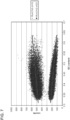
- the sequence reads were mapped to a reference human genome, and reads with non-unique alignments were discarded ( Figure 2 ).
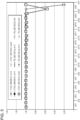
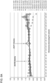
- Counts were first normalized per sample, based on the total counts to the autosomal chromosomes ( Figure 3 ).
- Counts were then normalized per chromsome, based on the average fraction of reads aligned to each chromosome across all samples (chrX - females only, chrY - males only; Figure 4 ).
- Data show quantitative chromosomal analysis ( Figure 5 ). These data show the genomic sequencing of selected HapMap samples, both male and female, followed by accurate quantitation of the chromosomal counts. Data herein show the distinct ability to identify expected ratios of chromosome X and chromosome Y.
- the data derived from genomic DNA obtained from individuals demonstrate the evenness of genomic coverage expected from a normal diploid genome, and demonstrate that no fetal nucleic acid is found in these samples.
- the deviation in the normalized counts per chromosome is 0.5% CV on average. It is lower (0.2-0.3%) for the larger chromosomes and higher (0.8-1.1%) for the smaller chromosomes. Female and Male samples are clearly distinguishable.
- Example 2 Detecting fetal nucleic acid in a maternal sample and detecting trisomy
- Maternal cell free plasma nucleic acid was obtained using methods well known in the art, such as a Qiagen nucleic acid purification kit.
- the nucleic acid was then subjected to the following protocol. Briefly, the protocol consists of a one hour 3' polyA tailing step, followed by a one hour 3' dideoxy-blocking step. The protocol was performed with 500pg of nucleic acid.
- RNA contamination Prior to conducting the tailing reaction on the DNA, RNA contamination was removed using RNase digestion and cleanup with a Qiagen Reaction Cleanup Kit (catalog 28204). DNA should was accurately quantitated prior to use.
- the Quant-iT TM PicoGreen dsDNA Reagent Kit (Invitrogen, catalog#P11495) with a Nanodrop 3300 Fluoro spectrometer was used. Molecular biology-grade nuclease-free glycogen or linear acrylamide was used as carrier during DNA clean-up/precipitation steps.
- the following mix was prepared: NEB Terminal Transferase 10X buffer (2 ⁇ l); 2.5mM CoCl 2 (2 ⁇ l); and maternal cell free plasma nucleic acid and Nuclease-free water (10.8 ⁇ l). The total volume was 14.8 ⁇ l.
- the mix was heated at 95°C for 5 minutes in the thermocycler to denature the DNA. After heating, the mix was cooled on the pre-chilled aluminum block that was kept in an ice and water slurry (about 0°C) to obtain single-stranded DNA. The sample was chilled as quick as possible to prevent re-annealing of the denatured, single-stranded DNA.
- the 20 ⁇ l poly-adenylation reaction was denature by heating the mixture to 95°C for 5 minutes in the thermocycler followed by rapid cooling in the pre-chilled aluminum block kept in an ice and water slurry (about 0°C). The sample was chilled as quick as possible to prevent re-annealing of the denatured, single-stranded DNA.
- the following blocking mixture was added to the denatured poly-adenylated mixture from above: 1 ⁇ l of Terminal Transferase 10X buffer; 1 ⁇ l of CoCl 2 (2.5mM); 1 ⁇ l of Terminal Transferase (dilute 1:4 to 5U/ ⁇ l using 1x buffer); 0.5 ⁇ l of 200 ⁇ M Biotin-ddATP; and 6.5 ⁇ l of nuclease-free water.
- the volume of this mix was 10 ⁇ l, bringing the total volume of the reaction to 30 ⁇ l.
- the tubes containing the mixture were placed in the thermocycler and the following program was run: 37°C for 1 hour; 70°C for 20 minutes; and temperature was brought back down to 4°C. It was observed that that a 3' end block was now added to the poly-adenylated DNA.
- control oligonucleotide 2 picomoles was added to the heat inactivated 30 ⁇ l terminal transferase reaction above.
- the control oligonucleotide was added to the sample to minimize DNA loss during sample loading steps.
- the control oligonucleotide does not contain a poly(A) tail, and therefore will not hybridize to the flow cell surface.
- the sample is now ready to be hybridized to the flow cells for the sequencing reaction. No additional clean-up step is required.
- HELISCOPE Sequencer channels Single molecule sequencing instrument, Helicos BioSciences Corporation
- DNA from the sample was sequenced in the channels according to the manufacturer's instructions.
- the sequence reads were mapped to a reference human genome, and reads with non-unique alignments were discarded.
- Counts were first normalized per sample, based on the total counts to the autosomal chromosomes. Counts were then normalized per chromsome, based on the average fraction of reads aligned to each chromosome across all samples (chrX - females only, chrY - males only). Chromosome counts for chromosomes 1, 18, and 21 across the samples were compared to deviations from the expected values based on control samples.
- FIG 10 shows results of analysis of the sequence information.
- chromosome 1 was used as a control. Data herein show that fetal DNA was detected ( Figure 10 ). Data herein further show that trisomy of chromosome 18 and chromosome 21 was also detected ( Figure 10 ).
- chromosomal counting analysis base on sequencing information i.e., quantifying the amount of each chromosome, or chromosome segment, based on relative representation
- sequencing information i.e., quantifying the amount of each chromosome, or chromosome segment, based on relative representation
- a relative number of read counts of each chromosome (or chromosome segment) are compared to a standard measured across one or more normal samples.
- Certain steps in the sample preparation or sequencing process may result in a GC bias, where the relative representation of each chromosome is influenced not only by the relative quantity (copy number) of that chromosome, but also by its GC content.
- a difference in GC bias between the measured sample and the control (normal) sample will result in skewing of the chromosomal counts such that chromosomes with extreme GC content may appear to have more or fewer than their real copy number.
- Figure 6 is a graph showing a sample in which chromosomal counts are skewed by
- Methods of the invention allow for determining an amount of GC bias in obtained sequence information, and also allow for correction of the GC bias in the sequence information.
- methods of the invention involve sequencing a sample to obtain nucleic acid sequence information; determining an amount of GC bias in the sequence information; correcting the sequence information to account for the GC bias; and analyzing the corrected information.
- the amount of GC bias in a sample may be accomplished in numerous ways.
- the amount of GC bias may be quantified by partitioning the genome into bins, and measuring the correlation between the number of counts in each bin and its GC content.
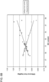
- Figure 7 is a graph showing counts in each bin plotted as a function of GC content of the bin.
- the genome is partitioned into 1000kbp bins. Although this number is exemplary and any size may be used.
- a significant negative or positive correlation indicates the existence of GC bias (see Figure 7 ).
- the upper sample shows positive correlation with GC content
- the lower sample shows negative correlation with GC content.
- Methods of the invention reduce or eliminate the effects of GC bias in sequence information.
- Numerous protocols may be used to reduce or eliminate the effects of GC bias in sequence information.
- a subset of genomic bins is selected within a given range such that the average GC content per chromosome is equalized (or less skewed). Chromosomal counting is then performed on the selected subset.
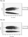
- Figure 8 provides an example of this protocol. In Figure 8 , analysis was limited to only genomic bins with a given GC content of 0.42 to 0.48, approximately 25% of the genome ( Figure 8 panel A)
- Figure 8 panels B and C show the difference in obtained sequence information after there is a correction for GC bias in the sequence information.
- Figure 8 panel B shows the sequence information prior to correction for GC bias.
- Figure 8 panel C shows the sequence information after correction for GC bias. These data show that the GC bias was skewing the chromosomal counts such that chromosomes with extreme GC content appeared to have more or fewer than their real copy number. After correction for GC bias in the sequence information, the data show a more accurate chromosomal count, and allowed for the detection of trisomy at chromosome 18 and 21, which was not possible from analysis of the sequence information prior to correction for GC bias.
- the correlation between GC content and chromosome counts is modeled across a set of genomic bins using a mathematical function (e.g. a first or second order polynomial).
- An exemplary mathematical function is a regression model (i.e., fitting the sequence information to a mathematical function, such as lower order functions (linear and/or quadratic polynomials)).
- the effect of GC bias is corrected for by subtracting the GC-dependent component, reflected by the model, from the count of each bin. Chromosomal counting is then performed based on the corrected counts.
- Figure 9 provides an example of this protocol.
- the sequence information was corrected by subtracting a linear model of GC dependence from each genomic bin.
- Figure 9 panels A and B show sequence information prior to correction for GC bias.
- Figure 9 panels C and D show sequence information after correction for GC bias. These data show that the GC bias was skewing the chromosomal counts such that chromosomes with extreme GC content appeared to have more or fewer than their real copy number.
- the data show a more accurate chromosomal count, and allowed for the detection of trisomy at chromosome 18 and 21, which was not possible from analysis of the sequence information prior to correction for GC bias.
- GC bias is corrected for as follows. An average coverage per bin over a number of control samples is obtained, and the observed coverage in the sample is divided by the mean of the control population (this could be a weighted mean to take into account different levels of overall coverage in the control samples). Each corrected bin value would then be a ratio of observed to expected, which will be more consistent across bins of different %GC.
Landscapes
- Chemical & Material Sciences (AREA)
- Life Sciences & Earth Sciences (AREA)
- Health & Medical Sciences (AREA)
- Proteomics, Peptides & Aminoacids (AREA)
- Engineering & Computer Science (AREA)
- Organic Chemistry (AREA)
- Physics & Mathematics (AREA)
- Analytical Chemistry (AREA)
- Bioinformatics & Cheminformatics (AREA)
- Zoology (AREA)
- Wood Science & Technology (AREA)
- Genetics & Genomics (AREA)
- Molecular Biology (AREA)
- Biophysics (AREA)
- Biotechnology (AREA)
- General Health & Medical Sciences (AREA)
- Immunology (AREA)
- General Engineering & Computer Science (AREA)
- Biochemistry (AREA)
- Microbiology (AREA)
- Bioinformatics & Computational Biology (AREA)
- Evolutionary Biology (AREA)
- Medical Informatics (AREA)
- Spectroscopy & Molecular Physics (AREA)
- Theoretical Computer Science (AREA)
- Pathology (AREA)
- Measuring Or Testing Involving Enzymes Or Micro-Organisms (AREA)
Applications Claiming Priority (5)
| Application Number | Priority Date | Filing Date | Title |
|---|---|---|---|
| US12/709,057 US20100216151A1 (en) | 2004-02-27 | 2010-02-19 | Methods for detecting fetal nucleic acids and diagnosing fetal abnormalities |
| US12/727,824 US20100216153A1 (en) | 2004-02-27 | 2010-03-19 | Methods for detecting fetal nucleic acids and diagnosing fetal abnormalities |
| EP11745050.2A EP2536852B1 (de) | 2010-02-19 | 2011-02-09 | Verfahren zur erkennung fötaler nukleinsäuren und zur diagnose fötaler anomalien |
| PCT/US2011/024132 WO2011102998A2 (en) | 2010-02-19 | 2011-02-09 | Methods for detecting fetal nucleic acids and diagnosing fetal abnormalities |
| EP19197417.9A EP3636776B1 (de) | 2010-02-19 | 2011-02-09 | Verfahren zur analyse von sequenzinformation von nukleinsäuren unter berücksichtigung des gc biases, optional zur diagnose von nukleinsäureanomalien in foeten. |
Related Parent Applications (3)
| Application Number | Title | Priority Date | Filing Date |
|---|---|---|---|
| EP11745050.2A Division EP2536852B1 (de) | 2010-02-19 | 2011-02-09 | Verfahren zur erkennung fötaler nukleinsäuren und zur diagnose fötaler anomalien |
| EP19197417.9A Division EP3636776B1 (de) | 2010-02-19 | 2011-02-09 | Verfahren zur analyse von sequenzinformation von nukleinsäuren unter berücksichtigung des gc biases, optional zur diagnose von nukleinsäureanomalien in foeten. |
| EP19197417.9A Division-Into EP3636776B1 (de) | 2010-02-19 | 2011-02-09 | Verfahren zur analyse von sequenzinformation von nukleinsäuren unter berücksichtigung des gc biases, optional zur diagnose von nukleinsäureanomalien in foeten. |
Publications (2)
| Publication Number | Publication Date |
|---|---|
| EP4455309A2 true EP4455309A2 (de) | 2024-10-30 |
| EP4455309A3 EP4455309A3 (de) | 2025-01-22 |
Family
ID=44483527
Family Applications (3)
| Application Number | Title | Priority Date | Filing Date |
|---|---|---|---|
| EP24195465.0A Pending EP4455309A3 (de) | 2010-02-19 | 2011-02-09 | Verfahren zur analyse von nukleinsäuresequenzinformationen mittels gc-bias, optional zum nachweis von anomalien fetaler nukleinsäure |
| EP19197417.9A Active EP3636776B1 (de) | 2010-02-19 | 2011-02-09 | Verfahren zur analyse von sequenzinformation von nukleinsäuren unter berücksichtigung des gc biases, optional zur diagnose von nukleinsäureanomalien in foeten. |
| EP11745050.2A Active EP2536852B1 (de) | 2010-02-19 | 2011-02-09 | Verfahren zur erkennung fötaler nukleinsäuren und zur diagnose fötaler anomalien |
Family Applications After (2)
| Application Number | Title | Priority Date | Filing Date |
|---|---|---|---|
| EP19197417.9A Active EP3636776B1 (de) | 2010-02-19 | 2011-02-09 | Verfahren zur analyse von sequenzinformation von nukleinsäuren unter berücksichtigung des gc biases, optional zur diagnose von nukleinsäureanomalien in foeten. |
| EP11745050.2A Active EP2536852B1 (de) | 2010-02-19 | 2011-02-09 | Verfahren zur erkennung fötaler nukleinsäuren und zur diagnose fötaler anomalien |
Country Status (9)
| Country | Link |
|---|---|
| US (4) | US20100216153A1 (de) |
| EP (3) | EP4455309A3 (de) |
| CN (2) | CN103108960A (de) |
| AU (2) | AU2011218382B2 (de) |
| ES (1) | ES3001090T3 (de) |
| HU (1) | HUE047618T2 (de) |
| PL (2) | PL3636776T3 (de) |
| PT (1) | PT2536852T (de) |
| WO (1) | WO2011102998A2 (de) |
Families Citing this family (280)
| Publication number | Priority date | Publication date | Assignee | Title |
|---|---|---|---|---|
| ES2784011T3 (es) | 2002-10-16 | 2020-09-21 | Streck Inc | Procedimiento y dispositivo para recoger y preservar las células para su análisis |
| US7169560B2 (en) | 2003-11-12 | 2007-01-30 | Helicos Biosciences Corporation | Short cycle methods for sequencing polynucleotides |
| CA2557177A1 (en) * | 2004-02-19 | 2005-09-01 | Stephen Quake | Methods and kits for analyzing polynucleotide sequences |
| DE602005009324D1 (de) | 2005-04-06 | 2008-10-09 | Maurice Stroun | Methode zur Krebsdiagnose mittels Nachweis von DNA und RNA im Kreislauf |
| US11111544B2 (en) | 2005-07-29 | 2021-09-07 | Natera, Inc. | System and method for cleaning noisy genetic data and determining chromosome copy number |
| US10081839B2 (en) | 2005-07-29 | 2018-09-25 | Natera, Inc | System and method for cleaning noisy genetic data and determining chromosome copy number |
| US11111543B2 (en) | 2005-07-29 | 2021-09-07 | Natera, Inc. | System and method for cleaning noisy genetic data and determining chromosome copy number |
| US20070027636A1 (en) * | 2005-07-29 | 2007-02-01 | Matthew Rabinowitz | System and method for using genetic, phentoypic and clinical data to make predictions for clinical or lifestyle decisions |
| US20070178501A1 (en) * | 2005-12-06 | 2007-08-02 | Matthew Rabinowitz | System and method for integrating and validating genotypic, phenotypic and medical information into a database according to a standardized ontology |
| US9424392B2 (en) | 2005-11-26 | 2016-08-23 | Natera, Inc. | System and method for cleaning noisy genetic data from target individuals using genetic data from genetically related individuals |
| SI2385143T1 (sl) | 2006-02-02 | 2016-11-30 | The Board of Trustees of the Leland Stanford Junior University Office of the General Counsel | Neinvazivni genetski presejalni test ploda z digitalno analizo |
| EP2589668A1 (de) | 2006-06-14 | 2013-05-08 | Verinata Health, Inc | Analyse seltener Zellen mittels Probentrennung und DNA-Etiketten |
| WO2007147074A2 (en) | 2006-06-14 | 2007-12-21 | Living Microsystems, Inc. | Use of highly parallel snp genotyping for fetal diagnosis |
| US20080050739A1 (en) | 2006-06-14 | 2008-02-28 | Roland Stoughton | Diagnosis of fetal abnormalities using polymorphisms including short tandem repeats |
| US8137912B2 (en) | 2006-06-14 | 2012-03-20 | The General Hospital Corporation | Methods for the diagnosis of fetal abnormalities |
| KR102222378B1 (ko) | 2007-07-23 | 2021-03-04 | 더 차이니즈 유니버시티 오브 홍콩 | 핵산 서열 불균형의 결정 |
| US12180549B2 (en) | 2007-07-23 | 2024-12-31 | The Chinese University Of Hong Kong | Diagnosing fetal chromosomal aneuploidy using genomic sequencing |
| US20110178719A1 (en) | 2008-08-04 | 2011-07-21 | Gene Security Network, Inc. | Methods for Allele Calling and Ploidy Calling |
| US8583380B2 (en) | 2008-09-05 | 2013-11-12 | Aueon, Inc. | Methods for stratifying and annotating cancer drug treatment options |
| US8476013B2 (en) | 2008-09-16 | 2013-07-02 | Sequenom, Inc. | Processes and compositions for methylation-based acid enrichment of fetal nucleic acid from a maternal sample useful for non-invasive prenatal diagnoses |
| US8962247B2 (en) | 2008-09-16 | 2015-02-24 | Sequenom, Inc. | Processes and compositions for methylation-based enrichment of fetal nucleic acid from a maternal sample useful for non invasive prenatal diagnoses |
| US11634747B2 (en) * | 2009-01-21 | 2023-04-25 | Streck Llc | Preservation of fetal nucleic acids in maternal plasma |
| DK3290530T3 (da) | 2009-02-18 | 2020-12-07 | Streck Inc | Konservering af cellefrie nukleinsyrer |
| US12129514B2 (en) | 2009-04-30 | 2024-10-29 | Molecular Loop Biosolutions, Llc | Methods and compositions for evaluating genetic markers |
| AU2010242073C1 (en) | 2009-04-30 | 2015-12-24 | Good Start Genetics, Inc. | Methods and compositions for evaluating genetic markers |
| EP2473638B1 (de) | 2009-09-30 | 2017-08-09 | Natera, Inc. | Verfahren zur nichtinvasiven pränatalen ploidiezuordnung |
| US8835358B2 (en) | 2009-12-15 | 2014-09-16 | Cellular Research, Inc. | Digital counting of individual molecules by stochastic attachment of diverse labels |
| US9315857B2 (en) | 2009-12-15 | 2016-04-19 | Cellular Research, Inc. | Digital counting of individual molecules by stochastic attachment of diverse label-tags |
| EP2516680B1 (de) | 2009-12-22 | 2016-04-06 | Sequenom, Inc. | Verfahren und kits zur identifizierung von aneuploidie |
| US9260745B2 (en) | 2010-01-19 | 2016-02-16 | Verinata Health, Inc. | Detecting and classifying copy number variation |
| CA2786565C (en) | 2010-01-19 | 2017-04-25 | Verinata Health, Inc. | Partition defined detection methods |
| WO2011090556A1 (en) | 2010-01-19 | 2011-07-28 | Verinata Health, Inc. | Methods for determining fraction of fetal nucleic acid in maternal samples |
| EP2848704B1 (de) | 2010-01-19 | 2018-08-29 | Verinata Health, Inc | Sequenzierungsverfahren für pränatale Diagnosen |
| US10388403B2 (en) | 2010-01-19 | 2019-08-20 | Verinata Health, Inc. | Analyzing copy number variation in the detection of cancer |
| US20120100548A1 (en) | 2010-10-26 | 2012-04-26 | Verinata Health, Inc. | Method for determining copy number variations |
| US9323888B2 (en) | 2010-01-19 | 2016-04-26 | Verinata Health, Inc. | Detecting and classifying copy number variation |
| EP2513341B1 (de) * | 2010-01-19 | 2017-04-12 | Verinata Health, Inc | Identifizierung von polymorphen sequenzen in mischungen aus genomischer dna durch gesamtgenomsequenzierung |
| US20110312503A1 (en) | 2010-01-23 | 2011-12-22 | Artemis Health, Inc. | Methods of fetal abnormality detection |
| US20190010543A1 (en) | 2010-05-18 | 2019-01-10 | Natera, Inc. | Methods for simultaneous amplification of target loci |
| US12152275B2 (en) | 2010-05-18 | 2024-11-26 | Natera, Inc. | Methods for non-invasive prenatal ploidy calling |
| US11332785B2 (en) | 2010-05-18 | 2022-05-17 | Natera, Inc. | Methods for non-invasive prenatal ploidy calling |
| US11322224B2 (en) | 2010-05-18 | 2022-05-03 | Natera, Inc. | Methods for non-invasive prenatal ploidy calling |
| US10316362B2 (en) | 2010-05-18 | 2019-06-11 | Natera, Inc. | Methods for simultaneous amplification of target loci |
| US9677118B2 (en) | 2014-04-21 | 2017-06-13 | Natera, Inc. | Methods for simultaneous amplification of target loci |
| US11332793B2 (en) | 2010-05-18 | 2022-05-17 | Natera, Inc. | Methods for simultaneous amplification of target loci |
| US8825412B2 (en) | 2010-05-18 | 2014-09-02 | Natera, Inc. | Methods for non-invasive prenatal ploidy calling |
| US11939634B2 (en) | 2010-05-18 | 2024-03-26 | Natera, Inc. | Methods for simultaneous amplification of target loci |
| US11339429B2 (en) | 2010-05-18 | 2022-05-24 | Natera, Inc. | Methods for non-invasive prenatal ploidy calling |
| US11408031B2 (en) | 2010-05-18 | 2022-08-09 | Natera, Inc. | Methods for non-invasive prenatal paternity testing |
| US11326208B2 (en) | 2010-05-18 | 2022-05-10 | Natera, Inc. | Methods for nested PCR amplification of cell-free DNA |
| US12221653B2 (en) | 2010-05-18 | 2025-02-11 | Natera, Inc. | Methods for simultaneous amplification of target loci |
| EP3115468B1 (de) | 2010-09-21 | 2018-07-25 | Agilent Technologies, Inc. | Erhöhung der verlässlichkeit von allel-aufrufen mit molekülzählung |
| WO2012040387A1 (en) | 2010-09-24 | 2012-03-29 | The Board Of Trustees Of The Leland Stanford Junior University | Direct capture, amplification and sequencing of target dna using immobilized primers |
| CN103534591B (zh) | 2010-10-26 | 2016-04-06 | 利兰·斯坦福青年大学托管委员会 | 通过测序分析进行的非侵入性胎儿遗传筛选 |
| CA2821906C (en) * | 2010-12-22 | 2020-08-25 | Natera, Inc. | Methods for non-invasive prenatal paternity testing |
| US9163281B2 (en) | 2010-12-23 | 2015-10-20 | Good Start Genetics, Inc. | Methods for maintaining the integrity and identification of a nucleic acid template in a multiplex sequencing reaction |
| EP2655666A2 (de) * | 2010-12-23 | 2013-10-30 | Sequenom, Inc. | Nachweis fötaler genetischer variationen |
| US10131947B2 (en) * | 2011-01-25 | 2018-11-20 | Ariosa Diagnostics, Inc. | Noninvasive detection of fetal aneuploidy in egg donor pregnancies |
| CA2824387C (en) | 2011-02-09 | 2019-09-24 | Natera, Inc. | Methods for non-invasive prenatal ploidy calling |
| CA3252077A1 (en) | 2011-02-24 | 2025-03-20 | The Chinese University Of Hong Kong | Molecular testing of multiple pregnancies |
| US9260753B2 (en) | 2011-03-24 | 2016-02-16 | President And Fellows Of Harvard College | Single cell nucleic acid detection and analysis |
| RS57837B1 (sr) * | 2011-04-12 | 2018-12-31 | Verinata Health Inc | Razrešavanje genomskih frakcija upotrebom broja kopija polimorfizama |
| AU2011365507A1 (en) * | 2011-04-14 | 2013-05-02 | Verinata Health, Inc. | Normalizing chromosomes for the determination and verification of common and rare chromosomal aneuploidies |
| GB2484764B (en) | 2011-04-14 | 2012-09-05 | Verinata Health Inc | Normalizing chromosomes for the determination and verification of common and rare chromosomal aneuploidies |
| US9411937B2 (en) | 2011-04-15 | 2016-08-09 | Verinata Health, Inc. | Detecting and classifying copy number variation |
| WO2012151391A2 (en) | 2011-05-04 | 2012-11-08 | Streck, Inc. | Inactivated virus compositions and methods of preparing such compositions |
| DK2716766T3 (da) | 2011-05-31 | 2017-01-02 | Berry Genomics Co Ltd | Indretning til detektering af kopiantal af føtale kromosomer eller tumorcellekromosomer |
| US20140235474A1 (en) | 2011-06-24 | 2014-08-21 | Sequenom, Inc. | Methods and processes for non invasive assessment of a genetic variation |
| RU2589681C2 (ru) * | 2011-06-29 | 2016-07-10 | БиДжиАй Дженомикс Ко., Лтд. | Неинвазивное обнаружение генетической аномалии плода |
| US20130157875A1 (en) * | 2011-07-20 | 2013-06-20 | Anthony P. Shuber | Methods for assessing genomic instabilities |
| CA2850785C (en) | 2011-10-06 | 2022-12-13 | Sequenom, Inc. | Methods and processes for non-invasive assessment of genetic variations |
| US10424394B2 (en) | 2011-10-06 | 2019-09-24 | Sequenom, Inc. | Methods and processes for non-invasive assessment of genetic variations |
| US10196681B2 (en) | 2011-10-06 | 2019-02-05 | Sequenom, Inc. | Methods and processes for non-invasive assessment of genetic variations |
| US9984198B2 (en) | 2011-10-06 | 2018-05-29 | Sequenom, Inc. | Reducing sequence read count error in assessment of complex genetic variations |
| US9367663B2 (en) | 2011-10-06 | 2016-06-14 | Sequenom, Inc. | Methods and processes for non-invasive assessment of genetic variations |
| US8688388B2 (en) | 2011-10-11 | 2014-04-01 | Sequenom, Inc. | Methods and processes for non-invasive assessment of genetic variations |
| US9228233B2 (en) | 2011-10-17 | 2016-01-05 | Good Start Genetics, Inc. | Analysis methods |
| AU2013209499B2 (en) * | 2012-01-20 | 2018-05-10 | Sequenom, Inc. | Diagnostic processes that factor experimental conditions |
| GB2513024B (en) | 2012-02-27 | 2016-08-31 | Cellular Res Inc | A clonal amplification method |
| WO2013130512A2 (en) | 2012-02-27 | 2013-09-06 | The University Of North Carolina At Chapel Hill | Methods and uses for molecular tags |
| WO2013128281A1 (en) | 2012-02-28 | 2013-09-06 | Population Genetics Technologies Ltd | Method for attaching a counter sequence to a nucleic acid sample |
| US9605313B2 (en) | 2012-03-02 | 2017-03-28 | Sequenom, Inc. | Methods and processes for non-invasive assessment of genetic variations |
| US8209130B1 (en) | 2012-04-04 | 2012-06-26 | Good Start Genetics, Inc. | Sequence assembly |
| US10227635B2 (en) | 2012-04-16 | 2019-03-12 | Molecular Loop Biosolutions, Llc | Capture reactions |
| US9920361B2 (en) | 2012-05-21 | 2018-03-20 | Sequenom, Inc. | Methods and compositions for analyzing nucleic acid |
| US10504613B2 (en) | 2012-12-20 | 2019-12-10 | Sequenom, Inc. | Methods and processes for non-invasive assessment of genetic variations |
| US10497461B2 (en) | 2012-06-22 | 2019-12-03 | Sequenom, Inc. | Methods and processes for non-invasive assessment of genetic variations |
| US20140093873A1 (en) * | 2012-07-13 | 2014-04-03 | Sequenom, Inc. | Processes and compositions for methylation-based enrichment of fetal nucleic acid from a maternal sample useful for non-invasive prenatal diagnoses |
| US10273541B2 (en) | 2012-08-14 | 2019-04-30 | 10X Genomics, Inc. | Methods and systems for processing polynucleotides |
| US10752949B2 (en) | 2012-08-14 | 2020-08-25 | 10X Genomics, Inc. | Methods and systems for processing polynucleotides |
| US9951386B2 (en) | 2014-06-26 | 2018-04-24 | 10X Genomics, Inc. | Methods and systems for processing polynucleotides |
| US9701998B2 (en) | 2012-12-14 | 2017-07-11 | 10X Genomics, Inc. | Methods and systems for processing polynucleotides |
| EP4397767A3 (de) | 2012-08-14 | 2024-07-31 | 10X Genomics, Inc. | Mikrokapselzusammensetzungen und verfahren |
| US10323279B2 (en) | 2012-08-14 | 2019-06-18 | 10X Genomics, Inc. | Methods and systems for processing polynucleotides |
| US10584381B2 (en) | 2012-08-14 | 2020-03-10 | 10X Genomics, Inc. | Methods and systems for processing polynucleotides |
| US11591637B2 (en) | 2012-08-14 | 2023-02-28 | 10X Genomics, Inc. | Compositions and methods for sample processing |
| US20140100126A1 (en) | 2012-08-17 | 2014-04-10 | Natera, Inc. | Method for Non-Invasive Prenatal Testing Using Parental Mosaicism Data |
| US20160040229A1 (en) | 2013-08-16 | 2016-02-11 | Guardant Health, Inc. | Systems and methods to detect rare mutations and copy number variation |
| US11913065B2 (en) | 2012-09-04 | 2024-02-27 | Guardent Health, Inc. | Systems and methods to detect rare mutations and copy number variation |
| US10876152B2 (en) | 2012-09-04 | 2020-12-29 | Guardant Health, Inc. | Systems and methods to detect rare mutations and copy number variation |
| EP4036247B1 (de) | 2012-09-04 | 2024-04-10 | Guardant Health, Inc. | Verfahren zur detektion von seltenen mutationen und kopienzahlvariationen |
| US10482994B2 (en) | 2012-10-04 | 2019-11-19 | Sequenom, Inc. | Methods and processes for non-invasive assessment of genetic variations |
| TWI489305B (zh) * | 2012-11-21 | 2015-06-21 | Bgi Diagnosis Co Ltd | 對胎兒遺傳異常的無創性檢測 |
| WO2014093676A1 (en) | 2012-12-14 | 2014-06-19 | 10X Technologies, Inc. | Methods and systems for processing polynucleotides |
| US10533221B2 (en) | 2012-12-14 | 2020-01-14 | 10X Genomics, Inc. | Methods and systems for processing polynucleotides |
| US20130309666A1 (en) * | 2013-01-25 | 2013-11-21 | Sequenom, Inc. | Methods and processes for non-invasive assessment of genetic variations |
| CN108753766A (zh) | 2013-02-08 | 2018-11-06 | 10X基因组学有限公司 | 多核苷酸条形码生成 |
| CN105229168B (zh) * | 2013-02-20 | 2020-07-17 | 生物纳米基因有限公司 | 纳米流体中分子的表征 |
| EP2971100A1 (de) | 2013-03-13 | 2016-01-20 | Sequenom, Inc. | Primer zur analyse der dna-methylierung |
| EP2971159B1 (de) | 2013-03-14 | 2019-05-08 | Molecular Loop Biosolutions, LLC | Verfahren zur analyse von nukleinsäuren |
| LT2981921T (lt) * | 2013-04-03 | 2023-02-27 | Sequenom, Inc. | Neinvazinio genetinių variacijų vertinimo būdai ir procesai |
| WO2014190286A2 (en) | 2013-05-24 | 2014-11-27 | Sequenom, Inc. | Methods and processes for non-invasive assessment of genetic variations |
| KR102784584B1 (ko) * | 2013-06-21 | 2025-03-19 | 시쿼넘, 인코포레이티드 | 유전적 변이의 비침습 평가를 위한 방법 및 프로세스 |
| CA2917912C (en) | 2013-07-24 | 2019-09-17 | Streck, Inc. | Compositions and methods for stabilizing circulating tumor cells |
| GB2546833B (en) | 2013-08-28 | 2018-04-18 | Cellular Res Inc | Microwell for single cell analysis comprising single cell and single bead oligonucleotide capture labels |
| US10395758B2 (en) | 2013-08-30 | 2019-08-27 | 10X Genomics, Inc. | Sequencing methods |
| WO2015035555A1 (zh) * | 2013-09-10 | 2015-03-19 | 深圳华大基因科技有限公司 | 用于确定胎儿是否存在性染色体数目异常的方法、系统和计算机可读介质 |
| US9499870B2 (en) | 2013-09-27 | 2016-11-22 | Natera, Inc. | Cell free DNA diagnostic testing standards |
| US10577655B2 (en) | 2013-09-27 | 2020-03-03 | Natera, Inc. | Cell free DNA diagnostic testing standards |
| US10262755B2 (en) | 2014-04-21 | 2019-04-16 | Natera, Inc. | Detecting cancer mutations and aneuploidy in chromosomal segments |
| US10964409B2 (en) * | 2013-10-04 | 2021-03-30 | Sequenom, Inc. | Methods and processes for non-invasive assessment of genetic variations |
| CN111863131A (zh) | 2013-10-07 | 2020-10-30 | 塞昆纳姆股份有限公司 | 用于非侵入性评估染色体改变的方法和过程 |
| US9582877B2 (en) | 2013-10-07 | 2017-02-28 | Cellular Research, Inc. | Methods and systems for digitally counting features on arrays |
| US10851414B2 (en) | 2013-10-18 | 2020-12-01 | Good Start Genetics, Inc. | Methods for determining carrier status |
| CA2928185C (en) | 2013-10-21 | 2024-01-30 | Verinata Health, Inc. | Method for improving the sensitivity of detection in determining copy number variations |
| US9824068B2 (en) | 2013-12-16 | 2017-11-21 | 10X Genomics, Inc. | Methods and apparatus for sorting data |
| SG10201804519RA (en) | 2013-12-28 | 2018-07-30 | Guardant Health Inc | Methods and systems for detecting genetic variants |
| CN103824001A (zh) * | 2014-02-27 | 2014-05-28 | 北京诺禾致源生物信息科技有限公司 | 染色体的检测方法和装置 |
| US11365447B2 (en) | 2014-03-13 | 2022-06-21 | Sequenom, Inc. | Methods and processes for non-invasive assessment of genetic variations |
| CA2943624A1 (en) | 2014-04-10 | 2015-10-15 | 10X Genomics, Inc. | Fluidic devices, systems, and methods for encapsulating and partitioning reagents, and applications of same |
| EP3957749A1 (de) | 2014-04-21 | 2022-02-23 | Natera, Inc. | Nachweis von tumorspezifischen mutationen in biopsien mittels sequenzierung des gesamten exoms und in zellfreien proben |
| US12492429B2 (en) | 2014-04-21 | 2025-12-09 | Natera, Inc. | Detecting mutations and ploidy in chromosomal segments |
| WO2015175530A1 (en) * | 2014-05-12 | 2015-11-19 | Gore Athurva | Methods for detecting aneuploidy |
| EP3149199B1 (de) | 2014-05-30 | 2020-03-25 | Verinata Health, Inc. | Nachweis von eventuell fetalen subchromosomalen aneuploidien und kopienzahlvariationen |
| US20180173846A1 (en) | 2014-06-05 | 2018-06-21 | Natera, Inc. | Systems and Methods for Detection of Aneuploidy |
| AU2015279617A1 (en) * | 2014-06-26 | 2017-01-12 | 10X Genomics, Inc. | Analysis of nucleic acid sequences |
| US12312640B2 (en) | 2014-06-26 | 2025-05-27 | 10X Genomics, Inc. | Analysis of nucleic acid sequences |
| EP3161700B1 (de) | 2014-06-26 | 2023-03-29 | 10X Genomics, Inc. | Verfahren und systeme zur nukleinsäuresequenzanordnung |
| EP3161160B1 (de) | 2014-06-26 | 2021-10-13 | 10X Genomics, Inc. | Verfahren zur analyse von nukleinsäuren aus einzelzellen oder zellpopulationen |
| EP3175000B1 (de) | 2014-07-30 | 2020-07-29 | Sequenom, Inc. | Verfahren und prozesse zur nicht-invasiven beurteilung genetischer variationen |
| WO2016025818A1 (en) | 2014-08-15 | 2016-02-18 | Good Start Genetics, Inc. | Systems and methods for genetic analysis |
| US11408024B2 (en) | 2014-09-10 | 2022-08-09 | Molecular Loop Biosciences, Inc. | Methods for selectively suppressing non-target sequences |
| EP3224595A4 (de) | 2014-09-24 | 2018-06-13 | Good Start Genetics, Inc. | Prozesssteuerung für erhöhte robustheit von genetischen assays |
| EP3204512B1 (de) * | 2014-10-10 | 2020-05-06 | Sequenom, Inc. | Verfahren zum partitionieren genomischer sequenzen |
| CN107750277B (zh) | 2014-12-12 | 2021-11-09 | 维里纳塔健康股份有限公司 | 使用无细胞dna片段大小来确定拷贝数变化 |
| WO2016112073A1 (en) | 2015-01-06 | 2016-07-14 | Good Start Genetics, Inc. | Screening for structural variants |
| CN107427808B (zh) | 2015-01-12 | 2020-10-23 | 10X基因组学有限公司 | 用于制备核酸测序文库的方法和系统以及用其制备的文库 |
| KR20170106979A (ko) | 2015-01-13 | 2017-09-22 | 10엑스 제노믹스, 인크. | 구조 변이 및 위상 조정 정보를 시각화하기 위한 시스템 및 방법 |
| US10364467B2 (en) | 2015-01-13 | 2019-07-30 | The Chinese University Of Hong Kong | Using size and number aberrations in plasma DNA for detecting cancer |
| EP3256606B1 (de) | 2015-02-09 | 2019-05-22 | 10X Genomics, Inc. | Systeme und verfahren zur bestimmung struktureller varianten |
| ES2975332T3 (es) | 2015-02-19 | 2024-07-04 | Becton Dickinson Co | Análisis unicelular de alto rendimiento que combina información proteómica y genómica |
| US10697000B2 (en) | 2015-02-24 | 2020-06-30 | 10X Genomics, Inc. | Partition processing methods and systems |
| EP3262192B1 (de) | 2015-02-27 | 2020-09-16 | Becton, Dickinson and Company | Räumlich adressierbare molekulare barcodierung |
| US11168351B2 (en) | 2015-03-05 | 2021-11-09 | Streck, Inc. | Stabilization of nucleic acids in urine |
| US10829804B2 (en) | 2015-03-23 | 2020-11-10 | The University Of North Carolina At Chapel Hill | Method for identification and enumeration of nucleic acid sequences, expression, splice variant, translocation, copy, or DNA methylation changes using combined nuclease, ligase, polymerase, terminal transferase, and sequencing reactions |
| US10393726B2 (en) | 2015-03-23 | 2019-08-27 | The University Of North Carolina At Chapel Hill | Universal molecular processor for precision medicine |
| WO2016160844A2 (en) | 2015-03-30 | 2016-10-06 | Cellular Research, Inc. | Methods and compositions for combinatorial barcoding |
| EP3286326B1 (de) | 2015-04-23 | 2025-01-22 | Becton, Dickinson and Company | Verfahren zur gesamttranskriptomamplifikation |
| US11479812B2 (en) | 2015-05-11 | 2022-10-25 | Natera, Inc. | Methods and compositions for determining ploidy |
| WO2016196229A1 (en) | 2015-06-01 | 2016-12-08 | Cellular Research, Inc. | Methods for rna quantification |
| AU2016293025A1 (en) * | 2015-07-13 | 2017-11-02 | Agilent Technologies Belgium Nv | System and methodology for the analysis of genomic data obtained from a subject |
| BE1023266B1 (nl) * | 2015-07-13 | 2017-01-17 | Cartagenia N.V. | Systeem en methodologie voor de analyse van genomische gegevens die zijn verkregen van een onderwerp |
| EP3118323A1 (de) * | 2015-07-13 | 2017-01-18 | Cartagenia N.V. | System und methodologie zur analyse genomischer daten aus einer person |
| US11302416B2 (en) | 2015-09-02 | 2022-04-12 | Guardant Health | Machine learning for somatic single nucleotide variant detection in cell-free tumor nucleic acid sequencing applications |
| EP3347465B1 (de) | 2015-09-11 | 2019-06-26 | Cellular Research, Inc. | Verfahren und zusammensetzung zur standardisierung von nukleinsäure bibliothek |
| US11371094B2 (en) | 2015-11-19 | 2022-06-28 | 10X Genomics, Inc. | Systems and methods for nucleic acid processing using degenerate nucleotides |
| US20170145475A1 (en) | 2015-11-20 | 2017-05-25 | Streck, Inc. | Single spin process for blood plasma separation and plasma composition including preservative |
| CN117174167A (zh) | 2015-12-17 | 2023-12-05 | 夸登特健康公司 | 通过分析无细胞dna确定肿瘤基因拷贝数的方法 |
| US10095831B2 (en) | 2016-02-03 | 2018-10-09 | Verinata Health, Inc. | Using cell-free DNA fragment size to determine copy number variations |
| CN108779491B (zh) | 2016-02-11 | 2021-03-09 | 10X基因组学有限公司 | 用于全基因组序列数据的从头组装的系统、方法和介质 |
| AU2017218149B2 (en) * | 2016-02-12 | 2020-09-03 | Regeneron Pharmaceuticals, Inc. | Methods and systems for detection of abnormal karyotypes |
| JP7280044B2 (ja) | 2016-04-15 | 2023-05-23 | ナテラ, インコーポレイテッド | 肺癌の検出方法 |
| ES2956757T3 (es) | 2016-05-02 | 2023-12-27 | Becton Dickinson Co | Codificación con códigos de barras moleculares precisa |
| US10301677B2 (en) | 2016-05-25 | 2019-05-28 | Cellular Research, Inc. | Normalization of nucleic acid libraries |
| ES2979395T3 (es) | 2016-05-26 | 2024-09-25 | Becton Dickinson Co | Métodos de ajuste del recuento de etiquetas moleculares |
| US10640763B2 (en) | 2016-05-31 | 2020-05-05 | Cellular Research, Inc. | Molecular indexing of internal sequences |
| US10202641B2 (en) | 2016-05-31 | 2019-02-12 | Cellular Research, Inc. | Error correction in amplification of samples |
| US11200963B2 (en) | 2016-07-27 | 2021-12-14 | Sequenom, Inc. | Genetic copy number alteration classifications |
| US11506655B2 (en) | 2016-07-29 | 2022-11-22 | Streck, Inc. | Suspension composition for hematology analysis control |
| CN109791157B (zh) | 2016-09-26 | 2022-06-07 | 贝克顿迪金森公司 | 使用具有条形码化的寡核苷酸序列的试剂测量蛋白质表达 |
| US11854666B2 (en) | 2016-09-29 | 2023-12-26 | Myriad Women's Health, Inc. | Noninvasive prenatal screening using dynamic iterative depth optimization |
| US9850523B1 (en) | 2016-09-30 | 2017-12-26 | Guardant Health, Inc. | Methods for multi-resolution analysis of cell-free nucleic acids |
| WO2018064629A1 (en) | 2016-09-30 | 2018-04-05 | Guardant Health, Inc. | Methods for multi-resolution analysis of cell-free nucleic acids |
| WO2018067517A1 (en) | 2016-10-04 | 2018-04-12 | Natera, Inc. | Methods for characterizing copy number variation using proximity-litigation sequencing |
| GB201618485D0 (en) | 2016-11-02 | 2016-12-14 | Ucl Business Plc | Method of detecting tumour recurrence |
| CN109906274B (zh) | 2016-11-08 | 2023-08-25 | 贝克顿迪金森公司 | 用于细胞标记分类的方法 |
| EP3539035B1 (de) | 2016-11-08 | 2024-04-17 | Becton, Dickinson and Company | Verfahren zur klassifizierung von expressionsprofilen |
| US10011870B2 (en) | 2016-12-07 | 2018-07-03 | Natera, Inc. | Compositions and methods for identifying nucleic acid molecules |
| US10815525B2 (en) | 2016-12-22 | 2020-10-27 | 10X Genomics, Inc. | Methods and systems for processing polynucleotides |
| US10550429B2 (en) | 2016-12-22 | 2020-02-04 | 10X Genomics, Inc. | Methods and systems for processing polynucleotides |
| WO2018132610A1 (en) | 2017-01-13 | 2018-07-19 | Cellular Research, Inc. | Hydrophilic coating of fluidic channels |
| CA3198931A1 (en) | 2017-01-20 | 2018-07-26 | Sequenom, Inc. | Methods for non-invasive assessment of genetic alterations |
| IL319365A (en) | 2017-01-24 | 2025-05-01 | Sequenom Inc | Methods and processes for assessing genetic variations |
| EP4029939B1 (de) | 2017-01-30 | 2023-06-28 | 10X Genomics, Inc. | Verfahren und systeme für eine einzelzellbarcodierung auf tröpfchenbasis |
| US12264411B2 (en) | 2017-01-30 | 2025-04-01 | 10X Genomics, Inc. | Methods and systems for analysis |
| WO2018144240A1 (en) | 2017-02-01 | 2018-08-09 | Cellular Research, Inc. | Selective amplification using blocking oligonucleotides |
| US10995333B2 (en) | 2017-02-06 | 2021-05-04 | 10X Genomics, Inc. | Systems and methods for nucleic acid preparation |
| US10894976B2 (en) | 2017-02-21 | 2021-01-19 | Natera, Inc. | Compositions, methods, and kits for isolating nucleic acids |
| PT3596233T (pt) | 2017-03-17 | 2022-08-22 | Sequenom Inc | Métodos e processos para avaliação de mosaicismo genético |
| CN110914456A (zh) * | 2017-03-31 | 2020-03-24 | 普莱梅沙有限公司 | 检测胎儿染色体异常的方法 |
| US10844372B2 (en) | 2017-05-26 | 2020-11-24 | 10X Genomics, Inc. | Single cell analysis of transposase accessible chromatin |
| EP3445876B1 (de) | 2017-05-26 | 2023-07-05 | 10X Genomics, Inc. | Einzelzellenanalyse von transposasezugänglichem chromatin |
| AU2018281745B2 (en) | 2017-06-05 | 2022-05-19 | Becton, Dickinson And Company | Sample indexing for single cells |
| CN109423510B (zh) * | 2017-09-04 | 2022-08-30 | 深圳华大生命科学研究院 | 一种检测rca产物的方法及应用 |
| US10837047B2 (en) | 2017-10-04 | 2020-11-17 | 10X Genomics, Inc. | Compositions, methods, and systems for bead formation using improved polymers |
| EP4427588A3 (de) | 2017-10-19 | 2024-10-16 | Streck LLC | Zusammensetzungen zur hämolyse- und gerinnungsregulierung sowie stabilisierung von extrazellulären vesikeln |
| WO2019084043A1 (en) | 2017-10-26 | 2019-05-02 | 10X Genomics, Inc. | METHODS AND SYSTEMS FOR NUCLEIC ACID PREPARATION AND CHROMATIN ANALYSIS |
| WO2019084165A1 (en) | 2017-10-27 | 2019-05-02 | 10X Genomics, Inc. | METHODS AND SYSTEMS FOR SAMPLE PREPARATION AND ANALYSIS |
| JP2021500883A (ja) * | 2017-10-27 | 2021-01-14 | ジュノ ダイアグノスティックス,インク. | 超微量リキッドバイオプシーのためのデバイス、システム、および方法 |
| SG11201913654QA (en) | 2017-11-15 | 2020-01-30 | 10X Genomics Inc | Functionalized gel beads |
| US10829815B2 (en) | 2017-11-17 | 2020-11-10 | 10X Genomics, Inc. | Methods and systems for associating physical and genetic properties of biological particles |
| WO2019108851A1 (en) | 2017-11-30 | 2019-06-06 | 10X Genomics, Inc. | Systems and methods for nucleic acid preparation and analysis |
| WO2019118355A1 (en) | 2017-12-12 | 2019-06-20 | 10X Genomics, Inc. | Systems and methods for single cell processing |
| WO2019118926A1 (en) | 2017-12-14 | 2019-06-20 | Tai Diagnostics, Inc. | Assessing graft suitability for transplantation |
| US11946095B2 (en) | 2017-12-19 | 2024-04-02 | Becton, Dickinson And Company | Particles associated with oligonucleotides |
| CN111712579B (zh) | 2017-12-22 | 2024-10-15 | 10X基因组学有限公司 | 用于处理来自一个或多个细胞的核酸分子的系统和方法 |
| EP3752832A1 (de) | 2018-02-12 | 2020-12-23 | 10X Genomics, Inc. | Verfahren zur charakterisierung mehrerer analyten aus einzelnen zellen oder zellpopulationen |
| US12398389B2 (en) | 2018-02-15 | 2025-08-26 | Natera, Inc. | Methods for isolating nucleic acids with size selection |
| US11639928B2 (en) | 2018-02-22 | 2023-05-02 | 10X Genomics, Inc. | Methods and systems for characterizing analytes from individual cells or cell populations |
| WO2019169028A1 (en) | 2018-02-28 | 2019-09-06 | 10X Genomics, Inc. | Transcriptome sequencing through random ligation |
| SG11202009889VA (en) | 2018-04-06 | 2020-11-27 | 10X Genomics Inc | Systems and methods for quality control in single cell processing |
| US12024738B2 (en) | 2018-04-14 | 2024-07-02 | Natera, Inc. | Methods for cancer detection and monitoring |
| EP3788170B1 (de) | 2018-05-03 | 2025-01-01 | Becton, Dickinson and Company | Molekulare barcodierung auf gegenüberliegenden transkriptenden |
| ES2945191T3 (es) | 2018-05-03 | 2023-06-29 | Becton Dickinson Co | Análisis de muestras multiómicas de alto rendimiento |
| WO2019217758A1 (en) | 2018-05-10 | 2019-11-14 | 10X Genomics, Inc. | Methods and systems for molecular library generation |
| US11932899B2 (en) | 2018-06-07 | 2024-03-19 | 10X Genomics, Inc. | Methods and systems for characterizing nucleic acid molecules |
| US11703427B2 (en) | 2018-06-25 | 2023-07-18 | 10X Genomics, Inc. | Methods and systems for cell and bead processing |
| US12234509B2 (en) | 2018-07-03 | 2025-02-25 | Natera, Inc. | Methods for detection of donor-derived cell-free DNA |
| US12188014B1 (en) | 2018-07-25 | 2025-01-07 | 10X Genomics, Inc. | Compositions and methods for nucleic acid processing using blocking agents |
| US20200032335A1 (en) | 2018-07-27 | 2020-01-30 | 10X Genomics, Inc. | Systems and methods for metabolome analysis |
| EP3830289A1 (de) | 2018-08-03 | 2021-06-09 | 10X Genomics, Inc. | Verfahren und systeme zur minimierung von strichcodeaustausch |
| WO2020041148A1 (en) | 2018-08-20 | 2020-02-27 | 10X Genomics, Inc. | Methods and systems for detection of protein-dna interactions using proximity ligation |
| US12065688B2 (en) | 2018-08-20 | 2024-08-20 | 10X Genomics, Inc. | Compositions and methods for cellular processing |
| EP4471156A3 (de) | 2018-10-01 | 2025-02-26 | Becton, Dickinson and Company | Bestimmung von 5'-transkriptsequenzen |
| WO2020097315A1 (en) | 2018-11-08 | 2020-05-14 | Cellular Research, Inc. | Whole transcriptome analysis of single cells using random priming |
| US11459607B1 (en) | 2018-12-10 | 2022-10-04 | 10X Genomics, Inc. | Systems and methods for processing-nucleic acid molecules from a single cell using sequential co-partitioning and composite barcodes |
| CN113195717A (zh) | 2018-12-13 | 2021-07-30 | 贝克顿迪金森公司 | 单细胞全转录组分析中的选择性延伸 |
| CN109637586B (zh) * | 2018-12-27 | 2020-11-17 | 北京优迅医学检验实验室有限公司 | 测序深度的矫正方法及装置 |
| US12169198B2 (en) | 2019-01-08 | 2024-12-17 | 10X Genomics, Inc. | Systems and methods for sample analysis |
| US11845983B1 (en) | 2019-01-09 | 2023-12-19 | 10X Genomics, Inc. | Methods and systems for multiplexing of droplet based assays |
| US11371076B2 (en) | 2019-01-16 | 2022-06-28 | Becton, Dickinson And Company | Polymerase chain reaction normalization through primer titration |
| WO2020154247A1 (en) | 2019-01-23 | 2020-07-30 | Cellular Research, Inc. | Oligonucleotides associated with antibodies |
| ES3013495T3 (en) | 2019-01-31 | 2025-04-14 | Guardant Health Inc | Method for isolating and sequencing cell-free dna |
| US12305239B2 (en) | 2019-02-12 | 2025-05-20 | 10X Genomics, Inc. | Analysis of nucleic acid sequences |
| US11851683B1 (en) | 2019-02-12 | 2023-12-26 | 10X Genomics, Inc. | Methods and systems for selective analysis of cellular samples |
| US11467153B2 (en) | 2019-02-12 | 2022-10-11 | 10X Genomics, Inc. | Methods for processing nucleic acid molecules |
| US12275993B2 (en) | 2019-02-12 | 2025-04-15 | 10X Genomics, Inc. | Analysis of nucleic acid sequences |
| WO2020167866A1 (en) | 2019-02-12 | 2020-08-20 | 10X Genomics, Inc. | Systems and methods for transposon loading |
| WO2020167862A1 (en) | 2019-02-12 | 2020-08-20 | 10X Genomics, Inc. | Systems and methods for transfer of reagents between droplets |
| SG11202108788TA (en) | 2019-02-12 | 2021-09-29 | 10X Genomics Inc | Methods for processing nucleic acid molecules |
| EP3924506A1 (de) | 2019-02-14 | 2021-12-22 | Becton Dickinson and Company | Hybride gezielte und gesamttranskriptomamplifikation |
| US11655499B1 (en) | 2019-02-25 | 2023-05-23 | 10X Genomics, Inc. | Detection of sequence elements in nucleic acid molecules |
| WO2020185791A1 (en) | 2019-03-11 | 2020-09-17 | 10X Genomics, Inc. | Systems and methods for processing optically tagged beads |
| US11965208B2 (en) | 2019-04-19 | 2024-04-23 | Becton, Dickinson And Company | Methods of associating phenotypical data and single cell sequencing data |
| WO2020247263A1 (en) | 2019-06-06 | 2020-12-10 | Natera, Inc. | Methods for detecting immune cell dna and monitoring immune system |
| CN114051534B (zh) | 2019-07-22 | 2025-02-21 | 贝克顿迪金森公司 | 单细胞染色质免疫沉淀测序测定 |
| US12235262B1 (en) | 2019-09-09 | 2025-02-25 | 10X Genomics, Inc. | Methods and systems for single cell protein analysis |
| CN112634986B (zh) * | 2019-09-24 | 2024-07-26 | 厦门赛尔吉亚医学检验所有限公司 | 一种基于孕妇外周血对双胞胎合子性质的无创鉴定方法 |
| EP4407041A3 (de) | 2019-11-08 | 2024-09-25 | Becton Dickinson and Company | Verwendung von random-rpiming zur gewinnung von v(d)j-informationen in voller länge für immunrepertoiresequenzierung |
| WO2021137770A1 (en) | 2019-12-30 | 2021-07-08 | Geneton S.R.O. | Method for fetal fraction estimation based on detection and interpretation of single nucleotide variants |
| US11649497B2 (en) | 2020-01-13 | 2023-05-16 | Becton, Dickinson And Company | Methods and compositions for quantitation of proteins and RNA |
| EP4471155A3 (de) | 2020-01-29 | 2024-12-18 | Becton, Dickinson and Company | Strichcodierte bohrlöcher zur räumlichen abbildung einzelner zellen durch sequenzierung |
| US12449419B1 (en) | 2020-02-12 | 2025-10-21 | 10X Genomics, Inc. | Methods for detecting binding of peptide-MHC monomers to T cells |
| EP4104179A1 (de) | 2020-02-13 | 2022-12-21 | 10X Genomics, Inc. | Systeme und verfahren zur gemeinsamen interaktiven visualisierung von gen-expression und dna-chromatin-zugänglichkeit |
| CN115151810A (zh) | 2020-02-25 | 2022-10-04 | 贝克顿迪金森公司 | 实现使用单细胞样品作为单色补偿对照的双特异性探针 |
| US11851700B1 (en) | 2020-05-13 | 2023-12-26 | 10X Genomics, Inc. | Methods, kits, and compositions for processing extracellular molecules |
| CN115605614A (zh) | 2020-05-14 | 2023-01-13 | 贝克顿迪金森公司(Us) | 用于免疫组库谱分析的引物 |
| US12157913B2 (en) | 2020-06-02 | 2024-12-03 | Becton, Dickinson And Company | Oligonucleotides and beads for 5 prime gene expression assay |
| US11932901B2 (en) | 2020-07-13 | 2024-03-19 | Becton, Dickinson And Company | Target enrichment using nucleic acid probes for scRNAseq |
| US12391940B2 (en) | 2020-07-31 | 2025-08-19 | Becton, Dickinson And Company | Single cell assay for transposase-accessible chromatin |
| US12084715B1 (en) | 2020-11-05 | 2024-09-10 | 10X Genomics, Inc. | Methods and systems for reducing artifactual antisense products |
| US12480158B1 (en) | 2020-11-05 | 2025-11-25 | 10X Genomics, Inc. | Methods and systems for processing polynucleotides |
| US11739443B2 (en) | 2020-11-20 | 2023-08-29 | Becton, Dickinson And Company | Profiling of highly expressed and lowly expressed proteins |
| US12392771B2 (en) | 2020-12-15 | 2025-08-19 | Becton, Dickinson And Company | Single cell secretome analysis |
| CN114645078B (zh) * | 2020-12-17 | 2024-07-23 | 厦门大学 | 一种检测胎儿样品中母体细胞存在或比例的方法和试剂盒 |
| US12398262B1 (en) | 2021-01-22 | 2025-08-26 | 10X Genomics, Inc. | Triblock copolymer-based cell stabilization and fixation system and methods of use thereof |
| EP4298244A1 (de) | 2021-02-23 | 2024-01-03 | 10X Genomics, Inc. | Sonden-basierte analyse von nukleinsäuren und proteinen |
| WO2024242641A1 (en) | 2023-05-25 | 2024-11-28 | Medirex Group Academy N.O. | Method for detection of samples with insufficient amount of fetal and circulating tumor dna fragments for non-invasive genetic testing |
| WO2024253600A1 (en) | 2023-06-07 | 2024-12-12 | Univerzita Komenského v Bratislave | Method for estimation of cell-free dna mixture proportions based on telomere-derived fragments |
| CN118711657B (zh) * | 2024-08-30 | 2024-11-12 | 杭州杰毅生物技术有限公司 | 一种ngs测序中针对标签跳跃污染的假阳性排除方法及系统 |
Citations (17)
| Publication number | Priority date | Publication date | Assignee | Title |
|---|---|---|---|---|
| US5091652A (en) | 1990-01-12 | 1992-02-25 | The Regents Of The University Of California | Laser excited confocal microscope fluorescence scanner and method |
| US5432054A (en) | 1994-01-31 | 1995-07-11 | Applied Imaging | Method for separating rare cells from a population of cells |
| US5445934A (en) | 1989-06-07 | 1995-08-29 | Affymax Technologies N.V. | Array of oligonucleotides on a solid substrate |
| US5928870A (en) | 1997-06-16 | 1999-07-27 | Exact Laboratories, Inc. | Methods for the detection of loss of heterozygosity |
| US6100029A (en) | 1996-08-14 | 2000-08-08 | Exact Laboratories, Inc. | Methods for the detection of chromosomal aberrations |
| WO2001032887A1 (en) | 1999-10-29 | 2001-05-10 | Stratagene | Compositions and methods utilizing dna polymerases |
| US6235475B1 (en) | 1994-10-13 | 2001-05-22 | Lynx Therapeutics, Inc. | Oligonucleotide tags for sorting and identification |
| US20020164629A1 (en) | 2001-03-12 | 2002-11-07 | California Institute Of Technology | Methods and apparatus for analyzing polynucleotide sequences by asynchronous base extension |
| US20040137470A1 (en) | 2002-03-01 | 2004-07-15 | Dhallan Ravinder S. | Methods for detection of genetic disorders |
| US6818395B1 (en) | 1999-06-28 | 2004-11-16 | California Institute Of Technology | Methods and apparatus for analyzing polynucleotide sequences |
| US20050164241A1 (en) | 2003-10-16 | 2005-07-28 | Sinuhe Hahn | Non-invasive detection of fetal genetic traits |
| US7169560B2 (en) | 2003-11-12 | 2007-01-30 | Helicos Biosciences Corporation | Short cycle methods for sequencing polynucleotides |
| US7282337B1 (en) | 2006-04-14 | 2007-10-16 | Helicos Biosciences Corporation | Methods for increasing accuracy of nucleic acid sequencing |
| US20080081330A1 (en) | 2006-09-28 | 2008-04-03 | Helicos Biosciences Corporation | Method and devices for analyzing small RNA molecules |
| US20080187915A1 (en) | 2007-02-02 | 2008-08-07 | Stanislav Polonsky | Systems and Methods for Controlling the Position of a Charged Polymer Inside a Nanopore |
| US20080233575A1 (en) | 2006-04-14 | 2008-09-25 | Helicos Biosciences Corporation | Methods for increasing accuracy of nucleic scid sequencing |
| US20090197257A1 (en) | 2008-02-03 | 2009-08-06 | Helicos Biosciences Corporation | Paired-end reads in sequencing by synthesis |
Family Cites Families (17)
| Publication number | Priority date | Publication date | Assignee | Title |
|---|---|---|---|---|
| US5641628A (en) * | 1989-11-13 | 1997-06-24 | Children's Medical Center Corporation | Non-invasive method for isolation and detection of fetal DNA |
| ATE139258T1 (de) * | 1990-01-12 | 1996-06-15 | Cell Genesys Inc | Erzeugung xenogener antikörper |
| US5670325A (en) * | 1996-08-14 | 1997-09-23 | Exact Laboratories, Inc. | Method for the detection of clonal populations of transformed cells in a genomically heterogeneous cellular sample |
| US6300077B1 (en) * | 1996-08-14 | 2001-10-09 | Exact Sciences Corporation | Methods for the detection of nucleic acids |
| US6566101B1 (en) * | 1997-06-16 | 2003-05-20 | Anthony P. Shuber | Primer extension methods for detecting nucleic acids |
| EP1218543A2 (de) * | 1999-09-29 | 2002-07-03 | Solexa Ltd. | Polynukleotidsequenzierung |
| US20010049102A1 (en) * | 2000-02-24 | 2001-12-06 | Huang Xiaohua C. | Methods for determining single nucleotide variations |
| GB0016742D0 (en) * | 2000-07-10 | 2000-08-30 | Simeg Limited | Diagnostic method |
| US6664056B2 (en) * | 2000-10-17 | 2003-12-16 | The Chinese University Of Hong Kong | Non-invasive prenatal monitoring |
| US20030157489A1 (en) * | 2002-01-11 | 2003-08-21 | Michael Wall | Recursive categorical sequence assembly |
| US20060046258A1 (en) * | 2004-02-27 | 2006-03-02 | Lapidus Stanley N | Applications of single molecule sequencing |
| US20100216151A1 (en) * | 2004-02-27 | 2010-08-26 | Helicos Biosciences Corporation | Methods for detecting fetal nucleic acids and diagnosing fetal abnormalities |
| ATE508209T1 (de) * | 2006-02-28 | 2011-05-15 | Univ Louisville Res Found | Erkennung von chromosomabnormalitäten im fötus mit hilfe der tandem-einzelnukleotid- polymorphismen |
| US8137912B2 (en) * | 2006-06-14 | 2012-03-20 | The General Hospital Corporation | Methods for the diagnosis of fetal abnormalities |
| US8709726B2 (en) * | 2008-03-11 | 2014-04-29 | Sequenom, Inc. | Nucleic acid-based tests for prenatal gender determination |
| CA2737643C (en) * | 2008-09-20 | 2020-10-06 | Hei-Mun Fan | Noninvasive diagnosis of fetal aneuploidy by sequencing |
| CA2786565C (en) * | 2010-01-19 | 2017-04-25 | Verinata Health, Inc. | Partition defined detection methods |
-
2010
- 2010-03-19 US US12/727,824 patent/US20100216153A1/en not_active Abandoned
-
2011
- 2011-02-09 EP EP24195465.0A patent/EP4455309A3/de active Pending
- 2011-02-09 EP EP19197417.9A patent/EP3636776B1/de active Active
- 2011-02-09 EP EP11745050.2A patent/EP2536852B1/de active Active
- 2011-02-09 CN CN2011800199927A patent/CN103108960A/zh active Pending
- 2011-02-09 HU HUE11745050A patent/HUE047618T2/hu unknown
- 2011-02-09 WO PCT/US2011/024132 patent/WO2011102998A2/en not_active Ceased
- 2011-02-09 PT PT117450502T patent/PT2536852T/pt unknown
- 2011-02-09 PL PL19197417.9T patent/PL3636776T3/pl unknown
- 2011-02-09 CN CN201811061109.0A patent/CN109411017A/zh active Pending
- 2011-02-09 ES ES19197417T patent/ES3001090T3/es active Active
- 2011-02-09 AU AU2011218382A patent/AU2011218382B2/en active Active
- 2011-02-09 PL PL11745050T patent/PL2536852T3/pl unknown
-
2012
- 2012-09-14 US US13/619,039 patent/US20130022977A1/en not_active Abandoned
-
2014
- 2014-02-24 US US14/187,876 patent/US20140322709A1/en not_active Abandoned
-
2015
- 2015-10-22 AU AU2015246128A patent/AU2015246128A1/en not_active Abandoned
-
2021
- 2021-05-28 US US17/333,569 patent/US20210301337A1/en active Pending
Patent Citations (20)
| Publication number | Priority date | Publication date | Assignee | Title |
|---|---|---|---|---|
| US5445934A (en) | 1989-06-07 | 1995-08-29 | Affymax Technologies N.V. | Array of oligonucleotides on a solid substrate |
| US5091652A (en) | 1990-01-12 | 1992-02-25 | The Regents Of The University Of California | Laser excited confocal microscope fluorescence scanner and method |
| US5432054A (en) | 1994-01-31 | 1995-07-11 | Applied Imaging | Method for separating rare cells from a population of cells |
| US6235475B1 (en) | 1994-10-13 | 2001-05-22 | Lynx Therapeutics, Inc. | Oligonucleotide tags for sorting and identification |
| US6100029A (en) | 1996-08-14 | 2000-08-08 | Exact Laboratories, Inc. | Methods for the detection of chromosomal aberrations |
| US6214558B1 (en) | 1996-08-14 | 2001-04-10 | Exact Laboratories, Inc. | Methods for the detection of chromosomal aberrations |
| US5928870A (en) | 1997-06-16 | 1999-07-27 | Exact Laboratories, Inc. | Methods for the detection of loss of heterozygosity |
| US6818395B1 (en) | 1999-06-28 | 2004-11-16 | California Institute Of Technology | Methods and apparatus for analyzing polynucleotide sequences |
| WO2001032887A1 (en) | 1999-10-29 | 2001-05-10 | Stratagene | Compositions and methods utilizing dna polymerases |
| US20020164629A1 (en) | 2001-03-12 | 2002-11-07 | California Institute Of Technology | Methods and apparatus for analyzing polynucleotide sequences by asynchronous base extension |
| US20040137470A1 (en) | 2002-03-01 | 2004-07-15 | Dhallan Ravinder S. | Methods for detection of genetic disorders |
| US20050164241A1 (en) | 2003-10-16 | 2005-07-28 | Sinuhe Hahn | Non-invasive detection of fetal genetic traits |
| US7169560B2 (en) | 2003-11-12 | 2007-01-30 | Helicos Biosciences Corporation | Short cycle methods for sequencing polynucleotides |
| US20090191565A1 (en) | 2003-11-12 | 2009-07-30 | Helicos Biosciences Corporation | Short cycle methods for sequencing polynucleotides |
| US7282337B1 (en) | 2006-04-14 | 2007-10-16 | Helicos Biosciences Corporation | Methods for increasing accuracy of nucleic acid sequencing |
| US20080233575A1 (en) | 2006-04-14 | 2008-09-25 | Helicos Biosciences Corporation | Methods for increasing accuracy of nucleic scid sequencing |
| US20090075252A1 (en) | 2006-04-14 | 2009-03-19 | Helicos Biosciences Corporation | Methods for increasing accuracy of nucleic acid sequencing |
| US20080081330A1 (en) | 2006-09-28 | 2008-04-03 | Helicos Biosciences Corporation | Method and devices for analyzing small RNA molecules |
| US20080187915A1 (en) | 2007-02-02 | 2008-08-07 | Stanislav Polonsky | Systems and Methods for Controlling the Position of a Charged Polymer Inside a Nanopore |
| US20090197257A1 (en) | 2008-02-03 | 2009-08-06 | Helicos Biosciences Corporation | Paired-end reads in sequencing by synthesis |
Non-Patent Citations (43)
| Title |
|---|
| "The Official World-Wide Database for the Annotation of the Human Genome Hosted by RTI International", THE GDB HUMAN GENOME DATABASE, Retrieved from the Internet <URL:http://www.gdb.org/gdb> |
| BIANCHI ET AL., PNAS, vol. 87, 1990, pages 3279 - 3283 |
| BORSENBERGER ET AL., J. AM. CHEM. SOC., vol. 131, 2009, pages 7530 |
| BRANTON ET AL., NATURE BIOTECHNOLOGY, vol. 26, 2008, pages 1146 |
| BRASLAVSKY ET AL., PNAS, vol. 100, 2003, pages 3960 - 3964 |
| BRUCH, DIAGNOSIS, vol. 11, 1991, pages 787 - 798 |
| CANN ET AL., PROC. NATL. ACAD. SCI. USA, vol. 95, 1998, pages 14250 |
| CHIEN ET AL., J. BACTEORIOL, vol. 127, 1976, pages 1550 |
| CUNNINGHAM ET AL.: "Williams Obstetrics", 2002, MCGRAW-HILL, pages: 942 |
| DHALLAN ET AL., J. AM. MED. SOC., vol. 291, no. 9, March 2004 (2004-03-01), pages 1114 - 1119 |
| DIAZSABINO, BRAZ J. MED. RES, vol. 31, 1998, pages 1239 |
| DRMANAC ET AL., ELECTROPHORESIS, vol. 13, 1992, pages 566 |
| ENDERS ET AL., CLINICAL CHEMISTRY, vol. 49, 2003, pages 727 - 731 |
| FAN ET AL., PNAS, vol. 105, no. 42, 2008, pages 16266 - 16271 |
| HERZENBERG ET AL., PNAS, vol. 76, 1979, pages 1453 - 1455 |
| HUBER ET AL., NUCLEIC ACIDS RES., vol. 21, no. 5, 1993, pages 1061 - 1066 |
| J. SAMBROOKD. W. RUSSELL: "Molecular Cloning, a Laboratory Manual", vol. 3, 2001, COLD SPRING HARBOR PRESS |
| JOHNSTON ET AL., ELECTROPHORESIS, vol. 13, 1990, pages 566 |
| JOOS ET AL., ANALYTICAL BIOCHEMISTRY, vol. 247, 1997, pages 96 - 101 |
| JUNCOSA-GINESTA ET AL.: "Biotechniques", vol. 16, 1994, NEW ENGLAND BIOLABS, article "Deep Vent.TM. DNA polymerase", pages: 820 - 8 |
| KATO ET AL., J. BIOCHEM, vol. 95, no. 1, 1984, pages 83 - 86 |
| KHANDJIAN, MOL. BIO. REP., vol. 11, 1986, pages 107 - 115 |
| LECOMTEDOUBLEDAY, POLYNUCLEOTIDES RES., vol. 11, 1983, pages 7505 |
| LEVIN, CELL, vol. 88, 1997, pages 5 - 8 |
| LI ET AL., J. AMER. MED. ASSOC., vol. 293, 2005, pages 843 - 849 |
| LO ET AL., AM J HUM GENET, vol. 62, no. 4, 1998, pages 768 |
| LO ET AL., LANCET, vol. 350, 1997, pages 485 - 487 |
| MANIATIS ET AL.: "Molecular Cloning: A Laboratory Manual", 1982, COLD SPRING HARBOR, pages: 280 - 281 |
| MITCHELLHOWORKA, ANGEW. CHEM., vol. 47, 2008, pages 5565 |
| MYERSGELFAND, BIOCHEMISTRY, vol. 108, 1991, pages 7661 |
| NG ET AL.: "mRNA of placental origin is readily detectable in maternal plasma", PROC. NAT. ACAD. SCI., vol. 100, no. 8, 2003, pages 4748 - 4753, XP002268216, DOI: 10.1073/pnas.0637450100 |
| NORDSTROM ET AL., J. BIOL. CHEM., vol. 256, 1981, pages 3112 |
| OROSKAR ET AL., CLIN. CHEM., vol. 42, 1996, pages 1547 - 1555 |
| PLOEM: "Fluorescent and Luminescent Probes for Biological Activity Mason", 1993, ACADEMIC PRESS, pages: 1 - 11 |
| PURNELLSCHMIDT, ACS NANO, vol. 3, 2009, pages 2533 |
| SMID ET AL., CLIN CHEM, vol. 45, 1999, pages 1570 - 1572 |
| SMITH ET AL., SCIENCE, vol. 253, 1992, pages 1122 |
| STENESHMCGOWAN, BIOCHIM BIOPHYS ACTA, vol. 473, 1977, pages 32 - 38 |
| STODDART ET AL., PROC. NAT. ACAD. SCI., vol. 106, 2009, pages 7702 |
| TAKAGI ET AL., APPL. ENVIRON. MICROBIOL., vol. 63, 1997, pages 4504 |
| TAYLOR, D. APPL. PHYS., vol. 24, 1991, pages 1443 |
| WU ET AL., CRC CRIT. REV BIOCHEM., vol. 3, 1975, pages 289 - 347 |
| YERSHOV ET AL., PROC. NATL. ACAD. SCI., vol. 93, 1996, pages 4913 |
Also Published As
| Publication number | Publication date |
|---|---|
| EP2536852A2 (de) | 2012-12-26 |
| US20140322709A1 (en) | 2014-10-30 |
| AU2015246128A1 (en) | 2015-11-12 |
| PT2536852T (pt) | 2019-11-19 |
| HUE047618T2 (hu) | 2020-04-28 |
| EP2536852B1 (de) | 2019-09-18 |
| EP3636776C0 (de) | 2024-10-09 |
| PL2536852T3 (pl) | 2020-04-30 |
| ES3001090T3 (es) | 2025-03-04 |
| WO2011102998A2 (en) | 2011-08-25 |
| US20210301337A1 (en) | 2021-09-30 |
| EP4455309A3 (de) | 2025-01-22 |
| US20100216153A1 (en) | 2010-08-26 |
| EP3636776A1 (de) | 2020-04-15 |
| WO2011102998A3 (en) | 2014-04-03 |
| AU2011218382A1 (en) | 2012-10-11 |
| EP2536852A4 (de) | 2015-12-30 |
| US20130022977A1 (en) | 2013-01-24 |
| AU2011218382B2 (en) | 2015-07-30 |
| CN103108960A (zh) | 2013-05-15 |
| PL3636776T3 (pl) | 2025-03-03 |
| EP3636776B1 (de) | 2024-10-09 |
| CN109411017A (zh) | 2019-03-01 |
Similar Documents
| Publication | Publication Date | Title |
|---|---|---|
| US20210301337A1 (en) | Methods for reducing guanine and cytosine (gc) bias in nucleotide sequence read counts | |
| US20100216151A1 (en) | Methods for detecting fetal nucleic acids and diagnosing fetal abnormalities | |
| US11326202B2 (en) | Methods of enriching and determining target nucleotide sequences | |
| CN114150041B (zh) | 用于分析相联系核酸的试剂和方法 | |
| CN103534591B (zh) | 通过测序分析进行的非侵入性胎儿遗传筛选 | |
| US11053542B2 (en) | Compositions and methods for screening mutations in thyroid cancer | |
| JP6302048B2 (ja) | 次世代システムを用いるhla遺伝子アンプリコンのディープシーケンシングにより混合物を定量解析することによる固形臓器移植片拒絶の非侵襲的早期検出 | |
| US20110301042A1 (en) | Methods of sample encoding for multiplex analysis of samples by single molecule sequencing | |
| CA2767028A1 (en) | Methods and compositions for detecting genetic material | |
| CA2789734C (en) | Methods for detecting fetal nucleic acids and diagnosing fetal abnormalities | |
| US8377657B1 (en) | Primers for analyzing methylated sequences and methods of use thereof | |
| US20130309667A1 (en) | Primers for analyzing methylated sequences and methods of use thereof | |
| HK40110868A (en) | Methods for analysing nucleic acid sequence information using gc bias, optionally for detecting fetal nucleic acid abnormalities | |
| HK40024936A (en) | Methods for analysing nucleic acid sequence information using gc bias, optionally for detecting fetal nucleic acid abnormalities | |
| HK40024936B (en) | Methods for analysing nucleic acid sequence information using gc bias, optionally for detecting fetal nucleic acid abnormalities | |
| HK1180369A (en) | Methods for detecting fetal nucleic acids and diagnosing fetal abnormalities | |
| HK1180369B (en) | Methods for detecting fetal nucleic acids and diagnosing fetal abnormalities | |
| HK40001430A (en) | Methods for detecting fetal nucleic acids and diagnosing fetal abnormalities | |
| US20130310550A1 (en) | Primers for analyzing methylated sequences and methods of use thereof | |
| HK1119206B (en) | Non-invasive fetal genetic screening by digital analysis |
Legal Events
| Date | Code | Title | Description |
|---|---|---|---|
| PUAI | Public reference made under article 153(3) epc to a published international application that has entered the european phase |
Free format text: ORIGINAL CODE: 0009012 |
|
| STAA | Information on the status of an ep patent application or granted ep patent |
Free format text: STATUS: THE APPLICATION HAS BEEN PUBLISHED |
|
| AC | Divisional application: reference to earlier application |
Ref document number: 2536852 Country of ref document: EP Kind code of ref document: P Ref document number: 3636776 Country of ref document: EP Kind code of ref document: P |
|
| AK | Designated contracting states |
Kind code of ref document: A2 Designated state(s): AL AT BE BG CH CY CZ DE DK EE ES FI FR GB GR HR HU IE IS IT LI LT LU LV MC MK MT NL NO PL PT RO RS SE SI SK SM TR |
|
| REG | Reference to a national code |
Ref country code: DE Ref legal event code: R079 Free format text: PREVIOUS MAIN CLASS: C12Q0001688300 Ipc: C12Q0001686900 |
|
| PUAL | Search report despatched |
Free format text: ORIGINAL CODE: 0009013 |
|
| REG | Reference to a national code |
Ref country code: HK Ref legal event code: DE Ref document number: 40110868 Country of ref document: HK |
|
| AK | Designated contracting states |
Kind code of ref document: A3 Designated state(s): AL AT BE BG CH CY CZ DE DK EE ES FI FR GB GR HR HU IE IS IT LI LT LU LV MC MK MT NL NO PL PT RO RS SE SI SK SM TR |
|
| RIC1 | Information provided on ipc code assigned before grant |
Ipc: C12Q 1/6883 20180101ALI20241216BHEP Ipc: C12Q 1/6809 20180101ALI20241216BHEP Ipc: G16B 20/00 20190101ALI20241216BHEP Ipc: C12Q 1/6869 20180101AFI20241216BHEP |
|
| STAA | Information on the status of an ep patent application or granted ep patent |
Free format text: STATUS: REQUEST FOR EXAMINATION WAS MADE |
|
| 17P | Request for examination filed |
Effective date: 20250717 |