EP4144898A1 - Verfahren zur selektion oder screenen von antikörpern aus einer antikörperbibliothek - Google Patents
Verfahren zur selektion oder screenen von antikörpern aus einer antikörperbibliothek Download PDFInfo
- Publication number
- EP4144898A1 EP4144898A1 EP21195411.0A EP21195411A EP4144898A1 EP 4144898 A1 EP4144898 A1 EP 4144898A1 EP 21195411 A EP21195411 A EP 21195411A EP 4144898 A1 EP4144898 A1 EP 4144898A1
- Authority
- EP
- European Patent Office
- Prior art keywords
- antibody
- antibodies
- seq
- screening
- amino acid
- Prior art date
- Legal status (The legal status is an assumption and is not a legal conclusion. Google has not performed a legal analysis and makes no representation as to the accuracy of the status listed.)
- Withdrawn
Links
- 238000000034 method Methods 0.000 title claims abstract description 27
- 238000012216 screening Methods 0.000 title claims abstract description 20
- 239000000427 antigen Substances 0.000 claims abstract description 55
- 108091007433 antigens Proteins 0.000 claims abstract description 55
- 102000036639 antigens Human genes 0.000 claims abstract description 55
- 239000013604 expression vector Substances 0.000 claims abstract description 26
- 210000004027 cell Anatomy 0.000 claims description 63
- 108090000765 processed proteins & peptides Proteins 0.000 claims description 31
- 108090000623 proteins and genes Proteins 0.000 claims description 26
- 150000001413 amino acids Chemical class 0.000 claims description 25
- 108091033319 polynucleotide Proteins 0.000 claims description 22
- 102000040430 polynucleotide Human genes 0.000 claims description 22
- 239000002157 polynucleotide Substances 0.000 claims description 22
- 239000001963 growth medium Substances 0.000 claims description 18
- 108010052285 Membrane Proteins Proteins 0.000 claims description 12
- 102000018697 Membrane Proteins Human genes 0.000 claims description 12
- 230000027455 binding Effects 0.000 claims description 12
- 238000001943 fluorescence-activated cell sorting Methods 0.000 claims description 10
- 108050003866 Bifunctional ligase/repressor BirA Proteins 0.000 claims description 9
- 102100033743 Biotin-[acetyl-CoA-carboxylase] ligase Human genes 0.000 claims description 9
- 108010076504 Protein Sorting Signals Proteins 0.000 claims description 9
- 102000004169 proteins and genes Human genes 0.000 claims description 9
- 230000006287 biotinylation Effects 0.000 claims description 8
- 238000007413 biotinylation Methods 0.000 claims description 8
- 210000004962 mammalian cell Anatomy 0.000 claims description 7
- 108090001008 Avidin Proteins 0.000 claims description 5
- 241001529936 Murinae Species 0.000 claims description 5
- 108010090804 Streptavidin Proteins 0.000 claims description 5
- YOHYSYJDKVYCJI-UHFFFAOYSA-N n-[3-[[6-[3-(trifluoromethyl)anilino]pyrimidin-4-yl]amino]phenyl]cyclopropanecarboxamide Chemical group FC(F)(F)C1=CC=CC(NC=2N=CN=C(NC=3C=C(NC(=O)C4CC4)C=CC=3)C=2)=C1 YOHYSYJDKVYCJI-UHFFFAOYSA-N 0.000 claims description 5
- 230000009466 transformation Effects 0.000 claims description 4
- HVLSXIKZNLPZJJ-TXZCQADKSA-N HA peptide Chemical compound C([C@@H](C(=O)N[C@@H](CC(O)=O)C(=O)N[C@@H](C(C)C)C(=O)N1[C@@H](CCC1)C(=O)N[C@@H](CC(O)=O)C(=O)N[C@@H](CC=1C=CC(O)=CC=1)C(=O)N[C@@H](C)C(O)=O)NC(=O)[C@H]1N(CCC1)C(=O)[C@@H](N)CC=1C=CC(O)=CC=1)C1=CC=C(O)C=C1 HVLSXIKZNLPZJJ-TXZCQADKSA-N 0.000 claims description 3
- 239000002981 blocking agent Substances 0.000 claims description 3
- 102000052116 epidermal growth factor receptor activity proteins Human genes 0.000 claims description 3
- 108700015053 epidermal growth factor receptor activity proteins Proteins 0.000 claims description 3
- 230000014759 maintenance of location Effects 0.000 claims description 3
- 108010087904 neutravidin Proteins 0.000 claims description 3
- 239000011324 bead Substances 0.000 claims description 2
- 101150031021 birA gene Proteins 0.000 claims description 2
- 210000004978 chinese hamster ovary cell Anatomy 0.000 claims description 2
- YBJHBAHKTGYVGT-ZKWXMUAHSA-N (+)-Biotin Chemical compound N1C(=O)N[C@@H]2[C@H](CCCCC(=O)O)SC[C@@H]21 YBJHBAHKTGYVGT-ZKWXMUAHSA-N 0.000 description 20
- 229960002685 biotin Drugs 0.000 description 11
- 239000011616 biotin Substances 0.000 description 11
- 210000004408 hybridoma Anatomy 0.000 description 11
- 238000007898 magnetic cell sorting Methods 0.000 description 11
- 235000020958 biotin Nutrition 0.000 description 10
- 238000002823 phage display Methods 0.000 description 9
- 108060003951 Immunoglobulin Proteins 0.000 description 8
- 239000012634 fragment Substances 0.000 description 8
- 102000018358 immunoglobulin Human genes 0.000 description 8
- 238000004519 manufacturing process Methods 0.000 description 7
- 108010032595 Antibody Binding Sites Proteins 0.000 description 6
- 108020004414 DNA Proteins 0.000 description 6
- 238000005516 engineering process Methods 0.000 description 6
- 238000003752 polymerase chain reaction Methods 0.000 description 6
- RXWNCPJZOCPEPQ-NVWDDTSBSA-N puromycin Chemical compound C1=CC(OC)=CC=C1C[C@H](N)C(=O)N[C@H]1[C@@H](O)[C@H](N2C3=NC=NC(=C3N=C2)N(C)C)O[C@@H]1CO RXWNCPJZOCPEPQ-NVWDDTSBSA-N 0.000 description 6
- 239000013598 vector Substances 0.000 description 6
- 239000002609 medium Substances 0.000 description 5
- 239000013612 plasmid Substances 0.000 description 5
- 108010004729 Phycoerythrin Proteins 0.000 description 4
- 238000000684 flow cytometry Methods 0.000 description 4
- 238000001890 transfection Methods 0.000 description 4
- 108020004635 Complementary DNA Proteins 0.000 description 3
- 241000724791 Filamentous phage Species 0.000 description 3
- 108700005091 Immunoglobulin Genes Proteins 0.000 description 3
- 108020004684 Internal Ribosome Entry Sites Proteins 0.000 description 3
- 210000003719 b-lymphocyte Anatomy 0.000 description 3
- 238000010804 cDNA synthesis Methods 0.000 description 3
- 239000002299 complementary DNA Substances 0.000 description 3
- 238000011534 incubation Methods 0.000 description 3
- 239000003446 ligand Substances 0.000 description 3
- 239000011159 matrix material Substances 0.000 description 3
- 239000000203 mixture Substances 0.000 description 3
- 239000008188 pellet Substances 0.000 description 3
- 102000004196 processed proteins & peptides Human genes 0.000 description 3
- 229950010131 puromycin Drugs 0.000 description 3
- 102000005962 receptors Human genes 0.000 description 3
- 108020003175 receptors Proteins 0.000 description 3
- 239000000126 substance Substances 0.000 description 3
- 238000005406 washing Methods 0.000 description 3
- 101710132601 Capsid protein Proteins 0.000 description 2
- 101710094648 Coat protein Proteins 0.000 description 2
- 108010047041 Complementarity Determining Regions Proteins 0.000 description 2
- 102000001301 EGF receptor Human genes 0.000 description 2
- 102100021181 Golgi phosphoprotein 3 Human genes 0.000 description 2
- 101710154606 Hemagglutinin Proteins 0.000 description 2
- 108010021625 Immunoglobulin Fragments Proteins 0.000 description 2
- 102000008394 Immunoglobulin Fragments Human genes 0.000 description 2
- 102000012745 Immunoglobulin Subunits Human genes 0.000 description 2
- 108010079585 Immunoglobulin Subunits Proteins 0.000 description 2
- 101710125418 Major capsid protein Proteins 0.000 description 2
- 101710141454 Nucleoprotein Proteins 0.000 description 2
- 101710093908 Outer capsid protein VP4 Proteins 0.000 description 2
- 101710135467 Outer capsid protein sigma-1 Proteins 0.000 description 2
- 101710083689 Probable capsid protein Proteins 0.000 description 2
- 101710176177 Protein A56 Proteins 0.000 description 2
- 108010003723 Single-Domain Antibodies Proteins 0.000 description 2
- PXIPVTKHYLBLMZ-UHFFFAOYSA-N Sodium azide Chemical compound [Na+].[N-]=[N+]=[N-] PXIPVTKHYLBLMZ-UHFFFAOYSA-N 0.000 description 2
- 102000008579 Transposases Human genes 0.000 description 2
- 108010020764 Transposases Proteins 0.000 description 2
- 210000004369 blood Anatomy 0.000 description 2
- 239000008280 blood Substances 0.000 description 2
- 210000000170 cell membrane Anatomy 0.000 description 2
- 210000002472 endoplasmic reticulum Anatomy 0.000 description 2
- 238000000799 fluorescence microscopy Methods 0.000 description 2
- 239000000185 hemagglutinin Substances 0.000 description 2
- 210000005260 human cell Anatomy 0.000 description 2
- 230000028993 immune response Effects 0.000 description 2
- 230000016784 immunoglobulin production Effects 0.000 description 2
- 230000009871 nonspecific binding Effects 0.000 description 2
- 210000003819 peripheral blood mononuclear cell Anatomy 0.000 description 2
- JTJMJGYZQZDUJJ-UHFFFAOYSA-N phencyclidine Chemical compound C1CCCCN1C1(C=2C=CC=CC=2)CCCCC1 JTJMJGYZQZDUJJ-UHFFFAOYSA-N 0.000 description 2
- 230000008569 process Effects 0.000 description 2
- 230000018883 protein targeting Effects 0.000 description 2
- 230000028327 secretion Effects 0.000 description 2
- 108091032973 (ribonucleotides)n+m Proteins 0.000 description 1
- 108091093088 Amplicon Proteins 0.000 description 1
- 235000002198 Annona diversifolia Nutrition 0.000 description 1
- 101100067974 Arabidopsis thaliana POP2 gene Proteins 0.000 description 1
- 101710192393 Attachment protein G3P Proteins 0.000 description 1
- 208000003322 Coinfection Diseases 0.000 description 1
- 239000006144 Dulbecco’s modified Eagle's medium Substances 0.000 description 1
- KCXVZYZYPLLWCC-UHFFFAOYSA-N EDTA Chemical compound OC(=O)CN(CC(O)=O)CCN(CC(O)=O)CC(O)=O KCXVZYZYPLLWCC-UHFFFAOYSA-N 0.000 description 1
- 108060006698 EGF receptor Proteins 0.000 description 1
- 238000002965 ELISA Methods 0.000 description 1
- 101150039808 Egfr gene Proteins 0.000 description 1
- 241000588724 Escherichia coli Species 0.000 description 1
- 241001524679 Escherichia virus M13 Species 0.000 description 1
- 101710082439 Hemagglutinin A Proteins 0.000 description 1
- 108010025076 Holoenzymes Proteins 0.000 description 1
- 101100118549 Homo sapiens EGFR gene Proteins 0.000 description 1
- 101001002657 Homo sapiens Interleukin-2 Proteins 0.000 description 1
- 101000848014 Homo sapiens Trypsin-2 Proteins 0.000 description 1
- 101710099955 Ig heavy chain V region 102 Proteins 0.000 description 1
- 102000013463 Immunoglobulin Light Chains Human genes 0.000 description 1
- 108010065825 Immunoglobulin Light Chains Proteins 0.000 description 1
- 108010067060 Immunoglobulin Variable Region Proteins 0.000 description 1
- 102100034343 Integrase Human genes 0.000 description 1
- 241000282838 Lama Species 0.000 description 1
- 241000282842 Lama glama Species 0.000 description 1
- 241001465754 Metazoa Species 0.000 description 1
- 206010028980 Neoplasm Diseases 0.000 description 1
- 108091028043 Nucleic acid sequence Proteins 0.000 description 1
- 229930040373 Paraformaldehyde Natural products 0.000 description 1
- 241000577979 Peromyscus spicilegus Species 0.000 description 1
- 206010035226 Plasma cell myeloma Diseases 0.000 description 1
- 108010076181 Proinsulin Proteins 0.000 description 1
- 108010092799 RNA-directed DNA polymerase Proteins 0.000 description 1
- 101100123851 Saccharomyces cerevisiae (strain ATCC 204508 / S288c) HER1 gene Proteins 0.000 description 1
- 239000004480 active ingredient Substances 0.000 description 1
- 239000000654 additive Substances 0.000 description 1
- 230000009824 affinity maturation Effects 0.000 description 1
- 230000003321 amplification Effects 0.000 description 1
- 210000000628 antibody-producing cell Anatomy 0.000 description 1
- 230000000890 antigenic effect Effects 0.000 description 1
- 238000013459 approach Methods 0.000 description 1
- 230000001588 bifunctional effect Effects 0.000 description 1
- 239000011230 binding agent Substances 0.000 description 1
- 201000011510 cancer Diseases 0.000 description 1
- 238000004113 cell culture Methods 0.000 description 1
- 238000005119 centrifugation Methods 0.000 description 1
- 239000003153 chemical reaction reagent Substances 0.000 description 1
- YTRQFSDWAXHJCC-UHFFFAOYSA-N chloroform;phenol Chemical compound ClC(Cl)Cl.OC1=CC=CC=C1 YTRQFSDWAXHJCC-UHFFFAOYSA-N 0.000 description 1
- 238000010367 cloning Methods 0.000 description 1
- 230000001419 dependent effect Effects 0.000 description 1
- 238000001514 detection method Methods 0.000 description 1
- 239000012153 distilled water Substances 0.000 description 1
- 229940079593 drug Drugs 0.000 description 1
- 239000003814 drug Substances 0.000 description 1
- 230000009977 dual effect Effects 0.000 description 1
- 239000012636 effector Substances 0.000 description 1
- 230000004927 fusion Effects 0.000 description 1
- 108020001507 fusion proteins Proteins 0.000 description 1
- 102000037865 fusion proteins Human genes 0.000 description 1
- 230000002068 genetic effect Effects 0.000 description 1
- 238000010353 genetic engineering Methods 0.000 description 1
- 239000011521 glass Substances 0.000 description 1
- 230000006801 homologous recombination Effects 0.000 description 1
- 238000002744 homologous recombination Methods 0.000 description 1
- 102000055277 human IL2 Human genes 0.000 description 1
- 102000043864 human PRSS2 Human genes 0.000 description 1
- 230000008105 immune reaction Effects 0.000 description 1
- 230000003053 immunization Effects 0.000 description 1
- 238000002649 immunization Methods 0.000 description 1
- 238000010166 immunofluorescence Methods 0.000 description 1
- 229940072221 immunoglobulins Drugs 0.000 description 1
- 238000000338 in vitro Methods 0.000 description 1
- 238000002955 isolation Methods 0.000 description 1
- 210000003292 kidney cell Anatomy 0.000 description 1
- 239000003550 marker Substances 0.000 description 1
- 239000012528 membrane Substances 0.000 description 1
- 108020004999 messenger RNA Proteins 0.000 description 1
- 239000011325 microbead Substances 0.000 description 1
- 238000007431 microscopic evaluation Methods 0.000 description 1
- 210000005087 mononuclear cell Anatomy 0.000 description 1
- 231100000219 mutagenic Toxicity 0.000 description 1
- 230000003505 mutagenic effect Effects 0.000 description 1
- 201000000050 myeloid neoplasm Diseases 0.000 description 1
- 238000003199 nucleic acid amplification method Methods 0.000 description 1
- 102000039446 nucleic acids Human genes 0.000 description 1
- 108020004707 nucleic acids Proteins 0.000 description 1
- 150000007523 nucleic acids Chemical class 0.000 description 1
- 229920002866 paraformaldehyde Polymers 0.000 description 1
- 230000002093 peripheral effect Effects 0.000 description 1
- 210000001322 periplasm Anatomy 0.000 description 1
- 108700010839 phage proteins Proteins 0.000 description 1
- 230000004962 physiological condition Effects 0.000 description 1
- 229920000729 poly(L-lysine) polymer Polymers 0.000 description 1
- 229920001184 polypeptide Polymers 0.000 description 1
- 238000000746 purification Methods 0.000 description 1
- 230000010076 replication Effects 0.000 description 1
- 238000011160 research Methods 0.000 description 1
- 238000012552 review Methods 0.000 description 1
- 238000002702 ribosome display Methods 0.000 description 1
- 230000003248 secreting effect Effects 0.000 description 1
- 238000000926 separation method Methods 0.000 description 1
- 210000002966 serum Anatomy 0.000 description 1
- 230000000392 somatic effect Effects 0.000 description 1
- 241000894007 species Species 0.000 description 1
- 230000009870 specific binding Effects 0.000 description 1
- 238000010186 staining Methods 0.000 description 1
- 239000004575 stone Substances 0.000 description 1
- 239000000758 substrate Substances 0.000 description 1
- 239000000725 suspension Substances 0.000 description 1
- AYEKOFBPNLCAJY-UHFFFAOYSA-O thiamine pyrophosphate Chemical compound CC1=C(CCOP(O)(=O)OP(O)(O)=O)SC=[N+]1CC1=CN=C(C)N=C1N AYEKOFBPNLCAJY-UHFFFAOYSA-O 0.000 description 1
- 239000003053 toxin Substances 0.000 description 1
- 231100000765 toxin Toxicity 0.000 description 1
- 108700012359 toxins Proteins 0.000 description 1
- 238000012546 transfer Methods 0.000 description 1
- 230000009261 transgenic effect Effects 0.000 description 1
- 230000032258 transport Effects 0.000 description 1
- XLYOFNOQVPJJNP-UHFFFAOYSA-N water Chemical compound O XLYOFNOQVPJJNP-UHFFFAOYSA-N 0.000 description 1
Images


Classifications
-
- C—CHEMISTRY; METALLURGY
- C07—ORGANIC CHEMISTRY
- C07K—PEPTIDES
- C07K16/00—Immunoglobulins [IGs], e.g. monoclonal or polyclonal antibodies
- C07K16/005—Immunoglobulins [IGs], e.g. monoclonal or polyclonal antibodies constructed by phage libraries
-
- C—CHEMISTRY; METALLURGY
- C07—ORGANIC CHEMISTRY
- C07K—PEPTIDES
- C07K14/00—Peptides having more than 20 amino acids; Gastrins; Somatostatins; Melanotropins; Derivatives thereof
- C07K14/435—Peptides having more than 20 amino acids; Gastrins; Somatostatins; Melanotropins; Derivatives thereof from animals; from humans
- C07K14/705—Receptors; Cell surface antigens; Cell surface determinants
-
- C—CHEMISTRY; METALLURGY
- C12—BIOCHEMISTRY; BEER; SPIRITS; WINE; VINEGAR; MICROBIOLOGY; ENZYMOLOGY; MUTATION OR GENETIC ENGINEERING
- C12N—MICROORGANISMS OR ENZYMES; COMPOSITIONS THEREOF; PROPAGATING, PRESERVING, OR MAINTAINING MICROORGANISMS; MUTATION OR GENETIC ENGINEERING; CULTURE MEDIA
- C12N15/00—Mutation or genetic engineering; DNA or RNA concerning genetic engineering, vectors, e.g. plasmids, or their isolation, preparation or purification; Use of hosts therefor
- C12N15/09—Recombinant DNA-technology
- C12N15/10—Processes for the isolation, preparation or purification of DNA or RNA
- C12N15/1034—Isolating an individual clone by screening libraries
- C12N15/1037—Screening libraries presented on the surface of microorganisms, e.g. phage display, E. coli display
-
- C—CHEMISTRY; METALLURGY
- C12—BIOCHEMISTRY; BEER; SPIRITS; WINE; VINEGAR; MICROBIOLOGY; ENZYMOLOGY; MUTATION OR GENETIC ENGINEERING
- C12N—MICROORGANISMS OR ENZYMES; COMPOSITIONS THEREOF; PROPAGATING, PRESERVING, OR MAINTAINING MICROORGANISMS; MUTATION OR GENETIC ENGINEERING; CULTURE MEDIA
- C12N15/00—Mutation or genetic engineering; DNA or RNA concerning genetic engineering, vectors, e.g. plasmids, or their isolation, preparation or purification; Use of hosts therefor
- C12N15/09—Recombinant DNA-technology
- C12N15/10—Processes for the isolation, preparation or purification of DNA or RNA
- C12N15/1034—Isolating an individual clone by screening libraries
- C12N15/1086—Preparation or screening of expression libraries, e.g. reporter assays
-
- C—CHEMISTRY; METALLURGY
- C40—COMBINATORIAL TECHNOLOGY
- C40B—COMBINATORIAL CHEMISTRY; LIBRARIES, e.g. CHEMICAL LIBRARIES
- C40B30/00—Methods of screening libraries
- C40B30/06—Methods of screening libraries by measuring effects on living organisms, tissues or cells
-
- C—CHEMISTRY; METALLURGY
- C40—COMBINATORIAL TECHNOLOGY
- C40B—COMBINATORIAL CHEMISTRY; LIBRARIES, e.g. CHEMICAL LIBRARIES
- C40B40/00—Libraries per se, e.g. arrays, mixtures
- C40B40/02—Libraries contained in or displayed by microorganisms, e.g. bacteria or animal cells; Libraries contained in or displayed by vectors, e.g. plasmids; Libraries containing only microorganisms or vectors
-
- G—PHYSICS
- G01—MEASURING; TESTING
- G01N—INVESTIGATING OR ANALYSING MATERIALS BY DETERMINING THEIR CHEMICAL OR PHYSICAL PROPERTIES
- G01N33/00—Investigating or analysing materials by specific methods not covered by groups G01N1/00 - G01N31/00
- G01N33/48—Biological material, e.g. blood, urine; Haemocytometers
- G01N33/50—Chemical analysis of biological material, e.g. blood, urine; Testing involving biospecific ligand binding methods; Immunological testing
- G01N33/68—Chemical analysis of biological material, e.g. blood, urine; Testing involving biospecific ligand binding methods; Immunological testing involving proteins, peptides or amino acids
- G01N33/6803—General methods of protein analysis not limited to specific proteins or families of proteins
- G01N33/6845—Methods of identifying protein-protein interactions in protein mixtures
-
- G—PHYSICS
- G01—MEASURING; TESTING
- G01N—INVESTIGATING OR ANALYSING MATERIALS BY DETERMINING THEIR CHEMICAL OR PHYSICAL PROPERTIES
- G01N33/00—Investigating or analysing materials by specific methods not covered by groups G01N1/00 - G01N31/00
- G01N33/48—Biological material, e.g. blood, urine; Haemocytometers
- G01N33/50—Chemical analysis of biological material, e.g. blood, urine; Testing involving biospecific ligand binding methods; Immunological testing
- G01N33/68—Chemical analysis of biological material, e.g. blood, urine; Testing involving biospecific ligand binding methods; Immunological testing involving proteins, peptides or amino acids
- G01N33/6854—Immunoglobulins
-
- C—CHEMISTRY; METALLURGY
- C07—ORGANIC CHEMISTRY
- C07K—PEPTIDES
- C07K2317/00—Immunoglobulins specific features
- C07K2317/20—Immunoglobulins specific features characterized by taxonomic origin
- C07K2317/22—Immunoglobulins specific features characterized by taxonomic origin from camelids, e.g. camel, llama or dromedary
-
- C—CHEMISTRY; METALLURGY
- C07—ORGANIC CHEMISTRY
- C07K—PEPTIDES
- C07K2317/00—Immunoglobulins specific features
- C07K2317/50—Immunoglobulins specific features characterized by immunoglobulin fragments
- C07K2317/56—Immunoglobulins specific features characterized by immunoglobulin fragments variable (Fv) region, i.e. VH and/or VL
- C07K2317/569—Single domain, e.g. dAb, sdAb, VHH, VNAR or nanobody®
Definitions
- the invention relates to a new method for selecting or screening antibodies and antigens from an antibody library together with suitable expression vectors and their use.
- Monoclonal antibodies are usually obtained using the hybridoma technique, whereby antibody-producing cells (B cells or B lymphocytes) fuse with myeloma cells (cancer cells), also using fusion cell lines, whereupon hybrids arise that produce monoclonal antibodies (G. Köhler, C. Milstein: Continuous cultures of fused cells secreting antibody of predefined specificity. Nature. Vol. 256, 1975, pp. 495-497 ).
- the hybridoma cell that produces an antibody directed against the antigen can be identified particularly advantageously using the applicant's SELMA ® technology, with an antibody (Ab) successfully secreted from the hybridoma cell being captured or released by this hybridoma cell via an Ab capture matrix or an antigen .is bound to the cell surface and is identified, for example, by means of fluorescence-labeled detection antibodies.
- the SELMA ® technology is in WO2015161835 described.
- the antibody-producing hybridoma cells are modified through the use of artificial surface markers. This creates a direct link between the antibody phenotype and the genotype of the hybridoma cell. In this way, hybridoma cells that produce the desired antibodies can be enriched extremely efficiently and in a time-saving manner using MACS or FACS technology.
- WO 2015161835 A1 also describes the use of a ligation peptide sequence with the function that the binding of a Adapter ligands can be done, such as avidin / biotin.
- the antibody released from the hybridoma cell binds to the adapter ligand via an antigen (strept) avidin complex having an epitope, with the antibody binding to the epitope of the antigen and via the (strept) avidin-biotin bond (adapter ligand), in turn the Hybridoma cell can be assigned and sorted out.
- the antigen can be replaced with an antibody capture matrix.
- fluorescence-labeled constructs can be sorted and isolated with a flow sorter (flow sorter) or FACS (fluorescence-activated cell sorting).
- flow sorter flow sorter
- FACS fluorescence-activated cell sorting
- WO 2015161835 A1 does not disclose antibody libraries and selection of antibodies therefrom. Furthermore, in WO 2015161835 A1 does not disclose screening of antibodies using one or more antigens.
- Antibodies can be generated using state-of-the-art phage display methods (e.g WO91/17271 , WO92/001047 , WO92/20791 ) or selected from a human combinatorial monoclonal antibody library (e.g., HuCAL ® from Morphosys ® ). Human antibodies can also be produced using transgenic animals carrying a human immunoglobulin gene (eg WO93/12227 ). Large libraries of fully or partially synthetic antibody junctions, or paratopes, have been produced using filamentous phage display vectors termed phagemids, resulting in large libraries of monoclonal antibodies with diverse and novel immunospecificities.
- the technology uses a filamentous phage coat protein membrane anchor domain as a means of joining the gene product and the gene during the stage of assembly of filamentous phage replication, and has been used to clone and express antibodies from combinatorial libraries.
- a phage display can be provided as follows: Phage display Antibody-producing B cells or their mRNA are isolated from human blood, for example. After preparing a cDNA, the DNA fragments coding for the variable regions (V L , V H ) of the light and heavy chain genes are amplified by means of PCR (polymerase chain reaction). In a second PCR, V L and V H fragments are connected to one another via a linker sequence which encodes the scFv fragment. The resulting fragments code for V H -GGGGSSSV L sequences ("AK").
- the PCR fragments can be bound, for example, using phagemid vectors to a DNA fragment consisting of a truncated gene coding for the coat protein pIII (minor coat protein) of the M13 phage and a gene coding for a signal peptide, and in E. coli are expressed.
- the signal peptide causes the fusion proteins to enter the periplasm.
- the fused "AK" are integrated into the outer coat of the phage.
- the phages contain the genetic information of the respective "AK" protein, which they display on their surface.
- Such antibody libraries are the subject of the present invention.
- An antibody library can be screened for a target, in particular an antigen, and the binders are selected using an enzyme-linked immunoassay.
- the antibody genes then have to be isolated and recloned in order to produce the identified antibody fragment. Since the phage display usually contains the variable part of the antibody, a suitable Fc part has to be added for the production of a full-length peptide.
- the disadvantages of this solution are the extensive work steps, such as a considerable cloning effort for the transfer of suitable antibody candidates into antibody production. Each antibody candidate must be cloned individually. If the antibodies are not functional after production and purification, the entire process including phage display must be repeated.
- the task is to improve the complex screening method.
- an antibody library into an expression vector for the selection of screened antibodies using an antigen
- production can be carried out directly, in particular in a full-length format, since the identified antibody (or parts thereof) points directly to the production cell.
- the use of several antigens is possible at the same time, and screening can still be carried out, although the secretion of the various antibodies into the Culture medium (foreign) loading by non-specific antigens can take place.
- This also applies to the surface protein according to the invention containing a ligation peptide sequence. It is therefore particularly surprising that the method according to the invention can be successfully carried out simultaneously for different secreted antibodies from even different antibody libraries.
- the Fc part of the secreted antibody can bind to an epitope from b.).
- the bound antigen and/or the bound antibody from a.) or b.) can be labeled using customary methods, for example with labeled (secondary) antibodies (e.g. fluorescence) or magnetic beads.
- a suitable antibody from the antibody library can be screened for and the antibody produced, e.g. after cell sorting.
- the "at least one antigen” can be of any nature and in particular a chemical substance, protein, peptide, active ingredient, drug or an antibody. “At least one antigen” means that the antigens are the same or preferably different in terms of chemical composition and the respective amount or concentration. For example, more than one antigen, in particular 100 or 1000 or more different antigens can be used.
- the method according to the invention is sufficiently robust to cope with this large number of antibodies and antigens and to enable a selection or selection (screening) of antibodies for antigens and vice versa.
- Said epitope is part of the surface protein, in particular the epitope can be formed as part of an antibody or an antigen.
- the host cell is particularly preferably a mammalian cell including human cells, in particular selected from HEK293 cells, CHO cells, in particular cell lines such as, but not limited to, NS0, Sp2/0, PER.C6, BHK, COS-7 and others. Suitable cells for antibody production are known to those skilled in the art ( Kunert R, Reinhart D. Advances in recombinant antibody manufacturing. Appl Microbiol Biotechnol. 2016 Apr;100(8):3451-61. doi: 10.1007/s00253-016-7388-9 ). However, the host cell is not a hybridoma cell.
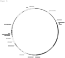
- Fig. 1 Biotinylation of AviTag (biotinylization peptide) using BirA (biotin protein ligase)
- the expression vector has polynucleotides coding for at least one biotin protein ligase (BirA).
- Biotin protein ligase (BirA) is particularly advantageously released intracellularly according to the invention.
- Biotinylation of a biotinylation peptide occurs intracellularly, preferably in the endoplasmic reticulum.
- the biotinylated receptor or the biotinylating peptide containing biotin is transported to the cell surface, namely in the form of a surface protein containing a biotinylating peptide, which binds and presents biotin.
- BirA contains a retention signal for the endoplasmic reticulum, such as KDEL (SEQ ID No. 4).
- the added antigen has an adapter, where the adapter binds to the biotin of the biotinylating peptide, and the bound antigen is labeled and the adapter is selected from avidin, streptavidin, neutravidin.
- the method according to the invention takes place under natural, physiological conditions (e.g. culture medium in an incubator) in which the host cells remain in the culture medium.
- the vitality of the cells advantageously remains unaffected.
- the culture medium can contain, for example, biotin and other auxiliary substances and additives.
- the addition of the multiple and distinct antigens occurs in the cold, such as 4 degrees Celsius, so that secretion of the antibody cannot occur.
- Said addition or incubation can preferably take place successively, for example at intervals of 20-40 minutes, so that, for example after removal of the host cells, a new addition (or incubation) can take place or the antigens can be resuspended in the culture medium.
- the antibodies of the antibody library can secrete from the host cell into the culture medium.
- the invention therefore relates to a method in which the addition of the multiple and different antigens takes place in the cold and the antibodies of the antibody library then secrete out of the host cell into the culture medium in the heat.
- blocking agents such as free avidin, streptavidin, neutravidin to biotin of the biotinylization peptide can be used to avoid non-specific binding.
- the invention therefore relates to a method in which additional blocking agents are added to the culture medium after the at least one antigen has been added.
- the expression vector according to the invention contains the following additional features, such as: Signal sequence, HA tag (SEQ ID No. 5, SEQ ID No. 6), DNA biotinylation sequence (SEQ ID No. 7), EGFR signal sequence (e.g. SEQ ID No. 8, SEQ ID No. 9), EGFR Sequence (SEQ ID No. 10, SEQ ID No. 11 including transmembrane domain), IRES (SEQ ID No. 12) and birA sequence (SEQ ID No. 13 including retention signal).
- sequences given include both polynucleotides and the expressed and translated sequences.
- Preferred suitable promoters are not limited to EF-1, SV-40, CMV, CAG, and many others. also conditionable promoters, such as TET on, off, light-dependent promoters or stress-inducible promoters (e.g. temperature) as well as with the help of the use of auxiliary sequences, such as IRES (internal ribosome entry site) 2A peptides, etc. Such promoters and auxiliary sequences are known to the person skilled in the art.
- the surface protein according to the invention can, for example, be transported safely to the cell surface and represented on the cell surface with the aid of an epidermal growth factor receptor (EGFR (supra), ErbB-1, HER1, etc.). Furthermore, the surface protein can have a control epitope, such as the hemagglutinin A epitope YPYDVPDYA (SEQ ID No. 14), so that it can be determined, for example cytometrically or by fluorescence microscopy using an antibody, whether the surface protein is extracellularly present on the cell membrane.
- EGFR epidermal growth factor receptor
- HER1 epidermal growth factor receptor
- the surface protein can have a control epitope, such as the hemagglutinin A epitope YPYDVPDYA (SEQ ID No. 14), so that it can be determined, for example cytometrically or by fluorescence microscopy using an antibody, whether the surface protein is extracellularly present on the cell membrane.
- An expression vector according to the invention is exemplified in its functional units (vector map) in the figures 2 and 3 played back.
- a specifically suitable expression vector is exemplified in figure 3 reproduced (SEQ ID No. 15).
- the expression vector can be divided into two or more units.
- the person skilled in the art is able to produce appropriate expression vectors and to introduce them into the host cell, preferably mammalian cell or human cell, preferably together—simultaneously or one after the other.
- a dual system consisting of a donor and helper plasmid can also be used, in which the transposase is encoded on a helper plasmid.
- the donor plasmid contains a cassette that is integrated into the genome.
- the transposase and the donor construct are encoded on one vector.
- antibody within the meaning of this invention relates to immunoglobulin molecules or immunologically active parts of immunoglobulin molecules, i. that is, molecules containing an antibody binding site or paratope.
- Exemplary antibody molecules are intact immunoglobulin molecules, substantially intact immunoglobulin molecules, and portions of an immunoglobulin molecule, including portions referred to in the art as Fab, Fab', F(ab')2, and F(v).
- An antibody is therefore a protein with one or more polypeptides which are encoded by immunoglobulin genes or polynucleotides which specifically bind an antigen to at least one epitope.
- immunoglobulin genes include the kappa, lambda, alpha (IgA), gamma (IgG1, IgG2, IgG3, IgG4), delta (IgD), epsilon (IgE) and mu (IgM) genes of the constant region and the myriad immunoglobulin variable region genes.
- Full-length immunoglobulin light chains are typically about 25 kD or 214 amino acids in length.
- heaviness Full-length immunoglobulin chains are typically about 50 kD or 446 amino acids in length.
- Light chains are encoded by a variable region gene at the NH 2 -terminus (ca. 110 amino acids in length) and by a kappa or lambda constant region gene at the COOH-terminus.
- Heavy chains are similarly encoded by one variable region gene (ca. 116 amino acids long) and one constant region gene.
- the basic structural unit of an antibody is usually a tetramer composed of two identical pairs of immunoglobulin chains, each pair containing a light chain and a heavy chain. In each pair, the light and heavy chain variable regions bind to an antigen and the constant domain mediate effector functions. Immunoglobulins also occur in a variety of other forms including, for example, Fv, Fab, and (Fab')2, as well as bifunctional hybrid antibodies and single chains.
- An immunoglobulin light or heavy chain variable region comprises a framework region interrupted by three hypervariable regions, also known as complementarity determining regions (CDR). As stated, the CDRs are primarily responsible for binding to an epitope of an antigen.
- An immune complex is an antibody, such as a monoclonal antibody, chimeric antibody, humanized antibody, or human antibody, or a functional antibody fragment that is specifically bound to the antigen.
- Chimeric antibodies are antibodies whose light and heavy chain genes have been produced from immunoglobulin variable and constant region genes from different species (e.g. mouse human etc.), usually by genetic engineering methods.
- antibody binding site encompasses the structural portion of an antibody molecule composed of variable and hypervariable regions of heavy and light chains that specifically bind an antigen, and consequently describe an immune response, where the so-called paratope of the antibody binds to the so-called epitope of the antigen.
- immune response means specific binding between a molecule containing an antigenic determinant and a molecule containing an antibody binding site, such as all or a portion of an antibody molecule.
- camelid antibodies according to the invention which do not have any light chains and are therefore able to carry out the antigen binding or immune reaction with only one chain.
- the invention also includes antibodies that can be produced recombinantly, in particular monoclonal antibodies, in the case of camelid antibodies, so-called nanobodies or VHH or VH fragments ( Aline Desmyter, Silvia Spinelli, Alain Roussel, Christian Cambillau, Camelid nanobodies: killing two birds with one stone, Current Opinion in Structural Biology, Volume 32, 2015, Pages 1-8, ISSN 0959-440X, https://doi.org /10.1016/j.sbi.2015.01.001 .).
- an antibody library is one that provides at least two or more different antibodies, at least in the expression vector according to the invention polynucleotides coding for at least one variable region ("variable domain") of the light and / or heavy chain and at least one constant region ("constant domain”).
- variable domain variable region
- constant domain constant region
- the constant region preferably has at least one Fc part, in particular at least one human, murine or camelid Fc.
- Antibody libraries are described, for example, in: Roger R. Beerli, Monika Bauer, Regula B. Buser, Myriam Gwerder, Simone Muntwiler, Patrik Maurer, Philippe Saudan, Martin F.
- the antibody library according to the invention can comprise a combinatorial or mutagenized antibody library, in particular naive, immune, non-immune and semisynthetic antibody libraries (see: Lim CC, Choong YS, Lim TS. Cognizance of Molecular Methods for the Generation of Mutagenic Phage Display Antibody Libraries for Affinity Maturation. Int J Mol Sci. 2019 Apr 15;20(8):1861. doi: 10.3390/ijms20081861. PMID: 30991723; PMCID: PMC6515083 ; Ponsel D, Neugebauer J, Ladetzki-Baehs K, Tissot K. High affinity, developability and functional size: the holy grail of combinatorial antibody library generation.
- naive, immune, non-immune and semisynthetic antibody libraries see: Lim CC, Choong YS, Lim TS. Cognizance of Molecular Methods for the Generation of Mutagenic Phage Display Antibody Libraries for Affinity Maturation
- variable domain variable domain
- constant domain constant domain
- proteins particularly preferably those such as murine Ig heavy chain V region 102 (SEQ. ID No. 16), murine Ig kappa (SEQ. ID No. 17), human proinsulin (SEQ. ID No. 18), human interleukin-2 (SEQ. ID No. 19) and human trypsinogen-2 (SEQ. ID No.
- the invention therefore includes an expression vector with polynucleotides coding for an antibody library with at least one variable region of the light and/or heavy chain and at least one constant region, in particular at least one human, murine or camelid Fc part, and at least one protein leader sequence .
- expression vector can have further embodiments, as described above.
- the invention relates to a mammalian cell comprising an expression vector according to the invention.
- the two batches were then mixed and incubated for a further 20 minutes. Thereafter, the transfection mixture was added dropwise to the cells. The cells were cultured dynamically for 24 h. The positive transfectants were then selected with selection media containing 2 ⁇ g/mL puromycin (ThermoFisher; Waltham, USA) for 7 days at 37° C., 125 rpm and 8% CO 2 .
- selection media containing 2 ⁇ g/mL puromycin (ThermoFisher; Waltham, USA) for 7 days at 37° C., 125 rpm and 8% CO 2 .
- Positively transfected HEK293T cells were sorted using MACS technology according to the manufacturer's protocol (Miltenyi; Bergisch Gladbach, Germany). Briefly, cells were harvested by centrifugation (300 xg for 8 min at 4°C) and resuspended in 200 ⁇ L MACS buffer (PBS/1% BSA; 2mM EDTA pH 7.4) and treated with 1 ⁇ g mouse anti- Hemagglutinin (HA) antibodies stained per 1x10⁶ cells. Staining was carried out at 4°C for 20 min. After the incubation, the cells were washed twice in 5 mL MACS buffer and then centrifuged (300 ⁇ g for 8 min at 4° C.).
- the cell pellet was resuspended in 200 ⁇ L MACS buffer with 10 ⁇ L of an anti-mouse IgG micro-bead suspension and incubated at 4°C for a further 20 min. After washing the cells twice in 5 mL MACS buffer, the cell pellet was resuspended in 3 mL MACS buffer and applied to a MACS LS column for magnetic cell separation. Positive cells were eluted by adding 5 mL of MACS buffer to the column after removing the column from the magnetic field. The positive cells were centrifuged and the cell pellet was resuspended in Expi293 expression medium supplemented with 2 ⁇ g/mL puromycin. The resuspended cells were cultured in 125 mL culture flasks (TPP, Ort) at 37°C, 125 rpm and 8% CO2.
- PBMC peripheral mononuclear cells
- RNAPure phenol-chloroform
- cDNA reverse transcriptase
- 3x10⁵ HA-AP-EGF-R+ HEK293T cells were seeded on poly-L-lysine-coated glass slides in 6-well plates and in DMEM complete medium supplemented with 2 ⁇ g/mL puromycin at 37°C and 8% CO2 incubated overnight. The cells were then washed with sterile PBS and fixed with 4% paraformaldehyde in PBS for 20 min at RT. After fixation, the slides were washed with sterile PBS and soaked in PBS/1% BSA (20 min at RT) to avoid non-specific binding events. Phycoerythrin (PE)-conjugated streptavidin is used to detect the biotinylated surface receptor.
- PE Phycoerythrin
Abstract
Die Erfindung betrifft ein neues Verfahren zur Selektion oder zum Screenen von Antikörpern und Antigenen aus einer Antikörperbibliothek samt geeigneten Expressionsvektoren und deren Verwendung.
Description
- Die Erfindung betrifft ein neues Verfahren zur Selektion oder zum Screenen von Antikörpern und Antigenen aus einer Antikörperbibliothek samt geeigneten Expressionsvektoren und deren Verwendung.
- Monoklonale Antikörper werden in der Regel mit Hilfe der Hybridomtechnik gewonnen, wobei antikörperproduzierende Zellen (B-Zellen oder B-Lymphozyten) mit Myelomzellen (Krebszellen) fusionieren, auch mittels Fusionszelllinien, woraufhin Hybride entstehen, die monoklonale Antikörper produzieren (G. Köhler, C. Milstein: Continuous cultures of fused cells secreting antibody of predefined specificity. In: Nature. Band 256, 1975, S. 495-497). Die Identifizierung der Hybridomzelle, die einen gegen das Antigen gerichteten Antikörper produziert, kann besonders vorteilhaft mit der SELMA® Technologie der Anmelderin erfolgen, wobei ein aus der Hybridomzelle erfolgreich sezernierter Antikörper (Ak) von dieser Hybridomzelle über eine Ak-Fängermatrix oder einem Antigen eingefangen bzw. an der Zelloberfläche gebunden wird und z.B. mittels fluoreszenzmarkierter Nachweis-Antikörper identifiziert wird.
- Die SELMA® Technologie wird in
WO2015161835 beschrieben. Insbesondere werden durch die Verwendung artifizieller Oberflächenmarker die Antikörper-produzierenden Hybridomzellen modifiziert. Dadurch entsteht eine direkte Verbindung zwischen dem Antikörperphänotyp und dem Genotyp der Hybridomzelle. Auf diese Weise können Hybridomzellen, die die gewünschten Antikörper produzieren mittels MACS- oder FACS-Technik äußerst effizient, sowie zeitsparend angereichert werden. -
WO 2015161835 A1 beschreibt zudem den Einsatz einer Ligationspeptidsequenz, mit der Funktion, dass die Bindung eines Adapterliganden erfolgen kann, wie z.B. Avidin/Biotin. An den Adapterliganden erfolgt die Bindung des freigesetzten Antikörpers aus der Hybridomzelle über einen Epitop aufweisenden Antigen-(Strept)avidin-Komplex, wobei der Antikörper an das Epitop des Antigens bindet und über die (Strept)avidin-Biotin Bindung (Adapterliganden), wiederum der Hybridomzelle zugeordnet und (aus)sortiert werden kann. Das Antigen kann alternativ mit einer Antikörper-Fängermatrix ersetzt werden. - Mit Hilfe der Durchflusszytometrie können beispielsweise solche Fluoreszenz-markierte Konstrukte mit einem flow sorter (Fluss-Sortierer) oder FACS (fluorescence-activated cell sorting) sortiert und isoliert werden.
-
WO 2015161835 A1 offenbart jedoch keine Antikörperbibliotheken und eine Selektion von Antikörpern daraus. Weiterhin wird inWO 2015161835 A1 nicht das Screenen von Antikörpern mittels eines oder mehreren Antigenen offenbart. - Antikörper können im Stand der Technik mit Phage-Display-Verfahren erzeugt werden (z.B.
WO91/17271 WO92/001047 WO92/20791 WO93/12227 - Zum Beispiel kann ein Phage Display, wie folgt bereitgestellt werden:
Phagen-Display Antikörperproduzierende B-Zellen oder deren mRNA werden z.B. aus Humanblut isoliert. Nach der Herstellung einer cDNA werden mittels PCR (Polymerase-Chain-Reaction) die DNA-Fragmente, die für die variablen Regionen (VL, VH) der Gene der leichten und schweren Ketten kodieren, vervielfältigt. In einer zweiten PCR werden VL - und VH - Fragmente über eine Linker-Sequenz, die für das scFv-Fragment kodiert, miteinander verbunden. Die entstandenen Fragmente kodieren für VH -G-G-G-G-S-S-S-VL - Sequenzen ("AK"). Die PCR-Fragmente können beispielweise unter Verwendung von Phagemid-Vektoren an ein DNA-Fragment, das aus einem verkürzten, für das Hüllprotein pIII (minor coat protein) des M13-Phagen kodierenden Gen und einem für ein Signalpeptid kodierenden Gen besteht, gebunden und in E. coli exprimiert werden. Das Signalpeptid bewirkt, dass die Fusionsproteine in das Periplasma gelangen. Nach Koinfektion mit einem M13-Helferphagen, der die Bildung des nicht modifizierten pIII und anderer Phagenproteine erlaubt, werden die fusionierten "AK" in die Außenhülle des Phagen integriert. Die Phagen enthalten die genetische Information des jeweiligen "AK"-Proteins, das sie auf ihrer Oberfläche präsentieren. Die Methode, mit der die Phagen, die das "richtige" "AK"-Protein präsentieren, über seine Antigenbindefähigkeiten erkannt werden können, wird "Biopanning" genannt. Auf diese Weise werden die DNA-Sequenz der DNA-Fragmente erhalten, die für die variablen Regionen der humanen leichten und humanen schweren Ketten eines ein bestimmtes Antigen bindenden AK kodieren, welche nachfolgend mit DNA verbunden werden, die für einen humanen Fc-Teil eines "AK" kodieren, um ein komplettes für einen "AK" kodierendes Gen zu erhalten. Das Verfahren ist jedoch aufwändig und zeitintensiv. Alternative Displays wie Ribosom Display sind im Stand der Technik beschrieben. - Solche Antikörperbibliotheken sind Gegenstand der vorliegenden Erfindung. Eine Antikörperbibliothek kann auf ein Target, insbesondere Antigen gescreent und die Binder werden über einen Enzym-gekoppelten Immunoassay selektiert. Anschließend müssen die Antikörpergene isoliert und umkloniert werden, um das identifizierte Antikörper-Fragment zu produzieren. Da der Phagendisplay zumeist den variablen Teil des Antikörpers aufweist, muss für die Produktion eines Volllängenpeptids noch ein geeigneter Fc Teil angefügt werden. Die Nachteile dieser Lösung liegen in den umfangreichen Arbeitsschritten, wie z.B. ein erheblicher Klonierungsaufwand für die Überführung von geeigneten Antikörper-Kandidaten in die Antikörper-Produktion. Jeder Antikörperkandidat muss einzeln kloniert werden. Sollten die Antikörper nach Produktion und Reinigung nicht funktional sein, muss der gesamte Prozess inklusive Phagendisplay wiederholt werden.
- Daher besteht die Aufgabe das aufwändige Screeningverfahren zu verbessern.
- Überraschenderweise kann durch die Integration einer Antikörperbibliothek in einen Expressionsvektor zur Selektion von gescreenten Antikörpern mittels eines Antigens direkt eine Produktion, insbesondere eines Volllängenformats durchgeführt werden, da der identifizierte Antikörper (oder Teile davon) unmittelbar auf die Produktionszelle hinweist. Weiterhin ist überraschend, dass der Einsatz von mehreren Antigenen zugleich möglich ist, und dennoch ein Screening erfolgen kann, obwohl durch die Sezernierung der verschiedenen Antikörper in das Kulturmedium eine (Fremd-)Beladung durch unspezifische Antigene erfolgen kann. Dies gilt ebenfalls für das erfindungsgemäße Oberflächenprotein enthaltend eine Ligationspeptidsequenz. Besonders überraschend ist daher, dass das erfindungsgemäße Verfahren für unterschiedliche sezernierte Antikörper aus sogar verschiedenen Antikörperbibliotheken erfolgreich simultan durchgeführt werden kann.
- Die Aufgabe wird daher durch die technische Lehre von mindestens einem Patentanspruch gelöst.
- Daher betrifft die Erfindung ein Verfahren zur Selektion oder zum Screenen ein oder mehrerer Antikörper aus ein oder mehreren Antikörperbibliotheken, wobei mindestens einem nach seiner Transformation stabil in das Genom integrierte Polynukleotide in einem Expressionsvektor in einer Wirtszelle verwendet wird, wobei der Expressionsvektor mindestens aufweist:
- i.) Polynukleotide kodierend für mindestens eine Antikörperbibliothek,
- ii.) Polynukleotide kodierend für ein Oberflächenprotein enthaltend eine Ligationspeptidsequenz und / oder mindestens ein Epitop für mindestens einen Antikörper aus einer oder mehreren Antikörperbibliotheken,
- a.) die Antikörper der Antikörperbibliothek aus der Wirtszelle in das Kulturmedium sezernieren und an mindestens ein zugegebenes Antigen binden, welche einen Adapter aufweist, wobei der Adapter an die Ligationspeptidsequenz aus ii.) bindet,
und /oder - b.) die Antikörper der Antikörperbibliothek aus der Wirtszelle in das Kulturmedium sezernieren und an mindestens ein zugegebenes Antigen binden, wobei der sezernierte Antikörper an mindestens ein Epitop aus ii.) bindet.
- Eine solche erfindungsgemäße Ausgangssituation an bereitgestellten Antikörperbibliotheken ist in
Figur 1A dargestellt. - Beispielweise kann der Fc-Teil des sezernierten Antikörpers an ein Epitop aus b.) binden.
- In einer weiteren bevorzugten Ausführungsform kann eine Markierung des gebundenen Antigens und /oder des gebundenen Antiköpers aus a.) oder b.) erfolgen, und zwar nach üblichen Methoden beispielsweise mit gelabelten (Sekundär-)Antikörpern (z.B. Fluoreszenz) oder magnetischen Beads.
- Mit Hilfe der Durchflusszytometrie können beispielsweise solche Fluoreszenz-markierte Konstrukte gemäß
Figur 1B (Antikörper-Fängermatrix) oder einem Antigen mit einem flow sorter (Fluss-Sortierer) oder FACS (fluorescence-activated cell sorting) sortiert und isoliert werden. - Mithilfe des mindestens einen zugegebenen Antigens kann nach einem geeigneten Antikörper aus der Antikörperbibliothek gescreent, und der Antikörper z.B. nach Zellsortierung produziert werden.
- Daher betrifft die Erfindung die Verwendung von mindestens einem oder mehr als einem Antigen zur Selektion oder zum Screenen ein oder mehrerer Antikörper aus ein oder mehreren Antikörperbibliotheken, wobei mindestens einem nach seiner Transformation stabil in das Genom integrierte Polynukleotide in einem Expressionsvektor in einer Wirtszelle verwendet wird, wobei der Expressionsvektor mindestens aufweist:
- i.) Polynukleotide kodierend für mindestens eine Antikörperbibliothek,
- ii.) Polynukleotide kodierend für ein Oberflächenprotein enthaltend eine Ligationspeptidsequenz und / oder mindestens ein Epitop für mindestens einen Antikörper aus einer oder mehreren Antikörperbibliotheken,
- a.) die Antikörper der Antikörperbibliothek aus der Wirtszelle in das Kulturmedium sezernieren und an mindestens ein zugegebenes Antigen binden, welche einen Adapter aufweist, wobei der Adapter an die Ligationspeptidsequenz aus ii.) bindet,
und /oder - b.) die Antikörper der Antikörperbibliothek aus der Wirtszelle in das Kulturmedium sezernieren und an mindestens ein zugegebenes Antigen binden, wobei der sezernierte Antikörper an mindestens ein Epitop aus ii.) bindet.
- Das "mindestens eine Antigen" kann beliebiger Natur sein und insbesondere eine chemische Substanz, Protein, Peptid, Wirkstoff, Medikament oder ein Antiköper sein. "Mindestens ein Antigen" heißt, dass die Antigene gleich oder vorzugsweise verschieden sind, und zwar hinsichtlich der chemischen Zusammensetzung und der jeweiligen Menge bzw. Konzentration. Beispielsweise können mehr als ein Antigen, insbesondere 100 oder 1000 oder mehr verschiedene Antigene eingesetzt werden.
- Überraschender Weise ist das erfindungsgemäße Verfahren hinreichend robust, auch mit dieser Vielzahl an Antikörpern und Antigenen zurecht zu kommen und eine Selektion bzw. Auswahl (Screenen) von Antikörpern zu Antigenen und vice versa zu ermöglichen.
- Das besagte Epitop ist Gegenstand des Oberflächenproteins, insbesondere kann das Epitop als Teil eines Antikörpers oder eines Antigens ausgebildet sein.
- Die Wirtszelle ist besonders bevorzugt eine Säugetierzelle einschließlich Humanzelle, insbesondere ausgewählt aus HEK293-Zellen, CHO-Zellen, insbesondere Zelllinien, solche wie, nicht abschließend, NS0, Sp2/0, PER.C6, BHK, COS-7 u.a.. Geeignete Zellen zur Antikörperproduktion sind dem Fachmann bekannt (Kunert R, Reinhart D. Advances in recombinant antibody manufacturing. Appl Microbiol Biotechnol. 2016 Apr;100(8):3451-61. doi: 10.1007/s00253-016-7388-9). Die Wirtszelle ist jedoch keine Hybridomzelle.
- Die besagte Ligationspeptidsequenz weist im Fall von Biotin eine Biotinakzeptorpeptidsequenz bzw. Biotinylisierungspeptid, wie vorzugsweise GLNDIFEAQKIEWHE (SEQ ID No. 1) oder LNDIFEAQKIEWH (SEQ ID No. 2) auf. Auch andere Biotinakzeptorpeptidsequenz bzw. Biotinylisierungspeptide können geeignet sein, wie ein Alignment mit 13 Aminosäuren 1-LXXIXXXXKX XXX-13 gemäß SEQ ID. No. 3 (vgl. D. Beckett, E. Kovaleva, and P. J. Schatz, A minimal peptide substrate in biotin holoenzyme synthetase-catalyzed biotinylation, Protein Sci. 1999 Apr; 8(4): 921-929), wobei:
- Aminosäure 2 N, C oder beliebig oder entfernt sein kann,
- Aminosäure 3 beliebig, jedoch ausgenommen L, V, I, W, F, Y sein kann,
- Aminosäure 5 F oder L sein kann,
- Aminosäure 6 E oder D sein kann,
- Aminosäure 7 A, G, S, T sein kann,
- Aminosäure 8 Q oder M sein kann,
- Aminosäure 10 I, M, V sein kann,
- Aminosäure 11 E, L, V, Y, I sein kann,
- Aminosäure 12 W, Y, V, F, L, I sein kann,
- Aminosäure 13 R, H oder beliebig, jedoch ausgenommen D, E.
-

- In einer bevorzugten Ausführungsform der Erfindung weist der Expressionsvektor Polynukleotide kodierend für mindestens eine Biotin-Proteinligase (BirA) auf. Besonders vorteilhaft wird erfindungsgemäß Biotin-Proteinligase (BirA) intrazellulär freigesetzt. Die Biotinylierung eines Biotinylierungspeptid erfolgt intrazellulär vorzugsweise im endoplasmatischen Retikulum. Anschließend wird der biotinylierte Rezeptor bzw. das Biotin aufweisende Biotinylisierungspeptid an die Zelloberfläche transportiert, und zwar in Form eines Oberflächenproteins aufweisend ein Biotinylierungspeptid, welches Biotin bindet und präsentiert. In einer weiteren bevorzugten Ausführungsform enthält BirA ein Retentionssignal für das endoplasmatische Retikulum, wie KDEL (SEQ ID No. 4).
- Das zugegebene Antigen weist einen Adapter auf, wobei der Adapter an das Biotin des Biotinylisierungspeptids bindet, und das gebundene Antigen markiert wird und der Adapter ist ausgewählt aus Avidin, Streptavidin, Neutravidin.
- Insbesondere erfolgt das erfindungsgemäße Verfahren unter natürlichen, physiologischen Bedingungen (z.B. Kulturmedium im Brutschrank) bei dem die Wirtszellen im Kulturmedium bleiben. Die Vitalität der Zellen bleibt vorteilhaft unberührt. Das Kulturmedium kann z.B. Biotin u.a. Hilfs- und Zusatzstoffe enthalten.
- In einer weiteren bevorzugten Ausführungsform erfolgt die Zugabe der mehreren und verschiedenen Antigene in der Kälte, wie z.B. 4 Grad Celsius, sodass eine Sezernierung des Antikörpers nicht erfolgen kann. Die besagte Zugabe oder Inkubation kann vorzugsweise sukzessiv erfolgen, beispielsweise in Intervallen von 20 - 40 Minuten, sodass z.B. nach Entnahme der Wirtszellen eine erneute Zugabe (bzw. Inkubation) erfolgen kann bzw. die Antigene erneut im Kulturmedium suspendieren können.
- Anschließend können in der Wärme, wie z.B. 37 Grad Celsius die Antikörper der Antikörperbibliothek aus der Wirtszelle in das Kulturmedium sezernieren.
- Daher betrifft die Erfindung ein Verfahren, wobei die Zugabe der mehreren und verschiedenen Antigene in der Kälte erfolgt und anschließend in der Wärme die Antikörper der Antikörperbibliothek aus der Wirtszelle in das Kulturmedium sezernieren.
- In einem weiteren vorteilhaften Verfahrensschritt können zur Vermeidung von unspezifischen Bindungen Blockierungsmittel, wie freies Avidin, Streptavidin, Neutravidin an Biotin des Biotinylisierungspeptids eingesetzt werden.
- Daher betrifft die Erfindung ein Verfahren, wobei zusätzlich Blockierungsmittel nach Zugabe des mindestens einen Antigens in das Kulturmedium hinzugegeben werden.
- In einer bevorzugten Ausführungsform enthält der erfindungsgemäße Expressionsvektor folgende weitere Merkmale, wie:
Signalsequenz, HA-Tag (SEQ ID No. 5, SEQ ID No. 6), DNA-Biotinylierungssequenz (SEQ ID No. 7), EGFR-Signalsequenz (z.B. SEQ ID No. 8, SEQ ID No. 9), EGFR-Sequenz (SEQ ID No. 10, SEQ ID No. 11 inkl. Transmenbrandomäne), IRES (SEQ ID No. 12) als auch birA-Sequenz (SEQ ID No. 13 inkl. Retentionssignal). - Die angegebenen Sequenzen umfassen sowohl Polynukleotide als auch die exprimierten und translatierten Sequenzen.
- Bevorzugte geeignete Promotoren sind nicht abschließend EF-1, SV-40, CMV, CAG, u.v.a. auch konditionierbare Promotoren, wie TET on, off, lichtabhängige Promotoren oder stressinduzierbare Promotoren (z.B. Temperatur) als auch mit Hilfe der Verwendung von Hilfssequenzen, wie IRES (internal ribosome entry site) 2A Peptide u.a.. Solche Promotoren und Hilfssequenzen sind dem Fachmann einschlägig bekannt.
- Das erfindungsgemäße Oberflächenprotein kann beispielsweise mit Hilfe von einem Epidermal Growth Factor Receptor (EGFR (supra), ErbB-1, HER1 u.a.) sicher an die Zelloberfläche transportiert und an der Zelloberfläche repräsentiert werden. Weiterhin kann das Oberflächenprotein ein Kontrollepitop, wie z.B. Hämagglutinin-A-Epitop YPYDVPDYA (SEQ ID No. 14) aufweisen, so dass beispielsweise cytometrisch oder fluoreszenzmikroskopisch mit Hilfe eines Antikörpers festgestellt werden kann, ob das Oberflächenprotein an der Zellmembran extrazellulär präsent ist.
- Ein erfindungsgemäßer Expressionsvektor ist in seinen funktionellen Einheiten (Vektorkarte) beispielhaft in den
Figuren 2 und3 wiedergegeben. - Ein konkret geeigneter Expressionsvektor ist beispielhaft in
Figur 3 wiedergegeben (SEQ ID No. 15). - Selbstredend kann der Expressionsvektor in zwei oder mehr Einheiten aufgeteilt werden. Der Fachmann ist in der Lage entsprechende Expressionsvektoren herzustellen und diese vorzugsweise gemeinsam - simultan oder nacheinander - in die Wirtszelle, vorzugsweise Säugetierzelle oder Humanzelle einzubringen.
- In einer weiteren Ausführungsform kann ebenfalls ein duales System bestehend aus einem Donor- und Helferplasmid verwendet werden, bei dem die Transposase auf einem Helferplasmid kodiert ist. Das Donorplasmid beinhaltet eine Kassette, die ins Genom integriert wird. Es besteht auch die Möglichkeit, dass die Transposase und das Donor-Konstrukt auf einem Vektor kodiert sind.
- Der Begriff "Antikörper" im Sinne dieser Erfindung betrifft Immunglobulinmoleküle oder immunologisch aktive Teile von Immunglobulinmolekülen, d. h., Moleküle, die eine antikörperverbindende Stelle oder ein Paratop enthalten.
- Beispielhafte Antikörpermoleküle sind intakte Immunglobulinmoleküle, im Wesentlichen intakte Immunglobulinmoleküle und Teile eines Immunglobulinmoleküls, einschließlich der auf dem Fachgebiet als Fab, Fab', F(ab')2 und F(v) bezeichneten Teile.
- Ein Antikörper ist daher ein Protein mit einem oder mehreren Polypeptiden, die von Immunglobulingenen bzw. Polynucleotiden kodiert sind, die spezifisch an mindestens einem Epitop ein Antigen binden. Zu den bekannten Immunglobulingenen gehören die Kappa-, Lambda-, Alpha- (IgA), Gamma- (IgG1, IgG2, IgG3, IgG4), Delta- (IgD), Epsilon- (IgE) und Mu-(IgM)- Gene der konstanten Region sowie die unzähligen Immunglobulingene der variablen Region. Leichte Immunglobulinketten der vollen Länge sind in der Regel ca. 25 kD oder 214 Aminosäuren lang. Schwere Immunglobulinketten der vollen Länge sind in der Regel ca. 50 kD oder 446 Aminosäuren lang. Leichte Ketten werden durch ein Gen der variablen Region am NH2-Terminus (ca. 110 Aminosäuren lang) und durch ein Kappa- oder Lambda-Gen der konstanten Region am COOH-Terminus codiert. Schwere Ketten werden in ähnlicher Weise durch ein Gen der variablen Region (ca. 116 Aminosäuren lang) und durch ein Gen der anderen konstanten Region codiert.
- Die grundlegende strukturelle Einheit eines Antikörpers ist in der Regel ein Tetramer, das aus zwei identischen Paaren von Immunglobulinketten besteht, wobei jedes Paar eine leichte und eine schwere Kette enthält. In jedem Paar binden die variablen Regionen ("variable domain") der leichten und der schweren Kette an ein Antigen und die konstanten Regionen ("constant domain") vermitteln Effektorfunktionen. Immunglobuline treten auch in einer Vielfalt von anderen Formen, darunter zum Beispiel Fv, Fab, und (Fab')2, sowie als bifunktionelle hybride Antikörper und Einzelketten auf. Eine variable Region einer leichten oder schweren Kette eines Immunglobulins umfasst eine Rahmenregion, die von drei hypervariablen Regionen, auch als Komplementarität bestimmende Regionen (Complementarity Determining Regions, CDR) bekannt, unterbrochen wird. Wie ausgeführt, sind die CDR in erster Linie verantwortlich für die Bindung an ein Epitop eines Antigens. Ein Immunkomplex ist ein Antikörper wie zum Beispiel ein monoklonaler Antikörper, chimärer Antikörper, humanisierter Antikörper oder humaner Antikörper oder ein funktionales Antikörperfragment, der spezifisch an das Antigen gebunden ist. Chimäre Antikörper sind Antikörper, deren leichte und schwere Kettengene aus Immunglobulingenen der variablen und konstanten Region aus verschiedenen Spezies hergestellt wurden (z.B. Maus Mensch u.a.), in der Regel durch gentechnische Verfahren.
- Der Begriff "antikörperverbindende Stelle" umfasst den strukturellen Anteil eines Antikörpermoleküls, der aus variablen und hypervariablen Regionen von schweren und leichten Ketten besteht, die spezifisch ein Antigen binden, und folglich eine Immunreaktion beschreiben, wobei das sog. Paratop des Antikörpers an das sog. Epitop des Antigens bindet.
- Der Begriff "Immunreaktion" bedeutet eine spezifische Bindung zwischen einem Molekül, das eine antigene Determinante enthält, und einem Molekül, das eine antikörperverbindende Stelle enthält, wie ein Gesamtantikörpermolekül oder ein Teil davon.
- Ebenfalls erfindungsgemäß umfasst sind erfindungsgemäß kamelide Antikörper, welche keine leichten Ketten aufweisen und daher in der Lage sind, die Antigenbindung bzw. Immunreaktion mit nur einer Kette zu realisieren.
- Weiterhin sind erfindungsgemäß solche Antikörper umfasst, die rekombinant hergestellt werden können, insbesondere monoklonale Antikörper, im Fall von kameliden Antikörper, sogenannte Nanobodies oder VHH bzw. VH Fragmente (Aline Desmyter, Silvia Spinelli, Alain Roussel, Christian Cambillau, Camelid nanobodies: killing two birds with one stone, Current Opinion in Structural Biology, Volume 32, 2015, Pages 1-8, ISSN 0959-440X, https://doi.org/10.1016/j.sbi.2015.01.001.).
- Im Sinne dieser Erfindung ist eine Antikörperbibliothek eine solche, welche mindestens zwei und mehr verschiedene Antikörper bereitstellt, wobei zumindest im erfindungsgemäßen Expressionsvektor Polynukleotide kodierend für mindestens eine variable Region ("variable domain") der leichten und/oder der schweren Kette und mindestens eine konstante Region ("constant domain") aufweisen. Der Fachmann ist in der Lage solche Antikörperbibliotheken in einem Expressionsvektor bereitzustellen, wie vorstehend beschrieben. Bevorzugt weist die konstante Region ("constant domain") mindestens einen Fc-Teil auf, insbesondere mindestens einen humanen, murinen oder kameliden Fc. Antikörperbibliotheken sind z.B. beschreiben in: Roger R. Beerli, Monika Bauer, Regula B. Buser, Myriam Gwerder, Simone Muntwiler, Patrik Maurer, Philippe Saudan, Martin F. Bachmann, Isolation of human monoclonal antibodies by mammalian cell display, Proceedings of the National Academy of Sciences Sep 2008, 105 (38) 14336-14341; DOI: 10.1073/pnas.0805942105; Hoogenboom HR. Selecting and screening recombinant antibody libraries. Nat Biotechnol. 2005 Sep;23(9):1105-16. doi: 10.1038/nbt1126. PMID: 16151404; Ledsgaard L, Kilstrup M, Karatt-Vellatt A, McCafferty J, Laustsen AH. Basics of Antibody Phage Display Technology. Toxins (Basel). 2018 Jun 9;10(6):236. doi: 10.3390/toxins10060236. PMID: 29890762; PMCID: PMC6024766; Kumar R, Parray HA, Shrivastava T, Sinha S, Luthra K. Phage display antibody libraries: A robust approach for generation of recombinant human monoclonal antibodies. Int J Biol Macromol. 2019 Aug 15;135:907-918. doi: 10.1016/j.ijbiomac.2019.06.006. Epub 2019 Jun 3. PMID: 31170490; Reader RH, Workman RG, Maddison BC, Gough KC. Advances in the Production and Batch Reformatting of Phage Antibody Libraries. Mol Biotechnol. 2019 Nov;61(11):801-815. doi: 10.1007/s12033-019-00207-0. PMID: 31468301; PMCID: PMC6785589.
- Ferner kann die erfindungsgemäße Antikörperbibliothek eine kombinatorische oder mutagenisierte Antikörperbibliothek umfassen, insbesondere naive, immune, nicht-immune und semisynthetische Antikörperbibliotheken (siehe: Lim CC, Choong YS, Lim TS. Cognizance of Molecular Methods for the Generation of Mutagenic Phage Display Antibody Libraries for Affinity Maturation. Int J Mol Sci. 2019 Apr 15;20(8):1861. doi: 10.3390/ijms20081861. PMID: 30991723; PMCID: PMC6515083; Ponsel D, Neugebauer J, Ladetzki-Baehs K, Tissot K. High affinity, developability and functional size: the holy grail of combinatorial antibody library generation. Molecules. 2011 May 3;16(5):3675-700. doi: 10.3390/molecules16053675; McConnell AD, Do M, Neben TY, Spasojevic V, MacLaren J, Chen AP, Altobell L 3rd, Macomber JL, Berkebile AD, Horlick RA, Bowers PM, King DJ. High affinity humanized antibodies without making hybridomas; immunization paired with mammalian cell display and in vitro somatic hypermutation, PLoS One. 2012;7(11):e49458. doi: 10.1371/journal.pone.0049458).
- Zur Expression der variablen Regionen ("variable domain) und der konstanten Regionen ("constant domain") werden sogenannte Protein-Leader Sequenzen eingesetzt, insbesondere vorzugsweise solche wie, murines Ig heavy chain V region 102 (SEQ. ID No. 16), murine Ig Kappa (SEQ. ID No. 17), humanes Proinsulin (SEQ. ID No. 18), humanes Interleukin-2 (SEQ. ID No. 19) und humanes Trypsinogen-2 (Seq. ID No. 20)) (Marion E.E. Watson, Compilation of published signal sequences, Nucleic Acids Research, Volume 12, Issue 13, 11 July 1984, Pages 5145-5164, https://doi.org/10.1093/nar/12.13.5145, Owji H, Nezafat N, Negahdaripour M, Hajiebrahimi A, Ghasemi Y. A comprehensive review of signal peptides: Structure, roles, and applications. Eur J Cell Biol. 2018 Aug;97(6):422-441. doi: 10.1016/j.ejcb.2018.06.003, Stroud RM, Walter P. Signal sequence recognition and protein targeting. Curr Opin Struct Biol. 1999 Dec;9(6):754-9. doi: 10.1016/s0959-440x(99)00040-8, Chen Y, Shanmugam SK, Dalbey RE. The Principles of Protein Targeting and Transport Across Cell Membranes. Protein J. 2019 Jun;38(3):236-248. doi: 10.1007/s10930-019-09847-2.
- Daher umfasst die Erfindung einen Expressionsvektor mit Polynukleotiden kodierend für eine Antikörperbibliothek mit mindestens einer variablen Region der leichten und/oder der schweren Kette und mindestens einer konstanten Region, insbesondere mindestens einen humanen, murinen oder kameliden Fc-Teil, und mindestens einer Protein-Leader Sequenz.
- Weiterhin umfasst die Erfindung einen Expressionsvektor aufweisend
- i.) Polynukleotide kodierend für mindestens eine Antikörperbibliothek,
- ii.) Polynukleotide kodierend für ein Oberflächenprotein enthaltend eine Ligationspeptidsequenz und / oder mindestens ein Epitop für mindestens einen Antikörper aus einer oder mehreren Antikörperbibliotheken.
- Weiterhin kann der Expressionsvektor weitere Ausführungsformen aufweisen, wie vorstehend beschrieben.
- Ferner betrifft die Erfindung eine Säugetierzelle, die einen erfindungsgemäßen Expressionsvektor umfasst.
- Die folgenden Beispiele und Figuren sollen die Erfindung näher erläutern, ohne jedoch diese zu beschränken.
- Humane embryonale Nierenzellen (Expi293F HEK) wurden bei 37°C, 125 rpm und 8% CO2 in Expi293 Expression Medium (ThermoFisher; Waltham, USA) kultiviert.
- Die Transfektion der Expi293F HEK Zelllinie (ThermoFisher; Waltham, USA) mit der Antikörperbibliothek und dem Oberflächenmarker-exprimierenden Genkonstrukt HA-AP-EGF-R (
Fig. 2 ) wurde in einer dynamischen Kultur durchgeführt. Die Expi293F HEK Zellen wurden am Tag der Transfektion auf eine Zellzahl von 150E6 Zellen in 50 mL Expi293 Expression Medium eingestellt. Für die Transfektion der Expi293F HEK Zellen wurden 50 µg Plasmid-DNA und 8 µg Helferplasmid mit Opi-MEM™ I Reduced Serum Medium sowie ExpiFectamine™ 293 Reagent vermengt und für 5 Minuten inkubiert. Im Anschluss wurden beide Ansätze vermengt und weitere 20 Minuten inkubiert. Danach wurde der Transfektionsansatz tropfenweise zu den Zellen gegeben. Die Zellen wurden für 24 h dynamisch kultiviert. Danach erfolgte die Selektion der positiven Transfektanten mit Selektionsmedien, die 2 µg/mL Puromycin (ThermoFisher; Waltham, USA) enthielten, für 7 Tage bei 37 °C, 125 rpm und 8 % CO2. - Positiv transfizierte HEK293T-Zellen wurden mittels MACS-Technologie nach dem Protokoll des Herstellers sortiert (Miltenyi; Bergisch Gladbach, Deutschland). Kurz gesagt wurden die Zellen durch Zentrifugation (300 x g für 8 min bei 4°C) geerntet und in 200 µL MACS-Puffer (PBS/1% BSA; 2 mM EDTA pH 7,4) resuspendiert und mit 1 µg Maus-Anti-Hämagglutinin (HA)-Antikörper pro 1x10⁶ Zellen gefärbt. Die Färbung wurde für 20 min bei 4°C durchgeführt. Nach der Inkubation wurden die Zellen zweimal in 5 mL MACS-Puffer gewaschen und anschließend zentrifugiert (300 x g für 8 min bei 4°C). Das Zellpellet wurde in 200 µL MACS-Puffer mit 10 µL einer Anti-Maus-IgG-Micro-Bead-Suspension resuspendiert und für weitere 20 min bei 4°C inkubiert. Nach zweimaligem Waschen der Zellen in 5 mL MACS-Puffer wurde das Zellpellet in 3 mL MACS-Puffer resuspendiert und auf eine MACS LS-Säule zur magnetischen Zellseparation aufgetragen. Positive Zellen wurden durch Zugabe von 5 mL MACS-Puffer auf die Säule eluiert, nachdem die Säule aus dem Magnetfeld entfernt wurde. Die positiven Zellen wurden zentrifugiert und das Zellpellet wurde in Expi293 Expression Medium, ergänzt mit 2 µg/mL Puromycin, resuspendiert. Die resuspendierten Zellen wurden in 125 mL Kulturflaschen (TPP, Ort) bei 37°C, 125 rpm und 8% CO2 kultiviert.
- Für die Herstellung einer Nanobody Library wurden zunächst periphere mononukleäre Zellen (PBMC) aus dem Vollblut von Lamas (Llama glama) isoliert. Aus diesen PBMCs wurde die RNA nach dem Standardprotokoll mit Phenol-Chloroform isoliert (RNAPure, VWR). Im Anschluss erfolgte die Erzeugung der komplementären DNS (cDNA) mittels der Reversen Transkriptase. Diese dient als Grundlage für die Amplifikation der VH und VHH Gene (=initiale PCR). In einer zweiten PCR wurde, sofern gewünscht, zwischen den VH und den VHH Gen-Amplifikaten diskriminiert. Im nächsten Schritt erfolgt die Integration der VHs und/oder VHHs in den zuvor linearisierten Zielvektor über homologe Rekombination. Die daraus resultierenden Vektoren stellen die Nanobody Library dar.
- Für die Immunfluoreszenz wurden 3x10⁵ HA-AP-EGF-R+ HEK293T-Zellen auf mit Poly-L-Lysin beschichteten Glasobjektträgern in 6-WellPlatten ausgesät und in DMEM-Vollmedium, ergänzt mit 2 µg/mL Puromycin, bei 37°C und 8% CO2 über Nacht inkubiert. Die Zellen wurden dann mit sterilem PBS gewaschen und mit 4% Paraformaldehyd in PBS für 20 min bei RT fixiert. Nach der Fixierung wurden die Objektträger mit sterilem PBS gewaschen und mit PBS/1% BSA (20 min bei RT) getränkt, um unspezifische Bindungsereignisse zu vermeiden. Phycoerythrin (PE)-konjugiertes Streptavidindient dem Nachweis des biotinylieren Oberflächenrezeptors. Dafür wurden 2 µg des PE-Streptavidinkonjugates (eBioScience 12-4317; 1E6 Zellen) in PBS/1% BSA verdünnt und für 20 min inkubiert. Danach wurde ein Waschschritt mit PBS/1% BSA durchgeführt. Für die fluoreszenzmikroskopische Analyse wurden die Objektträger noch einmal mit sterilem PBS und einmal mit destilliertem Wasser gewaschen, bevor sie in TissueTek montiert wurden. Die mikroskopischen Aufnahmen wurden mit dem LSM800 (Zeiss; Oberkochen, Deutschland)
- Für die durchflusszytometrischen Analysen wurden die Zellen in FACS-Puffer (PBS mit 0,5 % BSA und 0,01 % Natriumazid) zwei Mal gewaschen. Im Anschluss wurde der biotinylierte Oberflächenrezeptor durch ein Phycoerythrin (PE)-konjugiertes Streptavidin nachgewiesen. Dafür wurden 1 µg des PE-Streptavidinkonjugates (eBioScience 12-4317; 1E6 Zellen) in FACS-Puffer verdünnt und für 20 min inkubiert. Durch den nachfolgenden Waschschritt wurde überschüssiges PE-Streptavidinkonjugat entfernt. Die Zellen wurde in FACS-Puffer erneut aufgenommen und wurden direkt im Anschluss im Durchflusszytometer (BD FACSAria™
Claims (12)
- Verfahren zur Selektion oder zum Screenen ein oder mehrerer Antikörper aus ein oder mehreren Antikörperbibliotheken, wobei mindestens einem nach seiner Transformation stabil in das Genom integrierte Polynukleotide in einem Expressionsvektor in einer Wirtszelle verwendet wird, wobei der Expressionsvektor mindestens aufweist:i.) Polynukleotide kodierend für mindestens eine Antikörperbibliothek,ii.) Polynukleotide kodierend für ein Oberflächenprotein enthaltend eine Ligationspeptidsequenz und / oder mindestens ein Epitop für mindestens einen Antikörper aus einer oder mehreren Antikörperbibliotheken,
wobeia.) die Antikörper der Antikörperbibliothek aus der Wirtszelle in das Kulturmedium sezernieren und an mindestens ein zugegebenes Antigen binden, welche einen Adapter aufweist, wobei der Adapter an die Ligationspeptidsequenz aus ii.) bindet,
und /oderb.) die Antikörper der Antikörperbibliothek aus der Wirtszelle in das Kulturmedium sezernieren und an mindestens ein zugegebenes Antigen binden, wobei der sezernierte Antikörper an mindestens ein Epitop aus ii.) bindet. - Verfahren zur Selektion oder Screenen ein oder mehrerer Antikörper aus ein oder mehreren Antikörperbibliotheken nach Anspruch 1, wobei eine Markierung des gebundenen Antigens aus a.) oder b.) und / oder des gebundenen Antikörpers aus a.) oder b.) erfolgt, insbesondere mit einem gelabelten Antikörper oder magnetischen Bead und mit einem flow sorter (Fluss-Sortierer) oder FACS (fluorescence-activated cell sorting) sortiert und isoliert werden.
- Verfahren zur Selektion oder Screenen ein oder mehrerer Antikörper aus ein oder mehreren Antikörperbibliotheken nach einem der vorstehenden Ansprüche, wobei die Antikörperbibliothek mindestens eine variable Region der leichten und/oder der schweren Kette und mindestens eine konstante Region aufweisen, insbesondere mindestens einen humanen, murinen oder kameliden Fc-Teil, und mindestens eine Protein-Leader Sequenz.
- Verfahren zur Selektion oder Screenen ein oder mehrerer Antikörper aus ein oder mehreren Antikörperbibliotheken nach einem der vorstehenden Ansprüche, wobei die Antikörperbibliothek ausgewählt ist aus der Gruppe kombinatorische oder mutagenisierte Antikörperbibliothek insbesondere naive, immune, nicht-immune und semisynthetische Antikörperbibliothek.
- Verfahren zur Selektion oder Screenen ein oder mehrerer Antikörper aus ein oder mehreren Antikörperbibliotheken nach einem der vorstehenden Ansprüche, wobei die Wirtszelle eine Säugetierzelle ist, insbesondere HEK293-Zellen, CHO-Zellen.
- Verfahren zur Selektion oder Screenen ein oder mehrerer Antikörper aus ein oder mehreren Antikörperbibliotheken nach einem der vorstehenden Ansprüche, wobei die Ligationspeptidsequenz ein Biotinylierungspeptid ist ausgewählt aus der Gruppe1-LXXIXXXXKX XXX-13 (SEQ ID No. 3) mitAminosäure 2 N, C oder beliebig oder entfernt sein kann,Aminosäure 3 beliebig, jedoch ausgenommen L, V, I, W, F, Y sein kann,Aminosäure 5 F oder L sein kann,Aminosäure 6 E oder D sein kann,Aminosäure 7 A, G, S, T sein kann,Aminosäure 8 Q oder M sein kann,Aminosäure 10 I, M, V sein kann,Aminosäure 11 E, L, V, Y, I sein kann,Aminosäure 12 W, Y, V, F, L, I sein kann,Aminosäure 13 R, H oder beliebig, jedoch ausgenommen D, E oder das Biotinylierungspeptid ist ausgewählt aus der Gruppe GLNDIFEAQKIEWHE (SEQ ID No. 1) oder LNDIFEAQKIEWH (SEQ ID No. 2) und der Adapter ist ausgewählt aus Avidin, Streptavidin, Neutravidin.
- Verfahren zur Selektion oder Screenen ein oder mehrerer Antikörper aus ein oder mehreren Antikörperbibliotheken nach einem der vorstehenden Ansprüche, wobei der Expressionsvektor Polynukleotide aufweist ausgewählt aus der Gruppe kodierend für mindestens eine Biotin-Proteinligase (BirA), Signalsequenz, HA-Tag (SEQ ID No. 5, SEQ ID No. 6), DNA-Biotinylierungssequenz (SEQ ID No. 7), EGFR-Signalsequenz (SEQ ID No. 8, SEQ ID No. 9), EGFR-Sequenz (SEQ ID No. 10, SEQ ID No. 11 inkl. Transmenbrandomäne), IRES (SEQ ID No. 12), birA-Sequenz (SEQ ID No. 13 inkl. Retentionssignal).
- Verfahren zur Selektion oder Screenen ein oder mehrerer Antikörper aus ein oder mehreren Antikörperbibliotheken nach einem der vorstehenden Ansprüche, wobei die Zugabe der mehreren und verschiedenen Antigene in der Kälte erfolgt und anschließend in der Wärme die Antikörper der Antikörperbibliothek aus der Wirtszelle in das Kulturmedium sezernieren.
- Verfahren zur Selektion oder Screenen ein oder mehrerer Antikörper aus ein oder mehreren Antikörperbibliotheken nach einem der vorstehenden Ansprüche, wobei zusätzlich Blockierungsmittel nach Zugabe des mindestens einen Antigens in das Kulturmedium hinzugegeben werden.
- Expressionsvektor aufweisendi.) Polynukleotide kodierend für mindestens eine Antikörperbibliothek,ii.) Polynukleotide kodierend für ein Oberflächenprotein enthaltend eine Ligationspeptidsequenz und / oder mindestens ein Epitop für mindestens einen Antikörper aus einer oder mehreren Antikörperbibliotheken.
- Säugetierzelle umfassend einen Expressionsvektor nach Anspruch 10.
- Verwendung von mindestens einem oder mehr als einem Antigen zur Selektion oder zum Screenen ein oder mehrerer Antikörper aus ein oder mehreren Antikörperbibliotheken, wobei mindestens einem nach seiner Transformation stabil in das Genom integrierte Polynukleotide in einem Expressionsvektor in einer Wirtszelle verwendet wird, wobei der Expressionsvektor mindestens aufweist:i.) Polynukleotide kodierend für mindestens eine Antikörperbibliothek,ii.) Polynukleotide kodierend für ein Oberflächenprotein enthaltend eine Ligationspeptidsequenz und / oder mindestens ein Epitop für mindestens einen Antikörper aus einer oder mehreren Antikörperbibliotheken,
wobeia.) die Antikörper der Antikörperbibliothek aus der Wirtszelle in das Kulturmedium sezernieren und an mindestens ein zugegebenes Antigen binden, welche einen Adapter aufweist, wobei der Adapter an die Ligationspeptidsequenz aus ii.) bindet, und /oderb.) die Antikörper der Antikörperbibliothek aus der Wirtszelle in das Kulturmedium sezernieren und an mindestens ein zugegebenes Antigen binden, wobei der sezernierte Antikörper an mindestens ein Epitop aus ii.) bindet.
Priority Applications (2)
| Application Number | Priority Date | Filing Date | Title |
|---|---|---|---|
| EP21195411.0A EP4144898A1 (de) | 2021-09-07 | 2021-09-07 | Verfahren zur selektion oder screenen von antikörpern aus einer antikörperbibliothek |
| PCT/EP2022/074925 WO2023036852A1 (de) | 2021-09-07 | 2022-09-07 | Verfahren zur selektion oder screenen von antikörpern und antigenen aus einer antikörperbibliothek |
Applications Claiming Priority (1)
| Application Number | Priority Date | Filing Date | Title |
|---|---|---|---|
| EP21195411.0A EP4144898A1 (de) | 2021-09-07 | 2021-09-07 | Verfahren zur selektion oder screenen von antikörpern aus einer antikörperbibliothek |
Publications (1)
| Publication Number | Publication Date |
|---|---|
| EP4144898A1 true EP4144898A1 (de) | 2023-03-08 |
Family
ID=77666300
Family Applications (1)
| Application Number | Title | Priority Date | Filing Date |
|---|---|---|---|
| EP21195411.0A Withdrawn EP4144898A1 (de) | 2021-09-07 | 2021-09-07 | Verfahren zur selektion oder screenen von antikörpern aus einer antikörperbibliothek |
Country Status (2)
| Country | Link |
|---|---|
| EP (1) | EP4144898A1 (de) |
| WO (1) | WO2023036852A1 (de) |
Citations (11)
| Publication number | Priority date | Publication date | Assignee | Title |
|---|---|---|---|---|
| WO1991017271A1 (en) | 1990-05-01 | 1991-11-14 | Affymax Technologies N.V. | Recombinant library screening methods |
| WO1992001047A1 (en) | 1990-07-10 | 1992-01-23 | Cambridge Antibody Technology Limited | Methods for producing members of specific binding pairs |
| WO1992020791A1 (en) | 1990-07-10 | 1992-11-26 | Cambridge Antibody Technology Limited | Methods for producing members of specific binding pairs |
| WO1993012227A1 (en) | 1991-12-17 | 1993-06-24 | Genpharm International, Inc. | Transgenic non-human animals capable of producing heterologous antibodies |
| WO1999020749A1 (en) * | 1997-10-20 | 1999-04-29 | Medical Research Council | Method to screen phage display libraries with different ligands |
| US20090022736A1 (en) * | 2007-05-21 | 2009-01-22 | Reason Donald C | Monoclonal Antibodies That Specifically Bind To And Neutralize Bacillus Anthracis Toxin, Compositions, And Methods Of Use |
| DE102010039019A1 (de) * | 2010-08-06 | 2012-02-09 | Technische Universität Dresden | Antikörper gegen 6-sulfo LacNAc positive humane dendritische Zellen und deren Verwendung |
| WO2012085911A1 (en) * | 2010-12-19 | 2012-06-28 | Merck Serono S.A | Membrane bound reporter molecules and their use in cell sorting |
| WO2015161835A1 (de) | 2014-04-20 | 2015-10-29 | New / Era / Mabs Gmbh | Biomoleküle freisetzende zelle und deren selektion mittels eines oberflächenproteins |
| WO2020176815A2 (en) * | 2019-02-27 | 2020-09-03 | Zhejiang Nanomab Technology Center Co. Ltd. | Sequence-based high throughput method generating camelids antibodies to cover broad epitopes with high-resolution |
| WO2021009322A1 (de) * | 2019-07-16 | 2021-01-21 | new/era/mabs GmbH | VERFAHREN ZUR SELEKTION VON HYBRIDOMZELLEN AUS EINER VIELZAHL VON HYBRIDOMZELLEN MITTELS EINES BIRA-EXPRESSIONSVEKTORs |
Family Cites Families (2)
| Publication number | Priority date | Publication date | Assignee | Title |
|---|---|---|---|---|
| EP1438400B1 (de) * | 2001-10-01 | 2009-06-17 | Dyax Corp. | Mehrkettige eukaryontische display-vektoren und deren verwendungen |
| EP1737971B1 (de) * | 2004-01-20 | 2017-08-16 | Merus N.V. | Gemische bindender proteine |
-
2021
- 2021-09-07 EP EP21195411.0A patent/EP4144898A1/de not_active Withdrawn
-
2022
- 2022-09-07 WO PCT/EP2022/074925 patent/WO2023036852A1/de active Application Filing
Patent Citations (11)
| Publication number | Priority date | Publication date | Assignee | Title |
|---|---|---|---|---|
| WO1991017271A1 (en) | 1990-05-01 | 1991-11-14 | Affymax Technologies N.V. | Recombinant library screening methods |
| WO1992001047A1 (en) | 1990-07-10 | 1992-01-23 | Cambridge Antibody Technology Limited | Methods for producing members of specific binding pairs |
| WO1992020791A1 (en) | 1990-07-10 | 1992-11-26 | Cambridge Antibody Technology Limited | Methods for producing members of specific binding pairs |
| WO1993012227A1 (en) | 1991-12-17 | 1993-06-24 | Genpharm International, Inc. | Transgenic non-human animals capable of producing heterologous antibodies |
| WO1999020749A1 (en) * | 1997-10-20 | 1999-04-29 | Medical Research Council | Method to screen phage display libraries with different ligands |
| US20090022736A1 (en) * | 2007-05-21 | 2009-01-22 | Reason Donald C | Monoclonal Antibodies That Specifically Bind To And Neutralize Bacillus Anthracis Toxin, Compositions, And Methods Of Use |
| DE102010039019A1 (de) * | 2010-08-06 | 2012-02-09 | Technische Universität Dresden | Antikörper gegen 6-sulfo LacNAc positive humane dendritische Zellen und deren Verwendung |
| WO2012085911A1 (en) * | 2010-12-19 | 2012-06-28 | Merck Serono S.A | Membrane bound reporter molecules and their use in cell sorting |
| WO2015161835A1 (de) | 2014-04-20 | 2015-10-29 | New / Era / Mabs Gmbh | Biomoleküle freisetzende zelle und deren selektion mittels eines oberflächenproteins |
| WO2020176815A2 (en) * | 2019-02-27 | 2020-09-03 | Zhejiang Nanomab Technology Center Co. Ltd. | Sequence-based high throughput method generating camelids antibodies to cover broad epitopes with high-resolution |
| WO2021009322A1 (de) * | 2019-07-16 | 2021-01-21 | new/era/mabs GmbH | VERFAHREN ZUR SELEKTION VON HYBRIDOMZELLEN AUS EINER VIELZAHL VON HYBRIDOMZELLEN MITTELS EINES BIRA-EXPRESSIONSVEKTORs |
Non-Patent Citations (20)
| Title |
|---|
| ALINE DESMYTERSILVIA SPINELLIALAIN ROUSSELCHRISTIAN CAMBILLAU: "Camelid nanobodies: killing two birds with one stone", CURRENT OPINION IN STRUCTURAL BIOLOGY, vol. 32, 2015, pages 1 - 8, XP055417841, DOI: 10.1016/j.sbi.2015.01.001 |
| CHEN YSHANMUGAM SKDALBEY RE.: "The Principles of Protein Targeting and Transport Across Cell Membranes", PROTEIN J., vol. 38, no. 3, June 2019 (2019-06-01), pages 236 - 248, XP036816803, DOI: 10.1007/s10930-019-09847-2 |
| D. BECKETTE. KOVALEVAP. J. SCHATZ: "A minimal peptide substrate in biotin holoenzyme synthetase-catalyzed biotinylation", PROTEIN SCI., vol. 8, no. 4, April 1999 (1999-04-01), pages 921 - 929, XP002971557 |
| GUO XIAOCHEN ET AL: "Screening scFv antibodies against infectious bursal disease virus by co-expression of antigen and antibody in the bacteria display system", VETERINARY IMMUNOLOGY AND IMMUNOPATHOLOGY, ELSEVIER, AMSTERDAM, NL, vol. 180, 9 September 2016 (2016-09-09), pages 45 - 52, XP029750459, ISSN: 0165-2427, DOI: 10.1016/J.VETIMM.2016.09.004 * |
| HOLZLöHNER PAMELA ET AL: "Generation of Murine Monoclonal Antibodies by Hybridoma Technology", JOURNAL OF VISUALIZED EXPERIMENTS, vol. 119, 2 January 2017 (2017-01-02), pages e54832 - 1, XP055819559, Retrieved from the Internet <URL:https://www.ncbi.nlm.nih.gov/pmc/articles/PMC5407676/pdf/jove-119-54832.pdf> DOI: 10.3791/54832 * |
| KUMAR RPARRAY HASHRIVASTAVA TSINHA SLUTHRA K.: "Phage display antibody libraries: A robust approach for generation of recombinant human monoclonal antibodies", INT J BIOL MACROMOL., vol. 135, 15 August 2019 (2019-08-15), pages 907 - 918, XP085734774, DOI: 10.1016/j.ijbiomac.2019.06.006 |
| KUNERT RREINHART D.: "Advances in recombinant antibody manufacturing", APPL MICROBIOL BIOTECHNOL., vol. 100, no. 8, April 2016 (2016-04-01), pages 3451 - 61, XP035870818, DOI: 10.1007/s00253-016-7388-9 |
| LEDSGAARD LKILSTRUP MKARATT-VELLATT AMCCAFFERTY JLAUSTSEN AH.: "Basics of Antibody Phage Display Technology", TOXINS (BASEL, vol. 10, no. 6, 9 June 2018 (2018-06-09), pages 236 |
| LIM CCCHOONG YSLIM TS: "Cognizance of Molecular Methods for the Generation of Mutagenic Phage Display Antibody Libraries for Affinity Maturation", INT J MOL SCI., vol. 20, no. 8, 15 April 2019 (2019-04-15), pages 1861 |
| LISTEK MARTIN ET AL: "A novel selection strategy for antibody producing hybridoma cells based on a new transgenic fusion cell line", SCIENTIFIC REPORTS, vol. 10, no. 1, 1 December 2020 (2020-12-01), pages 1664, XP055889605, Retrieved from the Internet <URL:https://www.nature.com/articles/s41598-020-58571-w.pdf> DOI: 10.1038/s41598-020-58571-w * |
| MARION E.E. WATSON: "Compilation of published signal sequences", NUCLEIC ACIDS RESEARCH, vol. 12, 11 July 1984 (1984-07-11), pages 5145 - 5164, XP001316781, Retrieved from the Internet <URL:https://doi.org/10.1093/nar/12.13.5145> |
| MCCONNELL ADDO MNEBEN TYSPASOJEVIC VMACLAREN JCHEN APALTOBELL LMACOMBER JLBERKEBILE ADHORLICK RA: "High affinity humanized antibodies without making hybridomas; immunization paired with mammalian cell display and in vitro somatic hypermutation", PLOS ONE, vol. 7, no. 11, 2012, pages e49458, XP055299299, DOI: 10.1371/journal.pone.0049458 |
| NAT BIOTECHNOL., vol. 23, no. 9, September 2005 (2005-09-01), pages 1105 - 16 |
| NATURE. BAND, vol. 256, 1975, pages 495 - 497 |
| OWJI HNEZAFAT NNEGAHDARIPOUR MHAJIEBRAHIMI AGHASEMI Y: "A comprehensive review of signal peptides: Structure, roles, and applications", EUR J CELL BIOL., vol. ;97, no. 6, August 2018 (2018-08-01), pages 422 - 441 |
| PONSEL DNEUGEBAUER JLADETZKI-BAEHS KTISSOT K.: "High affinity, developability and functional size: the holy grail of combinatorial antibody library generation", MOLECULES, vol. 16, no. 5, 3 May 2011 (2011-05-03), pages 3675 - 700, XP009168724, DOI: 10.3390/molecules16053675 |
| PROCEEDINGS OF THE NATIONAL ACADEMY OF SCIENCES, vol. 105, no. 38, September 2008 (2008-09-01), pages 14336 - 14341 |
| READER RHWORKMAN RGMADDISON BCGOUGH KC: "Advances in the Production and Batch Reformatting of Phage Antibody Libraries", MOL BIOTECHNOL., vol. 61, no. 11, November 2019 (2019-11-01), pages 801 - 815, XP036903676, DOI: 10.1007/s12033-019-00207-0 |
| SCHLÖR ANJA ET AL: "Generation and validation of murine monoclonal and camelid recombinant single domain antibodies specific for human pancreatic glycoprotein 2", NEW BIOTECHNOLOGY, ELSEVIER BV, NL, vol. 45, 7 April 2018 (2018-04-07), pages 60 - 68, XP085446763, ISSN: 1871-6784, DOI: 10.1016/J.NBT.2018.03.006 * |
| STROUD RMWALTER P.: "Signal sequence recognition and protein targeting", CURR OPIN STRUCT BIOL, vol. 9, no. 6, December 1999 (1999-12-01), pages 754 - 9 |
Also Published As
| Publication number | Publication date |
|---|---|
| WO2023036852A1 (de) | 2023-03-16 |
Similar Documents
| Publication | Publication Date | Title |
|---|---|---|
| DE69922261T2 (de) | Monoklonale antikörper, antigene sowie diagnose und therapie von bösartigen krankheiten | |
| DE69818458T2 (de) | Ribosom-komplexe als selektionspartikel für die in vitro anzeige und evolution von proteinen | |
| Milstein | The hybridoma revolution: an offshoot of basic research | |
| Ho et al. | Isolation of anti-CD22 Fv with high affinity by Fv display on human cells | |
| DE60211329T2 (de) | Isolierung von sezernierte proteine exprimierenden zellen | |
| DE602004012299T2 (de) | Verfahren zur verknüpfung interessierender sequenzen | |
| Van Deventer et al. | A switchable yeast display/secretion system | |
| Holmes et al. | Improved cell line development by a high throughput affinity capture surface display technique to select for high secretors | |
| US20060246477A1 (en) | Method for generating variable domain sequences of heavy chain antibodies | |
| Ho et al. | Mammalian cell display for antibody engineering | |
| US8617830B2 (en) | Method for identifying lineage-related antibodies | |
| US20130085072A1 (en) | Recombinant renewable polyclonal antibodies | |
| AT503902A1 (de) | Verfahren zur manipulation von immunglobulinen | |
| JPH06508511A (ja) | 特異的な結合ペアーの構成員の製造方法 | |
| EP1918302A2 (de) | Verfahren zur Identifikation und Isolation von epitopspezifischen Antikörpern | |
| DE69932446T2 (de) | Verfahren zur herstellung von nukleinsäure- und polypeptidbanken durch in vivo rekombination und ihre verwendungen | |
| US20160145604A1 (en) | An Integrated System for Library Construction, Affinity Binder Screening and Expression Thereof | |
| US20190225672A1 (en) | Triple vector for expressing antibody molecules in full therapeutic format | |
| Zielonka et al. | The shark strikes twice: hypervariable loop 2 of shark IgNAR antibody variable domains and its potential to function as an autonomous paratope | |
| DE69733828T2 (de) | Zusammensetzungen und verfahren zum nachweis der bindung von antikörpern in zellen | |
| Fang et al. | Going native: Direct high throughput screening of secreted full-length IgG antibodies against cell membrane proteins | |
| DE69736239T2 (de) | Magnetisch aktivierte zellsortierung zur herstellung von proteinen | |
| US11661598B2 (en) | Antibody identification by lineage analysis | |
| US11384354B2 (en) | Method of generating an antibody naïve library, said library and application(s) thereof | |
| EP4144898A1 (de) | Verfahren zur selektion oder screenen von antikörpern aus einer antikörperbibliothek |
Legal Events
| Date | Code | Title | Description |
|---|---|---|---|
| PUAI | Public reference made under article 153(3) epc to a published international application that has entered the european phase |
Free format text: ORIGINAL CODE: 0009012 |
|
| STAA | Information on the status of an ep patent application or granted ep patent |
Free format text: STATUS: THE APPLICATION HAS BEEN PUBLISHED |
|
| AK | Designated contracting states |
Kind code of ref document: A1 Designated state(s): AL AT BE BG CH CY CZ DE DK EE ES FI FR GB GR HR HU IE IS IT LI LT LU LV MC MK MT NL NO PL PT RO RS SE SI SK SM TR |
|
| STAA | Information on the status of an ep patent application or granted ep patent |
Free format text: STATUS: THE APPLICATION IS DEEMED TO BE WITHDRAWN |
|
| 18D | Application deemed to be withdrawn |
Effective date: 20230909 |