EP3928074B1 - Verfahren für die iterative direkte expansionsmikroskopie - Google Patents
Verfahren für die iterative direkte expansionsmikroskopie Download PDFInfo
- Publication number
- EP3928074B1 EP3928074B1 EP20760091.7A EP20760091A EP3928074B1 EP 3928074 B1 EP3928074 B1 EP 3928074B1 EP 20760091 A EP20760091 A EP 20760091A EP 3928074 B1 EP3928074 B1 EP 3928074B1
- Authority
- EP
- European Patent Office
- Prior art keywords
- sample
- swellable material
- enlarged
- swellable
- embedding
- Prior art date
- Legal status (The legal status is an assumption and is not a legal conclusion. Google has not performed a legal analysis and makes no representation as to the accuracy of the status listed.)
- Active
Links
Images


Classifications
-
- G—PHYSICS
- G01—MEASURING; TESTING
- G01N—INVESTIGATING OR ANALYSING MATERIALS BY DETERMINING THEIR CHEMICAL OR PHYSICAL PROPERTIES
- G01N1/00—Sampling; Preparing specimens for investigation
- G01N1/28—Preparing specimens for investigation including physical details of (bio-)chemical methods covered elsewhere, e.g. G01N33/50, C12Q
- G01N1/36—Embedding or analogous mounting of samples
-
- G—PHYSICS
- G01—MEASURING; TESTING
- G01N—INVESTIGATING OR ANALYSING MATERIALS BY DETERMINING THEIR CHEMICAL OR PHYSICAL PROPERTIES
- G01N1/00—Sampling; Preparing specimens for investigation
- G01N1/28—Preparing specimens for investigation including physical details of (bio-)chemical methods covered elsewhere, e.g. G01N33/50, C12Q
- G01N1/286—Preparing specimens for investigation including physical details of (bio-)chemical methods covered elsewhere, e.g. G01N33/50, C12Q involving mechanical work, e.g. chopping, disintegrating, compacting, homogenising
- G01N2001/2873—Cutting or cleaving
Definitions
- Optical microscopy provides a convenient way for imaging biological samples using readily available dyes/antibodies.
- the spatial resolution of conventional optical microscopes is limited to 300 nm due to the diffraction of light waves.
- existing super-resolution optical techniques face challenges in scalability to thick tissues and require extremely expensive hardware, which limits their application.
- ExM expansion microscopy
- iExM iterative ExM
- the iExM process requires transfer of biomolecules from one gel to another, with the cleaving of the first gel, which makes the process complex and potentially non-compatible for imaging of RNA or biomolecular retention for post-processing.
- the present invention provides methods that allow enlarging a sample of interest to be performed iteratively without the need for gel cleaving or transfer of biomolecules.
- This technology referred to herein as iterative direct ExM or id-ExM, utilizes electrostatic and/or mechanical forces for enlarging a sample to achieve high expansion factors (e.g., 20x to 100x) and enables super-resolution imaging without the need for any specialized or expensive hardware.
- enlarging a sample of interest it is generally meant that the sample is physically expanded, or enlarged, relative to the sample prior to be exposed to the method(s) described herein.
- steps (e) through (g) are repeated to form a further enlarged sample.
- This further enlarged sample may be further enlarged by the same process if desired.
- sample of interest generally refers to, but is not limited to, a biological, chemical or biochemical sample.
- sample of interest includes, but is not limited to, a tissue sample, a cell, or any components thereof.
- tissue samples include, but are not limited to liver, spleen, kidney, lung, intestine, thymus, colon, tonsil, testis, skin, brain, heart, muscle and pancreas tissue.
- Other exemplary tissue samples include, but are not limited to, biopsies, bone marrow samples, organ samples, skin fragments and organisms. Materials obtained from clinical or forensic settings are also within the intended meaning of the term tissue sample.
- the sample is derived from a human, animal or plant.
- samples are human. The sample can be obtained, for example, from autopsy, biopsy or from surgery.
- tissue can be a solid tissue such as, for example, parenchyme, connective or fatty tissue, heart or skeletal muscle, smooth muscle, skin, brain, nerve, kidney, liver, spleen, breast, carcinoma (e.g. bowel, nasopharynx, breast, lung, stomach etc.), cartilage, lymphoma, meningioma, placenta, prostate, thymus, tonsil, umbilical cord or uterus.
- the tissue can be a tumor (benign or malignant), cancerous or precancerous tissue.
- the sample can be obtained from an animal or human subject affected by disease or other pathology or suspected of same (normal or diseased), or considered normal or healthy.
- tissue sample explicitly excludes cell-free samples, for example cell extracts, wherein cytoplasmic and/or nuclear components from cells are isolated.
- Tissue samples suitable for use with the methods and systems described herein generally include any type of tissue samples collected from living or dead subjects, such as, e.g., biopsy specimens and autopsy specimens. Tissue samples may be collected and processed using the methods and systems described herein and subjected to microscopic analysis immediately following processing, or may be preserved and subjected to microscopic analysis at a future time, e.g., after storage for an extended period of time. In some embodiments, the methods described herein may be used to preserve tissue samples in a stable, accessible and fully intact form for future analysis. For example, tissue samples, such as, e.g., human brain tissue samples, may be processed as described above and cleared to remove a plurality of cellular components, such as, e.g., lipids, and then stored for future analysis.
- tissue samples such as, e.g., human brain tissue samples
- the sample is mounted in a water-based mounting medium, the sample is treated with water. The sample is then then rehydrated and subjected to antigen-retrieval.
- antigen retrieval refers to any technique in which the masking of an epitope is reversed and epitope-antibody binding is restored such as, but not limited to, enzyme induced epitope retrieval, heat induced epitope retrieval (HIER), or proteolytic induced epitope retrieval (PIER).
- HIER heat induced epitope retrieval
- PIER proteolytic induced epitope retrieval
- the antigen retrieval treatment can be performed in a 10 mM sodium citrate buffer as well as the commercially available Target Retrieval Solution (DakoCytomation) or such.
- the cell or tissue sample can be labeled or tagged with a detectable label.
- the label or tag will bind chemically (e.g., covalently, hydrogen bonding or ionic bonding) to a biomolecule of the sample, or a component thereof.
- the detectable label can be selective for a specific target (e.g., a biomarker or class of molecule), as can be accomplished with an antibody or other target specific binder.
- the detectable label may comprise a visible component, as is typical of a dye or fluorescent molecule; however, any signaling means used by the label is also contemplated.
- a fluorescently labeled biological sample is a biological sample labeled through techniques such as, but not limited to, immunofluorescence, immunohistochemical or immunocytochemical staining to assist in microscopic analysis.
- the detectable label is chemically attached to the biological sample, or a targeted component thereof.
- the detectable label is an antibody and/or fluorescent dye wherein the antibody and/or fluorescent dye, further comprises a physical, biological, or chemical anchor or moiety that attaches or crosslinks the cell or tissue sample to the polymer.
- the labeled sample may furthermore include more than one label.
- each label can have a particular or distinguishable fluorescent property, e.g., distinguishable excitation and emission wavelengths.
- each label can have a different target specific binder that is selective for a specific and distinguishable target in, or component of the sample.
- the anchor is a chemical moiety that attaches or crosslinks the sample to the swellable material. This may be accomplished by crosslinking the anchor with the swellable material, such as during or after the polymerization, i.e., in situ formation of the swellable material.
- the anchor may comprise a polymerizable moiety.
- the anchor may include, but is not limited to, vinyl or vinyl monomers such as styrene and its derivatives (e.g., divinyl benzene), acrylamide and its derivatives, butadiene, acrylonitrile, vinyl acetate, or acrylates and acrylic acid derivatives.
- the polymerizable moiety may be, for example, an acrylamide modified moiety that may be covalently fixed within a swellable material.
- the biomolecule anchoring agent comprises reactive groups to functional groups (e.g., primary amines or sulfhydryls) on biomolecules within the sample.
- the BAA may be used to chemically modify the amine group of biomolecules with a swellable material functional group, which enables antibodies and/or other endogenous biomolecules within the sample to be directly anchored to, or incorporate into, the swellable material.
- Acrylamide modified oligonucleotide primers may be covalently fixed within a swellable material such as a polyacrylate gel.
- a swellable material such as a polyacrylate gel.
- acrylamide modified in reference to an oligonucleotide means that the oligonucleotide has an acrylamide moiety attached to the 5' end of the molecule.
- a "small molecule linker” is a small molecule having a binding moiety capable of attaching to a target nucleic acid and an anchor moiety capable of attaching to the swellable material. Attaching the small molecule linker to the target nucleic acid may be accomplished by hybridization or by a chemical reactive group capable of covalently binding the target nucleic acid.
- LABEL-IT ® Amine (MirusBio) is a small molecule with alkylating group that primarily reacts to the N7 of guanine, thereby allowing covalent binding of RNA and DNA.
- the small molecule linker may be, for example, acrylamide modified and therefore may be covalently fixed within a swellable material.
- the term "acrylamide modified" in reference to a small molecule linker means that the small molecule linker has an acrylamide moiety.
- the biomolecule anchoring agent may comprise a protein-reactive chemical moiety and an anchor.
- the protein-reactive chemical group includes, but is not limited to, N-hydroxysuccinimide (NHS) ester, thiol, amine, maleimide, imidoester, pyridyldithiol, hydrazide, phthalimide, diazirine, aryl azide, isocyanate, or carboxylic acid, which, for example, can be reacted with amino or carboxylic acid groups on proteins or peptides.
- the protein-reactive groups include, but are not limited to, N-succinimidyl ester, pentafluorophenyl ester, carboxylic acid, or thiol.
- the protein-reactive chemical group is a succinimidyl ester of 6-((acryloyl)amino)hexanoic acid (acryloyl-X, SE; abbreviated "AcX”; Life Technologies).
- acryloyl-X 6-((acryloyl)amino)hexanoic acid
- AcX acryloyl-X
- Treatment with AcX modifies amines on proteins with an acrylamide functional group.
- the acrylamide functional groups allows for proteins to be anchored to the swellable polymer as it is synthesized in situ.
- the proteins of the sample can be modified with the protein-reactive group and the anchor in separate steps using click chemistry.
- Click chemistry also referred to as tagging, is a class of biocompatible reactions intended primarily to join substrates of choice with specific biomolecules.
- proteins of the sample of interest are treated with a protein-reactive group comprising a click group and then treated with an anchor comprising a complementary click group.
- Complementary groups include, but are not limited to, azide groups and terminal alkynes (see e.g., H. C. Kolb; M. G. Finn; K. B. Sharpless (2001). "Click Chemistry: Diverse Chemical Function from a Few Good Reactions". Angewandte Chemie International Edition. 40(11): 2004-2021 .
- the term "attach” or “attached” refers to both covalent interactions and noncovalent interactions.
- covalent attachment may be used, but generally all that is required is that the bi-functional linker remain attached to the target nucleic acid under conditions for nucleic acid amplification and/or sequencing.
- Oligonucleotide adaptors may be attached such that a 3' end is available for enzymatic extension and at least a portion of the sequence is capable of hybridizing to a complementary sequence. Attachment can occur via hybridization to the target nucleic acid, in which case the attached oligonucleotide may be in the 3'-5' orientation.
- attachment can occur by means other than base-pairing hybridization, such as the covalent attachment set forth above.
- the term “attach” may be used interchangeably herein with the terms, “anchor(ed)”, affix(ed), link(ed) and immobilize(d).
- the terms “swellable material” and “swellable polymer” are used interchangeably and generally refers to a material that expands when contacted with a liquid, such as water or other solvent. Additionally, or alternatively, the swellable material can be expanded by any other means known to one of skill in the art. In some embodiments, the swellable material uniformly expands in three dimensions. Additionally, or alternatively, the material is transparent such that, upon expansion, light can pass through the sample. The first swellable material and the second swellable material may be the same or different swellable materials.
- the sample of interest and each iteratively enlarged sample is permeated with one or more monomers or precursors or a solution comprising one or more monomers or precursors which are then reacted (e.g., polymerized) to form a swellable or non-swellable material depending on what step of the method is being performed.
- Precursors of a swellable material it is meant hydrophilic monomers, prepolymers, or polymers that can be crosslinked, or “polymerized”, to form a three-dimensional (3D) hydrogel network. Precursors can also comprise polymerization initiators and crosslinkers.
- the swellable material is a polyelectrolyte. In one embodiment, the swellable material is polyacrylate or polyacrylamide and copolymers or crosslinked copolymers thereof.
- one or more polymerizable materials, monomers or oligomers can be used, such as monomers selected from the group consisting of water soluble groups containing a polymerizable ethylenically unsaturated group.
- Monomers or oligomers can comprise one or more substituted or unsubstituted methacrylates, acrylates, acrylamides, methacrylamides, vinylalcohols, vinylamines, allylamines, allylalcohols, including divinylic crosslinkers thereof (e.g., N, N-alkylene bisacrylamides).
- the precursor of the swellable material comprises at least one polyelectrolyte monomer and a covalent crosslinker.
- the swellable material is a hydrogel.
- the hydrogel is a polyacrylate hydrogel.
- the precursor of the swellable material comprises acrylate, acrylamide, and a crosslinker selected from N,N-methylenebisacrylamide (BIS), N,N'-(1,2-Dihydroxythylene)bisacryalmide) (DHEBA); and N,N'-Bis(acryloyl)cystamine (BAC).
- BIOS N,N-methylenebisacrylamide
- DHEBA N,N'-(1,2-Dihydroxythylene)bisacryalmide
- BAC N,N'-Bis(acryloyl)cystamine
- the precursors of the swellable polymer may be delivered to the biological specimen by any convenient method including, but not limited to, permeating, perfusing, infusing, soaking, adding or other intermixing the sample with the precursors of swellable material. In this manner, the biological specimen is saturated with precursors of the swellable material, which flow between and around biomolecules throughout the specimen.
- the swellable polymer precursors are polymerized, i.e., covalently or physically crosslinked, to form a polymer network.
- the polymer network is formed within and throughout the specimen. In this manner, the biological specimen is saturated with the swellable material, which flow between and around biomolecules throughout the specimen.
- Polymerization may be by any method including, but not limited to, thermal crosslinking, chemical crosslinking, physical crosslinking, ionic crosslinking, photocrosslinking, irradiative crosslinking (e.g., x-ray, electron beam), and the like, and may be selected based on the type of hydrogel used and knowledge in the art.
- the polymer is a hydrogel. Once polymerized, a polymer-embedded biological specimen is formed.
- the swellable polymer is polyacrylate and copolymers or crosslinked copolymers thereof.
- a solution comprising the monomers sodium acrylate and acrylamide, and a crosslinker selected from N , N -methylenebisacrylamide (BIS), N,N'-(1,2-Dihydroxythylene)bisacrylamide), and (DHEBA) N,N'-Bis(acryloyl)cystamine (BAC) are perfused throughout the sample.
- a crosslinker selected from N , N -methylenebisacrylamide (BIS), N,N'-(1,2-Dihydroxythylene)bisacrylamide), and (DHEBA) N,N'-Bis(acryloyl)cystamine (BAC)
- the swellable material is a swellable polymer or hydrogel.
- the hydrogel may be a polyelectrolyte hydrogel.
- the polyelectrolyte may be a polyacrylate.
- this technology preserves the biomolecules (e.g., proteins, small peptides, small molecules, and nucleic acids in the specimen) in their three-dimensional distribution, secured by the polymer network.
- biomolecules e.g., proteins, small peptides, small molecules, and nucleic acids in the specimen
- the specimen can be iteratively stained, unstained, and restained with other reagents for comprehensive analysis.
- native proteins anchored to the swellable polymer perfused throughout the sample as described herein can retain epitope functionality and be labeled post-expansion if the nonspecific proteolysis of ExM is replaced with modified post-gelation homogenization treatments.
- Such approaches may overcome the limitations inherent to delivering antibodies in the crowded environment of native tissue.
- the composition can comprise a detectable label, tag or other feature of interest (for example, fluorescent dye molecules that have been delivered to the biological sample via antibody staining) which can be anchored (e.g., chemically) into the hydrogel before expansion.
- a detectable label, tag or other feature of interest for example, fluorescent dye molecules that have been delivered to the biological sample via antibody staining
- the sample is subjected to a disruption of the underlying network of biological molecules, leaving the tags of interest (e.g., the fluorescent dye molecules) intact and anchored to the gel.
- the mechanical properties of the swellable material-sample complex are rendered more spatially uniform, allowing isotropic expansion with minimal artifacts.
- the sample is anchored to the swellable material before expansion. This can be accomplished by chemically crosslinking the polymerizable moiety of the biomolecule anchoring agent with the swellable material, such as during or after the polymerization or in situ formation of the swellable material.
- Re-embedding the expanded sample in a non-swellable material (also referred to as a re-embedding gel) comprises permeating (such as, perfusing, infusing, soaking, adding or other intermixing) the sample with the non-swellable material, preferably by adding precursors thereof.
- embedding the sample in a non-swellable material comprises permeating one or more monomers or other precursors throughout the sample and polymerizing and/or crosslinking the monomers or precursors to form the non-swellable material or polymer. In this manner the first enlarged sample, for example, is embedded in the non-swellable material.
- the non-swellable material can be charge-neutral hydrogels.
- it can be polyacrylamide hydrogel, composed of acrylamide monomers, bisacrylamide crosslinker, ammonium persulfate (APS) initiator and tetramethylethylenediamine (TEMED) accelerator.
- APS ammonium persulfate
- TEMED tetramethylethylenediamine
- the sample of interest can, optionally, be treated with a detergent prior to being contacted with the one or more swellable material precursors.
- a detergent can improve the wettability of the sample or disrupt the sample to allow the one or more swellable monomer precursors to permeate throughout sample.
- the sample may be subjected to a disruption of the endogenous biological molecules or the physical structure of the biological sample.
- the disruption of the endogenous physical structure of the sample or of the endogenous biomolecules of the sample generally refers to the mechanical, physical, chemical, biochemical or, enzymatic digestion, disruption or break up of the sample so that it will not resist expansion. In this way, the mechanical properties of the sample-swellable material complex are rendered more spatially uniform, allowing greater and more consistent isotropic expansion.
- the disruption does not impact the structure of the swellable material but disrupts the structure of the sample.
- the sample disruption should be substantially inert to the swellable material.
- the degree of digestion can be sufficient to compromise the integrity of the mechanical structure of the sample or it can be complete to the extent that the sample-swellable material complex is rendered substantially free of the sample.
- the physical disruption of the sample is accomplished by a mild disruption treatment that minimizes damage to the individual proteins, allowing staining and other treatments on the proteins to be carried out after expansion.
- a mild disruption treatment is performed by using LyC.
- such milder treatment is performed by heating the sample.
- heating the sample is performed by autoclaving the sample.
- the expandable cell or tissue sample can be expanded by contacting the sample-polymer complex with a solvent or liquid to cause the polymer to swell.
- expanding, or swelling the expandable sample it is generally meant that the sample is physically expanded, or enlarged, relative to the sample prior to be exposed to the method(s) described herein.
- the swelling of the swellable material results in the sample itself expanding (e.g., becoming larger). This is because the swellable material is embedded throughout the sample, therefore, by binding, e.g., anchoring, biomolecules to the swellable material and swelling, or expanding, the swellable material, the biomolecules are thereby moved apart.
- the swellable material expands (swells) isotropically. As the biomolecules are anchored to the polymer network isotropic expansion of the polymer network retains the spatial orientation of the biomolecules resulting in an expanded, or enlarged, sample.
- the expanded sample can then be subjected to microscopic analysis.
- microscopic analysis it is meant the analysis of a sample using any technique that provides for the visualization of aspects of a sample that cannot be seen with the unaided eye, i.e., that are not within the resolution range of the normal eye.
- the expanded sample-polymer complex can be imaged on any optical microscope, allowing effective imaging of features below the classical diffraction limit. Since the resultant expanded sample can be transparent, custom microscopes capable of large volume, wide field of view, 3D scanning may also be used in conjunction with the expanded sample.
- cellular components e.g. lipids
- This removal renders the interior of sample substantially permeable to light and/or macromolecules, allowing the interior of the sample, e.g. cells and subcellular structures, to be microscopically visualized without time-consuming and disruptive sectioning.
- sample can be iteratively stained, unstained, and re-stained with other reagents for comprehensive analysis.
- biomolecules it is generally meant, but not limited to, proteins, lipids, steroids, nucleic acids, and sub-cellular structures within a tissue or cell.
- macromolecules proteins, nucleic acids, or small molecules that target biomolecules within the sample. These macromolecules are used to detect biomolecules within the sample and/or anchor the biolmolecules to the swellable polymer.
- macromolecules may be provided that promote the visualization of particular cellular biomolecules, e.g., proteins, lipids, steroids, nucleic acids, etc. and sub-cellular structures.
- the macromolecules are diagnostic.
- the macromolecules are prognostic.
- the macromolecules are predictive of responsiveness to a therapy.
- the macromolecules are candidate agents in a screen, e.g., a screen for agents that will aid in the diagnosis and/or prognosis of disease, in the treatment of a disease, and the like.
- the sample may be contacted with one or more polypeptide macromolecules, e.g. antibodies, labeled peptides, and the like, that are specific for and will bind to particular cellular biomolecules for either direct or indirect labeling by color or immunofluorescence.
- immunofluorescence it is meant a technique that uses the highly specific binding of an antibody to its antigen or binding partner in order to label specific proteins or other molecules within the cell.
- a sample is treated with a primary antibody specific for the biomolecule of interest.
- a fluorophore can be directly conjugated to the primary antibody or peptide.
- a secondary antibody conjugated to a detection moiety or fluorophore which binds specifically to the first antibody can be used.
- Peptides that are specific for a target cellular biomolecule and that are conjugated to a fluorophore or other detection moiety may also be employed.
- RNA complementary to and specifically hybridizes to a transcript of a gene of interest, e.g., to study gene expression in cells of the sample.
- a sample may be contacted with a DNA that is complementary to and specifically hybridizes to genomic material of interest, e.g., to study genetic mutations, e.g., loss of heterozygosity, gene duplication, chromosomal inversions, and the like.
- the hybridizing RNA or DNA is conjugated to detection moieties, i.e., agents that may be either directly or indirectly visualized microscopically.
- in situ hybridization techniques may be found at, for example, Harris and Wilkinson. In situ hybridization: Application to developmental biology and medicine, Cambridge University Press 1990 ; and Fluorescence In Situ Hybridization (FISH) Application Guide. Liehr, T, ed., Springer-Verlag, Berlin Heidelberg 1990 .
- the fixed biological sample is subjected to passivation.
- passivation refers to the process for rendering the sample less reactive with the components contained within the fixative such as by functionalizing the fixative with chemical reagents to neutralize charges within.
- the carboxylic groups of acrylate which may be used in the swellable gel, can inhibit downstream enzymatic reactions.
- Treating the swellable gel composed of acrylate with 1-Ethyl-3-(3-dimethylaminopropyl)carbodiimide (EDC) and N-Hydroxysuccinimide (NHS) allows primary amines to covalently bind the carboxylic groups to form charge neutral amides and passivate the swellable gel.
- the innovation enables physical expansion of common clinical tissue sample based on the unique physical and chemical properties of clinical tissue samples.
- Clinical tissue samples are usually highly fixed, tightly attached on the superfrost glass slides, and embedded in the paraffin (or stained and mounted in a mounting medium) for long-term storage.
- Some clinical tissue samples are stained with dyes, such as hematoxylin and eosin (H&E), which are incompatible with fluorescence imaging.
- H&E hematoxylin and eosin
- De-paraffinization and antigen retrieval address the recovery of archived clinical samples, while aggressive protease digestion is critical for the success of sample expansion, as most of the human tissues contain abundant hard-to-digest structural proteins, such as collagen and fibronectin, which prevent homogeneous expansion of the sample.
- the present invention allows for the application of ExM to the enormous amount of archived clinical samples and enable super-resolution optical interrogations of mechanisms of a broad range of diseases by conventional optical microscopy.
- This invention provides a comprehensive workflow to facilitate expansion of common types of clinical samples for super-resolution molecular imaging.
- the methods described herein will result in optimal outcomes, such as proper immunostaining, sufficient digestion of tissue, high quality of polymer synthesis, and maintenance of proteins of interest during expansion.
- the invention also describes the reutilization of classic H&E stained slides for further biomolecular interrogation in nanoscale level.
- H&E stained slides are not considered suitable for further downstream processing due to the difficulty in removing the stain and mounting medium.
- the invention describes a unique and cost-effective approach to overcome this barrier and enable the extraction of more information from the used H&E slides.
- the method of expanding H&E stained slides for further utilization combines xylene-ethanol-water sequential washing, protein anchoring and in situ polymer synthesis.
- Connectivity generally means the connections between neurons, and includes connections at the single cell level, e.g., synapses, axon termini, dendritic spines, etc., as well as connections between groups of neurons and regions of the CNS as major axon tracts, e.g., corpus callosum (CC), anterior commissure (AC), hippocampal commissure (HC), pyramidal decussation, pyramidal tracts, external capsule, internal capsule (IC), cerebral peduncle (CP), etc.
- a whole brain and/or spinal cord specimen or region thereof e.g.
- cerebrum i.e., cerebral cortex
- cerebellum i.e., cerebellar cortex
- ventral region of the forebrain e.g., striatum, caudate, putamen, globus pallidus, nucleus accumbens; septal nuclei, subthalamic nucleus
- regions and nuclei of the thalamus and hypothalamus regions and nuclei of the deep cerebellum (e.g., dentate nucleus, globose nucleus, emboliform nucleus, fastigial nucleus) and brainstem (e.g., substantia nigra, red nucleus, pons, olivary nuclei, cranial nerve nuclei); and regions of the spine (e.g., anterior horn, lateral horn, posterior horn)) may be prepared post-mortem by the subject methods and the connectivity of the neurons therein microscopically analyzed, e.g., obtained, stored,
- the subject methods may be employed to evaluate, diagnose or monitor a disease.
- "Diagnosis" as used herein generally includes a prediction of a subject's susceptibility to a disease or disorder, determination as to whether a subject is presently affected by a disease or disorder, prognosis of a subject affected by a disease or disorder (e.g., identification of cancerous states, stages of cancer, likelihood that a patient will die from the cancer), prediction of a subject's responsiveness to treatment for a disease or disorder (e.g., a positive response, a negative response, no response at all to, e.g., allogeneic hematopoietic stem cell transplantation, chemotherapy, radiation therapy, antibody therapy, small molecule compound therapy) and use of therametrics (e.g., monitoring a subject's condition to provide information as to the effect or efficacy of therapy).
- a biopsy may be prepared from a cancerous tissue and microscopically analyzed to determine the type of cancer, the extent to which the extent to
- Diagnostic methods differ in their sensitivity and specificity.
- the "sensitivity” of a diagnostic assay is the percentage of diseased individuals who test positive (percent of "true positives”). Diseased individuals not detected by the assay are “false negatives.” Subjects who are not diseased and who test negative in the assay are termed “true negatives.”
- the "specificity” of a diagnostic assay is 1 minus the false positive rate, where the "false positive” rate is defined as the proportion of those without the disease who test positive. While a particular diagnostic method may not provide a definitive diagnosis of a condition, it suffices if the method provides a positive indication that aids in diagnosis.
- diagnosis refers to classifying a disease or a symptom, determining a severity of the disease, monitoring disease progression, forecasting an outcome of a disease and/or prospects of recovery.
- detecting may also optionally encompass any of the above.
- a biopsy may be prepared from a diseased tissue, e.g. kidney, pancreas, stomach, etc., to determine the condition of the tissue, the extent to which the disease has developed, the likelihood that a treatment will be successful, etc.
- treatment e.g. treating
- treating and the like are used herein to generally mean obtaining a desired pharmacologic and/or physiologic effect.
- the effect may be prophylactic in terms of completely or partially preventing a disease or symptom thereof and/or may be therapeutic in terms of a partial or complete cure for a disease and/or adverse effect attributable to the disease.
- Treatment covers any treatment of a disease in a mammal, and includes: (a) preventing the disease from occurring in a subject which may be predisposed to the disease but has not yet been diagnosed as having it; (b) inhibiting the disease, i.e., arresting its development; or (c) relieving the disease, i.e., causing regression of the disease.
- the therapeutic agent may be administered before, during or after the onset of disease or injury.
- the treatment of ongoing disease where the treatment stabilizes or reduces the undesirable clinical symptoms of the patient, is of particular interest. Such treatment is desirably performed prior to complete loss of function in the affected tissues.
- the subject therapy will desirably be administered during the symptomatic stage of the disease, and in some cases after the symptomatic stage of the disease.
- the terms "individual,” “subject,” “host,” and “patient,” are used interchangeably herein and refer to any mammalian subject for whom diagnosis, treatment, or therapy is desired, particularly humans.
- diseases that are suitable to evaluation, analysis, diagnosis, prognosis, and/or treatment using the subject methods and systems include, but are not limited to, cancer, immune system disorders, neuropsychiatric disease, endocrine/reproductive disease, cardiovascular/pulmonary disease, musculoskeletal disease, gastrointestinal disease, and the like.
- the subject methods may also be used to evaluate normal tissues, organs and cells, for example to evaluate the relationships between cells and tissues of a normal tissue sample, e.g., a tissue sample taken from a subject not known to suffer from a specific disease or condition.
- the subject methods may be used to investigate, e.g., relationships between cells and tissues during fetal development, such as, e.g., during development and maturation of the nervous system, as well as to investigate the relationships between cells and tissues after development has been completed, e.g., the relationships between cells and tissues of the nervous systems of a fully developed adult sample.
- the subject methods also provide a useful system for screening candidate therapeutic agents for their effect on a tissue or a disease.
- a subject e.g. a mouse, rat, dog, primate, human, etc.
- an organ or a biopsy thereof may be prepared by the subject methods, and the prepared sample microscopically analyzed for one or more cellular or tissue parameters.
- Parameters are quantifiable components of cells or tissues, particularly components that can be accurately measured, desirably in a high throughput system.
- the subject methods may also be used to visualize the distribution of genetically encoded markers in whole tissue at subcellular resolution, for example, chromosomal abnormalities (inversions, duplications, translocations, etc.), loss of genetic heterozygosity, the presence of gene alleles indicative of a predisposition towards disease or good health, likelihood of responsiveness to therapy, ancestry, and the like.
- detection may be used in, for example, diagnosing and monitoring disease as, e.g., described above, in personalized medicine, and in studying paternity.
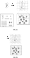
- Fig. 1 The mechanism of id-ExM and concept of electrostatic and mechanical expansion is illustrated through the diagram as shown in Fig. 1 .
- the positive and negative ions are in a state of equilibrium ( Fig. 1a ).
- the positive sodium ions get washed away and the negative ions in the polymer network repel each other ( Fig. 1b ), causing the gel to expand.
- the gel is in equilibrium again and will not expand further.
- the 1st polymer network has still room to stretch out and expand but since electrostatic equilibrium is reached, there is no net force available to stretch the polymer further. Integrating a 2nd polymer network into the 1st polymer network ( Fig.
- FIG. 2 provides an example workflow of Iterative Direct Anchoring Expansion Microscopy (idExM).
- the process of idExM begins with a fixation step that equips proteins with a biomolecule anchoring agent (e.g., a gel-anchorable chemical groups).
- a biomolecule anchoring agent e.g., a gel-anchorable chemical groups.
- Samples are contacted with biomolecule anchoring reagents to retain endogenous biomolecules, with acrylamide added during fixation to retain proteins. Subsequently, the sample is embedded in the swellable hydrogel, mechanically homogenized and expanded in water.
- B Re-embedding.
- the expanded gel is then re-embedded in a charge-neutral gel followed by the formation of a 2 nd swellable hydrogel within the re-embedded first gel.
- C The specimen is then expanded via the addition of water.
- D Post-expansion staining. Post-expansion labels against proteins can be applied in the form of antibodies.
- mice are transcardially perfused with a fixative consisting of 4% Paraformaldehyde and 30% Acrylamide in 1x PBS, after which the brain is removed and sectioned on a vibratome at a thickness of 50-100 ⁇ m.
- a fixative consisting of 4% Paraformaldehyde and 30% Acrylamide in 1x PBS, after which the brain is removed and sectioned on a vibratome at a thickness of 50-100 ⁇ m.
- fresh-frozen sections are fixed with the same fixative.
- tissue sections are incubated with the idExM 1 st gel solution (8.625% (w/v) sodium acrylate, 2.5% (w/v) acrylamide, 0.075% (w/v) N,N'-methylenebisacrylamide, 0.2% (w/v) ammonium persulfate (APS) initiator, 0.2% (w/v) tetramethylethylenediamine (TEMED) accelerator, 0.2% (w/v), 0.01% Hydroxy-TEMPO) at 4 °C for 30 minutes. Tissue sections are then embedded in the 1 st idExM gel by incubating them in an enclosed chamber surrounded by excess 1 st gel solution at 37 °C for two hours.
- the idExM 1 st gel solution 8.625% (w/v) sodium acrylate, 2.5% (w/v) acrylamide, 0.075% (w/v) N,N'-methylenebisacrylamide, 0.2% (w/v) ammonium persulfate (APS) initi
- the expanded gel is then re-embedded by incubating for two hours at room temperature in the idExM re-embedding solution (13.75% (w/v) acrylamide, 0.038% (w/v) N,N'-methylenebisacrylamide, 0.025% (w/v) ammonium persulfate (APS) initiator, 0.025% (w/v) tetramethylethylenediamine (TEMED) accelerator) while shaking, followed by incubation in a chamber with excess re-embedding solution for two hours at 45 °C. The re-embedded gel is then washed several times with 1x PBS.
- the idExM re-embedding solution (13.75% (w/v) acrylamide, 0.038% (w/v) N,N'-methylenebisacrylamide, 0.025% (w/v) ammonium persulfate (APS) initiator, 0.025% (w/v) tetramethylethylenediamine (TEM
- the re-embedded gel is incubated for two hours in the idExM 3 rd gel solution (8.625% (w/v) sodium acrylate, 2.5% (w/v) acrylamide, 0.038% (w/v) N,N'-methylenebisacrylamide, 0.025% (w/v) ammonium persulfate (APS) initiator, 0.025% (w/v) tetramethylethylenediamine (TEMED) accelerator, 1x PBS).
- the 3 rd idExM gel is then formed by placing the re-embedded gel in an enclosed chamber at 60 °C for one hour.
- the gel can be fully expanded in water and trimmed axially as needed to reduce thickness to facilitate subsequent immunostaining and imaging. Immunostaining is then performed as needed with the appropriate antibodies and blocking solution. Both primary and secondary antibodies are incubated at 4 °C overnight to allow sufficient antibody penetration into the gel.
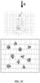
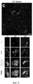
- idExM was employed prepared mouse brain slices to decipher the nanoscale structure of synapses. The sample was stained with primary and secondary antibodies after the expansion process.
- Fig. 2 shows the imaging of two synaptic proteins bassoon (pre-synaptic) and PSD-95 (post-synaptic) in the mouse brain cortex. While before expansion or with 4.5x expansion, the synaptic organization of these two proteins is not clearly observable, after 20x expansion, the two proteins are distinctively resolved.
- Example-4 Nanoscale imaging of RNA in cultured cells
- Nanoscale resolution imaging of RNA is critical for identifying cell types, distinguish between normal and pathological states of cell as well as understanding local RNA processing.
- idExM was applied for nanoscale imaging of RNA.
- a bifunctional crosslinker such as the mixing of Label-IT Amine and 6-((acryloyl)amino) hexanoic acid which provide an alkylating group that primarily reacts to the N7 of guanine and an acrylamide moiety that can get incorporated in the swellable material during or after polymerization
- FISH fluorescent in-situ hybridization
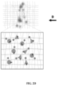
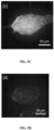
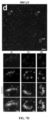
- Fig. 3 shows the wide-field image showing UbiquitinC RNA smFISH staining before (a) and after about 10x expansion (b). While the denser RNA domains are not resolved before expansion, they are clearly resolved after expansion as clear from the inset images.
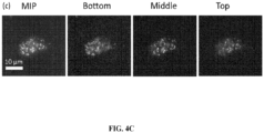
- Fig. 3(c) shows the wide-field image of NEAT1 smFISH staining in the nuclei of cultured HeLa cells after ⁇ 10x expansion. Maximum intensity projection (MIP) image, as well as representative images taken at different heights axially along the cluster, is shown. These images illustrate the capability of idExM in nanoscale imaging of RNA.
- MIP Maximum intensity projection
- idExM was applied to image RNA in mouse brain tissue samples, which were then subjected to FISH staining.
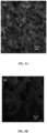
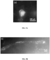
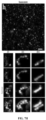
- Fig. 4 (a) show wide-field image of HCR-FISH staining performed against the 28s rRNA (red) in a Thy1-YFP mouse brain slice (cortex) after ⁇ 10x expansion. Representative images of 28s rRNA staining in neurons expressing YFP (green) is shown. (b) Wide-field image showing the distribution of 28s rRNA in dendrites.
- Mouse brain tissue was prepared by idExM according to Example 2.
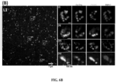
- Figure 6A and 6B depict confocal images of mouse brain tissue after post-expansion staining with idExM.
- A Low-magnification widefield image of a mouse brain slice stained with DAPI showing the somatosensory cortex region imaged after expansion in (B).
- B Confocal image of Layer 1 somatosensory cortex after post-expansion immunostaining with antibodies against P/Q-type calcium channel (red), PSD-95 (green) and RIM1/2 (blue).
- i-iv shows confocal images of highlighted four regions.
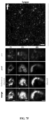
- Figure 7A through Fig. 7I depict nanoscale-resolution imaging of synapses with idExM.
- (a-b) provide a low-magnification widefield image of a mouse brain slice with DAPI staining showing the somatosensory cortex region. Confocal images of the specimen after immunostaining with antibodies against calcium channel (c), RIM1/2 (d), PSD95 (e), Syngap (f), Homer1 (g), Bassoon (h), and Shank3 (i).
- primary and secondary antibodies were applied before expansion, secondary antibodies were anchored to the gel by AcX treatment, and tertiary antibodies were applied after expansion to visualize the pre-expansion staining.
Landscapes
- Physics & Mathematics (AREA)
- Health & Medical Sciences (AREA)
- Life Sciences & Earth Sciences (AREA)
- Chemical & Material Sciences (AREA)
- Analytical Chemistry (AREA)
- Biochemistry (AREA)
- General Health & Medical Sciences (AREA)
- General Physics & Mathematics (AREA)
- Immunology (AREA)
- Pathology (AREA)
- Investigating Or Analysing Biological Materials (AREA)
Claims (15)
- Verfahren zur iterativen Vergrößerung einer Probe von Interesse für Mikroskopie, wobei das Verfahren die folgenden Schritte umfasst:a) Inkontaktbringen der Probe mit einem Biomolekül-Verankerungsmittel (BAA), wobei das BAA ein heterobifunktioneller Vernetzer ist, der an beiden Enden eines Abstandshalterarms unterschiedliche reaktive Gruppen besitzt, wobei die reaktiven Gruppen eine Biomolekül-Bindungseinheit und einen Anker umfassen;b) Einbetten der Probe in ein erstes quellfähiges Material;c) Unterziehen der Probe einer Störung der endogenen physikalischen Struktur der Probe;d) Aufquellen des ersten quellfähigen Materials, was zu einer Vergrößerung der Probe führt;e) Wiedereinbetten der vergrößerten Probe in ein nicht quellfähiges Material zur Bildung eines vergrößerten Probenhybrids;f) Einbetten des vergrößerten Probenhybrids in ein zweites quellfähiges Material; undg) Aufquellen des zweiten quellfähigen Materials zur weiteren Vergrößerung der Probe.
- Verfahren nach Anspruch 1, wobei das Einbetten der Probe in das erste quellfähige Material in Schritt b) Permeieren der Probe mit einer Zusammensetzung, die einen oder mehrere wasserlösliche Monomervorläufer umfasst; und Polymerisieren der Zusammensetzung innerhalb der Probe unter Bildung des ersten quellfähigen Materials umfasst, wobei das Polymerisieren zur Verankerung der Biomoleküle der Probe an dem ersten quellfähigen Material unter Bildung eines Komplexes aus Probe und quellfähigem Material führt.
- Verfahren nach Anspruch 1, wobei das Einbetten der Probe in das zweite quellfähige Material in Schritt f) das Permeieren der Probe mit einer Zusammensetzung, die einen oder mehrere wasserlösliche Monomervorläufer umfasst; und das Polymerisieren der Zusammensetzung innerhalb der Probe umfasst, um das zweite quellfähige Material zu bilden, wobei die Biomoleküle an dem ersten quellfähigen Material verankert bleiben.
- Verfahren nach Anspruch 2 oder 3, wobei die Zusammensetzung mindestens ein Polyelektrolytmonomer und einen kovalenten Vernetzer umfasst.
- Verfahren nach Anspruch 2 oder 3, wobei das quellfähige Material ein Hydrogel ist.
- Verfahren nach Anspruch 5, wobei das Hydrogel ein Polyacrylat-Hydrogel ist.
- Verfahren nach Anspruch 6, wobei die Zusammensetzung Acrylat, Acrylamid und einen Vernetzer, ausgewählt aus N,N-Methylenbisacrylamid (BIS), N,N'-(1,2-Dihydroxyethylen)bisacryalmid) (DHEBA) und N,N'-Bis(acryloyl)cystamin (BAC), umfasst.
- Verfahren nach Anspruch 1, wobei das Wiedereinbetten der vergrößerten Probe in das nicht quellbare Material in Schritt e) das Permeieren der vergrößerten Probe mit einer Zusammensetzung, die Vorläufer eines nicht quellfähigen Materials umfasst, und das Polymerisieren der Zusammensetzung innerhalb der vergrößerten Probe umfasst, um ein nicht quellfähiges Material zu bilden.
- Verfahren nach Anspruch 1, wobei das zweite quellfähige Material das gleiche wie das erste quellfähige Material ist.
- Verfahren nach Anspruch 1, wobei sich das zweite quellfähige Material von dem ersten quellfähigen Material unterscheidet.
- Verfahren nach Anspruch 1, wobei vor Schritt g) eine Scheibe der Probe entnommen und aufgequollen wird.
- Verfahren nach Anspruch 2, wobei das Aufquellen des ersten quellfähigen Materials in Schritt d) das Zugeben eines wässrigen Lösungsmittels oder einer wässrigen Flüssigkeit umfasst, um das Aufquellen des Proben-Quellfähiges-Material-Komplexes zu bewirken, wodurch der Proben-Quellfähiges-Material-Komplex physikalisch expandiert wird, was zu einer vergrößerten Probe führt.
- Verfahren nach Anspruch 12, wobei das wässrige Lösungsmittel oder die wässrige Flüssigkeit Wasser ist.
- Verfahren nach Anspruch 1, ferner umfassend den Schritt des Erzeugens eines hochauflösenden Bildes der Probe durch Betrachtung der vergrößerten Probe unter einem Mikroskop.
- Verfahren nach Anspruch 1, ferner umfassend den Schritt des optischen Abbildens der vergrößerten Probe durch Betrachtung der Probe unter einem Mikroskop.
Applications Claiming Priority (2)
| Application Number | Priority Date | Filing Date | Title |
|---|---|---|---|
| US201962809062P | 2019-02-22 | 2019-02-22 | |
| PCT/US2020/018789 WO2020172247A1 (en) | 2019-02-22 | 2020-02-19 | Iterative direct expansion microscopy |
Publications (3)
| Publication Number | Publication Date |
|---|---|
| EP3928074A1 EP3928074A1 (de) | 2021-12-29 |
| EP3928074A4 EP3928074A4 (de) | 2022-11-30 |
| EP3928074B1 true EP3928074B1 (de) | 2025-04-16 |
Family
ID=72142065
Family Applications (1)
| Application Number | Title | Priority Date | Filing Date |
|---|---|---|---|
| EP20760091.7A Active EP3928074B1 (de) | 2019-02-22 | 2020-02-19 | Verfahren für die iterative direkte expansionsmikroskopie |
Country Status (4)
| Country | Link |
|---|---|
| US (1) | US12405193B2 (de) |
| EP (1) | EP3928074B1 (de) |
| CA (1) | CA3130889A1 (de) |
| WO (1) | WO2020172247A1 (de) |
Families Citing this family (18)
| Publication number | Priority date | Publication date | Assignee | Title |
|---|---|---|---|---|
| WO2018157048A1 (en) | 2017-02-24 | 2018-08-30 | Massachusetts Institute Of Technology | Methods for examining podocyte foot processes in human renal samples using conventional optical microscopy |
| WO2018157074A1 (en) | 2017-02-24 | 2018-08-30 | Massachusetts Institute Of Technology | Methods for diagnosing neoplastic lesions |
| US11873374B2 (en) | 2018-02-06 | 2024-01-16 | Massachusetts Institute Of Technology | Swellable and structurally homogenous hydrogels and methods of use thereof |
| WO2019241662A1 (en) | 2018-06-15 | 2019-12-19 | Carnegie Mellon University | Improved expansion microscopy methods and kits |
| WO2020013833A1 (en) | 2018-07-13 | 2020-01-16 | Massachusetts Institute Of Technology | Dimethylacrylamide (dmaa) hydrogel for expansion microscopy (exm) |
| US12265004B2 (en) | 2019-11-05 | 2025-04-01 | Massachusetts Institute Of Technology | Membrane probes for expansion microscopy |
| US11802822B2 (en) | 2019-12-05 | 2023-10-31 | Massachusetts Institute Of Technology | Multiplexed expansion (MultiExM) pathology |
| US11492662B2 (en) | 2020-08-06 | 2022-11-08 | Singular Genomics Systems, Inc. | Methods for in situ transcriptomics and proteomics |
| EP4121557A4 (de) | 2020-08-06 | 2024-05-01 | Singular Genomics Systems, Inc. | Räumliche sequenzierung |
| EP4334706A4 (de) | 2021-05-05 | 2025-08-06 | Singular Genomics Systems Inc | Multiomikanalysevorrichtung und verfahren zur verwendung davon |
| CN117916572A (zh) * | 2021-06-15 | 2024-04-19 | 西湖大学 | 用于样品的物理膨胀的方法及其用途 |
| CN115372527B (zh) * | 2021-08-27 | 2024-10-15 | 西湖大学 | 用于生物样品的膨胀蛋白组学分析的方法和制剂 |
| US20230304905A1 (en) * | 2022-03-23 | 2023-09-28 | Howard Hughes Medical Institute | Sample preparation for expansion microscopy |
| WO2024171064A1 (en) * | 2023-02-14 | 2024-08-22 | Carnegie Mellon University | Rapid expansion microscopy methods and reagents |
| WO2024249961A1 (en) | 2023-06-01 | 2024-12-05 | Singular Genomics Systems, Inc. | Methods and probes for detecting polynucleotide sequences in cells and tissues |
| WO2025067452A1 (en) * | 2023-09-27 | 2025-04-03 | Westlake Laboratory Of Life Sciences And Biomedicine | Methods for deep untargeted profiling of spatial transcriptome and proteome in intact tissues |
| WO2025090986A1 (en) * | 2023-10-27 | 2025-05-01 | Massachusetts Institute Of Technology | Multiplexed expansion revealing |
| EP4603819A1 (de) * | 2024-02-13 | 2025-08-20 | IST Austria - Institute of Science and Technology Austria | Iterative expansion zur hochgenauen gewebekonservierung |
Family Cites Families (122)
| Publication number | Priority date | Publication date | Assignee | Title |
|---|---|---|---|---|
| FR2716263B1 (fr) | 1994-02-11 | 1997-01-17 | Pasteur Institut | Procédé d'alignement de macromolécules par passage d'un ménisque et applications dans un procédé de mise en évidence, séparation et/ou dosage d'une macromolécule dans un échantillon. |
| US6271278B1 (en) | 1997-05-13 | 2001-08-07 | Purdue Research Foundation | Hydrogel composites and superporous hydrogel composites having fast swelling, high mechanical strength, and superabsorbent properties |
| ATE327345T1 (de) | 1998-08-07 | 2006-06-15 | Cellay Llc | Gel mikrotropfen für die genetische analyse |
| US5952232A (en) | 1998-09-17 | 1999-09-14 | Rothman; James Edward | Expandible microparticle intracellular delivery system |
| US6204064B1 (en) | 1999-01-30 | 2001-03-20 | David S. Alberts | Measurement of lesion progression via mapping of chromatin texture features along progression curve |
| US6107081A (en) | 1999-02-05 | 2000-08-22 | The United States Of America As Represented By The Administrator Of The National Aeronautics And Space Administration | Uni-directional cell stretching device |
| EP1173878B1 (de) | 1999-04-27 | 2011-04-06 | Bio-Rad Laboratories, Inc. | Probenhalter für ein gasphaseionenspektrometer |
| US6287870B1 (en) | 1999-08-20 | 2001-09-11 | Robert A. Levine | Method and assembly for separating formed constituents from a liquid constituent in a complex biologic fluid sample |
| EP1125905A1 (de) | 2000-02-16 | 2001-08-22 | Pepscan Systems B.V. | Synthese von Segmenten |
| US6878384B2 (en) | 2001-03-13 | 2005-04-12 | Microvention, Inc. | Hydrogels that undergo volumetric expansion in response to changes in their environment and their methods of manufacture and use |
| EP2465943A3 (de) | 2001-03-16 | 2012-10-03 | Kalim Mir | Lineare Polymeranzeige |
| US7885448B2 (en) | 2001-03-19 | 2011-02-08 | Dmetrix, Inc. | Diagnostic scanning microscope for information-enriched qualitative histopathology |
| AT410805B (de) | 2001-05-29 | 2003-08-25 | Sleytr Uwe B | Verfahren zum erzeugen einer schicht funktioneller moleküle |
| CA2450921A1 (en) | 2001-06-22 | 2003-01-03 | Incyte Genomics, Inc. | Protein modification and maintenance molecules |
| US20040029192A1 (en) | 2002-08-08 | 2004-02-12 | Shaw Andrey S. | Compositions and methods for the diagnosis and treatment of kidney disease |
| WO2003051286A2 (en) | 2001-11-01 | 2003-06-26 | Regents Of The University Of Minnesota | Hydrogel compositions, devices, and microscale components |
| US7189888B2 (en) | 2001-12-21 | 2007-03-13 | Kimberly-Clark Worldwide, Inc. | Nonabsorbent surge layer having discrete regions of superabsorbent and method for making |
| US20040115629A1 (en) | 2002-01-09 | 2004-06-17 | Panzer Scott R | Molecules for diagnostics and therapeutics |
| AU2003251874A1 (en) | 2002-07-12 | 2004-02-02 | Dirk R. Albrecht | Three dimensional cell patterned bioploymer scaffolds and method of making the same |
| US7172877B2 (en) | 2003-01-09 | 2007-02-06 | Massachusetts Institute Of Technology | Methods and compositions for peptide and protein labeling |
| WO2004066185A1 (en) | 2003-01-23 | 2004-08-05 | U.S. Genomics, Inc. | Methods for analyzing polymer populations |
| WO2004074935A1 (en) | 2003-02-13 | 2004-09-02 | The Trustees Of Columbia University In The City Of New York | Micropatterning of molecular surfaces via selective irradiation |
| US20050034990A1 (en) | 2003-08-12 | 2005-02-17 | Crooks Richard M. | System and method for electrokinetic trapping and concentration enrichment of analytes in a microfluidic channel |
| US7419593B2 (en) | 2003-11-19 | 2008-09-02 | Amcol International Corp. | Bioremediation mat and method of manufacture and use |
| WO2005069886A2 (en) | 2004-01-16 | 2005-08-04 | Northwestern University | Sparsely cross-linked nanogels: a novel polymer structure for microchannel dna sequencing |
| US7192693B2 (en) | 2004-02-24 | 2007-03-20 | University Of Washington | Methods for photopatterning hydrogels |
| JP2005291759A (ja) | 2004-03-31 | 2005-10-20 | Michimasa Kishimoto | 二次元画像による病症診断システム |
| JP4374435B2 (ja) | 2004-07-28 | 2009-12-02 | 国立大学法人 大分大学 | 高親水性高分子による組織包埋方法。 |
| SG155169A1 (en) | 2004-07-30 | 2009-09-30 | Novartis Ag | Method of creating ophthalmic lenses using modulated energy |
| KR100695134B1 (ko) | 2004-11-25 | 2007-03-14 | 삼성전자주식회사 | 층류를 이용한 마이크로어레이 및 이의 제조 방법 |
| JP4496943B2 (ja) | 2004-11-30 | 2010-07-07 | 日本電気株式会社 | 病理診断支援装置、病理診断支援プログラム、病理診断支援装置の作動方法、及び病理診断支援システム |
| EP1885746B1 (de) | 2005-02-08 | 2012-01-11 | Research Development Foundation | Zusammensetzungen in zusammenhang mit löslichen, g-proteingekoppelten rezeptoren (sgpcrs) |
| JP2008541761A (ja) | 2005-05-31 | 2008-11-27 | インヴィトロジェン コーポレーション | パラフィン含有試料からの核酸の分離及び精製 |
| US9084546B2 (en) | 2005-08-31 | 2015-07-21 | The Regents Of The University Of Michigan | Co-electrodeposited hydrogel-conducting polymer electrodes for biomedical applications |
| US8367793B2 (en) | 2005-09-30 | 2013-02-05 | Abs Materials, Inc. | Swellable materials and methods of use |
| US20070134902A1 (en) | 2005-12-12 | 2007-06-14 | The Curators Of The University Of Missouri | Patterning of Substrates with Metal-Containing Particles |
| US20090131551A1 (en) | 2006-03-01 | 2009-05-21 | Dong Xie | Polyfunctional compounds and uses as implant materials |
| WO2008118148A2 (en) | 2006-11-07 | 2008-10-02 | Thomas Jefferson University | Adiponectin for the treatment and diagnosis of albuminuria |
| AT504330B8 (de) | 2006-11-13 | 2008-09-15 | Nano S Biotechnologie Gmbh | Beschichtung hydrophiler oberflächen mit s-schicht-proteinen |
| HRP20100532T1 (hr) | 2007-02-01 | 2010-11-30 | Immundiagnostik Ag | Izravno određivanje vitamina d u serumu ili plazmi |
| US8163188B2 (en) | 2007-04-03 | 2012-04-24 | The University Of Massachusetts | Article with PHEMA lift-off layer and method therefor |
| CA2584087C (en) | 2007-04-05 | 2016-11-29 | Molly Shoichet | Chemically patterned hydrogels, manufacture and use thereof |
| EP1985682A1 (de) | 2007-04-17 | 2008-10-29 | Services Pétroliers Schlumberger | Verfahren und Zusammensetzung zur Behandlung eines Bohrloches |
| JP5058676B2 (ja) | 2007-05-18 | 2012-10-24 | 株式会社アイエスティー | 生体標本の作製方法 |
| AU2008296981A1 (en) | 2007-08-30 | 2009-03-12 | President And Fellows Of Harvard College | Compliant surface multi-well culture plate |
| EP2205974B1 (de) | 2007-09-20 | 2011-08-03 | Arizona Board of Regents acting for and on behalf of Arizona State University | Immobilisierung einer einheit in einer bestimmten ausrichtung auf einem trägermaterial |
| US8048641B2 (en) | 2007-10-10 | 2011-11-01 | The United States Of America As Represented By The Secretary, Department Of Health And Human Services | Micropatterning of biological molecules using laser ablation |
| US20100041128A1 (en) | 2008-01-08 | 2010-02-18 | Medtrain Technologies, Llc | Microfluidic Device for Application of Shear Stress and Tensile Strain |
| AU2009210837A1 (en) | 2008-01-30 | 2009-08-13 | Geron Corporation | Cell culture article and screening |
| JP4956839B2 (ja) | 2008-02-13 | 2012-06-20 | 国立大学法人 大分大学 | 高親水性高分子モノマー水溶液による組織包埋方法 |
| US20090241681A1 (en) | 2008-03-27 | 2009-10-01 | Andrew Machauf | Hydrogel-based mems biosensor |
| US8652798B2 (en) | 2008-05-20 | 2014-02-18 | The Regents Of The University Of California | Analysis of ex vivo cells for disease state detection and therapeutic agent selection and monitoring |
| EP2303770B1 (de) | 2008-06-30 | 2014-07-09 | 3M Innovative Properties Company | Verfahren zur in-situ-bildung von metallnanoclustern in einem porösen substrat |
| WO2010014903A1 (en) | 2008-07-31 | 2010-02-04 | Massachusetts Institute Of Technology | Multiplexed olfactory receptor-based microsurface plasmon polariton detector |
| US20100055161A1 (en) | 2008-08-27 | 2010-03-04 | Dong June Ahn | Hydrogel face mask for delivering skin care agents |
| SI2963709T1 (sl) | 2008-10-24 | 2017-10-30 | Epicentre Technologies Corporation | Transposonski sestavki in postopki za spreminjanje nukleinskih kislin |
| US8488863B2 (en) | 2008-11-06 | 2013-07-16 | Los Alamos National Security, Llc | Combinational pixel-by-pixel and object-level classifying, segmenting, and agglomerating in performing quantitative image analysis that distinguishes between healthy non-cancerous and cancerous cell nuclei and delineates nuclear, cytoplasm, and stromal material objects from stained biological tissue materials |
| US10150245B2 (en) | 2008-11-11 | 2018-12-11 | University Of Florida Research Foundation, Inc. | Method of patterning a surface and articles comprising the same |
| CN102369594A (zh) | 2009-04-06 | 2012-03-07 | 住友化学株式会社 | 半导体基板、半导体基板的制造方法、半导体基板的判定方法以及电子器件 |
| JP2011018195A (ja) | 2009-07-09 | 2011-01-27 | Toshiba Corp | 電源回路及び電子機器 |
| EP2456885A2 (de) | 2009-07-20 | 2012-05-30 | Bar Harbor Biotechnology, Inc. | Verfahren zur beurteilung des krankheitsrisikos |
| CN102548654A (zh) | 2009-09-29 | 2012-07-04 | 株式会社日本触媒 | 颗粒状吸水剂及其制造方法 |
| WO2011057172A1 (en) | 2009-11-06 | 2011-05-12 | University Of Miami | Podocyte specific assays and uses thereof |
| WO2011111876A1 (en) | 2010-03-12 | 2011-09-15 | Riken | Clearing reagent for biological material, and use thereof |
| US20110291357A1 (en) | 2010-06-01 | 2011-12-01 | Tom Boyle | Slider game |
| EP2670894B1 (de) | 2011-02-02 | 2017-11-29 | University Of Washington Through Its Center For Commercialization | Massiv parallele nachbarschaftsabbildung |
| WO2012112689A1 (en) | 2011-02-15 | 2012-08-23 | The University Of North Carolina At Chapel Hill | Nanoparticle, liposomes, polymers, agents and proteins modified with reversible linkers |
| AU2012245075B2 (en) | 2011-04-20 | 2016-12-08 | 4Dx Limited | Method and device for trapping and analysing cells and the like |
| US10813791B2 (en) | 2011-06-02 | 2020-10-27 | University Of Rochester | Method for modifying the refractive index of ocular tissues and applications thereof |
| EP3088416B1 (de) | 2011-12-23 | 2019-02-27 | Mayo Foundation for Medical Education and Research | Beurteilung von strukturellen änderungen und ergebnissen der nieren |
| US9189678B2 (en) | 2012-03-30 | 2015-11-17 | Konica Minolta, Inc. | Medical image processor and storage medium |
| JP5967528B2 (ja) | 2012-06-22 | 2016-08-10 | 国立研究開発法人理化学研究所 | 生物材料を透明化する方法および生物材料用透明化処理キット |
| KR102148747B1 (ko) | 2012-08-09 | 2020-08-27 | 더 보드 오브 트러스티스 오브 더 리랜드 스탠포드 쥬니어 유니버시티 | 현미경적 분석을 위한 생물학적 표본을 준비하기 위한 방법과 조성물 |
| US9346239B2 (en) | 2012-09-26 | 2016-05-24 | Eastman Kodak Company | Method for providing patterns of functional materials |
| US20150226743A1 (en) | 2012-10-28 | 2015-08-13 | Clarient Diagnostics Services, Inc. | Multiplexed method for diagnosing classical hodgkin lymphoma |
| WO2014107181A1 (en) | 2013-01-07 | 2014-07-10 | The Uab Research Foundation | Multilayer hydrogels with ph-responsive swelling and surface wettability |
| PT2951201T (pt) | 2013-01-30 | 2018-01-09 | Univ Brussel Vrije | Novos polipéptidos quiméricos para rastreamento e fins de descoberta de fármacos |
| US9778573B2 (en) | 2013-03-14 | 2017-10-03 | The United States Of America As Represented By The Department Of Veterans Affairs | Optical illumination system |
| US9778154B2 (en) | 2013-09-20 | 2017-10-03 | California Institute Of Technology | Methods for phenotyping of intact whole tissues |
| US10794802B2 (en) | 2013-09-20 | 2020-10-06 | California Institute Of Technology | Whole-body tissue stabilization and selective extractions via tissue-hydrogel hybrids for high resolution intact circuit mapping and phenotyping |
| EP3058091B1 (de) | 2013-10-18 | 2020-03-25 | The Broad Institute, Inc. | Räumliche und zelluläre kartographie von biomolekülen in situ durch hochdurchsatz-sequenzierung |
| EP3108218A4 (de) * | 2014-02-21 | 2017-11-15 | Massachusetts Institute Of Technology | Expansionsmikroskopie |
| WO2015124777A1 (en) | 2014-02-21 | 2015-08-27 | Ventana Medical Systems, Inc. | Medical image analysis for identifying biomarker-positive tumor cells |
| US20170182220A1 (en) | 2014-02-26 | 2017-06-29 | University Of Massachusetts | Degradable hydrogel with predictable tuning of properties, and compositions and methods thereof |
| US10373702B2 (en) | 2014-03-27 | 2019-08-06 | Massachusetts Institute Of Technology | Water-soluble trans-membrane proteins and methods for the preparation and use thereof |
| US20150353989A1 (en) | 2014-06-09 | 2015-12-10 | Illumina Cambridge Limited | Sample preparation for nucleic acid amplification |
| NL2012998B1 (en) | 2014-06-13 | 2016-07-04 | Van Den Herik Sliedrecht | Method and apparatus for placement of a subterranean curtain below the face of the earth. |
| WO2015199089A1 (ja) | 2014-06-23 | 2015-12-30 | 株式会社日本触媒 | 吸収性樹脂およびその製造方法 |
| EP3191016B1 (de) | 2014-09-09 | 2021-10-27 | University of Washington | Funktionalisierte zwitterionische polymere und polymere mit gemischter ladung, zugehörige hydrogele und verfahren zu deren verwendung |
| WO2016042963A1 (ja) | 2014-09-19 | 2016-03-24 | コニカミノルタ株式会社 | 画像処理装置、画像処理方法、及びプログラム |
| WO2016120441A2 (en) | 2015-01-30 | 2016-08-04 | Ventana Medical Systems, Inc. | Quality metrics for automatic evaluation of dual ish images |
| US10059990B2 (en) * | 2015-04-14 | 2018-08-28 | Massachusetts Institute Of Technology | In situ nucleic acid sequencing of expanded biological samples |
| US11408890B2 (en) * | 2015-04-14 | 2022-08-09 | Massachusetts Institute Of Technology | Iterative expansion microscopy |
| US10526649B2 (en) | 2015-04-14 | 2020-01-07 | Massachusetts Institute Of Technology | Augmenting in situ nucleic acid sequencing of expanded biological samples with in vitro sequence information |
| CA2994957A1 (en) | 2015-08-07 | 2017-02-16 | Massachusetts Institute Of Technology | Protein retention expansion microscopy |
| WO2017027367A1 (en) | 2015-08-07 | 2017-02-16 | Massachusetts Institute Of Technology | Nanoscale imaging of proteins and nucleic acids via expansion microscopy |
| WO2017031249A1 (en) | 2015-08-17 | 2017-02-23 | California Institute Of Technology | Whole-body tissue stabilization and selective extractions via tissue-hydrogel hybrids for high resolution intact circuit mapping and phenotyping |
| US11214661B2 (en) | 2015-09-17 | 2022-01-04 | Massachusetts Institute Of Technology | Three-dimensional nanofabrication by patterning of hydrogels |
| MX2018005611A (es) | 2015-11-03 | 2018-11-09 | Harvard College | Metodo y aparato para la formacion de imagenes volumetricas de una matriz tridimensional que contiene acido nucleico. |
| KR102771841B1 (ko) | 2015-11-06 | 2025-02-24 | 벤타나 메디컬 시스템즈, 인코포레이티드 | 대표 진단법 |
| US20190113423A1 (en) | 2015-12-02 | 2019-04-18 | Clearlight Diagnostics, LLC | Methods for preparing and analyzing tumor tissue samples for detection and monitoring of cancers |
| WO2017147435A1 (en) | 2016-02-25 | 2017-08-31 | Massachusetts Institute Of Technology | Methods for expanding clinical tissue specimens |
| EP3868879A1 (de) | 2016-03-10 | 2021-08-25 | The Board of Trustees of the Leland Stanford Junior University | Transposase-vermittelte abbildung des zugänglichen genoms |
| CN109923216B (zh) | 2016-08-31 | 2024-08-02 | 哈佛学院董事及会员团体 | 将生物分子的检测组合到使用荧光原位测序的单个试验的方法 |
| WO2018136856A1 (en) | 2017-01-23 | 2018-07-26 | Massachusetts Institute Of Technology | Multiplexed signal amplified fish via splinted ligation amplification and sequencing |
| WO2018157074A1 (en) | 2017-02-24 | 2018-08-30 | Massachusetts Institute Of Technology | Methods for diagnosing neoplastic lesions |
| WO2018157048A1 (en) | 2017-02-24 | 2018-08-30 | Massachusetts Institute Of Technology | Methods for examining podocyte foot processes in human renal samples using conventional optical microscopy |
| US11180804B2 (en) | 2017-07-25 | 2021-11-23 | Massachusetts Institute Of Technology | In situ ATAC sequencing |
| WO2019144391A1 (en) | 2018-01-26 | 2019-08-01 | National Institute Of Biological Sciences, Beijing | Use of ishcr for exm and solvent-based tissue clearing |
| US11873374B2 (en) | 2018-02-06 | 2024-01-16 | Massachusetts Institute Of Technology | Swellable and structurally homogenous hydrogels and methods of use thereof |
| WO2020013833A1 (en) | 2018-07-13 | 2020-01-16 | Massachusetts Institute Of Technology | Dimethylacrylamide (dmaa) hydrogel for expansion microscopy (exm) |
| US12153163B2 (en) | 2018-08-03 | 2024-11-26 | OPSYS Tech Ltd. | Distributed modular solid-state lidar system |
| CN113767177B (zh) | 2018-12-10 | 2025-01-14 | 10X基因组学有限公司 | 生成用于空间分析的捕获探针 |
| WO2020146325A1 (en) | 2019-01-08 | 2020-07-16 | Massachusetts Institute Of Technology | Single-molecule protein and peptide sequencing |
| EP3990634A1 (de) | 2019-09-13 | 2022-05-04 | Google LLC | Verfahren und zusammensetzungen zur protein- und peptidsequenzierung |
| CN112574089B (zh) | 2019-09-30 | 2023-09-05 | 中国科学院上海药物研究所 | 一种光诱导的多功能交联剂、其制备方法及应用 |
| US12265004B2 (en) | 2019-11-05 | 2025-04-01 | Massachusetts Institute Of Technology | Membrane probes for expansion microscopy |
| US11802822B2 (en) | 2019-12-05 | 2023-10-31 | Massachusetts Institute Of Technology | Multiplexed expansion (MultiExM) pathology |
| WO2021183667A1 (en) | 2020-03-11 | 2021-09-16 | The Regents Of The University Of Colorado, A Body Corporate | Swellable photopolymerized hydrogels for expansion microscopy |
| CN111848855A (zh) | 2020-07-03 | 2020-10-30 | 西安交通大学 | 一种具有pH响应的可注射水凝胶敷料及其制备方法和应用 |
| CN114478420B (zh) | 2020-11-13 | 2025-02-07 | 北京大学 | 多特异生物偶联连接臂及其合成方法 |
| US20230332207A1 (en) | 2022-04-01 | 2023-10-19 | Massachusetts Institute Of Technology | Epoxides for multimodal detection of biomolecules |
-
2020
- 2020-02-19 US US16/794,849 patent/US12405193B2/en active Active
- 2020-02-19 WO PCT/US2020/018789 patent/WO2020172247A1/en not_active Ceased
- 2020-02-19 EP EP20760091.7A patent/EP3928074B1/de active Active
- 2020-02-19 CA CA3130889A patent/CA3130889A1/en active Pending
Also Published As
| Publication number | Publication date |
|---|---|
| EP3928074A4 (de) | 2022-11-30 |
| EP3928074A1 (de) | 2021-12-29 |
| WO2020172247A1 (en) | 2020-08-27 |
| CA3130889A1 (en) | 2020-08-27 |
| US12405193B2 (en) | 2025-09-02 |
| US20200271556A1 (en) | 2020-08-27 |
Similar Documents
| Publication | Publication Date | Title |
|---|---|---|
| EP3928074B1 (de) | Verfahren für die iterative direkte expansionsmikroskopie | |
| US12061199B2 (en) | Methods for diagnosing neoplastic lesions | |
| US12233184B2 (en) | Dimethylacrylamide (DMAA) hydrogel for expansion microscopy (ExM) | |
| US20190064037A1 (en) | Methods for Expanding Clinical Tissue Specimens | |
| US11802872B2 (en) | Methods for examining podocyte foot processes in human renal samples using conventional optical microscopy | |
| US12265004B2 (en) | Membrane probes for expansion microscopy | |
| US11802822B2 (en) | Multiplexed expansion (MultiExM) pathology | |
| US20230213415A1 (en) | Method and System for Imaging and Analysis of a Biological Specimen | |
| KR102148747B1 (ko) | 현미경적 분석을 위한 생물학적 표본을 준비하기 위한 방법과 조성물 | |
| JP6456969B2 (ja) | 膨張顕微鏡法 | |
| US11333588B1 (en) | Matrix-assisted methods and compositions to prepare biological samples for super-resolution imaging | |
| JP2024000599A (ja) | 生検試料および生物学的試料を調製および分析するための方法 | |
| WO2025172471A1 (en) | Method and composition for iterative expansion of tissue samples for high-fidelity tissue preservation | |
| Rodriguez-Gatica et al. | Light-Sheet Fluorescence Microscopy of Expanded |
Legal Events
| Date | Code | Title | Description |
|---|---|---|---|
| STAA | Information on the status of an ep patent application or granted ep patent |
Free format text: STATUS: THE INTERNATIONAL PUBLICATION HAS BEEN MADE |
|
| PUAI | Public reference made under article 153(3) epc to a published international application that has entered the european phase |
Free format text: ORIGINAL CODE: 0009012 |
|
| STAA | Information on the status of an ep patent application or granted ep patent |
Free format text: STATUS: REQUEST FOR EXAMINATION WAS MADE |
|
| 17P | Request for examination filed |
Effective date: 20210907 |
|
| AK | Designated contracting states |
Kind code of ref document: A1 Designated state(s): AL AT BE BG CH CY CZ DE DK EE ES FI FR GB GR HR HU IE IS IT LI LT LU LV MC MK MT NL NO PL PT RO RS SE SI SK SM TR |
|
| DAV | Request for validation of the european patent (deleted) | ||
| DAX | Request for extension of the european patent (deleted) | ||
| A4 | Supplementary search report drawn up and despatched |
Effective date: 20221031 |
|
| RIC1 | Information provided on ipc code assigned before grant |
Ipc: G01N 1/28 20060101ALI20221025BHEP Ipc: G01N 1/36 20060101ALI20221025BHEP Ipc: G01N 1/30 20060101AFI20221025BHEP |
|
| GRAP | Despatch of communication of intention to grant a patent |
Free format text: ORIGINAL CODE: EPIDOSNIGR1 |
|
| STAA | Information on the status of an ep patent application or granted ep patent |
Free format text: STATUS: GRANT OF PATENT IS INTENDED |
|
| INTG | Intention to grant announced |
Effective date: 20241112 |
|
| GRAS | Grant fee paid |
Free format text: ORIGINAL CODE: EPIDOSNIGR3 |
|
| GRAA | (expected) grant |
Free format text: ORIGINAL CODE: 0009210 |
|
| STAA | Information on the status of an ep patent application or granted ep patent |
Free format text: STATUS: THE PATENT HAS BEEN GRANTED |
|
| AK | Designated contracting states |
Kind code of ref document: B1 Designated state(s): AL AT BE BG CH CY CZ DE DK EE ES FI FR GB GR HR HU IE IS IT LI LT LU LV MC MK MT NL NO PL PT RO RS SE SI SK SM TR |
|
| REG | Reference to a national code |
Ref country code: GB Ref legal event code: FG4D |
|
| REG | Reference to a national code |
Ref country code: CH Ref legal event code: EP Ref country code: DE Ref legal event code: R096 Ref document number: 602020049573 Country of ref document: DE |
|
| REG | Reference to a national code |
Ref country code: IE Ref legal event code: FG4D |
|
| REG | Reference to a national code |
Ref country code: NL Ref legal event code: MP Effective date: 20250416 |
|
| PG25 | Lapsed in a contracting state [announced via postgrant information from national office to epo] |
Ref country code: NL Free format text: LAPSE BECAUSE OF FAILURE TO SUBMIT A TRANSLATION OF THE DESCRIPTION OR TO PAY THE FEE WITHIN THE PRESCRIBED TIME-LIMIT Effective date: 20250416 |
|
| REG | Reference to a national code |
Ref country code: AT Ref legal event code: MK05 Ref document number: 1785997 Country of ref document: AT Kind code of ref document: T Effective date: 20250416 |
|
| PG25 | Lapsed in a contracting state [announced via postgrant information from national office to epo] |
Ref country code: FI Free format text: LAPSE BECAUSE OF FAILURE TO SUBMIT A TRANSLATION OF THE DESCRIPTION OR TO PAY THE FEE WITHIN THE PRESCRIBED TIME-LIMIT Effective date: 20250416 Ref country code: ES Free format text: LAPSE BECAUSE OF FAILURE TO SUBMIT A TRANSLATION OF THE DESCRIPTION OR TO PAY THE FEE WITHIN THE PRESCRIBED TIME-LIMIT Effective date: 20250416 Ref country code: PT Free format text: LAPSE BECAUSE OF FAILURE TO SUBMIT A TRANSLATION OF THE DESCRIPTION OR TO PAY THE FEE WITHIN THE PRESCRIBED TIME-LIMIT Effective date: 20250818 |
|
| REG | Reference to a national code |
Ref country code: LT Ref legal event code: MG9D |
|
| PG25 | Lapsed in a contracting state [announced via postgrant information from national office to epo] |
Ref country code: GR Free format text: LAPSE BECAUSE OF FAILURE TO SUBMIT A TRANSLATION OF THE DESCRIPTION OR TO PAY THE FEE WITHIN THE PRESCRIBED TIME-LIMIT Effective date: 20250717 Ref country code: NO Free format text: LAPSE BECAUSE OF FAILURE TO SUBMIT A TRANSLATION OF THE DESCRIPTION OR TO PAY THE FEE WITHIN THE PRESCRIBED TIME-LIMIT Effective date: 20250716 |
|
| PG25 | Lapsed in a contracting state [announced via postgrant information from national office to epo] |
Ref country code: PL Free format text: LAPSE BECAUSE OF FAILURE TO SUBMIT A TRANSLATION OF THE DESCRIPTION OR TO PAY THE FEE WITHIN THE PRESCRIBED TIME-LIMIT Effective date: 20250416 |
|
| PG25 | Lapsed in a contracting state [announced via postgrant information from national office to epo] |
Ref country code: BG Free format text: LAPSE BECAUSE OF FAILURE TO SUBMIT A TRANSLATION OF THE DESCRIPTION OR TO PAY THE FEE WITHIN THE PRESCRIBED TIME-LIMIT Effective date: 20250416 |
|
| PG25 | Lapsed in a contracting state [announced via postgrant information from national office to epo] |
Ref country code: HR Free format text: LAPSE BECAUSE OF FAILURE TO SUBMIT A TRANSLATION OF THE DESCRIPTION OR TO PAY THE FEE WITHIN THE PRESCRIBED TIME-LIMIT Effective date: 20250416 |
|
| PG25 | Lapsed in a contracting state [announced via postgrant information from national office to epo] |
Ref country code: AT Free format text: LAPSE BECAUSE OF FAILURE TO SUBMIT A TRANSLATION OF THE DESCRIPTION OR TO PAY THE FEE WITHIN THE PRESCRIBED TIME-LIMIT Effective date: 20250416 |
|
| PG25 | Lapsed in a contracting state [announced via postgrant information from national office to epo] |
Ref country code: RS Free format text: LAPSE BECAUSE OF FAILURE TO SUBMIT A TRANSLATION OF THE DESCRIPTION OR TO PAY THE FEE WITHIN THE PRESCRIBED TIME-LIMIT Effective date: 20250716 |
|
| PG25 | Lapsed in a contracting state [announced via postgrant information from national office to epo] |
Ref country code: IS Free format text: LAPSE BECAUSE OF FAILURE TO SUBMIT A TRANSLATION OF THE DESCRIPTION OR TO PAY THE FEE WITHIN THE PRESCRIBED TIME-LIMIT Effective date: 20250816 |
|
| PG25 | Lapsed in a contracting state [announced via postgrant information from national office to epo] |
Ref country code: LV Free format text: LAPSE BECAUSE OF FAILURE TO SUBMIT A TRANSLATION OF THE DESCRIPTION OR TO PAY THE FEE WITHIN THE PRESCRIBED TIME-LIMIT Effective date: 20250416 |
|
| PG25 | Lapsed in a contracting state [announced via postgrant information from national office to epo] |
Ref country code: SM Free format text: LAPSE BECAUSE OF FAILURE TO SUBMIT A TRANSLATION OF THE DESCRIPTION OR TO PAY THE FEE WITHIN THE PRESCRIBED TIME-LIMIT Effective date: 20250416 Ref country code: DK Free format text: LAPSE BECAUSE OF FAILURE TO SUBMIT A TRANSLATION OF THE DESCRIPTION OR TO PAY THE FEE WITHIN THE PRESCRIBED TIME-LIMIT Effective date: 20250416 |
|
| PG25 | Lapsed in a contracting state [announced via postgrant information from national office to epo] |
Ref country code: CZ Free format text: LAPSE BECAUSE OF FAILURE TO SUBMIT A TRANSLATION OF THE DESCRIPTION OR TO PAY THE FEE WITHIN THE PRESCRIBED TIME-LIMIT Effective date: 20250416 |
|
| PG25 | Lapsed in a contracting state [announced via postgrant information from national office to epo] |
Ref country code: EE Free format text: LAPSE BECAUSE OF FAILURE TO SUBMIT A TRANSLATION OF THE DESCRIPTION OR TO PAY THE FEE WITHIN THE PRESCRIBED TIME-LIMIT Effective date: 20250416 |
|
| PG25 | Lapsed in a contracting state [announced via postgrant information from national office to epo] |
Ref country code: RO Free format text: LAPSE BECAUSE OF FAILURE TO SUBMIT A TRANSLATION OF THE DESCRIPTION OR TO PAY THE FEE WITHIN THE PRESCRIBED TIME-LIMIT Effective date: 20250416 Ref country code: SK Free format text: LAPSE BECAUSE OF FAILURE TO SUBMIT A TRANSLATION OF THE DESCRIPTION OR TO PAY THE FEE WITHIN THE PRESCRIBED TIME-LIMIT Effective date: 20250416 |
|
| PG25 | Lapsed in a contracting state [announced via postgrant information from national office to epo] |
Ref country code: IT Free format text: LAPSE BECAUSE OF FAILURE TO SUBMIT A TRANSLATION OF THE DESCRIPTION OR TO PAY THE FEE WITHIN THE PRESCRIBED TIME-LIMIT Effective date: 20250416 |