EP3766962A1 - Cellules progénitrices d'oligodendrocyte issues de cellules pluripotentes induites pour le traitement de troubles de la myéline - Google Patents
Cellules progénitrices d'oligodendrocyte issues de cellules pluripotentes induites pour le traitement de troubles de la myéline Download PDFInfo
- Publication number
- EP3766962A1 EP3766962A1 EP20190706.0A EP20190706A EP3766962A1 EP 3766962 A1 EP3766962 A1 EP 3766962A1 EP 20190706 A EP20190706 A EP 20190706A EP 3766962 A1 EP3766962 A1 EP 3766962A1
- Authority
- EP
- European Patent Office
- Prior art keywords
- cells
- preparation
- derived
- human
- cell
- Prior art date
- Legal status (The legal status is an assumption and is not a legal conclusion. Google has not performed a legal analysis and makes no representation as to the accuracy of the status listed.)
- Pending
Links
Images


Classifications
-
- C—CHEMISTRY; METALLURGY
- C12—BIOCHEMISTRY; BEER; SPIRITS; WINE; VINEGAR; MICROBIOLOGY; ENZYMOLOGY; MUTATION OR GENETIC ENGINEERING
- C12N—MICROORGANISMS OR ENZYMES; COMPOSITIONS THEREOF; PROPAGATING, PRESERVING, OR MAINTAINING MICROORGANISMS; MUTATION OR GENETIC ENGINEERING; CULTURE MEDIA
- C12N5/00—Undifferentiated human, animal or plant cells, e.g. cell lines; Tissues; Cultivation or maintenance thereof; Culture media therefor
- C12N5/06—Animal cells or tissues; Human cells or tissues
- C12N5/0602—Vertebrate cells
- C12N5/0618—Cells of the nervous system
- C12N5/0622—Glial cells, e.g. astrocytes, oligodendrocytes; Schwann cells
-
- A—HUMAN NECESSITIES
- A61—MEDICAL OR VETERINARY SCIENCE; HYGIENE
- A61K—PREPARATIONS FOR MEDICAL, DENTAL OR TOILETRY PURPOSES
- A61K35/00—Medicinal preparations containing materials or reaction products thereof with undetermined constitution
- A61K35/12—Materials from mammals; Compositions comprising non-specified tissues or cells; Compositions comprising non-embryonic stem cells; Genetically modified cells
- A61K35/30—Nerves; Brain; Eyes; Corneal cells; Cerebrospinal fluid; Neuronal stem cells; Neuronal precursor cells; Glial cells; Oligodendrocytes; Schwann cells; Astroglia; Astrocytes; Choroid plexus; Spinal cord tissue
-
- A—HUMAN NECESSITIES
- A61—MEDICAL OR VETERINARY SCIENCE; HYGIENE
- A61K—PREPARATIONS FOR MEDICAL, DENTAL OR TOILETRY PURPOSES
- A61K9/00—Medicinal preparations characterised by special physical form
- A61K9/0012—Galenical forms characterised by the site of application
- A61K9/0085—Brain, e.g. brain implants; Spinal cord
-
- A—HUMAN NECESSITIES
- A61—MEDICAL OR VETERINARY SCIENCE; HYGIENE
- A61P—SPECIFIC THERAPEUTIC ACTIVITY OF CHEMICAL COMPOUNDS OR MEDICINAL PREPARATIONS
- A61P25/00—Drugs for disorders of the nervous system
-
- A—HUMAN NECESSITIES
- A61—MEDICAL OR VETERINARY SCIENCE; HYGIENE
- A61P—SPECIFIC THERAPEUTIC ACTIVITY OF CHEMICAL COMPOUNDS OR MEDICINAL PREPARATIONS
- A61P25/00—Drugs for disorders of the nervous system
- A61P25/02—Drugs for disorders of the nervous system for peripheral neuropathies
-
- A—HUMAN NECESSITIES
- A61—MEDICAL OR VETERINARY SCIENCE; HYGIENE
- A61P—SPECIFIC THERAPEUTIC ACTIVITY OF CHEMICAL COMPOUNDS OR MEDICINAL PREPARATIONS
- A61P25/00—Drugs for disorders of the nervous system
- A61P25/28—Drugs for disorders of the nervous system for treating neurodegenerative disorders of the central nervous system, e.g. nootropic agents, cognition enhancers, drugs for treating Alzheimer's disease or other forms of dementia
-
- A—HUMAN NECESSITIES
- A61—MEDICAL OR VETERINARY SCIENCE; HYGIENE
- A61P—SPECIFIC THERAPEUTIC ACTIVITY OF CHEMICAL COMPOUNDS OR MEDICINAL PREPARATIONS
- A61P27/00—Drugs for disorders of the senses
- A61P27/02—Ophthalmic agents
-
- A—HUMAN NECESSITIES
- A61—MEDICAL OR VETERINARY SCIENCE; HYGIENE
- A61P—SPECIFIC THERAPEUTIC ACTIVITY OF CHEMICAL COMPOUNDS OR MEDICINAL PREPARATIONS
- A61P29/00—Non-central analgesic, antipyretic or antiinflammatory agents, e.g. antirheumatic agents; Non-steroidal antiinflammatory drugs [NSAID]
-
- A—HUMAN NECESSITIES
- A61—MEDICAL OR VETERINARY SCIENCE; HYGIENE
- A61P—SPECIFIC THERAPEUTIC ACTIVITY OF CHEMICAL COMPOUNDS OR MEDICINAL PREPARATIONS
- A61P37/00—Drugs for immunological or allergic disorders
- A61P37/02—Immunomodulators
- A61P37/06—Immunosuppressants, e.g. drugs for graft rejection
-
- A—HUMAN NECESSITIES
- A61—MEDICAL OR VETERINARY SCIENCE; HYGIENE
- A61P—SPECIFIC THERAPEUTIC ACTIVITY OF CHEMICAL COMPOUNDS OR MEDICINAL PREPARATIONS
- A61P43/00—Drugs for specific purposes, not provided for in groups A61P1/00-A61P41/00
-
- C—CHEMISTRY; METALLURGY
- C12—BIOCHEMISTRY; BEER; SPIRITS; WINE; VINEGAR; MICROBIOLOGY; ENZYMOLOGY; MUTATION OR GENETIC ENGINEERING
- C12N—MICROORGANISMS OR ENZYMES; COMPOSITIONS THEREOF; PROPAGATING, PRESERVING, OR MAINTAINING MICROORGANISMS; MUTATION OR GENETIC ENGINEERING; CULTURE MEDIA
- C12N2506/00—Differentiation of animal cells from one lineage to another; Differentiation of pluripotent cells
- C12N2506/02—Differentiation of animal cells from one lineage to another; Differentiation of pluripotent cells from embryonic cells
-
- C—CHEMISTRY; METALLURGY
- C12—BIOCHEMISTRY; BEER; SPIRITS; WINE; VINEGAR; MICROBIOLOGY; ENZYMOLOGY; MUTATION OR GENETIC ENGINEERING
- C12N—MICROORGANISMS OR ENZYMES; COMPOSITIONS THEREOF; PROPAGATING, PRESERVING, OR MAINTAINING MICROORGANISMS; MUTATION OR GENETIC ENGINEERING; CULTURE MEDIA
- C12N2506/00—Differentiation of animal cells from one lineage to another; Differentiation of pluripotent cells
- C12N2506/09—Differentiation of animal cells from one lineage to another; Differentiation of pluripotent cells from epidermal cells, from skin cells, from oral mucosa cells
- C12N2506/094—Differentiation of animal cells from one lineage to another; Differentiation of pluripotent cells from epidermal cells, from skin cells, from oral mucosa cells from keratinocytes
-
- C—CHEMISTRY; METALLURGY
- C12—BIOCHEMISTRY; BEER; SPIRITS; WINE; VINEGAR; MICROBIOLOGY; ENZYMOLOGY; MUTATION OR GENETIC ENGINEERING
- C12N—MICROORGANISMS OR ENZYMES; COMPOSITIONS THEREOF; PROPAGATING, PRESERVING, OR MAINTAINING MICROORGANISMS; MUTATION OR GENETIC ENGINEERING; CULTURE MEDIA
- C12N2506/00—Differentiation of animal cells from one lineage to another; Differentiation of pluripotent cells
- C12N2506/45—Differentiation of animal cells from one lineage to another; Differentiation of pluripotent cells from artificially induced pluripotent stem cells
-
- C—CHEMISTRY; METALLURGY
- C12—BIOCHEMISTRY; BEER; SPIRITS; WINE; VINEGAR; MICROBIOLOGY; ENZYMOLOGY; MUTATION OR GENETIC ENGINEERING
- C12N—MICROORGANISMS OR ENZYMES; COMPOSITIONS THEREOF; PROPAGATING, PRESERVING, OR MAINTAINING MICROORGANISMS; MUTATION OR GENETIC ENGINEERING; CULTURE MEDIA
- C12N5/00—Undifferentiated human, animal or plant cells, e.g. cell lines; Tissues; Cultivation or maintenance thereof; Culture media therefor
- C12N5/06—Animal cells or tissues; Human cells or tissues
- C12N5/0602—Vertebrate cells
- C12N5/0603—Embryonic cells ; Embryoid bodies
- C12N5/0606—Pluripotent embryonic cells, e.g. embryonic stem cells [ES]
Definitions
- the present invention relates to preparations of induced pluripotent cell-derived oligodendrocyte progenitor cells, and methods of making, isolating, and using these cells.
- the myelinogenic potential of implanted brain cells was first noted in the shiverer mouse ( Lachapelle et al., "Transplantation of CNS Fragments Into the Brain of Shiverer Mutant Mice: Extensive Myelination by Implanted Oligodendrocytes," Dev. Neurosci 6:325-334 (1983 )).
- the shiverer is a mutant deficient in myelin basic protein (MBP), by virtue of a premature stop codon in the MBP gene that results in the omission of its last 5 exons ( Roach et al., "Chromosomal Mapping of Mouse Myelin Basic Protein Gene and Structure and Transcription of the Partially Deleted Gene in Shiverer Mutant Mice," Cell 42:149-155 (1985 )).
- Shiverer is an autosomal recessive mutation, and shi/shi homozygotes fail to develop central compact myelin. They die young, typically by 20-22 weeks of age, with ataxia, dyscoordination, spasticity, and seizures.
- Enriched glial progenitor cells were thus assessed for their myelinogenic capacity, and were found able to myelinate shiverer axons ( Warrington et al., "Differential Myelinogenic Capacity of Specific Development Stages of the Oligodendrocyte Lineage Upon Transplantation Into Hypomyelinating Hosts," J. Neurosci Res 34:1-13 (1993 )), though with low efficiency, likely due to predominantly astrocytic differentiation by the grafted cells.
- Yandava et al. "Global Cell Replacement is Feasible via Neural Stem Cell Transplantation: Evidence from the Dysmyelinated Shiverer Mouse Brain,” Proc. Natl. Acad. Sci.
- Human glial progenitor cells capable of oligodendrocytic maturation and myelination have been derived from both fetal and adult human brain tissue ( Dietrich et al., "Characterization of A2B5+ Glial Precursor Cells From Cryopreserved Human Fetal Brain Progenitor Cells," Glia 40:65-77 (2002 ), Roy et al., "Identification, Isolation, and Promoter-Defined Separation of Mitotic Oligodendrocyte Progenitor Cells From the Adult Human Subcortical White Matter," J. Neurosci.
- Windrem et al. "Neonatal Chimerization With Human Glial Progenitor Cells Can Both Remyelinate and Rescue the Otherwise Lethally Hypomyelinated Shiverer Mouse," Cell Stem Cell 2:553-565 (2008 )) and adult demyelinated ( Windrem et al., "Progenitor Cells Derived From the Adult Human Subcortical White Matter Disperse and Differentiate as Oligodendrocytes Within Demyelinated Lesions of the Rat Brain,” J. Neurosci. Res. 69:966-975 (2002 )) brain and spinal cord.
- the present invention is directed at overcoming this and other deficiencies in the art.
- a first aspect of the present invention is directed to a preparation of CD140a/PDGFR ⁇ positive cells where the preparation comprises oligodendrocyte progenitor cells co-expressing OLIG2 and CD140a/PDGFR ⁇ , and where the preparation of cells is derived from pluripotential cells derived from skin cells.
- a second aspect of the present invention is directed to a preparation of CD140a/PDGFR ⁇ positive cells where the preparation comprises oligodendrocyte progenitor cells co-expressing OLIG2 and CD140a/PDGFR ⁇ , and where the preparation of cells is derived from pluripotential cells derived from umbilical cord blood.
- Another aspect of the present invention is directed to a preparation of CD140a/PDGFR ⁇ positive cells where the preparation comprises oligodendrocyte progenitor cells co-expressing OLIG2 and CD140a/PDGFR ⁇ , and where the preparation of cells is derived from pluripotential cells derived from peripheral blood.
- Another aspect of the present invention is directed to a preparation of CD140a/PDGFR ⁇ positive cells where the preparation comprises oligodendrocyte progenitor cells co-expressing OLIG2 and CD140a/PDGFR ⁇ , and where the preparation of cells is derived from pluripotential cells derived from bone marrow.
- Another aspect of the present invention is directed to a preparation of cells at least 95% of which are CD140a/PDGFR ⁇ positive cells, where the preparation comprises oligodendrocyte progenitor cells co-expressing OLIG2 and CD140a/PDGFR ⁇ , and where the oligodendrocyte progenitor cells retain one or more epigenetic markers of a differentiated somatic cell other than an oligodendrocyte.
- Another aspect of the present invention is directed to a method of producing an enriched preparation of oligodendrocyte progenitor cells.
- This method involves culturing a population of induced pluripotent stem cells under conditions effective for the cells to form embryoid bodies, and inducing cells of the embryoid bodies to differentiate into neuroepithelial cells and form neuroepithelial cell colonies.
- the method further involves exposing the neuroepithelial cell colonies to conditions effective to induce differentiation to oligodendrocyte progenitor cells, thereby forming an enriched preparation of oligodendrocyte progenitor cells co-expressing OLIG2 and CD140 ⁇ /PDGFR ⁇ .
- Another aspect of the present invention is directed to a method of treating a subject having a condition mediated by a loss of myelin or a loss of oligodendrocytes that involves administering to the subject any one of the cell preparations of the present invention under conditions effective to treat the condition.
- iPSCs oligodendrocytes
- the iPSCs reliably progress through serial stages of neural progenitor, glial progenitor cell, oligodendrocyte, and astrocyte differentiation in vitro. Since random differentiation is avoided, the CD140a/PDGFR ⁇ + cells of the population are oligodendrocyte progenitor cells, which can be further purified and enriched using CD140a/PDGFR ⁇ based sorting prior to clinical use.
- the myelination competence of the hiPSC-derived oligodendrocyte progenitor cells of the present invention was assessed in the shiverer mouse model, a genetic model of congenital hypomyelination. These cells efficiently and robustly myelinate the hypomyelinated shiverer brain, with no evidence of either tumorigenesis or heterotopic non-glial differentiation. The transplanted animals survived significantly longer than their untreated counterparts, and the majority were somehow spared otherwise early lethality, a striking clinical rescue of a fatal hereditary disorder via an iPSC-based strategy.
- human iPSCs are a feasible and effective source of oligodendrocyte progenitor cells and their derived myelinogenic central oligodendrocytes are suitable for use as patient-specific therapeutic vectors for treating disorders of central myelin.
- the present invention is directed to preparations of CD140a/PDGFR ⁇ positive cells where the preparation comprises oligodendrocyte progenitor cells co-expressing OLIG2 and CD140a/PDGFR ⁇ .
- the preparation of cells is derived from pluripotential cells that are derived from skin cells.
- the preparation of cells is derived from pluripotential cells that are derived from umbilical cord blood.
- the preparation of cells is derived from pluripotential cells that are derived from peripheral blood.
- the preparation of cells is derived from pluripotential cells that are derived from bone marrow.
- Oligodendrocyte progenitor cells comprise a population of bipotential progenitor cells that can give rise to both oligodendrocytes and astrocytes.
- the preparation of CD140a/PDGFR ⁇ positive cells are derived from pluripotential cells using a robust and scalable protocol that directs a controlled differentiation process.
- the pluripotential cells are induced pluripotent stem cells (iPSC).
- iPSC induced pluripotent stem cells
- “Induced pluripotent stem cells” as used herein refers to pluripotent cells that are derived from non-pluripotent cells, such as somatic cells or tissue stem cells.
- iPSCs can be derived from adult fibroblasts (see e.g., Streckfuss-Bomeke et al., "Comparative Study of Human-Induced Pluripotent Stem Cells Derived from Bone Marrow Cells, Hair Keratinocytes, and Skin Fibroblasts," Eur. Heart J. doi: 10.1093/eurheartj/ehs203 (2012 ), which is hereby incorporated by reference in its entirety), umbilical cord blood (see e.g., Cai et al., "Generation of Human Induced Pluripotent Stem Cells from Umbilical Cord Matrix and Amniotic Membrane Mesenchymal Cells," J. Biol.
- iPSCs can also be derived from keratinocytes, mature B cells, mature T cells, pancreatic ⁇ cells, melanocytes, hepatocytes, foreskin cells, cheek cells, lung fibroblasts, myeloid progenitors, hematopoietic stem cells, adipose-derived stem cells, neural stem cells, and liver progenitor cells. Methods of generating iPSCs from non-pluripotent cells are described in more detail herein.
- PDGFR ⁇ is highly and specifically expressed by oligodendrocyte progenitor cells ( Sim et al., "CD140a Identifies a Population of Highly Myelinogenic Migration-Competent and Efficiently Engrafting Human Oligodendrocyte Progenitor Cells," Nat. Biotech. 29(10): 934-941 (2011 ), which is hereby incorporated by reference in its entirety).
- CD140a is an ectodomain of the PDGFR ⁇ that can be readily detected and, therefore, serves as a reliable indicator or marker of PDGFR ⁇ expression.
- PDGFR ⁇ positive cells can be identified by CD140a and a population of PDGFR ⁇ cells can be enriched using CD140a. Such cells are referred to as PDGF ⁇ + /CD140a + cells.
- PDGFR ⁇ is a highly specific marker of oligodendrocyte progenitor cells within a population of brain-derived cells
- PDGFR ⁇ is expressed by a number of cells outside of the brain, and, therefore, does not constitute an oligodendrocyte specific marker in the context of a mixed population of cells containing both brain and non-brain cell types. Due to the pluripotent nature of iPSCs and possible random differentiation of these cells, the use of more than one oligodendrocyte marker is desirable for accurate identification of oligodendrocyte progenitor cells within a preparation of cells derived from iPSCs.
- the oligodendrocyte progenitor cells of the various cell preparations of the present invention are identified by their co-expression of CD140a/PDGFR ⁇ and oligodendrocyte transcription factor 2 (OLIG2).
- the oligodendrocyte progenitor cells of the preparation are further or alternatively identified by co-expression of one or more other oligodendrocyte progenitor cell markers such as SOX10, CD9, NKX2.2, or any combination thereof (see e.g., U.S. Patent Publication No. 2011/0059055 to Goldman et al. , which is hereby incorporated by reference in its entirety).
- the CD140a/PDGFR ⁇ /OLIG2 oligodendrocyte progenitor cell fraction of a preparation of the present invention may constitute greater than 30% of the preparation. In another embodiment of the present invention, the oligodendrocyte progenitor cell fraction of the preparation constitutes greater than 40% of the preparation. In other embodiments of the present invention, the oligodendrocyte progenitor cell fraction of the preparation constitutes >45% of the preparation, >50% of the preparation, >55% of the preparation, >60% of the preparation, >65% of the preparation, >70% of the preparation, >75% of the preparation, >80% of the preparation, or >90% of the preparation.
- Another aspect of the present invention relates to a preparation of cells that has been enriched for oligodendrocyte progenitor cells.
- This aspect of the present invention is directed to a preparation of cells, at least 95% of which are CD140a/PDGFR ⁇ positive cells, where the preparation comprises oligodendrocyte progenitor cells co-expressing OLIG2 and CD140a/PDGFR ⁇ , and where the oligodendrocyte progenitor cells retain one or more epigenetic markers of a differentiated somatic cell other than an oligodendrocyte.
- greater than 95% of the cells in the preparation are CD140a/PDGFR ⁇ positive cells.
- greater than 98% of the cells in the preparation are CD140a/PDGFR ⁇ positive cells.
- at least 90% of the cell preparation comprises oligodendrocyte progenitor cells.
- at least 95% of the cell preparation comprises oligodendrocyte progenitor cells.
- the cell preparations of the present invention are preferably substantially free of non-oligodendrocyte progenitor cell contaminants.
- preparations of CD140a/PDGFR ⁇ positive cells are substantially free ( e.g ., containing less than 20%, 15%, 10%, 9%, 8%, 7%, 6%, 5%, 4%, 3%, 2%, or 1%) of other neural cell types such as astrocytes (e.g ., GFAP antibody defined cells), neurons (e.g ., MAP2 and PSA-NCAM antibody defined cells), microglia ( e.g ., CD11, CD32, and CD36 antibody defined cells), or non-brain cell types.
- astrocytes e.g ., GFAP antibody defined cells
- neurons e.g ., MAP2 and PSA-NCAM antibody defined cells
- microglia e.g ., CD11, CD32, and CD36 antibody defined cells
- the cell preparations of the present invention containing oligodendrocyte progenitor cells are also substantially free (e.g ., containing less than 20%, 15%, 10%, 9%, 8%, 7%, 6%, 5%, 4%, 3%, 2%, or 1%) of non-differentiated, residual pluripotent cell types, e.g., the preparation is substantially free of cells expressing either OCT4, NANOG, or SSEA4, and is substantially free of less differentiated cell lineages, e.g., neural progenitor cells identified by PAX6 and/or TUJ1 expression.
- the cells of the preparation of the present invention are mammalian cells, including, for example, but without limitation, human, monkey, rat, or mouse cells.
- the cell preparations of the present invention, and in particular, the oligodendrocyte progenitor cell populations of the present invention can be structurally distinguished from tissue derived or embryonic stem cell-derived oligodendrocyte progenitor cell counterparts based on the maintenance of one or more epigenetic markers of its somatic cell origin.
- tissue derived or embryonic stem cell-derived oligodendrocyte progenitor cell counterparts based on the maintenance of one or more epigenetic markers of its somatic cell origin.
- the iPSCs are derived from skin cells such as dermal fibroblasts
- the iPSC-derived oligodendrocyte progenitor cells maintain one or more epigenetic markers of a somatic skin cell.
- the one or more epigenetic markers may include, without limitation, methylation marks (e.g., DNA and/or RNA methylation markers), or a histone modification (e.g., acetylation, methylation, ubiquitylation, phosphorylation, or sumoylation).
- methylation marks e.g., DNA and/or RNA methylation markers
- histone modification e.g., acetylation, methylation, ubiquitylation, phosphorylation, or sumoylation.
- the oligodendrocyte progenitor cell preparations of the present invention are functionally distinguishable from their tissue derived or embryonic stem cell-derived oligodendrocyte progenitor cell counterparts.
- the cell preparations of the present invention have an in vivo myelination efficiency that is greater than the in vivo myelination efficiency of a preparation of A2B5 + /PSA-NCAM - or CD140a + sorted fetal human tissue derived oligodendrocyte progenitor cells.
- Myelination efficiency is measured by the proportion of central axons myelinated as a function of time after engraftment.
- the cell preparations of the present invention are also capable of achieving an in vivo myelination density upon engraftment which is greater than that achieveable with a preparation of A2B5 + /PSA-NCAM - or CD140a + sorted fetal human tissue derived oligodendrocyte progenitor cells. Additionally, the cell preparations of the present invention are capable, upon engraftment, of achieving improved survival in a myelination deficient mammal compared to that achieveable with a preparation of A2B5 + /PSA-NCAM - or CD140a + sorted fetal human tissue derived oligodendrocyte progenitor cells.
- the proportion of myelination deficient mammals surviving at any given timepoint is greater in iPSC oligodendrocyte progenitor engrafted mammals than fetal human tissue derived A2B5 + /PS-NCAM - cell engrafted mammals that had otherwise been treated identically.
- iPSC oligodendrocyte progenitor cell preparations of the present invention are functionally distinguishable from non-iPSC derived oligodendrocyte progenitor cell preparations.
- the cell preparations of the present invention can be optionally expanded in culture to increase the total number of cells.
- the cells can be expanded by either continuous or pulsatile exposure to PDGF-AA or AB as mitogens that support the expansion of oligodendrocyte progenitor cells; they can be exposed to fibroblast growth factors, including FGF2, FGF4 , FGF8 and FGF9, which can support the mitotic expansion of the glial progenitor cells, but which can bias their differentiation to a mixed population of astrocytes as well as oligodendrocytes.
- fibroblast growth factors including FGF2, FGF4 , FGF8 and FGF9
- the cells can also be expanded in media supplemented with combinations of FGF2, PDGF, and NT3, which can optionally be supplemented with either platelet-depleted or whole serum (see Nunes et al. "Identification and Isolation of Multipotent Neural Progenitor Cells from the Subcortical White Matter of the Adult Human Brain,” Nature Medicine 9:239-247 ; Windrem et al., "Fetal and Adult Human Oligodendrocyte Progenitor Cell Isolates Myelinate the Congenitally Dysmyelinated Brain,” Nature Medicine 10:93-97 (2004 ), which are incorporated by reference for the methods and compositions described therein).
- the populations of oligodendrocyte progenitor cells are optionally genetically modified to express one or more proteins of interest.
- the cells can be modified to express an exogenous targeting moiety, an exogenous marker (for example, for imaging purposes), or the like.
- the oligodendrocyte progenitor cells of the cell preparations of the present invention can be optionally modified to overexpress an endogenous targeting moiety, marker, or a myelin basic protein, or the like.
- Another aspect of the present invention is directed to a method of producing an enriched preparation of oligodendrocyte progenitor cells.
- This method involves culturing a population of induced pluripotent stem cells under conditions effective for the cells to form embryoid bodies, and inducing cells of the embryoid bodies to differentiate into neuroepithelial cells and form neuroepithelial cell colonies.
- the method further involves exposing the neuroepithelial cell colonies to conditions effective to induce differentiation to oligodendrocyte progenitor cells, thereby forming an enriched preparation of oligodendrocyte progenitor cells co-expressing OLIG2 and CD140a/PDGFR ⁇ .
- the enriched preparation of oligodendrocyte progenitor cells may further express SOX10, CD9 or a combination thereof.
- iPSCs can be derived from any species, including but not limited to, human, non-human primates, rodents (mice, rats), ungulates (cows, sheep, etc), dogs, cats, rabbits, hamsters, goats, and the like.
- the iPSCs can be obtained from embryonic, fetal, newborn, and adult tissue, from peripheral blood, umbilical cord blood, and bone marrow.
- Exemplary somatic cells that can be used include fibroblasts, such as dermal fibroblasts obtained by a skin sample or biopsy, synoviocytes from synovial tissue, keratinocytes, mature B cells, mature T cells, pancreatic ⁇ cells, melanocytes, hepatocytes, foreskin cells, cheek cells, or lung fibroblasts.
- Exemplary stem or progenitor cells that are suitable for iPSC production include, without limitation, myeloid progenitors, hematopoietic stem cells, adipose-derived stem cells, neural stem cells, and liver progenitor cells.
- skin and cheek provide a readily available and easily attainable source of appropriate cells, virtually any cell can be used.
- Induced pluripotent stem cells can be produced by expressing a combination of reprogramming factors in a somatic cell.
- Suitable reprogramming factors that promote and induce iPSC generation include one or more of Oct4, Klf4, Sox2, c-Myc, Nanog, C/EBP ⁇ , Esrrb, Lin28, and Nr5a2.
- at least two reprogramming factors are expressed in a somatic cell to successfully reprogram the somatic cell.
- at least three reprogramming factors are expressed in a somatic cell to successfully reprogram the somatic cell.
- at least four reprogramming factors are expressed in a somatic cell to successfully reprogram the somatic cell.
- iPSCs may be derived by methods known in the art including the use integrating viral vectors (e.g., lentiviral vectors, inducible lentiviral vectors, and retroviral vectors), excisable vectors (e.g., transposon and floxed lentiviral vectors), and non-integrating vectors (e.g., adenoviral and plasmid vectors) to deliver the genes that promote cell reprogramming (see e.g., Takahashi and Yamanaka, Cell 126:663-676 (2006 ); Okita. et al., Nature 448:313-317 (2007 ); Nakagawa et al., Nat. Biotechnol.
- viral vectors e.g., lentiviral vectors, inducible lentiviral vectors, and retroviral vectors
- excisable vectors e.g., transposon and floxed lentiviral vectors
- non-integrating vectors e.
- Suitable methods of iPSC production that utilize non-integrating vectors include methods that use adenoviral vectors ( Stadtfeld et al., "Induced Pluripotent Stem Cells Generated without Viral Integration,” Science 322: 945-949 (2008 ), and Okita et al., "Generation of Mouse Induced Pluripotent Stem Cells without Viral Vectors," Science 322: 949-953 (2008 ), which are hereby incorporated by reference in their entirety), Sendi virus vectors ( Fusaki et al., "Efficient Induction of Transgene-Free Human Pluripotent Stem Cells Using a Vector Based on Sendi Virus, an RNA Virus That Does Not Integrate into the Host Genome,” Proc Jpn Acad.
- Suitable methods for iPSC generation also include methods involving the direct delivery of reprogramming factors as recombinant proteins ( Zhou et al., "Generation of Induced Pluripotent Stem Cells Using Recombinant Proteins," Cell Stem Cell 4: 381-384 (2009 ), which is hereby incorporated by reference in its entirety) or as whole-cell extracts isolated from ESCs ( Cho et al., "Induction of Pluripotent Stem Cells from Adult Somatic Cells by Protein-Based Reprogramming without Genetic Manipulation," Blood 116: 386-395 (2010 ), which is hereby incorporated by reference in its entirety).
- the methods of iPSC generation described above can be modified to include small molecules that enhance reprogramming efficiency or even substitute for a reprogramming factor.
- small molecules include, without limitation, epigenetic modulators such as the DNA methyltransferase inhibitor 5'-azacytidine, the histone deacetylase inhibitor VPA, and the G9a histone methyltransferase inhibitor BIX-01294 together with BayK8644, an L-type calcium channel agonist.
- TGF- ⁇ inhibitors e.g., kenpaullone
- kinase inhibitors e.g., kenpaullone
- iPSCs derived from adult fibroblasts can be obtained following the procedure described in Streckfuss-Bomeke et al., "Comparative Study of Human-Induced Pluripotent Stem Cells Derived from Bone Marrow Cells, Hair Keratinocytes, and Skin Fibroblasts," Eur. Heart J. doi: 10.1093/eurheartj/ehs203 (2012 ), which is hereby incorporated by reference in its entirety).
- iPSCs derived from umbilical cord blood cells can be obtained as described in Cai et al., "Generation of Human Induced Pluripotent Stem Cells from Umbilical Cord Matrix and Amniotic Membrane Mesenchymal Cells," J. Biol.
- iPSCs derived from bone marrow cells can be obtained using methods described in Streckfuss-Bomeke et al., "Comparative Study of Human-Induced Pluripotent Stem Cells Derived from Bone Marrow Cells, Hair Keratinocytes, and Skin Fibroblasts," Eur. Heart J.
- iPSCs derived from peripheral blood can be obtained following the methods described in Sommer et al., "Generation of Human Induced Pluripotent Stem Cells from Peripheral Blood using the STEMCCA Lentiviral Vector," J. Vis. Exp.
- iPS cells contemplated for use in the methods of the present invention are not limited to those described in the above references, but rather includes cells prepared by any method as long as the cells have been artificially induced from cells other than pluripotent stem cells.
- oligodendrocyte progenitor cells can be derived from iPSCs using a protocol that directs the iPSCs through serial stages of neural and glial progenitor cell differentiation. Each stage of lineage restriction is characterized and identified by the expression of certain cell proteins.
- Stage 1 of the process involves culturing iPSCs under conditions effective to induce embryoid body formation.
- iPSCs may be maintained in co-culture with other cells, such as embryonic fibroblasts, in an embryonic stem cell (ESC) media (e.g., DMEM/F12 containing a suitable serum replacement and bFGF).
- ESC embryonic stem cell
- the iPSCs are passaged before reaching 100% confluence, e.g., 80% confluence, when colonies are approximately 250-300 ⁇ m in diameter.
- the pluripotential state of the cells can be readily assessed using markers to SSEA4, TRA-1-60, OCT-4, NANOG, and/or SOX2.
- EBs embryoid bodies
- Stage 2 To generate embryoid bodies (EBs) (Stage 2), which are complex three-dimensional cell aggregates of pluripotent stem cells, iPSC cultures are dissociated once they achieved ⁇ 80% confluence with colony diameters at or around 250-300 ⁇ m.
- the EBs are initially cultured in suspension in ESC media without bFGF, and then switched to neural induction medium supplemented with bFGF and heparin.
- To induce neuroepithelial differentiation (Stage 3), EBs are plated and cultured in neural induction medium supplemented with bFGF, heparin, laminin, then switched to neural induction media supplemented with retinoic acid.
- Neuroepithelial differentiation is assessed by the co-expression of PAX6 and SOX1, which characterize central neural stem and progenitor cells.
- pre-oligodendrocyte progenitor cell To induce pre-oligodendrocyte progenitor cell (“pre-OPCs”) differentiation, neuroepithelial cell colonies are cultured in the presence of additional factors including retinoic acid, B27 supplement, and a sonic hedgehog (shh) agonist (e.g., purmophamine). The appearance of pre-OPC colonies can be assessed by the presence of OLIG2 and/or NKX2.2 expression. While both OLIG2 and NKX2.2 are expressed by central oligodendrocyte progenitor cells, NKX2.2 is a more specific indicator of oligodendroglial differentiation. Accordingly, an early pre-oligodendrocyte progenitor cell stage is marked by OLIG + /NKX2.2 - cell colonies.
- OLIG + /NKX2.2 - early pre-OPCs are differentiated into later-stage OLIG + /NKX2.2 + pre-OPCs by replacing retinoic acid with bFGF.
- Stage 5 a significant percentage of the cells are pre-OPCs as indicated by OLIG2 + /NKX2.2 + expression profile.
- Pre-OPCs are further differentiated into bipotential oligodendrocyte progenitor cells by culture in glial induction media supplemented with growth factors such as triiodothyronine (T3), neurotrophin 3 (NT3), insulin growth factor (IGF-1), and platelet-derived growth factor-AA (PDGF-AA) (Stage 6). These culture conditions can be extended for 3-4 months or longer to maximize the production of myelinogenic oligodendrocyte progenitor cells.
- Cell preparations suitable for therapeutic transplant are identified as containing OLIG2 + /NKX2.2 + /SOX10 + /PDGFR ⁇ + oligodendrocyte progenitor cells.
- O4 + and myelin basic protein (MBP) arose in further culture with a reduction of glial mitogens.
- oligodendrocyte progenitor cells it may be preferable to enrich for oligodendrocyte progenitor cells within a cell preparation produced using the methods described herein. Accordingly, in one embodiment, selection of CD140a/PDGFR ⁇ positive cells is employed to produce a purified or enriched preparation of oligodendrocyte progenitor cells. In another embodiment of the present invention, selection of CD9 positive cells is employed to produce a purified or enriched preparation of oligodendrocyte progenitor cells. In yet another embodiment, both CD140a/PDGFR ⁇ and CD9 positive cell selection is employed to produce a purified or enriched preparation of oligodendrocyte progenitor cells.
- Selection for a PDGFR ⁇ marker and/or a CD9 marker can be carried out serially or sequentially and can be performed using conventional methods known in the art such as immunopanning.
- the selection methods optionally involve the use of fluorescence sorting (FACS), magnetic sorting (MACS) or any other methods that allow a rapid, efficient cell sorting. Examples of methods for cell sorting are taught for example in U.S. Pat. No. 6,692,957 , which is hereby incorporated by reference in its entirety.
- Detectable moieties include any suitable direct or indirect label, including, but not limited to, enzymes, fluorophores, biotin, chromophores, radioisotopes, colored beads, electrochemical, chemical-modifying or chemiluminescent moieties.
- Common fluorescent moieties include fluorescein, cyanine dyes, coumarins, phycoerythrin, phycobiliproteins, dansyl chloride, Texas Red, and lanthanide complexes or derivatives thereof.
- Magnetic cell sorting may be used.
- the marker can be an ectodomain and cell permeabilization or membrane disruption techniques are not used.
- the PDGFR ⁇ marker selection step is optionally performed using an antibody or other binding moiety that binds an ectodomain of the PDGFR ⁇ (e.g. CD140a).
- Suitable antibodies include, but are not limited to, monoclonal and polyclonal antibodies, chimeric antibodies, antibody fragments (e.g., F(ab')2, Fab', Fab fragments) capable of binding the selected marker, and single chain antibodies.
- Other binding moieties include marker ligands, cofactors, and the like that specifically bind to the marker.
- a receptor ligand or binding portion thereof can be used as a detectable moiety.
- Antibodies and other binding moieties are commercially available or can be made using techniques available to a skilled artisan.
- a population of cells sorted for a particular marker includes identifying cells that are positive for that particular marker and retaining those cells for further use or further selection steps.
- a population of cells sorted against a specific marker includes identifying cells that are positive for that particular marker and excluding those cells for further use or further selection steps.
- Another aspect of the present invention is directed to a method of treating a subject having a condition mediated by a loss of myelin or a loss of oligodendrocytes.
- This method involves administering to the subject any one of the oligodendrocyte progenitor cell preparations of the present invention under conditions effective to treat the condition.
- any of cell preparations described herein are suitable for therapeutic treatment.
- Conditions mediated by a loss of myelin or a loss of oligodendrocytes that can be treated in accordance with the methods of the present invention include hypomyelination disorders and demyelinating disorders.
- the condition is an autoimmune demyelination condition, such as e.g., multiple sclerosis, neuromyelitis optica, transverse myelitis, and optic neuritis.
- the myelin-related disorder is a vascular leukoencephalopathy, such as e.g., subcortical stroke, diabetic leukoencephalopathy, hypertensive leukoencephalopathy, age-related white matter disease, and spinal cord injury.
- the myelin-related condition is a radiation induced demyelination condition.
- the myelin-related disorder is a pediatric leukodystrophy, such as e.g., Pelizaeus-Merzbacher Disease, Tay-Sach Disease, Sandhoff's gangliosidoses, Krabbe's disease, metachromatic leukodystrophy, mucopolysaccharidoses, Niemann-Pick A disease, adrenoleukodystrophy, Canavan's disease, Vanishing White Matter Disease, and Alexander Disease.
- the myelin-related condition is periventricular leukomalacia or cerebral palsy.
- the number of oligodendrocyte progenitor cells administered to the subject can range from about 10 2 -10 8 cells at each transplantation (e.g., injection site), depending on the size and species of the recipient, and the volume of tissue requiring myelin production or replacement.
- Single transplantation (e.g., injection) doses can span ranges of 10 3 -10 5 , 10 4 -10 7 , and 10 5 -10 8 cells, or any amount in total for a transplant recipient patient.
- Delivery of the cells to the subject can include either a single step or a multiple step injection directly into the nervous system.
- the cells can be delivered directly to one or more sites of the brain, the brain stem, the spinal cord, and/or any combination thereof.
- a single injection can be used for localized disorders such as demyelination of the optic nerve.
- the oligodendrocyte precursor cells of the present invention disperse widely within a transplant recipient's brain, for widespread demyelinating or hypomyelination disorders, multiple injections sites can be performed to optimize treatment.
- Injection is optionally directed into areas of the central nervous system such as white matter tracts like the corpus callosum (e.g., into the anterior and posterior anlagen), dorsal columns, cerebellar peduncles, cerebral peduncles via intraventricular, intracallosal, or intraparenchymal injections.
- white matter tracts like the corpus callosum (e.g., into the anterior and posterior anlagen), dorsal columns, cerebellar peduncles, cerebral peduncles via intraventricular, intracallosal, or intraparenchymal injections.
- Such injections can be made unilaterally or bilaterally using precise localization methods such as stereotaxic surgery, optionally with accompanying imaging methods (e.g., high resolution MRI imaging).
- imaging methods e.g., high resolution MRI imaging.
- the cellular transplants are optionally injected as dissociated cells but can also be provided by local placement of non-dissociated cells.
- the cellular transplants optionally comprise an acceptable solution.
- acceptable solutions include solutions that avoid undesirable biological activities and contamination.
- Suitable solutions include an appropriate amount of a pharmaceutically-acceptable salt to render the formulation isotonic.
- the pharmaceutically-acceptable solutions include, but are not limited to, saline, Ringer's solution, dextrose solution, and culture media.
- the pH of the solution is preferably from about 5 to about 8, and more preferably from about 7 to about 7.5.
- the injection of the dissociated cellular transplant can be a streaming injection made across the entry path, the exit path, or both the entry and exit paths of the injection device (e.g., a cannula, a needle, or a tube). Automation can be used to provide a uniform entry and exit speed and an injection speed and volume.
- a multifocal delivery strategy can be used, for example as described in the examples.
- Such a multifocal delivery strategy is designed to achieve widespread, dense, whole neuraxis donor cell engraftment throughout the recipient central nervous system.
- Injection sites can be chosen to permit contiguous infiltration of migrating donor cells into one or more of the major brain areas, brainstem, and spinal cord white matter tracts, without hindrance (or with limited hindrance) from intervening gray matter structures.
- injection sites optionally include four locations in the forebrain subcortex, specifically into the anterior and posterior anlagen of the corpus callosum bilaterally, and into a fifth location in the cerebellar peduncle dorsally.
- the methods of treatment provided herein further comprise assessing remyelination directly or indirectly.
- imagining technique, conduction velocities, or symptomatic improvement are optionally tested subsequent to engraftment.
- oligodendrocyte progenitor cells of the present invention is the ability to obtain patient specific preparation for autologous cell therapy (i.e., the cell preparation is derived from the subject being treated).
- Autologous oligodendrocyte progenitor cells can be derived, for example, from the somatic skin cells, umbilical chord blood, peripheral blood, or bone marrow of the subject being treated.
- Oligodendrocyte progenitor cells derived from allogeneic and/or xenogeneic cell sources are also suitable for use in the methods of the present invention.
- hESC and hiPSC Culture Four distinct lines of pluripotent cells were used in this study. These included hESCs (WA09/H9; WiCell, Madison, WI, USA) and hiPSCs of both keratinocyte (K04; K. Hochedlinger) and fibroblast origin (C14 and C27 hiPSCs; L. Studer). The experiments described were approved by the University of Rochester Embryonic Stem Cell Research Oversight committee.
- OPC Production OPCs were induced from hESCs and iPSCs using modifications of published protocols ( Hu et al., "Human Oligodendrocytes From Embryonic Stem Cells: conserveed SHH Signaling Networks and Divergent FGF Effects," Development 136:1443-1452 (2009 ); Hu et al., “Differentiation of Human Oligodendrocytes From Pluripotent Stem Cells," Nat. Protoc.
- hiPSC- or hESC-derived gliogenic spheres at 120-170 DIV were cultured in suspension in GIM supplemented with platelet-derived growth factor-AA (PDGF-AA; 10 ng/ml), insulin growth factor-1 (IGF-1; 10 ng/ml) and Neurotrophin-3 (NT3; 10 ng/ml).
- PDGF-AA platelet-derived growth factor-AA
- IGF-1 insulin growth factor-1
- NT3 Neurotrophin-3
- the dissected OPC clusters were plated onto polyornithine/laminin-coated 12-well plates and cultured in GIM for 1-2 weeks.
- the OPC clusters were cultured either in GIM supplemented with PDGF-AA (10 ng/ml), IGF-1 (10 ng/ml), and NT3 (10 ng/ml) or in GIM supplemented with 10% fetal bovine serum (FBS; HyClone) for 1-2 weeks.
- the cultures were switched to half GIM supplemented with PDGF-AA (5 ng/ml), IGF-1 (5 ng/ml), and NT3 (5 ng/ml) plus half neurobasal (NB) media (Invitrogen) supplemented with B27 (1X) and brain-derived neurotrophic factor (BDNF; 10 ng/ml) and grown for 2-4 weeks.
- PDGF-AA 5 ng/ml
- IGF-1 5 ng/ml
- NT3 5 ng/ml
- NB neurobasal
- B27 (1X) and brain-derived neurotrophic factor (BDNF; 10 ng/ml) were grown for 2-4 weeks.
- the mature astrocytes were recognized with immunostaining of anti-GFAP or anti-CD44. Oligodendroglia were identified using O4 and MBP antibodies.
- Human fetal forebrain tissue was obtained from second-trimester aborted fetuses of 20 weeks g.a. Tissues were obtained as de-identified tissue, as approved by the Research Subjects Review Board of the University of Rochester Medical Center. The tissue samples were washed 2-3 times with sterile Hank's balanced salt solution with Ca 2+ /Mg 2+ (HBSS +/+ ).
- Cortical plate tissue was separated from the ventricular zone/subventricular zone, then dissociated with papain (Worthington Biochemical) as described ( Keyoung et al., "High-Yield Selection and Extraction of Two Promoter-Defined Phenotypes of Neural Stem Cells From the Fetal Human Brain,” Nat. Biotechnol. 19:843-850 (2001 ); Wang et al., “Prospective Identification, Isolation, and Profiling of a Telomerase-Expressing Subpopulation of Human Neural Stem Cells, Using sox2 Enhancer-Directed Fluorescence-Activated Cell Sorting," J. Neurosci. 30:14635-14648 (2010 ), which are hereby incorporated by reference in their entirety).
- the cells were resuspended at 2-4 x 10 6 cells/ml in Dulbecco's modified Eagle's medium (DMEM)/F12 supplemented with N-2 supplement (Life Technologies) and basic fibroblast growth factor (bFGF; 20 ng/ml) and plated in suspension culture dishes. A day later, the cells were recovered and neurons isolated by magnetic-activated cell sorting ( Windrem et al., "Neonatal Chimerization With Human Glial Progenitor Cells Can Both Remyelinate and Rescue the Otherwise Lethally Hypomyelinated Shiverer Mouse," Cell Stem Cell 2:553-565 (2008 ), which is hereby incorporated by reference in its entirety).
- DMEM Dulbecco's modified Eagle's medium
- bFGF basic fibroblast growth factor
- the recovered neural progenitor cells were incubated with PSA-NCAM (Chemicon) at 1:100 for 30 min, then washed and labeled with rat anti-mouse immunoglobulin M microbeads (Miltenyi Biotech).
- PSA-NCAM rat anti-mouse immunoglobulin M microbeads
- the bound PSA-NCAM + neurons were eluted, spun, washed with DMEM/F12, and then cultured in DMEM/F12 with N2, 0.5% PD-FBS, and bFGF (20 ng/ml) for 4-6 days.
- the fetal cortical neurons were dissociated into single cells and then plated onto either poly-L-ornithine/laminin-coated 24-well plates or poly-L-ornithine/fibronectincoated coverslips (50,000-100,000 cells per well or coverslip).
- the replated neurons were then switched to NB media with B27 (1X) and BDNF (10 ng/ml; Invitrogen) for an additional 6-10 days prior to coculture.
- the hiPSC OPCs were then seeded at 200,000 cells/ml, either with or without human cortical neurons, and cultured in a 1:1 mixture of NB/B27/BDNF and GIM/NT3/IGF-1/PDGF-AA media.
- the cultures of cortical neurons alone, hiPSC OPCs alone, or both populations together were allowed to grow 2-4 additional weeks before fixation and immunolabeling for O4, MBP, GFAP, and ⁇ III-tubulin.
- RNA Extraction and RT-PCR Total RNA was extracted from undifferentiated hESCs and hiPSCs, or hESC- and hiPSC-derived OPCs, using RNeasy mini kit (QIAGEN). The first of strand of complementary DNA was transcribed using the TaqMan Reverse Transcription kit (Roche #N808-0234). The primer sequences are provided in Table 1 below. The relative abundance of transcript expression of mRNAs was measured with the ABI PRISM 7000 system. The resultant expression data were normalized to the expression level of glyceraldehyde-3-phosphate dehydrogenase ( GAPDH ) mRNA. Statistical analysis was performed on transformed data.
- Neonatal Xenograft into Shiverer Mice Homozygous shiverer mice (The Jackson Laboratory, Bar Harbor, ME, USA) were crossed with homozygous rag2-null immunodeficient mice ( Shinkai et al., "RAG-2-Deficient Mice Lack Mature Lymphocytes Owing to Inability to Initiate V(D)J Rearrangement," Cell 68:855-867 (1992 ), which is hereby incorporated by reference in its entirety) on the C3H background (Taconic, Germantown, NY, USA) for generation of shi / shi x rag2 -/- myelin-deficient, immunodeficient mice.
- the hiPSC-derived OPCs were prepared for transplantation as described for in vitro coculture. Neonatal pups were either transplanted bilaterally in the corpus callosum with a total of 100,000 cells, as described in Windrem et al., "Fetal and Adult Human Oligodendrocyte Progenitor Cell Isolates Myelinate the Congenitally Dysmyelinated Brain,” Nat. Med.
- mice were anesthetized with pentobarbital, then perfusion fixed with cold HBSS +/+ followed by 4% paraformaldehyde. All procedures were approved by the University Committee on Animal Resources. Brains were extracted and postfixed for 2 hr in cold paraformaldehyde. Brains processed for electron microscopy were perfused in 4% paraformaldehyde and 0.25% glutaraldehyde.
- Alexa Fluor secondary antibodies including goat anti-mouse, -rat, and -rabbit antibodies conjugated to 488, 568, 594, and 647 nm fluorophores, were used at 1:400 (Invitrogen, Carlsbad, CA, USA).
- PAX6, NKX2.2, OCT4, NANOG, and SOX2 antibodies were employed using the same conditions as in vitro.
- Myelinated Axon Counts Regions of dense engraftment with human cells were selected for NF and MBP staining; a 1 ⁇ m stack of ten superimposed optical slices taken at 0.1 ⁇ m intervals (Olympus FluoView 300) was made for each of three fields of view in the corpus callosum. Three parallel, equidistant lines were laid over the images perpendicular to the axons. Axons were scored at intersections with the lines as either myelinated (closely apposed to MBP on both sides) or unmyelinated.
- hESCs and hiPSCs were cultured on irradiated mouse embryonic fibroblast (MEF) cells, and fed daily with hESC medium, consisting of DMEM/F12 containing with 20% KO-serum replacement, supplemented with bFGF (4 ng/ml, Invitrogen). Both hESC and hiPSCs were passaged when they reached 80% confluence in colonies of 250-300 ⁇ m diameter, typically every 3-4 days for WA09/H9 and every 4 (K04 cells) or 7 days (C14 and C27) for hiPSCs.
- MEF mouse embryonic fibroblast
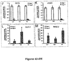
- the undifferentiated stem cells were validated immunocytochemically for their expression of human pluripotent stem cell markers, that included SSEA4, TRA-1-60, OCT4, NANOG, and SOX2 ( Figure 2 ). Of note, both OCT4 and SOX2 were also utilized as reprogramming factors in the generation of the hiPSCs, the generation of which have been previously described.
- Cells were passaged by incubation in collagenase type IV (1 mg/ml, Invitrogen) for 5-10 min, followed by gentle scraping from the culture dish, after which they were triturated 5 times through a polished glass pipette and then spun, washed and resuspended twice. The cells were then split 1:3-1:4 onto 6-well plates pre-coated with irradiated MEF cells.
- stage 2 To generate embryonic bodies (EB), hESC or hiPSC cultures were dissociated using Dispase (0.5 mg/ml, Invitrogen) at 37°C for 5-10 min, once they achieved 80% confluence with colony diameters of 250-300 m, in the absence of evident differentiation. These criteria proved important, as it was noted that the quality of hESC and hiPSC cultures at the stage 1-2 transition critically affected that of their derived EBs, as well as their subsequent differentiation into OPCs.
- Dispase 0.5 mg/ml, Invitrogen
- the EBs were cultured in suspension in tissue culture flasks (Nunc EasYFlasks, Thermo Scientific) in ESC medium without bFGF for 5 days; then switched to neural induction medium (NIM; DMEM/F12 supplemented with non-essential amino acids and N2) supplemented with bFGF (20 ng/ml, Sigma) and heparin (2 ⁇ g/ml, Sigma), for either 2 days (WA9/H9 hES) or 7 days (K04, C14 and C27 hiPSCs).
- NIM neural induction medium
- DMEM/F12 DMEM/F12 supplemented with non-essential amino acids and N2
- heparin (2 ⁇ g/ml, Sigma
- the EBs were plated onto laminin/poly-ornithine coated 6-well plates and cultured in NIM supplemented with bFGF, heparin and laminin (10 ⁇ g/ml) for 3 additional days; the medium was then switched to NIM supplemented with retinoic acid (RA, 100 nM, Sigma), for 4 days.
- NIM supplemented with bFGF, heparin and laminin (10 ⁇ g/ml) 3 additional days; the medium was then switched to NIM supplemented with retinoic acid (RA, 100 nM, Sigma), for 4 days.
- RA retinoic acid
- Stage 3 The neuroepithelial differentiation efficiency at this point, the end of stage 3, was assessed by immunolabeling for PAX6 and SOX1; co-expression of these markers characterizes central neural stem and progenitor cells.
- the yields of neuroepithelial colonies, defined as (PAX6 + /SOX1 + )/total rosette-like colonies, were 52.2 ⁇ 7.5%, 78.4 ⁇ 4.7%, and 76.0 ⁇ 7.0% from K04, C14 and C27 cultures, respectively (N 3-6 scored cultures/line).
- the efficiencies of neuroepithelial colony production from C14 and C27 hiPSCs were similar to those for WA09/H9 (75.4 ⁇ 8.8%) ( Figures 3A and 4A ).
- Stage 4 On day 14 (for H9) or day 19 (for K04 and C27) (end of stage 3, 4 days after addition of RA), purmorphamine (1 ⁇ M, Calbiochem), a sonic hedgehog (shh) agonist, and B27 (Invitrogen) were added to the media.
- the cultured NE colonies were detached mechanically 9 days later, at either 23 DIV (WA9/H9 hES) or 28 DIV (K04, C14 and C27 hiPSC), and then cultured in suspension in 6-well Ultralow cluster plates ( Figure 4B ).
- Stage 5 One day after plating into Ultralow cluster plates, the medium was replaced with NIM supplemented with bFGF (10 ng/ml), in addition to purmophamine and B27. At that point the phenotypic composition of aliquots was assessed by staining of OLIG2 and/or NKX2.2, to ascertain the appearance of pre-OPC colonies following RA treatment. Both OLIG2 and NKX2.2 are expressed by central OPCs, though NKX2.2 is the more specific indicator of oligodendroglial differentiation (see citations 16-17). At this early pre-OPC stage, the percentage of OLIG2-expressing colonies was higher than that of NKX2.2 + colonies, reflecting the earlier appearance of OLIG2 ( Figures 3A and 4C ).
- stage 5 35 DIV for WA09/H9 or 40 DIV for K04, C14 and C27
- stage 5 35 DIV for WA09/H9 or 40 DIV for K04, C14 and C27
- NKX2.2 + colonies appeared, concurrent with the peak of OLIG2 expression.
- the percentage of OLIG2 + /NKX2.2 + co-expressing colonies was similar among all four lines in this study ( Figure 3A ).
- stage 6 To initiate stage 6 (day 35 for WA09/H9 or day 40 for K04, C14 and C27), the OLIG2/NKX2.2-defined pre-OPCs in stage 5 suspension culture were switched to glial induction media (GIM; DMEM/F12, N1, B27, T3 at 60 ng/ml, biotin at 100 ng/ml, dibutyryl-cAMP at 1 ⁇ M; all from Sigma) supplemented with PDGF AA (10 ng/ml), IGF-1 (10 ng/ml), and NT3 (10 ng/ml). During this long period of OPC suspension culture, 2/3 of the media volume was changed every 3 days. The resultant stage 6 gliospheres were prevented from aggregating by gentle trituration through P1000 pipette tips during media changes.
- GEM glial induction media
- hOPC differentiation as defined by A2B5, CD140a and CD140a/CD9 co-expression, was assessed both in vitro and in vivo, using ICC, qRT-PCR, and xenograft into neonatal shiverer mice at serial time points. Gliospheres were capable of yielding both mature oligodendrocytes and myelinogenic OPCs as of 120 DIV.
- Late stage 6 / pre-transplant the newly produced hOPCs also expressed other OPC markers, such as CD140a/PDGFR ⁇ and SOX10, which typically co-expressed OLG2 or NKX2.2 ( Figures 4D-4H ).
- OPC markers such as CD140a/PDGFR ⁇ and SOX10, which typically co-expressed OLG2 or NKX2.2 ( Figures 4D-4H ).
- the percentages of the OLG2 + , NXK2.2 + or SOX10 + among all DAPI-identified cells, were 61.9 ⁇ 10.3%, 63.4 ⁇ 7.3%, and 84.6 ⁇ 7.0% among hiPCS/K04-derived OPCs, while the corresponding proportion of NKX2.2/SOX10 co-expressing OPCs was 60.6 ⁇ 4.4% ( Figure 4I ).
- RT-PCR for OLIG2, NKX2.2 and GFAP mRNA was performed, and all genes were substantially upregulated in stage 6 OPCs, as were their corresponding protein products ( Figures 11C-D and 3C ).
- Flow cytometric protocols and analysis Flow cytometric protocols and analysis. Flow cytometry of hESC- or hiPSC-derived OPCs was performed on a FACSAria IIIU (Becton Dickinson, San Jose, CA). Cells were gently scraped from the culture dishes and then treated with Accutase (Chemicon) at 37°C for 5 minutes with gentle shaking. The samples were then triturated with a narrow glass Pasteur pipette until a single cell suspension was obtained. The cells were then spun and resuspended in Miltenyi Washing Buffer (MWB) at 1x10 6 cells/ml. The primary antibodies, directly conjugated antibodies or their corresponding isotype controls were added to the cells at the concentrations listed below, then incubated on ice for 15 minutes.
- MMB Miltenyi Washing Buffer
- the cells were analyzed by forward and side scatter, for PE fluorescence through a 582 ⁇ 15 nm band-pass filter, for Alexa Fluor 488/FITC fluorescence through a 530 ⁇ 30 nm band-pass, for PERCP-Cy5.5 through a 695 ⁇ 40 nm band-pass, and for DAPI fluorescence through a 450 ⁇ 50 nm band-pass. Unstained cells were used to set the background fluorescence; a false positive rate of 0.5% was accepted.
- mice IgM isotype control (Chemicon, PP50), mouse anti-A2B5 (IgM, Chemicon, MAB312), mouse anti-O4 (IgM, Chemicon, MAB345), PE mouse IgG 2a , ⁇ isotype control (BD, 555574), PE mouse anti-human CD140a (IgG 2a , BD, 556002), PERCP-Cy5.5 mouse IgGi isotype control (BD, 347212) and PERCP-Cy5.5 mouse anti-human CD9 (IgG 1 , BD, 341649).
- Pluripotent hESC or hiPSCs raised on irradiated MEF cells were cultured for 3 to 4 days prior to fixation with 4% paraformaldehyde.
- the differentiated neurogenic or gliogenic clusters were plated onto poly-ornithine and laminin coated 24-well plate and cultured for 3 days before being fixed with 4% paraformaldehyde.
- the gliogenic spheres containing hOPCs at later stages were dissected into small fragments and plated onto poly-ornithine/laminin coated 24-well plates, and cultured for 2-4 weeks before being fixed, depending on the experiment.
- Example 1 Human iPSCs Can Be Efficiently Directed to Glial Progenitor Cell Fate
- each of the hiPSC and hESC lines could be directed into highly enriched preparations of OLIG2+/ PDGFRa+/NKX2.2+/SOX10+ OPCs. Indeed, the efficiencies of OPCs' differentiation from hiPSCs, whether induced from keratinocytes (K04 cells) or fibroblasts (C14 and C27 cells), were consistently higher than that of WA09/H9 hESCs.
- GFAP-defined astroglia first appeared by 70 days in vitro (DIV), significantly earlier than OLs did.
- DIV dipalmitosarcoma
- GFAP+ astrocytes were found to be abundant when gliogenic spheres were plated onto a polyornithine/ laminin-coated surface ( Figures 3B-3D ).
- GFAP+ cells comprised 40%-50% of cells in OPC-induced cultures, across all cell lines ( Figure 3D ).
- Quantitative RT-PCR confirmed the upregulation of GFAP messenger RNA (mRNA) expression during OPC differentiation in all cell lines (Table 2).
- Table 2 Table 2.
- O4+ OLs in C27, C14, and K04 hiPSC-derived OPC cultures respectively comprised 11.9 ⁇ 3.8%, 4.1 ⁇ 0.9%, and 7.6 ⁇ 1.5% of all cells (at 194 ⁇ 15, 186 ⁇ 14, and 205 ⁇ 14 DIV, respectively; means ⁇ SEM) ( Figure 5A ; Table 3).
- the culture conditions favored initial oligodendrocytic differentiation, but not postmitotic oligodendrocytic survival, because the focus was on preparing populations of transplantable lineage-biased progenitors and immature oligodendroglia rather than more mature-but less transplantable-process-bearing OLs. Table 3.
- Example 3 - OPCs could Be Isolated from hiPSC Cultures by CD140a- and CD9-Directed Fluorescence-Activated Cell Sorting
- CD140a+ OPCs derived from C27, C14, and K04 hiPSCs respectively comprised 33.0 ⁇ 10.3%, 32.8 ⁇ 12.0%, and 41.1 ⁇ 6.1% of all cells, compared to 37.5 ⁇ 10.2% of H9-derived cells ( Figure 5C ; Table 4).
- hiPSC OPCs could be identified and isolated at different stages of lineage restriction, which were serially represented by A2B5, CD140a, CD9, and O4. Selection based on these epitopes permits the isolation of relatively pure populations of hiPSC OPCs while removing residual undifferentiated cells from the isolate. Table 4.
- hiPSC OPCs were cocultured gestational age (g.a.) fetal brain using polysialyted neural cell adhesion molecule (PSA-NCAM)-directed selection ( Windrem et al., "Neonatal Chimerization With Human Glial Progenitor Cells Can Both Remyelinate and Rescue the Otherwise Lethally Hypomyelinated Shiverer Mouse," Cell Stem Cell 2:553-565 (2008 ), which is hereby incorporated by reference in its entirety).
- PSA-NCAM polysialyted neural cell adhesion molecule
- hiPSC OPCs were then prepared as clusters of 50-100 mm in diameter and cocultured with the fetal neurons for 4 weeks; the cultures were then immunolabeled for MBP and neurofilament (NF). Confocal imaging revealed abundant MBP + processes that contacted axons and initiated ensheathment ( Figures 3H-3J ), though unambiguous myelin formation was not noted at the time points imaged. Thus, to better assess myelinogenesis by hiPSC OPCs, their engraftment and myelination in vivo was evaluated.
- mice were killed and their brains were analyzed in terms of donor cell distribution and density, myelin production and the proportion of myelinated axons, and nodal reconstitution. All three of the hiPSC line-derived OPCs were able to robustly myelinate the recipient brains; from each line, high donor cell densities and widespread dispersal were observed throughout the forebrain white matter ( Figures 6A and 6B ).
- C27 and K04 hiPSC-derived OPCs and oligodendroglia defined as human nuclear antigen (hNA) + /OLIG2 + , achieved densities of 29,498 ⁇ 13,144 and 37,032 ⁇ 8,392 cells/mm 3 , respectively.
- hNA human nuclear antigen
- OLIG2 + human nuclear antigen + /OLIG2 +
- the density of hiPSC-OPC donor derived myelination and the proportion of ensheathed axons at 13 weeks proved as high as, and exceeded, those achieved by OPCs derived from second-trimester fetal brain tissue, whether isolated as A2B5 + /PSA-NCAM - or CD140a + cells ( Sim et al., "CD140a Identifies a Population of Highly Myelinogenic, Migration-Competent and Efficiently Engrafting Human Oligodendrocyte Progenitor Cells," Nat. Biotechnol.
- Windrem et al. "Fetal and Adult Human Oligodendrocyte Progenitor Cell Isolates Myelinate the Congenitally Dysmyelinated Brain,” Nat. Med. 10:93-97 (2004 ); Windrem et al., "Neonatal Chimerization With Human Glial Progenitor Cells Can Both Remyelinate and Rescue the Otherwise Lethally Hypomyelinated Shiverer Mouse,” Cell Stem Cell 2:553-565 (2008 ), which are hereby incorporated by reference in their entirety).
- hiPSC OPCs Despite the widespread infiltration of the recipient brains by hiPSC OPCs, substantial astrocytic differentiation was noted by those cells within the presumptive white matter, within which the donor cells differentiated as morphologically apparent fibrous astrocytes, in close association with hiPSC derived OLs. These hiPSC-derived astrocytes might have been generated from lineage-restricted hiPSC-derived astrogliogenic precursors or by astrocytic differentiation in situ from still-bipotential hOPCs.
- a matched set of 19 littermate controls were injected only with saline, and both sets were housed without further manipulation.
- the 19 unimplanted shiverer controls died before 5 months of age, with a median survival of 141 days.
- 19 of the 22 implanted mice lived longer than the longest-lived control mouse.
- the transplanted mice exhibited greatly prolonged survival ( Figure 8E ), with reduced death over the 9 month period of observation, after which the experiment was terminated so that surviving mice could be processed for both immunohistochemical assessment of late-stage myelination and nodal reconstitution and for ultrastructural analysis (see Example 8).
- the recipient callosa were densely myelinated by mature compact myelin characterized by concentrically organized major dense lines ( Figures 10A-10E ) and interlaminar tight junctions ( Figures 10F, 10G , and 11C ); and the engrafted callosa were quite unlike those of their untransplanted shiverer controls, which failed to exhibit major dense lines or any other evidence of myelin compaction ( Figures 11B, 11D, and 11E ).
- hiPSC-derived OPCs can efficiently generate OLs, which in turn can robustly myelinate the hypomyelinated shiverer forebrain, and that the myelin thereby generated is able to restore nodal architecture as well as to ensheath axons as efficiently as purified isolates of fetal-tissue-derived OPCs.
- hiPSCs were transplanted into normally myelinated rag2 -null mice to assess tumorigenicity in the wild-type myelin environment as well.
- hiPSC-OPC-engrafted mice The lack of tumor formation in hiPSC-OPC-engrafted mice was associated with a significant decrease in the mitotic fraction of the implanted hiPSC OPCs as a function of time after graft.
- hiPSC OPCs from single-patient skin samples can now be reliably produced in sufficient numbers to provide myelinogenic autografts largely, though perhaps not completely ( Zhao et al., "Immunogenicity of Induced Pluripotent Stem Cells," Nature 474:212-215 (2011 ), which is hereby incorporated by reference in its entirety), free of rejection risk.
- the myelination efficiency of the implanted iPSC derived OPCs defined as the proportion of central axons myelinated as a function of time after graft, proved as high as that which was had previously achieved using tissue-derived, CD140a sorted OPCs ( Sim et al., "CD140a Identifies a Population of Highly Myelinogenic, Migration-Competent and Efficiently Engrafting Human Oligodendrocyte Progenitor Cells," Nat. Biotechnol. 29:934-941 (2011 ), which is hereby incorporated by reference in its entirety).
- the proportion of axons ensheathed was as high in enriched but unsorted hiPSC-OPC grafts as in fetal-tissue-derived OPC grafts that had been sorted for CD140a+ cells prior to transplant.
- the hiPSC-OPCs grafts myelinated more axons more rapidly than did A2B5+/PSA-NCAM-sorted fetal-tissue derived cells, probably reflecting the higher proportion of bipotential glial progenitor cells in the hiPSC-OPC populations by the time of their harvest and transplantation.
- hiPSC-OPC grafts In light of the robust myelination afforded by hiPSC-OPC grafts, it was asked whether neonatal transplantation of hiPSC OPCs might be sufficient to rescue the phenotype and survival of recipient shiverer homozygotes, as had previously been observed in a minority of shiverers transplanted with fetal-human brain-derived OPCs.
- the hiPSC-OPC-transplanted mice indeed exhibited markedly improved survival; death was both delayed and reduced overall in the transplanted group over the 9 month period of observation.
- mice manifested progressive resolution of their neurological deficits ( Windrem et al., "Neonatal Chimerization With Human Glial Progenitor Cells Can Both Remyelinate and Rescue the Otherwise Lethally Hypomyelinated Shiverer Mouse,” Cell Stem Cell 2:553-565 (2008 ), which is hereby incorporated by reference in its entirety).
- hiPSC OPCs as therapeutic vectors, perhaps by virtue of their more rapid myelinogenesis, which may be a function of the prolonged differentiation conditions that was employed in this OPC induction protocol.
- hiPSC OPCs may be sorted to purity before transplantation, on the basis of the high incidence of definitively pro-oligodendrocytic CD9 + /CD140a + cells in the cultures, and the ability to isolate these cells by fluorescence-activated cell sorting (FACS) based upon these coexpressed epitopes ( Sim et al., "CD140a Identifies a Population of Highly Myelinogenic, Migration-Competent and Efficiently Engrafting Human Oligodendrocyte Progenitor Cells," Nat. Biotechnol. 29:934-941 (2011 ), which is hereby incorporated by reference in its entirety).
- FACS fluorescence-activated cell sorting
- astrocytic engraftment may be of particular importance in correcting dysmyelinating disorders of enzyme deficiency, given that astrocytic lysosomal enzymes have been found to readily transit from wild-type to deficient glia within brain tissue, in a manner potentially sufficient to rescue enzyme-deficient hosts ( Lee et al., "Stem Cells Act Through Multiple Mechanisms to Benefit Mice With Neurodegenerative Metabolic Disease,” Nat. Med. 13:439-447 (2007 ), which is hereby incorporated by reference in its entirety).
- hiPSC-derived astrocytes may prove to be critically important therapeutic vectors for diseases of primarily astrocytic pathology ( Krencik et al., "Specification of Transplantable Astroglial Subtypes From Human Pluripotent Stem Cells,” Nat. Biotechnol. 29:528-534 (2011 ), which is hereby incorporated by reference in its entirety), such as Alexander disease and the vanishing white-matter disorders ( Bugiani et al., "Defective Glial Maturation in Vanishing White Matter Disease,” J. Neuropathol. Exp. Neurol.
- iPSC OPCs Human iPSC OPCs might thus be attractive vectors for restoring or replacing glial populations in a variety of disease settings.
- the data presented herein indicates the preferential use of hiPSC-derived OPCs to restore lost myelin in disorders such as multiple sclerosis and traumatic demyelination, in which no genetic abnormalities might complicate the use of a patient's own somatic cells as the iPSC source.
- iPSC OPCs may similarly prove of great therapeutic value in genetic disorders of myelin, such as Pelizaeus-Merzbacher disease, recognizing that the underlying genetic defect must first be repaired in the donor somatic cells before glial progenitor induction and implantation.
Applications Claiming Priority (4)
| Application Number | Priority Date | Filing Date | Title |
|---|---|---|---|
| US201361761584P | 2013-02-06 | 2013-02-06 | |
| US201361780265P | 2013-03-13 | 2013-03-13 | |
| EP14749594.9A EP2954046A4 (fr) | 2013-02-06 | 2014-02-06 | Cellules progénitrices d'oligodendrocyte issues de cellules pluripotentes induites pour le traitement de troubles de la myéline |
| PCT/US2014/015019 WO2014124087A1 (fr) | 2013-02-06 | 2014-02-06 | Cellules progénitrices d'oligodendrocyte issues de cellules pluripotentes induites pour le traitement de troubles de la myéline |
Related Parent Applications (1)
| Application Number | Title | Priority Date | Filing Date |
|---|---|---|---|
| EP14749594.9A Division EP2954046A4 (fr) | 2013-02-06 | 2014-02-06 | Cellules progénitrices d'oligodendrocyte issues de cellules pluripotentes induites pour le traitement de troubles de la myéline |
Publications (1)
| Publication Number | Publication Date |
|---|---|
| EP3766962A1 true EP3766962A1 (fr) | 2021-01-20 |
Family
ID=51300121
Family Applications (2)
| Application Number | Title | Priority Date | Filing Date |
|---|---|---|---|
| EP20190706.0A Pending EP3766962A1 (fr) | 2013-02-06 | 2014-02-06 | Cellules progénitrices d'oligodendrocyte issues de cellules pluripotentes induites pour le traitement de troubles de la myéline |
| EP14749594.9A Pending EP2954046A4 (fr) | 2013-02-06 | 2014-02-06 | Cellules progénitrices d'oligodendrocyte issues de cellules pluripotentes induites pour le traitement de troubles de la myéline |
Family Applications After (1)
| Application Number | Title | Priority Date | Filing Date |
|---|---|---|---|
| EP14749594.9A Pending EP2954046A4 (fr) | 2013-02-06 | 2014-02-06 | Cellules progénitrices d'oligodendrocyte issues de cellules pluripotentes induites pour le traitement de troubles de la myéline |
Country Status (4)
| Country | Link |
|---|---|
| US (5) | US10450546B2 (fr) |
| EP (2) | EP3766962A1 (fr) |
| JP (4) | JP6541577B2 (fr) |
| WO (1) | WO2014124087A1 (fr) |
Families Citing this family (43)
| Publication number | Priority date | Publication date | Assignee | Title |
|---|---|---|---|---|
| US20030223972A1 (en) | 2002-02-15 | 2003-12-04 | Goldman Steven A. | Myelination of congenitally dysmyelinated forebrains using oligodendrocyte progenitor cells |
| EP2283116B1 (fr) | 2008-05-08 | 2016-09-28 | University Of Rochester | Traitement des maladies de la myéline au moyen de préparations cellulaires optimisées |
| JP6541577B2 (ja) * | 2013-02-06 | 2019-07-10 | ユニバーシティー オブ ロチェスター | ミエリン障害の治療のための誘導多能性細胞由来オリゴデンドロサイト前駆細胞 |
| US10801010B2 (en) | 2014-03-07 | 2020-10-13 | Unist (Ulsan National Institute Of Science And Technology) | Composition for inducing direct transdifferentiation into oligodendrocyte progenitor cells from somatic cells and use thereof |
| US9724432B2 (en) | 2015-04-30 | 2017-08-08 | University Of Rochester | Non-human mammal model of human degenerative disorder, uses thereof, and method of treating human degenerative disorder |
| CA2995634A1 (fr) * | 2015-08-15 | 2017-02-23 | Asterias Biotherapeutics, Inc. | Cellules progenitrices d'olygodendrocytes derivees de cellules souches destinees au traitement d'accidents vasculaires cerebraux lies a la substance blanche |
| US11920155B2 (en) | 2016-03-30 | 2024-03-05 | Asterias Biotherapeutics, Inc. | Oligodendrocyte progenitor cell compositions |
| EP3439648A4 (fr) * | 2016-04-04 | 2019-04-24 | Mayo Foundation for Medical Education and Research | Traitement de la névrite optique avec des cellules précurseurs d'oligodendrocytes dérivées de cellules souches pluripotentes induites |
| EP3474876A4 (fr) * | 2016-06-22 | 2020-01-15 | City of Hope | Traitement de la maladie de canavan |
| EP3621434A4 (fr) | 2017-05-10 | 2021-03-31 | University of Rochester | Méthodes de traitement de troubles neuropsychiatriques |
| CN108624560B (zh) * | 2018-06-01 | 2022-04-08 | 南京艾尔普再生医学科技有限公司 | 一种分化培养基及少突胶质前体细胞的制备方法 |
| JP2021528445A (ja) | 2018-06-21 | 2021-10-21 | ユニバーシティー オブ ロチェスター | ハンチントン病を治療するまたはその発症を阻害する方法 |
| TWI675678B (zh) * | 2018-08-23 | 2019-11-01 | 國為生醫科技股份有限公司 | 亞丁基苯酞於多巴胺神經前驅細胞移植治療的應用 |
| WO2020154533A1 (fr) | 2019-01-23 | 2020-07-30 | Asterias Biotherapeutics, Inc. | Cellules progénitrices d'oligodendrocytes dérivées du dos à partir de cellules souches pluripotentes humaines |
| WO2020167822A2 (fr) | 2019-02-13 | 2020-08-20 | University Of Rochester | Réseaux de gènes assurant la médiation de la remyélinisation du cerveau humain |
| US20220267737A1 (en) | 2019-07-18 | 2022-08-25 | University Of Rochester | Cell-type selective immunoprotection of cells |
| EP4017508A1 (fr) | 2019-08-23 | 2022-06-29 | Sana Biotechnology, Inc. | Cellules exprimant cd24 et utilisations associées |
| WO2021055585A1 (fr) | 2019-09-17 | 2021-03-25 | Boston Polarimetrics, Inc. | Systèmes et procédés de modélisation de surface utilisant des repères de polarisation |
| KR102650805B1 (ko) * | 2019-12-17 | 2024-03-25 | 코아스템켐온 주식회사 | 인간 만능 줄기세포로부터 제작된 3d 오가노이드를 해체하여 희소돌기아교세포를 다량 확보하는 분화방법 |
| CA3173096A1 (fr) | 2020-03-25 | 2021-09-30 | Sonja SCHREPFER | Cellules neurales hypoimmunogenes pour le traitement de troubles et d'etats neurologiques |
| WO2021216583A1 (fr) | 2020-04-20 | 2021-10-28 | University Of Rochester | Identification de cellules humaines transplantées |
| KR20230074718A (ko) | 2020-08-13 | 2023-05-31 | 사나 바이오테크놀로지, 인크. | 저면역원성 세포로 감작된 환자를 치료하는 방법 및 관련 방법 및 조성물 |
| CN112798771B (zh) * | 2021-03-31 | 2021-07-30 | 宝枫生物科技(北京)有限公司 | 用于诊断脑白质病变的生物标志物及其应用 |
| KR20240013135A (ko) | 2021-05-27 | 2024-01-30 | 사나 바이오테크놀로지, 인크. | 조작된 hla-e 또는 hla-g를 포함하는 저면역원성 세포 |
| AU2022309875A1 (en) | 2021-07-14 | 2024-01-25 | Sana Biotechnology, Inc. | Altered expression of y chromosome-linked antigens in hypoimmunogenic cells |
| CN113481161A (zh) * | 2021-07-30 | 2021-10-08 | 四川省医学科学院·四川省人民医院 | 一种培养基及少突胶质祖细胞的诱导培养方法以及应用 |
| CN113564122B (zh) * | 2021-08-05 | 2022-04-08 | 呈诺再生医学科技(珠海横琴新区)有限公司 | 人诱导性多能干细胞向少突胶质细胞分化的方法,试剂盒以及应用 |
| WO2023019225A2 (fr) | 2021-08-11 | 2023-02-16 | Sana Biotechnology, Inc. | Cellules génétiquement modifiées pour une thérapie cellulaire allogénique permettant de réduire les réactions inflammatoires à médiation par le sang instantanée |
| AU2022326565A1 (en) | 2021-08-11 | 2024-02-08 | Sana Biotechnology, Inc. | Genetically modified cells for allogeneic cell therapy |
| WO2023019203A1 (fr) | 2021-08-11 | 2023-02-16 | Sana Biotechnology, Inc. | Systèmes inductibles pour modifier l'expression génique dans des cellules hypoimmunogènes |
| WO2023019227A1 (fr) | 2021-08-11 | 2023-02-16 | Sana Biotechnology, Inc. | Cellules génétiquement modifiées pour une thérapie cellulaire allogénique pour réduire les réactions inflammatoires induites par le complément |
| CN113699117B (zh) * | 2021-09-03 | 2023-06-30 | 呈诺再生医学科技(北京)有限公司 | 经基因改造的少突胶质祖细胞在多发性硬化症中的应用 |
| US20230226116A1 (en) | 2021-10-20 | 2023-07-20 | University Of Rochester | Method for rejuvenating glial progenitor cells and rejuvenated glial progenitor cells per se |
| WO2023069979A1 (fr) | 2021-10-20 | 2023-04-27 | University Of Rochester | Cellules progénitrices gliales isolées destinées à être utilisées dans le traitement par compétition de la perte de matière blanche liée à l'âge |
| US20230292719A1 (en) | 2021-10-20 | 2023-09-21 | University Of Rochester | Humanized chimeras for the prospective assessment of cell addition and replacement therapies |
| US20230270818A1 (en) | 2021-11-02 | 2023-08-31 | University Of Rochester | Tcf7l2 mediated remyelination in the brain |
| WO2023150557A1 (fr) | 2022-02-01 | 2023-08-10 | University Of Rochester | Procédés de génération d'une population de neurones à partir de cellules progénitrices gliales humaines et constructions génétiques pour la mise en œuvre de tels procédés |
| WO2023150553A1 (fr) | 2022-02-01 | 2023-08-10 | University Of Rochester | Ciblage et transduction basés sur un promoteur rcpg17 de cellules progénitrices gliales |
| WO2023164466A1 (fr) * | 2022-02-23 | 2023-08-31 | Sqz Biotechnologies Company | Procédés de production de cellules gliales et leurs utilisations |
| CN114848847A (zh) * | 2022-03-22 | 2022-08-05 | 中山大学·深圳 | 一种负载Olig2信使RNA的脂质纳米颗粒及其应用 |
| WO2023183313A1 (fr) | 2022-03-22 | 2023-09-28 | Sana Biotechnology, Inc. | Cellules d'ingénierie avec un transgène dans un locus b2m ou ciita et compositions et procédés associés |
| WO2023215455A1 (fr) | 2022-05-05 | 2023-11-09 | University Of Rochester | Approche macrogliale-microgliale double vers le remplacement de cellules thérapeutiques dans une maladie neurodégénérative et neuropsychiatrique |
| WO2023229819A1 (fr) * | 2022-05-24 | 2023-11-30 | Bluerock Therapeutics Lp | Procédés de fabrication de cellules progénitrices d'oligodendrocytes |
Citations (11)
| Publication number | Priority date | Publication date | Assignee | Title |
|---|---|---|---|---|
| WO2003070171A2 (fr) * | 2002-02-15 | 2003-08-28 | Cornell Research Foundation, Inc. | Myelinisation de prosencephales congenitalement demyelinises par utilisation de cellules souches d'oligodendrocyte |
| US6692957B2 (en) | 1997-01-23 | 2004-02-17 | Cornell Research Foundation, Inc. | Method for separating cells |
| WO2007069666A1 (fr) | 2005-12-13 | 2007-06-21 | Kyoto University | Facteur de reprogrammation nucleaire |
| US20080233610A1 (en) | 2007-03-23 | 2008-09-25 | Thomson James A | Somatic cell reprogramming |
| WO2009006930A1 (fr) | 2007-06-15 | 2009-01-15 | Izumi Bio, Inc. | Cellules souches pluripotentes humaines induites à partir des cellules souches indifférenciées issues d'un tissu postnatal humain |
| US20100156778A1 (en) | 2005-06-10 | 2010-06-24 | Seiko Epson Corporation | Display panel module, display unit, inspection device for display panel and inspection method for display panel |
| US20110059055A1 (en) | 2008-05-08 | 2011-03-10 | University Of Rochester | Treating myelin diseases with optimized cell preparations |
| US20110200568A1 (en) | 2008-08-08 | 2011-08-18 | Yasuhiro Ikeda | Induced pluripotent stem cells |
| US8227247B2 (en) * | 2007-12-20 | 2012-07-24 | Wisconsin Alumni Research Foundation | Method of generating myelinating oligodendrocytes |
| US20120276070A1 (en) | 2009-11-17 | 2012-11-01 | Vitro Diagnositics, Inc. | Induced Pluripotent Stem Cells and Related Methods |
| US20120276636A1 (en) | 2010-01-22 | 2012-11-01 | Kyoto University | Method for improving induced pluripotent stem cell generation efficiency |
Family Cites Families (30)
| Publication number | Priority date | Publication date | Assignee | Title |
|---|---|---|---|---|
| US3263402A (en) | 1964-01-02 | 1966-08-02 | Ford Motor Co | Internal combustion engine filtration assembly for fresh air and crankcase ventilatin air |
| US3753357A (en) | 1970-12-14 | 1973-08-21 | Ovitron Res Corp | Method and apparatus for the preservation of cells and tissues |
| US4199022A (en) | 1978-12-08 | 1980-04-22 | The United States Of America As Represented By The Department Of Energy | Method of freezing living cells and tissues with improved subsequent survival |
| US4559298A (en) | 1982-11-23 | 1985-12-17 | American National Red Cross | Cryopreservation of biological materials in a non-frozen or vitreous state |
| US6497872B1 (en) | 1991-07-08 | 2002-12-24 | Neurospheres Holdings Ltd. | Neural transplantation using proliferated multipotent neural stem cells and their progeny |
| ATE234353T1 (de) | 1992-10-28 | 2003-03-15 | Neurospheres Holdings Ltd | Biologische faktoren und neuronale stammzellen |
| US6361996B1 (en) | 1997-05-07 | 2002-03-26 | University Of Utah Research Foundation | Neuroepithelial stem cells and glial-restricted intermediate precursors |
| US6734015B1 (en) | 1997-07-04 | 2004-05-11 | University Of Utah Research Foundation | Isolation of lineage-restricted neuronal precursors |
| US6235527B1 (en) | 1997-11-29 | 2001-05-22 | University Of Utah Research Foundation | Lineage restricted glial precursors from the central nervous system |
| US6812027B2 (en) | 1998-03-25 | 2004-11-02 | Cornell Research Foundation, Inc. | Discovery, localization, harvest, and propagation of an FGF2 and BDNF-responsive population of neural and neuronal progenitor cells in the adult human forebrain |
| US8263402B1 (en) | 1998-10-19 | 2012-09-11 | Cornell Research Foundation, Inc. | Method for isolating and purifying oligodendrocytes and oligodendrocyte progenitor cells |
| US7468277B2 (en) | 1999-12-23 | 2008-12-23 | Cornell Research Foundation, Inc. | Enriched preparation of human fetal multipotential neural stem cells |
| EP1278529B1 (fr) | 2000-04-12 | 2006-06-14 | Children's Hospital Of Philadelphia | Utilisations therapeutiques de cellules stromales mesenchymateuses |
| CA2409698C (fr) * | 2000-05-17 | 2010-10-26 | Geron Corporation | Populations de progeniteurs neuronaux |
| US6852532B2 (en) | 2001-03-21 | 2005-02-08 | University Of Utah Research Foundation | Method of isolating human neuroepithelial precursor cells from human fetal tissue |
| US20050084963A1 (en) | 2001-06-01 | 2005-04-21 | Tailoi Chan-Ling | Purification of lineage-specific cells and uses therefor |
| US7150989B2 (en) | 2001-08-10 | 2006-12-19 | Cornell Research Foundation, Inc. | Telomerase immortalized neural progenitor cells |
| KR20040075957A (ko) | 2002-01-23 | 2004-08-30 | 유니버시티 오브 유타 리서치 파운데이션 | 별아교세포 제한 전구체 세포의 순수한 집단과 그것을분리하고 이용하는 방법 |
| US7285415B2 (en) | 2002-07-11 | 2007-10-23 | The Regents Of The University Of California | Oligodendrocytes derived from human embryonic stem cells for remyelination and treatment of spinal cord injury |
| US8642332B2 (en) | 2003-03-07 | 2014-02-04 | Cornell Research Foundation, Inc. | Identification and isolation of multipotential neural progenitor cells from the subcortical white matter of the adult human brain |
| EP2114167B1 (fr) | 2007-01-16 | 2018-07-11 | University of Rochester | Animaux non humains à cerveaux chimériques ayant des cellules gliales humaines |
| EP2379711B8 (fr) | 2008-12-23 | 2017-03-22 | BOCO Silicon Valley, Inc. | Populations cibles de cellules précurseurs d'oligodendrocytes et leurs méthodes de fabrication et d'utilisation |
| US20120207744A1 (en) | 2009-03-19 | 2012-08-16 | Mendlein John D | Reprogramming compositions and methods of using the same |
| NO2499238T3 (fr) | 2009-11-12 | 2018-01-27 | ||
| US8206669B2 (en) | 2010-07-27 | 2012-06-26 | Air Products And Chemicals, Inc. | Method and apparatus for treating a sour gas |
| CN103429734A (zh) * | 2010-10-26 | 2013-12-04 | 卡斯西部储备大学 | 用于产生神经胶质细胞的群体的分化方法 |
| BR112013017685A2 (pt) | 2011-01-12 | 2016-10-11 | Tsuneo Kido | método de cultura in vitro de uma célula neural expansível e uso de uma célula neural expansível |
| JP6541577B2 (ja) | 2013-02-06 | 2019-07-10 | ユニバーシティー オブ ロチェスター | ミエリン障害の治療のための誘導多能性細胞由来オリゴデンドロサイト前駆細胞 |
| US10779519B2 (en) | 2014-05-13 | 2020-09-22 | University Of Rochester | Human glial chimeric model for drug candidate assessment in human gliotrophic viral infections and progressive multifocal encephalopathy |
| US9724432B2 (en) | 2015-04-30 | 2017-08-08 | University Of Rochester | Non-human mammal model of human degenerative disorder, uses thereof, and method of treating human degenerative disorder |
-
2014
- 2014-02-06 JP JP2015557053A patent/JP6541577B2/ja active Active
- 2014-02-06 EP EP20190706.0A patent/EP3766962A1/fr active Pending
- 2014-02-06 US US14/764,507 patent/US10450546B2/en active Active
- 2014-02-06 WO PCT/US2014/015019 patent/WO2014124087A1/fr active Application Filing
- 2014-02-06 EP EP14749594.9A patent/EP2954046A4/fr active Pending
-
2017
- 2017-02-08 US US15/427,986 patent/US10626369B2/en active Active
-
2019
- 2019-06-11 JP JP2019108327A patent/JP6894941B2/ja active Active
- 2019-10-21 US US16/659,062 patent/US20200048604A1/en active Pending
- 2019-10-21 US US16/659,110 patent/US20200048605A1/en active Pending
-
2021
- 2021-06-04 JP JP2021093967A patent/JP7277978B2/ja active Active
-
2022
- 2022-03-22 US US17/701,267 patent/US20220290099A1/en active Pending
-
2023
- 2023-04-27 JP JP2023072923A patent/JP2023093709A/ja active Pending
Patent Citations (14)
| Publication number | Priority date | Publication date | Assignee | Title |
|---|---|---|---|---|
| US6692957B2 (en) | 1997-01-23 | 2004-02-17 | Cornell Research Foundation, Inc. | Method for separating cells |
| WO2003070171A2 (fr) * | 2002-02-15 | 2003-08-28 | Cornell Research Foundation, Inc. | Myelinisation de prosencephales congenitalement demyelinises par utilisation de cellules souches d'oligodendrocyte |
| US20100156778A1 (en) | 2005-06-10 | 2010-06-24 | Seiko Epson Corporation | Display panel module, display unit, inspection device for display panel and inspection method for display panel |
| WO2007069666A1 (fr) | 2005-12-13 | 2007-06-21 | Kyoto University | Facteur de reprogrammation nucleaire |
| US20080233610A1 (en) | 2007-03-23 | 2008-09-25 | Thomson James A | Somatic cell reprogramming |
| WO2008118820A2 (fr) | 2007-03-23 | 2008-10-02 | Wisconsin Alumni Research Foundation | Reprogrammation d'une cellule somatique |
| WO2009006997A1 (fr) | 2007-06-15 | 2009-01-15 | Izumi Bio, Inc. | Cellules souches pluripotentes humaines et leur utilisation à des fins médicales |
| WO2009007852A2 (fr) | 2007-06-15 | 2009-01-15 | Izumi Bio, Inc | Cellules multipotentes/pluripotentes et procédés s'y rapportant |
| WO2009006930A1 (fr) | 2007-06-15 | 2009-01-15 | Izumi Bio, Inc. | Cellules souches pluripotentes humaines induites à partir des cellules souches indifférenciées issues d'un tissu postnatal humain |
| US8227247B2 (en) * | 2007-12-20 | 2012-07-24 | Wisconsin Alumni Research Foundation | Method of generating myelinating oligodendrocytes |
| US20110059055A1 (en) | 2008-05-08 | 2011-03-10 | University Of Rochester | Treating myelin diseases with optimized cell preparations |
| US20110200568A1 (en) | 2008-08-08 | 2011-08-18 | Yasuhiro Ikeda | Induced pluripotent stem cells |
| US20120276070A1 (en) | 2009-11-17 | 2012-11-01 | Vitro Diagnositics, Inc. | Induced Pluripotent Stem Cells and Related Methods |
| US20120276636A1 (en) | 2010-01-22 | 2012-11-01 | Kyoto University | Method for improving induced pluripotent stem cell generation efficiency |
Non-Patent Citations (76)
| Title |
|---|
| ALIREZA POUYA ET AL: "Human Induced Pluripotent Stem Cells Differentiation into Oligodendrocyte Progenitors and Transplantation in a Rat Model of Optic Chiasm Demyelination", PLOS ONE, vol. 6, no. 11, 18 November 2011 (2011-11-18), pages e27925, XP055628994, DOI: 10.1371/journal.pone.0027925 * |
| BEN-HUR TAMIR ET AL: "Prospects of cell therapy for disorders of myelin", ANNALS OF THE NEW YORK ACADEMY OF SCIENCES, NEW YORK ACADEMY OF SCIENCES, US, vol. 1142, 1 October 2008 (2008-10-01), pages 218 - 249, XP002567696, ISSN: 0077-8923, [retrieved on 20081023], DOI: 10.1196/ANNALS.1444.014 * |
| BERRY ET AL.: "Cytology and Lineage of NG2-Positive Glia", J. NEUROCYTOL., vol. 31, 2002, pages 457 - 467, XP019214089 |
| BUGIANI ET AL.: "Defective Glial Maturation in Vanishing White Matter Disease", J. NEUROPATHOL. EXP. NEUROL., vol. 70, 2011, pages 69 - 82 |
| CAI ET AL.: "Generation of Human Induced Pluripotent Stem Cells from Umbilical Cord Matrix and Amniotic Membrane Mesenchymal Cells", J. BIOL. CHEM., vol. 285, no. 15, pages 112227 - 11234 |
| CHAMBERS ET AL.: "Highly Efficient Neural Conversion of Human ES and iPS Cells by Dual Inhibition of SMAD Signaling", NAT. BIOTECHNOL., vol. 27, 2009, pages 275 - 280, XP055007827, DOI: 10.1038/nbt.1529 |
| CHO ET AL.: "Induction of Pluripotent Stem Cells from Adult Somatic Cells by Protein-Based Reprogramming without Genetic Manipulation", BLOOD, vol. 116, 2010, pages 386 - 395, XP055153330, DOI: 10.1182/blood-2010-02-269589 |
| CHUA S J ET AL: "Neural progenitors, neurons and oligodendrocytes from human umbilical cord blood cells in a serum-free, feeder-free cell culture", BIOCHEMICAL AND BIOPHYSICAL RESEARCH COMMUNICATIONS, ACADEMIC PRESS INC. ORLANDO, FL, US, vol. 379, no. 2, 6 February 2009 (2009-02-06), pages 217 - 221, XP027552644, ISSN: 0006-291X, [retrieved on 20090206], DOI: 10.1016/J.BBRC.2008.12.045 * |
| DIETRICH ET AL.: "Characterization of A2B5+ Glial Precursor Cells From Cryopreserved Human Fetal Brain Progenitor Cells", GLIA, vol. 40, 2002, pages 65 - 77 |
| DONG WOOK HAN ET AL: "Direct Reprogramming of Fibroblasts into Neural Stem Cells by Defined Factors", CELL STEM CELL, vol. 10, no. 4, 1 April 2012 (2012-04-01), pages 465 - 472, XP055027756, ISSN: 1934-5909, DOI: 10.1016/j.stem.2012.02.021 * |
| FENG ET AL., NATURE CELL BIOLOGY, vol. 11, 2009, pages 197 - 203 |
| FRASER J SIM ET AL: "CD140a identifies a population of highly myelinogenic, migration-competent and efficiently engrafting human oligodendrocyte progenitor cells", NATURE BIOTECHNOLOGY, vol. 29, no. 10, 1 January 2011 (2011-01-01), pages 934 - 941, XP055010932, ISSN: 1087-0156, DOI: 10.1038/nbt.1972 * |
| FUSAKI ET AL.: "Efficient Induction of Transgene-Free Human Pluripotent Stem Cells Using a Vector Based on Sendi Virus, an RNA Virus That Does Not Integrate into the Host Genome", PROC JPN ACAD., vol. 85, 2009, pages 348 - 362, XP002663242, DOI: 10.2183/PJAB.85.348 |
| GIORGETTI ET AL.: "Generation of Induced Pluripotent Stem Cells from Human Cord Blood Cells with only Two Factors: Oct4 and Sox2", NATURE PROTOCOLS, vol. 5, no. 4, 2010, pages 811 - 820, XP009173937, DOI: 10.1038/nprot.2010.16 |
| GUMPEL ET AL.: "Transplantation of Human Embryonic Oligodendrocytes Into Shiverer Brain", ANN NY ACAD SCI, vol. 495, 1987, pages 71 - 85, XP009074370, DOI: 10.1111/j.1749-6632.1987.tb23666.x |
| HANNA ET AL., CELL, vol. 133, no. 2, 2008, pages 250 - 264 |
| HOJJAT-ALLAH ABBASZADEH ET AL: "Bone Marrow Stromal Cell Transdifferentiation into Oligodendrocyte-Like Cells Using Triiodothyronine as a Inducer with Expression of Platelet-Derived Growth Factor [alpha] as a Maturity Marker", IRANIAN BIOMEDICAL JOURNAL, 1 January 1996 (1996-01-01), pages 62 - 70, XP055275467, Retrieved from the Internet <URL:http://www.ncbi.nlm.nih.gov/pmc/articles/PMC3677678/pdf/IBJ-017-062.pdf> [retrieved on 2016], DOI: 10.6091/ibj.11162.2013 * |
| HU ET AL.: "Differentiation of Human Oligodendrocytes From Pluripotent Stem Cells", NAT. PROTOC., vol. 4, 2009, pages 1614 - 1622, XP002714373, DOI: 10.1038/nprot.2009.186 |
| HU ET AL.: "Efficient Generation of Transgene-Free Induced Pluripotent Stem Cells from Normal and Neoplastic Bone Marrow and Cord Blood Mononuclear Cells", BLOOD, 4 February 2011 (2011-02-04) |
| HU ET AL.: "Human Oligodendrocytes From Embryonic Stem Cells: Conserved SHH Signaling Networks and Divergent FGF Effects", DEVELOPMENT, vol. 136, 2009, pages 1443 - 1452, XP055456592, DOI: 10.1242/dev.029447 |
| HUANGFU ET AL., NATURE BIOTECHNOLOGY, vol. 26, 2008, pages 1269 - 1275 |
| IZRAEL ET AL.: "Human Oligodendrocytes Derived From Embryonic Stem Cells: Effect of Noggin on Phenotypic Differentiation in Vitro and on Myelination in Vivo", MOL. CELL. NEUROSCI., vol. 34, 2007, pages 310 - 323, XP005907285, DOI: 10.1016/j.mcn.2006.11.008 |
| JIA ET AL.: "A Nonviral Minicircle Vector for Deriving Human iPS Cells", NAT. METHODS, vol. 7, 2010, pages 197 - 199, XP055073344, DOI: 10.1038/nmeth.1426 |
| KAJI ET AL.: "Virus-Free Induction of Pluripotency and Subsequent Excision of Reprogramming Factors", NATURE, vol. 458, 2009, pages 771 - 775, XP002568858, DOI: 10.1038/nature07864 |
| KEIRSTEAD ET AL.: "Human Embryonic Stem Cell-Derived Oligodendrocyte Progenitor Cell Transplants Remyelinate and Restore Locomotion After Spinal Cord Injury", J. NEUROSCI., vol. 25, 2005, pages 4694 - 4705, XP055093017, DOI: 10.1523/JNEUROSCI.0311-05.2005 |
| KEYOUNG ET AL.: "High-Yield Selection and Extraction of Two Promoter-Defined Phenotypes of Neural Stem Cells From the Fetal Human Brain", NAT. BIOTECHNOL., vol. 19, 2001, pages 843 - 850, XP002402058 |
| KEYOUNGGOLDMAN: "Glial Progenitor-Based Repair of Demyelinating Neurological Diseases", NEUROSURG. CLIN. N. AM., vol. 18, 2007, pages 93 - 104, XP055556859, DOI: 10.1016/j.nec.2006.10.009 |
| KRENCIK ET AL.: "Specification of Transplantable Astroglial Subtypes From Human Pluripotent Stem Cells", NAT. BIOTECHNOL., vol. 29, 2011, pages 528 - 534, XP055259166, DOI: 10.1038/nbt.1877 |
| LACHAPELLE ET AL.: "Transplantation of CNS Fragments Into the Brain of Shiverer Mutant Mice: Extensive Myelination by Implanted Oligodendrocytes", DEV. NEUROSCI, vol. 6, 1983, pages 325 - 334 |
| LACHAPELLE ET AL.: "Transplantation of Fragments of CNS Into the Brains of Shiverer Mutant Mice: Extensive Myelination by Implanted Oligodendrocytes", DEV. NEUROSCI, vol. 6, 1983, pages 326 - 334 |
| LEARISH ET AL.: "Intraventricular Transplantation of Oligodendrocyte Progenitors into a Fetal Myelin Mutant Results in Widespread Formation of Myelin", ANN NEUROL, vol. 46, 1999, pages 716 - 722 |
| LEE ET AL.: "Stem Cells Act Through Multiple Mechanisms to Benefit Mice With Neurodegenerative Metabolic Disease", NAT. MED., vol. 13, 2007, pages 439 - 447, XP055072898, DOI: 10.1038/nm1548 |
| MAHERALI ET AL.: "A High-Efficiency System for the Generation and Study of Human Induced Pluripotent Stem Cells", CELL STEM CELL, vol. 3, no. 5, 2008, pages 340 - 345, XP009166206, DOI: 10.1016/j.stem.2008.08.003 |
| MEISSNER ET AL., NAT. BIOTECH., vol. 25, 2007, pages 1177 - 1181 |
| NAKAGAWA ET AL., NAT. BIOTECHNOL., vol. 26, 2007, pages 101 - 106 |
| NUNES ET AL.: "Identification and Isolation of Multipotent Neural Progenitor Cells from the Subcortical White Matter of the Adult Human Brain", NATURE MEDICINE, vol. 9, pages 239 - 247 |
| OKITA ET AL., NATURE, vol. 448, 2007, pages 313 - 317 |
| OKITA ET AL.: "Generation of Mouse Induced Pluripotent Stem Cells without Viral Vectors", SCIENCE, vol. 322, 2008, pages 949 - 953, XP002571322, DOI: 10.1126/science.1164270 |
| PARK ET AL., NATURE, vol. 454, 2008, pages 646 - 650 |
| POUYA ET AL.: "Human Induced Pluripotent Stem Cells Differentiation into Oligodendrocyte Progenitors and Transplantation in a Rat Model of Optic Chiasm Demyelination", PLOS ONE, vol. 6, no. ll, 2011, pages e27925 |
| PRUSZAK ET AL.: "CD15, CD24, and CD29 Define a Surface Biomarker Code for Neural Lineage Differentiation of Stem Cells", STEM CELLS, vol. 27, 2009, pages 2928 - 2940 |
| QI ET AL.: "Control of Oligodendrocyte Differentiation by the Nkx2.2 Homeodomain Transcription Factor", DEVELOPMENT, vol. 128, 2001, pages 2723 - 2733 |
| ROACH ET AL.: "Chromosomal Mapping of Mouse Myelin Basic Protein Gene and Structure and Transcription of the Partially Deleted Gene in Shiverer Mutant Mice", CELL, vol. 42, 1985, pages 149 - 155, XP023913348, DOI: 10.1016/S0092-8674(85)80110-0 |
| ROY ET AL.: "Functional Engraftment of Human ES Cell-Derived Dopaminergicneurons Enriched by Coculture With Telomerase-Immortalized Midbrain Astrocytes", NAT. MED., vol. 12, 2006, pages 1259 - 1268 |
| ROY ET AL.: "Identification, Isolation, and Promoter-Defined Separation of Mitotic Oligodendrocyte Progenitor Cells From the Adult Human Subcortical White Matter", J. NEUROSCI., vol. 19, 1999, pages 9986 - 9995, XP000906926 |
| SEILHEAN ET AL.: "Myelination by Transplanted Human and Mouse Central Nervous System Tissue After Long-Term Cryopreservation", ACTA NEUROPATHOL, vol. 91, 1996, pages 82 - 88 |
| SHINKAI ET AL.: "RAG-2-Deficient Mice Lack Mature Lymphocytes Owing to Inability to Initiate V(D)J Rearrangement", CELL, vol. 68, 1992, pages 855 - 867, XP024244599, DOI: 10.1016/0092-8674(92)90029-C |
| SIM ET AL.: "CD140a Identifies a Population of Highly Myelinogenic Migration-Competent and Efficiently Engrafting Human Oligodendrocyte Progenitor Cells", NAT. BIOTECH., vol. 29, no. 10, 2011, pages 934 - 941, XP055010932, DOI: 10.1038/nbt.1972 |
| SIM ET AL.: "CD140a Identifies a Population of Highly Myelinogenic, Migration-Competent and Efficiently Engrafting Human Oligodendrocyte Progenitor Cells", NAT. BIOTECHNOL., vol. 29, 2011, pages 934 - 941, XP055010932, DOI: 10.1038/nbt.1972 |
| SOLDNER ET AL.: "Parkinson's Disease Patient-Derived Induced Pluripotent Stem Cells Free of Viral Reprogramming Factors", CELL, vol. 136, no. 3, 2009, pages 964 - 977, XP002552497, DOI: 10.1016/j.cell.2009.02.013 |
| SOMMER ET AL.: "Generation of Human Induced Pluripotent Stem Cells from Peripheral Blood using the STEMCCA Lentiviral Vector", J. VIS. EXP., vol. 68, 2012, pages e4327 |
| SOMMERMOSTOSLAVSKY: "Experimental Approaches for the Generation of Induced Pluripotent Stem Cells", STEM CELL RES. THER., vol. 1, 10 August 2010 (2010-08-10), pages 26 |
| STADTFELD ET AL.: "Induced Pluripotent Stem Cells Generated without Viral Integration", SCIENCE, vol. 322, 2008, pages 945 - 949, XP002531345, DOI: 10.1126/science.1162494 |
| STRECKFUSS-BOMEKE ET AL.: "Comparative Study of Human-Induced Pluripotent Stem Cells Derived from Bone Marrow Cells, Hair Keratinocytes, and Skin Fibroblasts", EUR. HEART J., 12 July 2012 (2012-07-12) |
| STRECKFUSS-BOMEKE ET AL.: "Comparative Study of Human-Induced Pluripotent Stem Cells Derived from Bone Marrow Cells, Hair Keratinocytes, and Skin Fibroblasts", EUR. HEART J., 2012 |
| SU WANG ET AL: "Human iPSC-Derived Oligodendrocyte Progenitor Cells Can Myelinate and Rescue a Mouse Model of Congenital Hypomyelination", CELL STEM CELL, vol. 12, no. 2, 1 February 2013 (2013-02-01), AMSTERDAM, NL, pages 252 - 264, XP055275454, ISSN: 1934-5909, DOI: 10.1016/j.stem.2012.12.002 * |
| TAKAHASHI ET AL., CELL, vol. 131, 2007, pages 1 - 12 |
| TAKAHASHIYAMANAKA, CELL, vol. 126, 2006, pages 663 - 676 |
| TERADA ET AL.: "The Tetraspanin Protein, CD9, Is Expressed by Progenitor Cells Committed to Oligodendrogenesis and Is Linked to Betal Integrin, CD81, and Tspan-2", GLIA, vol. 40, 2002, pages 350 - 359, XP055108524, DOI: 10.1002/glia.10134 |
| THOMSON ET AL.: "Embryonic Stem Cell Lines Derived From Human Blastocysts", SCIENCE, vol. 282, 1998, pages 1145 - 1147, XP002933311, DOI: 10.1126/science.282.5391.1145 |
| WANG ET AL.: "Prospective Identification, Isolation, and Profiling of a Telomerase-Expressing Subpopulation of Human Neural Stem Cells, Using sox2 Enhancer-Directed Fluorescence-Activated Cell Sorting", J. NEUROSCI., vol. 30, 2010, pages 14635 - 14648 |
| WARRINGTON ET AL.: "Differential Myelinogenic Capacity of Specific Development Stages of the Oligodendrocyte Lineage Upon Transplantation Into Hypomyelinating Hosts", J. NEUROSCI RES, vol. 34, 1993, pages 1 - 13 |
| WINDREM ET AL.: "Fetal and Adult Human Oligodendrocyte Progenitor Cell Isolates Myelinate the Congenitally Dysmyelinated Brain", NAT. MED., vol. 10, 2004, pages 93 - 97, XP088111763 |
| WINDREM ET AL.: "Fetal and Adult Human Oligodendrocyte Progenitor Cell Isolates Myelinate the Congenitally Dysmyelinated Brain", NATURE MEDICINE, vol. 10, 2004, pages 93 - 97, XP088111763 |
| WINDREM ET AL.: "Neonatal Chimerization With Human Glial Progenitor Cells Can Both Remyelinate and Rescue the Otherwise Lethally Hypomyelinated Shiverer Mouse", CELL STEM CELL, vol. 2, 2008, pages 553 - 565, XP002614830, DOI: 10.1016/j.stem.2008.03.020 |
| WINDREM ET AL.: "Progenitor Cells Derived From the Adult Human Subcortical White Matter Disperse and Differentiate as Oligodendrocytes Within Demyelinated Lesions of the Rat Brain", J. NEUROSCI. RES., vol. 69, 2002, pages 966 - 975, XP002363907, DOI: 10.1002/jnr.10397 |
| WINDREM M S ET AL: "Neonatal Chimerization with Human Glial Progenitor Cells Can Both Remyelinate and Rescue the Otherwise Lethally Hypomyelinated Shiverer Mouse", CELL STEM CELL, ELSEVIER, CELL PRESS, AMSTERDAM, NL, vol. 2, no. 6, 5 June 2008 (2008-06-05), pages 553 - 565, XP002614830, ISSN: 1934-5909, [retrieved on 20080604], DOI: 10.1016/J.STEM.2008.03.020 * |
| WOLTJEN ET AL.: "PiggyBac Transposition Reprograms Fibroblasts to Induced Pluripotent Stem Cells", NATURE, vol. 458, 2009, pages 766 - 770, XP009139776 |
| YANDAVA ET AL.: "Global Cell Replacement is Feasible via Neural Stem Cell Transplantation: Evidence from the Dysmyelinated Shiverer Mouse Brain", PROC. NATL. ACAD. SCI., vol. 96, 1999, pages 7029 - 7034, XP002614827, DOI: 10.1073/pnas.96.12.7029 |
| YU ET AL., SCIENCE, vol. 318, 2007, pages 1917 - 1920 |
| YU ET AL.: "Human Induced Pluripotent Stem Cells Free of Vector and Transgene Sequences", SCIENCE, vol. 324, 2009, pages 797 - 801 |
| YUSA ET AL.: "Generation of Transgene-Free Induced Pluripotent Mouse Stem Cells by the PiggyBac Transposon", NAT. METHODS, vol. 6, 2009, pages 363 - 369, XP055040531, DOI: 10.1038/nmeth.1323 |
| ZHAO ET AL.: "Immunogenicity of Induced Pluripotent Stem Cells", NATURE, vol. 474, 2011, pages 212 - 215, XP055588641, DOI: 10.1038/nature10135 |
| ZHOU ET AL.: "Generation of Induced Pluripotent Stem Cells Using Recombinant Proteins", CELL STEM CELL, vol. 4, 2009, pages 381 - 384, XP002532629, DOI: 10.1016/J.STEM.2009.04.005 |
| ZHOU ET AL.: "Identification of a Novel Family of Oligodendrocyte Lineage-Specific Basic Helix-Loop-Helix Transcription Factors", NEURON, vol. 25, 2000, pages 331 - 343 |
| ZHOU ET AL.: "The bHLH Transcription Factor Olig2 Promotes Oligodendrocyte Differentiation in Collaboration with Nkx2.2", NEURON, vol. 31, 2001, pages 791 - 807 |
Also Published As
| Publication number | Publication date |
|---|---|
| WO2014124087A8 (fr) | 2015-09-11 |
| JP7277978B2 (ja) | 2023-05-19 |
| JP2019167375A (ja) | 2019-10-03 |
| JP2021121644A (ja) | 2021-08-26 |
| US20200048605A1 (en) | 2020-02-13 |
| JP2016507568A (ja) | 2016-03-10 |
| EP2954046A1 (fr) | 2015-12-16 |
| US20200048604A1 (en) | 2020-02-13 |
| US20150352154A1 (en) | 2015-12-10 |
| JP2023093709A (ja) | 2023-07-04 |
| JP6894941B2 (ja) | 2021-06-30 |
| JP6541577B2 (ja) | 2019-07-10 |
| EP2954046A4 (fr) | 2016-07-20 |
| US20170198255A1 (en) | 2017-07-13 |
| US10450546B2 (en) | 2019-10-22 |
| US20220290099A1 (en) | 2022-09-15 |
| WO2014124087A1 (fr) | 2014-08-14 |
| US10626369B2 (en) | 2020-04-21 |
Similar Documents
| Publication | Publication Date | Title |
|---|---|---|
| US20220290099A1 (en) | Induced pluripotent cell-derived oligodendrocyte progenitor cells for the treatment of myelin disorders | |
| Wang et al. | Human iPSC-derived oligodendrocyte progenitor cells can myelinate and rescue a mouse model of congenital hypomyelination | |
| JP7134317B2 (ja) | 生体組織から単離できる多能性幹細胞 | |
| Sareen et al. | Human induced pluripotent stem cells are a novel source of neural progenitor cells (iNPCs) that migrate and integrate in the rodent spinal cord | |
| KR102014977B1 (ko) | 세포의 재프로그램화 방법 및 이의 용도 | |
| EP2980209B1 (fr) | Cellules progénitrices neuronales et utilisations de celles-ci | |
| AU2014250190B2 (en) | Method for producing reprogrammed derivative neuronal stem cell from non-neuronal cell by using HMGA2 | |
| US20190322981A1 (en) | Means and methods for the generation of oligodendrocytes | |
| Zhang et al. | Generation of oligodendrocyte progenitor cells from mouse bone marrow cells | |
| Isoda et al. | Robust production of human neural cells by establishing neuroepithelial-like stem cells from peripheral blood mononuclear cell-derived feeder-free iPSCs under xeno-free conditions | |
| US10041040B2 (en) | Culture method of embryoid bodies and/or neural stem cells derived from human differentiated cell-derived pluripotent stem cells | |
| AU2009324641A1 (en) | Somatic cell-derived pluripotent cells and methods of use therefor | |
| Sareen et al. | Human neural progenitor cells generated from induced pluripotent stem cells can survive, migrate, and integrate in the rodent spinal cord | |
| WO2013124309A1 (fr) | Reprogrammation directe de cellules somatiques en cellules souches neurales | |
| KR20230165846A (ko) | 도파민성 전구세포 및 사용 방법 | |
| WO2023215455A1 (fr) | Approche macrogliale-microgliale double vers le remplacement de cellules thérapeutiques dans une maladie neurodégénérative et neuropsychiatrique | |
| CN117441011A (zh) | 多巴胺能前体细胞及使用方法 |
Legal Events
| Date | Code | Title | Description |
|---|---|---|---|
| PUAI | Public reference made under article 153(3) epc to a published international application that has entered the european phase |
Free format text: ORIGINAL CODE: 0009012 |
|
| STAA | Information on the status of an ep patent application or granted ep patent |
Free format text: STATUS: THE APPLICATION HAS BEEN PUBLISHED |
|
| AC | Divisional application: reference to earlier application |
Ref document number: 2954046 Country of ref document: EP Kind code of ref document: P |
|
| AK | Designated contracting states |
Kind code of ref document: A1 Designated state(s): AL AT BE BG CH CY CZ DE DK EE ES FI FR GB GR HR HU IE IS IT LI LT LU LV MC MK MT NL NO PL PT RO RS SE SI SK SM TR |
|
| STAA | Information on the status of an ep patent application or granted ep patent |
Free format text: STATUS: REQUEST FOR EXAMINATION WAS MADE |
|
| 17P | Request for examination filed |
Effective date: 20210720 |
|
| RBV | Designated contracting states (corrected) |
Designated state(s): AL AT BE BG CH CY CZ DE DK EE ES FI FR GB GR HR HU IE IS IT LI LT LU LV MC MK MT NL NO PL PT RO RS SE SI SK SM TR |
|
| REG | Reference to a national code |
Ref country code: HK Ref legal event code: DE Ref document number: 40045021 Country of ref document: HK |
|
| STAA | Information on the status of an ep patent application or granted ep patent |
Free format text: STATUS: EXAMINATION IS IN PROGRESS |
|
| 17Q | First examination report despatched |
Effective date: 20221108 |
|
| P01 | Opt-out of the competence of the unified patent court (upc) registered |
Effective date: 20230524 |