EP3734607A1 - Abbildungssystem und -verfahren zur anzeige eines mehrdimensionalen zusammengesetzten und rekonstruierten bildes - Google Patents
Abbildungssystem und -verfahren zur anzeige eines mehrdimensionalen zusammengesetzten und rekonstruierten bildes Download PDFInfo
- Publication number
- EP3734607A1 EP3734607A1 EP20169896.6A EP20169896A EP3734607A1 EP 3734607 A1 EP3734607 A1 EP 3734607A1 EP 20169896 A EP20169896 A EP 20169896A EP 3734607 A1 EP3734607 A1 EP 3734607A1
- Authority
- EP
- European Patent Office
- Prior art keywords
- multidimensional
- visualization
- reconstructed image
- imaging system
- processor
- Prior art date
- Legal status (The legal status is an assumption and is not a legal conclusion. Google has not performed a legal analysis and makes no representation as to the accuracy of the status listed.)
- Pending
Links
- 238000003384 imaging method Methods 0.000 title claims abstract description 202
- 238000000034 method Methods 0.000 title claims description 38
- 238000012800 visualization Methods 0.000 claims abstract description 212
- 230000004927 fusion Effects 0.000 claims abstract description 22
- 230000004044 response Effects 0.000 claims description 19
- 206010028980 Neoplasm Diseases 0.000 claims description 18
- 210000004204 blood vessel Anatomy 0.000 claims description 13
- 210000000988 bone and bone Anatomy 0.000 claims description 9
- 230000000007 visual effect Effects 0.000 claims description 7
- 210000005013 brain tissue Anatomy 0.000 claims description 6
- 230000001965 increasing effect Effects 0.000 claims description 5
- 238000010276 construction Methods 0.000 claims description 4
- 238000009499 grossing Methods 0.000 claims description 3
- 238000001356 surgical procedure Methods 0.000 description 41
- 210000003484 anatomy Anatomy 0.000 description 25
- 210000003128 head Anatomy 0.000 description 10
- 230000015654 memory Effects 0.000 description 10
- 238000010586 diagram Methods 0.000 description 8
- 238000002595 magnetic resonance imaging Methods 0.000 description 7
- 230000008569 process Effects 0.000 description 7
- 230000008859 change Effects 0.000 description 6
- 230000004048 modification Effects 0.000 description 6
- 238000012986 modification Methods 0.000 description 6
- 238000012545 processing Methods 0.000 description 6
- 210000001519 tissue Anatomy 0.000 description 6
- 230000004075 alteration Effects 0.000 description 5
- 210000004556 brain Anatomy 0.000 description 5
- 230000006870 function Effects 0.000 description 5
- 210000003813 thumb Anatomy 0.000 description 5
- 206010002329 Aneurysm Diseases 0.000 description 4
- 238000004891 communication Methods 0.000 description 4
- 230000003287 optical effect Effects 0.000 description 4
- 208000003174 Brain Neoplasms Diseases 0.000 description 3
- 230000008901 benefit Effects 0.000 description 3
- 230000004888 barrier function Effects 0.000 description 2
- 238000002591 computed tomography Methods 0.000 description 2
- 238000002059 diagnostic imaging Methods 0.000 description 2
- 239000004615 ingredient Substances 0.000 description 2
- 210000000056 organ Anatomy 0.000 description 2
- 238000012552 review Methods 0.000 description 2
- 238000001228 spectrum Methods 0.000 description 2
- 230000007704 transition Effects 0.000 description 2
- 241000282461 Canis lupus Species 0.000 description 1
- 206010010071 Coma Diseases 0.000 description 1
- 241000251539 Vertebrata <Metazoa> Species 0.000 description 1
- 230000009471 action Effects 0.000 description 1
- 230000003466 anti-cipated effect Effects 0.000 description 1
- 238000003491 array Methods 0.000 description 1
- 201000009310 astigmatism Diseases 0.000 description 1
- 238000011888 autopsy Methods 0.000 description 1
- 230000005540 biological transmission Effects 0.000 description 1
- 230000002308 calcification Effects 0.000 description 1
- 239000003086 colorant Substances 0.000 description 1
- 238000012937 correction Methods 0.000 description 1
- 238000007428 craniotomy Methods 0.000 description 1
- 230000003247 decreasing effect Effects 0.000 description 1
- 230000001934 delay Effects 0.000 description 1
- 238000012217 deletion Methods 0.000 description 1
- 230000037430 deletion Effects 0.000 description 1
- 238000011161 development Methods 0.000 description 1
- 201000010099 disease Diseases 0.000 description 1
- 208000037265 diseases, disorders, signs and symptoms Diseases 0.000 description 1
- 229940079593 drug Drugs 0.000 description 1
- 239000003814 drug Substances 0.000 description 1
- 230000000694 effects Effects 0.000 description 1
- 230000002708 enhancing effect Effects 0.000 description 1
- 238000005562 fading Methods 0.000 description 1
- 238000002682 general surgery Methods 0.000 description 1
- 238000005286 illumination Methods 0.000 description 1
- 230000010365 information processing Effects 0.000 description 1
- 208000014674 injury Diseases 0.000 description 1
- 239000004973 liquid crystal related substance Substances 0.000 description 1
- 239000000463 material Substances 0.000 description 1
- 238000005259 measurement Methods 0.000 description 1
- 230000007246 mechanism Effects 0.000 description 1
- 230000003340 mental effect Effects 0.000 description 1
- 238000002156 mixing Methods 0.000 description 1
- 239000000203 mixture Substances 0.000 description 1
- 210000003739 neck Anatomy 0.000 description 1
- 230000000399 orthopedic effect Effects 0.000 description 1
- 230000037361 pathway Effects 0.000 description 1
- 239000004033 plastic Substances 0.000 description 1
- 229920003023 plastic Polymers 0.000 description 1
- 238000002600 positron emission tomography Methods 0.000 description 1
- 238000002278 reconstructive surgery Methods 0.000 description 1
- 238000009877 rendering Methods 0.000 description 1
- 239000000523 sample Substances 0.000 description 1
- 238000000547 structure data Methods 0.000 description 1
- 238000012360 testing method Methods 0.000 description 1
- 230000008733 trauma Effects 0.000 description 1
- 238000001429 visible spectrum Methods 0.000 description 1
Images


Classifications
-
- G—PHYSICS
- G16—INFORMATION AND COMMUNICATION TECHNOLOGY [ICT] SPECIALLY ADAPTED FOR SPECIFIC APPLICATION FIELDS
- G16H—HEALTHCARE INFORMATICS, i.e. INFORMATION AND COMMUNICATION TECHNOLOGY [ICT] SPECIALLY ADAPTED FOR THE HANDLING OR PROCESSING OF MEDICAL OR HEALTHCARE DATA
- G16H30/00—ICT specially adapted for the handling or processing of medical images
- G16H30/40—ICT specially adapted for the handling or processing of medical images for processing medical images, e.g. editing
-
- A—HUMAN NECESSITIES
- A61—MEDICAL OR VETERINARY SCIENCE; HYGIENE
- A61B—DIAGNOSIS; SURGERY; IDENTIFICATION
- A61B5/00—Measuring for diagnostic purposes; Identification of persons
- A61B5/0059—Measuring for diagnostic purposes; Identification of persons using light, e.g. diagnosis by transillumination, diascopy, fluorescence
- A61B5/0077—Devices for viewing the surface of the body, e.g. camera, magnifying lens
-
- A—HUMAN NECESSITIES
- A61—MEDICAL OR VETERINARY SCIENCE; HYGIENE
- A61B—DIAGNOSIS; SURGERY; IDENTIFICATION
- A61B5/00—Measuring for diagnostic purposes; Identification of persons
- A61B5/05—Detecting, measuring or recording for diagnosis by means of electric currents or magnetic fields; Measuring using microwaves or radio waves
- A61B5/055—Detecting, measuring or recording for diagnosis by means of electric currents or magnetic fields; Measuring using microwaves or radio waves involving electronic [EMR] or nuclear [NMR] magnetic resonance, e.g. magnetic resonance imaging
-
- A—HUMAN NECESSITIES
- A61—MEDICAL OR VETERINARY SCIENCE; HYGIENE
- A61B—DIAGNOSIS; SURGERY; IDENTIFICATION
- A61B5/00—Measuring for diagnostic purposes; Identification of persons
- A61B5/74—Details of notification to user or communication with user or patient ; user input means
- A61B5/742—Details of notification to user or communication with user or patient ; user input means using visual displays
- A61B5/7425—Displaying combinations of multiple images regardless of image source, e.g. displaying a reference anatomical image with a live image
-
- A—HUMAN NECESSITIES
- A61—MEDICAL OR VETERINARY SCIENCE; HYGIENE
- A61B—DIAGNOSIS; SURGERY; IDENTIFICATION
- A61B5/00—Measuring for diagnostic purposes; Identification of persons
- A61B5/74—Details of notification to user or communication with user or patient ; user input means
- A61B5/742—Details of notification to user or communication with user or patient ; user input means using visual displays
- A61B5/743—Displaying an image simultaneously with additional graphical information, e.g. symbols, charts, function plots
-
- A—HUMAN NECESSITIES
- A61—MEDICAL OR VETERINARY SCIENCE; HYGIENE
- A61B—DIAGNOSIS; SURGERY; IDENTIFICATION
- A61B5/00—Measuring for diagnostic purposes; Identification of persons
- A61B5/74—Details of notification to user or communication with user or patient ; user input means
- A61B5/742—Details of notification to user or communication with user or patient ; user input means using visual displays
- A61B5/745—Details of notification to user or communication with user or patient ; user input means using visual displays using a holographic display
-
- A—HUMAN NECESSITIES
- A61—MEDICAL OR VETERINARY SCIENCE; HYGIENE
- A61B—DIAGNOSIS; SURGERY; IDENTIFICATION
- A61B6/00—Apparatus for radiation diagnosis, e.g. combined with radiation therapy equipment
- A61B6/02—Devices for diagnosis sequentially in different planes; Stereoscopic radiation diagnosis
- A61B6/03—Computerised tomographs
- A61B6/032—Transmission computed tomography [CT]
-
- A—HUMAN NECESSITIES
- A61—MEDICAL OR VETERINARY SCIENCE; HYGIENE
- A61B—DIAGNOSIS; SURGERY; IDENTIFICATION
- A61B6/00—Apparatus for radiation diagnosis, e.g. combined with radiation therapy equipment
- A61B6/02—Devices for diagnosis sequentially in different planes; Stereoscopic radiation diagnosis
- A61B6/03—Computerised tomographs
- A61B6/037—Emission tomography
-
- A—HUMAN NECESSITIES
- A61—MEDICAL OR VETERINARY SCIENCE; HYGIENE
- A61B—DIAGNOSIS; SURGERY; IDENTIFICATION
- A61B90/00—Instruments, implements or accessories specially adapted for surgery or diagnosis and not covered by any of the groups A61B1/00 - A61B50/00, e.g. for luxation treatment or for protecting wound edges
- A61B90/20—Surgical microscopes characterised by non-optical aspects
-
- A—HUMAN NECESSITIES
- A61—MEDICAL OR VETERINARY SCIENCE; HYGIENE
- A61B—DIAGNOSIS; SURGERY; IDENTIFICATION
- A61B90/00—Instruments, implements or accessories specially adapted for surgery or diagnosis and not covered by any of the groups A61B1/00 - A61B50/00, e.g. for luxation treatment or for protecting wound edges
- A61B90/36—Image-producing devices or illumination devices not otherwise provided for
- A61B90/37—Surgical systems with images on a monitor during operation
-
- G—PHYSICS
- G06—COMPUTING; CALCULATING OR COUNTING
- G06T—IMAGE DATA PROCESSING OR GENERATION, IN GENERAL
- G06T11/00—2D [Two Dimensional] image generation
- G06T11/003—Reconstruction from projections, e.g. tomography
- G06T11/008—Specific post-processing after tomographic reconstruction, e.g. voxelisation, metal artifact correction
-
- G—PHYSICS
- G06—COMPUTING; CALCULATING OR COUNTING
- G06T—IMAGE DATA PROCESSING OR GENERATION, IN GENERAL
- G06T19/00—Manipulating 3D models or images for computer graphics
-
- G—PHYSICS
- G06—COMPUTING; CALCULATING OR COUNTING
- G06T—IMAGE DATA PROCESSING OR GENERATION, IN GENERAL
- G06T19/00—Manipulating 3D models or images for computer graphics
- G06T19/20—Editing of 3D images, e.g. changing shapes or colours, aligning objects or positioning parts
-
- G06T5/70—
-
- G—PHYSICS
- G06—COMPUTING; CALCULATING OR COUNTING
- G06T—IMAGE DATA PROCESSING OR GENERATION, IN GENERAL
- G06T7/00—Image analysis
- G06T7/0002—Inspection of images, e.g. flaw detection
- G06T7/0012—Biomedical image inspection
- G06T7/0014—Biomedical image inspection using an image reference approach
-
- G—PHYSICS
- G06—COMPUTING; CALCULATING OR COUNTING
- G06T—IMAGE DATA PROCESSING OR GENERATION, IN GENERAL
- G06T7/00—Image analysis
- G06T7/30—Determination of transform parameters for the alignment of images, i.e. image registration
- G06T7/33—Determination of transform parameters for the alignment of images, i.e. image registration using feature-based methods
- G06T7/337—Determination of transform parameters for the alignment of images, i.e. image registration using feature-based methods involving reference images or patches
-
- G—PHYSICS
- G16—INFORMATION AND COMMUNICATION TECHNOLOGY [ICT] SPECIALLY ADAPTED FOR SPECIFIC APPLICATION FIELDS
- G16H—HEALTHCARE INFORMATICS, i.e. INFORMATION AND COMMUNICATION TECHNOLOGY [ICT] SPECIALLY ADAPTED FOR THE HANDLING OR PROCESSING OF MEDICAL OR HEALTHCARE DATA
- G16H20/00—ICT specially adapted for therapies or health-improving plans, e.g. for handling prescriptions, for steering therapy or for monitoring patient compliance
- G16H20/40—ICT specially adapted for therapies or health-improving plans, e.g. for handling prescriptions, for steering therapy or for monitoring patient compliance relating to mechanical, radiation or invasive therapies, e.g. surgery, laser therapy, dialysis or acupuncture
-
- G—PHYSICS
- G16—INFORMATION AND COMMUNICATION TECHNOLOGY [ICT] SPECIALLY ADAPTED FOR SPECIFIC APPLICATION FIELDS
- G16H—HEALTHCARE INFORMATICS, i.e. INFORMATION AND COMMUNICATION TECHNOLOGY [ICT] SPECIALLY ADAPTED FOR THE HANDLING OR PROCESSING OF MEDICAL OR HEALTHCARE DATA
- G16H30/00—ICT specially adapted for the handling or processing of medical images
- G16H30/20—ICT specially adapted for the handling or processing of medical images for handling medical images, e.g. DICOM, HL7 or PACS
-
- H—ELECTRICITY
- H04—ELECTRIC COMMUNICATION TECHNIQUE
- H04N—PICTORIAL COMMUNICATION, e.g. TELEVISION
- H04N13/00—Stereoscopic video systems; Multi-view video systems; Details thereof
- H04N13/20—Image signal generators
- H04N13/204—Image signal generators using stereoscopic image cameras
-
- H—ELECTRICITY
- H04—ELECTRIC COMMUNICATION TECHNIQUE
- H04N—PICTORIAL COMMUNICATION, e.g. TELEVISION
- H04N13/00—Stereoscopic video systems; Multi-view video systems; Details thereof
- H04N13/30—Image reproducers
-
- A—HUMAN NECESSITIES
- A61—MEDICAL OR VETERINARY SCIENCE; HYGIENE
- A61B—DIAGNOSIS; SURGERY; IDENTIFICATION
- A61B90/00—Instruments, implements or accessories specially adapted for surgery or diagnosis and not covered by any of the groups A61B1/00 - A61B50/00, e.g. for luxation treatment or for protecting wound edges
- A61B90/36—Image-producing devices or illumination devices not otherwise provided for
- A61B2090/364—Correlation of different images or relation of image positions in respect to the body
- A61B2090/365—Correlation of different images or relation of image positions in respect to the body augmented reality, i.e. correlating a live optical image with another image
-
- A—HUMAN NECESSITIES
- A61—MEDICAL OR VETERINARY SCIENCE; HYGIENE
- A61B—DIAGNOSIS; SURGERY; IDENTIFICATION
- A61B90/00—Instruments, implements or accessories specially adapted for surgery or diagnosis and not covered by any of the groups A61B1/00 - A61B50/00, e.g. for luxation treatment or for protecting wound edges
- A61B90/39—Markers, e.g. radio-opaque or breast lesions markers
- A61B2090/3983—Reference marker arrangements for use with image guided surgery
-
- A—HUMAN NECESSITIES
- A61—MEDICAL OR VETERINARY SCIENCE; HYGIENE
- A61B—DIAGNOSIS; SURGERY; IDENTIFICATION
- A61B2576/00—Medical imaging apparatus involving image processing or analysis
-
- A—HUMAN NECESSITIES
- A61—MEDICAL OR VETERINARY SCIENCE; HYGIENE
- A61B—DIAGNOSIS; SURGERY; IDENTIFICATION
- A61B2576/00—Medical imaging apparatus involving image processing or analysis
- A61B2576/02—Medical imaging apparatus involving image processing or analysis specially adapted for a particular organ or body part
- A61B2576/026—Medical imaging apparatus involving image processing or analysis specially adapted for a particular organ or body part for the brain
-
- G—PHYSICS
- G06—COMPUTING; CALCULATING OR COUNTING
- G06T—IMAGE DATA PROCESSING OR GENERATION, IN GENERAL
- G06T2200/00—Indexing scheme for image data processing or generation, in general
- G06T2200/24—Indexing scheme for image data processing or generation, in general involving graphical user interfaces [GUIs]
-
- G—PHYSICS
- G06—COMPUTING; CALCULATING OR COUNTING
- G06T—IMAGE DATA PROCESSING OR GENERATION, IN GENERAL
- G06T2207/00—Indexing scheme for image analysis or image enhancement
- G06T2207/10—Image acquisition modality
- G06T2207/10016—Video; Image sequence
- G06T2207/10021—Stereoscopic video; Stereoscopic image sequence
-
- G—PHYSICS
- G06—COMPUTING; CALCULATING OR COUNTING
- G06T—IMAGE DATA PROCESSING OR GENERATION, IN GENERAL
- G06T2207/00—Indexing scheme for image analysis or image enhancement
- G06T2207/10—Image acquisition modality
- G06T2207/10028—Range image; Depth image; 3D point clouds
-
- G—PHYSICS
- G06—COMPUTING; CALCULATING OR COUNTING
- G06T—IMAGE DATA PROCESSING OR GENERATION, IN GENERAL
- G06T2207/00—Indexing scheme for image analysis or image enhancement
- G06T2207/10—Image acquisition modality
- G06T2207/10072—Tomographic images
- G06T2207/10088—Magnetic resonance imaging [MRI]
-
- G—PHYSICS
- G06—COMPUTING; CALCULATING OR COUNTING
- G06T—IMAGE DATA PROCESSING OR GENERATION, IN GENERAL
- G06T2207/00—Indexing scheme for image analysis or image enhancement
- G06T2207/20—Special algorithmic details
- G06T2207/20092—Interactive image processing based on input by user
- G06T2207/20104—Interactive definition of region of interest [ROI]
-
- G—PHYSICS
- G06—COMPUTING; CALCULATING OR COUNTING
- G06T—IMAGE DATA PROCESSING OR GENERATION, IN GENERAL
- G06T2207/00—Indexing scheme for image analysis or image enhancement
- G06T2207/20—Special algorithmic details
- G06T2207/20212—Image combination
- G06T2207/20221—Image fusion; Image merging
-
- G—PHYSICS
- G06—COMPUTING; CALCULATING OR COUNTING
- G06T—IMAGE DATA PROCESSING OR GENERATION, IN GENERAL
- G06T2207/00—Indexing scheme for image analysis or image enhancement
- G06T2207/30—Subject of image; Context of image processing
- G06T2207/30004—Biomedical image processing
- G06T2207/30016—Brain
-
- G—PHYSICS
- G06—COMPUTING; CALCULATING OR COUNTING
- G06T—IMAGE DATA PROCESSING OR GENERATION, IN GENERAL
- G06T2207/00—Indexing scheme for image analysis or image enhancement
- G06T2207/30—Subject of image; Context of image processing
- G06T2207/30196—Human being; Person
-
- G—PHYSICS
- G06—COMPUTING; CALCULATING OR COUNTING
- G06T—IMAGE DATA PROCESSING OR GENERATION, IN GENERAL
- G06T2207/00—Indexing scheme for image analysis or image enhancement
- G06T2207/30—Subject of image; Context of image processing
- G06T2207/30204—Marker
-
- G—PHYSICS
- G06—COMPUTING; CALCULATING OR COUNTING
- G06T—IMAGE DATA PROCESSING OR GENERATION, IN GENERAL
- G06T2210/00—Indexing scheme for image generation or computer graphics
- G06T2210/41—Medical
-
- G—PHYSICS
- G06—COMPUTING; CALCULATING OR COUNTING
- G06T—IMAGE DATA PROCESSING OR GENERATION, IN GENERAL
- G06T2210/00—Indexing scheme for image generation or computer graphics
- G06T2210/62—Semi-transparency
-
- G—PHYSICS
- G06—COMPUTING; CALCULATING OR COUNTING
- G06T—IMAGE DATA PROCESSING OR GENERATION, IN GENERAL
- G06T2211/00—Image generation
- G06T2211/40—Computed tomography
-
- G—PHYSICS
- G06—COMPUTING; CALCULATING OR COUNTING
- G06T—IMAGE DATA PROCESSING OR GENERATION, IN GENERAL
- G06T2219/00—Indexing scheme for manipulating 3D models or images for computer graphics
- G06T2219/028—Multiple view windows (top-side-front-sagittal-orthogonal)
-
- G—PHYSICS
- G06—COMPUTING; CALCULATING OR COUNTING
- G06T—IMAGE DATA PROCESSING OR GENERATION, IN GENERAL
- G06T2219/00—Indexing scheme for manipulating 3D models or images for computer graphics
- G06T2219/20—Indexing scheme for editing of 3D models
- G06T2219/2004—Aligning objects, relative positioning of parts
Definitions
- surgeons will generally first image an area of a patient where the surgery is to be performed. Surgeons will meticulously review these images to plan how the surgery is to be preformed. Even during surgery, surgeons may again review physical copies of these images or access a video monitor and scroll through the images as a way to refresh their memory or to help determine their bearings.
- An issue with this procedure is that it requires surgeons to look at the surgical site, then direct their attention to a video monitor or physical images, and then redirect their attention back to the patient. In other words, the surgeons have to mentally relate the images to the anatomy of the patient.
- a surgeon looking down at the top of the patient's head to determine where to make an incision to reach a deeply embedded tumor has to construct and rectify in his mind: (1) the different layers of MRI images between the top of the head and the level of the tumor, (2) the orientation of the MRI images versus the orientation of the patient, and (3) the specific location on the MRI images as corresponding to an actual location on the patient.
- the imaging systems, imaging apparatuses and imaging methods fuse portions of a multidimensional reconstructed image with multidimensional visualizations of at least a portion of a surgical site.
- the imaging systems may generate multidimensional reconstructed images based on pre-operative image data.
- the imaging systems may display a portion of the multidimensional reconstructed image.
- imaging systems display a window through a live surgery visualization into a multidimensional reconstructed image below a surface of at least a portion of a surgical site.
- Such a configuration may be referred to as providing "x-ray vision" window capability.
- imaging systems enable users to control the window to suit their immediate needs. For example, using a mouse, joystick, foot pedal, or any other suitable control device, imaging systems may enable a user to control the window in x, y, and z directions. Such control devices also may enable a user to control the orientation of the displayed portion of the multidimensional reconstructed image. For example, imaging systems may enable the user to control the orientations via a yaw button, pitch button and roll button. Still further, imaging systems may enable a user to control the scale or transparency of the displayed multidimensional reconstructed image.
- the window may have any suitable shape.
- the window may be round, square, rectangular, elliptical, or any other geometric shape.
- the window may also have an anatomical shape (e.g., the window may follow the outline of a tumor or organ).
- the window is fused or blended with the live multidimensional visualization at the edges using a fading level of transparency (e.g., alpha blending).
- the overall window itself may be alpha blended with the live multidimensional visualization using adjustable transparency.
- imaging systems enable a user to adjust the window position and shape.
- the imaging system may algorithmically drive the imaging system to follow notations or highlighted anatomy throughout the course of a surgical procedure.
- the present disclosure relates in general to systems for displaying a portion of a multidimensional image at a selected portion of a visualization of a surgical site.
- Imaging systems enable user(s) to select the portion of the visualization in which a portion of a multidimensional reconstructed image is displayed.
- memory 26 stores software program(s) or instructions that interact with the other devices in system 10 as described below. This program may be executed by processor 22 in any suitable manner.
- memory 26 may be part of a "cloud" such that cloud computing may be utilized by imaging system 12 and server 14.
- Memory 26 may also store digital data indicative of images, documents, files, programs, web pages, etc. retrieved from computing devices 12, 14 and/or loaded via input device 32.
- Interface circuit 30 may be implemented using any suitable interface standard, such as an Ethernet interface and/or a Universal Serial Bus (USB) interface.
- At least one input device 32 may be connected to interface circuit 30 for entering data and commands into main unit 20.
- input device 32 may be at least one of a keyboard, mouse, joystick, touch screen device, remote control, foot-pedal device, gesture recognition device, track pad, track ball, isopoint, character recognition, barcode scanner, and a voice recognition system.
- at least one input device 32 includes an image sensor and/or camera system, such as photosensor 33.
- a user interface may include prompts for human input from user 16 including links, buttons, tabs, checkboxes, thumbnails, text fields, drop down boxes, etc., and may provide various outputs in response to the user inputs, such as text, still images, videos, audio, and animations.
- Imaging system 12 and/or server 14 may also exchange data with other network devices 40 via a connection to network 18.
- Network devices 40 may include at least one server 42, which may be used to store certain types of data, and particularly large volumes of data which may be stored in at least one data repository 44.
- Server 42 may include any kind of data 46 including multidimensional visualization data, multidimensional reconstructed image data, selection data, window data, image data, content data, statistical data, historical data, databases, programs, files, libraries, and/or other data, etc.
- Server 42 may store and operate various applications relating to receiving, transmitting, processing, and storing the large volumes of data. It should be appreciated that various configurations of at least one server 42 may be used to support and maintain system 10.
- Access to imaging system 12 and/or server 14 can be controlled by appropriate security software or security measures. A user's access can be denied to imaging system 12 and/or server 14 and be limited to certain data and/or actions. Accordingly, users of system 10 may be required to register with imaging system 12 and/or server 14.
- Database system 302 multidimensional visualization generation system 304, multidimensional image reconstruction module 306, window generation module 308, fusion module 310, multidimensional reconstructed image depth adjustment module 312, multidimensional reconstructed image highlight module 314, and multidimensional reconstructed image filter module 316 may advantageously be configured to reside on an addressable storage medium and configured to be executed on one or more processors.
- database system 302, multidimensional visualization generation system 304, multidimensional image reconstruction module 306, window generation module 308, fusion module 310, multidimensional reconstructed image depth adjustment module 312, multidimensional reconstructed image highlight module 314, and multidimensional reconstructed image filter module 316 may include, by way of example, components, such as software components, object-oriented software components, class components and task components, processes, functions, attributes, procedures, subroutines, segments of program code, drivers, firmware, microcode, circuitry, data, databases, data structures, tables, arrays, and variables.
- components such as software components, object-oriented software components, class components and task components, processes, functions, attributes, procedures, subroutines, segments of program code, drivers, firmware, microcode, circuitry, data, databases, data structures, tables, arrays, and variables.
- the functionality provided for in the components and modules may be combined into fewer components and modules or further separated into additional components and modules.
- multidimensional visualization generation system 304 generates and displays multidimensional visualizations of at least a portion of a target surgical site.
- the multidimensional visualizations may include images and/or videos and are preferably in 3D and HD.
- Multidimensional visualization generation system 304 may generate visualizations using a photosensor.
- the photosensor may respond to any or all of the wavelengths of light that form the electromagnetic spectrum.
- the photosensor may be sensitive to a more restricted range of wavelengths including at least one wavelength of light outside of the wavelengths of visible light.
- Visible light may refer to light having wavelengths corresponding to the visible spectrum, which is that portion of the electromagnetic spectrum where the light has a wavelength ranging from about 380 nanometers (nm) to about 750 nm.
- imaging system 12 in response to a change in the displayed multidimensional visualization, displays a different portion of the multidimensional reconstructed image that corresponds to the changed multidimensional visualization.
- Imaging system 12 may determine a change in the visualization has occurred based on at least one of: (a) selections from input device 32; (b) indications from an image guidance system ("IGS") that a surgical microscope has moved; (c) a detected difference between a current visualization and a previous visualization.
- IGS image guidance system
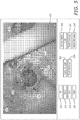
- imaging system 12 enables a user to operate with an input device to change the scale of the displayed multidimensional reconstructed image. For example, referring to FIG. 5 , in this embodiment, imaging system 12 displays scale increase button 522 and scale decrease button 524. In response to a user selecting scale increase button 522, imaging system 12 enables the user to cause an increase in the display size of at least one of: (a) the displayed multidimensional reconstructed image; and (b) the displayed multidimensional visualization. In response to a user selecting scale decrease button 524, imaging system 12 enables the user to cause a decrease in the display size of at least one of: (a) the displayed multidimensional reconstructed image; and (b) the displayed multidimensional visualization.
- Imaging system 12 also displays depth increase button 526 and depth decrease button 528.
- imaging system 12 enables the user to cause an increase in the viewing depth of the currently displayed multidimensional reconstructed image. That is, in response to a user selecting depth increase button 526, imaging system 12 displays a different portion of the multidimensional reconstructed image having an increased depth.
- imaging system 12 enables the user to cause a decrease in the depth of the currently displayed multidimensional reconstructed image. That is, in response to a user selecting depth decrease button 528, imaging system 12 displays a different portion of the multidimensional reconstructed image having a decreased depth.
- imaging system 12 may highlight certain features or portions of the displayed multidimensional reconstructed image. Such features or portions may include internal anatomical structures such as an aneurysm, a tumor or blood vessels. For example, referring to FIG. 5 , in this example, imaging system 12 highlights tumor 530. As a result, in this example, imaging system 12 enables a surgeon to 'see' where tumor 530 is located relative to adjacent brain tissue and relative to the features of patient's head shown in visualization 502. Such a configuration allows attention to be brought to important anatomical structures within the multidimensional reconstructed image.
- Such features or portions may be highlighted in any suitable way.
- a tumor may be highlighted with pseudo colors such as purple and blood vessels may be highlighted with a second, different color.
- the highlighted anatomy may be manually or automatically selected pre-operatively in the imaging data or selected intra-operatively in the live surgical view based on specific criteria in the image or data.
- the software may select a specific range of Hounsfield Units or CT densities to identify a tumor or aneurism with calcification as the highlighted anatomy.
- the imaging system inverts the imaging overlay/underlay modality.
- the "x-ray window" will open up a view that underlays the live surgical view and shows a small portion of the multidimensional reconstruction within the boundaries of the live surgical multidimensional visualization.
- the portion of multidimensional reconstructed image 502 that is displayed within window 503 is displayed as being underlayed with respect to the displayed multidimensional visualization 502. That is, in this example, if the surgeon were to move his thumb 505 over the currently displayed portion of the multidimensional reconstructed image 504, the surgeon's thumb would block the view of the currently displayed multidimensional reconstructed image.
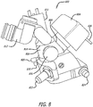
- FIG. 6 illustrates another example embodiment of an imaging system 12, illustrating multidimensional reconstructed image 602 being displayed as being underlayed with respect to the displayed multidimensional visualization 604. That is, the surgeon's thumb blocks the view of the multidimensional reconstructed image 602.
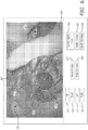
- the portion of multidimensional reconstructed image that is displayed within the window may be displayed as being overlayed with respect to the displayed multidimensional visualization.
- the portion of multidimensional reconstructed image 702 is positioned over surgeon's thumb 505 and is shown as being overlayed with respect to multidimensional visualization 704. That is, unlike the multidimensional reconstructed image shown in FIG. 5 , the thumb does not block the view of the multidimensional reconstructed image.
- Imaging system 12 may fuse or merge the portion of the multidimensional reconstructed image with the multidimensional visualization in any suitable way.
- imaging system 12 fuses the multidimensional reconstructed image with the multidimensional visualization of a target site by using visual tracking and registration of physical features in the multidimensional visualization with the corresponding features in the multidimensional reconstructed image.
- imaging system 12 may orientate a multidimensional reconstructed image with a multidimensional visualization of a surgical site by matching tracking coordinates of the multidimensional visualization with coordinates assigned to the image data.
- an image guidance system (“IGS") generates the coordinates.
- the image guidance system may include the Medtronic® STEALTHSTATION® or BRAINLAB® KOLIBRITM system.
- imaging system 12 matches identified physical features of a head (e.g., brain bone structure or brain blood vessels) to corresponding features of the multidimensional reconstructed image.
- imaging system 12 may match patterns of the blood vessels and brain tissue to the corresponding patterns and features in the multidimensional reconstructed image.
- imaging system 12 fuses a portion of the multidimensional reconstructed image based on an orientation of the surgical site shown in the displayed visualization.
- imaging system 12 enables a user to manipulate an input device to change the orientation of the displayed multidimensional reconstructed image.
- imaging system 12 enables a user to click buttons on the screen to move the position of the multidimensional reconstructed image in Cartesian coordinates.
- the buttons may be appropriately labeled and enable in and out movement along the x, y, and z axes for position, as well as angular alignment known as roll, pitch, and yaw rotation along the x, y, and z axes.
- Imaging system 12 may express the position information in any suitable way, such as cylindrical, (aka polar) coordinates, spherical (aka radial) coordinates, or other coordinate systems.
- imaging system 12 may enable a user to adjust the multidimensional reconstructed image.
- imaging system 12 enables a user to grasp a joystick control (e.g., a LOGITECH® SPACE NAVIGATOR®) to adjust the six degrees of freedom which position multidimensional reconstructed image relative to the multidimensional visualization.
- a joystick control e.g., a LOGITECH® SPACE NAVIGATOR®
- imaging system 12 enables a user to move a displayed multidimensional reconstructed image based on physical changes to a patient's anatomy made during surgery. For example, a surgeon may move a location of a blood vessel in a multidimensional reconstructed image by selecting and moving the graphical representation of the blood vessel on a display of the multidimensional reconstructed image after a blood vessel has physically been moved in the patient. Such a configuration enables the imaging system to maintain an accurate representation of a patient's anatomical structure during surgery.
- imaging system 12 displays the multidimensional visualization as superimposed onto a portion of the multidimensional reconstructed image.
- a live view of a three inch craniotomy is overlayed onto the overall MRI scan of a patient's head.
- imaging system 12 determines which portion of the multidimensional reconstructed image to display based on coordinates of a selection corresponding to a window. In one example embodiment, imaging system 12 enables a user to generate, place or move a window on the displayed visualization. In this example, imaging system 12 determines which portion of the multidimensional reconstructed image corresponds to the received selection based on the generated window.
- imaging system 12 moves the window in response to a user operating with an input device and clicking and dragging the window.
- imaging system 12 enables a user to set the display device to constantly display a designated distance (e.g., ten millimeters) ahead of a currently displayed multidimensional visualization. Such a configuration enables a surgeon to graphically see what anatomical structure his surgical tools are approaching. In this example, as a user moves into a body, imaging system 12 updates the displayed multidimensional reconstructed image to display anatomical structures that are constantly some specified distance ahead.
- a designated distance e.g., ten millimeters
- imaging system 12 enables surgeons to use a slider or scroll to visually move through different layers or depths of a multidimensional reconstructed image. Such a configuration enables a surgeon to 'view' relatively deep anatomic structures well before a surgeon has physically reached those structures. This configuration also enables a surgeon to functionally slide through different layers to determine what anatomical structure lies between surgical tools and a target anatomy.
- imaging system 12 enables a user to control of the back-side depth of the multidimensional reconstructed image. For example, if a tumor lays close to the cranium, the user may wish to see the full extent of the tumor in the multidimensional reconstruction, but not see the dense bone of the cranium which is just behind the tumor. In this instance, the user can scroll the rear boundary of the multidimensional reconstruction forward until the cranium bone is no longer seen in the multidimensional reconstruction
- imaging system 12 enables a user to set which portion of the multidimensional reconstructed image is displayed based on depth information. For example, in one embodiment where a surgeon is cutting into a brain of a patient, imaging system 12 may receive a request from a surgeon to display a 3D reconstructed image corresponding to fifteen mm ahead of where the surgeon's scalpel is currently located. In response to such a request, imaging system 12 may determine coordinates of the currently displayed multidimensional visualization and then display a portion of the multidimensional reconstructed image data that corresponds to fifteen mm ahead of the determined coordinates. In this example, as the surgical tool goes deeper, imaging system 12 automatically updates the displayed portion of the multidimensional reconstructed image to display the portion of the multidimensional reconstructed image that is fifteen mm ahead of surgery.
- imaging system 12 filters or enables a user to select and remove certain types of anatomical structures of a multidimensional reconstructed image.
- imaging system 12 enables a user to select to view only bone structures, brain tissue, blood vessels, tumors or aneurisms. Such a configuration enables users to focus the multidimensional reconstructed image on desired anatomical structures that are important for a surgery.
- imaging system 12 adjusts the transparency of a displayed multidimensional reconstructed image so that a highlighted anatomical structure may be viewed through different layers of the multidimensional reconstructed image.
- imaging system 12 enables a user to move or manipulate objects or structure (e.g., a blood vessel or tissue) of a displayed multidimensional reconstructed image to reflect actual movement within a patient's anatomy.
- objects or structure e.g., a blood vessel or tissue
- imaging system 12 displays annotations associated with a multidimensional reconstructed image.
- a surgeon may electronically attach notations to or in at least one pre-operative image.
- imaging system 12 stores the notations and the location of the notation in association with the multidimensional reconstructed image such that the notation is displayed in conjunction with the multidimensional reconstructed image.
- imaging system 12 enables a user to associate a surgical note with a specific portion of image data or a specific portion of a multidimensional reconstructed image.
- imaging system 12 in response to a user selecting certain portions of a displayed multidimensional visualization, certain notes which have been associated (e.g., attached) with such selected portions are displayed simultaneously with the displayed portion of the multidimensional reconstructed image.
- imaging system 12 enables a user to draw a route trajectory through a multidimensional reconstructed image or a series of pre-operative images for surgical tools as part of a pre-operative plan. Imaging system 12 may display this route in the displayed portion of the multidimensional reconstructed image in conjunction with a multidimensional visualization of a surgical site.
- imaging system 12 updates which portion of the route is displayed to coincide with the currently displayed multidimensional visualization.
- imaging system 12 displays surgical routes in conjunction with a portion of a multidimensional reconstructed image and a multidimensional visualization of a surgical site.
- imaging system 12 may enable a user to control or adjust display characteristics of at least one of the multidimensional reconstructed image and the multidimensional visualization.
- Display characteristics may include at least one of color, saturation, hue, luminosity, contrast, brightness, gamma and/or any other display characteristics.
- the image data may include any suitable type of data.
- the image data preferably corresponds to a surgical area of a patient.
- the image data may include at least one of: pre-operative image data; intra-operative image data; scan data; any video, image, or data structure that includes medical information obtained via, for example, a computed tomography (“CT") scan, a computed tomography agiography (“CT-A”) scan, a magnetic resonance imaging (“MRI”) scan, a positron emission tomography (“PET”) scan or any other type of medical scan or imaging; 2D image data; 3D image data; and any sequence or video of medical images that conform to, for example, the Digital Imaging and Communications in Medicine (“DICOM”) standard.
- CT computed tomography
- CT-A computed tomography agiography
- MRI magnetic resonance imaging
- PET positron emission tomography
- the image data may correspond to sequential scans of different depths of a patient's anatomy.
- the image data may be generated or received from any suitable device.
- the image data is received from a medical imaging machine via a hospital information system.
- the image data is received from another server or another computer.
- the systems and methods disclosed herein may enable a surgeon to comfortably visualize a surgical procedure on a display device instead of staring for, in some cases, several hours though the eyepieces of a surgical microscope. This is because the real-time visualizations of the systems and methods allow the surgery to take place in comfortable sitting or standing positions without sacrificing a complete and accurate visualization of the target surgical field.
- the primary surgeon and any assistant surgeons have to be physically looking through the microscope oculars - positioning themselves in rigid and frequently awkward positions. By viewing the surgery on a display, a surgeon is free to sit comfortably and easily move their necks, backs, and shoulders to remain relaxed and ergonomically situated. These capacities may be ideal for a surgeon and surgical team working long hours.
- the systems and methods of the present invention may eliminate this problem by providing a display which fits into the normal visual filed of the surgeon.
- imaging system 12 provides multidimensional internal guidance rather than just surface guidance. As a result, imaging system 12 may reduce surgery time, reduce trauma from surgery, and may provide better surgery with fewer complications. In addition, the imaging system may eliminate a need for some visualization probes or cameras in delicate or hard to reach areas.
- the imaging system 12 is a single device.
- imaging system 12 is configured to be retrofitted onto existing surgical equipment such as surgical microscopes or an open surgery apparatus. This can be advantageous as the retrofit embodiments may be added to existing systems (e.g., microscopes and IGS), allowing expensive equipment to simply be upgraded as opposed to purchasing an entirely new system.
- An example imaging system 12 may include various optical or electronic magnification systems including stereomicroscopes or may function as an open surgery apparatus utilizing cameras and overhead visualizations with or without magnification.
- imaging system 804 may send and receive information through signal cable 826.
- the imaging system, imaging apparatuses and imaging methods of the present invention may be applicable to any form of surgery, such as brain surgery, spinal surgery, ophthalmologic surgery, corneal transplants, neurosurgery, orthopedic surgery, ear, nose and throat surgery, plastics and reconstructive surgery, or general surgery on any target structure or tissue.
- imaging system 12 includes a voice activated control system.
- voice activated control system Such a configuration enables a user to control the modification and alignment of multidimensional reconstructed images in conjunction with a multidimensional visualization of a surgical site as if he or she was talking to an assistant or a member of the surgical team.
- the voice activated controls may include a microphone and a second data processor or software to interpret the oral voice commands.
- imaging system 12 includes a gesture recognition device configured to enable a user to use gesture commands to control multidimensional reconstructed images fused with a visualization of a surgical site.
- the gesture recognition device may include a camera to monitor and track the gestures of the controlling user and, optionally, a second data processor or software to interpret the commands.
- imaging system 12 implements camera calibration on one or more photosensors to identify the parameters typically used for image rectification, camera principal points including: (a) position in x,y,z; (b) rotational orientation in angles phi, theta, psi; (c) focal length and magnification/field of view; and (d) distortion parameters which may characterize optical aberrations including any or all of the following: defocus, piston, tilt, shear, astigmatism, coma, or other higher order aberrations or chromatic aberrations.
- imaging system 12 may apply corrections to the video signal which results in an orthoscopic or rectilinear visualization of the surgery.
- imaging system 12 employs camera calibration parameters to apply the optical distortion that is present in the video signal to the reconstruction such that the final rendering of the pre-operative data reflects the same distortion and aberrations present in the video signal.
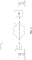
- FIG. 9 is a block diagram of an example data architecture 900.
- interface data 902, administrative data 904, and data 906 interact with each other, for example, based on user commands or requests.
- Interface data 902, administrative data 904, and data 906 may be stored on any suitable storage medium (e.g., database system 302 and/or server 14). It should be appreciated that different types of data may use different data formats, storage mechanisms, etc. Further, various applications may be associated with processing interface data 902, administrative data 904, and data 906. Various other or different types of data may be included in the example data architecture 900.
- Interface data 902 may include input and output data of various kinds.
- input data may include mouse click data, scrolling data, hover data, keyboard data, touch screen data, voice recognition data, etc.
- output data may include image data, text data, video data, audio data, etc.
- Interface data 902 may include formatting, user interface options, links or access to other websites or applications, and the like.
- Interface data 902 may include applications used to provide or monitor interface activities and handle input and output data.
- Data 906 may include, for example, multidimensional visualization data 908, multidimensional reconstructed image data 910, selection data 912, window data 914, and image data 916.
- Multidimensional visualization data 908 may include data representative of at least one of: a surgical site, a 3D visualization of a surgical site, a 2D visualization of a surgical site, and real time data.
- Multidimensional reconstructed image data 910 may include data representative of at least one of: feature data, brain tumor data, brain tissue data, bone structure data, aneurysm data, blood vessel data, vertebrate data, coordinate data, depth data, distance data, and transparency data.
- Selection data 912 may include data representative of at least one of: a portion of a multidimensional visualization.
- Window data 914 may include data representative of position data.
- Image data 916 may include data representative of at least one of: pre-operative image data, intra operative image data, medical scan data, and image slice data.
- the imaging system may include components of Applicant's TrueVision Systems, Inc. real-time 3D HD visualization systems described in Applicant's co-pending U.S. applications: Ser. No. 11/256,497 entitled “Stereoscopic Image Acquisition Device,” filed October 21, 2005; Ser. No. 11/668,400 entitled “Stereoscopic Electronic Microscope Workstation,” filed January 29, 2007; Ser. No. 11/668,420 entitled “Stereoscopic Electronic Microscope Workstation,” filed January 29, 2007; Ser. No. 11/739,042 entitled “Stereoscopic Display Cart and System,” filed April 23, 2007; and Ser. No. 61/042,606 , entitled “Apparatus and Methods for Performing Enhanced Visually Directed Procedures Under Low Ambient Light Conditions,” filed April 4, 2008, all of which are fully incorporated herein by reference as if part of this specification.
- Realtime as used herein generally refers to the updating of information at essentially the same rate as the data is received. More specifically, “realtime” is intended to mean that the image data is acquired, processed, and transmitted from the photosensor of the visualization generation system at a high enough data rate and at a low enough time delay that when the data is displayed, objects presented in the visualization move smoothly without user-noticeable judder, latency or lag. Typically, this occurs when new images are acquired, processed, and transmitted at a rate of at least about 30 frames per second (“fps") and displayed at a rate of at least about 60 fps and when the combined processing of the video signal has no more than about 1/10 th of a second of delay.
- fps frames per second
- the signal processing may have no more than about 1/20 th second of delay, about 1/30 th second of delay, about 1/50 th second of delay, about 1/90 th second of delay, about 1/120 th second of delay, about 1/500 th second of delay, or about 1/1000 th second delay or more.
Applications Claiming Priority (3)
| Application Number | Priority Date | Filing Date | Title |
|---|---|---|---|
| US201261695230P | 2012-08-30 | 2012-08-30 | |
| EP13834045.0A EP2891137B1 (de) | 2012-08-30 | 2013-08-30 | Abbildungssystem und -verfahren zur anzeige eines mehrdimensionalen zusammengesetzten und rekonstruierten bildes |
| PCT/US2013/057686 WO2014036499A1 (en) | 2012-08-30 | 2013-08-30 | Imaging system and methods displaying a fused multidimensional reconstructed image |
Related Parent Applications (2)
| Application Number | Title | Priority Date | Filing Date |
|---|---|---|---|
| EP13834045.0A Division-Into EP2891137B1 (de) | 2012-08-30 | 2013-08-30 | Abbildungssystem und -verfahren zur anzeige eines mehrdimensionalen zusammengesetzten und rekonstruierten bildes |
| EP13834045.0A Division EP2891137B1 (de) | 2012-08-30 | 2013-08-30 | Abbildungssystem und -verfahren zur anzeige eines mehrdimensionalen zusammengesetzten und rekonstruierten bildes |
Publications (1)
| Publication Number | Publication Date |
|---|---|
| EP3734607A1 true EP3734607A1 (de) | 2020-11-04 |
Family
ID=50184458
Family Applications (2)
| Application Number | Title | Priority Date | Filing Date |
|---|---|---|---|
| EP13834045.0A Active EP2891137B1 (de) | 2012-08-30 | 2013-08-30 | Abbildungssystem und -verfahren zur anzeige eines mehrdimensionalen zusammengesetzten und rekonstruierten bildes |
| EP20169896.6A Pending EP3734607A1 (de) | 2012-08-30 | 2013-08-30 | Abbildungssystem und -verfahren zur anzeige eines mehrdimensionalen zusammengesetzten und rekonstruierten bildes |
Family Applications Before (1)
| Application Number | Title | Priority Date | Filing Date |
|---|---|---|---|
| EP13834045.0A Active EP2891137B1 (de) | 2012-08-30 | 2013-08-30 | Abbildungssystem und -verfahren zur anzeige eines mehrdimensionalen zusammengesetzten und rekonstruierten bildes |
Country Status (5)
| Country | Link |
|---|---|
| US (6) | US9552660B2 (de) |
| EP (2) | EP2891137B1 (de) |
| CA (1) | CA2883498C (de) |
| ES (1) | ES2813625T3 (de) |
| WO (1) | WO2014036499A1 (de) |
Families Citing this family (28)
| Publication number | Priority date | Publication date | Assignee | Title |
|---|---|---|---|---|
| US9168173B2 (en) | 2008-04-04 | 2015-10-27 | Truevision Systems, Inc. | Apparatus and methods for performing enhanced visually directed procedures under low ambient light conditions |
| US10117721B2 (en) | 2008-10-10 | 2018-11-06 | Truevision Systems, Inc. | Real-time surgical reference guides and methods for surgical applications |
| US9226798B2 (en) | 2008-10-10 | 2016-01-05 | Truevision Systems, Inc. | Real-time surgical reference indicium apparatus and methods for surgical applications |
| US9173717B2 (en) | 2009-02-20 | 2015-11-03 | Truevision Systems, Inc. | Real-time surgical reference indicium apparatus and methods for intraocular lens implantation |
| WO2014036499A1 (en) * | 2012-08-30 | 2014-03-06 | Truevision Systems, Inc. | Imaging system and methods displaying a fused multidimensional reconstructed image |
| US10588597B2 (en) | 2012-12-31 | 2020-03-17 | Intuitive Surgical Operations, Inc. | Systems and methods for interventional procedure planning |
| GB201501157D0 (en) * | 2015-01-23 | 2015-03-11 | Scopis Gmbh | Instrument guidance system for sinus surgery |
| RU2638014C2 (ru) * | 2015-09-28 | 2017-12-08 | Общество С Ограниченной Ответственностью "Яндекс" | Способ и вычислительное устройство для создания симплифицированных границ графических объектов |
| US11244478B2 (en) * | 2016-03-03 | 2022-02-08 | Sony Corporation | Medical image processing device, system, method, and program |
| JP2017168003A (ja) * | 2016-03-18 | 2017-09-21 | オリンパス株式会社 | 顕微鏡画像閲覧システム |
| KR101769914B1 (ko) * | 2016-06-08 | 2017-08-21 | 가톨릭대학교 산학협력단 | Oct 검출부를 포함하는 부분층 각막 이식 수술용 수술 장치 |
| CN110114803B (zh) * | 2016-12-28 | 2023-06-27 | 松下电器(美国)知识产权公司 | 三维模型分发方法、三维模型接收方法、三维模型分发装置以及三维模型接收装置 |
| US10299880B2 (en) | 2017-04-24 | 2019-05-28 | Truevision Systems, Inc. | Stereoscopic visualization camera and platform |
| WO2019012551A1 (en) * | 2017-07-08 | 2019-01-17 | Srinivas Hirekatur Venkataram | GONIO CAMERA WITHOUT SLOT LAMP |
| US11269172B2 (en) * | 2017-11-24 | 2022-03-08 | Sigtuple Technologies Private Limited | Method and system for reconstructing a field of view |
| WO2019210322A1 (en) | 2018-04-27 | 2019-10-31 | Truevision Systems, Inc. | Stereoscopic visualization camera and integrated robotics platform |
| CN109147914B (zh) * | 2018-10-16 | 2023-04-28 | 上海联影医疗科技股份有限公司 | 一种图像重建系统 |
| US11340441B2 (en) | 2019-07-09 | 2022-05-24 | Lightech Fiberoptics, Inc. | Microscope made with CMOS camera(s) |
| US11031118B2 (en) * | 2019-08-12 | 2021-06-08 | Biosense Webster (Israel) Ltd. | Mixed electroanatomical map coloring tool having draggable geodesic overlay |
| CN110675355B (zh) * | 2019-09-27 | 2022-06-17 | 深圳市商汤科技有限公司 | 图像重建方法及装置、电子设备和存储介质 |
| JP6849780B2 (ja) * | 2019-12-11 | 2021-03-31 | キヤノン株式会社 | 眼科装置、表示制御方法およびプログラム |
| EP4093311A4 (de) * | 2020-01-22 | 2023-06-14 | Beyeonics Surgical Ltd. | System und verfahren für verbesserte elektronikunterstützte medizinische verfahren |
| WO2022146996A1 (en) * | 2020-12-30 | 2022-07-07 | Intuitive Surgical Operations, Inc. | Systems for updating a graphical user interface based upon intraoperative imaging |
| US11478327B2 (en) | 2021-02-18 | 2022-10-25 | Xenco Medical, Inc. | Surgical display |
| US11273003B1 (en) * | 2021-02-18 | 2022-03-15 | Xenco Medical, Inc. | Surgical display |
| EP4304511A1 (de) | 2021-03-12 | 2024-01-17 | Digital Surgery Systems, Inc. | Automatisierte berührungslose registrierung für chirurgische navigation |
| US20230020780A1 (en) * | 2021-07-14 | 2023-01-19 | Cilag Gmbh International | Stereoscopic endoscope with critical structure depth estimation |
| WO2023220674A2 (en) * | 2022-05-11 | 2023-11-16 | Kaliber Labs Inc. | Surgery evidence report generation |
Citations (2)
| Publication number | Priority date | Publication date | Assignee | Title |
|---|---|---|---|---|
| US20060293557A1 (en) * | 2005-03-11 | 2006-12-28 | Bracco Imaging, S.P.A. | Methods and apparati for surgical navigation and visualization with microscope ("Micro Dex-Ray") |
| US20080170781A1 (en) * | 2004-09-17 | 2008-07-17 | Koninklijke Philips Electronics, N.V. | Image Selection on a Screen |
Family Cites Families (167)
| Publication number | Priority date | Publication date | Assignee | Title |
|---|---|---|---|---|
| US3431992A (en) | 1966-12-16 | 1969-03-11 | Smithkline Corp | Lift truck scale |
| US3517183A (en) | 1968-04-01 | 1970-06-23 | Bausch & Lomb | Microscope illuminator |
| US3867697A (en) | 1969-07-29 | 1975-02-18 | Vanzetti Infrared Computer Sys | Measuring means |
| US4395731A (en) | 1981-10-16 | 1983-07-26 | Arnold Schoolman | Television microscope surgical method and apparatus therefor |
| DE3431992C2 (de) | 1983-09-05 | 1985-12-12 | Olympus Optical Co., Ltd., Tokio/Tokyo | Endoskopanordnung mit einer Aufnahmeeinrichtung |
| GB8425827D0 (en) | 1984-10-12 | 1984-11-21 | Gec Avionics | Position indicating apparatus |
| DE3508306A1 (de) | 1985-03-08 | 1986-09-11 | Fa. Carl Zeiss, 7920 Heidenheim | Mikroskoptubus |
| US4790305A (en) | 1986-06-23 | 1988-12-13 | The Johns Hopkins University | Medication delivery system |
| US4786155A (en) | 1986-12-16 | 1988-11-22 | Fantone Stephen D | Operating microscope providing an image of an obscured object |
| NL8800595A (nl) | 1988-03-10 | 1989-10-02 | Philips Nv | Weergeef- en opneeminrichting voor stereoscopische beeldweergave. |
| DE3814006A1 (de) | 1988-04-26 | 1989-11-09 | Leitz Wild Gmbh | Mikroskop mit einer kamera und automatischem farbtemperaturabgleich |
| US5109276A (en) | 1988-05-27 | 1992-04-28 | The University Of Connecticut | Multi-dimensional multi-spectral imaging system |
| US5200838A (en) | 1988-05-27 | 1993-04-06 | The University Of Connecticut | Lateral effect imaging system |
| GB8817672D0 (en) | 1988-07-25 | 1988-09-01 | Sira Ltd | Optical apparatus |
| US4989078A (en) | 1988-08-15 | 1991-01-29 | Eastman Kodak Company | Still video camera for recording stereo images on a video disk |
| US5098426A (en) | 1989-02-06 | 1992-03-24 | Phoenix Laser Systems, Inc. | Method and apparatus for precision laser surgery |
| US4995716A (en) | 1989-03-09 | 1991-02-26 | Par Technology Corporation | Method and apparatus for obtaining the topography of an object |
| US4967268A (en) | 1989-07-31 | 1990-10-30 | Stereographics | Liquid crystal shutter system for stereoscopic and other applications |
| US5054907A (en) | 1989-12-22 | 1991-10-08 | Phoenix Laser Systems, Inc. | Ophthalmic diagnostic apparatus and method |
| JPH03209543A (ja) | 1990-01-12 | 1991-09-12 | Toshiba Corp | パーソナルコンピュータ |
| US5048946A (en) | 1990-05-15 | 1991-09-17 | Phoenix Laser Systems, Inc. | Spectral division of reflected light in complex optical diagnostic and therapeutic systems |
| JP3209543B2 (ja) | 1990-07-13 | 2001-09-17 | オリンパス光学工業株式会社 | 手術用顕微鏡 |
| US5699798A (en) | 1990-08-10 | 1997-12-23 | University Of Washington | Method for optically imaging solid tumor tissue |
| US6006126A (en) | 1991-01-28 | 1999-12-21 | Cosman; Eric R. | System and method for stereotactic registration of image scan data |
| US5751927A (en) | 1991-03-26 | 1998-05-12 | Wason; Thomas D. | Method and apparatus for producing three dimensional displays on a two dimensional surface |
| US5417210A (en) | 1992-05-27 | 1995-05-23 | International Business Machines Corporation | System and method for augmentation of endoscopic surgery |
| US5193000A (en) | 1991-08-28 | 1993-03-09 | Stereographics Corporation | Multiplexing technique for stereoscopic video system |
| DE4134481C2 (de) | 1991-10-18 | 1998-04-09 | Zeiss Carl Fa | Operationsmikroskop zur rechnergestützten, stereotaktischen Mikrochirurgie |
| JP3298185B2 (ja) | 1992-11-05 | 2002-07-02 | 株式会社ニコン | 眼科装置 |
| DE4304571A1 (de) | 1993-02-16 | 1994-08-18 | Mdc Med Diagnostic Computing | Verfahren zur Planung und Kontrolle eines chirurgischen Eingriffs |
| EP0646263B1 (de) | 1993-04-20 | 2000-05-31 | General Electric Company | Graphisches digitalverarbeitungssystem und echtzeitvideosystem zur verbesserung der darstellung von körperstrukturen während eines chirugischen eingriffs. |
| US5579772A (en) | 1993-06-14 | 1996-12-03 | Olympus Optical Co., Ltd. | Surgical microscope system |
| JP2607828B2 (ja) | 1993-10-04 | 1997-05-07 | 永島醫科器械株式会社 | ハイディフィニションテレビ統合顕微鏡システム |
| WO1995018512A1 (de) | 1993-12-29 | 1995-07-06 | Leica Ag | Verfahren und vorrichtung zur darstellung von stereoskopischen videobildern auf einem display |
| US5867309A (en) | 1994-03-30 | 1999-02-02 | Leica Geosystems Ag | Stereomicroscope |
| DE9407854U1 (de) | 1994-05-11 | 1994-07-28 | Haag Ag Streit | Videozusatz an einem Mikroskop |
| JP3444980B2 (ja) | 1994-07-25 | 2003-09-08 | キヤノン株式会社 | 眼科撮影装置 |
| DE59503726D1 (de) | 1994-08-19 | 1998-10-29 | Leica Mikroskopie Syteme Ag | Verfahren und vorrichtung zur darstellung von stereoskopischen videobildern auf einem display |
| US5999840A (en) * | 1994-09-01 | 1999-12-07 | Massachusetts Institute Of Technology | System and method of registration of three-dimensional data sets |
| EP0951874A3 (de) | 1994-09-15 | 2000-06-14 | Visualization Technology, Inc. | Positions- und Bilderfassung mittels einer an einem Patientenkopf angebrachten Referenzeinheit zur Anwendung im medizinischen Gebiet |
| US5765561A (en) | 1994-10-07 | 1998-06-16 | Medical Media Systems | Video-based surgical targeting system |
| US6483948B1 (en) | 1994-12-23 | 2002-11-19 | Leica Ag | Microscope, in particular a stereomicroscope, and a method of superimposing two images |
| US5545120A (en) | 1995-01-18 | 1996-08-13 | Medical Media Systems | Endoscopic viewing system for maintaining a surgeon's normal sense of kinesthesia during endoscopic surgery regardless of the orientation of the endoscope vis-a-vis the surgeon |
| US5671085A (en) | 1995-02-03 | 1997-09-23 | The Regents Of The University Of California | Method and apparatus for three-dimensional microscopy with enhanced depth resolution |
| EP0807274B1 (de) | 1995-02-03 | 1998-10-28 | Leica Mikroskopie Systeme AG | Stereomikroskop |
| US5652676A (en) | 1995-04-24 | 1997-07-29 | Grinblat; Avi | Microscope-television camera adapter |
| US6256529B1 (en) | 1995-07-26 | 2001-07-03 | Burdette Medical Systems, Inc. | Virtual reality 3D visualization for surgical procedures |
| US5835133A (en) | 1996-01-23 | 1998-11-10 | Silicon Graphics, Inc. | Optical system for single camera stereo video |
| US5867210A (en) | 1996-02-09 | 1999-02-02 | Rod; Samuel R. | Stereoscopic on-screen surgical microscope systems |
| JP3448795B2 (ja) | 1996-04-11 | 2003-09-22 | 株式会社ニコン | 眼科装置 |
| JP4136011B2 (ja) | 1996-04-30 | 2008-08-20 | オリンパス株式会社 | 焦点深度伸長装置 |
| US6133762A (en) | 1997-03-31 | 2000-10-17 | Texas Instruments Incorporated | Family of logic circuits emploting mosfets of differing thershold voltages |
| WO1998046122A1 (en) | 1997-04-17 | 1998-10-22 | Avimo Group Limited | Ocular microcirculation examination and treatment apparatus |
| US6247812B1 (en) | 1997-09-25 | 2001-06-19 | Vismed | System and method for diagnosing and treating a target tissue |
| AUPO981997A0 (en) | 1997-10-15 | 1997-11-06 | Lions Eye Institute Of Western Australia Incorporated, The | Stereo optic disc analyser |
| US6191809B1 (en) | 1998-01-15 | 2001-02-20 | Vista Medical Technologies, Inc. | Method and apparatus for aligning stereo images |
| US6147797A (en) | 1998-01-20 | 2000-11-14 | Ki Technology Co., Ltd. | Image processing system for use with a microscope employing a digital camera |
| US6088470A (en) | 1998-01-27 | 2000-07-11 | Sensar, Inc. | Method and apparatus for removal of bright or dark spots by the fusion of multiple images |
| US6144762A (en) | 1998-02-23 | 2000-11-07 | Olympus America Inc. | Stereo video microscope |
| US6468265B1 (en) | 1998-11-20 | 2002-10-22 | Intuitive Surgical, Inc. | Performing cardiac surgery without cardioplegia |
| DE19854852C2 (de) | 1998-11-27 | 2001-02-15 | Univ Eberhard Karls | Verfahren zur Selektion von Augenstellungs-Meßdaten und Vorrichtung zur Durchführung des Verfahrens |
| US6522906B1 (en) | 1998-12-08 | 2003-02-18 | Intuitive Surgical, Inc. | Devices and methods for presenting and regulating auxiliary information on an image display of a telesurgical system to assist an operator in performing a surgical procedure |
| US6285902B1 (en) | 1999-02-10 | 2001-09-04 | Surgical Insights, Inc. | Computer assisted targeting device for use in orthopaedic surgery |
| JP2000262472A (ja) | 1999-03-19 | 2000-09-26 | Kowa Co | 視野計 |
| JP3943753B2 (ja) | 1999-04-13 | 2007-07-11 | ユニ・チャーム株式会社 | 使い捨ての汚れ拭き取り用具の製造方法 |
| DE10027166B4 (de) | 1999-05-31 | 2007-03-08 | Pentax Corp. | Stereoskopmikroskop |
| WO2001027659A2 (de) | 1999-10-13 | 2001-04-19 | Leica Microsystems Ag | Stereo-operationsmikroskop mit einer informations-einspiegelvorrichtung |
| BR0014890B1 (pt) | 1999-10-21 | 2009-01-13 | sistema para alinhar dados de tratamento e/ou diagnàstico refrativo. | |
| DE19961971B4 (de) | 1999-12-22 | 2009-10-22 | Forschungszentrum Karlsruhe Gmbh | Vorrichtung zum sicheren automatischen Nachführen eines Endoskops und Verfolgen eines Instruments |
| US7431455B2 (en) | 2005-03-22 | 2008-10-07 | Amo Manufacturing Usa, Llc | Pupilometer for pupil center drift and pupil size measurements at differing viewing distances |
| US7044602B2 (en) | 2002-05-30 | 2006-05-16 | Visx, Incorporated | Methods and systems for tracking a torsional orientation and position of an eye |
| US6685317B2 (en) | 2000-06-13 | 2004-02-03 | Massie Research Laboratories, Inc. | Digital eye camera |
| US7025459B2 (en) | 2000-07-14 | 2006-04-11 | Visual Pathways, Inc. | Ocular fundus auto imager |
| US6902569B2 (en) | 2000-08-17 | 2005-06-07 | Image-Guided Neurologics, Inc. | Trajectory guide with instrument immobilizer |
| EP1405122B1 (de) | 2000-10-07 | 2007-07-18 | David Dickerson | Vorrichtung zur bestimmung der orientierung eines auges |
| US6607527B1 (en) | 2000-10-17 | 2003-08-19 | Luis Antonio Ruiz | Method and apparatus for precision laser surgery |
| DE10052201B8 (de) | 2000-10-20 | 2005-06-30 | Carl Zeiss Meditec Ag | Verfahren und Vorrichtung zur Identifizierung eines Patienten und eines Operationsgebietes |
| DE10064910A1 (de) | 2000-12-23 | 2002-07-04 | Leica Microsystems | Optische Betrachtungseinrichtung |
| US6631990B2 (en) | 2001-01-03 | 2003-10-14 | Acmed, Inc. | System for measuring distances in the anterior chamber of the eye |
| JP3679331B2 (ja) | 2001-01-25 | 2005-08-03 | 株式会社エクスプローラ | 屈折矯正装置 |
| US6643070B2 (en) | 2001-02-23 | 2003-11-04 | Leica Microsystems (Schweiz) Ag | Viewing tube for an optical device |
| US6596025B2 (en) | 2001-03-15 | 2003-07-22 | Valdemar Portney | Narrow profile intraocular lens |
| CA2342414A1 (en) | 2001-03-29 | 2002-09-29 | Motic Instruments Inc. | Digital imaging microscope |
| AU2001297967B2 (en) | 2001-04-27 | 2006-01-05 | Bausch & Lomb Incorporated | Iris pattern recognition and alignment |
| DE10130278B4 (de) | 2001-06-26 | 2005-11-03 | Carl Zeiss Meditec Ag | Verfahren und Vorrichtung zur Darstellung eines Operationsgebietes bei Laseroperationen |
| US7385168B2 (en) | 2001-07-06 | 2008-06-10 | Palantyr Research, Llc | Imaging system, methodology, and applications employing reciprocal space optical design |
| US20030021016A1 (en) | 2001-07-27 | 2003-01-30 | Grier David G. | Parallel scanned laser confocal microscope |
| US6634751B2 (en) | 2001-09-10 | 2003-10-21 | Bausch & Lomb Incorporated | Intraocular lens derivation system |
| US20050117118A1 (en) | 2001-10-05 | 2005-06-02 | David Miller | Digital ophthalmic workstation |
| WO2003030763A1 (en) | 2001-10-05 | 2003-04-17 | Boston Innovative Optics, Inc. | A system and method of providing visual documentation during surgery |
| FR2833100B1 (fr) * | 2001-11-30 | 2004-03-12 | Ge Med Sys Global Tech Co Llc | Procede de reconstitution d'une image d'un organe |
| JP3978024B2 (ja) | 2001-12-03 | 2007-09-19 | 株式会社ニデック | 眼科装置及び角膜手術装置 |
| DE10161613A1 (de) | 2001-12-15 | 2003-06-18 | Leica Microsystems | Verfahren zur Selbstüberwachung eines Mikroskopsystems, Mikroskopsystem und Software zur Selbstüberwachung |
| US20030142271A1 (en) | 2002-01-30 | 2003-07-31 | Ross Denwood F. | Aberration and corneal topography measurement |
| EP1333305B8 (de) | 2002-02-04 | 2007-08-01 | Carl Zeiss Surgical GmbH | Stereo-Untersuchungssysteme und Stereo-Bilderzeugungsvorrichtung sowie Verfahren zum Betrieb einer solchen |
| EP1486067B1 (de) | 2002-02-13 | 2015-04-15 | Reify Corporation | Verfahren und vorrichtung zur anschaffung, komprimierung und charakterisierung von raum-zeitlichen signalen |
| US7428001B2 (en) | 2002-03-15 | 2008-09-23 | University Of Washington | Materials and methods for simulating focal shifts in viewers using large depth of focus displays |
| US6924929B2 (en) | 2002-03-29 | 2005-08-02 | National Institute Of Radiological Sciences | Microscope apparatus |
| WO2004001569A2 (en) | 2002-06-21 | 2003-12-31 | Cedara Software Corp. | Computer assisted system and method for minimal invasive hip, uni knee and total knee replacement |
| US20050007659A1 (en) | 2002-09-03 | 2005-01-13 | Stereovision Imaging, Inc. | Focusing mechanism for stereoscopic systems |
| JP3931139B2 (ja) | 2002-12-27 | 2007-06-13 | 興和株式会社 | 眼科撮影装置 |
| JP4564239B2 (ja) | 2003-04-11 | 2010-10-20 | オリンパス株式会社 | 内視鏡装置 |
| WO2005000139A1 (en) * | 2003-04-28 | 2005-01-06 | Bracco Imaging Spa | Surgical navigation imaging system |
| US7525586B2 (en) | 2003-05-12 | 2009-04-28 | Altasens, Inc. | Image sensor and method with multiple scanning modes |
| US7052134B2 (en) | 2003-05-29 | 2006-05-30 | Nidek Co., Ltd. | Fundus camera |
| JP4493408B2 (ja) | 2003-06-06 | 2010-06-30 | 富士フイルム株式会社 | 画像読影支援方法及び装置並びにプログラム |
| US7458683B2 (en) | 2003-06-16 | 2008-12-02 | Amo Manufacturing Usa, Llc | Methods and devices for registering optical measurement datasets of an optical system |
| JP3867143B2 (ja) | 2003-06-25 | 2007-01-10 | 独立行政法人産業技術総合研究所 | 三次元顕微鏡システムおよび画像表示方法 |
| US7313430B2 (en) | 2003-08-28 | 2007-12-25 | Medtronic Navigation, Inc. | Method and apparatus for performing stereotactic surgery |
| AU2004285103A1 (en) | 2003-09-29 | 2005-05-12 | Pathwork Diagnostics, Inc. | Systems and methods for detecting biological features |
| DE102004050893B4 (de) | 2003-10-31 | 2015-05-21 | Carl Zeiss Meditec Ag | Tubus mit zwei umschaltbaren Planoptikelementen zur wahlweisen Strahlengangvertauschung und Bildumkehr für ein Mikroskop sowie Mikroskop |
| DE10355527A1 (de) | 2003-11-21 | 2005-06-09 | Carl Zeiss Jena Gmbh | Mikroskopkamera |
| US7524064B2 (en) | 2004-03-09 | 2009-04-28 | Research Foundation Of The State University Of New York | Apparatus and method for assessing retinal damage |
| US7476248B2 (en) | 2004-04-06 | 2009-01-13 | Alcon, Inc. | Method of calculating the required lens power for an opthalmic implant |
| DE102004030904A1 (de) | 2004-06-25 | 2006-01-19 | Neuhann, Thomas, Prof.Dr.med. | Vorrichtung zum Erfassen der räumlichen Lage der optischen Achse eines Auges sowie zum Zentrieren eines Bezugssystems relativ zur optischen Achse |
| JP2008505696A (ja) | 2004-07-09 | 2008-02-28 | ヴィスクス インコーポレイテッド | 走査レーザー眼手術装置用のレーザーパルス位置モニター |
| US8339447B2 (en) | 2004-10-21 | 2012-12-25 | Truevision Systems, Inc. | Stereoscopic electronic microscope workstation |
| DE102004055683B4 (de) | 2004-10-26 | 2006-09-07 | Carl Zeiss Surgical Gmbh | Augenchirurgie-Mikroskopiesystem und Verfahren hierzu |
| EP1652469A3 (de) | 2004-10-27 | 2006-06-07 | Kowa Company Ltd. | Ophthalmologisches Messgerät |
| US7815631B2 (en) | 2004-11-30 | 2010-10-19 | Alcon Refractivehorizons, Inc. | Eye registration system for refractive surgery and associated methods |
| US20060223037A1 (en) | 2005-04-01 | 2006-10-05 | Ingrid Tanda | Device for teaching biblical scripture and method of using the same |
| US7912258B2 (en) * | 2005-09-27 | 2011-03-22 | Vanderbilt University | Method and apparatus for standardizing ultrasonography training using image to physical space registration of tomographic volumes from tracked ultrasound |
| US20070188603A1 (en) | 2005-10-21 | 2007-08-16 | Riederer Thomas P | Stereoscopic display cart and system |
| US8358330B2 (en) | 2005-10-21 | 2013-01-22 | True Vision Systems, Inc. | Stereoscopic electronic microscope workstation |
| DE102007008521A1 (de) | 2006-02-21 | 2007-08-30 | Burgkart, Rainer, Dr. med. | Implantatlagepositioniersystem |
| US8474974B2 (en) | 2006-02-24 | 2013-07-02 | Amo Development Llc. | Induced high order aberrations corresponding to geometrical transformations |
| US7695136B2 (en) | 2007-08-01 | 2010-04-13 | Amo Development, Llc. | Wavefront refractions and high order aberration correction when wavefront maps involve geometrical transformations |
| US8454160B2 (en) | 2006-02-24 | 2013-06-04 | Amo Development, Llc | Zone extension systems and methods |
| WO2007104166A1 (en) | 2006-03-16 | 2007-09-20 | Sis Ag, Surgical Instrument Systems | Ophthalmological device and ophthalmological measuring method |
| EP1868157A1 (de) * | 2006-06-14 | 2007-12-19 | BrainLAB AG | Rekonstruktion von Formen anhand von Röntgenbildern |
| US7927344B2 (en) | 2006-10-13 | 2011-04-19 | Burba Thomas A | Eye positioner |
| US7831096B2 (en) | 2006-11-17 | 2010-11-09 | General Electric Company | Medical navigation system with tool and/or implant integration into fluoroscopic image projections and method of use |
| US8088124B2 (en) | 2007-01-19 | 2012-01-03 | Technolas Perfect Vision Gmbh | System and method for precise beam positioning in ocular surgery |
| US7899226B2 (en) | 2007-04-03 | 2011-03-01 | General Electric Company | System and method of navigating an object in an imaged subject |
| WO2008148038A1 (en) | 2007-05-24 | 2008-12-04 | Amo Development, Llc | Accommodation compensation systems and methods |
| US8414123B2 (en) | 2007-08-13 | 2013-04-09 | Novartis Ag | Toric lenses alignment using pre-operative images |
| JP5134316B2 (ja) * | 2007-09-05 | 2013-01-30 | 株式会社東芝 | 医用画像診断装置 |
| US8764736B2 (en) | 2007-09-05 | 2014-07-01 | Alcon Lensx, Inc. | Laser-induced protection shield in laser surgery |
| EP2211802B1 (de) | 2007-11-02 | 2012-06-27 | Alcon LenSx, Inc. | Gerät für verbesserte postoperative okulare sehleistung |
| US8357145B2 (en) | 2007-11-12 | 2013-01-22 | Boston Scientific Neuromodulation Corporation | Implanting medical devices |
| WO2009085204A2 (en) | 2007-12-23 | 2009-07-09 | Oraya Therapeutics, Inc. | Methods and devices for detecting, controlling, and predicting radiation delivery |
| US8012157B2 (en) | 2007-12-28 | 2011-09-06 | Illuminoss Medical, Inc. | Internal bone fixation sizing device and methods |
| CA2727585C (en) | 2008-03-21 | 2018-09-25 | Atsushi Takahashi | Three-dimensional digital magnifier operation supporting system |
| US20090236541A1 (en) | 2008-03-24 | 2009-09-24 | General Electric Company | System and Methods for Optical Imaging |
| US9168173B2 (en) | 2008-04-04 | 2015-10-27 | Truevision Systems, Inc. | Apparatus and methods for performing enhanced visually directed procedures under low ambient light conditions |
| US20100134759A1 (en) | 2008-06-26 | 2010-06-03 | Silvestrini Thomas A | Digital imaging system for eye procedures |
| DE102008034490B4 (de) | 2008-07-24 | 2018-12-20 | Carl Zeiss Meditec Ag | Augenchirurgiesystem und Verfahren zur Vorbereitung und Durchführung einer Augenoperation |
| US10117721B2 (en) | 2008-10-10 | 2018-11-06 | Truevision Systems, Inc. | Real-time surgical reference guides and methods for surgical applications |
| US9226798B2 (en) | 2008-10-10 | 2016-01-05 | Truevision Systems, Inc. | Real-time surgical reference indicium apparatus and methods for surgical applications |
| US9119565B2 (en) | 2009-02-19 | 2015-09-01 | Alcon Research, Ltd. | Intraocular lens alignment |
| US9173717B2 (en) | 2009-02-20 | 2015-11-03 | Truevision Systems, Inc. | Real-time surgical reference indicium apparatus and methods for intraocular lens implantation |
| US8472685B2 (en) * | 2009-08-12 | 2013-06-25 | The Regents Of The University Of California | Apparatus and method for surface capturing and volumetric analysis of multidimensional images |
| CA2774536C (en) | 2009-09-18 | 2017-12-12 | Amo Development, Llc | Registration of corneal flap with ophthalmic measurement and/or treatment data for lasik and other procedures |
| US8784443B2 (en) | 2009-10-20 | 2014-07-22 | Truevision Systems, Inc. | Real-time surgical reference indicium apparatus and methods for astigmatism correction |
| US8845625B2 (en) | 2010-01-22 | 2014-09-30 | Optimedica Corporation | Method and apparatus for automated placement of scanned laser capsulorhexis incisions |
| US20110213342A1 (en) | 2010-02-26 | 2011-09-01 | Ashok Burton Tripathi | Real-time Virtual Indicium Apparatus and Methods for Guiding an Implant into an Eye |
| ES2581543T3 (es) | 2010-05-06 | 2016-09-06 | Novartis Ag | Dispositivos y métodos para evaluar cambios en la salud corneal |
| US8625869B2 (en) * | 2010-05-21 | 2014-01-07 | Siemens Medical Solutions Usa, Inc. | Visualization of medical image data with localized enhancement |
| AU2011305629B2 (en) | 2010-09-20 | 2015-06-04 | Amo Development Llc | System and methods for mitigating changes in pupil size during laser refractive surgery to maintain ablation centration |
| EP3213724B1 (de) | 2010-12-30 | 2019-09-18 | Amo Wavefront Sciences, LLC | Verbessertes behandlungsplanungsverfahren und system zur steuerung von refraktiver laserchirurgie |
| US8978660B2 (en) | 2011-07-21 | 2015-03-17 | Amo Development, Llc | Tilt compensation, measurement, and associated adjustment of refractive prescriptions during surgical and other treatments of the eye |
| WO2014036499A1 (en) * | 2012-08-30 | 2014-03-06 | Truevision Systems, Inc. | Imaging system and methods displaying a fused multidimensional reconstructed image |
| US20140114296A1 (en) | 2012-10-24 | 2014-04-24 | Optimedica Corporation | Graphical user interface for laser eye surgery system |
| US9332899B2 (en) | 2012-11-06 | 2016-05-10 | Clarity Medical Systems, Inc. | Electronic eye marking/registration |
| DE112014000925T5 (de) | 2013-03-07 | 2015-11-26 | Adventist Health System/Sunbelt, Inc. | Chirurgisches Navigationsplanungssystem und damit verbundene Verfahren |
| SG11201508478TA (en) | 2013-03-15 | 2015-11-27 | Annmarie Hipsley | Systems and methods for affecting the biomechanical properties of connective tissue |
| JP3209543U (ja) | 2017-01-12 | 2017-03-23 | 株式会社島津製作所 | フレーム式原子吸光光度計 |
-
2013
- 2013-08-30 WO PCT/US2013/057686 patent/WO2014036499A1/en active Application Filing
- 2013-08-30 US US14/424,985 patent/US9552660B2/en active Active
- 2013-08-30 ES ES13834045T patent/ES2813625T3/es active Active
- 2013-08-30 EP EP13834045.0A patent/EP2891137B1/de active Active
- 2013-08-30 EP EP20169896.6A patent/EP3734607A1/de active Pending
- 2013-08-30 CA CA2883498A patent/CA2883498C/en active Active
-
2017
- 2017-01-23 US US15/412,959 patent/US10019819B2/en active Active
-
2018
- 2018-07-09 US US16/030,400 patent/US10740933B2/en active Active
-
2020
- 2020-06-30 US US16/916,683 patent/US11386992B2/en active Active
-
2022
- 2022-06-08 US US17/835,356 patent/US11682483B2/en active Active
-
2023
- 2023-05-03 US US18/311,514 patent/US20230274819A1/en active Pending
Patent Citations (2)
| Publication number | Priority date | Publication date | Assignee | Title |
|---|---|---|---|---|
| US20080170781A1 (en) * | 2004-09-17 | 2008-07-17 | Koninklijke Philips Electronics, N.V. | Image Selection on a Screen |
| US20060293557A1 (en) * | 2005-03-11 | 2006-12-28 | Bracco Imaging, S.P.A. | Methods and apparati for surgical navigation and visualization with microscope ("Micro Dex-Ray") |
Non-Patent Citations (1)
| Title |
|---|
| CHRISTOPH BICHLMEIER ET AL: "Virtual Window for Improved Depth Perception in Medical AR", STEREOTACTIC AND FUNCTIONAL NEUROSURGERY, 6 October 2006 (2006-10-06), CH, XP055451875, ISSN: 1011-6125 * |
Also Published As
| Publication number | Publication date |
|---|---|
| EP2891137A4 (de) | 2017-08-16 |
| US9552660B2 (en) | 2017-01-24 |
| ES2813625T3 (es) | 2021-03-24 |
| CA2883498C (en) | 2022-05-31 |
| US11682483B2 (en) | 2023-06-20 |
| US20220301693A1 (en) | 2022-09-22 |
| EP2891137A1 (de) | 2015-07-08 |
| US20170132812A1 (en) | 2017-05-11 |
| US20150221105A1 (en) | 2015-08-06 |
| WO2014036499A1 (en) | 2014-03-06 |
| US20230274819A1 (en) | 2023-08-31 |
| EP2891137B1 (de) | 2020-05-27 |
| US10740933B2 (en) | 2020-08-11 |
| US20200334872A1 (en) | 2020-10-22 |
| CA2883498A1 (en) | 2014-03-06 |
| US10019819B2 (en) | 2018-07-10 |
| US11386992B2 (en) | 2022-07-12 |
| US20180322666A1 (en) | 2018-11-08 |
Similar Documents
| Publication | Publication Date | Title |
|---|---|---|
| US11682483B2 (en) | Imaging system and methods displaying a fused multidimensional reconstructed image | |
| US11750788B1 (en) | Augmented reality guidance for spinal surgery with stereoscopic display of images and tracked instruments | |
| US20220346889A1 (en) | Graphical user interface for use in a surgical navigation system with a robot arm | |
| McJunkin et al. | Development of a mixed reality platform for lateral skull base anatomy | |
| US20180368656A1 (en) | Surgical visualization systems and displays | |
| Suenaga et al. | Real-time in situ three-dimensional integral videography and surgical navigation using augmented reality: a pilot study | |
| US20170042631A1 (en) | Intra-operative medical image viewing system and method | |
| Badiali et al. | Review on augmented reality in oral and cranio-maxillofacial surgery: toward “surgery-specific” head-up displays | |
| Condino et al. | Evaluation of a wearable AR platform for guiding complex craniotomies in neurosurgery | |
| US11273003B1 (en) | Surgical display | |
| Dibble et al. | Device profile of the XVision-spine (XVS) augmented-reality surgical navigation system: overview of its safety and efficacy | |
| EP3803540B1 (de) | Gestensteuerung medizinischer anzeigen | |
| US20230355315A1 (en) | Surgical navigation system and applications thereof | |
| Scherl et al. | Augmented reality with HoloLens® in parotid tumor surgery: a prospective feasibility study | |
| Wong et al. | Review and future/potential application of mixed reality technology in orthopaedic oncology | |
| Ma et al. | Visualization, registration and tracking techniques for augmented reality guided surgery: a review | |
| Avrumova et al. | Augmented reality for minimally invasive spinal surgery | |
| Bichlmeier et al. | Improving depth perception in medical ar: A virtual vision panel to the inside of the patient | |
| Zhang et al. | 3D augmented reality based orthopaedic interventions | |
| Alsadoon et al. | DVT: a recent review and a taxonomy for oral and maxillofacial visualization and tracking based augmented reality: image guided surgery | |
| Raposio et al. | An “augmented-reality” aid for plastic and reconstructive surgeons | |
| Shahzad et al. | Applications of Augmented Reality in Orthopaedic Spine Surgery |
Legal Events
| Date | Code | Title | Description |
|---|---|---|---|
| PUAI | Public reference made under article 153(3) epc to a published international application that has entered the european phase |
Free format text: ORIGINAL CODE: 0009012 |
|
| STAA | Information on the status of an ep patent application or granted ep patent |
Free format text: STATUS: THE APPLICATION HAS BEEN PUBLISHED |
|
| AC | Divisional application: reference to earlier application |
Ref document number: 2891137 Country of ref document: EP Kind code of ref document: P |
|
| AK | Designated contracting states |
Kind code of ref document: A1 Designated state(s): AL AT BE BG CH CY CZ DE DK EE ES FI FR GB GR HR HU IE IS IT LI LT LU LV MC MK MT NL NO PL PT RO RS SE SI SK SM TR |
|
| STAA | Information on the status of an ep patent application or granted ep patent |
Free format text: STATUS: REQUEST FOR EXAMINATION WAS MADE |
|
| 17P | Request for examination filed |
Effective date: 20210504 |
|
| RBV | Designated contracting states (corrected) |
Designated state(s): AL AT BE BG CH CY CZ DE DK EE ES FI FR GB GR HR HU IE IS IT LI LT LU LV MC MK MT NL NO PL PT RO RS SE SI SK SM TR |
|
| STAA | Information on the status of an ep patent application or granted ep patent |
Free format text: STATUS: EXAMINATION IS IN PROGRESS |
|
| 17Q | First examination report despatched |
Effective date: 20221205 |
|
| P01 | Opt-out of the competence of the unified patent court (upc) registered |
Effective date: 20230507 |