EP3680321A2 - Dreidimensionaler zellkulturbehälter - Google Patents
Dreidimensionaler zellkulturbehälter Download PDFInfo
- Publication number
- EP3680321A2 EP3680321A2 EP17924352.2A EP17924352A EP3680321A2 EP 3680321 A2 EP3680321 A2 EP 3680321A2 EP 17924352 A EP17924352 A EP 17924352A EP 3680321 A2 EP3680321 A2 EP 3680321A2
- Authority
- EP
- European Patent Office
- Prior art keywords
- cell culture
- sample
- cell
- pillars
- culture container
- Prior art date
- Legal status (The legal status is an assumption and is not a legal conclusion. Google has not performed a legal analysis and makes no representation as to the accuracy of the status listed.)
- Pending
Links
Images


Classifications
-
- C—CHEMISTRY; METALLURGY
- C12—BIOCHEMISTRY; BEER; SPIRITS; WINE; VINEGAR; MICROBIOLOGY; ENZYMOLOGY; MUTATION OR GENETIC ENGINEERING
- C12M—APPARATUS FOR ENZYMOLOGY OR MICROBIOLOGY; APPARATUS FOR CULTURING MICROORGANISMS FOR PRODUCING BIOMASS, FOR GROWING CELLS OR FOR OBTAINING FERMENTATION OR METABOLIC PRODUCTS, i.e. BIOREACTORS OR FERMENTERS
- C12M23/00—Constructional details, e.g. recesses, hinges
- C12M23/02—Form or structure of the vessel
- C12M23/12—Well or multiwell plates
-
- C—CHEMISTRY; METALLURGY
- C12—BIOCHEMISTRY; BEER; SPIRITS; WINE; VINEGAR; MICROBIOLOGY; ENZYMOLOGY; MUTATION OR GENETIC ENGINEERING
- C12M—APPARATUS FOR ENZYMOLOGY OR MICROBIOLOGY; APPARATUS FOR CULTURING MICROORGANISMS FOR PRODUCING BIOMASS, FOR GROWING CELLS OR FOR OBTAINING FERMENTATION OR METABOLIC PRODUCTS, i.e. BIOREACTORS OR FERMENTERS
- C12M21/00—Bioreactors or fermenters specially adapted for specific uses
- C12M21/08—Bioreactors or fermenters specially adapted for specific uses for producing artificial tissue or for ex-vivo cultivation of tissue
-
- C—CHEMISTRY; METALLURGY
- C12—BIOCHEMISTRY; BEER; SPIRITS; WINE; VINEGAR; MICROBIOLOGY; ENZYMOLOGY; MUTATION OR GENETIC ENGINEERING
- C12N—MICROORGANISMS OR ENZYMES; COMPOSITIONS THEREOF; PROPAGATING, PRESERVING, OR MAINTAINING MICROORGANISMS; MUTATION OR GENETIC ENGINEERING; CULTURE MEDIA
- C12N5/00—Undifferentiated human, animal or plant cells, e.g. cell lines; Tissues; Cultivation or maintenance thereof; Culture media therefor
- C12N5/0062—General methods for three-dimensional culture
-
- C—CHEMISTRY; METALLURGY
- C12—BIOCHEMISTRY; BEER; SPIRITS; WINE; VINEGAR; MICROBIOLOGY; ENZYMOLOGY; MUTATION OR GENETIC ENGINEERING
- C12N—MICROORGANISMS OR ENZYMES; COMPOSITIONS THEREOF; PROPAGATING, PRESERVING, OR MAINTAINING MICROORGANISMS; MUTATION OR GENETIC ENGINEERING; CULTURE MEDIA
- C12N5/00—Undifferentiated human, animal or plant cells, e.g. cell lines; Tissues; Cultivation or maintenance thereof; Culture media therefor
- C12N5/06—Animal cells or tissues; Human cells or tissues
- C12N5/0602—Vertebrate cells
- C12N5/0603—Embryonic cells ; Embryoid bodies
- C12N5/0606—Pluripotent embryonic cells, e.g. embryonic stem cells [ES]
-
- C—CHEMISTRY; METALLURGY
- C12—BIOCHEMISTRY; BEER; SPIRITS; WINE; VINEGAR; MICROBIOLOGY; ENZYMOLOGY; MUTATION OR GENETIC ENGINEERING
- C12N—MICROORGANISMS OR ENZYMES; COMPOSITIONS THEREOF; PROPAGATING, PRESERVING, OR MAINTAINING MICROORGANISMS; MUTATION OR GENETIC ENGINEERING; CULTURE MEDIA
- C12N5/00—Undifferentiated human, animal or plant cells, e.g. cell lines; Tissues; Cultivation or maintenance thereof; Culture media therefor
- C12N5/06—Animal cells or tissues; Human cells or tissues
- C12N5/0602—Vertebrate cells
- C12N5/0607—Non-embryonic pluripotent stem cells, e.g. MASC
-
- C—CHEMISTRY; METALLURGY
- C12—BIOCHEMISTRY; BEER; SPIRITS; WINE; VINEGAR; MICROBIOLOGY; ENZYMOLOGY; MUTATION OR GENETIC ENGINEERING
- C12N—MICROORGANISMS OR ENZYMES; COMPOSITIONS THEREOF; PROPAGATING, PRESERVING, OR MAINTAINING MICROORGANISMS; MUTATION OR GENETIC ENGINEERING; CULTURE MEDIA
- C12N2513/00—3D culture
-
- C—CHEMISTRY; METALLURGY
- C12—BIOCHEMISTRY; BEER; SPIRITS; WINE; VINEGAR; MICROBIOLOGY; ENZYMOLOGY; MUTATION OR GENETIC ENGINEERING
- C12N—MICROORGANISMS OR ENZYMES; COMPOSITIONS THEREOF; PROPAGATING, PRESERVING, OR MAINTAINING MICROORGANISMS; MUTATION OR GENETIC ENGINEERING; CULTURE MEDIA
- C12N2535/00—Supports or coatings for cell culture characterised by topography
-
- C—CHEMISTRY; METALLURGY
- C12—BIOCHEMISTRY; BEER; SPIRITS; WINE; VINEGAR; MICROBIOLOGY; ENZYMOLOGY; MUTATION OR GENETIC ENGINEERING
- C12N—MICROORGANISMS OR ENZYMES; COMPOSITIONS THEREOF; PROPAGATING, PRESERVING, OR MAINTAINING MICROORGANISMS; MUTATION OR GENETIC ENGINEERING; CULTURE MEDIA
- C12N5/00—Undifferentiated human, animal or plant cells, e.g. cell lines; Tissues; Cultivation or maintenance thereof; Culture media therefor
- C12N5/06—Animal cells or tissues; Human cells or tissues
- C12N5/0602—Vertebrate cells
- C12N5/0618—Cells of the nervous system
- C12N5/0619—Neurons
Definitions
- the present invention relates to a three-dimensional cell culture container. More specifically, the present invention relates to an instrument having a structure which enables an experimenter to accurately inject a predetermined amount of cell sample to a predetermined height into a container, enables cells injected into the container to appropriately perform a material exchange process with a culture medium, and enables cell culture results to be easily observed, as a container for facilitating three-dimensional cell culture.
- An analysis technique based on living cells are primarily being utilized in the development of new pharmaceuticals and pharmacokinetic test as well as various fundamental researches.
- a cell-based analysis technique is excellent in the aspect of human biomimetics and is economical in the aspects of period and costs compared to an existing biochemical analysis or an analysis method using experimental animal model.
- the cell-based analysis technique has a merit of enabling an overall research of molecular mechanisms associated with drug reaction since the cell-based analysis technique can study drug characteristics and efficacies reflecting environments within the human body such as drug permeability measurement and the like through cellular cofactors although the cell-based analysis technique is an in vitro analysis.
- the cell-based analysis technique has been in progress day by day, and development in a three-dimensional cell-based analysis technique of more accurately simulating an in vivo environment or disease state based on three-dimensional cells cultured by using a scaffold structure including collagen, an extracellular matrix, a hydrogel, and the like has actively been made in order to overcome limitations in a two-dimensional cell-based analysis technique.
- a two-dimensional cell culture model in case of brain tissue cells having neurons and neural stem cells which are three-dimensionally contacted in the living body, there has been a rising interest in a three-dimensional cell culture model which simulates well spatial structure and biochemical complexity of living tissues.
- the three-dimensional cell culture model can embody complexities of cell-to-cell connections and directional growth in an in vitro experiment since the three-dimensional cell culture model imitates well an in vivo condition, and the three-dimensional cell culture model exhibits enhanced cell survival and improved differentiation of neural stem cells compared to the two-dimensional cell culture model. Accordingly, the three-dimensional cell culture model is useful in picking up well a drug reaction and a signaling mechanism of various disease states when conducting a research on molecule-based tissue functions compared to the two-dimensional cell culture model.
- the present invention has been devised to solve the conventional problems, and an object of the present invention is to provide a cell culture container which safely injects a predetermined amount of cells into the cell culture container by supplementing experimental proficiency of an experimenter, supports a cell sample to enable the cell sample to exchange materials with a peripheral medium easily and appropriately, and constantly maintains height of the cell sample to facilitate microscope observation of cell culture results.
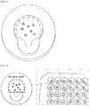
- a cell culture container may include: a lower plate 120; a barrier part composed of two or more pillars 110 provided on the top side of the lower plate; an upper plate 130 which finishes an opened end portion of the barrier part; and one or more cell sample injection guide parts 220 provided in one side of the upper plate.
- the cell culture container may be manufactured into a form in which a plurality of containers is horizontally arrayed or vertically arranged including a microplate form and a culture medium connecting form.
- At least one of the cell sample injection guide parts may be positioned in one side surface of the cell culture container, and may have hole structures drawn such that an experimental tool such as a pipette or the like, i.e., an instrument for injecting a cell sample can lean on the projected hole structures.
- an experimental tool such as a pipette or the like, i.e., an instrument for injecting a cell sample can lean on the projected hole structures.
- the plurality of pillars are preferably formed in two or more pillars, the pillars have a diameter of 20 ⁇ m to 2,000 ⁇ m, a gap between the pillars is preferably 10 ⁇ m to 2,000 ⁇ m, and the pillars may be preferably disposed in a circular shape to smoothly exchange materials between a cell sample and a medium.
- a cell culture parts surrounded by the plurality of pillars has a diameter of 0.1 mm to 10 mm.
- the upper plate may be perpendicularly connected to the pillar parts and positioned to be parallel with the lower plate, and it is appropriate that a gap between the upper plate and the lower plate is 0.05 mm to 5 mm.
- the present invention using such configurations can stably inject a sample including cells into an accurate position in a state that the sample leans on an inlet guide, and the inlet guide may perform a barrier function such that a flow of the sample is directed toward the culture container, and the sample is prevented from being discharged to the outside.
- the upper plate and the plurality of pillars facilitate an operation of an experimenter by performing a helping function such that an appropriate amount of sample is injected easily and uniformly
- the pillar parts spaced apart from one another at predetermined intervals enable a stable cell culturing process to be performed by performing a barrier function of preventing a gel-type sample from being discharged to the outside and performing a function of opening a space for exchanging materials with a medium in an external cell.
- the upper plate parallel to the bottom surface allows height of a three-dimensional cell culture to be constantly maintained by determining height of the sample.
- a cell culture container includes: a lower plate 120; a barrier part composed of two or more pillars 110 provided on the top side of the lower plate; an upper plate 130 which finishes an opened end portion of the barrier part; and cell sample injection guide parts 220 provided in one side of the upper plate.
- a cell culture container according to the present invention may comprise mixtures comprising at least two of transparent ceramics including glass, silicone rubbers including at least one of polydimethylsiloxane (PDMS) and ecoflex, and engineering plastics, transparent ceramics, and silicone rubbers and engineering plastics including at least one of polystyrene, polymethylmethacrylate, polypropylene, polycarbonate and polyurethane, and mixtures comprising at least one or two of chemical variants of the transparent ceramics, the silicone rubbers, the engineering plastics, or the transparent ceramics, the silicone rubbers and the engineering plastics.
- the cell culture container according to the present invention may comprise one or more materials which can be used as three-dimensional printer materials such as photocurable plastics, films, thermoplastic platics, and metals, or all of chemical variants thereof.
- a cell sample injection part of a cell culture container according to the present invention may be positioned in one side surface of a cell culture container configuration.
- the cell sample injection part preferably has a circular hole structure such that the hole structure can guide an outlet of a pipette, a syringe needle or the like for injecting a cell sample.
- the cell sample injection part may include a barrier structure which guides a flow of liquid such that a sample is not discharged to the outside of pillars, but are filled toward the inside of a culture part.
- a plurality of pillar parts of a cell culture container according to the present invention have a gap therebetween of an extent to which a gel-type cell sample is appropriately seated on inner portions of the pillar parts by surface tension of the cell sample, the gap between the pillars may be 10 to 2,000 ⁇ m, and the pillars may have a diameter of about 20 to 2,000 ⁇ m.
- the pillar parts functioning as a barrier the smaller a diameter of pillars is, the smaller a gap between the pillars also tends to be.
- restrictions arise in performing a material exchange process between cells and a culture solution and there is a high possibility that bubbles may be formed between cracks when the gap is too narrow.
- a sample is discharged to the outside of the pillars while a balance between surface energy and tension between the pillars and the sample is being broken when the gap is too wide.
- a gap between the respective pillars is appropriately about 200 to 400 ⁇ m, and the gap range is suitable for seating a cell sample on inner portions of the pillars.
- the number of pillars is formed of a number appropriate for constructing a space enabling the cell sample to seated on an inner portion thereof, and the number of 1 to 100 is appropriate.
- a cross-sectional shape of the pillars may include various shapes including circular, polygonal and other shapes capable of adjusting interfacial tension.
- a cell culture container according to the present invention may preferably be formed in a circular shape, or other shapes besides the circular shape.
- Width of a culture part has a size enabling oxygen and nutrients transferred to cells inside the culture part to be smoothly supplied by sufficiently performing a material exchange process, and the cell culture part may have a diameter of about 0.1 to 10 mm when a cell culture part is formd in a circular shape. It is difficult to supply a sufficient amount of a cell sample when size of the cell culture part is too small, while the cell culture part is not appropriate as a cell culture device since the cell culture part having a too large diameter (diameter of an inscribed circle when the culture part is a polygonal) has restrictions on material transfer to the side surface thereof.
- An upper plate of a cell culture container according to the present invention may be formed in such a form that the upper plate is in parallel with the bottom surface in a state that the upper plate is perpendicularly connected to the pillar parts.
- a height from the lower plate to the upper plate, as a distance which is suitable for observing cell culture results by a microscope, is appropriately 0.05 to 5 mm. It is difficult to sufficiently secure the number of observation target cells within an observation range when the height from the lower plate to the upper plate is 0.05 mm or less, while it is not appropriate to observe the cell culture results by the microscope due to problems including overlapping of cells that are an observation target when the height from the lower plate to the upper plate is 5 mm or more.
- a cell sample loading experiment was carried out after maintaining a gap between the pillars of 200 ⁇ m to 400 ⁇ m at intervals of 100 ⁇ m to draw a gap between the pillars suitable for being fixed to the inside of a barrier from which the cell sample is not eluted, and which is comprised of a plurality of pillars by considering surface energy between a gel-type cell sample generally used and the surface of the container. 2 mg/ml of collagen was used as an injected sample, and the pillars had a diameter of 300 ⁇ m at this time. Thickness of the upper plate or the bottom surface (thickness of the sample) was fixed to 250 ⁇ m.
- the gel-type cell sample is not eluted to the outside of the pillars, but are appropriately positioned on an inner portion of the barrier comprised of the pillars due to tension of the gel-type cell sample at a gap between the pillars of 200 ⁇ m to 400 ⁇ m when the diameter of the pillars is fixed to 300 ⁇ m.
- a hydrophilic treatment process (a plasma treatment process) on a cell culture device and surfaces of pillars
- a success rate of determining if sampels injected into a container were well seated on an inner portion of the container was examined.
- the container is hydrophilic by having a high surface energy, and the container gradually recovers hydrophobicity as time goes by. Since the surfaces of the pillars have good wettability when the surfaces of the pillars are hydrophilic, the sample easily comes out to the outside of the pillars, and a sample loading failure rate is increased, while a sample loading success rate is increased by surface tension of liquid as the surfaces of the pillars recover hydrophobicity.
- FIG. 5(A) shows a probability of suceeding in loading of the sample on a hydrophilic surface according as time changes.
- FIG. 5(B) shows effects of diameter sizes of a cell culture part on loading success rate. It is observed that the larger the diameters of the culture part on a very hydrophilic surface (time after plasma treatment of 1 hour), the higher probabilities that a solution is leaked to the outside of peripheral pillars before the sample is finally filled in the culture part.
- the probabilities are gradually decreased according to a hydrophobicity recovering time, and a loading success rate reaches 100% after 8 hours irrespective of the diameter sizes of the cell culture part. Further, sample loading probabilities according to gaps between peripheral pillar parts of the culture part were measured in FIG. 5(C) . Similarly, it can be confirmed that the larger the gaps between the pillars on a very hydrophilic surface (time after plasma treatment of 1 hour), the higher probabilities that a sample is leaked to the outside of the pillars. As the probabilities are decreased as time goes by, there are hardly any failure rate irrespective of the gaps after 8 hours at which hydrophobity is recovered.
- Example 3 Drawing size of a cell injection part through confirmation of material exchange of cells with a medium
- a material exchange experiment was conducted at intervals of 1 mm from 2 mm to 5 mm of diameter of a cell loading part. Thickness of the upper plate or the bottom surface (thickness of the sample) was fixed to 250 ⁇ m.
- ReN-VM Neuro stem cell
Landscapes
- Health & Medical Sciences (AREA)
- Life Sciences & Earth Sciences (AREA)
- Engineering & Computer Science (AREA)
- Wood Science & Technology (AREA)
- Organic Chemistry (AREA)
- Chemical & Material Sciences (AREA)
- Bioinformatics & Cheminformatics (AREA)
- Zoology (AREA)
- Biomedical Technology (AREA)
- Genetics & Genomics (AREA)
- Biotechnology (AREA)
- Microbiology (AREA)
- Biochemistry (AREA)
- General Engineering & Computer Science (AREA)
- General Health & Medical Sciences (AREA)
- Sustainable Development (AREA)
- Cell Biology (AREA)
- Developmental Biology & Embryology (AREA)
- Clinical Laboratory Science (AREA)
- Gynecology & Obstetrics (AREA)
- Reproductive Health (AREA)
- Molecular Biology (AREA)
- Apparatus Associated With Microorganisms And Enzymes (AREA)
Applications Claiming Priority (2)
| Application Number | Priority Date | Filing Date | Title |
|---|---|---|---|
| KR1020170114829A KR101847044B1 (ko) | 2017-09-07 | 2017-09-07 | 3차원 세포배양 용기 |
| PCT/KR2017/015160 WO2019050101A2 (ko) | 2017-09-07 | 2017-12-20 | 3차원 세포배양 용기 |
Publications (2)
| Publication Number | Publication Date |
|---|---|
| EP3680321A2 true EP3680321A2 (de) | 2020-07-15 |
| EP3680321A4 EP3680321A4 (de) | 2021-06-02 |
Family
ID=61977838
Family Applications (1)
| Application Number | Title | Priority Date | Filing Date |
|---|---|---|---|
| EP17924352.2A Pending EP3680321A4 (de) | 2017-09-07 | 2017-12-20 | Dreidimensionaler zellkulturbehälter |
Country Status (5)
| Country | Link |
|---|---|
| US (1) | US12473521B2 (de) |
| EP (1) | EP3680321A4 (de) |
| KR (1) | KR101847044B1 (de) |
| CN (1) | CN111164200A (de) |
| WO (1) | WO2019050101A2 (de) |
Families Citing this family (1)
| Publication number | Priority date | Publication date | Assignee | Title |
|---|---|---|---|---|
| KR102233285B1 (ko) | 2018-11-08 | 2021-03-29 | 한국화학연구원 | 미세유체소자를 이용한 3차원 세포배양 |
Family Cites Families (10)
| Publication number | Priority date | Publication date | Assignee | Title |
|---|---|---|---|---|
| GB0329220D0 (en) * | 2003-12-17 | 2004-01-21 | Inverness Medical Switzerland | System |
| US20060252087A1 (en) * | 2005-01-18 | 2006-11-09 | Biocept, Inc. | Recovery of rare cells using a microchannel apparatus with patterned posts |
| KR101454580B1 (ko) * | 2009-06-23 | 2014-10-28 | 가부시키가이샤 히타치세이사쿠쇼 | 배양 기재 |
| US9630178B2 (en) * | 2010-03-17 | 2017-04-25 | Ut-Battelle, Llc | Method for preparing small volume reaction containers |
| KR101401781B1 (ko) * | 2012-04-25 | 2014-05-30 | 충남대학교산학협력단 | 세포 배양 및 응용연구를 위한 미세 관류 소자 및 그 제조방법 |
| KR101422345B1 (ko) * | 2012-10-19 | 2014-07-22 | 한국생명공학연구원 | 이중구조 세포배양용기 |
| WO2014131079A1 (en) * | 2013-02-26 | 2014-09-04 | Ddnt Consultants Australia Pty Ltd | Structure for culturing cells |
| US9933413B2 (en) * | 2013-03-08 | 2018-04-03 | Cfd Research Corporation | Synthetic microfluidic systems for tumor metastasis |
| WO2016069892A1 (en) * | 2014-10-29 | 2016-05-06 | Corning Incorporated | Devices and methods for generation and culture of 3d cell aggregates |
| KR20170074503A (ko) * | 2015-12-22 | 2017-06-30 | 고려대학교 산학협력단 | 세포 배양 어세이를 이용한 약물 평가 방법 |
-
2017
- 2017-09-07 KR KR1020170114829A patent/KR101847044B1/ko active Active
- 2017-12-20 EP EP17924352.2A patent/EP3680321A4/de active Pending
- 2017-12-20 US US16/645,539 patent/US12473521B2/en active Active
- 2017-12-20 WO PCT/KR2017/015160 patent/WO2019050101A2/ko not_active Ceased
- 2017-12-20 CN CN201780094660.2A patent/CN111164200A/zh active Pending
Also Published As
| Publication number | Publication date |
|---|---|
| WO2019050101A3 (ko) | 2019-05-23 |
| WO2019050101A9 (ko) | 2019-04-11 |
| KR101847044B1 (ko) | 2018-04-09 |
| US12473521B2 (en) | 2025-11-18 |
| US20200283711A1 (en) | 2020-09-10 |
| CN111164200A (zh) | 2020-05-15 |
| WO2019050101A2 (ko) | 2019-03-14 |
| EP3680321A4 (de) | 2021-06-02 |
Similar Documents
| Publication | Publication Date | Title |
|---|---|---|
| US10744505B2 (en) | Microfluidic device for in vitro 3D cell culture experimentation | |
| US10710077B2 (en) | Method for handling microdrops which include samples | |
| FI131135B1 (en) | MICROFLUIDIC CELL CULTURE DEVICE AND METHOD FOR CELL CULTURE | |
| JP7595584B2 (ja) | オルガノイド培養のためのシステムおよび方法 | |
| KR101822784B1 (ko) | 신경혈관단위-온-칩 및 그 칩의 제조방법 | |
| KR20140113139A (ko) | 세포 스페로이드 배양판 | |
| US10030219B2 (en) | Neurovascular unit(NVU)-on-a-chip and method of fabricating the same | |
| EP3098298A1 (de) | Vorrichtung zum aussäen und kultivieren von zellen | |
| KR101341572B1 (ko) | 중공형관을 이용한 3차원 세포 배양 용구 및 이를 이용한 세포의 3차원 배양 방법 | |
| US20070036690A1 (en) | Inlet channel volume in a reactor | |
| US12473521B2 (en) | Three-dimensional cell culture container | |
| KR20150051199A (ko) | 세포 스페로이드 배양판 | |
| TWI588256B (zh) | 單細胞擷取與培養之裝置與方法 | |
| KR102678639B1 (ko) | 스페로이드 또는 오가노이드 미세 유체 칩 및 이를 이용한 스페로이드 또는 오가노이드 체외 모델 형성 방법 | |
| KR102187633B1 (ko) | 실시간 모니터링이 가능한 마이크로웰 플레이트 | |
| Cheng et al. | “Chip-on-a-Transwell” Devices for User-Friendly Control of the Microenvironment of Cultured Cells | |
| KR102769156B1 (ko) | 혈관오간칩 | |
| CN117683701A (zh) | 一种具有血脑屏障功能的神经血管单元模型及其构建方法与应用 | |
| EP4239052A1 (de) | Kultivierungsvorrichtung für biologisches zellmaterial und verfahren zur herstellung davon | |
| EP4311855A1 (de) | Vorrichtung und verfahren zum züchten von nervenzellen und kompartimentierung von axonen und dendriten | |
| WO2009157212A1 (ja) | 培養細胞への作用因子投与方法、マイクロチャンバ、マイクロチャンバアレイ、培養容器および作用因子投与装置 | |
| WO2024177611A1 (en) | Microfluidic devices and methods for determining dose response | |
| SK2142025U1 (sk) | Mikrofluidický čip pre zariadenie typu orgán-načipe a zariadenie typu orgán-na-čipe obsahujúce mikrofluidický čip | |
| HK40065856A (zh) | 用於类器官培养的系统和方法 | |
| TW202039819A (zh) | 細胞培養晶片 |
Legal Events
| Date | Code | Title | Description |
|---|---|---|---|
| STAA | Information on the status of an ep patent application or granted ep patent |
Free format text: STATUS: THE INTERNATIONAL PUBLICATION HAS BEEN MADE |
|
| PUAI | Public reference made under article 153(3) epc to a published international application that has entered the european phase |
Free format text: ORIGINAL CODE: 0009012 |
|
| STAA | Information on the status of an ep patent application or granted ep patent |
Free format text: STATUS: REQUEST FOR EXAMINATION WAS MADE |
|
| 17P | Request for examination filed |
Effective date: 20200331 |
|
| AK | Designated contracting states |
Kind code of ref document: A2 Designated state(s): AL AT BE BG CH CY CZ DE DK EE ES FI FR GB GR HR HU IE IS IT LI LT LU LV MC MK MT NL NO PL PT RO RS SE SI SK SM TR |
|
| AX | Request for extension of the european patent |
Extension state: BA ME |
|
| DAV | Request for validation of the european patent (deleted) | ||
| DAX | Request for extension of the european patent (deleted) | ||
| A4 | Supplementary search report drawn up and despatched |
Effective date: 20210504 |
|
| RIC1 | Information provided on ipc code assigned before grant |
Ipc: C12M 1/32 20060101AFI20210428BHEP Ipc: C12N 5/00 20060101ALI20210428BHEP Ipc: C12M 3/00 20060101ALI20210428BHEP |