EP3671212A2 - Verfahren zur diagnose von autoimmungastritis - Google Patents
Verfahren zur diagnose von autoimmungastritis Download PDFInfo
- Publication number
- EP3671212A2 EP3671212A2 EP19217068.6A EP19217068A EP3671212A2 EP 3671212 A2 EP3671212 A2 EP 3671212A2 EP 19217068 A EP19217068 A EP 19217068A EP 3671212 A2 EP3671212 A2 EP 3671212A2
- Authority
- EP
- European Patent Office
- Prior art keywords
- atpase
- autoantibody
- beta subunit
- antibody
- carrier
- Prior art date
- Legal status (The legal status is an assumption and is not a legal conclusion. Google has not performed a legal analysis and makes no representation as to the accuracy of the status listed.)
- Pending
Links
Images


Classifications
-
- G—PHYSICS
- G01—MEASURING; TESTING
- G01N—INVESTIGATING OR ANALYSING MATERIALS BY DETERMINING THEIR CHEMICAL OR PHYSICAL PROPERTIES
- G01N33/00—Investigating or analysing materials by specific methods not covered by groups G01N1/00 - G01N31/00
- G01N33/48—Biological material, e.g. blood, urine; Haemocytometers
- G01N33/50—Chemical analysis of biological material, e.g. blood, urine; Testing involving biospecific ligand binding methods; Immunological testing
- G01N33/53—Immunoassay; Biospecific binding assay; Materials therefor
- G01N33/564—Immunoassay; Biospecific binding assay; Materials therefor for pre-existing immune complex or autoimmune disease, i.e. systemic lupus erythematosus, rheumatoid arthritis, multiple sclerosis, rheumatoid factors or complement components C1-C9
-
- G—PHYSICS
- G01—MEASURING; TESTING
- G01N—INVESTIGATING OR ANALYSING MATERIALS BY DETERMINING THEIR CHEMICAL OR PHYSICAL PROPERTIES
- G01N33/00—Investigating or analysing materials by specific methods not covered by groups G01N1/00 - G01N31/00
- G01N33/48—Biological material, e.g. blood, urine; Haemocytometers
- G01N33/50—Chemical analysis of biological material, e.g. blood, urine; Testing involving biospecific ligand binding methods; Immunological testing
- G01N33/53—Immunoassay; Biospecific binding assay; Materials therefor
- G01N33/573—Immunoassay; Biospecific binding assay; Materials therefor for enzymes or isoenzymes
-
- G—PHYSICS
- G01—MEASURING; TESTING
- G01N—INVESTIGATING OR ANALYSING MATERIALS BY DETERMINING THEIR CHEMICAL OR PHYSICAL PROPERTIES
- G01N2333/00—Assays involving biological materials from specific organisms or of a specific nature
- G01N2333/90—Enzymes; Proenzymes
- G01N2333/914—Hydrolases (3)
-
- G—PHYSICS
- G01—MEASURING; TESTING
- G01N—INVESTIGATING OR ANALYSING MATERIALS BY DETERMINING THEIR CHEMICAL OR PHYSICAL PROPERTIES
- G01N2800/00—Detection or diagnosis of diseases
- G01N2800/06—Gastro-intestinal diseases
- G01N2800/062—Gastritis or peptic ulcer disease
Definitions
- the present invention relates to a method for diagnosing AIG, comprising the step of detecting whether autoantibodies against the beta subunit of gastric H + / K + -ATPase are present or not in a sample containing antibodies, a diagnostically useful carrier is used with a solid phase on which a means for specifically capturing the autoantibody is immobilized, and a diagnostically useful carrier with a solid phase on which a means for specifically capturing an autoantibody against the beta subunit of the gastritic H + / K + -ATPase is immobilized.
- AIG autoimmune atritis
- pernicious anemia circulating autoantibodies against this H + / K + -ATPase from the parietal cells, also called APCA, abbreviation for "anti-parietal cell antibodies”
- AIG is a chronic disease with a prevalence of approximately 2% in the general population. In patients suffering from other autoimmune diseases such as type I diabetes, the prevalence is up to 10%.
- the H + / K + -ATPase in the parietal cells of the lamina limba mucosae of the gastric mucosa has the task of pumping protons and thus maintaining an acidic environment in the stomach. It is a heterodimer consisting of an alpha subunit (encoded by the ATP4A gene), which contains the catalytic center and handles ion transport, and a beta subunit (encoded by ATP4B), which stabilizes the alpha subunit and is necessary for the enzymatic activity.
- AIG causes gastric mucosal injuries known as atrophic gastritis (Atrophic body gastritis, ABG).
- ABG auxic body gastritis
- the breakdown of the mucous membrane leads to a reduced or no secretion of gastric acid. It can also be caused by Helicobacter pylori infections.
- autoimmune gastritis can cause pernicious anemia (PA), anemia based on a lack of vitamin B12. It is through deficiency in intrinsic factor, a protein that binds and stabilizes the vitamin and is produced by the parietal cells against which APCAs are directed.
- PA pernicious anemia
- AIG or PA For the correct treatment of AIG or PA, a differentiation between gastritis caused by autoimmunity, bacterial or medication is essential. Incorrect or absent treatment can lead to a stomach ulcer or gastric breakthrough, and the risk of gastric cancer increases.
- test result alone does not allow a diagnosis, not least because samples from patients with numerous other diseases such as pancreatitis or diabetes mellitus may have the autoantibody.
- the test result can be interpreted by the attending physician together with other symptoms such as vitamin B12 deficiency, hyperchromic anemia, the infiltration of the gastric mucosa with lymphocytes and thus supports the diagnosis without providing a sufficient basis for a decision to make a final diagnosis.
- the prior art discloses ELISA and immunofluorescence tests with which autoantibodies against the heterodimer with the ATP4A and the ATP4B subunit can be detected.
- the WO2011 / 150086 discloses the detection of autoantibodies against the ATP4A subunit.
- Wenzlau et al. disclose the detection of autoantibodies against ATP4A for the diagnosis of autoimmune gastritis in patients suffering from type I diabetes.
- a radio binding test was used ( Wenzlau, JM, Gardner, TJ, Frisch, LM, Davidson, HW and Hutton, JC (2011) Development of a novel autoantibody assay for autoimmune gastritis in T1D individuals, Diabetes Metab Res Rev., 27 (8): 887-90 ).
- Rusak et al. disclose that the ATP4A subunit of gastric H + / K + -ATPase is the major antigen for APCA ( Rusak, E., Chobot, A., Krzywicka, A. and Wenzlau, J. (2016) Anti-parietal cell antibodies - diagnostic significance, Advances in Medical Sciences, 61: 175-179 ). They state that better results can be obtained with radioimmunoassays than with ELISA or IFT.
- Scharf et al. disclose, in connection with neurological autoimmune diseases, that autoantibodies to the ATPase heterodimer could be detected using an immunofluorescence assay when using cells that expressed either ATP4A or ATP4B ( Scharf, M., Miske, R., Heidenreich, F., Giess, R., Landwehr, P., Blöcker, I., Begemann, N., Denno, Y., Tiede, S., Dähnrich, C., Schlumberger, W., Unger, M., Teegen, B., Stöcker, W., Probst, C., Komorowski, L.
- Neuronal Na + / K + ATPase is an autoantibody target in paraneoplastic neurologic syndrome, Neurology, 84: 1-7 ).
- Antibodies against ATP4B but not against ATP4A were detected in a serum from a patient with neurological symptoms. It is not disclosed what kind of antigens were used exactly and how the method was carried out.
- Lahner et al. disclose detection of autoantibodies to ATP4A and ATP4B using a luminescent immunoprecipitation system (LIPS) from samples from patients suffering from atrophic gastritis. It is pointed out that autoantibodies against ATP4A and ATP4B can be detected with almost the same high sensitivity and specificity ( Lahner, E., Brigatti, C., Marzinotto, I., Carabotti, M., Scalese, G., Davidson, HW, Wenzlau, JM, Bosi, E., Piemonti, L., Annibale, B. and Lampasona, V.
- LIPS luminescent immunoprecipitation system
- Kontani et al. identified the alpha subunit as the major antigenic protein in AIG (using samples from mice) ( Kontani, K., Taguchi, D., and Takahashi, T. (1992) Involvement of the H + / + - ATPase alpha subunit as a major antigenic protein in autoimmune gastritis induced by neonatal thymectomy in mice, Clin. Exp. Immunol. 89, 63-67 ).
- the problem on which the invention is based is solved by a method for diagnosing AIG, optionally for diagnosing AIG and / or PA, comprising the step of detecting whether an autoantibody or autoantibody is present in a sample which contains antibodies the beta subunit of gastric H + / K + -ATPase is or is absent, being a diagnostically useful carrier with a solid phase is used, on which a means for specific capture of the autoantibody is immobilized.
- the carrier is selected from the group comprising a microtiter plate, a bead, a blot; preferably Western blot, line blot or dot blot; an immunofluorescence test carrier with fixed cells, preferably for microscopic analysis, a lateral flow device, a cellulose-based polymer, a biochip, a microarray, and a chromatography column material.
- the diagnostically useful carrier is a microtiter plate for ELISA.
- the sample is selected from the group comprising serum, whole blood, plasma, CSF.
- the method comprises detecting whether an autoantibody or autoantibody against the intrinsic factor is or is not present in a sample.
- the autoantibody is an antibody of the IgG class.
- a diagnostically useful carrier having a solid phase on which a means for specifically capturing an autoantibody against the beta subunit of the gastritic H + / K + -ATPase is immobilized.
- the problem on which the invention is based is solved by a monoclonal antibody or isolated autoantibody which specifically binds to the beta subunit of the gastritic H + / K + -ATPase, particularly preferably in purified form, but preferably not against that alpha subunit.
- the problem underlying the invention is solved by a method for calibrating or checking the quality of a medical device, comprising the step of contacting the carrier according to the invention with a liquid comprising the antibody according to the invention, preferably in a known concentration, followed by the Step Detect whether the antibody is present in the liquid or not, particularly preferably with at least semi-quantitative determination of the antibody in the liquid.
- the problem on which the invention is based is solved by a kit comprising the diagnostically useful carrier according to the invention, the kit containing one or more reagents, preferably all reagents, from the group comprising a washing solution, a calibrator solution, and a Antibody against the beta subunit of gastritic H + / K + -ATPase, and an agent for detecting the autoantibody, preferably a secondary antibody, more preferably a secondary antibody against IgG antibody.
- the secondary antibody has a detectable label.
- the detectable label is preferably selected from the group comprising a chemiluminescent, radioactive or a detectable isotope-containing label, an enzymatically active label, a fluorescent label, a compound detectable by NMR spectroscopy, a spin label label, preferably a chemiluminescent label, on most preferred a chemiluminescent label that generates a chemiluminescent signal when incubated in a suitable chemiluminescent trigger solution.
- the problem underlying the invention is solved by using a means for detecting whether an autoantibody or autoantibody against the beta subunit of the gastritic H + / K + -ATPase is or is not present in a sample , or the carrier according to the invention or the antibody according to the invention for the highly specific diagnosis of AIG and / or PA.
- the problem underlying the invention is solved by using a means for detecting whether an autoantibody or autoantibody against the beta subunit of the gastritic H + / K + -ATPase is or is not present in a sample , or a carrier comprising the means for producing a kit, a composition for the highly specific diagnosis of AIG and / or PA.
- the problem on which the invention is based is solved by using an autoantibody against the beta subunit of the gastritic H + / K + -ATPase for diagnosis, prefers the highly specific diagnosis of AIG.
- the autoantibody of the beta subunit particularly preferably serves as the sole biomarker, preferably as the sole autoimmune biomarker in one test run, more preferably as the sole autoimmune biomarker derived from the gastritic H + / K + -ATPase. This can mean that no other autoantibodies are detected in the same sample, especially no other autoantibodies against the gastritic H + / K + -ATPase, especially the alpha subunit.
- the present invention is based on the surprising finding of the inventors that the detection of autoantibodies with optimized diagnostic reliability, in particular specificity and sensitivity, can contribute to the diagnosis of AIG if it is proven whether an autoantibody against the beta subunit of gastritic H + / K + -ATPase, is not present against the alpha subunit or the heterodimer. This also applies if a diagnostically useful carrier with a solid phase is used, ie assay formats which are shown in the prior art as being diagnostically unreliable.
- the present invention is further based on the surprising finding of the inventors that the detection of autoantibodies against the extracellular domain of the beta subunit of gastritic H + / K + -ATPase allows a particularly specific diagnosis. The number of false positive diagnoses or findings can thus be reduced to a high level.
- the test is carried out using a diagnostically useful carrier with a solid phase on which an agent for specifically capturing an autoantibody against the beta subunit of the gastric H + / K + -ATPase is immobilized.
- the agent is a polypeptide comprising the beta subunit of gastritic H + / K + -ATPase or a variant thereof.
- molecules derived therefrom or mimicking the backbone of the beta subunit such as peptidomimetics, peptoids, beta peptides and peptides with unnatural amino acids, can also be used.
- beta subunit is indirectly detected by firstly detecting whether an autoantibody or autoantibody against the heterodimer comprising alpha and beta subunit is or is not present, and secondly by detecting whether an autoantibody is present or autoantibodies to the alpha subunit or are absent.
- the term "immobilized" as used herein means that the means for detecting whether an autoantibody against the beta subunit of gastric H + / K + -ATPase is present or not is bound to the solid phase of the support is, preferably via a covalent bond, electrostatic interaction, or via hydrophobic interaction, the agent remaining exposed to the environment in such a way that it is accessible to autoantibodies in a liquid sample.
- Suitable supports for example paper, polystyrene, metal, silicone or glass surfaces, microfluidic channels, membranes, beads such as magnetic beads, chromatography column media, biochips, polyacrylamide gels and the like are described in the literature, for example in Kim, D., and Herr, AE (2013), Protein immobilization techniques for microfluidic assays, Biomicrofluidics 7 (4), 041501 .
- the immobilized agent together with the insoluble carrier can be separated from an aqueous solution in a simple manner, for example by filtration, centrifugation or decanting.
- An immobilized agent can be reversibly or irreversibly immobilized.
- immobilization is reversible when the agent interacts with the carrier via an ionic interaction that can be masked by adding a high concentration of a salt, or when the agent is linked through a cleavable covalent bond such as a disulfide bridge, which is added by adding Reagents contained in thiol can be cleaved.
- immobilization is irreversible if the agent is attached to the support via a covalent bond that cannot be broken in aqueous solution, for example a bond formed by the reaction of an epoxy group and an amino group, as is often used to bind lysine side chains to affinity columns.
- the agent preferably a polypeptide
- immobilization are described in the literature, for Example in Kim, D., and Herr, AE (2013), Protein immobilizsation techniques for microfluidic assays, Biomicrofluidics 7 (4), 041501 .
- the beta subunit of the gastric H + / K + -ATPase is preferably the amino acid sequence represented by the UniProtKB sequence entry P51164 or in SEQ ID NO7, more preferably the amino acid sequence represented by SEQ ID NO3, the latter being the extracellular domain which does not have the transmembrane domain, more preferably around the fragment shown in SEQ ID NO5. Variants thereof can be used according to the invention. In this application all database codes cited are for the Uniprot database or other databases, more precisely for their version on the filing date of this application or its earliest priority application.
- SEQ ID NO3 or a variant thereof is used in the absence of at least one epitope from the transmembrane domain of the beta subunit with the sequence SEQ ID NO4 and / or at least any epitope of the alpha subunit that is used against an autoantibody from the AIG patient's serum is reactive, preferably any epitope of the alpha subunit.
- the alpha subunit of the gastric H + / K + -ATPase is preferably the amino acid sequence represented by UniProtKB accession number P20648 or in SEQ ID NO9.
- the intrinsic factor is preferably the amino acid sequence represented by the NCBI gene bank entry NP_005133.2 or in SEQ ID NO10.
- detecting whether or not an autoantibody against the beta subunit of gastritic H + / K + -ATPase is present in a sample means that it is detected whether the sample contains an autoantibody which is directed against the beta subunit, but not against the alpha subunit of the same protein.
- An autoantibody which is only directed against the complex comprising the alpha and beta subunit is also preferably not detected. It is particularly preferred that a first signal is generated that indicates that the autoantibody has bound to the beta subunit and can be distinguished from a signal that arises from the fact that another autoantibody to another autoantigen such as the alpha subunit or has bound to an autoantigen consisting of amino acids of the alpha and beta subunits.
- an antibody specific for the beta subunit binds to the beta subunit or whether another event has occurred, in particular the binding of an autoantibody which is directed against the alpha subunit or the complex of alpha and beta subunit, but not the beta subunit alone binds.
- an agent for specific capture an agent for specifically detecting whether an autoantibody against the beta subunit of the gastric H + / K + -ATPase is present or not is used, it being preferred is a means that allows the detection of whether or not an autoantibody against the beta subunit of gastritic H + / K + -ATPase is present in a sample.
- the detection means or the specific capture means may be a polypeptide or a reagent or a composition for detection or specific capture.
- the detection of the autoantibody can be qualitative or quantitative.
- the term “qualitative” is understood to mean that it is only demonstrated whether the autoantibody is present or not, but not in what concentration. In particular, it is not determined whether the concentration of the autoantibody exceeds a certain value which provides further information on the present disease, for example on the severity of the disease, on development, on the success of therapy or on certain symptoms.
- the term "quantitative” means that a statement is made that goes beyond that that the autoantibody can be detected.
- an absolute or relative value can be determined or at least an assignment to one of several defined concentration ranges.
- an autoantibody is present in the sample which binds to the complex comprising the alpha and beta subunit of the gastritic H + / K + -ATPase, and additionally whether there is no antibody present in the sample which specifically binds to the alpha subunit. If this is the case, it can be concluded that an autoantibody is present which binds specifically to the beta subunit, even if neither the beta subunit nor a variant thereof is used for the test.
- the presence or absence of autoantibodies against the beta subunit is preferably used to indicate whether this is the case.
- the detection of autoantibodies against the alpha subunit is ignored because it does not indicate such an increased risk.
- the "agent for specifically capturing an autoantibody against the beta subunit of gastritic H + / K + -ATPase” means an agent with which an autoantibody against the beta subunit of gastritic H + / K + -ATPase can be captured, other antibodies, preferably autoantibodies, especially against the alpha subunit, but less efficiently, most preferably not or to an undetectable extent, so that in a detection method according to the invention when contacting the agent or carrier comprising the agent with the sample remain unaffected in the sample.
- the agent for specifically capturing an autoantibody against the beta subunit of the gastritic H + / K + -ATPase specifically binds to an autoantibody against the beta subunit from a sample from an AIG patient.
- the agent for specifically capturing an autoantibody against the beta subunit of the gastritic H + / K + -ATPase specifically binds to an autoantibody against the beta subunit from a sample from an AIG patient.
- all autoantibodies against the H + / K + -ATPase only those are used or used as markers to diagnose AIG that bind against the beta subunit of the gastritic H + / K + -ATPase, particularly preferred not those who bind against the alpha subunit.
- This autoantibody or these autoantibodies serve as the sole autoantibody marker for the diagnosis according to the invention.
- the term "specifically bind" as used herein for an antibody means that it binds to the corresponding antigen in a binding reaction characterized by a dissociation constant that is 1 x 10 -5 M, more preferably 1 x 10 -7 s , more preferably 1 x 10 -8 s , more preferably 1 x 10 -9 s , is more preferably 1 x 10 -10 M, more preferably 1 x 10 -11 M, more preferably 1 x 10 -12 M or a stronger binding reaction.
- the dissociation constant is preferably measured by surface plasmon resonance using a Biacore device at 25 ° C. and in PBS buffer at pH 7 and calculated using the software offered by the manufacturer.
- the term "diagnosis” as used herein is understood to mean a procedure in which information is obtained which supports the assessment or even permits the assessment of whether a subject has a disease or is more likely than one to be average person suffers from an illness or has suffered in the past or will suffer in the future, whether an illness is progressing or how it will develop in the future or to assess whether a particular treatment will work. Before the diagnosis, the subject preferably does not know whether it is suffering from the disease.
- the diagnosis or to support the diagnosis merely to demonstrate whether the antibody is present, it being possible to determine whether detectable concentrations of the antibody are present in the sample.
- it is determined whether the relative concentration of the antibody is higher in a patient to be diagnosed than in an average healthy person. It can be determined whether the concentration is higher by a factor of 1.1, more preferably 1.2, 1.5, 1, 2, 5, 10, 20, 25, 50, 100, 200, 500, 1000, 10000 or 100000 is considered in a sample by an average healthy person.
- the higher concentration supports the diagnosis that the patient from whom the sample is taken has AIG or indicates that the patient is more likely to have AIG.
- the method according to the invention or the use according to the invention is preferably carried out in vitro.
- the person skilled in the art understands that such a proof usually does not alone permit a comprehensive diagnosis, but that further aspects have to be taken into account, for example further parameters to be determined in a sample, the medical history, clinical symptoms, the medical history or the results of imaging procedures. He further understands that the value of the method according to the invention can consist in the fact that an indirect diagnosis or differential diagnosis can take place at which excludes a disease indicates that the patient is suffering from another disease with similar symptoms.
- products according to the invention such as carriers or kits or methods according to the invention serve to differentiate between gastritis caused by autoimmunity, bacterial or medication or to diagnose a disease from the group comprising AIG, pernicious anemia, atrophic gastritis or vitamin B12 deficiency .
- the sample to be examined is a sample which contains a representative set of antibodies of the patient, which is preferably a mammal, more preferably a human. It is preferably selected from the group comprising serum, plasma, saliva and CSF.
- the teaching according to the invention can be used not only using the wild-type sequences of the specified polypeptides or the reference sequences as they are referred to in the form of SEQ ID NOs from now on as “full-length polypeptide", or database codes or in some other way, possibly implicitly, here are specified, but also using variants of these sequences.
- the term “variant” as used herein means at least one fragment of the full-length polypeptide which is shortened by one or more than one amino acid at the C- and / or N-terminal end compared to the full-length polypeptide .
- Such a fragment can contain a length of 5, 6, 7, 8, 10, 12, 15, 20, 25, 50, 75, 100, 150 or, 200 consecutive amino acids from the sequence of the full-length polypeptide, preferably 10 or more .
- the term “variant” as used herein includes not only a fragment but also a polypeptide that contains amino acid sequences with increasing preference in the order of at least 40, 50, 60, 70, 75, 80, 85, 90 , 92, 94, 95, 96, 97, 98 or 99% sequence identity to the full-length polypeptide or to the fragment thereof, wherein none for biological activity, eg the ability of an antigen, to an antibody specific for the full-length polypeptide antigen to bind, essential amino acid is deleted or non-conservatively substituted.
- the prior art discloses methods for determining the sequence identity of amino acid sequences, for example in Arthur Lesk (2008), Introduction to bioinformatics, Oxford University Press, 2008 , third edition.
- the ClustalW software Larkin, MA, Blackshields, G., Brown, NP, Chenna, R., McGettigan, PA, McWilliam, H., Valentin, F., Wallace, IM, Wilm, A., Lopez, R., Thompson, JD, Gibson, TJ, Higgins, DG (2007).
- a polypeptide or a variant thereof used in accordance with the invention may have chemical modifications, e.g. Isotope labels, covalent modifications such as glycosylation, phosphorylation, acetylation, decarboxylation, citrullination, methylation, hydroxylation etc.
- a polypeptide used for the detection of an autoantibody against the beta subunit or otherwise according to the invention comprises the beta subunit or a variant thereof glycosylated.
- Glycosylation is preferably accomplished by expression in a cell capable of glycosylation, preferably a mammalian or insect cell, more preferably a mammalian cell.
- Variants can also be fusion proteins with amino acids or other known polypeptides or variants thereof, preferably with a tag for purification, more preferably from the group comprising His tag, thioredoxin, maltose-binding protein, streptavidin, glutathione-S-transferase or a variant thereof, and be or have active parts or domains, preferably with a sequence identity of at least 70, 75, 80, 85, 90, 92, 94, 95, 96, 97, 98 or 99% of the full-length polypeptide.
- the term “active parts or domains” is understood to mean a fragment of the full-length polypeptide or a variant which retains at least part of the biological activity of the full-length polypeptide.
- the full-length polypeptide variant has biological activity.
- the biological activity is the ability of an agent, preferably polypeptides, to capture or bind to an autoantibody against the beta subunit, preferably to bind specifically, as occurs in samples from patients suffering from AIG are.
- the autoantibody can be obtained from such samples as a reference for experiments.
- Variants of a secondary antibody which serves to detect a specifically captured antibody are all molecules which have the same binding specificity as the secondary antibody, preferably the ability to bind against all antibodies of the human class IgG. For example, they include fragments and derivatives of the secondary antibody.
- SEQ ID NO6 As a peptide containing the immunodominant epitope. This means that the homologous sequence from the human protein shown in SEQ ID NO5 contains the human epitope. Sequence alignments with other mammalian homologs, for example from pigs, cattle, from primates, goats and others, allow the skilled worker to predict reactive variants easily. The person skilled in the art can generate further variants by mutations and further truncations. Carried out by means of ELISA as in the examples, he can confirm that the biological activity is retained.
- a polypeptide or a variant thereof does not bind or only binds to a small extent against antibodies against the alpha subunit, preferably its immunodominant epitope, as shown in SEQ ID NO8.
- Homologous epitopes from other homologous mammalian enzymes can be found in Nishio et al. and take it into account when designing variants ( Nishio, A., Hosono, M., Watanabe, Y., Sakai, M., Okuma, M and Masuda, T (1994) Gastroenterology 107, 1408-1414 ).
- the binding constant of autoantibodies against the beta subunit as they occur in a sample from a patient is at least 10, preferably, 100, more preferably 1,000, more preferably 10,000, more preferably 100,000, more preferably 1,000,000, still more preferably 10,000,000 times lower and the strength of the binding reaction correspondingly higher than the binding constant of autoantibodies against the alpha subunit, as occurs in a sample from a patient.
- the binding constant characterizes the strength of the binding of the respective autoantibody against the variant, more precisely a mixture of the autoantibodies as they occur in a sample.
- the binding constant is preferably measured using Biacore in PBS buffer at 25 ° C.
- the binding constant when binding autoantibodies against the beta subunit from a sample can be 10 -10 M, while the binding constant of identical autoantibodies against the alpha subunit from the sample is 10 -6 M.
- the first binding constant is 10,000 times lower than the second. This means that autoantibodies against the beta subunit bind significantly more strongly to the polypeptide or the variant than autoantibodies against the alpha subunit. If the polypeptide or the variant is used according to the invention, only autoantibodies against it are used the beta subunit bound and detected, but not autoantibodies against the alpha subunit.
- the term “autoantibody” as used herein means an antibody which specifically binds to a structure, preferably an autoantigen, from the organism which produces it.
- Such an autoantibody has a constant region like that of other antibodies of the same class from the same organism. It is particularly preferably a mammalian, more preferably a human autoantibody, more preferably a human autoantibody of the IgG class.
- the variable domain is able to bind specifically to the autoantigen.
- the autoantigen is preferably an epitope derived from or contained in the beta subunit, preferably SEQ ID NO5 or a variant thereof.
- the capture agent is an isolated and / or purified polypeptide, which is particularly preferably recombinant.
- An "isolated” polypeptide can be understood to mean that the polypeptide has been removed from its natural environment or from the environment in which it was expressed.
- a "purified” polypeptide is understood to mean a polypeptide which has been enriched for its concentration and / or purity in its natural environment using chromatographic methods, which are preferably selected from the group consisting of affinity chromatography, gel filtration chromatography and ion exchange. chromatography includes.
- a polypeptide is purified when it has a purity of at least 40, 50, 60, 70, 80, 90, 95, 98, 99 or 99.5%, which is visual or, preferably, by measuring the intensity of bands can be assessed on an SDS-PAGE gel after Coomassie staining.
- a polypeptide can be provided in the form of a tissue or a cell, preferably a recombinant tissue or a recombinant cell.
- the cell is preferably a eukaryotic cell, more preferably an insect, plant or mammalian cell, more preferably a mammalian cell such as a human cell, most preferably a HEK293 cell.
- a polypeptide as an agent can be provided in folded form or in unfolded form and is preferably folded. Folding is preferred by means of CD spectroscopy measured in a buffer which allows the measurement and in which the protein can assume its three-dimensional folding (see for example Banaszak LJ (2008), Foundations of Structural Biology, Academics Press , or Teng Q. (2013), Structural Biology: Practical Applications, Springer ), for example in 10 mM sodium phosphate at pH 7. The protein preferably assumes a fold that is recognized by an antibody to be detected.
- an antibody is detected after capture by a method from the group comprising the measurement of immunodiffusion, immunoelectrophoresis, light scattering, agglutination, radioactivity, enzymatic activity, (electro) chemiluminescence and immunofluorescence.
- a detectable label can be used. It is preferably attached to a secondary antibody that binds to the antibody to be detected. Detection methods are, for example, in Zane, HD (2001), Immunology - Theoretical & Practical Concepts in Laboratory Medicine, WB Saunders Company, especially described in Chapter 14 en.
- a kit which comprises the carrier according to the invention, the kit containing one or more reagents, preferably all reagents, from the group comprising a washing solution, at least one calibrator solution, an antibody against the beta subunit of gastritic H + / K + -ATPase, and an agent for detecting the autoantibody, preferably a secondary antibody, more preferably a secondary antibody against IgG antibody or the beta subunit of the gastric H + / K + -ATPase or a variant thereof, preferably with a detectable one Label, a dilution buffer to dilute the sample, a positive control, a chemiluminescent trigger solution and a chemiluminescent substrate.
- a chromogen solution for an ELISA can also be included.
- the autoantibody detection can be quantitative or semi-quantitative, preferably semi-quantitative.
- “semi-quantitative” means that it is determined whether the measurement result lies above a certain threshold value, for example 5,6,7,8,9,10, 11, 12, 13, 14, 15, 16 , 17, 18, 19, 20, 21, 22, 23, 24, 25, 26, 27, 28, 29, 30, 31, 32, 33, 34, 35, 36, 37, 38, 39 or 40 RU / ml.
- the value in RU / ml becomes preferably measured by means of ELISA (EA 1361-9601 G) from EUROIMMUN Medical Laboratory Diagnostics AG.
- a calibrator solution as used in the method according to the invention is understood to mean a solution with a defined amount of an antibody which binds to the same antigen as the autoantibody to be detected and preferably belongs to the same Ig class as the antibody to be detected, the antigen being preferred is the beta subunit.
- the calibrator can contain an amount of the antibody known on an absolute or relative scale. For example, one calibrator can contain ten times as much antibody as another.
- the antibody present in a defined amount can be an autoantibody from a sample or a recombinant, monoclonal antibody.
- calibrator solutions each with different concentrations of the antibody, are preferably added to the kit or used according to the invention, in particular examined parallel to a sample, so that a calibration curve for the semi-quantitative or quantitative determination of the autoantibody against the beta subunit can be recorded.
- the calibrator comprises an autoantibody or antibody against the beta subunit of the gastritic H + / K + -ATPase, but preferably less or not in detectable amounts or no autoantibody or antibody against the alpha subunit.
- a monoclonal antibody or isolated autoantibody which specifically binds to the beta subunit of gastritic H + / K + -ATPase is used, particularly preferably in recombinant, isolated and / or purified form, most preferably against SEQ ID NO5.
- Such an antibody can serve as a positive control for methods according to the invention and as a reagent for the immobilization of the beta subunit or as a reagent for competitive test formats for which it can optionally have a detectable label.
- the captured autoantibody is detected by means of chemiluminescence.
- the autoantibody can be contacted with a chemiluminescent ligand that binds to the autoantibody.
- a chemiluminescent ligand can be a secondary antibody that binds to the autoantibody constant domain, or the beta subunit of gastric H + / K + -ATPase, or a variant thereof that binds to the variable domain.
- chemiluminescent ligand can be a chemiluminescent enzyme, for example luciferase, peroxidase, alkaline phosphatase and ⁇ -galactosidase, which can convert a chemiluminescent substrate without being consumed ( Kricka, LJ (2003). Clinical applications of chemiluminescence. Analyticazia acta, 500 (1): 279-286 ).
- chemiluminescent enzyme for example luciferase, peroxidase, alkaline phosphatase and ⁇ -galactosidase
- the chemiluminescent ligand can also have a chemiluminescent organic compound without enzymatic activity, which emits a chemiluminescent signal in the presence of a chemiluminescent trigger solution, for example an acridinium ester ( Weeks, I., Beheshti, I., McCapra, F., Campbell, AK, Woodhead, JS, Acridinium esters as high specific activity labels in immunoassay. Clin Chem 29: 1474-1479 (1983 )) or luminol or a chemiluminescent derivative thereof. This compound is consumed in the reaction.
- a chemiluminescent trigger solution for example an acridinium ester ( Weeks, I., Beheshti, I., McCapra, F., Campbell, AK, Woodhead, JS, Acridinium esters as high specific activity labels in immunoassay. Clin Chem 29: 1474-1479 (1983 )) or luminol or a
- acridinium ester In the case of an acridinium ester, a mixture of hydrogen peroxide and hydroxide, e.g. used in the form of sodium hydroxide.
- the acridinium ester compound is oxidized under alkaline conditions, which produces light-emitting acridon.
- the signal is released in the form of a flash of light (flash luminescence), which typically lasts 1-5 seconds. The short duration of the emission requires the initiation and measurement of the reaction directly at the detector.
- Chemiluminescence is preferably used as a detection method with a support which is a bead.
- the captured autoantibody is detected by means of ELISA.
- the agent for specific capture is immobilized in at least one, preferably several, preferably at least 2, 4, 6, 8, 10, 12, 16, 20 or 24 wells of a microtiter plate for ELISA.
- a sample to be analyzed is placed in at least one well, 2, 3, 4, 5 or more calibrator solutions are placed in other wells.
- the captured antibody is then preferably detected using a secondary antibody with an enzymatically active label.
- an agent for specifically capturing an autoantibody against the beta subunit of the gastritic H + / K + -ATPase is used to calibrate or to control the quality of a medical device.
- calibration is understood to mean that measured values are used using calibration solutions are obtained which confirm that the device provides reliable measured values in a required concentration range or allows a reliable delimitation between the presence or absence of the autoantibody in a sample.
- control can mean that it is verified that the device works reliably in the sense that a sample containing the autoantibody is actually measured as positive and a sample without the autoantibody is actually measured as negative.
- control means that a check is carried out to determine whether a batch of the agent or a device has, under given conditions, the ability, preferably still sufficient capacity, to bind the autoantibody.
- the medical product can be a diagnostic product such as the carrier according to the invention. Alternatively, it can be a therapeutic device, for example for apheresis, which also comprises a means for specifically capturing or detecting the autoantibody.
- the invention is a use or a reagent or agent or polypeptide for the detection of an autoantibody against the beta subunit, or a use of a nucleic acid encoding the beta subunit or a variant thereof or a nucleic acid that is present under stringent conditions this nucleic acid hybridizes or a vector or a cell comprising the nucleic acid or the vector for the production of a kit for the diagnosis of a disease.
- a cell comprising the vector, and preferably not comprising a vector encoding the alpha subunit or an epitope or a variant thereof, is provided and cultured under conditions that allow expression of the nucleic acid followed by isolation and / or Purifying the expression product, followed by using the expression product as an agent for specifically capturing an antibody in a method or product according to the present invention.
- Fig. 1 shows a Coomassie-stained polyacrylamide gel on which 1 ⁇ g of the purified extracellular domain of the beta subunit of gastritic H + / K + -ATPase with N-terminal His-Tag DTT was reduced on the right next to the marker and separated on the right next to it was not reduced.
- Fig. 2 shows the sample distribution for the data shown in Example 3 (sensitivity) and Table 4.
- the Y axis shows the signal in RLU, each number on the X axis represents a patient suffering from AIG (1-29) or a healthy blood donor (from 30).
- Example 1 Determination of the sensitivity of a detection by means of the beta subunit of the gastritic H + / K + -ATPase as an antigen using immunofluorescence and ELISA
- HEK293 cells cultured on glass slides in D-MEM with an addition of 10% (v / v) fetal calf serum were isolated using 25 kDa linear polyethyleneimine with the plasmid DNA coding for ATP4A or ATP4B individually and in combination and in addition to producing a negative control transfected with the pTriEx-Leervector and fixed after 48 hours.
- the glass slides with the cells adhering to them were first washed with PBS and then alternatively fixed for 5 minutes with 1.8% formalin solution in PBS or for 10 minutes in acetone at room temperature. Finally, there was a washing and blocking step in PBS with 1% (w / v) BSA additive (PBS (1% w / v BSA)) for 5 minutes at room temperature.
- the serum or plasma samples to be analyzed were incubated on the substrate in a 1:10 dilution in PBS (1% w / v BSA) for 30 minutes. Then the substrates washed in PBS (0.2% v / v Tween 20) for 5 minutes. Bound human immunoglobulin class IgG was detected with fluorescein-labeled anti-human IgG in a 30 minute incubation step. Excess conjugate was removed for 5 minutes with PBS (0.2% v / v Tween 20) before evaluation on the immunofluorescence microscope.
- the immunofluorescence was evaluated semi-quantitatively by specifying the intensity of the specific fluorescence signal in intensities of 0 (no fluorescence), trace (borderline signal), 1 (weakly positive), 2, 3 and 4 (strongly positive).
- HEK293 cells cultured in D-MEM with an addition of 10% (v / v) fetal calf serum were transfected with 25 kDa linear polyethyleneimine with the plasmid DNA coding for spGh * -H6-ATP4B (ec).
- the cell culture supernatant was removed, clarified by centrifugation and used to purify the recombinant protein by immobilized metal chelate affinity chromatography (IMAC) using standard protocols.
- IMAC immobilized metal chelate affinity chromatography
- microtiter plates (Nunc, Germany) were coated with 0.7 ⁇ g / ml antigen in PBS overnight at 4 ° C and washed three times with wash buffer (0.05% (w / v) Tween-20 in PBS). Unspecific binding sites were blocked by incubation with 0.1% casein (w / v) in PBS for one hour.
- Human serum samples were diluted 1: 100 in sample buffer (0.05% (w / v) Tween-20, 1% (w / v) casein in PBS) and incubated for 30 minutes, followed by three washes with wash buffer. In addition to two positive samples from clinically characterized patients, seven other samples from patients were examined.
- Bound antibodies were detected by incubation for half an hour with anti-human-IgG HRP conjugate (EUROIMMUN, Germany), which was diluted 1: 1000 in sample buffer. The washing was then carried out as described above, followed by the addition of 3,3 ', 5,5'-tetramethylbenzidine (EUROIMMUN Medical Laboratory Diagnostics AG) and fifteen minutes of incubation. All incubation steps were carried out at room temperature.
- optical density was measured at 450 nm using an automatic spectrophotometer (Spectra Mini, Tecan, Crailsheim, Germany). The cut-off was determined by ROC curve analysis (maximum sum of sensitivity plus specificity).
- test was also carried out using the conventional anti-PCA ELISA (IgG) (EUROIMMUN Medical Laboratory Diagnostics AG, order number EA 1361 G).
- the autoantibody against the gastritic H + / K + -ATPase can also be detected in the samples of the patients suffering from AIG if the extracellular domain of the beta subunit of the gastritic H + / K + -ATPase is used.
- Example 2 show that a significant specificity gain of 6% can be achieved compared to the conventional test.
- Example 3 Determination of the sensitivity and specificity of a chemiluminescence-based assay for the diagnosis of AIG by determining autoantibodies against ATP4B
- test was carried out using the same reagents as described in Example 1.
- antigen-coated magnetic particles are used as a solid phase for the specific detection of autoantibodies of the immunoglobulin class IgG against ATP4B.
- the magnetic particles were produced using M-280 Dynabeads (Invitrogen, catalog number 14203, 14204) activated according to the manufacturer's instructions.
- the test is carried out automatically in a random access analyzer (RA Analyzer 10, product no. YG 0710-0101, EUROIMMUN medical laboratory diagnostics AG) ..
- RA Analyzer 10 product no. YG 0710-0101, EUROIMMUN medical laboratory diagnostics AG
- sample buffer catalog number LL9012, EUROIMMUN medical laboratory diagnostics AG
- specific antibodies bind to the antigen under these conditions.
- acridinium ester-labeled anti-human-IgG (catalog number LK0711-G, EUROIMMUN Medical Laboratory Diagnostics AG) conjugate is added, which binds to the specific antibodies.
- reaction mixture is then mixed with trigger solutions (catalog numbers LL0713-1 (A) and LL0713-2 (B), EUROIMMUN), which induce a chemiluminescence reaction.
- trigger solutions catalog numbers LL0713-1 (A) and LL0713-2 (B), EUROIMMUN
- the resulting light signal is output in relative light units (RLU).
- the intensity of the light signal is proportional to the amount of bound antibodies.
Landscapes
- Health & Medical Sciences (AREA)
- Life Sciences & Earth Sciences (AREA)
- Immunology (AREA)
- Engineering & Computer Science (AREA)
- Hematology (AREA)
- Molecular Biology (AREA)
- Biomedical Technology (AREA)
- Chemical & Material Sciences (AREA)
- Urology & Nephrology (AREA)
- Food Science & Technology (AREA)
- Biochemistry (AREA)
- Cell Biology (AREA)
- Biotechnology (AREA)
- Medicinal Chemistry (AREA)
- Physics & Mathematics (AREA)
- Analytical Chemistry (AREA)
- Microbiology (AREA)
- General Health & Medical Sciences (AREA)
- General Physics & Mathematics (AREA)
- Pathology (AREA)
- Rehabilitation Therapy (AREA)
- Rheumatology (AREA)
- Peptides Or Proteins (AREA)
- Investigating Or Analysing Biological Materials (AREA)
- Measuring Or Testing Involving Enzymes Or Micro-Organisms (AREA)
Abstract
Description
- Die vorliegende Erfindung betrifft ein Verfahren zur Diagnose von AIG umfassend den Schritt Nachweisen, ob in einer Probe, die Antikörper enthält, Autoantikörper gegen die beta-Untereinheit der gastritischen H+/K+-ATPase vorhanden oder nicht vorhanden sind, wobei ein diagnostisch nützlicher Träger mit einer Festphase eingesetzt wird, an der ein Mittel zum spezifischen Einfangen des Autoantikörpers immobilisiert ist, sowie einen diagnostisch nützlicher Träger mit einer Festphase, an der ein Mittel zum spezifischen Einfangen eines Autoantikörpers gegen die beta-Untereinheit der gastritischen H+/K+-ATPase immobilisiert ist.
- Es ist anerkannt, dass zirkulierende Autoantikörper gegen diese H+/K+-ATPase aus den Parietalzellen, auch als APCA, Abkürzung für "anti-parietal cell antibodies" bezeichnet, diagnostische Marker für Autoimmungastritis (AIG) und perniziöse Anämie sind. AIG ist eine chronische Krankheit mit einer Prävalenz von ca. 2% in der Gesamtbevölkerung. In Patienten, die an anderen Autoimmunkrankheiten wie Typ I-Diabetes leiden, beträgt die Prävalenz bis zu 10%.
- Die H+/K+-ATPase in den Parietalzellen der Lamina propria mucosae der Magenschleimhaut hat die Aufgabe, Protonen zu pumpen und damit ein saures Milieu im Magen aufrechtzuerhalten. Es handelt sich um ein Heterodimer aus einer alpha-Untereinheit (kodiert durch das ATP4A-Gen), welche das katalytische Zentrum enthält und den Ionen-Transport bewerkstelligt, und einer beta-Untereinheit (kodiert durch ATP4B), die die alpha-Untereinheit stabilisiert und für die enzymatische Aktivität notwendig ist.
- Bei der AIG treten Verletzungen der Magenschleimhaut auf, die als Atrophische Gastritis bezeichnet werden (Atrophic body gastritis, ABG). Der Abbau der Schleimhaut führt dabei zu einer verringerten oder fehlenden Sekretion von Magensäure. Sie kann auch durch Infektionen durch Helicobacter pylori bedingt sein.
- Neben anderen möglichen Ursachen kann eine Autoimmun-Gastritis eine Perniziöse Anämie (PA) bedingen, eine Anämie, die auf einem Mangel an Vitamin B12 beruht. Sie wird durch einen Mangel an intrinsischem Faktor bedingt, einem Protein, das das Vitamin bindet und stabilisiert und von den Parietalzellen gebildet wird, gegen die APCAs gerichtet sind.
- Für die richtige Behandlung einer AIG bzw. PAist eine Differenzierung zwischen einer durch Autoimmunität bedingten, einer bakteriell oder durch Medikamente verursachten Gastritis unentbehrlich. Bei falscher oder ausbleibender Behandlung kann es zu einem Magengeschwür oder einem Magendurchbruch kommen, zudem steigt das Risiko eines Magenkarzinoms.
- Es besteht daher Bedarf an Hochdurchsatz-tauglichen diagnostischen Tests, mit denen diagnostisch nützliche Antikörper in leicht verfügbarem Probenmaterial von Patienten mit möglichst hoher diagnostischer Verlässlichkeit nachgewiesen werden können, insbesondere mit möglichst hoher Sensitivität und/oder Spezifität, besonders gegenüber Tests, die auf dem Nachweis von einem Autoantikörper gegen die alpha-Untereinheit oder das Heterodimer umfassend alpha- und beta-Untereinheit der gastritischen H+/K+-ATPase basieren.
- Es sei darauf hingewiesen, dass das Testergebnis allein keine Diagnose ermöglicht, nicht zuletzt, weil Proben von Patienten mit zahlreichen anderen Krankheiten wie Pankreatitis oder Diabetis mellitus den Autoantikörper aufweisen können. Das Testergebnis kann vom behandelnden Arzt zusammen mit weiteren Symptomen wie Vitamin B12-Mangel, hyperchromer Anämie, dem Infiltrieren der Magenschleimhaut mit Lymphozyten interpretiert werden und unterstützt damit die Diagnose, ohne selbst für eine endgültige Diagnose eine ausreichende Entscheidungsgrundlage zu stellen.
- Im Stand der Technik sind dazu ELISA- und Immunfluoreszenz-Tests offenbart, mit denen Autoantikörper gegen das Heterodimer mit der ATP4A- und der ATP4B-Untereinheit nachgewiesen werden können.
- Die EUROIMMUN Medizinische Labordiagnostika AG verkauft seit 2011 einen ELISA basierend auf aufgereinigter nativer Säugetier-ATP4 mit beiden Untereinheiten. In der Testbeschreibung wird die Spezifität ausführlich addressiert und darauf hingewiesen, dass man diese über die Wahl des Cut-Off-Wertes optimieren kann (Katalog Nr. EA 1361-9601 G).
- Die
WO2011/150086 offenbart den Nachweis von Autoantikörpern gegen die ATP4A-Untereinheit. - Wenzlau et al. offenbaren den Nachweis von Autoantikörpern gegen ATP4A zur Diagnose von Autoimmun-Gastritis in Patienten, die unter Typ I-Diabetes leiden. Es wurde ein Radiobindungs-Test eingesetzt (Wenzlau, J.M., Gardner, T.J., Frisch, L.M., Davidson, H.W. and Hutton, J.C. (2011) Development of a novel autoantibody assay for autoimmune gastritis in T1D individuals, Diabetes Metab Res Rev., 27 (8): 887-90).
- Rusak et al. offenbaren, dass die ATP4A-Untereinheit der gastritischen H+/K+-ATPase das Hauptantigen für APCA ist (Rusak, E., Chobot, A., Krzywicka, A. and Wenzlau, J. (2016) Anti-parietal cell antibodies - diagnostic significance, Advances in Medical Sciences, 61:175-179). Sie geben an, dass mit Radioimmunassays bessere Ergebnisse als mit ELISA oder IFT erzielt werden können.
- Scharf et al. offenbaren im Zusammenhang mit neurologischen Autoimmunkrankheiten, dass Autoantikörper gegen das ATPase-Heterodimer unter Verwendung eines Immunfluoreszenz-Tests nachgewiesen werden konnten, wenn Zellen eingesetzt wurden, die entweder ATP4A oder ATP4B exprimierten (Scharf, M., Miske, R., Heidenreich, F., Giess, R., Landwehr, P., Blöcker, I., Begemann, N., Denno, Y., Tiede, S., Dähnrich, C., Schlumberger, W., Unger, M., Teegen, B., Stöcker, W., Probst, C., Komorowski, L. (2015) Neuronal Na+/K+ ATPase is an autoantibody target in paraneoplastic neurologic syndrome, Neurology, 84:1-7). In einem Serum von einem Patienten mit neurologischen Symptomen wurden Antikörper gegen ATP4B, aber nicht gegen ATP4A nachgewiesen. Es wird nicht offenbart, welche Art von Antigenen genau eingesetzt wurde und wie das Verfahren durchgeführt wurde.
- Lahner et al. offenbaren die Detektion von Autoantikörpern gegen ATP4A und ATP4B unter Verwendung eines Lumineszenz-Immunpräzipitations-System (LIPS) aus Proben von Patienten, die unter einer atrophischen Gastritis litten. Es wird herausgestellt, dass Autoantikörper gegen ATP4A und ATP4B beinahe mit der gleichen, hohen Sensitivität und Spezifität nachgewiesen werden können (Lahner, E., Brigatti, C., Marzinotto, I., Carabotti, M., Scalese, G., Davidson, H.W., Wenzlau, J.M., Bosi, E., Piemonti, L., Annibale, B. and Lampasona, V. (2017) Luminescent Immunoprecipitation System (LIPS) for Detection of Autoantibodies Against ATP4A and ATP4B Subunits of Gastric Proton Pump H+/K+-ATPase in Atrophic Body Gastritis Patients, Clinical and Translational Gastroenterology, 8(1):e215). Burbelo et al. untersuchten das Auftreten von Antikörpern gegen ATP4B bei Patienten, die unter Typ I-Diabetes litten (Burbelo, P.D., Lebovitz, E.E., Bren, K.E., Bayat, A., Paviol, S., Wenzlau, J.M., Barriga, K.J., Rewers, M., Harlan, D.M. and ladarola, M.J. (2012) Extrapancreatic Autoantibody Profiles in Type I Diabetes, PLOS ONE, 7(9):e45216).
- Kontani et al. identifizierten unter Verwendung von Proben von Mäusen die alpha-Untereinheit als antigenes Hauptprotein in AIG (Kontani, K., Taguchi, D., und Takahashi, T. (1992) Involvement of the H+/+-ATPase alpha subunit as a major antigenic protein in autoimmune gastritis induced by neonatal thymectomy in mice, Clin. Exp. Immunol. 89, 63-67).
- Ma et al. kommen unter Verwendung von unaufgereinigter beta-Untereinheit in Form eines Zellextraktes zu dem Schluss, dass sowohl die alpha- als auch die beta-Untereinheit an Autoantikörper von Patienten mit AIG binden. Ein direkter Vergleich der menschlichen alpha- und beta-Untereinheiten wurde nicht vorgenommen (Ma, J. Y., Borch, K und Mardh, S. (1994) Human Gastric H,K-Adenosine Triphosphatase beta-Subunit is a Major Autoantigen in Atrophic Corpus Gastritis, Scand J. Gastroenterol 29, 790-794).
- Sowohl Lahner et al. als auch Burbelo et al. geben an, dass nur ein Test mit besonders hoher Sensitivität und Spezifität wie LIPS oder eine Radiobindungs-Test für eine zuverlässig Diagnostik geeignet ist, im Gegensatz zu Testsystemen mit Festphase, mit denen viele diagnostisch nützliche Epitope nicht nachgewiesen werden können. Es werden keine Gründe angegeben, warum nicht die ATPA4-Untereinheit ausgewählt wurde.
- Die der Erfindung zu Grunde liegende Aufgabe wird durch den Gegenstand der beigefügten unabhängigen und abhängigen Ansprüche gelöst.
- In einem erstem Aspekt wird das der Erfindung zu Grunde liegende Problem gelöst durch ein Verfahren zur Diagnose von AIG, optional zur Diagnose von AIG und/oder PA, umfassend den Schritt Nachweisen, ob in einer Probe, die Antikörper enthält, ein Autoantikörper oder Autoantikörper gegen die beta-Untereinheit der gastritischen H+/K+-ATPase vorhanden oder nicht vorhanden ist oder sind, wobei ein diagnostisch nützlicher Träger mit einer Festphase eingesetzt wird, an der ein Mittel zum spezifischen Einfangen des Autoantikörpers immobilisiert ist.
- In einer bevorzugten Ausführungsform ist der Träger aus der Gruppe ausgewählt, die eine Mikrotiterplatte, einen Bead, einen Blot; bevorzugt Western Blot, Lineblot oder Dot Blot; einen Immunfluoreszenztestträger mit fixierten Zellen, bevorzugt für eine mikroskopische Analyse, eine Lateral Flow-Vorrichtung, ein zellulosebasiertes Polymer, einen Biochip, einen Mikroarray, und ein Chromatographiesäulenmaterial umfasst.
- In einer bevorzugten Ausführungsform handelt es sich bei dem diagnostisch nützlichen Träger um eine Mikrotiterplatte für ELISA.
- In einer bevorzugten Ausführungsform ist die Probe aus der Gruppe ausgewählt, die Serum, Vollblut, Plasma, CSF umfasst.
- In einer bevorzugten Ausführungsform umfasst das Verfahren Nachweisen, ob in einer Probe ein Autoantikörper oder Autoantikörper gegen den Intrinsic Factor vorhanden oder nicht vorhanden ist oder sind.
- In einer bevorzugten Ausführungsform ist der Autoantikörper ein Antikörper der Klasse IgG.
- In einem zweiten Aspekt wird das der Erfindung zu Grunde liegende Problem gelöst durch einen diagnostisch nützlichen Träger mit einer Festphase, an der ein Mittel zum spezifischen Einfangen eines Autoantikörpers gegen die beta-Untereinheit der gastritischen H+/K+-ATPase immobilisiert ist.
- In einem dritten Aspekt wird das der Erfindung zu Grunde liegende Problem gelöst durch einen monoklonalen Antikörper oder isolierten Autoantikörper, der spezifisch gegen die beta-Untereinheit der gastritischen H+/K+-ATPase bindet, besonders bevorzugt in aufgereinigter Form, bevorzugt aber nicht gegen die alpha-Untereinheit.
- In einem vierten Aspekt wird das der Erfindung zu Grunde liegende Problem gelöst durch ein Verfahren zum Kalibrieren oder zur Kontrolle der Qualität eines Medizinproduktes umfassend den Schritt Kontaktieren des erfindungsgemäßen Trägers mit einer Flüssigkeit umfassend den erfindungsgemäßen Antikörper, bevorzugt in bekannter Konzentration, gefolgt von dem Schritt Nachweisen, ob der Antikörper in der Flüssigkeit vorhanden oder nicht vorhanden sind, besonders bevorzugt unter wenigstens semi-quantitativer Bestimmung des Antikörpers in der Flüssigkeit.
- In einem fünften Aspekt wird das der Erfindung zu Grunde liegende Problem gelöst durch einen Kit umfassend den erfindungsgemäßen diagnostisch nützlichen Träger, wobei der Kit ein oder mehr als ein Reagenz, bevorzugt alle Reagenzien, aus der Gruppe enthält, die eine Waschlösung, eine Kalibratorlösung, einen Antikörper gegen die beta-Untereinheit der gastritischen H+/K+-ATPase, und ein Mittel zum Detektieren des Autoantikörpers, bevorzugt einen sekundären Antikörper, noch bevorzugter einen sekundären Antikörper gegen IgG-Antikörper, umfasst.
- In einer bevorzugten Ausführungsform weist der sekundäre Antikörper eine nachweisbare Markierung auf. Die nachweisbare Markierung ist bevorzugt ausgewählt aus der Gruppe umfassend eine chemilumineszenzfähige, radioaktive oder ein nachweisbares Isotop umfassende Markierung, eine enzymatisch aktive Markierung, eine fluoreszenzfähige Markierung, eine durch NMR-Spektroskopie nachweisbare Verbindung, eine Spin Label-Markierung, bevorzugt eine chemilumineszenzfähige Markierung, am bevorzugtesten eine chemilumineszenzfähige Markierung, die bei Inkubation in einer geeigneten Chemilumineszenz-Triggerlösung ein Chemilumineszenz-Signal erzeugt.
- In einem sechsten Aspekt wird das der Erfindung zu Grunde liegende Problem gelöst durch eine Verwendung eines Mittels zum Nachweisen, ob in einer Probe ein Autoantikörper oder Autoantikörper gegen die beta-Untereinheit der gastritischen H+/K+-ATPase vorhanden oder nicht vorhanden ist oder sind, oder des erfindungsgemäßen Trägers oder des erfindungsgemäßen Antikörpers zur hochspezifischen Diagnose von AIG und/oder PA.
- In einem siebten Aspekt wird das der Erfindung zu Grunde liegende Problem gelöst durch die Verwendung eines Mittels zum Nachweisen, ob in einer Probe ein Autoantikörper oder Autoantikörper gegen die beta-Untereinheit der gastritischen H+/K+-ATPase vorhanden oder nicht vorhanden ist oder sind, oder eines Trägers umfassend das Mittel zur Herstellung eines Kits, einer Zusammensetzung für die hochspezifische Diagnose von AIG und/oder PA.
- In einem achten Aspekt wird das der Erfindung zu Grunde liegende Problem gelöst durch die Verwendung eines Autoantikörpers gegen die beta-Untereinheit der gastritischen H+/K+-ATPase für die Diagnose, bevorzugt die hochspezifische Diagnose von AIG. Besonders bevorzugt dient dabei der Autoantikörper der beta-Untereinheit als alleiniger Biomarker, bevorzugt als alleiniger Autoimmun-Biomarker in einem Testdurchgang, noch bevorzugter als alleiniger von der gastritischen H+/K+-ATPase abgeleiteter Autoimmun-Biomarker. Das kann bedeuten, dass parallel kein weiterer Autoantikörper in der gleichen Probe nachgewiesen wird, besonders kein weiterer Autoantikörper gegen die gastritische H+/K+-ATPase, besonders die alpha-Untereinheit.
- Die vorliegende Erfindung basiert auf der überraschenden Erkenntnis der Erfinder, dass das Nachweisen von Autoantikörpern mit optimierter diagnostischer Zuverlässigkeit, insbesondere Spezifität und Sensitivität, einen Beitrag zur Diagnose von AIG leisten kann, wenn nachgewiesen wird, ob ein Autoantikörper gegen die beta-Untereinheit der gastritischen H+/K+-ATPase, nicht etwa gegen die alpha-Untereinheit oder das Heterodimer vorhanden ist. Dies trifft auch dann zu, wenn ein diagnostisch nützlicher Träger mit einer Festphase eingesetzt wird, d.h. Assayformate, die im Stand der Technik als diagnostisch unzuverlässig dargestellt werden.
- Die vorliegende Erfindung basiert weiterhin auf der überraschenden Erkenntnis der Erfinder, dass der Nachweis von Autoantikörpern gegen die extrazelluläre Domäne der beta-Untereinheit der gastritischen H+/K+-ATPase eine besonders spezifische Diagnose gestattet. Damit kann die Zahl falsch positiver Diagnosen oder Befunde auf hohem Niveau reduziert werden.
- In einer bevorzugten Ausführungsform wird der Test unter Verwendung eines diagnostisch nützlichen Trägers mit einer Festphase durchgeführt, an der ein Mittel zum spezifischen Einfangen eines Autoantikörpers gegen die beta-Untereinheit der gastritischen H+/K+-ATPase immobilisiert ist. In einer bevorzugteren Ausführungsform handelt es sich bei dem Mittel um ein Polypeptid umfassend die beta-Untereinheit der gastritischen H+/K+-ATPase oder eine Variante davon. Jedoch sind auch davon abgeleitete oder das Rückgrat der beta-Untereinheit nachahmende Moleküle wie Peptidomimetika, Peptoide, beta-Peptide und Peptide mit unnatürlichen Aminosäuren verwendbar. Der Fachmann ist mit derartigen Molekülen und ihrem Design vertraut (Pelay Gimeno, M., Glas, A., Koch, O., Grossmann, T.N. (2015) Structure-based design of inhibitors of protein-protein interactions: Mimicking peptide binding epitopes. Angewandte Chemie International Edition. 54 (31): 8896-8927). Ebenso ist möglich, dass die beta-Untereinheit indirekt dadurch nachgewiesen wird, dass einerseits nachgewiesen wird, ob ein Autoantikörper oder Autoantikörper gegen das Heterodimer umfassend alpha- und beta-Untereinheit vorhanden oder nicht vorhanden ist oder sind, und andererseits nachgewiesen wird, ob ein Autoantikörper oder Autoantikörper gegen die alpha-Untereinheit vorhanden oder nicht vorhanden ist oder sind.
- In einer bevorzugten Ausführungsform bezeichnet der Begriff "immobilisiert", wie hierin verwendet, dass das Mittel zum Nachweisen, ob ein Autoantikörper gegen die beta-Untereinheit der gastritischen H+/K+-ATPase vorhanden oder nicht vorhanden ist, an die Festphase des Trägers gebunden ist, bevorzugt über eine kovalente Bindung, elektrostatische Interaktion, oder über hydrophobe Interaktion, wobei das Mittel gegenüber der Umgebung derart exponiert bleibt, dass es für Autoantikörper in einer flüssigen Probe zugänglich ist. Verschiedene geeignete Träger, zum Beispiel Papier, Polystyrol, Metall, Silikon oder Glasoberflächen, mikrofluidische Kanäle, Membranen, Beads wie magnetische Beads, Chromatographie-Säulenmedien, Biochips, Polyacrylamid Gele und dergleichen sind in der Literatur beschrieben, zum Beispiel in Kim, D., and Herr, A. E. (2013), Protein immobilization techniques for microfluidic assays, Biomicrofluidics 7(4), 041501. Auf diese Weise kann das immobilisierte Mittel zusammen mit dem unlöslichen Träger von einer wässrigen Lösung auf einfache Weise getrennt werden, zum Beispiel durch Filtrierung, Zentrifugieren oder Dekantieren. Ein immobilisiertes Mittel kann reversibel oder irreversibel immobilisiert sein. Zum Beispiel ist die Immobilisierung reversibel, wenn das Mittel mit dem Träger über eine ionische Interaktion interagiert, die durch Hinzufügen einer hohen Konzentration eines Salzes maskiert werden kann, oder wenn das Mittel über eine spaltbare kovalente Bindung wie eine Disulfidbrücke gebunden ist, die durch Hinzufügen von Thiol enthaltenen Reagenzien gespalten werden kann. Im Gegensatz dazu ist die Immobilisierung irreversibel, wenn das Mittel an dem Träger über eine kovalente Bindung befestigt ist, die nicht in wässriger Lösung gespalten werden kann, zum Beispiel eine Bindung, die durch Reaktion einer Epoxidgruppe und einer Aminogruppe gebildet wird, wie sie häufig verwendet wird, um Lysinseitenketten an Affinitätssäulen zu binden. Das Mittel, bevorzugt ein Polypeptid, kann indirekt immobilisiert werden, zum Beispiel durch Immobilisieren eines Antikörpers oder einer anderen Einheit mit Affinität an das Mittel, gefolgt von der Bildung eines Komplexes mit der Wirkung, dass der Mittel-Antikörper-Komplex immobilisiert ist. Es kann auch direkt immobilisiert sein, d.h. zur Beschichtung des Trägers in direktem Kontakt. Verschiedene Möglichkeiten zum Immobilisieren sind in der Literatur beschrieben, zum Beispiel in Kim, D., and Herr, A. E. (2013), Protein immobilizsation techniques for microfluidic assays, Biomicrofluidics 7(4), 041501.
- Bei der beta-Untereinheit der gastritischen H+/K+-ATPase handelt es sich bevorzugt um die mit dem UniProtKB-Sequenzeintrag P51164 oder in SEQ ID NO7 dargestellte Aminosäuresequenz, bevorzugter um die durch SEQ ID NO3 dargestellte Aminosäuresequenz, wobei die letztere die extrazelluläre Domäne darstellt, die die Transmembrandomäne nicht aufweist, noch bevorzugter um das in SEQ ID NO5 dargestellte Fragment. Erfindungsgemäß können Varianten davon eingesetzt werden. In dieser Anmeldung stehen sämtliche zitierte Datenbank-Codes für die Uniprot-Datenbank oder sonstige Datenbanken, genauer für deren Version am Einreichungstag dieser Anmeldung oder ihrer frühesten Prioritätsanmeldung. Besonders bevorzugt ist, dass SEQ ID NO3 oder eine Variante davon in Abwesenheit von wenigstens einem Epitop aus der Transmembrandomäne der beta-Untereinheit mit der Sequenz SEQ ID NO4 und/oder wenigstens irgendeinem Epitop der alpha-Untereinheit verwendet wird, das gegenüber einem Autoantikörper aus dem Serum eines AIG-Patienten reaktiv ist, bevorzugt irgendeinem Epitop der alpha-Untereinheit.
- Bei der alpha-Untereinheit der gastritischen H+/K+-ATPase handelt es sich bevorzugt um die durch UniProtKB-Zugriffsnummer P20648 oder in SEQ ID NO9 dargestellte Aminosäuresequenz. Bei dem Intrinsic factor handelt es sich bevorzugt um die durch den NCBI-Genbankeintrag NP_005133.2 oder in SEQ ID NO10 dargestellte Aminosäuresequenz.
- In einer bevorzugten Ausführungsform wird unter "Nachweisen, ob in einer Probe ein Autoantikörper gegen die beta-Untereinheit der gastritischen H+/K+-ATPase vorhanden oder nicht vorhanden ist", verstanden, dass nachgewiesen wird, ob die Probe einen Autoantikörper enthält, der gegen die beta-Untereinheit, nicht jedoch gegen die alpha-Untereinheit des gleichen Proteins gerichtet ist. Bevorzugt wird auch ein Autoantikörper, der nur gegen den Komplex umfassend alpha- und beta-Untereinheit gerichtet ist, nicht nachgewiesen. Dabei ist besonders bevorzugt, dass ein erstes Signal erzeugt wird, das anzeigt, dass der Autoantikörper gegen die beta-Untereinheit gebunden hat und von einem Signal unterschieden werden kann, das dadurch entsteht, dass ein anderer Autoantikörper an ein anderes Autoantigen wie die alpha-Untereinheit oder an ein Autoantigen gebunden hat, das aus Aminosäuren der alpha- und der beta-Untereinheiten besteht. Es kann also unterschieden werden, ob ein für die beta-Untereinheit spezifischer Antikörper an die beta-Untereinheit bindet, oder ob ein anderes Ereignis stattgefunden hat, besonders die Bindung eines Autoantikörpers, der gegen die alpha-Untereinheit oder den Komplex aus alpha- und beta-Untereinheit, nicht aber die beta-Untereinheit allein bindet. In einer bevorzugten Ausführungsform wird erfindungsgemäß statt eines Mittels zum spezifischen Einfangen ein Mittel zum spezifischen Nachweis, ob in einer Probe ein Autoantikörper gegen die beta-Untereinheit der gastritischen H+/K+-ATPase vorhanden oder nicht vorhanden ist, eingesetzt, wobei es sich bevorzugt um ein Mittel handelt, dass den Nachweis gestattet, ob in einer Probe ein Autoantikörper gegen die beta-Untereinheit der gastritischen H+/K+-ATPase vorhanden oder nicht vorhanden ist. Das Mittel zum Nachweisen oder das Mittel zum spezifischen Einfangen kann ein Polypeptid oder ein Reagenz oder eine Zusammensetzung zum Nachweisen bzw. spezifischen Einfangen sein.
- Der Nachweis des Autoantikörpers kann qualitativ oder quantitativ sein. In einer bevorzugten Ausführungsform wird unter dem Begriff "qualitativ" verstanden, dass lediglich nachgewiesen wird, ob der Autoantikörper anwesend ist oder nicht, nicht jedoch, in welcher Konzentration. Insbesondere wird nicht festgestellt, ob die Konzentration des Autoantikörpers einen gewissen Wert überschreitet, der weitere Information zur vorliegenden Erkrankung erbringt, beispielsweise zur Schwere der Krankheit, zur Entwicklung, zum Therapieerfolg oder zu bestimmten Symptomen. In einer bevorzugten Asuführungsform wird unter dem Begriff "quantitativ" verstanden, dass eine Aussage hinausgehend über diejenige getroffen wird, dass der Autoantikörper nachgewiesen werden kann. Insbesondere kann ein absoluter oder relativer Wert bestimmt werden oder wenigstens eine Zuordnung zu einem von mehreren definierten Konzentrationsbereichen.
- Dies kann dadurch erreicht werden, dass die beta-Untereinheit oder eine Variante davon räumlich getrennt von anderen Antigenen, bevorzugt Autoantigenen oder gegenüber Autoantikörpern aus Proben von AIG-Patienten reaktiven Epitopen solcher Autoantigene eingesetzt wird, insbesondere der alpha-Untereinheit oder Varianten davon. Alternativ kann nachgewiesen werden, ob in der Probe ein Autoantikörper vorhanden ist, der gegen den Komplex umfassend alpha- und beta-Untereinheit der gastritischen H+/K+-ATPase bindet, und zusätzlich, ob in der Probe kein Antikörper vorhanden ist, der spezifisch gegen die alpha-Untereinheit bindet. Ist das nämlich der Fall, so kann geschlussfolgert werden, dass ein Autoantikörper vorhanden ist, der spezifisch gegen die beta-Untereinheit bindet, auch wenn weder die beta-Untereinheit noch eine Variante davon für den Test eingesetzt wird. Schließlich besteht auch die Möglichkeit, dass sämtliche in der Probe vorhandenen Antikörper der gesuchten Klasse, bevorzugt IgG, immobilisiert werden, gefolgt vom Kontaktieren mit der beta-Untereinheit der gastritischen H+/K+-ATPase, die entweder selbst markiert ist oder über einen markierten Liganden, bevorzugt einen markierten Antikörper, nachgewiesen werden kann.
- Für die Einschätzung, ob ein Patient ein erhöhtes Risiko hat, unter AIG und/oder PA zu leiden oder zu erkranken, wird bevorzugt ausschließlich durch die Anwesenheit oder Abwesenheit von Autoantikörpern gegen die beta-Untereinheit angezeigt, ob dies der Fall ist. Hingegen findet die Detektion von Autoantikörpern gegen die alpha-Untereinheit keine Beachtung, weil sie kein derartig erhöhtes Risiko anzeigt.
- In einer bevorzugten Ausführungsform wird unter dem "Mittel zum spezifischen Einfangen eines Autoantikörpers gegen die beta-Untereinheit der gastritischen H+/K+-ATPase" ein Mittel verstanden, mit dem ein Autoantikörper gegen die beta-Untereinheit der gastritischen H+/K+-ATPase eingefangen werden kann, andere Antikörper, bevorzugt Autoantikörper, besonders gegen die alpha-Untereinheit jedoch weniger effizient, am bevorzugtesten nicht oder in einem nicht detektierbaren Ausmaß, so dass sie bei einem erfindungsgemäßen Nachweisverfahren beim Kontaktieren des Mittels oder Trägers umfassend das Mittel mit der Probe uneingefangen in der Probe verbleiben. Bevorzugt bindet das Mittel zum spezifischen Einfangen eines Autoantikörpers gegen die beta-Untereinheit der gastritischen H+/K+-ATPase spezifisch an einen Autoantikörper gegen die beta-Untereinheit aus einer Probe von einem AIG-Patienten. In einer bevorzugten Ausführungsform werden unter allen Autoantikörpern gegen die H+/K+-ATPase nur diejenigen verwendet bzw. als Marker verwendet, um AIG zu diagnostizieren, die gegen die beta-Untereinheit der gastritischen H+/K+-ATPase binden, insbesondere bevorzugt nicht diejenigen, die gegen die alpha-Untereinheit binden. Dieser Autoantikörper bzw. diese Autoantikörper dienen als alleiniger Autoantikörper-Marker zur erfindungsgemäßen Diagnose. Bevorzugt sind von der alpha-Untereinheit abgeleitete Epitope, an die Autoantikörper gegen die alpha-Untereinheit binden, nicht aber Autoantikörper, die gegen die beta-Untereinheit binden, abwesend.
- In einer besonders bevorzugten Ausführungsform bedeutet der Begriff "spezifisch binden", wie hierin verwendet, bei einem Antikörper, dass er gegen das entsprechende Antigen in einer Bindungsreaktion bindet, die durch eine Dissoziationskonstante gekennzeichnet ist, die 1 x 10⁻⁵ M, bevorzugter 1 x 10⁻⁷ M, bevorzugter 1 x 10⁻⁸ M, bevorzugter 1 x 10⁻⁹ M, bevorzugter 1 x 10⁻¹⁰ M, bevorzugter 1 x 10-11 M, bevorzugter 1 x 10-12 M beträgt oder eine stärkere Bindungsreaktion. Bevorzugt wird die Dissoziationskonstante dabei durch Surface Plasmon Resonanz unter Verwendung eines Biacore-Gerätes bei 25 °C und in PBS-Puffer bei pH 7 gemessen und unter Verwendung der vom Hersteller angebotenen Software berechnet.
- In einer bevorzugten Ausführungsform wird unter dem Begriff "Diagnose", wie hierin verwendet, eine Vorgehensweise verstanden, bei der Information gewonnen wird, die die Einschätzung unterstützt oder überhaupt erst die Einschätzung gestattet, ob ein Subjekt unter einer Krankheit leidet oder mit höherer Wahrscheinlichkeit als eine durchschnittliche Person unter einer Krankheit leidet oder in der Vergangenheit litt oder in der Zukunft leiden wird, ebenso, ob eine Krankheit fortschreitet oder wie sie sich in der Zukunft entwickeln wird oder um einzuschätzen, ob eine bestimmte Behandlung anschlagen wird. Von dem Subject ist vor der Diagnose bevorzugt nicht bekannt, ob es unter der Krankheit leidet.
- Bevorzugt ist es ausreichend für die Diagnose oder zum Unterstützen der Diagnose, lediglich nachzuweisen, ob der Antikörper vorhanden ist, wobei bestimmt werden kann, ob nachweisbare Konzentrationen des Antikörpers in der Probe vorhanden sind. In einer bevorzugten Ausführungsform wird bestimmt, ob die relative Konzentration des Antikörpers bei einem zu diagnostizierenden Patienten höher ist als bei einem durchschnittlichen Gesunden. Es kann bestimmt werden, ob die Konzentration um den Faktor 1,1, bevorzugter 1,2, 1,5, 1, 2, 5, 10, 20, 25, 50, 100, 200, 500, 1000, 10000 or 100000 höher ist als in einer Probe von einem durchschnittlichen Gesunden. Die höhere Konzentration stützt die Diagnose, dass der Patient, von dem die Probe stammt, an AIG leidet bzw. zeigt an, dass der Patient mit erhöhter Wahrscheinlichkeit an AIG leidet.
- Das erfindungsgemäße Verfahren oder die erfindungsgemäße Verwendung erfolgt bevorzugt in vitro.
- Der Fachmann versteht, dass ein solcher Nachweis in der Regel nicht allein eine umfassende Diagnose gestattet, sondern dass weitere Aspekte berücksichtigt werden müssen, beispielsweise weitere in einer Probe zu bestimmende Parameter, die medizinische Vorgeschichte, klinische Symptome, die Anamnese oder die Ergebnisse bildgebender Verfahren. Er versteht weiter, dass der Wert des erfindungsgemäßen Verfahrens darin bestehen kann, dass eine indirekte Diagnose oder Differentialdiagnose erfolgen kann, bei der der Ausschluss einer Krankheit anzeigt, dass der Patient unter einer anderen Krankheit mit ähnlichen Symptomen leidet. In einer bevorzugten Ausführungsform dienen erfindungsgemäße Produkte wie Träger oder Kits oder erfindungsgemäße Verfahren zur Differenzierung zwischen einer durch Autoimmunität bedingten, einer bakteriell oder durch Medikamente verursachte Gastritis oder zur Diagnose einer Krankheit aus der Gruppe umfassend AIG, Perniziöse Anämie, Atrophische Gastritis oder Vitamin B12-Mangel.
- Die zu untersuchende Probe ist für den Fall, dass Antikörper nachgewiesen werden, eine Probe, die ein repräsentatives Set von Antikörpern des Patienten enthält, der bevorzugt ein Säugetier, noch bevorzugter ein Mensch ist. Bevorzugt ist sie aus der Gruppe ausgewählt, die Serum, Plasma, Speichel und CSF umfasst.
- Die erfindungsgemäße Lehre kann nicht nur unter Verwendung der Wildtypsequenzen der angegebenen Polypeptide oder der Referenzsequenzen, wie sie in Form von SEQ ID NOs, zusammen von nun an als "Volllängen-Polypeptid" bezeichnet, oder Datenbankencodes oder in sonstiger Weise, ggf. implizit, hier angegeben sind, ausgeführt werden, sondern auch unter Verwendung von Varianten dieser Sequenzen.
- In einer besonders bevorzugten Ausführungsform bedeutet der Begriff "Variante", wie hierin verwendet, dabei wenigstens ein Fragment des Volllängen-Polypeptids, das am C- und/oder N-terminalen Ende gegenüber dem Volllängen-Polypeptid um eine oder mehr als eine Aminosäure verkürzt ist. Ein solche Fragment kann eine Länge von 5, 6, 7, 8, 10, 12, 15, 20, 25, 50, 75, 100, 150 oder, 200 aufeinanderfolgenden Aminosäuren aus der Sequenz des Volllängen-Polypeptides enthalten, bevorzugt 10 oder mehr.
- In einer weiteren bevorzugten Ausführungsform umfasst der Begriff "Variante", wie hierin verwendet, nicht nur ein Fragment, sondern auch ein Polypeptid, das Aminosäuresequenzen mit in der Reihenfolge zunehmender Bevorzugung wenigstens 40, 50, 60, 70, 75, 80, 85, 90, 92, 94, 95, 96, 97, 98 oder 99 % Sequenzidentität zum Volllängen-Polypeptid oder zum Fragment davon aufweist, wobei keine für die biologische Aktivität, z.B. die Fähigkeit eines Antigens, an einen für das Volllängen-Polypeptid-Antigen spezifischen Antikörper zu binden, essentielle Aminosäure deletiert oder nicht-konservativ substituiert wird. Der Stand der Technik offenbart Verfahren zur Feststellung der Sequenzidentität von Aminosäuresequenzen, z.B. in Arthur Lesk (2008), Introduction to bioinformatics, Oxford University Press, 2008, dritte Ausgabe. In einer bevorzugten Ausführungsform wird die ClustalW-Software (Larkin, M. A., Blackshields, G., Brown, N. P., Chenna, R., McGettigan, P. A., McWilliam, H., Valentin, F., Wallace, I. M., Wilm, A., Lopez, R., Thompson, J. D., Gibson, T. J., Higgins, D. G. (2007). Clustal W and Clustal X version 2.0. Bioinformatics, 23, 2947-2948) unter Verwendung der Standardeinstellungen dazu benutzt.
- Zusätzlich kann ein erfindungsgemäß verwendetes Polypeptid oder eine Variante davon chemische Modifikationen aufweisen, z.B. Isotopenlabel, kovalente Modifikationen wie Glykosylierung, Phosphorylierung, Acetylierung, Decarboxylierung, Citrullinierung, Methylierung, Hydroxylierung usw. In einer besonders bevorzugten Ausführungsform ist ein zum Nachweis eines Autoantikörpers gegen die beta-Untereinheit oder sonst erfindungsgemäß verwendetes Polypeptid umfassend die beta-Untereinheit oder eine Variante davon glykosyliert. Die Glykosylierung wird bevorzugt durch Expression in einer zur Glykosylierung befähigten Zelle bewerkstelligt, bevorzugt eine Säugetier- oder Insektenzelle, bevorzugter eine Säugetierzelle.
- Varianten können auch Fusionsproteine mit Aminosäuren oder anderen bekannten Polypeptiden oder Varianten davon sein, bevorzugt mit einem Tag zur Aufreinigung, noch bevorzugter aus der Gruppe umfassend His Tag, Thioredoxin, Maltose-bindendes Protein, Streptavidin, Glutathion-S-Transferase oder einer Variante davon, und aktive Teile oder Domänen sein oder aufweisen, bevorzugt mit einer Sequenzidentität von wenigstens 70, 75, 80, 85, 90, 92, 94, 95, 96, 97, 98 oder 99 % zum Volllängen-Polypeptid. In einer bevorzugten Ausführungsform wird unter dem Begriff "aktive Teile oder Domänen" ein Fragment des Volllängen-Polypeptids oder eine Variante verstanden, das bzw. die wenigstens einen Teil der biologischen Aktivität des Volllängen-Polypeptids behält.
- Es ist essentiell, dass die Variante des Volllängen-Polypeptids biologische Aktivität aufweist. In einer bevorzugten Ausführungsform handelt es sich bei der biologischen Aktivität um die Fähigkeit eines Mittels, bevorzugt Polypeptides, einen Autoantikörper gegen die beta-Untereinheit einzufangen oder daran zu binden, bevorzugt spezifisch zu binden, wie er in Proben von Patienten vorkommt, die an AIG erkrankt sind. Als Referenz für Versuche kann der Autoantikörper aus solchen Proben gewonnen werden. Varianten eines sekundären Antikörpers, der zum Nachweis eines spezifisch eingefangenen Antikörpers dient, sind sämtliche Moleküle, die die gleiche Bindungsspezifität wie der sekundäre Antikörper aufweisen, bevorzugt die Fähigkeit, gegen alle Antikörper der menschlichen Klasse IgG zu binden. Sie umfassen beispielsweise Fragmente und Derivate des sekundären Antikörpers. Bei der Herstellung von Varianten oder der Einschätzung, ob diese die biologische Aktivität zeigen können, kann sich der Fachmann an den Arbeiten von da Silva et al. orientieren (da Silva, H. D., Gleeson, P. A., Toh, B. H., Van Driel, I. R. und Carbone, F. R. (1999) Identification of a gastritogenic epitope of the H/K ATPase beta-subunit, Immunology 1999, 96, 145-151). Sie identifizierten SEQ ID NO6 als Peptid, das das immunodominante Epitop enthält. Das bedeutet, dass die homologe Sequenz aus dem humanen Protein, dargestellt in SEQ ID NO5, das humane Epitop enthält. Durch Sequenzalignments mit weiteren Säugetier-Homologen, z.B. aus dem Schwein, Rind, aus Primaten, Ziege und weiteren kann der Fachman leicht reaktive Varianten vorhersagen. Durch Mutationen und weitere Trunkierungen kann der Fachmann weitere Varianten generieren. Mittels ELISA ausgeführt wie in den Beispielen kann er bestätigen, dass die biologische Aktivität erhalten bleibt.
- Dabei bindet ein Polypeptid oder eine Variante davon nicht oder nur in geringem Maß gegen Antikörper gegen die alpha-Untereinheit, bevorzugt deren immunodominantes Epitop, wie es in SEQ ID NO8 wiedergegeben ist. Homologe Epitope aus weiteren homologen Säugetier-Enzymen kann der Fachmann aus Nishio et al. entnehmen und für das Design von Varianten berücksichtigen (Nishio, A., Hosono, M., Watanabe, Y., Sakai, M., Okuma, M und Masuda, T (1994) Gastroenterology 107, 1408-1414). Bevorzugt ist die Bindungskonstante von Autoantikörpern gegen die beta-Untereinheit, wie sie in einer Probe von einem Patienten vorkommen, wenigstens 10, bevorzugt, 100, bevorzugter 1.000, bevorzugter 10.000, bevorzugter 100.000, noch bevorzugter 1.000.000, noch bevorzugter 10.000.000 fach niedriger und die Stärke der Bindungsreaktion entsprechend höher als die Bindungskonstante von Autoantikörpern gegen die alpha-Untereinheit, wie sie in einer Probe von einem Patienten vorkommen. Dabei charakterisiert die Bindungskonstante die Stärke der Bindung des jeweiligen Autoantikörpers gegen die Variante, genauer einer Mischung der Autoantikörper, wie sie in einer Probe vorkommen. Die Bindungskonstante wird dabei bevorzugt mittels Biacore in PBS-Puffer bei 25 °C gemessen. Zum Beispiel kann die Bindungskonstante bei der Bindung von Autoantikörpern gegen die beta-Untereinheit aus einer Probe 10-10 M betragen, während die Bindungskonstante der identischen Autoantikörper gegen die alpha-Untereinheit aus der Probe 10-6 M beträgt. Die erste Bindungskonstante ist damit 10.000-fach niedriger als die zweite. Das bedeutet, dass Autoantikörper gegen die beta-Untereinheit deutlich stärker an das Polypeptid oder die Variante binden als Autoantikörper gegen die alpha-Untereinheit. Wird das Polypeptid oder die Variante erfindungsgemäß eingesetzt, so werden damit lediglich Autoantikörper gegen die beta-Untereinheit gebunden und nachgewiesen, nicht aber Autoantikörper gegen die alpha-Untereinheit.
- In einer bevorzugten Ausführungsform wird unter dem Begriff "Autoantikörper", wie hierin verwendet, ein Antikörper verstanden, der spezifisch an eine Struktur, bevorzugt ein Autoantigen, aus dem Organismus bindet, welcher ihn herstellt. Ein solcher Autoantikörper weist eine konstante Region auf, wie sie andere Antikörper der gleichen Klasse aus dem gleichen Organismus aufweisen. Besonders bevorzugt handelt es sich um einen Säugetier-, noch bevorzugter um einen menschlichen Autoantikörper, noch bevorzugter um einen menschlichen Autoantikörper der Klasse IgG. Die variable Domäne ist in der Lage gegen das Autoantigen spezifisch zu binden. Bevorzugt ist das Autoantigen ein Epitop abgeleitet von oder enthalten in der beta-Untereinheit, bevorzugt SEQ ID NO5 oder einer Variante davon.
- In einer bevorzugten Ausführungsform ist das Mittel zum Einfangen ein isoliertes und/oder aufgereinigtes Polypeptid, das besonders bevorzugt rekombinant ist. Unter einem "isolierten" Polypeptid kann dabei verstanden werden, dass das Polypeptid aus seiner natürlichen Umgebung bzw. der Umgebung, in der es exprimiert wurde, entfernt wurde. Unter einem "aufgereinigten" Polypeptid wird in einer bevorzugten Ausführungsform ein Polypeptid verstanden, das unter Verwendung von chromatographischen Verfahren gegenüber der Konzentration und/oder Reinheit in seiner natürlichen Umgebung angereichert wurde, die bevorzugt aus der Gruppe ausgewählt sind, die Affinitätschromatographie, Gelfiltrationschromatographie und lonenaustausch-chromatographie umfasst. In einer besonders bevorzugten Ausführungsform ist ein Polypeptid aufgereinigt, wenn es eine Reinheit von wenigstens 40, 50, 60, 70, 80, 90, 95, 98, 99 oder 99,5% hat, was visuell oder, bevorzugt, durch Intensitätsmessung von Banden auf einem SDS-PAGE-Gel nach Coomassie-Färbung beurteilt werden kann.
- Ein Polypeptid kann in Form eines Gewebes oder einer Zelle bereitgestellt werden, bevorzugt ein rekombinantes Gewebe oder eine rekombinante Zelle. Die Zelle ist bevorzugt eine eukaryontische Zelle, bevorzugter eine Insekten-, Pflanzen- oder Säugetierzelle, noch bevorzugter eine Säugetierzelle wie eine menschliche Zelle, am bevorzugtesten eine HEK293-Zelle.
- Ein Polypeptid als Mittel kann in gefalteter Form oder in ungefalteter Form bereitgestellt werden und ist bevorzugt gefaltet. Bevorzugt wird die Faltung mittels CD-Spektroskopie in einem Puffer gemessen, der die Messung gestattet und in dem das Protein seine dreidimensionale Faltung annehmen kann (siehe beispielsweise Banaszak L. J. (2008), Foundations of Structural Biology, Academics Press, or Teng Q. (2013), Structural Biology: Practical Applications, Springer), z.B. in 10 mM Natriumphosphat bei pH 7. Bevorzugt nimmt das Protein eine Faltung an, die von einem nachzuweisenden Antikörper erkannt wird.
- In einer bevorzugten Ausführungsform wird ein Antikörper nach dem Einfangen durch eine Methode aus der Gruppe nachgewiesen, die die Messung von Immundiffusion, Immunoelektrophorese, Lichtstreuung, Agglutination, Radioaktivität, enzymatische Aktivität, (Elektro)-Chemolumineszenz und Immunfluoreszenz umfasst. Im Falle einer Detektion durch Radioaktivität, enzymatische Aktivität, (Elektro-)Chemolumineszenz und Immunfluoreszenz kann eine detektierbare Markierung eingesetzt werden. Sie ist bevorzugt an einem sekundären Antikörper angebracht, der an den nachzuweisenden Antikörper bindet. Nachweismethoden sind beispielsweise in Zane, H. D. (2001), Immunology - Theoretical & Practical Concepts in Laboratory Medicine, W. B. Saunders Company, besonders in Kapitel 14 beschrieben.
- Erfindungsgemäß wird ein Kit bereitgestellt, der den erfindungsgemäßen Träger umfasst, wobei der Kit ein oder mehr als ein Reagenz, bevorzugt alle Reagenzien, aus der Gruppe enthält, die eine Waschlösung, wenigstens eine Kalibratorlösung, einen Antikörper gegen die beta-Untereinheit der gastritischen H+/K+-ATPase, und ein Mittel zum Detektieren des Autoantikörpers, bevorzugt einen sekundären Antikörper, noch bevorzugter einen sekundären Antikörper gegen IgG-Antikörper oder die beta-Untereinheit der gastritischen H+/K+-ATPase oder eine Variante davon, bevorzugt mit nachweisbarer Markierung, einen Verdünnungspuffer zum Verdünnen der Probe, eine Positivkontrolle, eine Chemilumineszenz-Triggerlösung und ein chemilumineszenzfähiges Substrat umfasst. Statt der Chemilumineszenz-Triggerlösung und dem chemilumineszenzfähigen Substrat kann auch eine Chromogenlösung für einen ELISA enthalten sein.
- Der Autoantikörper-Nachweis kann quantitativ oder semiquantitativ sein, bevorzugt semiquantitativ. In einer bevorzugten Ausführungsform versteht man, dass "semiquantitativ" bedeutet, dass bestimmt wird, ob das Messergebnis über einem bestimmten Schwellenwert liegt, z.B. 5,6,7,8,9,10, 11, 12, 13, 14, 15, 16, 17, 18, 19, 20, 21, 22, 23, 24, 25, 26, 27, 28, 29, 30, 31, 32, 33, 34, 35, 36, 37, 38, 39 oder 40 RU/ml. Der Wert in RU/ml wird dabei bevorzugt mittels ELISA (EA 1361-9601 G) der EUROIMMUN Medizinische Labordiagnostika AG gemessen.
- Unter einer Kalibratorlösung, wie sie beim erfindungsgemäßen Verfahren eingesetzt wird, wird eine Lösung mit einer definierten Menge eines Antikörpers verstanden, der gegen das gleiche Antigen bindet wie der nachzuweisende Autoantikörper und bevorzugt zur gleichen Ig-Klasse gehört wie der nachzuweisende Antikörper, wobei das Antigen bevorzugt die beta-Untereinheit ist. Der Kalibrator kann eine in absoluter oder relativem Maßstab bekannte Menge des Antikörpers enthalten. Z.B. kann ein Kalibrator zehn Mal so viel Antikörper enthalten wie ein anderer. Bei dem in definierter Menge vorhandenen Antikörper kann es sich um einen Autoantikörper aus einer Probe handeln oder um einen rekombinanten, monoklonalen Antikörper. Bevorzugt werden mehr als zwei, bevorzugt 3, 4, 5 oder 6 Kalibratorlösungen mit jeweils unterschiedlichen Konzentrationen des Antikörpers in das Kit gegeben oder erfindungsgemäß verwendet, insbesondere parallel zu einer Probe untersucht, so dass eine Kalibrationskurve zur semi-quantitativen oder quantitativen Bestimmung der Autoantikörpers gegen die beta-Untereinheit aufgezeichnet werden kann. In einer bevorzugten Ausführungsform umfasst der Kalibrator einen Autoantikörper oder Antikörper gegen die beta-Untereinheit der gastritischen H+/K+-ATPase, bevorzugt hingegen weniger oder nicht in detektierbaren Mengen oder keinen Autoantikörper oder Antikörper gegen die alpha-Untereinheit.
- Erfindungsgemäß wird ein monoklonaler Antikörper oder isolierter Autoantikörper, der spezifisch gegen die beta-Untereinheit der gastritischen H+/K+-ATPase bindet, besonders bevorzugt in rekombinanter, isolierter und/oder aufgereinigter Form verwendet, am bevorzugtesten gegen SEQ ID NO5. Ein solcher Antikörper kann als Positivkontrolle für erfindungsgemäße Verfahren dienen sowie als Reagenz für die Immobilisierung der beta-Untereinheit oder als Reagenz für kompetitive Testformate, für die er optional eine nachweisbare Markierung aufweisen kann.
- In einer bevorzugten Ausführungsform wird der eingefangene Autoantikörper mittels Chemilumineszenz nachgewiesen. Zu diesem Zweck kann der Autoantikörper mit einem chemilumineszenzfähigen Liganden kontaktiert werden, der an den Autoantikörper bindet. Ein chemilumineszenzfähiger Ligand kann ein sekundärer Antikörper sein, der an die konstante Domäne des Autoantikörpers bindet, oder die beta-Untereinheit der gastritischen H+/K+-ATPase oder eine Variante davon, die an die variable Domäne bindet. Der chemilumineszenzfähige Ligand kann ein chemilumineszenzfähiges Enzym sein, beispielsweise Luciferase, Peroxidase, alkalische Phosphatase und β-Galaktosidase, welches ein chemilumineszenzfähiges Substrat umsetzen kann ohne dabei verbraucht zu werden (Kricka, L. J. (2003).Clinical applications of chemiluminescence. Analytica chimica acta, 500(1): 279-286). Der chemilumineszenzfähige Ligand kann aber auch eine chemilumineszenzfähige organische Verbindung ohne enzymatische Aktivität aufweisen, die in Gegenwart einer Chemilumineszenz-Triggerlösung ein Chemilumineszenz-Signal aussendet, beispielsweise einen Acridinium-Ester (Weeks, I., Beheshti, I., McCapra, F., Campbell, A. K., Woodhead, J. S., Acridinium esters as high specific activity labels in immunoassay. Clin Chem 29: 1474-1479 (1983)) oder Luminol oder ein chemilumineszenzfähiges Derivat davon. Diese Verbindung wird bei der Reaktion verbraucht.
- Im Falle eines Acridiniumesters wird als Triggerlösung eine Mischung aus Hydrogenperoxid und Hydroxid, z.B. in Form von Natriumhydroxid eingesetzt. Die Acridiniumester-Verbindung wird dabei unter alkalischen Bedingungen oxidiert, was Licht-emittierendes Acridon erzeugt. Das Signal wird in Form eines Lichtblitzes (Flash-Lumineszenz), welcher typischerweise 1-5 sec andauert, freigesetzt. Die kurze Dauer der Emission erfordert die Initiierung und Messung der Reaktion direkt am Detektor.
- Bevorzugt wird die Chemilumineszenz als Nachweismethode mit einem Träger eingesetzt, bei dem es sich um einen Bead handelt.
- In einer weiteren bevorzugten Ausführungsform wird der eingefangene Autoantikörper mittels ELISA nachgewiesen. Zu diesem Zweck wird das Mittel zum spezifischen Einfangen in wenigstens einem, bevorzugt mehreren, bevorzugt wenigstens 2, 4, 6, 8, 10, 12, 16, 20 oder 24 Wells einer Mikrotiterplatte für ELISA immobilisiert. In wenigstens ein Well wird eine zu analysierende Probe gegeben, in andere Wells werden 2, 3, 4, 5 oder mehr Kalibratorlösungen gegeben. Der eingefangene Antikörper wird dann bevorzugt über einen sekundären Antikörper mit einer enzymatisch aktiven Markierung nachgewiesen.
- Erfindungsgemäß wird ein Mittel zum spezifischen Einfangen eines Autoantikörpers gegen die beta-Untereinheit der gastritischen H+/K+-ATPase zum Kalibrieren oder zur Kontrolle der Qualität eines Medizinproduktes verwendet. In einer bevorzugten Ausführungsform wird unter "Kalibrieren" verstanden, dass unter Verwendung von Kalibrationslösungen Messwerte erhalten werden, die bestätigen, dass das Gerät in einem erforderlichen Konzentrationsbereich zuverlässige Messwerte liefert oder eine zuverlässige Abgrenzung zwischen der Anwesenheit oder Abwesenheit des Autoantikörpers in einer Probe gestattet. In einer weiteren bevorzugten Ausführungsform kann "Kontrolle" bedeuten, dass verifiziert wird, dass das Gerät in dem Sinne zuverlässig arbeitet, dass eine Probe enthaltend den Autoantikörper tatsächlich als positiv gemessen wird und eine Probe ohne den Autoantikörper tatsächlich als negativ gemessen wird. In einer weiteren Ausführungsform bedeutet "Kontrolle", dass überprüft wird, ob eine Charge des Mittels oder ein Gerät unter gegebenen Bedingungen die Fähigkeit, bevorzugt noch genügend Kapazität besitzt, um den Autoantikörper zu binden. Bei dem Medizinprodukt kann es sich um ein diagnostisches Produkt wie den erfindungsgemäßen Träger handeln. Alternativ kann es sich um eine therapeutische Vorrichtung handeln, beispielsweise für Apherese, die ebenfalls ein Mittel zum spezifischen Einfangen oder Nachweisen des Autoantikörpers umfasst.
- In einer bevorzugten Ausführungsform stellt die Erfindung eine Verwendung oder ein Reagenz oder Mittel oder Polypeptid zum Nachweisen eines Autoantikörpers gegen die beta-Untereinheit dar, oder eine Verwendung einer Nukleinsäure kodiferend die beta-Untereinheit oder eine Variante davon oder eine Nukleinsäure, die unter stringenten Bedinungen an diese Nukleinsäure hybridisiert oder einen Vektor oder eine Zelle umfassend die Nukleinsäure oder den Vektor zur Herstellung eines Kits zur Diagnose einer Krankheit. In einer bevorzugten Ausführungsform wird eine Zelle umfassend den Vektor, und bevorzugt nicht umfassend einen Vektor kodierend für die alpha-Untereinheit oder ein Epitop oder eine Variante davon, bereitgestellt und unter Bedingungen kultiviert, die das Exprimieren der Nukleinsäure gestatten, gefolgt vom Isolieren und/oder Aufreinigen des Expressionsproduktes, gefolgt vom Verwenden des Expressionsproduktes als Mittel zum spezifischen Einfangen eines Antikörpers in einem Verfahren oder Produkt gemäß der vorliegenden Erfindung.
- In einer bevorzugten Ausführungsform stellt die vorliegende Erfindung einen Apparat zum Analysieren einer Probe von einem Patienten zum Nachweisen von einem Autoantikörper gegen die beta-Untereinheit bereit, wobei der Nachweis eine erhöhte Warhscheinlichkeit einer Krankheit, bevorzugt AIG und/oder PAG, anzeigt, wobei der Apparat umfasst:
- a. einen Träger, der ein Mittel zum spezifischen Einfangen des Autoantikörpers umfasst, wenn eine Probe mit dem Träger kontaktiert wird,
- b. ein nachweisbares Mittel, das in der Lage ist, an den Antikörper zu binden, wenn dieser gemäß a. beim Kontaktieren von Träger und Probe eingefangen wurde, wobei das Mittel insbesondere ein markierter sekundärer Antikörper ist, der an den eingefangenen Antikörper bindet,
- c. eine Detektierungvorrichtung zum Nachweisen der Anwesenheit des nachweisbaren Mittels und zum Umwandeln des dabei erhaltenen Ergebnisses in ein Signal, bevorzugt ein elektrisches Signal, und
- d. optional ein Mittel zum Empfangen des Signals aus der Detektierungsvorrichtung und zum Bestimmen, ob die Höhe des Signals eine erhöhte Wahrscheinlichkeit der Krankheit anzeigt, genauer durch Vergleichen der Höhe des Signals mit der Höhe eines Hintergrund- oder Schwellensignals oder einem eingegebenen Signalwert, das mit Proben von gesunden Personen generiert wurde.
- In der vorliegenden Patentanmeldung sind neue Polypeptide und/oder Nukleinsäuren aufgeführt. Sie haben die folgenden Sequenzen:


- SEQ ID NO4: Transmembrandomäne der ATP4B-Untereinheit
SLYYVAFYVVMTGLFALCLYVLM - SEQ ID NO5: Immunodominantes Epitop der humanen ATP4B-Untereinheit homolog
zu dem aus da Silva et al.
LLNIPRNAEVAIVCKVMAEHVTFNN - SEQ ID NO6: Immunodominantes Epitop der murinen ATP4B-Untereinheit gemäß da Silva et al.
LLNVPKNMQVSIVCKILADHVTFNN
- SEQ ID NO8: Immunodominantes Epitop der murinen ATP4A-Untereinheit gemäß Nishio et al.
PLLCVGLRAQWEDHHLQDL


- SEQ ID N011: cDNA kodierend für die ATP4A-Untereinheit ligiert in den Vector pTriEx
[Nur im Sequenzprotokoll enthalten] - SEQ ID NO12: cDNA kodierend für die ATP4B-Untereinheit ligiert in den Vector pTriEx
[Nur im Sequenzprotokoll enthalten] - Im Folgenden wird die Erfindung unter Bezugnahme auf die Figuren anhand von Ausführungsbeispielen erläutert. Die beschriebenen Ausführungsformen sind in jeder Hinsicht lediglich beispielhaft und nicht als einschränkend zu verstehen, und verschiedene Kombinationen der angeführten Merkmale sind vom Umfang der Erfindung umfasst.
-
Fig. 1 zeigt ein Coomassie-gefärbtes Polyacrylamidgel, auf dem rechts neben dem Marker 1 µg der gereinigten extrazellulären Domäne der beta-Untereinheit der gastritischen H+/K+-ATPase mit N-terminalem His-Tag DTT reduziert und rechts daneben nicht reduziert aufgetrennt wurde. -
Fig. 2 zeigt die Probenverteilung zu den in Beispiel 3 (Sensitivität) und Tabelle 4 gezeigten Daten. Die Y-Achse zeigt das Signal in RLU, jede Zahl der X-Achse steht für einen Patienten, der unter AIG leidet (1-29) bzw. für einen gesunden Blutspender (ab 30). - Es wurden zur Untersuchung der Sensitivität Seren von Patienten eingesetzt, bei denen die Diagnose AIG gestellt wurde. Auf der anderen Seite wurden zur Untersuchung der Spezifität Seren von klinisch unauffälligen Blutspendern eingesetzt, bei denen davon auszugehen ist, dass sie nicht unter AIG leiden.
- Die humane mit Genbank-Zugriffsnummer BC167780 beschriebene cDNA, kodierend für die ATP4A-Untereinheit, wurde in den Vector pTriEx ligiert (ergab SEQ ID NO11). Die humane mit Genbank-Zugriffsnummer BC029059 beschriebene cDNA, kodierend für die ATP4B-Untereinheit, wurde ebenfalls in den Vector pTriEx ligiert (ergab SEQ ID NO12).
- Auf Glasträgern in D-MEM mit einem Zusatz von 10% (v/v) fötalem Kälberserum kultivierte HEK293-Zellen wurden mittels 25 kDa linearem Polyethylenimin mit der für ATP4A bzw. ATP4B kodierenden Plasmid-DNA einzeln sowie in Kombination und zusätzlich zur Herstellung einer Negativkontrolle mit dem pTriEx-Leervector transfiziert und nach 48 Stunden fixiert. Zur Fixation wurden die Glasträger mit den darauf haftenden Zellen zunächst mit PBS gewaschen und anschließend alternativ für 5 Minuten mit 1,8% Formalinlösung in PBS bzw. für 10 Minuten in Aceton bei Raumtemperatur fixiert. Abschließend erfolgte ein Wasch- und Blockierungsschritt in PBS mit 1% (w/v) BSA Zusatz (PBS (1% w/v BSA)) für 5 Minuten bei Raumtemperatur.
- Alle Inkubationsschritte wurden bei Raumtemperatur durchgeführt.
- Die zu analysierenden Serum- bzw. Plasmaproben wurden in einer 1:10-Verdünnung in PBS (1% w/v BSA) für 30 Minuten auf dem Substrat inkubiert. Anschließend wurden die Substrate in PBS (0,2% v/v Tween 20) für 5 Minuten gewaschen. Gebundenes humanes Immunglobulin der Klasse IgG wurde mit Fluoreszein-markiertem anti-human-IgG in einem 30 minütigen Inkubationsschritt detektiert. Überschüssiges Konjugat wurde vor der Auswertung am Immunfluoreszenzmikroskop für 5 Minuten mit PBS (0,2% v/v Tween 20) entfernt.
- Die Auswertung der Immunfluoreszenz erfolgte semiquantitativ durch Angabe der Intensität des spezifischen Fluoreszenzsignals in Intensitäten von 0 (keine Fluoreszenz), Spur (grenzwertiges Signal), 1 (schwach positiv), 2, 3 und 4 (stark positiv).
- In D-MEM mit einem Zusatz von 10% (v/v) fötalem Kälberserum kultivierte HEK293-Zellen wurden mittels 25 kDa linearem Polyethylenimin mit der für spGh*-H6-ATP4B(ec)-kodierenden Plasmid-DNA transfiziert. Am Tag 5 nach Transfektion wurde der Zellkulturüberstand abgenommen, durch Zentrifugation geklärt und zur Reinigung des rekombinanten Proteins durch immobilisierte Metallchelat-Affinitätschromatografie (IMAC) unter Verwendung von Standardprotokollen eingesetzt. Das gereinigte, mit Ponceau S gefärbte Protein ist in
Fig. 1 gezeigt. - Die Versuche wurden durchgeführt wie beschrieben in Dähnrich et al. (2013) Development of a standardized ELISA for the determination of autoantibodies against human M-type phospholipase A2 receptor in primary membranous nephropathy, Clinica Chimica Acta 421, 213-218, abgesehen davon, dass als Antigen die extrazelluläre Domäne von ATP4B (SEQ ID NO2) eingesetzt wurde.
- Kurz gesagt wurden Mikrotiterplatten (Nunc, Deutschland) mit 0,7 µg/ml Antigen in PBS über Nacht bei 4 °C beschichtet und drei Mal mit Waschpuffer (0,05% (w/v) Tween-20 in PBS) gewaschen. Unspezifische Bindestellen wurden durch einstündige Inkubation mit 0,1% Casein (w/v) in PBS blockiert.
- Proben von menschlichem Serum wurden 1:100 in Probenpuffer (0,05% (w/v) Tween-20, 1% (w/v) Casein in PBS) verdünnt und 30 Minuten lang inkubiert, gefolgt von drei Waschschritten mit Waschpuffer. Neben zwei positiven Proben von klinisch charakterisierten Patienten wurden sieben weitere Proben von Patienten untersucht.
- Gebundene Antikörper wurden durch eine halbstündige Inkubation mit anti-humanem-IgG HRP-Konjugat (EUROIMMUN, Deutschland) nachgewiesen, das 1:1000 in Probenpuffer verdünnt war. Anschließend wurde wie oben geschrieben gewaschen, gefolgt von Zugabe von 3,3',5,5'-Tetramethylbenzidin- (EUROIMMUN Medizinische Labordiagnostika AG) und fünfzehnminütiger Inkubation. Alle Inkubationsschritte wurden bei Raumtemperatur durchgeführt.
- Die optische Dichte (OD) wurde bei 450 nm unter Verwendung eines automatischen Spektrophotometers (Spectra Mini, Tecan, Crailsheim, Deutschland) gemessen. Der Cut-off wurde durch ROC-Kurvenanalyse (maximale Summe der Sensitivität plus Spezifität) ermittelt.
- Zum Vergleich wurde der Test auch unter Verwendung des herkömmlichen Anti-PCA ELISA (IgG) durchgeführt (EUROIMMUN Medizinische Labordiagnostika AG, Bestellnummer EA 1361 G).
-


- Die Versuche wurden durchgeführt wie in Beispiel 1 beschrieben, abgesehen davon, dass Seren von klinisch unauffälligen Blutspendern eingesetzt wurden.
-


- Die Ergebnisse zeigen, dass mit Ausnahme der Probe des erstgenannten Patienten auch dann der Autoantikörper gegen die gastritischen H+/K+-ATPase in den Proben der unter AIG leidenden Patienten nachgewiesen werden kann, wenn als Antigen lediglich die extrazelluläre Domäne der beta-Untereinheit der gastritischen H+/K+-ATPase verwendet wird.
- Gegenüber dem konventionellen ELISA ergibt sich also praktisch kein Sensitivitätsverlust.
- Unter Verwendung der alpha-Untereinheit der gastritischen H+/K+-ATPase erscheinen hingegen praktisch alle Proben als falsch negativ.
- Die Ergebnisse von Beispiel 2 zeigen, dass ein erheblicher Spezifitätsgewinn von 6% gegenüber dem herkömmlichen Test erzielt werden kann.
- Es ist zu betonen, dass bereits der herkömmliche ELISA eine erhebliche Spezifität von 93% zeigt. Eine weitere Erhöhung der Spezifität auf diesem hohen Niveau zu erreichen, ist in der Diagnostik technisch anspruchsvoll und ein erheblicher Beitrag zum Stand der Technik.
- Die Spezifität und die diagnostische Zuverlässigkeit insgesamt sind erhöht, wenn das erfindungsgemäße Verfahren zur Anwendung kommt.
- Soweit nicht anders beschrieben, wurde der Versuch mit den gleichen Reagenzien durchgeführt, wie sie in Beispiel 1 beschrieben sind.
- Im vorliegenden ChLIA werden Antigen-beschichtete Magnetpartikel als Festphase für den spezifischen Nachweis von Autoantikörpern der Immunglobulinklasse IgG gegen ATP4B eingesetzt. Die Magnetpartikel wurden unter Verwendung von tosylaktivierten M-280 Dynabeads (Invitrogen, Katalognummer 14203, 14204) gemäß Anleitung des Herstellers hergestellt.
- Die Durchführung des Tests erfolgt automatisiert in einem Random Access Analyzer (RA Analyzer 10, product no. YG 0710-0101, EUROIMMUN Medizinische Labordiagnostika AG).. Im ersten Analyseschritt werden beschichtete Magnetpartikel mit verdünnten Patientenproben in Probenpuffer (Katalognummer LL9012, EUROIMMUN Medizinische Labordiagnostika AG) inkubiert. Sind die Proben anti-ATP4B -positiv, binden sich spezifische Antikörper unter diesen Bedingungen an das Antigen.
- In einem zweiten Analyseschritt wird Acridiniumester-markiertes Anti-human-IgG (Katalognummer LK0711-G, EUROIMMUN Medizinische Labordiagnostika AG) Konjugat hinzugegeben, das sich an die spezifischen Antikörper bindet.
- Anschließend wird der Reaktionsansatz mit Triggerlösungen (Katalognummern LL0713-1 (A) und LL0713-2 (B), EUROIMMUN) versetzt, die eine Chemilumineszenz-Reaktion induzieren.
- Das entstehende Lichtsignal wird in relativen Lichteinheiten (RLU) ausgegeben. Die Intensität des Lichtsignals ist proportional zur Menge der gebundenen Antikörper.
- Die Ergebnisse sind in Tabelle 3 gezeigt. Eine Kontrolle mit 20 Seren von Blutspender zeigte eine Spezifität von 100%, mit dem herkömmlichen Assay betrug sie nur 95%. Daten zur Sensitiviät sind in Tabelle 4 und
Fig. 2 gezeigt. Die Sensitivität betrug bei 29 Patienten, die unter Autoimmungastritis leiden, 96,5% und liegt damit höher als der Wert, den Ma et al. unter Verwendung von nicht aufgereinigter beta-Untereinheit mit ELISA erreichten.Tabelle 3: Spezifität des erfindungsgemäßen Assays (Anti-ATP4B-ELISA und Anti-ATP4B-ChLIA) im Vergleich zum herkömmlichen Assay (Anti-PCA-ELISA): Anti-ATP4B-ELISA (IgG) (Serum 1:101) Anti-ATP4B-ChLIA (IgG) Anti-PCA-ELISA (IgG) Cutoff: 0,284 OD Cutoff: 15.203 RLU Cutoff: 20 E/ml normierte OD RLU E/ml BS 2014-II-247 52 M 0,018 4.148 37 BS 2014-II-248 51 M 0,011 3.146 < 2 BS 2014-II-249 69 M 0,007 3.821 12 BS 2014-II-251 31 M 0,024 2.579 < 2 BS 2014-II-252 51 M 0,007 3.712 15 BS 2014-II-254 21 M 0,018 3.080 < 2 BS 2014-II-255 21 M 0,021 2.997 < 2 BS 2014-II-256 42 M 0,014 4.835 12 BS 2014-II-257 30 W 0,021 4.098 < 2 BS 2014-II-259 27 W 0,016 4.306 8 BS 2014-II-262 44 M 0,015 4.165 2 BS 2014-II-263 22 M 0,018 3.922 6 BS 2014-II-264 24 M 0,017 3.305 < 2 BS 2014-II-266 26 M 0,015 2.285 < 2 BS 2014-II-268 64 M 0,014 4.419 < 2 BS 2014-II-269 59 M 0,021 4.057 6 BS 2014-II-270 20 W 0,009 2.941 < 2 BS 2014-II-274 21 M 0,035 9.391 2 BS 2014-II-275 54 M 0,015 5.130 2 BS 2014-II-276 33 W 0,032 4.446 4 


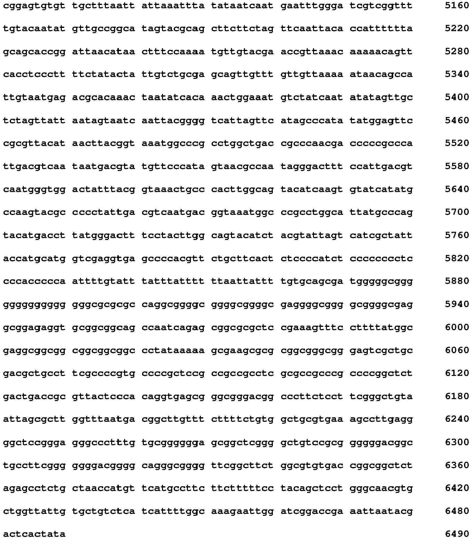
Claims (15)
- Verfahren zur Diagnose von AIG umfassend den Schritt Nachweisen, ob in einer Probe, die Antikörper enthält, Autoantikörper gegen die beta-Untereinheit der gastritischen H+/K+-ATPase vorhanden oder nicht vorhanden sind, wobei ein diagnostisch nützlicher Träger mit einer Festphase eingesetzt wird, an der ein Mittel zum spezifischen Einfangen des Autoantikörpers immobilisiert ist.
- Verfahren nach Anspruch 1, wobei der Träger aus der Gruppe ausgewählt ist, die eine Mikrotiterplatte, einen Bead, einen Blot; bevorzugt Western Blot, Lineblot oder Dot Blot; einen Immunfluoreszenztestträger mit fixierten Zellen, bevorzugt für eine mikroskopische Analyse, eine Lateral Flow-Vorrichtung, ein zellulosebasiertes Polymer, einen Biochip, einen Mikroarray, und ein Chromatographiesäulenmaterial umfasst.
- Verfahren nach Anspruch 2, wobei es sich bei dem diagnostisch nützlichen Träger um eine Mikrotiterplatte für ELISA handelt.
- Verfahren nach einem der Ansprüche 1 bis 3, wobei die Probe aus der Gruppe ausgewählt ist, die Serum, Vollblut, Plasma, CSF umfasst.
- Verfahren nach einem der Ansprüche 1 bis 4, weiter umfassend Nachweisen, ob in einer Probe Autoantikörper gegen den Intrinsic Factor vorhanden oder nicht vorhanden sind.
- Verfahren nach einem der Ansprüche 1 bis 5, wobei der Autoantikörper ein Antikörper der Klasse IgG ist.
- Diagnostisch nützlicher Träger mit einer Festphase, an der ein Mittel zum spezifischen Einfangen eines Autoantikörpers gegen die beta-Untereinheit der gastritischen H+/K+-ATPase immobilisiert ist.
- Träger nach Anspruch 7, ausgewählt aus der Gruppe, die eine Mikrotiterplatte, einen Bead, einen Blot; bevorzugt Western Blot, Lineblot oder Dot Blot; einen Immunfluoreszenztestträger mit fixierten Zellen, bevorzugt für eine mikroskopische Analyse, eine Lateral Flow-Vorrichtung, ein zellulosebasiertes Polymer, einen Biochip, einen Mikroarray, und ein Chromatographiesäulenmaterial umfasst.
- Träger nach Anspruch 8, wobei es sich um eine Mikrotiterplatte für ELISA handelt.
- Monoklonaler Antikörper oder isolierter Autoantikörper, der spezifisch gegen die beta-Untereinheit der gastritischen H+/K+-ATPase bindet, besonders bevorzugt in aufgereinigter Form.
- Verfahren zum Kalibrieren oder zur Kontrolle der Qualität eines Medizinproduktes, umfassend den Schritt Kontaktieren des Trägers nach einem der Ansprüche 7 bis 9 mit einer Flüssigkeit umfassend einen Antikörper gemäß Anspruch 10, bevorzugt in bekannter Konzentration, gefolgt von dem Schritt Nachweisen, ob der Antikörper in der Flüssigkeit vorhanden oder nicht vorhanden ist, besonders bevorzugt unter wenigstens semi-quantitativer Bestimmung des Antikörpers in der Flüssigkeit.
- Kit umfassend den diagnostisch nützlichen Träger gemäß einem der Ansprüche 7 bis 9, wobei der Kit ein oder mehr als ein Reagenz, bevorzugt alle Reagenzien, aus der Gruppe enthält, die eine Waschlösung, eine Kalibratorlösung, einen Antikörper gegen die beta-Untereinheit der gastritischen H+/K+-ATPase, und ein Mittel zum Detektieren des Autoantikörpers, bevorzugt einen sekundären Antikörper, noch bevorzugter einen sekundären Antikörper gegen IgG-Antikörper oder die beta-Untereinheit der gastritischen H+/K+-ATPase oder eine Variante davon, bevorzugt mit nachweisbarer Markierung, umfasst.
- Kit nach Anspruch 12, wobei der sekundäre Antikörper eine nachweisbare Markierung aufweist, bevorzugt aus der Gruppe umfassend eine chemilumineszenzfähige, radioaktive oder ein nachweisbares Isotop umfassende Markierung, eine enzymatisch aktive Markierung, eine fluoreszenzfähige Markierung, eine durch NMR-Spektroskopie nachweisbare Verbindung, eine Spin Label-Markierung, bevorzugt eine chemilumineszenzfähige Markierung, am bevorzugtesten eine chemilumineszenzfähige Markierung, die bei Inkubation in einer geeigneten Chemilumineszenz-Triggerlösung ein Chemilumineszenz-Signal erzeugt.
- Verwendung eines Mittels zum spezifischen Einfangen eines Autoantikörpers gegen die beta-Untereinheit der gastritischen H+/K+-ATPase oder des Trägers gemäß einem der Ansprüche 7 bis 9 oder des Antikörpers nach Anspruch 10 oder eines Polypeptides umfassend die beta-Untereinheit der gastritischen H+/K+-ATPase, bevorzugt der extrazellulären Domäne der beta-Untereinheit der gastritischen H+/K+-ATPase oder einer Variante davon oder einer dafür kodierenden Nukleinsäure zur hochspezifischen Diagnose von AIG.
- Verwendung eines Mittels zum spezifischen Einfangen eines Autoantikörpers gegen die beta-Untereinheit der gastritischen H+/K+-ATPase oder eines Trägers umfassend das Mittel oder der beta-Untereinheit der gastritischen H+/K+-ATPase, bevorzugt der extrazellulären Domäne der beta-Untereinheit der gastritischen H+/K+-ATPase oder einer Variante davon oder einer dafür kodierenden Nukleinsäure zur Herstellung eines Kits für die hochspezifische Diagnose von AIG.
Applications Claiming Priority (2)
| Application Number | Priority Date | Filing Date | Title |
|---|---|---|---|
| EP18213659 | 2018-12-18 | ||
| EP19171829 | 2019-04-30 |
Publications (2)
| Publication Number | Publication Date |
|---|---|
| EP3671212A2 true EP3671212A2 (de) | 2020-06-24 |
| EP3671212A3 EP3671212A3 (de) | 2020-09-09 |
Family
ID=68848210
Family Applications (1)
| Application Number | Title | Priority Date | Filing Date |
|---|---|---|---|
| EP19217068.6A Pending EP3671212A3 (de) | 2018-12-18 | 2019-12-17 | Verfahren zur diagnose von autoimmungastritis |
Country Status (3)
| Country | Link |
|---|---|
| US (1) | US20200191785A1 (de) |
| EP (1) | EP3671212A3 (de) |
| CN (1) | CN111337676A (de) |
Families Citing this family (1)
| Publication number | Priority date | Publication date | Assignee | Title |
|---|---|---|---|---|
| WO2025170033A1 (ja) * | 2024-02-09 | 2025-08-14 | 富士フイルム株式会社 | 抗胃壁細胞抗体測定用試薬、抗胃壁細胞抗体測定用キット、および試料中の抗胃壁細胞抗体を測定する方法 |
Citations (1)
| Publication number | Priority date | Publication date | Assignee | Title |
|---|---|---|---|---|
| WO2011150086A2 (en) | 2010-05-25 | 2011-12-01 | The Regents Of The University Of Colorado, A Body Corporate | Diagnosis and treatment of autoimmune disease |
Family Cites Families (4)
| Publication number | Priority date | Publication date | Assignee | Title |
|---|---|---|---|---|
| AU2002256168B2 (en) * | 2001-04-10 | 2007-09-20 | The Board Of Trustees Of The Leland Stanford Junior University | Therapeutic and diagnostic uses of antibody specificity profiles |
| WO2007030571A2 (en) * | 2005-09-06 | 2007-03-15 | Molecular Image Inc. | Identification of targets and development of reagents for testing and molecular imaging of human disease |
| US8628932B2 (en) * | 2007-07-03 | 2014-01-14 | One Lambda | Methods of detecting antibodies specific for denatured HLA antigens |
| CN108650889B (zh) * | 2016-02-22 | 2022-06-10 | 欧蒙医学实验诊断股份公司 | 用于诊断病毒感染的免疫测定法 |
-
2019
- 2019-11-18 US US16/686,923 patent/US20200191785A1/en active Pending
- 2019-12-17 EP EP19217068.6A patent/EP3671212A3/de active Pending
- 2019-12-17 CN CN201911297388.5A patent/CN111337676A/zh active Pending
Patent Citations (1)
| Publication number | Priority date | Publication date | Assignee | Title |
|---|---|---|---|---|
| WO2011150086A2 (en) | 2010-05-25 | 2011-12-01 | The Regents Of The University Of Colorado, A Body Corporate | Diagnosis and treatment of autoimmune disease |
Non-Patent Citations (20)
| Title |
|---|
| BANASZAK L. J.: "Foundations of Structural Biology", 2008, OXFORD UNIVERSITY PRESS |
| BURBELO, P.D.LEBOVITZ, E.E.BREN, K.E.BAYAT, A.PAVIOL, S.WENZLAU, J.M.BARRIGA, K.J.REWERS, M.HARLAN, D.M.LADAROLA, M.J.: "Extrapancreatic Autoantibody Profiles in Type I Diabetes", PLOS ONE, vol. 7, no. 9, 2012, pages e45216 |
| DA SILVA, H. D.GLEESON, P. A.TOH, B. H.VAN DRIEL, I. R.CARBONE, F. R.: "Identification of a gastritogenic epitope of the H/K ATPase beta-subunit", IMMUNOLOGY, vol. 96, 1999, pages 145 - 151, XP055553073, DOI: 10.1046/j.1365-2567.1999.00669.x |
| DÄHNRICH ET AL.: "Development of a standardized ELISA for the determination of autoantibodies against human M-type phospholipase A2 receptor in primary membranous nephropathy", CLINICA CHIMICA ACTA, vol. 421, 2013, pages 213 - 218, XP055455580, DOI: 10.1016/j.cca.2013.03.015 |
| KIM, D.HERR, A. E.: "Protein immobilization techniques for microfluidic assays", BIOMICROFLUIDICS, vol. 7, no. 4, 2013, pages 041501, XP002753670, DOI: 10.1063/1.4816934 |
| KIM, D.HERR, A. E.: "Protein immobilizsation techniques for microfluidic assays", BIOMICROFLUIDICS, vol. 7, no. 4, 2013, pages 041501, XP002753670, DOI: 10.1063/1.4816934 |
| KONTANI, K.TAGUCHI, D.TAKAHASHI, T.: "Involvement of the H+/+-ATPase alpha subunit as a major antigenic protein in autoimmune gastritis induced by neonatal thymectomy in mice", CLIN. EXP. IMMUNOL., vol. 89, 1992, pages 63 - 67, XP009511063, DOI: 10.1111/j.1365-2249.1992.tb06878.x |
| KRICKA, L. J.: "Clinical applications of chemiluminescence", ANALYTICA CHIMICA ACTA, vol. 500, no. 1, 2003, pages 279 - 286, XP002503602, DOI: 10.1016/S0003-2670(03)00809-2 |
| LAHNER, E.BRIGATTI, C.MARZINOTTO, I.CARABOTTI, M.SCALESE, G.DAVIDSON, H.W.WENZLAU, J.M.BOSI, E.PIEMONTI, L.ANNIBALE, B.: "Luminescent Immunoprecipitation System (LIPS) for Detection of Autoantibodies Against ATP4A and ATP4B Subunits of Gastric Proton Pump H+/K+-ATPase in Atrophic Body Gastritis Patients", CLINICAL AND TRANSLATIONAL GASTROENTEROLOGY, vol. 8, no. 1, 2017, pages e215 |
| LARKIN, M. A.BLACKSHIELDS, G.BROWN, N. P.CHENNA, R.MCGETTIGAN, P. A.MCWILLIAM, H.VALENTIN, F.WALLACE, I. M.WILM, A.LOPEZ, R.: "Clustal Wand Clustal X version 2.0", BIOINFORMATICS, vol. 23, 2007, pages 2947 - 2948 |
| MA JING-YIE ET AL: "cDNA CLONING OF THE fl-SUBUNIT OF THE HUMAN GASTRIC H,K-ATPase 1", 15 October 1991 (1991-10-15), XP093257393, Retrieved from the Internet <URL:https://pdf.sciencedirectassets.com/272308/1-s2.0-S0006291X05X12451/1-s2.0-S0006291X05812513/main.pdf?hash=f3616e619dc5aca3e461ab4addbf23b8aac8b73652a8faaa156c707857a4a5be&host=68042c943591013ac2b2430a89b270f6af2c76d8dfd086a07176afe7c76c2c61&pii=S0006291X05812513&tid=spdf-5096313f-d482-43e9-b3e6-168> * |
| MA, J. Y.BORCH, KMARDH, S.: "Human Gastric H,K-Adenosine Triphosphatase beta-Subunit is a Major Autoantigen in Atrophic Corpus Gastritis", SCAND J. GASTROENTEROL, vol. 29, 1994, pages 790 - 794, XP009511062, DOI: 10.3109/00365529409092512 |
| NISHIO, A.HOSONO, M.WATANABE, Y.SAKAI, M.OKUMA, MMASUDA, T, GASTROENTEROLOGY, vol. 107, 1994, pages 1408 - 1414 |
| PELAY GIMENO, M.GLAS, A.KOCH, O.GROSSMANN, T.N.: "Structure-based design of inhibitors of protein-protein interactions: Mimicking peptide binding epitopes", ANGEWANDTE CHEMIE INTERNATIONAL EDITION, vol. 54, no. 31, 2015, pages 8896 - 8927 |
| RUSAK, E.CHOBOT, A.KRZYWICKA, A.WENZLAU, J.: "Anti-parietal cell antibodies - diagnostic significance", ADVANCES IN MEDICAL SCIENCES, vol. 61, 2016, pages 175 - 179 |
| SCHARF, M.MISKE, R.HEIDENREICH, F.GIESS, R.LANDWEHR, P.BLÖCKER, I.BEGEMANN, N.DENNO, Y.TIEDE, S.DÄHNRICH, C.: "Neuronal Na+/K+ ATPase is an autoantibody target in paraneoplastic neurologic syndrome", NEUROLOGY, vol. 84, 2015, pages 1 - 7 |
| SONG Y H: "Localization of a Pernicious Anaemia Autoantibody Epitope on the a'-Subunit of Human H,K-Adenosine Triphosphatase", SCANDINAVIAN JOURNAL OF GASTROENTEROLOGY, 8 July 2009 (2009-07-08), XP093257392 * |
| WEEKS, I.BEHESHTI, I.MCCAPRA, F.CAMPBELL, A. K.WOODHEAD, J. S.: "Acridinium esters as high specific activity labels in immunoassay", CLIN CHEM, vol. 29, 1983, pages 1474 - 1479, XP002097142 |
| WENZLAU, J.M.GARDNER, T.J.FRISCH, L.M.DAVIDSON, H.W.HUTTON, J.C.: "Development of a novel autoantibody assay for autoimmune gastritis in T1 D individuals", DIABETES METAB RES REV., vol. 27, no. 8, 2011, pages 887 - 90 |
| ZANE, H. D.: "Immunology - Theoretical & Practical Concepts in Laboratory Medicine", 2001, W. B. SAUNDERS COMPANY |
Also Published As
| Publication number | Publication date |
|---|---|
| EP3671212A3 (de) | 2020-09-09 |
| US20200191785A1 (en) | 2020-06-18 |
| CN111337676A (zh) | 2020-06-26 |
Similar Documents
| Publication | Publication Date | Title |
|---|---|---|
| DE202021100842U1 (de) | Methoden und Reagenzien zur Diagnose einer SARS-CoV-2-Infektion | |
| EP3505935B1 (de) | Verfahren zur diagnose einer bornavirus-infektion | |
| DE202020105116U1 (de) | Reagenzien und Verwendungen zur Diagnose einer SARS-CoV-2-Infektion | |
| EP0544869B1 (de) | Immunologisches bestimmungsverfahren zur bestimmung von antikörpern in biologischen flüssigkeiten sowie kit zur durchführung des verfahrens | |
| DE102018004759A1 (de) | Diagnose einer Neuroautoimmunkrankheit | |
| EP3644060A1 (de) | Diagnose blasenbildender autoimmunkrankheiten | |
| WO2012076000A2 (de) | Verfahren zum nachweis von anti-nmda-rezeptor autoantikörpern zum einsatz in diagnoseverfahren | |
| EP2884278A2 (de) | Markersequenzen für rheumatoide Arthritis und deren Verwendung | |
| EP2318546A2 (de) | Markersequenzen für prostataentzündungserkrankungen, prostatakarzinom und deren verwendung | |
| EP4040153A1 (de) | Verfahren zum nachweis von tau-protein unter verwendung einer blutprobe als probe | |
| EP4080215A2 (de) | Verfahren und reagenzien zum nachweis eines autoantikörpers | |
| EP3671212A2 (de) | Verfahren zur diagnose von autoimmungastritis | |
| WO2009080017A2 (de) | Markersequenzen für neurodegenerative erkrankungen und deren verwendung | |
| DE202022104518U1 (de) | Autoantikörper und Mittel zum Nachweis eines Autoantikörpers | |
| DE102014226663A1 (de) | Brückenassays zum Nachweis von Antikörpern gegen Mitglieder der kardialen Rezeptorfamilie | |
| EP2839290A1 (de) | Verfahren zur präsymptomatischen diagnostik von zöliakie und glutensensitivität | |
| EP3070473A2 (de) | Markersequenzen für multiple sklerose und deren verwendung | |
| DE102008053503A1 (de) | Immunologischer Test zum Nachweis von Autoantikörpern gegen testikuläre Antigene | |
| DE202020000743U1 (de) | Träger und Kit zur Diagnose einer neurologischen Autoimmunkrankheit | |
| EP4134674A1 (de) | Verfahren und reagenzien zum detektieren eines antikörpers gegen ein ses ipr-10-allergen | |
| DE102017204268A1 (de) | Biomarker für endokrine, autoimmun-bezogene Funktionsstörung | |
| DE102018000815A1 (de) | Verfahren zum Nachweis von Biomarkern im Stuhl zur Erkennung von Erkrankungen des Intestinaltraktes | |
| DE102010042359A1 (de) | Markersequenzen für Multiple Sklerose und deren Verwendung | |
| LU100002B1 (en) | New testing device and method for detecting Ankylosing Spondylitis | |
| EP2644704A1 (de) | Markersequenzen für Rheumatoide Arthritis |
Legal Events
| Date | Code | Title | Description |
|---|---|---|---|
| PUAI | Public reference made under article 153(3) epc to a published international application that has entered the european phase |
Free format text: ORIGINAL CODE: 0009012 |
|
| STAA | Information on the status of an ep patent application or granted ep patent |
Free format text: STATUS: THE APPLICATION HAS BEEN PUBLISHED |
|
| AK | Designated contracting states |
Kind code of ref document: A2 Designated state(s): AL AT BE BG CH CY CZ DE DK EE ES FI FR GB GR HR HU IE IS IT LI LT LU LV MC MK MT NL NO PL PT RO RS SE SI SK SM TR |
|
| AX | Request for extension of the european patent |
Extension state: BA ME |
|
| PUAL | Search report despatched |
Free format text: ORIGINAL CODE: 0009013 |
|
| AK | Designated contracting states |
Kind code of ref document: A3 Designated state(s): AL AT BE BG CH CY CZ DE DK EE ES FI FR GB GR HR HU IE IS IT LI LT LU LV MC MK MT NL NO PL PT RO RS SE SI SK SM TR |
|
| AX | Request for extension of the european patent |
Extension state: BA ME |
|
| RIC1 | Information provided on ipc code assigned before grant |
Ipc: G01N 33/564 20060101AFI20200806BHEP Ipc: G01N 33/543 20060101ALI20200806BHEP |
|
| STAA | Information on the status of an ep patent application or granted ep patent |
Free format text: STATUS: REQUEST FOR EXAMINATION WAS MADE |
|
| 17P | Request for examination filed |
Effective date: 20210309 |
|
| RBV | Designated contracting states (corrected) |
Designated state(s): AL AT BE BG CH CY CZ DE DK EE ES FI FR GB GR HR HU IE IS IT LI LT LU LV MC MK MT NL NO PL PT RO RS SE SI SK SM TR |
|
| STAA | Information on the status of an ep patent application or granted ep patent |
Free format text: STATUS: EXAMINATION IS IN PROGRESS |
|
| 17Q | First examination report despatched |
Effective date: 20230313 |
|
| APAF | Appeal reference modified |
Free format text: ORIGINAL CODE: EPIDOSCREFNE |
|
| APBN | Date of receipt of notice of appeal recorded |
Free format text: ORIGINAL CODE: EPIDOSNNOA2E |
|
| APAF | Appeal reference modified |
Free format text: ORIGINAL CODE: EPIDOSCREFNE |
|
| APBR | Date of receipt of statement of grounds of appeal recorded |
Free format text: ORIGINAL CODE: EPIDOSNNOA3E |
|
| APAV | Appeal reference deleted |
Free format text: ORIGINAL CODE: EPIDOSDREFNE |