EP3663320A1 - Nucleic acid sequence encoding a cs1-specific chimeric antigen receptor (car) polypeptide - Google Patents
Nucleic acid sequence encoding a cs1-specific chimeric antigen receptor (car) polypeptide Download PDFInfo
- Publication number
- EP3663320A1 EP3663320A1 EP20150396.8A EP20150396A EP3663320A1 EP 3663320 A1 EP3663320 A1 EP 3663320A1 EP 20150396 A EP20150396 A EP 20150396A EP 3663320 A1 EP3663320 A1 EP 3663320A1
- Authority
- EP
- European Patent Office
- Prior art keywords
- cells
- car
- nucleic acid
- cell
- acid sequence
- Prior art date
- Legal status (The legal status is an assumption and is not a legal conclusion. Google has not performed a legal analysis and makes no representation as to the accuracy of the status listed.)
- Withdrawn
Links
Images













Classifications
-
- A—HUMAN NECESSITIES
- A61—MEDICAL OR VETERINARY SCIENCE; HYGIENE
- A61P—SPECIFIC THERAPEUTIC ACTIVITY OF CHEMICAL COMPOUNDS OR MEDICINAL PREPARATIONS
- A61P35/00—Antineoplastic agents
-
- A—HUMAN NECESSITIES
- A61—MEDICAL OR VETERINARY SCIENCE; HYGIENE
- A61P—SPECIFIC THERAPEUTIC ACTIVITY OF CHEMICAL COMPOUNDS OR MEDICINAL PREPARATIONS
- A61P37/00—Drugs for immunological or allergic disorders
- A61P37/02—Immunomodulators
- A61P37/04—Immunostimulants
-
- A—HUMAN NECESSITIES
- A61—MEDICAL OR VETERINARY SCIENCE; HYGIENE
- A61P—SPECIFIC THERAPEUTIC ACTIVITY OF CHEMICAL COMPOUNDS OR MEDICINAL PREPARATIONS
- A61P39/00—General protective or antinoxious agents
-
- C—CHEMISTRY; METALLURGY
- C07—ORGANIC CHEMISTRY
- C07K—PEPTIDES
- C07K14/00—Peptides having more than 20 amino acids; Gastrins; Somatostatins; Melanotropins; Derivatives thereof
- C07K14/435—Peptides having more than 20 amino acids; Gastrins; Somatostatins; Melanotropins; Derivatives thereof from animals; from humans
- C07K14/705—Receptors; Cell surface antigens; Cell surface determinants
- C07K14/70503—Immunoglobulin superfamily
- C07K14/7051—T-cell receptor (TcR)-CD3 complex
-
- C—CHEMISTRY; METALLURGY
- C07—ORGANIC CHEMISTRY
- C07K—PEPTIDES
- C07K14/00—Peptides having more than 20 amino acids; Gastrins; Somatostatins; Melanotropins; Derivatives thereof
- C07K14/435—Peptides having more than 20 amino acids; Gastrins; Somatostatins; Melanotropins; Derivatives thereof from animals; from humans
- C07K14/705—Receptors; Cell surface antigens; Cell surface determinants
- C07K14/70503—Immunoglobulin superfamily
- C07K14/70521—CD28, CD152
-
- C—CHEMISTRY; METALLURGY
- C07—ORGANIC CHEMISTRY
- C07K—PEPTIDES
- C07K16/00—Immunoglobulins [IG], e.g. monoclonal or polyclonal antibodies
- C07K16/18—Immunoglobulins [IG], e.g. monoclonal or polyclonal antibodies against material from animals or humans
- C07K16/28—Immunoglobulins [IG], e.g. monoclonal or polyclonal antibodies against material from animals or humans against receptors, cell surface antigens or cell surface determinants
- C07K16/2803—Immunoglobulins [IG], e.g. monoclonal or polyclonal antibodies against material from animals or humans against receptors, cell surface antigens or cell surface determinants against the immunoglobulin superfamily
-
- C—CHEMISTRY; METALLURGY
- C07—ORGANIC CHEMISTRY
- C07K—PEPTIDES
- C07K16/00—Immunoglobulins [IG], e.g. monoclonal or polyclonal antibodies
- C07K16/18—Immunoglobulins [IG], e.g. monoclonal or polyclonal antibodies against material from animals or humans
- C07K16/28—Immunoglobulins [IG], e.g. monoclonal or polyclonal antibodies against material from animals or humans against receptors, cell surface antigens or cell surface determinants
- C07K16/30—Immunoglobulins [IG], e.g. monoclonal or polyclonal antibodies against material from animals or humans against receptors, cell surface antigens or cell surface determinants from tumour cells
- C07K16/3061—Blood cells
-
- A—HUMAN NECESSITIES
- A61—MEDICAL OR VETERINARY SCIENCE; HYGIENE
- A61K—PREPARATIONS FOR MEDICAL, DENTAL OR TOILETRY PURPOSES
- A61K39/00—Medicinal preparations containing antigens or antibodies
- A61K2039/505—Medicinal preparations containing antigens or antibodies comprising antibodies
-
- C—CHEMISTRY; METALLURGY
- C07—ORGANIC CHEMISTRY
- C07K—PEPTIDES
- C07K2317/00—Immunoglobulins specific features
- C07K2317/60—Immunoglobulins specific features characterized by non-natural combinations of immunoglobulin fragments
- C07K2317/62—Immunoglobulins specific features characterized by non-natural combinations of immunoglobulin fragments comprising only variable region components
- C07K2317/622—Single chain antibody (scFv)
-
- C—CHEMISTRY; METALLURGY
- C07—ORGANIC CHEMISTRY
- C07K—PEPTIDES
- C07K2317/00—Immunoglobulins specific features
- C07K2317/70—Immunoglobulins specific features characterized by effect upon binding to a cell or to an antigen
- C07K2317/73—Inducing cell death, e.g. apoptosis, necrosis or inhibition of cell proliferation
-
- C—CHEMISTRY; METALLURGY
- C07—ORGANIC CHEMISTRY
- C07K—PEPTIDES
- C07K2319/00—Fusion polypeptide
-
- C—CHEMISTRY; METALLURGY
- C07—ORGANIC CHEMISTRY
- C07K—PEPTIDES
- C07K2319/00—Fusion polypeptide
- C07K2319/01—Fusion polypeptide containing a localisation/targetting motif
- C07K2319/02—Fusion polypeptide containing a localisation/targetting motif containing a signal sequence
-
- C—CHEMISTRY; METALLURGY
- C07—ORGANIC CHEMISTRY
- C07K—PEPTIDES
- C07K2319/00—Fusion polypeptide
- C07K2319/01—Fusion polypeptide containing a localisation/targetting motif
- C07K2319/03—Fusion polypeptide containing a localisation/targetting motif containing a transmembrane segment
-
- C—CHEMISTRY; METALLURGY
- C07—ORGANIC CHEMISTRY
- C07K—PEPTIDES
- C07K2319/00—Fusion polypeptide
- C07K2319/33—Fusion polypeptide fusions for targeting to specific cell types, e.g. tissue specific targeting, targeting of a bacterial subspecies
Definitions
- MM Multiple myeloma
- PCs plasma cells
- CAR chimeric antigen receptor
- MM myeloma
- the cell surface glycoprotein CS1 is highly and ubiquitously expressed on the surface of myeloma cells while being expressed at very low levels in the majority of immune effector cells. Therefore, the disclosed CAR polypeptides contain in an ectodomain an anti-CS1 binding agent that can bind CS1-expressing MM cells.
- the disclosed polypeptides can also contain a transmembrane domain and an endodomain capable of activating an immune effector cell.
- the endodomain can contain an intracellular signaling domain and optionally a co-stimulatory signaling region.
- the anti-CS1 binding agent is in some embodiments an antibody fragment or an antigen-binding fragment that specifically binds CS1.
- the antigen binding domain can be a Fab or a single-chain variable fragment (scFv) of an antibody that specifically binds CS1.
- the anti-CS1 binding agent is in some embodiments an aptamer that specifically binds CS1.
- the anti-CS1 binding agent can be a peptide aptamer selected from a random sequence pool based on its ability to bind CS1.
- the anti-CS1 binding agent can also be a natural ligand of CS1, or a variant and/or fragment thereof capable of binding CS1.
- the intracellular signaling domain is a CD3 zeta (CD3 ⁇ ) signaling domain
- the costimulatory signaling region comprises the cytoplasmic domain of CD28, 4-1BB, or a combination thereof.
- the costimulatory signaling region contains 1, 2, 3, or 4 cytoplasmic domains of one or more intracellular signaling and/or costimulatory molecules.
- the cell can be an immune effector cell selected from the group consisting of a T cell, a Natural Killer (NK) cell, a cytotoxic T lymphocyte (CTL), and a regulatory T cell.
- the cell exhibits an anti-tumor immunity when the antigen binding domain of the CAR binds to CS1.
- MM multiple myeloma
- CAR chimeric antigen receptors
- TAA tumor-associated antigens
- MM Multiple myeloma
- immune effector cells such as T cells or Natural Killer (NK) cells, that are engineered to express these CARs. Therefore, also disclosed are methods for providing an anti-tumor immunity in a subject with MM that involves adoptive transfer of the disclosed immune effector cells engineered to express the disclosed CS1-specific CARs.
- CARs generally incorporate an antigen recognition domain from the single-chain variable fragments (scFv) of a monoclonal antibody (mAb) with transmembrane signaling motifs involved in lymphocyte activation ( Sadelain M, et al. Nat Rev Cancer 2003 3:35-45 ).
- the cell surface glycoprotein CS1 is highly and ubiquitously expressed on the surface of myeloma cells ( Hsi ED, et al. Clin Cancer Res 2008 14:2775-84 ).
- CS1 is expressed at very low levels in the majority of immune effector cells, including natural killer (NK) cells, some subsets of T cells, and normal B cells, and is almost undetectable on myeloid cells ( Hsi ED, et al.
- CS1 is negligibly expressed in human hematopoietic stem cells ( Hsi ED, et al. Clin Cancer Res 2008 14:2775-84 ), which can be used for stem cell transplantation to treat hematologic malignancies, including MM.
- Hsi ED et al. Clin Cancer Res 2008 14:2775-84
- the functions of CS1 in MM remain incompletely understood, and it has been documented that CS1 may play a role in myeloma cell adhesion, clonogenic growth, and tumorigenicity ( Benson DM Jr, et al. J Clin Oncol 2012 30:2013-5 ; Tai YT, et al. Blood 2009 113:4309-18 ).
- CAR CS1-specific chimeric antigen receptor
- the disclosed CAR is generally made up of three domains: an ectodomain, a transmembrane domain, and an endodomain.
- the ectodomain comprises the CS1-binding region and is responsible for antigen recognition. It also generally contains a signal peptide (SP) so that the CAR can be glycosylated and anchored in the cell membrane of the immune effector cell.
- SP signal peptide
- the transmembrane domain (TD) is as its name suggests, connects the ectodomain to the endodomain and resides within the cell membrane when expressed by a cell.
- the endodomain is the business end of the CAR that transmits an activation signal to the immune effector cell after antigen recognition.
- the endodomain can contain an intracellular signaling domain (ISD) and optionally a co-stimulatory signaling region (CSR).
- ISD intracellular signaling domain
- CSR co-stimulatory signaling region
- the disclosed CAR is defined by the formula: SP-CS1-HG-TM-CSR-ISD; or SP-CS1-HG-TM-ISD-CSR
- the antigen recognition domain of the disclosed CAR is usually an scFv.
- An antigen recognition domain from native T-cell receptor (TCR) alpha and beta single chains have been described, as have simple ectodomains (e.g. CD4 ectodomain to recognize HIV infected cells) and more exotic recognition components such as a linked cytokine (which leads to recognition of cells bearing the cytokine receptor).
- TCR T-cell receptor
- the endodomain is the business end of the CAR that after antigen recognition (i.e., CS1) transmits a signal to the immune effector cell, activating at least one of the normal effector functions of the immune effector cell.
- Effector function of a T cell may be cytolytic activity or helper activity including the secretion of cytokines. Therefore, the endodomain may comprise the "intracellular signaling domain" of a T cell receptor (TCR) and optional co-receptors. While usually the entire intracellular signaling domain can be employed, in many cases it is not necessary to use the entire chain. To the extent that a truncated portion of the intracellular signaling domain is used, such truncated portion may be used in place of the intact chain as long as it transduces the effector function signal.
- TCR T cell receptor
- Cytoplasmic signaling sequences that regulate primary activation of the TCR complex that act in a stimulatory manner may contain signaling motifs which are known as immunoreceptor tyrosine-based activation motifs (ITAMs).
- ITAMs immunoreceptor tyrosine-based activation motifs
- Examples of ITAM containing cytoplasmic signaling sequences include those derived from TCR zeta, FcR gamma, FcR beta, CD3 gamma, CD3 delta, CD3 epsilon, CD5, CD22, CD79a, CD79b, and CD66d.
- the intracellular signaling domain is derived from CD3 zeta (CD3 ⁇ ).
- T-cell surface glycoprotein CD3 zeta (CD3 ⁇ ) chain also known as T-cell receptor T3 zeta chain or CD247 (Cluster of Differentiation 247), is a protein that in humans is encoded by the CD247 gene.
- First-generation CARs typically had the intracellular domain from the CD3 ⁇ chain, which is the primary transmitter of signals from endogenous TCRs.
- Second-generation CARs add intracellular signaling domains from various costimulatory protein receptors (e.g., CD28, 41BB, ICOS) to the endodomain of the CAR to provide additional signals to the T cell.
- costimulatory protein receptors e.g., CD28, 41BB, ICOS
- Preclinical studies have indicated that the second generation of CAR designs improves the antitumor activity of T cells.
- third-generation CARs combine multiple signaling domains to further augment potency.
- T cells grafted with these CARs have demonstrated improved expansion, activation, persistence, and tumor-eradicating efficiency independent of costimulatory receptor/ligand interaction ( Imai C, et al. Leukemia 2004 18:676-84 ; Maher J, et al. Nat Biotechnol 2002 20:70-5 ).
- the endodomain of the CAR can be designed to comprise the CD3 ⁇ signaling domain by itself or combined with any other desired cytoplasmic domain(s) useful in the context of the CAR of the invention.
- the cytoplasmic domain of the CAR can comprise a CD3 zeta chain portion and a costimulatory signaling region.
- the costimulatory signaling region refers to a portion of the CAR comprising the intracellular domain of a costimulatory molecule.
- a costimulatory molecule is a cell surface molecule other than an antigen receptor or their ligands that is required for an efficient response of lymphocytes to an antigen.
- Examples of such molecules include CD27, CD28, 4-1BB (CD137), OX40, CD30, CD40, ICOS, lymphocyte function-associated antigen-1 (LFA-1), CD2, CD7, LIGHT, NKG2C, B7-H3, and a ligand that specifically binds with CD83, and the like.
- CD28 CD28
- 4-1BB CD137
- OX40 CD30
- CD40 CD40
- ICOS lymphocyte function-associated antigen-1
- CD2 CD7
- LIGHT lymphocyte function-associated antigen-1
- NKG2C NKG2C
- B7-H3 a ligand that specifically binds with CD83
- the CAR comprises a hinge sequence.
- a hinge sequence is a short sequence of amino acids that facilitates antibody flexibility (see, e.g., Woof et al., Nat. Rev. Immunol., 4(2): 89-99 (2004 )).
- the hinge sequence may be positioned between the antigen recognition moiety (e.g., an anti- CS1 scFv) and the transmembrane domain.
- the hinge sequence can be any suitable sequence derived or obtained from any suitable molecule. In some embodiments, for example, the hinge sequence is derived from a CD8a molecule or a CD28 molecule.
- the transmembrane domain may be derived either from a natural or from a synthetic source. Where the source is natural, the domain may be derived from any membrane-bound or transmembrane protein.
- the transmembrane region may be derived from (i.e. comprise at least the transmembrane region(s) of) the alpha, beta or zeta chain of the T-cell receptor, CD28, CD3 epsilon, CD45, CD4, CD5, CD8, CD9, CD16, CD22, CD33, CD37, CD64, CD80, CD86, CD134, CD137, or CD154.
- the transmembrane domain may be synthetic, in which case it will comprise predominantly hydrophobic residues such as leucine and valine.
- a triplet of phenylalanine, tryptophan and valine will be found at each end of a synthetic transmembrane domain.
- a short oligo- or polypeptide linker such as between 2 and 10 amino acids in length, may form the linkage between the transmembrane domain and the endoplasmic domain of the CAR.
- the bivalent linker can be any molecule suitable to link a compound or nucleic acid to a polynucleotide sequence. Methods and compositions for conjugating biomolecules, such as polynucleotides, are disclosed in G.T. Hermanon, Bioconjugate Techniques (2nd ed.), Academic Press (2008 ), which is incorporated by reference in its entirety for the teaching of these techniques.
- the bivalent linker comprises one or more amino acids. However, it can also comprise a peptide bond directly linking the disclosed domains.
- the disclosed CS1-specific CAR comprises one or more of the SP, CS1, HG, TM, CSR, ISD, and/or linker components set forth in Table 1, or variants thereof having at least 65%, 70%, 71%, 72%, 73%, 74%, 75%, 76%, 77%, 78%, 79%, 80%, 81%, 82%, 83%, 84%, 85%, 86%, 87%, 88%, 89%, 90%, 91%, 92%, 93%, 94%, 95%, 96%, 97%, 98%, 99% sequence identity to the sequences set forth in Table 1.
- the disclosed CS1-specific CAR comprises the amino acid sequence SEQ ID NO: 1 (shown below), or a variant thereof having at least 65%, 70%, 71%, 72%, 73%, 74%, 75%, 76%, 77%, 78%, 79%, 80%, 81%, 82%, 83%, 84%, 85%, 86%, 87%, 88%, 89%, 90%, 91%, 92%, 93%, 94%, 95%, 96%, 97%, 98%, 99% sequence identity to SEQ ID NO:28.
- CS1-CD28-CD3Z construct :
- polynucleotides and polynucleotide vectors encoding the disclosed CS1-specific CARs that allow expression of the CS1-specific CARs in the disclosed immune effector cells are also disclosed.
- the disclosed CS1-specific CAR are encoded by the nucleic acid sequence SEQ ID NO:28 (shown below), or a variant thereof having at least 65%, 70%, 71%, 72%, 73%, 74%, 75%, 76%, 77%, 78%, 79%, 80%, 81%, 82%, 83%, 84%, 85%, 86%, 87%, 88%, 89%, 90%, 91%, 92%, 93%, 94%, 95%, 96%, 97%, 98%, 99% sequence identity to SEQ ID NO:29.
- Nucleic acid sequences encoding the disclosed CARs, and regions thereof can be obtained using recombinant methods known in the art, such as, for example by screening libraries from cells expressing the gene, by deriving the gene from a vector known to include the same, or by isolating directly from cells and tissues containing the same, using standard techniques.
- the gene of interest can be produced synthetically, rather than cloned.
- nucleic acids encoding CARs is typically achieved by operably linking a nucleic acid encoding the CAR polypeptide to a promoter, and incorporating the construct into an expression vector.
- Typical cloning vectors contain transcription and translation terminators, initiation sequences, and promoters useful for regulation of the expression of the desired nucleic acid sequence.
- the disclosed nucleic acid can be cloned into a number of types of vectors.
- the nucleic acid can be cloned into a vector including, but not limited to a plasmid, a phagemid, a phage derivative, an animal virus, and a cosmid.
- Vectors of particular interest include expression vectors, replication vectors, probe generation vectors, and sequencing vectors.
- the expression vector may be provided to a cell in the form of a viral vector.
- Viral vector technology is well known in the art and is described, for example, in Sambrook et al. (2001, Molecular Cloning: A Laboratory Manual, Cold Spring Harbor Laboratory, New York ), and in other virology and molecular biology manuals.
- Viruses, which are useful as vectors include, but are not limited to, retroviruses, adenoviruses, adeno-associated viruses, herpes viruses, and lentiviruses.
- a suitable vector contains an origin of replication functional in at least one organism, a promoter sequence, convenient restriction endonuclease sites, and one or more selectable markers.
- the polynucleotide vectors are lentiviral or retroviral vectors.
- retroviruses provide a convenient platform for gene delivery systems.
- a selected gene can be inserted into a vector and packaged in retroviral particles using techniques known in the art.
- the recombinant virus can then be isolated and delivered to cells of the subject either in vivo or ex vivo.
- a suitable promoter is the immediate early cytomegalovirus (CMV) promoter sequence.
- CMV immediate early cytomegalovirus
- This promoter sequence is a strong constitutive promoter sequence capable of driving high levels of expression of any polynucleotide sequence operatively linked thereto.
- Another example of a suitable promoter is Elongation Growth Factor-1 ⁇ (EF-1 ⁇ ).
- constitutive promoter sequences may also be used, including, but not limited to the simian virus 40 (SV40) early promoter, MND (myeloproliferative sarcoma virus) promoter, mouse mammary tumor virus (MMTV), human immunodeficiency virus (HIV) long terminal repeat (LTR) promoter, MoMuLV promoter, an avian leukemia virus promoter, an Epstein-Barr virus immediate early promoter, a Rous sarcoma virus promoter, as well as human gene promoters such as, but not limited to, the actin promoter, the myosin promoter, the hemoglobin promoter, and the creatine kinase promoter.
- the promoter can alternatively be an inducible promoter. Examples of inducible promoters include, but are not limited to a metallothionine promoter, a glucocorticoid promoter, a progesterone promoter, and a tetracycline promoter.
- promoter elements e.g., enhancers
- promoters regulate the frequency of transcriptional initiation.
- these are located in the region 30-110 bp upstream of the start site, although a number of promoters have recently been shown to contain functional elements downstream of the start site as well.
- the spacing between promoter elements frequently is flexible, so that promoter function is preserved when elements are inverted or moved relative to one another.
- the expression vector to be introduced into a cell can also contain either a selectable marker gene or a reporter gene or both to facilitate identification and selection of expressing cells from the population of cells sought to be transfected or infected through viral vectors.
- the selectable marker may be carried on a separate piece of DNA and used in a co-transfection procedure. Both selectable markers and reporter genes may be flanked with appropriate regulatory sequences to enable expression in the host cells. Useful selectable markers include, for example, antibiotic-resistance genes.
- Reporter genes are used for identifying potentially transfected cells and for evaluating the functionality of regulatory sequences.
- a reporter gene is a gene that is not present in or expressed by the recipient organism or tissue and that encodes a polypeptide whose expression is manifested by some easily detectable property, e.g., enzymatic activity. Expression of the reporter gene is assayed at a suitable time after the DNA has been introduced into the recipient cells.
- Suitable reporter genes may include genes encoding luciferase, beta-galactosidase, chloramphenicol acetyl transferase, secreted alkaline phosphatase, or the green fluorescent protein gene. Suitable expression systems are well known and may be prepared using known techniques or obtained commercially.
- the construct with the minimal 5' flanking region showing the highest level of expression of reporter gene is identified as the promoter. Such promoter regions may be linked to a reporter gene and used to evaluate agents for the ability to modulate promoter-driven transcription.
- the vector can be readily introduced into a host cell, e.g., mammalian, bacterial, yeast, or insect cell by any method in the art.
- the expression vector can be transferred into a host cell by physical, chemical, or biological means.
- Physical methods for introducing a polynucleotide into a host cell include calcium phosphate precipitation, lipofection, particle bombardment, microinjection, electroporation, and the like.
- Methods for producing cells comprising vectors and/or exogenous nucleic acids are well-known in the art. See, for example, Sambrook et al. (2001, Molecular Cloning: A Laboratory Manual, Cold Spring Harbor Laboratory, New York ).
- Biological methods for introducing a polynucleotide of interest into a host cell include the use of DNA and RNA vectors.
- Viral vectors, and especially retroviral vectors have become the most widely used method for inserting genes into mammalian, e.g., human cells.
- Chemical means for introducing a polynucleotide into a host cell include colloidal dispersion systems, such as macromolecule complexes, nanocapsules, microspheres, beads, and lipid-based systems including oil-in-water emulsions, micelles, mixed micelles, and liposomes.
- colloidal dispersion systems such as macromolecule complexes, nanocapsules, microspheres, beads, and lipid-based systems including oil-in-water emulsions, micelles, mixed micelles, and liposomes.
- An exemplary colloidal system for use as a delivery vehicle in vitro and in vivo is a liposome (e.g., an artificial membrane vesicle).
- an exemplary delivery vehicle is a liposome.
- the nucleic acid may be associated with a lipid.
- the nucleic acid associated with a lipid may be encapsulated in the aqueous interior of a liposome, interspersed within the lipid bilayer of a liposome, attached to a liposome via a linking molecule that is associated with both the liposome and the oligonucleotide, entrapped in a liposome, complexed with a liposome, dispersed in a solution containing a lipid, mixed with a lipid, combined with a lipid, contained as a suspension in a lipid, contained or complexed with a micelle, or otherwise associated with a lipid.
- Lipid, lipid/DNA or lipid/expression vector associated compositions are not limited to any particular structure in solution. For example, they may be present in a bilayer structure, as micelles, or with a "collapsed" structure. They may also simply be interspersed in a solution, possibly forming aggregates that are not uniform in size or shape.
- Lipids are fatty substances which may be naturally occurring or synthetic lipids.
- lipids include the fatty droplets that naturally occur in the cytoplasm as well as the class of compounds which contain long-chain aliphatic hydrocarbons and their derivatives, such as fatty acids, alcohols, amines, amino alcohols, and aldehydes. Lipids suitable for use can be obtained from commercial sources.
- dimyristyl phosphatidylcholine can be obtained from Sigma, St. Louis, Mo.
- dicetyl phosphate can be obtained from K & K Laboratories (Plainview, N.Y); cholesterol (“Choi”) can be obtained from Calbiochem-Behring; dimyristyl phosphatidylglycerol (“DMPG”) and other lipids may be obtained from Avanti Polar Lipids, Inc, (Birmingham, Ala.).
- immune effector cells that are engineered to express the disclosed CARs. These cells are preferably obtained from the subject to be treated (i.e. are autologous). However, in some embodiments, immune effector cell lines or donor effector cells (allogeneic) are used. Immune effector cells can be obtained from a number of sources, including peripheral blood mononuclear cells, bone marrow, lymph node tissue, cord blood, thymus tissue, tissue from a site of infection, ascites, pleural effusion, spleen tissue, and tumors. Immune effector cells can be obtained from blood collected from a subject using any number of techniques known to the skilled artisan, such as FicollTM separation.
- immune effector cells are isolated from peripheral blood lymphocytes by lysing the red blood cells and depleting the monocytes, for example, by centrifugation through a PERCOLLTM gradient or by counterflow centrifugal elutriation.
- a specific subpopulation of immune effector cells can be further isolated by positive or negative selection techniques.
- immune effector cells can be isolated using a combination of antibodies directed to surface markers unique to the positively selected cells, e.g., by incubation with antibody-conjugated beads for a time period sufficient for positive selection of the desired immune effector cells.
- enrichment of immune effector cells population can be accomplished by negative selection using a combination of antibodies directed to surface markers unique to the negatively selected cells.
- the immune effector cells comprise any leukocyte involved in defending the body against infectious disease and foreign materials.
- the immune effector cells can comprise lymphocytes, monocytes, macrophages, dentritic cells, mast cells, neutrophils, basophils, eosinophils, or any combinations thereof.
- the immune effector cells can comprise T lymphocytes.
- T cells or T lymphocytes can be distinguished from other lymphocytes, such as B cells and natural killer cells (NK cells), by the presence of a T-cell receptor (TCR) on the cell surface. They are called T cells because they mature in the thymus (although some also mature in the tonsils). There are several subsets of T cells, each with a distinct function.
- T helper cells assist other white blood cells in immunologic processes, including maturation of B cells into plasma cells and memory B cells, and activation of cytotoxic T cells and macrophages. These cells are also known as CD4+ T cells because they express the CD4 glycoprotein on their surface. Helper T cells become activated when they are presented with peptide antigens by MHC class II molecules, which are expressed on the surface of antigen-presenting cells (APCs). Once activated, they divide rapidly and secrete small proteins called cytokines that regulate or assist in the active immune response. These cells can differentiate into one of several subtypes, including T H 1, T H 2, T H 3, T H 17, T H 9, or T FH , which secrete different cytokines to facilitate a different type of immune response.
- APCs antigen-presenting cells
- T C cells destroy virally infected cells and tumor cells, and are also implicated in transplant rejection. These cells are also known as CD8 + T cells since they express the CD8 glycoprotein at their surface. These cells recognize their targets by binding to antigen associated with MHC class I molecules, which are present on the surface of all nucleated cells. Through IL-10, adenosine and other molecules secreted by regulatory T cells, the CD8+ cells can be inactivated to an anergic state, which prevents autoimmune diseases.
- Memory T cells are a subset of antigen-specific T cells that persist long-term after an infection has resolved. They quickly expand to large numbers of effector T cells upon re-exposure to their cognate antigen, thus providing the immune system with "memory" against past infections. Memory cells may be either CD4 + or CD8 + . Memory T cells typically express the cell surface protein CD45RO.
- T reg cells Regulatory T cells
- Regulatory T cells are crucial for the maintenance of immunological tolerance. Their major role is to shut down T cell-mediated immunity toward the end of an immune reaction and to suppress auto-reactive T cells that escaped the process of negative selection in the thymus.
- CD4 + T reg cells Two major classes of CD4 + T reg cells have been described - naturally occurring T reg cells and adaptive T reg cells.
- Natural killer T (NKT) cells (not to be confused with natural killer (NK) cells) bridge the adaptive immune system with the innate immune system.
- NKT Natural killer T
- MHC major histocompatibility complex
- NKT cells recognize glycolipid antigen presented by a molecule called CD1d.
- the T cells comprise a mixture of CD4+ cells. In other embodiments, the T cells are enriched for one or more subsets based on cell surface expression. For example, in some cases, the T comprise are cytotoxic CD8 + T lymphocytes. In some embodiments, the T cells comprise y ⁇ T cells, which possess a distinct T-cell receptor (TCR) having one ⁇ chain and one ⁇ chain instead of ⁇ and ⁇ chains.
- TCR T-cell receptor
- Natural-killer (NK) cells are CD56 + CD3 - large granular lymphocytes that can kill virally infected and transformed cells, and constitute a critical cellular subset of the innate immune system ( Godfrey J, et al. Leuk Lymphoma 2012 53:1666-1676 ). Unlike cytotoxic CD8 + T lymphocytes, NK cells launch cytotoxicity against tumor cells without the requirement for prior sensitization, and can also eradicate MHC-I-negative cells ( Narni-Mancinelli E, et al. Int Immunol 2011 23:427-431 ). NK cells are safer effector cells, as they may avoid the potentially lethal complications of cytokine storms ( Morgan RA, et al.
- NK cells have a well-known role as killers of cancer cells, and NK cell impairment has been extensively documented as crucial for progression of MM ( Godfrey J, et al. Leuk Lymphoma 2012 53:1666-1676 ; Fauriat C, et al. Leukemia 2006 20:732-733 ), the means by which one might enhance NK cell-mediated anti-MM activity has been largely unexplored prior to the disclosed CARs.
- Immune effector cells expressing the disclosed CARs can elicit an anti-tumor immune response against MM cells.
- the anti-tumor immune response elicited by the disclosed CAR-modified immune effector cells may be an active or a passive immune response.
- the CAR-mediated immune response may be part of an adoptive immunotherapy approach in which CAR-modified immune effector cells induce an immune response specific to CS1.
- the cells may be genetically engineered to express the disclosed CS1-specific CARs, then infused back into the patient.
- the disclosed CAR-modified immune effector cells may be administered either alone, or as a pharmaceutical composition in combination with diluents and/or with other components such as IL-2, IL-15, or other cytokines or cell populations.
- pharmaceutical compositions may comprise a target cell population as described herein, in combination with one or more pharmaceutically or physiologically acceptable carriers, diluents or excipients.
- compositions may comprise buffers such as neutral buffered saline, phosphate buffered saline and the like; carbohydrates such as glucose, mannose, sucrose or dextrans, mannitol; proteins; polypeptides or amino acids such as glycine; antioxidants; chelating agents such as EDTA or glutathione; adjuvants (e.g., aluminum hydroxide); and preservatives.
- buffers such as neutral buffered saline, phosphate buffered saline and the like
- carbohydrates such as glucose, mannose, sucrose or dextrans, mannitol
- proteins polypeptides or amino acids
- antioxidants e.g., antioxidants
- chelating agents such as EDTA or glutathione
- adjuvants e.g., aluminum hydroxide
- preservatives e.g., aluminum hydroxide
- an immunologically effective amount When “an immunologically effective amount”, “an anti-tumor effective amount”, “an tumor-inhibiting effective amount”, or “therapeutic amount” is indicated, the precise amount of the compositions of the present invention to be administered can be determined by a physician with consideration of individual differences in age, weight, tumor size, extent of infection or metastasis, and condition of the patient (subject). It can generally be stated that a pharmaceutical composition comprising the T cells described herein may be administered at a dosage of 10 4 to 10 9 cells/kg body weight, such as 10 5 to 10 6 cells/kg body weight, including all integer values within those ranges. T cell compositions may also be administered multiple times at these dosages.
- the cells can be administered by using infusion techniques that are commonly known in immunotherapy (see, e.g., Rosenberg et al., New Eng. J. of Med. 319:1676, 1988 ).
- the optimal dosage and treatment regime for a particular patient can readily be determined by one skilled in the art of medicine by monitoring the patient for signs of disease and adjusting the treatment accordingly.
- T cells can be activated from blood draws of from 10 cc to 400 cc.
- T cells are activated from blood draws of 20 cc, 30 cc, 40 cc, 50 cc, 60 cc, 70 cc, 80 cc, 90 cc, or 100 cc. Using this multiple blood draw/multiple reinfusion protocol may serve to select out certain populations of T cells.
- compositions described herein may be administered to a patient subcutaneously, intradermally, intratumorally, intranodally, intramedullary, intramuscularly, by intravenous (i.v.) injection, or intraperitoneally.
- the disclosed compositions are administered to a patient by intradermal or subcutaneous injection.
- the disclosed compositions are administered by i.v. injection.
- the compositions may also be injected directly into a tumor, lymph node, or site of infection.
- the disclosed CAR-modified immune effector cells are administered to a patient in conjunction with (e.g., before, simultaneously or following) any number of relevant treatment modalities, including but not limited to thalidomide, dexamethasone, bortezomib, and lenalidomide.
- the CAR-modified immune effector cells may be used in combination with chemotherapy, radiation, immunosuppressive agents, such as cyclosporin, azathioprine, methotrexate, mycophenolate, and FK506, antibodies, or other immunoablative agents such as CAM PATH, anti-CD3 antibodies or other antibody therapies, cytoxin, fludaribine, cyclosporin, FK506, rapamycin, mycophenolic acid, steroids, FR901228, cytokines, and irradiation.
- immunosuppressive agents such as cyclosporin, azathioprine, methotrexate, mycophenolate, and FK506, antibodies
- immunoablative agents such as CAM PATH, anti-CD3 antibodies or other antibody therapies
- cytoxin fludaribine
- cyclosporin FK506, rapamycin
- mycophenolic acid steroids
- irradiation irradiation
- the CAR-modified immune effector cells are administered to a patient in conjunction with (e.g., before, simultaneously or following) bone marrow transplantation, T cell ablative therapy using either chemotherapy agents such as, fludarabine, external-beam radiation therapy (XRT), cyclophosphamide, or antibodies such as OKT3 or CAMPATH.
- the cell compositions of the present invention are administered following B-cell ablative therapy such as agents that react with CD20, e.g., Rituxan.
- subjects may undergo standard treatment with high dose chemotherapy followed by peripheral blood stem cell transplantation.
- subjects receive an infusion of the expanded immune cells of the present invention.
- expanded cells are administered before or following surgery.
- amino acid sequence refers to a list of abbreviations, letters, characters or words representing amino acid residues.
- the amino acid abbreviations used herein are conventional one letter codes for the amino acids and are expressed as follows: A, alanine; B, asparagine or aspartic acid; C, cysteine; D aspartic acid; E, glutamate, glutamic acid; F, phenylalanine; G, glycine; H histidine; I isoleucine; K, lysine; L, leucine; M, methionine; N, asparagine; P, proline; Q, glutamine; R, arginine; S, serine; T, threonine; V, valine; W, tryptophan; Y, tyrosine; Z, glutamine or glutamic acid.
- antibody refers to natural or synthetic antibodies that selectively bind a target antigen.
- the term includes polyclonal and monoclonal antibodies.
- antibodies are fragments or polymers of those immunoglobulin molecules, and human or humanized versions of immunoglobulin molecules that selectively bind the target antigen.
- aptamer refers to oligonucleic acid or peptide molecules that bind to a specific target molecule. These molecules are generally selected from a random sequence pool. The selected aptamers are capable of adapting unique tertiary structures and recognizing target molecules with high affinity and specificity.
- a "nucleic acid aptamer” is a DNA or RNA oligonucleic acid that binds to a target molecule via its conformation, and thereby inhibits or suppresses functions of such molecule.
- a nucleic acid aptamer may be constituted by DNA, RNA, or a combination thereof.
- a "peptide aptamer” is a combinatorial protein molecule with a variable peptide sequence inserted within a constant scaffold protein. Identification of peptide aptamers is typically performed under stringent yeast dihybrid conditions, which enhances the probability for the selected peptide aptamers to be stably expressed and correctly folded in an intracellular context.
- carrier means a compound, composition, substance, or structure that, when in combination with a compound or composition, aids or facilitates preparation, storage, administration, delivery, effectiveness, selectivity, or any other feature of the compound or composition for its intended use or purpose.
- a carrier can be selected to minimize any degradation of the active ingredient and to minimize any adverse side effects in the subject.
- chimeric molecule refers to a single molecule created by joining two or more molecules that exist separately in their native state.
- the single, chimeric molecule has the desired functionality of all of its constituent molecules.
- One type of chimeric molecules is a fusion protein.
- fusion protein refers to a polypeptide formed by the joining of two or more polypeptides through a peptide bond formed between the amino terminus of one polypeptide and the carboxyl terminus of another polypeptide.
- the fusion protein can be formed by the chemical coupling of the constituent polypeptides or it can be expressed as a single polypeptide from nucleic acid sequence encoding the single contiguous fusion protein.
- a single chain fusion protein is a fusion protein having a single contiguous polypeptide backbone. Fusion proteins can be prepared using conventional techniques in molecular biology to join the two genes in frame into a single nucleic acid, and then expressing the nucleic acid in an appropriate host cell under conditions in which the fusion protein is produced.
- identity refers to sequence identity between two nucleic acid molecules or polypeptides. Identity can be determined by comparing a position in each sequence which may be aligned for purposes of comparison. When a position in the compared sequence is occupied by the same base, then the molecules are identical at that position. A degree of similarity or identity between nucleic acid or amino acid sequences is a function of the number of identical or matching nucleotides at positions shared by the nucleic acid sequences.
- Various alignment algorithms and/or programs may be used to calculate the identity between two sequences, including FASTA, or BLAST which are available as a part of the GCG sequence analysis package (University of Wisconsin, Madison, Wis.), and can be used with, e.g., default setting.
- polypeptides having at least 70%, 85%, 90%, 95%, 98% or 99% identity to specific polypeptides described herein and preferably exhibiting substantially the same functions, as well as polynucleotide encoding such polypeptides are contemplated.
- a similarity score will be based on use of BLOSUM62.
- BLASTP is used, the percent similarity is based on the BLASTP positives score and the percent sequence identity is based on the BLASTP identities score.
- BLASTP "Identities" shows the number and fraction of total residues in the high scoring sequence pairs which are identical; and BLASTP “Positives” shows the number and fraction of residues for which the alignment scores have positive values and which are similar to each other.
- amino acid sequences having these degrees of identity or similarity or any intermediate degree of identity of similarity to the amino acid sequences disclosed herein are contemplated and encompassed by this disclosure.
- the polynucleotide sequences of similar polypeptides are deduced using the genetic code and may be obtained by conventional means, in particular by reverse translating its amino acid sequence using the genetic code.
- nucleic acid refers to a natural or synthetic molecule comprising a single nucleotide or two or more nucleotides linked by a phosphate group at the 3' position of one nucleotide to the 5' end of another nucleotide.
- the nucleic acid is not limited by length, and thus the nucleic acid can include deoxyribonucleic acid (DNA) or ribonucleic acid (RNA).
- operably linked to refers to the functional relationship of a nucleic acid with another nucleic acid sequence. Promoters, enhancers, transcriptional and translational stop sites, and other signal sequences are examples of nucleic acid sequences operably linked to other sequences.
- operable linkage of DNA to a transcriptional control element refers to the physical and functional relationship between the DNA and promoter such that the transcription of such DNA is initiated from the promoter by an RNA polymerase that specifically recognizes, binds to and transcribes the DNA.
- peptide protein
- polypeptide are used interchangeably to refer to a natural or synthetic molecule comprising two or more amino acids linked by the carboxyl group of one amino acid to the alpha amino group of another.
- pharmaceutically acceptable refers to those compounds, materials, compositions, and/or dosage forms which are, within the scope of sound medical judgment, suitable for use in contact with the tissues of human beings and animals without excessive toxicity, irritation, allergic response, or other problems or complications commensurate with a reasonable benefit/risk ratio.
- protein domain refers to a portion of a protein, portions of a protein, or an entire protein showing structural integrity; this determination may be based on amino acid composition of a portion of a protein, portions of a protein, or the entire protein.
- a "spacer” as used herein refers to a peptide that joins the proteins comprising a fusion protein. Generally a spacer has no specific biological activity other than to join the proteins or to preserve some minimum distance or other spatial relationship between them. However, the constituent amino acids of a spacer may be selected to influence some property of the molecule such as the folding, net charge, or hydrophobicity of the molecule.
- a specified ligand or antibody when referring to a polypeptide (including antibodies) or receptor, refers to a binding reaction which is determinative of the presence of the protein or polypeptide or receptor in a heterogeneous population of proteins and other biologics.
- a specified ligand or antibody under designated conditions (e.g. immunoassay conditions in the case of an antibody), a specified ligand or antibody "specifically binds" to its particular "target” (e.g. an antibody specifically binds to an endothelial antigen) when it does not bind in a significant amount to other proteins present in the sample or to other proteins to which the ligand or antibody may come in contact in an organism.
- a first molecule that "specifically binds" a second molecule has an affinity constant (Ka) greater than about 10 5 M -1 (e.g., 10 6 M -1 , 10 7 M -1 , 10 8 M -1 , 10 9 M -1 , 10 10 M -1 , 10 11 M -1 , and 10 12 M -1 or more) with that second molecule.
- Ka affinity constant
- specifically deliver refers to the preferential association of a molecule with a cell or tissue bearing a particular target molecule or marker and not to cells or tissues lacking that target molecule. It is, of course, recognized that a certain degree of non-specific interaction may occur between a molecule and a non- target cell or tissue. Nevertheless, specific delivery, may be distinguished as mediated through specific recognition of the target molecule. Typically specific delivery results in a much stronger association between the delivered molecule and cells bearing the target molecule than between the delivered molecule and cells lacking the target molecule.
- subject refers to any individual who is the target of administration or treatment.
- the subject can be a vertebrate, for example, a mammal.
- the subject can be a human or veterinary patient.
- patient refers to a subject under the treatment of a clinician, e.g., physician.
- terapéuticaally effective refers to the amount of the composition used is of sufficient quantity to ameliorate one or more causes or symptoms of a disease or disorder. Such amelioration only requires a reduction or alteration, not necessarily elimination.
- transformation and “transfection” mean the introduction of a nucleic acid, e.g., an expression vector, into a recipient cell including introduction of a nucleic acid to the chromosomal DNA of said cell.
- treatment refers to the medical management of a patient with the intent to cure, ameliorate, stabilize, or prevent a disease, pathological condition, or disorder.
- This term includes active treatment, that is, treatment directed specifically toward the improvement of a disease, pathological condition, or disorder, and also includes causal treatment, that is, treatment directed toward removal of the cause of the associated disease, pathological condition, or disorder.
- this term includes palliative treatment, that is, treatment designed for the relief of symptoms rather than the curing of the disease, pathological condition, or disorder; preventative treatment, that is, treatment directed to minimizing or partially or completely inhibiting the development of the associated disease, pathological condition, or disorder; and supportive treatment, that is, treatment employed to supplement another specific therapy directed toward the improvement of the associated disease, pathological condition, or disorder.
- variant refers to an amino acid or peptide sequence having conservative amino acid substitutions, non-conservative amino acid substitutions (i.e. a degenerate variant), substitutions within the wobble position of each codon (i.e. DNA and RNA) encoding an amino acid, amino acids added to the C-terminus of a peptide, or a peptide having 60%, 70%, 80%, 90%, 95%, 96%, 97%, 98%, 99% sequence identity to a reference sequence.
- vector refers to a nucleic acid sequence capable of transporting into a cell another nucleic acid to which the vector sequence has been linked.
- expression vector includes any vector, (e.g., a plasmid, cosmid or phage chromosome) containing a gene construct in a form suitable for expression by a cell (e.g., linked to a transcriptional control element).
- Example 1 Genetic Modification of T Cells Redirected towards CS1 Enhances Eradication of Myeloma Cells
- T cells were manipulated to express a CS1-specific CAR incorporating CD28-CD3 ⁇ signaling moieties, demonstrating that CS1-specific CAR T cells mediated enhanced cytokine release and cytotoxicity in response to CS1-expressing myeloma cells, which occurred in a CS1-dependent manner.
- CS1-redirected T cells efficiently eradicated human myeloma cells and significantly prolonged mouse survival.
- Human multiple myeloma cell lines IM9, NCI-H929, MM.1S and RPMI-8226 were obtained from the American Type Culture Collection (ATCC), and maintained in RPMI 1640 medium (Invitrogen) supplemented with 10% fetal bovine serum (FBS) (Invitrogen). 293T and phoenix packaging cells were cultivated in DMEM medium (Invitrogen) with 10% FBS.
- Human peripheral blood mononuclear cells (PBMCs) from healthy donors and multiple myeloma patients were isolated by Ficoll-Paque Plus (GE Healthcare Bio-Sciences, Pittsburgh, PA) density gradient centrifugation, and monocytes were depleted by plastic adherence.
- PBMCs peripheral blood mononuclear cells
- mice Six- to 8-week-old male NOD-scid IL-2Rgamma null (NSG) mice were obtained from The Jackson Laboratory. Mice were monitored frequently for MM disease progression, and sacrificed when they became moribund with the symptoms of hind limb paralysis, lethargy, or obvious weight loss. All animal work was approved by The Ohio State University Animal Care and Use Committee.
- the anti-CS1 scFv was derived from the hybridoma cell line Luc90.
- the coding domain sequences for variable regions of heavy (V H ) and light (V L ) chains were amplified separately and recombined using a linker by overlapping PCR reaction.
- the V H -linker-V L fragment was incorporated in frame with the CD28-CD3 ⁇ portion.
- the entire anti-CS1-scFv-CD28-CD3 ⁇ fragment was then ligated into a retroviral vector designated Pinco ( Yu J, et al. Immunity 2006 24:575-90 ; Becknell B, et al. J Immunol Methods 2005 296:115-23 ) to generate a Pinco-CS1-CAR construct.
- Retroviral supernatants were collected from phoenix packaging cells transiently transfected with the Pinco- or Pinco-CS1-CAR construct for 48 h, as described previously ( Becknell B, et al. J Immunol Methods. 2005 296(1-2): 115-123 ; Yu J, et al. Immunity. 2006 24(5):575-590 ).
- PBMCs were cultured in RPMI 1640 medium with 10% FBS and stimulated with Human T-Activator CD3/CD28 Dynabeads (Invitrogen) and 150 IU/mL human recombinant interleukin-2 (IL-2, Hoffman-La Roche Inc.) for 2 days.
- transduced T cells were washed with PBS containing 4% bovine serum albumin, and incubated with biotin-labeled goat anti-mouse (Fab)2 polyclonal antibody or normal polyclonal goat immunoglobulin G (IgG) antibody (Jackson ImmunoResearch) as an isotype control. Then cells were stained with allophycocyanin (APC)-conjugated streptavidin (Jackson ImmunoResearch) and anti-CD3 antibody conjugated to V450 (BD Biosciences).
- Fab goat anti-mouse
- IgG normal polyclonal goat immunoglobulin G
- CS1 CS1 on the surface of myeloma cells
- the cells were stained with phycoerythrin (PE)-conjugated mouse anti-CS1 mAb (eBiosciences) and APC-conjugated mouse anti-CD138 mAb (Miltenyi Biotec).
- PE phycoerythrin
- APC APC-conjugated mouse anti-CD138 mAb
- Antibody staining was monitored with a BD LSRII flow cytometer. Data analysis was carried out using FLOWJO software (Tree Star Inc.)
- a standard 4-hour 51 Cr release assay was performed as described previously ( Yu J, et al. Blood 2010 115:274-81 ). Briefly, target cells were labeled with 51 Cr and co-cultured with T cells at various effector/target ratios (E/T) in the wells of 96-well V-bottom plate at 37°C for 4 hours. Supernatants were harvested and transferred into scintillation vials containing a liquid scintillation cocktail (Fisher Scientific), and the release of 51 Cr was measured on TOPCOUNT counter (Canberra Packard). Target cells incubated in complete medium or 1% SDS were used to determine spontaneous or maximal 51 Cr release. The percentage of specific lysis was calculated using the standard formula: 100 x (cpm experimental release - cpm spontaneous release)/(cpm maximal release - cpm spontaneous release).
- Target cells were co-cultured with an equal number of effector cells in 96-well V-bottom plates at 37 °C for 24 hours.
- Cell-free supernatants were harvested and assessed for IFN-g and interleukin (IL)-2 secretion by ELISA using corresponding ELISA kits from R&D system according to the manufacturer's protocol.
- IL interleukin
- CD107a assay was performed as described previously with some modification ( He S, et al. Blood 2013 121:4663-71 ). Briefly, MM target cells (2.5 ⁇ 10 5 ) were co-cultured with an equal number of effector cells in 0.2 mL per well in 96-well V-bottom plates. Control cells are either mock- or CS1-CAR-transduced T cells incubated without target cells. Anti-CD 107a or IgGI isotype antibody conjugated to APC (BD Biosciences) together with 1 mL monensin (BD Biosciences) was added and incubated at 37 °C for 4 hours. Cells were further stained with PE-conjugated CD69 and V450-conjugated CD3 antibodies, and analyzed using a LSRII flow cytometer (BD Biosciences).
- Mock- or CS1-CAR-transduced T cells were washed and stained with V450-conjugated anti-human CD3 mAb. Subsequently, cells were fixed and permeabilized using the Cytofix/Cytoperm Kit (BD Biosciences), labeled with APC-conjugated anti-granzyme B (Invitrogen), APC-conjugated anti-perforin antibody (eBiosciences) or a mouse APC-conjugated isotype antibody, and then analyzed on a BD LSRII flow cytometer (BD Biosciences).
- MM.1S and IM9 myeloma cells were retrovirally transduced with Pinco-pGL3-luc/GFP virus expressing firefly luciferase, and GFP-positive cells were sorted using the aforementioned method, yielding MM.1S-GL3 and IM9-GL3 cells, respectively.
- Male NSG mice were intravenously injected with 8 ⁇ 10 6 MM. 1S-GL3 cells or 5 ⁇ 10 5 IM9-GL3 cells in 400 ⁇ L of PBS via tail vein on day 0 to establish a xenograft orthotopic MM model. On days 7 and 14 (MM.
- mice were intravenously administered with 10 ⁇ 10 6 effector cells, CS1-CAR-transduced T cells or mock-transduced control cells, in 400 mL of PBS via tail vein.
- mice were intraperitoneally infused with D-luciferin (150 mg/kg body weight; Gold Biotechnology), anesthetized with isoflurane, and imaged using the In Vivo Imaging System (IVIS) with Living Image software (PerkinElmer).
- D-luciferin 150 mg/kg body weight; Gold Biotechnology
- IVIS In Vivo Imaging System
- Living Image software PerkinElmer
- the unpaired Student t test was used to compare two independent groups for continuous endpoints if normally distributed.
- One-way ANOVA was used when three or more independent groups were compared.
- Kaplan-Meier curves were plotted and compared using a log-rank test. All tests were two-sided. P values were adjusted for multiple comparisons using the Bonferroni method. A P value of less than 0.05 is considered statistically significant.
- Pinco retroviral vector encoding a CS1-specific CAR (Pinco-CS1-CAR) was constructed, which consisted of anti-CS1 scFv, the hinge and transmembrane regions of the CD8 molecule, the CD28 costimulatory signaling moiety, and the cytoplasmic component of CD3 ⁇ molecule ( Fig. 1A ).
- Anti-CD3/CD28 antibody-activated primary T cells from a healthy donor were transduced with retroviral particles encoding CS1-CAR or empty vector (mock) and sorted for expression of GFP, which was encoded by the retroviral construct.
- CS1-CAR was successfully transferred
- the sorted cells were lysed and subjected to immunoblotting with an anti-CD3 ⁇ mAb.
- CS1+CAR-transduced T cells expressed the chimeric CS1-scFv-CD28-CD3 ⁇ fusion protein at the predicted size in addition to native CD3 ⁇ .
- CS1-CAR expression of CS1-CAR on the cell surface was demonstrated by staining transduced T cells with a goat anti-mouse Fab antibody that recognized the scFv portion of anti-CS1, which detected expression of the scFV on 70.3% of CS1-CAR-transduced T cells, whereas expression remained almost undetectable on mock-transduced T cells ( Fig. 1C ).
- CS1 surface expression of CS1 was evaluated in four commonly used myeloma cell lines NCI-H929, IM9, MM.1S, and RPMI-8226 by flow cytometry, which revealed that CS1 protein was variably expressed in these cell lines with much higher expression in NCI-H929, IM9, and MM.1S cells than RPMI-8226 cells with minimal CS1 expression ( Fig. 2A ).
- the transformed human kidney cell line, 293T did not express CS1 on its surface ( Fig. 7A ).
- CS1-CAR T cells For recognition of myeloma cells that endogenously expressed CS1, IFN- ⁇ , and IL-2 secretion was measured via ELISA in supernatants from mock-transduced T cells or CS1-CAR-transduced T cells in the presence or absence of each myeloma cell line. Mock-transduced T cells and CS1-CAR-transduced T cells each alone produced negligible levels of IFN- ⁇ and IL-2 ( Fig.
- CS1-CAR-transduced T cells In response toMM.1S cells with high levels of CS1 expression, CS1-CAR-transduced T cells also produced a higher amount of IFN- ⁇ than mock-transduced T cells ( Fig. 2B ) whereas, for unknown reasons, CS1-CAR-transduced T cells could not be triggered by this cell line to secrete higher levels of IL-2 than mock-transduced T cells ( Fig. 2C ).
- both CD4 + (CD8 - ) and CD8 + CS1-CAR T cells displayed increased IFN-g secretion in response to NCI-H929 or MM.1S cells ( Fig. 8A ).
- both mock-transduced T cells and CS1-CAR-transduced T cells produced low levels of IFN-g and IL-2 that were comparable with background ( Fig. 2B and C ).
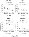
- CS1-CAR T cells To determine whether enhanced recognition of CS1 + myeloma cells by CS1-CAR T cells could lead to more efficient tumor cell lysis, a standard 4-hour chromium-51 release assay was performed. NCI-H929, IM9, and MM.1S cells, which express high levels of CS1, were resistant to mock-transduced T-cell-mediated killing, even at E/T ratios as high as 20:1; however, these cells were efficiently lysed by CS1-CAR T cells at all E:T ratios tested ( Fig. 3A , left three).
- cytolytic activity of RPMI-8226 cells expressing low levels of CS1 could only be slightly augmented upon co-incubation with CS1-CAR-transduced T cells ( Fig. 3A , right one).
- Degranulation and activation of T cells was further characterized by assessing expression of CD107a andCD69 in mock-transduced T cells and CS1-CAR-transduced T cells following incubation with or without NCI-H929 myeloma cells which, as mentioned above, triggered a strong response in CS1-CAR T cells with respect to cytokine release and cytolytic activity.
- both CD4 + (CD8 - ) and CD8 + CS1-CAR T cells exhibited increased levels of degranulation when stimulated by NCI-H929 or MM.1S cells ( Fig. 8B ).
- CS1-CAR-transduced T cells expressed significantly higher levels of granzyme B, but not perforin, even in the absence of target cells ( Fig. 3C and D ), suggesting that granzyme B may be predominantly involved in mediating the cytolytic activity of CS1-redirected T cells.
- CS1-CAR T cells The considerably stronger response in CS1-CAR T cells in terms of cytokine release and cytotoxicity when stimulated by myeloma cells expressing high levels of CS1 prompted investigation of whether ectopic expression of CS1 in myeloma cells with endogenously low levels of CS1 expression could elicit an increase in cytokine release and cytolysis.
- RPMI-8226 myeloma cells with low levels of endogenous CS1 expression were transduced with lentiviruses encoding human CS1 or PCDH empty vector as a mock-transduced control.
- the transduction efficiency was monitored by detection of GFP protein encoded by the lentiviruses, and the percentage of GFP-positive cells was more than 90% by flow cytometric analysis.
- Overexpression of CS1 was confirmed by staining the surface of the transduced cells with a PE-conjugated anti-CS1 antibody ( Fig. 4A ).
- Chromium-51 release assay indicated that forced CS1 expression resulted in a discernible increase in the susceptibility of RPMI-8226 cells to lysis by CS1-CAR-transduced T cells as opposed to mock-transduced T cells ( Fig. 4B ).
- IFN- ⁇ and IL-2 production was assessed via ELISA, showing that, compared with mock-transduced T cells, CS1-CAR-transduced T cells produced significantly higher amounts of IFN- ⁇ and IL-2 in response to RPMI-8226 cells overexpressing CS1; meanwhile, there was only a moderate increase in IFN- ⁇ secretion and no change in IL-2 secretion when CS1-CAR T cells were co-cultured with empty vector-modified RPMI-8226 cells ( Fig. 4C and D ).
- overexpression of CS1 in CS1 - 293T a transformed cell line, also triggered enhanced cytokine release and cytolysis by CS1-CAR T cells ( Fig.
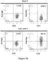
- CS1-specific CAR T cells To study the effects of CS1-specific CAR T cells in a more clinically relevant context, it was investigated whether CS1-CAR-transduced autologous T cells could efficiently recognize and kill tumor cells freshly isolated from patients with myeloma. Like T cells from healthy donors, T cells from patients with relapsed myeloma were successfully expanded and manipulated to express CS1-CAR by retroviral infection, as manifested by 60.7% of T cells staining positively with both anti-mouse Fab and anti-human CD3 antibodies determined by flow cytometry ( Fig. 5A ).
- CS1-directed T cells suppress in vivo tumor growth and prolong survival of tumor-bearing mice in orthotopic xenograft myeloma models
- MM.1S-grafted NSG mouse model The therapeutic potential of CS1-CAR T cells was evaluated in an MM.1S-grafted NSG mouse model. Intravenous injection of MM.1S cells has been widely used to establish a mouse xenograft model of MM, because this can lead to the engraftment in bone marrow and bone, as well as consistent establishment of multifocal bone lesions, which closely recapitulates human MM ( Mitsiades CS, et al. Cancer Cell 2004 5:221-30 ; Runnels JM, et al. J Biomed Opt 2011 16:011006 ). To facilitate monitoring of tumor growth, MM.
- IS cells were engineered to express both GFP and firefly luciferase by retroviral infection, and GFP + cells were sorted and intravenously grafted into NSG mice to initiate tumor growth. These mice were then intravenously infused with mock-transduced T cells, CS1+CAR-transduced T cells or PBS.
- mice were then intravenously infused with mock-transduced T cells, CS1+CAR-transduced T cells or PBS.
- Example 2 CS1-specific chimeric antigen receptor (CAR)-engineered natural killer cells enhance in vitro and in vivo antitumor activity against human multiple myeloma
- human NK cells were engineered to express a CAR that was CS1 specific, and incorporated a CD28-CD3 ⁇ co-stimulatory signaling domain.
- the anti-MM function of these cells was evaluated in vitro and in an in vivo orthotopic xenograft mouse model of MM.
- the results showed that the expression of the CS1-CAR could redirect NK cells to specifically and efficiently eradicate CS1-expressing MM cells, both in vitro and in vivo, and this eradication was CS1 dependent.
- This CAR strategy is suitable for the development of an effective NK cell-based immunotherapy as a means to treat patients with refractory or relapsed MM.
- CAR NK cells allow the use of allogeneic NK cell sources, which are less likely to cause and may even help to suppress graft-versus-host disease ( Olson JA, et al. Blood 2010 115:4293-4301 ), while also potentiating an increase in cytotoxicity due to mismatched killer immunoglobulin-like receptors (KIRs) ( Ruggeri L, et al. Science 2002 295:2097-2100 ).
- KIRs mismatched killer immunoglobulin-like receptors
- Human multiple myeloma cell lines L363 (German Collection of Microorganisms and Cell Cultures, Braunschweig, Germany), IM9 [American Type Culture Collection (ATCC), Manassas, VA, USA] and U266 (ATCC) were maintained in RPMI 1640 medium with 10% fetal bovine serum (FBS) (Life Technologies, Grand Island, NY, USA).
- Human IL-2-dependent NK cell lines NK-92 (ATCC) and NKL were maintained in RPMI 1640 medium supplemented with 20% FBS and 150 IU/mL rhIL-2 (Hoffman-LaRoche Inc., Nutley, NJ, USA).
- mice Six- to eight-week-old NOD.Cg-prkdcscid IL2rgtm1Wjl/szJ (NSG) mice were obtained from Jackson Laboratories (Bar Harbor, ME, USA). All animal work was approved by The Ohio State University Animal Care and Use Committee. Mice were monitored frequently for MM disease progression, and killed when they were moribund with the symptoms of hindlimb paralysis, lethargy, and obvious weight loss.
- the CS1-scFv fragment amplified from the hybridoma cell line Luc90, was fused with a sequence encoding a Myc tag immediately following the CS1-VL-encoding sequence.
- the fused DNA sequences were incorporated with CD28-CD3 ⁇ that was incised from a retroviral vector.
- the entire CSI-scFv-myc tag-CD28-CD3 ⁇ fragment was ligated into a lentiviral vector designated as PCDH-CMV-MCS-EF1-copGFP (PCDH, System Biosciences, Mountain View, CA, USA) to generate a PCDH-CS1-scFv-myc tag-CD28-CD3 ⁇ (PCDH-CS1-CAR) construct.
- VSVG-pseudotyped lentiviral supernatant 293T cells cultured in DMEM media (Invitrogen) were co-transfected with PCDH-CS1-scFv-CD28-CD3 ⁇ or the PCDH control vector (to generate virus for mock infection with the empty vector) together with the packaging constructs, pCMV-VSVG and pCMV-dr9, using calcium phosphate transfection reagent (Promega, Madison, WI, USA). After 24 h, the DMEM media was replaced with RPMI-1640 media containing 20% FBS.
- conditioned medium containing lentivirus was harvested and filtered through a 0.45 ⁇ m filter unit (Milliopore, Billerica, MA, USA) to remove cell debris.
- Viral infection was performed in 6-well plates using 2 ⁇ 10 6 NK-92 or NKL cells in a total volume of 2 mL of lentiviral supernatant containing 8 ⁇ g/mL polybrene (Sigma-Aldrich, St. Louis, MO, USA) and 450 IU/mL rhIL-2. Cells were centrifuged at 1,800 rpm at 32 °C for 45 min, then plates were placed in an incubator at 37 °C for 2 h.
- NK cells were maintained in RPMI 1640 media supplemented with 20% FBS and 150 IU/mL rhIL-2 at 37 °C.
- Transduced NK cells were enriched by two rounds of cell sorting using a FACS Aria II cell sorter (BD Biosciences, San Jose, CA, USA). Positive cells were selected based on expression of green fluorescence protein (GFP) surface marker, which was encoded in the PCDH vector
- Human CS1 coding sequences were amplified from cDNA isolated from IM9 cells via PCR, then subcloned into a PCDH lentiviral vector to generate a PCDH-CS 1 construct. Lentivirus production and infection of U266 cells were performed using the methods described above. GFP-positive cells were then sorted using an FACS Aria II cell sorter (BD Biosciences, San Jose, CA, USA).
- the membrane was then washed three times in TBS supplemented with Tween 20.
- the HRP-conjugated secondary antibody (GE Healthcare Biosciences, Pittsburgh, PA, USA) was diluted 1:5,000 with 5% milk in TBS supplemented with 0.1% Tween 20 and added to the membrane to stand for 1 h.
- the membrane was again washed four times in TB S supplemented with Tween 20, and an enhanced chemiluminescence reagent (ECL; GE Healthcare Biosciences) was added for 1 min.
- ECL enhanced chemiluminescence reagent
- CS1-CAR To analyze surface expression of CS1-CAR, a single cell suspension of transduced NK cells was incubated for 1 h at 4°C with an anti-Myc tag mouse mAb 9E10 (Sigma-Aldrich). Cells were washed twice with PBS and then incubated for 30 min at 4°C with PE-conjugated rat anti-mouse IgGI secondary antibody (BD Pharmingen).
- CS1 and CD138 on myeloma cells were examined by FACS analysis using a BD LSRII analyzer after cells were stained with PE-conjugated mouse anti-CS1 mAb (eBiosciences, San Diego, CA, USA) and APC-conjugated mouse anti-CD138 mAb (BD Pharmingen). Data analysis was carried out using FLOWJO software (Tree Star Inc., Ashland, OR, USA).
- MM target cells were labeled for 1.5 h with 100 mCi chromium-51 ( 51 Cr), washed four times with regular RPMI media, and adjusted to a concentration of 5,000 cells per well in 100 ⁇ l volume of a 96-well V-bottom microtiter plate.
- FACS-enriched mock- or CS1-CAR-transduced NK cells were added in 100 ⁇ l volume into triplicate wells at various effector to target (E:T) ratios.
- Myeloma target cells were co-cultured with NK effector cells in 96-well V bottom plates for 24 h. In all, 2.5 ⁇ 10 5 myeloma cell line cells or 1.0 ⁇ 10 5 primary myeloma cells were incubated with 2.5 ⁇ 10 5 or 5.0 ⁇ 10 5 NK cells, respectively. Cell-free supernatants were assayed for interferon- ⁇ (IFN- ⁇ ) secretion by enzyme-linked immunosorbent assay (ELISA) using a kit from R&D Systems (Minneapolis, MN, USA) according to the manufacturer's protocol. Data depicted in Figures represent mean values of triplicate wells from one of three representative experiments with similar results.
- IFN- ⁇ interferon- ⁇ secretion by enzyme-linked immunosorbent assay
- IM9 cells were retrovirally transduced with Pinco-pGL3-luc/GFP virus expressing firefly luciferase as previously described ( He S, et al. Blood 2013 121:4663-4671 ). GFP-positive cells were sorted using an FACS Aria II cell sorter (BD Biosciences), and were designated as 'IM9-Luc' cells. Then, six- to eight-week-old male NSG mice were intravenously (i.v.) injected with 0.5 ⁇ 10 6 IM9-Luc MM cells in 400 ⁇ l of phosphate-buffered saline via tail vein on day 0 to establish a xenograft orthotopic MM model. Beginning on day 7, the mice were i.v.
- mice were intraperitoneally (i.p.) infused with D-luciferin (150 mg/kg body weight; Gold Biotechnology, St. Louis, MO, USA), anesthetized with isoflurane, and imaged using an In Vivo Imaging System (IVIS-100, Perkin-Elmer, Waltham, MA, USA) with the Living Image software (Perkin-Elmer).
- D-luciferin 150 mg/kg body weight; Gold Biotechnology, St. Louis, MO, USA
- IVIS-100 In Vivo Imaging System
- Perkin-Elmer Waltham, MA, USA
- Unpaired Student's t test was utilized to compare two independent groups for continuous end points if normally distributed. One-way ANOVA was used when three or more independent groups were compared. For non-normally distributed end points, such as in vivo bioluminescence intensity, a Kruskal-Wallis test was utilized to compare the median of NK-92-CS1-CAR to NK-92-EV and phosphate-buffered saline groups. For survival data, Kaplan-Meier curves were plotted and compared using a log-rank test. All tests are two-sided. P-values were adjusted for multiple comparisons by the Bonferroni method. A P-value of ⁇ 0.05 is considered as statistically significant.
- a specific CS1-CAR construct was generated with a PCDH lentiviral vector backbone, sequentially containing a signal peptide (SP), a heavy chain variable region (VH), a linker, a light chain variable region (VL), a Myc tag, a hinge, CD28 and CD3 ⁇ ( Figure 13A ).
- SP signal peptide
- VH heavy chain variable region
- VL light chain variable region
- Myc tag a hinge
- CD28 and CD3 ⁇ Figure 13A
- NK-92 and NKL NK cell lines were transduced with the CAR construct and then sorted for expression of GFP, a marker expressed by the vector.
- CS1-CAR NK cells After generating the CS1-CAR NK cells, it was determined whether they selectively kill CS1 + better than CS1 - MM cells. For this purpose, it was first confirmed that IM9 and L363 MM cell lines constitutively expressed CS1 protein on their surface, while expression of CS1 was negligible in U266 MM cells ( Figure 14A ). Next, a 4-h chromium-51 release assay indicated that, compared with mock-transduced NK-92 cells, NK-92 cells transduced with CS1-CAR were significantly enhanced in their ability to kill CS1 + IM9 and L363 cells ( Figures 14B and 14C , left panels).
- CS1-CAR-modified NK cells secrete more IFN- ⁇ than mock-transduced NK cells do after exposure to CS1 + MM cells
- the signaling domain of the CD28 co-stimulatory molecule may enhance activation after recognition of the CS1 scFv with the CS1 antigen on the surface of MM cells. Therefore, the inclusion of this signaling domain may have the capacity to activate NK cells not only to have higher cytotoxicity, but also to produce more IFN- ⁇ , the latter of which is also important for tumor surveillance and activation of CD8 + T cells and macrophages ( Martin-Fontecha A, et al. Nat Immunol 2004 5:1260-1265 ; Tu SP, et al. Cancer Res 2011 71:4247-4259 ; Ma J, et al. Cell Mol Life Sci 2003 60:2334-2346 ).
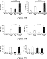
- CS1-CAR-modified or control-engineered effector NK cells were either cultured alone or co-cultured with CS1 + myeloma cells including the IM9 and L363 MM cell lines. After 24 h, the IFN- ⁇ production was measured by ELISA. As shown in Figure 15 , both CS1-CAR-modified and mock-transduced NK-92 or NKL cells spontaneously produced low or negligible levels of IFN- ⁇ when incubated alone.
- Enforced CS1 expression in U266 cells enhances cytotoxicity and IFN- ⁇ production of NK-92-CS1-CAR cells
- CS1 protein was successfully expressed on the surface of the U266-CS1 cells ( Figure 16A ).
- Chromium-51 release assay indicated that, when compared with mock-transduced NK-92 cells, there was a significant increase in the cytotoxic activity of CS1-CAR-transduced NK-92 cells toward U266 cells overexpressing CS1 ( Figure 16B ).
- NK-92-CS1-CAR cells co-cultured with U266 cells overexpressing CS1 secreted significantly higher levels of IFN-g ( Figure 16C ).
- NK-92-CS1-CAR cells had significantly higher levels of perforin and granzyme B expression, even in the absence of MM tumor cells ( Figures 17B and 17C ). This is consistent with a previous report regarding the elevated expression of granzyme B in CAR T cells ( Koehler H, et al. Cancer Res 2007 67:2265-2273 ), and also consistent with the fact that perforin and granzyme B expression are generally correlated with cytotoxic activity of NK cells ( Krzewski K, et al. Front Immunol 2012 3:335 ).
- CS1 protein was indeed uniformly expressed on the surface of primary MM cells ( Figure 18A ).
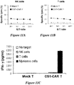
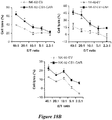
- chromium-51 release assay primary myeloma cells freshly isolated from MM patients were shown to be highly resistant to NK-92 cell-mediated lysis even at E:T ratios as high as 40:1 and 20:1; however, this resistance could be partially overcome by NK-92 cell expression of CS1-CAR, which resulted in a dramatic increase in eradication of primary myeloma cells ( Figure 18B ).
- CS1-CAR-transduced NK-92 cells In line with the cytotoxicity result, after 24 h co-culture with primary myeloma cells, CS1-CAR-transduced NK-92 cells also secreted significantly higher levels of IFN- ⁇ than mock-transduced NK-92 cells ( Figure 18C ).
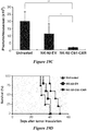
- CS1-CAR-transduced NK-92 cells inhibit MM tumor growth and prolong survival of tumor-bearing mice in an orthotopic xenograft MM model
- NK-92-CS1-CAR cells their antitumor activity was examined in IM9-xenografted NSG mice.
- An IM9 cell line expressing firefly luciferase (IM9-Luc) was generated by retrovirally transducing IM9 cells with virus expressing firefly luciferase, then performing GFP-based cell sorting.
- the expression of full-length firefly luciferase mRNA was confirmed by RT-PCR.
- IM9-Luc cells expressed CD138 protein on their surface. In agreement with a previous report ( Francisco JA, et al.
- IM9-Luc-bearing NSG mice displayed disseminated disease, manifested by hindlimb paralysis and motor ataxia. Histological examination of spinal vertebrae in a mouse displaying hindlimb paralysis showed the presence of numerous tumor cells and osteolytic lesions in bone tissue ( Figure 19A , left). Immunohistochemical staining with human-specific anti-CD138 antibody further confirmed the presence of tumor cells ( Figure 19A , right). Bioluminescence imaging was used to monitor the IM9-Luc tumor growth.
- IM9-Luc tumors were significantly suppressed in mice who instead later were administered NK-92-CS 1-CAR cells.
- treatment with NK-92-CS 1-CAR cells significantly prolonged the survival of mice bearing IM9-Luc tumors as compared with treatment with the mock-transduced NK-92 control cells ( Figure 19D ).
- NK-92-CS1-CAR cells or mock-transduced NK-92 cells were similarly administered, but without i.v. injection of IM9-Luc cells, mice did not develop disseminated disease or die.
Landscapes
- Health & Medical Sciences (AREA)
- Chemical & Material Sciences (AREA)
- Immunology (AREA)
- Life Sciences & Earth Sciences (AREA)
- Organic Chemistry (AREA)
- General Health & Medical Sciences (AREA)
- Medicinal Chemistry (AREA)
- Genetics & Genomics (AREA)
- Biochemistry (AREA)
- Biophysics (AREA)
- Molecular Biology (AREA)
- Proteomics, Peptides & Aminoacids (AREA)
- Cell Biology (AREA)
- Toxicology (AREA)
- Zoology (AREA)
- Gastroenterology & Hepatology (AREA)
- Pharmacology & Pharmacy (AREA)
- Public Health (AREA)
- Veterinary Medicine (AREA)
- Animal Behavior & Ethology (AREA)
- General Chemical & Material Sciences (AREA)
- Nuclear Medicine, Radiotherapy & Molecular Imaging (AREA)
- Chemical Kinetics & Catalysis (AREA)
- Engineering & Computer Science (AREA)
- Bioinformatics & Cheminformatics (AREA)
- Hematology (AREA)
- Medicines Containing Material From Animals Or Micro-Organisms (AREA)
- Peptides Or Proteins (AREA)
- Micro-Organisms Or Cultivation Processes Thereof (AREA)
- Biomedical Technology (AREA)
- Biotechnology (AREA)
- Wood Science & Technology (AREA)
- General Engineering & Computer Science (AREA)
- Microbiology (AREA)
- Medicines Containing Antibodies Or Antigens For Use As Internal Diagnostic Agents (AREA)
- Physics & Mathematics (AREA)
- Plant Pathology (AREA)
- Mycology (AREA)
- Epidemiology (AREA)
Applications Claiming Priority (4)
| Application Number | Priority Date | Filing Date | Title |
|---|---|---|---|
| US201361819141P | 2013-05-03 | 2013-05-03 | |
| US201361876492P | 2013-09-11 | 2013-09-11 | |
| EP14791494.9A EP2992020B1 (en) | 2013-05-03 | 2014-05-02 | Cs1-specific chimeric antigen receptor engineered immune effector cells |
| PCT/US2014/036684 WO2014179759A1 (en) | 2013-05-03 | 2014-05-02 | Cs1-specific chimeric antigen receptor engineered immune effector cells |
Related Parent Applications (1)
| Application Number | Title | Priority Date | Filing Date |
|---|---|---|---|
| EP14791494.9A Division EP2992020B1 (en) | 2013-05-03 | 2014-05-02 | Cs1-specific chimeric antigen receptor engineered immune effector cells |
Publications (1)
| Publication Number | Publication Date |
|---|---|
| EP3663320A1 true EP3663320A1 (en) | 2020-06-10 |
Family
ID=51844005
Family Applications (2)
| Application Number | Title | Priority Date | Filing Date |
|---|---|---|---|
| EP20150396.8A Withdrawn EP3663320A1 (en) | 2013-05-03 | 2014-05-02 | Nucleic acid sequence encoding a cs1-specific chimeric antigen receptor (car) polypeptide |
| EP14791494.9A Active EP2992020B1 (en) | 2013-05-03 | 2014-05-02 | Cs1-specific chimeric antigen receptor engineered immune effector cells |
Family Applications After (1)
| Application Number | Title | Priority Date | Filing Date |
|---|---|---|---|
| EP14791494.9A Active EP2992020B1 (en) | 2013-05-03 | 2014-05-02 | Cs1-specific chimeric antigen receptor engineered immune effector cells |
Country Status (15)
| Country | Link |
|---|---|
| US (3) | US10358494B2 (https=) |
| EP (2) | EP3663320A1 (https=) |
| JP (3) | JP6856188B2 (https=) |
| KR (1) | KR102098985B1 (https=) |
| CN (2) | CN105377897A (https=) |
| AU (2) | AU2014259675B2 (https=) |
| BR (1) | BR112015027567B1 (https=) |
| DK (1) | DK2992020T3 (https=) |
| ES (1) | ES2777940T3 (https=) |
| HU (1) | HUE048312T2 (https=) |
| IL (1) | IL242315B (https=) |
| MX (1) | MX370173B (https=) |
| PL (1) | PL2992020T3 (https=) |
| PT (1) | PT2992020T (https=) |
| WO (1) | WO2014179759A1 (https=) |
Families Citing this family (52)
| Publication number | Priority date | Publication date | Assignee | Title |
|---|---|---|---|---|
| CN105377897A (zh) * | 2013-05-03 | 2016-03-02 | 俄亥俄州创新基金会 | Cs1-特异性嵌合抗原受体工程化的免疫效应细胞 |
| US10189908B2 (en) | 2014-02-05 | 2019-01-29 | The University Of Chicago | Chimeric antigen receptors recognizing cancer-specific TN glycopeptide variants |
| KR102558502B1 (ko) | 2014-12-05 | 2023-07-20 | 시티 오브 호프 | Cs1 표적화된 키메라 항원 수용체-변형된 t 세포 |
| JP6797803B2 (ja) * | 2014-12-31 | 2020-12-09 | セルジーン コーポレイション | ナチュラルキラー細胞を用いて血液障害、固形腫瘍、又は感染性疾患を治療する方法 |
| WO2016123143A1 (en) | 2015-01-26 | 2016-08-04 | The University Of Chicago | CAR T-CELLS RECOGNIZING CANCER-SPECIFIC IL 13Rα2 |
| CN107683289B (zh) | 2015-01-26 | 2021-08-06 | 芝加哥大学 | IL13Rα2结合剂和其在癌症治疗中的用途 |
| JP6849600B6 (ja) | 2015-01-29 | 2021-06-30 | リージェンツ オブ ザ ユニバーシティ オブ ミネソタ | キメラ抗原受容体、組成物及び方法 |
| WO2016134371A2 (en) | 2015-02-20 | 2016-08-25 | Ohio State Innovation Foundation | Bivalent antibody directed against nkg2d and tumor associated antigens |
| US11045543B2 (en) | 2015-04-06 | 2021-06-29 | Cytoimmune Therapeutics, Inc. | EGFR-directed car therapy for glioblastoma |
| US10752670B2 (en) * | 2015-05-20 | 2020-08-25 | Cellectis | Anti-GD3 specific chimeric antigen receptors for cancer immunotherapy |
| EP3307876B1 (en) * | 2015-06-10 | 2023-09-13 | ImmunityBio, Inc. | Modified nk-92 cells for treating cancer |
| JP7010487B2 (ja) | 2015-08-17 | 2022-03-03 | ソウル大学校産学協力団 | 抗コチニン抗体が連結したキメラ抗原受容体およびその使用 |
| EP4289944A3 (en) * | 2015-10-08 | 2024-03-13 | National University Corporation Tokai National Higher Education and Research System | Method for preparing genetically-modified t cells which express chimeric antigen receptor |
| EP3402495A4 (en) * | 2016-01-15 | 2019-06-19 | Etubics Corporation | METHOD AND COMPOSITIONS FOR T-CELL IMMUNOTHERAPY |
| EP3408297A2 (en) | 2016-01-29 | 2018-12-05 | Med Manor Organics, (P) Ltd | A chimeric antigen receptor specific to b-cell maturation antigen, a recombinant expression vector and a method thereof |
| EP3433269B1 (en) | 2016-03-23 | 2023-09-27 | Helmholtz Zentrum München - Deutsches Forschungszentrum für Gesundheit und Umwelt (GmbH) | Fusion proteins of pd-1 and 4-1bb |
| CN121102467A (zh) | 2016-04-22 | 2025-12-12 | 克莱格医学有限公司 | 用于细胞免疫疗法的组合物和方法 |
| JP7086928B2 (ja) | 2016-07-25 | 2022-06-20 | ザ ユナイテッド ステイツ オブ アメリカ, アズ リプレゼンテッド バイ ザ セクレタリー, デパートメント オブ ヘルス アンド ヒューマン サービシーズ | 改変されたナチュラルキラー細胞を生成する方法および使用方法 |
| US12516292B2 (en) | 2016-07-25 | 2026-01-06 | The United States Of America, As Represented By The Secretary, Department Of Health And Human Services | Methods of producing modified natural killer cells and methods of use |
| CN106279432B (zh) * | 2016-08-10 | 2019-09-20 | 深圳市再生之城生物医药技术有限公司 | 一种vc-car分子及在清除hiv-1感染细胞中的应用 |
| WO2018058432A1 (zh) * | 2016-09-28 | 2018-04-05 | 李华顺 | 一种多基因重组嵌合抗原受体分子及其应用 |
| CA3048211A1 (en) | 2016-12-23 | 2018-06-28 | Macrogenics, Inc. | Adam9-binding molecules, and methods of use thereof |
| EP3567101A4 (en) * | 2017-01-05 | 2020-12-02 | Korea Research Institute of Bioscience and Biotechnology | NATURAL KILLER CELL EXPRESSING AN ANTI-COTININ CHEMERIC ANTIGEN RECEPTOR |
| MX2019011225A (es) * | 2017-03-20 | 2020-01-21 | Células t modificadas por el receptor de antígeno quimérico dirigidas a cs1 para el tratamiento de la amiloidosis al. | |
| ES2963635T3 (es) * | 2017-03-28 | 2024-04-01 | Ohio State Innovation Foundation | Vacunas de péptido PD1 humano y usos de las mismas |
| WO2018189360A1 (en) | 2017-04-13 | 2018-10-18 | Cellectis | New sequence specific reagents targeting ccr5 in primary hematopoietic cells |
| KR20200027508A (ko) * | 2017-07-07 | 2020-03-12 | 에이치 리 모피트 캔서 센터 앤드 리서어치 인스티튜트 아이엔씨 | 돌연변이된 cd28 공동자극 도메인을 갖는 키메라 항원 수용체 |
| WO2019014624A1 (en) * | 2017-07-14 | 2019-01-17 | The Johns Hopkins University | Engineered tsc2 |
| TWI676483B (zh) * | 2017-08-06 | 2019-11-11 | 強普生技股份有限公司 | 醫藥套組及其用途 |
| WO2019069125A1 (en) * | 2017-10-06 | 2019-04-11 | Oslo Universitetssykehus Hf | CHIMERIC ANTIGEN RECEPTORS |
| US20200283501A1 (en) | 2017-10-26 | 2020-09-10 | Regents Of The University Of Minnesota | Recombinant immune cells, methods of making, and methods of use |
| US12077598B2 (en) | 2018-03-16 | 2024-09-03 | H. Lee Moffitt Cancer Center And Research Institute, Inc. | Chimeric antigen receptors with mutated CD28 phosphorylation sites |
| CN112566643A (zh) | 2018-06-12 | 2021-03-26 | 加利福尼亚大学董事会 | 用于治疗癌症的单链双特异性嵌合抗原受体 |
| WO2019241315A1 (en) | 2018-06-12 | 2019-12-19 | Obsidian Therapeutics, Inc. | Pde5 derived regulatory constructs and methods of use in immunotherapy |
| WO2020009868A1 (en) * | 2018-07-03 | 2020-01-09 | The United States Of America, As Represented By The Secretary, Department Of Health And Human Services | Anti-slamf7 chimeric antigen receptors |
| EP3818148A4 (en) * | 2018-07-05 | 2022-09-28 | Emory University | TRANSDUCTION OF CONGENITAL IMMUNE COMPETENT CELLS USING AAV6 |
| CN112469734A (zh) * | 2018-08-03 | 2021-03-09 | 南京驯鹿医疗技术有限公司 | 表达嵌合抗原受体的t细胞、该嵌合抗原相关表达载体以及它们的应用 |
| WO2020123716A1 (en) | 2018-12-11 | 2020-06-18 | Obsidian Therapeutics, Inc. | Membrane bound il12 compositions and methods for tunable regulation |
| MX2021010840A (es) | 2019-03-08 | 2022-01-19 | Obsidian Therapeutics Inc | Composiciones de anhidrasa carbónica 2 (ca2) humana y métodos de regulación ajustable. |
| KR102691932B1 (ko) | 2019-04-03 | 2024-08-06 | 프리시젼 바이오사이언시스 인코포레이티드 | 마이크로RNA-적응 shRNA(shRNAmiR)를 포함하는 유전자-변형 면역 세포 |
| JP2022537670A (ja) | 2019-06-12 | 2022-08-29 | オブシディアン セラピューティクス, インコーポレイテッド | Ca2の組成物および調整可能な制御方法 |
| EP3983537A1 (en) | 2019-06-12 | 2022-04-20 | Obsidian Therapeutics, Inc. | Ca2 compositions and methods for tunable regulation |
| SG11202111943UA (en) | 2019-07-02 | 2021-11-29 | Hutchinson Fred Cancer Res | Recombinant ad35 vectors and related gene therapy improvements |
| CN112442126B (zh) * | 2019-09-04 | 2022-07-29 | 杭州济元基因科技有限公司 | 一种抗人cs1抗原的单克隆抗体及其car-t细胞 |
| EP4054602A4 (en) * | 2019-11-08 | 2023-12-06 | Mayo Foundation for Medical Education and Research | EPHA3 TARGETED CAR-T CELLS FOR TREATING TUMORS |
| WO2022002919A1 (en) * | 2020-06-29 | 2022-01-06 | Julius-Maximilians-Universität Würzburg | Slamf7 cars |
| CN114286691B (zh) | 2020-08-06 | 2024-08-06 | 武汉思安医疗技术有限公司 | Cs1-抗体和抗cs1-car-t细胞 |
| WO2022063302A1 (zh) * | 2020-09-25 | 2022-03-31 | 克莱格医学有限公司 | 免疫细胞活性调节 |
| AR124414A1 (es) | 2020-12-18 | 2023-03-22 | Century Therapeutics Inc | Sistema de receptor de antígeno quimérico con especificidad de receptor adaptable |
| CN116410331B (zh) * | 2021-12-31 | 2024-01-30 | 合源生物科技(天津)有限公司 | 靶向cs1的嵌合抗原受体、靶向bcma/cs1的双特异性嵌合抗原受体及其应用 |
| CN120344563A (zh) * | 2022-12-02 | 2025-07-18 | 广州荔泰生物科技有限公司 | 人cs1 car-t细胞 |
| WO2026006370A1 (en) * | 2024-06-28 | 2026-01-02 | Immunitybio, Inc. | Anti-slamf7 antibodies and therapeutics |
Citations (2)
| Publication number | Priority date | Publication date | Assignee | Title |
|---|---|---|---|---|
| WO2005005638A2 (en) * | 2003-06-30 | 2005-01-20 | Regeneron Pharmaceuticals, Inc. | High affinity fusion proteins (rc-scfv-fc or scfv-scfv-fc) binding cytokines, particularly il-6 & il-18 |
| US20120064083A1 (en) * | 2003-05-08 | 2012-03-15 | Abbott Biotherapeutics Corp. | Therapeutic use of anti-cs1 antibodies |
Family Cites Families (21)
| Publication number | Priority date | Publication date | Assignee | Title |
|---|---|---|---|---|
| US20040038339A1 (en) * | 2000-03-24 | 2004-02-26 | Peter Kufer | Multifunctional polypeptides comprising a binding site to an epitope of the nkg2d receptor complex |
| US7709610B2 (en) * | 2003-05-08 | 2010-05-04 | Facet Biotech Corporation | Therapeutic use of anti-CS1 antibodies |
| NZ573646A (en) | 2006-06-12 | 2012-04-27 | Wyeth Llc | Single-chain multivalent binding proteins with effector function |
| WO2008034076A2 (en) | 2006-09-15 | 2008-03-20 | The Johns Hopkins University | Cyclophosphamide in combination with immune therapeutics |
| KR20100058509A (ko) | 2007-07-31 | 2010-06-03 | 메디뮨 엘엘씨 | 다중특이적 에피토프 결합 단백질 및 이의 용도 |
| WO2010017103A2 (en) | 2008-08-04 | 2010-02-11 | The United States Of America, As Represented By The Secretary, Department Of Health And Human Servic | Fully human anti-human nkg2d monoclonal antibodies |
| EP2361263A1 (en) | 2008-10-31 | 2011-08-31 | Abbott Biotherapeutics Corp. | Use of anti-cs1 antibodies for treatment of rare lymphomas |
| UY32808A (es) | 2009-07-29 | 2011-02-28 | Abbott Lab | Inmunoglobulinas como dominio variable dual y usos de las mismas |
| WO2011125015A2 (en) | 2010-04-05 | 2011-10-13 | Bar-Ilan University | Protease-activatable pore-forming polypeptides |
| ES2602743T3 (es) * | 2010-09-08 | 2017-02-22 | Chemotherapeutisches Forschungsinstitut Georg-Speyer-Haus | Receptores de antígenos quiméricos con una región bisagra optimizada |
| EP2643350A4 (en) * | 2010-11-22 | 2015-01-07 | Innate Pharma Sa | TREATMENTS WITH NK CELL MODULATION AND METHOD FOR THE TREATMENT OF HEMATOLOGICAL MALIGNOMES |
| PH12013501201A1 (en) * | 2010-12-09 | 2013-07-29 | Univ Pennsylvania | Use of chimeric antigen receptor-modified t cells to treat cancer |
| WO2013051718A1 (ja) * | 2011-10-07 | 2013-04-11 | 国立大学法人三重大学 | キメラ抗原受容体 |
| PE20141937A1 (es) | 2011-11-16 | 2014-12-18 | Amgen Inc | Metodos para tratar trastornos relacionados con mutante viii de eliminacion de factor de crecimiento epidermico |
| CA2861491C (en) * | 2012-02-13 | 2020-08-25 | Seattle Children's Hospital D/B/A Seattle Children's Research Institute | Bispecific chimeric antigen receptors and therapeutic uses thereof |
| BR112015013193A2 (pt) | 2012-12-07 | 2017-07-11 | Solazyme Inc | ácido nucleico recombinante, cassete de expressão, célula, método para obter óleo microbiano, óleo microbiano, e, método para obter óleo microbiano. |
| CN103224561A (zh) | 2013-01-16 | 2013-07-31 | 天津大学 | 葡萄球菌肠毒素小分子抗体及制备方法及用途 |
| US20140302037A1 (en) | 2013-03-15 | 2014-10-09 | Amgen Inc. | BISPECIFIC-Fc MOLECULES |
| CN105377897A (zh) * | 2013-05-03 | 2016-03-02 | 俄亥俄州创新基金会 | Cs1-特异性嵌合抗原受体工程化的免疫效应细胞 |
| CN105829349B (zh) | 2013-10-15 | 2023-02-03 | 斯克利普斯研究所 | 肽嵌合抗原受体t细胞开关和其用途 |
| KR20200131844A (ko) * | 2018-03-16 | 2020-11-24 | 사이토이뮨 테라퓨틱스 인코퍼레이티드 | 이중특이적 항체 car 세포 면역요법 |
-
2014
- 2014-05-02 CN CN201480030178.9A patent/CN105377897A/zh active Pending
- 2014-05-02 BR BR112015027567-2A patent/BR112015027567B1/pt active IP Right Grant
- 2014-05-02 ES ES14791494T patent/ES2777940T3/es active Active
- 2014-05-02 EP EP20150396.8A patent/EP3663320A1/en not_active Withdrawn
- 2014-05-02 EP EP14791494.9A patent/EP2992020B1/en active Active
- 2014-05-02 PT PT147914949T patent/PT2992020T/pt unknown
- 2014-05-02 KR KR1020157033493A patent/KR102098985B1/ko active Active
- 2014-05-02 US US14/888,877 patent/US10358494B2/en active Active
- 2014-05-02 MX MX2015015182A patent/MX370173B/es active IP Right Grant
- 2014-05-02 CN CN202210255676.XA patent/CN115028735A/zh active Pending
- 2014-05-02 HU HUE14791494A patent/HUE048312T2/hu unknown
- 2014-05-02 WO PCT/US2014/036684 patent/WO2014179759A1/en not_active Ceased
- 2014-05-02 JP JP2016512083A patent/JP6856188B2/ja active Active
- 2014-05-02 DK DK14791494.9T patent/DK2992020T3/da active
- 2014-05-02 AU AU2014259675A patent/AU2014259675B2/en active Active
- 2014-05-02 PL PL14791494T patent/PL2992020T3/pl unknown
-
2015
- 2015-10-28 IL IL242315A patent/IL242315B/en active IP Right Grant
-
2017
- 2017-05-17 US US15/597,436 patent/US10227409B2/en active Active
-
2019
- 2019-04-18 JP JP2019079616A patent/JP2019146579A/ja active Pending
- 2019-05-31 AU AU2019203823A patent/AU2019203823B2/en active Active
- 2019-07-09 US US16/506,327 patent/US11845794B2/en active Active
-
2021
- 2021-07-16 JP JP2021118254A patent/JP7221338B2/ja active Active
Patent Citations (2)
| Publication number | Priority date | Publication date | Assignee | Title |
|---|---|---|---|---|
| US20120064083A1 (en) * | 2003-05-08 | 2012-03-15 | Abbott Biotherapeutics Corp. | Therapeutic use of anti-cs1 antibodies |
| WO2005005638A2 (en) * | 2003-06-30 | 2005-01-20 | Regeneron Pharmaceuticals, Inc. | High affinity fusion proteins (rc-scfv-fc or scfv-scfv-fc) binding cytokines, particularly il-6 & il-18 |
Non-Patent Citations (45)
| Title |
|---|
| A. J. JAKUBOWIAK ET AL: "Phase I Trial of Anti-CS1 Monoclonal Antibody Elotuzumab in Combination With Bortezomib in the Treatment of Relapsed/Refractory Multiple Myeloma", JOURNAL OF CLINICAL ONCOLOGY, vol. 30, no. 16, 1 June 2012 (2012-06-01), US, pages 1960 - 1965, XP055317482, ISSN: 0732-183X, DOI: 10.1200/JCO.2011.37.7069 * |
| ATTIANESE GRETA MARIA PAOLA GIORDANO ET AL: "A New Chimeric Antigen Receptor (CAR) Targeting the CD23 Antigen Expressed by Chronic Lymphocytic Leukemia (B-CLL) Cells", BLOOD, vol. 116, no. 21, November 2010 (2010-11-01), & 52ND ANNUAL MEETING OF THE AMERICAN-SOCIETY-OF-HEMATOLOGY (ASH); ORLANDO, FL, USA; DECEMBER 04 -07, 2010, pages 1015, XP008182251 * |
| B. JENA ET AL: "Redirecting T-cell specificity by introducing a tumor-specific chimeric antigen receptor", BLOOD, vol. 116, no. 7, 19 August 2010 (2010-08-19), pages 1035 - 1044, XP055021403, ISSN: 0006-4971, DOI: 10.1182/blood-2010-01-043737 * |
| BECKNELL B ET AL., J IMMUNOL METHODS, vol. 296, no. 1-2, 2005, pages 115 - 123 |
| BENSON DM JR ET AL., J CLIN ONCOL, vol. 30, 2012, pages 2013 - 5 |
| CASUCCI MONICA ET AL: "3125: Dual Transgenesis of T Cells with a Novel CD44v6-Specific Chimeric Antigen Receptor and a Suicide Gene for Safe and Effective Targeting of Chemoresistance in Hematopoietic Tumors", vol. 118, no. 21, 18 November 2011 (2011-11-18), pages 1350 - 1351, XP008178876, ISSN: 0006-4971, Retrieved from the Internet <URL:http://www.bloodjournal.org/content/118/21/3125> * |
| CHINNASAMY D ET AL., J CLIN INVEST, vol. 120, 2010, pages 3953 - 68 |
| CHU YAYA ET AL: "Genetically Engineered Natural Killer (NK) Cell Immunotherapy for Poor Risk B-Cell (CD20(+)) Leukemia and Lymphoma", BLOOD, vol. 118, no. 21, November 2011 (2011-11-01), & 53RD ANNUAL MEETING AND EXPOSITION OF THE AMERICAN-SOCIETY-OF-HEMATOLOGY (ASH); SAN DIEGO, CA, USA; DECEMBER 10 -13, 2011, pages 458 - 459, XP008182252 * |
| D. M. BENSON ET AL: "CS1-Directed Monoclonal Antibody Therapy for Multiple Myeloma", JOURNAL OF CLINICAL ONCOLOGY, vol. 30, no. 16, 1 June 2012 (2012-06-01), US, pages 2013 - 2015, XP055318672, ISSN: 0732-183X, DOI: 10.1200/JCO.2011.40.4061 * |
| FAURIAT C ET AL., LEUKEMIA, vol. 20, 2006, pages 732 - 733 |
| FRANCISCO JA ET AL., CANCER RES, vol. 60, 2000, pages 3225 - 3231 |
| G.T. HERMANON: "Bioconjugate Techniques", 2008, ACADEMIC PRESS |
| GODFREY J ET AL., LEUK LYMPHOMA, vol. 53, 2012, pages 1666 - 1676 |
| HE S ET AL., BLOOD, vol. 121, 2013, pages 4663 - 4671 |
| HSI ED ET AL., CLIN CANCER RES, vol. 14, 2008, pages 2775 - 2784 |
| IMAI C ET AL., LEUKEMIA, vol. 18, 2004, pages 676 - 84 |
| KOEHLER H ET AL., CANCER RES, vol. 67, 2007, pages 2265 - 2273 |
| KRZEWSKI K ET AL., FRONT IMMUNOL, vol. 3, 2012, pages 335 |
| LEI WANG ET AL: "T cell-based targeted immunotherapies for patients with multiple myeloma", INTERNATIONAL JOURNAL OF CANCER, 1 September 2014 (2014-09-01), pages n/a - n/a, XP055160595, ISSN: 0020-7136, DOI: 10.1002/ijc.29190 * |
| MA J ET AL., CELL MOL LIFE SCI, vol. 60, 2003, pages 2334 - 2346 |
| MAHER J ET AL., NAT BIOTECHNOL, vol. 20, 2002, pages 70 - 5 |
| MARTIN-FONTECHA A ET AL., NAT IMMUNOL, vol. 5, 2004, pages 1260 - 1265 |
| MITSIADES CS ET AL., CANCER CELL, vol. 5, 2004, pages 221 - 30 |
| MORGAN RA ET AL., MOL THER, vol. 18, 2010, pages 843 - 851 |
| NARNI-MANCINELLI E ET AL., INT IMMUNOL, vol. 23, 2011, pages 427 - 431 |
| OLSON JA ET AL., BLOOD, vol. 115, 2010, pages 4293 - 4301 |
| PALUMBO A ET AL., LEUKEMIA, vol. 23, 2009, pages 449 - 456 |
| PHILLIPS JH ET AL., J EXP MED, vol. 159, 1984, pages 993 - 1008 |
| PORTER DL ET AL., N ENGL J MED, vol. 365, 2011, pages 725 - 733 |
| ROSENBERG ET AL., NEW ENG. J. OF MED., vol. 319, 1988, pages 1676 |
| RUGGERI L ET AL., SCIENCE, vol. 295, 2002, pages 2097 - 2100 |
| RUNNELS JM ET AL., J BIOMED OPT, vol. 16, 2011, pages 011006 |
| S. LONIAL ET AL: "Elotuzumab in Combination With Lenalidomide and Low-Dose Dexamethasone in Relapsed or Refractory Multiple Myeloma", JOURNAL OF CLINICAL ONCOLOGY, vol. 30, no. 16, 30 April 2012 (2012-04-30), US, pages 1953 - 1959, XP055317479, ISSN: 0732-183X, DOI: 10.1200/JCO.2011.37.2649 * |
| SADELAIN M ET AL., NAT REV CANCER, vol. 3, 2003, pages 35 - 45 |
| SAMBROOK ET AL.: "Molecular Cloning: A Laboratory Manual", 2001, COLD SPRING HARBOR LABORATORY |
| SANCHEZ C ET AL., PROSTATE CANCER PROSTATIC DIS, vol. 16, 2013, pages 123 - 31 |
| SIEGEL R ET AL., CANCER J CLIN, vol. 62, 2012, pages 10 - 29 |
| SPITS H ET AL., IMMUNITY, vol. 26, 2007, pages 11 - 16 |
| TAI YT ET AL., BLOOD, vol. 112, 2008, pages 1329 - 1337 |
| TAI YT ET AL., BLOOD, vol. 113, 2009, pages 4309 - 18 |
| TOPP MAX S ET AL: "T-cells from multiple myeloma patients can be rendered to lyse CD138+ multiple myeloma cells by expression of a chimeric immunoreceptor targeting the myeloma-specific antigen HM 1.24.", BLOOD, vol. 102, no. 11, 16 November 2003 (2003-11-16), & 45TH ANNUAL MEETING OF THE AMERICAN SOCIETY OF HEMATOLOGY; SAN DIEGO, CA, USA; DECEMBER 06-09, 2003, pages 932a, XP008182250, ISSN: 0006-4971 * |
| TU SP ET AL., CANCER RES, vol. 71, 2011, pages 4247 - 4259 |
| W. R. BURNS ET AL: "A High Molecular Weight Melanoma-Associated Antigen-Specific Chimeric Antigen Receptor Redirects Lymphocytes to Target Human Melanomas", CANCER RESEARCH, vol. 70, no. 8, 15 April 2010 (2010-04-15), pages 3027 - 3033, XP055023662, ISSN: 0008-5472, DOI: 10.1158/0008-5472.CAN-09-2824 * |
| WOOF ET AL., NAT. REV. IMMUNOL., vol. 4, no. 2, 2004, pages 89 - 99 |
| YU J ET AL., IMMUNITY, vol. 24, no. 5, 2006, pages 575 - 590 |
Also Published As
Similar Documents
| Publication | Publication Date | Title |
|---|---|---|
| US11845794B2 (en) | CS1-specific chimeric antigen receptor engineered immune effector cells | |
| US20230303653A1 (en) | Compositions and methods of chimeric autoantibody receptor t cells | |
| CN110177803A (zh) | 用于使用融合蛋白进行tcr重新编程的组合物和方法 | |
| CN112004832A (zh) | 结合cd83的嵌合抗原受体 | |
| CN114929341A (zh) | 用于治疗髓系恶性肿瘤的嵌合抗原受体 | |
| CA2910666C (en) | Cs1-specific chimeric antigen receptor engineered immune effector cells | |
| HK1218925B (en) | Cs1-specific chimeric antigen receptor engineered immune effector cells | |
| HK40074993A (en) | Compositions and methods of chimeric autoantibody receptor t cells | |
| HK40063360A (en) | Compositions and methods of chimeric autoantibody receptor t cells |
Legal Events
| Date | Code | Title | Description |
|---|---|---|---|
| PUAI | Public reference made under article 153(3) epc to a published international application that has entered the european phase |
Free format text: ORIGINAL CODE: 0009012 |
|
| STAA | Information on the status of an ep patent application or granted ep patent |
Free format text: STATUS: THE APPLICATION HAS BEEN PUBLISHED |
|
| AC | Divisional application: reference to earlier application |
Ref document number: 2992020 Country of ref document: EP Kind code of ref document: P |
|
| AK | Designated contracting states |
Kind code of ref document: A1 Designated state(s): AL AT BE BG CH CY CZ DE DK EE ES FI FR GB GR HR HU IE IS IT LI LT LU LV MC MK MT NL NO PL PT RO RS SE SI SK SM TR |
|
| RIN1 | Information on inventor provided before grant (corrected) |
Inventor name: HOFMEISTER, CRAIG Inventor name: CHU, JIANHONG Inventor name: YU, JIANHUA |
|
| STAA | Information on the status of an ep patent application or granted ep patent |
Free format text: STATUS: REQUEST FOR EXAMINATION WAS MADE |
|
| 17P | Request for examination filed |
Effective date: 20201209 |
|
| RBV | Designated contracting states (corrected) |
Designated state(s): AL AT BE BG CH CY CZ DE DK EE ES FI FR GB GR HR HU IE IS IT LI LT LU LV MC MK MT NL NO PL PT RO RS SE SI SK SM TR |
|
| P01 | Opt-out of the competence of the unified patent court (upc) registered |
Effective date: 20230524 |
|
| STAA | Information on the status of an ep patent application or granted ep patent |
Free format text: STATUS: EXAMINATION IS IN PROGRESS |
|
| 17Q | First examination report despatched |
Effective date: 20231020 |
|
| STAA | Information on the status of an ep patent application or granted ep patent |
Free format text: STATUS: THE APPLICATION IS DEEMED TO BE WITHDRAWN |
|
| 18D | Application deemed to be withdrawn |
Effective date: 20240301 |