EP3600407B1 - Improved antigen binding receptors - Google Patents
Improved antigen binding receptors Download PDFInfo
- Publication number
- EP3600407B1 EP3600407B1 EP18712231.2A EP18712231A EP3600407B1 EP 3600407 B1 EP3600407 B1 EP 3600407B1 EP 18712231 A EP18712231 A EP 18712231A EP 3600407 B1 EP3600407 B1 EP 3600407B1
- Authority
- EP
- European Patent Office
- Prior art keywords
- domain
- antigen binding
- seq
- amino acid
- mutated
- Prior art date
- Legal status (The legal status is an assumption and is not a legal conclusion. Google has not performed a legal analysis and makes no representation as to the accuracy of the status listed.)
- Active
Links
Images


Classifications
-
- A—HUMAN NECESSITIES
- A61—MEDICAL OR VETERINARY SCIENCE; HYGIENE
- A61K—PREPARATIONS FOR MEDICAL, DENTAL OR TOILETRY PURPOSES
- A61K35/00—Medicinal preparations containing materials or reaction products thereof with undetermined constitution
- A61K35/12—Materials from mammals; Compositions comprising non-specified tissues or cells; Compositions comprising non-embryonic stem cells; Genetically modified cells
- A61K35/14—Blood; Artificial blood
- A61K35/17—Lymphocytes; B-cells; T-cells; Natural killer cells; Interferon-activated or cytokine-activated lymphocytes
-
- A—HUMAN NECESSITIES
- A61—MEDICAL OR VETERINARY SCIENCE; HYGIENE
- A61K—PREPARATIONS FOR MEDICAL, DENTAL OR TOILETRY PURPOSES
- A61K40/00—Cellular immunotherapy
- A61K40/10—Cellular immunotherapy characterised by the cell type used
- A61K40/11—T-cells, e.g. tumour infiltrating lymphocytes [TIL] or regulatory T [Treg] cells; Lymphokine-activated killer [LAK] cells
-
- A—HUMAN NECESSITIES
- A61—MEDICAL OR VETERINARY SCIENCE; HYGIENE
- A61K—PREPARATIONS FOR MEDICAL, DENTAL OR TOILETRY PURPOSES
- A61K40/00—Cellular immunotherapy
- A61K40/40—Cellular immunotherapy characterised by antigens that are targeted or presented by cells of the immune system
- A61K40/41—Vertebrate antigens
- A61K40/42—Cancer antigens
- A61K40/4202—Receptors, cell surface antigens or cell surface determinants
- A61K40/4221—CD20
-
- A—HUMAN NECESSITIES
- A61—MEDICAL OR VETERINARY SCIENCE; HYGIENE
- A61K—PREPARATIONS FOR MEDICAL, DENTAL OR TOILETRY PURPOSES
- A61K40/00—Cellular immunotherapy
- A61K40/40—Cellular immunotherapy characterised by antigens that are targeted or presented by cells of the immune system
- A61K40/41—Vertebrate antigens
- A61K40/42—Cancer antigens
- A61K40/4264—Cancer antigens from embryonic or fetal origin
- A61K40/4266—Carcinoembryonic antigen [CEA]
-
- A—HUMAN NECESSITIES
- A61—MEDICAL OR VETERINARY SCIENCE; HYGIENE
- A61P—SPECIFIC THERAPEUTIC ACTIVITY OF CHEMICAL COMPOUNDS OR MEDICINAL PREPARATIONS
- A61P35/00—Antineoplastic agents
-
- C—CHEMISTRY; METALLURGY
- C07—ORGANIC CHEMISTRY
- C07K—PEPTIDES
- C07K14/00—Peptides having more than 20 amino acids; Gastrins; Somatostatins; Melanotropins; Derivatives thereof
- C07K14/435—Peptides having more than 20 amino acids; Gastrins; Somatostatins; Melanotropins; Derivatives thereof from animals; from humans
- C07K14/705—Receptors; Cell surface antigens; Cell surface determinants
- C07K14/70503—Immunoglobulin superfamily
- C07K14/7051—T-cell receptor (TcR)-CD3 complex
-
- C—CHEMISTRY; METALLURGY
- C07—ORGANIC CHEMISTRY
- C07K—PEPTIDES
- C07K14/00—Peptides having more than 20 amino acids; Gastrins; Somatostatins; Melanotropins; Derivatives thereof
- C07K14/435—Peptides having more than 20 amino acids; Gastrins; Somatostatins; Melanotropins; Derivatives thereof from animals; from humans
- C07K14/705—Receptors; Cell surface antigens; Cell surface determinants
- C07K14/70503—Immunoglobulin superfamily
- C07K14/70517—CD8
-
- C—CHEMISTRY; METALLURGY
- C07—ORGANIC CHEMISTRY
- C07K—PEPTIDES
- C07K14/00—Peptides having more than 20 amino acids; Gastrins; Somatostatins; Melanotropins; Derivatives thereof
- C07K14/435—Peptides having more than 20 amino acids; Gastrins; Somatostatins; Melanotropins; Derivatives thereof from animals; from humans
- C07K14/705—Receptors; Cell surface antigens; Cell surface determinants
- C07K14/70503—Immunoglobulin superfamily
- C07K14/70521—CD28, CD152
-
- C—CHEMISTRY; METALLURGY
- C07—ORGANIC CHEMISTRY
- C07K—PEPTIDES
- C07K14/00—Peptides having more than 20 amino acids; Gastrins; Somatostatins; Melanotropins; Derivatives thereof
- C07K14/435—Peptides having more than 20 amino acids; Gastrins; Somatostatins; Melanotropins; Derivatives thereof from animals; from humans
- C07K14/705—Receptors; Cell surface antigens; Cell surface determinants
- C07K14/70503—Immunoglobulin superfamily
- C07K14/70535—Fc-receptors, e.g. CD16, CD32, CD64 (CD2314/705F)
-
- C—CHEMISTRY; METALLURGY
- C07—ORGANIC CHEMISTRY
- C07K—PEPTIDES
- C07K14/00—Peptides having more than 20 amino acids; Gastrins; Somatostatins; Melanotropins; Derivatives thereof
- C07K14/435—Peptides having more than 20 amino acids; Gastrins; Somatostatins; Melanotropins; Derivatives thereof from animals; from humans
- C07K14/705—Receptors; Cell surface antigens; Cell surface determinants
- C07K14/70578—NGF-receptor/TNF-receptor superfamily, e.g. CD27, CD30, CD40, CD95
-
- C—CHEMISTRY; METALLURGY
- C07—ORGANIC CHEMISTRY
- C07K—PEPTIDES
- C07K14/00—Peptides having more than 20 amino acids; Gastrins; Somatostatins; Melanotropins; Derivatives thereof
- C07K14/435—Peptides having more than 20 amino acids; Gastrins; Somatostatins; Melanotropins; Derivatives thereof from animals; from humans
- C07K14/705—Receptors; Cell surface antigens; Cell surface determinants
- C07K14/71—Receptors; Cell surface antigens; Cell surface determinants for growth factors; for growth regulators
-
- C—CHEMISTRY; METALLURGY
- C07—ORGANIC CHEMISTRY
- C07K—PEPTIDES
- C07K16/00—Immunoglobulins [IGs], e.g. monoclonal or polyclonal antibodies
- C07K16/18—Immunoglobulins [IGs], e.g. monoclonal or polyclonal antibodies against material from animals or humans
-
- C—CHEMISTRY; METALLURGY
- C07—ORGANIC CHEMISTRY
- C07K—PEPTIDES
- C07K16/00—Immunoglobulins [IGs], e.g. monoclonal or polyclonal antibodies
- C07K16/18—Immunoglobulins [IGs], e.g. monoclonal or polyclonal antibodies against material from animals or humans
- C07K16/28—Immunoglobulins [IGs], e.g. monoclonal or polyclonal antibodies against material from animals or humans against receptors, cell surface antigens or cell surface determinants
- C07K16/2803—Immunoglobulins [IGs], e.g. monoclonal or polyclonal antibodies against material from animals or humans against receptors, cell surface antigens or cell surface determinants against the immunoglobulin superfamily
- C07K16/283—Immunoglobulins [IGs], e.g. monoclonal or polyclonal antibodies against material from animals or humans against receptors, cell surface antigens or cell surface determinants against the immunoglobulin superfamily against Fc-receptors, e.g. CD16, CD32, CD64
-
- C—CHEMISTRY; METALLURGY
- C07—ORGANIC CHEMISTRY
- C07K—PEPTIDES
- C07K16/00—Immunoglobulins [IGs], e.g. monoclonal or polyclonal antibodies
- C07K16/18—Immunoglobulins [IGs], e.g. monoclonal or polyclonal antibodies against material from animals or humans
- C07K16/28—Immunoglobulins [IGs], e.g. monoclonal or polyclonal antibodies against material from animals or humans against receptors, cell surface antigens or cell surface determinants
- C07K16/2887—Immunoglobulins [IGs], e.g. monoclonal or polyclonal antibodies against material from animals or humans against receptors, cell surface antigens or cell surface determinants against CD20
-
- C—CHEMISTRY; METALLURGY
- C07—ORGANIC CHEMISTRY
- C07K—PEPTIDES
- C07K16/00—Immunoglobulins [IGs], e.g. monoclonal or polyclonal antibodies
- C07K16/18—Immunoglobulins [IGs], e.g. monoclonal or polyclonal antibodies against material from animals or humans
- C07K16/28—Immunoglobulins [IGs], e.g. monoclonal or polyclonal antibodies against material from animals or humans against receptors, cell surface antigens or cell surface determinants
- C07K16/30—Immunoglobulins [IGs], e.g. monoclonal or polyclonal antibodies against material from animals or humans against receptors, cell surface antigens or cell surface determinants from tumour cells
- C07K16/3007—Carcino-embryonic Antigens
-
- C—CHEMISTRY; METALLURGY
- C07—ORGANIC CHEMISTRY
- C07K—PEPTIDES
- C07K16/00—Immunoglobulins [IGs], e.g. monoclonal or polyclonal antibodies
- C07K16/40—Immunoglobulins [IGs], e.g. monoclonal or polyclonal antibodies against enzymes
-
- C—CHEMISTRY; METALLURGY
- C07—ORGANIC CHEMISTRY
- C07K—PEPTIDES
- C07K16/00—Immunoglobulins [IGs], e.g. monoclonal or polyclonal antibodies
- C07K16/42—Immunoglobulins [IGs], e.g. monoclonal or polyclonal antibodies against immunoglobulins
- C07K16/4283—Immunoglobulins [IGs], e.g. monoclonal or polyclonal antibodies against immunoglobulins against an allotypic or isotypic determinant on Ig
-
- A—HUMAN NECESSITIES
- A61—MEDICAL OR VETERINARY SCIENCE; HYGIENE
- A61K—PREPARATIONS FOR MEDICAL, DENTAL OR TOILETRY PURPOSES
- A61K39/00—Medicinal preparations containing antigens or antibodies
- A61K2039/505—Medicinal preparations containing antigens or antibodies comprising antibodies
-
- A—HUMAN NECESSITIES
- A61—MEDICAL OR VETERINARY SCIENCE; HYGIENE
- A61K—PREPARATIONS FOR MEDICAL, DENTAL OR TOILETRY PURPOSES
- A61K39/00—Medicinal preparations containing antigens or antibodies
- A61K2039/51—Medicinal preparations containing antigens or antibodies comprising whole cells, viruses or DNA/RNA
- A61K2039/515—Animal cells
- A61K2039/5158—Antigen-pulsed cells, e.g. T-cells
-
- A—HUMAN NECESSITIES
- A61—MEDICAL OR VETERINARY SCIENCE; HYGIENE
- A61K—PREPARATIONS FOR MEDICAL, DENTAL OR TOILETRY PURPOSES
- A61K2239/00—Indexing codes associated with cellular immunotherapy of group A61K40/00
- A61K2239/46—Indexing codes associated with cellular immunotherapy of group A61K40/00 characterised by the cancer treated
- A61K2239/48—Blood cells, e.g. leukemia or lymphoma
-
- A—HUMAN NECESSITIES
- A61—MEDICAL OR VETERINARY SCIENCE; HYGIENE
- A61K—PREPARATIONS FOR MEDICAL, DENTAL OR TOILETRY PURPOSES
- A61K2239/00—Indexing codes associated with cellular immunotherapy of group A61K40/00
- A61K2239/46—Indexing codes associated with cellular immunotherapy of group A61K40/00 characterised by the cancer treated
- A61K2239/50—Colon
-
- A—HUMAN NECESSITIES
- A61—MEDICAL OR VETERINARY SCIENCE; HYGIENE
- A61K—PREPARATIONS FOR MEDICAL, DENTAL OR TOILETRY PURPOSES
- A61K38/00—Medicinal preparations containing peptides
-
- C—CHEMISTRY; METALLURGY
- C07—ORGANIC CHEMISTRY
- C07K—PEPTIDES
- C07K2317/00—Immunoglobulins specific features
- C07K2317/30—Immunoglobulins specific features characterized by aspects of specificity or valency
- C07K2317/32—Immunoglobulins specific features characterized by aspects of specificity or valency specific for a neo-epitope on a complex, e.g. antibody-antigen or ligand-receptor
-
- C—CHEMISTRY; METALLURGY
- C07—ORGANIC CHEMISTRY
- C07K—PEPTIDES
- C07K2317/00—Immunoglobulins specific features
- C07K2317/50—Immunoglobulins specific features characterized by immunoglobulin fragments
- C07K2317/52—Constant or Fc region; Isotype
-
- C—CHEMISTRY; METALLURGY
- C07—ORGANIC CHEMISTRY
- C07K—PEPTIDES
- C07K2317/00—Immunoglobulins specific features
- C07K2317/50—Immunoglobulins specific features characterized by immunoglobulin fragments
- C07K2317/56—Immunoglobulins specific features characterized by immunoglobulin fragments variable (Fv) region, i.e. VH and/or VL
- C07K2317/565—Complementarity determining region [CDR]
-
- C—CHEMISTRY; METALLURGY
- C07—ORGANIC CHEMISTRY
- C07K—PEPTIDES
- C07K2317/00—Immunoglobulins specific features
- C07K2317/70—Immunoglobulins specific features characterized by effect upon binding to a cell or to an antigen
- C07K2317/73—Inducing cell death, e.g. apoptosis, necrosis or inhibition of cell proliferation
- C07K2317/732—Antibody-dependent cellular cytotoxicity [ADCC]
-
- C—CHEMISTRY; METALLURGY
- C07—ORGANIC CHEMISTRY
- C07K—PEPTIDES
- C07K2319/00—Fusion polypeptide
-
- C—CHEMISTRY; METALLURGY
- C07—ORGANIC CHEMISTRY
- C07K—PEPTIDES
- C07K2319/00—Fusion polypeptide
- C07K2319/01—Fusion polypeptide containing a localisation/targetting motif
- C07K2319/03—Fusion polypeptide containing a localisation/targetting motif containing a transmembrane segment
-
- C—CHEMISTRY; METALLURGY
- C07—ORGANIC CHEMISTRY
- C07K—PEPTIDES
- C07K2319/00—Fusion polypeptide
- C07K2319/33—Fusion polypeptide fusions for targeting to specific cell types, e.g. tissue specific targeting, targeting of a bacterial subspecies
Definitions
- the present disclosure generally relates to antigen binding receptors capable of specific binding to mutated Fc domains with reduced Fc receptor binding and T cells expressing these antigen binding receptors. More precisely, the present disclosure relates to T cells, transfected/transduced with an antigen binding receptor which is recruited by specifically binding to/interacting with the mutated Fc domain of therapeutic antibodies. Furthermore, the present disclosure describes a kit comprising the T cells of the invention and/or nucleic acid molecules, vectors expressing antigen binding receptors of the present invention and (a) tumor targeting antibody/antibodies comprising a mutated Fc domain.
- the present disclosure also describes the production and use of T cells in a method for the treatment of particular diseases in conjunction with tumor-specific antibodies as well as pharmaceutical compositions/medicaments comprising T cells and/or therapeutic antibodies, wherein T cells are to be administered in combination with therapeutic-tumor targeting antibody/antibodies comprising a mutated Fc domain with reduced Fc receptor binding.
- Adoptive T cell therapy is a powerful treatment approach using cancer-specific T cells ( Rosenberg and Restifo, Science 348(6230) (2015), 62-68 ). ACT may use naturally occurring tumor-specific cells or T cells rendered specific by genetic engineering using T cell or chimeric antigen receptors ( Rosenberg and Restifo, Science 348(6230) (2015), 62-68 ).
- ACT can successfully treat and induce remission in patients suffering even from advanced and otherwise treatment refractory diseases such as acute lymphatic leukemia, non-hodgkins lymphoma or melanoma ( Dudley et al., J Clin Oncol 26(32) (2008), 5233-5239 ; Grupp et al., N Engl J Med 368 (16) (2013), 1509-1518 ; Kochenderfer et al., J Clin Oncol. (2015) 33(6):540-549, doi: 10.1200/JCO.2014.56.2025. Epub 2014 Aug 25 ).
- advanced and otherwise treatment refractory diseases such as acute lymphatic leukemia, non-hodgkins lymphoma or melanoma ( Dudley et al., J Clin Oncol 26(32) (2008), 5233-5239 ; Grupp et al., N Engl J Med 368 (16) (2013), 1509-1518 ; Kochenderfer et al., J Clin Oncol. (2015) 33(6):
- ACT is limited by treatment-related toxicities.
- the specificity, and resulting on-target and off-target effects, of engineered T cells used in ACT is mainly driven by the tumor targeting antigen binding moiety implemented in the chimeric antigen receptor (CAR).
- CAR chimeric antigen receptor
- Non-exclusive expression of the tumor antigen or temporal difference in the expression level can result with serious side effects or even abortion of ACT due to non-tolerable toxicity of the treatment.
- adaptor molecules comprise small molecular bimodular switches as e.g. recently described folate-FITC switch ( Kim et al. J Am Chem Soc 2015; 137:2832-2835 ).
- a further approach included artificially modified antibodies comprising a tag to guide and direct the specificity of CAR-T cells to target tumor cells ( Ma et al. PNAS 2016; 113(4):E450-458 , Cao et al. Angew Chem 2016; 128:1-6 , Rogers et al. PNAS 2016; 113(4):E459-468 , Tamada et al. Clin Cancer Res 2012; 18(23):6436-6445 ).
- the targeted tumor therapy particularly the adoptive T cell therapy needs to be improved in order to suffice the needs of the cancer patients.
- WO 2015/179833 A1 discloses chimeric receptors comprising an Fc-binding region corresponding to the extracellular domain of CD64, a transmembrane domain, and intracellular T cell signalling domains, and T cells expressing such chimeric receptors.
- Schlothauer et al. Protein Eng. Des. Sel. 2016; 29(10):457-466 discloses engineered hIgG Fc domains, including an 'effector-silent' variant Fc domain comprising the substitutions P329G, L234A and L235A.
- the present disclosure generally relates to antigen binding receptors capable of specific binding to mutated Fc domains with reduced Fc receptor binding and T cells expressing these antigen binding receptors.
- the present disclosure provides an antigen binding receptor comprising an anchoring transmembrane domain and an extracellular domain comprising an antigen binding moiety, wherein the antigen binding moiety is capable of specific binding to a mutated fragment crystallizable (Fc) domain but not capable of specific binding to the non-mutated parent Fc domain, wherein the mutated Fc domain comprises at least one amino acid substitution compared to the non-mutated parent Fc domain.
- Fc mutated fragment crystallizable
- Fc receptor binding of the mutated Fc domain is reduced compared to Fc receptor binding of the non-mutated parent Fc domain, particularly wherein the Fc receptor is a Fcy receptor or neonatal Fc receptor (FcRn).
- Fc receptor binding is measured by Surface Plasmon Resonance (SPR) at 25°C.
- the antigen binding moiety is a scFv, a Fab, a crossFab, or a scFab. In a preferred embodiment, the antigen binding moiety is a scFv. In another preferred embodiment, the antigen binding moiety is a Fab or a crossFab.
- the anchoring transmembrane domain is a transmembrane domain selected from the group consisting of the CD8, the CD3z, the FCGR3A, the NKG2D, the CD27, the CD28, the CD137, the OX40, the ICOS, the DAP10 or the DAP12 transmembrane domain or a fragment thereof.
- the anchoring transmembrane domain is the CD28 transmembrane domain, in particular wherein the anchoring transmembrane domain comprises the amino acid sequence of SEQ ID NO: 11.
- the antigen binding receptor further comprises at least one stimulatory signaling domain and/or at least one co-stimulatory signaling domain.
- the at least one stimulatory signaling domain is individually selected from the group consisting of the intracellular domain of CD3z, of FCGR3A and of NKG2D, or fragments thereof.
- the at least one stimulatory signaling domain is a fragment of the intracellular domain of CD3z, in particular wherein the at least one stimulatory signaling domain comprises the amino acid sequence of SEQ ID NO:13.
- the at least one co-stimulatory signaling domain is individually selected from the group consisting of the intracellular domain of CD27, of CD28, of CD137, of OX40, of ICOS, of DAP10 and of DAP12, or fragments thereof. In one embodiment, the at least one co-stimulatory signaling domain is a fragment of the CD28 intracellular domain.
- the antigen binding receptor comprises one stimulatory signaling domain comprising the intracellular domain of CD3z, or a fragment thereof, and wherein the antigen binding receptor comprises one co-stimulatory signaling domain comprising the intracellular domain of CD28, or a fragment thereof.
- the stimulatory signaling domain comprises the amino acid sequence of SEQ ID NO:13 and the co-stimulatory signaling domain comprises the amino acid sequence of SEQ ID NO: 12.
- the extracellular domain is connected to the anchoring transmembrane domain, optionally through a peptide linker.
- the peptide linker comprises the amino acid sequence GGGGS (SEQ ID NO: 17).
- the anchoring transmembrane domain is connected to a co-signaling domain or to a signaling domain, optionally through a peptide linker.
- the signaling and/or co-signaling domains are connected, optionally through at least one peptide linker.
- the antigen binding moiety is a scFv fragment, wherein the scFv fragment is connected at the C-terminus to the N-terminus of the anchoring transmembrane domain, optionally through a peptide linker.
- the antigen binding moiety is a Fab fragment or a crossFab fragment, wherein the Fab or crossFab fragment is connected at the C-terminus of the heavy chain to the N-terminus of the anchoring transmembrane domain, optionally through a peptide linker.
- the antigen binding receptor comprises one co-signaling domain, wherein the co-signaling domain is connected at the N-terminus to the C-terminus of the anchoring transmembrane domain. In one embodiment, the antigen binding receptor comprises one stimulatory signaling domain, wherein the stimulatory signaling domain is connected at the N-terminus to the C-terminus of the co-stimulatory signaling domain.
- the non-mutated parent Fc domain is an IgG1 or an IgG4 Fc domain, particularly a human IgG1 Fc domain.
- the mutated Fc domain comprises at least one amino acid mutation at a position selected from the group consisting of L234, L235, I253, H310, P331, P329 and H435 according to EU numbering, in particular wherein the amino acid mutation is L234A, L235A, I253A, N297A, H310A, P329G and/or H435A.
- the mutated Fc domain comprises at least one amino acid mutation at a position selected from the group consisting of L234, L235 and P329 according to EU numbering, in particular the amino acid mutations L234A, L235A and P329G ("PGLALA").
- the mutated Fc domain comprises the amino acid mutation P329G according to EU numbering, wherein Fcy receptor binding of the mutated Fc domain is reduced compared to Fcy receptor binding of the non-mutated parent Fc domain, in particular wherein the Fcy receptor is human FcyRIIIa and/or FcyRIIa.
- the mutated Fc domain comprises at least one amino acid mutation at a position selected from the group consisting of I253, H310 and H435 according to EU numbering, in particular the amino acid mutations I253A, H310A and H435A ("AAA"), wherein FcRn binding of the mutated Fc domain is reduced compared to FcRn binding of the non-mutated parent Fc domain.
- the at least one antigen binding moiety is capable of specific binding to a mutated Fc domain comprising the P329G mutation but not capable of specific binding to the non-mutated parent Fc domain, wherein the antigen binding moiety comprises:
- the at least one antigen binding moiety is capable of specific binding to a mutated Fc domain comprising the P329G mutation but not capable of specific binding to the non-mutated parent Fc domain, wherein the antigen binding moiety comprises a heavy chain variable region (VH) comprising an amino acid sequence that is at least about 95%, 96%, 97%, 98%, 99% or 100% identical to an amino acid sequence selected from the group consisting of SEQ ID NO:8 and SEQ ID NO:32, and a light chain variable region (VL) comprising an amino acid sequence that is at least about 95%, 96%, 97%, 98%, 99% or 100% identical to an amino acid sequence selected from the group consisting of SEQ ID NO:9 and SEQ ID NO:33.
- VH heavy chain variable region
- VL light chain variable region
- the at least one antigen binding moiety comprises the heavy chain variable region (VH) of SEQ ID NO:8 and the light chain variable region (VL) of SEQ ID NO:9.
- the at least one antigen binding moiety is a scFv capable of specific binding to a mutated Fc domain comprising the P329G mutation but not capable of specific binding to the non-mutated parent Fc domain, wherein the antigen binding receptor comprises an amino acid sequence that is at least about 95%, 96%, 97%, 98%, 99% or 100% identical to an amino acid sequence selected from the group consisting of SEQ ID NO:7 and SEQ ID NO:31. In one embodiment, the antigen binding receptor comprises the amino acid sequence of SEQ ID NO:7.
- the at least one antigen binding moiety is a Fab fragment capable of specific binding to a mutated Fc domain comprising the P329G mutation but not capable of specific binding to the non-mutated parent Fc domain, wherein the antigen binding receptor comprises
- the antigen binding receptor comprises
- the at least one antigen binding moiety is capable of specific binding to a mutated Fc domain comprising the I253A, H310A and H435A ("AAA") mutations but not capable of specific binding to the non-mutated parent Fc domain, wherein the antigen binding moiety comprises:
- the at least one antigen binding moiety is capable of specific binding to a mutated Fc domain comprising the I253A, H310A and H435A ("AAA") mutations but not capable of specific binding to the non-mutated parent Fc domain, wherein the antigen binding moiety comprises a heavy chain variable region (VH) comprising an amino acid sequence that is at least about 95%, 96%, 97%, 98%, 99% or 100% identical to the amino acid sequence of SEQ ID NO:61 and a light chain variable region (VL) comprising an amino acid sequence that is at least about 95%, 96%, 97%, 98%, 99% or 100% identical to the amino acid sequence of SEQ ID NO:62.
- VH heavy chain variable region
- VL light chain variable region
- the at least one antigen binding moiety comprises
- the at least one antigen binding moiety is a scFv capable of specific binding to a mutated Fc domain comprising the I253A, H310A and H435A ("AAA") mutations but not capable of specific binding to the non-mutated parent Fc domain, wherein the antigen binding receptor comprises an amino acid sequence that is at least about 95%, 96%, 97%, 98%, 99% or 100% identical to the amino acid sequence of SEQ ID NO:59. In one embodiment, the antigen binding receptor comprises the amino acid sequence of SEQ ID NO:59.
- the at least one antigen binding moiety is a Fab fragment capable of specific binding to a mutated Fc domain comprising the P329G mutation but not capable of specific binding to the non-mutated parent Fc domain, wherein the antigen binding receptor comprises
- the antigen binding receptor comprises
- a vector particularly an expression vector, comprising the polynucleotide(s) as described herein.
- a transduced T cell comprising the polynucleotide(s) as described herein or the vector as described herein. In one embodiment, provided is a transduced T cell capable of expressing the antigen binding receptor as described herein. In one embodiment, provided is the transduced T cell as described herein, wherein the transduced T cell is co-transduced with a T cell receptor (TCR) capable of specific binding of a target antigen.
- TCR T cell receptor
- kits comprising
- kits comprising
- kits comprising
- the non-mutated parent Fc domain is an IgG1 or an IgG4 Fc domain, particularly a human IgG1 Fc domain.
- a mutated Fc domain comprising at least one amino acid mutation at a position selected from the group consisting of L234, L235, I253, H310, P331, P329 and H435 according to EU numbering, in particular wherein the amino acid mutation is L234A, L235A, I253A, N297A, H310A, P329G and/or H435A.
- the mutated Fc domain comprises at least one amino acid mutation at a position selected from the group consisting of L234, L235 and P329 according to EU numbering, in particular the amino acid mutations L234A, L235A and P329G ("PGLALA").
- the mutated Fc domain comprises the amino acid mutation P329G according to EU numbering.
- the mutated Fc domain comprises at least one amino acid mutation at a position selected from the group consisting of I253, H310 and H435 according to EU numbering, in particular the amino acid mutations I253A, H310A and H435A ("AAA").
- the antibody comprising the mutated Fc domain is capable of specific binding to an antigen on the surface of a tumor cell, in particular wherein the antigen is selected from the group consisting of FAP, CEA, p95, BCMA, EpCAM, MSLN, MCSP, HER-1, HER-2, HER-3, CD19, CD20, CD22, CD33, CD38, CD52Flt3, FOLR1, Trop-2, CA-12-5, HLA-DR, MUC-1 (mucin), A33-antigen, PSMA, PSCA, transferrin-receptor, TNC (tenascin) and CA-IX, and/or to a peptide bound to a molecule of the human major histocompatibility complex (MHC).
- MHC human major histocompatibility complex
- the antibody comprising the mutated Fc domain is capable of specific binding to an antigen selected from the group consisting of fibroblast activation protein (FAP), carcinoembryonic antigen (CEA), mesothelin (MSLN), CD20, folate receptor 1 (FOLR1) and tenascin (TNC).
- FAP fibroblast activation protein
- CEA carcinoembryonic antigen
- MSLN mesothelin
- CD20 CD20
- FOLR1 folate receptor 1
- TMC tenascin
- kits as described herein for use as a medicament.
- the antigen binding receptor or the transduced T cell as described herein for use as a medicament, wherein the transduced T cell expressing the antigen binding receptor is administered before, simultaneously with or after administration of an antibody comprising a mutated Fc domain wherein the antigen binding receptor is capable of specific binding to the mutated Fc domain but not capable of specific binding to the non-mutated parent Fc domain.
- kits as described herein for use in the treatment of a malignant disease.
- the antigen binding receptor or the transduced T cell as described herein for use in the treatment of a malignant disease wherein the treatment comprises administration of a transduced T cell expressing the antigen binding receptor before, simultaneously with or after administration of an antibody comprising a mutated Fc domain wherein the antigen binding receptor is capable of specific binding to the mutated Fc domain but not capable of specific binding to the non-mutated parent Fc domain.
- said malignant disease is selected from cancer of epithelial, endothelial or mesothelial origin and cancer of the blood.
- the transduced T cell is derived from a cell isolated from the subject to be treated. In one embodiment, the transduced T cell is not derived from a cell isolated from the subject to be treated.
- a method of treating a disease in a subject comprising administering to the subject a transduced T cell capable of expressing the antigen binding receptor as described herein and administering before, simultaneously with or after administration of the transduced T cell a therapeutically effective amount of an antibody comprising a mutated Fc domain, wherein the antigen binding receptor is capable of specific binding to the mutated Fc domain but not capable of specific binding to the non-mutated parent Fc domain.
- the T cell is additionally isolated from the subject and the transduced T cell is generated by transducing the isolated T cell with the polynucleotide, the composition or the vector as described herein.
- the T cell is transduced with a retroviral or lentiviral vector construct or with a non-viral vector construct.
- the non-viral vector construct is a sleeping beauty minicircle vector.
- the transduced T cell is administered to the subject by intravenous infusion. In one embodiment, the transduced T cell is contacted with anti-CD3 and/or anti-CD28 antibodies prior to administration to the subject. In one embodiment, the transduced T cell is contacted with at least one cytokine prior to administration to the subject, preferably with interleukin-2 (IL-2), interleukin-7 (IL-7), interleukin-15 (IL-15), and/or interleukin-21, or variants thereof.
- IL-2 interleukin-2
- IL-7 interleukin-7
- IL-15 interleukin-15
- interleukin-21 interleukin-21
- the disease is a malignant disease.
- the malignant disease is selected from cancer of epithelial, endothelial or mesothelial origin and cancer of the blood.
- a method for inducing lysis of a target cell comprising contacting the target cell with a transduced T cell capable of expressing the antigen binding receptor as described herein in the presence of an antibody comprising a mutated Fc domain wherein the antigen binding receptor is capable of specific binding to the mutated Fc domain but not capable of specific binding to the non-mutated parent Fc domain.
- the target cell is a cancer cell.
- the target cell expresses an antigen selected from the group consisting of FAP, CEA, p95, BCMA, EpCAM, MSLN, MCSP, HER-1, HER-2, HER-3, CD19, CD20, CD22, CD33, CD38, CD52Flt3, FOLR1, Trop-2, CA-12-5, HLA-DR, MUC-1 (mucin), A33-antigen, PSMA, PSCA, transferrin-receptor, TNC (tenascin) and CA-IX.
- an antigen selected from the group consisting of FAP, CEA, p95, BCMA, EpCAM, MSLN, MCSP, HER-1, HER-2, HER-3, CD19, CD20, CD22, CD33, CD38, CD52Flt3, FOLR1, Trop-2, CA-12-5, HLA-DR, MUC-1 (mucin), A33-antigen, PSMA, PSCA, transferrin-receptor,
- the target cell expresses an antigen selected from the group consisting of carcinoembryonic antigen (CEA), mesothelin (MSLN), CD20, folate receptor 1 (FOLR1), and tenascin (TNC).
- CEA carcinoembryonic antigen
- MSLN mesothelin
- CD20 CD20
- FOLR1 folate receptor 1
- TMC tenascin
- the polynucleotides or the transduced T cell as described herein is used for the manufacture of a medicament.
- the medicament is for treatment of a malignant disease.
- an "activating Fc receptor” is an Fc receptor that following engagement by an Fc domain of an antibody elicits signaling events that stimulate the receptor-bearing cell to perform effector functions.
- Human activating Fc receptors include FcyRIIIa (CD16a), FcyRI (CD64), FcyRIIa (CD32), and Fc ⁇ RI (CD89).
- ADCC Antibody-dependent cell-mediated cytotoxicity
- the target cells are cells to which antibodies or derivatives thereof comprising an Fc region specifically bind, generally via the protein part that is N-terminal to the Fc region.
- reduced ADCC is defined as either a reduction in the number of target cells that are lysed in a given time, at a given concentration of antibody in the medium surrounding the target cells, by the mechanism of ADCC defined above, and/or an increase in the concentration of antibody in the medium surrounding the target cells, required to achieve the lysis of a given number of target cells in a given time, by the mechanism of ADCC.
- the reduction in ADCC is relative to the ADCC mediated by the same antibody produced by the same type of host cells, using the same standard production, purification, formulation and storage methods (which are known to those skilled in the art), but that has not been mutated.
- the reduction in ADCC mediated by an antibody comprising in its Fc domain an amino acid mutation that reduces ADCC is relative to the ADCC mediated by the same antibody without this amino acid mutation in the Fc domain.
- Suitable assays to measure ADCC are well known in the art (see e.g., PCT publication no. WO 2006/082515 or PCT publication no. WO 2012/130831 ).
- an “effective amount” of an agent refers to the amount that is necessary to result in a physiological change in the cell or tissue to which it is administered.
- Binding affinity refers to intrinsic binding affinity which reflects a 1:1 interaction between members of a binding pair (e.g., an antigen binding moiety and an antigen and/or a receptor and its ligand).
- the affinity of a molecule X for its partner Y can generally be represented by the dissociation constant (K D ), which is the ratio of dissociation and association rate constants (k off and k on , respectively).
- equivalent affinities may comprise different rate constants, as long as the ratio of the rate constants remains the same.
- Affinity can be measured by well-established methods known in the art, including those described herein.
- a preferred method for measuring affinity is Surface Plasmon Resonance (SPR) and a preferred temperature for the measurement is 25°C.
- amino acid refers to naturally occurring and synthetic amino acids, as well as amino acid analogs and amino acid mimetics that function in a manner similar to the naturally occurring amino acids.
- Naturally occurring amino acids are those encoded by the genetic code, as well as those amino acids that are later modified, e.g. hydroxyproline, ⁇ -carboxyglutamate, and O-phosphoserine.
- Amino acid analogs refer to compounds that have the same basic chemical structure as a naturally occurring amino acid, i.e., an ⁇ carbon that is bound to a hydrogen, a carboxyl group, an amino group, and an R group, e.g., homoserine, norleucine, methionine sulfoxide, methionine methyl sulfonium. Such analogs have modified R groups (e.g., norleucine) or modified peptide backbones, but retain the same basic chemical structure as a naturally occurring amino acid.
- Amino acid mimetics refers to chemical compounds that have a structure that is different from the general chemical structure of an amino acid, but that function in a manner similar to a naturally occurring amino acid. Amino acids may be referred to herein by either their commonly known three letter symbols or by the one-letter symbols recommended by the IUPAC-IUB Biochemical Nomenclature Commission.
- amino acid mutation as used herein is meant to encompass amino acid substitutions, deletions, insertions, and modifications. Any combination of substitution, deletion, insertion, and modification can be made to arrive at the final construct, provided that the final construct possesses the desired characteristics, e.g., reduced binding to an Fc receptor.
- Amino acid sequence deletions and insertions include amino- and/or carboxy-terminal deletions and insertions of amino acids.
- Particular amino acid mutations are amino acid substitutions.
- non-conservative amino acid substitutions i.e. replacing one amino acid with another amino acid having different structural and/or chemical properties, are particularly preferred.
- Amino acid substitutions include replacement by non-naturally occurring amino acids or by naturally occurring amino acid derivatives of the twenty standard amino acids (e.g., 4-hydroxyproline, 3-methylhistidine, ornithine, homoserine, 5-hydroxylysine).
- Amino acid mutations can be generated using genetic or chemical methods well known in the art. Genetic methods may include site-directed mutagenesis, PCR, gene synthesis and the like. It is contemplated that methods of altering the side chain group of an amino acid by methods other than genetic engineering, such as chemical modification, may also be useful. Various designations may be used herein to indicate the same amino acid mutation. For example, a substitution from proline at position 329 of the Fc domain to glycine can be indicated as 329G, G329, G 329 , P329G, or Pro329Gly.
- antibody herein is used in the broadest sense and encompasses various antibody structures, including but not limited to monoclonal antibodies, polyclonal antibodies, and antibody fragments so long as they exhibit the desired antigen-binding activity. Accordingly, in context of the present disclosure, the term antibody relates to full immunoglobulin molecules as well as to parts of such immunoglobulin molecules. Furthermore, the term relates, as discussed herein, to modified and/or altered antibody molecules, in particular to mutated antibody molecules. The term also relates to recombinantly or synthetically generated/synthesized antibodies. In the context of the present disclosure the term antibody is used interchangeably with the term immunoglobulin.
- antibody fragment refers to a molecule other than an intact antibody that comprises a portion of an intact antibody that binds the antigen to which the intact antibody binds.
- antibody fragments include but are not limited to Fv, Fab, Fab', Fab'-SH, F(ab') 2 , diabodies, linear antibodies, single-chain antibody molecules (e.g., scFv), and single-domain antibodies.
- scFv single-chain antibody molecules
- Diabodies are antibody fragments with two antigen-binding sites that may be bivalent or bispecific. See, for example, EP 404,097 ; WO 1993/01161 ; Hudson et al., Nat Med 9, 129-134 (2003 ); and Hollinger et al., Proc Natl Acad Sci USA 90, 6444-6448 (1993 ). Triabodies and tetrabodies are also described in Hudson et al., Nat Med 9, 129-134 (2003 ).
- Single-domain antibodies are antibody fragments comprising all or a portion of the heavy chain variable domain or all or a portion of the light chain variable domain of an antibody (Domantis, Inc., Waltham, MA; see e.g., U.S. Patent No. 6,248,516 B1 ).
- Antibody fragments can be made by various techniques, including but not limited to proteolytic digestion of an intact antibody as well as production by recombinant host cells (e.g., E. coli or phage), as described herein.
- antigen binding molecule refers in its broadest sense to a molecule that specifically binds an antigenic determinant.
- antigen binding molecules are immunoglobulins and derivatives, e.g., fragments, thereof as well as antigen binding receptors and derivatives thereof.
- an antigen binding moiety refers to a polypeptide molecule that specifically binds to an antigenic determinant.
- an antigen binding moiety is able to direct the entity to which it is attached (e.g., an immunoglobulin or an antigen binding receptor) to a target site, for example to a specific type of tumor cell or tumor stroma bearing the antigenic determinant or to an immunoglobulin binding to the antigenic determinant on a tumor cell.
- an antigen binding moiety is able to activate signaling through its target antigen, for example signaling is activated upon binding of an antigenic determinant to an antigen binding receptor on a T cell.
- antigen binding moieties may be included in antibodies and fragments thereof as well as in antigen binding receptors and fragments thereof as further defined herein.
- Antigen binding moieties include an antigen binding domain, comprising an immunoglobulin heavy chain variable region and an immunoglobulin light chain variable region.
- the antigen binding moieties may comprise immunoglobulin constant regions as further defined herein and known in the art.
- Useful heavy chain constant regions include any of the five isotypes: ⁇ , ⁇ , ⁇ , ⁇ , or ⁇ .
- Useful light chain constant regions include any of the two isotypes: ⁇ and ⁇ .
- an antigen binding receptor relates to an antigen binding molecule comprising an anchoring transmembrane domain and an extracellular domain comprising at least one antigen binding moiety.
- An antigen binding receptor can be made of polypeptide parts from different sources. Accordingly, it may be also understood as a “fusion protein” and/or a “chimeric protein".
- fusion proteins are proteins created through the joining of two or more genes (or preferably cDNAs) that originally coded for separate proteins. Translation of this fusion gene (or fusion cDNA) results in a single polypeptide, preferably with functional properties derived from each of the original proteins. Recombinant fusion proteins are created artificially by recombinant DNA technology for use in biological research or therapeutics.
- CAR chimeric antigen receptor
- a CAR chimeric antigen receptor
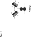
- an antigen binding receptor comprising an extracellular portion comprising an antigen binding moiety fused by a spacer sequence to an anchoring transmembrane domain which is itself fused to the intracellular signaling domains of CD3z and CD28.
- an "antigen binding site” refers to the site, i.e. one or more amino acid residues, of an antigen binding molecule which provides interaction with the antigen.
- the antigen binding site of an antibody or an antigen binding receptor comprises amino acid residues from the complementarity determining regions (CDRs).
- CDRs complementarity determining regions
- a native immunoglobulin molecule typically has two antigen binding sites, a Fab or a scFv molecule typically has a single antigen binding site.
- an antigen binding domain refers to the part of an antibody or an antigen binding receptor that comprises the area which specifically binds to and is complementary to part or all of an antigen.
- An antigen binding domain may be provided by, for example, one or more immunoglobuling variable domains (also called variable regions).
- an antigen binding domain comprises an immunoglobulin light chain variable region (VL) and an immunoglobulin heavy chain variable region (VH).
- variable region refers to the domain of an immunoglobulin heavy or light chain that is involved in binding the antigen.
- the variable domains of the heavy chain and light chain (VH and VL, respectively) of a native antibody generally have similar structures, with each domain comprising four conserved framework regions (FRs) and three hypervariable regions (HVRs). See, e.g., Kindt et al., Kuby Immunology, 6th ed., W.H. Freeman and Co, page 91 (2007 ). A single VH or VL domain is usually sufficient to confer antigen-binding specificity.
- ATM anchoring transmembrane domain
- the ATM can be fused to further extracellular and/or intracellular polypeptide domains wherein these extracellular and/or intracellular polypeptide domains will be confined to the cell membrane as well.
- the ATM confers membrane attachment and confinement of the antigen binding receptor of the present disclosure.
- the antigen binding receptors of the present disclosure comprise at least one ATM and an extracellular domain comprising an antigen binding moiety. Additionally, the ATM may be fused to further intracellular signaling domains.
- binding to as used in the context of the antigen binding receptors of the present disclosure defines a binding (interaction) of an "antigen-interaction-site" and an antigen with each other.
- antigen-interaction-site defines, in accordance with antigen binding receptors of the present disclosure, a motif of a polypeptide which shows the capacity of specific interaction with a specific antigen or a specific group of antigens (i.e. mutated Fc domains). Said binding/interaction is also understood to define a "specific recognition”.
- the term "specifically recognizing” means in accordance with this disclosure that the antigen binding receptor is capable of specifically interacting with and/or binding to a modified molecule as defined herein whereas the non-modified molecule is not recognized.
- the antigen binding moiety of an antigen binding receptor can recognize, interact and/or bind to different epitopes on the same molecule. This term relates to the specificity of the antigen binding receptor, i.e., to its ability to discriminate between the specific regions of a modified molecule, i.e. a mutated Fc domain, as defined herein.
- the specific interaction of the antigen-interaction-site with its specific antigen may result in an initiation of a signal, e.g.
- binding to does not only relate to a linear epitope but may also relate to a conformational epitope, a structural epitope or a discontinuous epitope consisting of two regions of the target molecules or parts thereof.
- a conformational epitope is defined by two or more discrete amino acid sequences separated in the primary sequence which comes together on the surface of the molecule when the polypeptide folds to the native protein ( Sela, Science 166 (1969), 1365 and Laver, Cell 61 (1990), 553-536 ).
- binding to is interchangeably used in the context of the present disclosure with the term “interacting with”. The ability of the antigen binding moiety (e.g.
- a Fab or scFv domain of an antigen binding receptor or an antibody to bind to a specific target antigenic determinant can be measured either through an enzyme-linked immunosorbent assay (ELISA) or other techniques familiar to one of skill in the art, e.g., surface plasmon resonance (SPR) technique (analyzed on a BIAcore instrument) ( Liljeblad et al., Glyco J 17, 323-329 (2000 )), and traditional binding assays ( Heeley, Endocr Res 28, 217-229 (2002 )).
- ELISA enzyme-linked immunosorbent assay
- SPR surface plasmon resonance
- the extent of binding of a antigen binding moiety to an unrelated protein is less than about 10% of the binding of the antigen binding moiety to the target antigen as measured, in particular by SPR.
- an antigen binding moiety that binds to the target antigen has a dissociation constant (K D ) of ⁇ 1 ⁇ M, ⁇ 100 nM, ⁇ 10 nM, ⁇ 1 nM, ⁇ 0.1 nM, ⁇ 0.01 nM, or ⁇ 0.001 nM (e.g., 10 -8 M or less, e.g., from 10 -8 M to 10 -13 M, e.g., from 10 -9 M to 10 -13 M).
- K D dissociation constant
- an antigen binding receptor of the present disclosure is capable of specific binding to a mutated Fc domain. Accordingly, the antigen binding receptor of the present disclosure specifically binds to/interacts with a mutated Fc domain.
- Cross-reactivity of a panel of constructs under investigation may be tested, for example, by assessing binding of a panel of antigen binding moieties under conventional conditions (see, e.g., Harlow and Lane, Antibodies: A Laboratory Manual, Cold Spring Harbor Laboratory Press, (1988 ) and Using Antibodies: A Laboratory Manual, Cold Spring Harbor Laboratory Press, (1999 )) to the mutated Fc domain of interest as well as to parent non-mutated Fc domain. Only those constructs (i.e.
- Fab fragments, scFvs and the like) that bind to the mutated Fc domain of interest but do not or do not essentially bind to a non-mutated parent Fc domain are considered specific for the mutated Fc domain of interest and selected for further studies in accordance with the method provided herein. These methods may comprise, inter alia, binding studies, blocking and competition studies with structurally and/or functionally closely related Fc domains.
- the binding studies also comprise FACS analysis, surface plasmon resonance (SPR, e.g. with BIAcore ® ), analytical ultracentrifugation, isothermal titration calorimetry, fluorescence anisotropy, fluorescence spectroscopy or by radiolabeled ligand binding assays.
- CDR as employed herein relates to "complementary determining region", which is well known in the art.
- the CDRs are parts of immunoglobulins or antigen binding receptors that determine the specificity of said molecules and make contact with a specific ligand.
- the CDRs are the most variable part of the molecule and contribute to the antigen binding diversity of these molecules.
- CDR-H depicts a CDR region of a variable heavy chain and CDR-L relates to a CDR region of a variable light chain.
- VH means the variable heavy chain and VL means the variable light chain.
- the CDR regions of an Ig-derived region may be determined as described in " Kabat” (Sequences of Proteins of Immunological Interest", 5th edit. NIH Publication no. 91-3242 U.S. Department of Health and Human Services (1991 ); Chothia J. Mol. Biol. 196 (1987), 901-917 ) or " Chothia” (Nature 342 (1989), 877-883 ).
- CD3z refers to T-cell surface glycoprotein CD3 zeta chain, also known as “T-cell receptor T3 zeta chain” and "CD247".
- chimeric antigen receptor or “chimeric receptor” or “CAR” refers to an antigen binding receptor constituted of an extracellular portion of an antigen binding moiety (e.g. a single chain antibody domain) fused by a spacer sequence to the intracellular signaling domains of CD3z and CD28.
- the present disclosure additionally provides antigen binding receptors wherein the antigen binding moiety is a Fab or a crossFab fragment.
- CAR is understood in its broadest form to comprise antigen binding receptors constituted of an extracellular portion comprising an antigen binding moiety fused to CD3z and fragment thereof and to CD28 and fragments thereof, optionally through one or several peptide linkers.
- the "class" of an antibody or immunoglobulin refers to the type of constant domain or constant region possessed by its heavy chain.
- the heavy chain constant domains that correspond to the different classes of immunoglobulins are called ⁇ , ⁇ , ⁇ , ⁇ , and ⁇ , respectively.
- crossover Fab molecule (also termed “crossFab” or “crossover Fab fragment”) is meant a Fab molecule wherein either the variable regions or the constant regions of the Fab heavy and light chain are exchanged, i.e. the crossFab fragment comprises a peptide chain composed of the light chain variable region and the heavy chain constant region, and a peptide chain composed of the heavy chain variable region and the light chain constant region.
- the peptide chain comprising the heavy chain constant region is referred to herein as the heavy chain of the crossover Fab molecule.
- a crossFab fragment comprises a heavy or light chain composed of the heavy chain variable and the light chain constant regions (VH-CL), and a heavy or light chain composed of the light chain variable and the heavy chain constant regions (VL-CH1).
- VH-CL heavy chain variable and the light chain constant regions
- VL-CH1 light chain variable and the heavy chain constant regions
- a "conventional Fab" molecule is meant a Fab molecule in its natural format, i.e. comprising a heavy chain composed of the heavy chain variable and constant regions (VH-CH1), and a light chain composed of the light chain variable and constant regions (VL-CL).
- CSD co-stimulatory signaling domain
- effector functions refers to those biological activities attributable to the Fc region of an antibody, which vary with the antibody isotype.
- antibody effector functions include: C1q binding and complement dependent cytotoxicity (CDC), Fc receptor binding, antibody-dependent cell-mediated cytotoxicity (ADCC), antibody-dependent cellular phagocytosis (ADCP), cytokine secretion, immune complex-mediated antigen uptake by antigen presenting cells, down regulation of cell surface receptors (e.g., B cell receptor), and B cell activation.
- engine As used herein, the terms “engineer”, “engineered”, “engineering”, are considered to include any manipulation of the peptide backbone or the post-translational modifications of a naturally occurring or recombinant polypeptide or fragment thereof. Engineering includes modifications of the amino acid sequence, of the glycosylation pattern, or of the side chain group of individual amino acids, as well as combinations of these approaches.
- expression cassette refers to a polynucleotide generated recombinantly or synthetically, with a series of specified nucleic acid elements that permit transcription of a particular nucleic acid in a target cell.
- the recombinant expression cassette can be incorporated into a plasmid, chromosome, mitochondrial DNA, plastid DNA, virus, or nucleic acid fragment.
- the recombinant expression cassette portion of an expression vector includes, among other sequences, a nucleic acid sequence to be transcribed and a promoter.
- the expression cassette of the present disclosure comprises polynucleotide sequences that encode antigen binding molecules of the present disclosure or fragments thereof.
- a “Fab molecule” refers to a protein consisting of the VH and CH1 domain of the heavy chain (the “Fab heavy chain”) and the VL and CL domain of the light chain (the “Fab light chain”) of an antigen binding molecule.
- Fc domain or "Fc region” herein is used to define a C-terminal region of an immunoglobulin heavy chain that contains at least a portion of the constant region.
- the term includes native sequence Fc regions and variant Fc regions.
- the boundaries of the Fc region of an IgG heavy chain might vary slightly, the human IgG heavy chain Fc region is usually defined to extend from Cys226, or from Pro230, to the carboxyl-terminus of the heavy chain.
- the C-terminal lysine (Lys447) of the Fc region may or may not be present.
- a subunit of an Fc domain as used herein refers to one of the two polypeptides forming the dimeric Fc domain, i.e. a polypeptide comprising C-terminal constant regions of an immunoglobulin heavy chain, capable of stable self-association.
- a subunit of an IgG Fc domain comprises an IgG CH2 and an IgG CH3 constant domain.
- FR Framework or "FR” refers to variable domain residues other than hypervariable region (HVR) residues.
- the FR of a variable domain generally consists of four FR domains: FR1, FR2, FR3, and FR4. Accordingly, the HVR and FR sequences generally appear in the following sequence in VH (or VL): FR1-H1(L1)-FR2-H2(L2)-FR3-H3(L3)-FR4.
- full length antibody denotes an antibody consisting of two “full length antibody heavy chains” and two “full length antibody light chains”.
- a “full length antibody heavy chain” is a polypeptide consisting in N-terminal to C-terminal direction of an antibody heavy chain variable domain (VH), an antibody constant heavy chain domain 1 (CH1), an antibody hinge region (HR), an antibody heavy chain constant domain 2 (CH2), and an antibody heavy chain constant domain 3 (CH3), abbreviated as VH-CH1-HR-CH2-CH3; and optionally an antibody heavy chain constant domain 4 (CH4) in case of an antibody of the subclass IgE.
- VH antibody heavy chain variable domain
- CH1 antibody constant heavy chain domain 1
- HR antibody hinge region
- CH2 antibody heavy chain constant domain 2
- CH3 antibody heavy chain constant domain 3
- the "full length antibody heavy chain” is a polypeptide consisting in N-terminal to C-terminal direction of VH, CH1, HR, CH2 and CH3.
- a “full length antibody light chain” is a polypeptide consisting in N-terminal to C-terminal direction of an antibody light chain variable domain (VL), and an antibody light chain constant domain (CL), abbreviated as VL-CL.
- the antibody light chain constant domain (CL) can be ⁇ (kappa) or ⁇ (lambda).
- the two full length antibody chains are linked together via inter-polypeptide disulfide bonds between the CL domain and the CH1 domain and between the hinge regions of the full length antibody heavy chains. Examples of typical full length antibodies are natural antibodies like IgG (e.g.
- fused is meant that the components (e.g., a Fab and a transmembrane domain) are linked by peptide bonds, either directly or via one or more peptide linkers.
- host cell refers to cells into which exogenous nucleic acid has been introduced, including the progeny of such cells.
- Host cells include “transformants” and “transformed cells” which include the primary transformed cell and progeny derived therefrom without regard to the number of passages. Progeny may not be completely identical in nucleic acid content to a parent cell, but may contain mutations. Mutant progeny that have the same function or biological activity as screened or selected for in the originally transformed cell are included herein.
- a host cell is any type of cellular system that can be used to generate an antibody used according to the present disclosure.
- Host cells include cultured cells, e.g., mammalian cultured cells, such as CHO cells, BHK cells, NS0 cells, SP2/0 cells, YO myeloma cells, P3X63 mouse myeloma cells, PER cells, PER.C6 cells or hybridoma cells, yeast cells, insect cells, and plant cells, to name only a few, but also cells comprised within a transgenic animal, transgenic plant or cultured plant or animal tissue.
- mammalian cultured cells such as CHO cells, BHK cells, NS0 cells, SP2/0 cells, YO myeloma cells, P3X63 mouse myeloma cells, PER cells, PER.C6 cells or hybridoma cells, yeast cells, insect cells, and plant cells, to name only a few, but also cells comprised within a transgenic animal, transgenic plant or cultured plant or animal tissue.
- hypervariable region refers to each of the regions of an antibody variable domain which are hypervariable in sequence and/or form structurally defined loops ("hypervariable loops").
- native four-chain antibodies comprise six HVRs; three in the VH (H1, H2, H3), and three in the VL (L1, L2, L3).
- HVRs generally comprise amino acid residues from the hypervariable loops and/or from the complementarity determining regions (CDRs), the latter being of highest sequence variability and/or involved in antigen recognition. With the exception of CDR1 in VH, CDRs generally comprise the amino acid residues that form the hypervariable loops.
- Hypervariable regions are also referred to as complementarity determining regions (CDRs), and these terms are used herein interchangeably in reference to portions of the variable region that form the antigen binding regions.
- CDRs complementarity determining regions
- This particular region has been described by Kabat et al., U.S. Dept. of Health and Human Services, Sequences of Proteins of Immunological Interest (1983 ) and by Chothia et al., J Mol Biol 196:901-917 (1987 ), where the definitions include overlapping or subsets of amino acid residues when compared against each other. Nevertheless, application of either definition to refer to a CDR of an antibody and/or an antigen binding receptor or variants thereof is intended to be within the scope of the term as defined and used herein.
- CDR Definitions 1 CDR Kabat Chothia AbM 2 V H CDR1 31-35 26-32 26-35 V H CDR2 50-65 52-58 50-58 V H CDR3 95-102 95-102 95-102 V L CDR1 24-34 26-32 24-34 V L CDR2 50-56 50-52 50-56 V L CDR3 89-97 91-96 89-97 1 Numbering of all CDR definitions in Table 1 is according to the numbering conventions set forth by Kabat et al. (see below). 2 "AbM” with a lowercase “b” as used in Table 1 refers to the CDRs as defined by Oxford Molecular's "AbM” antibody modeling software.
- Kabat et al. also defined a numbering system for variable region sequences that is applicable to any antibody.
- Kabat numbering refers to the numbering system set forth by Kabat et al., U.S. Dept. of Health and Human Services, "Sequence of Proteins of Immunological Interest” (1983 ). Unless otherwise specified, references to the numbering of specific amino acid residue positions in an antigen binding moiety variable region are according to the Kabat numbering system.
- the polypeptide sequences of the sequence listing are not numbered according to the Kabat numbering system. However, it is well within the ordinary skill of one in the art to convert the numbering of the sequences of the Sequence Listing to Kabat numbering.
- mammals include, but are not limited to, domesticated animals (e.g., cows, sheep, cats, dogs, and horses), primates (e.g., humans and non-human primates such as monkeys), rabbits, and rodents (e.g., mice and rats). Particularly, the individual or subject is a human.
- domesticated animals e.g., cows, sheep, cats, dogs, and horses
- primates e.g., humans and non-human primates such as monkeys
- rabbits e.g., mice and rats
- rodents e.g., mice and rats
- isolated nucleic acid molecule or polynucleotide is intended a nucleic acid molecule, DNA or RNA, which has been removed from its native environment.
- a recombinant polynucleotide encoding a polypeptide contained in a vector is considered isolated for the purposes of the present disclosure.
- Further examples of an isolated polynucleotide include recombinant polynucleotides maintained in heterologous host cells or purified (partially or substantially) polynucleotides in solution.
- An isolated polynucleotide includes a polynucleotide molecule contained in cells that ordinarily contain the polynucleotide molecule, but the polynucleotide molecule is present extrachromosomally or at a chromosomal location that is different from its natural chromosomal location.
- Isolated RNA molecules include in vivo or in vitro RNA transcripts of the present disclosure, as well as positive and negative strand forms, and double-stranded forms. Isolated polynucleotides or nucleic acids according to the present disclosure further include such molecules produced synthetically.
- a polynucleotide or a nucleic acid may be or may include a regulatory element such as a promoter, ribosome binding site, or a transcription terminator.
- nucleic acid or polynucleotide having a nucleotide sequence at least, for example, 95% "identical" to a reference nucleotide sequence of the present disclosure it is intended that the nucleotide sequence of the polynucleotide is identical to the reference sequence except that the polynucleotide sequence may include up to five point mutations per each 100 nucleotides of the reference nucleotide sequence.
- a polynucleotide having a nucleotide sequence at least 95% identical to a reference nucleotide sequence up to 5% of the nucleotides in the reference sequence may be deleted or substituted with another nucleotide, or a number of nucleotides up to 5% of the total nucleotides in the reference sequence may be inserted into the reference sequence.
- These alterations of the reference sequence may occur at the 5' or 3' terminal positions of the reference nucleotide sequence or anywhere between those terminal positions, interspersed either individually among residues in the reference sequence or in one or more contiguous groups within the reference sequence.
- any particular polynucleotide sequence is at least 80%, 85%, 90%, 95%, 96%, 97%, 98% or 99% identical to a nucleotide sequence of the present disclosure can be determined conventionally using known computer programs, such as the ones discussed below for polypeptides (e.g., ALIGN-2).
- an “isolated polypeptide” or a variant, or derivative thereof is intended a polypeptide that is not in its natural milieu. No particular level of purification is required.
- an isolated polypeptide can be removed from its native or natural environment.
- Recombinantly produced polypeptides and proteins expressed in host cells are considered isolated for the purpose of the present disclosure, as are native or recombinant polypeptides which have been separated, fractionated, or partially or substantially purified by any suitable technique.
- Percent (%) amino acid sequence identity with respect to a reference polypeptide sequence is defined as the percentage of amino acid residues in a candidate sequence that are identical with the amino acid residues in the reference polypeptide sequence, after aligning the sequences and introducing gaps, if necessary, to achieve the maximum percent sequence identity, and not considering any conservative substitutions as part of the sequence identity. Alignment for purposes of determining percent amino acid sequence identity can be achieved in various ways that are within the skill in the art, for instance, using publicly available computer software such as BLAST, BLAST-2, ALIGN or Megalign (DNASTAR) software. Those skilled in the art can determine appropriate parameters for aligning sequences, including any algorithms needed to achieve maximal alignment over the full length of the sequences being compared.
- the % amino acid sequence identity of a given amino acid sequence A to, with, or against a given amino acid sequence B is calculated as follows: 100 times the fraction X/Y where X is the number of amino acid residues scored as identical matches by the sequence alignment program ALIGN-2 in that program's alignment of A and B, and where Y is the total number of amino acid residues in B.
- nucleic acid molecule relates to the sequence of bases comprising purine- and pyrimidine bases which are comprised by polynucleotides, whereby said bases represent the primary structure of a nucleic acid molecule.
- nucleic acid molecule includes DNA, cDNA, genomic DNA, RNA, synthetic forms of DNA and mixed polymers comprising two or more of these molecules.
- nucleic acid molecule includes both, sense and antisense strands.
- the herein described nucleic acid molecule may contain non-natural or derivatized nucleotide bases, as will be readily appreciated by those skilled in the art.
- package insert is used to refer to instructions customarily included in commercial packages of therapeutic products, that contain information about the indications, usage, dosage, administration, combination therapy, contraindications and/or warnings concerning the use of such therapeutic products.
- composition refers to a preparation which is in such form as to permit the biological activity of an active ingredient contained therein to be effective, and which contains no additional components which are unacceptably toxic to a subject to which the formulation would be administered.
- a pharmaceutical composition usually comprises one or more pharmaceutically acceptable carrier(s).
- a “pharmaceutically acceptable carrier” refers to an ingredient in a pharmaceutical composition, other than an active ingredient, which is nontoxic to a subject.
- a pharmaceutically acceptable carrier includes, but is not limited to, a buffer, excipient, stabilizer, or preservative.
- polypeptide refers to a molecule composed of monomers (amino acids) linearly linked by amide bonds (also known as peptide bonds).
- the term polypeptide refers to any chain of two or more amino acids, and does not refer to a specific length of the product.
- peptides, dipeptides, tripeptides, oligopeptides, protein, amino acid chain, or any other term used to refer to a chain of two or more amino acids are included within the definition of polypeptide, and the term polypeptide may be used instead of, or interchangeably with any of these terms.
- polypeptide is also intended to refer to the products of post-expression modifications of the polypeptide, including without limitation glycosylation, acetylation, phosphorylation, amidation, derivatization by known protecting/blocking groups, proteolytic cleavage, or modification by non-naturally occurring amino acids.
- a polypeptide may be derived from a natural biological source or produced by recombinant technology, but is not necessarily translated from a designated nucleic acid sequence. It may be generated in any manner, including by chemical synthesis.
- a polypeptide of the present disclosure may be of a size of about 3 or more, 5 or more, 10 or more, 20 or more, 25 or more, 50 or more, 75 or more, 100 or more, 200 or more, 500 or more, 1,000 or more, or 2,000 or more amino acids.
- Polypeptides may have a defined three-dimensional structure, although they do not necessarily have such structure. Polypeptides with a defined three-dimensional structure are referred to as folded, and polypeptides which do not possess a defined three-dimensional structure, but rather can adopt a large number of different conformations, and are referred to as unfolded.
- polynucleotide refers to an isolated nucleic acid molecule or construct, e.g., messenger RNA (mRNA), virally-derived RNA, or plasmid DNA (pDNA).
- mRNA messenger RNA
- pDNA virally-derived RNA
- a polynucleotide may comprise a conventional phosphodiester bond or a non-conventional bond (e.g., an amide bond, such as found in peptide nucleic acids (PNA).
- PNA peptide nucleic acids
- nucleic acid molecule refers to any one or more nucleic acid segments, e.g., DNA or RNA fragments, present in a polynucleotide.
- Reduced binding for example reduced binding to an Fc receptor, refers to a decrease in affinity for the respective interaction, as measured for example by SPR.
- the term includes also reduction of the affinity to zero (or below the detection limit of the analytic method), i.e. complete abolishment of the interaction.
- increased binding refers to an increase in binding affinity for the respective interaction.
- control sequence refers to DNA sequences, which are necessary to effect the expression of coding sequences to which they are ligated. The nature of such control sequences differs depending upon the host organism. In prokaryotes, control sequences generally include promoter, ribosomal binding site, and terminators. In eukaryotes generally control sequences include promoters, terminators and, in some instances, enhancers, transactivators or transcription factors. The term “control sequence” is intended to include, at a minimum, all components the presence of which are necessary for expression, and may also include additional advantageous components.
- single-chain refers to a molecule comprising amino acid monomers linearly linked by peptide bonds.
- one of the antigen binding moieties is a scFv fragment, i.e. a VH domain and a VL domain connected by a peptide linker.
- one of the antigen binding moieties is a single-chain Fab molecule, i.e. a Fab molecule wherein the Fab light chain and the Fab heavy chain are connected by a peptide linker to form a single peptide chain.
- the C-terminus of the Fab light chain is connected to the N-terminus of the Fab heavy chain in the single-chain Fab molecule.
- SSD stimulatory signaling domain
- treatment refers to clinical intervention in an attempt to alter the natural course of a disease in the individual being treated, and can be performed either for prophylaxis or during the course of clinical pathology. Desirable effects of treatment include, but are not limited to, preventing occurrence or recurrence of disease, alleviation of symptoms, diminishment of any direct or indirect pathological consequences of the disease, preventing metastasis, decreasing the rate of disease progression, amelioration or palliation of the disease state, and remission or improved prognosis.
- cell expressing antigen binding receptors of the present disclosure are used together with therapeutic antibodies comprising a mutated Fc domain to delay development of a disease or to slow the progression of a disease.
- target antigenic determinant is synonymous with “target antigen”, “target epitope” and “target cell antigen” and refers to a site (e.g., a contiguous stretch of amino acids or a conformational configuration made up of different regions of non-contiguous amino acids) on a polypeptide macromolecule to which an antibody binds, forming an antigen binding moiety-antigen complex.
- Useful antigenic determinants can be found, for example, on the surfaces of tumor cells, on the surfaces of virus-infected cells, on the surfaces of other diseased cells, on the surface of immune cells, free in blood serum, and/or in the extracellular matrix (ECM).
- ECM extracellular matrix
- the proteins referred to as antigens herein can be any native form of the proteins from any vertebrate source, including mammals such as primates (e.g., humans) and rodents (e.g., mice and rats), unless otherwise indicated.
- the target antigen is a human protein.
- the term encompasses the "full-length", unprocessed target protein as well as any form of the target protein that results from processing in the target cell.
- the term also encompasses naturally occurring variants of the target protein, e.g., splice variants or allelic variants.
- Exemplary human target proteins useful as antigens include, but are not limited to: CD20, CEA, FAP, TNC, MSLN, FolR1, HER1 and HER2.
- ELISA enzyme-linked immunosorbent assay
- SPR surface plasmon resonance
- the extent of binding of the antibody to an unrelated protein is less than about 10% of the binding of the antibody to the target antigen as measured, e.g., by SPR.
- the antibody binds to the target antigen with an affinity dissociation constant (K D ) of ⁇ 1 ⁇ M, ⁇ 100 nM, ⁇ 10 nM, ⁇ 1 nM, ⁇ 0.1 nM, ⁇ 0.01 nM, or ⁇ 0.001 nM (e.g., 10 -8 M or less, e.g., from 10 -8 M to 10 -13 M, e.g., from 10 -9 M to 10 -13 M).
- K D affinity dissociation constant
- T cell receptor or "TCR” is commonly known in the art.
- T cell receptor refers to any T cell receptor, provided that the following three criteria are fulfilled: (i) tumor specificity, (ii) recognition of (most) tumor cells, which means that an antigen or target should be expressed in (most) tumor cells and (iii) that the TCR matches to the HLA-type of the subjected to be treated.
- the amino acid mutation is P329G wherein binding to Fcy receptor is reduced as measured by SPR at 25°C.
- the amino acid mutations are I253A, H310A and H435A wherein binding to the neonatal Fc receptor (FcRn) is reduced as measured by SPR at 25°C.
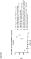
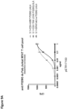
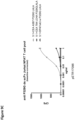
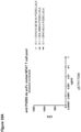
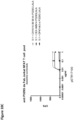
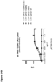
- Transduced T cells Jurkat NFAT T cells
- expressing the Anti-P329G-scFv-CD28ATD-CD28CSD-CD3zSSD protein SEQ ID NO:7 as encoded by the DNA sequence shown in SEQ ID NO:19
- the inventors further provided multiple formats of the antigen binding receptor capable of specific binding to a mutated Fc domain but not capable of specific binding to the non-mutated parent Fc domain to support the proof of the inventive concept.
- T cells preferably CD8+ T cells
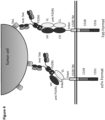
- an antigen binding receptor of the present disclosure can be specifically stimulated by the use of a tumor-specific antibody comprising a mutated Fc domain and recruited by the tumor-specific antibody as linking element to the tumor cell.
- a tumor-specific antibody i.e. a therapeutic antibody
- T cells transduced with an antigen binding receptor which comprise/consist of an extracellular domain comprising an antigen binding moiety capable of specific binding to the mutated Fc domain would result in a specific activation of the T cells and subsequent lysis of the tumor cell.
- the present disclosure provides a versatile therapeutic platform wherein IgG type antibodies may be used to mark or label tumor cells as a guidance for T cell and wherein transduced T cells are specifically targeted toward the tumor cells by providing specificity for a mutated Fc domain of the IgG type antibody. After binding to the mutated Fc domain of the antibody on the surface of a tumor cell, the transduced T cell as described herein becomes activated and the tumor cell will subsequently be lysed.
- an antigen binding receptor comprising an extracellular domain comprising at least one antigen binding moiety capable of specific binding to a mutated Fc domain, wherein the at least one antigen binding moiety is not capable of specific binding to the parent non-mutated Fc domain, wherein the mutated Fc domain comprises at least one amino acid mutation compared to the non-mutated parent Fc domain, wherein Fc receptor binding by the mutated Fc domain and/or effector function induced by the mutated Fc domain is reduced compared to Fc receptor binding and/or effector function induced by the non-mutated Fc domain.
- the antigen binding receptor comprises an extracellular domain that does not naturally occur in or on T cells.
- the antigen binding receptor is capable of providing tailored binding specificity to cells expressing the antigen binding receptor according to the present disclosure.
- Cells, e.g. T cells, transduced with (an) antigen binding receptor(s) of the present disclosure become capable of specific binding to a mutated Fc domain but not to the non-mutated parent Fc domain.
- antigen binding moiety of the extracellular domain of the antigen binding receptor such antigen binding moieties are considered to be specific for the mutated Fc domain as defined herein.
- antigen binding moiety capable of specific binding to a mutated Fc domain bind to/interact with the mutated Fc domain but not to/with the non-mutated parent Fc domain.
- At least one of the antigen binding moieties is a conventional Fab fragment, i.e. a Fab molecule consisting of a Fab light chain and a Fab heavy chain.
- at least one of the antigen binding moieties is a crossFab fragment, i.e. a Fab molecule consisting of a Fab light chain and a Fab heavy chain, wherein either the variable regions or the constant regions of the Fab heavy and light chain are exchanged.
- at least one of the antigen binding moieties is a scFv fragment.
- the C-terminus of the variable heavy chain (VH) is connected to the N-terminus of the variable light chain (VL) in the scFv molecule, optionally through a peptide linker.
- at least one of the antigen binding moieties is a single-chain Fab molecule, i.e. a Fab molecule wherein the Fab light chain and the Fab heavy chain are connected by a peptide linker to form a single peptide chain.
- the C-terminus of the Fab light chain is connected to the N-terminus of the Fab heavy chain in the single-chain Fab molecule, optionally through a peptide linker.
- the antigen binding moiety is capable of specific binding to a mutated Fc domain but not capable of specific binding to the non-mutated parent Fc domain, wherein the mutated Fc domain comprises at least one amino acid substitution compared to the non-mutated parent Fc domain.
- Antigen binding moieties capable of specific binding to a mutated Fc domain may be generated by immunization of e.g. a mammalian immune system. Such methods are known in the art and e.g. are described in Burns in Methods in Molecular Biology 295:1-12 (2005 ). Alternatively, antigen binding moieties of the present disclosure may be isolated by screening combinatorial libraries for antibodies with the desired activity or activities. Methods for screening combinatorial libraries are reviewed, e.g., in Lerner et al. in Nature Reviews 16:498-508 (2016 ). For example, a variety of methods are known in the art for generating phage display libraries and screening such libraries for antigen binding moieties possessing the desired binding characteristics.
- Phage typically display antibody fragments, either as single-chain Fv (scFv) fragments or as Fab fragments.
- Libraries from immunized sources provide high-affinity antigen binding moieties to the immunogen without the requirement of constructing hybridomas.
- the naive repertoire can be cloned (e.g., from human) to provide a single source of antigen binding moieties to a wide range of non-self and also self antigens without any immunization as described by Griffiths et al. in EMBO Journal 12: 725-734 (1993 ).
- naive libraries can also be made synthetically by cloning unrearranged V-gene segments from stem cells, and using PCR primers containing random sequence to encode the highly variable CDR3 regions and to accomplish rearrangement in vitro, as described by Hoogenboom and Winter in Journal of Molecular Biology 227: 381-388 (1992 ).
- Patent publications describing human antibody phage libraries include, for example: US Patent Nos. 5,750,373 ; 7,985,840 ; 7,785,903 and 8,679,490 as well as US Patent Publication Nos. 2005/0079574 , 2007/0117126 , 2007/0237764 and 2007/0292936 . and 2009/0002360 .
- ribosome and mRNA display as well as methods for antibody display and selection on bacteria, mammalian cells, insect cells or yeast cells.
- Methods for yeast surface display are reviewed, e.g., in Scholler et al. in Methods in Molecular Biology 503:135-56 (2012 ) and in Cherf et al. in Methods in Molecular biology 1319:155-175 (2015 ) as well as in the Zhao et al. in Methods in Molecular Biology 889:73-84 (2012 ).
- Methods for ribosome display are described, e.g., in He et al. in Nucleic Acids Research 25:5132-5134 (1997 ) and in Hanes et al. in PNAS 94:4937-4942 (1997 ).
- antigen binding receptors comprising at least one antigen binding moiety capable of specific binding to a mutated Fc domain.
- transduced cells i.e. T cells
- expressing an antigen binding receptor according to the present disclosure are capable of specific binding to the mutated Fc domain of an antibody, i.e. of a therapeutic antibody.
- the Fc domain confers to antibodies, i.e. therapeutic antibodies, favorable pharmacokinetic properties, including a long serum half-life which contributes to good accumulation in the target tissue and a favorable tissue-blood distribution ratio.
- Fc receptor signaling pathways may lead to cytokine release which, results in excessive activation of cytokine receptors and severe side effects upon systemic administration of therapeutic antibodies.
- Activation of (Fc receptor-bearing) immune cells other than T cells may even reduce efficacy of therapeutic antibodies due to the potential destruction of immune cells.
- therapeutic antibodies known in the art may be engineered or mutated to exhibit reduced binding affinity to an Fc receptor and/or reduced effector function, as compared to, e.g., a native IgG 1 Fc domain.
- the antigen binding receptors according to the present disclosure may be used to target effector cells, e.g.
- T cells expressing the antigen binding receptors according to the present disclosure in vitro and/or in vivo to target cells, i.e. tumor cells, which are labeled with an antibody capable of specific binding to the target cells, wherein the antibody comprise an engineered and/or mutated Fc domain as described herein.
- antigen binding receptors capable of specific binding to a mutated Fc domain comprising the amino acid mutation P329G and effector cells expressing said antigen binding receptors.
- the P329G mutation reduces binding to Fcy receptors and associated effector function. Accordingly, the mutated Fc domain comprising the P329G mutation binds to Fcy receptors with reduced or abolished affinity compared to the non-mutated Fc domain.
- antigen binding receptors capable of specific binding to a mutated Fc domain comprising the amino acid mutations I253A, H310A and H435A ("AAA").
- AAA antigen binding receptors capable of specific binding to a mutated Fc domain comprising the amino acid mutations I253A, H310A and H435A.
- the AAA mutations essentially abolishes binding to the FcRn.
- antigen binding receptors capable of specific binding to antibodies comprising a mutated Fc domain
- target antibodies such antibodies are herein also referred to as target antibodies.
- the antigen binding receptor of the present disclosure is capable of specific binding to a target antibody comprising a mutated Fc domain with reduced binding affinity to an Fc receptor and/or reduced effector function.
- Target antibodies with reduced effector function include those with mutation of one or more of Fc region residues 238, 265, 269, 270, 297, 327 and 329 ( U.S. Patent No. 6,737,056 ).
- Such Fc mutants include Fc mutants with mutations at two or more of amino acid positions 265, 269, 270, 297 and 327, including the so-called "DANA" Fc mutant with mutation of residues 265 and 297 to alanine ( US Patent No. 7,332,581 ).
- DANA "DANA" Fc mutant with mutation of residues 265 and 297 to alanine
- Certain antibody variants with improved or diminished binding to FcRs are described. (See, e.g., U.S. Patent No. 6,737,056 ; WO 2004/056312 , and Shields et al., J. Biol. Chem.
- an antigen binding receptor capable of specific binding to an antibody variant comprises an Fc region with one or more amino acid mutations which improve ADCC, e.g., mutations at positions 298, 333, and/or 334 of the Fc region (EU numbering of residues).
- a target antibody variant comprises an Fc region with one or more amino acid mutations, which reduce or diminish FcRn binding, e.g., mutations at positions 253, and/or 310, and/or 435 of the Fc region (EU numbering of residues).
- the target antibody variant comprises an Fc region with the amino acid mutations at positions 253, 310 and 435.
- the mutations are I253A, H310A and H435A in an Fc region derived from a human IgG1 Fc region. See e.g., Grevys, A., et al., J. Immunol. 194 (2015) 5497-5508 .
- an antigen binding receptor capable of specific binding to an antibody variant comprising an Fc region with one or more amino acid mutations, which reduced or diminished FcRn binding, e.g., mutations at one of the positions 310 and/or, 433 and/or 436 of the Fc region (EU numbering of residues).
- the target antibody variant comprises an Fc region with the amino acid mutations at positions 310, 433 and 436.
- the mutations are H310A, H433A and Y436A in an Fc region derived from a human IgG1 Fc region.
- a target antibody variant comprises an Fc region with one or more amino acid mutations, which increased FcRn binding, e.g., mutations at positions 252 and/or, 254 and/or 256 of the Fc region (EU numbering of residues).
- the target antibody variant comprises an Fc region with the amino acid mutations at positions 252, 254, and 256.
- the mutations are M252Y, S254T and T256E in an Fc region derived from a human IgG1 Fc region.
- an antigen binding receptor capable of specific binding to an antibody variant comprising an Fc region with amino acid mutations, which diminish FcyR binding, e.g., mutations at positions 234, 235 and 329 of the Fc region (EU numbering of residues).
- the mutations are L234A and L235A (LALA).
- the target antibody variant further comprises D265A and/or P329G in an Fc region derived from a human IgG1 Fc region.
- the mutation is P329G ("PG") in an Fc region derived from a human IgG1 Fc region.
- the mutations are I253A, H310A and H435A ("AAA") in an Fc region derived from a human IgG1 Fc region.
- the antigen binding moiety is capable of specific binding to a mutated Fc domain composed of a first and a second subunit capable of stable association.
- the Fc domain is an IgG, specifically an IgG 1 or IgG 4 , Fc domain.
- the Fc domain is a human Fc domain.
- the mutated Fc domain exhibits reduced binding affinity to an Fc receptor and/or reduced effector function, as compared to a native IgG 1 Fc domain.
- the Fc domain comprises one or more amino acid mutations that reduce binding to an Fc receptor and/or effector function.
- the antigen binding receptor essentially consists of a Fab or crossFab fragment, an anchoring transmembrane domain, a stimulatory signaling domain and a co-stimulatory signaling domain connected by one or more peptide linkers, wherein the Fab or crossFab fragment is fused at the C-terminus of the heavy or light chain to the N-terminus of the anchoring transmembrane domain, wherein the stimulatory signaling domain is fused at the C-terminus to the N-terminus of the co-stimulatory signaling domain.
- the co-stimulatory signaling domain is connected to the anchoring transmembrane domain instead of the stimulatory signaling domain.
- the antigen binding receptor is fused to a reporter protein, particularly to GFP or enhanced analogs thereof.
- the antigen binding receptor is fused at the C-terminus to the N-terminus of eGFP (enhanced green fluorescent protein), optionally through a peptide linker as described herein.
- the peptide linker is GEGRGSLLTCGDVEENPGP (T2A) according to SEQ ID NO: 18.
- the present disclosure provides an antigen binding receptor comprising in order from the N-terminus to the C-terminus
- the antigen binding moiety is a Fab fragment. In one embodiment, the antigen binding moiety is fused at the C-terminus of the Fab heavy chain to the N-terminus of an anchoring transmembrane domain.
- the anchoring transmembrane domain is a transmembrane domain selected from the group consisting of the CD8, the CD3z, the FCGR3A, the NKG2D, the CD27, the CD28, the CD137, the OX40, the ICOS, the DAP10 or the DAP12 transmembrane domain or a fragment thereof.
- the anchoring transmembrane domain is the CD28 transmembrane domain or a fragment thereof.
- the anchoring transmembrane domain is FWVLVVVGGVLACYSLLVTVAFIIFWV (SEQ ID NO:11).
- the antigen binding receptor further comprises a co-stimulatory signaling domain (CSD).
- the anchoring transmembrane domain of the antigen binding receptor is fused at the C-terminus to the N-terminus of a co-stimulatory signaling domain.
- the co-stimulatory signaling domain is individually selected from the group consisting of the intracellular domain of CD27, CD28, CD137, OX40, ICOS, DAP10 and DAP12, or fragments thereof as described herein before.
- the co-stimulatory signaling domain is the intracellular domain of CD28 or a fragment thereof.
- the co-stimulatory signaling domain comprises or consists of the sequence: RSKRSRLLHSDYMNMTPRRPGPTRKHYQPYAPPRDFAAYRS (SEQ ID NO:12).
- the antigen binding receptor further comprises a stimulatory signaling domain.
- the co-stimulatory signaling domain of the antigen binding receptor is fused at the C-terminus to the N-terminus of the stimulatory signaling domain.
- the at least one stimulatory signaling domain is individually selected from the group consisting of the intracellular domain of CD3z, FCGR3A and NKG2D, or fragments thereof.
- the co-stimulatory signaling domain is the intracellular domain of CD3z or a fragment thereof.
- the co-stimulatory signaling domain comprises or consists of the sequence:
- the antigen binding receptor is fused to a reporter protein, particularly to GFP or enhanced analogs thereof.
- the antigen binding receptor is fused at the C-terminus to the N-terminus of eGFP (enhanced green fluorescent protein), optionally through a peptide linker as described herein.
- the peptide linker is GEGRGSLLTCGDVEENPGP (T2A) of SEQ ID NO:18.
- the antigen binding receptor comprises an anchoring transmembrane domain and an extracellular domain comprising at least one antigen binding moiety, wherein the at least one antigen binding moiety is a Fab fragment capable of specific binding to a mutated Fc domain but not capable of specific binding to the non-mutated parent Fc domain, wherein the mutated Fc domain comprises the P329G mutation, wherein the P329G mutation reduces Fcy receptor binding.
- the antigen binding receptor of the present disclosure comprises an anchoring transmembrane domain (ATD), a co-stimulatory signaling domain (CSD) and a stimulatory signaling domain (SSD).
- the antigen binding moiety is a Fab fragment comprising a heavy chain comprising or consisting of an amino acid sequence of SEQ ID NO:40 or SEQ ID NO:49, and a light chain comprising or consisting of the amino acid sequence of SEQ ID NO:41 or SEQ ID NO:50.
- the antigen binding moiety is a Fab fragment comprising a heavy chain comprising or consisting of an amino acid sequence of SEQ ID NO:40 and a light chain comprising or consisting of the amino acid sequence of SEQ ID NO:41.
- the antigen binding moiety is a Fab fragment capable of specific binding to a mutated Fc domain comprising the P329G mutation
- the antigen binding receptor comprises a heavy chain fusion polypeptide comprising an amino acid sequence that is at least about 95%, 96%, 97%, 98%, 99% or 100% identical to an amino acid sequence selected from the group of SEQ ID NO:39 and SEQ ID NO:48 and a light chain polypeptide comprising an amino acid sequence that is at least about 95%, 96%, 97%, 98%, 99% or 100% identical to an amino acid sequence selected from the group of SEQ ID NO:41 and SEQ ID NO:50.
- the antigen binding moiety is a Fab fragment capable of specific binding to a mutated Fc domain comprising the P329G mutation, wherein the antigen binding receptor comprises a heavy chain fusion polypeptide comprising the amino acid sequence of SEQ ID NO:39 and a light chain polypeptide comprising the amino acid sequence of SEQ ID NO:41.
- the antigen binding receptor comprises one antigen binding moiety capable of specific binding to a mutated Fc domain, in particular an IgG1 Fc domain, comprising the mutations I253A, H310A and H435A ("AAA"),
- antigen binding moiety capable of specific binding to a mutated Fc domain but not capable of specific binding to the non-mutated parent Fc domain is a scFv, a Fab or a crossFab.
- the antigen binding moiety is fused at the C-terminus of the scFv fragment or at the C-terminus of the Fab or crossFab heavy chain to the N-terminus of an anchoring transmembrane domain, optionally through a peptide linker.
- the peptide linker comprises the amino acid sequence GGGGS (SEQ ID NO:16).
- the anchoring transmembrane domain is a transmembrane domain selected from the group consisting of the CD8, the CD3z, the FCGR3A, the NKG2D, the CD27, the CD28, the CD137, the OX40, the ICOS, the DAP10 or the DAP12 transmembrane domain or a fragment thereof.
- the anchoring transmembrane domain is the CD28 transmembrane domain or a fragment thereof.
- the anchoring transmembrane domain comprises or consist of the amino acid sequence of FWVLVVVGGVLACYSLLVTVAFIIFWV (SEQ ID NO:11).
- the antigen binding receptor further comprises a co-stimulatory signaling domain (CSD).
- the anchoring transmembrane domain of the antigen binding receptor is fused at the C-terminus to the N-terminus of a co-stimulatory signaling domain.
- the co-stimulatory signaling domain is individually selected from the group consisting of the intracellular domain of CD27, of CD28, of CD137, of OX40, of ICOS, of DAP10 and of DAP12, or fragments thereof as described herein before.
- the co-stimulatory signaling domain is the intracellular domain of CD28 or a fragment thereof.
- the co-stimulatory signaling domain comprises or consists of the sequence: RSKRSRLLHSDYMNMTPRRPGPTRKHYQPYAPPRDFAAYRS (SEQ ID NO:12).
- the antigen binding receptor further comprises a stimulatory signaling domain.
- the co-stimulatory signaling domain of the antigen binding receptor is fused at the C-terminus to the N-terminus of the stimulatory signaling domain.
- the at least one stimulatory signaling domain is individually selected from the group consisting of the intracellular domain of CD3z, FCGR3A and NKG2D, or fragments thereof.
- the co-stimulatory signaling domain is the intracellular domain of CD3z or a fragment thereof.
- the co-stimulatory signaling domain comprises or consists of the sequence:
- the antigen binding receptor is fused to a reporter protein, particularly to GFP or enhanced analogs thereof.
- the antigen binding receptor is fused at the C-terminus to the N-terminus of eGFP (enhanced green fluorescent protein), optionally through a peptide linker as described herein.
- the peptide linker is GEGRGSLLTCGDVEENPGP (T2A) according to SEQ ID NO:18.
- the antigen binding receptor comprises an anchoring transmembrane domain and an extracellular domain comprising at least one antigen binding moiety, wherein the at least one antigen binding moiety is a scFv fragment capable of specific binding to a mutated Fc domain but not capable of specific binding to the non-mutated parent Fc domain, wherein the mutated Fc domain comprises the I253A, H310A and H435A mutations.
- the I253A, H310A and H435A mutations reduce FcRn receptor binding.
- the antigen binding receptor of the present disclosure comprises an anchoring transmembrane domain (ATD), a co-stimulatory signaling domain (CSD) and a stimulatory signaling domain (SSD).
- the antigen binding receptor has the configuration scFv-ATD-CSD-SSD.
- the antigen binding receptor has the configuration scFv-G 4 S-ATD-CSD-SSD, wherein G 4 S is a linker comprising the sequence GGGGS of SEQ ID NO:17.
- a reporter protein can be added to the C-terminus of the antigen binding receptor, optionally through a peptide linker.
- the antigen binding moiety is a scFv fragment capable of specific binding to a mutated Fc domain comprising the I253A, H310A and H435A mutations, wherein the antigen binding moiety comprises at least one heavy chain complementarity determining region (CDR) selected from the group consisting of SEQ ID NO:53, SEQ ID NO:54 and SEQ ID NO:55 and at least one light chain CDR selected from the group of SEQ ID NO:56, SEQ ID NO:57, SEQ ID NO:58.
- CDR heavy chain complementarity determining region
- the antigen binding moiety is a scFv capable of specific binding to a mutated Fc domain comprising the I253A, H310A and H435A mutations, wherein the antigen binding moiety comprises the complementarity determining region (CDR H) 1 amino acid sequence SYGMS (SEQ ID NO:53), the CDR H2 amino acid sequence SSGGSY (SEQ ID NO:54), the CDR H3 amino acid sequence LGMITTGYAMDY (SEQ ID NO:55), the light chain complementary-determining region (CDR L) 1 amino acid sequence RSSQTIVHSTGHTYLE (SEQ ID NO:56), the CDR L2 amino acid sequence KVSNRFS (SEQ ID NO:57) and the CDR L3 amino acid sequence FQGSHVPYT (SEQ ID NO:58).
- CDR H complementarity determining region 1 amino acid sequence SYGMS
- SEQ ID NO:54 the CDR H2 amino acid sequence SSGGSY (SEQ ID NO:54), the CDR H3
- the present disclosure provides an antigen binding receptor comprising in order from the N-terminus to the C-terminus:
- the present disclosure provides an antigen binding receptor comprising in order from the N-terminus to the C-terminus:
- the antigen binding moiety is capable of specific binding to a mutated Fc domain comprising the I253A, H310A and H435A mutations, wherein the antigen binding receptor comprises an amino acid sequence that is at least about 95%, 96%, 97%, 98%, 99% or 100% identical to the amino acid sequence of: SEQ ID NO:59.
- the antigen binding moiety is capable of specific binding to a mutated Fc domain comprising the I253A, H310A and H435A mutations, wherein the antigen binding receptor comprises the amino acid sequence of: SEQ ID NO:595
- the antigen binding moiety is a Fab fragment. In one embodiment, the antigen binding moiety is fused at the C-terminus of the Fab heavy chain to the N-terminus of an anchoring transmembrane domain.
- the anchoring transmembrane domain is a transmembrane domain selected from the group consisting of the CD8, the CD3z, the FCGR3A, the NKG2D, the CD27, the CD28, the CD137, the OX40, the ICOS, the DAP10 or the DAP12 transmembrane domain or a fragment thereof.
- the anchoring transmembrane domain is the CD28 transmembrane domain or a fragment thereof.
- the anchoring transmembrane domain is FWVLVVVGGVLACYSLLVTVAFIIFWV (SEQ ID NO:11).
- the antigen binding receptor further comprises a co-stimulatory signaling domain (CSD).
- the anchoring transmembrane domain of the antigen binding receptor is fused at the C-terminus to the N-terminus of a co-stimulatory signaling domain.
- the co-stimulatory signaling domain is individually selected from the group consisting of the intracellular domain of CD27, CD28, CD137, OX40, ICOS, DAP10 and DAP12, or fragments thereof as described herein before.
- the co-stimulatory signaling domain is the intracellular domain of CD28 or a fragment thereof.
- the co-stimulatory signaling domain comprises or consists of the sequence RSKRSRLLHSDYMNMTPRRPGPTRKHYQPYAPPRDFAAYRS (SEQ ID NO: 12).
- the antigen binding receptor further comprises a stimulatory signaling domain.
- the co-stimulatory signaling domain of the antigen binding receptor is fused at the C-terminus to the N-terminus of the stimulatory signaling domain.
- the at least one stimulatory signaling domain is individually selected from the group consisting of the intracellular domain of CD3z, FCGR3A and NKG2D, or fragments thereof.
- the co-stimulatory signaling domain is the intracellular domain of CD3z or a fragment thereof.
- the co-stimulatory signaling domain comprises or consists of the sequence:
- the antigen binding receptor is fused to a reporter protein, particularly to GFP or enhanced analogs thereof.
- the antigen binding receptor is fused at the C-terminus to the N-terminus of eGFP (enhanced green fluorescent protein), optionally through a peptide linker as described herein.
- the peptide linker is GEGRGSLLTCGDVEENPGP (T2A) of SEQ ID NO:18.
- the antigen binding receptor comprises an anchoring transmembrane domain and an extracellular domain comprising at least one antigen binding moiety, wherein the at least one antigen binding moiety is a Fab fragment capable of specific binding to a mutated Fc domain but not capable of specific binding to the non-mutated parent Fc domain, wherein the mutated Fc domain comprises the I253A, H310A and H435A mutations, wherein the I253A, H310A and H435A mutations reduce FcRn receptor binding.
- the antigen binding receptor of the present disclosure comprises an anchoring transmembrane domain (ATD), a co-stimulatory signaling domain (CSD) and a stimulatory signaling domain (SSD).
- the antigen binding receptor has the configuration Fab-ATD-CSD-SSD.
- the antigen binding receptor has the configuration Fab- G 4 S-ATD-CSD-SSD, wherein G 4 S is a linker comprising the sequence GGGGS of SEQ ID NO:17.
- a reporter protein can be added to the C-terminus of the antigen binding receptor, optionally through a peptide linker.
- the antigen binding moiety is capable of specific binding to a mutated Fc domain comprising the I253A, H310A and H435A mutations, wherein the antigen binding moiety is a Fab fragment comprising at least one heavy chain complementarity determining region (CDR) selected from the group consisting of SEQ ID NO:53, SEQ ID NO:54 and SEQ ID NO:55 and at least one light chain CDR selected from the group of SEQ ID NO:56, SEQ ID NO:57, SEQ ID NO:58.
- CDR heavy chain complementarity determining region
- the antigen binding moiety is a Fab fragment capable of specific binding to a mutated Fc domain comprising the I253A, H310A and H435A mutations, wherein the antigen binding moiety comprises the complementarity determining region (CDR H) 1 amino acid sequence SYGMS (SEQ ID NO:53), the CDR H2 amino acid sequence SSGGSY (SEQ ID NO:54), the CDR H3 amino acid sequence LGMITTGYAMDY (SEQ ID NO:55), the light chain complementary-determining region (CDR L) 1 amino acid sequence RSSQTIVHSTGHTYLE (SEQ ID NO:56), the CDR L2 amino acid sequence KVSNRFS (SEQ ID NO:57) and the CDR L3 amino acid sequence FQGSHVPYT (SEQ ID NO:58).
- CDR H complementarity determining region 1 amino acid sequence SYGMS
- SEQ ID NO:54 the CDR H2 amino acid sequence SSGGSY (SEQ ID NO:54), the CDR H3 amino acid
- the antigen binding moiety is a Fab fragment comprising a heavy chain comprising or consisting of the amino acid sequence of SEQ ID NO:64 and a light chain comprising or consisting of the amino acid sequence of SEQ ID NO:65.
- the antigen binding moiety is a Fab fragment capable of specific binding to a mutated Fc domain comprising the I253A, H310A and H435A mutations, wherein the antigen binding receptor comprises a heavy chain fusion polypeptide comprising an amino acid sequence that is at least about 95%, 96%, 97%, 98%, 99% or 100% identical to the amino acid sequence of SEQ ID NO:63 and a light chain polypeptide comprising an amino acid sequence that is at least about 95%, 96%, 97%, 98%, 99% or 100% identical to the amino acid sequence of SEQ ID NO:65.
- the antigen binding moiety is a Fab fragment capable of specific binding to a mutated Fc domain comprising the I253A, H310A and H435A mutations, wherein the antigen binding receptor comprises a heavy chain fusion polypeptide comprising the amino acid sequence of SEQ ID NO:63 and a light chain polypeptide comprising the amino acid sequence of SEQ ID NO:65.
- the antigen binding receptor of the present disclosure the Fab light chain polypeptide and the Fab heavy chain fusion polypeptide are fused to each other, optionally via a linker peptide. Fusion of the Fab heavy and light chains can improve pairing of Fab heavy and light chains, and also reduces the number of plasmids needed for expression of some of the antigen binding receptor of the present disclosure.
- An alternative strategy to reduce the number of plasmids needed for expression of the antigen binding receptor is the use of an internal ribosomal entry side to enable expression of both heavy and light chain constructs from the same plasmid as illustrated e.g. in Figure 2 .
- the antigen binding receptor comprises a polypeptide wherein the Fab light chain variable region of the antigen binding moiety shares a carboxy-terminal peptide bond with the Fab heavy chain constant region of the antigen binding moiety (i.e. a the antigen binding moiety comprises a crossFab heavy chain, wherein the heavy chain variable region is replaced by a light chain variable region), which in turn shares a carboxy-terminal peptide bond with the anchoring transmembrane domain (VL (1) -CH1 (1) -ATD).
- the antigen binding receptor further comprises a polypeptide wherein the Fab heavy chain variable region of the first antigen binding moiety shares a carboxy-terminal peptide bond with the Fab light chain constant region of the first antigen binding moiety (VH(i)-CL(i)).
- the polypeptides are covalently linked, e.g., by a disulfide bond.
- the antigen binding receptor comprises a polypeptide wherein the Fab heavy chain variable region of the antigen binding moiety shares a carboxy-terminal peptide bond with the Fab light chain constant region of the antigen binding moiety (i.e.
- the antigen binding moiety comprises a crossFab heavy chain, wherein the heavy chain constant region is replaced by a light chain constant region), which in turn shares a carboxy-terminal peptide bond with an anchoring transmembrane domain (VH (1) -CL (1) -ATD).
- the antigen binding receptor further comprises a polypeptide wherein the Fab light chain variable region of the antigen binding moiety shares a carboxy-terminal peptide bond with the Fab heavy chain constant region of the antigen binding moiety (VL (1) -CH1 (1) )
- the polypeptides are covalently linked, e.g., by a disulfide bond.
- components of the antigen binding receptor may be fused directly or through various linkers, particularly peptide linkers comprising one or more amino acids, typically about 2-20 amino acids, that are described herein or are known in the art.
- Suitable, non-immunogenic peptide linkers include, for example, (G 4 S) n , (SG 4 ) n , (G 4 S) n or G 4 (SG 4 ) n peptide linkers, wherein n is generally a number between 1 and 10, preferably between 1 and 4.
- the antigen binding receptor "Anti-P329G-ds-scFv-CD28ATD-CD28CSD-CD3zSSD pETR17096" (SEQ ID NO:7) was constructed which comprises one stabilized scFv antigen binding moiety binding to/directed against/interacting with or on an antibody comprising the P329G mutation in the Fc domain.
- the construct further comprises the CD28 transmembrane domain, a fragment of CD28 as co-stimulatory signaling domain and a fragment of CD3z as stimulatory signaling domain.
- the antigen binding receptor "Anti-P329G-ds-Fab-CD28ATD-CD28CSD-CD3zSSD pETR17100” (SEQ ID NOs: 39, 41) was constructed which comprises one stabilized Fab antigen binding moiety binding to/directed against/interacting with or on an antibody comprising the P329G mutations in the Fc domain.
- the construct further comprises the CD28 transmembrane domain, a fragment of CD28 as co-stimulatory signaling domain and a fragment of CD3z as stimulatory signaling domain.
- the sequences (amino acid and DNA) of the antigen binding receptor "Anti-P329G-ds-Fab-CD28ATD-CD28CSD-CD3zSSD pETR17100" are shown in Tables 4 and 5.
- the antigen binding receptor "Anti-P329G-Fab-CD28ATD-CD28CSD-CD3zSSD pETR17594" (SEQ ID NOs: 48, 50) was constructed which comprises one Fab antigen binding moiety binding to/directed against/interacting with or on an antibody comprising the P329G mutations in the Fc domain.
- the construct further comprises the CD28 transmembrane domain, a fragment of CD28 as co-stimulatory signaling domain and a fragment of CD3z as stimulatory signaling domain.
- the sequences (amino acid and DNA) of the antigen binding receptor "Anti-P329G-Fab-CD28ATD-CD28CSD-CD3zSSD pETR17594" are shown in Tables 6 and 7.
- the antigen binding receptor "Anti-AAA scFv” (SEQ ID NO:59) was constructed which comprises one scFv antigen binding moiety binding to/directed against/interacting with or on an antibody comprising the I253A, H310A and H435A mutations in the Fc domain.
- the construct further comprises the CD28 transmembrane domain, a fragment of CD28 as co-stimulatory signaling domain and a fragment of CD3z as stimulatory signaling domain.
- the sequences (amino acid and cDNA) of the antibody binding molecule "Anti-AAA scFv" are shown below in Tables 8 and 9.
- the antigen binding receptor "Anti-AAA Fab” (SEQ ID NOs: 63, 65) was constructed which comprises one Fab antigen binding moiety binding to/directed against/interacting with or on an antibody comprising the I253A, H310A and H435A mutations in the Fc domain.
- the construct further comprises the CD28 transmembrane domain, a fragment of CD28 as co-stimulatory signaling domain and a fragment of CD3z as stimulatory signaling domain.
- the sequences (amino acid and cDNA) of the antibody binding molecule "Anti-AAA scFv" are shown below in Tables 10 and 11.
- the present disclosure also provides (a) nucleic acid molecule(s) encoding antigen binding receptors of the present disclosure as described herein. Also encompassed by the present disclosure are (a) nucleic acid molecule(s) encoding the antigen binding receptors of the present disclosure and kits comprising nucleic acid molecule(s) according to the present disclosure as further described herein.
- kits comprising or consisting of a nucleic acid encoding an antigen binding receptor of the present disclosure and/or cells, preferably T cells transduced with antigen binding receptors of the present disclosure and, optionally, (an) antibody/antibodies comprising a mutated Fc domain, wherein the antigen binding receptor is capable of specific binding to the mutated Fc domain.
- kits of the present disclosure may comprise transduced T cells, isolated polynucleotides and/or vectors and one or more antibodies comprising a mutated Fc domain.
- the antibody is a therapeutic antibody, e.g. a tumor specific antibody. Tumor specific antigens are known in the art and described herein.
- the antibody is administered before, simultaneously with or after administration of transduced T cell expressing an antigen binding receptor of the present disclosure.
- the kits according to the present disclosure comprise transduced T cells or polynucleotides/vectors to generate transduced T cells.
- the transduced T cells are universal T cells since they are not specific for a given tumor but can be targeted to any tumor depending on the therapeutic antibody comprising the mutated Fc domain.
- any antibody comprising a mutated Fc domain as described herein may be included in the herein provided kits.
- the mutated Fc domain of the antibodies exhibits reduced binding affinity to an Fc receptor and/or reduced effector function, as compared to a native IgG 1 Fc domain.
- the mutated Fc domain exhibits less than 50%, preferably less than 20%, more preferably less than 10% and most preferably less than 5% of the binding affinity to an Fc receptor, as compared to a native IgG 1 Fc domain (or an antibody comprising a native IgG 1 Fc domain), and/or less than 50%, preferably less than 20%, more preferably less than 10% and most preferably less than 5% of the effector function, as compared to a native IgG 1 Fc domain (or an antibody comprising a native IgG 1 Fc domain).
- the mutated Fc domain does not substantially bind to an Fc receptor and/or induce effector function.
- the Fc receptor is an Fc ⁇ receptor.
- the Fc receptor is a human Fc receptor.
- the Fc receptor is an activating Fc receptor.
- the Fc receptor is an activating human Fcy receptor, more specifically human FcyRIIIa, FcyRI or FcyRIIa, most specifically human FcyRIIIa.
- the effector function is one or more selected from the group of CDC, ADCC, ADCP, and cytokine secretion. In a particular embodiment the effector function is ADCC.
- the antibody comprising the mutated Fc region is a bispecific antibody, e.g. a T-cell activating bispecific antibody.
- the bispecific antibody comprises a first binding moiety capable of specific binding to a T-cell activating target, in particular CD3, and a second binding moiety capable of specific binding to a tumor antigen as described herein.
- kits comprising an amino acid sequence as shown in SEQ ID NO:7 ("Anti-P329G-ds-scFv-CD28ATD-CD28CSD-CD3zSSD" (as encoded by the DNA sequence shown in SEQ ID NO:19)) combined with the antibody comprising a heavy chain of SEQ ID NO:112 and a light chain of SEQ ID NO:113.
- the kit may comprise an amino acid sequence as shown in SEQ ID NO:31 ("Anti-P329G-scFv-CD28ATD-CD28CSD-CD3zSSD" (as encoded by the DNA sequence shown in SEQ ID NO:35)) combined with the antibody comprising a heavy chain of SEQ ID NO:112 and a light chain of SEQ ID NO:113.
- the kit may comprise an amino acid sequence as shown in SEQ ID NO:39 ("Anti-P329G-ds-Fab-CD28ATD-CD28CSD-CD3zSSD" (as encoded by the DNA sequence shown in SEQ ID NO:44)) combined with the antibody comprising a heavy chain of SEQ ID NO:112 and a light chain of SEQ ID NO:113.
- the kit may comprise an amino acid sequence as shown in SEQ ID NO:48 ("Anti-P329G-Fab-CD28ATD-CD28CSD-CD3zSSD" (as encoded by the DNA sequence shown in SEQ ID NO:51)) combined with an antibody comprising a heavy chain of SEQ ID NO:112 and a light chain of SEQ ID NO:113.
- the kit may comprise an amino acid sequence as shown in SEQ ID NO:59 (“Anti-AAA-scFv-CD28ATD-CD28CSD-CD3zSSD”) combined with an antibody comprising a heavy chain of SEQ ID NO:112 and a light chain of SEQ ID NO:113.
- the kit may comprise an amino acid sequence as shown in SEQ ID NO:63 ("Anti-AAA-Fab-CD28ATD-CD28CSD-CD3zSSD") combined with an antibody comprising a heavy chain of SEQ ID NO:112 and a light chain of SEQ ID NO:113.
- the kit may comprise at least one antibody molecule comprising a heavy chain and a light chain selected from the group consisting of SEQ ID NO:112 and SEQ ID NO:113, SEQ ID NO:114 and SEQ ID NO:115, SEQ ID NO:116 and SEQ ID NO:117, SEQ ID NO:118 and SEQ ID NO:119, SEQ ID NO:120 and SEQ ID NO:121, SEQ ID NO:122 and SEQ ID NO:123, and SEQ ID NO:124 and SEQ ID NO:125.
- the kit may comprise a bispecific antibody molecule, in particular a bispecific antibody comprising a first heavy chain of SEQ ID NO:128, a first light chain of SEQ ID NO:129, a second heavy chain of SEQ ID NO:130 and a second light chain of SEQ ID NO:131.
- kits of the present disclosure can be packaged individually in vials or bottles or in combination in containers or multicontainer units.
- the kit of the present disclosure may comprise a (closed) bag cell incubation system where patient cells, preferably T cells as described herein, can be transduced with (an) antigen binding receptor(s) of the present disclosure and incubated under GMP (good manufacturing practice, as described in the guidelines for good manufacturating practice published by the European Commission under http://ec.europa.eu/health/documents/eudralex/index_en.htm) conditions.
- GMP Good manufacturing practice, as described in the guidelines for good manufacturating practice published by the European Commission under http://ec.europa.eu/health/documents/eudralex/index_en.htm
- the kit of the present disclosure comprises a (closed) bag cell incubation system where isolated/obtained patients T cells can be transduced with (an) antigen binding receptor(s) of the present disclosure and incubated under GMP.
- the kit may also comprise a vector encoding (the) antigen binding receptor(s) as described herein.
- the kit of the present disclosure may be advantageously used, inter alia, for carrying out the method of the present disclosure and could be employed in a variety of applications referred herein, e.g., as research tools or medical tools.
- the manufacture of the kits preferably follows standard procedures which are known to the person skilled in the art.
- patient derived cells preferably T cells
- an antigen binding receptor of the present disclosure capable of specific binding to a mutated Fc domain as described herein using the kit as described above.
- the extracellular domain comprising an antigen binding moiety capable of specific binding to a mutated Fc domain does not naturally occur in or on T cells. Accordingly, the patient derived cells transduced with the kits of the present disclosure will acquire the capability of specific binding to a mutated Fc domain of an antibody, e.g.
- a therapeutic antibody and will become capable of inducing elimination/lysis of target cells via interaction with a therapeutic antibody comprising the mutated Fc domain, wherein the therapeutic antibody is able to bind to a tumor-specific antigen naturally occurring (that is endogenously expressed) on the surface of a tumor cell. Binding of the extracellular domain of the antigen binding receptor as described herein activates that T cell and brings it into physical contact with the tumor cell through the therapeutic antibody comprising the mutated Fc domain. Non-transduced or endogenous T cells (e.g. CD8+ T cells) are unable to bind to the mutated Fc domain of the therapeutic antibody comprising the mutated Fc domain.
- a tumor-specific antigen naturally occurring that is endogenously expressed
- T cells expressing the antigen binding receptor comprising the extracellular domain capable of specific binding to a mutated Fc domain remain unaffected by a therapeutic antibody not comprising the mutations in the Fc domain as described herein.
- T cells expressing the inventive antigen binding receptor molecule have the ability to lyse target cells in the presence of an antibody comprising the mutations in the Fc domain as described herein in vivo and/or in vitro.
- Corresponding target cells comprise cells expressing a surface molecule, i.e. a tumor-specific antigen naturally occurring on the surface of a tumor cell, which is recognized by at least one, preferably two, binding domains of the therapeutic antibody as described herein.
- Such surface molecules are characterized herein below.
- Lysis of the target cell can be detected by methods known in the art. Accordingly, such methods comprise, inter alia, physiological in vitro assays. Such physiological assays may monitor cell death, for example by loss of cell membrane integrity (e.g. FACS based propidium Iodide assay, trypan blue influx assay, photometric enzyme release assays (LDH), radiometric 51Cr release assay, fluorometric Europium release and CalceinAM release assays).
- FACS based propidium Iodide assay e.g. FACS based propidium Iodide assay, trypan blue influx assay, photometric enzyme release assays (LDH), radiometric 51Cr release assay, fluorometric Europium release and CalceinAM release assays.
- LDH photometric enzyme release assays
- radiometric 51Cr release assay e.g., radiometric 51Cr release assay, fluorometric Europium release and CalceinAM release assays.
- Further assays comprise monitoring of cell viability, for example by photometric MTT, XTT, WST-1 and alamarBlue assays, radiometric 3H-Thd incorporation assay, clonogenic assay measuring cell division activity, and fluorometric Rhodamine123 assay measuring mitochondrial transmembrane gradient.
- apoptosis may be monitored for example by FACS-based phosphatidylserin exposure assay, ELISA-based TUNEL test, caspase activity assay (photometric, fluorometric or ELISA-based) or analyzing changed cell morphology (shrinking, membrane blebbing).
- Transduced T cells capable of expressing antigen binding receptors of the present disclosure
- a further aspect of the present disclosure are transduced T cells capable of expressing an antigen binding receptor of the present disclosure.
- the antigen binding receptors as described herein relate to molecules which are naturally not comprised in and/or on the surface of T cells and which are not (endogenously) expressed in or on normal (non-transduced) T cells.
- the antigen binding receptor of the present disclosure in and/or on T cells is artificially introduced into T cells.
- said T cells preferably CD8+ T cells, may be isolated/obtained from a subject to be treated as defined herein.
- the antigen binding receptors as described herein which are artificially introduced and subsequently presented in and/or on the surface of said T cells comprise domains comprising one or more antigen binding moiety accessible ( in vitro or in vivo ) to (Ig-derived) immunoglobulins, preferably antibodies, in particular to the Fc domain of the antibodies.
- these artificially introduced molecules are presented in and/or on the surface of said T cells after (retroviral or lentiviral) transduction as described herein below.
- T cells according to the present disclosure can be activated by immunoglobulins, preferably (therapeutic) antibodies comprising specific mutations in the Fc domain as described herein.
- the present disclosure also relates to transduced T cells expressing an antigen binding receptor encoded by (a) nucleic acid molecule(s) encoding the antigen binding receptor of the present disclosure.
- the transduced cell may comprise a nucleic acid molecule encoding the antigen binding receptor of the present disclosure or a vector of the present disclosure which expresses an antigen binding receptor of the present disclosure.
- transduced T cell relates to a genetically modified T cell (i.e. a T cell wherein a nucleic acid molecule has been introduced deliberately).
- the herein provided transduced T cell may comprise the vector of the present disclosure.
- the herein provided transduced T cell comprises the nucleic acid molecule encoding the antigen binding receptor of the present disclosure and/or the vector of the present disclosure.
- the transduced T cell of the present disclosure may be a T cell which transiently or stably expresses the foreign DNA (i.e. the nucleic acid molecule which has been introduced into the T cell).
- the nucleic acid molecule encoding the antigen binding receptor of the present disclosure can be stably integrated into the genome of the T cell by using a retroviral or lentiviral transduction.
- the nucleic acid molecule encoding the antigen binding receptor of the present disclosure may be expressed transiently.
- the herein provided transduced T cell has been genetically modified by introducing a nucleic acid molecule in the T cell via a viral vector (e.g. a retroviral vector or a lentiviral vector).
- the expression of the antigen binding receptors may be constitutive and the extracellular domain of the antigen binding receptor may be detectable on the cell surface.
- This extracellular domain of the antigen binding receptor may comprise the complete extracellular domain of an antigen binding receptor as defined herein but also parts thereof. The minimal size required being the antigen binding site of the antigen binding moiety in the antigen binding receptor.
- the expression may also be conditional or inducible in the case that the antigen binding receptor is introduced into T cells under the control of an inducible or repressible promoter.
- inducible or repressible promoters can be a transcriptional system containing the alcohol dehydrogenase I (alcA) gene promoter and the transactivator protein AlcR.
- alcA alcohol dehydrogenase I
- AlcR transactivator protein
- Different agricultural alcohol-based formulations are used to control the expression of a gene of interest linked to the alcA promoter.
- tetracycline-responsive promoter systems can function either to activate or repress gene expression system in the presence of tetracycline.
- Some of the elements of the systems include a tetracycline repressor protein (TetR), a tetracycline operator sequence (tetO) and a tetracycline transactivator fusion protein (tTA), which is the fusion of TetR and a herpes simplex virus protein 16 (VP16) activation sequence.
- TetR tetracycline repressor protein
- tetO tetracycline operator sequence
- tTA tetracycline transactivator fusion protein
- VP16 herpes simplex virus protein 16
- steroid-responsive promoters, metal-regulated or pathogenesis-related (PR) protein related promoters can be used.
- the expression can be constitutive or constitutional, depending on the system used.
- the antigen binding receptors of the present disclosure can be expressed on the surface of the herein provided transduced T cell.
- the extracellular portion of the antigen binding receptor i.e. the extracellular domain of the antigen binding receptor can be detected on the cell surface, while the intracellular portion (i.e. the co-stimulatory signaling domain(s) and the stimulatory signaling domain) are not detectable on the cell surface.
- the detection of the extracellular domain of the antigen binding receptor can be carried out by using an antibody which specifically binds to this extracellular domain or by the mutated Fc domain which the extracellular domain is capable to bind.
- the extracellular domain can be detected using these antibodies or Fc domains by flow cytometry or microscopy.
- the transduced cells of the present disclosure may be any immune cell. These include but are not limited to B-cells, T cells, Natural Killer (NK) cells, Natural Killer (NK) T cells, ⁇ T cells, innate lymphoid cells, macrophages, monocytes, dendritic cells, or neutrophils.
- said immune cell would be a lymphocyte, preferentially a NK or T cells.
- the said T cells include CD4 T cells and CD8 T cells. Triggering of the antigen binding receptor of the present disclosure on the surface of the leukocyte will render the cell cytotoxic against a target cell in conjunction with a therapeutic antibody comprising a mutated Fc domain irrespective of the lineage the cell originated from.
- the transduced cell of the present disclosure may be, e.g., a CD4+ T cell, a CD8+-T cell, a ⁇ T cell, a Natural Killer (NK) T cell, a Natural Killer (NK) cell, a tumor-infiltrating lymphocyte (TIL) cell, a myeloid cell, or a mesenchymal stem cell.
- the herein provided transduced cell is a T cell (e.g.
- the transduced cell is a CD8+ T cell. Accordingly, in the context of the present disclosure, the transduced cell is a CD8+ T cell. Further, in the context of the present disclosure, the transduced cell is an autologous T cell. Accordingly, in the context of the present disclosure, the transduced cell is preferably an autologous CD8+ T cell. In addition to the use of autologous cells (e.g. T cells) isolated from the subject, the present disclosure also comprehends the use of allogeneic cells. Accordingly, in the context of the present disclosure the transduced cell may also be an allogeneic cell, such as an allogeneic CD8+ T cell.
- the transduced cell may also be an allogeneic cell, such as an allogeneic CD8+ T cell.
- allogeneic cells are based on the fact that cells, preferably T cells can recognize a specific antigen epitope presented by foreign antigen-presenting cells (APC), provided that the APC express the MHC molecule, class I or class II, to which the specific responding cell population, i.e. T cell population is restricted, along with the antigen epitope recognized by the T cells.
- APC foreign antigen-presenting cells
- allogeneic refers to cells from an unrelated coming from an unrelated donor individual/subject which is human leukocyte antigen (HLA) compatible to the individual/subject which will be treated by e.g. the herein described antigen binding receptor expressing transduced cell.
- HLA human leukocyte antigen
- Autologous cells refer to cells which are isolated/obtained as described herein above from the subject to be treated with the transduced cell described herein.
- the transduced cell of the present disclosure may be co-transduced with further nucleic acid molecules, e.g. with a nucleic acid molecule encoding a T cell receptor.
- the present disclosure also relates to a method for the production of a transduced T cell expressing an antigen binding receptor of the present disclosure, comprising the steps of transducing a T cell with a vector of the present disclosure, culturing the transduced T cell under conditions allowing the expressing of the antigen binding receptor in or on said transduced cell and recovering said transduced T cell.
- the transduced cell of the present disclosure is preferably produced by the following process: cells (e.g., T cells, preferably CD8+ T cells) are isolated/obtained from a subject (preferably a human patient). Methods for isolating/obtaining cells (e.g. T cells, preferably CD8+ T cells) from patients or from donors are well known in the art and in the context of the present the cells (e.g. T cells, preferably CD8+ T cells) from patients or from donors may be isolated by blood draw or removal of bone marrow. After isolating/obtaining cells as a sample of the patient, the cells (e.g. T cells) are separated from the other ingredients of the sample. Several methods for separating cells (e.g.
- T cells from the sample are known and include, without being limiting, e.g. leukapheresis for obtaining cells from the peripheral blood sample from a patient or from a donor, isolating/obtaining cells by using a FACSort apparatus, picking living of dead cells from fresh biopsy specimens harboring living cells by hand or by using a micromanipulator (see, e.g., Dudley, Immunother. 26 (2003), 332-342 ; Robbins, Clin. Oncol. 29 (201 1), 917-924 or Leisegang, J. Mol. Med. 86 (2008), 573-58 ).
- the isolated/obtained cells T cells are subsequently cultivated and expanded, e.g., by using an anti-CD3 antibody, by using anti-CD3 and anti-CD28 monoclonal antibodies and/or by using an anti-CD3 antibody, an anti-CD28 antibody and interleukin-2 (IL-2) (see, e.g., Dudley, Immunother. 26 (2003), 332-342 or Dudley, Clin. Oncol. 26 (2008), 5233-5239 ).
- IL-2 interleukin-2
- the cells are artificially/genetically modified/transduced by methods known in the art (see, e.g., Lemoine, J Gene Med 6 (2004), 374-386 ).
- Methods for transducing cells are known in the art and include, without being limited, in a case where nucleic acid or a recombinant nucleic acid is transduced, for example, an electroporation method, calcium phosphate method, cationic lipid method or liposome method.
- the nucleic acid to be transduced can be conventionally and highly efficiently transduced by using a commercially available transfection reagent, for example, Lipofectamine (manufactured by Invitrogen, catalogue no.: 11668027).
- the vector can be transduced in the same manner as the above-mentioned nucleic acid as long as the vector is a plasmid vector (i.e. a vector which is not a viral vector
- the methods for transducing cells include retroviral or lentiviral T cell transduction, non-viral vectors (e.g., sleeping beauty minicircle vector) as well as mRNA transfection.
- mRNA transfection refers to a method well known to those skilled in the art to transiently express a protein of interest, like in the present case the antigen binding receptor of the present disclosure, in a cell to be transduced.
- cells may be electroporated with the mRNA coding for the antigen binding receptor of the present by using an electroporation system (such as e.g. Gene Pulser, Bio-Rad) and thereafter cultured by standard cell (e.g. T cell) culture protocol as described above (see Zhao et al., Mol Ther. 13(1) (2006), 151-159 .)
- the transduced cell of the present disclosure is a T cell, most preferably a CD8+ T cell, and is generated by lentiviral, or most preferably retroviral T cell transduction.
- retroviral vectors for transducing T cells are known in the art such as SAMEN CMV/SRa ( Clay et al., J. Immunol. 163 (1999), 507-513 ), LZRS-id3-IHRES ( Heemskerk et al., J. Exp. Med. 186 (1997), 1597-1602 ), FeLV ( Neil et al., Nature 308 (1984), 814-820 ), SAX ( Kantoff et al., Proc. Natl. Acad. Sci. USA 83 (1986), 6563-6567 ), pDOL ( Desiderio, J. Exp. Med.
- suitable lentiviral vector for transducing cells are, e.g. PL-SIN lentiviral vector ( Hotta et al., Nat Methods.
- the transduced T cell/T cells of the present disclosure is/are preferably grown under controlled conditions, outside of their natural environment.
- the term "culturing” means that cells (e.g. the transduced cell(s) of the present disclosure) which are derived from multi-cellular eukaryotes (preferably from a human patient) are grown in vitro. Culturing cells is a laboratory technique of keeping cells alive which are separated from their original tissue source.
- the transduced cell of the present disclosure is cultured under conditions allowing the expression of the antigen binding receptor of the present disclosure in or on said transduced cells. Conditions which allow the expression or a transgene (i.e.
- the antigen binding receptor of the present disclosure are commonly known in the art and include, e.g., agonistic anti-CD3- and anti-CD28 antibodies and the addition of cytokines such as interleukin 2 (IL-2), interleukin 7 (IL-7), interleukin 12 (IL-12) and/or interleukin 15 (IL-15).
- IL-2 interleukin 2
- IL-7 interleukin 7
- IL-12 interleukin 12
- IL-15 interleukin 15
- transduced cell preferably a T cell, in particular a CD8+ T expressing an antigen binding receptor encoded by a nucleic acid molecule of the present disclosure obtainable by the method of the present disclosure.
- a further aspect of the present disclosure are nucleic acids and vectors encoding one or several antigen binding receptors of the present disclosure.
- Exemplary nucleic acid molecules encoding the antigen binding receptors of the present disclosure are shown in SEQ ID NOs:19, 30, 35, 38, 44, 47, 51 and 52.
- the nucleic acid molecules of the present disclosure may be under the control of regulatory sequences.
- promoters, transcriptional enhancers and/or sequences which allow for induced expression of the antigen binding receptor of the disclosure may be employed.
- the nucleic acid molecules are expressed under the control of constitutive or inducible promoter. Suitable promoters are e.g.
- the CMV promoter Qin et al., PLoS One 5(5) (2010), e10611
- the UBC promoter Qin et al., PLoS One 5(5) (2010), e10611
- PGK Qin et al., PLoS One 5(5) (2010), e10611
- the EF1A promoter Qin et al., PLoS One 5(5) (2010), e10611
- the CAGG promoter Qin et al., PLoS One 5(5) (2010), e10611
- the SV40 promoter Qin et al., PLoS One 5(5) (2010), e10611
- the COPIA promoter Qin et al., PLoS One 5(5) (2010), e10611
- the ACT5C promoter Qin et al., PLoS One 5(5) (2010), e10611
- the TRE promoter Qin et al., PLoS One
- the present disclosure therefore also relates to (a) vector(s) comprising the nucleic acid molecule(s) described in the present disclosure.
- the term vector relates to a circular or linear nucleic acid molecule which can autonomously replicate in a host cell (i.e. in a transduced cell) into which it has been introduced.
- plasmids are known to those skilled in molecular biology, the choice of which would depend on the function desired and include plasmids, cosmids, viruses, bacteriophages and other vectors used conventionally in genetic engineering. Methods which are well known to those skilled in the art can be used to construct various plasmids and vectors; see, for example, the techniques described in Sambrook et al. (loc cit.) and Ausubel, Current Protocols in Molecular Biology, Green Publishing Associates and Wiley Interscience, N.Y. (1989), (1994 ). Alternatively, the polynucleotides and vectors of the present disclosure can be reconstituted into liposomes for delivery to target cells.
- a cloning vector was used to isolate individual sequences of DNA. Relevant sequences can be transferred into expression vectors where expression of a particular polypeptide is required.
- Typical cloning vectors include pBluescript SK, pGEM, pUC9, pBR322, pGA18 and pGBT9.
- Typical expression vectors include pTRE, pCAL-n-EK, pESP-1, pOP13CAT.
- the present disclosure also relates to (a) vector(s) comprising (a) nucleic acid molecule(s) which is (are) a regulatory sequence operably linked to said nucleic acid molecule(s) encoding an antigen binding receptor as defined herein.
- the vector can be polycistronic.
- regulatory sequences (control elements) are known to the skilled person and may include a promoter, a splice cassette, translation initiation codon, translation and insertion site for introducing an insert into the vector(s).
- said nucleic acid molecule(s) is (are) operatively linked to said expression control sequences allowing expression in eukaryotic or prokaryotic cells.
- said vector(s) is (are) an expression vector(s) comprising the nucleic acid molecule(s) encoding the antigen binding receptor as defined herein.
- Operably linked refers to a juxtaposition wherein the components so described are in a relationship permitting them to function in their intended manner.
- a control sequence operably linked to a coding sequence is ligated in such a way that expression of the coding sequence is achieved under conditions compatible with the control sequences.
- the control sequence is a promoter, it is obvious for a skilled person that double-stranded nucleic acid is preferably used.
- an expression vector is a construct that can be used to transform a selected cell and provides for expression of a coding sequence in the selected cell.
- An expression vector(s) can for instance be cloning (a) vector(s), (a) binary vector(s) or (a) integrating vector(s).
- Expression comprises transcription of the nucleic acid molecule preferably into a translatable mRNA. Regulatory elements ensuring expression in prokaryotes and/or eukaryotic cells are well known to those skilled in the art. In the case of eukaryotic cells they comprise normally promoters ensuring initiation of transcription and optionally poly-A signals ensuring termination of transcription and stabilization of the transcript.
- Possible regulatory elements permitting expression in prokaryotic host cells comprise, e.g., the PL, lac, trp or tac promoter in E. coli, and examples of regulatory elements permitting expression in eukaryotic host cells are the AOX1 or GAL1 promoter in yeast or the CMV-, SV40 , RSV-promoter (Rous sarcoma virus), CMV-enhancer, SV40-enhancer or a globin intron in mammalian and other animal cells.
- Beside elements which are responsible for the initiation of transcription such regulatory elements may also comprise transcription termination signals, such as the SV40-poly-A site or the tk-poly-A site, downstream of the polynucleotide.
- transcription termination signals such as the SV40-poly-A site or the tk-poly-A site, downstream of the polynucleotide.
- leader sequences encoding signal peptides capable of directing the polypeptide to a cellular compartment or secreting it into the medium may be added to the coding sequence of the recited nucleic acid sequence and are well known in the art; see also, e.g., appended Examples.
- the leader sequence(s) is (are) assembled in appropriate phase with translation, initiation and termination sequences, and preferably, a leader sequence capable of directing secretion of translated protein, or a portion thereof, into the periplasmic space or extracellular medium.
- the heterologous sequence can encode an antigen binding receptor including an N-terminal identification peptide imparting desired characteristics, e.g., stabilization or simplified purification of expressed recombinant product; see supra.
- suitable expression vectors are known in the art such as Okayama-Berg cDNA expression vector pcDV1 (Pharmacia), pCDM8, pRc/CMV, pcDNA1, pcDNA3 (In-vitrogene), pEF-DHFR, pEF-ADA or pEF-neo ( Kunststoff et al. Cancer Immunol Immunother 50 (2001), 141-150 ) or pSPORT1 (GIBCO BRL).
- the expression control sequences will be eukaryotic promoter systems in vectors capable of transforming or transfecting eukaryotic cells, but control sequences for prokaryotic cells may also be used.
- control sequences for prokaryotic cells may also be used.
- the vector Once the vector has been incorporated into the appropriate cell, the cell is maintained under conditions suitable for high level expression of the nucleotide sequences, and as desired. Additional regulatory elements may include transcriptional as well as translational enhancers.
- the above-described vectors of the present disclosure comprise a selectable and/or scorable marker.
- Selectable marker genes useful for the selection of transformed cells and, e.g., plant tissue and plants are well known to those skilled in the art and comprise, for example, antimetabolite resistance as the basis of selection for dhfr, which confers resistance to methotrexate ( Reiss, Plant Physiol. (Life Sci. Adv.) 13 (1994), 143-149 ), npt, which confers resistance to the aminoglycosides neomycin, kanamycin and paromycin ( Herrera-Estrella, EMBO J. 2 (1983), 987-995 ) and hygro, which confers resistance to hygromycin ( Marsh, Gene 32 (1984), 481-485 ).
- trpB which allows cells to utilize indole in place of tryptophan
- hisD which allows cells to utilize histinol in place of histidine
- mannose-6-phosphate isomerase which allows cells to utilize mannose ( WO 94/20627 ) and ODC (omithine decarboxylase) which confers resistance to the ornithine decarboxylase inhibitor, 2-(difluoromethyl)-DL-omithine, DFMO ( McConlogue, 1987, In: Current Communications in Molecular Biology, Cold Spring Harbor Laboratory ed .) or deaminase from Aspergillus terreus which confers resistance to Blasticidin S ( Tamura, Biosci. Biotechnol. Biochem. 59 (1995), 2336-2338 ).
- luciferase Giacomin, Pl. Sci. 116 (1996), 59-72 ; Scikantha, J. Bact. 178 (1996), 121 ), green fluorescent protein ( Gerdes, FEBS Lett. 389 (1996), 44-47 ) or ⁇ -glucuronidase ( Jefferson, EMBO J. 6 (1987), 3901-3907 ).
- This embodiment is particularly useful for simple and rapid screening of cells, tissues and organisms containing a recited vector.
- Suitable vectors, methods or gene-delivery systems for in methods or gene-delivery systems for in-vitro or in-vivo gene therapy are described in the literature and are known to the person skilled in the art; see, e.g., Giordano, Nature Medicine 2 (1996), 534-539 ; Schaper, Circ. Res. 79 (1996), 911-919 ; Anderson, Science 256 (1992), 808-813 ; Verma, Nature 389 (1994), 239 ; Isner, Lancet 348 (1996), 370-374 ; Muhlhauser, Circ. Res. 77 (1995), 1077-1086 ; Onodera, Blood 91 (1998), 30-36 ; Verma, Gene Ther.
- nucleic acid molecule(s) and vector(s) may be designed for direct introduction or for introduction via liposomes, or viral vectors (e.g., adenoviral, retroviral) into the cell.
- said cell is a T cells, such as CD8+ T cells, CD4+ T cells, CD3+ T cells, ⁇ T cells or natural killer (NK) T cells, preferably CD8+ T cells.
- the present disclosure provides methods to derive vectors, particularly plasmids, cosmids and bacteriophages used conventionally in genetic engineering that comprise a nucleic acid molecule encoding the polypeptide sequence of an antigen binding receptor defined herein.
- said vector is an expression vector and/or a gene transfer or targeting vector.
- Expression vectors derived from viruses such as retroviruses, vaccinia virus, adeno-associated virus, herpes virus, or bovine papilloma virus, may be used for delivery of the recited polynucleotides or vector into targeted cell populations.
- nucleic acid molecules and vectors can be reconstituted into liposomes for delivery to target cells.
- the vectors containing the nucleic acid molecules of the present disclosure can be transferred into the host cell by well-known methods, which vary depending on the type of cellular host. For example, calcium chloride transfection is commonly utilized for prokaryotic cells, whereas calcium phosphate treatment or electroporation may be used for other cellular hosts; see Sambrook, supra.
- the recited vector may, inter alia, be the pEF-DHFR, pEF-ADA or pEF-neo.
- the vectors pEF-DHFR, pEF-ADA and pEF-neo have been described in the art, e.g. in Mack et al. Proc. Natl. Acad. Sci. USA 92 (1995), 7021-7025 and Griffin et al. Cancer Immunol Immunother 50 (2001) , 141-150 .
- the present disclosure also provides for a T cell transformed or transfected with a vector as described herein.
- Said T cell may be produced by introducing at least one of the above described vector or at least one of the above described nucleic acid molecules into the T cell or its precursor cell.
- the presence of said at least one vector or at least one nucleic acid molecule in the T cell may mediate the expression of a gene encoding the above described antigen binding receptor comprising an extracellular domain comprising an antigen binding moiety capable of specific binding to a mutated Fc domain.
- the vector of the present disclosure can be polycistronic.
- the described nucleic acid molecule(s) or vector(s) which is (are) introduced in the T cell or its precursor cell may either integrate into the genome of the cell or it may be maintained extrachromosomally.
- the (Ig-derived) domain(s) of the herein-described antibody comprising a mutated Fc domain may comprise an antigen-interaction-site with specificity for a cell surface molecule, i.e. a tumor-specific antigen that naturally occurs on the surface of a tumor cell.
- a tumor-specific antigen that naturally occurs on the surface of a tumor cell.
- such antibodies will bring transduced T cells as described herein comprising the antigen binding receptor of the present disclosure in physical contact with a tumor cell, wherein the transduced T cell becomes activated. Activation of transduced T cells of the present disclosure can result with lysis of the tumor cell as described herein.
- tumor markers that naturally occur on the surface of tumor cells are given herein below and comprise, but are not limited to FAP (fibroblast activation protein), CEA (carcinoembryonic antigen), p95 (p95HER2), BCMA (B-cell maturation antigen), EpCAM (epithelial cell adhesion molecule), MSLN (mesothelin), MCSP (melanoma chondroitin sulfate proteoglycan), HER-1 (human epidermal growth factor 1), HER-2 (human epidermal growth factor 2), HER-3 (human epidermal growth factor 3), CD19, CD20, CD22, CD33, CD38, CD52Flt3, folate receptor 1 (FOLR1), human trophoblast cell-surface antigen 2 (Trop-2) cancer antigen 12-5 (CA-12-5), human leukocyte antigen - antigen D related (HLA-DR), MUC-1 (Mucin-1), A33-antigen, PSMA (prostate-specific membrane antigen), FMS-like fibro
- the antigen binding receptor as described herein binds to the mutated Fc domain of an antibody, i.e. a therapeutic antibody capable of specific binding to an antigen/marker that naturally occurs on the surface of tumor cells selected from the group consisting of FAP (fibroblast activation protein), CEA (carcinoembryonic antigen), p95 (p95HER2), BCMA (B-cell maturation antigen), EpCAM (epithelial cell adhesion molecule), MSLN (mesothelin), MCSP (melanoma chondroitin sulfate proteoglycan), HER-1 (human epidermal growth factor 1), HER-2 (human epidermal growth factor 2), HER-3 (human epidermal growth factor 3), CD19, CD20, CD22, CD33, CD38, CD52Flt3, folate receptor 1 (FOLR1), human trophoblast cell-surface antigen 2 (Trop-2) cancer antigen 12-5 (CA-12-5), human leukocyte
- FAP fibroblast activation protein
- BCMA B-cell maturation antigen
- cancer antigen 12-5 CA-12-5
- carbon anhydrase IX CA-IX
- CD19 CD20, CD22, CD33, CD38
- CEA carcinoembryonic antigen
- EpCAM epipithelial cell adhesion molecule
- FAP fibroblast activation protein
- FMS-like tyrosine kinase 3 FLT-3
- folate receptor 1 FOLR1
- HER-1 human epidermal growth factor 1
- HER-2 human epidermal growth factor 2
- HER-3 human epidermal growth factor 3
- human leukocyte antigen - antigen D related HLA-DR
- MSLN mesothelin
- MCSP melanoma chondroitin sulfate proteoglycan
- MUC-1 Mucin-1
- PSMA prostate specific membrane antigen
- PSMA prostate-specific membrane antigen
- PSCA prostate-specific membrane antigen
- PSCA prostate-specific membrane antigen
- proteins sequences also relate to annotated modified sequences.
- the present disclosure also provides techniques and methods wherein homologous sequences, and also genetic allelic variants and the like of the concise sequences provided herein are used. Preferably such variants and the like of the concise sequences herein are used. Preferably, such variants are genetic variants.
- the skilled person may easily deduce the relevant coding region of these (protein) sequences in these databank entries, which may also comprise the entry of genomic DNA as well as mRNA/cDNA.
- the sequence(s) of the (human) FAP can be obtained from the Swiss-Prot database entry Q12884 (entry version 168, sequence version 5);
- the sequence(s) of the (human) CEA can be obtained from the Swiss-Prot database entry P06731 (entry version 171, sequence version 3);
- the sequence(s) of the (human) EpCAM Episomal cell adhesion molecule
- the sequence(s) of the (human) MSLN (mesothelin) can be obtained from the UniProt Entry number Q13421 (version number 132; sequence version 2);
- the sequence(s) of the (human) FMS-like tyrosine kinase 3 (FLT-3) can be obtained from the Swiss-Prot database entry P36888 (primary citable accession number) or Q13414 (secondary accession number) with the version number 165 and
- the molecules or constructs are particularly useful in medical settings, in particular for treatment of a malignant disease.
- a tumor may be treated with a transduced T cell expressing an antigen binding receptor of the present disclosure in conjunction with a therapeutic antibody specific to the tumor cell and comprising a mutated Fc domain.
- the antigen binding receptor, the transduced T cell or the kit are used in the treatment of a malignant disease, in particular wherein the malignant disease is selected from cancer of epithelial, endothelial or mesothelial origin and cancer of the blood.
- the tumor specificity of the treatment is provided by the therapeutic antibody comprising a mutated Fc domain, wherein the antibody is administered before, simultaneously with or after administration of transduced T cell expressing an antigen binding receptor of the present disclosure.
- the transduced T cells are universal T cells since they are not specific for a given tumor but can be targeted to any tumor depending on the therapeutic antibody comprising the mutated Fc domain used according to the present disclosure.
- the malignant disease may be a cancer/carcinoma of epithelial, endothelial or mesothelial origin or a cancer of the blood.
- the cancer/carcinoma is selected from the group consisting of gastrointestinal cancer, pancreatic cancer, cholangiocellular cancer, lung cancer, breast cancer, ovarian cancer, skin cancer, oral cancer, gastric cancer, cervical cancer, B and T cell lymphoma, myeloid leukemia, ovarial cancer, leukemia, lymphatic leukemia, nasopharyngeal carcinoma, colon cancer, prostate cancer, renal cell cancer, head and neck cancer, skin cancer (melanoma), cancers of the genitourinary tract, e.g., testis cancer, ovarial cancer, endothelial cancer, cervix cancer and kidney cancer, cancer of the bile duct, esophagus cancer, cancer of the salivatory glands and cancer of the thyroid gland or other tumorous diseases like haematological tumor
- tumorous diseases and/or lymphomas may be treated with a specific construct directed against these medical indication(s).
- the indication for a transduced T cell of the present disclosure combined with a therapeutic antibody comprising a mutated Fc domain is given by specificity of the therapeutic antibody to a tumor antigen.
- gastrointestinal cancer, pancreatic cancer, cholangiocellular cancer, lung cancer, breast cancer, ovarian cancer, skin cancer and/or oral cancer may be treated with an antibody comprising a mutated Fc domain wherein the antibody is directed against (human) EpCAM (as the tumor-specific antigen naturally occurring on the surface of a tumor cell).
- Gastrointestinal cancer, pancreatic cancer, cholangiocellular cancer, lung cancer, breast cancer, ovarian cancer, skin cancer and/or oral cancer may be treated with a transduced T cell of the present disclosure administered before, simultaneously with or after administration of a therapeutic antibody comprising a mutated Fc domain wherein the antibody is directed against HER1, preferably human HER1.
- gastrointestinal cancer, pancreatic cancer, cholangiocellular cancer, lung cancer, breast cancer, ovarian cancer, skin cancer, glioblastoma and/or oral cancer may be treated with a transduced T cell of the present disclosure administered before, simultaneously with or after administration of a therapeutic antibody comprising a mutated Fc domain wherein the antibody is directed against MCSP, preferably human MCSP.
- Gastrointestinal cancer, pancreatic cancer, cholangiocellular cancer, lung cancer, breast cancer, ovarian cancer, skin cancer, glioblastoma and/or oral cancer may be treated with a transduced T cell of the present disclosure administered before, simultaneously with or after administration of a therapeutic antibody comprising a mutated Fc domain wherein the antibody is directed against FOLR1, preferably human FOLR1.
- Gastrointestinal cancer, pancreatic cancer, cholangiocellular cancer, lung cancer, breast cancer, ovarian cancer, skin cancer, glioblastoma and/or oral cancer may be treated with a transduced T cell of the present disclosure administered before, simultaneously with or after administration of a therapeutic antibody comprising a mutated Fc domain wherein the antibody is directed against Trop-2, preferably human Trop-2.
- Gastrointestinal cancer, pancreatic cancer, cholangiocellular cancer, lung cancer, breast cancer, ovarian cancer, skin cancer, glioblastoma and/or oral cancer may be treated with a transduced T cell of the present disclosure administered before, simultaneously with or after administration of a therapeutic antibody comprising a mutated Fc domain wherein the antibody is directed against PSCA, preferably human PSCA.
- Gastrointestinal cancer, pancreatic cancer, cholangiocellular cancer, lung cancer, breast cancer, ovarian cancer, skin cancer, glioblastoma and/or oral cancer may be treated with a transduced T cell of the present disclosure administered before, simultaneously with or after administration of a therapeutic antibody comprising a mutated Fc domain wherein the antibody is directed against EGFRvIII, preferably human EGFRvIII.
- Gastrointestinal cancer, pancreatic cancer, cholangiocellular cancer, lung cancer, breast cancer, ovarian cancer, skin cancer, glioblastoma and/or oral cancer may be treated with a transduced T cell of the present disclosure administered before, simultaneously with or after administration of a therapeutic antibody comprising a mutated Fc domain wherein the antibody is directed against MSLN, preferably human MSLN.
- Gastric cancer, breast cancer and/or cervical cancer may be treated with a transduced T cell of the present disclosure administered before, simultaneously with or after administration of a therapeutic antibody comprising a mutated Fc domain wherein the antibody is directed against HER2, preferably human HER2.
- Gastric cancer and/or lung cancer may be treated with a transduced T cell of the present disclosure administered before, simultaneously with or after administration of a therapeutic antibody comprising a mutated Fc domain wherein the antibody is directed against HER3, preferably human HER3.
- B-cell lymphoma and/or T cell lymphoma may be treated with a transduced T cell of the present disclosure administered before, simultaneously with or after administration of a therapeutic antibody comprising a mutated Fc domain wherein the antibody is directed against CD20, preferably human CD20.
- B-cell lymphoma and/or T cell lymphoma may be treated with a transduced T cell of the present disclosure administered before, simultaneously with or after administration of a therapeutic antibody comprising a mutated Fc domain wherein the antibody is directed against CD22, preferably human CD22.
- Myeloid leukemia may be treated with a transduced T cell of the present disclosure administered before, simultaneously with or after administration of a therapeutic antibody comprising a mutated Fc domain wherein the antibody is directed against CD33, preferably human CD33.
- Ovarian cancer, lung cancer, breast cancer and/or gastrointestinal cancer may be treated with a transduced T cell of the present disclosure administered before, simultaneously with or after administration of a therapeutic antibody comprising a mutated Fc domain wherein the antibody is directed against CA12-5, preferably human CA12-5.
- Gastrointestinal cancer, leukemia and/or nasopharyngeal carcinoma may be treated with a transduced T cell of the present disclosure administered before, simultaneously with or after administration of a therapeutic antibody comprising a mutated Fc domain wherein the antibody is directed against HLA-DR, preferably human HLA-DR.
- Colon cancer, breast cancer, ovarian cancer, lung cancer and/or pancreatic cancer may be with a transduced T cell of the present disclosure administered before, simultaneously with or after administration of a therapeutic antibody comprising a mutated Fc domain wherein the antibody is directed against MUC-1, preferably human MUC-1.
- Colon cancer may be treated with a transduced T cell of the present disclosure administered before, simultaneously with or after administration of a therapeutic antibody comprising a mutated Fc domain wherein the antibody is directed against A33, preferably human A33.
- Prostate cancer may be treated with a transduced T cell of the present disclosure administered before, simultaneously with or after administration of a therapeutic antibody comprising a mutated Fc domain wherein the antibody is directed against PSMA, preferably human PSMA.
- Gastrointestinal cancer, pancreatic cancer, cholangiocellular cancer, lung cancer, breast cancer, ovarian cancer, skin cancer and/or oral cancer may be treated with a transduced T cell of the present disclosure administered before, simultaneously with or after administration of a therapeutic antibody comprising a mutated Fc domain wherein the antibody is directed against the transferrin receptor, preferably the human transferring receptor.
- Pancreatic cancer, lunger cancer and/or breast cancer may be treated with a transduced T cell of the present disclosure administered before, simultaneously with or after administration of a therapeutic antibody comprising a mutated Fc domain wherein the antibody is directed against the transferrin receptor, preferably the human transferring receptor.
- Renal cancer may be with a transduced T cell of the present disclosure administered before, simultaneously with or after administration of a therapeutic antibody comprising a mutated Fc domain wherein the antibody is directed against CA-IX, preferably human CA-IX.
- the present disclosure also relates to a method for the treatment of a disease, a malignant disease such as cancer of epithelial, endothelial or mesothelial origin and/or cancer of blood.
- a disease a malignant disease such as cancer of epithelial, endothelial or mesothelial origin and/or cancer of blood.
- said subject is a human.
- a particular method for the treatment of a disease comprises the steps of
- transduced T cells preferably CD8+ T cells, and/or therapeutic antibody/antibodies are co-administered to said subject by intravenous infusion.
- the present disclosure provides a method for the treatment of a disease comprising the steps of
- step (d) (referring to the expanding step of the T cells such as TIL by anti-CD3 and/or anti-CD28 antibodies) may also be performed in the presence of (stimulating) cytokines such as interleukin-2 and/or interleukin-15 (IL-15).
- cytokines such as interleukin-2 and/or interleukin-15 (IL-15).
- the above mentioned step (d) (referring to the expanding step of the T cells such as TIL by anti-CD3 and/or anti-CD28 antibodies) may also be performed in the presence of interleukin-12 (IL-12), interleukin-7 (IL-7) and/or interleukin-21 (IL-21).
- IL-12 interleukin-12
- IL-7 interleukin-7
- IL-21 interleukin-21
- the method for the treatment in addition, comprise the administration of the antibody used according to the present disclosure.
- Said antibody may be administered before, simultaneously with or after the transduced T cells are to be administered.
- the administration of the transduced T cells will be performed by intravenous infusion.
- transduced T cells are isolated/obtained from the subject to be treated.
- compositions comprising (an) antibody molecule(s) with (a) mutated Fc domain(s), (a) transduced T cell(s) comprising an antigen binding receptor of the present disclosure, (a) nucleic acid molecule(s) and (a) vector(s) encoding the antigen binding receptors according to the present disclosure, and/or and kits comprising one or more of said compositions.
- the composition is a pharmaceutical composition further comprising, optionally, suitable formulations of carrier, stabilizers and/or excipients.
- a pharmaceutical composition (medicament) that comprises an antibody molecule comprising a mutated Fc domain as defined herein which is to be administered in combination with a transduced T cell comprising an antigen binding receptor as described herein and/or a composition comprising said transduced T cell, wherein said antibody molecule is to be administered before, simultaneously with or after administration of transduced T cells comprising an antigen binding receptor of the present disclosure.
- the term "pharmaceutical composition” relates to a composition for administration to a patient, preferably a human patient. Furthermore, in the context of the present disclosure that patient suffers from a disease, wherein said disease is a malignant disease, especially cancers/carcinomas of ephithelial, endothelial or mesothelial origin or a cancer of the blood.
- the cancers/carcinomas is selected from the group consisting of gastrointestinal cancer, pancreatic cancer, cholangiocellular cancer, lung cancer, breast cancer, ovarian cancer, skin cancer, oral cancer, gastric cancer, cervical cancer, B and T cell lymphoma, myeloid leukemia, ovarial cancer, leukemia, lymphatic leukemia, nasopharyngeal carcinoma, colon cancer, prostate cancer, renal cell cancer, head and neck cancer, skin cancer (melanoma), cancers of the genitor-urinary tract, e.g., testis cancer, endothelial cancer, cervix cancer and kidney cancer, cancer of the bile duct, esophagus cancer, cancer of the salivatory glands and cancer of the thyroid gland or other tumorous diseases like haematological tumors, gliomas, sarcomas or osteosarcomas.
- gastrointestinal cancer pancreatic cancer
- cholangiocellular cancer lung cancer, breast cancer, ovarian cancer
- skin cancer oral
- the pharmaceutical composition/medicament comprises an antibody and/or a transduced T cell as defined herein for parenteral, transdermal, intraluminal, intraarterial, intravenous, intrathecal administration or by direct injection into the tissue or tumor.
- the composition/medicament comprises an antibody comprising a mutated Fc domain as defined herein that is to be administered before, simultaneously with or after administration of transduced T cells comprising an antigen binding receptor as defined herein.
- composition/medicament comprising an antibody as defined herein is to be administered in combination with a composition/medicament comprising a transduced T cell comprising an antigen binding receptor as defined herein, wherein said T cell was obtained from a subject to be treated.
- the pharmaceutical composition/medicament described herein encompass the administration of an antibody as defined herein before, simultaneously with or after administration of transduced T cells comprising an antigen binding receptor of the present disclosure.
- “In combination” as used herein also does not restrict the timing between the administration of an antibody as defined herein before and the transduced T cells comprising an antigen binding receptor as defined herein.
- the administrations may be separated by 1 minute, 5 minutes, 15 minutes, 30 minutes, 45 minutes, 1 hour, 2 hours, 4 hours, 6 hours, 12 hours, 24 hours, 48 hours or 72 hours or by any suitable time differential readily determined by one of skill in art and/or described herein.
- the term "in combination” also encompasses the situation where the antibody as defined herein and the transduced T cells comprising an antigen binding receptor according to the present disclosure are pre-incubated together before administration to the subject.
- the two components may be pre-incubated before administration, for example, for 1 minute, 5 minutes, 10 minutes, 15 minutes, 30 minutes, 45 minutes or 1 hour or for any suitable time readily determined by one skilled in the art.
- the present disclosure also provides a treatment regimen, in which the antibody as defined herein and the transduced T cells comprising an antigen binding receptor as defined herein, are to be administered simultaneously with/concurrently.
- the antibody as defined herein may be administered after the transduced T cells comprising an antigen binding receptor has been administered.
- the present treatment regimens also encompass the separate administration of an antibody molecule as defined herein and transduced T cells, preferably CD8+ T cells, comprising an antigen binding receptor according to the present disclosure, wherein the administrations are separated by one or more treatment protocols necessary and/or suitable for the treatment or prevention of the disease, or a symptom thereof.
- the treatment regimens as disclosed herein encompass the administration of an antibody as defined herein and transduced T cells, preferably CD8+ T cells, comprising an antigen binding receptor as defined herein together with none, one, or more than one treatment protocol suitable for the treatment or prevention of a disease, or a symptom thereof, as described herein or as known in the art.
- composition(s)/medicament(s) is (are) to be administered to a patient via infusion or injection.
- the transduced T cells comprising an antigen binding receptor as described herein is to be administered to a patient via infusion or injection.
- Administration of the suitable compositions/medicaments may be effected by different ways, intravenous, intraperitoneal, subcutaneous, intramuscular, topical or intradermal administration.
- the pharmaceutical composition/medicament of the present disclosure may further comprise a pharmaceutically acceptable carrier.
- suitable pharmaceutical carriers include phosphate buffered saline solutions, water, emulsions, such as oil/water emulsions, various types of wetting agents, sterile solutions, etc.
- Compositions comprising such carriers can be formulated by well-known conventional methods. These pharmaceutical compositions can be administered to the subject at a suitable dose. The dosage regimen will be determined by the attending physician and clinical factors. As is well known in the medical arts, dosages for any one patient depend upon many factors, including the patient's size, body surface area, age, the particular compound to be administered, sex, time and route of administration, general health, and other drugs being administered concurrently.
- the regimen as a regular administration of the pharmaceutical composition should be in the range of 1 ⁇ g to 5 g units per day.
- a more preferred dosage for continuous infusion might be in the range of 0.01 ⁇ g to 2 mg, preferably 0.01 ⁇ g to 1 mg, more preferably 0.01 ⁇ g to 100 ⁇ g, even more preferably 0.01 ⁇ g to 50 ⁇ g and most preferably 0.01 ⁇ g to 10 ⁇ g units per kilogram of body weight per hour.
- Particularly preferred dosages are recited herein below. Progress can be monitored by periodic assessment. Dosages will vary but a preferred dosage for intravenous administration of DNA is from approximately 10 6 to 10 12 copies of the DNA molecule.
- compositions of the present disclosure may be administered locally or systematically. Administration will generally be parenterally, e.g., intravenously; transduced T cells may also be administered directed to the target site, e.g., by catheter to a site in an artery. Preparations for parenteral administration include sterile aqueous or non-aqueous solutions, suspensions, and emulsions. Examples of non-aqueous solvents are propylene glycol, polyethylene glycol, vegetable oils such as olive oil, and injectable organic esters such as ethyl oleate. Aqueous carriers include water, alcoholic/aqueous solutions, emulsions or suspensions, including saline and buffered media.
- Parenteral vehicles include sodium chloride solution, Ringer's dextrose, dextrose and sodium chloride, lactated Ringer's, or fixed oils.
- Intravenous vehicles include fluid and nutrient replenishes, electrolyte replenishers (such as those based on Ringer's dextrose), and the like. Preservatives and other additives may also be present such as, for example, antimicrobials, anti-oxidants, chelating agents, and inert gases and the like.
- the pharmaceutical composition of the present disclosure might comprise proteinaceous carriers, like, e.g., serum albumine or immunoglobuline, preferably of human origin.
- the pharmaceutical composition of the present disclosure might comprise, in addition to the proteinaceous antibody constructs or nucleic acid molecules or vectors encoding the same (as described herein), and/or cells, further biologically active agents, depending on the intended use of the pharmaceutical composition.
- agents might be drugs acting on the gastro-intestinal system, drugs acting as cytostatica, drugs preventing hyperurikemia, drugs inhibiting immunereactions (e.g. corticosteroids), drugs acting on the circulatory system and/or agents such as T cell co-stimulatory molecules or cytokines known in the art.
- malignant diseases such as cancer of epithelial, endothelial or mesothelial origin and cancer of the blood, especially epithelial cancers/carcinomas such as breast cancer, colon cancer, prostate cancer, head and neck cancer, skin cancer (melanoma), cancers of the genitor-urinary tract, e.g., ovarial cancer, testis cancer, endothelial cancer, cervix cancer and kidney cancer, lung cancer, gastric cancer, cancer of the bile duct, esophagus cancer, cancer of the salivatory glands and cancer of the thyroid gland or other tumorous diseases like haematological tumors, gliomas, sarcomas or osteosarcomas.
- malignant diseases such as cancer of epithelial, endothelial or mesothelial origin and cancer of the blood, especially epithelial cancers/carcinomas such as breast cancer, colon cancer, prostate cancer, head and neck cancer, skin cancer (melanoma), cancers of the gen
- the present disclosure further envisages the co-administration protocols with other compounds, e.g., molecules capable of providing an activation signal for immune effector cells, for cell proliferation or for cell stimulation.
- Said molecule may be, e.g., a further primary activation signal for T cells (e.g. a further costimulatory molecule: molecules of B7 family, Ox40L, 4.1 BBL, CD40L, anti-CTLA-4, anti-PD-1), or a further cytokine interleukin (e.g., IL-2).
- composition of the present disclosure as described above may also be a diagnostic composition further comprising, optionally, means and methods for detection.
- kits the antigen binding receptors or the transduced T cell as described herein for use as a medicament.
- the antigen binding receptor according to the present disclosure for use as a medicament is provided, wherein one or more antibodies comprising a mutated Fc domain as described herein is/are to be administered before, simultaneously with or after administration of transduced T cells, preferably CD8+ T cells, comprising and/or expressing an antigen binding receptor as defined herein and wherein said T cells, preferably CD8+ T cells, were obtained from a subject to be treated.
- Said medicament may be employed in a method of treatment of malignant diseases especially cancers/carcinomas of epithelial, endothelial or mesothelial origin or of the blood.
- the cancer/carcinoma is selected from the group consisting of gastrointestinal cancer, pancreatic cancer, cholangiocellular cancer, lung cancer, breast cancer, ovarian cancer, skin cancer, oral cancer, gastric cancer, cervical cancer, B and T cell lymphoma, myeloid leukemia, ovarial cancer, leukemia, lymphatic leukemia, nasopharyngeal carcinoma, colon cancer, prostate cancer, renal cell cancer, head and neck cancer, skin cancer (melanoma), cancers of the genitor-urinary tract, e.g., testis cancer, ovarial cancer, endothelial cancer, cervix cancer and kidney cancer, cancer of the bile duct, esophagus cancer, cancer of the salivatory glands and cancer of the thyroid gland or other tumorous diseases like
- the antibody as described herein comprising a mutated Fc domain binds to a tumor-specific antigen naturally occurring on the surface of a tumor cell, wherein said antibody molecule is to be administered before, simultaneously with or after administration of transduced T cells, preferably CD8+ T cells, from said subject comprising an antigen binding receptor as defined herein.
- the cancer/carcinoma is selected from the group consisting of gastrointestinal cancer, pancreatic cancer, cholangiocellular cancer, lung cancer, breast cancer, ovarian cancer, skin cancer, oral cancer, gastric cancer, cervical cancer, B and T cell lymphoma, myeloid leukemia, ovarial cancer, leukemia, lymphatic leukemia, nasopharyngeal carcinoma, colon cancer, prostate cancer, renal cell cancer, head and neck cancer, skin cancer (melanoma), cancers of the genitor-urinary tract, e.g., testis cancer, ovarial cancer, endothelial cancer, cervix cancer and kidney cancer, cancer of the bile duct, esophagus cancer, cancer of the salivatory glands and cancer of the thyroid gland or other tumorous diseases like haematological tumors, gliomas, sarcomas or osteosarcomas.
- gastrointestinal cancer pancreatic cancer
- cholangiocellular cancer lung cancer, breast cancer, ovarian cancer
- a molecule or construct comprising one or two binding domains directed to/binding to/interacting with a tumor antigen, preferably a human tumor antigen, (as the tumor-specific antigen naturally occurring on the surface of a tumor cell) and comprising a mutated Fc domain, wherein the herein defined extracellular domains of the antigen binding receptor of the present disclosure is directed to/binding to/interacting with the mutated Fc domain, is provided for in the treatment of gastrointestinal cancer, pancreatic cancer, cholangiocellular cancer, lung cancer, breast cancer, ovarian cancer, skin cancer and/or oral cancer.
- an antibody molecule comprising two binding domains directed to/binding to/interacting with a tumor antigen, preferably a human tumor antigen, and comprising a mutated Fc domain, wherein the herein defined extracellular domains of the antigen binding receptor is directed to/binding to/interacting with the mutated Fc domain, for use in the treatment of epithelial, endothelial or mesothelial origin and cancer of the blood is provided.
- an antibody comprising two binding domains directed to/binding to/interacting with a tumor antigen, preferably a human tumor antigen, and a mutated Fc domain; and (ii) the antigen binding receptor according to the present disclosure directed to/binding to/interacting with the mutated Fc domain, for use in the treatment of gastrointestinal cancer, pancreatic cancer, cholangiocellular cancer, lung cancer, breast cancer, ovarian cancer, skin cancer and/or oral cancer.
- an antibody comprising one or two binding domain(s) against HER1, preferably human HER1, and a mutated Fc domain, and (ii) the antigen binding receptor according to the present disclosure directed to/binding to/interacting with the mutated Fc domain, for use in the treatment of gastrointestinal cancer, pancreatic cancer, cholangiocellular cancer, lung cancer, breast cancer, ovarian cancer, skin cancer and/or oral cancer.
- an antibody comprising one or two binding domain(s) against HER2, preferably human HER2, and a mutated Fc domain, and (ii) the antigen binding receptor according to the present disclosure directed to/binding to/interacting with the mutated Fc domain, for use in the treatment of gastric cancer, breast cancer and/or cervical cancer.
- an antibody comprising one or two binding domain(s) against HER3, preferably human HER3, and a mutated Fc domain, and (ii) the antigen binding receptor according to the present disclosure directed to/binding to/interacting with the mutated Fc domain, for use in the treatment of gastric cancer and/or lung cancer.
- an antibody comprising one or two binding domain(s) against CEA, preferably human CEA, and a mutated Fc domain, and (ii) the antigen binding receptor according to the present disclosure directed to/binding to/interacting with the mutated Fc domain, for use in the treatment of cancer of epithelial, endothelial or mesothelial origin and cancer of the blood.
- an antibody comprising one or two binding domain(s) against p95, preferably human p95, and a mutated Fc domain, and (ii) the antigen binding receptor according to the present disclosure directed to/binding to/interacting with the mutated Fc domain, for use in the treatment of cancer of epithelial, endothelial or mesothelial origin and cancer of the blood.
- an antibody comprising one or two binding domain(s) against BCMA, preferably human BCMA, and a mutated Fc domain, and (ii) the antigen binding receptor according to the present disclosure directed to/binding to/interacting with the mutated Fc domain, for use in the treatment of cancer of epithelial, endothelial or mesothelial origin and cancer of the blood.
- an antibody comprising one or two binding domain(s) against MSLN, preferably human MSLN, and a mutated Fc domain, and (ii) the antigen binding receptor according to the present disclosure directed to/binding to/interacting with the mutated Fc domain, for use in the treatment of cancer of epithelial, endothelial or mesothelial origin and cancer of the blood.
- an antibody comprising one or two binding domain(s) against MCSP, preferably human MCSP, and a mutated Fc domain
- the antigen binding receptor according to the present disclosure directed to/binding to/interacting with the mutated Fc domain, for use in the treatment of cancer of epithelial, endothelial or mesothelial origin and cancer of the blood.
- an antibody comprising one or two binding domain(s) against CD19, preferably human CD19, and a mutated Fc domain, and (ii) the antigen binding receptor according to the present disclosure directed to/binding to/interacting with the mutated Fc domain, for use in the treatment of cancer of epithelial, endothelial or mesothelial origin and cancer of the blood.
- an antibody comprising one or two binding domain(s) against CD20, preferably human CD20, and a mutated Fc domain, and (ii) the antigen binding receptor according to the present disclosure directed to/binding to/interacting with the mutated Fc domain, for use in the treatment of B-cell lymphoma and/or T cell lymphoma.
- an antibody comprising one or two binding domain(s) against CD22, preferably human CD22, and a mutated Fc domain, and (ii) the antigen binding receptor according to the present disclosure directed to/binding to/interacting with the mutated Fc domain, for use in the treatment of B-cell lymphoma and/or T cell lymphoma.
- an antibody comprising one or two binding domain(s) against CD38, preferably human CD38, and a mutated Fc domain, and (ii) the antigen binding receptor according to the present disclosure directed to/binding to/interacting with the mutated Fc domain, for use in the treatment of cancer of epithelial, endothelial or mesothelial origin and cancer of the blood.
- an antibody comprising one or two binding domain(s) against CD52Flt3, preferably human CD52Flt3, and a mutated Fc domain; and (ii) the antigen binding receptor according to the present disclosure directed to/binding to/interacting with the mutated Fc domain, for use in the treatment of cancer of epithelial, endothelial or mesothelial origin and cancer of the blood.
- an antibody comprising one or two binding domain(s) against FolR1, preferably human FolR1, and a mutated Fc domain; and (ii) the antigen binding receptor according to the present disclosure directed to/binding to/interacting with the mutated Fc domain, for use in the treatment of cancer of epithelial, endothelial or mesothelial origin and cancer of the blood.
- an antibody comprising one or two binding domain(s) against Trop-2, preferably human Trop-2, and a mutated Fc domain; and (ii) the antigen binding receptor according to the present disclosure directed to/binding to/interacting with the mutated Fc domain, for use in the treatment of gastrointestinal cancer, pancreatic cancer, cholangiocellular cancer, lung cancer, breast cancer, ovarian cancer, skin cancer, glioblastoma and/or oral cancer.
- an antibody comprising one or two binding domain(s) against CA-12-5, preferably human CA-12-5, and a mutated Fc domain; and (ii) the antigen binding receptor according to the present disclosure directed to/binding to/interacting with the mutated Fc domain, for use in the treatment of ovarian cancer, lung cancer, breast cancer and/or gastrointestinal cancer.
- an antibody comprising one or two binding domain(s) against HLA-DR, preferably human HLA-DR, and a mutated Fc domain; and (ii) the antigen binding receptor according to the present disclosure directed to/binding to/interacting with the mutated Fc domain, for use in the treatment of gastrointestinal cancer, leukemia and/or nasopharyngeal carcinoma.
- an antibody comprising one or two binding domain(s) against MUC-1, preferably human MUC-1, and a mutated Fc domain; and (ii) the antigen binding receptor according to the present disclosure directed to/binding to/interacting with the mutated Fc domain, for use in the treatment cancer of colon cancer, breast cancer, ovarian cancer, lung cancer and/or pancreatic cancer.
- an antibody molecule comprising one or two binding domain(s) against A33, preferably human A33, and a mutated Fc domain; and (ii) the antigen binding receptor according to the present disclosure directed to/binding to/interacting with the mutated Fc domain, for use in the treatment of colon cancer.
- an antibody comprising one or two binding domain(s) against PSMA, preferably human PSMA, and a mutated Fc domain; and (ii) the antigen binding receptor according to the present disclosure directed to/binding to/interacting with the mutated Fc domain, for use in the treatment of prostate cancer.
- an antibody molecule comprising one or two binding domain(s) against PSCA, preferably human PSCA, and a mutated Fc domain; and (ii) the antigen binding receptor according to the present disclosure directed to/binding to/interacting with the mutated Fc domain, for use in the treatment cancer of epithelial, endothelial or mesothelial origin and cancer of the blood.
- an antibody molecule comprising one or two binding domain(s) against transferrin-receptor, preferably human transferring-receptor, and a mutated Fc domain; and (ii) the antigen binding receptor according to the present disclosure directed to/binding to/interacting with the mutated Fc domain, for use in the treatment of cancer of epithelial, endothelial or mesothelial origin and cancer of the blood.
- an antibody comprising one or two binding domain(s) against tenascin, preferably human tenascin, and a mutated Fc domain; and (ii) the antigen binding receptor according to the present disclosure directed to/binding to/interacting with the mutated Fc domain, for use in the treatment of cancer of epithelial, endothelial or mesothelial origin and cancer of the blood.
- an antibody molecule comprising one or two binding domain(s) against CA-IX, preferably human XA-IX, and a mutated Fc domain; and (ii) the antigen binding receptor according to the present disclosure directed to/binding to/interacting with the mutated Fc domain, for use in the treatment of renal cancer.
- DNA sequences were determined by double strand sequencing.
- Desired gene segments were either generated by PCR using appropriate templates or were synthesized by Geneart AG (Regensburg, Germany) from synthetic oligonucleotides and PCR products by automated gene synthesis.
- the gene segments flanked by singular restriction endonuclease cleavage sites were cloned into standard cloning / sequencing vectors.
- the plasmid DNA was purified from transformed bacteria and concentration determined by UV spectroscopy.
- the DNA sequence of the subcloned gene fragments was confirmed by DNA sequencing.
- Gene segments were designed with suitable restriction sites to allow sub-cloning into the respective expression vectors. All constructs were designed with a 5'-end DNA sequence coding for a leader peptide which targets proteins for secretion in eukaryotic cells.
- Proteins were purified from filtered cell culture supernatants referring to standard protocols. In brief, antibodies were applied to a Protein A Sepharose column (GE healthcare) and washed with PBS. Elution of antibodies was achieved at pH 2.8 followed by immediate neutralization of the sample. Aggregated protein was separated from monomeric antibodies by size exclusion chromatography (Superdex 200, GE Healthcare) in PBS or in 20 mM Histidine, 150 mM NaCl pH 6.0. Monomeric antibody fractions were pooled, concentrated (if required) using e.g., a MILLIPORE Amicon Ultra (30 MWCO) centrifugal concentrator, frozen and stored at -20°C or -80°C. Part of the samples were provided for subsequent protein analytics and analytical characterization e.g. by SDS-PAGE and size exclusion chromatography (SEC).
- SEC size exclusion chromatography
- the NuPAGE ® Pre-Cast gel system (Invitrogen) was used according to the manufacturer's instruction. In particular, 10% or 4-12% NuPAGE ® Novex ® Bis-TRIS Pre-Cast gels (pH 6.4) and a NuPAGE ® MES (reduced gels, with NuPAGE ® Antioxidant running buffer additive) or MOPS (non-reduced gels) running buffer was used.
- Size exclusion chromatography for the determination of the aggregation and oligomeric state of antibodies was performed by HPLC chromatography. Briefly, Protein A purified antibodies were applied to a Tosoh TSKgel G3000SW column in 300 mM NaCl, 50 mM KH 2 PO 4 /K 2 HPO 4 , pH 7.5 on an Agilent HPLC 1100 system or to a Superdex 200 column (GE Healthcare) in 2 x PBS on a Dionex HPLC-System. The eluted protein was quantified by UV absorbance and integration of peak areas. BioRad Gel Filtration Standard 151-1901 served as a standard.
- the Pro329Gly, Leu234Ala and Leu235Ala mutations were introduced in the constant region to abrogate binding to Fc gamma receptors according to the method described in International Patent Appl. Publ. No. WO2012/130831A1 . Accordingly, the I253A, H310A and H435A ("AAA") mutations were introduced in the constant region to abrogate binding to FcRn.
- the respective antibodies were produced by co-transfecting HEK293-EBNA cells with the mammalian expression vectors using polyethylenimine. The cells were transfected with the corresponding expression vectors for heavy and light chains in a 1:1 ratio
- lentiviral vectors To produce lentiviral vectors, respective DNA sequences for the correct assembly of the antigen binding receptor were cloned in frame in a lentiviral polynucleotide vector under a constitutively active human cytomegalovirus immediate early promoter (CMV).
- CMV human cytomegalovirus immediate early promoter
- the retroviral vector contained a woodchuck hepatitis virus posttranscriptional regulatory element (WPRE), a central polypurine tract (cPPT) element, a pUC origin of replication and a gene encoding for antibiotic resistance facilitating the propagation and selection in bacteria.
- WPRE woodchuck hepatitis virus posttranscriptional regulatory element
- cPPT central polypurine tract
- pUC origin of replication a gene encoding for antibiotic resistance facilitating the propagation and selection in bacteria.
- Lipofectamine LTX TM based transfection was performed using 60-70% confluent Hek293T cells (ATCC CRL3216) and CAR containing vectors as well as pCMV-VSV-G:pRSV-REV:pCgpV transfer vectors at 3:1:1:1 ratio. After 48h supernatant was collected, centrifuge for 5 minutes at 250 g to remove cell debris and filtrated through 0.45 or 0.22 ⁇ m polyethersulfon filter. Concentrated virus particles (Lenti-x-Concentrator, Takara) were used to transduce Jurkat NFAT cells (Signosis). Positive transduced cells were sorted as pool or single clones using FACSARIA sorter (BD Bioscience). After cell expansion to appropriate density Jurkat NFAT T cells were used for experiments.
- Described herein is a Jurkat NFAT T cell reporter assay using CD20 expressing SUDHDL4 tumor cells as target cells and a sorted pool of Anti-P329G-ds-Fab-CD28ATD-CD28CSD-CD3zSSD expressing Jurkat NFAT T cells ( Figure 6A ) or a pool of Anti-P329G-ds-scFv-CD28ATD-CD28CSD-CD3zSSD expressing Jurkat NFAT T cells ( Figure 6B ) as target cells.
- GA101 IgG with P329G LALA mutation was used as IgG, which on one hand recognizes the tumor antigen and on the other hand is recognized by the transduced Jurkat NFAT T cells.
- a 96 well plate (Cellstar Greiner-bio-one, CAT-No. 655185) was coated with 10 ⁇ g/ml CD3 antibody (from Biolegend ® ) in phosphate buffered saline (PBS) either for 4°C over night or for at least 1h at 37°C.
- the CD3 coated wells were washed twice with PBS, after the final washing step PBS was fully removed. Effector cells or Jurkat NFAT wild type cells were counted and checked for their viability using Cedex HiRes. The cell number was adjusted to 1 ⁇ 10 6 viable cells/ml.
- a serial dilution of GA101 with P329G LALA mutation, targeting the antigen of interest was prepared in growth medium using a 2 ml deep well plate (Axygen ® ).
- Axygen ® a 2 ml deep well plate
- a 50 ⁇ l aliquot of the different dilutions was pipetted to the respective wells.
- the 96 well plate was centrifuged for 2 min at 190g and RT. Sealed with Parafilm ® , the plate was incubated at 37°C and 5% CO 2 in a humidity atmosphere.
- Described herein is a Jurkat NFAT T cell reporter assay using CD20 expressing SUDHDL4 ( Figure 7C and 7D ) or WSUDLCL2 ( Figure 7A and 7B ) tumor cells as target cells and single clone Jurkat NFAT cells expressing Anti-P329G-ds-Fab-CD28ATD-CD28CSD-CD3zSSD as target cells.
- GA101 IgG with P329G LALA mutation was used as IgG which on one hand recognizes the tumor antigen and on the other hand is recognized by the Jurkat NFAT T cells. Effector cells or Jurkat NFAT wild type cells were counted and checked for their viability using Cedex HiRes.
- the cell number was adjusted to 1 ⁇ 10 6 viable cells/ml. Therefore an appropriate aliquot of the cell suspension was pelleted at 210g for 5 min at room temperature (RT) and resuspended in fresh RPMI-160+10% FCS+1% Glutamax (growth medium). Target cells expressing the antigen of interest, were counted and checked for their viability as well. The cell number was adjusted, analog as described for the effector cells, to 1 ⁇ 10 6 viable cells/ml in growth medium. Target cells and effector cells were plated in either 10:1, 5:1 or 1:1 E:T ratio (110.000 cells per well in total) in triplicates in a 96- well suspension culture plate (Greiner-bio one ).
- a serial dilution of GA101 with P329G LALA mutation, targeting the antigen of interest was prepared in growth medium using a 2 ml deep well plate (Axygen ® ).
- Axygen ® a 2 ml deep well plate
- a 50 ⁇ l aliquot of the different dilutions was pipetted to the respective wells.
- the 96 well plate was centrifuged for 2 min at 190g and RT. Sealed with Parafilm ® , the plate was incubated at 37°C and 5% CO 2 in a humidity atmosphere.
- the graphs show a GA101 IgG with P329G LALA dose-dependent activation of Anti-P329G-ds-Fab-CD28ATD-CD28CSD-CD3zSSD expressing Jurkat NFAT T cells ( Figure 7A-D , depicted in black). If the GA101 IgG without P329G LALA mutation ( Figure 7A-D , depicted in grey) was used, then only little activation of the transduced Jurkat NFAT T cells was detectable at the highest antibody concentration of 1 ⁇ g/ml. Each point represents the mean value of technical duplicate. All values are depicted as baseline corrected. Standard deviation is indicated by error bars.
- Described herein is a Jurkat NFAT T cell reporter assay performed using adherent FAP expressing NIH/3T3-huFAP cl 19 tumor cells as target cells.
- effector cells a sorted pool of Anti-P329G-ds-Fab-CD28ATD-CD28CSD-CD3zSSD expressing Jurkat NFAT T cells ( Figure 8A ) or Anti-P329G-ds-scFv-CD28ATD-CD28CSD-CD3zSSD expressing Jurkat NFAT T cells ( Figure 8C ) were used.
- IgG with P329G LALA mutation was used as IgG which on one hand recognizes the tumor antigen and on the other hand is recognized by the Jurkat NFAT T cells.
- IgG DP47/vk3 harboring P329G LALA mutation was included as isotype control.
- positive control wells of a 96 well plate (Greiner-bio-one, CAT-No. 655185) were coated with 10 ⁇ g/ml CD3 antibody (from Biolegend ® ) in phosphate buffered saline (PBS) for at least 1h at 37°C.
- PBS phosphate buffered saline
- Adherent NIH/3T3-huFAP cl 19 target cells were washed once with PBS and detached using Trypsin. Detached cells were resuspended in DMEM+4.5g LD-Glucose+L-Glutamine+25mM HEPES+10%FCS and 1% Glutamax. Effector cells or Jurkat NFAT wild type T cells were counted and checked for their viability using Cedex HiRes. The cell number was adjusted to 1 ⁇ 10 6 viable cells/ml. Therefore an appropriate aliquot of the cell suspension was pelleted at 210g for 5 min at room temperature (RT) and resuspended in fresh RPMI-160+10% FCS+1% Glutamax (growth medium).
- RT room temperature
- Target cells expressing the antigen of interest were counted and checked for their viability as well. The cell number was adjusted, analog as described for the effector cells, to 1 ⁇ 10 6 viable cells/ml in growth medium. Target cells and effector cells were plated in 5:1 E:T ratio (110.000 cells per well in total) in triplicates in a 96- well suspension culture plate (Greiner-bio one ). As a next step a serial dilution of an antibody with P329G LALA mutation, targeting the antigen of interest, was prepared in growth medium using a 2 ml deep well plate (Axygen ® ).
- Figure 8 B and 8 D represent data of Anti-P329G-ds-Fab-CD28ATD-CD28CSD-CD3zSSD expressing Jurkat NFAT T cells ( Figure 8 D) or Anti-P329G-ds-scFv-CD28ATD-CD28CSD-CD3zSSD expressing Jurkat NFAT T cells ( Figure 8 B) both co-cultivated with target cells and 1 ⁇ g/ml of FAP 4B9 antibody compared to different control conditions.
- Jurkat NFAT T cells ( Figure 8 B and 8 D black triangle) as well as target cells only ( Figure 8 B and 8 D upside down black triangle) do not show any detectable luminescence signal.
- Jurkat NFAT T cells show no luminescence signal upon co-cultivation with target cells and 1 ⁇ g/ml of FAP 4B9 antibody ( Figure 8 B and 8 D black diamond).
- CD3 dependent activation of Jurkat NFAT cells co-cultivated with target cells and 1 ⁇ g/ml of FAP 4B9 antibody proofs their functionality through a detectable luminescence signal (withe dots).
- CD3 mediated luminescence signal is also visible when CARs are incubated with target cells and 1 ⁇ g/ml of DP47/vk3 antibody ( Figure 8 B and 8 D upside down white triangles). Each point represents the mean value of technical triplicates. All values are depicted as baseline corrected. Standard deviation is indicated by error bars.
- Described herein is a Jurkat NFAT T cell reporter assay using adherent CEA expressing MKN45 tumor cells as target cells.
- effector cells a sorted pool of Anti-P329G-ds-Fab-CD28ATD-CD28CSD-CD3zSSD expressing Jurkat NFAT T cells ( Figure 9 A) or Anti-P329G-ds-scFv-CD28ATD-CD28CSD-CD3zSSD expressing Jurkat NFAT T cells ( Figure 9 C ) were used.
- Either CEA A5B7 IgG or CEA T84 LCHA IgG both with P329G LALA mutation were used. Further IgG DP47/vk3 harboring P329G LALA mutation was included as isotype control.
- Adherent MKN45 target cells were washed once with PBS and detached using Trypsin. Detached cells were resuspended in DMEM+4.5g LD-Glucose+L-Glutamine +25mM HEPES+10%FCS and 1% Glutamax.
- Target cells expressing the antigen of interest were counted and checked for their viability as well. The cell number was adjusted, analog as described for the effector cells, to 1 ⁇ 10 6 viable cells/ml in RPMI-1640 + 10%FCS + 1% Glutamax.
- Target cells and effector cells were plated in 5:1 E:T ratio (110.000 cells per well in total) in triplicates in a 96- well suspension culture plate (Greiner-bio one ).
- a serial dilution of an antibody with P329G LALA mutation, targeting the antigen of interest was prepared in growth medium using a 2 ml deep well plate (Axygen ® ).
- Axygen ® a 2 ml deep well plate
- a 50 ⁇ l aliquot of the different dilutions was pipetted to the respective wells.
- the 96 well plate was centrifuged for 2 min at 190g and RT. Sealed with Parafilm ® , the plate was incubated at 37°C and 5% CO 2 in a humidity atmosphere.
- CEA T84 LCHA with P329G LALA mutation showed only for Anti-P329G-ds-Fab-CD28ATD-CD28CSD-CD3zSSD expressing Jurkat NFAT T cells a dose dependent activation ( Figure 9 A black dots).
- Figure 9 A black dots when using the antibody with P329G LALA mutation an activation of Anti-P329G-ds-scFv-CD28ATD-CD28CSD-CD3zSSD expressing Jurkat NFAT T cells was detectable only at the highest antibody concentration of 1 ⁇ g/ml.
- Figure 9 B and 9D represent data of Anti-P329G-ds-Fab-CD28ATD-CD28CSD-CD3zSSD expressing Jurkat NFAT T cells ( Figure 9 B) or Anti-P329G-ds-scFv-CD28ATD-CD28CSD-CD3zSSD expressing Jurkat NFAT T cells ( Figure 9 D) both co-cultivated with target cells and 1 ⁇ g/ml of CEA T8 LCHA P329G LALA or CEA A5B7 P329G LALA antibody compared to different control conditions.
- Jurkat NFAT T cells do not show a detectable luminescence signal upon co-cultivation with target cells and 1 ⁇ g/ml IgG ( Figure 9 B and 9D white square and white diamond).
- CD3 dependent activation of Jurkat NFAT T cells co-cultivated with target cells and 1 ⁇ g/ml IgG proofs their functionality through a detectable luminescence signal ( Figure 9 B and D grey cross).
- CD3 mediated luminescence signal is also visible when CARs are incubated with target cells and 1 ⁇ g/ml of DP47/vk3 antibody ( Figure 9 B and 9D grey plus). Each point represents the mean value of technical triplicates. Standard deviation is indicated by error bars.
- Described herein is a Jurkat NFAT T cell reporter assay using adherent CEA expressing MKN45 tumor cells as target cells.
- As effector cells a sorted pool of Anti-P329G-ds-Fab-CD28ATD-CD28CSD-CD3zSSD expressing Jukat NFAT T cells ( Figure 10 C) or Anti-P329G-ds-scFv-CD28ATD-CD28CSD-CD3zSSD expressing Jurkat NFAT T cells ( Figure 10 A ) were used. Either CH1A1A 98 99 or CEA hMN14 IgG both with P329G LALA mutation were used. Further IgG DP47/vk3 harboring P329G LALA mutation was included as isotype control.
- Adherent MKN45 target cells were washed once with PBS and detached using Trypsin. Detached cells were resuspended in DMEM+4.5g LD-Glucose+L-Glutamine +25mM HEPES+10%FCS and 1% Glutamax.
- Target cells expressing the antigen of interest were counted and checked for their viability as well. The cell number was adjusted, analog as described for the effector cells, to 1 ⁇ 10 6 viable cells/ml in RPMI-1640 + 10%FCS + 1% Glutamax.
- Target cells and effector cells were plated in 5:1 E:T ratio (110.000 cells per well in total) in triplicates in a 96- well suspension culture plate (Greiner-bio one).
- a serial dilution of an antibody with P329G LALA mutation, targeting the antigen of interest was prepared in growth medium using a 2 ml deep well plate (Axygen ® ).
- Axygen ® a 2 ml deep well plate
- a 50 ⁇ l aliquot of the different dilutions was pipetted to the respective wells.
- the 96 well plate was centrifuged for 2 min at 190g and RT. Sealed with Parafilm ® , the plate was incubated at 37°C and 5% CO 2 in a humidity atmosphere.
- Figure 10 B and 10 D represent data of Anti-P329G-ds-Fab-CD28ATD-CD28CSD-CD3zSSD expressing Jurkat NFAT T cells ( Figure D) or Anti-P329G-ds-scFv-CD28ATD-CD28CSD-CD3zSSD expressing NFAT T cells ( Figure 9D ) both co-cultivated with target cells and 1 ⁇ g/ml of CEA hMN14 antibody or the CH1A1A 98 99 antibody compared to different control conditions.
- Described herein is a Jurkat NFAT T cell reporter assay using adherent TNC expressing CT26TNC cl 19 tumor cells as target cells.
- As effector cells a sorted pool of Anti-P329G-ds-Fab-CD28ATD-CD28CSD-CD3zSSD expressing Jurkat NFAT T cells ( Figure 11 C) or Anti-P329G-ds-scFv-CD28ATD-CD28CSD-CD3zSSD expressing Jurkat NFAT T cells ( Figure 11 A ) were used.
- TNCA2B10 with P329G LALA mutation was used as IgG. Further IgG DP47/vk3 harboring P329G LALA mutation was included as isotype control.
- Adherent CT26TNC cl 19 target cells were washed once with PBS and detached using Trypsin. Detached cells were resuspended in RPMI-1630+10%FCS and 1% Glutamax+ 15 ⁇ g/ml Puromycin.
- Effector cells or Jurkat NFAT wild type T cells were counted and checked for their viability using Cedex HiRes. The cell number was adjusted to 1 ⁇ 10 6 viable cells/ml. Therefore an appropriate aliquot of the cell suspension was pelleted at 210g for 5 min at room temperature (RT) and resuspended in fresh RPMI-160+10% FCS+1% Glutamax (growth medium).
- Target cells expressing the antigen of interest were counted and checked for their viability as well. The cell number was adjusted, analog as described for the effector cells, to 1 ⁇ 10 6 viable cells/ml in RPMI-1640 + 10%FCS + 1% Glutamax.
- Target cells and effector cells were plated in 5:1 E:T ratio (110.000 cells per well in total) in triplicates in a 96- well suspension culture plate (Greiner-bio one).
- a serial dilution of an antibody with P329G LALA mutation, targeting the antigen of interest was prepared in growth medium using a 2 ml deep well plate (Axygen ® ).
- Axygen ® a 2 ml deep well plate
- a 50 ⁇ l aliquot of the different dilutions was pipetted to the respective wells.
- the 96 well plate was centrifuged for 2 min at 190g and RT. Sealed with Parafilm ® , the plate was incubated at 37°C and 5% CO 2 in a humidity atmosphere.
- Figure 11 B and 11 D represent data of Anti-P329G-ds-Fab-CD28ATD-CD28CSD-CD3zSSD expressing Jurkat NFAT T cells ( Figure 11 D) or Anti-P329G-ds-scFv-CD28ATD-CD28CSD-CD3zSSD expressing Jurkat NFAT T cells ( Figure 11 B) both co-cultivated with target cells and 1 ⁇ g/ml of TNC A2B10 compared to different control conditions.
- Jurkat NFAT T cells do not show any detectable luminescence signal upon co-cultivation with target cells and 1 ⁇ g/ml IgG ( Figure 11 B and 11 D white triangle). Whereas CD3 dependent activation of Jurkat NFAT cells co-cultivated with target cells and 1 ⁇ g/ml IgG proofs their functionality through a detectable luminescence signal ( Figure 11 B and 11 D white square).
- CD3 mediated luminescence signal is also visible when CARs are incubated with target cells and 1 ⁇ g/ml of DP47/vk3 antibody ( Figure 11 B and Figure 11 D, black diamond). Each point represents the mean value of technical triplicates. Standard deviation is indicated by error bars.
- Described herein is a Jurkat NFAT T cell reporter assay using adherent TNC expressing CT26TNC cl 19 tumor cells as target cells.
- As effector cells a sorted pool of Anti-P329G-Fab-CD28ATD-CD28CSD-CD3zSSD expressing Jurkat NFAT T cells ( Figure 12 A) was used.
- TNCA2B10 with P329G LALA mutation was used as IgG. Further IgG DP47/vk3 harboring P329G LALA mutation was included as isotype control.
- Adherent CT26TNC cl 19 target cells were washed once with PBS and detached using Trypsin. Detached cells were resuspended in RPMI-1630+10%FCS and 1% Glutamax+ 15 ⁇ g/ml Puromycin.
- Target cells expressing the antigen of interest were counted and checked for their viability as well. The cell number was adjusted, analog as described for the effector cells, to 1 ⁇ 10 6 viable cells/ml in RPMI-1640 + 10%FCS + 1% Glutamax.
- Target cells and effector cells were plated in 5:1 E:T ratio (110.000 cells per well in total) in triplicates in a 96- well suspension culture plate (Greiner-bio one).
- a serial dilution of an antibody with P329G LALA mutation, targeting the antigen of interest was prepared in growth medium using a 2 ml deep well plate (Axygen ® ).
- Axygen ® a 2 ml deep well plate
- a 50 ⁇ l aliquot of the different dilutions was pipetted to the respective wells.
- the 96 well plate was centrifuged for 2 min at 190g and RT. Sealed with Parafilm ® , the plate was incubated at 37°C and 5% CO 2 in a humidity atmosphere.
- Figure 12 B represents data of Anti-P329G-Fab-CD28ATD-CD28CSD-CD3zSSD expressing Jurkat NFAT T cells co-cultivated with target cells and 1 ⁇ g/ml of TNC A2B10 antibody compared to different control conditions.
- Anti-P329G-Fab-CD28ATD-CD28CSD-CD3zSSD expressing Jurkat NFAT T cells incubated with target cells but without antibody Figure 12 B black square
- Jurkat NFAT cells incubated with target cells and 1 ⁇ g/ml of TNC A2B10 antibody Figure 12 B white dots
- Jurkat NFAT cells co-cultured with target cells and 1 ⁇ g/ml of TNC A2B10 plated in CD3 coated wells show a clear luminescence signal.
- Described herein is a Jurkat NFAT T cell reporter assay using CD20 expressing SUDHDL4 tumor cells as target cells and a pool of Jurkat NFAT cells expressing anti-P329G-ds-scFv-CD28ATD-CD28CSD-CD3zSSD ( Figure 13A ) or anti-P329G-ds-Fab-CD28ATD-CD28CSD-CD3zSSD as ( Figure 13B ) as effector cells.
- IgG Either GA101 IgG with P329G LALA, a D265A P329G mutation, a LALA mutation only or no mutation at all was used as IgG which on one hand recognizes the tumor antigen and on the other hand is recognized by the Jurkat NFAT T cells. Effector cells were counted and checked for their viability using Cedex HiRes. The cell number was adjusted to 1 ⁇ 10 6 viable cells/ml. An appropriate aliquot of the cell suspension was pelleted at 210g for 5 min at room temperature (RT) and resuspended in fresh RPMI-160+10% FCS+1% Glutamax. Target cells expressing the antigen of interest, were counted and checked for their viability as well.
- the cell number was adjusted, analog as described for the effector cells, to 1 ⁇ 10 6 viable cells/ml in growth medium.
- Target cells and effector cells were plated in 5:1 E:T ratio (110.000 cells per well in total) in triplicates in a 96- well suspension culture plate (Greiner-bio one ).
- a serial dilution of the different antibodies, targeting the antigen of interest were prepared in growth medium using a 2 ml deep well plate (Axygen ® ).
- a 50 ⁇ l aliquot of the different dilutions was pipetted to the respective wells.
- the 96 well plate was centrifuged for 2 min at 190g and RT. Sealed with Parafilm ® , the plate was incubated at 37°C and 5% CO 2 in a humidity atmosphere. After 20h incubation the content of each well was mixed by pipetting up and down 10 times using a multichannel pipette. 100 ⁇ l cell suspension was transferred to a new white flat clear bottom 96 well plate (Greiner-bio-one) and 100 ⁇ l ONE-Glo TM Luciferase Assay (Promega) was added. After 15 min incubation in the dark on a rotary shaker at 300 rpm and RT luminescence was measured using Tecan ® Spark10M plate reader, 1 sec/well as detection time.
- the graphs show an dose dependent activation of the target cells only when the antibodies are used that harbor a P329G mutation or the P329G and the LALA mutation but not the LALA mutation alone. Further, no activation of the effector cells is detectable if the GA101 wild type antibody is used.
- Described herein is a Jurkat NFAT T cell reporter assay using CD20 expressing SUDHDL4 tumor cells as target cells and a pool of Jurkat NFAT cells expressing anti-P329G-ds-scFv-CD28ATD-CD28CSD-CD3zSSD ( Figure 14A ) or anti-P329G-ds-Fab-CD28ATD-CD28CSD-CD3zSSD as ( Figure 14B ) as effector cells.
- Either GA101 IgG with P329G LALA, a P329G mutation alone, a LALA mutation only or no mutation at all was used as IgG which on one hand recognizes the tumor antigen and on the other hand is recognized by the Jurkat NFAT T cells.
- Effector cells were counted and checked for their viability using Cedex HiRes. The cell number was adjusted to 1 ⁇ 10 6 viable cells/ml. An appropriate aliquot of the cell suspension was pelleted at 210g for 5 min at room temperature (RT) and resuspended in fresh RPMI-160+10% FCS+1% Glutamax. Target cells expressing the antigen of interest, were counted and checked for their viability as well. The cell number was adjusted, analog as described for the effector cells, to 1 ⁇ 10 6 viable cells/ml in growth medium. Target cells and effector cells were plated in 5:1 E:T ratio (110.000 cells per well in total) in triplicates in a 384- well plate.
- a serial dilution of the different antibodies, targeting the antigen of interest were prepared in growth medium using a 96 well plate.
- a 10 ⁇ l aliquot of the different dilutions was pipetted to the respective wells.
- the 384 well plate was centrifuged for 2 min at 190g and RT. Sealed with Parafilm ® , the plate was incubated at 37°C and 5% CO 2 in a humidity atmosphere.
- Anti-P329G-ds-scFv amino acid sequences Construct Amino acid sequence SEQ ID NO Anti-P329G CDR H1 Kabat RYWMN 1 Anti-P329G CDR H2 Kabat EITPDSSTINYTPSLKD 2 Anti-P329G CDR H3 Kabat PYDYGAWFAS 3 Kabat RSSTGAVTTSNYAN 4 Anti-P329G CDR L2 Kabat GTNKRAP 5 Anti-P329G CDR L3 Kabat ALWYSNHWV 6 Anti-P329G-ds-scFv-CD28ATD-CD28CSD-CD3zSSD fusion pETR17096 7 Anti-P329G-ds VH 8 Anti-P329G-ds VL 9 Anti-P329G-ds-scFv 10 CD28ATD FWVLVVVGGVLACYSLLVTVAFIIFWV 11 CD28CSD 12 CD3zSSD 13 CD28AT
Landscapes
- Health & Medical Sciences (AREA)
- Chemical & Material Sciences (AREA)
- Life Sciences & Earth Sciences (AREA)
- Immunology (AREA)
- Organic Chemistry (AREA)
- General Health & Medical Sciences (AREA)
- Medicinal Chemistry (AREA)
- Proteomics, Peptides & Aminoacids (AREA)
- Biophysics (AREA)
- Molecular Biology (AREA)
- Genetics & Genomics (AREA)
- Biochemistry (AREA)
- Cell Biology (AREA)
- Zoology (AREA)
- Toxicology (AREA)
- Gastroenterology & Hepatology (AREA)
- Veterinary Medicine (AREA)
- Public Health (AREA)
- Animal Behavior & Ethology (AREA)
- Epidemiology (AREA)
- Pharmacology & Pharmacy (AREA)
- Oncology (AREA)
- Engineering & Computer Science (AREA)
- Hematology (AREA)
- Biomedical Technology (AREA)
- Biotechnology (AREA)
- Developmental Biology & Embryology (AREA)
- Virology (AREA)
- Nuclear Medicine, Radiotherapy & Molecular Imaging (AREA)
- Chemical Kinetics & Catalysis (AREA)
- General Chemical & Material Sciences (AREA)
- Bioinformatics & Cheminformatics (AREA)
- Peptides Or Proteins (AREA)
- Micro-Organisms Or Cultivation Processes Thereof (AREA)
- Medicines That Contain Protein Lipid Enzymes And Other Medicines (AREA)
- Medicines Containing Antibodies Or Antigens For Use As Internal Diagnostic Agents (AREA)
- Pharmaceuticals Containing Other Organic And Inorganic Compounds (AREA)
- Medicines Containing Material From Animals Or Micro-Organisms (AREA)
- Preparation Of Compounds By Using Micro-Organisms (AREA)
Applications Claiming Priority (2)
| Application Number | Priority Date | Filing Date | Title |
|---|---|---|---|
| EP17163090 | 2017-03-27 | ||
| PCT/EP2018/057566 WO2018177966A1 (en) | 2017-03-27 | 2018-03-26 | Improved antigen binding receptors |
Publications (3)
| Publication Number | Publication Date |
|---|---|
| EP3600407A1 EP3600407A1 (en) | 2020-02-05 |
| EP3600407C0 EP3600407C0 (en) | 2025-01-29 |
| EP3600407B1 true EP3600407B1 (en) | 2025-01-29 |
Family
ID=58428198
Family Applications (1)
| Application Number | Title | Priority Date | Filing Date |
|---|---|---|---|
| EP18712231.2A Active EP3600407B1 (en) | 2017-03-27 | 2018-03-26 | Improved antigen binding receptors |
Country Status (21)
| Country | Link |
|---|---|
| US (2) | US11679127B2 (enExample) |
| EP (1) | EP3600407B1 (enExample) |
| JP (2) | JP2020515256A (enExample) |
| KR (1) | KR102712254B1 (enExample) |
| CN (1) | CN110662560B (enExample) |
| AU (1) | AU2018241624B2 (enExample) |
| BR (1) | BR112019020168A2 (enExample) |
| CA (1) | CA3056837A1 (enExample) |
| CL (1) | CL2019002717A1 (enExample) |
| CR (1) | CR20190440A (enExample) |
| ES (1) | ES3010117T3 (enExample) |
| IL (1) | IL269531B2 (enExample) |
| MA (2) | MA49270A (enExample) |
| MX (1) | MX2019011526A (enExample) |
| MY (1) | MY205503A (enExample) |
| PE (1) | PE20200152A1 (enExample) |
| SG (1) | SG11201908796XA (enExample) |
| TW (1) | TWI890655B (enExample) |
| UA (1) | UA128717C2 (enExample) |
| WO (1) | WO2018177966A1 (enExample) |
| ZA (1) | ZA201906358B (enExample) |
Families Citing this family (32)
| Publication number | Priority date | Publication date | Assignee | Title |
|---|---|---|---|---|
| AR106188A1 (es) | 2015-10-01 | 2017-12-20 | Hoffmann La Roche | Anticuerpos anti-cd19 humano humanizados y métodos de utilización |
| ES3000558T3 (en) | 2016-08-10 | 2025-02-28 | Univ Ajou Ind Academic Coop Found | Heterodimeric fc-fused cytokine and pharmaceutical composition comprising the same |
| AU2018308088B2 (en) | 2017-07-25 | 2025-05-29 | Truebinding, Inc. | Treating cancer by blocking the interaction of TIM-3 and its ligand |
| AU2019366956B2 (en) | 2018-10-23 | 2025-10-30 | Dragonfly Therapeutics, Inc. | Heterodimeric Fc-fused proteins |
| US20200223925A1 (en) * | 2018-12-21 | 2020-07-16 | Hoffmann-La Roche Inc. | Tumor-targeted superagonistic cd28 antigen binding molecules |
| WO2020127618A1 (en) | 2018-12-21 | 2020-06-25 | F. Hoffmann-La Roche Ag | Tumor-targeted agonistic cd28 antigen binding molecules |
| US20220152108A1 (en) * | 2019-03-18 | 2022-05-19 | Svenska Vaccinfabriken Produktion Ab | Chimeric antigen receptors directed to cells expressing the sodium taurocholate co-transporting receptor |
| CN114007643A (zh) | 2019-04-19 | 2022-02-01 | 中外制药株式会社 | 识别抗体改变部位的嵌合受体 |
| WO2020220027A1 (en) * | 2019-04-25 | 2020-10-29 | Purdue Research Foundation | Engineered natural killer cells redirected toward purinergic signaling, constructs thereof, and methods for using the same |
| US12281166B2 (en) | 2020-05-26 | 2025-04-22 | Truebinding, Inc. | Methods of treating inflammatory diseases by blocking Galectin-3 |
| EP3915576A1 (en) * | 2020-05-28 | 2021-12-01 | Fundació Privada Institut d'Investigació Oncològica de Vall-Hebron | Chimeric antigen receptors specific for p95her2 and uses thereof |
| JP2023531067A (ja) * | 2020-06-25 | 2023-07-20 | エフ・ホフマン-ラ・ロシュ・アクチェンゲゼルシャフト | 抗cd3/抗cd28二重特異性抗原結合分子 |
| US20230322898A1 (en) | 2020-07-31 | 2023-10-12 | Chugai Seiyaku Kabushiki Kaisha | Pharmaceutical composition comprising cell expressing chimeric receptor |
| WO2022029051A1 (en) * | 2020-08-03 | 2022-02-10 | F. Hoffmann-La Roche Ag | Improved antigen binding receptors |
| AU2021329375A1 (en) | 2020-08-20 | 2023-04-20 | A2 Biotherapeutics, Inc. | Compositions and methods for treating ceacam positive cancers |
| CN111995689B (zh) * | 2020-08-27 | 2023-05-05 | 深圳市体内生物医药科技有限公司 | 一种基因修饰的免疫细胞及其制备方法和应用 |
| EP4237449A1 (en) * | 2020-10-28 | 2023-09-06 | F. Hoffmann-La Roche AG | Improved antigen binding receptors |
| CN117730102A (zh) | 2021-07-22 | 2024-03-19 | 豪夫迈·罗氏有限公司 | 异二聚体Fc结构域抗体 |
| AU2022397540A1 (en) * | 2021-11-25 | 2024-05-16 | F. Hoffmann-La Roche Ag | Improved antigen binding receptors |
| CN116162168A (zh) * | 2021-11-25 | 2023-05-26 | 信达细胞制药(苏州)有限公司 | 分子开关调控型嵌合抗原受体细胞和抗体的组合及其应用 |
| CN116284385A (zh) * | 2021-12-07 | 2023-06-23 | 信达细胞制药(苏州)有限公司 | 靶向bcma的p329g抗体及其与嵌合抗原受体细胞的组合和应用 |
| JP2025512797A (ja) * | 2022-03-25 | 2025-04-22 | エフ・ホフマン-ラ・ロシュ・アクチェンゲゼルシャフト | 改善されたキメラ受容体 |
| WO2023242343A1 (en) | 2022-06-15 | 2023-12-21 | Immunoscape Pte. Ltd. | Human t cell receptors specific for antigenic peptides derived from mitogen-activated protein kinase 8 interacting protein 2 (mapk8ip2), epstein-barr virus or human endogenous retrovirus, and uses thereof |
| AR130387A1 (es) | 2022-09-08 | 2024-12-04 | Hoffmann La Roche | Receptores de linfocitos t recombinantes |
| EP4590322A1 (en) * | 2022-09-20 | 2025-07-30 | Dana-Farber Cancer Institute, Inc. | Reversible control of chimeric antigen receptor t cells |
| CN120548192A (zh) | 2023-01-20 | 2025-08-26 | 豪夫迈·罗氏有限公司 | 免疫缀合物 |
| WO2024251547A1 (en) | 2023-06-06 | 2024-12-12 | F. Hoffmann-La Roche Ag | Switch receptors |
| WO2024251132A1 (zh) * | 2023-06-06 | 2024-12-12 | 信达细胞制药(苏州)有限公司 | 一种包含bcma pg car-t细胞制剂和pg抗体制剂的药物组合制剂及其用途 |
| WO2025146286A1 (en) | 2024-01-04 | 2025-07-10 | Immunoscape Pte. Ltd. | Human t cell receptors and uses thereof |
| WO2025181329A1 (en) | 2024-03-01 | 2025-09-04 | Immunoscape Pte. Ltd. | Human t cell receptors and uses thereof |
| WO2025233304A1 (en) | 2024-05-08 | 2025-11-13 | F. Hoffmann-La Roche Ag | Recombinant fc domain – il7 variant polypeptides and combination therapy with membrane-anchored antigen binding polypeptides |
| CN119074926B (zh) * | 2024-08-29 | 2025-11-25 | 中国医学科学院阜外医院深圳医院(深圳市孙逸仙心血管医院) | 一种靶向fap的car-t细胞在制备治疗心肌炎慢性期心脏纤维化的药物中的应用 |
Family Cites Families (38)
| Publication number | Priority date | Publication date | Assignee | Title |
|---|---|---|---|---|
| IL85035A0 (en) | 1987-01-08 | 1988-06-30 | Int Genetic Eng | Polynucleotide molecule,a chimeric antibody with specificity for human b cell surface antigen,a process for the preparation and methods utilizing the same |
| WO1990005144A1 (en) | 1988-11-11 | 1990-05-17 | Medical Research Council | Single domain ligands, receptors comprising said ligands, methods for their production, and use of said ligands and receptors |
| US5703055A (en) | 1989-03-21 | 1997-12-30 | Wisconsin Alumni Research Foundation | Generation of antibodies through lipid mediated DNA delivery |
| DE3920358A1 (de) | 1989-06-22 | 1991-01-17 | Behringwerke Ag | Bispezifische und oligospezifische, mono- und oligovalente antikoerperkonstrukte, ihre herstellung und verwendung |
| WO1992009690A2 (en) | 1990-12-03 | 1992-06-11 | Genentech, Inc. | Enrichment method for variant proteins with altered binding properties |
| US5571894A (en) | 1991-02-05 | 1996-11-05 | Ciba-Geigy Corporation | Recombinant antibodies specific for a growth factor receptor |
| EP0590058B1 (en) | 1991-06-14 | 2003-11-26 | Genentech, Inc. | HUMANIZED Heregulin ANTIBODy |
| GB9114948D0 (en) | 1991-07-11 | 1991-08-28 | Pfizer Ltd | Process for preparing sertraline intermediates |
| GB9304200D0 (en) | 1993-03-02 | 1993-04-21 | Sandoz Ltd | Improvements in or relating to organic compounds |
| FI941572L (fi) | 1991-10-07 | 1994-05-27 | Oncologix Inc | Anti-erbB-2-monoklonaalisten vasta-aineiden yhdistelmä ja käyttömenetelmä |
| ATE419355T1 (de) | 1992-02-06 | 2009-01-15 | Novartis Vaccines & Diagnostic | Marker für krebs und biosynthetisches bindeprotein dafür |
| JP3626187B2 (ja) | 1993-06-07 | 2005-03-02 | バイカル インコーポレイテッド | 遺伝子治療に適するプラスミド |
| AU723325B2 (en) | 1995-06-23 | 2000-08-24 | President And Fellows Of Harvard College | Transcriptional regulation of genes encoding vascular endothelial growth factor receptors |
| US6737056B1 (en) | 1999-01-15 | 2004-05-18 | Genentech, Inc. | Polypeptide variants with altered effector function |
| US20030180714A1 (en) | 1999-12-15 | 2003-09-25 | Genentech, Inc. | Shotgun scanning |
| CA2488441C (en) | 2002-06-03 | 2015-01-27 | Genentech, Inc. | Synthetic antibody phage libraries |
| PT1572744E (pt) | 2002-12-16 | 2010-09-07 | Genentech Inc | Variantes de imunoglobulina e utilizações destas |
| US20050079574A1 (en) | 2003-01-16 | 2005-04-14 | Genentech, Inc. | Synthetic antibody phage libraries |
| US7785903B2 (en) | 2004-04-09 | 2010-08-31 | Genentech, Inc. | Variable domain library and uses |
| BR122019012028B1 (pt) | 2004-04-13 | 2023-09-26 | F. Hoffmann-La Roche Ag | Anticorpos anti-p-selectina, molécula de ácido nucléico, vetor, e composição |
| DE602005011617D1 (de) | 2004-05-19 | 2009-01-22 | Medigene Ltd | Hochaffiner ny-eso-t-zellen-rezeptor |
| TWI380996B (zh) | 2004-09-17 | 2013-01-01 | Hoffmann La Roche | 抗ox40l抗體 |
| EP2402374A1 (en) | 2005-02-07 | 2012-01-04 | GlycArt Biotechnology AG | Antigen binding molecules that bind EGFR, vectors encoding same, and uses thereof |
| EP2465870A1 (en) | 2005-11-07 | 2012-06-20 | Genentech, Inc. | Binding polypeptides with diversified and consensus VH/VL hypervariable sequences |
| US20070237764A1 (en) | 2005-12-02 | 2007-10-11 | Genentech, Inc. | Binding polypeptides with restricted diversity sequences |
| WO2007134050A2 (en) | 2006-05-09 | 2007-11-22 | Genentech, Inc. | Binding polypeptides with optimized scaffolds |
| CN100592373C (zh) | 2007-05-25 | 2010-02-24 | 群康科技(深圳)有限公司 | 液晶显示面板驱动装置及其驱动方法 |
| WO2010012829A1 (en) | 2008-07-31 | 2010-02-04 | Helmholtz Zentrum München Deutsches Forschungszentrum Für Gesundheit Und Umwelt (Gmbh) | Her2/neu specific t cell receptors |
| KR20160044598A (ko) | 2011-03-29 | 2016-04-25 | 로슈 글리카트 아게 | 항체 Fc 변이체 |
| JP2016528167A (ja) | 2013-04-29 | 2016-09-15 | エフ.ホフマン−ラ ロシュ アーゲーF. Hoffmann−La Roche Aktiengesellschaft | ヒトFcRn結合改変抗体及び使用方法 |
| KR102266819B1 (ko) * | 2013-04-29 | 2021-06-18 | 에프. 호프만-라 로슈 아게 | Fc-수용체 결합 개질된 비대칭 항체 및 이의 사용 방법 |
| US10144770B2 (en) * | 2013-10-17 | 2018-12-04 | National University Of Singapore | Chimeric receptors and uses thereof in immune therapy |
| WO2015142675A2 (en) | 2014-03-15 | 2015-09-24 | Novartis Ag | Treatment of cancer using chimeric antigen receptor |
| US20170151283A1 (en) * | 2014-05-23 | 2017-06-01 | The Trustees Of The University Of Pennsylvania | Compositions and methods for treating antibody resistance |
| MX2017003062A (es) * | 2014-09-09 | 2017-12-14 | Unum Therapeutics | Receptores quimericos y usos de los mismos en terapia inmune. |
| KR102558502B1 (ko) | 2014-12-05 | 2023-07-20 | 시티 오브 호프 | Cs1 표적화된 키메라 항원 수용체-변형된 t 세포 |
| CN114891102A (zh) | 2015-10-29 | 2022-08-12 | 豪夫迈·罗氏有限公司 | 抗变体Fc区抗体及使用方法 |
| SG11201908784TA (en) | 2017-03-27 | 2019-10-30 | Hoffmann La Roche | Improved antigen binding receptor formats |
-
2018
- 2018-03-26 AU AU2018241624A patent/AU2018241624B2/en active Active
- 2018-03-26 WO PCT/EP2018/057566 patent/WO2018177966A1/en not_active Ceased
- 2018-03-26 UA UAA201910455A patent/UA128717C2/uk unknown
- 2018-03-26 MA MA049270A patent/MA49270A/fr unknown
- 2018-03-26 MY MYPI2019005602A patent/MY205503A/en unknown
- 2018-03-26 BR BR112019020168-8A patent/BR112019020168A2/pt unknown
- 2018-03-26 CN CN201880034376.0A patent/CN110662560B/zh active Active
- 2018-03-26 CR CR20190440A patent/CR20190440A/es unknown
- 2018-03-26 TW TW107110249A patent/TWI890655B/zh active
- 2018-03-26 KR KR1020197031539A patent/KR102712254B1/ko active Active
- 2018-03-26 MX MX2019011526A patent/MX2019011526A/es unknown
- 2018-03-26 IL IL269531A patent/IL269531B2/en unknown
- 2018-03-26 MA MA049990A patent/MA49990A/fr unknown
- 2018-03-26 EP EP18712231.2A patent/EP3600407B1/en active Active
- 2018-03-26 SG SG11201908796X patent/SG11201908796XA/en unknown
- 2018-03-26 JP JP2019553013A patent/JP2020515256A/ja active Pending
- 2018-03-26 ES ES18712231T patent/ES3010117T3/es active Active
- 2018-03-26 CA CA3056837A patent/CA3056837A1/en active Pending
- 2018-03-26 PE PE2019001946A patent/PE20200152A1/es unknown
-
2019
- 2019-09-19 US US16/576,546 patent/US11679127B2/en active Active
- 2019-09-24 CL CL2019002717A patent/CL2019002717A1/es unknown
- 2019-09-26 ZA ZA2019/06358A patent/ZA201906358B/en unknown
-
2023
- 2023-03-28 JP JP2023050868A patent/JP7642010B2/ja active Active
- 2023-05-04 US US18/312,339 patent/US12291561B2/en active Active
Also Published As
Similar Documents
| Publication | Publication Date | Title |
|---|---|---|
| US12291561B2 (en) | Antigen binding receptors specific for mutated Fc domains | |
| US20200093861A1 (en) | Antigen binding receptor formats | |
| US20230322950A1 (en) | Antigen binding receptors | |
| US20230357431A1 (en) | Improved antigen binding receptors | |
| US20240424095A1 (en) | Antigen binding receptors | |
| US20250082753A1 (en) | Chimeric receptors | |
| RU2825816C2 (ru) | Улучшенные антигенсвязывающие рецепторы | |
| HK40088090A (zh) | 改善的抗原结合受体 | |
| HK40019803A (en) | Improved antigen binding receptors | |
| HK40019803B (zh) | 改进的抗原结合受体 |
Legal Events
| Date | Code | Title | Description |
|---|---|---|---|
| STAA | Information on the status of an ep patent application or granted ep patent |
Free format text: STATUS: UNKNOWN |
|
| STAA | Information on the status of an ep patent application or granted ep patent |
Free format text: STATUS: THE INTERNATIONAL PUBLICATION HAS BEEN MADE |
|
| PUAI | Public reference made under article 153(3) epc to a published international application that has entered the european phase |
Free format text: ORIGINAL CODE: 0009012 |
|
| STAA | Information on the status of an ep patent application or granted ep patent |
Free format text: STATUS: REQUEST FOR EXAMINATION WAS MADE |
|
| 17P | Request for examination filed |
Effective date: 20191028 |
|
| AK | Designated contracting states |
Kind code of ref document: A1 Designated state(s): AL AT BE BG CH CY CZ DE DK EE ES FI FR GB GR HR HU IE IS IT LI LT LU LV MC MK MT NL NO PL PT RO RS SE SI SK SM TR |
|
| AX | Request for extension of the european patent |
Extension state: BA ME |
|
| DAX | Request for extension of the european patent (deleted) | ||
| RAV | Requested validation state of the european patent: fee paid |
Extension state: MA Effective date: 20191028 |
|
| STAA | Information on the status of an ep patent application or granted ep patent |
Free format text: STATUS: EXAMINATION IS IN PROGRESS |
|
| 17Q | First examination report despatched |
Effective date: 20220420 |
|
| GRAP | Despatch of communication of intention to grant a patent |
Free format text: ORIGINAL CODE: EPIDOSNIGR1 |
|
| STAA | Information on the status of an ep patent application or granted ep patent |
Free format text: STATUS: GRANT OF PATENT IS INTENDED |
|
| INTG | Intention to grant announced |
Effective date: 20240919 |
|
| GRAS | Grant fee paid |
Free format text: ORIGINAL CODE: EPIDOSNIGR3 |
|
| GRAA | (expected) grant |
Free format text: ORIGINAL CODE: 0009210 |
|
| STAA | Information on the status of an ep patent application or granted ep patent |
Free format text: STATUS: THE PATENT HAS BEEN GRANTED |
|
| AK | Designated contracting states |
Kind code of ref document: B1 Designated state(s): AL AT BE BG CH CY CZ DE DK EE ES FI FR GB GR HR HU IE IS IT LI LT LU LV MC MK MT NL NO PL PT RO RS SE SI SK SM TR |
|
| REG | Reference to a national code |
Ref country code: GB Ref legal event code: FG4D |
|
| REG | Reference to a national code |
Ref country code: CH Ref legal event code: EP |
|
| REG | Reference to a national code |
Ref country code: DE Ref legal event code: R096 Ref document number: 602018078829 Country of ref document: DE |
|
| REG | Reference to a national code |
Ref country code: IE Ref legal event code: FG4D |
|
| U01 | Request for unitary effect filed |
Effective date: 20250129 |
|
| U07 | Unitary effect registered |
Designated state(s): AT BE BG DE DK EE FI FR IT LT LU LV MT NL PT RO SE SI Effective date: 20250204 |
|
| U20 | Renewal fee for the european patent with unitary effect paid |
Year of fee payment: 8 Effective date: 20250218 |
|
| REG | Reference to a national code |
Ref country code: ES Ref legal event code: FG2A Ref document number: 3010117 Country of ref document: ES Kind code of ref document: T3 Effective date: 20250401 |
|
| PGFP | Annual fee paid to national office [announced via postgrant information from national office to epo] |
Ref country code: GB Payment date: 20250221 Year of fee payment: 8 |
|
| PG25 | Lapsed in a contracting state [announced via postgrant information from national office to epo] |
Ref country code: RS Free format text: LAPSE BECAUSE OF FAILURE TO SUBMIT A TRANSLATION OF THE DESCRIPTION OR TO PAY THE FEE WITHIN THE PRESCRIBED TIME-LIMIT Effective date: 20250429 |
|
| PG25 | Lapsed in a contracting state [announced via postgrant information from national office to epo] |
Ref country code: PL Free format text: LAPSE BECAUSE OF FAILURE TO SUBMIT A TRANSLATION OF THE DESCRIPTION OR TO PAY THE FEE WITHIN THE PRESCRIBED TIME-LIMIT Effective date: 20250129 |
|
| PGFP | Annual fee paid to national office [announced via postgrant information from national office to epo] |
Ref country code: ES Payment date: 20250401 Year of fee payment: 8 |
|
| PG25 | Lapsed in a contracting state [announced via postgrant information from national office to epo] |
Ref country code: NO Free format text: LAPSE BECAUSE OF FAILURE TO SUBMIT A TRANSLATION OF THE DESCRIPTION OR TO PAY THE FEE WITHIN THE PRESCRIBED TIME-LIMIT Effective date: 20250429 Ref country code: IS Free format text: LAPSE BECAUSE OF FAILURE TO SUBMIT A TRANSLATION OF THE DESCRIPTION OR TO PAY THE FEE WITHIN THE PRESCRIBED TIME-LIMIT Effective date: 20250529 |
|
| PG25 | Lapsed in a contracting state [announced via postgrant information from national office to epo] |
Ref country code: HR Free format text: LAPSE BECAUSE OF FAILURE TO SUBMIT A TRANSLATION OF THE DESCRIPTION OR TO PAY THE FEE WITHIN THE PRESCRIBED TIME-LIMIT Effective date: 20250129 |
|
| PG25 | Lapsed in a contracting state [announced via postgrant information from national office to epo] |
Ref country code: GR Free format text: LAPSE BECAUSE OF FAILURE TO SUBMIT A TRANSLATION OF THE DESCRIPTION OR TO PAY THE FEE WITHIN THE PRESCRIBED TIME-LIMIT Effective date: 20250430 |
|
| PGFP | Annual fee paid to national office [announced via postgrant information from national office to epo] |
Ref country code: CH Payment date: 20250401 Year of fee payment: 8 |
|
| PG25 | Lapsed in a contracting state [announced via postgrant information from national office to epo] |
Ref country code: SM Free format text: LAPSE BECAUSE OF FAILURE TO SUBMIT A TRANSLATION OF THE DESCRIPTION OR TO PAY THE FEE WITHIN THE PRESCRIBED TIME-LIMIT Effective date: 20250129 |
|
| PG25 | Lapsed in a contracting state [announced via postgrant information from national office to epo] |
Ref country code: MC Free format text: LAPSE BECAUSE OF FAILURE TO SUBMIT A TRANSLATION OF THE DESCRIPTION OR TO PAY THE FEE WITHIN THE PRESCRIBED TIME-LIMIT Effective date: 20250129 |
|
| PG25 | Lapsed in a contracting state [announced via postgrant information from national office to epo] |
Ref country code: CZ Free format text: LAPSE BECAUSE OF FAILURE TO SUBMIT A TRANSLATION OF THE DESCRIPTION OR TO PAY THE FEE WITHIN THE PRESCRIBED TIME-LIMIT Effective date: 20250129 |
|
| PG25 | Lapsed in a contracting state [announced via postgrant information from national office to epo] |
Ref country code: SK Free format text: LAPSE BECAUSE OF FAILURE TO SUBMIT A TRANSLATION OF THE DESCRIPTION OR TO PAY THE FEE WITHIN THE PRESCRIBED TIME-LIMIT Effective date: 20250129 |
|
| PLBE | No opposition filed within time limit |
Free format text: ORIGINAL CODE: 0009261 |
|
| STAA | Information on the status of an ep patent application or granted ep patent |
Free format text: STATUS: NO OPPOSITION FILED WITHIN TIME LIMIT |