EP3600158B1 - Dispositif permettant d'améliorer le fonctionnement cardiaque - Google Patents
Dispositif permettant d'améliorer le fonctionnement cardiaque Download PDFInfo
- Publication number
- EP3600158B1 EP3600158B1 EP18721138.8A EP18721138A EP3600158B1 EP 3600158 B1 EP3600158 B1 EP 3600158B1 EP 18721138 A EP18721138 A EP 18721138A EP 3600158 B1 EP3600158 B1 EP 3600158B1
- Authority
- EP
- European Patent Office
- Prior art keywords
- guidewire
- papillary muscles
- band
- catheter
- around
- Prior art date
- Legal status (The legal status is an assumption and is not a legal conclusion. Google has not performed a legal analysis and makes no representation as to the accuracy of the status listed.)
- Active
Links
- 230000004217 heart function Effects 0.000 title description 19
- 210000003540 papillary muscle Anatomy 0.000 claims description 284
- 230000007246 mechanism Effects 0.000 claims description 28
- 230000002093 peripheral effect Effects 0.000 claims description 9
- 239000011324 bead Substances 0.000 claims description 8
- 238000000034 method Methods 0.000 description 63
- 239000000463 material Substances 0.000 description 15
- 229920000295 expanded polytetrafluoroethylene Polymers 0.000 description 8
- 229920001343 polytetrafluoroethylene Polymers 0.000 description 8
- 239000004810 polytetrafluoroethylene Substances 0.000 description 8
- 229920002994 synthetic fiber Polymers 0.000 description 6
- 210000005240 left ventricle Anatomy 0.000 description 5
- 229920004934 Dacron® Polymers 0.000 description 4
- 238000013459 approach Methods 0.000 description 4
- 210000004115 mitral valve Anatomy 0.000 description 4
- 239000005020 polyethylene terephthalate Substances 0.000 description 4
- -1 polytetrafluoroethylene Polymers 0.000 description 4
- 210000005241 right ventricle Anatomy 0.000 description 4
- 241001465754 Metazoa Species 0.000 description 3
- 239000012620 biological material Substances 0.000 description 3
- 238000009954 braiding Methods 0.000 description 3
- 238000010276 construction Methods 0.000 description 3
- 238000001125 extrusion Methods 0.000 description 3
- 230000006870 function Effects 0.000 description 3
- 238000009940 knitting Methods 0.000 description 3
- 238000012986 modification Methods 0.000 description 3
- 230000004048 modification Effects 0.000 description 3
- 210000003205 muscle Anatomy 0.000 description 3
- 238000009941 weaving Methods 0.000 description 3
- 206010056370 Congestive cardiomyopathy Diseases 0.000 description 2
- 201000010046 Dilated cardiomyopathy Diseases 0.000 description 2
- 239000005022 packaging material Substances 0.000 description 2
- 230000008439 repair process Effects 0.000 description 2
- 238000001356 surgical procedure Methods 0.000 description 2
- 206010019280 Heart failures Diseases 0.000 description 1
- 206010067171 Regurgitation Diseases 0.000 description 1
- 206010045545 Univentricular heart Diseases 0.000 description 1
- 210000003484 anatomy Anatomy 0.000 description 1
- 230000003247 decreasing effect Effects 0.000 description 1
- 238000006073 displacement reaction Methods 0.000 description 1
- 230000001747 exhibiting effect Effects 0.000 description 1
- 239000004744 fabric Substances 0.000 description 1
- 210000001308 heart ventricle Anatomy 0.000 description 1
- 208000028867 ischemia Diseases 0.000 description 1
- 239000012781 shape memory material Substances 0.000 description 1
- 238000006467 substitution reaction Methods 0.000 description 1
- 230000002861 ventricular Effects 0.000 description 1
Images


Classifications
-
- A—HUMAN NECESSITIES
- A61—MEDICAL OR VETERINARY SCIENCE; HYGIENE
- A61F—FILTERS IMPLANTABLE INTO BLOOD VESSELS; PROSTHESES; DEVICES PROVIDING PATENCY TO, OR PREVENTING COLLAPSING OF, TUBULAR STRUCTURES OF THE BODY, e.g. STENTS; ORTHOPAEDIC, NURSING OR CONTRACEPTIVE DEVICES; FOMENTATION; TREATMENT OR PROTECTION OF EYES OR EARS; BANDAGES, DRESSINGS OR ABSORBENT PADS; FIRST-AID KITS
- A61F2/00—Filters implantable into blood vessels; Prostheses, i.e. artificial substitutes or replacements for parts of the body; Appliances for connecting them with the body; Devices providing patency to, or preventing collapsing of, tubular structures of the body, e.g. stents
- A61F2/02—Prostheses implantable into the body
- A61F2/24—Heart valves ; Vascular valves, e.g. venous valves; Heart implants, e.g. passive devices for improving the function of the native valve or the heart muscle; Transmyocardial revascularisation [TMR] devices; Valves implantable in the body
- A61F2/2478—Passive devices for improving the function of the heart muscle, i.e. devices for reshaping the external surface of the heart, e.g. bags, strips or bands
- A61F2/2487—Devices within the heart chamber, e.g. splints
-
- A—HUMAN NECESSITIES
- A61—MEDICAL OR VETERINARY SCIENCE; HYGIENE
- A61F—FILTERS IMPLANTABLE INTO BLOOD VESSELS; PROSTHESES; DEVICES PROVIDING PATENCY TO, OR PREVENTING COLLAPSING OF, TUBULAR STRUCTURES OF THE BODY, e.g. STENTS; ORTHOPAEDIC, NURSING OR CONTRACEPTIVE DEVICES; FOMENTATION; TREATMENT OR PROTECTION OF EYES OR EARS; BANDAGES, DRESSINGS OR ABSORBENT PADS; FIRST-AID KITS
- A61F2/00—Filters implantable into blood vessels; Prostheses, i.e. artificial substitutes or replacements for parts of the body; Appliances for connecting them with the body; Devices providing patency to, or preventing collapsing of, tubular structures of the body, e.g. stents
- A61F2/02—Prostheses implantable into the body
- A61F2/24—Heart valves ; Vascular valves, e.g. venous valves; Heart implants, e.g. passive devices for improving the function of the native valve or the heart muscle; Transmyocardial revascularisation [TMR] devices; Valves implantable in the body
- A61F2/2442—Annuloplasty rings or inserts for correcting the valve shape; Implants for improving the function of a native heart valve
- A61F2/2454—Means for preventing inversion of the valve leaflets, e.g. chordae tendineae prostheses
- A61F2/2457—Chordae tendineae prostheses
-
- A—HUMAN NECESSITIES
- A61—MEDICAL OR VETERINARY SCIENCE; HYGIENE
- A61F—FILTERS IMPLANTABLE INTO BLOOD VESSELS; PROSTHESES; DEVICES PROVIDING PATENCY TO, OR PREVENTING COLLAPSING OF, TUBULAR STRUCTURES OF THE BODY, e.g. STENTS; ORTHOPAEDIC, NURSING OR CONTRACEPTIVE DEVICES; FOMENTATION; TREATMENT OR PROTECTION OF EYES OR EARS; BANDAGES, DRESSINGS OR ABSORBENT PADS; FIRST-AID KITS
- A61F2/00—Filters implantable into blood vessels; Prostheses, i.e. artificial substitutes or replacements for parts of the body; Appliances for connecting them with the body; Devices providing patency to, or preventing collapsing of, tubular structures of the body, e.g. stents
- A61F2/02—Prostheses implantable into the body
- A61F2/24—Heart valves ; Vascular valves, e.g. venous valves; Heart implants, e.g. passive devices for improving the function of the native valve or the heart muscle; Transmyocardial revascularisation [TMR] devices; Valves implantable in the body
- A61F2/2442—Annuloplasty rings or inserts for correcting the valve shape; Implants for improving the function of a native heart valve
- A61F2/2466—Delivery devices therefor
-
- A—HUMAN NECESSITIES
- A61—MEDICAL OR VETERINARY SCIENCE; HYGIENE
- A61M—DEVICES FOR INTRODUCING MEDIA INTO, OR ONTO, THE BODY; DEVICES FOR TRANSDUCING BODY MEDIA OR FOR TAKING MEDIA FROM THE BODY; DEVICES FOR PRODUCING OR ENDING SLEEP OR STUPOR
- A61M25/00—Catheters; Hollow probes
- A61M25/01—Introducing, guiding, advancing, emplacing or holding catheters
- A61M25/09—Guide wires
-
- A—HUMAN NECESSITIES
- A61—MEDICAL OR VETERINARY SCIENCE; HYGIENE
- A61M—DEVICES FOR INTRODUCING MEDIA INTO, OR ONTO, THE BODY; DEVICES FOR TRANSDUCING BODY MEDIA OR FOR TAKING MEDIA FROM THE BODY; DEVICES FOR PRODUCING OR ENDING SLEEP OR STUPOR
- A61M25/00—Catheters; Hollow probes
- A61M25/10—Balloon catheters
-
- A—HUMAN NECESSITIES
- A61—MEDICAL OR VETERINARY SCIENCE; HYGIENE
- A61F—FILTERS IMPLANTABLE INTO BLOOD VESSELS; PROSTHESES; DEVICES PROVIDING PATENCY TO, OR PREVENTING COLLAPSING OF, TUBULAR STRUCTURES OF THE BODY, e.g. STENTS; ORTHOPAEDIC, NURSING OR CONTRACEPTIVE DEVICES; FOMENTATION; TREATMENT OR PROTECTION OF EYES OR EARS; BANDAGES, DRESSINGS OR ABSORBENT PADS; FIRST-AID KITS
- A61F2220/00—Fixations or connections for prostheses classified in groups A61F2/00 - A61F2/26 or A61F2/82 or A61F9/00 or A61F11/00 or subgroups thereof
- A61F2220/0025—Connections or couplings between prosthetic parts, e.g. between modular parts; Connecting elements
- A61F2220/0075—Connections or couplings between prosthetic parts, e.g. between modular parts; Connecting elements sutured, ligatured or stitched, retained or tied with a rope, string, thread, wire or cable
-
- A—HUMAN NECESSITIES
- A61—MEDICAL OR VETERINARY SCIENCE; HYGIENE
- A61F—FILTERS IMPLANTABLE INTO BLOOD VESSELS; PROSTHESES; DEVICES PROVIDING PATENCY TO, OR PREVENTING COLLAPSING OF, TUBULAR STRUCTURES OF THE BODY, e.g. STENTS; ORTHOPAEDIC, NURSING OR CONTRACEPTIVE DEVICES; FOMENTATION; TREATMENT OR PROTECTION OF EYES OR EARS; BANDAGES, DRESSINGS OR ABSORBENT PADS; FIRST-AID KITS
- A61F2250/00—Special features of prostheses classified in groups A61F2/00 - A61F2/26 or A61F2/82 or A61F9/00 or A61F11/00 or subgroups thereof
- A61F2250/0004—Special features of prostheses classified in groups A61F2/00 - A61F2/26 or A61F2/82 or A61F9/00 or A61F11/00 or subgroups thereof adjustable
- A61F2250/001—Special features of prostheses classified in groups A61F2/00 - A61F2/26 or A61F2/82 or A61F9/00 or A61F11/00 or subgroups thereof adjustable for adjusting a diameter
-
- A—HUMAN NECESSITIES
- A61—MEDICAL OR VETERINARY SCIENCE; HYGIENE
- A61M—DEVICES FOR INTRODUCING MEDIA INTO, OR ONTO, THE BODY; DEVICES FOR TRANSDUCING BODY MEDIA OR FOR TAKING MEDIA FROM THE BODY; DEVICES FOR PRODUCING OR ENDING SLEEP OR STUPOR
- A61M2205/00—General characteristics of the apparatus
- A61M2205/02—General characteristics of the apparatus characterised by a particular materials
- A61M2205/0272—Electro-active or magneto-active materials
Definitions
- the present invention relates in general to devices of improving cardiac function. More specifically, the present invention relates to devices of delivering a band to encircle a plurality of papillary muscles in a heart of a body.
- US2006229708 A1 discloses a system for repositioning papillary muscles by snaring the papillary muscles.
- a snare comprised of a looped strand of material is positioned around the papillary muscles PM. The snare is tightened to draw the papillary muscles PM toward one another and re-shape the left ventricle and/or changes the distance that the leaflets need to travel during systole such that the leaflets LF contact one another to close the mitral valve.
- the embodiments of the present disclosure include devices and methods of improving cardiac function.
- the exemplary embodiments provide a method of improving cardiac function by delivering at least one guidewire to a ventricle of the heart in order to encircle a band around a plurality of papillary muscles.
- Various embodiments of the disclosure may include one or more of the following aspects.
- a method of improving cardiac function comprising transcatheterly delivering at least one guidewire to a ventricle in a heart, looping the at least one guidewire around a plurality of papillary muscles in the ventricle, directing, using the guidewire, a band around the plurality of papillary muscles, tightening the band in a single loop around the plurality of papillary muscles to pull the plurality of papillary muscles toward each other, and transcatheterly removing the guidewire from the heart.
- the plurality of papillary muscles may include two and/or three papillary muscles.
- the method of improving cardiac function may further comprise passing the at least one guidewire through a catheter.
- the catheter may be pre-shaped to loop around the plurality of papillary muscles. Accordingly, the pre-shaped catheter may aid in looping the guidewire around the plurality of papillary muscles.
- the at least one guidewire may include a proximal end and a distal end. Accordingly, in some embodiments, directing the band using the guidewire may further include connecting an end of the band to a distal end of the guidewire, and pulling the proximal end of the guidewire while the guidewire is looped around the plurality of papillary muscles, thereby pulling the band around the plurality of papillary muscles.
- the band may include a hollow tube. In other embodiments, at least a portion of the band may include a hollow tube, whereas another portion of the band may not include a hollow tube.
- the band may include a tube sized to simultaneously encircle the plurality of papillary muscles and pull the papillary muscles towards each other, thereby decreasing a distance between opposing surfaces of the papillary muscles.
- the band may include at least one of polytetrafluoroethylene (PTFE), expanded polytetrafluoroethylene (ePTFE), and/or Dacron.
- the pull-string may be a self-locking pull-string. In other embodiment, the pull-string may be locked into the loop via a transcatheter suture lock, or any other locking mechanism capable of locking the pull-string into a loop.
- tightening the band in a single loop around the plurality of papillary muscles may include tightening the band until the loop around the plurality of papillary muscles reaches a predefined circumference.
- the circumference of the loop after tightening the band around the plurality of papillary muscles may be smaller than the circumference of the loop before tightening the band around the plurality of papillary muscles.
- the method may further comprise tightening the band in the single loop around the plurality of papillary muscles to draw the plurality of papillary muscles into contact with each other.
- looping the at least one guidewire around the plurality of papillary muscles in the ventricle of the heart may include passing the at least one guidewire through a steerable catheter and modifying a shape of the steerable catheter around the plurality of papillary muscles, thereby looping the guidewire around the plurality of papillary muscles.
- the present disclosure relates to methods and devices for improving cardiac function. While the present disclosure provides examples of improving cardiac function by looping a band around the plurality of papillary muscles, it should be noted that aspects of the disclosure in their broadest sense, are not limited to looping a band around the plurality of papillary muscles. Rather, it is contemplated that the forgoing principles may be applied to other methods of improving cardiac function as well.
- the term band refers generally to any element that is capable of either partially or completely encircling a plurality of papillary muscles in the ventricle of the heart in order to bring the papillary muscles closer to each other.
- Looping a band around the plurality of papillary muscles as illustrated in FIGS. 4A-4C , is one example of a method of improving cardiac function in accordance with the present disclosure. Looping may involve partially or completely surrounding one or more papillary muscles.
- a method of improving cardiac function in accordance with the present disclosure may include transcatheterly delivering at least one guidewire to a ventricle in a heart.
- the term "transcatheterly” may include delivering via a catheter.
- FIGS. 2A-2C illustrates a single catheter 208 used to deliver at least one guidewire 206 to a ventricle in a heart.
- a transcatheter approach to the ventricle of the heart may be transthoracic, transarterial, transvenous, transseptal, transapical, or transatrial. All of these can be achieved using standard medical catheters and catheterization tools known in the art. They may also be performed using new tools for performing associated functions.
- guidewire may include any thin elongated, flexible member that is capable of encircling a plurality of papillary muscles in the ventricle of the heart.
- FIGS. 2A-2C illustrate a guidewire 206 for encircling, i.e. completely or partially surrounding, a plurality of papillary muscles 202 and 204.
- the guidewire 206 may be a string, a thread, a wire, a suture, a coil, or other thin elongated flexible member suitable for use within an anatomical body.
- the forgoing principles are in no way limited to a single guidewire. Rather, it is contemplated that the forgoing principles may be applied to methods using one or more guidewires. For example, FIGS.
- 3A-3C illustrate two guidewires 306 encircling a first papillary muscle 302 and a second papillary muscle 304.
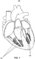
- inventive concepts in connection with the mitral valve in its broadest sense, the invention is not so limited and can be used for any papillary muscles, whether in the left ventricle or the right ventricle as shown, for example, in FIG. 1 .
- a method may further include looping the at least one guidewire around a plurality of papillary muscles in the ventricle.
- the plurality of papillary muscles in the ventricle may include two and/or three papillary muscles. As seen in FIG. 1 , there are generally two papillary muscles 102 in the left ventricle of the heart and three papillary muscles 104 in the right ventricle of the heart. Therefore, the plurality of papillary muscles may include two and/or three papillary muscles.
- looping the at least one guidewire around the plurality of papillary muscles in the ventricle may include using a pre-shaped catheter whose shape aids in looping the guidewire around the papillary muscles.
- a pre-shaped catheter 610 may aid in looping a guidewire 606 around the papillary muscles 602 and 604.
- the catheter may be made of a shape memory material that enables the catheter to assume the pre-shape following release from a sheath (not shown).
- looping the at least one guidewire around the plurality of papillary muscles in the ventricle may include using a steerable catheter whose shape can be modified interactively by the user to aid in looping the guidewire around the papillary muscles.
- looping the at least one guidewire around the plurality of papillary muscles may include looping a first guidewire around a first papillary muscle, looping a second guidewire around a second papillary muscle, and interconnecting the first guidewire and the second guidewire to thereby create a single loop around the plurality of papillary muscles.
- looping the guidewire around the papillary muscles may include using a pre-shaped and/or steerable guidewire to aid in looping the guidewire around the papillary muscles. Looping the guidewire around the plurality of papillary muscles may also be performed by any other mechanism for steering at least one guidewire around one or more papillary muscles.
- the at least one guidewire may include a single guidewire looped around a group of papillary muscles, or multiple guidewires that are later interconnected, after looping less than all papillary muscles, to form a loop around a plurality of papillary muscles.
- the papillary muscles, around which the looping occurs may include chordae or base portions of the muscles to which chordae are attached.
- the papillary muscles, around which the looping occurs may include multiple papillary muscle heads or multiple papillary muscle bodies.
- the papillary muscles, around which the looping occurs may include trabeculae of the papillary muscles.
- looping the guidewire around the papillary muscles may include passing the guidewire through the plurality of spaces among the trabeculae between the papillary muscles and a wall of the ventricle.
- Embodiments of the present disclosure may further include directing, using the guidewire, a band around the plurality of papillary muscles.
- Directing, using the guidewire, the band around the plurality of papillary muscles may include directing the band such that the band at least partially encircles between two and three papillary muscles simultaneously.
- the guidewire 406 may be used to direct the band 414 such that the band 414 at least partially encircles a plurality of papillary muscles 402 and 404.
- directing the band around the plurality of papillary muscles may include passing the band through the spaces among the trabeculae between the plurality of papillary muscles and the wall of the ventricle of the heart.
- the band may be a tube.
- the tube may be hollow inside.
- directing the band around the plurality of papillary muscles may include passing a band that is formed of a tube over the guidewire.
- a portion of the tube may include a tube and/or a ring.
- a portion of the tube may be connected to a tube and/or a ring.
- directing the band around the plurality of papillary muscles may include directing a band, a portion of which is a tube and/or a ring or that is connected to a tube and/or a ring, along a guidewire by passing the tube and/or the ring portion over the guidewire.
- directing the band around the plurality of papillary muscles may include connecting an end of the band to an end of the guidewire and pulling on the other end of the guidewire such that pulling the guidewire around the plurality of papillary muscles will pull the band around the papillary muscles as well.
- the band may be sized to simultaneously encircle a plurality of papillary muscles and pull the plurality of papillary muscles toward each other.
- the band 506 may be sized to simultaneously encircle both of the papillary muscles 502 and 504.
- the band placed around the plurality of papillary muscles may be formed of a stent-like frame within a tube.
- the tube may be made of a fabric or other flexible material.
- the stent-like frame may be able to expand after deployment from a catheter.
- the stent-like frame may be expanded by the use of an inflatable balloon.
- the inflatable balloon may have a predefined shape upon expansion and may induce the stent-like frame to bend into the shape of a balloon.
- the stent-like frame may be self-expanding and may have a predefined shape into which it self-expands upon deployment from the catheter or from a sheath intended to maintain it in its compressed configuration.
- the band includes at least one of polytetrafluoroethylene (PTFE), expanded polytetrafluoroethylene (ePTFE), Dacron, and/or any other biologically inert synthetic material with appropriate tensile strength for use in pulling the papillary muscles closer together.
- the band may be manufactured by extrusion, knitting, weaving, braiding, or any other method of forming the biologically inert synthetic material into a band or a tubular band.
- the band may be elastic or non-elastic or partially elastic. In other embodiments, a part of the band may be elastic and a part of the band may be non-elastic.
- the band may be made of more than one material. That is, a part of the band may be made of one material while another part of the band may be made of a different material.
- the band may be made of biological material from the patient, from another human donor, or from animal derived material.
- tightening the band in a single loop around the plurality of papillary muscles to pull the plurality of papillary muscles toward each other may include passing a pull-string through the band, pulling the ends of the pull-string through a catheter to tighten the band, and using a transcatether suture lock to lock the pull-string into a loop of a desired and/or appropriate size.
- a pull-string 706 may be passed through the band 710 to tighten the band 710 in a single loop around the plurality of papillary muscles 702 and 704.
- the ends of the pull-string 706 may be pulled through the catheter 708 in order to tighten the band 710 around the papillary muscles 702 and 704.
- tightening the band in a single loop around the plurality of papillary muscles may include passing a self-locking pull string through the band and pulling one or both ends of the pull-string to tighten the band around the plurality of papillary muscles. Pulling one or both ends of the pull-string may cause the pull-string to self-lock into a loop of the appropriate size.
- tightening the band in a single loop around the plurality of papillary muscles may include pulling a plurality of pull-strings attached to both ends of the band through a catheter. Pulling the plurality of pull-strings attached may tighten the band. Using a transcatheter suture lock, the pull-string may be locked into place, thereby forming the band into a loop of an appropriate and/or desired size.
- the pull-string may be self-locking or the pull-string may be locked using another locking mechanism.
- the pull-string may be coupled to any mechanism capable of locking the band into a looped configuration.
- a self-locking pull-string may be similar in construction to a beaded locking cable tie, in which a string includes one or more beads in fixed positions along the length of the string. As the beads pass through a locking grasper, the string and/or the band may be locked into a fixed position because the beads will not be able to pass in the opposite direction as it passes through the locking grasper.
- the self-locking pull-string may be locked at a position, at which the papillary muscles are pulled towards each other in order to reposition the papillary muscles.
- pulling the plurality of papillary muscles toward each other may include pulling the plurality of papillary muscles until the opposing surfaces of each of the papillary muscles are in contact with each other. In other embodiments, pulling the plurality of papillary muscles toward each other may include pulling the papillary muscles so that the opposing surfaces of each of the papillary muscles are closer to each other than they were before. In yet another embodiment, pulling the papillary muscles toward each other may include pulling the papillary muscles until the distance between the opposing surfaces of each papillary muscle meets a certain predetermined value or range of values.
- the papillary muscles may be pulled until the distance between the opposing surfaces of each of the papillary muscles is less than a predetermined distance or within a predetermined range of a predetermined distance.
- pulling the papillary muscles toward each other may include pulling the papillary muscles so that the loop formed by the band around the papillary muscles has a predetermined circumference.
- FIG. 5A illustrates a normal heart with properly positioned papillary muscles.
- FIG. 5B illustrates a heart with dilated cardiomyopathy exhibiting an enlarged ventricle and displaced papillary muscles 502 and 504.
- a band 506 may be looped around the papillary muscles 502 and 504 in order to pull them closer to each other, for example, as illustrated in FIG. 5C .
- the plurality of papillary muscles 502 and 504 may be pulled towards each other until the opposing surfaces of each of the papillary muscles 502 and 504 are in contact with each other, as illustrated in FIG. 5D .
- the present disclosure further relates to methods and devices for transcatheterly delivering a band to at least partially encircle a plurality of papillary muscles in a heart of a body. While the present disclosure provides examples of delivering a first guidewire and a second guidewire to the ventricle via at least one catheter, it should be noted that aspects of the disclosure in their broadest sense, are not limited to delivering two guidewires to the ventricle. Rather, it is contemplated that the forgoing principles may be applied to delivering fewer or more guidewires to the ventricle of the heart via a catheter.
- band refers generally to any element that is capable of at least partially encircling a plurality of papillary muscles in the ventricle of the heart in order to bring the papillary muscles closer to each other. Encircling may involve partially or completely surrounding one or more papillary muscles.
- Delivering a first guidewire and a second guidewire to the ventricle of the heart, as illustrated in FIGS. 3A-3C is one example of a method of delivering a band to at least partially encircle a plurality of papillary muscles 302 and 304 in a heart of the body in accordance with the present disclosure.
- the method of transcatheterly delivering a band to at least partially encircle one or more papillary muscles in a heart of the body may comprise transcutaneously inserting at least one catheter into a ventricle of the heart.
- the transcatheter approach to the ventricle may be transthoracic, transarterial, transvenous, transseptal, transapical, or transatrial.
- Each of these transcatheter approaches can be achieved using medical catheters and catheterization tools known in the art. They may also be performed using new tools for performing associated functions.
- inventive concepts in connection with the mitral valve in its broadest sense, the invention is not so limited and can be used for any papillary muscles, whether in the left ventricle or the right ventricle.
- the method may further comprise delivering a first guidewire to the ventricle via the at least one catheter.
- the first guidewire may include a string, thread, wire, suture, coil, or any other thin, elongated, flexible, and/or biocompatible member.
- the method may further comprise looping the first guidewire around a first papillary muscle.
- a first guidewire 306 may be delivered to the ventricle via at least one catheter 308 such that it at least partially encircles a first papillary muscle 302.
- looping the first guidewire around the first papillary muscle may include using a pre-shaped catheter whose shape aids in looping around papillary muscles.
- the catheter 610 may be pre-shaped to facilitate looping the first guidewire 606 around the first papillary muscle 602.
- looping the first guidewire around the first papillary muscle may include using a steerable catheter whose shape can be modified interactively by the user to aid in looping around the papillary muscles. Looping may involve partially or completely surrounding one or more papillary muscles.
- the method may further comprise bringing a first end of the first guidewire out of the body. That is, a first end of the first guidewire may be out of the body while a second end of the first guidewire remains inside the body. In other embodiments, both ends of the first guidewire may be brought outside of the body, while in yet other embodiments the first end of the first guidewire may be brought out of the body while a second end of the first guidewire remains outside the body never having entered the body. For example, as illustrated in FIGS. 3A-3C , both ends 312 of the first guidewire 306 may be brought through a proximal end 310 of the catheter 308 and outside of the body.
- bringing the first end of the first guidewire out of the body may include pushing the second end of the first guidewire such that the first end of the first guidewire is pushed through the catheter and out of the body.
- bringing the first end of the first guidewire out of the body may include catching the first end of the first guidewire within the ventricle of the heart by a guidewire catching technique.
- a magnetic guidewire catching may be implemented, wherein a magnet is used to catch the first end of the first guidewire and pull the first end of the first guidewire out of the body. While the forgoing principles describe a magnetic guidewire catching mechanism, any mechanism capable of catching the first end of the guidewire and pulling the guidewire out of the body may be used.
- a capture loop guidewire catching mechanism may be used to bring the first end of the first guidewire out of the body.
- Bringing the first end of the first guidewire out of the body may further include passing the guidewire from one portion of a catheter to another portion of the catheter such that the guidewire is captured at the second portion of the catheter.
- the catheter may be pulled out of the body in order to bring the first end of the first guidewire out of the body.
- the method may further comprise delivering a second guidewire to the ventricle via the at least one catheter.
- the second guidewire may also include a string, thread, wire, suture, coil, or any other thin, elongated, flexible, and/or biocompatible member.
- the method may further comprise looping the second guidewire around a second papillary muscle.
- a second guidewire 316 may be delivered to the ventricle via at least one catheter 308 such that it encircles a second papillary muscle 304.
- looping the second guidewire around the second papillary muscle may include using a pre-shaped catheter whose shape aids in looping around papillary muscles.
- the catheter may be pre-shaped to facilitate looping the second guidewire around the second papillary muscle. For example, as seen in FIGS.
- the catheter 610 may be pre-shaped to facilitate looping a second guidewire (not shown) around the second papillary muscle 604.
- looping the second guidewire around the second papillary muscle may include using a steerable catheter whose shape can be modified interactively by the user to aid in looping around the papillary muscles.
- the method may further comprise bringing a second end of the second guidewire out of the body. That is, a second end of the second guidewire may be out of the body while the other end of the second guidewire remains inside the body. In another embodiment, both ends of the second guidewire may be brought outside of the body, while in yet another embodiment a second end of the second guidewire may be brought out of the body while the other end of the second guidewire remains outside the body, never having entered the body. For example, both ends 312 of the second guidewire 316 may be brought outside the body through a proximal end 310 of the catheter 308.
- Bringing the second end of the second guidewire out of the body may include pushing the first end of the second guidewire such that the second end of the second guidewire is pushed through the catheter and out of the body.
- bringing the second end of the second guidewire out of the body may include catching the second end of the second guidewire within the ventricle of the heart by a guidewire catching technique.
- a magnetic guidewire catching may be implemented, wherein a magnet is used to catch the second end of the second guidewire and pull the second end of the second guidewire out of the body. While the forgoing principles describe a magnetic guidewire catching mechanism, any mechanism capable of catching the second end of the guidewire and pulling the guidewire out of the body may be used.
- a capture loop guidewire catching mechanism may be used to bring the second end of the second guidewire out of the body.
- Bringing the second end of the second guidewire out of the body may further include passing the guidewire from one portion of a catheter to another portion of the catheter such that the guidewire is captured at the second portion of the catheter.
- the catheter may be pulled out of the body in order to bring the second end of the second guidewire out of the body.
- the method may further comprise interconnecting the first guidewire to the second guidewire.
- Interconnecting the first guidewire to the second guidewire may include tying the first guidewire and the second guidewire together.
- interconnecting the first and second guidewires may include connecting the first and second guidewires together with a built-in connector, or any other type of connector used to connect a plurality of ropes, strings, wires, or cables together.
- interconnecting the first and second guidewires may include connecting the first and second guidewires with an applied clip, clasp, or any other type of clip, clasp, or crimp ring used to connect a plurality of ropes, strings, wires, or cables together. For example, as illustrated in FIGS.
- one end of the first guidewire 306 and one end of the second guidewire 316 may be interconnected using a flexible connector ( FIG. 3A ), using a rigid connector ( FIG. 3B ), tying a knot ( FIG. 3C ), or any combination thereof.
- the ends of the first and/or second guidewires may be interconnected using any mechanism used to connect a plurality of ropes, strings, wires, or cables together.
- the method may further comprise pulling at least one end of the first and second guidewires after interconnecting them to thereby establish a single loop around at least the first papillary muscle and the second papillary muscle.
- one end of a guidewire 412b may be pulled through a proximal end 410 of a catheter 408 in order to establish a single loop 406 around at least the first papillary muscle 402 and the second papillary muscle 404.
- the papillary muscles, around which the looping occurs may include chordae or base portions of the muscles to which chordae are attached.
- the papillary muscles, around which the looping occurs may include multiple papillary muscle heads or multiple papillary muscle bodies.
- the papillary muscles, around which the looping occurs may include trabeculae of the papillary muscles.
- looping the first and second guidewires around the papillary muscles may include passing the first and second guidewires through the plurality of spaces among the trabeculae between the papillary muscles and a wall of the ventricle.
- the method may further comprise directing, using the single loop established by pulling the at least one end of the first and second guidewires after interconnecting them, a band around the first papillary muscle and the second papillary muscle.
- the band may be a tube positioned over the guidewire.
- only a portion of the band may be a tube or a ring.
- a portion of the band may be connected to a tube or a ring.
- directing the band around the first and second papillary muscles may include directing a band along a guidewire by passing the tube or ring portion over the guidewire.
- directing the band around the first and second papillary muscles may include connecting an end of the band to an end of the guidewire and pulling on the other end of the guidewire such that the guidewire pulls the band around the papillary muscles.
- an end of the band 414 may be connected to an end of the guidewire 412a.
- the other end of the guidewire 412b may be pulled through a proximal end 410 of the catheter 408 in order to pull the band 414 through the catheter 408 and around the papillary muscles 402 and 404.
- the band 414 may form a single loop around at least the first papillary muscle 402 and the second papillary muscle 404, as illustrated in FIG. 4C .
- the band may be sized to simultaneously encircle, i.e. completely or partially surround, a plurality of papillary muscles and pull the plurality of papillary muscles toward each other.
- the band 506 may be sized to simultaneously encircle a first papillary muscle 502 and a second papillary muscle 504.
- the band may be a tube or a portion of the band may be a tube.
- a first portion of the band may be a tube whereas a second portion of the band may not be a tube.
- the band may include at least one of polytetrafluoroethylene (PTFE), expanded polytetrafluoroethylene (ePTFE), Dacron, and/or any other biologically inert synthetic material with appropriate tensile strength for use in pulling the papillary muscles closer together.
- the band may be manufactured by extrusion, knitting, weaving, braiding, or any other method of forming the biologically inert synthetic material into a band or a tubular band.
- the band may be elastic or non-elastic or partially elastic.
- a part of the band may be elastic and a part of the band may be non-elastic.
- the band may be made of more than one material. That is, a part of the band may be made of one material while another part of the band may be made of a different material.
- the band may be made of biological material from the patient, from another human donor, or from animal derived material.
- the method of interconnecting the first guidewire and the second guidewire may include connecting a first end of the first guidewire to a second end of the second guidewire outside of the body.
- the first guidewire may be connected to the second guidewire inside of the body.
- bringing at least one of the first end of the first guidewire and the second end of the second guidewire out of the body may include catching at least one of the first end and the second end using a magnet and pulling at least one of the first and second end out of the body.
- any mechanism capable of catching at least one of the first end of the first guidewire and the second end of the second guidewire and pulling at least one of the first guidewire and the second guidewire out of the body may be used.
- a capture loop guidewire catching mechanism, a guidewire catching basket, or any combination thereof may be used to bring the second end of the second guidewire out of the body.
- Bringing at least one of the first end of the first guidewire and the second end of the second guidewire out of the body may include pushing a second end of the first guidewire, thereby causing the first end of the first guidewire to move through the at least one catheter and out of the body.
- bringing at least one of the first end of the first guidewire and the second end of the second guidewire out of the body may include passing at least one of the first end of the first guidewire and the second end of the second guidewire around a papillary muscle from a first portion of the at least one catheter to a second portion of the catheter and capturing at least one of the first and second guidewires at the second portion of the catheter.
- the catheter may be pulled out of the body in order to extract at least one of the first end of the first guidewire and the second end of the second guidewire from the body.
- the method of transcatheterly delivering a band to at least partially encircle a plurality of papillary muscles in the heart may comprise delivering an end of a third guidewire to the heart ventricle via the at least one catheter, looping the end of the third guidewire around a third papillary muscle, bringing the end of the third guidewire out of the body while the third guidewire remains looped around the third papillary muscle, and interconnecting at least one of the first and second guidewires with the third guidewire. Therefore, in some embodiments, the first, second, and third guidewires may direct a band in a single loop around three papillary muscles.
- the method of transcatheterly delivering a band to at least partially encircle a plurality of papillary muscles in the heart may further comprise connecting an end of the band to at least one of the first and second guidewire, and pulling the proximal end of the guidewire while the guidewire is looped around the first and second papillary muscles, thereby guiding the band around the first and second papillary muscles.
- an end of the band 414 may be connected to an end of the guidewire 412a.
- the other end of the guidewire 412b may be pulled through a proximal end 410 of the catheter 408 in order to pull the band 414 through the catheter 408 and around the papillary muscles 402 and 404.
- At least one of the first guidewire and the second guidewire may include a built-in connector.
- an end of the first guidewire may be connected to an end of the second guidewire via the built-in connector.
- any other type of connector used to connect a plurality of ropes, strings, wires, or cables together may be used.
- interconnecting the first and second guidewires may include connecting the first and second guidewires with an applied clip, clasp, or any other type of clip, clasp, or crimp ring used to connect a plurality of ropes, strings, wires, or cables together. For example, as illustrated in FIGS.
- one end of the first guidewire 306 and one end of the second guidewire 316 may be interconnected using a flexible connector ( FIG. 3A ), using a rigid connector ( FIG. 3B ), tying a knot ( FIG. 3C ), or any combination thereof.
- the ends of the first and/or second guidewires may be interconnected using any mechanism used to connect a plurality of ropes, strings, wires, or cables together.
- Another embodiment of the present disclosure relates to methods and devices for improving cardiac function. While the present disclosure provides examples of improving cardiac function by looping a single band around the plurality of papillary muscles, it should be noted that aspects of the disclosure in their broadest sense, are not limited to looping a band around the plurality of papillary muscles. Rather, it is contemplated that the forgoing principles may be applied to other methods of improving cardiac function as well.
- the term band refers generally to any element that is capable of encircling a plurality of papillary muscles in the ventricle of the heart in order to bring the papillary muscles closer to each other.
- Looping a band around the plurality of papillary muscles as illustrated in FIGS. 4A-4C and 5A-5D , is one example of a method of improving cardiac function in accordance with the present disclosure. Looping may involve partially or completely surrounding one or more papillary muscles.
- a method of improving cardiac function in accordance with the present disclosure may include transcatheterly looping a single band around a plurality of papillary muscles in a ventricle of a heart.
- the term "transcatheterly” may include delivering via a catheter.
- FIGS. 4A-4C illustrates a single catheter 408 used to deliver and loop a single band 414 around a plurality of papillary muscles 402, 404 in a ventricle of a heart.
- a transcatheter approach to the ventricle of the heart may be transthoracic, transarterial, transvenous, transseptal, transapical, or transatrial. All of these can be achieved using standard medical catheters and catheterization tools known in the art.
- inventive concepts in connection with the mitral valve in its broadest sense, the invention is not so limited and can be used for any papillary muscles, whether in the left ventricle or the right ventricle.
- the method may further include looping a single band around a plurality of papillary muscles in a ventricle of a heart such that the band at least partially encircles a cluster of papillary muscles.
- cluster generally refers to a plurality of papillary muscles that are relatively in close proximity to each other.
- the "cluster" of papillary muscles may include two and/or three papillary muscles.
- the “cluster” may also refer to any subset of two or more papillary muscles within a single ventricle of the heart.
- the cluster may have an outer peripheral boundary defined by the at least partially encircling band. Further, the cluster may have an inner region on sides of the plurality of papillary muscles opposite the outer peripheral boundary.
- the cluster may refer to a plurality of papillary muscles 502, 504 that has an outer peripheral boundary defined by the at least partially encircling band 506 and having an inner region on side of the papillary muscles 502, 504 opposite the outer peripheral boundary.
- the method may implement a first guidewire and/or a second guidewire.
- the method may further include looping at least one guidewire 206 around a plurality of papillary muscles 202, 204 in the ventricle.
- the method may further include looping a first guidewire around a first papillary muscle, looping a second guidewire around a second papillary muscle, and interconnecting the first guidewire and the second guidewire.
- the method may further include looping a first guidewire 306 around a first papillary muscle 302, looping a second guidewire 316 around a second papillary muscle 304, and interconnecting the first guidewire 306 and the second guidewire 316 outside of the body by a connector 314.
- the plurality of papillary muscles in the ventricle may include two and/or three papillary muscles.
- looping a single band around the plurality of papillary muscles may further comprise delivering a first guidewire and/or a second guidewire to the ventricle via the at least one catheter.
- the first guidewire and/or the second guidewire may include a string, thread, wire, suture, coil, or any other thin, elongated, flexible, and/or biocompatible member.
- the method may comprise looping the first guidewire around a first papillary muscle and/or looping the second guidewire around a second papillary muscle.
- looping the first guidewire and/or the second guidewire around the first and/or second papillary muscle may include using a pre-shaped catheter whose shape aids in looping around papillary muscles.
- the catheter 610 may be pre-shaped to facilitate looping the guidewire 606 around the papillary muscles 602 and 604.
- looping the guidewire around the papillary muscle may include using a steerable catheter whose shape can be modified interactively by the user to aid in looping around the papillary muscles.
- the method may further comprise bringing a first end of the first guidewire out of the body. That is, a first end of the first guidewire may be out of the body while a second end of the first guidewire remains inside the body. In other embodiments, both ends of the first guidewire may be brought outside of the body, while in yet other embodiments, a first end of the first guidewire may be brought out of the body while a second end of the first guidewire remains outside the body, never having entered the body. For example, as illustrated in FIGS. 3A-3C , both ends 312 of the first guidewire 306 may be brought outside of the body.
- Bringing the first end of the first guidewire out of the body may include pushing the second end of the first guidewire such that the first end of the first guidewire is pushed through the catheter and out of the body.
- bringing the first end of the first guidewire out of the body may include catching the first end of the first guidewire within the ventricle of the heart by a guidewire catching technique.
- a magnetic guidewire catching may be implemented, wherein a magnet is used to catch the first end of the first guidewire and pull the first end of the first guidewire out of the body. While the forgoing principles describe a magnetic guidewire catching mechanism, any mechanism capable of catching the first end of the guidewire and pulling the guidewire out of the body may be used.
- a capture loop guidewire catching mechanism may be used to bring the first end of the first guidewire out of the body.
- Bringing the first end of the first guidewire out of the body may further include passing the guidewire from one portion of a catheter to another portion of the catheter such that the guidewire is captured at the second portion of the catheter.
- the catheter may be pulled out of the body in order to bring the first end of the first guidewire out of the body.
- the method may further comprise bringing a second end of the second guidewire out of the body. That is, a second end of the second guidewire may be out of the body while the other end of the second guidewire remains inside the body. In other embodiments, both ends of the second guidewire may be brought outside of the body, while in yet other embodiments, a second end of the second guidewire may be brought out of the body while the other end of the second guidewire remains outside the body, never having entered the body. For example, as illustrated in FIGS. 3A-3C , both ends 312 of the second guidewire 316 may be brought outside of the body through a proximal end 310 of the catheter 308.
- Bringing the second end of the second guidewire out of the body may include pushing the first end of the second guidewire such that the second end of the second guidewire is pushed through the catheter and out of the body.
- bringing the second end of the second guidewire out of the body may include catching the second end of the second guidewire within the ventricle of the heart by a guidewire catching technique.
- a magnetic guidewire catching may be implemented, wherein a magnet is used to catch the second end of the second guidewire and pull the second end of the second guidewire out of the body. While the forgoing principles describe a magnetic guidewire catching mechanism, any mechanism capable of catching the second end of the guidewire and pulling the guidewire out of the body may be used.
- a capture loop guidewire catching mechanism may be used to bring the second end of the second guidewire out of the body.
- Bringing the second end of the second guidewire out of the body may further include passing the guidewire from one portion of a catheter to another portion of the catheter such that the guidewire is captured at the second portion of the catheter.
- the catheter may be pulled out of the body in order to bring the second end of the second guidewire out of the body.
- the method may further comprise interconnecting the first guidewire to the second guidewire.
- Interconnecting the first guidewire to the second guidewire may include tying the first guidewire and the second guidewire together.
- interconnecting the first and second guidewires may include connecting the first and second guidewires together with a built-in connector, or any other type of connector used to connect a plurality of ropes, strings, wires, or cables together.
- interconnecting the first and second guidewires may include connecting the first and second guidewires with an applied clip, clasp, or any other type of clip, clasp, or crimp ring used to connect a plurality of ropes, strings, wires, or cables together. For example, as illustrated in FIGS.
- one end of the first guidewire 306 and one end of the second guidewire 316 may be interconnected using a flexible connector ( FIG. 3A ), using a rigid connector ( FIG. 3B ), tying a knot ( FIG. 3C ), or any combination thereof.
- the ends of the first and/or second guidewires may be interconnected using any mechanism used to connect a plurality of ropes, strings, wires, or cables together.
- the method may further comprise pulling at least one end of the first and second guidewires after interconnecting them to thereby establish a single loop around at least the first papillary muscle and the second papillary muscle.
- one end 412b of the guidewire 406 may be pulled in order to establish a single loop around the first papillary muscle 402 and the second papillary muscle 404.
- the papillary muscles, around which the looping occurs may include chordae or base portions of the muscles to which chordae are attached.
- the papillary muscles, around which the looping occurs may include multiple papillary muscle heads or multiple papillary muscle bodies.
- the papillary muscles, around which the looping occurs may include trabeculae of the papillary muscles.
- looping the first and second guidewires around the papillary muscles may include passing the first and second guidewires through the plurality of spaces among the trabeculae between the papillary muscles and a wall of the ventricle.
- Embodiments of the present disclosure may further include directing, using the guidewire, a band around the plurality of papillary muscles.
- Directing, using the guidewire, the band around the plurality of papillary muscles may include directing the band such that the band at least partially encircles between two and three papillary muscles simultaneously.
- the band may be sized to simultaneously encircle, i.e. partially or completely surround, a plurality of papillary muscles and pull the plurality of papillary muscles toward each other.
- the band 506 may be sized to simultaneously encircle the papillary muscles 502, 504 and pull the papillary muscles 502, 504 toward each other.
- the band may be a tube or a portion of the band may be a tube.
- a first portion of the band may be a tube whereas a second portion of the band may not be a tube.
- the band may include at least one of polytetrafluoroethylene (PTFE), expanded polytetrafluoroethylene (ePTFE), Dacron, and/or any other biologically inert synthetic material with appropriate tensile strength for use in pulling the papillary muscles closer together.
- the band may be manufactured by extrusion, knitting, weaving, braiding, or any other method of forming the biologically inert synthetic material into a band or a tubular band.
- the band may be elastic or non-elastic or partially elastic.
- a part of the band may be elastic and a part of the band may be non-elastic.
- the band may be made of more than one material. That is, a part of the band may be made of one material while another part of the band may be made of a different material.
- the band may be made of biological material from the patient, from another human donor, or from animal derived material.
- the method of improving cardiac function may further comprise transcatheterly tightening the band to contract the outer peripheral boundary of the cluster and thereby pull the papillary muscles closer together such that the band contacts the outer peripheral boundary portions of the plurality of papillary muscles without contacting the inner region and such that the inner region is devoid of any portion of the band.
- the band 506 may contact the outer peripheral boundary portions of the plurality of papillary muscles 502, 504 and thereby pull the papillary muscles 502, 504 closer together without contacting the inner region of the papillary muscles 502, 504.
- transcatheterly tightening the band to contract the outer peripheral boundary and thereby pull the papillary muscles closer together may include passing a pull-string through the band, pulling the ends of the pull-string through a catheter to tighten the band, and using a transcatether suture lock to lock the pull-string into a loop of a desired and/or appropriate size.
- a pull-string 706 may be passed through a catheter 708 and through the band 710 to facilitate tightening of the band 710 in a single loop around the plurality of papillary muscles 702, 704.
- tightening the band in a single loop around the plurality of papillary muscles may include passing a self-locking pull string through the band and pulling one or both ends of the pull-string to tighten the band around the plurality of papillary muscles. Pulling one or both ends of the pull-string may cause the pull-string to self-lock into a loop of the appropriate size.
- tightening the band in a single loop around the plurality of papillary muscles may include pulling a plurality of pull-strings attached to both ends of the band through a catheter. Pulling the plurality of pull-strings attached may tighten the band. Using a transcatheter suture lock, the pull-string may be locked into place, thereby forming the band into a loop of an appropriate and/or desired size.
- the pull-string may be self-locking or the pull-string may be locked using another locking mechanism.
- the pull-string may be coupled to any mechanism capable of locking the band into a looped configuration.
- a self-locking pull-string may be similar in construction to a beaded locking cable tie, in which a string includes one or more beads in fixed positions along the length of the string. As the beads pass through a locking grasper, the string and/or the band may be locked into a fixed position because the beads will not be able to pass in the opposite direction as it passes through the locking grasper.
- the self-locking pull-string may be locked at a position, at which the papillary muscles are pulled towards each other in order to reposition the papillary muscles.
- pulling the papillary muscles closer together may include pulling the papillary muscles until the opposing surfaces of each of the papillary muscles are in contact with each other.
- pulling the plurality of papillary muscles toward each other may include pulling the papillary muscles so that the opposing surfaces of each of the papillary muscles are closer to each other than they were before.
- pulling the papillary muscles toward each other may include pulling the papillary muscles until the distance between the opposing surfaces of each papillary muscle meets a certain predetermined value or range of values. For example, the papillary muscles may be pulled until the distance between the opposing surfaces of each of the papillary muscles is less than a predetermined distance or within a predetermined range of a predetermined distance.
- pulling the papillary muscles closer together may include pulling the papillary muscles so that the outer periphery of the cluster has a predetermined circumference.
- the predetermined circumference may be smaller than the circumference of the outer periphery of the cluster before transcatheterly tightening the band.
- FIG. 5A illustrates a normal heart with properly positioned papillary muscles.
- FIG. 5B illustrates a heart with dilated cardiomyopathy showing enlarged ventricle and displaced papillary muscles 502 and 504.
- a band 506 may be looped around the papillary muscles 502 and 504 in order to pull them closer to each other, for example, as illustrated in FIG. 5C .
- the plurality of papillary muscles 502 and 504 may be pulled towards each other until the opposing surfaces of each of the papillary muscles 502 and 504 are in contact with each other, as illustrated in FIG. 5D .
- the method may further comprise pulling the papillary muscles into contact with each other without any intervening material between the papillary muscles.
Landscapes
- Health & Medical Sciences (AREA)
- Cardiology (AREA)
- Life Sciences & Earth Sciences (AREA)
- Heart & Thoracic Surgery (AREA)
- Animal Behavior & Ethology (AREA)
- Veterinary Medicine (AREA)
- Engineering & Computer Science (AREA)
- Public Health (AREA)
- Biomedical Technology (AREA)
- General Health & Medical Sciences (AREA)
- Oral & Maxillofacial Surgery (AREA)
- Transplantation (AREA)
- Vascular Medicine (AREA)
- Hematology (AREA)
- Anesthesiology (AREA)
- Pulmonology (AREA)
- Biophysics (AREA)
- Child & Adolescent Psychology (AREA)
- Surgical Instruments (AREA)
- Prostheses (AREA)
- Electrotherapy Devices (AREA)
- Magnetic Resonance Imaging Apparatus (AREA)
- Steroid Compounds (AREA)
Claims (11)
- Dispositif permettant de délivrer par cathéter une bande destinée à encercler une pluralité de muscles papillaires dans le coeur d'un corps, le dispositif comprenant :un cathéter, comprenant une extrémité proximale et une extrémité distale ;une bande conçue pour passer à travers l'extrémité distale du cathéter, ladite bande étant conçue pour encercler la pluralité de muscles papillaires de manière à permettre à la bande d'entrer en contact avec les parties limites périphériques externes de la pluralité de muscles papillaires et de tirer la pluralité de muscles papillaires les uns vers les autres ;au moins un fil de guidage conçu pour passer à travers l'extrémité distale du cathéter pour s'enrouler autour d'une pluralité de muscles papillaires, ladite bande étant conçue pour coopérer avec ledit au moins un fil de guidage ;un mécanisme de capture de fil de guidage conçu pour attraper une extrémité dudit au moins un fil de guidage.
- Dispositif selon la revendication 1, ledit mécanisme de capture du fil de guidage comprenant au moins un aimant, une boucle de capture, ou un panier de capture de fil de guidage.
- Dispositif selon la revendication 1, ledit cathéter étant préformé de manière à faciliter l'enroulement dudit au moins un fil de guidage autour de la pluralité de muscles papillaires.
- Dispositif selon la revendication 1, ledit cathéter étant conçu pour être orientable de manière réglable de manière à faciliter l'enroulement dudit au moins un fil de guidage autour de la pluralité de muscles papillaires.
- Dispositif selon la revendication 1, ledit au moins un fil de guidage comprenant au moins une ficelle, un fil, un câble, une suture ou une bobine.
- Dispositif selon la revendication 1, comprenant en outre :une ficelle de traction conçue pour passer à travers le cathéter ; etun verrou de suture transcathéter conçu pour verrouiller la ficelle de traction dans une boucle.
- Dispositif selon la revendication 1, comprenant en outre une ficelle de traction conçue pour passer à travers le cathéter, et une pince de verrouillage conçue pour engager de manière autobloquante la ficelle de traction.
- Dispositif selon la revendication 7,
ladite ficelle de traction comprenant une ou plusieurs billes fixées le long d'une longueur de la ficelle de traction, et lesdites une ou plusieurs billes étant conçues pour être engagées par la pince de verrouillage et verrouiller ainsi la ficelle de traction dans une position fixe. - Dispositif selon la revendication 1, au moins une partie de la bande comprenant un tube creux.
- Dispositif selon la revendication 1, ledit au moins un fil de guidage comprenant deux fils de guidage et ledit dispositif comprenant en outre un raccord conçu pour raccorder entre elles une première extrémité du premier fil de guidage à une seconde extrémité du second fil de guidage.
- Dispositif selon la revendication 1, ledit mécanisme de capture de fil de guidage étant conçu pour tirer l'extrémité dudit au moins un fil de guidage hors du corps.
Applications Claiming Priority (2)
| Application Number | Priority Date | Filing Date | Title |
|---|---|---|---|
| US201762477461P | 2017-03-28 | 2017-03-28 | |
| PCT/IB2018/052142 WO2018178901A2 (fr) | 2017-03-28 | 2018-03-28 | Procédé permettant d'améliorer le fonctionnement cardiaque |
Publications (2)
| Publication Number | Publication Date |
|---|---|
| EP3600158A2 EP3600158A2 (fr) | 2020-02-05 |
| EP3600158B1 true EP3600158B1 (fr) | 2024-04-17 |
Family
ID=62089786
Family Applications (1)
| Application Number | Title | Priority Date | Filing Date |
|---|---|---|---|
| EP18721138.8A Active EP3600158B1 (fr) | 2017-03-28 | 2018-03-28 | Dispositif permettant d'améliorer le fonctionnement cardiaque |
Country Status (9)
| Country | Link |
|---|---|
| US (4) | US10058428B1 (fr) |
| EP (1) | EP3600158B1 (fr) |
| JP (1) | JP6985493B2 (fr) |
| CN (1) | CN110691568A (fr) |
| AU (1) | AU2018247166B2 (fr) |
| BR (1) | BR112019020106A2 (fr) |
| CA (1) | CA3058150A1 (fr) |
| IL (1) | IL269673B2 (fr) |
| WO (1) | WO2018178901A2 (fr) |
Families Citing this family (15)
| Publication number | Priority date | Publication date | Assignee | Title |
|---|---|---|---|---|
| EP1909655A2 (fr) | 2005-06-20 | 2008-04-16 | Sutura, Inc. | Procede et appareil pour pratiquer un noeud sur une suture |
| JP6469109B2 (ja) | 2013-12-06 | 2019-02-13 | メッド − ベンチャー インベストメンツ、エルエルシー | 縫合方法及び装置 |
| US11833034B2 (en) | 2016-01-13 | 2023-12-05 | Shifamed Holdings, Llc | Prosthetic cardiac valve devices, systems, and methods |
| US11318018B2 (en) * | 2017-03-28 | 2022-05-03 | Cardiac Success Ltd. | Method of improving cardiac function |
| JP6985493B2 (ja) * | 2017-03-28 | 2021-12-22 | カーディアック・サクセス・リミテッド | 心機能を向上させる方法 |
| EP3641663B1 (fr) | 2017-06-19 | 2022-03-02 | Heartstitch, Inc. | Systèmes et procédés de suture destinés à suturer un tissu corporel |
| WO2019051379A1 (fr) * | 2017-09-11 | 2019-03-14 | Heartstitch, Inc. | Procédés et dispositifs pour suture papillaire |
| US11464638B2 (en) | 2017-10-23 | 2022-10-11 | Cardiac Success Ltd | Adjustable self-locking papillary muscle band |
| JP7229238B2 (ja) | 2017-10-23 | 2023-02-27 | カーディアック・サクセス・リミテッド | 調整可能自動ロック式乳頭筋バンド |
| US11413146B2 (en) * | 2018-10-03 | 2022-08-16 | Edwards Lifesciences Corporation | Spring and coil devices for papillary muscle approximation and ventricle remodeling |
| US11413147B2 (en) * | 2018-10-03 | 2022-08-16 | Edwards Lifesciences Corporation | Ventricular remodeling using coil devices |
| JP2022504241A (ja) | 2018-10-05 | 2022-01-13 | シファメド・ホールディングス・エルエルシー | 人工心弁デバイス、システム、および方法 |
| EP3941391A4 (fr) | 2019-03-19 | 2022-11-23 | Shifamed Holdings, LLC | Dispositifs, systèmes et procédés pour valvule cardiaque prothétique |
| US20220202571A1 (en) * | 2019-05-07 | 2022-06-30 | Yale University | Papillary muscle approximation and ventricular restoration |
| EP4048207A1 (fr) * | 2019-10-25 | 2022-08-31 | Edwards Lifesciences Corporation | Dispositifs et procédés de restauration et de remodelage cardiaque |
Citations (2)
| Publication number | Priority date | Publication date | Assignee | Title |
|---|---|---|---|---|
| US20050197696A1 (en) * | 2004-02-23 | 2005-09-08 | Gomez Duran Carlos M. | Papilloplasty band and sizing device |
| US20060229708A1 (en) * | 2005-02-07 | 2006-10-12 | Powell Ferolyn T | Methods, systems and devices for cardiac valve repair |
Family Cites Families (37)
| Publication number | Priority date | Publication date | Assignee | Title |
|---|---|---|---|---|
| ATE484241T1 (de) | 1999-04-09 | 2010-10-15 | Evalve Inc | Verfahren und vorrichtung zur herzklappenreperation |
| US7331972B1 (en) * | 2002-11-15 | 2008-02-19 | Abbott Cardiovascular Systems Inc. | Heart valve chord cutter |
| US20070255396A1 (en) * | 2003-06-20 | 2007-11-01 | Medtronic Vascular, Inc. | Chrodae Tendinae Girdle |
| WO2005032421A2 (fr) * | 2003-09-15 | 2005-04-14 | Medtronic Vascular, Inc. | Appareil et procede pour l'elongation d'un muscle papillaire |
| US20110060407A1 (en) * | 2005-02-07 | 2011-03-10 | Ted Ketai | Methods, systems and devices for cardiac valve repair |
| EP1874217A4 (fr) * | 2005-04-25 | 2014-11-19 | Evalve Inc | Dispositif et procedes d'annuloplastie endoscopique |
| EP1887981A2 (fr) * | 2005-06-09 | 2008-02-20 | The University Of Miami | Fixation aux muscles papillaires pour reduction ventriculaire gauche |
| US20090082619A1 (en) * | 2005-06-09 | 2009-03-26 | De Marchena Eduardo | Method of treating cardiomyopathy |
| US20070100439A1 (en) * | 2005-10-31 | 2007-05-03 | Medtronic Vascular, Inc. | Chordae tendinae restraining ring |
| WO2007100408A2 (fr) | 2005-12-15 | 2007-09-07 | Georgia Tech Research Corporation | dispositifs, SYSTÈMES, & PROCÉDÉS de commande de position de muscle papillaire |
| EP2114304B1 (fr) * | 2007-02-14 | 2017-09-06 | Edwards Lifesciences Corporation | Dispositif médical implantable destiné à réparer le coeur |
| US9125632B2 (en) * | 2007-10-19 | 2015-09-08 | Guided Delivery Systems, Inc. | Systems and methods for cardiac remodeling |
| EP2072027B1 (fr) * | 2007-12-21 | 2020-06-17 | Medtentia International Ltd Oy | dispositif pré-annuloplastique et méthode |
| US20100121437A1 (en) * | 2008-04-16 | 2010-05-13 | Cardiovascular Technologies, Llc | Transvalvular intraannular band and chordae cutting for ischemic and dilated cardiomyopathy |
| US8323335B2 (en) * | 2008-06-20 | 2012-12-04 | Edwards Lifesciences Corporation | Retaining mechanisms for prosthetic valves and methods for using |
| US20100210899A1 (en) * | 2009-01-21 | 2010-08-19 | Tendyne Medical, Inc. | Method for percutaneous lateral access to the left ventricle for treatment of mitral insufficiency by papillary muscle alignment |
| US20100185278A1 (en) | 2009-01-21 | 2010-07-22 | Tendyne Medical | Apical Papillary Msucle Attachment for Left Ventricular Reduction |
| US20110015476A1 (en) * | 2009-03-04 | 2011-01-20 | Jeff Franco | Devices and Methods for Treating Cardiomyopathy |
| US9011522B2 (en) * | 2009-04-10 | 2015-04-21 | Lon Sutherland ANNEST | Device and method for temporary or permanent suspension of an implantable scaffolding containing an orifice for placement of a prosthetic or bio-prosthetic valve |
| PL3335670T3 (pl) * | 2010-03-05 | 2022-09-05 | Edwards Lifesciences Corporation | Mechanizmy ustalające do zastawek protetycznych |
| CN105392518A (zh) * | 2012-10-02 | 2016-03-09 | 女王医疗中心 | 具有引导线防滑脱特征的脉管通路系统 |
| CA3112256C (fr) * | 2012-11-20 | 2023-09-26 | Innovheart S.R.L. | Systeme prothetique pour le remplacement d'une valvule cardiaque |
| ES2610992T3 (es) * | 2012-11-20 | 2017-05-04 | Innovheart S.R.L. | Dispositivo para desplegar un sistema de alambres guías dentro de una cámara cardíaca para implantar una válvula cardíaca protésica |
| ITBO20120636A1 (it) | 2012-11-20 | 2014-05-21 | Ht Consultant Di Giovanni Righini | Sistema protesico sostitutivo per valvola cardiaca |
| US9999507B2 (en) * | 2013-06-25 | 2018-06-19 | Mitralign, Inc. | Percutaneous valve repair by reshaping and resizing right ventricle |
| EP3060172A4 (fr) * | 2013-10-23 | 2017-07-05 | LC Therapeutics, Inc. | Système percutané ou très peu invasif de réparation d'une valve cardiaque et ses procédés d'utilisation |
| DE102015107242B4 (de) * | 2015-05-08 | 2022-11-03 | Highlife Sas | System zum Implantieren eines Implantats um eine umfängliche Gewebestruktur in einem Herz und Verfahren zum Anordnen und Vorbringen eines Implantats auf einem Führungsdraht eines solchen Systems |
| US10463492B2 (en) * | 2015-11-17 | 2019-11-05 | Edwards Lifesciences Corporation | Systems and devices for setting an anchor |
| US9877833B1 (en) * | 2016-12-30 | 2018-01-30 | Pipeline Medical Technologies, Inc. | Method and apparatus for transvascular implantation of neo chordae tendinae |
| JP6985493B2 (ja) * | 2017-03-28 | 2021-12-22 | カーディアック・サクセス・リミテッド | 心機能を向上させる方法 |
| US11135062B2 (en) | 2017-11-20 | 2021-10-05 | Valtech Cardio Ltd. | Cinching of dilated heart muscle |
| WO2019226803A1 (fr) | 2018-05-22 | 2019-11-28 | Boston Scientific Scimed, Inc. | Relocalisation percutanée de muscle papillaire |
| US20190365539A1 (en) | 2018-05-29 | 2019-12-05 | Edwards Lifesciences Corporation | Reverse ventricular remodeling and papillary muscle approximation |
| US11224418B2 (en) | 2018-06-15 | 2022-01-18 | Edwards Lifesciences Corporation | Papillary muscle approximation pads |
| US11413146B2 (en) | 2018-10-03 | 2022-08-16 | Edwards Lifesciences Corporation | Spring and coil devices for papillary muscle approximation and ventricle remodeling |
| US20220202571A1 (en) | 2019-05-07 | 2022-06-30 | Yale University | Papillary muscle approximation and ventricular restoration |
| EP4069146A2 (fr) | 2019-12-03 | 2022-10-12 | Boston Scientific Scimed, Inc. | Anse percutanée d'approximation de muscle papillaire |
-
2018
- 2018-03-28 JP JP2020503379A patent/JP6985493B2/ja active Active
- 2018-03-28 BR BR112019020106A patent/BR112019020106A2/pt unknown
- 2018-03-28 WO PCT/IB2018/052142 patent/WO2018178901A2/fr unknown
- 2018-03-28 US US15/938,241 patent/US10058428B1/en active Active
- 2018-03-28 EP EP18721138.8A patent/EP3600158B1/fr active Active
- 2018-03-28 AU AU2018247166A patent/AU2018247166B2/en active Active
- 2018-03-28 IL IL269673A patent/IL269673B2/en unknown
- 2018-03-28 CN CN201880033678.6A patent/CN110691568A/zh active Pending
- 2018-03-28 CA CA3058150A patent/CA3058150A1/fr active Pending
- 2018-07-05 US US16/027,839 patent/US10271950B2/en active Active
-
2019
- 2019-03-18 US US16/355,971 patent/US10517729B2/en active Active
- 2019-11-20 US US16/689,151 patent/US11344417B2/en active Active
Patent Citations (2)
| Publication number | Priority date | Publication date | Assignee | Title |
|---|---|---|---|---|
| US20050197696A1 (en) * | 2004-02-23 | 2005-09-08 | Gomez Duran Carlos M. | Papilloplasty band and sizing device |
| US20060229708A1 (en) * | 2005-02-07 | 2006-10-12 | Powell Ferolyn T | Methods, systems and devices for cardiac valve repair |
Also Published As
| Publication number | Publication date |
|---|---|
| US10058428B1 (en) | 2018-08-28 |
| US20190209326A1 (en) | 2019-07-11 |
| JP6985493B2 (ja) | 2021-12-22 |
| AU2018247166A1 (en) | 2019-10-17 |
| AU2018247166B2 (en) | 2022-06-02 |
| US20180311043A1 (en) | 2018-11-01 |
| EP3600158A2 (fr) | 2020-02-05 |
| US11344417B2 (en) | 2022-05-31 |
| WO2018178901A2 (fr) | 2018-10-04 |
| US10517729B2 (en) | 2019-12-31 |
| JP2020515375A (ja) | 2020-05-28 |
| IL269673A (en) | 2019-11-28 |
| IL269673B2 (en) | 2024-02-01 |
| US20200085580A1 (en) | 2020-03-19 |
| CN110691568A (zh) | 2020-01-14 |
| IL269673B1 (en) | 2023-10-01 |
| BR112019020106A2 (pt) | 2020-05-05 |
| US10271950B2 (en) | 2019-04-30 |
| CA3058150A1 (fr) | 2018-10-04 |
Similar Documents
| Publication | Publication Date | Title |
|---|---|---|
| EP3600158B1 (fr) | Dispositif permettant d'améliorer le fonctionnement cardiaque | |
| US10548732B2 (en) | Adjustable self-locking papillary muscle band | |
| US20220218485A1 (en) | Device for improving cardiac function | |
| US10624742B2 (en) | System and method for cardiac valve repair and replacement | |
| JP2019516527A (ja) | 環状形成処置、関連装置及び方法 | |
| CN106473791B (zh) | 一种可调距离的左心耳封堵器 | |
| US11000300B2 (en) | Magnetically coupled vascular snare system and method | |
| CN110099651A (zh) | 心脏植入物 | |
| US11628064B2 (en) | Adjustable self-locking papillary muscle band |
Legal Events
| Date | Code | Title | Description |
|---|---|---|---|
| STAA | Information on the status of an ep patent application or granted ep patent |
Free format text: STATUS: UNKNOWN |
|
| STAA | Information on the status of an ep patent application or granted ep patent |
Free format text: STATUS: THE INTERNATIONAL PUBLICATION HAS BEEN MADE |
|
| PUAI | Public reference made under article 153(3) epc to a published international application that has entered the european phase |
Free format text: ORIGINAL CODE: 0009012 |
|
| STAA | Information on the status of an ep patent application or granted ep patent |
Free format text: STATUS: REQUEST FOR EXAMINATION WAS MADE |
|
| 17P | Request for examination filed |
Effective date: 20190927 |
|
| AK | Designated contracting states |
Kind code of ref document: A2 Designated state(s): AL AT BE BG CH CY CZ DE DK EE ES FI FR GB GR HR HU IE IS IT LI LT LU LV MC MK MT NL NO PL PT RO RS SE SI SK SM TR |
|
| AX | Request for extension of the european patent |
Extension state: BA ME |
|
| DAV | Request for validation of the european patent (deleted) | ||
| DAX | Request for extension of the european patent (deleted) | ||
| STAA | Information on the status of an ep patent application or granted ep patent |
Free format text: STATUS: EXAMINATION IS IN PROGRESS |
|
| 17Q | First examination report despatched |
Effective date: 20230525 |
|
| GRAJ | Information related to disapproval of communication of intention to grant by the applicant or resumption of examination proceedings by the epo deleted |
Free format text: ORIGINAL CODE: EPIDOSDIGR1 |
|
| GRAP | Despatch of communication of intention to grant a patent |
Free format text: ORIGINAL CODE: EPIDOSNIGR1 |
|
| GRAP | Despatch of communication of intention to grant a patent |
Free format text: ORIGINAL CODE: EPIDOSNIGR1 |
|
| STAA | Information on the status of an ep patent application or granted ep patent |
Free format text: STATUS: GRANT OF PATENT IS INTENDED |
|
| INTG | Intention to grant announced |
Effective date: 20231208 |
|
| GRAS | Grant fee paid |
Free format text: ORIGINAL CODE: EPIDOSNIGR3 |
|
| GRAA | (expected) grant |
Free format text: ORIGINAL CODE: 0009210 |
|
| STAA | Information on the status of an ep patent application or granted ep patent |
Free format text: STATUS: THE PATENT HAS BEEN GRANTED |
|
| AK | Designated contracting states |
Kind code of ref document: B1 Designated state(s): AL AT BE BG CH CY CZ DE DK EE ES FI FR GB GR HR HU IE IS IT LI LT LU LV MC MK MT NL NO PL PT RO RS SE SI SK SM TR |
|
| REG | Reference to a national code |
Ref country code: GB Ref legal event code: FG4D |