EP3587447A2 - Manipulation von immunoglobulindomänen - Google Patents
Manipulation von immunoglobulindomänen Download PDFInfo
- Publication number
- EP3587447A2 EP3587447A2 EP19186022.0A EP19186022A EP3587447A2 EP 3587447 A2 EP3587447 A2 EP 3587447A2 EP 19186022 A EP19186022 A EP 19186022A EP 3587447 A2 EP3587447 A2 EP 3587447A2
- Authority
- EP
- European Patent Office
- Prior art keywords
- scaffold
- cys
- seq
- immunoglobulin
- library
- Prior art date
- Legal status (The legal status is an assumption and is not a legal conclusion. Google has not performed a legal analysis and makes no representation as to the accuracy of the status listed.)
- Pending
Links
Images












Classifications
-
- C—CHEMISTRY; METALLURGY
- C07—ORGANIC CHEMISTRY
- C07K—PEPTIDES
- C07K16/00—Immunoglobulins [IG], e.g. monoclonal or polyclonal antibodies
- C07K16/005—Immunoglobulins [IG], e.g. monoclonal or polyclonal antibodies constructed by phage libraries
-
- C—CHEMISTRY; METALLURGY
- C07—ORGANIC CHEMISTRY
- C07K—PEPTIDES
- C07K16/00—Immunoglobulins [IG], e.g. monoclonal or polyclonal antibodies
-
- A—HUMAN NECESSITIES
- A61—MEDICAL OR VETERINARY SCIENCE; HYGIENE
- A61K—PREPARATIONS FOR MEDICAL, DENTAL OR TOILETRY PURPOSES
- A61K39/00—Medicinal preparations containing antigens or antibodies
- A61K39/395—Antibodies; Immunoglobulins; Immune serum, e.g. antilymphocytic serum
-
- A—HUMAN NECESSITIES
- A61—MEDICAL OR VETERINARY SCIENCE; HYGIENE
- A61P—SPECIFIC THERAPEUTIC ACTIVITY OF CHEMICAL COMPOUNDS OR MEDICINAL PREPARATIONS
- A61P35/00—Antineoplastic agents
-
- A—HUMAN NECESSITIES
- A61—MEDICAL OR VETERINARY SCIENCE; HYGIENE
- A61P—SPECIFIC THERAPEUTIC ACTIVITY OF CHEMICAL COMPOUNDS OR MEDICINAL PREPARATIONS
- A61P37/00—Drugs for immunological or allergic disorders
-
- A—HUMAN NECESSITIES
- A61—MEDICAL OR VETERINARY SCIENCE; HYGIENE
- A61P—SPECIFIC THERAPEUTIC ACTIVITY OF CHEMICAL COMPOUNDS OR MEDICINAL PREPARATIONS
- A61P43/00—Drugs for specific purposes, not provided for in groups A61P1/00-A61P41/00
-
- C—CHEMISTRY; METALLURGY
- C07—ORGANIC CHEMISTRY
- C07K—PEPTIDES
- C07K16/00—Immunoglobulins [IG], e.g. monoclonal or polyclonal antibodies
- C07K16/18—Immunoglobulins [IG], e.g. monoclonal or polyclonal antibodies against material from animals or humans
-
- C—CHEMISTRY; METALLURGY
- C07—ORGANIC CHEMISTRY
- C07K—PEPTIDES
- C07K16/00—Immunoglobulins [IG], e.g. monoclonal or polyclonal antibodies
- C07K16/40—Immunoglobulins [IG], e.g. monoclonal or polyclonal antibodies against enzymes
-
- C—CHEMISTRY; METALLURGY
- C12—BIOCHEMISTRY; BEER; SPIRITS; WINE; VINEGAR; MICROBIOLOGY; ENZYMOLOGY; MUTATION OR GENETIC ENGINEERING
- C12N—MICROORGANISMS OR ENZYMES; COMPOSITIONS THEREOF; PROPAGATING, PRESERVING, OR MAINTAINING MICROORGANISMS; MUTATION OR GENETIC ENGINEERING; CULTURE MEDIA
- C12N15/00—Mutation or genetic engineering; DNA or RNA concerning genetic engineering, vectors, e.g. plasmids, or their isolation, preparation or purification; Use of hosts therefor
- C12N15/09—Recombinant DNA-technology
- C12N15/11—DNA or RNA fragments; Modified forms thereof; Non-coding nucleic acids having a biological activity
-
- C—CHEMISTRY; METALLURGY
- C07—ORGANIC CHEMISTRY
- C07K—PEPTIDES
- C07K2317/00—Immunoglobulins specific features
- C07K2317/50—Immunoglobulins specific features characterized by immunoglobulin fragments
- C07K2317/56—Immunoglobulins specific features characterized by immunoglobulin fragments variable (Fv) region, i.e. VH and/or VL
-
- C—CHEMISTRY; METALLURGY
- C07—ORGANIC CHEMISTRY
- C07K—PEPTIDES
- C07K2317/00—Immunoglobulins specific features
- C07K2317/50—Immunoglobulins specific features characterized by immunoglobulin fragments
- C07K2317/56—Immunoglobulins specific features characterized by immunoglobulin fragments variable (Fv) region, i.e. VH and/or VL
- C07K2317/565—Complementarity determining region [CDR]
-
- C—CHEMISTRY; METALLURGY
- C07—ORGANIC CHEMISTRY
- C07K—PEPTIDES
- C07K2317/00—Immunoglobulins specific features
- C07K2317/50—Immunoglobulins specific features characterized by immunoglobulin fragments
- C07K2317/56—Immunoglobulins specific features characterized by immunoglobulin fragments variable (Fv) region, i.e. VH and/or VL
- C07K2317/567—Framework region [FR]
-
- C—CHEMISTRY; METALLURGY
- C07—ORGANIC CHEMISTRY
- C07K—PEPTIDES
- C07K2317/00—Immunoglobulins specific features
- C07K2317/50—Immunoglobulins specific features characterized by immunoglobulin fragments
- C07K2317/56—Immunoglobulins specific features characterized by immunoglobulin fragments variable (Fv) region, i.e. VH and/or VL
- C07K2317/569—Single domain, e.g. dAb, sdAb, VHH, VNAR or nanobody®
-
- C—CHEMISTRY; METALLURGY
- C07—ORGANIC CHEMISTRY
- C07K—PEPTIDES
- C07K2317/00—Immunoglobulins specific features
- C07K2317/60—Immunoglobulins specific features characterized by non-natural combinations of immunoglobulin fragments
- C07K2317/62—Immunoglobulins specific features characterized by non-natural combinations of immunoglobulin fragments comprising only variable region components
- C07K2317/624—Disulfide-stabilized antibody (dsFv)
-
- C—CHEMISTRY; METALLURGY
- C07—ORGANIC CHEMISTRY
- C07K—PEPTIDES
- C07K2317/00—Immunoglobulins specific features
- C07K2317/90—Immunoglobulins specific features characterized by (pharmaco)kinetic aspects or by stability of the immunoglobulin
- C07K2317/92—Affinity (KD), association rate (Ka), dissociation rate (Kd) or EC50 value
-
- C—CHEMISTRY; METALLURGY
- C07—ORGANIC CHEMISTRY
- C07K—PEPTIDES
- C07K2317/00—Immunoglobulins specific features
- C07K2317/90—Immunoglobulins specific features characterized by (pharmaco)kinetic aspects or by stability of the immunoglobulin
- C07K2317/94—Stability, e.g. half-life, pH, temperature or enzyme-resistance
-
- C—CHEMISTRY; METALLURGY
- C07—ORGANIC CHEMISTRY
- C07K—PEPTIDES
- C07K2318/00—Antibody mimetics or scaffolds
-
- C—CHEMISTRY; METALLURGY
- C40—COMBINATORIAL TECHNOLOGY
- C40B—COMBINATORIAL CHEMISTRY; LIBRARIES, e.g. CHEMICAL LIBRARIES
- C40B40/00—Libraries per se, e.g. arrays, mixtures
- C40B40/04—Libraries containing only organic compounds
- C40B40/10—Libraries containing peptides or polypeptides, or derivatives thereof
Definitions
- the present invention relates to engineering of immunoglobulin domains. More specifically, the present invention is directed to engineering of immunoglobulin domains to improve biophysical properties of the immunoglobulin.
- Protein stability plays an important role in protein therapeutics, since unstable proteins lead to variable therapeutic availability and unpredictable physiological effects in vivo (Horwich, 2002; Wetzel, 1988; Mitraki and King, 1992; Worn and Pluckthun, 2001; Hurle et al., 1994).
- human conventional antibodies and antibody fragments are either too large and/or have poor biophysical properties such as low stability, irreversible unfolding and low expression; therefore, they have limited clinical applications.
- “Naturally-occurring" single-domain antibodies (sdAbs) e.g., camelid V H Hs, shark V NAR s, do not have the aforementioned problems, though they are immunogenic due to their non-human nature.
- Fully human sdAbs e.g., V H or V L
- V H or V L would be ideal molecules for human therapy because of their expected lower (or lack of) immunogenicity; however, they are prone to aggregation due to their low stability and solubility (Ward et al., 1989; Davies and Reichmann, 1994; Davies and Riechmann, 1995; Tanha et al., 2001; Kazlauskas and Bornscheuer, 2009; Holliger and Hudson, 2005; Hoogenboom, 2005). This limits their applications in human therapy.
- One typical method of improving stability of sdAbs is to use one sdAb as a scaffold to generate a display library comprising hundreds of millions of sdAb varieties, each with a unique specificity; binders (sdAbs) are then selected against target antigens by panning techniques.
- the parent scaffold is first engineered to be non-aggregating then the library is constructed based on the non-aggregating scaffold; since it is assumed that the progeny sdAbs in the library by-and-large "inherit" the non-aggregation property of the parent scaffold, a conventional panning based only on the affinity criterion is performed to select for non-aggregating sdAbs.
- the parent scaffold may or may not be non-aggregating, and libraries thereof are panned based on both affinity and non-aggregation criteria.
- aggregating and non-aggregating sdAbs are frequently co-selected; in many instances, the aggregating sdAbs dominate the selection process, or the selected binders have low solubility, stability and expression levels.
- a number of V H antibodies are lost during affinity selection, where great ranges of amino acid substitutions occur to destabilize the V H s leading to aggregation (Kazlauskas and Bornscheuer, 2009; Bloom et al., 2006).
- the present invention relates to engineering of immunoglobulin domains. More specifically, the present invention is directed to engineering of immunoglobulin domains to improve biophysical properties of the immunoglobulin.
- the present invention provides a composition comprising an immunoglobulin scaffold, said immunoglobulin scaffold comprising one or more than one non-canonical disulfide bond in the framework region (FR), as well as methods for making and using the compositions.
- the immunoglobulin scaffold is a single domain antibody (sdAb).
- the immunoglobulin scaffold may be V H ; the V H may be of the V H 3 family.
- the immunoglobulin scaffold may be a V L ; the V L may be of the kappa or lambda family.
- the immunoglobulin scaffold is human.
- the immunoglobulin scaffold is humaneered.
- the immunoglobulin scaffold is camelid. In one embodiment, the camelid species is selected from the group consisting of llama, alpaca and camel.
- the one or more than one non-canonical disulfide bond may be formed between cysteines introduced by mutations in FR2 and FR3.
- the invention provides a method of making the scaffold wherein the Cys may be introduced at any one of positions 47-49 and any one of positions 67-71, based on Kabat numbering. In one embodiment, the Cys may be introduced at positions 49 and 69, based on Kabat numbering.
- the invention provides a method of making the scaffold wherein the Cys residues may be introduced at any one of positions 46-49 and any one of positions 62-66, based on Kabat numbering. In one embodiment, the Cys residues may be introduced at positions 48 and 64, based on Kabat numbering.
- the immunoglobulin scaffold described herein may comprise a polypeptide sequence comprising at least two non-canonical Cys residues introduced into the framework regions FR2 and FR3 of an antibody variable region.
- the polypeptide comprises a Cys residue at a position selected from residues 47 to 49 of a V H FR2 regions and a Cys residue at a position selected from residues 69 to 71 of a V H FR3 regions of a V H sdAb domain.
- the polypeptide comprises a Cys residue at a position selected from residues 46 to 49 of a V L FR2 regions and a Cys residue at a position selected from residues 62 to 66 of a V L FR3 regions of a V L sdAb domain.
- the polypeptide comprises at least a Cys residue at position 49 and at least a Cys residue at position 69 of a V H domain.
- the inventive V H domain comprises a sdAb fragment.
- the invention provides an expression library comprising the inventive V H sdAb fragment, wherein the inventive sdAb fragments comprise a multiplicity of CDR sequences.
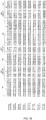
- the position of the CDR regions of an inventive V H sdAb fragment selected from the group of SEQ ID NOS: 2, 4, 6 and 8 are at the positions provided in FIGURE 1A and those selected from the group of SEQ ID NOS: 70 to 83 are at the position provided for the corresponding wild type sequences in FIGURE 2 of the PCT publication WO2006/099747 .
- the polypeptide comprises at least a Cys residue at position 48 and at least a Cys residue at position 64 of a V L domain.
- the inventive V L domain comprises a sdAb fragment.
- the invention provides an expression library comprising the inventive V L sdAb fragment, wherein the inventive sdAb fragments comprise a multiplicity of CDR sequences.
- the position of the CDR regions of an inventive V L sdAb fragment selected from the group of SEQ ID NOS: 10, 12, 14, 16, 18, 20, 22 and 24 are at the positions provided in FIG. 1B .
- the CDR residues of the inventive polypeptides may be any suitable sequence and may have a variable number of residues other than the number provided in Figures 1A and 1B .
- the inventive immunoglobulin polypeptide comprises the scaffold region of one or more of the sequences selected from the group consisting of or sequences substantially identical thereto, or fragments thereof, with the proviso that the substantially identical sequence, or fragment thereof, retains both the canonical and non-canonical disulfide bonds, and wherein the sequence in the regions of the CDRs may be any suitable sequence and may have a suitable but variable number of residues in each of CDR1, CDR2 and CDR3.
- the residues that comprise the CDRs are as provided in Figures 1A and 1B .
- SEQ ID Nos: 70 through 83 the residues that comprise the CDRs are as provided in the corresponding wild type clones as provided in Figure 2 of PCT publication WO2006/099747 .
- the present invention also encompasses a recombinant library encoding a diversity of variable domains, the variable domains comprising a multiplicity of CDRs comprising a variety of suitable sequences and lengths interspersed by the framework regions of a scaffold comprising the inventive non-canonical Cys-Cys residues as described herein.
- the recombinant library comprises mammalian sequences.
- the recombinant library comprises human sequences.
- the recombinant library comprises camelid sequences.
- the recombinant library comprises a diversity of sequences that, once selected, is optionally matured and/or humaneered.
- the present invention also provides a method of improving stability of single domain antibodies, comprising introducing one or more than one non-canonical disulfide bond in the framework regions.
- the present invention provides methods for engineering V H and V L domains with enhanced stability.
- the formation of additional disulfide linkage(s) in V H and V L domains by introducing Cys pair(s) at specific locations is shown herein to transform V H s and V L s into highly non-aggregating, soluble, stable and expressible domains.
- these advantageous properties are obtained when residues within the FR2 and FR3 regions are replaced such that a Cys pair at amino acid positions 49 and 69 for V H , and 48 and 64 for V L (Kabat numbering) was introduced.
- Formation of disulfide linkages in between the introduced non-canonical residues in the Cys mutants was confirmed by mass spectrometry.
- V H and/or V L libraries based on the engineered scaffolds of the present invention can be prepared to identify stable and nonaggregating V H s and V L s that bind specific target molecules.
- Various display libraries known in the art may be used to express the V H and/or V L constructs of the invention.
- the library is a single domain antibody (“sdAb”) library.
- the present invention provides a novel approach that may (i) convert aggregating sdAb to non-aggregating sdAb; (ii) increase the efficiency of obtaining non-aggregating sdAb from sdAb libraries; (iii) additionally increase the stability, e.g., melting temperature, refolding efficiency, resistance to proteases, and expression level, of sdAb.
- the provision of V H and V L scaffolds with these characteristics may allow the recovery of broader ranges of antibodies from antibody libraries; the retention of a high degree of non-aggregation and stability may lead to isolating novel antibodies that could otherwise be lost during the antibody selection process. Additionally, the improved biophysical characteristics may allow the V H and V L to be broadly applicable in therapeutics and industrial field.
- the present invention relates to engineering of immunoglobulin domains. More specifically, the present invention is directed to engineering of immunoglobulin domains to improve biophysical properties of the immunoglobulin. Compositions of matter and methods of making and using such compositions are provided.
- the present invention provides a composition comprising a polypeptide comprising a V H or V L scaffold comprising one or more than one non-canonical disulfide bond in the framework region (FR), wherein the non-canonical disulfide bond provides improved stability and non-aggregation of the polypeptide.
- the polypeptide is a V H or V L scaffold.
- the polypeptide comprises the sdAb fragment of a V H or V L domain.
- the sdAb may be derived from a V H region, a V H H region or a V L region as described herein.
- the immunoglobulin scaffold may comprise the variable region of a heavy chain ("VH").
- V H is derived from the V H 1, the V H 2 or the V H 3 family.
- the V H may be of the V H 3 family.
- the immunoglobulin scaffold may be a variable region of a light chain ("V L ").
- V L may be of the kappa or lambda family.
- the immunoglobulin scaffold is a non-human animal.
- the non-human animal includes all vertebrates, especially vertebrates selected from avians, amphibians, reptiles, mammals, camelids, chicken, rat, mouse, rabbit, goat, sheep, cow, cat, dog, horse, or nonhuman primates.
- the immunoglobulin scaffold is human. In one embodiment the immunoglobulin scaffold is camelid. In one embodiment, the camelid species is selected from the group consisting of llama, alpaca and camel. In one embodiment, the camelid scaffold is derived from any one or more of a V H region, a V L or a V H H region.
- the immunoglobulin scaffold is "humaneered” (also alternatively termed “humanized”), i.e., a sdAb that originated from a species other than human that has had immunogenic or potentially immunogenic amino acid residues replaced with amino acids that are less immunogenic or not immunogenic in the context of a sdAb administered to a human subject.
- humaneered also alternatively termed “humanized”
- Any method known in the art for creating humaneered antibodies are contemplated in the invention, including but not limited to humaneering technology of Kalobios.
- an immunogenic amino acid residue may be replaced by any other less immunogenic amino acid residue regardless of whether or not this constitutes a conserved amino acid change. It is understood that humaneered scaffolds of the invention retain the non-canonical Cys residues described herein, as well as any native (canonical) disulfide bonds.
- V H or V L refer to the variable region of an antibody heavy chain or an antibody light chain, respectively.
- the V H or V L domain is in the format of a "single domain antibody” (sdAb).
- sdAb refers to a single immunoglobulin domain that retains the immunoglobulin fold; i.e., it is a variable domain in which up to three complementarity determining regions (CDR) along with up to four framework regions (FR) form the antigen-binding site.
- CDR1 CDR2, and CDR3 The CDR of the V H or V L variable domain are referred to herein as CDR1, CDR2, and CDR3.
- FR1, FR2, FR3 and FR4 The FRs of the V H or V L variable domain are referred to herein as FR1, FR2, FR3 and FR4.
- Kabat et al (1991) define the "complementarity-determining regions” (CDR) based on sequence variability at the antigen-binding regions of the V H and/or V L domains.
- CDR complementarity-determining regions
- Chothia and Lesk (1987) define the "hypervariable loops" (H or L) based on the location of the structural loop regions in the V H and V L domains.
- CDR and hypervariable loop regions that are adjacent or overlapping
- those of skill in the antibody art often utilize the terms "CDR” and “hypervariable loop” interchangeably, and they may be so used herein.
- the majority of the sequence variability in variable domains (V H or V L ) occurs in the CDR/loops; the regions outside the CDR/loops are referred to as the framework regions (FR).
- the FR provide structural integrity to the variable domain and ensure retention of the immunoglobulin fold.
- the FR and CDR/loops are defined herein according to the Kabat numbering system (Kabat et al, 1991).
- the numbering of the positions with the FR1, CDR1, FR2, CDR2, FR3, CDR3 and FR4 regions of the polypeptides described herein are as provided in Figure 1A for a VH domain and as provided in Figure 1B for a V L domain.
- Human V H or V L domains may be obtained from human Ig heavy or light chain sequences (Holliger, and Hudson, 2005; Holt, et al., 2003; Jespers et al, 2004; To et al, 2005). Similar techniques are known in the art for obtaining V H or V L domains from non-human species.
- inventive V H and V L domains of the present invention include recombinantly produced V H or V L , as well as those V H or V L generated through further modification of such V H or V L by affinity maturation, stabilization, solubilization (e.g., camelization), or other methods of antibody engineering.
- homologues, derivatives, or variants that retain or improve the stability and non-aggregation characteristics of the VH or V L .
- the invention provides novel V H or V L domains that comprise a scaffold comprising non-canonical Cys resides.
- a "scaffold” or alternatively an “immunoglobulin scaffold” comprises the framework regions (FR1, FR2, FR3, and FR4) of the V H or V L , wherein the framework provides the polypeptide backbone for the various CDRs (CDR1, CDR2, CDR3).
- the term "scaffold” ignores the CDR regions for purposes of describing the framework portion of composition of the invention.
- the immunoglobulin scaffold described herein may comprise a polypeptide sequence comprising at least two non-canonical Cys residues introduced into the framework regions FR2 and FR3 of an antibody variable region.
- the polypeptide comprises a Cys residue at a position selected from residues 47 to 49 of a V H FR2 regions and a Cys residue at a position selected from residues 69 to 71 of a V H FR3 regions of a V H sdAb domain.
- the polypeptide comprises a Cys residue at a position selected from residues 46 to 49 of a V L FR2 regions and a Cys residue at a position selected from residues 62 to 66 of a V L FR3 regions of a V L sdAb domain.
- "introduced” or “introducing” means incorporating a change into the polypeptide composition or the nucleotide sequence encoding the polypeptide composition using any suitable method known to one skilled in the art, and especially including recombinant technology.
- Cys residues are introduced into the V H and/or VL construct by replacing an existing codon in a gene, a coding sequence and/or an mRNA with a codon that is specific for a cysteine residue.
- the inventive composition comprises an immunoglobuin scaffold selected from a V H or a V L scaffold, wherein the V H scaffold comprises at least one non-canonical disulfide bond in the FR, wherein the non-canonical disulfide bond is formed between Cys residues introduced at positions 49 and 69, based on Kabat numbering; and the V L scaffold comprises at least one non-canonical disulfide bond in the FR, wherein the non-canonical disulfide bond is formed between Cys residues introduced at positions 48 and 64, based on Kabat numbering.
- an "inventive" V H construct or sdAb fragment comprises at least one non-canonical disulfide bridge comprising at least a Cys residue at position 49 and at least a Cys residue at position 69.
- an "inventive" V L construct or sdAb fragment comprises at least one non-canonical disulfide bridge comprising at least a Cys residue at position 48 and at least a Cys residue at position 64.
- the polypeptide comprises at least a Cys residue at position 49 and at least a Cys residue at position 69 of a V H domain.
- the inventive V H domain comprises a sdAb fragment.
- the invention provides an expression library comprising the inventive V H immunoglobulin scaffold, wherein the immunoglobulin scaffold further comprise a multiplicity of CDR sequences.
- the invention provides a recombinant library comprising the inventive V H immunoglobulin scaffold and a multiplicity of suitable CDR sequences so as to provide libraries with diversities of at least 10 5 , at least 10 6 , at least 10 7 , at least 10 8 , at least 10 9 or more than 10 9 clones per library.
- the position of the CDR regions of the inventive V H sdAb fragment selected from the group of SEQ ID NOS: 2, 4, 6 and 8 are at the positions provided in FIGURE 1A and those of SEQ ID NOS:70 to 83 are at the position provided for the corresponding wild type sequences in FIGURE 2 of the PCT publication WO2006/099747 .
- the polypeptide comprises at least a Cys residue at position 48 and at least a Cys residue at position 64 of a V L domain.
- the inventive V L domain comprises a sdAb fragment.
- the invention provides an expression library comprising the inventive V L sdAb fragment, wherein the inventive sdAb fragments comprise a multiplicity of CDR sequences.
- the invention provides a recombinant library comprising the inventive V L immunoglobulin scaffold and a multiplicity of suitable CDR sequences so as to provide libraries with diversities of at least 10 5 , at least 10 6 , at least 10 7 , at least 10 8 , at least 10 9 or more than 10 9 clones per library.
- the position of the CDR regions of the inventive V L sdAb fragment selected from the group of SEQ ID NOS: 10, 12, 14, 16, 18, 20, 22 and 24 are at the positions provided in FIGURE 1B .
- the CDR residues of the inventive polypeptides may be any suitable sequence and may have a variable number of residues other than the number provided in Figures 1A and 1B .
- Each of CDR1, CDR2 and CDR3 may vary in sequence and in length according to guidelines known to those skilled in the antibody arts. Without wishing to be limited to any one specific reference, exemplary guidelines are provided in the Kabat and the Chothia and Lesk publications cited herein.
- the design and construction of a phage library based on the HVLP324S scaffold is provided in Figure 11 and in Example 8 and the library is characterized in Example 9.
- the V H or V L compositions of the present invention provide scaffolds in the construction of additional libraries or antibodies.
- the framework regions (FR1, FR2, FR3, and FR4) of the V H or V L may be used to carry various CDRs, i.e., wherein the FR are as provided and wherein the sequence and number of residues of the CDR region are as provided or vary at one or more, or at all, of the CDR-specific residues.
- synthetic V H or V L libraries may be constructed on single scaffolds by inserting suitable oligonucleotides comprising randomized sequences to provide randomized CDR/loops.
- Such libraries may be display libraries such as phage display, ribosome display, or yeast display.
- the libraries may optionally be used for further antibody engineering or for in vitro affinity maturation (Davies and Riechmann, 1996b; Yau et al., 2005).
- an existing library based on a naturally-occurring sdAb can be used to engineer disulfide linkage(s) into every member of the library by splice overlap extension-polymerase chain reaction (SOE-PCR) (Arbabi-Ghahroudi, et al., 2010 and references therein; Ho et al., 1989) or other methods (Kunkel et al., 1987; Sidhu et al., 2000).
- SOE-PCR splice overlap extension-polymerase chain reaction
- V H or V L may be used as a scaffold to incorporate specific CDR sequences; for example, and without wishing to be limiting in any manner, a humanized antibody may be constructed from any non-human antibody by using recombinant technology to further comprise the non-canonical disulfide bridge according to the methods provided herein. This construct may then be further modified to bind a specific target by inserting the appropriate CDR coding segments (responsible for the desired binding properties, and which may vary in sequence and in length according to guidelines cited herein and known to those skilled in the antibody arts) into the humanized antibody scaffold according to the present invention.
- CDR coding segments response for the desired binding properties, and which may vary in sequence and in length according to guidelines cited herein and known to those skilled in the antibody arts
- the V H or V L scaffolds of the present invention comprise one or more than one non-canonical disulfide bond in the framework regions.
- a disulfide bond is a covalent force that stabilizes proteins through coupling of two thiol groups between cysteine residues (Betz, 1993).
- non-canonical it is meant that the disulfide bond is not found in naturally-occurring human V H and V L , but rather is introduced using molecular engineering techniques.
- the naturally-occurring (or "canonical") disulfide bond connecting two ⁇ -sheets within a variable domain is highly conserved in V H and V L superfamilies (Amzel and Poljak, 1979; Williams and Barclay, 1988).
- the conserved disulfide bonds form between Cys22 and Cys92 in V H s and Cys23 and Cys88 in V L s, wherein the residues are numbered according to Kabat (see Fig. 1 ).
- one or more than one additional non-canonical disulfide bond is engineered into the V H or V L domain.
- the engineered disulfide bond forms between a Cys from a first beta sheet and a Cys from a nearby second beta sheet in the immunoglobulin fold. For example, this can be accomplished by introducing an appropriate number of Cys pair(s) at locations in the framework regions that will form a disulfide bond in the expressed and fully folded protein.
- the Cys residues may be introduced into framework regions that are adjacent in the three-dimensional structure of the variable domain.
- Cys residues could be introduced into the FR2 beta sheet region and the FR3 beta sheet region.
- the Cys residues are introduced into the adjacent framework regions at locations that are within range for coupling of the thiol groups.
- Cys residues may be introduced at one of positions 47-49 (3 positions) and one of positions 67-71 (5 positions) for V H , or at one of positions 46-49 (4 positions) and one of positions 62-66 (5 positions) for V L (based on Kabat numbering), providing that the respective Cys pairs are within sufficient proximity so as to form a disulfide bond.
- the Cys residues may be introduced at positions 49 and 69 for V H , and/or 48 and 64 for V L (based on Kabat numbering).
- the Cys may be introduced into the framework regions of V H or V L domains using any suitable method known in the art. Without wishing to be limiting, the Cys may be introduced by point-mutation or insertion through recombinant DNA methods; for example and without wishing to be limiting in any manner, the Cys may be introduced using splicing by overlap extension (SOE)-PCR (Arbabi-Ghahroudi, et al., 2010 and references therein; Ho et al., 1989).
- SOE overlap extension
- the one or more than one non-canonical disulfide bond in the V H and/or V L scaffolds lends improved stability to and non-aggregation of the antibody.
- Non-aggregation refers to the molecule existing in a monomeric state; for example, and without wishing to be limiting in any manner, monomers give essentially one peak with size exclusion chromatography (SEC). Aggregating molecules form dimers, trimers, and higher order aggregates.
- Stability of an antibody includes biophysical characteristics such as thermostability, thermal and chemical refolding efficiency, and protease resistance. Other factors to be considered when evaluating the V H or V L scaffolds of the present invention include expression levels and solubility.
- solubility refers to the number of molecules per volume (e.g., in molarity or mg/mL) that may be dissolved in a liquid prior to precipitation; in the case of sdAbs, and in the context of the present invention, the number of monomeric molecules is of particular importance.
- the addition of a non-canonical disulfide bond may improve one or more than one of the aforementioned biophysical characteristics.
- the introduction of a non-canonical disulfide bond into a V H or V L domain may improve stability (for example, higher melting temperature and protease resistance, by stabilizing the framework); and/or improve non-aggregation (by reducing inter-molecular interactions and formation of aggregates).
- V H or V L scaffold into which non-canonical disulfide bonds are introduced may be of any suitable germline origin.
- the V L may be of the lambda or kappa family, for example kappa 1 or kappa 3, or its composition may be derived from various combinations of V and J segment germline sequences.
- the V H may be of the V H 1, the V H 2, the V H 3, the V H 4, the V H 5, the V H 6 or the V H 7 family, or its composition may be derived from various combinations of V, D and J segment germline sequences (To et al., 2005; V BASE database of the MRC Centre for Protein Engineering, http://vbase.mrc-cpe.cam.ac.uk/ ).
- the V H may be of the V H 3 family.
- the V H or V L scaffolds of the present invention may include, but are not limited to scaffolds comprising the framework regions of sequences selected from the group consisting of or sequences substantially identical thereto, or fragments thereof, with the proviso that the substantially identical sequence, or fragments thereof, retains both the canonical and non-canonical disulfide bonds and wherein the sequence in the regions of the CDRs may be any suitable sequence and may have a suitable but variable number of residues in each of CDR1, CDR2 and CDR3.
- SEQ ID Nos: 2, 4, 6, 8, 10, 12, 14, 16, 18, 20, 22 and 24 the residues that comprise the CDRs are as provided in Figures 1A and 1B .
- the residues that comprise the CDRs are as provided in the corresponding wild type clones as provided in Figure 2 of PCT publication WO2006/099747 , wherein said Figure 2 is incorporated herein by reference.
- the substantially identical sequences should also retain or improve the stability and non-aggregation characteristics of the V H or V L .
- the inventive scaffolds are not limited to the residues listed in the CDR regions, which are as delimited in Figures 1A and 1B for the clones listed herein.
- a substantially identical sequence may comprise one or more conservative amino acid mutations. It is known in the art that one or more conservative amino acid mutations to a reference sequence may yield a mutant peptide with no substantial change in physiological, chemical, or functional properties compared to the reference sequence; in such a case, the reference and mutant sequences would be considered "substantially identical" polypeptides.
- Conservative amino acid mutation may include addition, deletion, or substitution of an amino acid; a conservative amino acid substitution is defined herein as the substitution of an amino acid residue for another amino acid residue with similar chemical properties (e.g. size, charge, or polarity).
- a conservative mutation may be an amino acid substitution.
- Such a conservative amino acid substitution may substitute a basic, neutral, hydrophobic, or acidic amino acid for another of the same group.
- basic amino acid it is meant hydrophilic amino acids having a side chain pK value of greater than 7, which are typically positively charged at physiological pH.
- Basic amino acids include histidine (His or H), arginine (Arg or R), and lysine (Lys or K).
- neutral amino acid also “polar amino acid”
- hydrophilic amino acids having a side chain that is uncharged at physiological pH, but which has at least one bond in which the pair of electrons shared in common by two atoms is held more closely by one of the atoms.
- Polar amino acids include serine (Ser or S), threonine (Thr or T), cysteine (Cys or C), tyrosine (Tyr or Y), asparagine (Asn or N), and glutamine (Gln or Q).
- hydrophobic amino acid (also “non-polar amino acid”) is meant to include amino acids exhibiting a hydrophobicity of greater than zero according to the normalized consensus hydrophobicity scale of Eisenberg (1984). Hydrophobic amino acids include proline (Pro or P), isoleucine (Ile or I), phenylalanine (Phe or F), valine (Val or V), leucine (Leu or L), tryptophan (Trp or W), methionine (Met or M), alanine (Ala or A), and glycine (Gly or G).
- “Acidic amino acid” refers to hydrophilic amino acids having a side chain pK value of less than 7, which are typically negatively charged at physiological pH. Acidic amino acids include glutamate (Glu or E), and aspartate (Asp or D).
- Sequence identity is used to evaluate the similarity of two sequences; it is determined by calculating the percent of residues that are the same when the two sequences are aligned for maximum correspondence between residue positions. Any known method may be used to calculate sequence identity; for example, computer software is available to calculate sequence identity. Without wishing to be limiting, sequence identity can be calculated by software such as NCBI BLAST2 service maintained by the Swiss Institute of Bioinformatics (and as found at http://ca.expasy.org/tools/blast/), BLAST-P, Blast-N, or FASTA-N, or any other appropriate software that is known in the art. In one embodiment, calculation of percent identity is limited to comparison of the framework regions and disregards the residues within the CDRs. In one embodiment, calculation of percent identity compares all residues in a polypeptide, including both those of the framework regions and the CDRs.
- inventive scaffolds are not limited to the residues listed in the CDR regions, which are as delimited in Figures 1A and 1B for the clones listed herein as SEQ ID Nos: 2, 4, 6, 8, 10, 12, 14, 16, 18, 20, 22 and 24; and are delimited in Figure 2 of PCT publication WO2006/099747 for the wild type sequences corresponding to the clones listed herein as SEQ ID Nos: 70 through 83.
- the substantially identical sequences of the present invention may be at least 90% identical; in another example, the substantially identical sequences may be at least 90, 91, 92, 93, 94, 95, 96, 97, 98, 99, or 100% (or any percentage therebetween) identical at the amino acid level to sequences described herein.
- a substantially identical sequence may contain one, two, three or four amino acid differences in the framework region when aligned with an inventive composition provided herein.
- the substantially identical sequences retain the stability and biophysical properties of the reference sequence.
- the difference in sequence identity may be due to conservative amino acid mutation(s).
- the substantially identical sequences consist of the residues comprising FR1, FR2, FR3 and FR4 and does not consider the residues comprising any one or more of CDR1, CDR2 and CDR3.
- V H or V L scaffolds may be included as part of larger antibody protein or fragments such as one or more of a sdAb, scFv, Fab, F(ab) 2 and/or mature immunoglobulin, including but not limited to an IgG, IgE, and/or IgM, for the purpose of increasing their biophysical properties such as non-aggregation, stability, expression level, and solubility.
- a "mature immunoglobulin” comprises a light chain, said light chain comprising a variable and a constant region, and a heavy chain, said heavy chain comprising a variable and a constant region.
- the mature immunoglobulin is selected from the group consisting of an IgG1, IgG2, IgG3, IgG4, IgE, and/or IgM.
- the introduced sequence of this larger antibody construct is human or is humaneered.
- V H and V L derivatives such as fusion constructs made by fusions of V H s and V L s to other polypeptides such as toxins, V H s and/or V L s with the same or different specificity, Fc domains, etc. for the purpose of increasing their biophysical properties such as solubility, non-aggregation, expression level and stability.
- the V H or V L scaffolds of the present invention may also be fused to additional sequences to aid in expression, detection or purification of a recombinant antibody or fragment thereof.
- additional sequences to aid in expression, detection or purification of a recombinant antibody or fragment thereof.
- Any such sequences or tags known to those of skill in the art may be used.
- the antibody or fragment thereof may comprise a targeting or signal sequence (for example, but not limited to ompA), a detection tag (for example, but not limited to c-Myc), a purification tag (for example, but not limited to a His 5 or His 6 ), an Fc fragment, or a combination thereof.
- the additional sequence may be a biotin recognition site such as that described by Cronan et al in WO 95/04069 or Voges et al in WO/2004/076670 .
- linker sequences may be used in conjunction with the additional sequences or tags.
- the V H or V L scaffolds of the present invention may also be in a multivalent display.
- Multimerization may be achieved by any suitable method known in the art. For example, and without wishing to be limiting in any manner, multimerization may be achieved using self-assembly molecules as described in Zhang et al (2004a; 2004b) and WO2003/046560 .
- the described method produces pentabodies by expressing a fusion protein comprising the V H or V L scaffold of the present invention and the pentamerization domain of the B-subunit of an AB 5 toxin family (Merritt and Hol, 1995); the pentamerization domain assembles into a pentamer, through which a multivalent display of the antibody or fragment thereof is formed.
- the pentamerization domain may be linked to the V H or V L scaffold using a linker; such a linker should be of sufficient length and appropriate composition to provide flexible attachment of the two molecules, but should not hamper the biophysical properties of the V H or V L scaffold.
- V H or V L scaffold may be presented as at least a dimer, a trimer, tetramer, pentamer, hexamer, heptamer, octamer, or any other suitable oligomer.
- V H or V L scaffolds may be fused to an antibody constant region fragment ("Fc") to generate chimeric heavy chain antibodies (Bell et al., 2010).
- Fc antibody constant region fragment
- the Fc gene in inserted into a vector along with the V H or V L gene to generate a sdAb-Fc fusion protein (Bell et al, 2010; Iqbal et al, 2010); the fusion protein is recombinantly expressed then purified.
- sdAb-Fc fusion protein Bell et al, 2010; Iqbal et al, 2010
- Such antibodies are easy to engineer and to produce (Zhang et al.,2009), can greatly extend the serum half life of V H or V L , and may be excellent tumor imaging reagents (Bell et al., 2010).
- the Fc portion is human.
- the present invention also encompasses nucleic acid sequences encoding the molecules as described herein.
- the nucleic acid comprises a sequence encoding an immunoglobulin scaffold selected from a V H or a V L scaffold, wherein the V H scaffold comprises at least one non-canonical disulfide bond in the FR formed between Cys residues introduced at positions 49 and 69, based on Kabat numbering; and the V L scaffold comprises at least one non-canonical disulfide bond in the FR formed between Cys residues introduced at positions 48 and 64, based on Kabat numbering.
- the nucleic acid encodes any one or more of the sequences selected from SEQ ID NOS: 2, 4, 6, 8, 10, 12, 14, 16, 18, 20, 22, 24 and 70-83.
- the nucleic acid sequence comprises any codon of the degenerate code that encodes the appropriate amino acid residue. In one embodiment, coding region of the nucleic acid sequence is codon optimized. In one embodiment, the nucleic acid is in an expression vector. In one embodiment, the expression vector is in a host cell or organism capable of expressing said nucleic acid.
- nucleic acid sequence in a host cell or organism by replacing the nucleic acids coding for a particular amino acid (i.e. a codon) with another codon which is better expressed in the host organism.
- a codon a particular amino acid
- One reason that this effect arises is due to the fact that different organisms show preferences for different codons. In particular, bacterial organisms and yeast organisms prefer different codons from plants and animals.
- codon optimization The process of altering the sequence of a nucleic acid to achieve better expression based on codon preference is called codon optimization.
- the nucleic acid sequence may be codon-optimized for expression in mammalian cells. In one embodiment, the nucleic acid sequence may be codon-optimized for expression in various micro-organisms. In one embodiment, the nucleic acid sequence is codon-optimized for expression in E . coli. In one embodiment, the nucleic acid sequence is codon-optimized for expression in yeast.
- the nucleic acid sequence is codon-optimized for expression in yeast and for surface display of antibodies, i.e., shuttle between antibody phage display and antibody yeast display.
- the present invention also encompasses vectors comprising the nucleic acids as just described.
- the invention encompasses cells comprising the nucleic acid and/or vector as described.
- the present invention further encompasses the V H or V L scaffold immobilized onto a surface using various methodologies; for example, and without wishing to be limiting, the V H or V L scaffold may be linked or coupled to the surface via His-tag coupling, biotin binding, covalent binding, adsorption, and the like.
- the solid surface may be any suitable surface, for example, but not limited to the well surface of a microtiter plate, channels of surface plasmon resonance (SPR) sensorchips, membranes, beads (such as magnetic-based or sepharose-based beads or other chromatography resin), glass, a film, or any other useful surface.
- SPR surface plasmon resonance
- the present invention further provides an inventive V H or V L linked to a cargo molecule; the antibody or fragment thereof may deliver the cargo molecule to a desired site.
- the cargo molecule may be any type of molecule that may be use to diagnose, treat or detect a biomarker.
- the cargo marker is a molecule that may reduce and/or inhibit the growth of targeted cells.
- the targetted cells are cells associated with a proliferative disease.
- the proliferative disease is a cancer, including various types of tumours, wherein the targeted cells express a marker recognized by the V H or V L domain portion of the invention.
- the cargo molecule is a therapeutic or diagnostic agent.
- the cargo molecule is linked to a therapeutic or diagnostic agent.
- the therapeutic agent may be a radioisotope, which may be used for radioimmunotherapy; a toxin, such as an immunotoxin; a cytokine, such as an immunocytokine; a cytotoxin; an apoptosis inducer; an enzyme; or any other suitable therapeutic molecule known in the art.
- a diagnostic agent may include, but is by no means limited to a radioisotope, a paramagnetic label, a fluorophore, an affinity label (for example biotin, avidin, etc), fused to a detectable protein-based molecule, or any other suitable agent that may be detected by imaging methods.
- the antibody or fragment thereof may be linked to a fluorescent agent such as FITC or may genetically be fused to the Enhanced Green Fluorescent Protein (EGFP).
- EGFP Enhanced Green Fluorescent Protein
- the antibody or fragment thereof may be linked to the cargo molecule using any method known in the art (recombinant technology, chemical conjugation, etc.).
- the present invention also provides a method of improving stability of single domain antibodies, comprising introducing one or more than one non-canonical disulfide bond in the framework region.
- the method comprises introducing at least two non-canonical Cys residues into the framework regions of the immunoglobulin of interest.
- the two Cys residues replace residues in the FR2 and FR3 of an antibody variable region.
- the method comprises introducing a Cys residue at a position selected from residues 47 to 49 of a V H FR2 regions and a Cys residue at a position selected from residues 69 to 71 of a V H FR3 regions of a V H sdAb domain.
- the method comprises introducing a Cys residue at a position selected from residues 46 to 49 of a V L FR2 regions and a Cys residue at a position selected from residues 62 to 66 of a V L FR3 regions of a VL sdAb domain.
- the method comprises introducing at least one non-canonical Cys residue at position 49 and at least one non-canonical Cys residue at position 69 of a V H domain. In one embodiment, the method comprises introducing at least one non-canonical Cys residue at position 48 and at least one non-canonical Cys residue at position 64 of a V L domain.
- the method comprises the creation of an expression library comprising the inventive V H sdAb fragment comprising a disulfide bridge between non-canonical Cys residues at positions 49 and 69, wherein the inventive sdAb fragments comprise a multiplicity of CDR sequences.
- the method comprises the creation of an expression library comprising the inventive V L sdAb fragment comprising a disulfide bridge between non-canonical Cys residues at positions 48 and 64, wherein the inventive sdAb fragments comprise a multiplicity of CDR sequences. Any suitable method known in the art for antibody library creation replacement of CDR sequences may be used.
- the position of the CDR regions are as defined by Kabat.
- the position of the CDR regions are as defined by Chothia and Lesk.
- the expression library is codon optimized for expression in mammalian cells. In one embodiment, the expression library is codon optimized for expression in yeast. In one embodiment, the expression library is codon optimized for expression in E . coli.
- the mutations can be introduced by SOE-PCR approach (Arbabi-Ghahroudi et al., 2010 and reference therein; Ho et al., 1989) or by other method (Kunkel et al., 1987; Sidhu et al., 2000, Hussack et al., In press (c)).
- the genes for the mutants can be obtained commercially, either on their own or within the target cloning and expression vector (Kim et al., In press).
- V H and V L domains Aggregating and non-aggregating V H and V L domains as well as their counterpart double cysteine mutants were cloned, expressed and purified.
- V H and V L utilized, as well as their corresponding Cys mutants, are listed in Table 1 and shown in Figure 1 .
- Table 1 V H and V L and mutants constructed.
- H * V H Cys mutant HVHAm302 (SEQ ID NO:1) HVHAm302S (SEQ ID NO:2) HVHAm427 (SEQ ID NO:3) HVHAm427S (SEQ ID NO:4) HVHAm431 (SEQ ID NO:5) HVHAm431S (SEQ ID NO:6) HVHPC235 (SEQ ID NO:7) HVHPC235S (SEQ ID NO:8) HVHP44 HVHP44S (SEQ ID NO:70) HVHB82 HVHB82S (SEQ ID NO:71) HVHP421 HVHP421S (SEQ ID NO:72) HVHP419 HVHP419S (SEQ ID NO:73) HVH P430 HVHP430S (SEQ ID NO:74) HVHP429 HVHP429S (SEQ ID NO:75) HVHM41 HVHM41S (SEQ ID NO:76) HVHM81 HVHM81S (SEQ ID NO:
- VHs 14 additional wild type VHs, HVHP44, HVHB82, HVHP421, HVHP419, HVHP430, HVHP429, HVHM41, HVHM81, HVHP428, HVHP420, HVHP414, HVHP423, HVHP413, and HVHP426 and wild type V L s were prepared as described in WO 2006/099747 by Tanha . Plasmids containing genes for these wild-type domains (excluding the 14 aforementioned V H s; see below) were used as templates to construct corresponding mutant versions by SOE-PCR.
- Inventive constructs also referred to herein as "Cys mutants," of V H s and V L s (HVHAm302, HVHAm427, HVHAm431, and HVHPC235) were created with splicing by overlap extension (SOE)-PCR (Ho et al., 1989; Kim et al., Submitted Arbabi-Ghahroudi et al, 2010) to introduce a pair of Cys at the positions of 49 and 69 (V H s) or positions 48 and 64 (V L s) as defined by Kabat numbering (Kabat et al., 1991) and as provided in Figure 1A for a V H domain and Figure 1B for a V L domain.
- SOE overlap extension
- Cys-mutated sub-fragments of the clones were prepared, then assembled to form full-length Cys mutants by SOE-PCR.
- sub-fragments 302S-1 and 302S-2 were generated separately by standard PCR using the plasmid containing the wild-type HVHAm302 gene as the DNA template and the following pairs of primers:
- V H mutants were generated from their corresponding wild-type V H plasmid using the same procedures and primers; V L mutants were generated using the same SOE-PCR procedure as that for the V H mutants, except different, VL gene-specific PCR primers and corresponding wild-type V L plasmid templates were used.
- HVHP44S HVHB82S, HVHP421S, HVHP419S, HVHP430S, HVHP429S, HVHM41S, HVHM81S, HVHP428S, HVHP420S, HVHP414S, HVHP423S, HVHP413S, and HVHP426S, respectively.
- the framework and CDR regions are otherwise the same as those of the corresponding wild-type sequences, which are as provided in PCT publication WO2006/099747 , especially as shown in FIG. 2 of the publication.
- Cys mutants were then cloned into an expression vector, expressed, purified, and their concentrations determined as described elsewhere (Sambrook et al., 1989; To, et al., 2005; Arbabi-Ghahroudi et al 2009a; 2009b; 2010).
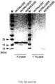
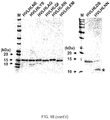
- the gel migration differences between wild-type and mutant V H s is due to their conformational differences as a consequence of extra, engineered disulfide linkages in mutants (Kim and Tanha, 2010).
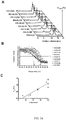
- the wild-type V H HVHAm302 form aggregates (see dimeric bands in Fig. 2B ), which was further confirmed by a Western blotting using anti-His antibody ( Fig. 2B and data not shown). The dimeric bands disappear for the corresponding mutant V H , HVHAm302S ( Fig. 2B ). This shows that the engineered disulfide linkage improves V H non-aggregation and is consistent with the size exclusion chromatography findings (see below and Fig. 4A ).
- Disulfide linkage determination for V H s and V L s was carried out as described elsewhere (Hussack et al., In press (b); Kim and Tanha, In press; Wu et al., 2009). Disulfide linkage determination for HVHAm302 and HVHAm302S were carried out exactly as described in Kim and Tanha (In press).
- V H Cys mutants were concentrated in 0.1 M Tris-HCl, pH 8.5, using Ultrafree-0.5 centrifugal filter device with biomax-5 membrane (MWCO 5000; Millipore, Nepean, ON, Canada) and subjected to trypsin digestion (Roche Diagnostics Canada, Laval, QC, Canada) at concentration of 0.5 mg/mL in 0.1 M Tris-HCl, pH 8.5, and analyzed by SDS-PAGE for the success of trypsin digestion and subsequently subjected to a peptide mass spectrometry analysis ( Fig. 3 and Table 2).
- results showed that the disulfide-linked peptides produced by tryptic digestion of V H s could be identified by the mass spectrometry method employed ( Fig. 3B and Table 2). All molecular weights of the recombinant proteins were determined to be within 40 ppm mass accuracy using infusion ESI-MS. The identification coverage of the each protein from the analysis of their tryptic digests using nanoRPLC-MS 2 with DDA (data dependent analysis) was more than 30%. The disulfide-linked peptide ions appeared prominent in the survey scan of the DDA experiment. The expected disulfide-linked peptide sequences from the V H s were all confirmed by manual de novo sequencing. Table 2. Disulfide linkage determination of V H s by mass spectrometry.
- MW for formula (expected) molecular weight
- MW exp molecular weight determined experimentally by MS
- ⁇ MW is calculated: (MW for - MW exp ).
- MW for , MW exp , and ⁇ MW are given in Daltons (Da).
- HVHP426S one of the 14 mutant V H s was also chosen for the determination of the Cys 49/Cys 69 disulfide linkage formation.
- the MS results showed that HVHP426S had formed a disulfide linkage between Cys 49 and Cys 69 (Table 2).
- MW for formula (expected) molecular weight
- MW exp molecular weight determined experimentally by MS
- ⁇ MW is calculated: (MW for -MW exp ).
- MW for , MW exp , and ⁇ MW are given in Daltons (Da).
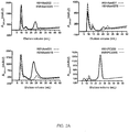
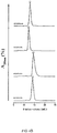
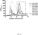
- Size exclusion chromatography (or gel filtration chromatography) separates proteins by molecular size and hydrodynamic volume (Porath and Flodin, 1959). Therefore, this method is useful in assessing protein aggregation status in solution. Size exclusion chromatography employing SuperdexTM 75 is used to assess the aggregation state of V H (or V L ) domains. Non-aggregating V H s (or V L ) should yield chromatograms with a single, symmetrical peak with elution volumes expected for a monomeric V H s (or V L s).
- the chromatogram profiles of aggregating V H s in addition to the monomeric peaks, consist of additional peaks, e.g., large aggregates, dimeric aggregates, which elute earlier.
- Percent monomer can be calculated by area integration of the peaks and used as a quantitative measure of V H (or V L ) aggregation tendency (the higher the % monomer of a V H (or V L ), the lower its aggregation tendency and vice versa the higher the % aggregates of a V H (or V L ) the higher its aggregation tendency).
- %A 280 is the normalized absorbance
- a 280 N is the absorbance at any elution volume
- a 280 M is the maximum absorbance
- a 280 B is the baseline absorbance.
- results show that protein non-aggregation was greatly improved by the mutation in all three aggregating V H s, HVHAm302, HVHAm427 and HVHAm431. While the wild-type V H s showed early elution peaks typical for aggregating V H s (about 22% for HVHAm302 and HVHAm427, and 25% for HVHAm431) in addition to monomeric peaks, these early elution peaks disappeared in the corresponding mutants, which essentially consisted of monomeric peaks; this indicates that the disulfide linkage engineering converted aggregating V H s to non-aggregating V H s.
- FIG. 4A shows representative SEC profiles for the 14 V H mutants of the V H s, HVHP44S, HVHB82S, HVHP421S, HVHP419S, HVHP430S, HVHP429S, HVHM41S, HVHM81S, HVHP428S, HVHP420S, HVHP414S, HVHP423S, HVHP413S, and HVHP426S.
- V L s were essentially free of aggregates and gave symmetric single peaks.
- V L Cys mutants were also monomeric with the exception of HVLP364S, which showed slight aggregation (11% dimeric aggregate).
- HVLP364S which showed slight aggregation (11% dimeric aggregate).
- the engineered disulfide linkage did not compromise the non-aggregating character of V L s ( Fig. 4C ).
- a Jasco J-815 spectropolarimeter equipped with a Peltier thermoelectric type temperature control system (Jasco, Easton, MD, USA) was used to carry out experiments.
- a CD cuvette with a path length of 1 mm was used.
- the spectra were recorded over a wavelength range of 180 - 260 nm with scanning speed of 50 nm/min, digital integration time (DIT) of 4 s, a band width of 1 nm, data pitch of 1 nm, and an integration time of 1 s.
- DIT digital integration time
- T m Greenfield, 2006a; 2006b
- CD spectra were recorded over a temperature range of 30°C to 96°C.
- ff ⁇ T ⁇ ⁇ U / ⁇ F ⁇ ⁇ U
- [ ⁇ ] T is the molar ellipticity at any temperature
- [ ⁇ ] F is the molar ellipticity of the fully folded protein at 30°C
- [ ⁇ ] U is the molar ellipticity of the unfolded protein at 90°C.
- T m Melting temperature (T m ) was obtained as a midpoint of the unfolding curve (fraction folded (ff) versus temperature) by a nonlinear regression curve fit (Boltzman sigmoidal equation) using the graphing software GraphPad Prism (version 4.02 for Windows) and were recorded in Table 4.
- a Jasco J-810 spectropolarimeter with a NESLAB RTE-111 bath accessory was used for wild-type V L .
- a circular cell with a pathlength of 0.02 cm was used.
- the spectra were recorded with scanning speed of 100 nm/min, a band width of 1 nm and an integration time of 1 s.
- the CD spectra were recorded over a temperature range of 25°C to 85°C for HVLP342, HVLP351 and HVLP3103, 25°C to 80°C for HVLP324, HVLP325, HVLP364 and HVLP389, and 25°C to 90°C for HVLP335.
- Heat-induced changes in molar ellipticity were monitored at203 nm for HVLP325 and HVLP389 and at 218 nm for HVLP324, HVLP335, HVLP342, HVLP351, HVLP364 and HVLP3103. Measurements were performed in sodium phosphate buffer with concentrations ranging from 4.1 x 10 -5 M to 5.7 x 10 -5 M. All CD spectra were subtracted from the blank corresponding buffer spectra and melting temperature (T m ) was obtained as previously described for V H s and mutant V L s. Table 4. Affinity constants, K D s, and melting temperatures, T m s, of V H s, V L s, and corresponding Cys mutants.
- V L s the K D s are against protein L, for V H s against protein A.
- V H s The melting temperatures (T m s) of V H s (HVHAm302 and HVHAm302S, HVHAm427 and HVHAm427S, HVHAm431 and HVHAm431S, HVHPC235 and HVHPC235S, HVHP44 and HVHP44S, HVHB82 and HVHB82S, HVHP421 and HVHP421S, HVHP419 and HVHP419S, HVHP430 and HVHP430S, HVHP429 and HVHP429S, HVHM41 and HVHM41S, HVHM81 and HVHM81S, HVHP428 and HVHP428S, HVHP420 and HVHP420S, HVHP414 and HVHP414S, HVHP423 and HVHP423S, HVHP413 and HVHP413S, and HVHP426 and HVHP426S) and V L s
- the wild type V H s had T m s of 52.8 °C - 70.9 °C which were increased to 65.4 °C - 84.6 °C for mutant V H s.
- T m increases ( ⁇ T m ) of 8.9 °C - 16.8 °C . for mutant V H s (Table 4).
- T m increases are indeed due to the presence of the extra Cys49/Cys69 disulfide linkages.
- the disulfide linkage in HVHAm427S could cause conformational alteration to bring conformational modules, such as beta-sheets, close together (acting through either short or long range contact) to induce extra chemical forces, such as hydrophobic interaction or salt bridges, to further stabilize the structure at that level.
- T m s of V L s were in the range of 49.3°C - 64.6°C, a range observed in other sdAbs (Jespers et al., 2004; Tanha et al., 2006) ( Fig 6 and Table 4).
- the T m s were drastically increased by an average of 17°C (range: 11.2°C -21.1°C) compared to T m s of the wild-type V L s.
- the T m s of mutant V L s were in the range of 63.8°C - 82.5°C compared to 49.3°C - 64.6°C for wild-type V L s.
- the T m of the least thermostable mutant, HVLP342S was comparable to that of the most thermostable wild-type, HVLP325 ( Fig. 6C ); HVLP325S had the highest T m (82.5°C), while HVLP342S had the lowest one (63.8°C).
- the effect of disulfide engineering on the stability of V L s appears to be universal, as it stabilizes both lambda and kappa type V L s and irrespective of their germline sequence origin.
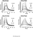
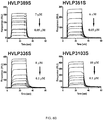
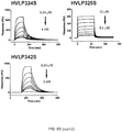
- V H s and V L s were used in SPR binding analyses to probe any subtle possible structural changes due to the engineered disulfide linkages in V H and V L mutants.
- Protein A is often used to monitor conformational integrity of V H s (Starovasnik et al., 1999). Standard procedures were carried out for SPR analysis of V H s and V L s.
- V H s and V L s were subjected to SuperdexTM 75 10/300 (GL) size exclusion chromatography (GE Healthcare) in HBS-EP buffer (10 mM HEPES, pH 7.4, 150 mM NaCl, 3 mM EDTA and 0.005% P20 surfactant) with flow rate of 0.5 mL/min priorto BIACORE analysis and purified monomer peaks collected even in the absence of any evidence of aggregated material.
- the binding kinetics for the interaction of the V H s to protein A (Pierce, Nepean, ON, Canada) and V L s to protein L (Pierce) were determined by SPR using BIACORE 3000 biosensor system (GE Healthcare).
- HVHAm302, HVHAm302S, HVHAm427, HVHAm427S, HVHAm431 and HVHAm431S protein A and ovalbumin (as a reference protein) (Sigma, Oakville, ON, Canada) of 540 RUs and 1350 RUs, respectively, were immobilized on research grade CM5 sensor chips (GE Healthcare). Immobilizations were carried out at 50 ⁇ g/mL in 10 mM acetate buffer, pH 4.5, using the amine coupling kit supplied by the manufacturer (GE Healthcare). All measurements were carried out at 25°C in HBS-EP buffer at a flow rate of 50 ⁇ L/min. Surfaces were regenerated by washing with the running buffer.
- HVHPC235 and HVHPC235S approximately 1,100 RUs and 1,000 RUs of recombinant protein A and ovalbumin (reference protein), respectively, were immobilized on research grade sensor chip CM5. Immobilizations were carried out at 50 ⁇ g/mL in 10 mM acetate buffer (pH 4.0 or pH 4.5 for protein A or ovalbumin, respectively) using the amine coupling kit supplied by the manufacturer. All measurements were carried out at 25°C in HBS-EP buffer at a flow rate of 20 ⁇ L/min or 40 ⁇ L/min for HVHPC235 or HVHPC235S, respectively.
- Immobilizations were carried out at 50 ⁇ g/mL in 10 mM acetate buffer (pH 4.5 for both protein A and ovalbumin) using the amine coupling kit supplied by the manufacturer. All measurements were carried out at 25°C in HBS-EP buffer at a flow rate of 20 ⁇ L/min or 40 ⁇ L/min for HVHB82S, HVHP421S, HVHP429S, HVHM41S, HVHM81S, HVHP423S, HVHP413S, HVHP426S, and HVHP414S or HVHP44S, HVHP419S, HVHP430S, HVHP428S, and HVHP420S, respectively.
- the protein A binding property of the wild-type counterparts were determined previously (To et al 2005).
- V L mutants 600 RUs of protein L or 800 RUs of a Fab reference protein were immobilized on research grade CM5 sensor chips (GE Healthcare).
- V L mutants approximately 400 RUs of recombinant protein L or ovalbumin (reference protein) were immobilized. Immobilizations were carried out at protein concentrations of 20 or 50 ⁇ g/mL in 10 mM acetate buffer, pH 4.5, using the amine coupling kit supplied by the manufacturer (GE Healthcare). All measurements were carried out at 25°C in HBS-EP buffer at a flow rate of 40 or 50 ⁇ L/min. Surfaces were regenerated by washing with the running buffer. All data were evaluated using the BIA evaluation 4.1 software (GE Healthcare).
- the protein A binding sites which are known to reside within FR1 and FR3 of V H s (Starovasnik et al., 1999; Riechmann and Davies, 1995), may have been compromised by the introduction of the two Cys on beta-strands of FR2 and FR3.
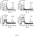
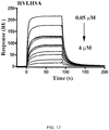
- the mutant V H s showed consistently increased disassociation rate constants (k off ) compared to their wild-type counterparts, which resulted in raising the equilibration binding constant (K D ) ( Fig. 7B ).
- V L Cys mutants bound to protein L with affinities similar to their wild-type counterparts ( Fig. 8 and Table 4), indicating that the disulfide bond did not affect the overall structure of the V L s.
- V L s and V H s were examined for the occurrence of enzymatic cleavage by SDS-PAGE and mass spectrometry. Digestions were performed essentially as described (Hussack et al.,2011).
- V L s digestion experiments were performed in a total volume of 30 ⁇ L with 6 ⁇ g of V L at 37°C for 1 h with an enzyme to sdAb ratio of 1:200, 1:40, 1:20, and 1:10 (trypsin/chymotrypsin), or 1:200, 1:20, 1:10, and 1:4 (pepsin). Tryptic and chymotryptic digestion experiments were carried out using sequencing grade enzymes (Hoffmann-La Roche Ltd., Mississauga, ON, Canada), according to the manufacturer's instructions. Pepsin (Sigma) digestion reactions were performed at ⁇ pH 2.0 where the pH was adjusted by adding appropriate volume of 400 mM HCl.
- reaction buffer In control experiments, enzymes were replaced with equal volume of reaction buffer. Reactions were stopped by adding an equal volume of SDS-PAGE sample buffer (containing 0.2 M dithiothreitol) and boiling the mixture at 95°C for 5 min. Samples were subsequently subjected to analysis by SDS-PAGE and the percent of intact sdAbs after protease digestions were determined by spot density analysis as described in Hussack et al., In press(a) and compared with controls to calculate the percentage of V L s with intact structure after protease digestion.
- SDS-PAGE sample buffer containing 0.2 M dithiothreitol
- V H s digestion experiments with trypsin/chymotrypsin/pepsin were performed in a total volume of 30 ⁇ L with 6 ⁇ g of V H and its mutant V H counterpart in parallel at 37°C for 1 h with an enzyme to sdAb ratio of 1:20 and duplicated by two independent trials. The digestions and analyses were carried out as described for the V L s.
- HVHM81S and HVHP423S While most of the mutant V H s showed unchanged or slightly improved resistance against pepsin, two mutant V H s (HVHM81S and HVHP423S) showed decreased resistance from 100% (for both HVHM81 and HVHP423) to 53% (for HVHM81S) and to 68% (for HVHP423S), although the pepsin resistance of HVHM81S seemed to be underestimated due to inability of the spot density analysis tool to recognize the protein bands properly.
- V H s Sequence identities between the V H s, as well as V L s were determined. Only FR sequences were included in the analysis, i.e., CDR sequences were excluded.
- V H pairs or V L pairs were aligned using ClustalW (Thompson et al, 1994), and the percentage identity between the V H pairs or V L pairs was calculated using the BioEdit Sequence Alignment Editor.
- V L s described in WO 2006/099747 are diverse: they belong to both kappa and lambda families and have diverse V segment and J segment germline origin and CDR amino acid compositions and lengths.
- the length of CDR3, in particular ranges from 6 to 11 amino acids.
- 8 non-aggregating V L s representing different classes ( Fig. 1B ) were chosen from the aforementioned 32 to assess the biophysical consequences of Cys48/Cys64 substitution.
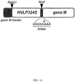
- the HVLP324S V L phage display library was constructed in two steps by the method described by Hussack et al. (In press (c)). First, a library with a randomized CDR3 was constructed based on the HVLP324S V L scaffold ( Fig. 10 ). Then, the CDR3-randomized library was used as scaffold to construct the final library with all 3 CDRs randomized.
- Phage ssDNA containing uridine instead of thymidine was prepared as described (Hussack et al. In press (c)), except that the phage used for ssDNA preparation was fd-tetV L 24S.
- fd-tetV L 24S is fd-tetGIIID phage (Tanha et al., 2001) with the HVLP324S V L gene in its genome and fused with the phage p3 gene ( Fig. 10 ).
- Incorporation of uridine into phage ssDNA was confirmed by titering the phage against TG1 and CJ236 E .
- CCC-DNA Covalently closed circular DNA
- Twenty-three transformations (11 ⁇ L CCC-DNA plus 350 ⁇ L of electrocompetent E . coli TG1) were carried out as described.
- transformation materials were titrated and grown in 1 L of 2xYT/tetracycline (12.5 ⁇ g/mL) overnight at 37°C and 180 rpm. In the morning, the cells were harvested and frozen library stocks were made as described. The cell density of the frozen library stocks were estimated by A 600 nm measurements.
- the amplified library phage was purified from the supernatant, titred, (See Hussack et al (c)) and used as the starting material to construct the final library (see Section (ii)). From the titration experiments on the transformation materials (see above) the library size (number of transformants) was determined to be 2.3 x 10 8 .
- dU-ssDNA template was prepared starting with 4 x 10 12 cfu CDR3-randomized library phage from above (see Hussack et al (c)). 200 ⁇ g of ssDNA was obtained from 2 mL of phage (2 x 10 9 / ⁇ L). Incorporation of uridine into phage ssDNA was confirmed as above.
- the dU-ssDNA template was used in the second round of mutagenesis which used mutagenic oligonucleotides V L 24-CDR1 and V L 24S-CDR2. Annealing and CCC-DNA synthesis steps were essentially identical to those described above.
- a total of 300 ⁇ g CCC-DNA in 1 mL ddH 2 O was prepared. Seventy-seven transformations (13 ⁇ L CCC-DNA plus 200 ⁇ L of electrocompetent E . coli TG1) were carried out as described. Following incubation in SOC medium, transformation materials were titrated and grown in 3 L of 2xYT/tetracycline (5 ⁇ g/mL) overnight at 180 RPM and 37°C. Frozen library stocks were made and its cell density was estimated as described above.
- the library phage was purified from the supernatant in a final volume of 20 mL PBS, titred against TG1, and stored frozen at -80°C in 500 ⁇ L aliquots for future first round panning as the input phage.
- the library size was determined to be 1 x 10 8 transformants. 78 V L clones from the library (non-amplified library) titer plates were subjected to sequencing as described in Hussack et al (c).
- HVLP324S V L was used as the scaffold for construction of a disulfide-stabilized synthetic V L library.
- HVLP324S is the mutant version of the previously described HVLP324 but with an additional disulfide linkage between Cys48 and Cys64.
- the library size was modest with 1 x 10 8 transformants.
- a CDR3 length distribution analysis of the 76 library clones showed that the majority (71%) of the library clones had a CDR3 length of 9 amino acids. Clones with CDR3 lengths of 10 and 11 amino acids were at 18% and 11%, respectively.
- oligonucleotides used are as provided in Table 6, where IUPAC nomenclature is used to designate degenerate nucleotide residues at positions indication by N, K and R. Table 6. List of oligonucleotides used for molecular cloning.
- HVLP324S V L phage display library To validate HVLP324S V L phage display library, the library was subjected to selection for binders to two different test antigens, namely human lysozyme and human serum albumin (HSA). Positive candidate binders (phage-displayed V L s) identified by initial binding assays (phage ELISA) were subcloned in vectors for expression in E . coli. V L binders were expressed, purified and analyzed for affinity and stability.
- HSA human serum albumin
- the well was washed 3x with sterile PBS, blotted on a paper towel, and blocked with 150 ⁇ L of 2% (w/v) milk in sterile PBS (2% MPBS) at 37°C for 2 h.
- the blocking agent was removed from the well and 100 ⁇ L of library phage (amplified phage prepared in Example 8, Section (ii) in 1% MPBS (5 x 10 11 cfu; preincubated at 37°C for 1.5 h) were added to the well and incubated for 1.5 h at 37°C.
- phage were eluted by incubating the well with 100 ⁇ L of freshly prepared 124 mM triethylamine for 10 min at room temperature, pipetting up and down at 8 min.
- the eluted phage solution was neutralized in a 1.5 mL microtube by mixing with 50 ⁇ L of 1 M Tris-HCl, pH 7.5 and kept on ice until the infection phase. 2 x 5 mL of exponentially growing E .
- the infected cells were centrifuged at 4,000 rpm and 4°C for 20 min using Sorval Legend RT rotor and centrifuge (Thermo Fisher Scientific, Nepean, ON, Canada), the supernatant was removed, and the pellet was resuspended in 1 mL of 2xYT and equally spread onto 3 large Petri dishes with 2xYT agar/5 ⁇ g/mL tetracycline (2xYT/Tet). The Petri dishes were incubated at 37°C overnight. Next day, the amplified phage were recovered from the large Petri dishes.
- phage supernatant that was prefiltrated with sterile 0.22 m filter was added a 1/5 volume of 20% PEG-8000/2.5 M NaCl in 50 mL sterile Falcon tubes, and the mixture was incubated on ice for 1 h. The solution was centrifuged for 20 min at 4,000 rpm and 4°C using Sorval Legend RT rotor and centrifuge (Thermo Fisher Scientific). The phage pellet was redissolved in 3 mL of sterile PBS, transferred to a 15-mL sterile Falcon tube, mixed with 1/5 volume of 20% PEG-8000/2.5 M NaCl (0.6 mL), and incubated on ice for 20 min.
- the phage solution was centrifuged at 4,000 rpm and 4°C for 20 min using Sorval Legend RT rotor and centrifuge (Thermo Fisher Scientific), and the phage pellet was dissolved in 0.5 - 1 mL sterile PBS, dissolution volume depending on the size of the pellet.
- the phage solution was aliquoted in 0.1 mL volumes in a sterile 1.5-mL sterile microtube.
- the titer of the amplified phage was determined as described below.
- thermopanning was also performed where the amplified phage in sterile PBS from the first round was heated at 65°C for 2 h in a 1.5 mL sterile microtube, subsequently centrifuged, and the phage supernatant was added to the human lysozyme-coated well for binding (in the second round of panning). All the remaining steps were the same for both methods of panning. Parallel pannings were continued for two more rounds. Washes were 12x, 15x, and 15x for the second, third and fourth rounds of panning, respectively.
- Panning (conventional panning) against human serum albumin was performed as described for human lysozyme except that no thermopanning in parallel was performed.
- standard phage ELISA (Harrison et al., 1996; Tanha et al., 2001; Hussack et al., In press (c)) was performed on clones from titer plates (obtained from eluted phage) to identify phage displaying V L s positive for binding to human lysozyme or serum albumin.
- TG1 cells were plated on 2xYT/Tet agar Petri dishes and incubated at 37°C overnight. The number of colonies was used to determine the titer (cfu/mL) of the eluted phage. Colony-PCR on colonies was performed to verify the presence of inserts (positive hits gave a size of around 500 bp) and to provide templates for DNA sequencing using fdTGIII and -96GIII primers (Table 6). Sequences were analyzed and manipulated by DNASTAR Lasergene 8.
- phage dilutions were made at 10 -3 , 10 -6 , 10 -8 , and 10 -10 .
- 10 ⁇ L of each diluted phage was mixed with 100 ⁇ L of exponentially growing TG1 cells, and the mixtures were incubated at 37°C for 30 min for phage infection of cells to occur.
- the phage infected cells were spread on Petri dishes with 2xYT r/Tet agar media and left in clean bench for 5 min. Plates were incubated overnight at 37°C. In the morning, the colony titers on the plates were used to determine the titer of the amplified phage.
- the library was panned against human lysozyme and human serum albumin for four rounds.
- a conventional panning was performed in the first round followed by conventional panning and thermopanning in parallel from the 2 nd round on.
- the input phage was incubated at 65°C for 2 h, centrifuged to remove any possible aggregates, and the supernatant was added to the lysozyme-coated well for binding.
- the input (amplified) phage was directly added to the lysozyme-coated well without the heat treatment.
- thermopanning was applied to see if it preferentially enriches for stable (thermostable and/or non-aggregating) V L binders compared to the conventional panning which was expected to be indiscriminate with regards to stable and unstable V L binders.
- Table 7 shows the results of panning against human lysozyme with respect to the output phage titer (or eluted phage titer).
- the output phage titers are 3 - 4 orders of magnitude lower for the thermopanning approach compared to the conventional one. This may be due to lower input phage titers used and further loss of phage (possibly aggregating phage-displayed V L s) to aggregation during the heat treatment (data not shown).
- Table 7 Phage titers for pannings performed under room temperature (conventional panning) versus 65°C-2 h (thermopanning) selection conditions. Data are for panning against human lysozyme.
- cfu colony-forming units. Panning round Room temperature 65°C-2 h Input (cfu) output (cfu) Input (cfu) output (cfu) 1 5 x 10 11 7 x 10 4 N/A N/A 2 4 x 10 10 2 x 10 5 4 x 10 10 2 x 10 2 3 7 x 10 11 2 x 10 8 2 x 10 10 6 x 10 4 4 5 x 10 11 6 x 10 7 5 x 10 10 7 x 10 4
- Phage ELISA and sequence analyses from round 3 and 4 identified 9 anti-human lysozyme V L s with variable affinities to immobilized human lysozyme ( Fig 13 and Table 8). After 4 rounds of panning, of the total of 177 V L S (101 V L s and 76 V L s for rounds 3 and round 4, respectively) analyzed, 5 clones of the total of 9 identified in round 3 survived: HVLHLRN; HVLHLDS; HVLHLNE; HVLHLNN; HVLHLFL ( Fig. 12 ).
- HVLHLRN and HVLHLDS were represented at 54.2% and 25% for HVLHLRN and HVLHLDS, respectively, followed by 8.3%, 8.3%, and 4.2% for HVLHLNE, HVLHLNN, and HVLHLFL, respectively, in the thermopanning approach (65°C, 2 h).
- HVLHLRN and HVLHLDS were not represented, and HVLHLNE, HVLHLNN, and HVLHLFL were occurred at relative frequencies of 28.6%, 42.9%, and 28.6%, respectively ( Fig. 12 ).
- HVLHLRN and HVLHLDS were also the predominant clones in round 3 of thermopanning and marginally present in the round 3 of conventional panning.
- HVLHLYS, HVLHLAQ, HVLHLQI,and HVLHLEM were notselected by either the conventional panning or thermopanning in round 4 panning.
- HVLHLRN (SEQ ID NO: 101) 0.05 74 Monomer 65°C - 2 h HVLHLDS (SEQ ID NO: 100) 6.1 68.3 Monomer 65°C - 2 h HVLHLEM (SEQ ID NO: 104) >73 72.2 Monomer RT HVLHLAQ (SEQ ID NO: 103) 0.2 66.5 Agg. (7%) RT HVLHLQI (SEQ ID NO: 105) 2.6 62 Agg. (17%) RT HVLHLNE (SEQ ID NO: 102) 0.06 62 Agg. (13%) RT/65°C - 2 h HVLHLYS (SEQ ID NO: 106) 6.4 66.8 Agg.
- RT HVLHLNN (SEQ ID NO: 99) 0.2 62.6 Agg. (12%) RT/65°C - 2 h HVLHLFL (SEQ ID NO: 108) ND ND ND RT/65°C - 2 h HVLHSA1 (SEQ ID NO: 107) 0.8 71 Monomer RT ND, not determined; RT, room temperature; Agg., aggregate. %'s are % aggregates.
- V L s were cloned, expressed, and purified by standard techniques as described above for other antibody domains.
- the V L s were subsequently analyzed by size exclusion chromatography for aggregation status, by CD for thermostability, and by surface plasmon resonance for their binding affinity and specificity.
- V L s and HVLHSA1 V L were cloned and expressed in E. coli and purified by standard techniques as described for other domains. Since V L s had His6 tags at their C-termini, they were purified by immobilized-metal affinity chromatography.
- Figure 16A shows the IMAC chromatogram profiles of the purified V L s that shows the A 280 range of 400 mAU (HVLHLEM) to 2400 mAU (HVLHLRN) for the highest elution peaks.
- the SDS-PAGE profiles of the V L s shows the V L s are highly pure ( Fig. 16B ).
- the expression yield of HVLHSA1 was also high as judged from its IMAC chromatogram profile (data not shown). Thus the V L s can be expressed in high yields in E . coli with high purity.
- V L s were analyzed for their aggregation behaviour by SuperdexTM 75 size exclusion chromatography. % aggregate (or %monomer) for each V L was also determined as described (Arbabi-Ghahroudi et al. 2010). As shown in Figure 14A , 3 anti-human lysozyme V L s (HVLHLRN, HVLHLEM, and HVLHLDS) were purely monomeric (non-aggregating), while the rest of the V L binders were aggregating with % aggregates ranging from 7% to 17% (Table 8). HVLHSA1 V L was also analyzed by size exclusion chromatography as described for other domains.
- thermostability of the V L binders melting temperatures ( T m s) of the V L s were determined by CD as described previously for other domains.
- Figure 14B shows the melting profiles of the anti-human lysozyme V L s which are in the range of 62°C to 74°C.
- the calculated T m s are recorded in Table 8.
- the T m of anti-albumin HVLHSA1 was also determined and shown to be high: 71°C ( Fig. 14B ; Table 8).
- T m s are significantly higher than V L s without the non-canonical disulfide linkage, demonstrating that the V L (or V H ) libraries with the non-canonical Cys48/Cys64 (or Cys49/Cys69) disulfide linkage feature yields more stable binders.
- HVLHLRN and HVLHLDS which are non-aggregating (Table 8) and have the highest T m s were also the clones which were enriched very significantly over aggregating, less thermostable ones by thermopanning and not by conventional panning.
- thermopanning is more efficient in enriching them.
- HVLHLEM which was both non-aggregating and thermostable and yet was not selected in round 4 by thermopanning could be due to its very low affinity during the binding stage of the panning [ Fig. 12 ; Table 8].
- V L /V H libraries with the non-canonical disulfide linkage feature yield more thermostable V L /V H binders than libraries without this feature, they also yield more non-aggregating V L /V H binders. As therapeutics non-aggregating and thermostable V L /V H binders are more efficacious.
- V L binders 298 resonance units (RUs), 312 RUs, 295 RUs, 339 RUs, 315 RUs, 412 RUs, 370 RUs, 345 RUs, and 349 RUs of each V L binders (HVLHLAQ, HVLHLNE, HVLHLEM, HVLHLYG, HVLHLRN, HVLHLNN, HVLHLQI, HVLHLYS, and HVLHLDS, respectively) was immobilized on a research grade CM5 sensor chip. Immobilizations were carried out at concentrations of 10 ⁇ g/mL in 10 mM acetate at pH 4.5 using the amine coupling kit supplied by the manufacturer.
- Human lysozyme was passed through a Superdex 75 column (GE Healthcare) in HBS-EP buffer (10 mM HEPES, pH 7.4, 150 mM NaCl, 3 mM EDTA and 0.005% P20 surfactant) with flow rate of 0.5 mL/min to eliminate any possible aggregates prior to Biacore analysis.
- HBS-EP buffer 10 mM HEPES, pH 7.4, 150 mM NaCl, 3 mM EDTA and 0.005% P20 surfactant
- analyses were carried out at 25°C in 10 mM HEPES, pH 7.4 containing 150 mM NaCl, 3 mM EDTA and 0.005% surfactant P20.
- the flow rates used were 20 ⁇ L/min.
- For regeneration the surfaces were washed for 15 min with the running buffer. Data were analyzed with BIAevaluation 4.1 software.
- Binding profiles and calculated K D s are shown in Figure 15 and Table 8. From the SPR profiles ( Figure 15 ), it can be seen that the anti-human lysozyme V L s indeed bind to their target antigen, lysozyme, confirming the binding results obtained by phage ELISA.
- the K D of the V L against human serum albumin was determined by surface plasmon resonance as described for other domains above. Briefly, the binding of the V L to immobilized human serum albumin was determined by SPR using the BIACORE 3000 (GE Healthcare). Approximately 1,600 RUs of human serum albumin, 1,900 RUs of bovine serum albumin and 2,200 RUs of ovalbumin were immobilized on a research grade CM5 sensor chip. An ethanolamine blocked surface was prepared, as a reference, in exactly the same manner as the protein surfaces. Immobilizations were carried out at concentrations of 20 ⁇ g/mL in 10 mM acetate buffer pH 4.5 using the amine coupling kit supplied by the manufacturer.
- the V L was passed through a Superdex 75 column (GE Healthcare) to eliminate any possible aggregates prior to Biacore analysis.
- analyses were carried out at 25°C in 10 mM HEPES, pH 7.4 containing 150 mM NaCl, 3 mM EDTA and 0.005% surfactant P20.
- the flow rates used were 40 ⁇ L/min and sample volumes were 60 ⁇ L.
- Sample volumes were 60 ⁇ L.
- Figure 17 clearly shows that the V L binds to its target antigen, human serum albumin.
- the calculated K D was determined to be 0.8 ⁇ M (Table 8). The binding was specific as the V L did not bind to ovalbumin or to bovine serum albumin.
- V H and V L domains with non-canonical disulfide bonds are feasible as scaffolds for constructing V H /V L synthetic libraries that are sources of antigen binders with high affinity, stability (non-aggregation and thermostability), and expression yields.
- V H phage display library with randomized CDRs built on a V H scaffold stabilized by extra disulfide linkage at positions 49 and 69.
- a V H phage display library HVHP430LGH3 was used as a template to introduce additional disulfide linkage by replacing two selected amino acid residues (49Ser and 69Ile) with cysteines using the method described above for the V L library and previously (Sidhu et al., 2000; Hussack et al (c)).
- the HVHP430LGH3phage display library is a phagemid vector-based library with the CDR-randomized human V H genes cloned into the phagemid pMED-1 and fused with protein 3 gene. ("Non-aggregating human V H domains" PCT publication WO2009/079093 ).
- dU-ssDNA uridine-containing single strand phage DNA
- E . coli CJ236 infected with phage (4 x 10 12 cfu (colony-forming unit)) from HVHP430LGH3 library.
- the HVHP430LGH3C2 phage display library was amplified in 300 mL of 2xYT, the library cells were resuspended in 15 mL of 10% glycerol, and subsequently kept as 1 mL aliquots at -80°C.
- the diversity of the library is high (5.3 x 10 9 transformants) with the additional stabilizing 49Ser to 49Cys and 69Ile to 69Cys mutations in all its V H members.
- the library should yield binders with high stability to target antigens, due to the presence of Cys49/Cys69 disulfide linkage in its VH members, using the standard selection and panning techniques known in the art.
Landscapes
- Chemical & Material Sciences (AREA)
- Health & Medical Sciences (AREA)
- Organic Chemistry (AREA)
- Life Sciences & Earth Sciences (AREA)
- Medicinal Chemistry (AREA)
- General Health & Medical Sciences (AREA)
- Genetics & Genomics (AREA)
- Molecular Biology (AREA)
- Immunology (AREA)
- Biochemistry (AREA)
- Biophysics (AREA)
- Proteomics, Peptides & Aminoacids (AREA)
- Engineering & Computer Science (AREA)
- Bioinformatics & Cheminformatics (AREA)
- Public Health (AREA)
- Veterinary Medicine (AREA)
- Animal Behavior & Ethology (AREA)
- Pharmacology & Pharmacy (AREA)
- Chemical Kinetics & Catalysis (AREA)
- General Chemical & Material Sciences (AREA)
- Virology (AREA)
- Nuclear Medicine, Radiotherapy & Molecular Imaging (AREA)
- Biomedical Technology (AREA)
- Microbiology (AREA)
- Biotechnology (AREA)
- General Engineering & Computer Science (AREA)
- Wood Science & Technology (AREA)
- Zoology (AREA)
- Plant Pathology (AREA)
- Physics & Mathematics (AREA)
- Mycology (AREA)
- Epidemiology (AREA)
- Peptides Or Proteins (AREA)
- Micro-Organisms Or Cultivation Processes Thereof (AREA)
- Medicines Containing Antibodies Or Antigens For Use As Internal Diagnostic Agents (AREA)
- Breeding Of Plants And Reproduction By Means Of Culturing (AREA)
Applications Claiming Priority (3)
| Application Number | Priority Date | Filing Date | Title |
|---|---|---|---|
| US201161437174P | 2011-01-28 | 2011-01-28 | |
| PCT/CA2012/000126 WO2012100343A1 (en) | 2011-01-28 | 2012-01-27 | Engineering of immunoglobulin domains |
| EP12738867.6A EP2668206B1 (de) | 2011-01-28 | 2012-01-27 | Manipulation von immunoglobulindomänen |
Related Parent Applications (1)
| Application Number | Title | Priority Date | Filing Date |
|---|---|---|---|
| EP12738867.6A Division EP2668206B1 (de) | 2011-01-28 | 2012-01-27 | Manipulation von immunoglobulindomänen |
Publications (2)
| Publication Number | Publication Date |
|---|---|
| EP3587447A2 true EP3587447A2 (de) | 2020-01-01 |
| EP3587447A3 EP3587447A3 (de) | 2020-05-13 |
Family
ID=46580154
Family Applications (2)
| Application Number | Title | Priority Date | Filing Date |
|---|---|---|---|
| EP19186022.0A Pending EP3587447A3 (de) | 2011-01-28 | 2012-01-27 | Manipulation von immunoglobulindomänen |
| EP12738867.6A Active EP2668206B1 (de) | 2011-01-28 | 2012-01-27 | Manipulation von immunoglobulindomänen |
Family Applications After (1)
| Application Number | Title | Priority Date | Filing Date |
|---|---|---|---|
| EP12738867.6A Active EP2668206B1 (de) | 2011-01-28 | 2012-01-27 | Manipulation von immunoglobulindomänen |
Country Status (15)
| Country | Link |
|---|---|
| US (2) | US9371371B2 (de) |
| EP (2) | EP3587447A3 (de) |
| JP (2) | JP6093715B2 (de) |
| KR (1) | KR20140092237A (de) |
| CN (2) | CN107513104A (de) |
| AU (1) | AU2012211011A1 (de) |
| BR (1) | BR112013019150A2 (de) |
| CA (1) | CA2825552A1 (de) |
| EA (1) | EA201391086A1 (de) |
| ES (1) | ES2746559T3 (de) |
| IL (1) | IL227656A0 (de) |
| MX (1) | MX2013008736A (de) |
| RU (1) | RU2013139260A (de) |
| SG (1) | SG194472A1 (de) |
| WO (1) | WO2012100343A1 (de) |
Families Citing this family (23)
| Publication number | Priority date | Publication date | Assignee | Title |
|---|---|---|---|---|
| WO2006099747A1 (en) * | 2005-03-25 | 2006-09-28 | National Research Council Of Canada | Method for isolation of soluble polypeptides |
| BR112013019150A2 (pt) | 2011-01-28 | 2019-09-24 | Nat Res Council Canada | engenharia do âmbito da imunoglobulina |
| WO2014030780A1 (en) * | 2012-08-22 | 2014-02-27 | Mogam Biotechnology Research Institute | Screening and engineering method of super-stable immunoglobulin variable domains and their uses |
| US20140161796A1 (en) * | 2012-09-13 | 2014-06-12 | Andreas Loew | Single chain proteins with c-terminal modifications |
| CN104870472A (zh) * | 2012-10-24 | 2015-08-26 | 加拿大国家研究委员会 | 抗空肠弯曲杆菌抗体及其用途 |
| AR095863A1 (es) * | 2013-04-16 | 2015-11-18 | Genentech Inc | Variantes de pertuzumab, su evaluación, método de tratamiento, método de preparación y artículo de fabricación |
| JP7001474B2 (ja) * | 2015-01-21 | 2022-01-19 | インヒブルクス,インコーポレイティド | 非免疫原性単一ドメイン抗体 |
| AU2016210551B2 (en) | 2015-01-23 | 2019-03-07 | Helix Biopharma Corp. | Antibody-urease conjugates for therapeutic purposes |
| JP2018511303A (ja) * | 2015-02-04 | 2018-04-26 | アブセレックス テクノロジーズ インコーポレイテッド | 抗サルモネラ抗体及びその使用 |
| JP7059494B2 (ja) * | 2016-07-06 | 2022-04-26 | ナショナル リサーチ カウンシル オブ カナダ | 血液脳関門を通過するヒト化抗体及びその使用 |
| CA3030163A1 (en) | 2016-07-11 | 2018-01-18 | Board Of Regents, The University Of Texas System | Recombinant polypeptides comprising selenocysteine and method for producing the same |
| EP3506945A4 (de) * | 2016-08-30 | 2020-07-15 | Board of Regents, The University of Texas System | Herstellung von seleno-biologika in genomisch umcodierten organismen |
| JP7117088B2 (ja) * | 2017-08-04 | 2022-08-12 | シスメックス株式会社 | 抗体及びその製造方法、並びに抗体の熱安定性を向上させる方法 |
| CN111278461A (zh) * | 2017-08-16 | 2020-06-12 | 百时美施贵宝公司 | 可前药化抗体、其前药以及使用和制备方法 |
| CN111655730A (zh) | 2018-01-31 | 2020-09-11 | 豪夫迈·罗氏有限公司 | 包含与lag3结合的抗原结合位点的双特异性抗体 |
| EP3746470A1 (de) * | 2018-01-31 | 2020-12-09 | F. Hoffmann-La Roche AG | Stabilisierte immunoglobulindomänen |
| CN108191973B (zh) * | 2018-03-06 | 2021-04-27 | 中国科学院武汉病毒研究所 | 一种靶向埃博拉病毒囊膜蛋白的高亲和力单域抗体及其制备方法与应用 |
| WO2020041360A1 (en) | 2018-08-21 | 2020-02-27 | Quidel Corporation | Dbpa antibodies and uses thereof |
| AU2020408213A1 (en) | 2019-12-19 | 2022-06-23 | Ortho-Clinical Diagnostics, Inc. | Monoclonal antibody fusions |
| JP7481856B2 (ja) | 2020-02-25 | 2024-05-13 | シスメックス株式会社 | 改変抗体及びその製造方法、並びに抗体の熱安定性を向上させる方法 |
| US20250136694A1 (en) | 2021-08-18 | 2025-05-01 | Harbour Biomed (Shanghai) Co., Ltd | Anti-b7-h4 antibody, and preparation method therefor and use thereof |
| EP4612172A1 (de) * | 2022-10-31 | 2025-09-10 | National Research Council of Canada | Verbesserte manipulierte immunglobuline und bindungsfragmente |
| JP2024158328A (ja) | 2023-04-27 | 2024-11-08 | シスメックス株式会社 | 抗EGFR抗体の生産方法、FcγRIIIaに対する親和性向上方法及び抗EGFR抗体 |
Citations (5)
| Publication number | Priority date | Publication date | Assignee | Title |
|---|---|---|---|---|
| WO1995004069A1 (en) | 1993-07-30 | 1995-02-09 | Affymax Technologies N.V. | Biotinylation of proteins |
| WO2003046560A2 (en) | 2001-11-30 | 2003-06-05 | National Research Council Of Canada | Self-assembly molecules |
| WO2004076670A1 (en) | 2003-02-28 | 2004-09-10 | Roche Diagnostics Gmbh | Enhanced expression of fusion polypeptides with a biotinylation tag |
| WO2006099747A1 (en) | 2005-03-25 | 2006-09-28 | National Research Council Of Canada | Method for isolation of soluble polypeptides |
| WO2009079093A1 (en) | 2007-12-14 | 2009-06-25 | Lsi Corporation | Systems and methods for fly-height control using servo data |
Family Cites Families (8)
| Publication number | Priority date | Publication date | Assignee | Title |
|---|---|---|---|---|
| JP5092160B2 (ja) * | 2007-03-08 | 2012-12-05 | 独立行政法人産業技術総合研究所 | ラクダ科動物のvhhドメインの安定化 |
| CA2691434C (en) * | 2007-06-21 | 2020-07-21 | Macrogenics, Inc. | Covalent diabodies and uses thereof |
| KR20100089816A (ko) * | 2007-08-21 | 2010-08-12 | 모르포시스 아게 | 이황화 결합을 형성하기 위한 개선된 방법 |
| US20110052565A1 (en) | 2007-12-21 | 2011-03-03 | National Research Council Of Canada | Non-aggregating human vh domains |
| JP5646457B2 (ja) * | 2008-04-29 | 2014-12-24 | アッヴィ・インコーポレイテッド | 二重可変ドメイン免疫グロブリン及びその使用 |
| WO2010035012A1 (en) * | 2008-09-26 | 2010-04-01 | Ucb Pharma S.A. | Biological products |
| CN101830978A (zh) * | 2009-03-09 | 2010-09-15 | 中国科学院微生物研究所 | 一种人免疫球蛋白样受体突变体及其应用 |
| BR112013019150A2 (pt) | 2011-01-28 | 2019-09-24 | Nat Res Council Canada | engenharia do âmbito da imunoglobulina |
-
2012
- 2012-01-27 BR BR112013019150A patent/BR112013019150A2/pt not_active IP Right Cessation
- 2012-01-27 ES ES12738867T patent/ES2746559T3/es active Active
- 2012-01-27 RU RU2013139260/10A patent/RU2013139260A/ru not_active Application Discontinuation
- 2012-01-27 KR KR1020137022298A patent/KR20140092237A/ko not_active Withdrawn
- 2012-01-27 EP EP19186022.0A patent/EP3587447A3/de active Pending
- 2012-01-27 JP JP2013550712A patent/JP6093715B2/ja active Active
- 2012-01-27 EA EA201391086A patent/EA201391086A1/ru unknown
- 2012-01-27 MX MX2013008736A patent/MX2013008736A/es not_active Application Discontinuation
- 2012-01-27 CN CN201611027628.6A patent/CN107513104A/zh active Pending
- 2012-01-27 WO PCT/CA2012/000126 patent/WO2012100343A1/en not_active Ceased
- 2012-01-27 CA CA2825552A patent/CA2825552A1/en active Pending
- 2012-01-27 AU AU2012211011A patent/AU2012211011A1/en not_active Abandoned
- 2012-01-27 SG SG2013076229A patent/SG194472A1/en unknown
- 2012-01-27 CN CN201280016587.4A patent/CN103619875B/zh active Active
- 2012-01-27 US US13/981,967 patent/US9371371B2/en active Active
- 2012-01-27 EP EP12738867.6A patent/EP2668206B1/de active Active
-
2013
- 2013-07-25 IL IL227656A patent/IL227656A0/en unknown
-
2016
- 2016-06-10 US US15/178,758 patent/US10633430B2/en active Active
-
2017
- 2017-02-13 JP JP2017024256A patent/JP2017079797A/ja not_active Withdrawn
Patent Citations (5)
| Publication number | Priority date | Publication date | Assignee | Title |
|---|---|---|---|---|
| WO1995004069A1 (en) | 1993-07-30 | 1995-02-09 | Affymax Technologies N.V. | Biotinylation of proteins |
| WO2003046560A2 (en) | 2001-11-30 | 2003-06-05 | National Research Council Of Canada | Self-assembly molecules |
| WO2004076670A1 (en) | 2003-02-28 | 2004-09-10 | Roche Diagnostics Gmbh | Enhanced expression of fusion polypeptides with a biotinylation tag |
| WO2006099747A1 (en) | 2005-03-25 | 2006-09-28 | National Research Council Of Canada | Method for isolation of soluble polypeptides |
| WO2009079093A1 (en) | 2007-12-14 | 2009-06-25 | Lsi Corporation | Systems and methods for fly-height control using servo data |
Non-Patent Citations (57)
| Title |
|---|
| "Sequences of Proteins of Immunological Interest", 1991, US DEPARTMENT OF HEALTH AND HUMAN SERVICES, US PUBLIC HEALTH SERVICE |
| AMZEL, L. M.POLJAK, R. J.: "Three-dimensional structure of immunoglobulins", ANNU. REV. BIOCHEM., vol. 48, 1979, pages 961 - 997 |
| ARBABI-GHAHROUDI, M.MACKENZIE, R.TANHA, J.: "Selection of non-aggregating VH binders from synthetic VH phage-display libraries", METHODS MOL. BIOL., vol. 525, 2009, pages 187 - 216, XP009171418, DOI: doi:10.1007/978-1-59745-554-1_10 |
| ARBABI-GHAHROUDI, M.MACKENZIE, R.TANHA, J.: "Site-directed mutagenesis for improving biophysical properties of VH domains", METHODS MOL. BIOL., vol. 634, 2010, pages 309 - 330, XP009183447 |
| ARBABI-GHAHROUDI, M.TO, R.GAUDETTE, N.HIRAMA, T.DING, W.MACKENZIE, R.TANHA, J.: "Aggregation-resistant VHs selected by in vitro evolution tend to have disulfide-bonded loops and acidic isoelectric points", PROTEIN ENG. DES. SET., vol. 22, 2009, pages 59 - 66, XP008138408, DOI: doi:10.1093/PROTEIN/GZN071 |
| BELL, A.WANG, Z. J.ARBABI-GHAHROUDI, M.CHANG, T. A.DUROCHER, Y.TROJAHN, U.BAARDSNES, J.JARAMILLO, M. L.LI, S.BARAL, T. N.: "Differential tumor-targeting abilities of three single-domain antibody formats", CANCER LETT., vol. 289, 2010, pages 81 - 90, XP026897079, DOI: doi:10.1016/j.canlet.2009.08.003 |
| BETZ, S. F.: "Disulfide bonds and the stability of globular proteins", PROTEIN SCI., vol. 2, 1993, pages 1551 - 1558, XP009016509 |
| BLOOM, J. D.LABTHAVIKUL, S. T.OTEY, C. R.ARNOLD, F. H.: "Protein stability promotes evolvability", PROC. NATL. ACAD. SCI. USA., vol. 103, 2006, pages 5869 - 5874 |
| CHOTHIA, C.LESK, A. M.: "Canonical structures for the hypervariable regions of immunoglobulins", J. MOL. BIOL., vol. 196, 1987, pages 901 - 917, XP024010426, DOI: doi:10.1016/0022-2836(87)90412-8 |
| DAVIES, J.RIECHMANN, L.: "Affinity improvement of single antibody VH domains: residues in all three hypervariable regions affect antigen binding", IMMUNOTECHNOLOGY, vol. 2, 1996, pages 169 - 179, XP004070292, DOI: doi:10.1016/S1380-2933(96)00045-0 |
| DAVIES, J.RIECHMANN, L.: "Antibody VH domains as small recognition units", BIOTECHNOLOGY N. Y., vol. 13, 1995, pages 475 - 479, XP002011336, DOI: doi:10.1038/nbt0595-475 |
| DAVIES, J.RIECHMANN, L.: "Camelising' human antibody fragments: NMR studies on VH domains", FEBS LETT., vol. 339, 1994, pages 285 - 290, XP025614903, DOI: doi:10.1016/0014-5793(94)80432-X |
| DAVIES, J.RIECHMANN, L.: "Single antibody domains as small recognition units: design and in vitro antigen selection of camelized, human VH domains with improved protein stability", PROTEIN ENG., vol. 9, 1996, pages 531 - 537 |
| DE KRUIF, J.LOGTENBERG, T.: "Leucine zipper dimerized bivalent and bispecific scFv antibodies from a semi-synthetic antibody phage display library", J. BIOL. CHEM., vol. 271, 1996, pages 7630 - 7634, XP002337214, DOI: doi:10.1074/jbc.271.13.7630 |
| EISENBERG, D.SCHWARZ, E.KOMAROMY M.WALL R.: "Analysis of membrane and surface protein sequences with the hydrophobic moment plot", J. MOL. BIOL., vol. 179, 1984, pages 125 - 142, XP024018280, DOI: doi:10.1016/0022-2836(84)90309-7 |
| GREENFIELD, N. J.: "Analysis of the kinetics of folding of proteins and peptides using circular dichroism", NAT. PROTOC., vol. 1, 2006, pages 2891 - 2899 |
| GREENFIELD, N. J.: "Using circular dichroism spectra to estimate protein secondary structure", NAT. PROTOC., vol. 1, 2006, pages 2876 - 2890, XP055220113, DOI: doi:10.1038/nprot.2006.202 |
| HARRISON, J. L.WILLIAMS, S. C.WINTER, G.NISSIM, A.: "Screening of phage antibody libraries", METHODS ENZYMOL., vol. 267, 1996, pages 83 - 109, XP009083801, DOI: doi:10.1016/S0076-6879(96)67007-4 |
| HO, S. N.HUNT, H. D.HORTON, R. M.PULLEN, J. K.PEASE, L. R.: "Site-directed mutagenesis by overlap extension using the polymerase chain reaction", GENE, vol. 77, 1989, pages 51 - 59, XP023544945, DOI: doi:10.1016/0378-1119(89)90358-2 |
| HOLLIGER, P.HUDSON, P. J.: "Engineered antibody fragments and the rise of single domains", NAT. BIOTECHNOL., vol. 23, 2005, pages 1126 - 1136, XP008076746, DOI: doi:10.1038/nbt1142 |
| HOLT, L. J.HERRING, C.JESPERS, L. S.WOOLVEN, B. P.TOMLINSON, I. M.: "Domain antibodies: proteins for therapy", TRENDS BIOTECHNOL., vol. 21, 2003, pages 484 - 490, XP004467495, DOI: doi:10.1016/j.tibtech.2003.08.007 |
| HOOGENBOOM, H. R.: "Selecting and screening recombinant antibody libraries", NAT. BIOTECHNOL., vol. 23, 2005, pages 1105 - 1116, XP002348401, DOI: doi:10.1038/nbt1126 |
| HORWICH, A.: "Protein aggregation in disease: a role for folding intermediates forming specific multimeric interactions", J. CLIN. INVEST., vol. 110, 2002, pages 1221 - 1232 |
| HURLE, M. R.HELMS, L. R.LI, L.CHAN, W.WETZEL, R.: "A role of destabilizing amino acid replacements in light-chain amyloidosis", PROC. NATL. ACAD. SCI. USA., vol. 91, 1994, pages 5446 - 5450, XP002159931, DOI: doi:10.1073/pnas.91.12.5446 |
| HUSSACK, G.ARBABI-GHAHROUDI, M.MACKENZIE, C. R.TANHA, J.: "Isolation and Characterization of Clostridium difficile toxin-specific single-domain antibodies", METHODS MOL. BIOL. |
| HUSSACK, G.HIRAMA, T.DING, W.MACKENZIE, R.TANHA, J.: "Engineered single-domain antibodies with high protease resistance and thermal stability", PLOS ONE, vol. 6, 2011, pages e28218 |
| HUSSACK, G.KEKLIKIAN, A.ALSUGHAYYIR, J.HANIFI-MOGHADAM, P.ARBABI-GHAHROUDI, M.FAASSEN, H.HOU, S. T.SAD, S.MACKENZIE, R.TANHA, J.: "A VL single-domain antibody library yields a high-propensity of non-aggregating binders", PROTEIN ENG. DES. SET. |
| HUSSACK, G.MACKENZIE, C. R.TANHA, J.: "Functional characterization of single-domain antibodies with an engineered disulfide bond", METHODS MOL. BIOL. |
| IQBAL, U.TROJAHN, U.ALBAGHDADI, H.ZHANG, J.O'CONNOR-MCCOURT, M.STANIMIROVIC, D.TOMANEK, B.SUTHERLAND, G.ABULROB, A.: "Kinetic analysis of novel mono- and multivalent VHH-fragments and their application for molecular imaging of brain tumours", BR. J. PHARMACOL., vol. 160, 2010, pages 1016 - 1028, XP055081280, DOI: doi:10.1111/j.1476-5381.2010.00742.x |
| JESPERS, L.SCHON, O.FAMM, K.WINTER, G.: "Aggregation-resistant domain antibodies selected on phage by heat denaturation", NAT. BIOTECHNOL., vol. 22, 2004, pages 1161 - 1165, XP008154827, DOI: doi:10.1038/nbt1000 |
| KAZLAUSKAS, R. J.BORNSCHEUER, U. T.: "Finding better protein engineering strategies", NAT. CHEM. BIOL., vol. 5, 2009, pages 526 - 529 |
| KIM, D. Y.DING, W.HIRAMA, T.RYAN, R.MACKENZIE, R.TANHA, J.: "Disulfide linkage engineering for improving biophysical properties of VH domains", J. MOL. BIOL. SUBMITTED |
| KIM, D. Y.DING, W.TANHA, J.: "Solubility and stability engineering of human V domain", METHODS MOL. BIOL. |
| KIM, D. YTANHA, J.: "Sodium dodecyl sulfate-polyacrylamide get electrophoresis for screening nonaggregating human antibody heavy chain variable domains", ANAL. BIOCHEM., vol. 403, 2010, pages 117 - 119 |
| KUNKEL, T. A.ROBERTS, J. DZAKOUR, R. A.: "Rapid and efficient site-specific mutagenesis without phenotypic selection", METHODS ENZYMOL., vol. 154, 1987, pages 367 - 382, XP001098930, DOI: doi:10.1016/0076-6879(87)54085-X |
| LEE, C. M.LORNO, N.SIERRO, F.CHRIST, D.: "Selection of human antibody fragments by phage display", NAT. PROTOC., vol. 2, 2007, pages 3001 - 3008, XP055247011, DOI: doi:10.1038/nprot.2007.448 |
| LITHWICK GMARGALIT H: "Hierarchy of sequence-dependent features associated with prokaryotic translation", GENOME RESEARCH, vol. 13, 2003, pages 2665 - 73 |
| MERRITT, E. A.HOL, W. G.: "AB5 toxins", CURR. OPIN. STRUCT. BIOL., vol. 5, 1995, pages 165 - 171, XP002267548, DOI: doi:10.1016/0959-440X(95)80071-9 |
| MITRAKI, A.KING, J.: "Amino acid substitutions influencing intracellular protein folding pathways", FEBS LETT., vol. 307, 1992, pages 20 - 25 |
| NIELSEN, U. B.ADAMS, G. P.WEINER, L. M.MARKS, J. D.: "Targeting of bivalent anti-ErbB2 diabody antibody fragments to tumor cells is independent of the intrinsic antibody affinity", CANCER RES., vol. 60, 2000, pages 6434 - 6440, XP003014072 |
| PORATH, J.FLODIN, P.: "Get filtration: a method for desalting and group separation", NATURE, vol. 183, 1959, pages 1657 - 1659, XP008072760, DOI: doi:10.1038/1831657a0 |
| RIDGWAY, J. B.PRESTA, L. G.CARTER, P.: "Knobs-into-holes' engineering of antibody CH3 domains for heavy chain heterodimerization", PROTEIN ENG., vol. 9, 1996, pages 617 - 621, XP002610995, DOI: doi:10.1093/protein/9.7.617 |
| RIECHMANN, L.DAVIES, J.: "Backbone assignment, secondary structure and protein A binding of an isolated, human antibody VH domain", J. BIOMOL. NMR., vol. 6, 1995, pages 141 - 152, XP001095927, DOI: doi:10.1007/BF00211778 |
| SIDHU, S. S.LOWMAN, H. B.CUNNINGHAM, B. C.WELLS, J. A.: "Phage display for selection of novel binding peptides", METHODS ENZYMOL., vol. 328, 2000, pages 333 - 63, XP000985556, DOI: doi:10.1016/S0076-6879(00)28406-1 |
| STAROVASNIK, M. A.O'CONNELL, M. P.FAIRBROTHER, W. J.KELLEY, R. F.: "Antibody variable region binding by Staphylococcal protein A: thermodynamic analysis and location of the Fv binding site on E-domain", PROTEIN SCI., vol. 8, 1999, pages 1423 - 1431, XP055123878, DOI: doi:10.1110/ps.8.7.1423 |
| TANHA, J.NGUYEN, T.D.NG,A.RYAN, S.NI, F.MACKENZIE, R.: "Improving solubility and refolding efficiency of human VHs by a novel mutational approach", PROTEIN ENG. DES. SET., vol. 19, 2006, pages 503 - 509, XP055286380, DOI: doi:10.1093/protein/gzl037 |
| TANHA, J.XU, P.CHEN, Z.NI, F.KAPLAN, H.NARANG, S. A.MACKENZIE, C. R.: "Optimal design features of camelized human single-domain antibody libraries", J. BIOL. CHEM., vol. 276, 2001, pages 24774 - 24780, XP002340991, DOI: doi:10.1074/jbc.M100770200 |
| TO, R.HIRAMA, T.ARBABI-GHAHROUDI, M.MACKENZIE, R.WANG, P.XU, P.NI, F.TANHA, J.: "Isolation of monomeric human VHs by a phage selection", J. BIOL. CHEM., vol. 280, 2005, pages 41395 - 41403, XP009100333, DOI: doi:10.1074/jbc.M509900200 |
| WARD, E. S.GUSSOW, D.GRIFFITHS, A. D.JONES, P. T.WINTER, G.: "Binding activities of a repertoire of single immunoglobulin variable domains secreted from Escherichia coli", NATURE, vol. 341, 1989, pages 544 - 546 |
| WETZEL, R.PERRY, L. J.BAASE, W. A.BECKTEL, W. J.: "Disulfide bonds and thermal stability in T4 lysozyme", PROC. NATL. ACAD. SCI. USA., vol. 85, 1988, pages 401 - 405 |
| WILLIAMS, A. F.BARCLAY, A. N.: "The immunoglobulin superfamily--domains for cell surface recognition", ANNU. REV. IMMUNOL., vol. 6, 1988, pages 381 - 405, XP055049274, DOI: doi:10.1146/annurev.iy.06.040188.002121 |
| WORN, A.PLUCKTHUN, A.: "Stability engineering of antibody single-chain Fv fragments", J. MOL. BIOL., vol. 305, 2001, pages 989 - 1010, XP004465987, DOI: doi:10.1006/jmbi.2000.4265 |
| WU, S. L.JIANG, H.LU, Q.DAI, S.HANCOCK, W. S.KARGER, B. L.: "Mass spectrometric determination of disulphide linkages in recombinant therapeutic proteins using online LC-MS with electron-transfer dissociation", ANAL. CHEM., vol. 81, 2009, pages 112 - 122 |
| YAU, K. Y. F.DUBUC, G.LI, S.HIRAMA, T.MACKENZIE, C. R.JERMUTUS, L.HALL, J. C.TANHA, J.: "Affinity maturation of a VHH by mutational hotspot randomization", IMMUNOL. METHODS, vol. 297, 2005, pages 213 - 224, XP027659127 |
| ZHANG, J.LI, Q.NGUYEN, T.-D.TREMBLAY, T.-L.STONE, E.TO, R.KELLY, J.MACKENZIE, C. R.: "A pentavalent single-domain antibody approach to tumor antigen discovery and the development of novel proteomics reagents", J. MOL. BIOL., vol. 341, 2004, pages 161 - 169, XP004679996, DOI: doi:10.1016/j.jmb.2004.05.069 |
| ZHANG, J.LIU, X.BELL, A.TO, R.BARAL, T. N.AZIZI, A.LI, J.CASS, B.DUROCHER, Y.: "Transient expression and purification of chimeric heavy chain antibodies", PROTEIN EXPR. PURIF., vol. 65, 2009, pages 77 - 82, XP025959408, DOI: doi:10.1016/j.pep.2008.10.011 |
| ZHANG, J.TANHA, J.HIRAMA, T.KHIEU, N. H.TO, R.TONG-SEVINC, H.STONE, E.BRISSON, J.-R.MACKENZIE, C. R.: "Pentamerization of single-domain antibodies from phage libraries: A novel strategy for the rapid generation of high-avidity antibody reagents", J. MOL. BIOL., vol. 335, 2004, pages 49 - 56, XP004476445, DOI: doi:10.1016/j.jmb.2003.09.034 |
Also Published As
| Publication number | Publication date |
|---|---|
| US10633430B2 (en) | 2020-04-28 |
| CN103619875A (zh) | 2014-03-05 |
| AU2012211011A1 (en) | 2013-08-15 |
| WO2012100343A1 (en) | 2012-08-02 |
| EA201391086A1 (ru) | 2014-01-30 |
| SG194472A1 (en) | 2013-12-30 |
| EP2668206A1 (de) | 2013-12-04 |
| JP2014506454A (ja) | 2014-03-17 |
| IL227656A0 (en) | 2013-09-30 |
| CN103619875B (zh) | 2017-03-08 |
| EP2668206B1 (de) | 2019-07-17 |
| RU2013139260A (ru) | 2015-03-10 |
| EP2668206A4 (de) | 2015-05-06 |
| JP2017079797A (ja) | 2017-05-18 |
| MX2013008736A (es) | 2013-11-22 |
| CA2825552A1 (en) | 2012-08-02 |
| ES2746559T3 (es) | 2020-03-06 |
| EP3587447A3 (de) | 2020-05-13 |
| US9371371B2 (en) | 2016-06-21 |
| KR20140092237A (ko) | 2014-07-23 |
| CN107513104A (zh) | 2017-12-26 |
| JP6093715B2 (ja) | 2017-03-08 |
| BR112013019150A2 (pt) | 2019-09-24 |
| US20170044238A1 (en) | 2017-02-16 |
| US20130303406A1 (en) | 2013-11-14 |
Similar Documents
| Publication | Publication Date | Title |
|---|---|---|
| EP2668206B1 (de) | Manipulation von immunoglobulindomänen | |
| CN108473987B (zh) | 具有改变的多样性支架结构域的结合成员 | |
| JP6628822B2 (ja) | 可溶性ポリペプチドの単離方法 | |
| JP4191258B2 (ja) | ヒト化抗体およびヒト化抗体の形成法 | |
| KR101732552B1 (ko) | 안정화된 면역글로불린가변도메인 선별방법 및 선별된 도메인의 응용 | |
| EP3288973B1 (de) | Verfahren für die massenhumanisierung von kaninchen-antikörpern | |
| JP6143921B2 (ja) | ヒトepo受容体に対する抗体 | |
| KR102637804B1 (ko) | 개과 항체 라이브러리 | |
| JP2015531597A5 (de) | ||
| Henry et al. | A disulfide-stabilized human VL single-domain antibody library is a source of soluble and highly thermostable binders | |
| US20240376639A1 (en) | Fast-track humanisation of specific binding molecules | |
| JP7853222B2 (ja) | 合成単一ドメインライブラリー | |
| CN119998326A (zh) | 具有新型二硫键的超稳定抗体片段 | |
| Weidenhaupt et al. | Functional mapping of conserved, surface‐exposed charges of antibody variable domains | |
| KR20220026869A (ko) | 신규 항체 라이브러리 제조방법 및 이로부터 제조된 라이브러리 | |
| EP4612172A1 (de) | Verbesserte manipulierte immunglobuline und bindungsfragmente | |
| JP2017018117A (ja) | 安定化された免疫グロブリン可変ドメイン選別方法及び選別されたドメインの応用 |
Legal Events
| Date | Code | Title | Description |
|---|---|---|---|
| PUAI | Public reference made under article 153(3) epc to a published international application that has entered the european phase |
Free format text: ORIGINAL CODE: 0009012 |
|
| STAA | Information on the status of an ep patent application or granted ep patent |
Free format text: STATUS: THE APPLICATION HAS BEEN PUBLISHED |
|
| AC | Divisional application: reference to earlier application |
Ref document number: 2668206 Country of ref document: EP Kind code of ref document: P |
|
| AK | Designated contracting states |
Kind code of ref document: A2 Designated state(s): AL AT BE BG CH CY CZ DE DK EE ES FI FR GB GR HR HU IE IS IT LI LT LU LV MC MK MT NL NO PL PT RO RS SE SI SK SM TR |
|
| PUAL | Search report despatched |
Free format text: ORIGINAL CODE: 0009013 |
|
| AK | Designated contracting states |
Kind code of ref document: A3 Designated state(s): AL AT BE BG CH CY CZ DE DK EE ES FI FR GB GR HR HU IE IS IT LI LT LU LV MC MK MT NL NO PL PT RO RS SE SI SK SM TR |
|
| RIC1 | Information provided on ipc code assigned before grant |
Ipc: C07K 16/00 20060101AFI20200409BHEP Ipc: C40B 40/10 20060101ALI20200409BHEP Ipc: A61P 43/00 20060101ALI20200409BHEP Ipc: A61P 35/00 20060101ALI20200409BHEP Ipc: C07K 16/18 20060101ALI20200409BHEP Ipc: C12N 15/13 20060101ALI20200409BHEP Ipc: A61P 37/00 20060101ALI20200409BHEP Ipc: C07K 16/40 20060101ALI20200409BHEP Ipc: A61K 39/395 20060101ALI20200409BHEP |
|
| STAA | Information on the status of an ep patent application or granted ep patent |
Free format text: STATUS: REQUEST FOR EXAMINATION WAS MADE |
|
| 17P | Request for examination filed |
Effective date: 20201110 |
|
| RBV | Designated contracting states (corrected) |
Designated state(s): AL AT BE BG CH CY CZ DE DK EE ES FI FR GB GR HR HU IE IS IT LI LT LU LV MC MK MT NL NO PL PT RO RS SE SI SK SM TR |
|
| P01 | Opt-out of the competence of the unified patent court (upc) registered |
Effective date: 20230519 |