EP3558314B1 - Rho kinase inhibitor ba-1049 (r) and active metabolites thereof - Google Patents
Rho kinase inhibitor ba-1049 (r) and active metabolites thereof Download PDFInfo
- Publication number
- EP3558314B1 EP3558314B1 EP17882341.5A EP17882341A EP3558314B1 EP 3558314 B1 EP3558314 B1 EP 3558314B1 EP 17882341 A EP17882341 A EP 17882341A EP 3558314 B1 EP3558314 B1 EP 3558314B1
- Authority
- EP
- European Patent Office
- Prior art keywords
- rock2
- hydroxy
- cells
- brain
- dose
- Prior art date
- Legal status (The legal status is an assumption and is not a legal conclusion. Google has not performed a legal analysis and makes no representation as to the accuracy of the status listed.)
- Active
Links
Images


Classifications
-
- A—HUMAN NECESSITIES
- A61—MEDICAL OR VETERINARY SCIENCE; HYGIENE
- A61K—PREPARATIONS FOR MEDICAL, DENTAL OR TOILETRY PURPOSES
- A61K31/00—Medicinal preparations containing organic active ingredients
- A61K31/33—Heterocyclic compounds
- A61K31/395—Heterocyclic compounds having nitrogen as a ring hetero atom, e.g. guanethidine or rifamycins
- A61K31/435—Heterocyclic compounds having nitrogen as a ring hetero atom, e.g. guanethidine or rifamycins having six-membered rings with one nitrogen as the only ring hetero atom
- A61K31/47—Quinolines; Isoquinolines
- A61K31/472—Non-condensed isoquinolines, e.g. papaverine
- A61K31/4725—Non-condensed isoquinolines, e.g. papaverine containing further heterocyclic rings
-
- A—HUMAN NECESSITIES
- A61—MEDICAL OR VETERINARY SCIENCE; HYGIENE
- A61K—PREPARATIONS FOR MEDICAL, DENTAL OR TOILETRY PURPOSES
- A61K31/00—Medicinal preparations containing organic active ingredients
- A61K31/33—Heterocyclic compounds
- A61K31/395—Heterocyclic compounds having nitrogen as a ring hetero atom, e.g. guanethidine or rifamycins
- A61K31/495—Heterocyclic compounds having nitrogen as a ring hetero atom, e.g. guanethidine or rifamycins having six-membered rings with two or more nitrogen atoms as the only ring heteroatoms, e.g. piperazine or tetrazines
- A61K31/505—Pyrimidines; Hydrogenated pyrimidines, e.g. trimethoprim
- A61K31/517—Pyrimidines; Hydrogenated pyrimidines, e.g. trimethoprim ortho- or peri-condensed with carbocyclic ring systems, e.g. quinazoline, perimidine
-
- A—HUMAN NECESSITIES
- A61—MEDICAL OR VETERINARY SCIENCE; HYGIENE
- A61K—PREPARATIONS FOR MEDICAL, DENTAL OR TOILETRY PURPOSES
- A61K31/00—Medicinal preparations containing organic active ingredients
- A61K31/33—Heterocyclic compounds
- A61K31/395—Heterocyclic compounds having nitrogen as a ring hetero atom, e.g. guanethidine or rifamycins
- A61K31/55—Heterocyclic compounds having nitrogen as a ring hetero atom, e.g. guanethidine or rifamycins having seven-membered rings, e.g. azelastine, pentylenetetrazole
- A61K31/551—Heterocyclic compounds having nitrogen as a ring hetero atom, e.g. guanethidine or rifamycins having seven-membered rings, e.g. azelastine, pentylenetetrazole having two nitrogen atoms, e.g. dilazep
-
- A—HUMAN NECESSITIES
- A61—MEDICAL OR VETERINARY SCIENCE; HYGIENE
- A61K—PREPARATIONS FOR MEDICAL, DENTAL OR TOILETRY PURPOSES
- A61K45/00—Medicinal preparations containing active ingredients not provided for in groups A61K31/00 - A61K41/00
- A61K45/06—Mixtures of active ingredients without chemical characterisation, e.g. antiphlogistics and cardiaca
-
- A—HUMAN NECESSITIES
- A61—MEDICAL OR VETERINARY SCIENCE; HYGIENE
- A61P—SPECIFIC THERAPEUTIC ACTIVITY OF CHEMICAL COMPOUNDS OR MEDICINAL PREPARATIONS
- A61P25/00—Drugs for disorders of the nervous system
-
- A—HUMAN NECESSITIES
- A61—MEDICAL OR VETERINARY SCIENCE; HYGIENE
- A61P—SPECIFIC THERAPEUTIC ACTIVITY OF CHEMICAL COMPOUNDS OR MEDICINAL PREPARATIONS
- A61P43/00—Drugs for specific purposes, not provided for in groups A61P1/00-A61P41/00
-
- A—HUMAN NECESSITIES
- A61—MEDICAL OR VETERINARY SCIENCE; HYGIENE
- A61P—SPECIFIC THERAPEUTIC ACTIVITY OF CHEMICAL COMPOUNDS OR MEDICINAL PREPARATIONS
- A61P9/00—Drugs for disorders of the cardiovascular system
- A61P9/08—Vasodilators for multiple indications
-
- A—HUMAN NECESSITIES
- A61—MEDICAL OR VETERINARY SCIENCE; HYGIENE
- A61P—SPECIFIC THERAPEUTIC ACTIVITY OF CHEMICAL COMPOUNDS OR MEDICINAL PREPARATIONS
- A61P9/00—Drugs for disorders of the cardiovascular system
- A61P9/10—Drugs for disorders of the cardiovascular system for treating ischaemic or atherosclerotic diseases, e.g. antianginal drugs, coronary vasodilators, drugs for myocardial infarction, retinopathy, cerebrovascula insufficiency, renal arteriosclerosis
-
- C—CHEMISTRY; METALLURGY
- C07—ORGANIC CHEMISTRY
- C07D—HETEROCYCLIC COMPOUNDS
- C07D401/00—Heterocyclic compounds containing two or more hetero rings, having nitrogen atoms as the only ring hetero atoms, at least one ring being a six-membered ring with only one nitrogen atom
- C07D401/02—Heterocyclic compounds containing two or more hetero rings, having nitrogen atoms as the only ring hetero atoms, at least one ring being a six-membered ring with only one nitrogen atom containing two hetero rings
- C07D401/12—Heterocyclic compounds containing two or more hetero rings, having nitrogen atoms as the only ring hetero atoms, at least one ring being a six-membered ring with only one nitrogen atom containing two hetero rings linked by a chain containing hetero atoms as chain links
Definitions
- Rho kinase is a serine/threonine kinase that plays a pivotal role in regulation of the cytoskeleton, motility, and junctional contacts in a variety of tissues.
- ROCK is activated when the small GTPase Rho is activated, and ROCK is downstream of Rho and plays a key role in phosphorylating other kinases in a complex intracellular signaling cascade.
- Rho kinase There are two isoforms of Rho kinase, ROCK1 and ROCK2, both of which are activated by Rho.
- ROCK1 has widespread tissue distribution (but less in brain and skeletal muscle).
- ROCK2 is expressed in the central nervous system ( U.S. Patent No.
- Rho and ROCK are abnormally activated in many types of neurotrauma and neurovascular diseases.
- Inhibitors of ROCK promote neurite outgrowth and axon regeneration after injury and inhibitors of ROCK are also effective in reducing ROCK activation in endothelial cells after stroke and in neurovascular diseases such as aneurysms or angiomas.
- Subarachnoid hemorrhage is a condition requiring emergent treatment intervention and frequently is associated with poor patient outcomes. Either open or endovascular surgical approaches are commonly applied to either stop or minimize bleeding in order to limit damage. An important feature in managing patients with this disorder is to control the vasospasm and changes in vascular tone that often occurs as a co-morbidity. Calcium channel blockers such as nimodipine have been used clinically, as has the ROCK inhibitor Fasudil ( Satoh et al.. (2014) Curr. Vasc. Pharmacol.: 12(5):758-765 ).
- Fasudil has been used to control vasospasm in this setting, its non-selective effects on both ROCK1 and ROCK2, and likely its off-target effects on other kinases, have made its use limited to only the first two weeks after hemorrhage.
- TBI traumatic brain injury
- numerous forces e.g . percussive or shear forces
- TBI percussive or shear forces
- primary injury not only to neurons and glia but also to the vasculature of the brain inducing hemorrhage.
- secondary injuries can also occur.
- Many of these injuries are the result of decreased blood perfusion in the area of the injury, as a consequence of vascular coagulation, and the onset of tissue edema due to increases in vascular permeability across the blood brain barrier ( Chodobski et al.. (2011) Transl. Stroke Res. 2(4):492-516 ).
- Rho kinase Inactivation of Rho kinase promotes functional repair after spinal cord injury ( Watzalawick et al. (2013). JAMA Neurol. 71:91-99 ).
- Cerebral cavernous malformations is another disorder that impacts the nervous system vasculature.
- CCMs are vascular malformations that develop essentially exclusively in the venous (low pressure) vascular bed within the nervous system.
- the dysfunction of any of the three proteins genetically linked to this disorder in the cells that form the blood vessel walls causes reduced adhesion between the cells and hyperactivation of ROCK. Ultimately this leads to an increased leakiness in these blood vessel walls, allowing blood cells and other plasma constituents to enter the brain in a non-regulated manner ( Clatterbuck et al.. (2001) J. Neurol. Neurosurg. Psychiat.71:188-192 ).
- the blood-brain barrier is typically a very strong and highly regulated structure and is formed between the cells of the small blood vessels (capillary vascular endothelial cells, pericytes) and other cells of the nervous system, including astrocytes.
- the function of the blood brain barrier is to highly regulate the entrance of blood-borne molecules into the brain, and the ability of cells present in the blood plasma to enter the brain ( Ballabh et al. (2004) Neurobiol. Dis. 16:1-13 ).
- the unregulated release of plasma proteins and other molecules into the brain tissue commonly leads to functional problems in the brain, and red blood cell accumulation can cause pathologic iron deposition.
- kinase inhibitors typically compete with ATP for binding to the ATP pocket of the kinase. Because the structure of ATP pockets is conserved, kinase inhibitors may have non-specific binding to multiple kinases, causing unwanted off-target kinase inhibition. Some kinase cause toxicity because of on-target effects, in which case risk -benefit analysis will drive drug development decisions. Off-target effects can cause toxicity including cardiotoxicity, and these can be detected by kinome screening. Inactivation of AMP-activated protein kinase (AMPK) contributes to cardiotoxicity because it is a regulator of cellular metabolism and its activation is needed when cardiomyocytes are energy stressed ( Chen et al. (2010) Progr. Cardiovasc. Dis. 53:114-120 .) A number of kinase inhibitors are approved for human use despite increased risk of cardiotoxicity.
- AMPK AMP-activated protein kinase
- Rho kinase inhibitors target both ROCK1 and ROCK2 and thus are nonselective.
- Fasudil a non-selective ROCK inhibitor, inhibits both ROCK1 and ROCK2. Fasudil was developed for the short-term treatment of cerebral vasospasm following hemorrhagic stroke ( Rikitake et al. (2005) Stroke 36(10):2251-2257 ). Fasudil has also been studied in spinal cord injury ( Hara et al. (2000) J. Neurosurg. (Spine 1) 93:94-101 ).
- Fasudil causes toxicity that includes nausea, subcutaneous hemorrhage, subarachnoid hemorrhage, pyrexia, kidney failure, and hypotension, and hence long term use of Fasudil causes severe complications ( Fukumoto et al. (2005) Heart 91:391-392 ; Shi et al. (2013) J. Cardiovasc. Pharmacol. 62:341-354) (http://www.ehealthme.com/drug_side_effects/Fasudil-Hydrochloride-1268381 ).
- SLx-2119 must be used at a higher dose to show efficacy in neuroprotection in a mouse model of stroke ( Lee et al. (2014) Ann. Clin. Transl. Neurol.
- Rho kinase inhibitors that have more selectivity for ROCK2 decrease the incidence of the associated side effect of hypotension ( Xin et al. (2015) Biosci. Rep. 35:1-13 ). This is likely because ROCK1 is the predominant Rho kinase in smooth muscle ( Pelosi et al. (2007) Mol. Cell. Biol. 27(17):6163-6176 ), and it is the relaxing the tone of vascular smooth muscle that causes the side effect of hypotension, thereby preventing chronic systemic use of non-selective ROCK inhibitors such as Fasudil to treat neurological disorders.
- FSD-C10 is an example of an inhibitor that targets ROCK2 more selectively than ROCK1 ( Xin et al. (2015) Biosci. Rep. 35:1-13 ). This inhibitor causes less hypotension than Fasudil, indicating that reducing affinity of ROCK1 compared to ROCK2 is better for drug development.
- FSD-C10 does not have high affinity for ROCK2 and with an IC 50 of 1141 ⁇ M for ROCK1 and 711 ⁇ M for ROCK2 the compound does not have appropriate drug-like properties.
- FSD-C10 When FSD-C10 was compared with Fasudil, it was not as effective as Fasudil in inducing neurotrophic factor expression in an experimental model of multiple sclerosis, and animals treated with FSD-C10 tended to lose weight, suggesting potential efficacy versus safety issues.
- US 2016/213664 A1 discloses "methods of treating a cerebral cavernous malformation (CCM) and methods of treating cerebral aneurysm in a mammal with certain Rho kinase inhibitors".
- the disclosure provides an adipate salt of R enantiomer of BA-1049, and deuterated forms thereof.
- the disclosure also provides pharmaceutical formulations for use in methods of treating stroke, vasospasm after subarachnoid hemorrhage, cerebral aneurysms, spinal cord injury, or traumatic brain injury, in a patient suffering therefrom comprising administering to a patient a therapeutically effective amount of a pharmaceutical formulation comprising, BA-1049 (R).
- Also disclosed herein but not recited by the wording of the claims are methods of treating stroke, vasospasm after subarachnoid hemorrhage, cerebral aneurysms, spinal cord injury, or traumatic brain injury, in a patient suffering therefrom comprising administering to a patient a therapeutically effective amount of a pharmaceutical formulation comprising, an active hydroxyl metabolite of BA-1049 (R), or BA-1049 (R) and an active hydroxyl metabolite thereof.
- the hydroxyl metabolite is 1-hydroxy-BA-1049 (R).
- BA-1049 (R) is deuterated.
- the hydroxyl metabolite of BA-1049 (R) is deuterated and/or is an adipate salt.
- the disclosure provides pharmaceutical formulations comprising BA-1049 (R), activate hydroxyl metabolites thereof such as 1-hydroxy-BA-1049, or mixtures thereof.
- BA-1049 (R) and/or the active metabolites thereof are deuterated.
- BA-1049 (R) and/or the active metabolites thereof are adipate salts.
- the pharmaceutical formulation further comprises a rho kinase inhibitor which is not BA-1049 (R) or 1-hydroxy-RA-1049 (R).
- hydroxyl metabolite is 1-hydroxy-BA-1049 (R).
- Also disclosed herein but not recited by the wording of the claims are methods of treating CCM, in a patient suffering therefrom, comprising administering to the patient a therapeutically effective amount of a pharmaceutical formulation comprising 1-hydroxy-BA-1049 (R), and/or deuterated and/or adipate salts thereof.
- the pharmaceutical formulation further comprises a second rho kinase inhibitor which is not 1-hydroxy-BA-1049 (R).
- the method further comprises administering a second pharmaceutical formulation comprising a rho kinase inhibitor which is not 1-hydroxy-BA-1049 (R).
- hydroxyl metabolite is 1-hydroxy-BA-1049 (R).
- the disclosure provides a pharmaceutical formulation for use in treating CCM, cerebral aneurysms, stroke, vasospasm after subarachnoid hemorrhage, or spinal cord injury in a patient suffering therefrom, comprising administering a pharmaceutical formulation comprising at least one rho kinase inhibitor that is not BA-1049 (R) or an active metabolite thereof.
- the pharmaceutical formulation comprises a second rho kinase inhibitor that is not BA-1049 (R) or 1-hydroxy-BA-1049 (R).
- the method further comprises administering a second pharmaceutical formulation comprising a therapeutically effective amount of a rho kinase inhibitor that is not BA-1049 (R) or an active metabolite thereof.
- the invention is set out in the appended set of claims.
- the present disclosure provides the R enantiomer of the BA-1049 (BA-1049 R), active metabolites thereof, such as 1-hydroxy-BA1049(R), deuterated forms of BA-1049 (R) and/or its active metabolites, and adipate salts of BA-1049 (R) and/or its active metabolites.
- the present disclosure also provides pharmaceutical formulations for use in methods of treating various CNS disorders and injuries using these compounds in pharmaceutical formulations.
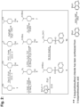
- Hydrochloride salt of BA-1049 ( FIG. 1A ), a 4-substituted piperidine derivative existing as a racemic mixture can be made in any method known in thereof, and has been described and its mode of preparation set forth in U.S. Patent Nos. 7,572,913 and 8,957,093 ( FIG. 2 ).
- Hydrochloride salt of BA-1049 (R) ( fig. 2 ) can be prepared after purification from the racemic mixture of (R) and (S) enantiomers, e.g ., by column chromatography.
- BA-1049 (R) can be synthesized de novo as a chirally pure compound as shown in FIG. 3 or by any method known in the art.
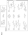
- stereocontrolled synthesis of the hydrochloride salt of BA-1049 can be performed, as shown in FIGS. 4 , and as described in EXAMPLE 1. Briefly, in this method, condensation of the commercially available 1-benzyloxycarbonyl-4-formylpiperidine (1) with (S)-(-)-2-methyl-2-propanesulfinamide under standard conditions, stir for 18 hours at room temperature, filter through celite, separation of the organic layer, concentration and purification by column chromatography, affords the required chiral imine 2. The latter was then transformed to the (R, S) diastereomer 3 by selective addition of methyl Grignard at low temperature. Removal of the chiral auxiliary, protecting group manipulations, and the introduction of the isoquinoline 8 results in the same intermediate 7 used in the racemic synthesis of the hydrochloride salt of BA-1049 (R) (see FIG. 2 ).
- the 1-hydroxy-BA-1049 (R) metabolite was isolated from cultures of hepatocytes after they were exposed to BA-1049 (R) and characterized using LC-MS methods as described in EXAMPLE 2.
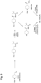
- FIG. 5 shows one non-limiting method of chemically synthesizing 1-hydroxy-BA-1049 (R) starting with compound 9 illustrated in FIG. 2 .
- BA-1049 was synthesized, and the racemic mixture was separated into its enantiomers on a chiral column.
- Six additional Rho kinase inhibitors having structural similarity to BA-1049 i.e., BA-1041, BA-1042, BA-1043, BA-1050, BA-1050A, and BA-1050B) were also synthesized. The Ko's of these compounds were then measured to determine their selectivity for ROCK2 (see EXAMPLE 2, Table 5).
- FIG. 6A - FIG. 6D and Table 1 illustrate the K D 's for binding to either ROCK1 ( FIG. 6A and FIG. 6B ) or ROCK2 ( FIG. 6C and FIG. 6D ) by BA-1049 (R) and (S) isomers.
- Table 1 also shows the Ko's for binding to ROCK1 or ROCK2 by Fasudil and SLx-2119 and the ratio of the dissociation constants of these compounds for each kinase ( i.e., selectivity for ROCK2 binding).
- FIGS. 7A - 7D and Table 2 were obtained using a direct filter-binding radiometric kinase assay (see EXAMPLE 2).
- the IC 50 's against ROCK1 and ROCK2 for BA-1049 (S) ( FIG. 7A and FIG. 7B ) and BA-1049 (R) ( FIG. 7C and FIG. 7D ) were determined using 10 ⁇ M ATP for both enzyme reactions.
- BA-1049 (R) has an 86-fold greater selectivity for ROCK2 than for ROCK1.
- the variability in ROCK selectivity using different ATP concentrations may be due to the fact that at 10 ⁇ M ATP, ROCK1 is below its Km and therefore would take longer to reach V max , requiring less BA-1049 to compete with and inhibit enzyme activity.
- BA-1049 (R) has high potency for ROCK2 in ischemic or diseased tissue where ATP concentrations are reduced compared to healthy tissue.
- BA-1049 (R) and 1-hydroxy-BA-1049 (R) are selective for ROCK2, the isoform of ROCK highly expressed in the central nervous system.
- ROCK2 is hyperactivated in neurons in various neurological diseases in part because of the inflammatory component of the disease and entry of molecules such as tumor necrosis factor and LPA that are known to act as activators of ROCK.
- diseases include amyotrophic lateral sclerosis(ALS), Alzheimer's Disease, Parkinson's Disease, multiple sclerosis, Huntington's Disease, and spinal muscular atrophy (SMA).
- BA-1049 (R) and 1-hydroxy-BA-1049 (R) are useful in treating spinal cord injury, traumatic brain injury, optic nerve injury and peripheral nerve injuries.
- BA-1049 is useful to treat stroke, vasospasm after subarachnoid hemorrhage, cerebral cavernous malformation, hereditary hemorrhagic telangiectasis, cerebral arteriovascular malformations, and Behcet's Disease.
- BA-1049 (R) and 1-hydroxy-BA-1049 (R) are useful to treat autism and diseases such as Fragile X, Rett's Syndrome, and other disorders where there are abnormalities in dendritic spines. This is because ROCK is a critical regulator of the cytoskeleton and of dendritic spine formation, and overactivation of ROCK leading to abnormal dendritic spines has been reported in various forms of autism.
- BA-1049 is useful in treating diseases where ROCK2 is hyperactivated in diseased epithelial cells. As with endothelial cells, ROCK2 regulates cell-cell junctions between epithelial cells. Therefore BA-1049 (R) and 1-hydroxy-BA-1049 (R) are useful in treating Crohns' disease and inflammatory bowel disease.
- BA-1049 is useful in protecting cells from the harmful effects of ionizing radiation used in chemotherapy, or radiation resulting from environmental hazards. This is useful for the gastrointestinal effects of radiation syndrome.
- ROCK also plays a role in regulating the expression of collagen.
- BA-1049 (R) is effective in treating various fibrotic diseases e.g ., in the kidney and liver, and especially fibrosis of the lung, because ROCK2 is highly expressed in lung.
- the active metabolite 1-hydroxy-BA-1049 (R) shows efficacy against ROCK2 and thus has utility in many of the same roles as for BA-1049 (R) as described above.
- Aldehyde oxidase is an enzyme well-known for its importance in the metabolism of xenobiotics, especially those that are N-heterocycles such as BA-1049 (R), and is the enzyme likely involved in the generation of 1-hydroxy-BA-1049 (R) when BA-1049 (R) is administered orally.
- 1-hydroxy-BA-1049 (R) is preferentially used via the intravenous route when a patient in an acute setting shows altered or reduced levels of consciousness, and thus oral administration of a drug is difficult or impossible.
- either genetics or pathology may guide the choice of compound. Because there are naturally occurring polymorphisms in the aldehyde oxidase gene in humans that can lead to reduced activity of this enzyme ( Hartmann et al.. (2012) Drug Metab. Disposition; 40(5): 856-864 ), this population benefits from administration of 1-hydroxy-BA-1049 (R) for the diseases, disorders or injuries as described above. Additionally, identification of chronic alcohol use or abuse in a social history favors the choice of 1-hydroxy-BA-1049 (R) for use in treating diseases, disorders or injuries such as the ones listed above, as a history of alcohol abuse can cause significant reductions in aldehyde oxidase activity in the hepatocytes of the liver ( Hutzler et al.. (2014) Drug Metab. Disposition; 42(6): 1090-1097 ). Accordingly, for treatment of acute spinal cord injury and traumatic brain injury the hydroxy metabolite is useful because intoxication is a common co-morbidity with neurotrauma in civilian populations.
- the pharmaceutical formulations useful in the therapeutic methods according the invention are as defined in the appended claims.
- the pharmaceutical formulations useful in the therapeutic methods include a therapeutically effective amount of BA-1049 (R) and/or active metabolites thereof, such as 1-hydroxy-BA-1049 (R), and/or adipate salts thereof and/or deuterated form thereof.
- Other pharmaceutical formulations for use in treating CCM, aneurysm, and those disorders listed above include therapeutically effective amounts of rho kinase inhibitors other than BA-1049 (R) or 1-hydroxy-BA-1049 (R).
- a “therapeutically effective amount” as used herein refers to that amount which provides a therapeutic and/or prophylactic therapeutic effect for treating a neurological trauma such as a CCM, cerebral aneurysm, stroke, vasospasm after subarachnoid hemorrhage, or spinal cord injury. If another Rho inhibitor compound is part of the BA-1049 (R) pharmaceutical formulation, or if it is to be administered in a separate pharmaceutical formulation, the therapeutically effective amount may be different for each one, and the addition of one or more of these drugs in the formulation can alter the ratio of the kinase inhibited.
- Such formulations are prepared with a pharmaceutically acceptable carrier in accordance with known techniques, for example, those described in Remington, The Science And Practice of Pharmacy (9th Ed. 1995 ).
- pharmaceutically acceptable carrier is to be understood herein as referring to any substance that may, medically, be acceptably administered to a patient, together with a compound of this invention, and which does not undesirably affect the pharmacological activity thereof; a “pharmaceutically acceptable carrier” may thus be, for example, a pharmaceutically acceptable member(s) selected from the group comprising or consisting of diluents, preservatives, solubilizers, emulsifiers, adjuvant, tonicity modifying agents, buffers as well as any other physiologically acceptable vehicle.
- This pharmaceutical formulation may further contain additional Rho inhibitors.
- the pharmaceutical formulation may be prepared for injectable use, for oral use, for inhalation use, for transdermal use, for transmembrane use, and the like.
- Formulations suitable for oral administration may be presented in discrete units or dosage forms, such as capsules, cachets, lozenges, tablets, sublingual tablets, pills, powders, granules, chewing gum, suspensions, solutions, and the like.
- Each dosage form contains a predetermined amount of Rho kinase inhibitor compound.
- the pharmaceutically acceptable carrier may be an aqueous liquid, such as buffered with a pharmaceutically acceptable pH buffer, or in non-aqueous liquid such as DMSO, or be prepared as an oil-in-water or water-in-oil emulsion.
- Injectable dosage forms may be sterilized in a pharmaceutically acceptable fashion, for example by steam sterilization of an aqueous solution sealed in a vial under an inert gas atmosphere at 120°C for about 15 minutes to 20 minutes, or by sterile filtration of a solution through a 0.2 ⁇ M or smaller pore-size filter, optionally followed by a lyophilization step, or by irradiation of a composition containing a compound of the present invention by means of emissions from a radionuclide source.
- a therapeutically effective dosage of BA-1049 (R) or an active hydroxyl metabolite thereof may vary from patient to patient, and may depend upon factors such as the age of the patient, the patient's genetics, and the diagnosed condition of the patient, and the route of delivery of the dosage form to the patient.
- a therapeutically effective dose and frequency of administration of a dosage form may be determined in accordance with routine pharmacological procedures known to those skilled in the art. For example, dosage amounts and frequency of administration may vary or change as a function of time and severity of the neurological trauma.
- a dosage from about 0.1 mg/kg to 1000 mg/kg, or from about 1 mg/kg to about 100 mg/kg BA-1049 (R), 1-hydroxy-BA-1049 (R), or deuterated or adipate salts thereof, may be suitable.
- Administration may be by injection into cerebrospinal fluid as a solution or as a suspension suitable for sustained release from the injected pharmaceutical dosage form such as from a vesicle. Administration alternatively may be made to the lesion site by stereotactic injection.
- the following scheme describes the synthesis of 50 mg to 100 mg of a hydrochloride salt of the enantiomer R of BA-1049 (NT-000077) and includes a chiral synthesis method that enables the identification of the absolute configuration ((R) or (S)) of the molecule.
- Compounds BA-1049 (R), BA-1049 (S) and other additional Rho kinase inhibitors (BA-1041, BA-1042, BA-1043, BA-1050, BA-1050A, and BA-1050B were synthesized according to established protocols ( US Patent No. 7,572,913 ).
- Compounds 1050 A and B are enantiomers that were purified by column chromatography, but the exact orientation was not identified, so they are termed A and B.
- Stock solutions for each compound were prepared at 100 mM in 100% DMSO and stored in an air tight container at -20°C. 10 mM working aliquots were prepared by diluting the stock solutions 1:10 in 100% DMSO. For K D determinations, 50 ⁇ L aliquots at 10 mM were prepared for each compound tested. For IC 50 determinations, compounds were prepared as 20 ⁇ L or 100 ⁇ L aliquots at 10 mM.
- BA-1049 and BA-1050 exist as racemic mixtures with each having one chiral center.
- BA-1049 and BA-1050 were resolved into their respective enantiomers, (S) and (R) for BA-1049; BA-1050 enantiomers are called A and B because their absolute stereochemistry was not determined.
- 10 mM stock solutions were prepared in DMSO before storing the stock solutions at -20°C.
- IC 50 determinations for BA-1049 were performed using a direct filter-binding radiometric kinase assay as described below.
- ROCK1 and ROCK2 IC 50 determinations were made using an ATP concentration of 10 ⁇ M.
- ROCK1 and ROCK2 IC 50 determinations were made using ATP concentrations of either 10 ⁇ M, or the Km ATP of 70 ⁇ M and 15 ⁇ M for ROCK1 and ROCK2, respectively. Variations in ATP concentrations were tested to better understand potential selectivity in ischemic and diseased tissue where ATP concentrations may be low and impact selectivity.
- K D values for the compounds to be tested and control articles for ROCK1 and ROCK2 were determined using the KINOMEscan TM Profiling Service (DiscoverX Corp, Freemont, CA).
- KINOMEscan TM is based on a competition binding assay that quantitatively measures the ability of a compound to compete with an immobilized, active-site directed ligand. The assay is performed by combining the DNA-tagged kinase, immobilized ligand, and a test compound. The ability of the test compound to compete with the immobilized ligand is measured via quantitative PCR of the DNA tag.
- Test compounds were tested using an 11-point curve with 3-fold serial dilutions. The highest concentration tested was 30 ⁇ M. Test compounds were prepared in 100% DMSO at 100x final test concentration and were diluted to 1 ⁇ in the assay with a final DMSO concentration of 1%.
- Streptavidin-coated magnetic beads were treated with biotinylated small molecule ligands for 30 min at RT to generate affinity resins for kinase assays.
- the ligand-bound beads were blocked with excess biotin and washed with blocking buffer (SeaBlock (Pierce), 1% BSA, 0.05% Tween 20, 1 mM DTT) to remove unbound ligand and to reduce non-specific binding.
- Binding reactions were assembled by combining kinases, ligand-bound affinity beads, and test compounds in 1 ⁇ binding buffer (20% SeaBlock, 0.17x PBS, 0.05% Tween 20, 6 mM DTT).
- IC 50 values for test articles and control articles for ROCK1 and ROCK2 were determined using the Kinase HotSpot TM Profiling Service (Reaction Biology Corp., Malvern, PA), and the IC 50 Profiler TM (Eurofins Pharma Discovery Services, St. Charles, MO). Both services use a direct filter-binding, radiometric kinase assay with slight variation in protocol.
- Test compounds were tested using an 11-point curve with 3-fold serial dilutions. The top concentration tested was 100 ⁇ M for both services. Test compounds were prepared in 100% DMSO at 50x final test concentration and were diluted to 1 ⁇ in the assay with a final DMSO concentration of 2%.
- the substrate EAKEKRQEQIAKRRRLSSLRASTSKSGGSQK (SEQ ID NO: 1) (30 ⁇ M) was mixed with reaction buffer (Table 4) and the kinase domain from either the ROCK1 or ROCK2 (as indicated in Table 4).
- the compounds (in DMSO) were delivered into the mixture via acoustic mixing technology (Echo550, nanoliter range) (SelectScience, Waltham, MA), and incubated at RT for 20 min. Radiolabeled ⁇ - 33 P-ATP (10 ⁇ M) was added and the reaction incubated for 2 hr at RT. Reactions were then spotted onto P81 ion exchange paper followed by scintillation counting.
- ROCK1 or ROCK2 was incubated with reaction buffer, substrate, 10 mM Mg acetate and ⁇ - 33 P-ATP (10 ⁇ M or Km concentrations) (see Table 4) and incubated for 40 min at RT.
- the reaction was stopped by the addition of 3% phosphoric acid and 10 ⁇ L of the reaction was spotted onto P30 filtermat, washed 3 times in 75 mM phosphoric acid and once in methanol prior to scintillation counting.
- Raw data was captured in Microsoft Excel 2013. Data from Microsoft Excel was transferred GraphPad Prism 6.07 to derive dose-response curves and calculate K D and IC 50 values. Standard error of the mean (SEM) was calculated using Microsoft Excel 2013. Data was presented as the average value plus or minus SEM.
- K D values Equilibrium dissociation constants were performed on different racemic lots of BA-1049 (Lots 1 and 2) as well as the enantiomers, R and S. The analysis also included six other Rho kinase inhibitors selected based on their structural similarity to BA-1049 and their ability to selectively inhibit ROCK2. These compounds were included BA-1041, BA-1042, BA-1043, BA-1050 and its enantiomers, A and B.
- IC 50 values for both ROCK1 and ROCK2 were determined using an ATP concentration of 10 ⁇ M. While most kinase inhibitors compete with ATP binding, 10 ⁇ M is often used for screening, despite physiological levels being in the mM range. Two different variations on the direct filter-binding radiometric kinase assay were used, as detailed above. The results are shown in Table 6.
- the IC 50 values for BA-1049 (S) and BA-1049 (R) were determined at the Km ATP concentration for both ROCK1 (70 ⁇ M) and ROCK2 (15 ⁇ M), an artificial assay to understand what might happen if the ATP concentration fell below the Km of ROCK1. In this case the maximal velocity of the reaction would be slower, and BA-1049 (R) would continue to be active against ROCK2.
- Table 7 Compound ID Compound IC 50 ( ⁇ M) ROCK1 ROCK2 70 ⁇ M ATP 15 ⁇ M ATP BA-1049 (S) 57.9 1.00 BA-1049 (R) 29.6 0.34
- the data show a 50- to 60-fold greater selectivity for ROCK2 relative to ROCK1.
- MCAO middle cerebral artery occlusion
- BA-1049 racemic mixture
- BA-1049 (R) efficacy in endothelial cells through the use of well characterized biomarkers for ROCK2 activity, phosphorylated adducin (p-adducin), phosphorylated myosin light chain 2 (pMLC2), phosphorylated cofilin (phospho-cofilin), phosphorylated LIMK1/2, and autophosphorylated ROCK2.
- p-adducin phosphorylated adducin
- pMLC2 phosphorylated myosin light chain 2
- phosphorylated cofilin phospho-cofilin
- LIMK1/2 autophosphorylated LIMK1/2
- CCA left common carotid artery
- ECA external carotid artery
- ICA internal carotid artery
- the ECA was dissected further distally and two 8-0 silk sutures are tied around the ECA stump and a vascular clamp was applied at the bifurcation of the CCA into the ECA and ICA.
- a small incision was made at the end of ECA stump with Vannas-style spring scissors.
- a blunt 5-0 monofilament suture (Doccol) was inserted into the incision and advanced from the lumen of the ECA into the ICA for a distance of 9-10 mm beyond the bifurcation of CCA to occlude the origin of MCA.
- Doccol monofilament suture
- a distance of 9 mm -11 mm rostral to the CCA bifurcation is inserted. After 60 min., the monofilament was removed.
- the incision was sutured with 4.0 prolene sutures.
- One mL saline was injected subcutaneously and 0.1 mg/kg buprenorphine was injected subcutaneously every 8 hr to 12 hr for up to 48 hr to decrease pain.
- the mouse is allowed to recover on a heating pad until thermoregulation is re-established.
- test Rho kinase inhibitor compound dosing solutions were prepared by dissolving BA-1049 powder (racemic mixture) and BA-1049 (R) in sterile PBS in order to achieve concentrations of 10 mg/mL, 25 mg/mL, and 50 mg/mL for groups B, C, and D, respectively.
- the pH of test compound dosing solutions was adjusted to 7. After preparation, the dosing solutions were kept at 4°C until use, retained dosing solutions are stored at 4°C.
- test compound dosing solutions were prepared by dissolving Fasudil (Calbiochem) powder in sterile PBS in order to achieve concentrations of 10 mg/mL, 25 mg/mL, and 50 mg/mL.
- the pH of test compound dosing solutions was adjusted to 7. After preparation, the dosing solutions are kept at 4°C until use. Retained dosing solutions were stored at 4°C.
- Table 8 Part 1 Determination of an efficacious dose of BA-1049 vs. Fasudil, intraperitoneal (I.P.) inj. Group N Surgical Procedure Route Treatment, Dose 1 4 Transient (60 min.) MCAO I.P. PBS 2 4 Transient (60 min.) MCAO I.P. BA-1049 (racemate), 10 mg/kg 5 4 Transient (60 min.) MCAO I.P. Fasudil, 10mg/kg Table 9 Part 2a : Determination of Optimal Delivery Method, I.P. vs.
- test articles BA-1049 racemic, BA-1049 (R), Fasudil
- (saline) were administered at the specified dose at a volume of about 0.2 mL (volume was adjusted based on individual body weights) to all mice as a single dose I.P. 30 min after MCAO surgery using sterile 1 mL-syringes fitted with a 25 G 5/8 needle.
- test and vehicle articles were administered at the specified dose at a volume of about 0.2 mL (volume is adjusted based on individual body weights) to all mice by either oral gavage (PO) ( ⁇ 10 mg/kg) 30 min after MCAO surgery (2a) or subcutaneous (SC) injection in the right flank 30 min after MCAO surgery (2b) using sterile 1 mL-syringes fitted with a 25 G 5/8 needle.
- PO oral gavage
- SC subcutaneous
- mice were euthanized and brains are collected for biomarker analysis by Western blot analysis or immunohistochemistry.
- Western blot analysis mice were anesthetized using isoflurane and decapitated.
- the right and left middle cerebral arteries supply the lateral surface of their respective lobes in the territory of the motor and sensory cortices.
- the brain rostral to the cerebellum (excluding olfactory bulb) was collected ipsilateral (left hemisphere) and contralateral (right hemisphere) to the occlusion. Ipsilateral and contralateral hemispheres are collected in 2 separate tubes. Frozen brain tissue was further processed into tissue lysates following the experimental protocol documented below.
- mice were anesthetized using isoflurane and perfused intracardially using paraformaldehyde (4% PFA), brain and weights were recorded. Brain specimens from fixed animals were frozen in OCT and sectioned using a cryostat.
- Protein lysates were analyzed by Western blot analysis following the experimental protocol below in Table 12.
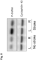
- Table 12 Primary Antibody Dilution Secondary Antibody Dilution pCofilin rabbit polyclonal (Cell Signaling Cat# 3313, lot# 7) 1:500 Anti-Rabbit IgG HRP (Cell Signaling Cat# 7074, lot #26) 1:1500 pROCK2 rabbit polyclonal (Genetex Cat# GTX122651, lot# 42025) 1:500 Anti-Rabbit IgG HRP (Cell Signaling Cat# 7074, lot #26) 1:1500 Cyclophilin 40 rabbit polyclonal (Santa Cruz Cat# sc-66848, lot# H3103) 1:2000 Anti-Rabbit IgG HRP (Cell Signaling Cat# 7074, lot #26) 1:1500 pMLC2 (Thr18/Ser19) rabbit polyclonal (Cell Signaling Cat#3674, lot# 3) 1:1000 Anti-Rabbit IgG HRP (Cell Signaling Cat# 7074
- the left and right hemispheres of each brain were homogenized and prepared for Western blotting, and the signal of phospho-cofilin was measured quantitatively by densitometry compared to an internal standard, cyclophilin.
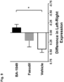
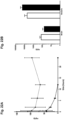
- FIG. 9 shows the results of Part 1 of the study outlined above and in Table 3.
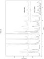
- BA-1049 (10 mg/kg) was compared to Fasudil (10 mg/kg) and vehicle controls. Injections were made IP within 30 min of recovery after MCAO. Tissue was harvested 24 hr post MCAO. The ratio of phospho-cofilin signal in the left versus right hemispheres of the brain was examined using Western blotting. In these experiments, a reduction in the ratio of phospho-cofilin expression between the left (MCAO) and right (uninjured) sides demonstrates a reduction in ROCK activation and therefore an increase in ROCK inhibition by the given treatment.
- FIG. 8 shows that phospho-cofilin expression is strongly increased in the stroke side of the brain and acts as an effective biomarker for ROCK2 activation.
- Frozen tissue sections embedded in OCT were analyzed by immunohistochemistry.
- the left side of the brain slice contains ischemic tissue due to the MCAO while the right hemisphere acts as a control without ischemia.
- Brain slices were treated with antibodies listed in Table 13. Staining with these antibodies provide biomarkers for both ischemic area and ROCK2 activation.
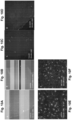
- FIGS. 10A - 10D show phospho-MLC2 (Serl9) staining in small blood vessels of insular cortex. Blood vessels are strongly stained in the insular cortex on the left (MCAO) side of the brain ( FIG. 10A ) while there is no staining of small vessels on the control (non-MCAO) side ( FIG. 10B ). Phospho-MLC2 is a downstream marker for ROCK2 activation. 10 mg/kg Fasudil injected intraperitoneally (I.P.) ( FIG. 10C ) reduces the amount of staining while 10 mg/kg BA-1049 ( FIG. 10D ) injected I.P. is even more effective than Fasudil at reducing small vessel staining. FIGS.
- I.P. intraperitoneally
- FIG. 10E and 10F show Iba-1 immunoreactivity in the left brain striatum after MCAO and I.P. injection with vehicle ( FIG. 10E ) or BA-1049 (R) ( FIG. 10F ).
- Iba-1 immunoreactivity is a marker for microglia in the brain. Iba-1 staining intensity is increased in activated microglia, responding to ischemic damage in the brain. These results demonstrate that BA-1049 (R) reduces the microglial reaction to ischemia.
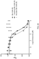
- the minimal effective dose was 1 mg/kg as seen in FIG. 11 .
- the minimal effective levels are less than 1 mg/kg with daily repeat dosing.
- mice received 1 mg/kg or 10 mg/kg of BA-1049 (R) once daily by intraperitoneal injection for 2 weeks. During the course of the experiment, animals were monitored daily for the appearance of any clinical signs. Hematology, blood chemistry, and histopathology were followed at termination of the experiment.
- mice were subject to MCAO and then the ratio of left brain to right brain phospho-cofilin levels determined as described above in EXAMPLE 4. Mice were examined 4 hr or 24 hr after reperfusion and administration of 10 mg/kg of BA-1049 (R).
- both time points showed efficacy, demonstrating efficacy of BA-1049 (R) for at least 24 hr.
- the mice were given BA-1049 (R) for 24 hr, 72 hr, or 168 hr prior to the MCAO.
- the results shown in FIG. 12C demonstrate that 3 days prior exposure to BA-1049 (R) prevents the hyperactivation of ROCK after MCAO.
- mice were given an MCAO lesion, as described in EXAMPLE 4. Two hr after reperfusion, mice were injected I.P. with a 1.2% solution of Evans blue dye in PBS, which binds tightly to serum albumin. Two hr after MCAO, Evans blue was injected, and 4 hr since reperfusion, mice were sacrificed, perfused, decapitated, and their brains dissected as described in EXAMPLE 4.
- Homogenates were prepared as in EXAMPLE 4 and then precipitated in 50% trichloroacetic acid to precipitate proteins and release Evans blue dye from the albumin. After spinning down proteins (at 10,000 ⁇ g), supernatants were placed in a 96-well dish and the absorbance at 595 nm and fluorescence (535 Ex/642 Em) read using a Victor 2 Wallac plate reader (Perkin-Elmer; Branford, CT). Comparison to a standard curve allows a determination of Evans blue dye concentration in the sample and, therefore, a measure of permeability to albumin.
- FIG. 13A shows micrographs of the brains of animals treated with Vehicle or BA-1049 (R).
- the Left (L) side is the ischemic side of the brain and is compared to the right (R) cortex.
- BA-1049 (R) reduces the fluorescent signal compared to the vehicle control.
- FIG. 13B shows a graphic representation of the results and shows that BA-1049 (R) treatment is able to reduce the Evans blue signal compared to the left brain of the vehicle control.
- mice C57BL/6 mice, 8-10 weeks old (Charles River Labs, Wilmington, MA) were used in this study.
- the test drug is provided in Table 15 below: Table 15 Group # No. of Mice/Sex Route of Administration Dose (mg/kg) Dose Conc. (mg/mL) Dose Vol. (mL/kg) Dose Vol. ( ⁇ L/g) 1 24M IV 5 1.25 4.0 4 2 24M IP 10 0.5 20.0 20 3 24M PO 30 3.75 8.0 8 4* 5M - - - - - *Group 4 is for collection of naive whole brain only and will not be dosed.
- the test drug is provided once as follows: Each I.V. dosing syringe is weighed loaded and unloaded to at least 4 decimal places. The actual dose (5 mg/kg) administered to the animal is the difference between the loaded and unloaded dosing syringe weights. Each IP dosing syringe is weighed loaded and unloaded to at least 4 decimal places. The actual dose (10 mg/kg) administered to the animal is the difference between the loaded and unloaded dosing syringe weights. 30 mg/kg PO administration is conducted using a ball-tipped, stainless steel gavage needle attached to a plastic syringe.
- terminal blood samples (about 1 ml to 2 ml each) are collected from 4 animals at each of the following time points post-dose: 0.083 hr, 0.5 hr, 2 hr, 6 hr, 12 hr, and 24 hr.
- terminal blood samples (about 1 ml to 2 ml each) are collected from four animals at each of the following time points post-dose per dose route: 0.25 hr, 1 hr, 4 hr, 8 hr, 12 hr, and 24 hr.
- Each animal is anesthetized by CO 2 inhalation and terminal blood samples are collected via cardiocentesis and transferred into pre-labeled tubes containing sodium heparin as the anticoagulant. Following cardiocentesis blood collection, the animal is returned to the CO 2 chamber and euthanized by CO 2 asphyxiation. Collected blood samples are gently inverted several times to mix the anticoagulant.
- Blood samples are centrifuged at about 3000 ⁇ g rpm for 10 min at about 4°C, [resultant plasma is observed for hemolysis]. All derived plasma samples are stored frozen at approximately -80°C until further processing.
- whole brain is collected following cardiocentesis from 4 animals at each of the following time points post-dose: 0.083 hr, 0.5 hr, 2 hr, 6 hr, 12 hr, and 24 hr.
- whole brain is collected following cardiocentesis from 4 animals at each of the following time points post-dose per dose route: 0.25 hr, 1 hr, 4 hr, 8 hr, 12 hr, and 24 hr.
- Whole brain is rinsed in 1 X PBS, the tissue weight recorded and snap frozen in pre-labeled tubes using liquid nitrogen.
- whole brain from 5 naive mice is harvested at 24 hr, rinsed in 1 X PBS, the tissue weight recorded and snap frozen in pre-labeled tubes using liquid nitrogen.
- Samples are analyzed by LC/MS/MS using an Agilent 6410 mass spectrometer coupled with an Agilent 1200 HPLC and a CTC PAL chilled autosampler, all controlled by MassHunter software (Agilent). After separation on an X-Select HPLC column (Waters, 130A,3.5 ⁇ m, 2.1 ⁇ 50 mm) using an acetonitrile-water gradient system (shown below), peaks were analyzed by mass spectrometry (MS) using ESI ionization in MRM mode.
- MS mass spectrometry
- a working dilution of the test article at 25 times the final concentration is prepared and serially diluted (3-fold). These samples are diluted 25-fold into C57BL/6 mouse blank plasma and mixed with three volumes of acetonitrile containing an analytical internal standard (propranolol), incubated on ice for 10 min at 4°C and then centrifuged. The protein-free supernatant is used for LC-MS/MS (liquid chromatography-tandem mass spectrometry) analysis.
- LC-MS/MS liquid chromatography-tandem mass spectrometry
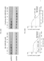
- BA-1049 (R) and BA-1049 (S) were compared for their effect on neurite outgrowth using a cell culture assay with NG-108 cells. First the effective dose for use with NG-108 cells was determined and then BA-1049 (R) was compared with a ROCK2 specific inhibitor SLx-2119.
- BA-1049 (S) and BA-1049 (R) were compared for their effect on neurite outgrowth from NG-108 cells. As shown in FIG. 15D , BA-1049 (R) was the most potent compound to stimulate neurite outgrowth compared to the DMSO control ( FIG. 15A ), racemic BA-1049 ( FIG. 15B ), or BA-1049 (S) ( FIG. 15C ). Thus, BA-1049 (R) produces the most robust neurite outgrowth than the (S) compound.
- FIGS.16A - 16C 5 ⁇ M SLx-2119 was shown to be toxic to NG-108 cells, and cells showed formation of vacuoles and began to die ( FIG. 16A ).
- BA-1049 (R) promoted neurite outgrowth at 5 ⁇ M ( FIG. 16B ) and 50 ⁇ M ( FIG. 16C ); BA-1049 (R) was not toxic to NG-108 cells at the highest concentrations tested ( FIG. 16C ).
- Neuronal cells were isolated from the developing brain cortex of embryonic rats using enzymatic and mechanical disruption methods and then plated onto specialized growth substrates including laminin and poly-D-lysine in a tissue culture dish. These cell mixtures were grown in a specialized medium under conditions of low serum containing specialized selected media additions (NeuroBasal Medium with B27 additive, Thermo Fisher Scientific, Waltham, MA) designed to enhance the survival of the neuronal cells in the dish.
- Neuronal Medium with B27 additive Thermo Fisher Scientific, Waltham, MA
- sdRNA self-delivering RNAi
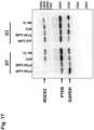
- the results shown in FIG. 17 are from Western blotting experiments where levels of PTEN, ROCK2 and GAPDH proteins were analyzed.
- D7 sdRNA
- targeting PTEN mRNA for reduction using PTEN-specific sdRNA leads to reduced PTEN protein levels in these cells, as expected.
- the reduction in PTEN protein levels also leads to a substantial reduction in the protein levels of the ROCK2 kinase.
- ROCK2 expression is decreased by knockdown of PTEN, showing unexpectedly that ROCK2 is a promising target downstream of PTEN.
- BA-1049 which inactivates ROCK2 is a better target than PTEN for eliciting axon regeneration in the CNS.
- mice central nervous system
- optic nerve in a group of 12 animals and the sciatic nerve of the peripheral nervous system in an additional group of 12 mice
- 5 ⁇ L of 1 ⁇ M BA-1049 (R) or control saline solution per group of 6 mice is applied during surgery by direct injection at the injury site at the time of the experimental injury and the animals recovered for 2 to 4 weeks after injury.
- changes in the relative extent of regeneration are measured using histological techniques.
- fluorescent tracer molecules such as labeled cholera toxin are injected into the motor cortex to stain the axons that project into the spinal cord, in the eye to measure retinal ganglion cell axon regeneration, or in the case of the sciatic nerve, injection of labeled dextran amine proximal to the spinal cord at the region of the nerve roots allows for the characterization, by anterograde labeling, of the peripheral nerve axons.
- Sections are prepared the next day following the sacrifice and perfusion of the animal with fixative. The sections are collected onto glass slides and then the fluorescent markers are visualized through the microscope and the distance to which the labeled regenerating axons are growing are measured in a series of photographs taken of the specified sections. The lengths of the labeled axons are compared between the animals treated with BA-1049 (R) and those treated simply with saline injections.
- ROCK hyperactivation is induced by administration of lysophosphatidic acid (LPA) (Sigma-Aldrich, St. Louis, MO), a potent activator of the Rho/ROCK pathway.
- LPA lysophosphatidic acid
- BA-1049 (R) efficacy is determined by investigating actin stress fiber formation, a biomarker for ROCK activity, and distribution of the focal adhesion complex protein vinculin, a biomarker for endothelial barrier integrity.
- HUVEC cells (Gibco ® , Thermo Fisher, Waltham, MA) are cultured in endothelial growth media (Gibco ® Medium 200 basal media Cat supplemented with Gibco ® Large Vessel Endothelial Supplement). HUVECs between passage 1 and 4 are used for all experiments. HUVECs are cultured on poly-D-lysine- treated glass coverslips (Corning, Corning, NY) coated with 70 ⁇ g/ml rat collagen I (Corning, Corning, NY) and cultured for 4 d until a confluent monolayer of cells is established.
- HUVECs are stimulated with 20 ⁇ M LPA for 10 min and subsequently fixed using 4% paraformaldehyde (PFA)/sucrose solution.
- PFA paraformaldehyde
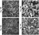
- FIG. 18 demonstrate that LPA stimulation induces pronounced actin stress fiber formation in HUVECs ( FIG. 18B ) compared to cells prior to stimulation ( Fig 18A ).
- FIG. 19 treatment with either BA-1049 (R) ( FIGS. 19A and 19C ) or Fasudil ( FIGS. 19B and 19D ) prevents such formation of stress fiber.
- the reduction in actin stress fibers is more pronounced in HUVECs treated with BA-1049 (R) ( FIG. 19A ) compared to Fasudil-treated cells ( FIG. 19B ).
- vinculin immunofluorescence reveals that BA-1049 (R) ( Fig 19C ) is superior in preserving the integrity of the HUVEC monolayer upon LPA stimulation in comparison to Fasudil ( FIG. 19D ).
- BA-1049 (R) treated cells show a thin, peripheral band of vinculin immunofluorescence ( FIG. 19C ), characteristic for a confluent cellular monolayer
- Fasudil treated cells show an upregulation of vinculin-positive focal adhesion complexes associated with a decreased cell confluency ( FIG. 19D ).
- BA-1049 (R) was added to cultures of rat, human, mouse, cynomolgus monkey, and canine hepatocytes to investigate metabolism in different species.
- a stock solution of BA-1049 was first diluted in acetonitrile at a concentration 100x of the desired final concentration.
- Test articles were incubated in duplicate rat hepatocytes at 37°C.
- the reaction contained 106 viable hepatocytes/ml KHB buffer, a pH 7.4. At indicated times (0 min, 15 min, 30 min, 60 min, 120 min), an aliquot was removed from each experimental and control reaction and mixed with an equal volume of ice-cold methanol. Stopped reactions were incubated at least ten minutes at -20°C.
- the samples were centrifuged to remove precipitated protein, and the supernatants were analyzed by LC-MS/MS.
- Table 17 shows the half-life at BA-1049 (R) in different species.
- the results in FIG. 20 and Table 17 show that BA-1049 (R) added to cultured hepatocytes from multiple species is readily metabolized to 1-hydroxy-BA-1049 (R) in human, rat, mouse, and monkey hepatocytes, while rat hepatocytes also generate N-oxide-BA-1049 (R).
- the structure of N-oxide-BA-1049 (R) is shown represented in FIG. 21C . Dog hepatocytes do not show any detectable metabolism of BA-1049 (R), indicating they may be a poor species choice for studies of the drug prior to testing in human.
- Mouse brains are dissected out at specific time points after IV BA-1049 (R) administration, and the concentration of BA-1049 (R) and 1-hydroxy-BA-1049 (R) in homogenized brain tissue determined using LC-MS/MS analysis.
- brains and blood vessels are dissected out at specific time points after IV BA-1049 (R) administration.
- mice Male male C57BL/6 mice (Charles River Laboratories) are used in this study. Mice are dosed either by intravenous (IV) administration or oral (PO) administration via oral gavage. Mice dosed by IV receive a single 5 mg/kg BA-1049 (R) dose formulated in phosphate buffered saline (PBS). An additional cohort of mice receives a single 30 mg/kg BA-1049 (R) oral dose formulated in PBS.
- IV intravenous
- PO oral
- mice dosed by IV receive a single 5 mg/kg BA-1049 (R) dose formulated in phosphate buffered saline (PBS).
- PBS phosphate buffered saline
- An additional cohort of mice receives a single 30 mg/kg BA-1049 (R) oral dose formulated in PBS.
- mice are monitored after administration for clinical signs of adverse effects of the administered compound.
- Rats are dosed by a single intravenous (IV) 2.5 mg/kg BA-1049 (R) dose via tail vein injection. Both doses are formulated in phosphate buffered saline (PBS).
- IV intravenous
- R BA-1049
- PBS phosphate buffered saline
- Rats are monitored after administration for clinical signs of adverse effects of the administered compound.
- mice After IV dosing, brains are collected from 4 mice at each time point: 0.083 hr, 0.5 hr, 2 hr, 6 hr, 12 hr, and 24 hr after dose. Each mouse is euthanized by CO 2 . The entire brains are removed, weighed, rinsed with PBS, placed in tubes, and snap frozen in liquid nitrogen.
- Brains are collected from 3 rats at each time point: 0.25 hr, 0.5 hr, and 1 hr after dose. Brain samples are weighed, placed in a tube and frozen on dry ice. Brains are the stored at -80°C until analysis.
- a portion of the inferior vena cava is dissected out, weighed, and placed in a tube on dry ice. This vascular tissue is stored at -80°C until analysis.
- Brain tissue was homogenized in 1 mL/g PBS. Brain homogenates are precipitated by pipetting 200 ⁇ L of homogenate into a tube using aseptically cut off 1000 ⁇ L pipette tips to prevent clogging. Samples are further diluted with 100 ⁇ L PBS to aid in precipitation. 900 ⁇ L of cold methanol is added to each sample and samples are vortexed for 5 sec to 10 sec. Samples are placed at 4°C for 30 min to 40 min and then centrifuged at 10,000 RPM for 15 min at 4°C. The supernatant is collected and stored at -80°C until analyzed.
- Rat brain samples are analyzed for determination of the plasma concentration of BA-1049 (R) and its metabolite 1-hydroxy-BA-1049 (R).
- Brain tissue is homogenized in 1 mL/g PBS. Brain homogenates are precipitated by pipetting 200 ⁇ L of homogenate into a tube using aseptically cut-off 1000 ⁇ L pipette tips to prevent clogging. Samples are further diluted with 100 ⁇ L PBS to aid in precipitation. 900 ⁇ L of cold methanol is added to each sample and samples are vortexed for 5-10 seconds. Samples are placed at 4°C for 30 min to 40 min, and then centrifuged at 10,000 RPM for 15 min at 4°C. The supernatant was collected and stored at -80°C until analyzed.
- Inferior vena cava were homogenized by adding 2 mL/g PBS and mashed with a biomasher pestle (Kimble). Precipitation was performed by adding 3 mL/mL cold methanol. Samples were vortexed for 5 sec to 10 sec. Samples were placed at 4°C for 60 min, then centrifuged at 10,000 RPM for 15 min at 4°C. The supernatant was collected and stored at - 80°C until analyzed.
- BA-1049 (R) and its primary, active metabolite 1-hydroxy-BA-1049 (R) were detectable in rat and mouse brains after IV administration ( FIGS. 22A and B ).
- BA-1049 (R) remained detectable at least 24 hr after administration, while 1-hydroxy-BA-1049 (R) was undetectable by 12 hr.
- rat brains like in mice, there was more BA-1049 (R) than 1-hydroxy-BA-1049 (R) at 30 min post-administration. There was a large amount of both compounds in vascular tissue of rats 30 min after IV administration, with more 1-hydroxy-BA-1049 (R) than BA-1049 (R).
- BA-1049 (R) and its metabolite 1-hydroxy-BA-1049 (R) are present and detectable in blood, brain and vascular tissue after IV administration.
- vascular tissue such as the inferior vena cava, contains even more of both compounds than brain tissue.
- intravenous administration provides pharmacologically active compound into brain and vascular tissue to treat or manage diseases, disorders, or injuries.
- Each compound was dissolved at 100 mM in DMSO. This compound-DMSO solution is diluted to 500 ⁇ M in dH 2 O. Serial dilutions were prepared so that a semi-log range of compound is present after being diluted 5X in other assay reagents, from 100 ⁇ M through 1 nM.
- the reaction was stopped by emptying plate and washing 3 times for 3 min each with 200 ⁇ L Washing Buffer (Tris-buffered saline and 0.05% Tween-20).
- Washing Buffer Tris-buffered saline and 0.05% Tween-20.
- BA-1049 (R), BA-1049 (S) and metabolites of BA-1049 (R) were investigated using cultured human vascular endothelial cells.
- HUVECs ATCC, Manassas, VA were grown on collagen-1(50 ⁇ g/mL) coated 75 cm 2 tissue culture flasks using Medium-200 supplemented with large vessel endothelium growth supplement (both ThermoFisher, Waltham, MA). After reaching 80% confluency, cells were extracted using 0.25% trypsin-EDTA (ThermoFisher, Waltham, MA) and plated on tissue culture substrate as described below.
- Cells were subjected to biochemical analysis of diphosphorylated MLC2 (Threonine 18/Serine 19 phosphorylation sites) and cofilin (Serine 9 phosphorylation site) using SDS-PAGE and immunoblotting.
- Cells were plated in 24-well tissue culture plates coated with 70 ⁇ g/ml rat collagen-1 (Corning; Corning, NY), and grown for 2 d to 3 d at 37 °C/5% CO 2 until confluency.
- proteins were extracted from cells by lysis with RIPA buffer supplemented with HALT complete protease/phosphatase inhibitor (ThermoFisher, Waltham, MA), reduced and denatured using Laemmli sample buffer (BioRad, Hercules, CA)/beta-mercaptoethanol and subjected to SDS-PAGE.
- BA-1049 (R), BA-1049 (S) and 1-hydroxy-BA-1049 (R) reduce MLC2 diphosphorylation, while N-Oxide-BA-1049 (R) did not show such an effect ( FIG. 24 ).
- BA-1049 (R) shows a higher potency in reducing pMLC2 T18/S19 than BA-1049 (S) ( FIG. 24 ).
- 1-hydroxy-BA-1049 (R) shows the highest potency of all tested compounds, fully abolishing MLC2 diphosphorylation at a concentration of 10 ⁇ M or higher ( FIG. 24 ).
- HUVECs treated with LPA to stimulate ROCK activation show increased stress fibers and changes in cell-cell junctions, which results in cell free holes in the monolayer ( FIG.
- FIG. 25D As compared to controls ( FIG. 25A ).
- Cells treated with LPA and 1 ⁇ M BA-1049 (R) ( FIG. 25B ) or 10 ⁇ M of BA-1049 (R) ( FIG. 25C ) resemble control cultures with normal monolayer appearance.
- FIG. 25E When cells are treated with LPA plus 1 ⁇ M BA-1049 (S) ( FIG. 25E ) or 10 ⁇ M BA-1049 (S) ( FIG. 25F ), neither concentration reverses the effect of LPA.
- UUVEC umbilical vein endothelial cells
- LPA lysophosphatidic acid
- BA-1049 (R), BA-1049 (S), and metabolites of BA-1049 (R) are investigated using primary human brain microvascular endothelial cells.
- hBMVECs Neuromics, Edina, MN
- collagen-1 70 ⁇ g/mL, Coming
- ENDO-Basal media + ENDO-growth supplement both Neuromics, Edina, MN
- cells are extracted using 0.25% trypsin-EDTA and plated on tissue culture substrate as described below.
- BA-1049 Five individual doses of BA-1049 (R), 1-Hydroxy-BA-1049 (R), N-Oxide-BA-1049(R) and of BA-1049 (S) are evaluated for the ability to restore normal Rho/ROCK activity in LPA-treated hBMVEC.
- Cells are exposed to each dose for 1 hr, and are then stimulated with 20 ⁇ M LPA for either 5 min or 1 hr as described below. All experiments are conducted in triplicate and analyzed by 2 independent assessors blinded to both cell line and treatment.
- Cells are subjected to biochemical analysis of diphosphorylated MLC2 (Threonine 18/Serine 19 phosphorylation sites) and phosphorylated cofilin (Serine 9 phosphorylation site) using SDS-PAGE and immunoblotting.
- Cells are plated in 24-well tissue culture plates coated with 70 ⁇ g/ml rat collagen-1 (Corning) and grown in ENDO-growth media (Neuromics) for 3 days at 37 °C/5% CO 2 until confluency.
- Proteins are extracted from cells by lysis with RIPA buffer supplemented with complete protease/phosphatase inhibitor (HALT, Thermo Fisher Scientific), reduced and denatured using Laemmli sample buffer (BioRad; Hercules, CA, beta-mercaptoethanol and subjected to SDS-PAGE.
- HALT complete protease/phosphatase inhibitor
- BioRad Hercules, CA, beta-mercaptoethanol
- Denatured samples are electrophoresed in reducing 10% Bis-Tris-PA gels (Novex; Thermo-Fisher), and are transferred onto 0.25 ⁇ m PVDF membranes (EMD Millipore).
- Membranes are probed using amti-pMI,C2 T18/S19 (1:500; Cell Signaling) or anti-pCofilin (1:500, Cell Signaling S9) and anti-glyceraldehyde 3-phosphate dehydrogenase loading control (GAPDH, 1:10000; Abcam, Cambridge, MA). Protein levels are detected using a chemiluminescence substrate (Super Signal West, ThermoFisher) and images are captured using a FluoroChem SP Imaging System.
- hBMVECs monolayer integrity
- cells are plated on PDL-coated coverslips (Corning) coated with Fibronectin, Collagen-1 and gelatin. 50,000 cells are seeded on each coverslip situated in a 24-well plate. Cells are grown for 3 days in ENDO-growth media (Neuromics) at 37 °C/5% CO 2 . Different cell samples are pretreated with five different doses of BA-1049 (R), 1-Hydroxy-BA-1049 (R), N-Oxide-BA-1049 (R) or BA-1049 (S) diluted in ENDO-basal media (Neuromics) supplemented with 0.1% human serum albumin (HSA, Sigma-Aldrich) for 1 hr.
- HSA human serum albumin
- the medium is exchanged for ENDO basal media supplemented with 1% HSA, 20 ⁇ M of LPA (Santa Cruz Biotech; Santa Cruz, CA) and the appropriate amount of compound being tested. After 60 min, cells were fixed with 4% paraformaldehyde and stained for actin-phalloidin (ThermoFisher) and V-Cadherin (Cell Signaling), and then were mounted on microscope slides. Coverslips were imaged by fluorescence microscopy and the cell-free holes in the monolayer are measured using Image J.
- BA-1049 (R), BA-1049 (S) and 1-hydroxy-BA-1049 (R) reduce MLC2 diphosphorylation, while N-Oxide-BA-1049 (R) do not show such an effect.
- BA-1049 (R) shows a higher potency in reducing pMLC2 T18/S19 than BA-1049 (S).
- 1-hydroxy-BA-1049 (R) shows the highest potency of all tested compounds, fully abolishing MLC2 diphosphorylation at a concentration of 10 ⁇ M or higher.
- hBMVECs treated with LPA to stimulate ROCK activation show increased stress fibers and changes in cell-cell junctions, resulting in cell-free holes in the monolayer, as compared to controls.
- BA-1049 (R) but not BA-1049 (S) is able to reverse the cellular changes induced by LPA stimulation of ROCK activation in endothelial cells.
- hBMVECs human brain microvascular endothelial cells
- 1-hydroxy-BA-1049 (R) was tested to determine its efficacy in primary human endothelial cells.
- the EC 50 for BA-1049 (R) and for 1-hydroxy-BA-1049 (R) in cultured human endothelial cells (HUVECs) was also investigated to compare potency of both compounds and to provide information about required exposure in vivo.
- HUVECs Primary human umbilical vein endothelial cells (HUVECs; ATCC) were grown on plastic tissue culture flasks using Medium-200 supplemented with large vessel endothelium growth supplement (Thermo Fisher Scientific). After reaching 80% confluency, cells were extracted using 0.25% trypsin-EDTA and plated in 24-well tissue culture plates (Falcon) as described below.
- Cells were subjected to biochemical protein analysis of di-phosphorylated MLC2 (Threonine 18/Serine 19 phosphorylation sites) using SDS-PAGE and immunoblotting.
- Cells were plated in a 24-well tissue culture plates, coated with 70 ⁇ g/ml rat collagen-1 (Corning) and grown for 2-3 days at 37°C/5% CO 2 until confluency.
- Cells were pretreated with different doses of BA-1049 (R) or 1-hydroxy-BA-1049 (R) for 1 hr. Drugs were diluted in Medium 200 (Thermo Fisher Scientific) supplemented with 0.1% human serum albumin (HSA, Sigma-Aldrich).
- HSA human serum albumin
- ppMLC2 T18/S19 1:500, Cell Signaling
- GPDH glyceraldehyde 3-phosphate dehydrogenase loading control
- Protein levels were detected using a chemiluminescence substrate (Super Signal West, ThermoFisher) and were captured with FluoroChem SP imaging system.
- Chemiluminescence of ppMLC2 and GAPDH was measured by densitometric analysis.
- ppMLC2 signal was normalized against GAPDH signal plotted as percentage of no treatment control (0 ⁇ M).
- EC 50 values were determined by dose-response analysis using GraphPad software.
- Rho kinase plays an important role in regulating vascular tone, but blood pressure depends more on peripheral blood vessels, where ROCK1 expression is greater than ROCK2.
- Mice are exposed to increasing doses of either BA-1049 (R) or BA-1049 (S), delivered via their drinking water and their blood pressure is monitored.
- the lower selectivity of BA-1049 (S) for ROCK2 causes a lowering of systolic blood pressure at a dose significantly lower than BA-1049 (R), which shows greater selectivity for ROCK2.
- mice Three test cohorts of mice are housed with unlimited access to normal chow and drinking water. For the first 5 days, the mice are handled daily to habituate them to contact and their blood pressures measured using a multi-animal tail cuff plethysmography apparatus (MRBP Tail Cuff Blood Pressure System; IITC Life Science; Woodland Hills, CA). Systolic blood pressure for each mouse is recorded multiple times over a 5 min. session on days 2 and 5. These values serve as the baseline.
- MRBP Tail Cuff Blood Pressure System IITC Life Science; Woodland Hills, CA
- mice On day 5, one cohort of mice is switched to drinking water containing BA-1049 (R) at 0.5 mg/kg-body weight. A second cohort receive water containing BA-1049 (S) at 0.5 mg/kg-body weight. The third cohort receives normal, unadulterated drinking water. On day 8, blood pressure is measured for all mice.
- One or more of the test doses of BA-1049 (S) reduce average systolic blood pressure within a given treatment cohort.
- Increasing doses of BA-1049 (S)) show larger reductions in systolic blood pressure.
- BA-1049 (R) because of its higher selectivity for ROCK2, shows little or no effect on average systolic blood pressure.
- HMVEC cells are depleted of each of the CCM proteins via transfection with small interfering RNA (siRNA) specific to Ccm1, Ccm2 or Ccm3 (Dharmacon, Lafayette, Colorado); the Wildtype cell line consists of untreated HMVEC cells.
- Patient biopsy cell lines (Wildtype, Ccm1 Mutation; Ccm2 Mutation; Ccm3 Mutation) are generated from endothelial cells obtained during biopsies of patients with specific Ccm mutations; the Wildtype cell line is derived from a biopsy sample of a subject with no Ccm mutations.
- the endpoints described below are used to assess the ability of each dose to restore a Wildtype endothelial phenotype in each cell line at each time point. Each endpoint is evaluated at 24 hr, 48 hr, 72 hr, 96 hr, 120 hr, 144 hr, and 168 hr after initial treatment. All experiments are conducted in triplicate and analyzed by two independent assessors blinded to both cell line and treatment.
- Cells are suspended in collagen matrices to permit an analysis of their capacity for vessel-like tube formation.
- the matrices are made as follows: Collagen is added to tubes containing a mixture of Medium 199 (ThermoFisher Scientific, Waltham, MA) and NaOH at 0°C. Cells are added to a final collagen concentration of 3.75 mg/mL, and the cell-collagen mixture is seeded out into 4.5 mm microwells. The collagen is allowed to gel and equilibrated at 37 °C in a CO 2 incubator.
- serum-free culture medium Medium 199 containing reduced-serum II supplement (Upstate Biotechnology, Lake Placid, NY), bFGF (40 ng/mL), VEGF (40 ng/mL), phorbol ester (50 ng/mL), and ascorbic acid (50 ⁇ g/mL)
- serum-free culture medium Medium 199 containing reduced-serum II supplement (Upstate Biotechnology, Lake Placid, NY)
- bFGF 40 ng/mL
- VEGF 40 ng/mL
- phorbol ester 50 ng/mL
- ascorbic acid 50 ⁇ g/mL
- Time-lapse fluorescence microscopy is conducted to collect images of cultures at 0 hr, 3 hr, 6 hr, 9 hr, 12 hr, 16 hr, 20 hr, and 24 hr after fixation.
- Haptotactic migration is examined as follows: 20,000 cells are seeded into the upper well of a Boyden chamber (Neuro Probe, Gaithersburg, MD) in endothelial growth medium-2 (Lonza, Walkersville, MD) and allowed to migrate for 3 hr into a polycarbonate membrane (8 ⁇ M pores) (Sigma-Aldrich) coated on the lower surface with human fibronectin (1 ⁇ g/mL) (Biomedical Technologies, Ward Hill, MA).
- Intra-endothelial ROCK activity is assessed by measuring levels of phosphorylated myosin light chain 2 (phospho-MLC2) as follows: Sub-confluent cells are collected and lysed in RIPA Lysis Buffer (Santa Cruz Biotechnology, Inc., Dallas, Texas). Cell lysates are analyzed by SDS-PAGE (7% gel) and Western blot using an antibody specific to diphosphorylated myosin light chain 2 (Cell Signaling Technology; Danvers, MA) (3674).
- phospho-MLC2 phosphorylated myosin light chain 2
- Horseradish peroxidase (HRP)-conjugated secondary antibodies are used to permit development and imaging; relative levels of phosphorylated myosin light chain 2 are quantified via a densitometric quantification of band intensity on ImageJ, according to the ImageJ User Manual instructions for Gel Analysis: http://rsb.info.nih.gov/ij/docs/menus/analyze.html#gels.
- ROCK1 The ratio of ROCK1 to ROCK2 within endothelial cells is assessed as follows: Sub-confluent cells are collected and lysed in RIPA Lysis Buffer (Santa Cruz Biotechnology, Inc.). Cell lysates are analyzed by SDS-PAGE (7% gel) and Western blot using anti-ROCK1 and anti-ROCK2 antibodies (611136 and 610623) (BD Biosciences, San Jose, CA).
- HRP-conjugated secondary antibodies are used to permit development and imaging; relative levels of ROCK1 and ROCK2 are quantified via a densitometric quantification of band intensity on ImageJ, according to the ImageJ User Manual instructions for Gel Analysis: http://rsb.info.nih.gov/ij/docs/menus/analyze.html#gels.
- One or more of the tested BA-1049 doses restores endothelial cell vasculogenesis, migration, permeability, Rho kinase activity, and ROCK1: ROCK2 ratio to WT levels when administered according to a tested dosing regimen; None of the tested BA-1049 (R), 1049 (S) doses restore endothelial cell vasculogenesis, migration, permeability, Rho kinase activity, and ROCK1: ROCK2 ratio to WT levels when administered according to a tested dosing regimen.
- BA-1049 (R) and BA-1049 (S) are compared for their effect on repairing the blood-retinal barrier in a retinal model of extravasation.
- Extravasation in retina is detected after intravenous injection of a fluorescent tracer, albumin-FITC. This tracer is not normally able to traverse the blood-brain or blood-retinal barrier, but will leaky into the retina when there is detective endothelial cell permeability. Extravasation of the tracer can be detected in retinal whole mounts that allow visualization of the retinal vasculature.
- the right eye serves as an untreated control to detect extravasation induced by the high concentration of cell-permeable C3 transferase.
- fluorescein isothiocyanate FITC, 300 ⁇ g/ml
- bovine serum albumin BSA, Sigma-Aldrich St. Louis, MO
- FITC fluorescein isothiocyanate
- BSA bovine serum albumin
- the eyes are fixed in 4% paraformaldehyde in labelled tubes for 1 hr prior to preparation of whole mounts.
- Retinal whole mounts are prepared by cutting around the sclera, and gently removing the lens.
- the eye is pinned on a wax plate is cut into 4 quadrants in the eye cup with single clean cuts.
- the flaps are pinned down, sticking the pins in the sclera, not the retina, and then the flaps are folded back to expose the retina.
- the optic nerve is snipped at the fovea to release the retina from the optic nerve.
- a small paint brush to transfer the retina to a slide, it is placed ganglion cell layer up, attached to a filter paper and post-fixed (the eye curls naturally with the ganglion layer inside).
- the ganglion cell layer is kept up (the retina curls naturally with ganglion cell layer inside and the filter paper to fix it flat).
- the sample is post-fixed in 4% paraformaldehyde in phosphate buffer to flatten retina and rinsed overnight in PBS.
- the retina is teased away from the filter paper using a brush. Excess vitreous is blotted away with paper wicks.
- the slides are coverslipped and the FITC-labeled blood vessels are detected by epifluorescence microscopy.
- BA-1049 (R) and BA-1049(S) are also investigated in 4 rats.
- Occludin is downregulated in retinal endothelial cells in the first 24 hr after injection of cell-permeable C3 transferase.
- cell-permeable C3 transferase is injected as above, and 24 hr later BA-1049 (R) or BA-1049(S) is injected as described above.
- Three days later the retinas are removed and processed for Western blotting for quantitative determination of occludin expression.
- Deuterium is a stable isotope of hydrogen, containing a single electron but with a nucleus containing one proton and one neutron. Consequently, deuterium has an atomic mass (AMU) of 2.0, whereas hydrogen, whose nucleus only contains one proton, is 1.0 AMU.
- AMU atomic mass
- Rats are dosed by either intravenous (IV) administration or oral (PO) administration. Rats dosed by IV receive a single dose of a 1:1 mixture of 1 mg/kg BA-1049 (R) and 1 mg/kg deuterated BA-1049 (R) via a surgically-implanted jugular vein cannula while rats dosed by PO receive a single 10 mg/kg dose of the 1:1 mixture of normal and deuterated compounds via oral gavage. Both doses are formulated in phosphate buffered saline (PBS).
- PBS phosphate buffered saline
- deuterium in place of hydrogen increases the mass of each ion by 1 AMU per site of exchange. Therefore, for example, if 4 sites were exchanged deuterium for hydrogen, the resulting deuterated ions would have a total mass that is 4 AMU higher.
- blood samples are collected at 0.083 hr, 0.25 hr, 0.5 hr, 1 hr, 2 hr, 4 hr, 8 hr, 12 hr, 16 hr, and 24 hr.
- 0.3 mL of blood is collected from a jugular vein cannula and placed in a tube containing dipotassium EDTA as an anticoagulant.
- 0.3 mL of sterile 0.9% sodium chloride for Injection USP is administered through the cannula to replace the collected blood volume. Blood samples are placed on ice until centrifuged. Samples are centrifuged and plasma pipetted to a new tube and placed on dry ice before being transferred to -80°C for storage prior to further analysis.
- Plasma samples are analyzed for determination of the plasma concentration of BA-1049 (R) and 1-hydroxy-BA-1049 (R), and the deuterated equivalent of each.
- 25 ⁇ L of each sample of plasma, as well as standards and quality control (QC) samples are mixed with 200 ⁇ L internal standard solution containing 10 ng/mL propranolol in acetonitrile/methanol (50/50 v/v).
- the samples are vortexed and centrifuged at 4°C. 100 ⁇ L of the resulting supernatant is mixed with 300 ⁇ L of water/formic acid (100/0.1 v/v) and an aliquot is removed.
- Plasma concentrations of the analytes are identified, averaged and plotted against time to determine various pharmacokinetic values such as the time course of appearance and disappearance (T1/2), the maximum concentration in plasma (C max ) and the time at which the plasma maximum occurs (T max ), and area under the concentration curve (AUC) as an indication of overall systemic exposure to the compounds.
- T1/2 time course of appearance and disappearance
- C max maximum concentration in plasma
- T max time at which the plasma maximum occurs
- AUC area under the concentration curve
- BA-1049 (R), 1-hydroxy-BA-1049 (R) and their deuterated counterparts are all detectable in blood plasma after either IV or PO administration, as is seen with the method represented in FIG. 22 .
- the rate of time over which each form disappear from the plasma indicates how deuteration changes the metabolism or removal of BA-1049 (R) from the circulatory system.
- Administration via the oral route shows that the production of the 1-hydroxy-BA-1049 (R) and its deuterated form is detectable and the deuterated forms are likely to have different rates of disappearance from the circulation.
- a neurite outgrowth assay is used. Following the methods describe in EXAMPLE 9, BA-1049 (S), BA-1049 (R), and 1-hydroxy-BA-1049 to compare the ability of the different compounds to promote neurite outgrowth.
- the 1-hydroxy metabolite of BA-1049 (R) stimulates neurite outgrowth at a lower concentration than any of the other test compounds.
- the following experiment demonstrates how BA-1049(R) and 1-hydroxy-BA-1049(R) were assessed by a kinome screen.
- the KINOMEscan TM screening platform employs a novel active site-directed competition binding assay to quantitatively measure interactions between test compounds and more than 450 human kinases and disease relevant mutant variants. These assays do not require ATP and thereby report true thermodynamic interaction affinities, as opposed to IC 50 values, which can depend on the ATP concentration.
- Compounds that bind the kinase active site and directly (sterically) or indirectly (allosterically) prevent kinase binding to the immobilized ligand, will reduce the amount of kinase captured on the solid support.
- test molecules that do not bind the kinase have no effect on the amount of kinase captured on the solid support. Screening "hits" are identified by measuring the amount of kinase captured in test versus control samples by using a quantitative, precise and ultra-sensitive qPCR method that detects the associated DNA label.
- Streptavidin-coated magnetic bead were treated with biotinylated small molecule ligands for 30 min at RT to generate affinity resins for kinase assays ( Pegoraro, et al. (2017) Nat. Comm. 8 ).
- the liganded beads were blocked with excess biotin and washed with blocking buffer (SeaBlock, (Pierce Biochemical; Rockford, IL), 1 % BSA, 0.05 % Tween 20, 1 mM DTT) to remove unbound ligand and to reduce non-specific phage binding.
- Binding reactions were assembled by combining kinases, liganded affinity beads, and test compounds in 1x Binding buffer (20 % SeaBlock, 0.17x PBS, 0.05 % Tween 20, 6 mM DTT). Test compounds were prepared as 40x stocks in 100% DMSO and directly diluted into the assay. All reactions were performed in polypropylene 384-well plates in a final volume of 0.02 ml. The assay plates were incubated at RT with shaking for 1 hr, and the affinity beads were washed with Wash buffer (1x PBS, 0.05 % Tween 20).
- the beads were then re-suspended in Elution buffer (1x PBS, 0.05 % Tween 20, 0.5 ⁇ M non-biotinylated affinity ligand) and incubated at RT with shaking for 30 min.
- Elution buffer (1x PBS, 0.05 % Tween 20, 0.5 ⁇ M non-biotinylated affinity ligand
- Table 18 shows study results for a 1-hydroxy-BA-1049 and BA-1049 screened against kinases and for kinases implicated in cardiotoxicity.
- Table 18 % Negative Binding Kinase 1-Hydroxy BA-1049 (R) BA-1049 (R) AMPK (1 and 2) 100 95 ALK 81 94 FGFR4 100 100 IRAK3 96 92 GCN 96 99 MARK1 69 67 MARK2 100 100 MARK3 81 100 MARK4 92 94 PRKCQ 66 74 MEK2 97 99 MEK1 100 99 SLK 100 70 ROCK1 0.15 0.65 ROCK1 0.35 0.6
- the values represent the percent of negative control binding (i.e . 100% equals no interference with binding by compound; significant binding is present when 30% or less of negative control is present). Neither compound interacted with biologically relevant affinity for any of the compounds. However, under these assay conditions they bind to both ROCK1 and ROCK2.
- BA-1049(R) is an ionizable compound which, when in the free base form, exists as a cation under the appropriate conditions and, forms a salt with an acceptable anion.
- the following salt screen was undertaken to identify appropriate counterions with which to pair the molecule so as to achieve good crystallinity and acceptable physicochemical properties. Formation of a crystal, with a stable lattice, can help to shield impurities from the drug substance crystal during production and also enhance its stability.
- BA-1049(R) Approximately 100 mg of BA-1049(R) as the free base was dissolved in 2 mL of isopropyl alcohol followed by 0.5 mL of dichloromethane, and the solution stirred until the BA-1049(R) is completely dissolved. Either 1 molar equivalent or 0.5 molar equivalent amount of a pharmaceutically-acceptable counterion was added to the solution which was stirred at 68° C for 2 hr to 6 hr to initiate the process of salt formation. Acceptable counterion include hydrochloride, tosylate, adipate, mesylate, D-tartrate, L-tartrate, fumarate, malate, maleate, mandelate, succinate, phosphate and others.
- the solution was cooled to RT with continuous stirring and the salt collected by filtration, and subsequently dried for further analysis of crystallinity and physical properties via X-ray powder diffraction (XRPD), water absorption by hygroscopicity, melting point and other physicochemical parameters.
- XRPD X-ray powder diffraction
- BA-1049(R) A significant percentage of the tested counterions generated stable crystals with BA-1049(R) and were evaluated further using various means. Numerous different salt forms of BA-1049(R) showed substantial increases in melting temperature profiles, as compared to the free base, as determined by dynamic scanning calorimetry and were evaluated by thermogravimetric analysis (TGA) and dynamic vapor sorption (DVS).
- TGA thermogravimetric analysis
- DVS dynamic vapor sorption
- the adipate salt While many of the salts that formed solids with good crystallinity by XRPD showed increased melting temperatures, their respective behaviors in DVS were different. Repeated cycles of humidification and de-humidification specifically identified the adipate salt as capable of absorbing small amounts of water vapor. As shown in FIG. 27 , the BA-1049(R) adipate form was able to shed water upon drying, and did so without showing a change in the behavior of the crystal lattice, and to do so over multiple cycles of hydration followed by dehydration ( FIG. 27 ). Coupled with the ability of the adipate salt to dissolve readily in water (>200 mg/mL at RT), while retaining low water content as a crystalline solid, these properties identified the adipate salt as a useful salt for therapeutic indications.
- Small molecule kinase inhibitors are targeted against a specific kinase or group of kinases. However, all kinase inhibitors display unintended off-target effects against other kinases, and they may also affect non-kinase targets.
- An example of off-target activity against non-kinase targets is the compound AR-13324 that also inactivates the norepinephrine transporter (NET) and the serotonin transporter (SERT) (Table 19) ( Sturdivant et al.. (2016) Bio-organic Med. Chem. Lett. 36: 2475 ).
- kinase inhibitors are synthesized to interfere directly or indirectly with the binding of adenosine triphosphate (ATP).
- ATP adenosine triphosphate
- the following experiment identifies potential off-target interactions of either BA-1049(R) or 1-hydroxy-BA-1049(R) using assays that measure the specific activities of selected cell surface targets on tissue culture cell lines or from purified enzymes.
- the SAFETYscan TM (DiscoverX) is used for rapid testing of any compound against a panel that includes well-known membrane proteins of importance for drug development such as selected voltage-gated ion channels, G-protein coupled receptors, neurotransmitter transporters and also a selected group of purified cellular enzymes.
- an enzyme assay is set up in standard reaction buffer and necessary indicators for activity such as co-factors, buffers and synthetic substrates. Activity assays are then undertaken in the presence or absence of 10 ⁇ M of either BA-1049(R) or 1-hydroxy-BA-1049(R) and the impact of their presence on enzyme activity is measured.
- a binding assay using Rho kinase (ROCK) was included in the panel.
- Other targets in the panel included G-protein coupled receptors, nuclear hormone receptors, intracellular non-kinase enzymes, ion channels, and neurotransmitter transporters. The specific targets were chosen because of their previous known associations with being the source of problematic off-target effects in cellular or whole animal studies.
- mice Male female BALB/c mice (Charles River Laboratories) were used in this study. Mice received a dorsal overhemisection via the following method. Induction of anesthesia was accomplished by 5% isoflurane in 100% O 2 . Mice remained anesthetized under 1-2% isoflurane in 100% O 2 for the rest of the surgery. Mice received Buprenorphine SR for pain relief. The mouse's back was shaved and sterilized with alternating 70% ethanol and povidone solution solutions. An incision was made above the lower thoracic spine. A laminectomy was performed at spinal level T9-T10. An 0.5 mm deep cut into and transversely across the dorsal spinal cord is made, accomplishing the dorsal overhemisection.
- mice The muscles and skin were sutured and the mouse was supplied 0.9% sodium chloride solution to assist with fluid replenishment.
- the mice were provided water gel, softened food, normal mouse chow and water during their recovery.
- the mouse's bladder was expressed twice daily until they regain the ability to empty on their own. Mice were then weighed daily and monitored daily for clinical signs.
- SCI mice were randomly assigned to one of three groups: BA-1049 (R) treatment starting 2 d after surgery and continuing for 2 wk, BA-1049 (R) treatment starting 14 d after surgery and continuing 2 wk, and a control group receiving no BA-1049 (R).
- mice in treatment groups received 10 mg/kg intraperitoneal (IP) BA-1049 (R) in phosphate buffered saline (PBS) daily during the appropriate treatment period.
- IP intraperitoneal
- PBS phosphate buffered saline
- mice from all 3 groups were graded by an experimenter masked to the identity of each mouse's treatment paradigm for performance on 2 behavioral tests.
- the first is a test of open-field locomotion and the second is a hindlimb grasping test in which the mouse is securely dangled with its hindlimbs near a thin, conical object such as a pencil.
- An uninjured mouse will reach out with its hindlimbs and grasp the object; the injured mice are graded on how similar their response it to that of an uninjured mouse.
- mice were consolidated by another scientist and the mice are ranked according to their performance on each test. After ranking, the identity of the mice were "unmasked.”
Landscapes
- Health & Medical Sciences (AREA)
- Chemical & Material Sciences (AREA)
- Veterinary Medicine (AREA)
- Medicinal Chemistry (AREA)
- Pharmacology & Pharmacy (AREA)
- Life Sciences & Earth Sciences (AREA)
- Animal Behavior & Ethology (AREA)
- General Health & Medical Sciences (AREA)
- Public Health (AREA)
- Organic Chemistry (AREA)
- Engineering & Computer Science (AREA)
- Bioinformatics & Cheminformatics (AREA)
- Chemical Kinetics & Catalysis (AREA)
- Nuclear Medicine, Radiotherapy & Molecular Imaging (AREA)
- Epidemiology (AREA)
- General Chemical & Material Sciences (AREA)
- Cardiology (AREA)
- Heart & Thoracic Surgery (AREA)
- Vascular Medicine (AREA)
- Urology & Nephrology (AREA)
- Biomedical Technology (AREA)
- Neurology (AREA)
- Neurosurgery (AREA)
- Pharmaceuticals Containing Other Organic And Inorganic Compounds (AREA)
- Medicines That Contain Protein Lipid Enzymes And Other Medicines (AREA)
- Plural Heterocyclic Compounds (AREA)
- Peptides Or Proteins (AREA)
- Compounds Of Unknown Constitution (AREA)
- Preparation Of Compounds By Using Micro-Organisms (AREA)
- Agricultural Chemicals And Associated Chemicals (AREA)
Priority Applications (1)
| Application Number | Priority Date | Filing Date | Title |
|---|---|---|---|
| EP24208222.0A EP4509138A3 (en) | 2016-12-21 | 2017-05-09 | Rho kinase inhibitor ba-1049 (r) and active metabolites thereof |
Applications Claiming Priority (2)
| Application Number | Priority Date | Filing Date | Title |
|---|---|---|---|
| US201662437181P | 2016-12-21 | 2016-12-21 | |
| PCT/US2017/031836 WO2018118109A1 (en) | 2016-12-21 | 2017-05-09 | Rho kinase inhibitor ba-1049 (r) and active metabolites thereof |
Related Child Applications (1)
| Application Number | Title | Priority Date | Filing Date |
|---|---|---|---|
| EP24208222.0A Division EP4509138A3 (en) | 2016-12-21 | 2017-05-09 | Rho kinase inhibitor ba-1049 (r) and active metabolites thereof |
Publications (3)
| Publication Number | Publication Date |
|---|---|
| EP3558314A1 EP3558314A1 (en) | 2019-10-30 |
| EP3558314A4 EP3558314A4 (en) | 2020-08-26 |
| EP3558314B1 true EP3558314B1 (en) | 2024-10-23 |
Family
ID=62627011
Family Applications (2)
| Application Number | Title | Priority Date | Filing Date |
|---|---|---|---|
| EP17882341.5A Active EP3558314B1 (en) | 2016-12-21 | 2017-05-09 | Rho kinase inhibitor ba-1049 (r) and active metabolites thereof |
| EP24208222.0A Pending EP4509138A3 (en) | 2016-12-21 | 2017-05-09 | Rho kinase inhibitor ba-1049 (r) and active metabolites thereof |
Family Applications After (1)
| Application Number | Title | Priority Date | Filing Date |
|---|---|---|---|
| EP24208222.0A Pending EP4509138A3 (en) | 2016-12-21 | 2017-05-09 | Rho kinase inhibitor ba-1049 (r) and active metabolites thereof |
Country Status (8)
| Country | Link |
|---|---|
| EP (2) | EP3558314B1 (enExample) |
| JP (2) | JP7117306B2 (enExample) |
| DK (1) | DK3558314T3 (enExample) |
| ES (1) | ES3002432T3 (enExample) |
| FI (1) | FI3558314T3 (enExample) |
| PL (1) | PL3558314T3 (enExample) |
| PT (1) | PT3558314T (enExample) |
| WO (1) | WO2018118109A1 (enExample) |
Families Citing this family (11)
| Publication number | Priority date | Publication date | Assignee | Title |
|---|---|---|---|---|
| US10857157B2 (en) | 2015-01-26 | 2020-12-08 | BioAxone BioSciences, Inc. | Treatment of cerebral cavernous malformations and cerebral aneurysms with rho kinase inhibitors |
| US10106525B2 (en) | 2015-01-26 | 2018-10-23 | BioAxone BioSciences, Inc. | Rho kinase inhibitor BA-1049 (R) and active metabolites thereof |
| US11198680B2 (en) | 2016-12-21 | 2021-12-14 | BioAxone BioSciences, Inc. | Rho kinase inhibitor BA-1049 (R) and active metabolites thereof |
| WO2018118109A1 (en) * | 2016-12-21 | 2018-06-28 | BioAxone BioSciences, Inc. | Rho kinase inhibitor ba-1049 (r) and active metabolites thereof |
| WO2019014322A1 (en) | 2017-07-11 | 2019-01-17 | BioAxone BioSciences, Inc. | KINASE INHIBITORS FOR THE TREATMENT OF DISEASES |
| DK3877364T3 (da) * | 2018-11-06 | 2024-09-23 | Cervello Therapeutics LLC | Rock-kinasehæmmere |
| US12253482B2 (en) | 2019-11-01 | 2025-03-18 | Kirin Holdings Kabushiki Kaisha | Method for determining structure of substance in multicomponent sample |
| WO2021100852A1 (ja) * | 2019-11-21 | 2021-05-27 | キリンホールディングス株式会社 | 結晶スポンジ法による構造解析のための結晶構造解析用試料の調製方法 |
| JP7213839B2 (ja) * | 2019-11-21 | 2023-01-27 | キリンホールディングス株式会社 | 結晶スポンジ法による構造解析のための結晶構造解析用試料の調製方法 |
| WO2023060820A1 (zh) * | 2021-10-15 | 2023-04-20 | 合肥中科普瑞昇生物医药科技有限公司 | 胃癌原代细胞的培养基和培养方法 |
| CA3238227A1 (en) | 2021-11-11 | 2023-05-19 | The Doshisha | Cryopreservation preparation for corneal endothelial cells and method for producing said cryopreservation preparation |
Family Cites Families (11)
| Publication number | Priority date | Publication date | Assignee | Title |
|---|---|---|---|---|
| US20040266755A1 (en) * | 2003-05-29 | 2004-12-30 | Schering Aktiengesellschaft | Prodrugs of 1-(1-hydroxy-5-isoquinolinesulfonyl) homopiperazine |
| EP1720874A4 (en) * | 2004-02-24 | 2010-03-03 | Bioaxone Therapeutique Inc | 4-SUBSTITUTED PIPERIDINE DERIVATIVES |
| JP4652112B2 (ja) | 2005-04-26 | 2011-03-16 | 富士フイルム株式会社 | 投射型表示装置 |
| CN101622243B (zh) | 2007-02-28 | 2013-12-04 | 旭化成制药株式会社 | 磺酰胺衍生物 |
| EP2323659A4 (en) | 2008-08-05 | 2012-03-14 | Abbott Lab | COMPOUNDS USEFUL AS INHIBITORS OF PROTEIN KINASES |
| US8957093B2 (en) | 2011-06-06 | 2015-02-17 | The Scripps Research Institute | N-biphenylmethylindole modulators of PPARG |
| BR112015021458B1 (pt) * | 2013-03-06 | 2022-06-07 | Incyte Holdings Corporation | "processos e intermediários para preparar {1-{1-[3-flúor2-(trifluormetil)isonicotinoil] piperidin-4-il}-3-[4-(7hpirrolo[2,3-d]pirimidin-4-il)-1h-pirazol-1-il]azetidin-3-il}acetonitrila, útil no tratamento de doenças relacionadas com a atividade de janus quinases |
| WO2015165341A1 (zh) * | 2014-04-28 | 2015-11-05 | 南京明德新药研发股份有限公司 | 作为rho激酶抑制剂的异喹啉磺酰衍生物 |
| US9902702B2 (en) * | 2014-07-15 | 2018-02-27 | Bristol-Myers Squibb Company | Spirocycloheptanes as inhibitors of rock |
| US9687483B2 (en) * | 2015-01-26 | 2017-06-27 | BioAxone BioSciences, Inc. | Treatment of cerebral cavernous malformations and cerebral aneurysms with Rho kinase inhibitors |
| WO2018118109A1 (en) | 2016-12-21 | 2018-06-28 | BioAxone BioSciences, Inc. | Rho kinase inhibitor ba-1049 (r) and active metabolites thereof |
-
2017
- 2017-05-09 WO PCT/US2017/031836 patent/WO2018118109A1/en not_active Ceased
- 2017-05-09 ES ES17882341T patent/ES3002432T3/es active Active
- 2017-05-09 DK DK17882341.5T patent/DK3558314T3/da active
- 2017-05-09 FI FIEP17882341.5T patent/FI3558314T3/fi active
- 2017-05-09 PL PL17882341.5T patent/PL3558314T3/pl unknown
- 2017-05-09 EP EP17882341.5A patent/EP3558314B1/en active Active
- 2017-05-09 PT PT178823415T patent/PT3558314T/pt unknown
- 2017-05-09 JP JP2019534685A patent/JP7117306B2/ja active Active
- 2017-05-09 EP EP24208222.0A patent/EP4509138A3/en active Pending
-
2022
- 2022-06-30 JP JP2022105969A patent/JP7461414B2/ja active Active
Also Published As
| Publication number | Publication date |
|---|---|
| WO2018118109A1 (en) | 2018-06-28 |
| PL3558314T3 (pl) | 2025-02-24 |
| ES3002432T3 (en) | 2025-03-06 |
| JP2022141695A (ja) | 2022-09-29 |
| EP3558314A4 (en) | 2020-08-26 |
| JP2020502248A (ja) | 2020-01-23 |
| JP7461414B2 (ja) | 2024-04-03 |
| EP4509138A3 (en) | 2025-04-23 |
| EP4509138A2 (en) | 2025-02-19 |
| JP7117306B2 (ja) | 2022-08-12 |
| FI3558314T3 (fi) | 2024-11-29 |
| DK3558314T3 (da) | 2024-11-04 |
| EP3558314A1 (en) | 2019-10-30 |
| PT3558314T (pt) | 2024-12-19 |
Similar Documents
| Publication | Publication Date | Title |
|---|---|---|
| EP3558314B1 (en) | Rho kinase inhibitor ba-1049 (r) and active metabolites thereof | |
| US10526313B2 (en) | Rho kinase inhibitor BA-1049 (R) and active metabolites thereof | |
| US12384759B2 (en) | Rho kinase inhibitor ba-1049 (r) and active metabolites thereof | |
| KR102836833B1 (ko) | 신규 메틸퀴나졸리논 유도체 | |
| JP6684831B2 (ja) | 化合物 | |
| KR20140019770A (ko) | 인간 ezh2의 억제제 및 이의 사용 방법 | |
| US20240091233A1 (en) | Use of wee1 kinase inhibitors in the treatment of cancer | |
| EP2701694A1 (en) | Methods of treating hemoglobinopathies | |
| BR112015026292B1 (pt) | Uso de 1-etil-7-(2-metil-6-(1h-1,2,4-triazol-3-il)piridin-3-il)-3,4-dihidropirazino [2,3-b]pirazin-2(1h)- ona e métodos in vitro | |
| US20070184490A1 (en) | Neuronal nicotinic receptor ligands and their use | |
| CN110903283B (zh) | 一种取代的喹唑啉类化合物、包含该化合物的药物组合物和该化合物的用途 | |
| US20110105517A1 (en) | Therapeutic agent for amyotrophic lateral sclerosis | |
| CA2974781A1 (en) | Rho kinase inhibitor ba-1049 (r) and active metabolites thereof | |
| JP2022500416A (ja) | がんを処置するためのキットおよび方法 | |
| JP7566775B2 (ja) | 筋肉量及び酸化的代謝を増加させるための組成物及び方法 | |
| US20230032239A1 (en) | A method of treatment in predisposed subjects for lmna-related dilated cardiomyopathy | |
| RU2802968C1 (ru) | Новые производные метилхиназолинона | |
| KR20240170562A (ko) | Rpd치환된 1-(3,3-디플루오로피페리딘-4-일)-이미다조[4,5-c] 퀴놀린-2-온 유도체 결정형, 염 결정형, 제조법 및 응용 | |
| EP4622951A1 (en) | Selective beta2-adrenoceptor antagonist | |
| CN116162093A (zh) | 一种tyk2抑制剂化合物及其用途 | |
| HK40074142B (zh) | 新颖甲基喹唑啉酮衍生物 | |
| JP2008230977A (ja) | ニチジンを成分とする抗癌剤、および該抗癌剤の感受性増強剤 | |
| CN108299411A (zh) | 4,4-二苯基哌啶类化合物或其可药用盐、药物组合物及用途 |
Legal Events
| Date | Code | Title | Description |
|---|---|---|---|
| STAA | Information on the status of an ep patent application or granted ep patent |
Free format text: STATUS: THE INTERNATIONAL PUBLICATION HAS BEEN MADE |
|
| PUAI | Public reference made under article 153(3) epc to a published international application that has entered the european phase |
Free format text: ORIGINAL CODE: 0009012 |
|
| STAA | Information on the status of an ep patent application or granted ep patent |
Free format text: STATUS: REQUEST FOR EXAMINATION WAS MADE |
|
| 17P | Request for examination filed |
Effective date: 20190719 |
|
| AK | Designated contracting states |
Kind code of ref document: A1 Designated state(s): AL AT BE BG CH CY CZ DE DK EE ES FI FR GB GR HR HU IE IS IT LI LT LU LV MC MK MT NL NO PL PT RO RS SE SI SK SM TR |
|
| AX | Request for extension of the european patent |
Extension state: BA ME |
|
| DAV | Request for validation of the european patent (deleted) | ||
| DAX | Request for extension of the european patent (deleted) | ||
| A4 | Supplementary search report drawn up and despatched |
Effective date: 20200724 |
|
| RIC1 | Information provided on ipc code assigned before grant |
Ipc: C07C 55/00 20060101ALI20200720BHEP Ipc: A61P 9/10 20060101ALI20200720BHEP Ipc: A61K 31/517 20060101ALI20200720BHEP Ipc: A61K 45/06 20060101ALI20200720BHEP Ipc: A61K 31/4725 20060101AFI20200720BHEP Ipc: C07D 401/12 20060101ALI20200720BHEP Ipc: A61K 31/551 20060101ALI20200720BHEP Ipc: A61P 25/00 20060101ALI20200720BHEP Ipc: A61P 9/08 20060101ALI20200720BHEP |
|
| STAA | Information on the status of an ep patent application or granted ep patent |
Free format text: STATUS: EXAMINATION IS IN PROGRESS |
|
| 17Q | First examination report despatched |
Effective date: 20230601 |
|
| GRAP | Despatch of communication of intention to grant a patent |
Free format text: ORIGINAL CODE: EPIDOSNIGR1 |
|
| STAA | Information on the status of an ep patent application or granted ep patent |
Free format text: STATUS: GRANT OF PATENT IS INTENDED |
|
| INTG | Intention to grant announced |
Effective date: 20240522 |
|
| GRAS | Grant fee paid |
Free format text: ORIGINAL CODE: EPIDOSNIGR3 |
|
| GRAA | (expected) grant |
Free format text: ORIGINAL CODE: 0009210 |
|
| STAA | Information on the status of an ep patent application or granted ep patent |
Free format text: STATUS: THE PATENT HAS BEEN GRANTED |
|
| RAP3 | Party data changed (applicant data changed or rights of an application transferred) |
Owner name: BIOAXONE BIOSCIENCES, INC. |
|
| AK | Designated contracting states |
Kind code of ref document: B1 Designated state(s): AL AT BE BG CH CY CZ DE DK EE ES FI FR GB GR HR HU IE IS IT LI LT LU LV MC MK MT NL NO PL PT RO RS SE SI SK SM TR |
|
| REG | Reference to a national code |
Ref country code: GB Ref legal event code: FG4D |
|
| REG | Reference to a national code |
Ref country code: CH Ref legal event code: EP |
|
| REG | Reference to a national code |
Ref country code: DK Ref legal event code: T3 Effective date: 20241030 |
|
| P01 | Opt-out of the competence of the unified patent court (upc) registered |
Free format text: CASE NUMBER: APP_54219/2024 Effective date: 20241001 |
|
| REG | Reference to a national code |
Ref country code: DE Ref legal event code: R096 Ref document number: 602017085706 Country of ref document: DE |
|
| REG | Reference to a national code |
Ref country code: NL Ref legal event code: FP Ref country code: IE Ref legal event code: FG4D |
|
| REG | Reference to a national code |
Ref country code: FI Ref legal event code: FGE |
|
| REG | Reference to a national code |
Ref country code: PT Ref legal event code: SC4A Ref document number: 3558314 Country of ref document: PT Date of ref document: 20241219 Kind code of ref document: T Free format text: AVAILABILITY OF NATIONAL TRANSLATION Effective date: 20241212 |
|
| REG | Reference to a national code |
Ref country code: SE Ref legal event code: TRGR |
|
| REG | Reference to a national code |
Ref country code: LT Ref legal event code: MG9D |
|
| REG | Reference to a national code |
Ref country code: ES Ref legal event code: FG2A Ref document number: 3002432 Country of ref document: ES Kind code of ref document: T3 Effective date: 20250306 Ref country code: GR Ref legal event code: EP Ref document number: 20240402957 Country of ref document: GR Effective date: 20250211 |
|
| PG25 | Lapsed in a contracting state [announced via postgrant information from national office to epo] |
Ref country code: HR Free format text: LAPSE BECAUSE OF FAILURE TO SUBMIT A TRANSLATION OF THE DESCRIPTION OR TO PAY THE FEE WITHIN THE PRESCRIBED TIME-LIMIT Effective date: 20241023 |
|
| PG25 | Lapsed in a contracting state [announced via postgrant information from national office to epo] |
Ref country code: BG Free format text: LAPSE BECAUSE OF FAILURE TO SUBMIT A TRANSLATION OF THE DESCRIPTION OR TO PAY THE FEE WITHIN THE PRESCRIBED TIME-LIMIT Effective date: 20241023 |
|
| PG25 | Lapsed in a contracting state [announced via postgrant information from national office to epo] |
Ref country code: LV Free format text: LAPSE BECAUSE OF FAILURE TO SUBMIT A TRANSLATION OF THE DESCRIPTION OR TO PAY THE FEE WITHIN THE PRESCRIBED TIME-LIMIT Effective date: 20241023 |
|
| PG25 | Lapsed in a contracting state [announced via postgrant information from national office to epo] |
Ref country code: RS Free format text: LAPSE BECAUSE OF FAILURE TO SUBMIT A TRANSLATION OF THE DESCRIPTION OR TO PAY THE FEE WITHIN THE PRESCRIBED TIME-LIMIT Effective date: 20250123 |
|
| PGFP | Annual fee paid to national office [announced via postgrant information from national office to epo] |
Ref country code: NL Payment date: 20250423 Year of fee payment: 9 |
|
| PGFP | Annual fee paid to national office [announced via postgrant information from national office to epo] |
Ref country code: LU Payment date: 20250423 Year of fee payment: 9 |
|
| PG25 | Lapsed in a contracting state [announced via postgrant information from national office to epo] |
Ref country code: SM Free format text: LAPSE BECAUSE OF FAILURE TO SUBMIT A TRANSLATION OF THE DESCRIPTION OR TO PAY THE FEE WITHIN THE PRESCRIBED TIME-LIMIT Effective date: 20241023 |
|
| PGFP | Annual fee paid to national office [announced via postgrant information from national office to epo] |
Ref country code: FI Payment date: 20250423 Year of fee payment: 9 |
|
| PGFP | Annual fee paid to national office [announced via postgrant information from national office to epo] |
Ref country code: PL Payment date: 20250430 Year of fee payment: 9 Ref country code: DE Payment date: 20250423 Year of fee payment: 9 |
|
| PGFP | Annual fee paid to national office [announced via postgrant information from national office to epo] |
Ref country code: ES Payment date: 20250602 Year of fee payment: 9 Ref country code: DK Payment date: 20250423 Year of fee payment: 9 Ref country code: GB Payment date: 20250423 Year of fee payment: 9 |
|
| PGFP | Annual fee paid to national office [announced via postgrant information from national office to epo] |
Ref country code: IS Payment date: 20250509 Year of fee payment: 9 Ref country code: NO Payment date: 20250424 Year of fee payment: 9 |
|
| PGFP | Annual fee paid to national office [announced via postgrant information from national office to epo] |
Ref country code: BE Payment date: 20250423 Year of fee payment: 9 Ref country code: IT Payment date: 20250423 Year of fee payment: 9 |
|
| PGFP | Annual fee paid to national office [announced via postgrant information from national office to epo] |
Ref country code: PT Payment date: 20250423 Year of fee payment: 9 |
|
| PG25 | Lapsed in a contracting state [announced via postgrant information from national office to epo] |
Ref country code: EE Free format text: LAPSE BECAUSE OF FAILURE TO SUBMIT A TRANSLATION OF THE DESCRIPTION OR TO PAY THE FEE WITHIN THE PRESCRIBED TIME-LIMIT Effective date: 20241023 |
|
| PGFP | Annual fee paid to national office [announced via postgrant information from national office to epo] |
Ref country code: FR Payment date: 20250423 Year of fee payment: 9 |
|
| PGFP | Annual fee paid to national office [announced via postgrant information from national office to epo] |
Ref country code: GR Payment date: 20250424 Year of fee payment: 9 |
|
| PGFP | Annual fee paid to national office [announced via postgrant information from national office to epo] |
Ref country code: CH Payment date: 20250601 Year of fee payment: 9 |
|
| PG25 | Lapsed in a contracting state [announced via postgrant information from national office to epo] |
Ref country code: RO Free format text: LAPSE BECAUSE OF FAILURE TO SUBMIT A TRANSLATION OF THE DESCRIPTION OR TO PAY THE FEE WITHIN THE PRESCRIBED TIME-LIMIT Effective date: 20241023 |
|
| PGFP | Annual fee paid to national office [announced via postgrant information from national office to epo] |
Ref country code: AT Payment date: 20250424 Year of fee payment: 9 |
|
| REG | Reference to a national code |
Ref country code: DE Ref legal event code: R097 Ref document number: 602017085706 Country of ref document: DE |
|
| PG25 | Lapsed in a contracting state [announced via postgrant information from national office to epo] |
Ref country code: SK Free format text: LAPSE BECAUSE OF FAILURE TO SUBMIT A TRANSLATION OF THE DESCRIPTION OR TO PAY THE FEE WITHIN THE PRESCRIBED TIME-LIMIT Effective date: 20241023 |
|
| PGFP | Annual fee paid to national office [announced via postgrant information from national office to epo] |
Ref country code: TR Payment date: 20250429 Year of fee payment: 9 |
|
| PG25 | Lapsed in a contracting state [announced via postgrant information from national office to epo] |
Ref country code: CZ Free format text: LAPSE BECAUSE OF FAILURE TO SUBMIT A TRANSLATION OF THE DESCRIPTION OR TO PAY THE FEE WITHIN THE PRESCRIBED TIME-LIMIT Effective date: 20241023 |
|
| PGFP | Annual fee paid to national office [announced via postgrant information from national office to epo] |
Ref country code: IE Payment date: 20250424 Year of fee payment: 9 |
|
| PGFP | Annual fee paid to national office [announced via postgrant information from national office to epo] |
Ref country code: SE Payment date: 20250423 Year of fee payment: 9 |
|
| PLBE | No opposition filed within time limit |
Free format text: ORIGINAL CODE: 0009261 |
|
| STAA | Information on the status of an ep patent application or granted ep patent |
Free format text: STATUS: NO OPPOSITION FILED WITHIN TIME LIMIT |
|
| 26N | No opposition filed |
Effective date: 20250724 |