EP3489681B1 - Procédé d'observation de la dynamique des glandes sudorales - Google Patents
Procédé d'observation de la dynamique des glandes sudorales Download PDFInfo
- Publication number
- EP3489681B1 EP3489681B1 EP17831017.3A EP17831017A EP3489681B1 EP 3489681 B1 EP3489681 B1 EP 3489681B1 EP 17831017 A EP17831017 A EP 17831017A EP 3489681 B1 EP3489681 B1 EP 3489681B1
- Authority
- EP
- European Patent Office
- Prior art keywords
- sweat gland
- whole sweat
- dynamics
- whole
- staining reagent
- Prior art date
- Legal status (The legal status is an assumption and is not a legal conclusion. Google has not performed a legal analysis and makes no representation as to the accuracy of the status listed.)
- Active
Links
- 210000000106 sweat gland Anatomy 0.000 title claims description 435
- 238000000034 method Methods 0.000 title claims description 65
- 239000012128 staining reagent Substances 0.000 claims description 145
- 239000000126 substance Substances 0.000 claims description 143
- 238000012360 testing method Methods 0.000 claims description 100
- 108010035532 Collagen Proteins 0.000 claims description 37
- 102000008186 Collagen Human genes 0.000 claims description 37
- 229920001436 collagen Polymers 0.000 claims description 37
- 230000009471 action Effects 0.000 claims description 33
- 239000000872 buffer Substances 0.000 claims description 32
- 238000010186 staining Methods 0.000 claims description 32
- 229920000936 Agarose Polymers 0.000 claims description 25
- KDXKERNSBIXSRK-RXMQYKEDSA-N D-lysine Chemical compound NCCCC[C@@H](N)C(O)=O KDXKERNSBIXSRK-RXMQYKEDSA-N 0.000 claims description 24
- 230000000717 retained effect Effects 0.000 claims description 24
- 210000004379 membrane Anatomy 0.000 claims description 22
- 239000012528 membrane Substances 0.000 claims description 22
- 210000002469 basement membrane Anatomy 0.000 claims description 21
- 239000011159 matrix material Substances 0.000 claims description 21
- WCUXLLCKKVVCTQ-UHFFFAOYSA-M Potassium chloride Chemical compound [Cl-].[K+] WCUXLLCKKVVCTQ-UHFFFAOYSA-M 0.000 claims description 20
- FAPWRFPIFSIZLT-UHFFFAOYSA-M Sodium chloride Chemical compound [Na+].[Cl-] FAPWRFPIFSIZLT-UHFFFAOYSA-M 0.000 claims description 20
- 210000000170 cell membrane Anatomy 0.000 claims description 18
- TWRXJAOTZQYOKJ-UHFFFAOYSA-L Magnesium chloride Chemical compound [Mg+2].[Cl-].[Cl-] TWRXJAOTZQYOKJ-UHFFFAOYSA-L 0.000 claims description 16
- 239000000463 material Substances 0.000 claims description 13
- 230000003436 cytoskeletal effect Effects 0.000 claims description 10
- 239000001103 potassium chloride Substances 0.000 claims description 10
- 235000011164 potassium chloride Nutrition 0.000 claims description 10
- 239000011780 sodium chloride Substances 0.000 claims description 10
- UXVMQQNJUSDDNG-UHFFFAOYSA-L Calcium chloride Chemical compound [Cl-].[Cl-].[Ca+2] UXVMQQNJUSDDNG-UHFFFAOYSA-L 0.000 claims description 8
- WQZGKKKJIJFFOK-GASJEMHNSA-N Glucose Natural products OC[C@H]1OC(O)[C@H](O)[C@@H](O)[C@@H]1O WQZGKKKJIJFFOK-GASJEMHNSA-N 0.000 claims description 8
- 239000001110 calcium chloride Substances 0.000 claims description 8
- 229910001628 calcium chloride Inorganic materials 0.000 claims description 8
- 239000008103 glucose Substances 0.000 claims description 8
- 229910001629 magnesium chloride Inorganic materials 0.000 claims description 8
- 108091003079 Bovine Serum Albumin Proteins 0.000 claims description 7
- UIIMBOGNXHQVGW-DEQYMQKBSA-M Sodium bicarbonate-14C Chemical compound [Na+].O[14C]([O-])=O UIIMBOGNXHQVGW-DEQYMQKBSA-M 0.000 claims description 7
- 229940098773 bovine serum albumin Drugs 0.000 claims description 7
- AJPJDKMHJJGVTQ-UHFFFAOYSA-M sodium dihydrogen phosphate Chemical compound [Na+].OP(O)([O-])=O AJPJDKMHJJGVTQ-UHFFFAOYSA-M 0.000 claims description 6
- 229910000403 monosodium phosphate Inorganic materials 0.000 claims description 5
- 235000019799 monosodium phosphate Nutrition 0.000 claims description 5
- 210000004027 cell Anatomy 0.000 description 102
- 239000000523 sample Substances 0.000 description 64
- 238000012758 nuclear staining Methods 0.000 description 53
- 238000003384 imaging method Methods 0.000 description 34
- 230000000977 initiatory effect Effects 0.000 description 33
- 239000011521 glass Substances 0.000 description 32
- 210000001519 tissue Anatomy 0.000 description 27
- 230000008602 contraction Effects 0.000 description 20
- 239000000243 solution Substances 0.000 description 20
- 239000002953 phosphate buffered saline Substances 0.000 description 17
- 210000003491 skin Anatomy 0.000 description 17
- 230000001939 inductive effect Effects 0.000 description 15
- 210000004292 cytoskeleton Anatomy 0.000 description 14
- 230000008859 change Effects 0.000 description 13
- 230000001166 anti-perspirative effect Effects 0.000 description 12
- 239000003213 antiperspirant Substances 0.000 description 12
- 239000003153 chemical reaction reagent Substances 0.000 description 12
- 238000002474 experimental method Methods 0.000 description 10
- 230000033001 locomotion Effects 0.000 description 10
- 238000002955 isolation Methods 0.000 description 9
- 230000004936 stimulating effect Effects 0.000 description 9
- CIWBSHSKHKDKBQ-JLAZNSOCSA-N Ascorbic acid Chemical compound OC[C@H](O)[C@H]1OC(=O)C(O)=C1O CIWBSHSKHKDKBQ-JLAZNSOCSA-N 0.000 description 8
- 108010085895 Laminin Proteins 0.000 description 8
- 238000002360 preparation method Methods 0.000 description 8
- 238000011156 evaluation Methods 0.000 description 7
- 239000000644 isotonic solution Substances 0.000 description 7
- 238000002844 melting Methods 0.000 description 7
- 230000008018 melting Effects 0.000 description 7
- 239000000203 mixture Substances 0.000 description 7
- 230000003248 secreting effect Effects 0.000 description 7
- QCHFTSOMWOSFHM-WPRPVWTQSA-N (+)-Pilocarpine Chemical compound C1OC(=O)[C@@H](CC)[C@H]1CC1=CN=CN1C QCHFTSOMWOSFHM-WPRPVWTQSA-N 0.000 description 6
- QCHFTSOMWOSFHM-UHFFFAOYSA-N SJ000285536 Natural products C1OC(=O)C(CC)C1CC1=CN=CN1C QCHFTSOMWOSFHM-UHFFFAOYSA-N 0.000 description 6
- 238000001035 drying Methods 0.000 description 6
- 239000007788 liquid Substances 0.000 description 6
- 239000002504 physiological saline solution Substances 0.000 description 6
- 229960001416 pilocarpine Drugs 0.000 description 6
- 210000004243 sweat Anatomy 0.000 description 6
- 210000005239 tubule Anatomy 0.000 description 6
- XLYOFNOQVPJJNP-UHFFFAOYSA-N water Chemical compound O XLYOFNOQVPJJNP-UHFFFAOYSA-N 0.000 description 6
- PRDFBSVERLRRMY-UHFFFAOYSA-N 2'-(4-ethoxyphenyl)-5-(4-methylpiperazin-1-yl)-2,5'-bibenzimidazole Chemical compound C1=CC(OCC)=CC=C1C1=NC2=CC=C(C=3NC4=CC(=CC=C4N=3)N3CCN(C)CC3)C=C2N1 PRDFBSVERLRRMY-UHFFFAOYSA-N 0.000 description 5
- JKMHFZQWWAIEOD-UHFFFAOYSA-N 2-[4-(2-hydroxyethyl)piperazin-1-yl]ethanesulfonic acid Chemical compound OCC[NH+]1CCN(CCS([O-])(=O)=O)CC1 JKMHFZQWWAIEOD-UHFFFAOYSA-N 0.000 description 5
- 229910019142 PO4 Inorganic materials 0.000 description 5
- 230000009087 cell motility Effects 0.000 description 5
- 230000000694 effects Effects 0.000 description 5
- 239000007850 fluorescent dye Substances 0.000 description 5
- NBIIXXVUZAFLBC-UHFFFAOYSA-K phosphate Chemical compound [O-]P([O-])([O-])=O NBIIXXVUZAFLBC-UHFFFAOYSA-K 0.000 description 5
- 239000010452 phosphate Substances 0.000 description 5
- 229930003347 Atropine Natural products 0.000 description 4
- 239000007995 HEPES buffer Substances 0.000 description 4
- RKUNBYITZUJHSG-UHFFFAOYSA-N Hyosciamin-hydrochlorid Natural products CN1C(C2)CCC1CC2OC(=O)C(CO)C1=CC=CC=C1 RKUNBYITZUJHSG-UHFFFAOYSA-N 0.000 description 4
- 108010039918 Polylysine Proteins 0.000 description 4
- 239000007864 aqueous solution Substances 0.000 description 4
- RKUNBYITZUJHSG-SPUOUPEWSA-N atropine Chemical compound O([C@H]1C[C@H]2CC[C@@H](C1)N2C)C(=O)C(CO)C1=CC=CC=C1 RKUNBYITZUJHSG-SPUOUPEWSA-N 0.000 description 4
- 229960000396 atropine Drugs 0.000 description 4
- 239000002791 cell membrane marker Substances 0.000 description 4
- LOKCTEFSRHRXRJ-UHFFFAOYSA-I dipotassium trisodium dihydrogen phosphate hydrogen phosphate dichloride Chemical compound P(=O)(O)(O)[O-].[K+].P(=O)(O)([O-])[O-].[Na+].[Na+].[Cl-].[K+].[Cl-].[Na+] LOKCTEFSRHRXRJ-UHFFFAOYSA-I 0.000 description 4
- 238000011534 incubation Methods 0.000 description 4
- 229920000656 polylysine Polymers 0.000 description 4
- 239000012085 test solution Substances 0.000 description 4
- 102000008394 Immunoglobulin Fragments Human genes 0.000 description 3
- 108010021625 Immunoglobulin Fragments Proteins 0.000 description 3
- 239000012891 Ringer solution Substances 0.000 description 3
- HEMHJVSKTPXQMS-UHFFFAOYSA-M Sodium hydroxide Chemical compound [OH-].[Na+] HEMHJVSKTPXQMS-UHFFFAOYSA-M 0.000 description 3
- 230000004071 biological effect Effects 0.000 description 3
- 239000002537 cosmetic Substances 0.000 description 3
- 210000004207 dermis Anatomy 0.000 description 3
- 238000011161 development Methods 0.000 description 3
- 230000018109 developmental process Effects 0.000 description 3
- 210000004907 gland Anatomy 0.000 description 3
- 108010082117 matrigel Proteins 0.000 description 3
- 238000002156 mixing Methods 0.000 description 3
- PGSADBUBUOPOJS-UHFFFAOYSA-N neutral red Chemical compound Cl.C1=C(C)C(N)=CC2=NC3=CC(N(C)C)=CC=C3N=C21 PGSADBUBUOPOJS-UHFFFAOYSA-N 0.000 description 3
- 230000003287 optical effect Effects 0.000 description 3
- 230000000638 stimulation Effects 0.000 description 3
- 230000001629 suppression Effects 0.000 description 3
- 239000012103 Alexa Fluor 488 Substances 0.000 description 2
- IGAZHQIYONOHQN-UHFFFAOYSA-N Alexa Fluor 555 Chemical compound C=12C=CC(=N)C(S(O)(=O)=O)=C2OC2=C(S(O)(=O)=O)C(N)=CC=C2C=1C1=CC=C(C(O)=O)C=C1C(O)=O IGAZHQIYONOHQN-UHFFFAOYSA-N 0.000 description 2
- 239000012099 Alexa Fluor family Substances 0.000 description 2
- 102000012422 Collagen Type I Human genes 0.000 description 2
- 108010022452 Collagen Type I Proteins 0.000 description 2
- 102000004266 Collagen Type IV Human genes 0.000 description 2
- 108010042086 Collagen Type IV Proteins 0.000 description 2
- 108010010803 Gelatin Proteins 0.000 description 2
- 102000005705 Keratin-5 Human genes 0.000 description 2
- 108010070553 Keratin-5 Proteins 0.000 description 2
- KPKZJLCSROULON-QKGLWVMZSA-N Phalloidin Chemical compound N1C(=O)[C@@H]([C@@H](O)C)NC(=O)[C@H](C)NC(=O)[C@H](C[C@@](C)(O)CO)NC(=O)[C@H](C2)NC(=O)[C@H](C)NC(=O)[C@@H]3C[C@H](O)CN3C(=O)[C@@H]1CSC1=C2C2=CC=CC=C2N1 KPKZJLCSROULON-QKGLWVMZSA-N 0.000 description 2
- 108091000080 Phosphotransferase Proteins 0.000 description 2
- OIPILFWXSMYKGL-UHFFFAOYSA-N acetylcholine Chemical compound CC(=O)OCC[N+](C)(C)C OIPILFWXSMYKGL-UHFFFAOYSA-N 0.000 description 2
- 229960004373 acetylcholine Drugs 0.000 description 2
- UCTWMZQNUQWSLP-UHFFFAOYSA-N adrenaline Chemical compound CNCC(O)C1=CC=C(O)C(O)=C1 UCTWMZQNUQWSLP-UHFFFAOYSA-N 0.000 description 2
- 239000000048 adrenergic agonist Substances 0.000 description 2
- 238000005842 biochemical reaction Methods 0.000 description 2
- 239000002981 blocking agent Substances 0.000 description 2
- 239000000064 cholinergic agonist Substances 0.000 description 2
- 230000000052 comparative effect Effects 0.000 description 2
- 150000001875 compounds Chemical class 0.000 description 2
- 230000007423 decrease Effects 0.000 description 2
- DGLRDKLJZLEJCY-UHFFFAOYSA-L disodium hydrogenphosphate dodecahydrate Chemical compound O.O.O.O.O.O.O.O.O.O.O.O.[Na+].[Na+].OP([O-])([O-])=O DGLRDKLJZLEJCY-UHFFFAOYSA-L 0.000 description 2
- VYFYYTLLBUKUHU-UHFFFAOYSA-N dopamine Chemical compound NCCC1=CC=C(O)C(O)=C1 VYFYYTLLBUKUHU-UHFFFAOYSA-N 0.000 description 2
- 230000002500 effect on skin Effects 0.000 description 2
- 210000002919 epithelial cell Anatomy 0.000 description 2
- 229920000159 gelatin Polymers 0.000 description 2
- 239000008273 gelatin Substances 0.000 description 2
- 235000019322 gelatine Nutrition 0.000 description 2
- 235000011852 gelatine desserts Nutrition 0.000 description 2
- 239000003102 growth factor Substances 0.000 description 2
- 239000003068 molecular probe Substances 0.000 description 2
- 229910000402 monopotassium phosphate Inorganic materials 0.000 description 2
- 235000019796 monopotassium phosphate Nutrition 0.000 description 2
- 230000007170 pathology Effects 0.000 description 2
- 230000037361 pathway Effects 0.000 description 2
- 239000008363 phosphate buffer Substances 0.000 description 2
- PJNZPQUBCPKICU-UHFFFAOYSA-N phosphoric acid;potassium Chemical compound [K].OP(O)(O)=O PJNZPQUBCPKICU-UHFFFAOYSA-N 0.000 description 2
- 102000020233 phosphotransferase Human genes 0.000 description 2
- 239000011148 porous material Substances 0.000 description 2
- 238000012216 screening Methods 0.000 description 2
- 239000002904 solvent Substances 0.000 description 2
- 238000005406 washing Methods 0.000 description 2
- SFLSHLFXELFNJZ-QMMMGPOBSA-N (-)-norepinephrine Chemical compound NC[C@H](O)C1=CC=C(O)C(O)=C1 SFLSHLFXELFNJZ-QMMMGPOBSA-N 0.000 description 1
- FWBHETKCLVMNFS-UHFFFAOYSA-N 4',6-Diamino-2-phenylindol Chemical compound C1=CC(C(=N)N)=CC=C1C1=CC2=CC=C(C(N)=N)C=C2N1 FWBHETKCLVMNFS-UHFFFAOYSA-N 0.000 description 1
- 102000007469 Actins Human genes 0.000 description 1
- 108010085238 Actins Proteins 0.000 description 1
- 108010088751 Albumins Proteins 0.000 description 1
- 102000009027 Albumins Human genes 0.000 description 1
- 206010073062 Biphasic mesothelioma Diseases 0.000 description 1
- 108091016585 CD44 antigen Proteins 0.000 description 1
- OYPRJOBELJOOCE-UHFFFAOYSA-N Calcium Chemical compound [Ca] OYPRJOBELJOOCE-UHFFFAOYSA-N 0.000 description 1
- 102000001187 Collagen Type III Human genes 0.000 description 1
- 108010069502 Collagen Type III Proteins 0.000 description 1
- 102000029816 Collagenase Human genes 0.000 description 1
- 108060005980 Collagenase Proteins 0.000 description 1
- 102000002585 Contractile Proteins Human genes 0.000 description 1
- 108010068426 Contractile Proteins Proteins 0.000 description 1
- -1 DNA Chemical class 0.000 description 1
- 102000008055 Heparan Sulfate Proteoglycans Human genes 0.000 description 1
- 229920002971 Heparan sulfate Polymers 0.000 description 1
- 102000011782 Keratins Human genes 0.000 description 1
- 108010076876 Keratins Proteins 0.000 description 1
- 102000004856 Lectins Human genes 0.000 description 1
- 108090001090 Lectins Proteins 0.000 description 1
- 102000003505 Myosin Human genes 0.000 description 1
- 108060008487 Myosin Proteins 0.000 description 1
- 108010009711 Phalloidine Proteins 0.000 description 1
- 108090000054 Syndecan-2 Proteins 0.000 description 1
- 102000004243 Tubulin Human genes 0.000 description 1
- 108090000704 Tubulin Proteins 0.000 description 1
- JUGOREOARAHOCO-UHFFFAOYSA-M acetylcholine chloride Chemical compound [Cl-].CC(=O)OCC[N+](C)(C)C JUGOREOARAHOCO-UHFFFAOYSA-M 0.000 description 1
- 229960004266 acetylcholine chloride Drugs 0.000 description 1
- 239000002269 analeptic agent Substances 0.000 description 1
- 230000019552 anatomical structure morphogenesis Effects 0.000 description 1
- 210000003719 b-lymphocyte Anatomy 0.000 description 1
- NZUPCNDJBJXXRF-UHFFFAOYSA-O bethanechol Chemical compound C[N+](C)(C)CC(C)OC(N)=O NZUPCNDJBJXXRF-UHFFFAOYSA-O 0.000 description 1
- 229960000910 bethanechol Drugs 0.000 description 1
- 230000000903 blocking effect Effects 0.000 description 1
- 239000011575 calcium Substances 0.000 description 1
- 229910052791 calcium Inorganic materials 0.000 description 1
- 229960004484 carbachol Drugs 0.000 description 1
- AIXAANGOTKPUOY-UHFFFAOYSA-N carbachol Chemical compound [Cl-].C[N+](C)(C)CCOC(N)=O AIXAANGOTKPUOY-UHFFFAOYSA-N 0.000 description 1
- 150000001720 carbohydrates Chemical group 0.000 description 1
- 150000003943 catecholamines Chemical class 0.000 description 1
- 238000004113 cell culture Methods 0.000 description 1
- 239000003795 chemical substances by application Substances 0.000 description 1
- 230000003749 cleanliness Effects 0.000 description 1
- 239000011248 coating agent Substances 0.000 description 1
- 238000000576 coating method Methods 0.000 description 1
- 229940096422 collagen type i Drugs 0.000 description 1
- 229960002424 collagenase Drugs 0.000 description 1
- 229960003638 dopamine Drugs 0.000 description 1
- 230000002708 enhancing effect Effects 0.000 description 1
- 230000002055 immunohistochemical effect Effects 0.000 description 1
- 150000002484 inorganic compounds Chemical class 0.000 description 1
- 229910010272 inorganic material Inorganic materials 0.000 description 1
- 210000003963 intermediate filament Anatomy 0.000 description 1
- 230000003834 intracellular effect Effects 0.000 description 1
- 239000002523 lectin Substances 0.000 description 1
- 201000010117 malignant biphasic mesothelioma Diseases 0.000 description 1
- 208000014699 malignant epithelioid mesothelioma Diseases 0.000 description 1
- 238000012986 modification Methods 0.000 description 1
- 230000004048 modification Effects 0.000 description 1
- 108010008217 nidogen Proteins 0.000 description 1
- 229960002748 norepinephrine Drugs 0.000 description 1
- SFLSHLFXELFNJZ-UHFFFAOYSA-N norepinephrine Natural products NCC(O)C1=CC=C(O)C(O)=C1 SFLSHLFXELFNJZ-UHFFFAOYSA-N 0.000 description 1
- 102000039446 nucleic acids Human genes 0.000 description 1
- 108020004707 nucleic acids Proteins 0.000 description 1
- 150000007523 nucleic acids Chemical class 0.000 description 1
- 150000002894 organic compounds Chemical class 0.000 description 1
- 230000001737 promoting effect Effects 0.000 description 1
- 102000004169 proteins and genes Human genes 0.000 description 1
- 108090000623 proteins and genes Proteins 0.000 description 1
- 238000011160 research Methods 0.000 description 1
- 230000028327 secretion Effects 0.000 description 1
- 208000006379 syphilis Diseases 0.000 description 1
- 230000032258 transport Effects 0.000 description 1
Images


Classifications
-
- G—PHYSICS
- G01—MEASURING; TESTING
- G01N—INVESTIGATING OR ANALYSING MATERIALS BY DETERMINING THEIR CHEMICAL OR PHYSICAL PROPERTIES
- G01N33/00—Investigating or analysing materials by specific methods not covered by groups G01N1/00 - G01N31/00
- G01N33/48—Biological material, e.g. blood, urine; Haemocytometers
- G01N33/50—Chemical analysis of biological material, e.g. blood, urine; Testing involving biospecific ligand binding methods; Immunological testing
-
- A—HUMAN NECESSITIES
- A61—MEDICAL OR VETERINARY SCIENCE; HYGIENE
- A61B—DIAGNOSIS; SURGERY; IDENTIFICATION
- A61B5/00—Measuring for diagnostic purposes; Identification of persons
- A61B5/42—Detecting, measuring or recording for evaluating the gastrointestinal, the endocrine or the exocrine systems
- A61B5/4261—Evaluating exocrine secretion production
- A61B5/4266—Evaluating exocrine secretion production sweat secretion
-
- A—HUMAN NECESSITIES
- A61—MEDICAL OR VETERINARY SCIENCE; HYGIENE
- A61B—DIAGNOSIS; SURGERY; IDENTIFICATION
- A61B5/00—Measuring for diagnostic purposes; Identification of persons
- A61B5/0059—Measuring for diagnostic purposes; Identification of persons using light, e.g. diagnosis by transillumination, diascopy, fluorescence
- A61B5/0071—Measuring for diagnostic purposes; Identification of persons using light, e.g. diagnosis by transillumination, diascopy, fluorescence by measuring fluorescence emission
-
- G—PHYSICS
- G01—MEASURING; TESTING
- G01N—INVESTIGATING OR ANALYSING MATERIALS BY DETERMINING THEIR CHEMICAL OR PHYSICAL PROPERTIES
- G01N33/00—Investigating or analysing materials by specific methods not covered by groups G01N1/00 - G01N31/00
- G01N33/48—Biological material, e.g. blood, urine; Haemocytometers
- G01N33/50—Chemical analysis of biological material, e.g. blood, urine; Testing involving biospecific ligand binding methods; Immunological testing
- G01N33/5005—Chemical analysis of biological material, e.g. blood, urine; Testing involving biospecific ligand binding methods; Immunological testing involving human or animal cells
- G01N33/5008—Chemical analysis of biological material, e.g. blood, urine; Testing involving biospecific ligand binding methods; Immunological testing involving human or animal cells for testing or evaluating the effect of chemical or biological compounds, e.g. drugs, cosmetics
- G01N33/5082—Supracellular entities, e.g. tissue, organisms
- G01N33/5088—Supracellular entities, e.g. tissue, organisms of vertebrates
-
- A—HUMAN NECESSITIES
- A61—MEDICAL OR VETERINARY SCIENCE; HYGIENE
- A61K—PREPARATIONS FOR MEDICAL, DENTAL OR TOILETRY PURPOSES
- A61K38/00—Medicinal preparations containing peptides
- A61K38/16—Peptides having more than 20 amino acids; Gastrins; Somatostatins; Melanotropins; Derivatives thereof
- A61K38/17—Peptides having more than 20 amino acids; Gastrins; Somatostatins; Melanotropins; Derivatives thereof from animals; from humans
- A61K38/18—Growth factors; Growth regulators
Definitions
- the present invention relates to a method for observing dynamics of a sweat gland. More specifically, the present invention relates to a method for observing dynamics of a sweat gland and a method for evaluating a test substance, which are useful for development of an external preparation such as a cosmetic.
- Non-Patent Literature 1 discloses the isolation of a whole sweat gland using neutral red staining to distinguish said glands from the surrounding collagen and fat. Once said glands are isolated, they are digested using collagenase and the sweat gland epithelial cells are cultured on an agarose-coated or Matrigel followed by staining of the cells and the dynamics of the tubular morphogenesis was observed.
- Non-Patent Literature 1 Roland Moll et al., "Expression of keratin 5 as a distinctive feature of epithelial and biphasic mesotheliomas; An immunohistochemical study using monoclonal antibody AE14", Virchows Archiv B cell Pathology Including Molecular Pathology, published in 1989, Vol. 58, pp. 129-145 .
- Non-Patent Literature 2 Xia Lei et al.,”Effects of acetylcholine chloride on intracellular calcium concentration of cultured sweat gland epithelial cells",Archives Of Dermatological Research ; founded in 1869 AS Archiv Theory Mosatosis, Springer, Berlin, DE, vol. 300, no. 7, 2 April 2008. (2008-04-02), pages 335-341 .
- An object of the present invention is to provide a method for observing dynamics of a sweat gland, which can accurately observe dynamics of a sweat gland in a living state, and a method for evaluating a test substance, which can easily evaluate a perspiration-controlling action possessed by the test substance.
- the gist of the present invention relates to:
- the method for observing dynamics of a sweat gland includes the steps of:
- a sweat gland 1 is composed of a secretory portion 2 and an excretory portion 3 connected to the secretory portion 2 as shown in the schematic explanation view showing a structure of a human sweat gland of Fig. 1 .
- the secretory portion 2 is present in dermis 5 of skin 4.
- the secretory portion 2 is an entangled single tubular gland having a coil structure 6 which is twisted in a coil shape.
- a myoepithelial cell (not shown in the figure) is present on the surface of the secretory portion.
- the provenance of the whole sweat gland is not particularly limited thereto.
- a human sweat gland is preferred when the method for observing dynamics of a sweat gland according to the present invention is used for observing dynamics of a sweat gland during sweat secretion in human.
- the term “whole sweat gland” means a whole extent of a sweat gland.
- isolated whole sweat gland means a whole sweat gland in a living state obtained from a skin tissue. The whole sweat gland can be partially lost.
- the whole sweat gland is usually isolated from a skin tissue obtained within preferably 70 hours after being removed from the donor, and more preferably within 15 hours after being removed from the donor, from the viewpoint of accurate observation of dynamics of a sweat gland in a living state.
- the whole sweat gland is a sweat gland obtained within 10 hours after the isolation from the skin tissue.
- living state refers to a state showing biological activity and movement similar to the biological activity and movement in a living donor.
- Examples of dynamics of a sweat gland include movement in contraction of a sweat gland and the like, and the present invention is not limited only to those exemplified ones.
- tissue fixation In a conventional microscopic observation of a tissue obtained from a living organism, tissue fixation, optically transparent treatment of the tissue and the like are carried out before the tissue is stained.
- the tissue fixation is carried out in order to inhibit progress of a biochemical reaction in the tissue, so that change of morphology of cells included in the tissue is suppressed.
- the tissue fixation and the optically transparent treatment are carried out, the obtained tissue is in a state of loss of biological activity, so that it is impossible to observe the dynamics of the tissue.
- the isolated whole sweat gland is stained in a living state in the step (A).
- an observation sample is obtained in the step (B) by holding the stained whole sweat gland in a living state on a support with at least one material selected from the group consisting of collagen, agarose, basement membrane matrix, poly-D-lysine and a membrane so that the position of the stained whole sweat gland is retained.
- the method for observing dynamics of a sweat gland according to the present invention enables to accurately observe dynamics of a sweat gland, since both the step (A) and the step (B) are employed.
- the isolated whole sweat gland is stained with the staining reagent.
- the isolated whole sweat gland can be stained by contacting the isolated whole sweat gland with the staining reagent.
- at least one material of a cell membrane, a nucleus and a cytoskeleton component included in the whole sweat gland allows to bind with the corresponding staining reagent, to obtain a whole sweat gland stained with the staining reagent.
- the staining reagent for a cell membrane includes a substance which specifically binds to a cell membrane marker (hereinafter also referred to as “cell membrane-binding substance”) and a detectable substance.
- the cell membrane-staining reagent can contain a substance which serves as a cell membrane-binding substance and as a detectable substance.
- the cell membrane marker examples include a cell membrane kinase and the like, and the present invention is not particularly limited thereto.
- the cell membrane-binding substance can be any substance that binds to a cell membrane in a living state.
- the cell membrane-binding substance examples include an anti-cell membrane marker antibody such as an anti-cell membrane kinase antibody or an antibody fragment thereof; a compound specifically binding to a cell membrane marker, such as a lectin binding to a carbohydrate chain of the cell membrane, and the like, and the present invention is not limited only to those exemplified ones.
- an anti-cell membrane marker antibody such as an anti-cell membrane kinase antibody or an antibody fragment thereof
- a compound specifically binding to a cell membrane marker such as a lectin binding to a carbohydrate chain of the cell membrane, and the like
- the present invention is not limited only to those exemplified ones.
- Examples of the detectable substance contained in the cell membrane-staining reagent include a fluorescent dye molecule such as Alexa fluor series including a fluorescent dye molecule manufactured by Molecular Probes Inc. under the trade name of Alexa fluor 488; a fluorescent substance such as Cy5, and the like, and the present invention is not limited only to those exemplified ones.
- the antibody can be a monoclonal antibody or a polyclonal antibody.
- the staining reagent for a nucleus contains a substance specifically binding to a nucleus component (hereinafter also referred to as “nucleus-binding substance”), and a detectable substance.
- the nuclear staining reagent can contain a detectable nucleus-binding substance.
- nucleus component examples include a nucleic acid such as DNA, and the like, and the present invention is not limited only to those exemplified ones.
- the nucleus-binding substance can be a cell membrane-permeating substance. Examples of the nucleus-binding substance include Hoechst 33342 and the like, and the present invention is not limited only to those exemplified ones.
- Examples of the detectable substance contained in the nuclear staining reagent include a fluorescent substance such as 4',6-diamidino-2-phenylindole; a fluorescent dye such as Hoechst series including Hoechst 33342, and the like, and the present invention is not limited only to those exemplified ones.
- the nucleus-binding substance Hoechst 33342 can be used as a fluorescent substance.
- a staining reagent for a cytoskeletal component contains a substance specifically binding to the cytoskeletal component (hereinafter also referred to as “cytoskeleton-binding substance”), and a detectable substance.
- the cytoskeleton-staining reagent can contain a substance which serves as a cytoskeleton-binding substance and as a detectable substance.
- cytoskeletal component examples include a contractile protein such as actin or myosin; an intermediate filament such as keratin; a microtubule-constituting protein such as tubulin, and the like, and the present invention is limited only to those exemplified ones.
- the cytoskeleton-binding substance can be any substance which binds to a cell membrane in a living state.
- examples of the cytoskeleton-binding substance include an anti-cytoskeletal component antibody such as an anti-actin antibody, an anti-myosin antibody, an anti-keratin antibody, an anti-tubulin antibody or an antibody fragment of each of these antibodies; a compound specifically binding to a cytoskeletal component including phalloidin, and the like, and the present invention is not particularly limited only to the exemplified ones.
- Examples of the detectable substance contained in the cytoskeleton-staining reagent include a fluorescent substance such as a fluorescent dye such as Alexa fluor series including fluorescent dye molecules manufactured by Molecular Probes Inc. under the trade names of Alexa fluor 488 and Alexa fluor 555, and the like, and the present invention is not limited only to those exemplified ones.
- a fluorescent substance such as a fluorescent dye such as Alexa fluor series including fluorescent dye molecules manufactured by Molecular Probes Inc. under the trade names of Alexa fluor 488 and Alexa fluor 555, and the like, and the present invention is not limited only to those exemplified ones.
- the detectable substance contained in the nuclear staining reagent, the detectable substance contained the cell membrane-staining reagent and the detectable substance contained the cytoskeleton-staining reagent are substances which generate signals having a wavelength different from each other.
- Examples of the cell membrane-staining reagent include a reagent manufactured by Thermo Fisher Scientific Co., Ltd. under the trade name of CellMask, and the like, and the present invention is not limited only to those exemplified ones.
- Examples of the cytoskeleton-staining reagent include Hoechst 33342 and the like, and the present invention is not limited only to those exemplified ones.
- Examples of the cytoskeleton-staining reagent include reagents manufactured by Cytoskelton under the trade names of Acti-stain 488 and Acti-stain 555, and the like, and the present invention is limited only to those exemplified ones.
- the mixing ratio and the contact duration between the isolated whole sweat gland and the staining reagent cannot be absolutely determined because the mixing ratio and the contact duration vary depending on the kind of the staining reagent and the like. It is therefore preferred to appropriately adjust the mixing ratio and the contact duration in accordance with the kind of the staining reagent and the like.
- the stained whole sweat gland is washed with an appropriate washing solution.
- the washing solution include a phosphate-buffered physiological saline, a phosphate buffer and the like, and the present invention is not limited only to those exemplified ones.
- the stained whole sweat gland is subjected to a blocking treatment using a blocking agent from the viewpoint of more accurate observation of dynamics of a sweat gland when the staining reagent contains an antibody or an antibody fragment thereof.
- a blocking agent include a phosphate-buffered physiological saline containing albumin and the like, and the present invention is not limited only to those exemplified ones.
- the whole sweat gland stained in the step (A) is held on a support with at least one material selected from the group consisting of collagen, agarose, basement membrane matrix, poly-D-lysine and a membrane so that the position of the whole sweat gland stained in the step (A) is retained.
- the observation sample allows to perform accurate observation of movement of a sweat gland including contraction of a sweat gland while movement not related to dynamics of a sweat gland itself is suppressed during observation of dynamics of a sweat gland, since the observation sample has a sweat gland in a living state held on the support so that the position of the sweat gland is retained. Accordingly, the observation sample is suitable for observing dynamics of a sweat gland with an optical microscope such as a fluorescence microscope or a confocal laser microscope.
- the collagen examples include type I collagen such as collagen type I-A or collagen type I-B; type III collagen; type IV collagen, and the like, and the present invention is limited only to those exemplified ones.
- the stained whole sweat gland can be held on the support, for example, by dropping a collagen-containing liquid cooled on ice to the stained whole sweat gland placed on the support, and then incubating the whole sweat gland placed on the support at 22 to 38°C to gelate the collagen, or the like.
- the agarose can be agarose having a melting point at which a sweat gland-constituting cell is survived.
- the stained whole sweat gland can be held on the support, for example, by dropping an agarose having a molten state onto the stained whole sweat gland on the support, and then incubating the whole sweat gland at a temperature equal to or lower than the melting point of the agarose to gelate the agarose, or the like.
- the basement membrane matrix can contain laminin, collagen type IV, nidogen and heparan sulfate proteoglycan as an essential component.
- Examples of the basement membrane matrix include a product manufactured by Corning under the trade name of Matrigel® Growth Factor Reduced (GFR) Basement Membrane Matrix, and the like, and the present invention is not limited only to those exemplified ones.
- GFR Matrigel® Growth Factor Reduced
- the stained whole sweat gland can be held on the support, for example, by dropping the basement membrane matrix onto the stained whole sweat gland placed on the support, and then incubating the stained whole sweat gland at 22 to 37°C to gelate the basement membrane matrix, or the like.
- the poly-D-lysine can have a molecular weight enough to adhere the sweat gland-constituting cell to the support.
- the stained whole sweat gland can be held on the support, for example, by coating the surface of the support with the poly-D-lysine, and then placing the whole sweat gland stained on the poly-D-lysine coated surface, or the like.
- a substance for enhancing the adhesiveness between the poly-D-lysine and the whole sweat gland for example, laminin and the like can be further used from the viewpoint of enhancement of the adhesiveness between the poly-D-lysine and the whole sweat gland and suppression of the positional shift of the whole sweat gland.
- the membrane can be a membrane having pores through which a buffer, a stimulant described below and a test substance described below can be passed, and the whole sweat gland cannot be passed.
- the pore diameter is preferably 40 ⁇ m or less, and more preferably 0.4 ⁇ m or less, from the viewpoint of suppression of passage of the whole sweat gland, and is preferably 0.1 ⁇ m or more from the viewpoint of allowing a stimulating agent described below or the like to pass through.
- buffer A examples include a buffer containing 120 to 160 mM sodium chloride, 3 to 7 mM potassium chloride, 1 to 3 mM calcium chloride, 1 to 3 mM magnesium chloride, 8 to 12 mM HEPES buffer and 8 to 12 mM glucose, and having a pH of 7.1 to 7.6 (hereinafter referred to as "buffer A”); a buffer containing 100 to 150 mM sodium chloride, 3 to 7 mM potassium chloride, 0.5 to 2 mM calcium chloride, 0.5 to 2 mM magnesium chloride, 20 to 30 mM sodium bicarbonate, 0.5 to 2 mM sodium dihydrogen phosphate, 2 to 8 mM glucose and 5 to 15 mg/100 mL of fatty acid-free bovine serum albumin, and having a pH of 7.1 to 7.6 (hereinafter referred to as "buffer B”); an isotonic solution such as a phosphate-buffered physiological saline, and the like, and the present invention is not limited
- Examples of the buffer A include an extracellular solution and the like, and the present invention is not particularly limited thereto.
- Examples of the buffer B include Krebs-Ringer's solution and the like, and the present invention is not limited only to those exemplified ones.
- the buffer A, the buffer B and the phosphate-buffered physiological saline are preferable, the buffer B is more preferable, and the Krebs-Ringer's solution is furthermore preferable, since stimulation of the sweat gland with the stimulant can be favorably observed.
- the use of the buffer B, preferably the Krebs-Ringer's solution as the buffer enables to observe movement of the sweat gland in the absence of the stimulant.
- step (C) dynamics of the whole sweat gland contained in the observation sample obtained in the step (B) is observed.
- Dynamics of the whole sweat gland can be observed by using, for example, an optical microscope such as a fluorescence microscope or a confocal laser microscope, and the like. Concretely, the dynamics of the whole sweat gland can be observed, for example, by directly subjecting the stained whole sweat gland to live observation, by detecting and observing a signal derived from the staining reagent in the stained whole sweat gland, and the like.
- an optical microscope such as a fluorescence microscope or a confocal laser microscope, and the like.
- the dynamics of the whole sweat gland can be observed, for example, by directly subjecting the stained whole sweat gland to live observation, by detecting and observing a signal derived from the staining reagent in the stained whole sweat gland, and the like.
- the dynamics of the whole sweat gland can be evaluated, for example, by using as an index, a change of a moving distance of a specific cell with the passage of time, a change of a distance between specific cells with the passage of time, a change of the volume of the tubule of the sweat gland with the passage of time, a change of an outer diameter of the tubule of included in the sweat gland with the passage of time, a change of an inner diameter of the tubule of included in the sweat gland with the passage of time, and the like.
- the method for observing the dynamics of a sweat gland according to the present invention can further include a step of contacting the whole sweat gland with the stimulant, to observe dynamics of the whole sweat gland caused by the stimulant (hereinafter also referred to as "stimulant contacting step”).
- the stimulant contacting step can be carried out between the steps (B) and (C), or can be carried out during the step (C). It is preferred that the stimulant contacting step is carried out during the step (C) from the viewpoint of more accurate observation of dynamics of the sweat gland, particularly the contraction of the sweat gland.
- Examples of the stimulant include a cholinergic agonist, an adrenergic agonist and the like, and the present invention is not limited only to those exemplified ones.
- Examples of the cholinergic agonist include acetylcholine, pilocarpine, bethanechol, carbachol and the like, and the present invention is not limited only to those exemplified ones.
- Examples of the adrenergic agonist include a catecholamine such as adrenaline, noradrenaline or dopamine, and the like, and the present invention is not limited only to those exemplified ones.
- the method for observing dynamics of a sweat gland include a procedure including the steps of staining an isolated whole sweat gland with at least one staining reagent selected from the group consisting of a cell membrane-staining reagent, a nuclear staining reagent and a cytoskeleton-staining reagent, and holding the stained whole sweat gland on a support with at least one material selected from the group consisting of collagen, agarose, basement membrane matrix, poly-D-lysine and a membrane so that the position of the sweat gland is retained, to obtain an observation sample.
- staining reagent selected from the group consisting of a cell membrane-staining reagent, a nuclear staining reagent and a cytoskeleton-staining reagent
- the method for observing dynamics of a sweat gland according to the present invention enables to observe movement during contraction of a sweat gland and the like, since the method does not need to perform tissue fixation, transparency treatment of the living tissue and the like, which inhibit progress of a biochemical reaction of a living tissue.
- the contraction of a sweat gland is also thought to be related to perspiration.
- the method for observing dynamics of a sweat gland according to the present invention is used for screening or evaluation of a perspiration controlling component such as an antiperspirant component or a perspiration component, based on dynamics of a sweat gland. It is therefore expected that the method for observing dynamics of a sweat gland according to the present invention is used for development of an external preparation such as a cosmetic.
- the method for observing dynamics of a sweat gland according to the present invention there can be evaluated whether or not a test substance has a perspiration-controlling action.
- the method for observing dynamics of a sweat gland according to the present invention for example, whether or not the test substance has a perspiration-controlling action can be evaluated by contacting a sweat gland with a test substance to observe dynamics of the sweat gland.
- the method for evaluating a test substance according to the present invention is a method for evaluating a test substance, wherein whether or not the test substance has a perspiration-controlling action is evaluated.
- the method for evaluating a test substance according to the present invention includes the steps of:
- step (a) and the step (b) can be carried out by methods similar to those carried out in the step (A) and the step (B) employed in the above-mentioned method for observing dynamics of a sweat gland.
- step (c) the whole sweat gland contained in the observation sample obtained in the step (b) is contacted with the test substance to observe dynamics of the whole sweat gland.
- test substance examples include a substance which is expected to have a perspiration-controlling action, and the present invention is not limited only to those exemplified ones.
- Concrete examples of the test substance include an inorganic compound, an organic compound and the like, and the present invention is not limited only to those exemplified ones.
- the test substance can be used as it is or can be used by dissolving in a solvent as occasion demands.
- the solvent include a buffer which is used in the method for observing dynamics of a sweat gland according to the present invention, a physiological saline solution, water and the like, and the present invention is not limited only to those exemplified ones.
- the whole sweat gland contained in the observation sample can be contacted with the test substance, for example, by adding a solution containing the test substance (hereinafter also referred to as "test solution”) to the whole sweat gland placed on the observation sample and incubating the observation sample.
- test solution a solution containing the test substance
- the amount of the test substance to be contacted with the whole sweat gland, and the contact duration cannot be absolutely determined because the amount of the test substance to be contacted with the whole sweat gland and the contact duration vary depending on the kind of the test substance and the like. It is therefore preferred to appropriately adjust the amount of the test substance to be contacted with the whole sweat gland and the contact duration in accordance with the kind of the test substance and the like.
- the content of the test substance in the test solution cannot be absolutely determined because the content of the test substance in the test solution varies depending on the kind of the test substance, uses of the method for evaluating a test substance according to the present invention and the like. It is therefore preferred to appropriately adjust the content of the test substance in the test solution in accordance with the kind of the test substance, uses of the method for evaluating a test substance according to the present invention and the like.
- the dynamics of the whole sweat gland can be observed in the same manner as in the step (C) used in the method for observing dynamics of a sweat gland.
- step (d) whether or not the test substance has a perspiration-controlling action is evaluated based on dynamics of the whole sweat gland observed in the step (c).
- the presence or absence of the perspiration-controlling action of the test substance can be evaluated based only on dynamics of the whole sweat gland which is contacted with the test substance.
- the presence or absence of the perspiration-controlling action of the test substance can be evaluated based on dynamics of the sweat gland which is contacted with the test substance and dynamics of the sweat gland which is not contacted with the test substance (the sweat gland not contacted with the test substance).
- the step (c) it is preferred that dynamics of the whole sweat gland which is not contacted with the test substance and dynamics of the sweat gland which is contacted with the test substance are observed in the step (c) from the viewpoint of more accurate evaluation of whether or not the test substance has a perspiration-controlling effect.
- the step (d) it is preferred that whether the test substance has a perspiration-controlling action is evaluated by comparing dynamics of the whole sweat gland which is not contacted with the test substance with dynamics of the whole sweat gland after contacting with the test substance.
- step (c) one observation sample is used. Dynamics of the whole sweat gland contained in the observation sample is observed. Thereafter, the whole sweat gland contained in the observation sample is contacted with the test substance, and then dynamics of the resulting whole sweat gland is observed. In the step (d), whether or not the test substance has a perspiration-controlling action can be evaluated based on a difference in dynamics of the whole sweat gland before and after contacting with the test substance.
- Examples of a criterion for determining that the test substance has an antiperspirant action in the procedure 1 include that a change of dynamics of the whole sweat gland after contacting with the test substance with the passage of time is smaller than a change of dynamics of the whole sweat gland which is not contacted with the test substance, that is, dynamics of the sweat gland before contacting with the test substance with the passage of time, and the like.
- examples of a criterion for determining that the test substance has a perspiration-inducing action include that a change of dynamics of the whole sweat gland after contacting with the test substance with the passage of time is larger than changes of dynamics of the whole sweat gland which is not contacted with the test substance, that is, dynamics of the whole sweat gland before contacting with the test substance with the passage of time.
- a stimulant can also be used from the viewpoint of more accurate evaluation in the case whether or not the test substance has an antiperspirant action is evaluated by carrying out the procedure 1.
- one observation sample is used in the step (c).
- the whole sweat gland contained in the observation sample is contacted with the stimulant, and then dynamics of the whole sweat gland is observed.
- the whole sweat gland contained in the observation sample is contacted with the test substance, and then dynamics of the whole sweat gland is observed.
- whether or not the test substance has a perspiration-controlling action can be evaluated by using a difference in dynamics of the whole sweat gland before and after contacting with the test substance.
- step (c) at least two kinds of observation samples are used, and the following steps are performed:
- Examples of a criterion for determining that the test substance has an antiperspirant action in the procedure 2 include that a change of dynamics of the whole sweat gland observed in the step (c2-2) with the passage of time is smaller than changes of dynamics of the whole sweat gland observed in (c2-1) with the passage of time.
- examples of a criterion for determining that the test substance has a perspiration-inducing action include that a change of dynamics of the whole sweat gland observed in the step (c2-2) with the passage of time is larger than changes of dynamics of the whole sweat gland observed in the step (c2-1) with the passage of time.
- a stimulant is used in evaluation in which whether or not the test substance has an antiperspirant action is evaluated by carrying out the procedure 2 from the viewpoint of carrying out more accurate evaluation.
- the step (c2-1) the step of contacting the whole sweat gland contained in one kind of observation sample selected from the group consisting of at least two kinds of the observation samples with the stimulant, to observe dynamics of the whole sweat gland not contacted with the test substance (in the case where the whole sweat gland has not been contacted with the test substance) (c2-1A).
- At least one of the following steps (c 2-1A-1), (c2-1A-2) and (c2-1A-3) can be carried out as the step (c2-1) when a control substance is further used as a control for the test substance in addition to the stimulant.
- the step includes a step of contacting the whole sweat gland contained in one kind of the observation sample selected from the group consisting of at least two kinds of the observation samples with a control substance, contacting the whole sweat gland with a stimulant, and thereafter observing dynamics of the whole sweat gland.
- the step includes a step of contacting the whole sweat gland contained in one kind of the observation sample selected from the group consisting of at least two kinds of the observation samples with the stimulant, contacting the whole sweat gland with the control substance, and thereafter observing dynamics of the whole sweat gland.
- the step includes a step of simultaneously contacting the whole sweat gland contained in one kind of the observation sample selected from the group consisting of at least two kinds of the observation samples, the control substance and the stimulant with one another, and thereafter observing dynamics of the whole sweat gland.
- steps (c2-2-1), (c2-2-2) and (c2-2-3) can be carried out as the step (c2-2A).
- the step includes a step of contacting a whole sweat gland contained in an observation sample which is different from the observation sample used in the step (c 2-1) with the test substance, contacting the whole sweat gland with the stimulant, and thereafter observing dynamics of the whole sweat gland.
- the step includes a step of contacting a whole sweat gland contained in an observation sample which is different from the observation sample used in the step (c2-1) with the stimulant, contacting the whole sweat gland with the test substance, and thereafter observing dynamics of the whole sweat gland.
- the step includes a step of simultaneously contacting a whole sweat gland contained in an observation sample which is different from the observation sample used in the step (c2-1), the test substance and the stimulant with one another, and thereafter observing dynamics of the whole sweat gland.
- the stimulant used in the method for evaluating a test substance according to the present invention is the same as the stimulant used in the method for observing dynamics of a sweat gland.
- the amount of the stimulant to be contacted with the whole sweat gland and the contact duration cannot be absolutely determined because the amount of the stimulant to be contacted with the whole sweat gland and the contact duration vary depending on the kind of the stimulant and the like. Accordingly, it is preferred that the amount of the stimulant to be contacted with the whole sweat gland and the contact duration are appropriately determined in accordance with the kind of the stimulant and the like.
- the method for evaluating a test substance according to the present invention can evaluate whether or not a test substance has a perspiration-controlling action, the method can be used for screening or evaluation of a perspiration-controlling component such as an antiperspirant component or a perspiration component. It is therefore expected that the method for evaluating a test substance according to the present invention is used for development of an external preparation such as a cosmetic.
- a cytoskeleton-staining reagent manufactured by Cytoskelton, trade name: Acti-stain 488, concentration of Acti-stain 488: 14 ⁇ M
- 4 ⁇ L of a cell membrane-staining reagent manufactured by Thermo Fisher Scientific, trade name: CellMask
- 1 ⁇ L of a nuclear staining reagent Hoechst 33342
- collagen type I-A solution content of collagen type I-A: 3% by mass [manufactured by Nitta Gelatin Inc.] [manufactured by Nitta Gelatin Inc.], 100 ⁇ L of 10 ⁇ PBS [composition: 1370 mM sodium chloride, 27 mM potassium chloride, 100 mM disodium hydrogenphosphate dodecahydrate and 18 mM potassium dihydrogenphosphate, pH 7.4] and 100 ⁇ L of the collagen reconstitution buffer were mixed to obtain a collagen-containing liquid B.
- skin tissues As skin tissues, skin tissues which were refrigerated immediately after removal from living donors and stored at 4°C for 15 hours were used. The skin tissues were immersed in a phosphate-buffered saline containing 10 ⁇ M Neutral Red, to incorporate Neutral Red into sweat glands included in the skin tissues. Next, the dermis of each of the skin tissues was finely cut to obtain dermal segments. Whole sweat glands were collected from the dermal segments by using tweezers under an optical microscope.
- Example 1(1) The whole sweat glands obtained in Example 1(1) were immersed in the staining reagent mixture obtained in Preparation Example 1 at room temperature (24°C) for 30 minutes, to stain the whole sweat glands. The stained whole sweat glands were washed with PBS.
- Example 1(2) The washed whole sweat glands obtained in Example 1(2) were allowed to stand on a glass bottom dish under a stereoscopic microscope (manufactured by Leica, trade name: M125).
- the collagen-containing liquid A obtained in Preparation Example 2 was added dropwise to the whole sweat glands that were allowed to stand on the glass bottom dish.
- the glass bottom dish was incubated at 37°C for 5 minutes to gelate the collagen type I-A contained in the collagen-containing liquid A placed on the whole sweat glands.
- an isotonic solution (PBS) was added onto the whole sweat glands to prevent the whole sweat glands from drying.
- PBS isotonic solution
- the observation with the time-lapse imaging of the whole sweat glands on the glass bottom dish was started by using a confocal laser scanning microscope for biological use (inverted microscope (manufactured by Olympus Corporation, trade name: IX-83) equipped with FV 1200 manufactured by Olympus Corporation)).
- a confocal laser scanning microscope for biological use inverted microscope (manufactured by Olympus Corporation, trade name: IX-83) equipped with FV 1200 manufactured by Olympus Corporation)).
- the whole sweat glands were visualized by detecting fluorescence derived from the nuclear staining reagent bound to the nucleus and fluorescence derived from the cytoskeleton-staining reagent bound to the cytoskeleton.
- pilocarpine which was a sweat gland contraction-inducing reagent (stimulant) was added to the whole sweat glands so as to have a concentration of 10 mM.
- the conditions of time-lapse imaging are as follows:
- Fig. 2 The results of dynamics of each of sweat glands obtained in Example 1 observed by time-lapse imaging based on fluorescence derived from the nuclear staining reagent and fluorescence derived from the cytoskeleton-staining reagent are shown in Fig. 2 .
- the panels (A1) to (G1) in Fig. 2 are images of sweat glands double-stained with a nuclear staining reagent and a cytoskeleton-staining reagent.
- Panels (A2) to (G2) shown in Fig. 2 are images of sweat glands stained with a nuclear staining reagent corresponding to the panels (A1) to (G1), respectively.
- FIG. 2 are images of sweat glands stained with a cytoskeleton-staining reagent corresponding to the panels (A1) to (G1), respectively.
- the correspondence relationship between the panels (A1) to (G3) shown in Fig. 2 and the elapsed time from the initiation of observation is shown in Table 1.
- the scale bar shown in Fig. 2 means 50 ⁇ m.
- the cells A, B, C and D were selected from the image of Fig. 2 , and then changes of a moving distance of each cell with the passage of time and changes of distance between cells with the passage of time were examined.
- the change (difference) in coordinates between consecutive frames was calculated by using X-coordinate and Y-coordinate of each cell in accordance with the formulae (I) and (II): X n + 1 ⁇ X n wherein n represents an integer of 1 to 30, and Y n + 1 ⁇ Y n wherein n is the same as mentioned above.
- the movement value between cells for each successive frame was obtained by calculating a square root of the formula (III): X n + 1 ⁇ X n 2 + Y n + 1 ⁇ Y n 2 wherein n is the same as mentioned above. Furthermore, in order to obtain an actual moving distance, the unit per coordinate 1 ( ⁇ m/pixel) was obtained from the size of the obtained image [126.728 ⁇ 76.136 (unit: ⁇ m 2 )] and the pixel (512 ⁇ 306). The actual moving distance ( ⁇ m) of the cell was calculated by multiplying the movement value by the size per coordinate 1.
- Example 1 changes of the distance between two cells included in the sweat glands shown in Fig. 2 with the passage of time are shown in Fig. 4 .
- Fig. 4 (A) shows the distance between the cell A and the cell B, (B) shows the distance between the cell A and the cell C, (C) shows the distance between the cell A and the cell D, (D) shows the distance between the cell B and the cell C, (E) shows the distance between the cell B and the cell D, and (F) shows the distance between the cell C and the cell D.
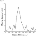
- Example 1 changes of a moving distance of each cell included in the sweat glands shown in Fig. 2 with the passage of time are shown in Fig. 5 .
- Fig. 5 shows the moving distance of the cell A
- (B) shows the moving distance of the cell B
- (C) shows the moving distance of the cell C
- (D) shows the moving distance of the cell D.
- Fig. 6 The results of dynamics of each of sweat glands shown in Example 2 observed by time-lapse imaging based on fluorescence derived from the nuclear staining reagent and fluorescence derived from the cell membrane-staining reagent are shown in Fig. 6 .
- Panels (A1) to (G1) shown in Fig. 6 are images of sweat glands double-stained with the nuclear staining reagent and the cell membrane-staining reagent.
- Panels (A2) to (G2) shown in Fig. 6 are images of sweat glands stained with the nuclear staining reagents corresponding to the panels (A1) to (G1), respectively.
- FIG. 6 are images of sweat glands stained with the cell membrane-staining reagents corresponding to the panels (A1) to (G1), respectively.
- the correspondence relationship between the panels (A1) to (G3) shown in Fig. 6 and the elapsed time from the initiation of observation are shown in Table 2.
- the scale bar shown in Fig. 6 means 50 ⁇ m.
- Example 3(2) The washed whole sweat glands obtained in Example 3(2) were allowed to stand on a glass bottom dish under a stereoscopic microscope (manufactured by Leica, trade name: M125). Next, PBS containing 3% by mass of a low melting point agarose kept at 40°C was added dropwise to the whole sweat glands. The whole sweat glands after dropping the low melting point agarose were allowed to stand for 10 minutes at room temperature (24°C) for 10 minutes, to gelate the low melting point agarose.
- the isotonic solution [extracellular solution (composition: 140 mM sodium chloride, 5 mM potassium chloride, 2 mM calcium chloride, 2 mM magnesium chloride, 10 mM HEPES buffer and 10 mM glucose, pH 7.4)] was added onto the gelated low melting agarose.
- extract solution composition: 140 mM sodium chloride, 5 mM potassium chloride, 2 mM calcium chloride, 2 mM magnesium chloride, 10 mM HEPES buffer and 10 mM glucose, pH 7.4
- Example 1(4) Dynamics of each of sweat glands was observed in the same manner as in Example 1(4) except that in Example 1(4), the observation sample obtained in Example 3(3) was used in place of the observation sample obtained in Example 1(3); in Example 1(4), fluorescence derived from the nuclear staining reagent bound to the nucleus was detected in place of fluorescence derived from the nuclear staining reagent bound to the nucleus and fluorescence derived from the cytoskeleton-staining reagent bound to the cytoskeleton; in Example 1(4), a sweat gland contraction-inducing reagent was added to the whole sweat glands after 10 minutes passed from the initiation of the observation in place of the whole sweat glands after 60 seconds passed from the initiation of the observation. Conditions for time-lapse imaging are as follows:
- FIG. 7 The results of dynamics of each of sweat glands obtained in Example 3 observed by time-lapse imaging based on fluorescence derived from the nuclear staining reagent are shown in Fig. 7 .
- Panels (A1) to (G1) shown in Fig. 7 are images of sweat glands stained with the nuclear staining reagent.
- the correspondence relationship between the panels (A1) to (G1) described in Fig. 7 and the elapsed time from the initiation of observation are shown in Table 3.
- the scale bar shown in Fig. 7 means 100 ⁇ m.
- Elapsed time passed from initiation of observation (sec.) (A1) 0 (B1) 200 (C1) 400 (D1) 600 (E1) 800 (F1) 1000 (G1) 1200
- Example 3 changes of a moving distance of each cell included in sweat glands with the passage of time, which were obtained from the adjacent panels of Fig. 7 are shown in Fig. 8 .
- a cell movement is observed with the passage of time.
- dynamics of each of sweat glands in a living state can be observed by staining the isolated whole sweat glands in a living state using any of a cytoskeleton-staining reagent, a cell membrane-staining reagent and a nuclear staining reagent, and holding the whole sweat glands on a support with agarose so that the position of each of the whole sweat glands is retained.
- the whole sweat glands were isolated from the skin tissues in the same manner as in Example 1(1).
- Example 1(4) Dynamics of each of sweat glands was observed in the same manner as in Example 1(4) except that in Example 1(4), the observation sample obtained in Example 4(3) was used in place of the observation sample obtained in Example 1(3); in Example 1(4), fluorescence derived from the nuclear staining reagent bound to the nucleus was detected in place of fluorescence derived from the nuclear staining reagent bound to the nucleus and fluorescence derived from the cytoskeleton-staining reagent bound to the cytoskeleton; in Example 1(4), a sweat gland contraction-inducing reagent was added to the whole sweat glands after 10 minutes passed from the initiation of the observation in place of the whole sweat glands after 60 seconds passed from the initiation of the observation. Conditions for time-lapse imaging are as follows:
- Panels (A1) to (G1) shown in Fig. 9 are images of sweat glands stained with the nuclear staining reagent.
- the correspondence relationship between the panels (A1) to (G1) shown in Fig. 9 and the elapsed time from the initiation of observation is the same as that between the panel (A1) to (G1) and the elapsed time from the initiation of observation shown in Table 3.
- the scale bar shown in Fig. 9 means 200 ⁇ m.
- Example 4 changes of a moving distance of each cell included in sweat glands with the passage of time, which were obtained from the adjacent panels of Fig. 9 are shown in Fig. 10 .
- aqueous solution containing 200 ⁇ g/mL poly-D-lysine was applied onto the surface of the glass bottom dish.
- the glass bottom dish applied was incubated at 37°C for 2 hours.
- the glass bottom dish after the incubation was washed twice with a sterilized water.
- a poly-lysine-coated glass bottom dish was obtained.
- Example 1(2) the washed whole sweat glands, which was obtained in Example 1(2) was allowed to stand on a poly-lysine-coated glass bottom dish under a stereoscopic microscope (manufactured by Leica, trade name: M125). Thereafter, an isotonic solution (extracellular solution) was added onto the whole sweat glands to prevent the whole sweat glands from drying. Thus, the whole sweat glands were held on the glass bottom dish as a support with poly-D-lysine so that the position of each of the whole sweat glands was retained, to obtain an observation sample.
- Example 1(4) Dynamics of each of sweat glands was observed in the same manner as in Example 1(4) except that in Example 1(4), the observation sample obtained in Example 5(3) was used in place of the observation sample obtained in Example 1(3); in Example 1(4), fluorescence derived from the nuclear staining reagent bound to the nucleus was detected in place of fluorescence derived from the nuclear staining reagent bound to the nucleus and fluorescence derived from the cytoskeleton-staining reagent bound to the cytoskeleton; in Example 1(4), a sweat gland contraction-inducing reagent was added to the whole sweat glands after 10 minutes passed from the initiation of the observation in place of the whole sweat glands after 60 seconds passed from the initiation of the observation. Conditions for time-lapse imaging are as follows:
- Example 5 the results of dynamics of each of sweat glands observed by time-lapse imaging based on fluorescence derived from nuclear staining reagent are shown in Fig. 11 .
- Panels (A1) to (G1) shown in Fig. 11 are images of sweat glands stained with a nuclear staining reagent.
- the correspondence relationship between the panels (A1) to (G1) shown in Fig. 11 and the elapsed time from the initiation of observation is the same as that between the panel (A1) to (G1) and the elapsed time from the initiation of observation shown in Table 3.
- the scale bar shown in Fig. 11 means 200 ⁇ m.
- Example 5 changes of a moving distance of each cell included in the sweat glands with the passage of time, which were obtained from the adjacent panels of Fig. 11 are shown in Fig. 12 .
- aqueous solution containing 200 ⁇ g/mL poly-D-lysine was applied onto the surface of a glass bottom dish.
- the glass bottom dish applied was incubated at 37°C for 30min.
- the glass bottom dish after the incubation was washed twice with a sterilized water.
- a poly-lysine-coated glass bottom dish was obtained.
- aqueous solution containing 20 ⁇ g/mL laminin was applied onto the surface of the poly-lysine-coated glass bottom dish.
- the coated glass bottom dish was incubated at 37°C for 2 hours.
- the glass bottom dish after the incubation was washed twice with a sterilized water.
- a poly-lysine-laminin-coated glass bottom dish was obtained.
- Example 1(2) The washed whole sweat glands obtained in Example 1(2) were allowed to stand on the poly-lysine-laminin-coated glass bottom dish under a stereoscopic microscope (manufactured by Leica, trade name: M125). After that, an isotonic solution (extracellular solution) was added onto the whole sweat glands to prevent the whole sweat glands from drying. Thus, the whole sweat glands were held on the glass bottom dish as a support with poly-D-lysine and laminin in combination so that the position of each of the whole sweat glands was retained, to obtain an observation sample.
- isotonic solution extracellular solution
- Example 1(4) Dynamics of each of sweat glands was observed in the same manner as in Example 1(4) except that in Example 1(4), the observation sample obtained in Example 6(3) was used in place of the observation sample obtained in Example 1(3); in Example 1(4), fluorescence derived from the nuclear staining reagent bound to the nucleus was detected in place of fluorescence derived from the nuclear staining reagent bound to the nucleus and fluorescence derived from the cytoskeleton-staining reagent bound to the cytoskeleton; in Example 1(4) a sweat gland contraction-inducing reagent was added to the whole sweat glands after 10 minutes passed from the initiation of the observation in place of the whole sweat glands after 60 seconds passed from the initiation of the observation. Conditions for time-lapse imaging are as follows:
- Panels (A1) to (G1) shown in Fig. 13 are images of sweat glands stained with a nuclear staining reagent.
- the correspondence relationship between the panels (A1) to (G1) shown in Fig. 13 and the elapsed time from the initiation of observation is the same as that between the panel (A1) to (G1) and the elapsed time from the initiation of observation shown in Table 3.
- the scale bar shown in Fig. 13 means 200 ⁇ m.
- Example 6 changes of a moving distance of each cell included in sweat glands with the passage of time, which were obtained from the adjacent panels of Fig. 13 are shown in Fig. 14 .
- a membrane was obtained by cutting out a membrane part from a product under the trade name of Millicell Cell Culture Insert manufactured by Merck Millipore.
- Example 1(2) the washed whole sweat glands obtained in Example 1(2) were allowed to stand on a glass bottom dish under a stereoscopic microscope (manufactured by Leica, trade name M125).
- the membrane was placed on the whole sweat glands which were allowed to stand on the glass bottom dish.
- an isotonic solution extracellular solution
- the whole sweat glands were held on the glass bottom dish as a support with the membrane so that the position of each of the whole sweat glands was retained, to obtain an observation sample.
- Example 1(4) the observation sample obtained in Example 7(3) was used in place of the observation sample obtained in Example 1(3); in Example 1(4), fluorescence derived from the nuclear staining reagent bound to the nucleus was detected in place of fluorescence derived from the nuclear staining reagent bound to the nucleus and fluorescence derived from the cytoskeleton-staining reagent bound to the cytoskeleton; a sweat gland contraction-inducing reagent was added to the whole sweat glands after 10 minutes passed from the initiation of the observation in place of the whole sweat glands after 60 seconds passed from the initiation of the observation. Conditions for time-lapse imaging are as follows:
- Fig. 15 The results of dynamics of each of sweat glands obtained in Example 7 observed by time-lapse imaging based on fluorescence derived from the nuclear staining reagent are shown in Fig. 15 .
- Panels (A1) to (G1) shown in Fig. 15 are stained images obtained by using the nuclear staining reagent.
- the correspondence relationship between the panels (A1) to (G1) shown in Fig. 15 and the elapsed time from the initiation of observation is the same as that between the panel (A1) to (G1) and the elapsed time from the initiation of observation shown in Table 3.
- the scale bar shown in Fig. 15 means 200 ⁇ m.
- Example 7 changes of a moving distance of each cell included in sweat glands with the passage of time, which were obtained from the adjacent panels of Fig. 15 are shown in Fig. 16 .
- aqueous solution containing 20 ⁇ g/mL laminin was applied onto the surface of a glass bottom dish.
- the glass bottom dish applied was incubated at 37°C for 2 hours.
- the glass bottom dish after the incubation was washed twice with a sterilized water.
- a laminin-coated glass bottom dish was obtained.
- Example 1(2) The washed whole sweat glands obtained in Example 1(2) was allowed to stand on the laminin-coated glass bottom dish under a stereoscopic microscope (manufactured by Leica, trade name: M125). Thereafter, an isotonic solution (extracellular solution) was added onto the whole sweat glands to prevent the whole sweat glands from drying, to obtain an observation sample.
- an isotonic solution extracellular solution
- Example 1(4) Dynamics of each of sweat glands was observed in the same manner as in Example 1(4) except that in Example 1(4), the observation sample obtained in Comparative Example 1(3) was used in place of the observation sample obtained in Example 1(3); in Example 1(4), fluorescence derived from the nuclear staining reagent bound to the nucleus was detected in place of fluorescence derived from the nuclear staining reagent bound to the nucleus and fluorescence derived from the cytoskeleton-staining reagent bound to the cytoskeleton; in Example 1(4), a sweat gland contraction-inducing reagent was added to the whole sweat glands after 10 minutes passed from the initiation of the observation in place of the whole sweat glands after 60 seconds passed from the initiation of the observation.
- dynamics of each of sweat glands in a living state can be observed by using the observation sample which was obtained by staining the isolated whole sweat glands in a living state with any of a cytoskeleton-staining reagent, a cell membrane-staining reagent and a nuclear staining reagent, and holding the whole sweat glands on the support with at least one material selected from the group consisting of collagen, agarose, basement membrane matrix, poly-D-lysine and a membrane so that the position of each of the whole sweat glands is retained.
- pilocarpine which was a sweat gland contraction-inducing reagent (stimulant) was added to the whole sweat glands so as to have a concentration of 10 mM, and thereafter the observation of the whole sweat glands using the time-lapse imaging was continued.
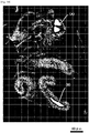
- the determined site (A) is a portion enveloped by myoepithelial cells.
- the determined site (B) and the determined site (C) are portions which are not enveloped by myoepithelial cells, and which are flow pathways for sweat.
- the scale bar in Fig. 17 means 50 ⁇ m.
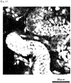
- Determined sites (A) to (C) shown in Fig. 18 correspond to determined sites (A) to (C) shown in Fig. 17 .
- the scale bar means 50 ⁇ m.
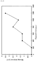
- Example 8 changes over time in each outer diameter index value at the determined sites shown in the drawing-substituted photograph of Fig. 17 are shown in Fig. 19 .
- Fig. 19 shows changes of the outer diameter index value at the determined site of (A) in Fig. 17 with the passage of time
- (B) shows changes of the outer diameter index value at the determined site of (B) in Fig. 17 with the passage of time
- (C) shows changes of the outer diameter index value at the determined site (C) in Fig. 17 with the passage of time.
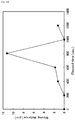
- changes of inner diameter index values at the determined sites shown in the drawing-substituted photograph of Fig. 18 with the passage of time are shown in Fig. 20 .
- Fig. 20 changes of inner diameter index values at the determined sites shown in the drawing-substituted photograph of Fig. 18 with the passage of time are shown in Fig. 20 .
- (A) shows changes of the inner diameter index value at the determined site (A) in Fig. 18 with the passage of time
- (B) shows changes of the inner diameter index value at the determined site (B) with the passage of time
- (C) shows changes in the inner diameter index value at the determined site (C) in Fig. 18 with the passage of time.
- the determined site (A) contracts after stimulating with the stimulant, and thereafter expands, since the outer diameter index value at the determined site (A) decreases after stimulating with the stimulant and then increases.
- the determined site (B) expands after stimulating with the stimulant, since the outer diameter index value at the determined site (B) increases after stimulating with the stimulant.
- the outer diameter index value at the determined site (C) hardly changes after stimulating with the stimulant.
- the determined site (A) contracts after stimulating with the stimulant, and thereafter expands, since the inner diameter index value at the determined site (A) greatly decreases after stimulating with the stimulant and then increases.
- the determined site (B) expands after passing the time from the termination of the stimulation with the stimulant, since the outer diameter index value at the determined site (B) hardly changes immediately after stimulating with the stimulant, but the outer diameter index value increases when passing a certain time.
- the determined site (C) expands after passing the time from the termination of the stimulation with the stimulant, since the outer diameter index value at the determined site (C) hardly changes immediately after stimulating with the stimulant, but the outer diameter index value increases when passing a certain time.
- Example 9 changes of a moving distance of each cell included in the sweat glands of Experiment number 1 with the passage of time are shown in Fig. 23 . Changes of a moving distance of each cell included in the sweat glands of Experiment number 2 with the passage of time are shown in Fig. 24 .
- test substance has a perspiration-controlling action such as an antiperspirant action or the like is evaluated by using the observation sample obtained by staining the isolated sweat glands in a living state with a staining reagent, and holding the whole sweat glands on a support so that the position of each of the whole sweat glands is retained by using at least one material selected from the group consisting of collagen, agarose, basement membrane matrix, poly-D-lysine and a membrane.
Landscapes
- Health & Medical Sciences (AREA)
- Life Sciences & Earth Sciences (AREA)
- Engineering & Computer Science (AREA)
- Biomedical Technology (AREA)
- Molecular Biology (AREA)
- Immunology (AREA)
- Pathology (AREA)
- General Health & Medical Sciences (AREA)
- Physics & Mathematics (AREA)
- Urology & Nephrology (AREA)
- Hematology (AREA)
- Chemical & Material Sciences (AREA)
- Cell Biology (AREA)
- Public Health (AREA)
- Surgery (AREA)
- Biophysics (AREA)
- Medical Informatics (AREA)
- Veterinary Medicine (AREA)
- Heart & Thoracic Surgery (AREA)
- Animal Behavior & Ethology (AREA)
- Biochemistry (AREA)
- General Physics & Mathematics (AREA)
- Analytical Chemistry (AREA)
- Microbiology (AREA)
- Biotechnology (AREA)
- Medicinal Chemistry (AREA)
- Food Science & Technology (AREA)
- Gastroenterology & Hepatology (AREA)
- Physiology (AREA)
- Endocrinology (AREA)
- Tropical Medicine & Parasitology (AREA)
- Toxicology (AREA)
- Bioinformatics & Cheminformatics (AREA)
- Investigating Or Analysing Biological Materials (AREA)
Claims (8)
- Procédé d'observation de la dynamique d'une glande sudorale, comprenant les étapes suivantes :(A) coloration d'une glande sudorale complète isolée avec au moins un réactif colorant sélectionné dans le groupe composé d'un réactif colorant pour membrane cellulaire, d'un réactif colorant pour noyau et d'un réactif colorant pour composant cytosquelettique,(B) retenue de la glande sudorale complète colorée dans l'étape (A) sur un support avec au moins un matériau sélectionné dans le groupe composé de collagène, agarose, matrice membranaire de base, poly-D-lysine et une membrane de manière à retenir la position de la glande sudorale complète colorée dans l'étape (A), afin d'obtenir un échantillon d'observation, et(C) observation de la dynamique de la glande sudorale complète contenue dans l'échantillon d'observation obtenu dans l'étape (B).
- Procédé d'observation de la dynamique d'une glande sudorale selon la revendication 1, dans lequel, dans l'étape (B), un tampon est ajouté sur la glande sudorale complète retenue sur le support.
- Procédé d'observation de la dynamique d'une glande sudorale selon la revendication 2, dans lequel le tampon est un tampon contenant 100 à 150 mM de chlorure de sodium, 3 à 7 mM de chlorure de potassium, 0,5 à 2 mM de chlorure de calcium, 0,5 à 2 mM de chlorure de magnésium, 20 à 30 mM de bicarbonate de soude, 0,5 à 2 mM de sodium dihydrogène phosphate, 2 à 8 mM de glucose et 5 à 15 mg/100 ml d'albumine de sérum bovin exempt d'acides gras, et ayant un pH de 7,1 à 7,6.
- Procédé d'observation de la dynamique d'une glande sudorale selon l'une quelconque des revendications 1 à 3, dans lequel, dans l'étape (C), la glande sudorale complète contenue dans l'échantillon d'observation est mise en contact avec un stimulant, et la dynamique de la glande sudorale complète causée par le stimulant est observée.
- Procédé d'évaluation d'une substance de test, dans lequel on évalue si la substance de test a une action de contrôle de transpiration ou non, ce procédé comprenant les étapes suivantes :(a) coloration d'une glande sudorale complète isolée avec au moins un réactif colorant sélectionné dans le groupe composé d'un réactif colorant pour membrane cellulaire, d'un réactif colorant pour noyau et d'un réactif colorant pour composant cytosquelettique,(b) retenue de la glande sudorale complète colorée dans l'étape (a) sur un support avec au moins un matériau sélectionné dans le groupe composé de collagène, agarose, matrice membranaire de base, poly-D-lysine et une membrane de manière à retenir la position de la glande sudorale complète colorée dans l'étape (a), afin d'obtenir un échantillon d'observation,(c) mise en contact de la glande sudorale complète contenue dans l'échantillon d'observation avec la substance de test pour observer la dynamique de la glande sudorale complète,(d) évaluation si la substance de test a une action de contrôle de la transpiration en se basant sur la dynamique de la glande sudorale complète observée dans l'étape (c).
- Procédé d'évaluation d'une substance de test selon la revendication 5, dans lequel, dans l'étape (b), un tampon est ajouté sur la glande sudorale complète retenue sur le support.
- Procédé d'évaluation d'une substance de test selon la revendication 6, dans lequel le tampon est un tampon contenant 100 à 150 mM de chlorure de sodium, 3 à 7 mM de chlorure de potassium, 0,5 à 2 mM de chlorure de calcium, 0,5 à 2 mM de chlorure de magnésium, 20 à 30 mM de bicarbonate de soude, 0,5 à 2 mM de sodium dihydrogène phosphate, 2 à 8 mM de glucose et 5 à 15 mg/100 ml d'albumine de sérum bovin exempt d'acides gras, et ayant un pH de 7,1 à 7,6.
- Procédé d'évaluation d'une substance de test selon l'une quelconque des revendications 5 à 7, dans lequel, dans l'étape (c), la glande sudorale complète contenue dans l'échantillon d'observation est mise en contact avec la substance de test et un stimulant, et la dynamique de la glande sudorale complète est observée.
Applications Claiming Priority (2)
| Application Number | Priority Date | Filing Date | Title |
|---|---|---|---|
| JP2016141740 | 2016-07-19 | ||
| PCT/JP2017/026001 WO2018016501A1 (fr) | 2016-07-19 | 2017-07-18 | Procédé d'observation de la dynamique des glandes sudorales |
Publications (3)
| Publication Number | Publication Date |
|---|---|
| EP3489681A1 EP3489681A1 (fr) | 2019-05-29 |
| EP3489681A4 EP3489681A4 (fr) | 2020-04-22 |
| EP3489681B1 true EP3489681B1 (fr) | 2021-07-07 |
Family
ID=60992434
Family Applications (1)
| Application Number | Title | Priority Date | Filing Date |
|---|---|---|---|
| EP17831017.3A Active EP3489681B1 (fr) | 2016-07-19 | 2017-07-18 | Procédé d'observation de la dynamique des glandes sudorales |
Country Status (4)
| Country | Link |
|---|---|
| US (1) | US11085916B2 (fr) |
| EP (1) | EP3489681B1 (fr) |
| JP (1) | JP6276487B1 (fr) |
| WO (1) | WO2018016501A1 (fr) |
Families Citing this family (2)
| Publication number | Priority date | Publication date | Assignee | Title |
|---|---|---|---|---|
| EP3869194B1 (fr) * | 2018-10-17 | 2024-01-10 | Mandom Corporation | Procédé d'observation de glande sébacée et son utilisation |
| JP7296785B2 (ja) * | 2019-05-29 | 2023-06-23 | 株式会社マンダム | アポクリン汗腺の動態の観察方法、および被験物質の評価方法 |
Family Cites Families (6)
| Publication number | Priority date | Publication date | Assignee | Title |
|---|---|---|---|---|
| RU2322497C2 (ru) * | 2000-11-30 | 2008-04-20 | Стемрон Инк. | Изолированная гомозиготная стволовая клетка (варианты), способ ее получения (варианты), способ получения требуемой клетки-родоначальника, дифференцированной клетки, группы дифференцированных клеток или типа ткани |
| JP4959710B2 (ja) * | 2005-11-01 | 2012-06-27 | レンセレアー ポリテクニック インスティテュート | 毒物学アッセイのための3次元細胞アレイチップおよびプラットフォーム |
| GB0705738D0 (en) * | 2007-03-26 | 2007-05-02 | Unilever Plc | Apocrine cell line |
| JP6044950B2 (ja) * | 2012-09-27 | 2016-12-14 | 株式会社マンダム | 汗腺細胞の検出方法 |
| JP6238397B2 (ja) * | 2013-08-01 | 2017-11-29 | 国立大学法人 香川大学 | 細胞空間分画装置および微細構造刃 |
| US20160075995A1 (en) * | 2014-09-17 | 2016-03-17 | Krzysztof Kobielak | Sweat gland-derived stem cells and methods of use |
-
2017
- 2017-07-18 US US16/085,755 patent/US11085916B2/en active Active
- 2017-07-18 WO PCT/JP2017/026001 patent/WO2018016501A1/fr unknown
- 2017-07-18 EP EP17831017.3A patent/EP3489681B1/fr active Active
- 2017-07-18 JP JP2017552522A patent/JP6276487B1/ja active Active
Also Published As
| Publication number | Publication date |
|---|---|
| JPWO2018016501A1 (ja) | 2018-07-19 |
| US20190049429A1 (en) | 2019-02-14 |
| WO2018016501A1 (fr) | 2018-01-25 |
| EP3489681A4 (fr) | 2020-04-22 |
| EP3489681A1 (fr) | 2019-05-29 |
| JP6276487B1 (ja) | 2018-02-07 |
| US11085916B2 (en) | 2021-08-10 |
Similar Documents
| Publication | Publication Date | Title |
|---|---|---|
| Suleiman et al. | Injury-induced actin cytoskeleton reorganization in podocytes revealed by super-resolution microscopy | |
| Maresca et al. | Methods for studying spindle assembly and chromosome condensation in Xenopus egg extracts | |
| Williams et al. | Reduction of diffusion barriers in isolated rat islets improves survival, but not insulin secretion or transplantation outcome | |
| EP3489681B1 (fr) | Procédé d'observation de la dynamique des glandes sudorales | |
| JP2950519B2 (ja) | 皮膚の未変性状態組織培養法 | |
| KR20110091805A (ko) | 동물 간 세포의 배양 방법 | |
| Cobbs et al. | Methods for the detection of cytomegalovirus in glioblastoma cells and tissues | |
| Garofalo et al. | Calmodulin antagonists inhibit secretion in Paramecium. | |
| Pena et al. | Detection of early changes in sperm membrane integrity pre-freezing can estimate post-thaw quality of boar spermatozoa | |
| EP3498823A1 (fr) | Échantillon de peau transparent | |
| Ktistakis | Monitoring the localization of MAP1LC3B by indirect immunofluorescence | |
| Sartelet et al. | Hyperexpression of ICAM‐1 and CD36 in placentas infected with Plasmodium falciparum: a possible role of these molecules in sequestration of infected red blood cells in placentas | |
| Kilic et al. | Methods to evaluate the formation and stabilization of blood vessels and their role in tumor growth and metastasis | |
| KR20210119210A (ko) | 오가노이드 투명화 키트, 이를 이용한 오가노이드 투명화 방법 및 3차원 이미지화를 위한 면역염색 방법 | |
| Bernard‐Trifilo et al. | Analyzing FAK and Pyk2 in early integrin signaling events | |
| JP6692593B1 (ja) | 皮脂腺の観察方法およびその利用 | |
| EP3614145A1 (fr) | Procédé d'observation de glande sébacée | |
| US6399380B1 (en) | Native-state histoculturing skin extracellular support | |
| Sigot-Luizard et al. | In vitro cytocompatibility tests | |
| Smrt et al. | Primary culture of Drosophila larval neurons with morphological analysis using NeuronMetrics | |
| DE10242066A1 (de) | Verfahren zur Behandlung von Zellkulturen | |
| Boyle et al. | Permeabilization by streptolysin-O reveals a role for calcium-dependent protein kinase C isoforms alpha and beta in the response of cultured cardiomyocytes to hyposmotic challenge | |
| Ahmed et al. | Forensic Biology | |
| Idelfonso-García et al. | Protocol to detect senescence-associated β-galactosidase and immunoperoxidase activity in fresh-frozen murine tissues | |
| WO1999051763A1 (fr) | Systeme de culture d'organes de la cornee destine a des essais de toxicite chimique de produits de consommation |
Legal Events
| Date | Code | Title | Description |
|---|---|---|---|
| STAA | Information on the status of an ep patent application or granted ep patent |
Free format text: STATUS: THE INTERNATIONAL PUBLICATION HAS BEEN MADE |
|
| PUAI | Public reference made under article 153(3) epc to a published international application that has entered the european phase |
Free format text: ORIGINAL CODE: 0009012 |
|
| STAA | Information on the status of an ep patent application or granted ep patent |
Free format text: STATUS: REQUEST FOR EXAMINATION WAS MADE |
|
| 17P | Request for examination filed |
Effective date: 20180924 |
|
| AK | Designated contracting states |
Kind code of ref document: A1 Designated state(s): AL AT BE BG CH CY CZ DE DK EE ES FI FR GB GR HR HU IE IS IT LI LT LU LV MC MK MT NL NO PL PT RO RS SE SI SK SM TR |
|
| AX | Request for extension of the european patent |
Extension state: BA ME |
|
| DAV | Request for validation of the european patent (deleted) | ||
| DAX | Request for extension of the european patent (deleted) | ||
| A4 | Supplementary search report drawn up and despatched |
Effective date: 20200319 |
|
| RIC1 | Information provided on ipc code assigned before grant |
Ipc: G01N 33/50 20060101AFI20200313BHEP Ipc: G01N 33/48 20060101ALI20200313BHEP |
|
| GRAP | Despatch of communication of intention to grant a patent |
Free format text: ORIGINAL CODE: EPIDOSNIGR1 |
|
| STAA | Information on the status of an ep patent application or granted ep patent |
Free format text: STATUS: GRANT OF PATENT IS INTENDED |
|
| INTG | Intention to grant announced |
Effective date: 20210122 |
|
| GRAS | Grant fee paid |
Free format text: ORIGINAL CODE: EPIDOSNIGR3 |
|
| GRAA | (expected) grant |
Free format text: ORIGINAL CODE: 0009210 |
|
| STAA | Information on the status of an ep patent application or granted ep patent |
Free format text: STATUS: THE PATENT HAS BEEN GRANTED |
|
| AK | Designated contracting states |
Kind code of ref document: B1 Designated state(s): AL AT BE BG CH CY CZ DE DK EE ES FI FR GB GR HR HU IE IS IT LI LT LU LV MC MK MT NL NO PL PT RO RS SE SI SK SM TR |
|
| REG | Reference to a national code |
Ref country code: GB Ref legal event code: FG4D |
|
| REG | Reference to a national code |
Ref country code: AT Ref legal event code: REF Ref document number: 1409087 Country of ref document: AT Kind code of ref document: T Effective date: 20210715 |
|
| REG | Reference to a national code |
Ref country code: DE Ref legal event code: R096 Ref document number: 602017041828 Country of ref document: DE |
|
| REG | Reference to a national code |
Ref country code: IE Ref legal event code: FG4D |
|
| REG | Reference to a national code |
Ref country code: LT Ref legal event code: MG9D |
|
| REG | Reference to a national code |
Ref country code: NL Ref legal event code: MP Effective date: 20210707 |
|
| REG | Reference to a national code |
Ref country code: AT Ref legal event code: MK05 Ref document number: 1409087 Country of ref document: AT Kind code of ref document: T Effective date: 20210707 |
|
| PG25 | Lapsed in a contracting state [announced via postgrant information from national office to epo] |
Ref country code: BG Free format text: LAPSE BECAUSE OF FAILURE TO SUBMIT A TRANSLATION OF THE DESCRIPTION OR TO PAY THE FEE WITHIN THE PRESCRIBED TIME-LIMIT Effective date: 20211007 Ref country code: AT Free format text: LAPSE BECAUSE OF FAILURE TO SUBMIT A TRANSLATION OF THE DESCRIPTION OR TO PAY THE FEE WITHIN THE PRESCRIBED TIME-LIMIT Effective date: 20210707 Ref country code: LT Free format text: LAPSE BECAUSE OF FAILURE TO SUBMIT A TRANSLATION OF THE DESCRIPTION OR TO PAY THE FEE WITHIN THE PRESCRIBED TIME-LIMIT Effective date: 20210707 Ref country code: NL Free format text: LAPSE BECAUSE OF FAILURE TO SUBMIT A TRANSLATION OF THE DESCRIPTION OR TO PAY THE FEE WITHIN THE PRESCRIBED TIME-LIMIT Effective date: 20210707 Ref country code: PT Free format text: LAPSE BECAUSE OF FAILURE TO SUBMIT A TRANSLATION OF THE DESCRIPTION OR TO PAY THE FEE WITHIN THE PRESCRIBED TIME-LIMIT Effective date: 20211108 Ref country code: NO Free format text: LAPSE BECAUSE OF FAILURE TO SUBMIT A TRANSLATION OF THE DESCRIPTION OR TO PAY THE FEE WITHIN THE PRESCRIBED TIME-LIMIT Effective date: 20211007 Ref country code: SE Free format text: LAPSE BECAUSE OF FAILURE TO SUBMIT A TRANSLATION OF THE DESCRIPTION OR TO PAY THE FEE WITHIN THE PRESCRIBED TIME-LIMIT Effective date: 20210707 Ref country code: RS Free format text: LAPSE BECAUSE OF FAILURE TO SUBMIT A TRANSLATION OF THE DESCRIPTION OR TO PAY THE FEE WITHIN THE PRESCRIBED TIME-LIMIT Effective date: 20210707 Ref country code: HR Free format text: LAPSE BECAUSE OF FAILURE TO SUBMIT A TRANSLATION OF THE DESCRIPTION OR TO PAY THE FEE WITHIN THE PRESCRIBED TIME-LIMIT Effective date: 20210707 Ref country code: ES Free format text: LAPSE BECAUSE OF FAILURE TO SUBMIT A TRANSLATION OF THE DESCRIPTION OR TO PAY THE FEE WITHIN THE PRESCRIBED TIME-LIMIT Effective date: 20210707 Ref country code: FI Free format text: LAPSE BECAUSE OF FAILURE TO SUBMIT A TRANSLATION OF THE DESCRIPTION OR TO PAY THE FEE WITHIN THE PRESCRIBED TIME-LIMIT Effective date: 20210707 |
|
| PG25 | Lapsed in a contracting state [announced via postgrant information from national office to epo] |
Ref country code: PL Free format text: LAPSE BECAUSE OF FAILURE TO SUBMIT A TRANSLATION OF THE DESCRIPTION OR TO PAY THE FEE WITHIN THE PRESCRIBED TIME-LIMIT Effective date: 20210707 Ref country code: LV Free format text: LAPSE BECAUSE OF FAILURE TO SUBMIT A TRANSLATION OF THE DESCRIPTION OR TO PAY THE FEE WITHIN THE PRESCRIBED TIME-LIMIT Effective date: 20210707 Ref country code: GR Free format text: LAPSE BECAUSE OF FAILURE TO SUBMIT A TRANSLATION OF THE DESCRIPTION OR TO PAY THE FEE WITHIN THE PRESCRIBED TIME-LIMIT Effective date: 20211008 |
|
| REG | Reference to a national code |
Ref country code: CH Ref legal event code: PL |
|
| REG | Reference to a national code |
Ref country code: BE Ref legal event code: MM Effective date: 20210731 |
|
| REG | Reference to a national code |
Ref country code: DE Ref legal event code: R097 Ref document number: 602017041828 Country of ref document: DE |
|
| PG25 | Lapsed in a contracting state [announced via postgrant information from national office to epo] |
Ref country code: LI Free format text: LAPSE BECAUSE OF NON-PAYMENT OF DUE FEES Effective date: 20210731 Ref country code: DK Free format text: LAPSE BECAUSE OF FAILURE TO SUBMIT A TRANSLATION OF THE DESCRIPTION OR TO PAY THE FEE WITHIN THE PRESCRIBED TIME-LIMIT Effective date: 20210707 Ref country code: CH Free format text: LAPSE BECAUSE OF NON-PAYMENT OF DUE FEES Effective date: 20210731 |
|
| PLBE | No opposition filed within time limit |
Free format text: ORIGINAL CODE: 0009261 |
|
| STAA | Information on the status of an ep patent application or granted ep patent |
Free format text: STATUS: NO OPPOSITION FILED WITHIN TIME LIMIT |
|
| PG25 | Lapsed in a contracting state [announced via postgrant information from national office to epo] |
Ref country code: SM Free format text: LAPSE BECAUSE OF FAILURE TO SUBMIT A TRANSLATION OF THE DESCRIPTION OR TO PAY THE FEE WITHIN THE PRESCRIBED TIME-LIMIT Effective date: 20210707 Ref country code: SK Free format text: LAPSE BECAUSE OF FAILURE TO SUBMIT A TRANSLATION OF THE DESCRIPTION OR TO PAY THE FEE WITHIN THE PRESCRIBED TIME-LIMIT Effective date: 20210707 Ref country code: RO Free format text: LAPSE BECAUSE OF FAILURE TO SUBMIT A TRANSLATION OF THE DESCRIPTION OR TO PAY THE FEE WITHIN THE PRESCRIBED TIME-LIMIT Effective date: 20210707 Ref country code: MC Free format text: LAPSE BECAUSE OF FAILURE TO SUBMIT A TRANSLATION OF THE DESCRIPTION OR TO PAY THE FEE WITHIN THE PRESCRIBED TIME-LIMIT Effective date: 20210707 Ref country code: LU Free format text: LAPSE BECAUSE OF NON-PAYMENT OF DUE FEES Effective date: 20210718 Ref country code: EE Free format text: LAPSE BECAUSE OF FAILURE TO SUBMIT A TRANSLATION OF THE DESCRIPTION OR TO PAY THE FEE WITHIN THE PRESCRIBED TIME-LIMIT Effective date: 20210707 Ref country code: CZ Free format text: LAPSE BECAUSE OF FAILURE TO SUBMIT A TRANSLATION OF THE DESCRIPTION OR TO PAY THE FEE WITHIN THE PRESCRIBED TIME-LIMIT Effective date: 20210707 Ref country code: AL Free format text: LAPSE BECAUSE OF FAILURE TO SUBMIT A TRANSLATION OF THE DESCRIPTION OR TO PAY THE FEE WITHIN THE PRESCRIBED TIME-LIMIT Effective date: 20210707 |
|
| 26N | No opposition filed |
Effective date: 20220408 |
|
| PG25 | Lapsed in a contracting state [announced via postgrant information from national office to epo] |
Ref country code: IT Free format text: LAPSE BECAUSE OF FAILURE TO SUBMIT A TRANSLATION OF THE DESCRIPTION OR TO PAY THE FEE WITHIN THE PRESCRIBED TIME-LIMIT Effective date: 20210707 Ref country code: IE Free format text: LAPSE BECAUSE OF NON-PAYMENT OF DUE FEES Effective date: 20210718 Ref country code: BE Free format text: LAPSE BECAUSE OF NON-PAYMENT OF DUE FEES Effective date: 20210731 |
|
| PG25 | Lapsed in a contracting state [announced via postgrant information from national office to epo] |
Ref country code: CY Free format text: LAPSE BECAUSE OF FAILURE TO SUBMIT A TRANSLATION OF THE DESCRIPTION OR TO PAY THE FEE WITHIN THE PRESCRIBED TIME-LIMIT Effective date: 20210707 |
|
| PG25 | Lapsed in a contracting state [announced via postgrant information from national office to epo] |
Ref country code: HU Free format text: LAPSE BECAUSE OF FAILURE TO SUBMIT A TRANSLATION OF THE DESCRIPTION OR TO PAY THE FEE WITHIN THE PRESCRIBED TIME-LIMIT; INVALID AB INITIO Effective date: 20170718 |
|
| PGFP | Annual fee paid to national office [announced via postgrant information from national office to epo] |
Ref country code: GB Payment date: 20230721 Year of fee payment: 7 |
|
| PGFP | Annual fee paid to national office [announced via postgrant information from national office to epo] |
Ref country code: FR Payment date: 20230726 Year of fee payment: 7 Ref country code: DE Payment date: 20230719 Year of fee payment: 7 |