EP3458477B1 - Anti-factor ix padua antibodies - Google Patents
Anti-factor ix padua antibodies Download PDFInfo
- Publication number
- EP3458477B1 EP3458477B1 EP17730590.1A EP17730590A EP3458477B1 EP 3458477 B1 EP3458477 B1 EP 3458477B1 EP 17730590 A EP17730590 A EP 17730590A EP 3458477 B1 EP3458477 B1 EP 3458477B1
- Authority
- EP
- European Patent Office
- Prior art keywords
- antibody
- antigen
- fix
- binding
- binding fragment
- Prior art date
- Legal status (The legal status is an assumption and is not a legal conclusion. Google has not performed a legal analysis and makes no representation as to the accuracy of the status listed.)
- Active
Links
Images


Classifications
-
- C—CHEMISTRY; METALLURGY
- C07—ORGANIC CHEMISTRY
- C07K—PEPTIDES
- C07K16/00—Immunoglobulins [IG], e.g. monoclonal or polyclonal antibodies
- C07K16/18—Immunoglobulins [IG], e.g. monoclonal or polyclonal antibodies against material from animals or humans
- C07K16/36—Immunoglobulins [IG], e.g. monoclonal or polyclonal antibodies against material from animals or humans against blood coagulation factors
-
- C—CHEMISTRY; METALLURGY
- C07—ORGANIC CHEMISTRY
- C07K—PEPTIDES
- C07K16/00—Immunoglobulins [IG], e.g. monoclonal or polyclonal antibodies
- C07K16/005—Immunoglobulins [IG], e.g. monoclonal or polyclonal antibodies constructed by phage libraries
-
- G—PHYSICS
- G01—MEASURING; TESTING
- G01N—INVESTIGATING OR ANALYSING MATERIALS BY DETERMINING THEIR CHEMICAL OR PHYSICAL PROPERTIES
- G01N33/00—Investigating or analysing materials by specific methods not covered by groups G01N1/00 - G01N31/00
- G01N33/48—Biological material, e.g. blood, urine; Haemocytometers
- G01N33/50—Chemical analysis of biological material, e.g. blood, urine; Testing involving biospecific ligand binding methods; Immunological testing
- G01N33/53—Immunoassay; Biospecific binding assay; Materials therefor
- G01N33/573—Immunoassay; Biospecific binding assay; Materials therefor for enzymes or isoenzymes
-
- G—PHYSICS
- G01—MEASURING; TESTING
- G01N—INVESTIGATING OR ANALYSING MATERIALS BY DETERMINING THEIR CHEMICAL OR PHYSICAL PROPERTIES
- G01N33/00—Investigating or analysing materials by specific methods not covered by groups G01N1/00 - G01N31/00
- G01N33/48—Biological material, e.g. blood, urine; Haemocytometers
- G01N33/50—Chemical analysis of biological material, e.g. blood, urine; Testing involving biospecific ligand binding methods; Immunological testing
- G01N33/86—Chemical analysis of biological material, e.g. blood, urine; Testing involving biospecific ligand binding methods; Immunological testing involving blood coagulating time or factors, or their receptors
-
- C—CHEMISTRY; METALLURGY
- C07—ORGANIC CHEMISTRY
- C07K—PEPTIDES
- C07K2317/00—Immunoglobulins specific features
- C07K2317/20—Immunoglobulins specific features characterized by taxonomic origin
- C07K2317/21—Immunoglobulins specific features characterized by taxonomic origin from primates, e.g. man
-
- C—CHEMISTRY; METALLURGY
- C07—ORGANIC CHEMISTRY
- C07K—PEPTIDES
- C07K2317/00—Immunoglobulins specific features
- C07K2317/30—Immunoglobulins specific features characterized by aspects of specificity or valency
- C07K2317/33—Crossreactivity, e.g. for species or epitope, or lack of said crossreactivity
-
- C—CHEMISTRY; METALLURGY
- C07—ORGANIC CHEMISTRY
- C07K—PEPTIDES
- C07K2317/00—Immunoglobulins specific features
- C07K2317/30—Immunoglobulins specific features characterized by aspects of specificity or valency
- C07K2317/34—Identification of a linear epitope shorter than 20 amino acid residues or of a conformational epitope defined by amino acid residues
-
- C—CHEMISTRY; METALLURGY
- C07—ORGANIC CHEMISTRY
- C07K—PEPTIDES
- C07K2317/00—Immunoglobulins specific features
- C07K2317/30—Immunoglobulins specific features characterized by aspects of specificity or valency
- C07K2317/35—Valency
-
- C—CHEMISTRY; METALLURGY
- C07—ORGANIC CHEMISTRY
- C07K—PEPTIDES
- C07K2317/00—Immunoglobulins specific features
- C07K2317/50—Immunoglobulins specific features characterized by immunoglobulin fragments
- C07K2317/55—Fab or Fab'
-
- C—CHEMISTRY; METALLURGY
- C07—ORGANIC CHEMISTRY
- C07K—PEPTIDES
- C07K2317/00—Immunoglobulins specific features
- C07K2317/70—Immunoglobulins specific features characterized by effect upon binding to a cell or to an antigen
- C07K2317/76—Antagonist effect on antigen, e.g. neutralization or inhibition of binding
-
- C—CHEMISTRY; METALLURGY
- C07—ORGANIC CHEMISTRY
- C07K—PEPTIDES
- C07K2317/00—Immunoglobulins specific features
- C07K2317/90—Immunoglobulins specific features characterized by (pharmaco)kinetic aspects or by stability of the immunoglobulin
- C07K2317/92—Affinity (KD), association rate (Ka), dissociation rate (Kd) or EC50 value
-
- C—CHEMISTRY; METALLURGY
- C07—ORGANIC CHEMISTRY
- C07K—PEPTIDES
- C07K2319/00—Fusion polypeptide
- C07K2319/20—Fusion polypeptide containing a tag with affinity for a non-protein ligand
- C07K2319/21—Fusion polypeptide containing a tag with affinity for a non-protein ligand containing a His-tag
-
- C—CHEMISTRY; METALLURGY
- C07—ORGANIC CHEMISTRY
- C07K—PEPTIDES
- C07K2319/00—Fusion polypeptide
- C07K2319/40—Fusion polypeptide containing a tag for immunodetection, or an epitope for immunisation
- C07K2319/43—Fusion polypeptide containing a tag for immunodetection, or an epitope for immunisation containing a FLAG-tag
-
- G—PHYSICS
- G01—MEASURING; TESTING
- G01N—INVESTIGATING OR ANALYSING MATERIALS BY DETERMINING THEIR CHEMICAL OR PHYSICAL PROPERTIES
- G01N2333/00—Assays involving biological materials from specific organisms or of a specific nature
- G01N2333/90—Enzymes; Proenzymes
- G01N2333/914—Hydrolases (3)
- G01N2333/948—Hydrolases (3) acting on peptide bonds (3.4)
- G01N2333/95—Proteinases, i.e. endopeptidases (3.4.21-3.4.99)
- G01N2333/964—Proteinases, i.e. endopeptidases (3.4.21-3.4.99) derived from animal tissue
- G01N2333/96425—Proteinases, i.e. endopeptidases (3.4.21-3.4.99) derived from animal tissue from mammals
- G01N2333/96427—Proteinases, i.e. endopeptidases (3.4.21-3.4.99) derived from animal tissue from mammals in general
- G01N2333/9643—Proteinases, i.e. endopeptidases (3.4.21-3.4.99) derived from animal tissue from mammals in general with EC number
- G01N2333/96433—Serine endopeptidases (3.4.21)
- G01N2333/96441—Serine endopeptidases (3.4.21) with definite EC number
- G01N2333/9645—Factor IX (3.4.21.22)
Definitions
- CRM-positive (CRM+) patients express, for example, wild-type (WT) Factor IX, which cross-reacts with currently available FIX Padua-binding agents, thus making it difficult to determine if FIX Padua is expressed by such CRM+ patients.
- WT wild-type Factor IX
- EP 2 881 463 A1 states that it relates to variants of FIX or activated FIX, wherein the variant is characterized in that it has clotting activity in absence of its cofactor.
- WO 2016/004113 A1 states that it provides codon optimized FIX sequences.
- references in the description to methods of treatment refer to the compounds, pharmaceutical compositions and medicaments of the present invention for use in a method for treatment of the human (or animal) body by therapy (or for diagnosis).
- binding constructs which specifically recognize FIX Padua (FIXp) without cross-reactivity to wild-type FIX.
- the binding construct is an antibody or an antigen-binding fragment thereof that binds FIX Padua comprising the amino acid sequence of SEQ ID NO: 1 and does not bind to a WT Factor IX comprising the amino acid sequence SEQ ID NO: 2.
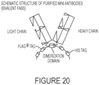
- the binding construct is a polypeptide comprising an amino acid sequence comprising each of SEQ ID NOs: 6-11, optionally, wherein (i) one or more amino acids are present between each of SEQ ID NOs: 6-11, and/or (ii) the polypeptide optionally further comprises a FLAG tag comprising DYKDDDDK (SEQ ID NO: 12) and/or a hexa-His tag comprising HHHHHH (SEQ ID NO: 13), optionally, wherein the FLAG tag and/or the hexa-His tag are located at the C-terminal end of the polypeptide.
- the binding construct is a conjugate comprising an antigen-binding fragment as described herein conjugated to (i) a constant region of an immunoglobulin heavy chain, (ii) a constant region of an immunoglobulin light chain, or (iii) both a constant region of an immunoglobulin heavy chain and a constant region of an immunoglobulin light chain.
- the binding construct is a conjugate comprising an antibody or antigen-binding fragment as described herein linked or conjugated to a heterologous moiety.
- the conjugate comprises an antibody or antigen-binding fragment as described herein conjugated to a polymer, a carbohydrate, a lipid, a nucleic acid, an oligonucleotide, an amino acid, peptide, polypeptide, protein, or a detecting agent.
- Nucleic acids comprising a nucleotide sequence encoding the antibody, antigen-binding fragment, polypeptide, conjugate, or a fragment thereof, as described herein, are additionally provided.
- Vectors comprising the nucleic acid and host cells comprising the nucleic acid or vector are further provided.
- kits comprising an antibody, antigen-binding fragment, polypeptide, conjugate, nucleic acid, vector, host cell, as described herein, or a combination thereof, and, optionally, instructions for use.
- the kit also comprises a solid support and optionally, the antibody, antigen-binding fragment, polypeptide, or conjugate is pre-coated on the solid support.
- the kit also comprises a secondary antibody which binds to the antibody, antigen-binding fragment, polypeptide, or conjugate provided in the kit.
- the invention further provides compositions comprising an antibody or antigen-binding fragment or polypeptide or conjugate, as described herein, admixed with a biological sample, e.g., a biological sample obtained from a human.
- a biological sample e.g., a biological sample obtained from a human.
- the biological sample comprises human plasma, or a diluted fraction thereof, and/or human tissue, or cells thereof.
- the biological sample comprises human plasma proteins, wherein at least one of the human plasma proteins is selected from the group consisting of Factor IX, Factor II, and Factor X, and variants thereof.
- the composition comprises a detecting agent.
- binding constructs provided herein are useful in, e.g., detection methods that allow for unambiguous or specific detection of FIX Padua in samples, e.g., clinical or preclinical samples comprising, e.g., wild-type FIX. Accordingly, provided herein are methods of detecting Factor IX Padua comprising the amino acid sequence of SEQ ID NO: 1 in a sample obtained from a subject.
- the method comprises (i) contacting the sample with a binding construct (e.g., an antibody, antigen-binding fragment, polypeptide or conjugate, as described herein) to form a complex, e.g., an immunocomplex, comprising the FIXp and the binding construct, and (ii) detecting the complex in the sample.
- a binding construct e.g., an antibody, antigen-binding fragment, polypeptide or conjugate, as described herein
- a complex e.g., an immunocomplex, comprising the FIXp and the binding construct
- binding constructs which specifically recognize FIX Padua with minimal or no cross-reactivity to wild-type FIX.
- the binding constructs bind to FIX Padua and do not bind to wild-type (WT) Factor IX.
- the binding constructs bind to FIX Padua (and not to WT Factor IX) in the presence of WT Factor IX.
- the binding constructs bind to FIX Padua and do not bind to one WT Factor IX and one or both of Factor II and Factor X, or any other mutated or modified forms thereof (except FIX Padua) under similar or the same conditions.
- the binding constructs bind to FIX Padua and bind to neither Factor II nor Factor X. In exemplary aspects, the binding constructs bind to FIX Padua and bind to none of WT Factor IX, Factor II and Factor X. In exemplary aspects, the binding constructs bind to FIX Padua and (not to WT Factor IX, Factor II and Factor X) in the presence of WT Factor IX, Factor II and Factor X.
- the binding constructs bind an epitope of FIX Padua (SEQ ID NO: 1) even when present in a solution comprising levels of WT Factor IX, Factor II, and Factor X which are present in human plasma.
- epitope as used herein is meant the region of or within FIX Padua which is bound by the binding construct.
- the epitope is a linear epitope.
- linear epitope refers to the region of or within the FIX Padua which is bound by the binding construct and which region is composed of contiguous amino acids of the amino acid sequence of the FIX Padua. The amino acids of a linear epitope are adjacent to each other in the primary structure of the Factor IX Padua. Accordingly, a linear epitope is a fragment or portion of the amino acid sequence of the antigen, i.e., FIX Padua.
- the epitope is a conformational or structural epitope.
- conformational epitope or “structural epitope” is meant an epitope which is composed of amino acids which are located in close proximity to one another when the Factor IX Padua is in its properly folded state. Unlike linear epitopes, the amino acids of a conformational or structural epitope need not be adjacent to each other in the primary structure (i.e., amino acid sequence) of the FIX Padua.
- a conformational or structural epitope is not necessarily made of contiguous amino acids of the amino acid sequence of the antigen (FIXp).
- the binding construct binds to an epitope of FIX Padua comprising the amino acid sequence of SEQ ID NO: 1, wherein the epitope is a linear epitope within the amino acid sequence of SEQ ID NO: 1.
- the linear epitope is within the amino acid sequence of DRATCLLSTKFT (SEQ ID NO: 3).
- the linear epitope comprises at least L-L of SEQ ID NO: 3.
- the binding construct binds to the linear epitope of FIX Padua even in the present of WT Factor IX, Factor II, and/or Factor X.
- the binding construct binds to the epitope of FIX Padua even in the presence of 5 ⁇ g/mL WT Factor IX. In exemplary embodiments, the binding construct binds to the epitope of FIX Padua even in the presence of 5 ⁇ g/mL WT Factor IX and in a 20% human plasma matrix.
- the binding construct does not bind to an epitope of WT FIX. In exemplary aspects, the binding construct does not bind to an epitope within SEQ ID NO: 2 or within DRATCLRSTKFT (SEQ ID NO: 14) or within LVDRATCLRSTKFTIYNNMFCAGFH (SEQ ID NO: 15). In exemplary aspects, the binding construct does not bind to an epitope of WT FIX under similar or same conditions as when the binding construct binds to FIX Padua. In exemplary aspects, the binding construct does not bind to an epitope of WT FIX when in a solution comprising normal plasma levels of WT FIX.
- the binding construct does not bind to an epitope of WT FIX when in a solution (e.g., buffer) comprising 5 ⁇ g/mL WT FIX (e.g., a human plasma matrix comprising about 5 ⁇ g/mL WT FIX).
- a solution e.g., buffer
- WT FIX e.g., a human plasma matrix comprising about 5 ⁇ g/mL WT FIX.
- the binding construct binds to an epitope of FIX Padua (SEQ ID NO: 1), wherein the epitope is a conformational epitope of the folded structure of the amino acid sequence LVDRATCLLSTKFTIYNNMFCAGFH (SEQ ID NO: 5).
- the binding construct binds to an epitope of FIX Padua (SEQ ID NO: 1), wherein the epitope is a conformational epitope of the folded structure of the amino acid sequence LVDRATCLLSTKFTIYNNMFCAGFH (SEQ ID NO: 5), wherein the folded structure comprises a disulfide bridge.
- the binding construct binds to the conformational epitope of FIX Padua even in the present of WT Factor IX, Factor II, and/or Factor X. In exemplary embodiments, the binding construct binds to the epitope of FIX Padua even in the presence of 5 ⁇ g/mL WT Factor IX.
- the binding constructs provided herein bind to FIX Padua in a non-covalent and reversible manner.
- the binding strength of the binding construct to FIX Padua may be described in terms of its affinity, a measure of the strength of interaction between the binding site of the binding construct and the epitope.
- the binding constructs provided herein have high-affinity for FIX Padua and thus will bind a greater amount of FIX Padua in a shorter period of time than low-affinity binding constructs.
- the binding construct has an equilibrium association constant, KA, which is at least 10 5 mol -1 , at least 10 6 mol -1 , at least 10 7 mol -1 , at least 10 8 mol -1 , at least 10 9 mol -1 , or at least 10 10 mol -1 .
- the binding constructs provided herein exhibit high affinity for FIX Padua in human plasma.
- the binding construct binds to the Factor IX Padua and does not bind to a WT Factor IX in a sample comprising human plasma.
- the binding construct binds to the Factor IX Padua and does not bind to a WT Factor IX in a sample comprising at least or about 5% human plasma (e.g., a 5% human plasma matrix). In exemplary aspects, the binding construct binds to the Factor IX Padua and does not bind to a WT Factor IX in a sample comprising at least or about 10% human plasma (e.g., a 10% human plasma matrix). In exemplary aspects, the binding construct binds to the Factor IX Padua and does not bind to a WT Factor IX in a sample comprising at least or about 20% human plasma (e.g., a 20% human plasma matrix).
- the binding construct binds to the Factor IX Padua and does not bind to a WT Factor IX in a sample comprising at least or about 5% to about 40%, about 10% to about 30%, or about 15% to about 20% human plasma. In exemplary aspects, the binding construct binds to the Factor IX Padua even when a substantial amount of WT Factor IX is present in the sample.
- the binding construct binds to the Factor IX Padua and does not bind to a WT Factor IX in a sample comprising an amount of human plasma (e.g., at least or about 5% human plasma, at least or about 10% human plasma, at least or about 20% human plasma) and at least or about 1 ⁇ g/mL WT Factor IX or at least or about 2.5 ⁇ g/mL WT Factor IX or at least or about 5 ⁇ g/mL WT Factor IX or at least or about 10 ⁇ g/mL WT Factor IX.
- human plasma e.g., at least or about 5% human plasma, at least or about 10% human plasma, at least or about 20% human plasma
- the binding construct binds to the Factor IX Padua and does not bind to a WT Factor IX in a sample comprising other coagulation factors, including, but not limited to Factor II, Factor V, Factor VI, Factor VII, Factor VIII, Factor X, Factor XI, Factor XII, and Factor XIII.
- the binding construct binds to the Factor IX Padua and does not bind to a WT Factor IX and further does not bind to Factor II or Factor X.
- the binding construct binds to the Factor IX Padua and does not bind to a WT Factor IX and further binds to neither Factor II nor Factor X.
- the binding strength of the binding construct to FIX Padua may be described in terms of its sensitivity.
- KD is the equilibrium dissociation constant, a ratio of k off /k on , between the binding construct and FIX Padua.
- KD and KA are inversely related.
- the KD value relates to the concentration of the binding construct (the amount of binding construct needed for a particular experiment) and so the lower the KD value (lower concentration) the higher the affinity of the binding construct.
- the binding strength of the binding construct to FIX Padua may be described in terms of KD.
- the KD of the binding constructs provided herein for FIXp is about 1.0 x 10 -6 or less, about 1.0 x 10 -7 or less, about 1.0 x 10 -8 or less, about 1.0 x 10 -9 or less, about 1.0 x 10 -10 or less.
- the KD of the binding constructs provided herein is micromolar, nanomolar, picomolar or femtomolar.
- the KD of the binding constructs provided herein is within a range of about 10 -4 to 10 -6 or 10 -7 to 10 -9 or 10 -10 to 10 -12 or 10 -13 to 10 -15 .
- the KD of the binding constructs provided herein is about 100 nM or less. In certain aspects, the KD of the binding constructs provided herein is about 20 nM to about 100 nM, about 25 nM to about 95 nM, about 30 nM to about 90 nM, about 35 nM to about 85 nM, about 40 nM to about 80 nM, about 45 nM to about 75 nM, about 50 nM to about 70 nM, or about 55 nM to about 65 nM. In exemplary aspects, the KD of the binding constructs is within a range of about 25 nM to about 75 nM. In exemplary aspects, the KD of the binding constructs is within a range of about 50 nM to about 60 nM. In exemplary aspects, the KD of the binding constructs is about 56 nM.
- Avidity gives a measure of the overall strength of an antibody-antigen complex. It is dependent on three major parameters: affinity of the binding construct for the epitope, valency of both the binding construct and FIX Padua, and structural arrangement of the parts that interact. The greater a binding construct's valency (number of antigen binding sites), the greater the amount of antigen (FIX Padua) it can bind.
- the binding constructs have a strong avidity for FIXp.
- the binding constructs are multivalent.
- the binding constructs are bivalent.
- binding constructs described herein may be engineered to have one of a multitude of structures.
- the binding constructs provided herein have a structure of an antibody or antigen-binding fragment thereof.
- the binding constructs provided herein have a structure based on or derived from an antibody.
- the binding constructs provided herein have a structure of a synthetic antibody mimic, an engineered protein, or an aptamer, such as those described herein and in McEnaney et al., "Chemically Synthesized Molecules with the Targeting and Effector Functions of Antibodies" J. Am. Chem.
- the binding construct is an antibody.
- the antibody may be any type of antibody, i.e., immunoglobulin, known in the art.
- the antibody is an antibody of class or isotype IgA, IgD, IgE, IgG, or IgM.
- the antibody described herein comprises one or more alpha, delta, epsilon, gamma, and/or mu heavy chains.
- the antibody described herein comprises one or more kappa or light chains.
- the antibody is an IgG antibody and optionally is one of the four human subclasses: IgG1, IgG2, IgG3 and IgG4.
- the antibody in some embodiments is a monoclonal antibody. In other embodiments, the antibody is a polyclonal antibody.
- the antibody is structurally similar to or derived from a naturally-occurring antibody, e.g., an antibody isolated and/or purified from a mammal, e.g., mouse, rabbit, goat, horse, chicken, hamster, human, and the like.
- the antibody may be considered as a mammalian antibody, e.g., a mouse antibody, rabbit antibody, goat antibody, horse antibody, chicken antibody, hamster antibody, human antibody, and the like.
- the antibody comprises sequence of only mammalian antibodies. Methods of producing such antibodies are known in the art, some of which are described further herein under the section entitled "Methods of Antibody Production.”
- the binding construct is a fully human antibody, or does not comprise sequences of non-human antibodies.
- the antibody is a genetically-engineered antibody and does not occur in nature.
- the antibody is a single chain antibody, a humanized antibody, a chimeric antibody, a CDR-grafted antibody, a humaneered antibody, a bispecific antibody, a trispecific antibody, and the like. Genetic engineering techniques also provide the ability to make fully human antibodies from a non-human source.
- the genetically-engineered antibody is a single chain antibody (SCA) specific for FIX Padua.
- SCA single chain antibody
- the antibody is a chimeric antibody.
- chimeric antibody is used herein to refer to an antibody containing constant domains from one species and the variable domains from a second, or more generally, containing stretches of amino acid sequence from at least two species.
- the chimeric antibody binds to FIX Padua.
- the antibody is a humanized antibody.
- humanized when used in relation to antibodies refers to antibodies having at least CDR regions from a non-human source which are engineered to have a structure and immunological function more similar to true human antibodies than the original source antibodies.
- humanizing can involve grafting CDR from a non-human antibody, such as a mouse antibody, into a human antibody.
- Humanizing also can involve select amino acid substitutions to make a non-human sequence look more like a human sequence.
- chimeric or humanized is not meant to be mutually exclusive, and rather, is meant to encompass chimeric antibodies, humanized antibodies, and chimeric antibodies that have been further humanized. Except where context otherwise indicates, statements about (properties of, uses of, testing of, and so on) chimeric antibodies apply to humanized antibodies, and statements about humanized antibodies pertain also to chimeric antibodies. Likewise, except where context dictates, such statements also should be understood to be applicable to antibodies and antigen-binding fragments of such antibodies.
- the antibody is a humaneered TM antibody.
- Humaneering technology is a proprietary method of KaloBios Pharmaceuticals, Inc. (South San Francisco, California) for converting non-human antibodies into engineered human antibodies.
- Humaneered TM antibodies have high affinity, and highly similar to human germline antibody sequences. See, e.g., Tomasevic et al., Growth Factors 32: 223-235 (2014 ).
- the antibody is a CDR-grafted antibody specific for FIX Padua.
- Methods of making CDR-grafted antibodies are known in the art. See, for example, Lo, Benny, Antibody Engineering: Methods and Protocols, Volume 248 (2004 ),
- the antibody is engineered to be bispecific, trispecific, or multi-specific, and the antibody comprises two or more distinct antigen-binding regions.
- the antibody is a bispecific or trispecific antibody specific for FIX Padua. Methods of making bispecific or trispecific antibodies are known in the art. See, for example, Marvin and Zhu, Acta Pharmacologica Sinica 26: 649-658 (2005 ) and U.S. Patent 6,551,592 .
- the binding construct is a bi-specific antigen-binding construct specific for a first epitope of FIX Padua and a second epitope of FIX Padua.
- the antibody is quadroma, heterodimeric bispecific antibody, bispecific antibody fusion, bispecific antibody fragment, a bispecific T-cell engager (BiTE), or a multi-specific antibody.
- the antibody is engineered to be bivalent, trivalent, or multivalent. See, e.g., Cuesta et al., “Multivalent antibodies: when design surpasses evolution” Trends in Biotechnology 28, 355-362 (2010 ); Holliger et al., “Engineered antibody fragments and the rise of single domains” Nat. Biotechnol.
- the antibody is in monomeric form, while in other embodiments, the antibody is conjugated to one or more antibodies (e.g., each of which recognize the same epitope of the first antibody). Accordingly, in some aspects, the antibody is in dimeric, polymeric, oligomeric, or multimeric form.
- the binding construct is an antigen-binding fragment of an antibody or comprises an antigen-binding fragment of an antibody.
- the antigen-binding fragment (also referred to herein as "antigen-binding portion") may be an antigen-binding fragment of any of the antibodies described herein.
- the antigen-binding fragment can be any part of an antibody that has at least one antigen binding site, including, but not limited to, Fab, F(ab') 2 , a monospecific or bispecific Fab 2 , a trispecific Fab 3 , a monovalent IgG, scFv, dsFv, scFv-Fc, bispecific diabodies, trispecific triabodies, minibodies, or a fragment of IgNAR (e.g., V-NAR), or a fragment of hcIgG (e.g., VhH), or bis-scFvs, fragments expressed by a Fab expression library, and the like.
- Fab fragment of IgNAR
- hcIgG e.g., VhH
- bis-scFvs fragments expressed by a Fab expression library, and the like.
- the antigen-binding fragment is a domain antibody, VhH domain, V-NAR domain, VH domain, VL domain, or the like. Antibody fragments of the disclosure, however, are not limited to these exemplary types of antibody fragments.
- the binding construct comprises a Fab fragment. In exemplary aspects, the binding construct comprises two Fab fragments. In exemplary aspects, the binding construct comprises two Fab fragments connected via a linker. In exemplary aspects, the binding construct comprises or is a minibody comprising two Fab fragments. In exemplary aspects, the binding construct comprises or is a minibody comprising two Fab fragments joined via a linker. Minibodies are known in the art.
- the binding construct comprises or is a minibody comprising two Fab fragments joined via a linker, optionally, comprising an alkaline phosphatase domain.
- a domain antibody comprises a functional binding unit of an antibody, and can correspond to the variable regions of either the heavy (V H ) or light (V L ) chains of antibodies.
- a domain antibody can have a molecular weight of approximately 13 kDa, or approximately one-tenth of a full antibody. Domain antibodies may be derived from full antibodies such as those described herein.
- the binding constructs in some embodiments are monomeric or polymeric, bispecific or trispecific, bivalent or trivalent.
- the binding construct provided herein is monospecific.
- the binding construct provided herein is bispecific.
- the binding construct provided herein is fully human.
- the binding construct comprises two Fab fragments and is bivalent.
- the binding construct is a homodimer of two Fab fragments that are identical in structure.
- the binding construct is bivalent but monospecific for FIX Padua.
- the homodimer is dimerized via a helix-turn-helix motif.
- the binding construct is a homodimer of two Fab mini antibodies that are identical in structure.
- the binding construct is bivalent but monospecific for FIX Padua.
- the homodimer of two Fab mini antibodies are dimerized via an alkaline phosphatase domain.
- Antibody fragments that contain the antigen-binding, or idiotype, of the antibody molecule may be generated by techniques known in the art.
- such fragments include, but are not limited to, the F(ab') 2 fragment which may be produced by pepsin digestion of the antibody molecule; the Fab' fragments which may be generated by reducing the disulfide bridges of the F(ab') 2 fragment; and the two Fab' fragments which may be generated by treating the antibody molecule with papain and a reducing agent.
- a single-chain variable region fragment (sFv) antibody fragment which consists of a truncated Fab fragment comprising the variable (V) domain of an antibody heavy chain linked to a V domain of a light antibody chain via a synthetic peptide, can be generated using routine recombinant DNA technology techniques (see, e.g., Janeway et al., supra).
- dsFv disulfide-stabilized variable region fragments
- dsFv can be prepared by recombinant DNA technology (see, e.g., Reiter et al., Protein Engineering, 7, 697-704 (1994 )).
- Recombinant antibody fragments e.g., scFvs
- scFvs can also be engineered to assemble into stable multimeric oligomers of high binding avidity and specificity to different target antigens.
- diabodies dimers
- triabodies trimers
- tetrabodies tetramers
- Bispecific antibodies are molecules comprising two single-chain Fv fragments joined via a glycine-serine linker using recombinant methods.
- the V light-chain (V L ) and V heavy-chain (V H ) domains of two antibodies of interest in exemplary embodiments are isolated using standard PCR methods.
- the V L and V H cDNA's obtained from each hybridoma are then joined to form a single-chain fragment in a two-step fusion PCR.
- Bispecific fusion proteins are prepared in a similar manner.
- Bispecific single-chain antibodies and bispecific fusion proteins are antibody substances included within the scope of the present disclosure. Exemplary bispecific antibodies are taught in U.S. Patent Application Publication No. 2005-0282233A1 and International Patent Application Publication No. WO 2005/087812 .
- the binding construct is a biparatopic antibody, or a biparatopic antigen-binding fragment thereof, having the capability of binding two different non-overlapping epitopes on the same target antigen molecule.
- biparatopic antibodies and biparatopic antigen-binding fragments thereof may result in enhanced binding avidity, leading to preferential (strong) binding to only cells that express the targets, thus fine-tuning the antibody selectivity. It has been demonstrated that biparatopic antibodies or biparatopic antigen-binding fragments thereof, by simultaneously binding to two different epitopes on the same target molecule, could even potentially acquire new functionality that could not be achieved with the parent antibodies (or antigen-binding fragments) when used alone or in combination.
- the binding constructs provided herein are biparatopic for FIX Padua.
- the antigen-binding fragment is engineered to be bispecific, trispecific, or multi-specific.
- the antigen-binding fragment comprises two or more distinct antigen-binding regions.
- the antigen-binding fragment is a bispecific or trispecific antibody specific for FIX Padua and at least one other antigen.
- the binding construct is a bi-specific antigen-binding fragment specific for a first epitope of FIX Padua and a second epitope of FIX Padua.
- the antigen-binding fragment is engineered to be bivalent, trivalent, or multivalent.
- the binding construct is a bivalent Fab fragment monospecific for FIX Padua.
- the antigen-binding fragment is in monomeric form, while in other embodiments, the antigen-binding fragment is conjugated to one or more antigen-binding fragments (e.g., each of which recognize the same epitope of the first antigen-binding fragment).
- the antigen-binding fragment is dimerized, polymerized, oligomerized, or multimerized.
- the binding construct is a dimerized Fab fragment.
- the binding construct is a fully human dimerized Fab fragment.
- the binding construct is dimerized via a helix-turn-helix motif.
- the antigen-binding fragment is engineered to be bivalent, trivalent, or multivalent.
- the binding construct is a dimerized bivalent Fab fragment monospecific for FIX Padua, wherein the binding construct is dimerized via a helix-turn-helix motif.
- the binding construct e.g., antibody or antigen-binding fragment thereof, comprises the amino acid sequences of: SSYAIS (SEQ ID NO: 6); GIVPAFGTANYAQKFQG (SEQ ID NO: 7); SWGVISFAY (SEQ ID NO: 8); RASQDISSYLN (SEQ ID NO: 9); AASNLQS (SEQ ID NO: 10); and MQYDSLPFTF (SEQ ID NO: 11).
- one or more amino acids are present between each of SEQ ID NOs: 6-11.
- the binding construct e.g., antibody or antigen-binding fragment thereof, comprises the sequence of SEQ ID NO: 24 or SEQ ID NO: 25 or both SEQ ID NOs: 24 and 25.
- the binding construct e.g., antibody or antigen-binding fragment thereof, comprises the sequence of SEQ ID NO: 26 or SEQ ID NO: 27 or both SEQ ID NOs: 26 and 27.
- the amino acid sequence of the binding construct comprises additional sequences of, e.g., linker(s), expression tags (e.g., His tags, FLAG tags, myc tags, fluorescent proteins (e.g., green fluorescent protein, blue fluorescent protein, red fluorescent protein, yellow fluorescent protein, cyan fluorescent protein, enhanced green fluorescent protein, and the like).
- expression tags e.g., His tags, FLAG tags, myc tags, fluorescent proteins (e.g., green fluorescent protein, blue fluorescent protein, red fluorescent protein, yellow fluorescent protein, cyan fluorescent protein, enhanced green fluorescent protein, and the like.
- the binding construct e.g., antibody or antigen-binding fragment thereof, comprises the sequence of a His tag and/or a FLAG tag.
- the FLAG tag comprises a sequence of SEQ ID NO: 12.
- the His tag comprises a sequence of SEQ ID NO: 13.
- the binding construct e.g., antibody or antigen-binding fragment thereof, comprises a linker.
- the binding construct e.g., antibody or antigen-binding fragment thereof, comprises a synthetic double helix loop helix motif, such as that described in Haylock et al., Int J. Oncol. 48(2): 461-470 (2016 ) or Wang et al., Anal. Chem. 78: 997-1004 (2006 ).
- the binding construct, e.g., antibody or antigen-binding fragment thereof comprises a constant antibody domain.
- the binding construct is an analog of an antibody.
- the binding construct is an aptamer.
- SELEX technology has been used to identify DNA and RNA aptamers with binding properties that rival mammalian antibodies
- the field of immunology has generated and isolated antibodies or antibody fragments which bind to a myriad of compounds
- phage display has been utilized to discover new peptide sequences with very favorable binding properties. Based on the success of these molecular evolution techniques, it is certain that molecules can be created which bind to any target molecule.
- a loop structure is often involved with providing the desired binding attributes, as in the case of aptamers which often utilize hairpin loops created from short regions without complimentary base pairing, and naturally derived antibodies that utilize combinatorial arrangement of looped hyper-variable regions and new phage display libraries utilizing cyclic peptides that have shown improved results when compared to linear peptide phage display results.
- aptamers see, generally, Gold, L., Singer, B., He, Y. Y., Brody. E., "Aptamers As Therapeutic And Diagnostic Agents," J. Biotechnol. 74:5-13 (2000 ).
- Relevant techniques for generating aptamers may be found in U.S. Patent No. 6,699,843 .
- Suitable methods of making antibodies are known in the art. For instance, standard hybridoma methods are described in, e.g., Harlow and Lane (eds.), Antibodies: A Laboratory Manual, CSH Press (1988 ), and CA. Janeway et al. (eds.), Immunobiology, 5th Ed., Garland Publishing, New York, NY (2001 )).
- a polyclonal antibody is prepared by immunizing an animal with an immunogen comprising a polypeptide of the present disclosure and collecting antisera from that immunized animal.
- an animal used for production of anti-antisera is a non-human animal including rabbits, mice, rats, hamsters, goat, sheep, pigs or horses. Because of the relatively large blood volume of rabbits, a rabbit is a preferred choice for production of polyclonal antibodies.
- Monoclonal antibodies for use in the methods of the disclosure may be prepared using any technique which provides for the production of antibody molecules by continuous cell lines in culture. These include but are not limited to the hybridoma technique originally described by Koehler and Milstein (Nature 256: 495-497, 1975 ), the human B-cell hybridoma technique ( Kosbor et al., Immunol Today 4:72, 1983 ; Cote et al., Proc Natl Acad Sci 80: 2026-2030, 1983 ) and the EBV-hybridoma technique ( Cole et al., Monoclonal Antibodies and Cancer Therapy, Alan R Liss Inc, New York N.Y., pp 77-96, (1985 ).
- a mouse is injected periodically with recombinant FIX Padua against which the antibody is to be raised (e.g., 10-20 ⁇ g emulsified in Freund's Complete Adjuvant).
- FIX Padua against which the antibody is to be raised
- the mouse is given a final pre-fusion boost of FIX Padua in PBS, and four days later the mouse is sacrificed and its spleen removed.
- the spleen is placed in 10 ml serum-free RPMI 1640, and a single cell suspension is formed by grinding the spleen between the frosted ends of two glass microscope slides submerged in serum-free RPMI 1640, supplemented with 2 mM L-glutamine, 1 mM sodium pyruvate, 100 units/ml penicillin, and 100 ⁇ g/ml streptomycin (RPMI) (Gibco, Canada).
- RPMI streptomycin
- the cell suspension is filtered through sterile 70-mesh Nitex cell strainer (Becton Dickinson, Parsippany, N.J.), and is washed twice by centrifuging at 200 g for 5 minutes and resuspending the pellet in 20 ml serum-free RPMI.
- Splenocytes taken from three naive Balb/c mice are prepared in a similar manner and used as a control.
- NS-1 myeloma cells kept in log phase in RPMI with 11% fetal bovine serum (FBS) (Hyclone Laboratories, Inc., Logan, Utah) for three days prior to fusion, are centrifuged at 200 g for 5 minutes, and the pellet is washed twice.
- FBS fetal bovine serum
- Spleen cells (1 x 10 8 ) are combined with 2.0 x 10 7 NS-1 cells and centrifuged, and the supernatant is aspirated.
- the cell pellet is dislodged by tapping the tube, and 1 ml of 37 ⁇ C.
- PEG 1500 (50% in 75 mM Hepes, pH 8.0) (Boehringer Mannheim) is added with stirring over the course of 1 minute, followed by the addition of 7 ml of serum-free RPMI over 7 minutes. An additional 8 ml RPMI is added and the cells are centrifuged at 200 g for 10 minutes.
- the pellet After discarding the supernatant, the pellet is resuspended in 200 ml RPMI containing 15% FBS, 100 ⁇ M sodium hypoxanthine, 0.4 ⁇ M aminopterin, 16 ⁇ M thymidine (HAT) (Gibco), 25 units/ml IL-6 (Boehringer Mannheim) and 1.5 x 10 6 splenocytes/ml and plated into 10 Corning flat-bottom 96-well tissue culture plates (Corning, Corning N.Y.).
- Plates are washed three times with PBS with 0.05% Tween 20 (PBST) and 50 ⁇ l culture supernatant is added. After incubation at 37° C. for 30 minutes, and washing as above, 50 ⁇ l of horseradish peroxidase conjugated goat anti-mouse IgG(fc) (Jackson ImmunoResearch, West Grove, Pa.) diluted 1:3500 in PBST is added. Plates are incubated as above, washed four times with PBST, and 100 ⁇ l substrate, consisting of 1 mg/ml o-phenylene diamine (Sigma) and 0.1 ⁇ l/ml 30% H 2 O 2 in 100 mM Citrate, pH 4.5, are added. The color reaction is stopped after 5 minutes with the addition of 50 ⁇ l of 15% H 2 SO 4 . A 490 is read on a plate reader (Dynatech).
- Selected fusion wells are cloned twice by dilution into 96-well plates and visual scoring of the number of colonies/well after 5 days.
- the monoclonal antibodies produced by hybridomas are isotyped using the Isostrip system (Boehringer Mannheim, Indianapolis, Ind.).
- myeloma cell lines may be used.
- Such cell lines suited for use in hybridoma-producing fusion procedures preferably are non-antibody-producing, have high fusion efficiency, and enzyme deficiencies that render them incapable of growing in certain selective media which support the growth of only the desired fused cells (hybridomas).
- the immunized animal is a mouse
- rats one may use R210.RCY3, Y3-Ag 1.2.3, IR983F and 4B210
- U-266, GM1500-GRG2, LICR-LON-HMy2 and UC729-6 are all useful in connection with cell fusions.
- Antibodies may also be produced by inducing in vivo production in the lymphocyte population or by screening recombinant immunoglobulin libraries or panels of highly specific binding reagents as disclosed in Orlandi et al (Proc Natl Acad Sci 86: 3833-3837; 1989 ), and Winter G and Milstein C (Nature 349: 293-299, 1991 ). If the full sequence of the antibody or antigen-binding fragment is known, then methods of producing recombinant proteins may be employed. See, e.g., " Protein production and purification" Nat Methods 5(2): 135-146 (2008 ).
- Phage display also can be used to generate the antibody of the present disclosures.
- phage libraries encoding antigen-binding variable (V) domains of antibodies can be generated using standard molecular biology and recombinant DNA techniques (see, e.g., Sambrook et al. (eds.), Molecular Cloning, A Laboratory Manual, 3rd Edition, Cold Spring Harbor Laboratory Press, New York (2001 )).
- Phage encoding a variable region with the desired specificity are selected for specific binding to the desired antigen, and a complete or partial antibody is reconstituted comprising the selected variable domain.
- Nucleic acid sequences encoding the reconstituted antibody are introduced into a suitable cell line, such as a myeloma cell used for hybridoma production, such that antibodies having the characteristics of monoclonal antibodies are secreted by the cell (see, e.g., Janeway et al., supra, Huse et al., supra, and U.S. Patent 6,265,150 ).
- a suitable cell line such as a myeloma cell used for hybridoma production
- Related methods also are described in U.S. Patent No. 5,403,484 ; U.S. Patent No. 5,571,698 ; U.S. Patent No. 5,837,500 ; U.S. Patent No. 5,702,892 .
- Antibodies can be produced by transgenic mice that are transgenic for specific heavy and light chain immunoglobulin genes. Such methods are known in the art and described in, for example U.S. Patent Nos. 5,545,806 and 5,569,825 , and Janeway et al., supra.
- Humanized antibodies can also be generated using the antibody resurfacing technology described in U.S. Patent No. 5,639,641 and Pedersen et al., J. Mol. Biol, 235, 959-973 (1994 ).
- a preferred chimeric or humanized antibody has a human constant region, while the variable region, or at least a CDR, of the antibody is derived from a non-human species.
- a humanized antibody has one or more amino acid residues introduced into its framework region from a source which is non-human. Humanization can be performed, for example, using methods described in Jones et al. (Nature 321: 522-525, 1986 ), Riechmann et al., (Nature, 332: 323-327, 1988 ) and Verhoeyen et al. (Science 239:1534-1536, 1988 ), by substituting at least a portion of a rodent complementarity-determining region (CDRs) for the corresponding regions of a human antibody. Numerous techniques for preparing engineered antibodies are described, e.g., in Owens and Young, J. Immunol. Meth., 168:149-165 (1994 ). Further changes can then be introduced into the antibody framework to modulate affinity or immunogenicity.
- CDRs rodent complementarity-determining region
- compositions comprising CDRs are generated.
- Complementarity determining regions are characterized by six polypeptide loops, three loops for each of the heavy or light chain variable regions.
- the amino acid position in a CDR is defined by Kabat et al., "Sequences of Proteins of Immunological Interest," U.S. Department of Health and Human Services, (1983 ).
- hypervariable regions of human antibodies are roughly defined to be found at residues 28 to 35, from 49-59 and from residues 92-103 of the heavy and light chain variable regions ( Janeway and Travers, Immunobiology, 2nd Edition, Garland Publishing, New York, (1996 )).
- the murine CDRs also are found at approximately these amino acid residues. It is understood in the art that CDR regions may be found within several amino acids of these approximated residues set forth above.
- An immunoglobulin variable region also consists of four "framework" regions surrounding the CDRs (FR1-4). The sequences of the framework regions of different light or heavy chains are highly conserved within a species, and are also conserved between human and murine sequences.
- compositions comprising one, two, and/or three CDRs of a heavy chain variable region or a light chain variable region of a monoclonal antibody are generated.
- Techniques for cloning and expressing nucleotide and polypeptide sequences are well-established in the art (see e.g. Sambrook et al., Molecular Cloning: A Laboratory Manual, 2nd Edition, Cold Spring Harbor, New York (1989 )).
- the amplified CDR sequences are ligated into an appropriate expression vector.
- the vector comprising one, two, three, four, five and/or six cloned CDRs optionally contains additional polypeptide encoding regions linked to the CDR.
- Framework regions (FR) of a murine antibody are humanized by substituting compatible human framework regions chosen from a large database of human antibody variable sequences, including over twelve hundred human V H sequences and over one thousand V L sequences.
- the database of antibody sequences used for comparison is downloaded from Andrew C. R. Martin's KabatMan web page (http://www.rubic.rdg.ac.uk/abs/).
- the Kabat method for identifying CDR provides a means for delineating the approximate CDR and framework regions from any human antibody and comparing the sequence of a murine antibody for similarity to determine the CDRs and FRs.
- Best matched human V H and V L sequences are chosen on the basis of high overall framework matching, similar CDR length, and minimal mismatching of canonical and V H / V L contact residues.
- Human framework regions most similar to the murine sequence are inserted between the murine CDR.
- the murine framework region may be modified by making amino acid substitutions of all or part of the native framework region that more closely resemble a framework region of a human antibody.
- Another useful technique for generating antibodies for use in the present disclosure may be one which uses a rational design type approach.
- the goal of rational design is to produce structural analogs of biologically active polypeptides or compounds with which they interact (agonists, antagonists, inhibitors, peptidomimetics, binding partners, etc.).
- An alternative approach, "alanine scan” involves the random replacement of residues throughout molecule with alanine, and the resulting effect on function determined.
- Chemically constructed bispecific antibodies may be prepared by chemically cross-linking heterologous Fab or F(ab') 2 fragments by means of chemicals such as heterobifunctional reagent succinimidyl-3-(2-pyridyldithiol)-propionate (SPDP, Pierce Chemicals, Rockford, III.).
- the Fab and F(ab') 2 fragments can be obtained from intact antibody by digesting it with papain or pepsin, respectively ( Karpovsky et al., J. Exp. Med. 160:1686-701 (1984 ); Titus et al., J. Immunol., 138:4018-22 (1987 )).
- Methods of testing antibodies for the ability to bind to the epitope of the FIX Padua regardless of how the antibodies are produced are known in the art and include any antibody-antigen binding assay, such as, for example, radioimmunoassay (RIA), ELISA, Western blot, immunoprecipitation, surface plasmon resonance, and competitive inhibition assays (see, e.g., Janeway et al., infra, and U.S. Patent Application Publication No. 2002/0197266 ).
- Alexa Fluors include Alexa Fluor 350, 405, 430, 488, 500, 514, 532, 546, 555, 568, 594, 610, 633, 635, 647, 660, 680, 700, 750 and 790.
- the fluorescent label comprises one or more of Oregon Green 488, fluorescein-EX, FITC, Rhodamine Red-X, Lissamine rhodamine B, calcein, fluorescein, rhodamine, BODIPYS, and Texas Red, e.g. which are disclosed in Molecular Probes Handbook, 1 1th Edition (2010 ).
- the human IgG heavy chain Fc region stretches from Cys226 to the C-terminus of the heavy chain.
- the "hinge region” generally extends from Glu216 to Pro230 of human IgG1 (hinge regions of other IgG isotypes may be aligned with the IgG1 sequence by aligning the cysteines involved in cysteine bonding).
- the Fc region of an IgG includes two constant domains, CH2 and CH3.
- the CH2 domain of a human IgG Fc region usually extends from amino acids 231 to amino acid 341.
- the CH3 domain of a human IgG Fc region usually extends from amino acids 342 to 447.
- the Fc region may comprise one or more native or modified constant regions from an immunoglobulin heavy chain, other than CH1, for example, the CH2 and CH3 regions of IgG and IgA, or the CH3 and CH4 regions of IgE.
- Suitable heterologous moieties include portions of immunoglobulin sequence that include the FcRn binding site.
- FcRn a salvage receptor, is responsible for recycling immunoglobulins and returning them to circulation in blood.
- the region of the Fc portion of IgG that binds to the FcRn receptor has been described based on X-ray crystallography ( Burffle et al. 1994, Nature 372:379 ).
- the major contact area of the Fc with the FcRn is near the junction of the CH2 and CH3 domains.
- Fc-FcRn contacts are all within a single Ig heavy chain.
- the major contact sites include amino acid residues 248, 250-257, 272, 285, 288, 290-291, 308-311, and 314 of the CH2 domain and amino acid residues 385-387, 428, and 433-436 of the CH3 domain.
- Amino acid modifications may be made to the Fc region of an immunoglobulin.
- Such variant Fc regions comprise at least one amino acid modification in the CH3 domain of the Fc region (residues 342-447) and/or at least one amino acid modification in the CH2 domain of the Fc region (residues 231-341).
- Mutations believed to impart an increased affinity for FcRn include T256A, T307A, E380A, and N434A ( Shields et al. 2001, J. Biol. Chem. 276:6591 ).
- Other mutations may reduce binding of the Fc region to Fc ⁇ RI, Fc ⁇ RIIA, Fc ⁇ RIIB, and/or Fc ⁇ RIIIA without significantly reducing affinity for FcRn.
- substitution of the Asn at position 297 of the Fc region with Ala or another amino acid removes a highly conserved N-glycosylation site and may result in reduced immunogenicity with concomitant prolonged half-life of the Fc region, as well as reduced binding to Fc ⁇ Rs ( Routledge et al. 1995, Transplantation 60:847 ; Friend et al. 1999, Transplantation 68:1632 ; Shields et al. 1995, J. Biol. Chem. 276:6591 ).
- Amino acid modifications at positions 233-236 of IgG1 have been made that reduce binding to Fc ⁇ Rs ( Ward and Ghetie 1995, Therapeutic Immunology 2:77 and Armour et al. 1999, Eur. J. Immunol. 29:2613 ).
- Some exemplary amino acid substitutions are described in US Patents 7,355,008 and 7,381,408 ,
- the heterologous moiety is a polymer.
- the polymer may be branched or unbranched.
- the polymer may be of any molecular weight.
- the polymer in some embodiments has an average molecular weight of between about 2 kDa to about 100 kDa (the term "about” indicating that in preparations of a water soluble polymer, some molecules will weigh more, some less, than the stated molecular weight).
- the average molecular weight of the polymer is in some aspect between about 5 kDa and about 50 kDa, between about 12 kDa to about 40 kDa or between about 20 kDa to about 35 kDa.
- the polymer is modified to have a single reactive group, such as an active ester for acylation or an aldehyde for alkylation, so that the degree of polymerization may be controlled.
- the polymer in some embodiments is water soluble so that the protein to which it is attached does not precipitate in an aqueous environment, such as a physiological environment.
- the polymer when, for example, the composition is used for therapeutic use, the polymer is pharmaceutically acceptable.
- the polymer is a mixture of polymers, e.g., a co-polymer, a block co-polymer.
- the polymer is selected from the group consisting of: polyamides, polycarbonates, polyalkylenes and derivatives thereof including, polyalkylene glycols, polyalkylene oxides, polyalkylene terepthalates, polymers of acrylic and methacrylic esters, including poly(methyl methacrylate), poly(ethyl methacrylate), poly(butylmethacrylate), poly(isobutyl methacrylate), poly(hexylmethacrylate), poly(isodecyl methacrylate), poly(lauryl methacrylate), poly(phenyl methacrylate), poly(methyl acrylate), poly(isopropyl acrylate), poly(isobutyl acrylate), and poly(octadecyl acrylate), polyvinyl polymers including polyvinyl alcohols, polyvinyl ethers, polyvinyl esters, polyvinyl halides, poly(vinyl acetate), and polyvinylpyr
- the polymer is a biodegradable polymer, including a synthetic biodegradable polymer (e.g., polymers of lactic acid and glycolic acid, polyanhydrides, poly(ortho)esters, polyurethanes, poly(butic acid), poly(valeric acid), and poly(lactide-cocaprolactone)), and a natural biodegradable polymer (e.g., alginate and other polysaccharides including dextran and cellulose, collagen, chemical derivatives thereof (substitutions, additions of chemical groups, for example, alkyl, alkylene, hydroxylations, oxidations, and other modifications routinely made by those skilled in the art), albumin and other hydrophilic proteins (e.g., zein and other prolamines and hydrophobic proteins)), as well as any copolymer or mixture thereof.
- these materials degrade either by enzymatic hydrolysis or exposure to water in vivo, by surface or bulk erosion.
- the polymer is a bioadhesive polymer, such as a bioerodible hydrogel described by H. S. Sawhney, C. P. Pathak and J. A. Hubbell in Macromolecules, 1993, 26, 581-587 , polyhyaluronic acids, casein, gelatin, glutin, polyanhydrides, polyacrylic acid, alginate, chitosan, poly(methyl methacrylates), poly(ethyl methacrylates), poly(butylmethacrylate), poly(isobutyl methacrylate), poly(hexylmethacrylate), poly(isodecyl methacrylate), poly(lauryl methacrylate), poly(phenyl methacrylate), poly(methyl acrylate), poly(isopropyl acrylate), poly(isobutyl acrylate), and poly(octadecyl acrylate).
- a bioadhesive polymer such as a bioerodible hydrogel described by H
- the polymer is a water-soluble polymer or a hydrophilic polymer.
- Suitable water-soluble polymers are known in the art and include, for example, polyvinylpyrrolidone, hydroxypropyl cellulose (HPC; Klucel), hydroxypropyl methylcellulose (HPMC; Methocel), nitrocellulose, hydroxypropyl ethylcellulose, hydroxypropyl butylcellulose, hydroxypropyl pentylcellulose, methyl cellulose, ethylcellulose (Ethocel), hydroxyethyl cellulose, various alkyl celluloses and hydroxyalkyl celluloses, various cellulose ethers, cellulose acetate, carboxymethyl cellulose, sodium carboxymethyl cellulose, calcium carboxymethyl cellulose, vinyl acetate/crotonic acid copolymers, poly-hydroxyalkyl methacrylate, hydroxymethyl methacrylate, methacrylic acid copolymers, polymethacrylic acid, polymethylmethacrylate, male
- the water soluble polymers or mixtures thereof include, but are not limited to, N-linked or O-linked carbohydrates, sugars, phosphates, carbohydrates; sugars; phosphates; polyethylene glycol (PEG) (including the forms of PEG that have been used to derivatize proteins, including mono-(C1-C 10) alkoxy- or aryloxy-polyethylene glycol); monomethoxy-polyethylene glycol; dextran (such as low molecular weight dextran, of, for example about 6 kD), cellulose; cellulose; other carbohydrate-based polymers, poly-(N-vinyl pyrrolidone)polyethylene glycol, propylene glycol homopolymers, a polypropylene oxide/ethylene oxide co-polymer, polyoxyethylated polyols (e.g., glycerol) and polyvinyl alcohol.
- bifunctional crosslinking molecules which may be used to prepare covalently attached multivalently attached multivalently attached multi-linked
- a particularly preferred water-soluble polymer for use herein is polyethylene glycol (PEG).
- PEG polyethylene glycol
- polyethylene glycol is meant to encompass any of the forms of PEG that can be used to derivatize other proteins, such as mono-(C1-C10) alkoxy- or aryloxy-polyethylene glycol.
- PEG is a linear or branched neutral polyether, available in a broad range of molecular weights, and is soluble in water and most organic solvents. PEG is effective at excluding other polymers or peptides when present in water, primarily through its high dynamic chain mobility and hydrophibic nature, thus creating a water shell or hydration sphere when attached to other proteins or polymer surfaces.
- PEG is nontoxic, non-immunogenic, and approved by the Food and Drug Administration for internal consumption.
- Methods for preparing pegylated compounds may comprise the steps of (a) reacting the compound with polyethylene glycol (such as a reactive ester or aldehyde derivative of PEG) under conditions whereby the compound becomes attached to one or more PEG groups, and (b) obtaining the reaction product(s).
- polyethylene glycol such as a reactive ester or aldehyde derivative of PEG
- the optimal reaction conditions for the acylation reactions will be determined based on known parameters and the desired result. For example, the larger the ratio of PEG: compound, the greater the percentage of poly-pegylated product.
- the binding construct will have a single PEG moiety at the N-terminus. See U.S. Patent No. 8,234,784 .
- the heterologous moiety is a carbohydrate.
- the carbohydrate is a monosaccharide (e.g., glucose, galactose, fructose), a disaccharide (e.g., sucrose, lactose, maltose), an oligosaccharide (e.g., raffinose, stachyose), a polysaccharide (a starch, amylase, amylopectin, cellulose, chitin, callose, laminarin, xylan, mannan, fucoidan, galactomannan.
- a monosaccharide e.g., glucose, galactose, fructose
- a disaccharide e.g., sucrose, lactose, maltose
- an oligosaccharide e.g., raffinose, stachyose
- a polysaccharide a starch,
- the heterologous moiety is a lipid.
- the lipid in some embodiments, is a fatty acid, eicosanoid, prostaglandin, leukotriene, thromboxane, N-acyl ethanolamine), glycerolipid (e.g., mono-, di-, tri-substituted glycerols), glycerophospholipid (e.g., phosphatidylcholine, phosphatidylinositol, phosphatidylethanolamine, phosphatidylserine), sphingolipid (e.g., sphingosine, ceramide), sterol lipid (e.g., steroid, cholesterol), prenol lipid, saccharolipid, or a polyketide, oil, wax, cholesterol, sterol, fat-soluble vitamin, monoglyceride, diglyceride, triglyceride, a phospholipid.
- glycerolipid
- the heterologous moiety is a therapeutic agent.
- the therapeutic agent may be any of those known in the art.
- therapeutic agents that are contemplated herein include, but are not limited to, natural enzymes, proteins derived from natural sources, recombinant proteins, natural peptides, synthetic peptides, cyclic peptides, antibodies, receptor agonists, cytotoxic agents, immunoglobins, beta-adrenergic blocking agents, calcium channel blockers, coronary vasodilators, cardiac glycosides, antiarrhythmics, cardiac sympathomemetics, angiotensin converting enzyme (ACE) inhibitors, diuretics, inotropes, cholesterol and triglyceride reducers, bile acid sequestrants, fibrates, 3-hydroxy-3-methylgluteryl (HMG)-CoA reductase inhibitors, niacin derivatives, antiadrenergic agents, alpha-adrenergic blocking agents, centrally acting antiadrenergic agents, va
- erythropoieses stimulants erythropoieses stimulants, hematopoietic agents, anemia agents, heparins, antifibrinolytics, hemostatics, blood coagulation factors, adenosine diphosphate inhibitors, glycoprotein receptor inhibitors, fibrinogen-platelet binding inhibitors, thromboxane-A 2 inhibitors, plasminogen activators, antithrombotic agents, glucocorticoids, mineralcorticoids, corticosteroids, selective immunosuppressive agents, antifungals, drugs involved in prophylactic therapy, AIDS-associated infections, cytomegalovirus, non-nucleoside reverse transcriptase inhibitors, nucleoside analog reverse transcriptse inhibitors, protease inhibitors, anemia, Kaposi's sarcoma, aminoglycosides, carbapenems, cephalosporins, glycopoptides, lincosamides, macrolies, oxazolidinones,
- lidocaine articaine hydrochloride, bupivacaine hydrochloride
- antipyretics hynotics and sedatives
- cyclopyrrolones pyrazolopyrimidines
- nonsteroidal anti-inflammatory drugs opioids, para-aminophenol derivatives, alcohol dehydrogenase inhibitor, heparin antagonists, adsorbents, emetics, opioid antagonists, cholinesterase reactivators, nicotine replacement therapy, vitamin A analogs and antagonists, vitamin B analogs and antagonists, vitamin C analogs and antagonists, vitamin D analogs and antagonists, vitamin E analogs and antagonists, vitamin K analogs and antagonists.
- the binding construct is conjugated to a detecting agent.
- the detecting agent is capable of emitting a detectable (measurable) signal based on enzymatic activity, radioactivity, chromogenic activity, and/or binding activity.
- the signal is radioactive, chromogenic, colorimetric, fluorometric, chemiluminescent, enhanced chemiluminescent, direct fluorescent, time-resolved fluorescent, direct chemiluminescent, phosphorescent, enzymatic, or based on binding of a micro- or nanoparticle, streptavidin/avidin-biotin and protein A.
- the detecting agent comprises an enzyme, a radioactive isotope, a DNA reporter, a chromogenic or fluorogenic reporter, or an electrochemiluminescent tag.
- the enzyme is horseradish peroxidase (HRP), alkaline phosphatase (AP), glucose oxidase, or beta-galactosidase.
- the enzyme when exposed to certain reagents cause chemiluminescence or light production.
- the radioisotope is I 125 .
- the DNA reporter is a DNA probe.
- the fluorogenic reporter is phycoerythrin (PE), e.g., B-PE, R-PE, or allophycocyanin (APC).
- the binding construct is provided as a dimer or a multimer in which more than one binding construct of the invention are linked together.
- the dimer in some aspects is a homodimer comprising two binding constructs of the same type (e.g., same structure) linked together.
- the dimer is a heterodimer comprising two binding constructs of the invention, wherein the two binding constructs are structurally distinct from each other.
- the multimer in some aspects is a homomultimer comprising more than one binding construct of the invention and each binding construct is of the same type (e.g., same structure).
- the multimer is a heteromultimer comprising more than one binding construct of the invention and wherein at least two binding constructs of the heteromultimer are structurally distinct from the other.
- the binding construct comprises a dimer, e.g., a homodimer, of two Fab fragments, each Fab fragment of which binds to FIXp and not to WT FIX, e.g., binds to FIXp even in the presence of WT FIX or in a sample comprising human plasma.
- the homodimer comprising two Fab fragments is bivalent yet monospecific for FIXp.
- each Fab fragment of the homodimer comprises the amino acid sequence of SEQ ID NOs: 6-11. In exemplary aspects, one or more amino acids are present between each of SEQ ID NOs: 6-11. In exemplary aspects, each Fab fragment of the homodimer comprises the sequence of SEQ ID NO: 24 or SEQ ID NO: 25 or both SEQ ID NOs: 24 and 25. In exemplary aspects, each Fab fragment of the homodimer comprises the sequence of SEQ ID NO: 26 or SEQ ID NO: 27 or both SEQ ID NOs: 26 and 27.
- the linker connecting the two (or more) binding constructs is a linker known in the art.
- the linker is a disulfide bond.
- each monomer of the dimer may comprise a sulfhydryl and the sulfur atom of each participates in the formation of the disulfide bond.
- the linker is a helix-turn-helix motif.
- each monomer of the dimer is connected via a helix-turn-helix motif.
- each monomer of the dimer is connected via an alkaline phosphatase domain.
- the homodimer comprising two Fab fragments comprises a linker connecting the two Fab fragments.
- the homodimer comprises a synthetic double helix loop helix motif, such as that described in Haylock et al., Int J. Oncol. 48(2): 461-470 (2016 ) or Wang et al., Anal. Chem. 78: 997-1004 (2006 ).
- the homodimer comprises a constant antibody domain. Such antibody domains are described in Hu et al., Cancer Res 56: 3055-3061 (1996 ) and McGregor et al., Mol Immuno 31: 219-226 (1994 ).
- the homodimer comprises a bacterial alkaline phosphatase domain, such as that described in Wang et al. (2006), supra.
- the homodimer comprises the sequence of SEQ ID NO: 28 or SEQ ID NO: 27 or both SEQ ID NOs: 28 and 27.
- nucleic acids comprising a nucleotide sequence encoding any of the binding constructs (e.g., antibodies, antigen-binding fragments, polypeptides, or conjugates) described herein.
- nucleic acid includes “polynucleotide,” “oligonucleotide,” and “nucleic acid molecule,” and generally means a polymer of DNA or RNA, which can be single-stranded or double- stranded, synthesized or obtained (e.g., isolated and/or purified) from natural sources, which can contain natural, non-natural or altered nucleotides, and which can contain a natural, non-natural or altered inter-nucleotide linkage, such as a phosphoroamidate linkage or a phosphorothioate linkage, instead of the phosphodiester found between the nucleotides of an unmodified oligonucleotide.
- the nucleic acid does not comprise any insertions, deletions, inversions, and/or substitutions. However, it may be suitable in some instances, as discussed herein, for the nucleic acid to comprise one or more insertions, deletions, inversions, and/or substitutions.
- the nucleic acids of the invention are recombinant.
- the term “recombinant” refers to (i) molecules that are constructed outside living cells by joining natural or synthetic nucleic acid segments to nucleic acid molecules that can replicate in a living cell, or (ii) molecules that result from the replication of those described in (i) above.
- the replication can be in vitro replication or in vivo replication.
- the nucleic acids can be constructed based on chemical synthesis and/or enzymatic ligation reactions using procedures known in the art. See, for example, Sambrook et al., supra, and Ausubel et al., supra.
- a nucleic acid can be chemically synthesized using naturally occurring nucleotides or variously modified nucleotides designed to increase the biological stability of the molecules or to increase the physical stability of the duplex formed upon hybridization (e.g., phosphorothioate derivatives and acridine substituted nucleotides).
- modified nucleotides that can be used to generate the nucleic acids include, but are not limited to, 5-fluorouracil, 5-bromouracil, 5-chlorouracil, 5-iodouracil, hypoxanthine, xanthine, 4-acetylcytosine, 5-(carboxyhydroxymethyl) uracil, 5-carboxymethylaminomethyl-2-thiouridme, 5-carboxymethylaminomethyluracil, dihydrouracil, beta-D-galactosylqueosine, inosine, N6-isopentenyladenine, 1-methylguanine, 1-methylinosine, 2,2-dimethylguanine, 2-methyladenine, 2-methylguanine, 3-methylcytosine, 5-methylcytosine, N -substituted adenine, 7-methylguanine, 5-methylammomethyluracil, 5- methoxyaminomethyl-2-thiouracil, beta-D-mannosylque
- the vectors of the invention are not naturally-occurring as a whole. However, parts of the vectors can be naturally-occurring.
- inventive recombinant expression vectors can comprise any type of nucleotides, including, but not limited to DNA and RNA, which can be single-stranded or double-stranded, synthesized or obtained in part from natural sources, and which can contain natural, non-natural or altered nucleotides.
- the recombinant expression vectors can comprise naturally-occurring or non-naturally-occurring internucleotide linkages, or both types of linkages.
- the altered nucleotides or non-naturally occurring internucleotide linkages do not hinder the transcription or replication of the vector.
- the kit comprises a solid support, and in exemplary aspects the antibody or antigen-binding fragment or polypeptide or conjugate is pre-coated onto the solid support.
- the kit comprises a solid support selected from the group consisting of a tube, a dish, a flask, a bag, a plate (e.g., a microtiter plate), a membrane, a filter, a bead, a fiber, a probe, and the like.
- the solid support is made of a polymer.
- the solid support is made of agarose, cellulose, dextran, polyacrylamide, latex, or controlled pore glass.
- the solid support is made of agarose.
- the solid support is made of polyvinyl difluoride (PVDF), nitrocellulose, nylon 66, protran nitrocellulose, or paper.
- the membrane is one of the Immobilon ® , Protran ® , QuickDraw ® , Westran ® , Whatman ® or Hybond ® membranes (Sigma-Aldrich, St. Louis, MO).
- the solid support is a polymer bead, a microtiter plate, a membrane or a filter.
- the kit comprises a solid support pre-coated with a solution comprising about 100 ng or more, about 150 ng or more, about 200 ng or more, about 500 ng or more of the antibody or antigen-binding fragment or polypeptide or conjugate.
- the kit comprises a solid support pre-coated with a solution comprising about 50 ng to about 550 ng, about 100 ng to about 500 ng, about 125 ng to about 400 ng, about 150 ng to about 350 ng, about 175 ng to about 300 ng, or about 200 ng to about 250 ng of the antibody or antigen-binding fragment or polypeptide or conjugate.
- the kit comprises a solid support pre-coated with a solution comprising about 100 ng to about 150 ng, about 150 ng to about 200 ng, about 200 ng to about 500 ng of the antibody or antigen-binding fragment or polypeptide or conjugate.
- the kit comprises a solid support comprising pre-aliquoted amounts of the antibody or antigen-binding fragment or polypeptide or conjugate.
- the kit comprises a microtiter plate, wherein each well of the microtiter plate comprises a solution comprising about 100 ⁇ L to about 500 ⁇ L of a solution comprising about 1-10 ⁇ g/mL or about 1-5 ⁇ g/mL of the antibody or antigen-binding fragment or polypeptide or conjugate.
- the kit comprises a microtiter plate, wherein each well of the microtiter plate comprises a solution comprising about 100 ⁇ L to about 500 ⁇ L of a solution comprising about 2.5 ⁇ g/mL of the antibody or antigen-binding fragment or polypeptide or conjugate.
- the kit comprises additional reagents, substrates, solvents, buffers, diluents, etc., used in the detection methods described herein.
- any one or more of the additional components are provided in the kit in a predetermined amount, e.g., the amount necessary and suitable for a detection assay.
- the kit comprises a secondary antibody which binds to the FIX Padua-binding antibody, antigen-binding fragment, polypeptide or conjugate.
- the secondary antibody comprising a detecting agent.
- the detecting agent comprises an enzyme, a radioactive isotope, a DNA reporter, a chromogenic or fluorogenic reporter, or an electrochemiluminescent tag.
- the detecting agent can be any of the detecting agents described herein.
- the secondary antibody or the FIX Padua-binding antibody, antigen-binding fragment, polypeptide or conjugate is attached to a detecting agent.
- compositions comprising any one or more of the binding constructs of the present disclosure.
- the composition comprises a binding construct as described herein admixed with a detecting agent.
- the detecting agent is any detecting agent described herein. See the section entitled “Conjugates: Detecting Agents”.
- the composition comprises a binding construct as described herein admixed with a biological sample obtained from a subject.
- the biological sample is any biological sample described herein. See the section entitled “Samples”.
- the composition comprises an antibody or antigen-binding fragment or polypeptide or conjugate, as described herein, admixed with a biological sample comprising human plasma, or a diluted fraction thereof.
- the composition comprises an antibody or antigen-binding fragment or polypeptide or conjugate, as described herein, admixed with a biological sample comprising human tissue, or cells thereof.
- the biological sample comprises liver tissue.
- the composition further comprises a detecting agent.
- compositions comprising any one or more of the binding constructs of the present disclosure admixed with a sample, e.g., a biological sample, comprising human plasma proteins is further provided herein.
- the composition comprises an antibody or antigen-binding fragment or polypeptide or conjugate, as described herein, admixed with at least one of the human plasma proteins selected from the group consisting of Factor IX, Factor II, and Factor X, and variants thereof.
- the composition further comprises a detecting agent.
- Binding constructs provided herein are useful in, e.g., detection methods that allow for unambiguous or specific detection of FIX Padua in samples, e.g., clinical samples comprising, e.g., FIX Padua and WT FIX.
- the binding constructs can be used in any antibody-based assay or technique or any immunoassay known in the art, such as, but not limited to, radioimmunoassay (RIA), magnetic immunoassay (MIA), immunocytochemical (ICC) assays, immunohistochemical (IHC) assays, immunofluorescent assays, ELISA, EIA, ELISPOT, enzyme multiplied immunoassay, radiobinding assay, Western blotting, immunoprecipitation, dot blots, flow cytometry, real-time immunoquantitative PCR, protein microarrays and the like.
- RIA radioimmunoassay
- MIA magnetic immunoassay
- ICC immunocytochemical
- IHC immunohistochemical
- IHC immunofluorescent assays
- ELISA EIA
- ELISPOT enzyme multiplied immunoassay
- radiobinding assay Western blotting, immunoprecipitation, dot blots, flow cytometry,
- the binding construct e.g., antibody or antigen-binding fragment, polypeptide, or conjugate
- nucleic acid e.g., antibody or antigen-binding fragment, polypeptide, or conjugate
- the sample is a biological sample that has been obtained from a subject who has been administered an expression vector comprising a nucleic acid encoding FIX Padua.
- the subject is suffering from a bleeding disorder and is undergoing Factor IX replacement therapy, optionally achieved by expression of a nucleic acid comprising a nucleotide sequence encoding a heterologous Factor IX Padua.
- the method comprises (i) contacting the sample with a binding construct (e.g., an antibody or antigen-binding fragment or polypeptide or conjugate) as described herein to form a complex (e.g., an immunocomplex) comprising FIX Padua and the binding construct (e.g., antibody, antigen-binding fragment, polypeptide, or conjugate), and (ii) detecting the complex.
- a binding construct e.g., an antibody or antigen-binding fragment or polypeptide or conjugate
- the FIX Padua comprises the amino acid sequence of SEQ ID NO: 1.
- detecting the complex comprises detecting a signal of a detecting agent.
- the signal is based on enzymatic activity, radioactivity, chromogenic activity, and/or binding activity.
- the signal is radioactive, chromogenic, colorimetric, fluorometric, chemiluminescent, enhanced chemiluminescent, direct fluorescent, time-resolved fluorescent, direct chemiluminescent, phosphorescent, enzymatic, or based on binding of a micro- or nanoparticle, streptavidin/avidin-biotin and protein A.
- the detecting agent comprises an enzyme, a radioactive isotope, a DNA reporter, a chromogenic or fluorogenic reporter, an electrochemiluminescent tag.
- detecting the complex comprises carrying out surface plasmon resonance to detect the complex or measuring change in resistance on an electrode (as FIX Padua binds to the antibody, antigen-binding fragment, polypeptide, or conjugate). See, González-D ⁇ az et al., "Plasmonic Au/Co/Au nanosandwiches with Enhanced Magneto-Optical Activity" Small 4(2): 202-5 (2008 ) and Tsekenis (2008).
- the enzyme is horseradish peroxidase (HRP), alkaline phosphatase (AP), glucose oxidase, or beta-galactosidase.
- HRP horseradish peroxidase
- AP alkaline phosphatase
- glucose oxidase or beta-galactosidase.
- the enzyme is exposed to reagents which cause them to chemiluminesce or produce light.
- the radioisotope is I125.
- the DNA reporter is a DNA probe.
- the fluorogenic reporter is phycoerythrin (PE) e.g., B-PE, R-PE, or allophycocyanin (APC).
- PE phycoerythrin
- APC allophycocyanin
- the antibody or antigen-binding fragment or polypeptide is conjugated to a detecting agent.
- the conjugate comprises a detecting agent.
- the antibody or antigen-binding fragment or polypeptide is not conjugated to a detecting agent or the conjugate does not comprises a detecting agent.
- the methods comprise contacting the sample with a secondary antibody comprising a detecting agent, wherein the secondary antibody binds to the antibody or antigen-binding fragment or polypeptide or conjugate.
- the secondary antibody may be any antibody of any isotype or class, provided that the secondary antibody will bind to the anti-FIX Padua antibody, antigen-binding fragment thereof, polypeptide or conjugate.
- the antibody or antigen-binding fragment or polypeptide is conjugated to a solid support.
- the conjugate comprises a solid support.
- the solid support is selected from the group consisting of a tube, a dish, a flask, a bag, a plate (e.g., a microtiter plate), a membrane, a filter, a bead, a fiber, a probe, and the like.
- the solid support is made of a polymer.
- the solid support is made of agarose, cellulose, dextran, polyacrylamide, latex, or controlled pore glass.
- the solid support is made of agarose.
- the solid support is made of polyvinyl difluoride (PVDF), nitrocellulose, nylon 66, protran nitrocellulose, or paper.
- the membrane is one of the Immobilon ® , Protran ® , QuickDraw ® , Westran ® , Whatman ® or Hybond ® membranes (Sigma-Aldrich, St. Louis, MO).
- the solid support is a polymer bead, a magnetic or paramagnetic bead, a microtiter plate, a membrane or a filter.
- the subject referenced herein is a mammal, including, but not limited to, mammals of the order Rodentia, such as mice and hamsters, and mammals of the order Logomorpha, such as rabbits, mammals from the order Carnivora, including Felines (cats) and Canines (dogs), mammals from the order Artiodactyla, including Bovines (cows) and Swines (pigs) or of the order Perssodactyla, including Equines (horses).
- the mammals are of the order Primates, Ceboids, or Simoids (monkeys) or of the order Anthropoids (humans and apes).
- the mammal is a human.
- the human subject is an adult, e.g., 18 years or older, or an adolescent.
- the subject has been administered an expression vector comprising a nucleotide sequence encoding FIX Padua comprising the amino acid sequence of SEQ ID NO: 1.
- the subject has a bleeding disorder.
- the subject has a bleeding disorder in which the subject's blood does not clot properly.
- the subject has a lack of expression or a low expression level of Factor IX, e.g., WT Factor IX.
- the subject has a mutation in the gene encoding Factor IX.
- the subject suffers from hemophilia, e.g., hemophilia B (also known as Christmas Disease).
- hemophilia B also known as Christmas Disease
- the subject exhibits a higher than normal clotting activity.
- the subject has a naturally-occurring FIX Padua, e.g., has a gene mutation leading to expression of FIX Padua.
- the sample referenced herein is a biological sample comprising one or more bodily fluids, e.g., human bodily fluids.
- the sample comprises a bodily fluid, including, but not limited to, blood, plasma, serum, lymph, breast milk, saliva, mucous, semen, vaginal secretions, cellular extracts, inflammatory fluids, cerebrospinal fluid, feces, vitreous humor, or urine obtained from the subject.
- the sample comprises blood, plasma, or serum.
- the sample is prepared from blood, plasma, or serum.
- the sample is a fraction of blood, plasma, or serum.
- the sample is a blood sample, a plasma sample, or a serum sample.
- the sample comprises blood or a fraction thereof (e.g., plasma, serum).
- the sample comprises or is plasma.
- the sample is a human tissue sample.
- the human tissue sample comprises muscle tissue, epithelial tissue, connective tissue, or nervous tissue.
- the human tissue sample comprises bone tissue.
- the human tissue sample comprises heart tissue, spleen tissue, lymph node tissue, brain tissue, spinal cord tissue, nerve tissue, ear, nose or eye tissue, breast tissue, subcutaneous tissue, mammary gland tissue, myeloid tissue, lymphoid tissue, nasopharynx tissue, larynx tissue, tracheal tissue brochus tissue, lung tissue, skin tissue, salivary gland tissue, tissue from the tongue or mouth, oropharynx tissue, larngopharynx tissue, esophagus tissue, stomach tissue, small intestine tissue, appendix tissue, colon tissue, rectal tissue, anal tissue, liver tissue, biliary tract tissue, pancreas tissue, gall bladder tissue, kidney tissue, ureter tissue, bladder tissue, urethra tissue, uterine tissue
- This example demonstrates a method of making an antibody or antigen-binding fragment thereof of the invention.
- a phage display method was used to select candidate specific FIX Padua binding construct.
- the phage library was screened using a linear peptide that enclosed the single amino acid substitution at position 338, a structural peptide that enclosed the single amino acid substitution at position 338, or a full-length recombinant FIX Padua to capture candidates.
- BIACORE and ELISA experiments were performed to determine the specificity and affinity of the candidates obtained.
- BC1 One candidate (termed BC1) was obtained with a linear peptide comprising the sequence DRATCLLSTKFT and two candidates (termed BC2 and BC3) were obtained with a structural peptide comprising the sequence LVDRATCLLSTKFTIYNNMFCAGFH. Yet another candidate (BC4) was obtained with a peptide comprising a linear peptide or a structural peptide alternating with FIX Padua.
- the binding of BC1 to FIX Padua was tested via ELISA on Ni 2+ plates in which coating concentrations of BC1 as well as concentrations of the FIX Padua were varied.
- concentrations of BC1 and FIX Padua tested were 0.04 ⁇ g/ml, 0.2 ⁇ g/ml, 1 ⁇ g/ml, and 5 ⁇ g/ml.
- the binding signals of BC1 to FIX Padua were compared to a wt FIX control. At each concentration tested, the binding of FIX Padua to BC1 was greater than the binding of WT FIX to BC1 ( Figure 2 ).
- BC1 could detect FIX Padua in the range of 3.13 ng/ml to 200 ng/ml in the 20% plasma solution.
- BC1 exhibited the highest sensitivity
- BC2 exhibited the lowest sensitivity and failed to bind to FIX Padua in the 20% human plasma sample containing 5 ⁇ g/mL WT Factor IX.
- BC4 demonstrated a binding ability to FIX Padua lower than that of BC1 ( Figure 6 ).
- BC3 failed to function as a FIX Padua-specific detection antibody in a 20% human plasma containing sample (data not shown).
- BC1 and BC4 The binding of BC1 and BC4 to other coagulation factors were tested via ELISA. In specific, the binding of BC1 or BC4 to Factor II and Factor X were tested. As shown in Figure 7 , the binding of BC1 was highly specific for FIX Padua, showing no cross-reactivity against Factor II or Factor X, whereas BC4, in contrast, demonstrated a slight cross-reactivity to Factor II.
- BC1 The binding of BC1 was specific for FIX Padua.
- BC5 and BC6 Two other binding constructs (BC5 and BC6) were made via phase display using the structural peptide. As shown in Figure 8 , the binding of BC5 and BC6 was not specific for FIX Padua, since significant binding to WT FIX was demonstrated.
- the KD value for BC1 was determined via Biacore surface plasmon resonance.
- the KD of BC1 was 56 nM.
- BC1 demonstrated the highest affinity for FIX Padua than the other candidates. That value is similar to, if not better than, the KD (M) values of commercial antibodies.
- the KD(M) value of a commercially available sheet anti-human wild-type FIX antibody is 3.11 x 10 -9 .
- BC1 obtained with the linear peptide provided a unique and specific FIX Padua binding and this activity was confirmed. BC1 exhibited a detection limit of ⁇ 3 ng/mL plasma and showed no cross reactivity to wild-type FIX even at highly elevated (>5 ⁇ g/mL) concentrations.
- This example demonstrates a method of using an antibody or antigen-binding fragment thereof of the invention.
- the Fab fragment of a newly developed Factor IX Padua (FIXp)-specific binding antibody (BC1) was coated on 96-well microplates at 2 ⁇ g/mL using standard conditions.
- FIXp Factor IX Padua
- a commercially-available biotinylated polyclonal sheep anti-human FIX IgG and streptavidin-peroxidase were used as a detection system.
- a schematic of the assay components is shown in Figure 9 .
- Assay calibration was obtained by generating a six-point calibration curve with a FIXp preparation, covering a FIXp concentration range from 27.1 - 0.85 ng/mL. Patients' samples were diluted with HEPES/NaCl buffer containing 5 mg/mL bovine serum albumin, 10 mM benzamidine, 10 mM CaCl 2 and 0.05% Tween 20.
- FIXp-specific ELISA Normal human plasma or purified human FIX showed no signals in the FIXp-specific ELISA. Accurate calibration curves were obtained. FIXp spiked to 1/10-diluted normal human plasma showed acceptable recoveries with dilution response curves parallel to that obtained for the assay standard in buffer. Importantly, the analysis of samples of six patients treated with an expression vector encoding FIXp demonstrated highly similar FIXp protein and FIX activity curves over time, and the samples of cross-reactive material positive (CRM+) patients showed no increased signals for FIXp protein compared to CRM- patients, indicating the specificity of the assay.
- CRM+ cross-reactive material positive
- the FIXp-specific ELISA allows additional monitoring of treatment outcome by the measurement of the FIXp protein.
- This example demonstrates a selective ELISA for FIX Padua and the use of the ELISA for testing human plasma samples.
- a preparation of BC1 (0.94 mg/mL) was diluted 1/500 with 0.1 M NaHCO 3 -Na 2 CO 3 , pH 9.5 and bound to the wells of a Maxisorp F96 plate by incubating 100 ⁇ L/well at 0 to +10°C overnight.
- the dilution buffer (DB) used for the dilution of the samples and the reagents as well as for the blocking of the plates, was 0.1 M Hepes, 0.1 M NaCl, pH 7.2, containing 5 mg/mL biotin-free bovine serum albumin (BSA), 10 mM Ca 2+ , 0.05% Tween 20 (Bio-Rad, EIA grade) and 10 mM benzamidine.
- the plate was washed with phosphate-buffered saline containing 0.05% Tween 20 and the wells were blocked by incubation with 200 ⁇ L DB/well at room temperature (RT, 18 to 26°C) for 60 min. The blocking step was terminated by washing. Then the dilutions of the standard/samples were loaded, preparing the serial 1+1 dilution series directly on the plate. The dilutions (100 ⁇ L/well) were incubated with the plate at RT for 60 min.
- the plate was washed again and the biotinylated polyclonal sheep anti-human FIX detection antibody, prepared from F9-1030A (CoaChrom) was added (100 ⁇ L/well; working dilution 1/500). After an incubation at RT for 60 min, the plate was washed again, streptavidin peroxidase (DakoCytomation) was added (100 ⁇ L/well; dilution 1/4,000) and incubated at RT for 30 min. After a final, extensive washing procedure, bound peroxidase activity was measured with the ready-to-use peroxidase substrate SureBlue, stopping the reaction with 3 N sulfuric acid. The plate was then measured at 450 nm with an ELISA reader, subtracting the results obtained at 620 nm.
- the biotinylated polyclonal sheep anti-human FIX detection antibody prepared from F9-1030A (CoaChrom) was added (100 ⁇ L/well; working dilution
- Figure 11 shows the concentration-response curves obtained for a purified human FIX Padua preparation and a fresh-frozen control plasma preparation (CRYOCheck; Precision Biologics) with a WT FIX concentration of 5 ⁇ g/mL.
- the dose-response curve obtained for the recombinant human FIX Padua concentration covering a range from 29 to 0.91 ng/mL met accepted requirements for accuracy, precision and linearity and was thus deemed to be appropriate for extrapolating samples.
- the correlation coefficient of the log-log regression curve was 0.9985 with a mean accuracy of 101.4% and a precision of 7.0%.
- Accuracy and precision were calculated by back-fitting the signals measured for the six concentrations of the calibration curve.
- the data furthermore demonstrated the absolute specificity of the approach for FIX Padua: Human reference plasma, containing FIX wt at the normal plasma concentration of 5 ⁇ g/mL and measured using the minimum dilution of 1/10, did not elicit any signal.
- RTE was calculated by back-fitting the mean blank-corrected optical densities (ODs) of the calibration curve standards.
- concentrations obtained were normalized by multiplication with their dilution.
- the RTE was now calculated as the sum of the absolute difference between the nominal concentration of the assay standard and the mean concentration determined by the back-fitting approach and the double standard deviation of this mean concentration.
- the low RSD of the slope demonstrated that these curves could be obtained at a reproducibility required for an assay to be used in a clinical setting.
- the ELISA was carried out as described above. Normal fresh-frozen human plasma and FIX-deficient plasma were spiked with FIX Padua. The dilution series of the two spiked plasma preparations started at the dilution of 1/10. Figure 13 shows the dilution-response curves of the two plasma samples in comparison to those obtained in buffer. The slopes of the dilution-response curves obtained for the FIX Padua-spiked plasma samples differed by less than 5% from that obtained for the buffer dilution series. This indicated that the plasma matrix had no influence on the assay performance.
- the slopes of the dilution-response curves obtained for the FIX Padua-spiked plasma samples differed by less than 4% from that obtained for the buffer dilution series. This indicated that the citrated monkey plasma matrix had no influence on the assay performance. Moreover, the recoveries of the spiked FIX Padua concentrations were 91.3% and 103.1% for the female and the male plasma sample, respectively.
- FIG. 15 shows the results of the FIX activity and FIX protein measurement carried out for the plasma samples obtained from a subject from a Phase1/2 clinical trial, which constantly demonstrated a FIX antigen concentration of close to 1 U/mL, but FIX activity below the lower limit of quantification.
- FIX Padua ELISA data paralleled the FIX activity data, evidencing that the FIX activity measured was dependent on the expression of FIX Padua.
- the FIX antigen concentrations measured with the FIX Padua-specific ELISA were clearly lower than those obtained with the standard ELISA, demonstrating that the FIX Padua ELISA allowed discrimination between CRM+ material and FIX Padua.
- FIX Padua Measurements of FIX Padua in citrated plasma samples from a second subject with FIX cross-reactive material (CRM+) in a Phase 1/2 clinical trial after treatment with AAV2/8 viral vector expressing FIX Padua were performed.
- the FIX Padua ELISA was carried out as described above.
- the FIX coagulation activity and the FIX antigen measurements using the plasma samples obtained from the second subject were performed by using standard methods.
- Figure 16 shows the results of the FIX activity and FIX protein measurement carried out for the plasma samples obtained from the second subject, which constantly demonstrated a FIX antigen concentration of close to 0.8 U/mL, but FIX activity below the lower limit of quantification.
- FIX Padua ELISA data paralleled the FIX activity data, evidencing that the FIX activity measured was dependent on the expression of FIX Padua.
- the FIX antigen concentrations measured with the FIX Padua-specific ELISA were clearly lower than those obtained with the standard ELISA, demonstrating that the FIX Padua ELISA allowed discrimination between CRM+ material and FIX Padua.
- the results of the FIX Padua ELISA were transformed to activity and antigen units, using for the activity transformation the specific activity of the assay standard (2310 IU/mg) and for the antigen transformation a normal human FIX concentration of 5 ⁇ g/mL.
- the FIX Padua ELISA data paralleled the FIX activity and FIX protein data, evidencing that the FIX activity measured was dependent on the expression of FIX Padua.
- FIX chromogenic activity assay was also developed using BC1.
- FIX Padua was selectively purified from the sample matrix by binding to the plate-bound BC1.
- Non-bound sample components, including human FIX wild type were removed by extensive washing, before the chromogenic Factor IX Test 221806 (Hyphen Biomed) was carried out on the wells of the microplate.
- the samples were diluted with the dilution buffer described above.
- Figure 18 shows the dose response-curve obtained for FIX Padua and a normal reference plasma preparation.
- FIX Padua showed a linear concentration-response curve, while the normal reference plasma concentration with a FIX concentration of about 1 U/mL did not demonstrate any measurable signal.
- FIX Padua is a naturally occurring hyper-functional variant of wild-type FIX with a single amino acid exchange (FIX R338L).
- FIX R338L single amino acid exchange
- the usefulness of FIX Padua for hemophilia B gene therapy has been shown in preclinical models and is currently being explored in clinical phase 1/2 programs. Assessment of the success of the therapy largely relies on the determination of the expression of the FIX Padua transgene, which is however hampered by the lack of an antibody that discriminates between wild-type FIX and FIX Padua.
- Antibodies that specifically recognize FIX Padua without cross reactivity to wild-type FIX allows the development of assays that unambiguously detect FIX Padua in clinical samples.
- a phage display method was used to select specific FIX Padua binders.
- the phage library was screened with a linear and a structural peptide that enclosed the single amino acid substitution at position 338, as well as full-length recombinant FIX Padua.
- BIACORE surface plasmon resonance
- ELISA experiments were performed to determine the specificity and affinity of the antibodies obtained.
- Various antibodies were initially identified from the different phage display panning routes. An antibody generated from the linear peptide route demonstrated unique and specific FIX Padua binding.
- the selected antibody had a detection limit of ⁇ 3 ng/mL plasma and showed no cross reactivity to wild-type FIX even at highly elevated (>50 ⁇ g/mL) concentrations.
- the highly specific anti-FIX Padua antibody can be used for the development of clinical assays to selectively distinguish between wild-type FIX and FIX Padua antigen levels.
- FIX Padua is a naturally occurring hyper-functional variant of FIX wild-type (wt) with a single amino acid exchange (FIX R338L). This gain in function mutation leads to an 8- to 10-fold increase in specific clotting activity compared with normal FIX.
- recombinant FIX-R338L had a 5- to 10-fold higher specific clotting activity than that of the recombinant FIX wild type [1].
- the usefulness of FIX Padua for hemophilia B gene therapy has been shown in preclinical models, and is currently being explored in a clinical Phase 1/2 trial. Assessment of the success of the therapy largely relies on determination of the FIX Padua transgene expression, which is however hampered by the lack of an antibody that discriminates between FIX wt and FIX Padua.
- the objective of this study was to generate antibodies that specifically bind FIX Padua without cross reactivity to FIX wild-type to allow detection of FIX Padua in the presence of FIX wt in clinical samples.
- Antibodies (bivalent Fabs) were generated by Phage Display Technology based on HuCAL PLATINUM ® library and CysDisplay ® technology. Four different approaches were applied to isolate antibodies specifically for FIX Padua ( Figure19 ). Two were performed with peptides that enclosed the single amino acid substitution at position 338 either in a solid or liquid (bead) phase assay. The third and fourth strategy included the full-length active FIX Padua protein. Three panning rounds were performed for each strategy, including the proper negative controls (wt peptides or FIX wt). Fabs of unique positive clones were produced and tested for specific binding to FIX Padua antigens. For ELISA, antigens [5 ⁇ g/mL] were coated and incubated with Fab fragments [2 ⁇ g/mL] followed by detection with an anti Fab AP conjugate.
- Plasmatic ELISA Fabs were coated on MaxiSorp ELISA plates [5 ⁇ g/mL] and incubated with 20% human plasma diluted in PBS buffer plus 50 mM benzamidine. Plasma was spiked with 5 ⁇ g/mL rFIX wt and increasing concentrations of rFIX Padua (in-house produced in HEK293). Detection was done using a HRP labeled polyclonal goat anti FIX antibody (100 ng/mL).
- BiaCore All experiments were performed at 25°C using a Biacore TM 200 instrument and nickel-coated biosensor chips (NTA-Chip GE Healthcare). The instrument was first primed three times with HBS-EP running buffer, and flow cell 1 (FC1) was used as the reference flow cell, which was unmodified and lacked the Fab ligand. Flow cell 2 (FC2) was used for immobilization of ⁇ 500 RU FIX Padua specific Ab42. Ligand concentrations ranged from 100 to 6.25 nM.
- FIX Padua mini antibody Ab42 was generated using Phage Display technology.
- the selected candidate shows no cross reactivity to FIX wild type or other common blood factor proteins such as FII or FX.
- Effective binding to FIX Padua was shown in a human plasma matrix (20%) and in the presence of common concentration of FIX wt.
- Mini antibody Ab42 is currently used for FIX Padua analysis in plasma from patients treated during a clinical Phase 1/2 trial (see below).
- FIXp FIX Padua
- R338L single amino acid exchange
- Specific detection of the transgene product is key for assessing the success of the therapy, but challenging for patients with FIX cross-reactive material (CRM+).
- CCM+ FIX cross-reactive material
- Fab fragment of a newly developed FIXp-specific binding antibody was coated to 96-well microplates at 2 ⁇ g/mL using standard conditions.
- a biotinylated polyclonal sheep anti-human FIX IgG and streptavidin peroxidase were used as detection system.
- Assay calibration was obtained by generating a six-point calibration curve with a FIXp preparation, covering a FIXp concentration range from 27.1 - 0.85 ng/mL.
- Patients' samples were diluted with HEPES/NaCl buffer containing 5 mg/mL bovine serum albumin, 10 mM benzamidine, 10 mM CaCl 2 and 0.05% Tween 20.
- FIXp-specific ELISA allows additional monitoring of treatment outcome by the measurement of the FIXp protein. This represents the first data demonstrating the feasibility of this approach.
- FIXp FIX Padua
- R338L single amino acid exchange
- Figure 25 depicts the principle of the method (assay) described in this study.
- Anti-FIX Padua Fab is coated to microplate wells and selectively captures FIX Padua from the sample. After a washing step, removing non-bound sample compounds, the bound FIX Padua is detected using an in-house biotinylated polyclonal sheep anti-FIX IgG and streptavidin peroxidase. The bound HRP activity is measured using the ready-to-use HRP substrate SureBlue.
- the Fab preparation Ab42 (0.94 mg/mL) was diluted 1/500 with 0.1 M NaHCO 3 -Na 2 CO 3 , pH 9.5 and bound to the wells of Maxisorp F96 plate by incubating 100 ⁇ L/well at 0 to +10°C overnight.
- the dilution buffer (DB) used for dilution of the samples and reagents and for blocking of the plates, contained 0.1 M HEPES, 0.1 M NaCl, pH 7.2, 5 mg/mL biotin-free bovine serum albumin (BSA), 10 mM Ca 2+ , 0.05% Tween 20 (Bio-Rad, EIA grade), and 10 mM benzamidine.
- the plate was washed with phosphate-buffered saline containing 0.05% Tween 20. The wells were then blocked by incubation with 200 ⁇ L DB/well at room temperature (RT) for 60 min. The blocking step was terminated by washing. Then, the dilutions of the standard/samples were loaded, preparing the serial 1+1 dilution series directly on the plate. The dilutions (100 ⁇ L/well) were incubated at RT for 60 min. The plate was washed again, and the biotinylated polyclonal sheep anti-human FIX detection antibody prepared from F9-1030A (A-Coa) added (100 ⁇ L/well; dilution 11500).
- the specific clotting activity of 2,310 IU/mg protein clearly classified this preparation as the hyperactive FIX Padua variant.
- the serial dilution series ranged from 1/20,000 to 1/640,000 and defined a FIX concentration range from 27.1 to 0.85 ng/mL.
- Plasma samples were obtained from patients treated with an AAV2/8 viral vector.
- Figures 30-32 demonstrate activity and expression of FIX Padua in samples obtained from three patients.
- the plasma samples obtained from Subject 05-001 consistently demonstrated a FIX antigen concentration of close to 1 U/mL, but FIX activity below the lower limit of quantification.
- This data revealed the presence of FIX cross-reactive material (CRM+), i.e. coagulation-inactive FIX protein, measurable with a conventional FIX ELISA.
- CCM+ FIX cross-reactive material
- FIXp ELISA data paralleled the FIX activity data, demonstrating that the FIX activity measured was dependent on the expression of FIX Padua.
Landscapes
- Health & Medical Sciences (AREA)
- Life Sciences & Earth Sciences (AREA)
- Chemical & Material Sciences (AREA)
- Immunology (AREA)
- Molecular Biology (AREA)
- Engineering & Computer Science (AREA)
- Hematology (AREA)
- Organic Chemistry (AREA)
- General Health & Medical Sciences (AREA)
- Biomedical Technology (AREA)
- Biochemistry (AREA)
- Medicinal Chemistry (AREA)
- Urology & Nephrology (AREA)
- Physics & Mathematics (AREA)
- Proteomics, Peptides & Aminoacids (AREA)
- Analytical Chemistry (AREA)
- Microbiology (AREA)
- Cell Biology (AREA)
- General Physics & Mathematics (AREA)
- Pathology (AREA)
- Biotechnology (AREA)
- Biophysics (AREA)
- Genetics & Genomics (AREA)
- Food Science & Technology (AREA)
- Virology (AREA)
- Peptides Or Proteins (AREA)
- Micro-Organisms Or Cultivation Processes Thereof (AREA)
- Medicinal Preparation (AREA)
Applications Claiming Priority (3)
| Application Number | Priority Date | Filing Date | Title |
|---|---|---|---|
| US201662337118P | 2016-05-16 | 2016-05-16 | |
| US201662340834P | 2016-05-24 | 2016-05-24 | |
| PCT/US2017/032808 WO2017200981A1 (en) | 2016-05-16 | 2017-05-16 | Anti-factor ix padua antibodies |
Publications (2)
| Publication Number | Publication Date |
|---|---|
| EP3458477A1 EP3458477A1 (en) | 2019-03-27 |
| EP3458477B1 true EP3458477B1 (en) | 2025-07-09 |
Family
ID=59067875
Family Applications (1)
| Application Number | Title | Priority Date | Filing Date |
|---|---|---|---|
| EP17730590.1A Active EP3458477B1 (en) | 2016-05-16 | 2017-05-16 | Anti-factor ix padua antibodies |
Country Status (11)
| Country | Link |
|---|---|
| US (2) | US11098132B2 (enExample) |
| EP (1) | EP3458477B1 (enExample) |
| JP (3) | JP7084878B2 (enExample) |
| KR (2) | KR102625416B1 (enExample) |
| CN (1) | CN109563154B (enExample) |
| AU (1) | AU2017268198C1 (enExample) |
| CA (1) | CA3024485A1 (enExample) |
| IL (1) | IL262813B2 (enExample) |
| RU (2) | RU2770006C2 (enExample) |
| SG (1) | SG11201809642TA (enExample) |
| WO (1) | WO2017200981A1 (enExample) |
Families Citing this family (6)
| Publication number | Priority date | Publication date | Assignee | Title |
|---|---|---|---|---|
| GB201709970D0 (en) | 2017-06-22 | 2017-08-09 | Kymab Ltd | Bispecific antigen-binding molecules |
| CN110343181B (zh) * | 2018-04-08 | 2022-04-08 | 苏州康宁杰瑞生物科技有限公司 | 针对凝血因子ix(fix)的单域抗体 |
| US10815308B2 (en) | 2018-12-21 | 2020-10-27 | Kymab Limited | FIXaxFX bispecific antibody with common light chain |
| GB202009928D0 (en) * | 2020-06-29 | 2020-08-12 | Freeline Therapeutics Ltd | Method |
| US12098400B2 (en) * | 2022-07-08 | 2024-09-24 | Novo Nordisk A/S | Highly potent ISVD compounds capable of substituting for FVIII(A) |
| CN116217674B (zh) * | 2023-02-07 | 2024-08-02 | 湖南九典制药股份有限公司 | 一种抗菌/抗炎多肽及其用途 |
Family Cites Families (42)
| Publication number | Priority date | Publication date | Assignee | Title |
|---|---|---|---|---|
| US3691016A (en) | 1970-04-17 | 1972-09-12 | Monsanto Co | Process for the preparation of insoluble enzymes |
| CA1023287A (en) | 1972-12-08 | 1977-12-27 | Boehringer Mannheim G.M.B.H. | Process for the preparation of carrier-bound proteins |
| US4195128A (en) | 1976-05-03 | 1980-03-25 | Bayer Aktiengesellschaft | Polymeric carrier bound ligands |
| US4330440A (en) | 1977-02-08 | 1982-05-18 | Development Finance Corporation Of New Zealand | Activated matrix and method of activation |
| CA1093991A (en) | 1977-02-17 | 1981-01-20 | Hideo Hirohara | Enzyme immobilization with pullulan gel |
| US4229537A (en) | 1978-02-09 | 1980-10-21 | New York University | Preparation of trichloro-s-triazine activated supports for coupling ligands |
| GB8607679D0 (en) | 1986-03-27 | 1986-04-30 | Winter G P | Recombinant dna product |
| US5225539A (en) | 1986-03-27 | 1993-07-06 | Medical Research Council | Recombinant altered antibodies and methods of making altered antibodies |
| US4946778A (en) | 1987-09-21 | 1990-08-07 | Genex Corporation | Single polypeptide chain binding molecules |
| US5223409A (en) | 1988-09-02 | 1993-06-29 | Protein Engineering Corp. | Directed evolution of novel binding proteins |
| US5530101A (en) | 1988-12-28 | 1996-06-25 | Protein Design Labs, Inc. | Humanized immunoglobulins |
| GB9015198D0 (en) | 1990-07-10 | 1990-08-29 | Brien Caroline J O | Binding substance |
| US5545806A (en) | 1990-08-29 | 1996-08-13 | Genpharm International, Inc. | Ransgenic non-human animals for producing heterologous antibodies |
| ATE158021T1 (de) | 1990-08-29 | 1997-09-15 | Genpharm Int | Produktion und nützung nicht-menschliche transgentiere zur produktion heterologe antikörper |
| CA2095633C (en) | 1990-12-03 | 2003-02-04 | Lisa J. Garrard | Enrichment method for variant proteins with altered binding properties |
| US5780279A (en) | 1990-12-03 | 1998-07-14 | Genentech, Inc. | Method of selection of proteolytic cleavage sites by directed evolution and phagemid display |
| US5858657A (en) | 1992-05-15 | 1999-01-12 | Medical Research Council | Methods for producing members of specific binding pairs |
| DE69230142T2 (de) | 1991-05-15 | 2000-03-09 | Cambridge Antibody Technology Ltd. | Verfahren zur herstellung von spezifischen bindungspaargliedern |
| US6225447B1 (en) | 1991-05-15 | 2001-05-01 | Cambridge Antibody Technology Ltd. | Methods for producing members of specific binding pairs |
| US5234784A (en) | 1992-04-01 | 1993-08-10 | Eastman Kodak Company | Method of making a projection viewable transparency comprising an electrostatographic toner image |
| US5639641A (en) | 1992-09-09 | 1997-06-17 | Immunogen Inc. | Resurfacing of rodent antibodies |
| US5855885A (en) | 1993-01-22 | 1999-01-05 | Smith; Rodger | Isolation and production of catalytic antibodies using phage technology |
| US5702892A (en) | 1995-05-09 | 1997-12-30 | The United States Of America As Represented By The Department Of Health And Human Services | Phage-display of immunoglobulin heavy chain libraries |
| US6265150B1 (en) | 1995-06-07 | 2001-07-24 | Becton Dickinson & Company | Phage antibodies |
| US6699843B2 (en) | 1995-06-07 | 2004-03-02 | Gilead Sciences, Inc. | Method for treatment of tumors using nucleic acid ligands to PDGF |
| US5714352A (en) | 1996-03-20 | 1998-02-03 | Xenotech Incorporated | Directed switch-mediated DNA recombination |
| EP0826696B1 (de) | 1996-09-03 | 2002-05-29 | GSF-Forschungszentrum für Umwelt und Gesundheit GmbH | Verwendung bi-und trispezifischer Antikörper zur Induktion einer Tumorimmunität |
| US6057098A (en) | 1997-04-04 | 2000-05-02 | Biosite Diagnostics, Inc. | Polyvalent display libraries |
| AT411997B (de) | 1999-09-14 | 2004-08-26 | Baxter Ag | Faktor ix/faktor ixa aktivierende antikörper und antikörper-derivate |
| WO2001058479A1 (en) | 2000-02-08 | 2001-08-16 | The Penn State Research Foundation | Immunotherapy using interleukin 13 receptor subunit alpha 2 |
| CA2435503C (en) | 2001-01-19 | 2011-02-22 | Ludwig Institute For Cancer Research | Flt4(vegfr-3) as a target for tumor imaging and anti-tumor therapy |
| US20040096983A1 (en) * | 2002-11-14 | 2004-05-20 | Jacobs Merrit N. | Wash process for a sample being analyzed |
| CA2512729C (en) | 2003-01-09 | 2014-09-16 | Macrogenics, Inc. | Identification and engineering of antibodies with variant fc regions and methods of using same |
| TWI353991B (en) | 2003-05-06 | 2011-12-11 | Syntonix Pharmaceuticals Inc | Immunoglobulin chimeric monomer-dimer hybrids |
| US7297336B2 (en) * | 2003-09-12 | 2007-11-20 | Baxter International Inc. | Factor IXa specific antibodies displaying factor VIIIa like activity |
| WO2005087812A1 (en) | 2004-03-05 | 2005-09-22 | Ludwig Institute For Cancer Research | Multivalent antibody materials and methods for vegf/pdgf family of growth factors |
| PT1876236E (pt) * | 2005-04-08 | 2014-10-22 | Chugai Pharmaceutical Co Ltd | Anticorpos para substituição da função do factor de coagulação sanguínea viii |
| US8234784B2 (en) | 2008-02-13 | 2012-08-07 | Younger Steven W | Valve piston repositioning apparatus and method |
| HK1213521A1 (zh) * | 2012-09-25 | 2016-07-08 | Bioverativ Therapeutics Inc. | Fix多肽的應用 |
| AU2013336601B2 (en) * | 2012-10-26 | 2018-01-25 | Vrije Universiteit Brussel | Vector for liver-directed gene therapy of hemophilia and methods and use thereof |
| EP2881463A1 (en) * | 2013-12-09 | 2015-06-10 | DRK-Blutspendedienst Baden-Württemberg-Hessen gGmbH | Factor IX variants with clotting activity in absence of their cofactor and/or with increased F.IX clotting activity and their use for treating bleeding disorders |
| US11008561B2 (en) * | 2014-06-30 | 2021-05-18 | Bioverativ Therapeutics Inc. | Optimized factor IX gene |
-
2017
- 2017-05-16 KR KR1020227044875A patent/KR102625416B1/ko active Active
- 2017-05-16 KR KR1020187036234A patent/KR102481704B1/ko active Active
- 2017-05-16 IL IL262813A patent/IL262813B2/en unknown
- 2017-05-16 WO PCT/US2017/032808 patent/WO2017200981A1/en not_active Ceased
- 2017-05-16 JP JP2018560082A patent/JP7084878B2/ja active Active
- 2017-05-16 SG SG11201809642TA patent/SG11201809642TA/en unknown
- 2017-05-16 CN CN201780044200.9A patent/CN109563154B/zh active Active
- 2017-05-16 RU RU2018138782A patent/RU2770006C2/ru active
- 2017-05-16 AU AU2017268198A patent/AU2017268198C1/en active Active
- 2017-05-16 CA CA3024485A patent/CA3024485A1/en active Pending
- 2017-05-16 EP EP17730590.1A patent/EP3458477B1/en active Active
- 2017-05-16 RU RU2022108606A patent/RU2022108606A/ru unknown
- 2017-05-16 US US16/301,962 patent/US11098132B2/en active Active
-
2021
- 2021-08-17 US US17/404,459 patent/US11999798B2/en active Active
-
2022
- 2022-06-03 JP JP2022090787A patent/JP2022133276A/ja not_active Withdrawn
-
2024
- 2024-02-19 JP JP2024022760A patent/JP7671887B2/ja active Active
Also Published As
| Publication number | Publication date |
|---|---|
| JP2019527033A (ja) | 2019-09-26 |
| JP7084878B2 (ja) | 2022-06-15 |
| BR112018073660A2 (pt) | 2019-02-19 |
| KR102625416B1 (ko) | 2024-01-15 |
| JP7671887B2 (ja) | 2025-05-02 |
| JP2022133276A (ja) | 2022-09-13 |
| IL262813B1 (en) | 2025-08-01 |
| RU2018138782A3 (enExample) | 2020-07-14 |
| US20190194351A1 (en) | 2019-06-27 |
| JP2024056941A (ja) | 2024-04-23 |
| WO2017200981A1 (en) | 2017-11-23 |
| EP3458477A1 (en) | 2019-03-27 |
| RU2018138782A (ru) | 2020-06-17 |
| KR20230006026A (ko) | 2023-01-10 |
| WO2017200981A8 (en) | 2018-02-01 |
| CA3024485A1 (en) | 2017-11-23 |
| IL262813A (en) | 2018-12-31 |
| IL262813B2 (en) | 2025-12-01 |
| US11098132B2 (en) | 2021-08-24 |
| US11999798B2 (en) | 2024-06-04 |
| KR20190006548A (ko) | 2019-01-18 |
| KR102481704B1 (ko) | 2022-12-27 |
| US20220056150A1 (en) | 2022-02-24 |
| AU2017268198C1 (en) | 2020-11-26 |
| RU2770006C2 (ru) | 2022-04-14 |
| RU2022108606A (ru) | 2022-04-11 |
| SG11201809642TA (en) | 2018-11-29 |
| AU2017268198B2 (en) | 2020-09-03 |
| NZ747752A (en) | 2025-05-30 |
| CN109563154A (zh) | 2019-04-02 |
| CN109563154B (zh) | 2024-12-13 |
| AU2017268198A1 (en) | 2018-11-22 |
Similar Documents
| Publication | Publication Date | Title |
|---|---|---|
| US11999798B2 (en) | Anti-factor IX padua antibodies | |
| WO2019184912A1 (zh) | 抗cd47抗体及其用途 | |
| JP5273689B2 (ja) | ヒトプロスタグランジンe2受容体ep4に対する抗体 | |
| JP2016505843A (ja) | B7−h4特異的抗体、並びにその組成物及び使用方法 | |
| JP2021528400A (ja) | がんを予防及び治療するためのgremlin−1アンタゴニスト | |
| RS58141B1 (sr) | Anti-cxcr3 antitela | |
| JP6851322B2 (ja) | 神経疾患を治療する方法 | |
| US12258409B2 (en) | Method of assessing the efficacy of a test agent for modulating programmed death 1 (PD-1) signaling by using anti-phosphotyrosinylated PD-1 monoclonal antibodies | |
| WO2011049082A1 (ja) | インテグリンα8β1特異的モノクローナル抗体 | |
| JPWO2011049082A6 (ja) | インテグリンα8β1特異的モノクローナル抗体 | |
| JP2016000713A (ja) | 線維芽細胞増殖因子4に対するハイブリドーマクローンおよびモノクローナル抗体 | |
| JP2017511314A (ja) | 癌の処置におけるアクチビン受容体様キナーゼ1(alk−1)アンタゴニストの使用 | |
| JP5147696B2 (ja) | 可溶型lox−1に対するモノクローナル抗体 | |
| WO2013001819A1 (ja) | 可溶性インテグリンα4変異体 | |
| TW202330026A (zh) | 用於療法之抗btn3a活化抗體及il2促效劑之組合 | |
| WO2021147954A1 (zh) | Semg2抗体及其用途 | |
| BR112018073660B1 (pt) | Anticorpo ou fragmento de ligação ao antígeno deste que se liga a um fator ix pádua, polipeptídeo, conjugado, kit, composições, uso e método de detecção in vitro de fator ix pádua em uma amostra | |
| JP2024508222A (ja) | 抗Mcl1抗体を投与することによる癌を監視するための材料及び方法 |
Legal Events
| Date | Code | Title | Description |
|---|---|---|---|
| STAA | Information on the status of an ep patent application or granted ep patent |
Free format text: STATUS: UNKNOWN |
|
| STAA | Information on the status of an ep patent application or granted ep patent |
Free format text: STATUS: THE INTERNATIONAL PUBLICATION HAS BEEN MADE |
|
| PUAI | Public reference made under article 153(3) epc to a published international application that has entered the european phase |
Free format text: ORIGINAL CODE: 0009012 |
|
| STAA | Information on the status of an ep patent application or granted ep patent |
Free format text: STATUS: REQUEST FOR EXAMINATION WAS MADE |
|
| 17P | Request for examination filed |
Effective date: 20181107 |
|
| AK | Designated contracting states |
Kind code of ref document: A1 Designated state(s): AL AT BE BG CH CY CZ DE DK EE ES FI FR GB GR HR HU IE IS IT LI LT LU LV MC MK MT NL NO PL PT RO RS SE SI SK SM TR |
|
| AX | Request for extension of the european patent |
Extension state: BA ME |
|
| DAV | Request for validation of the european patent (deleted) | ||
| DAX | Request for extension of the european patent (deleted) | ||
| RAP1 | Party data changed (applicant data changed or rights of an application transferred) |
Owner name: BAXALTA GMBH Owner name: BAXALTA INCORPORATED |
|
| REG | Reference to a national code |
Ref country code: HK Ref legal event code: DE Ref document number: 40004460 Country of ref document: HK |
|
| STAA | Information on the status of an ep patent application or granted ep patent |
Free format text: STATUS: EXAMINATION IS IN PROGRESS |
|
| 17Q | First examination report despatched |
Effective date: 20200810 |
|
| RAP1 | Party data changed (applicant data changed or rights of an application transferred) |
Owner name: TAKEDA PHARMACEUTICAL COMPANY LIMITED |
|
| P01 | Opt-out of the competence of the unified patent court (upc) registered |
Effective date: 20230515 |
|
| REG | Reference to a national code |
Ref country code: DE Free format text: PREVIOUS MAIN CLASS: C07K0016000000 Ref country code: DE Ref legal event code: R079 Ref document number: 602017090444 Country of ref document: DE Free format text: PREVIOUS MAIN CLASS: C07K0016000000 Ipc: G01N0033860000 |
|
| GRAP | Despatch of communication of intention to grant a patent |
Free format text: ORIGINAL CODE: EPIDOSNIGR1 |
|
| STAA | Information on the status of an ep patent application or granted ep patent |
Free format text: STATUS: GRANT OF PATENT IS INTENDED |
|
| RIC1 | Information provided on ipc code assigned before grant |
Ipc: C07K 16/00 20060101ALI20240828BHEP Ipc: C07K 16/36 20060101ALI20240828BHEP Ipc: G01N 33/573 20060101ALI20240828BHEP Ipc: G01N 33/86 20060101AFI20240828BHEP |
|
| INTG | Intention to grant announced |
Effective date: 20240923 |
|
| GRAJ | Information related to disapproval of communication of intention to grant by the applicant or resumption of examination proceedings by the epo deleted |
Free format text: ORIGINAL CODE: EPIDOSDIGR1 |
|
| STAA | Information on the status of an ep patent application or granted ep patent |
Free format text: STATUS: EXAMINATION IS IN PROGRESS |
|
| GRAP | Despatch of communication of intention to grant a patent |
Free format text: ORIGINAL CODE: EPIDOSNIGR1 |
|
| STAA | Information on the status of an ep patent application or granted ep patent |
Free format text: STATUS: GRANT OF PATENT IS INTENDED |
|
| INTC | Intention to grant announced (deleted) | ||
| INTG | Intention to grant announced |
Effective date: 20250225 |
|
| GRAS | Grant fee paid |
Free format text: ORIGINAL CODE: EPIDOSNIGR3 |
|
| GRAA | (expected) grant |
Free format text: ORIGINAL CODE: 0009210 |
|
| STAA | Information on the status of an ep patent application or granted ep patent |
Free format text: STATUS: THE PATENT HAS BEEN GRANTED |
|
| AK | Designated contracting states |
Kind code of ref document: B1 Designated state(s): AL AT BE BG CH CY CZ DE DK EE ES FI FR GB GR HR HU IE IS IT LI LT LU LV MC MK MT NL NO PL PT RO RS SE SI SK SM TR |
|
| REG | Reference to a national code |
Ref country code: GB Ref legal event code: FG4D |
|
| REG | Reference to a national code |
Ref country code: CH Ref legal event code: EP |
|
| REG | Reference to a national code |
Ref country code: IE Ref legal event code: FG4D |
|
| REG | Reference to a national code |
Ref country code: DE Ref legal event code: R096 Ref document number: 602017090444 Country of ref document: DE |
|
| REG | Reference to a national code |
Ref country code: NL Ref legal event code: MP Effective date: 20250709 |
|
| PG25 | Lapsed in a contracting state [announced via postgrant information from national office to epo] |
Ref country code: PT Free format text: LAPSE BECAUSE OF FAILURE TO SUBMIT A TRANSLATION OF THE DESCRIPTION OR TO PAY THE FEE WITHIN THE PRESCRIBED TIME-LIMIT Effective date: 20251110 |
|
| PG25 | Lapsed in a contracting state [announced via postgrant information from national office to epo] |
Ref country code: NL Free format text: LAPSE BECAUSE OF FAILURE TO SUBMIT A TRANSLATION OF THE DESCRIPTION OR TO PAY THE FEE WITHIN THE PRESCRIBED TIME-LIMIT Effective date: 20250709 |
|
| REG | Reference to a national code |
Ref country code: AT Ref legal event code: MK05 Ref document number: 1812253 Country of ref document: AT Kind code of ref document: T Effective date: 20250709 |
|
| PG25 | Lapsed in a contracting state [announced via postgrant information from national office to epo] |
Ref country code: IS Free format text: LAPSE BECAUSE OF FAILURE TO SUBMIT A TRANSLATION OF THE DESCRIPTION OR TO PAY THE FEE WITHIN THE PRESCRIBED TIME-LIMIT Effective date: 20251109 |
|
| PG25 | Lapsed in a contracting state [announced via postgrant information from national office to epo] |
Ref country code: NO Free format text: LAPSE BECAUSE OF FAILURE TO SUBMIT A TRANSLATION OF THE DESCRIPTION OR TO PAY THE FEE WITHIN THE PRESCRIBED TIME-LIMIT Effective date: 20251009 |
|
| REG | Reference to a national code |
Ref country code: LT Ref legal event code: MG9D |
|
| PG25 | Lapsed in a contracting state [announced via postgrant information from national office to epo] |
Ref country code: AT Free format text: LAPSE BECAUSE OF FAILURE TO SUBMIT A TRANSLATION OF THE DESCRIPTION OR TO PAY THE FEE WITHIN THE PRESCRIBED TIME-LIMIT Effective date: 20250709 |
|
| PG25 | Lapsed in a contracting state [announced via postgrant information from national office to epo] |
Ref country code: FI Free format text: LAPSE BECAUSE OF FAILURE TO SUBMIT A TRANSLATION OF THE DESCRIPTION OR TO PAY THE FEE WITHIN THE PRESCRIBED TIME-LIMIT Effective date: 20250709 |
|
| PG25 | Lapsed in a contracting state [announced via postgrant information from national office to epo] |
Ref country code: HR Free format text: LAPSE BECAUSE OF FAILURE TO SUBMIT A TRANSLATION OF THE DESCRIPTION OR TO PAY THE FEE WITHIN THE PRESCRIBED TIME-LIMIT Effective date: 20250709 |
|
| PG25 | Lapsed in a contracting state [announced via postgrant information from national office to epo] |
Ref country code: GR Free format text: LAPSE BECAUSE OF FAILURE TO SUBMIT A TRANSLATION OF THE DESCRIPTION OR TO PAY THE FEE WITHIN THE PRESCRIBED TIME-LIMIT Effective date: 20251010 |
|
| PG25 | Lapsed in a contracting state [announced via postgrant information from national office to epo] |
Ref country code: SE Free format text: LAPSE BECAUSE OF FAILURE TO SUBMIT A TRANSLATION OF THE DESCRIPTION OR TO PAY THE FEE WITHIN THE PRESCRIBED TIME-LIMIT Effective date: 20250709 |
|
| PG25 | Lapsed in a contracting state [announced via postgrant information from national office to epo] |
Ref country code: LV Free format text: LAPSE BECAUSE OF FAILURE TO SUBMIT A TRANSLATION OF THE DESCRIPTION OR TO PAY THE FEE WITHIN THE PRESCRIBED TIME-LIMIT Effective date: 20250709 |
|
| PG25 | Lapsed in a contracting state [announced via postgrant information from national office to epo] |
Ref country code: PL Free format text: LAPSE BECAUSE OF FAILURE TO SUBMIT A TRANSLATION OF THE DESCRIPTION OR TO PAY THE FEE WITHIN THE PRESCRIBED TIME-LIMIT Effective date: 20250709 Ref country code: BG Free format text: LAPSE BECAUSE OF FAILURE TO SUBMIT A TRANSLATION OF THE DESCRIPTION OR TO PAY THE FEE WITHIN THE PRESCRIBED TIME-LIMIT Effective date: 20250709 |
|
| PG25 | Lapsed in a contracting state [announced via postgrant information from national office to epo] |
Ref country code: RS Free format text: LAPSE BECAUSE OF FAILURE TO SUBMIT A TRANSLATION OF THE DESCRIPTION OR TO PAY THE FEE WITHIN THE PRESCRIBED TIME-LIMIT Effective date: 20251009 |
|
| PG25 | Lapsed in a contracting state [announced via postgrant information from national office to epo] |
Ref country code: ES Free format text: LAPSE BECAUSE OF FAILURE TO SUBMIT A TRANSLATION OF THE DESCRIPTION OR TO PAY THE FEE WITHIN THE PRESCRIBED TIME-LIMIT Effective date: 20250709 |
|
| PG25 | Lapsed in a contracting state [announced via postgrant information from national office to epo] |
Ref country code: SM Free format text: LAPSE BECAUSE OF FAILURE TO SUBMIT A TRANSLATION OF THE DESCRIPTION OR TO PAY THE FEE WITHIN THE PRESCRIBED TIME-LIMIT Effective date: 20250709 |
|
| PGFP | Annual fee paid to national office [announced via postgrant information from national office to epo] |
Ref country code: GB Payment date: 20260317 Year of fee payment: 10 |
|
| PG25 | Lapsed in a contracting state [announced via postgrant information from national office to epo] |
Ref country code: DK Free format text: LAPSE BECAUSE OF FAILURE TO SUBMIT A TRANSLATION OF THE DESCRIPTION OR TO PAY THE FEE WITHIN THE PRESCRIBED TIME-LIMIT Effective date: 20250709 |
|
| PG25 | Lapsed in a contracting state [announced via postgrant information from national office to epo] |
Ref country code: CZ Free format text: LAPSE BECAUSE OF FAILURE TO SUBMIT A TRANSLATION OF THE DESCRIPTION OR TO PAY THE FEE WITHIN THE PRESCRIBED TIME-LIMIT Effective date: 20250709 |
|
| PG25 | Lapsed in a contracting state [announced via postgrant information from national office to epo] |
Ref country code: EE Free format text: LAPSE BECAUSE OF FAILURE TO SUBMIT A TRANSLATION OF THE DESCRIPTION OR TO PAY THE FEE WITHIN THE PRESCRIBED TIME-LIMIT Effective date: 20250709 Ref country code: SK Free format text: LAPSE BECAUSE OF FAILURE TO SUBMIT A TRANSLATION OF THE DESCRIPTION OR TO PAY THE FEE WITHIN THE PRESCRIBED TIME-LIMIT Effective date: 20250709 |