EP3449800B1 - Vorrichtung zur verarbeitung medizinischer bilder, endoskopvorrichtung, diagnoseunterstützungsvorrichtung und vorrichtung zur unterstützung medizinischer dienste - Google Patents
Vorrichtung zur verarbeitung medizinischer bilder, endoskopvorrichtung, diagnoseunterstützungsvorrichtung und vorrichtung zur unterstützung medizinischer dienste Download PDFInfo
- Publication number
- EP3449800B1 EP3449800B1 EP18189880.0A EP18189880A EP3449800B1 EP 3449800 B1 EP3449800 B1 EP 3449800B1 EP 18189880 A EP18189880 A EP 18189880A EP 3449800 B1 EP3449800 B1 EP 3449800B1
- Authority
- EP
- European Patent Office
- Prior art keywords
- red blood
- medical image
- short wavelength
- blood cell
- processing apparatus
- Prior art date
- Legal status (The legal status is an assumption and is not a legal conclusion. Google has not performed a legal analysis and makes no representation as to the accuracy of the status listed.)
- Active
Links
- 238000012545 processing Methods 0.000 title claims description 81
- 210000003743 erythrocyte Anatomy 0.000 claims description 170
- 230000033001 locomotion Effects 0.000 claims description 54
- 238000004364 calculation method Methods 0.000 claims description 49
- 238000001514 detection method Methods 0.000 claims description 37
- 238000003384 imaging method Methods 0.000 claims description 27
- 210000004400 mucous membrane Anatomy 0.000 claims description 22
- 230000003902 lesion Effects 0.000 claims description 18
- 210000004204 blood vessel Anatomy 0.000 claims description 14
- 230000009087 cell motility Effects 0.000 claims description 14
- 230000002596 correlated effect Effects 0.000 claims description 10
- 208000022559 Inflammatory bowel disease Diseases 0.000 claims description 6
- 210000001519 tissue Anatomy 0.000 claims description 3
- 206010009900 Colitis ulcerative Diseases 0.000 description 16
- 201000006704 Ulcerative Colitis Diseases 0.000 description 16
- 238000004458 analytical method Methods 0.000 description 13
- 238000012937 correction Methods 0.000 description 11
- 238000010191 image analysis Methods 0.000 description 11
- 230000004048 modification Effects 0.000 description 11
- 238000012986 modification Methods 0.000 description 11
- 210000004369 blood Anatomy 0.000 description 7
- 239000008280 blood Substances 0.000 description 7
- 238000010586 diagram Methods 0.000 description 6
- 210000001035 gastrointestinal tract Anatomy 0.000 description 6
- 238000005286 illumination Methods 0.000 description 6
- 230000000740 bleeding effect Effects 0.000 description 5
- 208000027866 inflammatory disease Diseases 0.000 description 5
- 238000003745 diagnosis Methods 0.000 description 4
- 201000010099 disease Diseases 0.000 description 4
- 208000037265 diseases, disorders, signs and symptoms Diseases 0.000 description 4
- 208000025865 Ulcer Diseases 0.000 description 3
- 230000000875 corresponding effect Effects 0.000 description 3
- 231100000397 ulcer Toxicity 0.000 description 3
- 208000011231 Crohn disease Diseases 0.000 description 2
- 239000002775 capsule Substances 0.000 description 2
- 210000002429 large intestine Anatomy 0.000 description 2
- 238000000034 method Methods 0.000 description 2
- 230000003595 spectral effect Effects 0.000 description 2
- 102000001554 Hemoglobins Human genes 0.000 description 1
- 108010054147 Hemoglobins Proteins 0.000 description 1
- 208000032843 Hemorrhage Diseases 0.000 description 1
- 206010030113 Oedema Diseases 0.000 description 1
- WLDHEUZGFKACJH-UHFFFAOYSA-K amaranth Chemical compound [Na+].[Na+].[Na+].C12=CC=C(S([O-])(=O)=O)C=C2C=C(S([O-])(=O)=O)C(O)=C1N=NC1=CC=C(S([O-])(=O)=O)C2=CC=CC=C12 WLDHEUZGFKACJH-UHFFFAOYSA-K 0.000 description 1
- QVGXLLKOCUKJST-UHFFFAOYSA-N atomic oxygen Chemical compound [O] QVGXLLKOCUKJST-UHFFFAOYSA-N 0.000 description 1
- 230000008901 benefit Effects 0.000 description 1
- 208000034158 bleeding Diseases 0.000 description 1
- 210000004027 cell Anatomy 0.000 description 1
- 230000008859 change Effects 0.000 description 1
- 238000006243 chemical reaction Methods 0.000 description 1
- 238000004040 coloring Methods 0.000 description 1
- 238000004891 communication Methods 0.000 description 1
- 238000000354 decomposition reaction Methods 0.000 description 1
- 238000009826 distribution Methods 0.000 description 1
- 238000005516 engineering process Methods 0.000 description 1
- 230000003628 erosive effect Effects 0.000 description 1
- 210000003238 esophagus Anatomy 0.000 description 1
- 238000004519 manufacturing process Methods 0.000 description 1
- 239000000463 material Substances 0.000 description 1
- 229910052760 oxygen Inorganic materials 0.000 description 1
- 239000001301 oxygen Substances 0.000 description 1
- 210000001525 retina Anatomy 0.000 description 1
- 230000028327 secretion Effects 0.000 description 1
- 239000004065 semiconductor Substances 0.000 description 1
- 230000002269 spontaneous effect Effects 0.000 description 1
- 238000003860 storage Methods 0.000 description 1
- 238000001356 surgical procedure Methods 0.000 description 1
- 230000009466 transformation Effects 0.000 description 1
Images


Classifications
-
- A—HUMAN NECESSITIES
- A61—MEDICAL OR VETERINARY SCIENCE; HYGIENE
- A61B—DIAGNOSIS; SURGERY; IDENTIFICATION
- A61B1/00—Instruments for performing medical examinations of the interior of cavities or tubes of the body by visual or photographical inspection, e.g. endoscopes; Illuminating arrangements therefor
- A61B1/00002—Operational features of endoscopes
- A61B1/00004—Operational features of endoscopes characterised by electronic signal processing
- A61B1/00009—Operational features of endoscopes characterised by electronic signal processing of image signals during a use of endoscope
- A61B1/000094—Operational features of endoscopes characterised by electronic signal processing of image signals during a use of endoscope extracting biological structures
-
- A—HUMAN NECESSITIES
- A61—MEDICAL OR VETERINARY SCIENCE; HYGIENE
- A61B—DIAGNOSIS; SURGERY; IDENTIFICATION
- A61B1/00—Instruments for performing medical examinations of the interior of cavities or tubes of the body by visual or photographical inspection, e.g. endoscopes; Illuminating arrangements therefor
- A61B1/00002—Operational features of endoscopes
- A61B1/00043—Operational features of endoscopes provided with output arrangements
- A61B1/00045—Display arrangement
-
- A—HUMAN NECESSITIES
- A61—MEDICAL OR VETERINARY SCIENCE; HYGIENE
- A61B—DIAGNOSIS; SURGERY; IDENTIFICATION
- A61B1/00—Instruments for performing medical examinations of the interior of cavities or tubes of the body by visual or photographical inspection, e.g. endoscopes; Illuminating arrangements therefor
- A61B1/06—Instruments for performing medical examinations of the interior of cavities or tubes of the body by visual or photographical inspection, e.g. endoscopes; Illuminating arrangements therefor with illuminating arrangements
- A61B1/0638—Instruments for performing medical examinations of the interior of cavities or tubes of the body by visual or photographical inspection, e.g. endoscopes; Illuminating arrangements therefor with illuminating arrangements providing two or more wavelengths
-
- A—HUMAN NECESSITIES
- A61—MEDICAL OR VETERINARY SCIENCE; HYGIENE
- A61B—DIAGNOSIS; SURGERY; IDENTIFICATION
- A61B1/00—Instruments for performing medical examinations of the interior of cavities or tubes of the body by visual or photographical inspection, e.g. endoscopes; Illuminating arrangements therefor
- A61B1/31—Instruments for performing medical examinations of the interior of cavities or tubes of the body by visual or photographical inspection, e.g. endoscopes; Illuminating arrangements therefor for the rectum, e.g. proctoscopes, sigmoidoscopes, colonoscopes
-
- A—HUMAN NECESSITIES
- A61—MEDICAL OR VETERINARY SCIENCE; HYGIENE
- A61B—DIAGNOSIS; SURGERY; IDENTIFICATION
- A61B5/00—Measuring for diagnostic purposes; Identification of persons
- A61B5/0059—Measuring for diagnostic purposes; Identification of persons using light, e.g. diagnosis by transillumination, diascopy, fluorescence
- A61B5/0082—Measuring for diagnostic purposes; Identification of persons using light, e.g. diagnosis by transillumination, diascopy, fluorescence adapted for particular medical purposes
- A61B5/0084—Measuring for diagnostic purposes; Identification of persons using light, e.g. diagnosis by transillumination, diascopy, fluorescence adapted for particular medical purposes for introduction into the body, e.g. by catheters
-
- A—HUMAN NECESSITIES
- A61—MEDICAL OR VETERINARY SCIENCE; HYGIENE
- A61B—DIAGNOSIS; SURGERY; IDENTIFICATION
- A61B5/00—Measuring for diagnostic purposes; Identification of persons
- A61B5/02—Detecting, measuring or recording pulse, heart rate, blood pressure or blood flow; Combined pulse/heart-rate/blood pressure determination; Evaluating a cardiovascular condition not otherwise provided for, e.g. using combinations of techniques provided for in this group with electrocardiography or electroauscultation; Heart catheters for measuring blood pressure
- A61B5/026—Measuring blood flow
- A61B5/0261—Measuring blood flow using optical means, e.g. infrared light
-
- A—HUMAN NECESSITIES
- A61—MEDICAL OR VETERINARY SCIENCE; HYGIENE
- A61B—DIAGNOSIS; SURGERY; IDENTIFICATION
- A61B5/00—Measuring for diagnostic purposes; Identification of persons
- A61B5/145—Measuring characteristics of blood in vivo, e.g. gas concentration, pH value; Measuring characteristics of body fluids or tissues, e.g. interstitial fluid, cerebral tissue
- A61B5/14503—Measuring characteristics of blood in vivo, e.g. gas concentration, pH value; Measuring characteristics of body fluids or tissues, e.g. interstitial fluid, cerebral tissue invasive, e.g. introduced into the body by a catheter or needle or using implanted sensors
-
- A—HUMAN NECESSITIES
- A61—MEDICAL OR VETERINARY SCIENCE; HYGIENE
- A61B—DIAGNOSIS; SURGERY; IDENTIFICATION
- A61B5/00—Measuring for diagnostic purposes; Identification of persons
- A61B5/145—Measuring characteristics of blood in vivo, e.g. gas concentration, pH value; Measuring characteristics of body fluids or tissues, e.g. interstitial fluid, cerebral tissue
- A61B5/14535—Measuring characteristics of blood in vivo, e.g. gas concentration, pH value; Measuring characteristics of body fluids or tissues, e.g. interstitial fluid, cerebral tissue for measuring haematocrit
-
- A—HUMAN NECESSITIES
- A61—MEDICAL OR VETERINARY SCIENCE; HYGIENE
- A61B—DIAGNOSIS; SURGERY; IDENTIFICATION
- A61B5/00—Measuring for diagnostic purposes; Identification of persons
- A61B5/145—Measuring characteristics of blood in vivo, e.g. gas concentration, pH value; Measuring characteristics of body fluids or tissues, e.g. interstitial fluid, cerebral tissue
- A61B5/1455—Measuring characteristics of blood in vivo, e.g. gas concentration, pH value; Measuring characteristics of body fluids or tissues, e.g. interstitial fluid, cerebral tissue using optical sensors, e.g. spectral photometrical oximeters
- A61B5/1459—Measuring characteristics of blood in vivo, e.g. gas concentration, pH value; Measuring characteristics of body fluids or tissues, e.g. interstitial fluid, cerebral tissue using optical sensors, e.g. spectral photometrical oximeters invasive, e.g. introduced into the body by a catheter
-
- A—HUMAN NECESSITIES
- A61—MEDICAL OR VETERINARY SCIENCE; HYGIENE
- A61B—DIAGNOSIS; SURGERY; IDENTIFICATION
- A61B5/00—Measuring for diagnostic purposes; Identification of persons
- A61B5/48—Other medical applications
- A61B5/4842—Monitoring progression or stage of a disease
-
- A—HUMAN NECESSITIES
- A61—MEDICAL OR VETERINARY SCIENCE; HYGIENE
- A61B—DIAGNOSIS; SURGERY; IDENTIFICATION
- A61B5/00—Measuring for diagnostic purposes; Identification of persons
- A61B5/74—Details of notification to user or communication with user or patient ; user input means
- A61B5/742—Details of notification to user or communication with user or patient ; user input means using visual displays
- A61B5/743—Displaying an image simultaneously with additional graphical information, e.g. symbols, charts, function plots
-
- G—PHYSICS
- G06—COMPUTING; CALCULATING OR COUNTING
- G06T—IMAGE DATA PROCESSING OR GENERATION, IN GENERAL
- G06T7/00—Image analysis
- G06T7/0002—Inspection of images, e.g. flaw detection
- G06T7/0012—Biomedical image inspection
-
- G—PHYSICS
- G06—COMPUTING; CALCULATING OR COUNTING
- G06T—IMAGE DATA PROCESSING OR GENERATION, IN GENERAL
- G06T7/00—Image analysis
- G06T7/0002—Inspection of images, e.g. flaw detection
- G06T7/0012—Biomedical image inspection
- G06T7/0014—Biomedical image inspection using an image reference approach
- G06T7/0016—Biomedical image inspection using an image reference approach involving temporal comparison
-
- G—PHYSICS
- G06—COMPUTING; CALCULATING OR COUNTING
- G06T—IMAGE DATA PROCESSING OR GENERATION, IN GENERAL
- G06T7/00—Image analysis
- G06T7/20—Analysis of motion
- G06T7/246—Analysis of motion using feature-based methods, e.g. the tracking of corners or segments
-
- G—PHYSICS
- G06—COMPUTING; CALCULATING OR COUNTING
- G06V—IMAGE OR VIDEO RECOGNITION OR UNDERSTANDING
- G06V20/00—Scenes; Scene-specific elements
- G06V20/60—Type of objects
- G06V20/69—Microscopic objects, e.g. biological cells or cellular parts
- G06V20/693—Acquisition
-
- G—PHYSICS
- G06—COMPUTING; CALCULATING OR COUNTING
- G06T—IMAGE DATA PROCESSING OR GENERATION, IN GENERAL
- G06T2207/00—Indexing scheme for image analysis or image enhancement
- G06T2207/10—Image acquisition modality
- G06T2207/10016—Video; Image sequence
-
- G—PHYSICS
- G06—COMPUTING; CALCULATING OR COUNTING
- G06T—IMAGE DATA PROCESSING OR GENERATION, IN GENERAL
- G06T2207/00—Indexing scheme for image analysis or image enhancement
- G06T2207/10—Image acquisition modality
- G06T2207/10024—Color image
-
- G—PHYSICS
- G06—COMPUTING; CALCULATING OR COUNTING
- G06T—IMAGE DATA PROCESSING OR GENERATION, IN GENERAL
- G06T2207/00—Indexing scheme for image analysis or image enhancement
- G06T2207/10—Image acquisition modality
- G06T2207/10068—Endoscopic image
-
- G—PHYSICS
- G06—COMPUTING; CALCULATING OR COUNTING
- G06T—IMAGE DATA PROCESSING OR GENERATION, IN GENERAL
- G06T2207/00—Indexing scheme for image analysis or image enhancement
- G06T2207/30—Subject of image; Context of image processing
- G06T2207/30004—Biomedical image processing
- G06T2207/30028—Colon; Small intestine
-
- G—PHYSICS
- G06—COMPUTING; CALCULATING OR COUNTING
- G06T—IMAGE DATA PROCESSING OR GENERATION, IN GENERAL
- G06T2207/00—Indexing scheme for image analysis or image enhancement
- G06T2207/30—Subject of image; Context of image processing
- G06T2207/30004—Biomedical image processing
- G06T2207/30096—Tumor; Lesion
-
- G—PHYSICS
- G06—COMPUTING; CALCULATING OR COUNTING
- G06T—IMAGE DATA PROCESSING OR GENERATION, IN GENERAL
- G06T2207/00—Indexing scheme for image analysis or image enhancement
- G06T2207/30—Subject of image; Context of image processing
- G06T2207/30004—Biomedical image processing
- G06T2207/30101—Blood vessel; Artery; Vein; Vascular
- G06T2207/30104—Vascular flow; Blood flow; Perfusion
-
- G—PHYSICS
- G06—COMPUTING; CALCULATING OR COUNTING
- G06T—IMAGE DATA PROCESSING OR GENERATION, IN GENERAL
- G06T2207/00—Indexing scheme for image analysis or image enhancement
- G06T2207/30—Subject of image; Context of image processing
- G06T2207/30242—Counting objects in image
-
- G—PHYSICS
- G16—INFORMATION AND COMMUNICATION TECHNOLOGY [ICT] SPECIALLY ADAPTED FOR SPECIFIC APPLICATION FIELDS
- G16H—HEALTHCARE INFORMATICS, i.e. INFORMATION AND COMMUNICATION TECHNOLOGY [ICT] SPECIALLY ADAPTED FOR THE HANDLING OR PROCESSING OF MEDICAL OR HEALTHCARE DATA
- G16H30/00—ICT specially adapted for the handling or processing of medical images
- G16H30/20—ICT specially adapted for the handling or processing of medical images for handling medical images, e.g. DICOM, HL7 or PACS
-
- G—PHYSICS
- G16—INFORMATION AND COMMUNICATION TECHNOLOGY [ICT] SPECIALLY ADAPTED FOR SPECIFIC APPLICATION FIELDS
- G16H—HEALTHCARE INFORMATICS, i.e. INFORMATION AND COMMUNICATION TECHNOLOGY [ICT] SPECIALLY ADAPTED FOR THE HANDLING OR PROCESSING OF MEDICAL OR HEALTHCARE DATA
- G16H30/00—ICT specially adapted for the handling or processing of medical images
- G16H30/40—ICT specially adapted for the handling or processing of medical images for processing medical images, e.g. editing
-
- G—PHYSICS
- G16—INFORMATION AND COMMUNICATION TECHNOLOGY [ICT] SPECIALLY ADAPTED FOR SPECIFIC APPLICATION FIELDS
- G16H—HEALTHCARE INFORMATICS, i.e. INFORMATION AND COMMUNICATION TECHNOLOGY [ICT] SPECIALLY ADAPTED FOR THE HANDLING OR PROCESSING OF MEDICAL OR HEALTHCARE DATA
- G16H40/00—ICT specially adapted for the management or administration of healthcare resources or facilities; ICT specially adapted for the management or operation of medical equipment or devices
- G16H40/60—ICT specially adapted for the management or administration of healthcare resources or facilities; ICT specially adapted for the management or operation of medical equipment or devices for the operation of medical equipment or devices
- G16H40/63—ICT specially adapted for the management or administration of healthcare resources or facilities; ICT specially adapted for the management or operation of medical equipment or devices for the operation of medical equipment or devices for local operation
Definitions
- the present invention relates to a medical image processing apparatus according to the preamble of claim 1, an endoscope apparatus, a diagnostic support apparatus, and a medical service support apparatus that use analysis results of medical images.
- an apparatus relevant to medical care (hereinafter, referred to as a medical apparatus) that acquires an image (hereinafter, referred to as a medical image) of a subject presents the acquired medical image to a doctor. Then, the doctor performs diagnosis or the like using the medical image obtained from the medical apparatus as one of the determination materials. Needless to say, the determination of the state of the subject or the like that is performed by using the medical image at the time of diagnosis is based on the skill, experience, and the like of the doctor.
- EP 1 494 579 A2 discloses a medical image processing apparatus in which a differential image is obtained by subtracting images which are obtained by sequentially capturing images of a retina, wherein the sequence is rapid enough to let the stationary information in the images remain stationary.
- a spectral decomposition of spectral images of moving cells is performed for the assessment of hemoglobin oxygen saturation by tracking individual or clustered red blood cells along blood vessels.
- Recent medical apparatuses are required to present the analysis results of medical images.
- analysis results relevant to the presence or absence of a lesion or the degree of progress may not be able to be easily obtained.
- stage may not be able to be easily obtained.
- diagnosis using an endoscope image it may be difficult to obtain an accurate analysis result from which the degree of progress of an inflammatory disease can be determined.
- JP2010-187925A detects the movement speed or the movement direction of red blood cells, a very limited subject is reflected on the used endoscope image. For this reason, it is difficult to detect red blood cells from endoscope images, which are captured by other endoscopes for imaging the digestive tract and the like, using the method used in the invention according to JP2010-187925A .
- the red blood cell detected by the invention according to JP2010-187925A is a red blood cell flowing through a capillary vessel having a thickness of almost one red blood cell called a glomerulus.
- a filamentous body is irradiated with broadband light including at least a green wavelength band, and is imaged through a green color filter.
- broadband light including at least a green wavelength band
- a medical image processing apparatus of the invention comprise the features of claim 1.
- It comprises: a red blood cell quantity calculation unit that calculates a quantity of red blood cells detected in the short wavelength medical image; a red blood cell movement amount calculation unit that calculates a movement amount of red blood cells detected in the short wavelength medical image; and an index calculation unit that calculates an index indicating a degree of progress of a lesion using the quantity and the movement amount of red blood cells.
- the red blood cell movement amount calculation unit calculates the movement amount of red blood cells using a series of the short wavelength medical images in which red blood cells are detected by the red blood cell detection unit.
- the red blood cell movement amount calculation unit calculates the movement amount of red blood cells using two of the short wavelength medical images captured consecutively or two of the short wavelength medical images captured at specific intervals.
- the index calculation unit calculates the index correlated with a degree of progress of an inflammatory bowel disease.
- the index calculation unit calculates the index by weighting addition of the quantity and the movement amount.
- the display control unit sets a color of a mucous membrane as a green color.
- a region of interest detection unit that detects a region of interest, which is a region to be observed, based on a feature amount of pixels of the short wavelength medical image. It is preferable that the red blood cell detection unit detects a red blood cell in the region of interest.
- the light in the specific wavelength band has a peak at 390 nm or more and 450 nm or less.
- An endoscope apparatus of the invention comprises: the medical image processing apparatus described above; and an endoscope that acquires an image by emitting light in the short wavelength band.
- a diagnostic support apparatus of the invention comprises the medical image processing apparatus described above.
- a medical service support apparatus of the invention comprises the medical image processing apparatus described above.
- the medical image processing apparatus, the endoscope apparatus, the diagnostic support apparatus, and the medical service support apparatus of the invention can detect red blood cells using an endoscope image obtained by imaging the large intestine or the like.
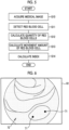
- a medical image processing apparatus 10 includes a medical image acquisition unit 11, a medical image analysis processing unit 12, a display unit 13, a display control unit 15, an input receiving unit 16, and an overall control unit 17.
- the medical image acquisition unit 11 acquires an endoscope image (hereinafter, referred to as a medical image), which is a medical image including a subject image, directly from an endoscope apparatus 21 that is a medical apparatus or through a management system, such as a picture archiving and communication system (PACS) 22, or other information systems.
- a medical image is a still image or a motion picture.
- the display of the medical image includes not only displaying a still image of one representative frame forming the motion picture but also reproducing the motion picture once or multiple times.
- the medical image acquisition unit 11 acquires at least short wavelength medical images 61 and 62 (refer to Figs. 6 and 7 ) among these medical images.
- the short wavelength medical images 61 and 62 are medical images obtained by imaging the subject with light in a shorter wavelength band than the green wavelength band.
- the "green wavelength band” refers to a wavelength band of about 500 nm or more and 570 nm or less.
- the "wavelength band shorter than the green wavelength band” refers to a wavelength less than about 500 nm.
- the short wavelength medical images 61 and 62 acquired by the medical image acquisition unit 11 are medical images obtained by imaging the subject using light containing components in a wavelength band substantially less than about 500 nm as illumination light.
- the illumination light used for imaging the short wavelength medical images 61 and 62 can contain a component in a wavelength band of about 500 nm or more (green light) in addition to a component in a wavelength band less than about 500 nm to the extent that the characteristics of the subject image are not lost in the case of imaging the subject using light in a wavelength band less than about 500 nm.
- the medical image acquisition unit 11 acquires a short wavelength medical image captured by using, as illumination light, narrow band light in a very narrow wavelength band (for example, about ⁇ 10 nm) in which the peak is in the range of 390 nm to 450 nm, the medical image analysis processing unit 12 uses the short wavelength medical images 61 and 62.
- the medical image acquisition unit 11 acquires a series of short wavelength medical images 61 and 62.
- the series of short wavelength medical images 61 and 62 refer to the two short wavelength medical images 61 and 62 captured consecutively or the two short wavelength medical images 61 and 62 captured at specific intervals. More specifically, in the case of a still image, the series of short wavelength medical images 61 and 62 are two or more medical images obtained by sequentially imaging the same subject in one examination. In the case of a motion picture, the series of short wavelength medical images 61 and 62 are medical images of two or more frames contained in the motion picture or medical images of two or more frames that can be freely acquired from a plurality of motion pictures obtained by sequentially imaging the same subject in one examination.
- the medical image acquisition unit 11 can acquire these as a series of short wavelength medical images.
- the imaging interval between individual short wavelength medical images included in a series of short wavelength medical images can be freely set or selected.
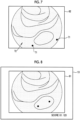
- the medical image acquisition unit 11 acquires the short wavelength medical image 61 (refer to Fig. 6 ), which is captured first, and the short wavelength medical image 62 (refer to Fig. 7 ), which is captured after the short wavelength medical image 61, along the time series of imaging.
- the medical image acquisition unit 11 can select and acquire all or some (for example, only short wavelength medical images) of the plurality of medical images. In the case of selecting and acquiring some medical images from the plurality of medical images in the endoscope apparatus 21, the PACS 22, or the like, it is possible to manually select a medical image in accordance with a user operation of a doctor or the like.
- the medical image acquisition unit 11 can automatically select a medical image to be acquired according to the imaging date and time, an imaging part, or other conditions set in advance (for example, the type of illumination light used for imaging).
- the medical image processing apparatus 10 is connected to the endoscope apparatus 21 to acquire a medical image from the endoscope apparatus 21.
- the endoscope apparatus 21 to which the medical image processing apparatus 10 is connected has an endoscope 31 that acquires an image by emitting at least one of light in a white wavelength band or light in a specific wavelength band to image the subject, a light source device 32 that emits illumination light to the inside of the subject through the endoscope 31, a processor device 33, and a monitor 34 for displaying an endoscope image or the like captured by using the endoscope 31.
- the light in a specific wavelength band that is used as illumination light by the endoscope 31 is, for example, light in a shorter wavelength band than the green wavelength band.
- the light in a specific wavelength band is light in a blue band or a violet band of the visible range.
- the processor device 33 includes an image generation unit 36 that generates an endoscope image.
- the medical image processing apparatus 10 is connected to the processor device 33.
- the medical image acquisition unit 11 acquires the short wavelength medical images 61 and 62 directly from the image generation unit 36 of the endoscope apparatus 21.
- the medical image analysis processing unit 12 performs analysis processing using the short wavelength medical images 61 and 62 acquired by the medical image acquisition unit 11. Specifically, as shown in Fig. 3 , the medical image analysis processing unit 12 includes a red blood cell detection section 41, a red blood cell quantity calculation section 42, a red blood cell movement amount calculation section 43, and an index calculation section 44.
- the red blood cell detection section 41 detects a red blood cell 71 (refer to Fig. 6 and the like) using the short wavelength medical images 61 and 62.
- the detection target of the red blood cell detection section 41 is a mass (collection) in which one red blood cell 71 or a plurality of red blood cell 71 gather.
- the red blood cell detection section 41 detects a high-frequency, granular, and high-density region as the red blood cell 71 (or the mass of the red blood cells 71; the same hereinbelow) using the short wavelength medical images 61 and 62.
- high frequency refers to a higher spatial frequency than the spatial frequency of a structure such as a blood vessel and a pit pattern (hereinafter, referred to as a tissue and the like structure) contained in a mucous membrane 72 (refer to Fig. 6 and the like), which is a subject in the short wavelength medical images 61 and 62, in a case where the short wavelength medical images 61 and 62 are subjected to Fourier transformation. That is, the red blood cell detection section 41 sets a finer pattern than the tissue and the like structure as a candidate for the red blood cell 71.
- the red blood cell detection section 41 sets the size of a pattern to be a candidate for the red blood cell 71 using the imaging magnification (enlargement ratio) of the subject in the short wavelength medical images 61 and 62.
- the term "granular" means that the shape of a pattern is a fine point (including a surface having a small area). That is, the red blood cell detection section 41 removes a linear pattern, such as a blood vessel, and a pattern having a relatively large area compared with the area of the mucous membrane 72 from the red blood cell candidates. Then, the red blood cell detection section 41 sets a spot pattern as a candidate for the red blood cell 71.
- the term "high density” refers to having a contrast that can be distinguished from the mucous membrane 72 occupying most of the short wavelength medical images 61 and 62. That is, the red blood cell detection section 41 sets a pattern having a predetermined contrast or more with respect to the mucous membrane 72 as a candidate for the red blood cell 71. Accordingly, the red blood cell detection section 41 can detect the red blood cell 71 even in a case where the short wavelength medical images 61 and 62 are reversed.
- the red blood cell detection section 41 detects the red blood cell 71 from the short wavelength medical images 61 and 62 using a bottom hat filter using a circular structure, a Quoit filter, or the like. Since the red blood cell has a diameter of about 7 to 8 ⁇ m and a thickness of about 2 ⁇ m, even in a case where some red blood cells gather, the size is very small. Therefore, except for the case of a special imaging situation in which the red blood cell 71 can be viewed macroscopically by enlarging the subject extremely, it is difficult to detect red blood cells using a medical image (endoscope image).
- the red blood cell detection section 41 can detect red blood cells while distinguishing the red blood cells from noise by using the short wavelength medical images 61 and 62 is that the short wavelength medical images 61 and 62 are medical images (endoscope images) obtained by imaging the subject with light in a shorter wavelength band than the green wavelength band.
- the short wavelength medical images 61 and 62 are obtained by imaging the subject with narrowband light that is light in a shorter wavelength band than the green wavelength band and has a very narrow wavelength band.
- red blood cell 71 even in a case where the situation is not a special imaging situation, it is possible to detect the red blood cell 71 by enlarging and imaging the subject to some extent.
- white light or the like is emitted to the subject to image the subject through a filter that transmits light in the green wavelength band, it is possible to accurately detect the red blood cell 71 in a state with little noise in particular.
- the red blood cell quantity calculation section 42 calculates the quantity of red blood cells 71 detected in the short wavelength medical images 61 and 62 using the detection result of the red blood cell detection section 41.
- the quantity of red blood cells 71 is the number of red blood cells 71 or an amount (area or the like) correlated with the number.
- the red blood cell movement amount calculation section 43 calculates the movement amount of the red blood cell 71 detected in the short wavelength medical images 61 and 62 using a series of short wavelength medical images 61 and 62 obtained by detecting the red blood cell 71 by the red blood cell detection section 41.
- the movement amount of the red blood cell 71 is the movement distance of the red blood cell 71 or an amount (for example, a sum or an average of movement distances) correlated with the movement distance of the red blood cell 71.
- the red blood cell movement amount calculation section 43 calculates a difference between at least two short wavelength medical images, among a series of short wavelength medical images acquired by the medical image acquisition unit 11, for each pixel.
- the red blood cell movement amount calculation section 43 sums the pixel value difference in each pixel in the entire image or a partial region defined in the image (including calculating the average and other statistics), and sets the absolute value as the movement amount of the red blood cell 71.
- the global movement of the mucous membrane 72 is small. Therefore, the sum or the like of differences is correlated with the movement distance of the fast red blood cell 71. That is, the sum or the like of differences between two short wavelength medical images increases as the movement distance of the red blood cell 71 substantially increases.
- the index calculation section 44 calculates an index indicating the degree of progress of a lesion using the quantity of red blood cells 71 calculated by the red blood cell quantity calculation section 42 and the movement amount of the red blood cell 71 calculated by the red blood cell movement amount calculation section 43.
- the lesion is, for example, an inflammatory disease. More specifically, the lesion is an inflammatory bowel disease, such as ulcerative colitis or Crohn's disease occurring in the lower digestive tract. Accordingly the index calculation section 44 calculates an index correlated with the degree of progress of the inflammatory bowel disease.
- the index calculation section 44 calculates an index indicating the degree of progress of ulcerative colitis.
- Mayo classification of endoscopic finding classification is known.
- the Mayo classification has four grades of Mayo0, Mayo1, Mayo2, and Mayo3.
- Mayo0 is a grade indicating normal or inactive (including a remission period).
- Mayo1 is a grade indicating mild, and is generally a state in which redness, blood vessel image observer, or slight easy bleeding is recognized.
- Mayo2 is a grade indicating moderate, and is generally a state in which significant redness, loss of a blood vessel image, easy bleeding, adhesion of purulent secretion, mucosal roughness, erosion, partial ulcer, and the like are recognized.
- Mayo3 is a grade indicating severe (active phase), and is generally a state in which obvious spontaneous bleeding, edema, ulcer (including a wide range of ulcer), and the like are recognized.
- the red blood cells 71 densely move in the blood vessel in general in the short wavelength medical images 61 and 62. Therefore, this is excluded from the detection of the red blood cell 71 in the red blood cell detection section 41. In a case where the degree of progress of ulcerative colitis is approximately Mayo1 to Mayo2, the red blood cell 71 leaks into the mucous membrane 72 even in a case where bleeding to the outside of the mucous membrane 72 does not occur. For this reason, in the short wavelength medical images 61 and 62, it is possible to detect the red blood cell 71 moving without following the blood vessel.
- the medical image processing apparatus 10 of the present embodiment determines each grade in the range of Mayo0, Mayo1, and Mayo2, and calculates an index indicating to which grade the state of progress is close.
- the index calculation section 44 can calculate a value (hereinafter, referred to as a score) by weighting addition of the quantity of red blood cells 71 and the movement amount of the red blood cell 71.
- a score a value (hereinafter, referred to as a score) by weighting addition of the quantity of red blood cells 71 and the movement amount of the red blood cell 71.
- the correlation with the grade of Mayo classification can be made stronger by adjusting the values of the weighting coefficients ⁇ and ⁇ .
- the index calculation section 44 can determine the degree of progress of a lesion from the quantity of red blood cells 71 and the movement amount of the red blood cell 71 with reference to a map (including a case of a numerical value table or the like) in which the quantity of red blood cells 71 and the movement amount of the red blood cell 71 are associated with the degree of progress of the lesion. For example, as shown in Fig. 4 , it is possible to determine the grade of Mayo classification as the degree of progress of ulcerative colitis by referring to a map 51 in which the quantity of red blood cells 71 and the movement amount of the red blood cell 71 are associated with the degree of progress of the ulcerative colitis. In this case, the grade of Mayo classification is an index calculated by the index calculation section 44. For example, in a case where the quantity of red blood cells 71 is "P1" and the movement amount of the red blood cell 71 is "Q1", the index calculated by the index calculation section 44 is "Mayo2".
- the calculation form of the index can be freely switched.
- the weighting coefficients ⁇ and ⁇ used for the calculation of the score X1 can also be freely set or changed.
- the display unit 13 is a display for displaying the medical image acquired by the medical image acquisition unit 11 and the analysis result of the medical image analysis processing unit 12.
- a monitor or a display included in a device or the like to which the medical image processing apparatus 10 is connected can be shared and used as the display unit 13 of the medical image processing apparatus 10.
- the display control unit 15 controls the display form of the medical image and the analysis result on the display unit 13.
- the input receiving unit 16 receives inputs from a mouse, a keyboard, and other operation devices attached to the medical image processing apparatus 10. The operation of each unit of the medical image processing apparatus 10 can be controlled using the operation devices.
- the overall control unit 17 controls the overall operation of each unit of the medical image processing apparatus 10. In a case where the input receiving unit 16 receives an operation input using an operation device, the overall control unit 17 controls each unit of the medical image processing apparatus 10 according to the operation input.
- the medical image acquisition unit 11 acquires a series of short wavelength medical images 61 and 62 automatically or manually (step S10).
- the short wavelength medical image 61 captured first between the series of short wavelength medical images 61 and 62, it is possible to detect the red blood cell 71 leaking into the mucous membrane 72.
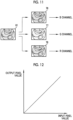
- the position of the red blood cell 71 moves compared with the first short wavelength medical image 61.
- the red blood cell detection section 41 detects the red blood cell 71 in each of the series of short wavelength medical images 61 and 62 (step S11). In a case where the red blood cell detection section 41 detects the red blood cell 71 in the series of short wavelength medical images 61 and 62, the red blood cell quantity calculation section 42 calculates the quantity of red blood cells 71 (step S12), and the red blood cell quantity calculation section 42 calculates the movement amount of the red blood cell 71 (step S13). Thereafter, the index calculation section 44 calculates the score X1 correlated with the degree of progress of ulcerative colitis or the grade of Mayo classification as an index using the quantity of red blood cells 71 and the movement amount of the red blood cell 71 (step S14).
- the display control unit 15 displays, on the display unit 13, at least one short wavelength medical image of the series of short wavelength medical images 61 and 62 and the index calculated by the index calculation section 44.
- the display control unit 15 displays the short wavelength medical image 61 and the score X1 on the display unit 13, for example.
- the display control unit 15 displays the short wavelength medical image 61 and the grade of Mayo classification on the display unit 13, for example.
- the medical image processing apparatus 10 can detect the red blood cell 71 leaking into the mucous membrane 72, which is normally difficult to accurately detect.
- the medical image processing apparatus 10 can obtain an analysis result relevant to the degree of progress of a lesion that has been difficult to determine in the related art, such as the degree of progress of ulcerative colitis, using the detected quantity of red blood cells 71 and the detected movement amount of the red blood cell 71.
- the inflammatory bowel disease such as ulcerative colitis and Crohn's disease, it has been difficult to accurately determine the grade of the disease as approximately mild to moderate.
- the medical image processing apparatus 10 can provide an index by which the diseases can be more accurately determined than in the related art.
- the index calculated by the medical image processing apparatus 10 it is possible to specifically and objectively determine Mayo1 and Mayo2 or Mayo0 and Mayo1 in ulcerative colitis or to what extent the disease is close to each grade (Mayo1 close to Mayo2, Mayo1 close to Mayo0, and the like).
- the red blood cell movement amount calculation section 43 sums up the differences between the two short wavelength medical images 61 and 62 and calculates the absolute value as the movement amount of the red blood cell 71.
- the red blood cell movement amount calculation section 43 can calculate the movement amount of the red blood cell 71 using a method different from the first embodiment described above. For example, by performing pattern matching between the short wavelength medical image 61 captured first and the short wavelength medical image 62 captured second to detect the corresponding red blood cell 71, the movement amount of each red blood cell 71 may be calculated.
- the imaging magnification of the series of short wavelength medical images 61 and 62 can be set freely.
- the series of short wavelength medical images 61 and 62 are short wavelength medical images captured by enlarging the subject 60 times or more on the 19-inch display screen.
- the red blood cell 71 can be accurately detected using the series of short wavelength medical images 61 and 62.
- the imaging magnification is an imaging magnification at the time of one step zooming in an endoscope for a digestive tract having a general zoom function.
- the display control unit 15 displays the short wavelength medical image 61 and the score X1 or the grade of Mayo classification on the display unit 13.
- this display form is merely an example, and these pieces of information can be displayed on the display unit 13 in another display form.
- any of the score X1 and the grade of Mayo classification is calculated as an index, as shown in Fig.
- the display control unit 15 can display the short wavelength medical image 61 and the map 51, in which the quantity of red blood cells 71 and the movement amount of the red blood cell 71 are associated with the grade of the Mayo classification, on the display unit 13 so that the specific quantity "P1" and movement amount "Q1" of the red blood cell 71 and the corresponding position (star mark) in the map 51 are shown on the map 51.
- At least the display forms of the display unit 13 shown in Figs. 9 and 10 are display forms in which the degree of progress of a lesion can be directly recognized. In this respect, at least the display forms of the display unit 13 shown in Figs. 9 and 10 are display forms in which the degree of progress of a lesion and the index are displayed so as to be associated with each other.
- the display control unit 15 displays the image on the display unit 13 in gray scale.
- the display control unit 15 can display the short wavelength medical images 61 and 62 on the display unit 13 in color.
- the display control unit 15 functions as a display color adjustment unit that adjusts the display color of the short wavelength medical images 61 and 62 displayed on the display unit 13. For example, in the case of displaying the short wavelength medical image 61 on the display unit 13, as shown in Fig. 11 , the display control unit 15 assigns the short wavelength medical image 61 displayed on the display unit 13 to a B channel (blue channel), a G channel (green channel), and an R channel (red channel) for display. In this case, the display control unit 15 performs correction for each color channel on the grayscale short wavelength medical image 61, which is an input source, for at least one color channel, and then outputs the obtained image to each color channel.
- a B channel blue channel
- G channel green channel
- R channel red channel
- the display control unit 15 generates a B channel image 76 after B channel correction, a G channel image 77 after G channel correction, and an R channel image 78 from the input short wavelength medical image 61, and displays the pseudo-colored short wavelength medical image 61 on the display unit 13 by using these.
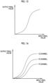
- the display control unit 15 linearly associates the input pixel value and the output pixel value with each other as shown in Fig. 12 .
- the short wavelength medical image 61 as an input source is output as it is without substantially performing correction for the B channel and the R channel.
- correction is made so as to nonlinearly associate the input pixel value and the output pixel value with each other. In these results, the color of the mucous membrane 72 becomes a green color.
- the red blood cell 71 absorbs light in a short wavelength band satisfactorily, the red blood cell 71 is almost black in the grayscale short wavelength medical image 61 that is an input source. Therefore, even after the above correction, the color of the red blood cell 71 is almost black. As a result, as shown in Fig. 14 , in a display image 81 in which the color of the mucous membrane 72 is a green color, the contrast of the red blood cell 71 with respect to the mucous membrane 72 is higher than in the original short wavelength medical image 61. Therefore, the red blood cell 71 can be easily recognized.
- the color difference between the green color of the mucous membrane 72 and the black color of the red blood cell 71 in the display image 81 is higher than the color difference between the white color of the mucous membrane 72 and the black color of the red blood cell 71 in the grayscale short wavelength medical image 61.
- the color of the mucous membrane 72 in such a medical image is a green-based color in many cases. Accordingly, there is also an advantage that, by making the mucous membrane 72 in green color in the display image 81 as described above, the image can be displayed with less discomfort than medical images of other general display forms while coloring in a pseudo manner.
- the correction for nonlinear association is not limited to the example described above.
- an S-shaped curve may be used in order to improve the visibility of an intermediate density signal.
- separate conversion tables may be assigned to the R channel, the G channel and the B channel.
- adjustment may be made by multiplying the grayscale short wavelength medical image 61, which is an input source, by a gain as a correction for each color channel. In this case, the gain assigned to the G channel is set to be high, and the gain assigned to the R channel and the B channel is set to be low.
- short wavelength medical image 61 or the display image 81 in which the short wavelength medical image 61 is pseudo-colored is displayed on the display unit 13.
- the display control unit 15 can display other medical images on the display unit 13.
- the medical image acquisition unit 11 acquires a medical image to be used for display on the display unit 13 in addition to the series of short wavelength medical images 61 and 62.
- the medical image to be used for display on the display unit 13 is, for example, a medical image captured by emitting white light to the subject, and is an endoscope image (hereinafter, referred to as a white light image) captured almost simultaneously with any of the series of short wavelength medical images 61 and 62 (at time intervals at which there is no large change in the mucous membrane 72 and the like).
- the display control unit 15 can display the white light image on the display unit 13 instead of the series of short wavelength medical images 61 and 62 used for detection of the red blood cell 71 or the like.
- the display control unit 15 can highlight the position of the red blood cell 71 on the white light image by displaying the detection result of the red blood cell detection section 41 on the display unit 13 so as to overlap the white light image.
- the medical image processing apparatus 10 and the endoscope apparatus 21 are separate apparatuses.
- the endoscope apparatus 21 can include the medical image processing apparatus 10.
- each unit 220 forming the medical image processing apparatus 10 is provided in the processor device 33.
- the monitor 34 of the endoscope apparatus 21 can be shared as the display unit 13, it is sufficient to provide each unit other than the display unit 13 in the processor device 33.
- a new endoscope apparatus can be configured by all of the medical image processing apparatuses 10 of the above embodiment and other modification examples and the endoscope apparatus 21 shown in Fig. 2 .
- the endoscope apparatus 21 is an apparatus for observing the subject in real time. As described above, in a case where the endoscope apparatus 21 includes the medical image processing apparatus 10, detection of the red blood cell 71 and the calculation of an index can be performed at any timing by automatic or manual setting.
- a diagnostic support apparatus 610 used in combination with the endoscope apparatus 21 and other modalities can include the medical image processing apparatuses 10 of the above embodiment and other modification examples.
- a medical service support apparatus 630 connected to various examination apparatuses including the endoscope apparatus 21, such as a first examination apparatus 621, a second examination apparatus 622, ..., and an N-th examination apparatus 623, through a certain network 626 can include the medical image processing apparatuses 10 of the above embodiment and other modification examples.
- the medical image processing apparatus 10 various apparatuses including the medical image processing apparatus 10, and various apparatuses or systems having the function of the medical image processing apparatus 10 can be used by making the following various changes or the like.
- the red blood cell detection section 41 can detect the red blood cell 71 in a region of interest.
- a capsule endoscope can be used as the endoscope 31.
- the light source device 32 and a part of the processor device 33 can be mounted in the capsule endoscope.
- the index calculation section 44 can calculate indices other than the indices shown in the above embodiment and modification examples.
- the index calculation section 44 can calculate information regarding blood vessels (hereinafter, referred to as blood vessel information), such as the thickness, the number, or the density of blood vessels, using the series of short wavelength medical images 61 and 62 or other medical images acquired by the medical image acquisition unit 11.
- blood vessel information such as the thickness, the number, or the density of blood vessels
- the display control unit 15 can display the indices other than the indices shown in the above embodiment and modification examples on the display unit 13 together with the indices shown in the above embodiment and modification examples.
- display or non-display of indices other than the indices shown in the above embodiment and modification examples can be freely switched by user's setting or operation.
- the medical image processing apparatus 10 can store the analysis result (the position of the red blood cell 71, the quantity of red blood cells 71, the movement amount of the red blood cell 71, and the index calculated using these) of the medical image analysis processing unit 12 in the PACS 22 or any other storage.
- the series of short wavelength medical images 61 and 62 used for the analysis and the analysis result can be stored so as to be associated with each other.
- the hardware structures of processing units for executing various kinds of processing are various processors shown below.
- the various processors include: a central processing unit (CPU) that is a general-purpose processor that functions as various processing units by executing software; a programmable logic device (PLD) that is a processor whose circuit configuration can be changed after manufacture, such as field programmable gate array (FPGA), a dedicated electrical circuit that is a processor having a dedicated circuit configuration for executing various types of processing; and the like.
- CPU central processing unit
- PLD programmable logic device
- FPGA field programmable gate array
- One processing unit may be configured by one of various processors, or may be a combination of two or more processors of the same type or different types (for example, a combination of a plurality of FPGAs or a combination of a CPU and an FPGA).
- a plurality of processing units may be configured by one processor.
- a processor As an example of configuring a plurality of processing units using one processor, first, so represented by a computer, such as a client or a server, there is a form in that one processor is configured by a combination of one or more CPUs and software and this processor functions as a plurality of processing units.
- SoC system on chip
- IC integrated circuit
- circuitry in the form of a combination of circuit elements, such as semiconductor elements.
- the medical image processing apparatus 10 of the above embodiment and modification examples can be suitably used not only for the purpose of determining the degree of progress of an inflammatory disease but also for the purpose of checking the presence or absence of bleeding from a sutured part after surgery.
- the medical image processing apparatus 10 is also suitable for determining the degree of progress of an inflammatory disease in the upper digestive tract, such as the esophagus, as well as an inflammatory bowel disease in the lower digestive tract.
- the display forms of medical images shown in Figs. 11 to 14 can be used separately from the detection function of the red blood cell 71 of the medical image processing apparatus 10 and the like.
- the display forms of medical images shown in Figs. 11 to 14 are useful in the case of displaying a grayscale endoscope image on the display unit 13 or the like.
- the grayscale endoscope image can be displayed in a pseudo-color manner in the display forms of medical images shown in Figs. 11 to 14 . In this case, even in a case where the red blood cell 71 is not reflected, the visibility of a portion, which is approximately black in the original grayscale medical image, can be improved.
- the display control unit 15 adjusts the display color.
- the display image 81 can be generated in advance. That is, a correction unit that performs correction corresponding to each color channel in the case of assigning a grayscale image as an input source to a plurality of color channels (RGB or CMYK) and a pseudo-color image generation unit that generates a new image in which an original grayscale image is pseudo-colored using a corrected image are provided. Then, the display control unit 15 may display the pseudo-color image (display image 81) generated by the pseudo-color image generation unit on the display unit 13. Needless to say, also in the medical image processing apparatus 10, the correction unit and the pseudo-color image generation unit can be provided.
Claims (12)
- Verarbeitungsvorrichtung (10) für medizinische Bilder, umfassend:eine Medizinbild-Erfassungseinheit (11), ausgebildet zum Erfassen eines kurzwelligen medizinischen Bildes, bei dem es sich um ein medizinisches Bild handelt, welches ein Subjektbild enthält und gewonnen ist durch Abbilden eines Subjekts mit Licht in einem kürzeren Wellenlängenband als einem Grün-Wellenlängenband, wobei das Licht in dem kürzeren Wellenlängenband eine Spitze im Bereich von 390 nm bis 450 nm aufweist;eine Rote-Blutkörperchen-Detektoreinheit (41), ausgebildet zum Nachweisen eines roten Blutkörperchens unter Verwendung des kurzwelligen medizinischen Bildes;eine Rote-Blutkörperchen-Mengenberechnungseinheit (42), ausgebildet zum Berechnen einer Menge roter Blutkörperchen, die in dem kurzwelligen medizinischen Bild nachgewiesen wurden;eine Rote-Blutkörperchen-Bewegungshub-Berechnungseinheit (43), ausgebildet zum Berechnen eines Bewegungshubs roter Blutkörperchen, die in dem kurzwelligen medizinischen Bild nachgewiesen wurden;eine Indexberechnungseinheit (43), ausgebildet zum Berechnen eines Index , der ein Fortschrittsmaß einer Läsion unter Verwendung der Menge und des Bewegungshubs roter Blutkörperchen angibt,dadurch gekennzeichnet, dass die Rote-Blutkörperchen-Detektoreinheit (41) ausgebildet ist zum Nachweisen einer hochfrequenten, granularen und hochdichten Zone mit einer Raumfrequenz höher als derjenigen eines Blutgefäßes und eines Gewebes, die in Schleimhaut enthalten sind, die eine granulare Form eines Musters aufweist, das ein feiner Punkt ist, und eine hohe Dichte aufweist, die einen Kontrast besitzt, der sich von der Schleimhaut unterscheiden lässt, die als Kandidat für ein rotes Blutkörperchen dient, unter Verwendung des kurzwelligen medizinischen Bildes, unddie Indexberechnungseinheit (43) ausgebildet ist zum Berechnen des Index', der den Fortschrittsgrad der Läsion unter Verwendung der Menge und des Bewegungshubs roter Blutkörperchen berechnet, die sich ohne dem Blutgefäß zu folgen, bewegen.
- Verarbeitungsvorrichtung nach Anspruch 1,
bei der die Rote-Blutzellen-Bewegungshub-Berechnungseinheit (43) ausgebildet ist zum Berechnen des Bewegungshubs der roten Blutkörperchen unter Verwendung einer Serie kurzwelliger medizinischer Bilder, in denen rote Blutkörperchen von der rote-Blutkörperchen-Detektoreinheit nachgewiesen werden. - Verarbeitungsvorrichtung nach Anspruch2,
bei der die Rote-Blutkörperchen-Bewegungshub-Berechnungseinheit(43) ausgebildet ist zum Berechnen des Bewegungshubs roter Blutkörperchen unter Verwendung von zwei kurzwelligen medizinischen Bildern, die aufeinanderfolgend aufgenommen wurden, oder von zwei der kurzwelligen medizinischen Bilder, die in spezifischen Intervallen aufgenommen wurden. - Verarbeitungsvorrichtung nach einem der Ansprüche 1 bis 3,
bei der die Indexberechnungseinheit ausgebildet ist zum Berechnen des Index, der mit einem Fortschrittsgrad einer entzündlichen Darmerkrankung korreliert ist. - Verarbeitungsvorrichtung nach einem der Ansprüche 1 bis 4,
bei der die Indexberechnungseinheit ausgebildet ist zum Berechnen des Index durch gewichtete Addition der Menge und des Bewegungshubs. - Verarbeitungsvorrichtung nach einem der Ansprüche 1 bis 5,
weiterhin umfassend:eine Anzeigeeinheit, ausgebildet zum Anzeigen des kurzwelligen medizinischen Bildes;eine Anzeigesteuereinheit, ausgebildet zum Einstellen einer Anzeigefarbe des kurzwelligen medizinischen Bildes, das auf der Anzeigeeinheit dargestellt ist. - Verarbeitungsvorrichtung nach Anspruch 6,
bei der die Anzeigesteuereinheit ausgebildet ist zum Einstellen einer Farbe einer Schleimhaut als grüne Farbe. - Verarbeitungsvorrichtung nach einem der Ansprüche 1 bis 5,
weiterhin umfassen:
eine Anzeigeeinheit, ausgebildet zum Anzeigen des Fortschrittsgrades der Läsion und des Indes in Verbindung miteinander. - Verarbeitungsvorrichtung nach einem der Ansprüche 1 bis 8,
weiterhin umfassend:eine Region-off-Interest-Detektoreinheit, ausgebildet zum Nachweisen einer Region-off-Interest, bei der es sich um eine zu beobachtende Zone handelt, basierend auf einem Merkmalswert von Pixel des kurzwelligen medizinischen Bildes,wobei die rote-Blutkörperchen-Detektoreinheit ausgebildet ist zum Nachweisen eines roten Blutkörperchens in der Region-off-Interest. - Endoskopvorrichtung, umfassend:
Die Verarbeitungsvorrichtung für medizinische Bilder nach einem der Ansprüche 1 bis 9;
Ein Endoskop, ausgebildet zum Erfassen eines Bildes durch Emittieren von Licht in dem kurzwelligen Band. - Diagnoseunterstützungsvorrichtung, umfassend:
Die Verarbeitungsvorrichtung für medizinische Bilder nach einem der Ansprüche 1 bis 3. - Medizinische Dienstunterstützungsvorrichtung, umfassend die Verarbeitungsvorrichtung für medizinische Bilder nach einem der Ansprüche 1 bis 9.
Applications Claiming Priority (1)
| Application Number | Priority Date | Filing Date | Title |
|---|---|---|---|
| JP2017168754A JP6850225B2 (ja) | 2017-09-01 | 2017-09-01 | 医療画像処理装置、内視鏡装置、診断支援装置、及び、医療業務支援装置 |
Publications (2)
| Publication Number | Publication Date |
|---|---|
| EP3449800A1 EP3449800A1 (de) | 2019-03-06 |
| EP3449800B1 true EP3449800B1 (de) | 2024-01-17 |
Family
ID=63350375
Family Applications (1)
| Application Number | Title | Priority Date | Filing Date |
|---|---|---|---|
| EP18189880.0A Active EP3449800B1 (de) | 2017-09-01 | 2018-08-21 | Vorrichtung zur verarbeitung medizinischer bilder, endoskopvorrichtung, diagnoseunterstützungsvorrichtung und vorrichtung zur unterstützung medizinischer dienste |
Country Status (3)
| Country | Link |
|---|---|
| US (1) | US11010891B2 (de) |
| EP (1) | EP3449800B1 (de) |
| JP (1) | JP6850225B2 (de) |
Families Citing this family (19)
| Publication number | Priority date | Publication date | Assignee | Title |
|---|---|---|---|---|
| CN110325098A (zh) | 2016-11-28 | 2019-10-11 | 适内有限责任公司 | 具有可分离一次性轴的内窥镜 |
| JP6858672B2 (ja) * | 2017-08-29 | 2021-04-14 | 富士フイルム株式会社 | 医療画像処理システム及び内視鏡システム |
| US11754824B2 (en) * | 2019-03-26 | 2023-09-12 | Active Medical, BV | Method and apparatus for diagnostic analysis of the function and morphology of microcirculation alterations |
| CN113784654A (zh) | 2019-04-23 | 2021-12-10 | 富士胶片株式会社 | 图像处理装置及其工作方法 |
| WO2021006121A1 (ja) * | 2019-07-08 | 2021-01-14 | 富士フイルム株式会社 | 画像処理装置、内視鏡システム、及び画像処理装置の作動方法 |
| WO2021079691A1 (ja) * | 2019-10-23 | 2021-04-29 | 富士フイルム株式会社 | 画像処理装置及びその作動方法 |
| USD1018844S1 (en) | 2020-01-09 | 2024-03-19 | Adaptivendo Llc | Endoscope handle |
| US11911030B2 (en) | 2020-10-02 | 2024-02-27 | Cilag Gmbh International | Communication capability of a surgical device with component |
| US11748924B2 (en) | 2020-10-02 | 2023-09-05 | Cilag Gmbh International | Tiered system display control based on capacity and user operation |
| US11830602B2 (en) | 2020-10-02 | 2023-11-28 | Cilag Gmbh International | Surgical hub having variable interconnectivity capabilities |
| US11672534B2 (en) | 2020-10-02 | 2023-06-13 | Cilag Gmbh International | Communication capability of a smart stapler |
| US11883022B2 (en) | 2020-10-02 | 2024-01-30 | Cilag Gmbh International | Shared situational awareness of the device actuator activity to prioritize certain aspects of displayed information |
| US11883052B2 (en) | 2020-10-02 | 2024-01-30 | Cilag Gmbh International | End effector updates |
| US20220104713A1 (en) * | 2020-10-02 | 2022-04-07 | Ethicon Llc | Tiered-access surgical visualization system |
| US11963683B2 (en) | 2020-10-02 | 2024-04-23 | Cilag Gmbh International | Method for operating tiered operation modes in a surgical system |
| US11877897B2 (en) | 2020-10-02 | 2024-01-23 | Cilag Gmbh International | Situational awareness of instruments location and individualization of users to control displays |
| CN116322465A (zh) * | 2020-10-02 | 2023-06-23 | 富士胶片株式会社 | 图像处理装置、内窥镜系统、图像处理装置的工作方法及图像处理装置用程序 |
| US11877792B2 (en) | 2020-10-02 | 2024-01-23 | Cilag Gmbh International | Smart energy combo control options |
| US20220104765A1 (en) * | 2020-10-02 | 2022-04-07 | Ethicon Llc | Surgical visualization and particle trend analysis system |
Citations (1)
| Publication number | Priority date | Publication date | Assignee | Title |
|---|---|---|---|---|
| EP1494579B1 (de) * | 2002-04-02 | 2011-07-27 | Yeda Research And Development Co., Ltd. | Charakterisierung beweglicher objekte in einem stationären hintergrund |
Family Cites Families (20)
| Publication number | Priority date | Publication date | Assignee | Title |
|---|---|---|---|---|
| JPS5928251B2 (ja) * | 1975-09-03 | 1984-07-11 | 株式会社日立製作所 | 白血球分類装置 |
| AU738223B2 (en) * | 1995-10-23 | 2001-09-13 | Cytometrics, Inc. | Method and apparatus for reflected imaging analysis |
| IL124814A (en) * | 1998-06-08 | 2003-04-10 | Grinvald Amiram | System and method for imaging and analysis of the movement of individual red blood corpuscles |
| US8521260B2 (en) | 2002-12-02 | 2013-08-27 | Yeda Research And Development Co. Ltd. | Characterization of arteriosclerosis by optical imaging |
| JP4139276B2 (ja) | 2003-06-17 | 2008-08-27 | オリンパス株式会社 | 電子内視鏡装置及び信号処理装置 |
| US20080021331A1 (en) * | 2004-09-29 | 2008-01-24 | Yeda Research And Development Co. Ltd. | Characterization of moving objects in a stationary background |
| JP2008104628A (ja) * | 2006-10-25 | 2008-05-08 | Tokyo Institute Of Technology | 眼球の結膜強膜撮像装置 |
| JP5446074B2 (ja) * | 2007-06-11 | 2014-03-19 | 株式会社日立製作所 | 血流の計測および評価装置 |
| US9602777B2 (en) * | 2008-04-25 | 2017-03-21 | Roche Diagnostics Hematology, Inc. | Systems and methods for analyzing body fluids |
| JP5582520B2 (ja) * | 2009-02-18 | 2014-09-03 | 国立大学法人名古屋大学 | 血流観測装置 |
| JP6027803B2 (ja) * | 2012-07-17 | 2016-11-16 | Hoya株式会社 | 画像処理装置及び内視鏡装置 |
| JP6198410B2 (ja) * | 2013-02-28 | 2017-09-20 | キヤノン株式会社 | 画像処理装置及び画像処理方法 |
| JP6200168B2 (ja) * | 2013-02-28 | 2017-09-20 | キヤノン株式会社 | 画像処理装置及び画像処理方法 |
| JP6097629B2 (ja) * | 2013-04-26 | 2017-03-15 | Hoya株式会社 | 病変評価情報生成装置 |
| JP6140056B2 (ja) * | 2013-09-26 | 2017-05-31 | 富士フイルム株式会社 | 内視鏡システム、内視鏡システムのプロセッサ装置、内視鏡システムの作動方法、プロセッサ装置の作動方法 |
| JP6196922B2 (ja) * | 2014-03-17 | 2017-09-13 | オリンパス株式会社 | 画像処理装置、画像処理方法、及び画像処理プログラム |
| JP6584090B2 (ja) * | 2015-02-23 | 2019-10-02 | Hoya株式会社 | 画像処理装置 |
| JP2017067524A (ja) * | 2015-09-29 | 2017-04-06 | 富士フイルム株式会社 | 有核赤血球の選別方法および有核赤血球の選別装置 |
| TW201725314A (zh) | 2016-01-05 | 2017-07-16 | 三宅圀博 | 雙殼式流體發電裝置及其轉子組件 |
| WO2017122431A1 (ja) * | 2016-01-15 | 2017-07-20 | オリンパス株式会社 | 画像解析装置、画像解析システム、及び画像解析装置の作動方法 |
-
2017
- 2017-09-01 JP JP2017168754A patent/JP6850225B2/ja active Active
-
2018
- 2018-08-21 EP EP18189880.0A patent/EP3449800B1/de active Active
- 2018-08-22 US US16/109,677 patent/US11010891B2/en active Active
Patent Citations (1)
| Publication number | Priority date | Publication date | Assignee | Title |
|---|---|---|---|---|
| EP1494579B1 (de) * | 2002-04-02 | 2011-07-27 | Yeda Research And Development Co., Ltd. | Charakterisierung beweglicher objekte in einem stationären hintergrund |
Also Published As
| Publication number | Publication date |
|---|---|
| JP2019042157A (ja) | 2019-03-22 |
| US20190073769A1 (en) | 2019-03-07 |
| US11010891B2 (en) | 2021-05-18 |
| EP3449800A1 (de) | 2019-03-06 |
| JP6850225B2 (ja) | 2021-03-31 |
Similar Documents
| Publication | Publication Date | Title |
|---|---|---|
| EP3449800B1 (de) | Vorrichtung zur verarbeitung medizinischer bilder, endoskopvorrichtung, diagnoseunterstützungsvorrichtung und vorrichtung zur unterstützung medizinischer dienste | |
| CN110325100B (zh) | 内窥镜系统及其操作方法 | |
| US10674892B2 (en) | Image processor, image processing method, and endoscope system | |
| US10776915B2 (en) | Medical image processing apparatus, endoscope apparatus, diagnostic support apparatus, and medical service support apparatus | |
| US20180214005A1 (en) | Image processing apparatus, endoscope system, and image processing method | |
| US11426054B2 (en) | Medical image processing system, endoscope system, diagnosis support apparatus, and medical service support apparatus | |
| EP2896363B1 (de) | Bearbeitung von endoskopischen Sauerstoffsättigungsdaten | |
| US20180206738A1 (en) | Endoscope system and method of operating endoscope system | |
| JPWO2019012911A1 (ja) | 医療画像処理装置、内視鏡システム、診断支援装置、並びに医療業務支援装置 | |
| US20200184645A1 (en) | Medical image processing apparatus | |
| US20210169306A1 (en) | Medical image processing apparatus, endoscope system, and method for operating medical image processing apparatus | |
| CN112512398B (zh) | 医疗图像处理装置 | |
| US11961228B2 (en) | Medical image processing system | |
| JP6785990B2 (ja) | 医療画像処理装置、及び、内視鏡装置 | |
| US20230141302A1 (en) | Image analysis processing apparatus, endoscope system, operation method of image analysis processing apparatus, and non-transitory computer readable medium | |
| JP6834019B2 (ja) | 医療画像処理装置、及び、内視鏡装置 | |
| JP6604743B2 (ja) | 情報処理装置、その作動方法、及びコンピュータプログラム | |
| JP6866497B2 (ja) | 医療画像処理装置、及び、内視鏡装置 | |
| WO2023026538A1 (ja) | 医療支援システム、医療支援方法及び評価支援装置 | |
| US20220245808A1 (en) | Image processing apparatus, image processing system, image processing method, and computer-readable recording medium | |
| WO2022230607A1 (ja) | 医療画像処理装置、内視鏡システム、及び医療画像処理装置の作動方法 | |
| WO2022009478A1 (ja) | 画像処理装置、内視鏡システム、画像処理装置の作動方法、及び画像処理装置用プログラム | |
| JP2023011303A (ja) | 医療画像処理装置及びその作動方法 |
Legal Events
| Date | Code | Title | Description |
|---|---|---|---|
| PUAI | Public reference made under article 153(3) epc to a published international application that has entered the european phase |
Free format text: ORIGINAL CODE: 0009012 |
|
| STAA | Information on the status of an ep patent application or granted ep patent |
Free format text: STATUS: THE APPLICATION HAS BEEN PUBLISHED |
|
| AK | Designated contracting states |
Kind code of ref document: A1 Designated state(s): AL AT BE BG CH CY CZ DE DK EE ES FI FR GB GR HR HU IE IS IT LI LT LU LV MC MK MT NL NO PL PT RO RS SE SI SK SM TR |
|
| AX | Request for extension of the european patent |
Extension state: BA ME |
|
| STAA | Information on the status of an ep patent application or granted ep patent |
Free format text: STATUS: REQUEST FOR EXAMINATION WAS MADE |
|
| 17P | Request for examination filed |
Effective date: 20190813 |
|
| RBV | Designated contracting states (corrected) |
Designated state(s): AL AT BE BG CH CY CZ DE DK EE ES FI FR GB GR HR HU IE IS IT LI LT LU LV MC MK MT NL NO PL PT RO RS SE SI SK SM TR |
|
| RAP3 | Party data changed (applicant data changed or rights of an application transferred) |
Owner name: FUJIFILM CORPORATION |
|
| STAA | Information on the status of an ep patent application or granted ep patent |
Free format text: STATUS: EXAMINATION IS IN PROGRESS |
|
| STAA | Information on the status of an ep patent application or granted ep patent |
Free format text: STATUS: EXAMINATION IS IN PROGRESS |
|
| 17Q | First examination report despatched |
Effective date: 20211116 |
|
| RIC1 | Information provided on ipc code assigned before grant |
Ipc: G06T 7/00 20170101ALI20230928BHEP Ipc: A61B 5/1459 20060101ALI20230928BHEP Ipc: A61B 5/145 20060101ALI20230928BHEP Ipc: A61B 5/026 20060101ALI20230928BHEP Ipc: A61B 1/00 20060101AFI20230928BHEP |
|
| GRAP | Despatch of communication of intention to grant a patent |
Free format text: ORIGINAL CODE: EPIDOSNIGR1 |
|
| STAA | Information on the status of an ep patent application or granted ep patent |
Free format text: STATUS: GRANT OF PATENT IS INTENDED |
|
| INTG | Intention to grant announced |
Effective date: 20231108 |
|
| GRAS | Grant fee paid |
Free format text: ORIGINAL CODE: EPIDOSNIGR3 |
|
| GRAA | (expected) grant |
Free format text: ORIGINAL CODE: 0009210 |
|
| STAA | Information on the status of an ep patent application or granted ep patent |
Free format text: STATUS: THE PATENT HAS BEEN GRANTED |
|
| AK | Designated contracting states |
Kind code of ref document: B1 Designated state(s): AL AT BE BG CH CY CZ DE DK EE ES FI FR GB GR HR HU IE IS IT LI LT LU LV MC MK MT NL NO PL PT RO RS SE SI SK SM TR |
|
| REG | Reference to a national code |
Ref country code: GB Ref legal event code: FG4D |
|
| P01 | Opt-out of the competence of the unified patent court (upc) registered |
Effective date: 20231219 |
|
| REG | Reference to a national code |
Ref country code: DE Ref legal event code: R096 Ref document number: 602018064191 Country of ref document: DE |
|
| REG | Reference to a national code |
Ref country code: CH Ref legal event code: EP |
|
| REG | Reference to a national code |
Ref country code: IE Ref legal event code: FG4D |