EP3446125B1 - Methods for determining dpp3 and diagnostic methods - Google Patents
Methods for determining dpp3 and diagnostic methods Download PDFInfo
- Publication number
- EP3446125B1 EP3446125B1 EP17720047.4A EP17720047A EP3446125B1 EP 3446125 B1 EP3446125 B1 EP 3446125B1 EP 17720047 A EP17720047 A EP 17720047A EP 3446125 B1 EP3446125 B1 EP 3446125B1
- Authority
- EP
- European Patent Office
- Prior art keywords
- dpp3
- dpp
- activity
- antibody
- capture
- Prior art date
- Legal status (The legal status is an assumption and is not a legal conclusion. Google has not performed a legal analysis and makes no representation as to the accuracy of the status listed.)
- Active
Links
Images


Classifications
-
- C—CHEMISTRY; METALLURGY
- C07—ORGANIC CHEMISTRY
- C07K—PEPTIDES
- C07K16/00—Immunoglobulins [IGs], e.g. monoclonal or polyclonal antibodies
- C07K16/40—Immunoglobulins [IGs], e.g. monoclonal or polyclonal antibodies against enzymes
-
- A—HUMAN NECESSITIES
- A61—MEDICAL OR VETERINARY SCIENCE; HYGIENE
- A61K—PREPARATIONS FOR MEDICAL, DENTAL OR TOILETRY PURPOSES
- A61K39/00—Medicinal preparations containing antigens or antibodies
- A61K39/395—Antibodies; Immunoglobulins; Immune serum, e.g. antilymphocytic serum
- A61K39/39533—Antibodies; Immunoglobulins; Immune serum, e.g. antilymphocytic serum against materials from animals
- A61K39/3955—Antibodies; Immunoglobulins; Immune serum, e.g. antilymphocytic serum against materials from animals against proteinaceous materials, e.g. enzymes, hormones, lymphokines
-
- A—HUMAN NECESSITIES
- A61—MEDICAL OR VETERINARY SCIENCE; HYGIENE
- A61P—SPECIFIC THERAPEUTIC ACTIVITY OF CHEMICAL COMPOUNDS OR MEDICINAL PREPARATIONS
- A61P1/00—Drugs for disorders of the alimentary tract or the digestive system
- A61P1/16—Drugs for disorders of the alimentary tract or the digestive system for liver or gallbladder disorders, e.g. hepatoprotective agents, cholagogues, litholytics
-
- A—HUMAN NECESSITIES
- A61—MEDICAL OR VETERINARY SCIENCE; HYGIENE
- A61P—SPECIFIC THERAPEUTIC ACTIVITY OF CHEMICAL COMPOUNDS OR MEDICINAL PREPARATIONS
- A61P13/00—Drugs for disorders of the urinary system
- A61P13/12—Drugs for disorders of the urinary system of the kidneys
-
- A—HUMAN NECESSITIES
- A61—MEDICAL OR VETERINARY SCIENCE; HYGIENE
- A61P—SPECIFIC THERAPEUTIC ACTIVITY OF CHEMICAL COMPOUNDS OR MEDICINAL PREPARATIONS
- A61P17/00—Drugs for dermatological disorders
- A61P17/02—Drugs for dermatological disorders for treating wounds, ulcers, burns, scars, keloids, or the like
-
- A—HUMAN NECESSITIES
- A61—MEDICAL OR VETERINARY SCIENCE; HYGIENE
- A61P—SPECIFIC THERAPEUTIC ACTIVITY OF CHEMICAL COMPOUNDS OR MEDICINAL PREPARATIONS
- A61P25/00—Drugs for disorders of the nervous system
-
- A—HUMAN NECESSITIES
- A61—MEDICAL OR VETERINARY SCIENCE; HYGIENE
- A61P—SPECIFIC THERAPEUTIC ACTIVITY OF CHEMICAL COMPOUNDS OR MEDICINAL PREPARATIONS
- A61P25/00—Drugs for disorders of the nervous system
- A61P25/08—Antiepileptics; Anticonvulsants
-
- A—HUMAN NECESSITIES
- A61—MEDICAL OR VETERINARY SCIENCE; HYGIENE
- A61P—SPECIFIC THERAPEUTIC ACTIVITY OF CHEMICAL COMPOUNDS OR MEDICINAL PREPARATIONS
- A61P25/00—Drugs for disorders of the nervous system
- A61P25/28—Drugs for disorders of the nervous system for treating neurodegenerative disorders of the central nervous system, e.g. nootropic agents, cognition enhancers, drugs for treating Alzheimer's disease or other forms of dementia
-
- A—HUMAN NECESSITIES
- A61—MEDICAL OR VETERINARY SCIENCE; HYGIENE
- A61P—SPECIFIC THERAPEUTIC ACTIVITY OF CHEMICAL COMPOUNDS OR MEDICINAL PREPARATIONS
- A61P29/00—Non-central analgesic, antipyretic or antiinflammatory agents, e.g. antirheumatic agents; Non-steroidal antiinflammatory drugs [NSAID]
-
- A—HUMAN NECESSITIES
- A61—MEDICAL OR VETERINARY SCIENCE; HYGIENE
- A61P—SPECIFIC THERAPEUTIC ACTIVITY OF CHEMICAL COMPOUNDS OR MEDICINAL PREPARATIONS
- A61P31/00—Antiinfectives, i.e. antibiotics, antiseptics, chemotherapeutics
-
- A—HUMAN NECESSITIES
- A61—MEDICAL OR VETERINARY SCIENCE; HYGIENE
- A61P—SPECIFIC THERAPEUTIC ACTIVITY OF CHEMICAL COMPOUNDS OR MEDICINAL PREPARATIONS
- A61P31/00—Antiinfectives, i.e. antibiotics, antiseptics, chemotherapeutics
- A61P31/04—Antibacterial agents
-
- A—HUMAN NECESSITIES
- A61—MEDICAL OR VETERINARY SCIENCE; HYGIENE
- A61P—SPECIFIC THERAPEUTIC ACTIVITY OF CHEMICAL COMPOUNDS OR MEDICINAL PREPARATIONS
- A61P31/00—Antiinfectives, i.e. antibiotics, antiseptics, chemotherapeutics
- A61P31/12—Antivirals
-
- A—HUMAN NECESSITIES
- A61—MEDICAL OR VETERINARY SCIENCE; HYGIENE
- A61P—SPECIFIC THERAPEUTIC ACTIVITY OF CHEMICAL COMPOUNDS OR MEDICINAL PREPARATIONS
- A61P31/00—Antiinfectives, i.e. antibiotics, antiseptics, chemotherapeutics
- A61P31/12—Antivirals
- A61P31/14—Antivirals for RNA viruses
- A61P31/18—Antivirals for RNA viruses for HIV
-
- A—HUMAN NECESSITIES
- A61—MEDICAL OR VETERINARY SCIENCE; HYGIENE
- A61P—SPECIFIC THERAPEUTIC ACTIVITY OF CHEMICAL COMPOUNDS OR MEDICINAL PREPARATIONS
- A61P33/00—Antiparasitic agents
-
- A—HUMAN NECESSITIES
- A61—MEDICAL OR VETERINARY SCIENCE; HYGIENE
- A61P—SPECIFIC THERAPEUTIC ACTIVITY OF CHEMICAL COMPOUNDS OR MEDICINAL PREPARATIONS
- A61P33/00—Antiparasitic agents
- A61P33/02—Antiprotozoals, e.g. for leishmaniasis, trichomoniasis, toxoplasmosis
- A61P33/06—Antimalarials
-
- A—HUMAN NECESSITIES
- A61—MEDICAL OR VETERINARY SCIENCE; HYGIENE
- A61P—SPECIFIC THERAPEUTIC ACTIVITY OF CHEMICAL COMPOUNDS OR MEDICINAL PREPARATIONS
- A61P35/00—Antineoplastic agents
-
- A—HUMAN NECESSITIES
- A61—MEDICAL OR VETERINARY SCIENCE; HYGIENE
- A61P—SPECIFIC THERAPEUTIC ACTIVITY OF CHEMICAL COMPOUNDS OR MEDICINAL PREPARATIONS
- A61P37/00—Drugs for immunological or allergic disorders
- A61P37/02—Immunomodulators
-
- A—HUMAN NECESSITIES
- A61—MEDICAL OR VETERINARY SCIENCE; HYGIENE
- A61P—SPECIFIC THERAPEUTIC ACTIVITY OF CHEMICAL COMPOUNDS OR MEDICINAL PREPARATIONS
- A61P9/00—Drugs for disorders of the cardiovascular system
-
- A—HUMAN NECESSITIES
- A61—MEDICAL OR VETERINARY SCIENCE; HYGIENE
- A61P—SPECIFIC THERAPEUTIC ACTIVITY OF CHEMICAL COMPOUNDS OR MEDICINAL PREPARATIONS
- A61P9/00—Drugs for disorders of the cardiovascular system
- A61P9/02—Non-specific cardiovascular stimulants, e.g. drugs for syncope, antihypotensives
-
- A—HUMAN NECESSITIES
- A61—MEDICAL OR VETERINARY SCIENCE; HYGIENE
- A61P—SPECIFIC THERAPEUTIC ACTIVITY OF CHEMICAL COMPOUNDS OR MEDICINAL PREPARATIONS
- A61P9/00—Drugs for disorders of the cardiovascular system
- A61P9/04—Inotropic agents, i.e. stimulants of cardiac contraction; Drugs for heart failure
-
- A—HUMAN NECESSITIES
- A61—MEDICAL OR VETERINARY SCIENCE; HYGIENE
- A61P—SPECIFIC THERAPEUTIC ACTIVITY OF CHEMICAL COMPOUNDS OR MEDICINAL PREPARATIONS
- A61P9/00—Drugs for disorders of the cardiovascular system
- A61P9/10—Drugs for disorders of the cardiovascular system for treating ischaemic or atherosclerotic diseases, e.g. antianginal drugs, coronary vasodilators, drugs for myocardial infarction, retinopathy, cerebrovascula insufficiency, renal arteriosclerosis
-
- G—PHYSICS
- G01—MEASURING; TESTING
- G01N—INVESTIGATING OR ANALYSING MATERIALS BY DETERMINING THEIR CHEMICAL OR PHYSICAL PROPERTIES
- G01N33/00—Investigating or analysing materials by specific methods not covered by groups G01N1/00 - G01N31/00
- G01N33/48—Biological material, e.g. blood, urine; Haemocytometers
- G01N33/50—Chemical analysis of biological material, e.g. blood, urine; Testing involving biospecific ligand binding methods; Immunological testing
- G01N33/53—Immunoassay; Biospecific binding assay; Materials therefor
- G01N33/543—Immunoassay; Biospecific binding assay; Materials therefor with an insoluble carrier for immobilising immunochemicals
- G01N33/54306—Solid-phase reaction mechanisms
-
- G—PHYSICS
- G01—MEASURING; TESTING
- G01N—INVESTIGATING OR ANALYSING MATERIALS BY DETERMINING THEIR CHEMICAL OR PHYSICAL PROPERTIES
- G01N33/00—Investigating or analysing materials by specific methods not covered by groups G01N1/00 - G01N31/00
- G01N33/48—Biological material, e.g. blood, urine; Haemocytometers
- G01N33/50—Chemical analysis of biological material, e.g. blood, urine; Testing involving biospecific ligand binding methods; Immunological testing
- G01N33/53—Immunoassay; Biospecific binding assay; Materials therefor
- G01N33/574—Immunoassay; Biospecific binding assay; Materials therefor for cancer
-
- G—PHYSICS
- G01—MEASURING; TESTING
- G01N—INVESTIGATING OR ANALYSING MATERIALS BY DETERMINING THEIR CHEMICAL OR PHYSICAL PROPERTIES
- G01N33/00—Investigating or analysing materials by specific methods not covered by groups G01N1/00 - G01N31/00
- G01N33/48—Biological material, e.g. blood, urine; Haemocytometers
- G01N33/50—Chemical analysis of biological material, e.g. blood, urine; Testing involving biospecific ligand binding methods; Immunological testing
- G01N33/58—Chemical analysis of biological material, e.g. blood, urine; Testing involving biospecific ligand binding methods; Immunological testing involving labelled substances
- G01N33/581—Chemical analysis of biological material, e.g. blood, urine; Testing involving biospecific ligand binding methods; Immunological testing involving labelled substances with enzyme label (including co-enzymes, co-factors, enzyme inhibitors or substrates)
-
- G—PHYSICS
- G01—MEASURING; TESTING
- G01N—INVESTIGATING OR ANALYSING MATERIALS BY DETERMINING THEIR CHEMICAL OR PHYSICAL PROPERTIES
- G01N33/00—Investigating or analysing materials by specific methods not covered by groups G01N1/00 - G01N31/00
- G01N33/48—Biological material, e.g. blood, urine; Haemocytometers
- G01N33/50—Chemical analysis of biological material, e.g. blood, urine; Testing involving biospecific ligand binding methods; Immunological testing
- G01N33/68—Chemical analysis of biological material, e.g. blood, urine; Testing involving biospecific ligand binding methods; Immunological testing involving proteins, peptides or amino acids
- G01N33/6854—Immunoglobulins
-
- G—PHYSICS
- G01—MEASURING; TESTING
- G01N—INVESTIGATING OR ANALYSING MATERIALS BY DETERMINING THEIR CHEMICAL OR PHYSICAL PROPERTIES
- G01N33/00—Investigating or analysing materials by specific methods not covered by groups G01N1/00 - G01N31/00
- G01N33/48—Biological material, e.g. blood, urine; Haemocytometers
- G01N33/50—Chemical analysis of biological material, e.g. blood, urine; Testing involving biospecific ligand binding methods; Immunological testing
- G01N33/68—Chemical analysis of biological material, e.g. blood, urine; Testing involving biospecific ligand binding methods; Immunological testing involving proteins, peptides or amino acids
- G01N33/6893—Chemical analysis of biological material, e.g. blood, urine; Testing involving biospecific ligand binding methods; Immunological testing involving proteins, peptides or amino acids related to diseases not provided for elsewhere
-
- A—HUMAN NECESSITIES
- A61—MEDICAL OR VETERINARY SCIENCE; HYGIENE
- A61K—PREPARATIONS FOR MEDICAL, DENTAL OR TOILETRY PURPOSES
- A61K39/00—Medicinal preparations containing antigens or antibodies
- A61K2039/505—Medicinal preparations containing antigens or antibodies comprising antibodies
-
- A—HUMAN NECESSITIES
- A61—MEDICAL OR VETERINARY SCIENCE; HYGIENE
- A61K—PREPARATIONS FOR MEDICAL, DENTAL OR TOILETRY PURPOSES
- A61K39/00—Medicinal preparations containing antigens or antibodies
- A61K2039/545—Medicinal preparations containing antigens or antibodies characterised by the dose, timing or administration schedule
-
- C—CHEMISTRY; METALLURGY
- C07—ORGANIC CHEMISTRY
- C07K—PEPTIDES
- C07K2317/00—Immunoglobulins specific features
- C07K2317/70—Immunoglobulins specific features characterized by effect upon binding to a cell or to an antigen
- C07K2317/73—Inducing cell death, e.g. apoptosis, necrosis or inhibition of cell proliferation
-
- C—CHEMISTRY; METALLURGY
- C07—ORGANIC CHEMISTRY
- C07K—PEPTIDES
- C07K2317/00—Immunoglobulins specific features
- C07K2317/70—Immunoglobulins specific features characterized by effect upon binding to a cell or to an antigen
- C07K2317/76—Antagonist effect on antigen, e.g. neutralization or inhibition of binding
-
- C—CHEMISTRY; METALLURGY
- C07—ORGANIC CHEMISTRY
- C07K—PEPTIDES
- C07K2317/00—Immunoglobulins specific features
- C07K2317/90—Immunoglobulins specific features characterized by (pharmaco)kinetic aspects or by stability of the immunoglobulin
-
- C—CHEMISTRY; METALLURGY
- C07—ORGANIC CHEMISTRY
- C07K—PEPTIDES
- C07K2317/00—Immunoglobulins specific features
- C07K2317/90—Immunoglobulins specific features characterized by (pharmaco)kinetic aspects or by stability of the immunoglobulin
- C07K2317/92—Affinity (KD), association rate (Ka), dissociation rate (Kd) or EC50 value
-
- G—PHYSICS
- G01—MEASURING; TESTING
- G01N—INVESTIGATING OR ANALYSING MATERIALS BY DETERMINING THEIR CHEMICAL OR PHYSICAL PROPERTIES
- G01N2333/00—Assays involving biological materials from specific organisms or of a specific nature
- G01N2333/90—Enzymes; Proenzymes
- G01N2333/914—Hydrolases (3)
- G01N2333/948—Hydrolases (3) acting on peptide bonds (3.4)
-
- G—PHYSICS
- G01—MEASURING; TESTING
- G01N—INVESTIGATING OR ANALYSING MATERIALS BY DETERMINING THEIR CHEMICAL OR PHYSICAL PROPERTIES
- G01N2800/00—Detection or diagnosis of diseases
- G01N2800/24—Immunology or allergic disorders
-
- G—PHYSICS
- G01—MEASURING; TESTING
- G01N—INVESTIGATING OR ANALYSING MATERIALS BY DETERMINING THEIR CHEMICAL OR PHYSICAL PROPERTIES
- G01N2800/00—Detection or diagnosis of diseases
- G01N2800/26—Infectious diseases, e.g. generalised sepsis
-
- G—PHYSICS
- G01—MEASURING; TESTING
- G01N—INVESTIGATING OR ANALYSING MATERIALS BY DETERMINING THEIR CHEMICAL OR PHYSICAL PROPERTIES
- G01N2800/00—Detection or diagnosis of diseases
- G01N2800/32—Cardiovascular disorders
- G01N2800/325—Heart failure or cardiac arrest, e.g. cardiomyopathy, congestive heart failure
-
- G—PHYSICS
- G01—MEASURING; TESTING
- G01N—INVESTIGATING OR ANALYSING MATERIALS BY DETERMINING THEIR CHEMICAL OR PHYSICAL PROPERTIES
- G01N2800/00—Detection or diagnosis of diseases
- G01N2800/34—Genitourinary disorders
- G01N2800/347—Renal failures; Glomerular diseases; Tubulointerstitial diseases, e.g. nephritic syndrome, glomerulonephritis; Renovascular diseases, e.g. renal artery occlusion, nephropathy
-
- Y—GENERAL TAGGING OF NEW TECHNOLOGICAL DEVELOPMENTS; GENERAL TAGGING OF CROSS-SECTIONAL TECHNOLOGIES SPANNING OVER SEVERAL SECTIONS OF THE IPC; TECHNICAL SUBJECTS COVERED BY FORMER USPC CROSS-REFERENCE ART COLLECTIONS [XRACs] AND DIGESTS
- Y02—TECHNOLOGIES OR APPLICATIONS FOR MITIGATION OR ADAPTATION AGAINST CLIMATE CHANGE
- Y02A—TECHNOLOGIES FOR ADAPTATION TO CLIMATE CHANGE
- Y02A50/00—TECHNOLOGIES FOR ADAPTATION TO CLIMATE CHANGE in human health protection, e.g. against extreme weather
- Y02A50/30—Against vector-borne diseases, e.g. mosquito-borne, fly-borne, tick-borne or waterborne diseases whose impact is exacerbated by climate change
Definitions
- Dipeptidyl peptidase 3 also known as Dipeptidyl aminopeptidase III, Dipeptidyl arylamidase III, Dipeptidyl peptidase III, Enkephalinase B or red cell angiotensinase; short name: DPP3, DPPIII - is a metallopeptidase, that removes dipeptides. from physiologically active peptides, such as enkephalins and angiotensins.
- DPP3 was first identified and its activity measured in extracts of purified bovine anterior pituitary by Ellis & Nuenke 1967.
- the enzyme which is listed as EC 3.4.14.4, has a molecular mass of about 83 kDa and is highly conserved in procaryotes and eucaryotes (Prajapati & Chauhan 2011).
- the amino acid sequence of the human variant is depicted in SEQ ID NO 1.
- Dipeptidyl peptidase III is a mainly cytosolic peptidase which is ubiquitously expressed. Despite lacking a signal sequence, a few studies reported membranous activity (Lee & Snyder 1982).
- DPP3 is a zinc-depending exo-peptidase belonging to the peptidase family M49. It has a broad substrate specificity for oligopeptides from three! four to ten amino acids of various compositions and is also capable of cleaving after proline. DPP3 is known to hydrolyze dipeptides from the N-terminus of its substrates, including angiotensin II, III and IV; Leu- and Met-enkephalin; endomorphin 1 and 2. The metallopeptidase DPP3 has its activity optimum at pH 8.0-9.0 and can be activated by addition of divalent metal ions, such as Co 2+ and Mg 2+ .
- divalent metal ions such as Co 2+ and Mg 2+ .
- DPP3 Structural analysis of DPP3 revealed the catalytic motifs HELLGH (hDPP3 450-455) and EECRAE (hDPP3 507-512), as well as following amino acids, that are important for substrate binding and hydrolysis: Glu316, Tyr, 318, Asp366, Asn391, Asn394, His568, Arg572, Arg577, Lys666 and Arg669 (Prajapati & Chauhan 2011; Kumar et al. 2016; numbering refers to the sequence of human DPP3, see SEQ ID No. 1). Considering all known amino acids or sequence regions that are involved in substrate binding and hydrolysis, the active site of human DPP3 can be defined as the area between amino acids 316 and 669.
- DPP3 can be inhibited unspecifically by different general protease inhibitors (e.g. PMSF, TPCK), sulfhydryl reagents (e.g. pHMB, DTNB) and metal chelators (EDTA, o-phenantroline; Abrami ⁇ . et al. 2000).
- general protease inhibitors e.g. PMSF, TPCK
- sulfhydryl reagents e.g. pHMB, DTNB
- EDTA metal chelators
- DPP3 activity can be inhibited specifically by different kinds of compounds: an endogenous DPP3 inhibitor is the peptide spinorphin.
- peptide spinorphin Several synthetic derivatives of spinorphin, e.g. tynorphin, have been produced and shown to inhibit DPP3 activity to varying extents (Yamamoto et al. 2000).
- Other published peptide inhibitors of DPP3 are propioxatin A and B ( US4804676 ) and propioxatin A analogues (Inaoka et al. 1988).
- DPP3 can also be inhibited by small molecules such as fluostatins and benzimidazol derivatives.
- Fluostatins A and B are antibiotics produced in Streptomyces sp. TA-3391 that are non-toxic and strongly inhibit DPP3 activity. So far 20 different derivatives of benzimidazol have been synthesized and published (Agi ⁇ et al. 2007; Rastija et al. 2015), of which the two compounds 1' and 4' show the strongest inhibitory effect (Agi ⁇ et al. 2007).
- DPP3 inhibitors see table 2.
- DPP3 has been shown to be a promising biomarker in several publications, which all refer to intracellular DPP3. It has been shown that DPP3 activity is elevated in homogenates of ovarian and endometrial tumors. DPP3 activity even increases with the severity! malignancy of said tumors (Simaga et al. 1998 and 2003). Immune histology and western blot analysis of glioblastoma cell lines also revealed elevated DPP3 levels (Singh et al. 2014).
- Intracellular or membranous DPP3 was also proposed to be a potential arteriorisk marker ( US2011008805 ) and marker for rheumatoid arthrosis ( US2006177886 ).
- DPP3 has been proposed as potential biomarker but also as potential therapeutic target due to its ability to cleave several bioactive peptides.
- Overexpression of DPP3 protects neuroblastoma cells from oxidative stress (Liu et al. 2007).
- Influenca A virus changes host DPP3 levels for own replication (cell culture studies, Meliopoulos et al. 2012).
- Enkephalin and/ or angiotensin degrading enzymes in general, including DPP3, have a therapeutic potential as targets for treatment of pain, cardiovascular diseases (CVD) and cancer and the corresponding inhibitors as potential treatments of pain, mental illnesses and CVD (Khaket et al. 2012, Patel et al. 1993, Igic et al. 2007).
- DPP3 is known as an intracellular protein
- DPP3 activity was detected in some bodily fluids: retroplacental serum (Shimamori et al. 1986), seminal plasma (Vanha-Perttula et al. 1988) and CSF (Aoyagi
- Subject of the present invention is an in vitro method for diagnosing a disease accompanied by or related to necrotic processes in a subject comprising:
- Binding specifically to full-length DPP3 means that said capture-binder does not bind to any other protein than DPP3. Binding specifically to full-length DPP3 means that said capture-binder does not bind to any other aminopeptidase than DPP.
- a binder that binds to full-length DPP3 is binder that binds to the protein of SEQ ID No. 1.
- a binder that binds to full-length DPP3 is binder that binds to SEQ ID No. 1.
- a solid phase assay is an assays where the respective binding events take place at the solid phase (see example 4 and 5).
- the capture-binder should does not bind the DPP3 in the region at or to the region of amino acids 316 - 669 of SEQ ID No. 1.
- the region of amino acids 316 - 669 of SEQ ID No. 1 includes the active center of DPP3 and the area of substrate binding (Prajapati & Chauhan 2011; Kumar et al. 2016).
- DPP3 activity can be measured by detection of cleavage products of DPP3 specific substrates.
- peptide hormone substrates include angiotensin II, III and IV, Leu-enkephalin, Met-enkephalin, endomorphin 1 and 2, valorphin, ⁇ -casomorphin, dynorphin, proctolin, ACTH (Adrenocorticotropic hormone) and MSH (melanocyte-stimulating hormone; Abrami ⁇ et al. 2000, Barsun et al. 2007, Dhanda et al. 2008).
- the cleavage of mentioned peptide hormones as well as other untagged oligopeptides can be monitored by detection of the respective cleavage products.
- Detection methods include, but are not limited to, HPLC analysis (e.g. Lee & Snyder 1982), mass spectrometry (e.g. Abrami ⁇ et al. 2000), H1-NMR analysis (e.g. Vandenberg et al. 1985), capillary zone electrophoresis (CE; e.g. Barsun et al. 2007), thin layer chromatography (e.g. Dhanda et al. 2008) or reversed phase chromatography (e.g. Mazocco et al. 2006).
- HPLC analysis e.g. Lee & Snyder 1982
- mass spectrometry e.g. Abrami ⁇ et al. 2000
- H1-NMR analysis e.g. Vandenberg et al. 1985
- CE capillary zone electrophoresis
- thin layer chromatography e.g. Dhanda et al. 2008
- reversed phase chromatography e.g. Mazocco et al. 2006.
- Detection of fluorescence due to hydrolysis of fluorogenic substrates by DPP3 is a standard procedure to monitor DPP3 activity.
- Those substrates are specific di- or tripeptides (Arg-Arg, Ala-Ala, Ala-Arg, Ala-Phe, Asp-Arg, Gly-Ala, Gly-Arg, Gly-Phe, Leu-Ala, Leu-Gly, Lys-Ala, Phe-Arg, Suc-Ala-Ala-Phe) coupled to a fluorophore.
- Fluorophores include but are not limited to ⁇ -naphtylamide (2-naphtylamide, ⁇ NA, 2NA), 4-methoxy- ⁇ -naphtylamide (4-methoxy-2-naphtylamide) and 7-amido-4-methylcoumarin (AMC, MCA; Abrami ⁇ et al. 2000, Ohkubo et al. 1999). Cleavage of these fluorogenic substrates leads to the release of fluorescent ⁇ -naphtylamine or 7-amino-4-methylcoumarin respectively.
- DPP3 carrying samples can be immobilized and divided on a gel by electrophoresis, gels stained with fluorogenic substrate (e.g. Arg-Arg- ⁇ NA) and Fast Garnet GBC and fluorescent protein bands detected by a fluorescence reader (Ohkubo et al. 1999).
- fluorogenic substrate e.g. Arg-Arg- ⁇ NA
- DPP3 activity is measured by addition of the fluorogenic substrate Arg-Arg- ⁇ NA and monitoring fluorescence in real time.
- said binder may be selected from the group of antibody, antibody fragment or non-IgG scaffold.
- An antibody is a protein including one or more polypeptides substantially encoded by immunoglobulin genes that specifically bind an antigen.
- the recognized immunoglobulin genes include the kappa, lambda, alpha (IgA), gamma (IgG 1 , IgG 2 , IgG 3 , IgG 4 ), delta (IgD), epsilon (IgE) and mu (IgM) constant region genes, as well as the myriad immunoglobulin variable region genes.
- Full-length immunoglobulin light chains are generally about 25 kD or 214 amino acids in length.
- Full-length immunoglobulin heavy chains are generally about 50 kD or 446 amino acid in length.
- Light chains are encoded by a variable region gene at the NH 2 -terminus (about 110 amino acids in length) and a kappa or lambda constant region gene at the COOH-terminus.
- Heavy chains are similarly encoded by a variable region gene (about 116 amino acids in length) and one of the other constant region genes.
- the basic structural unit of an antibody is generally a tetramer that consists of two identical pairs of immunoglobulin chains, each pair having one light and one heavy chain. In each pair, the light and heavy chain variable regions bind to an antigen, and the constant regions mediate effector functions.
- Immunoglobulins also exist in a variety of other forms including, for example, Fv, Fab, and (Fab') 2 , as well as bifunctional hybrid antibodies and single chains (e.g., Lanzavecchia et al. 1987; Huston et al. 1988; Bird et al. 1988; Hood et al. 1984; Hunkapiller & Hood, 1986).
- An immunoglobulin light or heavy chain variable region includes a framework region interrupted by three hypervariable regions, also called complementarity determining regions (CDR's) (see, Kabat et al. 1983). As noted above, the CDRs are primarily responsible for binding to an epitope of an antigen.
- An immune complex is an antibody, such as a monoclonal antibody, chimeric antibody, humanized antibody or human antibody, or functional antibody fragment, specifically bound to the antigen.
- Chimeric antibodies are antibodies whose light and heavy chain genes have been constructed, typically by genetic engineering, from immunoglobulin variable and constant region genes belonging to different species.
- the variable segments of the genes from a mouse monoclonal antibody can be joined to human constant segments, such as kappa and gamma 1 or gamma 3.
- a therapeutic chimeric antibody is thus a hybrid protein composed of the variable or antigen-binding domain from a mouse antibody and the constant or effector domain from a human antibody, although other mammalian species can be used, or the variable region can be produced by molecular techniques. Methods of making chimeric antibodies are well known in the art, e.g., see U.S. Patent No. 5,807,715 .
- a “humanized” immunoglobulin is an immunoglobulin including a human framework region and one or more CDRs from a non-human (such as a mouse, rat, or synthetic) immunoglobulin.
- the non-human immunoglobulin providing the CDRs is termed a "donor” and the human immunoglobulin providing the framework is termed an "acceptor.” All the CDRs are from the donor immunoglobulin in a humanized immunoglobulin.
- Constant regions need not be present, but if they are, they must be substantially identical to human immunoglobulin constant regions, i.e., at least about 85-90%, such as about 95% or more identical.
- a humanized antibody is an antibody comprising a humanized light chain and a humanized heavy chain immunoglobulin.
- a humanized antibody binds to the same antigen as the donor antibody that provides the CDRs.
- the acceptor framework of a humanized immunoglobulin or antibody may have a limited number of substitutions by amino acids taken from the donor framework. Humanized or other monoclonal antibodies can have additional conservative amino acid substitutions which have substantially no effect on antigen binding or other immunoglobulin functions.
- Humanized immunoglobulins can be constructed by means of genetic engineering (e.g ., see U.S. Patent No. 5,585,089 ).
- a human antibody is an antibody wherein the light and heavy chain genes are of human origin. Human antibodies can be generated using methods known in the art. Human antibodies can be produced by immortalizing a human B cell secreting the antibody of interest.
- Immortalization can be accomplished, for example, by EBV infection or by fusing a human B cell with a myeloma or hybridoma cell to produce a trioma cell.
- Human antibodies can also be produced by phage display methods (see, e.g., PCT Publication No. WO91/17271 ; PCT Publication No. WO92/001047 ; PCT Publication No. WO92/20791 , or selected from a human combinatorial monoclonal antibody library (see the Morphosys website).
- Human antibodies can also be prepared by using transgenic animals carrying a human immunoglobulin gene (for example, see PCT Publication No. WO93/12227 ; PCT Publication No. WO91/10741 ).
- the DPP3 antibody may have the formats known in the art.
- Examples are human antibodies, monoclonal antibodies, humanized antibodies, chimeric antibodies, CDR-grafted antibodies.
- the recombinantly produced antibodies as e.g. IgG, a typical full-length immunoglobulin, or antibody fragments containing at least the F-variable domain of heavy and/or light chain as e.g. chemically coupled antibodies (fragment antigen binding) including but not limited to Fab-fragments including Fab minibodies, single chain Fab antibody, monovalent Fab antibody with epitope tags, e.g.
- bivalent Fab-V5Sx2 bivalent Fab (mini-antibody) dimerized with the CH3 domain
- bivalent Fab or multivalent Fab e.g. formed via multimerization with the aid of a heterologous domain, e.g. via dimerization of dHLX domains, e.g. Fab-dHLX-FSx2; F(ab')2-fragments; scFv-fragments, multimerized multivalent or/and multispecific scFv-fragments, bivalent and/or bispecific diabodies, BITE ® (bispecific T-cell engager), trifunctional antibodies, polyvalent antibodies, e.g. from a different class than G; single-domain antibodies, e.g. nanobodies derived from camelid or fish immunoglobulins and numerous others.
- biopolymer scaffolds are well known in the art to complex a target molecule and have been used for the generation of highly target specific biopolymers. Examples are aptamers, spiegelmers, anticalins and conotoxins.
- Non-Ig scaffolds may be protein scaffolds and may be used as antibody mimics as they are capable to bind to ligands or antigens.
- Non-Ig scaffolds may be selected from the group comprising tetranectin-based non-Ig scaffolds ( e.g. described in US 2010/0028995 ), fibronectin scaffolds ( e.g. described in EP 1266 025 ; lipocalin-based scaffolds (( e.g. described in WO 2011/154420 ); ubiquitin scaffolds (e.g. described in WO 2011/073214 ), transferring scaffolds (e.g. described in US 2004/0023334 ), protein A scaffolds (e.g.
- Non-Ig scaffolds may be peptide or oligonucleotide aptamers.
- Aptamers are usually created by selecting them from a large random sequence pool and are either short strands of oligonucleotides (DNA, RNA or XNA; Xu et al. 2010, Deng et al. 2014) or short variable peptide domains attached to a protein scaffold (Li et al. 2011).
- Anti-DPP3 antibodies may be produced as follows: Mice are immunized with either recombinant DPP3 (e.g. GST-hDPP3 from USBio, Salem, USA), peptides comprising parts of the DPP3 amino acid sequence, e.g. conjugated to BSA or native purified DPP3 (e.g. from human erythrozytes, Abrami ⁇ et al. 1988).
- recombinant DPP3 e.g. GST-hDPP3 from USBio, Salem, USA
- peptides comprising parts of the DPP3 amino acid sequence
- BSA native purified DPP3
- mice were intraperitoneally (i.p.) injected with 100 ⁇ g recombinant GST-hDPP3, native purified hDPP3 or DPP3-peptide-BSA-conjugates at day 0 (emulsified in TiterMax Gold Adjuvant), 100 ⁇ g at day 14 (emulsified in complete Freund's adjuvant) and 50 ⁇ g at day 21 and 28 (in incomplete Freund's adjuvant).
- the animal received an intravenous (i.v.) injection of 50 ⁇ g GST-hDPP3, native purified hDPP3 or DPP3-peptide-BSA-conjugates dissolved in saline. Three days later the mice were sacrificed and the immune cell fusion was performed.
- Splenocytes from the immunized mice and cells of the myeloma cell line SP2/0 were fused with 1 ml 50% polyethylene glycol for 30 s at 37°C. After washing, the cells were seeded in 96-well cell culture plates. Hybrid clones were selected by growing in HAT medium [RPMI 1640 culture medium supplemented with 20% fetal calf serum and HAT-Supplement]. After one week, the HAT medium was replaced with HT Medium for three passages followed by returning to the normal cell culture medium.
- the cell culture supernatants were primarily screened for recombinant DPP3 binding IgG antibodies two weeks after fusion. Therefore recombinant GST-tagged DPP3 (USBiologicals, Salem, USA) was immobilized in 96-well plates (100 ng/ well) and incubated with 50 ⁇ l cell culture supernatant per well for 2 hours at room temperature. After washing of the plate, 50 ⁇ l / well POD-rabbit anti mouse IgG was added and incubated for 1 h at RT.
- a chromogen solution (3,7 mM o-phenylendiamin in citrate/ hydrogen phosphate buffer, 0.012% H 2 O 2 ) were added to each well, incubated for 15 minutes at RT and the chromogenic reaction stopped by the addition of 50 ⁇ l 4N sulfuric acid. Absorption was detected at 490 mm.
- the positive tested microcultures were transferred into 24-well plates for propagation. After retesting the selected cultures were cloned and recloned using the limiting-dilution technique and the isotypes were determined.
- Antibodies raised against GST-tagged human DPP3 or DPP3-peptides were produced via standard antibody production methods (Marx et al., 1997) and purified via Protein A. The antibody purities were ⁇ 90% based on SDS gel electrophoresis analysis.
- Antibodies may be produced by means of phage display according to the following procedure:
- the human naive antibody gene libraries HAL7/8 were used for the isolation of recombinant single chain F-Variable domains (scFv) against DPP3 peptide.
- the antibody gene libraries were screened with a panning strategy comprising the use of peptides containing a biotin tag linked via two different spacers to the DPP3 peptide sequence.
- a mix of panning rounds using non-specifically bound antigen and streptavidin bound antigen were used to minimize background of non-specific binders.
- the eluted phages from the third round of panning have' been used for the generation of monoclonal scFv expressing E.coli strains. Supernatant from the cultivation of these clonal strains has been directly used for an antigen ELISA testing (see also Hust et al. 2011; Sciere et al. 2009).
- Humanization of murine antibodies may be conducted according to the following procedure: For humanization of an antibody of murine origin the antibody sequence is analyzed for the structural interaction of framework regions (FR) with the complementary determining regions (CDR) and the antigen. Based on structural modeling an appropriate FR of human origin is selected and the murine CDR sequences are transplanted into the human FR. Variations in the amino acid sequence of the CDRs or FRs may be introduced to regain structural interactions, which were abolished by the species switch for the FR sequences. This recovery of structural interactions may be achieved by random approach using phage display libraries or via directed approach guided by molecular modeling. (Almagro & Fransson 2008).
- the DPP3 antibody format can be selected from the group comprising Fv fragment, scFv fragment, Fab fragment, scFab fragment, F(ab) 2 fragment and scFv-Fc Fusion protein.
- the antibody format may be selected from the group comprising scFab fragment, Fab fragment, scFv fragment and bioavailability optimized conjugates thereof, such as PEGylated fragments.
- said binder is an antibody,
- Said binder may be selected from the group of antibody, antibody fragments or non-Ig scaffold.
- the capture binder reactive with DPP3 is immobilized on a solid phase.
- solid phase may be used to include any material or vessel in which or on which the assay may be performed and includes, but is not limited to: porous materials, nonporous materials, test tubes, wells, slides, agarose resins (e.g. Sepharose from GE Healthcare Life Sciences), magnetic particals (e.g. Dynabeads TM or Pierce TM magnetic beads from Thermo Fisher Scientific), etc.
- agarose resins e.g. Sepharose from GE Healthcare Life Sciences
- magnetic particals e.g. Dynabeads TM or Pierce TM magnetic beads from Thermo Fisher Scientific
- aptamers may be immobilized onto the solid phase by utilization of the (strept)avidin-biotin system (Müller et al. 2012, Deng et al. 2014).
- said the separation step accordinf to claim 1 is a separation step is a washing step that removes ingredients of the sample that are not bound to said capture-binder from the captured DPP3.
- DPP3 activity is measured by addition of the fluorogenic substrate Arg-Arg- ⁇ NA and monitoring fluorescence in real time.
- the sample of a subject said sample is plasma.
- the sample is a blood sample that is plasma.
- antibody generally comprises monoclonal and polyclonal antibodies and binding fragments thereof, in particular Fc-fragments as well as so called “single-chain-antibodies” (Bird et al. 1988), chimeric, humanized, in particular CDR-grafted antibodies, and dia or tetrabodies (Holliger et al. 1993). Also comprised are immunoglobulin-like proteins that are selected through techniques including, for example, phage display to specifically bind to the molecule of interest contained in a sample. In this context the term “specific binding” refers to antibodies raised against the molecule of interest or a fragment thereof.
- An antibody is considered to be specific, if its affinity towards the molecule of interest or the aforementioned fragment thereof is at least preferably 50-fold higher, more preferably 100-fold higher, most preferably at least 1000-fold higher than towards other molecules comprised in a sample containing the molecule of interest. It is well known in the art how to make antibodies and to select antibodies with a given specificity.
- the assay uses an enzyme substrate which reacts with DPP3 to form a detectable reaction product. Alternatively, the rate of the reaction of the substrate can be monitored to determine the presence or amount of DPP3 in a test sample.
- Suitable enzyme substrates include, but are not limited to, dipeptide substrates such as Arg-Arg- ⁇ -NA.
- Assays embodying such reagents and reactions can be performed in any suitable reaction vessel, for example, a test tube or well of a microtiter plate.
- assay devices may be developed in disposable form such as dipstick or teststrip device formats which are well known to those skilled-in-the-art and which provide ease of manufacture and use.
- Such disposable assay devices may be packaged in the form of kits containing all necessary materials, reagents and instructions for use.
- a dipstick may be made from a piece of bibulous material containing a chromogenic substrate for DPP3.
- the dipstick could be made from a nonporous material on which the substrate is coated.
- a capture or binding assay may be performed to detect and/or quantitate the protease.
- an antibody reactive with DPP3 protein, but which does not interfere with peptidase activity may be immobilized upon a solid phase.
- the test sample is passed over the immobile antibody, and DPP3, if present, binds to the antibody and is itself immobilized for detection.
- a substrate may then be added, and the reaction product may be detected to indicate the presence or amount of DPP3 in the test sample.
- solid phase may used to include any material or vessel in which or on which the assay may be performed and includes, but is not limited to, porous materials, nonporous materials, test tubes, wells, slides, etc.
- DPP3 ECA can be performed as a teststrip assay.
- a test sample application pad is optionally attached to one end of a porous strip.
- the strip contains an immobilized antibody which will bind to and thereby immobilize DPP3 at a predetermined site for subsequent detection.
- the device may include an end of assay indicator which is positioned at the distal end of the teststrip away from the test sample contact site. The end of assay indicator produces a detectable signal upon contact with the test sample or an assay reagent thereby indicating that the assay is complete.
- a test sample application pad may be a portion of the porous strip itself or a material in fluid-flow contact with the end of the porous strip, referred to as the proximal end, such that the test sample can pass or migrate from the application pad to the porous strip.
- Fluid-flow contact can include physical contact of the application pad to the porous strip as well as the separation of the application pad from the porous strip by an intervening space or additional material which still allows fluid to flow between the application pad and the porous strip.
- Substantially all of the application pad can overlap the porous strip to enable the test sample to pass through substantially any part of the application pad to the proximal end of the porous strip.
- only a portion of the application pad might be in fluid-flow contact with the porous strip.
- the application pad can be any material which can transfer the test sample to the porous strip.
- the porous strip of the assay device can be any suitably absorbent, porous, bibulous, chromatographic or capillary possessing material through which a test sample containing the analyte can be transported by a capillary or wicking action.
- Natural, synthetic, or naturally occurring materials that are synthetically modified can be used as the porous strip including, but not limited to: cellulose materials such as paper, cellulose, and cellulose derivatives such as cellulose acetate and nitrocellulose; fiberglass; cloth, both naturally occurring (e.g., cotton) and synthetic (e.g., nylon); porous gels such as silica gel, agarose, dextran, and gelatin; porous fibrous matrixes: starch based materials, such as crosslinked dextran chains; ceramic materials; films of polyvinyl chloride and combinations of polyvinyl chloride-silica; and the like.
- the porous strip should not interfere with the production of a detectable signal.
- the porous strip should have a reasonable inherent strength, or strength
- the particular dimensions of the porous strip will be a matter of convenience, depending upon the size of the test sample involved, the assay protocol, the means for detecting and measuring the signal, and the like.
- the dimensions may be chosen to regulate the rate of fluid migration as well as the amount of test sample to be imbibed by the porous strip.
- a DPP3 substrate and/or DPP3 capture antibody may be immobilized on the porous strip to form at least one analyte detection site, i.e., that region of the porous strip having one or more assay reagents non-diffusively attached thereto.
- the measurement or detection region of the teststrip may include a plurality of sites containing a DPP3 substrate and/or immobilized anti-DPP3 antibody.
- the different detection sites may contain different amounts of substrate and/or immobilized anti-DPP3 antibody, i.e., a higher amount in the first detection site and lesser amounts in subsequent sites.
- the first detection site of an assay device might contain 50 nanograms of anti-DPP3 antibody while the subsequent sites contain 10, 20, 30, etc. nanograms of antibody.
- the number of sites displaying a detectable signal provides a quantitative indication of the amount of DPP3 present in the sample.
- the detection sites may be configured in any suitably detectable shape and are typically in the shape of a bar spanning the width of the teststrip.
- the multi-capture site device may be prepared such that if a threshold amount of DPP3 is not present in the test sample, then substantially all of the DPP3 will bind to the antibody in the first capture site and thus become immobilized at that site. If a greater than threshold amount of DPP3 is present in the test sample, the remaining DPP3 will bind to subsequent detection zones of immobilized antibody along the length of the teststrip. The greater the amount of DPP3 in the test sample, the greater the number of capture sites that will display a detectable signal due to the presence of DPP3. As will be appreciated by those skilled-in-the-art, devices containing multiple DPP3 substrate sites can also be produced wherein the amount of substrate in the individual sites is designed to produce a quantitative or semiquantitative assay result.
- Subject matter according to the invention is an in vitro method for diagnosing a disease accompanied by or related to necrotic processes in a subject comprising:
- the threshold is pre-determined by measuring DPP3 concentration and or DPP3 activity in healthy controls and calculating e.g. the according 75-percentile, more preferably the 90-percentile, even more preferably the 95-percentile.
- the upper boarder of the 75-percentile, more preferably the 90-percentile, even more preferably the 95-percentile, defines the threshold for healthy versus diseased patients.
- the threshold that divides between healthy and diseased patients may be between 0.5 and 2 nmol ⁇ NA min -1 ml -1 , more preferably between 0.7 and 1.8 nmol ⁇ NA min -1 ml -1 , more preferably between 0.8 and 1.5 nmol ⁇ NA min -1 ml -1 , most preferred between 1.0 and 1.3 nmol ⁇ NA min -1 ml -1 (see example 6).
- a specific threshold value may depend on the cohort used for calculating a pre-determined threshold that can be later-on used in routine.
- a specific threshold value may depend on the calibration used in the assay.
- a specific threshold value may depend on the sensitivity and/or specificity that seems to be acceptable for the practitioner.
- ROC curves Receiver Operating Characteristic curves
- the reference group must not be necessarily “normals”, but it might be a group of patients suffering from another disease or condition, from which the diseased group of interest shall be differentiated. For any particular marker, a distribution of marker levels for subjects with and without a disease will likely overlap.
- a test does not absolutely distinguish normal from disease with 100% accuracy, and the area of overlap indicates where the test cannot distinguish normal from disease.
- a threshold is selected, above which (or below which, depending on how a marker changes with the disease) the test is considered to be abnormal and below which the test is considered to be normal.
- a threshold is selected to provide a ROC curve area of greater than about 0.5, more preferably greater than about 0.7.
- the term "about” in this context refers to +/- 5% of a given measurement.
- the medical practitioner will use the pre-determined threshold for the methods of diagnosing a disease according to the invention and will determine whether the subject has a value above or below said pre-determined threshold value in order to make an appropriate diagnosis.
- the standard procedure to measure DPP3 levels is to determine the DPP3 activity using fluorogenic substrate (e.g. Arg-Arg- ⁇ -naphtylamide) in a liquid phase assay (Ellis & Nuenke 1967).
- fluorogenic substrate e.g. Arg-Arg- ⁇ -naphtylamide
- kits e.g. from BPS Bioscience usually contain low binding black microtiter plates, recombinant DPP3, fluorogenic substrate and respective buffers. Those kits are regularly used as screening assay for DPP3 substrates and inhibitors.
- Necrotic processes are herein defined by all processes in the body that lead to the death of cells and release of DPP3 from the cells cytoplasm into the extracellular space and/ or bodily fluids. Those processes include, but are not limited to, necrosis, apoptosis, necroptosis, eryptosis.
- Subject matter of the invention is a method for diagnosing a disease in a subject accompanied by or related to necrotic processes according to claim 1, said disease is selected from the group comprising: acute heart failure, sepsis, acute kidney injury, lower respiratory tract infection, acute myocardial infarction, cardiogenic shock, septic shock and liver failure. Table 1 lists clinical symptoms/ diseases that are accompanied by or related to necrotic processes and the respective necrotic event.
- Table 1 Diseases accompanied by or related to necrotic processes.
- Clinical symptome/ disease necrotic/ apoptotic event reference acute heart failure sudden death of cells/ tissue areas/ organs Fischer et al. 2005, Zong et al. 2006 myocardial infarction stroke liver failure/ injury kidney failure/ injury burn injuries Lanier et al. 2011 traumatic injuries (e.g. traumatic brain injury); polytrauma Raghupathi 2004 viral + microbial + parasite infections death of macrophages, lysis of host cells, ... Zong et al. 2006; Fink et al. 2005 AIDS progressive death of immune cells Fischer et al.
- Apoptosis is the process of programmed cell death (PCD) that may occur in multicellular organisms. Biochemical events lead to characteristic cell changes (morphology) and death. These changes include blebbing, cell shrinkage, nuclear fragmentation, chromatin condensation, and chromosomal DNA fragmentation. For review see Elmore 2007. Toxicol Pathol 35(4): 495-516 .
- Necrosis is a form of cell injury, which results in the premature death of cells in living tissue by autolysis ( Van GmbHakker et al. 2008. Current Molecular Medicine 8(3): 207-220 ). Necrosis is caused by factors external to the cell or tissue, such as infection, toxins, or trauma, which result in the unregulated digestion of cell components.
- necrosis is almost always detrimental and can be fatal.
- necrosis due to necrosis does not follow the apoptotic signal transduction pathway, but rather various receptors are activated, and result in the loss of cell membrane integrity and an uncontrolled release of products of cell death into the extracellular space. This initiates in the surrounding tissue an inflammatory response, which prevents nearby phagocytes from locating and eliminating the dead cells by phagocytosis. For this reason, it is often necessary to remove necrotic tissue surgically, a procedure known as debridement. Untreated necrosis results in a build-up of decomposing dead tissue and cell debris at or near the site of the cell death.
- necroptosis A form of programmed necrosis, called necroptosis, has been recognized as an alternate form of programmed cell death. Necroptosis may serve as a cell-death backup to apoptosis when the apoptosis signaling is blocked by endogenous or exogenous factors such as viruses or mutations ( Linkermann et al. 2014. NEJM 370(5): 455-465 ). Most recently, other types of regulated necrosis have been discovered as well, which share several signaling events with necroptosis and apoptosis ( Vanden Berghe et al. 2014. Nature Reviews 15 (135-147 ).
- Heart failure is a cardiac condition that occurs, when a problem with the structure or function of the heart impairs its ability to supply sufficient blood flow to meet the body's needs. It can cause a large variety of symptoms, particularly shortness of breath (SOB) at rest or during exercise, signs of fluid retention such as pulmonary congestion or ankle swelling and objective evidence of an abnormality of the structure or function of the heart at rest.
- SOB shortness of breath
- Heart failure is a clinical syndrome characterized by a constellation of symptoms and signs caused by cardiac dysfunction. It is one of the major causes of morbidity and mortality in the developed countries, with a prevalence of 1-2%. Heart failure can be grouped into chronic HF and acute HF. Patients with chronic HF can be grouped into stable chronic HF, worsening signs and symptoms of chronic HF and acute decompensation of chronic HF.
- Acute heart failure is defined as a rapid onset of signs and symptoms of heart failure resulting in the need for urgent therapy or hospitalization. AHF can present as acute de novo HF (new onset of AHF in a patient without previous cardiac dysfunction) or acute decompensation of chronic HF. AHF is the leading cause of hospitalization in adults older than 65 years of age.
- Heart failure comprises a wide range of patients, from those with normal left ventricular ejection fraction (LVEF) typically considered as ⁇ 50%, also known as HF with preserved EF (HFpEF) to those with reduced LVEF, typically considered as ⁇ 40%, also known as HF with reduced EF (HFrEF).
- LVEF left ventricular ejection fraction
- HFpEF HF with preserved EF
- HFrEF HF with reduced EF
- Patients with an LVEF in the range of 40-49% represent a 'grey area', which is defined as HF with mid-range EF (HFmrEF) ( Ponikowski et al. 2016. European Heart Journal 18(8): 891-975 ).
- Heart failure may occur as acute or chronic heart failure.
- acute is used to mean rapid onset and to describe exacerbated or decompensated heart failure, referring to episodes in which a patient can be characterized as having a change in heart failure signs and symptoms resulting in a need for urgent therapy or hospitalization.
- Chronic heart failure is a long-term condition, usually kept stable by the treatment of symptoms (stable chronic HF).
- Chronic heart failure may also decompensate (termed acute decompensated heart failure or acute decompensated chronic heart failure), which is most commonly the result from an intercurrent illness (such as pneumonia), myocardial infarction, arrhythmias, uncontrolled hypertension or a patient's failure to maintain fluid restriction, diet or medication.
- patients with acute decompensated chronic HF may return to a stable chronic compensated status (stable chronic HF).
- a liquid phase assay samples of bodily fluids are directly subjected to fluorogenic substrates (e.g. Arg-Arg- ⁇ -NA). Since there are many different amino peptidases in the plasma (Sanderink et al. 1988), it is possible that the used substrate is cleaved by peptidases other than DPP3. To circumvent this problem one preferred method of detecting specific DPP3 activity is the use of an enzyme capture activity assay.
- fluorogenic substrates e.g. Arg-Arg- ⁇ -NA
- the determination of active DPP3 in an enzyme capture assay comprises the steps:
- solid phase may be used to include any material or vessel in which or on which the assay may be performed and includes, but is not limited to: porous materials, nonporous materials, test tubes, wells, slides, agarose resins (e.g. Sepharose from GE Healthcare Life Sciences), magnetic particals (e.g. Dynabeads TM or Pierce TM magnetic beads from Thermo Fisher Scientific), etc.
- agarose resins e.g. Sepharose from GE Healthcare Life Sciences
- magnetic particals e.g. Dynabeads TM or Pierce TM magnetic beads from Thermo Fisher Scientific
- the separation step is a washing step that removes ingredients of the sample that are not bound to said capture-binder from the captured DPP3.
- DPP3 activity is measured by addition of the fluorogenic substrate Arg-Arg- ⁇ NA and monitoring fluorescence in real time.
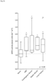
- DPP3 activity in plasma of a variety of diseased patients was determined using a specific DPP3 capture activity assay and compared to DPP3 plasma activities of healthy controls.
- Plasma samples were obtained from 388 patients entering the emergency department directly at their first presentation. With their final diagnosis these patients could be divided into 4 subgroups: patients suffering from acute myocardial infarction (AMI), cardiogenic shock, septic shock and liver failure.
- the control group was a collection of plasma samples from 93 healthy controls.
- DPP3 of plasma samples (10 ⁇ l) is firstly enriched by an affinity purification step and secondly its activity measured by addition of the fluorogenic substrate Arg-Arg- ⁇ NA (for a detailed description see example 4).
- the calculated slopes (in nmol ⁇ NA/ min per ml sample [nmol ⁇ NA min -1 ml -1 ]) of increasing fluorescence of the different samples refer to the 10 ⁇ l sample size.
- BP blood pressure
- a catheter Introcan-W; 22 G/1"; B.Braun
- Arteria carotis communis dextra right common carotid artery
- Human recombinant dipeptidyl peptidase 3 with an N-terminal GST-tag was injected via the tail vain.
- Rats were treated with recGST-hDPP3 0.2 mg/ kg in PBS by injection into the tail vain. Blood pressure was constantly monitored before and after injection of DPP3.
- DPP3 peptides for immunization were synthesized, see table 3, (JPT Technologies, Berlin, Germany) with an additional N-terminal cystein (if no cystein is present within the selected DPP3-sequence) residue for conjugation of the peptides to Bovine Serum Albumin (BSA).
- BSA Bovine Serum Albumin
- the peptides were covalently linked to BSA by using Sulfolink-coupling gel (Perbio-science, Bonn, Germany). The coupling procedure was performed according to the manual of Perbio. Recombinant GST-hDPP3 was produced by USBio.
- mice Immunization of mice, immune cell fusion and screening:
- mice were intraperitoneally (i.p.) injected with 84 ⁇ g GST-hDPP3 or 100 ⁇ g DPP3-peptide-BSA-conjugates at day 0 (emulsified in TiterMax Gold Adjuvant), 84 ⁇ g or 100 ⁇ g at day 14 (emulsified in complete Freund's adjuvant) and 42 ⁇ g or 50 ⁇ g at day 21 and 28 (in incomplete Freund's adjuvant).
- the animal received an intravenous (i.v.) injection of 42 ⁇ g GST-hDPP3 or 50 ⁇ g DPP3-peptide-BSA-conjugates dissolved in saline. Three days later the mice were sacrificed and the immune cell fusion was performed.
- Splenocytes from the immunized mice and cells of the myeloma cell line SP2/0 were fused with 1 ml 50% polyethylene glycol for 30 s at 37°C. After washing, the cells were seeded in 96-well cell culture plates. Hybrid clones were selected by growing in HAT medium [RPMI 1640 culture medium supplemented with 20% fetal calf serum and HAT-Supplement]. After one week, the HAT medium was replaced with HT Medium for three passages followed by returning to the normal cell culture medium.
- the cell culture supernatants were primarily screened for recombinant DPP3 binding IgG antibodies two weeks after fusion. Therefore recombinant GST-tagged DPP3 (USBiologicals, Salem, USA) was immobilized in 96-well plates (100 ng/ well) and incubated with 50 ⁇ l cell culture supernatant per well for 2 hours at room temperature. After washing of the plate, 50 ⁇ l / well POD-rabbit anti mouse IgG was added and incubated for 1 h at RT.
- a chromogen solution (3,7 mM o-phenylendiamin in citrate/ hydrogen phosphate buffer, 0.012% H 2 O 2 ) were added to each well, incubated for 15 minutes at RT and the chromogenic reaction stopped by the addition of 50 ⁇ l 4N sulfuric acid. Absorption was detected at 490 mm.
- the positive tested microcultures were transferred into 24-well plates for propagation. After retesting the selected cultures were cloned and recloned using the limiting-dilution technique and the isotypes were determined.
- Antibodies raised against GST-tagged human DPP3 or DPP3-peptides were produced via standard antibody production methods (Marx et al., 1997) and purified via Protein A. The antibody purities were ⁇ 90% based on SDS gel electrophoresis analysis.
- the following table represents a selection of obtained antibodies and their maximum inhibition rate (table 3).
- Table 2 Immunogen sequence, designation and characteristics of produced anti-DPP3 antibodies SEQ ID: 1 GST tagged recombinant FL-DPP3 1-737 mAb-FL-DPP3 2552 65% 2553 35% 2554 30% 2555 25% SEQ ID: 2 CETVINPETGEQIQSWYRSGE 474-493 mAb-pep 1-DPP3 1963 60% 1964 60% 1965 70% 1966 65% 1967 70% 1968 65% 1969 70%
- Antibodies raised against GST-tagged human DPP3 were produced via standard antibody production methods (Marx et al., 1997) and purified via Protein A. The antibody purities were ⁇ 90% based on SDS gel electrophoresis analysis. Different clones were analyzed in their capability of binding DPP3. Resulting positive clones were used as solid phase or tracer antibodies.
- 96-Well polystyrene microplates (Greiner Bio-One International AG, Austria) were coated (1 h at room temperature) with one antiDPP3 antibody clone (catching antibody; 1.5 ⁇ g antibody/ 0.25 mL 100 mmol/ L NaCl, 50 mmol/ L Tris/ HCl, pH 7.8). After blocking with 5% bovine serum albumine, the microplates were vacuum dried.
- the purified labeled antibody was diluted in assay buffer (50 mmol/l potassiumphosphate, 100 mmol/l NaCl, 10 mmol/l Na 2 -EDTA, 5 g/l bovine serum albumin, 1 g/l murine IgG, 1 g/l bovine IgG, 50 ⁇ mol/l amastatin, 100 ⁇ mol/l leupeptin, pH 7.4).
- the final concentration was approx. 7*10 6 relative light units (RLU) of labelled compound (approx. 20 ng labeled antibody) per 200 ⁇ l.
- acridinium ester chemiluminescence was measured by using a Centro LB 960 luminometer (Berthold Technologies GmbH & Co. KG).
- a stock solution (in PBS, pH 7.4) of recombinant human GST- DPP3 (USBiological, USA) was linearly diluted using (50 mmol/ L potassium phosphate, 100 mmol/ L NaCl, 10 mmol/ L Na-EDTA, 5 g/ L bovine serum albumin, 1 g/ L murine IgG, 1 g/ L bovine IgG, 50 ⁇ mol/ L amastatin, 100 ⁇ mol/ L leupeptin, pH 7.4)
- the stock solution was stored at -80 °C. Calibrators were prepared before use.
- DPP3 concentration in plasma of a variety of diseased patients was determined using an hDPP3 immuno assay and compared to DPP3 plasma concentrations of healthy controls.
- Plasma samples were obtained from 214 patients entering the emergency department or the oncology department directly at their first presentation. With their final diagnosis these patients could be divided into 6 subgroups: patients suffering from acute heart failure (AHF), myocardial infarct (MI), sepsis, cancer, acute kidney injury (AKI) and lower respirational tract infections (LRTI).
- the control group was a collection of plasma samples from 93 healthy controls.
- mAbDPP3_2555 was used as solid phase antibody and mAbDPP3_2553 as labelled tracer antibody.
- Antibody immobilization, labelling and incubation were performed as described in example 2.
- DPP3 in human plasma can be determined by DPP3 concentrations but also by activity assays.
- One standard procedure is a soluble activity assay using Arg-Arg- ⁇ NA as fluorogenic substrate: The activity of native human DPP III was determined by the hydrolysis of Arg-Arg- ⁇ -naphthylamide (Bachem Holdig AG, Switzerland) to form the fluorescent ⁇ -naphthylamine.
- soluble, DPP3 activity assay it can not be determined whether one measures DPP3 activity or the activity of other amino peptidases in the plasma.
- an enzyme capture assay is performed, where in a first step DPP3 is immobilized to a surface via binding to a monoclonal antibody, and after a washing step, only specific DPP3 activity can be measured:
- the solid phase was prepared as described in Example 3 using black 96-well microplates (Greiner Bio-One International GmbH, Austria).
- 10 ⁇ l sample (plasma or standard) and 200 ⁇ l buffer (50 mmol/l potassiumphosphate, 100 mmol/1 NaCl, 5 g/l bovine serum albumin, 1 g/l murine IgG, 1 g/l bovine IgG, 50 ⁇ mol/l Amastatin, 100 ⁇ mol/l Leupeptin, pH 7.4) were pipetted into said coated microplates and incubated (18-24 h, 2-8 °C, 600 rpm). Unbound analyte was removed by washing (3 ⁇ 350 ⁇ l) with washing solution.
- DPP3 activity in plasma of a variety of diseased patients was determined using the specific DPP3 capture activity assay and compared to DPP3 plasma activities of healthy controls.
- DPP3 of plasma samples (10 ⁇ l) is firstly enriched by an affinity purification step and secondly its activity measured by addition of the fluorogenic substrate Arg-Arg- ⁇ NA (for a detailed description see example 4).
- the calculated slopes (in nmol ⁇ NA/ min per ml sample [nmol ⁇ NA min -1 ml -1 ]) of increasing fluorescence of the different samples refer to the 10 ⁇ l sample size.
- Table 7 shows the percentage of patients with DPP3 values above the 75-percentile of the control group and their respective diagnosis. The activity shows a better division of healthy controls and diseased patients.
- Table 6 Comparison of DPP3 values of diseased patients and healthy controls in a sandwich type immune assay and in an enzyme capture activity assay. percentage of patients with DPP3 values above 75-percentile of control group indication AHF MI sepsis cancer AKI LRTI Sandwich assay 48,7 64,3 85,5 91,7 51,4 53,3 Activity assay 77,8 n.a. 87,0 n.a. 70,0 85,7
- mice Female BALB/c nude (CAnN.Cg-Foxnlnu/Crl) Mice (Charles River GmbH, Sulzfeld, Germany) aged 4-5 weeks at delivery and weighing approximately 15-18 g were kept under optimum hygienic conditions, air-conditioned with 10-15 air changes per hour, and continually monitored environment with target ranges for temperature 22 ⁇ 3°C and for relative humidity 30-70%, 12 hours artificial fluorescent light / 12 hours dark. Maximum 4 animals were kept per individual ventilated cage (IVC) and fed with a diet consisting of M-Zucht (ssniff Spezialdiuschen GmbH) and autoclaved community tab water.
- IVC individual ventilated cage
- mAbDPP3 was administered intraperitonealy (i.p.) at days 1, 3, 5 and 7, and mice monitored for 14 days.
- MAbDPP3 is safe to be used in other animal experiments, which will always be performed with a concentration of 1.9 mg/ kg.
- BP blood pressure
- a catheter Introcan-W; 22 G/1"; B.Braun
- the administration and sampling catheter were inserted into the Vena jugularis sinistra (left jugular vein).
- MAbDPP3 (1,9 mg/ kg in PBS) was tested versus vehicle (PBS). I.v. injection of the compound and vehicle was performed 5 minutes before CLP (preventive treatment) and after full development of sepsis, 2 hours after CLP (therapeutic treatment). Each group contained 10 mice and were followed over a period of 7 days.
- a septic shock model was used to induce heart failure in rats and then to characterize mAbDPP3's influence on heart function.
- mice Male Wistar rats (2-3 months, 300 to 400 g, group size refer to Table 1) from the Centre d'élevage Janvier (France) were allocated randomly to one of three groups. All the animals were anesthetized using ketamine hydrochloride (90 mg/ kg) and xylazine (9 mg/ kg) intraperitoneally (i.p.). For induction of polymicrobial sepsis, cecal ligation and puncture (CLP) was performed using Rittirsch's protocol with minor modification. A ventral midline incision (1.5 cm) was made to allow exteriorization of the cecum. The cecum is then ligated just below the ileocecal valve and punctured once with an 18-gauge needle.
- CLP cecal ligation and puncture
- the abdominal cavity is, then closed in two layers, followed by fluid resuscitation (3 ml/ 100 g body of weight of saline injected subcutaneously) and returning the animal to its cage. Sham animals were subjected to surgery, without getting their cecum punctured.
- Hemodynamic variables were obtained using the AcqKnowledge system (BIOPAC Systems, Inc., USA). It provides a fully automated blood pressure analysis system.
- the catheter is connected to the BIOPAC system through a pressure sensor.
- rats were anesthetized (ketamine and xylazine). Animals were moved to the heating pad for the desired body temperature to 37-37.5 °C. The temperature feedback probe was inserted into the rectum. The rats were placed on the operating table in a supine position. The trachea was opened and a catheter (16G) was inserted for an external ventilator without to damage carotid arteries and vagus nerves. The arterial catheter was inserted into the right carotid artery. The carotid artery is separate from vagus before ligation.
- a central venous catheter was inserted through the left jugular vein allowing administration of drug.
- BP blood pressure
- TTE transthoracic echocardiographic
- pulmonary artery flow was recorded using pulsed wave Doppler. Velocity time integral of pulmonary artery outflow was measured.
- mitral flow was recorded using pulsed Doppler at the level of the tip of the mitral valves.
- septic rats Compared to sham animals the septic rats have very low blood pressure and a decreased shortening fraction of the heart.
- the aim of the herein described study was to assess the potential anti-proliferative effect of mAbDPP3 in an in vitro cell culture system employing several cancer cell lines.
- a stock solution of mAbDPP3 (1 mg/ ml in PBS) was diluted to cover the final concentration range between 0 - 100 ⁇ g/ ml. PBS was used as reference compound.
- Cancer cells stemming from established cancer cell lines (A549, HCT116, MDA-MB231) were cultured in DMEM containing 10 % FCS and Penicillin/Streptomycin.
- A549 cells are adenocarcinomic human alveolar basal epithelial cells. This cell line was first established through the removal and culturing of cancerous lung tissue in the explanted tumor of a 58-year-old male. In nature, these cells are squamous and responsible for the diffusion of some substances, such as water and electrolytes, across the alveoli of lungs. In case said A549 cells are cultured in vitro, they grow as monolayer cells, adhered to the culture flask. A further characteristic is that these cells are able to synthesize lecithin and contain high level of desaturated fatty acids. The A549 cell line is widely used as in vitro model for a type II pulmonary epithelial cell model for drug metabolism and as a transfection host.
- HCT116 cell line denotes human colon cancer cells. These epithelial cells have adherent culture properties, and stem from a male adult. This cell line is a suitable transfection host. This line has a mutation in codon 13 of the ras proto-oncogene, and can be used as a positive control for PCR assays of mutation in this codon.
- MDA-MB231 cell line denotes human breast adenocarcinoma cells having epithelial morphology. These cells were isolated from pleural effusions of Caucasian breast cancer patient.
- 96 well suspension cell culture plates were prepared. 100 ⁇ L of the soft agar bottom layer (0.6 % final concentration in complete medium) was poured and left to solidify. 50 ⁇ L of the soft agar top layer (0.4 % final concentration) containing the corresponding cells and cell number were then added on top, solidified and such 96 well plates incubated overnight at 37°C, 10 % CO 2 .
- Raw data were converted into percent soft agar growth relative to high controls (solvent 0.1 % DMSO) and low controls (10 - 5 M staurosporine), which were set to 100 % and 0 %, respectively.
- IC 50 calculation was performed using GraphPad Prism 5 software with a variable slope sigmoidal response-fitting model using 0 % soft agar growth as bottom constraint and without bottom constraint and 100 % soft agar growth as top constraint.
- IC 50 values were determined using standard parameters based on the signal of the solvent control as top constraint (100 % soft agar growth) and the signal of the staurosporine control as bottom constraint (0 % soft agar growth). The respective IC 50 values are summarized in Table 10.
- MAbDPP3 treatment had an antiproliferative effect on the three cell lines tested.
- Table 9 IC 50 values for mAbDPP3 treatment.
- Cell line Tissue source Incubation time IC 50 A549 Lung 8 days 6.3 ⁇ g/ ml HCT116 Colon 8 days 2.0 ⁇ g/ ml MDA-MB231 Breast 11 days 8.7 ⁇ g/ ml
- the aim of the herein described study was to assess the ability of mAbDPP3 to prevent tumor formation in xenograft models for breast cancer and colon cancer (tumor growth inhibition study).
- Monolayer MDA-MB-231 cells (breast cancer) and HCT-116 cells (colon cancer), respectively, were grown in DMEM + 10% FCS.
- the cells were cultured in a humidified atmosphere of 90% air and 10% carbon dioxide at 37°C. Media was routinely changed every 3 days.
- Confluent cultures were split 1:3 to 1:3 every 3-4 days using Trypsin/EDTA and seeded at a density of approximately 3-4 ⁇ 10 6 cells/15 cm 2 + 25 mL medium.
- mice Female BALB/c nude (CAnN.Cg-Foxn1 nu /Crl) Mice (Charles River GmbH, Sulzfeld, Germany) aged 4-5 weeks at delivery and weighing approximately 15-18 g were kept under optimum hygienic conditions, air-conditioned with 10-15 air changes per hour, and continually monitored environment with target ranges for temperature 22 ⁇ 3°C and for relative humidity 30-70%, 12 hours artificial fluorescent light / 12 hours dark. Maximum 4 animals were kept per individual ventilated cage (IVC) and fed with a diet consisting of M-Zucht (ssniff Spezialdi expertsen GmbH) and autoclaved community tab water.
- IVC individual ventilated cage
- DPP3-adsorber In order to assess the possibility of using a DPP3-adsorber to clear plasma from excess DPP3, it should be analyzed whether DPP3 would sufficiently bind to an antiDPP3 column.
- Affinity chromatography columns were prepared by immobilizing DPP3 binding antibodies to GlycoLink columns (Thermo Fisher). Binding of DPP3 to those columns was analyzed by measuring DPP3 concentration before and after flow through these columns.
- DPP3 binding antibodies In a first step all DPP3 binding antibodies (mAbDPP3_2552, 2553, 2554, 2555) were oxidized and immobilized on one GlycoLink column each according to the instructor's manual. Subsequently recombinant GST-hDPP3 (USBio) or patient plasma samples were loaded onto the column and binding of DPP3 monitored.
- the DPP3 concentration of samples and recombinant GST-hDPP3 before affinity purification and of the flow through were measured using a sandwich type luminescence immune assay (for details see example 2).
- 20 ⁇ l of sample (or calibrator) were pipetted into coated 96-well microplates, after adding labeled and diluted detection antibody (200 ⁇ l), the plates were incubated for 18-24 h at 2-8 °C. Unbound tracer was removed by washing 4 times with 350 ⁇ l washing solution (20 mM PBS, pH 7.4, 0.1 % Triton X-100).
- Well-bound chemiluminescence was measured by using the Centre LB 960 luminometer (Berthold Technologies GmbH & Co. KG).
Landscapes
- Health & Medical Sciences (AREA)
- Life Sciences & Earth Sciences (AREA)
- Chemical & Material Sciences (AREA)
- Engineering & Computer Science (AREA)
- Medicinal Chemistry (AREA)
- General Health & Medical Sciences (AREA)
- Organic Chemistry (AREA)
- Veterinary Medicine (AREA)
- Immunology (AREA)
- Chemical Kinetics & Catalysis (AREA)
- Pharmacology & Pharmacy (AREA)
- Animal Behavior & Ethology (AREA)
- Public Health (AREA)
- Nuclear Medicine, Radiotherapy & Molecular Imaging (AREA)
- General Chemical & Material Sciences (AREA)
- Molecular Biology (AREA)
- Bioinformatics & Cheminformatics (AREA)
- Biomedical Technology (AREA)
- Urology & Nephrology (AREA)
- Hematology (AREA)
- Biochemistry (AREA)
- Cardiology (AREA)
- Microbiology (AREA)
- Physics & Mathematics (AREA)
- Pathology (AREA)
- Biotechnology (AREA)
- Cell Biology (AREA)
- General Physics & Mathematics (AREA)
- Analytical Chemistry (AREA)
- Food Science & Technology (AREA)
- Heart & Thoracic Surgery (AREA)
- Oncology (AREA)
- Proteomics, Peptides & Aminoacids (AREA)
- Communicable Diseases (AREA)
- Tropical Medicine & Parasitology (AREA)
- Hospice & Palliative Care (AREA)
- Genetics & Genomics (AREA)
- Biophysics (AREA)
- Neurology (AREA)
- Neurosurgery (AREA)
Priority Applications (4)
| Application Number | Priority Date | Filing Date | Title |
|---|---|---|---|
| EP24178574.0A EP4417218A3 (en) | 2016-04-21 | 2017-04-20 | Methods for determining dpp3 and therapeutic methods |
| EP21153560.4A EP3896450A1 (en) | 2016-04-21 | 2017-04-20 | Methods for determining dpp3 and therapeutic methods |
| EP24178565.8A EP4417217A3 (en) | 2016-04-21 | 2017-04-20 | Methods for determining dpp3 and therapeutic methods |
| EP21153574.5A EP3854817A1 (en) | 2016-04-21 | 2017-04-20 | Methods for determining dpp3 and therapeutic methods |
Applications Claiming Priority (2)
| Application Number | Priority Date | Filing Date | Title |
|---|---|---|---|
| EP16166476 | 2016-04-21 | ||
| PCT/EP2017/059377 WO2017182561A1 (en) | 2016-04-21 | 2017-04-20 | Methods for determining dpp3 and therapeutic methods |
Related Child Applications (8)
| Application Number | Title | Priority Date | Filing Date |
|---|---|---|---|
| EP21153574.5A Division-Into EP3854817A1 (en) | 2016-04-21 | 2017-04-20 | Methods for determining dpp3 and therapeutic methods |
| EP21153574.5A Division EP3854817A1 (en) | 2016-04-21 | 2017-04-20 | Methods for determining dpp3 and therapeutic methods |
| EP24178565.8A Division-Into EP4417217A3 (en) | 2016-04-21 | 2017-04-20 | Methods for determining dpp3 and therapeutic methods |
| EP24178565.8A Division EP4417217A3 (en) | 2016-04-21 | 2017-04-20 | Methods for determining dpp3 and therapeutic methods |
| EP24178574.0A Division EP4417218A3 (en) | 2016-04-21 | 2017-04-20 | Methods for determining dpp3 and therapeutic methods |
| EP24178574.0A Division-Into EP4417218A3 (en) | 2016-04-21 | 2017-04-20 | Methods for determining dpp3 and therapeutic methods |
| EP21153560.4A Division EP3896450A1 (en) | 2016-04-21 | 2017-04-20 | Methods for determining dpp3 and therapeutic methods |
| EP21153560.4A Division-Into EP3896450A1 (en) | 2016-04-21 | 2017-04-20 | Methods for determining dpp3 and therapeutic methods |
Publications (3)
| Publication Number | Publication Date |
|---|---|
| EP3446125A1 EP3446125A1 (en) | 2019-02-27 |
| EP3446125B1 true EP3446125B1 (en) | 2024-08-28 |
| EP3446125C0 EP3446125C0 (en) | 2024-08-28 |
Family
ID=56014788
Family Applications (5)
| Application Number | Title | Priority Date | Filing Date |
|---|---|---|---|
| EP17720047.4A Active EP3446125B1 (en) | 2016-04-21 | 2017-04-20 | Methods for determining dpp3 and diagnostic methods |
| EP21153574.5A Pending EP3854817A1 (en) | 2016-04-21 | 2017-04-20 | Methods for determining dpp3 and therapeutic methods |
| EP24178574.0A Pending EP4417218A3 (en) | 2016-04-21 | 2017-04-20 | Methods for determining dpp3 and therapeutic methods |
| EP21153560.4A Pending EP3896450A1 (en) | 2016-04-21 | 2017-04-20 | Methods for determining dpp3 and therapeutic methods |
| EP24178565.8A Pending EP4417217A3 (en) | 2016-04-21 | 2017-04-20 | Methods for determining dpp3 and therapeutic methods |
Family Applications After (4)
| Application Number | Title | Priority Date | Filing Date |
|---|---|---|---|
| EP21153574.5A Pending EP3854817A1 (en) | 2016-04-21 | 2017-04-20 | Methods for determining dpp3 and therapeutic methods |
| EP24178574.0A Pending EP4417218A3 (en) | 2016-04-21 | 2017-04-20 | Methods for determining dpp3 and therapeutic methods |
| EP21153560.4A Pending EP3896450A1 (en) | 2016-04-21 | 2017-04-20 | Methods for determining dpp3 and therapeutic methods |
| EP24178565.8A Pending EP4417217A3 (en) | 2016-04-21 | 2017-04-20 | Methods for determining dpp3 and therapeutic methods |
Country Status (15)
Families Citing this family (16)
| Publication number | Priority date | Publication date | Assignee | Title |
|---|---|---|---|---|
| BR112018071583A2 (pt) * | 2016-04-21 | 2019-02-12 | Sphingotec Therapeutics Gmbh | métodos para determinar dpp3 e métodos terapêuticos |
| MY202502A (en) | 2017-10-25 | 2024-05-01 | 4TEEN4 Pharmaceuticals GmbH | Dpp3 binder directed to and binding to specific dpp3-epitopes and its use in the prevention or treatment of diseases / acute conditions that are associated with oxidative stress |
| US20220211798A1 (en) * | 2018-12-21 | 2022-07-07 | 4TEEN4 Pharmaceuticals GmbH | Therapy guidance and/or therapy monitoring for a treatment with angiotensin-receptor-agonist and/or a precursor thereof |
| CN114556100A (zh) * | 2019-08-30 | 2022-05-27 | 4Teen4制药有限公司 | 用于治疗休克的疗法指引和/或疗法监测 |
| MX2022010564A (es) | 2020-02-27 | 2022-11-30 | Adrenomed Ag | Aglutinante anti-adrenomedulina (adm) para usarse en terapia de pacientes en choque. |
| BR112022016528A2 (pt) | 2020-02-27 | 2022-11-16 | Adrenomed Ag | Anticorpo antiadrenomedulina (adm) ou fragmento de anticorpo anti-adm ou scaffold anti-adm não ig para uso em terapia ou prevenção de choque |
| WO2021170838A1 (en) | 2020-02-27 | 2021-09-02 | 4TEEN4 Pharmaceuticals GmbH | Dpp3 for therapy guidance, monitoring and stratification of nt-adm antibodies in patients with shock |
| EP3922993A1 (en) | 2020-06-12 | 2021-12-15 | 4TEEN4 Pharmaceuticals GmbH | Dpp3 in patients infected with coronavirus |
| EP4121763A1 (en) * | 2020-03-16 | 2023-01-25 | 4TEEN4 Pharmaceuticals GmbH | Dpp3 in patients infected with coronavirus |
| WO2023175035A1 (en) | 2022-03-15 | 2023-09-21 | Adrenomed Ag | Stable aqueous formulation of an anti-adrenomedullin (adm) antibody or anti-adm antibody fragment |
| CN117186177B (zh) * | 2022-05-31 | 2024-08-23 | 中国农业大学 | 一种具有降尿酸活性的鸭血球蛋白肽及其制备方法 |
| CN119630969A (zh) | 2022-07-29 | 2025-03-14 | 4Teen4制药有限公司 | 预测患有败血症性休克的患者中的dpp3增加 |
| WO2024023369A1 (en) | 2022-07-29 | 2024-02-01 | Adrenomed Ag | Anti-adrenomedullin (adm) antibody or anti-adm antibody fragment or anti-adm non-ig scaffold for use in therapy or prevention of shock |
| WO2024126793A1 (en) | 2022-12-15 | 2024-06-20 | 4TEEN4 Pharmaceuticals GmbH | Dpp3 inhibitor for improvement of pulmonary function in critically ill patients |
| CN115804836A (zh) * | 2022-12-27 | 2023-03-17 | 中国人民解放军海军特色医学中心 | 一种中枢小分子蛋白dpp3在制备降压药物中的应用 |
| WO2024200862A1 (en) | 2023-03-29 | 2024-10-03 | 4TEEN4 Pharmaceuticals GmbH | Dpp3 inhibitor for myocardial protection and prevention of myocardial injury in critically ill patients with blood pressure decline |
Citations (1)
| Publication number | Priority date | Publication date | Assignee | Title |
|---|---|---|---|---|
| US20060177886A1 (en) * | 2005-02-05 | 2006-08-10 | Tadahiko Hazato | Method for diagnosis of rheumatoid arthritis |
Family Cites Families (36)
| Publication number | Priority date | Publication date | Assignee | Title |
|---|---|---|---|---|
| US5807715A (en) | 1984-08-27 | 1998-09-15 | The Board Of Trustees Of The Leland Stanford Junior University | Methods and transformed mammalian lymphocyte cells for producing functional antigen-binding protein including chimeric immunoglobulin |
| JPS6188884A (ja) | 1984-10-04 | 1986-05-07 | Sankyo Co Ltd | エンケフアリナ−ゼb阻害物質およびその製法 |
| AU634716B2 (en) | 1988-08-01 | 1993-03-04 | Ciba Corning Diagnostics Corp. | Method for detection of an analyte using acridinium esters and liposomes |
| US5530101A (en) | 1988-12-28 | 1996-06-25 | Protein Design Labs, Inc. | Humanized immunoglobulins |
| ATE356869T1 (de) | 1990-01-12 | 2007-04-15 | Amgen Fremont Inc | Bildung von xenogenen antikörpern |
| US5427908A (en) | 1990-05-01 | 1995-06-27 | Affymax Technologies N.V. | Recombinant library screening methods |
| DK0585287T3 (da) | 1990-07-10 | 2000-04-17 | Cambridge Antibody Tech | Fremgangsmåde til fremstilling af specifikke bindingsparelementer |
| GB9015198D0 (en) | 1990-07-10 | 1990-08-29 | Brien Caroline J O | Binding substance |
| CA2124967C (en) | 1991-12-17 | 2008-04-08 | Nils Lonberg | Transgenic non-human animals capable of producing heterologous antibodies |
| JPH06219989A (ja) * | 1993-01-27 | 1994-08-09 | Microbial Chem Res Found | 新規生理活性物質フルオスタチンaおよびb、その製造法およびその用途 |
| GB9312324D0 (en) * | 1993-06-15 | 1993-07-28 | Pitman Moore Inc | Vaccine |
| US5612186A (en) | 1994-06-22 | 1997-03-18 | Food Industry Research And Development Institute | Enzyme-capture assay (ECA) for the identification of Escherichia coli in clinical samples |
| US5601986A (en) | 1994-07-14 | 1997-02-11 | Amgen Inc. | Assays and devices for the detection of extrahepatic biliary atresia |
| US6818418B1 (en) | 1998-12-10 | 2004-11-16 | Compound Therapeutics, Inc. | Protein scaffolds for antibody mimics and other binding proteins |
| EP1212434A2 (en) | 1999-09-01 | 2002-06-12 | Incyte Genomics, Inc. | Human hydrolytic enzymes |
| WO2002038742A2 (en) * | 2000-11-08 | 2002-05-16 | The University Of Georgia Research Foundation, Inc. | Dipeptidylpeptidases and methods of use |
| DK1427750T3 (da) | 2001-08-30 | 2011-02-14 | Biorexis Pharmaceutical Corp | Modificerede transferrin-fusionsproteiner |
| EP2298278B1 (en) | 2002-06-07 | 2015-11-11 | Dyax Corp. | Prevention and reduction of blood loss and inflammatory response |
| CN1852974A (zh) * | 2003-06-09 | 2006-10-25 | 密歇根大学董事会 | 用于治疗和诊断癌症的组合物和方法 |
| US20050164301A1 (en) | 2003-10-24 | 2005-07-28 | Avidia Research Institute | LDL receptor class A and EGF domain monomers and multimers |
| US20100028995A1 (en) | 2004-02-23 | 2010-02-04 | Anaphore, Inc. | Tetranectin Trimerizing Polypeptides |
| WO2005106486A2 (en) | 2004-04-28 | 2005-11-10 | Bayer Healthcare Ag | Diagnostic and therapeutics for diseases associated with dipeptidyl-peptidase 3(dpp3) |
| EP1767648A4 (en) * | 2004-06-01 | 2009-08-05 | Sysmex Corp | METHOD FOR MEASURING ENZYME ACTIVITY AND PILLAR FOR USE IN THE MEASUREMENT OF ENZYME ACTIVITY |
| EP2314308A1 (en) | 2004-09-21 | 2011-04-27 | BioNTech AG | Use of microproteins as tryptase inhibitors |
| CN100420947C (zh) * | 2005-05-30 | 2008-09-24 | 孙东旭 | 用单一捕获剂定量检测特异性分析物的方法及其试剂盒 |
| US20070148130A1 (en) * | 2005-11-02 | 2007-06-28 | Tetsuya Gatanaga | Native TRRE: protein implicated in TNF receptor shedding for treating arthritis and inflammation |
| ES2549860T3 (es) * | 2006-03-13 | 2015-11-02 | Becton, Dickinson And Company | Diagnóstico y pronóstico de estados patológicos relacionados con la dipepitidil peptidasa |
| CA2659082A1 (en) | 2006-06-07 | 2007-12-21 | Tethys Bioscience, Inc. | Markers associated with arteriovascular events and methods of use thereof |
| ES2373832T3 (es) | 2007-12-19 | 2012-02-09 | Affibody Ab | Polipéptido derivado de proteína a y capaz de unirse a pdgf. |
| BRPI0921469B1 (pt) | 2008-11-03 | 2022-01-18 | Molecular Partners Ag | Proteínas de ligação que inibem a interação com o receptor de vegf-a, composição farmacêutica |
| US8715928B2 (en) * | 2009-02-13 | 2014-05-06 | The United States Of America, As Represented By The Secretary, Department Of Health And Human Services | Molecular-based method of cancer diagnosis and prognosis |
| BR112012004314A2 (pt) | 2009-08-27 | 2016-11-29 | Covagen Ag | compostos de ligação a il-17 e usos medicinais desses compostos |
| EP2513138B1 (en) | 2009-12-14 | 2014-10-01 | Scil Proteins GmbH | Modified ubiquitin proteins having a specific binding activity for the extradomain b of fibronectin |
| KR101234352B1 (ko) * | 2010-03-18 | 2013-02-15 | 권동락 | 선택적 면역 세포 분리 반출장치 |
| LT2580236T (lt) | 2010-06-08 | 2019-06-25 | Pieris Pharmaceuticals Gmbh | Ašarų lipokalino muteinai, surišantys il-4 r alfa |
| BR112018071583A2 (pt) * | 2016-04-21 | 2019-02-12 | Sphingotec Therapeutics Gmbh | métodos para determinar dpp3 e métodos terapêuticos |
-
2017
- 2017-04-20 BR BR112018071583-2A patent/BR112018071583A2/pt unknown
- 2017-04-20 EP EP17720047.4A patent/EP3446125B1/en active Active
- 2017-04-20 KR KR1020237026743A patent/KR20230123520A/ko active Pending
- 2017-04-20 US US16/095,563 patent/US20190145981A1/en active Pending
- 2017-04-20 SG SG11201809252YA patent/SG11201809252YA/en unknown
- 2017-04-20 IL IL262472A patent/IL262472B2/en unknown
- 2017-04-20 CN CN202510254123.6A patent/CN120093910A/zh active Pending
- 2017-04-20 WO PCT/EP2017/059377 patent/WO2017182561A1/en active Application Filing
- 2017-04-20 EP EP21153574.5A patent/EP3854817A1/en active Pending
- 2017-04-20 KR KR1020247025458A patent/KR20240122910A/ko active Pending
- 2017-04-20 KR KR1020187033194A patent/KR102565453B1/ko active Active
- 2017-04-20 EP EP24178574.0A patent/EP4417218A3/en active Pending
- 2017-04-20 DE DE112017002105.7T patent/DE112017002105T5/de active Pending
- 2017-04-20 JP JP2018555652A patent/JP2019520550A/ja active Pending
- 2017-04-20 MX MX2018012856A patent/MX2018012856A/es unknown
- 2017-04-20 EP EP21153560.4A patent/EP3896450A1/en active Pending
- 2017-04-20 CA CA3021252A patent/CA3021252A1/en active Pending
- 2017-04-20 MY MYPI2018703855A patent/MY200300A/en unknown
- 2017-04-20 EP EP24178565.8A patent/EP4417217A3/en active Pending
- 2017-04-20 CN CN201780039077.1A patent/CN110168372A/zh active Pending
- 2017-04-20 CH CH001280/2018A patent/CH713803B1/de unknown
- 2017-04-20 AU AU2017252212A patent/AU2017252212B2/en active Active
-
2018
- 2018-10-19 MX MX2025006655A patent/MX2025006655A/es unknown
- 2018-10-19 MX MX2020001193A patent/MX2020001193A/es unknown
-
2020
- 2020-04-23 JP JP2020076521A patent/JP7219737B2/ja active Active
- 2020-07-02 US US16/919,449 patent/US11726094B2/en active Active
-
2022
- 2022-07-08 JP JP2022110620A patent/JP2022163014A/ja active Pending
-
2023
- 2023-06-21 US US18/338,805 patent/US20240385197A1/en active Pending
- 2023-11-02 AU AU2023258417A patent/AU2023258417A1/en active Pending
-
2024
- 2024-10-11 JP JP2024178873A patent/JP2025011237A/ja active Pending
Patent Citations (1)
| Publication number | Priority date | Publication date | Assignee | Title |
|---|---|---|---|---|
| US20060177886A1 (en) * | 2005-02-05 | 2006-08-10 | Tadahiko Hazato | Method for diagnosis of rheumatoid arthritis |
Non-Patent Citations (2)
| Title |
|---|
| ABRAMIC MARIJA ET AL: "Dipeptidyl Peptidase III from Human Erythrocytes", BIOLOGICAL CHEMISTRY HOPPE-SEYLER, vol. 369, no. 1, 1 January 1988 (1988-01-01), DE, pages 29 - 38, XP093123415, ISSN: 0177-3593, DOI: 10.1515/bchm3.1988.369.1.29 * |
| SHIMAMORI YOSHIMITU ET AL: "Purification and characterization of dipeptidyl aminopeptidase III from human placenta.", CHEMICAL AND PHARMACEUTICAL BULLETIN, vol. 34, no. 8, 1 January 1986 (1986-01-01), JP, pages 3333 - 3340, XP093123407, ISSN: 0009-2363, DOI: 10.1248/cpb.34.3333 * |
Also Published As
Similar Documents
| Publication | Publication Date | Title |
|---|---|---|
| EP3446125B1 (en) | Methods for determining dpp3 and diagnostic methods | |
| US20230295344A1 (en) | Dpp3 binder directed to and binding to specific dpp3-epitopes and its use in the prevention or treatment of diseases/acute conditions that are associated with oxidative stress | |
| EP4552649A2 (en) | Therapy guidance and/or therapy monitoring for treatment of shock | |
| CN115769076A (zh) | 感染冠状病毒的患者中的dpp3 | |
| US20230193348A1 (en) | Dpp3 for therapy guidance, monitoring and stratification of nt-adm antibodies in patients with shock | |
| KR20220145897A (ko) | 쇼크의 치료 또는 예방에 사용하기 위한 항-아드레노메둘린 (adm) 항체 또는 항-adm 항체 단편 또는 항-adm 비-ig 스캐폴드 | |
| RU2771824C2 (ru) | Способы определения dpp3 и терапевтические способы | |
| HK40011980A (en) | Methods for determining dpp3 and therapeutic methods | |
| CA3207969A1 (en) | Anti-adrenomedullin (adm) antibody or anti-adm antibody fragment or anti-adm non-ig scaffold for use in therapy or prevention of shock and prediction of an increase of dpp3 in a critically ill patient | |
| AU2023315168A1 (en) | Prediction of an increase of dpp3 in a patient with septic shock | |
| CN119604526A (zh) | 用于治疗或预防休克的抗肾上腺髓质素(ADM)抗体或抗ADM抗体片段或抗ADM非Ig支架 | |
| WO2024126793A1 (en) | Dpp3 inhibitor for improvement of pulmonary function in critically ill patients | |
| WO2024200862A1 (en) | Dpp3 inhibitor for myocardial protection and prevention of myocardial injury in critically ill patients with blood pressure decline |
Legal Events
| Date | Code | Title | Description |
|---|---|---|---|
| STAA | Information on the status of an ep patent application or granted ep patent |
Free format text: STATUS: UNKNOWN |
|
| STAA | Information on the status of an ep patent application or granted ep patent |
Free format text: STATUS: THE INTERNATIONAL PUBLICATION HAS BEEN MADE |
|
| PUAI | Public reference made under article 153(3) epc to a published international application that has entered the european phase |
Free format text: ORIGINAL CODE: 0009012 |
|
| STAA | Information on the status of an ep patent application or granted ep patent |
Free format text: STATUS: REQUEST FOR EXAMINATION WAS MADE |
|
| 17P | Request for examination filed |
Effective date: 20181120 |
|
| AK | Designated contracting states |
Kind code of ref document: A1 Designated state(s): AL AT BE BG CH CY CZ DE DK EE ES FI FR GB GR HR HU IE IS IT LI LT LU LV MC MK MT NL NO PL PT RO RS SE SI SK SM TR |
|
| AX | Request for extension of the european patent |
Extension state: BA ME |
|
| DAV | Request for validation of the european patent (deleted) | ||
| DAX | Request for extension of the european patent (deleted) | ||
| RAP1 | Party data changed (applicant data changed or rights of an application transferred) |
Owner name: 4TEEN4 PHARMACEUTICALS GMBH |
|
| STAA | Information on the status of an ep patent application or granted ep patent |
Free format text: STATUS: EXAMINATION IS IN PROGRESS |
|
| 17Q | First examination report despatched |
Effective date: 20200324 |
|
| STAA | Information on the status of an ep patent application or granted ep patent |
Free format text: STATUS: EXAMINATION IS IN PROGRESS |
|
| P01 | Opt-out of the competence of the unified patent court (upc) registered |
Effective date: 20230418 |
|
| GRAP | Despatch of communication of intention to grant a patent |
Free format text: ORIGINAL CODE: EPIDOSNIGR1 |
|
| STAA | Information on the status of an ep patent application or granted ep patent |
Free format text: STATUS: GRANT OF PATENT IS INTENDED |
|
| INTG | Intention to grant announced |
Effective date: 20240326 |
|
| GRAS | Grant fee paid |
Free format text: ORIGINAL CODE: EPIDOSNIGR3 |
|
| GRAA | (expected) grant |
Free format text: ORIGINAL CODE: 0009210 |
|
| STAA | Information on the status of an ep patent application or granted ep patent |
Free format text: STATUS: THE PATENT HAS BEEN GRANTED |
|
| AK | Designated contracting states |
Kind code of ref document: B1 Designated state(s): AL AT BE BG CH CY CZ DE DK EE ES FI FR GB GR HR HU IE IS IT LI LT LU LV MC MK MT NL NO PL PT RO RS SE SI SK SM TR |
|
| REG | Reference to a national code |
Ref country code: GB Ref legal event code: FG4D |
|
| REG | Reference to a national code |
Ref country code: CH Ref legal event code: EP |
|
| REG | Reference to a national code |
Ref country code: DE Ref legal event code: R096 Ref document number: 602017084451 Country of ref document: DE |
|
| REG | Reference to a national code |
Ref country code: IE Ref legal event code: FG4D |
|
| P04 | Withdrawal of opt-out of the competence of the unified patent court (upc) registered |
Free format text: CASE NUMBER: APP_49861/2024 Effective date: 20240903 |
|
| U01 | Request for unitary effect filed |
Effective date: 20240927 |
|
| U07 | Unitary effect registered |
Designated state(s): AT BE BG DE DK EE FI FR IT LT LU LV MT NL PT RO SE SI Effective date: 20241021 |
|
| PG25 | Lapsed in a contracting state [announced via postgrant information from national office to epo] |
Ref country code: NO Free format text: LAPSE BECAUSE OF FAILURE TO SUBMIT A TRANSLATION OF THE DESCRIPTION OR TO PAY THE FEE WITHIN THE PRESCRIBED TIME-LIMIT Effective date: 20241128 |
|
| PG25 | Lapsed in a contracting state [announced via postgrant information from national office to epo] |
Ref country code: PL Free format text: LAPSE BECAUSE OF FAILURE TO SUBMIT A TRANSLATION OF THE DESCRIPTION OR TO PAY THE FEE WITHIN THE PRESCRIBED TIME-LIMIT Effective date: 20240828 Ref country code: GR Free format text: LAPSE BECAUSE OF FAILURE TO SUBMIT A TRANSLATION OF THE DESCRIPTION OR TO PAY THE FEE WITHIN THE PRESCRIBED TIME-LIMIT Effective date: 20241129 |
|
| PG25 | Lapsed in a contracting state [announced via postgrant information from national office to epo] |
Ref country code: IS Free format text: LAPSE BECAUSE OF FAILURE TO SUBMIT A TRANSLATION OF THE DESCRIPTION OR TO PAY THE FEE WITHIN THE PRESCRIBED TIME-LIMIT Effective date: 20241228 |
|
| PG25 | Lapsed in a contracting state [announced via postgrant information from national office to epo] |
Ref country code: HR Free format text: LAPSE BECAUSE OF FAILURE TO SUBMIT A TRANSLATION OF THE DESCRIPTION OR TO PAY THE FEE WITHIN THE PRESCRIBED TIME-LIMIT Effective date: 20240828 |
|
| PG25 | Lapsed in a contracting state [announced via postgrant information from national office to epo] |
Ref country code: RS Free format text: LAPSE BECAUSE OF FAILURE TO SUBMIT A TRANSLATION OF THE DESCRIPTION OR TO PAY THE FEE WITHIN THE PRESCRIBED TIME-LIMIT Effective date: 20241128 Ref country code: ES Free format text: LAPSE BECAUSE OF FAILURE TO SUBMIT A TRANSLATION OF THE DESCRIPTION OR TO PAY THE FEE WITHIN THE PRESCRIBED TIME-LIMIT Effective date: 20240828 |
|
| PG25 | Lapsed in a contracting state [announced via postgrant information from national office to epo] |
Ref country code: RS Free format text: LAPSE BECAUSE OF FAILURE TO SUBMIT A TRANSLATION OF THE DESCRIPTION OR TO PAY THE FEE WITHIN THE PRESCRIBED TIME-LIMIT Effective date: 20241128 Ref country code: PL Free format text: LAPSE BECAUSE OF FAILURE TO SUBMIT A TRANSLATION OF THE DESCRIPTION OR TO PAY THE FEE WITHIN THE PRESCRIBED TIME-LIMIT Effective date: 20240828 Ref country code: NO Free format text: LAPSE BECAUSE OF FAILURE TO SUBMIT A TRANSLATION OF THE DESCRIPTION OR TO PAY THE FEE WITHIN THE PRESCRIBED TIME-LIMIT Effective date: 20241128 Ref country code: IS Free format text: LAPSE BECAUSE OF FAILURE TO SUBMIT A TRANSLATION OF THE DESCRIPTION OR TO PAY THE FEE WITHIN THE PRESCRIBED TIME-LIMIT Effective date: 20241228 Ref country code: HR Free format text: LAPSE BECAUSE OF FAILURE TO SUBMIT A TRANSLATION OF THE DESCRIPTION OR TO PAY THE FEE WITHIN THE PRESCRIBED TIME-LIMIT Effective date: 20240828 Ref country code: GR Free format text: LAPSE BECAUSE OF FAILURE TO SUBMIT A TRANSLATION OF THE DESCRIPTION OR TO PAY THE FEE WITHIN THE PRESCRIBED TIME-LIMIT Effective date: 20241129 Ref country code: ES Free format text: LAPSE BECAUSE OF FAILURE TO SUBMIT A TRANSLATION OF THE DESCRIPTION OR TO PAY THE FEE WITHIN THE PRESCRIBED TIME-LIMIT Effective date: 20240828 |
|
| PG25 | Lapsed in a contracting state [announced via postgrant information from national office to epo] |
Ref country code: SM Free format text: LAPSE BECAUSE OF FAILURE TO SUBMIT A TRANSLATION OF THE DESCRIPTION OR TO PAY THE FEE WITHIN THE PRESCRIBED TIME-LIMIT Effective date: 20240828 |
|
| PG25 | Lapsed in a contracting state [announced via postgrant information from national office to epo] |
Ref country code: CZ Free format text: LAPSE BECAUSE OF FAILURE TO SUBMIT A TRANSLATION OF THE DESCRIPTION OR TO PAY THE FEE WITHIN THE PRESCRIBED TIME-LIMIT Effective date: 20240828 |
|
| PG25 | Lapsed in a contracting state [announced via postgrant information from national office to epo] |
Ref country code: SK Free format text: LAPSE BECAUSE OF FAILURE TO SUBMIT A TRANSLATION OF THE DESCRIPTION OR TO PAY THE FEE WITHIN THE PRESCRIBED TIME-LIMIT Effective date: 20240828 |
|
| U20 | Renewal fee for the european patent with unitary effect paid |
Year of fee payment: 9 Effective date: 20250424 |
|
| PLBE | No opposition filed within time limit |
Free format text: ORIGINAL CODE: 0009261 |
|
| STAA | Information on the status of an ep patent application or granted ep patent |
Free format text: STATUS: NO OPPOSITION FILED WITHIN TIME LIMIT |
|
| 26N | No opposition filed |
Effective date: 20250530 |