EP3441765A1 - Verfahren zum nachweis einer basophilenaktivierung - Google Patents
Verfahren zum nachweis einer basophilenaktivierung Download PDFInfo
- Publication number
- EP3441765A1 EP3441765A1 EP18187647.5A EP18187647A EP3441765A1 EP 3441765 A1 EP3441765 A1 EP 3441765A1 EP 18187647 A EP18187647 A EP 18187647A EP 3441765 A1 EP3441765 A1 EP 3441765A1
- Authority
- EP
- European Patent Office
- Prior art keywords
- basophils
- allergen
- ligand
- marker
- activated
- Prior art date
- Legal status (The legal status is an assumption and is not a legal conclusion. Google has not performed a legal analysis and makes no representation as to the accuracy of the status listed.)
- Granted
Links
Images












Classifications
-
- G—PHYSICS
- G01—MEASURING; TESTING
- G01N—INVESTIGATING OR ANALYSING MATERIALS BY DETERMINING THEIR CHEMICAL OR PHYSICAL PROPERTIES
- G01N33/00—Investigating or analysing materials by specific methods not covered by groups G01N1/00 - G01N31/00
- G01N33/48—Biological material, e.g. blood, urine; Haemocytometers
- G01N33/50—Chemical analysis of biological material, e.g. blood, urine; Testing involving biospecific ligand binding methods; Immunological testing
- G01N33/53—Immunoassay; Biospecific binding assay; Materials therefor
- G01N33/566—Immunoassay; Biospecific binding assay; Materials therefor using specific carrier or receptor proteins as ligand binding reagents where possible specific carrier or receptor proteins are classified with their target compounds
-
- G—PHYSICS
- G01—MEASURING; TESTING
- G01N—INVESTIGATING OR ANALYSING MATERIALS BY DETERMINING THEIR CHEMICAL OR PHYSICAL PROPERTIES
- G01N33/00—Investigating or analysing materials by specific methods not covered by groups G01N1/00 - G01N31/00
- G01N33/48—Biological material, e.g. blood, urine; Haemocytometers
- G01N33/50—Chemical analysis of biological material, e.g. blood, urine; Testing involving biospecific ligand binding methods; Immunological testing
- G01N33/68—Chemical analysis of biological material, e.g. blood, urine; Testing involving biospecific ligand binding methods; Immunological testing involving proteins, peptides or amino acids
- G01N33/6863—Cytokines, i.e. immune system proteins modifying a biological response such as cell growth proliferation or differentiation, e.g. TNF, CNF, GM-CSF, lymphotoxin, MIF or their receptors
- G01N33/6869—Interleukin
-
- G—PHYSICS
- G01—MEASURING; TESTING
- G01N—INVESTIGATING OR ANALYSING MATERIALS BY DETERMINING THEIR CHEMICAL OR PHYSICAL PROPERTIES
- G01N33/00—Investigating or analysing materials by specific methods not covered by groups G01N1/00 - G01N31/00
- G01N33/48—Biological material, e.g. blood, urine; Haemocytometers
- G01N33/50—Chemical analysis of biological material, e.g. blood, urine; Testing involving biospecific ligand binding methods; Immunological testing
- G01N33/53—Immunoassay; Biospecific binding assay; Materials therefor
- G01N33/577—Immunoassay; Biospecific binding assay; Materials therefor involving monoclonal antibodies binding reaction mechanisms characterised by the use of monoclonal antibodies
-
- A—HUMAN NECESSITIES
- A61—MEDICAL OR VETERINARY SCIENCE; HYGIENE
- A61B—DIAGNOSIS; SURGERY; IDENTIFICATION
- A61B5/00—Measuring for diagnostic purposes; Identification of persons
- A61B5/41—Detecting, measuring or recording for evaluating the immune or lymphatic systems
- A61B5/411—Detecting or monitoring allergy or intolerance reactions to an allergenic agent or substance
-
- G—PHYSICS
- G01—MEASURING; TESTING
- G01N—INVESTIGATING OR ANALYSING MATERIALS BY DETERMINING THEIR CHEMICAL OR PHYSICAL PROPERTIES
- G01N21/00—Investigating or analysing materials by the use of optical means, i.e. using sub-millimetre waves, infrared, visible or ultraviolet light
- G01N21/75—Systems in which material is subjected to a chemical reaction, the progress or the result of the reaction being investigated
- G01N21/76—Chemiluminescence; Bioluminescence
-
- G—PHYSICS
- G01—MEASURING; TESTING
- G01N—INVESTIGATING OR ANALYSING MATERIALS BY DETERMINING THEIR CHEMICAL OR PHYSICAL PROPERTIES
- G01N33/00—Investigating or analysing materials by specific methods not covered by groups G01N1/00 - G01N31/00
- G01N33/48—Biological material, e.g. blood, urine; Haemocytometers
- G01N33/50—Chemical analysis of biological material, e.g. blood, urine; Testing involving biospecific ligand binding methods; Immunological testing
- G01N33/5005—Chemical analysis of biological material, e.g. blood, urine; Testing involving biospecific ligand binding methods; Immunological testing involving human or animal cells
- G01N33/5008—Chemical analysis of biological material, e.g. blood, urine; Testing involving biospecific ligand binding methods; Immunological testing involving human or animal cells for testing or evaluating the effect of chemical or biological compounds, e.g. drugs, cosmetics
- G01N33/5044—Chemical analysis of biological material, e.g. blood, urine; Testing involving biospecific ligand binding methods; Immunological testing involving human or animal cells for testing or evaluating the effect of chemical or biological compounds, e.g. drugs, cosmetics involving specific cell types
- G01N33/5047—Cells of the immune system
-
- G—PHYSICS
- G01—MEASURING; TESTING
- G01N—INVESTIGATING OR ANALYSING MATERIALS BY DETERMINING THEIR CHEMICAL OR PHYSICAL PROPERTIES
- G01N33/00—Investigating or analysing materials by specific methods not covered by groups G01N1/00 - G01N31/00
- G01N33/48—Biological material, e.g. blood, urine; Haemocytometers
- G01N33/50—Chemical analysis of biological material, e.g. blood, urine; Testing involving biospecific ligand binding methods; Immunological testing
- G01N33/5005—Chemical analysis of biological material, e.g. blood, urine; Testing involving biospecific ligand binding methods; Immunological testing involving human or animal cells
- G01N33/5091—Chemical analysis of biological material, e.g. blood, urine; Testing involving biospecific ligand binding methods; Immunological testing involving human or animal cells for testing the pathological state of an organism
-
- G—PHYSICS
- G01—MEASURING; TESTING
- G01N—INVESTIGATING OR ANALYSING MATERIALS BY DETERMINING THEIR CHEMICAL OR PHYSICAL PROPERTIES
- G01N33/00—Investigating or analysing materials by specific methods not covered by groups G01N1/00 - G01N31/00
- G01N33/48—Biological material, e.g. blood, urine; Haemocytometers
- G01N33/50—Chemical analysis of biological material, e.g. blood, urine; Testing involving biospecific ligand binding methods; Immunological testing
- G01N33/5005—Chemical analysis of biological material, e.g. blood, urine; Testing involving biospecific ligand binding methods; Immunological testing involving human or animal cells
- G01N33/5094—Chemical analysis of biological material, e.g. blood, urine; Testing involving biospecific ligand binding methods; Immunological testing involving human or animal cells for blood cell populations
-
- G—PHYSICS
- G01—MEASURING; TESTING
- G01N—INVESTIGATING OR ANALYSING MATERIALS BY DETERMINING THEIR CHEMICAL OR PHYSICAL PROPERTIES
- G01N33/00—Investigating or analysing materials by specific methods not covered by groups G01N1/00 - G01N31/00
- G01N33/48—Biological material, e.g. blood, urine; Haemocytometers
- G01N33/50—Chemical analysis of biological material, e.g. blood, urine; Testing involving biospecific ligand binding methods; Immunological testing
- G01N33/53—Immunoassay; Biospecific binding assay; Materials therefor
- G01N33/564—Immunoassay; Biospecific binding assay; Materials therefor for pre-existing immune complex or autoimmune disease, i.e. systemic lupus erythematosus, rheumatoid arthritis, multiple sclerosis, rheumatoid factors or complement components C1-C9
-
- G—PHYSICS
- G01—MEASURING; TESTING
- G01N—INVESTIGATING OR ANALYSING MATERIALS BY DETERMINING THEIR CHEMICAL OR PHYSICAL PROPERTIES
- G01N33/00—Investigating or analysing materials by specific methods not covered by groups G01N1/00 - G01N31/00
- G01N33/48—Biological material, e.g. blood, urine; Haemocytometers
- G01N33/50—Chemical analysis of biological material, e.g. blood, urine; Testing involving biospecific ligand binding methods; Immunological testing
- G01N33/53—Immunoassay; Biospecific binding assay; Materials therefor
- G01N33/569—Immunoassay; Biospecific binding assay; Materials therefor for microorganisms, e.g. protozoa, bacteria, viruses
- G01N33/56966—Animal cells
- G01N33/56972—White blood cells
-
- G—PHYSICS
- G01—MEASURING; TESTING
- G01N—INVESTIGATING OR ANALYSING MATERIALS BY DETERMINING THEIR CHEMICAL OR PHYSICAL PROPERTIES
- G01N33/00—Investigating or analysing materials by specific methods not covered by groups G01N1/00 - G01N31/00
- G01N33/48—Biological material, e.g. blood, urine; Haemocytometers
- G01N33/50—Chemical analysis of biological material, e.g. blood, urine; Testing involving biospecific ligand binding methods; Immunological testing
- G01N33/58—Chemical analysis of biological material, e.g. blood, urine; Testing involving biospecific ligand binding methods; Immunological testing involving labelled substances
-
- G—PHYSICS
- G01—MEASURING; TESTING
- G01N—INVESTIGATING OR ANALYSING MATERIALS BY DETERMINING THEIR CHEMICAL OR PHYSICAL PROPERTIES
- G01N33/00—Investigating or analysing materials by specific methods not covered by groups G01N1/00 - G01N31/00
- G01N33/48—Biological material, e.g. blood, urine; Haemocytometers
- G01N33/50—Chemical analysis of biological material, e.g. blood, urine; Testing involving biospecific ligand binding methods; Immunological testing
- G01N33/68—Chemical analysis of biological material, e.g. blood, urine; Testing involving biospecific ligand binding methods; Immunological testing involving proteins, peptides or amino acids
-
- G—PHYSICS
- G01—MEASURING; TESTING
- G01N—INVESTIGATING OR ANALYSING MATERIALS BY DETERMINING THEIR CHEMICAL OR PHYSICAL PROPERTIES
- G01N2800/00—Detection or diagnosis of diseases
- G01N2800/24—Immunology or allergic disorders
Definitions
- the present invention relates to a method of diagnosing an allergy, comprising the steps of contacting basophils from a whole blood sample of a patient with an allergen in an aqueous solution in the presence of whole blood with a volume fraction of whole blood to the aqueous solution of at least 20% by volume under conditions allowing activation of the basophils by the allergen, enrichment of the basophils of a) and detection of activated basophils, wherein detection is achieved by detecting a marker characteristic of activated basophils by means of chemiluminescence; and a kit comprising a vessel for contacting the basophils with the allergen, the vessel preferably being a microtiter plate, optionally an allergen, a means for detecting a marker characteristic of activated basophils by means of chemiluminescence, which is preferably one with a ligand labeled with a chemiluminescent label against the marker and the ligand is more preferably a monoclonal antibody, optionally a means for stopping
- Type I allergies which are based on an IgE-mediated immune response
- skin tests such as skin prick tests or provocation tests, which are the gold standard for allergy diagnostics, are conducted after a detailed anamnesis.
- in vitro assays are used to detect the presence of specific IgE antibodies to one or more than one defined allergen.
- allergens are bound to matrices such as sponges, membranes, or beads and contacted with patient sera. Subsequently, the amount of bound IgE is determined with the aid of a labeled anti-human IgE antibody.
- BAT basophil activation test
- the particular advantage of cellular assays versus specific IgE antibody determination, which determines only the dissolved, non-biologically active IgE content in serum, is that they capture the actual cell biological response caused by the binding of the allergen to receptor-bound IgE -Anti emotions at the cell surface of basophils and their cross-linking is triggered.
- the cellular tests also make it possible to determine the threshold of degranulability. In particular, there is a need for immunotherapy for monitoring the therapeutic success of the patient.
- cellular assays are performed by first contacting patient blood with an allergen. If the patient has previously been sensitized, it will happen Binding of the allergen to receptor-bound allergen-specific IgE antibodies located on the cell surface of the basophils. This leads to a cross-linking of IgE receptors, which in turn triggers a degranulation of basophils granulocytes and thus the release of mediators from intracellular granules. These mediators, z. As histamine, leukotrienes and tryptase, can be quantified by means of enzyme immunoassays in the supernatant.
- the release of the mediators is accompanied by an increased concentration at the cell surface of markers such as CD63 or CD203c, which were previously located on the membranes of the intracellular granules and thus also show a cellular response.
- the release of the mediators thus shows a cellular response to the allergen.
- Another problem underlying the invention relates to the quality of samples comprising activatable basophils.
- the quality of the samples and thus their suitability for diagnostic procedures depends on whether extraction, transport and storage were carried out expertly. For example, exposure of the samples to heat or inappropriate buffers may lead to cell death.
- DE10235310 discloses a test in which the activation of basophils is detected by means of a fluorescent calcium dye. It is used via a density gradient centrifugation fractionated blood.
- WO2012 / 044756 US2009 / 0246805 .
- WO98 / 32014 and EP2037269 disclose flow cytometric assays for the detection of basophil activation.
- the whole blood is unprocessed whole blood.
- step a) is carried out in an aqueous solution having a whole blood content of at least 30, preferably 35, more preferably 40, even more preferably 45 percent by volume.
- the method comprises the step of stopping the activation of basophils, preferably before step b).
- the basophils in step b) are enriched by binding to a ligand which specifically binds to a polypeptide located at basophils on the cell surface.
- This polypeptide may be a polypeptide located on the cell surface of the basophils, regardless of its activation state, and / or a marker characteristic of activated basophils, preferably another marker of activated basophils that differs from the marker , which is detected according to the invention by means of chemiluminescence.
- the ligand that specifically binds to a polypeptide that is located at basophils, regardless of their activation state on their cell surface is a monoclonal antibody to the polypeptide.
- the basophils are immobilized in step b).
- the basophils are immobilized in step b) on a bead, preferably a magnetic bead.
- the basophils are washed after immobilization.
- the erythrocytes are lysed and removed in step b).
- the erythrocytes are removed by centrifugation prior to immobilizing the basophils.
- the detection in step c) is performed by means of a ligand provided with a chemiluminescent label against the marker located on the surface of basophils, preferably a marker characteristic of activated basophils.
- the ligand provided with a chemiluminescent label is a monoclonal antibody which itself has a chemiluminescent label or binds to a secondary antibody having a chemiluminescent label.
- steps a), b) and c) are carried out in a microtiter plate.
- step a) is performed before steps b) and c).
- the object underlying the invention is achieved by a kit comprising a vessel for contacting the basophils with the allergen, wherein the vessel is preferably a microtiter plate, optionally an allergen, a means for detecting a characteristic of activated basophils Marker by means of chemiluminescence, which is preferably a provided with a chemilumineszenzDirecten marker ligands against the marker and the ligand is more preferably a monoclonal antibody, optionally a means for stopping the activation of the basophils, an optionally immobilizable ligand, to an independent of binds to its activated state on the cell surface of basophils-localized polypeptide, wherein the ligand is preferably a monoclonal antibody against the polypeptide and optionally a means for lysing and / or erythrocyte removal.
- the vessel is preferably a microtiter plate, optionally an allergen
- a kit comprising a vessel for contacting the basophils with the allergen, the vessel preferably being a microtiter plate, optionally an allergen, a ligand against a polypeptide located at the cell surface of basophils for the enrichment of basophils and with a ligand for detecting a marker located on the surface of basophils, wherein the ligand has a chemiluminescent label against the marker located on the surface of basophils for detection, wherein the chemiluminescent label is an enzyme which catalyzes a chemiluminescent reaction, or a molecule which itself produces a chemiluminescent signal under suitable conditions, the enzyme in the first case being included in the kit in combination with a chemiluminescent substrate solution, and wherein the cell cleavage reaction is performed is a basophil-localized polypeptide and / or the basophil-localized marker is characteristic of activated basophils.
- the kit includes
- the reagents contained in a kit according to the invention may also be used with or without a vessel for contacting the basophils with the allergen for the preparation of a kit for the diagnosis of an allergy.
- the problem is solved in a further aspect by a use of a ligand against a cell surface of basophils located polypeptide for accumulation of basophils together with a ligand against a surface located on the surface of basophils marker for detection for the diagnosis of allergy or for the preparation of a kit for the diagnosis of allergy or for the screening of candidate drugs for the therapy of an allergy to an allergen, wherein the ligand has a chemiluminescent label for detecting the marker located on the surface of basophils for detection, wherein the chemiluminescent label may be an enzyme, which catalyzes a chemiluminescent reaction, or a molecule which itself produces a chemiluminescent signal under appropriate conditions, the enzyme in the first case being used in combination with a chemiluminescent substrate solution, and wherein the Z surface of basophil-localized polypeptide and / or the basophil-localized marker is characteristic of activated basophils.
- the problem is solved in a further aspect by a use of a ligand against a basophil characteristic of activated basophils for the diagnosis of allergy or for the preparation of a kit for the diagnosis of allergy or for the screening of candidate drugs for the therapy of an allergy to an allergen, wherein the ligand has a chemiluminescent label.
- the use for increasing the sensitivity is preferably in a high-throughput process.
- the present invention relates to a method for the diagnosis of an allergy, first comprising the step a) contacting basophils from a whole blood sample of a patient with an allergen in an aqueous solution.
- the whole blood sample is preferably whole blood with anticoagulant substances.
- the whole blood is added to the aqueous solution prior to actually contacting the basophils with the allergen in an amount such that its final volume fraction of the aqueous solution is at least 20, more preferably 25, 30, 35, 40, 45, 50, 55, 60, 65, Is 70, 75 or 80 volume percent.
- the presence of the whole blood results in contacting the basophils with the allergen under near physiological conditions.
- the method is preferably a method suitable for high throughput.
- this is understood to mean a method which permits the parallel processing of a plurality of samples, for example at least 2, 4, 8, 16 or 32 samples, the method steps being at least 50, more preferably 60, 70, 80 or 90%. require the processing time for a single sample to proceed substantially simultaneously with the samples.
- Methods in which the actual measuring operation is carried out successively by means of a measuring device in all samples, the individual, non-parallel processing of a sample but not exceeding 5, more preferably 4, 3, 2, 1 minute or 30 seconds are included .
- Methods suitable for high throughput include, for example, chemiluminescence measurement in microtiter plates suitable for this purpose. By contrast, flow cytometry, for example, is not suitable for high throughput.
- Whole blood is preferably unprocessed whole blood.
- unprocessed whole blood means that the blood may be diluted or added chemical substances for preservation, preferably anticoagulant substances such as heparin, but it is not fractionated, for example by centrifugation as a density gradient centrifugation.
- Step a) can also be carried out with more than one allergen and / or one allergen in more than one concentration in different, parallel runs.
- the antigen can be used in two or more, preferably three or more different concentrations in a corresponding number of parallel batches.
- a positive control and a negative control are also included.
- the allergen is a material selected from the group comprising mite cultures and mite faeces of the main taxonomic group Animalia Arthropoda; Dandruff, hair, saliva, faeces or other secretions from the main taxonomic group Animalia Chordata; Spores or particles derived from the main taxonomic group Funghi Ascomycetes; Pollen, which comes from the main taxonomic group Corniferopsida, pollen, which comes from the taxonomic main group Plantae Magnoliopsidae, pollen, that of the taxonomic main group Plantae Liliopsidae is from; Poison or secretions originating from the main taxonomic group Animalia Arthropoda; Rubber or products comprising such rubber derived from trees of the main taxonomic group Plantae Magnoliopsidae.
- the allergen is a drug substance selected from the group comprising antibiotics, more preferably ⁇ -lactams, more preferably from the group comprising penicillin G, penicillin V, PPL, MDM, amoxicillin and ampicillin; Cephalosporins, more preferably from the group comprising cefazolin, cefamandole, cefaclor, cefonaxim, ceftazidime, cefotaxime, ceftazidime, cefepime; carbapenems, monobactams, ⁇ -lactamase inhibitors, more preferably clavulanic acid; Macrolides, aminoglycosides, rifamycins, glycopetids, polypeptides, tetracyclines, imidazoles, quinolones, pyrazolones, more preferably sulfomethoxazole; Streptogramin, nitrofurans, isoniazides, pentamidines; Antis, antibiotics, more
- Anticonvulsants and neuroleptics preferably selected from the group comprising carbamazepine, phenytoin and valproic acid; Anti-psychotic agents; antidepressants; dopamine; Anti-histamine; Corticosteroids and glucocorticoids; Chemotherapeutic and immunosuppressive agents; diuretics; anticoagulants; Vasoconstrictors and vasodilators; Cardiovascular medicines, preferably selected from the group consisting of insulators, ACE inhibitors, alpha receptor blockers, beta-receptor blocker calcium antagonists and anti-hypertonic agents; (Anti) ulcer drugs; (Anti) thyroid agents; estrogens; Heparins and derivatives; insulins; Streptokinases and urokinases;
- the allergen is a colloid, plasma expander or adjuvant, preferably selected from the group comprising albumin, Dextran, gelatin, hetastarch, pentastarch, sinistrin, polydocanol 600 (ethoxysclerol), lactose, carboxymethylcellulose, hydroxypropylcellulose, protamine and aprotinin.
- the allergen is a food additive, preferably selected from the group consisting of food preservatives, food colorants, food processing agents, antioxidants and emulsifiers.
- the allergen is an environmental or pollutant, preferably selected from the group comprising isocyanates, isothiazolinones, formaldehyde, ethylene oxide, phthalic anhydride, chloramine T, DMSO, latex and enzymes used in the baking, food processing and Washing industry can be used.
- an environmental or pollutant preferably selected from the group comprising isocyanates, isothiazolinones, formaldehyde, ethylene oxide, phthalic anhydride, chloramine T, DMSO, latex and enzymes used in the baking, food processing and Washing industry can be used.
- the allergen is a material selected from the group comprising a culture and / or faeces of a mite selected from the group consisting of mites of the genus Acarus, Glycyphagus, Lepidoglyphus, Tryophagus, Blomia, Dermatophagoides, Euroglyphus, Blatella and Periplaneta; Dandruff, hair, saliva or faeces of an animal of a genus selected from Canis, Felis or Equus; Yeast, mold or fungi of a genus selected from the group consisting of Cladosporium, Aspergillus, Penicillium, Alternaria and Candida; Pollen of a genus selected from the group consisting of Chamaecyparis, Cryptomeria, Cupressus, Juniperus, Anthoxanthum, Cynodon, Dactylis, Festuca, Holcus, Hordeum, Lolium, Oryza, Paspalum, Phal
- the allergen is a material selected from the group comprising shrimp or a shrimp-containing food of the taxonomic main group Animalia Arthropoda; Lobster or a lobster-containing food of the main taxonomic group Animalia Arthropoda; Fruits, legumes, cereals or beans derived from the main taxonomic group Plantae Liliopsidae or a food product comprising such fruits, legumes, cereals and / or beans; Fruits, legumes, cereals or beans derived from the main taxonomic group Plantae Magnoliopsidae or a food product comprising such fruits, legumes, cereals and / or beans; Nut or nutritious food of the main taxonomic group Plantae Magnoliopsidae.
- the allergen is material selected from the group consisting of cow's milk, cow's milk-containing food, egg white, egg-white food, fish or fish-containing food.
- the allergen is material selected from the group comprising Hordeum, Oryza, Secale, Triticum, Zea, Arachis, Corylis, Juglans, Prunus, Anacardium, Pistacia and Glycine.
- the allergen is a polypeptide comprising an antigen or a variant thereof from the group include Aca s 13, Gly d 2, Lep d 2, Lep d 5, Lep d 7, Lep d 10, Lep d 13, Tyr p 2, Tyr p 3, Tyr p 10, Tyr p 13, Tyr p 24, Blo t 1, Blo t 2 Blo t 3, Blo t 4, Blo t 5, Blo t 6, Blo t 10, Blo t 11, Blo t 12, Blo t 13, Blo t 19, Blo t 21, The f 1, The f 2, The f 3, The f 6, The f 7, Derf 10, Derf 11, Derf 13, Derf 14 , Derf 15, Derf 16, Der f 17, Der f 18, Der f 22, Der m 1, Der p 1, Der p 2, Der p 3, Der p 4, Der p 5, Der p 6 Der p 7, The p 8, The p 9, The p 10, The p 11, The p 14, The p 20, The p 21, The p 23, Eur m 1, Eur
- Step a) can be carried out in a vessel for contacting the basophils with the allergen.
- the vessel is a microtiter plate.
- the allergen or allergens are initially introduced into the vessel in lyophilized form and are converted into the liquid phase on addition of aqueous solution and whole blood sample.
- the antigen is Ara h 7 isotype 7.0201 ( EP17000245 ). In a further preferred embodiment, the antigen is that in EP16000165.7 described antigen of macadamia nut. Also modified polypeptides can be used as antigen, for example, those in the EP 15001277.1 disclosed oleosin constructs.
- the method comprises the step of stopping the activation of the basophils, preferably before step b).
- Various methods for stopping the activation reaction are known to the person skilled in the art, for example the addition of calcium-binding agents such as EDTA, the addition of azide, preferably sodium azide or the cooling of the sample, preferably to a temperature of at most 4 ° C.
- the basophils are enriched in step b).
- the term “enrichment” as used herein means that the number of basophils is increased relative to the total number of all cells or to the leukocyte count, preferably to the leukocyte count in the whole blood sample, preferably by a factor of at least 2, 5, 10, 20, 50, 100 or 1000, more preferably at least 1000. This is preferably done by binding a ligand that specifically binds via a first binding site to a polypeptide that is dependent or independent, preferably independent, of basophils Activation state is expressed and localized on its cell surface, while it is localized in other cells or at least the majority of other cells only slightly or preferably not at all there.
- the polypeptide must therefore be suitable as a selection agent to enrich basophils relative to the total number of cells in a blood sample. It is acceptable if the enrichment is not completely successful, but a small part of the other cells remain in the sample, such as dendritic cells.
- the polypeptide is selected from the group of polypeptides located independently of the activation state of the basophils on the cell surface of the basophils, the CD123 (database code NM_002183), CD193 (NP_001828), Fc ⁇ RI (NM_002001), IgE, CD203c (NM_005021) and CRTH2 (NM_004778) and is preferably CD123 or IgE, more preferably CD123.
- the ligand is preferably a monoclonal antibody to the polypeptide, preferably a monoclonal antibody to CD123 or IgE, more preferably to CD123 ( Agis, H., Treasureeder, W., Bankl, HC, Kundi, M., Sperr, WR, Willheim, M., ... & Valent, P. (1996). Comparative immunophenotypic analysis of human mast cells, blood basophils and monocytes. Immunology, 87 (4), 535-543 . Sainte-Laudy, J.
- the basophils bind to more than one ligand that specifically binds via a first binding site to a polypeptide that is dependent or independently expressed on basophils, preferably independently of their activation state, and located on their cell surface, wherein the polypeptide is preferably the group of polypeptides located independently of the activation state of basophils on the cell surface of the basophils, comprising CD123 (database code NM_002183), CD193 (NP_001828), Fc ⁇ RI (NM_002001), IgE, CD203c (NM_005021) and CRTH2 (NM_004778), more preferably two or three ligands, more preferably one of the ligands is CD123.
- a polypeptide expressed in basophils depending on their activation state and localized on their cell surface is preferably selected from the group consisting of basogranulin ( Mochizuki A, McEuen AR, Buckley MG, Walls AF. The release of basogranulin in response to IgE-dependent and IgE-independent stimuli: validity of basogranulin measurement as an indicator of basophil activation. J. Allergy Clin. Immunol. 2003 Jul; 112 (1): 102-8 .)], 2D7 antigen [ CL Kepley, SS Craig and LB Schwartz. Identification and partial characterization of a unique marker for human basophils.
- CD13 NM_001150
- CD45 NM_001150
- CD63 NP_001244318.1
- CD107a NM_00556
- CD11b NM_000632
- CD62L NM_000655
- CD11c XM_011545852
- CD164 NM_001142401
- CD45 NM_001267798
- CD203c CD203c
- CD63 Knol, EF, Mul, FP, Jansen, H., Calafat, J., & Roos, D. (1991). Monitoring human basophil activation via CD63 monoclonal antibody 435. Journal of Allergy and Clinical Immunology, 88 (3), 328-338 .).
- the ligand has a second binding site that can be used to immobilize it to a solid support, more specifically, specifically, when the support has a complementary binding ability, more specifically, the ability to specifically bind to the ligand.
- the ligand comprises a molecule having a binding ability selected from the group consisting of biotin, streptavidin, His tag, GST, avidin, neutravidin, protein A, protein G, protein L, S-tag and MBP.
- the solid support is preferably a support from the group comprising a bead, preferably a magnetic bead Microtiter plate, a reaction vessel made of plastic and a reaction vessel made of glass.
- the support is a bead, preferably a magnetic bead, and the aqueous solution comprising the bead is located in a well of a microtiter plate.
- the aqueous solution in step a) and / or step b) is mixed continuously, for example by stirring or shaking.
- the lysing and subsequent removal of erythrocytes preferably in step b).
- the person skilled in the art is known for lysing erythrocytes, for example, they are in the US4902613 . US6143567 or WO85 / 05640 described.
- hypotonic lysis can be performed with water alone or in the presence of diethylene glycol, formaldehyde and citric acid ( US4902613 ).
- the erythrocytes are preferably removed by centrifugation.
- the activation of basophils is inventively detected by the fact that the surface concentration of a marker characteristic of the activation is detected by means of chemiluminescence.
- the marker is selected from the group comprising basogranulin ( Mochizuki A, McEuen AR, Buckley MG, Walls AF.
- basogranulin Mochizuki A, McEuen AR, Buckley MG, Walls AF.
- the release of basogranulin in response to IgE-dependent and IgE-independent stimuli validity of basogranulin measurement as an indicator of basophil activation. J. Allergy Clin. Immunol. 2003 Jul; 112 (1): 102-8 .)], 2D7 antigen [ CL Kepley, SS Craig and LB Schwartz. Identification and partial characterization of a unique marker for human basophils.
- CD13 NM_001150
- CD45 NM_001150
- CD63 NP_001244318.1
- CD107a NM_00556
- CD11b NM_000632
- CD62L NM_000655
- CD11c XM_011545852
- CD164 NM_001142401
- CD45 NM_001267798
- CD203c CD203c
- CD63 Knol, EF, Mul, FP, Jansen, H., Calafat, J., & Roos, D. (1991). Monitoring human basophil activation via CD63 monoclonal antibody 435. Journal of Allergy and Clinical Immunology, 88 (3), 328-338 .).
- a ligand against a marker which is characteristic of activated basophils preferably a monoclonal antibody
- a label capable of producing chemiluminescence in a even more preferred embodiment, an isolated monoclonal antibody is used.
- the chemiluminescent label may be an enzyme that catalyzes a chemiluminescent reaction, or a molecule that produces a chemiluminescent signal even under appropriate conditions, preferably the latter molecule, more preferably an acridinium ester or acrinium sulfonamide.
- a ligand may be coupled to a marker characteristic of activated basophils with a chemiluminescent label.
- the monoclonal antibody can be treated with an acridinium ester (AE).
- AE compounds are highly sensitive labels already used in automated assays for clinical diagnostics ( I Weeks, I Beheshti, F McCapra, AK Campbell, JS Woodhead.
- AE N-hydroxysuccinimidyl
- the purified antibody is reacted with AE (eg NSP-SA-NHS, NSP-DMAE or NSP-DMAE-NHS from Heliosense) at room temperature for a certain time.
- AE eg NSP-SA-NHS, NSP-DMAE or NSP-DMAE-NHS from Heliosense
- the labeled antibody is separated from unreacted free AE via columns or other purification methods such as HPLC.
- the acridinium ester has the formula: wherein R 1 is an alkyl, substituted alkyl, aryl, substituted aryl, and R 2 and R 3 are independently selected from the group consisting of hydrogen, alkyl, substituted alkyl, halogen, cyanide, hydroxy, alkoxy, thiol, alkylmercapto, and wherein Y is a leaving group , preferably with a pKa ⁇ 12, preferably from the group comprising Aryloxy, substituted aryloxy, substituted alkoxy, mercapto, substituted mercapto, sulfonamidyl, substituted sulfonamidyl, N-hydroxylamide, substituted N-hydroxylamide, and wherein the acridinium ester contains a group via which it is coupled to a polypeptide, preferably an antibody is or is coupled, preferably an N-hydroxysuccinimidyl (NHS) group.
- Suitable compounds and methods Suitable compounds
- a chemiluminescence detection kit e.g., Invitron
- the luminometer is used to automatically add the two triggers one after the other (100 ⁇ l each, trigger 1: contains hydrogen peroxide, trigger 2: contains sodium hydroxide).
- the acridinium ester compound is thereby oxidized under alkaline conditions, producing light-emitting acridone.
- the signal is released in the form of a flash of light (flash luminescence), which typically lasts 1-5 sec. The short duration of the emission requires the initiation and measurement of the reaction directly at the detector.
- Suitable luminometers allow automatic trigger addition and detection of the signal in the microtiter plate. For a higher intensity, the entire signal is usually integrated over the measurement interval.
- an alternative is the coupling of the antibody with an enzyme, preferably selected from the group comprising peroxidase, alkaline phosphatase and ⁇ -galactosidase, which can implement a chemilumineszenzfites substrate ( Kricka LJ. (2003). Clinical applications of chemiluminescence. Analyticazia acta, 500 (1): 279-286 ).
- an enzyme preferably selected from the group comprising peroxidase, alkaline phosphatase and ⁇ -galactosidase, which can implement a chemilumineszenzfites substrate ( Kricka LJ. (2003). Clinical applications of chemiluminescence. Analyticazia acta, 500 (1): 279-286 ).
- a substrate from the group of cyclic diacylhydrazides preferably luminol or isoluminol, acridine, dioxetane and their derivatives is used.
- HRP horseradish peroxidase
- CLIA chemiluminescence immunoassay
- Tris (2,2-bipyridyl) ruthenium (II) is already used in some immunoassays ( Huawei Zhou, Debin Zhu, Yuhui Liao, Weipeng Liu, Hongxing Liu, Zhaokui Ma & Da Xing (2014). Synthesis, labeling and bioanalytical applications of a tris (2,2-bipyridyl) ruthenium (II) -based electrochemiluminescence probe. Nature Protocols Volume 9, Pages: 1146-1159 ).
- chemiluminescence with an emission maximum of 620 nm is released from the surrounding medium during the reaction of the ruthenium complex with tripropylamine (TPA).
- the chemiluminescent label is preferably a chemiluminescent metal complex, preferably comprising ruthenium.
- the chemiluminescent label catalyzes the emission by another group or emits even at an emission maximum with a wavelength of 400 nm or more, preferably 400 nm to 650 nm, more preferably 400 nm to 550 nm, even more preferably 405 nm to 500 nm More preferably, 405 nm to 490 nm, more preferably 410 to 490 nm, even more preferably 410 to 450 nm, most preferably 415 to 445 nm.
- Suitable compounds and processes are disclosed in U.S. Pat Natrajan et al. (2010) Enhanced immunoassay sensitivity using chemiluminescent acridinium esters with increased light output, Analytical Biochemistry, 27 July 2010 ).
- a plurality of reactions are performed in parallel in different wells in a microtiter plate and the chemiluminescent signals are detected by means of a plate luminometer as commercially available (e.g., from Berthold Technologies).
- a plate luminometer as commercially available (e.g., from Berthold Technologies).
- the extent of activation of the basophils correlates with the intensity or increase of the chemiluminescence signal relative to an unstimulated control.
- a test substance-dependent threshold must be exceeded, which the expert can routinely specify.
- step a) the process is carried out in the order that first step a), then step b) and finally step c) is performed.
- step b) and step c) it is also possible first to carry out step b) and then step a) and then step c).
- step b) and step c) may be performed simultaneously or overlapping.
- both a ligand is added which binds specifically to a polypeptide which is located at basophils regardless of their activation state on their cell surface, as well as provided with a chemilumineszenzTransen marker ligand against the marker, which is characteristic of activated basophils. More preferably, this is in the presence of a carrier, wherein the ligand specifically binds to a polypeptide that is located at basophils, regardless of their activation state on their cell surface, more preferably immobilized on a solid support such as a bead.
- the aqueous solution can be exchanged and immobilized basophils washed, removing excess ligands and unbound cells. If present, basophils remain, which are immobilized on the support via the ligand that binds specifically to a polypeptide that is localized to its cell surface in basophils, regardless of their activation state. Of these immobilized basophils, those actually activated by contacting with the allergen are characterized by the fact that the ligand is bound to the marker, which is characteristic of activated basophils.
- a secondary antibody having a chemiluminescent label which binds to the latter ligand may be added remains bound to the activated basophils, not to non-activated ones. In a further washing step, excess secondary antibody can be removed.
- step a) is carried out after contacting the basophils with a candidate drug.
- a therapeutically active candidate drug there is less activation relative to an approach without candidate drug or with a non-effective candidate drug.
- the term "candidate active ingredient”, as used herein, means a compound which is assumed to be or should be tested for whether it has a therapeutic effect in a patient, preferably in the sense that it is allergic reaction relieves or prevents.
- one or more than one previously known active ingredient is assayed, which optionally targets only a subset of the allergy sufferers as to whether it is therapeutically effective in a particular patient.
- the invention relates to the use of the ligand having a chemiluminescent marking against a ligand characteristic of activated basophils for the diagnosis of an allergy or for the preparation of a kit for the diagnosis of an allergy.
- the kit according to the invention can be used.
- the preparation may involve the coupling of a chemiluminescent label to the ligand.
- the ligand can be used in a kit or in a composition or other combination with the ligand that specifically binds to a polypeptide that is located on basophils, regardless of their activation state on their cell surface.
- a kit according to the invention may contain a reagent which activates basophils independently of the presence of an allergen.
- reagents are well known in the art and include, but are not limited to, formyl-methionyl-leucyl-phenylalanine (fMLP), ionomycin, 12-myristate-13-acetate (PMA), fMLP, and A23187, anti-human IgG anti-IgE receptor antibodies C40 / 80 and sinomenins.
- fMLP formyl-methionyl-leucyl-phenylalanine
- PMA 12-myristate-13-acetate
- fMLP and A23187
- anti-human IgG anti-IgE receptor antibodies C40 / 80 and sinomenins Preferably, it is fMLP and / or anti-Fc epsilon RI, more preferably fMLP.
- the present invention further relates to a method and reagents suitable therefor in order to determine the quality of a sample efficiently, ie in a method which is suitable for high throughput. While such a method is not performed with an allergen and therefore does not provide a diagnostically useful result, it does allow a prediction as to whether the sample quality is appropriate for a diagnostic procedure performed in a separate approach.
- the method according to the invention is carried out, wherein the allergen is replaced by a reagent which is known to activate activatable basophils independently of the presence of an allergen.
- a basophil preparation can be used in combination with an allergen or with a reagent known to activate activatable basophils independently of the presence of an allergen, the basophil preparation being known that they are sufficiently reactive, ie can be activated by allergens. It may be used alone or in combination with an allergen or with a reagent known to activate activatable basophils independently of the presence of an allergen, contained in a kit according to the invention.
- This method can be performed before, after, or in parallel to the diagnostic procedure, or independently.
- the basophil independently of the presence of an allergen activating reagent, may be formyl-methionyl-leucyl-phenylalanine (fMLP, e.g., from Sigma, St Louis, MO) and / or an anti-IgE receptor antibody.
- fMLP formyl-methionyl-leucyl-phenylalanine
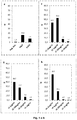
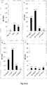
- Fig. 1 shows the results of the method according to the invention measured in flash chemiluminescence relative light units (RLU; Fig. 1a -d ).
- RLU flash chemiluminescence relative light units
- 50 ⁇ l of whole blood from a pollen allergy were stimulated with allergen extracts (t2: alder pollen extract, t3: birch pollen extract, t4: hazelnut extract) in various concentrations (10 ⁇ g - 1ng) or controls (anti-Fc ⁇ RI and fMLP) and basophil activation was determined by chemiluminescence assay.
- the bar graphs show the absolute RLU values depending on the allergen concentrations or the controls. The measured values are also indicated above the bar.
- Fig. 1a shows the reaction of the non-stimulated control (neg.
- Fig. 1b shows the reaction of the samples stimulated with different concentrations (10 ⁇ g / ml, 1 ⁇ g / ml, 100 ng / ml, 10 ng / ml) of alder pollen extract.
- Fig. 1c shows the reaction of the samples stimulated with different concentrations (1 ⁇ g / ml, 100 ng / ml, 10 ng / ml, 1 ng / ml) of birch pollen extract.
- Fig. 1b shows the reaction of the samples stimulated with different concentrations (10 ⁇ g / ml, 1 ⁇ g / ml, 100 ng / ml, 10 ng / ml, 1 ng / ml) of alder pollen extract.
- Fig. 1c shows the reaction of the samples stimulated with different concentrations (1 ⁇ g / ml, 100 ng / ml, 10 ng / ml, 1 ng / ml
- FIG. 1d shows the reaction of samples stimulated with different concentrations (10 ⁇ g / ml, 1 ⁇ g / ml, 100 ng / ml, 10 ng / ml) of hazelnut extract. The determination was carried out in triplicate. Mean values and standard deviations were calculated.
- Fig. 1 eh is the stimulation index (SI) calculated by (mean RLU activated sample) / (mean RLU neg. control) for the in Fig. 1 ad shown samples shown.
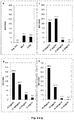
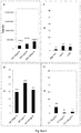
- Fig. 2 shows a basophil activation test with flow cytometric determination of the activated basophils in% ( Fig. 2a -d ) or as mean fluorescence intensity (MFI; Fig. 2e -h ).
- 50 ⁇ l of whole blood from a pollen allergy were added with the same allergen extracts as in the experiment Fig. 1 (t2: alder pollen extract, t3: birch pollen extract, t4: hazelnut extract) in various concentrations (10 ⁇ g - 1ng) or controls (anti-Fc ⁇ RI and fMLP) stimulated and basophil activation determined by flow cytometry.
- the bar graphs each show the proportions of activated basophils depending on the allergen concentrations or the controls.
- Fig. 2a shows the reaction of the non-stimulated control (negative control), as well as the control stimulation with anti-Fc ⁇ RI and fMLP. Approximately 30% of the basophils were activated in both positive controls. From a stimulation of more than 10%, the reaction was rated as positive.
- Fig. 2b shows the reaction of the samples stimulated with different concentrations (10 ⁇ g / ml, 1 ⁇ g / ml, 100 ng / ml, 10 ng / ml) of alder pollen extract.
- Fig. 1a shows the concentration of the samples stimulated with different concentrations (10 ⁇ g / ml, 1 ⁇ g / ml, 100 ng / ml, 10 ng / ml) of alder pollen extract.
- FIG. 2c shows the reaction of the samples stimulated with different concentrations (1 ⁇ g / ml, 100 ng / ml, 10 ng / ml, 1 ng / ml) of birch pollen extract.
- Fig. 2d shows the reaction of samples stimulated with different concentrations (10 ⁇ g / ml, 1 ⁇ g / ml, 100 ng / ml, 10 ng / ml) of hazelnut extract.
- Cells with a CD123 + / HLA-DR phenotype were identified as basophils, activation was detected via anti-CD63-Vio-Blue.
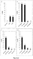
- Fig. 3 shows the results of the method according to the invention measured in flash chemiluminescence relative light units (RLU; Fig. 3a -d ).
- RLU flash chemiluminescence relative light units
- 50 ⁇ l of whole blood of a pollen allergy were stimulated with allergen extracts (f49: apple extract, g6: timothy pollen extract, t3: birch pollen extract) in various concentrations (10 ⁇ g-1ng) or controls (anti-Fc ⁇ RI and fMLP) and the basophil activation was determined by means of a chemiluminescence test.
- the bar graphs show the absolute RLU values depending on the allergen concentrations or the controls. The measured values are also indicated above the bar.
- Fig. 3a shows the reaction of the non-stimulated control (neg.
- Fig. 3b shows the reaction of the samples stimulated with different concentrations (10 ⁇ g / ml, 1 ⁇ g / ml, 100 ng / ml, 10 ng / ml) of apple extract.
- Fig. 3c Figure 4 shows the reaction of the samples stimulated with different concentrations (1 ⁇ g / ml, 100 ng / ml, 10 ng / ml, 1 ng / ml) timothy grass pollen extract.
- Fig. 3b shows the reaction of the samples stimulated with different concentrations (10 ⁇ g / ml, 1 ⁇ g / ml, 100 ng / ml, 10 ng / ml, 1 ng / ml) of timothy grass pollen extract.
- Fig. 3b shows the reaction of the samples stimulated with different concentrations (10 ⁇ g / ml, 1 ⁇ g / ml, 100 ng / ml, 10
- FIG. 3d shows the reaction of the samples stimulated with different concentrations (1 ⁇ g / ml, 100 ng / ml, 10 ng / ml, 1 ng / ml) of birch pollen extract. The determination was carried out in triplicate. Mean values and standard deviations were calculated.
- Fig. 3 eh is the stimulation index (SI) calculated by (mean RLU activated sample) / (mean RLU neg. control) for the in Fig. 3 ad shown samples shown.
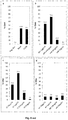
- Fig. 4 shows a basophil activation test with flow cytometric determination of the activated basophils in% ( Fig. 4a -d ) or as mean fluorescence intensity (MFI; Fig. 4e -h ).
- 50 ⁇ l of whole blood from a pollen allergy were stimulated with allergen extracts (f49: apple extract, g6: timothy grass pollen extract, t3: birch pollen extract) in various concentrations (10 ⁇ g - 1ng) or controls (anti-Fc ⁇ RI and fMLP) and basophil activation was determined by flow cytometry.
- the bar graphs each show the proportions of activated basophils as a function of the allergen concentrations or the controls. The measured values are also indicated above the bar.
- Fig. 4a shows the reaction of the non-stimulated control (negative control), as well as the control stimulation with anti-Fc ⁇ RI and fMLP. Approximately 30% of the basophils were activated in both positive controls. From a stimulation of more than 10%, the reaction was rated as positive.
- Fig. 4b shows the reaction of the samples stimulated with different concentrations (10 ⁇ g / ml, 1 ⁇ g / ml, 100 ng / ml, 10 ng / ml) of apple extract.
- Fig. 4b shows the reaction of the samples stimulated with different concentrations (10 ⁇ g / ml, 1 ⁇ g / ml, 100 ng / ml, 10 ng / ml) of apple extract.
- Figure 4 shows the reaction of the samples stimulated with different concentrations (1 ⁇ g / ml, 100 ng / ml, 10 ng / ml, 1 ng / ml) timothy grass pollen extract.
- Fig. 4d shows the reaction of the samples stimulated with different concentrations (1 ⁇ g / ml, 100 ng / ml, 10 ng / ml, 1 ng / ml) of birch pollen extract.
- Cells with a CD123 + / HLA-DR phenotype were identified as basophils, activation was detected via anti-CD63-Vio-Blue.
- the maximum reactivity of the basophils depends on the extract at 96-65% at the highest concentration used and steadily decreased with increasing dilution of the allergen extracts.
- Fig. 5 shows the results of the method according to the invention measured in flash chemiluminescence relative light units (RLU; Fig. 5a -d ).
- RLU flash chemiluminescence relative light units
- FIG. 5a shows the reaction of the non-stimulated control (neg. Ktrl.), as well as the Control stimulation with anti-Fc ⁇ RI and fMLP.
- Fig. 5b shows the reaction of the samples stimulated with different concentrations (1 ⁇ g / ml, 100 ng / ml, 10 ng / ml, 1 ng / ml) of rFel d 1.
- Fig. 5c shows the reaction of the samples stimulated with different concentrations (10 ⁇ g / ml, 1 ⁇ g / ml, 100 ng / ml, 10 ng / ml) of birch pollen extract.
- FIG. 5d shows the reaction of samples stimulated with different concentrations (10 ⁇ g / ml, 1 ⁇ g / ml, 100 ng / ml, 10 ng / ml) of mugwort pollen extract. The determination was carried out in triplicate. Mean values and standard deviations were calculated.
- Fig. 5 eh is the stimulation index (SI) calculated by (mean RLU activated sample) / (mean RLU neg. control) for the in Fig. 5 ad shown samples shown.
- Fig. 6 shows a basophil activation test with flow cytometric determination of the activated basophils in% ( Fig. 6a-d ) or as mean fluorescence intensity (MFI; Fig. 6e -h ).
- 50 ⁇ l of whole blood from a pollen allergy were stimulated with allergen extracts (e94: rField d 1, t3: birch pollen extract, w6: mugwort pollen extract) in various concentrations (10 ⁇ g - 1ng) or controls (anti-Fc ⁇ RI and fMLP) and basophil activation was determined by flow cytometry.
- the bar graphs each show the proportions of activated basophils as a function of the allergen concentrations or the controls. The measured values are also indicated above the bar.
- Fig. 6a shows the reaction of the non-stimulated control (negative control), as well as the control stimulation with anti-Fc ⁇ RI and fMLP. About 40% of the basophils were activated in both positive controls. From a stimulation of more than 10%, the reaction was rated as positive.
- Fig. 6b shows the reaction of the samples stimulated with different concentrations (1 ⁇ g / ml, 100 ng / ml, 10 ng / ml, 1 ng / ml) of rFel d 1.
- Fig. 6b shows the reaction of the samples stimulated with different concentrations (1 ⁇ g / ml, 100 ng / ml, 10 ng / ml, 1 ng / ml) of rFel d 1.
- FIG. 6c shows the reaction of the samples stimulated with different concentrations (10 ⁇ g / ml, 1 ⁇ g / ml, 100 ng / ml, 10 ng / ml) of birch pollen extract.
- Fig. 6d shows the reaction of samples stimulated with different concentrations (10 ⁇ g / ml, 1 ⁇ g / ml, 100 ng / ml, 10 ng / ml) of mugwort pollen extract.
- Cells with a CD123 + / HLA-DR phenotype were identified as basophils, activation was detected via anti-CD63-Vio-Blue.
- Fig. 7 shows the results of the method according to the invention measured in flash chemiluminescence relative light units (RLU; Fig. 7a -d ).
- RLU flash chemiluminescence relative light units
- 50 ⁇ l of whole blood from a pollen allergy were mixed with allergen extracts (e2: dog hair extract, i3: wasp venom extract; i209: rVes v 5) in various concentrations (10 ⁇ g-1 ng) or controls (anti-Fc ⁇ RI and fMLP) stimulated and the basophil activation determined by means of chemiluminescence.
- the bar graphs show the absolute RLU values depending on the allergen concentrations or the controls. The measured values are also indicated above the bar.
- Fig. 7a shows the reaction of the non-stimulated control (neg.
- Fig. 7b shows the reaction of the samples stimulated with different concentrations (10 ⁇ g / ml, 1 ⁇ g / ml, 100 ng / ml, 10 ng / ml) of dog hair extract.
- Fig. 7c shows the response of the samples stimulated with different concentrations (10 ⁇ g / ml, 1 ⁇ g / ml, 100 ng / ml, 10 ng / ml) wasp venom extract.
- Fig. 7b shows the reaction of the samples stimulated with different concentrations (10 ⁇ g / ml, 1 ⁇ g / ml, 100 ng / ml, 10 ng / ml) of dog hair extract.
- Fig. 7c shows the response of the samples stimulated with different concentrations (10 ⁇ g / ml, 1 ⁇ g / ml, 100 ng / ml, 10 ng / ml) wasp venom extract.
- FIG. 7d shows the reaction of the samples stimulated with different concentrations (10 ⁇ g / ml, 1 ⁇ g / ml, 100 ng / ml, 10 ng / ml) of rVes v 5. The determination was carried out in triplicate. Mean values and standard deviations were calculated. In Fig. 7 eh i st the stimulation index (SI) calculated by (mean RLU activated sample) / (mean RLU neg. control) for the in Fig. 7 ad shown samples shown.
- SI stimulation index
- Fig. 8 shows a basophil activation test with flow cytometric determination of the activated basophils in% ( Fig. 8a -d ) or as mean fluorescence intensity (MFI; Fig. 8e -h ).
- 50 ⁇ l of whole blood from a pollen allergy were stimulated with allergen extracts (e2: dog hair extract, i3: wasp venom extract, i209: rVes v 5) in various concentrations (10 ⁇ g - 1ng) or controls (anti-Fc ⁇ RI and fMLP) and basophil activation was determined by flow cytometry.
- the bar graphs each show the proportions of activated basophils as a function of the allergen concentrations or the controls. The measured values are also indicated above the bar.
- Fig. 8a shows the reaction of the non-stimulated control (negative control), as well as the control stimulation with anti-Fc ⁇ RI and fMLP. Approximately 30% of the basophils were activated in both positive controls. From a stimulation of more than 10%, the reaction was rated as positive.
- Fig. 8b shows the reaction of the samples stimulated with different concentrations (1 ⁇ g / ml, 100 ng / ml, 10 ng / ml, 1 ng / ml) of dog hair extract.
- Fig. 8c shows the response of the samples stimulated with different concentrations (1 ⁇ g / ml, 100 ng / ml, 10 ng / ml, 1 ng / ml) wasp venom extract.
- Fig. 8d shows the reaction of the samples stimulated with different concentrations (10 ⁇ g / ml, 1 ⁇ g / ml, 100 ng / ml, 10 ng / ml) of rVes v 5.
- Cells with a CD123 + / HLA-DR phenotype were identified as basophils, activation was detected via anti-CD63-Vio-Blue.
- Fig. 9 shows the results of the method according to the invention measured in flash chemiluminescence relative light units (RLU; Fig. 9a -d ).
- RLU flash chemiluminescence relative light units
- 50 ⁇ l of whole blood from a pollen allergy were stimulated with allergen extracts (w8: dandelion pollen extract, f17: hazelnut extract, f49: apple extract, e94 rFel d 1) in various concentrations (10 ⁇ g - 100ng) or controls (anti-Fc ⁇ RI and fMLP) and basophil activation by chemiluminescence certainly.
- the bar graphs show the absolute RLU values depending on the allergen concentrations or the controls. The measured values are also indicated above the bar.
- Fig. 9a shows the reaction of the non-stimulated control (neg.
- Fig. 9b shows the reaction of the samples stimulated with different concentrations (10 ⁇ g / ml, 1 ⁇ g / ml, 100 ng / ml) of dandelion pollen extract.
- Fig. 9c shows the reaction of the samples stimulated with different concentrations (10 ⁇ g / ml, 1 ⁇ g / ml, 100 ng / ml) of hazelnut extract.
- Fig. 9d shows the reaction of the samples stimulated with different concentrations (10 ⁇ g / ml, 1 ⁇ g / ml, 100 ng / ml) of apple extract.
- Fig. 9b shows the reaction of the samples stimulated with different concentrations (10 ⁇ g / ml, 1 ⁇ g / ml, 100 ng / ml) of dandelion pollen extract.
- Fig. 9c shows the reaction of the samples stimulated with different concentrations (10 ⁇ g / ml, 1 ⁇ g
- FIG. 9e shows the reaction of the samples stimulated with different concentrations (10 ⁇ g / ml, 1 ⁇ g / ml, 100 ng / ml) of rFel d 1. The determination was carried out in triplicate. Mean values and standard deviations were calculated.
- Fig. 9 fj is the stimulation index (SI) calculated by (mean RLU activated sample) / (mean RLU neg. control) for the in Fig. 9 ae shown samples shown.
- Fig. 10 shows a basophil activation test with flow cytometric determination of the activated basophils in% ( Fig. 10a -e ) or as mean fluorescence intensity (MFI; Fig. 10f -j ).
- 50 ⁇ l of whole blood from a pollen allergy were stimulated with allergen extracts (w8: dandelion pollen extract, f17: hazelnut extract, f49: apple extract, e94 rFel d 1) in various concentrations (10 ⁇ g - 100ng) or controls (anti-Fc ⁇ RI and fMLP) and basophil activation by flow cytometry certainly.
- the bar graphs each show the proportions of activated basophils as a function of the allergen concentrations or the controls.
- Fig. 10a shows the reaction of the non-stimulated control (negative control), as well as the control stimulation with anti-Fc ⁇ RI and fMLP. Approximately 30% of the basophils were activated in both positive controls. From a stimulation of more than 10%, the reaction was rated as positive.
- Fig. 10b shows the reaction of those with different concentrations (10 ⁇ g / ml, 1 ⁇ g / ml, 100 ng / ml) dandelion pollen extract stimulated samples.
- Fig. 10c shows the reaction of the samples stimulated with different concentrations (10 ⁇ g / ml, 1 ⁇ g / ml, 100 ng / ml) of hazelnut extract.
- Fig. 10a shows the reaction of the non-stimulated control (negative control), as well as the control stimulation with anti-Fc ⁇ RI and fMLP. Approximately 30% of the basophils were activated in both positive controls. From a stimulation of more than 10%, the reaction was rated
- 10d shows the reaction of the samples stimulated with different concentrations (10 ⁇ g / ml, 1 ⁇ g / ml, 100 ng / ml) of apple extract.
- Fig. 10e shows the reaction of the samples stimulated with different concentrations (10 ⁇ g / ml, 1 ⁇ g / ml, 100 ng / ml) of rFel d 1.
- Cells with a CD123 + / HLA-DR phenotype were identified as basophils, activation was detected via anti-CD63-Vio-Blue.
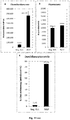
- Fig. 11 shows the results of a patient sample after stimulation with fMLP or the non-stimulated control (neg. Ktrl.) with different detection systems.
- Fig. 11a shows the results of the method according to the invention measured in flash chemiluminescence relative light units (RLU).
- Fig. 11b shows the results of the enrichment of the basophils carried out according to the invention, after detection with a fluorescence-labeled antibody (anti-CD63-FITC, 2 ⁇ g / ml) measured as fluorescence intensity (FI).
- Fig. 11c shows a basophil activation test with flow cytometric determination of the activated basophils.
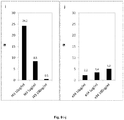
- Fig. 12 shows the results of a patient sample after stimulation with fMLP or the non-stimulated control (neg. Ktrl.) Using different antibody combinations for enrichment and detection of basophils. No allergen was used.
- Fig. 12a shows the results of the method according to the invention measured in flash chemiluminescence relative light units (RLU).
- Fig. 12b shows the stimulation index (SI) calculated by (mean RLU activated sample) / (mean RLU neg. control) for the in Fig. 12a represented sample.
- Fig. 12c shows a basophil activation test with flow cytometric determination of the activated basophils.
- Fig. 12 shows the results of a patient sample after stimulation with fMLP or the non-stimulated control (neg. Ktrl.) Using different antibody combinations for enrichment and detection of basophils. No allergen was used.
- Fig. 12a shows the results of the method according to the invention measured in flash chemilum
- FIG. 12d shows the results of an enrichment of basophils via the activation-dependent marker CD63 on anti-CD63-biotin and the detection with anti-IgE-NSP-SA.
- the anti-CD63-biotin antibody was used in concentrations of 0.2-1 ⁇ g / ml, while the detection antibody anti-IgE-NSP-SA was used in a fixed concentration of 0.5 ⁇ g / ml.
- Fig. 12e shows the stimulation index (SI) calculated by (Mean RLU activated sample) / (mean RLU neg. Control) for the in Fig. 12d shown samples.
- an anti-CD63 antibody (clone 7G3, BD Bioscience # 353014) was covalently coupled with an NSP-SA-NHS acridinium ester (Heliosense Biotechnologies, # 199293-83-9) according to the manufacturer's instructions. Subsequently, the antibody was purified on a PD-10 column (GE Healthcare, # 383420). In the same way IgE was labeled by Biomol GmbH with NSP-SA-NHS.
- T2 alder pollen extract (Squarix, # T-4402), t3 birch pollen extract (Squarix, # T-4400) and t4 hazelnut extract (Squarix, # T-4404) were used in concentrations of 10 ⁇ g / ml, 1 ⁇ g / ml, 100 ng / ml , 10 ng / ml, 1 ng / ml in Dilution Buffer (1xHanks Balanced Salt Solution (Thermo Fisher Scientific # 14065056), 40 mM Hepes, pH 7.4).
- the negative control was dilution buffer.
- fMLP (Sigma, # F3506-5MG) was used at a concentration of 10 -6 M and anti-Fc ⁇ RI antibody (clone AER-37, ebioscience, # 14-5899-82) at a concentration of 100 ng / ml.
- antigens apple extract (Squarix # F-2327), timothy pollen extract (Squarix, # G-5501), e94 (purified according to Rogers et al.
- anti-CD63-biotin Biolegend 353017
- anti-CD63-FITC Miltenyi 130-100-160
- Erythrocytes were lysed by addition of 100 ⁇ l of a saponin-containing lysis buffer (0.14% saponin in PBS) and incubation at RT for 10 min. Subsequently, the microtiter plate was centrifuged for 5 min at 500 g and the supernatant was removed.
- a saponin-containing lysis buffer 0.14% saponin in PBS
- the samples were mixed with 10 ⁇ g of streptavidin-coated magnetic beads (Sera-Mag Speedbeads streptavidin blocked, GE-Healthcare, # 21152104010350) and incubated for 10 min at RT.
- the cells bound to the beads were washed 5x with 300 ⁇ l wash buffer (0.05% Tween, 2 mM EDTA, 150 mM NaCl, 0.05% NaN 3) on the plate washer, e.g. Tecan Hydroflex equipped with a MBS96 magnet, washed.
- 300 ⁇ l wash buffer 0.05% Tween, 2 mM EDTA, 150 mM NaCl, 0.05% NaN 3
- Tecan Hydroflex equipped with a MBS96 magnet
- the NSP-SA acridinium ester on the CD63 antibody was triggered by two solutions (H2O2, 0.1 M HNO3 and 0.25M NaOH (Chemiluminescence Detection Reagent Kit, Invitron Ltd., # IV1-001)).
- the generated chemiluminescence signal flash luminescence
- the level of basophil activation was calculated by the increase in the chemiluminescent signal compared to the unstimulated control.
- a stimulation index (SI) of relative light units (RLU) of the activated sample / RLU of the unstimulated sample can be calculated.
- SI stimulation index
- RLU relative light units
- the samples were identified after activation with anti-CD123-PE (Miltenyi Biotec, # 130-098-894), anti-HLA-DR-PerCPVio700 (Miltenyi Biotec, # 130-103-873), and anti-CD63 -Vio-Blue (Miltenyi Biotec, # 130-100-154) incubated according to manufacturer's instructions for 15 min at 4 ° C protected from light.
- the fluorescence of anti-CD63-FITC was measured with a BMG Labtech Fluostar (485nm / 520nm) device.
- the chemiluminescence test ( Fig. 1 ) was considered positive when the signal of the stimulated sample doubled in relation to the control (SI> 2). In the evaluation of flow cytometric determinations ( Fig. 2 ), all samples that show an activation of ⁇ 10% are considered positive.
- the comparison of the chemiluminescence test with the flow cytometry test shows a good correlation of the results of the two test systems as well as a very good reproducibility.
- the dynamic measurement range of the chemiluminescence signal is significantly wider than the fluorescence signal.
- analysis windows In flow cytometric analysis, analysis windows (gating) must be set to define the activated and non-activated cell populations, which must be adjusted individually for each patient and are subjectively set by the respective evaluator. These possibilities of the variant evaluation is not given in the chemiluminescence test, since only the ratio of stimulated sample to control is formed. This enables a standardized evaluation, which can not be achieved in a flow cytometric test. Due to the sample preparation in the microtiter plate format, the fast measurement and a simple evaluation, a significantly higher sample throughput is possible.
- Figs. 3-10 is shown with a wide selection of antigens and sample dispensers that the inventive method is widely applicable.
- the first figure eg Fig. 3
- the result is that this is surprisingly high.
- the method according to the invention can therefore replace flow cytometry.
- chemiluminescence corresponds approximately to flow cytometry in terms of sensitivity and is significantly better suited than fluorescence, in confirmation that the method according to the invention can replace flow cytometry.
Landscapes
- Health & Medical Sciences (AREA)
- Life Sciences & Earth Sciences (AREA)
- Immunology (AREA)
- Engineering & Computer Science (AREA)
- Chemical & Material Sciences (AREA)
- Biomedical Technology (AREA)
- Hematology (AREA)
- Molecular Biology (AREA)
- Urology & Nephrology (AREA)
- Cell Biology (AREA)
- Physics & Mathematics (AREA)
- Pathology (AREA)
- General Health & Medical Sciences (AREA)
- Analytical Chemistry (AREA)
- Biochemistry (AREA)
- General Physics & Mathematics (AREA)
- Food Science & Technology (AREA)
- Medicinal Chemistry (AREA)
- Biotechnology (AREA)
- Microbiology (AREA)
- Tropical Medicine & Parasitology (AREA)
- Chemical Kinetics & Catalysis (AREA)
- Plasma & Fusion (AREA)
- Ecology (AREA)
- Proteomics, Peptides & Aminoacids (AREA)
- Bioinformatics & Cheminformatics (AREA)
- Toxicology (AREA)
- Physiology (AREA)
- Surgery (AREA)
- Animal Behavior & Ethology (AREA)
- Vascular Medicine (AREA)
- Biophysics (AREA)
- Heart & Thoracic Surgery (AREA)
- Medical Informatics (AREA)
- Rehabilitation Therapy (AREA)
- Rheumatology (AREA)
- Public Health (AREA)
- Veterinary Medicine (AREA)
- Zoology (AREA)
- Virology (AREA)
Abstract
Description
- Die vorliegende Erfindung betrifft ein Verfahren zur Diagnose einer Allergie, umfassend die Schritte Kontaktieren von Basophilen aus einer Vollblutprobe eines Patienten mit einem Allergen in einer wässrigen Lösung in Anwesenheit des Vollblutes mit einem Volumenanteil des Vollblutes an der wässrigen Lösung von wenigstens 20 Volumenprozent unter Bedingungen, die eine Aktivierung der Basophilen durch das Allergen zulassen, Anreichern der Basophilen aus a) und Nachweisen aktivierter Basophiler, wobei das Nachweisen dadurch erreicht wird, dass die ein für aktivierte Basophile charakteristischer Marker mittels Chemilumineszenz detektiert wird; und ein Kit umfassend ein Gefäß zur Kontaktierung der Basophilen mit dem Allergen, wobei es sich bei dem Gefäß bevorzugt um eine Mikrotiterplatte handelt, optional ein Allergen, ein Mittel zur Detektion eines für aktivierte Basophile charakteristischen Markers mittels Chemilumineszenz, wobei es sich bevorzugt um einen mit einer chemilumineszenzfähigen Markierung versehenen Liganden gegen den Marker handelt und der Ligand noch bevorzugter ein monoklonaler Antikörper ist, optional ein Mittel zum Stoppen der Aktivierung der Basophilen, einen optional immobilisierbaren Liganden, der an ein unabhängig von ihrem Aktivierungszustand an der Zelloberfläche von Basophilen lokalisiertes Polypeptid bindet, wobei es sich bei dem Liganden bevorzugt um einen monoklonalen Antikörper gegen das Polypeptid handelt und optional ein Mittel zum Lysieren und/oder Entfernen von Erythrozyten.
- Zur Diagnose von Allergien des Soforttyps, auch als Typ I-Allergien bezeichnet, die auf einer IgE-vermittelte Immunantwort basieren, werden nach einer ausführlichen Anamnese vorrangig Hauttests wie Skin-Prick-Tests oder Provokationstests durchgeführt, die für die Allergiediagnostik der Goldstandard sind. Als Ergänzung werden in vitro-Tests eingesetzt, mit denen die Anwesenheit von spezifischen IgE-Antikörpern gegen ein oder mehr als ein definiertes Allergen nachgewiesen wird. Für einen solchen Nachweis werden Allergene an Matrizes wie Schwämmchen, Membranen oder Kügelchen gebunden und mit Patientenseren kontaktiert. Anschließend wird die Menge an gebundenem IgE mit Hilfe eines markierten anti-human-lgE Antikörpers bestimmt.
- Die Vorteile der spezifischen IgE-Bestimmung in vitro umfassen ihre leichte Durchführbarkeit, Standardisierbarkeit und Aufskalierbarkeit. Allerdings kann man häufig nicht zwischen einer klinisch relevanten Allergie und einer bloßen Sensibilisierung unterscheiden.
- Weitere Nachteile sind die schlechte Verfügbarkeit von manchen Antigenen und eine mögliche Konformationsänderung des Allergens durch dessen Kopplung an eine Matrix, was die Affinität zum nachzuweisenden IgE-Antikörper verändern kann. Allergenextrakte können in vielen Fällen nur unter Schwierigkeiten oder gar nicht standardisiert werden. Kleinere Allergene müssen zudem an Carriermoleküle wie z. B. Serumalbumin gebunden werden. Weiterhin muss die Spezifität für Allergene von IgE-Antikörpern in Serum und Membran gebundenen IgE-Antikörper auf Effektorzellen wie Basophilen und Mastzellen nicht übereinstimmen, da nur ein langsamer Austausch von gelöstem und gebundenem IgE stattfindet. Schließlich bestimmen zusätzliche Faktoren, die bei einem In vitro-Test zum Nachweis spezifischer IgE-Antikörper nicht abgebildet werden können, ob tatsächlich eine zelluläre allergische Reaktion ausgelöst wird, beispielsweise das Gesamtverhältnis von gesamt-IgE zu spezifischem IgE im Blut oder die Affinität der Antikörper.
- Insbesondere dann, wenn IgE- und Hauttests unklare Ergebnisse liefern, ist daher empfehlenswert, als Ergänzung ein zelluläres Testsystem heranzuziehen wie den Basophilen-Degranulationstests oder den Basophilen-Aktivierungstest (BAT). Üblicherweise wird der Begriff "BAT" mit Bezug auf Durchflusszytometrie-Tests gebraucht.
- Der besondere Vorteil der zellulären Tests gegenüber der spezifischen IgE-Antikörperbestimmung, die lediglich den gelösten, biologisch nicht aktiven IgE-Anteil im Serum bestimmt, besteht darin, dass sie die tatsächliche zellbiologische Reaktion erfassen, welche durch die Bindung des Allergens an Rezeptor-gebundene IgE-Antikörper an der Zelloberfläche der Basophilen und deren Quervernetzung ausgelöst wird. Die zellulären Tests ermöglichen es außerdem, die Schwelle der Degranulationsfähigkeit zu bestimmen. Dazu besteht insbesondere nach einer Immuntherapie zur Überwachung des Therapieerfolges des Patienten Bedarf.
- Grundsätzlich werden zelluläre Tests in der Weise durchgeführt, dass zunächst Patientenblut mit einem Allergen kontaktiert wird. War der Patient bereits zuvor sensibilisiert, so kommt es zur Bindung des Allergenes an Rezeptor-gebundene allergenspezifische IgE-Antikörper, die an der Zelloberfläche der Basophilen lokalisiert sind. Dies führt zu einer Kreuzvernetzung der IgE-Rezeptoren, die wiederum eine Degranulation der Basophilen Granulozyten und damit die Freisetzung von Mediatoren aus intrazellulären Granula auslöst. Diese Mediatoren, z. B. Histamin, Leukotriene und Tryptase, können mit Hilfe von Enzymimmunoassays im Überstand quantifiziert werden. Die Ausschüttung der Mediatoren geht mit einer erhöhten Konzentration an der Zelloberfläche von Markern wie CD63 oder CD203c einher, welche zuvor auf den Membranen der intrazellulären Granula lokalisiert waren und somit ebenfalls eine zelluläre Reaktion zeigen. Die Ausschüttung der Mediatoren zeigt damit eine zelluläre Reaktion auf das Allergen.
- Diese verstärkte Oberflächenkonzentration wird bislang in der Routinediagnostik durchflusszytometrisch mit Hilfe von Antikörpern gegen die aktivierungsspezifischen Zelloberflächenmarker nachgewiesen, die an einem Fluorophor gekoppelt sind. Die zu untersuchenden Zellen fließen dabei einzeln hintereinander in einem Flüssigkeitsstrahl durch eine dünne Messkammer und passieren mehrere Laserstrahlen verschiedener Wellenlänge, wobei ein für jeden Zelltyp charakteristisches Streulicht erzeugt wird und gleichzeitig, in Abhängigkeit der jeweiligen Wellenlänge, die an die Antikörper gekoppelten Fluorophore angeregt werden, sofern aktivierte Zellen vorhandeln sind. Das vom angeregten Fluorophor emittierte Licht und das Streulicht werden von Detektoren erfasst.
- Nachteile dieses Testsystems sind die hohen Anschaffungskosten für ein Durchflusszytometer, die Komplexität der Testergebnisse, die nur von speziell geschultem Fachpersonal interpretiert werden können sowie die schwierige Standardisierbarkeit. Zudem kann mit einem Gerät immer nur eine Probe gleichzeitig gemessen werden.
- Ein weiteres der Erfindung zugrunde liegendes Problem betrifft die Qualität von Proben, die aktivierbare Basophile umfassen. Die Qualität der Proben und damit deren Eignung für diagnostische Verfahren hängt davon ab, ob Gewinnung, Transport und Lagerung fachmännisch durchgeführt wurden. Beispielsweise kann eine Exponierung der Proben gegenüber Hitze oder ungeeigneten Puffern zum Absterben der Zellen führen.
- Ist die Qualität unzureichend, so kann es zu falsch-negativen Ergebnissen kommen, d.h. ein negatives Ergebnis ist nicht dadurch bedingt, dass der Patient nicht allergisch ist, sondern dadurch, dass die Probe von ihm von unzureichender Qualität ist, genauer keine oder zu wenig aktivierbare Basophile enthält.
- Es ist besonders ineffizient, wenn die mangelnde Qualität einer Probe erst bei der Durchführung eines aufwändigen Verfahrens wie der Durchflusszytometrie festgestellt wird.
- Es besteht daher Bedarf an einem Verfahren und dazu brauchbaren, die Qualität einer Probe effizient, d.h. in einem für Hochdurchsatz tauglichen Verfahren festzustellen. Ein solches Verfahren wird zwar nicht mit einem Allergen durchgeführt und liefert daher kein diagnostisch nützliches Ergebnis, lässt aber eine Vorhersage zu, ob die Probenqualität für ein in einem getrennten Ansatz durchgeführtes diagnostisches Verfahren geeignet ist. Dieses Verfahren kann zuvor, nachträglich oder parallel zum diagnostischen Verfahren durchgeführt werden. Dieses Verfahren oder die dabei eingesetzten Reagenzien können dazu dienen, die Anzahl oder den Anteil aktivierbarer Basophiler in einer Zellpräparation, bevorzugt einer Probe zu bestimmen.
-
DE10235310 offenbart einen Test, bei dem die Aktivierung von Basophilen mit Hilfe eines fluoreszierenden Calciumfarbstoffes nachgewiesen wird. Es wird über eine Dichtegradientenzentrifugation fraktioniertes Blut eingesetzt. -
WO2012/044756 ,US2009/0246805 ,WO98/32014 EP2037269 offenbaren durchflusszytometrische Tests zum Nachweis der Aktivierung von Basophilen. - Von diesem Hintergrund besteht eine der vorliegenden Erfindung zugrundeliegende Aufgabe darin, ein Verfahren und dazu geeignete Reagenzien bereitzustellen, mit dem bzw. mit denen die oben genannten Nachteile von im Stand der Technik beschriebenen Verfahren überwunden werden können, insbesondere ein Basophilen-Aktivierungstest, der mit einem anderen System als einem Durchflusszytometer durchgeführt werden kann.
- Insbesondere besteht eine Aufgabe darin, ein System zu schaffen, dass die gleichzeitige Abarbeitung einer Vielzahl von Proben in einem Hochdurchsatzverfahren erlaubt. Weiterhin sind Reproduzierbarkeit und Standardisierbarkeit zu gewährleisten.
- Diese und weiteren Aufgaben werden durch den Gegenstand der vorliegenden Erfindung insbesondere durch den Gegenstand der beigefügten unabhängigen Ansprüche gelöst, wobei sich bevorzugte Ausführungsformen aus den Unteransprüchen ergeben.
- In einem ersten Aspekt wird das die Erfindung zugrundeliegende Problem gelöst durch ein Verfahren zur Diagnose einer Allergie, bevorzugt ein für den Hochdurchsatz geeignetes Verfahren, umfassend die Schritte:
- a) Kontaktieren von Basophilen aus einer Vollblutprobe eines Patienten mit einem Allergen in einer wässrigen Lösung in Anwesenheit des Vollblutes mit einem Volumenanteil des Vollblutes an der wässrigen Lösung von wenigstens 20 Volumenprozent unter Bedingungen, die eine Aktivierung der Basophilen durch das Allergen zulassen,
- b) Anreichern der Basophilen aus a) und
- c) Nachweisen aktivierter Basophiler,
- Alternativ wird das Problem in einem ersten Aspekt gelöst durch ein Verfahren zur Diagnose einer Allergie, umfassend die Schritte:
- a) Kontaktieren von Basophilen aus einer Vollblutprobe eines Patienten mit einem Allergen in einer wässrigen Lösung in Anwesenheit des Vollblutes mit einem Volumenanteil des Vollblutes an der wässrigen Lösung von wenigstens 20 Volumenprozent unter Bedingungen, die eine Aktivierung der Basophilen durch das Allergen zulassen,
- b) Anreichern der Basophilen aus a) und
- c) Nachweisen aktivierter Basophiler,
- In einer bevorzugten Ausführungsform handelt sich bei dem Vollblut um unprozessiertes Vollblut.
- In einer weiteren bevorzugten Ausführungsform wird Schritt a) in einer wässrigen Lösung mit einem Vollblutanteil von wenigstens 30, bevorzugt 35, noch bevorzugter 40, noch bevorzugter 45 Volumenprozent durchgeführt.
- In einer weiteren Ausführungsform umfasst das Verfahren den Schritt Stoppen der Aktivierung von Basophilen, bevorzugt vor Schritt b).
- In einer weiteren Ausführungsform werden die Basophilen in Schritt b) dadurch angereichert, dass sie an einen Liganden binden, der spezifisch an ein Polypeptid bindet, das bei Basophilen an der Zelloberfläche lokalisiert ist. Bei diesem Polypeptid kann es sich um ein Polypeptid handeln, das unabhängig von ihrem Aktivierungszustand an der Zelloberfläche der Basophilen lokalisiert ist, und/oder um einen für aktivierte Basophile charakteristischen Marker, bevorzugt einen weiteren für aktivierte Basophile charakteristischen Marker, der sich von dem Marker unterscheidet, der erfindungsgemäß mittels Chemilumineszenz detektiert wird. In einer besonders bevorzugten Ausführungsform handelt es sich um ein Polypeptid, das unabhängig vom Aktivierungszustand der Basophilen an ihrer Zelloberfläche lokalisiert ist.
- In einer weiteren Ausführungsform ist der Ligand, der spezifisch an ein Polypeptid bindet, das bei Basophilen unabhängig von ihrem Aktivierungszustand an ihrer Zelloberfläche lokalisiert ist, ein monoklonaler Antikörper gegen das Polypeptid.
- In einer bevorzugten Ausführungsform werden die Basophilen in Schritt b) immobilisiert.
- In einer bevorzugten Ausführungsform werden die Basophilen in Schritt b) an einem Bead immobilisiert, bevorzugt einem magnetischen Bead.
- In einer bevorzugten Ausführungsform werden die Basophilen nach dem Immobilisieren gewaschen.
- In einer bevorzugten Ausführungsform werden die Erythrozyten in Schritt b) lysiert und entfernt.
- In einer bevorzugten Ausführungsform werden die Erythrozyten vor dem Immobilisieren der Basophilen durch Zentrifugieren entfernt.
- In einer bevorzugten Ausführungsform wird das Nachweisen in Schritt c) mit Hilfe eines mit einer chemilumineszenzfähigen Markierung versehenen Liganden gegen den an der Oberfläche von Basophilen lokalisierten Marker durchgeführt, bevorzugt einen Marker, der für aktivierte Basophile charakteristisch ist.
- In einer bevorzugten Ausführungsform handelt es sich bei dem mit einer chemilumineszenzfähigen Markierung versehenen Liganden um einen monoklonalen Antikörper, der selbst eine chemilumineszenzfähige Markierung aufweist oder an einen sekundären Antikörper mit einer chemilumineszenzfähigen Markierung bindet.
- In einer bevorzugten Ausführungsform werden die Schritte a), b) und c) in einer Mikrotiterplatte ausgeführt.
- In einer bevorzugten Ausführungsform wird Schritt a) vor den Schritten b) und c) durchgeführt.
- In einem weiteren Aspekt wird die der Erfindung zugrundeliegende Aufgabe gelöst durch ein Kit umfassend ein Gefäß zur Kontaktierung der Basophilen mit dem Allergen, wobei es sich bei dem Gefäß bevorzugt um eine Mikrotiterplatte handelt, optional ein Allergen, ein Mittel zur Detektion eines für aktivierte Basophile charakteristischen Markers mittels Chemilumineszenz, wobei es sich bevorzugt um einen mit einer chemilumineszenzfähigen Markierung versehenen Liganden gegen den Marker handelt und der Ligand noch bevorzugter ein monoklonaler Antikörper ist, optional ein Mittel zum Stoppen der Aktivierung der Basophilen, einen optional immobilisierbaren Liganden, der an ein unabhängig von ihrem Aktivierungszustand an der Zelloberfläche von Basophilen lokalisiertes Polypeptid bindet, wobei es sich bei dem Liganden bevorzugt um einen monoklonalen Antikörper gegen das Polypeptid handelt und optional ein Mittel zum Lysieren und/oder Entfernen von Erythrozyten.
- Alternativ wird das Problem in einem weiteren Aspekt gelöst durch einen Kit umfassend ein Gefäß zur Kontaktierung der Basophilen mit dem Allergen, wobei es sich bei dem Gefäß bevorzugt um eine Mikrotiterplatte handelt, optional ein Allergen, einen Liganden gegen ein an der Zelloberfläche von Basophilen lokalisiertes Polypeptid zur Anreicherung von Basophilen und mit einem Liganden gegen einen an der Oberfläche von Basophilen lokalisierten Marker zur Detektion, wobei der Ligand gegen den an der Oberfläche von Basophilen lokalisierten Marker zur Detektion eine chemilumineszenzfähige Markierung aufweist, wobei es sich bei der chemilumineszenzfähigen Markierung um ein Enzym handeln kann, das eine Chemilumineszenz-Reaktion katalysiert, oder um ein Molekül, das selbst unter geeigneten Bedingungen ein Chemilumineszenz-Signal erzeugt, wobei das Enzym im ersten Falle in Kombination mit einer chemilumineszenzfähigen Substratlösung im Kit enthalten ist, und wobei das an der Zelloberfläche von Basophilen lokalisierte Polypeptid und/oder der an der Oberfläche von Basophilen lokalisierte Marker charakteristisch für aktivierte Basophile ist. Optional umfasst der Kit ein Mittel zum Stoppen der Aktivierung der Basophilen. Optional umfasst der Kit ein Mittel zum Lysieren und/oder Entfernen von Erythrozyten.
- Die in einem erfindungsgemäßen Kit enthaltenen Reagenzien können auch mit einem oder ohne ein Gefäß zur Kontaktierung der Basophilen mit dem Allergen zur Herstellung eines Kits zur Diagnose einer Allergie verwendet werden.
- Das Problem wird in einem weiteren Aspekt gelöst durch eine Verwendung eines Liganden gegen ein an der Zelloberfläche von Basophilen lokalisiertes Polypeptid zur Anreicherung von Basophilen zusammen mit einem Liganden gegen einen an der Oberfläche von Basophilen lokalisierten Marker zur Detektion zur Diagnose einer Allergie oder zur Herstellung eines Kits zur Diagnose einer Allergie oder zum Screening von Kandidatenwirkstoffen für die Therapie einer Allergie gegen ein Allergen, wobei der Ligand gegen den an der Oberfläche von Basophilen lokalisierten Marker zur Detektion eine chemilumineszenzfähige Markierung aufweist, wobei es sich bei der chemilumineszenzfähigen Markierung um ein Enzym handeln kann, das eine Chemilumineszenz-Reaktion katalysiert, oder um ein Molekül, das selbst unter geeigneten Bedingungen ein Chemilumineszenz-Signal erzeugt, wobei das Enzym im ersten Falle in Kombination mit einer chemilumineszenzfähigen Substratlösung verwandt wird, und wobei das an der Zelloberfläche von Basophilen lokalisiertes Polypeptid und/oder der an der Oberfläche von Basophilen lokalisierte Marker charakteristisch für aktivierte Basophile ist.
- Das Problem wird in einem weiteren Aspekt gelöst durch eine Verwendung eines Liganden gegen einen für aktivierte Basophile charakteristischen Liganden zur Diagnose einer Allergie oder zur Herstellung eines Kits zur Diagnose einer Allergie oder zum Screening von Kandidatenwirkstoffen für die Therapie einer Allergie gegen ein Allergen, wobei der Ligand eine chemilumineszenzfähige Markierung aufweist.
- In einer bevorzugten Ausführungsform ist die Verwendung zur Erhöhung der Sensitivität, bevorzugt in einem Hochdurchsatzverfahren.
- Die vorliegende Erfindung betrifft ein Verfahren zur Diagnose einer Allergie zunächst umfassend den Schritt a) Kontaktieren von Basophilen aus einer Vollblutrobe eines Patienten mit einem Allergen in einer wässrigen Lösung. Bei der Vollblutprobe handelt es sich bevorzugt um Vollblut mit antikoagulierend wirkenden Substanzen. Das Vollblut wird der wässrigen Lösung vor dem eigentlich Kontaktieren der Basophilen mit dem Allergen in einer Menge hinzugefügt, dass sein finaler Volumenanteil an der wässrigen Lösung wenigstens 20, bevorzugter 25, 30, 35, 40, 45, 50, 55, 60, 65, 70, 75 oder 80 Volumenprozent beträgt. Die Anwesenheit des Vollblutes führt dazu, dass das Kontaktieren der Basophilen mit dem Allergen unter nahezu physiologischen Bedingungen abläuft.
- Bei dem Verfahren handelt es sich bevorzugt um ein für den Hochdurchsatz geeignetes Verfahren. Darunter wird in einer bevorzugten Ausführungsform ein Verfahren verstanden, das die parallele Abarbeitung einer Vielzahl von Proben, beispielsweise wenigstens 2, 4, 8, 16 oder 32 Proben gestattet, wobei die Verfahrensschritte, die wenigstens 50, bevorzugter 60, 70, 80 oder 90% der Abarbeitungszeit für eine einzelne Probe erfordern, im Wesentlichen gleichzeitig mit den Proben von statten gehen können. Verfahren, bei denen der eigentliche Messvorgang zwar mittels einer Messvorrichtung bei allen Proben nacheinander durchgeführt wird, die individuelle, nicht-parallele Abarbeitung einer Probe aber nicht länger als 5, bevorzugter 4, 3, 2, 1 Minute oder 30 Sekunden nicht überschreitet, sind umfasst. Für den Hochdurchsatz geeignete Verfahren umfassen beispielsweise die Chemilumineszenzmessung in dafür geeigneten Mikrotiterplatten. Hingegen ist beispielsweise die Durchflusszytometrie kein für den Hochdurchsatz geeignetes Verfahren.
- Bei dem Vollblut handelt es sich bevorzugt um unprozessiertes Vollblut. In einer bevorzugten Ausführungsform wird unter dem Begriff "unprozessiertes Vollblut", wie hierin verwendet, verstanden, dass das Blut zwar verdünnt sein kann oder ihm chemische Substanzen zur Konservierung zugesetzt werden, bevorzugt antikoagulierend wirkende Substanzen wie Heparin, dass es jedoch nicht fraktioniert ist, beispielsweise durch Zentrifugation wie eine Dichtegradientenzentrifugation.
- Schritt a) kann auch mit mehr als einem Allergen und/oder einem Allergen in mehr als einer Konzentration in unterschiedlichen, parallel abgearbeiteten Ansätzen durchgeführt werden. Insbesondere kann das Antigen in zwei oder mehr, bevorzugt drei oder mehr unterschiedlichen Konzentrationen in einer entsprechenden Zahl von parallelen Ansätzen eingesetzt werden. Bevorzugt werden zusätzlich eine Positivkontrolle und eine Negativkontrolle mitgeführt. In einer bevorzugten Ausführungsform handelt es sich bei dem Allergen um ein Material ausgewählt aus der Gruppe umfassend Milbenkulturen und Milbenfäkalien der taxonomischen Hauptgruppe Animalia Arthropoda; Schuppen, Haar, Speichel, Kot oder anderen Sekrete, die von der taxonomischen Hauptgruppe Animalia Chordata stammen; Sporen oder Partikel, die von der taxonomischen Hauptgruppe Funghi Ascomycetes stammen; Pollen, der von der taxonomischen Hauptgruppe Corniferopsida stammt, Pollen, der von der taxonomischen Hauptgruppe Plantae Magnoliopsidae stammt, Pollen, der von der taxonomischen Hauptgruppe Plantae Liliopsidae stammt; Gift oder Sekrete, die von der taxonomischen Hauptgruppe Animalia Arthropoda stammen; Kautschuk oder Produkte, die einen solchen Kautschuk umfassen, der von Bäumen der taxonomischen Hauptgruppe Plantae Magnoliopsidae stammt.
- In einer bevorzugten Ausführungsform handelt es sich bei dem Allergen um eine Arzneimittelsubstanz ausgewählt aus der Gruppe umfassend Antibiotika, bevorzugter β-Lactame, noch bevorzugter aus der Gruppe umfassend Penicillin G, Penicillin V, PPL, MDM, Amoxicillin und Ampicillin; Cephalosporine, noch bevorzugter aus der Gruppe umfassend Cefazolin, Cefamandol, Cefaclor, Cefonaxim, Ceftazidim, Cefotaxim, Ceftazidim, Cefepim;, Carbapeneme, Monobactame, β-Lactamase-Inhibitoren, noch bevorzugter Clavulansäure; Makrolide, Aminoglycoside, Rifamycine, Glykopetide, Polypeptide, Tetracycline, Imidazole, Chinolone, Pyrazolone, , noch bevorzugter Sulfomethoxazol; Streptogramine, Nitrofurane, Isoniazide, Pentamidine; Antiseptika, bevorzugtervorzugsweise Chlorhexidin, Fungizide, Antivirale Mittel, Anti-Malaria-Mittel, Analgetika, COX-2-Inhibitoren und nicht-steroidale entzündungshemmende Arzneimittel, bevorzugt aus der Gruppe umfassend Aspirin, Lys-Aspirin, luprofen, Ketoprofen, Diclofenac, Naproxen, Paracetamol, Dipyron, Indomethacin, Mefenaminsäure, Phenylbutazon und Propyphenazon; Neuromuskuläre Blocker, bevorzugt aus der Gruppe umfassend Suxamethonium, Atracurium, Cis-Atracurium, Mivacurium, Pancuronium, Rocuronium, Vecuronium und Succinylcholin; Hypnotika und Lokalanästhetika, bevorzugt aus der Gruppe umfassendMidazolam, Propofol, Thiopental, Fentanyl und Lidocain; Tranquilizer; Opioide; Radio-Kontrastmittel, bevorzugt aus der Gruppe umfassend ionische Iodkontrastmittel, nichtionische iodierte Kontrastmittel, Isosulfanblau, Patentblau und Methylenblau; Protonenpumpeninhibitoren. Antikonvulsiva und Neuroleptika, bevorzugt aus der Gruppe umfassend Carbamazepin, Phenytoin und Valproinsäure; Anti-psychotische Mittel; Antidepressiva; Dopamine; Anti-histamine; Kortikosteroide und Glukokortikoide; Chemotherapeutische und immunsuppressive Mittel; Diuretika; Antikoagulantien; Vasokonstriktoren und Vasodilatatoren; Herzarzneimittel, bevorzugt aus der Gruppe umfassendStatine, ACE-Inhibitoren, Alpha-Rezeptorblocker, Beta-Rezeptorblocker Calciumantagonisten und Anti-Hypertonika; (Anti-)Geschwür Drogen; (Anti-)Schilddrüsenmittel; Östrogene; Heparine und Derivate; Insuline; Streptokinasen und Urokinasen;
- In einer bevorzugten Ausführungsform handelt es sich bei dem Allergen um einen Kolloid-, Plasmaexpander oder Hilfsmittel, bevorzugt ausgewählt aus der Gruppe umfassend Albumin, Dextran, Gelatine, Hetastarch, Pentastarch, Sinistrin, Polydocanol 600 (Ethoxysclerol), Laktose, Carboxymethylcellulose, Hydroxypropylcellulose, Protamin und Aprotinin.
- In einer bevorzugten Ausführungsform handelt es sich bei dem Allergen um einen Nahrungsmittelzusatzstoff, bevorzug ausgewählt aus der Gruppe umfassend Lebensmittelkonservierungsmittel, Lebensmittelfarbstoffe, Nahrungsmittelveredelungsmitte, Antioxidationsmittel und Emulgatoren.
- In einer bevorzugten Ausführungsform handelt es sich bei dem Allergen um ein Umwelt- oder Schadstoffmittel, bevorzugt ausgewählt aus der Gruppe umfassend Isocyanate, Isothiazolinone, Formaldehyd, Ethylenoxid, Phthalsäureanhydrid, Chloramin T, DMSO, Latex und Enzyme, die in der Back-, Lebensmittelverarbeitung und Waschindustrie verwendet werden.
- In einer bevorzugten Ausführungsform handelt es sich bei dem Allergen um ein Material ausgewählt aus der Gruppe umfassend eine Kultur und/oder Fäkalien einer Milbe, die ausgewählt ist aus der Gruppe bestehend aus Milben der Gattung Acarus, Glycyphagus, Lepidoglyphus, Tryophagus, Blomia, Dermatophagoides, Euroglyphus, Blatella und Periplaneta; Schuppen, Haar, Speichel oder Kot eines Tiers einer Gattung ausgewählt aus Canis, Felis oder Equus; Hefe, Schimmelpilz oder Pilze einer Gattung ausgewählt aus der Gruppe bestehend aus Cladosporium, Aspergillus, Penicillium, Alternaria und Candida; Pollen einer Gattung ausgewählt aus der Gruppe bestehend aus Chamaecyparis, Cryptomeria, Cupressus, Juniperus, Anthoxanthum, Cynodon, Dactylis, Festuca, Holcus, Hordeum, Lolium, Oryza, Paspalum, Phalaris, Phleum, Poa, Secale, Sorghum, Triticum, Zea, Ambrosia, Artemisia, Alnus, Betula, Corylus, Fraxinus, Olea und Platanus; Gift eines Insekts einer Gattung ausgewählt aus der Gruppe bestehend aus Apis Bombus, Dolichovespula, Polistes, Polybia, Vespa und Vespula; Kautschuk oder Produkte, die einen solchen Kautschuk umfassen, der aus der Gattung Hevea stammt.
- In einer bevorzugten Ausführungsform handelt es sich bei dem Allergen um ein Material ausgewählt aus der Gruppe umfassend Shrimps oder eine Shrimps-haltigen Nahrung der taxonomischen Hauptgruppe Animalia Arthropoda; Hummer oder eine Hummer-haltigen Nahrung der taxonomischen Hauptgruppe Animalia Arthropoda; Früchte, Hülsenfrüchte, Getreide oder Bohnen, die aus der taxonomischen Hauptgruppe Plantae Liliopsidae stammen oder ein Nahrungsmittelprodukt, das solche Früchte, Hülsenfrüchte, Getreide und/oder Bohnen umfasst; Früchte, Hülsenfrüchte, Getreide oder Bohnen, die aus der taxonomischen Hauptgruppe Plantae Magnoliopsidae stammen oder ein Nahrungsmittelprodukt, das solche Früchte, Hülsenfrüchte, Getreide und/oder Bohnen umfasst; Nuss oder einer nusshaltigen Nahrung der taxonomischen Hauptgruppe Plantae Magnoliopsidae.
- In einer bevorzugten Ausführungsform handelt es sich bei dem Allergen um Material ausgewählt aus der Gruppe umfassend Kuhmilch, Kuhmilch-haltiger Nahrung, Hühnereiweiß, Hühnereiweißhaltiger Nahrung, Fisch oder fischhaltige Nahrung.
- In einer bevorzugten Ausführungsform handelt es sich bei dem Allergen um Material ausgewählt aus der Gruppe umfassend Hordeum, Oryza, Secale, Triticum, Zea, Arachis, Corylis, Juglans, Prunus, Anacardium, Pistacia und Glycine.
- In einer bevorzugten Ausführungsform handelt es sich bei dem Allergen um ein Polypeptid umfassend ein Antigen oder eine Variante davon aus der Gruppe umfassen Aca s 13, Gly d 2, Lep d 2, Lep d 5, Lep d 7, Lep d 10, Lep d 13, Tyr p 2, Tyr p 3, Tyr p 10, Tyr p 13, Tyr p 24, Blo t 1, Blo t 2 Blo t 3, Blo t 4, Blo t 5, Blo t 6, Blo t 10, Blo t 11, Blo t 12, Blo t 13, Blo t 19, Blo t 21, Der f 1, Der f 2, Der f 3, Der f 6, Der f 7, Derf 10, Derf 11, Derf 13, Derf 14, Derf 15, Derf 16, Der f 17, Der f 18, Der f 22, Der m 1, Der p 1, Der p 2, Der p 3, Der p 4, Der p 5, Der p 6 Der p 7, Der p 8, Der p 9, Der p 10, Der p 11, Der p 14, Der p 20, Der p 21, Der p 23, Eur m 1, Eur m 2, Eur m 3, Eur m 4, Eur m 14, Bla g 1, Bla g 2, Bla g 4, Bla g 5, Bla g 6, Bla g 7, Bla g 8, Per a 1, Per a 3, Per a 6, Per a 7, Per a 9, Per a 10, Can f 1, Can f 2, Can f 3, Can f 4, Can f 5, Can f 6, Fel d 1, Fel d 2, Fel d 3, Fel d 4, Fel d 5w, Fel d 6w, Fel d 7, Fel d 8, Equ c 1, Equ c 2, Equ c 3, Equ c 4 and Equ c 5, Cla c 9m, Cla c 14, Cla h 2, Cla h 5, Cla h 6, Cla h 7, Ca h 8, Cla h 9, Cla h 10, Cla h 12, Asp fl 13, Asp f 1, Asp f 2, Asp f 3, Asp f 4, Asp f 5, Asp f 6, Asp f 7, Asp f 8, Asp f 9, Asp f 10, Asp f 11, Asp f 12, Asp f 13, Asp f 15, Asp f 16, Asp f 17, Asp f 18, Asp f 22, Asp f 23, Asp f 27, A5p f 28, A5p f 29, Asp f 34, Asp n 14, Asp n 18, Asp n 25, Asp o 13, Asp o 21, Pen b 13, Pen b 26, Pen ch 13, Pen ch 18, Pen ch 20, Pen ch 31, Pen ch 33, Pen ch 35, Pen c 3, Pen c 13, Pen c 19, Pen c 22, Pen c 24, Pen C 30, Pen c 32, Pen o 18, Fus c 1, Fus c 2, Alt a 1, Alt a 3, Alt a 4, Alt a 5, Alt a 6, Alt a 7, Alt a 8, Alt a 10, Alt a 12, Alt a 13, Cand a 1, Cand a 3 and Cand b 2, Cha o 1, Cha o 2, Cup a 1, Cup s 1, Cup s 3, Jun a 1, Jun a 2, Jun a 3, Jun o 4, Jun s 1, Jun v 1 and Jun v 3, Ant o 1, Cyn d 1, Cyn d 7, Cyn d 12, Cyn d 15, Cyn d 22w, Cyn d 23, Cyn d 24, Dac g 1, Dac g 2, Dac g 3, Dac g 4, Dac g 5, Fes p 4, Hol l 1, Hol l 5, Hor v 1, Hor v 5, Lol p 1, Lol p 2, Lol p 3, Lol p 4, Lol p 5, Lol p 11, Pas n 1, Pha a 1, Pha a 5, Phl p 1, Phl p 2, Phl p 4, Phl p 5, Phl p 6, Phl p 7, Phl p 11, Phl p 12, Phl p 13, Poa p 1, Poa p 5, Sec c 1, Sec C 5, Sec c 20, Sor h 1, Tri a 15, Tri a 21, Tri a 27, Tri a 28, Tri a 29, Tri a 30, Tri a 31, Tri a 32, Tri a 33, Tri a 34, Tri a 35, Zea m 1, Zea m 12, Amb a 1, Amb a 2, Amb a 3, Amb a 4, Amb a 5, Amb a 6, Amb a 7, Amb a 8, Amb a 9 and Amb a 10, Amb p 5, Amb t 5, Art v 1, Art v 2, Art v 3, Art v 4, Art v 5, Art v 6, Aln g 1, Aln g 4, Bet v 1, Bet v 2, Bet v 3, Bet v 4, Bet v 6, Bet v 7, Cor a 10, Fra e 1, Ole e 1, Ole e 2, Ole e 3, Ole e 4, Ole e 5, Ole e 6, Ole e 7, Ole e 8, Ole e 9, Ole e 10, Ole e 11, Pla a 1, Pla a 2, Pla a 3, Pla or 1, Pla or 2 and Pla or 3, Api c 1, Api d 1, Api m 1, Api m 2, Api m 3, Api m 4, Api m 5, Api m 6, Api m 7, Api m 8, Api m 9, Api m 10, Api m 11, Bom p 1, Bom p 4, Bom t 1, Bom t 4, Dol a 5, Dol m 1, Dol m 2, Dol m 5, Pol a 1, Pol a 2 and Pol a 5, Pol d 1, Pol d 4, Pol d 5, Pol e 1, Pol e 4 and Pol e 5, Pol f 5, Pol g 1, Pol g 5, Poly p 1, Poly s 5, Vesp c 1, Vesp c 5, Vesp ma 2, Vesp ma 5, Vesp m 1, Vesp m 5, Ves g 5, Ves m 1, Ves m 2, Ves m 5, Ves p 5, Ves s 1, Ves s 5, Ves vi 5, Ves v 1, Ves v 2, Ves v 3, Ves v 5, Hev b 1, Hev b 2, Hev b 3, Hev b 4, Hev b, 5, Hev b 6, Hev b 7, Hev b 8, Hev b 9, Hev b 10, Hev b 11, Hev b 12, Hev b 13 and Hev b 14, Hor v 12, Hor v 15, Hor v 16, Hor v 17, Hor v 21, Ory s 12, Sec c 20, Tri a 12, Tri a 14, Tn a 18, Tri a 19, Tri a 21, Tri a 25, Tri a 26, Tri a 36, Zea m 14, Zea m 25, Ara h 1, Ara h 2, Ara h 3, Ara h 4, Ara h 5, Ara h 6, Ara h 7, Ara h 8, Ara h 9, Ara h 10, Ara h 11, Cor a 1, Cor a 2, Cor a 8, Cor a 9, Cor a 11, Cor a 12, Cor a 13, Cor a 14, Jug n 1, Jug n 2, Jug r 1, Jug r 2, Jug r 3, Jug r 4, Pru du 3, Pru du 4, Pru du 5, Pru du 6, Ana o 1, Ana o 2, Ana o 3, Pis v 1, Pis v 2, Pis v 3, Pis v 4, Pis v 5, Gly m 1, Gly m 2, Gly m 3, Gly m 4, Gly m 5 and Gly m 6.
- Schritt a) kann in einem Gefäß zur Kontaktierung der Basophilen mit dem Allergen durchgeführt werden. In einer bevorzugten Ausführungsform handelt es sich bei dem Gefäß um eine Mikrotiterplatte. In einer weiteren bevorzugten Ausführungsform ist das Allergen oder sind die Allergene in dem Gefäß in lyophilisierter Form vorgelegt und gehen bei der Zugabe von wässriger Lösung und Vollblutprobe in die flüssige Phase über.
- In einer bevorzugten Ausführungsform ist das Antigen Ara h 7 Isotyp 7.0201 (
EP17000245 EP16000165.7 EP 15001277.1 - In einer bevorzugten Ausführungsform umfasst das Verfahren den Schritt Stoppen der Aktivierung der Basophilen, bevorzugt vor Schritt b). Dem Fachmann sind verschiedene Mittel zum Stoppen der Aktivierungsreaktion bekannt, beispielsweise die Zugabe von Calziumbindenden Agenzien wie EDTA, die Zugabe von Azid, bevorzugt Natriumazid oder die Kühlung der Probe, bevorzugt auf eine Temperatur von höchstens 4 °C.
- Bevorzugt nach Schritt a) werden die Basophilen in Schritt b) angereichert. In einer bevorzugten Ausführungsform wird unter dem Begriff "Anreichern", wie hierin verwendet, verstanden, dass die Anzahl der Basophilen relativ zur Anzahl der Gesamtheit aller Zellen oder zur Leukozytenanzahl, bevorzugt zur Leukozytenzahl in der Vollblutprobe erhöht wird, bevorzugt um den Faktor wenigstens 2, 5, 10, 20, 50, 100 oder 1000, bevorzugter wenigstens 1000. Dies geschieht bevorzugt dadurch, dass sie einen Liganden binden, der spezifisch über eine erste Bindungsstelle an ein Polypeptid bindet, das bei Basophilen abhängig oder unabhängig, bevorzugt unabhängig, von ihrem Aktivierungszustand exprimiert und an ihrer Zelloberfläche lokalisiert ist, während es bei anderen Zellen oder wenigstens der Mehrheit der anderen Zellen nur in geringem Maße oder bevorzugt gar nicht dort lokalisiert ist. Dieses Polypeptid muss also als Selektionsmittel geeignet sein, um Basophile relativ zu der Gesamtzahl der Zellen in einer Blutprobe anzureichern. Dabei ist es akzeptabel, wenn die Anreicherung nicht vollständig gelingt, sondern ein kleiner Teil der anderen Zellen in der Probe verbleibt, beispielsweise dendritische Zellen. Bevorzugt ist das Polypeptid aus der Gruppe von unabhängig vom Aktivierungszustand der Basophilen an der Zelloberfläche der Basophilen lokalisierten Polypeptiden ausgewählt, die CD123 (Datenbankcode NM_002183), CD193 (NP_001828), FcεRI (NM_002001), IgE, CD203c (NM_005021) und CRTH2 (NM_004778) umfasst, und ist bevorzugt CD123 oder IgE, noch bevorzugter CD123. Sämtliche in dieser Anmeldung verwandten Datenbankcodes beziehen sich auf die Sequenz, die bei der durch die Nummer festgelegten Datenbank zum Zeitpunkt des Anmeldetags der frühesten prioritätsbegründenden Anmeldung online zugänglich war. Der Ligand ist bevorzugt ein monoklonaler Antikörper gegen das Polypeptid, bevorzugt ein monoklonaler Antikörper gegen CD123 oder IgE, noch bevorzugter gegen CD123 (Agis, H., Füreder, W., Bankl, H. C., Kundi, M., Sperr, W. R., Willheim, M., ... & Valent, P. (1996). Comparative immunophenotypic analysis of human mast cells, blood basophils and monocytes. Immunology, 87(4), 535-543.; Sainte-Laudy, J. Sabba, A., Vallon C, Guerin JC (1998). Analysis of anti-IgE and allergen induced human basophil activation by flow cytometry. Comparison with histamine release. Inflamm Res. 47(10), 401-8.). In einer noch bevorzugteren Ausführungsform wird dabei ein isolierter monoklonaler Antikörper eingesetzt. In einer weiteren bevorzugten Ausführungsform binden die Basophilen an mehr als einen Liganden, der spezifisch über eine erste Bindungsstelle an ein Polypeptid bindet, das bei Basophilen abhängig oder unabhängig, bevorzugt unabhängig von ihrem Aktivierungszustand exprimiert und an ihrer Zelloberfläche lokalisiert ist, wobei das Polypeptid bevorzugt aus der Gruppe von unabhängig vom Aktivierungszustand der Basophilen an der Zelloberfläche der Basophilen lokalisierten Polypeptiden ausgewählt ist, die CD123 (Datenbankcode NM_002183), CD193 (NP_001828), FcεRI (NM_002001), IgE, CD203c (NM_005021) und CRTH2 (NM_004778) umfasst, bevorzugter zwei oder drei Liganden, wobei noch bevorzugter einer der Liganden CD123 ist. Ein Polypeptid, das bei Basophilen abhängig von ihrem Aktivierungszustand exprimiert und an ihrer Zelloberfläche lokalisiert ist, ist bevorzugt aus der Gruppe ausgewählt, die Basogranulin (Mochizuki A, McEuen AR, Buckley MG, Walls AF. The release of basogranulin in response to IgE-dependent and IgE-independent stimuli: validity of basogranulin measurement as an indicator of basophil activation. J. Allergy Clin. Immunol. 2003 Jul;112(1):102-8.)], 2D7 Antigen [C L Kepley, S S Craig and L B Schwartz. Identification and partial characterization of a unique marker for human basophils. J Immunol June 15, 1995, 154 (12) 6548-6555;), Avidin bindender Mediator (Joulia R, Valitutti S, Didier A, Espinosa E. Direct monitoring of basophil degranulation by using avidin-based probes. J Allergy Clin Immunol. 2017), CD13 (NM_001150), CD45 (NM_001150), CD63 (NP_001244318.1), CD107a (NM_00556), CD11b (NM_000632), CD62L (NM_000655), CD11c (XM_011545852), CD164 (NM_001142401), CD45 (NM_001267798) und CD203c (NM_005021) umfasst, und ist bevorzugt CD63 (Knol, E. F., Mul, F. P., Jansen, H., Calafat, J., & Roos, D. (1991). Monitoring human basophil activation via CD63 monoclonal antibody 435. Journal of Allergy and Clinical Immunology, 88(3), 328-338.).
- Bevorzugt weist der Ligand eine zweite Bindungsstelle auf, die genutzt werden kann, um ihn an einem festen Träger zu immobilisieren, bevorzugt spezifisch, wenn der Träger eine komplementäre Bindungsfähigkeit aufweist, genauer, die Fähigkeit, spezifisch an den Liganden zu binden. Bevorzugt weist der Ligand ein Molekül mit einer Bindungsfähigkeit auf, das aus der Gruppe ausgewählt ist, die Biotin, Streptavidin, His Tag, GST, Avidin, Neutravidin, Protein A, Protein G, Protein L, S-Tag und MBP umfasst. Bei dem festen Träger handelt es sich bevorzugt um einen Träger aus der Gruppe, die ein Bead, bevorzugt ein magnetisches Bead, eine Mikrotiterplatte, ein Reaktionsgefäß aus Plastik und ein Reaktionsgefäß aus Glass umfasst. In einer besonders bevorzugten Ausführungsform handelt es sich bei dem Träger um einen Bead, bevorzugt einen magnetischen Bead, und die wässrige Lösung umfassend den Bead befindet sich in einem Well einer Mikrotiterplatte. In einer bevorzugten Ausführungsform wird die wässrige Lösung in Schritt a) und/oder Schritt b) kontinuierlich durchmischt, beispielsweise durch Rühren oder Schütteln.
- In einer bevorzugten Ausführungsverfahren das Lysieren und anschließende Entfernen von Erythrozyten, bevorzugt in Schritt b). Dem Fachmann sind Mittel zum Lysieren von Erythrozyten bekannt, beispielsweise sind sie in der
US4902613 ,US6143567 oderWO85/05640 US4902613 ). - Das Entfernen der Erythrozyten erfolgt bevorzugt über Zentrifugation.
- Die Aktivierung von Basophilen wird erfindungsgemäß dadurch nachgewiesen, dass die Oberflächenkonzentration eines für die Aktivierung charakteristischen Markers mittels Chemilumineszenz detektiert wird. Bevorzugt ist der Marker aus der Gruppe ausgewählt, die Basogranulin (Mochizuki A, McEuen AR, Buckley MG, Walls AF. The release of basogranulin in response to IgE-dependent and IgE-independent stimuli: validity of basogranulin measurement as an indicator of basophil activation. J. Allergy Clin. Immunol. 2003 Jul; 112(1): 102-8.)], 2D7 Antigen [C L Kepley, S S Craig and L B Schwartz. Identification and partial characterization of a unique marker for human basophils. J Immunol June 15, 1995, 154 (12) 6548-6555;), Avidin bindender Mediator (Joulia R, Valitutti S, Didier A, Espinosa E. Direct monitoring of basophil degranulation by using avidin-based probes. J Allergy Clin Immunol. 2017), CD13 (NM_001150), CD45 (NM_001150), CD63 (NP_001244318.1), CD107a (NM_00556), CD11b (NM_000632), CD62L (NM_000655), CD11c (XM_011545852), CD164 (NM_001142401), CD45 (NM_001267798) und CD203c (NM_005021) umfasst, und ist bevorzugt CD63 (Knol, E. F., Mul, F. P., Jansen, H., Calafat, J., & Roos, D. (1991). Monitoring human basophil activation via CD63 monoclonal antibody 435. Journal of Allergy and Clinical Immunology, 88(3), 328-338.). Es besteht die Möglichkeit, mehr als einen dieser Marker zu detektieren. Bevorzugt wird dazu ein Ligand gegen einen für aktivierte Basophile charakteristischen Marker, bevorzugt ein monoklonaler Antikörper, mit einer chemilumineszenzfähigen Markierung versehen. In einer noch bevorzugteren Ausführungsform wird dabei ein isolierter monoklonaler Antikörper eingesetzt.
- Bei der chemilumineszenzfähigen Markierung kann es sich um ein Enzym handeln, das eine Chemilumineszenz-Reaktion katalysiert, oder um ein Molekül, das selbst unter geeigneten Bedingungen ein Chemilumineszenz-Signal erzeugt, bevorzugt um letzteres Molekül, noch bevorzugter um einen Acridiniumester oder Acriniumsulfonamid. In einem vor Schritt a) durchgeführten Schritt kann ein Ligand gegen einen Marker, der für aktivierte Basophile charakteristisch ist, mit einer chemilumineszenzfähigen Markierung gekoppelt werden. Beispielsweise kann der monoklonale Antikörper mit einem Acridiniumester (AE) versetzt werden. AE Verbindungen sind hochsensitive Markierungen, die bereits in automatisierten Assays für klinische Diagnostik eingesetzt werden (I Weeks, I Beheshti, F McCapra, A K Campbell, J S Woodhead. Acridinium esters as high specific activity labels in immunoassay. Clin Chem 29: 1474-1479 (1983). Die Kopplung der AE Verbindungen an Antikörper ist gut etabliert und wird meist über funktionelle Gruppen wie das N-hydroxysuccinimidyl (NHS) im Phenolring erreicht. Zu diesem Zweck wird der gereinigte Antikörper mit AE (z.B. NSP-SA-NHS, NSP-DMAE oder NSP-DMAE-NHS der Firma Heliosense) bei Raumtemperatur für bestimmte Zeit umgesetzt. Anschließend wird der markierte Antikörper über Säulen oder andere Reinigungsverfahren wie HPLC von nicht-umgesetztem freiem AE abgetrennt.
- In einer bevorzugten Ausführungsform hat der Acridiniumester die Formel:

WO2012/028167 beschrieben. - Für die Detektion kann ein Chemilumineszenz-Detektionskit (z.B. Firma Invitron) verwendet werden. Nach dem Waschen der ungebundenen Antikörper erfolgt im Luminometer nacheinander die automatische Zugabe der beiden Trigger (je 100µl; Trigger 1: enthält Hydrogenperoxid; Trigger 2: enthält Natriumhydroxid). Die Acridiniumester-Verbindung wird dabei unter alkalischen Bedingungen oxidiert, was Licht-emittierendes Acridon erzeugt. Das Signal wird in Form eines Lichtblitzes (Flash-Lumineszenz), welcher typischerweise 1-5 sec andauert, freigesetzt. Die kurze Dauer der Emission erfordert die Initiierung und Messung der Reaktion direkt am Detektor. Geeignete Luminometer ermöglichen die automatische Triggerzugabe und Detektion des Signals in der Mikrotiterplatte. Für eine höhere Intensität wir das gesamte Signal üblicherweise über das Messintervall integriert.
- Eine Alternative ist die Kopplung des Antikörpers mit einem Enzym, bevorzugt ausgewählt aus der Gruppe umfassend Peroxidase, alkalische Phosphatase und β-Galaktosidase, welches ein chemilumineszenzfähiges Substrat umsetzen kann (Kricka L J. (2003).Clinical applications of chemiluminescence. Analytica chimica acta, 500(1): 279-286). Je nach Enzym wird dabei ein Substrat aus der Gruppe zyklische Diacylhydrazide, bevorzugt Luminol oder Isoluminol, Acridin, Dioxetan und ihre Derivate eingesetzt. Das am häufigsten eingesetzte Enzym dafür ist die Meerrettichperoxidase (HRP), die unter Anwesenheit von Wasserstoffperoxid die Oxidation von Luminol katalysiert. Das dabei freigesetzte Licht kann bei einer Wellenlänge von 425 nm detektiert werden. Eine mögliche Vorgehensweise ist in Neupert, W., Oelkers, R., Brune, K., Geisslinger, G. A new reliable chemiluminescence immunoassay (CLIA) for prostaglandin E2 using enhanced luminol as substrate. Prostaglandins. 1996 Nov;52(5):385-401 beschrieben.
- Eine weitere Alternative ist die Verwendung von elektrogenerierter Chemilumineszenz. Tris(2,2-bipyridyl)ruthenium(II) wird bereits in einigen Immunoassays verwendet (Xiaoming Zhou, Debin Zhu, Yuhui Liao, Weipeng Liu, Hongxing Liu, Zhaokui Ma & Da Xing (2014). Synthesis, labeling and bioanalytical applications of a tris(2,2-bipyridyl)ruthenium(II)-based electrochemiluminescence probe. Nature Protocols Volume 9, Pages: 1146-1159). Nach Anlegen einer Spannung wird bei der Reaktion des Rutheniumkomplexes mit Tripropylamin (TPA) aus dem umgebenden Medium Chemilumineszenz mit einem Emissionsmaximum von 620 nm freigesetzt. Bevorzugt handelt es sich bei der chemilumineszenzfähigen Markierung dabei um einen chemilumineszenzfähigen Metallkomplex, bevorzugt umfassend Ruthenium.
- In einer bevorzugten Ausführungsform katalysiert die chemilumineszenzfähigen Markierung die Emission durch eine andere Gruppe oder emittiert selbst bei einem Emissionsmaximum mit einer Wellenlänge von 400 nm oder mehr, bevorzugt 400 nm bis 650 nm, bevorzugter 400 nm bis 550 nm, noch bevorzugter 405 nm bis 500 nm, noch bevorzugter 405 nm bis 490 nm, noch bevorzugter 410 bis 490 nm, noch bevorzugter 410 bis 450 nm, am bevorzugtesten 415 bis 445 nm. Geeignete Verbindungen und Verfahren sind in Natrajan et al. (2010) Enhanced immunoassay sensitivity using chemiluminescent acridinium esters with increased light output, Analytical Biochemistry, 27 July 2010) beschrieben.
- Bevorzugt werden eine Vielzahl von Reaktionen parallel in verschiedenen Wells in einer Mikrotiterplatte durchgeführt und die Chemilumineszenz-Signale mit Hilfe eines Plattenluminometers, wie er im Handel erhältlich ist (z.B. von Berthold Technologies) detektiert. Das Ausmaß der Aktivierung der Basophilen korreliert dabei mit der Intensität bzw. Zunahme des Chemilumineszenz-Signals relativ zu einer unstimulierten Kontrolle. Für eine positive Bewertung muss ein von der Testsubstanz abhängiger Schwellenwert überschritten werden, den der Fachmann routinemäßig festlegen kann.
- Bevorzugt wird das Verfahren in der Reihenfolge ausgeführt, dass zunächst Schritt a), dann Schritt b) und schließlich Schritt c) ausgeführt wird. Es ist jedoch auch möglich, zunächst Schritt b) durchzuführen und anschließend Schritt a) und dann Schritt c). Alternativ können Schritt b) und Schritt c) gleichzeitig oder überlappend durchgeführt werden.
- In einer besonders bevorzugten Ausführungsform wird nach Schritt a) zur wässrigen Lösung mit den Basophilen sowohl ein Ligand gegeben, der spezifisch an ein Polypeptid bindet, das bei Basophilen unabhängig von ihrem Aktivierungszustand an ihrer Zelloberfläche lokalisiert ist, als auch ein mit einer chemilumineszenzfähigen Markierung versehener Ligand gegen den Marker, der für aktivierte Basophile charakteristisch ist. Noch bevorzugter geschieht dies in Anwesenheit von einem Träger, wobei der Ligand der spezifisch an ein Polypeptid bindet, das bei Basophilen unabhängig von ihrem Aktivierungszustand an ihrer Zelloberfläche lokalisiert ist, noch bevorzugter an einem festen Träger wie einem Bead immobilisiert ist oder wird. Anschließend können die wässrige Lösung ausgetauscht und immobilisierte Basophile gewaschen werden, wobei überschüssige Liganden und nicht gebundene Zellen entfernt werden. Übrig bleiben, sofern vorhanden, Basophile, die über den Liganden, der spezifisch an ein Polypeptid bindet, das bei Basophilen unabhängig von ihrem Aktivierungszustand an ihrer Zelloberfläche lokalisiert wird, auf dem Träger immobilisiert sind. Von diesen immobilisierten Basophilen sind die durch die Kontaktierung mit dem Allergen tatsächlich aktivierten dadurch gekennzeichnet, dass an ihnen der Ligand gegen den Marker gebunden ist, der für aktivierte Basophile charakteristisch ist. Sofern letzterer Ligand nicht selbst mit einer chemilumineszenzfähigen Markierung versehen ist, kann, optional nach einem Waschschritt zur Entfernung von überschüssigen Liganden, in einem folgenden Schritt bei Bedarf ein sekundärer Antikörper mit einer chemilumineszenzfähigen Markierung hinzugegeben werden, der gegen den letztgenannten Liganden bindet, wobei dieser nur an den aktivierten Basophilen gebunden bleibt, nicht an nicht aktivierten. In einem weiteren Waschschritt kann überschüssiger sekundärer Antikörper entfernt werden.
- Anschließend wird die Chemilumineszenz gemessen. Mit Hilfe dieses Verfahrens, insbesondere in Kombination mit der Entfernung von Erythrozyten, kann trotz des geringen Anteils der Basophilen beim Nachweis der Aktivierung eine Empfindlichkeit erreicht werden, die mit den Verfahren mittels Durchflusszytometrie vergleichbar ist.
- Das erfindungsgemäße Verfahren und oder der erfindungsgemäße Kit oder ein oder mehr als ein Reagenz, wie es im erfindungsgemäßen Kit enthalten sein kann, kann auch zum Screening von Kandidatenwirkstoffen verwendet werden, um unter einer Auswahl von Kandidatenwirkstoffen therapeutisch wirksame Wirkstoffe zu identifizieren. In diesem Fall wird Schritt a) nach dem Kontaktieren der Basophilen mit einem Kandidatenwirkstoff durchgeführt. Im Falle eines therapeutisch wirksamen Kandidatenwirkstoffes ist relativ zu einem Ansatz ohne Kandidatenwirkstoff oder mit einem nicht wirksamen Kandidatenwirkstoff eine geringere Aktivierung festzustellen.
- In einer bevorzugten Ausführungsform wird dabei unter dem Begriff "Kandidatenwirkstoff", wie hierein verwendet, eine Verbindung verstanden, von der angenommen wird bzw. die darauf untersucht werden soll, ob sie bei einem Patienten eine therapeutische Wirkung hat, bevorzugt in dem Sinne, dass sie allergische Reaktion lindert oder verhindert. In einer bevorzugten Ausführungsform wird ein oder mehr als ein bereits bekannter Wirkstoff darauf untersucht, der optional nur bei einer Untergruppe der unter der Allergie leidenden Patienten anschlägt, ob er bei einem bestimmten Patienten therapeutisch wirksam ist.
- Die Erfindung betrifft die Verwendung des eine chemilumineszenzfähige Markierung aufweisenden Liganden gegen einen für aktivierte Basophile charakteristischen Liganden zur Diagnose einer Allergie oder zur Herstellung eines Kits zur Diagnose einer Allergie. Ebenfalls für die Diagnose einer Allergie kann das erfindungsgemäße Kit eingesetzt werden. Die Herstellung kann die Kopplung einer chemilumineszenzfähigen Markierung an dem Liganden umfassen. Der Ligand kann in einem Kit oder in einer Zusammensetzung oder sonstigen Kombination mit dem Liganden verwendet werden, der spezifisch an ein Polypeptid bindet, das bei Basophilen unabhängig von ihrem Aktivierungszustand an ihrer Zelloberfläche lokalisiert ist.
- Ein erfindungsgemäßes Kit kann ein Reagenz enthalten, das Basophile unabhängig von der Anwesenheit eines Allergens aktiviert. Derartige Reagenzien sind im Stand der Technik beschrieben und umfassen, sind aber nicht beschränkt auf Formyl-methionyl-leucyl-phenylalanine (fMLP), Lonomycin, 12-Myristat-13-Acetat (PMA), fMLP und A23187, anti-human IgG, einen anti-IgE Rezeptor-Antikörper C40/80 und Sinomenine. Bevorzugt handelt es sich um fMLP und/oder um anti-Fc Epsilon-RI, noch bevorzugter fMLP.
- Die vorliegende Erfindung betrifft weiterhin ein Verfahren und dafür geeignete Reagenzien, um die Qualität einer Probe effizient, d.h. in einem für Hochdurchsatz tauglichen Verfahren festzustellen. Ein solches Verfahren wird zwar nicht mit einem Allergen durchgeführt und liefert daher kein diagnostisch nützliches Ergebnis, lässt aber eine Vorhersage zu, ob die Probenqualität für ein in einem getrennten Ansatz durchgeführtes diagnostisches Verfahren geeignet ist.
- Zu diesem Zweck wird das erfindungsgemäße Verfahren durchgeführt, wobei das Allergen durch ein Reagenz ersetzt wird, von dem bekannt ist, dass es aktivierbare Basophile unabhängig von der Anwesenheit eines Allergens aktiviert.
- Als eine zusätzliche oder alternative Positivkontrolle kann statt der Probe des Patienten eine Basophilenpräparation in Kombination mit einem Allergen oder mit einem Reagenz eingesetzt werden, von dem bekannt ist, dass es aktivierbare Basophile unabhängig von der Anwesenheit eines Allergens aktiviert, wobei von der Basophilenpräparation bekannt ist, dass sie ausreichend reaktiv, d.h. durch Allergene aktivierbar ist. Sie kann allein oder in Kombination mit einem Allergen oder mit einem Reagenz eingesetzt werden, von dem bekannt ist, dass es aktivierbare Basophile unabhängig von der Anwesenheit eines Allergens aktiviert, in einem erfindungsgemäßen Kit enthalten sein.
- Dieses Verfahren kann zuvor, nachträglich oder parallel zum diagnostischen Verfahren durchgeführt werden, oder unabhängig davon.
- Die Erfindung betrifft weiterhin ein nicht-diagnostisches Verfahren zur Validierung oder zum Überprüfen der Zuverlässigkeit eines Verfahrens zum Nachweis einer Basophilenaktivierung oder zur Feststellung der Qualität der Probe, die Basophile enthält, bevorzugt die Aktivierbarkeit von in der Probe enthaltenden Basophilen, umfassend die Schritte:
- a) Kontaktieren von Basophilen aus einer Vollblutprobe eines Patienten mit einem Reagenz, das Basophile unabhängig von der Anwesenheit eines Allergens aktiviert, unter Bedingungen, die eine Aktivierung der Basophilen durch das Allergen zulassen,
- b) Anreichern der Basophilen aus a) und
- c) Nachweisen aktivierter Basophiler,
- Bei dem Basophile unabhängig von der Anwesenheit eines Allergens aktivierenden Reagens kann es sich um formyl-methionyl-leucyl-phenylalanine (fMLP; z.B. von Sigma, St Louis, MO) und/oder um einen anti-IgE Rezeptor-Antikörper handeln.
- Im Folgenden wird die Erfindung unter Bezugnahme auf die Figuren anhand von Ausführungsbeispielen erläutert. Die beschriebenen Ausführungsformen sind in jeder Hinsicht lediglich beispielhaft und nicht als einschränkend zu verstehen, und verschiedene Kombinationen der angeführten Merkmale sind vom Umfang der Erfindung umfasst.
-
Fig. 1 zeigt die Resultate des erfindungsgemäßen Verfahrens gemessen in Flash Chemilumineszenz relative light units (RLU;Fig. 1a -d). Jeweils 50µl Vollblut eines Pollenallergikers wurden mit Allergenextrakten (t2: Erlenpollenextrakt; t3: Birkenpollenextrakt; t4: Haselpollenextrakt) in verschiedenen Konzentrationen (10µg - 1ng) bzw. Kontrollen (anti-FcεRI und fMLP) stimuliert und die Basophilenaktivierung mittels Chemilumineszenztest bestimmt. Die Balkendiagramme zeigen jeweils die absoluten RLU Meßwerte in Abhängigkeit von den Allergenkonzentrationen bzw. den Kontrollen. Die Messwerte sind zusätzlich über den Balken angegeben.Fig. 1a zeigt die Reaktion der nicht stimulierten Kontrolle (neg. Ktrl.), sowie der Kontrollstimulation mit anti-FcεRI und fMLP.Fig. 1b zeigt die Reaktion der mit unterschiedlichen Konzentrationen (10 µg/ml, 1 µg/ml, 100 ng/ml, 10 ng/ml) Erlenpollenextrakt stimulierten Proben.Fig. 1c zeigt die Reaktion der mit unterschiedlichen Konzentrationen (1 µg/ml, 100 ng/ml, 10 ng/ml, 1 ng/ml) Birkenpollenextrakt stimulierten Proben.Fig. 1d zeigt die Reaktion der mit unterschiedlichen Konzentrationen (10 µg/ml, 1 µg/ml, 100 ng/ml, 10 ng/ml) Haselpollenextrakt stimulierten Proben. Die Bestimmung erfolgte im Dreifachansatz. Es wurden Mittelwerte und Standardabweichungen berechnet. InFig. 1 e-h ist der Stimulationsindex (SI) berechnet durch (Mittelwert RLU aktivierte Probe)/ (Mittelwert RLU neg. Kontrolle) für die inFig. 1 a-d dargestellten Proben gezeigt. -
Fig. 2 zeigt einen Basophilenaktivierungstest mit durchflusszytometrischer Bestimmung der aktivierten Basophilen in % (Fig. 2a -d) oder als mittlere Fluoreszenzintensität (MFI;Fig. 2e -h). Jeweils 50µl Vollblut eines Pollenallergikers wurden mit den gleichen Allergenextrakten wie im Versuch zuFig. 1 (t2: Erlenpollenextrakt; t3: Birkenpollenextrakt; t4: Haselpollenextrakt) in verschiedenen Konzentrationen (10µg - 1ng) bzw. Kontrollen (anti-FcεRI und fMLP) stimuliert und die Basophilenaktivierung mittels Durchflusszytometrie bestimmt. Die Balkendiagramme zeigen jeweils die Anteile aktivierter Basophiler in Abhängigkeit von den Allergenkonzentrationen bzw. den Kontrollen. Die Messwerte sind zusätzlich über den Balken angegeben.Fig. 2a zeigt die Reaktion der nicht stimulierten Kontrolle (Negativkontrolle), sowie der Kontrollstimulation mit anti-FcεRI und fMLP. Dabei wurden in beiden Positivkontrollen ca. 30% der Basophilen aktiviert. Ab einer Stimulation von mehr als 10% wurde die Reaktion als positiv bewertet.Fig. 2b zeigt die Reaktion der mit unterschiedlichen Konzentrationen (10 µg/ml, 1 µg/ml, 100 ng/ml, 10 ng/ml) Erlenpollenextrakt stimulierten Proben.Fig. 2c zeigt die Reaktion der mit unterschiedlichen Konzentrationen (1 µg/ml, 100 ng/ml, 10 ng/ml, 1 ng/ml) Birkenpollenextrakt stimulierten Proben.Fig. 2d zeigt die Reaktion der mit unterschiedlichen Konzentrationen (10 µg/ml, 1 µg/ml, 100 ng/ml, 10 ng/ml) Haselpollenextrakt stimulierten Proben. Zellen mit einem CD123+/HLA-DR- Phänotyp wurden als Basophile identifiziert, die Aktivierung wurde über anti-CD63-Vio-Blue detektiert. - Die maximale Reaktivität der Basophilen Iag abhängig vom Pollenextrakt bei 80-88% bei der jeweils höchsten eingesetzten Konzentration und nahm mit zunehmender Verdünnung der Allergenextrakte stetig ab.
-
Fig. 3 zeigt die Resultate des erfindungsgemäßen Verfahrens gemessen in Flash Chemilumineszenz relative light units (RLU;Fig. 3a -d). Jeweils 50µl Vollblut eines Pollenallergikers wurden mit Allergenextrakten (f49: Apfelextrakt; g6: Lieschgraspollenextrakt; t3: Birkenpollenextrakt) in verschiedenen Konzentrationen (10µg - 1ng) bzw. Kontrollen (anti-FcεRI und fMLP) stimuliert und die Basophilenaktivierung mittels Chemilumineszenztest bestimmt. Die Balkendiagramme zeigen jeweils die absoluten RLU Meßwerte in Abhängigkeit von den Allergenkonzentrationen bzw. den Kontrollen. Die Messwerte sind zusätzlich über den Balken angegeben.Fig. 3a zeigt die Reaktion der nicht stimulierten Kontrolle (neg. Ktrl.), sowie der Kontrollstimulation mit anti-FcεRI und fMLP.Fig. 3b zeigt die Reaktion der mit unterschiedlichen Konzentrationen (10 µg/ml, 1 µg/ml, 100 ng/ml, 10 ng/ml) Apfelextrakt stimulierten Proben.Fig. 3c zeigt die Reaktion der mit unterschiedlichen Konzentrationen (1 µg/ml, 100 ng/ml, 10 ng/ml, 1 ng/ml) Lieschgraspollenextrakt stimulierten Proben.Fig. 3d zeigt die Reaktion der mit unterschiedlichen Konzentrationen (1 µg/ml, 100 ng/ml, 10 ng/ml, 1 ng/ml) Birkenpollenextrakt stimulierten Proben. Die Bestimmung erfolgte im Dreifachansatz. Es wurden Mittelwerte und Standardabweichungen berechnet. InFig. 3 e-h ist der Stimulationsindex (SI) berechnet durch (Mittelwert RLU aktivierte Probe)/ (Mittelwert RLU neg. Kontrolle) für die inFig. 3 a-d dargestellten Proben gezeigt. -
Fig. 4 zeigt einen Basophilenaktivierungstest mit durchflusszytometrischer Bestimmung der aktivierten Basophilen in % (Fig. 4a -d) oder als mittlere Fluoreszenzintensität (MFI;Fig. 4e -h). Jeweils 50µl Vollblut eines Pollenallergikers wurden mit Allergenextrakten (f49: Apfelextrakt; g6: Lieschgraspollenextrakt; t3: Birkenpollenextrakt) in verschiedenen Konzentrationen (10µg - 1ng) bzw. Kontrollen (anti-FcεRI und fMLP) stimuliert und die Basophilenaktivierung mittels Durchflusszytometrie bestimmt. Die Balkendiagramme zeigen jeweils die Anteile aktivierter Basophiler in Abhängigkeit von den Allergenkonzentrationen bzw. den Kontrollen. Die Messwerte sind zusätzlich über den Balken angegeben.Fig. 4a zeigt die Reaktion der nicht stimulierten Kontrolle (Negativkontrolle), sowie der Kontrollstimulation mit anti-FcεRI und fMLP. Dabei wurden in beiden Positivkontrollen ca. 30% der Basophilen aktiviert. Ab einer Stimulation von mehr als 10% wurde die Reaktion als positiv bewertet.Fig. 4b zeigt die Reaktion der mit unterschiedlichen Konzentrationen (10 µg/ml, 1 µg/ml, 100 ng/ml, 10 ng/ml) Apfelextrakt stimulierten Proben.Fig. 4c zeigt die Reaktion der mit unterschiedlichen Konzentrationen (1 µg/ml, 100 ng/ml, 10 ng/ml, 1 ng/ml) Lieschgraspollenextrakt stimulierten Proben.Fig. 4d zeigt die Reaktion der mit unterschiedlichen Konzentrationen (1 µg/ml, 100 ng/ml, 10 ng/ml, 1 ng/ml) Birkenpollenextrakt stimulierten Proben. Zellen mit einem CD123+/HLA-DR- Phänotyp wurden als Basophile identifiziert, die Aktivierung wurde über anti-CD63-Vio-Blue detektiert. - Die maximale Reaktivität der Basophilen Iag abhängig vom Extrakt bei 96-65% bei der jeweils höchsten eingesetzten Konzentration und nahm mit zunehmender Verdünnung der Allergenextrakte stetig ab.
-
Fig. 5 zeigt die Resultate des erfindungsgemäßen Verfahrens gemessen in Flash Chemilumineszenz relative light units (RLU;Fig. 5a -d). Jeweils 50µl Vollblut eines Pollenallergikers wurden mit Allergenextrakten (e94: rFeld d 1; t3: Birkenpollenextrakt; w6: Beifußpollenextrakt) in verschiedenen Konzentrationen (10µg - 1ng) bzw. Kontrollen (anti-FcεRI und fMLP) stimuliert und die Basophilenaktivierung mittels Chemilumineszenztest bestimmt. Die Balkendiagramme zeigen jeweils die absoluten RLU Meßwerte in Abhängigkeit von den Allergenkonzentrationen bzw. den Kontrollen. Die Messwerte sind zusätzlich über den Balken angegeben.Fig. 5a zeigt die Reaktion der nicht stimulierten Kontrolle (neg. Ktrl.), sowie der Kontrollstimulation mit anti-FcεRI und fMLP.Fig. 5b zeigt die Reaktion der mit unterschiedlichen Konzentrationen (1 µg/ml, 100 ng/ml, 10 ng/ml, 1 ng/ml) rFel d 1 stimulierten Proben.Fig. 5c zeigt die Reaktion der mit unterschiedlichen Konzentrationen (10 µg/ml, 1 µg/ml, 100 ng/ml, 10 ng/ml) Birkenpollenextrakt stimulierten Proben.Fig. 5d zeigt die Reaktion der mit unterschiedlichen Konzentrationen (10 µg/ml, 1 µg/ml, 100 ng/ml, 10 ng/ml) Beifußpollenextrakt stimulierten Proben. Die Bestimmung erfolgte im Dreifachansatz. Es wurden Mittelwerte und Standardabweichungen berechnet. InFig. 5 e-h ist der Stimulationsindex (SI) berechnet durch (Mittelwert RLU aktivierte Probe)/ (Mittelwert RLU neg. Kontrolle) für die inFig. 5 a-d dargestellten Proben gezeigt. -
Fig. 6 zeigt einen Basophilenaktivierungstest mit durchflusszytometrischer Bestimmung der aktivierten Basophilen in % (Fig. 6a-d ) oder als mittlere Fluoreszenzintensität (MFI;Fig. 6e -h). Jeweils 50µl Vollblut eines Pollenallergikers wurden mit Allergenextrakten (e94: rFeld d 1; t3: Birkenpollenextrakt; w6: Beifußpollenextrakt) in verschiedenen Konzentrationen (10µg - 1ng) bzw. Kontrollen (anti-FcεRI und fMLP) stimuliert und die Basophilenaktivierung mittels Durchflusszytometrie bestimmt. Die Balkendiagramme zeigen jeweils die Anteile aktivierter Basophiler in Abhängigkeit von den Allergenkonzentrationen bzw. den Kontrollen. Die Messwerte sind zusätzlich über den Balken angegeben.Fig. 6a zeigt die Reaktion der nicht stimulierten Kontrolle (Negativkontrolle), sowie der Kontrollstimulation mit anti-FcεRI und fMLP. Dabei wurden in beiden Positivkontrollen ca. 40% der Basophilen aktiviert. Ab einer Stimulation von mehr als 10% wurde die Reaktion als positiv bewertet.Fig. 6b zeigt die Reaktion der mit unterschiedlichen Konzentrationen (1 µg/ml, 100 ng/ml, 10 ng/ml, 1 ng/ml) rFel d 1 stimulierten Proben.Fig. 6c zeigt die Reaktion der mit unterschiedlichen Konzentrationen (10 µg/ml, 1 µg/ml, 100 ng/ml, 10 ng/ml) Birkenpollenextrakt stimulierten Proben.Fig. 6d zeigt die Reaktion der mit unterschiedlichen Konzentrationen (10 µg/ml, 1 µg/ml, 100 ng/ml, 10 ng/ml) Beifußpollenextrakt stimulierten Proben. Zellen mit einem CD123+/HLA-DR- Phänotyp wurden als Basophile identifiziert, die Aktivierung wurde über anti-CD63-Vio-Blue detektiert. - Die maximale Reaktivität der Basophilen Iag abhängig vom Extrakt bei 73-12%.
-
Fig. 7 zeigt die Resultate des erfindungsgemäßen Verfahrens gemessen in Flash Chemilumineszenz relative light units (RLU;Fig. 7a -d). Jeweils 50µl Vollblut eines Pollenallergikers wurden mit Allergenextrakten (e2: Hundehaarextrakt; i3: Wespengiftextrakt; i209: rVes v 5) in verschiedenen Konzentrationen (10µg - 1ng) bzw. Kontrollen (anti-FcεRI und fMLP) stimuliert und die Basophilenaktivierung mittels Chemilumineszenztest bestimmt. Die Balkendiagramme zeigen jeweils die absoluten RLU Meßwerte in Abhängigkeit von den Allergenkonzentrationen bzw. den Kontrollen. Die Messwerte sind zusätzlich über den Balken angegeben.Fig. 7a zeigt die Reaktion der nicht stimulierten Kontrolle (neg. Ktrl.), sowie der Kontrollstimulation mit anti-FcεRI und fMLP.Fig. 7b zeigt die Reaktion der mit unterschiedlichen Konzentrationen (10 µg/ml, 1 µg/ml, 100 ng/ml, 10 ng/ml) Hundehaarextrakt stimulierten Proben.Fig. 7c zeigt die Reaktion der mit unterschiedlichen Konzentrationen (10 µg/ml, 1 µg/ml, 100 ng/ml, 10 ng/ml) Wespengiftextrakt stimulierten Proben.Fig. 7d zeigt die Reaktion der mit unterschiedlichen Konzentrationen (10 µg/ml, 1 µg/ml, 100 ng/ml, 10 ng/ml) rVes v 5 stimulierten Proben. Die Bestimmung erfolgte im Dreifachansatz. Es wurden Mittelwerte und Standardabweichungen berechnet. InFig. 7 e-h i st der Stimulationsindex (SI) berechnet durch (Mittelwert RLU aktivierte Probe)/ (Mittelwert RLU neg. Kontrolle) für die inFig. 7 a-d dargestellten Proben gezeigt. -
Fig. 8 zeigt einen Basophilenaktivierungstest mit durchflusszytometrischer Bestimmung der aktivierten Basophilen in % (Fig. 8a -d) oder als mittlere Fluoreszenzintensität (MFI;Fig. 8e -h). Jeweils 50µl Vollblut eines Pollenallergikers wurden mit Allergenextrakten (e2: Hundehaarextrakt; i3: Wespengiftextrakt; i209: rVes v 5) in verschiedenen Konzentrationen (10µg - 1ng) bzw. Kontrollen (anti-FcεRI und fMLP) stimuliert und die Basophilenaktivierung mittels Durchflusszytometrie bestimmt. Die Balkendiagramme zeigen jeweils die Anteile aktivierter Basophiler in Abhängigkeit von den Allergenkonzentrationen bzw. den Kontrollen. Die Messwerte sind zusätzlich über den Balken angegeben.Fig. 8a zeigt die Reaktion der nicht stimulierten Kontrolle (Negativkontrolle), sowie der Kontrollstimulation mit anti-FcεRI und fMLP. Dabei wurden in beiden Positivkontrollen ca. 30% der Basophilen aktiviert. Ab einer Stimulation von mehr als 10% wurde die Reaktion als positiv bewertet.Fig. 8b zeigt die Reaktion der mit unterschiedlichen Konzentrationen (1 µg/ml, 100 ng/ml, 10 ng/ml, 1 ng/ml) Hundehaarextrakt stimulierten Proben.Fig. 8c zeigt die Reaktion der mit unterschiedlichen Konzentrationen (1 µg/ml, 100 ng/ml, 10 ng/ml, 1 ng/ml) Wespengiftextrakt stimulierten Proben.Fig. 8d zeigt die Reaktion der mit unterschiedlichen Konzentrationen (10 µg/ml, 1 µg/ml, 100 ng/ml, 10 ng/ml) rVes v 5 stimulierten Proben. Zellen mit einem CD123+/HLA-DR- Phänotyp wurden als Basophile identifiziert, die Aktivierung wurde über anti-CD63-Vio-Blue detektiert. - Die maximale Reaktivität der Basophilen Iag abhängig vom Extrakt bei 25-8%.
-
Fig. 9 zeigt die Resultate des erfindungsgemäßen Verfahrens gemessen in Flash Chemilumineszenz relative light units (RLU;Fig. 9a -d). Jeweils 50µl Vollblut eines Pollenallergikers wurden mit Allergenextrakten (w8: Löwenzahnpollenextrakt; f17: Haselnussextrakt; f49: Apfelextrakt; e94 rFel d 1) in verschiedenen Konzentrationen (10µg - 100ng) bzw. Kontrollen (anti-FcεRI und fMLP) stimuliert und die Basophilenaktivierung mittels Chemilumineszenztest bestimmt. Die Balkendiagramme zeigen jeweils die absoluten RLU Meßwerte in Abhängigkeit von den Allergenkonzentrationen bzw. den Kontrollen. Die Messwerte sind zusätzlich über den Balken angegeben.Fig. 9a zeigt die Reaktion der nicht stimulierten Kontrolle (neg. Ktrl.), sowie der Kontrollstimulation mit anti-FcεRI und fMLP.Fig. 9b zeigt die Reaktion der mit unterschiedlichen Konzentrationen (10 µg/ml, 1 µg/ml, 100 ng/ml) Löwenzahnpollenextrakt stimulierten Proben.Fig. 9c zeigt die Reaktion der mit unterschiedlichen Konzentrationen (10 µg/ml, 1 µg/ml, 100 ng/ml) Haselnussextrakt stimulierten Proben.Fig. 9d zeigt die Reaktion der mit unterschiedlichen Konzentrationen (10 µg/ml, 1 µg/ml, 100 ng/ml) Apfelextrakt stimulierten Proben.Fig. 9e zeigt die Reaktion der mit unterschiedlichen Konzentrationen (10 µg/ml, 1 µg/ml, 100 ng/ml) rFel d 1 stimulierten Proben. Die Bestimmung erfolgte im Dreifachansatz. Es wurden Mittelwerte und Standardabweichungen berechnet. InFig. 9 f-j ist der Stimulationsindex (SI) berechnet durch (Mittelwert RLU aktivierte Probe)/ (Mittelwert RLU neg. Kontrolle) für die inFig. 9 a-e dargestellten Proben gezeigt. -
Fig. 10 zeigt einen Basophilenaktivierungstest mit durchflusszytometrischer Bestimmung der aktivierten Basophilen in % (Fig. 10a -e) oder als mittlere Fluoreszenzintensität (MFI;Fig. 10f -j). Jeweils 50µl Vollblut eines Pollenallergikers wurden mit Allergenextrakten (w8: Löwenzahnpollenextrakt; f17: Haselnussextrakt; f49: Apfelextrakt; e94 rFel d 1) in verschiedenen Konzentrationen (10µg - 100ng) bzw. Kontrollen (anti-FcεRI und fMLP) stimuliert und die Basophilenaktivierung mittels Durchflusszytometrie bestimmt. Die Balkendiagramme zeigen jeweils die Anteile aktivierter Basophiler in Abhängigkeit von den Allergenkonzentrationen bzw. den Kontrollen. Die Messwerte sind zusätzlich über den Balken angegeben.Fig. 10a zeigt die Reaktion der nicht stimulierten Kontrolle (Negativkontrolle), sowie der Kontrollstimulation mit anti-FcεRI und fMLP. Dabei wurden in beiden Positivkontrollen ca. 30% der Basophilen aktiviert. Ab einer Stimulation von mehr als 10% wurde die Reaktion als positiv bewertet.Fig. 10b zeigt die Reaktion der mit unterschiedlichen Konzentrationen (10 µg/ml, 1 µg/ml, 100 ng/ml) Löwenzahnpollenextrakt stimulierten Proben.Fig. 10c zeigt die Reaktion der mit unterschiedlichen Konzentrationen (10 µg/ml, 1 µg/ml, 100 ng/ml) Haselnussextrakt stimulierten Proben.Fig. 10d zeigt die Reaktion der mit unterschiedlichen Konzentrationen (10 µg/ml, 1 µg/ml, 100 ng/ml) Apfelextrakt stimulierten Proben.Fig. 10e zeigt die Reaktion der mit unterschiedlichen Konzentrationen (10 µg/ml, 1 µg/ml, 100 ng/ml) rFel d 1 stimulierten Proben. Zellen mit einem CD123+/HLA-DR- Phänotyp wurden als Basophile identifiziert, die Aktivierung wurde über anti-CD63-Vio-Blue detektiert. - Die maximale Reaktivität der Basophilen Iag abhängig vom Extrakt bei 89-37%.
-
Fig. 11 zeigt die Resultate einer Patientenprobe nach Stimulation mit fMLP bzw. der nicht stimulierten Kontrolle (neg. Ktrl.) mit unterschiedlichen Detektionssystemen.Fig. 11a zeigt die Resultate des erfindungsgemäßen Verfahrens gemessen in Flash Chemilumineszenz relative light units (RLU).Fig. 11b zeigt die Resultate der nach dem erfindungsgemäßen Verfahren durchgeführten Anreicherung der Basophilen, nach Detektion mit einem Fluoreszenz-markierten Antikörper (anti-CD63-FITC; 2µg/ml) gemessen als Fluoreszenzintensität (FI).Fig. 11c zeigt einen Basophilenaktivierungstest mit durchflusszytometrischer Bestimmung der aktivierten Basophilen. Jeweils 50µl Vollblut eines Pollenallergikers wurden mit fMLP stimuliert bzw. ohne Stimulation (neg. Ktrl.) belassen. Die Basophilenaktivierung wurde dann mit den oben angegebenen Methoden bestimmt. Die Balkendiagramme zeigen jeweils die absoluten Messwerte in RLU (Fig. 11a ) oder FI (Fig. 11b ) bzw. die Basophilenaktivierung in % (Fig. 11c ). -
Fig. 12 zeigt die Resultate einer Patientenprobe nach Stimulation mit fMLP bzw. der nicht stimulierten Kontrolle (neg. Ktrl.) unter Verwendung verschiedener Antikörperkombinationen für Anreicherung und Nachweis der Basophilen. Es wurde kein Allergen eingesetzt.Fig. 12a zeigt die Resultate des erfindungsgemäßen Verfahrens gemessen in Flash Chemilumineszenz relative light units (RLU).Fig. 12b zeigt den Stimulationsindex (SI) berechnet durch (Mittelwert RLU aktivierte Probe)/ (Mittelwert RLU neg. Kontrolle) für die inFig. 12a dargestellte Probe.Fig. 12c zeigt einen Basophilenaktivierungstest mit durchflusszytometrischer Bestimmung der aktivierten Basophilen.Fig. 12d zeigt die Resultate einer Anreicherung der Basophilen über den aktivierungsabhängigen Marker CD63 über anti-CD63-Biotin und den Nachweis mit anti-IgE-NSP-SA. Der anti-CD63-Biotin Antikörper wurde in Konzentrationen von 0,2-1 µg/ml eingesetzt, während der Nachweisantikörper anti-IgE-NSP-SA in einer fixen Konzentration von 0,5 µg/ml eingesetzt wurde.Fig. 12e zeigt den Stimulationsindex (SI) berechnet durch (Mittelwert RLU aktivierte Probe)/ (Mittelwert RLU neg. Kontrolle) für die inFig. 12d gezeigten Proben. - Zum Nachweis der Basophilenaktivierung wurde ein anti-CD63 Antikörper (Klon 7G3, BD Bioscience #353014) mit einem NSP-SA-NHS Acridinium Ester (Heliosense Biotechnologies, #199293-83-9) nach Herstellerangaben kovalent gekoppelt. Anschließend wurde der Antikörper über eine PD-10 Säule (GE Healthcare, #383420) gereinigt. Auf die gleiche Weise wurde IgE von Biomol GmbH mit NSP-SA-NHS markiert.
- T2 Erlenpollenextrakt (Squarix, #T-4402), t3 Birkenpollenextrakt (Squarix, #T-4400) und t4 Haselpollenextrakt (Squarix, #T-4404) wurden in Konzentrationen von 10 µg/ml, 1 µg/ml, 100 ng/ml, 10 ng/ml, 1 ng/ml in Verdünnungspuffer (1xHanks Balanced Salt Solution (Thermo Fisher Scientific #14065056), 40mM Hepes, pH 7,4) verdünnt. Als Negativ-Kontrolle diente Verdünnungspuffer. Als Positivkontrollen wurde fMLP (Sigma, #F3506-5MG) in einer Konzentration von 10-6 M und anti-FcεRI Antikörper (Klon AER-37, ebioscience, #14-5899-82) in einer Konzentration von 100 ng/ml eingesetzt. Zudem wurden als Antigene Apfelextrakt (Squarix # F-2327), Lieschgraspollenextrakt (Squarix,# G-5501), e94 (aufgereinigt gemäß Rogers et al. (1993) Recombinant Fel d.I: Expression, purification, IgE binding and reaction with cat-allergic human T cells., Molecular Immunology, 30 April 2010 (6), 559-568), Beifußpollenextrakt (Squarix #W-3400), Hundehaarextrakt (Squarix # E-6617), Wespengiftextrakt (Squarix # I-7804), i209 (aufgereinigt gemäß Seismann et al. 2010, Clinical and Molecular Allergy, 8:7), Löwenzahnpollenextrakt (Squarix # W-3404) und Haselnussextrakt /Squarix, #T-4404) eingesetzt.
- Je 50µl peripheres Vollblut (heparinisiert) wurde in einer Mikrotiterplatte mit je 50 µl Allergen-bzw. Kontrollverdünnung gemischt und 20 min bei 37 °C in einem Wärmeschrank inkubiert. Die Reaktion wurde durch Zugabe von 10 µl einer konzentrierten EDTA-Lösung (100 mM EDTA in PBS) gestoppt.
- Anschließend wurden zwei Antikörper zu den Proben gegeben:
- 1) anti-CD123-Biotin (Klon H5C6, Biolegend #353014) in einer finalen Konzentration von 0,5 µg/ml
- 2) anti-CD63-NSP-SA (Klon 7G3, BD Bioscience #353014) in einer finalen Konzentration von 2 µg/ml
- Alternativ wurden anti-CD63-Biotin (Biolegend 353017) und anti-CD63-FITC (Miltenyi 130-100-160) eingesetzt.
- Durch Zugabe von 100 µl eines Saponin-haltigen Lysepuffers (0,14% Saponin in PBS) und 10 min Inkubation bei RT wurden Erythrozyten lysiert. Anschließend wurde die Mikrotiterplatte 5 min bei 500 g zentrifugiert und der Überstand abgenommen.
- Die Proben wurden mit je 10µg Streptavidin-beschichteten Magnetbeads (Sera-Mag Speedbeads Streptavidin blocked, GE-Healthcare, #21152104010350) gemischt und für 10min bei RT inkubiert.
- Die an die Beads gebundenen Zellen wurden 5x mit 300µl Waschpuffer (0,05% Tween, 2mM EDTA, 150mM NaCl, 0,05% NaN3) am Plattenwasher, z.B. Tecan Hydroflex ausgestattet mit einem MBS96 Magneten, gewaschen.
- Bei der anschließenden Detektion wurde der NSP-SA Acridinium-Ester am CD63 Antikörper durch zwei Lösungen getriggert (H2O2; 0,1 M HNO3 und 0,25M NaOH (Chemiluminescence Detection Reagent Kit, Invitron Ltd., #IV1-001)). Das erzeugte Chemilumineszenz Signal (Flash-Lumineszenz) wurde am Plattenluminometer z.B. Berthold Centro LB 960 bei einer Wellenlänge von 425nm detektiert.
- Die Stärke der Aktivierung der Basophilen wurde über die Zunahme des Chemilumineszenz-Signals im Vergleich zur unstimulierten Kontrolle berechnet. Es kann ein Stimulationsindex (SI) aus relativen light units (RLU) der aktivierten Probe/ RLU der unstimulierten Probe berechnet werden. Der zu überschreitende Schwellenwert ist dabei von der Testsubstanz abhängig.
- Für die durchflusszytometrische Bestimmung wurden die Proben nach der Aktivierung mit anti-CD123-PE (Miltenyi Biotec, #130-098-894), anti-HLA-DR-PerCPVio700 (Miltenyi Biotec, #130-103-873) und anti-CD63-Vio-Blue (Miltenyi Biotec, #130-100-154) Antikörpern nach Herstellerangaben für 15 min bei 4 °C lichtgeschützt inkubiert.
- Nach Zugabe von 1ml 1x FACS-Lysing-Solution (BD Bioscience, #349202) und einer Inkubation für 10 min bei RT wurden die Proben bei 500 g, 5min zentrifugiert, der Überstand abgenommen und die Proben erneut mit je 500 µl Waschpuffer (PBS ohne Ca2+/Mg2+, 0,5% BSA, 2 mM EDTA, 0,05 % NaN3) gewaschen. Die Messung erfolgte am Durchflusszytometer z.B. MACS Quant (Miltenyi Biotec) innerhalb von 2 h. Zellen mit einem CD123+/HLA-DR- Phänotyp wurden als Basophile identifiziert. Basophile waren aktiviert, wenn das CD63 Signal einen gewählten Schwellenwert überstieg. Eine Gesamtaktivierung von ≥ 10% wurde als positiv gewertet.
- Die Fluoreszenz von anti-CD63-FITC wurde mit einem BMG Labtech Fluostar (485nm/520nm)-Gerät gemessen.
- Der Chemilumineszenztest (
Fig. 1 ) wurde dann als positiv gewertet wenn das Signal der stimulierten Probe sich im Verhältnis zur Kontrolle verdoppelte (SI > 2). Bei der Auswertung der durchflusszytometrischen Bestimmungen (Fig. 2 ) werden alle Proben als positiv gewertet, die eine Aktivierung von ≥ 10% zeigen. Der Vergleich des Chemilumineszenztests mit dem Durchflusszytometrietests zeigt eine gute Korrelation der Ergebnisse der beiden Testsysteme sowie eine sehr gute Reproduzierbarkeit. Der dynamische Messbereich des Chemilumineszenzsignals ist dabei deutlich breiter als beim Fluoreszenzsignal. - Bei der durchflusszytometrischen Auswertung müssen zur Definition der aktivierten und nichtaktivierten Zellpopulationen Analysefenster gesetzt werden (Gating), die für jeden Patienten individuell angepasst werden müssen und subjektiv vom jeweiligen Auswertenden gesetzt werden. Diese Möglichkeiten der varianten Auswertung ist beim Chemilumineszenztest nicht gegeben, da hier nur das Verhältnis von stimulierter Probe zur Kontrolle gebildet wird. Dies ermöglicht eine standardisierte Auswertung, die sich so bei einem durchflusszytometrischen Test nicht erreichen lässt. Aufgrund der Probenvorbereitung im Mikrotiterplatten-Format, der schnellen Messung und einer einfachen Auswertung wird zudem ein deutlich höherer Probendurchsatz ermöglicht.
- Dieser Versuch zeigte zudem, dass der Einsatz Chemilumineszenz als Detektionssystem einen entscheidenden Vorteil darstellt: die Basophilen und weitere Zellen zeigen nur eine sehr geringe Autochemilumineszenz. Eine absolute Reinheit der Zielzellen bei der Messung ist daher nicht erforderlich.
- Im Gegensatz besitzen insbesondere tote Zellen und Granulozyten eine hohe Autofluoreszenz, was eine Überlagerung des gewünschten Signals bei der Durchflusszytometrie verursachen würde und daher ein entsprechendes Gating voraussetzt. Bei Verwendung des CD123-Biotin Antikörpers zur Reinigung der Basophilen ist ein geringer Anteil an insbesondere dendritischen Zellen, die ebenfalls CD123 an der Oberfläche exprimieren, in den Proben enthalten. Diese exprimieren allerdings kein CD63, so dass das detektierte anti-CD63 Chemilumineszenzsignal ausschließlich auf Basophile zurückzuführen ist. Die Messung selbst wird durch das Vorhandensein von anderen nicht-markierten Zellen nicht beeinträchtigt.
- Die Kombination der Chemilumineszenz als Nachweismethode mit der Anwesenheit von unfraktioniertem Vollblut führt daher zu einem hochempfindlichen Nachweisverfahren, mit dem eine Vielzahl von Proben bei maximaler Reproduzierbarkeit und Standardisierbarkeit parallel abgearbeitet werden kann.
- In
Figs. 3-10 wird mit einer breiten Auswahl von Antigenen und Probenspendern gezeigt, dass das erfindungsgemäße Verfahren weit anwendbar ist. Dabei jeweils die erste Figur (z.B.Fig. 3 ) das gleiche Experiment mit Chemilumineszenz und die darauf folgende (Fig. 4 ) mit Durchflusszytometrie, um die Vergleichbarkeit zu demonstrieren. Das Ergebnis ist, dass diese überraschend hoch ist. Das erfindungsgemäße Verfahren kann die Durchflusszytometrie daher ersetzen. - In
Fig. 11 werden die drei Nachweismethoden Chemilumineszenz, Fluoreszenz und Durchflusszytometrie direkt verglichen. Dabei wird deutlich, dass Chemilumineszenz der Durchflusszytometrie an Sensitivität ungefähr entspricht und deutlich besser als Fluoreszenz geeignet ist, zur Bestätigung, dass das erfindungsgemäße Verfahren die Durchflusszytometrie ersetzen kann. - Das in
Fig. 12 gezeigte Experiment demonstriert schließlich, dass das erfindungsgemäße Verfahren auch dann funktioniert, wenn man zur Anreicherung einen aktivierungsabhängigen und zur Detektion einen aktivierungsunabhängigen Liganden einsetzt, statt umgekehrt wie in den anderen Experimenten. Zwar ist der der Hintergrund hier bei der Chemilumineszenz im Vergleich zum Signal höher, aber das Signal vom Hintergrund noch deutlich zu unterscheiden.
wobei in Schritt b) Basophile dadurch angereichert werden, dass sie an einen Liganden binden, der spezifisch an ein Polypeptid bindet, das ein für aktivierte Basophile charakteristischer Marker ist,
und/oder das Nachweisen in Schritt c) dadurch erreicht wird, dass ein für aktivierte Basophile charakteristischer Marker mittels Chemilumineszenz detektiert wird.
Claims (17)
- Verfahren zur Diagnose einer Allergie, umfassend die Schritte:a) Kontaktieren von Basophilen aus einer Vollblutprobe eines Patienten mit einem Allergen in einer wässrigen Lösung in Anwesenheit des Vollblutes mit einem Volumenanteil des Vollblutes an der wässrigen Lösung von wenigstens 20 Volumenprozent unter Bedingungen, die eine Aktivierung der Basophilen durch das Allergen zulassen,b) Anreichern der Basophilen aus a) undc) Nachweisen aktivierter Basophiler,wobei das Nachweisen in Schritt c) dadurch erreicht wird, dass ein an der Oberfläche von Basophilen lokalisierter Marker für Basophile mittels Chemilumineszenz detektiert wird,
wobei in Schritt b) Basophile dadurch angereichert werden, dass sie an einen Liganden binden, der spezifisch an ein Polypeptid bindet, das ein für aktivierte Basophile charakteristischer Marker ist,
und/oder das Nachweisen in Schritt c) dadurch erreicht wird, dass ein für aktivierte Basophile charakteristischer Marker mittels Chemilumineszenz detektiert wird. - Verfahren nach Anspruch 1, wobei es sich bei dem Vollblut um unprozessiertes Vollblut handelt.
- Verfahren nach einem der Ansprüche 1 bis 2, wobei Schritt a) in einer wässrigen Lösung mit einem Vollblutanteil von wenigstens 30, bevorzugt 35, noch bevorzugter 40, noch bevorzugter 45 Volumenprozent durchgeführt wird.
- Verfahren einem der Ansprüche 1 bis 3, umfassend den Schritt Stoppen der Aktivierung der Basophilen, bevorzugt vor Schritt b).
- Verfahren nach einem der Ansprüche 1 bis 4, wobei in Schritt b) die Basophilen dadurch angereichert werden, dass sie an einen Liganden binden, der spezifisch an ein Polypeptid bindet, das bei Basophilen an der Zelloberfläche lokalisiert ist, bevorzugt unabhängig von ihrem Aktivierungszustand.
- Verfahren nach Anspruch 5, wobei der Ligand ein monoklonaler Antikörper gegen das Polypeptid ist.
- Verfahren nach einem der Ansprüche 1 bis 6, wobei in Schritt b) die Basophilen immobilisiert werden.
- Verfahren nach Anspruch 7, wobei die Basophilen an einem Bead, bevorzugt einem magnetischen Bead, immobilisiert werden.
- Verfahren nach einem der Ansprüche 7 bis 8, wobei die Basophilen nach dem Immobilisieren gewaschen werden.
- Verfahren nach einem der Ansprüche 1 bis 9, wobei in Schritt b) Erythrozyten lysiert und entfernt werden.
- Verfahren nach Anspruch 10, wobei die Erythrozyten vor dem Immobilisieren der Basophilen durch Zentrifugieren entfernt werden.
- Verfahren nach Anspruch 1 bis 11, wobei das Nachweisen in Schritt c) mit Hilfe eines mit einer chemilumineszenzfähigen Markierung versehenen Liganden gegen den an der Oberfläche von Basophilen lokalisierten Marker durchgeführt wird, bevorzugt einen Marker, der für aktivierte Basophile charakteristisch ist.
- Verfahren nach Anspruch 12, wobei es sich um den mit einer chemilumineszenzfähigen Markierung versehenen Liganden um einen monoklonalen Antikörper handelt, der selbst eine chemilumineszenzfähige Markierung aufweist oder an den ein sekundärer Antikörper mit einer chemilumineszenzfähigen Markierung bindet.
- Verfahren nach Anspruch 1 bis 13, wobei die Schritte a), b) und c) in einem Well einer Mikrotiterplatte ausgeführt werden.
- Verfahren nach Anspruch 1 bis 14, wobei Schritt a) vor den Schritten b) und c) durchgeführt wird.
- Kit umfassend
ein Gefäß zur Kontaktierung der Basophilen mit dem Allergen, wobei es sich bei dem Gefäß bevorzugt um eine Mikrotiterplatte handelt,
optional ein Allergen,
einen Liganden gegen ein an der Zelloberfläche von Basophilen lokalisiertes Polypeptid zur Anreicherung von Basophilen und einen Liganden gegen einen an der Oberfläche von Basophilen lokalisierten Marker zur Detektion,
wobei der Ligand gegen den an der Oberfläche von Basophilen lokalisierten Marker zur Detektion eine chemilumineszenzfähige Markierung aufweist,
wobei es sich bei der chemilumineszenzfähigen Markierung um ein Enzym handeln kann, das eine Chemilumineszenz-Reaktion katalysiert, oder um ein Molekül, das selbst unter geeigneten Bedingungen ein Chemilumineszenz-Signal erzeugt, wobei das Enzym im ersten Falle in Kombination mit einer chemilumineszenzfähigen Substratlösung im Kit enthalten ist,
und wobei das an der Zelloberfläche von Basophilen lokalisierte Polypeptid und/oder der an der Oberfläche von Basophilen lokalisierte Marker charakteristisch für aktivierte Basophile ist.
optional ein Mittel zum Stoppen der Aktivierung der Basophilen,
optional ein Mittel zum Lysieren und/oder Entfernen von Erythrozyten. - Verwendung eines Liganden gegen ein an der Zelloberfläche von Basophilen lokalisiertes Polypeptid zur Anreicherung von Basophilen zusammen mit einem Liganden gegen einen an der Oberfläche von Basophilen lokalisierten Marker zur Detektion
zur Diagnose einer Allergie oder zur Herstellung eines Kits zur Diagnose einer Allergie oder zum Screening von Kandidatenwirkstoffen für die Therapie einer Allergie gegen ein Allergen,
wobei der Ligand gegen den an der Oberfläche von Basophilen lokalisierten Marker zur Detektion eine chemilumineszenzfähige Markierung aufweist,
wobei es sich bei der chemilumineszenzfähigen Markierung um ein Enzym handeln kann, das eine Chemilumineszenz-Reaktion katalysiert, oder um ein Molekül, das selbst unter geeigneten Bedingungen ein Chemilumineszenz-Signal erzeugt, wobei das Enzym im ersten Falle in Kombination mit einer chemilumineszenzfähigen Substratlösung verwandt wird,
und wobei das an der Zelloberfläche von Basophilen lokalisiertes Polypeptid und/oder der an der Oberfläche von Basophilen lokalisierte Marker charakteristisch für aktivierte Basophile ist.
Priority Applications (1)
| Application Number | Priority Date | Filing Date | Title |
|---|---|---|---|
| PL18187647T PL3441765T3 (pl) | 2017-08-08 | 2018-08-07 | Sposób wykrywania aktywacji bazofilów |
Applications Claiming Priority (2)
| Application Number | Priority Date | Filing Date | Title |
|---|---|---|---|
| EP17001352.8A EP3441763A1 (de) | 2017-08-08 | 2017-08-08 | Verfahren zum nachweis einer basophilenaktivierung |
| EP17001979 | 2017-12-05 |
Publications (2)
| Publication Number | Publication Date |
|---|---|
| EP3441765A1 true EP3441765A1 (de) | 2019-02-13 |
| EP3441765B1 EP3441765B1 (de) | 2021-10-20 |
Family
ID=63041937
Family Applications (1)
| Application Number | Title | Priority Date | Filing Date |
|---|---|---|---|
| EP18187647.5A Active EP3441765B1 (de) | 2017-08-08 | 2018-08-07 | Verfahren zum nachweis einer basophilenaktivierung |
Country Status (13)
| Country | Link |
|---|---|
| US (1) | US11119109B2 (de) |
| EP (1) | EP3441765B1 (de) |
| JP (1) | JP7265325B2 (de) |
| KR (1) | KR102547565B1 (de) |
| CN (1) | CN109387636B (de) |
| AU (1) | AU2018213977B2 (de) |
| BR (1) | BR102018015288A2 (de) |
| CA (1) | CA3012989C (de) |
| DE (1) | DE102018006212A1 (de) |
| ES (1) | ES2904354T3 (de) |
| LT (1) | LT3441765T (de) |
| PL (1) | PL3441765T3 (de) |
| SG (1) | SG10201806697RA (de) |
Families Citing this family (3)
| Publication number | Priority date | Publication date | Assignee | Title |
|---|---|---|---|---|
| US20210382070A1 (en) * | 2020-06-04 | 2021-12-09 | The Chinese University Of Hong Kong | Diagnostic biomarkers for shrimp allergy |
| CN112630423A (zh) * | 2020-12-31 | 2021-04-09 | 杭州福德敏生物技术有限公司 | 一种小麦免疫荧光检测试纸条及应用 |
| KR102497904B1 (ko) * | 2021-02-10 | 2023-02-10 | 재단법인 아산사회복지재단 | 세포 원심분리 및 농축을 이용한 면역세포 검출 방법 및 면역세포 검출용 칩 |
Citations (10)
| Publication number | Priority date | Publication date | Assignee | Title |
|---|---|---|---|---|
| US4435509A (en) * | 1980-08-22 | 1984-03-06 | Laboratorium Prof. Dr. Rudolf Berthold | Assays, including immunoassays with FITC label activated by sodium hypochlorite |
| WO1985005640A1 (en) | 1984-05-31 | 1985-12-19 | Coulter Electronics, Inc. | A reagent system and method for identification, enumeration and examination of classes and subclasses of blood leukocytes |
| US4902613A (en) | 1984-05-14 | 1990-02-20 | Becton, Dickinson And Company | Lysing agent for analysis of peripheral blood cells |
| WO1998032014A1 (de) | 1997-01-20 | 1998-07-23 | Orpegen Pharma Gmbh | Basophilen-degranulationstest |
| US6143567A (en) | 1998-05-07 | 2000-11-07 | Immunotech | Reagents and a method for the lysis of erythrocytes |
| DE10235310A1 (de) | 2002-08-01 | 2004-02-12 | Molecular And Clinical Drug Research | Verfahren zur Bestimmung allergener Potenz von Produkten |
| EP2037269A1 (de) | 2007-09-11 | 2009-03-18 | Bühlmann Laboratories AG | Allergietest auf Basis durchflusszytometrischer Analysen |
| US20090246805A1 (en) | 2008-03-28 | 2009-10-01 | Sysmex Corporation | Method for classifying and counting basophils |
| WO2012028167A2 (en) | 2010-05-20 | 2012-03-08 | Innohyphen Bv | Acridinium ester chemiluminescence upon reductive triggering |
| WO2012044756A2 (en) | 2010-10-01 | 2012-04-05 | The Board Of Trustees Of The Leland Stanford Junior University | Basophil activation based allergy diagnostic test |
Family Cites Families (10)
| Publication number | Priority date | Publication date | Assignee | Title |
|---|---|---|---|---|
| CZ292686B6 (cs) * | 2001-09-17 | 2003-11-12 | Imumed S. R. O. | Způsob měření aktivace basofilů po stimulaci alergenem pro stanovení přecitlivělosti na tento alergen a příslušný kit |
| RU2289138C2 (ru) * | 2004-12-14 | 2006-12-10 | Научно-исследовательский институт микробиологии Министерства обороны Российской Федерации | Способ аллергодиагностики по показателям хемилюминесцентного свечения нейтрофилов |
| US20100167317A1 (en) * | 2008-08-05 | 2010-07-01 | Institut Pasteur | Process for enriching basophils in a blood sample |
| WO2012105541A1 (ja) * | 2011-01-31 | 2012-08-09 | 大鵬薬品工業株式会社 | 新規ヒノキ花粉アレルゲン |
| US20150299315A1 (en) * | 2012-10-22 | 2015-10-22 | Becton, Dickinson And Company | Non-b-lineage cells capable of producing antibody |
| ES2717509T3 (es) | 2015-01-29 | 2019-06-21 | Kjellberg Stiftung | Soplete de plasma |
| CN104677810B (zh) * | 2015-01-30 | 2017-08-22 | 广东医学院附属医院 | 嗜碱性粒细胞活化的检测试剂盒及其使用方法 |
| CN104777303B (zh) * | 2015-04-29 | 2017-02-01 | 何韶衡 | 一种嗜碱性粒细胞激活、脱颗粒的鉴定方法 |
| EP3196209B1 (de) | 2016-01-25 | 2019-10-23 | Euroimmun Medizinische Labordiagnostika AG | Ein macadamia-allergen |
| EP3244212A1 (de) | 2017-02-15 | 2017-11-15 | Euroimmun Medizinische Labordiagnostika AG | Verbesserter assay zur diagnose von erdnussallergie |
-
2018
- 2018-07-26 BR BR102018015288-2A patent/BR102018015288A2/pt unknown
- 2018-08-01 CA CA3012989A patent/CA3012989C/en active Active
- 2018-08-06 KR KR1020180091151A patent/KR102547565B1/ko active Active
- 2018-08-06 AU AU2018213977A patent/AU2018213977B2/en active Active
- 2018-08-06 SG SG10201806697RA patent/SG10201806697RA/en unknown
- 2018-08-06 JP JP2018147570A patent/JP7265325B2/ja active Active
- 2018-08-06 US US16/055,936 patent/US11119109B2/en active Active
- 2018-08-07 LT LTEP18187647.5T patent/LT3441765T/lt unknown
- 2018-08-07 PL PL18187647T patent/PL3441765T3/pl unknown
- 2018-08-07 DE DE102018006212.1A patent/DE102018006212A1/de not_active Withdrawn
- 2018-08-07 EP EP18187647.5A patent/EP3441765B1/de active Active
- 2018-08-07 ES ES18187647T patent/ES2904354T3/es active Active
- 2018-08-08 CN CN201810893515.7A patent/CN109387636B/zh active Active
Patent Citations (10)
| Publication number | Priority date | Publication date | Assignee | Title |
|---|---|---|---|---|
| US4435509A (en) * | 1980-08-22 | 1984-03-06 | Laboratorium Prof. Dr. Rudolf Berthold | Assays, including immunoassays with FITC label activated by sodium hypochlorite |
| US4902613A (en) | 1984-05-14 | 1990-02-20 | Becton, Dickinson And Company | Lysing agent for analysis of peripheral blood cells |
| WO1985005640A1 (en) | 1984-05-31 | 1985-12-19 | Coulter Electronics, Inc. | A reagent system and method for identification, enumeration and examination of classes and subclasses of blood leukocytes |
| WO1998032014A1 (de) | 1997-01-20 | 1998-07-23 | Orpegen Pharma Gmbh | Basophilen-degranulationstest |
| US6143567A (en) | 1998-05-07 | 2000-11-07 | Immunotech | Reagents and a method for the lysis of erythrocytes |
| DE10235310A1 (de) | 2002-08-01 | 2004-02-12 | Molecular And Clinical Drug Research | Verfahren zur Bestimmung allergener Potenz von Produkten |
| EP2037269A1 (de) | 2007-09-11 | 2009-03-18 | Bühlmann Laboratories AG | Allergietest auf Basis durchflusszytometrischer Analysen |
| US20090246805A1 (en) | 2008-03-28 | 2009-10-01 | Sysmex Corporation | Method for classifying and counting basophils |
| WO2012028167A2 (en) | 2010-05-20 | 2012-03-08 | Innohyphen Bv | Acridinium ester chemiluminescence upon reductive triggering |
| WO2012044756A2 (en) | 2010-10-01 | 2012-04-05 | The Board Of Trustees Of The Leland Stanford Junior University | Basophil activation based allergy diagnostic test |
Non-Patent Citations (15)
| Title |
|---|
| AGIS, H.; FÜREDER, W.; BANKL, H. C.; KUNDI, M.; SPERR, W. R.; WILLHEIM, M.; VALENT, P.: "Comparative immunophenotypic analysis of human mast cells, blood basophils and monocytes", IMMUNOLOGY, vol. 87, no. 4, 1996, pages 535 - 543, XP002679003, DOI: doi:10.1046/j.1365-2567.1996.493578.x |
| ANONYMUS: "Basotest Reagent kit for the quantitative determination of the degranulation of basophilic granulocytes in heparinized human whole blood", 4 August 2016 (2016-08-04), XP002774283, Retrieved from the Internet <URL:http://www.bdbiosciences.com/ds/pm/tds/340547.pdf> [retrieved on 20170929] * |
| C L KEPLEY; S S CRAIG; L B SCHWARTZ: "Identification and partial characterization of a unique marker for human basophils", J IMMUNOL, vol. 154, no. 12, 15 June 1995 (1995-06-15), pages 6548 - 6555 |
| I WEEKS; I BEHESHTI; F MCCAPRA; A K CAMPBELL; J S WOODHEAD: "Acridinium esters as high specific activity labels in immunoassay", CLIN CHEM, vol. 29, 1983, pages 1474 - 1479, XP002097142 |
| JOULIA R; VALITUTTI S; DIDIER A; ESPINOSA E: "Direct monitoring of basophil degranulation by using avidin-based probes", J ALLERGY CLIN IMMUNOL., 2017 |
| KNOL, E. F.; MUL, F. P.; JANSEN, H.; CALAFAT, J.; ROOS, D.: "Monitoring human basophil activation via CD63 monoclonal antibody 435", JOURNAL OF ALLERGY AND CLINICAL IMMUNOLOGY, vol. 88, no. 3, 1991, pages 328 - 338, XP002059015, DOI: doi:10.1016/0091-6749(91)90094-5 |
| KRICKA L J.: "Clinical applications of chemiluminescence", ANALYTICA CHIMICA ACTA, vol. 500, no. 1, 2003, pages 279 - 286, XP002503602, DOI: doi:10.1016/S0003-2670(03)00809-2 |
| MOCHIZUKI A; MCEUEN AR; BUCKLEY MG; WALLS AF: "The release of basogranulin in response to IgE-dependent and IgE-independent stimuli: validity of basogranulin measurement as an indicator of basophil activation", J. ALLERGY CLIN. IMMUNOL., vol. 112, no. 1, July 2003 (2003-07-01), pages 102 - 8, XP005687257, DOI: doi:10.1067/mai.2003.1511 |
| NATRAJAN ET AL.: "Enhanced immunoassay sensitivity using chemiluminescent acridinium esters with increased light output", ANALYTICAL BIOCHEMISTRY, 27 July 2010 (2010-07-27) |
| NEUPERT, W.; OELKERS, R.; BRUNE, K.; GEISSLINGER, G.: "A new reliable chemiluminescence immunoassay (CLIA) for prostaglandin E2 using enhanced luminol as substrate", PROSTAGLANDINS, vol. 52, no. 5, November 1996 (1996-11-01), pages 385 - 401, XP004072280, DOI: doi:10.1016/S0090-6980(96)00103-7 |
| ROGERS ET AL.: "Recombinant Fei d.l: Expression, purification, IgE binding and reaction with cat-allergic human T cells", MOLECULAR IMMUNOLOGY, vol. 6, 30 April 2010 (2010-04-30), pages 559 - 568 |
| SAARNE T ET AL: "Rational design of hypoallergens applied to the major cat allergen Fel d 1", CLINICAL & EXPERIMENTAL ALLERGY : JOURNAL OF THE BRITISH SOCIETY FOR ALLERGY AND CLINICAL IMMUNOLOGY, WILEY INTERSCIENCE, UK, vol. 35, no. 5, 1 May 2005 (2005-05-01), pages 657 - 663, XP002397061, ISSN: 0954-7894, DOI: 10.1111/J.1365-2222.2005.02234.X * |
| SAINTE-LAUDY, J.; SABBA, A.; VALLON C; GUERIN JC: "Analysis of anti-lgE and allergen induced human basophil activation by flow cytometry. Comparison with histamine release", INFLAMM RES., vol. 47, no. 10, 1998, pages 401 - 8 |
| SEISMANN ET AL., CLINICAL AND MOLECULAR ALLERGY, vol. 8, 2010, pages 7 |
| XIAOMING ZHOU; DEBIN ZHU; YUHUI LIAO; WEIPENG LIU; HONGXING LIU; ZHAOKUI MA; DA XING: "Synthesis, labeling and bioanalytical applications of a tris(2,2-bipyridyl)ruthenium(II)-based electrochemiluminescence probe", NATURE PROTOCOLS, vol. 9, 2014, pages 1146 - 1159, XP055296891, DOI: doi:10.1038/nprot.2014.060 |
Also Published As
| Publication number | Publication date |
|---|---|
| JP7265325B2 (ja) | 2023-04-26 |
| CN109387636B (zh) | 2023-07-04 |
| US20190049461A1 (en) | 2019-02-14 |
| EP3441765B1 (de) | 2021-10-20 |
| SG10201806697RA (en) | 2019-03-28 |
| CA3012989C (en) | 2026-02-10 |
| CA3012989A1 (en) | 2019-02-08 |
| ES2904354T3 (es) | 2022-04-04 |
| KR20190016446A (ko) | 2019-02-18 |
| PL3441765T3 (pl) | 2022-02-14 |
| BR102018015288A2 (pt) | 2021-11-09 |
| KR102547565B1 (ko) | 2023-06-26 |
| LT3441765T (lt) | 2022-01-10 |
| CN109387636A (zh) | 2019-02-26 |
| AU2018213977A1 (en) | 2019-02-28 |
| JP2019032317A (ja) | 2019-02-28 |
| US11119109B2 (en) | 2021-09-14 |
| DE102018006212A1 (de) | 2019-02-14 |
| AU2018213977B2 (en) | 2025-02-20 |
Similar Documents
| Publication | Publication Date | Title |
|---|---|---|
| EP2037269B1 (de) | Allergietest auf Basis durchflusszytometrischer Analysen | |
| EP2223111B1 (de) | Elispot-verfahren mit zwei filtersystemen | |
| EP3441765B1 (de) | Verfahren zum nachweis einer basophilenaktivierung | |
| DE60222121T2 (de) | Diagnose von metall-allergien durch zytokin-freigabe von t-zellen in vitro | |
| EP3502686B1 (de) | Antigennachweis von trichinella | |
| EP2210097B1 (de) | Verfahren zur in vitro-diagnose und/oder in vitro-therapieverfolgung von infektionen | |
| RU2273029C2 (ru) | Способ и набор для определения индуцированной аллергеном активации базофилов для определения гиперчувствительности к некоторым веществам | |
| EP3441763A1 (de) | Verfahren zum nachweis einer basophilenaktivierung | |
| EP2884278A2 (de) | Markersequenzen für rheumatoide Arthritis und deren Verwendung | |
| Alves et al. | Application of the chemiluminescence systems to evaluate the role of Fcγ and complement receptors in stimulating the oxidative burst in neutrophils | |
| Tarpataki et al. | Comparison between an intradermal skin test and allergen-specific IgE-ELISA for canine atopic dermatitis | |
| EP3147663A2 (de) | Markersequenzen für multiple sklerose und deren verwendung | |
| WO2025003040A1 (en) | Bat-based method for determining the prevalence and/or severity of an allergy in an individual and kits therefore | |
| EP4325224A1 (de) | Thrombozytenaktivierungstest zur diagnose einer heparin-induzierten thrombozytopenie | |
| EP2506009A1 (de) | Verfahren und Kit zur Erfassung und Quantifizierung von intrazellulärem Histamin und Histaminfreisetzung | |
| DE102013227106A1 (de) | In-vitro Verfahren zur Diagnose einer aktiven Lupus Nephritis |
Legal Events
| Date | Code | Title | Description |
|---|---|---|---|
| PUAI | Public reference made under article 153(3) epc to a published international application that has entered the european phase |
Free format text: ORIGINAL CODE: 0009012 |
|
| STAA | Information on the status of an ep patent application or granted ep patent |
Free format text: STATUS: THE APPLICATION HAS BEEN PUBLISHED |
|
| AK | Designated contracting states |
Kind code of ref document: A1 Designated state(s): AL AT BE BG CH CY CZ DE DK EE ES FI FR GB GR HR HU IE IS IT LI LT LU LV MC MK MT NL NO PL PT RO RS SE SI SK SM TR |
|
| AX | Request for extension of the european patent |
Extension state: BA ME |
|
| STAA | Information on the status of an ep patent application or granted ep patent |
Free format text: STATUS: REQUEST FOR EXAMINATION WAS MADE |
|
| STAA | Information on the status of an ep patent application or granted ep patent |
Free format text: STATUS: EXAMINATION IS IN PROGRESS |
|
| 17P | Request for examination filed |
Effective date: 20190812 |
|
| RBV | Designated contracting states (corrected) |
Designated state(s): AL AT BE BG CH CY CZ DE DK EE ES FI FR GB GR HR HU IE IS IT LI LT LU LV MC MK MT NL NO PL PT RO RS SE SI SK SM TR |
|
| 17Q | First examination report despatched |
Effective date: 20190916 |
|
| GRAP | Despatch of communication of intention to grant a patent |
Free format text: ORIGINAL CODE: EPIDOSNIGR1 |
|
| STAA | Information on the status of an ep patent application or granted ep patent |
Free format text: STATUS: GRANT OF PATENT IS INTENDED |
|
| INTG | Intention to grant announced |
Effective date: 20210512 |
|
| GRAS | Grant fee paid |
Free format text: ORIGINAL CODE: EPIDOSNIGR3 |
|
| GRAA | (expected) grant |
Free format text: ORIGINAL CODE: 0009210 |
|
| STAA | Information on the status of an ep patent application or granted ep patent |
Free format text: STATUS: THE PATENT HAS BEEN GRANTED |
|
| AK | Designated contracting states |
Kind code of ref document: B1 Designated state(s): AL AT BE BG CH CY CZ DE DK EE ES FI FR GB GR HR HU IE IS IT LI LT LU LV MC MK MT NL NO PL PT RO RS SE SI SK SM TR |
|
| REG | Reference to a national code |
Ref country code: GB Ref legal event code: FG4D Free format text: NOT ENGLISH |
|
| REG | Reference to a national code |
Ref country code: CH Ref legal event code: EP |
|
| REG | Reference to a national code |
Ref country code: IE Ref legal event code: FG4D Free format text: LANGUAGE OF EP DOCUMENT: GERMAN |
|
| REG | Reference to a national code |
Ref country code: DE Ref legal event code: R096 Ref document number: 502018007490 Country of ref document: DE |
|
| REG | Reference to a national code |
Ref country code: AT Ref legal event code: REF Ref document number: 1440360 Country of ref document: AT Kind code of ref document: T Effective date: 20211115 |
|
| REG | Reference to a national code |
Ref country code: PT Ref legal event code: SC4A Ref document number: 3441765 Country of ref document: PT Date of ref document: 20211229 Kind code of ref document: T Free format text: AVAILABILITY OF NATIONAL TRANSLATION Effective date: 20211221 |
|
| REG | Reference to a national code |
Ref country code: NL Ref legal event code: FP |
|
| REG | Reference to a national code |
Ref country code: ES Ref legal event code: FG2A Ref document number: 2904354 Country of ref document: ES Kind code of ref document: T3 Effective date: 20220404 |
|
| PG25 | Lapsed in a contracting state [announced via postgrant information from national office to epo] |
Ref country code: RS Free format text: LAPSE BECAUSE OF FAILURE TO SUBMIT A TRANSLATION OF THE DESCRIPTION OR TO PAY THE FEE WITHIN THE PRESCRIBED TIME-LIMIT Effective date: 20211020 Ref country code: FI Free format text: LAPSE BECAUSE OF FAILURE TO SUBMIT A TRANSLATION OF THE DESCRIPTION OR TO PAY THE FEE WITHIN THE PRESCRIBED TIME-LIMIT Effective date: 20211020 Ref country code: BG Free format text: LAPSE BECAUSE OF FAILURE TO SUBMIT A TRANSLATION OF THE DESCRIPTION OR TO PAY THE FEE WITHIN THE PRESCRIBED TIME-LIMIT Effective date: 20220120 |
|
| PG25 | Lapsed in a contracting state [announced via postgrant information from national office to epo] |
Ref country code: IS Free format text: LAPSE BECAUSE OF FAILURE TO SUBMIT A TRANSLATION OF THE DESCRIPTION OR TO PAY THE FEE WITHIN THE PRESCRIBED TIME-LIMIT Effective date: 20220220 Ref country code: SE Free format text: LAPSE BECAUSE OF FAILURE TO SUBMIT A TRANSLATION OF THE DESCRIPTION OR TO PAY THE FEE WITHIN THE PRESCRIBED TIME-LIMIT Effective date: 20211020 Ref country code: NO Free format text: LAPSE BECAUSE OF FAILURE TO SUBMIT A TRANSLATION OF THE DESCRIPTION OR TO PAY THE FEE WITHIN THE PRESCRIBED TIME-LIMIT Effective date: 20220120 Ref country code: LV Free format text: LAPSE BECAUSE OF FAILURE TO SUBMIT A TRANSLATION OF THE DESCRIPTION OR TO PAY THE FEE WITHIN THE PRESCRIBED TIME-LIMIT Effective date: 20211020 Ref country code: HR Free format text: LAPSE BECAUSE OF FAILURE TO SUBMIT A TRANSLATION OF THE DESCRIPTION OR TO PAY THE FEE WITHIN THE PRESCRIBED TIME-LIMIT Effective date: 20211020 Ref country code: GR Free format text: LAPSE BECAUSE OF FAILURE TO SUBMIT A TRANSLATION OF THE DESCRIPTION OR TO PAY THE FEE WITHIN THE PRESCRIBED TIME-LIMIT Effective date: 20220121 |
|
| REG | Reference to a national code |
Ref country code: DE Ref legal event code: R097 Ref document number: 502018007490 Country of ref document: DE |
|
| PG25 | Lapsed in a contracting state [announced via postgrant information from national office to epo] |
Ref country code: SM Free format text: LAPSE BECAUSE OF FAILURE TO SUBMIT A TRANSLATION OF THE DESCRIPTION OR TO PAY THE FEE WITHIN THE PRESCRIBED TIME-LIMIT Effective date: 20211020 Ref country code: SK Free format text: LAPSE BECAUSE OF FAILURE TO SUBMIT A TRANSLATION OF THE DESCRIPTION OR TO PAY THE FEE WITHIN THE PRESCRIBED TIME-LIMIT Effective date: 20211020 Ref country code: RO Free format text: LAPSE BECAUSE OF FAILURE TO SUBMIT A TRANSLATION OF THE DESCRIPTION OR TO PAY THE FEE WITHIN THE PRESCRIBED TIME-LIMIT Effective date: 20211020 Ref country code: EE Free format text: LAPSE BECAUSE OF FAILURE TO SUBMIT A TRANSLATION OF THE DESCRIPTION OR TO PAY THE FEE WITHIN THE PRESCRIBED TIME-LIMIT Effective date: 20211020 Ref country code: DK Free format text: LAPSE BECAUSE OF FAILURE TO SUBMIT A TRANSLATION OF THE DESCRIPTION OR TO PAY THE FEE WITHIN THE PRESCRIBED TIME-LIMIT Effective date: 20211020 |
|
| PLBE | No opposition filed within time limit |
Free format text: ORIGINAL CODE: 0009261 |
|
| STAA | Information on the status of an ep patent application or granted ep patent |
Free format text: STATUS: NO OPPOSITION FILED WITHIN TIME LIMIT |
|
| 26N | No opposition filed |
Effective date: 20220721 |
|
| PG25 | Lapsed in a contracting state [announced via postgrant information from national office to epo] |
Ref country code: AL Free format text: LAPSE BECAUSE OF FAILURE TO SUBMIT A TRANSLATION OF THE DESCRIPTION OR TO PAY THE FEE WITHIN THE PRESCRIBED TIME-LIMIT Effective date: 20211020 |
|
| PG25 | Lapsed in a contracting state [announced via postgrant information from national office to epo] |
Ref country code: SI Free format text: LAPSE BECAUSE OF FAILURE TO SUBMIT A TRANSLATION OF THE DESCRIPTION OR TO PAY THE FEE WITHIN THE PRESCRIBED TIME-LIMIT Effective date: 20211020 |
|
| PG25 | Lapsed in a contracting state [announced via postgrant information from national office to epo] |
Ref country code: MC Free format text: LAPSE BECAUSE OF FAILURE TO SUBMIT A TRANSLATION OF THE DESCRIPTION OR TO PAY THE FEE WITHIN THE PRESCRIBED TIME-LIMIT Effective date: 20211020 |
|
| PG25 | Lapsed in a contracting state [announced via postgrant information from national office to epo] |
Ref country code: LU Free format text: LAPSE BECAUSE OF NON-PAYMENT OF DUE FEES Effective date: 20220807 |
|
| P01 | Opt-out of the competence of the unified patent court (upc) registered |
Effective date: 20230605 |
|
| PG25 | Lapsed in a contracting state [announced via postgrant information from national office to epo] |
Ref country code: IE Free format text: LAPSE BECAUSE OF NON-PAYMENT OF DUE FEES Effective date: 20220807 |
|
| PG25 | Lapsed in a contracting state [announced via postgrant information from national office to epo] |
Ref country code: HU Free format text: LAPSE BECAUSE OF FAILURE TO SUBMIT A TRANSLATION OF THE DESCRIPTION OR TO PAY THE FEE WITHIN THE PRESCRIBED TIME-LIMIT; INVALID AB INITIO Effective date: 20180807 |
|
| PG25 | Lapsed in a contracting state [announced via postgrant information from national office to epo] |
Ref country code: CY Free format text: LAPSE BECAUSE OF FAILURE TO SUBMIT A TRANSLATION OF THE DESCRIPTION OR TO PAY THE FEE WITHIN THE PRESCRIBED TIME-LIMIT Effective date: 20211020 |
|
| PG25 | Lapsed in a contracting state [announced via postgrant information from national office to epo] |
Ref country code: MK Free format text: LAPSE BECAUSE OF FAILURE TO SUBMIT A TRANSLATION OF THE DESCRIPTION OR TO PAY THE FEE WITHIN THE PRESCRIBED TIME-LIMIT Effective date: 20211020 |
|
| PG25 | Lapsed in a contracting state [announced via postgrant information from national office to epo] |
Ref country code: MT Free format text: LAPSE BECAUSE OF FAILURE TO SUBMIT A TRANSLATION OF THE DESCRIPTION OR TO PAY THE FEE WITHIN THE PRESCRIBED TIME-LIMIT Effective date: 20211020 |
|
| PGFP | Annual fee paid to national office [announced via postgrant information from national office to epo] |
Ref country code: NL Payment date: 20250821 Year of fee payment: 8 |
|
| PGFP | Annual fee paid to national office [announced via postgrant information from national office to epo] |
Ref country code: PT Payment date: 20250728 Year of fee payment: 8 Ref country code: ES Payment date: 20250917 Year of fee payment: 8 |
|
| PGFP | Annual fee paid to national office [announced via postgrant information from national office to epo] |
Ref country code: LT Payment date: 20250728 Year of fee payment: 8 Ref country code: DE Payment date: 20250819 Year of fee payment: 8 |
|
| PGFP | Annual fee paid to national office [announced via postgrant information from national office to epo] |
Ref country code: PL Payment date: 20250728 Year of fee payment: 8 Ref country code: TR Payment date: 20250804 Year of fee payment: 8 Ref country code: IT Payment date: 20250829 Year of fee payment: 8 |
|
| PGFP | Annual fee paid to national office [announced via postgrant information from national office to epo] |
Ref country code: BE Payment date: 20250820 Year of fee payment: 8 Ref country code: GB Payment date: 20250818 Year of fee payment: 8 |
|
| PGFP | Annual fee paid to national office [announced via postgrant information from national office to epo] |
Ref country code: FR Payment date: 20250827 Year of fee payment: 8 Ref country code: AT Payment date: 20250819 Year of fee payment: 8 |
|
| PGFP | Annual fee paid to national office [announced via postgrant information from national office to epo] |
Ref country code: CH Payment date: 20250901 Year of fee payment: 8 |
|
| PGFP | Annual fee paid to national office [announced via postgrant information from national office to epo] |
Ref country code: CZ Payment date: 20250729 Year of fee payment: 8 |