EP3433383B1 - Verfahren zur quantitativen amplifikation - Google Patents
Verfahren zur quantitativen amplifikation Download PDFInfo
- Publication number
- EP3433383B1 EP3433383B1 EP17770890.6A EP17770890A EP3433383B1 EP 3433383 B1 EP3433383 B1 EP 3433383B1 EP 17770890 A EP17770890 A EP 17770890A EP 3433383 B1 EP3433383 B1 EP 3433383B1
- Authority
- EP
- European Patent Office
- Prior art keywords
- quantification
- stage
- amplification
- sample
- target
- Prior art date
- Legal status (The legal status is an assumption and is not a legal conclusion. Google has not performed a legal analysis and makes no representation as to the accuracy of the status listed.)
- Active
Links
- 238000003199 nucleic acid amplification method Methods 0.000 title claims description 99
- 230000003321 amplification Effects 0.000 title claims description 97
- 238000000034 method Methods 0.000 title claims description 70
- 238000011002 quantification Methods 0.000 claims description 197
- 239000000523 sample Substances 0.000 claims description 158
- 150000007523 nucleic acids Chemical class 0.000 claims description 81
- 102000039446 nucleic acids Human genes 0.000 claims description 75
- 108020004707 nucleic acids Proteins 0.000 claims description 75
- 238000006243 chemical reaction Methods 0.000 claims description 62
- 239000000203 mixture Substances 0.000 claims description 32
- 238000012937 correction Methods 0.000 claims description 24
- 238000002360 preparation method Methods 0.000 claims description 17
- 108091093088 Amplicon Proteins 0.000 claims description 12
- 238000012408 PCR amplification Methods 0.000 claims description 4
- 238000004886 process control Methods 0.000 claims description 3
- 230000008859 change Effects 0.000 claims description 2
- 238000003752 polymerase chain reaction Methods 0.000 description 78
- 238000003556 assay Methods 0.000 description 46
- 238000012545 processing Methods 0.000 description 33
- 244000052769 pathogen Species 0.000 description 32
- 241000588626 Acinetobacter baumannii Species 0.000 description 27
- 238000010790 dilution Methods 0.000 description 27
- 239000012895 dilution Substances 0.000 description 27
- 108020004414 DNA Proteins 0.000 description 20
- 239000011159 matrix material Substances 0.000 description 20
- 230000001717 pathogenic effect Effects 0.000 description 20
- 239000011324 bead Substances 0.000 description 18
- 239000000463 material Substances 0.000 description 18
- 238000007403 mPCR Methods 0.000 description 17
- 238000001514 detection method Methods 0.000 description 16
- 241000701022 Cytomegalovirus Species 0.000 description 15
- 241000700605 Viruses Species 0.000 description 15
- 230000002401 inhibitory effect Effects 0.000 description 15
- 238000002347 injection Methods 0.000 description 15
- 239000007924 injection Substances 0.000 description 15
- 102000053602 DNA Human genes 0.000 description 14
- 239000000975 dye Substances 0.000 description 14
- 238000000605 extraction Methods 0.000 description 12
- 239000000243 solution Substances 0.000 description 12
- 239000002131 composite material Substances 0.000 description 11
- 230000009089 cytolysis Effects 0.000 description 11
- 238000013459 approach Methods 0.000 description 10
- 230000000694 effects Effects 0.000 description 10
- 230000008569 process Effects 0.000 description 10
- 210000004027 cell Anatomy 0.000 description 8
- 239000003153 chemical reaction reagent Substances 0.000 description 8
- 244000005700 microbiome Species 0.000 description 8
- 238000003753 real-time PCR Methods 0.000 description 8
- 238000012360 testing method Methods 0.000 description 8
- 239000007788 liquid Substances 0.000 description 7
- 239000002245 particle Substances 0.000 description 7
- 101100422638 Caenorhabditis elegans syx-4 gene Proteins 0.000 description 6
- 101500023985 Drosophila melanogaster Synapsin-2 Proteins 0.000 description 6
- 108091034117 Oligonucleotide Proteins 0.000 description 6
- 101150033766 Syn3 gene Proteins 0.000 description 6
- 238000004458 analytical method Methods 0.000 description 6
- 230000008901 benefit Effects 0.000 description 6
- 238000002474 experimental method Methods 0.000 description 6
- 239000011888 foil Substances 0.000 description 6
- 230000003287 optical effect Effects 0.000 description 6
- 239000002985 plastic film Substances 0.000 description 6
- 238000007789 sealing Methods 0.000 description 6
- 108091032973 (ribonucleotides)n+m Proteins 0.000 description 5
- 240000004808 Saccharomyces cerevisiae Species 0.000 description 5
- 239000008280 blood Substances 0.000 description 5
- 210000004369 blood Anatomy 0.000 description 5
- 208000037797 influenza A Diseases 0.000 description 5
- 238000003672 processing method Methods 0.000 description 5
- 108090000623 proteins and genes Proteins 0.000 description 5
- 239000000126 substance Substances 0.000 description 5
- 208000035473 Communicable disease Diseases 0.000 description 4
- 238000002944 PCR assay Methods 0.000 description 4
- 101150102573 PCR1 gene Proteins 0.000 description 4
- 208000002606 Paramyxoviridae Infections Diseases 0.000 description 4
- 229910052782 aluminium Inorganic materials 0.000 description 4
- XAGFODPZIPBFFR-UHFFFAOYSA-N aluminium Chemical compound [Al] XAGFODPZIPBFFR-UHFFFAOYSA-N 0.000 description 4
- 238000013461 design Methods 0.000 description 4
- 238000009396 hybridization Methods 0.000 description 4
- 208000015181 infectious disease Diseases 0.000 description 4
- 230000000670 limiting effect Effects 0.000 description 4
- 238000002844 melting Methods 0.000 description 4
- 230000008018 melting Effects 0.000 description 4
- 230000003278 mimic effect Effects 0.000 description 4
- 229920006255 plastic film Polymers 0.000 description 4
- 230000000241 respiratory effect Effects 0.000 description 4
- 241000894006 Bacteria Species 0.000 description 3
- 241000711573 Coronaviridae Species 0.000 description 3
- IAZDPXIOMUYVGZ-UHFFFAOYSA-N Dimethylsulphoxide Chemical compound CS(C)=O IAZDPXIOMUYVGZ-UHFFFAOYSA-N 0.000 description 3
- 241000709661 Enterovirus Species 0.000 description 3
- PEDCQBHIVMGVHV-UHFFFAOYSA-N Glycerine Chemical compound OCC(O)CO PEDCQBHIVMGVHV-UHFFFAOYSA-N 0.000 description 3
- 101710163270 Nuclease Proteins 0.000 description 3
- 108091028043 Nucleic acid sequence Proteins 0.000 description 3
- 230000000295 complement effect Effects 0.000 description 3
- 230000001276 controlling effect Effects 0.000 description 3
- 230000001419 dependent effect Effects 0.000 description 3
- 238000003745 diagnosis Methods 0.000 description 3
- 239000013024 dilution buffer Substances 0.000 description 3
- 238000011049 filling Methods 0.000 description 3
- 230000005764 inhibitory process Effects 0.000 description 3
- 238000003475 lamination Methods 0.000 description 3
- 239000006166 lysate Substances 0.000 description 3
- 230000002934 lysing effect Effects 0.000 description 3
- 229910052751 metal Inorganic materials 0.000 description 3
- 239000002184 metal Substances 0.000 description 3
- 239000008188 pellet Substances 0.000 description 3
- 239000004033 plastic Substances 0.000 description 3
- 229920003023 plastic Polymers 0.000 description 3
- -1 polyethylene terephthalate Polymers 0.000 description 3
- 230000002441 reversible effect Effects 0.000 description 3
- 238000013207 serial dilution Methods 0.000 description 3
- 230000009897 systematic effect Effects 0.000 description 3
- 241000701161 unidentified adenovirus Species 0.000 description 3
- 241001515965 unidentified phage Species 0.000 description 3
- 238000003260 vortexing Methods 0.000 description 3
- 239000002699 waste material Substances 0.000 description 3
- GFQYVLUOOAAOGM-UHFFFAOYSA-N zirconium(iv) silicate Chemical compound [Zr+4].[O-][Si]([O-])([O-])[O-] GFQYVLUOOAAOGM-UHFFFAOYSA-N 0.000 description 3
- CURLTUGMZLYLDI-UHFFFAOYSA-N Carbon dioxide Chemical compound O=C=O CURLTUGMZLYLDI-UHFFFAOYSA-N 0.000 description 2
- 238000007400 DNA extraction Methods 0.000 description 2
- 206010012735 Diarrhoea Diseases 0.000 description 2
- ZHNUHDYFZUAESO-UHFFFAOYSA-N Formamide Chemical compound NC=O ZHNUHDYFZUAESO-UHFFFAOYSA-N 0.000 description 2
- 241000713772 Human immunodeficiency virus 1 Species 0.000 description 2
- 241000342334 Human metapneumovirus Species 0.000 description 2
- TWRXJAOTZQYOKJ-UHFFFAOYSA-L Magnesium chloride Chemical compound [Mg+2].[Cl-].[Cl-] TWRXJAOTZQYOKJ-UHFFFAOYSA-L 0.000 description 2
- 201000009906 Meningitis Diseases 0.000 description 2
- 241001465754 Metazoa Species 0.000 description 2
- 239000012807 PCR reagent Substances 0.000 description 2
- 108091093037 Peptide nucleic acid Proteins 0.000 description 2
- 206010035664 Pneumonia Diseases 0.000 description 2
- 239000004743 Polypropylene Substances 0.000 description 2
- 206010036790 Productive cough Diseases 0.000 description 2
- 241000235347 Schizosaccharomyces pombe Species 0.000 description 2
- VYPSYNLAJGMNEJ-UHFFFAOYSA-N Silicium dioxide Chemical compound O=[Si]=O VYPSYNLAJGMNEJ-UHFFFAOYSA-N 0.000 description 2
- 230000004913 activation Effects 0.000 description 2
- 239000000427 antigen Substances 0.000 description 2
- 108091007433 antigens Proteins 0.000 description 2
- 102000036639 antigens Human genes 0.000 description 2
- 244000052616 bacterial pathogen Species 0.000 description 2
- 239000012472 biological sample Substances 0.000 description 2
- 210000000234 capsid Anatomy 0.000 description 2
- 230000006037 cell lysis Effects 0.000 description 2
- 230000001413 cellular effect Effects 0.000 description 2
- 210000001175 cerebrospinal fluid Anatomy 0.000 description 2
- 239000003795 chemical substances by application Substances 0.000 description 2
- 239000002299 complementary DNA Substances 0.000 description 2
- 238000011109 contamination Methods 0.000 description 2
- 230000001351 cycling effect Effects 0.000 description 2
- 230000003111 delayed effect Effects 0.000 description 2
- 238000005516 engineering process Methods 0.000 description 2
- 239000012530 fluid Substances 0.000 description 2
- 238000002866 fluorescence resonance energy transfer Methods 0.000 description 2
- 238000010438 heat treatment Methods 0.000 description 2
- 238000007849 hot-start PCR Methods 0.000 description 2
- 238000003018 immunoassay Methods 0.000 description 2
- 208000037798 influenza B Diseases 0.000 description 2
- 239000012139 lysis buffer Substances 0.000 description 2
- 238000004519 manufacturing process Methods 0.000 description 2
- 239000000155 melt Substances 0.000 description 2
- 239000012528 membrane Substances 0.000 description 2
- 238000002156 mixing Methods 0.000 description 2
- 238000007837 multiplex assay Methods 0.000 description 2
- 238000007481 next generation sequencing Methods 0.000 description 2
- 238000001821 nucleic acid purification Methods 0.000 description 2
- 239000004417 polycarbonate Substances 0.000 description 2
- 229920000728 polyester Polymers 0.000 description 2
- 229920000139 polyethylene terephthalate Polymers 0.000 description 2
- 239000005020 polyethylene terephthalate Substances 0.000 description 2
- 229920001155 polypropylene Polymers 0.000 description 2
- 230000037452 priming Effects 0.000 description 2
- 108700022487 rRNA Genes Proteins 0.000 description 2
- 230000035484 reaction time Effects 0.000 description 2
- 238000010839 reverse transcription Methods 0.000 description 2
- 238000005096 rolling process Methods 0.000 description 2
- 210000003802 sputum Anatomy 0.000 description 2
- 208000024794 sputum Diseases 0.000 description 2
- 238000005406 washing Methods 0.000 description 2
- XLYOFNOQVPJJNP-UHFFFAOYSA-N water Substances O XLYOFNOQVPJJNP-UHFFFAOYSA-N 0.000 description 2
- WOVKYSAHUYNSMH-RRKCRQDMSA-N 5-bromodeoxyuridine Chemical compound C1[C@H](O)[C@@H](CO)O[C@H]1N1C(=O)NC(=O)C(Br)=C1 WOVKYSAHUYNSMH-RRKCRQDMSA-N 0.000 description 1
- 101100519158 Arabidopsis thaliana PCR2 gene Proteins 0.000 description 1
- 229920002799 BoPET Polymers 0.000 description 1
- 241000588832 Bordetella pertussis Species 0.000 description 1
- 241001647372 Chlamydia pneumoniae Species 0.000 description 1
- 101500023984 Drosophila melanogaster Synapsin-1 Proteins 0.000 description 1
- 108090000790 Enzymes Proteins 0.000 description 1
- 102000004190 Enzymes Human genes 0.000 description 1
- 108091092584 GDNA Proteins 0.000 description 1
- 206010017964 Gastrointestinal infection Diseases 0.000 description 1
- 241000711467 Human coronavirus 229E Species 0.000 description 1
- 241000482741 Human coronavirus NL63 Species 0.000 description 1
- 241001428935 Human coronavirus OC43 Species 0.000 description 1
- 241000430519 Human rhinovirus sp. Species 0.000 description 1
- 238000007397 LAMP assay Methods 0.000 description 1
- 241000202934 Mycoplasma pneumoniae Species 0.000 description 1
- 239000005041 Mylar™ Substances 0.000 description 1
- 108020005187 Oligonucleotide Probes Proteins 0.000 description 1
- 108700026244 Open Reading Frames Proteins 0.000 description 1
- 238000010222 PCR analysis Methods 0.000 description 1
- ISWSIDIOOBJBQZ-UHFFFAOYSA-N Phenol Chemical compound OC1=CC=CC=C1 ISWSIDIOOBJBQZ-UHFFFAOYSA-N 0.000 description 1
- 206010037660 Pyrexia Diseases 0.000 description 1
- 241000725643 Respiratory syncytial virus Species 0.000 description 1
- 206010057190 Respiratory tract infections Diseases 0.000 description 1
- 241000239226 Scorpiones Species 0.000 description 1
- 206010040047 Sepsis Diseases 0.000 description 1
- 108020004682 Single-Stranded DNA Proteins 0.000 description 1
- 241000194017 Streptococcus Species 0.000 description 1
- 108010006785 Taq Polymerase Proteins 0.000 description 1
- JLCPHMBAVCMARE-UHFFFAOYSA-N [3-[[3-[[3-[[3-[[3-[[3-[[3-[[3-[[3-[[3-[[3-[[5-(2-amino-6-oxo-1H-purin-9-yl)-3-[[3-[[3-[[3-[[3-[[3-[[5-(2-amino-6-oxo-1H-purin-9-yl)-3-[[5-(2-amino-6-oxo-1H-purin-9-yl)-3-hydroxyoxolan-2-yl]methoxy-hydroxyphosphoryl]oxyoxolan-2-yl]methoxy-hydroxyphosphoryl]oxy-5-(5-methyl-2,4-dioxopyrimidin-1-yl)oxolan-2-yl]methoxy-hydroxyphosphoryl]oxy-5-(6-aminopurin-9-yl)oxolan-2-yl]methoxy-hydroxyphosphoryl]oxy-5-(6-aminopurin-9-yl)oxolan-2-yl]methoxy-hydroxyphosphoryl]oxy-5-(6-aminopurin-9-yl)oxolan-2-yl]methoxy-hydroxyphosphoryl]oxy-5-(6-aminopurin-9-yl)oxolan-2-yl]methoxy-hydroxyphosphoryl]oxyoxolan-2-yl]methoxy-hydroxyphosphoryl]oxy-5-(5-methyl-2,4-dioxopyrimidin-1-yl)oxolan-2-yl]methoxy-hydroxyphosphoryl]oxy-5-(4-amino-2-oxopyrimidin-1-yl)oxolan-2-yl]methoxy-hydroxyphosphoryl]oxy-5-(5-methyl-2,4-dioxopyrimidin-1-yl)oxolan-2-yl]methoxy-hydroxyphosphoryl]oxy-5-(5-methyl-2,4-dioxopyrimidin-1-yl)oxolan-2-yl]methoxy-hydroxyphosphoryl]oxy-5-(6-aminopurin-9-yl)oxolan-2-yl]methoxy-hydroxyphosphoryl]oxy-5-(6-aminopurin-9-yl)oxolan-2-yl]methoxy-hydroxyphosphoryl]oxy-5-(4-amino-2-oxopyrimidin-1-yl)oxolan-2-yl]methoxy-hydroxyphosphoryl]oxy-5-(4-amino-2-oxopyrimidin-1-yl)oxolan-2-yl]methoxy-hydroxyphosphoryl]oxy-5-(4-amino-2-oxopyrimidin-1-yl)oxolan-2-yl]methoxy-hydroxyphosphoryl]oxy-5-(6-aminopurin-9-yl)oxolan-2-yl]methoxy-hydroxyphosphoryl]oxy-5-(4-amino-2-oxopyrimidin-1-yl)oxolan-2-yl]methyl [5-(6-aminopurin-9-yl)-2-(hydroxymethyl)oxolan-3-yl] hydrogen phosphate Polymers Cc1cn(C2CC(OP(O)(=O)OCC3OC(CC3OP(O)(=O)OCC3OC(CC3O)n3cnc4c3nc(N)[nH]c4=O)n3cnc4c3nc(N)[nH]c4=O)C(COP(O)(=O)OC3CC(OC3COP(O)(=O)OC3CC(OC3COP(O)(=O)OC3CC(OC3COP(O)(=O)OC3CC(OC3COP(O)(=O)OC3CC(OC3COP(O)(=O)OC3CC(OC3COP(O)(=O)OC3CC(OC3COP(O)(=O)OC3CC(OC3COP(O)(=O)OC3CC(OC3COP(O)(=O)OC3CC(OC3COP(O)(=O)OC3CC(OC3COP(O)(=O)OC3CC(OC3COP(O)(=O)OC3CC(OC3COP(O)(=O)OC3CC(OC3COP(O)(=O)OC3CC(OC3COP(O)(=O)OC3CC(OC3COP(O)(=O)OC3CC(OC3CO)n3cnc4c(N)ncnc34)n3ccc(N)nc3=O)n3cnc4c(N)ncnc34)n3ccc(N)nc3=O)n3ccc(N)nc3=O)n3ccc(N)nc3=O)n3cnc4c(N)ncnc34)n3cnc4c(N)ncnc34)n3cc(C)c(=O)[nH]c3=O)n3cc(C)c(=O)[nH]c3=O)n3ccc(N)nc3=O)n3cc(C)c(=O)[nH]c3=O)n3cnc4c3nc(N)[nH]c4=O)n3cnc4c(N)ncnc34)n3cnc4c(N)ncnc34)n3cnc4c(N)ncnc34)n3cnc4c(N)ncnc34)O2)c(=O)[nH]c1=O JLCPHMBAVCMARE-UHFFFAOYSA-N 0.000 description 1
- 238000002835 absorbance Methods 0.000 description 1
- 230000004308 accommodation Effects 0.000 description 1
- 239000002671 adjuvant Substances 0.000 description 1
- 150000001413 amino acids Chemical class 0.000 description 1
- 239000012491 analyte Substances 0.000 description 1
- 230000000692 anti-sense effect Effects 0.000 description 1
- 238000003491 array Methods 0.000 description 1
- LFYJSSARVMHQJB-QIXNEVBVSA-N bakuchiol Chemical compound CC(C)=CCC[C@@](C)(C=C)\C=C\C1=CC=C(O)C=C1 LFYJSSARVMHQJB-QIXNEVBVSA-N 0.000 description 1
- 230000004888 barrier function Effects 0.000 description 1
- 238000010296 bead milling Methods 0.000 description 1
- 238000010009 beating Methods 0.000 description 1
- 210000000941 bile Anatomy 0.000 description 1
- 238000005415 bioluminescence Methods 0.000 description 1
- 230000029918 bioluminescence Effects 0.000 description 1
- 230000015572 biosynthetic process Effects 0.000 description 1
- 238000009640 blood culture Methods 0.000 description 1
- 208000037815 bloodstream infection Diseases 0.000 description 1
- 210000001124 body fluid Anatomy 0.000 description 1
- 239000010839 body fluid Substances 0.000 description 1
- 239000000872 buffer Substances 0.000 description 1
- 238000004364 calculation method Methods 0.000 description 1
- 229910002092 carbon dioxide Inorganic materials 0.000 description 1
- 239000001569 carbon dioxide Substances 0.000 description 1
- 239000013592 cell lysate Substances 0.000 description 1
- 210000003850 cellular structure Anatomy 0.000 description 1
- 239000000919 ceramic Substances 0.000 description 1
- 150000005829 chemical entities Chemical class 0.000 description 1
- 230000002860 competitive effect Effects 0.000 description 1
- 230000021615 conjugation Effects 0.000 description 1
- 238000001816 cooling Methods 0.000 description 1
- 238000005336 cracking Methods 0.000 description 1
- 238000012864 cross contamination Methods 0.000 description 1
- 230000009260 cross reactivity Effects 0.000 description 1
- 238000005170 crystalloluminescence Methods 0.000 description 1
- 210000004748 cultured cell Anatomy 0.000 description 1
- 230000008021 deposition Effects 0.000 description 1
- 238000011161 development Methods 0.000 description 1
- 239000010432 diamond Substances 0.000 description 1
- 201000010099 disease Diseases 0.000 description 1
- 231100000676 disease causative agent Toxicity 0.000 description 1
- 208000037265 diseases, disorders, signs and symptoms Diseases 0.000 description 1
- 238000006073 displacement reaction Methods 0.000 description 1
- 231100000673 dose–response relationship Toxicity 0.000 description 1
- 230000009977 dual effect Effects 0.000 description 1
- 230000005611 electricity Effects 0.000 description 1
- 238000005401 electroluminescence Methods 0.000 description 1
- 238000010828 elution Methods 0.000 description 1
- 239000012149 elution buffer Substances 0.000 description 1
- 230000007613 environmental effect Effects 0.000 description 1
- 230000002255 enzymatic effect Effects 0.000 description 1
- 230000029142 excretion Effects 0.000 description 1
- 238000001125 extrusion Methods 0.000 description 1
- 229920002457 flexible plastic Polymers 0.000 description 1
- 238000001917 fluorescence detection Methods 0.000 description 1
- 239000007850 fluorescent dye Substances 0.000 description 1
- 238000005755 formation reaction Methods 0.000 description 1
- 244000053095 fungal pathogen Species 0.000 description 1
- 230000002496 gastric effect Effects 0.000 description 1
- 230000014509 gene expression Effects 0.000 description 1
- 229920001519 homopolymer Polymers 0.000 description 1
- 244000052637 human pathogen Species 0.000 description 1
- 230000000887 hydrating effect Effects 0.000 description 1
- 230000036571 hydration Effects 0.000 description 1
- 238000006703 hydration reaction Methods 0.000 description 1
- 238000005286 illumination Methods 0.000 description 1
- 238000010166 immunofluorescence Methods 0.000 description 1
- 230000003116 impacting effect Effects 0.000 description 1
- 238000011534 incubation Methods 0.000 description 1
- 239000012678 infectious agent Substances 0.000 description 1
- 239000003112 inhibitor Substances 0.000 description 1
- 238000003780 insertion Methods 0.000 description 1
- 230000037431 insertion Effects 0.000 description 1
- 229910001629 magnesium chloride Inorganic materials 0.000 description 1
- 238000005259 measurement Methods 0.000 description 1
- 238000005166 mechanoluminescence Methods 0.000 description 1
- 230000001404 mediated effect Effects 0.000 description 1
- 238000011880 melting curve analysis Methods 0.000 description 1
- 108091070501 miRNA Proteins 0.000 description 1
- 239000002679 microRNA Substances 0.000 description 1
- 230000002906 microbiologic effect Effects 0.000 description 1
- 238000013048 microbiological method Methods 0.000 description 1
- 238000003801 milling Methods 0.000 description 1
- 238000012986 modification Methods 0.000 description 1
- 230000004048 modification Effects 0.000 description 1
- 238000012544 monitoring process Methods 0.000 description 1
- 238000007857 nested PCR Methods 0.000 description 1
- 238000010606 normalization Methods 0.000 description 1
- 239000002773 nucleotide Substances 0.000 description 1
- 125000003729 nucleotide group Chemical group 0.000 description 1
- 239000002751 oligonucleotide probe Substances 0.000 description 1
- 238000005457 optimization Methods 0.000 description 1
- 210000000056 organ Anatomy 0.000 description 1
- 230000036961 partial effect Effects 0.000 description 1
- 230000001575 pathological effect Effects 0.000 description 1
- 239000013610 patient sample Substances 0.000 description 1
- XEBWQGVWTUSTLN-UHFFFAOYSA-M phenylmercury acetate Chemical compound CC(=O)O[Hg]C1=CC=CC=C1 XEBWQGVWTUSTLN-UHFFFAOYSA-M 0.000 description 1
- 150000004713 phosphodiesters Chemical class 0.000 description 1
- 238000005424 photoluminescence Methods 0.000 description 1
- 239000004014 plasticizer Substances 0.000 description 1
- 229920003229 poly(methyl methacrylate) Polymers 0.000 description 1
- 229920000515 polycarbonate Polymers 0.000 description 1
- 239000004926 polymethyl methacrylate Substances 0.000 description 1
- 102000040430 polynucleotide Human genes 0.000 description 1
- 108091033319 polynucleotide Proteins 0.000 description 1
- 239000002157 polynucleotide Substances 0.000 description 1
- 229920001184 polypeptide Polymers 0.000 description 1
- 238000003825 pressing Methods 0.000 description 1
- 238000007639 printing Methods 0.000 description 1
- 108090000765 processed proteins & peptides Proteins 0.000 description 1
- 102000004196 processed proteins & peptides Human genes 0.000 description 1
- 238000000746 purification Methods 0.000 description 1
- 238000003908 quality control method Methods 0.000 description 1
- 238000010791 quenching Methods 0.000 description 1
- 230000000171 quenching effect Effects 0.000 description 1
- 230000002285 radioactive effect Effects 0.000 description 1
- 238000005395 radioluminescence Methods 0.000 description 1
- 239000011541 reaction mixture Substances 0.000 description 1
- 238000011897 real-time detection Methods 0.000 description 1
- 230000009467 reduction Effects 0.000 description 1
- 230000001105 regulatory effect Effects 0.000 description 1
- 210000002345 respiratory system Anatomy 0.000 description 1
- 208000020029 respiratory tract infectious disease Diseases 0.000 description 1
- 210000003296 saliva Anatomy 0.000 description 1
- 150000003839 salts Chemical class 0.000 description 1
- 238000005464 sample preparation method Methods 0.000 description 1
- 239000012488 sample solution Substances 0.000 description 1
- 238000010517 secondary reaction Methods 0.000 description 1
- 230000035945 sensitivity Effects 0.000 description 1
- 238000012163 sequencing technique Methods 0.000 description 1
- 239000000377 silicon dioxide Substances 0.000 description 1
- 238000005393 sonoluminescence Methods 0.000 description 1
- 241000894007 species Species 0.000 description 1
- 239000000758 substrate Substances 0.000 description 1
- 208000024891 symptom Diseases 0.000 description 1
- 210000001138 tear Anatomy 0.000 description 1
- ISXOBTBCNRIIQO-UHFFFAOYSA-N tetrahydrothiophene 1-oxide Chemical compound O=S1CCCC1 ISXOBTBCNRIIQO-UHFFFAOYSA-N 0.000 description 1
- 238000005382 thermal cycling Methods 0.000 description 1
- 238000000904 thermoluminescence Methods 0.000 description 1
- 210000001519 tissue Anatomy 0.000 description 1
- 238000013518 transcription Methods 0.000 description 1
- 230000035897 transcription Effects 0.000 description 1
- 239000001226 triphosphate Substances 0.000 description 1
- 235000011178 triphosphate Nutrition 0.000 description 1
- 238000011144 upstream manufacturing Methods 0.000 description 1
- 210000002700 urine Anatomy 0.000 description 1
- 230000003612 virological effect Effects 0.000 description 1
- 239000011534 wash buffer Substances 0.000 description 1
Images


Classifications
-
- C—CHEMISTRY; METALLURGY
- C12—BIOCHEMISTRY; BEER; SPIRITS; WINE; VINEGAR; MICROBIOLOGY; ENZYMOLOGY; MUTATION OR GENETIC ENGINEERING
- C12Q—MEASURING OR TESTING PROCESSES INVOLVING ENZYMES, NUCLEIC ACIDS OR MICROORGANISMS; COMPOSITIONS OR TEST PAPERS THEREFOR; PROCESSES OF PREPARING SUCH COMPOSITIONS; CONDITION-RESPONSIVE CONTROL IN MICROBIOLOGICAL OR ENZYMOLOGICAL PROCESSES
- C12Q1/00—Measuring or testing processes involving enzymes, nucleic acids or microorganisms; Compositions therefor; Processes of preparing such compositions
- C12Q1/68—Measuring or testing processes involving enzymes, nucleic acids or microorganisms; Compositions therefor; Processes of preparing such compositions involving nucleic acids
- C12Q1/6844—Nucleic acid amplification reactions
- C12Q1/686—Polymerase chain reaction [PCR]
-
- C—CHEMISTRY; METALLURGY
- C07—ORGANIC CHEMISTRY
- C07H—SUGARS; DERIVATIVES THEREOF; NUCLEOSIDES; NUCLEOTIDES; NUCLEIC ACIDS
- C07H21/00—Compounds containing two or more mononucleotide units having separate phosphate or polyphosphate groups linked by saccharide radicals of nucleoside groups, e.g. nucleic acids
- C07H21/04—Compounds containing two or more mononucleotide units having separate phosphate or polyphosphate groups linked by saccharide radicals of nucleoside groups, e.g. nucleic acids with deoxyribosyl as saccharide radical
-
- C—CHEMISTRY; METALLURGY
- C12—BIOCHEMISTRY; BEER; SPIRITS; WINE; VINEGAR; MICROBIOLOGY; ENZYMOLOGY; MUTATION OR GENETIC ENGINEERING
- C12M—APPARATUS FOR ENZYMOLOGY OR MICROBIOLOGY; APPARATUS FOR CULTURING MICROORGANISMS FOR PRODUCING BIOMASS, FOR GROWING CELLS OR FOR OBTAINING FERMENTATION OR METABOLIC PRODUCTS, i.e. BIOREACTORS OR FERMENTERS
- C12M1/00—Apparatus for enzymology or microbiology
- C12M1/26—Inoculator or sampler
-
- C—CHEMISTRY; METALLURGY
- C12—BIOCHEMISTRY; BEER; SPIRITS; WINE; VINEGAR; MICROBIOLOGY; ENZYMOLOGY; MUTATION OR GENETIC ENGINEERING
- C12P—FERMENTATION OR ENZYME-USING PROCESSES TO SYNTHESISE A DESIRED CHEMICAL COMPOUND OR COMPOSITION OR TO SEPARATE OPTICAL ISOMERS FROM A RACEMIC MIXTURE
- C12P19/00—Preparation of compounds containing saccharide radicals
- C12P19/26—Preparation of nitrogen-containing carbohydrates
- C12P19/28—N-glycosides
- C12P19/30—Nucleotides
- C12P19/34—Polynucleotides, e.g. nucleic acids, oligoribonucleotides
-
- C—CHEMISTRY; METALLURGY
- C12—BIOCHEMISTRY; BEER; SPIRITS; WINE; VINEGAR; MICROBIOLOGY; ENZYMOLOGY; MUTATION OR GENETIC ENGINEERING
- C12Q—MEASURING OR TESTING PROCESSES INVOLVING ENZYMES, NUCLEIC ACIDS OR MICROORGANISMS; COMPOSITIONS OR TEST PAPERS THEREFOR; PROCESSES OF PREPARING SUCH COMPOSITIONS; CONDITION-RESPONSIVE CONTROL IN MICROBIOLOGICAL OR ENZYMOLOGICAL PROCESSES
- C12Q1/00—Measuring or testing processes involving enzymes, nucleic acids or microorganisms; Compositions therefor; Processes of preparing such compositions
- C12Q1/68—Measuring or testing processes involving enzymes, nucleic acids or microorganisms; Compositions therefor; Processes of preparing such compositions involving nucleic acids
- C12Q1/6806—Preparing nucleic acids for analysis, e.g. for polymerase chain reaction [PCR] assay
-
- C—CHEMISTRY; METALLURGY
- C12—BIOCHEMISTRY; BEER; SPIRITS; WINE; VINEGAR; MICROBIOLOGY; ENZYMOLOGY; MUTATION OR GENETIC ENGINEERING
- C12Q—MEASURING OR TESTING PROCESSES INVOLVING ENZYMES, NUCLEIC ACIDS OR MICROORGANISMS; COMPOSITIONS OR TEST PAPERS THEREFOR; PROCESSES OF PREPARING SUCH COMPOSITIONS; CONDITION-RESPONSIVE CONTROL IN MICROBIOLOGICAL OR ENZYMOLOGICAL PROCESSES
- C12Q1/00—Measuring or testing processes involving enzymes, nucleic acids or microorganisms; Compositions therefor; Processes of preparing such compositions
- C12Q1/68—Measuring or testing processes involving enzymes, nucleic acids or microorganisms; Compositions therefor; Processes of preparing such compositions involving nucleic acids
- C12Q1/6844—Nucleic acid amplification reactions
- C12Q1/6851—Quantitative amplification
-
- C—CHEMISTRY; METALLURGY
- C12—BIOCHEMISTRY; BEER; SPIRITS; WINE; VINEGAR; MICROBIOLOGY; ENZYMOLOGY; MUTATION OR GENETIC ENGINEERING
- C12Q—MEASURING OR TESTING PROCESSES INVOLVING ENZYMES, NUCLEIC ACIDS OR MICROORGANISMS; COMPOSITIONS OR TEST PAPERS THEREFOR; PROCESSES OF PREPARING SUCH COMPOSITIONS; CONDITION-RESPONSIVE CONTROL IN MICROBIOLOGICAL OR ENZYMOLOGICAL PROCESSES
- C12Q1/00—Measuring or testing processes involving enzymes, nucleic acids or microorganisms; Compositions therefor; Processes of preparing such compositions
- C12Q1/70—Measuring or testing processes involving enzymes, nucleic acids or microorganisms; Compositions therefor; Processes of preparing such compositions involving virus or bacteriophage
-
- G—PHYSICS
- G16—INFORMATION AND COMMUNICATION TECHNOLOGY [ICT] SPECIALLY ADAPTED FOR SPECIFIC APPLICATION FIELDS
- G16B—BIOINFORMATICS, i.e. INFORMATION AND COMMUNICATION TECHNOLOGY [ICT] SPECIALLY ADAPTED FOR GENETIC OR PROTEIN-RELATED DATA PROCESSING IN COMPUTATIONAL MOLECULAR BIOLOGY
- G16B30/00—ICT specially adapted for sequence analysis involving nucleotides or amino acids
-
- C—CHEMISTRY; METALLURGY
- C12—BIOCHEMISTRY; BEER; SPIRITS; WINE; VINEGAR; MICROBIOLOGY; ENZYMOLOGY; MUTATION OR GENETIC ENGINEERING
- C12Q—MEASURING OR TESTING PROCESSES INVOLVING ENZYMES, NUCLEIC ACIDS OR MICROORGANISMS; COMPOSITIONS OR TEST PAPERS THEREFOR; PROCESSES OF PREPARING SUCH COMPOSITIONS; CONDITION-RESPONSIVE CONTROL IN MICROBIOLOGICAL OR ENZYMOLOGICAL PROCESSES
- C12Q1/00—Measuring or testing processes involving enzymes, nucleic acids or microorganisms; Compositions therefor; Processes of preparing such compositions
- C12Q1/68—Measuring or testing processes involving enzymes, nucleic acids or microorganisms; Compositions therefor; Processes of preparing such compositions involving nucleic acids
- C12Q1/6876—Nucleic acid products used in the analysis of nucleic acids, e.g. primers or probes
- C12Q1/6888—Nucleic acid products used in the analysis of nucleic acids, e.g. primers or probes for detection or identification of organisms
- C12Q1/689—Nucleic acid products used in the analysis of nucleic acids, e.g. primers or probes for detection or identification of organisms for bacteria
-
- C—CHEMISTRY; METALLURGY
- C12—BIOCHEMISTRY; BEER; SPIRITS; WINE; VINEGAR; MICROBIOLOGY; ENZYMOLOGY; MUTATION OR GENETIC ENGINEERING
- C12Q—MEASURING OR TESTING PROCESSES INVOLVING ENZYMES, NUCLEIC ACIDS OR MICROORGANISMS; COMPOSITIONS OR TEST PAPERS THEREFOR; PROCESSES OF PREPARING SUCH COMPOSITIONS; CONDITION-RESPONSIVE CONTROL IN MICROBIOLOGICAL OR ENZYMOLOGICAL PROCESSES
- C12Q2600/00—Oligonucleotides characterized by their use
- C12Q2600/16—Primer sets for multiplex assays
-
- C—CHEMISTRY; METALLURGY
- C12—BIOCHEMISTRY; BEER; SPIRITS; WINE; VINEGAR; MICROBIOLOGY; ENZYMOLOGY; MUTATION OR GENETIC ENGINEERING
- C12Q—MEASURING OR TESTING PROCESSES INVOLVING ENZYMES, NUCLEIC ACIDS OR MICROORGANISMS; COMPOSITIONS OR TEST PAPERS THEREFOR; PROCESSES OF PREPARING SUCH COMPOSITIONS; CONDITION-RESPONSIVE CONTROL IN MICROBIOLOGICAL OR ENZYMOLOGICAL PROCESSES
- C12Q2600/00—Oligonucleotides characterized by their use
- C12Q2600/166—Oligonucleotides used as internal standards, controls or normalisation probes
Definitions
- infectious disease In the United States, Canada, and Western Europe infectious disease accounts for approximately 7% of human mortality, while in developing regions infectious disease accounts for over 40% of human mortality. Infectious diseases lead to a variety of clinical manifestations. Among common overt manifestations are fever, pneumonia, meningitis, diarrhea, and diarrhea containing blood. While the physical manifestations suggest some pathogens and eliminate others as the etiological agent, a variety of potential causative agents remain, and clear diagnosis often requires a variety of assays to be performed. Traditional microbiology techniques for diagnosing pathogens can take days or weeks, often delaying a proper course of treatment.
- PCR polymerase chain reaction
- a solution is to run "multiplex PCR" wherein the sample is concurrently assayed for multiple targets in a single reaction. While multiplex PCR has proven to be valuable in some systems, shortcomings exist concerning robustness of high level multiplex reactions and difficulties for clear analysis of multiple products. To solve these problems, the assay may be subsequently divided into multiple secondary PCRs. Nesting secondary reactions within the primary product often increases robustness. However, this further handling can be expensive and may lead to contamination or other problems.
- Fully integrated multiplex PCR systems integrating sample preparation, amplification, detection, and analysis are user friendly and are particularly well adapted for the diagnostic market and for syndromic approaches.
- the FilmArray@ BioFire Diagnostics, LLC, Salt Lake City, UT
- the single sample instrument accepts a disposable "pouch" that integrates sample preparation and nested multiplex PCR.
- Integrated sample preparation provides ease-of-use, while the highly multiplexed PCR provides both the sensitivity of PCR and the ability to test for up to 30 different organisms simultaneously.
- This system is well suited to pathogen identification where a number of different pathogens all manifest similar clinical symptoms.
- Current available diagnostic panels include a respiratory panel for upper respiratory infections, a blood culture panel for blood stream infections, a gastrointestinal panel for GI infections, and a meningitis panel for cerebrospinal fluid infections. Other panels are in development.
- pathogens targeted by FilmArray panels can be found in the environment and as commensals at the site of sample collection.
- the most frequently encountered bacterial pathogens may also exist as "normal flora" of the oropharyngeal passage which is often itself the site of sample collection (sputum and tracheal aspirates or nasopharyngeal swab (NPS)) or the route for collection of more invasive specimens such as bronchoalveolar lavage (BAL). Frequent contamination by or co-collection of normal flora is unavoidable in such cases.
- qPCR Quantitative PCR
- Absolute quantification including amplification by qPCR, frequently uses a standard curve approach.
- a standard curve generated from plotting the crossing point (Cp) values obtained from real-time PCR against known quantities of a single reference template provides a regression line that can be used to extrapolate the quantities of the same target gene in samples of interest.
- Serial dilutions (illustratively 10-fold dilutions) of the reference template are set up alongside samples containing the specific gene target that needs to be quantified.
- Various separate reactions are run, usually one for each level of the reference target and one each for the samples of interest.
- assay-specific differences in PCR efficiencies often affect quantification, separate standard curves, with separate reference templates, are set up to quantify different gene targets.
- the FilmArray system employs a two-stage nested-multiplex PCR where only a single chamber is available for the first-stage multiplex reactions. Therefore, external standard curves for each PCR assay cannot be included. Also, in a single chamber reaction, one cannot easily keep the serial dilutions of reference templates separate in order to obtain Cp values for each level.
- nucleic acid purification from a patient sample is integrated into the FilmArray system, the effect of sample-driven variability in nucleic acid extraction, as well as the effect of any sample-derived inhibitors on PCR, and thus quantification, cannot be estimated easily by an external standard curve.
- WO 2007/035475 A2 describes methods of quantification of target nucleic acids in a biological sample, permitting determination of the levels of gene expression of one or more target gene sequences.
- EP1319716A1 describes a method for analyzing nucleic acids using internal control nucleic acids which can be discriminated from nucleic acids of interest.
- WO2007061284A1 describes multiplex detection of target nucleic acids in a sample using padlock probes.
- US 2015/232916 A1 describes methods and devices for simultaneous amplification of a plurality of wells and methods for analyzing a target nucleic acid sequence using melt curves.
- This disclosure also teaches use of process controls or sample processing control(s) for quantification.
- methods of performing quantitative two-step PCR amplification on a sample comprising amplifying the sample in a first-stage multiplex amplification mixture through a first number of PCR cycles, the amplification mixture comprising a plurality of target primer pairs, each of the target primer pairs configured to amplify one of a plurality of different targets that may be present in the sample in an unknown concentration, the amplification mixture further comprising a plurality of internal quantification standard nucleic acids each of the quantification standards a different sequence and each provided at a different known concentration and at least one quantification standard primer pair, the quantification standard primer pair configured to amplify quantification standard nucleic acids, wherein the quantification standard nucleic acids all have similar amplification efficiencies; dividing the first-stage amplification mixture into a plurality of second-stage individual reactions, a first group of the plurality of second-stage reactions each comprising at least one second-stage target primer pair configured to
- Also described herein but not part of the invention are methods for performing quantitative nucleic acid amplification on a sample are provided, the methods comprising: a) lysing said sample; b) extracting the nucleic acid molecules from said sample; c) performing nucleic acid amplification; wherein a microorganism is added to said sample prior to or during step a) in a known amount as a sample processing control; characterized in that a nucleic acid sequence from the sample processing control serves as a quantification standard.
- Uses of a sample processing control as a quantification standard in a method for quantifying a nucleic acid in a sample are also provided but not part of the invention.
- sample vessels for performing quantitative two-step PCR on a sample comprising an amplification container comprising a plurality of pairs of target primers, each pair of target primers configured to amplify a different target that may be present in the sample, the amplification mixture further comprising a plurality of internal quantification standard nucleic acids each of the quantification standards a different sequence and each provided at a different known concentration and having similar amplification efficiencies, and at least one quantification standard primer pair configured to amplify quantification standard nucleic acids, and a plurality of second-stage individual reaction wells fluidly connected to the amplification container, a first group of the plurality of second-stage individual reactions each comprising at least one second-stage target primer pair configured to further amplify one of the different targets that may be present in the sample, and a second group of the plurality of second-stage individual reactions each comprising at least one second-stage quantification standard primer pair configured to further amplify one of the quantification standard
- Also described herein but not part of the invention are methods for testing a sample processing method, comprising adding a quantification standard to a sample in a known amount; extracting nucleic acids from the sample using the sample processing method; adding a second quantification standard to the sample in a second known amount; performing nucleic acid amplification of the nucleic acids and the quantification standards; and determining a difference between amplification of the quantification standard and the second quantification standard; wherein the difference is indicative of efficiency of the sample processing method.
- instruments for performing quantitative two-step PCR on a sample comprising an opening for receiving the sample vessel as disclosed herein, a first heater for subjecting the amplification container to amplification conditions, a second heater for subjecting the plurality of second-stage individual reaction wells to amplification conditions, a computer programmed to generate a standard curve using the amplification of the quantification standard nucleic acids and output a quantitative or semi-quantitative result for each amplified target.
- directional and/or arbitrary terms such as “top,” “bottom,” “left,” “right,” “up,” “down,” “upper,” “lower,” “inner,” “outer,” “internal,” “external,” “interior,” “exterior,” “proximal,” “distal,” “forward,” “reverse,” and the like can be used solely to indicate relative directions and/or orientations and may not be otherwise intended to limit the scope of the disclosure, including the specification, invention, and/or claims.
- Example embodiments of the present inventive concepts are described herein with reference to cross-sectional illustrations that are schematic illustrations of idealized embodiments (and intermediate structures) of example embodiments. As such, variations from the shapes of the illustrations as a result, for example, of manufacturing techniques and/or tolerances, are to be expected. Thus, example embodiments of the present inventive concepts should not be construed as limited to the particular shapes of regions illustrated herein but are to include deviations in shapes that result, for example, from manufacturing. Accordingly, the regions illustrated in the figures are schematic in nature and their shapes are not intended to illustrate the actual shape of a region of a device and are not intended to limit the scope of example embodiments.
- sample is meant an animal; a tissue or organ from an animal; a cell (either within a subject, taken directly from a subject, or a cell maintained in culture or from a cultured cell line); a cell lysate (or lysate fraction) or cell extract; a solution containing one or more molecules derived from a cell, cellular material, or viral material (e.g., a polypeptide or nucleic acid); or a solution containing a non-naturally occurring nucleic acid illustratively a cDNA or next-generation sequencing library, which is assayed as described herein.
- a sample may also be any body fluid or excretion (for example, but not limited to, blood, urine, stool, saliva, tears, bile, or cerebrospinal fluid) that may or may not contain host or pathogen cells, cell components, or nucleic acids.
- body fluid or excretion for example, but not limited to, blood, urine, stool, saliva, tears, bile, or cerebrospinal fluid
- nucleic acid refers to a naturally occurring or synthetic oligonucleotide or polynucleotide, whether DNA or RNA or DNA-RNA hybrid, single-stranded or double-stranded, sense or antisense, which is capable of hybridization to a complementary nucleic acid by Watson-Crick base-pairing.
- Nucleic acids of the invention can also include nucleotide analogs (e.g., BrdU), modified or treated bases and non-phosphodiester internucleoside linkages (e.g., peptide nucleic acid (PNA) or thiodiester linkages).
- PNA peptide nucleic acid
- nucleic acids can include, without limitation, DNA, cDNA, gDNA, ssDNA, dsDNA, RNA, including all RNA types such as miRNA, mtRNA, rRNA, including coding or non-coding regions, or any combination thereof.
- probe By “probe,” “primer,” or “oligonucleotide” is meant a single-stranded nucleic acid molecule of defined sequence that can base-pair to a second nucleic acid molecule that contains a complementary sequence (the “target”).
- target a complementary sequence
- the stability of the resulting hybrid depends upon the length, GC content, and the extent of the base-pairing that occurs.
- the extent of base-pairing is affected by parameters such as the degree of complementarity between the probe and target molecules and the degree of stringency of the hybridization conditions.
- the degree of hybridization stringency is affected by parameters such as temperature, salt concentration, and the concentration of organic molecules such as formamide, and is determined by methods known to one skilled in the art.
- Probes, primers, and oligonucleotides may be detectably-labeled, either radioactively, fluorescently, or non-radioactively, by methods well-known to those skilled in the art.
- dsDNA binding dyes may be used to detect dsDNA. It is understood that a "primer” is specifically configured to be extended by a polymerase, whereas a “probe” or “oligonucleotide” may or may not be so configured.
- the oligonucleotide could be used as part of many fluorescent PCR primer- and probe-based chemistries that are known in the art, including those sharing the use of fluorescence quenching and/or fluorescence resonance energy transfer (FRET) configurations, such as 5'nuclease probes (TaqMan ® probes), dual hybridization probes (HybProbes ® ), or Eclipse ® probes or molecular beacons, or Amplifluor ® assays, such as Scorpions ® , LUX ® or QZyme ® PCR primers, including those with natural or modified bases.
- FRET fluorescence quenching and/or fluorescence resonance energy transfer
- dsDNA binding dyes dyes that fluoresce differentially when bound to double-stranded DNA than when bound to single-stranded DNA or free in solution, usually by fluorescing more strongly. While reference is made to dsDNA binding dyes, it is understood that any suitable dye may be used herein, with some non-limiting illustrative dyes described in U.S. Patent No. 7,387,887 . Other signal producing substances may be used for detecting nucleic acid amplification and melting, illustratively enzymes, antibodies, etc., as are known in the art.
- telomere sequence By “specifically hybridizes” is meant that a probe, primer, or oligonucleotide recognizes and physically interacts (that is, base-pairs) with a substantially complementary nucleic acid (for example, a sample nucleic acid) under high stringency conditions, and does not substantially base pair with other nucleic acids.
- high stringency conditions is meant at about melting temperature (Tm) minus 5°C (i.e., 5° below the Tm of the nucleic acid). Functionally, high stringency conditions are used to identify nucleic acid sequences having at least 80% sequence identity.
- PCR polymerase chain reaction
- SDA strand displacement amplification
- NASBA nucleic acid sequence-based amplification
- CRCA cascade rolling circle amplification
- LAMP loop-mediated isothermal amplification of DNA
- ICAN isothermal and chimeric primer-initiated amplification of nucleic acids
- HDA target based-helicase dependent amplification
- TMA transcription-mediated amplification
- PCR when the term PCR is used, it should be understood to include other alternative amplification methods, including amino acid quantification methods.
- reaction time may be used where measurements are made in cycles or Cp, and additional reaction time may be added where additional PCR cycles are added in the embodiments described herein. It is understood that protocols may need to be adjusted accordingly.
- sample processing control is meant a pathogen, microorganism, cell, whether living or not, nucleic acid, or any particle, natural or synthetic, possessing the ability to mimic a pathogen or a portion of it, or a nucleic acid, and its behavior during the workflow of the sample.
- a sample processing control is often included in the device in a known amount to control some or all of the steps of the workflow followed by the sample, illustratively to ensure that the sample has been correctly lysed, the nucleic acids of the potentially infecting target pathogens have been correctly extracted and purified, and that correct amplification and detection of specific sequences of target pathogens has taken place.
- a microorganism (illustratively Schizosaccharomyces pombe ( S. pombe )) that is used as sample processing control mimics as closely as possible the target microorganisms to be detected and quantified.
- the sample processing control particle may reproduce the structure (such as membrane(s) and/or capsid and/or envelop) of the pathogens to be detected, allowing it to mimic the behavior of the pathogen and its target nucleic acids along the workflow.
- the goal of the sample process control is to ensure that the lysis and nucleic acid extraction yield of the target are similar to the yield of the sample processing control, and that the purified nucleic acids are processed appropriately to ensure an optimal amplification/detection.
- a pathogen can be reported as positive or negative, or may be reported as undetermined if a run control failed.
- the sample processing control may be one of several run controls and should be positive, and perhaps be within a specified range, to validate the run, since some inhibitory conditions can decrease the yield of extraction, purification, or PCR amplification/detection.
- the sample processing control can be used to monitor this kind of inhibition, the reduction of the yield being similar between the sample processing control and the target pathogen. For qualitative results, such inhibition, if undetected, can lead to a false negative result. For quantitative results, an inhibition of one of the steps of the workflow can provide an underestimated quantification result. Therefore, several illustrative embodiments of the present invention use at least one sample processing control (SPC) for at least two goals:
- the SPC follows some or all of the process to which the sample is subjected.
- the SPC may be added prior to or during the step of lysis of the sample.
- the sample processing control may be chosen based on the type of target pathogen(s).
- a bacteriophage as the PhiX 174 can be chosen for an assay focused on viruses, a bacteriophage being a good candidate to mimic the target viruses, or a yeast, such as S. pombe , for use in a broad bacteria and yeast quantification assay.
- the two amplification assays illustratively PCR assays (target pathogen and sample processing control used as a quantification standard) can be designed to reach the same or similar thermodynamics characteristics and enable an accurate quantification (as in Example 5) using a synthetic quantification standard.
- amplification protocols illustratively the PCR design, of the sample processing control
- protocol of the amplification assay illustratively a PCR assay for each pathogen
- thermodynamics characteristics illustratively because of sequence variability and amplicon length.
- a correction factor can be calculated for each pathogen that correlates the quantification obtained with the quantification standard and the imported standard curve.
- the calibration could be performed against a known natural microorganism with known concentrations or against other naturally occurring nucleic acid templates.
- any amplification system with at least two different, illustratively three or four different sample processing controls, provided that these sample processing controls could be identified via a known technique of identification such as sequence-specific probes that are labeled fluorescently, radioactively, chemiluminescently, enzymatically, or the like, as are known in the art.
- kits, and devices described herein may be used to detect and sequence a wide variety of nucleic acid sequences from a wide variety of samples, including, human, veterinary, industrial, and environmental.
- Various embodiments disclosed herein use a self-contained nucleic acid analysis pouch to assay a sample for the presence of various biological substances, illustratively antigens and nucleic acid sequences, illustratively in a single closed system.
- Such systems including pouches and instruments for use with the pouches, are disclosed in more detail in U.S. Patent Nos. 8,394,608 ; and 8,895,295 ; and U.S. Patent Application No. 2014-0283945 .
- nucleic acid preparation and amplification reactions discussed herein may be performed in any of a variety of open or closed system sample vessels as are known in the art, including 96-well plates, plates of other configurations, arrays, carousels, and the like, using a variety of nucleic acid purification and amplification systems, as are known in the art. While the terms “sample well”, “amplification well”, “amplification container”, or the like are used herein, these terms are meant to encompass wells, tubes, and various other reaction containers, as are used in these amplification systems.
- Such amplification systems may include a single multiplex step in an amplification container and may optionally include a plurality of second-stage individual or lower-order multiplex reactions in a plurality of individual reaction wells.
- the pouch is used to assay for multiple pathogens.
- the pouch may include one or more blisters used as sample wells, illustratively in a closed system.
- various steps may be performed in the optionally disposable pouch, including nucleic acid preparation, primary large volume multiplex PCR, dilution of primary amplification product, and secondary PCR, culminating with optional real-time detection or post-amplification analysis such as melting-curve analysis.
- the various steps may be performed in pouches of the present invention, one or more of the steps may be omitted for certain uses, and the pouch configuration may be altered accordingly.
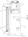
- Fig. 1 shows an illustrative pouch 510 that may be used in various embodiments, or may be reconfigured for various embodiments.
- Pouch 510 is similar to Fig. 15 of U.S. Patent No. 8,895,295 , with like items numbered the same.
- Fitment 590 is provided with entry channels 515a through 5151, which also serve as reagent reservoirs or waste reservoirs. Illustratively, reagents may be freeze dried in fitment 590 and rehydrated prior to use.
- Blisters 522, 544, 546, 548, 564, and 566, with their respective channels 514, 538, 543, 552, 553, 562, and 565 are similar to blisters of the same number of Fig. 15 of U.S. Patent No.
- Second-stage reaction zone 580 of Fig. 1 is similar to that of U.S. Patent Application No. 8,895,295 , but the second-stage wells 582 of high density array 581 are arranged in a somewhat different pattern.
- the more circular pattern of high density array 581 of Fig. 1 eliminates wells in corners and may result in more uniform filling of second-stage wells 582.
- the high density array 581 is provided with 102 second-stage wells 582.
- Pouch 510 is suitable for use in the FilmArray@ instrument (BioFire Diagnostics, LLC, Salt Lake City, UT). However, it is understood that the pouch embodiment is illustrative only.
- pouch 510 is formed of two layers of a flexible plastic film or other flexible material such as polyester, polyethylene terephthalate (PET), polycarbonate, polypropylene, polymethylmethacrylate, and mixtures thereof that can be made by any process known in the art, including extrusion, plasma deposition, and lamination. Metal foils or plastics with aluminum lamination also may be used. Other barrier materials are known in the art that can be sealed together to form the blisters and channels. If plastic film is used, the layers may be bonded together, illustratively by heat sealing. Illustratively, the material has low nucleic acid binding capacity.
- a flexible plastic film or other flexible material such as polyester, polyethylene terephthalate (PET), polycarbonate, polypropylene, polymethylmethacrylate, and mixtures thereof that can be made by any process known in the art, including extrusion, plasma deposition, and lamination. Metal foils or plastics with aluminum lamination also may be used. Other barrier materials are known in the art that can be sealed together to form
- plastic films that are adequately low in absorbance and auto-fluorescence at the operative wavelengths are preferred. Such material could be identified by testing different plastics, different plasticizers, and composite ratios, as well as different thicknesses of the film.
- the portion of the pouch that is to be read by a fluorescence detection device can be left without the foil. For example, if fluorescence is monitored in second-stage wells 582 of the second-stage reaction zone 580 of pouch 510, then one or both layers at wells 582 would be left without the foil.
- pouch 510 is made of a clear material capable of transmitting approximately 80%-90% of incident light.
- the materials are moved between blisters by the application of pressure, illustratively pneumatic pressure, upon the blisters and channels.
- the pouch material illustratively is flexible enough to allow the pressure to have the desired effect.
- the term "flexible” is herein used to describe a physical characteristic of the material of pouch.
- the term “flexible” is herein defined as readily deformable by the levels of pressure used herein without cracking, breaking, crazing, or the like.
- thin plastic sheets such as Saran TM wrap and Ziploc ® bags, as well as thin metal foil, such as aluminum foil, are flexible.
- only certain regions of the blisters and channels need be flexible, even in embodiments employing pneumatic pressure.
- only one side of the blisters and channels need to be flexible, as long as the blisters and channels are readily deformable.
- Other regions of the pouch 510 may be made of a rigid material or may be reinforced with a rigid material.
- a plastic film is used for pouch 510.
- a sheet of metal illustratively aluminum, or other suitable material, may be milled or otherwise cut, to create a die having a pattern of raised surfaces.
- a pneumatic press (illustratively A-5302-PDS, Janesville Tool Inc., Milton WI), illustratively regulated at an operating temperature of 195°C, the pneumatic press works like a printing press, melting the sealing surfaces of plastic film only where the die contacts the film.
- Various components such as PCR primers (illustratively spotted onto the film and dried), antigen binding substrates, magnetic beads, and zirconium silicate beads may be sealed inside various blisters as the pouch 510 is formed.
- NTPs nucleotide tri-phosphates
- Pouch 510 may be used in a manner similar to that described in U.S. Patent No. 8,895,295 .
- a 300 ⁇ l mixture comprising the sample to be tested (100 ⁇ l) and lysis buffer (200 ⁇ l) is injected into an injection port (not shown) in fitment 590 near entry channel 515a, and the sample mixture is drawn into entry channel 515a.
- Water is also injected into a second injection port (not shown) of the fitment 590 adjacent entry channel 5151, and is distributed via a channel (not shown) provided in fitment 590, thereby hydrating up to eleven different reagents, each of which were previously provided in dry form at entry channels 515b through 5151.
- reagents illustratively may include freeze-dried PCR reagents, DNA extraction reagents, wash solutions, immunoassay reagents, or other chemical entities.
- the reagents are for nucleic acid extraction, first-stage multiplex PCR, dilution of the multiplex reaction, and preparation of second-stage PCR reagents, as well as control reactions.
- all that need be injected is the sample solution in one injection port and water in the other injection port. After injection, the two injection ports may be sealed.
- Lysis blister 522 is provided with beads or particles 534, such as ceramic beads, and is configured for vortexing via impaction using rotating blades or paddles provided within the FilmArray@ instrument. Bead-milling, by shaking or vortexing the sample in the presence of lysing particles such as zirconium silicate (ZS) beads 534, is an effective method to form a lysate.
- ZS zirconium silicate
- Fig. 4 shows a bead beating motor 819, comprising blades 821 that may be mounted on a first side 811 of support member 802, of instrument 800 shown in Figs. 2A-B . Blades may extend through slot 804 to contact pouch 510. It is understood, however, that motor 819 may be mounted on other structures of instrument 800.
- motor 819 is a Mabuchi RC-280SA-2865 DC Motor (Chiba, Japan), mounted on support member 802.
- the motor is turned at 5,000 to 25,000 rpm, more illustratively 10,000 to 20,000 rpm, and still more illustratively approximately 15,000 to 18,000 rpm.
- 7.2V provides sufficient rpm for lysis. It is understood, however, that the actual speed may be somewhat slower when the blades 821 are impacting pouch 510. Other voltages and speeds may be used for lysis depending on the motor and paddles used.
- controlled small volumes of air may be provided into the bladder 822 adjacent lysis blister 522. It has been found that in some embodiments, partially filling the adjacent bladder with one or more small volumes of air aids in positioning and supporting lysis blister during the lysis process.
- other structure illustratively a rigid or compliant gasket or other retaining structure around lysis blister 522, can be used to restrain pouch 510 during lysis.
- motor 819 is illustrative only, and other devices may be used for milling, shaking, or vortexing the sample.
- the sample is moved through channel 538, blister 544, and channel 543, to blister 546, where the sample is mixed with a nucleic acid-binding substance, such as silica-coated magnetic beads 533.
- a nucleic acid-binding substance such as silica-coated magnetic beads 533.
- the mixture is allowed to incubate for an appropriate length of time, illustratively approximately 10 seconds to 10 minutes.
- a retractable magnet located within the instrument adjacent blister 546 captures the magnetic beads 533 from the solution, forming a pellet against the interior surface of blister 546.
- the liquid is then moved out of blister 546 and back through blister 544 and into blister 522, which is now used as a waste receptacle.
- One or more wash buffers from one or more of injection channels 515c to 515e are provided via blister 544 and channel 543 to blister 546.
- the magnet is retracted and the magnetic beads 533 are washed by moving the beads back and forth from blisters 544 and 546 via channel 543.
- the magnetic beads 533 are recaptured in blister 546 by activation of the magnet, and the wash solution is then moved to blister 522. This process may be repeated as necessary to wash the lysis buffer and sample debris from the nucleic acid-binding magnetic beads 533.
- elution buffer stored at injection channel 515f is moved to blister 548, and the magnet is retracted.
- the solution is cycled between blisters 546 and 548 via channel 552, breaking up the pellet of magnetic beads 533 in blister 546 and allowing the captured nucleic acids to dissociate from the beads and come into solution.
- the magnet is once again activated, capturing the magnetic beads 533 in blister 546, and the eluted nucleic acid solution is moved into blister 548.
- First-stage PCR master mix from injection channel 515g is mixed with the nucleic acid sample in blister 548.
- the mixture is mixed by forcing the mixture between 548 and 564 via channel 553.
- the solution is contained in blister 564, where a pellet of first-stage PCR primers is provided, at least one set of primers for each target, and first-stage multiplex PCR is performed.
- a reverse-transcription (RT) step may be performed prior to or simultaneously with the first-stage multiplex PCR.
- First-stage multiplex PCR temperature cycling in the FilmArray@ instrument is illustratively performed for 15-30 cycles, although other levels of amplification may be desirable, depending on the requirements of the specific application.
- the first-stage PCR master mix may be any of various master mixes, as are known in the art.
- the first-stage PCR master mix may be any of the chemistries disclosed in US2015/0118715 , for use with PCR protocols taking 20 seconds or less per cycle.
- the sample may be diluted, illustratively by forcing most of the sample back into blister 548, leaving only a small amount in blister 564, and adding second-stage PCR master mix from injection channel 515i.
- a dilution buffer from 515i may be moved to blister 566 then mixed with the amplified sample in blister 564 by moving the fluids back and forth between blisters 564 and 566.
- dilution may be repeated several times, using dilution buffer from injection channels 515j and 515k, or injection channel 515k may be reserved for sequencing or for other post-PCR analysis, and then adding second-stage PCR master mix from injection channel 515h to some or all of the diluted amplified sample.
- level of dilution may be adjusted by altering the number of dilution steps or by altering the percentage of the sample discarded prior to mixing with the dilution buffer or second-stage PCR master mix comprising components for amplification, illustratively a polymerase, dNTPs, and a suitable buffer, although other components may be suitable, particularly for non-PCR amplification methods.
- this mixture of the sample and second-stage PCR master mix may be pre-heated in blister 564 prior to movement to second-stage wells 582 for second-stage amplification. Such preheating may obviate the need for a hot-start component (antibody, chemical, or otherwise) in the second-stage PCR mixture.
- the illustrative second-stage PCR master mix is incomplete, lacking primer pairs, and each of the 102 second-stage wells 582 is pre-loaded with a specific PCR primer pair (or sometimes multiple pairs of primers). If desired, second-stage PCR master mix may lack other reaction components, and these components may be pre-loaded in the second-stage wells 582 as well. Each primer pair may be similar to or identical to a first-stage PCR primer pair or may be nested within the first-stage primer pair. Movement of the sample from blister 564 to the second-stage wells 582 completes the PCR reaction mixture. Once high density array 581 is filled, the individual second-stage reactions are sealed in their respective second-stage blisters by any number of means, as is known in the art.
- second-stage PCR master mix contains the dsDNA binding dye LCGreen ® Plus (BioFire Diagnostics, LLC) to generate a signal indicative of amplification.
- LCGreen ® Plus BioFire Diagnostics, LLC
- this dye is illustrative only, and that other signals may be used, including other dsDNA binding dyes and probes that are labeled fluorescently, radioactively, chemiluminescently, enzymatically, or the like, as are known in the art.
- wells 582 of array 581 may be provided without a signal, with results reported through subsequent processing.
- a "bladder” When pneumatic pressure is used to move materials within pouch 510, in one embodiment a "bladder" may be employed.
- the bladder assembly 810 includes a bladder plate 824 housing a plurality of inflatable bladders 822, 844, 846, 848, 864, and 866, each of which may be individually inflatable, illustratively by a compressed gas source. Because the bladder assembly 810 may be subjected to compressed gas and used multiple times, the bladder assembly 810 may be made from tougher or thicker material than the pouch. Alternatively, bladders 822, 844, 846, 848, 864, and 866 may be formed from a series of plates fastened together with gaskets, seals, valves, and pistons. Other arrangements are within the scope of this invention.
- PCR is dependent upon template generated by the multiplex first-stage reaction.
- PCR is performed using DNA of high purity. Methods such as phenol extraction or commercial DNA extraction kits provide DNA of high purity. Samples processed through the pouch 510 may require accommodations be made to compensate for a less pure preparation. PCR may be inhibited by components of biological samples, which is a potential obstacle. Illustratively, hot-start PCR, higher concentration of taq polymerase enzyme, adjustments in MgCl 2 concentration, adjustments in primer concentration, and addition of adjuvants (such as DMSO, TMSO, or glycerol) optionally may be used to compensate for lower nucleic acid purity. While purity issues are likely to be more of a concern with first-stage amplification and single-stage PCR, it is understood that similar adjustments may be provided in the second-stage amplification as well.
- adjuvants such as DMSO, TMSO, or glycerol
- the bladder assembly 810 When pouch 510 is placed within the instrument 800, the bladder assembly 810 is pressed against one face of the pouch 510, so that if a particular bladder is inflated, the pressure will force the liquid out of the corresponding blister in the pouch 510.
- the bladder assembly 810 may have additional pneumatic actuators, such as bladders or pneumatically-driven pistons, corresponding to various channels of pouch 510. Figs.
- 2A-B and 3 show an illustrative plurality of pistons or hard seals 838, 843, 852, 853, and 865 that correspond to channels 538, 543, 553, and 565 of pouch 510, as well as seals 871, 872, 873, 874 that minimize backflow into fitment 590.
- hard seals 838, 843, 852, 853, and 865 form pinch valves to pinch off and close the corresponding channels.
- the hard seals are activated over the channels leading to and from the blister, such that the actuators function as pinch valves to pinch the channels shut.
- the pinch valve actuator sealing the connecting channel is activated, and the pneumatic bladders over the blisters are alternately pressurized, forcing the liquid back and forth through the channel connecting the blisters to mix the liquid therein.
- the pinch valve actuators may be of various shapes and sizes and may be configured to pinch off more than one channel at a time. While pneumatic actuators are discussed herein, it is understood that other ways of providing pressure to the pouch are contemplated, including various electromechanical actuators such as linear stepper motors, motor-driven cams, rigid paddles driven by pneumatic, hydraulic or electromagnetic forces, rollers, rocker-arms, and in some cases, cocked springs.
- reversibly or irreversibly closing channels in addition to applying pressure normal to the axis of the channel.
- methods of reversibly or irreversibly closing channels include kinking the bag across the channel, heat-sealing, rolling an actuator, and a variety of physical valves sealed into the channel such as butterfly valves and ball valves.
- small Peltier devices or other temperature regulators may be placed adjacent the channels and set at a temperature sufficient to freeze the fluid, effectively forming a seal. Also, while the design of Fig.
- actuators could remain stationary, and the pouch 510 could be transitioned in one or two dimensions such that a small number of actuators could be used for several of the processing stations including sample disruption, nucleic-acid capture, first and second-stage PCR, and other applications of the pouch 510 such as immuno-assay and immuno-PCR. Rollers acting on channels and blisters could prove particularly useful in a configuration in which the pouch 510 is translated between stations.
- pneumatic actuators are used in the presently disclosed embodiments, when the term "pneumatic actuator" is used herein, it is understood that other actuators and other ways of providing pressure may be used, depending on the configuration of the pouch and the instrument.
- Figs. 2A-B show an illustrative instrument 800 that could be used with pouch 510.
- Instrument 800 includes a support member 802 that could form a wall of a casing or be mounted within a casing.
- Instrument 800 may also include a second support member (not shown) that is optionally movable with respect to support member 802, to allow insertion and withdrawal of pouch 510.
- a lid may cover pouch 510 once pouch 510 has been inserted into instrument 800.
- both support members may be fixed, with pouch 510 held into place by other mechanical means or by pneumatic pressure.
- heaters 886 and 888 are mounted on support member 802.
- Bladder plate 810, with bladders 822, 844, 846, 848, 864, 866, hard seals 838, 843, 852, 853, seals 871, 872, 873, 874 form bladder assembly 808 may illustratively be mounted on a moveable support structure that may be moved toward pouch 510, such that the pneumatic actuators are placed in contact with pouch 510.
- the various blisters of pouch 510 are in a position adjacent to the various bladders of bladder assembly 810 and the various seals of assembly 808, such that activation of the pneumatic actuators may force liquid from one or more of the blisters of pouch 510 or may form pinch valves with one or more channels of pouch 510.
- the relationship between the blisters and channels of pouch 510 and the bladders and seals of assembly 808 is illustrated in more detail in Fig. 3 .
- Each pneumatic actuator is connected to compressed air source 895 via valves 899. While only several hoses 878 are shown in Figs. 2A-B , it is understood that each pneumatic fitting is connected via a hose 878 to the compressed gas source 895.
- Compressed gas source 895 may be a compressor, or, alternatively, compressed gas source 895 may be a compressed gas cylinder, such as a carbon dioxide cylinder. Compressed gas cylinders are particularly useful if portability is desired. Other sources of compressed gas are within the scope of this invention.
- Assembly 808 is illustratively mounted on a movable support member, although it is understood that other configurations are possible.
- a magnet 850 which is mounted on a second side 814 of support member 802, is illustratively deployed and retracted using gas from compressed gas source 895 via hose 878, although other methods of moving magnet 850 are known in the art. Magnet 850 sits in recess 851 in support member 802. It is understood that recess 851 can be a passageway through support member 802, so that magnet 850 can contact blister 546 of pouch 510.
- recess 851 need not extend all the way through support member 802, as long as when magnet 850 is deployed, magnet 850 is close enough to provide a sufficient magnetic field at blister 546, and when magnet 850 is retracted, magnet 850 does not significantly affect any magnetic beads 533 present in blister 546.
- retracting magnet 850 it is understood that an electromagnet may be used and the electromagnet may be activated and inactivated by controlling flow of electricity through the electromagnet.
- the pneumatic connections may be pneumatic hoses or pneumatic air manifolds, thus reducing the number of hoses or valves required.
- the various pneumatic pistons 868 of pneumatic piston array 869 are also connected to compressed gas source 895 via hoses 878. While only two hoses 878 are shown connecting pneumatic pistons 868 to compressed gas source 895, it is understood that each of the pneumatic pistons 868 are connected to compressed gas source 895. Twelve pneumatic pistons 868 are shown.
- a pair of heating/cooling devices are mounted on a second side 814 of support 802.
- First-stage heater 886 is positioned to heat and cool the contents of blister 564 for first-stage PCR.
- Second-stage heater 888 is positioned to heat and cool the contents of second-stage blisters 582 of pouch 510, for second-stage PCR. It is understood, however, that these heaters could also be used for other heating purposes, and that other heaters may be use, as appropriate for the particular application. Other configurations are possible.
- optical array 890 When fluorescent detection is desired, an optical array 890 may be provided. As shown in Figs. 2A-B , optical array 890 includes a light source 898, illustratively a filtered LED light source, filtered white light, or laser illumination, and a camera 896. Camera 896 illustratively has a plurality of photodetectors each corresponding to a second-stage well 582 in pouch 510. Alternatively, camera 896 may take images that contain all of the second-stage wells 582, and the image may be divided into separate fields corresponding to each of the second-stage wells 582. Depending on the configuration, optical array 890 may be stationary, or optical array 890 may be placed on movers attached to one or more motors and moved to obtain signals from each individual second-stage well 582. It is understood that other arrangements are possible.
- a computer 894 controls valves 899 of compressed air source 895, and thus controls all of the pneumatics of instrument 800.
- Computer 894 also controls heaters 886 and 888, and optical array 890.
- Each of these components is connected electrically, illustratively via cables 891, although other physical or wireless connections are within the scope of this invention.
- computer 894 may be housed within instrument 800 or may be external to instrument 800.
- computer 894 may include built-in circuit boards that control some or all of the components, may calculate amplification curves, melting curves, Cps, Cts, standard curves, and other related data, and may also include an external computer, such as a desktop or laptop PC, to receive and display data from the optical array.
- An interface illustratively a keyboard interface, may be provided including keys for inputting information and variables such as temperatures, cycle times, etc.
- a display 892 is also provided.
- Display 892 may be an LED, LCD, or other such display, for example.
- adenovirus adenovirus
- PIV1, PIV2, PIV3, RSV adenovirus
- Influenza A Influenza B
- a more complete panel illustratively would include assays for other viruses including: coronavirus, human metapneumovirus, rhinovirus, and non-HRV enterovirus.
- coronavirus human metapneumovirus
- rhinovirus adenovirus
- non-HRV enterovirus enterovirus
- the FilmArray ® Respiratory Panel includes Adenovirus, Coronavirus HKU1, Coronavirus NL63, Coronavirus 229E, Coronavirus OC43, Human Metapneumovirus, Human Rhinovirus/Enterovirus, Influenza A, Influenza A/H1, Influenza A/H3, Influenza A/HI-2009, Influenza B, Parainfluenza Virus 1, Parainfluenza Virus 2, Parainfluenza Virus 3, Parainfluenza Virus 4, and Respiratory Syncytial Virus.
- the FilmArray@ Respiratory Panel includes three bacteria: Bordetella pertussis, Chlamydophila pneumoniae , and Mycoplasma pneumoniae.
- the high density array 581 is able to accommodate such a panel in a single pouch 510.
- Other panels are available for the FilmArray@, each assaying for at least 20 pathogens.
- the illustrative second-stage PCR master mix contains the dsDNA binding dye LCGreen ® Plus to generate a signal indicative of amplification.
- this dye is illustrative only, and that other signals may be used, including other dsDNA binding dyes, and probes that are labeled fluorescently, radioactively, chemiluminescently, enzymatically, or the like, as are known in the art.
- the illustrative FilmArray instrument is programmed to make positive or negative calls for each second-stage reaction based on a post-PCR melt.
- the melt curve must produce a melt peak (first derivative maximum or negative first derivative maximum) within a pre-defined temperature range, for the call to be positive. It is understood that this method of calling each second-stage reaction is illustrative only, and that calls could be made using real-time amplification data or by other means, as are known in the art.
- standard curves generated solely in the nested second-stage PCR may be of limited value for quantification, as single-plex standard template amplification reactions may not accurately reflect all of the upstream manipulations that the sample undergoes or may not be amplified with similar efficiencies, and, therefore, may not be reflective of the entire process.
- nucleic acid templates (illustratively varying in sequence and/or length), illustratively synthetic quantification standards, are used to represent different levels of a dilution series.
- Assays for all of the synthetic quantification standards have similar amplification efficiency and produce the same or similar Cp values at each given dilution point in the multiplex setting.
- all target assays are optimized for the same performance characteristics, including efficiency, although corrections may be applied to adjust for assay-specific variation in efficiency.
- outer and inner amplicon sizes for the quantification standards may be representative of amplicon sizes for the quantitative target assays.
- the sequence or GC content may be the same or similar in between the priming regions.
- the sequences may be identical with an exception of at least one inner priming region, which should be different enough to avoid cross-reactivity between inner assays. If sequences differ only by inner primer binding region, then the same PCR1 primers may be used to amplify all quantification standards, thus minimizing potential differences in the PCR1 assays performances.
- sequences may be identical, and if labels are not used, even a slight difference in sequence can provide for detection, illustratively in a second-stage single-plex reaction.
- these parameters are illustrative only, and other means for detection and controlling amplification efficiencies are possible.
- quantification standards present in the multiplex reaction should be designed to match reaction parameters, such as Mg 2+ , primer concentration, Tm, and cycling conditions. It is also understood that it is desirable to minimize non-specific amplification of the synthetic templates in the multiplex PCR reaction.
- Fig. 5 shows Cp vs. concentration of four prospective internal quantification standards.
- the internal quantification standards have synthetic sequences.
- the four quantification standards all share one common primer and each has one unique specific primer. They were also designed to share both outer primers for first-stage PCR.
- each of the second-stage wells used for detecting the quantification standards would be spotted with the common primer and the primer for that quantification standard, such that only one quantification standard should amplify in each such well.
- Syn2, Syn3, and Syn4 each have similar amplification efficiencies and were chosen for additional study. Syn1 behaves differently and was omitted from further work.
- the multiple quantification standards have similar amplification efficiencies.
- amplification was detected using the dsDNA binding dye LCGreen Plus.
- LCGreen Plus dsDNA binding dye
- the quantification standards have the same sequence between the inner primers and differ only in inner primer binding sequence.
- the quantification standards are all of substantially the same length and substantially the same GC content.
- the sequences are of differing lengths but also differ in GC content to compensate.
- Other ways of designing nucleic acids with similar amplification efficiencies are known in the art.
- the quantification standards may all use the same outer primers in the first-stage PCR reaction, potentially even sharing identical regions surrounding primers to avoid differences due to the secondary structure formations.
- the quantification standards may then be distinguished by using different inner primers in the individual second-stage PCR reactions, either with each calibrator having a unique pair of inner primers, or, as above, sharing one inner primer and having one unique inner primer.
- Such an embodiment has advantages in that each of the quantification standards binds to its first-stage primers with the same kinetics, and the complexity of the first-stage multiplex PCR reaction may be minimized.
- the quantification standards may have different forward or reverse primers or the same forward and reverse primers and illustratively each has a specific fluorescent probe or other identifiable label, such as chemiluminescence, bioluminescence, radioluminescence, electroluminescence, electrochemiluminescence, mechanoluminescence, crystalloluminescence, thermoluminescence, sonoluminescence, phosphorescence and other forms of photoluminescence, enzymatic, radioactive, and the like are contemplated herein.
- the application is only limited by the number of detection channels available in any system or other methods for distinguishing the labels, as are known in the art.
- labeled quantification standards may be used in a two-step PCR wherein the same or different primer sequences may be used and the label is used to detect in the second-stage PCR.
- the labeled quantification standards optionally may be multiplexed in the second-stage PCR and distinguished by the label.
- yeast sequences may be used for one or more of the quantification standard sequences.
- the Tf2-type retrotransposable element/transposon is present in 13 copies while the ribosomal RNA genes is repeated 47 times.
- gene sequences that exist in different copy numbers may be used.
- fungal pathogens have 50 to 200 copies of the ribosomal RNA gene per nuclear genome. These pathogens also have transposons that vary between five and 20 copies per genome.
- Bacterial pathogens have between 1 and 15 copies per genome but most have more than 5 copies.
- Other naturally occurring or synthetic templates may be used, such as bacteriophages for viruses and synthetic particles able to mimic membrane and/or capsid and/or envelope structures.
- three quantification standards are used in many of the examples herein, it is understood that only two quantification standards are needed to define a linear standard curve, and more quantification standards may be desired in embodiments where a wide range of target concentrations is expected or where a non-linear standard curve is expected.
- the number of quantification standards may be chosen based on the dynamic range of the system and the requirements of the assay.
- Example 5 it is also possible to use only one quantification standard (see the sample processing control (SPC) discussed in Example 5) in each experimental run and to rely on an imported standard curve for the quantification previously generated with a quantification standard range, illustratively with at least 3 quantification standards (named QS in Example 5) which may be included in software for this analysis.
- SPC sample processing control
- Fig. 6A is similar to Fig. 5 , but showing data only from the three chosen calibrator sequences.
- Fig. 6A demonstrates the linearity and nearly identical amplification efficiencies for the three illustrative quantification standards.
- Fig. 6B shows a composite standard curve generated from the combination of three points each of the three quantification standards over a total of five dilutions.
- a standard curve using assay-specific reference templates for each target assay may be generated.
- Fig. 7 compares the externally generated standard curve using a well-quantified synthetic reference template for Acinetobacter baumannii with a composite internal standard curve generated using the quantification standards.
- the three "Syn" templates were pre-mixed before addition to the reaction tube; Syn4 was added at 10 3 copies, Syn2 was added at 10 4 copies, and Syn3 was added at 10 5 copies per reaction.
- the Cp values from these templates were used to generate a composite internal standard curve for each reaction.
- An external standard curve was generated using a synthetic reference template A. baumannii reference template, also tested at the same concentrations as the "Syn" templates.
- the composite internal standard curve and the A. baumannii standard curves are very similar.
- the similar slope shows that the efficiencies are similar. It is expected that an unknown starting concentration of an A. baumannii sample can be predicted using the internal standard curve. However, because the y-intercept is shifted between the two curves, quantification of A. baumannii may benefit from a correction factor when using this internal standard curve.
- the concentration of target organisms can be computed using the composite internal standard curve.
- the internal standards are each at different known concentrations and are amplified in the same process as the target organisms.
- the methods illustratively employ cycle threshold (Ct) values (or alternatively a Cp value or other similar methods), which is the number of cycles of PCR required to obtain a fluorescence signal above the background fluorescence, for the target and internal standard, as determined experimentally.
- Ct cycle threshold
- Other points may be used as well, such as using a first, second, or nth order derivative, illustratively as taught in U.S. Patent No. 6,303,305 .
- the Cp value is determined in the nested second-stage reactions.
- the Cp may be determined as is appropriate for the amplification system.
- Cp may be determined in a single multiplex reaction or in a subsequent second-stage reaction by using oligonucleotide probes, each of which are specific for a quantification standard sequence and have a distinguishable fluorescent signal.
- the concentration of the target organism may be computed using the Ct of the target organism ( Ct t ), the concentration and Ct of the internal standard ( Concentrations , Ct s ), and the target organism's efficiency ( Efficiency t ) according to the following formula.
- the Efficiency variable for a target with 100% amplification each PCR cycle would equal 2.
- the Efficiency is assumed to be predetermined and constant across a dynamic range.
- the efficiencies of the internal calibrators should all be similar, illustratively within 1%, within 2%, within 5%, or within 10% of each other.
- the efficiencies of the targets should each be similar to that of the calibrators, illustratively within 2%, within 5%, within 10% or within 12% of the calibrators. It is understood that for precise quantification, efficiencies within a narrower range, illustratively within 1%, within 2%, or within 5% is desirable. However, for semi-quantitative or "binning" results (see below), a larger variation in efficiencies may be tolerated.
- a standard quantification curve may be generated, illustratively using a least-squares regression line fit to the ( Ct , log 10 ( Concentration )) data for the internal quantification standards, as illustrated in Figs. 6A-6B .
- the quantification standard curve may be non-linear, or may be linear only within a certain dynamic range.
- a concentration-dependent variable slope a sigmoidal dose-response curve may be used.
- Other non-linear curves are within the scope of this invention.
- the method described above can be used for a target organism with an unknown concentration based on observed Ct values for the target and the regression equation for the standard curve generated using internal quantification standards.
- all targets that are being quantified using this approach should have assays that have equivalent or similar PCR efficiencies as the internal quantification standards assays.
- assay specific correction factors can be used to adjust for systematic assay-specific bias to improve the accuracy of computed concentration of the unknown target.
- a when a linear quantification curve is used, a may be corrected with a correction factor indicative of a different assay-specific efficiency (which changes the slope) or b may be corrected due to lack of optimal PCR conditions for a specific target that causes the target Ct to be delayed. Both corrections may be used where appropriate.
- differences in b observed in PCR2 may be result of total outcome of the PCR1 assays, due to variations in the PCR1 efficiencies.
- the correction factor might be calculated as a function of the Ct values or be a constant depending on the desired quantification accuracy.
- a set of controlled experiments may be run with a known target organism concentration. If multiple replicates at a single concentration are used, then the assay specific correction factor may be computed as the average difference of the known concentration and the computed concentrations in Log 10 units. Illustratively, to obtain the corrected log concentration of the target organism, the assay specific correction factor may be added to the log concentration of the target organism (as computed above by the internal quantification standards method). Each target sequence in the multiplex assay illustratively will have its own correction (or no correction at all, if very similar to the composite standard curve).
- Fig. 8B shows how this systematic bias in quantification can be corrected by applying an average assay-specific correction factor, generated as described above, to quantities computed by the internal standard curve method. Similar corrections may be made for each assay in the multiplex reaction.
- results may be reported as absolute concentrations (with or without system error (illustratively 95% prediction interval)), or may be binned into one of a plurality of ranges, illustratively reporting a "high", “medium”, or “low” concentration, each covering one or more orders of magnitude. It is understood that the number of bins may vary, as is appropriate with a specific assay, and any number of bins may be used. Also, the range of binning (orders of magnitude or other measures) for semi-quantitative results may be adjusted, as is appropriate for the specific example.
- Inhibitory samples can affect extraction and amplification of internal quantification standards and target assays to more or less the same extent. Since the internal standards dynamically respond to matrix-driven effects in a way similar to the target sequences, they can also serve a normalization function.
- the three internal calibrators from Example 3 were used in the concentrations used above.
- TA-89 is a tracheal aspirate sample that was positive for Streptococcus spp. and was shown to have inhibitory properties as reflected in the considerably delayed Cps of the internal yeast RNA control in an illustrative FilmArray pouch used to test lower respiratory tract samples.
- TA-89 was used in three concentrations: as-is, diluted 2-fold, and diluted 10-fold. Each sample was spiked with A. baumannii at 10 6 CFU/mL. As in Example 3, the three internal quantification standard templates at 10 3 , 10 4 and 10 5 copies/mL were also added to all sample aliquots. A PBS no-matrix control also spiked with the same concentration of A. baumannii and internal calibrators.
- Fig. 9 shows the effect of dilution of an inhibitory matrix such as TA-89 on detection.
- the inhibitory matrix effect is visualized with the later Cps for A. baumannii in undiluted matrix and the trend to earlier Cps for A. baumannii is seen when the matrix is diluted, with the earliest Cp in the sample that did not contain the TA-89 matrix (PBS only).
- the amount of A. baumannii is the same in all dilutions of the matrix.
- Fig. 10 one sees a similar trend in Cps for the quantification standards and the A. baumannii assays, with one of the quantification standards (10 3 /mL) actually dropping out in the most inhibitory (undiluted) matrix.
- Figs. 11A-D the standard curves generated from the internal quantification standards for each of the TA-89 dilutions and the PBS no-matrix control are shown (diamonds).
- the Cp of A. baumannii (squares) is consistently earlier than the 10 5 /mL point in all sample matrices.
- Calculated concentrations for A. baumannii are shown below in Table 1. All predicted values are within three-fold of the actual concentration of 10 6 CFU/mL. This demonstrates that the internal quantification standards and the A.
- a single synthetic quantification standard (QS) at a known concentration was compared to the use of a single microorganism at a known concentration, wherein the microorganism is also used as sample processing control (SPC), for the quantification of a pathogen (illustratively cytomegalovirus (CMV)).
- SPC sample processing control
- CMV cytomegalovirus
- column-based extraction was used, and amplification was performed on a Bio-Rad CFX instrument, although it is understood that these methods and instruments are illustrative only.
- the experiment plan is as follows: Illustratively, the first step was performed as follows: Part A): A first PCR mixture provided using the 5'nuclease real-time PCR technology including two primers and one probe, wherein the primers and probe are designed specifically to amplify and detect and quantify the target pathogen. Another PCR similar mixture is used, but designed for amplification and detection of the synthetic quantification standard. A third PCR similar mixture is used, designed for amplification and detection of the Sample Processing Control. It is understood that the 5'nuclease real-time PCR technology is illustrative only and that other detection means may be used.
- Table 2 Ct Value Mean Ct Value Log Copies/mL Copies/mL QS1 23.38 23.52 6,903 8,000,000 23.52 23.66 QS2 27.30 27.33 5,903 800,000 27.35 27.34 QS3 30.74 30.69 4,903 80,000 30.61 30.72 QS4 33.62 33.61 3,903 8,000 33.59 33.62
- Part B was performed as follows: The results obtained for the SPC S. pombe were calibrated against the standard quantification curve and the calibration factor is determined.
- the optimal SPC dilution (illustratively dilution 2), illustratively chosen to be in the middle of the relevant quantification range of the pathogens to detect, is selected on which the standard quantification curve will be placed with its pre-defined regression line slope.
- quantification by PCR frequently uses a standard curve approach.
- a standard curve could be stored, as parameters (intercept and slope), and imported on a single value (Ct) which allows one to adjust the quantification run by run, depending on the behavior of the SPC in the run.
- the value 4.547 will be the value for Log 10 Concentration used to determine the equation of the next amplification runs when the SPC is used as the quantification standard for the quantification of the target CMV.
- the SPC may be called the adjuster or calibrator because it serves to adjust or calibrate the standard curve.
- one of the synthetic standards or one of the SPC dilutions could be used as the adjuster in the pre-defined regression line slope, with a factor of 4.903 for the use of the synthetic standard QS3, and a factor of 4.547 for the use of sample processing control dilution 2.
- Second Step A series of samples were then tested in parallel with the quantification standard QS3 and the sample processing control SPC S. pombe at a concentration of 80000 copies/mL: 4 dilutions of the reference CMV strain AD169 spiked in whole blood samples each in 4 replicates. Two whole blood samples obtained from QCMD (Quality Control Molecular Diagnostics) and 5 whole blood clinical samples were also used.
- the concentration of CMV in the samples was determined by applying Equations 3 and 5.
- sample processing control as a quantification standard in order to adjust standard curve parameters for the quantification takes advantage of the fact that this particle goes through all the same steps as a specimen and a potential pathogen infecting the test sample, but also has the same behavior as the pathogen to be detected.
- the quantification of the pathogen can take into account a potential loss of yield and efficiency along the workflow, to become more accurate.
- sample processing control that occur in different copy number could be used as internal quantification standards to generate a standard quantification curve that could be used to quantify targets in a multiplex assay.
- sample processing controls may be used at different concentrations, again to generate a standard quantification curve that could be used as in the methods disclosed in Examples 2-4.
- quantification standards can be applied to test, compare, or optimize sample processing methods.
- the quantification standards can also be used to assist in estimating true titers of analytes (organisms) in the original sample, illustratively by factoring the loss during sample preparation.
- the quantification standards can be applied to compare or optimize sample processing methods.
- the quantification standards can be used in one or more of the following processes: (1) optimization of various sample preparation sub-features, e.g., length of incubation steps, mag-bead collection time, wash steps, or elution steps; (2) comparison of extraction efficiency of different commercial or non-commercial sample preparation platforms, e.g., MagNA Pure ® , QiaCube ® , or Easy Mag TM , to adopt an optimal platform; or (3) comparison of matrix effect on extraction, illustratively using the same platform, although cross-platform comparisons are also possible.
- sample preparation protocols can be developed that work equally well for multiple matrices.
- illustratively comparisons may be done by adding one or more quantification standards (Template A) at a fixed quantity to the sample prior to sample preparation. Template A will then undergo all processes the sample is subjected to. The amount of Template A lost during sample preparation is expected be a good approximation of the amount of nucleic acid loss from the analytes in the sample.
- One or more second quantification standards (Template B) may then be added to the eluate at the same quantity as Template A. However, it is understood that a 1-to-1 ratio is not required and that other known amounts of Template B may be added. PCR will be performed on the eluate, and Cps of Template A will be compared to Cps of Template B. Given that none of Template B is lost during sample preparation, it is expected that Template B will amplify earlier.
- the protocol or platform that yields the smallest difference in Cps between Templates A and B may be a more efficient sample preparation method.
- the quantification standards can be used to estimate true titers of analytes (e.g., organisms) in the original sample by factoring the loss during sample preparation.
- the difference between Templates A and B may be used for back-calculating true titer values in samples by estimating loss due to sample extraction efficiency issues.
- the magnitude of loss can be converted into a correction factor that can then be applied to the analyte quantities determined by traditional qPCR methods.
- Templates A and B may each be a single quantification standard, as in Example 5, or either or both may be combinations of quantification standards, illustratively at different concentrations, as in Examples 2-4.
Landscapes
- Chemical & Material Sciences (AREA)
- Life Sciences & Earth Sciences (AREA)
- Organic Chemistry (AREA)
- Health & Medical Sciences (AREA)
- Engineering & Computer Science (AREA)
- Zoology (AREA)
- Wood Science & Technology (AREA)
- Biotechnology (AREA)
- Proteomics, Peptides & Aminoacids (AREA)
- Molecular Biology (AREA)
- Biochemistry (AREA)
- Bioinformatics & Cheminformatics (AREA)
- General Health & Medical Sciences (AREA)
- Genetics & Genomics (AREA)
- Microbiology (AREA)
- General Engineering & Computer Science (AREA)
- Analytical Chemistry (AREA)
- Physics & Mathematics (AREA)
- Immunology (AREA)
- Chemical Kinetics & Catalysis (AREA)
- Biophysics (AREA)
- Bioinformatics & Computational Biology (AREA)
- General Chemical & Material Sciences (AREA)
- Medicinal Chemistry (AREA)
- Biomedical Technology (AREA)
- Sustainable Development (AREA)
- Virology (AREA)
- Evolutionary Biology (AREA)
- Medical Informatics (AREA)
- Spectroscopy & Molecular Physics (AREA)
- Theoretical Computer Science (AREA)
- Measuring Or Testing Involving Enzymes Or Micro-Organisms (AREA)
- Steroid Compounds (AREA)
- Apparatus Associated With Microorganisms And Enzymes (AREA)
- Medicines Containing Plant Substances (AREA)
- Saccharide Compounds (AREA)
- Amplifiers (AREA)
- Crystals, And After-Treatments Of Crystals (AREA)
- Algebra (AREA)
- General Physics & Mathematics (AREA)
Claims (10)
- Verfahren zum Durchführen einer quantitativen zwei-Schritt-PCR-Amplifikation an einer Probe, das umfasst:
Amplifizieren der Probe in einer Erststufen-Multiplexamplifikationsmischung durch eine erste Anzahl von PCR-Zyklen, wobei die Amplifikationsmischung umfasst:eine Vielzahl von Zielprimerpaaren, wobei jeder der Zielprimerpaaren konfiguriert ist um eine Vielzahl von verschiedenen Zielen, die in der Probe in einer unbekannten Konzentration vorhanden sein können, zu amplifizieren, unddes Weiteren umfassend einer Vielzahl von internen Quantifikationsstandardnukleinsäuren und mindestens ein Quantifikationsstandardprimerpaar, wobei jeder der Quantifikationsstandards eine unterschiedliche Sequenz aufweist und jeweils in verschiedenen, bekannten Konzentrationen bereitgestellt ist, wobei das Quantifikationsstandardprimerpaar konfiguriert ist um die Quantifikationsstandardnukleinsäuren zu amplifizieren, wobei die Quantifikationsstandardnukleinsäuren alle ähnliche Amplifikationseffizienzen aufweisen,Auftrennen der Erststufen-Amplifikationsmischung in eine Vielzahl von Zweitstufen-Individualreaktionen, wobei eine erste Gruppe der Vielzahl von Zweitstufen-Individualreaktionen jeweils mindestens ein Zweitstufen-Zielprimerpaar umfasst, das konfiguriert ist um eines der verschiedenen Ziele, die in der Probe vorhanden sein können, weiter zu amplifizieren und eine zweite Gruppe der Vielzahl von Zweitstufen-Individualreaktionen, die jeweils mindestens ein Zweitstufen-Quantifikationsprimerpaar umfassen, das so konfiguriert ist um eine der Quantifikationsstandardnukleinsäuren weiter zu amplifizieren, wobei sich mindestens eines von jedem der Zweitstufen-Quantifikationsstandardprimerpaaren von jedem der anderen Zweitstufen-Quantifikationsstandardprimerpaaren unterscheidet, undUnterziehen der Vielzahl von Zweitstufen-Individualreaktionen einer zweiten Reihe von PCR-Zyklen unter Amplifikationsbedingungen um ein oder mehr Zielamplikons aus der Vielzahl verschiedener Ziele zu generieren, wobei jedes der Zielamplikons einen zugeordneten Ziel-Ct und eine Vielzahl von Quantifikationsstandardamplikons aufweist, wobei jedes der Quantifikationsstandardamplikons einen zugeordneten Quantifikationsstandard-Ct aufweist, wobei jeder der Ziel-Cts und jeder der Quantifikationsstandard-Cts während der zweiten Reihe von PCR-Zyklen bestimmt wird,generieren einer Standardkurve aus den Quantifikationsstandard-Cts und Quantifizieren jeder der ein oder mehreren Ziele unter Verwendung der zugeordneten Ziel-Cts und der Standardkurve. - Verfahren nach Anspruch 1, wobei jedes der Ziele unter Verwendung der Standardkurve, die unter Verwendung der Regressionsgeraden
 in der Ct ein für jedes Ziel gemessener Zyklus-Grenzwert ist,b, der Achsenabschnitt ist, der den Ct-Wert repräsentiert, wenn log10(Konzentration) des Ziels null ist, unda die Steigung ist, welche den Grad, in der sich der Ct bei einer Einzeleinheitsänderung der Konzentration ändert, repräsentiert, gegebenenfalls wobei jeder der Ziele unter Verwendung der Regressionsgeraden nach der Methode der kleinsten Quadrate und einem Korrekturfaktor quantifiziert wird.
 - Verfahren nach Anspruch 1, wobei die Standardkurve nicht-linear ist oder wobei der Quantifikationsschritt eine Konzentration ausgibt oder wobei der Quantifikationsschritt eine Konzentration generiert, die in einen von einer Vielzahl von Bereichen fällt, und diesen einen Bereich ausgibt.
- Verfahren nach Anspruch 1, wobei die Amplifikationsmischung nur ein Quantifikationsprimerpaar umfasst.
- Verfahren nach Anspruch 1, wobei die internen Quantifikationsstandardnukleinsäuren in der Probe natürlich auftretende Sequenzen sind.
- Verfahren nach Anspruch 1, wobei alle der Ziele ähnliche Amplifikationseffizienzen aufweisen.
- Verfahren nach Anspruch 1, wobei jede aus der Vielzahl von internen Quantifikationsstandardnukleinsäuren vor der Probenpräparation in bekannten Mengen in der Probe bereitgestellt wird, um auch als Prozesskontrollen zu dienen.
- Verfahren nach Anspruch 7, des Weiteren umfassend die Amplifikationsdetektion von jeder der Quantifikationsstandardnukleinsäuren unter Verwendung eines Labels spezifisch für diese Quantifikationsstandardnukleinsäuren, gegebenenfalls wobei jedes der Label eine unterscheidbar gelabelte Probe ist.
- Probengefäß für die Durchführung des Verfahrens nach Anspruch 1, umfassend einen Amplifikationsbehälter umfassend die Vielzahl von Zielprimerpaaren, wobei der Amplifikationsbehälter des Weiteren die Vielzahl von internen Quantifikationsstandardnukleinsäuren umfasst, wobei jeder der Quantifikationsstandards eine unterschiedliche Sequenz aufweist und jeweils in verschiedenen, bekannten Konzentrationen bereitgestellt ist und ähnliche Amplifikationseffizienzen aufweisen, und des Weiteren mindestens ein Quantifikationsstandardprimerpaar wie definiert in Anspruch 1 umfasst, und
die Vielzahl von Zweitstufen-Individualreaktionswells fluidisch mit dem Amplifikationsbehälter verbunden sind, wobei die erste Gruppe der Vielzahl von Zweitstufen-Individualreaktionen jeweils mindestens einen Zweitstufen-Zielprimer umfassen, der konfiguriert ist um eines der verschiedenen Ziele, die in der Probe vorhanden sein können, weiter zu amplifizieren und die zweite Gruppe der Vielzahl von Zweitstufen-Individualreaktionen jeweils mindestens ein Zweitstufen-Quantifikationsstandardprimerpaar umfassen, das konfiguriert ist um eine der Quantifikationsstandardnukleinsäuren weiter zu amplifizieren, wobei sich mindestens eines von jedem der Zweitstufen-Quantifikationsstandardprimerpaaren von jedem der anderen Zweitstufen-Quantifikationsstandardprimerpaaren unterscheidet. - Instrument für die Durchführung des Verfahrens nach Anspruch 1 an einer Probe, umfassend einer Öffnung für die Aufnahme des Probenbehälters nach Anspruch 9,eines ersten Heizers um den Amplifikationsbehälter auf Amplifikationsbedingungen zu bringen,eines zweiten Heizers um die Vielzahl der Zweitstufen-Individualreaktionswells auf Amplifikationsbedingungen zu bringen,eines Computers der programmiert ist um eine Standardkurve unter Verwendung der Amplifikation der Quantifikationsstandardnukleinsäuren zu erzeugen und ein quantitatives oder semi-quantitatives Ergebnis für jedes der amplifizierten Ziele auszugeben.
Priority Applications (1)
| Application Number | Priority Date | Filing Date | Title |
|---|---|---|---|
| EP22199836.2A EP4137583A1 (de) | 2016-03-24 | 2017-03-20 | Verfahren zur quantitativen amplifikation |
Applications Claiming Priority (2)
| Application Number | Priority Date | Filing Date | Title |
|---|---|---|---|
| US201662313032P | 2016-03-24 | 2016-03-24 | |
| PCT/US2017/023151 WO2017165269A1 (en) | 2016-03-24 | 2017-03-20 | Methods for quantitative amplification |
Related Child Applications (2)
| Application Number | Title | Priority Date | Filing Date |
|---|---|---|---|
| EP22199836.2A Division-Into EP4137583A1 (de) | 2016-03-24 | 2017-03-20 | Verfahren zur quantitativen amplifikation |
| EP22199836.2A Division EP4137583A1 (de) | 2016-03-24 | 2017-03-20 | Verfahren zur quantitativen amplifikation |
Publications (3)
| Publication Number | Publication Date |
|---|---|
| EP3433383A1 EP3433383A1 (de) | 2019-01-30 |
| EP3433383A4 EP3433383A4 (de) | 2019-10-02 |
| EP3433383B1 true EP3433383B1 (de) | 2022-11-16 |
Family
ID=59899700
Family Applications (2)
| Application Number | Title | Priority Date | Filing Date |
|---|---|---|---|
| EP17770890.6A Active EP3433383B1 (de) | 2016-03-24 | 2017-03-20 | Verfahren zur quantitativen amplifikation |
| EP22199836.2A Pending EP4137583A1 (de) | 2016-03-24 | 2017-03-20 | Verfahren zur quantitativen amplifikation |
Family Applications After (1)
| Application Number | Title | Priority Date | Filing Date |
|---|---|---|---|
| EP22199836.2A Pending EP4137583A1 (de) | 2016-03-24 | 2017-03-20 | Verfahren zur quantitativen amplifikation |
Country Status (8)
| Country | Link |
|---|---|
| US (2) | US11268141B2 (de) |
| EP (2) | EP3433383B1 (de) |
| JP (3) | JP6979029B2 (de) |
| CN (2) | CN109963948B (de) |
| AU (2) | AU2017237983B2 (de) |
| CA (1) | CA3018187A1 (de) |
| ES (1) | ES2936823T3 (de) |
| WO (1) | WO2017165269A1 (de) |
Families Citing this family (9)
| Publication number | Priority date | Publication date | Assignee | Title |
|---|---|---|---|---|
| EP3491149A4 (de) * | 2016-07-28 | 2019-12-25 | BioFire Diagnostics, LLC | Eigenständige nukleinsäureverarbeitung |
| EP3662480A4 (de) | 2017-08-04 | 2021-05-19 | BillionToOne, Inc. | Target-assoziierte moleküle zur charakterisierung im zusammenhang mit biologischen targets |
| US11519024B2 (en) * | 2017-08-04 | 2022-12-06 | Billiontoone, Inc. | Homologous genomic regions for characterization associated with biological targets |
| WO2019093530A1 (ja) * | 2017-11-13 | 2019-05-16 | 株式会社リコー | 検出判定用デバイス |
| US10246704B1 (en) | 2017-12-29 | 2019-04-02 | Clear Labs, Inc. | Detection of microorganisms in food samples and food processing facilities |
| CA3044782A1 (en) | 2017-12-29 | 2019-06-29 | Clear Labs, Inc. | Automated priming and library loading device |
| US10597714B2 (en) | 2017-12-29 | 2020-03-24 | Clear Labs, Inc. | Automated priming and library loading device |
| WO2020072459A1 (en) * | 2018-10-02 | 2020-04-09 | Biofire Diagnostics, Llc | Bacterial response |
| AU2019459344A1 (en) * | 2019-07-30 | 2022-02-03 | Alifax S.R.L. | Method and system to identify microorganisms |
Family Cites Families (26)
| Publication number | Priority date | Publication date | Assignee | Title |
|---|---|---|---|---|
| US6645758B1 (en) | 1989-02-03 | 2003-11-11 | Johnson & Johnson Clinical Diagnostics, Inc. | Containment cuvette for PCR and method of use |
| US6780617B2 (en) | 2000-12-29 | 2004-08-24 | Chen & Chen, Llc | Sample processing device and method |
| US6303305B1 (en) | 1999-03-30 | 2001-10-16 | Roche Diagnostics, Gmbh | Method for quantification of an analyte |
| US6691041B2 (en) | 2000-03-31 | 2004-02-10 | Roche Molecular Systems, Inc. | Method for the efficiency-corrected real-time quantification of nucleic acids |
| JP3752488B2 (ja) | 2000-09-22 | 2006-03-08 | 株式会社日立製作所 | 核酸解析方法 |
| EP1415113B1 (de) | 2001-07-16 | 2011-08-31 | Idaho Technology, Inc. | Temperaturwechselsystem und verwendungsverfahren |
| US20050014176A1 (en) * | 2002-10-18 | 2005-01-20 | Swiger Roy R. | Method for quantitative end-point PCR |
| US7788039B2 (en) * | 2003-09-25 | 2010-08-31 | Roche Molecular Systems, Inc. | Quantitation of nucleic acids using growth curves |
| US7387887B2 (en) | 2004-04-20 | 2008-06-17 | University Of Utah Research Foundation | Nucleic acid melting analysis with saturation dyes |
| CA2970005C (en) | 2005-05-09 | 2020-07-28 | Biofire Diagnostics, Inc. | A device for performing two-stage nucleic acid amplification |
| JP4936419B2 (ja) * | 2005-06-03 | 2012-05-23 | 学校法人日本大学 | Dnaの定量的検出方法 |
| JP2005348742A (ja) * | 2005-07-25 | 2005-12-22 | Hitachi Ltd | 核酸解析方法 |
| US7368246B2 (en) * | 2005-09-15 | 2008-05-06 | Primera Biosystems, Inc. | Quantitative gene expression profiling |
| EP1969137B1 (de) | 2005-11-22 | 2011-10-05 | Stichting Dienst Landbouwkundig Onderzoek | Multiplex-nukleinsäurenachweis |
| WO2008140568A2 (en) | 2006-11-15 | 2008-11-20 | Idaho Technology, Inc. | High density self-contained biological analysis |
| CA2705146A1 (en) * | 2007-11-07 | 2009-05-14 | Primeradx, Inc. | Quantification of nucleic acid molecules using multiplex pcr |
| CN101619346B (zh) * | 2008-07-04 | 2014-02-19 | 上海交通大学医学院附属瑞金医院 | 人冠心病易感基因-脂蛋白a基因拷贝数变异检测方法和试剂盒 |
| US20100228496A1 (en) | 2009-03-09 | 2010-09-09 | Life Technologies Corporation | Methods for the Determination of a Copy Number of a Genomic Sequence in a Biological Sample |
| CN103069007A (zh) | 2010-07-29 | 2013-04-24 | 霍夫曼-拉罗奇有限公司 | 微生物核酸的定性和定量检测 |
| US10464060B2 (en) | 2011-11-10 | 2019-11-05 | BioFare Diagnostics, LLC | Loading vials |
| DK3539666T3 (da) | 2012-05-24 | 2021-09-13 | Univ Utah Res Found | Indretning til ekstrem pcr |
| SG10201804101XA (en) | 2012-09-10 | 2018-07-30 | Biofire Diagnostics Llc | Multiple amplification cycle detection |
| JP6479336B2 (ja) * | 2014-04-23 | 2019-03-06 | 国立研究開発法人産業技術総合研究所 | 微生物の16SrRNA遺伝子定量用内部標準遺伝子 |
| ES2644949T3 (es) * | 2014-08-28 | 2017-12-01 | F. Hoffmann-La Roche Ag | Oligonucleótidos para controlar la amplificación de ácidos nucleicos |
| WO2017044694A2 (en) * | 2015-09-11 | 2017-03-16 | The Board Of Trustrees Of The Leland Stanford Junior University | Method of determining the prognosis of hepatocellular carcinomas using a multigene signature associated with metastasis |
| US10267802B2 (en) * | 2015-09-23 | 2019-04-23 | The Board Of Trustees Of The Leland Stanford Junior University | Methods of prognosis and diagnosis of ovarian cancer |
-
2017
- 2017-03-20 WO PCT/US2017/023151 patent/WO2017165269A1/en active Application Filing
- 2017-03-20 CA CA3018187A patent/CA3018187A1/en active Pending
- 2017-03-20 CN CN201780032103.8A patent/CN109963948B/zh active Active
- 2017-03-20 EP EP17770890.6A patent/EP3433383B1/de active Active
- 2017-03-20 US US16/087,724 patent/US11268141B2/en active Active
- 2017-03-20 CN CN202310030180.7A patent/CN115786472A/zh active Pending
- 2017-03-20 EP EP22199836.2A patent/EP4137583A1/de active Pending
- 2017-03-20 AU AU2017237983A patent/AU2017237983B2/en active Active
- 2017-03-20 ES ES17770890T patent/ES2936823T3/es active Active
- 2017-03-20 JP JP2018549796A patent/JP6979029B2/ja active Active
-
2021
- 2021-05-03 US US17/246,972 patent/US20210254133A1/en active Pending
- 2021-11-12 JP JP2021184574A patent/JP7453953B2/ja active Active
-
2023
- 2023-04-27 AU AU2023202560A patent/AU2023202560A1/en active Pending
- 2023-10-27 JP JP2023184871A patent/JP2024012410A/ja not_active Withdrawn
Also Published As
| Publication number | Publication date |
|---|---|
| JP2022028789A (ja) | 2022-02-16 |
| ES2936823T3 (es) | 2023-03-22 |
| CN109963948A (zh) | 2019-07-02 |
| AU2023202560A1 (en) | 2023-05-18 |
| CA3018187A1 (en) | 2017-09-28 |
| US11268141B2 (en) | 2022-03-08 |
| EP4137583A1 (de) | 2023-02-22 |
| JP2019509051A (ja) | 2019-04-04 |
| JP7453953B2 (ja) | 2024-03-21 |
| US20190078134A1 (en) | 2019-03-14 |
| JP2024012410A (ja) | 2024-01-30 |
| EP3433383A1 (de) | 2019-01-30 |
| EP3433383A4 (de) | 2019-10-02 |
| WO2017165269A1 (en) | 2017-09-28 |
| US20210254133A1 (en) | 2021-08-19 |
| CN115786472A (zh) | 2023-03-14 |
| AU2017237983B2 (en) | 2023-02-23 |
| CN109963948B (zh) | 2023-01-24 |
| AU2017237983A1 (en) | 2018-10-18 |
| JP6979029B2 (ja) | 2021-12-08 |
Similar Documents
| Publication | Publication Date | Title |
|---|---|---|
| US20210254133A1 (en) | Methods for quantitative amplification | |
| US10421991B2 (en) | Rapid epidemiologic typing of bacteria | |
| US20210180112A1 (en) | Methods for normalization and quantification of sequencing data | |
| EP3655549B1 (de) | Detektion unter verwendung von gleichzeitigen schmelzkurven | |
| WO2021086878A1 (en) | Assay devices and methods of manufacture | |
| US20230159998A1 (en) | Methods and systems for validation of a nucleic acid amplification assay | |
| US20210371895A1 (en) | Bacterial response | |
| US20230005571A1 (en) | System and method for identifying analytes in assay using normalized tm values | |
| WO2024059489A1 (en) | Multiple antibiotic bacterial response | |
| WO2024020414A1 (en) | Cell lysis and nucleic acid recovery |
Legal Events
| Date | Code | Title | Description |
|---|---|---|---|
| STAA | Information on the status of an ep patent application or granted ep patent |
Free format text: STATUS: THE INTERNATIONAL PUBLICATION HAS BEEN MADE |
|
| PUAI | Public reference made under article 153(3) epc to a published international application that has entered the european phase |
Free format text: ORIGINAL CODE: 0009012 |
|
| STAA | Information on the status of an ep patent application or granted ep patent |
Free format text: STATUS: REQUEST FOR EXAMINATION WAS MADE |
|
| 17P | Request for examination filed |
Effective date: 20180912 |
|
| AK | Designated contracting states |
Kind code of ref document: A1 Designated state(s): AL AT BE BG CH CY CZ DE DK EE ES FI FR GB GR HR HU IE IS IT LI LT LU LV MC MK MT NL NO PL PT RO RS SE SI SK SM TR |
|
| AX | Request for extension of the european patent |
Extension state: BA ME |
|
| RIC1 | Information provided on ipc code assigned before grant |
Ipc: C12P 19/34 20060101ALI20170929BHEP Ipc: C12M 1/26 20060101ALI20170929BHEP Ipc: C07H 21/04 20060101ALI20170929BHEP Ipc: G06F 19/22 20110101ALI20170929BHEP Ipc: C12Q 1/68 20180101ALI20170929BHEP Ipc: C12Q 1/70 20060101AFI20170929BHEP |
|
| RIN1 | Information on inventor provided before grant (corrected) |
Inventor name: ROGATCHEVA, MARGARITA Inventor name: BARRANGER, COME, HENRI, ALBERT Inventor name: CRISP, ROBERT, JOHN Inventor name: MAGRO, STEPHANE, CHRISTIAN, JACQUES Inventor name: SPAULDING, USHA, K. Inventor name: PORITZ, MARK, AARON |
|
| DAV | Request for validation of the european patent (deleted) | ||
| DAX | Request for extension of the european patent (deleted) | ||
| RIN1 | Information on inventor provided before grant (corrected) |
Inventor name: SPAULDING, USHA, K. Inventor name: CRISP, ROBERT, JOHN Inventor name: PORITZ, MARK, AARON Inventor name: BARRANGER, COME, HENRI, ALBERT Inventor name: ROGATCHEVA, MARGARITA Inventor name: MAGRO, STEPHANE, CHRISTIAN, JACQUES |
|
| A4 | Supplementary search report drawn up and despatched |
Effective date: 20190830 |
|
| RIC1 | Information provided on ipc code assigned before grant |
Ipc: C12Q 1/6851 20180101AFI20190826BHEP Ipc: C12Q 1/70 20060101ALI20190826BHEP Ipc: C07H 21/04 20060101ALI20190826BHEP |
|
| STAA | Information on the status of an ep patent application or granted ep patent |
Free format text: STATUS: EXAMINATION IS IN PROGRESS |
|
| 17Q | First examination report despatched |
Effective date: 20200629 |
|
| STAA | Information on the status of an ep patent application or granted ep patent |
Free format text: STATUS: EXAMINATION IS IN PROGRESS |
|
| STAA | Information on the status of an ep patent application or granted ep patent |
Free format text: STATUS: EXAMINATION IS IN PROGRESS |
|
| REG | Reference to a national code |
Ref country code: DE Ref legal event code: R079 Ref document number: 602017063762 Country of ref document: DE Free format text: PREVIOUS MAIN CLASS: C12Q0001700000 Ipc: C12Q0001685100 |
|
| GRAP | Despatch of communication of intention to grant a patent |
Free format text: ORIGINAL CODE: EPIDOSNIGR1 |
|
| STAA | Information on the status of an ep patent application or granted ep patent |
Free format text: STATUS: GRANT OF PATENT IS INTENDED |
|
| RIC1 | Information provided on ipc code assigned before grant |
Ipc: C07H 21/04 20060101ALI20220628BHEP Ipc: C12Q 1/6851 20180101AFI20220628BHEP |
|
| INTG | Intention to grant announced |
Effective date: 20220712 |
|
| GRAS | Grant fee paid |
Free format text: ORIGINAL CODE: EPIDOSNIGR3 |
|
| GRAA | (expected) grant |
Free format text: ORIGINAL CODE: 0009210 |
|
| STAA | Information on the status of an ep patent application or granted ep patent |
Free format text: STATUS: THE PATENT HAS BEEN GRANTED |
|
| AK | Designated contracting states |
Kind code of ref document: B1 Designated state(s): AL AT BE BG CH CY CZ DE DK EE ES FI FR GB GR HR HU IE IS IT LI LT LU LV MC MK MT NL NO PL PT RO RS SE SI SK SM TR |
|
| REG | Reference to a national code |
Ref country code: GB Ref legal event code: FG4D |
|
| REG | Reference to a national code |
Ref country code: CH Ref legal event code: EP |
|
| REG | Reference to a national code |
Ref country code: IE Ref legal event code: FG4D |
|
| REG | Reference to a national code |
Ref country code: DE Ref legal event code: R096 Ref document number: 602017063762 Country of ref document: DE |
|
| REG | Reference to a national code |
Ref country code: AT Ref legal event code: REF Ref document number: 1531813 Country of ref document: AT Kind code of ref document: T Effective date: 20221215 |
|
| REG | Reference to a national code |
Ref country code: NL Ref legal event code: FP |
|
| REG | Reference to a national code |
Ref country code: LT Ref legal event code: MG9D |
|
| REG | Reference to a national code |
Ref country code: ES Ref legal event code: FG2A Ref document number: 2936823 Country of ref document: ES Kind code of ref document: T3 Effective date: 20230322 |
|
| REG | Reference to a national code |
Ref country code: AT Ref legal event code: MK05 Ref document number: 1531813 Country of ref document: AT Kind code of ref document: T Effective date: 20221116 |
|
| PG25 | Lapsed in a contracting state [announced via postgrant information from national office to epo] |
Ref country code: SE Free format text: LAPSE BECAUSE OF FAILURE TO SUBMIT A TRANSLATION OF THE DESCRIPTION OR TO PAY THE FEE WITHIN THE PRESCRIBED TIME-LIMIT Effective date: 20221116 Ref country code: PT Free format text: LAPSE BECAUSE OF FAILURE TO SUBMIT A TRANSLATION OF THE DESCRIPTION OR TO PAY THE FEE WITHIN THE PRESCRIBED TIME-LIMIT Effective date: 20230316 Ref country code: NO Free format text: LAPSE BECAUSE OF FAILURE TO SUBMIT A TRANSLATION OF THE DESCRIPTION OR TO PAY THE FEE WITHIN THE PRESCRIBED TIME-LIMIT Effective date: 20230216 Ref country code: LT Free format text: LAPSE BECAUSE OF FAILURE TO SUBMIT A TRANSLATION OF THE DESCRIPTION OR TO PAY THE FEE WITHIN THE PRESCRIBED TIME-LIMIT Effective date: 20221116 Ref country code: FI Free format text: LAPSE BECAUSE OF FAILURE TO SUBMIT A TRANSLATION OF THE DESCRIPTION OR TO PAY THE FEE WITHIN THE PRESCRIBED TIME-LIMIT Effective date: 20221116 Ref country code: AT Free format text: LAPSE BECAUSE OF FAILURE TO SUBMIT A TRANSLATION OF THE DESCRIPTION OR TO PAY THE FEE WITHIN THE PRESCRIBED TIME-LIMIT Effective date: 20221116 |
|
| PGFP | Annual fee paid to national office [announced via postgrant information from national office to epo] |
Ref country code: FR Payment date: 20230327 Year of fee payment: 7 |
|
| PG25 | Lapsed in a contracting state [announced via postgrant information from national office to epo] |
Ref country code: RS Free format text: LAPSE BECAUSE OF FAILURE TO SUBMIT A TRANSLATION OF THE DESCRIPTION OR TO PAY THE FEE WITHIN THE PRESCRIBED TIME-LIMIT Effective date: 20221116 Ref country code: PL Free format text: LAPSE BECAUSE OF FAILURE TO SUBMIT A TRANSLATION OF THE DESCRIPTION OR TO PAY THE FEE WITHIN THE PRESCRIBED TIME-LIMIT Effective date: 20221116 Ref country code: LV Free format text: LAPSE BECAUSE OF FAILURE TO SUBMIT A TRANSLATION OF THE DESCRIPTION OR TO PAY THE FEE WITHIN THE PRESCRIBED TIME-LIMIT Effective date: 20221116 Ref country code: IS Free format text: LAPSE BECAUSE OF FAILURE TO SUBMIT A TRANSLATION OF THE DESCRIPTION OR TO PAY THE FEE WITHIN THE PRESCRIBED TIME-LIMIT Effective date: 20230316 Ref country code: HR Free format text: LAPSE BECAUSE OF FAILURE TO SUBMIT A TRANSLATION OF THE DESCRIPTION OR TO PAY THE FEE WITHIN THE PRESCRIBED TIME-LIMIT Effective date: 20221116 Ref country code: GR Free format text: LAPSE BECAUSE OF FAILURE TO SUBMIT A TRANSLATION OF THE DESCRIPTION OR TO PAY THE FEE WITHIN THE PRESCRIBED TIME-LIMIT Effective date: 20230217 |
|
| PGFP | Annual fee paid to national office [announced via postgrant information from national office to epo] |
Ref country code: IT Payment date: 20230321 Year of fee payment: 7 |
|
| PG25 | Lapsed in a contracting state [announced via postgrant information from national office to epo] |
Ref country code: SM Free format text: LAPSE BECAUSE OF FAILURE TO SUBMIT A TRANSLATION OF THE DESCRIPTION OR TO PAY THE FEE WITHIN THE PRESCRIBED TIME-LIMIT Effective date: 20221116 Ref country code: RO Free format text: LAPSE BECAUSE OF FAILURE TO SUBMIT A TRANSLATION OF THE DESCRIPTION OR TO PAY THE FEE WITHIN THE PRESCRIBED TIME-LIMIT Effective date: 20221116 Ref country code: EE Free format text: LAPSE BECAUSE OF FAILURE TO SUBMIT A TRANSLATION OF THE DESCRIPTION OR TO PAY THE FEE WITHIN THE PRESCRIBED TIME-LIMIT Effective date: 20221116 Ref country code: DK Free format text: LAPSE BECAUSE OF FAILURE TO SUBMIT A TRANSLATION OF THE DESCRIPTION OR TO PAY THE FEE WITHIN THE PRESCRIBED TIME-LIMIT Effective date: 20221116 Ref country code: CZ Free format text: LAPSE BECAUSE OF FAILURE TO SUBMIT A TRANSLATION OF THE DESCRIPTION OR TO PAY THE FEE WITHIN THE PRESCRIBED TIME-LIMIT Effective date: 20221116 |
|
| PGFP | Annual fee paid to national office [announced via postgrant information from national office to epo] |
Ref country code: ES Payment date: 20230403 Year of fee payment: 7 Ref country code: CH Payment date: 20230402 Year of fee payment: 7 |
|
| REG | Reference to a national code |
Ref country code: DE Ref legal event code: R097 Ref document number: 602017063762 Country of ref document: DE |
|
| PG25 | Lapsed in a contracting state [announced via postgrant information from national office to epo] |
Ref country code: SK Free format text: LAPSE BECAUSE OF FAILURE TO SUBMIT A TRANSLATION OF THE DESCRIPTION OR TO PAY THE FEE WITHIN THE PRESCRIBED TIME-LIMIT Effective date: 20221116 Ref country code: AL Free format text: LAPSE BECAUSE OF FAILURE TO SUBMIT A TRANSLATION OF THE DESCRIPTION OR TO PAY THE FEE WITHIN THE PRESCRIBED TIME-LIMIT Effective date: 20221116 |
|
| PLBE | No opposition filed within time limit |
Free format text: ORIGINAL CODE: 0009261 |
|
| STAA | Information on the status of an ep patent application or granted ep patent |
Free format text: STATUS: NO OPPOSITION FILED WITHIN TIME LIMIT |
|
| 26N | No opposition filed |
Effective date: 20230817 |
|
| PG25 | Lapsed in a contracting state [announced via postgrant information from national office to epo] |
Ref country code: MC Free format text: LAPSE BECAUSE OF FAILURE TO SUBMIT A TRANSLATION OF THE DESCRIPTION OR TO PAY THE FEE WITHIN THE PRESCRIBED TIME-LIMIT Effective date: 20221116 |
|
| PG25 | Lapsed in a contracting state [announced via postgrant information from national office to epo] |
Ref country code: SI Free format text: LAPSE BECAUSE OF FAILURE TO SUBMIT A TRANSLATION OF THE DESCRIPTION OR TO PAY THE FEE WITHIN THE PRESCRIBED TIME-LIMIT Effective date: 20221116 |
|
| REG | Reference to a national code |
Ref country code: BE Ref legal event code: MM Effective date: 20230331 |
|
| PG25 | Lapsed in a contracting state [announced via postgrant information from national office to epo] |
Ref country code: LU Free format text: LAPSE BECAUSE OF NON-PAYMENT OF DUE FEES Effective date: 20230320 |
|
| REG | Reference to a national code |
Ref country code: IE Ref legal event code: MM4A |
|
| PG25 | Lapsed in a contracting state [announced via postgrant information from national office to epo] |
Ref country code: IE Free format text: LAPSE BECAUSE OF NON-PAYMENT OF DUE FEES Effective date: 20230320 |
|
| PG25 | Lapsed in a contracting state [announced via postgrant information from national office to epo] |
Ref country code: BE Free format text: LAPSE BECAUSE OF NON-PAYMENT OF DUE FEES Effective date: 20230331 |
|
| PGFP | Annual fee paid to national office [announced via postgrant information from national office to epo] |
Ref country code: NL Payment date: 20240326 Year of fee payment: 8 |
|
| PGFP | Annual fee paid to national office [announced via postgrant information from national office to epo] |
Ref country code: DE Payment date: 20240327 Year of fee payment: 8 Ref country code: GB Payment date: 20240327 Year of fee payment: 8 |