EP1969137B1 - Multiplex-nukleinsäurenachweis - Google Patents
Multiplex-nukleinsäurenachweis Download PDFInfo
- Publication number
- EP1969137B1 EP1969137B1 EP05813533A EP05813533A EP1969137B1 EP 1969137 B1 EP1969137 B1 EP 1969137B1 EP 05813533 A EP05813533 A EP 05813533A EP 05813533 A EP05813533 A EP 05813533A EP 1969137 B1 EP1969137 B1 EP 1969137B1
- Authority
- EP
- European Patent Office
- Prior art keywords
- padlock
- probe
- target
- nucleotide
- sequence
- Prior art date
- Legal status (The legal status is an assumption and is not a legal conclusion. Google has not performed a legal analysis and makes no representation as to the accuracy of the status listed.)
- Not-in-force
Links
- 238000001514 detection method Methods 0.000 title claims abstract description 85
- 150000007523 nucleic acids Chemical class 0.000 title abstract description 24
- 102000039446 nucleic acids Human genes 0.000 title abstract description 18
- 108020004707 nucleic acids Proteins 0.000 title abstract description 18
- 239000000523 sample Substances 0.000 claims abstract description 290
- 238000003199 nucleic acid amplification method Methods 0.000 claims abstract description 96
- 230000003321 amplification Effects 0.000 claims abstract description 95
- 238000003776 cleavage reaction Methods 0.000 claims abstract description 28
- 230000007017 scission Effects 0.000 claims abstract description 28
- 239000002773 nucleotide Substances 0.000 claims description 145
- 125000003729 nucleotide group Chemical group 0.000 claims description 143
- 238000000034 method Methods 0.000 claims description 46
- 238000009396 hybridization Methods 0.000 claims description 41
- 108091034117 Oligonucleotide Proteins 0.000 claims description 36
- ISAKRJDGNUQOIC-UHFFFAOYSA-N Uracil Chemical group O=C1C=CNC(=O)N1 ISAKRJDGNUQOIC-UHFFFAOYSA-N 0.000 claims description 29
- 230000000295 complement effect Effects 0.000 claims description 28
- JLCPHMBAVCMARE-UHFFFAOYSA-N [3-[[3-[[3-[[3-[[3-[[3-[[3-[[3-[[3-[[3-[[3-[[5-(2-amino-6-oxo-1H-purin-9-yl)-3-[[3-[[3-[[3-[[3-[[3-[[5-(2-amino-6-oxo-1H-purin-9-yl)-3-[[5-(2-amino-6-oxo-1H-purin-9-yl)-3-hydroxyoxolan-2-yl]methoxy-hydroxyphosphoryl]oxyoxolan-2-yl]methoxy-hydroxyphosphoryl]oxy-5-(5-methyl-2,4-dioxopyrimidin-1-yl)oxolan-2-yl]methoxy-hydroxyphosphoryl]oxy-5-(6-aminopurin-9-yl)oxolan-2-yl]methoxy-hydroxyphosphoryl]oxy-5-(6-aminopurin-9-yl)oxolan-2-yl]methoxy-hydroxyphosphoryl]oxy-5-(6-aminopurin-9-yl)oxolan-2-yl]methoxy-hydroxyphosphoryl]oxy-5-(6-aminopurin-9-yl)oxolan-2-yl]methoxy-hydroxyphosphoryl]oxyoxolan-2-yl]methoxy-hydroxyphosphoryl]oxy-5-(5-methyl-2,4-dioxopyrimidin-1-yl)oxolan-2-yl]methoxy-hydroxyphosphoryl]oxy-5-(4-amino-2-oxopyrimidin-1-yl)oxolan-2-yl]methoxy-hydroxyphosphoryl]oxy-5-(5-methyl-2,4-dioxopyrimidin-1-yl)oxolan-2-yl]methoxy-hydroxyphosphoryl]oxy-5-(5-methyl-2,4-dioxopyrimidin-1-yl)oxolan-2-yl]methoxy-hydroxyphosphoryl]oxy-5-(6-aminopurin-9-yl)oxolan-2-yl]methoxy-hydroxyphosphoryl]oxy-5-(6-aminopurin-9-yl)oxolan-2-yl]methoxy-hydroxyphosphoryl]oxy-5-(4-amino-2-oxopyrimidin-1-yl)oxolan-2-yl]methoxy-hydroxyphosphoryl]oxy-5-(4-amino-2-oxopyrimidin-1-yl)oxolan-2-yl]methoxy-hydroxyphosphoryl]oxy-5-(4-amino-2-oxopyrimidin-1-yl)oxolan-2-yl]methoxy-hydroxyphosphoryl]oxy-5-(6-aminopurin-9-yl)oxolan-2-yl]methoxy-hydroxyphosphoryl]oxy-5-(4-amino-2-oxopyrimidin-1-yl)oxolan-2-yl]methyl [5-(6-aminopurin-9-yl)-2-(hydroxymethyl)oxolan-3-yl] hydrogen phosphate Polymers Cc1cn(C2CC(OP(O)(=O)OCC3OC(CC3OP(O)(=O)OCC3OC(CC3O)n3cnc4c3nc(N)[nH]c4=O)n3cnc4c3nc(N)[nH]c4=O)C(COP(O)(=O)OC3CC(OC3COP(O)(=O)OC3CC(OC3COP(O)(=O)OC3CC(OC3COP(O)(=O)OC3CC(OC3COP(O)(=O)OC3CC(OC3COP(O)(=O)OC3CC(OC3COP(O)(=O)OC3CC(OC3COP(O)(=O)OC3CC(OC3COP(O)(=O)OC3CC(OC3COP(O)(=O)OC3CC(OC3COP(O)(=O)OC3CC(OC3COP(O)(=O)OC3CC(OC3COP(O)(=O)OC3CC(OC3COP(O)(=O)OC3CC(OC3COP(O)(=O)OC3CC(OC3COP(O)(=O)OC3CC(OC3COP(O)(=O)OC3CC(OC3CO)n3cnc4c(N)ncnc34)n3ccc(N)nc3=O)n3cnc4c(N)ncnc34)n3ccc(N)nc3=O)n3ccc(N)nc3=O)n3ccc(N)nc3=O)n3cnc4c(N)ncnc34)n3cnc4c(N)ncnc34)n3cc(C)c(=O)[nH]c3=O)n3cc(C)c(=O)[nH]c3=O)n3ccc(N)nc3=O)n3cc(C)c(=O)[nH]c3=O)n3cnc4c3nc(N)[nH]c4=O)n3cnc4c(N)ncnc34)n3cnc4c(N)ncnc34)n3cnc4c(N)ncnc34)n3cnc4c(N)ncnc34)O2)c(=O)[nH]c1=O JLCPHMBAVCMARE-UHFFFAOYSA-N 0.000 claims description 27
- 239000011324 bead Substances 0.000 claims description 25
- 239000007787 solid Substances 0.000 claims description 24
- 230000002441 reversible effect Effects 0.000 claims description 21
- YBJHBAHKTGYVGT-ZKWXMUAHSA-N (+)-Biotin Chemical compound N1C(=O)N[C@@H]2[C@H](CCCCC(=O)O)SC[C@@H]21 YBJHBAHKTGYVGT-ZKWXMUAHSA-N 0.000 claims description 20
- HEMHJVSKTPXQMS-UHFFFAOYSA-M Sodium hydroxide Chemical compound [OH-].[Na+] HEMHJVSKTPXQMS-UHFFFAOYSA-M 0.000 claims description 15
- 102000006943 Uracil-DNA Glycosidase Human genes 0.000 claims description 13
- 108010072685 Uracil-DNA Glycosidase Proteins 0.000 claims description 13
- 239000000975 dye Substances 0.000 claims description 13
- 238000012360 testing method Methods 0.000 claims description 11
- 239000011616 biotin Substances 0.000 claims description 10
- 229960002685 biotin Drugs 0.000 claims description 10
- 235000020958 biotin Nutrition 0.000 claims description 10
- 239000011665 D-biotin Substances 0.000 claims description 7
- 108010036364 Deoxyribonuclease IV (Phage T4-Induced) Proteins 0.000 claims description 7
- 108091028043 Nucleic acid sequence Proteins 0.000 claims description 7
- 238000010828 elution Methods 0.000 claims description 7
- 238000004925 denaturation Methods 0.000 claims description 6
- 230000036425 denaturation Effects 0.000 claims description 6
- 239000007850 fluorescent dye Substances 0.000 claims description 6
- PCHJSUWPFVWCPO-UHFFFAOYSA-N gold Chemical compound [Au] PCHJSUWPFVWCPO-UHFFFAOYSA-N 0.000 claims description 6
- 239000010931 gold Substances 0.000 claims description 6
- 229910052737 gold Inorganic materials 0.000 claims description 6
- 238000002372 labelling Methods 0.000 claims description 6
- 238000005406 washing Methods 0.000 claims description 6
- 101710137500 T7 RNA polymerase Proteins 0.000 claims description 5
- 238000000137 annealing Methods 0.000 claims description 5
- ZMMJGEGLRURXTF-UHFFFAOYSA-N ethidium bromide Chemical compound [Br-].C12=CC(N)=CC=C2C2=CC=C(N)C=C2[N+](CC)=C1C1=CC=CC=C1 ZMMJGEGLRURXTF-UHFFFAOYSA-N 0.000 claims description 5
- 229960005542 ethidium bromide Drugs 0.000 claims description 5
- 238000012544 monitoring process Methods 0.000 claims description 5
- 238000009830 intercalation Methods 0.000 claims description 3
- NGVDGCNFYWLIFO-UHFFFAOYSA-N pyridoxal 5'-phosphate Chemical compound CC1=NC=C(COP(O)(O)=O)C(C=O)=C1O NGVDGCNFYWLIFO-UHFFFAOYSA-N 0.000 claims 1
- 238000003556 assay Methods 0.000 abstract description 40
- 238000013461 design Methods 0.000 abstract description 27
- 238000002955 isolation Methods 0.000 abstract description 5
- 108020004414 DNA Proteins 0.000 description 75
- 238000006243 chemical reaction Methods 0.000 description 38
- 244000052769 pathogen Species 0.000 description 27
- 108010090804 Streptavidin Proteins 0.000 description 22
- AUTOLBMXDDTRRT-JGVFFNPUSA-N (4R,5S)-dethiobiotin Chemical group C[C@@H]1NC(=O)N[C@@H]1CCCCCC(O)=O AUTOLBMXDDTRRT-JGVFFNPUSA-N 0.000 description 19
- 108060002716 Exonuclease Proteins 0.000 description 18
- 241001099903 Paramyrothecium roridum Species 0.000 description 18
- 102000013165 exonuclease Human genes 0.000 description 18
- 230000035945 sensitivity Effects 0.000 description 18
- 238000002493 microarray Methods 0.000 description 16
- 241000223221 Fusarium oxysporum Species 0.000 description 15
- 241001149949 Phytophthora cactorum Species 0.000 description 14
- 241000813090 Rhizoctonia solani Species 0.000 description 14
- 239000003795 chemical substances by application Substances 0.000 description 14
- 108091032973 (ribonucleotides)n+m Proteins 0.000 description 12
- 239000000203 mixture Substances 0.000 description 12
- 238000011002 quantification Methods 0.000 description 12
- 239000000243 solution Substances 0.000 description 12
- 108091093088 Amplicon Proteins 0.000 description 11
- 241001123668 Verticillium dahliae Species 0.000 description 11
- 238000013459 approach Methods 0.000 description 11
- 238000003753 real-time PCR Methods 0.000 description 11
- 241000233645 Phytophthora nicotianae Species 0.000 description 10
- 238000004458 analytical method Methods 0.000 description 10
- 230000001717 pathogenic effect Effects 0.000 description 10
- 241000233622 Phytophthora infestans Species 0.000 description 9
- 230000000694 effects Effects 0.000 description 9
- 239000011541 reaction mixture Substances 0.000 description 9
- 241000196324 Embryophyta Species 0.000 description 8
- 102000004190 Enzymes Human genes 0.000 description 8
- 108090000790 Enzymes Proteins 0.000 description 8
- 238000002474 experimental method Methods 0.000 description 8
- 102000053602 DNA Human genes 0.000 description 7
- 102000003960 Ligases Human genes 0.000 description 7
- 108090000364 Ligases Proteins 0.000 description 7
- 241000894007 species Species 0.000 description 7
- 229940035893 uracil Drugs 0.000 description 7
- 101710163270 Nuclease Proteins 0.000 description 6
- 241000948155 Phytophthora sojae Species 0.000 description 6
- 238000003491 array Methods 0.000 description 6
- 238000011088 calibration curve Methods 0.000 description 6
- 238000005516 engineering process Methods 0.000 description 6
- 238000002844 melting Methods 0.000 description 6
- 230000008018 melting Effects 0.000 description 6
- 239000000126 substance Substances 0.000 description 6
- 230000008685 targeting Effects 0.000 description 6
- 102100034343 Integrase Human genes 0.000 description 5
- 241000243787 Meloidogyne hapla Species 0.000 description 5
- 239000003550 marker Substances 0.000 description 5
- 230000008569 process Effects 0.000 description 5
- TWRXJAOTZQYOKJ-UHFFFAOYSA-L Magnesium chloride Chemical compound [Mg+2].[Cl-].[Cl-] TWRXJAOTZQYOKJ-UHFFFAOYSA-L 0.000 description 4
- 241000233614 Phytophthora Species 0.000 description 4
- 241001635622 Pythium splendens Species 0.000 description 4
- 108010092799 RNA-directed DNA polymerase Proteins 0.000 description 4
- 230000015572 biosynthetic process Effects 0.000 description 4
- 230000015556 catabolic process Effects 0.000 description 4
- 238000012937 correction Methods 0.000 description 4
- 238000006731 degradation reaction Methods 0.000 description 4
- 230000000368 destabilizing effect Effects 0.000 description 4
- 230000035772 mutation Effects 0.000 description 4
- 239000002105 nanoparticle Substances 0.000 description 4
- 108091008146 restriction endonucleases Proteins 0.000 description 4
- 125000006850 spacer group Chemical group 0.000 description 4
- 102000040650 (ribonucleotides)n+m Human genes 0.000 description 3
- JTBBWRKSUYCPFY-UHFFFAOYSA-N 2,3-dihydro-1h-pyrimidin-4-one Chemical compound O=C1NCNC=C1 JTBBWRKSUYCPFY-UHFFFAOYSA-N 0.000 description 3
- QKNYBSVHEMOAJP-UHFFFAOYSA-N 2-amino-2-(hydroxymethyl)propane-1,3-diol;hydron;chloride Chemical compound Cl.OCC(N)(CO)CO QKNYBSVHEMOAJP-UHFFFAOYSA-N 0.000 description 3
- 238000012408 PCR amplification Methods 0.000 description 3
- 108010010677 Phosphodiesterase I Proteins 0.000 description 3
- 241001123669 Verticillium albo-atrum Species 0.000 description 3
- 238000012512 characterization method Methods 0.000 description 3
- 238000005520 cutting process Methods 0.000 description 3
- 238000012217 deletion Methods 0.000 description 3
- 230000037430 deletion Effects 0.000 description 3
- 230000001419 dependent effect Effects 0.000 description 3
- 238000010790 dilution Methods 0.000 description 3
- 239000012895 dilution Substances 0.000 description 3
- 239000012634 fragment Substances 0.000 description 3
- 238000003205 genotyping method Methods 0.000 description 3
- 239000000463 material Substances 0.000 description 3
- 238000007837 multiplex assay Methods 0.000 description 3
- 238000011020 pilot scale process Methods 0.000 description 3
- 238000006116 polymerization reaction Methods 0.000 description 3
- 102000040430 polynucleotide Human genes 0.000 description 3
- 108091033319 polynucleotide Proteins 0.000 description 3
- 239000002157 polynucleotide Substances 0.000 description 3
- 108090000623 proteins and genes Proteins 0.000 description 3
- 238000004445 quantitative analysis Methods 0.000 description 3
- 238000010561 standard procedure Methods 0.000 description 3
- 238000003786 synthesis reaction Methods 0.000 description 3
- 241001103808 Albifimbria verrucaria Species 0.000 description 2
- 230000004544 DNA amplification Effects 0.000 description 2
- 102100031780 Endonuclease Human genes 0.000 description 2
- 108010042407 Endonucleases Proteins 0.000 description 2
- 102000005744 Glycoside Hydrolases Human genes 0.000 description 2
- 108010031186 Glycoside Hydrolases Proteins 0.000 description 2
- 241000233654 Oomycetes Species 0.000 description 2
- 241000522452 Phytophthora fragariae Species 0.000 description 2
- 241000918584 Pythium ultimum Species 0.000 description 2
- FAPWRFPIFSIZLT-UHFFFAOYSA-M Sodium chloride Chemical compound [Na+].[Cl-] FAPWRFPIFSIZLT-UHFFFAOYSA-M 0.000 description 2
- IQFYYKKMVGJFEH-XLPZGREQSA-N Thymidine Natural products O=C1NC(=O)C(C)=CN1[C@@H]1O[C@H](CO)[C@@H](O)C1 IQFYYKKMVGJFEH-XLPZGREQSA-N 0.000 description 2
- 239000000872 buffer Substances 0.000 description 2
- 230000008859 change Effects 0.000 description 2
- 238000011109 contamination Methods 0.000 description 2
- 239000013068 control sample Substances 0.000 description 2
- 230000037029 cross reaction Effects 0.000 description 2
- 230000008021 deposition Effects 0.000 description 2
- 230000002255 enzymatic effect Effects 0.000 description 2
- 238000011156 evaluation Methods 0.000 description 2
- 238000000684 flow cytometry Methods 0.000 description 2
- MHMNJMPURVTYEJ-UHFFFAOYSA-N fluorescein-5-isothiocyanate Chemical compound O1C(=O)C2=CC(N=C=S)=CC=C2C21C1=CC=C(O)C=C1OC1=CC(O)=CC=C21 MHMNJMPURVTYEJ-UHFFFAOYSA-N 0.000 description 2
- 238000010353 genetic engineering Methods 0.000 description 2
- 229910052739 hydrogen Inorganic materials 0.000 description 2
- 239000001257 hydrogen Substances 0.000 description 2
- 230000002779 inactivation Effects 0.000 description 2
- 238000010348 incorporation Methods 0.000 description 2
- 238000011534 incubation Methods 0.000 description 2
- 230000000977 initiatory effect Effects 0.000 description 2
- 238000003780 insertion Methods 0.000 description 2
- 230000037431 insertion Effects 0.000 description 2
- 229910001629 magnesium chloride Inorganic materials 0.000 description 2
- 230000007246 mechanism Effects 0.000 description 2
- 244000005700 microbiome Species 0.000 description 2
- 239000011859 microparticle Substances 0.000 description 2
- 244000000003 plant pathogen Species 0.000 description 2
- -1 polyethylene Polymers 0.000 description 2
- 238000003752 polymerase chain reaction Methods 0.000 description 2
- 102000054765 polymorphisms of proteins Human genes 0.000 description 2
- 238000002360 preparation method Methods 0.000 description 2
- 102000004169 proteins and genes Human genes 0.000 description 2
- 210000003705 ribosome Anatomy 0.000 description 2
- 238000012163 sequencing technique Methods 0.000 description 2
- 238000009987 spinning Methods 0.000 description 2
- ASJSAQIRZKANQN-CRCLSJGQSA-N 2-deoxy-D-ribose Chemical compound OC[C@@H](O)[C@@H](O)CC=O ASJSAQIRZKANQN-CRCLSJGQSA-N 0.000 description 1
- GNFTZDOKVXKIBK-UHFFFAOYSA-N 3-(2-methoxyethoxy)benzohydrazide Chemical compound COCCOC1=CC=CC(C(=O)NN)=C1 GNFTZDOKVXKIBK-UHFFFAOYSA-N 0.000 description 1
- LLTDOAPVRPZLCM-UHFFFAOYSA-O 4-(7,8,8,16,16,17-hexamethyl-4,20-disulfo-2-oxa-18-aza-6-azoniapentacyclo[11.7.0.03,11.05,9.015,19]icosa-1(20),3,5,9,11,13,15(19)-heptaen-12-yl)benzoic acid Chemical compound CC1(C)C(C)NC(C(=C2OC3=C(C=4C(C(C(C)[NH+]=4)(C)C)=CC3=3)S(O)(=O)=O)S(O)(=O)=O)=C1C=C2C=3C1=CC=C(C(O)=O)C=C1 LLTDOAPVRPZLCM-UHFFFAOYSA-O 0.000 description 1
- UHPMCKVQTMMPCG-UHFFFAOYSA-N 5,8-dihydroxy-2-methoxy-6-methyl-7-(2-oxopropyl)naphthalene-1,4-dione Chemical compound CC1=C(CC(C)=O)C(O)=C2C(=O)C(OC)=CC(=O)C2=C1O UHPMCKVQTMMPCG-UHFFFAOYSA-N 0.000 description 1
- 241000589155 Agrobacterium tumefaciens Species 0.000 description 1
- 241000972773 Aulopiformes Species 0.000 description 1
- 108090001008 Avidin Proteins 0.000 description 1
- DWRXFEITVBNRMK-UHFFFAOYSA-N Beta-D-1-Arabinofuranosylthymine Natural products O=C1NC(=O)C(C)=CN1C1C(O)C(O)C(CO)O1 DWRXFEITVBNRMK-UHFFFAOYSA-N 0.000 description 1
- FGUUSXIOTUKUDN-IBGZPJMESA-N C1(=CC=CC=C1)N1C2=C(NC([C@H](C1)NC=1OC(=NN=1)C1=CC=CC=C1)=O)C=CC=C2 Chemical compound C1(=CC=CC=C1)N1C2=C(NC([C@H](C1)NC=1OC(=NN=1)C1=CC=CC=C1)=O)C=CC=C2 FGUUSXIOTUKUDN-IBGZPJMESA-N 0.000 description 1
- 238000000018 DNA microarray Methods 0.000 description 1
- 230000006820 DNA synthesis Effects 0.000 description 1
- 108010014303 DNA-directed DNA polymerase Proteins 0.000 description 1
- 102000016928 DNA-directed DNA polymerase Human genes 0.000 description 1
- 108090000626 DNA-directed RNA polymerases Proteins 0.000 description 1
- 102000004163 DNA-directed RNA polymerases Human genes 0.000 description 1
- AHCYMLUZIRLXAA-SHYZEUOFSA-N Deoxyuridine 5'-triphosphate Chemical compound O1[C@H](COP(O)(=O)OP(O)(=O)OP(O)(O)=O)[C@@H](O)C[C@@H]1N1C(=O)NC(=O)C=C1 AHCYMLUZIRLXAA-SHYZEUOFSA-N 0.000 description 1
- ZGTMUACCHSMWAC-UHFFFAOYSA-L EDTA disodium salt (anhydrous) Chemical compound [Na+].[Na+].OC(=O)CN(CC([O-])=O)CCN(CC(O)=O)CC([O-])=O ZGTMUACCHSMWAC-UHFFFAOYSA-L 0.000 description 1
- 239000004593 Epoxy Substances 0.000 description 1
- 241000206602 Eukaryota Species 0.000 description 1
- 108010007577 Exodeoxyribonuclease I Proteins 0.000 description 1
- 102100029075 Exonuclease 1 Human genes 0.000 description 1
- 241000223218 Fusarium Species 0.000 description 1
- 241000879295 Fusarium equiseti Species 0.000 description 1
- 101710203526 Integrase Proteins 0.000 description 1
- 241000243786 Meloidogyne incognita Species 0.000 description 1
- 108020005196 Mitochondrial DNA Proteins 0.000 description 1
- FSVCELGFZIQNCK-UHFFFAOYSA-N N,N-bis(2-hydroxyethyl)glycine Chemical compound OCCN(CCO)CC(O)=O FSVCELGFZIQNCK-UHFFFAOYSA-N 0.000 description 1
- 108091093105 Nuclear DNA Proteins 0.000 description 1
- 108020005187 Oligonucleotide Probes Proteins 0.000 description 1
- 229910019142 PO4 Inorganic materials 0.000 description 1
- 239000004698 Polyethylene Substances 0.000 description 1
- 239000004793 Polystyrene Substances 0.000 description 1
- 108010066717 Q beta Replicase Proteins 0.000 description 1
- 230000006819 RNA synthesis Effects 0.000 description 1
- 238000011529 RT qPCR Methods 0.000 description 1
- 240000004808 Saccharomyces cerevisiae Species 0.000 description 1
- XUIMIQQOPSSXEZ-UHFFFAOYSA-N Silicon Chemical compound [Si] XUIMIQQOPSSXEZ-UHFFFAOYSA-N 0.000 description 1
- 108010006785 Taq Polymerase Proteins 0.000 description 1
- 229920004890 Triton X-100 Polymers 0.000 description 1
- 239000013504 Triton X-100 Substances 0.000 description 1
- 241000607479 Yersinia pestis Species 0.000 description 1
- 238000000246 agarose gel electrophoresis Methods 0.000 description 1
- 230000000692 anti-sense effect Effects 0.000 description 1
- 239000007864 aqueous solution Substances 0.000 description 1
- 238000007846 asymmetric PCR Methods 0.000 description 1
- 230000008901 benefit Effects 0.000 description 1
- IQFYYKKMVGJFEH-UHFFFAOYSA-N beta-L-thymidine Natural products O=C1NC(=O)C(C)=CN1C1OC(CO)C(O)C1 IQFYYKKMVGJFEH-UHFFFAOYSA-N 0.000 description 1
- 239000007998 bicine buffer Substances 0.000 description 1
- 239000000090 biomarker Substances 0.000 description 1
- 102000043871 biotin binding protein Human genes 0.000 description 1
- 108700021042 biotin binding protein Proteins 0.000 description 1
- 230000000903 blocking effect Effects 0.000 description 1
- 239000007853 buffer solution Substances 0.000 description 1
- 238000004364 calculation method Methods 0.000 description 1
- 210000004027 cell Anatomy 0.000 description 1
- 239000003153 chemical reaction reagent Substances 0.000 description 1
- 239000002299 complementary DNA Substances 0.000 description 1
- 238000010276 construction Methods 0.000 description 1
- 230000008878 coupling Effects 0.000 description 1
- 238000010168 coupling process Methods 0.000 description 1
- 238000005859 coupling reaction Methods 0.000 description 1
- NHVNXKFIZYSCEB-XLPZGREQSA-N dTTP Chemical compound O=C1NC(=O)C(C)=CN1[C@@H]1O[C@H](COP(O)(=O)OP(O)(=O)OP(O)(O)=O)[C@@H](O)C1 NHVNXKFIZYSCEB-XLPZGREQSA-N 0.000 description 1
- 230000003247 decreasing effect Effects 0.000 description 1
- 239000008367 deionised water Substances 0.000 description 1
- 230000027832 depurination Effects 0.000 description 1
- 238000011161 development Methods 0.000 description 1
- 238000003745 diagnosis Methods 0.000 description 1
- 230000029087 digestion Effects 0.000 description 1
- 239000000539 dimer Substances 0.000 description 1
- 238000006073 displacement reaction Methods 0.000 description 1
- 238000011143 downstream manufacturing Methods 0.000 description 1
- 238000001035 drying Methods 0.000 description 1
- 230000006862 enzymatic digestion Effects 0.000 description 1
- 108010052305 exodeoxyribonuclease III Proteins 0.000 description 1
- 230000001036 exonucleolytic effect Effects 0.000 description 1
- 125000000524 functional group Chemical group 0.000 description 1
- 230000002538 fungal effect Effects 0.000 description 1
- 238000001502 gel electrophoresis Methods 0.000 description 1
- 230000014509 gene expression Effects 0.000 description 1
- 239000011521 glass Substances 0.000 description 1
- 150000002343 gold Polymers 0.000 description 1
- 238000013537 high throughput screening Methods 0.000 description 1
- 230000005660 hydrophilic surface Effects 0.000 description 1
- 230000006872 improvement Effects 0.000 description 1
- 230000005764 inhibitory process Effects 0.000 description 1
- 238000013101 initial test Methods 0.000 description 1
- 238000005304 joining Methods 0.000 description 1
- 238000007834 ligase chain reaction Methods 0.000 description 1
- 230000004807 localization Effects 0.000 description 1
- 239000011654 magnesium acetate Substances 0.000 description 1
- 238000007726 management method Methods 0.000 description 1
- 238000005259 measurement Methods 0.000 description 1
- 108020004999 messenger RNA Proteins 0.000 description 1
- 238000010208 microarray analysis Methods 0.000 description 1
- 238000012775 microarray technology Methods 0.000 description 1
- 230000000813 microbial effect Effects 0.000 description 1
- 244000000010 microbial pathogen Species 0.000 description 1
- 230000003278 mimic effect Effects 0.000 description 1
- 238000002156 mixing Methods 0.000 description 1
- 238000010369 molecular cloning Methods 0.000 description 1
- 230000000877 morphologic effect Effects 0.000 description 1
- 238000007899 nucleic acid hybridization Methods 0.000 description 1
- 229940124276 oligodeoxyribonucleotide Drugs 0.000 description 1
- 239000002751 oligonucleotide probe Substances 0.000 description 1
- 239000010452 phosphate Substances 0.000 description 1
- 229920000573 polyethylene Polymers 0.000 description 1
- 229920002223 polystyrene Polymers 0.000 description 1
- 238000010223 real-time analysis Methods 0.000 description 1
- 230000010076 replication Effects 0.000 description 1
- 238000011160 research Methods 0.000 description 1
- 238000012552 review Methods 0.000 description 1
- PYWVYCXTNDRMGF-UHFFFAOYSA-N rhodamine B Chemical compound [Cl-].C=12C=CC(=[N+](CC)CC)C=C2OC2=CC(N(CC)CC)=CC=C2C=1C1=CC=CC=C1C(O)=O PYWVYCXTNDRMGF-UHFFFAOYSA-N 0.000 description 1
- 108020004418 ribosomal RNA Proteins 0.000 description 1
- 238000005096 rolling process Methods 0.000 description 1
- 235000019515 salmon Nutrition 0.000 description 1
- 229940016590 sarkosyl Drugs 0.000 description 1
- 108700004121 sarkosyl Proteins 0.000 description 1
- 238000012216 screening Methods 0.000 description 1
- 238000013207 serial dilution Methods 0.000 description 1
- 238000004904 shortening Methods 0.000 description 1
- 229910052710 silicon Inorganic materials 0.000 description 1
- 239000010703 silicon Substances 0.000 description 1
- 239000011780 sodium chloride Substances 0.000 description 1
- KSAVQLQVUXSOCR-UHFFFAOYSA-M sodium lauroyl sarcosinate Chemical compound [Na+].CCCCCCCCCCCC(=O)N(C)CC([O-])=O KSAVQLQVUXSOCR-UHFFFAOYSA-M 0.000 description 1
- 238000012289 standard assay Methods 0.000 description 1
- 238000006467 substitution reaction Methods 0.000 description 1
- MPLHNVLQVRSVEE-UHFFFAOYSA-N texas red Chemical compound [O-]S(=O)(=O)C1=CC(S(Cl)(=O)=O)=CC=C1C(C1=CC=2CCCN3CCCC(C=23)=C1O1)=C2C1=C(CCC1)C3=[N+]1CCCC3=C2 MPLHNVLQVRSVEE-UHFFFAOYSA-N 0.000 description 1
- 229940104230 thymidine Drugs 0.000 description 1
- 230000001988 toxicity Effects 0.000 description 1
- 231100000419 toxicity Toxicity 0.000 description 1
- 230000002103 transcriptional effect Effects 0.000 description 1
- 238000010200 validation analysis Methods 0.000 description 1
- 238000012800 visualization Methods 0.000 description 1
Images


Classifications
-
- C—CHEMISTRY; METALLURGY
- C12—BIOCHEMISTRY; BEER; SPIRITS; WINE; VINEGAR; MICROBIOLOGY; ENZYMOLOGY; MUTATION OR GENETIC ENGINEERING
- C12Q—MEASURING OR TESTING PROCESSES INVOLVING ENZYMES, NUCLEIC ACIDS OR MICROORGANISMS; COMPOSITIONS OR TEST PAPERS THEREFOR; PROCESSES OF PREPARING SUCH COMPOSITIONS; CONDITION-RESPONSIVE CONTROL IN MICROBIOLOGICAL OR ENZYMOLOGICAL PROCESSES
- C12Q1/00—Measuring or testing processes involving enzymes, nucleic acids or microorganisms; Compositions therefor; Processes of preparing such compositions
- C12Q1/68—Measuring or testing processes involving enzymes, nucleic acids or microorganisms; Compositions therefor; Processes of preparing such compositions involving nucleic acids
- C12Q1/6844—Nucleic acid amplification reactions
- C12Q1/6865—Promoter-based amplification, e.g. nucleic acid sequence amplification [NASBA], self-sustained sequence replication [3SR] or transcription-based amplification system [TAS]
Definitions
- the invention relates to multiplex detection of nucleic acid targets, more specifically detection on micro-arrays with tagged probes, even more specifically where such probes are padlock probes.
- Microarrays may enable highly parallel detection of diverse organisms ( Bodrossy, L. and Sessitsch, A. (2004) Oligonucleotide microarrays in microbial diagnostics. Curr. Opin. Microbiol., 7, 245-254 .).
- multiplex strategies involve either amplification with generic primers that target a genomic region containing species-specific information or multiple primer sets. Although such strategies are a marked improvement over traditional PCR-based assays, there are still serious limitations. Targeting a conserved genome region limits the analysis to a taxonomically defined group of pathogens, while combining several primer sets may present a significant technical challenge.
- Padlock probes are circularising probes which can offer a means of combining specific molecular recognition and universal amplification (or specific amplification and general recognition), thereby increasing sensitivity and multiplexing capabilities without limiting the range of potential target organisms.
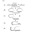
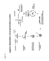
- PLPs are long oligonucleotides of approximately 100 bases, containing target complementary regions at both their 5' and 3' ends ( Fig. 1 ). These regions recognise adjacent sequences on the target DNA ( Nilsson, M., Malmgren, H., Samiotaki, M., Kwiatkowski, M., Chowdhary, B.P. and Landegren, U. (1994)
- Padlock probes circularizing oligonucleotides for localized DNA detection.
- the target-specific products are detected by a universal cZipCode microarray ( Shoemaker, D.D., Lashkari, D.A., Morris, D., Mittmann, M. and Davis, R.W. (1996) Quantitative phenotypic analysis of yeast deletion mutants using a highly parallel molecular bar-coding strategy. Nat. Genet., 14, 450-456 .).
- PLPs have been shown to have good specificity and very high multiplexing capabilities in genotyping assays ( Hardenbol, P., Baner, J., Jain, M., Nilsson, M., Namsaraev, E.A., Karlin-Neumann, G.A., Fakhrai-Rad, H., Ronaghi, M., Willis, T.D., Landegren, U. and Davis, R.W. (2003) Multiplexed genotyping with sequence-tagged molecular inversion probes. Nat. Biotechnol., 21, 673-678 .).
- WO 03/052142 describes a padlock nucleotide probe comprising from 5' to 3' a first targeting domain, a reverse primer binding site for a selective primer, a blocking section comprising a restriction endonuclease cleavage site, a forward primer binding site, a tag and a second targeting domain.
- US 2005/026180 describes a padlock nucleotide probe comprising from 5' to 3' a first targeting domain, a universal reverse primer, a cleavage site, a universal forward primer, a barcode and a second targeting domain.
- WO 00/36141 describes the use of a biotinylated spacer oligonucleotide for separating ligated and non-ligated padlock probes before amplification.
- these padlock probes all require further biotinylated.primers or spacers in order to reduce background amplification of non ligated padlock probes during the consecutive PCR steps.
- the inventors now have found an efficient and reliable multiplex amplification and detection system, which makes use of improved, specifically designed padlock probes.
- the present invention gives a solution for one of the problems associated with the use of padlock probes (PLPs), especially in multiplex assays, which is background amplification of non ligated PLP's during the consecutive PCR step.
- PLPs padlock probes
- the invention comprises a padlock oligonucleotide probe comprising (from 5' to 3'):
- Another aspect of the present invention is a padlock nucleotide probe comprising from 5' to 3'
- the invention also provides a method for the detection of a target nucleotide sequence comprising:
- These methods preferably comprise an NaOH denaturation step before capturing of the padlock nucleotide probes. Streptavidin will bind the desthiobiotin on the PLP. The elution of step g. is performed with biotin. If the unique cleavable sequence is a poly-uracil sequence, cleavage can preferably be effected by treatment with uracil-N-glycosidase and endonuclease IV.
- the linearized PLP stays bound to the streptavidin and goldbeads bearing both ZIP codes complementary to the nucleotide sequence in the padlock and fluorescent barcode nucleotides, will be hybridized.
- Another aspect of the invention is a method for the detection of a target nucleotide sequence comprising:
- Streptavidin will bind desthiobiotin on the PLP.
- the elution of step f. is performed with biotin.
- cleavage can preferably be effected by treatment with uracil-N-glycosidase and endonuclease IV.
- a further aspect of the invention is the use of padlock nucleotide probes according to the invention for the multiplex detection of nucleotide sequences.
- a next aspect of the invention is a test kit comprising multiple padlock probes according to the invention, wherein each padlock probe is designed to recognise a unique target.
- hybrid refers to a double-stranded nucleic acid molecule, or duplex, formed by hydrogen bonding between complementary nucleotides.
- hybridise or “anneal” refer to the process by which single strands of nucleic acid sequences form double-helical segments through hydrogen bonding between complementary nucleotides.
- ligation refers to the process of enzymatically joining two or more nucleotide sequence together by coupling the 5' P moiety of one nucleotide to the 3' OH moiety of a second nucleotide, thereby leaving the polynucleotide backbone intact, which thus will result in a concatenated, normal nucleotide sequence.
- the enzyme used for ligation is a ligase.
- amplification is meant the construction of multiple copies of a nucleic acid sequence or multiple copies complementary to the nucleic acid sequence using at least one of the nucleic acid sequences as a template.
- Methods of the invention can in principle be performed by using any nucleic acid amplification method, such as the Polymerase Chain Reaction (PCR; Mullis 1987, U.S. Pat. No. 4,683,195 , 4,683,202 , and 4,800,159 ) or by using amplification reactions such as Ligase Chain Reaction (LCR; Barany 1991, Proc. Natl. Acad. Sci. USA 88:189-193 ; EP Appl.
- PCR Polymerase Chain Reaction
- LCR Ligase Chain Reaction
- Amplification as used in the present invention also comprises BioBarCode amplification (BCA) as described by Jwa-Min Nam, Savka I. Stoeva and Chad A. Mirkin.
- primer refers to an oligonucleotide which is capable of annealing to the amplification target (the "primer binding site") allowing a polymerase to attach thereby serving as a point of initiation of DNA or RNA synthesis when placed under conditions in which synthesis of primer extension product which is complementary to a nucleic acid strand is induced, i.e., in the presence of nucleotides and an agent for polymerization such as DNA polymerase and at a suitable temperature and pH.
- the (amplification) primer is preferably single stranded for maximum efficiency in amplification.
- the primer is an oligodeoxy ribonucleotide.
- primers must be sufficiently long to prime the synthesis of extension products in the presence of the agent for polymerization.
- the exact lengths of the primers will depend on many factors, including temperature and source of primer.
- primers come in sets including one forward and one reverse primer as commonly used in the art of DNA amplification such as in PCR amplification.
- primer binding sites on the target DNA are present in a set of one for the forward and one for the reverse primer.
- the present invention gives a solution for one of the problems associated with the use of padlock probes (PLPs), especially in multiplex assays, which is background amplification of non ligated PLP's during the consecutive PCR step.
- PLPs padlock probes
- Padlock probes also have some tendency to linear dimer formation as a result of cross reactive ligation, the corresponding ligation products can easily be distinguished from circularized probes by exonucleolytic degradation.
- the exonuclease treatment reduces the number of such linear monomeric and dimeric molecules by almost three orders of magnitude with negligible effects on circularized probes.
- CTAAGNNNNNCTTAG (wherein N denotes any nucleotide), which is cleavable by the enzyme C EcoO109I; the sequence TGGCGACGAAAACCGCTTGGAAAGTGGCTG, which is cleavable by the enzyme F-TflI; ACCTACCATTAACGGAGTCAAAGGCCATTG, which is cleavable by the enzyme F-TflII, TAGGTACTGGACTTAAAATTCAGGTTTTGT, which is cleavable by the enzyme F-TflIII; CAAAACGTCGTAAGTTCCGGCGCG which is cleavable by the enzyme H-DreI; and GAGTAAGAGCCCGTAGTAATGACATGGC, which is cleavable by the enzyme I-BmoI.
- the PLP of the invention preferably contains a polyuracil site for enabling linearization of the probes.
- the unique cleavage sequence is introduced just to the 5' side of the unique forward primer binding site, which, after cleavage becomes the most 5' part of the linearized molecule.
- the unique reverse primer binding site should be positioned as close as possible 5' of the unique cleavage sequence, in order to leave, upon cleaving, said reverse primer binding site at the most 3'end of the linearized molecule.
- the ligated target recognition sites and the ZIP code will be present, which would ensure a proper amplification of the parts of the PLP that are used for giving a specific reaction in the assay.
- a treatment with endonuclease IV is performed for efficient cutting the deoxy-ribose phosphate backbone at the ends of the linearized polynucleotide.
- linearization of the PLP before amplification ensures that PLPs which have not been ligated at the target site will not be amplified. They are also cleaved at the unique cleavage site, leaving one short piece of DNA with only the unique reverse primer binding site, and another, a bit longer stretch, bearing the unique forward primer binding site. Since those pieces are not joined anymore, an amplification step using the universal primer set will not be able to generate amplification products to these incomplete PLPs. Therefore, by linearizing the PLP, an increase in the detection limit is obtained, since the background (noise) amplification signal will be much lower.
- the length of the site is fixed, the length of the poly-uracil sequence is not critical, as long as it gives a good cleavage upon application of uracil-N-glycosidase and endo IV nuclease.
- the stretch of uracil nucleotides has at least 2 uracil bases.
- there is no upper limit to the length of the poly-uracil sequence it will in practice be limited by the technical requirements of the synthesis.
- a poly-uracil site is preferred because this will normally not be present in any of the target nucleotides, nor in any of the further building blocks of the PLP. Thus, it provides an unique site, with little or no chance of disturbing other nucleotides which are present in the reaction of the assay. Further, use of the poly-uracil enables linearization of the padlock probe while it is still in the single-stranded state and thus, no additional mixing with complementary oligonucleotides is necessary. Also the used uracil-N-glycosidase and endonuclease IV have no negative effect on the other nucleotides in the reaction
- the target molecules which have to be assayed, can be any form of DNA or RNA, such as genomic DNA, cDNA, mitochondrial DNA, nuclear DNA, messenger RNA, ribosomal RNA and the like.
- the type of nucleotide is unimportant, but the target should be able of being specifically recognised by the corresponding padlock nucleotide probe.
- the target nucleotides which are present in the sample to be assayed, are randomly cut into smaller fragments of 100-1000 basepairs. This can be done using standard methods well known to a person skilled in the art. Such a random cutting prevents binding of the probe to very large target molecules, which would (partly) survive the exonuclease treatment.
- Tm melting temperatures
- the PLP preferably comprises a 5' arm (T1), which preferably has a length of about 10 to about 75 nucleotides, more preferably of about 20 to about 50 nucleotides and most preferably of about 25 to about 40 nucleotides.
- T1 and T2 sequences are given in Table 3A.
- the pivotal point of the present invention is that even a better detection is achieved when it is possible to isolate the circularized PLPs from the reaction mixture, which contains not only the ligated full length PLPs which have been linearized by cleavage at the unique cleavage site, but also the unreacted sample nucleotides, and short PLP fragments stemming from non-ligated, cleaved PLPs.
- Isolation of the circularised probes is accomplished by incorporation of a nucleotide bearing desthio-biotine. Isolation of the PLPs can then be achieved by contacting said PLPs with a solid support carrying the second member of said binding pair, and thus binding the PLPs.
- the solid support can be anything which is able to carry the second member of the binding pair, such as beads or a column.
- the material of the solid support can be any material which is conventionally used in biochemical procedures of this kind, such as glass, polystyrene, polyethylene, and the like.
- the binding pair according to the invention is (desthio-)biotin/streptavidin. It has been found extremely suitable to provide the PLP with a nucleotide carrying a desthio-biotin moiety. This enables binding to streptavidin coated magnobeads ( Hirsch JD, Eslamizar L, Filanoski BJ, Malekzadeh N, Haugland RP, Beechem JM and Haugland RP. (2002) Easily reversible desthiobiotin binding to streptavidin, avidin, and other biotin-binding proteins: uses for protein labeling, detection, and isolation. Anal Biochem.
- the PLPs which are bound to the beads can be set free again by addition of biotin, which binds more strongly to the streptavidin coated magnobeads and replaces the PLP.
- biotin which binds more strongly to the streptavidin coated magnobeads and replaces the PLP.
- the nucleotide bearing desthio-biotin moiety is engineered between the unique reverse primer binding site and the unique cleavage site, since there it will not interfere with any subsequent amplification reaction (it will be at the 3' end of the reverse primer binding site and thus remain outside the sequence which is amplified). Further, it needs a minimal distance from the cleavage site.
- the steps of isolation and linearization of the PLPs, as described above, are combined.
- the PLPs are fit (may serve as template) for amplification.
- the use of padlock probes in general and specifically in combination with the cleavage at the uracil-site ensures that only the PLPs are amplified which have recognized a target sequence, been able to hybridise to said target sequence and which have been ligated at said target site.
- a genuine representation of the target sequences that were present in the original sample can be obtained.
- the PLPs can be assayed by using any sort of assay which is capable of recognising specific polynucleotides.
- the assay can vary (as described below).
- the assay is performed on a (micro-)array.
- the method can be qualitative or quantitative.
- the specific sequence of the PLP (which can for instance be provided by the ZIP-code or by the target specific sequences T1 or T2) is recognised by a specific capture molecule (e.g. a sequence which is capable of hybridisation with said specific sequence), which bears a label.
- a specific capture molecule e.g. a sequence which is capable of hybridisation with said specific sequence
- the fluorescent substance includes Cy2, FluorX, Cy3, Cy3.5, Cy5, Cy5.5, Cy7, fluorescein isothiocyanate (FITC), Texas Red, Rhodamine, Alexa 532 and the like.
- FITC fluorescein isothiocyanate
- Methods to attach the labels to the nucleotides are generally known in the art.
- a quantitative method of the invention relates to an internal standard.
- a known amount of one or more marker target nucleotide sequences is added to the sample. These will then be recognised by a PLP which is specifically designed for this marker target nucleotide.
- the PLP will undergo the same treatment as the PLPs which have recognised their target nucleotides in the sample.
- all PLPs are detected by their specific sequences and a comparison of the signals generated by the PLP which is directed to the marker target nucleotide with the signals generated by the other PLPs indicates the relative amount of target present in the sample. Since the concentration of the marker target nucleotide was known, the concentration of other target nucleotides can be calculated. To decrease the error margins, several different marker target nucleotides can be added to the sample in increasing concentrations, to generate a sort of internal calibration curve.
- both the amplification and the detection of the PLPs is performed in a micro-array.
- the OpenArrayTM technology (BioTrove, Woburn, MA, USA) currently allows parallel amplification and testing of more than 3000 assays on one plate (48 subarrays with each 64 so-called Through-Holes with a volume of 33 nL).
- the primers are pre-loaded into the holes, while the (purified) sample along with the reagents are autoloaded due to surface tension, provided by the hydrophilic surface of the array. Detection of the amplification can take place by simple detection of the presence of double stranded DNA.
- amplification products can be detected and quantified by using a universal probe, such as a TaqMan probe.
- TaqMan probes are probes with fluorescent dyes at opposite ends and can be used during PCR amplification. The probe is degraded during amplification by 5'-exonuclease activity of the Taq-polymerase used and increase of fluorescence can be measured real-time during amplification and in that way quantification of target is possible (see Livak KJ, Flood SJA, Marmaro J, Giusti W and Deetz K (1995). Oligonucleotides with fluorescent dyes at opposite ends provide a quenched probe system useful for detecting PCR product and nucleic acid hybridization. PCR Methods and Applications 4: 357-362 ).
- the TaqMan probes for detecting amplification products comprise locked nucleic acids (LNA) nucleotides.
- Locked nucleic acids (LNAs) are synthetic nucleic acid analogs that bind to complementary target molecules (DNA, RNA or LNA) with very high affinity.
- binding affinity is decreased substantially for the LNA type when the hybrids thus formed contain even a single mismatched base pair.
- LNA existing TaqMan probes show an increased specificity (see Koshkin,A.A., Nielsen,P., Meldgaard,M., Rajwanshi,V.K., Singh,S.K. and Wengel,J. (1998) LNA (locked nucleic acid): an RNA mimic forming exceedingly stable LNA:LNA duplexes. J. Am. Chem. Soc., 120, 13252-13253 ).
- the detection of the ZIP-code can be performed also directly through hybridising the said padlock nucleotide to an array or to gold beads bearing ZIP codes complementary to the nucleotide sequence in the padlock.
- ligation detection reaction In the ligation detection reaction generic pre-amplified target DNA is used as a source for ligation of standard PLPs.
- the ligated padlock probes are hybridized on the array and labelled with e.g. a streptavidin-coupled fluorescent probe Alexa (532) directed against the desthiobiotin moiety of the padlock.
- This method comprises an exonuclease step after the ligation and a NaOH denaturation step before capturing of the padlock nucleotide probes.
- the elution can be performed with a 80 °C step in H 2 O or with biotin.
- cleavage can preferably be affected by treatment with uracil-N-glycosidase and endonuclease IV (see Fig 21 and Fig 22 ).
- BCA bio-bar-code amplification, Jwa-Min Nam et al., supra .
- BCA is a PCR-less target amplification method that relies on novel two-component oligonucleotide-modified gold nanoparticles (NPs) and single-component oligonucleotide-modified magnetic microparticles (MMPs) and subsequent detection of amplified target DNA in the form of bar-code DNA using a chip-based detection method (see Fig 23 ).
- target DNA is used as a source for ligation of standard PLPs.
- This method comprises an exonuclease step after the ligation and a NaOH denaturation step before capturing of the padlock nucleotide probes.
- the second member of a binding pair will bind the first member of a binding pair which is available on the PLP. If said first member is desthiobiotin, said second member is streptavidin. If the unique cleavable sequence is a poly-uracil sequence, cleavage can preferably be affected by treatment with uracil-N-glycosidase and endonuclease IV.
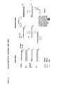
- Typical preferred PLPs of the invention are the standard Padlock probe, the PRI-lock probe and the LUNA-probe as depicted in Fig. 7 .
- the probes can be constructed using normal genetic engineering techniques, such as disclosed in handbooks like Sambrook, J., Fritsch, E.F., and Maniatis, T., in Molecular Cloning: A Laboratory Manual. Cold Spring Harbor Laboratory Press, NY, Vol. 1,2,3 (1989 ).
- Oligonucleotides which need to be assembled for use in the present invention can be made synthetically by standard DNA or RNA chemical synthesizers or may be obtained from enzymatic digestion of wild-type, naturally occurring sequences. It is also possible that naturally occurring sequences are modified by insertion, substitution or deletion of one or more nucleotides using conventional genetic engineering techniques.
- Insertion of the poly-uracil sequence as described above can be done by standard techniques, as mentioned above, which techniques are well known to a person skilled in the art. It is preferred that a commercially obtained desthiobiotin moiety is used.. These moieties are commercially available as desthiobiotin coupled to a thymidine nucleotide. This nucleotide is introduced into the padlock nucleotide probe according to standard methods.
- Standard padlock probes according to the general structure as depicted in the top figure of fig. 7 are designed in such a way that a set of those PLPs all comprise the same universal forward and reverse primer binding sites, designated as universal forward and reverse primer binding sites.
- Choice of these primer binding sites is flexible, insofar that care should be taken that the primer binding site sequences differ substantially from the rest of the padlock probe, so that the amplification step, which makes use of the universal primers is not hampered by homologous sequences in the rest of the probe.
- each standard PLP a unique set of target specific sequences is inserted, which is designed to bind to a specific target sequence which is suspected to be present in the sample.
- the target specific sequence should be unique, meaning that it can hybridise with only one target nucleotide molecule in the sample.
- the PLP comprises a unique ZIP-code, which eventually will serve for the detection of the PLP.
- this ZIP-code there is no functional restriction with regard to this ZIP-code, other than that each PLP of the set of PLPs should have a unique ZIP-code and that it can serve for detection in the assay.
- the ZIP-codes used for a given set of PLP-probes should be of the same size and character, in order not to influence the other steps of the method, such as the amplification step.
- the ZIP codes are derived chosen from the GeneFlexTM TagArray set (Affymetrix) or any other similar library.
- the other elements of the PLP are as described above.
- these probes can be added to a sample under conditions which are optimal for alignment and hybridisation of the target specific sequences T1 and T2 to the target sequences in the sample.
- the hybridisation reaction is complete the PLPs that have hybridised to a target sequence are ligated by addition of the enzyme ligase to the reaction mixture. Thereafter, preferably the non-ligated DNA is removed by exonuclase degradation.
- This exonuclease treatment can be performed with either a 3' to 5' exonuclease or a 5' to 3' exdonuclease or both or an exonuclase wich combines both activities. It is paramount for the present invention that these exonuclease(s) do not have any endonuclease activity.
- the probes are captured using e.g. streptavidin coupled magnetic beads (or another streptavidin coated solid support, such as a column or filter upon which streptavidin is immobilised) and separated from the sample. Subsequently, the probes are cleaved at the uracil-site by adding a sufficient amount of uracil-N-glycosidase and endo IV nuclease.
- the eluted probes are amplified with PCR using the universal primers (one of which is labelled). Amplicons are then hybridised on e.g. micro-arrays on which sequences which are complementary to the ZIP sequences are spotted.
- PRI-lock probe All the other elements of the PRI-lock probe are similar to those of the standard Padlock-probe and can be applied as mentioned above. Also the hybridisation, ligation, linearization and elution of the PRI-lock probe is identical to those described above.
- the Universal ZIP-code is designed for being able to hybridise to a universal probe, such as a TaqMan probe.
- Fig. 9 The scheme of the applied procedure is outlined in Fig. 9 .
- Multiple PRI-lock probes are ligated on fragmented target DNA. Target recognition is achieved by specific hybridization of both arm sequences, and efficient ligation occurs only if the end nucleotides are perfectly matching to the target. Therefore, the probes confer superior specificity.
- the probes are captured on streptavidin-coated magnetic beads via the desthiobiotin, and are cleaved at the deoxy-uracil nucleotides.
- the ligation mix and the TaqMan probe region of unligated probes are removed by several washing steps, eliminating the background due to the presence of unligated probes. The remaining probes are eluted in aqueous biotin solution or after a 80° C incubation step, the ligated probes are assayed in real-time PCR using a unique primer pair for each target.
- the linear quantification range of the proposed procedure is dependent on both the ligation step and the real-time PCR. Ligation of oligonucleotides has been shown to reflect well the target quantity and was used successfully for characterization of gene expression and gene copy number in a multiplex setting.
- PRI-locks combined with the OpenArrayTM system are useful for a flexible and easily adaptable design of high-throughput, quantitative multiplex DNA assays, since the target recognition step is separated from downstream processing.
- the primer binding and TaqMan probe sites were chosen from a set of artificial, well-balanced sequences that had been selected to have minimum cross-hybridization (e.g. the GeneFlexTM TagArrays set (Affymetrix))
- the LUNA probe is also a variant of the above described standard Padlock probe, the difference being that the universal forward primer binding site comprises a T7 polymerase recognition site.
- the PLPs that have hybridised to a target sequence are ligated by addition of the enzyme ligase to the reaction mixture. Thereafter, preferably the non-ligated DNA is removed by exonuclase degradation.
- This exonuclease treatment can be performed with either a 3' to 5' exonuclease or a 5' to 3' exdonuclease or both or an exonuclase wich combines both activities.
- the probes are captured using e.g. streptavidin coupled magnetic beads (or another streptavidin coated solid support, such as a column or filter upon which streptavidin is immobilised) and separated from the sample. Subsequently, the probes are cleaved at the uracil-site by adding a sufficient amount of uracil-N-glycosidase and endo IV nuclease.
- a NASBA reaction is based on the concurrent activity of AMV reverse transcriptase (RT), RNase H and T7 RNA polymerase, together with two primers to produce amplification (3). This process occurs at one temperature (41°C)
- FIG. 14 depicts a generalised assay method with these LUNA probes.
- the amplified products are collected on different matrices (array or Luminex beads) ( Gordon RF, McDade RL, 1997, Multiplexed quantification of human IgG, IgA, and IgM with the Flowmetrix system. Clinical Chemistry, 43: 1799-1801 ) where complementary ZIP (cZIP)-Code oligonucleotides with free 3' ends have been immobilized ( Fig. 14 .). With arrays probe addressable sites are used for target identification. Luminex beads are color coded beads; the association of the amplified product with a characteristic Luminex bead is used as a tag for target identification. With the Luminex approach we have an array in solution.
- cZIP-Code sequences (arbitrarily non-target sequence of approximately 20-25 nucleotides) makes detection of amplified products on both matrices independent from target sequences. As the array or Luminex beads containing cZIP-Codes are independent, this makes the described assay system adaptable for different fields of application.
- Molecular beacons are single-stranded oligonucleotides having a stem-loop structure.
- the loop portion contains the sequence complementary to the target nucleic acid, whereas the stem is unrelated to the target and has a double-stranded structure.
- One arm of the stem is labeled with a fluorescent dye, and the other arm is labeled with a non-fluorescent quencher.
- the probe does not produce fluorescence because the energy is transferred to the quencher and released as heat
- the molecular beacon hybridizes to its target it undergoes a conformational change that separates the fluorophore and the quencher, and the bound probe fluoresces brightly ( Tyagi, S. and Kramer, F.R. (1996) Nature Biotechnol., 14, 303-308 ).
- the pathogenic organisms were derived from the culture collection of the applicant. (Table 1) Genomic DNAs were extracted as previously described ( Bonants, P., Hagenaar-de Weerdt, M., van Gent-Pelzer, M., Lacourt, I., Cooke D. and Duncan, J. (1997) Detection and identification of Phytophthora fragariae Hickman by the polymerase chain reaction. Eur. J of Plant Pathol., 103, 345-355 .).
- the PLP arm sequences were combined with the universal primer binding sites (P1: 5' CTCGACCGTTAGCAGCATGA 3'; P2: 5' CCGAGATGTACCGCTATCGT 3') and a ZipCode sequence.
- the unique identifier was chosen from GeneFlexTM TagArray set (Affymetrix) in a way to minimize PLP secondary structures. Secondary structure predictions were performed by using MFold (http://www.bioinfo.rpi.edu/applications/mfold/). When necessary, PLP arm sequences were also adjusted to avoid strong secondary structures that might interfere with efficient ligation.
- Genomic DNA was fragmented by digestion using EcoRI, HindIII and BamHI (New England Biolabs) for 30 min, and used as template in the indicated amount. Cycled ligation was performed in 10 ⁇ L reaction mixture containing 20 mM Tris-HCl pH 9.0, 25 mM KCH 3 COO, 10 mM Mg(CH 3 COO) 2 , 10 mM DTT, 1 mM NAD, 0.1% Triton X-100, 20 ng sonicated salm sperm DNA, 2.4 U Taq ligase (New England Biolabs) and 25 pM PLP. For multiplex detection the concentration of the individual PLPs were adjusted to achieve comparable performance, and ranged from 25 to 200 pM.
- Reactions mixtures were made up on ice, and transferred rapidly to a thermal cycler. After 5 min at 95°C, 20 cycles of 30 sec at 95°C and 5 min at 65°C were performed, followed by 15 min inactivation at 95°C. After ligation, 10 ⁇ L of exonuclease mix (10 mM Tris-HCl pH 9.0, 4.4 mM MgCl 2 , 0.1 mg/ml BSA, 0.5 U Exonuclease I (USB) and 0.5 U Exonuclease III (USB) was added to each reaction, and the samples were incubated at 37°C for 2 h, followed by inactivation at 95°C for 2.5 h.
- exonuclease mix 10 mM Tris-HCl pH 9.0, 4.4 mM MgCl 2 , 0.1 mg/ml BSA, 0.5 U Exonuclease I (USB) and 0.5 U Exonuclease III (USB
- reaction mixtures of 25 ⁇ L contained 2.5 ⁇ L 10X real-time buffer, 3 mM MgCl 2 , 200 nM of each dNTP including dTTP/dUTP, 100 nM P-Frag TaqMan probe (5' FAM-CCCGGTCAACTTCAAGCTCCTAAGCC-TAMRA 3'), 300 nM of primers P1-f20 (5' CCGAGATGTACCGCTATCGT 3') and P2-r20 (5' TCATGCTGCTAACGGTCGAG 3'), 0.6 U Hot Gold Start polymerase, 0.6 U UNG and 3 ⁇ L ligation-exo mix as template.
- the reaction mix was initially incubated at 50°C for 2 min, followed by 10 min denaturation at 95°C, and 40 cycles of 15 sec at 95°C and 1 min at 60
- LATE-PCR linear-after-the-exponential PCR
- LATE Linear-after-the-exponential PCR
- PLPs were amplified in 25 ⁇ L reaction mixtures containing 1X Pfu buffer (Stratagene), 200 nM of each dNTP, 500 nM of Cy3- or Cy5-labeled P1-f19 primer (5' CGAGATGTACCGCTATCGT 3'), 50 nM P2-r20 primer, 0.375 U Pfu (Stratagene) and 3 ⁇ L ligation-exo mix as template.
- the temperature profile of the reaction was: 5 min at 95°C, 40 cycles of 2 sec at 51°C, 5 sec at 72°C and 15 sec at 95°C, after which the reaction was immediately cooled to 10°C.
- PLP amplicons were analysed by agarose gel electrophoresis before applying them on array.
- cZipCode Complementary ZipCode
- oligonucleotides (Fig. 4 ) carrying a C12 linker and a 5' NH 2 group were synthesised and spotted on Nexterion MPX-E16 epoxy-coated slides by Isogen B.V. (Utrecht, The Netherlands) according to manufacturer's instructions (Schott Nexterion). Briefly, 50 nL of 1.5 mM cZipCode solution was spotted using an OmniGrid100 contact-dispensing system (Genomic Solutions) equipped with SMP4 pins (Telechem) at 50% relative humidity.
- the uncoupled probes were removed by washing in 300 mM bicine, pH 8.0, 300 mM NaCl, and 0.1% SDS for 30 min at 65°C, followed by rinsing with deionised water and drying by spinning at 250 g for 2 min.
- the arrays were stored in dark, in a desiccator at room temperature until use.
- the hybridisation mixes were made up of 5 ⁇ L Cy3-labeled sample and 5 ⁇ L Cy5-labeled background control sample in 3 M TMAC, 0.1 % sarkosyl, 50 mM Tris-HCl pH 8.0, 4 mM Na 2 EDTA. Cy5-labeled hybridisation control was added to 20 pM final concentration in 50 ⁇ l final volume. For each slide, one of the hybridisation samples contained Cy5- and Cy3-labeled amplicons corresponding to the same, positive ligation reaction, which served to correct for dye bias (dye correction sample).
- the mixes were heated for 10 min at 99°C and cooled down rapidly on ice.
- Sixteen-well silicon superstructures (Schott Nexterion) were attached to the arrays to create separate chambers for the subarrays. After adding 40 ⁇ l of the samples to each well, the chambers were sealed, and the arrays were hybridised at 55°C o/n in high humidity. Afterwards, the isolators were removed, and the slides were washed once at 55°C for 5 min in prewarmed 1XSSC/0.2% SDS, and twice for an additional 1 min at RT in 0.1XSSC/0.2% SDS and in 0.1XSSC, respectively. Finally, the slides were dried by spinning at 250 g for 2 min.
- Microarrays were analysed using a confocal ScanArray ® 4000 laser scanning system (Packard GSI Lumonics) containing a GreNe 543 nm laser for Cy3 and a HeNe 633 nm laser for Cy5 fluorescence measurement. Laser power was fixed at 70% for both lasers, while PMT (photomultiplier tube power) ranged from 45 to 65%, depending on signal intensity. Fluorescent intensities were quantified by using QuantArray ® (Packard GSI Lumonics), and the parameters 'mean signal - mean local background' ( mean Cy3-B or mean Cy5-B ) and the 'mean local background' (B) were used in further calculations.
- QuantArray ® Packard GSI Lumonics
- the high discriminatory power of the ligation is of prime importance, since very similar, non-target DNA molecules can be present potentially in much higher concentration than the target DNA. Therefore, we aimed to optimise the reaction conditions and PLP design for maximum discrimination of single mismatches, which subsequently could be extrapolated to diagnostic assay design.
- the experimental system to optimise the ligation conditions consisted of PLP P-frag, which targeted the ITS region of Phytophthora fragariae, and of the corresponding synthetic, target and non-target oligonucleotides (Table 2A).
- cycled ligation consisting of 20 cycles of 5 minutes at 65°C provided good discrimination, sufficient yield of ligation product and freedom from potential secondary structures (data not shown).
- the reaction mixture also contained 20 ng sonicated salmon sperm DNA, which served to provide a large excess of non-target DNA. All the subsequent experiments were performed under these conditions.
- oligonucleotides D0 - D6 as targets, we tested how the discriminatory power of PLP P-frag depended on the type and the position of the mismatch (Table 2B).
- the discrimination factor was defined as the fold-difference in the yield of ligation product with target and mismatched oligonucleotides, as determined by real-time PCR.
- mismatches positioned at the 3' end of PLP were strongly discriminating, while those at the 5' end provided much less specificity.
- the type of the mismatch was also found to be important, although to lesser extent. In general, it appeared that the nearest neighbour parameters could be indicative of the destabilizing effects of different mismatches. Mismatched nucleotide pairs including cytosines were better discriminated, while the G-T pair (at the 5' end) hardly affected the ligation efficiency.
- the second strategy involved inserting a destabilizing mismatch in the middle of the 3' arm-complementary sequence, and the binding of the probe was similarly stabilized by lengthening the 5' arm (oligonucleotides A2 and A2C).
- the melting temperatures (T m ) of the 5' arm sequences became higher than the reaction temperature, while those of the 3' arms were about 20 to 30°C below it (Table 2C).
- T m melting temperatures
- the 3' arm sequences were selected to be 14-18 nucleotide-long and had a T m around 40°C (Table 3B). In general, the 3' arm sequence hybridised to the discriminatory region and contained a highly destabilizing mismatch or a gap at the 3' end when bound to the non-target sequence.
- the 5' arm sequences were 27-37 nucleotide-long.
- a genus-specific PLP to target all Phytophthora species and discriminate them from related oomycetes. After selecting the target-complementary regions, they were combined with the universal primer binding site sequences, and a unique ZipCode sequence was selected for each probe.
- a mix of the developed 11 PLPs was ligated on various genomic DNAs, treated with exonucleases, and subjected to LATE-PCR using Cy3-labeled forward primer.
- the labelled PLP amplicons were analysed on multi-chamber, low-density universal microarrays, which enabled the simultaneous assay of 16 samples on a single slide ( Fig. 4 ).
- the tag array used in our experiments contained 30 probes in 9 replicates, together with 90 hybridisation control probes distributed over the deposition area. This layout allows for the future extension of the PLP set to target other pathogens and enables high-throughput screening (see Fig. 8 ).
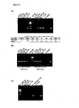
- genomic DNAs from a panel of well-characterized isolates of plant pathogenic organisms (Tables 1 and 4; Fig. 5 a-g). In each case, 1 ng genomic DNA could be specifically and reliably detected without any cross-reaction. All the Phytophthora species were correctly recognized by PLP Phyt-spp, including P. cactorum, which contained two adjacent mismatches with the 5' arm sequence of the probe (Table 3A). This polymorphism was apparently well tolerated, resulting in a positive signal.
- PRI-lock probes were designed to target economically important plant pathogens so as to create a pilot-scale, multiplex detection system to test the proposed principle ( Fig. 7 ).
- a universal TaqMan probe, containing LNA (locked nucleic acids) was designed to monitor the amplification.
- the specificity of the assay will be demonstrated by testing DNAs of the most similar, non-target organisms for each probe (Table 5). Since target discrimination is achieved based on only a single or a few nucleotides, this pilot system also shows the potential of PRI-Locks for extremely specific, quantitative analysis on a universal platform.
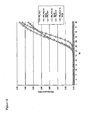
- the linear range of quantification was analyzed for all the three PRI-lock probes using dilution series of target DNA.
- the resulting calibration curves can be used for quantification of target in subsequent experiments.
- the linear range of quantification is only 4 magnitudes, because at higher target concentrations the ligation yield does not increase any more in a linear fashion with the increasing target concentration ( Fig. 13 ).
- a substantial increase in the applied PRI-lock probe concentration (100x) is expected to significantly increase the quantification range.
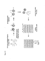
- any remaining non-circularized (unligated) padlock probes can be removed by exonuclease treatment followed by capturing of the probes through by binding of the desthiobiotin moiety with streptavidin magnobeads. After a washing step the remaining circularized probes are digested with Uracil-N-glycosidase/ endo IV nuclease at the position of the incorporated uracil nucleotides between the 5' T7 RNA polymerase recognition site and the 3' end of the universal reverse primer binding site. The release of the complementary T7 site, common for all padlock-probes, acts as starting point for a generic NASBA amplification at a fixed temperature (see Fig. 14-15 )
- LUNA probe hybridization, ligation, exonuclease and glycosidase treatment were performed as described previously.
- circularized and linearized LUNA probes are amplified by standard NASBA utilising the T7 primer binding site included in de LUNA probe.
- Amplified ss products can be hybridized to an array or Luminex beads on which cZipCode sequences are spotted/bound. Luminex beads can be analyzed with flow cytometry. Amplification of ligated LUNA probes in this example is measured using Molecular Beacons.
- two point mutation specific LUNA probes have been designed.
- a mutation on the 3'end of the probe is more discriminatory than when a mutation is placed at the 5' end.
- Specificity of the probe is largely increased with an asymmetrical design and a high ligation temperature.
- the 3' arm of the LUNA probe has a melting temperature (Tm) of 37-40 °C
- the 5'-arm has a Tm of 65 - 70 °C.
- Tm melting temperature
- the specificity of the LUNA ligation step has been validated with closely related (non)pathogenic species and appeared to point mutation specific.
- the secondary structure and in particular the localization of the T7 polymerase recognition site of the LUNA probe is essential for an efficient initiation of the NASBA amplification.
- Linearization of the LUNA probe by Uracil-N-glycosidase / endo IV nuclease followed by selective capturing of the probes with Streptavidine coated magnobeads appeared to be essential for an efficient NASBA amplification reaction.
- the target specific Zip-Codes of the LUNA probes have been used as hybridization sites for the used Molecular Beacons. With those identification tags quantification of the isothermal NASBA could be followed.
- As identifiers of the two LUNA probes against P. cactorum and V. dahliae a FAM- and JOE-labeled MB respectively have been designed ( Fig. 16 ).
- the multiplexibility and dynamic detection range are important parameters of this LUNA detection system. To validate those parameters genomic DNA of the plant pathogenic Phytophthora cactorum and Verticillium dahliae have been extracted and tested in different concentrations and ratio's and compared with traditional PCR ( Fig. 17 , 18 ).
- the detection limit for both targets appeared to be in the pg range ( Fig. 17 ).
- the dynamic detection range for those pathogens was at least 100 ( Fig. 18 ).
- the next step will be hybridization of the ssRNA LUNA amplicons to arrays spotted with cZipCode oligos or to different Luminex beads coupled with different cZipCode oligos. Detection can then be performed by array scanning, flow-cytometry or Molecular Beacon detection ( Fig. 19 ).
- Lengths (L) and melting temperatures (T m ) of PLP target-complementary regions are indicated.
- the number of nucleotides discriminating the targeted sequence from that of the known most similar, non-target organism is shown for each PLP.
- Sensitivity was defined as the lowest concentration of perfectly matching oligonucleotide that could be detected under standard assay conditions. Discriminatory range gives the magnitude difference between the lowest detectable concentrations of target and of non-target oligonucleotides.
- A Targeted species/group 5' target complementary sequence (5'- 3') 3'target complementary sequence (5'- 3') ZipCode sequence (5'-3') Phytophthora spp.
- solani AG 4-1 1 ng 0 0 0 0 0 14.3 ⁇ 0.2 0 0 0 0 na na na na 3.8 ⁇ 0.1 na na na na na R. solani AG 4-21 ng 0 0 0 0 0 0 0 0 0 0 0 0 na na na na 14.5 ⁇ 0.2 8.3 ⁇ 0.1 na na na na na na V.
- solani 4-2 500 pg M. roridum , 500 pg 2.2 ⁇ 0.1 na na 4.6 ⁇ 0.1 na na 5.1 ⁇ 0.1 na na 6.3 ⁇ 0.2 na P. nicotiane, 500 pg 13.0 ⁇ 0.4 12.3 ⁇ 0.4 0 0 11.9 ⁇ 0.4 0 0 0 0 0 12.0 ⁇ 0.4 Pyt. ultimum, 500 pg M. hapla, 500 pg 3.1 ⁇ 0.1 2.4 ⁇ 0.2 na na 6.5 ⁇ 0.6 na na na na na na 6.8t0.3 P.
- roridum 500 pg 0 0 0 0 0 0 0 0 0 11.2 ⁇ 0.7 14.1 ⁇ 0.7 0
- F. oxysporum 500 pg na na na na na na na na na 9.0 ⁇ 1.1 7.0 ⁇ 03 na M.
- roridum 500 pg 0 0 0 0 0 0 0 0 10.6 ⁇ 0.4 13.8 ⁇ 0.7 F. oxysporum, 50 pg na na na na na na na na na na 8.0 ⁇ 0.7 75 ⁇ 0.3 na M.
- roridum 500 pg 0 0 0 0 0 0 0 0 0 8.7 ⁇ 0.3 12.7 ⁇ 0.4 0
- F. oxysporum 5 pg na na na na na na na na na na S.It0.4 7.8 ⁇ 0.2 na M.
- roridum 500 pg 0 0 0 0 0 0 0 0 0 0 9.0 ⁇ 0.3 13.9 ⁇ 0.6 0
- F. oxysporum 0.5 pg na na na na na na na na na na na na 1 .5+0.2 8.0 ⁇ 0.3 na M.
- roridum 5 pg 0 0 0 0 0 0 0 0 14. ⁇ 0.7 9.6 ⁇ 0.3 0 F. oxysporum, 500 pg na na na na na na na na na 10.3 ⁇ 0.2 1.2 ⁇ 0.1 na M. roridum, 0.5 pg 0 0 0 0 0 0 0 0 14.5 ⁇ 1.0 5.9 ⁇ 0.9 0 F.
Claims (24)
- Standard-Padlocknucleotidsonde, umfassend von 5' nach 3':a) eine zielspezifische Nucleotidsequenz 1 (T1),b) eine generische Rückwärtsprimer-Bindestelle;c) eine Nucleotidsequenz, die Desthio-Biotin trägt,d) einen Linker von mindestens 12 Nucleotiden,e) eine einzigartige spaltbare Sequenz,f) eine generische Vorwärtsprimer-Bindestelle,g) eine einzigartige ZIP-Code-Sequenz,h) eine zielspezifische Nucleotidsequenz 2 (T2),wobei die T1- und T2-Sequenzen so entworfen sind, dass sie komplementär zu angrenzenden Nucleotidabschnitten auf dem gleichen Ziel sind, und zwar dergestalt, dass die Padlocksonde nach Hybridisierung (und Ligierung der äußeren Enden) ein ringförmiges Molekül bildet.
- Padlocksonde nach Anspruch 1, wobei die einzigartige spaltbare Sequenz eine Poly-Uracil-Sequenz ist.
- Padlocksonde nach Anspruch 2, wobei die Poly-Uracil-Sequenz als ein erstes Mitglied eines Bindepaares fungiert.
- Padlocksonde nach einem der Ansprüche 1 bis 3, dadurch gekennzeichnet, dass sie auch eine T7-RNA-Polymerase-Erkennungsstelle umfasst.
- Padlocksonde nach Anspruch 4, wobei die T7-RNA-Polymerase-Erkennungsstelle sich 5' zur generischen Vorwärtsprimer-Bindestelle befindet.
- Padlocksonde nach einem der Ansprüche 1 bis 5, wobei der ZIP-Code der komplementäre Strang einer Nucleotidsequenz auf einem Array oder einem anderen Element ist.
- Padlocknucleotidsonde (PRI-lock), umfassend von 5' nach 3'a) eine zielspezifische Nucleotidsequenz 1 (T1),b) eine einzigartige Rückwärtsprimer-Bindestelle,c) eine Nucleotidsequenz, die Desthio-Biotin trägt,d) ein Linker von mindestens 12 Nucleotiden,e) eine einzigartige spaltbare Sequenz,f) eine einzigartige Vorwärtsprimer-Bindestelle,g) eine zielspezifische Nucleotidsequenz 2 (T2),wobei die T1- und T2-Sequenzen so entworfen sind, dass sie komplementär zu angrenzenden Nucleotidabschnitten auf dem gleichen Ziel sind, und zwar dergestalt, dass die Padlocksonde nach Hybridisierung (und Ligierung der äußeren Enden) ein ringförmiges Molekül bildet.
- Padlocksonde nach Anspruch 7, wobei die einzigartige spaltbare Sequenz eine Poly-Uracil-Sequenz ist.
- Padlocksonde nach Anspruch 7 oder 8, welche einen universellen ZIP-Code umfasst, der zwischen der einzigartigen Vorwärtsprimer-Bindestelle und der zielspezifischen Nucleotidsequenz 2 (T2) gelegen ist.
- Padlocksonde nach einem der Ansprüche 1 bis 9, wobei die Poly-Uracil-Sequenz 2 -100, bevorzugt 2 - 50, mehr bevorzugt 2 - 255 und am meisten bevorzugt 3-10 Nucleotide umfasst.
- Padlocksonde nach einem der Ansprüche 1 bis 10, wobei die erste zielspezifische Sequenz 1 (T1) eine Länge von etwa 10 bis etwa 75 Nucleotiden, vorzugsweise von etwa 20 bis etwa 50 Nucleotide, und am meisten bevorzugt von etwa 25 bis etwa 40 Nucleotiden hat.
- Padlocksonde nach einem der Ansprüche 1 bis 11, wobei die zweite zielspezifische Nucleotidsequenz 2 (T2) eine Länge von etwa 10 bis etwa 30 Nucleotiden, vorzugsweise von etwa 10 bis etwa 20 Nucleotiden hat.
- Verfahren zum Nachweis einer Zielnucleotidsequenz, umfassend:a. Hinzufügen zu einer Probe, welche das Ziel enthält, einer oder mehrerer Padlocknucleotidsonden nach einem der Ansprüche 1- 6, welche zielspezifische Sequenzen haben, die zur Hybridisierung an das Ziel fähig sind;b. Ermöglichen des Annealings der Padlocknucleotidsonde an das Ziel;c. Ligieren der Padlocknucleotidsonde mit sich selbst;d. Einfangen der Padlocknucleotidsonden über das Desthio-Biotin durch Inkontaktbringen dieser mit einem festen Träger, der mit Strepatvidin beschichtet ist,e. Linearisieren der Padlocknucleotidsonde;f. Waschen des festen Trägers, um jedwede ungebundenen Oligonucleotide zu entfernen,g. Eluieren der PLP-Sonde von dem festen Träger,h. Nachweis des ZIP-Codes.
- Verfahren nach Anspruch 13, wobei der Nachweis des ZIP-Codes die Schritte umfasst:i. Amplifikation der Padlocknucleotidsonde unter Verwendung von generischen Primern;j. Markieren der amplifizierten Padlocknucleotidsonde;k. Prüfen auf das Vorhandensein des ZIP-Codes durch Hybridisierung der Padlocknucleotidsonde mit mindestens einer Sequenz, welche zur Hybridisierung mit dem ZIP-Code fähig ist, wobei die Hybridisierung vorzugsweise auf einem festen Träger wie einem (Micro-)Array stattfindet.
- Verfahren nach Anspruch 13, wobei der Nachweis des ZIP-Codes die Schritte umfasst:i. Prüfen auf das Vorhandensein des ZIP-Codes durch Hybridisierung der Padlocknucleotidsonde mit mindestens einer Sequenz, welche zur Hybridisierung mit dem ZIP-Code fähig ist, wobei die Hybridisierung vorzugsweise auf einem festen Träger wie einem (Micro-)Array oder einer Goldperle stattfindet; undj. Markieren der hybridisierten Padlocknucleotidsonde auf dem festen Träger mit einer fluoreszierenden Sonde, die gegen das erste Mitglied eines Bindepaares der Padlocknucleotidsonde gerichtet ist, oder mit fluoreszenzmarkierten Biobarcode-markierten Goldperlen.
- Verfahren zum Nachweis einer Zielnucleotidsequenz, umfassend:a. Hinzufügen zu einer Probe, welche das Ziel enthält, einer oder mehrerer Padlocknucleotidsonden nach einem der Ansprüche 7 - 12, welche zielspezifische Sequenzen haben, die zur Hybridisierung an das Ziel fähig sind;b. Ermöglichen des Annealings der Padlocknucleotidsonde an das Ziel;c. Zirkularisieren der Padlocknucleotidsonde durch Ligierung;d. Einfangen der Padlocknucleotidsonde über das Desthio-Biotin unter Verwendung eines festen Trägers, der mit Strepatvidin beschichtet ist;e. Linearisieren der Padlocknucleotidsonde;f. Eluieren der Padlocknucleotidsonde von dem festen Träger,g. Amplifizieren der eluierten Padlocknucleotidsonde unter Verwendung von einzigartigen Primern;h. Beobachten und/oder Nachweis der Amplification.
- Verfahren nach Anspruch 16, wobei das Beobachten und/oder der Nachweis der Amplifikation unter Verwendung eines interkalierenden Farbstoffes, vorzugsweise Ethidiumbromid oder SYBR Green, durchgeführt wird.
- Verfahren nach Anspruch 17, wobei das Beobachten und/oder der Nachweis der Amplifikation unter Verwendung einer universellen Sonde, vorzugsweise einer TaqMan-Sonde, durchgeführt wird.
- Verfahren nach einem der Ansprüche 13 - 18, wobei die einzigartige Spaltsequenz eine Poly-Uracil-Sequenz ist und die Linearisierung durch Behandlung mit Uracil-N-Glycosidase und Endonuclease IV erreicht wird.
- Verfahren nach einem der Ansprüche 13 - 19, wobei Biotin zur Eluierung der Sonde von dem festen Träger verwendet wird.
- Verfahren nach einem der Ansprüche 13 - 20, wobei ein Denaturierungsschritt mit NaOH vor dem Einfangen der Padlocknucleotidsonden durchgeführt wird.
- Verfahren nach einem der Ansprüche 13 - 21, wobei der feste Träger eine Perle oder eine Säule ist.
- Verwendung einer Padlocksonde nach einem der Ansprüche 1 - 10 zum vielfachen Nachweis von Nucleotidsequenzen.
- Kit umfassend vielfache Padlocksonden nach einem der Ansprüche 1 - 10, wobei jede Padlocksonde so entworfen ist, dass sie ein einzigartiges Ziel erkennt.
Applications Claiming Priority (1)
| Application Number | Priority Date | Filing Date | Title |
|---|---|---|---|
| PCT/NL2005/000807 WO2007061284A1 (en) | 2005-11-22 | 2005-11-22 | Multiplex nucleic acid detection |
Publications (2)
| Publication Number | Publication Date |
|---|---|
| EP1969137A1 EP1969137A1 (de) | 2008-09-17 |
| EP1969137B1 true EP1969137B1 (de) | 2011-10-05 |
Family
ID=36396665
Family Applications (1)
| Application Number | Title | Priority Date | Filing Date |
|---|---|---|---|
| EP05813533A Not-in-force EP1969137B1 (de) | 2005-11-22 | 2005-11-22 | Multiplex-nukleinsäurenachweis |
Country Status (5)
| Country | Link |
|---|---|
| US (1) | US20120021930A1 (de) |
| EP (1) | EP1969137B1 (de) |
| JP (1) | JP2009516525A (de) |
| AT (1) | ATE527383T1 (de) |
| WO (1) | WO2007061284A1 (de) |
Families Citing this family (65)
| Publication number | Priority date | Publication date | Assignee | Title |
|---|---|---|---|---|
| US7968287B2 (en) | 2004-10-08 | 2011-06-28 | Medical Research Council Harvard University | In vitro evolution in microfluidic systems |
| US20080003142A1 (en) | 2006-05-11 | 2008-01-03 | Link Darren R | Microfluidic devices |
| WO2008097559A2 (en) | 2007-02-06 | 2008-08-14 | Brandeis University | Manipulation of fluids and reactions in microfluidic systems |
| US8592221B2 (en) | 2007-04-19 | 2013-11-26 | Brandeis University | Manipulation of fluids, fluid components and reactions in microfluidic systems |
| WO2009032167A1 (en) * | 2007-08-29 | 2009-03-12 | Illumina Cambridge | Method for sequencing a polynucleotide template |
| CN101230404B (zh) * | 2008-02-04 | 2011-11-16 | 浙江理工大学 | 同时检测多种混合感染病毒的基因芯片 |
| WO2010009365A1 (en) | 2008-07-18 | 2010-01-21 | Raindance Technologies, Inc. | Droplet libraries |
| AU2010242073C1 (en) | 2009-04-30 | 2015-12-24 | Good Start Genetics, Inc. | Methods and compositions for evaluating genetic markers |
| US9399797B2 (en) | 2010-02-12 | 2016-07-26 | Raindance Technologies, Inc. | Digital analyte analysis |
| WO2011100604A2 (en) | 2010-02-12 | 2011-08-18 | Raindance Technologies, Inc. | Digital analyte analysis |
| US20190300945A1 (en) | 2010-04-05 | 2019-10-03 | Prognosys Biosciences, Inc. | Spatially Encoded Biological Assays |
| US10787701B2 (en) | 2010-04-05 | 2020-09-29 | Prognosys Biosciences, Inc. | Spatially encoded biological assays |
| EP3447155A1 (de) | 2010-09-30 | 2019-02-27 | Raindance Technologies, Inc. | Sandwichassays in tröpfchen |
| US9163281B2 (en) | 2010-12-23 | 2015-10-20 | Good Start Genetics, Inc. | Methods for maintaining the integrity and identification of a nucleic acid template in a multiplex sequencing reaction |
| US9364803B2 (en) | 2011-02-11 | 2016-06-14 | Raindance Technologies, Inc. | Methods for forming mixed droplets |
| US9150852B2 (en) | 2011-02-18 | 2015-10-06 | Raindance Technologies, Inc. | Compositions and methods for molecular labeling |
| CN102181443B (zh) * | 2011-03-21 | 2013-10-23 | 中国科学院植物研究所 | 一种多重检测基因组dna多态性的方法及其专用探针 |
| GB201106254D0 (en) | 2011-04-13 | 2011-05-25 | Frisen Jonas | Method and product |
| WO2012167142A2 (en) | 2011-06-02 | 2012-12-06 | Raindance Technolgies, Inc. | Enzyme quantification |
| US8658430B2 (en) | 2011-07-20 | 2014-02-25 | Raindance Technologies, Inc. | Manipulating droplet size |
| CA2852665A1 (en) | 2011-10-17 | 2013-04-25 | Good Start Genetics, Inc. | Analysis methods |
| EP3495817A1 (de) * | 2012-02-10 | 2019-06-12 | Raindance Technologies, Inc. | Molekulardiagnostischer screeningtest |
| US8209130B1 (en) | 2012-04-04 | 2012-06-26 | Good Start Genetics, Inc. | Sequence assembly |
| US8812422B2 (en) | 2012-04-09 | 2014-08-19 | Good Start Genetics, Inc. | Variant database |
| US10227635B2 (en) * | 2012-04-16 | 2019-03-12 | Molecular Loop Biosolutions, Llc | Capture reactions |
| US9222122B2 (en) | 2012-11-20 | 2015-12-29 | Src, Inc. | System and method for rapid detection and identification of nucleic acid labeled tags |
| US8778609B1 (en) | 2013-03-14 | 2014-07-15 | Good Start Genetics, Inc. | Methods for analyzing nucleic acids |
| US8847799B1 (en) | 2013-06-03 | 2014-09-30 | Good Start Genetics, Inc. | Methods and systems for storing sequence read data |
| WO2014210225A1 (en) | 2013-06-25 | 2014-12-31 | Prognosys Biosciences, Inc. | Methods and systems for determining spatial patterns of biological targets in a sample |
| US20150141257A1 (en) * | 2013-08-02 | 2015-05-21 | Roche Nimblegen, Inc. | Sequence capture method using specialized capture probes (heatseq) |
| KR20150029432A (ko) | 2013-09-10 | 2015-03-18 | 삼성전자주식회사 | 핵산 검출 방법 |
| US11901041B2 (en) | 2013-10-04 | 2024-02-13 | Bio-Rad Laboratories, Inc. | Digital analysis of nucleic acid modification |
| EP3058096A1 (de) | 2013-10-18 | 2016-08-24 | Good Start Genetics, Inc. | Verfahren zur beurteilung eines genomischen bereichs einer person |
| US10851414B2 (en) | 2013-10-18 | 2020-12-01 | Good Start Genetics, Inc. | Methods for determining carrier status |
| US9944977B2 (en) | 2013-12-12 | 2018-04-17 | Raindance Technologies, Inc. | Distinguishing rare variations in a nucleic acid sequence from a sample |
| WO2015175530A1 (en) | 2014-05-12 | 2015-11-19 | Gore Athurva | Methods for detecting aneuploidy |
| US11408024B2 (en) | 2014-09-10 | 2022-08-09 | Molecular Loop Biosciences, Inc. | Methods for selectively suppressing non-target sequences |
| WO2016048829A1 (en) | 2014-09-24 | 2016-03-31 | Good Start Genetics, Inc. | Process control for increased robustness of genetic assays |
| EP3271480B8 (de) | 2015-01-06 | 2022-09-28 | Molecular Loop Biosciences, Inc. | Abtastung auf strukturelle varianten |
| EP3530752B1 (de) | 2015-04-10 | 2021-03-24 | Spatial Transcriptomics AB | Räumlich getrennte multiplex-nukleinsäureanalyse von biologischen proben |
| US11268141B2 (en) * | 2016-03-24 | 2022-03-08 | Biofire Diagnostics, Llc | Methods for quantitative amplification |
| EP3639022A4 (de) * | 2017-06-15 | 2021-03-10 | Technion - Research & Development Foundation Ltd | Zusammensetzungen und verfahren zur erkennung von genomvariationen |
| US11519033B2 (en) | 2018-08-28 | 2022-12-06 | 10X Genomics, Inc. | Method for transposase-mediated spatial tagging and analyzing genomic DNA in a biological sample |
| EP3894587A1 (de) | 2018-12-10 | 2021-10-20 | 10X Genomics, Inc. | Auflösung von räumlichen anordnungen durch nähebasierte dekonvolution |
| US11926867B2 (en) | 2019-01-06 | 2024-03-12 | 10X Genomics, Inc. | Generating capture probes for spatial analysis |
| US11649485B2 (en) | 2019-01-06 | 2023-05-16 | 10X Genomics, Inc. | Generating capture probes for spatial analysis |
| WO2020243579A1 (en) | 2019-05-30 | 2020-12-03 | 10X Genomics, Inc. | Methods of detecting spatial heterogeneity of a biological sample |
| EP4025711A2 (de) | 2019-11-08 | 2022-07-13 | 10X Genomics, Inc. | Erhöhung der spezifität einer analytbindung |
| EP3891300B1 (de) | 2019-12-23 | 2023-03-29 | 10X Genomics, Inc. | Verfahren zur räumlichen analyse mittels rna-template-ligation |
| US11732299B2 (en) | 2020-01-21 | 2023-08-22 | 10X Genomics, Inc. | Spatial assays with perturbed cells |
| US11702693B2 (en) | 2020-01-21 | 2023-07-18 | 10X Genomics, Inc. | Methods for printing cells and generating arrays of barcoded cells |
| US11898205B2 (en) | 2020-02-03 | 2024-02-13 | 10X Genomics, Inc. | Increasing capture efficiency of spatial assays |
| US11732300B2 (en) | 2020-02-05 | 2023-08-22 | 10X Genomics, Inc. | Increasing efficiency of spatial analysis in a biological sample |
| WO2021216708A1 (en) | 2020-04-22 | 2021-10-28 | 10X Genomics, Inc. | Methods for spatial analysis using targeted rna depletion |
| WO2021237087A1 (en) | 2020-05-22 | 2021-11-25 | 10X Genomics, Inc. | Spatial analysis to detect sequence variants |
| WO2021236929A1 (en) | 2020-05-22 | 2021-11-25 | 10X Genomics, Inc. | Simultaneous spatio-temporal measurement of gene expression and cellular activity |
| WO2021242834A1 (en) | 2020-05-26 | 2021-12-02 | 10X Genomics, Inc. | Method for resetting an array |
| EP4162074B1 (de) | 2020-06-08 | 2024-04-24 | 10X Genomics, Inc. | Verfahren zur bestimmung eines chirurgischen randes und verfahren zur verwendung davon |
| CN116034166A (zh) | 2020-06-25 | 2023-04-28 | 10X基因组学有限公司 | Dna甲基化的空间分析 |
| US11761038B1 (en) | 2020-07-06 | 2023-09-19 | 10X Genomics, Inc. | Methods for identifying a location of an RNA in a biological sample |
| US11926822B1 (en) | 2020-09-23 | 2024-03-12 | 10X Genomics, Inc. | Three-dimensional spatial analysis |
| US11827935B1 (en) | 2020-11-19 | 2023-11-28 | 10X Genomics, Inc. | Methods for spatial analysis using rolling circle amplification and detection probes |
| AU2021409136A1 (en) | 2020-12-21 | 2023-06-29 | 10X Genomics, Inc. | Methods, compositions, and systems for capturing probes and/or barcodes |
| EP4196605A1 (de) | 2021-09-01 | 2023-06-21 | 10X Genomics, Inc. | Verfahren, zusammensetzungen und kits zur blockierung einer erfassungssonde auf einer räumlichen anordnung |
| WO2023218068A1 (en) * | 2022-05-12 | 2023-11-16 | Aplex Bio Ab | Methods and kits for detection of targets using pcr and padlock probes |
Family Cites Families (15)
| Publication number | Priority date | Publication date | Assignee | Title |
|---|---|---|---|---|
| US4683202A (en) | 1985-03-28 | 1987-07-28 | Cetus Corporation | Process for amplifying nucleic acid sequences |
| US4683195A (en) | 1986-01-30 | 1987-07-28 | Cetus Corporation | Process for amplifying, detecting, and/or-cloning nucleic acid sequences |
| US4800159A (en) | 1986-02-07 | 1989-01-24 | Cetus Corporation | Process for amplifying, detecting, and/or cloning nucleic acid sequences |
| AU622426B2 (en) | 1987-12-11 | 1992-04-09 | Abbott Laboratories | Assay using template-dependent nucleic acid probe reorganization |
| US5455166A (en) | 1991-01-31 | 1995-10-03 | Becton, Dickinson And Company | Strand displacement amplification |
| US5270184A (en) | 1991-11-19 | 1993-12-14 | Becton, Dickinson And Company | Nucleic acid target generation |
| US5719028A (en) | 1992-12-07 | 1998-02-17 | Third Wave Technologies Inc. | Cleavase fragment length polymorphism |
| SE9400522D0 (sv) | 1994-02-16 | 1994-02-16 | Ulf Landegren | Method and reagent for detecting specific nucleotide sequences |
| US5942391A (en) | 1994-06-22 | 1999-08-24 | Mount Sinai School Of Medicine | Nucleic acid amplification method: ramification-extension amplification method (RAM) |
| US20020034732A1 (en) * | 1997-07-30 | 2002-03-21 | Daniel J. Capon | Compositions and methods for determining anti-viral drug susceptibility and resistance and anti-viral drug screening |
| WO2000036141A1 (en) | 1998-12-15 | 2000-06-22 | Diatech Pty. Ltd. | A method of amplification of a circularised nucleic acid probe |
| US6593092B2 (en) * | 2000-04-04 | 2003-07-15 | Abbott Laboratories | Beta 2 adrenergic polymorphism detection |
| ATE380883T1 (de) | 2000-10-24 | 2007-12-15 | Univ Leland Stanford Junior | Direkte multiplex charakterisierung von genomischer dna |
| EP1319718A1 (de) | 2001-12-14 | 2003-06-18 | Keygene N.V. | Hochdurchsatzanalyse und Detektion von mehreren Zielsequenzen |
| AU2004214954A1 (en) * | 2003-02-27 | 2004-09-10 | Alnylam Pharmaceuticals, Inc. | Methods and constructs for evaluation of RNAi targets and effector molecules |
-
2005
- 2005-11-22 AT AT05813533T patent/ATE527383T1/de not_active IP Right Cessation
- 2005-11-22 EP EP05813533A patent/EP1969137B1/de not_active Not-in-force
- 2005-11-22 JP JP2008542257A patent/JP2009516525A/ja active Pending
- 2005-11-22 US US12/085,407 patent/US20120021930A1/en not_active Abandoned
- 2005-11-22 WO PCT/NL2005/000807 patent/WO2007061284A1/en active Application Filing
Also Published As
| Publication number | Publication date |
|---|---|
| US20120021930A1 (en) | 2012-01-26 |
| JP2009516525A (ja) | 2009-04-23 |
| EP1969137A1 (de) | 2008-09-17 |
| ATE527383T1 (de) | 2011-10-15 |
| WO2007061284A1 (en) | 2007-05-31 |
Similar Documents
| Publication | Publication Date | Title |
|---|---|---|
| EP1969137B1 (de) | Multiplex-nukleinsäurenachweis | |
| US20210024982A1 (en) | Inactivatable target capture oligomers for use in the selective hybridization and capture of target nucleic acid sequences | |
| CA2770588C (en) | Target discriminative probe(td) having modified dual specificity oligonucleotide(mdso) and uses thereof | |
| CA2537810C (en) | Nucleic acid detection assay | |
| KR100957057B1 (ko) | 핵산과 신호 프로브의 동시 등온증폭을 이용한 핵산의검출방법 | |
| KR101589483B1 (ko) | 핵산과 신호 프로브의 비대칭 등온증폭을 이용한 핵산의 검출방법 | |
| CN107532213B (zh) | 用于同时检测样品中多个核酸序列的方法 | |
| EP3152324B1 (de) | Dna-amplifizierung auf stranginvasionsbasis | |
| Howard et al. | Rapid detection of diagnostic targets using isothermal amplification and HyBeacon probes–a homogenous system for sequence-specific detection | |
| EP3469102B1 (de) | Zusammensetzungen und verfahren zur detektion von hepatitis-c-virus-genotyp 3 | |
| US20150079587A1 (en) | Method for detecting nucleic acids by simultaneous isothermal amplification of nucleic acids and signal probe | |
| JPWO2006070666A1 (ja) | 遺伝子多型の同時検出方法 | |
| Teng et al. | Detection of infectious hypodermal and hematopoietic necrosis virus (IHHNV) in Litopenaeus vannamei by ramification amplification assay | |
| JP2001258569A (ja) | 腸炎ビブリオ菌検出のためのオリゴヌクレオチド | |
| AU2004270756B2 (en) | Nucleic acid detection assay | |
| JP2001258570A (ja) | 腸炎ビブリオ菌検出用のオリゴヌクレオチド | |
| JP2002034599A (ja) | 1塩基多型を検出する方法 | |
| KR20140073214A (ko) | 표적 핵산을 증폭 또는 분석하는 방법 및 표적 핵산을 증폭하기 위한 키트 또는 조성물 |
Legal Events
| Date | Code | Title | Description |
|---|---|---|---|
| PUAI | Public reference made under article 153(3) epc to a published international application that has entered the european phase |
Free format text: ORIGINAL CODE: 0009012 |
|
| 17P | Request for examination filed |
Effective date: 20080620 |
|
| AK | Designated contracting states |
Kind code of ref document: A1 Designated state(s): AT BE BG CH CY CZ DE DK EE ES FI FR GB GR HU IE IS IT LI LT LU LV MC NL PL PT RO SE SI SK TR |
|
| 17Q | First examination report despatched |
Effective date: 20090922 |
|
| RAP1 | Party data changed (applicant data changed or rights of an application transferred) |
Owner name: STICHTING DIENST LANDBOUWKUNDIG ONDERZOEK |
|
| GRAP | Despatch of communication of intention to grant a patent |
Free format text: ORIGINAL CODE: EPIDOSNIGR1 |
|
| DAX | Request for extension of the european patent (deleted) | ||
| GRAS | Grant fee paid |
Free format text: ORIGINAL CODE: EPIDOSNIGR3 |
|
| GRAA | (expected) grant |
Free format text: ORIGINAL CODE: 0009210 |
|
| AK | Designated contracting states |
Kind code of ref document: B1 Designated state(s): AT BE BG CH CY CZ DE DK EE ES FI FR GB GR HU IE IS IT LI LT LU LV MC NL PL PT RO SE SI SK TR |
|
| REG | Reference to a national code |
Ref country code: GB Ref legal event code: FG4D |
|
| REG | Reference to a national code |
Ref country code: CH Ref legal event code: EP |
|
| REG | Reference to a national code |
Ref country code: IE Ref legal event code: FG4D |
|
| REG | Reference to a national code |
Ref country code: DE Ref legal event code: R096 Ref document number: 602005030485 Country of ref document: DE Effective date: 20111208 |
|
| REG | Reference to a national code |
Ref country code: NL Ref legal event code: T3 |
|
| PG25 | Lapsed in a contracting state [announced via postgrant information from national office to epo] |
Ref country code: SI Free format text: LAPSE BECAUSE OF FAILURE TO SUBMIT A TRANSLATION OF THE DESCRIPTION OR TO PAY THE FEE WITHIN THE PRESCRIBED TIME-LIMIT Effective date: 20111005 |
|
| LTIE | Lt: invalidation of european patent or patent extension |
Effective date: 20111005 |
|
| REG | Reference to a national code |
Ref country code: AT Ref legal event code: MK05 Ref document number: 527383 Country of ref document: AT Kind code of ref document: T Effective date: 20111005 |
|
| PG25 | Lapsed in a contracting state [announced via postgrant information from national office to epo] |
Ref country code: IS Free format text: LAPSE BECAUSE OF FAILURE TO SUBMIT A TRANSLATION OF THE DESCRIPTION OR TO PAY THE FEE WITHIN THE PRESCRIBED TIME-LIMIT Effective date: 20120205 Ref country code: BE Free format text: LAPSE BECAUSE OF FAILURE TO SUBMIT A TRANSLATION OF THE DESCRIPTION OR TO PAY THE FEE WITHIN THE PRESCRIBED TIME-LIMIT Effective date: 20111005 Ref country code: LT Free format text: LAPSE BECAUSE OF FAILURE TO SUBMIT A TRANSLATION OF THE DESCRIPTION OR TO PAY THE FEE WITHIN THE PRESCRIBED TIME-LIMIT Effective date: 20111005 |
|
| PG25 | Lapsed in a contracting state [announced via postgrant information from national office to epo] |
Ref country code: PT Free format text: LAPSE BECAUSE OF FAILURE TO SUBMIT A TRANSLATION OF THE DESCRIPTION OR TO PAY THE FEE WITHIN THE PRESCRIBED TIME-LIMIT Effective date: 20120206 Ref country code: LV Free format text: LAPSE BECAUSE OF FAILURE TO SUBMIT A TRANSLATION OF THE DESCRIPTION OR TO PAY THE FEE WITHIN THE PRESCRIBED TIME-LIMIT Effective date: 20111005 Ref country code: GR Free format text: LAPSE BECAUSE OF FAILURE TO SUBMIT A TRANSLATION OF THE DESCRIPTION OR TO PAY THE FEE WITHIN THE PRESCRIBED TIME-LIMIT Effective date: 20120106 Ref country code: SE Free format text: LAPSE BECAUSE OF FAILURE TO SUBMIT A TRANSLATION OF THE DESCRIPTION OR TO PAY THE FEE WITHIN THE PRESCRIBED TIME-LIMIT Effective date: 20111005 |
|
| PG25 | Lapsed in a contracting state [announced via postgrant information from national office to epo] |
Ref country code: CY Free format text: LAPSE BECAUSE OF FAILURE TO SUBMIT A TRANSLATION OF THE DESCRIPTION OR TO PAY THE FEE WITHIN THE PRESCRIBED TIME-LIMIT Effective date: 20111005 Ref country code: MC Free format text: LAPSE BECAUSE OF NON-PAYMENT OF DUE FEES Effective date: 20111130 |
|
| REG | Reference to a national code |
Ref country code: CH Ref legal event code: PL |
|
| PG25 | Lapsed in a contracting state [announced via postgrant information from national office to epo] |
Ref country code: CZ Free format text: LAPSE BECAUSE OF FAILURE TO SUBMIT A TRANSLATION OF THE DESCRIPTION OR TO PAY THE FEE WITHIN THE PRESCRIBED TIME-LIMIT Effective date: 20111005 Ref country code: BG Free format text: LAPSE BECAUSE OF FAILURE TO SUBMIT A TRANSLATION OF THE DESCRIPTION OR TO PAY THE FEE WITHIN THE PRESCRIBED TIME-LIMIT Effective date: 20120105 Ref country code: LI Free format text: LAPSE BECAUSE OF NON-PAYMENT OF DUE FEES Effective date: 20111130 Ref country code: EE Free format text: LAPSE BECAUSE OF FAILURE TO SUBMIT A TRANSLATION OF THE DESCRIPTION OR TO PAY THE FEE WITHIN THE PRESCRIBED TIME-LIMIT Effective date: 20111005 Ref country code: CH Free format text: LAPSE BECAUSE OF NON-PAYMENT OF DUE FEES Effective date: 20111130 Ref country code: DK Free format text: LAPSE BECAUSE OF FAILURE TO SUBMIT A TRANSLATION OF THE DESCRIPTION OR TO PAY THE FEE WITHIN THE PRESCRIBED TIME-LIMIT Effective date: 20111005 Ref country code: SK Free format text: LAPSE BECAUSE OF FAILURE TO SUBMIT A TRANSLATION OF THE DESCRIPTION OR TO PAY THE FEE WITHIN THE PRESCRIBED TIME-LIMIT Effective date: 20111005 |
|
| PLBE | No opposition filed within time limit |
Free format text: ORIGINAL CODE: 0009261 |
|
| STAA | Information on the status of an ep patent application or granted ep patent |
Free format text: STATUS: NO OPPOSITION FILED WITHIN TIME LIMIT |
|
| REG | Reference to a national code |
Ref country code: FR Ref legal event code: ST Effective date: 20120731 |
|
| REG | Reference to a national code |
Ref country code: IE Ref legal event code: MM4A |
|
| PG25 | Lapsed in a contracting state [announced via postgrant information from national office to epo] |
Ref country code: IT Free format text: LAPSE BECAUSE OF FAILURE TO SUBMIT A TRANSLATION OF THE DESCRIPTION OR TO PAY THE FEE WITHIN THE PRESCRIBED TIME-LIMIT Effective date: 20111005 Ref country code: PL Free format text: LAPSE BECAUSE OF FAILURE TO SUBMIT A TRANSLATION OF THE DESCRIPTION OR TO PAY THE FEE WITHIN THE PRESCRIBED TIME-LIMIT Effective date: 20111005 Ref country code: RO Free format text: LAPSE BECAUSE OF FAILURE TO SUBMIT A TRANSLATION OF THE DESCRIPTION OR TO PAY THE FEE WITHIN THE PRESCRIBED TIME-LIMIT Effective date: 20111005 |
|
| 26N | No opposition filed |
Effective date: 20120706 |
|
| REG | Reference to a national code |
Ref country code: DE Ref legal event code: R119 Ref document number: 602005030485 Country of ref document: DE Effective date: 20120601 |
|
| PG25 | Lapsed in a contracting state [announced via postgrant information from national office to epo] |
Ref country code: IE Free format text: LAPSE BECAUSE OF NON-PAYMENT OF DUE FEES Effective date: 20111122 |
|
| PG25 | Lapsed in a contracting state [announced via postgrant information from national office to epo] |
Ref country code: FR Free format text: LAPSE BECAUSE OF NON-PAYMENT OF DUE FEES Effective date: 20111205 |
|
| PG25 | Lapsed in a contracting state [announced via postgrant information from national office to epo] |
Ref country code: AT Free format text: LAPSE BECAUSE OF FAILURE TO SUBMIT A TRANSLATION OF THE DESCRIPTION OR TO PAY THE FEE WITHIN THE PRESCRIBED TIME-LIMIT Effective date: 20111005 |
|
| PGFP | Annual fee paid to national office [announced via postgrant information from national office to epo] |
Ref country code: GB Payment date: 20121120 Year of fee payment: 8 |
|
| PGFP | Annual fee paid to national office [announced via postgrant information from national office to epo] |
Ref country code: NL Payment date: 20121115 Year of fee payment: 8 |
|
| PG25 | Lapsed in a contracting state [announced via postgrant information from national office to epo] |
Ref country code: ES Free format text: LAPSE BECAUSE OF FAILURE TO SUBMIT A TRANSLATION OF THE DESCRIPTION OR TO PAY THE FEE WITHIN THE PRESCRIBED TIME-LIMIT Effective date: 20120116 |
|
| PG25 | Lapsed in a contracting state [announced via postgrant information from national office to epo] |
Ref country code: LU Free format text: LAPSE BECAUSE OF NON-PAYMENT OF DUE FEES Effective date: 20111122 |
|
| PG25 | Lapsed in a contracting state [announced via postgrant information from national office to epo] |
Ref country code: FI Free format text: LAPSE BECAUSE OF FAILURE TO SUBMIT A TRANSLATION OF THE DESCRIPTION OR TO PAY THE FEE WITHIN THE PRESCRIBED TIME-LIMIT Effective date: 20111005 Ref country code: DE Free format text: LAPSE BECAUSE OF NON-PAYMENT OF DUE FEES Effective date: 20120601 |
|
| PG25 | Lapsed in a contracting state [announced via postgrant information from national office to epo] |
Ref country code: TR Free format text: LAPSE BECAUSE OF FAILURE TO SUBMIT A TRANSLATION OF THE DESCRIPTION OR TO PAY THE FEE WITHIN THE PRESCRIBED TIME-LIMIT Effective date: 20111005 |
|
| PG25 | Lapsed in a contracting state [announced via postgrant information from national office to epo] |
Ref country code: HU Free format text: LAPSE BECAUSE OF FAILURE TO SUBMIT A TRANSLATION OF THE DESCRIPTION OR TO PAY THE FEE WITHIN THE PRESCRIBED TIME-LIMIT Effective date: 20111005 |
|
| REG | Reference to a national code |
Ref country code: NL Ref legal event code: V1 Effective date: 20140601 |
|
| GBPC | Gb: european patent ceased through non-payment of renewal fee |
Effective date: 20131122 |
|
| PG25 | Lapsed in a contracting state [announced via postgrant information from national office to epo] |
Ref country code: NL Free format text: LAPSE BECAUSE OF NON-PAYMENT OF DUE FEES Effective date: 20140601 |
|
| PG25 | Lapsed in a contracting state [announced via postgrant information from national office to epo] |
Ref country code: GB Free format text: LAPSE BECAUSE OF NON-PAYMENT OF DUE FEES Effective date: 20131122 |