EP3431503A1 - Method for making targeted therapeutic agents - Google Patents
Method for making targeted therapeutic agents Download PDFInfo
- Publication number
- EP3431503A1 EP3431503A1 EP18181455.9A EP18181455A EP3431503A1 EP 3431503 A1 EP3431503 A1 EP 3431503A1 EP 18181455 A EP18181455 A EP 18181455A EP 3431503 A1 EP3431503 A1 EP 3431503A1
- Authority
- EP
- European Patent Office
- Prior art keywords
- trna
- protein
- mrna
- cells
- rna
- Prior art date
- Legal status (The legal status is an assumption and is not a legal conclusion. Google has not performed a legal analysis and makes no representation as to the accuracy of the status listed.)
- Withdrawn
Links
Images






Classifications
-
- C—CHEMISTRY; METALLURGY
- C07—ORGANIC CHEMISTRY
- C07K—PEPTIDES
- C07K16/00—Immunoglobulins [IG], e.g. monoclonal or polyclonal antibodies
- C07K16/18—Immunoglobulins [IG], e.g. monoclonal or polyclonal antibodies against material from animals or humans
- C07K16/28—Immunoglobulins [IG], e.g. monoclonal or polyclonal antibodies against material from animals or humans against receptors, cell surface antigens or cell surface determinants
- C07K16/30—Immunoglobulins [IG], e.g. monoclonal or polyclonal antibodies against material from animals or humans against receptors, cell surface antigens or cell surface determinants from tumour cells
-
- A—HUMAN NECESSITIES
- A61—MEDICAL OR VETERINARY SCIENCE; HYGIENE
- A61P—SPECIFIC THERAPEUTIC ACTIVITY OF CHEMICAL COMPOUNDS OR MEDICINAL PREPARATIONS
- A61P31/00—Antiinfectives, i.e. antibiotics, antiseptics, chemotherapeutics
-
- A—HUMAN NECESSITIES
- A61—MEDICAL OR VETERINARY SCIENCE; HYGIENE
- A61P—SPECIFIC THERAPEUTIC ACTIVITY OF CHEMICAL COMPOUNDS OR MEDICINAL PREPARATIONS
- A61P31/00—Antiinfectives, i.e. antibiotics, antiseptics, chemotherapeutics
- A61P31/12—Antivirals
-
- A—HUMAN NECESSITIES
- A61—MEDICAL OR VETERINARY SCIENCE; HYGIENE
- A61P—SPECIFIC THERAPEUTIC ACTIVITY OF CHEMICAL COMPOUNDS OR MEDICINAL PREPARATIONS
- A61P31/00—Antiinfectives, i.e. antibiotics, antiseptics, chemotherapeutics
- A61P31/12—Antivirals
- A61P31/14—Antivirals for RNA viruses
- A61P31/18—Antivirals for RNA viruses for HIV
-
- A—HUMAN NECESSITIES
- A61—MEDICAL OR VETERINARY SCIENCE; HYGIENE
- A61P—SPECIFIC THERAPEUTIC ACTIVITY OF CHEMICAL COMPOUNDS OR MEDICINAL PREPARATIONS
- A61P35/00—Antineoplastic agents
-
- A—HUMAN NECESSITIES
- A61—MEDICAL OR VETERINARY SCIENCE; HYGIENE
- A61P—SPECIFIC THERAPEUTIC ACTIVITY OF CHEMICAL COMPOUNDS OR MEDICINAL PREPARATIONS
- A61P35/00—Antineoplastic agents
- A61P35/02—Antineoplastic agents specific for leukemia
-
- A—HUMAN NECESSITIES
- A61—MEDICAL OR VETERINARY SCIENCE; HYGIENE
- A61P—SPECIFIC THERAPEUTIC ACTIVITY OF CHEMICAL COMPOUNDS OR MEDICINAL PREPARATIONS
- A61P37/00—Drugs for immunological or allergic disorders
- A61P37/02—Immunomodulators
-
- A—HUMAN NECESSITIES
- A61—MEDICAL OR VETERINARY SCIENCE; HYGIENE
- A61P—SPECIFIC THERAPEUTIC ACTIVITY OF CHEMICAL COMPOUNDS OR MEDICINAL PREPARATIONS
- A61P37/00—Drugs for immunological or allergic disorders
- A61P37/02—Immunomodulators
- A61P37/04—Immunostimulants
-
- A—HUMAN NECESSITIES
- A61—MEDICAL OR VETERINARY SCIENCE; HYGIENE
- A61P—SPECIFIC THERAPEUTIC ACTIVITY OF CHEMICAL COMPOUNDS OR MEDICINAL PREPARATIONS
- A61P37/00—Drugs for immunological or allergic disorders
- A61P37/02—Immunomodulators
- A61P37/06—Immunosuppressants, e.g. drugs for graft rejection
-
- A—HUMAN NECESSITIES
- A61—MEDICAL OR VETERINARY SCIENCE; HYGIENE
- A61P—SPECIFIC THERAPEUTIC ACTIVITY OF CHEMICAL COMPOUNDS OR MEDICINAL PREPARATIONS
- A61P43/00—Drugs for specific purposes, not provided for in groups A61P1/00-A61P41/00
-
- G—PHYSICS
- G01—MEASURING; TESTING
- G01N—INVESTIGATING OR ANALYSING MATERIALS BY DETERMINING THEIR CHEMICAL OR PHYSICAL PROPERTIES
- G01N33/00—Investigating or analysing materials by specific methods not covered by groups G01N1/00 - G01N31/00
- G01N33/48—Biological material, e.g. blood, urine; Haemocytometers
- G01N33/50—Chemical analysis of biological material, e.g. blood, urine; Testing involving biospecific ligand binding methods; Immunological testing
- G01N33/53—Immunoassay; Biospecific binding assay; Materials therefor
- G01N33/531—Production of immunochemical test materials
-
- G—PHYSICS
- G01—MEASURING; TESTING
- G01N—INVESTIGATING OR ANALYSING MATERIALS BY DETERMINING THEIR CHEMICAL OR PHYSICAL PROPERTIES
- G01N33/00—Investigating or analysing materials by specific methods not covered by groups G01N1/00 - G01N31/00
- G01N33/48—Biological material, e.g. blood, urine; Haemocytometers
- G01N33/50—Chemical analysis of biological material, e.g. blood, urine; Testing involving biospecific ligand binding methods; Immunological testing
- G01N33/68—Chemical analysis of biological material, e.g. blood, urine; Testing involving biospecific ligand binding methods; Immunological testing involving proteins, peptides or amino acids
- G01N33/6803—General methods of protein analysis not limited to specific proteins or families of proteins
- G01N33/6845—Methods of identifying protein-protein interactions in protein mixtures
-
- C—CHEMISTRY; METALLURGY
- C07—ORGANIC CHEMISTRY
- C07K—PEPTIDES
- C07K14/00—Peptides having more than 20 amino acids; Gastrins; Somatostatins; Melanotropins; Derivatives thereof
-
- C—CHEMISTRY; METALLURGY
- C07—ORGANIC CHEMISTRY
- C07K—PEPTIDES
- C07K2319/00—Fusion polypeptide
- C07K2319/30—Non-immunoglobulin-derived peptide or protein having an immunoglobulin constant or Fc region, or a fragment thereof, attached thereto
-
- C—CHEMISTRY; METALLURGY
- C12—BIOCHEMISTRY; BEER; SPIRITS; WINE; VINEGAR; MICROBIOLOGY; ENZYMOLOGY; MUTATION OR GENETIC ENGINEERING
- C12N—MICROORGANISMS OR ENZYMES; COMPOSITIONS THEREOF; PROPAGATING, PRESERVING, OR MAINTAINING MICROORGANISMS; MUTATION OR GENETIC ENGINEERING; CULTURE MEDIA
- C12N15/00—Mutation or genetic engineering; DNA or RNA concerning genetic engineering, vectors, e.g. plasmids, or their isolation, preparation or purification; Use of hosts therefor
- C12N15/09—Recombinant DNA-technology
- C12N15/10—Processes for the isolation, preparation or purification of DNA or RNA
- C12N15/1034—Isolating an individual clone by screening libraries
- C12N15/1068—Template (nucleic acid) mediated chemical library synthesis, e.g. chemical and enzymatical DNA-templated organic molecule synthesis, libraries prepared by non ribosomal polypeptide synthesis [NRPS], DNA/RNA-polymerase mediated polypeptide synthesis
-
- C—CHEMISTRY; METALLURGY
- C12—BIOCHEMISTRY; BEER; SPIRITS; WINE; VINEGAR; MICROBIOLOGY; ENZYMOLOGY; MUTATION OR GENETIC ENGINEERING
- C12N—MICROORGANISMS OR ENZYMES; COMPOSITIONS THEREOF; PROPAGATING, PRESERVING, OR MAINTAINING MICROORGANISMS; MUTATION OR GENETIC ENGINEERING; CULTURE MEDIA
- C12N15/00—Mutation or genetic engineering; DNA or RNA concerning genetic engineering, vectors, e.g. plasmids, or their isolation, preparation or purification; Use of hosts therefor
- C12N15/09—Recombinant DNA-technology
- C12N15/10—Processes for the isolation, preparation or purification of DNA or RNA
- C12N15/1034—Isolating an individual clone by screening libraries
- C12N15/1075—Isolating an individual clone by screening libraries by coupling phenotype to genotype, not provided for in other groups of this subclass
-
- C—CHEMISTRY; METALLURGY
- C12—BIOCHEMISTRY; BEER; SPIRITS; WINE; VINEGAR; MICROBIOLOGY; ENZYMOLOGY; MUTATION OR GENETIC ENGINEERING
- C12P—FERMENTATION OR ENZYME-USING PROCESSES TO SYNTHESISE A DESIRED CHEMICAL COMPOUND OR COMPOSITION OR TO SEPARATE OPTICAL ISOMERS FROM A RACEMIC MIXTURE
- C12P19/00—Preparation of compounds containing saccharide radicals
- C12P19/26—Preparation of nitrogen-containing carbohydrates
- C12P19/28—N-glycosides
- C12P19/30—Nucleotides
- C12P19/34—Polynucleotides, e.g. nucleic acids, oligoribonucleotides
Definitions
- the present invention relates to methods for making therapeutics tailored to individual patients or sub-populations of patients, as well as methods of using such therapeutics to treat malignancies, pathogenic infections, and other conditions, and to reduce or prevent transplant rejection.
- malignant cells display epitopes that are specific not only to the type of malignancy but also to the individual patient.
- the present invention is directed to therapeutics that can be targeted to patient-specific epitopes, such as those displayed on malignant lymphocytes.
- Lymphocytes are critical to the immune system of vertebrates. Lymphocytes are produced in the thymus, spleen and bone marrow (adult) and represent about 30% of the total white blood cells present in the circulatory system of humans (adult). There are two major sub-populations of lymphocytes: T cells and B cells. T cells are responsible for cell-mediated immunity, while B cells are responsible for antibody production (humoral immunity).
- T cells are activated when the T cell receptor binds to fragments of an antigen that are bound to major histocompatibility complex (“MHC") glycoproteins on the surface of an antigen presenting cell; such activation causes release of biological mediators (“interleukins”) which, in essence, stimulate B cells to differentiate and produce antibody (“immunoglobulins”) against the antigen.
- MHC major histocompatibility complex
- hematological cancers such as lymphomas, leukemias and multiple myelomas varies or is unknown. Suspected causes range from viral and chemical exposure to familial propensities.
- a common denominator in these cancers is that they all begin with a malignantly transformed B-cell or T-cell which divides to form a clone of cells that express the same Fab idiotype on the immunoglobulin proteins they express on their surface.
- One of the difficulties in treating these cancers is that each cancer expresses a unique idiotype. Developing a therapeutic treatment that effectively and selectively treats all possible idiotypes has therefore been elusive.
- monoclonal antibody vaccines typically utilize "Chimeric” antibodies, i.e., antibodies which comprise portions from two or more different species (e.g., mouse and human). Repeated injections of such foreign antibodies can lead to the induction of immune responses leading to harmful hypersensitivity reactions. For murine-based monoclonal antibodies, this is often referred to as a Human Anti-Mouse Antibody response ("HAMA" response). Patients may also develop a Human Anti-Chimeric Antibody response ("HACA" response).
- HAMA Human Anti-Mouse Antibody response
- HACA Human Anti-Chimeric Antibody response
- HAMA and HACA can attack "foreign" antibodies so that they are, in effect, neutralized before they reach their target site(s).
- a further drawback to monoclonal antibody vaccines is the time and expense required to produce monoclonal antibodies. This is particularly problematic considering that targeted epitopes, such as CD20, CD19, CD52w, and anti-class II HLA can readily mutate to form new tumors that are resistant to previous therapeutics (see e.g., Clinical Cancer Research, 5:611-615, 1999 ).
- targeted epitopes such as CD20, CD19, CD52w, and anti-class II HLA can readily mutate to form new tumors that are resistant to previous therapeutics (see e.g., Clinical Cancer Research, 5:611-615, 1999 ).
- the cells of transplanted tissues and organs display cell-surface epitopes that are differentially expressed in transplanted cells relative to native cells.
- the present invention is directed to therapeutics that can be targeted to the cells of transplanted tissues or organs by recognition of such variable epitopes.
- Transplant rejection is caused by an immune response to alloantigens on the transplanted cells, which are proteins specific for an individual patient (including the donor), and which are thus perceived as foreign by the recipient.
- the most common alloantigens involved in transplant rejections are MHC (major histocompatibility complex) molecules, which are expressed on the surface of transplanted cells and are highly polymorphic among individuals.
- Foreign MHC molecules are recognized by the recipient's immune system, causing an immune response that leads to rejection of the transplant.
- complement-mediated immunity One pathway through which the immune system rejects transplanted tissues is complement-mediated immunity, which can be activated by binding of C1 (a first component of complement) to an immune complex consisting of an MHC antigen on a transplanted cell and the recipient's natural antibody against the MHC antigen. Activation of the pathway results in the assembly of enzymes called C3 convertases, which cleave the complement component C3 to form C3a and C3b. Some of the C3b molecules then bind to the C3 convertases to cleave C5 to C5a and C5b.
- the biological activities of the complement system are derived from the cleavage products of C3 and C5.
- C1q Another subcomponent of the complement system, C1q, is involved in the initial steps of complement activation.
- methods for treating transplant rejection by modulating complement-mediated immunity have suffered from side effects associated with non-selectivity, due, for example, to the suppression of all complement-mediated immune responses by a therapeutic agent, which eliminates an important component of the immune system's ability to protect against foreign molecules.
- methods are provided herein for developing targeted therapeutics useful in treating a wide range of conditions by targeting cell-surface markers (e.g., epitopes, idiotypes, and the like) or other molecules that are differentially expressed by, or in close proximity to, malignant cells, pathogens, transplanted cells, and/or other entities targeted for treatment. Also provided are methods for treating a disease or condition by administering a therapeutic produced by methods described herein.
- cell-surface markers e.g., epitopes, idiotypes, and the like
- methods for treating a disease or condition by administering a therapeutic produced by methods described herein.
- methods provided herein utilize novel techniques for linking proteins to their corresponding mRNAs, and screening the protein-mRNA complexes for binding to a molecular target associated with one or more etiological determinants.
- therapeutics provided herein are designed to recognize molecular targets that are differentially expressed in an individual patient seeking treatment, or in a sub-population of patients, such as patients diagnosed with a specific strain or subtype of a disease or condition. Proteins having high affinity for a target of interest are preferably isolated and linked to one or more therapeutic agents effective against the disease being treated (e.g., cytotoxic agents), to produce a variety of targeted therapeutics.
- the rapid and efficient identification, isolation, and production of proteins capable of recognizing targets of interest provides effective, low cost methods for the production of patient- and/or disease-specific therapeutics.
- methods provided herein beneficially allow a wide range of diseases and conditions to be treated with tailored therapeutics, within the context of existing health care budgets and resource allocations.
- the tailored therapeutics comprise a "targeting domain” that binds to a molecular target associated with a disease or condition selected for treatment, and a "therapeutic agent” capable of treating or preventing said disease or condition.
- the targeting domain is tailored to recognize targets that are differentially expressed in particular patients or sub-populations of patients, while in these and/or other embodiments, the therapeutic agent does not require substantial tailoring to individual patients or sub-populations of patients.
- This "modular" architecture advantageously allows for the creation of individualized therapeutics by tailoring only the small portion of the administrable therapeutic comprising the targeting domain, which can then be used to enhance the efficacy of a variety of pre-existing or easily prepared therapeutic agents.
- the targeting domain is tailored to bind an epitope selectively or preferentially expressed by cancerous cells relative to non-cancerous cells in a patient seeking treatment
- the therapeutic agent is an antibody or other molecule (hereinafter referred to as the "immune effector") capable of stimulating an immune response in the patient.
- the targeted epitopes are substantially absent from non-cancerous cells, and the therapeutic agent does not otherwise substantially bind to non-cancerous cells.
- the cancer is a hematological cancer, such as a lymphoma (e.g., non-Hodgkin's lymphoma), a leukemia, or a multiple myeloma, wherein cancerous and/or malignant cells express a patient-specific epitope that can be an idiotype.
- a lymphoma e.g., non-Hodgkin's lymphoma
- leukemia e.g., a leukemia
- multiple myeloma e.g., multiple myeloma
- the therapeutic agent is linked, fused, or derivatized, directly or indirectly, to the targeting domain to form the modular therapeutic.
- an immune effector is covalently linked to a target-binding domain, while in other embodiments it is non-covalently bound.
- the target-binding domain is part of a bifunctional protein comprising a target-binding domain fused to a second domain that binds the immune effector.
- the immune effector-binding domain may comprise an epitope recognized by variable regions of an antibody, or a molecule that binds other regions of an antibody, such as the Fc region.
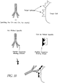
- the immune effector-binding domain may also comprise a peptide sequence designed to tightly bind the immune effector (see e.g., fig. 9 ).
- the bifunctional protein may comprise a fusion protein, or the two domains can be covalently or non-covalently linked.
- the target-binding domain and the immune effector binding domain can be directly linked, or indirectly linked, for example via a flexible linker peptide.
- the invention provides methods for preparing a therapeutic for treating a cancer comprising isolating complexes of expressed mRNA molecules and their nascent polypeptides from an mRNA expression library; screening the protein-mRNA complexes for binding to a molecular target associated with an etiological determinant, such as an epitope displayed by a cancerous cell; isolating and expressing mRNA encoding a protein that binds the target epitope; and derivatizing the target epitope-binding protein (or derivatives, fragments or subunits thereof) to a therapeutic agent, such as an antibody capable of eliciting an immune response against the target.
- a therapeutic agent such as an antibody capable of eliciting an immune response against the target.
- the preparation of the therapeutic further comprises allowing isolated mRNA encoding a target-binding domain to undergo in vitro evolution, selective mutagenesis, and/or other methods known in the art to identify and isolate mRNAs exhibiting stronger or more effective binding to the target epitope, as described in more detail below.
- the invention provides methods for treating a disease or condition, such as a cancer, comprising identifying a target epitope or other molecule specifically or preferentially expressed on, or in close proximity to, an etiological determinant of the condition targeted for treatment, in a patient in need of treatment; isolating a protein that binds the target, but does not bind substantially to non-targets; linking the target-binding protein (or derivatives, fragments or subunits thereof) to a therapeutic agent, such as an antibody capable of eliciting an immune response in the subject of interest; and administering the therapeutic to the patient in an amount and for a time sufficient to treat the condition targeted for treatment.
- a disease or condition such as a cancer
- the invention provides methods for identifying proteins that bind cancer cell target epitopes, or other etiological determinants, comprising isolating complexes of expressed mRNA molecules and their nascent polypeptides from an mRNA expression library; screening the protein-mRNA complexes for binding to a target epitope displayed by a cancerous cell; isolating and expressing mRNA encoding a protein that binds the target epitope; and derivatizing the target epitope-binding protein (or derivatives, fragments or subunits thereof) to an antibody capable of eliciting an immune response.
- the methods may further comprise allowing isolated mRNA encoding a target epitope-binding protein to undergo in vitro evolution, selective mutagenesis, or other methods known in the art to identify and isolate mRNAs exhibiting stronger or more effective binding to the target epitope.
- the invention provides a kit for developing an individualized therapeutic for the treatment of a disease or condition characterized by the expression of disease- and/or patient-specific etiological determinants.
- a kit is provided for developing patient-specific therapeutics for the treatment of a cancer, including solid tumors and hematological cancers, wherein the therapeutics are targeted to a unique cell-surface epitope differentially expressed on the surface of cancerous and/or malignant cells.
- the invention provides therapeutics and methods for reducing or preventing transplant rejection.
- the target-binding protein binds to cell surface antigens displayed by transplanted cells, such as MHC antigens
- the therapeutic agent e.g., the immune effector
- the immune effector comprises a protein or other molecule that binds to and inhibits one or more molecular determinants of the immune response.
- the immune effector binds the C1q or C3 components of the complement system to thereby inhibit the activation of complement-mediated immunity.
- the immune effector stimulates an immune response to eliminate transplanted cells bearing "foreign" MHC or other antigens.
- the therapeutic agent ablates or prevents the clonal expansion of lymphocyte subpopulations expressing specific epitopes, for example epitopes that recognize MHC antigens.
- the invention further provides methods and kits for inhibiting transplant rejection that are essentially similar to those described above for treating malignancies.
- the target-binding domain is selected to bind one or more variable epitopes expressed by a disease-causing pathogen, or one or more cells infected by a disease-causing pathogen
- the therapeutic agent comprises an anti-pathogenic drug, such as an antibiotic or other cytotoxic agent.
- the pathogen is a virus, such as HIV.
- methods provided herein allow for the rapid and cost-effective creation of individualized therapeutics.
- the following detailed description illustrates various aspects of the invention as they relate to particular applications. However, the description applies equally to the development and use of therapeutics and methods for the treatment of a wide variety of conditions, including but not limited to, the treatment of malignancies, the reduction and elimination of pathogens, the reduction and/or prevention of transplant rejection, and in general, the treatment of any condition involving a therapeutic target which exhibits differential binding characteristics relative to non-targeted cells or molecules.
- T lymphocyte and "T cell” as used herein encompass any cell within the T lymphocyte lineage from T cell precursors to mature T cells.
- B lymphocyte and "B cell” encompasses any cell within the B cell lineage from B cell precursors, such as pre-B cells, to mature B cells and plasma cells.
- Immunoglobulin molecules consist of heavy (H) and light (L) chains, which comprise highly specific variable regions at their amino termini.
- the variable (V) regions of the H (V H ) and L (V L ) chains combine to form the unique antigen recognition or antigen combining site of the immunoglobulin (Ig) protein.
- the variable regions of an Ig molecule contain determinants (i.e., molecular shapes) that can be recognized as antigens or idiotypes.
- epitope refers to the set of antigenic or epitopic determinants (i.e., idiotopes) of an immunoglobulin V domain (i.e., the antigen combining site formed by the association of the complementarity determining regions or V H and V L regions).
- idiotope refers to a single idiotypic epitope located along a portion of the V region of an immunoglobulin molecule.
- immune effector refers to a molecule, or derivatives, fragments, or subunits thereof, able to stimulate an immune response in the subject being treated, and may comprise an antibody, or derivatives, fragments, or subunits thereof, or a non-antibody molecule.
- an “adjuvant” is a compound which enhances or stimulates the immune response when administered with one or more antigen(s).
- Malignant cells refers to cells, which if left untreated, give rise to a cancer.
- protein protein
- peptide and “polypeptide” are defined herein to mean a polymeric molecule of two or more units comprised of amino acids in any form (e.g., D- or L- amino acids, synthetic or modified amino acids capable of polymerizing via peptide bonds, etc.), and these terms may be used interchangeably herein.
- the therapeutics are comprised of a "modular" architecture that allows tailoring of a small protein domain (the "target-binding domain”) to bind one or more patient- and/or disease-specific markers, and use of the tailored domain to direct a variety of existing or easily produced therapeutics that are effective against the condition targeted for treatment.
- the targeted disease markers can comprise any type of molecule, or portion or derivative thereof, or complex of molecules, including but not limited to, proteins, nucleic acids, lipids, chemical compounds, polymers, and metals, as well as biological structures, such as cell membranes, cytoskeletal elements, receptors, and even entire cells.
- the markers are expressed on, or in close proximity to, an etiological determinant of the condition targeted for treatment, and activity of the therapeutic agent is focused to disease-causing cells, pathogens, proteins, and/or other determinants of the condition being treated.
- the modular therapeutic is tailored for the treatment of a a hematological cancer, and designed to bind to the unique Fab idiotype on the surface of malignant B-cells and/or T-cells.
- a therapeutic is provided for the treatment of non-Hodgkin's Lymphoma (NHL), which is a "clonal" B-cell disease, in which all cancerous cells are derived from a single, malignant B-cell.
- NHL non-Hodgkin's Lymphoma
- every NHL cell expresses a common idiotype (comprising the variable domains of surface expressed IgM molecules) that is unique to each patient, which can be targeted by the target-binding portions of the modular therapeutics provided herein.
- B-cells can be isolated from the lymph nodes, or from peripheral blood, using methods known in the art. For example, in some embodiments, erythrocytes and/or granulocytes may be separated from the B-cells by centrifugation in a liquid having a density intermediate between the groups of cells to be separated. Means of obtaining T-lymphocytes are also well known in the art, such as isolation from the peripheral blood of a patient, and separation on the basis of size and/or density. Extraction of proteins from B-cells and/or T-cells may be performed by any of the many means known in the art. For example, cells may be lysed by a detergent or by mechanical means. If desired, nucleic acids can be removed from the cell preparation by enzymatic digestion or by precipitation with agents such as streptomycin. Such means are well known in the art.
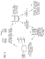
- a modular therapeutic for treating a hematological cancer prepared according to methods described herein is illustrated in Fig. 1 .
- the administered therapeutic binds to the unique Fab idiotype on the surface of malignant B-cells and/or T-cells, and the etiological determinant of the hematological cancer, with which the idiotype is associated, comprises malignant and/or cancerous cells expressing the targeted idiotype. Binding of the targeting domain to the idiotype effects an immune response that destroys the malignant cells. By itself, the target-binding protein is too small to elicit an immune response, and thus the bound immune effector will not produce an IR in the absence of binding to the targeted idiotype.
- the target-binding protein When the target-binding protein binds to the Fab idiotype on the surface of a malignant cell, the malignant cell acts as a carrier that confers immunogenicity to the target epitope-binding protein, allowing the immune effector to produce an IR that targets the malignant cell.
- therapeutics and methods of the invention can also be used to target epitopes that distinguish non hematological cancers.
- targeted markers are differentially expressed (i) in an individual patient, or a defined sub-group of patients, relative to other patients having similar diagnoses, and/or (ii) in association with cells or other molecular targets associated with the etiology of the condition, relative to cells/molecules that are unassociated with the condition and preferably not subjected to the therapeutic agent.
- the selectivity of tailored therapeutics enhances the efficacy of treatment relative to existing, non-tailored therapeutics, due, for example, to the non-selective activity of non-tailored therapeutics against healthy cells, and/or failure of existing targeted therapeutics to account for inter-patient variability in the targeted marker(s).
- the targeted idiotype is unique to both the patient and to the malignant cells, allowing the activity of the therapeutic agent (effecting an immune response) to be selectively directed to targeted cells, sparing non-cancerous cells.
- the idiotype is unique to each patient, non-tailored therapeutics would result in a non-selective, or less-selective, therapeutic response.
- cell-surface markers differentially expressed by malignant cells include, but are not limited to: for fluid tumors, stable cell surface antigen epitopes, such as CD-20 and CD-22, and for solid tumors, surface epitopes, such as CD-19 and CD-33, which become internalized upon binding with a mAb.
- Other differentially expressed cell-surface markers are known in the art, including but not limited to, CD-52w and class II HLA antigens.
- the targeted epitope is a cancer cell-specific epitope that is mutated (see e.g., Clinical Cancer Research, 5:611-615, 1999 ) in the patient targeted for treatment.
- methods provided herein allow for more efficacious treatment of cancers by allowing the development of individualized therapeutics that target mutated epitopes, which are unique to each patient.
- the target-binding domain of the modular therapeutic comprises a first sub-domain that binds the target (e.g., a target epitope-bdining domain (Tebd)), and a second sub-domain that binds the therapeutic agent (e.g., an immune effector-binding domain (Ebbd)).
- the therapeutic agent is an agent that is capable of stimulating an immune response in the subject targeted for treatment, such as an antibody, or a derivative, fragment, or subunit thereof, and the domain that binds the therapeutic agent is a small protein recognized by the therapeutic agent, for example an epitope recognized by a therapeutic agent antibody.
- Antibodies useful in the invention may be derived from any mammal, or may be a chimeric antibody derived from a combination of different mammals.
- the mammal may be, for example, a rabbit, a mouse, a rat, a goat, or a human.
- the antibody is preferably a human antibody.
- Reactivity of antibodies against a target antigen may be established by a number of well known means, including Western blot, immunoprecipitation, ELISA, and FACS analyses using, as appropriate, Fab idiotype fragments, peptides, idiotype-expressing cells or extracts thereof.
- the antibody can belong to any antibody class and/or sub-class.
- the antibodies may also contain fragments from antibodies of different classes and sub-classes, thereby forming a composite.
- human monoclonal antibodies having a desired binding activity are produced using methods known in the art (for review, see Vaughan et al., 1998, Nature Biotechnology 16: 535-539 ), for example by screening a phage display library, as described, e.g., in Parmley and Smith Gene 73:305-318 (1988 ), Barbas et al., Proc. Natl. Acad. Sci. USA 88: 7978-7982 (1991 ), Griffiths et al., EMBO J 13: 3245-3260 (1994 ), Griffiths and Hoogenboom, Building an in vitro immune system: human antibodies from phage display libraries.
- Fully human monoclonal antibodies may also be produced using transgenic mice engineered to contain human immunoglobulin gene loci, as described in PCT Patent Application WO98/24893 and Jakobovits, 1998, Exp. Opin. Invest. Drugs 7(4): 607-614 , herein incorporated by reference. This method avoids the in vitro manipulation required with phage display technology and efficiently produces high affinity authentic human antibodies.
- an antibody against an antigen of interest is produced in the patient selected for treatment with the modular therapeutic.
- the patient is "vaccinated" with the antigen of interest, and antibodies from the patient against the antigen are selected by binding to the antigen of interest, as described in Zebedee, et al. Proc. Natl. Acad. Sci. USA 89: 3175-3179 (1992 ), Burton et al., Proc. Natl. Acad. Sci. USA 88: 10134-10137 (1991 ), and Barbas et al., Proc. Natl. Acad. Sci. USA 89: 10164-20168 (1991 ).
- additional rounds of screening are performed to increase the affinity of the originally isolated antibody.
- the affinity of the antibody is enhanced by affinity maturation, in which hypervariable antibody regions are mutated to produce a large number of combinations, and the corresponding antibody variants are screened via phage display to select antibodies having the desired affinity for the antigen.
- the small protein epitope can undergo in vitro evolution, as described in more detail below, to increase its binding affinity for the antibody.
- an analogous process is carried out by the host immune system (e.g., via clonal selection) to produce high affinity antibodies specific for the antigen of interest.
- the immune effector comprises a non-antibody molecule capable of stimulating an immune response in the subject receiving treatment.
- the immune effector may comprise a C3 convertase, as described, e.g., in U.S. Patent No. 6,268,485 to Farries and Harrison , which is herein incorporated by reference.
- C3 convertase is an enzyme that catalyzes the proteolytic conversion of C3 protein into C3a and C3b, which conversion comprises a critical step in the generation of the complement system response.
- C3b binds to cell surfaces near its site of generation, where it mediates phagocytosis and other destructive immune responses.
- C3 convertase is modified or derivatized to confer reduced susceptibility to inhibitors, resistance to proteolytic cleavage, enhanced affinity for cofactors, or other modifications that enhance the effectiveness of the enzyme in stimulating a complement-mediated immune response.
- the immune effector comprises a C5 convertase, a mannose binding protein (MBP), or another molecule that stimulates complement-mediated immunity.
- Linking C3 convertase to the therapeutics of the instant invention allows a complement-mediated immune response to be directed to cells, pathogens, pathogen-infected cells, and/or other cells targeted for treatment by binding of the target epitope binding protein to variable eptiopes on the surface of the targeted cells.
- a therapeutic with C3 convertase as the immune effector is illustrated in Fig. 9 .
- peptide sequences encoding proteins that bind C3 convertase (the immune effector binding protein) and a target epitope (the target epitope binding protein) are identified, isolated, and optionally bred by in vitro evolution or other techniques to increase their affinity to their target ligands.
- a bifunctional fusion protein is then constructed comprising the target epitope binding domain, and the convertase binding peptide as the immune effector binding domain (Ebbd).
- the therapeutic is administered to a patient in need of treatment, upon which the target epitope-binding domain binds to the surface of a malignant cell or other target, and the C3 convertase produces a complement-mediated immune response, which includes the production of membrane attack complexes that mediate the destruction of the targeted cell.
- the therapeutic comprises a target epitope-binding domain and a C3 convertase binding domain.
- such therapeutics bind a target epitope and recruit endogenous C3 convertase to initiate a complement-mediated immune response.
- Fig. 10 shows a therapeutic in which the immune effector binds the C1 component of the complement system.
- the therapeutic binds to the target epitope and recruits endogenous C1 to the cell surface, where it induces a complement-mediated immune response.
- the immune effector component of the therapeutic binds to and inhibits C1q, C3, C5, and/or other molecules involved in generating complement-mediated immune responses, such as the proteins described in U.S. Patent No. 5,650,389 .
- such therapeutics bind to the target epitope and inhibit the immune response that would otherwise be directed at or near the binding site.
- a patient is the recipient of transplanted cells (e.g., stem cells, or cells comprising a transplanted organ), and the targeted therapeutic is capable of recognizing tranplanted cells, and inhibiting an immune response against such cells.
- targeted therapeutics for the inhibition of transplant rejection comprise a target-binding domain that targets one or more epitopes of immune cells that recognize transplanted cells, for example epitopes that bind MHC antigens on the surfaces of tranplanted cells, and the therapeutic agent is an immune effector capable of stimulating an immune response in the patient against the anti-transplant immune cells.
- Therapeutics according of the invention can preferably be created, from reception of the biopsy material to the completion of the therapeutic, in a matter of weeks and at a cost of several thousand dollars.
- a bifunctional fusion protein for use in the invention can be produced according to standard recombinant DNA techniques known to those skilled in the art (e.g., Sambrook, J. et al., Molecular Cloning: A Laboratory Manual, 2nd ed., Cold Spring Harbor Laboratory Press, New York (1989 )).
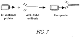
- the purified bifunctional protein can be reacted with the immune effector, yielding a conjugated effector-bifunctional protein complex, as shown Fig. 7 , which comprises the therapeutic.
- the bifunctional protein can also be chemically bound to the immune effector through means known in the art.
- a protein coupling agent such as N-succinimidyl-3-(2-pyridyldithiol)propionate (SPDP), iminothiolane (IT), bifunctional derivatives of imidoesters (such as dimethyl adipimidate HCL), active esters (such as disuccinimidyl suberate), aldehydes (such as glutareldehyde), bis-azido compounds (such as bis (p-azidobenzoyl)hexaned- iamine), bis-diazonium derivatives (such as bis-(p-diazoniumbenzoyl)-ethyl-enediamine), diisocyanates (such as tolyene 2,6-diisocyanate), or bis-active fluorine compounds (such as l,5-difluoro-2,4-dinitrobenzene), is used to link two or more protein components comprising the therapeutic.
- SPDP N-succinimidy
- a recombinant fusion protein is prepared by: linking a first polynucleotide sequence encoding a target-epitope-binding protein, or derivatives, fragments or subunits thereof, to a second polynucleotide sequence encoding an epitope or other protein that binds the immune effector to generate a chimeric coding sequence; subcloning the chimeric coding sequence into an expression vector; transfecting a cell with the expression vector; and purifying the fusion protein expressed by the transfected cell.
- the chimeric polynucleotide can include an initiation sequence appropriate for prokaryotic and/or eukaryotic in vitro translation systems, and/or a selectable marker.
- the bifunctional protein complex is made by in vitro translation of an mRNA oligo that includes the mRNA encoding both proteins and, in some embodiments, the major histocompatibility complex I and/or II.
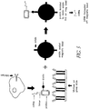
- the cDNA for the mRNA sequences can be synthesized or obtained commerically, and transcribed by PCR to obtain sufficient mRNA, as shown in Fig. 3 .
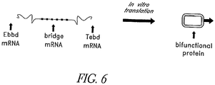
- the oligo is formed by ligating mRNA encoding the immune effector-binding domain and the mRNA encoding the target epitope-binding domain. In some cases this fusion can be connected through an mRNA bridge that codes for hydrophilic amino acids, as illustrated in Fig. 6 .
- the mRNA can then be translated in vitro using prokaryotic or eukaryotic translation systems, and the resultant bifunctional protein can purified by gel electrophoresis or any other method known in the art.
- the present fusion proteins can also comprise one or more numerous other components that enhance the utility of the chimeric proteins.
- the proteins can be designed to contain an epitope tag useful, for example, to facilitate purification of the fusion proteins.
- the chimeric coding sequence can be modified to encode two or more neighboring histidine residues, for example, in the amino or carboxy terminus of the peptide. Histidine residue insertion can be readily accomplished by the splice-by-overlap extension methodology, by incorporating histidine-encoding CAT and CAC triplet codons into the PCR primers at suitable locations in the coding sequence. Histidine-modified proteins can be efficiently and quantitatively isolated by nickel-sepharose chromatography methods known in the art.
- therapeutics provided herein may also be conjugated to a second molecule, such as a therapeutic agent (e.g., a cytotoxic agent).
- a therapeutic agent e.g., a cytotoxic agent
- the therapeutic agent includes, but is not limited to, an anti-tumor drug, a toxin, a radioactive agent, a cytokine, a second antibody or an enzyme.
- cytotoxic agents include, but are not limited to ricin, ricin A-chain, doxorubicin, daunorubicin, taxol, ethidium bromide, mitomycin, etoposide, tenoposide, vincristine, vinblastine, colchicine, dihydroxy anthracin dione, actinomycin D, diphteria toxin, Pseudomonas exotoxin (PE) A, PE40, abrin, arbrin A chain, modeccin A chain, alpha-sarcin, gelonin, mitogellin, retstrictocin, phenomycin, enomycin, curicin, crotin, calicheamicin, sapaonaria officinalis inhibitor, maytansinoids, and glucocorticoid and other chemotherapeutic agents, as well as radioisotopes such as 212 Bi, 131 I, 90 Y, and 186 Re.
- PE Ps
- Suitable detectable markers include, but are not limited to, a radioisotope, a fluorescent compound, a bioluminescent compound, chemiluminescent compound, a metal chelator or an enzyme.
- the therapeutics of the invention may also be conjugated to an anti-cancer pro-drug activating enzyme capable of converting the pro-drug to its active form. See, for example, U.S. Pat. No. 4,975,287 .
- the secondary therapeutic agent has a complementary mode of action with the primary therapeutic agent.
- the primary and secondary therapeutic agents act against different aspects of a signal transduction system involved in the etiology of a cancer or other disease, leading to enhanced efficacy, fewer side effects, an improved therapeutic index, and/or other benefits relative to tailored therapeutics bearing the primary and/or secondary therapeutic agents only.
- the first therapeutic agent potentiates the second therapeutic agent, or vice versa, or the first and second therapeutic agents exhibit a synergistic enhancement in one or more aspects of treatment.
- the therapeutic agent is conjugated to an antibody immune effector component of the therapeutic.
- Techniques for conjugating or joining therapeutic agents to antibodies are well known (see, e.g., Arnon et al., "Monoclonal Antibodies For Immuno-targeting Of Drugs In Cancer Therapy", in Monoclonal Antibodies And Cancer Therapy, Reisfeld et al. (eds.), pp. 243-56 (Alan R. Liss, Inc. 1985 ); Hellstrom et al., "Antibodies For Drug Delivery", in Controlled Drug Delivery (2nd Ed.), Robinson et al. (eds.), pp. 623-53 (Marcel Dekker, Inc.
- the therapeutics of the invention can be administered by any method known in the art that is suitable for protein pharmaceuticals, such as intravenous injection, intramuscular injection, topical administration, oral ingestion, rectal administration, and inhalation.
- the therapeutics of the present invention can be delivered directly to the site of the malignancy.
- the therapeutic may be administered in admixture with a pharmaceutically acceptable carrier. Any such carrier can be used according to the present methods, as long as compatibility problems do not arise.
- An effective amount of the present recombinant fusion protein should be administered to the patient.
- the term "effective amount" refers to that amount of the fusion protein needed to bring about the desired response.
- therapeutics made according to methods providing herein inhibit proliferation and/or induces apoptosis of cells bearing the epitope against which the epitope binding protein was screened.
- the epitope-binding portion of the therapeutic complex can bind with the epitopes expressed on the surface of malignant cells, or other targets. Once bound, the IR-eliciting antibody component of the complex stimulates the immune system to attack and eliminate the tagged cells, while sparing the normal cells, as illustrated in Fig. 8 .
- the tailoring of individualized therapeutics provided herein for binding to patient- and/or disease-specific targets is made possible by utilizing novel methods for linking proteins to their corresponding mRNAs (as "cognate pairs").
- protein libraries are prepared comprising a large number of cognate pairs, and the libraries are screened for cognate pairs that bind to a target of interest, such as the individualized targets described herein.
- tRNA and/or mRNA molecules to link translated protein products to their corresponding mRNAs via a tRNA linker, forming a "cognate pair.”
- mRNAs having unknown sequences are expressed in an in vitro translation system, for example from an mRNA library, and their corresponding proteins are screened for one or more desired characteristics, such as binding to the target epitope-binding domain, or another ligand of interest, and/or selectivity over one or more additional ligands, such as epitopes displayed by healthy cells.
- proteins and their linked nucleic acids identified in one or more rounds of selection are modified, for example through nucleic acid evolution ( Fig.
- Proteins having desired characteristics, such as a high affinity for the target ligand can then be produced in large quantities using standard cloning techniques by isolating their corresponding mRNA from the protein-mRNA cognate pairs.
- cognate pairs are formed using a eukaryotic in vitro translation system, such as rabbit reticulocyte lysate (RLL), wheat germ, E. coli, or yeast lysate systems.
- RLL rabbit reticulocyte lysate
- any in vitro translation system can be used, including in situ systems, as well as hybrid systems, which combine components of different systems.
- one or more prokaryotic factors are used in a eukaryotic translation system, such as translation suppressor proteins (see e.g., Geller and Rich Nature 283:41 (1980 ); Edwards et al PNAS 88:1153 (1991 ); Hou and Schimmel Biochem 28:6800 (1989 ), all herein incorporated by reference).
- one or more tRNAs or tRNA analogs are charged in a prokaryotic system and then purified according to established methods ( Lucas-Lenard and Haenni, PNAS 63:93 (1969 ), herein incorporated by reference) for use in a eukaryotic system.
- proteins comprising cognate pairs are linked to tRNA or a tRNA analog by the action of ribosomal peptidyl transferase.
- proteins are linked to a stable aminoacyl tRNA analog (SATA).
- SATA is a tRNA with an amino acid or amino acid analog attached to its 3' end via a stable bond, relative to the corresponding high-energy ester bond in the native structure.
- the stable aminoacyl bond prevents the detachment of the tRNA from the polypeptide by peptidyl transferase, and also preserves the tRNA-polypeptide structure during subsequent steps.
- a SATA is created according to methods generally described in Fraser and Rich, PNAS, 70:2671 (1973 ), herein incorporated by reference, which involve the conversion of a tRNA, or tRNA analog, to a 3'-amino-3'-deoxy tRNA. This is accomplished by adding a 3'-amino-3'-deoxy adenosine to the end of a native tRNA with tRNA nucleotidyl transferase after removing the native adenosine, and then charging the modified tRNA with an amino acid with the respective aminoacyl tRNA synthetase (aaRS).
- the aaRS charges the tRNA on the 3', rather than the 2', hydroxyl, linking the amino acid to the tRNA by a stable amide bond, rather than the usual labile high-energy ester bond.
- SATA accepts a peptide from ribosomal peptidyl transferase it will stably hold the peptide and be unable to donate it to another acceptor.
- tRNAs aminoacylated via a 3' amide bond may not combine with the elongation factor EF-TU, which assists in binding to the A site (e.g., SRocl and Cramer, Prog. Nuc. Acid Res. 22:1 (1979 ), herein incorporated by reference).
- modified tRNAs do, however, bind to the A site.
- This binding of 3' modified tRNAs can be increased by changing the Mg ++ concentration ( Chinali et al., Biochem. 13:3001 (1974 ), herein incorporated by reference).
- concentrations of and/or molar ratios of SATA and Mg 2+ can be determined empirically.
- the SATA may compete with native tRNAs for non-cognate codons, stalling translation.
- the concentration or A site avidity of SATA is too low, the SATA might fail to effectively compete with release factors, preventing it from stably accepting the nascent peptide.
- elongation factor is also believed to aid in proofreading codon-anticodon recognition, the absence of this source of proofreading would not be expected to interfere with methods provided herein. Without being bound to a particular mechanism, it is believed that the error rate in the absence of elongation factor and associated GTP hydrolysis is approximately 1 in 100 for codons one nucleotide away ( Voet and Voet, Biochemistry 2nd ed. pp. 1000-1002 (1995), John Wiley and Sons , herein incorporated by reference).
- UAA is used as the linking codon.
- UAA has 7 non stop codons that differ from it by one amino acid, which comprises 7/61, or about 11.5% of the non stop codons.
- UAG can be used as the linking codon without substantial impairment due to the absence of elongation factor-mediated proofreading.
- the SATA is a tRNA, or tRNA analog, with one or more modified bases in the acceptor stem, or another region of the molecule.
- tRNAs with acceptor stem modification are known in the art, and are described, for example, in SRocl and Cramer, Prog. Nuc. Acid Res., 22:1 (1979 ), herein incorporated by reference.
- a tRNA is modified with a puromycin moiety, such that the tRNA mimics aminoacyl-Tyr tRNA and is incorporated into the nascent polypeptide, terminating translation.
- acceptor stem-modified tRNAs are formed from "transcriptional tRNA", wherein the sequence of the tRNA itself, rather than post-transcriptional processing, leads to the atypical and modified bases.
- Transcriptional tRNAs are capable of functioning as tRNAs (see e.g., Dabrowski et al., EMBO J. 14:4872, 1995 ; and Harrington et al., Biochem. 32: 7617, 1993 , both herein incorporated by reference).
- Transcriptional tRNA can be produced by methods known in the art, such as transcription, or by connecting commercially available RNA sequences (e.g., from Dharmacon Research Inc., Boulder, Colo.) together, piece-wise as in FIG.
- the 5' phosphate and 3' puromycin are commercially available attached to oligoribonucleotides, which can be connected together using T4 DNA ligase (e.g., Moore and Sharp, Science 256: 992, (1992 ), herein incorporated by reference) or alternatively, T4 RNA ligase ( Romaniuk and Uhlenbeck, Methods in Enzymology 100:52 (1983 ), herein incorporated by reference).
- T4 DNA ligase e.g., Moore and Sharp, Science 256: 992, (1992 ), herein incorporated by reference
- T4 RNA ligase Romaniuk and Uhlenbeck, Methods in Enzymology 100:52 (1983 ), herein incorporated by reference.
- modified tRNAs are known in the art, and are described, e.g., in Chinali et al., Biochem. 13:3001 (1974 ) and Krayevsky and Kukhanova, Prog. Nuc. Acid Res 23:1 (1979 ), both herein incorporated by reference.
- the tRNA is a nonsense suppressor tRNA comprising a modified or unmodified tRNA, or tRNA analog, that recognizes a stop codon or a pseudo-stop codon, preferably by codon-anticodon hydrogen bonding, such that translation is terminated when the nascent protein is attached to the tRNA by peptidyl transferase.
- the nonsense suppressor tRNA has 3' modifications and/or sequences that conform to the Yarus extended anticodon rules ( Yarus, Science 218:646-652, 1982 , herein incorporated by reference).
- a “pseudo stop codon,” as defined herein, refers to a codon which, while not naturally a nonsense codon, prevents a message from being further translated.
- a pseudo stop codon can comprise a codon recognized by a "stable aminoacyl tRNA analog," or SATA, as described herein, or a codon for which tRNA bearing a complementary anticodon is substantially depleted or absent, such that translation is terminated when the absent tRNA is required, i.e. at the pseudo stop codon.
- SATA stable aminoacyl tRNA analog
- One skilled in the art will appreciate that are numerous ways to create a pseudo stop codon, as defined herein.
- the tRNA is a native tRNA, linked to the nascent polypeptide via a native peptide bond.
- the SATA is a tRNA that is unmodified at the 3' end, but which may have one or more modifications to the anticodon loop and/or other regions of the molecule.
- the use of native tRNAs and/or tRNAs that are unmodified at the 3' end results in improvements in various selection methods described herein, giving rise to quicker, less error-prone, more efficient, more cost-effective, and/or higher yield methods. While not being bound by a particular theory, it is believed that, under certain conditions, puromycin (and similar linkers) can result in lower yields due to interference with the interaction between elongation factor(s) and tRNAs.
- the crosslinker is an agent that chemically or mechanically links two molecules together.
- the crosslinker is an agent that can be activated to form one or more covalent bonds with tRNA and/or mRNA.
- the crosslinker is a sulfur-substituted nucleotide.
- the crosslinker is a halogen-substituted nucleotide. Examples of crosslinkers include, but are not limited to, 2-thiocytosine, 2-thiouridine, 4-thiouridine, 5-iodocytosine, 5-iodouridine, 5-bromouridine and 2-chloroadenosine, aryl azides, and modifications or analogues thereof.
- the crosslinker is psoralen or a psoralen analog.
- a psoralen is monoadducted to a tRNA, or a tRNA analog (e.g., a 3' modified SATA), for example by connecting a psoralen linked oligonucleotide ( FIG. 3 ), or by monoadduction ( FIG. 4 ), to a native or modified tRNA or tRNA analog, preferably to the anticodon or another site distinct from the linkage to the polypeptide.
- a covalent psoralen crosslink is formed between the SATA and mRNA, as described in more detail below.
- the anticodon or other portion of a tRNA is derivatized with a non-psoralen moiety capable of forming a crosslink to the mRNA, such as 2-thiocytosine, 2-thiouridine, 4-thiouridine 5-iodocytosine, 5-iodouridine, 5-bromouridine, 2-chloroadenosine, aryl azides, and modifications or analogues thereof.
- a non-psoralen moiety capable of forming a crosslink to the mRNA
- a non-psoralen moiety capable of forming a crosslink to the mRNA
- a non-psoralen moiety capable of forming a crosslink to the mRNA
- a SATA-polypeptide complex or a tRNA-polypeptide complex is linked to the mRNA encoding the polypeptide via a linker moiety, which can be located on the mRNA and/or on the tRNA.
- the mRNA comprises a cross-linker, preferably at or near a stop codon at the 3'-end of the transcript, and the mRNA-tRNA linkage is mediated entirely by the mRNA-based cross-linker.
- the tRNA is unmodified at its 3'-end (e.g., a nonsense suppressor tRNA).
- an mRNA comprises a pseudo-stop codon located at the end of the translatable reading frame.
- a pseudo-stop codon can be effectively placed at the end of a reading frame where the pseudo-stop codon is located at the 3' end of the mRNA, where the translation system is depleted of tRNAs corresponding to codons 3' to the pseudo-stop codon, and/or where 3'-modified tRNAs corresponding to the pseudo-stop codon are used, rendering the transcript untranslatable and incapable of activating release factors.
- the mRNA comprises a stop codon corresponding to a tRNA stop anticodon.
- a stop codon/anticodon pair selects for full-length transcripts.
- an mRNA not having a stop codon may also be used and that any codon or nucleic acid triplet may be used.
- a SATA is attached to the translated message by a psoralen cross link between the codon and anticodon.
- Psoralen cross links are preferentially made between sequences that contain complementary 5' pyrimidine-purine 3' sequences, especially UA or TA sequences ( Cimino et al., Ann. Rev. Biochem. 54:1151 (1985 ), herein incorporated by reference).
- the codon coding for the SATA, or the linking codon can be PYR-PUR-X or X-PYR-PUR, so that several codons may be used for the linking codon. Conveniently, the stop or nonsense codons have this configuration.
- a stop codon is used as the linking codon and the SATA functions as a nonsense suppressor in that it recognizes the linking codon.
- any codon can be used.
- the SATA or peptidyl-tRNA is cross-linked to the translated mRNA, for example between the codon and anticodon, by a psoralen cross-link, or by a cross-link formed from the group consisting of: 2-thio cytosine, 2-thio uridine, 4thio uridine 5-iodocytosine, 5-iodouridine, 5-bromouridine, 2-chloroadenosine, and aryl azides.
- Psoralen cross links are, in some embodiments, preferentially made between sequences that contain complementary 5' pyrimidine-purine 3' sequences, especially UA or TA sequences ( Cimino et al., Ann. Rev. Biochem.
- non-psoralen crosslinkers or aryl azides are used and in certain embodiments, are particularly advantageous because they are less stringent in their requirements and therefore increase the possible codon-anticodon pairs.
- translation terminates when the nascent protein is attached to a SATA by the peptidyl transferase and/or when the end of the reading frame is reached.
- SATA and the mRNA are cross-linked by application of UV light.
- cross-linking is accomplished by forming a psoralen crosslink upon irradiation with UV light, preferably in the range of 320 nm to 400 nm.
- Psoralens comprise a furan side and a pyrone side, and they readily intercalate between complementary base pairs in double stranded DNA, RNA, and DNA-RNA hybrids ( Cimino et al., Ann. Rev.
- psoralen cross-linking forms monoadducts, described more fully below, that are either pyrone sided or furan sided monoadducts. Upon further irradiation, the furan sided monoadducts can be covalently crosslinked to complementary base pairs, whereas the pyrone sided monoadducts cannot be further crosslinked.
- the formation of furan-sided psoralen monoadducts (MAf) is achieved according to established methods.
- psoralen can also be attached at the end of the reading frame of the message.
- furan sided monoadducts will be created using visible light, preferably in the range of approximately 400 nm-420 nm, according to the methods described in U.S. Pat. No. 5,462,733 and Gasparro et al., Photochem. Photobiol. 57:1007 (1993 ), both herein incorporated by reference.
- a SATA with a furan sided monoadduct or monoadducted oligonucleotides for placement on the 3' end of mRNAs, along with a nonadducted SATA are provided as the basis of a kit.
- the formation and reversal of monoadducts and crosslinks are performed according to the methods of Bachellerie et al. (Nuc Acids Res 9:2207 (1981 )), herein incorporated by reference.
- efficient production of monoadducts, resulting in high yield of the end-product is accomplished using the methods of Kobertz and Essigmann, J. A. Chem. Soc. 1997, 119, 5960-5961 and Kobertz and Essigmann, J. Org. Chem. 1997, 62, 2630-2632 , both herein incorporated by reference.
- appropriate concentrations of SATA and Mg ++ are used in the in vitro translation system in the presence of the mRNA molecules, causing translation to cease when ribosomes reach a stop, or pseudo-stop, codon which permits the SATA to accept the peptide chain, as described above. After a short time, a substantial proportion and/or number of the stop or pseudo-stop codons are occupied by SATAs within ribosomes, and in some embodiments, the system is then irradiated with UV light to generate crosslinks between the tRNA-polypeptides and their corresponding mRNAs.
- the ribosomes are released or denatured after cross-linking of to mRNA, preferably by the depletion of Mg ++ through dialysis, dilution, or chelation.
- Mg ++ through dialysis, dilution, or chelation.
- denaturation by changing the ionic strength, the pH, or the solvent system can also be used to release cognate pairs from associated ribosomes and/or other translation factors.
- cognate pairs are selected based on one or more desired characteristics.
- the selection of cognate pairs is based upon the binding of a target cell, protein, and/or other biomolecule, as determined by any of a variety of established methods, including, but not limited to, arrays, affinity columns, immunoprecipitation, and the like.
- selection criteria are measured using a high throughput screening procedure. The selection can be positive or negative in various embodiments, according to the desired characteristics of the therapeutic agent.
- mRNAs whose sequences are unknown are expressed (e.g., in the form of an mRNA library from a patient, tissue, or other source of interest) and linked to their encoded polypeptides using methods described herein, and the cognate pairs are screened to select for desired properties.
- cognate pairs are assayed for binding to a ligand of interest, such as an Fab idiotype displayed by a malignant cell, or other surface characteristic of a target cell.
- mRNA can be isolated for proteins exhibiting the desired binding characteristics, and large quantities of the protein can be produced using standard molecular cloning techniques known in the art.
- the protein of interest can then be incorporated into a modular therapeutic agent, for example to target the agent to a patient- and/or disease-specific target.
- the selected cognate pairs can be those that do bind well to a ligand or those that do not.
- a protein to accelerate a thermodynamically favorable reaction, i.e., act as an enzyme for that reaction, it should bind both the substrate and a transition state analog.
- proteins which compete poorly for binding to the substrate but compete well for binding to the transition state analog are selected. Operationally, this may be accomplished by taking the proteins that are easily eluted from a matrix with substrate or substrate analog bound to it and are the most difficult to remove from matrix with transition state analog bound to it. By sequentially repeating this selection and reproducing the proteins through replication and translation of the nucleic acid of the cognate pairs, an improved enzyme should evolve. Affinity to one entity and lack of affinity to another in the same selection process is used in several embodiments of the current invention. In some additional embodiments, cognate pairs can be selected according to one or more properties of the mRNA portion.
- a peptide microarray can be used to isolate cell-specific marker peptides from a combinatorial library, as described, e.g., by by Aina et al, "Therapeutic Cancer Targeting Peptides," Biopolymers 66:184-199 (2002 ).
- the protein-mRNA complex library is reacted an isolated population of malignant cells, and the degree of binding to the malignant cell epitope is measured relative to binding observed against a population of normal cells.
- proteins are selected having substantial affinity for malignant cells, with substantially lower or no affinity for normal cells.
- polypeptides are identified that bind an epitope or other target of interest with an affinity of less than about 10 ⁇ M, preferably less than about 1 ⁇ M, more preferably less than about 0.1 ⁇ M, and even more preferably less than about 10 nM.
- a polypeptide has an affinity for the epitope or other target of less than about 1 nM. Screening methods can be carried out with the potential ligands (e.g., proteins linked to their cognate mRNA) and targets (e.g., cells targeted for treatment) in a variety of orientations.
- malignant cells are presented on a planar surface, such as a glass slide, and are exposed to a solution containing mRNA-protein cognate pairs.
- Bound proteins can be detected via a variety of methods known in the art.
- the cognate pairs are derivatized, preferably on the mRNA and/or the tRNA linker, with a detectable probe, such as a biotin moiety, which can then be detected with a secondary reporter probe, for example using avidin coated magnetic beads.
- the resultant idiotype-(protein:inRNA)-biotin-avidin-magnetic bead complex can be identified with a Ventana 320 automated immunohistochemistry system (Ventana Medical Systems, Arlington, AZ), or a similar system, as described, for example, in Davis et al., Clinical Cancer Research 5: 611-615, (1999 ).
- the method can further comprise providing a plurality of distinct nucleic acid-polypeptide complexes, providing a ligand with a desired binding characteristic, contacting the complexes with the ligand, removing unbound complexes, and recovering complexes bound to the ligand.
- nucleic acid component as RNA, or corresponding cDNA
- methods of the current invention involve the evolution of nucleic acid molecules and/or proteins.
- such methods comprise amplifying the nucleic acid component (as RNA, or corresponding cDNA) of the recovered complexes and introducing variation to the sequence of the nucleic acids, for example by error-prone PCR, as described, e.g., in Cadwell et al., PCR Methods Appl., 2: 28 (1992 ), incorporated herein by reference, in vitro recombination, described, e.g., in U.S. Pat. No. 5,605,793 , mutagenesis, described, e.g., in U.S. Pat. No.
- the method further comprises translating polypeptides from the amplified and varied nucleic acids, linking them together using tRNA, and contacting them with the ligand to select another new population of bound complexes.
- a poisson approximation for large n and small p for a given ⁇ can be calculated so that we can compute the general term when n is, say, of the order 10 9 and p is of the order 10 -9 .
- the general term of the approximation is: ⁇ r r ! e ⁇
- An amplification factor of greater than approximately 6/P ensures that evolution will progress with the use of all amino acids. This is useful when the production of novel proteins precludes the use of "shuffling" of preexisting proteins.
- the above factor is termed the "Enrichment Factor".
- the ratio of the total components is multiplied by this factor to calculate the ratio of the bound components, or the enrichment of B over C.
- the maximum enrichment factor is k c / k B , when the [ A ] is significantly smaller than k c or k B .
- the enrichment is 1, that is, there is no enrichment of one over the other.
- the enrichment is limited by the ratio of binding constants. To enrich a scarce protein that is bound 100 times as strongly as its competitors, the ratio of that protein to its competitors is increased by 1 million with 3 enrichments. To enrich a protein that only binds twice as strongly as its competitors, 10 enrichment cycles would gain only an enrichment of ⁇ 1000.
- SATA can be produced in a number of different ways.
- the protocols described below in the following examples can be used for SATAs that have both a puromycin and a crosslinker on the tRNA, or that have a puromycin on the tRNA and a crosslinker on the mRNA. Where the crosslinker is on the mRNA, Example 4, below, provides guidance.
- the following protocol is also instructive for Linking tRNA Analogs, in the sense that Linking tRNA Analogs also, in a preferred embodiments, have a crosslinker on the tRNA.
- Fig. 1 three fragments ( Fig. 1 ) were purchased from a commercial source (e.g., Dharmacon Research Inc., Boulder, CO). Modified bases and a fragment 3 with a pre-attached puromycin on its 3' end and a P04 on its 3' end were included, all of which were available commercially. Three fragments were used to facilitate manipulation of the fragment 2 in forming the monoadduct.
- a commercial source e.g., Dharmacon Research Inc., Boulder, CO.
- Modified bases and a fragment 3 with a pre-attached puromycin on its 3' end and a P04 on its 3' end were included, all of which were available commercially.
- Three fragments were used to facilitate manipulation of the fragment 2 in forming the monoadduct.
- Yeast tRNAAla or yeast tRNAPhe were used; however, sequences can be chosen from widely known tRNAs or by selecting sequences that will form into a tRNA-like structure. Preferably, sequences with only a limited number of U's in the portion that corresponds to the fragment 2 are used. Using a sequence with only a few U's is not necessary because psoralen preferentially binds 5'UA3' sequences ( Thompson J.F., et al Biochemistry 21:1363 , herein incorporated by reference). However, there would be less doubly adducted product to purify out if such a sequence was used.
- Fragment 2 was preferably used in a helical conformation to induce the psoralen to intercalate. Accordingly, a complementary strand was required. RNA or DNA was used, and a sequence, such as poly C to one or both ends, was added to facilitate separation and removal after monoadduct formation was accomplished.
- Fragment 2 and the cRNA were combined in buffered 50 mM NaCl solution.
- the Tm was measured by hyperchromicity changes.
- the two molecules were re-annealed and incubated for 1 hour with the selected psoralen at a temperature ⁇ 10°C less than the Tm.
- the psoralen was selected based upon the sequence used.
- a relatively insoluble psoralen, such as 8 MOP could be selected which has a higher sequence stringency but may need to be replenished.
- a more soluble psoralen, such as AMT has less stringency but will fill most sites.
- HMT is used. If a fragment 2 is chosen that contains more nontarget U's, a greater stringency is desired. Decreasing the temperature or increasing ionic strength by adding Mg++ was also used to increase the stringency.
- MG++ was omitted and ⁇ 400 mM NaCl solution was used.
- psoralen was irradiated at a wavelength greater than approximately 400 nm.
- the irradiation depends on the wavelength chosen and the psoralen used. For instance, approximately 419 nm 20-150 J/cm2 was preferably used for HMT. This process results in an almost entirely furan sided monoadduct.
- the monoadduct was then purified by HPLC as described in Sastry et al, J. Photochem. Photobiol. B Biol. 14:65-79 , herein incorporated by reference.
- the fact that fragment 2 was separate from fragment 3 facilitated the purification step because, generally, purification of monoadducts ⁇ 25 mer is difficult ( Spielmann et al. PNAS 89: 4514-4518 , herein incorporated by reference).
- the fragment 2 was ligated to the fragment 3 using T4 RNA ligase.
- the puromycin on the 3' end acted as a protecting group. This is done as per Romaniuk and Uhlenbeck, Methods in Enzymology 100:52-59 (1983 ), herein incorporated by reference.
- Joining of fragment 2+3 to the 3' end of fragment 1 was done according to the methods described in Uhlenbeck, Biochemistry 24:2705-2712 (1985 ), herein incorporated by reference.
- Fragment 2+3 was 5' phosphorylated by polynucleotide kinase and the two half molecules were annealed.
- furan sided monoadducted U significant quantities were formed by hybridizing poly UA to itself and irradiating as above. The poly UA was then enzymatically digested to yield furan sided U which was protected and incorporated into a tRNA analog by nucleoside phosphoramidite methods.
- Other methods of forming the psoralen monoadducts include the methods described in Gamper et al., J. Mol. Biol. 197: 349 (1987 ); Gamper et al., Photochem. Photobiol. 40:29, 1984 ; Sastry et al, J. Photochem. Photobiol. B Biol. 14:65-79 ; Spielmann et al. PNAS 89:4514-4518 , U.S. Patent Number 4,599,303 , all herein incorporated by reference.
- Equal volumes of 3 ng/ml RNA:cRNA hybrid segments and of 10 ⁇ g/ml HMT both comprised of 50mM NaCl were transferred into a new 1.5 ml capped polypropylene microcentrifuge tube and incubated at 37°C for 30 minutes in the dark. This was then transferred onto a new clean culture dish. This was positioned in a photochemical reactor (419 nm peak Southern New England Ultraviolet Co.) at a distance of about 12.5 cm so that irradiance was ⁇ 6.5 mW/cm2 and irradiated for 60-120 minutes.
- a photochemical reactor (419 nm peak Southern New England Ultraviolet Co.) at a distance of about 12.5 cm so that irradiance was ⁇ 6.5 mW/cm2 and irradiated for 60-120 minutes.
- RNA+8-MOP Two volumes ( ⁇ 1000 ⁇ l) ice cold absolute ethanol was added to the mixture. The tube was centrifuged for 15 minutes at 15,000xg in a microcentrifuge. The supernatant was decanted and discarded and the precipitated RNA was redissolved in 100 ⁇ l DEPC treated water then re-exposed to the RNA+8-MOP.
- fractions were stored at 4°C in new, RNAase free snapped microcentrifuge tubes and stored at -20°C if more than four weeks of storage were required.
- the electrophoresis unit was set up in a 4°C refrigerator. A gel was selected with a 2 mm spacer. Each 5 ⁇ l of HPLC fraction was diluted to 10 ⁇ l with Loading Buffer. 10 ⁇ l of each diluted fraction was loaded into appropriately labeled sample wells. The tracking dye was loaded in a separate lane and electrophoresis was run as described in the following section entitled "Polyacrylamide Gel Electrophoresis (PAGE) of Psoralenated RNA Fragments.” After the electrophoresis run was complete, the electrophoresis was stopped when the tracking dye reached the edge of the gel. The apparatus was disassembled. The gel-glass panel unit was placed on the UV light box. UV lights were turned on. The RNA bands were identified. The bands appeared as denser shadows under UV lighting conditions.
- Each band was excised with a new sterile and RNAase free scalpel blade and transferred into a new 1.5 ml snap capped microcentrifuge tube. Each gel was crushed against the walls of the microcentrifuge tubes with the side of the scalpel blade. A new blade was used for each sample. 1.0 ml of 0.3M sodium acetate was added to each tube and eluted for at least 24 hours at 4°C. The eluate was transferred to a new 0.5 ml snap capped polypropylene microcentrifuge tube with a micropipette. A new RNAase free pipette tip was used for each tube and the RNA with ethanol was precipitated out.
- the tubes containing the remainder of each fraction were labeled and stored at - 20°C.
- RNA oligonucleotide fragments were precipitated, and all glassware was cleaned to remove any traces of RNase as described in the following section entitled "Inactivation of RNases on Equipment, Supplies, and in Solutions.” All solutions were stored in RNAase free glassware and introduction of nucleases was prevented. Absolute ethanol was stored at 0°C until used. Micropipettes were used to add two volumes of ice cold ethanol to nucleic acids that were to be precipitated in microcentrifuge tubes. Capped microcentrifuge tubes were placed into the microfuge and spun at 15,000 xg for 15 minutes. The supernatant was discarded and precipitated RNA was re-dissolved in DEPC treated DI-water. RNA was stored at 4°C in microcentrifuge tubes until ready to use.
- RNA was gently mixed then melted by heating the mixture to 70°C for 5 minutes in a heating block. The mixture was cooled to room temperature over a two hour period and the RNA was allowed to anneal in a tRNA configuration. The RNA was precipitated out of the solution.
- the mixture was incubated at 17°C in a temperature controlled refrigerator for 4.7 hours. Immediately after the incubation the tRNA was precipitated out as described in step 6.2 above and the tRNA was isolated by electrophoresis as described in the following section entitled "Polyacrylamide Gel Electrophoresis (PAGE) of Psoralenated RNA Fragments.”
- the tRNA was pooled in a small volume of RNase free water and stored at 4°C for up to two weeks or stored at -20°C for periods longer than two weeks.
- RNAase free as described in the following section entitled "Inactivation of RNases on Equipment, Supplies, and in Solutions.”
- the gel apparatus was assembled to produce a 4 mm thick by 20 cm x 42 cm square gel. 29 parts acrylamide with 1 part ammonium crosslinker were mixed at room temperature with the appropriate amount of acrylamide solution in an RNAase free, thick walled Erlenmeyer flask.
- the mixture was degassed with vacuum pressure for one minute.
- the appropriate amount of TEMED was added, mixed gently, and then the gel mixture was poured between the glass plates to within 0.5 cm of the top.
- the comb was immediately inserted between the glass sheets and into the gel mixture.
- An RNAase free gel comb was used.
- the comb produced wells for a 5 mm wide dye lane and 135 mm sample lanes.
- the gel was allowed to polymerize for about 30-40 minutes then the comb was carefully removed.
- the sample wells were rinsed out with a running buffer using a micropipette with a new pipette tip. The wells were then filled with running buffer.
- RNA/loading buffer solution was loaded into the 135 mm sample wells and the appropriate volume of tracking dye in 5 mm tracking lane.
- the samples were electrophoresed in a 5°C refrigerator. The electrophoresis was stopped when the tracking dye reached the edge of the gel.
- the apparatus was then disassembled. Glass panels were not removed from the gel.
- the gel-glass panel unit was placed on a UV light box. With UV filtering goggles in place, the UV lights were turned on. The RNA bands were identified. They appeared as denser shadows under UV lighting conditions. The RNA was extracted from the gel.
- Each band was excised with a new sterile and RNAase free scalpel blade and each band was transferred into a new 1.5 ml snap capped microcentrifuge tube. Each gel was crushed against the walls of the microcentrifuge tubes with the side of the scalpel blade. A new blade was used for each sample. 1.0 ml of 0.3M sodium acetate was added to each tube and eluted for at least 24 hours at 4°C. The eluate was transferred to a new 0.5 ml snap capped polypropylene microcentrifuge tubes with a micropipette with a new RNAase free pipette tip for each tube.
- RNA oligonucleotides HPLC purification of the RNA oligonucleotides is best effected using anion exchange chromatography. Either the 2'-protected or 2'-deprotected forms can be chromatographed. The 2'-protected form offers the advantage of minimizing secondary structure effects and provides resistance to nucleases. If the RNA is fully deprotected, sterile conditions are required during purification.
- the tubes are centrifuged at 15,000xg for 30 seconds or until the RNA pellet is at the bottom. 400 ⁇ l of pH 3.8 deprotection buffer is added to each tube of RNA.
- Acetic acid (100mM) is adjusted to pH 3.8 with tetramethylethylenediamine (TEMED).
- TEMED tetramethylethylenediamine
- the pellet is completely dissolved in the buffer by drawing in and out of a pipette.
- the tubes are vortexed for 10 seconds and centrifuged at 15,000xg.
- the tubes are incubated in a 60 °C water bath for 30 minutes.
- the samples are lyophilized before use.
- a 4 x 250 mm column (DNAPAC PA, No. 043010) packed with Dionex (800)-DIONEX-0 (346-6390), with a capacity of 40 optical density units (ODU) at 260 nm is installed.
- the column temperature is set to 54 °C.
- the injection volume is adjusted such that 5 ⁇ l produces approximately 0.20 ODU.
- a 30% to 60% gradient of Buffer B for oligos 17-32 base pairs long is provided: Time (minutes) Flow (ml / min) %A %B Curve 0 1.5 100 0 * 1 1.5 100 0 6 3 1.5 70* 30* 6 15 1.5 40* 60* 6 15.5 2.5 0 100 6 17 2.5 0 100 6 17.25 2.5 100 0 6 23 2.5 100 0 6 23.1 1.5 100 0 6 24 1.5 100 0 6 25 0.1 100 0 6 * % values that can be changed to modify the gradient. Typical gradients are 0-30%, 20-50%, 30-60%, and 40-70% of Buffer B.
- the gradient is selected based upon the number of bases, as follows: Number of bases Gradient 0-5 0-30 6-10 10-40 11-16 20-50 17-32 30-60 33-50 40-70 >50 50-80
- the target samples are collected and the RNA concentration is determined with a spectrophotometer at 260 nm.
- the samples are stored at -70 °C.
- Glassware was treated by baking at 180°C for at least 8 hours.
- Plasticware was treated by rinsing with chloroform. Alternatively, all items were soaked in 0.1% DEPC.
- DEPC 0.1% DEPC was prepared. DI water was filtered through a 0.2 ⁇ M membrane filter. The water was autoclaved at 15 psi for 15 minutes on a liquid cycle. 1.0g (wt/v) DEPC/liter of sterile filtered water was added.
- All glass and plasticware was submerged in 0.1% DEPC for two hours at 37°C.
- the glassware was rinsed at least 5X with sterile DI water.
- the glassware was heated to 100°C for 15 minutes or autoclaved for 15 minutes at 15 psi on a liquid cycle.
- Tanks were washed with detergent, rinsed with water then ethanol and air dried.
- the tank was filled with 3% (v/v) hydrogen peroxide (30ml/L) and left standing for 10 minutes at room temperature.
- the tank was rinsed at least 5 times with DEPC treated water.
- GIP gastroinhibitory peptide
- SATA was added to the experimental tubes. Control tubes which did not contain SATA were also prepared. The quantity of SATA used was approximately between 0.1 ⁇ g to 500 ⁇ g, preferably between 0.5 ⁇ g to 50 ⁇ g. 1 ⁇ l of Rnasin at 40 units/ml was added. Nuclease free water was added to make a total volume of 50 ⁇ l.
- the amount of tRNA may need to be supplemented. For example, approximately 10 - 200 ⁇ g of tRNA may be added.
- the quantity of the SATA should be high enough to effectively suppress stop or pseudo stop codons.
- the quantity of the native tRNA must be high enough to out compete the SATA which does not undergo dynamic proofreading under the action of elongation factors.
- each tube was immediately capped, parafilmed and incubated for the translation reactions at 30°C for 90 minutes.
- the contents of each reaction tube was transferred into a 50 ⁇ l quartz capillary tube by capillary action.
- the SATA was crosslinked with mRNA by illuminating the contents of each tube with 2-10 J/cm2 ⁇ 350 nm wavelength light, as per Gasparro et al. (Photochem. Photobiol. 57:1007 (1993 ), herein incorporated by reference).
- the contents of each tube were transferred into a new snapcap microfuge tube.
- the ribosomes were dissociated by chelating the calcium cations by adding 2 ⁇ l of 10 mM EDTA to each tube. Between each step, each tube was gently mixed by stirring each component with a pipette tip upon addition.
- RNA for a translation was determined prior to performing definitive experiments. Serial dilutions may be required to find the optimal concentration of mRNA between 5-20 ⁇ g/ml.
- SATA e.g., puromycin on tRNA plus crosslinker on the tRNA
- Linking tRNA Analog e.g., no puromycin, but has crosslinker on tRNA
- the SATA was produced in a manner similar to the above methodology, except that uridines were substituted with pseudouridines. Substitution by pseudouridines can also be used with Linking tRNA Analog, as it facilities the formation of crosslinker monoadduct formation (such as formation of the psoralen monoadduct). This technique is discussed below in Example 2.
- Figure 5 shows the chemical structures for uridine and pseudouridine.
- Pseudouridine is a naturally occurring base found in tRNA that forms hydrogen bonds just as uridine does, but lacks the 5-6 double bond that is the target for psoralen.
- Pseudouridine shall include the naturally occurring base and any synthetic analogs or modifications.
- the SATA was produced using pseudouridine.
- Linking tRNA Analog can also be produced using pseudouridine. Specifically, in a preferred embodiment, three fragments ( Fig.
- Modified yeast tRNAAla or yeast tRNAPhe was used according to one embodiment of the invention.
- sequences can be chosen widely from known tRNAs or by selecting sequences that will form into a tRNA-like structure.
- pseudouridine in some embodiments is that the pseudouridine in Fragment 2 avoids psoralen labeling of the nontarget U's.
- Use of pseudouridine instead of uridine decreases the avidity of the A site of the ribosome for the tRNA analog but eliminates the interaction of the terminal uridine with psoralen.
- Yarus "extended anticodon" guidelines increases A site binding ( Yarus, Science 218:646-652, 1982 , herein incorporated by reference).
- Fragment 2 was used in a helical conformation to induce the psoralen to intercalate.
- conformations can also be used in accordance with several embodiments of the invention.
- a complementary strand was also used.
- RNA or DNA was used, and a sequence, such as poly C or poly G when C interacts with the psoralen to one or both ends, was added to facilitate separation and removal after monoadduct formation was accomplished.
- Use of pseudouridine instead of uridines in the complement permitted the use of a high efficiency wave length, such as about 365 nm, without fear of crosslinking the product.
- Irradiation was preferably in the range of about 300-450 nm, more preferably in the range of about 320 to 400 nm, and most preferably about 365 nm. Further, use of pseudouridine left the furan-sided monoadduct in place on Fragment 2 because the Maf is the predominate first step in the crosslink formation.
- a reaction buffer was prepared as follows: Tris HCL 25 mM NaCl 100 mM EDTA 0.32 mM pH 7.0
- HMT 4'hydroxy methyl-4,5',8'-triethyl psoralen
- Steps 1 and 2 above were repeated 4 times to re-intercalate and irradiate HMT. After the second irradiation additional 10 ⁇ l of 1.6 mM HMT was added in total 100 ⁇ l reaction volume. After 4 cycles of irradiation, the free psoralens were extracted with chloroform and all oligos (labeled and unlabeled) were precipitated with ethanol overnight (see precipitation step). A small aliquot was saved for gel identification.
- the column temperature was 60°C and the detection wave length, set by a narrow band filter, was 340 nm.
- Furan sided psoralen monoadduct absorbs at 340 nm but the RNA, and any pyrone sided monoadduct does not.
- the buffer solutions were filtered and degassed before use.
- the fractions containing the 2MA were identified by the level of absorbance at 260 nm (RNA) and 330 nm (furan sided psoralen monoadducted RNA). This was done by redissolving the dried fractions with 120 ⁇ l of Rnase-free distilled water and the absorbance was measured with a spectrophotometer at 260 nm and 330 nm. The fractions with high absorbance at both wavelengths were pooled then dried with speed vacuum. A small aliquot from each was saved for gel analysis.
- the cross-linked products were analyzed on a denaturing 20% TBE-urea gel and visualized by gel silver staining.
- the oligos were eluted at a flow rate of 1 ml/min. with a concave gradient from 4% to 55% buffer D for 15 minutes followed by a convex gradient from 55 % to 80% with buffer D for the next 15 minutes.
- the oligos were washed with 100% buffer D for 5 min and 100% buffer C for another 5 min at a flow rate of 1.5 ml/min;
- Fractions were collected that absorbed 260 nm light.
- 2MA had a retention time (RT) of 16.2 minutes and was eluted by 57% buffer D, and free fragment 2 had RT less than 16.6 minutes, and was eluted by 55% buffer D and free cRNA had RT greater than 19.2 minutes.
- the fractions were collected that absorbed at 254 or 260 nm. The collected fractions were dried with speed vacuum overnight. All solutions were filtered and degassed before use.
- the solution used comprised the following:
- Step 4 Desalting, Precipitation And Collection Of The Purified 2MA Oligo
- Nuclease-Free Water Promega polyethylene glycol (PEG8000 Sigma) 40%(w t /w t in water) RNasin ® Ribonuclease Inhibitor (Promega) phenol:chloroform 1.5 ml sterile microcentrifuge tubes 100% ethanol 70% ethanol Dry ice or -20°C freezer Microcentrifuge at room temperature and +4°C PCR thermocycler or water bath
- Nuclease-free water was added to final Volume of 40 ⁇ l. The mixture was incubate at 16° C overnight(16hr). The mixture was centrifuged briefly and then was placed on ice.
- the pellet was then washed with 70% pre-cooled ethanol several times. This was then centrifuged at maximum speed in a microcentrifuge for 5 minutes at 4C. The ethanol was carefully removed using a pipette. Centrifugation was repeated again to collect remaining ethanol which was carefully removed. The wet pellet was dried with speed vacuum for 10 min. A small aliquot was collected for the gel analysis. For long term storage, the RNA was stored in ethanol at -20C. Care was taken not to store the RNA in DEPC water.
- the dried sample was redesolved with 0.5X TE buffer and was loaded onto a DNAPac PA-100 column which was equilibrated with buffer C.
- the column temperature was 85°C and the detector operated at 254 nm to identify fractions with RNA and at 340 nm to identify fractions with 2MaF.
- the oligos were eluted with a convex gradient from 30% to 70% with buffer D for the first 20 minutes at a flow rate of 0.8ml/min and followed with a linear gradient from 70 % to 98% D for another 20 min at the same flow rate. The elution was completed by washing with 100% D for 7 min and 100% C for another 10 min at 1.0 ml/min flow rate.
- the fractions were detected with 254 or 260 nm wavelength light.
- the ligated oligos (2MA-fragment 3) were eluted after 34 min, by more than 90% buffer B.
- Fractions with 254 nm absorbance (A254nm>0.01) were collected and dried with speed vacuum overnight.
- Step 7 Purified 2MA-Fragment 3 Desalting And Precipitation
- the dried fractions were re-dissolved with 100 ⁇ l Rnase free distilled water, 500 ⁇ l cool 100% ethanol with 0.5M (NH4)2CO3 was added and the mixture was vortexed briefly. The mixture was then frozen on dry ice for 60 minutes or stored at -20C overnight.
- the samples were brought to 4° C and centrifuged at maximum speed in a microcentrifuge for 15 minutes. The position of the pellet was noted and the supernatant decanted or removed by pipette. Care was taken not to disturb pellet. If still containing salt, this step was repeated. The pellet was then washed with 70% pre-cooled ethanol twice. The wet pellet was dried with speed vacuum for 15 min.
- Step 8 Preparation Of SATA (Or Other tRNA Molecule)
- Appropriate ratios are as follows: Acceptor oligo:Donor oligo (Fragment 1: 2MA-Fragment 3) molar ratio should be 1:1.1 to avoid fragment 1 self-ligation.
- MgCl 2 was added to T4 ligase buffer (50mM Tris-HCl , ( ⁇ pH 7.8) ⁇ , 10 mM MgCl 2 , 10 mM DTT and 1mM ATP) to final 20 mM concentration. Add Rnase free albumin to final 5 ⁇ g/ml. The final volume should be no more than 100 ⁇ l.
- the solution was heated to 70° C for 5 min, then was cooled from 70° C to 26° C over 2 hours and cooled from 26°C to 0°C over 40 minutes. Incubate at 16°C for 16 to 17 hours using PCR instrument.
- RNA should preferably not be stored in DEPC water, but in ethanol, at - 20° C.
- the samples were brought to 4°C and centrifuged at maximum speed in a microcentrifuge for 15 minutes. The position of the pellet was noted and the supernatant decanted or removed by pipette. Care was taken not to disturb pellet.
- the pellet was then washed with 70% pre-cooled ethanol twice. After removing the ethanol, the wet pellet was dried with a speed vacuum for 15 min. The dried pellet was stored at -20°C, until the next step.
- a luciferase mRNA which was modified to have the stop codon corresponding to that recognized by the anticodon of the SATA (in the present case UAG) was used in a standard Promega in vitro translation kit in the recommended 1 ⁇ l of concentration 1 ⁇ g/ ⁇ l.
- SATA in the present case UAG
- SATA was added to the experimental tubes. Control tubes which did not contain SATA were also prepared. The quantity of SATA used was approximately between 0.1 ⁇ g to 500 ⁇ g, preferably between 0.5 ⁇ g to 50 ⁇ g. 1 ⁇ l of Rnasin at 40 units/ml was added. Nuclease free water was added to make a total volume of 50 ⁇ l.
- the amount of tRNA may need to be supplemented. For example, approximately 10 - 200 ⁇ g of tRNA maybe added.
- the quantity of the SATA should be high enough to effectively suppress stop or pseudo stop codons.
- the quantity of the native tRNA must be high enough to out compete the SATA which does not undergo dynamic proofreading under the action of elongation factors.
- each tube was immediately capped, parafilmed and incubated for the translation reactions at 30°C for 90 minutes.
- the contents of each reaction tube was transferred into a 50 ⁇ l quartz capillary tube by capillary action.
- the SATA was crosslinked with mRNA by illuminating the contents of each tube with 2-10 J/cm2 ⁇ 350 nm wavelength light, as per Gasparro et al. (Photochem. Photobiol. 57:1007 (1993 ), herein incorporated by reference).
- the contents of each tube were transferred into a new snapcap microfuge tube.
- the ribosomes were dissociated by chelating the calcium cations by adding 2 ⁇ l of 10 mM EDTA to each tube. Between each step, each tube was gently mixed by stirring each component with a pipette tip upon addition.
- RNA for a translation was determined prior to performing definitive experiments. Serial dilutions may be required to find the optimal concentration of mRNA between 5-20 ⁇ g/ml.
- EXAMPLE 4 Production of a tRNA analog using ribonucleotides modified to form crosslinkers: use of psoralen and non-psoralen cross-linkers
- pseudouridine can be used in some embodiments to minimize the formation of unwanted monoadducts and crosslinks.
- a crosslinker modified mononucleotide is formed and used.
- One advantage of the crosslinker modified mononucleotide is that it minimizes the formation of undesirable monoadducts and crosslinks.
- SATA Linking tRNA Analog
- Nonsense Suppressor Analog can be produced in a number of different ways.
- psoralenated uridine 5' mononucleotide, 2-thiocytosine, 2-thiouridine, 4-thiouridine 5-iodocytosine, 5-iodouridine, 5-bromouridine or 2-chloroadenosine can be produced or purchased and enzymatically ligated to an oligonucleotide to be incorporated into a tRNA analog.
- Aryl azides, and analogues of aryl azides, and any modifications thereto, can also be used in several embodiments, as a linking moiety or agent.
- the following protocol can be employed for crosslinkers that are located on the tRNA.
- This protocol can also be used for crosslinkers located on the mRNA.
- SATA Serial tRNA Analog
- Nonsense Suppressor Analog is instructive on the production and use of SATA, Linking tRNA Analog, and Nonsense Suppressor Analog.
- 4-thioU, 5-iodo and 5-bromo U with and without puromycin can be purchased already incorporated into a custom nucleotide up to 80 basepairs in length (Dharmacon, Inc). Therefore, the SATA, and the Linking tRNA Analog with these crosslinkers already in place, and similar crosslinkers, can be purchased directly from Dharmacon, Inc. Nonsense Suppressor Analog can also be purchased from Dharmacon, Inc.
- 2-thiocytosine, 2-thiouridine, 4thiouridine 5-iodocytosine, 5-iodouridine, 5-bromouridine or 2-chloroadenosine can all be purchased for crosslinking from Ambion, Inc. for the use in the Ambion MODIscript kit for incorporation into RNA. Therefore, the SATA and the Linking tRNA Analog along with these crosslinkers, and similar crosslinkers, can be purchased directly from Ambion, Inc
- the PO 4 U psoralen can be produced as follows:
- a reaction buffer is prepared.
- the reaction buffer with a pH of 7.0, contains 25 mM Tris HCL, 100 mM NaCl, and 0.32 mM EDTA.
- HMT 4'hydroxy methyl-4,5',8'-triethyl psoralen
- the mixture of complementary oligos , HMT, trimethylpsoralen is processed as follows: 1) Heat to 85° C for 60 sec followed by cooling to 4° C over 15 min, using PCR thermocycler; and 2) Irradiate for 20 to 60 min at 4° C, in Eppendorf UVette plastic cuvette, covered top with parafilm, in an RPR-200 Rayonet Chamber Reactor equipped with a cooling fan and 419 nm wave. This is either placed on an ice water bath or in a -20° C freezer.
- Steps 1 and 2 above are repeated 4 times to re-intercalate and irradiate HMT. After 4 cycles of irradiation, the free psoralens are extracted with chloroform and all oligos (labeled and unlabeled) are precipitated with ethanol overnight (see precipitation step). A small aliquot is saved for gel identification.
- Comparable sequences can be produced using the Ambion, Inc kit for non-psoralen crosslinkers.
- Phenol/Chloroform extract (1) Add 1 vol. phenol/chloroform; (2) Vortex well; (3) Spin down 2 minutes in room temperature microfuge; (4) Remove top layer to new tube.
- Chloroform extract (1) Add 1 vol. chloroform; (2) Vortex well; (3) Spin down 2 minutes in room temperature microfuge; (4) Remove top layer to new tube.
- 5'OH CUC OH 3' oligoribonucleotides can be purchased from Dharmacon, Inc. and can be as acceptors in the ligation.
- the molar ratio of B1 to psoralenated mononucleotides is preferably kept at 10:1 to 50:1 so that the modified U's will be greatly out-numbered, thereby preventing the formation of CUC(U crosslinker ) N . This makes one of the preferred reactions: CUC + pU psoralen CUCU psoralen ⁇
- the product is purified by sequential anion exchange and reversed phase HPLC to ensure that the psoralenated U and the longer psoralenated 7 mer are separated.
- the 7 mer is then 3' protected by ligation with pAp yielding CUCU crosslinker Ap (Fragment 2B).
- This 2B fragment can be used in a tRNA analog that has a stable acceptor or one that has a native esterified acceptor.
- the acceptor stem is modified in that version of the analog.
- the 3' fragment is maintained with a commercially prepared puromycin as the acceptor.
- sequence 1B is as follows:
- the above product is treated with T4 polynucleotide kinase in two separate steps to remove the 3' phosphate and add a 5' phosphate.
- the newly prepared 5' and 3' half molecules ends are then ligated generally following the previous protocols.
- the 3' sequences corresponding to the respective 5' sequences are as follows:
- the example described above can be used to make and use the SATA, Linking tRNA, and the Nonsense Suppressor tRNA.
- EXAMPLE 5 Placement of crosslinkers on the mRNA for SATA and nonsense suppressor tRNA
- the crosslinker (such as psoralen or a non-psoralen crosslinker) is not placed on the tRNA, but rather located on the mRNA.
- the SATA comprises a puromycin located on the tRNA, while the crosslinker is on the mRNA.
- the Nonsense Suppressor tRNA is used, and this comprises a tRNA with no puromycin, with the crosslinker being on the mRNA. Placement of the crosslinker on the message (the mRNA) can be accomplished as set forth below. The relevant sequence is as follows:
- the modified bases can be purchased as the 5' monophosphate nucleotide from Ambion, Inc.
- the modified 5' monophosphate nucleotide is made as above.
- the modified 5' monophosphate nucleotides are first incorporated into hexamers to facilitate purification.
- the construction of uridine containing crosslinkers is shown but in several embodiments, the other bases can be incorporated into both stop and pseudo stop codons using similar techniques:
- AUG + pUcrosslinker ⁇ AUGUcrosslinker was accomplished using a similar protocol described above, except a preponderance of AUG was used because of the absence of a 3' protection of the pNcrosslinker.
- the product was purified by anion exchange HPLC from the excess of AUG.
- 5' pAGbiotin 3' was added with T4 RNA ligase.
- the 3' biotin was simply a convenient 3' blocking group available form Dharmacon.
- the resulting AUGU crosslinker AG biotin was again purified followed by 5' phosphorylation and ligated to: to produce
- SATAs and Nonsense Suppressor tRNAs can be made and used in accordance with several embodiments of the present invention.
- Linking tRNA Analogs and Nonsense Suppressor tRNAs do not require puromycin and can be made and used according to the following example.
- a translation system to aminoacylate the tRNA can be used.
- aminoacylation can be accomplished chemically.
- any type of translation system for aminoacylation can be employed, such as in vitro, in vivo and in situ.
- am e-coli translation system is used.
- An E. coli translation system is used for systems with a tRNA modified to be recognized by the aaRS Ala . In one embodiment, this is preferable for systems without the stable acceptor (e.g. the puromycin) 3 mcg of each of the following mRNA's are translated in 40 microliters each of Promega S30 E.
- coli translation mixture 3 mcg of amber suppressor tRNA manufactured as above are added to the first. 3mcg of suppressor with crosslinker on the anticodon are added to the second. 35S-methionine is added to both and the mixtures are then incubated at 37° C for 30 minutes. The reactions are then rapidly cooled by placement in an ice bath, transferred to a flat Petri dish and floated in an ice bath so that the mixture is 1.5 cm below a ⁇ 350 nm light source. They are exposed at ⁇ 20 J/cm for 15 min.
- Fragments 1, 2 and 3, described above in Example 1 have the following alternate sequences:
Landscapes
- Health & Medical Sciences (AREA)
- Life Sciences & Earth Sciences (AREA)
- Immunology (AREA)
- Chemical & Material Sciences (AREA)
- Engineering & Computer Science (AREA)
- Molecular Biology (AREA)
- General Health & Medical Sciences (AREA)
- Medicinal Chemistry (AREA)
- Organic Chemistry (AREA)
- Hematology (AREA)
- Animal Behavior & Ethology (AREA)
- Public Health (AREA)
- Nuclear Medicine, Radiotherapy & Molecular Imaging (AREA)
- Veterinary Medicine (AREA)
- Pharmacology & Pharmacy (AREA)
- Chemical Kinetics & Catalysis (AREA)
- General Chemical & Material Sciences (AREA)
- Urology & Nephrology (AREA)
- Biomedical Technology (AREA)
- Bioinformatics & Cheminformatics (AREA)
- Cell Biology (AREA)
- Physics & Mathematics (AREA)
- Biochemistry (AREA)
- Food Science & Technology (AREA)
- Proteomics, Peptides & Aminoacids (AREA)
- General Physics & Mathematics (AREA)
- Analytical Chemistry (AREA)
- Microbiology (AREA)
- Biotechnology (AREA)
- Biophysics (AREA)
- Pathology (AREA)
- Oncology (AREA)
- Virology (AREA)
- Communicable Diseases (AREA)
- Bioinformatics & Computational Biology (AREA)
- Genetics & Genomics (AREA)
- AIDS & HIV (AREA)
- Tropical Medicine & Parasitology (AREA)
- Transplantation (AREA)
- Medicines That Contain Protein Lipid Enzymes And Other Medicines (AREA)
Applications Claiming Priority (4)
| Application Number | Priority Date | Filing Date | Title |
|---|---|---|---|
| US64319105P | 2005-01-12 | 2005-01-12 | |
| EP09010076.9A EP2261257B1 (en) | 2005-01-12 | 2006-01-12 | Method for making targeted therapeutic agents |
| PCT/US2006/000956 WO2006076417A1 (en) | 2005-01-12 | 2006-01-12 | Method for making targeted therapeutic agents |
| EP06718071A EP1846432A4 (en) | 2005-01-12 | 2006-01-12 | METHOD FOR PRODUCING TARGETED THERAPEUTIC AGENTS |
Related Parent Applications (3)
| Application Number | Title | Priority Date | Filing Date |
|---|---|---|---|
| EP09010076.9A Division EP2261257B1 (en) | 2005-01-12 | 2006-01-12 | Method for making targeted therapeutic agents |
| EP09010076.9A Previously-Filed-Application EP2261257B1 (en) | 2005-01-12 | 2006-01-12 | Method for making targeted therapeutic agents |
| EP06718071A Division EP1846432A4 (en) | 2005-01-12 | 2006-01-12 | METHOD FOR PRODUCING TARGETED THERAPEUTIC AGENTS |
Publications (1)
| Publication Number | Publication Date |
|---|---|
| EP3431503A1 true EP3431503A1 (en) | 2019-01-23 |
Family
ID=36677960
Family Applications (3)
| Application Number | Title | Priority Date | Filing Date |
|---|---|---|---|
| EP18181455.9A Withdrawn EP3431503A1 (en) | 2005-01-12 | 2006-01-12 | Method for making targeted therapeutic agents |
| EP09010076.9A Expired - Lifetime EP2261257B1 (en) | 2005-01-12 | 2006-01-12 | Method for making targeted therapeutic agents |
| EP06718071A Withdrawn EP1846432A4 (en) | 2005-01-12 | 2006-01-12 | METHOD FOR PRODUCING TARGETED THERAPEUTIC AGENTS |
Family Applications After (2)
| Application Number | Title | Priority Date | Filing Date |
|---|---|---|---|
| EP09010076.9A Expired - Lifetime EP2261257B1 (en) | 2005-01-12 | 2006-01-12 | Method for making targeted therapeutic agents |
| EP06718071A Withdrawn EP1846432A4 (en) | 2005-01-12 | 2006-01-12 | METHOD FOR PRODUCING TARGETED THERAPEUTIC AGENTS |
Country Status (4)
| Country | Link |
|---|---|
| US (2) | US20080199499A1 (https=) |
| EP (3) | EP3431503A1 (https=) |
| JP (6) | JP2008526973A (https=) |
| WO (1) | WO2006076417A1 (https=) |
Families Citing this family (7)
| Publication number | Priority date | Publication date | Assignee | Title |
|---|---|---|---|---|
| US20080199499A1 (en) * | 2005-01-12 | 2008-08-21 | Proteonova, Inc. | Method for Making Targeted Therapeutic Agents |
| WO2011053730A1 (en) * | 2009-10-30 | 2011-05-05 | Proteonova | Method for making targeted therapeutic agents directed to soluble targets |
| US10206998B2 (en) | 2005-01-12 | 2019-02-19 | Proteonova, Inc. | Modular targeted therapeutic agents and methods of making same |
| EP2118344B1 (en) * | 2007-02-12 | 2014-07-23 | Proteonova, Inc. | GENERATION OF LIBRARY OF SOLUBLE RANDOM POLYPEPTIDES LINKED TO mRNA |
| WO2010051215A1 (en) * | 2008-10-31 | 2010-05-06 | Proteonova, Inc. | Methods for making hiv vaccines and related compositions |
| US8956859B1 (en) | 2010-08-13 | 2015-02-17 | Aviex Technologies Llc | Compositions and methods for determining successful immunization by one or more vaccines |
| PL2971038T3 (pl) | 2013-03-14 | 2020-04-30 | Research Corporation Technologies, Inc. | Cząsteczki rybotoksyny pochodzące z sarcyny i inne powiązane grzybowe rybotoksyny |
Family Cites Families (22)
| Publication number | Priority date | Publication date | Assignee | Title |
|---|---|---|---|---|
| US4661347A (en) * | 1982-11-12 | 1987-04-28 | Scripps Clinic | Cytotoxic compositions |
| US4882423A (en) * | 1984-10-02 | 1989-11-21 | Calpis Food Industry | Substance-conjugated complement component C1q |
| US5837485A (en) * | 1988-05-18 | 1998-11-17 | Dr. Karl Tomae Gmbh | DNA encoding and biosynthetic process for the preparation of chemical compounds, lantibiotics |
| US5843701A (en) * | 1990-08-02 | 1998-12-01 | Nexstar Pharmaceticals, Inc. | Systematic polypeptide evolution by reverse translation |
| DE69519929T2 (de) | 1994-07-11 | 2001-05-23 | Board Of Regents, The University Of Texas System | Verfahren und zusammensetzungen für die spezifische koagulation von tumorgefässen |
| JPH10505241A (ja) * | 1994-09-08 | 1998-05-26 | イムトラン・リミテッド | 改変タンパク質 |
| ATE332368T1 (de) * | 1997-01-21 | 2006-07-15 | Gen Hospital Corp | Selektion von proteinen mittels rns-protein fusionen |
| US6306393B1 (en) * | 1997-03-24 | 2001-10-23 | Immunomedics, Inc. | Immunotherapy of B-cell malignancies using anti-CD22 antibodies |
| DK1196637T3 (da) | 1999-07-27 | 2007-07-30 | Adnexus Therapeutics Inc | Peptidacceptor-ligeringsmetoder |
| WO2003044194A1 (en) * | 2001-11-16 | 2003-05-30 | Proteonova, Inc. | Selection and evolution of nucleic acids and polypeptides |
| ATE369442T1 (de) * | 2000-05-19 | 2007-08-15 | Richard B Williams | In vitro evolution von nukleinsäuren und kodierten polypeptiden |
| US20030059827A1 (en) * | 2001-03-13 | 2003-03-27 | Cayetano Gonzalez | Engineered protein binding domains and methods and systems for their design and use |
| US7494646B2 (en) | 2001-09-06 | 2009-02-24 | Agensys, Inc. | Antibodies and molecules derived therefrom that bind to STEAP-1 proteins |
| CA2471532A1 (en) | 2001-10-23 | 2003-05-01 | Centre For Translational Research In Cancer | A synthetic chimeric fusion protein with immuno-therapeutic uses |
| BRPI0315295C1 (pt) * | 2002-10-17 | 2021-05-25 | Genmab As | anticorpo monoclonal humano isolado, célula hospedeira procariótica, composição farmacêutica, molécula biespecífica, usos de um anticorpo, métodos in vitro de detectar a presença de antígeno de cd20 ou uma célula que expressa cd20 em uma amostra, kit, e, vetor de expressão |
| GB0227644D0 (en) | 2002-11-27 | 2003-01-08 | Cancer Rec Tech Ltd | Specific binding members and uses thereof |
| CA2512974A1 (en) * | 2003-01-13 | 2004-07-29 | Macrogenics, Inc. | Soluble fc.gamma.r fusion proteins and methods of use thereof |
| FI115035B (fi) * | 2003-05-02 | 2005-02-28 | Ctt Cancer Targeting Tech Oy | In vivo -kuvantaminen käyttäen peptidijohdannaisia |
| JP2008516586A (ja) | 2004-09-30 | 2008-05-22 | メダレックス インコーポレーティッド | Fcγ受容体II(CD32)に対するヒトモノクローナル抗体 |
| US20080199499A1 (en) | 2005-01-12 | 2008-08-21 | Proteonova, Inc. | Method for Making Targeted Therapeutic Agents |
| US20080253958A1 (en) * | 2006-11-15 | 2008-10-16 | Mccracken Colin G | Production of high-purity titanium monoxide and capacitor production therefrom |
| CA2697612A1 (en) | 2007-08-28 | 2009-03-12 | Biogen Idec Ma Inc. | Anti-igf-1r antibodies and uses thereof |
-
2006
- 2006-01-12 US US11/813,849 patent/US20080199499A1/en not_active Abandoned
- 2006-01-12 EP EP18181455.9A patent/EP3431503A1/en not_active Withdrawn
- 2006-01-12 EP EP09010076.9A patent/EP2261257B1/en not_active Expired - Lifetime
- 2006-01-12 EP EP06718071A patent/EP1846432A4/en not_active Withdrawn
- 2006-01-12 JP JP2007551343A patent/JP2008526973A/ja active Pending
- 2006-01-12 WO PCT/US2006/000956 patent/WO2006076417A1/en not_active Ceased
-
2011
- 2011-01-28 US US13/016,576 patent/US9193795B2/en not_active Expired - Lifetime
-
2013
- 2013-02-18 JP JP2013028521A patent/JP2013138674A/ja active Pending
-
2015
- 2015-01-28 JP JP2015013910A patent/JP2015091871A/ja active Pending
-
2016
- 2016-12-16 JP JP2016243972A patent/JP2017061564A/ja active Pending
-
2018
- 2018-12-07 JP JP2018230241A patent/JP2019055991A/ja active Pending
-
2021
- 2021-01-15 JP JP2021004884A patent/JP2021063132A/ja active Pending
Non-Patent Citations (3)
| Title |
|---|
| GANDECHA A R ET AL: "Production and secretion of a bifunctional staphylococcal protein A::antiphytochrome single-chain Fv fusion protein in Escherichia coli", GENE, ELSEVIER, AMSTERDAM, NL, vol. 122, no. 2, 15 December 1992 (1992-12-15), pages 361 - 365, XP023539685, ISSN: 0378-1119, [retrieved on 19921215], DOI: 10.1016/0378-1119(92)90227-G * |
| MEI-SHENG TAI ET AL.: "A Bifunctional Fusion Protein Containing Fc-Binding Fragment B ofStaphylococcal Protein A Amino Terminal to Antidigoxin Single-Chain Fv1", BIOCHEMISTRY, vol. 29, no. 35, 4 September 1990 (1990-09-04), pages 8024 - 8030, XP002785131, DOI: 10.1021/bi00487a005 * |
| TAKAHASHI T T ET AL: "mRNA display: ligand discovery, interaction analysis and beyond", TRENDS IN BIOCHEMICAL SCIENCES, ELSEVIER, HAYWARDS, GB LNKD- DOI:10.1016/S0968-0004(03)00036-7, vol. 28, no. 3, 1 March 2003 (2003-03-01), pages 159 - 165, XP004413599, ISSN: 0968-0004 * |
Also Published As
| Publication number | Publication date |
|---|---|
| JP2008526973A (ja) | 2008-07-24 |
| US20110212114A1 (en) | 2011-09-01 |
| JP2017061564A (ja) | 2017-03-30 |
| JP2015091871A (ja) | 2015-05-14 |
| EP2261257B1 (en) | 2018-07-11 |
| US20080199499A1 (en) | 2008-08-21 |
| EP1846432A1 (en) | 2007-10-24 |
| US9193795B2 (en) | 2015-11-24 |
| EP2261257A1 (en) | 2010-12-15 |
| EP1846432A4 (en) | 2008-11-19 |
| JP2013138674A (ja) | 2013-07-18 |
| WO2006076417A1 (en) | 2006-07-20 |
| JP2021063132A (ja) | 2021-04-22 |
| JP2019055991A (ja) | 2019-04-11 |
Similar Documents
| Publication | Publication Date | Title |
|---|---|---|
| JP2021063132A (ja) | 標的治療薬を作製する方法 | |
| JP2023551072A (ja) | Rnaおよびdna修飾の多重プロファイリング | |
| JP2005533794A (ja) | アプタマー−毒素分子およびこれを使用する方法 | |
| JP7026248B2 (ja) | 二本鎖dnaを増幅するための方法およびキット | |
| US7488600B2 (en) | Compositions and methods for the identification and selection of nucleic acids and polypeptides | |
| EP1603941B1 (en) | Inhibitors of extracellular hsp90 | |
| WO2016159211A1 (ja) | 試験管内淘汰及び分子間相互作用解析用高速光架橋型共用リンカー、そのリンカーを用いた試験管内淘汰方法 | |
| EP1290222B1 (en) | In vitro evolution of nucleic acids and encoded polypeptide | |
| EP1572867A2 (en) | Endothelial cell expression patterns | |
| US6962781B1 (en) | In vitro evolution of nucleic acids and encoded polypeptide | |
| US20240016927A1 (en) | Modular targeted therapeutic agents and methods of making same | |
| US20070015181A1 (en) | System and methods for nucleic acid and polypeptide selection | |
| AU2020369092A1 (en) | HLA-H, HLA-J, HLA-L, HLA-V and HLA-Y as therapeutic and diagnostic targets | |
| US7410761B2 (en) | System for rapid identification and selection of nucleic acids and polypeptides, and method thereof | |
| WO2003044194A1 (en) | Selection and evolution of nucleic acids and polypeptides | |
| AU2004314705A1 (en) | System and methods for nucleic acid and polypeptide selection | |
| EP2249159A1 (en) | Identification of tumor-associated markers for diagnosis and therapy |
Legal Events
| Date | Code | Title | Description |
|---|---|---|---|
| PUAI | Public reference made under article 153(3) epc to a published international application that has entered the european phase |
Free format text: ORIGINAL CODE: 0009012 |
|
| STAA | Information on the status of an ep patent application or granted ep patent |
Free format text: STATUS: THE APPLICATION HAS BEEN PUBLISHED |
|
| AC | Divisional application: reference to earlier application |
Ref document number: 2261257 Country of ref document: EP Kind code of ref document: P Ref document number: 1846432 Country of ref document: EP Kind code of ref document: P |
|
| AK | Designated contracting states |
Kind code of ref document: A1 Designated state(s): AT BE BG CH CY CZ DE DK EE ES FI FR GB GR HU IE IS IT LI LT LU LV MC NL PL PT RO SE SI SK TR |
|
| STAA | Information on the status of an ep patent application or granted ep patent |
Free format text: STATUS: REQUEST FOR EXAMINATION WAS MADE |
|
| 17P | Request for examination filed |
Effective date: 20190723 |
|
| RBV | Designated contracting states (corrected) |
Designated state(s): AT BE BG CH CY CZ DE DK EE ES FI FR GB GR HU IE IS IT LI LT LU LV MC NL PL PT RO SE SI SK TR |
|
| STAA | Information on the status of an ep patent application or granted ep patent |
Free format text: STATUS: EXAMINATION IS IN PROGRESS |
|
| 17Q | First examination report despatched |
Effective date: 20200330 |
|
| STAA | Information on the status of an ep patent application or granted ep patent |
Free format text: STATUS: THE APPLICATION IS DEEMED TO BE WITHDRAWN |
|
| 18D | Application deemed to be withdrawn |
Effective date: 20231003 |