EP3424527A1 - Proces diagnostique - Google Patents
Proces diagnostique Download PDFInfo
- Publication number
- EP3424527A1 EP3424527A1 EP18162666.4A EP18162666A EP3424527A1 EP 3424527 A1 EP3424527 A1 EP 3424527A1 EP 18162666 A EP18162666 A EP 18162666A EP 3424527 A1 EP3424527 A1 EP 3424527A1
- Authority
- EP
- European Patent Office
- Prior art keywords
- subset
- binding molecule
- seq
- cells
- autonomously
- Prior art date
- Legal status (The legal status is an assumption and is not a legal conclusion. Google has not performed a legal analysis and makes no representation as to the accuracy of the status listed.)
- Withdrawn
Links
Images

Classifications
-
- A—HUMAN NECESSITIES
- A61—MEDICAL OR VETERINARY SCIENCE; HYGIENE
- A61K—PREPARATIONS FOR MEDICAL, DENTAL OR TOILETRY PURPOSES
- A61K39/00—Medicinal preparations containing antigens or antibodies
- A61K39/395—Antibodies; Immunoglobulins; Immune serum, e.g. antilymphocytic serum
- A61K39/39533—Antibodies; Immunoglobulins; Immune serum, e.g. antilymphocytic serum against materials from animals
- A61K39/39566—Antibodies; Immunoglobulins; Immune serum, e.g. antilymphocytic serum against materials from animals against immunoglobulins, e.g. anti-idiotypic antibodies
-
- A—HUMAN NECESSITIES
- A61—MEDICAL OR VETERINARY SCIENCE; HYGIENE
- A61P—SPECIFIC THERAPEUTIC ACTIVITY OF CHEMICAL COMPOUNDS OR MEDICINAL PREPARATIONS
- A61P35/00—Antineoplastic agents
- A61P35/02—Antineoplastic agents specific for leukemia
-
- C—CHEMISTRY; METALLURGY
- C07—ORGANIC CHEMISTRY
- C07K—PEPTIDES
- C07K16/00—Immunoglobulins [IGs], e.g. monoclonal or polyclonal antibodies
- C07K16/18—Immunoglobulins [IGs], e.g. monoclonal or polyclonal antibodies against material from animals or humans
- C07K16/28—Immunoglobulins [IGs], e.g. monoclonal or polyclonal antibodies against material from animals or humans against receptors, cell surface antigens or cell surface determinants
- C07K16/2803—Immunoglobulins [IGs], e.g. monoclonal or polyclonal antibodies against material from animals or humans against receptors, cell surface antigens or cell surface determinants against the immunoglobulin superfamily
-
- C—CHEMISTRY; METALLURGY
- C07—ORGANIC CHEMISTRY
- C07K—PEPTIDES
- C07K16/00—Immunoglobulins [IGs], e.g. monoclonal or polyclonal antibodies
- C07K16/18—Immunoglobulins [IGs], e.g. monoclonal or polyclonal antibodies against material from animals or humans
- C07K16/28—Immunoglobulins [IGs], e.g. monoclonal or polyclonal antibodies against material from animals or humans against receptors, cell surface antigens or cell surface determinants
- C07K16/30—Immunoglobulins [IGs], e.g. monoclonal or polyclonal antibodies against material from animals or humans against receptors, cell surface antigens or cell surface determinants from tumour cells
- C07K16/3061—Blood cells
-
- G—PHYSICS
- G01—MEASURING; TESTING
- G01N—INVESTIGATING OR ANALYSING MATERIALS BY DETERMINING THEIR CHEMICAL OR PHYSICAL PROPERTIES
- G01N33/00—Investigating or analysing materials by specific methods not covered by groups G01N1/00 - G01N31/00
- G01N33/48—Biological material, e.g. blood, urine; Haemocytometers
- G01N33/50—Chemical analysis of biological material, e.g. blood, urine; Testing involving biospecific ligand binding methods; Immunological testing
- G01N33/53—Immunoassay; Biospecific binding assay; Materials therefor
- G01N33/574—Immunoassay; Biospecific binding assay; Materials therefor for cancer
- G01N33/57484—Immunoassay; Biospecific binding assay; Materials therefor for cancer involving compounds serving as markers for tumor, cancer, neoplasia, e.g. cellular determinants, receptors, heat shock/stress proteins, A-protein, oligosaccharides, metabolites
- G01N33/57492—Immunoassay; Biospecific binding assay; Materials therefor for cancer involving compounds serving as markers for tumor, cancer, neoplasia, e.g. cellular determinants, receptors, heat shock/stress proteins, A-protein, oligosaccharides, metabolites involving compounds localized on the membrane of tumor or cancer cells
-
- C—CHEMISTRY; METALLURGY
- C07—ORGANIC CHEMISTRY
- C07K—PEPTIDES
- C07K2317/00—Immunoglobulins specific features
- C07K2317/30—Immunoglobulins specific features characterized by aspects of specificity or valency
- C07K2317/34—Identification of a linear epitope shorter than 20 amino acid residues or of a conformational epitope defined by amino acid residues
-
- G—PHYSICS
- G01—MEASURING; TESTING
- G01N—INVESTIGATING OR ANALYSING MATERIALS BY DETERMINING THEIR CHEMICAL OR PHYSICAL PROPERTIES
- G01N2333/00—Assays involving biological materials from specific organisms or of a specific nature
- G01N2333/435—Assays involving biological materials from specific organisms or of a specific nature from animals; from humans
- G01N2333/705—Assays involving receptors, cell surface antigens or cell surface determinants
Definitions
- the present invention relates to the field of production, identification and selection of antibodies or fragments thereof, as well as their use in the diagnosis of cancers, in particular malignant B-cell neoplasms.
- Malignant B-cell neoplasms are commonly referred to as malignant diseases of the hematopoietic or lymphatic system. They include diseases such as leukemia and count in a broader sense to the cancers.
- Leukemias are characterized by a greatly increased formation of dysfunctional precursor cells of the white blood cells, which are also called leukemia cells. These cells spread in the bone marrow, displace there the usual blood formation and accumulate usually also greatly increased in the peripheral blood. They can infiltrate the liver, spleen, lymph nodes and other organs, thereby impairing their function.
- the disruption of blood formation results in a decrease in normal blood components, which may cause anemia due to lack of oxygen-carrying red blood cells, lack of hemostatic platelets, and a lack of mature, functional white blood cells.
- Acute leukemias are life-threatening diseases that, if left untreated, can lead to death within a few weeks to months.
- Chronic leukemias usually run over several years and are often symptom poor in the initial stage.
- malignant diseases of the hematopoietic or lymphatic system involve lymphomas, such as lymphoma.
- lymphomas such as lymphoma.
- antibodies to receptors When antibodies to receptors are generated, animals are usually immunized with the receptor (purified, cloned, or as peptide fragments) and hybridomas are generated. These hybridoma cells produce antibodies which are then tested by ELISA or by expressed receptors in cell systems. For this purpose, conventionally established cell lines are used, since only these can be easily cultivated. Antibodies can be generated which bind relatively specifically to a particular receptor type (e.g., anti-IgGl, anti-IgE). However, this often leads to cross-reactions with other receptors or other epitopes.
- a particular receptor type e.g., anti-IgGl, anti-IgE
- BCR antibodies For a diagnostic application of BCR antibodies, it is usually not sufficient to use only one antibody against the BCR in general, since such a broadband use can lead to false positive results. Rather, it would be desirable to provide an antibody that selectively binds to a receptor that has (pathophysiological) activation, particularly autonomous activation. Such an antibody is not known in the art and a Process for its preparation or production by selection does not exist.
- the object of the present invention is therefore to provide alternative concepts and means such as in particular alternative antibodies for diagnostic use, which overcome the existing problems of the prior art.
- neoplasia generally refers to a regeneration of body tissue. If it is a morbid or malignant manifestation, it is called a malignant neoplasm. In a malignant B-cell neoplasia is therefore a malignant and uncontrolled Neugewebstician of B cells, which Conceptuality affects all B-cell associated cancers such as leukemias and B-cell lymphomas alike.
- biological binding molecules is meant, for example, but not exclusively, antibodies including fusion proteins.
- an antibody is selected from the group consisting of an IgG antibody, an IgM antibody, a humanized IgG antibody, and a human antibody in which the recognition sequence of the epitope is inserted.
- Such a binding molecule may also be provided in the form of a functional fragment of the entire antibody, e.g. a Fab fragment.
- a binding molecule may also include other regions or additional entities that are advantageous for use in diagnostic applications.
- a binding molecule can also be used in the context of immunohistochemical methods together with substrate-converting enzymes (e.g., HRP).
- fusion proteins may also be provided in which fluorescence proteins, e.g. the green fluorescent protein (GFP) is coupled to the FC portion of the antibody.
- B-cell receptor or B-cell receptor complex The role of the B-cell receptor or B-cell receptor complex (BCR) on the surface of a B-cell is to recognize and bind pathogens, so it can be regarded as a membrane-bound antibody. This binding leads to a conformational change of the BCR, which triggers a signaling cascade that ultimately leads to activation of the B cell.
- BCR is formed in great variety in maturing B cells.
- B cells The development of B cells occurs in humans and in some other mammals in the bone marrow or fetal liver.
- the signals necessary for the development program are obtained from the developing lymphocytes of so-called stromal cells.
- B-cell development the formation of a functioning B-cell receptor (the membrane-bound form of the 'antibody') is of crucial importance. Only with this antigen receptor are mature B cells later able to recognize foreign antigens and bind to hostile structures through the formation of corresponding antibodies.
- the antigen specificity of the receptor is determined by the linkage of certain gene segments. The segments are called V, D and J segments, so the process is called V (D) J recombination. These segments, which form the antigen-binding part of the B-cell receptor, rearranged.
- the entire receptor consists of two identical light protein chains and two identical heavy protein chains, with the heavy and light chains each linked by disulfide bridges.
- VDJ recombination first the V, D and J segments of the B-cell heavy chain heavy chain are linked, followed by the V and J segments of the light receptor chain. Only when the genes are successfully rearranged, which is referred to as productive gene rearrangement, the cell can go into the next development step.
- B cells that react to endogenous antigens during maturation in bone marrow generally die from apoptosis.
- small amounts of autoreactive cells, including thyroglobulin or collagen can be detected ( Abul K. Abbas: Diseases of Immunity in Vinay Kumar, Abul K. Abbas, Nelson Fausto: Robbins and Cotran - Pathology Basis of Disease. 7th edition. Philadelphia 2005, p. 224f ).
- an “autonomously active” or “autonomously activated” BCR is a special type of permanently active BCR. While conventional activation proceeds from an external antigen (see above), the autonomously active BCR results from its interaction with membrane structures on the surface of the same cell. For the clinical picture of CLL, an autonomous activation-triggering interaction between BCRs could be shown, which were located adjacent to each other on the surface of the same cell ( M. Dmixen-von Minden et. al; Nature 2012 ). Another example of an autonomously active BCR is the pre-BCR, which is expressed during the development of a B cell as a development check. In addition to the interaction of neighboring receptors (BCR: BCR), an interaction between the receptor and a membrane protein (BCR: membrane protein) can lead to an autonomously active or activated BCR.
- BCR neighboring receptors
- BCR membrane protein
- the solution according to the invention of these problems is based on the surprising finding that are located on tumor cells of patents with CLL B-cell receptors that are autonomously activated or autonomously activated, and that these autonomously active or activated receptors by the presence of common epitopes that in corresponding Receptors from healthy cells of the same patient can not be detected, Marked are. Due to the presence of autonomously active B cell receptors, which are characterized by the occurrence of the abovementioned epitopes, these cells can thus be specifically recognized by an antibody and, if appropriate, treated, so that healthy B cells without this characteristic are not affected as a result which makes the treatment much more specific and with less undesirable side effects.
- the present invention provides diagnostic methods using biological binding molecules in the form of antibodies or functional fragments thereof and a method for producing (identifying and selecting) such binding molecules which selectively binds to the modified epitopes of autonomously active membrane-bound immunoglobulins of B-molecules. Binding cell neoplasia.
- the biological binding proteins selectively bind to such autonomously active B cell receptors on B cells which occur in immunological (eg autoimmune) diseases and are causally related to them (eg allergies, ulcerative colitis, diabetes mellitus Type 1, multiple sclerosis, psoriasis, rheumatic fever, rheumatoid arthritis, celiac disease).
- each individual B-cell progenitor generates its own and almost unique B-cell receptor (BCR) through the rearrangement of individual gene segments.
- BCR B-cell receptor
- the region of subset 2 relevant for the autonomously active functionality of the receptor is characterized by the amino acid sequences KLTVLRQPKA (SEQ ID NO. 1) and VAPGKTAR (SEQ ID NO. 2) of the light chain, while that relevant for the autonomously active functionality of the receptor Range of subset 4 is defined by the amino acid sequences PTIRRYYYYG (SEQ ID NO: 3) and NHKPSNTKV (SEQ ID NO: 4) of the variable part of the heavy chain.
- the sequences used for generating the murine antibodies in the context of immunization for the subsets 2 and 4 are in SEQ ID NOS. 5 and 6 (vHC; LC) and 7 and 8 (vHC; LC), respectively. For the sake of completeness, SEQ ID NO.
- target sequence or epitope with specificity for the variable part of the heavy chain of a BCR of the subset 4 is given.
- target sequences (epitopes) according to SEQ ID NOS. 3 and 4 represents the sequence according to SEQ ID NO. 17 thus represents another property characteristic of this subset.
- the methods used until this finding included not only standard methods such as ELISA and SPR, but also intracellular expression in fibroblasts with an intracellular FACS staining as binding control.
- the binding molecule proposed according to the invention selectively binds to autonomously active or autonomously activated B cell receptors, which are characterized by the presence of structural domains or epitopes (target sequences) and are responsible for the autonomously active or activated state of B cell receptors.
- the selective binding behavior of the binding molecule proposed according to the invention for diagnostic use means that it does not bind to receptors or other membrane structures of B cells that have no structural domain or epitope that are responsible for the autonomously active or activated state of B cells.
- the binding molecule proposed according to the invention does not bind selectively to target sequences of the B cell receptor which are not characteristic for the subset 2 or the subset 4, and in particular not to a B cell receptor which does not contain any of the sequences SEQ ID NOS. 1, 2, 3 and / or 4.
- pro- / pre-B cells derived from Triple Knockout (TKO) mice is particularly well-suited to these receptors, despite the difficult handling and costly recovery be expressed and used in a test system for the identification of these receptors.
- the stage of pro- / pre-B cells is naturally designed to carry out the maturation and selection of BCRs, and the cells of this stage are particularly suitable because of their enzyme endowment (chaperones, etc.), even for "difficult" BCR components fold in and on your surface in sufficient physiologically native form.
- the deletions (knockouts) set out below prevent a change in the desired BCR from taking place through recombination or the use of the surrogates light chain ("surrogate light chain").
- the antibodies suitable for diagnostic purposes could be obtained in larger quantities in the form of monoclonal antibodies.
- the binding site of the antibody could be determined (see SEQ ID Nos. 9 and 10).
- Corresponding methods are known to the person skilled in the art and are also commercially available. It is advantageous here to obtain a larger number of hybridoma cells and to select those with the best binding activity (specificity and binding strength / affinity).
- the genetic information about the binding site thus obtained was used to introduce the coding sequence into an expression plasmid in order to produce a monoclonal antibody with the desired specificity by the usual route of recombination.
- These antibodies because of their unique specificity, showed better diagnostic specificity compared to conventional diagnostic agents. It is clear to the person skilled in the art that these recombinant antibodies can be produced in large quantities by a biotechnological route.
- to Purification of the synthesized antibodies can be carried out using standard methods, such as combinations of precipitation, filtration and chromatography, which is well known to those skilled in the art, taking care not to denature the antibodies and to quantitatively remove any foreign substances such as proteins, pyrogens and toxins ,
- the desired antibodies are expressed in systems in which the antibody undergoes glycosylation, in particular human glycosylation.
- systems are well known to those skilled in the art and include the use of insect cells (S2 cells), mammalian cells (CHO cells) and, more preferably, human cells such as HEK293T type cells.
- the sufficiently purified antibody may in itself be therapeutically effective, provided that it has an isotype which elicits a specific immune response, e.g. an IgG subtype that leads to an immune response against the tumor via Fc receptors.
- the antibody can also be present as a fragment.
- the fragment to be used is for diagnostic use by coupling to e.g. modified a fluorescent dye, biotin, and / or to an enzymatic subunit in the usual way.
- the antibody is preferably used in standardized methods such as flow cytometry or immunohistochemistry.
- the antibodies, biological binding molecules or functional fragments thereof proposed for diagnostic purposes have a murine backbone.
- a detection in the flow cytometer is preferably carried out by means of secondary antibodies, or alternatively preferred by means of directly to the antibodies, biological binding molecules or functional fragments thereof bound fluorescent dyes.
- a stabilizing salt buffer e.g. a phosphate buffered saline (PBS), as known to those skilled in the art.
- PBS phosphate buffered saline
- a suitable drying form is e.g. Freeze-drying or lyophilization.
- the production and identification of antibodies that bind selectively to the modified B cell receptors has been characterized by major and unforeseen problems.
- the generation of the hybridomas was carried out by standard methods.
- the supernatant from the hybridoma groups was pooled and assayed for positive binding events by ELISA (soluble B cell receptors on the ELISA plate). Positive pools were isolated and the individual clones tested. Surprisingly, no positive clones were identified in the ELISA.
- the positive ELISA signals of the Poole subsequently turned out to be unspecific bindings.
- the BCR light chain has now been expressed in fibroblasts. This should ensure the correct folding of the protein bearing the motif (epitope) responsible for the autonomic signal. With these cells were intracellular FACS analyzes performed. No positive clone (antibody) could be identified.
- RAMOS cells human Burkitt lymphoma cell line
- CRISPR CRISPR
- CLL receptor CLL receptor
- the knockout of RAG2 or RAG1 and lambda5 is sufficient.
- the activity of the BCR can be measured.
- the method of choice here is the measurement of Ca flux after induction of the SLP65 by means of FACS analysis and the use of a Ca 2+ -dependent dye such as Indo-1.
- the first two knockouts ensured that only the "BCR of interest" was expressed on the surface. Moreover, by using an inducible SLP-65, with which the cells were reconstituted, the function of the expressed BCRs can be characterized and thus the autonomously active state of the BCRs on the surface can be verified prior to selection.
- FACS FACS was able to identify an antibody that could be modified Area, by which the autonomous activation of the BCR is justified and characterized, specifically binds. And this, although binding to the same type of receptor in RAMOS cells was unsuccessful!
- the cells which carried the "BCR of interest" on the surface were first incubated with the pooled supernatants, and after a few washes, the bound antibodies were detected by means of secondary antibodies.
- TKO cells TKOs
- TKOs TKO cells
- the selection matrix shown is exemplary for the selection of a CLL subset 2 BCR and was used to identify and select positive clones.
- the supernatants of the hybridomas were pooled and measured. The groups that showed binding were singulated and the supernatants of the respective hybridomas tested for binding.
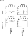
- the confirmation that the selected antibody specifically binds to the modified BCR and not to other BCR variants was carried out by means of two blank samples, ie with cells without BCR (s. FIG. 1 A) as well as with cells with non-CLL-BCR (s. FIG. 1 E).
- Primary B cells from the blood of leukemia patients were examined for binding by FACS.
- the selected antibody was able to specifically identify those BCRs that had the target structure. This was confirmed at the genomic level. Samples without this target structure had no binding.
- mice which have a respective knockout for the genes Lambda5, RAG2 and SLP-65 ( Dlicken von Minden et al., 2012, Nature 489, p309-313 ).
- the preparation of such mice is known in the art and belongs to the prior art.
- the mice were extracted after their killing the bone marrow of the femurs.
- the cells thus obtained were then cultured under conditions favoring the survival of pro- / pre-B cells (37 ° C, 7.5% CO2, Iscoves medium, 10% FCS, P / S, murine IL7).
- a FACS sorting was performed as a control, the pro / pre-B cells were sorted and then returned to culture.
- the markers used for this purpose are known to the person skilled in the art.
- the corresponding sequences coding for the heavy (HC) and light (LC) chains were synthesized and then each cloned into a CMV promoter-containing expression vectors. These were incorporated by lipofection into the packaging cell line (Phoenix cell line). After a 36 hour incubation, virus supernatant was removed and used for spinfection of TKO cells. Both the work for the recovery of the supernatants and the spinfection of the TKO are widely known methods and known to the skilled person.
- CLL subset 2 IgG1 For the expression of the CLL subset 2 IgG1, a human cellular expression system based on HEK293T cells was used. For transfection, a polyethyleneimine (PEI) based protocol was used. After several passages, the supernatant was pooled and the medium contained in the pooled cell supernatant was purified by means of Protein G columns. The purity and quality of soluble Subset-2 IgG1 was determined by Western blot.
- PEI polyethyleneimine
- FIG. 1 This procedure of selection is in FIG. 1 illustrated schematically using the example of CLL subset 2 BCRs, where the term 'TKO' refers to TKO cells (see above).
- the mRNA was isolated from the individual hybridoma clones, from which cDNA was generated and amplified by means of anchor PCR (Rapid Expression Cloning of human immunoglobulin Fab fragments for the analysis of antigenic specificity of B cell lymphomas and anti-idiotype lymphoma vaccination; Osterroth F, Alkan O, Mackensen A, Lindemann A, Fisch P, Skerra A, Veelken H., J Immunol Methods 1999 Oct 29; 229 (1-2): 141-53 ).
- a small amount of peripheral blood was withdrawn from a patient.
- 100 ⁇ l of blood was transferred to a reaction vessel and made up with 2 ml of PBS-BSA buffer solution.
- the sample was centrifuged in an Eppendorf 5804 centrifuge at 1500 rpm for a period of five minutes. The supernatant was discarded and the sediment was well mixed.
- the antibody was added. The following surface parameters were stained: 1) CD19-FITC, 2) CD5-PE, and 3) the CLL SubSet-2 Specific Antibody (APC) before incubating for 15 minutes at room temperature in the dark. Thereafter, lysis was initiated and the erythrocytes were lysed.
- APC CLL SubSet-2 Specific Antibody
- the cells were washed twice with PBS-BSA buffer solution and the cells were taken up in 500 ⁇ l of 0.1% PBS-BSA buffer solution and resuspended. The cells were stored in the dark at 2-8 ° C until measurement on the flow cytometer.
- the analysis at the FACS was carried out on a BDCalibur.
- the setting of the individual laser and detection parameters was carried out as instructed by the device manufacturer and is sufficiently known to the skilled person.
- the raw data of the analysis were then evaluated by FlowJo analysis software.
- the lymphocyte population was selected and labeled in the FSC / SSC blot. For this selection, it was then focused on the CD19-positive B cells and after binding of the Subset-2 specific antibody analyzed.
- Figure 2 exemplified such an analysis using the example of the use of the subset-2 specific antibody.
- the CD19 positive B cells were selected for further analysis (left panel). These were then examined for binding of the specific antibody (right panel).
Priority Applications (5)
| Application Number | Priority Date | Filing Date | Title |
|---|---|---|---|
| CA3070847A CA3070847A1 (fr) | 2017-07-07 | 2018-07-05 | Procede de diagnostic |
| US16/625,754 US11634487B2 (en) | 2017-07-07 | 2018-07-05 | Diagnostic method |
| EP18746103.3A EP3615072A1 (fr) | 2017-07-07 | 2018-07-05 | Procédé de diagnostic |
| JP2020522785A JP7239575B2 (ja) | 2017-07-07 | 2018-07-05 | サンプル中の自律的に活性なb細胞受容体または自律的に活性化されたb細胞受容体を認識する方法 |
| PCT/EP2018/068316 WO2019008128A1 (fr) | 2017-07-07 | 2018-07-05 | Procédé de diagnostic |
Applications Claiming Priority (2)
| Application Number | Priority Date | Filing Date | Title |
|---|---|---|---|
| EP17001167 | 2017-07-07 | ||
| EP17001166 | 2017-07-07 |
Publications (1)
| Publication Number | Publication Date |
|---|---|
| EP3424527A1 true EP3424527A1 (fr) | 2019-01-09 |
Family
ID=64500047
Family Applications (4)
| Application Number | Title | Priority Date | Filing Date |
|---|---|---|---|
| EP18162672.2A Withdrawn EP3424528A1 (fr) | 2017-07-07 | 2018-03-19 | Molécules de liaison biologiques |
| EP18162666.4A Withdrawn EP3424527A1 (fr) | 2017-07-07 | 2018-03-19 | Proces diagnostique |
| EP18746104.1A Pending EP3615073A1 (fr) | 2017-07-07 | 2018-07-05 | Molécules de liaison biologiques |
| EP18746103.3A Pending EP3615072A1 (fr) | 2017-07-07 | 2018-07-05 | Procédé de diagnostic |
Family Applications Before (1)
| Application Number | Title | Priority Date | Filing Date |
|---|---|---|---|
| EP18162672.2A Withdrawn EP3424528A1 (fr) | 2017-07-07 | 2018-03-19 | Molécules de liaison biologiques |
Family Applications After (2)
| Application Number | Title | Priority Date | Filing Date |
|---|---|---|---|
| EP18746104.1A Pending EP3615073A1 (fr) | 2017-07-07 | 2018-07-05 | Molécules de liaison biologiques |
| EP18746103.3A Pending EP3615072A1 (fr) | 2017-07-07 | 2018-07-05 | Procédé de diagnostic |
Country Status (5)
| Country | Link |
|---|---|
| US (2) | US11634487B2 (fr) |
| EP (4) | EP3424528A1 (fr) |
| JP (2) | JP7271532B2 (fr) |
| CA (2) | CA3070848A1 (fr) |
| WO (2) | WO2019008129A1 (fr) |
Families Citing this family (7)
| Publication number | Priority date | Publication date | Assignee | Title |
|---|---|---|---|---|
| EP3424528A1 (fr) * | 2017-07-07 | 2019-01-09 | AVA Lifescience GmbH | Molécules de liaison biologiques |
| EP3733709A1 (fr) * | 2019-05-02 | 2020-11-04 | AVA Lifescience | Molécules de liaison biologiques |
| WO2020221466A1 (fr) * | 2019-05-02 | 2020-11-05 | Ava Lifescience Gmbh | Molécules de liaison biologiques |
| WO2020221463A1 (fr) * | 2019-05-02 | 2020-11-05 | Ava Lifescience Gmbh | Procédé de sélection d'une molécule biologique de liaison |
| EP4227321A1 (fr) | 2022-02-10 | 2023-08-16 | AVA Lifescience GmbH | Anticorps contre le recepteur des cellules b de la leucemie lymphoide chronique (llc) utilisées dans le traitement de llc |
| WO2023152204A1 (fr) | 2022-02-10 | 2023-08-17 | Ava Lifescience Gmbh | Anticorps ciblant le récepteur des lymphocytes b de la leucémie lymphoïde chronique (llc) destinés à être utilisés dans le traitement de llc |
| WO2024023124A1 (fr) * | 2022-07-25 | 2024-02-01 | Ava Lifescience Gmbh | Récepteurs antigéniques chimériques humanisés ciblant le récepteur de lymphocytes b de la leucémie lymphoïde chronique et leurs utilisations |
Citations (1)
| Publication number | Priority date | Publication date | Assignee | Title |
|---|---|---|---|---|
| WO2014179714A1 (fr) * | 2013-05-03 | 2014-11-06 | The Board Of Regents Of The University Of Texas System | Génération de vaccins peptoïdes |
Family Cites Families (4)
| Publication number | Priority date | Publication date | Assignee | Title |
|---|---|---|---|---|
| US7393531B2 (en) * | 2003-01-21 | 2008-07-01 | Arius Research Inc. | Cytotoxicity mediation of cells evidencing surface expression of MCSP |
| RU2691698C2 (ru) * | 2012-02-24 | 2019-06-17 | ЭББВИ СТЕМСЕНТРКС ЭлЭлСи | Антитела против sez6 и способы их применения |
| BR112016004242A8 (pt) * | 2013-08-28 | 2018-06-12 | Stemcentrx Inc | Métodos para conjugação sítio-específica de anticorpos e composições |
| EP3424528A1 (fr) * | 2017-07-07 | 2019-01-09 | AVA Lifescience GmbH | Molécules de liaison biologiques |
-
2018
- 2018-03-19 EP EP18162672.2A patent/EP3424528A1/fr not_active Withdrawn
- 2018-03-19 EP EP18162666.4A patent/EP3424527A1/fr not_active Withdrawn
- 2018-07-05 EP EP18746104.1A patent/EP3615073A1/fr active Pending
- 2018-07-05 CA CA3070848A patent/CA3070848A1/fr active Pending
- 2018-07-05 US US16/625,754 patent/US11634487B2/en active Active
- 2018-07-05 CA CA3070847A patent/CA3070847A1/fr active Pending
- 2018-07-05 WO PCT/EP2018/068317 patent/WO2019008129A1/fr unknown
- 2018-07-05 EP EP18746103.3A patent/EP3615072A1/fr active Pending
- 2018-07-05 US US16/625,756 patent/US11591391B2/en active Active
- 2018-07-05 WO PCT/EP2018/068316 patent/WO2019008128A1/fr unknown
- 2018-07-05 JP JP2020522786A patent/JP7271532B2/ja active Active
- 2018-07-05 JP JP2020522785A patent/JP7239575B2/ja active Active
Patent Citations (1)
| Publication number | Priority date | Publication date | Assignee | Title |
|---|---|---|---|---|
| WO2014179714A1 (fr) * | 2013-05-03 | 2014-11-06 | The Board Of Regents Of The University Of Texas System | Génération de vaccins peptoïdes |
Non-Patent Citations (9)
| Title |
|---|
| BOJARCZUK KAMIL ET AL: "B-cell receptor signaling in the pathogenesis of lymphoid malignancies", BLOOD CELLS, MOLECULES AND DISEASES, LAJOLLA, US, vol. 55, no. 3, 11 July 2015 (2015-07-11), pages 255 - 265, XP029252301, ISSN: 1079-9796, DOI: 10.1016/J.BCMD.2015.06.016 * |
| CLAUDIA MINICI ET AL: "Distinct homotypic B-cell receptor interactions shape the outcome of chronic lymphocytic leukaemia", NATURE COMMUNICATIONS, vol. 8, 9 June 2017 (2017-06-09), pages 15746, XP055481551, DOI: 10.1038/ncomms15746 * |
| DÜHREN VON MINDEN ET AL., NATURE, vol. 489, 2012, pages 309 - 313 |
| JOSE L. SANCHEZ-TRINCADO ET AL: "Fundamentals and Methods for T- and B-Cell Epitope Prediction", JOURNAL OF IMMUNOLOGY RESEARCH, vol. 2017, 28 December 2017 (2017-12-28), pages 1 - 14, XP055469700, ISSN: 2314-8861, DOI: 10.1155/2017/2680160 * |
| M. DÜHREN-VON MINDEN, NATURE, 2012 |
| MARCUS DÜHREN-VON MINDEN ET AL: "Chronic lymphocytic leukaemia is driven by antigen-independent cell-autonomous signalling", NATURE, vol. 489, no. 7415, 12 August 2012 (2012-08-12), GB, pages 309 - 312, XP055481577, ISSN: 0028-0836, DOI: 10.1038/nature11309 * |
| MINICI, C. ET AL.: "Distinct homotypic B-cell receptor interactions shape the outcome of chronic lymphocytic leukaemia", NATURE COMM., 2017 |
| OSTERROTH F; ALKAN 0; MACKENSEN A; LINDEMANN A; FISCH P; SKERRA A; VEELKEN H, J IMMUNOL METHODS, vol. 229, no. 1-2, 29 October 1999 (1999-10-29), pages 141 - 53 |
| VINAY KUMAR; ABUL K. ABBAS; NELSON FAUSTO: "Robbins and Cotran - Pathologie Basis of Disease", 2005, pages: 224f |
Also Published As
| Publication number | Publication date |
|---|---|
| US11634487B2 (en) | 2023-04-25 |
| EP3424528A1 (fr) | 2019-01-09 |
| JP2020527727A (ja) | 2020-09-10 |
| JP7239575B2 (ja) | 2023-03-14 |
| JP2020526590A (ja) | 2020-08-31 |
| US11591391B2 (en) | 2023-02-28 |
| US20200199225A1 (en) | 2020-06-25 |
| JP7271532B2 (ja) | 2023-05-11 |
| WO2019008129A1 (fr) | 2019-01-10 |
| US20200209246A1 (en) | 2020-07-02 |
| WO2019008128A1 (fr) | 2019-01-10 |
| CA3070847A1 (fr) | 2019-01-10 |
| CA3070848A1 (fr) | 2019-01-10 |
| EP3615073A1 (fr) | 2020-03-04 |
| EP3615072A1 (fr) | 2020-03-04 |
Similar Documents
| Publication | Publication Date | Title |
|---|---|---|
| EP3424527A1 (fr) | Proces diagnostique | |
| DE60226036T9 (de) | ANTIKÖRPER, DER DAS GM1-GANGLIOSID-GEBUNDENE AMYLOID-b-PROTEIN ERKENNT, UND DNA, DIE FÜR DIESEN ANTIKÖRPER CODIERT | |
| DE69330523T4 (de) | Immunoglobuline ohne leichte ketten | |
| DE69838062T2 (de) | Menschliche monoklonale antikörper gegen den epidermalen wachstumsfaktorrezeptor | |
| EP0531300B1 (fr) | Variantes de proteines superficielles cd44, sequences d'adn les codant, anticorps contre ces proteines, ainsi que leur utilisation dans le diagnostic et la therapie | |
| DE4118120A1 (de) | Tetravalente bispezifische rezeptoren, ihre herstellung und verwendung | |
| DE3920358A1 (de) | Bispezifische und oligospezifische, mono- und oligovalente antikoerperkonstrukte, ihre herstellung und verwendung | |
| DE69634092T2 (de) | Herstellung von antikörpern | |
| DE69333039T2 (de) | Monoklonaler antikörper gegen ein humanes mdr1-gen produkt, das eine resistenz gegen verschiedene medikamente erzeugt und seine verwendung | |
| WO2020221466A1 (fr) | Molécules de liaison biologiques | |
| DE69837337T2 (de) | Herstellung von proteinen in eiern | |
| DE60129278T2 (de) | Gegen das SEMP1-Protein gerichtete Antikörper, Verfahren zu deren Herstellung, und deren Anwendungen | |
| EP3899536B1 (fr) | Procédé de sélection de molécules de liaison biologiques | |
| EP3543697B1 (fr) | Procédé de sélection de molécules de liaison biologiques | |
| EP0585570B1 (fr) | Greffon d'anticorps anti-granulocytes, sa préparation et usage | |
| DE69334156T2 (de) | Verfahren zur Reduzierung der Immunogenizität der variablen Regionen von Antikörpern | |
| DE10210427A1 (de) | Humaner monoklonaler Antikörper | |
| EP0537489B1 (fr) | Anticorps monoclonaux contre des antigènes associés aux tumeurs, procédé de préparation et utilisation | |
| US20240002500A1 (en) | Anti-TIGIT Antibody or Antigen-Binding Fragment Thereof | |
| EP3733709A1 (fr) | Molécules de liaison biologiques | |
| WO2020221463A1 (fr) | Procédé de sélection d'une molécule biologique de liaison | |
| EP0115062A2 (fr) | Anticorps monoclonaux, réagissant à l'essai d'agglutination directe et spécifiques de l'antigène humain du groupe sanguin D (Rh0) et lignées cellulaires d'hybridomes produisant ces anticorps monoclonaux | |
| DE202016009167U1 (de) | VHH-Konstrukte zur Umleitung einer Immunantwort | |
| DE69231058T3 (de) | Hochaffine humanisierte monoklonale antikoerper | |
| EP1268552A2 (fr) | Procede pour identifier des substances imitant les epitopes de mammiferes |
Legal Events
| Date | Code | Title | Description |
|---|---|---|---|
| PUAI | Public reference made under article 153(3) epc to a published international application that has entered the european phase |
Free format text: ORIGINAL CODE: 0009012 |
|
| AK | Designated contracting states |
Kind code of ref document: A1 Designated state(s): AL AT BE BG CH CY CZ DE DK EE ES FI FR GB GR HR HU IE IS IT LI LT LU LV MC MK MT NL NO PL PT RO RS SE SI SK SM TR |
|
| AX | Request for extension of the european patent |
Extension state: BA ME |
|
| STAA | Information on the status of an ep patent application or granted ep patent |
Free format text: STATUS: THE APPLICATION IS DEEMED TO BE WITHDRAWN |
|
| 18D | Application deemed to be withdrawn |
Effective date: 20190710 |