EP3359572B1 - Method for treating multiple sclerosis - Google Patents
Method for treating multiple sclerosis Download PDFInfo
- Publication number
- EP3359572B1 EP3359572B1 EP16790464.8A EP16790464A EP3359572B1 EP 3359572 B1 EP3359572 B1 EP 3359572B1 EP 16790464 A EP16790464 A EP 16790464A EP 3359572 B1 EP3359572 B1 EP 3359572B1
- Authority
- EP
- European Patent Office
- Prior art keywords
- antibody
- weeks
- patients
- ocrelizumab
- study
- Prior art date
- Legal status (The legal status is an assumption and is not a legal conclusion. Google has not performed a legal analysis and makes no representation as to the accuracy of the status listed.)
- Active
Links
Images


Classifications
-
- C—CHEMISTRY; METALLURGY
- C07—ORGANIC CHEMISTRY
- C07K—PEPTIDES
- C07K16/00—Immunoglobulins [IGs], e.g. monoclonal or polyclonal antibodies
- C07K16/18—Immunoglobulins [IGs], e.g. monoclonal or polyclonal antibodies against material from animals or humans
- C07K16/28—Immunoglobulins [IGs], e.g. monoclonal or polyclonal antibodies against material from animals or humans against receptors, cell surface antigens or cell surface determinants
- C07K16/2887—Immunoglobulins [IGs], e.g. monoclonal or polyclonal antibodies against material from animals or humans against receptors, cell surface antigens or cell surface determinants against CD20
-
- A—HUMAN NECESSITIES
- A61—MEDICAL OR VETERINARY SCIENCE; HYGIENE
- A61P—SPECIFIC THERAPEUTIC ACTIVITY OF CHEMICAL COMPOUNDS OR MEDICINAL PREPARATIONS
- A61P25/00—Drugs for disorders of the nervous system
- A61P25/28—Drugs for disorders of the nervous system for treating neurodegenerative disorders of the central nervous system, e.g. nootropic agents, cognition enhancers, drugs for treating Alzheimer's disease or other forms of dementia
-
- A—HUMAN NECESSITIES
- A61—MEDICAL OR VETERINARY SCIENCE; HYGIENE
- A61P—SPECIFIC THERAPEUTIC ACTIVITY OF CHEMICAL COMPOUNDS OR MEDICINAL PREPARATIONS
- A61P37/00—Drugs for immunological or allergic disorders
-
- A—HUMAN NECESSITIES
- A61—MEDICAL OR VETERINARY SCIENCE; HYGIENE
- A61P—SPECIFIC THERAPEUTIC ACTIVITY OF CHEMICAL COMPOUNDS OR MEDICINAL PREPARATIONS
- A61P37/00—Drugs for immunological or allergic disorders
- A61P37/02—Immunomodulators
- A61P37/06—Immunosuppressants, e.g. drugs for graft rejection
-
- A—HUMAN NECESSITIES
- A61—MEDICAL OR VETERINARY SCIENCE; HYGIENE
- A61K—PREPARATIONS FOR MEDICAL, DENTAL OR TOILETRY PURPOSES
- A61K39/00—Medicinal preparations containing antigens or antibodies
- A61K2039/505—Medicinal preparations containing antigens or antibodies comprising antibodies
-
- A—HUMAN NECESSITIES
- A61—MEDICAL OR VETERINARY SCIENCE; HYGIENE
- A61K—PREPARATIONS FOR MEDICAL, DENTAL OR TOILETRY PURPOSES
- A61K39/00—Medicinal preparations containing antigens or antibodies
- A61K2039/545—Medicinal preparations containing antigens or antibodies characterised by the dose, timing or administration schedule
-
- A—HUMAN NECESSITIES
- A61—MEDICAL OR VETERINARY SCIENCE; HYGIENE
- A61K—PREPARATIONS FOR MEDICAL, DENTAL OR TOILETRY PURPOSES
- A61K38/00—Medicinal preparations containing peptides
-
- C—CHEMISTRY; METALLURGY
- C07—ORGANIC CHEMISTRY
- C07K—PEPTIDES
- C07K2317/00—Immunoglobulins specific features
- C07K2317/20—Immunoglobulins specific features characterized by taxonomic origin
- C07K2317/24—Immunoglobulins specific features characterized by taxonomic origin containing regions, domains or residues from different species, e.g. chimeric, humanized or veneered
-
- C—CHEMISTRY; METALLURGY
- C07—ORGANIC CHEMISTRY
- C07K—PEPTIDES
- C07K2317/00—Immunoglobulins specific features
- C07K2317/70—Immunoglobulins specific features characterized by effect upon binding to a cell or to an antigen
- C07K2317/73—Inducing cell death, e.g. apoptosis, necrosis or inhibition of cell proliferation
- C07K2317/732—Antibody-dependent cellular cytotoxicity [ADCC]
-
- C—CHEMISTRY; METALLURGY
- C07—ORGANIC CHEMISTRY
- C07K—PEPTIDES
- C07K2317/00—Immunoglobulins specific features
- C07K2317/70—Immunoglobulins specific features characterized by effect upon binding to a cell or to an antigen
- C07K2317/73—Inducing cell death, e.g. apoptosis, necrosis or inhibition of cell proliferation
- C07K2317/734—Complement-dependent cytotoxicity [CDC]
-
- C—CHEMISTRY; METALLURGY
- C07—ORGANIC CHEMISTRY
- C07K—PEPTIDES
- C07K2317/00—Immunoglobulins specific features
- C07K2317/70—Immunoglobulins specific features characterized by effect upon binding to a cell or to an antigen
- C07K2317/76—Antagonist effect on antigen, e.g. neutralization or inhibition of binding
Definitions
- the present invention concerns an anti-CD20 antibody, which is ocrelizumab, for use in a method of improving functional ability in a human patient having multiple sclerosis, as further defined in the claims.
- MS Multiple Sclerosis
- CNS central nervous system
- MS cerebrospinal fluid
- MS has four patterns of disease: relapsing-remitting MS (RRMS; 80%-85% of cases at onset), primary progressive MS (PPMS; 10%-15% at onset), progressive relapsing MS (PRMS; 5% at onset); and secondary progressive MS (SPMS) ( Kremenchutzky et al. Brain 122 (Pt 10):1941-50 (1999 ); Confavreux et al. N Engl J Med 343(20):1430-8 (2000 )).
- RRMS relapsing-remitting MS
- PPMS primary progressive MS
- PRMS progressive relapsing MS
- SPMS secondary progressive MS
- the RRMS treatments include the following: interferon class, IFN-beta-1a (REBIF ® , Extavia, AVONEX ® and PLEGRIDY TM and IFN-beta-1b (BETASERON ® ); glatiramer acetate (COPAXONE ® ), a polypeptide; natalizumab (TYSABRI ® ), alemtuzumab (LEMTRADA ® ), both monoclonal antibodies; dimethyl fumarate (TECFIDERA ® ) and fingolimod (GILENYA ® ), both small molecules, and mitoxantrone (NOVANTRONE ® ), a cytotoxic agent; teriflunomide (AUBAGIO ® ).
- interferon class IFN-beta-1a
- REBIF ® Extavia
- the present invention relates to an anti-CD20 antibody for use in a method of improving functional ability in a human patient having multiple sclerosis comprising administering to the patient an effective amount of the anti-CD20 antibody, wherein the patient has improvement in functional ability after treatment; wherein the anti-CD20 antibody is ocrelizumab; wherein the multiple sclerosis is a primary progressive multiple sclerosis (PPMS) with T1 gadolinium staining lesions; and wherein the improvement in functional ability is measured by the Timed 25-Foot Walk (T-25FW) test or EDSS score.
- PPMS primary progressive multiple sclerosis
- T-25FW Timed 25-Foot Walk
- the patient has 12-week confirmed disability improvement after treatment. In some embodiments, the patient has 24-week confirmed disability improvement after treatment. In some embodiments, the improvement in functional ability in the patient is sustained for at least 12 weeks. In some embodiments, the improvement in functional ability in the patient is sustained for at least 24 weeks. As explained above, the improvement in functional ability is measured by the Timed 25-Foot Walk (T-25FW) test or EDSS score. In some embodiments, the improvement in functional ability is measured by the Timed 25-Foot Walk (T-25FW) test and EDSS score.
- the invention relates to an anti-CD20 antibody for use in a method of improving functional ability in a human patient having multiple sclerosis, as further defined herein above and in the claims, wherein the patient has T1 gadolinium staining lesions at baseline.
- the anti-CD20 antibody is administered to the patient to provide an initial anti-CD20 antibody exposure followed by a second anti-CD20 antibody exposure, wherein the first and second exposures are each about 600 mg of the antibody, and wherein the interval between the first exposure and the second exposure is about 20-24 weeks or about 5-6 months.
- the anti-CD20 antibody is administered to the patient to provide a third anti-CD20 antibody exposure, wherein the third exposure is about 600 mg of the antibody, and wherein the interval between the second exposure and the third exposure is about 20-24 weeks or about 5-6 months.
- the anti-CD20 antibody is administered to the patient to provide a fourth anti-CD20 antibody exposure, wherein the fourth exposure is about 600 mg of the antibody, and wherein the interval between the third exposure and the fourth exposure is about 20-24 weeks or about 5-6 months.
- the first exposure comprises a first dose and a second dose of the anti-CD20 antibody, wherein each dose is about 300 mg and the first dose and the second dose are separated by about two weeks or about 14 days (such as 13 days or 15 days). In some embodiments "about 14 days" refers to a variation of 1 day before or after the 14 th day.
- the second, third, and/or fourth exposures comprise a single dose of about 600 mg.
- the initial exposure and second, third, and/or fourth additional exposures comprise a first dose and a second dose of the anti-CD20 antibody, wherein each dose is about 300 mg and the first dose and the second dose are separated by about two weeks or about 14 days (such as 13 days or 15 days).
- the patient has improved functional ability after one, two, three, and/or four exposures of the anti-CD20 antibody.
- the patient maintains the ability to mount a humoral response to an antigen during treatment.
- the antigen is a mumps antigen, a rubella antigen, a varicella antigen, an S. pneumonia antigen, a tetanus toxoid antigen, a pneumococcal antigen, or an influenza antigen.
- the patient is premedicated prior to infusion with the anti-CD20 antibody. In certain embodiments, the patient is premedicated with methylprednisolone (or an equivalent) approximately 30 minutes prior to each infusion of anti-CD20 antibody. In certain embodiments, the patient is premedicated with 100 mg IV methylprednisolone (or an equivalent) approximately 30 minutes prior to each infusion of anti-CD20 antibody. In certain embodiments, the patient is additionally (or alternatively) premedicated with an antihistaminic drug (e.g. diphenhydramine) approximately 30-60 minutes before each infusion of anti-CD20 antibody. In certain embodiments, the patient is additionally (or alternatively) premedicated with an antipyretic (e.g. acetaminophen/paracetamol).
- an antipyretic e.g. acetaminophen/paracetamol
- a second medicament is administered with the initial exposure or later exposures, wherein the anti-CD20 antibody is a first medicament.
- the second medicament is selected from the group consisting of an interferon, glatiramer acetate, a cytotoxic agent, chemotherapeutic agent, mitoxantrone, methotrexate, cyclophosphamide, chlorambucil, azathioprine, gamma globulin, Campath, anti-CD4, cladribine, corticosteroid, mycophenolate mofetil (MMF), cyclosporine, cholesterol-lowering drug of the statin class, estradiol, testosterone, hormone replacement drug, a TNF inhibitor, disease modifying anti-rheumatic drug (DMARD), non-steroidal anti-inflammatory drug (NSAID), levothyroxine, cyclosporin A, somatostatin analogue, cytokine or cytokine receptor antagonist, antimetabolite, immunosuppressive
- the multiple sclerosis is a primary progressive multiple sclerosis (PPMS).
- the invention relates to an anti-CD20 antibody for use in a method of improving functional ability in a human patient having multiple sclerosis, wherein the multiple sclerosis is a primary progressive multiple sclerosis (PPMS) with T1 gadolinium staining lesions, as further defined herein above and in the claims.
- the patient is selected for treatment based upon having PPMS.
- treatment is based upon the patient having PPMS.
- the patient is diagnosed with PPMS prior to treatment.
- the anti-CD20 antibody is administered to the patient to provide an initial anti-CD20 antibody exposure followed by one or more additional anti-CD20 antibody exposures, wherein each exposure is about 600 mg of the antibody, and each exposure is provided to the patient as one or two doses of the anti-CD20 antibody, and wherein the interval between each exposure is about 20-24 weeks or about 5-6 months.
- “about 20-24 weeks” refers to a time point between 20 weeks and 24 weeks.
- “about 20-24 weeks” refers to a variation of a week or 7 days before or after the 24 th week.
- "about 5-6 months” refers to a time point between 5 and 6 months.
- the first exposure comprises a first dose and a second dose of the anti-CD20 antibody, wherein each dose is about 300 mg and the first dose and the second dose are separated by about two weeks or about 14 days (such as 13 days or 15 days). In some embodiments "about 14 days" refers to a variation of 1 day before or after the 14 th day.
- the second, third, and/or fourth exposures comprise a single dose of about 600 mg.
- the initial exposure and second, third, and/or fourth additional exposures comprise a first dose and a second dose of the anti-CD20 antibody, wherein each dose is about 300 mg and the first dose and the second dose are separated by about two weeks or about 14 days (such as 13 days or 15 days).
- the anti-CD20 antibody is administered to the patient to provide an initial anti-CD20 antibody exposure followed by a second anti-CD20 antibody exposure, wherein the first and second exposures are each about 600 mg of the antibody, and wherein the interval between the first exposure and the second exposure is about 20-24 weeks or about 5-6 months.
- the anti-CD20 antibody is administered to the patient to provide a third anti-CD20 antibody exposure, wherein the third exposure is about 600 mg of the antibody, and wherein the interval between the second exposure and the third exposure is about 20-24 weeks or about 5-6 months.
- the anti-CD20 antibody is administered to the patient to provide a fourth anti-CD20 antibody exposure, wherein the fourth exposure is about 600 mg of the antibody, and wherein the interval between the third exposure and the fourth exposure is about 20-24 weeks or about 5-6 months.
- the first exposure comprises a first dose and a second dose of the anti-CD20 antibody, wherein each dose is about 300 mg and the first dose and the second dose are separated by about two weeks or about 14 days (such as 13 days or 15 days). In some embodiments "about 14 days" refers to a variation of 1 day before or after the 14 th day.
- the first and second doses are administered intravenously.
- the first and second doses each comprise 250 ml of anti-CD20 antibody at a concentration of about 1.2 mg/ml.
- the first and second doses are each infused at a rate of 30 ml/hour.
- the infusion rate of the first and second doses can be increased in 30 ml/hour increments to a maximum rate of 180 ml/hr.
- the first and second doses are each be given over approximately 2.5 hours.
- the second, third, and/or fourth exposures comprise a single dose of about 600 mg. In certain embodiments according to (or as applied to) any of the embodiments above, the second, third, and/or fourth exposures are administered intravenously. In certain embodiments according to (or as applied to) any of the embodiments above, the second, third, and/or fourth exposures each comprise 500 ml of anti-CD20 antibody at a concentration of about 1.2 mg/ml. In certain embodiments according to (or as applied to) any of the embodiments above, the second, third, and/or fourth exposures are each infused at a rate of 40 ml/hour.
- the infusion rate of the second, third, and/or fourth exposures can be increased in 40 ml/hour increments to a maximum rate of 200 ml/hr.
- the first and second doses are each be given over approximately 3.5 hours.
- the initial exposure and second, third, and/or fourth additional exposures comprise a first dose and a second dose of the anti-CD20 antibody, wherein each dose is about 300 mg and the first dose and the second dose are separated by about two weeks or about 14 days (such as 13 days or 15 days). In some embodiments "about 14 days" refers to a variation of 1 day before or after the 14 th day.
- the first and second doses are administered intravenously.
- the first and second doses each comprise 250 ml of anti-CD20 antibody at a concentration of about 1.2 mg/ml.
- the first and second doses are each infused at a rate of 30 ml/hour.
- the infusion rate of the first and second doses can be increased in 30 ml/hour increments to a maximum rate of 180 ml/hr.
- the first and second doses are each be given over approximately 2.5 hours.
- the patient receives at least 2, 3, 4, or more than 4 anti-CD20 antibody exposures.
- the anti-CD20 antibody is administered to the patient to provide one or more additional anti-CD20 antibody exposure after the fourth exposure, wherein the one or more additional exposure is about 600 mg of the antibody, and wherein the interval between the fourth exposure and the additional exposure is about 20-24 weeks or about 5-6 months.
- “about 20-24 weeks” refers to a time point between 20 weeks and 24 weeks.
- “about 20-24 weeks” refers to a variation of a week or 7 days before or after the 24 th week.
- "about 5-6 months” refers to a time point between 5 and 6 months.
- the interval between each of the additional exposures following the fourth exposure is 20-24 weeks or about 5-6 months.
- the one or more exposures comprise a first dose and a second dose of the anti-CD20 antibody, wherein each dose is about 300 mg and the first dose and the second dose are separated by about 14 days (such as 13 days or 15 days). In some embodiments "about 14 days” refers to a variation of 1 day before or after the 14 th day. In some embodiments, the one or more exposures comprise a first dose and a second dose of the anti-CD20 antibody, wherein each dose is about 300 mg and the first dose and the second dose are separated by about 14 days (such as 13 days or 15 days).
- the anti-CD20 antibody to be used in accordance with the invention is ocrelizumab.
- the anti-CD20 antibody is in a pharmaceutically acceptable composition.
- the anti-CD20 antibody is in a formulation comprising 30 mg/mL antibody, 20 mM Sodium Acetate, 106 mM Trehalose, 0.02% polysorbate 20, pH 5.3.
- the antibody in the formulation is stored at about 2-8°C at 300 mg/vial.
- the antibody is diluted in saline (0.9% sodium chloride) in an IV bag for administration by infusion.
- the anti-CD20 antibody is administered intravenously. In some embodiments, the anti-CD20 antibody is administered intravenously for each antibody exposure. In some embodiments, the antibody is administered subcutaneously. In some embodiments, the anti-CD20 antibody is administered subcutaneously for each antibody exposure.
- a reduction or decrease or improvement after administration of the anti-CD20 antibody can be compared to a baseline level, to a level in untreated patient(s), and/or to a level in patient(s), e.g., such as mean, average, or median level of a group of patients, receiving a different treatment (such as interferon beta-1a or REBIF ® ).
- a "B-cell” is a lymphocyte that matures within the bone marrow, and includes a naive B cell, memory B cell, or effector B cell (plasma cells).
- the B-cell herein may be a normal or non-malignant B cell.
- B-cell surface marker or "B-cell surface antigen” herein is an antigen expressed on the surface of a B cell that can be targeted with an antibody that binds thereto.
- Exemplary B-cell surface markers include the CD10, CD19, CD20, CD21, CD22, CD23, CD24, CD37, CD40, CD53, CD72, CD73, CD74, CDw75, CDw76, CD77, CDw78, CD79a, CD79b, CD80, CD81, CD82, CD83, CDw84, CD85 and CD86 leukocyte surface markers (for descriptions, see The Leukocyte Antigen Facts Book, 2nd Edition. 1997, ed. Barclay et al. Academic Press , Harcourt Brace & Co., New York).
- B-cell surface markers include RP105, FcRH2, B-cell CR2, CCR6, P2X5, HLA-DOB, CXCR5, FCER2, BR3, Btig, NAG14, SLGC16270, FcRH1, IRTA2, ATWD578, FcRH3, IRTA1, FcRH6, BCMA, and 239287.
- the B-cell surface marker of particular interest herein is preferentially expressed on B cells compared to other non-B-cell tissues of a mammal and may be expressed on both precursor B cells and mature B cells.
- the preferred B-cell surface marker herein is CD20.
- CD20 antigen is an about 35-kDa, non-glycosylated phosphoprotein found on the surface of greater than 90% of B cells from peripheral blood or lymphoid organs. CD20 is present on both normal B cells as well as malignant B cells, but is not expressed on stem cells. Other names for CD20 in the literature include "B-lymphocyte-restricted antigen” and "Bp35". The CD20 antigen is described in Clark et al. Proc. Natl. Acad. Sci. (USA) 82:1766 (1985 ), for example.
- an "antibody antagonist” herein is an antibody that, upon binding to a B cell surface marker on B cells, destroys or depletes B cells in a mammal and/or interferes with one or more B-cell functions, e . g . by reducing or preventing a humoral response elicited by the B cell.
- the antibody antagonist preferably is able to deplete B cells ( i.e. reduce circulating B-cell levels) in a mammal treated therewith. Such depletion may be achieved via various mechanisms such antibody-dependent cell-mediated cytotoxicity (ADCC) and/or complement dependent cytotoxicity (CDC), inhibition of B-cell proliferation and/or induction of B-cell death (e.g. via apoptosis).
- ADCC antibody-dependent cell-mediated cytotoxicity
- CDC complement dependent cytotoxicity
- Antibody-dependent cell-mediated cytotoxicity and “ADCC” refer to a cell-mediated reaction in which nonspecific cytotoxic cells that express Fc receptors (FcRs) (e.g. Natural Killer (NK) cells, neutrophils, and macrophages) recognize bound antibody on a target cell and subsequently cause lysis of the target cell.
- FcRs Fc receptors
- FcR expression on hematopoietic cells in summarized is Table 3 on page 464 of Ravetch and Kinet, Annu. Rev. Immunol 9:457-92 (1991 ).
- ADCC activity of a molecule of interest may be assessed in vitro, such as that described in US Patent No. 5,500,362 or 5,821,337.
- useful effector cells for such assays include peripheral blood mononuclear cells (PBMC) and Natural Killer (NK) cells.
- PBMC peripheral blood mononuclear cells
- NK Natural Killer
- ADCC activity of the molecule of interest may be assessed in vivo, e.g., in an animal model such as that disclosed in Clynes et al. PNAS (USA) 95:652-656 (1998 ).
- Human effector cells are leukocytes that express one or more FcRs and perform effector functions. In some embodiments, the cells express at least FcyRIII and carry out ADCC effector function. Examples of human leukocytes that mediate ADCC include peripheral blood mononuclear cells (PBMC), natural killer (NK) cells, monocytes, cytotoxic T cells and neutrophils; with PBMCs and NK cells being preferred.
- PBMC peripheral blood mononuclear cells
- NK natural killer cells
- monocytes monocytes
- cytotoxic T cells and neutrophils cytotoxic T cells and neutrophils
- Fc receptor or “FcR” are used to describe a receptor that binds to the Fc region of an antibody.
- the FcR is a native sequence human FcR.
- a preferred FcR is one that binds an IgG antibody (a gamma receptor) and includes receptors of the FcyRI, FcyRII, and Fcy RIII subclasses, including allelic variants and alternatively spliced forms of these receptors.
- FcyRII receptors include FcyRIIA (an “activating receptor") and FcyRIIB (an “inhibiting receptor”), which have similar amino acid sequences that differ primarily in the cytoplasmic domains thereof.
- Activating receptor FcyRIIA contains an immunoreceptor tyrosine-based activation motif (ITAM) in its cytoplasmic domain.
- Inhibiting receptor FcyRIIB contains an immunoreceptor tyrosine-based inhibition motif (ITIM) in its cytoplasmic domain.
- ITAM immunoreceptor tyrosine-based activation motif
- ITIM immunoreceptor tyrosine-based inhibition motif
- FcR FcR
- FcRn neonatal receptor
- “Complement dependent cytotoxicity” or “CDC” refers to the ability of a molecule to lyse a target in the presence of complement.
- the complement activation pathway is initiated by the binding of the first component of the complement system (C1q) to a molecule (e.g. an antibody) complexed with a cognate antigen.
- a CDC assay e.g. as described in Gazzano-Santoro et al., J. Immunol. Methods 202:163 (1996 ) may be performed.
- “Growth inhibitory” antibodies are those that prevent or reduce proliferation of a cell expressing an antigen to which the antibody binds.
- the antibody may prevent or reduce proliferation of B cells in vitro and/or in vivo.
- Antibodies that "induce apoptosis” are those that induce programmed cell death, e.g. of a B cell, as determined by standard apoptosis assays, such as binding of annexin V, fragmentation of DNA, cell shrinkage, dilation of endoplasmic reticulum, cell fragmentation, and/or formation of membrane vesicles (called apoptotic bodies).
- antibody herein is used in the broadest sense and specifically covers monoclonal antibodies, polyclonal antibodies, multispecific antibodies (e.g. bispecific antibodies) formed from at least two intact antibodies, and antibody fragments so long as they exhibit the desired biological activity.
- Antibody fragments comprise a portion of an intact antibody, preferably comprising the antigen binding region thereof.
- antibody fragments include Fab, Fab', F(ab') 2 , and Fv fragments; diabodies; linear antibodies; single-chain antibody molecules; and multispecific antibodies formed from antibody fragments.
- an “intact antibody” is one comprising heavy and light variable domains as well as an Fc region.
- “Native antibodies” are usually heterotetrameric glycoproteins of about 150,000 daltons, composed of two identical light (L) chains and two identical heavy (H) chains. Each light chain is linked to a heavy chain by one covalent disulfide bond, while the number of disulfide linkages varies among the heavy chains of different immunoglobulin isotypes. Each heavy and light chain also has regularly spaced intrachain disulfide bridges. Each heavy chain has at one end a variable domain (V H ) followed by a number of constant domains.
- V H variable domain
- Each light chain has a variable domain at one end (V L ) and a constant domain at its other end; the constant domain of the light chain is aligned with the first constant domain of the heavy chain, and the light chain variable domain is aligned with the variable domain of the heavy chain. Particular amino acid residues are believed to form an interface between the light chain and heavy chain variable domains.
- variable refers to the fact that certain portions of the variable domains differ extensively in sequence among antibodies and are used in the binding and specificity of each particular antibody for its particular antigen. However, the variability is not evenly distributed throughout the variable domains of antibodies. It is concentrated in three segments called hypervariable regions both in the light chain and the heavy chain variable domains. The more highly conserved portions of variable domains are called the framework regions (FRs).
- the variable domains of native heavy and light chains each comprise four FRs, largely adopting a ⁇ -sheet configuration, connected by three hypervariable regions, which form loops connecting, and in some cases forming part of, the ⁇ -sheet structure.
- the hypervariable regions in each chain are held together in close proximity by the FRs and, with the hypervariable regions from the other chain, contribute to the formation of the antigen-binding site of antibodies ( see Kabat et al., Sequences of Proteins of Immunological Interest, 5th Ed. Public Health Service, National Institutes of Health, Bethesda, MD. (1991 )).
- the constant domains are not involved directly in binding an antibody to an antigen, but exhibit various effector functions, such as participation of the antibody in antibody dependent cellular cytotoxicity (ADCC).
- Papain digestion of antibodies produces two identical antigen-binding fragments, called “Fab” fragments, each with a single antigen-binding site, and a residual "Fc” fragment, whose name reflects its ability to crystallize readily. Pepsin treatment yields an F(ab') 2 fragment that has two antigen-binding sites and is still capable of cross-linking antigen.
- Fv is the minimum antibody fragment that contains a complete antigen-recognition and antigen-binding site. This region consists of a dimer of one heavy chain and one light chain variable domain in tight, non-covalent association. It is in this configuration that the three hypervariable regions of each variable domain interact to define an antigen-binding site on the surface of the V H -V L dimer. Collectively, the six hypervariable regions confer antigen-binding specificity to the antibody. However, even a single variable domain (or half of an Fv comprising only three hypervariable regions specific for an antigen) has the ability to recognize and bind antigen, although at a lower affinity than the entire binding site.
- the Fab fragment also contains the constant domain of the light chain and the first constant domain (CH1) of the heavy chain.
- Fab' fragments differ from Fab fragments by the addition of a few residues at the carboxy terminus of the heavy chain CH1 domain including one or more cysteines from the antibody hinge region.
- Fab'-SH is the designation herein for Fab' in which the cysteine residue(s) of the constant domains bear at least one free thiol group.
- F(ab') 2 antibody fragments originally were produced as pairs of Fab' fragments that have hinge cysteines between them. Other chemical couplings of antibody fragments are also known.
- the "light chains" of antibodies (immunoglobulins) from any vertebrate species can be assigned to one of two clearly distinct types, called kappa ( ⁇ ) and lambda ( ⁇ ), based on the amino acid sequences of their constant domains.
- antibodies can be assigned to different classes. There are five major classes of intact antibodies: IgA, IgD, IgE, IgG, and IgM, and several of these may be further divided into subclasses (isotypes), e.g., IgG1, IgG2, IgG3, IgG4, IgA, and IgA2.
- the heavy chain constant domains that correspond to the different classes of antibodies are called ⁇ , ⁇ , ⁇ , ⁇ , and ⁇ , respectively.
- the subunit structures and three-dimensional configurations of different classes of immunoglobulins are well known.
- Single-chain Fv or “scFv” antibody fragments comprise the V H and V L domains of antibody, wherein these domains are present in a single polypeptide chain.
- the Fv polypeptide further comprises a polypeptide linker between the V H and V L domains that enables the scFv to form the desired structure for antigen binding.
- diabodies refers to small antibody fragments with two antigen-binding sites, which fragments comprise a heavy chain variable domain (V H ) connected to a light chain variable domain (V L ) in the same polypeptide chain (V H - V L ).
- V H heavy chain variable domain
- V L light chain variable domain
- the domains are forced to pair with the complementary domains of another chain and create two antigen-binding sites.
- Diabodies are described more fully in, for example, EP 404,097 ; WO 93/11161 ; and Hollinger et al., Proc. Natl. Acad. Sci. USA, 90:6444-6448 (1993 ).
- the term "monoclonal antibody” as used herein refers to an antibody obtained from a population of substantially homogeneous antibodies, i.e., the individual antibodies comprising the population are identical and/or bind the same epitope, except for possible variants that may arise during production of the monoclonal antibody, such variants generally being present in minor amounts.
- each monoclonal antibody is directed against a single determinant on the antigen.
- the monoclonal antibodies are advantageous in that they are uncontaminated by other immunoglobulins.
- the modifier "monoclonal” indicates the character of the antibody as being obtained from a substantially homogeneous population of antibodies, and is not to be construed as requiring production of the antibody by any particular method.
- the monoclonal antibodies may be made by the hybridoma method first described by Kohler et al., Nature, 256:495 (1975 ), or may be made by recombinant DNA methods ( see, e.g., U.S. Patent No. 4,816,567 ).
- the “monoclonal antibodies” may also be isolated from phage antibody libraries using the techniques described in Clackson et al., Nature, 352:624-628 (1991 ) and Marks et al., J. Mol. Biol., 222:581-597 (1991 ), for example.
- the monoclonal antibodies herein specifically include "chimeric" antibodies (immunoglobulins) in which a portion of the heavy and/or light chain is identical with or homologous to corresponding sequences in antibodies derived from a particular species or belonging to a particular antibody class or subclass, while the remainder of the chain(s) is identical with or homologous to corresponding sequences in antibodies derived from another species or belonging to another antibody class or subclass, as well as fragments of such antibodies, so long as they exhibit the desired biological activity ( U.S. Patent No. 4,816,567 ; Morrison et al., Proc. Natl. Acad. Sci. USA, 81:6851-6855 (1984 )).
- chimeric antibodies immunoglobulins in which a portion of the heavy and/or light chain is identical with or homologous to corresponding sequences in antibodies derived from a particular species or belonging to a particular antibody class or subclass, while the remainder of the chain(s) is identical with or homologous to corresponding sequence
- Chimeric antibodies of interest herein include "primatized" antibodies comprising variable domain antigen-binding sequences derived from a non-human primate (e.g. Old World Monkey, such as baboon, rhesus or cynomolgus monkey) and human constant region sequences ( US Pat No. 5,693,780 ).
- a non-human primate e.g. Old World Monkey, such as baboon, rhesus or cynomolgus monkey
- human constant region sequences US Pat No. 5,693,780
- Humanized forms of non-human (e.g., murine) antibodies are chimeric antibodies that contain minimal sequence derived from non-human immunoglobulin.
- humanized antibodies are human immunoglobulins (recipient antibody) in which residues from a hypervariable region of the recipient are replaced by residues from a hypervariable region of a non-human species (donor antibody) such as mouse, rat, rabbit or nonhuman primate having the desired specificity, affinity, and capacity.
- donor antibody such as mouse, rat, rabbit or nonhuman primate having the desired specificity, affinity, and capacity.
- framework region (FR) residues of the human immunoglobulin are replaced by corresponding non-human residues.
- humanized antibodies may comprise residues that are not found in the recipient antibody or in the donor antibody. These modifications are made to further refine antibody performance.
- the humanized antibody will comprise substantially all of at least one, and typically two, variable domains, in which all or substantially all of the hypervariable loops correspond to those of a non-human immunoglobulin and all or substantially all of the FRs are those of a human immunoglobulin sequence, except for FR substitution(s) as noted above.
- the humanized antibody optionally also will comprise at least a portion of an immunoglobulin constant region, typically that of a human immunoglobulin. For further details, see Jones et al., Nature 321:522-525 (1986 ); Riechmann et al., Nature 332:323-329 (1988 ); and Presta, Curr. Op. Struct. Biol. 2:593-596 (1992 ).
- hypervariable region when used herein refers to the amino acid residues of an antibody that are responsible for antigen binding.
- the hypervariable region comprises amino acid residues from a "complementarity determining region" or "CDR" (e.g. residues 24-34 (L1), 50-56 (L2) and 89-97 (L3) in the light chain variable domain and 31-35 (H1), 50-65 (H2) and 95-102 (H3) in the heavy chain variable domain; Kabat et al., Sequences of Proteins of Immunological Interest, 5th Ed. Public Health Service, National Institutes of Health, Bethesda, MD. (1991 )) and/or those residues from a "hypervariable loop" (e.g.
- naked antibody is an antibody (as herein defined) that is not conjugated to a heterologous molecule, such as a cytotoxic moiety or radiolabel.
- humanized 2H7 refers to a humanized antibody that binds human CD20, or an antigen-binding fragment thereof, wherein the antibody is effective to deplete primate B cells in vivo, the antibody comprising in the H chain variable region (V H ) thereof at least a CDR H3 sequence of SEQ ID NO:12 ( Fig. 1B ) from an anti-human CD20 antibody and substantially the human consensus framework (FR) residues of the human heavy- chain subgroup III (V H III).
- this antibody further comprises the H chain CDR H1 sequence of SEQ ID NO:10 and CDR H2 sequence of SEQ ID NO:11, and, in some embodiments, further comprises the L chain CDR L1 sequence of SEQ ID NO:4, CDR L2 sequence of SEQ ID NO:5, CDR L3 sequence of SEQ ID NO:6 and substantially the human consensus framework (FR) residues of the human light chain kappa subgroup I (V ⁇ I), wherein the V H region may be joined to a human IgG chain constant region, wherein the region may be, for example, IgG1 or IgG3.
- such antibody comprises the V H sequence of SEQ ID NO:8 (v16, as shown in Fig.
- the antibody is an intact antibody comprising the light and heavy chain amino acid sequences of SEQ ID NOS: 13 and 14, respectively, as shown in Figs. 2 and 3 .
- the antibody is 2H7.v31 comprising the light and heavy chain amino acid sequences of SEQ ID NOS: 13 and 15, respectively, as shown in Figs. 2 and 4 .
- the antibody herein may further comprise at least one amino acid substitution in the Fc region that improves ADCC and/or CDC activity, such as one wherein the amino acid substitutions are S298A/E333A/K334A, and in some embodiments, the 2H7.v31 having the heavy chain amino acid sequence of SEQ ID NO: 15 (as shown in Fig. 4 ). Any of these antibodies may further comprise at least one amino acid substitution in the Fc region that decreases CDC activity, for example, comprising at least the substitution K322A. See U.S. Patent No. 6,528,624B1 (Idusogie et al .).
- the term "ocrelizumab” (CAS Registration No. 637334-45-3 ) herein refers to the genetically engineered humanized monoclonal antibody directed against the CD20 antigen and comprising (a) a light chain comprising the amino acid sequence of SEQ ID NO: 13 and (b) a heavy chain comprising the amino acid sequence of SEQ ID NO: 14. Ocrelizumab is available from Genentech.
- an “isolated” antibody is one that has been identified and separated and/or recovered from a component of its natural environment. Contaminant components of its natural environment are materials that would interfere with diagnostic or therapeutic uses for the antibody, and may include enzymes, hormones, and other proteinaceous or non-proteinaceous solutes.
- the antibody will be purified (1) to greater than 95% by weight of antibody as determined by the Lowry method, and in some embodiments, more than 99% by weight, (2) to a degree sufficient to obtain at least 15 residues of N-terminal or internal amino acid sequence by use of a spinning cup sequenator, or (3) to homogeneity by SDS-PAGE under reducing or nonreducing conditions using Coomassie blue or, in some embodiments, silver stain.
- Isolated antibody includes the antibody in situ within recombinant cells since at least one component of the antibody's natural environment will not be present. Ordinarily, however, isolated antibody will be prepared by at least one purification step.
- a “subject” or “patient” herein is a human subject or patient.
- the subject or patient is eligible for treatment for multiple sclerosis.
- such eligible subject or patient is one who is experiencing, has experienced, or is likely to experience, one or more signs, symptoms or other indicators of multiple sclerosis; has been diagnosed with multiple sclerosis, whether, for example, newly diagnosed (with "new onset” MS), previously diagnosed with a new relapse or exacerbation, previously diagnosed and in remission, etc; and/or is at risk for developing multiple sclerosis.
- One suffering from or at risk for suffering from multiple sclerosis may optionally be identified as one who has been screened for elevated levels of CD20-positive B cells in serum, cerebrospinal fluid (CSF) and/or MS lesion(s) and/or is screened for using an assay to detect autoantibodies, assessed qualitatively, and preferably quantitatively.
- autoantibodies associated with multiple sclerosis include anti-myelin basic protein (MBP), anti-myelin oligodendrocytic glycoprotein (MOG), anti-ganglioside and/or anti-neurofilament antibodies.
- MBP myelin basic protein
- MOG anti-myelin oligodendrocytic glycoprotein
- Such autoantibodies may be detected in the subject's serum, cerebrospinal fluid (CSF) and/or MS lesion.
- Elevated autoantibody or B cell level(s) herein level(s) of such autoantibodies or B cells which significantly exceed the level(s) in an individual
- treatment is an approach for obtaining beneficial or desired results including clinical results.
- beneficial or desired clinical results include, but are not limited to, one or more of the following: decreasing one or more symptoms resulting from the disease, diminishing the extent of the disease, stabilizing the disease (e.g., preventing or delaying the worsening of the disease), delay or slowing the progression of the disease, ameliorating the disease state, decreasing the dose of one or more other medications required to treat the disease, and/or increasing the quality of life.
- delay or slowing the progression of multiple sclerosis means to prevent, defer, hinder, slow, retard, stabilize, and/or postpone development of the disease. This delay can be of varying lengths of time, depending on the history of the disease and/or individual being treated.
- “at the time of starting treatment” refers to the time period at or prior to the first exposure to a multiple sclerosis drug, such as an anti-CD20 antibody. In some embodiments, “at the time of starting treatment” is about any of one year, nine months, six months, three months, second months, or one month prior to a multiple sclerosis drug, such as an anti-CD20 antibody. In some embodiments, "at the time of starting treatment” is immediately prior to coincidental with the first exposure to a multiple sclerosis drug, such as an anti-CD20 antibody.
- based upon includes (1) assessing, determining, or measuring the patient characteristics as described herein (and preferably selecting a patient suitable for receiving treatment; and (2) administering the treatment(s) as described herein.
- a "symptom" of MS is any morbid phenomenon or departure from the normal in structure, function, or sensation, experienced by the subject and indicative of MS.
- Multiple sclerosis refers to the chronic and often disabling disease of the central nervous system characterized by the progressive destruction of the myelin.
- MS primary progressive multiple sclerosis
- RRMS relapsing-remitting multiple sclerosis
- SPMS secondary progressive multiple sclerosis
- PRMS progressive relapsing multiple sclerosis
- Progressive multiple sclerosis refers to primary progressive multiple sclerosis (PPMS), secondary progressive multiple sclerosis (SPMS), and progressive relapsing multiple sclerosis (PRMS).
- PPMS primary progressive multiple sclerosis
- SPMS secondary progressive multiple sclerosis
- PRMS progressive relapsing multiple sclerosis
- progressive multiple sclerosis is characterized by documented, irreversible loss of neurological function persisting for ⁇ 6 months that cannot be attributed to clinical relapse.
- PPMS Primary progressive multiple sclerosis
- PPMS is characterized by a gradual progression of the disease from its onset with rare superimposed relapses and remissions. There may be periods of a leveling off of disease activity and there may be good and bad days or weeks.
- PPMS differs from RRMS and SPMS in that onset is typically in the late thirties or early forties, men are as likely as women to develop it, and initial disease activity is often in the spinal cord and not in the brain.
- PPMS disease activity can also be observed (or found) in the brain.
- PPMS is the sub-type of MS that is least likely to show inflammatory (gadolinium enhancing) lesions on MRI scans.
- PPMS Primary Progressive form of the disease affects between 10 and 15% of all people with multiple sclerosis.
- PPMS may be defined according to the criteria in Polman et al. Ann Neurol 69:292-392 (2010 ).
- the subject with PPMS treated herein is usually one with probable or definitive diagnosis of PPMS.
- RRMS Relapsing-remitting multiple sclerosis
- RRMS Relapsing-remitting multiple sclerosis
- RRMS Relapses
- the relapses are followed by periods of remission, during which time the person fully or partially recovers from the deficits acquired during the relapse.
- Relapses can last for days, weeks or months and recovery can be slow and gradual or almost instantaneous.
- the vast majority of people presenting with MS are first diagnosed with RRMS. This is typically when they are in their twenties or thirties, though diagnoses much earlier or later are known. Twice as many women as men present with this sub-type of MS.
- myelin a protective insulating sheath around the nerve fibers (neurons) in the white matter regions of the central nervous system (CNS)
- CNS central nervous system
- an oligodendrocyte sponsors remyelination - a process whereby the myelin sheath around the axon may be repaired. It is this remyelination that may be responsible for the remission.
- Approximately 50% of patients with RRMS convert to SPMS within 10 years of disease onset. After 30 years, this figure rises to 90%. At any one time, the relapsing-remitting form of the disease accounts around 55% of all people with MS.
- SPMS Secondary progressive multiple sclerosis
- Progressive relapsing multiple sclerosis refers to “PRMS” is characterized by a steady progression of clinical neurological damage with superimposed relapses and remissions. There is significant recovery immediately following a relapse but between relapses there is a gradual worsening of symptoms. PRMS affects around 5% of all people with multiple sclerosis. Some neurologists believe PRMS is a variant of PPMS.
- effective amount refers to an amount of the antibody (or other drug) that is effective for ameliorating or treating the multiple sclerosis.
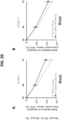
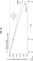
- Such an effective amount will generally result in an improvement in the signs, symptoms or other indicators of MS, such as reducing relapse rate, preventing disability, reducing number and/or volume of brain MRI lesions, improving timed 25-foot walk, slow or delay the progression of the disease such as extending the time to disease progression (e.g. using Expanded Disability Status Scale, EDSS), etc.
- Antibody exposure refers to contact with or exposure to the antibody herein in one or more doses administered over a period of time of about 1-20 days. The doses may be given at one time or at fixed or irregular time intervals over this period of exposure. Initial and later (e.g. second or third) antibody exposures are separated in time from each other as described in detail herein.
- an "interval" between antibody exposures refers to time period between an earlier antibody exposure and a later antibody exposure.
- An antibody exposure of the present disclosure may include one or two doses.
- an interval between two antibody exposures refers to the amount of time elapsed between the dose of one antibody exposure (e.g., Day 1) and the dose of the next antibody exposure. If one antibody exposure includes two doses and the next antibody exposure includes one dose, an interval between the two antibody exposures refers to the amount of time elapsed between the first of the two doses of the first antibody exposure ( e.g., Day 1) and the dose of the next antibody exposure.
- an interval between to the antibody exposures refers to the amount of time elapsed between the first of the two doses of the first antibody exposure (e.g., Day 1) and the first dose of the two doses of the second antibody exposure.
- the antibody for use in a method of the present disclosure includes a first antibody exposure with two doses and a second antibody exposure with two doses, and the second antibody exposure is not provided until about 24 weeks or 6 months after the first antibody exposure, then the interval between the first dose of the first antibody exposure and the first dose of the second antibody exposure is about 24 weeks or 6 months.
- the second antibody exposure is not provided until about 20-24 weeks or about 5-6 months after the first antibody exposure.
- the interval between the first dose of the first antibody exposure and the first dose of the second antibody exposure is about 20-24 weeks or about 5-6 months.
- “about 20-24 weeks” refers to a time point between 20 weeks and 24 weeks.
- “about 20-24 weeks” refers to a variation of a week or 7 days before or after the 24 th week.
- “about 5-6 months” refers to a time point between 5 and 6 months.
- immunosuppressive agent refers to substances that act to suppress or mask the immune system of the mammal being treated herein. This would include substances that suppress cytokine production, down-regulate or suppress self-antigen expression, or mask the MHC antigens. Examples of such agents include 2-amino-6-aryl-5-substituted pyrimidines ( see U.S. Pat. No.
- nonsteroidal anti-inflammatory drugs NSAIDs
- ganciclovir tacrolimus, glucocorticoids such as cortisol or aldosterone
- anti-inflammatory agents such as a cyclooxygenase inhibitor, a 5-lipoxygenase inhibitor, or a leukotriene receptor antagonist
- purine antagonists such as azathioprine or mycophenolate mofetil (MMF)
- alkylating agents such as cyclophosphamide; bromocryptine; danazol; dapsone; glutaraldehyde (which masks the MHC antigens, as described in U.S. Pat. No.
- anti-idiotypic antibodies for MHC antigens and MHC fragments include cyclosporin A; steroids such as corticosteroids or glucocorticosteroids or glucocorticoid analogs, e .
- prednisone, methylprednisolone, and dexamethasone dihydrofolate reductase inhibitors such as methotrexate (oral or subcutaneous); hydroxychloroquine; sulfasalazine; leflunomide; cytokine or cytokine receptor antagonists including anti-interferon-alpha, -beta, or -gamma antibodies, anti-tumor necrosis factor-alpha antibodies (infliximab or adalimumab), anti-TNF-alpha immunoadhesin (etanercept), anti-tumor necrosis factor-beta antibodies, anti-interleukin-2 antibodies and anti-IL-2 receptor antibodies; anti-LFA-1 antibodies, including anti-CD11a and anti-CD18 antibodies; anti-L3T4 antibodies; heterologous anti-lymphocyte globulin; pan-T antibodies, preferably anti-CD3 or anti-CD4/CD4a
- T-cell receptor fragments Offner et al., Science, 251: 430-432 (1991 ); WO 90/11294 ; Janeway, Nature, 341: 482 (1989 ); and WO 91/01133 ); and T cell receptor antibodies ( EP 340,109 ) such as T10B9.
- cytotoxic agent refers to a substance that inhibits or prevents the function of cells and/or causes destruction of cells.
- the term is intended to include radioactive isotopes (e.g. At 211 , I 131 , I 125 , Y 90 , Re 186 , Re 188 , Sm 153 , Bi 212 , P 32 and radioactive isotopes of Lu), chemotherapeutic agents, and toxins such as small molecule toxins or enzymatically active toxins of bacterial, fungal, plant or animal origin, or fragments thereof.
- radioactive isotopes e.g. At 211 , I 131 , I 125 , Y 90 , Re 186 , Re 188 , Sm 153 , Bi 212 , P 32 and radioactive isotopes of Lu
- chemotherapeutic agents e.g. At 211 , I 131 , I 125 , Y 90 , Re 186 , Re 188
- chemotherapeutic agent is a chemical compound useful in the treatment of cancer.
- examples of chemotherapeutic agents include alkylating agents such as thiotepa and CYTOXAN ® cyclosphosphamide; alkyl sulfonates such as busulfan, improsulfan and piposulfan; aziridines such as benzodopa, carboquone, meturedopa, and uredopa; ethylenimines and methylamelamines including altretamine, triethylenemelamine, trietylenephosphoramide, triethiylenethiophosphoramide and trimethylolomelamine; acetogenins (especially bullatacin and bullatacinone); a camptothecin (including the synthetic analogue topotecan); bryostatin; callystatin; CC-1065 (including its adozelesin, carzelesin and bizelesin synthetic analogues); cryptophy
- calicheamicin especially calicheamicin gammalI and calicheamicin omegaI1 (see, e.g., Agnew, Chem Intl. Ed. Engl., 33: 183-186 (1994 )); dynemicin, including dynemicin A; bisphosphonates, such as clodronate; an esperamicin; as well as neocarzinostatin chromophore and related chromoprotein enediyne antiobiotic chromophores), aclacinomysins, actinomycin, authramycin, azaserine, bleomycins, cactinomycin, carabicin, carminomycin, carzinophilin, chromomycinis, dactinomycin, daunorubicin, detorubicin, 6-diazo-5-oxo-L-norleucine, ADRIAMYCIN ® doxorubicin (including
- TAXOL ® paclitaxel Bristol- Myers Squibb Oncology, Princeton, N.J.
- ABRAXANE TM Cremophor-free, albumin-engineered nanoparticle formulation of paclitaxel American Pharmaceutical Partners, Schaumberg, Illinois
- TAXOTERE ® doxetaxel Rhône- Poulenc Rorer, Antony, France
- chloranbucil GEMZAR ® gemcitabine
- 6-thioguanine mercaptopurine
- methotrexate platinum analogs such as cisplatin and carboplatin; vinblastine; platinum; etoposide (VP-16); ifosfamide; mitoxantrone; vincristine; NAVELBINE ® vinorelbine; novantrone; teniposide; edatrexate; daunomycin; aminopterin; xeloda; ibandronate; CPT-11; top
- anti-hormonal agents that act to regulate or inhibit hormone action on tumors
- SERMs selective estrogen receptor modulators
- tamoxifen including NOLVADEX ® tamoxifen
- raloxifene including NOLVADEX ® tamoxifen
- droloxifene 4-hydroxytamoxifen
- trioxifene keoxifene
- LY117018 onapristone
- aromatase inhibitors that inhibit the enzyme aromatase, which regulates estrogen production in the adrenal glands, such as, for example, 4(5)-imidazoles, aminoglutethimide, MEGASE ® megestrol acetate, AROMASIN ® exemestane, formestanie, fadrozole, RIVISOR ® vorozole, FEMARA ® letrozole, and ARIMIDEX ® anastrozole
- antiandrogens such as flutamide
- cytokine is a generic term for proteins released by one cell population that act on another cell as intercellular mediators.
- cytokines are lymphokines, monokines; interleukins (ILs) such as IL-1, IL-1 ⁇ , IL-2, IL-3, IL-4, IL-5, IL-6, IL-7, IL-8, IL-9, IL-11, IL-12, IL-15; a tumor necrosis factor such as TNF- ⁇ or TNF- ⁇ ; and other polypeptide factors including LIF and kit ligand (KL).
- ILs interleukins
- IL-1 ⁇ interleukins
- IL-6 interleukins
- IL-8 interleukins
- IL-9 IL-11, IL-12, IL-15
- tumor necrosis factor such as TNF- ⁇ or TNF- ⁇
- KL kit ligand
- the term cytokine includes proteins from natural sources or from recombinant cell culture and biologically active equivalents of the native sequence
- hormone refers to polypeptide hormones, which are generally secreted by glandular organs with ducts. Included among the hormones are, for example, growth hormone such as human growth hormone, N-methionyl human growth hormone, and bovine growth hormone; parathyroid hormone; thyroxine; insulin; proinsulin; relaxin; prorelaxin; glycoprotein hormones such as follicle stimulating hormone (FSH), thyroid stimulating hormone (TSH), and luteinizing hormone (LH); prolactin, placental lactogen, mouse gonadotropin-associated peptide, inhibin; activin; mullerian-inhibiting substance; and thrombopoietin.
- growth hormone such as human growth hormone, N-methionyl human growth hormone, and bovine growth hormone
- parathyroid hormone thyroxine
- insulin proinsulin
- relaxin prorelaxin
- glycoprotein hormones such as follicle stimulating hormone (FSH), thyroid stimulating hormone (TSH), and luteinizing
- growth factor refers to proteins that promote growth, and include, for example, hepatic growth factor; fibroblast growth factor; vascular endothelial growth factor; nerve growth factors such as NGF- ⁇ ; platelet-derived growth factor; transforming growth factors (TGFs) such as TGF- ⁇ and TGF- ⁇ ; insulin-like growth factor-I and -II; erythropoietin (EPO); osteoinductive factors; interferons such as interferon- ⁇ , - ⁇ , and -y; and colony stimulating factors (CSFs) such as macrophage-CSF (M-CSF); granulocyte-macrophage-CSF (GM-CSF); and granulocyte-CSF (G-CSF).
- TGFs transforming growth factors
- EPO erythropoietin
- CSFs colony stimulating factors
- growth factor includes proteins from natural sources or from recombinant cell culture and biologically active equivalents of the native sequence growth factor, including synthetically produced small-
- integrin refers to a receptor protein that allows cells both to bind to and to respond to the extracellular matrix and is involved in a variety of cellular functions such as wound healing, cell differentiation, homing of tumor cells and apoptosis. They are part of a large family of cell adhesion receptors that are involved in cell-extracellular matrix and cell-cell interactions.
- Functional integrins consist of two transmembrane glycoprotein subunits, called alpha and beta that are non-covalently bound. The alpha subunits all share some homology to each other, as do the beta subunits.
- the receptors always contain one alpha chain and one beta chain.
- integrin includes proteins from natural sources or from recombinant cell culture and biologically active equivalents of the native sequence integrin, including synthetically produced small-molecule entities and pharmaceutically acceptable derivatives and salts thereof.
- integrin antagonists or antibodies include an LFA-1 antibody; an alpha 4 integrin antibody such as natalizumab (TYSABRI ® ) available from Biogen Idec/Elan Pharmaceuticals, Inc.; diazacyclic phenylalanine derivatives ( WO 2003/89410 ); phenylalanine derivatives ( WO 2003/70709 , WO 2002/28830 , WO 2002/16329 and WO 2003/53926 ); phenylpropionic acid derivatives ( WO 2003/10135 ); enamine derivatives ( WO 2001/79173 ); propanoic acid derivatives ( WO 2000/37444 ); alkanoic acid derivatives ( WO 2000/32575 ); substituted phenyl derivatives ( US Pat.
- LFA-1 antibody an alpha 4 integrin antibody
- alpha 4 integrin antibody such as natalizumab (TYSABRI ® ) available from Biogen Idec/Elan Pharmaceuticals, Inc.
- TNF-alpha tumor necrosis factor alpha
- TNF-alpha refers to a human TNF-alpha molecule comprising the amino acid sequence as described in Pennica et al., Nature, 312:721 (1984 ) or Aggarwal et al., JBC, 260:2345 (1985 ).
- TNF-alpha inhibitor herein is an agent that inhibits, to some extent, a biological function of TNF-alpha, generally through binding to TNF-alpha and neutralizing its activity.
- TNF inhibitors specifically contemplated herein are Etanercept (ENBREL ® ), Infliximab (REMICADE ® ) and Adalimumab (HUMIRA TM ).
- DMARDs Disease-modifying anti-rheumatic drugs
- DMARDs include hydroxychloroquine, sulfasalazine, methotrexate, leflunomide, etanercept, infliximab (plus oral and subcutaneous methrotrexate), azathioprine, D-penicillamine, Gold (oral), Gold (intramuscular), minocycline, cyclosporine, Staphylococcal protein A immunoadsorption, including salts and derivatives thereof, etc.
- nonsteroidal anti-inflammatory drugs or “NSAIDs” are acetylsalicylic acid, ibuprofen, naproxen, indomethacin, sulindac, tolmetin, including salts and derivatives thereof, etc.
- Corticosteroid refers to any one of several synthetic or naturally occurring substances with the general chemical structure of steroids that mimic or augment the effects of the naturally occurring corticosteroids.
- synthetic corticosteroids include prednisone, prednisolone (including methylprednisolone), dexamethasone, glucocorticoid and betamethasone.
- a "package insert” is used to refer to instructions customarily included in commercial packages of therapeutic products, that contain information about the indications, usage, dosage, administration, contraindications, other therapeutic products to be combined with the packaged product, and/or warnings concerning the use of such therapeutic products, etc.
- label is used herein to refer to information customarily included with commercial packages of pharmaceutical formulations including containers such as vials and package inserts, as well as other types of packaging.
- references to "about” a value or parameter herein includes (and describes) variations that are directed to that value or parameter per se. For example, description referring to "about X” includes description of "X.”
- the present invention provides an anti-CD20 antibody for use in a method of improving functional ability in a human patient having multiple sclerosis comprising administering to the patient an effective amount of the anti-CD20 antibody, wherein the patient has improvement in functional ability after treatment; wherein the anti-CD20 antibody is ocrelizumab; wherein the multiple sclerosis is a primary progressive multiple sclerosis (PPMS) with T1 gadolinium staining lesions; and wherein the improvement in functional ability is measured by the Timed 25-Foot Walk (T-25FW) test or EDSS score.
- the method further comprises a step of measuring the patient's functional ability by measuring EDSS score and/or the Timed 25-Foot Walk (T25-FW) after 1, 2, 3, 4, or more than 4 exposures of anti-CD20 antibody.
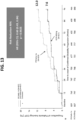
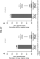
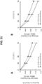
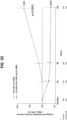
- the patient has at least about a 12-week confirmed disability improvement after treatment. In certain embodiments, the patient has at least about a 24-week confirmed disability improvement after treatment. In certain embodiments, the confirmed disability improvement is determined by Expanded Disability Status Scale (EDSS) score. In certain embodiments, the patient's EDSS score decreases by at least about 0.1, about 0.2, about 0.3, about 0.4, about 0.5, about 0.6, about 0.7, about 0.8, about 0.9, about 1.0 or more than about 1.0 points (such as about 1.1, about 1.2, about 1.3, about 1.4, or about 1.5 points).
- EDSS Expanded Disability Status Scale
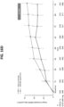
- the improvement in functional ability is sustained for at least about 1 week, at least about 2 weeks, at least about 3 weeks, at least about 4 weeks, at least about 5 weeks, at least about 6 weeks, at least about 7 weeks, at least about 8 weeks, at least about 9 weeks, at least about 10 weeks, at least about 11 weeks, at least about12 weeks, at least about 13 weeks, at least about 14 weeks, at least about 15 weeks, at least about 16 weeks, at least about 17 weeks, at least about 18 weeks, at least about 19 weeks, at least about 20 weeks, at least about 21 weeks, at least about 22 weeks, at least about 23 weeks, including any range in between these values.
- the improvement in functional ability is sustained for at least about 24 weeks, at least about 25 weeks, at least about 26 weeks, at least about 27 weeks, at least about 28 weeks, at least about 29 weeks, at least about 30 weeks, at least about 35 weeks, at least about 40 weeks, at least about 45 weeks, at least about 50 weeks, at least about 55 weeks, at least about 60 weeks, at least about 65 weeks, at least about 70 weeks, at least about 75weeks, or more than about 75 weeks, including any range in between these values.
- the improvement in functional ability is measured by the Timed 25-Foot Walk (T25-FW) test.
- T25-FW Timed 25-Foot Walk
- the time to walk 25 feet following the start of treatment is reduced by about 5 seconds, about 10 seconds, about 30 seconds, about 60 seconds, about 90 seconds, about 2 minutes, about 2.5 minutes, about 3 minutes, about 3.5 minutes, about 4 minutes, about 4.5 minutes, about 5 minutes, about 5.5 minutes, about 6 minutes, about 6.5 minutes, about 7 minutes, about 7.5 minutes, about 8 minutes, about 8.5 minutes, about 9 minutes about 9.5 minutes, or about 10 minutes relative to the time to walk 25 feet immediately prior to starting treatment.
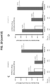
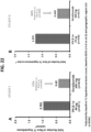
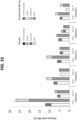
- NEDA no evidence of disease activity
- NEDA is demonstrated by the absence of new or enlarging T2 lesions or T1 gadolinium-enhancing lesions on magnetic resonance imaging.
- NEDA is demonstrated by absence of relapse.
- NEDA is demonstrated by the absence of progression.
- NEDA is demonstrated by lack of worsening of EDSS.
- NEDA is defined as: no protocol-defined relapses, no CDP events, no new or enlarging T2 lesions, and no gadolinium-enhancing T1 lesions.
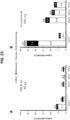
- NEDA is sustained for about least about 1 week, at least about 2 weeks, at least about 3 weeks, at least about 4 weeks, at least about 5 weeks, at least about 6 weeks, at least about 7 weeks, at least about 8 weeks, at least about 9 weeks, at least about 10 weeks, at least about 11 weeks, at least about12 weeks, at least about 13 weeks, at least about 14 weeks, at least about 15 weeks, at least about 16 weeks, at least about 17 weeks, at least about 18 weeks, at least about 19 weeks, at least about 20 weeks, at least about 21 weeks, at least about 22 weeks, at least about 23 weeks, including any range in between these values.
- the improvement in functional ability is sustained for at least about 24 weeks, at least about 25 weeks, at least about 26 weeks, at least about 27 weeks, at least about 28 weeks, at least about 29 weeks, at least about 30 weeks, at least about 35 weeks, at least about 40 weeks, at least about 45 weeks, at least about 50 weeks, at least about 55 weeks, at least about 60 weeks, at least about 65 weeks, at least about 70 weeks, at least about 75 weeks, or more than about 75 weeks, including any range in between these values.
- the invention relates to an anti-CD20 antibody for use in a method of improving functional ability in a human patient having multiple sclerosis, as further defined herein above and in the claims, wherein the patient has T1 gadolinium staining lesions at baseline (i.e., before starting treatment).
- the patient has not been previously treated with other therapy for multiple sclerosis (i.e., a "naive patient").
- a naive patient experienced at least 2 relapses in 2 years prior to starting treatment. In certain embodiments, the naive patient experienced at least 1 relapse in the last year prior to starting treatment.
- the patient is an inadequate responder to other therapy for multiple sclerosis.
- the patient who is an inadequate responder has been previously treated with interferon beta-1a or glatiramer acetate for at least 1 year.
- the patient who is an inadequate responder has experienced at least one relapse or experienced at least 1 baseline gadolinium-enhancing lesion while being treated with another therapy for multiple sclerosis.
- the patient has primary progressive multiple sclerosis.
- the patient is less than 18 years of age.
- the patient is between 18 and 55 years of age.
- the patient is over 55 years of age.
- the patient has a diagnosis of Primary Progressive Multiple Sclerosis in accordance with the 2005 revised McDonald criteria ( Polman et al. (2011) "Diagnostic criteria for multiple sclerosis: 2005 revisions to the ⁇ McDonald criteria.'", Ann Neurol 58, 840-846 ).
- the patient has an Expanded Disability Status Scale (EDSS) score of 3 to 6.5 points.
- EDSS Expanded Disability Status Scale
- the patient has a score of at least 2.0 on the pyramidal functions component of the Functional Systems Scale (FSS). Additionally or alternatively, in certain embodiments, the patient has a documented history or presence at screening of elevated IgG index in a cerebrospinal fluid (CSF) specimen and/or one or more IgG oligoclonal bands detected by isoelectric focusing in a cerebrospinal fluid (CSF) specimen. Additionally or alternatively, in certain embodiments, the patient has no history of relapse-remitting multiple sclerosis (RRMS). Additionally or alternatively, in certain embodiments, the patient has no history of secondary progressive multiple sclerosis (SPMS). Additionally or alternatively, in certain embodiments, the patient has history of progressive relapsing multiple sclerosis (PRMS).
- FSS Functional Systems Scale
- the patient has had previous treatment with B-cell targeted therapies (e.g. rituximab, ocrelizumab, atacicept, belimumab, or ofatumumab). In certain embodiments, the patient has not had previous treatment with B-cell targeted therapies (e.g. rituximab, ocrelizumab, atacicept, belimumab, or ofatumumab).
- B-cell targeted therapies e.g. rituximab, ocrelizumab, atacicept, belimumab, or ofatumumab.
- CDP Consfirmed Disability Progression
- CDP refers to an increase of at least 1.0 point from the baseline EDSS score in patients with a baseline score of 5.5 or less, or an increase of a 0.5 point in patients with a baseline score over 5.5, during the 96 weeks, wherein increases in the EDSS were confirmed at a regularly scheduled visit at least 12 weeks after the initial neurologic worsening.
- Consfirmed Disability Improvement refers to a reduction in EDSS score of at least 1.0 compared to baseline in patients with a baseline EDSS score of 5.5 or less, or a reduction of a 0.5 point in patients with a baseline EDSS score above 5.5.
- brain atrophy refers to one or more of the following: axonal loss in the brain, tissue loss within gray matter lesions or white matter lesions, Wallerian degeneration in pathways related to the lesions, or lesion burden.
- brain atrophy refers to a decrease in whole brain volume.
- brain atrophy refers to a decrease in volume of one or more of the structures of the brain (including, but not limited to, the cerebrum, the cerebellum, the thalamus, frontotemporal neocortex, brainstem, hippocampus, parietal lobe, and/or the hypothalamus).
- brain atrophy refers to cortical thinning in precentral gyrus, superior frontal gyrus, thalamus and/or putamen. In certain embodiments, brain atrophy refers to a loss of at least about 0.4%, at least about 0.5%, at least about 0.6%, or at least 0.7% brain volume per year. Further details regarding brain atrophy are detailed in, e.g., Riley et al. (2012) Expert Rev Neurother 12(3), 323-333 .
- the patient or subject has highly active multiple sclerosis.
- "highly active multiple sclerosis" in treatment naive patients refers to patients who have not been previously treated with other therapy for multiple sclerosis and who have experienced at least 2 relapses in the last year prior to randomization, and either (a) at least 1 baseline gadolinium lesion or (b) increase in T2 lesion count at baseline visit (changing categorically from 0-5 to 6-9 lesions or from 6-9 lesions to > 9 lesions) as compared to a prior MRI.
- the patient has primary progressive multiple sclerosis (PPMS) with T1 gadolinium staining lesions.
- PPMS primary progressive multiple sclerosis
- a baseline level in a patient refers to the level prior to administration of or treatment with an anti-CD20 antibody to the patient, for example, about 2 months, about 1.5 months, about 1 month, about 30 days, about 25 days, about 21 days, about 14 days, about 7 days, about 6 days, about 5 days, about 4 days, about 3 days, about 2 days, about 1 day prior to administration of or treatment with an anti-CD20 antibody to the patient.
- the patient maintains the ability to mount a humoral immune response to an antigen during treatment.
- the antigen is a mumps antigen, a rubella antigen, a varicella antigen, an S. pneumonia antigen, a tetanus toxoid antigen, a pneumococcal antigen, or an influenza antigen.
- the anti-CD20 antibody for use in a method as described herein may encompass any combination of the embodiments described herein.
- the method comprises administering an effective amount of the anti-CD20 antibody to the multiple sclerosis patient to provide an initial antibody exposure of about 0.3 to about 4 grams (preferably about 0.3 to about 1.5 grams, such as about 0.6 grams or about 1.0 grams) followed by a second antibody exposure of about 0.3 to about 4 grams (preferably about 0.3 to about 1.5 grams, such as about 0.6 grams or about 1.0 grams), the second antibody exposure not being provided until from about 16 to about 60 weeks from the initial antibody exposure.
- the second antibody exposure is the next time the patient is treated with the anti-CD20 antibody after the initial antibody exposure, there being no intervening anti-CD20 antibody treatment or exposure between the initial and second exposures.
- the initial antibody exposure and/or the second antibody exposure is about any of 0.3 grams, 0.4 grams, 0.5 grams, 0.6 grams, 0.7 grams, 0.8 grams, 0.9 grams, or 1.0 grams.
- the interval between the initial and second or subsequent antibody exposures can be measured from the first dose of the initial antibody exposure.
- the antibody exposures are approximately 24 weeks or 6 months apart; or approximately 48 weeks or 12 months apart. In some embodiments, the antibody exposures are approximately about 20-24 weeks or about 5-6 months apart. In some embodiments, “about 20-24 weeks” refers to a time point between 20 weeks and 24 weeks. In some embodiments, “about 20-24 weeks” refers to a variation of a week or 7 days before or after the 24 th week. In some embodiments, “about 5-6 months” refers to a time point between 5 and 6 months.
- the second antibody exposure is not provided until about 20 to about 30 weeks from the initial exposure, optionally followed by a third antibody exposure of about 0.3 to about 4 grams (preferably about 0.3 to about 1.5 grams), the third exposure not being administered until from about 46 to 60 weeks (preferably from about 46 to 54 weeks) from the initial exposure, and then, in some embodiments, no further antibody exposure is provided until at least about 70-75 weeks from the initial exposure.
- the third antibody exposure is about any of 0.3 grams, 0.4 grams, 0.5 grams, 0.6 grams, 0.7 grams, 0.8 grams, 0.9 grams, or 1.0 grams.
- the second antibody exposure is not provided until about 46 to 60 weeks from the initial exposure, and subsequent antibody exposures, if any, are not provided until about 46 to 60 weeks from the previous antibody exposure.
- the method comprises administering an effective amount of the anti-CD20 antibody to the multiple sclerosis patient to provide an initial antibody exposure of about 0.3 to about 4 grams (preferably about 0.3 to about 1.5 grams, such as about 0.6 grams or about 1.0 grams) followed by a second antibody exposure of about 0.3 to about 4 grams (preferably about 0.3 to about 1.5 grams, such as about 0.6 grams or about 1.0 grams), the second antibody exposure not being provided until from about 20 to about 30 weeks from the initial antibody exposure, followed by a third antibody exposure of about 0.3 to about 4 grams (preferably about 0.3 to about 1.5 grams, such as about 0.6 grams or about 1.0 grams), the third antibody exposure not being provided until from about 46 to about 54 weeks from the initial exposure, followed by a fourth antibody exposure of about 0.3 to about 4 grams (preferably about 0.3 to about 1.5 grams, such as about 0.6 grams or about 1.0 grams), the fourth antibody exposure not being provided until from about 70 to about 75 weeks from the initial exposure.
- an initial antibody exposure of about 0.3 to about 4 grams (preferably about 0.3 to about 1.5 grams,
- the fourth antibody exposure is followed by one or more antibody exposures of about 0.3 to about 4 grams (preferably about 0.3 to about 1.5 grams, such as about 0.6 grams or about 1.0 grams). In certain embodiments each subsequent antibody exposure is about 20 to about 30 weeks from the previous exposure.
- the/each subsequent exposure is the next time the patient is treated with the anti-CD20 antibody after the initial antibody exposure, there being no intervening anti-CD20 antibody treatment or exposure between, e.g., the initial and second exposures, the second and third exposures, or the third and fourth exposures, etc.
- the initial, second, third, fourth, and/or subsequent antibody exposure is about any of 0.3 grams, 0.4 grams, 0.5 grams, 0.6 grams, 0.7 grams, 0.8 grams, 0.9 grams, or 1.0 grams.
- any one or more of the antibody exposures herein may be provided to the patient as a single dose of antibody, or as two separate doses of the antibody (i.e., constituting a first and second dose).
- the particular number of doses (whether one or two) employed for each antibody exposure may be dependent, for example, on the type of MS treated, whether and what type of second medicament is employed, and the method and frequency of administration.
- the second dose is preferably administered from about 3 to 17 days, more preferably from about 6 to 16 days, and most preferably from about 13 to 16 days from the time the first dose was administered. In some embodiments, where two separate doses are administered, the second dose is about 14 days (such as 13 days or 15 days).
- the first and second dose of the antibody is preferably about 0.3 to 1.5 grams, more preferably about 0.3 to about 1.0 grams. In some embodiments, where two separate doses are administered, the first and second dose of the antibody is about any of 0.3 grams, 0.4 grams, 0.5 grams, or 0.6 grams.
- the initial ocrelizumab exposure comprises a first dose and a second dose of ocrelizumab, wherein the first dose and second dose of ocrelizumab is about 0.3 grams.
- the second ocrelizumab exposure comprises a single dose of ocrelizumab, wherein the single dose of ocrelizumab is 0.6 grams.
- the patient is provided at least about three, at least about four, or at least about five exposures of the antibody, for example, from about 3 to 60 exposures, and more particularly about 3 to 40 exposures, most particularly, about 3 to 20 exposures.
- the method further comprises providing between about one to about three subsequent ocrelizumab exposures. In some embodiments, such exposures are administered at intervals each of approximately 24 weeks or 6 months, or 48 weeks or 12 months.
- an interval is shortened by approximately 4 weeks, about 3.5 weeks, about 3 weeks, about 2.5 weeks, about 2 weeks, about 1.5 weeks, about 1 week, about 6 days, about 5 days, about 4 days, about 3 days, about 2 days or about 1 day.
- more than 1 interval is shortened by approximately 4 weeks, about 3.5 weeks, about 3 weeks, about 2.5 weeks, about 2 weeks, about 1.5 weeks, about 1 week, about 6 days, about 5 days, about 4 days, about 3 days, about 2 days or about 1 day.
- an interval is lengthened by approximately 4 weeks, about 3.5 weeks, about 3 weeks, about 2.5 weeks, about 2 weeks, about 1.5 weeks, about 1 week, about 6 days, about 5 days, about 4 days, about 3 days, about 2 days or about 1 day.
- more than one interval is lengthened by approximately 4 weeks, about 3.5 weeks, about 3 weeks, about 2.5 weeks, about 2 weeks, about 1.5 weeks, about 1 week, about 6 days, about 5 days, about 4 days, about 3 days, about 2 days or about 1 day.
- more than 1 interval is shortened by approximately 4 weeks, about 3.5 weeks, about 3 weeks, about 2.5 weeks, about 2 weeks, about 1.5 weeks, about 1 week, about 6 days, about 5 days, about 4 days, about 3 days, about 2 days or about 1 day or lengthened by approximately 4 weeks, about 3.5 weeks, about 3 weeks, about 2.5 weeks, about 2 weeks, about 1.5 weeks, about 1 week, about 6 days, about 5 days, about 4 days, about 3 days, about 2 days or about 1 day.
- each antibody exposure is provided as a single dose of the antibody. In an alternative embodiment, each antibody exposure is provided as two separate doses of the antibody. In some embodiments, some exposures are provided as a single dose or as two separate doses.
- the antibody to be used in accordance with the invention is ocrelizumab.
- Other anti-CD20 antibodies are disclosed herein, such as Rituximab, humanized 2H7 ( e.g. comprising the variable domain sequences in SEQ ID NOS. 2 and 8) or humanized 2H7 comprising the variable domain sequences in SEQ ID NOS. 23 and 24, or huMax-CD20 (Genmab).
- the antibody is ocrelizumab (comprising (a) a light chain comprising the amino acid sequence of SEQ ID NO: 13 and (b) a heavy chain comprising the amino acid sequence of SEQ ID NO: 14).
- the patient has never been previously treated with drug(s), such as immunosuppressive agent(s), to treat the multiple sclerosis and/or has never been previously treated with an antibody to a B-cell surface marker (e.g. never previously treated with a CD20 antibody).
- drug(s) such as immunosuppressive agent(s)
- an antibody to a B-cell surface marker e.g. never previously treated with a CD20 antibody
- the antibody is administered by any suitable means, including parenteral, topical, subcutaneous, intraperitoneal, intrapulmonary, intranasal, and/or intralesional administration.
- Parenteral infusions include intramuscular, intravenous, intraarterial, intraperitoneal, or subcutaneous administration.
- Intrathecal administration is also contemplated (see, e.g., US Patent Appln No. 2002/0009444, Grillo-Lopez , A concerning intrathecal delivery of a CD20 antibody).
- the antibody may suitably be administered by pulse infusion, e.g ., with declining doses of the antibody.
- the dosing is given intravenously, subcutaneously or intrathecally.
- the dosing is given by intravenous infusion(s).
- the patient is premedicated prior to infusion with the anti-CD20 antibody. In certain embodiments, the patient is premedicated with methylprednisolone (or an equivalent) approximately 30 minutes prior to each infusion of anti-CD20 antibody. In certain embodiments, the patient is premedicated with 100 mg IV methylprednisolone (or an equivalent) approximately 30 minutes prior to each infusion of anti-CD20 antibody. In certain embodiments, the patient is additionally (or alternatively) premedicated with an antihistaminic drug (e.g. diphenhydramine) approximately 30-60 minutes before each infusion of anti-CD20 antibody. In certain embodiments, the patient is additionally (or alternatively) premedicated with an antipyretic (e.g. acetaminophen/paracetamol).
- an antipyretic e.g. acetaminophen/paracetamol
- the CD20 antibody may be the only drug administered to the patient to treat the multiple sclerosis, one may optionally administer a second medicament, such as a cytotoxic agent, chemotherapeutic agent, immunosuppressive agent, cytokine, cytokine antagonist or antibody, growth factor, hormone, integrin, integrin antagonist or antibody (e.g. an LFA-1 antibody, or an alpha 4 integrin antibody such as natalizumab (TYSABRI ® ) available from Biogen Idec/Elan Pharmaceuticals, Inc) etc, with the antibody that binds a B cell surface marker (e.g. with the CD20 antibody).
- a second medicament such as a cytotoxic agent, chemotherapeutic agent, immunosuppressive agent, cytokine, cytokine antagonist or antibody, growth factor, hormone, integrin, integrin antagonist or antibody (e.g. an LFA-1 antibody, or an alpha 4 integrin antibody such as natalizumab (TYSABRI ® ) available from
- the antibody is combined with an interferon class drug such as IFN-beta-1a (REBIF ® and AVONEX ® ) or IFN-beta-1b (BETASERON ® ); an oligopeptide such a glatiramer acetate (COPAXONE ® ); a cytotoxic agent such as mitoxantrone (NOVANTRONE ® ), methotrexate, cyclophosphamide, chlorambucil, azathioprine; intravenous immunoglobulin (gamma globulin); lymphocyte-depleting therapy ( e.g., mitoxantrone, cyclophosphamide, alemtuzumab (Campath ® , LEMTRADA TM ), anti-CD4, cladribine, total body irradiation, bone marrow transplantation); corticosteroid (e.g.
- an interferon class drug such as IFN-beta-1a (
- methylprednisolone, prednisone, dexamethasone, or glucorticoid including systemic corticosteroid therapy; non-lymphocyte-depleting immunosuppressive therapy (e.g., mycophenolate mofetil (MMF) or cyclosporine); cholesterol-lowering drug of the "statin” class, which includes cerivastatin (BAYCOL ® ), fluvastatin (LESCOL ® ), atorvastatin (LIPITOR ® ), lovastatin (MEVACOR ® ), pravastatin (PRAVACHOL ® ), Simvastatin (ZOCOR ® ); estradiol; testosterone (optionally at elevated dosages; Stuve et al.
- MS e.g., spasticity, incontinence, pain, fatigue
- TNF inhibitor disease-modifying anti-rheumatic drug (DMARD); non-steroidal anti-inflammatory drug (NSAID); plasmapheresis; levothyroxine; cyclosporin A; somatostatin analogue; cytokine or cytokine receptor antagonist; anti-metabolite
- the second medicament is administered with the initial exposure and/or later exposures of the CD20 antibody, such combined administration includes co-administration, using separate formulations or a single pharmaceutical formulation, and consecutive administration in either order, wherein preferably there is a time period while both (or all) active agents simultaneously exert their biological activities.
- the present application contemplates administration of antibodies by gene therapy.
- Such administration of nucleic acid encoding the antibody is encompassed by the expression administering an "effective amount" of an antibody. See, for example, WO96/07321 published March 14, 1996 concerning the use of gene therapy to generate intracellular antibodies.
- nucleic acid (optionally contained in a vector) into the patient's cells
- in vivo and ex vivo the nucleic acid is injected directly into the patient, usually at the site where the antibody is required.
- ex vivo treatment the patient's cells are removed, the nucleic acid is introduced into these isolated cells and the modified cells are administered to the patient either directly or, for example, encapsulated within porous membranes that are implanted into the patient ( see, e.g. U.S. Patent Nos. 4,892,538 and 5,283,187 ).
- techniques available for introducing nucleic acids into viable cells There are a variety of techniques available for introducing nucleic acids into viable cells.
- the techniques vary depending upon whether the nucleic acid is transferred into cultured cells in vitro, or in vivo in the cells of the intended host.
- Techniques suitable for the transfer of nucleic acid into mammalian cells in vitro include the use of liposomes, electroporation, microinjection, cell fusion, DEAE-dextran, the calcium phosphate precipitation method, etc.
- a commonly used vector for ex vivo delivery of the gene is a retrovirus.
- the in vivo nucleic acid transfer techniques include transfection with viral vectors (such as adenovirus, Herpes simplex I virus, or adeno-associated virus) and lipid-based systems (useful lipids for lipid-mediated transfer of the gene are DOTMA, DOPE and DC-Chol, for example).
- viral vectors such as adenovirus, Herpes simplex I virus, or adeno-associated virus
- lipid-based systems useful lipids for lipid-mediated transfer of the gene are DOTMA, DOPE and DC-Chol, for example.
- an agent that targets the target cells such as an antibody specific for a cell surface membrane protein or the target cell, a ligand for a receptor on the target cell, etc.
- proteins that bind to a cell surface membrane protein associated with endocytosis may be used for targeting and/or to facilitate uptake, e . g .
- capsid proteins or fragments thereof tropic for a particular cell type antibodies for proteins that undergo internalization in cycling, and proteins that target intracellular localization and enhance intracellular half life.
- the technique of receptor-mediated endocytosis is described, for example, by Wu et al., J. Biol. Chem. 262:4429-4432 (1987 ); and Wagner et al., Proc. Natl. Acad. Sci. USA 87:3410-3414 (1990 ).
- For review of the currently known gene marking and gene therapy protocols see Anderson et al., Science 256:808-813 (1992 ). See also WO 93/25673 and the references cited therein.
- the anti-CD20 antibody for use in a method of the present invention is an antibody that binds to a B-cell surface marker, especially one that binds to CD20, namely ocrelizumab. Accordingly, methods for generating ocrelizumab and other (reference) anti-CD20 antibodies will be described here. As explained above, the anti-CD20 antibody to be used in accordance with the invention is ocrelizumab. The preparation of other antibodies is disclosed herein as reference.
- the anti-CD20 antibody for use in the methods described here is produced by a method comprising expressing a nucleic acid encoding a humanized antibody comprising the heavy and light chain amino acid sequences of SEQ ID NO: 14 or 13, respectively, in a host cell, and recovering the humanized antibody or an antigen-binding fragment thereof expressed in the host cell.
- the host cell is a mammalian cell (e.g., a CHO cell), an insect cell, or a plant cell.
- the host cell is a bacterial cell.
- the B cell surface marker to be used for production of, or screening for, antibodies may be, e.g., a soluble form of the marker or a portion thereof, containing the desired epitope.
- cells expressing the marker at their cell surface can be used to generate, or screen for, antibodies.
- Other forms of the B cell surface marker useful for generating antibodies will be apparent to those skilled in the art.
- Polyclonal antibodies are preferably raised in animals by multiple subcutaneous (sc) or intraperitoneal (ip) injections of the relevant antigen and an adjuvant. It may be useful to conjugate the relevant antigen to a protein that is immunogenic in the species to be immunized, e .
- Animals are immunized against the antigen, immunogenic conjugates, or derivatives by combining, e.g., 100 ⁇ g or 5 ⁇ g of the protein or conjugate (for rabbits or mice, respectively) with 3 volumes of Freund's complete adjuvant and injecting the solution intradermally at multiple sites.
- the animals are boosted with 1/5 to 1/10 the original amount of peptide or conjugate in Freund's complete adjuvant by subcutaneous injection at multiple sites.
- Seven to 14 days later the animals are bled and the serum is assayed for antibody titer. Animals are boosted until the titer plateaus.
- the animal is boosted with the conjugate of the same antigen, but conjugated to a different protein and/or through a different cross-linking reagent.
- Conjugates also can be made in recombinant cell culture as protein fusions. Also, aggregating agents such as alum are suitably used to enhance the immune response.
- Monoclonal antibodies are obtained from a population of substantially homogeneous antibodies, i.e., the individual antibodies comprising the population are identical and/or bind the same epitope except for possible variants that arise during production of the monoclonal antibody, such variants generally being present in minor amounts.
- the modifier "monoclonal” indicates the character of the antibody as not being a mixture of discrete or polyclonal antibodies.
- the monoclonal antibodies may be made using the hybridoma method first described by Kohler et al., Nature, 256:495 (1975 ), or may be made by recombinant DNA methods ( U.S. Patent No. 4,816,567 ).
- a mouse or other appropriate host animal such as a hamster
- lymphocytes that produce or are capable of producing antibodies that will specifically bind to the protein used for immunization.
- lymphocytes may be immunized in vitro. Lymphocytes then are fused with myeloma cells using a suitable fusing agent, such as polyethylene glycol, to form a hybridoma cell ( Goding, Monoclonal Antibodies: Principles and Practice, pp.59-103 (Academic Press, 1986 )).
- the hybridoma cells thus prepared are seeded and grown in a suitable culture medium that preferably contains one or more substances that inhibit the growth or survival of the unfused, parental myeloma cells.
- a suitable culture medium that preferably contains one or more substances that inhibit the growth or survival of the unfused, parental myeloma cells.
- the culture medium for the hybridomas typically will include hypoxanthine, aminopterin, and thymidine (HAT medium), which substances prevent the growth of HGPRT-deficient cells.
- the myeloma cells are those that fuse efficiently, support stable high-level production of antibody by the selected antibody-producing cells, and are sensitive to a medium such as HAT medium.
- the myeloma cell lines are murine myeloma lines, such as those derived from MOPC-21 and MPC-11 mouse tumors available from the Salk Institute Cell Distribution Center, San Diego, California USA, and SP-2 or X63-Ag8-653 cells available from the American Type Culture Collection, Rockville, Maryland USA.
- Human myeloma and mouse-human heteromyeloma cell lines also have been described for the production of human monoclonal antibodies ( Kozbor, J. Immunol., 133:3001 (1984 ); Brodeur et al., Monoclonal Antibody Production Techniques and Applications, pp. 51-63 (Marcel Dekker, Inc., New York, 1987 )).
- Culture medium in which hybridoma cells are growing is assayed for production of monoclonal antibodies directed against the antigen.
- the binding specificity of monoclonal antibodies produced by hybridoma cells is determined by immunoprecipitation or by an in vitro binding assay, such as radioimmunoassay (RIA) or enzyme-linked immunoabsorbent assay (ELISA).
- RIA radioimmunoassay
- ELISA enzyme-linked immunoabsorbent assay
- the binding affinity of the monoclonal antibody can, for example, be determined by the Scatchard analysis of Munson et al., Anal. Biochem., 107:220 (1980 ).
- the clones may be subcloned by limiting dilution procedures and grown by standard methods ( Goding, Monoclonal Antibodies: Principles and Practice, pp.59-103 (Academic Press, 1986 )). Suitable culture media for this purpose include, for example, D-MEM or RPMI-1640 medium.
- the hybridoma cells may be grown in vivo as ascites tumors in an animal.
- the monoclonal antibodies secreted by the subclones are suitably separated from the culture medium, ascites fluid, or serum by conventional immunoglobulin purification procedures such as, for example, protein A-Sepharose, hydroxylapatite chromatography, gel electrophoresis, dialysis, or affinity chromatography.
- DNA encoding the monoclonal antibodies is readily isolated and sequenced using conventional procedures (e.g., by using oligonucleotide probes that are capable of binding specifically to genes encoding the heavy and light chains of murine antibodies).
- the hybridoma cells serve as a source of such DNA.
- the DNA may be placed into expression vectors, which are then transfected into host cells such as E. coli cells, simian COS cells, Chinese Hamster Ovary (CHO) cells, or myeloma cells that do not otherwise produce immunoglobulin protein, to obtain the synthesis of monoclonal antibodies in the recombinant host cells.
- Antibodies or antibody fragments can also be isolated from antibody phage libraries generated using the techniques described in McCafferty et al., Nature, 348:552-554 (1990 ). Clackson et al., Nature, 352:624-628 (1991 ) and Marks et al., J. Mol. Biol., 222:581-597 (1991 ) describe the isolation of murine and human antibodies, respectively, using phage libraries.
- the DNA also may be modified, for example, by substituting the coding sequence for human heavy- and light chain constant domains in place of the homologous murine sequences ( U.S. Patent No. 4,816,567 ; Morrison, et al., Proc. Natl Acad. Sci. USA, 81:6851 (1984 )), or by covalently joining to the immunoglobulin coding sequence all or part of the coding sequence for a non-immunoglobulin polypeptide.
- non-immunoglobulin polypeptides are substituted for the constant domains of an antibody, or they are substituted for the variable domains of one antigen-combining site of an antibody to create a chimeric bivalent antibody comprising one antigen-combining site having specificity for an antigen and another antigen-combining site having specificity for a different antigen.
- a humanized antibody has one or more amino acid residues introduced into it from a source that is non-human. These non-human amino acid residues are often referred to as "import" residues, which are typically taken from an "import” variable domain. Humanization can be essentially performed following the method of Winter and co-workers ( Jones et al., Nature, 321:522-525 (1986 ); Riechmann et al., Nature, 332:323-327 (1988 ); Verhoeyen et al., Science, 239:1534-1536 (1988 )), by substituting hypervariable region sequences for the corresponding sequences of a human antibody.
- humanized antibodies are chimeric antibodies ( U.S. Patent No. 4,816,567 ) wherein substantially less than an intact human variable domain has been substituted by the corresponding sequence from a non-human species.
- humanized antibodies are typically human antibodies in which some hypervariable region residues and possibly some FR residues are substituted by residues from analogous sites in rodent antibodies.
- variable domains both light and heavy
- sequence of the variable domain of a rodent antibody is screened against the entire library of known human variable-domain sequences.
- the human sequence that is closest to that of the rodent is then accepted as the human framework region (FR) for the humanized antibody ( Sims et al., J. Immunol., 151:2296 (1993 ); Chothia et al., J. Mol. Biol., 196:901 (1987 )).
- Another method uses a particular framework region derived from the consensus sequence of all human antibodies of a particular subgroup of light or heavy chain variable regions.
- the same framework may be used for several different humanized antibodies ( Carter et al., Proc. Natl. Acad. Sci. USA, 89:4285 (1992 ); Presta et al., J. Immunol., 151:2623 (1993 )).
- humanized antibodies may be prepared by a process of analysis of the parental sequences and various conceptual humanized products using three-dimensional models of the parental and humanized sequences.
- Three-dimensional immunoglobulin models are commonly available and are familiar to those skilled in the art.
- Computer programs are available that illustrate and display probable three-dimensional conformational structures of selected candidate immunoglobulin sequences. Inspection of these displays permits analysis of the likely role of the residues in the functioning of the candidate immunoglobulin sequence, i.e., the analysis of residues that influence the ability of the candidate immunoglobulin to bind its antigen.
- FR residues can be selected and combined from the recipient and import sequences so that the desired antibody characteristic, such as increased affinity for the target antigen(s), is achieved.
- the hypervariable region residues are directly and most substantially involved in influencing antigen binding.
- the anti-CD20 antibody to be used in accordance with the invention is ocrelizumab.
- Other anti-CD20 antibodies are also disclosed herein.
- the anti-CD20 antibody may be a humanized 2H7 antibody.
- the humanized 2H7 antibody may comprise one, two, three, four, five or six of CDR sequences as shown in FIG. 1A and 1B .
- the humanized 2H7 antibody comprises one, two, three, four, five or six of the following CDR sequences:
- variable light and variable heavy framework sequences such as substantially the human consensus FR residues of human light chain kappa subgroup I (V L 6I), and substantially the human consensus FR residues of human heavy chain subgroup III (V H III). See also WO 2004/056312 (Lowman et al .).
- variable heavy region may be joined to a human IgG chain constant region, wherein the region may be, for example, IgG1 or IgG3, including native sequence and variant constant regions.
- such antibody comprises the variable heavy domain sequence of SEQ ID NO:8 (v16, as shown in Fig. 1B ), optionally also comprising the variable light domain sequence of SEQ ID NO:2 (v16, as shown in Fig. 1A ), which optionally comprises one or more amino acid substitution(s) at positions 56, 100, and/or 100a, e.g. D56A, N100A or N100Y, and/or S100aR in the variable heavy domain and one or more amino acid substitution(s) at positions 32 and/or 92, e.g. M32L and/or S92A, in the variable light domain.
- the antibody is an intact antibody comprising the light chain amino acid sequences of SEQ ID NOs. 13 or 16, and heavy chain amino acid sequences of SEQ ID NO. 14, 15, 17, 22 or 25.
- the humanized 2H7 antibody is ocrelizumab (Genentech).
- a humanized 2H7 may be an intact antibody or antibody fragment comprising the variable light chain sequence: and the variable heavy chain sequence:
- the humanized 2H7 antibody is an intact antibody, in some embodiments, it comprises the light chain amino acid sequence: and the heavy chain amino acid sequence: or the heavy chain amino acid sequence: or the heavy chain amino acid sequence: or the heavy chain amino acid sequence: or the heavy chain amino acid sequence: or the heavy chain amino acid sequence:
- the amino acid K at C-terminus of the heavy chain is removed.
- the humanized 2H7 antibody comprises 2H7.v511 variable light domain sequence: and 2H7.v511 variable heavy domain sequence:
- the humanized 2H7.v511 antibody is an intact antibody, it may comprise the light chain amino acid sequence: and the heavy chain amino acid sequence of SEQ ID NO: 17 or:
- the antibody herein may further comprise at least one amino acid substitution in the Fc region that improves ADCC activity, such as one wherein the amino acid substitutions are at positions 298, 333, and 334, preferably S298A, E333A, and K334A, using Eu numbering of heavy chain residues. See also US Patent No. 6,737,056B1 , Presta. Any of these antibodies may comprise at least one substitution in the Fc region that improves FcRn binding or serum half-life, for example a substitution at heavy chain position 434, such as N434W. See also US Patent No. 6,737,056B1 , Presta.
- any of these antibodies may further comprise at least one amino acid substitution in the Fc region that increases CDC activity, for example, comprising at least a substitution at position 326, preferably K326A or K326W. See also US Patent No. 6,528,624B1 (Idusogie et al .).
- the humanized 2H7 variants are those comprising the variable light domain of SEQ ID NO:2 and the variable heavy domain of SEQ ID NO:8, including those with or without substitutions in an Fc region (if present), and those comprising a variable heavy domain with alteration N100A; or D56A and N100A; or D56A, N100Y, and S100aR; in SEQ ID NO:8 and a variable light domain with alteration M32L; or S92A; or M32L and S92A; in SEQ ID NO:2.
- M34 in the variable heavy domain of 2H7.v 16 has been identified as a potential source of antibody stability and is another potential candidate for substitution.
- variable region of variants based on 2H7.v16 comprise the amino acid sequences of v16 except at the positions of amino acid substitutions that are indicated in Table 1 below. Unless otherwise indicated, the 2H7 variants will have the same light chain as that of v!6.
- human antibodies can be generated.
- transgenic animals e.g., mice
- transgenic animals e.g., mice
- J H antibody heavy chain joining region
- phage display technology McCafferty et al., Nature 348:552-553 (1990 )
- V domain gene repertoires from unimmunized donors.
- antibody V domain genes are cloned in-frame into either a major or minor coat protein gene of a filamentous bacteriophage, such as M13 or fd, and displayed as functional antibody fragments on the surface of the phage particle. Because the filamentous particle contains a single-stranded DNA copy of the phage genome, selections based on the functional properties of the antibody also result in selection of the gene encoding the antibody exhibiting those properties.
- the phage mimics some of the properties of the B cell.
- Phage display can be performed in a variety of formats; for their review see, e.g., Johnson, Kevin S. and Chiswell, David J., Current Opinion in Structural Biology 3:564-571 (1993 ).
- V-gene segments can be used for phage display. Clackson et al., Nature, 352:624-628 (1991 ) isolated a diverse array of anti-oxazolone antibodies from a small random combinatorial library of V genes derived from the spleens of immunized mice.
- a repertoire of V genes from unimmunized human donors can be constructed and antibodies to a diverse array of antigens (including self-antigens) can be isolated essentially following the techniques described by Marks et al., J. Mol. Biol. 222:581-597 (1991 ), or Griffith et al., EMBO J. 12:725-734 (1993 ). See also, US Patent Nos. 5,565,332 and 5,573,905 .
- Human antibodies may also be generated by in vitro activated B cells (see US Patents 5,567,610 and 5,229,275 ).
- antibody fragments Traditionally, these fragments were derived via proteolytic digestion of intact antibodies (see, e.g., Morimoto et al., Journal of Biochemical and Biophysical Methods 24:107-117 (1992 ) and Brennan et al., Science, 229:81 (1985 )). However, these fragments can now be produced directly by recombinant host cells. For example, the antibody fragments can be isolated from the antibody phage libraries discussed above. Alternatively, Fab'-SH fragments can be directly recovered from E. coli and chemically coupled to form F(ab') 2 fragments ( Carter et al., Bio/Technology 10:163-167 (1992 )).
- F(ab') 2 fragments can be isolated directly from recombinant host cell culture.
- the antibody of choice is a single chain Fv fragment (scFv). See WO 93/16185 ; US Patent No. 5,571,894 ; and US Patent No. 5,587,458 .
- the antibody fragment may also be a "linear antibody", e.g., as described in US Patent 5,641,870 for example. Such linear antibody fragments may be monospecific or bispecific.
- Bispecific antibodies are antibodies that have binding specificities for at least two different epitopes. Exemplary bispecific antibodies may bind to two different epitopes of the B cell surface marker. Other such antibodies may bind the B cell surface marker and further bind a second different B-cell surface marker. Alternatively, an anti-B cell surface marker binding arm may be combined with an arm that binds to a triggering molecule on a leukocyte such as a T-cell receptor molecule (e.g. CD2 or CD3), or Fc receptors for IgG (FcyR), such as FcyRI (CD64), FcyRII (CD32) and FcyRIII (CD16) so as to focus cellular defense mechanisms to the B cell.
- a triggering molecule such as a T-cell receptor molecule (e.g. CD2 or CD3), or Fc receptors for IgG (FcyR), such as FcyRI (CD64), FcyRII (CD32) and FcyRIII (CD16) so
- Bispecific antibodies may also be used to localize cytotoxic agents to the B cell. These antibodies possess a B cell surface marker-binding arm and an arm that binds the cytotoxic agent (e.g. saporin, anti-interferon- ⁇ , vinca alkaloid, ricin A chain, methotrexate or radioactive isotope hapten). Bispecific antibodies can be prepared as full length antibodies or antibody fragments ( e.g. F(ab') 2 bispecific antibodies).
- cytotoxic agent e.g. saporin, anti-interferon- ⁇ , vinca alkaloid, ricin A chain, methotrexate or radioactive isotope hapten.
- Bispecific antibodies can be prepared as full length antibodies or antibody fragments (e.g. F(ab') 2 bispecific antibodies).
- bispecific antibodies are known in the art. Traditional production of full length bispecific antibodies is based on the coexpression of two immunoglobulin heavy chain-light chain pairs, where the two chains have different specificities ( Millstein et al., Nature, 305:537-539 (1983 )). Because of the random assortment of immunoglobulin heavy and light chains, these hybridomas (quadromas) produce a potential mixture of 10 different antibody molecules, of which only one has the correct bispecific structure. Purification of the correct molecule, which is usually done by affinity chromatography steps, is rather cumbersome, and the product yields are low. Similar procedures are disclosed in WO 93/08829 , and in Traunecker et al., EMBO J., 10:3655-3659 (1991 ).
- antibody variable domains with the desired binding specificities are fused to immunoglobulin constant domain sequences.
- the fusion is with an immunoglobulin heavy chain constant domain, comprising at least part of the hinge, CH2, and CH3 regions.
- the first heavy chain constant region (CH1) containing the site necessary for light chain binding present in at least one of the fusions.
- the bispecific antibodies are composed of a hybrid immunoglobulin heavy chain with a first binding specificity in one arm, and a hybrid immunoglobulin heavy chain-light chain pair (providing a second binding specificity) in the other arm. It was found that this asymmetric structure facilitates the separation of the desired bispecific compound from unwanted immunoglobulin chain combinations, as the presence of an immunoglobulin light chain in only one half of the bispecific molecule provides for a facile way of separation. This approach is disclosed in WO 94/04690 . For further details of generating bispecific antibodies see, for example, Suresh et al., Methods in Enzymology, 121:210 (1986 ).
- the interface between a pair of antibody molecules can be engineered to maximize the percentage of heterodimers that are recovered from recombinant cell culture.
- the interface comprises at least a part of the C H 3 domain of an antibody constant domain.
- one or more small amino acid side chains from the interface of the first antibody molecule are replaced with larger side chains (e.g. tyrosine or tryptophan).
- Compensatory "cavities" of identical or similar size to the large side chain(s) are created on the interface of the second antibody molecule by replacing large amino acid side chains with smaller ones (e.g. alanine or threonine). This provides a mechanism for increasing the yield of the heterodimer over other unwanted end-products such as homodimers.
- Bispecific antibodies include cross-linked or "heteroconjugate" antibodies.
- one of the antibodies in the heteroconjugate can be coupled to avidin, the other to biotin.
- Such antibodies have, for example, been proposed to target immune system cells to unwanted cells ( US Patent No. 4,676,980 ), and for treatment of HIV infection ( WO 91/00360 , WO 92/200373 , and EP 03089 ).
- Heteroconjugate antibodies may be made using any convenient cross-linking methods. Suitable cross-linking agents are well known in the art, and are disclosed in US Patent No. 4,676,980 , along with a number of cross-linking techniques.
- bispecific antibodies can be prepared using chemical linkage.
- Brennan et al., Science, 229: 81 (1985 ) describe a procedure wherein intact antibodies are proteolytically cleaved to generate F(ab') 2 fragments. These fragments are reduced in the presence of the dithiol complexing agent sodium arsenite to stabilize vicinal dithiols and prevent intermolecular disulfide formation.
- the Fab' fragments generated are then converted to thionitrobenzoate (TNB) derivatives.
- One of the Fab'-TNB derivatives is then reconverted to the Fab'-thiol by reduction with mercaptoethylamine and is mixed with an equimolar amount of the other Fab'-TNB derivative to form the bispecific antibody.
- the bispecific antibodies produced can be used as agents for the selective immobilization of enzymes.
- bispecific antibodies have been produced using leucine zippers.
- the leucine zipper peptides from the Fos and Jun proteins were linked to the Fab' portions of two different antibodies by gene fusion.
- the antibody homodimers were reduced at the hinge region to form monomers and then re-oxidized to form the antibody heterodimers. This method can also be utilized for the production of antibody homodimers.
- the fragments comprise a heavy chain variable domain (V H ) connected to a light chain variable domain (V L ) by a linker that is too short to allow pairing between the two domains on the same chain. Accordingly, the V H and V L domains of one fragment are forced to pair with the complementary V L and V H domains of another fragment, thereby forming two antigen-binding sites.
- V H and V L domains of one fragment are forced to pair with the complementary V L and V H domains of another fragment, thereby forming two antigen-binding sites.
- sFv single-chain Fv
- Antibodies with more than two valencies are contemplated.
- trispecific antibodies can be prepared. Tutt et al. J. Immunol. 147: 60 (1991 ).
- the anti-CD20 antibody to be used in accordance with the present invention is ocrelizumab.
- Other (reference) anti-CD20 antibodies are also disclosed herein.
- the antibody may be optionally conjugated to a cytotoxic agent.
- the antibody may be conjugated to a drug as described in WO2004/032828 .
- Conjugates of an antibody and one or more small molecule toxins such as a calicheamicin, a maytansine ( US Patent No. 5,208,020 ), a trichothene, and CC1065 are also disclosed herein.
- the antibody may be conjugated to one or more maytansine molecules (e.g. about 1 to about 10 maytansine molecules per antibody molecule).
- Maytansine may, for example, be converted to May-SS-Me, which may be reduced to May-SH3 and reacted with modified antibody ( Chari et al. Cancer Research 52: 127-131 (1992 )) to generate a maytansinoid-antibody conjugate.
- the antibody is conjugated to one or more calicheamicin molecules.
- the calicheamicin family of antibiotics is capable of producing double-stranded DNA breaks at sub-picomolar concentrations.
- Structural analogues of calicheamicin include, but are not limited to, ⁇ 1 I , ⁇ 2 I , ⁇ 3 I , N-acetyl- ⁇ 1 I , PSAG and ⁇ I 1 ( Hinman et al. Cancer Research 53: 3336-3342 (1993 ) and Lode et al. Cancer Research 58: 2925-2928 (1998 )).
- Enzymatically active toxins and fragments thereof that can be used include diphtheria A chain, nonbinding active fragments of diphtheria toxin, exotoxin A chain (from Pseudomonas aeruginosa ), ricin A chain, abrin A chain, modeccin A chain, alpha-sarcin, Aleurites fordii proteins, dianthin proteins, Phytolaca americana proteins (PAPI, PAPII, and PAP-S), momordica charantia inhibitor, curcin, crotin, sapaonaria officinalis inhibitor, gelonin, mitogellin, restrictocin, phenomycin, enomycin and the tricothecenes. See, for example, WO 93/21232 published October 28, 1993 .
- an antibody conjugated with a compound with nucleolytic activity e.g. a ribonuclease or a DNA endonuclease such as a deoxyribonuclease; DNase).
- a compound with nucleolytic activity e.g. a ribonuclease or a DNA endonuclease such as a deoxyribonuclease; DNase.
- radioactive isotopes are available for the production of radioconjugated antibodies. Examples include At 211 , I 131 , I 125 , Y 90 , Re 186 , Re 188 , Sm 153 , Bi 212 , P 32 and radioactive isotopes of Lu.
- Conjugates of the antibody and cytotoxic agent may be made using a variety of bifunctional protein coupling agents such as N-succinimidyl-3-(2-pyridyldithiol) propionate (SPDP), succinimidyl-4-(N-maleimidomethyl) cyclohexane-1-carboxylate, iminothiolane (IT), bifunctional derivatives of imidoesters (such as dimethyl adipimidate HCL), active esters (such as disuccinimidyl suberate), aldehydes (such as glutareldehyde), bis-azido compounds (such as bis (p-azidobenzoyl) hexanediamine), bis-diazonium derivatives (such as bis-(p-diazoniumbenzoyl)-ethylenediamine), diisocyanates (such as tolyene 2,6-diisocyanate), and bis-active fluorine compounds (such as 1,5-di
- a ricin immunotoxin can be prepared as described in Vitetta et al. Science 238: 1098 (1987 ).
- Carbon-14-labeled 1-isothiocyanatobenzyl-3-methyldiethylene triaminepentaacetic acid (MX-DTPA) is an exemplary chelating agent for conjugation of radionucleotide to the antibody. See WO94/11026 .
- the linker may be a "cleavable linker" facilitating release of the cytotoxic drug in the cell.
- an acid-labile linker, peptidase-sensitive linker, dimethyl linker or disulfide-containing linker Chari et al. Cancer Research 52: 127-131 (1992 ) may be used.
- a fusion protein comprising the antibody and cytotoxic agent may be made, e.g. by recombinant techniques or peptide synthesis.
- the antibody may be conjugated to a "receptor” (such streptavidin) for utilization in tumor pretargeting wherein the antibody-receptor conjugate is administered to the patient, followed by removal of unbound conjugate from the circulation using a clearing agent and then administration of a "ligand” (e.g. avidin) that is conjugated to a cytotoxic agent (e.g. a radionucleotide).
- a "receptor” such streptavidin
- a ligand e.g. avidin
- cytotoxic agent e.g. a radionucleotide
- the antibodies may also be conjugated with a prodrug-activating enzyme that converts a prodrug (e.g. a peptidyl chemotherapeutic agent, see WO81/01145 ) to an active anti-cancer drug.
- a prodrug e.g. a peptidyl chemotherapeutic agent, see WO81/01145
- WO 88/07378 and U.S. Patent No. 4,975,278 See, for example, WO 88/07378 and U.S. Patent No. 4,975,278 .
- the enzyme component of such conjugates includes any enzyme capable of acting on a prodrug in such a way so as to convert it into its more active, cytotoxic form.
- Enzymes that are useful include, but are not limited to, alkaline phosphatase useful for converting phosphate-containing prodrugs into free drugs; arylsulfatase useful for converting sulfate-containing prodrugs into free drugs; cytosine deaminase useful for converting non-toxic 5-fluorocytosine into the anti-cancer drug, 5-fluorouracil; proteases, such as serratia protease, thermolysin, subtilisin, carboxypeptidases and cathepsins (such as cathepsins B and L), that are useful for converting peptide-containing prodrugs into free drugs; D-alanylcarboxypeptidases, useful for converting prodrugs that contain D-amino acid substituents; carbohydrate-cleaving enzymes such as ⁇ -galactosidase and neuraminidase useful for converting glycosylated prodrugs into free drugs; ⁇ -lactamase useful
- antibodies with enzymatic activity can be used to convert the prodrugs into free active drugs (see, e.g., Massey, Nature 328: 457-458 (1987 )).
- Antibody-abzyme conjugates can be prepared as described herein for delivery of the abzyme to a tumor cell population.
- the enzymes can be covalently bound to the antibody by techniques well known in the art such as the use of the heterobifunctional crosslinking reagents discussed above.
- fusion proteins comprising at least the antigen binding region of an antibody linked to at least a functionally active portion of an enzyme can be constructed using recombinant DNA techniques well known in the art (see, e.g., Neuberger et al., Nature, 312: 604-608 (1984 )).
- the antibody may be linked to one of a variety of nonproteinaceous polymers, e.g., polyethylene glycol (PEG), polypropylene glycol, polyoxyalkylenes, or copolymers of polyethylene glycol and polypropylene glycol.
- the antibody fragments, such as Fab' are linked to one or more PEG molecules.
- Liposomes containing the antibody are prepared by methods known in the art, such as described in Epstein et al., Proc. Natl. Acad. Sci. USA, 82:3688 (1985 ); Hwang et al., Proc. Natl. Acad. Sci. USA, 77:4030 (1980 ); U.S. Pat. Nos. 4,485,045 and 4,544,545 ; and WO97/38731 published October 23, 1997 . Liposomes with enhanced circulation time are disclosed in U.S. Patent No. 5,013,556 .
- Particularly useful liposomes can be generated by the reverse phase evaporation method with a lipid composition comprising phosphatidylcholine, cholesterol and PEG-derivatized phosphatidylethanolamine (PEG-PE).
- PEG-PE PEG-derivatized phosphatidylethanolamine
- Liposomes are extruded through filters of defined pore size to yield liposomes with the desired diameter.
- Fab' fragments of an antibody can be conjugated to the liposomes as described in Martin et al. J. Biol. Chem. 257: 286-288 (1982 ) via a disulfide interchange reaction.
- a chemotherapeutic agent is optionally contained within the liposome. See Gabizon et al. J. National Cancer Inst. 81(19)1484 (1989 ).
- Amino acid sequence modification(s) of the antibody are contemplated. For example, it may be desirable to improve the binding affinity and/or other biological properties of the antibody.
- Amino acid sequence variants of the antibody are prepared by introducing appropriate nucleotide changes into the antibody nucleic acid, or by peptide synthesis. Such modifications include, for example, deletions from, and/or insertions into and/or substitutions of, residues within the amino acid sequences of the antibody. Any combination of deletion, insertion, and substitution is made to arrive at the final construct, provided that the final construct possesses the desired characteristics.
- the amino acid changes also may alter post-translational processes of the antibody, such as changing the number or position of glycosylation sites.
- a useful method for identification of certain residues or regions of the antibody that are preferred locations for mutagenesis is called "alanine scanning mutagenesis" as described by Cunningham and Wells Science, 244:1081-1085 (1989 ).
- a residue or group of target residues are identified (e.g., charged residues such as arg, asp, his, lys, and glu) and replaced by a neutral or negatively charged amino acid (most preferably alanine or polyalanine) to affect the interaction of the amino acids with antigen.
- Those amino acid locations demonstrating functional sensitivity to the substitutions then are refined by introducing further or other variants at, or for, the sites of substitution.
- the site for introducing an amino acid sequence variation is predetermined, the nature of the mutation per se need not be predetermined. For example, to analyze the performance of a mutation at a given site, ala scanning or random mutagenesis is conducted at the target codon or region and the expressed antibody variants are screened for the desired activity.
- Amino acid sequence insertions include amino- and/or carboxyl-terminal fusions ranging in length from one residue to polypeptides containing a hundred or more residues, as well as intrasequence insertions of single or multiple amino acid residues.
- terminal insertions include an antibody with an N-terminal methionyl residue or the antibody fused to a cytotoxic polypeptide.
- Other insertional variants of the antibody molecule include the fusion to the N- or C-terminus of the antibody of an enzyme, or a polypeptide that increases the serum half-life of the antibody.
- variants are an amino acid substitution variant. These variants have at least one amino acid residue in the antibody molecule replaced by different residue.
- the sites of greatest interest for substitutional mutagenesis of antibody antibodies include the hypervariable regions, but FR alterations are also contemplated. Conservative substitutions are shown in Table 2 under the heading of "preferred substitutions”. If such substitutions result in a change in biological activity, then more substantial changes, denominated "exemplary substitutions" in Table 2, may be introduced and the products screened.
- Substantial modifications in the biological properties of the antibody are accomplished by selecting substitutions that differ significantly in their effect on maintaining (a) the structure of the polypeptide backbone in the area of the substitution, for example, as a sheet or helical conformation, (b) the charge or hydrophobicity of the molecule at the target site, or (c) the bulk of the side chain.
- Amino acids may be grouped according to similarities in the properties of their side chains (in A. L. Lehninger, in Biochemistry, second ed., pp. 73-75, Worth Publishers, New York (1975 )):
- Naturally occurring residues may be divided into groups based on common side-chain properties:
- Non-conservative substitutions will entail exchanging a member of one of these classes for another class.
- cysteine residue not involved in maintaining the proper conformation of the antibody also may be substituted, generally with serine, to improve the oxidative stability of the molecule and prevent aberrant crosslinking.
- cysteine bond(s) may be added to the antibody to improve its stability (particularly where the antibody is an antibody fragment such as an Fv fragment).
- a particularly preferred type of substitutional variant involves substituting one or more hypervariable region residues of a parent antibody.
- the resulting variant(s) selected for further development will have improved biological properties relative to the parent antibody from which they are generated.
- a convenient way for generating such substitutional variants is affinity maturation using phage display. Briefly, several hypervariable region sites (e.g. 6-7 sites) are mutated to generate all possible amino substitutions at each site.
- the antibody variants thus generated are displayed in a monovalent fashion from filamentous phage particles as fusions to the gene III product of M13 packaged within each particle. The phage-displayed variants are then screened for their biological activity (e.g. binding affinity) as herein disclosed.
- alanine scanning mutagenesis can be performed to identify hypervariable region residues contributing significantly to antigen binding.
- Another type of amino acid variant of the antibody alters the original glycosylation pattern of the antibody. Such altering includes deleting one or more carbohydrate moieties found in the antibody, and/or adding one or more glycosylation sites that are not present in the antibody.
- N-linked refers to the attachment of the carbohydrate moiety to the side chain of an asparagine residue.
- the tripeptide sequences asparagine-X-serine and asparagine-X-threonine, where X is any amino acid except proline, are the recognition sequences for enzymatic attachment of the carbohydrate moiety to the asparagine side chain.
- O-linked glycosylation refers to the attachment of one of the sugars N-aceylgalactosamine, galactose, or xylose to a hydroxyamino acid, most commonly serine or threonine, although 5-hydroxyproline or 5-hydroxylysine may also be used.
- glycosylation sites to the antibody is conveniently accomplished by altering the amino acid sequence such that it contains one or more of the above-described tripeptide sequences (for N-linked glycosylation sites).
- the alteration may also be made by the addition of, or substitution by, one or more serine or threonine residues to the sequence of the original antibody (for O-linked glycosylation sites).
- the carbohydrate attached thereto may be altered.
- antibodies with a mature carbohydrate structure that lacks fucose attached to an Fc region of the antibody are described in US Pat Appl No US 2003/0157108 A1 (Presta, L. ); see also US 2004/0093621 A1 (Kyowa Hakko Kogyo Co., Ltd ) concerning a CD20 antibody composition.
- Antibodies with a bisecting N-acetylglucosamine (GlcNAc) in the carbohydrate attached to an Fc region of the antibody are referenced in WO03/011878, Jean-Mairet et al. and US Patent No. 6,602,684, Umana et al.
- Antibodies with at least one galactose residue in the oligosaccharide attached to an Fc region of the antibody are reported in WO97/30087 (Patel et al .); see also WO98/58964 (Raju, S. ) and WO99/22764 (Raju, S. ) concerning antibodies with altered carbohydrate attached to the Fc region thereof.
- the glycosylation variant herein comprises an Fc region, wherein a carbohydrate structure attached to the Fc region lacks fucose.
- Such variants have improved ADCC function.
- the Fc region further comprises one or more amino acid substitutions therein which further improve ADCC, for example, substitutions at positions 298, 333, and/or 334 of the Fc region (Eu numbering of residues). Examples of publications related to "defucosylated" or "fucose-deficient" antibodies include: US Pat. Appl. No.
- cell lines producing defucosylated antibodies include Lec13 CHO cells deficient in protein fucosylation ( Ripka et al. Arch. Biochem. Biophys. 249:533-545 (1986 ); US Pat Appl No US 2003/0157108 A1, Presta, L; and WO 2004/056312 A1, Adams et al. , especially at Example 11), and knockout cell lines, such as alpha-1,6-fucosyltransferase gene, FUT8 , knockout CHO cells ( Yamane-Ohnuki et al. Biotech. Bioeng. 87: 614 (2004 )).
- Nucleic acid molecules encoding amino acid sequence variants of the antibody are prepared by a variety of methods known in the art. These methods include, but are not limited to, isolation from a natural source (in the case of naturally occurring amino acid sequence variants) or preparation by oligonucleotide-mediated (or site-directed) mutagenesis, PCR mutagenesis, and cassette mutagenesis of an earlier prepared variant or a non-variant version of the antibody.
- an antibody with respect to effector function, e.g. so as to enhance antigen-dependent cell-mediated cyotoxicity (ADCC) and/or complement dependent cytotoxicity (CDC) of the antibody.
- ADCC antigen-dependent cell-mediated cyotoxicity
- CDC complement dependent cytotoxicity
- This may be achieved by introducing one or more amino acid substitutions in an Fc region of an antibody.
- cysteine residue(s) may be introduced in the Fc region, thereby allowing interchain disulfide bond formation in this region.
- the homodimeric antibody thus generated may have improved internalization capability and/or increased complement-mediated cell killing and antibody-dependent cellular cytotoxicity (ADCC). See Caron et al., J. Exp Med. 176:1191-1195 (1992 ) and Shopes, B. J. Immunol.
- Homodimeric antibodies with enhanced anti-tumor activity may also be prepared using heterobifunctional cross-linkers as described in Wolff et al. Cancer Research 53:2560-2565 (1993 ).
- an antibody can be engineered that has dual Fc regions and may thereby have enhanced complement lysis and ADCC capabilities. See Stevenson et al. Anti-Cancer Drug Design 3:219-230 (1989 ).
- WO00/42072 (Presta, L. ) describes antibodies with improved ADCC function in the presence of human effector cells, where the antibodies comprise amino acid substitutions in the Fc region thereof.
- the antibody with improved ADCC comprises substitutions at positions 298, 333, and/or 334 of the Fc region.
- the altered Fc region is a human IgG1 Fc region comprising or consisting of substitutions at one, two or three of these positions.
- Antibodies with altered C1q binding and/or complement dependent cytotoxicity are described in WO99/51642 , US Patent No. 6,194,551B1 , US Patent No. 6,242,195B1 , US Patent No. 6,528,624B1 and US Patent No. 6,538,124 (Idusogie et al .) .
- the antibodies comprise an amino acid substitution at one or more of amino acid positions 270, 322, 326, 327, 329, 313, 333 and/or 334 of the Fc region thereof.
- a salvage receptor binding epitope refers to an epitope of the Fc region of an IgG molecule (e.g., IgG 1 , IgG 2 , IgG 3 , or IgG 4 ) that is responsible for increasing the in vivo serum half-life of the IgG molecule.
- IgG 1 , IgG 2 , IgG 3 , or IgG 4 an epitope of the Fc region of an IgG molecule that is responsible for increasing the in vivo serum half-life of the IgG molecule.
- Antibodies with substitutions in an Fc region thereof and increased serum half-lives are also described in WO00/42072 (Presta, L. ).
- Therapeutic formulations of the antibody used in accordance with the present invention are prepared for storage by mixing the antibody having the desired degree of purity with optional pharmaceutically acceptable carriers, excipients or stabilizers ( Remington's Pharmaceutical Sciences 16th edition, Osol, A. Ed. (1980 )), in the form of lyophilized formulations or aqueous solutions.
- Acceptable carriers, excipients, or stabilizers are nontoxic to recipients at the dosages and concentrations employed, and include buffers such as phosphate, citrate, and other organic acids; antioxidants including ascorbic acid and methionine; preservatives (such as octadecyldimethylbenzyl ammonium chloride; hexamethonium chloride; benzalkonium chloride, benzethonium chloride; phenol, butyl or benzyl alcohol; alkyl parabens such as methyl or propyl paraben; catechol; resorcinol; cyclohexanol; 3-pentanol; and m-cresol); low molecular weight (less than about 10 residues) polypeptides; proteins, such as serum albumin, gelatin, or immunoglobulins; hydrophilic polymers such as polyvinylpyrrolidone; amino acids such as glycine, glutamine, asparagine, histidine,
- anti-CD20 antibody formulations are described in WO98/56418 .
- This publication describes a liquid multidose formulation comprising 40 mg/mL Rituximab, 25 mM acetate, 150 mM trehalose, 0.9% benzyl alcohol, 0.02% polysorbate 20 at pH 5.0 that has a minimum shelf life of two years storage at 2-8BC.
- Another anti-CD20 formulation of interest comprises 10mg/mL Rituximab in 9.0 mg/mL sodium chloride, 7.35 mg/mL sodium citrate dihydrate, 0.7mg/mL polysorbate 80, and Sterile Water for Injection, pH 6.5.
- the anti-CD20 antibody to be used in accordance with the invention is ocrelizumab.
- Lyophilized formulations adapted for subcutaneous administration are described in US Pat No. 6,267,958 (Andya et al. ). Such lyophilized formulations may be reconstituted with a suitable diluent to a high protein concentration and the reconstituted formulation may be administered subcutaneously to the mammal to be treated herein.
- Crystalized forms of the antibody or antibody are also contemplated. See, for example, US 2002/0136719A1 (Shenoy et al .).
- the formulation herein may also contain more than one active compound as necessary for the particular indication being treated, in some embodiments, those with complementary activities that do not adversely affect each other.
- a cytotoxic agent chemotherapeutic agent
- immunosuppressive agent cytokine
- cytokine antagonist or antibody growth factor
- hormone integrin
- integrin antagonist or antibody e.g.
- an LFA-1 antibody, or an alpha 4 integrin antibody such as natalizumab/TYSABRI ® ) available from Biogen Idec/Elan Pharmaceuticals, Inc.
- interferon class drug such as IFN-beta-1a (REBIF ® and AVONEX ® ) or IFN-beta-1b (BETASERON ® ); an oligopeptide such a glatiramer acetate (COPAXONE ® ); a cytotoxic agent such as mitoxantrone (NOVANTRONE ® ), methotrexate, cyclophosphamide, chlorambucil, or azathioprine; intravenous immunoglobulin (gamma globulin); lymphocyte-depleting drug (e.g., mitoxantrone, cyclophosphamide, Campath, anti-CD4, or cladribine); non-lymphocyte-depleting immunosuppressive drug (e.g.,
- methylprednisolone, prednisone, dexamethasone, or glucorticoid levothyroxine
- cyclosporin A e.g. an LFA-1 antibody, such as efalizumab or an alpha 4 integrin antibody such as natalizumab
- integrin antagonist or antibody e.g. an LFA-1 antibody, such as efalizumab or an alpha 4 integrin antibody such as natalizumab
- another B-cell surface antagonist/antibody e.g. an LFA-1 antibody, such as efalizumab or an alpha 4 integrin antibody such as natalizumab
- B-cell surface antagonist/antibody etc. in the formulation.
- the type and effective amounts of such other agents depend, for example, on the amount of antibody present in the formulation, the type of multiple sclerosis being treated, and clinical parameters of the patients. These are generally used in the same dosages and with administration routes as used hereinbefore or about from
- the active ingredients may also be entrapped in microcapsules prepared, for example, by coacervation techniques or by interfacial polymerization, for example, hydroxymethylcellulose or gelatin-microcapsules and poly-(methylmethacylate) microcapsules, respectively, in colloidal drug delivery systems (for example, liposomes, albumin microspheres, microemulsions, nano-particles and nanocapsules) or in macroemulsions.
- colloidal drug delivery systems for example, liposomes, albumin microspheres, microemulsions, nano-particles and nanocapsules
- Sustained-release preparations may be prepared. Suitable examples of sustained-release preparations include semipermeable matrices of solid hydrophobic polymers containing the antibody, which matrices are in the form of shaped articles, e.g. films, or microcapsules. Examples of sustained-release matrices include polyesters, hydrogels (for example, poly(2-hydroxyethyl-methacrylate), or poly(vinylalcohol)), polylactides ( U.S. Pat. No.
- copolymers of L-glutamic acid and y ethyl-L-glutamate copolymers of L-glutamic acid and y ethyl-L-glutamate, non-degradable ethylene-vinyl acetate, degradable lactic acid-glycolic acid copolymers such as the LUPRON DEPOT TM (injectable microspheres composed of lactic acid-glycolic acid copolymer and leuprolide acetate), and poly-D-(-)-3-hydroxybutyric acid.
- LUPRON DEPOT TM injectable microspheres composed of lactic acid-glycolic acid copolymer and leuprolide acetate
- poly-D-(-)-3-hydroxybutyric acid poly-D-(-)-3-hydroxybutyric acid.
- the formulations to be used for in vivo administration must be sterile. This is readily accomplished by filtration through sterile filtration membranes.
- the formulation comprises one or more of the group consisting of a histidine buffer, trehalose, sucrose, and polysorbate 20.
- the histidine buffer is a histidine-acetate buffer, pH 6.0. Examples of formulations suitable for the administration of the anti-CD20 antibody are found in Andya et al., US2006/0088523 .
- anti-CD20 antibody formulations are described in Andya et al., US2006/0088523 and WO98/56418 .
- formulation is a liquid multidose formulation comprising the anti-CD20 antibody at 40 mg/mL, 25 mM acetate, 150 mM trehalose, 0.9% benzyl alcohol, 0.02% polysorbate 20 at pH 5.0 that has a minimum shelf life of two years storage at 2-8°C.
- anti-CD20 formulation of interest comprises 10mg/mL antibody in 9.0 mg/mL sodium chloride, 7.35 mg/mL sodium citrate dihydrate, 0.7mg/mL polysorbate 80, and Sterile Water for Injection, pH 6.5.
- the anti-CD20 antibody is in an aqueous pharmaceutical formulation comprising 10-30 mM sodium acetate from about pH 4.8 to about pH 5.5, preferably at pH5.5, polysorbate as a surfactant in a an amount of about 0.01-0.1% v/v, trehalose at an amount of about 2-10% w/v, and benzyl alcohol as a preservative ( U.S. 6,171,586 ).
- Lyophilized formulations adapted for subcutaneous administration are described in WO97/04801 . Such lyophilized formulations may be reconstituted with a suitable diluent to a high protein concentration and the reconstituted formulation may be administered subcutaneously to the mammal to be treated herein.
- the humanized 2H7 variants formulation is antibody at 12-14 mg/mL in 10 mM histidine, 6% sucrose, 0.02% polysorbate 20, pH 5.8.
- 2H7 variants and in particular 2H7.v16 is formulated at 20mg/mL antibody in 10mM histidine sulfate, 60mg/ml sucrose., 0.2 mg/ml polysorbate 20, and Sterile Water for Injection, at pH5.8.
- one IV formulation of humanized 2H7 v16 is: 30mg/ml antibody in 20mM sodium acetate, 4% trehalose dihydrate, 0.02% polysorbate 20 (Tween 20 TM ), pH 5.3.
- the humanized 2H7.v511 variant formulation is 15-30mg/ml antibody, preferably 20mg/mL antibody, in 10mM histidine sulfate, 60mg/ml sucrose (6%), 0.2 mg/ml polysorbate 20 (0.02%), and Sterile Water for Injection, at pH5.8.
- the formulation for 2H7 variants and in particular 2H7.v511 is 20 mg/ml 2H7, 20 mM sodium acetate, 4% trehalose dihydrate, 0.02% polysorbate 20, pH 5.5, for intravenous administration.
- 2H7.v 114 formulation is antibody at 15-25 mg/ml, preferably 20mg/ml, in 20mM Sodium Acetate, 240mM (8%) trehalose dihydrate, 0.02% Polysorbate 20, pH 5.3.
- the anti-CD20 antibody e.g., 2H7.v16
- the liquid formulation containing the antibody may be in 300 mg/vial, and may be stored at 2-8°C, protected from light.
- the antibody formulation is diluted with normal saline (0.9% Sodium Chloride) in an IV bag for administration by infusion.
- the invention relates to an anti-CD20 antibody for use in a method of improving functional ability in a human patient having multiple sclerosis, as further defined in the claims and as described herein above.
- the anti-CD20 antibody for use in accordance with the invention may also be provided in the form of an article of manufacture or kit. Articles of manufacture or kits (such as kits-of parts) containing materials useful for the treatment of progressive multiple sclerosis are described herein.
- the article of manufacture comprising, packaged together, a pharmaceutical composition comprising the anti-CD20 antibody and a pharmaceutically acceptable carrier and a label denoting that the anti-CD20 antibody or pharmaceutical composition is indicated for treating patients with multiple sclerosis and provides an improvement in functional ability in patients having multiple sclerosis.
- the article of manufacture or kit comprises, packaged together, a pharmaceutical composition comprising the anti-CD20 antibody and a pharmaceutically acceptable carrier and a label denoting the anti-CD20 antibody or pharmaceutical composition is indicated for treating patients with highly active multiple sclerosis.
- the label further denotes that the anti-CD20 antibody or pharmaceutical composition is indicated for treating patients with highly active multiple sclerosis that have not been previously treated with other therapy for multiple sclerosis.
- the label further denotes that the anti-CD20 antibody or pharmaceutical composition is indicated for treating patients with highly active multiple sclerosis that have been previously treated with other therapy for multiple sclerosis.
- the label further denotes that the anti-CD20 antibody or pharmaceutical composition is indicated for treating patients with highly active multiple sclerosis that are inadequate responders to other therapy for multiple sclerosis.
- the article of manufacture or kit comprises, packaged together, a pharmaceutical composition comprising the anti-CD20 antibody and a pharmaceutically acceptable carrier and a label denoting the anti-CD20 antibody or pharmaceutical composition is indicated for treating patients with multiple sclerosis and that the anti-CD20 antibody or pharmaceutical composition is effective in one or more of the following: (1) reduction in number of lesions in the brain of the patient; (2) reduction in annualized relapse rate; (3) reduction of disability progression; and (4) improvement of function ability.
- the packet insert has instructions denoting (i.e., indicating) that an amount of ocrelizumab is administered to the patient that is effective to provide an initial ocrelizumab exposure of between about 0.3 to about 0.6 grams followed by a second ocrelizumab exposure of between about 0.3 to about 0.6 grams, the second exposure not being administered until from about 16 to 60 weeks from the initial exposure, and each of the ocrelizumab exposures is provided to the patient as one or two doses of ocrelizumab.
- the initial ocrelizumab exposure is about 0.6 grams.
- the second ocrelizumab exposure is about 0.6 grams.
- the second exposure is administered from about 20-24 weeks from the initial exposure.
- “about 20-24 weeks” refers to a time point between 20 weeks and 24 weeks.
- “about 20-24 weeks” refers to a variation of a week or 7 days before or after the 24 th week.
- one or more of the ocrelizumab exposures are provided to the patient as one dose of ocrelizumab.
- one or more of the ocrelizumab exposures are provided to the patient as two doses of ocrelizumab.
- the two doses of ocrelizumab comprise about 0.3 grams of ocrelizumab.
- the article of manufacture or kit comprises a container and a label or package insert on or associated with the container.
- Suitable containers include, for example, bottles, vials, syringes, etc.
- the containers may be formed from a variety of materials such as glass or plastic.
- the container holds or contains a composition that is effective for treating the multiple sclerosis and may have a sterile access port (for example the container may be an intravenous solution bag or a vial having a stopper pierceable by a hypodermic injection needle).
- At least one active agent in the composition is the antibody.
- the container comprises between about 0.3 to about 4.0 grams of the anti-CD20 antibody. In some embodiments, the container comprises between about 0.3 to about 1.5 grams of the anti-CD20 antibody.
- the label or package insert indicates that the composition is used for treating multiple sclerosis in a patient suffering therefrom with specific guidance regarding dosing amounts and intervals of antibody and any other drug being provided.
- the article of manufacture may further comprise a second container comprising a pharmaceutically acceptable diluent buffer, such as bacteriostatic water for injection (BWFI), phosphate-buffered saline, Ringer's solution and dextrose solution.
- BWFI bacteriostatic water for injection
- phosphate-buffered saline such as bacteriostatic water for injection (BWFI), phosphate-buffered saline, Ringer's solution and dextrose solution.
- BWFI bacteriostatic water for injection
- the article of manufacture may further include other materials desirable from a commercial and user standpoint, including other buffers, diluents, filters, needles, and syringes.
- the article of manufacture or kit provided herein further comprises a container comprising an agent other than the antibody for treatment and further comprising instructions on treating the patient with such agent, such agent preferably being a chemotherapeutic agent or immunosuppressive agent, interferon class drug such as IFN-beta-1a (REBIF ® and AVONEX ® ) or IFN-beta-1b (BETASERON ® ); an oligopeptide such a glatiramer acetate (COPAXONE ® ); a cytotoxic agent such as mitoxantrone (NOVANTRONE ® ), methotrexate, cyclophosphamide, chlorambucil, or azathioprine; intravenous immunoglobulin (gamma globulin); lymphocyte-depleting drug (e.g., mitoxantrone, cyclophosphamide, Campath, anti-CD4, or cladribine); non-lymphocyte-depleting
- methylprednisolone, prednisone, dexamethasone, or glucorticoid levothyroxine
- cyclosporin A e.g. an LFA-1 antibody, such as efalizumab or an alpha 4 integrin antibody such as natalizumab
- integrin antagonist or antibody e.g. an LFA-1 antibody, such as efalizumab or an alpha 4 integrin antibody such as natalizumab
- another B-cell surface marker antibody e.g. an LFA-1 antibody, such as efalizumab or an alpha 4 integrin antibody such as natalizumab
- Example 1 Phase III Studies of Ocrelizumab in Comparison with Interferon Beta-1a (Rebij) in Patients with Relapsing Multiple Sclerosis
- Multiple sclerosis is a heterogeneous disease with an unpredictable disease course and no cure ( Scalfari et al. (2013) JAMA Neurol.70, 214-22 ; Tremlett et al. (2006) Neurology. 66, 172-7 ; Markowitz (2010) Am J Manag Care. 16, S211-8 ; Hauser et al. (2013) Ann Neurol. 74, 317-27 ).
- B cells are a key contributor to the pathogenesis of multiple sclerosis ( Monson (2005) J Neuroimmunol. 158, 170-81 ; Hauser SL. (2015) Mult Scler. 21, 8-21 ). B cells are rarely observed in the cerebrospinal fluid of healthy controls, but are frequently found at low percentages in the cerebrospinal fluid of patients with multiple sclerosis ( Cepok (2005) Brain. 128, 1667-76 ; Cross et al. (2011) Biochim Biophys Acta. 1812, 231-8 ) elevated levels in the cerebrospinal fluid correlate with faster disease progression in relapsing-remitting multiple sclerosis and secondary progressive multiple sclerosis ( Cepok (2005) Brain. 128, 1667-76 ).
- B cells influence the underlying pathogenesis of multiple sclerosis via a number of functions: antigen presentation ( Constant (1999) J Immunol. 162, 5695-703 ; Crawford et al. (2006) J Immunol. 176, 3498-506 ), autoantibody production ( Bar-Or A (2010) Ann Neurol. 67, 452-61 ; Duddy (2007) J Immunol. 178, 6092-9 ), cytokine regulation ( Genain et al. (1999) NatMed. 5, 170-5 ; Storch et al. (1998) Ann Neurol. 43, 465-71 ), and formation of ectopic lymphoid follicle-like aggregates ( Magliozzi et al. (2010) Ann Neurol.
- CD20 is a cell surface antigen found on pre-B cells, mature, and memory B cells, but it is not expressed on lymphoid stem cells and plasma cells ( Stashenko et al. (1980) J Immunol. 125, 1678-85 ; Loken et al. (1987) Blood. 70, 1316-24 ; Tedder et al. (1994) Immunol Today. 15, 450-4 ).
- rituximab an anti-CD20 chimeric monoclonal antibody, significantly reduced inflammatory brain lesions and clinical relapses compared with placebo in patients with relapsing-remitting multiple sclerosis; thus, providing evidence that selective depletion of CD20 + B cells is a potentially effective treatment approach in multiple sclerosis ( Kappos et al. (2011) Lancet. 378, 1779-87 ).
- Ocrelizumab is a recombinant humanized monoclonal antibody that selectively depletes CD20-expressing B cells ( Klein et al. (2013) MAbs. 5, 337-8 ; Genovese et al. (2008) Arthritis Rheum. 58, 2652-61 ) while preserving the capacity for B cell reconstitution and pre-existing humoral immunity ( Martin et al. (2006) Annu Rev Immunol. 24, 467-96 ; DiLillo et al. (2008) J Immunol. 180, 361-71 ).
- Ocrelizumab binds to the large extracellular loop of CD20 with high affinity, selectively depleting B cells via several mechanisms including antibody-dependent cell-mediated phagocytosis, antibody-dependent cell-mediated cytotoxicity, complement-dependent cytotoxicity, and induction of apoptosis ( Klein et al. (2013) MAbs. 5, 22-33 ).
- hepatitis B or C HIV, syphilis, tuberculosis), history of progressive multifocal leukoencephalopathy, contraindications to or intolerance of oral or intravenous corticosteroids, or contraindications to Rebif or incompatibility with Rebif use).
- Patients were randomized (1:1) to receive either ocrelizumab 600 mg by intravenous infusion every 24 weeks (administered as two 300-mg infusions on Days 1 and 15 for the first dose, and as a single 600 mg infusion on Day 1 for each 24-week treatment course thereafter), or subcutaneous IFN ⁇ -1a three times per week at a dose of 44 ⁇ g throughout the 96-week treatment period. See FIG. 7 .
- Patients in the ocrelizumab group and the IFN ⁇ -1a group also received subcutaneous and intravenous placebos, respectively. All patients received intravenous methylprednisolone 100 mg (and optional analgesics/antipyretics and antihistamines) prior to infusion. Randomization was performed centrally by an independent provider. Patients were stratified by region (US/rest of world) and baseline EDSS score (less than 4/greater than or equal to 4).
- each study center had separate treating investigators (neurologists experienced in the care of multiple sclerosis) and examining investigators (neurologists or other healthcare practitioners), all blinded throughout the course of the trial.
- the treating investigators had access to safety and blinded efficacy data and made treatment decisions based on the patient's clinical response and laboratory findings.
- the examining investigators conducted the neurologic assessments, including the EDSS scores ( Kurtze (1983) "Rating neurologic impairment in multiple sclerosis: an expanded disability status scale (EDSS)." Neurology. 33, 1444-52 ), the Functional System Scores ( Kurtze (1983) "Rating neurologic impairment in multiple sclerosis: an expanded disability status scale (EDSS).” Neurology.
- EDSS scores were determined at screening, baseline, and every 12 weeks; the MSFC, LCVA, and SDMT scores were determined at baseline and every 12 weeks; and Karnofsky Performance Status Scale was determined at baseline and every 24 weeks.
- MRI was performed at baseline and Weeks 24, 48, and 96. Adverse events were monitored throughout the study.
- the primary endpoint was the annualized protocol-defined relapse rate at 96 weeks, wherein relapses were defined as new or worsening neurologic symptoms persisting for over 24 hours that are attributable to multiple sclerosis only. New or worsening neurologic symptoms must have been accompanied by objective neurologic worsening consistent with an increase of at least half a step on the EDSS, of 2 points in one EDSS functional system score, or 1 point in each of two or more EDSS functional system scores. Relapses were reclassified as protocol-defined relapses by an automated algorithm following the rules described above. The algorithm was written before database closure and unblinding of the data.
- time to onset of 12 week confirmed disability progression i.e., CDP
- CDP disability progression
- i.e., CDP defined in the study as an increase of at least 1.0 point from the baseline EDSS score in patients with a baseline score of 5.5 or less, or an increase of a 0.5 point in patients with a baseline score over 5.5, during the 96 weeks, wherein increases in the EDSS were confirmed at a regularly scheduled visit at least 12 weeks after the initial neurologic worsening
- total number of T1 gadolinium-enhancing lesions at Weeks 24, 48, and 96
- total number of new and/or enlarging T2 hyperintense lesions at Weeks 24, 48, and 96
- proportion of patients who have 12-week confirmed disability improvement i.e., CDI
- time to onset of 24 week CDP confirmed at 24 weeks after the initial neurologic worsening, during the 96 weeks
- pharmacokinetics, immunogenicity pharmacokinetics, immunogenicity, and
- CDI in the study is defined as a reduction in EDSS score of at least 1.0 compared to baseline in patients with a baseline EDSS score of 5.5 or less, or a reduction of a 0.5 point in patients with a baseline EDSS score above 5.5.
- the sample size for each of the studies was based on an estimated annualized relapse rate of 0.165 for the ocrelizumab group and 0.33 for the IFN ⁇ -1a group. Using a two-sided t-test, it was calculated that a sample of 400 patients per arm would provide 84% statistical power to maintain a type I error rate of 0.05 and detect a relative reduction of 50% on ocrelizumab compared with IFN ⁇ -1a (assuming a drop-out rate of approximately 20%). A per-protocol sensitivity analysis evaluated the effect of major protocol violations on the primary endpoint.
- the safety population was used for all analyses of safety data and included all patients who have received any study treatment.
- the clinical and MRI outcomes from the first and second studies are summarized in Table 4.
- the frequency of relapses of multiple sclerosis was reduced by ocrelizumab in both studies, with an adjusted annualized relapse (ARR) rate at 96 weeks (primary endpoint) of 0.156 for ocrelizumab (versus 0.292 for IFN ⁇ -1a) in STUDY I and 0.155 for ocrelizumab (versus 0.290 for IFN ⁇ -1a) in STUDY II (see Table 4 and FIGs. 10A and 10B ).
- IFN ⁇ -1a (95% CI) 0.60 (0.43-0.84) 0.57 (0.34-0.95) 0.63 (0.40-0.98)
- Unpooled data for STUDY I and STUDY II showing that ocrelizumab reduced the risk of confirmed disability progression (i.e., CDP) that was sustained for 12 weeks as compared to IFN ⁇ -1a are shown in FIG. 12A (STUDY I) and FIG. 12B (STUDY II).
- disability improvement was defined as a reduction in EDSS score ⁇ 1.0 point compared to baseline EDSS score.
- FIG. 16A and FIG. 16B Unpooled data for Multiple Sclerosis Functional Composite score outputs are provided in FIG. 16C and 16D .
- FIGs. 16C and 16D include patients with assessment at baseline and at least one post-baseline value.
- ocrelizumab reduced the total number of T1 gadolinium-enhancing lesions at Weeks 24, 48, and 96 by 94% in STUDY I and 95% in STUDY II (p ⁇ 0.0001 for both studies; see Table 4 and FIGs. 17A and 17B ).
- Ocrelizumab reduced mean total number of T1 gadolinium-enhancing lesions by 91% at Week 24, by 98% at Week 48, and by 95% at week 96 compared to IFN ⁇ -1a in STUDY I (p ⁇ 0.0001 for all time points, see FIG. 18A ).
- Ocrelizumab reduced mean total number of T1 gadolinium-enhancing lesions by 92% at Week 24, by 96% at Week 48, and by 97% at week 96 compared to IFN ⁇ -1a in STUDY II (p ⁇ 0.0001 for all time points, see FIG. 18B ).
- the results in FIGs. 17A-B and 18A-B were adjusted by means calculated by negative binomial regression and adjusted for baseline T1 Gd lesion (present or not) baseline EDSS ( ⁇ 4.0 vs. ⁇ 4.0), and geographical location (US vs. rest of world).
- Ocrelizumab also reduced the total number of new/enlarging T2 hyperintense lesions at Weeks 24, 48, and 96 by 77% compared with IFN ⁇ -1a in STUDY I and 83% compared with IFN ⁇ -1a in STUDY II (p ⁇ 0.0001 for both comparisons; see Table 4 and FIGs. 19A and 19B ).
- ocrelizumab reduced the mean number of new/enlarging T2 hyperintense lesions at Week 24 by 41%; at Week 48 by 94%; and by Week 96 by 98% compared with IFN ⁇ -1a in STUDY I.
- FIG. 19C ocrelizumab reduced the mean number of new/enlarging T2 hyperintense lesions at Week 24 by 41%; at Week 48 by 94%; and by Week 96 by 98% compared with IFN ⁇ -1a in STUDY I.
- ocrelizumab reduced the mean number of new/enlarging T2 hyperintense lesions at Week 24 by 61%; at Week 48 by 96%; and by Week 97 by 97% compared with IFN ⁇ -1a in STUDY II.
- EDSS Expanded Disability Status Scale
- IFN interferon
- MRI magnetic resonance imaging
- ROW rest of the world.
- Ocrelizumab reduced the emergence of more chronic or growing areas of MS-related brain injury (T2 hyperintense lesions) at 24, 48, and 96 week by about 80% compared to IFN ⁇ -1a (pooled STUDY I and STUDY II).
- the results in FIGs. 19A-D were adjusted by means calculated by negative binomial regression and adjusted for baseline T2 lesion count, baseline EDSS ( ⁇ 4.0 vs. ⁇ 4.0), and geographical location (US vs. rest of world).
- MMRM mixed effect model repeat measurement
- Endpoints in FIGs. 20A-B and 21A-B were compared using the Cochran-Mantel-Haenszel test stratified by geographic region (US vs rest of world) and baseline EDSS score ( ⁇ 4.0 vs.
- Endpoints in FIGs. 20A-B and 21A-B were compared using the Cochran-Mantel-Haenszel test stratified by geographic region (US vs rest of world) and baseline EDSS score ( ⁇ 4.0 vs. >4.0).
- STUDY I brain volume loss (from baseline to week 96) in ocrelizumab-treated patients was reduced by was reduced by 23.5% vs. IFN ⁇ -1a-treated patients ( P ⁇ 0.0001).
- Ocrelizumab-treated patients showed an 18.8% reduction in brain atrophy as compared to IFN ⁇ -1a-treated patients.
- Ocrelizumab reduced the total number of new T1 hypointense lesions by 57% compared to IFN ⁇ -1a in STUDY I and by 64% compared to IFN ⁇ -1a in STUDY II. See FIGs. 22A and 22B .
- Ocrelizumab reduced the number of new T1 hypointense lesions at Week 24 by 27%; at Week 48 by 95%; and by Week 96 by 99% compared with IFN ⁇ -1a in STUDY I.
- Ocrelizumab reduced the number of new T1 hypointense lesions at Week 24 by 33%; at Week 48 by 95%; and by Week 97 by 96% compared with IFN ⁇ -1a in STUDY II.
- the total number of new T1 hypointense lesions for all patients in the treatment group at each time point (either Week 24, 48 or 96) was divided by the total number of brain MRI scans at that time point. Adjusted by means calculated by negative binomial regression and adjusted for baseline T1 hypointense lesion count, baseline EDSS ( ⁇ 4.0 vs ⁇ 4.0) and geographical region (US vs rest of world).
- Table 6A includes only pooled AEs occurring in ⁇ 5% of patients in at least one treatment group and the corresponding system organ class. Table 6B.
- the number of patients with at least one infusion-related reaction (IRR) was higher in patients treated with ocrelizumab versus IFN ⁇ -1a (30.9% for ocrelizumab versus 7.3% for IFN ⁇ -1a in STUDY I; 37.6% for ocrelizumab versus 12.0% for IFN ⁇ -1a in STUDY II; 34.3% for ocrelizumab versus 9.7% for IFN ⁇ -1 in both STUDY I and STUDY II (pooled)).
- One OCR-treated patient had a serious IRR at first infusion, with life-threatening bronchospasm; despite event resolution, subsequent treatment was withdrawn per protocol.
- IRRs Most IRR symptoms with ocrelizumab-treated included pruritus, rash, throat irritation and flushing. IRRs mostly occurred during infusion (IFN ⁇ -1a, 46.3%; ocrelizumab, 80.6%) and were managed with infusion adjustments and symptomatic treatment (42.5% of patients that had IRRs in IFN ⁇ -1a group and 65.4% in the ocrelizumab group received treatment).
- IFN ⁇ -1a one mantle cell carcinoma and one squamous cell carcinoma
- ocrelizumab two cases of invasive breast ductal breast carcinoma, one renal cell carcinoma, and one malignant melanoma
- Mumps 94.1% of IFN ⁇ -1a-treated patients and 93.6% of ocrelizumab-treated patients had positive levels of mumps Ab at baseline. (This proportion ranged (min-max) 92.7-94.8% (IFN ⁇ -1a) and 91.8-93.5% (ocrelizumab) over the six measurements taken during the 96-week study treatment period.)
- Rubella 87.9% of IFN ⁇ -1a- treated patients and 89.0% of ocrelizumab-treated patients had positive levels of rubella Ab, and ranged (min-max) 89.8-90.8% (IFN ⁇ -1a) and 88.7-89.4% (ocrelizumab) over the six measurements taken during the treatment period.
- Varicella 95.5% of both treatment groups had positive levels of varicella Ab at baseline, and ranged (min-max) 96.2-97.5% (IFN ⁇ -1a) and 94.8-95.6% (ocrelizumab) over the six measurements taken during the treatment period.
- S. pneumonia Among the evaluable patients, the mean (standard deviation) level of S . pneumonia Ab was 53.67 (54.13) mg/mL for IFN ⁇ -1a-treated patients and 55.35 (67.00) mg/mL for ocrelizumab-treated patients at baseline. At week 96, the mean (standard deviation) level of S. pneumonia Ab was 51.74 (42.50) mg/mL for IFN ⁇ -1a-treated patients and 54.06 (80.98) mg/mL for ocrelizumab-treated patients. At 96 weeks the mean change from baseline was -1.13 (40.25) mg/mL for IFN ⁇ -1a-treated patients and -1.99 (59.60) mg/mL for ocrelizumab-treated patients.
- Group A patients are immunized ⁇ 85 days/ 12 weeks after the first ocrelizumab administration. Briefly, Group A are immunized with tetanus toxoid (TT)-containing adsorbed vaccine at Week 12, with 23-valent pneumococcal polysaccharide vaccine (23-PPV) at Week 16, and with keyhole limpet hemocyanin (KLH) at Weeks 12, 16, and 20. Group A patients are further subdivided to either receive:
- TT tetanus toxoid
- 23-PPV 23-valent pneumococcal polysaccharide vaccine
- KLH keyhole limpet hemocyanin
- Group B patients are immunized with TT on Day 1, with 23-PPV on Day 28, with (KLH) on Days 1, 28, and 56, and with influenza vaccine during Weeks 1-12.
- the key inclusion criteria for this study are: RMS diagnosis (2010 revised McDonald criteria, Polman et al. Ann Neurol 2011;69:292-302 ); age between 18-55 years; receipt of ⁇ 1 previous immunization against TT or tetanus and diphtheria (DT/Td), or tetanus, diphtheria, and acellular pertussis (DTaP/Tdap); EDSS score of 0.0-5.5.
- the key exclusion criteria are: known hypersensitivity to any component of the TT-containing adsorbed vaccine, pneumococcal polysaccharide/conjugate vaccine, or influenza vaccine; receipt of any PPV ⁇ 5 years prior to screening or a live vaccine ⁇ 6 weeks prior to randomization; previous exposure to KLH or immunization with any tetanus-containing vaccine ⁇ 2 years prior to screening.
- Key eligibility criteria include at least 1 previous immunization against TT, tetanus or diphtheria; or tetanus, diphtheria and acellular pertussis.
- the primary outcome measure compares the proportion of patients in Groups A1 and A2 with a positive TT response (IgG) 8 weeks post-immunization with patients in Group B.
- a positive response to the booster immunisation is defined as an antibody titre ⁇ 0.2 IU/mL (pre-immunisation titres ⁇ 0.1 IU/mL) or a 4-fold increase in antibody titre (pre-immunisation titres ⁇ 0.1 IU/mL).
- OLE Patients entering the OLE phase enter the OLE screening phase, lasting up to 4 weeks. During OLE screening, patients receive IFN ⁇ -1A or IFN ⁇ -1A placebo until first infusion of dose 5. The OLE phase lasts until ocrelizumab is commercially available in the patient's country, as per local regulations or until the sponsor decides to terminate the ocrelizumab MS program. The OLE phase does not exceed 4 years (208 weeks). OLE is not mandatory. Patients unwilling to proceed to the OLE phase are entered into the safety follow-up phase, which is performed in 12-week intervals starting from the date of the patient's latest visit. Continued monitoring occurs if B cells are not repleted.
- Key inclusion criteria for the OLE phase include: completion of the blinded treatment period; consent from investigators who determine that the patient may benefit from OCR treatment based on the assessments performed during the treatment period; written informed consent from the patient to enter the OLE phase; and meeting ocrelizumab treatment/retreatment criteria, in which the patient is free from the following conditions and laboratory abnormalities: (a) life-threatening (CTCAE Grade 4) infusion-related event that occurred during a previous OCR infusion; (b) any significant or uncontrolled medical condition or treatment-emergent, clinically significant laboratory abnormality; (c) active infection; (d) absolute neutrophil count ⁇ 1.5 ⁇ 10 3 / ⁇ L; (e) CD4 cell count ⁇ 250/ ⁇ L; and (f) hypogammaglobulinemia immunoglobulin G ⁇ 3.3 g/L.
- CCAE Grade 4 life-threatening infusion-related event that occurred during a previous OCR infusion
- STUDY I and STUDY II showed that in patients with relapsing forms of MS, ocrelizumab significantly reduced the annualized relapse rate and the risk of 12- and 24-week confirmed disability progression compared with IFN ⁇ -1a.
- Ocrelizumab is the first treatment for multiple sclerosis to significantly reduce both 12- and 24-week confirmed disability progression in two separate Phase III studies and against an active comparator, demonstrating a consistency of effect. The proportion of patients achieving confirmed disability improvement for at least 12 weeks was also significantly increased in patients receiving ocrelizumab compared with IFN ⁇ -1a.
- ocrelizumab reduced brain volume loss (see FIGs. 21A-D) as compared to IFN ⁇ -1a.
- ocrelizumab increased the proportion of patients with no evidence of disease activity (NEDA) in the ITT population with an EDSS ⁇ 2.0 (see Tables 4 and 5) as compared to IFN ⁇ -1a.
- Ocrelizumab increased the proportion of patients with no evidence of disease activity (NEDA) in all patients in the ITT population as compared to IFN ⁇ -1a (see FIGs. 34A and 34B ).
- NEDA no evidence of disease activity
- CD20 + targeted treatments have a low risk of progressive multifocal leukoencephalopathy ( ⁇ 1 in 25,000) ( Clifford et al. (2011) Arch Neurol. 68, 1156-64 ). No cases of progressive multifocal leukoencephalopathy have been reported in the STUDY studies to date.
- ocrelizumab is at least as effective as any other tested treatment for multiple sclerosis, including those which are considered to be high-efficacy therapies. While only a direct comparison to IFN ⁇ -1a was measured in STUDY I and STUDY II, these efficacy and safety results suggest the benefit / risk profile of ocrelizumab over a 2 year period is superior to all disease modifying therapies available for the treatment of RMS patients. The high efficacy is not associated with an increase in safety risk compared with IFN ⁇ -1a. Other benefits include the early start of treatment benefit and no evidence of rebound upon discontinuation of ocrelizumab.
- Active treatment na ⁇ ve defined with no prior treatment experiencing at least 2 relapses in the previous 2 year, and at least 1 relapse in the last year prior to randomization.
- Active inadequate responders defined as patients previously treated with IFN ⁇ -1a or glatiramer acetate for at least 1 year, and either (a) experienced at least one relapse in the previous year or (b) experienced at least 1 baseline gadolinium-enhancing lesion
- Highly active treatment naive patients defined as patients naive to treatment who have experienced at least 2 relapses in the last year prior to randomization, and either (a) at least 1 baseline gadolinium lesion or (b) increase in T2 lesion count at baseline visit (changing categorically from 0-5 to 6-9 lesions or from 6-9 lesions to > 9 lesions), as compared to a prior MRI.
- Highly active inadequate responders defined as patients previously treated with interferon or glatiramer acetate for at least 1 year, had at least one relapse in the previous year, and either (a) have at least nine T2-lesions or (b) have at least one Gd lesion at baseline.
- STUDY I and STUDY II were pooled to increase the power to detect differences across the 4 subgroups. There were at least 100 patients in each subgroup to enable adequate detection of treatment differences.
- Safety outcomes in the 4 subgroups were consistent with each other, and similar with the overall population ("safety population", defined as any patient with any exposure to OCR), with in general a low proportion of patients who experienced any AE, SAEs and AEs leading to withdrawal. Consistent with observations in the safety population, there were more infections in the ocrelizumab group compared with the interferon group, whereas the proportion of patients experiencing serious infections in the ocrelizumab arm was similar to or lower than interferon in all subgroups.
- Example 2 A Phase III Study of Ocrelizumab in Patients with Primary Progressive Multiple Sclerosis
- a randomized, parallel group, double-blind, placebo controlled study was performed to evaluate the efficacy and safety of ocrelizumab compared with placebo in patients with primary progressive multiple sclerosis.
- Clinical trials in progressive forms of MS are typically > 2 years in duration due to the heterogeneity in disease progression rates, which have at times been historically lower than anticipated resulting in inability to show a treatment effect.
- This study had an event-driven design, meaning that if the projected number of confirmed disability progression events were not reached by week 120 due to slow disease progression, the treatment period was extended until sufficient progression events have occurred to maintain statistical power to detect a treatment difference.
- the blinded treatment period was a minimum of 120 weeks from the last patient enrolled; the average treatment period was 3.5 years. Most patients continue to be observed for a 3- to 4-year period in the blinded treatment phase.
- a history of relapsing remitting multiple sclerosis, secondary progressive, or progressive relapsing multiple sclerosis at screening contraindications for Magnetic Resonance Imaging (MRI); known presence of other neurologic disorders; known active infection or history of or presence of recurrent or chronic infection; history of cancer, including solid tumors and hematological malignancies (except for basal cell, in situ squamous cell carcinomas of the skin and in situ carcinoma of the cervix that have been excised and resolved); previous treatment with B-cell targeted therapies (e.g.
- lymphocyte trafficking blockers e.g. natalizumab, FTY720
- alemtuzumab CAMPATH ® , LEMTRADA TM
- anti-CD4 cladribine
- cyclophosphamide mitoxantrone, azathioprine, mycophenolate mofetil, cyclosporine, methotrexate, total body irradiation, or bone marrow transplantation
- treatment with ⁇ interferons, glatiramer acetate, i.v., immunoglobulin, plasmapheresis, or other immunomodulatory therapies within 12 weeks prior to randomization
- systemic corticosteroid therapy within 4 weeks prior to screening
- any concomitant disease that may require chronic treatment with systemic corticosteroids or immunosuppressants during the course of the study.
- Patients were randomized 2:1 to receive either to receive either ocrelizumab (300 mg intravenously, 2 infusions separated by 14 days in each treatment cycle (i.e., exposure) or placebo.
- the patients were stratified by age ( ⁇ 45 vs. > 45) and region (United States vs. rest of the world).
- Blinded Treatment Period All patients underwent at least 120 weeks of study treatment representing five treatment cycles, each cycle 24 weeks in duration. As patients were recruited over a 12-18 month period, this blinded treatment period extended up to 3.5-4 years for the first group of patients enrolled into the study. As shown in FIG. 24 , Ocrelizumab doses were administered as dual intravenous infusions of 300 mg x 2 separated by 14 days. Thirty minutes prior to the start of each ocrelizumab or placebo infusion, patients received 100 mg methylprednisolone intravenously. Preinfusion treatment with an oral analgesic/antipyretic (e.g., acetaminophen), and an oral antihistamine (e.g., diphenhydramine) was also recommended. The blinded treatment period was designed to end when approximately 253 events were reached, based on the original sample size assumptions. If the number of events had not been reached by week 120, the study would continue until the target number of events had been reached.
- Open-label Treatment Period Following the primary unblinding, patients who can benefit from further treatment receive open-label ocrelizumab. Patients randomized to the ocrelizumab group continue open-label treatment with a dual ocrelizumab infusion (300 mg i.v. infusions administered 14 days apart) every 24 weeks. Patients randomized to placebo receive a dual ocrelizumab infusion for the first open-label treatment cycle, i.e., two i.v. infusions administered 14 days apart, beginning with the next regularly scheduled visit following the interim database lock and primary analysis.
- the primary efficacy endpoint was time to onset of confirmed disability progression over the treatment period, defined as an increase in EDSS that was sustained for at least 12 weeks, based on regularly scheduled visits.
- Confirmed disability progression refers to an at least 12-week sustained increase of at least 1.0 from baseline EDSS if the baseline score was no greater than 5.5, or an increase of at least 0.5 if the baseline score was greater than 5.5.
- Time to onset of confirmed disability progression over the treatment period defined as an increase in EDSS that was sustained for at least 24 weeks (i.e., 24-week CDP), based on regularly scheduled visits; change in Timed 25-foot walk (T25-FW) from baseline to Week 120; percent change in MRI total T2 lesion volume from baseline to Week 120; percent change in MRI total brain volume from Week 24 to Week 120; and change in Short Form-36 (SF-36) physical component score from baseline to Week 120, and safety and tolerability of ocerlizumab.
- T25-FW Timed 25-foot walk
- SF-36 Short Form-36
- Exploratory endpoints included time to onset of 12-week and 24-week confirmed composite disability progression (defined as the first confirmed occurrence of EDSS progression, or at least 20% increase in timed 25-foot walk, and/or at least 20% increase in 9-hole peg test (9-HPT) time), time to sustained progression of at least 20% in timed 25-foot walk, time to sustained progression of at least 20% in 9-hole peg test during the treatment period, and total number of new or enlarging T2 lesions as detected by brain MRI from baseline to Week 120.
- the 9-HPT is a standardized, quantitative test of upper extremity function and fine manual dexterity. It is the second component of the MSFC (Multiple Sclerosis Functional Composite).
- the statistical hierarchy is provided in FIG. 25 .
- FIG. 27 The time to confirmed disability progression that was sustained for at least 12 weeks in patients receiving 600mg ocrelizumab compared to patients receiving placebo is provided in FIG. 27 (HR - hazard ratio).
- ITT patients with initial disability progression who discontinued treatment early with no confirmatory EDSS assessment were considered as having confirmed disability progression, p-value based on log-rank test stratified by geographic region and age.
- Ocrelizumab reduced the risk of CDP sustained for ⁇ 12 weeks by 24% versus placebo and reduced the risk of CDP sustained for ⁇ 24 weeks by 25% versus placebo.
- Analyses in FIGs. 27 and 28 were based on ITT population; p-value based on log rank test stratified by geographic region and age. Patients with initial disability progression who discontinued treatment early with no confirmatory EDSS assessment were considered as having confirmed disability progression.
- Table 12E Additional subgroup analyses of key clinical and MRI endpoints in patients with or without gadolinium-enhancing lesions on MRI scan at baseline are summarized in Table 12E below.
- FIG. 29 shows the rate of decline in walking speed (i.e., reduction in the progression rate of walking time) as measured by the Timed 25-Foot Walk, in patients receiving 600mg ocrelizumab compared to patients receiving placebo from baseline to Week 120. Ocrelizumab reduced the rate of walking speed decline by 29% versus placebo. Analysis was based on the ITT population; p-value was based on ranked ANCOVA at 120 week visit adjusted for baseline 25-foot timed walk, geographic region and age with missing values imputed by last observation carried forward (i.e., LOCF). Points estimates and 95% CIs were based on log-transformed data. Analysis in FIG.
- FIGs. 35A-C were based on intent-to-treat population; p-value based on ranked ANCOVA at 120-week visit adjusted for baseline timed 25-foot walk, geographic region and age with missing values imputed by LOCF ( i.e., last observation carried forward). Point estimates and 95% confidence intervals based on MMRM analysis on log-transformed data adjusted for baseline timed 25-foot walk, geographic region and age.
- FIG. 40A shows the change in SF-36 Physical Component Summary score from base line to Week 120 in patients in the overall study population receiving 600mg ocrelizumab compared to patients in the overall study population receiving placebo.
- FIG. 40B shows the change in SF-36 Physical Component Summary score from base line to Week 120 in patients with T1 gadolinium-enhancing lesions at baseline receiving 600mg ocrelizumab compared to patients with T1 gadolinium-enhancing lesions at baseline receiving placebo.
- FIG. 40C shows the change in SF-36 Physical Component Summary score from base line to Week 120 in patients without T1 gadolinium-enhancing lesions at baseline receiving 600mg ocrelizumab compared to patients without T1 gadolinium-enhancing lesions at baseline receiving placebo.
- FIG. 31 shows the percent change of whole brain volume from Week 24 to Week 96 in patients receiving 600mg ocrelizumab compared to patients receiving placebo.
- Analysis was based on ITT population with Week 24 and at least one post-Week 24 assessment; p-value was based on MMRM at Week 120 visit adjusted for Week 24 brain volume, geographic region and age.
- the rate of whole brain volume loss from Week 24 to Week 120 in OCR-treated patients with T1 Gd + lesions at baseline (see FIG. 36A ) and without T1 Gd + lesions at baseline (see FIG. 36B ) was consistent with the overall population data (see FIG.
- FIGs. 36A-C Analyses in FIGs. 36A-C were based on intent-to-treat population with week 24 and at least one post-week 24 assessment; p-value based on MMRM at 120 week visit adjusted for week 24 brain volume, geographic region and age.
- T2 lesion volume increased by 7.4% in patients in the placebo arm, whereas T2 lesion volume decreased by 3.4% in patients receiving ocrelizumab.
- Analysis was based on intent-to-treat population; p-value was based on ranked ANCOVA at 120 week visit adjusted for baseline T2 lesion volume, geographic region and age with missing values imputed by LOCF (last observation carried forward). Point estimates and 95% CIs (confidence intervals) based on MMRM analysis on log-transformed data adjusted for baseline T2 lesion volume, geographic region, and age.
- Ocrelizumab treatment reduced the total volume of brain T2 hyperintense lesions from baseline to Week 120 in patients with and without T1 Gd + lesions at baseline, whereas the total lesion volume increased in patients with and without T1 Gd + lesions at baseline in the placebo group. See FIG. 37A .
- 37A-C were based on ITT population; p-value based on ranked ANCOVA at 120-week visit adjusted for baseline T2 lesion volume, geographic region and age with missing values imputed by LOCF (last observation carried forward). Point estimates and 95% CIs based on MMRM analysis on log-transformed data adjusted for baseline T2 lesion volume, geographic region and age.
- Patients receiving 600mg ocrelizumab showed a 97.6% reduction of T1 gadolinium enhancing lesions compared to patients receiving placebo (p ⁇ 0.0001). Patients receiving 600mg ocrelizumab also showed a 91.9% reduction of new and/or enlarging T2 lesions compared to patients receiving placebo (p ⁇ 0.0001).
- the total number of T1 Gd + lesions and total number of new and/or enlarging T2 lesions was divided by the sum of the individual number of lesions at Weeks 24, 48 and 120) for all patients in the treatment group by the total number of brain MRI scans. Adjusted by means calculated by negative binomial regression and adjusted for baseline *T1 Gd + lesion (present or not) or ⁇ T2 lesion count, baseline age ( ⁇ 45 vs >45 years) and geographical region (US vs rest of world).
- Table 13A The efficacy endpoints for this study are summarized in Table 13A.
- Clinical and MRI endpoints (ITT population) are summarized in Table 13B.
- Table 13A Summary of Efficacy Endpoint Risk Reduction p value Time to CDP 12 week 24% 0.0321 Time to CDP 24 week 25% 0.0365 Change in Timed 25-Foot Walk (baseline to Week 120) 29% 0.0404 Percent change in MRI total T2 lesion volume (baseline to Week 120) placebo: 7.4% increase ⁇ 0.0001 ocrelizumab: 3.4% decrease Percent change in MRI total brain volume (Week 24 to Week 120) 17.5% 0.0206 Change in SF-36 PCS (baseline to Week 120)
- ocrelizumab consistently and significantly decreased the risk of 12-week and 24-week confirmed 20% progression on timed 25-foot walk (Table 13B) and on 9-hole peg test compared with placebo (Table 13B).
- IRRs The incidence of IRRs in ocrelizumab-treated patients was highest with the first infusion (27.4%) and decreased markedly with subsequent dosing (11.6% at first infusion of dose 2). Overall, IRRs mostly occurred during infusion in ocrelizumab-treated patients (61.3% compared with 37.7% in patients given placebo group). One patient (0.2%) withdrew ocrelizumab treatment due to an IRR during the first infusion.
- Ocrelizumab is the first investigational treatment showing a consistent, statistically significant clinical effect in PPMS in a large Phase III study compared to placebo. Ocrelizumab significantly reduced clinical disability progression sustained for 12 weeks (primary endpoint), clinical disability progression sustained for 24 weeks, change in Timed 25-foot walk, change in T2 lesion volume, and brain volume loss. Patients receiving ocrelizumab consistently met key secondary endpoints. Throughout the mean treatment duration of approximately 3 years, ocrelizumab showed a favorable safety profile. Overall incidence of adverse events associated with ocrelizumab was similar to placebo. The most common adverse events were mild-to-moderate infusion-related reactions. Incidence of serious adverse events associated with ocrelizumab, including serious infections, was also similar to placebo.
- patients who can benefit from further treatment receive open-label ocrelizumab.
- Patients randomized to the ocrelizumab group continue open-label treatment with a dual ocrelizumab infusion (300 mg i.v. infusions administered 14 days apart) every 24 weeks.
- Patients randomized to placebo receive a dual ocrelizumab infusion for the first open-label treatment cycle, i.e., two i.v. infusions administered 14 days apart, beginning with the next regularly scheduled visit following the interim database lock and primary analysis.
- the OLE phase lasts until ocrelizumab is commercially available in the patient's country, as per local regulations or until the sponsor decides to terminate the ocrelizumab MS program.
- the OLE phase does not exceed 4 years (208 weeks).
- OLE is not mandatory. Patients unwilling to proceed to the OLE phase are entered into the safety follow-up phase, which is performed in 12-week intervals starting from the date of the patient's latest visit. Continued monitoring occurs if B cells are not repleted.
- Key inclusion criteria for the OLE phase include: completion of the blinded treatment period; consent from investigators who determine that the patient may benefit from OCR treatment based on the assessments performed during the treatment period; written informed consent from the patient to enter the OLE phase; and meeting ocrelizumab treatment/retreatment criteria, in which the patient is free from the following conditions and laboratory abnormalities: (a) life-threatening (CTCAE Grade 4) infusion-related event that occurred during a previous OCR infusion; (b) any significant or uncontrolled medical condition or treatment-emergent, clinically significant laboratory abnormality; (c) active infection; (d) absolute neutrophil count ⁇ 1.5 ⁇ 10 3 / ⁇ L; (e) CD4 cell count ⁇ 250/ ⁇ L; and (f) hypogammaglobulinemia immunoglobulin G ⁇ 3.3 g/L.
- CCAE Grade 4 life-threatening infusion-related event that occurred during a previous OCR infusion
- Table 15 The schedule of key safety and efficacy assessments are shown in Table 15 below: Table 15 Assessment OLE phase cycles a Delayed dosing visit b Unscheduled visit c Withdrawal from treatment visit Day 1 OLE cycle ( ⁇ 5 days) Day 15 OLE cycle ( ⁇ 5 days) 12 weeks post-Day 1 infusion visit Day 1 OLE cycle ( ⁇ 7 days) Neurological exam d x x x x x EDSS e x x x x x Potential relapses f x x x x x x x EQ-5D g x x MRI h x x Routine safety labi x x x x x x Concomitant treatments j x x x x x x x Antibody titres k x a Patients continue with blinded 24-week treatment cycles until the last patient receives his/her final course of treatment scheduled at Week 98.
- a delayed dosing visit is performed when dosing cannot be administered at the scheduled dosing visit. At this time, other tests/assessments may be performed as appropriate.
- c During an unscheduled visit (non-dosing) assessments are performed according to the clinical needs of the patient. At this time, other tests/assessments may be performed as appropriate.
- d Neurological examination is performed at every planned and unscheduled visit.
- EQ-5D EuroQol five dimensions questionnaire
- h Annual brain MRI scans are performed at the start of the OLE phase (if none were taken in the previous 12 weeks), within ( ⁇ ) 4 weeks of scheduled visits and at OLE withdrawal visits (if none were taken in the previous 4 weeks).
- i Routine safety laboratory assessments include haematology, chemistry and urinalysis.
- j Concomitant medications and procedures are reported at each scheduled and unscheduled visit.
- k Antibody titres against common antigens mumps, rubella, varicella, and Streptococcus pneumoniae are performed.
Landscapes
- Health & Medical Sciences (AREA)
- Immunology (AREA)
- Chemical & Material Sciences (AREA)
- Organic Chemistry (AREA)
- Life Sciences & Earth Sciences (AREA)
- Medicinal Chemistry (AREA)
- General Health & Medical Sciences (AREA)
- Bioinformatics & Cheminformatics (AREA)
- Engineering & Computer Science (AREA)
- Pharmacology & Pharmacy (AREA)
- Animal Behavior & Ethology (AREA)
- Public Health (AREA)
- Veterinary Medicine (AREA)
- Chemical Kinetics & Catalysis (AREA)
- General Chemical & Material Sciences (AREA)
- Nuclear Medicine, Radiotherapy & Molecular Imaging (AREA)
- Proteomics, Peptides & Aminoacids (AREA)
- Molecular Biology (AREA)
- Biochemistry (AREA)
- Biophysics (AREA)
- Genetics & Genomics (AREA)
- Transplantation (AREA)
- Neurology (AREA)
- Neurosurgery (AREA)
- Biomedical Technology (AREA)
- Psychiatry (AREA)
- Epidemiology (AREA)
- Hospice & Palliative Care (AREA)
- Medicines Containing Antibodies Or Antigens For Use As Internal Diagnostic Agents (AREA)
- Peptides Or Proteins (AREA)
- Medicines That Contain Protein Lipid Enzymes And Other Medicines (AREA)
- Microbiology (AREA)
- Mycology (AREA)
- Rheumatology (AREA)
- Pain & Pain Management (AREA)
- Preparation Of Compounds By Using Micro-Organisms (AREA)
- Preliminary Treatment Of Fibers (AREA)
- Spinning Or Twisting Of Yarns (AREA)
- Treatment Of Fiber Materials (AREA)
Priority Applications (4)
| Application Number | Priority Date | Filing Date | Title |
|---|---|---|---|
| RS20250043A RS66408B1 (sr) | 2015-10-06 | 2016-10-06 | Postupak za lečenje multiple skleroze |
| SI201631885T SI3359572T1 (sl) | 2015-10-06 | 2016-10-06 | Postopek za zdravljenje multiple skleroze |
| EP24214663.7A EP4491227A3 (en) | 2015-10-06 | 2016-10-06 | Method for treating multiple sclerosis |
| HRP20250052TT HRP20250052T1 (hr) | 2015-10-06 | 2016-10-06 | Postupak liječenja multiple skleroze |
Applications Claiming Priority (8)
| Application Number | Priority Date | Filing Date | Title |
|---|---|---|---|
| US201562238103P | 2015-10-06 | 2015-10-06 | |
| US201562238674P | 2015-10-07 | 2015-10-07 | |
| US201562271985P | 2015-12-28 | 2015-12-28 | |
| US201662296049P | 2016-02-16 | 2016-02-16 | |
| US201662322734P | 2016-04-14 | 2016-04-14 | |
| US201662342633P | 2016-05-27 | 2016-05-27 | |
| US201662355299P | 2016-06-27 | 2016-06-27 | |
| PCT/US2016/055841 WO2017062682A2 (en) | 2015-10-06 | 2016-10-06 | Method for treating multiple sclerosis |
Related Child Applications (1)
| Application Number | Title | Priority Date | Filing Date |
|---|---|---|---|
| EP24214663.7A Division EP4491227A3 (en) | 2015-10-06 | 2016-10-06 | Method for treating multiple sclerosis |
Publications (2)
| Publication Number | Publication Date |
|---|---|
| EP3359572A2 EP3359572A2 (en) | 2018-08-15 |
| EP3359572B1 true EP3359572B1 (en) | 2024-11-27 |
Family
ID=57223757
Family Applications (2)
| Application Number | Title | Priority Date | Filing Date |
|---|---|---|---|
| EP16790464.8A Active EP3359572B1 (en) | 2015-10-06 | 2016-10-06 | Method for treating multiple sclerosis |
| EP24214663.7A Pending EP4491227A3 (en) | 2015-10-06 | 2016-10-06 | Method for treating multiple sclerosis |
Family Applications After (1)
| Application Number | Title | Priority Date | Filing Date |
|---|---|---|---|
| EP24214663.7A Pending EP4491227A3 (en) | 2015-10-06 | 2016-10-06 | Method for treating multiple sclerosis |
Country Status (18)
| Country | Link |
|---|---|
| US (3) | US20180327505A1 (enExample) |
| EP (2) | EP3359572B1 (enExample) |
| JP (4) | JP2018529747A (enExample) |
| KR (2) | KR20180075537A (enExample) |
| CN (1) | CN108431038A (enExample) |
| DK (1) | DK3359572T3 (enExample) |
| ES (1) | ES3007182T3 (enExample) |
| FI (1) | FI3359572T3 (enExample) |
| HK (1) | HK1257072A1 (enExample) |
| HR (1) | HRP20250052T1 (enExample) |
| HU (1) | HUE069935T2 (enExample) |
| IL (2) | IL293708A (enExample) |
| LT (1) | LT3359572T (enExample) |
| PL (1) | PL3359572T3 (enExample) |
| PT (1) | PT3359572T (enExample) |
| RS (1) | RS66408B1 (enExample) |
| SI (1) | SI3359572T1 (enExample) |
| WO (1) | WO2017062682A2 (enExample) |
Families Citing this family (2)
| Publication number | Priority date | Publication date | Assignee | Title |
|---|---|---|---|---|
| WO2021173740A1 (en) * | 2020-02-28 | 2021-09-02 | Genentech, Inc. | Methods of treating primary progressive multiple sclerosis using an inhibitor of bruton's tyrosine kinase |
| US20220064320A1 (en) * | 2020-08-14 | 2022-03-03 | Hoffmann-La Roche Inc. | Methods for treating multiple sclerosis with ocrelizumab |
Citations (3)
| Publication number | Priority date | Publication date | Assignee | Title |
|---|---|---|---|---|
| WO2004056312A2 (en) | 2002-12-16 | 2004-07-08 | Genentech, Inc. | Immunoglobulin variants and uses thereof |
| US20060051345A1 (en) | 2004-06-04 | 2006-03-09 | Genentech, Inc. | Method for treating multiple sclerosis |
| WO2010033587A2 (en) | 2008-09-16 | 2010-03-25 | Genentech, Inc. | Methods for treating progressive multiple sclerosis |
Family Cites Families (98)
| Publication number | Priority date | Publication date | Assignee | Title |
|---|---|---|---|---|
| US3773919A (en) | 1969-10-23 | 1973-11-20 | Du Pont | Polylactide-drug mixtures |
| IL47062A (en) | 1975-04-10 | 1979-07-25 | Yeda Res & Dev | Process for diminishing antigenicity of tissues to be usedas transplants by treatment with glutaraldehyde |
| FR2413974A1 (fr) | 1978-01-06 | 1979-08-03 | David Bernard | Sechoir pour feuilles imprimees par serigraphie |
| US4665077A (en) | 1979-03-19 | 1987-05-12 | The Upjohn Company | Method for treating rejection of organ or skin grafts with 6-aryl pyrimidine compounds |
| WO1981001145A1 (en) | 1979-10-18 | 1981-04-30 | Univ Illinois | Hydrolytic enzyme-activatible pro-drugs |
| US4485045A (en) | 1981-07-06 | 1984-11-27 | Research Corporation | Synthetic phosphatidyl cholines useful in forming liposomes |
| US4816567A (en) | 1983-04-08 | 1989-03-28 | Genentech, Inc. | Recombinant immunoglobin preparations |
| US4544545A (en) | 1983-06-20 | 1985-10-01 | Trustees University Of Massachusetts | Liposomes containing modified cholesterol for organ targeting |
| US4676980A (en) | 1985-09-23 | 1987-06-30 | The United States Of America As Represented By The Secretary Of The Department Of Health And Human Services | Target specific cross-linked heteroantibodies |
| US5567610A (en) | 1986-09-04 | 1996-10-22 | Bioinvent International Ab | Method of producing human monoclonal antibodies and kit therefor |
| IL85035A0 (en) | 1987-01-08 | 1988-06-30 | Int Genetic Eng | Polynucleotide molecule,a chimeric antibody with specificity for human b cell surface antigen,a process for the preparation and methods utilizing the same |
| GB8705477D0 (en) | 1987-03-09 | 1987-04-15 | Carlton Med Prod | Drug delivery systems |
| US4975278A (en) | 1988-02-26 | 1990-12-04 | Bristol-Myers Company | Antibody-enzyme conjugates in combination with prodrugs for the delivery of cytotoxic agents to tumor cells |
| US5283187A (en) | 1987-11-17 | 1994-02-01 | Brown University Research Foundation | Cell culture-containing tubular capsule produced by co-extrusion |
| US4892538A (en) | 1987-11-17 | 1990-01-09 | Brown University Research Foundation | In vivo delivery of neurotransmitters by implanted, encapsulated cells |
| IL85746A (en) | 1988-03-15 | 1994-05-30 | Yeda Res & Dev | Preparations comprising t-lymphocyte cells treated with 8-methoxypsoralen or cell membranes separated therefrom for preventing or treating autoimmune diseases |
| FI891226L (fi) | 1988-04-28 | 1989-10-29 | Univ Leland Stanford Junior | Reseptordeterminanter i anti-t-celler foer behandling av autoimmunsjukdom. |
| GB8823869D0 (en) | 1988-10-12 | 1988-11-16 | Medical Res Council | Production of antibodies |
| US5175384A (en) | 1988-12-05 | 1992-12-29 | Genpharm International | Transgenic mice depleted in mature t-cells and methods for making transgenic mice |
| WO1990008187A1 (en) | 1989-01-19 | 1990-07-26 | Dana Farber Cancer Institute | Soluble two domain cd2 protein |
| KR920700674A (ko) | 1989-03-21 | 1992-08-10 | 원본미기재 | 특정 t세포 집단에 의한 발명 반응으로부터 야기되는 질병에 대한 예방접종 및 방법 |
| DE3920358A1 (de) | 1989-06-22 | 1991-01-17 | Behringwerke Ag | Bispezifische und oligospezifische, mono- und oligovalente antikoerperkonstrukte, ihre herstellung und verwendung |
| ES2096590T3 (es) | 1989-06-29 | 1997-03-16 | Medarex Inc | Reactivos biespecificos para la terapia del sida. |
| WO1991001133A1 (en) | 1989-07-19 | 1991-02-07 | Arthur Allen Vandenbark | T cell receptor peptides as therapeutics for autoimmune and malignant disease |
| US5013556A (en) | 1989-10-20 | 1991-05-07 | Liposome Technology, Inc. | Liposomes with enhanced circulation time |
| US5208020A (en) | 1989-10-25 | 1993-05-04 | Immunogen Inc. | Cytotoxic agents comprising maytansinoids and their therapeutic use |
| US5229275A (en) | 1990-04-26 | 1993-07-20 | Akzo N.V. | In-vitro method for producing antigen-specific human monoclonal antibodies |
| US5571894A (en) | 1991-02-05 | 1996-11-05 | Ciba-Geigy Corporation | Recombinant antibodies specific for a growth factor receptor |
| JPH06507398A (ja) | 1991-05-14 | 1994-08-25 | リプリジェン コーポレーション | Hiv感染治療のための異種複合抗体 |
| CA2103059C (en) | 1991-06-14 | 2005-03-22 | Paul J. Carter | Method for making humanized antibodies |
| IE922437A1 (en) | 1991-07-25 | 1993-01-27 | Idec Pharma Corp | Recombinant antibodies for human therapy |
| US5565332A (en) | 1991-09-23 | 1996-10-15 | Medical Research Council | Production of chimeric antibodies - a combinatorial approach |
| FI941572L (fi) | 1991-10-07 | 1994-05-27 | Oncologix Inc | Anti-erbB-2-monoklonaalisten vasta-aineiden yhdistelmä ja käyttömenetelmä |
| WO1993008829A1 (en) | 1991-11-04 | 1993-05-13 | The Regents Of The University Of California | Compositions that mediate killing of hiv-infected cells |
| AU3178993A (en) | 1991-11-25 | 1993-06-28 | Enzon, Inc. | Multivalent antigen-binding proteins |
| EP1514934B1 (en) | 1992-02-06 | 2008-12-31 | Novartis Vaccines and Diagnostics, Inc. | Biosynthetic binding protein for cancer marker |
| JP3571337B2 (ja) | 1992-02-11 | 2004-09-29 | セル ジェネシス,インコーポレーテッド | 遺伝子標的現象による同型遺伝子接合 |
| US5573905A (en) | 1992-03-30 | 1996-11-12 | The Scripps Research Institute | Encoded combinatorial chemical libraries |
| DE69322860T2 (de) | 1992-04-03 | 1999-07-01 | Genentech, Inc., South San Francisco, Calif. | Antikörper gegen alpha v beta 3 integrin |
| ZA932522B (en) | 1992-04-10 | 1993-12-20 | Res Dev Foundation | Immunotoxins directed against c-erbB-2(HER/neu) related surface antigens |
| EP0646178A1 (en) | 1992-06-04 | 1995-04-05 | The Regents Of The University Of California | expression cassette with regularoty regions functional in the mammmlian host |
| DE69308573T2 (de) | 1992-08-17 | 1997-08-07 | Genentech Inc | Bispezifische immunoadhesine |
| RU2139731C1 (ru) | 1992-11-13 | 1999-10-20 | Айдек Фармасьютикалс Корпорейшн (US | Способ лечения, антитела, гибридома |
| US5910486A (en) | 1994-09-06 | 1999-06-08 | Uab Research Foundation | Methods for modulating protein function in cells using, intracellular antibody homologues |
| US6214388B1 (en) | 1994-11-09 | 2001-04-10 | The Regents Of The University Of California | Immunoliposomes that optimize internalization into target cells |
| US5731168A (en) | 1995-03-01 | 1998-03-24 | Genentech, Inc. | Method for making heteromultimeric polypeptides |
| US5641870A (en) | 1995-04-20 | 1997-06-24 | Genentech, Inc. | Low pH hydrophobic interaction chromatography for antibody purification |
| US5739277A (en) | 1995-04-14 | 1998-04-14 | Genentech Inc. | Altered polypeptides with increased half-life |
| DK2275119T3 (da) | 1995-07-27 | 2013-11-11 | Genentech Inc | Stabil, isotonisk lyofiliseret proteinformulering |
| US6267958B1 (en) | 1995-07-27 | 2001-07-31 | Genentech, Inc. | Protein formulation |
| GB9603256D0 (en) | 1996-02-16 | 1996-04-17 | Wellcome Found | Antibodies |
| US6171586B1 (en) | 1997-06-13 | 2001-01-09 | Genentech, Inc. | Antibody formulation |
| WO1998056418A1 (en) | 1997-06-13 | 1998-12-17 | Genentech, Inc. | Stabilized antibody formulation |
| DE69830315T2 (de) | 1997-06-24 | 2006-02-02 | Genentech Inc., San Francisco | Galactosylierte glykoproteine enthaltende zusammensetzungen und verfahren zur deren herstellung |
| CA2307166A1 (en) | 1997-10-31 | 1999-05-14 | Genentech, Inc. | Methods and compositions comprising glycoprotein glycoforms |
| US6242195B1 (en) | 1998-04-02 | 2001-06-05 | Genentech, Inc. | Methods for determining binding of an analyte to a receptor |
| US6194551B1 (en) | 1998-04-02 | 2001-02-27 | Genentech, Inc. | Polypeptide variants |
| US6528624B1 (en) | 1998-04-02 | 2003-03-04 | Genentech, Inc. | Polypeptide variants |
| CA2323757C (en) | 1998-04-02 | 2011-08-02 | Genentech, Inc. | Antibody variants and fragments thereof |
| PT2180007E (pt) | 1998-04-20 | 2013-11-25 | Roche Glycart Ag | Engenharia de glicosilação de anticorpos para melhorar a citotoxicidade celular dependente de anticorpos |
| GB9811969D0 (en) | 1998-06-03 | 1998-07-29 | Celltech Therapeutics Ltd | Chemical compounds |
| GB9821061D0 (en) | 1998-09-28 | 1998-11-18 | Celltech Therapeutics Ltd | Chemical compounds |
| GB9826174D0 (en) | 1998-11-30 | 1999-01-20 | Celltech Therapeutics Ltd | Chemical compounds |
| GB9828074D0 (en) | 1998-12-18 | 1999-02-17 | Glaxo Group Ltd | Therapeutically useful compounds |
| HUP0104865A3 (en) | 1999-01-15 | 2004-07-28 | Genentech Inc | Polypeptide variants with altered effector function |
| US6737056B1 (en) | 1999-01-15 | 2004-05-18 | Genentech, Inc. | Polypeptide variants with altered effector function |
| DK2270147T4 (da) | 1999-04-09 | 2020-08-31 | Kyowa Kirin Co Ltd | Fremgangsmåde til at kontrollere aktiviteten af immunologisk funktionelt molekyle |
| EP1229125A4 (en) | 1999-10-19 | 2005-06-01 | Kyowa Hakko Kogyo Kk | PROCESS FOR PREPARING A POLYPEPTIDE |
| IL151032A0 (en) | 2000-02-25 | 2003-04-10 | Immunex Corp | Integrin antagonists |
| US20020004587A1 (en) | 2000-04-11 | 2002-01-10 | Genentech, Inc. | Multivalent antibodies and uses therefor |
| ATE375330T1 (de) | 2000-04-17 | 2007-10-15 | Ucb Pharma Sa | Enamin-derivate als zell-adhäsionsmoleküle |
| WO2001080884A1 (en) | 2000-04-25 | 2001-11-01 | Idec Pharmaceuticals Corporation | Intrathecal administration of rituximab for treatment of central nervous system lymphomas |
| US6960597B2 (en) | 2000-06-30 | 2005-11-01 | Orth-Mcneil Pharmaceutical, Inc. | Aza-bridged-bicyclic amino acid derivatives as α4 integrin antagonists |
| DE60143984D1 (de) | 2000-08-18 | 2011-03-17 | Ajinomoto Kk | Neue phenylalanin-derivate |
| DE60124573T2 (de) | 2000-09-29 | 2007-06-21 | Ajinomoto Co., Inc. | Neue phenylalanin-derivate |
| US6946292B2 (en) | 2000-10-06 | 2005-09-20 | Kyowa Hakko Kogyo Co., Ltd. | Cells producing antibody compositions with increased antibody dependent cytotoxic activity |
| US7064191B2 (en) | 2000-10-06 | 2006-06-20 | Kyowa Hakko Kogyo Co., Ltd. | Process for purifying antibody |
| KR100923514B1 (ko) | 2000-12-28 | 2009-10-27 | 알투스 파마슈티컬스 인코포레이티드 | 전항체 및 이의 단편의 결정과 이의 제조 및 사용 방법 |
| JP4164871B2 (ja) | 2001-07-26 | 2008-10-15 | 味の素株式会社 | 新規フェニルプロピオン酸誘導体 |
| CA2455365C (en) | 2001-08-03 | 2014-07-29 | Glycart Biotechnology Ag | Antibody glycosylation variants having increased antibody-dependent cellular cytotoxicity |
| WO2003035835A2 (en) | 2001-10-25 | 2003-05-01 | Genentech, Inc. | Glycoprotein compositions |
| JP4452899B2 (ja) | 2001-12-13 | 2010-04-21 | 味の素株式会社 | 新規フェニルアラニン誘導体 |
| US20040093621A1 (en) | 2001-12-25 | 2004-05-13 | Kyowa Hakko Kogyo Co., Ltd | Antibody composition which specifically binds to CD20 |
| AU2003211560A1 (en) | 2002-02-20 | 2003-09-09 | Ajinomoto Co., Inc. | Novel phenylalanine derivative |
| WO2003085118A1 (en) | 2002-04-09 | 2003-10-16 | Kyowa Hakko Kogyo Co., Ltd. | Process for producing antibody composition |
| DE60336548D1 (de) | 2002-04-09 | 2011-05-12 | Kyowa Hakko Kirin Co Ltd | Zelle mit erniedrigter oder deletierter aktivität eines am gdp-fucosetransport beteiligten proteins |
| US20050031613A1 (en) | 2002-04-09 | 2005-02-10 | Kazuyasu Nakamura | Therapeutic agent for patients having human FcgammaRIIIa |
| US20040259150A1 (en) | 2002-04-09 | 2004-12-23 | Kyowa Hakko Kogyo Co., Ltd. | Method of enhancing of binding activity of antibody composition to Fcgamma receptor IIIa |
| US20040110704A1 (en) | 2002-04-09 | 2004-06-10 | Kyowa Hakko Kogyo Co., Ltd. | Cells of which genome is modified |
| EP1500400A4 (en) | 2002-04-09 | 2006-10-11 | Kyowa Hakko Kogyo Kk | MEDICAMENT CONTAINING ANTIBODY COMPOSITION |
| AU2003235256A1 (en) | 2002-04-19 | 2003-11-03 | Kyowa Hakko Kogyo Co., Ltd. | Phenylalanine derivative |
| WO2004032828A2 (en) | 2002-07-31 | 2004-04-22 | Seattle Genetics, Inc. | Anti-cd20 antibody-drug conjugates for the treatment of cancer and immune disorders |
| CA2542046A1 (en) | 2003-10-08 | 2005-04-21 | Kyowa Hakko Kogyo Co., Ltd. | Fused protein composition |
| WO2005035778A1 (ja) | 2003-10-09 | 2005-04-21 | Kyowa Hakko Kogyo Co., Ltd. | α1,6-フコシルトランスフェラーゼの機能を抑制するRNAを用いた抗体組成物の製造法 |
| JO3000B1 (ar) | 2004-10-20 | 2016-09-05 | Genentech Inc | مركبات أجسام مضادة . |
| GB0718684D0 (en) * | 2007-09-24 | 2007-10-31 | Roche Products Ltd | Treatment method |
| US20130315901A1 (en) * | 2011-01-10 | 2013-11-28 | Glaxo Group Limited | Novel uses |
| EP2530088A1 (en) * | 2011-05-30 | 2012-12-05 | Klinikum rechts der Isar der Technischen Universität München | Means and methods for diagnosing and treating multiple sclerosis |
-
2016
- 2016-10-06 HR HRP20250052TT patent/HRP20250052T1/hr unknown
- 2016-10-06 FI FIEP16790464.8T patent/FI3359572T3/fi active
- 2016-10-06 KR KR1020187012496A patent/KR20180075537A/ko not_active Ceased
- 2016-10-06 RS RS20250043A patent/RS66408B1/sr unknown
- 2016-10-06 IL IL293708A patent/IL293708A/en unknown
- 2016-10-06 EP EP16790464.8A patent/EP3359572B1/en active Active
- 2016-10-06 CN CN201680069743.1A patent/CN108431038A/zh active Pending
- 2016-10-06 SI SI201631885T patent/SI3359572T1/sl unknown
- 2016-10-06 HK HK18116241.3A patent/HK1257072A1/zh unknown
- 2016-10-06 HU HUE16790464A patent/HUE069935T2/hu unknown
- 2016-10-06 KR KR1020247020490A patent/KR20240107346A/ko active Pending
- 2016-10-06 JP JP2018517617A patent/JP2018529747A/ja active Pending
- 2016-10-06 WO PCT/US2016/055841 patent/WO2017062682A2/en not_active Ceased
- 2016-10-06 EP EP24214663.7A patent/EP4491227A3/en active Pending
- 2016-10-06 PL PL16790464.8T patent/PL3359572T3/pl unknown
- 2016-10-06 ES ES16790464T patent/ES3007182T3/es active Active
- 2016-10-06 LT LTEPPCT/US2016/055841T patent/LT3359572T/lt unknown
- 2016-10-06 DK DK16790464.8T patent/DK3359572T3/da active
- 2016-10-06 PT PT167904648T patent/PT3359572T/pt unknown
-
2018
- 2018-04-05 IL IL258506A patent/IL258506A/en unknown
- 2018-04-05 US US15/946,465 patent/US20180327505A1/en not_active Abandoned
-
2021
- 2021-03-16 US US17/203,671 patent/US20210388099A1/en not_active Abandoned
- 2021-05-28 JP JP2021090543A patent/JP2021152026A/ja not_active Withdrawn
-
2023
- 2023-06-07 JP JP2023093742A patent/JP2023123521A/ja active Pending
-
2024
- 2024-02-28 US US18/590,267 patent/US20240327536A1/en active Pending
-
2025
- 2025-08-13 JP JP2025134905A patent/JP2025170299A/ja active Pending
Patent Citations (5)
| Publication number | Priority date | Publication date | Assignee | Title |
|---|---|---|---|---|
| WO2004056312A2 (en) | 2002-12-16 | 2004-07-08 | Genentech, Inc. | Immunoglobulin variants and uses thereof |
| US20060051345A1 (en) | 2004-06-04 | 2006-03-09 | Genentech, Inc. | Method for treating multiple sclerosis |
| WO2010033587A2 (en) | 2008-09-16 | 2010-03-25 | Genentech, Inc. | Methods for treating progressive multiple sclerosis |
| EP3095463B1 (en) | 2008-09-16 | 2020-09-09 | F. Hoffmann-La Roche AG | Methods for treating progressive multiple sclerosis |
| EP3747464B1 (en) | 2008-09-16 | 2024-06-26 | F. Hoffmann-La Roche AG | Methods for treating progressive multiple sclerosis using an anti-cd20 antibody |
Non-Patent Citations (75)
| Title |
|---|
| AMIT BAR‐OR; PETER A. J. CALABRESI; DOUGLAS ARNOLD; CLYDE MARKOWITZ; STUART SHAFER; LLOYD H. KASPER; EMMANUELLE WAUBANT; SUZANNE G: "Rituximab in relapsing‐remitting multiple sclerosis: A 72‐week, open‐label, phase I trial", ANNALS OF NEUROLOGY, JOHN WILEY AND SONS, BOSTON , US, vol. 63, no. 3, 26 March 2008 (2008-03-26), Boston , US , pages 395 - 400, XP071638579, ISSN: 0364-5134, DOI: 10.1002/ana.21363 |
| ANONYMOUS: "761053Orig1s000 MEDICAL REVIEW(S): Ocrevus/ocrelizumab ; Reference ID: 3987680", MEDICAL REVIEW(S), FDA; CENTER FOR DRUG EVALUATION AND RESEARCH, pages 1 - 477, XP093309820, Retrieved from the Internet <URL:https://www.accessdata.fda.gov/drugsatfda_docs/nda/2017/761053orig1s000medr.pdf> |
| ANONYMOUS: "Genentech’s Ocrelizumab First Investigational Medicine to Show Efficacy in People with Primary Progressive Multiple Sclerosis in Large Phase III Study", GENENTECH: PRESS RELEASES, GENENTECH, INC., pages 1 - 4, XP093311060, Retrieved from the Internet <URL:https://www.gene.com/media/press-releases/14608/2015-09-27/genentechs-ocrelizumab-first-investigati> |
| ANONYMOUS: "Genentech’s Ocrelizumab First Investigational Medicine to Show Positive Pivotal Study Results in Both Relapsing and Primary Progressive Forms of Multiple Sclerosis", GENENTECH: PRESS RELEASES, GENENTECH, INC., pages 1 - 6, XP093311079, Retrieved from the Internet <URL:https://www.gene.com/media/press-releases/14609/2015-10-08/genentechs-ocrelizumab-first-investigati> |
| ANONYMOUS: "Genentech's Ocrelizumab First lnvestigational Medicine to Show Efficacy in People with Primary Progressive Multiple Sclerosis in Large Phase Ill Study", PIPELINE REVIEW, LA MERIE PUBLISHING, DE, DE, pages 1 - 6, XP093311519, Retrieved from the Internet <URL:https://pipelinereview.com/genentech-s-ocrelizumab-first-investigation-Medicine-to-Show-Efficacy-in-People-with-Primary-Progressive-Multiple-Sclerosis-in-Large-Phase-Ill-Study.htm> |
| ANONYMOUS: "News from the ECTRIMS Annual Meeting: Ocrelizumab Reported t... : Neurology Today", NEUROLOGYTODAY, pages 1 - 7, XP093311712, Retrieved from the Internet <URL:https://journals.lww.com/neurotodayonline/fulltext/2015/11050/news_from_the_ectrims_annual_meeting__ocrelizumab.6.aspx> |
| ANONYMOUS: "Roche's ocrelizumab first investigational medicine to show efficacy in people with primary progressive multiple sclerosis in large Phase III study", PRESS RELEASE, FIERCE PHARMA, pages 1 - 3, XP093309801, Retrieved from the Internet <URL:https://www.fiercepharma.com/marketing/roche-s-ocrelizumab-first-investigational-medicine-to-show-efficacy-people-primary> |
| BLÜML STEPHAN, MCKEEVER KATHLEEN, ETTINGER RACHEL, SMOLEN JOSEF, HERBST RONALD: "B-cell targeted therapeutics in clinical development", ARTHRITIS RESEARCH AND THERAPY, BIOMED CENTRAL, LONDON, GB, vol. 15, no. S1, 1 March 2013 (2013-03-01), GB , pages 1 - 21, XP093311066, ISSN: 1478-6354, DOI: 10.1186/ar3906 |
| BRANCATI SERENA, GOZZO LUCIA, LONGO LAURA, VITALE DANIELA CRISTINA, DRAGO FILIPPO: "Rituximab in Multiple Sclerosis: Are We Ready for Regulatory Approval?", FRONTIERS IN IMMUNOLOGY, FRONTIERS MEDIA, LAUSANNE, CH, vol. 12, 6 July 2021 (2021-07-06), Lausanne, CH , pages 661882 - 22, XP093309795, ISSN: 1664-3224, DOI: 10.3389/fimmu.2021.661882 |
| BUTTMANN MATHIAS; RIECKMANN PETER: "Treating multiple sclerosis with monoclonal antibodies.", EXPERT REVIEW OF NEUROTHERAPEUTICS, INFORMA UK LTD, UK, vol. 8, no. 3, 1 March 2008 (2008-03-01), UK , pages 433 - 455, XP009186339, ISSN: 1744-8360, DOI: 10.1586/14737175.8.3.433 |
| CARAVAN P, ELLISON J J, MCMURRY T J, LAUFFER R B: "GADOLINIUM(III) CHELATES AS MRI CONTRAST AGENTS: STRUCTURE, DYNAMICS, AND APPLICATIONS", CHEMICAL REVIEWS, vol. 99, no. 9, 20 August 1999 (1999-08-20), pages 2293 - 2352, XP002375825, ISSN: 0009-2665, DOI: 10.1021/cr980440x |
| CASTILLO-TRIVINO TAMARA, BRAITHWAITE DEJANA, BACCHETTI PETER, WAUBANT EMMANUELLE: "Rituximab in Relapsing and Progressive Forms of Multiple Sclerosis: A Systematic Review", PLOS ONE, PUBLIC LIBRARY OF SCIENCE, US, vol. 8, no. 7, 2 July 2013 (2013-07-02), US , pages e66308 - 8, XP093311070, ISSN: 1932-6203, DOI: 10.1371/journal.pone.0066308 |
| CHAN ANDREW, SCHUTTE ALBERT JAN: "Interview: Experiences Targeting B Cells for the Treatment of Multiple Sclerosis", IMMUNOTHERAPY, FUTURE MEDICINE, LONDON, vol. 6, no. 2, 1 February 2014 (2014-02-01), pages 127 - 130, XP093311081, ISSN: 1750-743X, DOI: 10.2217/imt.13.164 |
| CHRIS H. POLMAN, RUDICK, RICHARD A.: "The Multiple Sclerosis Functional Composite - A clinically meaningful measure of disability", NEUROLOGY, LIPPINCOTT WILLIAMS & WILKINS, vol. 74, no. Suppl. 3, 27 April 2010 (2010-04-27), pages S8 - S17, XP055071466, ISSN: 0028-3878, DOI: 10.1212/WNL.0b013e3181dbb571 |
| CHRIS H. POLMAN; STEPHEN C. REINGOLD; GILLES EDAN; MASSIMO FILIPPI; HANS‐PETER HARTUNG; LUDWIG KAPPOS; FRED D. LUBLIN; LUANNE M. M: "Diagnostic criteria for multiple sclerosis: 2005 revisions to the "McDonald Criteria"", ANNALS OF NEUROLOGY, JOHN WILEY AND SONS, BOSTON , US, vol. 58, no. 6, 10 November 2005 (2005-11-10), Boston , US , pages 840 - 846, XP071637904, ISSN: 0364-5134, DOI: 10.1002/ana.20703 |
| CHRISTIAN KLEIN, ALFRED LAMMENS, WOLFGANG SCHÄFER, GUY GEORGES, MANFRED SCHWAIGER, EKKEHARD MÖSSNER, KARL-PETER HOPFNER, PABLO UMA: "Epitope interactions of monoclonal antibodies targeting CD20 and their relationship to functional properties", MABS, LANDES BIOSCIENCE, US, vol. 5, no. 1, 1 January 2013 (2013-01-01), US , pages 22 - 33, XP055117097, ISSN: 1942-0862, DOI: 10.4161/mabs.22771 |
| CICCARELLI OLGA, MILLER DAVID: "Magnetic Resonance Imaging in Multiple Sclerosis", PRACTICAL NEUROLOGY, BMJ PUBLISHING, LONDON, UK, vol. 2, no. 2, 1 April 2002 (2002-04-01), London, UK , pages 103 - 112, XP009562790, ISSN: 1474-7758, DOI: 10.1046/j.1474-7766.2002.00405.x |
| D1 - Declaration by Prof Hemmer) with Exhibits 1-11 |
| D13 - Reply of Patentee in opposition case EP 3 747 464 B1 (20 176 370.3), submitted on 7 Auqust 2025 |
| D40a - Wayback machine entries for D18 during September 2015. Available at: https://web.archive.org/web/*/https://seekingalpha.com/article/3353505-roche-holdings-rhhby-ceo-severin-schwan-on-q2-2015-results-earnings-call-transcript* |
| D40c - Roic AI, Roche Holdings Inc. Q2 2015 Earnings Conference Call on 23 July 2015. Available at: https://www.roic.ai/quote/RHHBY/transcripts/2015-year/2-quarter |
| F HOFFMANN, ROCHE LTD -LA: "Media Release: Roche’s ocrelizumab first investigational medicine to show positive pivotal study results in both relapsing and primary progressive forms of multiple sclerosis ", MEDIA RELEASE, F. HOFFMANN-LA ROCHE LTD, BASEL, CH, 8 October 2015 (2015-10-08), Basel, CH, XP055334454, Retrieved from the Internet <URL:https://assets.roche.com/imported/en/med-cor-2015-10-08-e.pdf> [retrieved on 20170111] |
| FYFE IAN: "Ocrelizumab excites ECTRIMS", NATURE REVIEWS NEUROLOGY, NATURE PUBLISHING GROUP UK, LONDON, vol. 11, no. 12, 3 November 2015 (2015-11-03), London, pages 667 - 667, XP038246400, ISSN: 1759-4758, DOI: 10.1038/nrneurol.2015.210 |
| GENENTECH, INC.: "Genentech: Press Releases | Phase II Study Showed Ocrelizumab Maintained Significant Reduction in Disease Activity for Multiple Sclerosis Patients for Almost Two Years", XP055203108, Retrieved from the Internet <URL:http://www.gene.com/media/press-releases/13627/2011-10-19/phase-ii-study-showed-ocrelizumab-mainta> [retrieved on 20150717] |
| HANS‐PETER HARTUNG; ORHAN AKTAS: "Bleak prospects for primary progressive multiple sclerosis therapy: Downs and downs, but a glimmer of hope", ANNALS OF NEUROLOGY, JOHN WILEY AND SONS, BOSTON , US, vol. 66, no. 4, 25 September 2009 (2009-09-25), Boston , US , pages 429 - 432, XP071639195, ISSN: 0364-5134, DOI: 10.1002/ana.21880 |
| HAUSER, S L, GC COMI, H-P HARTUNG, K SELMAJ, A TRABOULSEE, A BAR-OR, DL ARNOLD, G KLINGELSCHMITT, F LUBLIN, H GARREN, L KAPPOS: "Efficacy and Safety of Ocrelizumab in Relapsing Multiple Sclerosis – Results of the Phase III Double-blind, Interferon beta-1a-controlled OPERA I and II Studies", 31ST CONGRESS OF THE EUROPEAN COMMITTEE FOR TREATMENT AND RESEARCH IN MULTIPLE SCLEROSIS 2015; 7 – 10 OCTOBER 2015 – BARCELONA – SPAIN, 19 October 2015 (2015-10-19) - 10 October 2015 (2015-10-10), pages 1 - 22, XP093311077 |
| HAWKER KATHLEEN; O'CONNOR PAUL; FREEDMAN MARK; CALABRESI PETER; ANTEL JACK; SIMON JACK; HAUSER STEPHEN; WAUBANT EMMANUELLE; VOLLME: "Efficacy and Safety of Rituximab in Patients with Primary Progressive Multiple Sclerosis (PPMS): Results of a Randomized Double-Blind Placebo-Controlled Multicenter Trial", NEUROLOGY, LIPPINCOTT WILLIAMS & WILKINS, vol. 72, no. 11, Suppl. 3, 1 March 2009 (2009-03-01), pages A254, XP009128823, ISSN: 0028-3878 |
| HAY MARION, ROLLOT FABIEN, CASEY ROMAIN, KERBRAT ANNE, EDAN GILLES, MATHEY GUILLAUME, LABAUGE PIERRE, DE SÈZE JÉRÔME, VUKUSIC SAND: "Anti-CD20 Therapies in Drug-Naive Patients With Primary Progressive Multiple Sclerosis : A Multicenter Real-Life Study", NEUROLOGY, LIPPINCOTT WILLIAMS & WILKINS, vol. 103, no. 8, 22 October 2024 (2024-10-22), pages 1 - 13, XP093269111, ISSN: 0028-3878, DOI: 10.1212/WNL.0000000000209886 |
| HELWICK CAROLINE: "Ocrelizumab benefit in PPMS consistent in subgroups – Consortium of Multiple Sclerosis Centers", MSCARE.ORG, pages 1 - 7, XP093311709, Retrieved from the Internet <URL:https://www.mscare.org/ocrelizumab-benefit-in-ppms-consistent-in-subgroups/> |
| HELWICK CAROLINE: "Ocrelizumab Benefit in Primary Progressive MS Consistent in Subgroups", MEDSCAPE, pages 1 - 2, XP093311706, Retrieved from the Internet <URL:https://www.medscape.com/viewarticle/859222> |
| HOFFMANN-LA ROCHE: "NCT01194570 | A Study of Ocrelizumab in Participants With Primary Progressive Multiple Sclerosis ", CLINICALTRIALS.GOV, NLM, pages 1 - 26, XP093311160, Retrieved from the Internet <URL:https://clinicaltrials.gov/study/NCT01194570?tab=history&a=96#version-content-panel> |
| J. L. JONES, J. M. ANDERSON, C.-L. PHUAH, E. J. FOX, K. SELMAJ, D. MARGOLIN, S. L. LAKE, J. PALMER, S. J. THOMPSON, A. WILKINS, D.: "Improvement in disability after alemtuzumab treatment of multiple sclerosis is associated with neuroprotective autoimmunity", BRAIN, OXFORD UNIVERSITY PRESS, GB, vol. 133, no. 8, 1 August 2010 (2010-08-01), GB , pages 2232 - 2247, XP055334857, ISSN: 0006-8950, DOI: 10.1093/brain/awq176 |
| KATHLEEN HAWKER; PAUL O'CONNOR; MARK S. FREEDMAN; PETER A. CALABRESI; JACK ANTEL; JACK SIMON; STEPHEN HAUSER; EMMANUELLE WAUBANT; : "Rituximab in patients with primary progressive multiple sclerosis: Results of a randomized double‐blind placebo‐controlled multicenter trial", ANNALS OF NEUROLOGY, JOHN WILEY AND SONS, BOSTON , US, vol. 66, no. 4, 9 September 2009 (2009-09-09), Boston , US , pages 460 - 471, XP071639199, ISSN: 0364-5134, DOI: 10.1002/ana.21867 |
| KIESEIER BERND C., POZZILLI CARLO: "Assessing walking disability in multiple sclerosis", MULTIPLE SCLEROSIS JOURNAL, SAGE, US, vol. 18, no. 7, 1 July 2012 (2012-07-01), US , pages 914 - 924, XP093311125, ISSN: 1352-4585, DOI: 10.1177/1352458512444498 |
| KURTZKE J F: "RATING NEUROLOGIC IMPAIRMENT IN MULTIPLE SCLEROSIS AN EXPANDED DISABILITY STATUS SCALE", NEUROLOGY, LIPPINCOTT WILLIAMS & WILKINS, vol. 33, no. 11, 1 January 1983 (1983-01-01), pages 1444 - 1452, XP009186641, ISSN: 0028-3878, DOI: 10.1212/WNL.33.11.1444 |
| KURTZKE J. F., LUDWIG KAPPOS: "Neurostatus Scoring: Definitions for a standardised, quantified neurological examination and assessment of Kurtzke's Functional Systems and Expanded Disability Status Scale in Multiple Sclerosis Slightly modified from", NEUROSTATUS SCORING, NEUROSTATUS.NET, pages 1 - 11, XP093311059, Retrieved from the Internet <URL:https://www.neurostatus.net/media/specimen/Definitions_0410-2_s.pdf> |
| LOLEIT VERENA; BIBERACHER VIOLA; HEMMER BERNHARD: "Current and future therapies targeting the immune system in multiple sclerosis.", CURRENT PHARMACEUTICAL BIOTECHNOLOGY, NETHERLANDS, vol. 15, no. 3, Netherlands , pages 276 - 296, XP008178154, ISSN: 1873-4316, DOI: 10.2174/1389201015666140617104332 |
| M YIN, HOFF F, ROCHE LTD MANN-LA, BASEL SWITZERLAND ( D, LEPPERT R, GLANZMAN J, TINBERGEN, KAPPOS PROF LUDWIG: "Ocrelizumab in relapsing-remitting multiple sclerosis: a phase 2, randomised, placebo-controlled, multicentre trial", THE LANCET, ELSEVIER, AMSTERDAM, NL, vol. 378, no. 9805, 19 November 2011 (2011-11-19), AMSTERDAM, NL , pages 1779 - 1787, XP055333854, DOI: 10.1016/S0140-6736(11)61649-8 |
| MAIA, MARGARIDA: "Disability progression in PPMS Anti-CD20 therapies appear to be ineffective in real-world study", MULTIPLE SCLEROSIS, NEWS TODAY, pages 1 - 4, XP093269112, Retrieved from the Internet <URL:https://multiplesclerosisnewstoday.com/news-posts/2024/10/02/ocrevus-rituximab-ineffective-disability-progression-ppms/> |
| MARK C. GENOVESE, JEFFREY L. KAINE, MITCHELL B. LOWENSTEIN, JOSÉ DEL GIUDICE, ANDREW BALDASSARE, JOY SCHECHTMAN, EDWARD FUDMAN, MI: "Ocrelizumab, a humanized anti-CD20 monoclonal antibody, in the treatment of patients with rheumatoid arthritis: A phase I/II randomized, blinded, placebo-controlled, dose-ranging study", ARTHRITIS & RHEUMATISM, WILEY INTERSCIENCE, US, vol. 58, no. 9, 1 September 2008 (2008-09-01), US , pages 2652 - 2661, XP055190940, ISSN: 0004-3591, DOI: 10.1002/art.23732 |
| MITCHELL E. REFF , KRISTIN CARNER , KAREN S. CHAMBERS , PAUL C. CHINN , JOHN E. LEONARD , RON RAAB , ROLAND A. NEWMAN , NAB: "Depletion of B cells in vivo by a chimeric mouse human monoclonal antibody to CD20", BLOOD, ELSEVIER, AMSTERDAM, NL, vol. 83., no. 2, 15 January 1994 (1994-01-15), AMSTERDAM, NL, pages 435 - 445, XP002112549, ISSN: 0006-4971, DOI: 10.1182/blood.V83.2.435.435 |
| MONTALBAN X, B HEMMER, K RAMMOHAN, G GIOVANNONI, J DE SEZE, A BAR-OR, DL ARNOLD, A SAUTER, D MASTERMAN, P CHIN, H GARREN, J WOLINS: "Efficacy and Safety of Ocrelizumab in Primary Progressive Multiple Sclerosis – Results of the Phase III, Double-blind, Placebo-controlled ORATORIO Study", 31ST CONGRESS OF THE EUROPEAN COMMITTEE FOR TREATMENT AND RESEARCH IN MULTIPLE SCLEROSIS 2015; 7 – 10 OCTOBER 2015 – BARCELONA – SPAIN, 19 October 2015 (2015-10-19) - 10 October 2015 (2015-10-10), pages 1 - 19, XP093311078 |
| MONTALBAN X, B. HEMMER, K. RAMMOHAN, G. GIOVANNONI, J. DE SEZE, A. BAR-OR, D.L. ARNOLD, A. SAUTER, A. KAKARIEKA, D. MASTERMAN, P. : "228: Efficacy and safety of ocrelizumab in primary progressive multiple sclerosis - results of the placebo-controlled, double-blind, Phase III ORATORIO study", MULTIPLE SCLEROSIS JOURNAL, SAGE, US, vol. 21, no. S11, 1 September 2015 (2015-09-01) - 10 October 2015 (2015-10-10), US , pages 781 - 782, XP093311492, ISSN: 1352-4585, DOI: 10.1177/1352458515604791 |
| MONTALBAN XAVIER, HEMMER BERNHARD, RAMMOHAN KOTTIL, GIOVANNONI GAVIN, DE SEZE JEROME, BAR-OR AMIT, ARNOLD DOUGLAS, SAUTER ANNETTE,: "Baseline Demographics and Disease Characteristics from ORATORIO, a Phase III Trial Evaluating Ocrelizumab in Patients with Primary Progressive Multiple Sclerosis (P7.017)", NEUROLOGY, LIPPINCOTT WILLIAMS & WILKINS, vol. 84, no. 14_supplement, 6 April 2015 (2015-04-06) - 25 April 2015 (2015-04-25), pages P7.017, XP093311063, ISSN: 0028-3878, DOI: 10.1212/WNL.84.14_supplement.P7.017 |
| MOTL ROBERT, NEAL WHITNEY, BACKUS DEBORAH, HEBERT JEFFREY, MCCULLY KEVIN, BETHOUX FRANCOIS, PLUMMER PRUDENCE, NG ALEXANDER, LOWMAN: "Middle-range scores from the patient determined disease steps scale reflect varying levels of walking dysfunction in multiple sclerosis", BMC NEUROLOGY, BIOMED CENTRAL LTD., LONDON, GB, vol. 24, no. 1, 10 October 2024 (2024-10-10), GB , pages 383 - 8, XP093309806, ISSN: 1471-2377, DOI: 10.1186/s12883-024-03871-1 |
| MYSLER EDUARDO F., SPINDLER ALBERTO J., GUZMAN RENATO, BIJL MARC, JAYNE DAVID, FURIE RICHARD A., HOUSSIAU FRÉDÉRIC A., DRAPPA JORN: "Efficacy and Safety of Ocrelizumab in Active Proliferative Lupus Nephritis: Results From a Randomized, Double‐Blind, Phase III Study", ARTHRITIS & RHEUMATISM, WILEY INTERSCIENCE, US, vol. 65, no. 9, 1 September 2013 (2013-09-01), US , pages 2368 - 2379, XP093311141, ISSN: 0004-3591, DOI: 10.1002/art.38037 |
| ONTANEDA DANIEL ET AL: "Progressive multiple sclerosis", CURRENT OPINION IN NEUROLOGY, RAPID SCIENCE PUBLISHERS, LONDON, GB, vol. 28, no. 3, 1 June 2015 (2015-06-01), pages 237 - 243, XP009192987, ISSN: 1350-7540, DOI: 10.1097/WCO.0000000000000195 * |
| ONTANEDA DANIEL; FOX ROBERT J: "Progressive multiple sclerosis", CURRENT OPINION IN NEUROLOGY, vol. 28, no. 3, 1 June 2015 (2015-06-01), GB , pages 237 - 243, XP009192987, ISSN: 1350-7540, DOI: 10.1097/WCO.0000000000000195 |
| PALAVRA FILIPE: "Monoclonal Antibodies for Multiple Sclerosis Treatment [Anticorpos Monoclonais para Tratamento da Esclerose Múltipla]", ACTA MEDICA PORTUGUESA, ORDEM DOS MEDICOS, PT, vol. 28, no. 5, 1 September 2015 (2015-09-01), PT , pages 640 - 651, XP093311486, ISSN: 0870-399X, DOI: 10.20344/amp.6486 |
| PALKHIVALA ALISON: "Rituximab Effective Only in Selected Patients With Primary Progressive Multiple Sclerosis", MEDSCAPE, pages 1 - 2, XP093269108, Retrieved from the Internet <URL:https://www.medscape.com/viewarticle/580869_print> |
| PRESTON DAVID C: "Magnetic Resonance Imaging (MRI) of the Brain and Spine: Basics", MAGNETIC RESONANCE IMAGING (MRI) OF THE BRAIN AND SPINE, CASE WESTERN RESERVE UNIVERSITY, pages 1 - 5, XP093311725, Retrieved from the Internet <URL:https://case.edu/med/neurology/NR/MRI%20Basics.htm> |
| RIGBY W., TONY H.‐P., OELKE K., COMBE B., LASTER A., VON MUHLEN C. A., FISHELEVA E., MARTIN C., TRAVERS H., DUMMER W.: "Safety and efficacy of ocrelizumab in patients with rheumatoid arthritis and an inadequate response to methotrexate: Results of a forty‐eight–week randomized, double‐blind, placebo‐controlled, parallel‐group phase III trial", ARTHRITIS & RHEUMATISM, WILEY INTERSCIENCE, US, vol. 64, no. 2, 1 February 2012 (2012-02-01), US , pages 350 - 359, XP093311134, ISSN: 0004-3591, DOI: 10.1002/art.33317 |
| ROCHE HOFFMANN-LA: "NCT01194570 | A Study of Ocrelizumab in Participants With Primary Progressive Multiple Sclerosis ", CLINICALTRIALS.GOV, NLM, pages 1 - 26, XP093309798, Retrieved from the Internet <URL:https://clinicaltrials.gov/study/NCT01194570?tab=history&a=105#version-content-panel> |
| ROCHE PRODUCTS LIMITED: "OCREVUS 300 mg concentrate for solution for infusion", SUMMARY OF PRODUCT CHARACTERISTICS (SMPC), MEDICINES.ORG.UK, pages 1 - 17, XP093311466, Retrieved from the Internet <URL:https://www.medicines.org.uk/emc/product/8898/smpc/print> |
| ROCHE PRODUCTS LIMITED: "Ocrevus 920 mg solution for injection", SUMMARY OF PRODUCT CHARACTERISTICS (SMPC), MEDICINES.ORG.UK, pages 1 - 17, XP093311467, Retrieved from the Internet <URL:https://www.medicines.org.uk/emc/product/15824/smpc/print> |
| ROMMER PAULUS S ET AL: "Rituximab for secondary progressive multiple sclerosis: a case series", CNS DRUGS, ADIS INTERNATIONAL, AUCKLAND, NZ, vol. 25, no. 7, 29 June 2011 (2011-06-29), pages 607 - 613, XP008157975, ISSN: 1172-7047, DOI: 10.2165/11589390-000000000-00000 * |
| ROMMER PAULUS S; PATEJDL ROBERT; WINKELMANN ALEXANDER; BENECKE REINER; ZETTL UWE K: "Rituximab for secondary progressive multiple sclerosis: a case series.", CNS DRUGS, ADIS INTERNATIONAL, AUCKLAND, NZ, vol. 25, no. 7, 29 June 2011 (2011-06-29), AUCKLAND, NZ , pages 607 - 613, XP008157975, ISSN: 1172-7047, DOI: 10.2165/11589390-000000000-00000 |
| ROVIRA ÀLEX, AUGER CRISTINA, ALONSO JULI: "Magnetic resonance monitoring of lesion evolution in multiple sclerosis", THERAPEUTIC ADVANCES IN NEUROLOGICAL DISORDERS MAY 2010, vol. 6, no. 5, 1 September 2013 (2013-09-01), pages 298 - 310, XP093311121, ISSN: 1756-2864, DOI: 10.1177/1756285613484079 |
| SA TRANSCRIPTS: "Roche Holding's (RHHBY) CEO Severin Schwan on Q2 2015 Results - Earnings Call Transcript | Seeking Alpha", SEEKING ALPHA, pages 1 - 40, XP093311151, Retrieved from the Internet <URL:https://seekingalpha.com/article/3353505-roche-holdings-rhhby-ceo-severin-schwan-on-q2-2015-results-earnings-call-transcript> |
| SA TRANSCRIPTS: "Roche Holding's (RHHBY) CEO Severin Schwan on Q2 2015 Results - Earnings Call Transcript", SEEKING ALPHA, 7 September 2015 (2015-09-07), pages 1 - 23, XP093311157, Retrieved from the Internet <URL:https://web.archive.org/web/20150907011207/http://seekingalpha.com/article/3353505-roche-holdings-rhhby-ceo-severin-schwan-on-q2-2015-results-earnings-call-transcript> |
| SABEEN LULU ET AL: "Humoral-Targeted Immunotherapies in Multiple Sclerosis", NEUROTHERAPEUTICS, vol. 10, no. 1, 1 January 2013 (2013-01-01), US, pages 34 - 43, XP055333733, ISSN: 1933-7213, DOI: 10.1007/s13311-012-0164-3 * |
| SABEEN LULU, EMMANUELLE WAUBANT: "Humoral-Targeted Immunotherapies in Multiple Sclerosis", NEUROTHERAPEUTICS, SPRINGER INTERNATIONAL PUBLISHING, CHAM, vol. 10, no. 1, 1 January 2013 (2013-01-01), Cham, pages 34 - 43, XP055333733, ISSN: 1933-7213, DOI: 10.1007/s13311-012-0164-3 |
| SIDDHARAMA PAWATE, BAGNATO FRANCESCA: "Newer Agents in the Treatment of Multiple Sclerosis", NEUROLOGIST, WLLIAMS AND WILKINS, BALTIMORE, MD, US, vol. 19, no. 4, 1 April 2015 (2015-04-01), US , pages 104 - 117, XP055333727, ISSN: 1074-7931, DOI: 10.1097/NRL.0000000000000020 |
| SIMON JACK H, LI D, TRABOULSEE A, COYLE P K, ARNOLD D L, BARKHOF F, FRANK J A, GROSSMAN R, PATY D W, RADUE E W, WOLINSKY J S: "Standardized MR Imaging Protocol for Multiple Sclerosis: Consortium of MS Centers Consensus Guidelines", AMERICAN JOURNAL OF NEURORADIOLOGY, vol. 27, no. 2, 1 February 2006 (2006-02-01), US , pages 455 - 461, XP093268740, ISSN: 0195-6108 |
| STEPHEN L HAUSER, EMMANUELLE WAUBANT, DOUGLAS L ARNOLD, TIMOTHY VOLLMER, JACK ANTEL, ROBERT J FOX, AMIT BAR-OR, MICHAEL PANZARA, N: "B-cell depletion with Rituximab in relapsing-remitting multiple sclerosis", NEW ENGLAND JOURNAL OF MEDICINE, vol. 358, no. 7, 14 February 2008 (2008-02-14), United States, pages 676 - 688, XP002567454, ISSN: 0028-4793, DOI: 10.1056/NEJMoa0706383 |
| T. ZIEMSSEN, ET AL.: "Evaluation of Study and Patient Characteristics of Clinical Studies in Primary Progressive Multiple Sclerosis: A Systematic Review", PLOS ONE, PUBLIC LIBRARY OF SCIENCE, US, vol. 10, no. 9, 22 September 2015 (2015-09-22), US , pages e0138243, XP055549863, ISSN: 1932-6203, DOI: 10.1371/journal.pone.0138243 |
| VIRGINIA MECA-LALLANA, X. MONTALBAN, B. HEMMER, K. RAMMOHAN, G. GIOVANNONI, J. DE SEZE, A. BAR-OR, D.L. ARNOLD, A. SAUTER, : "Efficacy and safety of ocrelizumab in primary progressive multiple sclerosis - results of the placebo-controlled, double-blind, Phase III ORATORIO study at 31ST CONGRESS OF THE EUROPEAN COMMITTEE FOR TREATMENT AND RESEARCH IN MULTIPLE SCLEROSIS 2015; 7 – 10 OCTOBER 2015 – BARCELONA – SPAIN", XP093311097, Retrieved from the Internet <URL:https://learn.ectrims.eu/ectrims/2015/31st/116701/virginia.mecalallana.efficacy.and.safety.of.ocrelizumab.in.primary.html?f=menu%3D14%2Abrowseby%3DX7%2Asortby%3D2%2Aspeaker%3DX130729> |
| VON BÜDINGEN H.-CHRISTIAN, PALANICHAMY ARUMUGAM, LEHMANN-HORN KLAUS, MICHEL BRADY A., ZAMVIL SCOTT S.: "Author manuscript Update on the Autoimmune Pathology of Multiple Sclerosis: B-Cells as Disease-Drivers and Therapeutic Targets: ", EUROPEAN NEUROLOGY, S. KARGER AG, SWITZERLAND, vol. 73, no. 3-4, 1 April 2015 (2015-04-01), Switzerland , pages 1 - 17, XP093311064, ISSN: 0014-3022, DOI: 10.1159/000377675 |
| WAUBANT E: "Spotlight on Anti-CD20", INTERNATIONAL MS JOURNAL, CAMBRIDGE MEDICAL PUBLISHERS, WORTHING, GB, vol. 15, no. 1, 1 January 2008 (2008-01-01), GB , pages 19 - 25, XP093269105, ISSN: 1352-8963 |
| WOLINSKY J, DL ARNOLD, A BAR-OR, J DE SEZE, G GIOVANNONI, B HEMMER, K RAMMOHAN,P CHIN, P FONTOURA, H GARREN, D MASTERMAN, A SAUTER: "Ocrelizumab Efficacy in PPMS Patients in the Presence/Absence of T1 Gadolinium-Enhancing Lesions at Baseline in a Phase III Placebo-Controlled Trial; Oral Presentation DX06", THE 2016 ANNUAL MEETING OF THE CONSORTIUM OF MULTIPLE SCLEROSIS CENTERS NATIONAL HARBOR, MD, USA, JUNE 1-4, 2016, 8 June 2016 (2016-06-08) - 4 June 2016 (2016-06-04), pages 1 - 6, XP093311129 |
| WOLINSKY JERRY S; ARNOLD DOUGLAS L; BROCHET BRUNO; HARTUNG HANS-PETER; MONTALBAN XAVIER; NAISMITH ROBERT T; MANFRINI MARIANNA; OVE: "Long-term follow-up from the ORATORIO trial of ocrelizumab for primary progressive multiple sclerosis: a post-hoc analysis from the ongoing open-label extension of the randomised, placebo-controlled, phase 3 trial", THE LANCET NEUROLOGY, ELSEVIER, AMSTERDAM, NL, vol. 19, no. 12, 29 October 2020 (2020-10-29), AMSTERDAM, NL, pages 998 - 1009, XP086349456, ISSN: 1474-4422, DOI: 10.1016/S1474-4422(20)30342-2 |
| WOLINSKY JERRY, DL ARNOLD, A BAR-OR, J DE SEZE, G GIOVANNONI, B HEMMER, K RAMMOHAN,P CHIN, P FONTOURA, H GARREN, D MASTERMAN, A SA: "Abstract: Ocrelizumab Efficacy in PPMS Patients in the Presence/Absence of T1 Gadolinium-Enhancing Lesions at Baseline in a Phase III, Placebo-Controlled Trial (2016 CMSC Annual Meeting)", THE 2016 ANNUAL MEETING OF THE CONSORTIUM OF MULTIPLE SCLEROSIS CENTERS NATIONAL HARBOR, MD, USA, JUNE 1-4, 2016, 3 June 2016 (2016-06-03) - 4 June 2016 (2016-06-04), pages 1 - 2, XP093311130 |
| XAVIER MONTALBAN: "Ocrelizumab versus Placebo in Primary Progressive Multiple Sclerosis", NEW ENGLAND JOURNAL OF MEDICINE, MASSACHUSETTS MEDICAL SOCIETY, UNITED STATES, vol. 376, no. 3, 19 January 2017 (2017-01-19), United States, pages 209 - 220, XP093166692, ISSN: 0028-4793, DOI: 10.1056/NEJMoa1606468 |
| ZHANG JIAMENG ET AL: "EDSS variability before randomization may limit treatment discovery in primary progressive MS", MULTIPLE SCLEROSIS JOURNAL, vol. 19, no. 6, 1 May 2013 (2013-05-01), US, pages 775 - 781, XP055841737, ISSN: 1352-4585, Retrieved from the Internet <URL:https://journals.sagepub.com/doi/pdf/10.1177/1352458512459685> DOI: 10.1177/1352458512459685 * |
| ZHANG JIAMENG, WAUBANT EMMANUELLE, CUTTER GARY, WOLINSKY JERRY S, GLANZMAN ROBERT: "EDSS variability before randomization may limit treatment discovery in primary progressive MS", MULTIPLE SCLEROSIS JOURNAL, SAGE, US, vol. 19, no. 6, 1 May 2013 (2013-05-01), US , pages 775 - 781, XP055841737, ISSN: 1352-4585, DOI: 10.1177/1352458512459685 |
Also Published As
| Publication number | Publication date |
|---|---|
| US20210388099A1 (en) | 2021-12-16 |
| JP2021152026A (ja) | 2021-09-30 |
| KR20240107346A (ko) | 2024-07-09 |
| ES3007182T3 (en) | 2025-03-19 |
| JP2023123521A (ja) | 2023-09-05 |
| US20240327536A1 (en) | 2024-10-03 |
| IL293708A (en) | 2022-08-01 |
| FI3359572T3 (fi) | 2025-01-27 |
| KR20180075537A (ko) | 2018-07-04 |
| WO2017062682A8 (en) | 2018-02-01 |
| US20180327505A1 (en) | 2018-11-15 |
| EP4491227A2 (en) | 2025-01-15 |
| LT3359572T (lt) | 2025-02-10 |
| CN108431038A (zh) | 2018-08-21 |
| SI3359572T1 (sl) | 2025-03-31 |
| IL258506A (en) | 2018-05-31 |
| JP2025170299A (ja) | 2025-11-18 |
| HK1257072A1 (zh) | 2019-10-11 |
| PL3359572T3 (pl) | 2025-03-17 |
| PT3359572T (pt) | 2025-01-27 |
| RS66408B1 (sr) | 2025-02-28 |
| EP4491227A3 (en) | 2025-04-23 |
| JP2018529747A (ja) | 2018-10-11 |
| HUE069935T2 (hu) | 2025-04-28 |
| EP3359572A2 (en) | 2018-08-15 |
| WO2017062682A3 (en) | 2017-05-26 |
| WO2017062682A2 (en) | 2017-04-13 |
| DK3359572T3 (da) | 2025-01-27 |
| HRP20250052T1 (hr) | 2025-03-14 |
Similar Documents
| Publication | Publication Date | Title |
|---|---|---|
| US20250171551A1 (en) | Methods for treating progressive multiple sclerosis | |
| AU2005249566B2 (en) | Method for treating multiple sclerosis | |
| US20240327536A1 (en) | Method for treating multiple sclerosis | |
| AU2022228198A1 (en) | Method for treating multiple sclerosis |
Legal Events
| Date | Code | Title | Description |
|---|---|---|---|
| REG | Reference to a national code |
Ref country code: HR Ref legal event code: TUEP Ref document number: P20250052T Country of ref document: HR |
|
| STAA | Information on the status of an ep patent application or granted ep patent |
Free format text: STATUS: UNKNOWN |
|
| STAA | Information on the status of an ep patent application or granted ep patent |
Free format text: STATUS: THE INTERNATIONAL PUBLICATION HAS BEEN MADE |
|
| PUAI | Public reference made under article 153(3) epc to a published international application that has entered the european phase |
Free format text: ORIGINAL CODE: 0009012 |
|
| STAA | Information on the status of an ep patent application or granted ep patent |
Free format text: STATUS: REQUEST FOR EXAMINATION WAS MADE |
|
| 17P | Request for examination filed |
Effective date: 20180507 |
|
| AK | Designated contracting states |
Kind code of ref document: A2 Designated state(s): AL AT BE BG CH CY CZ DE DK EE ES FI FR GB GR HR HU IE IS IT LI LT LU LV MC MK MT NL NO PL PT RO RS SE SI SK SM TR |
|
| AX | Request for extension of the european patent |
Extension state: BA ME |
|
| DAV | Request for validation of the european patent (deleted) | ||
| DAX | Request for extension of the european patent (deleted) | ||
| STAA | Information on the status of an ep patent application or granted ep patent |
Free format text: STATUS: EXAMINATION IS IN PROGRESS |
|
| 17Q | First examination report despatched |
Effective date: 20201006 |
|
| TPAC | Observations filed by third parties |
Free format text: ORIGINAL CODE: EPIDOSNTIPA |
|
| TPAC | Observations filed by third parties |
Free format text: ORIGINAL CODE: EPIDOSNTIPA |
|
| GRAP | Despatch of communication of intention to grant a patent |
Free format text: ORIGINAL CODE: EPIDOSNIGR1 |
|
| STAA | Information on the status of an ep patent application or granted ep patent |
Free format text: STATUS: GRANT OF PATENT IS INTENDED |
|
| INTG | Intention to grant announced |
Effective date: 20231214 |
|
| GRAJ | Information related to disapproval of communication of intention to grant by the applicant or resumption of examination proceedings by the epo deleted |
Free format text: ORIGINAL CODE: EPIDOSDIGR1 |
|
| STAA | Information on the status of an ep patent application or granted ep patent |
Free format text: STATUS: EXAMINATION IS IN PROGRESS |
|
| GRAP | Despatch of communication of intention to grant a patent |
Free format text: ORIGINAL CODE: EPIDOSNIGR1 |
|
| STAA | Information on the status of an ep patent application or granted ep patent |
Free format text: STATUS: GRANT OF PATENT IS INTENDED |
|
| INTG | Intention to grant announced |
Effective date: 20240426 |
|
| GRAS | Grant fee paid |
Free format text: ORIGINAL CODE: EPIDOSNIGR3 |
|
| P01 | Opt-out of the competence of the unified patent court (upc) registered |
Free format text: CASE NUMBER: APP_48825/2024 Effective date: 20240827 |
|
| GRAA | (expected) grant |
Free format text: ORIGINAL CODE: 0009210 |
|
| STAA | Information on the status of an ep patent application or granted ep patent |
Free format text: STATUS: THE PATENT HAS BEEN GRANTED |
|
| AK | Designated contracting states |
Kind code of ref document: B1 Designated state(s): AL AT BE BG CH CY CZ DE DK EE ES FI FR GB GR HR HU IE IS IT LI LT LU LV MC MK MT NL NO PL PT RO RS SE SI SK SM TR |
|
| REG | Reference to a national code |
Ref country code: GB Ref legal event code: FG4D |
|
| REG | Reference to a national code |
Ref country code: CH Ref legal event code: EP |
|
| REG | Reference to a national code |
Ref country code: DE Ref legal event code: R096 Ref document number: 602016090414 Country of ref document: DE |
|
| REG | Reference to a national code |
Ref country code: IE Ref legal event code: FG4D |
|
| REG | Reference to a national code |
Ref country code: FI Ref legal event code: FGE Ref country code: PT Ref legal event code: SC4A Ref document number: 3359572 Country of ref document: PT Date of ref document: 20250127 Kind code of ref document: T Free format text: AVAILABILITY OF NATIONAL TRANSLATION Effective date: 20250121 Ref country code: DK Ref legal event code: T3 Effective date: 20250123 |
|
| REG | Reference to a national code |
Ref country code: NL Ref legal event code: FP |
|
| REG | Reference to a national code |
Ref country code: SE Ref legal event code: TRGR |
|
| REG | Reference to a national code |
Ref country code: GR Ref legal event code: EP Ref document number: 20250400160 Country of ref document: GR Effective date: 20250211 |
|
| REG | Reference to a national code |
Ref country code: HR Ref legal event code: T1PR Ref document number: P20250052 Country of ref document: HR |
|
| REG | Reference to a national code |
Ref country code: ES Ref legal event code: FG2A Ref document number: 3007182 Country of ref document: ES Kind code of ref document: T3 Effective date: 20250319 |
|
| REG | Reference to a national code |
Ref country code: SK Ref legal event code: T3 Ref document number: E 45941 Country of ref document: SK |
|
| PG25 | Lapsed in a contracting state [announced via postgrant information from national office to epo] |
Ref country code: IS Free format text: LAPSE BECAUSE OF FAILURE TO SUBMIT A TRANSLATION OF THE DESCRIPTION OR TO PAY THE FEE WITHIN THE PRESCRIBED TIME-LIMIT Effective date: 20250327 |
|
| PG25 | Lapsed in a contracting state [announced via postgrant information from national office to epo] |
Ref country code: LV Free format text: LAPSE BECAUSE OF FAILURE TO SUBMIT A TRANSLATION OF THE DESCRIPTION OR TO PAY THE FEE WITHIN THE PRESCRIBED TIME-LIMIT Effective date: 20241127 |
|
| REG | Reference to a national code |
Ref country code: HU Ref legal event code: AG4A Ref document number: E069935 Country of ref document: HU |
|
| PG25 | Lapsed in a contracting state [announced via postgrant information from national office to epo] |
Ref country code: SM Free format text: LAPSE BECAUSE OF FAILURE TO SUBMIT A TRANSLATION OF THE DESCRIPTION OR TO PAY THE FEE WITHIN THE PRESCRIBED TIME-LIMIT Effective date: 20241127 |
|
| PG25 | Lapsed in a contracting state [announced via postgrant information from national office to epo] |
Ref country code: EE Free format text: LAPSE BECAUSE OF FAILURE TO SUBMIT A TRANSLATION OF THE DESCRIPTION OR TO PAY THE FEE WITHIN THE PRESCRIBED TIME-LIMIT Effective date: 20241127 |
|
| REG | Reference to a national code |
Ref country code: DE Ref legal event code: R026 Ref document number: 602016090414 Country of ref document: DE |
|
| PLBI | Opposition filed |
Free format text: ORIGINAL CODE: 0009260 |
|
| PLAB | Opposition data, opponent's data or that of the opponent's representative modified |
Free format text: ORIGINAL CODE: 0009299OPPO |
|
| PLBI | Opposition filed |
Free format text: ORIGINAL CODE: 0009260 |
|
| PLAX | Notice of opposition and request to file observation + time limit sent |
Free format text: ORIGINAL CODE: EPIDOSNOBS2 |
|
| 26 | Opposition filed |
Opponent name: POLPHARMA BIOLOGICS S.A. Effective date: 20250826 Opponent name: HEXAL AG Effective date: 20250827 Opponent name: ABEL & IMRAY LLP Effective date: 20250827 Opponent name: ELKINGTON AND FIFE LLP Effective date: 20250827 Opponent name: GRUENECKER PATENT- UND RECHTSANWAELTE PARTG MBB Effective date: 20250827 |
|
| 26 | Opposition filed |
Opponent name: STRAWMAN LIMITED Effective date: 20250827 |
|
| R26 | Opposition filed (corrected) |
Opponent name: HEXAL AG Effective date: 20250827 Opponent name: ABEL & IMRAY LLP Effective date: 20250827 Opponent name: ELKINGTON AND FIFE LLP Effective date: 20250827 Opponent name: GRUENECKER PATENT- UND RECHTSANWAELTE PARTG MBB Effective date: 20250827 |
|
| PGFP | Annual fee paid to national office [announced via postgrant information from national office to epo] |
Ref country code: FI Payment date: 20250923 Year of fee payment: 10 Ref country code: PT Payment date: 20250926 Year of fee payment: 10 |
|
| PGFP | Annual fee paid to national office [announced via postgrant information from national office to epo] |
Ref country code: LT Payment date: 20250923 Year of fee payment: 10 Ref country code: DK Payment date: 20250923 Year of fee payment: 10 |
|
| PGFP | Annual fee paid to national office [announced via postgrant information from national office to epo] |
Ref country code: GR Payment date: 20250925 Year of fee payment: 10 Ref country code: NO Payment date: 20250925 Year of fee payment: 10 |
|
| PGFP | Annual fee paid to national office [announced via postgrant information from national office to epo] |
Ref country code: PL Payment date: 20250925 Year of fee payment: 10 Ref country code: TR Payment date: 20250929 Year of fee payment: 10 Ref country code: IT Payment date: 20250923 Year of fee payment: 10 Ref country code: NL Payment date: 20250923 Year of fee payment: 10 |
|
| PGFP | Annual fee paid to national office [announced via postgrant information from national office to epo] |
Ref country code: BG Payment date: 20250924 Year of fee payment: 10 Ref country code: GB Payment date: 20250923 Year of fee payment: 10 Ref country code: BE Payment date: 20250923 Year of fee payment: 10 |
|
| PGFP | Annual fee paid to national office [announced via postgrant information from national office to epo] |
Ref country code: HR Payment date: 20250929 Year of fee payment: 10 |
|
| PGFP | Annual fee paid to national office [announced via postgrant information from national office to epo] |
Ref country code: FR Payment date: 20250924 Year of fee payment: 10 |
|
| PGFP | Annual fee paid to national office [announced via postgrant information from national office to epo] |
Ref country code: SE Payment date: 20250923 Year of fee payment: 10 |
|
| PGFP | Annual fee paid to national office [announced via postgrant information from national office to epo] |
Ref country code: IE Payment date: 20250925 Year of fee payment: 10 Ref country code: CZ Payment date: 20250926 Year of fee payment: 10 |
|
| REG | Reference to a national code |
Ref country code: HR Ref legal event code: ODRP Ref document number: P20250052 Country of ref document: HR Payment date: 20250929 Year of fee payment: 10 |
|
| PGFP | Annual fee paid to national office [announced via postgrant information from national office to epo] |
Ref country code: SK Payment date: 20250929 Year of fee payment: 10 |
|
| PGFP | Annual fee paid to national office [announced via postgrant information from national office to epo] |
Ref country code: SI Payment date: 20250930 Year of fee payment: 10 |
|
| REG | Reference to a national code |
Ref country code: CH Ref legal event code: U11 Free format text: ST27 STATUS EVENT CODE: U-0-0-U10-U11 (AS PROVIDED BY THE NATIONAL OFFICE) Effective date: 20251101 |
|
| PGFP | Annual fee paid to national office [announced via postgrant information from national office to epo] |
Ref country code: HU Payment date: 20251029 Year of fee payment: 10 |
|
| PLAF | Information modified related to communication of a notice of opposition and request to file observations + time limit |
Free format text: ORIGINAL CODE: EPIDOSCOBS2 |