EP3356827B1 - Gdf-15 as a diagnostic marker to predict the clinical outcome of a treatment with immune checkpoint blockers - Google Patents
Gdf-15 as a diagnostic marker to predict the clinical outcome of a treatment with immune checkpoint blockers Download PDFInfo
- Publication number
- EP3356827B1 EP3356827B1 EP16779036.9A EP16779036A EP3356827B1 EP 3356827 B1 EP3356827 B1 EP 3356827B1 EP 16779036 A EP16779036 A EP 16779036A EP 3356827 B1 EP3356827 B1 EP 3356827B1
- Authority
- EP
- European Patent Office
- Prior art keywords
- hgdf
- cancer
- binding
- antigen
- antibody
- Prior art date
- Legal status (The legal status is an assumption and is not a legal conclusion. Google has not performed a legal analysis and makes no representation as to the accuracy of the status listed.)
- Active
Links
- 108010041834 Growth Differentiation Factor 15 Proteins 0.000 title claims description 206
- 102000000597 Growth Differentiation Factor 15 Human genes 0.000 title claims description 204
- 238000011282 treatment Methods 0.000 title claims description 150
- 229940126533 immune checkpoint blocker Drugs 0.000 title claims description 84
- 239000003550 marker Substances 0.000 title description 9
- 206010028980 Neoplasm Diseases 0.000 claims description 242
- 230000027455 binding Effects 0.000 claims description 170
- 201000011510 cancer Diseases 0.000 claims description 143
- 239000000427 antigen Substances 0.000 claims description 115
- 108091007433 antigens Proteins 0.000 claims description 115
- 102000036639 antigens Human genes 0.000 claims description 115
- 238000000034 method Methods 0.000 claims description 107
- 230000004044 response Effects 0.000 claims description 106
- 210000002966 serum Anatomy 0.000 claims description 75
- 230000004083 survival effect Effects 0.000 claims description 70
- 201000001441 melanoma Diseases 0.000 claims description 63
- 210000004369 blood Anatomy 0.000 claims description 56
- 239000008280 blood Substances 0.000 claims description 56
- 239000007787 solid Substances 0.000 claims description 31
- 239000003112 inhibitor Substances 0.000 claims description 26
- 208000003174 Brain Neoplasms Diseases 0.000 claims description 22
- 238000005259 measurement Methods 0.000 claims description 22
- 238000002965 ELISA Methods 0.000 claims description 21
- 238000001514 detection method Methods 0.000 claims description 17
- 101001117317 Homo sapiens Programmed cell death 1 ligand 1 Proteins 0.000 claims description 16
- 101000611936 Homo sapiens Programmed cell death protein 1 Proteins 0.000 claims description 15
- 102000048776 human CD274 Human genes 0.000 claims description 15
- 208000002154 non-small cell lung carcinoma Diseases 0.000 claims description 15
- 208000029729 tumor suppressor gene on chromosome 11 Diseases 0.000 claims description 15
- 230000003247 decreasing effect Effects 0.000 claims description 14
- 102000048362 human PDCD1 Human genes 0.000 claims description 14
- 206010009944 Colon cancer Diseases 0.000 claims description 13
- 208000001333 Colorectal Neoplasms Diseases 0.000 claims description 9
- 206010060862 Prostate cancer Diseases 0.000 claims description 9
- 208000000236 Prostatic Neoplasms Diseases 0.000 claims description 9
- KJTLSVCANCCWHF-UHFFFAOYSA-N Ruthenium Chemical compound [Ru] KJTLSVCANCCWHF-UHFFFAOYSA-N 0.000 claims description 9
- 201000010536 head and neck cancer Diseases 0.000 claims description 9
- 208000014829 head and neck neoplasm Diseases 0.000 claims description 9
- 229910052707 ruthenium Inorganic materials 0.000 claims description 9
- 208000005718 Stomach Neoplasms Diseases 0.000 claims description 8
- 206010017758 gastric cancer Diseases 0.000 claims description 8
- 201000011549 stomach cancer Diseases 0.000 claims description 8
- 208000006265 Renal cell carcinoma Diseases 0.000 claims description 7
- YQHZVYJAGWMHES-ZLUOBGJFSA-N Ser-Ala-Ser Chemical group OC[C@H](N)C(=O)N[C@@H](C)C(=O)N[C@@H](CO)C(O)=O YQHZVYJAGWMHES-ZLUOBGJFSA-N 0.000 claims description 7
- 239000011324 bead Substances 0.000 claims description 6
- 206010044412 transitional cell carcinoma Diseases 0.000 claims description 6
- 206010006187 Breast cancer Diseases 0.000 claims description 5
- 208000026310 Breast neoplasm Diseases 0.000 claims description 5
- 206010061902 Pancreatic neoplasm Diseases 0.000 claims description 5
- 206010041067 Small cell lung cancer Diseases 0.000 claims description 5
- 238000003556 assay Methods 0.000 claims description 5
- 239000012634 fragment Substances 0.000 claims description 5
- 208000015486 malignant pancreatic neoplasm Diseases 0.000 claims description 5
- 201000002528 pancreatic cancer Diseases 0.000 claims description 5
- 208000008443 pancreatic carcinoma Diseases 0.000 claims description 5
- 206010008342 Cervix carcinoma Diseases 0.000 claims description 4
- 206010033128 Ovarian cancer Diseases 0.000 claims description 4
- 206010061535 Ovarian neoplasm Diseases 0.000 claims description 4
- 208000024313 Testicular Neoplasms Diseases 0.000 claims description 4
- 206010057644 Testis cancer Diseases 0.000 claims description 4
- 208000006105 Uterine Cervical Neoplasms Diseases 0.000 claims description 4
- 201000010881 cervical cancer Diseases 0.000 claims description 4
- 238000000338 in vitro Methods 0.000 claims description 4
- 201000007270 liver cancer Diseases 0.000 claims description 4
- 208000014018 liver neoplasm Diseases 0.000 claims description 4
- 239000002207 metabolite Substances 0.000 claims description 4
- 201000003120 testicular cancer Diseases 0.000 claims description 4
- 208000000102 Squamous Cell Carcinoma of Head and Neck Diseases 0.000 claims description 3
- 201000002740 oral squamous cell carcinoma Diseases 0.000 claims description 3
- 206010014733 Endometrial cancer Diseases 0.000 claims description 2
- 206010014759 Endometrial neoplasm Diseases 0.000 claims description 2
- 208000006168 Ewing Sarcoma Diseases 0.000 claims description 2
- 201000004228 ovarian endometrial cancer Diseases 0.000 claims description 2
- 208000000587 small cell lung carcinoma Diseases 0.000 claims description 2
- 125000003275 alpha amino acid group Chemical group 0.000 claims 28
- 108010090804 Streptavidin Proteins 0.000 claims 5
- 239000002105 nanoparticle Substances 0.000 claims 1
- 230000005298 paramagnetic effect Effects 0.000 claims 1
- 101000893549 Homo sapiens Growth/differentiation factor 15 Proteins 0.000 description 242
- 102000046181 human GDF15 Human genes 0.000 description 239
- 210000004027 cell Anatomy 0.000 description 99
- 210000001744 T-lymphocyte Anatomy 0.000 description 98
- 108090000765 processed proteins & peptides Proteins 0.000 description 49
- 102000003855 L-lactate dehydrogenase Human genes 0.000 description 45
- 108700023483 L-lactate dehydrogenases Proteins 0.000 description 45
- 150000001413 amino acids Chemical group 0.000 description 36
- 230000014509 gene expression Effects 0.000 description 27
- 102000004196 processed proteins & peptides Human genes 0.000 description 25
- 238000004458 analytical method Methods 0.000 description 23
- 239000000872 buffer Substances 0.000 description 23
- 230000007423 decrease Effects 0.000 description 23
- 238000010790 dilution Methods 0.000 description 22
- 239000012895 dilution Substances 0.000 description 22
- 239000000523 sample Substances 0.000 description 22
- 102100040678 Programmed cell death protein 1 Human genes 0.000 description 21
- 230000000903 blocking effect Effects 0.000 description 21
- 101710089372 Programmed cell death protein 1 Proteins 0.000 description 20
- 239000000243 solution Substances 0.000 description 20
- 230000034994 death Effects 0.000 description 18
- 231100000517 death Toxicity 0.000 description 18
- 206010059282 Metastases to central nervous system Diseases 0.000 description 17
- 102000004169 proteins and genes Human genes 0.000 description 17
- 108090000623 proteins and genes Proteins 0.000 description 17
- 238000012360 testing method Methods 0.000 description 17
- 238000002560 therapeutic procedure Methods 0.000 description 17
- 235000018102 proteins Nutrition 0.000 description 16
- 206010027476 Metastases Diseases 0.000 description 15
- 238000011534 incubation Methods 0.000 description 15
- 239000003814 drug Substances 0.000 description 14
- 210000002889 endothelial cell Anatomy 0.000 description 14
- 210000000987 immune system Anatomy 0.000 description 14
- 238000010186 staining Methods 0.000 description 14
- 102000008096 B7-H1 Antigen Human genes 0.000 description 13
- 229960005386 ipilimumab Drugs 0.000 description 13
- 238000005096 rolling process Methods 0.000 description 13
- 108010074708 B7-H1 Antigen Proteins 0.000 description 12
- 238000002619 cancer immunotherapy Methods 0.000 description 12
- 229920000136 polysorbate Polymers 0.000 description 12
- 239000006228 supernatant Substances 0.000 description 12
- 210000001519 tissue Anatomy 0.000 description 12
- 230000000869 mutational effect Effects 0.000 description 11
- 238000007619 statistical method Methods 0.000 description 11
- 101100112922 Candida albicans CDR3 gene Proteins 0.000 description 10
- 238000010494 dissociation reaction Methods 0.000 description 10
- 230000005593 dissociations Effects 0.000 description 10
- 230000000694 effects Effects 0.000 description 10
- 230000003993 interaction Effects 0.000 description 10
- 230000036961 partial effect Effects 0.000 description 10
- 229960002621 pembrolizumab Drugs 0.000 description 10
- 241000699666 Mus <mouse, genus> Species 0.000 description 9
- 108010003723 Single-Domain Antibodies Proteins 0.000 description 9
- 230000000875 corresponding effect Effects 0.000 description 9
- 238000002474 experimental method Methods 0.000 description 9
- 230000007246 mechanism Effects 0.000 description 9
- 238000002198 surface plasmon resonance spectroscopy Methods 0.000 description 9
- 102000037982 Immune checkpoint proteins Human genes 0.000 description 8
- 108091008036 Immune checkpoint proteins Proteins 0.000 description 8
- 230000008901 benefit Effects 0.000 description 8
- 230000005907 cancer growth Effects 0.000 description 8
- 229940079593 drug Drugs 0.000 description 8
- 238000011275 oncology therapy Methods 0.000 description 8
- 208000037265 diseases, disorders, signs and symptoms Diseases 0.000 description 7
- 230000002949 hemolytic effect Effects 0.000 description 7
- 230000028993 immune response Effects 0.000 description 7
- 230000009401 metastasis Effects 0.000 description 7
- 230000001225 therapeutic effect Effects 0.000 description 7
- 241000699670 Mus sp. Species 0.000 description 6
- 238000011156 evaluation Methods 0.000 description 6
- 230000002349 favourable effect Effects 0.000 description 6
- 230000000670 limiting effect Effects 0.000 description 6
- 238000011068 loading method Methods 0.000 description 6
- 239000000758 substrate Substances 0.000 description 6
- 241000283707 Capra Species 0.000 description 5
- 206010061818 Disease progression Diseases 0.000 description 5
- 206010018910 Haemolysis Diseases 0.000 description 5
- 241000238631 Hexapoda Species 0.000 description 5
- 235000001014 amino acid Nutrition 0.000 description 5
- 239000002246 antineoplastic agent Substances 0.000 description 5
- 238000007405 data analysis Methods 0.000 description 5
- 238000007418 data mining Methods 0.000 description 5
- 230000005750 disease progression Effects 0.000 description 5
- 230000008588 hemolysis Effects 0.000 description 5
- 238000004949 mass spectrometry Methods 0.000 description 5
- 239000002243 precursor Substances 0.000 description 5
- 238000002360 preparation method Methods 0.000 description 5
- 208000037821 progressive disease Diseases 0.000 description 5
- 210000004881 tumor cell Anatomy 0.000 description 5
- 238000007482 whole exome sequencing Methods 0.000 description 5
- 206010069754 Acquired gene mutation Diseases 0.000 description 4
- 108020004414 DNA Proteins 0.000 description 4
- 108700022150 Designed Ankyrin Repeat Proteins Proteins 0.000 description 4
- 102000016359 Fibronectins Human genes 0.000 description 4
- 108010067306 Fibronectins Proteins 0.000 description 4
- 238000008050 Total Bilirubin Reagent Methods 0.000 description 4
- 102000004142 Trypsin Human genes 0.000 description 4
- 108090000631 Trypsin Proteins 0.000 description 4
- 230000002411 adverse Effects 0.000 description 4
- 229940024606 amino acid Drugs 0.000 description 4
- 239000003146 anticoagulant agent Substances 0.000 description 4
- 229940127219 anticoagulant drug Drugs 0.000 description 4
- 238000013459 approach Methods 0.000 description 4
- 230000008878 coupling Effects 0.000 description 4
- 238000010168 coupling process Methods 0.000 description 4
- 238000005859 coupling reaction Methods 0.000 description 4
- DDRJAANPRJIHGJ-UHFFFAOYSA-N creatinine Chemical compound CN1CC(=O)NC1=N DDRJAANPRJIHGJ-UHFFFAOYSA-N 0.000 description 4
- 201000010099 disease Diseases 0.000 description 4
- 238000005516 engineering process Methods 0.000 description 4
- 230000006870 function Effects 0.000 description 4
- 210000004408 hybridoma Anatomy 0.000 description 4
- 238000003018 immunoassay Methods 0.000 description 4
- 238000003364 immunohistochemistry Methods 0.000 description 4
- 230000002401 inhibitory effect Effects 0.000 description 4
- 230000005764 inhibitory process Effects 0.000 description 4
- 239000003446 ligand Substances 0.000 description 4
- 238000010197 meta-analysis Methods 0.000 description 4
- 229960003301 nivolumab Drugs 0.000 description 4
- 150000007523 nucleic acids Chemical class 0.000 description 4
- 239000002773 nucleotide Substances 0.000 description 4
- 125000003729 nucleotide group Chemical group 0.000 description 4
- 210000002381 plasma Anatomy 0.000 description 4
- 230000037439 somatic mutation Effects 0.000 description 4
- 239000012089 stop solution Substances 0.000 description 4
- 239000012588 trypsin Substances 0.000 description 4
- 241000588724 Escherichia coli Species 0.000 description 3
- 102100027581 Forkhead box protein P3 Human genes 0.000 description 3
- WQZGKKKJIJFFOK-GASJEMHNSA-N Glucose Chemical compound OC[C@H]1OC(O)[C@H](O)[C@@H](O)[C@@H]1O WQZGKKKJIJFFOK-GASJEMHNSA-N 0.000 description 3
- 102100040896 Growth/differentiation factor 15 Human genes 0.000 description 3
- 101000861452 Homo sapiens Forkhead box protein P3 Proteins 0.000 description 3
- 102000008394 Immunoglobulin Fragments Human genes 0.000 description 3
- 108091007491 NSP3 Papain-like protease domains Proteins 0.000 description 3
- 108060008682 Tumor Necrosis Factor Proteins 0.000 description 3
- 102000000852 Tumor Necrosis Factor-alpha Human genes 0.000 description 3
- 238000000540 analysis of variance Methods 0.000 description 3
- 210000004899 c-terminal region Anatomy 0.000 description 3
- 238000004113 cell culture Methods 0.000 description 3
- 239000002771 cell marker Substances 0.000 description 3
- 238000003776 cleavage reaction Methods 0.000 description 3
- 230000001186 cumulative effect Effects 0.000 description 3
- 208000035250 cutaneous malignant susceptibility to 1 melanoma Diseases 0.000 description 3
- 229940127089 cytotoxic agent Drugs 0.000 description 3
- 238000000605 extraction Methods 0.000 description 3
- 230000008826 genomic mutation Effects 0.000 description 3
- 230000036541 health Effects 0.000 description 3
- 238000003384 imaging method Methods 0.000 description 3
- 210000002865 immune cell Anatomy 0.000 description 3
- 230000005746 immune checkpoint blockade Effects 0.000 description 3
- 229940126546 immune checkpoint molecule Drugs 0.000 description 3
- 238000003365 immunocytochemistry Methods 0.000 description 3
- 230000002163 immunogen Effects 0.000 description 3
- 238000009169 immunotherapy Methods 0.000 description 3
- 230000008595 infiltration Effects 0.000 description 3
- 238000001764 infiltration Methods 0.000 description 3
- 230000003834 intracellular effect Effects 0.000 description 3
- 239000011159 matrix material Substances 0.000 description 3
- 239000000203 mixture Substances 0.000 description 3
- 230000004048 modification Effects 0.000 description 3
- 238000012986 modification Methods 0.000 description 3
- 230000035772 mutation Effects 0.000 description 3
- 108020004707 nucleic acids Proteins 0.000 description 3
- 102000039446 nucleic acids Human genes 0.000 description 3
- 229920001184 polypeptide Polymers 0.000 description 3
- 238000011002 quantification Methods 0.000 description 3
- 230000009467 reduction Effects 0.000 description 3
- 230000002829 reductive effect Effects 0.000 description 3
- 208000019465 refractory cytopenia of childhood Diseases 0.000 description 3
- 230000007017 scission Effects 0.000 description 3
- 230000009870 specific binding Effects 0.000 description 3
- 238000013179 statistical model Methods 0.000 description 3
- 230000009261 transgenic effect Effects 0.000 description 3
- 230000004580 weight loss Effects 0.000 description 3
- 238000001262 western blot Methods 0.000 description 3
- YBJHBAHKTGYVGT-ZKWXMUAHSA-N (+)-Biotin Chemical compound N1C(=O)N[C@@H]2[C@H](CCCCC(=O)O)SC[C@@H]21 YBJHBAHKTGYVGT-ZKWXMUAHSA-N 0.000 description 2
- PGOHTUIFYSHAQG-LJSDBVFPSA-N (2S)-6-amino-2-[[(2S)-5-amino-2-[[(2S)-2-[[(2S)-2-[[(2S)-2-[[(2S)-4-amino-2-[[(2S)-2-[[(2S)-2-[[(2S)-2-[[(2S)-2-[[(2S)-5-amino-2-[[(2S)-5-amino-2-[[(2S)-2-[[(2S)-2-[[(2S)-2-[[(2S,3R)-2-[[(2S)-5-amino-2-[[(2S)-2-[[(2S)-2-[[(2S,3R)-2-[[(2S)-2-[[(2S)-2-[[(2S)-2-[[(2S)-2-[[(2S)-5-amino-2-[[(2S)-1-[(2S,3R)-2-[[(2S)-2-[[(2S)-2-[[(2R)-2-[[(2S)-2-[[(2S)-2-[[2-[[(2S)-2-[[(2S)-2-[[(2S)-2-[[(2S)-1-[(2S)-2-[[(2S)-2-[[(2S)-2-[[(2S)-2-amino-4-methylsulfanylbutanoyl]amino]-3-(1H-indol-3-yl)propanoyl]amino]-5-carbamimidamidopentanoyl]amino]propanoyl]pyrrolidine-2-carbonyl]amino]-3-methylbutanoyl]amino]-4-methylpentanoyl]amino]-4-methylpentanoyl]amino]acetyl]amino]-3-hydroxypropanoyl]amino]-4-methylpentanoyl]amino]-3-sulfanylpropanoyl]amino]-4-methylsulfanylbutanoyl]amino]-5-carbamimidamidopentanoyl]amino]-3-hydroxybutanoyl]pyrrolidine-2-carbonyl]amino]-5-oxopentanoyl]amino]-3-hydroxypropanoyl]amino]-3-hydroxypropanoyl]amino]-3-(1H-imidazol-5-yl)propanoyl]amino]-4-methylpentanoyl]amino]-3-hydroxybutanoyl]amino]-3-(1H-indol-3-yl)propanoyl]amino]-5-carbamimidamidopentanoyl]amino]-5-oxopentanoyl]amino]-3-hydroxybutanoyl]amino]-3-hydroxypropanoyl]amino]-3-carboxypropanoyl]amino]-3-hydroxypropanoyl]amino]-5-oxopentanoyl]amino]-5-oxopentanoyl]amino]-3-phenylpropanoyl]amino]-5-carbamimidamidopentanoyl]amino]-3-methylbutanoyl]amino]-4-methylpentanoyl]amino]-4-oxobutanoyl]amino]-5-carbamimidamidopentanoyl]amino]-3-(1H-indol-3-yl)propanoyl]amino]-4-carboxybutanoyl]amino]-5-oxopentanoyl]amino]hexanoic acid Chemical compound CSCC[C@H](N)C(=O)N[C@@H](Cc1c[nH]c2ccccc12)C(=O)N[C@@H](CCCNC(N)=N)C(=O)N[C@@H](C)C(=O)N1CCC[C@H]1C(=O)N[C@@H](C(C)C)C(=O)N[C@@H](CC(C)C)C(=O)N[C@@H](CC(C)C)C(=O)NCC(=O)N[C@@H](CO)C(=O)N[C@@H](CC(C)C)C(=O)N[C@@H](CS)C(=O)N[C@@H](CCSC)C(=O)N[C@@H](CCCNC(N)=N)C(=O)N[C@@H]([C@@H](C)O)C(=O)N1CCC[C@H]1C(=O)N[C@@H](CCC(N)=O)C(=O)N[C@@H](CO)C(=O)N[C@@H](CO)C(=O)N[C@@H](Cc1cnc[nH]1)C(=O)N[C@@H](CC(C)C)C(=O)N[C@@H]([C@@H](C)O)C(=O)N[C@@H](Cc1c[nH]c2ccccc12)C(=O)N[C@@H](CCCNC(N)=N)C(=O)N[C@@H](CCC(N)=O)C(=O)N[C@@H]([C@@H](C)O)C(=O)N[C@@H](CO)C(=O)N[C@@H](CC(O)=O)C(=O)N[C@@H](CO)C(=O)N[C@@H](CCC(N)=O)C(=O)N[C@@H](CCC(N)=O)C(=O)N[C@@H](Cc1ccccc1)C(=O)N[C@@H](CCCNC(N)=N)C(=O)N[C@@H](C(C)C)C(=O)N[C@@H](CC(C)C)C(=O)N[C@@H](CC(N)=O)C(=O)N[C@@H](CCCNC(N)=N)C(=O)N[C@@H](Cc1c[nH]c2ccccc12)C(=O)N[C@@H](CCC(O)=O)C(=O)N[C@@H](CCC(N)=O)C(=O)N[C@@H](CCCCN)C(O)=O PGOHTUIFYSHAQG-LJSDBVFPSA-N 0.000 description 2
- 108091032973 (ribonucleotides)n+m Proteins 0.000 description 2
- HZAXFHJVJLSVMW-UHFFFAOYSA-N 2-Aminoethan-1-ol Chemical compound NCCO HZAXFHJVJLSVMW-UHFFFAOYSA-N 0.000 description 2
- FPQQSJJWHUJYPU-UHFFFAOYSA-N 3-(dimethylamino)propyliminomethylidene-ethylazanium;chloride Chemical compound Cl.CCN=C=NCCCN(C)C FPQQSJJWHUJYPU-UHFFFAOYSA-N 0.000 description 2
- 102100036475 Alanine aminotransferase 1 Human genes 0.000 description 2
- 108010082126 Alanine transaminase Proteins 0.000 description 2
- 102000002260 Alkaline Phosphatase Human genes 0.000 description 2
- 108020004774 Alkaline Phosphatase Proteins 0.000 description 2
- 108010003415 Aspartate Aminotransferases Proteins 0.000 description 2
- 102000004625 Aspartate Aminotransferases Human genes 0.000 description 2
- 229940125431 BRAF inhibitor Drugs 0.000 description 2
- 201000009030 Carcinoma Diseases 0.000 description 2
- 108010047041 Complementarity Determining Regions Proteins 0.000 description 2
- 102000004127 Cytokines Human genes 0.000 description 2
- 108090000695 Cytokines Proteins 0.000 description 2
- 238000008789 Direct Bilirubin Methods 0.000 description 2
- AOJJSUZBOXZQNB-TZSSRYMLSA-N Doxorubicin Chemical compound O([C@H]1C[C@@](O)(CC=2C(O)=C3C(=O)C=4C=CC=C(C=4C(=O)C3=C(O)C=21)OC)C(=O)CO)[C@H]1C[C@H](N)[C@H](O)[C@H](C)O1 AOJJSUZBOXZQNB-TZSSRYMLSA-N 0.000 description 2
- 238000000729 Fisher's exact test Methods 0.000 description 2
- HVLSXIKZNLPZJJ-TXZCQADKSA-N HA peptide Chemical compound C([C@@H](C(=O)N[C@@H](CC(O)=O)C(=O)N[C@@H](C(C)C)C(=O)N1[C@@H](CCC1)C(=O)N[C@@H](CC(O)=O)C(=O)N[C@@H](CC=1C=CC(O)=CC=1)C(=O)N[C@@H](C)C(O)=O)NC(=O)[C@H]1N(CCC1)C(=O)[C@@H](N)CC=1C=CC(O)=CC=1)C1=CC=C(O)C=C1 HVLSXIKZNLPZJJ-TXZCQADKSA-N 0.000 description 2
- WZUVPPKBWHMQCE-UHFFFAOYSA-N Haematoxylin Chemical compound C12=CC(O)=C(O)C=C2CC2(O)C1C1=CC=C(O)C(O)=C1OC2 WZUVPPKBWHMQCE-UHFFFAOYSA-N 0.000 description 2
- 101710154606 Hemagglutinin Proteins 0.000 description 2
- 241000282412 Homo Species 0.000 description 2
- 101000960969 Homo sapiens Interleukin-5 Proteins 0.000 description 2
- 101000984753 Homo sapiens Serine/threonine-protein kinase B-raf Proteins 0.000 description 2
- 108010001336 Horseradish Peroxidase Proteins 0.000 description 2
- 108010021625 Immunoglobulin Fragments Proteins 0.000 description 2
- CKLJMWTZIZZHCS-REOHCLBHSA-N L-aspartic acid Chemical compound OC(=O)[C@@H](N)CC(O)=O CKLJMWTZIZZHCS-REOHCLBHSA-N 0.000 description 2
- 229940124647 MEK inhibitor Drugs 0.000 description 2
- 206010027457 Metastases to liver Diseases 0.000 description 2
- 206010027480 Metastatic malignant melanoma Diseases 0.000 description 2
- 101710093908 Outer capsid protein VP4 Proteins 0.000 description 2
- 101710135467 Outer capsid protein sigma-1 Proteins 0.000 description 2
- -1 PDGF Proteins 0.000 description 2
- 102100035593 POU domain, class 2, transcription factor 1 Human genes 0.000 description 2
- 101710084414 POU domain, class 2, transcription factor 1 Proteins 0.000 description 2
- 229920001213 Polysorbate 20 Polymers 0.000 description 2
- 101710176177 Protein A56 Proteins 0.000 description 2
- 102100027378 Prothrombin Human genes 0.000 description 2
- 108010094028 Prothrombin Proteins 0.000 description 2
- 238000003559 RNA-seq method Methods 0.000 description 2
- 229920002684 Sepharose Polymers 0.000 description 2
- 102100027103 Serine/threonine-protein kinase B-raf Human genes 0.000 description 2
- VMHLLURERBWHNL-UHFFFAOYSA-M Sodium acetate Chemical compound [Na+].CC([O-])=O VMHLLURERBWHNL-UHFFFAOYSA-M 0.000 description 2
- FAPWRFPIFSIZLT-UHFFFAOYSA-M Sodium chloride Chemical compound [Na+].[Cl-] FAPWRFPIFSIZLT-UHFFFAOYSA-M 0.000 description 2
- QAOWNCQODCNURD-UHFFFAOYSA-N Sulfuric acid Chemical compound OS(O)(=O)=O QAOWNCQODCNURD-UHFFFAOYSA-N 0.000 description 2
- 108010000499 Thromboplastin Proteins 0.000 description 2
- 102100030859 Tissue factor Human genes 0.000 description 2
- 102000004887 Transforming Growth Factor beta Human genes 0.000 description 2
- 108090001012 Transforming Growth Factor beta Proteins 0.000 description 2
- 239000012491 analyte Substances 0.000 description 2
- 229940009098 aspartate Drugs 0.000 description 2
- 230000037396 body weight Effects 0.000 description 2
- 238000004422 calculation algorithm Methods 0.000 description 2
- 238000004364 calculation method Methods 0.000 description 2
- 230000020411 cell activation Effects 0.000 description 2
- 239000003153 chemical reaction reagent Substances 0.000 description 2
- 239000003795 chemical substances by application Substances 0.000 description 2
- 208000029742 colonic neoplasm Diseases 0.000 description 2
- 238000002648 combination therapy Methods 0.000 description 2
- 230000009137 competitive binding Effects 0.000 description 2
- 238000002591 computed tomography Methods 0.000 description 2
- 230000002596 correlated effect Effects 0.000 description 2
- 229940109239 creatinine Drugs 0.000 description 2
- 238000010586 diagram Methods 0.000 description 2
- 239000012470 diluted sample Substances 0.000 description 2
- 238000009826 distribution Methods 0.000 description 2
- 230000002255 enzymatic effect Effects 0.000 description 2
- 238000001704 evaporation Methods 0.000 description 2
- 230000008020 evaporation Effects 0.000 description 2
- 238000000684 flow cytometry Methods 0.000 description 2
- 230000012010 growth Effects 0.000 description 2
- 239000000185 hemagglutinin Substances 0.000 description 2
- 230000006058 immune tolerance Effects 0.000 description 2
- 230000006872 improvement Effects 0.000 description 2
- 238000001727 in vivo Methods 0.000 description 2
- 238000002955 isolation Methods 0.000 description 2
- 210000004185 liver Anatomy 0.000 description 2
- 230000007774 longterm Effects 0.000 description 2
- 210000004072 lung Anatomy 0.000 description 2
- 208000020816 lung neoplasm Diseases 0.000 description 2
- 238000004519 manufacturing process Methods 0.000 description 2
- 238000013507 mapping Methods 0.000 description 2
- 108020004999 messenger RNA Proteins 0.000 description 2
- 208000021039 metastatic melanoma Diseases 0.000 description 2
- 206010061289 metastatic neoplasm Diseases 0.000 description 2
- 238000002493 microarray Methods 0.000 description 2
- 239000002829 mitogen activated protein kinase inhibitor Substances 0.000 description 2
- 210000000822 natural killer cell Anatomy 0.000 description 2
- 239000013642 negative control Substances 0.000 description 2
- 210000000440 neutrophil Anatomy 0.000 description 2
- 238000010606 normalization Methods 0.000 description 2
- 230000003287 optical effect Effects 0.000 description 2
- 238000002823 phage display Methods 0.000 description 2
- 238000003752 polymerase chain reaction Methods 0.000 description 2
- 235000010486 polyoxyethylene sorbitan monolaurate Nutrition 0.000 description 2
- 239000000256 polyoxyethylene sorbitan monolaurate Substances 0.000 description 2
- 238000010837 poor prognosis Methods 0.000 description 2
- 239000013641 positive control Substances 0.000 description 2
- 238000011533 pre-incubation Methods 0.000 description 2
- XOFYZVNMUHMLCC-ZPOLXVRWSA-N prednisone Chemical compound O=C1C=C[C@]2(C)[C@H]3C(=O)C[C@](C)([C@@](CC4)(O)C(=O)CO)[C@@H]4[C@@H]3CCC2=C1 XOFYZVNMUHMLCC-ZPOLXVRWSA-N 0.000 description 2
- 229960004618 prednisone Drugs 0.000 description 2
- 238000012545 processing Methods 0.000 description 2
- 229940039716 prothrombin Drugs 0.000 description 2
- 238000000275 quality assurance Methods 0.000 description 2
- 230000002285 radioactive effect Effects 0.000 description 2
- 238000000926 separation method Methods 0.000 description 2
- 238000013207 serial dilution Methods 0.000 description 2
- 241000894007 species Species 0.000 description 2
- 239000000126 substance Substances 0.000 description 2
- 230000008961 swelling Effects 0.000 description 2
- ZRKFYGHZFMAOKI-QMGMOQQFSA-N tgfbeta Chemical compound C([C@H](NC(=O)[C@H](C(C)C)NC(=O)CNC(=O)[C@H](CCC(O)=O)NC(=O)[C@H](CCCNC(N)=N)NC(=O)[C@H](CC(N)=O)NC(=O)[C@H](CC(C)C)NC(=O)[C@H]([C@@H](C)O)NC(=O)[C@H](CCC(O)=O)NC(=O)[C@H]([C@@H](C)O)NC(=O)[C@H](CC(C)C)NC(=O)CNC(=O)[C@H](C)NC(=O)[C@H](CO)NC(=O)[C@H](CCC(N)=O)NC(=O)[C@@H](NC(=O)[C@H](C)NC(=O)[C@H](C)NC(=O)[C@@H](NC(=O)[C@H](CC(C)C)NC(=O)[C@@H](N)CCSC)C(C)C)[C@@H](C)CC)C(=O)N[C@@H]([C@@H](C)O)C(=O)N[C@@H](C(C)C)C(=O)N[C@@H](CC=1C=CC=CC=1)C(=O)N[C@@H](C)C(=O)N1[C@@H](CCC1)C(=O)N[C@@H]([C@@H](C)O)C(=O)N[C@@H](CC(N)=O)C(=O)N[C@@H](CCC(O)=O)C(=O)N[C@@H](C)C(=O)N[C@@H](CC=1C=CC=CC=1)C(=O)N[C@@H](CCCNC(N)=N)C(=O)N[C@@H](C)C(=O)N[C@@H](CC(C)C)C(=O)N1[C@@H](CCC1)C(=O)N1[C@@H](CCC1)C(=O)N[C@@H](CCCNC(N)=N)C(=O)N[C@@H](CCC(O)=O)C(=O)N[C@@H](CCCNC(N)=N)C(=O)N[C@@H](CO)C(=O)N[C@@H](CCCNC(N)=N)C(=O)N[C@@H](CC(C)C)C(=O)N[C@@H](CC(C)C)C(O)=O)C1=CC=C(O)C=C1 ZRKFYGHZFMAOKI-QMGMOQQFSA-N 0.000 description 2
- 238000009095 third-line therapy Methods 0.000 description 2
- 238000011269 treatment regimen Methods 0.000 description 2
- 210000003171 tumor-infiltrating lymphocyte Anatomy 0.000 description 2
- 238000005406 washing Methods 0.000 description 2
- XLYOFNOQVPJJNP-UHFFFAOYSA-N water Substances O XLYOFNOQVPJJNP-UHFFFAOYSA-N 0.000 description 2
- MZOFCQQQCNRIBI-VMXHOPILSA-N (3s)-4-[[(2s)-1-[[(2s)-1-[[(1s)-1-carboxy-2-hydroxyethyl]amino]-4-methyl-1-oxopentan-2-yl]amino]-5-(diaminomethylideneamino)-1-oxopentan-2-yl]amino]-3-[[2-[[(2s)-2,6-diaminohexanoyl]amino]acetyl]amino]-4-oxobutanoic acid Chemical compound OC[C@@H](C(O)=O)NC(=O)[C@H](CC(C)C)NC(=O)[C@H](CCCN=C(N)N)NC(=O)[C@H](CC(O)=O)NC(=O)CNC(=O)[C@@H](N)CCCCN MZOFCQQQCNRIBI-VMXHOPILSA-N 0.000 description 1
- GVJXGCIPWAVXJP-UHFFFAOYSA-N 2,5-dioxo-1-oxoniopyrrolidine-3-sulfonate Chemical compound ON1C(=O)CC(S(O)(=O)=O)C1=O GVJXGCIPWAVXJP-UHFFFAOYSA-N 0.000 description 1
- JKMHFZQWWAIEOD-UHFFFAOYSA-N 2-[4-(2-hydroxyethyl)piperazin-1-yl]ethanesulfonic acid Chemical compound OCC[NH+]1CCN(CCS([O-])(=O)=O)CC1 JKMHFZQWWAIEOD-UHFFFAOYSA-N 0.000 description 1
- NDMPLJNOPCLANR-UHFFFAOYSA-N 3,4-dihydroxy-15-(4-hydroxy-18-methoxycarbonyl-5,18-seco-ibogamin-18-yl)-16-methoxy-1-methyl-6,7-didehydro-aspidospermidine-3-carboxylic acid methyl ester Natural products C1C(CC)(O)CC(CC2(C(=O)OC)C=3C(=CC4=C(C56C(C(C(O)C7(CC)C=CCN(C67)CC5)(O)C(=O)OC)N4C)C=3)OC)CN1CCC1=C2NC2=CC=CC=C12 NDMPLJNOPCLANR-UHFFFAOYSA-N 0.000 description 1
- AOJJSUZBOXZQNB-VTZDEGQISA-N 4'-epidoxorubicin Chemical compound O([C@H]1C[C@@](O)(CC=2C(O)=C3C(=O)C=4C=CC=C(C=4C(=O)C3=C(O)C=21)OC)C(=O)CO)[C@H]1C[C@H](N)[C@@H](O)[C@H](C)O1 AOJJSUZBOXZQNB-VTZDEGQISA-N 0.000 description 1
- FWMNVWWHGCHHJJ-SKKKGAJSSA-N 4-amino-1-[(2r)-6-amino-2-[[(2r)-2-[[(2r)-2-[[(2r)-2-amino-3-phenylpropanoyl]amino]-3-phenylpropanoyl]amino]-4-methylpentanoyl]amino]hexanoyl]piperidine-4-carboxylic acid Chemical compound C([C@H](C(=O)N[C@H](CC(C)C)C(=O)N[C@H](CCCCN)C(=O)N1CCC(N)(CC1)C(O)=O)NC(=O)[C@H](N)CC=1C=CC=CC=1)C1=CC=CC=C1 FWMNVWWHGCHHJJ-SKKKGAJSSA-N 0.000 description 1
- SLXKOJJOQWFEFD-UHFFFAOYSA-N 6-aminohexanoic acid Chemical class NCCCCCC(O)=O SLXKOJJOQWFEFD-UHFFFAOYSA-N 0.000 description 1
- STQGQHZAVUOBTE-UHFFFAOYSA-N 7-Cyan-hept-2t-en-4,6-diinsaeure Natural products C1=2C(O)=C3C(=O)C=4C(OC)=CC=CC=4C(=O)C3=C(O)C=2CC(O)(C(C)=O)CC1OC1CC(N)C(O)C(C)O1 STQGQHZAVUOBTE-UHFFFAOYSA-N 0.000 description 1
- 239000004475 Arginine Substances 0.000 description 1
- 206010003571 Astrocytoma Diseases 0.000 description 1
- 102100022717 Atypical chemokine receptor 1 Human genes 0.000 description 1
- 208000023275 Autoimmune disease Diseases 0.000 description 1
- 108010006654 Bleomycin Proteins 0.000 description 1
- 238000011740 C57BL/6 mouse Methods 0.000 description 1
- 102100038078 CD276 antigen Human genes 0.000 description 1
- 101100279186 Caenorhabditis elegans efn-4 gene Proteins 0.000 description 1
- 241000282836 Camelus dromedarius Species 0.000 description 1
- 241000557626 Corvus corax Species 0.000 description 1
- CMSMOCZEIVJLDB-UHFFFAOYSA-N Cyclophosphamide Chemical compound ClCCN(CCCl)P1(=O)NCCCO1 CMSMOCZEIVJLDB-UHFFFAOYSA-N 0.000 description 1
- 108010092160 Dactinomycin Proteins 0.000 description 1
- 208000002699 Digestive System Neoplasms Diseases 0.000 description 1
- KCXVZYZYPLLWCC-UHFFFAOYSA-N EDTA Chemical compound OC(=O)CN(CC(O)=O)CCN(CC(O)=O)CC(O)=O KCXVZYZYPLLWCC-UHFFFAOYSA-N 0.000 description 1
- HTIJFSOGRVMCQR-UHFFFAOYSA-N Epirubicin Natural products COc1cccc2C(=O)c3c(O)c4CC(O)(CC(OC5CC(N)C(=O)C(C)O5)c4c(O)c3C(=O)c12)C(=O)CO HTIJFSOGRVMCQR-UHFFFAOYSA-N 0.000 description 1
- HKVAMNSJSFKALM-GKUWKFKPSA-N Everolimus Chemical compound C1C[C@@H](OCCO)[C@H](OC)C[C@@H]1C[C@@H](C)[C@H]1OC(=O)[C@@H]2CCCCN2C(=O)C(=O)[C@](O)(O2)[C@H](C)CC[C@H]2C[C@H](OC)/C(C)=C/C=C/C=C/[C@@H](C)C[C@@H](C)C(=O)[C@H](OC)[C@H](O)/C(C)=C/[C@@H](C)C(=O)C1 HKVAMNSJSFKALM-GKUWKFKPSA-N 0.000 description 1
- 102000010834 Extracellular Matrix Proteins Human genes 0.000 description 1
- 108010037362 Extracellular Matrix Proteins Proteins 0.000 description 1
- 241000287828 Gallus gallus Species 0.000 description 1
- 208000032612 Glial tumor Diseases 0.000 description 1
- 206010018338 Glioma Diseases 0.000 description 1
- 108010015776 Glucose oxidase Proteins 0.000 description 1
- 239000004366 Glucose oxidase Substances 0.000 description 1
- 239000007995 HEPES buffer Substances 0.000 description 1
- 208000031886 HIV Infections Diseases 0.000 description 1
- 208000037357 HIV infectious disease Diseases 0.000 description 1
- 241000700721 Hepatitis B virus Species 0.000 description 1
- 101000678879 Homo sapiens Atypical chemokine receptor 1 Proteins 0.000 description 1
- 101000884279 Homo sapiens CD276 antigen Proteins 0.000 description 1
- 101001023379 Homo sapiens Lysosome-associated membrane glycoprotein 1 Proteins 0.000 description 1
- 206010021042 Hypopharyngeal cancer Diseases 0.000 description 1
- 206010056305 Hypopharyngeal neoplasm Diseases 0.000 description 1
- 206010021143 Hypoxia Diseases 0.000 description 1
- XDXDZDZNSLXDNA-TZNDIEGXSA-N Idarubicin Chemical compound C1[C@H](N)[C@H](O)[C@H](C)O[C@H]1O[C@@H]1C2=C(O)C(C(=O)C3=CC=CC=C3C3=O)=C3C(O)=C2C[C@@](O)(C(C)=O)C1 XDXDZDZNSLXDNA-TZNDIEGXSA-N 0.000 description 1
- XDXDZDZNSLXDNA-UHFFFAOYSA-N Idarubicin Natural products C1C(N)C(O)C(C)OC1OC1C2=C(O)C(C(=O)C3=CC=CC=C3C3=O)=C3C(O)=C2CC(O)(C(C)=O)C1 XDXDZDZNSLXDNA-UHFFFAOYSA-N 0.000 description 1
- 229940076838 Immune checkpoint inhibitor Drugs 0.000 description 1
- 108060003951 Immunoglobulin Proteins 0.000 description 1
- 108010054477 Immunoglobulin Fab Fragments Proteins 0.000 description 1
- 102000001706 Immunoglobulin Fab Fragments Human genes 0.000 description 1
- 102100037850 Interferon gamma Human genes 0.000 description 1
- 108010074328 Interferon-gamma Proteins 0.000 description 1
- 102000000588 Interleukin-2 Human genes 0.000 description 1
- 108010002350 Interleukin-2 Proteins 0.000 description 1
- 238000012313 Kruskal-Wallis test Methods 0.000 description 1
- 206010058467 Lung neoplasm malignant Diseases 0.000 description 1
- 102100035133 Lysosome-associated membrane glycoprotein 1 Human genes 0.000 description 1
- 208000000172 Medulloblastoma Diseases 0.000 description 1
- 208000007650 Meningeal Carcinomatosis Diseases 0.000 description 1
- 229930192392 Mitomycin Natural products 0.000 description 1
- 101000893547 Mus musculus Growth/differentiation factor 15 Proteins 0.000 description 1
- 101710135898 Myc proto-oncogene protein Proteins 0.000 description 1
- 102100038895 Myc proto-oncogene protein Human genes 0.000 description 1
- NWIBSHFKIJFRCO-WUDYKRTCSA-N Mytomycin Chemical compound C1N2C(C(C(C)=C(N)C3=O)=O)=C3[C@@H](COC(N)=O)[C@@]2(OC)[C@@H]2[C@H]1N2 NWIBSHFKIJFRCO-WUDYKRTCSA-N 0.000 description 1
- ZDZOTLJHXYCWBA-VCVYQWHSSA-N N-debenzoyl-N-(tert-butoxycarbonyl)-10-deacetyltaxol Chemical compound O([C@H]1[C@H]2[C@@](C([C@H](O)C3=C(C)[C@@H](OC(=O)[C@H](O)[C@@H](NC(=O)OC(C)(C)C)C=4C=CC=CC=4)C[C@]1(O)C3(C)C)=O)(C)[C@@H](O)C[C@H]1OC[C@]12OC(=O)C)C(=O)C1=CC=CC=C1 ZDZOTLJHXYCWBA-VCVYQWHSSA-N 0.000 description 1
- 108091028043 Nucleic acid sequence Proteins 0.000 description 1
- 241000283973 Oryctolagus cuniculus Species 0.000 description 1
- 239000012270 PD-1 inhibitor Substances 0.000 description 1
- 239000012668 PD-1-inhibitor Substances 0.000 description 1
- 229930012538 Paclitaxel Natural products 0.000 description 1
- 208000037323 Rare tumor Diseases 0.000 description 1
- 241000700159 Rattus Species 0.000 description 1
- 108020004511 Recombinant DNA Proteins 0.000 description 1
- 108010008281 Recombinant Fusion Proteins Proteins 0.000 description 1
- 102000007056 Recombinant Fusion Proteins Human genes 0.000 description 1
- 238000003646 Spearman's rank correlation coefficient Methods 0.000 description 1
- 108091008874 T cell receptors Proteins 0.000 description 1
- 102000016266 T-Cell Antigen Receptors Human genes 0.000 description 1
- 229940123237 Taxane Drugs 0.000 description 1
- 101710150448 Transcriptional regulator Myc Proteins 0.000 description 1
- 102000005789 Vascular Endothelial Growth Factors Human genes 0.000 description 1
- 108010019530 Vascular Endothelial Growth Factors Proteins 0.000 description 1
- JXLYSJRDGCGARV-WWYNWVTFSA-N Vinblastine Natural products O=C(O[C@H]1[C@](O)(C(=O)OC)[C@@H]2N(C)c3c(cc(c(OC)c3)[C@]3(C(=O)OC)c4[nH]c5c(c4CCN4C[C@](O)(CC)C[C@H](C3)C4)cccc5)[C@@]32[C@H]2[C@@]1(CC)C=CCN2CC3)C JXLYSJRDGCGARV-WWYNWVTFSA-N 0.000 description 1
- 229940122803 Vinca alkaloid Drugs 0.000 description 1
- 241000700605 Viruses Species 0.000 description 1
- 230000006682 Warburg effect Effects 0.000 description 1
- 238000001793 Wilcoxon signed-rank test Methods 0.000 description 1
- 238000010521 absorption reaction Methods 0.000 description 1
- 239000002253 acid Substances 0.000 description 1
- 230000002378 acidificating effect Effects 0.000 description 1
- 229930183665 actinomycin Natural products 0.000 description 1
- 230000004913 activation Effects 0.000 description 1
- 239000012190 activator Substances 0.000 description 1
- 230000002730 additional effect Effects 0.000 description 1
- 229930013930 alkaloid Natural products 0.000 description 1
- 229940100198 alkylating agent Drugs 0.000 description 1
- 239000002168 alkylating agent Substances 0.000 description 1
- 150000001412 amines Chemical class 0.000 description 1
- 238000005571 anion exchange chromatography Methods 0.000 description 1
- 208000022531 anorexia Diseases 0.000 description 1
- 230000003042 antagnostic effect Effects 0.000 description 1
- 229940045799 anthracyclines and related substance Drugs 0.000 description 1
- 230000000340 anti-metabolite Effects 0.000 description 1
- 229940100197 antimetabolite Drugs 0.000 description 1
- 239000002256 antimetabolite Substances 0.000 description 1
- 239000003972 antineoplastic antibiotic Substances 0.000 description 1
- ODKSFYDXXFIFQN-UHFFFAOYSA-N arginine Natural products OC(=O)C(N)CCCNC(N)=N ODKSFYDXXFIFQN-UHFFFAOYSA-N 0.000 description 1
- LMEKQMALGUDUQG-UHFFFAOYSA-N azathioprine Chemical compound CN1C=NC([N+]([O-])=O)=C1SC1=NC=NC2=C1NC=N2 LMEKQMALGUDUQG-UHFFFAOYSA-N 0.000 description 1
- 229960002170 azathioprine Drugs 0.000 description 1
- 238000002869 basic local alignment search tool Methods 0.000 description 1
- 239000011230 binding agent Substances 0.000 description 1
- 230000015572 biosynthetic process Effects 0.000 description 1
- 229960002685 biotin Drugs 0.000 description 1
- 235000020958 biotin Nutrition 0.000 description 1
- 239000011616 biotin Substances 0.000 description 1
- 229960001561 bleomycin Drugs 0.000 description 1
- OYVAGSVQBOHSSS-UAPAGMARSA-O bleomycin A2 Chemical compound N([C@H](C(=O)N[C@H](C)[C@@H](O)[C@H](C)C(=O)N[C@@H]([C@H](O)C)C(=O)NCCC=1SC=C(N=1)C=1SC=C(N=1)C(=O)NCCC[S+](C)C)[C@@H](O[C@H]1[C@H]([C@@H](O)[C@H](O)[C@H](CO)O1)O[C@@H]1[C@H]([C@@H](OC(N)=O)[C@H](O)[C@@H](CO)O1)O)C=1N=CNC=1)C(=O)C1=NC([C@H](CC(N)=O)NC[C@H](N)C(N)=O)=NC(N)=C1C OYVAGSVQBOHSSS-UAPAGMARSA-O 0.000 description 1
- 230000017531 blood circulation Effects 0.000 description 1
- 238000010241 blood sampling Methods 0.000 description 1
- 210000001124 body fluid Anatomy 0.000 description 1
- 201000008275 breast carcinoma Diseases 0.000 description 1
- 230000009702 cancer cell proliferation Effects 0.000 description 1
- 229960004562 carboplatin Drugs 0.000 description 1
- 190000008236 carboplatin Chemical compound 0.000 description 1
- 238000005277 cation exchange chromatography Methods 0.000 description 1
- 230000005754 cellular signaling Effects 0.000 description 1
- 238000012512 characterization method Methods 0.000 description 1
- 210000004978 chinese hamster ovary cell Anatomy 0.000 description 1
- 229960004630 chlorambucil Drugs 0.000 description 1
- JCKYGMPEJWAADB-UHFFFAOYSA-N chlorambucil Chemical compound OC(=O)CCCC1=CC=C(N(CCCl)CCCl)C=C1 JCKYGMPEJWAADB-UHFFFAOYSA-N 0.000 description 1
- 238000004587 chromatography analysis Methods 0.000 description 1
- 229960004316 cisplatin Drugs 0.000 description 1
- DQLATGHUWYMOKM-UHFFFAOYSA-L cisplatin Chemical compound N[Pt](N)(Cl)Cl DQLATGHUWYMOKM-UHFFFAOYSA-L 0.000 description 1
- 238000010367 cloning Methods 0.000 description 1
- 201000010989 colorectal carcinoma Diseases 0.000 description 1
- 230000000295 complement effect Effects 0.000 description 1
- 238000010219 correlation analysis Methods 0.000 description 1
- 229960004397 cyclophosphamide Drugs 0.000 description 1
- 238000013480 data collection Methods 0.000 description 1
- 229960000975 daunorubicin Drugs 0.000 description 1
- STQGQHZAVUOBTE-VGBVRHCVSA-N daunorubicin Chemical compound O([C@H]1C[C@@](O)(CC=2C(O)=C3C(=O)C=4C=CC=C(C=4C(=O)C3=C(O)C=21)OC)C(C)=O)[C@H]1C[C@H](N)[C@H](O)[C@H](C)O1 STQGQHZAVUOBTE-VGBVRHCVSA-N 0.000 description 1
- 206010061428 decreased appetite Diseases 0.000 description 1
- 230000003111 delayed effect Effects 0.000 description 1
- 210000004443 dendritic cell Anatomy 0.000 description 1
- 238000003745 diagnosis Methods 0.000 description 1
- 239000012502 diagnostic product Substances 0.000 description 1
- 230000004069 differentiation Effects 0.000 description 1
- 230000029087 digestion Effects 0.000 description 1
- 239000003534 dna topoisomerase inhibitor Substances 0.000 description 1
- 229960003668 docetaxel Drugs 0.000 description 1
- 231100000673 dose–response relationship Toxicity 0.000 description 1
- 229960004679 doxorubicin Drugs 0.000 description 1
- 229950009791 durvalumab Drugs 0.000 description 1
- 230000005684 electric field Effects 0.000 description 1
- 210000003038 endothelium Anatomy 0.000 description 1
- 229960001904 epirubicin Drugs 0.000 description 1
- 230000000925 erythroid effect Effects 0.000 description 1
- VJJPUSNTGOMMGY-MRVIYFEKSA-N etoposide Chemical compound COC1=C(O)C(OC)=CC([C@@H]2C3=CC=4OCOC=4C=C3[C@@H](O[C@H]3[C@@H]([C@@H](O)[C@@H]4O[C@H](C)OC[C@H]4O3)O)[C@@H]3[C@@H]2C(OC3)=O)=C1 VJJPUSNTGOMMGY-MRVIYFEKSA-N 0.000 description 1
- 229960005420 etoposide Drugs 0.000 description 1
- 229960005167 everolimus Drugs 0.000 description 1
- 210000002744 extracellular matrix Anatomy 0.000 description 1
- 238000001914 filtration Methods 0.000 description 1
- 238000009093 first-line therapy Methods 0.000 description 1
- 238000013467 fragmentation Methods 0.000 description 1
- 238000006062 fragmentation reaction Methods 0.000 description 1
- 230000004927 fusion Effects 0.000 description 1
- 229940116332 glucose oxidase Drugs 0.000 description 1
- 235000019420 glucose oxidase Nutrition 0.000 description 1
- 239000003102 growth factor Substances 0.000 description 1
- 208000002672 hepatitis B Diseases 0.000 description 1
- 208000010710 hepatitis C virus infection Diseases 0.000 description 1
- 208000033519 human immunodeficiency virus infectious disease Diseases 0.000 description 1
- 201000006866 hypopharynx cancer Diseases 0.000 description 1
- 230000007954 hypoxia Effects 0.000 description 1
- 229960000908 idarubicin Drugs 0.000 description 1
- 229960001101 ifosfamide Drugs 0.000 description 1
- HOMGKSMUEGBAAB-UHFFFAOYSA-N ifosfamide Chemical compound ClCCNP1(=O)OCCCN1CCCl HOMGKSMUEGBAAB-UHFFFAOYSA-N 0.000 description 1
- 230000001900 immune effect Effects 0.000 description 1
- 239000012274 immune-checkpoint protein inhibitor Substances 0.000 description 1
- 102000018358 immunoglobulin Human genes 0.000 description 1
- 238000011532 immunohistochemical staining Methods 0.000 description 1
- 230000007233 immunological mechanism Effects 0.000 description 1
- 208000015181 infectious disease Diseases 0.000 description 1
- 230000009545 invasion Effects 0.000 description 1
- 150000002500 ions Chemical class 0.000 description 1
- 229960004768 irinotecan Drugs 0.000 description 1
- UWKQSNNFCGGAFS-XIFFEERXSA-N irinotecan Chemical compound C1=C2C(CC)=C3CN(C(C4=C([C@@](C(=O)OC4)(O)CC)C=4)=O)C=4C3=NC2=CC=C1OC(=O)N(CC1)CCC1N1CCCCC1 UWKQSNNFCGGAFS-XIFFEERXSA-N 0.000 description 1
- 238000011813 knockout mouse model Methods 0.000 description 1
- 238000002372 labelling Methods 0.000 description 1
- 230000002045 lasting effect Effects 0.000 description 1
- 210000000265 leukocyte Anatomy 0.000 description 1
- 238000001325 log-rank test Methods 0.000 description 1
- 238000007477 logistic regression Methods 0.000 description 1
- 201000005202 lung cancer Diseases 0.000 description 1
- 238000002595 magnetic resonance imaging Methods 0.000 description 1
- 230000003211 malignant effect Effects 0.000 description 1
- 230000035800 maturation Effects 0.000 description 1
- 229960004961 mechlorethamine Drugs 0.000 description 1
- HAWPXGHAZFHHAD-UHFFFAOYSA-N mechlorethamine Chemical compound ClCCN(C)CCCl HAWPXGHAZFHHAD-UHFFFAOYSA-N 0.000 description 1
- 206010027191 meningioma Diseases 0.000 description 1
- GLVAUDGFNGKCSF-UHFFFAOYSA-N mercaptopurine Chemical compound S=C1NC=NC2=C1NC=N2 GLVAUDGFNGKCSF-UHFFFAOYSA-N 0.000 description 1
- 229960001428 mercaptopurine Drugs 0.000 description 1
- 230000002503 metabolic effect Effects 0.000 description 1
- CFCUWKMKBJTWLW-BKHRDMLASA-N mithramycin Chemical compound O([C@@H]1C[C@@H](O[C@H](C)[C@H]1O)OC=1C=C2C=C3C[C@H]([C@@H](C(=O)C3=C(O)C2=C(O)C=1C)O[C@@H]1O[C@H](C)[C@@H](O)[C@H](O[C@@H]2O[C@H](C)[C@H](O)[C@H](O[C@@H]3O[C@H](C)[C@@H](O)[C@@](C)(O)C3)C2)C1)[C@H](OC)C(=O)[C@@H](O)[C@@H](C)O)[C@H]1C[C@@H](O)[C@H](O)[C@@H](C)O1 CFCUWKMKBJTWLW-BKHRDMLASA-N 0.000 description 1
- 229960004857 mitomycin Drugs 0.000 description 1
- 238000010369 molecular cloning Methods 0.000 description 1
- 239000000178 monomer Substances 0.000 description 1
- 230000007935 neutral effect Effects 0.000 description 1
- 230000000683 nonmetastatic effect Effects 0.000 description 1
- 230000002018 overexpression Effects 0.000 description 1
- 229960001756 oxaliplatin Drugs 0.000 description 1
- DWAFYCQODLXJNR-BNTLRKBRSA-L oxaliplatin Chemical compound O1C(=O)C(=O)O[Pt]11N[C@@H]2CCCC[C@H]2N1 DWAFYCQODLXJNR-BNTLRKBRSA-L 0.000 description 1
- 229960001592 paclitaxel Drugs 0.000 description 1
- 229940121655 pd-1 inhibitor Drugs 0.000 description 1
- 230000010412 perfusion Effects 0.000 description 1
- 230000004962 physiological condition Effects 0.000 description 1
- 229950010773 pidilizumab Drugs 0.000 description 1
- 210000002826 placenta Anatomy 0.000 description 1
- 229960003171 plicamycin Drugs 0.000 description 1
- 238000010149 post-hoc-test Methods 0.000 description 1
- 230000035935 pregnancy Effects 0.000 description 1
- 239000000047 product Substances 0.000 description 1
- 238000004393 prognosis Methods 0.000 description 1
- 238000002731 protein assay Methods 0.000 description 1
- 230000006337 proteolytic cleavage Effects 0.000 description 1
- 230000005180 public health Effects 0.000 description 1
- 238000000746 purification Methods 0.000 description 1
- 230000008707 rearrangement Effects 0.000 description 1
- 108020003175 receptors Proteins 0.000 description 1
- 102000005962 receptors Human genes 0.000 description 1
- 230000008929 regeneration Effects 0.000 description 1
- 238000011069 regeneration method Methods 0.000 description 1
- 230000008672 reprogramming Effects 0.000 description 1
- 238000011160 research Methods 0.000 description 1
- 238000002271 resection Methods 0.000 description 1
- 238000012552 review Methods 0.000 description 1
- 229960004641 rituximab Drugs 0.000 description 1
- 102220028358 rs386352325 Human genes 0.000 description 1
- 239000012146 running buffer Substances 0.000 description 1
- 238000009094 second-line therapy Methods 0.000 description 1
- 230000009291 secondary effect Effects 0.000 description 1
- 208000011581 secondary neoplasm Diseases 0.000 description 1
- 230000003248 secreting effect Effects 0.000 description 1
- 230000035945 sensitivity Effects 0.000 description 1
- 238000010206 sensitivity analysis Methods 0.000 description 1
- 238000002864 sequence alignment Methods 0.000 description 1
- 239000011780 sodium chloride Substances 0.000 description 1
- RPENMORRBUTCPR-UHFFFAOYSA-M sodium;1-hydroxy-2,5-dioxopyrrolidine-3-sulfonate Chemical compound [Na+].ON1C(=O)CC(S([O-])(=O)=O)C1=O RPENMORRBUTCPR-UHFFFAOYSA-M 0.000 description 1
- 238000012289 standard assay Methods 0.000 description 1
- 238000010561 standard procedure Methods 0.000 description 1
- 238000000528 statistical test Methods 0.000 description 1
- 230000000638 stimulation Effects 0.000 description 1
- 239000011550 stock solution Substances 0.000 description 1
- 238000006467 substitution reaction Methods 0.000 description 1
- 230000009897 systematic effect Effects 0.000 description 1
- 238000009121 systemic therapy Methods 0.000 description 1
- 238000004885 tandem mass spectrometry Methods 0.000 description 1
- 230000008685 targeting Effects 0.000 description 1
- RCINICONZNJXQF-MZXODVADSA-N taxol Chemical compound O([C@@H]1[C@@]2(C[C@@H](C(C)=C(C2(C)C)[C@H](C([C@]2(C)[C@@H](O)C[C@H]3OC[C@]3([C@H]21)OC(C)=O)=O)OC(=O)C)OC(=O)[C@H](O)[C@@H](NC(=O)C=1C=CC=CC=1)C=1C=CC=CC=1)O)C(=O)C1=CC=CC=C1 RCINICONZNJXQF-MZXODVADSA-N 0.000 description 1
- NRUKOCRGYNPUPR-QBPJDGROSA-N teniposide Chemical compound COC1=C(O)C(OC)=CC([C@@H]2C3=CC=4OCOC=4C=C3[C@@H](O[C@H]3[C@@H]([C@@H](O)[C@@H]4O[C@@H](OC[C@H]4O3)C=3SC=CC=3)O)[C@@H]3[C@@H]2C(OC3)=O)=C1 NRUKOCRGYNPUPR-QBPJDGROSA-N 0.000 description 1
- 229960001278 teniposide Drugs 0.000 description 1
- 229940044693 topoisomerase inhibitor Drugs 0.000 description 1
- 229960000303 topotecan Drugs 0.000 description 1
- UCFGDBYHRUNTLO-QHCPKHFHSA-N topotecan Chemical compound C1=C(O)C(CN(C)C)=C2C=C(CN3C4=CC5=C(C3=O)COC(=O)[C@]5(O)CC)C4=NC2=C1 UCFGDBYHRUNTLO-QHCPKHFHSA-N 0.000 description 1
- 231100000167 toxic agent Toxicity 0.000 description 1
- 239000003440 toxic substance Substances 0.000 description 1
- 230000005740 tumor formation Effects 0.000 description 1
- 230000004614 tumor growth Effects 0.000 description 1
- 230000037455 tumor specific immune response Effects 0.000 description 1
- 238000000108 ultra-filtration Methods 0.000 description 1
- 210000003606 umbilical vein Anatomy 0.000 description 1
- 241000701447 unidentified baculovirus Species 0.000 description 1
- 229960000653 valrubicin Drugs 0.000 description 1
- ZOCKGBMQLCSHFP-KQRAQHLDSA-N valrubicin Chemical compound O([C@H]1C[C@](CC2=C(O)C=3C(=O)C4=CC=CC(OC)=C4C(=O)C=3C(O)=C21)(O)C(=O)COC(=O)CCCC)[C@H]1C[C@H](NC(=O)C(F)(F)F)[C@H](O)[C@H](C)O1 ZOCKGBMQLCSHFP-KQRAQHLDSA-N 0.000 description 1
- 229960003048 vinblastine Drugs 0.000 description 1
- JXLYSJRDGCGARV-XQKSVPLYSA-N vincaleukoblastine Chemical compound C([C@@H](C[C@]1(C(=O)OC)C=2C(=CC3=C([C@]45[C@H]([C@@]([C@H](OC(C)=O)[C@]6(CC)C=CCN([C@H]56)CC4)(O)C(=O)OC)N3C)C=2)OC)C[C@@](C2)(O)CC)N2CCC2=C1NC1=CC=CC=C21 JXLYSJRDGCGARV-XQKSVPLYSA-N 0.000 description 1
- 229960004528 vincristine Drugs 0.000 description 1
- OGWKCGZFUXNPDA-XQKSVPLYSA-N vincristine Chemical compound C([N@]1C[C@@H](C[C@]2(C(=O)OC)C=3C(=CC4=C([C@]56[C@H]([C@@]([C@H](OC(C)=O)[C@]7(CC)C=CCN([C@H]67)CC5)(O)C(=O)OC)N4C=O)C=3)OC)C[C@@](C1)(O)CC)CC1=C2NC2=CC=CC=C12 OGWKCGZFUXNPDA-XQKSVPLYSA-N 0.000 description 1
- OGWKCGZFUXNPDA-UHFFFAOYSA-N vincristine Natural products C1C(CC)(O)CC(CC2(C(=O)OC)C=3C(=CC4=C(C56C(C(C(OC(C)=O)C7(CC)C=CCN(C67)CC5)(O)C(=O)OC)N4C=O)C=3)OC)CN1CCC1=C2NC2=CC=CC=C12 OGWKCGZFUXNPDA-UHFFFAOYSA-N 0.000 description 1
- 229960004355 vindesine Drugs 0.000 description 1
- UGGWPQSBPIFKDZ-KOTLKJBCSA-N vindesine Chemical compound C([C@@H](C[C@]1(C(=O)OC)C=2C(=CC3=C([C@]45[C@H]([C@@]([C@H](O)[C@]6(CC)C=CCN([C@H]56)CC4)(O)C(N)=O)N3C)C=2)OC)C[C@@](C2)(O)CC)N2CCC2=C1N=C1[C]2C=CC=C1 UGGWPQSBPIFKDZ-KOTLKJBCSA-N 0.000 description 1
- GBABOYUKABKIAF-GHYRFKGUSA-N vinorelbine Chemical compound C1N(CC=2C3=CC=CC=C3NC=22)CC(CC)=C[C@H]1C[C@]2(C(=O)OC)C1=CC([C@]23[C@H]([C@]([C@H](OC(C)=O)[C@]4(CC)C=CCN([C@H]34)CC2)(O)C(=O)OC)N2C)=C2C=C1OC GBABOYUKABKIAF-GHYRFKGUSA-N 0.000 description 1
- 229960002066 vinorelbine Drugs 0.000 description 1
- 239000011534 wash buffer Substances 0.000 description 1
Images


Classifications
-
- C—CHEMISTRY; METALLURGY
- C07—ORGANIC CHEMISTRY
- C07K—PEPTIDES
- C07K16/00—Immunoglobulins [IGs], e.g. monoclonal or polyclonal antibodies
- C07K16/18—Immunoglobulins [IGs], e.g. monoclonal or polyclonal antibodies against material from animals or humans
- C07K16/22—Immunoglobulins [IGs], e.g. monoclonal or polyclonal antibodies against material from animals or humans against growth factors ; against growth regulators
-
- G—PHYSICS
- G01—MEASURING; TESTING
- G01N—INVESTIGATING OR ANALYSING MATERIALS BY DETERMINING THEIR CHEMICAL OR PHYSICAL PROPERTIES
- G01N33/00—Investigating or analysing materials by specific methods not covered by groups G01N1/00 - G01N31/00
- G01N33/48—Biological material, e.g. blood, urine; Haemocytometers
- G01N33/50—Chemical analysis of biological material, e.g. blood, urine; Testing involving biospecific ligand binding methods; Immunological testing
- G01N33/53—Immunoassay; Biospecific binding assay; Materials therefor
- G01N33/574—Immunoassay; Biospecific binding assay; Materials therefor for cancer
- G01N33/57484—Immunoassay; Biospecific binding assay; Materials therefor for cancer involving compounds serving as markers for tumor, cancer, neoplasia, e.g. cellular determinants, receptors, heat shock/stress proteins, A-protein, oligosaccharides, metabolites
- G01N33/57488—Immunoassay; Biospecific binding assay; Materials therefor for cancer involving compounds serving as markers for tumor, cancer, neoplasia, e.g. cellular determinants, receptors, heat shock/stress proteins, A-protein, oligosaccharides, metabolites involving compounds identifable in body fluids
-
- G—PHYSICS
- G01—MEASURING; TESTING
- G01N—INVESTIGATING OR ANALYSING MATERIALS BY DETERMINING THEIR CHEMICAL OR PHYSICAL PROPERTIES
- G01N21/00—Investigating or analysing materials by the use of optical means, i.e. using sub-millimetre waves, infrared, visible or ultraviolet light
- G01N21/62—Systems in which the material investigated is excited whereby it emits light or causes a change in wavelength of the incident light
- G01N21/63—Systems in which the material investigated is excited whereby it emits light or causes a change in wavelength of the incident light optically excited
- G01N21/64—Fluorescence; Phosphorescence
- G01N21/6428—Measuring fluorescence of fluorescent products of reactions or of fluorochrome labelled reactive substances, e.g. measuring quenching effects, using measuring "optrodes"
-
- G—PHYSICS
- G01—MEASURING; TESTING
- G01N—INVESTIGATING OR ANALYSING MATERIALS BY DETERMINING THEIR CHEMICAL OR PHYSICAL PROPERTIES
- G01N33/00—Investigating or analysing materials by specific methods not covered by groups G01N1/00 - G01N31/00
- G01N33/48—Biological material, e.g. blood, urine; Haemocytometers
- G01N33/50—Chemical analysis of biological material, e.g. blood, urine; Testing involving biospecific ligand binding methods; Immunological testing
- G01N33/53—Immunoassay; Biospecific binding assay; Materials therefor
- G01N33/574—Immunoassay; Biospecific binding assay; Materials therefor for cancer
- G01N33/57407—Specifically defined cancers
- G01N33/5743—Specifically defined cancers of skin, e.g. melanoma
-
- G—PHYSICS
- G01—MEASURING; TESTING
- G01N—INVESTIGATING OR ANALYSING MATERIALS BY DETERMINING THEIR CHEMICAL OR PHYSICAL PROPERTIES
- G01N2333/00—Assays involving biological materials from specific organisms or of a specific nature
- G01N2333/435—Assays involving biological materials from specific organisms or of a specific nature from animals; from humans
- G01N2333/475—Assays involving growth factors
- G01N2333/495—Transforming growth factor [TGF]
-
- G—PHYSICS
- G01—MEASURING; TESTING
- G01N—INVESTIGATING OR ANALYSING MATERIALS BY DETERMINING THEIR CHEMICAL OR PHYSICAL PROPERTIES
- G01N2800/00—Detection or diagnosis of diseases
- G01N2800/52—Predicting or monitoring the response to treatment, e.g. for selection of therapy based on assay results in personalised medicine; Prognosis
Definitions
- GDF-15 growth and differentiation factor-15
- TGF- ⁇ growth factor- ⁇ superfamily
- Both the active, fully processed (mature) form and the precursor of GDF-15 can be found outside cells.
- the precursor covalently binds via its COOH-terminal amino acid sequence to the extracellular matrix ( Bauskin AR et al., Cancer Research 2005 ) and thus resides on the exterior of a cell.
- the active, fully processed (mature) form of GDF-15 is soluble and is found in blood sera.
- the processed form of GDF-15 may potentially act on any target cell within the body that is connected to the blood circulation, provided that the potential target cell expresses a receptor for the soluble GDF-15 ligand.
- GDF-15 is expressed in gliomas of different WHO grades as assessed by immunohistochemistry ( Roth et al., Clin. Cancer Res. 2010 ). Further, Roth et al. stably expressed short hairpin RNA-expressing DNA constructs targeting endogenous GDF-15 or control constructs in SMA560 glioma cells. When using these preestablished stable cell lines, they observed that tumor formation in mice bearing GDF-15 knockdown SMA560 cells was delayed compared to mice bearing control constructs.
- Patent applications WO 2005/099746 and WO 2009/021293 relate to an anti-human-GDF-15 antibody (Mab26) capable of antagonizing effects of human GDF-15 (hGDF-15) on tumor-induced weight loss in vivo in mice.
- Mc26 an anti-human-GDF-15 antibody
- hGDF-15 human GDF-15
- Johnen H et al. reported effects of an anti-human-GDF-15 monoclonal antibody on cancer-induced anorexia and weight loss but did not observe any effects of the anti-human-GDF-15 antibody on the size of the tumor formed by the cancer.
- WO 2014/049087 and PCT/EP2015/056654 relate to monoclonal antibodies to hGDF-15 and medical uses thereof.
- immune checkpoint blockers such as inhibitors of human PD-1 and inhibitors of human PD-L1.
- a rationale behind the use of these immune checkpoint blockers is that by blocking immune checkpoints which prevent the immune system from targeting cancer antigens and the respective cancer cells, an immune response to the cancer may become more effective. While immune checkpoint blockers as well as particular combinations of immune checkpoint blockers have been shown to improve patient survival in melanoma patients ( Cully M, "Combinations with checkpoint inhibitors at wavefront of cancer immunotherapy.”, Nat Rev Drug Discov.
- Another specific prognostic factor correlating with an objective response to anti-PD-1 therapy in several tumor entities including melanoma and non-small cell lung cancer is PD-L1 expression of tumor cells ( Taube et al., "Association of PD-1, PD-1 ligands, and other features of the tumor immune microenvironment with response to anti-PD-1 therapy.”, Clin Cancer Res. 2014 Oct 1;20(19):5064-74 .).
- KK Tsai et al. found only weak predictors among which the pattern of metastasis (presence of lung metastasis and absence of liver metastasis), no prior ipilimumab (i.e. early treatment with anti PD-1) and normal LDH levels were the best ( KK Tsai et al., JCO 33, 2015 (suppl. abstr. 9031 ). However, while the overall response rate was 40%, the best subgroup (lung metastasis and no liver metastasis) showed a response rate of 62.2%.
- prognostic factors which can be used to predict the probability of a clinical outcome of these new treatments with immune checkpoint blockers. Furthermore, prognostic factors are needed which allow to predict a probability of a clinical outcome of such treatments in a less laborious and easier way.
- the present invention meets the above needs and solves the above problems in the art by providing the embodiments described below:
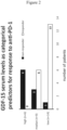
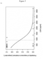
- the probability of a positive clinical outcome to a treatment with immune checkpoint blockers significantly decreases with increasing hGDF-15 levels in the patient sera and vice versa.
- the probability of a positive clinical outcome of a treatment with immune checkpoint blockers inversely correlates with hGDF-15 levels.
- This clinical outcome can, for instance, be a response to the treatment with immune checkpoint blockers or patient survival following the treatment with immune checkpoint blockers.
- hGDF-15 serum levels in melanoma patients are increased by 1 ng/ml, the probability of a response to a treatment with an immune checkpoint blocker decreases by about 60 %. Conversely, if hGDF-15 serum levels in melanoma patients are decreased by 1 ng/ml, the probability of a response to a treatment with an immune checkpoint blocker increases by about 60 %. Similarly, if hGDF-15 serum levels are increased by 1 ng/ml, the patients' probability to die increases by a factor of 1.27.
- hGDF-15 expression is not limited to melanoma but also present in numerous other solid cancers.
- solid tumors other than melanoma can also be treated with immune checkpoint blockers.
- levels of hGDF-15 in blood samples from patients can advantageously be used to predict the probability of a positive clinical outcome of the patients following a treatment with immune checkpoint blockers not only in melanoma, but in all of the solid cancers referred to herein.
- the methods of the invention are also advantageous because they do not require whole-exome sequencing or an existing tumor sample (which may not always be available) but can be based on a simple analysis of a blood sample.
- the present invention provides improved means to predict the clinical outcome of treatments with immune checkpoint blockers as defined in the appended claims.
- antibody refers to any functional antibody that is capable of specific binding to the antigen of interest, as generally outlined in chapter 7 of Paul, W.E. (Ed.).: Fundamental Immunology 2nd Ed. Raven Press, Ltd., New York 1989 .
- the term “antibody” encompasses antibodies from any appropriate source species, including chicken and mammalian such as mouse, goat, non-human primate and human.
- the antibody is a humanized antibody.
- the antibody is preferably a monoclonal antibody which can be prepared by methods well-known in the art.
- the term “antibody” encompasses an IgG-1, -2, -3, or -4, IgE, IgA, IgM, or IgD isotype antibody.
- antibody encompasses monomeric antibodies (such as IgD, IgE, IgG) or oligomeric antibodies (such as IgA or IgM).
- antibody also encompasses - without particular limitations - isolated antibodies and modified antibodies such as genetically engineered antibodies, e.g. chimeric antibodies.
- each monomer of an antibody comprises two heavy chains and two light chains, as generally known in the art.
- each heavy and light chain comprises a variable domain (termed V H for the heavy chain and V L for the light chain) which is important for antigen binding.
- V H variable domain
- V L variable domain
- These heavy and light chain variable domains comprise (in an N-terminal to C-terminal order) the regions FR1, CDR1, FR2, CDR2, FR3, CDR3, and FR4 (FR, framework region; CDR, complementarity determining region which is also known as hypervariable region).
- IMGT/V-QUEST an integrated software program for immunoglobulin and T cell receptor V-J and V-D-J rearrangement analysis. Nucleic Acids Res. 2004 Jul 1;32(Web Server issue):W435-40 .
- the antibody regions indicated above are identified and assigned by using the IMGT/V-QUEST software.
- a “monoclonal antibody” is an antibody from an essentially homogenous population of antibodies, wherein the antibodies are substantially identical in sequence (i.e. identical except for minor fraction of antibodies containing naturally occurring sequence modifications such as amino acid modifications at their N- and C-termini). Unlike polyclonal antibodies which contain a mixture of different antibodies directed to either a single epitope or to numerous different epitopes, monoclonal antibodies are directed to the same epitope and are therefore highly specific.
- the term "monoclonal antibody” includes (but is not limited to) antibodies which are obtained from a monoclonal cell population derived from a single cell clone, as for instance the antibodies generated by the hybridoma method described in Köhler and Milstein (Nature, 1975 Aug 7;256(5517):495-7 ) or Harlow and Lane ("Antibodies: A Laboratory Manual” Cold Spring Harbor Laboratory Press, Cold Spring Harbor, New York 1988 ).
- a monoclonal antibody may also be obtained from other suitable methods, including phage display techniques such as those described in Clackson et al. (Nature. 1991 Aug 15;352(6336):624-8 ) or Marks et al. (J Mol Biol.
- a monoclonal antibody may be an antibody that has been optimized for antigen-binding properties such as decreased Kd values, optimized association and dissociation kinetics by methods known in the art. For instance, Kd values may be optimized by display methods including phage display, resulting in affinity-matured monoclonal antibodies.
- Kd values may be optimized by display methods including phage display, resulting in affinity-matured monoclonal antibodies.
- the term "monoclonal antibody” is not limited to antibody sequences from particular species of origin or from one single species of origin. Thus, the meaning of the term “monoclonal antibody” encompasses chimeric monoclonal antibodies such as humanized monoclonal antibodies.
- Humanized antibodies are antibodies which contain human sequences and a minor portion of non-human sequences which confer binding specificity to an antigen of interest (e.g. human GDF-15).
- humanized antibodies are generated by replacing hypervariable region sequences from a human acceptor antibody by hypervariable region sequences from a non-human donor antibody (e.g. a mouse, rabbit, rat donor antibody) that binds to an antigen of interest (e.g. human GDF-15).
- framework region sequences of the acceptor antibody may also be replaced by the corresponding sequences of the donor antibody.
- a "humanized antibody” may either contain other (additional or substitute) residues or sequences or not.
- Such other residues or sequences may serve to further improve antibody properties such as binding properties (e.g. to decrease Kd values) and/or immunogenic properties (e.g. to decrease antigenicity in humans).
- binding properties e.g. to decrease Kd values
- immunogenic properties e.g. to decrease antigenicity in humans.
- Non-limiting examples for methods to generate humanized antibodies are known in the art, e.g. from Riechmann et al. (Nature. 1988 Mar 24; 332(6162):323-7 ) or Jones et al. (Nature. 1986 May 29-Jun 4; 321 (6069):522-5 ).
- an "antigen-binding portion" of an antibody as used herein refers to a portion of an antibody that retains the capability of the antibody to specifically bind to the antigen (e.g. hGDF-15, PD-1 or PD-L1). This capability can, for instance, be determined by determining the capability of the antigen-binding portion to compete with the antibody for specific binding to the antigen by methods known in the art.
- the antigen-binding portion may contain one or more fragments of the antibody.
- the antigen-binding portion can be produced by any suitable method known in the art, including recombinant DNA methods and preparation by chemical or enzymatic fragmentation of antibodies.
- Antigen-binding portions may be Fab fragments, F(ab') fragments, F(ab') 2 fragments, single chain antibodies (scFv), single-domain antibodies, diabodies or any other portion(s) of the antibody that retain the capability of the antibody to specifically bind to the antigen.
- an “antibody” e.g. a monoclonal antibody
- an “antigen-binding portion” may have been derivatized or be linked to a different molecule.
- molecules that may be linked to the antibody are other proteins (e.g. other antibodies), a molecular label (e.g. a fluorescent, luminescent, colored or radioactive molecule), a pharmaceutical and/or a toxic agent.
- the antibody or antigen-binding portion may be linked directly (e.g. in form of a fusion between two proteins), or via a linker molecule (e.g. any suitable type of chemical linker known in the art).
- the terms "binding" or “bind” refer to specific binding to the antigen of interest (e.g. human GDF-15).
- the Kd value is less than 100 nM, more preferably less than 50 nM, still more preferably less than 10 nM, still more preferably less than 5 nM and most preferably less than 2 nM.
- epitope refers to a small portion of an antigen that forms the binding site for an antibody.
- binding or competitive binding of antibodies or their antigen-binding portions to the antigen of interest is preferably measured by using surface plasmon resonance measurements as a reference standard assay, as described below.
- K D or "K D value” relate to the equilibrium dissociation constant as known in the art. In the context of the present invention, these terms relate to the equilibrium dissociation constant of an antibody with respect to a particular antigen of interest (e.g. human GDF-15)
- the equilibrium dissociation constant is a measure of the propensity of a complex (e.g. an antigen-antibody complex) to reversibly dissociate into its components (e.g. the antigen and the antibody).
- K D values are preferably determined by using surface plasmon resonance measurements as described below.
- an “isolated antibody” as used herein is an antibody that has been identified and separated from the majority of components (by weight) of its source environment, e.g. from the components of a hybridoma cell culture or a different cell culture that was used for its production (e.g. producer cells such as CHO cells that recombinantly express the antibody). The separation is performed such that it sufficiently removes components that may otherwise interfere with the suitability of the antibody for the desired applications (e.g. with a therapeutic use of the anti-human GDF-15 antibody).
- the isolated antibody preparation is at least 70 % pure (w/w), more preferably at least 80 % pure (w/w), still more preferably at least 90 % pure (w/w), still more preferably at least 95 % pure (w/w), and most preferably at least 99 % pure (w/w), as measured by using the Lowry protein assay.
- a “diabody” as used herein is a small bivalent antigen-binding antibody portion which comprises a heavy chain variable domain linked to a light chain variable domain on the same polypeptide chain linked by a peptide linker that is too short to allow pairing between the two domains on the same chain. This results in pairing with the complementary domains of another chain and in the assembly of a dimeric molecule with two antigen binding sites.
- Diabodies may be bivalent and monospecific (such as diabodies with two antigen binding sites for human GDF-15), or may be bivalent and bispecific (e.g. diabodies with two antigen binding sites, one being a binding site for human GDF-15, and the other one being a binding site for a different antigen). A detailed description of diabodies can be found in Holliger P et al. (""Diabodies”: small bivalent and bispecific antibody fragments.” Proc Natl Acad Sci USA. 1993 Jul 15;90(14):6444-8 .).
- a “single-domain antibody” (which is also referred to as “Nanobody TM ”) as used herein is an antibody fragment consisting of a single monomeric variable antibody domain. Structures of and methods for producing single-domain antibodies are known from the art, e.g. from Holt LJ et al. ("Domain antibodies: proteins for therapy.” Trends Biotechnol. 2003 Nov;21(11):484-90 .), Saerens D et al. ("Single-domain antibodies as building blocks for novel therapeutics.” Curr Opin Pharmacol. 2008 Oct;8(5):600-8. Epub 2008 Aug 22 .), and Arbabi Ghahroudi M et al. ("Selection and identification of single domain antibody fragments from camel heavy-chain antibodies.” FEBS Lett. 1997 Sep 15;414(3):521-6 .).
- each occurrence of the term “comprising” may optionally be substituted with the term “consisting of”.
- solid cancers are solid cancers.
- a “solid cancer” is a cancer which forms one or more solid tumors. Such solid cancers forming solid tumors are generally known in the art.
- the term “solid cancer” encompasses both a primary tumor formed by the cancer and possible secondary tumors, which are also known as metastases.
- Preferred solid cancers are selected from the group consisting of melanoma, colorectal cancer, prostate cancer, head and neck cancer, urothelial cancer, stomach cancer, pancreatic cancer, liver cancer, testis cancer, ovarian cancer, endometrial cancer, cervical cancer, brain cancer, breast cancer, gastric cancer, renal cell carcinoma, Ewing's sarcoma, non-small cell lung cancer and small cell lung cancer, preferably selected from the group consisting of melanoma, colorectal cancer, prostate cancer, head and neck cancer, urothelial cancer, stomach cancer, pancreatic cancer, liver cancer, testis cancer, ovarian cancer, endometrial cancer and cervical cancer, more preferably selected from the group consisting of melanoma, colorectal cancer, prostate cancer, head and neck cancer, urothelial cancer and stomach cancer, and most preferably selected from the group consisting of melanoma, colorectal cancer and prostate cancer.
- brain cancer refers to all brain cancers known in the art. It includes but is not limited to glioma (WHO grade I to IV), astrocytoma, meningioma and medulloblastoma.
- treatment of cancer or “treating cancer” refer to a therapeutic treatment.
- a treatment of cancer can be a first-line therapy, a second-line therapy or a third-line therapy or a therapy that is beyond third-line therapy.
- the meaning of these terms is known in the art and in accordance with the terminology that is commonly used by the US National Cancer Institute.
- a treatment of cancer does not exclude that additional or secondary therapeutic benefits also occur in patients.
- an additional or secondary benefit may be an influence on cancer-induced weight loss.
- a "treatment of cancer" as referred to herein is for treating the cancer itself, and that any secondary or additional effects only reflect optional, additional advantages of the treatment of cancer.
- treatment of cancer is preferably a cancer immunotherapy.
- cancer immunotherapy is known in the art and generally relates to a treatment of cancer in which the immune system of the patient is used to treat the cancer. Cancer cells harbor genomic mutations which give rise to cancer cell antigens that are specific to the cancer cells and different from the antigens of non-cancerous cells. Thus, a cancer immunotherapy referred to in accordance with the invention is preferably a cancer immunotherapy wherein such cancer cell antigens are recognized by the immune system, and wherein cancer cells expressing these antigens are killed by the immune system.
- a cancer immunotherapy can be assessed by immunomonitoring methods known in the art, e.g. by measuring intracellular IFN- ⁇ expression (e.g.
- CD8 + T-cells and/or NK cells in blood samples, measuring CD107a cell surface expression (e.g. on CD8 + T-cells and/or NK cells) in blood samples, measuring intracellular TNF- ⁇ expression (e.g. on leukocytes) in blood samples, intracellular Interleukin-2 expression (e.g. in CD8 + T-cells and/or in CD4 + T-cells) in blood samples, CD154 cell surface expression (e.g.
- a "cancer immunotherapy” optionally encompasses a treatment where in addition to the immune system which is used to treat the cancer, additional mechanisms of cancer treatment are used.
- a cancer immunotherapy where additional mechanisms of cancer treatment can be used is a combination therapy with known chemotherapeutic agent(s).
- Such combination therapy with known chemotherapeutic agent(s) may, for instance, not only include the treatment of cancer in which the immune system is used to treat the cancer but also include a treatment of cancer in which the cancer cells are killed by said chemotherapeutic agent(s) directly.
- an “immune checkpoint blocker treatment” is a treatment with one or more immune checkpoint blockers as indicated below.
- the methods for predicting according to the invention are preferably carried out prior to the start of the immune checkpoint blocker treatment.
- the methods for predicting according to the invention can also be carried out at a point in time where the immune checkpoint blocker treatment has already been started.
- the methods for predicting according to the invention may be used for patients who are subject to an immune checkpoint blocker treatment of the solid cancer or a different treatment of the solid cancer (e.g. a treatment with other agents which are pharmaceutically active against cancer). It is, however, also understood that neither immune checkpoint blocker treatment steps nor any other treatment steps form part of the methods for predicting according to the present invention.
- an agent pharmaceutically active against cancer may, for instance, be a known anticancer agent and/or an immune-stimulatory molecule.
- known anticancer agents include but are not limited to alkylating agents such as cisplatin, carboplatin, oxaliplatin, mechlorethamine, cyclophosphamide, chlorambucil, and ifosfamide; anti-metabolites such as azathioprine and mercaptopurine; alkaloids such as vinca alkaloids (e.g. vincristine, vinblastine, vinorelbine, and vindesine), taxanes (e.g.
- topoisomerase inhibitors such as camptothecins (e.g. irinotecan and topotecan); cytotoxic antibiotics such as actinomycin, anthracyclines, doxorubicin, daunorubicin, valrubicin, idarubicin, epirubicin, bleomycin, plicamycin and mitomycin; and radioisotopes.
- camptothecins e.g. irinotecan and topotecan
- cytotoxic antibiotics such as actinomycin, anthracyclines, doxorubicin, daunorubicin, valrubicin, idarubicin, epirubicin, bleomycin, plicamycin and mitomycin; and radioisotopes.
- blood sample includes, without limitation, whole blood, serum and plasma samples. It also includes other sample types such as blood fractions other than serum and plasma. Such samples and fractions are known in the art.
- Blood samples which are used for the methods according to the invention can be any types of blood samples which contain hGDF-15. Suitable types of blood samples containing hGDF-15 are known in the art and include serum and plasma samples. Alternatively, further types of blood samples which contain hGDF-15 can also be readily identified by the skilled person, e.g. by measuring whether hGDF-15 is contained in these samples, and which levels of hGDF-15 are contained in these samples, by using the methods disclosed herein.
- the human blood sample obtained from the human patient is a sample from a patient who has received said immune checkpoint blocker
- the blood sample obtained from the human patient may contain the immune checkpoint blocker and/or biological metabolites thereof.
- Immune checkpoint blockers are known in the art, and their presence can be determined by the skilled person.
- Examples of metabolites of drugs such as metabolites of therapeutic antibodies are also known and can be detected, e.g. by using appropriate secondary antibodies.
- levels of hGDF-15 in human blood samples can be used to predict the probability of a positive clinical outcome of a treatment with an immune checkpoint blocker in a human cancer patient.
- a "positive clinical outcome" can be any therapeutic indicator for a therapeutic treatment benefit.
- Such indicators are well-known in the art.
- a positive clinical outcome can be a treatment response of the human cancer patient to the immune checkpoint blocker treatment.
- the presence or absence of a treatment response is assessed by an assessment of whether the treatment inhibits cancer growth in the treated patient or patients.
- the inhibition is statistically significant as assessed by appropriate statistical tests which are known in the art.
- Inhibition of cancer growth may be assessed by comparing cancer growth in a group of patients treated in accordance with the present invention to a control group of untreated patients, or by comparing a group of patients that receive a standard cancer treatment of the art plus a treatment according to the invention with a control group of patients that only receive a standard cancer treatment of the art.
- Such studies for assessing the inhibition of cancer growth are designed in accordance with accepted standards for clinical studies, e.g. double-blinded, randomized studies with sufficient statistical power.
- cancer growth as used herein relates to any measureable growth of the cancer.
- cancer growth relates to a measurable increase in tumor volume over time. If the cancer has formed only a single tumor, “cancer growth” relates only to the increase in volume of the single tumor. If the cancer has formed multiple tumors such as metastases, “cancer growth” relates to the increase in volume of all measurable tumors.
- the tumor volume can be measured by any method known in the art, including magnetic resonance imaging and computed tomography (CT scan).
- an assessment of a response to the treatment is made based on a classification of responders and non-responders by using the response evaluation criteria in solid tumours, version 1.1 (RECIST v1.1) ( Eisenhauer et al.: New response evaluation criteria in solid tumours: revised RECIST guideline (version 1.1). In: Eur. J. Cancer. 45, No. 2, January 2009, pp. 228-47 ). Likewise, these criteria are also very preferred for the prediction methods according to the invention.
- a treatment response may be assessed at a time point of 1, 2, 3, 4, 5, 6, 7, 8, 9, 10, 11, 12, 18 and/or 24 months after start of the treatment with the immune checkpoint blocker.
- a treatment response is assessed after 12 weeks or after 4 months or after 6 months after start of the treatment with the immune checkpoint blocker.
- a prediction of a probability of a treatment response according to the invention may be provided for one or more of the above time points.
- a positive clinical outcome can be survival of the human cancer patient following the immune checkpoint blocker treatment. Survival of patient groups can be analysed by methods known in the art, e.g. by Kaplan-Meier curves.
- survival preferably short-term survival
- survival may, for instance, be assessed at a time point of 1 month, 6 weeks, 2 months, 3 months, 4 months, 5 months, 6 months, 7 months, 8 months, 9 months, 10 months, 11 months, 12 months and/or 18 months after start of the treatment with the immune checkpoint blocker.
- Short-term survival is preferably assessed after 6 weeks, 2 months, 3 months, 4 months, 5 months, or 6 months after start of the treatment with the immune checkpoint blocker.
- survival preferably long-term survival
- survival may, for instance, be assessed at a time point of 24 months, 30 months, 36 months, 42 months, 48 months, 54 months, 60 months, 6 years, 7 years, 8 years, 9 years and/or 10 years after start of the treatment with the immune checkpoint blocker.
- long-term survival is preferably assessed at a time point of 36 months after start of the treatment with the immune checkpoint blocker.
- a prediction of a probability of survival according to the invention may be provided for one or more of these time points.
- patient data from one or more clinical studies which have treated patients suffering from solid cancers with immune checkpoint blockers, can be used as a basis to generate statistical models. These models can then be used to determine appropriate hGDF-15 threshold levels for predicting the probability of a positive clinical outcome.
- survival can be analyzed by Cox proportional hazard survival models, e.g. by fitting the model with the hGDF-15 level (ng/ml) as continuous predictor.
- HR Hazard ratio
- GDF-15 continuous predictor
- the probability of survival is predicted using a grouping variable based on GDF-15 as categorical predictor, e.g. with groups ⁇ 1.8 ng/ml, 1.8 ⁇ 4.2 ng/ml, >4.2 ng/ml).
- hGDF-15 threshold levels can be used.
- the inverse relationship between hGDF-15 levels and a positive clinical outcome applies to any threshold value, and hence the invention is not limited to particular threshold values.
- hGDF-15 threshold levels according to the present invention can be obtained, and/or further adjusted, by using the above-mentioned statistical methods for predicting the probability of a positive clinical outcome.
- An hGDF-15 threshold level may be a single hGDF-15 threshold level.
- the invention also encompasses the use of more than one hGDF-15 threshold level, e.g. 2, 3, 4, 5, 6, 7, 8, 9, 10 or more hGDF-15 threshold levels.
- the invention also encompasses the use of a continuum of hGDF-15 threshold levels. A non-limiting example of such a continuum of hGDF-15 threshold levels is given in Figure 3 .
- a corresponding probability of a positive clinical outcome (e.g. probability of survival or probability of a treatment response) can be predicted. If the level of hGDF-15 in the blood sample is decreased compared to such as a hGDF-15 threshold level according to the invention, this indicates that the probability of a positive clinical outcome (e.g. the probability of survival or the probability of a treatment response) is increased, i.e. increased compared to the probability of a positive clinical outcome which can be predicted for said hGDF-15 threshold level.
- Figure 3 of Example 1 illustrates a continuum of hGDF-15 threshold levels and a curve of corresponding predicted probabilities for a treatment response.
- the probability of a treatment response can be predicted based on an odds ratio such as the odds ratio shown in Table 2.
- the probability of a treatment response can be predicted using an odds ratio of 0.389 for serum levels of hGDF-15 in ng/ml as a continuous predictor with a confidence interval of from 0.159 to 0.698.
- the median hGDF-15 serum level of healthy human control individuals is ⁇ 0.8 ng/ml.
- the expected range is between 0.2 ng/ml and 1.2 ng/ml in healthy human controls (Reference: Tanno T et al.: "Growth differentiation factor 15 in erythroid health and disease.” Curr Opin Hematol. 2010 May; 17(3): 184-190 .).
- the cancer of the patient is melanoma.
- the hGDF-15 threshold level is a hGDF-15 level selected from the range of between 3.0 ng/ml and 4.0 ng/ml, preferably a hGDF-15 level selected from the range of between 3.2 ng/ml and 3.7 ng/ml, and most preferably a hGDF-15 level of 3.4 ng/ml.
- hGDF-15 serum levels and based on the disclosure of the invention provided herein, corresponding hGDF-15 levels in other blood samples can be routinely obtained by the skilled person (e.g. by comparing the relative level of hGDF-15 in serum with the respective level in other blood samples).
- the present invention also encompasses preferred hGDF-15 levels in plasma, whole blood and other blood samples, which correspond to each of the preferred hGDF-15 serum levels and ranges indicated above.
- Antibodies capable of binding to hGDF-15 which can be used in accordance with the invention
- the methods, apparatuses and kits used in the invention may use one or more antibodies capable of binding to hGDF-15 or an antigen-binding portion thereof, as defined above.
- human GDF-15 protein can be advantageously targeted by a monoclonal antibody ( WO2014/049087 ), and that such antibody has advantageous properties including a high binding affinity to human GDF-15, as demonstrated by an equilibrium dissociation constant of about 790pM for recombinant human GDF-15 (see Reference Example 1).
- the invention uses an antibody capable of binding to hGDF-15, or an antigen-binding portion thereof.
- the antibody is a monoclonal antibody capable of binding to hGDF-15, or an antigen-binding portion thereof.
- the antibody or antigen-binding portion thereof comprises a heavy chain variable domain which comprises a CDR1 region comprising the amino acid sequence of SEQ ID NO: 3 and a CDR2 region comprising the amino acid sequence of SEQ ID NO: 4, and the antibody or antigen-binding portion thereof comprises a light chain variable domain which comprises a CDR1 region comprising the amino acid sequence of SEQ ID NO: 6, and a CDR2 region comprising the amino acid sequence ser-ala-ser.
- the antibody capable of binding to hGDF-15 or antigen-binding portion thereof is a monoclonal antibody capable of binding to human GDF-15, or an antigen-binding portion thereof, wherein the antibody or antigen-binding portion thereof comprises a heavy chain variable domain which comprises a CDR1 region comprising the amino acid sequence of SEQ ID NO: 3, a CDR2 region comprising the amino acid sequence of SEQ ID NO: 4 and a CDR3 region comprising the amino acid sequence of SEQ ID NO: 5, and wherein the antibody or antigen-binding portion thereof comprises a light chain variable domain which comprises a CDR1 region comprising the amino acid sequence of SEQ ID NO: 6, a CDR2 region comprising the amino acid sequence ser-ala-ser and a CDR3 region comprising the amino acid sequence of SEQ ID NO: 7.
- the heavy chain variable domain comprises a CDR1 region comprising the amino acid sequence of SEQ ID NO: 3 and a CDR2 region comprising the amino acid sequence of SEQ ID NO: 4
- the light chain variable domain comprises a CDR1 region comprising the amino acid sequence of SEQ ID NO: 6 and a CDR2 region comprising the amino acid sequence of SEQ ID NO: 7.
- the antibody may have CDR3 sequences as defined above.
- the monoclonal antibody capable of binding to human GDF-15, or an antigen-binding portion thereof is a single-domain antibody (also referred to as "Nanobody TM ").
- the single-domain antibody comprises the CDR1, CDR2, and CDR3 amino acid sequences of SEQ ID NO: 3, SEQ ID NO: 4, and SEQ ID NO: 5, respectively.
- the single-domain antibody comprises the CDR1, CDR2, and CDR3 amino acid sequences of SEQ ID NO: 6, ser-ala-ser, and SEQ ID NO: 7, respectively.
- the single-domain antibody is a humanized antibody.
- the antibodies capable of binding to human GDF-15 or the antigen-binding portions thereof have an equilibrium dissociation constant for human GDF-15 that is equal to or less than 100 nM, less than 20 nM, preferably less than 10 nM, more preferably less than 5 nM and most preferably between 0.1 nM and 2 nM.
- the antibody capable of binding to human GDF-15 or an antigen-binding portion thereof binds to the same human GDF-15 epitope as the antibody to human GDF-15 obtainable from the cell line B1-23 deposited with the Deutsche Sammlung für Mikroorganismen und Zellkulturen GmbH (DMSZ) under the accession No. DSM ACC3142.
- DMSZ Deutsche Sammlung für Mikroorganismen und Zellkulturen GmbH
- antibody binding to human GDF-15 in accordance with the present invention is preferably assessed by surface plasmon resonance measurements as a reference standard method, in accordance with the procedures described in Reference Example 1.
- Binding to the same epitope on human GDF-15 can be assessed similarly by surface plasmon resonance competitive binding experiments of the antibody to human GDF-15 obtainable from the cell line B1-23 and the antibody that is expected to bind to the same human GDF-15 epitope as the antibody to human GDF-15 obtainable from the cell line B1-23.
- the antibody capable of binding to human GDF-15 or the antigen-binding portion thereof is a monoclonal antibody capable of binding to human GDF-15, or an antigen-binding portion thereof, wherein the binding is binding to a conformational or discontinuous epitope on human GDF-15 comprised by the amino acid sequences of SEQ ID No: 25 and SEQ ID No: 26.
- the antibody or antigen-binding portion thereof is an antibody or antigen-binding portion thereof as defined by the sequences of any one of the above.
- Antibodies including the antibody capable of binding to human GDF-15 or the antigen-binding portion thereof can be modified, e.g. by a tag or a label.
- label relates to any molecule or group of molecules which can facilitate detection of the antibody.
- labels may be enzymatic such as horseradish peroxidase (HRP), alkaline phosphatase (AP) or glucose oxidase.
- HRP horseradish peroxidase
- AP alkaline phosphatase
- Enzymatically labelled antibodies may, for instance, be employed in enzyme-linked immunosorbent assays. Labels may also be radioactive isotopes, DNA sequences (which may, for instance, be used to detect the antibodies by polymerase chain reaction (PCR)), fluorogenic reporters and electrochemiluminescent groups (e.g. ruthenium complexes).
- PCR polymerase chain reaction
- fluorogenic reporters e.g. ruthenium complexes
- electrochemiluminescent groups e.g. ruthenium complexes.
- antibodies used according to the invention in particular an antibody capable of binding to human GDF-15 or the antigen-binding portion thereof
- the present invention relates to the prediction of the probability of a positive clinical outcome of a treatment with an immune checkpoint blocker in human cancer patient, in particular to the prediction of the probability of a treatment response of a human cancer patient to an immune checkpoint blocker treatment and to the prediction of the probability of survival of a human cancer patient following an immune checkpoint blocker treatment.
- an “immune checkpoint” as used herein generally means an immunological mechanism by which an immune response can be inhibited. More particularly, an immune checkpoint is a mechanism which is characterized in that a molecule of the immune system (or a group of molecules of the immune system) inhibits the immune response by inhibiting the activation of cells of the immune system. Such molecule (or group of molecules) of the immune system which inhibits (inhibit) the immune response by inhibiting the activation of cells of the immune system is (are) also known as checkpoint molecule(s).
- an “immune checkpoint blocker” is a molecule which is capable of blocking an immune checkpoint.
- the term “immune checkpoint blocker” as used herein does not refer to an hGDF-15 inhibitor such as an antibody capable of binding to hGDF-15 but means a molecule which is different from an hGDF-15 inhibitor.
- the most common immune checkpoint blockers which are known to date are inhibitors of immune checkpoint molecules such as inhibitors of human PD-1 and inhibitors of human PD-L1. Further immune checkpoint blockers are anti-LAG-3, anti-B7H3, anti-TIM3, anti-VISTA, anti-TIGIT, anti-KIR, anti-CD27, anti-CD137 as well as inhibitors of IDO. Therefore, a preferred form of an immune checkpoint blocker is an inhibitor of an immune checkpoint molecule. Alternatively, an immune checkpoint blocker can be an activator of a co-stimulating signal which overrides the immune checkpoint.
- immune checkpoint blockers are inhibitors of human PD-1 and inhibitors of human PD-L1.
- the methods used in the present disclosure are performed in accordance with procedures known in the art, e.g. the procedures described in Sambrook et al. ("Molecular Cloning: A Laboratory Manual.”, 2nd Ed., Cold Spring Harbor Laboratory Press, Cold Spring Harbor, New York 1989 ), Ausubel et al. ("Current Protocols in Molecular Biology.” Greene Publishing Associates and Wiley Interscience; New York 1992 ), and Harlow and Lane (“Antibodies: A Laboratory Manual” Cold Spring Harbor Laboratory Press, Cold Spring Harbor, New York 1988 ).
- Binding of antibodies to their respective target proteins can be assessed by methods known in the art.
- the binding of monoclonal antibodies to their respective targets is preferably assessed by surface plasmon resonance measurements. These measurements are preferably carried out by using a Biorad ProteOn XPR36 system and Biorad GLC sensor chips, as exemplified for anti-human GDF-15 mAb-B1-23 in Reference Example 1.
- Sequence Alignments of sequences according to the invention are performed by using the BLAST algorithm (see Altschul et al.(1990) "Basic local alignment search tool.” Journal of Molecular Biology 215. p. 403-410 .; Altschul et al.: (1997) Gapped BLAST and PSI-BLAST: a new generation of protein database search programs. Nucleic Acids Res. 25:3389-3402 .).
- Max target sequences 10 Word size 3; BLOSUM 62 matrix; gap costs: existence 11, extension 1; conditional compositional score matrix adjustment.
- identity refers to the identity value obtained by using the BLAST algorithm.
- Monoclonal antibodies can be produced by any method known in the art, including but not limited to the methods referred to in Siegel DL ("Recombinant monoclonal antibody technology.” Transfus Clin Biol. 2002 Jan;9(1):1 5-22 .).
- an antibody according to the disclosure is produced by the hybridoma cell line B1-23 deposited with the Deutsche Sammlung für Mikroorganismen und Zellkulturen GmbH (DSMZ) under the accession No. DSM ACC3142 under the Budapest treaty. The deposit was filed on September 29, 2011.
- hGDF-15 levels of human GDF-15 can be measured by any method known in the art, including measurements of hGDF-15 protein levels by methods including (but not limited to) mass spectrometry for proteins or peptides derived from human GDF-15, Western Blotting using antibodies specific to human GDF-15, strip tests using antibodies specific to human GDF-15, or immunocytochemistry using antibodies specific to human GDF-15.
- a preferred method of measuring hGDF-15 serum levels is a measurement of hGDF-15 serum levels by Enzyme-Linked Immunosorbent Assay (ELISA) by using antibodies to GDF-15. Such ELISA methods are exemplified in Example 1.
- hGDF-15 serum levels may be determined by known electrochemiluminesence immunoassays using antibodies to GDF-15. For instance, the Roche Elecsys ® technology can be used for such electrochemiluminesence immunoassays.
- An apparatus can be any apparatus which is configured to perform the methods of the disclosure.
- the term "configured to perform” means that the apparatus is specifically configured for the recited method steps. For instance, an apparatus configured to perform a method which uses a particular threshold level will be specifically configured to use that particular threshold. For ELISA measurement, any reader able to measure absorption would be suitable. For bead-based assays, a Luminex analyser or a flow cytometer could be used.
- the apparatus is an electrochemiluminescence (e.g., Elecsys ® ) analyzer such as a Cobas ® analyzer, including but not limited to analyzers of the Cobas ® 4000, Cobas ® 6000, Cobas ® 8000, Cobas c 111, and the Cobas INTEGRA ® 400 plus series.
- Elecsys ® electrochemiluminescence
- the invention also relates to use of kits in the methods of the invention detailed in the appended claims.
- the recombinant hGDF-15 contained in the kits may be present in a form which can conveniently be used for calibration purposes. For instance, it may be present in the form of stock solutions which cover several concentrations in the range of 0 to 15 ng/ml, e.g. at least one concentration in the range of 0-1 ng/ml, at least one concentration in the range of 1-3 ng/ml, at least one concentration in the range of 3-6 ng/ml, and preferably at least one further concentration in the range of 6-10 ng/ml, and more preferably further comprising at least one further concentration in the range of 10-15 ng/ml.
- stock solutions which cover several concentrations in the range of 0 to 15 ng/ml, e.g. at least one concentration in the range of 0-1 ng/ml, at least one concentration in the range of 1-3 ng/ml, at least one concentration in the range of 3-6 ng/ml, and preferably at least one further concentration in the range of 6-10 ng/
- amino acid sequences referred to in the present application are as follows (in an N-terminal to C-terminal order; represented in the one-letter amino acid code):
- nucleic acid sequences referred to in the present application are as follows (in a 5' to 3' order; represented in accordance with the standard nucleic acid code):
- the hybridoma cell line B1-23 producing mAb-B1-23 was deposited by the Julius-Maximilians-Universtician Würzburg, Sanderring 2, 97070 Würzburg, Germany, with the Deutsche Sammlung für Mikroorganismen und Zellkulturen GmbH (DMSZ) under the accession No. DSM ACC3142, in accordance with the Budapest Treaty.
- DSMZ Deutsche Sammlung für Mikroorganismen und Zellkulturen GmbH
- B1-23 was identified as an IgG2a (kappa chain) isotype.
- the dissociation constant (Kd) was determined as follows: Binding of the monoclonal anti-human-GDF-15 antibody anti-human GDF-15 mAb-B1-23 was measured by employing surface plasmon resonance measurements using a Biorad ProteOn XPR36 system and Biorad GLC sensor chips: For preparing the biosensors recombinant mature human GDF-15 protein was immobilized on flow cells 1 and 2. On one flow cell recombinant GDF-15 derived from Baculvirus-transfected insect cells (HighFive insect cells) and on the other recombinant protein derived from expression in E. coli was used.
- the non-reacted coupling groups were then quenched by perfusion with 1M ethanolamine pH 8.5 and the biosensor was equilibrated by perfusing the chip with running buffer (10M HEPES, 150mM NaCl, 3.4mM EDTA, 0.005% Tween-20, pH 7.4, referred to as HBS150).
- running buffer 10M HEPES, 150mM NaCl, 3.4mM EDTA, 0.005% Tween-20, pH 7.4, referred to as HBS150.
- HBS150 running buffer
- two flow cells were used, one empty with no protein coupled and one coupled with an non-physiological protein partner (human Interleukin-5), which was immobilized using the same coupling chemistry and the same coupling density.
- anti-human GDF-15 mAb-B1-23 was dissolved in HBS150 and used in six different concentrations as analyte (concentration: 0.4, 0.8, 3, 12, 49 und 98 nM).
- the analyte was perfused over the biosensor using the one-shot kinetics setup to avoid intermittent regeneration, all measurements were performed at 25°C and using a flow rate of 100 ⁇ l min -1 .
- For processing the bulk face effect and unspecific binding to the sensor matrix was removed by subtracting the SPR data of the empty flow cell (flow cell 3) from all other SPR data.
- the resulting sensogram was analyzed using the software ProteOn Manager version 3.0. For analysis of the binding kinetics a 1:1 Langmuir-type interaction was assumed.
- the anti-human GDF-15 mAb-B1-23 shows no binding to human interleukin-5 and thus confirms the specificity of the interaction data and the anti-human GDF-15 mAb-B1-23.
- amino acid sequence of recombinant human GDF-15 (as expressed in Baculovirus-transfected insect cells) is: (SEQ ID No: 8)
- the dissociation constant (Kd) of 790pM was determined.
- mAb B1-23 inhibits cancer cell proliferation in vitro, and that mAb B1-23 inhibits growth of tumors in vivo ( WO2014/049087 ).
- Reference Example 2 mAb B1-23 recognizes a conformational or a discontinuous epitope of human GDF-15
- the peptide array with 10, 12 and 15mer B7H3-derived linear peptides was incubated with secondary goat anti-mouse IgG (H+L) IRDye680 antibody only at a dilution of 1:5000 for 1h at room temperature to analyze background interactions of the secondary antibody.
- the PEPperCHIP ® was washed 2x1 min with standard buffer, rinsed with dist. water and dried in a stream of air.
- Example 1 In human melanoma patients who had received a prior treatment with Ipilimumab (a monoclonal anti-CTLA4 antibody) and failed to show a complete response, and who received a treatment with Pembrolizumab (a monoclonal anti-PD-1 antibody), hGDF-15 Serum Levels correlate with poor treatment response at a time point of four months after the start of the treatment with pembrolizumab.
- Ipilimumab a monoclonal anti-CTLA4 antibody
- Pembrolizumab a monoclonal anti-PD-1 antibody
- Eligible patients were aged 18 years or older and had histologically or cytologically confirmed unresectable stage III or stage IV melanoma not amenable to local therapy; confirmed disease progression within 24 weeks of the last ipilimumab dose (minimum two doses, 3 mg/kg once every 3 weeks); previous BRAF or MEK inhibitor therapy or both (if BRAFV600 mutant-positive); resolution or improvement of ipilimumab-related adverse events to grade 0-1 and prednisone dose 10 mg/day or less for at least 2 weeks before the first dose of study drug; Eastern Cooperative Oncology Group (ECOG) performance status 0 or 1; measurable disease per Response Evaluation Criteria in Solid Tumors, version 1.1 (RECIST v1.1); and values within the prespecified range for absolute neutrophil count ( ⁇ 1500 cells per mL), platelets ( ⁇ 100 000 cells per mL), haemoglobin ( ⁇ 90 g/L), serum creatinine ( ⁇ 1 ⁇ 5 upper limit of normal [ULN]
- Figure 1 shows the GDF-15 serum levels for responders and non-responders to the treatment regimen. All serum samples had been obtained prior to treatment with the anti PD-1 antibody. As can be seen from the Figure, most of the non-responders have higher GDF-15 serum levels than all of the responders.
- Figure 2 shows the numbers of responders and non-responders in the patients having hGDF-15 serum levels of ⁇ 1.8 ng/ml, 1.8-4.2 ng/ml, and >4.2 ng/ml, respectively.
- Tables 1-2 show the results from the models with GDF-15 as continuous predictor.
- Figure 3 shows the corresponding data on responders/non-responders as well as the probability of response to treatment predicted by the model.
- Table 3 shows the result from the Cox proportional hazards model with the group based on GDF-15 as categorical predictor.
- the group with GDF-15 ⁇ 1.8 ng/ml is used as reference group (not shown in the Table).
- the two hazard ratios in Table 3 represent the comparison of the group with GDF-15 between 1.8 and 4.2 and the group with GDF-15 >4.2 with the reference group.
- the hazard for death is increased in both of these groups (compared to the reference group), but to a larger extent in the group with GDF-15 >4.2.
- Figure4A shows the Kaplan-Meier curves for survival in the three groups.
- Table 1 shows the Hazard ratio (HR) estimates from the Cox proportional hazards model with overall survival (time to death) as outcome variable and GDF-15 as continuous predictor. The analysis included samples from 35 patients.
- Table 2 Estimate (OR) 95% CI z p (Intercept) 25.281 [4.219,364.950] 2.94 0.00324 GDF-15 0.389 [0.159,0.698] -2.54 0.01120
- Table 2 shows the Odds ratio (OR) estimates from the Generalized Linear Model with response to treatment (responder 1) as outcome variable and GDF-15 as continuous predictor.
- the analysis included samples from 29 patients.
- Table 3 HR 95% CI z p CDF-15-group(1.8,4.2] 1.54 [0.48,4.92] 0.73 0.466 GDF-15-group(4.2,13] 21.52 [5.20,89.06] 4.24 ⁇ 0.001
- Table 3 shows Hazard ratio (HR) estimates from the Cox proportional hazards model with overall survival (time to death) as outcome variable and the group based on GDF-15 as categorical predictor. The analysis included samples from 35 patients.
- Table 4 Variable Levels n [0,1.8] % [0,1.8] n (1.8,4.2] % (1.8,4.2] n (4..2,13] % (4.2,13] n all % all death 0 11 61.1 6 54.5 0 0.0 17 48.6 1 7 38.9 5 45.5 6 100.0 18 51.4 all 18 100.0 11 100.0 6 100.0 35 100.0 responder1 0 1 7.1 3 33.3 6 100.0 10 34.5 1 13 92.9 6 66.7 0 0.0 19 65.5 all 14 100.0 9 100.0 6 100.0 29 100.0
- Table 4 shows the number of deaths and responders ("responder1") in the three groups defined by the GDF-15 ( ⁇ 1.8,1.8-4.2, >4.2 ng/ml).
- Table 5 Variable Levels n x ⁇ x ⁇ s Min Max GDF-15 [0,1.8] 18 0.9 0.9 0.4 0.3 1.6 (1.8,4.2) 11 2.8 2.9 0.8 2.0 4.0 (4.2,13] 6 7.1 7.4 2.9 4.6 12.0 all 35 1.6 2.6 2.7 0.3 12.0
- Table 5 The continuous predictor variable GDF-15 (ng/ml) in the three groups defined by the GDF-15 ( ⁇ 1.8, 1.8-4.2, >4.2 ng/ml). The number of patients (n), the median (x ⁇ ), the mean (x ), the standard deviation (s), the minimum (Min), and the maximum (Max) are shown.
- LDH may be interpreted as reflecting high tumor burden or tumor aggressiveness ( Zhang, J., Yao, Y.-H., Li, B.-G., Yang, Q., Zhang, P.-Y., and Wang, H.-T. (2015). Prognostic value of pretreatment serum lactate dehydrogenase level in patients with solid tumors: a systematic review and meta-analysis. Scientific Reports 5, 9800 ). As serum LDH levels have been incorporated into the staging scheme for melanoma, this parameter is routinely measured during clinical diagnostics by the university reference laboratory.
- Table 7 is analogue to Table 2, except that LDH was used as continuous predictor of response to treatment (responder1) instead of GDF-15.
- Figure 5 shows the corresponding data on responders/ non-responders as well as the probability of response to treatment predicted by the model.
- GDF-15 is the better predictor of response to treatment (responder1) than LDH
- two additional models were fitted: a model containing both markers as predictors (which automatically only includes patients with measurements on both markers), and a model with GDF-15 as the only predictor but also only using the patients with a measurement of LDH.
- AIC Akaike's information criterion
- Table 7 Odds ratio (OR) estimates from the Generalized Linear Model with response to treatment (responder 1, as defined in file A) as outcome variable and LDH as continuous predictor. The analysis included samples from 25 patients.
- Table 8 df AIC Model with LDH and GDF-15 3.00 25.10 Model with LDH only 2.00 28.55 Model with GDF-15 only 2.00 23.77
- Table 8 Model comparison based on Akaike's information criterion (AIC) of which smaller values indicate a more efficient model.
- df degrees of freedom. All models included samples from 25 patients.
- Figure 5A shows the probability of response to treatment (responder 1) as predicted by the Generalized Linear Model model using LDH as continous predictor. Circles show the data, the curve shows the model. The vertical line indicates the LDH concentration where the probability of treatment response is 0.5.
- the patient cohort was identical. However, reliable determination of LDH levels failed in four patients due to hemolysis.
- Figure 5B shows a graphical representation of responders and non-responders and their respective hGDF-15 and LDH levels. When cut-off values are selected to cover all responders, testing based on GDF-15 allows for identification of 6 (out of 9) non-responders whereas analyses based on LDH levels can only discriminate 4 (out of 9) non-responders. For LDH testing, 4 hemolytic samples had to be excluded which causes loss of data.
- a prediction of a clinical outcome based on hGDF-15 levels includes the following advantages over the diagnostic standard marker LDH for solid tumors:
- LDH is currently considered as the best available clinical marker for solid tumors.
- Example 1 showed that the probability of a response to the treatment significantly decreases with increasing hGDF-15 levels in the patient sera.
- the hazard ratio of 1.27 shown in Table 1 indicates that if hGDF-15 serum levels are increased by 1 ng/ml, the patients' probability to die increases by a factor of 1.27.
- Example 1 The results of Example 1 indicate that there is a strong inverse correlation between the serum levels of hGDF-15 and the probability of a positive clinical outcome of anti PD-1 based immunotherapy in the patients, including patient response and patient survival.
- levels of hGDF-15 in blood samples from patients can advantageously be used to predict the probability of a response of patients to a treatment with immune checkpoint blockers , as defined in the appended claims. While the present Example shows results for melanoma as an example of a solid tumor, hGDF-15 expression is not limited to melanoma but also present in numerous other solid cancers. Likewise, it is known that solid tumors other than melanoma can also be treated with immune checkpoint blockers.
- levels of hGDF-15 in blood samples from patients can advantageously be used to predict the probability of a response of patients to a treatment with immune checkpoint blockers not only in melanoma, but in all of the solid cancers referred to herein.
- Example 2 GDF-15 levels inversely correlate with CD8 + tumor infiltrating lymphocytes (TILs) in metastases of different tumor entities.
- TILs tumor infiltrating lymphocytes
- hGDF-15 In order to identify a mechanism of hGDF-15 that contributes to the negative effect of hGDF-15 on the patients' responses, brain metastases from different solid tumors were analyzed for the expression of hGDF-15 and for the presence of cells of the immune system:
- FFPE Formalin-fixed and paraffin-embedded
- Figure 6 shows exemplary tissue sections from melanoma brain metastases having high no (upper panel) or high (lower panel) GDF-15 immunoreactivity, which were stained by immunohistochemistry for GDF-15 and for the T-cell marker proteins CD3 and CD8, respectively, as indicated in the Figure.
- the numerous infiltrating immune cells are seen as dark spots.
- the scarce infiltrating immune cells are depicted by arrows (CD3 and CD8-positive cells are indicated by arrows).
- Figure 8 shows a plot of the GDF-15 score against the percentage of CD8 + and CD3 + T cells, respectively, in 168 (for CD3) or, respectively, 169 (for CD8) brain metastases from different tumor entities (melanoma, CRC, RCC, breast cancer, NSCLC and SCLC). The plot was obtained as described above in the "statistical analyses" section.
- hGDF-15 decreases the percentage of CD8 + T lymphocytes in solid tumors including tumor metastases, thereby decreasing the probability of a favorable clinical outcome (e.g. patient survival or the presence of a treatment response). Since this correlation is observed across various solid cancer entities, the present invention is not limited to particular solid cancers such as melanoma.
- the invention can be applied to all of the solid tumors as referred to in the preferred embodiments.
- Example 3 GDF-15 decreases adhesion of T cells to endothelial cells.
- the inventors next set out to determine how hGDF-15 affects the percentage of T cells in the solid tumors.
- a step which is required for the invasion of T cells from the blood stream into the tumor tissue is that the T cells must first adhere to the endothelium before they can enter the tumor.
- the inventors used a model system which measures the adhesion of T cells to Human Umbilical Vein Endothelial Cells (HUVEC):
- Recombinant GDF-15 was obtained from Invigate GmbH, Jena, Germany.
- hGDF-15 acts both on the T cells and on the endothelial cells, but they also indicate that the main adhesion effect of hGDF-15 is an effect on the T cells.
- hGDF-15 which is secreted by tumor cells, on T-cell adhesion could be inhibited with an hGDF-15 inhibitor.
- the inventors used an hGDF-15-secreting melanoma cell line, UACC257:
- FIG. 10 This Figure shows analyses of the number of rolling T cells per field of view per second.
- the data were obtained from channel # 1 (control T cells on unstimulated HUVEC as "neg. control”), channel # 2 (control T cells on stimulated HUVEC as “pos. control”), channel # 3 ("GDF-15") channel # 4 ("UACC 257”: T cells cultured in the supernatant of UACC 257 melanoma cells containing secreted GDF-15) and channel # 5 (“UACC257 + anti-hGDF-15”: T cells cultured in the supernatant of UACC 257 melanoma cells depleted from secreted GDF-15 with anti hGDF-15 B1-23)
- hGDF-15 including GDF-15 secreted by tumor cells, decreases adhesion of T cells to endothelial cells. Since the entry of CD8 + T cells into solid cancers and the presence of these CD8 + T cells in the solid cancers is particularly advantageous for therapeutic approaches using immune checkpoint blockers, levels of hGDF-15 can be used to predict the probability of a response to treatments of these cancer patients with immune checkpoint blockers.
- Example 4 GDF-15 serum levels define survival of melanoma patients treated with anti PD-1
- Example 2 The study in this Example was performed in order to further validate the results obtained in the study of Example 1, e.g. the finding that hGDF-15 influences the patients' response to immune checkpoint blockers, by an additional independent study.
- CMMR Central Malignant Melanoma Registry
- Prior treatment with ipilimumab was considered to have failed when patients had received a minimum of two doses, 3 mg/kg once every 3 weeks, but showed confirmed disease progression within 24 weeks of the last ipilimumab dose.
- resolution or improvement of ipilimumab-related adverse events to grade 0-1 and prednisone dose 10 mg/day or less was demanded for at least 2 weeks before the first dose of study drug.
- Eligible patients had Eastern Cooperative Oncology Group (ECOG) performance status 0 or 1; measurable disease per Response Evaluation Criteria in Solid Tumors, version 1.1 (RECIST v1.1); and values within the prespecified range for absolute neutrophil count ( ⁇ 1500 cells per mL), platelets ( ⁇ 100 000 cells per mL), haemoglobin ( ⁇ 90 g/L), serum creatinine ( ⁇ 1 ⁇ 5 upper limit of normal [ULN]), serum total bilirubin ( ⁇ 1 ⁇ 5 ULN or direct bilirubin ⁇ ULN for patients with total bilirubin concentrations >1 ⁇ 5 ULN), aspartate and alanine aminotransferases ( ⁇ 2 ⁇ 5 ULN or ⁇ 5 ULN for patients with liver metastases), international normalised ratio or prothrombin time ( ⁇ 1 ⁇ 5 ULN if not using anticoagulants), and activated partial thromboplastin time ( ⁇ 1 ⁇ 5 ULN if not using anticoagulants). Patients had a washout period
- Table 10 Mean and Median for Survival (number of days of survival) Mean a Median Estimate Standard error 95%-Confidence interval Estimate Standard error lower limit upper limit ⁇ 1.5ng/ml 701.928 44.172 615.350 788.506 n/d. n/d.
- n/d No median survival data could be calculated due to the presence of >50% survivors in the group.
- the results of this Example further confirm that there is a strong inverse correlation between the serum levels of hGDF-15 and the probability of a positive clinical outcome of e.g. anti PD-1 based immunotherapy in the patients, including patient response and patient survival.
- levels of hGDF-15 in blood samples from patients can advantageously be used to predict the probability of a response of patients to a treatment with immune checkpoint blockers, as defined in the appended claims.
- Example 5 In human non-small cell lung cancer (NSCLC) patients treated with an anti-PD1 antibody, Median hGDF-15 Serum Levels in Patients with Progressive Disease are Higher than in Patients Showing a Partial Response.
- NSCLC human non-small cell lung cancer
- Example 2 was performed in order to further validate the results obtained in the study of Example 1, e.g. the finding that hGDF-15 allows to predict the patients' response to immune checkpoint blockers, in an additional independent study in a different solid cancer.
- NSCLC patients were treated with anti-PD1 antibodies in accordance with the approved drug label of the anti-PD1 antibodies.
- Serum samples were taken from the patients prior to the treatment with the anti-PD1 antibodies.
- hGDF-15 serum levels in the serum samples were analyzed by Enzyme-Linked Immunosorbent Assay (ELISA), as described in Example 1.
- ELISA Enzyme-Linked Immunosorbent Assay
- the median hGDF-15 serum level in the patients showing a partial response was 0.55 ng/ml
- the median hGDF-15 serum level in the patients showing progressive disease was 1.56 ng/ml.
- the median hGDF-15 serum level in the patients showing a progressive disease was about 2.8-fold higher than in the patients showing a partial response.
- hGDF-15 levels negatively correlate with the patients' response to immune checkpoint blockers.
- levels of hGDF-15 in blood samples from patients can advantageously be used to predict the probability of a response of patients to a treatment with immune checkpoint blockers, as defined in the appended claims. Such predictions can not only be made for melanoma, but also for, but also in lung cancers such as NSCLC and in all of the other solid cancers referred to herein.
- Example 6 hGDF-15 Serum Levels do not Significantly Correlate with the Mutational Burden of the Tumors
- the mutational burden is a known positive prognostic factor for a response of cancer patients to immune checkpoint blockers.
- cancer cells harbor genomic mutations which give rise to cancer cell antigens that are specific to the cancer cells and different from the antigens of non-cancerous cells.
- a high mutational burden leads to a high number of such cancer cell-specific antigens.
- the stimulation of the immune response by immune checkpoint blockers is considered to be particularly effective, because more cancer cell-specific antigens are available as target antigens for the immune response.
- Example 7 CD8 + T-cell Infiltration in Wild-Type Tumors or Human GDF-15 (over)expressing Tumors
- hGDF-15 decreases the percentage of CD8 + T cells in solid cancers.
- a preferred but not limiting explanation for the inverse correlation of hGDF-15 levels and a favorable clinical outcome is that hGDF-15 decreases the percentage of CD8 + T lymphocytes in solid tumors including tumor metastases, thereby decreasing the probability of a favorable clinical outcome (e.g. patient survival or the presence of a treatment response). Since this correlation is observed across various solid cancer entities, the present invention is not limited to particular solid cancers such as melanoma.
Priority Applications (1)
| Application Number | Priority Date | Filing Date | Title |
|---|---|---|---|
| EP23206060.8A EP4321866A3 (en) | 2015-10-02 | 2016-09-30 | Gdf-15 as a diagnostic marker to predict the clinical outcome of a treatment with immune checkpoint blockers |
Applications Claiming Priority (3)
| Application Number | Priority Date | Filing Date | Title |
|---|---|---|---|
| GBGB1517527.6A GB201517527D0 (en) | 2015-10-02 | 2015-10-02 | GDF-15 as a diagnosis marker to predict the clinical outcome of a treatment with immune checkpoint blockers |
| GBGB1607800.8A GB201607800D0 (en) | 2016-04-29 | 2016-04-29 | GDF-15 as a diagnostic marker to predict the clinical outcome of a treatment with immune checkpoint blockers |
| PCT/EP2016/073519 WO2017055612A1 (en) | 2015-10-02 | 2016-09-30 | Gdf-15 as a diagnostic marker to predict the clinical outcome of a treatment with immune checkpoint blockers |
Related Child Applications (2)
| Application Number | Title | Priority Date | Filing Date |
|---|---|---|---|
| EP23206060.8A Division EP4321866A3 (en) | 2015-10-02 | 2016-09-30 | Gdf-15 as a diagnostic marker to predict the clinical outcome of a treatment with immune checkpoint blockers |
| EP23206060.8A Division-Into EP4321866A3 (en) | 2015-10-02 | 2016-09-30 | Gdf-15 as a diagnostic marker to predict the clinical outcome of a treatment with immune checkpoint blockers |
Publications (2)
| Publication Number | Publication Date |
|---|---|
| EP3356827A1 EP3356827A1 (en) | 2018-08-08 |
| EP3356827B1 true EP3356827B1 (en) | 2023-12-06 |
Family
ID=57123979
Family Applications (2)
| Application Number | Title | Priority Date | Filing Date |
|---|---|---|---|
| EP16779036.9A Active EP3356827B1 (en) | 2015-10-02 | 2016-09-30 | Gdf-15 as a diagnostic marker to predict the clinical outcome of a treatment with immune checkpoint blockers |
| EP23206060.8A Pending EP4321866A3 (en) | 2015-10-02 | 2016-09-30 | Gdf-15 as a diagnostic marker to predict the clinical outcome of a treatment with immune checkpoint blockers |
Family Applications After (1)
| Application Number | Title | Priority Date | Filing Date |
|---|---|---|---|
| EP23206060.8A Pending EP4321866A3 (en) | 2015-10-02 | 2016-09-30 | Gdf-15 as a diagnostic marker to predict the clinical outcome of a treatment with immune checkpoint blockers |
Country Status (9)
| Country | Link |
|---|---|
| US (2) | US11262360B2 (ja) |
| EP (2) | EP3356827B1 (ja) |
| JP (1) | JP6858181B2 (ja) |
| KR (1) | KR102621034B1 (ja) |
| AU (1) | AU2016333538B2 (ja) |
| CA (1) | CA3000290C (ja) |
| DK (1) | DK3356827T3 (ja) |
| FI (1) | FI3356827T3 (ja) |
| WO (1) | WO2017055612A1 (ja) |
Families Citing this family (11)
| Publication number | Priority date | Publication date | Assignee | Title |
|---|---|---|---|---|
| KR101838786B1 (ko) * | 2012-09-26 | 2018-03-15 | 율리우스-막시밀리안스 우니버지태트 뷔르츠부르크 | 성장 및 분화 인자 15 (gdf-15)에 대한 모노클로날 항체 |
| TWI710377B (zh) | 2017-05-23 | 2020-11-21 | 丹麥商諾佛 儂迪克股份有限公司 | Mic-1化合物及其用途 |
| JP2022523564A (ja) | 2019-03-04 | 2022-04-25 | アイオーカレンツ, インコーポレイテッド | 機械学習を使用するデータ圧縮および通信 |
| KR102164432B1 (ko) | 2019-09-18 | 2020-10-13 | 한국과학기술원 | Dna 메틸화 변이를 이용한 면역항암치료 반응성 예측방법 |
| KR102238912B1 (ko) | 2020-05-27 | 2021-04-12 | 한국과학기술원 | Dna 메틸화 변이 및 종양 변이 부담을 이용한 면역항암치료 반응성 예측방법 |
| CN112162097B (zh) * | 2020-07-24 | 2022-04-01 | 广州医科大学 | Gdf1作为评估pd-1单抗治疗效果的生物标志物 |
| CN112698042A (zh) * | 2020-12-17 | 2021-04-23 | 北京赛诺浦生物技术有限公司 | 检测人生长分化因子-15的荧光免疫层析试纸条及其制备方法和应用 |
| CN113252905A (zh) * | 2021-05-12 | 2021-08-13 | 北京赛诺浦生物技术有限公司 | 人生长分化因子-15磁微粒化学发光检测试剂盒及其应用 |
| CN113702647B (zh) * | 2021-08-31 | 2024-03-15 | 普十生物科技(北京)有限公司 | 人生长分化因子15即时检测试剂盒、其制备方法及其应用 |
| KR20230046264A (ko) | 2021-09-29 | 2023-04-05 | (주)펜타메딕스 | Line-1의 dna 메틸화 변이를 이용한 면역항암치료 반응성 예측방법 |
| CN115831306A (zh) * | 2023-02-23 | 2023-03-21 | 北京康博众联电子科技有限公司 | 一种数据分析装置、方法及计算机存储介质 |
Family Cites Families (21)
| Publication number | Priority date | Publication date | Assignee | Title |
|---|---|---|---|---|
| PT1179067E (pt) | 1999-05-17 | 2007-01-31 | Biopharm G Biotechnolo Entwi V | Propriedades neuroprotectoras de gdf-15, um membro da superfamília de tgf-beta |
| EP2929891B1 (en) | 2004-04-13 | 2020-03-11 | St Vincent's Hospital Sydney Limited | Method for modulating appetite |
| WO2007067500A2 (en) | 2005-12-05 | 2007-06-14 | Genomic Health, Inc. | Predictors of patient response to treatment with egfr inhibitors |
| WO2009021293A1 (en) | 2007-08-16 | 2009-02-19 | St Vincent's Hospital Sydney Limited | Agents and methods for modulating macrophage inhibitory cytokine (mic-1) activity |
| WO2009150255A2 (en) | 2008-06-13 | 2009-12-17 | Institut National De La Sante Et De La Recherche Medicale (Inserm) | Markers for predicting response and survival in anti-egfr treated patients |
| ES2493042T3 (es) | 2008-09-29 | 2014-09-11 | Roche Glycart Ag | Anticuerpos contra la IL-17 humana y usos de los mismos |
| EP2209003A1 (en) | 2009-01-16 | 2010-07-21 | F. Hoffmann-Roche AG | Means and methods for differentiating between fibrosis and cirrhosis |
| US7736861B1 (en) * | 2009-10-19 | 2010-06-15 | Aveo Pharmaceuticals, Inc. | Tivozanib response prediction |
| US9212221B2 (en) | 2010-03-03 | 2015-12-15 | Detroit R & D, Inc. | Form-specific antibodies for NAG-1 (MIC-1, GDF-15), H6D and other TGF-β subfamily and heart disease and cancer diagnoses |
| AU2011237669B2 (en) | 2010-04-06 | 2016-09-08 | Caris Life Sciences Switzerland Holdings Gmbh | Circulating biomarkers for disease |
| EP2714738B1 (en) * | 2011-05-24 | 2018-10-10 | Zyngenia, Inc. | Multivalent and monovalent multispecific complexes and their uses |
| WO2013012648A1 (en) | 2011-07-15 | 2013-01-24 | Emory University | Gdf15 in diagnostic and therapeutic applications |
| WO2013113008A1 (en) | 2012-01-26 | 2013-08-01 | Amgen Inc. | Growth differentiation factor 15 (gdf-15) polypeptides |
| KR101838786B1 (ko) | 2012-09-26 | 2018-03-15 | 율리우스-막시밀리안스 우니버지태트 뷔르츠부르크 | 성장 및 분화 인자 15 (gdf-15)에 대한 모노클로날 항체 |
| AU2013364133B2 (en) | 2012-12-21 | 2018-10-11 | Aveo Pharmaceuticals, Inc. | Anti-GDF15 antibodies |
| WO2014151026A1 (en) * | 2013-03-15 | 2014-09-25 | Myriad Genetics, Inc. | Genes and gene signatures for diagnosis and treatment of melanoma |
| JP2015078138A (ja) | 2013-10-15 | 2015-04-23 | 株式会社ニックス | 小動物防除性樹脂組成物 |
| AU2015206603B9 (en) * | 2014-01-14 | 2019-07-18 | Dana-Farber Cancer Institute, Inc. | Compositions and methods for identification, assessment, prevention, and treatment of melanoma using PD-L1 isoforms |
| ES2712201T3 (es) | 2014-01-28 | 2019-05-09 | Hoffmann La Roche | Biomarcadores para la evaluación de riesgos y la monitorización del tratamiento en pacientes de insuficiencia cardíaca que han recibido terapia guiada por el péptido natriurético de tipo B |
| US20170204174A1 (en) * | 2014-03-26 | 2017-07-20 | Julius-Maximilians-Universität Würzburg | Monoclonal antibodies to growth and differentiation factor 15 (gdf-15), and uses thereof for treating cancer cachexia and cancer |
| HUE054670T2 (hu) | 2014-09-25 | 2021-09-28 | Aveo Pharmaceuticals Inc | Módszer cachexia visszafordítására és túlélés megnövelésére egy GDF15 modulátor és egy rákellenes szer adagolásával |
-
2016
- 2016-09-30 EP EP16779036.9A patent/EP3356827B1/en active Active
- 2016-09-30 AU AU2016333538A patent/AU2016333538B2/en active Active
- 2016-09-30 FI FIEP16779036.9T patent/FI3356827T3/fi active
- 2016-09-30 KR KR1020187012419A patent/KR102621034B1/ko active IP Right Grant
- 2016-09-30 JP JP2018516740A patent/JP6858181B2/ja active Active
- 2016-09-30 EP EP23206060.8A patent/EP4321866A3/en active Pending
- 2016-09-30 WO PCT/EP2016/073519 patent/WO2017055612A1/en active Application Filing
- 2016-09-30 DK DK16779036.9T patent/DK3356827T3/da active
- 2016-09-30 CA CA3000290A patent/CA3000290C/en active Active
- 2016-09-30 US US15/765,174 patent/US11262360B2/en active Active
-
2022
- 2022-01-20 US US17/580,245 patent/US20220242942A1/en active Pending
Non-Patent Citations (1)
| Title |
|---|
| SUZANNE L. TOPALIAN ET AL: "Immune Checkpoint Blockade: A Common Denominator Approach to Cancer Therapy", CANCER CELL, vol. 27, no. 4, 1 April 2015 (2015-04-01), US, pages 450 - 461, XP055372181, ISSN: 1535-6108, DOI: 10.1016/j.ccell.2015.03.001 * |
Also Published As
| Publication number | Publication date |
|---|---|
| CA3000290A1 (en) | 2017-04-06 |
| FI3356827T3 (fi) | 2024-01-03 |
| EP4321866A2 (en) | 2024-02-14 |
| AU2016333538A1 (en) | 2018-04-26 |
| JP6858181B2 (ja) | 2021-04-14 |
| EP4321866A3 (en) | 2024-03-20 |
| EP3356827A1 (en) | 2018-08-08 |
| KR102621034B1 (ko) | 2024-01-03 |
| CA3000290C (en) | 2023-02-21 |
| KR20180054867A (ko) | 2018-05-24 |
| US20220242942A1 (en) | 2022-08-04 |
| WO2017055612A1 (en) | 2017-04-06 |
| DK3356827T3 (da) | 2024-01-08 |
| AU2016333538B2 (en) | 2022-03-31 |
| US11262360B2 (en) | 2022-03-01 |
| JP2018535404A (ja) | 2018-11-29 |
| US20180292412A1 (en) | 2018-10-11 |
Similar Documents
| Publication | Publication Date | Title |
|---|---|---|
| US20220242942A1 (en) | Gdf-15 as a diagnostic marker to predict the clinical outcome of a treatment with immune checkpoint blockers | |
| US20230093412A1 (en) | Combination therapy using inhibitors of human growth and differentiation factor 15 (gdf-15) and immune checkpoint blockers | |
| US20230408521A1 (en) | Gdf-15 as a diagnostic marker for melanoma | |
| WISCHHUSEN et al. | Patent 3000293 Summary | |
| WISCHHUSEN et al. | Sommaire du brevet 3000293 | |
| EA044887B1 (ru) | Комбинированное лечение с использованием ингибиторов человеческого фактора роста и дифференцировки 15 (gdf-15) и блокаторов контрольных точек иммунного ответа |
Legal Events
| Date | Code | Title | Description |
|---|---|---|---|
| STAA | Information on the status of an ep patent application or granted ep patent |
Free format text: STATUS: UNKNOWN |
|
| STAA | Information on the status of an ep patent application or granted ep patent |
Free format text: STATUS: THE INTERNATIONAL PUBLICATION HAS BEEN MADE |
|
| PUAI | Public reference made under article 153(3) epc to a published international application that has entered the european phase |
Free format text: ORIGINAL CODE: 0009012 |
|
| STAA | Information on the status of an ep patent application or granted ep patent |
Free format text: STATUS: REQUEST FOR EXAMINATION WAS MADE |
|
| 17P | Request for examination filed |
Effective date: 20180424 |
|
| AK | Designated contracting states |
Kind code of ref document: A1 Designated state(s): AL AT BE BG CH CY CZ DE DK EE ES FI FR GB GR HR HU IE IS IT LI LT LU LV MC MK MT NL NO PL PT RO RS SE SI SK SM TR |
|
| AX | Request for extension of the european patent |
Extension state: BA ME |
|
| DAV | Request for validation of the european patent (deleted) | ||
| DAX | Request for extension of the european patent (deleted) | ||
| STAA | Information on the status of an ep patent application or granted ep patent |
Free format text: STATUS: EXAMINATION IS IN PROGRESS |
|
| 17Q | First examination report despatched |
Effective date: 20200325 |
|
| STAA | Information on the status of an ep patent application or granted ep patent |
Free format text: STATUS: EXAMINATION IS IN PROGRESS |
|
| GRAP | Despatch of communication of intention to grant a patent |
Free format text: ORIGINAL CODE: EPIDOSNIGR1 |
|
| STAA | Information on the status of an ep patent application or granted ep patent |
Free format text: STATUS: GRANT OF PATENT IS INTENDED |
|
| INTG | Intention to grant announced |
Effective date: 20220802 |
|
| GRAJ | Information related to disapproval of communication of intention to grant by the applicant or resumption of examination proceedings by the epo deleted |
Free format text: ORIGINAL CODE: EPIDOSDIGR1 |
|
| STAA | Information on the status of an ep patent application or granted ep patent |
Free format text: STATUS: EXAMINATION IS IN PROGRESS |
|
| INTC | Intention to grant announced (deleted) | ||
| GRAP | Despatch of communication of intention to grant a patent |
Free format text: ORIGINAL CODE: EPIDOSNIGR1 |
|
| STAA | Information on the status of an ep patent application or granted ep patent |
Free format text: STATUS: GRANT OF PATENT IS INTENDED |
|
| INTG | Intention to grant announced |
Effective date: 20230120 |
|
| GRAJ | Information related to disapproval of communication of intention to grant by the applicant or resumption of examination proceedings by the epo deleted |
Free format text: ORIGINAL CODE: EPIDOSDIGR1 |
|
| STAA | Information on the status of an ep patent application or granted ep patent |
Free format text: STATUS: EXAMINATION IS IN PROGRESS |
|
| INTC | Intention to grant announced (deleted) | ||
| P01 | Opt-out of the competence of the unified patent court (upc) registered |
Effective date: 20230330 |
|
| GRAP | Despatch of communication of intention to grant a patent |
Free format text: ORIGINAL CODE: EPIDOSNIGR1 |
|
| STAA | Information on the status of an ep patent application or granted ep patent |
Free format text: STATUS: GRANT OF PATENT IS INTENDED |
|
| INTG | Intention to grant announced |
Effective date: 20230616 |
|
| GRAS | Grant fee paid |
Free format text: ORIGINAL CODE: EPIDOSNIGR3 |
|
| GRAA | (expected) grant |
Free format text: ORIGINAL CODE: 0009210 |
|
| STAA | Information on the status of an ep patent application or granted ep patent |
Free format text: STATUS: THE PATENT HAS BEEN GRANTED |
|
| AK | Designated contracting states |
Kind code of ref document: B1 Designated state(s): AL AT BE BG CH CY CZ DE DK EE ES FI FR GB GR HR HU IE IS IT LI LT LU LV MC MK MT NL NO PL PT RO RS SE SI SK SM TR |
|
| REG | Reference to a national code |
Ref country code: GB Ref legal event code: FG4D |
|
| REG | Reference to a national code |
Ref country code: CH Ref legal event code: EP |
|
| REG | Reference to a national code |
Ref country code: DE Ref legal event code: R096 Ref document number: 602016084579 Country of ref document: DE |
|
| REG | Reference to a national code |
Ref country code: IE Ref legal event code: FG4D |
|
| REG | Reference to a national code |
Ref country code: DK Ref legal event code: T3 Effective date: 20240105 |
|
| REG | Reference to a national code |
Ref country code: NL Ref legal event code: FP |
|
| REG | Reference to a national code |
Ref country code: SE Ref legal event code: TRGR |
|
| REG | Reference to a national code |
Ref country code: NO Ref legal event code: T2 Effective date: 20231206 |
|
| REG | Reference to a national code |
Ref country code: LT Ref legal event code: MG9D |
|
| PG25 | Lapsed in a contracting state [announced via postgrant information from national office to epo] |
Ref country code: GR Free format text: LAPSE BECAUSE OF FAILURE TO SUBMIT A TRANSLATION OF THE DESCRIPTION OR TO PAY THE FEE WITHIN THE PRESCRIBED TIME-LIMIT Effective date: 20240307 |
|
| PG25 | Lapsed in a contracting state [announced via postgrant information from national office to epo] |
Ref country code: LT Free format text: LAPSE BECAUSE OF FAILURE TO SUBMIT A TRANSLATION OF THE DESCRIPTION OR TO PAY THE FEE WITHIN THE PRESCRIBED TIME-LIMIT Effective date: 20231206 |