EP3351558B1 - Matériau et procédés de traitement ou de prévention de maladies associées à des her-3 - Google Patents
Matériau et procédés de traitement ou de prévention de maladies associées à des her-3 Download PDFInfo
- Publication number
- EP3351558B1 EP3351558B1 EP17197711.9A EP17197711A EP3351558B1 EP 3351558 B1 EP3351558 B1 EP 3351558B1 EP 17197711 A EP17197711 A EP 17197711A EP 3351558 B1 EP3351558 B1 EP 3351558B1
- Authority
- EP
- European Patent Office
- Prior art keywords
- artificial sequence
- agent
- antibody
- cells
- cancer
- Prior art date
- Legal status (The legal status is an assumption and is not a legal conclusion. Google has not performed a legal analysis and makes no representation as to the accuracy of the status listed.)
- Active
Links
- 208000037265 diseases, disorders, signs and symptoms Diseases 0.000 title claims description 57
- 201000010099 disease Diseases 0.000 title claims description 52
- 238000000034 method Methods 0.000 title description 91
- 239000000463 material Substances 0.000 title description 9
- 241000282414 Homo sapiens Species 0.000 claims description 160
- 239000003795 chemical substances by application Substances 0.000 claims description 132
- 206010028980 Neoplasm Diseases 0.000 claims description 103
- 238000011282 treatment Methods 0.000 claims description 103
- 206010006187 Breast cancer Diseases 0.000 claims description 39
- 208000026310 Breast neoplasm Diseases 0.000 claims description 39
- 201000011510 cancer Diseases 0.000 claims description 37
- 230000026731 phosphorylation Effects 0.000 claims description 31
- 238000006366 phosphorylation reaction Methods 0.000 claims description 31
- 229960002087 pertuzumab Drugs 0.000 claims description 30
- 206010009944 Colon cancer Diseases 0.000 claims description 29
- 230000001225 therapeutic effect Effects 0.000 claims description 28
- -1 auristatinPE Chemical compound 0.000 claims description 25
- 206010061902 Pancreatic neoplasm Diseases 0.000 claims description 24
- 239000002246 antineoplastic agent Substances 0.000 claims description 24
- 239000003814 drug Substances 0.000 claims description 24
- 239000012634 fragment Substances 0.000 claims description 23
- 230000001404 mediated effect Effects 0.000 claims description 22
- 201000001441 melanoma Diseases 0.000 claims description 22
- 201000002528 pancreatic cancer Diseases 0.000 claims description 22
- 230000003463 hyperproliferative effect Effects 0.000 claims description 20
- 102000025171 antigen binding proteins Human genes 0.000 claims description 19
- 108091000831 antigen binding proteins Proteins 0.000 claims description 19
- 229960001592 paclitaxel Drugs 0.000 claims description 17
- RCINICONZNJXQF-MZXODVADSA-N taxol Chemical compound O([C@@H]1[C@@]2(C[C@@H](C(C)=C(C2(C)C)[C@H](C([C@]2(C)[C@@H](O)C[C@H]3OC[C@]3([C@H]21)OC(C)=O)=O)OC(=O)C)OC(=O)[C@H](O)[C@@H](NC(=O)C=1C=CC=CC=1)C=1C=CC=CC=1)O)C(=O)C1=CC=CC=C1 RCINICONZNJXQF-MZXODVADSA-N 0.000 claims description 17
- 206010033128 Ovarian cancer Diseases 0.000 claims description 15
- 206010061535 Ovarian neoplasm Diseases 0.000 claims description 15
- 229930012538 Paclitaxel Natural products 0.000 claims description 15
- 208000029742 colonic neoplasm Diseases 0.000 claims description 15
- AOJJSUZBOXZQNB-TZSSRYMLSA-N Doxorubicin Chemical compound O([C@H]1C[C@@](O)(CC=2C(O)=C3C(=O)C=4C=CC=C(C=4C(=O)C3=C(O)C=21)OC)C(=O)CO)[C@H]1C[C@H](N)[C@H](O)[C@H](C)O1 AOJJSUZBOXZQNB-TZSSRYMLSA-N 0.000 claims description 14
- 206010058467 Lung neoplasm malignant Diseases 0.000 claims description 14
- 229940127089 cytotoxic agent Drugs 0.000 claims description 14
- SDUQYLNIPVEERB-QPPQHZFASA-N gemcitabine Chemical compound O=C1N=C(N)C=CN1[C@H]1C(F)(F)[C@H](O)[C@@H](CO)O1 SDUQYLNIPVEERB-QPPQHZFASA-N 0.000 claims description 14
- 208000015486 malignant pancreatic neoplasm Diseases 0.000 claims description 14
- 208000008443 pancreatic carcinoma Diseases 0.000 claims description 14
- DQLATGHUWYMOKM-UHFFFAOYSA-L cisplatin Chemical compound N[Pt](N)(Cl)Cl DQLATGHUWYMOKM-UHFFFAOYSA-L 0.000 claims description 13
- 239000012636 effector Substances 0.000 claims description 13
- 201000005202 lung cancer Diseases 0.000 claims description 13
- 208000020816 lung neoplasm Diseases 0.000 claims description 13
- 230000019491 signal transduction Effects 0.000 claims description 13
- 208000008839 Kidney Neoplasms Diseases 0.000 claims description 12
- 206010060862 Prostate cancer Diseases 0.000 claims description 12
- 208000000236 Prostatic Neoplasms Diseases 0.000 claims description 12
- 206010038389 Renal cancer Diseases 0.000 claims description 12
- 229960004316 cisplatin Drugs 0.000 claims description 12
- 229960005277 gemcitabine Drugs 0.000 claims description 12
- 201000010982 kidney cancer Diseases 0.000 claims description 12
- 230000000259 anti-tumor effect Effects 0.000 claims description 11
- 230000002265 prevention Effects 0.000 claims description 11
- 229940034982 antineoplastic agent Drugs 0.000 claims description 9
- 108010021625 Immunoglobulin Fragments Proteins 0.000 claims description 8
- 102000008394 Immunoglobulin Fragments Human genes 0.000 claims description 8
- GAGWJHPBXLXJQN-UHFFFAOYSA-N Capecitabine Natural products C1=C(F)C(NC(=O)OCCCCC)=NC(=O)N1C1C(O)C(O)C(C)O1 GAGWJHPBXLXJQN-UHFFFAOYSA-N 0.000 claims description 7
- ZDZOTLJHXYCWBA-VCVYQWHSSA-N N-debenzoyl-N-(tert-butoxycarbonyl)-10-deacetyltaxol Chemical compound O([C@H]1[C@H]2[C@@](C([C@H](O)C3=C(C)[C@@H](OC(=O)[C@H](O)[C@@H](NC(=O)OC(C)(C)C)C=4C=CC=CC=4)C[C@]1(O)C3(C)C)=O)(C)[C@@H](O)C[C@H]1OC[C@]12OC(=O)C)C(=O)C1=CC=CC=C1 ZDZOTLJHXYCWBA-VCVYQWHSSA-N 0.000 claims description 7
- 230000000973 chemotherapeutic effect Effects 0.000 claims description 7
- 229960004679 doxorubicin Drugs 0.000 claims description 7
- 206010041823 squamous cell carcinoma Diseases 0.000 claims description 7
- 238000007920 subcutaneous administration Methods 0.000 claims description 7
- GAGWJHPBXLXJQN-UORFTKCHSA-N Capecitabine Chemical compound C1=C(F)C(NC(=O)OCCCCC)=NC(=O)N1[C@H]1[C@H](O)[C@H](O)[C@@H](C)O1 GAGWJHPBXLXJQN-UORFTKCHSA-N 0.000 claims description 6
- 201000008808 Fibrosarcoma Diseases 0.000 claims description 6
- 206010073324 Keratinising squamous cell carcinoma of nasopharynx Diseases 0.000 claims description 6
- 208000002454 Nasopharyngeal Carcinoma Diseases 0.000 claims description 6
- 206010061306 Nasopharyngeal cancer Diseases 0.000 claims description 6
- 230000037396 body weight Effects 0.000 claims description 6
- 230000012292 cell migration Effects 0.000 claims description 6
- 229960003668 docetaxel Drugs 0.000 claims description 6
- 208000028133 nasopharyngeal squamous cell carcinoma Diseases 0.000 claims description 6
- 201000011216 nasopharynx carcinoma Diseases 0.000 claims description 6
- 229940124597 therapeutic agent Drugs 0.000 claims description 6
- 108010054477 Immunoglobulin Fab Fragments Proteins 0.000 claims description 5
- 102000001706 Immunoglobulin Fab Fragments Human genes 0.000 claims description 5
- 229960004117 capecitabine Drugs 0.000 claims description 5
- 230000004663 cell proliferation Effects 0.000 claims description 5
- 238000012544 monitoring process Methods 0.000 claims description 5
- JRZJKWGQFNTSRN-UHFFFAOYSA-N Geldanamycin Natural products C1C(C)CC(OC)C(O)C(C)C=C(C)C(OC(N)=O)C(OC)CCC=C(C)C(=O)NC2=CC(=O)C(OC)=C1C2=O JRZJKWGQFNTSRN-UHFFFAOYSA-N 0.000 claims description 4
- 108010067060 Immunoglobulin Variable Region Proteins 0.000 claims description 4
- 102000017727 Immunoglobulin Variable Region Human genes 0.000 claims description 4
- 229930126263 Maytansine Natural products 0.000 claims description 4
- 229930195731 calicheamicin Natural products 0.000 claims description 4
- HXCHCVDVKSCDHU-LULTVBGHSA-N calicheamicin Chemical compound C1[C@H](OC)[C@@H](NCC)CO[C@H]1O[C@H]1[C@H](O[C@@H]2C\3=C(NC(=O)OC)C(=O)C[C@](C/3=C/CSSSC)(O)C#C\C=C/C#C2)O[C@H](C)[C@@H](NO[C@@H]2O[C@H](C)[C@@H](SC(=O)C=3C(=C(OC)C(O[C@H]4[C@@H]([C@H](OC)[C@@H](O)[C@H](C)O4)O)=C(I)C=3C)OC)[C@@H](O)C2)[C@@H]1O HXCHCVDVKSCDHU-LULTVBGHSA-N 0.000 claims description 4
- QTQAWLPCGQOSGP-GBTDJJJQSA-N geldanamycin Chemical compound N1C(=O)\C(C)=C/C=C\[C@@H](OC)[C@H](OC(N)=O)\C(C)=C/[C@@H](C)[C@@H](O)[C@H](OC)C[C@@H](C)CC2=C(OC)C(=O)C=C1C2=O QTQAWLPCGQOSGP-GBTDJJJQSA-N 0.000 claims description 4
- WKPWGQKGSOKKOO-RSFHAFMBSA-N maytansine Chemical compound CO[C@@H]([C@@]1(O)C[C@](OC(=O)N1)([C@H]([C@@H]1O[C@@]1(C)[C@@H](OC(=O)[C@H](C)N(C)C(C)=O)CC(=O)N1C)C)[H])\C=C\C=C(C)\CC2=CC(OC)=C(Cl)C1=C2 WKPWGQKGSOKKOO-RSFHAFMBSA-N 0.000 claims description 4
- 229960004562 carboplatin Drugs 0.000 claims description 3
- 230000003828 downregulation Effects 0.000 claims description 3
- 238000007918 intramuscular administration Methods 0.000 claims description 3
- 238000001990 intravenous administration Methods 0.000 claims description 3
- 238000001959 radiotherapy Methods 0.000 claims description 3
- 239000003053 toxin Substances 0.000 claims description 3
- 231100000765 toxin Toxicity 0.000 claims description 3
- CMSMOCZEIVJLDB-UHFFFAOYSA-N Cyclophosphamide Chemical compound ClCCN(CCCl)P1(=O)NCCCO1 CMSMOCZEIVJLDB-UHFFFAOYSA-N 0.000 claims description 2
- 229940045799 anthracyclines and related substance Drugs 0.000 claims description 2
- 229960004397 cyclophosphamide Drugs 0.000 claims description 2
- 125000003275 alpha amino acid group Chemical group 0.000 claims 6
- 190000008236 carboplatin Chemical compound 0.000 claims 1
- 108090000765 processed proteins & peptides Proteins 0.000 description 291
- 210000004027 cell Anatomy 0.000 description 273
- 101001010819 Homo sapiens Receptor tyrosine-protein kinase erbB-3 Proteins 0.000 description 255
- 102100029986 Receptor tyrosine-protein kinase erbB-3 Human genes 0.000 description 247
- 102000004196 processed proteins & peptides Human genes 0.000 description 136
- 229920001184 polypeptide Polymers 0.000 description 134
- 108020004414 DNA Proteins 0.000 description 125
- 108091033319 polynucleotide Proteins 0.000 description 125
- 102000040430 polynucleotide Human genes 0.000 description 125
- 239000002157 polynucleotide Substances 0.000 description 125
- 239000000427 antigen Substances 0.000 description 60
- 230000027455 binding Effects 0.000 description 59
- 108090000623 proteins and genes Proteins 0.000 description 58
- 102000036639 antigens Human genes 0.000 description 57
- 108091007433 antigens Proteins 0.000 description 57
- 241000699670 Mus sp. Species 0.000 description 56
- 239000000203 mixture Substances 0.000 description 51
- 150000001413 amino acids Chemical group 0.000 description 50
- 230000000694 effects Effects 0.000 description 50
- 101000851181 Homo sapiens Epidermal growth factor receptor Proteins 0.000 description 46
- 102000004169 proteins and genes Human genes 0.000 description 42
- 210000004408 hybridoma Anatomy 0.000 description 41
- 235000018102 proteins Nutrition 0.000 description 41
- 238000001943 fluorescence-activated cell sorting Methods 0.000 description 32
- 239000002136 L01XE07 - Lapatinib Substances 0.000 description 30
- 229960004891 lapatinib Drugs 0.000 description 30
- BCFGMOOMADDAQU-UHFFFAOYSA-N lapatinib Chemical compound O1C(CNCCS(=O)(=O)C)=CC=C1C1=CC=C(N=CN=C2NC=3C=C(Cl)C(OCC=4C=C(F)C=CC=4)=CC=3)C2=C1 BCFGMOOMADDAQU-UHFFFAOYSA-N 0.000 description 30
- 229960000575 trastuzumab Drugs 0.000 description 29
- 238000002965 ELISA Methods 0.000 description 28
- 102000014914 Carrier Proteins Human genes 0.000 description 27
- 108090000556 Neuregulin-1 Proteins 0.000 description 27
- 102400000058 Neuregulin-1 Human genes 0.000 description 27
- 108091008324 binding proteins Proteins 0.000 description 27
- 238000007912 intraperitoneal administration Methods 0.000 description 27
- 229940024606 amino acid Drugs 0.000 description 26
- 235000001014 amino acid Nutrition 0.000 description 26
- 230000012010 growth Effects 0.000 description 26
- 230000003442 weekly effect Effects 0.000 description 25
- 238000009472 formulation Methods 0.000 description 24
- 230000005764 inhibitory process Effects 0.000 description 24
- 230000004614 tumor growth Effects 0.000 description 24
- 239000006228 supernatant Substances 0.000 description 23
- 241001465754 Metazoa Species 0.000 description 22
- 239000000872 buffer Substances 0.000 description 22
- 241000699666 Mus <mouse, genus> Species 0.000 description 19
- 238000002474 experimental method Methods 0.000 description 19
- 239000007924 injection Substances 0.000 description 19
- 238000002347 injection Methods 0.000 description 19
- 239000002609 medium Substances 0.000 description 19
- XLYOFNOQVPJJNP-UHFFFAOYSA-N water Substances O XLYOFNOQVPJJNP-UHFFFAOYSA-N 0.000 description 19
- 239000003446 ligand Substances 0.000 description 18
- 208000002154 non-small cell lung carcinoma Diseases 0.000 description 18
- 239000008194 pharmaceutical composition Substances 0.000 description 18
- 208000029729 tumor suppressor gene on chromosome 11 Diseases 0.000 description 18
- 230000009467 reduction Effects 0.000 description 17
- 239000000523 sample Substances 0.000 description 17
- 239000003112 inhibitor Substances 0.000 description 16
- 229960001972 panitumumab Drugs 0.000 description 16
- 239000011534 wash buffer Substances 0.000 description 16
- 108010047041 Complementarity Determining Regions Proteins 0.000 description 15
- 101001012157 Homo sapiens Receptor tyrosine-protein kinase erbB-2 Proteins 0.000 description 15
- 102100030086 Receptor tyrosine-protein kinase erbB-2 Human genes 0.000 description 15
- 230000004927 fusion Effects 0.000 description 15
- 108091008611 Protein Kinase B Proteins 0.000 description 14
- 239000006285 cell suspension Substances 0.000 description 14
- 150000001875 compounds Chemical class 0.000 description 14
- 238000010790 dilution Methods 0.000 description 14
- 239000012895 dilution Substances 0.000 description 14
- 210000001519 tissue Anatomy 0.000 description 14
- 241000700159 Rattus Species 0.000 description 13
- 230000004913 activation Effects 0.000 description 13
- 229960005395 cetuximab Drugs 0.000 description 13
- 210000002966 serum Anatomy 0.000 description 13
- 239000000243 solution Substances 0.000 description 13
- QTBSBXVTEAMEQO-UHFFFAOYSA-N Acetic acid Chemical compound CC(O)=O QTBSBXVTEAMEQO-UHFFFAOYSA-N 0.000 description 12
- IAZDPXIOMUYVGZ-UHFFFAOYSA-N Dimethylsulphoxide Chemical compound CS(C)=O IAZDPXIOMUYVGZ-UHFFFAOYSA-N 0.000 description 12
- 238000003556 assay Methods 0.000 description 12
- 239000011230 binding agent Substances 0.000 description 12
- 238000006243 chemical reaction Methods 0.000 description 12
- 229940082789 erbitux Drugs 0.000 description 12
- 150000003839 salts Chemical class 0.000 description 12
- 238000012360 testing method Methods 0.000 description 12
- 208000001333 Colorectal Neoplasms Diseases 0.000 description 11
- 238000012452 Xenomouse strains Methods 0.000 description 11
- 239000001963 growth medium Substances 0.000 description 11
- 238000001727 in vivo Methods 0.000 description 11
- 230000035755 proliferation Effects 0.000 description 11
- 210000004881 tumor cell Anatomy 0.000 description 11
- 238000001262 western blot Methods 0.000 description 11
- 108091003079 Bovine Serum Albumin Proteins 0.000 description 10
- GHASVSINZRGABV-UHFFFAOYSA-N Fluorouracil Chemical compound FC1=CNC(=O)NC1=O GHASVSINZRGABV-UHFFFAOYSA-N 0.000 description 10
- 208000032612 Glial tumor Diseases 0.000 description 10
- 206010018338 Glioma Diseases 0.000 description 10
- VEXZGXHMUGYJMC-UHFFFAOYSA-N Hydrochloric acid Chemical compound Cl VEXZGXHMUGYJMC-UHFFFAOYSA-N 0.000 description 10
- 241001529936 Murinae Species 0.000 description 10
- PXIPVTKHYLBLMZ-UHFFFAOYSA-N Sodium azide Chemical compound [Na+].[N-]=[N+]=[N-] PXIPVTKHYLBLMZ-UHFFFAOYSA-N 0.000 description 10
- 208000005718 Stomach Neoplasms Diseases 0.000 description 10
- 238000001514 detection method Methods 0.000 description 10
- 206010017758 gastric cancer Diseases 0.000 description 10
- 238000004519 manufacturing process Methods 0.000 description 10
- 230000002829 reductive effect Effects 0.000 description 10
- 201000011549 stomach cancer Diseases 0.000 description 10
- 206010005003 Bladder cancer Diseases 0.000 description 9
- 241000283707 Capra Species 0.000 description 9
- 206010014733 Endometrial cancer Diseases 0.000 description 9
- 206010014759 Endometrial neoplasm Diseases 0.000 description 9
- LFQSCWFLJHTTHZ-UHFFFAOYSA-N Ethanol Chemical compound CCO LFQSCWFLJHTTHZ-UHFFFAOYSA-N 0.000 description 9
- 108010007457 Extracellular Signal-Regulated MAP Kinases Proteins 0.000 description 9
- 102000007665 Extracellular Signal-Regulated MAP Kinases Human genes 0.000 description 9
- 206010017993 Gastrointestinal neoplasms Diseases 0.000 description 9
- 241000283973 Oryctolagus cuniculus Species 0.000 description 9
- 208000004337 Salivary Gland Neoplasms Diseases 0.000 description 9
- 206010061934 Salivary gland cancer Diseases 0.000 description 9
- 208000024770 Thyroid neoplasm Diseases 0.000 description 9
- 208000007097 Urinary Bladder Neoplasms Diseases 0.000 description 9
- 238000004458 analytical method Methods 0.000 description 9
- 229940098773 bovine serum albumin Drugs 0.000 description 9
- 229940079593 drug Drugs 0.000 description 9
- 239000003937 drug carrier Substances 0.000 description 9
- 238000005516 engineering process Methods 0.000 description 9
- 201000002510 thyroid cancer Diseases 0.000 description 9
- 201000005112 urinary bladder cancer Diseases 0.000 description 9
- 101710167800 Capsid assembly scaffolding protein Proteins 0.000 description 8
- 102000004190 Enzymes Human genes 0.000 description 8
- 108090000790 Enzymes Proteins 0.000 description 8
- 108060003951 Immunoglobulin Proteins 0.000 description 8
- 239000005551 L01XE03 - Erlotinib Substances 0.000 description 8
- 101710130420 Probable capsid assembly scaffolding protein Proteins 0.000 description 8
- 101710204410 Scaffold protein Proteins 0.000 description 8
- 239000002299 complementary DNA Substances 0.000 description 8
- 229940088598 enzyme Drugs 0.000 description 8
- 229960001433 erlotinib Drugs 0.000 description 8
- AAKJLRGGTJKAMG-UHFFFAOYSA-N erlotinib Chemical compound C=12C=C(OCCOC)C(OCCOC)=CC2=NC=NC=1NC1=CC=CC(C#C)=C1 AAKJLRGGTJKAMG-UHFFFAOYSA-N 0.000 description 8
- 229960002949 fluorouracil Drugs 0.000 description 8
- 229940127121 immunoconjugate Drugs 0.000 description 8
- 102000018358 immunoglobulin Human genes 0.000 description 8
- 239000002502 liposome Substances 0.000 description 8
- 102000039446 nucleic acids Human genes 0.000 description 8
- 108020004707 nucleic acids Proteins 0.000 description 8
- 150000007523 nucleic acids Chemical class 0.000 description 8
- 230000003285 pharmacodynamic effect Effects 0.000 description 8
- 238000003752 polymerase chain reaction Methods 0.000 description 8
- 235000020183 skimmed milk Nutrition 0.000 description 8
- 206010055113 Breast cancer metastatic Diseases 0.000 description 7
- 239000005411 L01XE02 - Gefitinib Substances 0.000 description 7
- 241000124008 Mammalia Species 0.000 description 7
- 101710100969 Receptor tyrosine-protein kinase erbB-3 Proteins 0.000 description 7
- 239000013543 active substance Substances 0.000 description 7
- 230000015572 biosynthetic process Effects 0.000 description 7
- 235000018417 cysteine Nutrition 0.000 description 7
- 230000003247 decreasing effect Effects 0.000 description 7
- 239000013604 expression vector Substances 0.000 description 7
- 229960002584 gefitinib Drugs 0.000 description 7
- XGALLCVXEZPNRQ-UHFFFAOYSA-N gefitinib Chemical compound C=12C=C(OCCCN3CCOCC3)C(OC)=CC2=NC=NC=1NC1=CC=C(F)C(Cl)=C1 XGALLCVXEZPNRQ-UHFFFAOYSA-N 0.000 description 7
- 238000000338 in vitro Methods 0.000 description 7
- 238000011534 incubation Methods 0.000 description 7
- GURKHSYORGJETM-WAQYZQTGSA-N irinotecan hydrochloride (anhydrous) Chemical compound Cl.C1=C2C(CC)=C3CN(C(C4=C([C@@](C(=O)OC4)(O)CC)C=4)=O)C=4C3=NC2=CC=C1OC(=O)N(CC1)CCC1N1CCCCC1 GURKHSYORGJETM-WAQYZQTGSA-N 0.000 description 7
- 239000013642 negative control Substances 0.000 description 7
- 125000002924 primary amino group Chemical group [H]N([H])* 0.000 description 7
- 102000005962 receptors Human genes 0.000 description 7
- 108020003175 receptors Proteins 0.000 description 7
- DZMVCVHATYROOS-ZBFGKEHZSA-N soblidotin Chemical compound CC(C)[C@H](N(C)C)C(=O)N[C@@H](C(C)C)C(=O)N(C)[C@@H]([C@@H](C)CC)[C@H](OC)CC(=O)N1CCC[C@H]1[C@H](OC)[C@@H](C)C(=O)NCCC1=CC=CC=C1 DZMVCVHATYROOS-ZBFGKEHZSA-N 0.000 description 7
- 108010047846 soblidotin Proteins 0.000 description 7
- 239000000126 substance Substances 0.000 description 7
- 238000002560 therapeutic procedure Methods 0.000 description 7
- 239000013598 vector Substances 0.000 description 7
- 239000003981 vehicle Substances 0.000 description 7
- WSFSSNUMVMOOMR-UHFFFAOYSA-N Formaldehyde Chemical compound O=C WSFSSNUMVMOOMR-UHFFFAOYSA-N 0.000 description 6
- 108010001336 Horseradish Peroxidase Proteins 0.000 description 6
- 206010035226 Plasma cell myeloma Diseases 0.000 description 6
- 150000001540 azides Chemical class 0.000 description 6
- 210000003719 b-lymphocyte Anatomy 0.000 description 6
- 230000010261 cell growth Effects 0.000 description 6
- 230000005754 cellular signaling Effects 0.000 description 6
- 238000005119 centrifugation Methods 0.000 description 6
- 238000002512 chemotherapy Methods 0.000 description 6
- 238000006471 dimerization reaction Methods 0.000 description 6
- 239000000839 emulsion Substances 0.000 description 6
- 239000008098 formaldehyde solution Substances 0.000 description 6
- 108010087904 neutravidin Proteins 0.000 description 6
- 239000002773 nucleotide Substances 0.000 description 6
- 125000003729 nucleotide group Chemical group 0.000 description 6
- BASFCYQUMIYNBI-UHFFFAOYSA-N platinum Chemical compound [Pt] BASFCYQUMIYNBI-UHFFFAOYSA-N 0.000 description 6
- 229920001223 polyethylene glycol Polymers 0.000 description 6
- 229920001817 Agar Polymers 0.000 description 5
- 108020004635 Complementary DNA Proteins 0.000 description 5
- MWWSFMDVAYGXBV-RUELKSSGSA-N Doxorubicin hydrochloride Chemical compound Cl.O([C@H]1C[C@@](O)(CC=2C(O)=C3C(=O)C=4C=CC=C(C=4C(=O)C3=C(O)C=21)OC)C(=O)CO)[C@H]1C[C@H](N)[C@H](O)[C@H](C)O1 MWWSFMDVAYGXBV-RUELKSSGSA-N 0.000 description 5
- 239000006144 Dulbecco’s modified Eagle's medium Substances 0.000 description 5
- KCXVZYZYPLLWCC-UHFFFAOYSA-N EDTA Chemical compound OC(=O)CN(CC(O)=O)CCN(CC(O)=O)CC(O)=O KCXVZYZYPLLWCC-UHFFFAOYSA-N 0.000 description 5
- ZDXPYRJPNDTMRX-VKHMYHEASA-N L-glutamine Chemical compound OC(=O)[C@@H](N)CCC(N)=O ZDXPYRJPNDTMRX-VKHMYHEASA-N 0.000 description 5
- 239000004698 Polyethylene Substances 0.000 description 5
- 229920001213 Polysorbate 20 Polymers 0.000 description 5
- 239000002671 adjuvant Substances 0.000 description 5
- 239000008272 agar Substances 0.000 description 5
- 230000008901 benefit Effects 0.000 description 5
- 210000004369 blood Anatomy 0.000 description 5
- 239000008280 blood Substances 0.000 description 5
- 230000007910 cell fusion Effects 0.000 description 5
- 238000010367 cloning Methods 0.000 description 5
- 238000002648 combination therapy Methods 0.000 description 5
- 230000000052 comparative effect Effects 0.000 description 5
- XUJNEKJLAYXESH-UHFFFAOYSA-N cysteine Natural products SCC(N)C(O)=O XUJNEKJLAYXESH-UHFFFAOYSA-N 0.000 description 5
- 208000035475 disorder Diseases 0.000 description 5
- 239000000499 gel Substances 0.000 description 5
- 210000004602 germ cell Anatomy 0.000 description 5
- 229960001001 ibritumomab tiuxetan Drugs 0.000 description 5
- APFVFJFRJDLVQX-AHCXROLUSA-N indium-111 Chemical compound [111In] APFVFJFRJDLVQX-AHCXROLUSA-N 0.000 description 5
- 230000002401 inhibitory effect Effects 0.000 description 5
- 238000003780 insertion Methods 0.000 description 5
- 230000037431 insertion Effects 0.000 description 5
- 230000000670 limiting effect Effects 0.000 description 5
- 238000011068 loading method Methods 0.000 description 5
- 238000013507 mapping Methods 0.000 description 5
- 108010082117 matrigel Proteins 0.000 description 5
- 201000000050 myeloid neoplasm Diseases 0.000 description 5
- ZWLPBLYKEWSWPD-UHFFFAOYSA-N o-toluic acid Chemical compound CC1=CC=CC=C1C(O)=O ZWLPBLYKEWSWPD-UHFFFAOYSA-N 0.000 description 5
- 239000000256 polyoxyethylene sorbitan monolaurate Substances 0.000 description 5
- 235000010486 polyoxyethylene sorbitan monolaurate Nutrition 0.000 description 5
- 239000000047 product Substances 0.000 description 5
- 238000012163 sequencing technique Methods 0.000 description 5
- 239000007787 solid Substances 0.000 description 5
- 238000000672 surface-enhanced laser desorption--ionisation Methods 0.000 description 5
- 208000024891 symptom Diseases 0.000 description 5
- 238000001890 transfection Methods 0.000 description 5
- 238000005406 washing Methods 0.000 description 5
- YBJHBAHKTGYVGT-ZKWXMUAHSA-N (+)-Biotin Chemical compound N1C(=O)N[C@@H]2[C@H](CCCCC(=O)O)SC[C@@H]21 YBJHBAHKTGYVGT-ZKWXMUAHSA-N 0.000 description 4
- GQGVBSHMRYHBTF-UOWFLXDJSA-N 4-amino-1-[(2r,4r,5r)-3,3-difluoro-4-hydroxy-5-(hydroxymethyl)oxolan-2-yl]-1,3,5-triazin-2-one Chemical compound O=C1N=C(N)N=CN1[C@H]1C(F)(F)[C@H](O)[C@@H](CO)O1 GQGVBSHMRYHBTF-UOWFLXDJSA-N 0.000 description 4
- CIWBSHSKHKDKBQ-JLAZNSOCSA-N Ascorbic acid Chemical compound OC[C@H](O)[C@H]1OC(=O)C(O)=C1O CIWBSHSKHKDKBQ-JLAZNSOCSA-N 0.000 description 4
- 102000012422 Collagen Type I Human genes 0.000 description 4
- 108010022452 Collagen Type I Proteins 0.000 description 4
- 241000588724 Escherichia coli Species 0.000 description 4
- 241000282412 Homo Species 0.000 description 4
- OUYCCCASQSFEME-QMMMGPOBSA-N L-tyrosine Chemical compound OC(=O)[C@@H](N)CC1=CC=C(O)C=C1 OUYCCCASQSFEME-QMMMGPOBSA-N 0.000 description 4
- 206010027476 Metastases Diseases 0.000 description 4
- 238000010240 RT-PCR analysis Methods 0.000 description 4
- 229940123237 Taxane Drugs 0.000 description 4
- 241000700605 Viruses Species 0.000 description 4
- VWQVUPCCIRVNHF-OUBTZVSYSA-N Yttrium-90 Chemical compound [90Y] VWQVUPCCIRVNHF-OUBTZVSYSA-N 0.000 description 4
- 238000002835 absorbance Methods 0.000 description 4
- 235000011054 acetic acid Nutrition 0.000 description 4
- RJURFGZVJUQBHK-UHFFFAOYSA-N actinomycin D Natural products CC1OC(=O)C(C(C)C)N(C)C(=O)CN(C)C(=O)C2CCCN2C(=O)C(C(C)C)NC(=O)C1NC(=O)C1=C(N)C(=O)C(C)=C2OC(C(C)=CC=C3C(=O)NC4C(=O)NC(C(N5CCCC5C(=O)N(C)CC(=O)N(C)C(C(C)C)C(=O)OC4C)=O)C(C)C)=C3N=C21 RJURFGZVJUQBHK-UHFFFAOYSA-N 0.000 description 4
- 230000010056 antibody-dependent cellular cytotoxicity Effects 0.000 description 4
- 230000000903 blocking effect Effects 0.000 description 4
- 210000000481 breast Anatomy 0.000 description 4
- 239000000969 carrier Substances 0.000 description 4
- 239000011248 coating agent Substances 0.000 description 4
- 238000000576 coating method Methods 0.000 description 4
- 230000004540 complement-dependent cytotoxicity Effects 0.000 description 4
- 230000001419 dependent effect Effects 0.000 description 4
- 239000003085 diluting agent Substances 0.000 description 4
- 239000000539 dimer Substances 0.000 description 4
- PCHJSUWPFVWCPO-UHFFFAOYSA-N gold Chemical compound [Au] PCHJSUWPFVWCPO-UHFFFAOYSA-N 0.000 description 4
- 229910052737 gold Inorganic materials 0.000 description 4
- 239000010931 gold Substances 0.000 description 4
- 108010034429 heregulin alpha Proteins 0.000 description 4
- 229940072221 immunoglobulins Drugs 0.000 description 4
- 238000003364 immunohistochemistry Methods 0.000 description 4
- 239000007928 intraperitoneal injection Substances 0.000 description 4
- 239000007788 liquid Substances 0.000 description 4
- 210000004962 mammalian cell Anatomy 0.000 description 4
- 239000012528 membrane Substances 0.000 description 4
- YBYRMVIVWMBXKQ-UHFFFAOYSA-N phenylmethanesulfonyl fluoride Chemical compound FS(=O)(=O)CC1=CC=CC=C1 YBYRMVIVWMBXKQ-UHFFFAOYSA-N 0.000 description 4
- 229940002612 prodrug Drugs 0.000 description 4
- 239000000651 prodrug Substances 0.000 description 4
- 238000012216 screening Methods 0.000 description 4
- 239000003381 stabilizer Substances 0.000 description 4
- 239000000725 suspension Substances 0.000 description 4
- 238000012384 transportation and delivery Methods 0.000 description 4
- OUYCCCASQSFEME-UHFFFAOYSA-N tyrosine Natural products OC(=O)C(N)CC1=CC=C(O)C=C1 OUYCCCASQSFEME-UHFFFAOYSA-N 0.000 description 4
- HYJVYOWKYPNSTK-UONOGXRCSA-N (2r,3s)-3-benzamido-2-hydroxy-3-phenylpropanoic acid Chemical compound N([C@H]([C@@H](O)C(O)=O)C=1C=CC=CC=1)C(=O)C1=CC=CC=C1 HYJVYOWKYPNSTK-UONOGXRCSA-N 0.000 description 3
- RTQWWZBSTRGEAV-PKHIMPSTSA-N 2-[[(2s)-2-[bis(carboxymethyl)amino]-3-[4-(methylcarbamoylamino)phenyl]propyl]-[2-[bis(carboxymethyl)amino]propyl]amino]acetic acid Chemical compound CNC(=O)NC1=CC=C(C[C@@H](CN(CC(C)N(CC(O)=O)CC(O)=O)CC(O)=O)N(CC(O)=O)CC(O)=O)C=C1 RTQWWZBSTRGEAV-PKHIMPSTSA-N 0.000 description 3
- YRNWIFYIFSBPAU-UHFFFAOYSA-N 4-[4-(dimethylamino)phenyl]-n,n-dimethylaniline Chemical compound C1=CC(N(C)C)=CC=C1C1=CC=C(N(C)C)C=C1 YRNWIFYIFSBPAU-UHFFFAOYSA-N 0.000 description 3
- DCXYFEDJOCDNAF-UHFFFAOYSA-N Asparagine Natural products OC(=O)C(N)CC(N)=O DCXYFEDJOCDNAF-UHFFFAOYSA-N 0.000 description 3
- 241000699800 Cricetinae Species 0.000 description 3
- 102000001301 EGF receptor Human genes 0.000 description 3
- DHMQDGOQFOQNFH-UHFFFAOYSA-N Glycine Natural products NCC(O)=O DHMQDGOQFOQNFH-UHFFFAOYSA-N 0.000 description 3
- 229940125497 HER2 kinase inhibitor Drugs 0.000 description 3
- AGPKZVBTJJNPAG-WHFBIAKZSA-N L-isoleucine Chemical compound CC[C@H](C)[C@H](N)C(O)=O AGPKZVBTJJNPAG-WHFBIAKZSA-N 0.000 description 3
- ROHFNLRQFUQHCH-YFKPBYRVSA-N L-leucine Chemical compound CC(C)C[C@H](N)C(O)=O ROHFNLRQFUQHCH-YFKPBYRVSA-N 0.000 description 3
- KZSNJWFQEVHDMF-BYPYZUCNSA-N L-valine Chemical compound CC(C)[C@H](N)C(O)=O KZSNJWFQEVHDMF-BYPYZUCNSA-N 0.000 description 3
- ROHFNLRQFUQHCH-UHFFFAOYSA-N Leucine Natural products CC(C)CC(N)C(O)=O ROHFNLRQFUQHCH-UHFFFAOYSA-N 0.000 description 3
- 102000043136 MAP kinase family Human genes 0.000 description 3
- 108091054455 MAP kinase family Proteins 0.000 description 3
- 102000029749 Microtubule Human genes 0.000 description 3
- 108091022875 Microtubule Proteins 0.000 description 3
- 241000699660 Mus musculus Species 0.000 description 3
- MUBZPKHOEPUJKR-UHFFFAOYSA-N Oxalic acid Chemical compound OC(=O)C(O)=O MUBZPKHOEPUJKR-UHFFFAOYSA-N 0.000 description 3
- 108090000284 Pepsin A Proteins 0.000 description 3
- 102000057297 Pepsin A Human genes 0.000 description 3
- 239000002202 Polyethylene glycol Substances 0.000 description 3
- 239000004365 Protease Substances 0.000 description 3
- 102100033810 RAC-alpha serine/threonine-protein kinase Human genes 0.000 description 3
- 238000011579 SCID mouse model Methods 0.000 description 3
- 240000004808 Saccharomyces cerevisiae Species 0.000 description 3
- 235000014680 Saccharomyces cerevisiae Nutrition 0.000 description 3
- MTCFGRXMJLQNBG-UHFFFAOYSA-N Serine Natural products OCC(N)C(O)=O MTCFGRXMJLQNBG-UHFFFAOYSA-N 0.000 description 3
- FAPWRFPIFSIZLT-UHFFFAOYSA-M Sodium chloride Chemical compound [Na+].[Cl-] FAPWRFPIFSIZLT-UHFFFAOYSA-M 0.000 description 3
- QAOWNCQODCNURD-UHFFFAOYSA-N Sulfuric acid Chemical compound OS(O)(=O)=O QAOWNCQODCNURD-UHFFFAOYSA-N 0.000 description 3
- 241000282898 Sus scrofa Species 0.000 description 3
- AYFVYJQAPQTCCC-UHFFFAOYSA-N Threonine Natural products CC(O)C(N)C(O)=O AYFVYJQAPQTCCC-UHFFFAOYSA-N 0.000 description 3
- 239000004473 Threonine Substances 0.000 description 3
- KZSNJWFQEVHDMF-UHFFFAOYSA-N Valine Natural products CC(C)C(N)C(O)=O KZSNJWFQEVHDMF-UHFFFAOYSA-N 0.000 description 3
- 239000000556 agonist Substances 0.000 description 3
- 230000006907 apoptotic process Effects 0.000 description 3
- 238000013459 approach Methods 0.000 description 3
- 229960001230 asparagine Drugs 0.000 description 3
- 235000009582 asparagine Nutrition 0.000 description 3
- 125000002915 carbonyl group Chemical group [*:2]C([*:1])=O 0.000 description 3
- 239000003153 chemical reaction reagent Substances 0.000 description 3
- KRKNYBCHXYNGOX-UHFFFAOYSA-N citric acid Chemical compound OC(=O)CC(O)(C(O)=O)CC(O)=O KRKNYBCHXYNGOX-UHFFFAOYSA-N 0.000 description 3
- 238000012790 confirmation Methods 0.000 description 3
- 238000012217 deletion Methods 0.000 description 3
- 230000037430 deletion Effects 0.000 description 3
- 238000011161 development Methods 0.000 description 3
- 238000003745 diagnosis Methods 0.000 description 3
- 239000002552 dosage form Substances 0.000 description 3
- 150000002148 esters Chemical class 0.000 description 3
- 230000001747 exhibiting effect Effects 0.000 description 3
- 108020001507 fusion proteins Proteins 0.000 description 3
- 102000037865 fusion proteins Human genes 0.000 description 3
- 229940020967 gemzar Drugs 0.000 description 3
- ZDXPYRJPNDTMRX-UHFFFAOYSA-N glutamine Natural products OC(=O)C(N)CCC(N)=O ZDXPYRJPNDTMRX-UHFFFAOYSA-N 0.000 description 3
- 230000003053 immunization Effects 0.000 description 3
- 238000001802 infusion Methods 0.000 description 3
- 229960000779 irinotecan hydrochloride Drugs 0.000 description 3
- 229960000310 isoleucine Drugs 0.000 description 3
- AGPKZVBTJJNPAG-UHFFFAOYSA-N isoleucine Natural products CCC(C)C(N)C(O)=O AGPKZVBTJJNPAG-UHFFFAOYSA-N 0.000 description 3
- 108020001756 ligand binding domains Proteins 0.000 description 3
- 150000002632 lipids Chemical class 0.000 description 3
- 210000004698 lymphocyte Anatomy 0.000 description 3
- 239000006166 lysate Substances 0.000 description 3
- 239000003550 marker Substances 0.000 description 3
- 238000005259 measurement Methods 0.000 description 3
- 230000009401 metastasis Effects 0.000 description 3
- 210000004688 microtubule Anatomy 0.000 description 3
- 239000002777 nucleoside Substances 0.000 description 3
- 238000011580 nude mouse model Methods 0.000 description 3
- 239000003921 oil Substances 0.000 description 3
- 230000036961 partial effect Effects 0.000 description 3
- 229940111202 pepsin Drugs 0.000 description 3
- 239000000546 pharmaceutical excipient Substances 0.000 description 3
- 239000013612 plasmid Substances 0.000 description 3
- 238000007747 plating Methods 0.000 description 3
- 229910052697 platinum Inorganic materials 0.000 description 3
- 238000010188 recombinant method Methods 0.000 description 3
- 230000004044 response Effects 0.000 description 3
- 238000013207 serial dilution Methods 0.000 description 3
- 239000012679 serum free medium Substances 0.000 description 3
- 150000003384 small molecules Chemical class 0.000 description 3
- 238000010186 staining Methods 0.000 description 3
- 239000000021 stimulant Substances 0.000 description 3
- 238000003786 synthesis reaction Methods 0.000 description 3
- DKPFODGZWDEEBT-QFIAKTPHSA-N taxane Chemical group C([C@]1(C)CCC[C@@H](C)[C@H]1C1)C[C@H]2[C@H](C)CC[C@@H]1C2(C)C DKPFODGZWDEEBT-QFIAKTPHSA-N 0.000 description 3
- 239000004474 valine Substances 0.000 description 3
- 230000002792 vascular Effects 0.000 description 3
- 108091032973 (ribonucleotides)n+m Proteins 0.000 description 2
- UAIUNKRWKOVEES-UHFFFAOYSA-N 3,3',5,5'-tetramethylbenzidine Chemical compound CC1=C(N)C(C)=CC(C=2C=C(C)C(N)=C(C)C=2)=C1 UAIUNKRWKOVEES-UHFFFAOYSA-N 0.000 description 2
- 206010069754 Acquired gene mutation Diseases 0.000 description 2
- 108010088751 Albumins Proteins 0.000 description 2
- 102000009027 Albumins Human genes 0.000 description 2
- 239000004475 Arginine Substances 0.000 description 2
- 241000283690 Bos taurus Species 0.000 description 2
- 101100123850 Caenorhabditis elegans her-1 gene Proteins 0.000 description 2
- OYPRJOBELJOOCE-UHFFFAOYSA-N Calcium Chemical compound [Ca] OYPRJOBELJOOCE-UHFFFAOYSA-N 0.000 description 2
- 241000282472 Canis lupus familiaris Species 0.000 description 2
- 241000282693 Cercopithecidae Species 0.000 description 2
- 108091026890 Coding region Proteins 0.000 description 2
- 206010052358 Colorectal cancer metastatic Diseases 0.000 description 2
- FBPFZTCFMRRESA-FSIIMWSLSA-N D-Glucitol Natural products OC[C@H](O)[C@H](O)[C@@H](O)[C@H](O)CO FBPFZTCFMRRESA-FSIIMWSLSA-N 0.000 description 2
- FBPFZTCFMRRESA-JGWLITMVSA-N D-glucitol Chemical compound OC[C@H](O)[C@@H](O)[C@H](O)[C@H](O)CO FBPFZTCFMRRESA-JGWLITMVSA-N 0.000 description 2
- WQZGKKKJIJFFOK-QTVWNMPRSA-N D-mannopyranose Chemical compound OC[C@H]1OC(O)[C@@H](O)[C@@H](O)[C@@H]1O WQZGKKKJIJFFOK-QTVWNMPRSA-N 0.000 description 2
- 229960005500 DHA-paclitaxel Drugs 0.000 description 2
- 108010092160 Dactinomycin Proteins 0.000 description 2
- 229920002307 Dextran Polymers 0.000 description 2
- 108060006698 EGF receptor Proteins 0.000 description 2
- 241000196324 Embryophyta Species 0.000 description 2
- 241000282326 Felis catus Species 0.000 description 2
- 238000012413 Fluorescence activated cell sorting analysis Methods 0.000 description 2
- VZCYOOQTPOCHFL-OWOJBTEDSA-N Fumaric acid Chemical compound OC(=O)\C=C\C(O)=O VZCYOOQTPOCHFL-OWOJBTEDSA-N 0.000 description 2
- 108010010803 Gelatin Proteins 0.000 description 2
- 239000004471 Glycine Substances 0.000 description 2
- 229920000209 Hexadimethrine bromide Polymers 0.000 description 2
- 101000955064 Homo sapiens WAP four-disulfide core domain protein 1 Proteins 0.000 description 2
- PMMYEEVYMWASQN-DMTCNVIQSA-N Hydroxyproline Chemical compound O[C@H]1CN[C@H](C(O)=O)C1 PMMYEEVYMWASQN-DMTCNVIQSA-N 0.000 description 2
- DGAQECJNVWCQMB-PUAWFVPOSA-M Ilexoside XXIX Chemical compound C[C@@H]1CC[C@@]2(CC[C@@]3(C(=CC[C@H]4[C@]3(CC[C@@H]5[C@@]4(CC[C@@H](C5(C)C)OS(=O)(=O)[O-])C)C)[C@@H]2[C@]1(C)O)C)C(=O)O[C@H]6[C@@H]([C@H]([C@@H]([C@H](O6)CO)O)O)O.[Na+] DGAQECJNVWCQMB-PUAWFVPOSA-M 0.000 description 2
- 241000235058 Komagataella pastoris Species 0.000 description 2
- ODKSFYDXXFIFQN-BYPYZUCNSA-P L-argininium(2+) Chemical compound NC(=[NH2+])NCCC[C@H]([NH3+])C(O)=O ODKSFYDXXFIFQN-BYPYZUCNSA-P 0.000 description 2
- DCXYFEDJOCDNAF-REOHCLBHSA-N L-asparagine Chemical compound OC(=O)[C@@H](N)CC(N)=O DCXYFEDJOCDNAF-REOHCLBHSA-N 0.000 description 2
- WHUUTDBJXJRKMK-VKHMYHEASA-N L-glutamic acid Chemical compound OC(=O)[C@@H](N)CCC(O)=O WHUUTDBJXJRKMK-VKHMYHEASA-N 0.000 description 2
- 229930182816 L-glutamine Natural products 0.000 description 2
- COLNVLDHVKWLRT-QMMMGPOBSA-N L-phenylalanine Chemical compound OC(=O)[C@@H](N)CC1=CC=CC=C1 COLNVLDHVKWLRT-QMMMGPOBSA-N 0.000 description 2
- AYFVYJQAPQTCCC-GBXIJSLDSA-N L-threonine Chemical compound C[C@@H](O)[C@H](N)C(O)=O AYFVYJQAPQTCCC-GBXIJSLDSA-N 0.000 description 2
- QIVBCDIJIAJPQS-VIFPVBQESA-N L-tryptophane Chemical compound C1=CC=C2C(C[C@H](N)C(O)=O)=CNC2=C1 QIVBCDIJIAJPQS-VIFPVBQESA-N 0.000 description 2
- KDXKERNSBIXSRK-UHFFFAOYSA-N Lysine Natural products NCCCCC(N)C(O)=O KDXKERNSBIXSRK-UHFFFAOYSA-N 0.000 description 2
- 239000004472 Lysine Substances 0.000 description 2
- 239000012828 PI3K inhibitor Substances 0.000 description 2
- 108090000526 Papain Proteins 0.000 description 2
- 241001494479 Pecora Species 0.000 description 2
- NBIIXXVUZAFLBC-UHFFFAOYSA-N Phosphoric acid Chemical compound OP(O)(O)=O NBIIXXVUZAFLBC-UHFFFAOYSA-N 0.000 description 2
- 108091000080 Phosphotransferase Proteins 0.000 description 2
- 108010059712 Pronase Proteins 0.000 description 2
- 239000012083 RIPA buffer Substances 0.000 description 2
- 101000720702 Rattus norvegicus L-xylulose reductase Proteins 0.000 description 2
- 102000004278 Receptor Protein-Tyrosine Kinases Human genes 0.000 description 2
- 108090000873 Receptor Protein-Tyrosine Kinases Proteins 0.000 description 2
- 241000283984 Rodentia Species 0.000 description 2
- YASAKCUCGLMORW-UHFFFAOYSA-N Rosiglitazone Chemical compound C=1C=CC=NC=1N(C)CCOC(C=C1)=CC=C1CC1SC(=O)NC1=O YASAKCUCGLMORW-UHFFFAOYSA-N 0.000 description 2
- 238000012300 Sequence Analysis Methods 0.000 description 2
- BQCADISMDOOEFD-UHFFFAOYSA-N Silver Chemical compound [Ag] BQCADISMDOOEFD-UHFFFAOYSA-N 0.000 description 2
- UIIMBOGNXHQVGW-UHFFFAOYSA-M Sodium bicarbonate Chemical compound [Na+].OC([O-])=O UIIMBOGNXHQVGW-UHFFFAOYSA-M 0.000 description 2
- DBMJMQXJHONAFJ-UHFFFAOYSA-M Sodium laurylsulphate Chemical compound [Na+].CCCCCCCCCCCCOS([O-])(=O)=O DBMJMQXJHONAFJ-UHFFFAOYSA-M 0.000 description 2
- 229920002472 Starch Polymers 0.000 description 2
- QIVBCDIJIAJPQS-UHFFFAOYSA-N Tryptophan Natural products C1=CC=C2C(CC(N)C(O)=O)=CNC2=C1 QIVBCDIJIAJPQS-UHFFFAOYSA-N 0.000 description 2
- 102000005789 Vascular Endothelial Growth Factors Human genes 0.000 description 2
- 108010019530 Vascular Endothelial Growth Factors Proteins 0.000 description 2
- 102100038968 WAP four-disulfide core domain protein 1 Human genes 0.000 description 2
- 238000010521 absorption reaction Methods 0.000 description 2
- 230000002378 acidificating effect Effects 0.000 description 2
- RJURFGZVJUQBHK-IIXSONLDSA-N actinomycin D Chemical compound C[C@H]1OC(=O)[C@H](C(C)C)N(C)C(=O)CN(C)C(=O)[C@@H]2CCCN2C(=O)[C@@H](C(C)C)NC(=O)[C@H]1NC(=O)C1=C(N)C(=O)C(C)=C2OC(C(C)=CC=C3C(=O)N[C@@H]4C(=O)N[C@@H](C(N5CCC[C@H]5C(=O)N(C)CC(=O)N(C)[C@@H](C(C)C)C(=O)O[C@@H]4C)=O)C(C)C)=C3N=C21 RJURFGZVJUQBHK-IIXSONLDSA-N 0.000 description 2
- 230000009471 action Effects 0.000 description 2
- 239000004480 active ingredient Substances 0.000 description 2
- 235000004279 alanine Nutrition 0.000 description 2
- 125000003295 alanine group Chemical group N[C@@H](C)C(=O)* 0.000 description 2
- 229940037003 alum Drugs 0.000 description 2
- 210000002821 alveolar epithelial cell Anatomy 0.000 description 2
- 230000003321 amplification Effects 0.000 description 2
- 239000005557 antagonist Substances 0.000 description 2
- 239000003963 antioxidant agent Substances 0.000 description 2
- 235000006708 antioxidants Nutrition 0.000 description 2
- 239000007864 aqueous solution Substances 0.000 description 2
- 239000007900 aqueous suspension Substances 0.000 description 2
- ODKSFYDXXFIFQN-UHFFFAOYSA-N arginine Natural products OC(=O)C(N)CCCNC(N)=N ODKSFYDXXFIFQN-UHFFFAOYSA-N 0.000 description 2
- 125000003118 aryl group Chemical group 0.000 description 2
- 235000010323 ascorbic acid Nutrition 0.000 description 2
- 229960005070 ascorbic acid Drugs 0.000 description 2
- 239000011668 ascorbic acid Substances 0.000 description 2
- 229940009098 aspartate Drugs 0.000 description 2
- VSRXQHXAPYXROS-UHFFFAOYSA-N azanide;cyclobutane-1,1-dicarboxylic acid;platinum(2+) Chemical compound [NH2-].[NH2-].[Pt+2].OC(=O)C1(C(O)=O)CCC1 VSRXQHXAPYXROS-UHFFFAOYSA-N 0.000 description 2
- 239000011324 bead Substances 0.000 description 2
- 125000003236 benzoyl group Chemical group [H]C1=C([H])C([H])=C(C([H])=C1[H])C(*)=O 0.000 description 2
- 230000004071 biological effect Effects 0.000 description 2
- 239000012472 biological sample Substances 0.000 description 2
- 238000000225 bioluminescence resonance energy transfer Methods 0.000 description 2
- 229960002685 biotin Drugs 0.000 description 2
- 235000020958 biotin Nutrition 0.000 description 2
- 239000011616 biotin Substances 0.000 description 2
- OYVAGSVQBOHSSS-UAPAGMARSA-O bleomycin A2 Chemical compound N([C@H](C(=O)N[C@H](C)[C@@H](O)[C@H](C)C(=O)N[C@@H]([C@H](O)C)C(=O)NCCC=1SC=C(N=1)C=1SC=C(N=1)C(=O)NCCC[S+](C)C)[C@@H](O[C@H]1[C@H]([C@@H](O)[C@H](O)[C@H](CO)O1)O[C@@H]1[C@H]([C@@H](OC(N)=O)[C@H](O)[C@@H](CO)O1)O)C=1N=CNC=1)C(=O)C1=NC([C@H](CC(N)=O)NC[C@H](N)C(N)=O)=NC(N)=C1C OYVAGSVQBOHSSS-UAPAGMARSA-O 0.000 description 2
- 239000011575 calcium Substances 0.000 description 2
- 229910052791 calcium Inorganic materials 0.000 description 2
- 239000001506 calcium phosphate Substances 0.000 description 2
- 229910000389 calcium phosphate Inorganic materials 0.000 description 2
- 235000011010 calcium phosphates Nutrition 0.000 description 2
- OSGAYBCDTDRGGQ-UHFFFAOYSA-L calcium sulfate Chemical compound [Ca+2].[O-]S([O-])(=O)=O OSGAYBCDTDRGGQ-UHFFFAOYSA-L 0.000 description 2
- 230000009702 cancer cell proliferation Effects 0.000 description 2
- 239000002775 capsule Substances 0.000 description 2
- 230000008859 change Effects 0.000 description 2
- 239000002738 chelating agent Substances 0.000 description 2
- 229940044683 chemotherapy drug Drugs 0.000 description 2
- 239000003593 chromogenic compound Substances 0.000 description 2
- 210000000349 chromosome Anatomy 0.000 description 2
- 238000011260 co-administration Methods 0.000 description 2
- 238000010293 colony formation assay Methods 0.000 description 2
- 238000011284 combination treatment Methods 0.000 description 2
- 230000000295 complement effect Effects 0.000 description 2
- 238000007796 conventional method Methods 0.000 description 2
- 230000002596 correlated effect Effects 0.000 description 2
- 239000012228 culture supernatant Substances 0.000 description 2
- 150000001945 cysteines Chemical class 0.000 description 2
- 229960000640 dactinomycin Drugs 0.000 description 2
- LRCZQSDQZJBHAF-PUBGEWHCSA-N dha-paclitaxel Chemical compound N([C@H]([C@@H](OC(=O)CC\C=C/C\C=C/C\C=C/C\C=C/C\C=C/C\C=C/CC)C(=O)O[C@@H]1C(=C2[C@@H](OC(C)=O)C(=O)[C@]3(C)[C@@H](O)C[C@H]4OC[C@]4([C@H]3[C@H](OC(=O)C=3C=CC=CC=3)[C@](C2(C)C)(O)C1)OC(C)=O)C)C=1C=CC=CC=1)C(=O)C1=CC=CC=C1 LRCZQSDQZJBHAF-PUBGEWHCSA-N 0.000 description 2
- 230000029087 digestion Effects 0.000 description 2
- PMMYEEVYMWASQN-UHFFFAOYSA-N dl-hydroxyproline Natural products OC1C[NH2+]C(C([O-])=O)C1 PMMYEEVYMWASQN-UHFFFAOYSA-N 0.000 description 2
- 229940063519 doxorubicin hydrochloride liposome Drugs 0.000 description 2
- 230000012202 endocytosis Effects 0.000 description 2
- QXRSDHAAWVKZLJ-PVYNADRNSA-N epothilone B Chemical compound C/C([C@@H]1C[C@@H]2O[C@]2(C)CCC[C@@H]([C@@H]([C@@H](C)C(=O)C(C)(C)[C@@H](O)CC(=O)O1)O)C)=C\C1=CSC(C)=N1 QXRSDHAAWVKZLJ-PVYNADRNSA-N 0.000 description 2
- 210000003527 eukaryotic cell Anatomy 0.000 description 2
- 238000002866 fluorescence resonance energy transfer Methods 0.000 description 2
- 230000002538 fungal effect Effects 0.000 description 2
- 239000008273 gelatin Substances 0.000 description 2
- 229920000159 gelatin Polymers 0.000 description 2
- 235000019322 gelatine Nutrition 0.000 description 2
- 235000011852 gelatine desserts Nutrition 0.000 description 2
- 229930195712 glutamate Natural products 0.000 description 2
- 229940049906 glutamate Drugs 0.000 description 2
- 229940093915 gynecological organic acid Drugs 0.000 description 2
- 201000010536 head and neck cancer Diseases 0.000 description 2
- 208000014829 head and neck neoplasm Diseases 0.000 description 2
- 206010073071 hepatocellular carcinoma Diseases 0.000 description 2
- 239000000833 heterodimer Substances 0.000 description 2
- IPCSVZSSVZVIGE-UHFFFAOYSA-N hexadecanoic acid Chemical compound CCCCCCCCCCCCCCCC(O)=O IPCSVZSSVZVIGE-UHFFFAOYSA-N 0.000 description 2
- 229960002591 hydroxyproline Drugs 0.000 description 2
- 238000003384 imaging method Methods 0.000 description 2
- 238000002649 immunization Methods 0.000 description 2
- 238000000126 in silico method Methods 0.000 description 2
- 230000001939 inductive effect Effects 0.000 description 2
- 230000003834 intracellular effect Effects 0.000 description 2
- 238000007913 intrathecal administration Methods 0.000 description 2
- 238000007914 intraventricular administration Methods 0.000 description 2
- 229960004768 irinotecan Drugs 0.000 description 2
- FABUFPQFXZVHFB-PVYNADRNSA-N ixabepilone Chemical compound C/C([C@@H]1C[C@@H]2O[C@]2(C)CCC[C@@H]([C@@H]([C@@H](C)C(=O)C(C)(C)[C@@H](O)CC(=O)N1)O)C)=C\C1=CSC(C)=N1 FABUFPQFXZVHFB-PVYNADRNSA-N 0.000 description 2
- 210000003734 kidney Anatomy 0.000 description 2
- JVTAAEKCZFNVCJ-UHFFFAOYSA-N lactic acid Chemical compound CC(O)C(O)=O JVTAAEKCZFNVCJ-UHFFFAOYSA-N 0.000 description 2
- 125000005647 linker group Chemical group 0.000 description 2
- 239000000314 lubricant Substances 0.000 description 2
- 230000036210 malignancy Effects 0.000 description 2
- 125000002496 methyl group Chemical group [H]C([H])([H])* 0.000 description 2
- 238000002156 mixing Methods 0.000 description 2
- 238000010369 molecular cloning Methods 0.000 description 2
- 238000006386 neutralization reaction Methods 0.000 description 2
- 238000003199 nucleic acid amplification method Methods 0.000 description 2
- 150000003833 nucleoside derivatives Chemical class 0.000 description 2
- 210000004940 nucleus Anatomy 0.000 description 2
- 150000007524 organic acids Chemical class 0.000 description 2
- 235000005985 organic acids Nutrition 0.000 description 2
- 235000019834 papain Nutrition 0.000 description 2
- 229940055729 papain Drugs 0.000 description 2
- 239000008188 pellet Substances 0.000 description 2
- 230000035515 penetration Effects 0.000 description 2
- 239000000137 peptide hydrolase inhibitor Substances 0.000 description 2
- 230000000737 periodic effect Effects 0.000 description 2
- COLNVLDHVKWLRT-UHFFFAOYSA-N phenylalanine Natural products OC(=O)C(N)CC1=CC=CC=C1 COLNVLDHVKWLRT-UHFFFAOYSA-N 0.000 description 2
- 229940043441 phosphoinositide 3-kinase inhibitor Drugs 0.000 description 2
- 102000020233 phosphotransferase Human genes 0.000 description 2
- HYAFETHFCAUJAY-UHFFFAOYSA-N pioglitazone Chemical compound N1=CC(CC)=CC=C1CCOC(C=C1)=CC=C1CC1C(=O)NC(=O)S1 HYAFETHFCAUJAY-UHFFFAOYSA-N 0.000 description 2
- 235000013855 polyvinylpyrrolidone Nutrition 0.000 description 2
- 239000001267 polyvinylpyrrolidone Substances 0.000 description 2
- 229920000036 polyvinylpyrrolidone Polymers 0.000 description 2
- 239000000843 powder Substances 0.000 description 2
- 238000002360 preparation method Methods 0.000 description 2
- 239000003755 preservative agent Substances 0.000 description 2
- 238000012545 processing Methods 0.000 description 2
- 210000001236 prokaryotic cell Anatomy 0.000 description 2
- 230000001737 promoting effect Effects 0.000 description 2
- 230000002797 proteolythic effect Effects 0.000 description 2
- 230000007420 reactivation Effects 0.000 description 2
- 238000011160 research Methods 0.000 description 2
- 230000002441 reversible effect Effects 0.000 description 2
- 238000003118 sandwich ELISA Methods 0.000 description 2
- 238000013391 scatchard analysis Methods 0.000 description 2
- 230000011664 signaling Effects 0.000 description 2
- 229910052709 silver Inorganic materials 0.000 description 2
- 239000004332 silver Substances 0.000 description 2
- SLGIWUWTSWJBQE-VLCCYYTCSA-N simotaxel Chemical compound O([C@@H]1[C@]2(O)C[C@@H](C(=C([C@@H](OC(=O)C3CCCC3)C(=O)[C@]3(C)[C@@H](O)C[C@H]4OC[C@]4([C@H]31)OC(C)=O)C2(C)C)C)OC(=O)[C@H](O)[C@@H](NC(=O)OC(C)C)C=1SC=CC=1)C(=O)C1=CC=CC=C1 SLGIWUWTSWJBQE-VLCCYYTCSA-N 0.000 description 2
- 239000011734 sodium Substances 0.000 description 2
- 229910052708 sodium Inorganic materials 0.000 description 2
- 235000019333 sodium laurylsulphate Nutrition 0.000 description 2
- 230000037439 somatic mutation Effects 0.000 description 2
- 239000000600 sorbitol Substances 0.000 description 2
- PFNFFQXMRSDOHW-UHFFFAOYSA-N spermine Chemical compound NCCCNCCCCNCCCN PFNFFQXMRSDOHW-UHFFFAOYSA-N 0.000 description 2
- 230000006641 stabilisation Effects 0.000 description 2
- 238000011105 stabilization Methods 0.000 description 2
- 239000008107 starch Substances 0.000 description 2
- 235000019698 starch Nutrition 0.000 description 2
- 238000006467 substitution reaction Methods 0.000 description 2
- 235000000346 sugar Nutrition 0.000 description 2
- 230000001629 suppression Effects 0.000 description 2
- 230000004083 survival effect Effects 0.000 description 2
- 230000002459 sustained effect Effects 0.000 description 2
- 239000003826 tablet Substances 0.000 description 2
- 125000000999 tert-butyl group Chemical group [H]C([H])([H])C(*)(C([H])([H])[H])C([H])([H])[H] 0.000 description 2
- 125000005931 tert-butyloxycarbonyl group Chemical group [H]C([H])([H])C(OC(*)=O)(C([H])([H])[H])C([H])([H])[H] 0.000 description 2
- 238000001269 time-of-flight mass spectrometry Methods 0.000 description 2
- 231100000331 toxic Toxicity 0.000 description 2
- 230000002588 toxic effect Effects 0.000 description 2
- 230000005030 transcription termination Effects 0.000 description 2
- 238000012546 transfer Methods 0.000 description 2
- 230000009466 transformation Effects 0.000 description 2
- 238000003146 transient transfection Methods 0.000 description 2
- QORWJWZARLRLPR-UHFFFAOYSA-H tricalcium bis(phosphate) Chemical compound [Ca+2].[Ca+2].[Ca+2].[O-]P([O-])([O-])=O.[O-]P([O-])([O-])=O QORWJWZARLRLPR-UHFFFAOYSA-H 0.000 description 2
- 239000000080 wetting agent Substances 0.000 description 2
- AADVCYNFEREWOS-UHFFFAOYSA-N (+)-DDM Natural products C=CC=CC(C)C(OC(N)=O)C(C)C(O)C(C)CC(C)=CC(C)C(O)C(C)C=CC(O)CC1OC(=O)C(C)C(O)C1C AADVCYNFEREWOS-UHFFFAOYSA-N 0.000 description 1
- PFJFPBDHCFMQPN-RGJAOAFDSA-N (1s,3s,7s,10r,11s,12s,16r)-3-[(e)-1-[2-(aminomethyl)-1,3-thiazol-4-yl]prop-1-en-2-yl]-7,11-dihydroxy-8,8,10,12,16-pentamethyl-4,17-dioxabicyclo[14.1.0]heptadecane-5,9-dione Chemical compound C/C([C@@H]1C[C@@H]2O[C@]2(C)CCC[C@@H]([C@@H]([C@@H](C)C(=O)C(C)(C)[C@@H](O)CC(=O)O1)O)C)=C\C1=CSC(CN)=N1 PFJFPBDHCFMQPN-RGJAOAFDSA-N 0.000 description 1
- MTCFGRXMJLQNBG-REOHCLBHSA-N (2S)-2-Amino-3-hydroxypropansäure Chemical compound OC[C@H](N)C(O)=O MTCFGRXMJLQNBG-REOHCLBHSA-N 0.000 description 1
- JNSWIYCWZPFQQF-JGVFFNPUSA-N (2r,3s)-3-(carboxyamino)-2-hydroxy-3-phenylpropanoic acid Chemical compound OC(=O)[C@H](O)[C@@H](NC(O)=O)C1=CC=CC=C1 JNSWIYCWZPFQQF-JGVFFNPUSA-N 0.000 description 1
- TURJYGRXEJIBGT-OCOMGVANSA-N (2s)-2-[[4-[(2-amino-4-oxo-1h-pteridin-6-yl)methylamino]benzoyl]amino]-5-[[(2s)-3-carboxy-1-[[(2s)-1-[[(2s)-3-carboxy-1-[[(1r)-1-carboxy-2-[2-[2-[(1r,5s,6s,7r,10s,14s,16s)-6,10-dihydroxy-5,7,9,9-tetramethyl-14-[(e)-1-(2-methyl-1,3-thiazol-4-yl)prop-1-en-2 Chemical compound C/C([C@@H]1C[C@H]2[C@H](N2CCOC(=O)OCCSSC[C@H](NC(=O)[C@H](CC(O)=O)NC(=O)[C@H](CCCN=C(N)N)NC(=O)[C@H](CC(O)=O)NC(=O)CC[C@H](NC(=O)C=2C=CC(NCC=3N=C4C(=O)N=C(N)NC4=NC=3)=CC=2)C(O)=O)C(O)=O)CCC[C@@H]([C@@H]([C@@H](C)C(=O)C(C)(C)[C@@H](O)CC(=O)O1)O)C)=C\C1=CSC(C)=N1 TURJYGRXEJIBGT-OCOMGVANSA-N 0.000 description 1
- NAALWFYYHHJEFQ-ZASNTINBSA-N (2s,5r,6r)-6-[[(2r)-2-[[6-[4-[bis(2-hydroxyethyl)sulfamoyl]phenyl]-2-oxo-1h-pyridine-3-carbonyl]amino]-2-(4-hydroxyphenyl)acetyl]amino]-3,3-dimethyl-7-oxo-4-thia-1-azabicyclo[3.2.0]heptane-2-carboxylic acid Chemical compound N([C@@H](C(=O)N[C@H]1[C@H]2SC([C@@H](N2C1=O)C(O)=O)(C)C)C=1C=CC(O)=CC=1)C(=O)C(C(N1)=O)=CC=C1C1=CC=C(S(=O)(=O)N(CCO)CCO)C=C1 NAALWFYYHHJEFQ-ZASNTINBSA-N 0.000 description 1
- XAYAKDZVINDZGB-BMVMHAJPSA-N (4s,7r,8s,9s,10e,13z,16s)-4,8-dihydroxy-5,5,7,9,13-pentamethyl-16-[(e)-1-(2-methyl-1,3-thiazol-4-yl)prop-1-en-2-yl]-1-oxacyclohexadeca-10,13-diene-2,6-dione Chemical compound O1C(=O)C[C@H](O)C(C)(C)C(=O)[C@H](C)[C@@H](O)[C@@H](C)\C=C\C\C(C)=C/C[C@H]1C(\C)=C\C1=CSC(C)=N1 XAYAKDZVINDZGB-BMVMHAJPSA-N 0.000 description 1
- VHZOZCRALQEBDG-UHFFFAOYSA-N (E)-sarcodictyin A Natural products C1=CC2(C)OC1(O)C(C(=O)OC)=CC1C(C(C)C)CC=C(C)C1CC2OC(=O)C=CC1=CN(C)C=N1 VHZOZCRALQEBDG-UHFFFAOYSA-N 0.000 description 1
- CITHEXJVPOWHKC-UUWRZZSWSA-N 1,2-di-O-myristoyl-sn-glycero-3-phosphocholine Chemical compound CCCCCCCCCCCCCC(=O)OC[C@H](COP([O-])(=O)OCC[N+](C)(C)C)OC(=O)CCCCCCCCCCCCC CITHEXJVPOWHKC-UUWRZZSWSA-N 0.000 description 1
- KILNVBDSWZSGLL-KXQOOQHDSA-N 1,2-dihexadecanoyl-sn-glycero-3-phosphocholine Chemical compound CCCCCCCCCCCCCCCC(=O)OC[C@H](COP([O-])(=O)OCC[N+](C)(C)C)OC(=O)CCCCCCCCCCCCCCC KILNVBDSWZSGLL-KXQOOQHDSA-N 0.000 description 1
- GEYOCULIXLDCMW-UHFFFAOYSA-N 1,2-phenylenediamine Chemical compound NC1=CC=CC=C1N GEYOCULIXLDCMW-UHFFFAOYSA-N 0.000 description 1
- UHDGCWIWMRVCDJ-UHFFFAOYSA-N 1-beta-D-Xylofuranosyl-NH-Cytosine Natural products O=C1N=C(N)C=CN1C1C(O)C(O)C(CO)O1 UHDGCWIWMRVCDJ-UHFFFAOYSA-N 0.000 description 1
- CKTSBUTUHBMZGZ-SHYZEUOFSA-N 2'‐deoxycytidine Chemical compound O=C1N=C(N)C=CN1[C@@H]1O[C@H](CO)[C@@H](O)C1 CKTSBUTUHBMZGZ-SHYZEUOFSA-N 0.000 description 1
- QKNYBSVHEMOAJP-UHFFFAOYSA-N 2-amino-2-(hydroxymethyl)propane-1,3-diol;hydron;chloride Chemical compound Cl.OCC(N)(CO)CO QKNYBSVHEMOAJP-UHFFFAOYSA-N 0.000 description 1
- AXAVXPMQTGXXJZ-UHFFFAOYSA-N 2-aminoacetic acid;2-amino-2-(hydroxymethyl)propane-1,3-diol Chemical compound NCC(O)=O.OCC(N)(CO)CO AXAVXPMQTGXXJZ-UHFFFAOYSA-N 0.000 description 1
- BRMWTNUJHUMWMS-UHFFFAOYSA-N 3-Methylhistidine Natural products CN1C=NC(CC(N)C(O)=O)=C1 BRMWTNUJHUMWMS-UHFFFAOYSA-N 0.000 description 1
- BMYNFMYTOJXKLE-UHFFFAOYSA-N 3-azaniumyl-2-hydroxypropanoate Chemical compound NCC(O)C(O)=O BMYNFMYTOJXKLE-UHFFFAOYSA-N 0.000 description 1
- PBLQSFOIWOTFNY-UHFFFAOYSA-N 3-methylbut-2-enyl 4-methoxy-8-(3-methylbut-2-enoxy)quinoline-2-carboxylate Chemical compound C1=CC=C2C(OC)=CC(C(=O)OCC=C(C)C)=NC2=C1OCC=C(C)C PBLQSFOIWOTFNY-UHFFFAOYSA-N 0.000 description 1
- FWBHETKCLVMNFS-UHFFFAOYSA-N 4',6-Diamino-2-phenylindol Chemical compound C1=CC(C(=N)N)=CC=C1C1=CC2=CC=C(C(N)=N)C=C2N1 FWBHETKCLVMNFS-UHFFFAOYSA-N 0.000 description 1
- UZOVYGYOLBIAJR-UHFFFAOYSA-N 4-isocyanato-4'-methyldiphenylmethane Chemical compound C1=CC(C)=CC=C1CC1=CC=C(N=C=O)C=C1 UZOVYGYOLBIAJR-UHFFFAOYSA-N 0.000 description 1
- JCYNMRJCUYVDBC-UHFFFAOYSA-N 5-[[4-[[6-(4-amino-3,5-dimethylphenoxy)-1-methylbenzimidazol-2-yl]methoxy]phenyl]methyl]-1,3-thiazolidine-2,4-dione Chemical compound CC1=C(N)C(C)=CC(OC=2C=C3N(C)C(COC=4C=CC(CC5C(NC(=O)S5)=O)=CC=4)=NC3=CC=2)=C1 JCYNMRJCUYVDBC-UHFFFAOYSA-N 0.000 description 1
- 229940117976 5-hydroxylysine Drugs 0.000 description 1
- STQGQHZAVUOBTE-UHFFFAOYSA-N 7-Cyan-hept-2t-en-4,6-diinsaeure Natural products C1=2C(O)=C3C(=O)C=4C(OC)=CC=CC=4C(=O)C3=C(O)C=2CC(O)(C(C)=O)CC1OC1CC(N)C(O)C(C)O1 STQGQHZAVUOBTE-UHFFFAOYSA-N 0.000 description 1
- 229920000936 Agarose Polymers 0.000 description 1
- 108010012934 Albumin-Bound Paclitaxel Proteins 0.000 description 1
- 108010064942 Angiopep-2 Proteins 0.000 description 1
- 101100067974 Arabidopsis thaliana POP2 gene Proteins 0.000 description 1
- 206010063836 Atrioventricular septal defect Diseases 0.000 description 1
- 108090001008 Avidin Proteins 0.000 description 1
- 241000894006 Bacteria Species 0.000 description 1
- 101710189812 Bilin-binding protein Proteins 0.000 description 1
- 108010006654 Bleomycin Proteins 0.000 description 1
- 101001011741 Bos taurus Insulin Proteins 0.000 description 1
- 208000003174 Brain Neoplasms Diseases 0.000 description 1
- 230000005724 C-terminal phosphorylation Effects 0.000 description 1
- 201000009030 Carcinoma Diseases 0.000 description 1
- 229940123587 Cell cycle inhibitor Drugs 0.000 description 1
- VEXZGXHMUGYJMC-UHFFFAOYSA-M Chloride anion Chemical compound [Cl-] VEXZGXHMUGYJMC-UHFFFAOYSA-M 0.000 description 1
- KRKNYBCHXYNGOX-UHFFFAOYSA-K Citrate Chemical compound [O-]C(=O)CC(O)(CC([O-])=O)C([O-])=O KRKNYBCHXYNGOX-UHFFFAOYSA-K 0.000 description 1
- 241000699802 Cricetulus griseus Species 0.000 description 1
- UHDGCWIWMRVCDJ-PSQAKQOGSA-N Cytidine Natural products O=C1N=C(N)C=CN1[C@@H]1[C@@H](O)[C@@H](O)[C@H](CO)O1 UHDGCWIWMRVCDJ-PSQAKQOGSA-N 0.000 description 1
- 108090000695 Cytokines Proteins 0.000 description 1
- 102000004127 Cytokines Human genes 0.000 description 1
- 101150097493 D gene Proteins 0.000 description 1
- FBPFZTCFMRRESA-KVTDHHQDSA-N D-Mannitol Chemical compound OC[C@@H](O)[C@@H](O)[C@H](O)[C@H](O)CO FBPFZTCFMRRESA-KVTDHHQDSA-N 0.000 description 1
- 150000008574 D-amino acids Chemical class 0.000 description 1
- 230000004543 DNA replication Effects 0.000 description 1
- 230000006820 DNA synthesis Effects 0.000 description 1
- 108010014303 DNA-directed DNA polymerase Proteins 0.000 description 1
- 102000016928 DNA-directed DNA polymerase Human genes 0.000 description 1
- 102000009058 Death Domain Receptors Human genes 0.000 description 1
- 108010049207 Death Domain Receptors Proteins 0.000 description 1
- CKTSBUTUHBMZGZ-UHFFFAOYSA-N Deoxycytidine Natural products O=C1N=C(N)C=CN1C1OC(CO)C(O)C1 CKTSBUTUHBMZGZ-UHFFFAOYSA-N 0.000 description 1
- 239000004375 Dextrin Substances 0.000 description 1
- 229920001353 Dextrin Polymers 0.000 description 1
- 101000876610 Dictyostelium discoideum Extracellular signal-regulated kinase 2 Proteins 0.000 description 1
- AADVCYNFEREWOS-OBRABYBLSA-N Discodermolide Chemical compound C=C\C=C/[C@H](C)[C@H](OC(N)=O)[C@@H](C)[C@H](O)[C@@H](C)C\C(C)=C/[C@H](C)[C@@H](O)[C@@H](C)\C=C/[C@@H](O)C[C@@H]1OC(=O)[C@H](C)[C@@H](O)[C@H]1C AADVCYNFEREWOS-OBRABYBLSA-N 0.000 description 1
- OFDNQWIFNXBECV-UHFFFAOYSA-N Dolastatin 10 Natural products CC(C)C(N(C)C)C(=O)NC(C(C)C)C(=O)N(C)C(C(C)CC)C(OC)CC(=O)N1CCCC1C(OC)C(C)C(=O)NC(C=1SC=CN=1)CC1=CC=CC=C1 OFDNQWIFNXBECV-UHFFFAOYSA-N 0.000 description 1
- 238000012286 ELISA Assay Methods 0.000 description 1
- 108010067770 Endopeptidase K Proteins 0.000 description 1
- 241000792859 Enema Species 0.000 description 1
- 241000283086 Equidae Species 0.000 description 1
- 241000283073 Equus caballus Species 0.000 description 1
- 241000206602 Eukaryota Species 0.000 description 1
- 102000009109 Fc receptors Human genes 0.000 description 1
- 108010087819 Fc receptors Proteins 0.000 description 1
- KOSRFJWDECSPRO-WDSKDSINSA-N Glu-Glu Chemical compound OC(=O)CC[C@H](N)C(=O)N[C@@H](CCC(O)=O)C(O)=O KOSRFJWDECSPRO-WDSKDSINSA-N 0.000 description 1
- WQZGKKKJIJFFOK-GASJEMHNSA-N Glucose Natural products OC[C@H]1OC(O)[C@H](O)[C@@H](O)[C@@H]1O WQZGKKKJIJFFOK-GASJEMHNSA-N 0.000 description 1
- 108091006054 His-tagged proteins Proteins 0.000 description 1
- 101100118549 Homo sapiens EGFR gene Proteins 0.000 description 1
- 101001027128 Homo sapiens Fibronectin Proteins 0.000 description 1
- 101001052493 Homo sapiens Mitogen-activated protein kinase 1 Proteins 0.000 description 1
- 101000878540 Homo sapiens Protein-tyrosine kinase 2-beta Proteins 0.000 description 1
- 101000800116 Homo sapiens Thy-1 membrane glycoprotein Proteins 0.000 description 1
- 101001050288 Homo sapiens Transcription factor Jun Proteins 0.000 description 1
- 206010020751 Hypersensitivity Diseases 0.000 description 1
- XDXDZDZNSLXDNA-TZNDIEGXSA-N Idarubicin Chemical compound C1[C@H](N)[C@H](O)[C@H](C)O[C@H]1O[C@@H]1C2=C(O)C(C(=O)C3=CC=CC=C3C3=O)=C3C(O)=C2C[C@@](O)(C(C)=O)C1 XDXDZDZNSLXDNA-TZNDIEGXSA-N 0.000 description 1
- 206010061598 Immunodeficiency Diseases 0.000 description 1
- 238000012404 In vitro experiment Methods 0.000 description 1
- 108090001005 Interleukin-6 Proteins 0.000 description 1
- 108090001007 Interleukin-8 Proteins 0.000 description 1
- 101150008942 J gene Proteins 0.000 description 1
- 101100193693 Kirsten murine sarcoma virus K-RAS gene Proteins 0.000 description 1
- XUJNEKJLAYXESH-REOHCLBHSA-N L-Cysteine Chemical compound SC[C@H](N)C(O)=O XUJNEKJLAYXESH-REOHCLBHSA-N 0.000 description 1
- ONIBWKKTOPOVIA-BYPYZUCNSA-N L-Proline Chemical compound OC(=O)[C@@H]1CCCN1 ONIBWKKTOPOVIA-BYPYZUCNSA-N 0.000 description 1
- CKLJMWTZIZZHCS-REOHCLBHSA-N L-aspartic acid Chemical compound OC(=O)[C@@H](N)CC(O)=O CKLJMWTZIZZHCS-REOHCLBHSA-N 0.000 description 1
- HNDVDQJCIGZPNO-YFKPBYRVSA-N L-histidine Chemical compound OC(=O)[C@@H](N)CC1=CN=CN1 HNDVDQJCIGZPNO-YFKPBYRVSA-N 0.000 description 1
- KDXKERNSBIXSRK-YFKPBYRVSA-N L-lysine Chemical compound NCCCC[C@H](N)C(O)=O KDXKERNSBIXSRK-YFKPBYRVSA-N 0.000 description 1
- FFEARJCKVFRZRR-BYPYZUCNSA-N L-methionine Chemical compound CSCC[C@H](N)C(O)=O FFEARJCKVFRZRR-BYPYZUCNSA-N 0.000 description 1
- 239000002118 L01XE12 - Vandetanib Substances 0.000 description 1
- GUBGYTABKSRVRQ-QKKXKWKRSA-N Lactose Natural products OC[C@H]1O[C@@H](O[C@H]2[C@H](O)[C@@H](O)C(O)O[C@@H]2CO)[C@H](O)[C@@H](O)[C@H]1O GUBGYTABKSRVRQ-QKKXKWKRSA-N 0.000 description 1
- 102000019298 Lipocalin Human genes 0.000 description 1
- 108050006654 Lipocalin Proteins 0.000 description 1
- 229930195725 Mannitol Natural products 0.000 description 1
- 102000014171 Milk Proteins Human genes 0.000 description 1
- 108010011756 Milk Proteins Proteins 0.000 description 1
- 102000044589 Mitogen-Activated Protein Kinase 1 Human genes 0.000 description 1
- 102100024193 Mitogen-activated protein kinase 1 Human genes 0.000 description 1
- 108700015928 Mitogen-activated protein kinase 13 Proteins 0.000 description 1
- 229930192392 Mitomycin Natural products 0.000 description 1
- 241000237852 Mollusca Species 0.000 description 1
- NWIBSHFKIJFRCO-WUDYKRTCSA-N Mytomycin Chemical compound C1N2C(C(C(C)=C(N)C3=O)=O)=C3[C@@H](COC(N)=O)[C@@]2(OC)[C@@H]2[C@H]1N2 NWIBSHFKIJFRCO-WUDYKRTCSA-N 0.000 description 1
- HSHXDCVZWHOWCS-UHFFFAOYSA-N N'-hexadecylthiophene-2-carbohydrazide Chemical compound CCCCCCCCCCCCCCCCNNC(=O)c1cccs1 HSHXDCVZWHOWCS-UHFFFAOYSA-N 0.000 description 1
- DTERQYGMUDWYAZ-ZETCQYMHSA-N N(6)-acetyl-L-lysine Chemical compound CC(=O)NCCCC[C@H]([NH3+])C([O-])=O DTERQYGMUDWYAZ-ZETCQYMHSA-N 0.000 description 1
- JDHILDINMRGULE-LURJTMIESA-N N(pros)-methyl-L-histidine Chemical compound CN1C=NC=C1C[C@H](N)C(O)=O JDHILDINMRGULE-LURJTMIESA-N 0.000 description 1
- JJIHLJJYMXLCOY-BYPYZUCNSA-N N-acetyl-L-serine Chemical compound CC(=O)N[C@@H](CO)C(O)=O JJIHLJJYMXLCOY-BYPYZUCNSA-N 0.000 description 1
- PYUSHNKNPOHWEZ-YFKPBYRVSA-N N-formyl-L-methionine Chemical compound CSCC[C@@H](C(O)=O)NC=O PYUSHNKNPOHWEZ-YFKPBYRVSA-N 0.000 description 1
- 238000011786 NMRI nude mouse Methods 0.000 description 1
- 238000011887 Necropsy Methods 0.000 description 1
- 206010061309 Neoplasm progression Diseases 0.000 description 1
- 244000061176 Nicotiana tabacum Species 0.000 description 1
- 235000002637 Nicotiana tabacum Nutrition 0.000 description 1
- GRYLNZFGIOXLOG-UHFFFAOYSA-N Nitric acid Chemical compound O[N+]([O-])=O GRYLNZFGIOXLOG-UHFFFAOYSA-N 0.000 description 1
- 239000000020 Nitrocellulose Substances 0.000 description 1
- CTQNGGLPUBDAKN-UHFFFAOYSA-N O-Xylene Chemical compound CC1=CC=CC=C1C CTQNGGLPUBDAKN-UHFFFAOYSA-N 0.000 description 1
- 108700026244 Open Reading Frames Proteins 0.000 description 1
- 102000016979 Other receptors Human genes 0.000 description 1
- 238000009004 PCR Kit Methods 0.000 description 1
- 238000012408 PCR amplification Methods 0.000 description 1
- 229910019142 PO4 Inorganic materials 0.000 description 1
- 102000000536 PPAR gamma Human genes 0.000 description 1
- 108010016731 PPAR gamma Proteins 0.000 description 1
- 235000021314 Palmitic acid Nutrition 0.000 description 1
- 108091005804 Peptidases Proteins 0.000 description 1
- 102000035195 Peptidases Human genes 0.000 description 1
- 241000255969 Pieris brassicae Species 0.000 description 1
- 229920002685 Polyoxyl 35CastorOil Polymers 0.000 description 1
- ZLMJMSJWJFRBEC-UHFFFAOYSA-N Potassium Chemical compound [K] ZLMJMSJWJFRBEC-UHFFFAOYSA-N 0.000 description 1
- ONIBWKKTOPOVIA-UHFFFAOYSA-N Proline Natural products OC(=O)C1CCCN1 ONIBWKKTOPOVIA-UHFFFAOYSA-N 0.000 description 1
- 229940124158 Protease/peptidase inhibitor Drugs 0.000 description 1
- 102000001253 Protein Kinase Human genes 0.000 description 1
- 102000004022 Protein-Tyrosine Kinases Human genes 0.000 description 1
- 108090000412 Protein-Tyrosine Kinases Proteins 0.000 description 1
- 102100037787 Protein-tyrosine kinase 2-beta Human genes 0.000 description 1
- 102000018471 Proto-Oncogene Proteins B-raf Human genes 0.000 description 1
- 108010091528 Proto-Oncogene Proteins B-raf Proteins 0.000 description 1
- LCTONWCANYUPML-UHFFFAOYSA-M Pyruvate Chemical compound CC(=O)C([O-])=O LCTONWCANYUPML-UHFFFAOYSA-M 0.000 description 1
- 108091034057 RNA (poly(A)) Proteins 0.000 description 1
- 229940078123 Ras inhibitor Drugs 0.000 description 1
- 101100064649 Rattus norvegicus Ehhadh gene Proteins 0.000 description 1
- 108020005091 Replication Origin Proteins 0.000 description 1
- PLXBWHJQWKZRKG-UHFFFAOYSA-N Resazurin Chemical compound C1=CC(=O)C=C2OC3=CC(O)=CC=C3[N+]([O-])=C21 PLXBWHJQWKZRKG-UHFFFAOYSA-N 0.000 description 1
- 108091028664 Ribonucleotide Proteins 0.000 description 1
- 101100123851 Saccharomyces cerevisiae (strain ATCC 204508 / S288c) HER1 gene Proteins 0.000 description 1
- 108010071390 Serum Albumin Proteins 0.000 description 1
- 102000007562 Serum Albumin Human genes 0.000 description 1
- 108010003723 Single-Domain Antibodies Proteins 0.000 description 1
- 108020004459 Small interfering RNA Proteins 0.000 description 1
- 102220497176 Small vasohibin-binding protein_T47D_mutation Human genes 0.000 description 1
- VMHLLURERBWHNL-UHFFFAOYSA-M Sodium acetate Chemical compound [Na+].CC([O-])=O VMHLLURERBWHNL-UHFFFAOYSA-M 0.000 description 1
- 241000191967 Staphylococcus aureus Species 0.000 description 1
- 101000677856 Stenotrophomonas maltophilia (strain K279a) Actin-binding protein Smlt3054 Proteins 0.000 description 1
- 108010090804 Streptavidin Proteins 0.000 description 1
- 229930006000 Sucrose Natural products 0.000 description 1
- CZMRCDWAGMRECN-UGDNZRGBSA-N Sucrose Chemical compound O[C@H]1[C@H](O)[C@@H](CO)O[C@@]1(CO)O[C@@H]1[C@H](O)[C@@H](O)[C@H](O)[C@@H](CO)O1 CZMRCDWAGMRECN-UGDNZRGBSA-N 0.000 description 1
- 241000282887 Suidae Species 0.000 description 1
- NINIDFKCEFEMDL-UHFFFAOYSA-N Sulfur Chemical compound [S] NINIDFKCEFEMDL-UHFFFAOYSA-N 0.000 description 1
- 239000005864 Sulphur Substances 0.000 description 1
- GKLVYJBZJHMRIY-OUBTZVSYSA-N Technetium-99 Chemical compound [99Tc] GKLVYJBZJHMRIY-OUBTZVSYSA-N 0.000 description 1
- 102100033523 Thy-1 membrane glycoprotein Human genes 0.000 description 1
- 102100023132 Transcription factor Jun Human genes 0.000 description 1
- 108700019146 Transgenes Proteins 0.000 description 1
- 239000013504 Triton X-100 Substances 0.000 description 1
- 229920004890 Triton X-100 Polymers 0.000 description 1
- 108090000631 Trypsin Proteins 0.000 description 1
- 102000004142 Trypsin Human genes 0.000 description 1
- 102000004243 Tubulin Human genes 0.000 description 1
- 108090000704 Tubulin Proteins 0.000 description 1
- 206010053614 Type III immune complex mediated reaction Diseases 0.000 description 1
- 101150117115 V gene Proteins 0.000 description 1
- OIRDTQYFTABQOQ-UHTZMRCNSA-N Vidarabine Chemical compound C1=NC=2C(N)=NC=NC=2N1[C@@H]1O[C@H](CO)[C@@H](O)[C@@H]1O OIRDTQYFTABQOQ-UHTZMRCNSA-N 0.000 description 1
- 240000008042 Zea mays Species 0.000 description 1
- 235000005824 Zea mays ssp. parviglumis Nutrition 0.000 description 1
- 235000002017 Zea mays subsp mays Nutrition 0.000 description 1
- UKFDBDGJFYRBHE-QWSJSTABSA-N [(3e,5r,6s,7s,9s)-5-[(2r,3s)-3-acetyloxy-2-(2-hydroxyethyl)oxetan-3-yl]-3,7-dihydroxy-9-[(2r,3s)-2-hydroxy-5-methyl-3-[(2-methylpropan-2-yl)oxycarbonylamino]hexanoyl]oxy-4,10,11,11-tetramethyl-2-oxo-6-bicyclo[5.3.1]undeca-1(10),3-dienyl] benzoate Chemical compound O([C@@H]1[C@]2(O)C[C@@H](C(=C(C(=O)\C(O)=C(C)/[C@H]1[C@]1([C@H](OC1)CCO)OC(C)=O)C2(C)C)C)OC(=O)[C@H](O)[C@@H](NC(=O)OC(C)(C)C)CC(C)C)C(=O)C1=CC=CC=C1 UKFDBDGJFYRBHE-QWSJSTABSA-N 0.000 description 1
- HMNZFMSWFCAGGW-XPWSMXQVSA-N [3-[hydroxy(2-hydroxyethoxy)phosphoryl]oxy-2-[(e)-octadec-9-enoyl]oxypropyl] (e)-octadec-9-enoate Chemical compound CCCCCCCC\C=C\CCCCCCCC(=O)OCC(COP(O)(=O)OCCO)OC(=O)CCCCCCC\C=C\CCCCCCCC HMNZFMSWFCAGGW-XPWSMXQVSA-N 0.000 description 1
- 230000002159 abnormal effect Effects 0.000 description 1
- 229940028652 abraxane Drugs 0.000 description 1
- DPXJVFZANSGRMM-UHFFFAOYSA-N acetic acid;2,3,4,5,6-pentahydroxyhexanal;sodium Chemical compound [Na].CC(O)=O.OCC(O)C(O)C(O)C(O)C=O DPXJVFZANSGRMM-UHFFFAOYSA-N 0.000 description 1
- 229960004150 aciclovir Drugs 0.000 description 1
- MKUXAQIIEYXACX-UHFFFAOYSA-N aciclovir Chemical compound N1C(N)=NC(=O)C2=C1N(COCCO)C=N2 MKUXAQIIEYXACX-UHFFFAOYSA-N 0.000 description 1
- 239000002253 acid Substances 0.000 description 1
- 229930183665 actinomycin Natural products 0.000 description 1
- 239000011149 active material Substances 0.000 description 1
- 239000000654 additive Substances 0.000 description 1
- 230000000996 additive effect Effects 0.000 description 1
- 239000000443 aerosol Substances 0.000 description 1
- 238000001042 affinity chromatography Methods 0.000 description 1
- 230000001270 agonistic effect Effects 0.000 description 1
- 125000001931 aliphatic group Chemical group 0.000 description 1
- 229940100198 alkylating agent Drugs 0.000 description 1
- 239000002168 alkylating agent Substances 0.000 description 1
- 229910000147 aluminium phosphate Inorganic materials 0.000 description 1
- 150000001408 amides Chemical class 0.000 description 1
- 238000003277 amino acid sequence analysis Methods 0.000 description 1
- 239000012491 analyte Substances 0.000 description 1
- 239000004037 angiogenesis inhibitor Substances 0.000 description 1
- 210000004102 animal cell Anatomy 0.000 description 1
- 238000010171 animal model Methods 0.000 description 1
- 125000000129 anionic group Chemical group 0.000 description 1
- 238000000137 annealing Methods 0.000 description 1
- 230000002280 anti-androgenic effect Effects 0.000 description 1
- 230000001772 anti-angiogenic effect Effects 0.000 description 1
- 230000001093 anti-cancer Effects 0.000 description 1
- 230000003388 anti-hormonal effect Effects 0.000 description 1
- 229940121363 anti-inflammatory agent Drugs 0.000 description 1
- 239000002260 anti-inflammatory agent Substances 0.000 description 1
- 229940124599 anti-inflammatory drug Drugs 0.000 description 1
- 230000000340 anti-metabolite Effects 0.000 description 1
- 229940044684 anti-microtubule agent Drugs 0.000 description 1
- 230000000118 anti-neoplastic effect Effects 0.000 description 1
- 230000002946 anti-pancreatic effect Effects 0.000 description 1
- 230000001139 anti-pruritic effect Effects 0.000 description 1
- 230000000692 anti-sense effect Effects 0.000 description 1
- 239000000051 antiandrogen Substances 0.000 description 1
- 229940030495 antiandrogen sex hormone and modulator of the genital system Drugs 0.000 description 1
- 229940037157 anticorticosteroids Drugs 0.000 description 1
- 230000000890 antigenic effect Effects 0.000 description 1
- 229940100197 antimetabolite Drugs 0.000 description 1
- 239000002256 antimetabolite Substances 0.000 description 1
- 229940045688 antineoplastic antimetabolites pyrimidine analogues Drugs 0.000 description 1
- 239000003908 antipruritic agent Substances 0.000 description 1
- 239000003443 antiviral agent Substances 0.000 description 1
- 125000000613 asparagine group Chemical group N[C@@H](CC(N)=O)C(=O)* 0.000 description 1
- CKLJMWTZIZZHCS-REOHCLBHSA-L aspartate group Chemical group N[C@@H](CC(=O)[O-])C(=O)[O-] CKLJMWTZIZZHCS-REOHCLBHSA-L 0.000 description 1
- 239000003212 astringent agent Substances 0.000 description 1
- 239000012752 auxiliary agent Substances 0.000 description 1
- LFYJSSARVMHQJB-QIXNEVBVSA-N bakuchiol Chemical compound CC(C)=CCC[C@@](C)(C=C)\C=C\C1=CC=C(O)C=C1 LFYJSSARVMHQJB-QIXNEVBVSA-N 0.000 description 1
- 230000009286 beneficial effect Effects 0.000 description 1
- 125000005605 benzo group Chemical group 0.000 description 1
- WQZGKKKJIJFFOK-VFUOTHLCSA-N beta-D-glucose Chemical compound OC[C@H]1O[C@@H](O)[C@H](O)[C@@H](O)[C@@H]1O WQZGKKKJIJFFOK-VFUOTHLCSA-N 0.000 description 1
- 229960000397 bevacizumab Drugs 0.000 description 1
- 230000001588 bifunctional effect Effects 0.000 description 1
- 239000000090 biomarker Substances 0.000 description 1
- 238000001574 biopsy Methods 0.000 description 1
- 230000002051 biphasic effect Effects 0.000 description 1
- JCXGWMGPZLAOME-RNFDNDRNSA-N bismuth-213 Chemical compound [213Bi] JCXGWMGPZLAOME-RNFDNDRNSA-N 0.000 description 1
- 229960001561 bleomycin Drugs 0.000 description 1
- 239000002981 blocking agent Substances 0.000 description 1
- 230000008499 blood brain barrier function Effects 0.000 description 1
- 210000001218 blood-brain barrier Anatomy 0.000 description 1
- UBJAHGAUPNGZFF-XOVTVWCYSA-N bms-184476 Chemical compound O([C@H]1[C@@H]2[C@]3(OC(C)=O)CO[C@@H]3C[C@@H]([C@]2(C(=O)[C@H](OC(C)=O)C2=C(C)[C@@H](OC(=O)[C@H](O)[C@@H](NC(=O)C=3C=CC=CC=3)C=3C=CC=CC=3)C[C@]1(O)C2(C)C)C)OCSC)C(=O)C1=CC=CC=C1 UBJAHGAUPNGZFF-XOVTVWCYSA-N 0.000 description 1
- GMJWGJSDPOAZTP-MIDYMNAOSA-N bms-188797 Chemical compound O([C@H]1[C@H]2[C@@](C([C@H](OC(C)=O)C3=C(C)[C@@H](OC(=O)[C@H](O)[C@@H](NC(=O)C=4C=CC=CC=4)C=4C=CC=CC=4)C[C@]1(O)C3(C)C)=O)(C)[C@@H](O)C[C@H]1OC[C@]12OC(=O)OC)C(=O)C1=CC=CC=C1 GMJWGJSDPOAZTP-MIDYMNAOSA-N 0.000 description 1
- IXIBAKNTJSCKJM-BUBXBXGNSA-N bovine insulin Chemical compound C([C@@H](C(=O)N[C@@H](CC(C)C)C(=O)N[C@H]1CSSC[C@H]2C(=O)N[C@@H](C)C(=O)N[C@@H](CO)C(=O)N[C@H](C(=O)N[C@H](C(N[C@@H](CO)C(=O)N[C@@H](CC(C)C)C(=O)N[C@@H](CC=3C=CC(O)=CC=3)C(=O)N[C@@H](CCC(N)=O)C(=O)N[C@@H](CC(C)C)C(=O)N[C@@H](CCC(O)=O)C(=O)N[C@@H](CC(N)=O)C(=O)N[C@@H](CC=3C=CC(O)=CC=3)C(=O)N[C@@H](CSSC[C@H](NC(=O)[C@H](C(C)C)NC(=O)[C@H](CC(C)C)NC(=O)[C@H](CC=3C=CC(O)=CC=3)NC(=O)[C@H](CC(C)C)NC(=O)[C@H](C)NC(=O)[C@H](CCC(O)=O)NC(=O)[C@H](C(C)C)NC(=O)[C@H](CC(C)C)NC(=O)[C@H](CC=3NC=NC=3)NC(=O)[C@H](CO)NC(=O)CNC1=O)C(=O)NCC(=O)N[C@@H](CCC(O)=O)C(=O)N[C@@H](CCCNC(N)=N)C(=O)NCC(=O)N[C@@H](CC=1C=CC=CC=1)C(=O)N[C@@H](CC=1C=CC=CC=1)C(=O)N[C@@H](CC=1C=CC(O)=CC=1)C(=O)N[C@@H]([C@@H](C)O)C(=O)N1[C@@H](CCC1)C(=O)N[C@@H](CCCCN)C(=O)N[C@@H](C)C(O)=O)C(=O)N[C@@H](CC(N)=O)C(O)=O)=O)CSSC[C@@H](C(N2)=O)NC(=O)[C@H](CCC(N)=O)NC(=O)[C@H](CCC(O)=O)NC(=O)[C@H](C(C)C)NC(=O)[C@@H](NC(=O)CN)[C@@H](C)CC)C(C)C)NC(=O)[C@H](CCC(N)=O)NC(=O)[C@H](CC(N)=O)NC(=O)[C@@H](NC(=O)[C@@H](N)CC=1C=CC=CC=1)C(C)C)C1=CN=CN1 IXIBAKNTJSCKJM-BUBXBXGNSA-N 0.000 description 1
- 238000002725 brachytherapy Methods 0.000 description 1
- 210000004556 brain Anatomy 0.000 description 1
- 201000008275 breast carcinoma Diseases 0.000 description 1
- 239000008366 buffered solution Substances 0.000 description 1
- 210000004899 c-terminal region Anatomy 0.000 description 1
- 229960001573 cabazitaxel Drugs 0.000 description 1
- BMQGVNUXMIRLCK-OAGWZNDDSA-N cabazitaxel Chemical compound O([C@H]1[C@@H]2[C@]3(OC(C)=O)CO[C@@H]3C[C@@H]([C@]2(C(=O)[C@H](OC)C2=C(C)[C@@H](OC(=O)[C@H](O)[C@@H](NC(=O)OC(C)(C)C)C=3C=CC=CC=3)C[C@]1(O)C2(C)C)C)OC)C(=O)C1=CC=CC=C1 BMQGVNUXMIRLCK-OAGWZNDDSA-N 0.000 description 1
- VSGNNIFQASZAOI-UHFFFAOYSA-L calcium acetate Chemical compound [Ca+2].CC([O-])=O.CC([O-])=O VSGNNIFQASZAOI-UHFFFAOYSA-L 0.000 description 1
- 239000001639 calcium acetate Substances 0.000 description 1
- 235000011092 calcium acetate Nutrition 0.000 description 1
- 229960005147 calcium acetate Drugs 0.000 description 1
- KVUAALJSMIVURS-ZEDZUCNESA-L calcium folinate Chemical compound [Ca+2].C1NC=2NC(N)=NC(=O)C=2N(C=O)C1CNC1=CC=C(C(=O)N[C@@H](CCC([O-])=O)C([O-])=O)C=C1 KVUAALJSMIVURS-ZEDZUCNESA-L 0.000 description 1
- 235000008207 calcium folinate Nutrition 0.000 description 1
- 239000011687 calcium folinate Substances 0.000 description 1
- 229960001921 calcium levofolinate Drugs 0.000 description 1
- 235000011132 calcium sulphate Nutrition 0.000 description 1
- KVUAALJSMIVURS-QNTKWALQSA-L calcium;(2s)-2-[[4-[[(6s)-2-amino-5-formyl-4-oxo-1,6,7,8-tetrahydropteridin-6-yl]methylamino]benzoyl]amino]pentanedioate Chemical compound [Ca+2].C([C@@H]1N(C=O)C=2C(=O)N=C(NC=2NC1)N)NC1=CC=C(C(=O)N[C@@H](CCC([O-])=O)C([O-])=O)C=C1 KVUAALJSMIVURS-QNTKWALQSA-L 0.000 description 1
- 229940088954 camptosar Drugs 0.000 description 1
- 230000036952 cancer formation Effects 0.000 description 1
- 230000005907 cancer growth Effects 0.000 description 1
- 125000003917 carbamoyl group Chemical group [H]N([H])C(*)=O 0.000 description 1
- 150000001720 carbohydrates Chemical class 0.000 description 1
- 235000014633 carbohydrates Nutrition 0.000 description 1
- 125000005708 carbonyloxy group Chemical group [*:2]OC([*:1])=O 0.000 description 1
- 239000001768 carboxy methyl cellulose Substances 0.000 description 1
- UHBYWPGGCSDKFX-UHFFFAOYSA-N carboxyglutamic acid Chemical compound OC(=O)C(N)CC(C(O)=O)C(O)=O UHBYWPGGCSDKFX-UHFFFAOYSA-N 0.000 description 1
- 125000002091 cationic group Chemical group 0.000 description 1
- 150000001768 cations Chemical class 0.000 description 1
- 229950006754 cedelizumab Drugs 0.000 description 1
- 238000000423 cell based assay Methods 0.000 description 1
- 238000004113 cell culture Methods 0.000 description 1
- 230000025084 cell cycle arrest Effects 0.000 description 1
- 230000004709 cell invasion Effects 0.000 description 1
- 210000000170 cell membrane Anatomy 0.000 description 1
- 230000001413 cellular effect Effects 0.000 description 1
- 210000003169 central nervous system Anatomy 0.000 description 1
- 210000001175 cerebrospinal fluid Anatomy 0.000 description 1
- 238000012512 characterization method Methods 0.000 description 1
- GBVKRUOMSUTVPW-AHNVSIPUSA-N chembl1089636 Chemical compound N([C@H]([C@@H](OC(=O)CCC(=O)N[C@@H](C(O)C)C(=O)N[C@@H](CC=1C=CC=CC=1)C(=O)N[C@@H](CC=1C=CC=CC=1)C(=O)N[C@@H](CC=1C=CC(O)=CC=1)C(=O)NCC(=O)NCC(=O)N[C@@H](CO)C(=O)N[C@@H](CCCNC(N)=N)C(=O)NCC(=O)N[C@@H](CCCCNC(=O)CCC(=O)O[C@H]([C@@H](NC(=O)C=1C=CC=CC=1)C=1C=CC=CC=1)C(=O)O[C@@H]1C(=C2[C@@H](OC(C)=O)C(=O)[C@]3(C)[C@@H](O)C[C@H]4OC[C@]4([C@H]3[C@H](OC(=O)C=3C=CC=CC=3)[C@](C2(C)C)(O)C1)OC(C)=O)C)C(=O)N[C@@H](CCCNC(N)=N)C(=O)N[C@@H](CC(N)=O)C(=O)N[C@@H](CC(N)=O)C(=O)N[C@@H](CC=1C=CC=CC=1)C(=O)N[C@@H](CCCCNC(=O)CCC(=O)O[C@H]([C@@H](NC(=O)C=1C=CC=CC=1)C=1C=CC=CC=1)C(=O)O[C@@H]1C(=C2[C@@H](OC(C)=O)C(=O)[C@]3(C)[C@@H](O)C[C@H]4OC[C@]4([C@H]3[C@H](OC(=O)C=3C=CC=CC=3)[C@](C2(C)C)(O)C1)OC(C)=O)C)C(=O)N[C@@H]([C@@H](C)O)C(=O)N[C@@H](CCC(O)=O)C(=O)N[C@@H](CCC(O)=O)C(=O)N[C@@H](CC=1C=CC(O)=CC=1)C(O)=O)C(=O)O[C@@H]1C(=C2[C@@H](OC(C)=O)C(=O)[C@]3(C)[C@@H](O)C[C@H]4OC[C@]4([C@H]3[C@H](OC(=O)C=3C=CC=CC=3)[C@](C2(C)C)(O)C1)OC(C)=O)C)C=1C=CC=CC=1)C(=O)C1=CC=CC=C1 GBVKRUOMSUTVPW-AHNVSIPUSA-N 0.000 description 1
- 238000010382 chemical cross-linking Methods 0.000 description 1
- 210000004978 chinese hamster ovary cell Anatomy 0.000 description 1
- 235000015165 citric acid Nutrition 0.000 description 1
- 238000010372 cloning stem cell Methods 0.000 description 1
- 238000012411 cloning technique Methods 0.000 description 1
- 210000001072 colon Anatomy 0.000 description 1
- 230000005757 colony formation Effects 0.000 description 1
- 238000004040 coloring Methods 0.000 description 1
- 230000002301 combined effect Effects 0.000 description 1
- 230000002860 competitive effect Effects 0.000 description 1
- 230000009918 complex formation Effects 0.000 description 1
- 229950007276 conatumumab Drugs 0.000 description 1
- 239000003636 conditioned culture medium Substances 0.000 description 1
- 230000021615 conjugation Effects 0.000 description 1
- 239000000470 constituent Substances 0.000 description 1
- 235000005822 corn Nutrition 0.000 description 1
- 239000003246 corticosteroid Substances 0.000 description 1
- 230000008878 coupling Effects 0.000 description 1
- 238000010168 coupling process Methods 0.000 description 1
- 238000005859 coupling reaction Methods 0.000 description 1
- 238000004132 cross linking Methods 0.000 description 1
- 230000009260 cross reactivity Effects 0.000 description 1
- 238000009402 cross-breeding Methods 0.000 description 1
- UHDGCWIWMRVCDJ-ZAKLUEHWSA-N cytidine Chemical compound O=C1N=C(N)C=CN1[C@H]1[C@H](O)[C@@H](O)[C@H](CO)O1 UHDGCWIWMRVCDJ-ZAKLUEHWSA-N 0.000 description 1
- 239000000824 cytostatic agent Substances 0.000 description 1
- 230000001085 cytostatic effect Effects 0.000 description 1
- 231100000433 cytotoxic Toxicity 0.000 description 1
- 239000002254 cytotoxic agent Substances 0.000 description 1
- 231100000599 cytotoxic agent Toxicity 0.000 description 1
- 230000001472 cytotoxic effect Effects 0.000 description 1
- 230000006378 damage Effects 0.000 description 1
- 238000007405 data analysis Methods 0.000 description 1
- 229960000975 daunorubicin Drugs 0.000 description 1
- STQGQHZAVUOBTE-VGBVRHCVSA-N daunorubicin Chemical compound O([C@H]1C[C@@](O)(CC=2C(O)=C3C(=O)C=4C=CC=C(C=4C(=O)C3=C(O)C=21)OC)C(C)=O)[C@H]1C[C@H](N)[C@H](O)[C@H](C)O1 STQGQHZAVUOBTE-VGBVRHCVSA-N 0.000 description 1
- 229960003109 daunorubicin hydrochloride Drugs 0.000 description 1
- 230000002950 deficient Effects 0.000 description 1
- 230000003111 delayed effect Effects 0.000 description 1
- 229940124447 delivery agent Drugs 0.000 description 1
- YSMODUONRAFBET-UHFFFAOYSA-N delta-DL-hydroxylysine Natural products NCC(O)CCC(N)C(O)=O YSMODUONRAFBET-UHFFFAOYSA-N 0.000 description 1
- 238000004925 denaturation Methods 0.000 description 1
- 230000036425 denaturation Effects 0.000 description 1
- 229960003964 deoxycholic acid Drugs 0.000 description 1
- 238000013461 design Methods 0.000 description 1
- 235000019425 dextrin Nutrition 0.000 description 1
- 239000000032 diagnostic agent Substances 0.000 description 1
- 229940039227 diagnostic agent Drugs 0.000 description 1
- 239000013024 dilution buffer Substances 0.000 description 1
- 125000000118 dimethyl group Chemical group [H]C([H])([H])* 0.000 description 1
- 230000003292 diminished effect Effects 0.000 description 1
- 229960003724 dimyristoylphosphatidylcholine Drugs 0.000 description 1
- 229960005160 dimyristoylphosphatidylglycerol Drugs 0.000 description 1
- MWRBNPKJOOWZPW-CLFAGFIQSA-N dioleoyl phosphatidylethanolamine Chemical compound CCCCCCCC\C=C/CCCCCCCC(=O)OCC(COP(O)(=O)OCCN)OC(=O)CCCCCCC\C=C/CCCCCCCC MWRBNPKJOOWZPW-CLFAGFIQSA-N 0.000 description 1
- 229940042399 direct acting antivirals protease inhibitors Drugs 0.000 description 1
- 150000002016 disaccharides Chemical class 0.000 description 1
- 230000008034 disappearance Effects 0.000 description 1
- 229950002381 disermolide Drugs 0.000 description 1
- 239000006185 dispersion Substances 0.000 description 1
- 238000010494 dissociation reaction Methods 0.000 description 1
- 230000005593 dissociations Effects 0.000 description 1
- 239000012153 distilled water Substances 0.000 description 1
- 238000009826 distribution Methods 0.000 description 1
- BPHQZTVXXXJVHI-AJQTZOPKSA-N ditetradecanoyl phosphatidylglycerol Chemical compound CCCCCCCCCCCCCC(=O)OC[C@H](COP(O)(=O)OC[C@@H](O)CO)OC(=O)CCCCCCCCCCCCC BPHQZTVXXXJVHI-AJQTZOPKSA-N 0.000 description 1
- 239000003534 dna topoisomerase inhibitor Substances 0.000 description 1
- AMRJKAQTDDKMCE-UHFFFAOYSA-N dolastatin Chemical class CC(C)C(N(C)C)C(=O)NC(C(C)C)C(=O)N(C)C(C(C)C)C(OC)CC(=O)N1CCCC1C(OC)C(C)C(=O)NC(C=1SC=CN=1)CC1=CC=CC=C1 AMRJKAQTDDKMCE-UHFFFAOYSA-N 0.000 description 1
- OFDNQWIFNXBECV-VFSYNPLYSA-N dolastatin 10 Chemical compound CC(C)[C@H](N(C)C)C(=O)N[C@@H](C(C)C)C(=O)N(C)[C@@H]([C@@H](C)CC)[C@H](OC)CC(=O)N1CCC[C@H]1[C@H](OC)[C@@H](C)C(=O)N[C@H](C=1SC=CN=1)CC1=CC=CC=C1 OFDNQWIFNXBECV-VFSYNPLYSA-N 0.000 description 1
- 108010045524 dolastatin 10 Proteins 0.000 description 1
- 231100000673 dose–response relationship Toxicity 0.000 description 1
- 230000007783 downstream signaling Effects 0.000 description 1
- ZWAOHEXOSAUJHY-ZIYNGMLESA-N doxifluridine Chemical compound O[C@@H]1[C@H](O)[C@@H](C)O[C@H]1N1C(=O)NC(=O)C(F)=C1 ZWAOHEXOSAUJHY-ZIYNGMLESA-N 0.000 description 1
- 229940115080 doxil Drugs 0.000 description 1
- 229960002918 doxorubicin hydrochloride Drugs 0.000 description 1
- 239000008298 dragée Substances 0.000 description 1
- 238000012377 drug delivery Methods 0.000 description 1
- 229960005501 duocarmycin Drugs 0.000 description 1
- 229930184221 duocarmycin Natural products 0.000 description 1
- 239000000975 dye Substances 0.000 description 1
- 229950002458 efatutazone Drugs 0.000 description 1
- 238000001378 electrochemiluminescence detection Methods 0.000 description 1
- 238000001211 electron capture detection Methods 0.000 description 1
- 238000004520 electroporation Methods 0.000 description 1
- XOPYFXBZMVTEJF-PDACKIITSA-N eleutherobin Chemical compound C(/[C@H]1[C@H](C(=CC[C@@H]1C(C)C)C)C[C@@H]([C@@]1(C)O[C@@]2(C=C1)OC)OC(=O)\C=C\C=1N=CN(C)C=1)=C2\CO[C@@H]1OC[C@@H](O)[C@@H](O)[C@@H]1OC(C)=O XOPYFXBZMVTEJF-PDACKIITSA-N 0.000 description 1
- XOPYFXBZMVTEJF-UHFFFAOYSA-N eleutherobin Natural products C1=CC2(OC)OC1(C)C(OC(=O)C=CC=1N=CN(C)C=1)CC(C(=CCC1C(C)C)C)C1C=C2COC1OCC(O)C(O)C1OC(C)=O XOPYFXBZMVTEJF-UHFFFAOYSA-N 0.000 description 1
- 239000003995 emulsifying agent Substances 0.000 description 1
- 238000005538 encapsulation Methods 0.000 description 1
- 230000003511 endothelial effect Effects 0.000 description 1
- 239000007920 enema Substances 0.000 description 1
- 229940079360 enema for constipation Drugs 0.000 description 1
- 239000003623 enhancer Substances 0.000 description 1
- 230000006862 enzymatic digestion Effects 0.000 description 1
- 238000006911 enzymatic reaction Methods 0.000 description 1
- 238000001976 enzyme digestion Methods 0.000 description 1
- HESCAJZNRMSMJG-HGYUPSKWSA-N epothilone A Natural products O=C1[C@H](C)[C@H](O)[C@H](C)CCC[C@H]2O[C@H]2C[C@@H](/C(=C\c2nc(C)sc2)/C)OC(=O)C[C@H](O)C1(C)C HESCAJZNRMSMJG-HGYUPSKWSA-N 0.000 description 1
- XOZIUKBZLSUILX-GIQCAXHBSA-N epothilone D Chemical compound O1C(=O)C[C@H](O)C(C)(C)C(=O)[C@H](C)[C@@H](O)[C@@H](C)CCC\C(C)=C/C[C@H]1C(\C)=C\C1=CSC(C)=N1 XOZIUKBZLSUILX-GIQCAXHBSA-N 0.000 description 1
- YSMODUONRAFBET-UHNVWZDZSA-N erythro-5-hydroxy-L-lysine Chemical compound NC[C@H](O)CC[C@H](N)C(O)=O YSMODUONRAFBET-UHNVWZDZSA-N 0.000 description 1
- 239000000262 estrogen Substances 0.000 description 1
- 239000006277 exogenous ligand Substances 0.000 description 1
- 238000010195 expression analysis Methods 0.000 description 1
- 238000002710 external beam radiation therapy Methods 0.000 description 1
- 239000012894 fetal calf serum Substances 0.000 description 1
- IKIBJHWXDSKRKV-UHFFFAOYSA-N fijianolide B Natural products CC1CC(=C)CC(O)C2OC2CC(OC(=O)C=C/CC3OC(C)(CC=C3)C1)C(O)C=CC4CC(=CCO4)C IKIBJHWXDSKRKV-UHFFFAOYSA-N 0.000 description 1
- 239000000945 filler Substances 0.000 description 1
- 239000000796 flavoring agent Substances 0.000 description 1
- 125000001153 fluoro group Chemical group F* 0.000 description 1
- MKXKFYHWDHIYRV-UHFFFAOYSA-N flutamide Chemical compound CC(C)C(=O)NC1=CC=C([N+]([O-])=O)C(C(F)(F)F)=C1 MKXKFYHWDHIYRV-UHFFFAOYSA-N 0.000 description 1
- 235000013355 food flavoring agent Nutrition 0.000 description 1
- 239000012737 fresh medium Substances 0.000 description 1
- 239000001530 fumaric acid Substances 0.000 description 1
- 235000011087 fumaric acid Nutrition 0.000 description 1
- 238000002825 functional assay Methods 0.000 description 1
- 229960002963 ganciclovir Drugs 0.000 description 1
- IRSCQMHQWWYFCW-UHFFFAOYSA-N ganciclovir Chemical compound O=C1NC(N)=NC2=C1N=CN2COC(CO)CO IRSCQMHQWWYFCW-UHFFFAOYSA-N 0.000 description 1
- 230000002496 gastric effect Effects 0.000 description 1
- 239000008103 glucose Substances 0.000 description 1
- 229960002989 glutamic acid Drugs 0.000 description 1
- 230000013595 glycosylation Effects 0.000 description 1
- 238000006206 glycosylation reaction Methods 0.000 description 1
- 125000003630 glycyl group Chemical group [H]N([H])C([H])([H])C(*)=O 0.000 description 1
- 238000000227 grinding Methods 0.000 description 1
- 239000003102 growth factor Substances 0.000 description 1
- 238000003306 harvesting Methods 0.000 description 1
- 239000003481 heat shock protein 90 inhibitor Substances 0.000 description 1
- 231100000844 hepatocellular carcinoma Toxicity 0.000 description 1
- HNDVDQJCIGZPNO-UHFFFAOYSA-N histidine Natural products OC(=O)C(N)CC1=CN=CN1 HNDVDQJCIGZPNO-UHFFFAOYSA-N 0.000 description 1
- 229940121372 histone deacetylase inhibitor Drugs 0.000 description 1
- 239000003276 histone deacetylase inhibitor Substances 0.000 description 1
- 230000028996 humoral immune response Effects 0.000 description 1
- 238000009396 hybridization Methods 0.000 description 1
- 229910052739 hydrogen Inorganic materials 0.000 description 1
- 239000001257 hydrogen Substances 0.000 description 1
- 125000004435 hydrogen atom Chemical group [H]* 0.000 description 1
- 229920001477 hydrophilic polymer Polymers 0.000 description 1
- 239000001866 hydroxypropyl methyl cellulose Substances 0.000 description 1
- 235000010979 hydroxypropyl methyl cellulose Nutrition 0.000 description 1
- 229920003088 hydroxypropyl methyl cellulose Polymers 0.000 description 1
- 230000002727 hyperosmolar Effects 0.000 description 1
- 229960001176 idarubicin hydrochloride Drugs 0.000 description 1
- 125000000336 imidazol-5-yl group Chemical group [H]N1C([H])=NC([H])=C1[*] 0.000 description 1
- 125000001841 imino group Chemical group [H]N=* 0.000 description 1
- 230000002519 immonomodulatory effect Effects 0.000 description 1
- 230000016178 immune complex formation Effects 0.000 description 1
- 230000028993 immune response Effects 0.000 description 1
- 238000003119 immunoblot Methods 0.000 description 1
- 239000000367 immunologic factor Substances 0.000 description 1
- 238000001114 immunoprecipitation Methods 0.000 description 1
- 230000001024 immunotherapeutic effect Effects 0.000 description 1
- 238000002513 implantation Methods 0.000 description 1
- 230000001976 improved effect Effects 0.000 description 1
- 230000006872 improvement Effects 0.000 description 1
- 238000000099 in vitro assay Methods 0.000 description 1
- 239000004615 ingredient Substances 0.000 description 1
- 230000000977 initiatory effect Effects 0.000 description 1
- 230000003993 interaction Effects 0.000 description 1
- 239000012444 intercalating antibiotic Substances 0.000 description 1
- 239000007927 intramuscular injection Substances 0.000 description 1
- 238000002955 isolation Methods 0.000 description 1
- 229960002014 ixabepilone Drugs 0.000 description 1
- 235000015110 jellies Nutrition 0.000 description 1
- 210000003292 kidney cell Anatomy 0.000 description 1
- 229940043355 kinase inhibitor Drugs 0.000 description 1
- 238000002372 labelling Methods 0.000 description 1
- 239000004310 lactic acid Substances 0.000 description 1
- 235000014655 lactic acid Nutrition 0.000 description 1
- 239000008101 lactose Substances 0.000 description 1
- 229950005692 larotaxel Drugs 0.000 description 1
- SEFGUGYLLVNFIJ-QDRLFVHASA-N larotaxel dihydrate Chemical compound O.O.O([C@@H]1[C@@]2(C[C@@H](C(C)=C(C2(C)C)[C@H](C([C@@]23[C@H]1[C@@]1(CO[C@@H]1C[C@@H]2C3)OC(C)=O)=O)OC(=O)C)OC(=O)[C@H](O)[C@@H](NC(=O)OC(C)(C)C)C=1C=CC=CC=1)O)C(=O)C1=CC=CC=C1 SEFGUGYLLVNFIJ-QDRLFVHASA-N 0.000 description 1
- 230000002045 lasting effect Effects 0.000 description 1
- MSBQEQDLFWWWMV-XZZGLLCESA-N laulimalide Chemical compound C(/[C@H](O)[C@H]1OC(=O)\C=C/C[C@@H]2C=CC[C@H](O2)C[C@H](CC(=C)C[C@H](O)[C@@H]2O[C@H]2C1)C)=C\[C@@H]1CC(C)=CCO1 MSBQEQDLFWWWMV-XZZGLLCESA-N 0.000 description 1
- 239000008206 lipophilic material Substances 0.000 description 1
- 239000007791 liquid phase Substances 0.000 description 1
- 210000004185 liver Anatomy 0.000 description 1
- 239000003589 local anesthetic agent Substances 0.000 description 1
- 229960005015 local anesthetics Drugs 0.000 description 1
- 201000005296 lung carcinoma Diseases 0.000 description 1
- 210000001165 lymph node Anatomy 0.000 description 1
- 230000001926 lymphatic effect Effects 0.000 description 1
- 125000003588 lysine group Chemical group [H]N([H])C([H])([H])C([H])([H])C([H])([H])C([H])([H])C([H])(N([H])[H])C(*)=O 0.000 description 1
- 229940124302 mTOR inhibitor Drugs 0.000 description 1
- UEGPKNKPLBYCNK-UHFFFAOYSA-L magnesium acetate Chemical compound [Mg+2].CC([O-])=O.CC([O-])=O UEGPKNKPLBYCNK-UHFFFAOYSA-L 0.000 description 1
- 235000011285 magnesium acetate Nutrition 0.000 description 1
- 239000011654 magnesium acetate Substances 0.000 description 1
- 229940069446 magnesium acetate Drugs 0.000 description 1
- 238000012423 maintenance Methods 0.000 description 1
- 230000014759 maintenance of location Effects 0.000 description 1
- 238000009115 maintenance therapy Methods 0.000 description 1
- 230000003211 malignant effect Effects 0.000 description 1
- 239000003628 mammalian target of rapamycin inhibitor Substances 0.000 description 1
- 238000007726 management method Methods 0.000 description 1
- 239000000594 mannitol Substances 0.000 description 1
- 235000010355 mannitol Nutrition 0.000 description 1
- 230000007246 mechanism Effects 0.000 description 1
- 230000010534 mechanism of action Effects 0.000 description 1
- 239000002207 metabolite Substances 0.000 description 1
- 229930182817 methionine Natural products 0.000 description 1
- 125000000250 methylamino group Chemical group [H]N(*)C([H])([H])[H] 0.000 description 1
- 238000000520 microinjection Methods 0.000 description 1
- 238000013508 migration Methods 0.000 description 1
- XIVMHSNIQAICTR-UQYHODNASA-N milataxel Chemical compound O([C@H]1[C@@H]2[C@]3(OC(C)=O)CO[C@@H]3C[C@@H]([C@]2(C(=O)[C@H](O)C2=C(C)[C@@H](OC(=O)[C@H](O)[C@@H](NC(=O)OC(C)(C)C)C=3OC=CC=3)C[C@]1(O)C2(C)C)C)OC(=O)CC)C(=O)C1=CC=CC=C1 XIVMHSNIQAICTR-UQYHODNASA-N 0.000 description 1
- 229950003001 milataxel Drugs 0.000 description 1
- 235000021239 milk protein Nutrition 0.000 description 1
- 150000007522 mineralic acids Chemical class 0.000 description 1
- 239000002829 mitogen activated protein kinase inhibitor Substances 0.000 description 1
- 229960004857 mitomycin Drugs 0.000 description 1
- 230000000394 mitotic effect Effects 0.000 description 1
- 230000004048 modification Effects 0.000 description 1
- 238000012986 modification Methods 0.000 description 1
- 210000001616 monocyte Anatomy 0.000 description 1
- 150000002772 monosaccharides Chemical class 0.000 description 1
- 238000011294 monotherapeutic Methods 0.000 description 1
- DXASQZJWWGZNSF-UHFFFAOYSA-N n,n-dimethylmethanamine;sulfur trioxide Chemical group CN(C)C.O=S(=O)=O DXASQZJWWGZNSF-UHFFFAOYSA-N 0.000 description 1
- WQEPLUUGTLDZJY-UHFFFAOYSA-N n-Pentadecanoic acid Natural products CCCCCCCCCCCCCCC(O)=O WQEPLUUGTLDZJY-UHFFFAOYSA-N 0.000 description 1
- 239000002539 nanocarrier Substances 0.000 description 1
- 239000002105 nanoparticle Substances 0.000 description 1
- 210000001989 nasopharynx Anatomy 0.000 description 1
- 239000005445 natural material Substances 0.000 description 1
- 230000009826 neoplastic cell growth Effects 0.000 description 1
- 229910017604 nitric acid Inorganic materials 0.000 description 1
- 229920001220 nitrocellulos Polymers 0.000 description 1
- 229940021182 non-steroidal anti-inflammatory drug Drugs 0.000 description 1
- 239000002736 nonionic surfactant Substances 0.000 description 1
- 230000009871 nonspecific binding Effects 0.000 description 1
- 231100000252 nontoxic Toxicity 0.000 description 1
- 230000003000 nontoxic effect Effects 0.000 description 1
- 238000010606 normalization Methods 0.000 description 1
- 238000001668 nucleic acid synthesis Methods 0.000 description 1
- 125000003835 nucleoside group Chemical group 0.000 description 1
- 239000002674 ointment Substances 0.000 description 1
- 230000005959 oncogenic signaling Effects 0.000 description 1
- 239000003605 opacifier Substances 0.000 description 1
- 230000003287 optical effect Effects 0.000 description 1
- BWKDAMBGCPRVPI-ZQRPHVBESA-N ortataxel Chemical compound O([C@@H]1[C@]23OC(=O)O[C@H]2[C@@H](C(=C([C@@H](OC(C)=O)C(=O)[C@]2(C)[C@@H](O)C[C@H]4OC[C@]4([C@H]21)OC(C)=O)C3(C)C)C)OC(=O)[C@H](O)[C@@H](NC(=O)OC(C)(C)C)CC(C)C)C(=O)C1=CC=CC=C1 BWKDAMBGCPRVPI-ZQRPHVBESA-N 0.000 description 1
- 229950001094 ortataxel Drugs 0.000 description 1
- 230000003204 osmotic effect Effects 0.000 description 1
- 230000002611 ovarian Effects 0.000 description 1
- 210000001672 ovary Anatomy 0.000 description 1
- 235000006408 oxalic acid Nutrition 0.000 description 1
- KHPXUQMNIQBQEV-UHFFFAOYSA-N oxaloacetic acid Chemical compound OC(=O)CC(=O)C(O)=O KHPXUQMNIQBQEV-UHFFFAOYSA-N 0.000 description 1
- 230000003647 oxidation Effects 0.000 description 1
- 238000007254 oxidation reaction Methods 0.000 description 1
- QUANRIQJNFHVEU-UHFFFAOYSA-N oxirane;propane-1,2,3-triol Chemical compound C1CO1.OCC(O)CO QUANRIQJNFHVEU-UHFFFAOYSA-N 0.000 description 1
- 239000005022 packaging material Substances 0.000 description 1
- 238000004806 packaging method and process Methods 0.000 description 1
- 238000012856 packing Methods 0.000 description 1
- 229960002239 paclitaxel poliglumex Drugs 0.000 description 1
- 108700027936 paclitaxel poliglumex Proteins 0.000 description 1
- 229960002502 paclitaxel protein-bound Drugs 0.000 description 1
- 210000000496 pancreas Anatomy 0.000 description 1
- 238000004091 panning Methods 0.000 description 1
- 239000012188 paraffin wax Substances 0.000 description 1
- 238000007911 parenteral administration Methods 0.000 description 1
- 239000002245 particle Substances 0.000 description 1
- 239000006072 paste Substances 0.000 description 1
- 230000001575 pathological effect Effects 0.000 description 1
- 230000007170 pathology Effects 0.000 description 1
- 230000037361 pathway Effects 0.000 description 1
- 229950007460 patupilone Drugs 0.000 description 1
- 230000010412 perfusion Effects 0.000 description 1
- 102000013415 peroxidase activity proteins Human genes 0.000 description 1
- 108040007629 peroxidase activity proteins Proteins 0.000 description 1
- 238000002823 phage display Methods 0.000 description 1
- 239000000825 pharmaceutical preparation Substances 0.000 description 1
- NBIIXXVUZAFLBC-UHFFFAOYSA-K phosphate Chemical compound [O-]P([O-])([O-])=O NBIIXXVUZAFLBC-UHFFFAOYSA-K 0.000 description 1
- 239000010452 phosphate Substances 0.000 description 1
- WTJKGGKOPKCXLL-RRHRGVEJSA-N phosphatidylcholine Chemical compound CCCCCCCCCCCCCCCC(=O)OC[C@H](COP([O-])(=O)OCC[N+](C)(C)C)OC(=O)CCCCCCCC=CCCCCCCCC WTJKGGKOPKCXLL-RRHRGVEJSA-N 0.000 description 1
- 150000003904 phospholipids Chemical class 0.000 description 1
- BZQFBWGGLXLEPQ-REOHCLBHSA-N phosphoserine Chemical compound OC(=O)[C@@H](N)COP(O)(O)=O BZQFBWGGLXLEPQ-REOHCLBHSA-N 0.000 description 1
- 239000003757 phosphotransferase inhibitor Substances 0.000 description 1
- LHNIIDJUOCFXAP-UHFFFAOYSA-N pictrelisib Chemical compound C1CN(S(=O)(=O)C)CCN1CC1=CC2=NC(C=3C=4C=NNC=4C=CC=3)=NC(N3CCOCC3)=C2S1 LHNIIDJUOCFXAP-UHFFFAOYSA-N 0.000 description 1
- 229960005095 pioglitazone Drugs 0.000 description 1
- 229920000768 polyamine Polymers 0.000 description 1
- 239000008389 polyethoxylated castor oil Substances 0.000 description 1
- 239000011148 porous material Substances 0.000 description 1
- 239000013641 positive control Substances 0.000 description 1
- 239000011591 potassium Substances 0.000 description 1
- 229910052700 potassium Inorganic materials 0.000 description 1
- 230000003389 potentiating effect Effects 0.000 description 1
- 238000001556 precipitation Methods 0.000 description 1
- 230000008569 process Effects 0.000 description 1
- 230000002062 proliferating effect Effects 0.000 description 1
- 230000002035 prolonged effect Effects 0.000 description 1
- 230000000069 prophylactic effect Effects 0.000 description 1
- 210000002307 prostate Anatomy 0.000 description 1
- 210000005267 prostate cell Anatomy 0.000 description 1
- 238000000159 protein binding assay Methods 0.000 description 1
- 238000002818 protein evolution Methods 0.000 description 1
- 108060006633 protein kinase Proteins 0.000 description 1
- 239000003197 protein kinase B inhibitor Substances 0.000 description 1
- 230000017854 proteolysis Effects 0.000 description 1
- 210000001938 protoplast Anatomy 0.000 description 1
- 230000002685 pulmonary effect Effects 0.000 description 1
- 238000000746 purification Methods 0.000 description 1
- 150000003230 pyrimidines Chemical class 0.000 description 1
- 239000001397 quillaja saponaria molina bark Substances 0.000 description 1
- 230000005855 radiation Effects 0.000 description 1
- 238000011363 radioimmunotherapy Methods 0.000 description 1
- 239000002287 radioligand Substances 0.000 description 1
- 238000001525 receptor binding assay Methods 0.000 description 1
- 230000033300 receptor internalization Effects 0.000 description 1
- 230000006798 recombination Effects 0.000 description 1
- 238000005215 recombination Methods 0.000 description 1
- 238000011084 recovery Methods 0.000 description 1
- 238000002271 resection Methods 0.000 description 1
- 230000004043 responsiveness Effects 0.000 description 1
- 230000001177 retroviral effect Effects 0.000 description 1
- 238000010839 reverse transcription Methods 0.000 description 1
- 239000002336 ribonucleotide Substances 0.000 description 1
- 125000002652 ribonucleotide group Chemical group 0.000 description 1
- 210000003705 ribosome Anatomy 0.000 description 1
- 238000002702 ribosome display Methods 0.000 description 1
- 229960004586 rosiglitazone Drugs 0.000 description 1
- 229930182490 saponin Natural products 0.000 description 1
- 150000007949 saponins Chemical class 0.000 description 1
- VHZOZCRALQEBDG-BEMXCNERSA-N sarcodictyin a Chemical compound O([C@H]1C[C@H]2C(C)=CC[C@@H]([C@H]2\C=C(/[C@]2(O)O[C@@]1(C)C=C2)C(=O)OC)C(C)C)C(=O)\C=C\C1=CN(C)C=N1 VHZOZCRALQEBDG-BEMXCNERSA-N 0.000 description 1
- 230000003248 secreting effect Effects 0.000 description 1
- 230000035945 sensitivity Effects 0.000 description 1
- 125000003607 serino group Chemical group [H]N([H])[C@]([H])(C(=O)[*])C(O[H])([H])[H] 0.000 description 1
- 239000004017 serum-free culture medium Substances 0.000 description 1
- 238000007493 shaping process Methods 0.000 description 1
- 229950011127 simotaxel Drugs 0.000 description 1
- 238000009097 single-agent therapy Methods 0.000 description 1
- 239000001632 sodium acetate Substances 0.000 description 1
- 235000017281 sodium acetate Nutrition 0.000 description 1
- 229910000030 sodium bicarbonate Inorganic materials 0.000 description 1
- 235000019812 sodium carboxymethyl cellulose Nutrition 0.000 description 1
- 229920001027 sodium carboxymethylcellulose Polymers 0.000 description 1
- 239000011780 sodium chloride Substances 0.000 description 1
- FHHPUSMSKHSNKW-SMOYURAASA-M sodium deoxycholate Chemical compound [Na+].C([C@H]1CC2)[C@H](O)CC[C@]1(C)[C@@H]1[C@@H]2[C@@H]2CC[C@H]([C@@H](CCC([O-])=O)C)[C@@]2(C)[C@@H](O)C1 FHHPUSMSKHSNKW-SMOYURAASA-M 0.000 description 1
- 229920003109 sodium starch glycolate Polymers 0.000 description 1
- 229940079832 sodium starch glycolate Drugs 0.000 description 1
- 239000008109 sodium starch glycolate Substances 0.000 description 1
- 239000008247 solid mixture Substances 0.000 description 1
- 239000007790 solid phase Substances 0.000 description 1
- 238000005063 solubilization Methods 0.000 description 1
- 230000007928 solubilization Effects 0.000 description 1
- 239000002904 solvent Substances 0.000 description 1
- 238000001179 sorption measurement Methods 0.000 description 1
- 125000006850 spacer group Chemical group 0.000 description 1
- 241000894007 species Species 0.000 description 1
- 229940063675 spermine Drugs 0.000 description 1
- 208000017572 squamous cell neoplasm Diseases 0.000 description 1
- 238000010561 standard procedure Methods 0.000 description 1
- 238000011301 standard therapy Methods 0.000 description 1
- 229940032147 starch Drugs 0.000 description 1
- 229940071117 starch glycolate Drugs 0.000 description 1
- 238000000528 statistical test Methods 0.000 description 1
- 230000001954 sterilising effect Effects 0.000 description 1
- 238000004659 sterilization and disinfection Methods 0.000 description 1
- 230000000638 stimulation Effects 0.000 description 1
- 238000012916 structural analysis Methods 0.000 description 1
- 108020001568 subdomains Proteins 0.000 description 1
- 239000005720 sucrose Substances 0.000 description 1
- 150000005846 sugar alcohols Chemical class 0.000 description 1
- 150000008163 sugars Chemical class 0.000 description 1
- RWSOTUBLDIXVET-UHFFFAOYSA-O sulfonium Chemical compound [SH3+] RWSOTUBLDIXVET-UHFFFAOYSA-O 0.000 description 1
- 239000000829 suppository Substances 0.000 description 1
- 239000004094 surface-active agent Substances 0.000 description 1
- 230000004654 survival pathway Effects 0.000 description 1
- 239000000375 suspending agent Substances 0.000 description 1
- 230000002195 synergetic effect Effects 0.000 description 1
- 235000020357 syrup Nutrition 0.000 description 1
- 239000006188 syrup Substances 0.000 description 1
- 230000009885 systemic effect Effects 0.000 description 1
- 230000008685 targeting Effects 0.000 description 1
- 229940063683 taxotere Drugs 0.000 description 1
- 239000003277 telomerase inhibitor Substances 0.000 description 1
- 125000004213 tert-butoxy group Chemical group [H]C([H])([H])C(O*)(C([H])([H])[H])C([H])([H])[H] 0.000 description 1
- MODVSQKJJIBWPZ-VLLPJHQWSA-N tesetaxel Chemical compound O([C@H]1[C@@H]2[C@]3(OC(C)=O)CO[C@@H]3CC[C@@]2(C)[C@H]2[C@@H](C3=C(C)[C@@H](OC(=O)[C@H](O)[C@@H](NC(=O)OC(C)(C)C)C=4C(=CC=CN=4)F)C[C@]1(O)C3(C)C)O[C@H](O2)CN(C)C)C(=O)C1=CC=CC=C1 MODVSQKJJIBWPZ-VLLPJHQWSA-N 0.000 description 1
- 229950009016 tesetaxel Drugs 0.000 description 1
- 229940124598 therapeutic candidate Drugs 0.000 description 1
- 230000004797 therapeutic response Effects 0.000 description 1
- 239000002562 thickening agent Substances 0.000 description 1
- RTKIYNMVFMVABJ-UHFFFAOYSA-L thimerosal Chemical compound [Na+].CC[Hg]SC1=CC=CC=C1C([O-])=O RTKIYNMVFMVABJ-UHFFFAOYSA-L 0.000 description 1
- 229940033663 thimerosal Drugs 0.000 description 1
- 229950004742 tigatuzumab Drugs 0.000 description 1
- 230000000699 topical effect Effects 0.000 description 1
- 229940044693 topoisomerase inhibitor Drugs 0.000 description 1
- 230000002110 toxicologic effect Effects 0.000 description 1
- 231100000759 toxicological effect Toxicity 0.000 description 1
- FDTAUJJRHBRHIJ-FDJAAIFISA-N tpi-287 Chemical compound O([C@@H]1[C@]2(O)C[C@@H](C(=C([C@H](OC(C)=O)[C@@H]3OC(O[C@H]4C[C@H]5OC[C@]5([C@@H]1[C@]34C)OC(C)=O)C=C)C2(C)C)C)OC(=O)[C@H](O)[C@@H](NC(=O)OC(C)(C)C)CC(C)C)C(=O)C1=CC=CC=C1 FDTAUJJRHBRHIJ-FDJAAIFISA-N 0.000 description 1
- VZCYOOQTPOCHFL-UHFFFAOYSA-N trans-butenedioic acid Natural products OC(=O)C=CC(O)=O VZCYOOQTPOCHFL-UHFFFAOYSA-N 0.000 description 1
- 230000002463 transducing effect Effects 0.000 description 1
- 230000009261 transgenic effect Effects 0.000 description 1
- 238000013519 translation Methods 0.000 description 1
- 230000032258 transport Effects 0.000 description 1
- 238000011277 treatment modality Methods 0.000 description 1
- CUGZEDSDRBMZMY-UHFFFAOYSA-N trihydrate;hydrochloride Chemical compound O.O.O.Cl CUGZEDSDRBMZMY-UHFFFAOYSA-N 0.000 description 1
- 150000004684 trihydrates Chemical class 0.000 description 1
- IHIXIJGXTJIKRB-UHFFFAOYSA-N trisodium vanadate Chemical compound [Na+].[Na+].[Na+].[O-][V]([O-])([O-])=O IHIXIJGXTJIKRB-UHFFFAOYSA-N 0.000 description 1
- GXPHKUHSUJUWKP-UHFFFAOYSA-N troglitazone Chemical compound C1CC=2C(C)=C(O)C(C)=C(C)C=2OC1(C)COC(C=C1)=CC=C1CC1SC(=O)NC1=O GXPHKUHSUJUWKP-UHFFFAOYSA-N 0.000 description 1
- 229960001641 troglitazone Drugs 0.000 description 1
- GXPHKUHSUJUWKP-NTKDMRAZSA-N troglitazone Natural products C([C@@]1(OC=2C(C)=C(C(=C(C)C=2CC1)O)C)C)OC(C=C1)=CC=C1C[C@H]1SC(=O)NC1=O GXPHKUHSUJUWKP-NTKDMRAZSA-N 0.000 description 1
- 239000012588 trypsin Substances 0.000 description 1
- 230000005751 tumor progression Effects 0.000 description 1
- 238000013414 tumor xenograft model Methods 0.000 description 1
- 241001515965 unidentified phage Species 0.000 description 1
- AHXICHPPXIGCBN-GPWPDEGDSA-N uqc681jjiv Chemical compound O([C@H]1[C@H]2[C@@](C([C@H](OC(C)=O)C3=C(C)[C@@H](OC(=O)[C@H](O)[C@@H](NC(=O)OC(C)(C)C)C(C)(C)C)C[C@]1(O)C3(C)C)=O)(C)[C@@H](O)C[C@H]1OC[C@]12OC(=O)OC)C(=O)C1=CC=CC=C1 AHXICHPPXIGCBN-GPWPDEGDSA-N 0.000 description 1
- 229960005486 vaccine Drugs 0.000 description 1
- 229960000241 vandetanib Drugs 0.000 description 1
- UHTHHESEBZOYNR-UHFFFAOYSA-N vandetanib Chemical compound COC1=CC(C(/N=CN2)=N/C=3C(=CC(Br)=CC=3)F)=C2C=C1OCC1CCN(C)CC1 UHTHHESEBZOYNR-UHFFFAOYSA-N 0.000 description 1
- 229960003636 vidarabine Drugs 0.000 description 1
- 108700026220 vif Genes Proteins 0.000 description 1
- 239000013603 viral vector Substances 0.000 description 1
- 239000001993 wax Substances 0.000 description 1
- 229940053867 xeloda Drugs 0.000 description 1
- 239000008096 xylene Substances 0.000 description 1
- ZPUHVPYXSITYDI-HEUWMMRCSA-N xyotax Chemical compound OC(=O)[C@@H](N)CCC(O)=O.O([C@@H]1[C@@]2(C[C@@H](C(C)=C(C2(C)C)[C@H](C([C@]2(C)[C@@H](O)C[C@H]3OC[C@]3([C@H]21)OC(C)=O)=O)OC(=O)C)OC(=O)[C@H](O)[C@@H](NC(=O)C=1C=CC=CC=1)C=1C=CC=CC=1)O)C(=O)C1=CC=CC=C1 ZPUHVPYXSITYDI-HEUWMMRCSA-N 0.000 description 1
- 235000020138 yakult Nutrition 0.000 description 1
- 210000005253 yeast cell Anatomy 0.000 description 1
Images
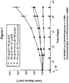
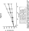
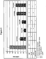
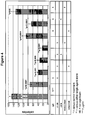
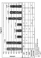
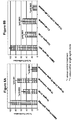

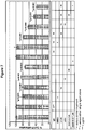
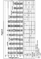
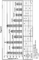


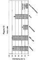

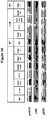


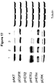
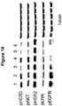

Classifications
-
- C—CHEMISTRY; METALLURGY
- C07—ORGANIC CHEMISTRY
- C07K—PEPTIDES
- C07K16/00—Immunoglobulins [IGs], e.g. monoclonal or polyclonal antibodies
- C07K16/40—Immunoglobulins [IGs], e.g. monoclonal or polyclonal antibodies against enzymes
-
- A—HUMAN NECESSITIES
- A61—MEDICAL OR VETERINARY SCIENCE; HYGIENE
- A61K—PREPARATIONS FOR MEDICAL, DENTAL OR TOILETRY PURPOSES
- A61K31/00—Medicinal preparations containing organic active ingredients
- A61K31/33—Heterocyclic compounds
- A61K31/395—Heterocyclic compounds having nitrogen as a ring hetero atom, e.g. guanethidine or rifamycins
- A61K31/535—Heterocyclic compounds having nitrogen as a ring hetero atom, e.g. guanethidine or rifamycins having six-membered rings with at least one nitrogen and one oxygen as the ring hetero atoms, e.g. 1,2-oxazines
- A61K31/5375—1,4-Oxazines, e.g. morpholine
- A61K31/5377—1,4-Oxazines, e.g. morpholine not condensed and containing further heterocyclic rings, e.g. timolol
-
- A—HUMAN NECESSITIES
- A61—MEDICAL OR VETERINARY SCIENCE; HYGIENE
- A61K—PREPARATIONS FOR MEDICAL, DENTAL OR TOILETRY PURPOSES
- A61K38/00—Medicinal preparations containing peptides
- A61K38/16—Peptides having more than 20 amino acids; Gastrins; Somatostatins; Melanotropins; Derivatives thereof
- A61K38/17—Peptides having more than 20 amino acids; Gastrins; Somatostatins; Melanotropins; Derivatives thereof from animals; from humans
- A61K38/18—Growth factors; Growth regulators
-
- A—HUMAN NECESSITIES
- A61—MEDICAL OR VETERINARY SCIENCE; HYGIENE
- A61K—PREPARATIONS FOR MEDICAL, DENTAL OR TOILETRY PURPOSES
- A61K38/00—Medicinal preparations containing peptides
- A61K38/16—Peptides having more than 20 amino acids; Gastrins; Somatostatins; Melanotropins; Derivatives thereof
- A61K38/17—Peptides having more than 20 amino acids; Gastrins; Somatostatins; Melanotropins; Derivatives thereof from animals; from humans
- A61K38/19—Cytokines; Lymphokines; Interferons
-
- A—HUMAN NECESSITIES
- A61—MEDICAL OR VETERINARY SCIENCE; HYGIENE
- A61K—PREPARATIONS FOR MEDICAL, DENTAL OR TOILETRY PURPOSES
- A61K39/00—Medicinal preparations containing antigens or antibodies
- A61K39/395—Antibodies; Immunoglobulins; Immune serum, e.g. antilymphocytic serum
-
- A—HUMAN NECESSITIES
- A61—MEDICAL OR VETERINARY SCIENCE; HYGIENE
- A61K—PREPARATIONS FOR MEDICAL, DENTAL OR TOILETRY PURPOSES
- A61K39/00—Medicinal preparations containing antigens or antibodies
- A61K39/395—Antibodies; Immunoglobulins; Immune serum, e.g. antilymphocytic serum
- A61K39/39533—Antibodies; Immunoglobulins; Immune serum, e.g. antilymphocytic serum against materials from animals
- A61K39/3955—Antibodies; Immunoglobulins; Immune serum, e.g. antilymphocytic serum against materials from animals against proteinaceous materials, e.g. enzymes, hormones, lymphokines
-
- A—HUMAN NECESSITIES
- A61—MEDICAL OR VETERINARY SCIENCE; HYGIENE
- A61K—PREPARATIONS FOR MEDICAL, DENTAL OR TOILETRY PURPOSES
- A61K39/00—Medicinal preparations containing antigens or antibodies
- A61K39/395—Antibodies; Immunoglobulins; Immune serum, e.g. antilymphocytic serum
- A61K39/39533—Antibodies; Immunoglobulins; Immune serum, e.g. antilymphocytic serum against materials from animals
- A61K39/39558—Antibodies; Immunoglobulins; Immune serum, e.g. antilymphocytic serum against materials from animals against tumor tissues, cells, antigens
-
- A—HUMAN NECESSITIES
- A61—MEDICAL OR VETERINARY SCIENCE; HYGIENE
- A61K—PREPARATIONS FOR MEDICAL, DENTAL OR TOILETRY PURPOSES
- A61K45/00—Medicinal preparations containing active ingredients not provided for in groups A61K31/00 - A61K41/00
- A61K45/06—Mixtures of active ingredients without chemical characterisation, e.g. antiphlogistics and cardiaca
-
- A—HUMAN NECESSITIES
- A61—MEDICAL OR VETERINARY SCIENCE; HYGIENE
- A61K—PREPARATIONS FOR MEDICAL, DENTAL OR TOILETRY PURPOSES
- A61K47/00—Medicinal preparations characterised by the non-active ingredients used, e.g. carriers or inert additives; Targeting or modifying agents chemically bound to the active ingredient
- A61K47/50—Medicinal preparations characterised by the non-active ingredients used, e.g. carriers or inert additives; Targeting or modifying agents chemically bound to the active ingredient the non-active ingredient being chemically bound to the active ingredient, e.g. polymer-drug conjugates
- A61K47/51—Medicinal preparations characterised by the non-active ingredients used, e.g. carriers or inert additives; Targeting or modifying agents chemically bound to the active ingredient the non-active ingredient being chemically bound to the active ingredient, e.g. polymer-drug conjugates the non-active ingredient being a modifying agent
- A61K47/68—Medicinal preparations characterised by the non-active ingredients used, e.g. carriers or inert additives; Targeting or modifying agents chemically bound to the active ingredient the non-active ingredient being chemically bound to the active ingredient, e.g. polymer-drug conjugates the non-active ingredient being a modifying agent the modifying agent being an antibody, an immunoglobulin or a fragment thereof, e.g. an Fc-fragment
- A61K47/6835—Medicinal preparations characterised by the non-active ingredients used, e.g. carriers or inert additives; Targeting or modifying agents chemically bound to the active ingredient the non-active ingredient being chemically bound to the active ingredient, e.g. polymer-drug conjugates the non-active ingredient being a modifying agent the modifying agent being an antibody, an immunoglobulin or a fragment thereof, e.g. an Fc-fragment the modifying agent being an antibody or an immunoglobulin bearing at least one antigen-binding site
- A61K47/6851—Medicinal preparations characterised by the non-active ingredients used, e.g. carriers or inert additives; Targeting or modifying agents chemically bound to the active ingredient the non-active ingredient being chemically bound to the active ingredient, e.g. polymer-drug conjugates the non-active ingredient being a modifying agent the modifying agent being an antibody, an immunoglobulin or a fragment thereof, e.g. an Fc-fragment the modifying agent being an antibody or an immunoglobulin bearing at least one antigen-binding site the antibody targeting a determinant of a tumour cell
-
- A—HUMAN NECESSITIES
- A61—MEDICAL OR VETERINARY SCIENCE; HYGIENE
- A61K—PREPARATIONS FOR MEDICAL, DENTAL OR TOILETRY PURPOSES
- A61K47/00—Medicinal preparations characterised by the non-active ingredients used, e.g. carriers or inert additives; Targeting or modifying agents chemically bound to the active ingredient
- A61K47/50—Medicinal preparations characterised by the non-active ingredients used, e.g. carriers or inert additives; Targeting or modifying agents chemically bound to the active ingredient the non-active ingredient being chemically bound to the active ingredient, e.g. polymer-drug conjugates
- A61K47/51—Medicinal preparations characterised by the non-active ingredients used, e.g. carriers or inert additives; Targeting or modifying agents chemically bound to the active ingredient the non-active ingredient being chemically bound to the active ingredient, e.g. polymer-drug conjugates the non-active ingredient being a modifying agent
- A61K47/68—Medicinal preparations characterised by the non-active ingredients used, e.g. carriers or inert additives; Targeting or modifying agents chemically bound to the active ingredient the non-active ingredient being chemically bound to the active ingredient, e.g. polymer-drug conjugates the non-active ingredient being a modifying agent the modifying agent being an antibody, an immunoglobulin or a fragment thereof, e.g. an Fc-fragment
- A61K47/6835—Medicinal preparations characterised by the non-active ingredients used, e.g. carriers or inert additives; Targeting or modifying agents chemically bound to the active ingredient the non-active ingredient being chemically bound to the active ingredient, e.g. polymer-drug conjugates the non-active ingredient being a modifying agent the modifying agent being an antibody, an immunoglobulin or a fragment thereof, e.g. an Fc-fragment the modifying agent being an antibody or an immunoglobulin bearing at least one antigen-binding site
- A61K47/6871—Medicinal preparations characterised by the non-active ingredients used, e.g. carriers or inert additives; Targeting or modifying agents chemically bound to the active ingredient the non-active ingredient being chemically bound to the active ingredient, e.g. polymer-drug conjugates the non-active ingredient being a modifying agent the modifying agent being an antibody, an immunoglobulin or a fragment thereof, e.g. an Fc-fragment the modifying agent being an antibody or an immunoglobulin bearing at least one antigen-binding site the antibody targeting an enzyme
-
- A—HUMAN NECESSITIES
- A61—MEDICAL OR VETERINARY SCIENCE; HYGIENE
- A61N—ELECTROTHERAPY; MAGNETOTHERAPY; RADIATION THERAPY; ULTRASOUND THERAPY
- A61N5/00—Radiation therapy
- A61N5/10—X-ray therapy; Gamma-ray therapy; Particle-irradiation therapy
-
- A—HUMAN NECESSITIES
- A61—MEDICAL OR VETERINARY SCIENCE; HYGIENE
- A61P—SPECIFIC THERAPEUTIC ACTIVITY OF CHEMICAL COMPOUNDS OR MEDICINAL PREPARATIONS
- A61P35/00—Antineoplastic agents
-
- A—HUMAN NECESSITIES
- A61—MEDICAL OR VETERINARY SCIENCE; HYGIENE
- A61P—SPECIFIC THERAPEUTIC ACTIVITY OF CHEMICAL COMPOUNDS OR MEDICINAL PREPARATIONS
- A61P43/00—Drugs for specific purposes, not provided for in groups A61P1/00-A61P41/00
-
- C—CHEMISTRY; METALLURGY
- C07—ORGANIC CHEMISTRY
- C07K—PEPTIDES
- C07K14/00—Peptides having more than 20 amino acids; Gastrins; Somatostatins; Melanotropins; Derivatives thereof
- C07K14/435—Peptides having more than 20 amino acids; Gastrins; Somatostatins; Melanotropins; Derivatives thereof from animals; from humans
- C07K14/475—Growth factors; Growth regulators
-
- C—CHEMISTRY; METALLURGY
- C07—ORGANIC CHEMISTRY
- C07K—PEPTIDES
- C07K16/00—Immunoglobulins [IGs], e.g. monoclonal or polyclonal antibodies
- C07K16/18—Immunoglobulins [IGs], e.g. monoclonal or polyclonal antibodies against material from animals or humans
-
- C—CHEMISTRY; METALLURGY
- C07—ORGANIC CHEMISTRY
- C07K—PEPTIDES
- C07K16/00—Immunoglobulins [IGs], e.g. monoclonal or polyclonal antibodies
- C07K16/18—Immunoglobulins [IGs], e.g. monoclonal or polyclonal antibodies against material from animals or humans
- C07K16/22—Immunoglobulins [IGs], e.g. monoclonal or polyclonal antibodies against material from animals or humans against growth factors ; against growth regulators
-
- C—CHEMISTRY; METALLURGY
- C07—ORGANIC CHEMISTRY
- C07K—PEPTIDES
- C07K16/00—Immunoglobulins [IGs], e.g. monoclonal or polyclonal antibodies
- C07K16/18—Immunoglobulins [IGs], e.g. monoclonal or polyclonal antibodies against material from animals or humans
- C07K16/24—Immunoglobulins [IGs], e.g. monoclonal or polyclonal antibodies against material from animals or humans against cytokines, lymphokines or interferons
-
- C—CHEMISTRY; METALLURGY
- C07—ORGANIC CHEMISTRY
- C07K—PEPTIDES
- C07K16/00—Immunoglobulins [IGs], e.g. monoclonal or polyclonal antibodies
- C07K16/18—Immunoglobulins [IGs], e.g. monoclonal or polyclonal antibodies against material from animals or humans
- C07K16/28—Immunoglobulins [IGs], e.g. monoclonal or polyclonal antibodies against material from animals or humans against receptors, cell surface antigens or cell surface determinants
- C07K16/2863—Immunoglobulins [IGs], e.g. monoclonal or polyclonal antibodies against material from animals or humans against receptors, cell surface antigens or cell surface determinants against receptors for growth factors, growth regulators
-
- C—CHEMISTRY; METALLURGY
- C07—ORGANIC CHEMISTRY
- C07K—PEPTIDES
- C07K16/00—Immunoglobulins [IGs], e.g. monoclonal or polyclonal antibodies
- C07K16/18—Immunoglobulins [IGs], e.g. monoclonal or polyclonal antibodies against material from animals or humans
- C07K16/28—Immunoglobulins [IGs], e.g. monoclonal or polyclonal antibodies against material from animals or humans against receptors, cell surface antigens or cell surface determinants
- C07K16/30—Immunoglobulins [IGs], e.g. monoclonal or polyclonal antibodies against material from animals or humans against receptors, cell surface antigens or cell surface determinants from tumour cells
-
- C—CHEMISTRY; METALLURGY
- C07—ORGANIC CHEMISTRY
- C07K—PEPTIDES
- C07K16/00—Immunoglobulins [IGs], e.g. monoclonal or polyclonal antibodies
- C07K16/18—Immunoglobulins [IGs], e.g. monoclonal or polyclonal antibodies against material from animals or humans
- C07K16/32—Immunoglobulins [IGs], e.g. monoclonal or polyclonal antibodies against material from animals or humans against translation products of oncogenes
-
- A—HUMAN NECESSITIES
- A61—MEDICAL OR VETERINARY SCIENCE; HYGIENE
- A61K—PREPARATIONS FOR MEDICAL, DENTAL OR TOILETRY PURPOSES
- A61K39/00—Medicinal preparations containing antigens or antibodies
- A61K2039/505—Medicinal preparations containing antigens or antibodies comprising antibodies
-
- A—HUMAN NECESSITIES
- A61—MEDICAL OR VETERINARY SCIENCE; HYGIENE
- A61K—PREPARATIONS FOR MEDICAL, DENTAL OR TOILETRY PURPOSES
- A61K39/00—Medicinal preparations containing antigens or antibodies
- A61K2039/505—Medicinal preparations containing antigens or antibodies comprising antibodies
- A61K2039/507—Comprising a combination of two or more separate antibodies
-
- A—HUMAN NECESSITIES
- A61—MEDICAL OR VETERINARY SCIENCE; HYGIENE
- A61K—PREPARATIONS FOR MEDICAL, DENTAL OR TOILETRY PURPOSES
- A61K31/00—Medicinal preparations containing organic active ingredients
- A61K31/33—Heterocyclic compounds
- A61K31/395—Heterocyclic compounds having nitrogen as a ring hetero atom, e.g. guanethidine or rifamycins
- A61K31/435—Heterocyclic compounds having nitrogen as a ring hetero atom, e.g. guanethidine or rifamycins having six-membered rings with one nitrogen as the only ring hetero atom
- A61K31/47—Quinolines; Isoquinolines
- A61K31/4709—Non-condensed quinolines and containing further heterocyclic rings
-
- A—HUMAN NECESSITIES
- A61—MEDICAL OR VETERINARY SCIENCE; HYGIENE
- A61K—PREPARATIONS FOR MEDICAL, DENTAL OR TOILETRY PURPOSES
- A61K31/00—Medicinal preparations containing organic active ingredients
- A61K31/33—Heterocyclic compounds
- A61K31/395—Heterocyclic compounds having nitrogen as a ring hetero atom, e.g. guanethidine or rifamycins
- A61K31/495—Heterocyclic compounds having nitrogen as a ring hetero atom, e.g. guanethidine or rifamycins having six-membered rings with two or more nitrogen atoms as the only ring heteroatoms, e.g. piperazine or tetrazines
- A61K31/496—Non-condensed piperazines containing further heterocyclic rings, e.g. rifampin, thiothixene
-
- A—HUMAN NECESSITIES
- A61—MEDICAL OR VETERINARY SCIENCE; HYGIENE
- A61K—PREPARATIONS FOR MEDICAL, DENTAL OR TOILETRY PURPOSES
- A61K31/00—Medicinal preparations containing organic active ingredients
- A61K31/33—Heterocyclic compounds
- A61K31/395—Heterocyclic compounds having nitrogen as a ring hetero atom, e.g. guanethidine or rifamycins
- A61K31/495—Heterocyclic compounds having nitrogen as a ring hetero atom, e.g. guanethidine or rifamycins having six-membered rings with two or more nitrogen atoms as the only ring heteroatoms, e.g. piperazine or tetrazines
- A61K31/505—Pyrimidines; Hydrogenated pyrimidines, e.g. trimethoprim
- A61K31/506—Pyrimidines; Hydrogenated pyrimidines, e.g. trimethoprim not condensed and containing further heterocyclic rings
-
- C—CHEMISTRY; METALLURGY
- C07—ORGANIC CHEMISTRY
- C07K—PEPTIDES
- C07K2317/00—Immunoglobulins specific features
- C07K2317/20—Immunoglobulins specific features characterized by taxonomic origin
- C07K2317/21—Immunoglobulins specific features characterized by taxonomic origin from primates, e.g. man
-
- C—CHEMISTRY; METALLURGY
- C07—ORGANIC CHEMISTRY
- C07K—PEPTIDES
- C07K2317/00—Immunoglobulins specific features
- C07K2317/20—Immunoglobulins specific features characterized by taxonomic origin
- C07K2317/24—Immunoglobulins specific features characterized by taxonomic origin containing regions, domains or residues from different species, e.g. chimeric, humanized or veneered
-
- C—CHEMISTRY; METALLURGY
- C07—ORGANIC CHEMISTRY
- C07K—PEPTIDES
- C07K2317/00—Immunoglobulins specific features
- C07K2317/30—Immunoglobulins specific features characterized by aspects of specificity or valency
- C07K2317/31—Immunoglobulins specific features characterized by aspects of specificity or valency multispecific
-
- C—CHEMISTRY; METALLURGY
- C07—ORGANIC CHEMISTRY
- C07K—PEPTIDES
- C07K2317/00—Immunoglobulins specific features
- C07K2317/50—Immunoglobulins specific features characterized by immunoglobulin fragments
- C07K2317/54—F(ab')2
-
- C—CHEMISTRY; METALLURGY
- C07—ORGANIC CHEMISTRY
- C07K—PEPTIDES
- C07K2317/00—Immunoglobulins specific features
- C07K2317/50—Immunoglobulins specific features characterized by immunoglobulin fragments
- C07K2317/55—Fab or Fab'
-
- C—CHEMISTRY; METALLURGY
- C07—ORGANIC CHEMISTRY
- C07K—PEPTIDES
- C07K2317/00—Immunoglobulins specific features
- C07K2317/50—Immunoglobulins specific features characterized by immunoglobulin fragments
- C07K2317/56—Immunoglobulins specific features characterized by immunoglobulin fragments variable (Fv) region, i.e. VH and/or VL
- C07K2317/565—Complementarity determining region [CDR]
-
- C—CHEMISTRY; METALLURGY
- C07—ORGANIC CHEMISTRY
- C07K—PEPTIDES
- C07K2317/00—Immunoglobulins specific features
- C07K2317/60—Immunoglobulins specific features characterized by non-natural combinations of immunoglobulin fragments
- C07K2317/62—Immunoglobulins specific features characterized by non-natural combinations of immunoglobulin fragments comprising only variable region components
- C07K2317/622—Single chain antibody (scFv)
-
- C—CHEMISTRY; METALLURGY
- C07—ORGANIC CHEMISTRY
- C07K—PEPTIDES
- C07K2317/00—Immunoglobulins specific features
- C07K2317/60—Immunoglobulins specific features characterized by non-natural combinations of immunoglobulin fragments
- C07K2317/62—Immunoglobulins specific features characterized by non-natural combinations of immunoglobulin fragments comprising only variable region components
- C07K2317/626—Diabody or triabody
-
- C—CHEMISTRY; METALLURGY
- C07—ORGANIC CHEMISTRY
- C07K—PEPTIDES
- C07K2317/00—Immunoglobulins specific features
- C07K2317/70—Immunoglobulins specific features characterized by effect upon binding to a cell or to an antigen
- C07K2317/76—Antagonist effect on antigen, e.g. neutralization or inhibition of binding
-
- C—CHEMISTRY; METALLURGY
- C07—ORGANIC CHEMISTRY
- C07K—PEPTIDES
- C07K2317/00—Immunoglobulins specific features
- C07K2317/90—Immunoglobulins specific features characterized by (pharmaco)kinetic aspects or by stability of the immunoglobulin
- C07K2317/92—Affinity (KD), association rate (Ka), dissociation rate (Kd) or EC50 value
Definitions
- This document relates to materials and methods for treating subjects having cancer expressing or overexpressing Human Epidermal Growth Factor Receptor -3 (HER-3) by administering a first agent that binds to HER-3, in combination with a second agent that is pertuzumab.
- HER-3 Human Epidermal Growth Factor Receptor -3
- HER-3 also known as ErbB3 is a receptor protein tyrosine kinase that belongs to the epidermal growth factor receptor (EGF-R, also known as HER) family of receptor protein tyrosine kinases, which also includes HER-1 (also known as EGF-R or erbB), HER-2 (also known as erbB2), and HER-4 (also known as erbB4) ( Plowman et al. (1990) Proc. Natl. Acad. Sci. US 87:4905-4909 ; Kraus et al. (1989) Proc. Natl. Acad. Sci. US 86:9193-9197 ; and Kraus et al. (1993) Proc. Natl.
- EGF-R epidermal growth factor receptor
- HER-2 also known as erbB2
- HER-4 also known as erbB4
- the transmembrane receptor HER-3 consists of an extracellular ligand-binding domain (ECD), a dimerization domain within the ECD, a transmembrane domain (TMD), an intracellular protein tyrosine kinase domain (TKD), and a C-terminal phosphorylation domain.
- ECD extracellular ligand-binding domain
- TMD transmembrane domain
- TKD intracellular protein tyrosine kinase domain
- C-terminal phosphorylation domain C-terminal phosphorylation domain
- the ligand for HER-3 known as heregulin (HRG) binds to the extracellular domain of HER-3 and activates receptor-mediated signaling by promoting dimerization with other human epidermal growth factor receptor (HER) family members, subsequent transphosphorylation of the intracellular HER-3 domain, and activation of downstream signaling cascades. Dimer formation with multiple HER family members expand the signaling potential of HER-3, and is a means for signal diversification as well as signal amplification.
- HRG heregulin
- This document relates to combinations of a first and a second agent for use in treating or preventing HER-3 expressing or overexpressing cancer by administering a first agent that binds to HER-3 as defined below, in combination with a second agent that binds to and/or inhibits another member of the HER family.
- the second agent is pertuzumab.
- the first agent is an antigen-binding protein that binds to HER-3 and comprises a heavy chain amino acid sequence that comprises a CDRH1 as shown in SEQ ID NO:256; a CDRH2 as shown in SEQ ID NO:282; and a CDRH3 as shown in SEQ ID NO:315; and a light chain amino acid sequence that comprises a CDRL1 as shown in SEQ ID NO:340; a CDRL2 as shown in SEQ ID NO: 344; and a CDRL3 as shown in SEQ ID NO:387.
- Other HER-3 binding proteins are provided as comparative examples only.
- the first agent can be an antigen-binding protein that binds to HER-3 and comprises a heavy chain amino acid sequence as shown in SEQ ID NO: 70.
- the antigen-binding protein can include a light chain amino acid sequence as shown in SEQ ID NO: 72.
- the antigen-binding protein can be directed against the extracellular domain of HER-3. Binding of the antigen-binding protein to HER-3 can reduce HER-3-mediated signal transduction, reduce HER-3 phosphorylation, reduce cell proliferation, reduce cell migration, and/or increase the downregulation of HER-3.
- the antigen-binding protein that binds to HER-3 can be an antibody.
- the antibody can be a monoclonal antibody, a polyclonal antibody, a recombinant antibody, a humanized antibody, a human antibody, a chimeric antibody, a multi-specific antibody, or an antibody fragment thereof (e.g., a Fab fragment, a Fab' fragment, a F(ab')2 fragment, a Fv fragment, a diabody, or a single chain antibody molecule).
- the antibody can be of the IgG1-, IgG2-, IgG3- or IgG4-type.
- the first agent can be an antigen-binding protein that binds to HER-3, and the antigen-binding protein can be coupled to an effector group.
- the effector group can be a radioisotope or radionuclide, a toxin, or a therapeutic or chemotherapeutic group (e.g., a therapeutic or chemotherapeutic group selected from the group consisting of calicheamicin, auristatin-PE, geldanamycin, maytansine and derivatives thereof).
- the second agent is pertuzumab.
- Other agents are provided as comparative examples.
- the methods described herein can optionally include administering a third or further therapeutic agent and/or radiation therapy.
- the third or further therapeutic agent can be an anti-neoplastic agent (e.g ., an anti-tumor antibody or a chemotherapeutic agent, such as capecitabine, anthracycline, doxorubicin, cyclophosphamide, paclitaxel, docetaxel, cisplatin, gemcitabine, or carboplatin).
- an anti-neoplastic agent e.g ., an anti-tumor antibody or a chemotherapeutic agent, such as capecitabine, anthracycline, doxorubicin, cyclophosphamide, paclitaxel, docetaxel, cisplatin, gemcitabine, or carboplatin.
- the first agent and the second agent can be administered by intravenous, subcutaneous, intramuscular or oral administration.
- the disease is a hyperproliferative disease which is HER-3 expressing or overexpressing cancer (e.g ., a disease selected from the group consisting of breast cancer, ovarian cancer, prostate cancer, colon cancer, renal cancer, lung cancer, pancreatic cancer, epidermoid carcinoma, fibrosarcoma, melanoma, nasopharyngeal carcinoma, and squamous cell carcinoma).
- the methods described herein can include administering the first agent at a dose of about 1 to about 20 mg/kg body weight, at least once every 6 weeks.
- the methods can include administering the second agent at a dose of about 1 to about 20 mg/kg body weight, at least once every 6 weeks.
- the methods can further include, prior to the administering, using a method that comprises analysis of a predictive marker to select a subject having a disease associated with HER-3.
- the methods can further include after the administering, monitoring the therapeutic outcome.
- Subject-matter of the invention is a first agent and a second agent for use in the treatment or prevention of a hyperproliferative disease associated with HER-3, wherein said hyperproliferative disease is HER-3 expressing or overexpressing cancer, wherein said first agent is an antigen-binding protein that binds to HER-3, and comprises: a heavy chain amino acid sequence that comprises a CDRH1 as shown in SEQ ID NO: 256; a CDRH2 as shown in SEQ ID NO: 282; and a CDRH3 as shown in SEQ ID NO: 315; and a light chain amino acid sequence that comprises a CDRL1 as shown in SEQ ID NO: 340; a CDRL2 as shown in SEQ ID NO: 344; and a CDRL3
- an antigen binding protein such as an antibody.
- an "antigen binding protein” or “binding protein” as used herein means a protein that specifically binds a specified target antigen, i.e. HER-3.
- An antigen binding protein is said to "specifically bind” its target antigen when the dissociation constant (K D ) is . ⁇ 10 -8 M.
- the antibody specifically binds antigen with "high affinity” when the K D is ⁇ 5x10 -9 M, and with "very high affinity” when the K D is ⁇ 5x 10 -10 M.
- the antibody has a K D of ⁇ 10 -9 M and an off-rate of about 1x.10 -4 /sec. In one embodiment, the off-rate is about 1x.10 5 /sec. In other embodiments, the antibodies will bind to a specified member of the HER family with a K D of between about 10 -8 M and 10 -10 M, and in yet another embodiment it will bind with a K D ⁇ 2x.10 -10 .
- An antibody targeted to HER-3 can be directed against the extracellular domain (ECD) of HER-3.
- ECD extracellular domain
- an anti-HER-3 antibody as described herein can interact with at least one epitope in the extracellular part of HER-3.
- the epitopes can be located in the amino terminal L1 domain (aa 19-184), in the S1 (aa 185-327) and S2 (aa 500-632) cysteine-rich domains, in the L2 domain (328-499), which is flanked by the two cysteine-rich domains, or in a combination of HER-3 domains.
- the epitopes also may be located in combinations of domains such as, without limitation, an epitope comprised by parts of L1 and S1.
- a HER-3 binding protein can be further characterized in that its binding to HER-3 reduces HER-3-mediated signal transduction.
- a reduction of HER-3-mediated signal transduction may, e.g ., be caused by a downregulation of HER-3 resulting in an at least partial disappearance of HER-3 molecules from the cell surface or by a stabilization of HER-3 on the cell surface in a substantially inactive form, i.e ., a form that exhibits a lower signal transduction compared to the non-stabilized form.
- a reduction of HER-3-mediated signal transduction also may be caused by influencing, e.g ., decreasing or inhibiting, the binding of a ligand or another member of the HER family to HER-3.
- a reduction of HER-3 mediated signal transduction also can be caused by, decreasing the formation of HER-3 containing dimers with other HER family members (e.g ., EGF-R).
- a HER-3 binding agent can be a scaffold protein having an antibody-like binding activity (e.g ., having activity similar to an anti-HER-3 antibody) or an antibody, i.e ., an anti-HER-3 antibody.
- the term "scaffold protein” means a polypeptide or protein with exposed surface areas in which amino acid insertions, substitutions or deletions are highly tolerable.
- scaffold proteins that can be used in accordance with the present methods include protein A from Staphylococcus aureus , the bilin binding protein from Pieris brassicae or other lipocalins, ankyrin repeat proteins, and human fibronectin (reviewed in Binz and Plückthun (2005) Curr. Opin. Biotechnol.
- Affinity function means a protein binding affinity according to the present document.
- a scaffold can be structurally separable from the amino acid sequences conferring binding specificity.
- proteins appearing suitable for the development of such artificial affinity reagents may be obtained by rational, or most commonly, combinatorial protein engineering techniques such as panning against HER-3, either purified protein or protein displayed on the cell surface, for binding agents in an artificial scaffold library displayed in vitro , skills which are known in the art ( see , e.g ., Skerra (2000) J. Mol. Recog.
- a scaffold protein having an antibody like binding activity can be derived from an acceptor polypeptide containing the scaffold domain, which can be grafted with binding domains of a donor polypeptide to confer the binding specificity of the donor polypeptide onto the scaffold domain containing the acceptor polypeptide.
- the inserted binding domains may be, for example, the complementarity determining region (CDR) of an antibody, in particular an anti-HER-3 antibody. Insertion can be accomplished by various methods known to those skilled in the art including, for example, polypeptide synthesis, nucleic acid synthesis of an encoding amino acid as well by various forms of recombinant methods well known to those skilled in the art.
- antibody includes monoclonal antibodies, polyclonal antibodies, recombinant antibodies, humanized antibodies ( Jones et al. (1986) Nature 321:522-525 ; Riechmann et al. (1988) Nature 332:323-329 ; and Presta (1992) Curr. Op. Struct. Biol. 2:593-596 ), chimeric antibodies ( Morrison et al. (1984) Proc. Natl. Acad. Sci. US 81:6851-6855 ), multispecific antibodies (e.g ., bispecific antibodies) formed from at least two antibodies, or antibody fragments thereof.
- antibody fragment comprises any portion of the aforementioned antibodies, such as their antigen binding or variable regions.
- antibody fragments include Fab fragments, Fab' fragments, F(ab') 2 fragments, Fv fragments, diabodies ( Hollinger et al. (1993) Proc. Natl. Acad. Sci. US 90:6444-6448 ), single chain antibody molecules ( Plückthun in: The Pharmacology of Monoclonal Antibodies 113, Rosenburg and Moore, eds., Springer Verlag, NY (1994), 269-315 ) and other fragments as long as they exhibit the desired capability of binding to HER-3.
- antibody includes antibody-like molecules that contain engineered sub-domains of antibodies or naturally occurring antibody variants. These antibody-like molecules may be single-domain antibodies such as V H -only or V L -only domains derived either from natural sources such as camelids ( Muyldermans et al. (2001) Rev. Mol. Biotechnol. 74:277-302 ) or through in vitro display of libraries from humans, camelids or other species ( Holt et al. (2003) Trends Biotechnol. 21:484-90 ).
- an “Fv fragment” is the minimum antibody fragment that contains a complete antigen-recognition and -binding site. This region consists of a dimer of one heavy chain variable domain and one light chain variable domain in tight, non-covalent association., It is in this configuration that the three CDR's of each variable domain interact to define an antigen-binding site on the surface of the V H -V L dimer. Collectively, the six CDR's confer antigen-binding specificity to the antibody. However, even a single variable domain (or half of an Fv comprising only three CDR's specific for an antigen) has the ability to recognize and bind the antigen, although usually at a lower affinity than the entire binding site.
- the “Fab fragment” also contains the constant domain of the light chain and the first constant domain (CHI) of the heavy chain.
- the “Fab fragment” differs from the “Fab' fragment” by the addition of a few residues at the carboxy terminus of the heavy chain CHI domain, including one or more cysteines from the antibody hinge region.
- the “F(ab') 2 fragment” originally is produced as a pair of “Fab' fragments” which have hinge cysteines between them. Methods of preparing such antibody fragments, such as papain or pepsin digestion, are known to those skilled in the art.
- An antibody can be of the IgA-, IgD-, IgE, IgG- or IgM-type, including IgG- or IgM-types such as, without limitation, IgG1-, IgG2-, IgG3-, IgG4-, IgM1- and IgM2-types.
- the antibody is of the IgG1-, IgG2- or IgG4- type.
- the antibody in certain respects, e.g., in connection with the generation of antibodies as therapeutic candidates against HER-3, it may be desirable that the antibody is capable of fixing complement and participating in complement-dependent cytotoxicity (CDC).
- CDC complement-dependent cytotoxicity
- isotypes of antibodies that are capable of the same including: murine IgM, murine IgG2a, murine IgG2b, murine IgG3, human IgM, human IgG1, human IgG3, and human IgA, for example.
- antibodies that are generated need not initially possess such an isotype but, rather the antibody as generated can possess any isotype and the antibody can be isotype switched by appending the molecularly cloned V region genes or cDNA to molecularly cloned constant region genes or cDNAs in appropriate expression vectors using conventional molecular biological techniques that are well known in the art and then expressing the antibodies in host cells using techniques known in the art.
- the isotype-switched antibody may also possess an Fc region that has been molecularly engineered to possess superior CDC over naturally occurring variants ( Idusogie et al. (2001) J. Immunol.
- a myeloma or other cell line such as CHO is prepared that possesses a heavy chain with any desired isotype and another myeloma or other cell line such as CHO is prepared that possesses the light chain.
- Such cells can thereafter be fused, and a cell line expressing an intact antibody can be isolated.
- a human anti-HER-3 IgG4 antibody that possesses the desired binding to the HER-3 antigen can be readily isotype switched to generate a human IgM, human IgG1 or human IgG3 isotype, while still possessing the same variable region (which defines the antibody's specificity and some of its affinity).
- Such a molecule might then be capable of fixing complement and participating in CDC.
- an antibody also may be capable of binding to Fc receptors on effector cells such as monocytes and natural killer (NK) cells, and participating in antibody-dependent cellular cytotoxicity (ADCC).
- effector cells such as monocytes and natural killer (NK) cells, and participating in antibody-dependent cellular cytotoxicity (ADCC).
- ADCC antibody-dependent cellular cytotoxicity
- the antibodies that are generated need not initially possess such an isotype but, rather the antibody as generated can possess any isotype and the antibody can be isotype switched by appending the molecularly cloned V region genes or cDNA to molecularly cloned constant region genes or cDNAs in appropriate expression vectors using conventional molecular biological techniques that are well known in the art and then expressing the antibodies in host cells using techniques known in the art.
- the isotype-switched antibody may also possess an Fc region that has been molecularly engineered to possess superior ADCC over naturally occurring variants ( Shields et al. (2001) J. Biol. Chem.
- a myeloma or other cell line such as CHO is prepared that possesses a heavy chain with any desired isotype and another myeloma or other cell line such as CHO is prepared that possesses the light chain.
- Such cells can thereafter be fused, and a cell line expressing an intact antibody can be isolated.
- a human anti-HER-3 IgG4 antibody that possesses the desired binding to the HER-3 antigen could be readily isotype switched to generate a human IgG1 or human IgG3 isotype, while still possessing the same variable region (which defines the antibody's specificity and some of its affinity). Such molecule might then be capable of binding to Fc ⁇ R on effectors cells and participating in ADCC.
- An isolated binding protein targeted to HER-3 can include a heavy chain amino acid sequence containing at least one CDR selected from the group consisting of: (a) CDRH1's as shown in SEQ ID NOS:2, 6, 10, 14, 18, 22, 26, 30, 34, 36, 40, 42, 46, 50, 54, 60, 62, 66, 70, 74, 78, 80, 84, 88, 92, 96, 100, 104, 108, 112, 116, 120, 122, 126, 130, 134, 138, 142, 146, 150, 154, 158, 162, 166, 170, 174, 178, 182, 186, 190, 194, 198, 202, 206, 210, 214, 218, 222, 226 and 230, (b) CDRH2's as shown in SEQ ID NOS:2, 6, 10, 14, 18, 22, 26, 30, 34, 36, 40, 42, 46, 50, 54, 60, 62, 66, 70, 74, 78, 80,
- An isolated binding protein targeted to HER-3 can include a heavy chain amino acid sequence selected from the group consisting of SEQ ID NOS:2, 6, 10, 14, 18, 22, 26, 30, 34, 36, 40, 42, 46, 50, 54, 60, 62, 66, 70, 74, 78, 80, 84, 88, 92, 96, 100, 104, 108, 112, 116, 120, 122, 126, 130, 134, 138, 142, 146, 150, 154, 158, 162, 166, 170, 174, 178, 182, 186, 190, 194, 198, 202, 206, 210, 214, 218, 222, 226 and 230, and/or a light chain amino acid sequence selected from the group consisting of SEQ ID NOS:4, 8, 12, 16, 20, 24, 28, 32, 38, 44, 48, 52, 56, 58, 64, 68, 72, 76, 82, 86, 90, 94, 98, 102, 106, 110,
- An anti-HER-3 antibody can include a heavy chain amino acid sequence and a light chain amino acid sequence as shown in SEQ ID NOS:2 and 4, 6 and 8, 10 and 12, 14 and 16, 18 and 20, 22 and 24, 26 and 28, 30 and 32, 36 and 38, 42 and 44, 46 and 48, 50 and 52, 54 and 56, 60 and 58, 62 and 64, 66 and 68, 70 and 72, 74 and 76, 78 and 82, 80 and 82, 84 and 86, 88 and 90, 92 and 94, 96 and 98, 100 and 102, 104 and 106, 108 and 110, 112 and 114, 116 and 118, 122 and 124, 126 and 128, 130 and 132, 134 and 136, 138 and 140, 142 and 144, 146 and 148, 150 and 152, 154 and 156, 158 and 160, 162 and 164, 166 and 168, 170 and 172, 174 and 176, 178 and 180, 182 and 184
- a protein targeted to HER-3 can be a scaffold protein having an antibody-like binding activity (e.g ., having activity similar to an anti-HER-3 antibody), or an antibody, e.g ., an anti-HER-3 antibody.
- Exemplary anti-HER-3 antibodies are designated U1-1, U1-2, U1-3, U1-4, U1-5, U1-6, U1-7, U1-8, U1-9, U1-10, U1-11, U1-12, U1-13, U1-14, U1-15, U1-16, U1-17, U1-18, U1-19, U1-20, U1-21, U1-22, U1-23, U1-24, U1-25, U1-26, U1-27, U1-28, U1-29, U1-30, U1-31, U1-32, U1-33, U1-34, U1-35, U1-36, U1-37, U1-38, U1-39, U1-40, U1-41, U1-42, U1-43, U1-44, U1-45, U1-46, U1-47, U1-48, U1-49, U1-50, U1-51, U1-52, U1
- amino acid sequence of the HER-3 binding proteins described herein is not limited to the twenty conventional amino acids ( see , Immunology - A Synthesis (2nd Edition, Golub and Gren, eds., Sinauer Associates, Sunderland, Mass. (1991 )).
- the amino acids may include stereoisomers (e.g ., D-amino acids) of the twenty conventional amino acids, unnatural amino acids such as ⁇ -, ⁇ -disubstituted amino acids, N-alkyl amino acids, lactic acid, and other unconventional amino acids.
- Examples of unconventional amino acids which may also be suitable components for the binding proteins provided herein, include: 4-hydroxyproline, ⁇ -carboxyglutamate, ⁇ -N,N,N-trimethyllysine, ⁇ -N-acetyllysine, O-phosphoserine, N-acetylserine, N-formylmethionine, 3-methylhistidine, 5-hydroxylysine, ⁇ -N-methylarginine, and other similar amino acids and imino acids, e.g ., 4-hydroxyproline.
- amino acid sequences shown in SEQ ID NOS:70 and 72 are contemplated as being encompassed by the present disclosure, provided that the variations in the amino acid sequence are not in the CDR sequences defined in the claims and maintain at least 75% (e.g ., at least 80%, 90%, 95%, or 99%) of the sequences shown in SEQ ID NOS:70 and 72. Variations can occur within the framework regions ( i.e ., outside the CDRs). In some embodiments, variations in the amino acid sequences, i.e ., deletions, insertions and/or substitutions of at least one amino acid, can occur near boundaries of functional domains.
- Structural and functional domains can be identified by comparison of the nucleotide and/or amino acid sequence data to public or proprietary sequence databases.
- Computerized comparison methods can be used to identify sequence motifs or predicted protein conformation domains that occur in other binding proteins of known structure and/or function. Methods for identifying protein sequences that fold into a known three-dimensional structure are known in the art. ( See , e.g ., Bowie et al. (1991) Science 253:164 ; Proteins, Structures and Molecular Principles, Creighton, Ed., W H. Freeman and Company, New York (1984 ); Introduction to Protein Structure, Branden and Tooze, eds., Garland Publishing, New York, N.Y. (1991 ); and Thornton et al. (1991) Nature 354:105 ) Thus, those of skill in the art can recognize sequence motifs and structural conformations that may be used to define structural and functional domains in accordance with the proteins described herein.
- Variations in the amino acid sequences shown in SEQ ID NOS:70 and 72 can include those that lead to a reduced susceptibility to proteolysis or oxidation, alter glycosylation patterns or alter binding affinities or confer or modify other physicochemical or functional properties of the binding protein.
- conservative amino acid replacements are contemplated. Conservative replacements are those that take place within a family of amino acids that are related in their side chains.
- aliphatic-hydroxy family serine and threonine
- amide-containing family asparagine and glutamine
- aliphatic family alanine, valine, leucine and isoleucine
- aromatic family phenylalanine, tryptophan, and tyrosine.
- an isolated replacement of a leucine with an isoleucine or valine, an aspartate with a glutamate, a threonine with a serine, or a similar replacement of an amino acid with a structurally related amino acid will not have a major effect on the binding or properties of the resulting binding protein, especially if the replacement does not involve an amino acid within a framework site.
- a HER-3 binding protein can be coupled to an effector group.
- an effector group refers to a cytotoxic group such as a radioisotope or radionuclide, a toxin, a therapeutic group or other effector group known in the art.
- Suitable effector groups are radioisotopes or radionuclides (e.g ., 3 H, 14 C, 15 N, 35 S, 90 Y, 99 Tc, 111 In, 125 I, 131 I) or non-radio isotopes ( e.g ., 2D), calicheamicin, dolastatin analogs such as auristatins, and chemotherapeutic agents such as geldanamycin and maytansine derivates, including DM1.
- a group can be both a labeling group and an effector group.
- effector groups to polypeptides or glycopolypeptides (such as antibodies) are known in the art, and may be used in making and carrying out the compositions and methods described herein. In some embodiments, it may be useful to have effector groups attached to a binding protein by spacer arms of various lengths to, for example, reduce potential steric hindrance.
- This document also relates to processes for preparing an isolated HER-3 binding protein, comprising the step of preparing the protein from a host cell that expresses the protein.
- Host cells that can be used include, without limitation, hybridomas, eukaryotic cells (e.g ., mammalian cells such as hamster, rabbit, rat, pig, or mouse cells), plant cells, fungal cells, yeast cells ( e.g ., Saccharomyces cerevisiae or Pichia pastoris cells), prokaryotic cells (e.g ., E. coli cells), and other cells used for production of binding proteins.
- eukaryotic cells e.g ., mammalian cells such as hamster, rabbit, rat, pig, or mouse cells
- plant cells fungal cells
- yeast cells e.g ., Saccharomyces cerevisiae or Pichia pastoris cells
- prokaryotic cells e.g ., E. coli cells
- binding protein fragments e.g ., scaffold protein fragments or antibody fragments
- methods for preparing binding protein fragments such as papain or pepsin digestion, modern cloning techniques, techniques for preparing single chain antibody molecules (Plückthun, supra ) and diabodies (Hollinger et al ., supra )
- Plückthun, supra techniques for preparing single chain antibody molecules
- diabodies techniques for preparing single chain antibody molecules
- Hollinger et al ., supra also are known to those skilled in the art and may be used in performing the presently described methods.
- a HER-3 binding protein can be prepared from a hybridoma that secretes the protein. See , e.g ., Köhler et al. (1975) Nature 256:495 .
- a HER-3 binding protein can be prepared recombinantly by optimizing and/or amplifying expression of the binding protein in host cells, and isolating the binding protein from the host cells.
- host cells can be transformed or transfected with DNA (e.g ., a vector) encoding a HER-3 binding protein, and cultured under conditions appropriate to produce the binding protein. See , e.g ., US Patent No. 4,816,567 .
- Useful host cells include, for example, CHO cells, NS/0 myeloma cells, human embryonic kidney 293 cells, E. coli cells, and Saccharomyces cerevisiae cells.
- HER-3 binding proteins that are antibodies can be prepared from animals genetically engineered to make fully human antibodies, or from an antibody display library made in bacteriophage, yeast, ribosome or E. coli. See , e.g ., Clackson et al. (1991) Nature 352:624-628 ; Marks et al. (1991) J. Mol. Biol. 222:581-597 ; Feldhaus and Siegel (2004) J. Immunol. Methods 290:69-80 ; Groves and Osbourn (2005) Expert Opin. Biol. Ther. 5:125-135 ; and Jostock and Dubel (2005) Comb. Chem. High Throughput Screen 8:127-133 .
- Antibodies as described herein can be fully human or humanized antibodies.
- Human antibodies avoid certain problems associated with xenogeneic antibodies, such as antibodies that possess murine or rat variable and/or constant regions.
- the presence of xenogeneic-derived proteins can lead to an immune response against the antibody by a patient, subsequently leading to the rapid clearance of the antibody, loss of therapeutic utility through neutralization of the antibody, and/or severe, even life-threatening, allergic reactions.
- fully human antibodies can be generated through the introduction of functional human antibody loci into a rodent or another mammal or animal so that the rodent, other mammal or animal produces fully human antibodies.
- One method for generating fully human antibodies is to utilize XENOMOUSE® strains of mice that have been engineered to contain 245 kb and 190 kb-sized germline configuration fragments of the human heavy chain locus and kappa light chain locus.
- Other XENOMOUSE® strains of mice contain 980 kb and 800 kb-sized germline configuration fragments of the human heavy chain locus and kappa light chain locus.
- Still other XENOMOUSE® strains of mice contain 980 kb and 800 kb-sized germline configuration fragments of the human heavy chain locus and kappa light chain locus plus a 740 kb-sized germline configured complete human lambda light chain locus. See, Mendez et al. (1997) Nature Genetics 15:146-156 ; and Green and Jakobovits (1998) J. Exp. Med. 188:483-495 .
- XENOMOUSE® strains are available from Amgen, Thousand Oak
- minilocus an exogenous Ig locus is mimicked through the inclusion of pieces (individual genes) from the Ig locus.
- V H genes one or more D H genes
- J H genes one or more J H genes
- a mu constant region e.g ., a gamma constant region
- a second constant region e.g ., a gamma constant region
- WO 92/03918 WO 92/22645 , WO 92/22647 , WO 92/22670 , WO 93/12227 , WO 94/00569 , WO 94/25585 , WO 96/14436 , WO 97/13852 , and WO 98/24884 .
- Human antibodies also can be generated from mice in which, through microcell fusion, large pieces of chromosomes, or entire chromosomes, have been introduced. See, EP Patent Application Nos. 773288 and 843961 . Additionally, KMTM mice, which are the result of cross-breeding of Kirin's Tc mice with Medarex's minilocus (Humab) mice have been generated. These mice possess the HC transchromosome of the Kirin mice and the kappa chain transgene of the Medarex mice ( Ishida et al. (2002) Cloning Stem Cells 4:91-102 ).
- Human antibodies also can be derived by in vitro methods. Suitable examples include, but are not limited to, phage display (as commercialized by Cambridge Antibody Technology, Morphosys, Dyax, Biosite/Medarex, Xoma, Symphogen, Alexion (formerly Proliferon), and Affimed), ribosome display (as commercialized by Cambridge Antibody Technology), yeast display, and the like.
- mice were prepared using XENOMOUSE® technology, as described below.
- Such mice are capable of producing human immunoglobulin molecules and antibodies, and are deficient in the production of murine immunoglobulin molecules and antibodies. Technologies utilized for achieving the same are disclosed in the patents, applications, and references disclosed herein.
- transgenic production of mice and antibodies therefrom is disclosed in US Patent Application Serial No. 08/759,620, filed December 3, 1996 , and PCT Publication Nos. WO 98/24893 and WO 00/76310 . See also Mendez et al. (1997) Nature Genetics 15:146-156 .
- XENOMOUSE® lines of mice can be immunized with a HER-3 antigen of interest (e.g ., HER-3 or a fragment thereof), lymphatic cells (such as B-cells) can be recovered from mice that express antibodies, and the recovered cell lines can be fused with a myeloid-type cell line to prepare immortal hybridoma cell lines.
- lymphatic cells such as B-cells
- These hybridoma cell lines can be screened and selected to identify hybridoma cell lines that produce antibodies specific to the antigen of interest.
- methods for the production of multiple hybridoma cell lines that produce antibodies specific to HER-3 are provided herein are methods for characterizing antibodies produced by such cell lines, including nucleotide and amino acid sequence analyses of the heavy and light chains of such antibodies.
- antibodies produced by fused hybridomas as described below are human IgG1 heavy chains with fully human kappa light chains, although some antibodies described herein possess human IgG4 heavy chains as well as IgG1 heavy chains.
- Antibodies also can be of other human isotypes, including IgG2 and IgG3.
- the antibodies generally have high affinities, with a K D typically from about 10 -6 to about 10 -13 M or below, when measured by solid phase and cell-based techniques.
- isolated nucleic acid molecule refers to a polynucleotide of genomic, cDNA, or synthetic origin, or some combination thereof, which (1) is not associated with all or a portion of a polynucleotide with which the "isolated polynucleotide” is found in nature, (2) is operably linked to a polynucleotide to which it is not linked to in nature, or (3) does not occur in nature as part of a larger sequence.
- nucleic acid molecule means a polymeric form of nucleotides of at least 10 bases in length, either ribonucleotides or deoxynucleotides or a modified form of either type of nucleotide, such as nucleotides with modified or substituted sugar groups and the like.
- the term also includes single and double stranded forms of DNA.
- control sequence refers to polynucleotide sequences that are necessary to effect the expression and processing of coding sequences to which they are ligated. The nature of such control sequences differs depending upon the host organism. In prokaryotes, such control sequences generally include promoters, ribosomal binding sites, and transcription termination sequences. In eukaryotes, generally, such control sequences include promoters and transcription termination sequences.
- control sequence is intended to include, at a minimum, all components whose presence is essential for expression and processing, and can also include additional components whose presence is advantageous, for example, leader sequences and fusion partner sequences.
- operably linked refers to positions of components so described which are in a relationship permitting them to function in their intended manner.
- an expression control sequence operably linked to a coding sequence is ligated in such a way that expression of the coding sequence is achieved under conditions compatible with the expression control sequence.
- vectors comprising a nucleic acid molecule encoding a binding protein as disclosed herein.
- the nucleic acid molecule can be operably linked to a control sequence.
- the vector may additionally contain a replication origin or a selection marker gene. Examples of vectors that may be used include, e.g ., plasmids, cosmids, phages, and viruses.
- Transformation can be accomplished by any known method for introducing polynucleotides into a host cell, including, for example, packaging the polynucleotide in a virus (or into a viral vector) and transducing a host cell with the virus (or vector), or by transfection procedures known in the art, as exemplified by US Patent Nos. 4,399,216 , 4,912,040 , 4,740,461 , and 4,959,455 .
- heterologous polynucleotides into mammalian cells
- methods for introducing heterologous polynucleotides into mammalian cells include, without limitation, dextran-mediated transfection, calcium phosphate precipitation, polybrene mediated transfection, protoplast fusion, electroporation, encapsulation of the polynucleotide(s) in liposomes, and direct microinjection of the DNA into nuclei.
- host cells include hybridomas, eukaryotic cells (e.g ., mammalian cells such as hamster, rabbit, rat, pig, mouse, or other animal cells), plant cells ( e.g ., corn and tobacco cells), fungal cells ( e.g ., S. cerevisiae and P.
- Mammalian cell lines available as hosts for expression are well known in the art and include, for example, many immortalized cell lines available from the American Type Culture Collection (ATCC; Manassas, VA). These include, without limitation, Chinese hamster ovary (CHO) cells, HeLa cells, baby hamster kidney (BHK) cells, monkey kidney cells (COS), human hepatocellular carcinoma cells ( e.g ., Hep G2 cells), and a number of other cell lines.
- ATCC American Type Culture Collection
- VA American Type Culture Collection
- compositions and methods described herein for treatment of HER-3 associated disease include a first agent that binds to HER-3, in combination with a second agent that binds and/or inhibits at least one other member of the HER family, including but not limited to, EGF-R, HER-2, HER-4.
- the second agent is pertuzumab.
- a biological drug e.g ., a binding protein, such as an antibody specifically binding to a member of the HER family, a small molecular compound that binds to and/or alters ( e.g ., inhibits) the activity of at least one member of the HER family other than (or in addition to) HER-3, an siRNA, or a natural substance.
- a binding protein such as an antibody specifically binding to a member of the HER family
- a small molecular compound that binds to and/or alters (e.g ., inhibits) the activity of at least one member of the HER family other than (or in addition to) HER-3, an siRNA, or a natural substance.
- the terms “other HER family members” and “another HER family member” refer to HER family members that are not HER-3. Examples are the EGF-R, HER-2, and HER-4, but "HER family member” also includes family members that have not yet been identified.
- the second agent can alter the activity (e.g ., increase or decrease) the activity of the other HER family member, either through a direct effect or an indirect effect on the HER family member. It is noted, however, that all second agents as described herein will have an effect on HER family function and activity.
- Pertuzumab (2C4) (marketed or to be marketed as OMNITARGTM) is a monoclonal antibody that inhibits the dimerization of HER2 with other HER receptors.
- a composition for treatment of HER3-associated disease as defined in the claims can be U1-59 in combination with pertuzumab, or U1-59 in combination with pertuzumab and other agent(s), for treatment of cancer, including, e.g .
- breast cancer breast cancer, gastrointestinal cancer, pancreatic cancer, prostate cancer, ovarian cancer, stomach cancer, endometrial cancer, salivary gland cancer, lung cancer, renal cancer, colon cancer, colorectal cancer, thyroid cancer, bladder cancer, glioma, melanoma, metastatic breast cancer, non-small cell lung cancer, epidermoid carcinoma, fibrosarcoma, melanoma, nasopharyngeal carcinoma, and squamous cell carcinoma.
- Additional agents may be added to the first and second agent for use according to the invention. These, in some embodiments, will be chemotherapeutic drugs.
- agents that act as microtubule stimulants include NK-105(paclitaxel)[(-)-(1S,2R,3S,4S,5R,7S,8S,10R,13S)-4,10-diacetoxy-2-benzoyloxy-5,20-epoxy-1,7-dihydroxy-9-oxotax-11 -en-13-yl (2R,3S)-3-benzoylamino-2-hydroxy-3-phenylpropionate] (NanoCarrier, Chiba, Japan), milataxel (1,10 ⁇ -dihydroxy-9-oxo-5 ⁇ ,20-epoxy-3zeta-tax-11-ene-2 ⁇ ,4,7 ⁇ 13 ⁇ -tetrayl 4-acetate 2-benzoate 13-[(2R,3R)-3-(tert-butoxycarbonylamino)-3-(furan-2-yl)-2-hydroxypropanoate] 7-propanoate) (Taxolog, Fairfield,
- BMS-184476 Bristol-Myers Squibb, New York, NY, EP Publication No. 639577
- BMS-188797 Bristol-Myers Squibb, New York, NY
- BMS-275183 (3'-tert-butyl-3'-N-tert-butyloxycarbonyl-4-deacetyl-3'-dephenyl-3'-N-debenzoyl-4-O-methyoxy-carbonyl-paclitaxel) (Bristol-Myers Squibb, New York, NY), BMS-310705 (Bristol-Myers Squibb, New York, NY), BMS-753493 (Bristol-Myers Squibb, New York, NY), cabazitaxel (1-hydroxy-7 ⁇ ,10 ⁇ -dimethoxy-9-oxo-5 ⁇ ,20-epoxytax-11-ene-2 ⁇ ,4,13 ⁇ -triyl 4-acetate 2-benzoate 13-[(2R)
- Anthracyclins include actinomycins such as actinomycin D (Dactinomycin: 2-amino-N,N'- bis[(6S,9R,10S,13R,18aS)-6,13-diisopropyl-2,5,9-trimethyl-1,4,7,11,14-pentaoxohexadecahydro-1H-pyrrolo[2,1-i][1,4,7,10,13] oxatetraazacyclohexadecin-10-yl]-4,6-dimethyl-3-oxo-3H-phenoxazine- 1,9-dicarboxamide), bleomycin (bleomycin hydrochloride: (3- ⁇ [(2'- ⁇ (5S,8S,9S,10R,13S)-15- ⁇ 6-amino-2- [(1S)-3-amino-1- ⁇ [(2S)-2,3-diamino-3-oxopropyl]amino ⁇ -3
- Cisplatin and gemcitabine are chemotherapeutic agents.
- Cisplatin or cis-diamminedichloroplatinum(II) is a platinum-based drug used to treat various types of cancers.
- the cisplatin platinum complex reacts in vivo , binding to and causing crosslinking of DNA, which ultimately triggers apoptosis.
- Gemcitabine is a nucleoside analog in which the hydrogen atoms on the 2' carbons of deoxycytidine are replaced by fluorine atoms.
- gemcitabine Like fluorouracil and other pyrimidine analogues, gemcitabine replaces cytidine during DNA replication, which arrests tumor growth since further nucleosides cannot be attached to the "faulty" nucleoside, resulting in apoptosis. Gemcitabine is marketed as GEMZAR® by Eli Lilly and Company (Indianapolis, IN).
- a combination for treatment can be U1-59 in combination with pertuzumab and cisplatin or gemcitabine and other agent(s), for treatment of cancer which is gastrointestinal cancer, pancreatic cancer, prostate cancer, ovarian cancer, stomach cancer, endometrial cancer, salivary gland cancer, kidney cancer, colon cancer, thyroid cancer, bladder cancer, glioma, melanoma, lung cancer including non-small cell lung cancer, colorectal cancer and/or breast cancer including metastatic breast cancer.
- cancer which is gastrointestinal cancer, pancreatic cancer, prostate cancer, ovarian cancer, stomach cancer, endometrial cancer, salivary gland cancer, kidney cancer, colon cancer, thyroid cancer, bladder cancer, glioma, melanoma, lung cancer including non-small cell lung cancer, colorectal cancer and/or breast cancer including metastatic breast cancer.
- Capecitabine (pentyl[1-(3,4-dihydroxy-5-methyl-tetrahydrofuran-2-yl)-5-fluoro-2-oxo-1H-pyrimidin-4-yl]aminomethanoate, Xeloda, Roche) is an orally-administered chemotherapeutic agent.
- Capecitabine is a prodrug that is enzymatically converted to 5-fluorouracil in the tumor, where it inhibits DNA synthesis and slows growth of tumor tissue.
- a combination for treatment of HER3-associated disease as defined in the claims can be U1-59 in combination with a second agent as described herein (i.e., pertuzumab) and capecitabine for treatment of cancer, wherein the cancer is gastrointestinal cancer, pancreatic cancer, prostate cancer, ovarian cancer, stomach cancer, endometrial cancer, salivary gland cancer, kidney cancer, colon cancer, thyroid cancer, bladder cancer, glioma, melanoma, lung cancer including non-small cell lung cancer, colorectal cancer and/or breast cancer including metastatic breast cancer.
- a combination can be administered after failure of prior treatment with an anthracyclin or taxane, for example.
- Docetaxel is marketed as Taxotere by Sanofi Aventis.
- Paclitaxel is marketed as Taxol by Bristol-Myers Squibb.
- paclitaxel is dissolved in Cremophor EL and ethanol, as a delivery agent.
- a formulation in which paclitaxel is bound to albumin is marketed as Abraxane.
- a combination for treatment of HER3-associated disease as defined in the claims can be U1-59 in combination with a second agent as described herein (i.e.
- pertuzumab and docetaxel or paclitaxel and other agent(s) such as trastuzumab, for treatment of cancer
- the cancer is gastrointestinal cancer, pancreatic cancer, prostate cancer, ovarian cancer, stomach cancer, endometrial cancer, salivary gland cancer, kidney cancer, colon cancer, thyroid cancer, bladder cancer, glioma, melanoma, lung cancer including non-small cell lung cancer, colorectal cancer and/or breast cancer including metastatic breast cancer.
- Doxorubicin hydrochloride liposome injection is marketed as Doxil, a liposome formulation comprising doxorubicin chloride.
- a combination treatment for HER-3-associated disease as defined in the claims can include administering U1-59 in combination with a second agent as described herein (i.e.
- pertuzumab and doxorubicin hydrochloride liposome injection, with or without one or more other agents such as paclitaxel or platinum-based chemotherapeutic agents, for treatment of cancer
- cancer such as breast cancer, gastrointestinal cancer, pancreatic cancer, prostate cancer, ovarian cancer, stomach cancer, endometrial cancer, salivary gland cancer, lung cancer, renal cancer, colon cancer, colorectal cancer, thyroid cancer, bladder cancer, glioma, melanoma, metastatic breast cancer, non-small cell lung cancer, epidermoid carcinoma, fibrosarcoma, melanoma, nasopharyngeal carcinoma, and squamous cell carcinoma.
- cancer such as breast cancer, gastrointestinal cancer, pancreatic cancer, prostate cancer, ovarian cancer, stomach cancer, endometrial cancer, salivary gland cancer, lung cancer, renal cancer, colon cancer, colorectal cancer, thyroid cancer, bladder cancer, glioma, melanoma, metastatic breast cancer, non-
- Irinotecan hydrochloride hydrate (irinotecan: (+)-(4S)-4,11-diethyl-4-hydroxy-9-[(4-piperidinopiperidino)carbonyloxy]-1H-pyrano[3',4':6,7]indolizino[1-2-b]quinoline-3,14(4H,12H)-dione hydrochloride trihydrate) ( Yakult, EP Publication Nos. 137145 and 56692 ) is marketed as Campto, Camptosar and Ircan.
- a combination treatment for HER3-associated disease as defined in the claims can include administering U1-59 in combination with a second agent as described herein (i.e.
- pertuzumab and irinotecan hydrochloride hydrate, or U1-59 in combination with a second agent as described herein (i.e. pertuzumab), irinotecan hydrochloride hydrate, and one or more other agent(s) such as 5-FU(5'-deoxy-5-fluorouridine or 5-fluoro-2,4(1H,3H)-pyrimidinedione), calcium folinate (N-[4-[[(2-amino-5-formyl-1,4,5,6,7,8-hexahydro-4-oxo-6-pteridinyl)methylamino]benzoyl]-L-glutamic acid calcium salt (1:1)) or calcium levofolinate ((-)-calcium N-[4-[[[(6S)-2-amino-5-formyl-1,4,5,6,7,8-hexahydro-4-oxo-6-pteridinyl]methyl]amino]benz
- U1-59 can be used in the treatment of patients with cancers including colorectal cancer and metastatic colorectal cancer after failure of 5-fluorouracil-based chemotherapy, in combination with 5-fluorouracil-based chemotherapy. Further, U1-59 can be used in the treatment of the treatment of patients with cancers including colorectal cancer and metastatic colorectal cancer with wild-type K-RAS after failure of 5-fluorouracil-based chemotherapy, in combination with cetuximab and irinotecan.
- the additional agents to be used in the compositions and methods described herein may be an artificial or naturally-occurring scaffold which is not an antibody, but has an antibody-like activity (e.g., has an activity similar to that of an antibody).
- Said additional agents can be agents inhibit, block or reduce (act as antagonists towards), or, activate, stimulate or accelerate (act as agonist towards) an activity of other targets, including but not limited to those affect cellular growth and/or survival pathways, such as PI3K inhibitors, AKT inhibitors, mTOR inhibitors, RAF/B-RAF inhibitors, RAS inhibitors, MEK inhibitors, Death Receptor inhibitors including DR4 and DR5 agonists such as anti-DR4 or DR5 agonistic antibodies (for example, cedelizumab, tigatuzumab, drozirumab, conatumumab), PPAR gamma agonists (for example, efatutazone, troglitazone, pioglitazone, rosiglitazone), c-MET inhibitors, Hsp-90 inhibitors and telomerase inhibitors.
- PI3K inhibitors PI3K inhibitors
- AKT inhibitors kinas
- mTOR inhibitors
- Said additional agents can be anti-angiogenics, including but not limited to, VEGF antagonists/inhibitors (for example, bevacizumab, vandetanib).
- VEGF antagonists/inhibitors for example, bevacizumab, vandetanib.
- said additional agents can be immunotherapeutic such as vaccines or cellular therapeutics.
- these and other agents can be contained within the compositions described herein, and can be administered in a variety of different forms, combinations and dosages.
- compositions comprising a HER-3 binding agent as described herein, in combination with a second agent that is directed against another HER family protein or is a chemotherapeutic compound, as well as one or more pharmaceutically acceptable carriers, diluents and/or adjuvants.
- pharmaceutical composition refers to a chemical compound or composition capable of inducing a desired therapeutic effect when properly administered to a patient ( The McGraw-Hill Dictionary of Chemical Terms, Parker, Ed., McGraw-Hill, San Francisco (1985 )).
- the potency of the pharmaceutical compositions provided herein typically is based on the binding of the at least one binding protein to HER-3. This binding can lead to a reduction of the HER-3-mediated signal transduction.
- a “pharmaceutically acceptable carrier” (also referred to herein as an “excipient” or a “carrier”) is a pharmaceutically acceptable solvent, suspending agent, stabilizing agent, or any other pharmacologically inert vehicle for delivering one or more therapeutic compounds (e.g ., HER binding proteins) to a subject, which is nontoxic to the cell or mammal being exposed thereto at the dosages and concentrations employed.
- Pharmaceutically acceptable carriers can be liquid or solid, and can be selected with the planned manner of administration in mind so as to provide for the desired bulk, consistency, and other pertinent transport and chemical properties, when combined with one or more of therapeutic compounds and any other components of a given pharmaceutical composition.
- Typical pharmaceutically acceptable carriers that do not deleteriously react with amino acids include, by way of example and not limitation: water, saline solution, binding agents (e.g ., polyvinylpyrrolidone or hydroxypropyl methylcellulose), fillers ( e.g ., lactose and other sugars, gelatin, or calcium sulfate), lubricants (e.g., starch, polyethylene glycol, or sodium acetate), disintegrates (e.g., starch or sodium starch glycolate), and wetting agents (e.g., sodium lauryl sulfate).
- binding agents e.g ., polyvinylpyrrolidone or hydroxypropyl methylcellulose
- fillers e.g ., lactose and other sugars, gelatin, or calcium sulfate
- lubricants e.g., starch, polyethylene glycol, or sodium acetate
- disintegrates e.g., starch or sodium starch
- Pharmaceutically acceptable carriers also include aqueous pH buffered solutions or liposomes (small vesicles composed of various types of lipids, phospholipids and/or surfactants which are useful for delivery of a drug to a mammal).
- Further examples of pharmaceutically acceptable carriers include buffers such as phosphate, citrate, and other organic acids, antioxidants such as ascorbic acid, low molecular weight (less than about 10 residues) polypeptides, proteins such as serum albumin, gelatin, or immunoglobulins, hydrophilic polymers such as polyvinylpyrrolidone, amino acids such as glycine, glutamine, asparagine, arginine or lysine, monosaccharides, disaccharides, and other carbohydrates including glucose, mannose or dextrins, chelating agents such as EDTA, sugar alcohols such as mannitol or sorbitol, salt-forming counterions such as sodium, and/or nonionic surfactants such as TWEENTM,
- Liposomes are vesicles that have a membrane formed from a lipophilic material and an aqueous interior that can contain the composition to be delivered. Liposomes can be particularly useful due to their specificity and the duration of action they offer from the standpoint of drug delivery.
- Liposome compositions can be formed, for example, from phosphatidylcholine, dimyristoyl phosphatidylcholine, dipalmitoyl phosphatidylcholine, dimyristoyl phosphatidylglycerol, or dioleoyl phosphatidylethanolamine.
- Numerous lipophilic agents are commercially available, including LIPOFECTIN® (Invitrogen/Life Technologies, Carlsbad, CA) and EFFECTENETM (Qiagen, Valencia, CA).
- At least one of the agents contained in a pharmaceutical composition can be coupled to an effector such as calicheamicin, duocarmycins, auristatins, maytansinoids, a radioisotope, or a toxic chemotherapeutic agent such as geldanamycin and maytansine.
- an effector such as calicheamicin, duocarmycins, auristatins, maytansinoids, a radioisotope, or a toxic chemotherapeutic agent such as geldanamycin and maytansine.
- Such conjugates can be particularly useful for targeting cells (e.g ., cancer cells) expressing HER-3.
- Radioisotopes can provide advantages to tumor treatments. Unlike chemotherapy and other forms of cancer treatment, radioimmunotherapy or the administration of a radioisotope-binding protein combination can directly target cancer cells with minimal damage to surrounding normal, healthy tissue. With this "magic bullet," patients can be treated with much smaller quantities of radioisotopes than other forms of treatment available today.
- Suitable radioisotopes include, for example, yttrium 90 ( 90 Y), indium 111 ( 111 In), 131 I, 99 mTc, radiosilver-111, radiosilver-199, and Bismuth 213 .
- the linkage of radioisotopes to binding proteins may be performed with, for example, conventional bifunctional chelates.
- sulphur-based linkers may be used ( Hazra et al. (1994) Cell Biophys. 24-25:1-7 ). Linkage of silver radioisotopes may involve reducing the immunoglobulin with ascorbic acid.
- tiuxetan is an MX-DTPA linker chelator attached to ibritumomab to form ibritumomab tiuxetan (Zevalin) ( Witzig (2001) Cancer Chemother. Pharmacol. 48 (Suppl 1):91-95 ).
- Ibritumomab tiuxetan can react with radioisotypes such as indium 111 ( 111 In) or 90 Y to form 111 In-ibritumomab tiuxetan and 90 Y-ibritumomab tiuxetan, respectively.
- radioisotypes such as indium 111 ( 111 In) or 90 Y to form 111 In-ibritumomab tiuxetan and 90 Y-ibritumomab tiuxetan, respectively.
- the binding proteins described herein can be conjugated with toxic chemotherapeutic drugs such as maytansinoids, ( Hamann et al. (2002) Bioconjug. Chem. 13:40-46 ), geldanamycinoids ( Mandler et al. (2000) J. Natl. Cancer Inst. 92:1549-1551 ) and maytansinoids,, for example, the maytansinoid drug, DM1 ( Liu et al. (1996) Proc. Natl. Acad. Sci. US 93:8618-8623 ).
- DM1 Liu et al. (1996) Proc. Natl. Acad. Sci. US 93:8618-8623
- Linkers that release the drugs under acidic or reducing conditions or upon exposure to specific proteases may be employed with this technology.
- a binding protein may be conjugated as described in the art.
- a binding protein can be conjugated to auristatin-PE.
- Auristatin-PE e.g ., is an antimicrotubule agent that is a structural modification of the marine, shell-less mollusk peptide constituent dolastatin 10.
- Auristatin-PE has both anti-tumor activity and anti-tumor vascular activity ( Otani et al. (2000) Jpn. J. Cancer Res. 91:837-44 ).
- auristatin-PE inhibits cell growth and induces cell cycle arrest and apoptosis in pancreatic cancer cell lines ( Li et al. (1999) Int. J. Mol. Med. 3:647-53 ).
- auristatin-PE may be conjugated to a binding protein as described herein.
- compositions described herein also can contain at least one further active agent.
- further active agents include antibodies or low molecular weight inhibitors of other receptor protein kinases, such as IGFR-1 and c-met, receptor ligands such as vascular endothelial factor (VEGF), cytotoxic agents such as doxorubicin, cisplatin or carboplatin, cytokines, or anti-neoplastic agents.
- VEGF vascular endothelial factor
- cytotoxic agents such as doxorubicin, cisplatin or carboplatin
- cytokines or anti-neoplastic agents.
- Many anti-neoplastic agents are known in the art.
- An anti-neoplastic agent can be selected from the group of therapeutic proteins including, but not limited to, antibodies and immunomodulatory proteins.
- An anti-neoplastic agent can be selected from the group of small molecule inhibitors and chemotherapeutic agents consisting of mitotic inhibitors, kinase inhibitors, alkylating agents, anti-metabolites, intercalating antibiotics, growth factor inhibitors, cell cycle inhibitors, enzymes, topoisomerase inhibitors, histone deacetylase inhibitors, anti-survival agents, biological response modifiers, anti-hormones ( e.g ., anti-androgens), microtubule stimulants, anthracyclins, and anti-angiogenesis agents.
- small molecule inhibitors and chemotherapeutic agents consisting of mitotic inhibitors, kinase inhibitors, alkylating agents, anti-metabolites, intercalating antibiotics, growth factor inhibitors, cell cycle inhibitors, enzymes, topoisomerase inhibitors, histone deacetylase inhibitors, anti-survival agents, biological response modifiers, anti-hormones ( e.g ., anti-androgen
- treatment can be achieved either with an internal source (e.g ., brachytherapy) or an external source (e.g ., external beam radiation therapy).
- an internal source e.g ., brachytherapy
- an external source e.g ., external beam radiation therapy.
- the one or more further active agent(s) can be administered with the HER3-binding agent and the second agent either simultaneously or separately, in a single formulation or in individual (separate) formulations for each active agent.
- the pharmaceutical compositions described herein can be especially useful for diagnosis, prevention, or treatment of a hyperproliferative disease.
- the hyperproliferative disease can be associated with increased HER family signal transduction.
- the disease can be associated with increased HER-3 phosphorylation, increased complex formation between HER-3 and other members of the HER family, increased PI 3 kinase activity, increased c-jun terminal kinase activity and/or AKT activity, increased ERK2 and/or PYK2 activity, or any combination thereof.
- the hyperproliferative disease can be, for example, selected from the group consisting of breast cancer, gastrointestinal cancer, pancreatic cancer, prostate cancer, ovarian cancer, stomach cancer, endometrial cancer, salivary gland cancer, lung cancer, kidney cancer, colon cancer, colorectal cancer, thyroid cancer, bladder cancer, glioma, melanoma, or other HER-3 expressing or overexpressing cancers, and the formation of tumor metastases.
- compositions can be formulated by mixing one or more active agents with one or more physiologically acceptable carriers, diluents, and/or adjuvants, and optionally other agents that are usually incorporated into formulations to provide improved transfer, delivery, tolerance, and the like.
- a pharmaceutical composition can be formulated, e.g ., in lyophilized formulations, aqueous solutions, dispersions, or solid preparations, such as tablets, dragees or capsules.
- a multitude of appropriate formulations can be found in the formulary known to all pharmaceutical chemists: Remington's Pharmaceutical Sciences (18th ed, Mack Publishing Company, Easton, PA (1990 )), particularly Chapter 87 by Block, Lawrence, therein.
- formulations include, for example, powders, pastes, ointments, jellies, waxes, oils, lipids, lipid (cationic or anionic) containing vesicles (such as LIPOFECTINTM), DNA conjugates, anhydrous absorption pastes, oil-in-water and water-in-oil emulsions, emulsions carbowax (polyethylene glycols of various molecular weights), semi-solid gels, and semi-solid mixtures containing carbowax. Any of the foregoing mixtures may be appropriate in treatments and therapies as described herein, provided that the active agent in the formulation is not inactivated by the formulation and the formulation is physiologically compatible and tolerable with the route of administration.
- This document also pertains to the use of at least one agent (e.g ., an isolated HER-3 binding protein) as described herein, and at least one other active agent (e.g ., an agent that binds to another HER family member or a chemotherapeutic compound) in admixture with pharmaceutically acceptable carriers, diluents and/or adjuvants, for the manufacture of a pharmaceutical composition for diagnosis, prevention or treatment of a hyperproliferative disease (e.g ., a disease associated with HER-3).
- the pharmaceutical composition can be a pharmaceutical composition as described herein, and the hyperproliferative disease can be a hyperproliferative disease as described herein.
- Dosing generally is dependent on the severity and responsiveness of the disease state to be treated, with the course of treatment lasting from several days to several months, or until a cure is effected or a diminution of the disease state is achieved. Persons of ordinary skill in the art routinely determine optimum dosages, dosing methodologies and repetition rates. Optimum dosages can vary depending on the relative potency of individual polypeptides, and can generally be estimated based on EC 50 found to be effective in in vitro and in vivo animal models. Typically, dosage is from 0.1 ⁇ g to 100 mg per kg of body weight, and may be given once or more daily, biweekly, weekly, monthly, or even less often. Following successful treatment, it may be desirable to have the patient undergo maintenance therapy to prevent the recurrence of the disease state.
- compositions can be administered by a number of methods, depending upon whether local or systemic treatment is desired and upon the area to be treated.
- Administration can be, for example, topical (e.g ., transdermal, sublingual, ophthalmic, or intranasal); pulmonary ( e.g ., by inhalation or insufflation of powders or aerosols); oral; or parenteral ( e.g ., by subcutaneous, intrathecal, intraventricular, intramuscular, or intraperitoneal injection, or by intravenous drip).
- Administration can be rapid ( e.g ., by injection) or can occur over a period of time ( e.g ., by slow infusion or administration of slow release formulations).
- HER-3 binding proteins can be administered by injection or infusion into the cerebrospinal fluid, typically with one or more agents capable of promoting penetration of the polypeptides across the blood-brain barrier.
- compositions and formulations for parenteral, intrathecal or intraventricular administration can include sterile aqueous solutions, which also can contain buffers, diluents and other suitable additives (e.g ., penetration enhancers, carrier compounds and other pharmaceutically acceptable carriers).
- suitable additives e.g ., penetration enhancers, carrier compounds and other pharmaceutically acceptable carriers.
- compositions include, without limitation, solutions, emulsions, aqueous suspensions, and liposome-containing formulations. These compositions can be generated from a variety of components that include, for example, preformed liquids, self-emulsifying solids and self-emulsifying semisolids.
- Emulsions are often biphasic systems comprising of two immiscible liquid phases intimately mixed and dispersed with each other; in general, emulsions are either of the water-in-oil (w/o) or oil-in-water (o/w) variety.
- Emulsion formulations have been widely used for oral delivery of therapeutics due to their ease of formulation and efficacy of solubilization, absorption, and bioavailability.
- HER binding agents can further encompass any pharmaceutically acceptable salts, esters, or salts of such esters, or any other compound which, upon administration to an animal including a human, is capable of providing (directly or indirectly) the biologically active metabolite or residue thereof.
- this document provides pharmaceutically acceptable salts of small molecules and polypeptides, prodrugs and pharmaceutically acceptable salts of such prodrugs, and other bioequivalents.
- prodrug indicates a therapeutic agent that is prepared in an inactive form and is converted to an active form (i.e ., drug) within the body or cells thereof by the action of endogenous enzymes or other chemicals and/or conditions.
- pharmaceutically acceptable salts refers to physiologically and pharmaceutically acceptable salts of the polypeptides provided herein (i.e ., salts that retain the desired biological activity of the parent polypeptide without imparting undesired toxicological effects).
- pharmaceutically acceptable salts include, but are not limited to, salts formed with cations (e.g.
- sodium, potassium, calcium, or polyamines such as spermine acid addition salts formed with inorganic acids (e.g ., hydrochloric acid, hydrobromic acid, sulfuric acid, phosphoric acid, or nitric acid); and salts formed with organic acids (e.g ., acetic acid, citric acid, oxalic acid, palmitic acid, or fumaric acid).
- inorganic acids e.g ., hydrochloric acid, hydrobromic acid, sulfuric acid, phosphoric acid, or nitric acid
- organic acids e.g ., acetic acid, citric acid, oxalic acid, palmitic acid, or fumaric acid.
- compositions containing (a) one or more HER-3 binding agents; (b) one or more agents that bind to another HER family member; and (c) one or more other agents that function by a different mechanism.
- agents of (c) are exchangeable with those of (b); anti-inflammatory drugs, including but not limited to nonsteroidal anti-inflammatory drugs and corticosteroids, and antiviral drugs, including but not limited to ribivirin, vidarabine, acyclovir and ganciclovir, can be included in compositions.
- Other non-polypeptide agents e.g ., chemotherapeutic agents
- Such combined compounds can be used together or sequentially.
- compositions additionally can contain other adjunct components conventionally found in pharmaceutical compositions.
- the compositions also can include compatible, pharmaceutically active materials such as, for example, antipruritics, astringents, local anesthetics or anti-inflammatory agents, or additional materials useful in physically formulating various dosage forms of the compositions provided herein, such as dyes, flavoring agents, preservatives, antioxidants, opacifiers, thickening agents and stabilizers.
- the composition can be mixed with auxiliary agents, e.g ., lubricants, preservatives, stabilizers, wetting agents, emulsifiers, salts for influencing osmotic pressure, buffers, colorings, flavorings, and aromatic substances. When added, however, such materials should not unduly interfere with the biological activities of the polypeptide components within the compositions provided herein.
- the formulations can be sterilized if desired.
- the pharmaceutical formulations which can be presented conveniently in unit dosage form, can be prepared according to conventional techniques well known in the pharmaceutical industry. Such techniques include the step of bringing into association the active ingredients (e.g ., the HER family binding agents provided herein) with the desired pharmaceutical carrier(s) or excipient(s). Typically, the formulations can be prepared by uniformly and bringing the active ingredients into intimate association with liquid carriers or finely divided solid carriers or both, and then, if necessary, shaping the product. Formulations can be sterilized if desired, provided that the method of sterilization does not interfere with the effectiveness of the polypeptide contained in the formulation.
- active ingredients e.g ., the HER family binding agents provided herein
- excipient(s) e.g., the HER family binding agents provided herein
- the formulations can be prepared by uniformly and bringing the active ingredients into intimate association with liquid carriers or finely divided solid carriers or both, and then, if necessary, shaping the product.
- Formulations can be sterilized if desired, provided that the method
- compositions described herein can be formulated into any of many possible dosage forms such as, but not limited to, tablets, capsules, liquid syrups, soft gels, suppositories, and enemas.
- the compositions also can be formulated as suspensions in aqueous, non-aqueous or mixed media.
- Aqueous suspensions further can contain substances that increase the viscosity of the suspension including, for example, sodium carboxymethylcellulose, sorbitol, and/or dextran.
- Suspensions also can contain stabilizers.
- HER binding proteins can be combined with packaging material and sold as kits for treating HER-3 associated diseases.
- Components and methods for producing articles of manufacture are well known.
- the articles of manufacture may combine one or more of the polypeptides and compounds set out in the above sections.
- the article of manufacture further may include, for example, buffers or other control reagents for reducing or monitoring reduced immune complex formation. Instructions describing how the polypeptides are effective for treating HER-3 associated diseases can be included in such kits. Any of the first agents, the second agents and additional agents could be delivered in nanoparticle(s) or liposome(s), or any other suitable form(s)
- a method can include contacting a subject or a biological sample from a subject (e.g ., a mammal such as a human) with a HER-3 binding protein in combination with a second agent as described herein.
- the sample may be a cell that shows expression of HER-3, such as a tumor cell, a blood sample or another suitable sample.
- the contacting can occur in vivo , such as when a composition containing a HER-3 binding agent and a second agent that binds to another member of the HER family is administered to a subject in need thereof.
- the diseases or conditions associated with expression of HER-3 that can be treated using the methods described herein include, for example, hyperproliferative diseases such as breast cancer, gastrointestinal cancer, pancreatic cancer, prostate cancer, ovarian cancer, stomach cancer, endometrial cancer, salivary gland cancer, lung cancer, kidney cancer, colon cancer, colorectal cancer, thyroid cancer, bladder cancer, glioma, melanoma, renal cancer, metastatic breast cancer, non-small cell lung cancer, epidermoid carcinoma, fibrosarcoma, melanoma, nasopharyngeal carcinoma, squamous cell carcinoma, and other HER-3-positive, -expressing or - overexpressing cancers.
- hyperproliferative diseases such as breast cancer, gastrointestinal cancer, pancreatic cancer, prostate cancer, ovarian cancer, stomach cancer, endometrial cancer, salivary gland cancer, lung cancer, kidney cancer, colon cancer, colorectal cancer, thyroid cancer, bladder cancer, glioma, melanoma, renal cancer, meta
- treatment or prevention refers to both therapeutic treatment and prophylactic or preventative measures, which can be used to prevent, slow, or lessen the effects of the targeted pathologic condition or disorder.
- Those in need of prevention or treatment can include those already having the disorder, as well as those who may be likely to develop the disorder, or those in whom the disorder is to be prevented.
- the patient in need of prevention or treatment can be a mammalian patient (i.e ., any animal classified as a mammal, including humans, domestic and farm animals, and zoo, sports, or pet animals, such as dogs, cats, cattle, horses, sheep, pigs, goats, rabbits, etc.)
- the patient in need of treatment is a human patient.
- Methods for preventing or treating diseases or conditions associated with expression of HER-3 in a patient in need thereof can include administering to the patient effective amounts of at least one HER-3 binding agent as described herein and at least one other agent against another HER family member, or a chemotherapeutic compound (e.g ., at least one of the " additional /further" agents described above , which are exchangeable with the second agents binding to and/or inhibiting another HER family ).
- a chemotherapeutic compound e.g ., at least one of the " additional /further” agents described above , which are exchangeable with the second agents binding to and/or inhibiting another HER family.
- Such treatment can, for example, inhibit abnormal cell growth, migration or invasion.
- the agent against HER-3 and the at least one other agent can be administered simultaneously ( e.g ., when they are contained in the same composition, or by admixture into a common i.v.
- the diseases or conditions associated with the expression of HER-3 that can be treated using the methods provided herein include, for example, the hyperproliferative diseases listed herein.
- the patient in need of prevention or treatment can be a mammal (e.g ., a human, a domestic or farm animal, or a zoo, sport, or pet animal such as a dog, cat, cow, horse, sheep, pig, goat, or rabbit). In some cases, the patient is a human patient.
- the term "effective amount" is an amount of an agent that results in a decrease or stabilization in one or more symptoms or clinical characteristics of the HER-3 associated condition being treated.
- administration of an effective amount of a composition as described herein can result in slowing of tumor growth progression, in decreased tumor size, or in decreased activation of HER-3 or HER-3-responsive biomarkers (e.g., Akt, HER-2, ERK, or EGF-R).
- the slowing or decrease can be any reduction as compared to a previous value (e.g ., a 5%, 10%, 20%, 25%, or more than 25% reduction in symptom or characteristic).
- An "effective amount" can result in stable disease.
- the dose of the agents administered can depend on a variety of factors. These include, for example, the nature of the agents, the tumor type, and the route of administration. It should be emphasized that the present methods are not limited to any particular doses. Methods for determining suitable doses are known in the art, and include those described in the Examples and Comparative Examples herein.
- each HER binding antibody can be administered to a patient in need thereof, e.g ., by one or more separate administrations or by continuous infusion.
- a typical daily dosage might range from about 1 gg/day to about 100 mg/day or more, depending on the factors mentioned above.
- the treatment can be sustained until a desired suppression of disease symptoms occurs.
- a method as described herein can include analyzing a particular marker (e.g ., HER-3) in a biological sample from a subject to determine whether the subject has a disease associated with HER-3 expression. Such methods can be used to select subjects having diseases associated with HER-3. In such methods, the analyzing step can be done prior to the step of administration, as such screening of patients may avoid treatments that are not likely to be effective. Thus, in some cases, the methods described herein can further include detecting HER-3 antigen in or on a cell, for determination of HER-3 antigen concentration in patients suffering from a hyperproliferative disease as mentioned above, or for staging of a hyperproliferative disease in a patient.
- a particular marker e.g ., HER-3
- a sample of blood can be taken from the subject and the concentration of the HER-3 antigen present in the sample can be determined.
- the concentration so obtained can be used to identify in which range of concentrations the value falls.
- the range so identified can be correlated with a stage of progression or a stage of therapy identified in the various populations of diagnosed subjects, thereby providing a stage for the subject under study.
- a biopsy of the disease, e.g ., cancerous, tissue obtained from the patient also can be used assess the amount of HER-3 antigen present.
- the amount of HER-3 antigen present in the disease tissue may be assessed using, for example, immunohistochemistry, ELISA, or antibody array using HER-3 antibodies as described herein.
- Other parameters of diagnostic interest are the dimerization state as well as the dimerization partners of the HER-3 protein and the activation state of it and its partners. Protein analytical methods to determine those parameters are well known in the art and are among others western blot and immunoprecipitation techniques, FACS analysis, chemical crosslinking, bioluminescence resonance energy transfer (BRET), fluorescence resonance energy transfer (FRET) and the like ( e.g ., Price et al. (2002) Methods Mol. Biol. 218:255-268 , or eTag technology ( WO 05/03707 , WO 04/091384 , and WO 04/011900 ).
- a method as described herein can include one or more steps for monitoring the therapeutic outcome of the treatment.
- a subject can be monitored for symptoms of their disease, to determine whether a reduction in symptoms has occurred.
- the subject also can be monitored, for example, for potential side effects of the treatment.
- the monitoring can be done after the administration step, and, in some embodiments, can be done multiple times ( e.g ., between administrations, if dosages are given more than once).
- Such methods can be used to assess efficacy and safety of the treatment methods described herein, for example.
- HER-3 proteins were prepared.
- the extracellular domain of HER-3 (ECD) cDNA was cloned by polymerase chain reaction (PCR) from pcDNA3-HER-3 (expression vector with full length human HER-3, Wallasch et al. (1995) EMBO J. 14:4267-4275 ) with primers based on the sequence of HER-3 (GeneBank Accession No. NM_001982): Forward primer: 5'-CG GGATCC ATGTCCTAGCCTAGGGGC-3' (SEQ ID NO: 233); Reverse primer: 5'-GC TCTAGA TTA ATGATGATGATGATGATG TTGTCCTAAACAGTCTTG-3' (SEQ ID NO: 234).
- the PCR product was digested with BamH1 and XbaI and ligated into pcDNA3 (Invitrogen) digested with BamH1 and XbaI. Plasmids were transfected into HEK293 cells using a CaPO 4 method. The HER-3-HIS fusion protein was purified from harvested conditioned media via Ni-NTA affinity chromatography.
- RatI HER-3 cells were generated by retroviral gene transfer. Briefly, GP+E 86 cells (3x10 5 ) were seeded on a 60 mm culture disc and transfected with 2 ⁇ g/ml plXSN vector or plXSN-HER-3 cDNA ( C. Wallasch, PhD Thesis, Max-Planck Institute of Biochemistry, Martinsried, Germany ) using the calcium phosphate method. After 24 hours, the medium was replaced with fresh medium and the GP+E 86 cells were incubated for 4-8 hours.
- Rat1 cells (2x10 5 cells per 6 cm dish) were then incubated with supernatants of GP+E 86 cells releasing high titer pLXSN or pLXSN-HER-3, p virus (>1 X 10 6 G418 c.f.u./ml; m.o.i. of 10) for 4-12 hours in the presence of Polybrene (4 mg/ml; Aldrich). After changing the medium, selection of RatI cells with G418 was started. Usually, stable clones were picked after selection for 21 days.
- HER-3 expression was quantified in a panel of human cancer cell lines to elucidate the role of HER-3 in human cancer formation. Cancer cell lines were grown as recommended by the ATCC. In detail, 10 5 cells were harvested with 10 mM EDTA in PBS, washed once with FACS buffer (PBS, 3 % FCS, 0.4 % azide) and seeded on a 96-well round bottom plate. The cells were spun for 3 minutes at 1000 rpm to remove supernatant and then resuspended with ⁇ -HER-3 antibody 2D1D12 ( WO03013602 ) (3 ⁇ g/ml).
- FACS buffer PBS, 3 % FCS, 0.4 % azide
- HER-3 was expressed in a variety of human cancer cell lines, including various breast, colon, epidermoid, melanoma, nasopharynx, ovarian, pancreas, and prostate cell lines. See , the figures of US Publication No. 20080124345 , which are hereby incorporated herein by reference in their entirety.
- HER-3 ECD protein that was prepared as described in Example 1 and C32 cells (Human melanoma; ATCC #CRL-1585) were used as antigen.
- Monoclonal antibodies against HER-3 were developed by sequentially immunizing XENOMOUSE® mice (strains XMG1 and XMG4; Abgenix, Inc., Fremont, CA). XENOMOUSE® animals were immunized via the footpad for all injections. The total volume of each injection was 50 ⁇ l per mouse, 25 ⁇ l per footpad.
- HER-3 ECD protein admixed 1:1 (v/v) with TITERMAX GOLD® (Sigma, Oakville, ON) per mouse.
- the subsequent five boosts were made with 10 ⁇ g of HER-3 ECD protein admixed 1:1 (v/v) with 100 ⁇ g alum gel (Sigma, Oakville, ON) in pyrogen-free D-PBS.
- the sixth boost consisted of 10 ⁇ g of HER-3 ECD protein admixed 1:1 (v/v) with TITERMAX GOLD®.
- the seventh injection consisted of 10 ⁇ g of HER-3 ECD protein admixed 1:1 v/v with 100 ⁇ g alum gel.
- a final boost was made with 10 ⁇ g HER-3 ECD protein in pyrogen-free DPBS, without adjuvant.
- the XENOMOUSE® mice were immunized on days 0, 4, 7, 11, 15, 20, 24, and 29 for this protocol, and fusions were performed on day 33.
- the two bleeds were made through Retro-Orbital Bleed procedure on day 13 after the fourth boost and on day 19 after the sixth boost. There was no cohort #2.
- Cohort #3 (10 XMG1 mice) and Cohort #4 (10 XMG4 mice)
- the first injection was with 10 7 C32 cells in pyrogen-free Dulbecco's PBS (DPBS) admixed 1:1 (v/v) with TITERMAX GOLD® per mouse.
- the next four boosts were with 10 7 C32 cells in pyrogen-free DPBS, admixed with 25 ⁇ g of Adju-Phos and 10 ⁇ g CpG per mouse.
- the sixth boost was with 10 7 C32 cells in pyrogen-free DPBS, admixed 1:1 (v/v) with TITERMAX GOLD® per mouse.
- the seventh, eighth, and ninth boosts were with 10 7 C32 cells in pyrogen-free DPBS, admixed with 25 ⁇ g of Adju-Phos and 10 ⁇ g CpG per mouse.
- the tenth to fourteenth boosts were with 5 ⁇ g of HER-3 ECD protein in pyrogen-free DPBS, admixed with 25 ⁇ g of Adju-Phos and 10 ⁇ g CpG per mouse.
- a final boost consisted of 5 ⁇ g of HER-3 ECD protein in pyrogen-free DPBS, without adjuvant.
- the mice were immunized on days 0, 3, 7, 11, 14, 17, 21, 24, 28, 33, 35, 38, 42 and 45, and fusions were performed on day 49.
- the three bleeds were made through Retro-Orbital Bleed procedure on day 12 after the fourth boost, on day 19 after the sixth boost, and on day 40 after twelfth boost.
- anti-HER-3 antibody titers in the serum from immunized mice were determined by FACS using Rat1/HER-3 (antigen positive cell line) cells and Rat1/pLSXN (antigen negative cell line) cells. Data are shown in TABLES 2 and 3, and are presented as geometric mean (GeoMean) fluorescent intensity of cell anti-HER-3 cell staining by serial dilutions of serum samples.
- lymph nodes were harvested and pooled from each cohort.
- the lymphoid cells were dissociated by grinding in DMEM to release the cells from the tissues, and the cells were suspended in DMEM. The cells were counted, and 0.9 ml DMEM per 100 million lymphocytes was added to the cell pellet to resuspend the cells gently but completely.
- DMEM fetal calf serum
- the cells were labeled by incubating the cells with the magnetic beads at 4°C for 15 minutes.
- the magnetically-labeled cell suspension containing up to 10 8 positive cells (or up to 2x10 9 total cells) was loaded onto a LS+ column and the column washed with DMEM. The total effluent was collected as the CD90-negative fraction (most of these cells were expected to be B cells).
- the fusion was performed by mixing washed enriched B cells from above and nonsecretory myeloma P3X63Ag8.653 cells purchased from ATCC (Cat. No. CRL 1580) ( Kearney et al. (1979) J. Immunol. 123:1548-1550 ) at a ratio of 1:1.
- the cell mixture was gently pelleted by centrifugation at 800 g. After complete removal of the supernatant, the cells were treated with 2 to 4 ml of pronase solution (CalBiochem, Cat. No. 53702; 0.5 mg/ml in PBS) for no more than 2 minutes.
- Electro-cell fusion was performed using a fusion generator, model ECM2001, Genetronic, Inc., San Diego, CA.
- the fusion chamber size was 2.0 ml, and the following instrument settings were used: Alignment conditions: voltage: 50 V, time: 50 seconds; membrane breaking: voltage: 3000 V, time: 30 ⁇ seconds; post-fusion holding time: 3 seconds.
- Hybridoma Culture Medium (DMEM (JRH Biosciences), 15 % FBS (Hyclone), supplemented with L-glutamine, pen/strep, OPI (oxaloacetate, pyruvate, bovine insulin) (all from Sigma) and IL-6 (Boehringer Mannheim).
- DMEM Hybridoma Culture Medium
- FBS Hyclone
- OPI oxaloacetate, pyruvate, bovine insulin
- IL-6 Boehringer Mannheim
- Hybridoma Selection Medium based on a final plating of 5x10 6 B cells total per 96-well plate and 200 ⁇ l per well. The cells were mixed gently and pipetted into 96-well plates and allowed to grow. On day 7 or 10, half of the medium was removed, and the cells were re-fed with Hybridoma Selection Medium.
- hybridoma supernatants from the cohorts #3 and #4 were screened for HER-3-specific monoclonal antibodies by FMAT.
- hybridoma supernatants at 1:10 final dilution were incubated with Rat1-HER-3 cells expressing human HER-3 and 400 ng/ml Cy5-conjugated Goat F(ab')2 anti-human IgG, Fc-specific antibody (Jackson ImmunoResearch, Cat. No. 109-176-098) at room temperature for 6 hours.
- the binding of antibodies and detection antibodies to cells were measured by FMAT (Applied Biosystems). Non-specific binding of antibodies to the cells was determined by their binding to parental Rat1 cells.
- a total of 420 hybridomas producing HER-3-specific antibodies were selected from the primary screen of fusion #3.
- the supernatants from these expanded cultures were tested again using the same FMAT protocol, and 262 of them were confirmed to bind specifically to HER-3 expressing cells.
- a total of 193 hybridomas producing HER-3 specific antibodies were selected from the primary screen of fusion #4.
- the supernatants from these expanded cultures were tested by FACS, and 138 of them were confirmed to bind specifically to cells expressing HER-3.
- Rat1-XHER-3 cells and parental Rat1 cells were incubated with hybridoma supernatants at 1:2 dilution for 1 hour at 40°C in PBS containing 2 % FBS.
- Antibodies from cohort #1 were selected for hybridoma cloning based on specificity for HER-3 over HER1 (EGF-R), HER-2 and HER-4 in ELISA using purified recombinant extra-cellular domains (available from, for example, R&D Biosystems, Minneapolis, MN), FACS-based analysis of human tumor cell lines expressing different HER family members, and a > 5-time increase in mean fluorescent intensity in FACS staining for HER-3 positive cells over background. Based on these criteria, a total of 23 hybridoma lines were selected for cloning by limiting dilution cell plating.
- Antibodies from cohorts 3 and 4 were selected for hybridoma cloning based on specificity for HER-3 over HER-1 (EGF-R), HER-2 and HER-4 plus three other criteria.
- the first criterion was an ELISA screen for antibodies with epitopes contained within the L2 domain of HER-3 ( see , Example 8 below).
- the second criterion was neutralization of binding of biotinylated heregulin-alpha to HER-3 expressing cells in a FACS based assay.
- SKBR-3 cells were harvested, washed in culture medium, pelleted via centrifugation and resuspended in culture medium. Resuspended cells were aliquoted into 96-well plates. The plates were centrifuged to pellet the cells. Test antibodies in exhaust hybridoma supernatants were added at 25 ⁇ l/well and incubated for 1 hour on ice to allow antibody binding.
- R&D Biosystems heregulin-alpha
- the third criterion was relative ranking for affinity and/or higher relative mean fluorescence intensity in FACS using HER-3 expressing cell lines. Relative ranking for affinity was performed by normalizing HER-3-specific antibody concentrations and plotting versus data from limiting antigen ELISA as follows.
- Neutravidin plates were made by coating neutravidin at 8 ⁇ g/ml in 1XPBS/0.05% sodium azide on Costar 3368 medium binding plates at 50 ⁇ l/well with overnight incubation at 4°C. The next day the plates were blocked with 1XPBS/1% skim milk. Photobiotinylated his-tagged-HER-3 ECD at 500 ng/ml in 1XPBS/1% skim milk was bound to the neutravidin plates by incubating for 1 hour at room temperature.
- Hybridoma supernatant serially diluted 1:2.5 from a starting dilution of 1:31 to a final dilution of 1:7568 in1XPBS/1% skim milk/0.05% azide, was added at 50 ⁇ l/well, and then incubated for 20 hours at room temperature. Serial dilutions were used to ensure obtaining OD readings for each unknown in the linear range of the assay.
- a secondary detection antibody, goat anti human IgG Fc HRP at 400 ng/ml in 1XPBX/1% skim milk was added at 50 ⁇ l/well. After 1 hour at room temperature, the plates were again washed 5 times with water and 50 ⁇ l of one-component TMB substrate were added to each well.
- the reaction was stopped after 30 minutes by addition of 50 ⁇ l 1M hydrochloric acid to each well and the plates were read at wavelength 450 nm.
- a standard curve was generated from the two IgG1 HER-3 mAbs from cohort #1, serially diluted at 1:2 from 1000 ng/ml to 0.06 ng/ml and assessed in ELISA using the above protocol. For each unknown, OD readings in the linear range of the assay were used to estimate the concentration of human HER-3 IgG in each sample.
- the limited antigen analysis is a method that affinity ranks the antigen-specific antibodies prepared in B-cell culture supernatants relative to all other antigen-specific antibodies. In the presence of a very low coating of antigen, only the highest affinity antibodies should be able to bind to any detectable level at equilibrium. ( See , e.g ., PCT Publication No. WO 03048730A2 ). In this instance, two mAbs from cohort #1, both of known concentration and known KD, were used as benchmarks in the assay.
- Neutravidin plates were made by coating neutravidin at 8 ⁇ g/ml in 1XPBS/0.05% sodium azide on Costar 3368 medium binding plates at 50 ⁇ l/well with overnight incubation at 4°C. The next day the plates were blocked with 1XPBS/1% skim milk. Biotinylated his-tagged-HER-3 ECD (50 ⁇ l/well) was bound to 96-well neutravidin plates at five concentrations: 125, 62.5, 31.2, 15.6, and 7.8 ng/ml in 1XPBS/1% skim milk for 1 hour at room temperature. Each plate was washed 5 times with water.
- Hybridoma supernatants diluted 1:31 in 1XPBS/1%skim milk/0.05% azide were added at 50 ul/well. After 20 hours incubation at room temperature on a shaker, the plates were again washed 5 times with dH 2 O. Next, a secondary detection antibody, goat anti human IgG Fc HRP (Horse Radish Peroxidase) at 400 ng/ml in 1XPBS/1% skim milk was added at 50 ⁇ l/well. After 1 hour at room temperature, the plates were again washed 5 times with dH 2 O and 50 ⁇ L of one-component TMB substrate were added to each well.
- a secondary detection antibody goat anti human IgG Fc HRP (Horse Radish Peroxidase) at 400 ng/ml in 1XPBS/1% skim milk was added at 50 ⁇ l/well. After 1 hour at room temperature, the plates were again washed 5 times with dH 2 O and 50 ⁇ L of one-
- the reaction was stopped after 30 minutes by addition of 50 ⁇ l of 1M hydrochloric acid to each well and the plates were read at wavelength 450 nm. OD readings from an antigen concentration that yielded OD values in the linear range were used in for data analysis.
- FACS analysis of HER-3 expression of RatI/pLXSN and RatI/HER-3 cells showed similar results (no crossreactivity with endogenous rat epitopes.
- 1x10 5 cells were harvested with 10 mM EDTA in PBS, washed once with FACS buffer (PBS, 3 % FCS, 0.4 % azide) and seeded on a 96-well round bottom plate. The cells were spun for 3 minutes at 1000 rpm to remove supernatant and then resuspended with the specific HER-family antibodies (3 ⁇ g/ml).
- V H and V L transcripts were amplified from individual hybridoma clones in 96 well plate using reverse transcriptase polymerase chain reaction (RT-PCR).
- Poly(A)+-mRNA was isolated from approximately 2x10 5 hybridoma cells using a Fast-Track kit (Invitrogen).
- Four PCR reactions were run for each Hybridoma: two for light chain (kappa ( ⁇ ), and two for gamma heavy chain ( ⁇ ).
- the QIAGEN OneStep room temperature-PCR kit was used for amplification (Qiagen, Catalog No.210212).
- cDNAs were synthesized with blend of room temperature enzymes (Omniscript and Sensiscript) using antisense sequence specific primer corresponded to C- ⁇ , or to a consensus of the CH1 regions of C ⁇ genes. Reverse transcription was performed at 50 °C for 1 hr followed by PCR amplification of the cDNA by HotStarTaq DNA Polymerase for high specificity and sensitivity. Each PCR reaction used a mixture of 5'-sense primers; primer sequences were based on the leader sequences of V H and Y K available at the Vbase website (http://vbase.mrc-cpe.cam.ac.uk/).
- PCR reactions were run at 94°C for 15 min, initial hot start followed by 40 cycles of 94°C for 30 sec (denaturation), 60°C for 30 sec (annealing) and 72°C for 1 min (elongation).
- PCR products were purified and directly sequenced using forward and reverse PCR primers using the ABI PRISM BigDye terminator cycle sequencing ready reaction Kit (Perkin Elmer). Both strands were sequenced using Prism dye-terminator sequencing kits and an ABI 377 sequencing machine.
- a heavy chain variable region was amplified using 5'-leader VH family specific primers, with 3'-C-gamma primer.
- a major band was amplified using a VH4 primer, no other bands were visible.
- the VH4-34 gamma fragment was cloned into pCDNA expression vector in frame with a human gamma 1 constant region gene.
- VH2-5 mu fragment was cloned into pCDNA expression vector in frame with a human mu constant region gene.
- V kappa chains were amplified and sequenced. Four kappa chain RT-PCR products were identified. The products were sequenced and after sequence analysis via in silico translation, only three of them had open-reading frames.
- VK1 A3-JK2 VK1 A20-JK3
- B3-JK1 All V-kappa were cloned into pCDNA expression vector in frame with a human kappa light chain constant region gene.
- Each heavy chain was transfected with each of the kappa chains in transient transfections for a total of 6 heavy chain/kappa light chain pairs.
- the transfection of the gamma chain with the A20 kappa chain gave poor antibody expression, while no antibody was secreted or detected when the A20 kappa chain was co-transfected with the mu chain.
- a total of three IgG sups and two IgM sups were available for HER-3 binding assay.
- Binding activity to HER-3+ cell lines was detected in FACS with the IgG1 mAb consisting of the VH4-34 and the B3 kappa chain. No other VH/Vk combinations gave fluorescence signal above background in FACS using HER-3+ cell lines.
- Binding competition of the anti-HER-3 antibodies Multiplexed competitive antibody binning was performed as published in Jia et al. (2004) J Immunol Methods. 288, 91-98 to assess clusters of HER-3 antibodies that competed for binding to HER-3. Tested HER-3 antibodies from cohort 1 clustered into 5 bins based on competition for binding. Bin#1 Bin#2 Bin#3 Bin#4 Bin#5 U1-42 U1-48 U1-52 U1-38 U1-45 U1-44 U1-50 U1-39 U1-40 U1-62 U1-51 U1-41 U1-46 U1-43 U1-47 U1-49 U1-61 U1-58 U1-53 U1-55
- Epitope characterization of anti-HER-3 antibodies The epitopes of human anti-HER-3 antibodies were characterized. First a dot blot analysis of the reduced, denatured HER-3-His tagged purified ECD protein showed absence of binding by the anti-HER-3 antibodies tested (U1-59, U1-61, U1-41, U1-46, U1-53, U1-43, U1-44, U1-47, U1-52, U1-40, U1-49)) demonstrating that all had epitopes sensitive to reduction of disulfide bonds, suggesting that all had discontinuous epitopes. Next, the antibodies were mapped to defined domains in the HER-3 molecule by engineering various human-rat HER-3 chimeric molecules, based on the division of the HER-3 extra-cellular domain into four domains:
- the extra-cellular domain (ECD) of Human HER-3 cDNA was amplified from RAT1-HER-3 cells.
- the rat HER-3 cDNAs was amplified by RT-PCR from rat liver RNA and confirmed by sequencing.
- the cDNAs expressing the ECD of human and rat HER-3 were cloned into mammalian expression vectors as V5-His fusion proteins. Domains from the human HER-3 ECD were swapped into the scaffold provided by the rat HER-3 ECD by using the Mfe1, BstX1 and DraIII internal restriction sites.
- various chimeric rat/human HER-3 ECD HIS fusion proteins (amino acids 1-160, 161-358, 359-575, 1-358, 359-604) were constructed and expressed via transient transfection of HEK 293T cells. Expression of the constructs was confirmed using a rat polyclonal antibody against human HER-3. The human monoclonal antibodies were tested in ELISA for binding to the secreted chimeric ECDs.
- the identified 6814 D fragment corresponds to two possible expected peptides generated from a partial digest of the HER-3-his ECD. Both overlapping peptides map to the domain S1. By coupling SELDI results with binding to a HER-3 deletion construct, the epitope was mapped to residues 251 to 325.
- the location of the binding domains in the extracellular part of HER-3 that are recognized by the human anti-HER-3 mAbs are summarized in TABLE 4.
- the epitope domain mapping results were consistent with results from antibody competition binding competition bins, with antibodies that cross-competed each other for binding to HER-3 also mapping to the same domains on HER-3.
- Antibody structure has been described in terms of "canonical classes" for the hypervariable regions of each immunoglobulin chain ( Chothia et al. (1987) J. Mol. Biol. 196:901-17 ).
- the atomic structures of the Fab and VL fragments of a variety of immunoglobulins were analyzed to determine the relationship between their amino acid sequences and the three-dimensional structures of their antigen binding sites. Chothia, et al. found that there were relatively few residues that, through their packing, hydrogen bonding or the ability to assume unusual phi, psi or omega conformations, were primarily responsible for the main-chain conformations of the hypervariable regions. These residues were found to occur at sites within the hypervariable regions and in the conserved ⁇ -sheet framework.
- the CDRs of each antibody described above were analyzed to determine their canonical class.
- canonical classes have only been assigned for CDR1 and CDR2 of the antibody heavy chain, along with CDR1, CDR2 and CDR3 of the antibody light chain.
- the tables below summarize the results of the analysis.
- the canonical class data is in the form of HCDR1-HCDR2-LCDR1-LCDR2-LCDR3, wherein "HCDR” refers to the heavy chain CDR and "LCDR" refers to the light chain CDR.
- a canonical class of 1-3-2-1-5 refers to an antibody that has a HCDR1 that falls into canonical class 1, a HCDR2 that falls into canonical class 3, a LCDR1 that falls into canonical class 2, a LCDR2 that falls into canonical class 1, and a LCDR3 that falls into canonical class 5.
- the canonical class assignment is marked with a letter s and a number, such as "s18", meaning the CDR is of size 18.
- the canonical class is marked with "Z".
- TABLE 6 is an analysis of the number of antibodies per class. The number of antibodies having the particular canonical class designated in the left column is shown in the right column. The four mAbs lacking one chain sequence data and thus having "Z" in the canonical assignment are not included in this counting.
- Affinity measurements of anti-HER-3 antibodies were performed by indirect FACS Scatchard analysis. Therefore, 10 5 cells of interest or SK-Br 3 cells were harvested with 10 mM EDTA in PBS, washed once with FACS buffer (PBS, 3 % FCS, 0.4 % azide) and seeded on a 96-well round bottom plate. The cells were spun for 3 min at 1000 rpm to remove supernatant and then resuspended with ⁇ -HER-3 antibody (3 ⁇ g/ml) or with antibody dilutions (100 ⁇ l/well) starting with 20 ⁇ g/ml human monoclonal antibody in FACS buffer, diluted in 1:2 dilution steps.
- FACS buffer PBS, 3 % FCS, 0.4 % azide
- EXAMPLE 11 Anti-HER-3 Antibodies Induce HER-3 Receptor Endocytosis
- HER-3 has been identified as a factor that can influence initiation and progression of hyperproliferative diseases through serving as an important gatekeeper of HER family mediated cell signaling. Thus, if HER-3 is effectively cleared from the cell surface/membrane by receptor internalization, cell signaling and therefore transformation and/or maintenance of cells in malignancy can be ultimately diminished or suppressed.
- EXAMPLE 12 Inhibition of Ligand Binding to Human Cancer Cells SKBr3 by Human Anti-HER-3 Antibodies
- Radioligand competition experiments were performed in order to quantitate the ability of the anti-HER-3 antibodies to inhibit ligand binding to HER-3 in a cell based assay. Therefore, the HER-3 receptor binding assay was performed with 4x10 5 SK-BR-3 cells which were incubated with varying concentrations of antibodies for 30 min on ice. 1.25 nM [I 125 ]- ⁇ -HRG/[ 125 I]- ⁇ -HRG were added to each well and the incubation was continued for 2 hr on ice. The plates were washed five times, air-dried and counted in a scintillation counter.
- the antibodies were capable of specifically reducing the binding of [ 125 I]- ⁇ -HRG/[ 125 I]- ⁇ -HRG to cells expressing endogenous HER-3. See , Figures 6a-6e of US Publication No. 20080124345 .
- EXAMPLE 13 Inhibition of Ligand-induced HER-3 Phosphorylation by Human Anti-HER-3 Antibodies
- MCF-7 cells were starved for 24 hr, incubated with mAb U1-53 for 1 hr at 37°C and stimulated with 10 nM HRG- ⁇ for 10 min. Lysates were transferred to 1B4 (mouse anti-HER-3 mAb) ELISA plates and phosphorylation of HER-3 was analyzed with antibody 4G10. Phosphorylation of HER-3 was almost completely inhibited in a dose dependent manner with an IC 50 of 0.14 nM. See , Figure 7b of US Publication No. 20080124345 .
- EXAMPLE 14 Inhibition of Ligand-induced p42/p44 MAP-Kinase Phosphorylation by Human Anti-HER-3 Antibodies
- EXAMPLE 15 Inhibition of ⁇ -HRG-induced Phospho-AKT Phosphorylation by Human Anti-HER-3 Antibodies
- EXAMPLE 16 Inhibition of ⁇ -HRG/ ⁇ -HRG-mediated MCF7 Cell proliferation by Human Anti-HER-3 Antibodies
- EXAMPLE 17 Inhibition of ⁇ -HRG-induced MCF7 Cell Migration by Human Anti-HER-3 Antibodies
- Soft agar assays were conducted in order to investigate the ability of the anti-HER-3 antibodies to inhibit anchorage independent cell growth.
- the soft agar colony formation assay is a standard in vitro assay to test for transformed cells, as only such transformed cells can grow in soft agar.
- 750 to 2000 cells were preincubated with indicated antibodies at 10 ⁇ g/ml in IMDM medium (Gibco) for 30 min and resuspended in 0.4 % Difco noble agar.
- the cell suspension was plated on 0.75 % agarose underlayer containing 20 % FCS in quadruplicate in a 96-well plate. Colonies were allowed to form for 14 days, and were then stained with 50 ⁇ l MTT (0.5 mg/ml in PBS) overnight, and counted with a Scanalyzer HTS camera system (Lemnatec, Wuerselen).
- Anti-HER-3 antibodies were able to reduce anchorage independent cell growth of MDA-MB361 and NCI-ADR breast cancer cells, MKN-28 gastric cancer cells, HT144 melanoma cells, Skov3 ovary carcinoma cells, PPC-1 prostate cancer cells, BX-PC3 pancreas cancer cells, A431 epidermoid carcinoma cells, and lung carcinoma cells. See , Figures 12a-12i of US Publication No. 20080124345 .
- the anti-tumor efficacy of therapeutic antibodies is often evaluated in human xenograft tumor studies. In these studies, human tumors grow as xenografts in immunocompromised mice and therapeutic efficacy is measured by the degree of tumor growth inhibition. In order to determine, if the anti-HER-3 antibodies interfere with tumor growth of human breast cancer cells in nude mice, 5x10 6 T47D cells were implanted in female NMRI nude/nude mice. Tumors were subcutaneous, grown on the back of the animal. Treatments began when tumors reached a mean volume of 20 mm 3 ; eight days post implantation. Prior to first treatment, mice were randomized and statistical tests performed to assure uniformity in starting tumor volumes (mean, median and standard deviation) across treatment groups.
- EXAMPLE 20 Human Anti-HER-3 Antibodies Inhibit Human Pancreatic Tumor Growth in SCID Mice
- mice To test the therapeutic potential of anti-HER-3 antibodies in other solid tumor types the anti-HER-3 antibodies, U1-53 and U1-59, were tested in mice with established tumors derived from the human pancreatic tumor cell line BxPC3. As controls sets of mice treated with either the vehicle control, PBS, or the established therapeutic antibody, Erbitux, were included. 5x10 6 BxPC3 cells were inoculated subcutaneously without Matrigel into CB17 SCiD mice. Mice bearing established tumors with a mean volume of 140mm 2 received 50mg/kg of U1-53, U1-59, Erbitux or the equivalent volume of PBS via intraperitoneal injection. Thereafter the mice received once weekly 25mg/kg injections for the duration of the study.
- U1-53 and U1-59 reduced the growth of the human pancreatic tumors in a cytostatic fashion. See , Figure 14 of US Publication No. 20080124345 . Notably, in this experiment, U1-53 and U1-59 were more effective than the EGF-R-targeting antibody Erbitux at delaying tumor growth. These studies demonstrated the therapeutic efficacy of anti-HER-3 antibodies in comparison to a benchmark therapeutic agent.
- EXAMPLE 21 Combining the Human Anti-HER-3 Antibodies with Anti-EGF-R Antibodies Increases Anti-tumor Activity
- the monotherapy of hyperproliferative diseases with targeted antibodies is often hampered by problems such as, on the one hand, the development of resistance to drugs, and on the other hand, a change in the antigenicity. For example, loss of antigenicity after prolonged treatment may render tumor cells insensitive to therapeutic antibodies, since those tumor cells that do not express or have lost the targeted antigen have a selective growth advantage.
- problems might be evaded by using the antibodies in combination with a therapeutic antibody that targets a different receptor on the tumor cells, or another antineoplastic agent. Intervening in multiple signaling pathways or even related pathways but at multiple intervention steps might also provide therapeutic benefit.
- These combined treatment modalities are likely to be more efficacious, because they combine two anti-cancer agents, each operating via a different mechanism of action.
- Antibodies U1-53 and U1-59 when administered as single agents, delayed the growth of the human pancreatic tumors to the same degree as Gemcitabine, which is often used as a standard anti-pancreatic cancer chemotherapy.
- Co-administration of Erbitux with U1-53 or U1-59 resulted in a significantly greater reduction of tumor growth than observed with either single agent administration of U1-53, U1-59 or Erbitux.
- a beneficial therapeutic response can be achieved by combining the anti-HER-3 antibodies with suitable antibodies that target separate tumor antigens. See , Figure 15 of US Publication No. 20080124345 .
- the anti-HER-3 antibodies had potent therapeutic efficacy against human tumors in vivo. They can be effectively combined with other anti-neoplastic therapeutics for increased anti-tumor activity.
- EXAMPLE 22 Human Anti-HER-3 Antibodies Inhibit Human Melanoma Tumor Growth in nu/nu Mice
- HER-3 Members of the erbB family of receptors, including HER-3, are abnormally expressed in a large variety of epithelial cancers and they are known to play important roles in the growth and survival of many these solid tumors. These tumors include melanomas, head and neck squamous cell cancers, non-small cell lung cancers and prostate, glioma, gastric, breast, colorectal, pancreatic, ovarian cancers.
- EXAMPLE 23 Human Anti-HER-3 Antibodies Inhibit Growth of Colon Carcinoma Xenografts in Mice
- HT-29 human colon carcinoma cells were suspended in medium with 2:1 ratio of Matrigel to a final concentration of 10 x 10 6 cells/ml. 0.2 ml of cell suspension were injected s.c. into the right flank of 4-5-week-old CD1 nu/nu mice. A total of 95 mice were used.
- mice were randomly assigned to control and treatment groups. The treatment started on the same day. Duration of treatment was 29 days. Upon completion of the study, three tumors per group were collected 3 hours after administration of treatment. The tumours were fast-frozen and kept at -80°C.
- Calu-3 human non-small cell lung cancer cells were suspended in medium with 1:1 ratio of Matrigel to a final concentration of 5 x 10 6 cells/ml. 0.05 ml of cell suspension were injected s.c. into the right flank of 9-week-old female CB17 scid mice. A total of 60 mice were used.
- mice were randomly selected to control and treatment groups. Treatment started on the same day. The duration of treatment was 32 days.
- EXAMPLE 25 Human Anti-HER-3 Antibodies Inhibit Human Pancreatic Tumor Growth in Balb/C-Mice
- Human pancreatic BxPC3 tumor cells were suspended in medium with a 2:1 ratio of Matrigel to a final concentration of 5 x 10 6 cells per ml. 0.2 ml of cell suspension were injected s.c. into the right flank of 5-7- week-old female BalbC nu/nu mice. A total of 100 mice were used.
- mice were randomly distributed into control and treatment groups.
- the treatment started on the same day.
- the treatment duration was 27 days.
- the inhibition of HER-3 in the human pancreatic tumors could also be shown in a pharmacodynamic experiment.
- the BxPC3 tumor xenografts were grown as described above. 3 mice were treated with 500 ⁇ g of an IgG1 control antibody and 3 mice were treated with 500 ⁇ g of the anti-HER-3 antibody U1-59. The mice were treated on day 1 and day 4 and then sacrificed on day 5 to measure the antibody-dependent inhibition of HER-3 phosphorylation (pHER-3).
- the tumors were homogenized in a standard RIPA buffer with protease inhibitors. 50 ⁇ g clear lysate was separated on a 4-20 % Tris-glycine gel, transferred onto a nitrocellulose membrane and blocked in 3 % bovine serum albumin (BSA). Immunoblotting was performed using an anti-pHER-3 antibody (antibody 21D3, Cell Signaling technology). An anti-actin antibody (AB a-2066, Sigma) was used as a control.
- EXAMPLE 26 U1-59 Inhibits Tumor Growth in Combination with a Second Agent in Xenograft Studies
- Calu-3 NSCLC tumor xenograft models were used to evaluate the effectiveness of an anti-HER-3 antibody (U1-59), either alone or in combination with panitumamab or erlotinib.
- U1-59 anti-HER-3 antibody
- mice bearing ⁇ 200 mm 3 Calu-3 NSCLC xenografts were treated twice a week with anti-HER family inhibitors or control.
- Other experiments were done with A549 cells.
- IgG1 was used as a negative control for U1-59
- IgG2 was used as a negative control for panitumumab.
- EXAMPLE 27 U1-59 in Combination with HER Inhibitors Inhibits Anchorage-independent Growth of Breast and Ovarian Cancer Cells
- the combination of 5 ⁇ g/ml pertuzumab or trastuzumab with 5 ⁇ g/ml U1-59 reduced anchorage-independent growth in basal SkBr-3 breast cancer cells significantly more than either agent alone ( Figure 3 ), pertuzumab, trastuzumab, or cetuximab in combination with U1-59 were significantly (p ⁇ 0.006) more effective than U1-59 alone in HRG stimulated SkBr-3 cells ( Figure 4 ).
- EXAMPLE 28 U1-59 in Combination with HER-2 Inhibitors or Chemotherapeutic Agents Reduces Cancer Cell Proliferation
- MDA-MB-175VII breast cancer cells were incubated with U1-59 and/or the other agents for 1 hour prior to HRG stimulation. After four days, the growth of treated cells was measured with ALOMAR BLUETM.
- U1-59 reduced HRG-stimulated MDA-MB-175VII proliferation up to 40% (p ⁇ 0.05) as a single agent, and up to 80% (p ⁇ 0.05) when combined with trastuzumab or lapatinib ( Figures 6A and 6B ).
- additive activity also was observed in MDA-MB-175VII cells when U1-59 was combined with standard of care chemotherapeutics (gemcitabine and cisplatin; p ⁇ 0.05 vs. either single agent alone) ( Figures 6C and 6D ).
- the combination of U1-59 with the HER-2 inhibitor was more effective at reducing proliferation of MDA-MB175VII cells than either agent alone.
- Head and Neck cancer cells were cultured in growth medium (MEM + 10% FBS + IX PSG) and treated with IgG controls, U1-59, panitumumab or a combination of U1-59 with panitumab. After incubation for 5 days at 37°C, proliferation was measured with ALOMAR BLUETM.
- U1-59 reduced proliferation of FaDu cells by 15% to 20%, whereas the combination of U1-59 with panitumumab resulted in more than 80% reduction.
- EXAMPLE 29 U1-59 in Combination with Other HER Inhibitors Inhibits Signal Transduction
- EXAMPLE 30 U1-59 sensitizes lapatinib for in vivo Activity
- mice were implanted with human breast cancer cells (HCC-1569) and treated with U1-59 and lapatinib either alone or in combination. Tumors were allowed to reach sizes greater than or equal to 100 mm 3 , and mice were subsequently treated with control, lapatinib, U1-59, or a combination of U1-59 and lapantinib.
- HCC-1569 human breast cancer cells
- mice were subsequently treated with control, lapatinib, U1-59, or a combination of U1-59 and lapantinib.
- the combination of lapatinib with U1-59 resulted in significant inhibition of tumor growth (p ⁇ 0.02 vs. control or p ⁇ 0.05 vs. lapatinib).
- Anti-HER-3 mAb can be used in the diagnostic of malignant diseases.
- HER-3 is expressed on tumor cells in a very distinct way compared to normal tissue and, therefore, an expression analysis of HER-3 would assist in the primary diagnosis of solid tumors, staging and grading of solid tumors, assessment of prognostic criteria for proliferative diseases and neoplasias and risk management in patients with HER-3 positive tumors.
- ELISA Enzyme-Linked Immunosorbent Assay
- wells of a microtiter plate such as a 96-well microtiter plate or a 384-well microtiter plate, are adsorbed for several hr with a first fully human monoclonal antibody directed against the HER-3 antigen.
- the immobilized antibody serves as a capture antibody for any of the HER-3 antigen that may be present in a test sample.
- the wells are rinsed and treated with a blocking agent such as milk protein or albumin to prevent nonspecific adsorption of the analyte.
- the wells are treated with a test sample suspected of containing the HER-3 antigen, or with a solution containing a standard amount of the HER-3 antigen.
- a test sample suspected of containing the HER-3 antigen
- a solution containing a standard amount of the HER-3 antigen is, for example, a serum sample from a subject suspected of having levels of circulating HER-3 antigen considered to be diagnostic of a pathology.
- the wells are treated with a second fully human monoclonal anti-HER-3 antibody that is labelled by conjugation with biotin.
- the labeled anti-HER-3 antibody serves as a detecting antibody.
- the wells are treated with avidin-conjugated horseradish peroxidase (HRP) and a suitable chromogenic substrate.
- HRP horseradish peroxidase
- Paraffin-embedded tissues are first deparaffinized in xylene for 2 x 5 min and then hydrated with 100% Ethanol 2 x 3 min, 95% Ethanol 1 min and rinsed in distilled water.
- Antigenic epitopes masked by formalin-fixation and paraffin-embedding are exposed by epitope unmasking, enzymatic digestion or saponin.
- epitope unmasking paraffin sections are heated in a steamer, water bath or microwave oven for 20-40 min in a epitope retrieval solution as for example 2N HCl solution (pH 1.0).
- tissue sections are incubated at 37°C for 10-30 minutes in different enzyme solutions such as proteinase K, trypsin, pronase, pepsin etc.
- tissue sections are treated with a blocking buffer to prevent unspecific interactions.
- the primary antibody is incubated at appropriate dilutions in dilution buffer for 1 hour at room temperature or overnight. Excess primary antibody is rinsed away and sections are incubated in peroxidase blocking solution for 10 min at room temperature.
- tissue sections are incubated with a secondary antibody labelled with a group that might serve as an anchor for an enzyme. Examples therefore are biotin labelled secondary antibodies that are recognized by streptavidin coupled horseradish peroxidase. Detection of the antibody/enzyme complex is achieved by incubating with a suitable chromogenic substrate.
- a sandwich ELISA is developed to quantify HER-3 levels in human serum.
- the two fully human monoclonal anti-HER-3 antibodies used in the sandwich ELISA recognized different domains on the HER-3 molecule and do not compete for binding, for example ( see , Example 8).
- the ELISA is performed as follows: 50 ⁇ l of capture anti-HER-3 antibody in coating buffer (0.1 M NaHCO 3 , pH 9.6) at a concentration of 2 ⁇ g/ml were coated on ELISA plates (Fisher). After incubation at 4°C overnight, the plates are treated with 200 ⁇ l of blocking buffer (0.5 % BSA, 0.1 % Tween 20, 0.01 % Thimerosal in PBS) for 1 hr at 25 °C.
- the plates were washed (3x) using 0.05 % Tween 20 in PBS (washing buffer, WB). Normal or patient sera (Clinomics, Bioreclaimation) are diluted in blocking buffer containing 50 % human serum. The plates are incubated with serum samples overnight at 4°C, washed with WB, and then incubated with 100 ⁇ l/well of biotinylated detection anti-HER-3 antibody for 1 hr at 25 °C. After washing, the plates are incubated with HRP-Streptavidin for 15 min, washed as before, and then treated with 100 ⁇ l/well of o-phenylenediamine in H 2 O 2 (Sigma developing solution) for color generation.
- PBS washing buffer, WB
- Normal or patient sera are diluted in blocking buffer containing 50 % human serum.
- the plates are incubated with serum samples overnight at 4°C, washed with WB, and then incubated with 100 ⁇ l/well of biotinylated detection anti-HER
- the reaction is stopped with 50 ⁇ l/well of H 2 SO 4 (2 M) and analyzed using an ELISA plate reader at 492 nm.
- the concentration of HER-3 antigen in serum samples is calculated by comparison to dilutions of purified HER-3 antigen using a four parameter curve fitting program.
- Staging of cancer in a patient Based on the results set forth and discussed under items A, B and C, it is possible to stage a cancer in a subject based on expression levels of the HER-3 antigen.
- samples of blood are taken from subjects diagnosed as being at various stages in the progression of the disease, and/or at various points in the therapeutic treatment of the cancer.
- the concentration of the HER-3 antigen present in the blood samples is determined using a method that specifically determines the amount of the antigen that is present.
- Such a method includes an ELISA method, such as the method described under items A and B.
- a range of concentrations of the HER-3 antigen that may be considered characteristic of each stage is designated.
- a sample of blood is taken from the subject and the concentration of the HER-3 antigen present in the sample is determined.
- the concentration so obtained is used to identify in which range of concentrations the value falls.
- the range so identified correlates with a stage of progression or a stage of therapy identified in the various populations of diagnosed subjects, thereby providing a stage in the subject under study.
- Anti-HER-3 antibodies as described herein are used for treatment of certain hyperproliferative or HER-3 associated disorders based on a number of factors, such as HER-3 expression, for example.
- Tumor types such as breast cancer, gastrointestinal cancer, pancreatic cancer, prostate cancer, ovarian cancer, stomach cancer, endometrial cancer, salivary gland cancer, lung cancer, kidney cancer, colon cancer, colorectal cancer, thyroid cancer, bladder cancer, glioma, melanoma, and other HER-3 expressing or overexpressing cancers are examples of indications that are treated with a combination therapy as described herein, although indications are not limited to those in the preceding list.
- the following groups of patients may benefit from treatment as described herein:
- Anti-HER-3 antibodies are used in combination with one or more additional agents in a so called “combination therapy.” Such combination therapy includes, but is not limited to, the agents disclosed herein. Combination therapy with anti-HER-3 antibodies and other agents can extend patient survival, increase time to tumor progression, or enhance quality of patient life. Protocol and administration design will address therapeutic efficacy as well as the ability to reduce the usual doses of standard therapies, such as chemotherapy or radiation therapy, for example.
- Treatment of humans with anti-HER-3 antibodies To determine the in vivo effects of anti-HER-3 antibody treatment in human patients with tumors, such human patients are injected over a certain amount of time with an effective amount of anti-HER-3 antibody. At periodic times during the treatment, the human patients are monitored to determine whether their tumors progress, in particular, whether the tumors grow and metastasize.
- a tumor patient treated with the anti-HER-3 antibodies has a lower level of tumor growth and/or metastasis compared to the level of tumor growth and metastasis in tumor patients treated with the current standard of care therapeutics.
- Treatment with anti-HER-3 antibody conjugates human patients or animals exhibiting tumors are injected over a certain amount of time with an effective amount of anti-HER-3 antibody conjugate.
- the anti-HER-3 antibody conjugate administered is DM1-anti-HER-3 antibody conjugate, an auristatin-anti-HER-3 antibody conjugate or radioisotope-anti-HER-3 antibody conjugate.
- the human patients or animals are monitored to determine whether their tumors progress, in particular, whether the tumors grow and metastasize.
- a human patient or animal exhibiting tumors and undergoing treatment with, for example, DM1-anti-HER-3 antibody or radioisotope-anti-HER-3 antibody conjugates has a lower level of tumor growth and metastasis when compared to a control patient or animal exhibiting tumors and undergoing treatment with an alternate therapy.
- Control DM1-antibodies that may be used in animals include conjugates comprising DM1 linked to antibodies of the same isotype of the anti-HER-3 antibodies, but more specifically, not having the ability to bind to HER-3 tumor antigen.
- Control radioisotope-antibodies that may be used in animal tests include conjugates comprising radioisotope linked to antibodies of the same isotype of the anti-HER-3 antibodies, but more specifically, not having the ability to bind to HER-3 tumor antigen. Note: the control conjugates would not be administered to humans.
- EXAMPLE 33 Identifying First in Human Doses and Schedule of an Anti-HER-3 mAb Based on Preclinical Pharmacokinetic, Pharmacodynamic, and Efficacy Data
- PK preclinical pharmacokinetics
- PD pharmacodynamic
- mice bearing ⁇ 200mm 3 established BxPC3 pancreatic xenografts were treated twice per week with U1-59 at 25, 100, 200, 500 ⁇ g/mouse. Inhibition of pHER in the BxPC3 xenograft tumors was analyzed by western blotting.
- a PK/PD/Efficacy model (based on Simeoni et al. (2004) Cancer Res. 64:1094-1101 ) was used to prospectively select dose and schedule for further testing.
- BxPC3 pancreatic tumor-bearing mice were treated with 400 ⁇ g/mouse biweekly and 200 ⁇ g/mouse biweekly, weekly and twice a week.
- Interspecies scaling based on body weight (BW) was used to predict U1-59 PK parameters in human on the basis of the serum concentrations obtained in mice, rat and monkeys.
- the relationship between drug concentration, inhibition of pHER-3 in animals, and interspecies PK scaling was used to select the minimally effective dose for the first in human study.
- U1-59 treatment of BxPC3 xenografts resulted in a statistically significant inhibition of tumor growth and pHER-3 levels in a dose and schedule dependent manner (p ⁇ 0.05).
- Treatment with U1-59 at 400 ⁇ g/mouse biweekly and 200 ⁇ g/mouse biweekly, weekly and twice a week resulted in a 50%, 33%, 74% and 70% inhibition of tumor growth (p ⁇ 0.05), a 30%, 58%, 23% and 20% inhibition of pHER-3 (quantitative Western blot) versus the IgG control treated group, respectively.
- Serum concentrations of U1-59 at necropsy for the respective dose groups were (mean (SD)) of 2.07 (0.97), 0.45 (0.21), 3.08 (0.82) and 34.9 (9.1) ⁇ g/mL, respectively.
- the estimated trough concentration needed to achieve 90% maximal pHER-3 inhibition (IC 90 ) was estimated to be ⁇ 3 ⁇ g/ml.
- the clearance (CL) and initial volume of distribution (Vd) in man were estimated to be 11 mL/day/kg and 28 mL/kg.
- Comparison of simulated human PK profiles suggested that biweekly doses of > 3 mg/kg, which should exhibit linear PK, may result in > 90% pHER-3 inhibition during two week dosing interval.
- the anti-tumor efficacy in the BxPC3 pancreatic xenograft model was correlated with an increased serum concentration of U1-59 and a decrease in pHER-3 levels, allowing for development of a PK/PD/Efficacy relationship. This relationship was used to determine a dose and schedule for U1-59 to investigate in a first in human (FIH) study.
- A549 cells were plated in Ham's F-12 medium (Gibco), all media supplemented with 10% FBS (Hyclone, Logan, UT) and 1X L-glutamine (Gibco). Cells were serum-starved overnight. The media were changed into fresh serum-free media and cells were treated with 50 ⁇ g/ml U1-59 or 5 ⁇ M gefitinib alone, or combination of U1-59 and gefitinib, for 1 or 24 hours at 37°C.
- the lysates were passed through QIA shredder columns (Qiagen) and the flow-through quantitated using a spectrophotometer (Beckman Coulter, Fullerton, CA). Proteins, 50 ⁇ g per well, were analyzed in duplicate for pHER3 using ELISA Duoset (R&D systems) according to manufacturer's protocol. The results are shown in FIGURE 20 .
Claims (11)
- Premier agent et second agent pour l'utilisation dans le traitement ou la prévention d'une maladie hyperproliférative associée au HER-3,
dans lesquels ladite maladie hyperproliférative est un cancer exprimant ou surexprimant HER-3,
dans lesquels ledit premier agent est une protéine de liaison d'antigène qui se lie au HER-3, et comprend : une séquence d'acides aminés de chaîne lourde qui comprend un CDRH1 telle que représentée par SEQ ID NO : 256 ; un CDRH2 telle que représentée par SEQ ID NO : 282 ; et un CDRH3 telle que représentée par SEQ ID NO : 315 ; et une séquence d'acides aminés de chaîne légère qui comprend un CDRL1 telle que représentée par SEQ ID NO: 340; un CDRL2 telle que représentée par SEQ ID NO : 344 ; et un CDRL3 telle que représentée par SEQ ID NO : 387, et
dans lesquels ledit second agent est le pertuzumab. - Premier agent et second agent pour l'utilisation selon la revendication 1, dans lesquels ledit premier agent est une protéine de liaison d'antigène qui se lie au HER-3, et comprend une séquence d'acides aminés de chaîne lourde telle que représentée par SEQ ID NO : 70 ou/et une séquence d'acides aminés de chaîne légère telle que représentée par SEQ ID NO : 72.
- Premier agent et second agent pour l'utilisation selon la revendication 1 ou 2, dans lesquels ledit premier agent est une protéine de liaison d'antigène qui se lie au HER-3, et comprend la séquence d'acides aminés de chaîne lourde de SEQ ID NO : 70 et la séquence d'acides aminés de chaîne légère de SEQ ID NO : 72.
- Premier agent et second agent pour l'utilisation selon la revendication 3, dans lesquels ladite protéine de liaison d'antigène est dirigée contre le domaine extracellulaire du HER-3, ou réduit la transduction du signal médiée par HER-3, ou réduit la phosphorylation du HER-3, ou réduit la prolifération cellulaire, ou réduit la migration cellulaire ou/et augmente la régulation négative du HER-3.
- Premier agent et second agent pour l'utilisation selon l'une quelconque des revendications précédentes, dans lesquels ladite protéine de liaison d'antigène qui se lie au HER-3 est un anticorps, particulièrement un anticorps monoclonal, un anticorps polyclonal, un anticorps recombinant, un anticorps humanisé, un anticorps humain, un anticorps chimérique, un anticorps multispécifique, ou l'un de leurs fragments d'anticorps, particulièrement un fragment Fab, un fragment Fab', un fragment F(ab')2, un fragment Fv, un diacorps, ou une molécule d'anticorps monocaténaire, particulièrement un anticorps du type IgG1, IgG2, IgG3 ou IgG4.
- Premier agent et second agent pour l'utilisation selon l'une quelconque des revendications 1 à 5, dans lesquels ledit premier agent est couplé à un groupe effecteur, particulièrement à un radio-isotope ou un radionucléide, une toxine, ou un groupe thérapeutique ou chimiothérapeutique, dans lesquels le groupe thérapeutique ou chimiothérapeutique est de préférence la calichéamicine, l'auristatine-PE, la geldanamycine, la maytansine et leurs dérivés.
- Premier agent et second agent pour l'utilisation selon l'une quelconque des revendications 1 à 6, dans lesquels ledit traitement ou ladite prévention comprend l'administration d'un autre agent thérapeutique et/ou d'une radiothérapie, dans lesquels l'autre agent thérapeutique est de préférence un agent antinéoplasique ou un anticorps antitumoral ou un agent chimiothérapeutique, dans lesquels l'agent chimiothérapeutique est choisi de préférence dans le groupe constitué de capécitabine, anthracycline, doxorubicine, cyclophosphamide, paclitaxel, docétaxel, cisplatine, gemcitabine, et carboplatine.
- Premier agent et second agent pour l'utilisation selon l'une quelconque des revendications 1 à 7, dans lesquels ledit premier agent et ledit second agent sont pour une administration par administration intraveineuse, sous-cutanée, intramusculaire ou orale.
- Premier agent et second agent pour l'utilisation selon l'une quelconque des revendications 1 à 8, dans lesquels ladite maladie hyperproliférative est choisie dans le groupe constitué du cancer du sein, du cancer des ovaires, du cancer de la prostate, du cancer du côlon, du cancer du rein, du cancer du poumon, du cancer du pancréas, d'un carcinome épidermoïde, d'un fibrosarcome, d'un mélanome, d'un carcinome du nasopharynx, et d'un carcinome squameux.
- Premier agent et second agent pour l'utilisation selon l'une quelconque des revendications 1 à 9, ledit traitement ou ladite prévention comprenant l'administration dudit premier agent ou second agent à une dose d'environ 1 à environ 20 mg/kg de poids corporel, au moins une fois toutes les 6 semaines.
- Premier agent et second agent pour l'utilisation selon l'une quelconque des revendications 1 à 10, ledit traitement ou ladite prévention comprenant en outre, après l'administration à un sujet, la surveillance du résultat thérapeutique.
Priority Applications (5)
| Application Number | Priority Date | Filing Date | Title |
|---|---|---|---|
| PL17197711T PL3351558T3 (pl) | 2009-11-13 | 2010-11-12 | Materiał i sposoby leczenia lub zapobiegania chorobom związanym z HER-3 |
| SI201032011T SI3351558T1 (sl) | 2009-11-13 | 2010-11-12 | Material in postopki za zdravljenje ali preprečevanje bolezni, povezanih s HER-3 |
| RS20200651A RS60390B1 (sr) | 2009-11-13 | 2010-11-12 | Materijal i postupci za lečenje ili prevenciju bolesti povezanih sa her-3 |
| EP20151640.8A EP3670539A1 (fr) | 2009-11-13 | 2010-11-12 | Matériau et procédés de traitement ou de prévention de maladies associées à des her-3 |
| HRP20200921TT HRP20200921T1 (hr) | 2009-11-13 | 2020-06-08 | Materijal i postupci za liječenje ili prevenciju bolesti povezanih sa her-3 |
Applications Claiming Priority (5)
| Application Number | Priority Date | Filing Date | Title |
|---|---|---|---|
| US26114909P | 2009-11-13 | 2009-11-13 | |
| EP15154165.3A EP2896632B1 (fr) | 2009-11-13 | 2010-11-12 | Matériau et procédés de traitement ou de prévention de maladies associées à des HER-3 |
| PCT/US2010/056430 WO2011060206A2 (fr) | 2009-11-13 | 2010-11-12 | Matière et procédés pour traiter ou prévenir des maladies associées à her-3 |
| EP13189092.3A EP2719708B1 (fr) | 2009-11-13 | 2010-11-12 | Matériau et procédés de traitement ou de prévention de maladies associées à des HER-3 |
| EP10785251A EP2499162A2 (fr) | 2009-11-13 | 2010-11-12 | Matière et procédés pour traiter ou prévenir des maladies associées à her-3 |
Related Parent Applications (3)
| Application Number | Title | Priority Date | Filing Date |
|---|---|---|---|
| EP13189092.3A Division EP2719708B1 (fr) | 2009-11-13 | 2010-11-12 | Matériau et procédés de traitement ou de prévention de maladies associées à des HER-3 |
| EP10785251A Division EP2499162A2 (fr) | 2009-11-13 | 2010-11-12 | Matière et procédés pour traiter ou prévenir des maladies associées à her-3 |
| EP15154165.3A Division EP2896632B1 (fr) | 2009-11-13 | 2010-11-12 | Matériau et procédés de traitement ou de prévention de maladies associées à des HER-3 |
Related Child Applications (2)
| Application Number | Title | Priority Date | Filing Date |
|---|---|---|---|
| EP20151640.8A Division EP3670539A1 (fr) | 2009-11-13 | 2010-11-12 | Matériau et procédés de traitement ou de prévention de maladies associées à des her-3 |
| EP20151640.8A Division-Into EP3670539A1 (fr) | 2009-11-13 | 2010-11-12 | Matériau et procédés de traitement ou de prévention de maladies associées à des her-3 |
Publications (3)
| Publication Number | Publication Date |
|---|---|
| EP3351558A2 EP3351558A2 (fr) | 2018-07-25 |
| EP3351558A3 EP3351558A3 (fr) | 2018-08-29 |
| EP3351558B1 true EP3351558B1 (fr) | 2020-03-11 |
Family
ID=43618172
Family Applications (5)
| Application Number | Title | Priority Date | Filing Date |
|---|---|---|---|
| EP20151640.8A Pending EP3670539A1 (fr) | 2009-11-13 | 2010-11-12 | Matériau et procédés de traitement ou de prévention de maladies associées à des her-3 |
| EP15154165.3A Active EP2896632B1 (fr) | 2009-11-13 | 2010-11-12 | Matériau et procédés de traitement ou de prévention de maladies associées à des HER-3 |
| EP17197711.9A Active EP3351558B1 (fr) | 2009-11-13 | 2010-11-12 | Matériau et procédés de traitement ou de prévention de maladies associées à des her-3 |
| EP10785251A Withdrawn EP2499162A2 (fr) | 2009-11-13 | 2010-11-12 | Matière et procédés pour traiter ou prévenir des maladies associées à her-3 |
| EP13189092.3A Active EP2719708B1 (fr) | 2009-11-13 | 2010-11-12 | Matériau et procédés de traitement ou de prévention de maladies associées à des HER-3 |
Family Applications Before (2)
| Application Number | Title | Priority Date | Filing Date |
|---|---|---|---|
| EP20151640.8A Pending EP3670539A1 (fr) | 2009-11-13 | 2010-11-12 | Matériau et procédés de traitement ou de prévention de maladies associées à des her-3 |
| EP15154165.3A Active EP2896632B1 (fr) | 2009-11-13 | 2010-11-12 | Matériau et procédés de traitement ou de prévention de maladies associées à des HER-3 |
Family Applications After (2)
| Application Number | Title | Priority Date | Filing Date |
|---|---|---|---|
| EP10785251A Withdrawn EP2499162A2 (fr) | 2009-11-13 | 2010-11-12 | Matière et procédés pour traiter ou prévenir des maladies associées à her-3 |
| EP13189092.3A Active EP2719708B1 (fr) | 2009-11-13 | 2010-11-12 | Matériau et procédés de traitement ou de prévention de maladies associées à des HER-3 |
Country Status (25)
| Country | Link |
|---|---|
| US (4) | US9101760B2 (fr) |
| EP (5) | EP3670539A1 (fr) |
| JP (5) | JP5931736B2 (fr) |
| KR (1) | KR101806323B1 (fr) |
| CN (2) | CN105999263B (fr) |
| AR (1) | AR080564A1 (fr) |
| AU (1) | AU2010319483B2 (fr) |
| BR (1) | BR112012012160B1 (fr) |
| CA (1) | CA2780935C (fr) |
| CY (2) | CY1119751T1 (fr) |
| DK (2) | DK2719708T3 (fr) |
| ES (3) | ES2781299T3 (fr) |
| HK (1) | HK1207651A1 (fr) |
| HR (2) | HRP20180104T1 (fr) |
| HU (2) | HUE049880T2 (fr) |
| LT (2) | LT2719708T (fr) |
| MX (3) | MX357972B (fr) |
| NO (1) | NO2719708T3 (fr) |
| PL (2) | PL2719708T3 (fr) |
| PT (2) | PT2719708T (fr) |
| RS (2) | RS60390B1 (fr) |
| SI (2) | SI3351558T1 (fr) |
| TW (1) | TWI630916B (fr) |
| UY (1) | UY33034A (fr) |
| WO (1) | WO2011060206A2 (fr) |
Families Citing this family (60)
| Publication number | Priority date | Publication date | Assignee | Title |
|---|---|---|---|---|
| CN113952338A (zh) | 2005-02-03 | 2022-01-21 | 综合医院公司 | 治疗吉非替尼耐药性癌症的方法 |
| BRPI0618042A2 (pt) | 2005-11-04 | 2011-08-16 | Wyeth Corp | usos de uma rapamicina e de uma herceptina, produto, pacote farmacêutico, e, composição farmacêutica |
| AR056857A1 (es) * | 2005-12-30 | 2007-10-24 | U3 Pharma Ag | Anticuerpos dirigidos hacia her-3 (receptor del factor de crecimiento epidérmico humano-3) y sus usos |
| US9365634B2 (en) * | 2007-05-29 | 2016-06-14 | Angiochem Inc. | Aprotinin-like polypeptides for delivering agents conjugated thereto to tissues |
| US8022216B2 (en) | 2007-10-17 | 2011-09-20 | Wyeth Llc | Maleate salts of (E)-N-{4-[3-chloro-4-(2-pyridinylmethoxy)anilino]-3-cyano-7-ethoxy-6-quinolinyl}-4-(dimethylamino)-2-butenamide and crystalline forms thereof |
| SI2310011T1 (sl) | 2008-06-17 | 2013-10-30 | Wyeth Llc | Antineoplastične kombinacije, ki vsebujejo HKI-272 in vinorelbin |
| BRPI0916694B1 (pt) | 2008-08-04 | 2021-06-08 | Wyeth Llc | uso de neratinibe em combinação com capecitabina para tratar câncer de mama metastático de erbb-2 positivo, kit e produto compreendendo os mesmos |
| EP2346906A4 (fr) | 2008-10-15 | 2013-04-24 | Angiochem Inc | Conjugués d'agonistes de glp-1 et leurs utilisations |
| US20120121596A1 (en) * | 2009-03-20 | 2012-05-17 | Germaine Fuh | Bispecific anti-her antibodies |
| EP4218760A3 (fr) | 2009-04-06 | 2023-08-16 | Wyeth LLC | Régime de traitement utilisant du nératinib pour le cancer du sein |
| CA2775601C (fr) | 2009-09-28 | 2017-10-03 | Qilu Pharmaceutical Co., Ltd | Derives de 4-(anilino substitue)quinazoline a titre d'inhibiteurs de tyrosine kinase |
| ES2860453T3 (es) | 2009-10-30 | 2021-10-05 | Novartis Ag | Bibliotecas universales del dominio de unión del lado inferior de la fibronectina de tipo III |
| AU2015201262B2 (en) * | 2009-11-13 | 2017-04-27 | Amgen Inc. | Material and methods for treating or preventing her-3 associated diseases |
| NZ600262A (en) | 2009-12-22 | 2013-06-28 | Roche Glycart Ag | Anti-her3 antibodies and uses thereof |
| MX345300B (es) | 2010-07-30 | 2017-01-24 | Novartis Ag * | Moléculas andamio de fibronectina y bibliotecas de las mismas. |
| DK2606070T3 (en) | 2010-08-20 | 2017-03-27 | Novartis Ag | Antibodies for the epidermal growth factor receptor 3 (HER3) |
| TW201302793A (zh) | 2010-09-03 | 2013-01-16 | Glaxo Group Ltd | 新穎之抗原結合蛋白 |
| US9155802B2 (en) | 2010-11-01 | 2015-10-13 | Symphogen A/S | Pan-HER antibody composition |
| KR101862832B1 (ko) | 2010-11-01 | 2018-05-30 | 심포젠 에이/에스 | Pan―her 항체 조성물 |
| EP2760893B1 (fr) | 2011-09-30 | 2018-09-12 | Regeneron Pharmaceuticals, Inc. | Anticorps anti-erbb3 et leurs utilisations |
| US9273143B2 (en) | 2011-09-30 | 2016-03-01 | Regeneron Pharmaceuticals, Inc. | Methods and compositions comprising a combination of an anti-ErbB3 antibody and an anti-EGFR antibody |
| WO2013067029A2 (fr) | 2011-10-31 | 2013-05-10 | Bristol-Myers Squibb Company | Domaines de liaison à la fibronectine à immunogénicité réduite |
| EP2797957B1 (fr) | 2011-11-23 | 2019-06-19 | MedImmune, LLC | Molécules de liaison propres à her3 et utilisation de celles-ci |
| MX2014006731A (es) * | 2011-12-05 | 2015-06-04 | Novartis Ag | Anticuerpos para el receptor del factor de crecimiento epidermico 3 (her3) dirigidos al dominio iii y al dominio iv del her3. |
| CN108341873B (zh) * | 2011-12-05 | 2022-03-25 | 诺华股份有限公司 | 表皮生长因子受体3(her3)的抗体 |
| US20150037336A1 (en) * | 2012-02-22 | 2015-02-05 | U3 Pharma Gmbh | Combination of hb-egf binding protein and egfr inhibitor |
| ES2842201T3 (es) * | 2012-02-23 | 2021-07-13 | Daiichi Sankyo Europe Gmbh | Inhibidor de HER3 para modulación de radiosensibilidad |
| CA2872226A1 (fr) | 2012-05-02 | 2013-11-07 | Symphogen A/S | Compositions d'anticorps pan-her humanises |
| CA2880342A1 (fr) * | 2012-08-14 | 2014-02-20 | Angiochem Inc. | Conjugues contenant un fragment d'anticorps, un polypeptide qui traverse la barriere hematoencephalique, et une cytotoxine |
| EP2727942A1 (fr) | 2012-11-05 | 2014-05-07 | MAB Discovery GmbH | Anticorps bispécifiques contre le EGFR, HER2 et HER3 humains |
| US20150259430A1 (en) | 2012-11-05 | 2015-09-17 | Mab Discovery Gmbh | Method for the production of multispecific antibodies |
| EP2727941A1 (fr) | 2012-11-05 | 2014-05-07 | MAB Discovery GmbH | Procédé pour la production d'anticorps multispécifiques |
| EP2727943A1 (fr) | 2012-11-05 | 2014-05-07 | MAB Discovery GmbH | Anticorps trispécifique contre le EGFR, HER2 et HER3 humains |
| RU2015121818A (ru) * | 2012-11-12 | 2017-01-10 | Ангиохем Инк. | Конъюгаты антител с полипептидами-производными апротинина |
| US9180185B2 (en) | 2013-01-11 | 2015-11-10 | Hoffman-La Roche Inc. | Combination therapy of anti-HER3 antibodies |
| US11305012B2 (en) | 2013-09-24 | 2022-04-19 | Medimmune, Llc | Binding molecules specific for HER3 and uses thereof |
| WO2015100459A2 (fr) | 2013-12-27 | 2015-07-02 | Merrimack Pharmaceuticals, Inc. | Profils de biomarqueur pour prédire les résultats d'une thérapie cancéreuse utilisant des inhibiteurs d'erbb3 et/ou des chimiothérapies |
| IL311108A (en) | 2014-04-10 | 2024-04-01 | Daiichi Sankyo Co Ltd | Preparation method of drug conjugate - anti-HER3 antibody |
| US10745490B2 (en) | 2014-04-11 | 2020-08-18 | Celldex Therapeutics, Inc. | Anti-ErbB antibodies and methods of use thereof |
| US10526416B2 (en) | 2014-09-08 | 2020-01-07 | Yeda Research And Development Co. Ltd. | Anti-HER3 antibodies and uses of same |
| JP2017534574A (ja) | 2014-09-08 | 2017-11-24 | イェダ リサーチ アンド ディベロップメント カンパニー リミテッドYeda Research And Development Co.Ltd. | チロシンキナーゼ阻害薬(tki)に耐性のがんを処置するための組成物および方法 |
| CN104861068B (zh) * | 2015-01-23 | 2020-11-17 | 中国人民解放军军事科学院军事医学研究院 | 一种全人源抗her3抗体及其治疗相关疾病的用途 |
| US10184006B2 (en) | 2015-06-04 | 2019-01-22 | Merrimack Pharmaceuticals, Inc. | Biomarkers for predicting outcomes of cancer therapy with ErbB3 inhibitors |
| US10980892B2 (en) | 2015-06-15 | 2021-04-20 | Angiochem Inc. | Methods for the treatment of leptomeningeal carcinomatosis |
| KR20180021723A (ko) | 2015-06-29 | 2018-03-05 | 다이이찌 산쿄 가부시키가이샤 | 항체-약물 콘주게이트의 선택적 제조 방법 |
| WO2017102789A1 (fr) * | 2015-12-17 | 2017-06-22 | F. Hoffmann-La Roche Ag | Polythérapie à base d'anticorps anti-her3 et d'anticorps anti-her2 |
| MX2019002664A (es) | 2016-09-15 | 2019-09-26 | Univ Stuttgart | Proteina de union a antigeno contra her3. |
| EP3552626A4 (fr) | 2016-12-12 | 2020-06-10 | Daiichi Sankyo Company, Limited | Association d'un conjugué anticorps-médicament et d'un inhibiteur de point de contrôle immunitaire |
| SG11201906554RA (en) | 2017-01-17 | 2019-08-27 | Daiichi Sankyo Co Ltd | Anti-gpr20 antibody and anti-gpr20 antibody-drug conjugate |
| AU2018227146A1 (en) * | 2017-02-28 | 2019-08-29 | Daiichi Sankyo Company, Limited | Method for treating EGFR-TKI-resistant non-small cell lung cancer by administration of anti-HER3 antibody-drug conjugate |
| AU2018258663B2 (en) | 2017-04-28 | 2022-08-04 | Seagen Inc. | Treatment of HER2 positive cancers |
| TWI794230B (zh) | 2017-05-15 | 2023-03-01 | 日商第一三共股份有限公司 | 抗cdh6抗體及抗cdh6抗體-藥物結合物、以及其製造方法 |
| BR112020003466B1 (pt) | 2017-08-31 | 2023-12-12 | Daiichi Sankyo Company, Limited | Métodos de produção de composto, e, composto |
| WO2019044947A1 (fr) | 2017-08-31 | 2019-03-07 | 第一三共株式会社 | Procédé amélioré de production d'un conjugué anticorps-médicament |
| SG11202010493XA (en) | 2018-05-18 | 2020-11-27 | Glycotope Gmbh | Anti-muc1 antibody |
| US20210246223A1 (en) * | 2018-06-22 | 2021-08-12 | Crd Pharmaceuticals Inc | ANTI-HER3 Antibody and uses thereof |
| KR20210095781A (ko) | 2020-01-24 | 2021-08-03 | 주식회사 에이프릴바이오 | 항원결합 단편 및 생리활성 이펙터 모이어티로 구성된 융합 컨스트럭트를 포함하는 다중결합항체 및 이를 포함하는 약학조성물 |
| WO2021154746A1 (fr) | 2020-01-28 | 2021-08-05 | Reflexion Medical, Inc. | Optimisation conjointe de radiothérapie par radionucléide et par faisceau externe |
| WO2023165475A1 (fr) * | 2022-03-03 | 2023-09-07 | 四川科伦博泰生物医药股份有限公司 | Protéine de liaison à her3 et son utilisation |
| WO2023208216A1 (fr) * | 2022-04-29 | 2023-11-02 | Sichuan Kelun-Biotech Biopharmaceutical Co., Ltd. | Conjugués anticorps-médicament, procédés de préparation et utilisation associées |
Family Cites Families (92)
| Publication number | Priority date | Publication date | Assignee | Title |
|---|---|---|---|---|
| GB1467383A (en) | 1974-06-12 | 1977-03-16 | Farmaceutici Italia | Daunomycin analogues |
| US4399216A (en) | 1980-02-25 | 1983-08-16 | The Trustees Of Columbia University | Processes for inserting DNA into eucaryotic cells and for producing proteinaceous materials |
| US4399276A (en) | 1981-01-09 | 1983-08-16 | Kabushiki Kaisha Yakult Honsha | 7-Substituted camptothecin derivatives |
| US4471052A (en) | 1982-01-18 | 1984-09-11 | Adria Laboratories, Inc. | Biosynthesis of simplified anthracyclines |
| JPS58166634A (ja) | 1982-03-29 | 1983-10-01 | Toshiba Corp | 有機溶媒電池用正極 |
| JPS58166633A (ja) | 1982-03-29 | 1983-10-01 | Toshiba Corp | 有機溶媒電池用正極 |
| GB8308235D0 (en) | 1983-03-25 | 1983-05-05 | Celltech Ltd | Polypeptides |
| US4816567A (en) * | 1983-04-08 | 1989-03-28 | Genentech, Inc. | Recombinant immunoglobin preparations |
| JPS6019790A (ja) | 1983-07-14 | 1985-01-31 | Yakult Honsha Co Ltd | 新規なカンプトテシン誘導体 |
| US4740461A (en) * | 1983-12-27 | 1988-04-26 | Genetics Institute, Inc. | Vectors and methods for transformation of eucaryotic cells |
| JPS62170639A (ja) | 1986-01-22 | 1987-07-27 | 株式会社システムメンテナンス | 防蟻板の取付け工法 |
| US4959455A (en) | 1986-07-14 | 1990-09-25 | Genetics Institute, Inc. | Primate hematopoietic growth factors IL-3 and pharmaceutical compositions |
| US4912040A (en) | 1986-11-14 | 1990-03-27 | Genetics Institute, Inc. | Eucaryotic expression system |
| GB8823869D0 (en) | 1988-10-12 | 1988-11-16 | Medical Res Council | Production of antibodies |
| US5175384A (en) | 1988-12-05 | 1992-12-29 | Genpharm International | Transgenic mice depleted in mature t-cells and methods for making transgenic mice |
| US4939168A (en) | 1989-08-11 | 1990-07-03 | Harbor Branch Oceanographics Institution, Inc. | Discodermolide compounds, compositions containing same and methods of preparation and use |
| US5183884A (en) * | 1989-12-01 | 1993-02-02 | United States Of America | Dna segment encoding a gene for a receptor related to the epidermal growth factor receptor |
| ATE139258T1 (de) | 1990-01-12 | 1996-06-15 | Cell Genesys Inc | Erzeugung xenogener antikörper |
| US6150584A (en) | 1990-01-12 | 2000-11-21 | Abgenix, Inc. | Human antibodies derived from immunized xenomice |
| US6075181A (en) | 1990-01-12 | 2000-06-13 | Abgenix, Inc. | Human antibodies derived from immunized xenomice |
| US6673986B1 (en) | 1990-01-12 | 2004-01-06 | Abgenix, Inc. | Generation of xenogeneic antibodies |
| US6255458B1 (en) | 1990-08-29 | 2001-07-03 | Genpharm International | High affinity human antibodies and human antibodies against digoxin |
| US5661016A (en) | 1990-08-29 | 1997-08-26 | Genpharm International Inc. | Transgenic non-human animals capable of producing heterologous antibodies of various isotypes |
| US5814318A (en) | 1990-08-29 | 1998-09-29 | Genpharm International Inc. | Transgenic non-human animals for producing heterologous antibodies |
| US6300129B1 (en) | 1990-08-29 | 2001-10-09 | Genpharm International | Transgenic non-human animals for producing heterologous antibodies |
| WO1994025585A1 (fr) | 1993-04-26 | 1994-11-10 | Genpharm International, Inc. | Animaux transgeniques capables de produire des anticorps heterologues |
| EP0546073B1 (fr) | 1990-08-29 | 1997-09-10 | GenPharm International, Inc. | production et utilisation des animaux non humains transgeniques capable de produire des anticorps heterologues |
| US5874299A (en) | 1990-08-29 | 1999-02-23 | Genpharm International, Inc. | Transgenic non-human animals capable of producing heterologous antibodies |
| US5633425A (en) | 1990-08-29 | 1997-05-27 | Genpharm International, Inc. | Transgenic non-human animals capable of producing heterologous antibodies |
| US5770429A (en) * | 1990-08-29 | 1998-06-23 | Genpharm International, Inc. | Transgenic non-human animals capable of producing heterologous antibodies |
| US5612205A (en) | 1990-08-29 | 1997-03-18 | Genpharm International, Incorporated | Homologous recombination in mammalian cells |
| US5545806A (en) | 1990-08-29 | 1996-08-13 | Genpharm International, Inc. | Ransgenic non-human animals for producing heterologous antibodies |
| US5877397A (en) | 1990-08-29 | 1999-03-02 | Genpharm International Inc. | Transgenic non-human animals capable of producing heterologous antibodies of various isotypes |
| US5625126A (en) | 1990-08-29 | 1997-04-29 | Genpharm International, Inc. | Transgenic non-human animals for producing heterologous antibodies |
| US5789650A (en) | 1990-08-29 | 1998-08-04 | Genpharm International, Inc. | Transgenic non-human animals for producing heterologous antibodies |
| WO1992022670A1 (fr) | 1991-06-12 | 1992-12-23 | Genpharm International, Inc. | Detection precoce d'embryons transgeniques |
| WO1992022645A1 (fr) | 1991-06-14 | 1992-12-23 | Genpharm International, Inc. | Animaux transgeniques non humains presentant une deficience immunitaire |
| WO1993004169A1 (fr) * | 1991-08-20 | 1993-03-04 | Genpharm International, Inc. | Ciblage de genes dans des cellules animales au moyen de produits de synthese d'adn isogeniques |
| AU3328493A (en) | 1991-12-17 | 1993-07-19 | Genpharm International, Inc. | Transgenic non-human animals capable of producing heterologous antibodies |
| JPH07508410A (ja) | 1992-06-18 | 1995-09-21 | ジェンファーム インターナショナル インコーポレイテッド | 酵母人工染色体を有するトランスジェニック非ヒト動物の製造方法 |
| EP0652950B1 (fr) | 1992-07-24 | 2007-12-19 | Amgen Fremont Inc. | Production d'anticorps xenogeniques |
| US5981175A (en) | 1993-01-07 | 1999-11-09 | Genpharm Internation, Inc. | Methods for producing recombinant mammalian cells harboring a yeast artificial chromosome |
| CA2129288C (fr) | 1993-08-17 | 2000-05-16 | Jerzy Golik | Esters phosphonooxymethyliques de derives de taxane |
| US5625825A (en) * | 1993-10-21 | 1997-04-29 | Lsi Logic Corporation | Random number generating apparatus for an interface unit of a carrier sense with multiple access and collision detect (CSMA/CD) ethernet data network |
| WO1995014776A1 (fr) * | 1993-11-23 | 1995-06-01 | Genentech, Inc. | TYROSINE KINASES PROTEIQUES APPELEES Rse |
| FR2718135B1 (fr) | 1994-04-05 | 1996-04-26 | Rhone Poulenc Rorer Sa | Procédé de préparation d'hydroxy-7 taxanes. |
| FR2721928A1 (fr) | 1994-07-04 | 1996-01-05 | Rhone Poulenc Rorer Sa | Nouveaux taxoides, leur preparation et les compositions pharmaceutiques qui les contiennent |
| US5643763A (en) | 1994-11-04 | 1997-07-01 | Genpharm International, Inc. | Method for making recombinant yeast artificial chromosomes by minimizing diploid doubling during mating |
| TW321649B (fr) | 1994-11-12 | 1997-12-01 | Zeneca Ltd | |
| US5837815A (en) * | 1994-12-15 | 1998-11-17 | Sugen, Inc. | PYK2 related polypeptide products |
| AU2466895A (en) | 1995-04-28 | 1996-11-18 | Abgenix, Inc. | Human antibodies derived from immunized xenomice |
| US5747498A (en) | 1996-05-28 | 1998-05-05 | Pfizer Inc. | Alkynyl and azido-substituted 4-anilinoquinazolines |
| US6150581A (en) * | 1995-06-07 | 2000-11-21 | United States Surgical Corporation | Chitosan/alginate anti-adhesion barrier |
| DK0843961T3 (da) | 1995-08-29 | 2007-05-21 | Kirin Brewery | Kimærisk mus og fremgangsmåde til at producere samme |
| US5681847A (en) | 1995-12-05 | 1997-10-28 | Harbor Branch Oceanographic Institution, Inc. | Methods of using discodermolide compounds |
| AU727082B2 (en) | 1996-03-27 | 2000-11-30 | Genentech Inc. | ErbB3 antibodies |
| US5968511A (en) * | 1996-03-27 | 1999-10-19 | Genentech, Inc. | ErbB3 antibodies |
| US6114595A (en) * | 1996-04-11 | 2000-09-05 | The Procter & Gamble Company | Stretchable, extensible composite topsheet for absorbent articles |
| US5916771A (en) | 1996-10-11 | 1999-06-29 | Abgenix, Inc. | Production of a multimeric protein by cell fusion method |
| US6204388B1 (en) | 1996-12-03 | 2001-03-20 | Sloan-Kettering Institute For Cancer Research | Synthesis of epothilones, intermediates thereto and analogues thereof |
| KR100643058B1 (ko) | 1996-12-03 | 2006-11-13 | 아브게닉스, 인크. | 복수의 Vн 및 Vк 영역을 함유하는 인간 면역글로불린 유전자좌를 갖는 트랜스제닉 포유동물 및 이로부터 생성된 항체 |
| AU5596998A (en) | 1996-12-11 | 1998-07-03 | New York University Medical Center | Pyk2 related products and methods |
| US6235883B1 (en) | 1997-05-05 | 2001-05-22 | Abgenix, Inc. | Human monoclonal antibodies to epidermal growth factor receptor |
| EP1135470A2 (fr) | 1998-11-20 | 2001-09-26 | Kosan Biosciences, Inc. | Matieres et procedes recombinants destines a la production d'epothilone et de derives d'epothilone |
| AU2209899A (en) | 1998-12-30 | 2000-07-24 | Sugen, Inc. | Pyk2 (raftk) and inflammation |
| US6833268B1 (en) | 1999-06-10 | 2004-12-21 | Abgenix, Inc. | Transgenic animals for producing specific isotypes of human antibodies via non-cognate switch regions |
| AUPQ105799A0 (en) * | 1999-06-18 | 1999-07-08 | Victor Chang Cardiac Research Institute, The | Cell growth inhibition |
| CN101518653B (zh) | 1999-06-25 | 2015-08-19 | 基因技术股份有限公司 | 使用抗ErbB抗体-类美坦素偶联物的治疗方法 |
| US6949245B1 (en) * | 1999-06-25 | 2005-09-27 | Genentech, Inc. | Humanized anti-ErbB2 antibodies and treatment with anti-ErbB2 antibodies |
| KR20110008112A (ko) | 1999-08-27 | 2011-01-25 | 제넨테크, 인크. | 항-ErbB2 항체 투여 치료 방법 |
| FR2817258B1 (fr) * | 2000-11-27 | 2003-01-10 | Atofina | Procede de sulfochloration photochimique d'alcanes gazeux |
| IL152420A0 (en) | 2001-02-23 | 2003-05-29 | Novartis Ag | Novel oncolytic adenoviral vectors |
| EP1283053A1 (fr) | 2001-08-09 | 2003-02-12 | Max-Planck-Gesellschaft zur Förderung der Wissenschaften e.V. | Inhibiteurs de l'activité de HER3 |
| DK1461442T3 (en) | 2001-11-30 | 2017-12-04 | Amgen Fremont Inc | Transgenic animals with human Ig lambda light chain genes |
| ATE374944T1 (de) | 2001-12-03 | 2007-10-15 | Abgenix Inc | Identifizierung hochaffiner moleküle durch screening mit begrenzter verdünnung |
| US7919098B2 (en) | 2002-03-26 | 2011-04-05 | Zensun ( Shanghai ) Sci & Tech Co., Ltd. | ErbB-3 based methods and compositions for treating neoplasms |
| US7332580B2 (en) * | 2002-04-05 | 2008-02-19 | The Regents Of The University Of California | Bispecific single chain Fv antibody molecules and methods of use thereof |
| PL374875A1 (en) | 2002-07-25 | 2005-11-14 | Aclara Biosciences, Inc. | Detecting receptor oligomerization |
| CN100434440C (zh) | 2002-12-02 | 2008-11-19 | 阿布格尼克斯公司 | 针对肿瘤坏死因子的抗体及其用途 |
| EP1613205B1 (fr) | 2003-04-01 | 2013-04-24 | Monogram BioSciences, Inc. | Complexes de recepteurs de surface erbb utilises comme biomarqueurs |
| JP2005024385A (ja) | 2003-07-02 | 2005-01-27 | Matsushita Electric Ind Co Ltd | 感圧センサ |
| ES2383901T5 (es) | 2005-02-18 | 2015-02-25 | Angiochem Inc. | Polipéptidos de aprotinina para transportar un compuesto a través de la barrera sangre-cerebro |
| PE20070207A1 (es) * | 2005-07-22 | 2007-03-09 | Genentech Inc | Tratamiento combinado de los tumores que expresan el her |
| AU2005337051A1 (en) * | 2005-10-05 | 2007-04-12 | Astrazeneca Uk Limited | Method to predict or monitor the response of a patient to an ErbB receptor drug |
| AR056857A1 (es) * | 2005-12-30 | 2007-10-24 | U3 Pharma Ag | Anticuerpos dirigidos hacia her-3 (receptor del factor de crecimiento epidérmico humano-3) y sus usos |
| US7547781B2 (en) | 2006-09-11 | 2009-06-16 | Curis, Inc. | Quinazoline based EGFR inhibitors containing a zinc binding moiety |
| PT2129396E (pt) | 2007-02-16 | 2013-11-18 | Merrimack Pharmaceuticals Inc | Anticorpos contra erbb3 e suas utilizações |
| GB0706633D0 (en) * | 2007-04-04 | 2007-05-16 | Cyclacel Ltd | Combination |
| PE20090368A1 (es) * | 2007-06-19 | 2009-04-28 | Boehringer Ingelheim Int | Anticuerpos anti-igf |
| UY31800A (es) * | 2008-05-05 | 2009-11-10 | Smithkline Beckman Corp | Metodo de tratamiento de cancer usando un inhibidor de cmet y axl y un inhibidor de erbb |
| EP2138511A1 (fr) * | 2008-06-27 | 2009-12-30 | Max-Planck-Gesellschaft zur Förderung der Wissenschaften e.V. | HER3 en tant que déterminant pour le pronostic de mélanome |
| KR20120059568A (ko) * | 2009-08-21 | 2012-06-08 | 메리맥 파마슈티컬즈, 인크. | Erbb3의 엑토도메인에 대한 항체 및 이의 용도 |
-
2010
- 2010-11-12 RS RS20200651A patent/RS60390B1/sr unknown
- 2010-11-12 EP EP20151640.8A patent/EP3670539A1/fr active Pending
- 2010-11-12 EP EP15154165.3A patent/EP2896632B1/fr active Active
- 2010-11-12 AU AU2010319483A patent/AU2010319483B2/en active Active
- 2010-11-12 US US12/944,764 patent/US9101760B2/en active Active
- 2010-11-12 WO PCT/US2010/056430 patent/WO2011060206A2/fr active Application Filing
- 2010-11-12 MX MX2016002120A patent/MX357972B/es unknown
- 2010-11-12 DK DK13189092.3T patent/DK2719708T3/da active
- 2010-11-12 ES ES17197711T patent/ES2781299T3/es active Active
- 2010-11-12 CN CN201610373755.5A patent/CN105999263B/zh active Active
- 2010-11-12 CN CN201080052236.XA patent/CN102812045B/zh active Active
- 2010-11-12 HU HUE17197711A patent/HUE049880T2/hu unknown
- 2010-11-12 LT LTEP13189092.3T patent/LT2719708T/lt unknown
- 2010-11-12 SI SI201032011T patent/SI3351558T1/sl unknown
- 2010-11-12 HU HUE13189092A patent/HUE035605T2/en unknown
- 2010-11-12 MX MX2016008205A patent/MX358013B/es unknown
- 2010-11-12 BR BR112012012160-0A patent/BR112012012160B1/pt active IP Right Grant
- 2010-11-12 DK DK17197711.9T patent/DK3351558T3/da active
- 2010-11-12 CA CA2780935A patent/CA2780935C/fr active Active
- 2010-11-12 SI SI201031627T patent/SI2719708T1/en unknown
- 2010-11-12 RS RS20180048A patent/RS56741B1/sr unknown
- 2010-11-12 LT LTEP17197711.9T patent/LT3351558T/lt unknown
- 2010-11-12 PL PL13189092T patent/PL2719708T3/pl unknown
- 2010-11-12 PT PT131890923T patent/PT2719708T/pt unknown
- 2010-11-12 PL PL17197711T patent/PL3351558T3/pl unknown
- 2010-11-12 ES ES13189092.3T patent/ES2655737T3/es active Active
- 2010-11-12 PT PT171977119T patent/PT3351558T/pt unknown
- 2010-11-12 MX MX2012005589A patent/MX349513B/es active IP Right Grant
- 2010-11-12 EP EP17197711.9A patent/EP3351558B1/fr active Active
- 2010-11-12 TW TW099139038A patent/TWI630916B/zh active
- 2010-11-12 JP JP2012538994A patent/JP5931736B2/ja active Active
- 2010-11-12 EP EP10785251A patent/EP2499162A2/fr not_active Withdrawn
- 2010-11-12 EP EP13189092.3A patent/EP2719708B1/fr active Active
- 2010-11-12 KR KR1020127015255A patent/KR101806323B1/ko active IP Right Grant
- 2010-11-12 ES ES15154165.3T patent/ES2647466T3/es active Active
- 2010-11-12 NO NO13189092A patent/NO2719708T3/no unknown
- 2010-11-15 UY UY33034A patent/UY33034A/es not_active Application Discontinuation
- 2010-11-15 AR ARP100104211 patent/AR080564A1/es unknown
-
2013
- 2013-04-25 US US13/870,796 patent/US9803025B2/en active Active
-
2015
- 2015-08-20 HK HK15108089.8A patent/HK1207651A1/xx unknown
-
2016
- 2016-01-05 JP JP2016000552A patent/JP6235620B2/ja active Active
- 2016-01-18 US US15/000,005 patent/US9988462B2/en active Active
-
2017
- 2017-10-26 JP JP2017206911A patent/JP2018076300A/ja active Pending
- 2017-10-27 US US15/796,361 patent/US20180134805A1/en active Pending
- 2017-12-13 CY CY20171101305T patent/CY1119751T1/el unknown
-
2018
- 2018-01-19 HR HRP20180104TT patent/HRP20180104T1/hr unknown
-
2019
- 2019-09-20 JP JP2019172295A patent/JP7317650B2/ja active Active
-
2020
- 2020-06-05 CY CY20201100505T patent/CY1123030T1/el unknown
- 2020-06-08 HR HRP20200921TT patent/HRP20200921T1/hr unknown
-
2021
- 2021-09-17 JP JP2021152652A patent/JP2022000445A/ja active Pending
Non-Patent Citations (1)
| Title |
|---|
| None * |
Also Published As
Similar Documents
| Publication | Publication Date | Title |
|---|---|---|
| EP3351558B1 (fr) | Matériau et procédés de traitement ou de prévention de maladies associées à des her-3 | |
| EP2993187B1 (fr) | Anticorps dirigés contre her-3 et leurs utilisations | |
| US20120156130A1 (en) | Use of her3 binding agents in prostate treatment | |
| AU2015201262B2 (en) | Material and methods for treating or preventing her-3 associated diseases | |
| AU2015201263B2 (en) | Material and methods for treating or preventing her-3 associated diseases |
Legal Events
| Date | Code | Title | Description |
|---|---|---|---|
| PUAI | Public reference made under article 153(3) epc to a published international application that has entered the european phase |
Free format text: ORIGINAL CODE: 0009012 |
|
| 17P | Request for examination filed |
Effective date: 20171023 |
|
| AC | Divisional application: reference to earlier application |
Ref document number: 2896632 Country of ref document: EP Kind code of ref document: P Ref document number: 2719708 Country of ref document: EP Kind code of ref document: P Ref document number: 2499162 Country of ref document: EP Kind code of ref document: P |
|
| AK | Designated contracting states |
Kind code of ref document: A2 Designated state(s): AL AT BE BG CH CY CZ DE DK EE ES FI FR GB GR HR HU IE IS IT LI LT LU LV MC MK MT NL NO PL PT RO RS SE SI SK SM TR |
|
| PUAL | Search report despatched |
Free format text: ORIGINAL CODE: 0009013 |
|
| AK | Designated contracting states |
Kind code of ref document: A3 Designated state(s): AL AT BE BG CH CY CZ DE DK EE ES FI FR GB GR HR HU IE IS IT LI LT LU LV MC MK MT NL NO PL PT RO RS SE SI SK SM TR |
|
| RIC1 | Information provided on ipc code assigned before grant |
Ipc: C07K 16/28 20060101AFI20180725BHEP Ipc: C07K 16/30 20060101ALI20180725BHEP Ipc: C07K 16/32 20060101ALI20180725BHEP |
|
| 17Q | First examination report despatched |
Effective date: 20190524 |
|
| GRAP | Despatch of communication of intention to grant a patent |
Free format text: ORIGINAL CODE: EPIDOSNIGR1 |
|
| INTG | Intention to grant announced |
Effective date: 20191022 |
|
| GRAS | Grant fee paid |
Free format text: ORIGINAL CODE: EPIDOSNIGR3 |
|
| GRAA | (expected) grant |
Free format text: ORIGINAL CODE: 0009210 |
|
| AC | Divisional application: reference to earlier application |
Ref document number: 2896632 Country of ref document: EP Kind code of ref document: P Ref document number: 2499162 Country of ref document: EP Kind code of ref document: P Ref document number: 2719708 Country of ref document: EP Kind code of ref document: P |
|
| AK | Designated contracting states |
Kind code of ref document: B1 Designated state(s): AL AT BE BG CH CY CZ DE DK EE ES FI FR GB GR HR HU IE IS IT LI LT LU LV MC MK MT NL NO PL PT RO RS SE SI SK SM TR |
|
| REG | Reference to a national code |
Ref country code: GB Ref legal event code: FG4D |
|
| REG | Reference to a national code |
Ref country code: CH Ref legal event code: EP |
|
| REG | Reference to a national code |
Ref country code: AT Ref legal event code: REF Ref document number: 1243055 Country of ref document: AT Kind code of ref document: T Effective date: 20200315 |
|
| REG | Reference to a national code |
Ref country code: IE Ref legal event code: FG4D |
|
| REG | Reference to a national code |
Ref country code: DE Ref legal event code: R096 Ref document number: 602010063514 Country of ref document: DE |
|
| REG | Reference to a national code |
Ref country code: PT Ref legal event code: SC4A Ref document number: 3351558 Country of ref document: PT Date of ref document: 20200409 Kind code of ref document: T Free format text: AVAILABILITY OF NATIONAL TRANSLATION Effective date: 20200403 |
|
| REG | Reference to a national code |
Ref country code: SE Ref legal event code: TRGR |
|
| REG | Reference to a national code |
Ref country code: FI Ref legal event code: FGE |
|
| REG | Reference to a national code |
Ref country code: NL Ref legal event code: FP |
|
| REG | Reference to a national code |
Ref country code: CH Ref legal event code: NV Representative=s name: E. BLUM AND CO. AG PATENT- UND MARKENANWAELTE , CH |
|
| REG | Reference to a national code |
Ref country code: DK Ref legal event code: T3 Effective date: 20200528 |
|
| REG | Reference to a national code |
Ref country code: HR Ref legal event code: TUEP Ref document number: P20200921T Country of ref document: HR |
|
| REG | Reference to a national code |
Ref country code: EE Ref legal event code: FG4A Ref document number: E019183 Country of ref document: EE Effective date: 20200407 |
|
| REG | Reference to a national code |
Ref country code: NO Ref legal event code: T2 Effective date: 20200311 |
|
| REG | Reference to a national code |
Ref country code: GR Ref legal event code: EP Ref document number: 20200401378 Country of ref document: GR Effective date: 20200716 |
|
| REG | Reference to a national code |
Ref country code: SK Ref legal event code: T3 Ref document number: E 34357 Country of ref document: SK |
|
| REG | Reference to a national code |
Ref country code: ES Ref legal event code: FG2A Ref document number: 2781299 Country of ref document: ES Kind code of ref document: T3 Effective date: 20200901 |
|
| REG | Reference to a national code |
Ref country code: HR Ref legal event code: T1PR Ref document number: P20200921 Country of ref document: HR |
|
| REG | Reference to a national code |
Ref country code: HU Ref legal event code: AG4A Ref document number: E049880 Country of ref document: HU |
|
| REG | Reference to a national code |
Ref country code: HR Ref legal event code: ODRP Ref document number: P20200921 Country of ref document: HR Payment date: 20201102 Year of fee payment: 11 |
|
| REG | Reference to a national code |
Ref country code: DE Ref legal event code: R097 Ref document number: 602010063514 Country of ref document: DE |
|
| PLBE | No opposition filed within time limit |
Free format text: ORIGINAL CODE: 0009261 |
|
| STAA | Information on the status of an ep patent application or granted ep patent |
Free format text: STATUS: NO OPPOSITION FILED WITHIN TIME LIMIT |
|
| 26N | No opposition filed |
Effective date: 20201214 |
|
| REG | Reference to a national code |
Ref country code: AT Ref legal event code: UEP Ref document number: 1243055 Country of ref document: AT Kind code of ref document: T Effective date: 20200311 |
|
| REG | Reference to a national code |
Ref country code: HR Ref legal event code: ODRP Ref document number: P20200921 Country of ref document: HR Payment date: 20211110 Year of fee payment: 12 |
|
| REG | Reference to a national code |
Ref country code: HR Ref legal event code: ODRP Ref document number: P20200921 Country of ref document: HR Payment date: 20221109 Year of fee payment: 13 |
|
| PGFP | Annual fee paid to national office [announced via postgrant information from national office to epo] |
Ref country code: PL Payment date: 20221013 Year of fee payment: 13 Ref country code: BE Payment date: 20221019 Year of fee payment: 13 |
|
| PGFP | Annual fee paid to national office [announced via postgrant information from national office to epo] |
Ref country code: MK Payment date: 20221010 Year of fee payment: 13 |
|
| PGFP | Annual fee paid to national office [announced via postgrant information from national office to epo] |
Ref country code: CY Payment date: 20221107 Year of fee payment: 13 |
|
| P01 | Opt-out of the competence of the unified patent court (upc) registered |
Effective date: 20230505 |
|
| PGFP | Annual fee paid to national office [announced via postgrant information from national office to epo] |
Ref country code: AL Payment date: 20221028 Year of fee payment: 13 |
|
| PGFP | Annual fee paid to national office [announced via postgrant information from national office to epo] |
Ref country code: GB Payment date: 20230928 Year of fee payment: 14 |
|
| REG | Reference to a national code |
Ref country code: HR Ref legal event code: ODRP Ref document number: P20200921 Country of ref document: HR Payment date: 20231012 Year of fee payment: 14 |
|
| PGFP | Annual fee paid to national office [announced via postgrant information from national office to epo] |
Ref country code: NL Payment date: 20231013 Year of fee payment: 14 Ref country code: FR Payment date: 20230929 Year of fee payment: 14 |
|
| PGFP | Annual fee paid to national office [announced via postgrant information from national office to epo] |
Ref country code: LU Payment date: 20231026 Year of fee payment: 14 |
|
| PGFP | Annual fee paid to national office [announced via postgrant information from national office to epo] |
Ref country code: SK Payment date: 20231013 Year of fee payment: 14 |
|
| PGFP | Annual fee paid to national office [announced via postgrant information from national office to epo] |
Ref country code: GR Payment date: 20231012 Year of fee payment: 14 |
|
| PGFP | Annual fee paid to national office [announced via postgrant information from national office to epo] |
Ref country code: MC Payment date: 20231027 Year of fee payment: 14 |
|
| PGFP | Annual fee paid to national office [announced via postgrant information from national office to epo] |
Ref country code: ES Payment date: 20231201 Year of fee payment: 14 |
|
| PGFP | Annual fee paid to national office [announced via postgrant information from national office to epo] |
Ref country code: IS Payment date: 20231109 Year of fee payment: 14 |
|
| PGFP | Annual fee paid to national office [announced via postgrant information from national office to epo] |
Ref country code: TR Payment date: 20231110 Year of fee payment: 14 Ref country code: SM Payment date: 20231201 Year of fee payment: 14 Ref country code: SI Payment date: 20231016 Year of fee payment: 14 Ref country code: SE Payment date: 20231002 Year of fee payment: 14 Ref country code: RS Payment date: 20231026 Year of fee payment: 14 Ref country code: RO Payment date: 20231011 Year of fee payment: 14 Ref country code: PT Payment date: 20231109 Year of fee payment: 14 Ref country code: NO Payment date: 20231108 Year of fee payment: 14 Ref country code: MT Payment date: 20231117 Year of fee payment: 14 Ref country code: LV Payment date: 20231003 Year of fee payment: 14 Ref country code: LT Payment date: 20231026 Year of fee payment: 14 Ref country code: IT Payment date: 20231010 Year of fee payment: 14 Ref country code: IE Payment date: 20231009 Year of fee payment: 14 Ref country code: HU Payment date: 20231019 Year of fee payment: 14 Ref country code: HR Payment date: 20231012 Year of fee payment: 14 Ref country code: FI Payment date: 20231116 Year of fee payment: 14 Ref country code: EE Payment date: 20231003 Year of fee payment: 14 Ref country code: DK Payment date: 20231116 Year of fee payment: 14 Ref country code: DE Payment date: 20230929 Year of fee payment: 14 Ref country code: CZ Payment date: 20231019 Year of fee payment: 14 Ref country code: CH Payment date: 20231201 Year of fee payment: 14 Ref country code: BG Payment date: 20231016 Year of fee payment: 14 Ref country code: AT Payment date: 20231025 Year of fee payment: 14 |
|
| PGFP | Annual fee paid to national office [announced via postgrant information from national office to epo] |
Ref country code: PL Payment date: 20231017 Year of fee payment: 14 Ref country code: BE Payment date: 20231016 Year of fee payment: 14 |
|
| PGFP | Annual fee paid to national office [announced via postgrant information from national office to epo] |
Ref country code: MK Payment date: 20231010 Year of fee payment: 14 |