EP3348631A1 - Procédé de production d'un tissu rétinien - Google Patents
Procédé de production d'un tissu rétinien Download PDFInfo
- Publication number
- EP3348631A1 EP3348631A1 EP16844465.1A EP16844465A EP3348631A1 EP 3348631 A1 EP3348631 A1 EP 3348631A1 EP 16844465 A EP16844465 A EP 16844465A EP 3348631 A1 EP3348631 A1 EP 3348631A1
- Authority
- EP
- European Patent Office
- Prior art keywords
- cell
- cells
- retinal
- medium
- production method
- Prior art date
- Legal status (The legal status is an assumption and is not a legal conclusion. Google has not performed a legal analysis and makes no representation as to the accuracy of the status listed.)
- Granted
Links
- 230000002207 retinal effect Effects 0.000 title claims abstract description 284
- 238000004519 manufacturing process Methods 0.000 title claims abstract description 111
- 210000004027 cell Anatomy 0.000 claims abstract description 575
- 239000000126 substance Substances 0.000 claims abstract description 199
- 230000019491 signal transduction Effects 0.000 claims abstract description 155
- 238000012258 culturing Methods 0.000 claims abstract description 142
- 230000003213 activating effect Effects 0.000 claims abstract description 133
- 210000001778 pluripotent stem cell Anatomy 0.000 claims abstract description 107
- 229940124647 MEK inhibitor Drugs 0.000 claims abstract description 72
- 239000002829 mitogen activated protein kinase inhibitor Substances 0.000 claims abstract description 71
- 239000000725 suspension Substances 0.000 claims abstract description 66
- 239000002609 medium Substances 0.000 claims description 261
- 210000001519 tissue Anatomy 0.000 claims description 191
- 239000003112 inhibitor Substances 0.000 claims description 138
- 239000012679 serum free medium Substances 0.000 claims description 90
- 238000000034 method Methods 0.000 claims description 87
- 108090000315 Protein Kinase C Proteins 0.000 claims description 62
- 102000003923 Protein Kinase C Human genes 0.000 claims description 62
- 238000011282 treatment Methods 0.000 claims description 56
- 210000001164 retinal progenitor cell Anatomy 0.000 claims description 52
- 208000037265 diseases, disorders, signs and symptoms Diseases 0.000 claims description 42
- 102000003693 Hedgehog Proteins Human genes 0.000 claims description 41
- 108090000031 Hedgehog Proteins Proteins 0.000 claims description 41
- 108090000623 proteins and genes Proteins 0.000 claims description 41
- KKVYYGGCHJGEFJ-UHFFFAOYSA-N 1-n-(4-chlorophenyl)-6-methyl-5-n-[3-(7h-purin-6-yl)pyridin-2-yl]isoquinoline-1,5-diamine Chemical compound N=1C=CC2=C(NC=3C(=CC=CN=3)C=3C=4N=CNC=4N=CN=3)C(C)=CC=C2C=1NC1=CC=C(Cl)C=C1 KKVYYGGCHJGEFJ-UHFFFAOYSA-N 0.000 claims description 40
- 101100381978 Mus musculus Braf gene Proteins 0.000 claims description 40
- 210000004263 induced pluripotent stem cell Anatomy 0.000 claims description 28
- 210000002469 basement membrane Anatomy 0.000 claims description 24
- 238000002360 preparation method Methods 0.000 claims description 24
- 210000002569 neuron Anatomy 0.000 claims description 23
- 238000012360 testing method Methods 0.000 claims description 23
- 101000762379 Homo sapiens Bone morphogenetic protein 4 Proteins 0.000 claims description 22
- 102100024505 Bone morphogenetic protein 4 Human genes 0.000 claims description 21
- 230000000694 effects Effects 0.000 claims description 21
- 201000010099 disease Diseases 0.000 claims description 19
- 231100000419 toxicity Toxicity 0.000 claims description 19
- 230000001988 toxicity Effects 0.000 claims description 19
- LLJJDLHGZUOMQP-UHFFFAOYSA-N 3-[1-[3-(dimethylamino)propyl]-5-methoxy-3-indolyl]-4-(1H-indol-3-yl)pyrrole-2,5-dione Chemical group C1=CC=C2C(C3=C(C(NC3=O)=O)C3=CN(CCCN(C)C)C4=CC=C(C=C43)OC)=CNC2=C1 LLJJDLHGZUOMQP-UHFFFAOYSA-N 0.000 claims description 18
- 102100024785 Fibroblast growth factor 2 Human genes 0.000 claims description 16
- 108090000379 Fibroblast growth factor 2 Proteins 0.000 claims description 16
- 102000004169 proteins and genes Human genes 0.000 claims description 15
- MLSAQOINCGAULQ-QFMPWRQOSA-N (E)-SB-590885 Chemical group C1=CC(OCCN(C)C)=CC=C1C1=NC(C=2C=CN=CC=2)=C(C=2C=C3CCC(/C3=CC=2)=N\O)N1 MLSAQOINCGAULQ-QFMPWRQOSA-N 0.000 claims description 13
- 238000002054 transplantation Methods 0.000 claims description 11
- 239000008194 pharmaceutical composition Substances 0.000 claims description 9
- 238000012136 culture method Methods 0.000 claims description 5
- 102100024506 Bone morphogenetic protein 2 Human genes 0.000 claims description 4
- 102100022544 Bone morphogenetic protein 7 Human genes 0.000 claims description 4
- 102100035363 Growth/differentiation factor 7 Human genes 0.000 claims description 4
- 101000762366 Homo sapiens Bone morphogenetic protein 2 Proteins 0.000 claims description 4
- 101000899361 Homo sapiens Bone morphogenetic protein 7 Proteins 0.000 claims description 4
- 101001023968 Homo sapiens Growth/differentiation factor 7 Proteins 0.000 claims description 4
- 239000004480 active ingredient Substances 0.000 claims description 2
- 239000011435 rock Substances 0.000 claims 2
- 210000002966 serum Anatomy 0.000 description 64
- 210000000130 stem cell Anatomy 0.000 description 49
- 230000004069 differentiation Effects 0.000 description 39
- 238000004114 suspension culture Methods 0.000 description 39
- 239000006185 dispersion Substances 0.000 description 37
- 238000005516 engineering process Methods 0.000 description 37
- VFSUUTYAEQOIMW-YHBQERECSA-N 3-chloro-N-[trans-4-(methylamino)cyclohexyl]-N-[3-(pyridin-4-yl)benzyl]-1-benzothiophene-2-carboxamide Chemical group C1C[C@@H](NC)CC[C@@H]1N(C(=O)C1=C(C2=CC=CC=C2S1)Cl)CC1=CC=CC(C=2C=CN=CC=2)=C1 VFSUUTYAEQOIMW-YHBQERECSA-N 0.000 description 36
- 102100031262 Deleted in malignant brain tumors 1 protein Human genes 0.000 description 35
- 108010085895 Laminin Proteins 0.000 description 23
- 102000007547 Laminin Human genes 0.000 description 23
- 230000014509 gene expression Effects 0.000 description 23
- 210000001525 retina Anatomy 0.000 description 22
- 108010041788 rho-Associated Kinases Proteins 0.000 description 22
- 102000000568 rho-Associated Kinases Human genes 0.000 description 22
- 230000006698 induction Effects 0.000 description 19
- 238000012423 maintenance Methods 0.000 description 18
- 230000001886 ciliary effect Effects 0.000 description 17
- 239000000243 solution Substances 0.000 description 17
- 101150021185 FGF gene Proteins 0.000 description 16
- 239000012634 fragment Substances 0.000 description 16
- 102000013814 Wnt Human genes 0.000 description 15
- 108050003627 Wnt Proteins 0.000 description 15
- 239000002771 cell marker Substances 0.000 description 15
- 239000000203 mixture Substances 0.000 description 15
- 230000008859 change Effects 0.000 description 14
- 239000003223 protective agent Substances 0.000 description 14
- 239000013598 vector Substances 0.000 description 13
- 108010059616 Activins Proteins 0.000 description 12
- 102100026818 Inhibin beta E chain Human genes 0.000 description 12
- 239000000488 activin Substances 0.000 description 12
- 108010038862 laminin 10 Proteins 0.000 description 12
- 241000124008 Mammalia Species 0.000 description 11
- 101150116978 RPE65 gene Proteins 0.000 description 11
- 239000007640 basal medium Substances 0.000 description 11
- 239000003550 marker Substances 0.000 description 11
- DGVVWUTYPXICAM-UHFFFAOYSA-N β‐Mercaptoethanol Chemical compound OCCS DGVVWUTYPXICAM-UHFFFAOYSA-N 0.000 description 11
- 102000018471 Proto-Oncogene Proteins B-raf Human genes 0.000 description 10
- 108010091528 Proto-Oncogene Proteins B-raf Proteins 0.000 description 10
- 230000015572 biosynthetic process Effects 0.000 description 10
- 239000012737 fresh medium Substances 0.000 description 10
- 210000000608 photoreceptor cell Anatomy 0.000 description 10
- 210000000844 retinal pigment epithelial cell Anatomy 0.000 description 10
- 210000001082 somatic cell Anatomy 0.000 description 10
- 239000006144 Dulbecco’s modified Eagle's medium Substances 0.000 description 9
- 108090000744 Mitogen-Activated Protein Kinase Kinases Proteins 0.000 description 9
- 102000004232 Mitogen-Activated Protein Kinase Kinases Human genes 0.000 description 9
- 101150086694 SLC22A3 gene Proteins 0.000 description 9
- 230000003247 decreasing effect Effects 0.000 description 9
- 150000002632 lipids Chemical class 0.000 description 9
- UZOVYGYOLBIAJR-UHFFFAOYSA-N 4-isocyanato-4'-methyldiphenylmethane Chemical compound C1=CC(C)=CC=C1CC1=CC=C(N=C=O)C=C1 UZOVYGYOLBIAJR-UHFFFAOYSA-N 0.000 description 8
- 101150081664 PAX6 gene Proteins 0.000 description 8
- 210000001671 embryonic stem cell Anatomy 0.000 description 8
- 210000002950 fibroblast Anatomy 0.000 description 8
- 239000003102 growth factor Substances 0.000 description 8
- 210000002287 horizontal cell Anatomy 0.000 description 8
- PJUIMOJAAPLTRJ-UHFFFAOYSA-N monothioglycerol Chemical compound OCC(O)CS PJUIMOJAAPLTRJ-UHFFFAOYSA-N 0.000 description 8
- 230000001537 neural effect Effects 0.000 description 8
- 238000010899 nucleation Methods 0.000 description 8
- KCXVZYZYPLLWCC-UHFFFAOYSA-N EDTA Chemical compound OC(=O)CN(CC(O)=O)CCN(CC(O)=O)CC(O)=O KCXVZYZYPLLWCC-UHFFFAOYSA-N 0.000 description 7
- 241000288906 Primates Species 0.000 description 7
- 101100247004 Rattus norvegicus Qsox1 gene Proteins 0.000 description 7
- 238000011276 addition treatment Methods 0.000 description 7
- 210000000411 amacrine cell Anatomy 0.000 description 7
- 230000030833 cell death Effects 0.000 description 7
- 238000012744 immunostaining Methods 0.000 description 7
- 239000000047 product Substances 0.000 description 7
- 230000008672 reprogramming Effects 0.000 description 7
- 108091003079 Bovine Serum Albumin Proteins 0.000 description 6
- 102000000905 Cadherin Human genes 0.000 description 6
- 108050007957 Cadherin Proteins 0.000 description 6
- 108020004414 DNA Proteins 0.000 description 6
- 102000004190 Enzymes Human genes 0.000 description 6
- 108090000790 Enzymes Proteins 0.000 description 6
- 239000012580 N-2 Supplement Substances 0.000 description 6
- 102000007354 PAX6 Transcription Factor Human genes 0.000 description 6
- LCTONWCANYUPML-UHFFFAOYSA-N Pyruvic acid Chemical compound CC(=O)C(O)=O LCTONWCANYUPML-UHFFFAOYSA-N 0.000 description 6
- 101150101169 Rx gene Proteins 0.000 description 6
- 238000011109 contamination Methods 0.000 description 6
- 229940088598 enzyme Drugs 0.000 description 6
- 239000012091 fetal bovine serum Substances 0.000 description 6
- 239000013642 negative control Substances 0.000 description 6
- 230000001737 promoting effect Effects 0.000 description 6
- 150000003384 small molecules Chemical class 0.000 description 6
- 239000000758 substrate Substances 0.000 description 6
- 102000010834 Extracellular Matrix Proteins Human genes 0.000 description 5
- 108010037362 Extracellular Matrix Proteins Proteins 0.000 description 5
- 108050000637 N-cadherin Proteins 0.000 description 5
- 102100037369 Nidogen-1 Human genes 0.000 description 5
- 102000043168 TGF-beta family Human genes 0.000 description 5
- 108091085018 TGF-beta family Proteins 0.000 description 5
- 102000004887 Transforming Growth Factor beta Human genes 0.000 description 5
- 108090001012 Transforming Growth Factor beta Proteins 0.000 description 5
- 108090000631 Trypsin Proteins 0.000 description 5
- 102000004142 Trypsin Human genes 0.000 description 5
- 230000005779 cell damage Effects 0.000 description 5
- 208000037887 cell injury Diseases 0.000 description 5
- 239000012141 concentrate Substances 0.000 description 5
- 239000003814 drug Substances 0.000 description 5
- 210000003981 ectoderm Anatomy 0.000 description 5
- 230000001976 improved effect Effects 0.000 description 5
- 230000002401 inhibitory effect Effects 0.000 description 5
- 210000003061 neural cell Anatomy 0.000 description 5
- 108010008217 nidogen Proteins 0.000 description 5
- 210000003583 retinal pigment epithelium Anatomy 0.000 description 5
- 238000010186 staining Methods 0.000 description 5
- 230000008685 targeting Effects 0.000 description 5
- 229940126585 therapeutic drug Drugs 0.000 description 5
- 239000012588 trypsin Substances 0.000 description 5
- 108010035532 Collagen Proteins 0.000 description 4
- 102000008186 Collagen Human genes 0.000 description 4
- 101710173438 Late L2 mu core protein Proteins 0.000 description 4
- 108010067372 Pancreatic elastase Proteins 0.000 description 4
- 102000016387 Pancreatic elastase Human genes 0.000 description 4
- 108090000526 Papain Proteins 0.000 description 4
- 239000004365 Protease Substances 0.000 description 4
- 101710188315 Protein X Proteins 0.000 description 4
- -1 Synthemax Proteins 0.000 description 4
- 102100027881 Tumor protein 63 Human genes 0.000 description 4
- 101710140697 Tumor protein 63 Proteins 0.000 description 4
- 238000004113 cell culture Methods 0.000 description 4
- 230000017455 cell-cell adhesion Effects 0.000 description 4
- 239000003153 chemical reaction reagent Substances 0.000 description 4
- 229920001436 collagen Polymers 0.000 description 4
- 238000010790 dilution Methods 0.000 description 4
- 239000012895 dilution Substances 0.000 description 4
- 210000002744 extracellular matrix Anatomy 0.000 description 4
- 238000010363 gene targeting Methods 0.000 description 4
- 230000012010 growth Effects 0.000 description 4
- 238000002744 homologous recombination Methods 0.000 description 4
- 230000006801 homologous recombination Effects 0.000 description 4
- 230000001939 inductive effect Effects 0.000 description 4
- NOESYZHRGYRDHS-UHFFFAOYSA-N insulin Chemical compound N1C(=O)C(NC(=O)C(CCC(N)=O)NC(=O)C(CCC(O)=O)NC(=O)C(C(C)C)NC(=O)C(NC(=O)CN)C(C)CC)CSSCC(C(NC(CO)C(=O)NC(CC(C)C)C(=O)NC(CC=2C=CC(O)=CC=2)C(=O)NC(CCC(N)=O)C(=O)NC(CC(C)C)C(=O)NC(CCC(O)=O)C(=O)NC(CC(N)=O)C(=O)NC(CC=2C=CC(O)=CC=2)C(=O)NC(CSSCC(NC(=O)C(C(C)C)NC(=O)C(CC(C)C)NC(=O)C(CC=2C=CC(O)=CC=2)NC(=O)C(CC(C)C)NC(=O)C(C)NC(=O)C(CCC(O)=O)NC(=O)C(C(C)C)NC(=O)C(CC(C)C)NC(=O)C(CC=2NC=NC=2)NC(=O)C(CO)NC(=O)CNC2=O)C(=O)NCC(=O)NC(CCC(O)=O)C(=O)NC(CCCNC(N)=N)C(=O)NCC(=O)NC(CC=3C=CC=CC=3)C(=O)NC(CC=3C=CC=CC=3)C(=O)NC(CC=3C=CC(O)=CC=3)C(=O)NC(C(C)O)C(=O)N3C(CCC3)C(=O)NC(CCCCN)C(=O)NC(C)C(O)=O)C(=O)NC(CC(N)=O)C(O)=O)=O)NC(=O)C(C(C)CC)NC(=O)C(CO)NC(=O)C(C(C)O)NC(=O)C1CSSCC2NC(=O)C(CC(C)C)NC(=O)C(NC(=O)C(CCC(N)=O)NC(=O)C(CC(N)=O)NC(=O)C(NC(=O)C(N)CC=1C=CC=CC=1)C(C)C)CC1=CN=CN1 NOESYZHRGYRDHS-UHFFFAOYSA-N 0.000 description 4
- 238000005259 measurement Methods 0.000 description 4
- 230000001404 mediated effect Effects 0.000 description 4
- 210000004379 membrane Anatomy 0.000 description 4
- 239000012528 membrane Substances 0.000 description 4
- 229940055729 papain Drugs 0.000 description 4
- 235000019834 papain Nutrition 0.000 description 4
- 239000000790 retinal pigment Substances 0.000 description 4
- XOAAWQZATWQOTB-UHFFFAOYSA-N taurine Chemical compound NCCS(O)(=O)=O XOAAWQZATWQOTB-UHFFFAOYSA-N 0.000 description 4
- 238000002560 therapeutic procedure Methods 0.000 description 4
- AWDORCFLUJZUQS-ZDUSSCGKSA-N (S)-2-methyl-1-(4-methylisoquinoline-5-sulfonyl)-1,4-diazepane Chemical compound C[C@H]1CNCCCN1S(=O)(=O)C1=CC=CC2=CN=CC(C)=C12 AWDORCFLUJZUQS-ZDUSSCGKSA-N 0.000 description 3
- JLOXTZFYJNCPIS-FYWRMAATSA-N (z)-3-amino-3-(4-aminophenyl)sulfanyl-2-[2-(trifluoromethyl)phenyl]prop-2-enenitrile Chemical compound C=1C=CC=C(C(F)(F)F)C=1C(\C#N)=C(/N)SC1=CC=C(N)C=C1 JLOXTZFYJNCPIS-FYWRMAATSA-N 0.000 description 3
- QFWCYNPOPKQOKV-UHFFFAOYSA-N 2-(2-amino-3-methoxyphenyl)chromen-4-one Chemical compound COC1=CC=CC(C=2OC3=CC=CC=C3C(=O)C=2)=C1N QFWCYNPOPKQOKV-UHFFFAOYSA-N 0.000 description 3
- GFMMXOIFOQCCGU-UHFFFAOYSA-N 2-(2-chloro-4-iodoanilino)-N-(cyclopropylmethoxy)-3,4-difluorobenzamide Chemical compound C=1C=C(I)C=C(Cl)C=1NC1=C(F)C(F)=CC=C1C(=O)NOCC1CC1 GFMMXOIFOQCCGU-UHFFFAOYSA-N 0.000 description 3
- IYOZTVGMEWJPKR-VOMCLLRMSA-N 4-[(1R)-1-aminoethyl]-N-pyridin-4-yl-1-cyclohexanecarboxamide Chemical compound C1CC([C@H](N)C)CCC1C(=O)NC1=CC=NC=C1 IYOZTVGMEWJPKR-VOMCLLRMSA-N 0.000 description 3
- 229920001342 Bakelite® Polymers 0.000 description 3
- 108091016585 CD44 antigen Proteins 0.000 description 3
- 102000029816 Collagenase Human genes 0.000 description 3
- 108060005980 Collagenase Proteins 0.000 description 3
- 108010053770 Deoxyribonucleases Proteins 0.000 description 3
- 102000016911 Deoxyribonucleases Human genes 0.000 description 3
- WSFSSNUMVMOOMR-UHFFFAOYSA-N Formaldehyde Chemical compound O=C WSFSSNUMVMOOMR-UHFFFAOYSA-N 0.000 description 3
- 108010010803 Gelatin Proteins 0.000 description 3
- 102000008055 Heparan Sulfate Proteoglycans Human genes 0.000 description 3
- 229920002971 Heparan sulfate Polymers 0.000 description 3
- HTTJABKRGRZYRN-UHFFFAOYSA-N Heparin Chemical compound OC1C(NC(=O)C)C(O)OC(COS(O)(=O)=O)C1OC1C(OS(O)(=O)=O)C(O)C(OC2C(C(OS(O)(=O)=O)C(OC3C(C(O)C(O)C(O3)C(O)=O)OS(O)(=O)=O)C(CO)O2)NS(O)(=O)=O)C(C(O)=O)O1 HTTJABKRGRZYRN-UHFFFAOYSA-N 0.000 description 3
- 101000729271 Homo sapiens Retinoid isomerohydrolase Proteins 0.000 description 3
- 108010003272 Hyaluronate lyase Proteins 0.000 description 3
- 102000001974 Hyaluronidases Human genes 0.000 description 3
- 108700021430 Kruppel-Like Factor 4 Proteins 0.000 description 3
- 230000005723 MEK inhibition Effects 0.000 description 3
- 101100070645 Mus musculus Hint1 gene Proteins 0.000 description 3
- 108091034117 Oligonucleotide Proteins 0.000 description 3
- 229930040373 Paraformaldehyde Natural products 0.000 description 3
- 108010059712 Pronase Proteins 0.000 description 3
- 102100031176 Retinoid isomerohydrolase Human genes 0.000 description 3
- 241000283984 Rodentia Species 0.000 description 3
- 108020004459 Small interfering RNA Proteins 0.000 description 3
- 108090000054 Syndecan-2 Proteins 0.000 description 3
- DVEXZJFMOKTQEZ-JYFOCSDGSA-N U0126 Chemical compound C=1C=CC=C(N)C=1SC(\N)=C(/C#N)\C(\C#N)=C(/N)SC1=CC=CC=C1N DVEXZJFMOKTQEZ-JYFOCSDGSA-N 0.000 description 3
- 108010031318 Vitronectin Proteins 0.000 description 3
- 102100035140 Vitronectin Human genes 0.000 description 3
- 239000000654 additive Substances 0.000 description 3
- 239000000853 adhesive Substances 0.000 description 3
- 230000001070 adhesive effect Effects 0.000 description 3
- 108091007433 antigens Proteins 0.000 description 3
- 239000000074 antisense oligonucleotide Substances 0.000 description 3
- 238000012230 antisense oligonucleotides Methods 0.000 description 3
- ACWZRVQXLIRSDF-UHFFFAOYSA-N binimetinib Chemical compound OCCONC(=O)C=1C=C2N(C)C=NC2=C(F)C=1NC1=CC=C(Br)C=C1F ACWZRVQXLIRSDF-UHFFFAOYSA-N 0.000 description 3
- 230000021164 cell adhesion Effects 0.000 description 3
- 238000005119 centrifugation Methods 0.000 description 3
- 239000002738 chelating agent Substances 0.000 description 3
- 210000000349 chromosome Anatomy 0.000 description 3
- 229960002424 collagenase Drugs 0.000 description 3
- 150000001875 compounds Chemical class 0.000 description 3
- 235000014113 dietary fatty acids Nutrition 0.000 description 3
- 210000002242 embryoid body Anatomy 0.000 description 3
- 210000002919 epithelial cell Anatomy 0.000 description 3
- 238000011156 evaluation Methods 0.000 description 3
- NGOGFTYYXHNFQH-UHFFFAOYSA-N fasudil Chemical compound C=1C=CC2=CN=CC=C2C=1S(=O)(=O)N1CCCNCC1 NGOGFTYYXHNFQH-UHFFFAOYSA-N 0.000 description 3
- 229930195729 fatty acid Natural products 0.000 description 3
- 239000000194 fatty acid Substances 0.000 description 3
- 150000004665 fatty acids Chemical class 0.000 description 3
- 229920000159 gelatin Polymers 0.000 description 3
- 239000008273 gelatin Substances 0.000 description 3
- 235000019322 gelatine Nutrition 0.000 description 3
- 235000011852 gelatine desserts Nutrition 0.000 description 3
- 229920000669 heparin Polymers 0.000 description 3
- 229960002897 heparin Drugs 0.000 description 3
- 210000005260 human cell Anatomy 0.000 description 3
- 229960002773 hyaluronidase Drugs 0.000 description 3
- 238000000338 in vitro Methods 0.000 description 3
- 238000001727 in vivo Methods 0.000 description 3
- 229940043355 kinase inhibitor Drugs 0.000 description 3
- 210000001161 mammalian embryo Anatomy 0.000 description 3
- 239000000463 material Substances 0.000 description 3
- 108010082117 matrigel Proteins 0.000 description 3
- 239000011159 matrix material Substances 0.000 description 3
- 229940125374 mitogen-activated extracellular signal-regulated kinase inhibitor Drugs 0.000 description 3
- 102000039446 nucleic acids Human genes 0.000 description 3
- 108020004707 nucleic acids Proteins 0.000 description 3
- 150000007523 nucleic acids Chemical class 0.000 description 3
- 239000003757 phosphotransferase inhibitor Substances 0.000 description 3
- 229920000642 polymer Polymers 0.000 description 3
- 102000040430 polynucleotide Human genes 0.000 description 3
- 108091033319 polynucleotide Proteins 0.000 description 3
- 239000002157 polynucleotide Substances 0.000 description 3
- 239000013641 positive control Substances 0.000 description 3
- 229940107700 pyruvic acid Drugs 0.000 description 3
- 239000000018 receptor agonist Substances 0.000 description 3
- 229940044601 receptor agonist Drugs 0.000 description 3
- 210000003994 retinal ganglion cell Anatomy 0.000 description 3
- 238000007790 scraping Methods 0.000 description 3
- 241000894007 species Species 0.000 description 3
- 238000012546 transfer Methods 0.000 description 3
- 229960001322 trypsin Drugs 0.000 description 3
- JNDVEAXZWJIOKB-JYRVWZFOSA-N 3-[(3-(2-carboxyethyl)-4-methylpyrrol-2-yl)methylene]-2-indolinone Chemical compound CC1=CNC(\C=C/2C3=CC=CC=C3NC\2=O)=C1CCC(O)=O JNDVEAXZWJIOKB-JYRVWZFOSA-N 0.000 description 2
- FWBHETKCLVMNFS-UHFFFAOYSA-N 4',6-Diamino-2-phenylindol Chemical compound C1=CC(C(=N)N)=CC=C1C1=CC2=CC=C(C(N)=N)C=C2N1 FWBHETKCLVMNFS-UHFFFAOYSA-N 0.000 description 2
- DDLZLOKCJHBUHD-WAVHTBQISA-N 6-bromoindirubin-3'-oxime Chemical compound O=C/1NC2=CC(Br)=CC=C2C\1=C\1/C(=N/O)/C2=CC=CC=C2N/1 DDLZLOKCJHBUHD-WAVHTBQISA-N 0.000 description 2
- 108010052946 Activin Receptors Proteins 0.000 description 2
- 102000018918 Activin Receptors Human genes 0.000 description 2
- 239000012583 B-27 Supplement Substances 0.000 description 2
- AQGNHMOJWBZFQQ-UHFFFAOYSA-N CT 99021 Chemical compound CC1=CNC(C=2C(=NC(NCCNC=3N=CC(=CC=3)C#N)=NC=2)C=2C(=CC(Cl)=CC=2)Cl)=N1 AQGNHMOJWBZFQQ-UHFFFAOYSA-N 0.000 description 2
- 241001466804 Carnivora Species 0.000 description 2
- 241000282693 Cercopithecidae Species 0.000 description 2
- 108010042086 Collagen Type IV Proteins 0.000 description 2
- 102000004266 Collagen Type IV Human genes 0.000 description 2
- 102000004127 Cytokines Human genes 0.000 description 2
- 108090000695 Cytokines Proteins 0.000 description 2
- 101100239628 Danio rerio myca gene Proteins 0.000 description 2
- 108091008794 FGF receptors Proteins 0.000 description 2
- 102000044168 Fibroblast Growth Factor Receptor Human genes 0.000 description 2
- 102100021259 Frizzled-1 Human genes 0.000 description 2
- 208000003098 Ganglion Cysts Diseases 0.000 description 2
- 206010053759 Growth retardation Diseases 0.000 description 2
- 208000028782 Hereditary disease Diseases 0.000 description 2
- 101000599951 Homo sapiens Insulin-like growth factor I Proteins 0.000 description 2
- 102000004877 Insulin Human genes 0.000 description 2
- 108090001061 Insulin Proteins 0.000 description 2
- 102100037852 Insulin-like growth factor I Human genes 0.000 description 2
- 102100032352 Leukemia inhibitory factor Human genes 0.000 description 2
- 101150039798 MYC gene Proteins 0.000 description 2
- XUMBMVFBXHLACL-UHFFFAOYSA-N Melanin Chemical compound O=C1C(=O)C(C2=CNC3=C(C(C(=O)C4=C32)=O)C)=C2C4=CNC2=C1C XUMBMVFBXHLACL-UHFFFAOYSA-N 0.000 description 2
- 241001465754 Metazoa Species 0.000 description 2
- 101710135898 Myc proto-oncogene protein Proteins 0.000 description 2
- 102100038895 Myc proto-oncogene protein Human genes 0.000 description 2
- NWIBSHFKIJFRCO-WUDYKRTCSA-N Mytomycin Chemical compound C1N2C(C(C(C)=C(N)C3=O)=O)=C3[C@@H](COC(N)=O)[C@@]2(OC)[C@@H]2[C@H]1N2 NWIBSHFKIJFRCO-WUDYKRTCSA-N 0.000 description 2
- 108010088225 Nestin Proteins 0.000 description 2
- 102000008730 Nestin Human genes 0.000 description 2
- 101150092239 OTX2 gene Proteins 0.000 description 2
- 101150041192 Otx1 gene Proteins 0.000 description 2
- 108010032788 PAX6 Transcription Factor Proteins 0.000 description 2
- 108010039918 Polylysine Proteins 0.000 description 2
- 108010029485 Protein Isoforms Proteins 0.000 description 2
- 102000001708 Protein Isoforms Human genes 0.000 description 2
- 101150035427 RDH10 gene Proteins 0.000 description 2
- UIIMBOGNXHQVGW-UHFFFAOYSA-M Sodium bicarbonate Chemical compound [Na+].OC([O-])=O UIIMBOGNXHQVGW-UHFFFAOYSA-M 0.000 description 2
- 208000005400 Synovial Cyst Diseases 0.000 description 2
- 101710150448 Transcriptional regulator Myc Proteins 0.000 description 2
- 102000004338 Transferrin Human genes 0.000 description 2
- 108090000901 Transferrin Proteins 0.000 description 2
- 108010047118 Wnt Receptors Proteins 0.000 description 2
- 101100459258 Xenopus laevis myc-a gene Proteins 0.000 description 2
- 108010023082 activin A Proteins 0.000 description 2
- 108010023079 activin B Proteins 0.000 description 2
- 230000000996 additive effect Effects 0.000 description 2
- 238000004115 adherent culture Methods 0.000 description 2
- 230000002411 adverse Effects 0.000 description 2
- 230000004931 aggregating effect Effects 0.000 description 2
- SHGAZHPCJJPHSC-YCNIQYBTSA-N all-trans-retinoic acid Chemical compound OC(=O)\C=C(/C)\C=C\C=C(/C)\C=C\C1=C(C)CCCC1(C)C SHGAZHPCJJPHSC-YCNIQYBTSA-N 0.000 description 2
- 150000001413 amino acids Chemical class 0.000 description 2
- 238000004458 analytical method Methods 0.000 description 2
- 210000004102 animal cell Anatomy 0.000 description 2
- 239000003963 antioxidant agent Substances 0.000 description 2
- 230000003078 antioxidant effect Effects 0.000 description 2
- 238000013459 approach Methods 0.000 description 2
- 239000003125 aqueous solvent Substances 0.000 description 2
- 239000000872 buffer Substances 0.000 description 2
- 239000006172 buffering agent Substances 0.000 description 2
- 239000001506 calcium phosphate Substances 0.000 description 2
- 229910000389 calcium phosphate Inorganic materials 0.000 description 2
- 235000011010 calcium phosphates Nutrition 0.000 description 2
- 230000004956 cell adhesive effect Effects 0.000 description 2
- 230000001413 cellular effect Effects 0.000 description 2
- 239000011248 coating agent Substances 0.000 description 2
- 238000000576 coating method Methods 0.000 description 2
- 230000000295 complement effect Effects 0.000 description 2
- 230000007423 decrease Effects 0.000 description 2
- 230000029087 digestion Effects 0.000 description 2
- 239000003937 drug carrier Substances 0.000 description 2
- 230000002500 effect on skin Effects 0.000 description 2
- 238000004520 electroporation Methods 0.000 description 2
- 230000002708 enhancing effect Effects 0.000 description 2
- 239000003797 essential amino acid Substances 0.000 description 2
- 235000020776 essential amino acid Nutrition 0.000 description 2
- 238000001943 fluorescence-activated cell sorting Methods 0.000 description 2
- 239000001963 growth medium Substances 0.000 description 2
- 210000003128 head Anatomy 0.000 description 2
- 210000003016 hypothalamus Anatomy 0.000 description 2
- 230000006872 improvement Effects 0.000 description 2
- 229940125396 insulin Drugs 0.000 description 2
- 230000000670 limiting effect Effects 0.000 description 2
- 238000001638 lipofection Methods 0.000 description 2
- 238000002826 magnetic-activated cell sorting Methods 0.000 description 2
- 101150087532 mitF gene Proteins 0.000 description 2
- 210000002894 multi-fate stem cell Anatomy 0.000 description 2
- 210000005055 nestin Anatomy 0.000 description 2
- 210000001020 neural plate Anatomy 0.000 description 2
- 210000005157 neural retina Anatomy 0.000 description 2
- 238000012758 nuclear staining Methods 0.000 description 2
- 210000004940 nucleus Anatomy 0.000 description 2
- 210000005259 peripheral blood Anatomy 0.000 description 2
- 239000011886 peripheral blood Substances 0.000 description 2
- 210000003819 peripheral blood mononuclear cell Anatomy 0.000 description 2
- 239000000825 pharmaceutical preparation Substances 0.000 description 2
- 229940127557 pharmaceutical product Drugs 0.000 description 2
- 108091008695 photoreceptors Proteins 0.000 description 2
- 239000013600 plasmid vector Substances 0.000 description 2
- 229920000656 polylysine Polymers 0.000 description 2
- 108010055896 polyornithine Proteins 0.000 description 2
- 229920002714 polyornithine Polymers 0.000 description 2
- 239000002243 precursor Substances 0.000 description 2
- 102000005962 receptors Human genes 0.000 description 2
- 108020003175 receptors Proteins 0.000 description 2
- 230000002829 reductive effect Effects 0.000 description 2
- 229930002330 retinoic acid Natural products 0.000 description 2
- 150000003839 salts Chemical class 0.000 description 2
- 238000012216 screening Methods 0.000 description 2
- UCSJYZPVAKXKNQ-HZYVHMACSA-N streptomycin Chemical compound CN[C@H]1[C@H](O)[C@@H](O)[C@H](CO)O[C@H]1O[C@@H]1[C@](C=O)(O)[C@H](C)O[C@H]1O[C@@H]1[C@@H](NC(N)=N)[C@H](O)[C@@H](NC(N)=N)[C@H](O)[C@H]1O UCSJYZPVAKXKNQ-HZYVHMACSA-N 0.000 description 2
- 238000004381 surface treatment Methods 0.000 description 2
- 229960003080 taurine Drugs 0.000 description 2
- 239000012581 transferrin Substances 0.000 description 2
- 229960001727 tretinoin Drugs 0.000 description 2
- QORWJWZARLRLPR-UHFFFAOYSA-H tricalcium bis(phosphate) Chemical compound [Ca+2].[Ca+2].[Ca+2].[O-]P([O-])([O-])=O.[O-]P([O-])([O-])=O QORWJWZARLRLPR-UHFFFAOYSA-H 0.000 description 2
- 210000002444 unipotent stem cell Anatomy 0.000 description 2
- 229930003231 vitamin Natural products 0.000 description 2
- 239000011782 vitamin Substances 0.000 description 2
- 229940088594 vitamin Drugs 0.000 description 2
- 235000013343 vitamin Nutrition 0.000 description 2
- 150000003722 vitamin derivatives Chemical class 0.000 description 2
- 108091032973 (ribonucleotides)n+m Proteins 0.000 description 1
- VUDQSRFCCHQIIU-UHFFFAOYSA-N 1-(3,5-dichloro-2,6-dihydroxy-4-methoxyphenyl)hexan-1-one Chemical compound CCCCCC(=O)C1=C(O)C(Cl)=C(OC)C(Cl)=C1O VUDQSRFCCHQIIU-UHFFFAOYSA-N 0.000 description 1
- NCYCYZXNIZJOKI-IOUUIBBYSA-N 11-cis-retinal Chemical compound O=C/C=C(\C)/C=C\C=C(/C)\C=C\C1=C(C)CCCC1(C)C NCYCYZXNIZJOKI-IOUUIBBYSA-N 0.000 description 1
- KZDCMKVLEYCGQX-UDPGNSCCSA-N 2-(diethylamino)ethyl 4-aminobenzoate;(2s,5r,6r)-3,3-dimethyl-7-oxo-6-[(2-phenylacetyl)amino]-4-thia-1-azabicyclo[3.2.0]heptane-2-carboxylic acid;hydrate Chemical compound O.CCN(CC)CCOC(=O)C1=CC=C(N)C=C1.N([C@H]1[C@H]2SC([C@@H](N2C1=O)C(O)=O)(C)C)C(=O)CC1=CC=CC=C1 KZDCMKVLEYCGQX-UDPGNSCCSA-N 0.000 description 1
- CGCAPTYBQWOHRO-UHFFFAOYSA-N 3,4-difluoro-2-(2-fluoro-4-iodoanilino)-5-propoxybenzamide Chemical compound FC1=C(F)C(OCCC)=CC(C(N)=O)=C1NC1=CC=C(I)C=C1F CGCAPTYBQWOHRO-UHFFFAOYSA-N 0.000 description 1
- ACCBNXMBIMNJHD-UHFFFAOYSA-N 3-chloro-n-[4-(methylamino)cyclohexyl]-n-[(3-pyridin-2-ylphenyl)methyl]-1-benzothiophene-2-carboxamide Chemical compound C1CC(NC)CCC1N(C(=O)C1=C(C2=CC=CC=C2S1)Cl)CC1=CC=CC(C=2N=CC=CC=2)=C1 ACCBNXMBIMNJHD-UHFFFAOYSA-N 0.000 description 1
- VRQMAABPASPXMW-HDICACEKSA-N AZD4547 Chemical compound COC1=CC(OC)=CC(CCC=2NN=C(NC(=O)C=3C=CC(=CC=3)N3C[C@@H](C)N[C@@H](C)C3)C=2)=C1 VRQMAABPASPXMW-HDICACEKSA-N 0.000 description 1
- 206010054881 Acquired pigmented retinopathy Diseases 0.000 description 1
- 229940126638 Akt inhibitor Drugs 0.000 description 1
- HJCMDXDYPOUFDY-WHFBIAKZSA-N Ala-Gln Chemical compound C[C@H](N)C(=O)N[C@H](C(O)=O)CCC(N)=O HJCMDXDYPOUFDY-WHFBIAKZSA-N 0.000 description 1
- 108010088751 Albumins Proteins 0.000 description 1
- 102000009027 Albumins Human genes 0.000 description 1
- 102000002260 Alkaline Phosphatase Human genes 0.000 description 1
- 108020004774 Alkaline Phosphatase Proteins 0.000 description 1
- QADPYRIHXKWUSV-UHFFFAOYSA-N BGJ-398 Chemical compound C1CN(CC)CCN1C(C=C1)=CC=C1NC1=CC(N(C)C(=O)NC=2C(=C(OC)C=C(OC)C=2Cl)Cl)=NC=N1 QADPYRIHXKWUSV-UHFFFAOYSA-N 0.000 description 1
- 241000283690 Bos taurus Species 0.000 description 1
- 108010028326 Calbindin 2 Proteins 0.000 description 1
- 102100021849 Calretinin Human genes 0.000 description 1
- 241000283707 Capra Species 0.000 description 1
- 241000700199 Cavia porcellus Species 0.000 description 1
- 102000016289 Cell Adhesion Molecules Human genes 0.000 description 1
- 108010067225 Cell Adhesion Molecules Proteins 0.000 description 1
- 241000699800 Cricetinae Species 0.000 description 1
- 241000702421 Dependoparvovirus Species 0.000 description 1
- 206010012689 Diabetic retinopathy Diseases 0.000 description 1
- 102100031480 Dual specificity mitogen-activated protein kinase kinase 1 Human genes 0.000 description 1
- 101710146526 Dual specificity mitogen-activated protein kinase kinase 1 Proteins 0.000 description 1
- 102100023266 Dual specificity mitogen-activated protein kinase kinase 2 Human genes 0.000 description 1
- 101710146529 Dual specificity mitogen-activated protein kinase kinase 2 Proteins 0.000 description 1
- 102100023275 Dual specificity mitogen-activated protein kinase kinase 3 Human genes 0.000 description 1
- 239000012824 ERK inhibitor Substances 0.000 description 1
- 241000283073 Equus caballus Species 0.000 description 1
- 101150099612 Esrrb gene Proteins 0.000 description 1
- 208000009331 Experimental Sarcoma Diseases 0.000 description 1
- 241000282326 Felis catus Species 0.000 description 1
- 241000027355 Ferocactus setispinus Species 0.000 description 1
- 102000018233 Fibroblast Growth Factor Human genes 0.000 description 1
- 108050007372 Fibroblast Growth Factor Proteins 0.000 description 1
- 102100028072 Fibroblast growth factor 4 Human genes 0.000 description 1
- 102100037680 Fibroblast growth factor 8 Human genes 0.000 description 1
- 206010064571 Gene mutation Diseases 0.000 description 1
- 208000010412 Glaucoma Diseases 0.000 description 1
- 102000019058 Glycogen Synthase Kinase 3 beta Human genes 0.000 description 1
- 108010051975 Glycogen Synthase Kinase 3 beta Proteins 0.000 description 1
- 101001115394 Homo sapiens Dual specificity mitogen-activated protein kinase kinase 3 Proteins 0.000 description 1
- 101001060274 Homo sapiens Fibroblast growth factor 4 Proteins 0.000 description 1
- 101001027382 Homo sapiens Fibroblast growth factor 8 Proteins 0.000 description 1
- 101000942967 Homo sapiens Leukemia inhibitory factor Proteins 0.000 description 1
- 101001051777 Homo sapiens Protein kinase C alpha type Proteins 0.000 description 1
- 101001059454 Homo sapiens Serine/threonine-protein kinase MARK2 Proteins 0.000 description 1
- 208000026350 Inborn Genetic disease Diseases 0.000 description 1
- 108010044467 Isoenzymes Proteins 0.000 description 1
- 241000288904 Lemur Species 0.000 description 1
- 241000713666 Lentivirus Species 0.000 description 1
- 108090000581 Leukemia inhibitory factor Proteins 0.000 description 1
- 241000288982 Loris Species 0.000 description 1
- 102000043136 MAP kinase family Human genes 0.000 description 1
- 108091054455 MAP kinase family Proteins 0.000 description 1
- 229940126560 MAPK inhibitor Drugs 0.000 description 1
- ACFGRWJEQJVZTM-LEJBHHMKSA-L Magnesium L-ascorbic acid-2-phosphate Chemical compound [Mg+2].OC[C@H](O)[C@H]1OC(=O)C(OP([O-])([O-])=O)=C1O ACFGRWJEQJVZTM-LEJBHHMKSA-L 0.000 description 1
- 208000024556 Mendelian disease Diseases 0.000 description 1
- 241000711408 Murine respirovirus Species 0.000 description 1
- 241000699666 Mus <mouse, genus> Species 0.000 description 1
- 101000687343 Mus musculus PR domain zinc finger protein 1 Proteins 0.000 description 1
- 101100310657 Mus musculus Sox1 gene Proteins 0.000 description 1
- 229940122960 Myosin inhibitor Drugs 0.000 description 1
- 101001055320 Myxine glutinosa Insulin-like growth factor Proteins 0.000 description 1
- 108700026495 N-Myc Proto-Oncogene Proteins 0.000 description 1
- 102100030124 N-myc proto-oncogene protein Human genes 0.000 description 1
- 101150111110 NKX2-1 gene Proteins 0.000 description 1
- 208000009507 Nervous System Mercury Poisoning Diseases 0.000 description 1
- 108700020796 Oncogene Proteins 0.000 description 1
- 229940116355 PI3 kinase inhibitor Drugs 0.000 description 1
- 241001494479 Pecora Species 0.000 description 1
- 101150008375 Pou4f1 gene Proteins 0.000 description 1
- 101710098940 Pro-epidermal growth factor Proteins 0.000 description 1
- 102100024924 Protein kinase C alpha type Human genes 0.000 description 1
- 239000012980 RPMI-1640 medium Substances 0.000 description 1
- 241000700159 Rattus Species 0.000 description 1
- 102000018210 Recoverin Human genes 0.000 description 1
- 108010076570 Recoverin Proteins 0.000 description 1
- 208000017442 Retinal disease Diseases 0.000 description 1
- 206010038923 Retinopathy Diseases 0.000 description 1
- 102100040756 Rhodopsin Human genes 0.000 description 1
- 108090000820 Rhodopsin Proteins 0.000 description 1
- 206010039491 Sarcoma Diseases 0.000 description 1
- 102100028904 Serine/threonine-protein kinase MARK2 Human genes 0.000 description 1
- 241001272562 Simiiformes Species 0.000 description 1
- FAPWRFPIFSIZLT-UHFFFAOYSA-M Sodium chloride Chemical compound [Na+].[Cl-] FAPWRFPIFSIZLT-UHFFFAOYSA-M 0.000 description 1
- 238000002105 Southern blotting Methods 0.000 description 1
- 241000168914 Strepsirrhini Species 0.000 description 1
- 241000282898 Sus scrofa Species 0.000 description 1
- 210000001744 T-lymphocyte Anatomy 0.000 description 1
- 108010008125 Tenascin Proteins 0.000 description 1
- 102100038126 Tenascin Human genes 0.000 description 1
- 102000003978 Tissue Plasminogen Activator Human genes 0.000 description 1
- 108090000373 Tissue Plasminogen Activator Proteins 0.000 description 1
- 241000700605 Viruses Species 0.000 description 1
- 108700020987 Wnt-1 Proteins 0.000 description 1
- 102000052547 Wnt-1 Human genes 0.000 description 1
- HCHKCACWOHOZIP-UHFFFAOYSA-N Zinc Chemical compound [Zn] HCHKCACWOHOZIP-UHFFFAOYSA-N 0.000 description 1
- 108010016200 Zinc Finger Protein GLI1 Proteins 0.000 description 1
- 230000009471 action Effects 0.000 description 1
- 239000008186 active pharmaceutical agent Substances 0.000 description 1
- 206010064930 age-related macular degeneration Diseases 0.000 description 1
- 239000003513 alkali Substances 0.000 description 1
- 239000000427 antigen Substances 0.000 description 1
- 102000036639 antigens Human genes 0.000 description 1
- 239000002585 base Substances 0.000 description 1
- 238000002306 biochemical method Methods 0.000 description 1
- 210000004369 blood Anatomy 0.000 description 1
- 239000008280 blood Substances 0.000 description 1
- 102000014823 calbindin Human genes 0.000 description 1
- 108060001061 calbindin Proteins 0.000 description 1
- 238000004364 calculation method Methods 0.000 description 1
- 239000003638 chemical reducing agent Substances 0.000 description 1
- 239000003795 chemical substances by application Substances 0.000 description 1
- 238000004891 communication Methods 0.000 description 1
- 238000012790 confirmation Methods 0.000 description 1
- 230000009260 cross reactivity Effects 0.000 description 1
- 230000007850 degeneration Effects 0.000 description 1
- 238000004925 denaturation Methods 0.000 description 1
- 230000036425 denaturation Effects 0.000 description 1
- 210000004207 dermis Anatomy 0.000 description 1
- 238000009826 distribution Methods 0.000 description 1
- 238000007876 drug discovery Methods 0.000 description 1
- 210000001900 endoderm Anatomy 0.000 description 1
- 230000002255 enzymatic effect Effects 0.000 description 1
- 210000005081 epithelial layer Anatomy 0.000 description 1
- 229960002435 fasudil Drugs 0.000 description 1
- 239000000834 fixative Substances 0.000 description 1
- 239000008098 formaldehyde solution Substances 0.000 description 1
- 238000010353 genetic engineering Methods 0.000 description 1
- 210000004602 germ cell Anatomy 0.000 description 1
- 210000001654 germ layer Anatomy 0.000 description 1
- 239000011521 glass Substances 0.000 description 1
- 239000008187 granular material Substances 0.000 description 1
- 239000003966 growth inhibitor Substances 0.000 description 1
- 210000003494 hepatocyte Anatomy 0.000 description 1
- 102000046148 human BMP4 Human genes 0.000 description 1
- 108010046591 human laminin-511 Proteins 0.000 description 1
- 102000007573 human laminin-511 Human genes 0.000 description 1
- 230000002267 hypothalamic effect Effects 0.000 description 1
- 208000015181 infectious disease Diseases 0.000 description 1
- 108010019691 inhibin beta A subunit Proteins 0.000 description 1
- 238000002347 injection Methods 0.000 description 1
- 239000007924 injection Substances 0.000 description 1
- 238000003780 insertion Methods 0.000 description 1
- 230000037431 insertion Effects 0.000 description 1
- 210000002570 interstitial cell Anatomy 0.000 description 1
- 210000002490 intestinal epithelial cell Anatomy 0.000 description 1
- 210000001955 intestinal smooth muscle cell Anatomy 0.000 description 1
- 230000003834 intracellular effect Effects 0.000 description 1
- 230000000366 juvenile effect Effects 0.000 description 1
- QQUXFYAWXPMDOE-UHFFFAOYSA-N kenpaullone Chemical compound C1C(=O)NC2=CC=CC=C2C2=C1C1=CC(Br)=CC=C1N2 QQUXFYAWXPMDOE-UHFFFAOYSA-N 0.000 description 1
- 238000011813 knockout mouse model Methods 0.000 description 1
- 101150111214 lin-28 gene Proteins 0.000 description 1
- 101150108076 lin28a gene Proteins 0.000 description 1
- 239000007788 liquid Substances 0.000 description 1
- 230000007774 longterm Effects 0.000 description 1
- 229940019452 loris Drugs 0.000 description 1
- 208000002780 macular degeneration Diseases 0.000 description 1
- 230000035800 maturation Effects 0.000 description 1
- 210000003716 mesoderm Anatomy 0.000 description 1
- 230000003278 mimic effect Effects 0.000 description 1
- 229960004857 mitomycin Drugs 0.000 description 1
- 238000010369 molecular cloning Methods 0.000 description 1
- 230000004899 motility Effects 0.000 description 1
- 210000004126 nerve fiber Anatomy 0.000 description 1
- 210000005155 neural progenitor cell Anatomy 0.000 description 1
- 230000036961 partial effect Effects 0.000 description 1
- 239000002935 phosphatidylinositol 3 kinase inhibitor Substances 0.000 description 1
- 230000001766 physiological effect Effects 0.000 description 1
- 230000019612 pigmentation Effects 0.000 description 1
- 239000003755 preservative agent Substances 0.000 description 1
- 230000002335 preservative effect Effects 0.000 description 1
- 102000004196 processed proteins & peptides Human genes 0.000 description 1
- 108090000765 processed proteins & peptides Proteins 0.000 description 1
- 238000012545 processing Methods 0.000 description 1
- 230000002062 proliferating effect Effects 0.000 description 1
- 229940043274 prophylactic drug Drugs 0.000 description 1
- 239000003197 protein kinase B inhibitor Substances 0.000 description 1
- FYBHCRQFSFYWPY-UHFFFAOYSA-N purmorphamine Chemical compound C1CCCCC1N1C2=NC(OC=3C4=CC=CC=C4C=CC=3)=NC(NC=3C=CC(=CC=3)N3CCOCC3)=C2N=C1 FYBHCRQFSFYWPY-UHFFFAOYSA-N 0.000 description 1
- 230000005855 radiation Effects 0.000 description 1
- 230000001172 regenerating effect Effects 0.000 description 1
- 238000003571 reporter gene assay Methods 0.000 description 1
- 238000011160 research Methods 0.000 description 1
- 210000003569 retinal bipolar cell Anatomy 0.000 description 1
- 108010056030 retronectin Proteins 0.000 description 1
- 230000028327 secretion Effects 0.000 description 1
- 238000010187 selection method Methods 0.000 description 1
- HVAKSLUOHARFLM-UHFFFAOYSA-N selenium;sodium Chemical compound [Se][Na] HVAKSLUOHARFLM-UHFFFAOYSA-N 0.000 description 1
- 229910000030 sodium bicarbonate Inorganic materials 0.000 description 1
- 239000011780 sodium chloride Substances 0.000 description 1
- 239000002904 solvent Substances 0.000 description 1
- 238000001179 sorption measurement Methods 0.000 description 1
- 239000003381 stabilizer Substances 0.000 description 1
- 239000007858 starting material Substances 0.000 description 1
- 230000003068 static effect Effects 0.000 description 1
- 229960005322 streptomycin Drugs 0.000 description 1
- 230000004083 survival effect Effects 0.000 description 1
- ZRKFYGHZFMAOKI-QMGMOQQFSA-N tgfbeta Chemical compound C([C@H](NC(=O)[C@H](C(C)C)NC(=O)CNC(=O)[C@H](CCC(O)=O)NC(=O)[C@H](CCCNC(N)=N)NC(=O)[C@H](CC(N)=O)NC(=O)[C@H](CC(C)C)NC(=O)[C@H]([C@@H](C)O)NC(=O)[C@H](CCC(O)=O)NC(=O)[C@H]([C@@H](C)O)NC(=O)[C@H](CC(C)C)NC(=O)CNC(=O)[C@H](C)NC(=O)[C@H](CO)NC(=O)[C@H](CCC(N)=O)NC(=O)[C@@H](NC(=O)[C@H](C)NC(=O)[C@H](C)NC(=O)[C@@H](NC(=O)[C@H](CC(C)C)NC(=O)[C@@H](N)CCSC)C(C)C)[C@@H](C)CC)C(=O)N[C@@H]([C@@H](C)O)C(=O)N[C@@H](C(C)C)C(=O)N[C@@H](CC=1C=CC=CC=1)C(=O)N[C@@H](C)C(=O)N1[C@@H](CCC1)C(=O)N[C@@H]([C@@H](C)O)C(=O)N[C@@H](CC(N)=O)C(=O)N[C@@H](CCC(O)=O)C(=O)N[C@@H](C)C(=O)N[C@@H](CC=1C=CC=CC=1)C(=O)N[C@@H](CCCNC(N)=N)C(=O)N[C@@H](C)C(=O)N[C@@H](CC(C)C)C(=O)N1[C@@H](CCC1)C(=O)N1[C@@H](CCC1)C(=O)N[C@@H](CCCNC(N)=N)C(=O)N[C@@H](CCC(O)=O)C(=O)N[C@@H](CCCNC(N)=N)C(=O)N[C@@H](CO)C(=O)N[C@@H](CCCNC(N)=N)C(=O)N[C@@H](CC(C)C)C(=O)N[C@@H](CC(C)C)C(O)=O)C1=CC=C(O)C=C1 ZRKFYGHZFMAOKI-QMGMOQQFSA-N 0.000 description 1
- 229940035024 thioglycerol Drugs 0.000 description 1
- 229960000187 tissue plasminogen activator Drugs 0.000 description 1
- 239000011573 trace mineral Substances 0.000 description 1
- 235000013619 trace mineral Nutrition 0.000 description 1
- 238000009281 ultraviolet germicidal irradiation Methods 0.000 description 1
- 241000701161 unidentified adenovirus Species 0.000 description 1
- 241001430294 unidentified retrovirus Species 0.000 description 1
- 108700026220 vif Genes Proteins 0.000 description 1
- 239000011701 zinc Substances 0.000 description 1
- 229910052725 zinc Inorganic materials 0.000 description 1
Images


Classifications
-
- C—CHEMISTRY; METALLURGY
- C12—BIOCHEMISTRY; BEER; SPIRITS; WINE; VINEGAR; MICROBIOLOGY; ENZYMOLOGY; MUTATION OR GENETIC ENGINEERING
- C12N—MICROORGANISMS OR ENZYMES; COMPOSITIONS THEREOF; PROPAGATING, PRESERVING, OR MAINTAINING MICROORGANISMS; MUTATION OR GENETIC ENGINEERING; CULTURE MEDIA
- C12N5/00—Undifferentiated human, animal or plant cells, e.g. cell lines; Tissues; Cultivation or maintenance thereof; Culture media therefor
- C12N5/06—Animal cells or tissues; Human cells or tissues
- C12N5/0602—Vertebrate cells
- C12N5/0618—Cells of the nervous system
- C12N5/0621—Eye cells, e.g. cornea, iris pigmented cells
-
- A—HUMAN NECESSITIES
- A61—MEDICAL OR VETERINARY SCIENCE; HYGIENE
- A61K—PREPARATIONS FOR MEDICAL, DENTAL OR TOILETRY PURPOSES
- A61K35/00—Medicinal preparations containing materials or reaction products thereof with undetermined constitution
- A61K35/12—Materials from mammals; Compositions comprising non-specified tissues or cells; Compositions comprising non-embryonic stem cells; Genetically modified cells
- A61K35/30—Nerves; Brain; Eyes; Corneal cells; Cerebrospinal fluid; Neuronal stem cells; Neuronal precursor cells; Glial cells; Oligodendrocytes; Schwann cells; Astroglia; Astrocytes; Choroid plexus; Spinal cord tissue
-
- C—CHEMISTRY; METALLURGY
- C12—BIOCHEMISTRY; BEER; SPIRITS; WINE; VINEGAR; MICROBIOLOGY; ENZYMOLOGY; MUTATION OR GENETIC ENGINEERING
- C12N—MICROORGANISMS OR ENZYMES; COMPOSITIONS THEREOF; PROPAGATING, PRESERVING, OR MAINTAINING MICROORGANISMS; MUTATION OR GENETIC ENGINEERING; CULTURE MEDIA
- C12N5/00—Undifferentiated human, animal or plant cells, e.g. cell lines; Tissues; Cultivation or maintenance thereof; Culture media therefor
- C12N5/06—Animal cells or tissues; Human cells or tissues
- C12N5/0602—Vertebrate cells
- C12N5/0696—Artificially induced pluripotent stem cells, e.g. iPS
-
- C—CHEMISTRY; METALLURGY
- C12—BIOCHEMISTRY; BEER; SPIRITS; WINE; VINEGAR; MICROBIOLOGY; ENZYMOLOGY; MUTATION OR GENETIC ENGINEERING
- C12N—MICROORGANISMS OR ENZYMES; COMPOSITIONS THEREOF; PROPAGATING, PRESERVING, OR MAINTAINING MICROORGANISMS; MUTATION OR GENETIC ENGINEERING; CULTURE MEDIA
- C12N5/00—Undifferentiated human, animal or plant cells, e.g. cell lines; Tissues; Cultivation or maintenance thereof; Culture media therefor
- C12N5/10—Cells modified by introduction of foreign genetic material
-
- C—CHEMISTRY; METALLURGY
- C12—BIOCHEMISTRY; BEER; SPIRITS; WINE; VINEGAR; MICROBIOLOGY; ENZYMOLOGY; MUTATION OR GENETIC ENGINEERING
- C12Q—MEASURING OR TESTING PROCESSES INVOLVING ENZYMES, NUCLEIC ACIDS OR MICROORGANISMS; COMPOSITIONS OR TEST PAPERS THEREFOR; PROCESSES OF PREPARING SUCH COMPOSITIONS; CONDITION-RESPONSIVE CONTROL IN MICROBIOLOGICAL OR ENZYMOLOGICAL PROCESSES
- C12Q1/00—Measuring or testing processes involving enzymes, nucleic acids or microorganisms; Compositions therefor; Processes of preparing such compositions
- C12Q1/02—Measuring or testing processes involving enzymes, nucleic acids or microorganisms; Compositions therefor; Processes of preparing such compositions involving viable microorganisms
-
- G—PHYSICS
- G01—MEASURING; TESTING
- G01N—INVESTIGATING OR ANALYSING MATERIALS BY DETERMINING THEIR CHEMICAL OR PHYSICAL PROPERTIES
- G01N33/00—Investigating or analysing materials by specific methods not covered by groups G01N1/00 - G01N31/00
- G01N33/48—Biological material, e.g. blood, urine; Haemocytometers
- G01N33/50—Chemical analysis of biological material, e.g. blood, urine; Testing involving biospecific ligand binding methods; Immunological testing
- G01N33/5005—Chemical analysis of biological material, e.g. blood, urine; Testing involving biospecific ligand binding methods; Immunological testing involving human or animal cells
- G01N33/5008—Chemical analysis of biological material, e.g. blood, urine; Testing involving biospecific ligand binding methods; Immunological testing involving human or animal cells for testing or evaluating the effect of chemical or biological compounds, e.g. drugs, cosmetics
- G01N33/5014—Chemical analysis of biological material, e.g. blood, urine; Testing involving biospecific ligand binding methods; Immunological testing involving human or animal cells for testing or evaluating the effect of chemical or biological compounds, e.g. drugs, cosmetics for testing toxicity
-
- G—PHYSICS
- G01—MEASURING; TESTING
- G01N—INVESTIGATING OR ANALYSING MATERIALS BY DETERMINING THEIR CHEMICAL OR PHYSICAL PROPERTIES
- G01N33/00—Investigating or analysing materials by specific methods not covered by groups G01N1/00 - G01N31/00
- G01N33/48—Biological material, e.g. blood, urine; Haemocytometers
- G01N33/50—Chemical analysis of biological material, e.g. blood, urine; Testing involving biospecific ligand binding methods; Immunological testing
- G01N33/5005—Chemical analysis of biological material, e.g. blood, urine; Testing involving biospecific ligand binding methods; Immunological testing involving human or animal cells
- G01N33/5008—Chemical analysis of biological material, e.g. blood, urine; Testing involving biospecific ligand binding methods; Immunological testing involving human or animal cells for testing or evaluating the effect of chemical or biological compounds, e.g. drugs, cosmetics
- G01N33/5044—Chemical analysis of biological material, e.g. blood, urine; Testing involving biospecific ligand binding methods; Immunological testing involving human or animal cells for testing or evaluating the effect of chemical or biological compounds, e.g. drugs, cosmetics involving specific cell types
- G01N33/5058—Neurological cells
-
- A—HUMAN NECESSITIES
- A61—MEDICAL OR VETERINARY SCIENCE; HYGIENE
- A61K—PREPARATIONS FOR MEDICAL, DENTAL OR TOILETRY PURPOSES
- A61K35/00—Medicinal preparations containing materials or reaction products thereof with undetermined constitution
- A61K35/12—Materials from mammals; Compositions comprising non-specified tissues or cells; Compositions comprising non-embryonic stem cells; Genetically modified cells
- A61K35/48—Reproductive organs
- A61K35/54—Ovaries; Ova; Ovules; Embryos; Foetal cells; Germ cells
- A61K35/545—Embryonic stem cells; Pluripotent stem cells; Induced pluripotent stem cells; Uncharacterised stem cells
-
- C—CHEMISTRY; METALLURGY
- C12—BIOCHEMISTRY; BEER; SPIRITS; WINE; VINEGAR; MICROBIOLOGY; ENZYMOLOGY; MUTATION OR GENETIC ENGINEERING
- C12N—MICROORGANISMS OR ENZYMES; COMPOSITIONS THEREOF; PROPAGATING, PRESERVING, OR MAINTAINING MICROORGANISMS; MUTATION OR GENETIC ENGINEERING; CULTURE MEDIA
- C12N2500/00—Specific components of cell culture medium
- C12N2500/90—Serum-free medium, which may still contain naturally-sourced components
-
- C—CHEMISTRY; METALLURGY
- C12—BIOCHEMISTRY; BEER; SPIRITS; WINE; VINEGAR; MICROBIOLOGY; ENZYMOLOGY; MUTATION OR GENETIC ENGINEERING
- C12N—MICROORGANISMS OR ENZYMES; COMPOSITIONS THEREOF; PROPAGATING, PRESERVING, OR MAINTAINING MICROORGANISMS; MUTATION OR GENETIC ENGINEERING; CULTURE MEDIA
- C12N2501/00—Active agents used in cell culture processes, e.g. differentation
- C12N2501/10—Growth factors
- C12N2501/115—Basic fibroblast growth factor (bFGF, FGF-2)
-
- C—CHEMISTRY; METALLURGY
- C12—BIOCHEMISTRY; BEER; SPIRITS; WINE; VINEGAR; MICROBIOLOGY; ENZYMOLOGY; MUTATION OR GENETIC ENGINEERING
- C12N—MICROORGANISMS OR ENZYMES; COMPOSITIONS THEREOF; PROPAGATING, PRESERVING, OR MAINTAINING MICROORGANISMS; MUTATION OR GENETIC ENGINEERING; CULTURE MEDIA
- C12N2501/00—Active agents used in cell culture processes, e.g. differentation
- C12N2501/10—Growth factors
- C12N2501/155—Bone morphogenic proteins [BMP]; Osteogenins; Osteogenic factor; Bone inducing factor
-
- C—CHEMISTRY; METALLURGY
- C12—BIOCHEMISTRY; BEER; SPIRITS; WINE; VINEGAR; MICROBIOLOGY; ENZYMOLOGY; MUTATION OR GENETIC ENGINEERING
- C12N—MICROORGANISMS OR ENZYMES; COMPOSITIONS THEREOF; PROPAGATING, PRESERVING, OR MAINTAINING MICROORGANISMS; MUTATION OR GENETIC ENGINEERING; CULTURE MEDIA
- C12N2501/00—Active agents used in cell culture processes, e.g. differentation
- C12N2501/40—Regulators of development
- C12N2501/41—Hedgehog proteins; Cyclopamine (inhibitor)
-
- C—CHEMISTRY; METALLURGY
- C12—BIOCHEMISTRY; BEER; SPIRITS; WINE; VINEGAR; MICROBIOLOGY; ENZYMOLOGY; MUTATION OR GENETIC ENGINEERING
- C12N—MICROORGANISMS OR ENZYMES; COMPOSITIONS THEREOF; PROPAGATING, PRESERVING, OR MAINTAINING MICROORGANISMS; MUTATION OR GENETIC ENGINEERING; CULTURE MEDIA
- C12N2501/00—Active agents used in cell culture processes, e.g. differentation
- C12N2501/70—Enzymes
- C12N2501/72—Transferases (EC 2.)
- C12N2501/727—Kinases (EC 2.7.)
-
- C—CHEMISTRY; METALLURGY
- C12—BIOCHEMISTRY; BEER; SPIRITS; WINE; VINEGAR; MICROBIOLOGY; ENZYMOLOGY; MUTATION OR GENETIC ENGINEERING
- C12N—MICROORGANISMS OR ENZYMES; COMPOSITIONS THEREOF; PROPAGATING, PRESERVING, OR MAINTAINING MICROORGANISMS; MUTATION OR GENETIC ENGINEERING; CULTURE MEDIA
- C12N2506/00—Differentiation of animal cells from one lineage to another; Differentiation of pluripotent cells
- C12N2506/45—Differentiation of animal cells from one lineage to another; Differentiation of pluripotent cells from artificially induced pluripotent stem cells
-
- C—CHEMISTRY; METALLURGY
- C12—BIOCHEMISTRY; BEER; SPIRITS; WINE; VINEGAR; MICROBIOLOGY; ENZYMOLOGY; MUTATION OR GENETIC ENGINEERING
- C12N—MICROORGANISMS OR ENZYMES; COMPOSITIONS THEREOF; PROPAGATING, PRESERVING, OR MAINTAINING MICROORGANISMS; MUTATION OR GENETIC ENGINEERING; CULTURE MEDIA
- C12N2513/00—3D culture
Definitions
- the present invention relates to a method for producing retinal cells or a retinal tissue from pluripotent stem cells.
- a method for producing a neural tissue such as retinal tissue from pluripotent stem cells a method for producing neural tissue which comprises forming uniformed aggregates of pluripotent stem cells in a serum-free medium, culturing them in suspension, culturing them in suspension in a medium for differentiation induction in the presence of a differentiation-inducing factor and the like as appropriate to induce differentiation of pluripotent stem cells into the intended neural cells has been reported (patent document 1 and non-patent document 1).
- a method for obtaining a multi-layered retinal tissue from pluripotent stem cells (patent document 2 and non-patent document 2), and a method for obtaining multi-layered retinal tissue which comprises forming uniformed aggregates of pluripotent stem cells in a serum-free medium containing a Wnt signal pathway inhibitor, followed by culturing them in suspension in the presence of a basement membrane preparation, and then culturing them in suspension in a serum-containing medium (patent document 3 and non-patent document 3) are known.
- a method for inducing differentiation of pluripotent stem cells into a hypothalamic tissue (patent document 4 and non-patent document 4), and a method inducing differentiation of pluripotent stem cells into neural progenitor cells (non-patent documents 5 and 6) have also been reported.
- a method for producing a retina related cell or a retinal tissue comprising culturing aggregates of pluripotent stem cells in suspension in a medium not containing a Sonic hedgehog signal transduction pathway activating substance but containing a BMP signal transduction pathway activating substance has been reported (patent document 5 and non-patent document 10).
- the pluripotent stem cells as a starting material of these production methods were cultured in the presence of feeder cells under conditions for maintaining an undifferentiated state by adding a factor(s) for maintaining an undifferentiated state (undifferentiated maintenance culture method).
- undifferentiated maintenance culture method improvement has been made in the culturing method for maintaining an undifferentiated state, and a method of culturing primate pluripotent stem cells in the absence of a feeder cell (under feeder-free conditions) with the addition of a factor for maintaining an undifferentiated state has been reported (non-patent document 7, non-patent document 8 and non-patent document 9).
- the problem to be solved by the present invention is provision of a method for efficiently producing retinal cells or retinal tissues from pluripotent stem cells prepared or maintenance cultured in the absence of a feeder cell (under feeder-free conditions).
- the present inventors have conducted studies in an attempt to solve the aforementioned problems and found that a cell aggregate containing retinal cells can be formed with high efficiency by culturing pluripotent stem cells in the absence of a feeder cell (under feeder-free conditions) in a medium containing (1) a factor for maintaining an undifferentiated state and (2) a MAP kinase kinase inhibitor (MEK inhibitor), and then culturing the cells in suspension.
- a feeder cell under feeder-free conditions
- MEK inhibitor a MAP kinase kinase inhibitor
- a cell aggregate containing retinal cells can be formed with high efficiency by culturing pluripotent stem cells in the absence of a feeder cell (under feeder-free conditions) in a medium containing (1) a factor for maintaining an undifferentiated state, (2) a MAP kinase kinase inhibitor (MEK inhibitor) and (3) a PKC inhibitor or a B-Raf inhibitor, and then culturing the cells in suspension.
- a cell aggregate containing retinal cells can be formed with high efficiency by culturing pluripotent stem cells in the absence of a feeder cell (under feeder-free conditions) in a medium containing (1) a factor for maintaining an undifferentiated state, (2) a MAP kinase kinase inhibitor (MEK inhibitor) and (3) a PKC inhibitor or a B-Raf inhibitor, and then culturing the cells in suspension.
- MEK inhibitor MAP kinase kinase inhibitor
- PKC inhibitor PKC inhibitor or a B-Raf
- the present invention relates to the following.
- a cell aggregate capable of differentiating into a retinal cell, and a retinal cell and a retinal tissue can be produced with high efficiency from pluripotent stem cells cultured in the absence of a feeder cell.
- stem cell means an undifferentiated cell having differentiation potency and proliferative capacity (particularly self-renewal competence) maintaining differentiation potency.
- the stem cell includes subpopulations such as pluripotent stem cell, multipotent stem cell, unipotent stem cell and the like according to the differentiation potency.
- Pluripotent stem cell refers to a stem cell capable of being cultured in vitro and having a potency to differentiate into any cell lineage belonging to three germ layers (ectoderm, mesoderm, endoderm) (pluripotency).
- the multipotent stem cell means a stem cell having a potency to differentiate into plural types of tissues or cells, though not all kinds.
- the unipotent stem cell means a stem cell having a potency to differentiate into a particular tissue or cell.
- Pluripotent stem cell can be induced from fertilized egg, clone embryo, germ stem cell, stem cell in a tissue, and the like.
- Examples of the pluripotent stem cell include embryonic stem cell (ES cell), EG cell (embryonic germ cell), induced pluripotent stem cell (iPS cell) and the like.
- Embryonic stem cell was first established in 1981, and has also been applied to the generation of knockout mouse since 1989. In 1998, human embryonic stem cell was established, which is also being utilized for regenerative medicine.
- ES cell can be produced by culturing an inner cell mass on a feeder cell or in a medium containing LIF. The production methods of ES cell are described in, for example, WO 96/22362 , WO 02/101057 , US 5,843,780 , US 6,200,806 , US 6,280,718 and the like.
- Embryonic stem cells are available from given organizations, or a commercially available product can be purchased.
- human embryonic stem cells KhES-1, KhES-2 and KhES-3
- EB5 cell which is a mouse embryonic stem cell
- D3 cell line which is a mouse embryonic stem cell
- Nuclear transfer ES cell which is one of the ES cells, can be established from a clone embryo produced by transplanting the nucleus of a somatic cell into an egg from which a cell line is removed.
- the "induced pluripotent stem cell” (also called iPS cell) in the present invention is a cell induced to have pluripotency by reprogramming a somatic cell by a known method and the like. Specifically, a cell induced to have pluripotency by reprogramming differentiated somatic cells such as fibroblast, peripheral blood mononuclear cell and the like by the expression of a combination of a plurality of genes selected from the group consisting of reprogramming genes including Oct3/4, Sox2, Klf4, Myc (c-Myc, N-Myc, L-Myc), Glis1, Nanog, Sall4, lin28, Esrrb and the like can be mentioned.
- Examples of preferable combination of reprogramming factors include (1) Oct3/4, Sox2, Klf4, and Myc (c-Myc or L-Myc), and (2) Oct3/4, Sox2, Klf4, Lin28 and L-Myc ( Stem Cells, 2013; 31:458-466 ) and the like.
- Induced pluripotent stem cell was established by Yamanaka et al. in mouse cell in 2006 ( Cell, 2006, 126(4), pp.663-676 ). In 2007, Induced pluripotent stem cell was also established from human fibroblast, and has pluripotency and self-renewal competence similar to those of embryonic stem cells ( Cell, 2007, 131(5), pp.861-872 ; Science, 2007, 318(5858), pp.1917-1920 ; Nat. Biotechnol., 2008, 26(1), pp.101-106 ). Various improvements have thereafter been made in the induction method of induced pluripotent stem cells (e.g., mouse iPS cell: Cell. 2006 Aug 25; 126(4):663-76 , human iPS cell: Cell. 2007 Nov 30; 131(5):861-72 ).
- mouse iPS cell Cell. 2006 Aug 25; 126(4):663-76
- human iPS cell Cell. 2007 Nov 30; 131(5):861-72
- induced pluripotent stem cell can also be obtained from somatic cell by the addition of a compound and the like ( Science, 2013, 341, pp. 651-654 ).
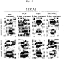
- induced pluripotent stem cell and, for example, human induced pluripotent cell lines established by Kyoto University such as 201B7 cell, 201B7-Ff cell, 253G1 cell, 253G4 cell, 1201C1 cell, 1205D1 cell, 1210B2 cell, 1231A3 cell, Ff-I01 cell, QHJI01 cell and the like are available from the National University Corporation Kyoto University and iPS Academia Japan, Inc.
- tissue-derived fibroblast tissue-derived fibroblast
- blood-lineage cells e.g., peripheral blood mononuclear cell, T cell
- hepatocyte e.g., hepatocyte
- pancreatic cell e.g., intestinal epithelial cell
- smooth muscle cell e.g., smooth muscle cell and the like
- fibroblast those derived from dermis and the like can be mentioned.
- the means for gene expression is not particularly limited.
- the aforementioned means include an infection method using a virus vector (e.g., retrovirus vector, lentivirus vector, Sendaivirus vector, adenovirus vector, adeno-associated virus vector), a gene transfer method using a plasmid vector (e.g., plasmid vector, episomal vector) (e.g., calcium phosphate method, lipofection method, RetroNectin method, electroporation method), a gene transfer method using an RNA vector (e.g., calcium phosphate method, lipofection method, electroporation method), a method with direct injection of protein and the like.
- a virus vector e.g., retrovirus vector, lentivirus vector, Sendaivirus vector, adenovirus vector, adeno-associated virus vector
- a gene transfer method using a plasmid vector e.g., plasmid vector, episomal vector
- the pluripotent stem cell to be used in the present invention is preferably ES cell or induced pluripotent stem cell, more preferably induced pluripotent stem cell.
- Genetically-modified pluripotent stem cells can be produced by using, for example, a homologous recombination technique.
- the gene on the chromosome to be modified include a cell marker gene, a histocompatibility antigen gene, a gene related to a disease due to a disorder of retinal cell and so on.
- a target gene on the chromosome can be modified using the methods described in Manipulating the Mouse Embryo, A Laboratory Manual, Second Edition, Cold Spring Harbor Laboratory Press (1994 ); Gene Targeting, A Practical Approach, IRL Press at Oxford University Press (1993 ); Biomanual Series 8, Gene Targeting, Making of Mutant Mouse using ES cell, YODOSHA CO., LTD. (1995 ); and so on.
- the genomic DNA comprising the target gene to be modified (e.g., cell marker gene, histocompatibility antigen gene, disease-related gene and so on) is isolated, and a targeting vector used for homologous recombination of the target gene is produced using the isolated genomic DNA.
- the produced targeting vector is introduced into stem cells and the cells that showed homologous recombination between the target gene and the targeting vector are selected, whereby stem cells having the modified gene on the chromosome can be produced.
- genomic DNA comprising the target gene examples include known methods described in Molecular Cloning, A Laboratory Manual, Second Edition, Cold Spring Harbor Laboratory Press (1989 ), Current Protocols in Molecular Biology, John Wiley & Sons (1987-1997 ) and so on.
- the genomic gene comprising the target gene can also be isolated using genomic DNA library screening system (manufactured by Genome Systems), Universal GenomeWalker Kits (manufactured by CLONTECH) and so on.
- a polynucleotide encoding the target protein can also be used instead of genome DNA.
- the polynucleotide can be obtained by amplifying the corresponding polynucleotide by the PCR method.
- Targeting vector used for homologous recombination of the target gene and efficient selection of a homologous recombinant can be performed according to the methods described in Gene Targeting, A Practical Approach, IRL Press at Oxford University Press (1993 ); Biomanual Series 8, Gene Targeting, Making of Mutant Mouse using ES cell, YODOSHA CO., LTD. (1995 ); and so on.
- the targeting vector any of replacement type or insertion type can be used.
- selection method methods such as positive selection, promoter selection, negative selection, polyA selection and so on can be used.
- Examples of a method for selecting the desired homologous recombinant from the selected cell lines include Southern hybridization method, PCR method and so on for the genomic DNA.
- the "mammal” in the present invention encompasses rodents, ungulata, carnivora, primates and the like.
- the rodents encompass mouse, rat, hamster, guinea pig and the like.
- Ungulata encompass swine, bovine, goat, horse, sheep and the like.
- Carnivora encompasses dog, cat and the like.
- the "primates” in the present invention refers to mammals belonging to the primate, and the primates include strepsirhini such as lemur, loris, tupai and the like, and anthropoidea such as monkey, ape, human and the like.
- the pluripotent stem cells to be used in the present invention are mammalian pluripotent stem cells, preferably pluripotent stem cells of rodents (e.g., mouse, rat) or primates (e.g., human, monkey), more preferably human pluripotent stem cells.
- rodents e.g., mouse, rat
- primates e.g., human, monkey
- the pluripotent stem cell to be used in the present invention is most preferably human induced pluripotent stem cell (human iPS cell).
- the "suspension culturing” or “suspension culture method” in the present invention refers to culturing while maintaining a state in which cells or cell aggregates are suspended in a culture medium and a method of performing the culture. That is, suspension culturing is performed under conditions in which cells or cell aggregates are not adhered to a culture vessel and the like, and culturing performed under conditions permitting adhesion to a culture vessel and the like (adhesion culturing or adhesion culture method) is not included in the category of suspension culturing.
- adhesion of cell means that a strong cell-substratum junction is formed between a cell or cell aggregate and a culture vessel.
- suspension culturing refers to culturing under conditions in which a strong cell-substratum junction is not formed between a cell or cell aggregate and a culture vessel
- adheresion culturing refers to culturing under conditions in which a strong cell-substratum junction is formed between a cell or cell aggregate and a culture vessel and the like.
- a planar cell-cell adhesion is formed in a cell aggregate in suspension culture.
- a cell-substratum junction is hardly formed with a culture vessel and the like and, even if it is formed, its contribution is small.
- an endogenous cell-substratum junction is present inside the aggregate, but a cell-substratum junction is hardly formed with a culture vessel and the like and, even if it is formed, its contribution is small.
- planar cell-cell adhesion means that a cell attaches to another cell via planes. More particularly, the planar cell-cell adhesion means that, for example, not less than 1%, preferably not less than 3%, more preferably not less than 5%, of the surface area of a cell adheres to the surface of another cell.
- a surface of a cell can be observed by staining with a reagent (e.g., DiI) that stains membranes, immunostaining of cell adhesion molecules (e.g., E-cadherin and N-cadherin).
- the cell culture vessel to be used when performing suspension culturing is not particularly limited as long as it enables "culturing in suspension” and those of ordinary skill in the art can appropriately determine same.
- Examples of such cell culture vessel include flask, tissue culture flask, dish, petri dish, tissue culture dish, multidish, microplate, microwell plate, micropore, multiplate, multiwell plate, chamber slide, schale, tube, tray, culture bag, spinner flask, roller bottle and so on.
- these culture vessels are preferably non-cell-adherent.
- Useful non-cell-adherent culture vessels include culture vessels whose surfaces have not undergone an artificial treatment for improving the cell adhesiveness (e.g., surface treatment such as coating treatment with extracellular matrix such as basement membrane preparation, laminin, entactin, collagen, gelatin etc., and the like, or polymer such as polylysine, polyornithine and the like or positive electric charge treatment and the like), and the like.
- an artificial treatment for improving the cell adhesiveness e.g., surface treatment such as coating treatment with extracellular matrix such as basement membrane preparation, laminin, entactin, collagen, gelatin etc., and the like, or polymer such as polylysine, polyornithine and the like or positive electric charge treatment and the like
- a non-cell-adherent culture vessel culture vessels whose surfaces have been artificially treated to decrease adhesiveness to the cells (e.g., superhydrophilic treatment with MPC polymer and the like, protein low adsorption treatment etc.) and the like can be used.
- culture vessels whose surfaces have been artificially treated to improve cell adhesiveness e.g. surface treatment such as coating treatment with extracellular matrix such as basement membrane preparation, laminin, entactin, collagen, gelatin, Matrigel, Synthemax, vitronectin and the like, or polymers such as polylysine, polyornithine and the like, or surface-processing by a positive electric charge treatment
- surface treatment such as coating treatment with extracellular matrix such as basement membrane preparation, laminin, entactin, collagen, gelatin, Matrigel, Synthemax, vitronectin and the like, or polymers such as polylysine, polyornithine and the like, or surface-processing by a positive electric charge treatment
- the medium to be used for culturing cells in the present invention can be prepared from a medium generally used for culturing animal cells as a basal medium.
- the basal medium include media that can be used for culturing animal cells such as BME medium, BGJb medium, CMRL1066 medium, Glasgow MEM medium, Improved MEM Zinc Option medium, IMDM medium, Medium199 medium, Eagle MEM medium, ⁇ MEM medium, DMEM medium, F-12 medium, DMEM/F-12 medium, IMDM/F12 medium, Ham medium, RPMI1640 medium, Fischer's medium, and mixed medium thereof etc.
- the "serum-free medium” in the present invention means a medium free of unadjusted or unpurified serum.
- a medium containing purified blood-derived components and animal tissue-derived components e.g., growth factor
- a medium containing purified blood-derived components and animal tissue-derived components is also included in a serum-free medium unless unadjusted or unpurified serum is contained therein.
- serum-free conditions means conditions free of unadjusted or unpurified serum, specifically, conditions using a serum-free medium.
- the serum-free medium may contain a serum replacement.
- the serum replacement include one appropriately containing albumin, transferrin, fatty acid, collagen precursor, trace element, 2-mercaptoethanol or 3' thiolglycerol, or equivalents of these etc., and so on.
- Such serum replacement may be prepared by, for example, the method described in WO98/30679 .
- the serum replacement may also be a commercially available product.
- KSR KnockoutTM Serum Replacement
- Chemically Defined Lipid Concentrated manufactured by Life Technologies
- GlutamaxTM manufactured by Life Technologies
- B27 manufactured by Life Technologies
- N2 manufactured by Life Technologies
- the serum-free medium to be used for suspension culturing may appropriately contain a fatty acid or lipid, amino acid (e.g., non-essential amino acids), vitamin, growth factor, cytokine, antioxidant, 2-mercaptoethanol, pyruvic acid, buffering agent, inorganic salts and so on.
- amino acid e.g., non-essential amino acids
- vitamin e.g., growth factor, cytokine, antioxidant, 2-mercaptoethanol, pyruvic acid, buffering agent, inorganic salts and so on.
- a serum-free medium supplemented with an appropriate amount (e.g., about 0.5% to about 30%, preferably about 1% to about 20%) of commercially available KSR (manufactured by Life Technologies) may be used as such serum-free medium (e.g., medium of 1:1 mixture of F-12 medium and IMDM medium supplemented with 10% KSR and 450 ⁇ M 1-monothioglycerol).
- KSR commercially available KSR
- the medium disclosed in JP-A-2001-508302 can be mentioned.
- the "serum-containing medium” in the present invention means a medium containing unadjusted or unpurified serum.
- the medium may contain a fatty acid, lipid, amino acid (e.g., non-essential amino acids), vitamin, growth factor, cytokine, antioxidant, 2-mercaptoethanol, 1-monothioglycerol, pyruvic acid, buffering agent, inorganic salts and so on.
- a serum medium can be used in the step of maintaining the retinal cell or retinal tissue produced by the present invention ( Cell Stem Cell, 10(6), 771-775 (2012 )).
- the culturing is preferably performed under xeno-free conditions.
- xeno-free means conditions eliminating components derived from species different from that of the cell to be cultured.
- the "medium containing substance X" or “in the presence of substance X” refer to a medium supplemented with an exogenous substance X or a medium containing an exogenous substance X, or in the presence of an exogenous substance X. That is, when the cells or tissues present in the medium endogenously express, secrete or produce substance X, the endogenous substance X is distinguished from the exogenous substance X, and a medium free of exogenous substance X is understood to fall outside the category of the "medium containing substance X", even when it contains endogenous substance X.
- a "medium containing a factor for maintaining an undifferentiated state” is a medium supplemented with an exogenous factor for maintaining an undifferentiated state or a medium containing an exogenous factor for maintaining an undifferentiated state.
- a feeder cell refers to a cell other than a stem cell co-exist when culturing the stem cell.
- the feeder cells used for culturing pluripotent stem cells while maintaining an undifferentiated state include mouse fibroblasts (MEF), human fibroblasts, SNL cells and the like.
- the feeder cells feeder cells that underwent a growth suppression treatment is preferable.
- the growth suppression treatment include treatment with a growth inhibitor (e.g., mitomycin C), gamma radiation, UV irradiation and the like.
- Feeder cells used for culturing pluripotent stem cells while maintaining an undifferentiated state contributes to the maintenance of undifferentiation of pluripotent stem cell by secretion of a humoral factor (preferably factor for maintaining an undifferentiated state), or production of a scaffold for cell adhesion (extracellular substrate).
- a humoral factor preferably factor for maintaining an undifferentiated state
- extracellular substrate extracellular substrate
- the absence of feeder cells means culturing in the absence of a feeder cell.
- the absence of feeder cells means, for example, conditions free of addition of feeder cells, or conditions substantially free of feeder cells (e.g., the ratio of the number of feeder cells relative to the total number of cells is not more than 3%).
- an "aggregate" of cells refers to a clump formed by assembly of cells dispersed in a medium, wherein the cells are adhered to each other.
- Cell clumps, embryoid bodies, spheres, spheroids are also encompassed in the cell aggregates.
- a planar cell-cell adhesion is formed in the aggregate of cells.
- cells sometimes form a cell-cell junction and/or a cell adhesion, for example, adherence junction, in some or all of the aggregates.
- the “aggregate” in the present invention specifically includes an aggregate produced in the second step of the above-mentioned present invention [1], which is formed by cells dispersed at the time of the start of the suspension culturing, and an aggregate produced in the third step of the above-mentioned present invention [1], which contains induced retinal cells differentiated from pluripotent stem cell, and the “aggregate” also includes an aggregate already formed at the time of the start of the second step in the above-mentioned present invention [1] (i.e., at the time of the start of suspension culture).
- the cell aggregate formed in the second step encompasses "embryoid body (EB)".
- uniformed aggregates means that the size of each aggregate is constant when a plurality of aggregates are cultured, and that the variance in the length of the maximum diameter is small when the size of the aggregates are evaluated by the length of the maximum diameter. More specifically, it means that not less than 75% of aggregates in the whole aggregate population are within mean ⁇ 100%, preferably mean ⁇ 50%, more preferably mean ⁇ 20%, of the maximum diameter in the population of the aggregates.
- to "form uniformed cell aggregates” means to "rapidly aggregate a given number of dispersed cells” to form cell aggregates uniform in size, when gathering the cells to form cell aggregates and culturing the aggregates in suspension.
- Dispersion refers to dividing cells or a tissue into small cell clumps (not less than 2 cells and not more than 100 cells, preferably not more than 50 cells) or single cells by a dispersion treatment such as enzymatic treatment, physical treatment and the like.
- a given number of dispersed cells is a collection of a certain number of cell clumps or single cells.
- Examples of the method of dispersing pluripotent stem cells include mechanical dispersion treatment, cell dispersion solution treatment, and cell protecting agent addition treatment. These treatments may be performed in combination. Preferably, cell dispersion treatment is performed and then mechanical dispersion treatment is performed.
- a cell dispersion solution to be used for the cell dispersion solution treatment a solution containing any of enzymes such as trypsin, collagenase, hyaluronidase, elastase, pronase, DNase, papain and so on, and a chelating agent such as ethylenediaminetetraacetic acid and so on can be mentioned.
- a commercially available cell dispersion solution such as TripLE Select (manufactured by Life Technologies) and TripLE Express (manufactured by Life Technologies) can also be used.
- pluripotent stem cells When pluripotent stem cells are dispersed or until dispersed cells form an aggregate, the cell death of pluripotent stem cells that have not formed an aggregate may be suppressed by a treatment with a cell protecting agent.
- a cell protecting agent to be used for the cell protecting agent treatment an FGF signal transduction pathway activating substance, heparin, Rho-associated coiled-coil kinase (ROCK) inhibitor, serum, or serum replacement can be mentioned.
- a ROCK inhibitor As a preferable cell protecting agent, a ROCK inhibitor can be mentioned.
- Y-27632, Fasudil or H-1152 can be mentioned. While the concentration of a ROCK inhibitor can be appropriately set by those of ordinary skill in the art, for example, it can be set within a concentration range corresponding to a ROCK inhibitory activity with about 50 nM - 200 ⁇ M of Y-27632.
- a method for dispersing pluripotent stem cells includes, for example, a method involving treating a colony of pluripotent stem cells with a cell dispersion solution (TrypLE Select) in the presence of a ROCK inhibitor as a cell protecting agent, and further dispersing them by pipetting.
- a cell dispersion solution TrypLE Select
- ROCK inhibitor as a cell protecting agent
- an aggregate of pluripotent stem cells by rapidly gathering the pluripotent stem cells.
- an aggregate of pluripotent stem cells is formed in such a manner, an epithelium-like structure can be formed with good reproducibility in the cells induced and differentiated from the formed aggregate.
- Examples of the experimental operation to form an aggregate include a method involving keeping cells in a small space by using a plate with small wells (e.g., plate with wells having a base area of about 0.1 - 2.0 cm 2 when calculated in terms of flat bottom), micropore and so on, a method involving aggregating cells by centrifugation for a short time using a small centrifugation tube.
- a plate with small wells for example, 24 well plate (area of about 1.88 cm 2 when calculated in terms of flat bottom), 48 well plate (area of about 1.0 cm 2 when calculated in terms of flat bottom), 96 well plate (area of about 0.35 cm 2 when calculated in terms of flat bottom, inner diameter about 6 - 8 mm), and 384 well plate can be mentioned.
- the shape of the bottom surface when the well is seen from above is, for example, polygon, rectangle, ellipse, true circle, preferably true circle.
- the shape of the bottom surface is preferably a structure having high outer circumference and low inner concave.
- U-bottom, V-bottom, ⁇ -bottom can be mentioned, preferably U-bottom or V-bottom, more preferably V-bottom.
- a cell culture dish e.g., 60 mm - 150 mm dish, culture flask
- a concave convex, or dent e.g., EZSPHERE (Asahi Techno Glass)
- the bottom surface of a plate with small wells is preferably a non-cell-adhesive bottom surface, preferably the aforementioned non-cell-adhesive-coated bottom surface.
- Formation of aggregates of pluripotent stem cells and formation of an epithelial-like structure in the aggregate can be confirmed based on the macroscopic morphology of the aggregate, microscopic morphology by tissue staining analysis, distribution of cells expressing differentiation and undifferentiation markers and the like.
- tissue in the present invention refers to a structure of a cell population having a structure in which plural types of cells having different morphologies and properties are sterically arranged in a given pattern.
- the "retinal tissue” means a tissue in which at least two or more types of cells such as photoreceptor cells, horizontal cells, bipolar cells, amacrin cells, retinal ganglion cells, their progenitor/progenitor cells, and retinal progenitor cells and so on, which constitute respective retinal layers in retina in vivo, are sterically arranged in layers.
- the retinal layer which is constituted by each cell can be confirmed by a known method, for example, presence or absence of the expression of a cell marker or the level thereof, etc.
- the "retinal layer” in the present invention means each layer constituting the retina. Specific examples thereof include retinal pigment epithelial layer, photoreceptor layer, external limiting membrane, outer nuclear layer, outer plexiform layer, inner nuclear layer, inner plexiform layer, ganglion cell layer, nerve fiber layer and inner limiting membrane.
- the "retinal progenitor cell” in the present invention refers to a progenitor cell capable of differentiating into any mature retinal cell including photoreceptor cell, horizontal cell, bipolar cell, amacrine cell, retinal ganglion cell, retinal pigment epithelial cell and the like.
- the photoreceptor progenitor cell, horizontal cell progenitor cell, bipolar cell progenitor cell, amacrine cell progenitor cell, retinal ganglion cell progenitor cell, and retinal pigment epithelial progenitor cell refer to progenitor cells committed to differentiate into photoreceptor cell, horizontal cell, bipolar cell, amacrine cell, retinal ganglion cell, and retinal pigment epithelial cell, respectively.
- the "retinal layer-specific neuron” is a cell constituting a retina layer and is a neuronal cell specific to the retinal layer.
- the retinal layer-specific neuron include bipolar cell, retinal ganglion cell, amacrine cell, horizontal cell and photoreceptor cell, and examples of the photoreceptor cell include rod cell and cone cell.
- retinal cell in the present invention encompasses the aforementioned retinal progenitor cell, retinal layer specific neuron and progenitor cell of retinal layer specific neuron.
- retinal cell marker examples include Rx (also referred to as Rax), PAX6 and Chx10 expressed in retinal progenitor cell, Nkx2.1 expressed in progenitor cell of hypothalamus neuron but not expressed in retinal progenitor cell, Sox1 expressed in hypothalamus neuroepithelium but not expressed in retina, Crx and Blimp1 expressed in progenitor cell of photoreceptor cell, and the like.
- Examples of the marker of the retinal layer-specific neuron include Chx10, PKC ⁇ and L7 expressed in bipolar cell, TUJI and Brn3 expressed in retinal ganglion cell, Calretinin expressed in amacrine cell, Calbindin expressed in horizontal cell, Rhodopsin and Recoverin expressed in mature photoreceptor cell, Nrl expressed in rod cell, Rxr-gamma expressed in cone cell, RPE65 and Mitf expressed in retinal pigment epithelium cell and the like.
- the production method of the present invention is a method for producing a retinal cell or a retinal tissue, comprising the following steps:
- induced pluripotent stem cell As a preferable pluripotent stem cell in the first step, induced pluripotent stem cell, more preferably human induced pluripotent stem cell can be mentioned.
- the production method of induced pluripotent stem cells is not particularly limited, and it can be produced by a method well known to those of ordinary skill in the art as mentioned above. It is also desirable to perform a step for preparing induced pluripotent stem cells (that is, a step of reprogramming somatic cells to establish pluripotent stem cells) under feeder-free condition.
- the maintenance culturing or expansion culturing for obtaining pluripotent stem cells to be used in the first step can be performed by a method well known to those of ordinary skill in the art as mentioned above. While the maintenance culturing and expansion culturing of pluripotent stem cells can be performed by adhesion culturing or suspension culturing, it is preferably performed by adhesion culturing. While the step of maintenance culturing and expansion culturing of pluripotent stem cells may be performed in the presence of a feeder or under feeder-free condition, it is preferably performed under feeder-free condition.
- the production method of the present invention may comprise, before step (1), a step of maintaining a pluripotent stem cell under feeder-free conditions and providing the pluripotent stem cell maintained under feeder-free conditions.
- the period of maintaining under feeder-free conditions is not particularly limited as long as retinal cells or retinal tissues are produced by the method of the present invention, it is generally not less than one day, preferably not less than 3 days, more preferably not less than 7 days.
- pluripotent stem cells can be semipermanently cultured and expanded while maintaining pluripotency under appropriate culture conditions and under feeder-free conditions.
- the risk of their differentiation and loss of pluripotency may increase.
- the period for maintaining under feeder-free conditions is, for example, within 6 months, preferably within about 3 months, and the passage number is, for example, within 30 passages, preferably within 20 passages.
- the maintenance culture under feeder-free conditions can be performed under culture conditions of the first step described in detail below except that the medium does not contain an MEK inhibitor, a PKC inhibitor or a B-Raf inhibitor.
- dispersed pluripotent stem cells are prepared by a dispersion operation.
- a dispersion operation of the pluripotent stem cells include mechanical dispersion treatment, cell dispersion solution treatment or cell protecting agent addition treatment, mentioned earlier. These treatments may be performed in combination.
- cell dispersion treatment is performed simultaneously with cell protecting agent addition treatment, and then mechanical dispersion treatment is performed.
- heparin As a cell protecting agent used for the cell protecting agent addition treatment, heparin, serum and serum replacement can be mentioned.
- a Rho-associated coiled-coil kinase (ROCK) inhibitor may be added at the time of dispersion.
- ROCK inhibitor Y-27632, Fasudil (HA1077), H-1152 and the like can be mentioned.
- a cell dispersion solution to be used for the cell dispersion treatment a solution containing any of enzymes such as trypsin, collagenase, hyaluronidase, elastase, pronase, DNase, papain and so on, and a chelating agent such as ethylenediaminetetraacetic acid and so on can be mentioned.
- a commercially available cell dispersion solution such as TrypLE Select (manufactured by Life Technologies) and TrypLE Express (manufactured by Life Technologies) can also be used.
- the dispersed pluripotent stem cells are seeded in a new culture container and subjected to the first step.
- the pluripotent stem cells may be seeded in a new culture container, maintenance culturing may be continuously performed under feeder-free conditions in the presence of a ROCK inhibitor, and the first step may be started thereafter. While the period of the treatment with a ROCK inhibitor is not particularly limited as long as the cell death induced by dispersion can be suppressed, it is generally about 12 - 24 hr.
- the factor for maintaining an undifferentiated state is not particularly limited as long as it is a substance having an action to suppress differentiation of pluripotent stem cells. It is generally a factor for maintaining an undifferentiated state derived from a mammal. Since the factor for maintaining an undifferentiated state may have cross-reactivity among mammal species, a factor for maintaining an undifferentiated state of any mammal may also be used as long as the undifferentiated state of the pluripotent stem cells to be cultured can be maintained. Preferably, a factor for maintaining an undifferentiated state of a mammal of the same species as the cells to be cultured is used.
- Examples of the factor for maintaining an undifferentiated state widely used by those of ordinary skill in the art include an FGF signal transduction pathway activating substance, a TGF ⁇ family signal transduction pathway activating substance and the like in the case of primed pluripotent stem cells (e.g., human ES cells, human iPS cells).
- FGF signal transduction pathway activating substance fibroblast growth factors (e.g., bFGF, FGF4, FGF8) can be specifically mentioned.
- As the TGF ⁇ family signal transduction pathway activating substance a TGF ⁇ signal transduction pathway activating substance, a Nodal/Activin signal transduction pathway activating substance can be mentioned.
- TGF ⁇ signal transduction pathway activating substance TGF ⁇ 1, TGF ⁇ 2 can be mentioned.
- Nodal/Activin signal transduction pathway activating substance Nodal, Activin A, Activin B can be mentioned.
- the factor for maintaining an undifferentiated state is preferably bFGF.
- the factor for maintaining an undifferentiated state to be used in the present invention is preferably isolated. Being “isolated” means that an operation to remove factors other than the intended component or cell has been performed, and the component or cell is no longer in a naturally occurring state. Therefore, "isolated protein X" does not include an endogenous protein X produced from the cells or tissue to be cultured, and contained in a cell or tissue or in the medium.
- the purity of the "isolated protein X" (percentage of the weight of protein X to the total protein weight) is generally not less than 70%, preferably not less than 80%, more preferably not less than 90%, further preferably not less than 99%, most preferably 100%. Therefore, in one embodiment, the present invention comprises a step of providing an isolated factor for maintaining an undifferentiated state.
- it includes a step of exogenously adding an isolated factor for maintaining an undifferentiated state to a medium used in the first step.
- a factor for maintaining an undifferentiated state may be added in advance to a medium to be used in the first step.
- the aforementioned basal medium supplemented with an undifferentiated maintenance factor or a commercially available medium containing an undifferentiated maintenance factor for culturing undifferentiated cells such as stem cell or iPS cell and the like can be used in the first step.
- the concentration of the factor for maintaining an undifferentiated state in the medium to be used in the first step is a concentration capable of maintaining the undifferentiated state of the pluripotent stem cells to be cultured, and can be appropriately determined by those of ordinary skill in the art.
- the concentration thereof is generally about 4 ng - 500 ng/mL, preferably 10 ng - 200 ng/mL, more preferably about 30 ng - 150 ng/mL.
- the MEK inhibitor is not particularly limited as long as it is a substance that inhibits expression or activity of MEK family, and may be any of protein, nucleic acid and low-molecular-weight compound.
- the representative MEK family MEK1, MEK2, MEK3 and the like can be mentioned, and the MEK inhibitor is a substance that inhibits the expression or activity of one, a plurality or all of these MEK families.
- the substance include, but are not limited to, substances (e.g., antisense oligonucleotide, siRNA etc.) that suppress expression of genes encoding various MEKs and substances that inhibit enzyme activity of various MEKs.
- the MEK inhibitor include, but are not limited to, low-molecular-weight compounds such as PD0325901 (N-[(2R)-2,3-dihydroxy propoxy-3,4-difluoro-2-[(2-fluoro-4-Iodophenyl)amino]-benzamide), PD184352 (2-(2-chloro-4-indophenylamino)-N-cyclopropylmethoxy-3,4-difluorobenzamido), PD98059 (2'-amino-3'-methoxyflavone), U0126 (1.4-diamino-2,3-dicyano-1,4-bis[2-amino-phenylthio]butadiene), MEK162 (5-[(4-Bromo-2-fluorophenyl)amino]-4-fluoro-N-(2-hydroxyethoxy)-1-methyl-1H-benzimidazole-6-carboxamide), SL3
- PD0325901, PD184352, PD98059, U0126, MEK162 and SL327 are known MEK inhibitors and commercially available products and the like can be obtained as appropriate.
- the MEK inhibitor preferred is PD0325901, PD184352 or U0126, and more preferred is PD0325901.
- the concentration of the MEK inhibitor contained in the medium is not particularly limited as long as a retinal cell or retinal tissue of a mammal can be produced by the method of the present invention, and can be appropriately determined according to the kind of the MEK inhibitor.
- the concentration of the MEK inhibitor is in a concentration range showing an MEK inhibitory activity corresponding to 0.001 - 10 ⁇ M, preferably 0.005 - 5 ⁇ M, particularly preferably 0.01 - 2.5 ⁇ M, of PD0325901.
- the medium used in the first step may contain a PKC inhibitor or a B-Raf inhibitor as well as a factor for maintaining an undifferentiated state and an MEK inhibitor. Addition of a PKC inhibitor or a B-Raf inhibitor is expected to improve production efficiency of a cell aggregate capable of differentiating into retinal cells, or retinal cells and retinal tissues.
- the PKC inhibitor is not particularly limited as long as it is a substance capable of inhibiting expression or activity of protein kinase C (PKC), and may be any of protein, nucleic acid and low-molecular-weight compound.
- PKC protein kinase C
- PKC is a protein family constituted of at least 10 kinds of isozymes, and the PKC inhibitor is a substance that inhibits the expression or activity of one, a plurality or all of these PKC families.
- the substance examples include, but are not limited to, a substance that suppresses expression of a gene encoding PKC (e.g., antisense oligonucleotide, siRNA etc.), a substance that inhibits enzyme activity of PKC [for example, low-molecular-weight compounds such as Go6983 (wherein "o” is umlaut) (3-[1-[3-(Dimethylamino)propyl]-5-methoxy-1H-indol-3-yl]-4-(1H-indol-3-yl)-1H-pyrrole-2,5-dione) and the like, and the like] and the like.
- Go6983 is a known PKC inhibitor, and can be obtained easily since it is commercially available. As the PKC inhibitor, preferred is Go6983.
- the concentration of the PKC inhibitor contained in the medium is not particularly limited as long as the method of the present invention can produce a retinal cell or a retinal tissue of a mammal, and can be appropriately determined according to the kind of the PKC inhibitor.
- the concentration of the PKC inhibitor is, for example, within a concentration range showing a PKC inhibitory activity corresponding to 0.05 - 10 ⁇ M, preferably 0.25 - 5 ⁇ M, more preferably 0.5 - 2.5 ⁇ M, of Go6983.
- B-Raf inhibitor is not particularly limited as long as it is a substance that suppresses expression or serine/threonine kinase activity of B-Raf and may be any of protein, nucleic acid and low-molecular-weight compound.
- the substance examples include, but are not limited to, a substance that suppresses expression of a gene encoding B-Raf (e.g., antisense oligonucleotide, siRNA etc.), a substance that suppresses enzyme activity of B-Raf [for example, low-molecular-weight compounds such as anti-B-Raf antibody, SB590885 (5-[2-[4-[2-(dimethylamino)ethoxy]phenyl]-5-(4-pyridinyl)-1H-imidazol-4-yl]-2,3-dihydro-1H-inden-1-one oxime) and the like, and the like] and the like.
- B-Raf inhibitor is preferably SB590885.
- the concentration of the B-Raf inhibitor contained in the medium is not particularly limited as long as the method of the present invention can produce a retinal cell or a retinal tissue of a mammal, and can be appropriately determined according to the kind of the B-Raf inhibitor.
- the concentration of the B-Raf inhibitor is within a concentration range showing a B-Raf inhibitory activity corresponding to 0.05 - 10 ⁇ M, preferably 0.25 - 5 ⁇ M, more preferably 0.5 - 2.5 ⁇ M, of SB590885.
- a pluripotent stem cell may be cultured under any conditions of suspension culturing and adhesion culturing, preferably adhesion culturing.
- a medium to be used in the first step may be a serum-containing medium or a serum-free medium. To avoid contamination with chemically-undefined components, it is preferably a serum-free medium, more preferably, a serum-free medium containing the aforementioned serum replacement.
- the medium used in the first step is preferably a medium containing chemically-determined components.
- the medium used in the first step is not particularly limited as long as it contains 1) a factor for maintaining an undifferentiated state, and 2) an MEK inhibitor (and optionally 3) a PKC inhibitor or a B-Raf inhibitor) and enables undifferentiated maintenance culture of pluripotent stem cells under feeder-free conditions (feeder-free medium).
- in the absence of a feeder cell means conditions substantially free of a feeder cell (e.g., the ratio of number of feeder cells relative to the total number of cells is not more than 3%).
- Essential 8 medium is DMEM/F12 medium containing L-ascorbic acid-2-phosphate magnesium (64 mg/l), sodium selenium (14 ⁇ g/1), insulin (19.4 mg/l), NaHCO 3 (543 mg/l), transferrin (10.7 mg/l), bFGF (100 ng/mL), and a TGF ⁇ family signal transduction pathway activating substance (TGF ⁇ 1 (2 ng/mL) or Nodal (100 ng/mL)) as additives ( Nature Methods, 8, 424-429 (2011 )).
- feeder-free medium examples include Essential 8 (manufactured by Life Technologies), S-medium (manufactured by DS Pharma Biomedical), StemPro (manufactured by Life Technologies), hESF9 ( Proc Natl Acad Sci U S A. 2008 Sep 9; 105(36):13409-14 ), mTeSR1 (manufactured by STEMCELL Technologies), mTeSR2 (manufactured by STEMCELL Technologies), and TeSR-E8 (manufactured by STEMCELL Technologies).
- StemFit manufactured by Ajinomoto Co., Inc.
- Ajinomoto Co., Inc. can be mentioned as the feeder-free medium.
- the present invention can be performed using any of these media added with (i) an MAP kinase kinase inhibitor (MEK inhibitor) or a medium added with (ii) an MEK inhibitor and a PKC inhibitor or B-Raf inhibitor in the above-mentioned first step.
- MEK inhibitor MAP kinase kinase inhibitor
- PKC inhibitor or B-Raf inhibitor in the above-mentioned first step, any of these media added with an MEK inhibitor, a medium added with an MEK inhibitor and a PKC inhibitor or a medium added with an MEK inhibitor and a B-Raf inhibitor can be mentioned.
- the medium used in the first step contains an MEK inhibitor in addition to a factor for maintaining an undifferentiated state.
- the medium used in the first step contains an MEK inhibitor, and a PKC inhibitor or a B-Raf inhibitor in addition to a factor for maintaining an undifferentiated state.
- the state of the cells changes and cell aggregates can be produced with high efficiency.
- the state of the cells changes and the quality of the aggregates is improved.
- round cell aggregates with a smooth surface and dense inside are expected to be produced with high efficiency.
- the medium to be used in the first step can contain a further component (e.g., any activating substance or inhibitor of signal transduction pathway) as long as the production efficiency of retinal cells or retinal tissues by the production method of the present invention is not markedly decreased (e.g., the same level as or not more than the level of efficiency without the first step).
- an appropriate matrix may be used as a scaffold to provide a scaffold in stead of the feeder cells to the pluripotent stem cells.
- the pluripotent stem cells are subjected to adhesion culturing in a cell container whose surface is coated with a matrix as a scaffold.
- laminin As a matrix available as a scaffold, laminin ( Nat Biotechnol 28, 611-615 (2010 )), laminin fragment ( Nat Commun 3, 1236 (2012 )), basement membrane preparation ( Nat Biotechnol 19, 971-974 (2001 )), gelatin, collagen, heparan sulfate proteoglycan, entactin, vitronectin and the like can be mentioned.
- laminin is a heterotrimer molecule consisting of ⁇ , ⁇ , ⁇ chains and an extracellular matrix protein containing isoforms having different subunit chain compositions. Specifically, laminin has about 15 kinds of isoforms based on the combinations of heterotrimers with 5 kinds of ⁇ chains, 4 kinds of ⁇ chains and 3 kinds of ⁇ chains.
- the name of laminin is determined by combining respective numbers of ⁇ chain ( ⁇ 1 - ⁇ 5), ⁇ chain ( ⁇ 1 - ⁇ 4) and ⁇ chain ( ⁇ 1 - ⁇ 3).
- laminin 511 is preferably used ( Nat Biotechnol 28, 611-615 (2010 )).
- Laminin to be used in the present invention is generally a mammalian laminin.
- the mammal those mentioned above can be recited.
- laminin of a mammal of the same species as the cell to be cultured is preferably used.
- human laminin preferably, human laminin 511
- human laminin 511 is used for culturing human pluripotent stem cells.
- a laminin fragment to be used in the present invention is not particularly limited as long as it has adhesiveness to pluripotent stem cells and enables maintenance culturing of pluripotent stem cell under feeder-free conditions, and is preferably E8 fragment.
- Laminin E8 fragment (laminin511-E8) was identified as a fragment with strong cell adhesion activity among the fragments obtained by digestion of laminin 511 with elastase ( EMBO J., 3:1463-1468, 1984 , J. Cell Biol., 105:589-598, 1987 ).
- E8 fragment of laminin 511 is preferably used ( Nat Commun 3, 1236 (2012 ), Scientific Reports 4, 3549 (2014 )).
- the laminin E8 fragment to be used in the present invention is not required to be an elastase- digestion product of laminin and may be a recombinant. To avoid contamination of unidentified components, a recombinant laminin fragment is preferably used in the present invention.
- a E8 fragment of laminin 511 is commercially available and, for example, iMatrix-511 (Nippi, Inc.) and the like can be purchased.
- the laminin or laminin fragment to be used in the present invention is preferably isolated.
- the “basement membrane preparation (basement membrane specimen)” in the present invention refers to one containing basement membrane-constituting components having a function to control cell morphology, differentiation, growth, motility, expression of function and so on which are similar to those of epithelial cell, when intended cells capable of forming a basement membrane are plated thereon and cultured.
- basement membrane preparation when intended cells capable of forming a basement membrane are plated thereon and cultured.
- retinal cells and retinal tissues produced by the present invention may be dispersed, and cultured in the presence of a basement membrane preparation when further adhesion culturing is performed.
- the “basement membrane constituting components” refers to extracellular matrix molecules in the form of a thin membrane present between epithelial cell layer and interstitial cell layer and so on in animal tissues.
- a basement membrane preparation can be produced by, for example, removing cells capable of forming a basement membrane, which adhere onto a support via a basement membrane, from a support with a solution capable of dissolving the lipid of the cells, an alkali solution and so on.
- the basement membrane preparation include products commercially available as basement membrane preparation (e.g., MatrigelTM (manufactured by Becton Dickinson: hereinafter sometimes referred to as Matrigel)), GeltrexTM (manufactured by Life Technologies), and extracellular matrix molecules known as basement membrane components (e.g., laminin, type IV collagen, heparan sulfate proteoglycan, entactin and so on).
- MatrigelTM is a basement membrane preparation extracted from Engelbreth Holm Swarn (EHS) mouse sarcoma.
- the main component of MatrigelTM is type IV collagen, laminin, heparan sulfate proteoglycan, and entactin.
- TGF- ⁇ , FGF, tissue plasminogen activator, and a growth factor naturally produced by EHS tumor are contained.
- the "growth factor reduced product" of MatrigelTM has a lower growth factor concentration than common MatrigelTM, and the standard concentration thereof is ⁇ 0.5 ng/ml for EGF, ⁇ 0.2 ng/ml for NGF, ⁇ 5 pg/ml for PDGF, 5 ng/ml for IGF1, and 1.7 ng/ml for TGF ⁇ .
- an isolated laminin or laminin fragment is preferably used in the present invention.
- the human pluripotent stem cells are cultured in an adhered state in a cell container with surface coated with isolated laminin 511 or E8 fragment of laminin 511 (most preferably, E8 fragment of laminin 511).
- the period for the culturing of pluripotent stem cells in the first step is not particularly limited as long as the effect of improving the quality of the aggregate formed in a second step can be achieved, it is generally a period not exceeding 30 days, preferably 0.5 - 8 days, more preferably 1 - 6 days, further preferably 4 - 6 days. That is, the first step is started 30 days, preferably 0.5 - 8 days, more preferably 1 - 6 days, further preferably 4 - 6 days before the start of the second step, and the second step is continuously performed on completion of the first step.
- the culture period of the pluripotent stem cells in the first step is set to 4 - 6 days (e.g., 5 days).
- the culture period of the pluripotent stem cells in the first step is set to 1 - 6 days (e.g., 1 - 5 days).
- the culture conditions such as culture temperature, and CO 2 concentration in the first step can be appropriately determined. While the culture temperature is, for example, about 30°C to about 40°C, preferably about 37°C.
- the CO 2 concentration is, for example, about 1% to about 10%, preferably about 5%.
- the cell population obtained in the first step is a cell population containing cells maintaining pluripotent-like properties (pluripotent-like state).
- pluripotent-like properties mean that at least a part of the characteristics unique to and common to the pluripotent stem cells including pluripotency is maintained.
- the pluripotent-like properties do not require strict pluripotency.
- a state expressing all or some of the markers to be the index of pluripotent properties is included in the "pluripotent-like properties”.
- a marker (undifferentiated marker) of the pluripotent-like properties As a marker (undifferentiated marker) of the pluripotent-like properties, Oct3/4 positive, alkaline phosphatase positive and the like can be mentioned.
- a cell maintaining the pluripotent-like properties is Oct3/4 positive. Even if the expression level of Nanog is lower than that of ES cell or iPS cell, it corresponds to "cell showing pluripotent-like properties". That is, while the first step is not the culture condition capable of maintaining the undifferentiated state of pluripotent stem cells, it is conducted under culturing conditions capable of maintaining pluripotent-like properties enabling differentiation into at least plural types of ectodermal cells. The pluripotent-like properties are maintained throughout the first step.
- the cells obtained in the first step have an ability to differentiate into retinal cells and retinal tissues.
- the cells obtained in the first step are stem cells having an ability to differentiate into retinal cells (including retinal progenitor cells, retina layer specific nerve cells and progenitor cells thereof) and retinal tissues.
- human pluripotent stem cells are adhesion cultured in the absence of a feeder cell in (i) a serum-free medium containing an MEK inhibitor and bFGF, or (ii) a serum-free medium containing an MEK inhibitor, a PKC inhibitor or a B-Raf inhibitor, and bFGF.
- a specific combination includes a combination of an MEK inhibitor (e.g., PD0325901) and bFGF; a combination of an MEK inhibitor (e.g., PD0325901), a PKC inhibitor (e.g., Go6983) and bFGF; a combination of an MEK inhibitor (e.g., PD0325901), a B-Raf inhibitor (e.g., SB590885) and bFGF, and the like.
- an MEK inhibitor e.g., PD0325901
- PKC inhibitor e.g., Go6983
- bFGF e.g., Go6983
- a combination of an MEK inhibitor e.g., PD0325901
- B-Raf inhibitor e.g., SB590885
- the above-mentioned adhesion culture is preferably performed in a cell container with a surface coated with laminin 511 or E8 fragment of laminin 511.
- human pluripotent stem cells e.g., human iPS cells
- the maintenance culture is preferably performed in adhesion culturing.
- the adhesion culturing is preferably performed in a cell container with a surface coated with vitronectin, laminin 511 or E8 fragment of laminin 511.
- an MEK inhibitor or (ii) a combination of an MEK inhibitor and a PKC inhibitor or a B-Raf inhibitor (specifically, PD0325901; combination of PD0325901 and Go6983; combination of PD0325901 and SB590885 and the like can be mentioned) and culturing is continued.
- a combination of an MEK inhibitor and a PKC inhibitor or a B-Raf inhibitor specifically, PD0325901; combination of PD0325901 and Go6983; combination of PD0325901 and SB590885 and the like can be mentioned
- the cells thus-obtained in the first step have an ability to differentiate into at least retinal tissues, retinal cells, retinal progenitor cells, or retina layer specific nerve cells.
- the cells thus-obtained in the first step include stem cells having an ability to differentiate into at least retinal tissues, retinal cells, retinal progenitor cells, or retina layer specific nerve cells.
- the second step of culturing the cells obtained in the first step in suspension in a medium to form a cell aggregate is explained.
- the medium to be used in the second step is not particularly limited as long as it is as described in the above-mentioned section of definition.
- the medium to be used in the second step may be a serum-containing medium or serum-free medium.
- a serum-free medium not added with any of a factor for maintaining an undifferentiated state an MEK inhibitor, a PKC inhibitor and a B-Raf inhibitor can be used in the present invention.
- a serum-free medium supplemented with an appropriate amount of a commercially available serum replacement such as KSR and so on (e.g., medium of 1:1 mixture of IMDM and F-12, which is supplemented with 10% KSR, 450 ⁇ M 1-monothioglycerol and 1x Chemically Defined Lipid Concentrate, or GMEM medium supplemented with 5% - 20% KSR, NEAA, pyruvic acid, 2-mercaptoethanol) is preferably used.
- the amount of KSR to be added to a serum-free medium in the case of human pluripotent stem cells is, for example, about 1% to about 30%, preferably about 2% to about 20%.
- the dispersed cells are prepared by a dispersing operation of the cells obtained in the first step.
- the "dispersed cells” obtained by the dispersing operation refers to a cell population in which, for example, not less than 70% of cells are single cells and not more than 30% of cells are clumps of 2 - 50 cells.
- it is a cell population of dispersed cells in which not less than 80% of the cells are single cells, and not more than 20% of the cells are clumps of 2 - 50 cells.
- the dispersed cells refer to a cell population in a state almost free of mutual adhesion of cells (e.g., plane attachment).
- dispersed cells refer to a cell population in a state almost free of cell-cell junction (e.g., adhesive bond).
- a dispersion operation of the cells obtained in the first step may contain the above-mentioned mechanical dispersion treatment, cell dispersion solution treatment, and cell protecting agent addition treatment. These treatments may be performed in combination.
- cell dispersion solution treatment is performed simultaneously with cell protecting agent addition treatment and then mechanical dispersion treatment is performed.
- heparin As a cell protecting agent to be used for the cell protecting agent addition treatment, heparin, serum and serum replacement can be mentioned. Also, to suppress cell death of pluripotent stem cells (particularly, human pluripotent stem cells) induced by dispersion, a Rho-associated coiled-coil kinase (ROCK) inhibitor or a Myosin inhibitor may be added from the start of the second step culture.
- a ROCK inhibitor Y-27632, Fasudil (HA1077), H-1152 and the like can be mentioned.
- a cell dispersion solution to be used for the cell dispersion treatment a solution containing any of enzymes such as trypsin, collagenase, hyaluronidase, elastase, pronase, DNase, papain and so on, and a chelating agent such as ethylenediaminetetraacetic acid and so on can be mentioned.
- a commercially available cell dispersion solution such as TrypLE Select (manufactured by Life Technologies) and TrypLE Express (manufactured by Life Technologies) can also be used.
- the dispersed cells are suspended in the above-mentioned medium.
- a suspension of the dispersed cells is seeded in the above-mentioned culture vessel, and the dispersed cells are cultured under a condition non-adhesive to the culture vessel, whereby plural cells are gathered to form an aggregate.
- plural cell aggregates may be simultaneously formed in one culture vessel by seeding the dispersed cells in a comparatively large culture vessel such as a 10 cm dish.
- the size of the aggregates varies in this case.
- a given amount of the cells are placed in each well of a multiwell plate (U-bottom, V-bottom) such as a 96-well microplate, and static culture is performed, whereby the cells are rapidly coagulated to form one aggregate in each well.
- the aggregates are recovered from plural wells, whereby a population of uniformed aggregates can be obtained.
- the concentration of the cells in the second step can be appropriately set so that cell aggregates can be more uniformly and efficiently formed.
- human cells e.g., cells obtained from human iPS cells in the first step
- a liquid prepared to achieve about 1 x 10 3 to about 1 x 10 5 cells, preferably about 3 x 10 3 to about 5 x 10 4 cells, more preferably about 4 x 10 3 to about 2 x 10 4 cells, further preferably about 4 x 10 3 to about 1.6 x 10 4 cells, most preferably about 8 x 10 3 to about 1.2 x 10 4 cells, per well is added to the wells, and the plate is left to stand to form aggregates.
- the culture conditions such as culture temperature, CO 2 concentration and so on in the second step can be appropriately determined.
- the culture temperature is, for example, about 30°C to about 40°C, preferably about 37°C.
- the CO 2 concentration is, for example, about 1% to about 10%, preferably about 5%.
- a medium change operation for example, an operation to add a fresh medium without discarding the existing medium (medium addition operation), an operation to discard about a half amount of the existing medium (about 30 - 90%, for example, 40 - 60% of the volume of the existing medium) and add about a half amount of a fresh medium (30 - 90%, for example, about 40 - 60% of the volume of the existing medium) (half-medium change operation), and an operation to discard about the whole amount of the existing medium (not less than 90% of the amount of the existing medium) and add about the whole amount of a fresh medium (not less than 90% of the amount of the existing medium) (full-medium change operation) can be mentioned.
- an operation to calculate the final concentration, to discard about a half amount of the existing medium, and to add about a half amount of a fresh medium containing a particular component at a concentration higher than the final concentration (half-medium change operation) may be performed.
- the medium change operation may be performed plural times per day, preferably plural times (e.g., 2 - 3 times) within 1 hr. Also, when the concentration of a component contained in the existing medium is to be decreased by dilution at a certain time point, the cells or aggregates may be transferred to another culture container.
- the tool used for the medium change operation is not particularly limited, for example, pipetter, micropipette, multichannel micropipette, continuous dispenser, and the like can be mentioned.
- a multichannel micropipette may be used.
- the period for suspension culturing necessary forming a cell aggregate can be determined as appropriate according to the cell to be used, so that the cells can be aggregated uniformly. To form uniformed cell aggregates, it is desirably as short as possible.
- the steps for the dispersed cells to form cell aggregates can be divided into a step for gathering cells, and a step forming cell aggregates from the gathered cells.
- the gathered cells are formed preferably within about 24 hr, more preferably within about 12 hr.
- the period from the time point of seeding the dispersed cells (i.e., at the time of the start of suspension culture) to the formation of an aggregate in the case of human pluripotent stem cells (e.g., human iPS cells) is, for example, preferably within about 72 hr, more preferably within about 48 hr.
- the period for cell aggregate formation can be appropriately adjusted by controlling the tools for aggregating the cells, centrifugation conditions and so on.
- Formation of cell aggregates can be determined based on the size and cell number of the aggregates, macroscopic morphology, microscopic morphology by tissue staining analysis and uniformity thereof, expression of differentiation- and undifferentiation-markers and uniformity thereof, control of expression of differentiation marker and synchronism thereof, reproducibility of differentiation efficiency between the aggregates, and so on.
- the aggregate may be continuously cultured as it is.
- the period for suspension culturing in the second step is generally 12 hr - 6 days, preferably about 12 hr - 48 hr.
- the medium used in the second step contains a Sonic hedgehog signal transduction pathway activating substance. That is, in the second step, the cells obtained in the first step are cultured in suspension in a medium (preferably serum-free medium) containing a Sonic hedgehog signal transduction pathway activating substance, whereby a cell aggregate having further improved quality of the aggregate can be formed at a high efficiency. For example, round cell aggregates with a smooth surface and dense inside and having further improved quality of aggregate and maintaining shape are expected to be produced with high efficiency.
- the Sonic hedgehog (hereinafter sometimes to be indicated as Shh) signal transduction pathway activating substance is a substance capable of enhancing signal transduction mediated by Shh.
- the Shh signal transduction pathway activating substance include proteins belonging to the Hedgehog family (e.g., Shh and Ihh), Shh receptor, Shh receptor agonist, Purmorphamine or SAG (Smoothened Agonist; N-methyl-N'-(3-pyridinylbenzyl)-N'-(3-chlorobenzo[b]thiophene-2-carbonyl)-1,4-diaminocyclohexane) and the like.
- the Shh signal transduction pathway activating substance is preferably SAG.
- the Sonic hedgehog signal transduction promoting activity of SAG can be determined by a method well known to those of ordinary skill in the art, for example, reporter gene assay taking note of the expression of Gli1 gene ( Oncogene (2007) 26, 5163-5168 ).
- the concentration of the Sonic hedgehog signal transduction pathway activating substance in the medium can be appropriately determined to fall within a range capable of achieving the aforementioned effects.
- SAG is generally used at a concentration of 1 - 2000 nM, preferably 1 - 1000 nM, preferably 10 nM - 700 nM, 20 nM - 500 nM, further preferably 100 nM - 500 nM.
- a Sonic hedgehog signal transduction pathway activating substance other than SAG it is desirably used at a concentration conferring Sonic hedgehog signal transduction promoting activity equivalent to that of SAG at the above-mentioned concentration.
- a Sonic hedgehog signal transduction pathway activating substance is added to the medium generally within 6 days, preferably within 3 days, more preferably within 1 day, from the start of the second step, and most preferably at the time of the start of the second step.
- the concentration of the Sonic hedgehog signal transduction pathway activating substance in the medium may be varied during the period of the second step.
- the Sonic hedgehog signal transduction pathway activating substance is provided to fall within the above-mentioned range at the time of the start of the second step, and the concentration may be gradually or stepwisely decreased at a ratio of 40 - 60% per 2 - 4 days.
- the human cells obtained in the first step are recovered, dispersed into single cells or a state close thereto and subjected to suspension culturing in a serum-free medium containing a Sonic hedgehog signal transduction pathway activating substance (e.g., SAG) to form aggregates.
- a Sonic hedgehog signal transduction pathway activating substance e.g., SAG
- a Sonic hedgehog signal transduction pathway activating substance is preferably contained in the medium from the time of the start of suspension culture.
- a ROCK inhibitor e.g., Y-27632
- the period for the culturing is 12 hr - 6 days, preferably 12 hr - 48 hr.
- the aggregates formed are preferably uniformed aggregates.
- the human cells obtained in the first step are recovered, dispersed into single cells or a state close thereto in a serum-free medium containing a Sonic hedgehog signal transduction pathway activating substance (e.g., SAG), and subjected to suspension culturing.
- the serum-free medium may contain a ROCK inhibitor (e.g., Y-27632).
- a suspension of the cells obtained from human pluripotent stem cells e.g., iPS cells
- the period for the culturing is 12 hr - 6 days, preferably 12 hr - 48 hr.
- the aggregates formed are preferably uniformed aggregates.
- a medium containing a Sonic hedgehog signal transduction pathway activating substance e.g., SAG
- a production method of a retinal cell or retinal tissue including the following steps is provided:
- the culture period of the first step is 4 - 6 days (e.g., 5 days) and a medium not containing a Sonic hedgehog signal transduction pathway activating substance (e.g., SAG) is used in the second step.
- a production method of a retinal cell or retinal tissue including the following steps is provided:
- the medium in the first step contains a factor for maintaining an undifferentiated state, an MEK inhibitor and a PKC inhibitor or a B-RAF inhibitor (e.g., medium containing a factor for maintaining an undifferentiated state, an MEK inhibitor, and a PKC inhibitor or B-RAF inhibitor, and not containing other factors possibly influencing differentiation induction)
- a medium containing or not containing a Sonic hedgehog signal transduction pathway activating substance e.g., SAG
- a production method of a retinal cell or retinal tissue including the following steps is provided:
- the present invention provides a method for producing such aggregate.
- the aggregate obtained in the second step has higher quality than the one formed without a treatment in a medium containing (i) an MEK inhibitor or (ii) an MEK inhibitor, and a PKC inhibitor or B-Raf inhibitor in the first step.
- the present invention provides a method of producing a cell aggregate with an improved quality with high efficiency.
- a population of aggregates having a high ratio of round cell aggregates having a smooth surface, a dense inside, and uncollapsed shape can be expected to be obtained.
- the sum of the ratios of uncollapsed aggregates and/or non-cystic aggregates is, for example, not less than 70%, preferably not less than 80%.
- the aggregate obtained in the second step has an ability to differentiate into a retinal cell (including retinal progenitor cell, retina layer specific nerve cell and progenitor cell thereof) or a retinal tissue containing them.
- the cells obtained in the first step and having an ability to differentiate into a retinal cell (including retinal progenitor cell, retina layer specific nerve cell and progenitor cell thereof) or a retinal tissue containing them an aggregate containing cells having an ability to differentiate into a retinal cell (including retinal progenitor cell, retina layer specific nerve cell and progenitor cell thereof) or a retinal tissue containing them can be obtained. Since the aggregate obtained in the second step has high quality, various retinal cells and retinal tissues can be induced with high efficiency by culturing the aggregate under appropriate differentiation conditions.
- the stem cells preferably, cells having an ability to differentiate into at least retinal tissue, retinal cell, retinal progenitor cell, or retina layer specific nerve cell obtained in the first step and having an ability to differentiate into at least retinal cells (including retinal progenitor cell, retina layer specific nerve cell and progenitor cell thereof) or a retinal tissue containing them, an aggregate containing cells having an ability to differentiate into at least retinal cells (including retinal progenitor cell, retina layer specific nerve cell and progenitor cell thereof) or a retinal tissue containing them can be obtained.
- the aggregate obtained in the second step contains cells maintaining pluripotent-like properties (specifically, expressing Oct3/4) obtained on completion of the first step, and/or cells corresponding to the cells in an intermediate stage between the cells and neural cells such as retinal cells and the like.
- These cells express any of pluripotent state marker Oct3/4, ectoderm marker (Soxl, Sox2, N-cadherin, TP63), neuroectoderm marker (Soxl, Sox2, Nestin, N-cadherin, TP63, Otx2), and the aforementioned neural cell marker.
- the aggregate obtained in the second step contains a mixture of cells expressing any of pluripotent state marker Oct3/4, ectoderm marker (Soxl, Sox2, N-cadherin, TP63), neuroectoderm marker (Soxl, Sox2, Nestin, N-cadherin, TP63, Otx2), and the aforementioned neural cell marker. That is, the aggregate obtained in the second step contains stem cells having a potency to differentiate into at least a retinal cell or retinal tissue, and/or progenitor cells of a retinal cell or retinal tissue.
- the progenitor cells are characterized in that they show an ability (competence) to express the aforementioned retinal cell markers when they are cultured under known appropriate culture conditions. Therefore, in one embodiment, the aggregate obtained in the second step contains Oct3/4 positive stem cells having a potency to differentiate into at least a retinal cell or retinal tissue, and/or progenitor cells of a retinal cell or retinal tissue. A part of the cells contained in the aggregate obtained in the second step may express the aforementioned retinal tissue markers.
- the aggregate obtained in the second step has a potency to differentiate into retinal cells and retinal tissues.
- An aggregate containing retinal cells and retinal tissues can be produced with high efficiency by culturing the aggregate obtained in the second step under the following conditions of the third step since the aggregate has high quality.
- a third step where an aggregate containing retinal cells or a retinal tissue are induced from the aggregate obtained in the second step is explained.
- the medium to be used in the third step is, for example, a serum-free medium or a serum-containing medium (preferably serum-free medium) supplemented with a BMP signal transduction pathway activating substance.
- a serum-free medium or a serum-containing medium (preferably serum-free medium) supplemented with a BMP signal transduction pathway activating substance may or may not contain a basement membrane preparation.
- the basement membrane preparation those mentioned above can be used.
- the concentration thereof is, for example, 0.1 to 10%, more preferably 0.5% to 2%, in volume concentration when Matrigel is used.
- a basement membrane preparation is not added. That is, the present invention encompasses a culture method in which the suspension culture in the third step is performed in the absence of a basement membrane preparation.
- a serum-free medium or serum-containing medium to be used for such medium is not particularly limited as long as it is as mentioned above.
- a serum-free medium supplemented with an appropriate amount of a commercially available serum replacement such as KSR and so on e.g., medium of 1:1 mixture of IMDM and F-12 supplemented with 10% KSR, 450 ⁇ M 1-monothioglycerol and 1x Chemically Defined Lipid Concentrate
- KSR a commercially available serum replacement
- the amount of KSR to be added to a serum-free medium in the case of cells derived from human pluripotent stem cell (e.g., iPS cell) is generally about 1% to about 20%, preferably about 2% to about 20%.
- the medium (preferably serum-free medium) used in the second step may be directly used, or may be replaced with a fresh medium (preferably serum-free medium).
- a BMP signal transduction pathway activating substance may be added to the medium.
- BMP signal transduction pathway activating substance used in the third step examples include BMP proteins such as BMP2, BMP4, BMP7 etc., GDF proteins such as GDF7 etc., anti-BMP receptor antibody, BMP partial peptides and so on.
- BMP2 protein, BMP4 protein and BMP7 protein are available from, for example, R&D Systems, and GDF7 protein is available from, for example, Wako Pure Chemical Industries, Ltd.
- the BMP signal transduction pathway activating substance is preferably BMP4.
- the concentration of the BMP signal transduction pathway activating substance may be a concentration at which differentiation of the cells forming the above-mentioned aggregates into retinal cells can be induced.
- the concentration of the BMP signal transduction pathway activating substance may be a concentration at which differentiation of the cells forming the above-mentioned aggregates into retinal cells can be induced.
- it is added to the medium to a concentration of about 0.01 nM to about 1 ⁇ M, preferably about 0.1 nM to about 100 nM, more preferably about 1.5 nM (55 ng/mL).
- a BMP signal transduction pathway activating substance other than BMP4 it is desirably used at a concentration at which a BMP signal transduction promoting activity equivalent to that of BMP4 at the above-mentioned concentration is exerted.
- the concentration of the BMP signal transduction pathway activating substance in the medium may be varied during the period of the third step.
- the BMP signal transduction pathway activating substance is provided to fall within the above-mentioned range of a concentration at the time of the start of the third step, and the concentration may be gradually or stepwisely decreased at a ratio of 40 - 60% per 2 - 4 days.
- a BMP signal transduction pathway activating substance may be added after about 24 hr or later from the start of the suspension culturing in the second step, and may also be added to the medium within several days (e.g., within 15 days) from the start of the suspension culturing.
- a BMP signal transduction pathway activating substance is added to the medium at any time point between day 1 and day 15, more preferably between day 1 and day 9, further preferably between day 3 and day 8, still more preferably, between day 3 and day 6, from the start of the suspension culture.
- the concentration of the BMP signal transduction pathway activating substance in the medium is gradually or stepwisely decreased at a ratio of 40 - 60% per 2 - 4 days by exchanging the medium with a serum-free medium or a serum-containing medium, each free of a BMP signal transduction pathway activating substance.
- the cells whose induction of differentiation into retinal cells has been started can be confirmed by, for example, detecting the expression of retinal progenitor cell marker gene (e.g., Rx gene (alias Rax), Pax6 gene, Chx10 gene) in the cells.
- retinal progenitor cell marker gene e.g., Rx gene (alias Rax), Pax6 gene, Chx10 gene
- the aggregate formed in the second step by using pluripotent stem cells in which a fluorescence reporter protein gene such as GFP and so on is knocked-in into the Rx gene locus is cultured in suspension in the presence of a BMP signal transduction pathway activating substance at a concentration necessary for differentiation induction into retinal cell, and fluorescence emitted from the expressed fluorescence reporter protein is detected, whereby the time point when differentiation induction into retinal cell was started can be confirmed.
- a fluorescence reporter protein gene such as GFP and so on
- a step of culturing the aggregate formed in the second step in suspension in a serum-free medium or serum-containing medium containing a BMP signal transduction pathway activating substance at a concentration necessary for differentiation induction into retinal cell, until a cell expressing retinal progenitor cell marker gene (e.g., Rx gene, Pax6 gene, Chx10 gene) begins appearing, thereby obtaining a cell aggregate comprising retinal progenitor cells can be mentioned.
- a retinal progenitor cell marker gene e.g., Rx gene, Pax6 gene, Chx10 gene
- a medium change operation for example, an operation to add a fresh medium without discarding the existing medium (medium addition operation), an operation to discard about a half amount of the existing medium (about 40 - 80% of the volume of the existing medium) and add about a half amount of a fresh medium (40 - 80% of the volume of the existing medium) (half-medium change operation), and an operation to discard about the whole amount of the existing medium (not less than 90% of the amount of the existing medium) and add about the whole amount of a fresh medium (not less than 90% of the amount of the existing medium) (full-medium change operation) can be mentioned.
- a particular component e.g., BMP4
- an operation to discard about a half amount of the existing medium, and to add about a half amount of a fresh medium containing a particular component at a concentration higher than the final concentration (half -medium change operation) may be performed.
- the medium change operation may be performed plural times per day, preferably plural times (e.g., 2 - 3 times) within 1 hr. Also, when the concentration of a component contained in the existing medium is to be decreased by dilution at a certain time point, the cell or aggregate may be transferred to another culture container.
- the tool used for the medium change operation is not particularly limited, for example, pipetter, micropipette, multichannel micropipette, continuous dispenser, and the like can be mentioned.
- a multichannel micropipette may be used.
- the concentration of the Shh signal transduction pathway activating substance added to the medium in the second step is comparatively low (e.g., not more than 700 nM for SAG, and a concentration conferring Shh signal transduction promoting activity equivalent to or lower than that of SAG at the above-mentioned concentration, for other Shh signal transduction pathway activating substances)
- medium change is not necessary, and in the third step, a BMP signal transduction pathway activating substance may be added to the medium used in the second step.
- the concentration of the Shh signal transduction pathway activating substance is comparatively high (e.g., exceeding 700 nM, preferably not less than 1000 nM for SAG, and a concentration conferring a Shh signal transduction promoting activity equivalent to that of SAG at the above-mentioned concentration, for other Shh signal transduction pathway activating substances)
- a BMP signal transduction pathway activating substance e.g., BMP4
- the concentration of a Shh signal transduction pathway activating substance in the medium to be used in the third step is, when calculated in terms of Shh signal transduction promoting activity of SAG, not more than 700 nM, preferably not more than 300 nM, more preferably not more than 10 nM, further preferably not more than 0.1 nM, most preferably free of a Shh signal transduction pathway activating substance.
- the medium “free of a Shh signal transduction pathway activating substance” also includes a medium substantially free of a Shh signal transduction pathway activating substance, for example, a medium not supplemented with a Shh signal transduction pathway activating substance at a concentration imparting an adverse influence on selective differentiation into a retinal progenitor cell or a retinal tissue.
- the medium “not supplemented with a Shh signal transduction pathway activating substance” also includes a medium substantially not supplemented with a Shh signal transduction pathway activating substance, for example, a medium not supplemented with a Shh signal transduction pathway activating substance at a concentration imparting an adverse effect on selective differentiation into a retinal progenitor cell or a retinal tissue.
- the culture conditions such as culture temperature, CO 2 concentration and so on in the third step can be appropriately determined.
- the culture temperature is, for example, about 30°C to about 40°C, preferably about 37°C.
- the CO 2 concentration is, for example, about 1% to about 10%, preferably about 5%.
- the present invention also provides a method for producing such aggregate containing retinal progenitor cells. That an aggregate comprising retinal progenitor cells was obtained can be confirmed by, for example, detecting the presence of cells expressing Rax, PAX6 or Chx10, which is a retinal progenitor cell marker, in the aggregate.
- One embodiment of the third step is a step of culturing the aggregate formed in the second step in suspension in a serum-free medium or serum-containing medium containing a BMP signal transduction pathway activating substance at a concentration necessary for differentiation induction into a retinal cell, until a cell expressing Rx gene begins appearing, whereby obtaining an aggregate comprising retinal progenitor cells.
- the culturing of the third step is performed until not less than 20% (preferably, not less than 30%, not less than 40%, not less than 50%, not less than 60%) of the cells contained in the aggregate express Rx.
- the proportion of the aggregates expressing Rx on day 18 to day 25, for example, day 22, from the start of the second step is, for example, not less than 60%, preferably not less than 80%.
- the proportion of the aggregates expressing Pax6 and/or Chx10 on day 18 to day 25, for example, day 22, from the start of the second step is, for example, not less than 60%, preferably not less than 80%.
- human pluripotent stem cells e.g., human iPS cells
- adhesion culture in the absence of a feeder cell in a serum-free medium containing bFGF, an MEK inhibitor (e.g., PD0325901) and a PKC inhibitor (e.g., Go6983) for a period not exceeding 30 days, preferably 0.5 day - 8 days, more preferably 1 day - 6 days in the first step
- the cells obtained in the first step are subjected to suspension culture in a serum-free medium containing a Sonic hedgehog signal transduction pathway activating substance (e.g., SAG) to form cell aggregates in the second step
- the aggregates obtained in the second step are subjected to suspension culture in a serum-free medium containing an BMP signal transduction pathway activating substance (e.g., BMP4) in the third step.
- BMP signal transduction pathway activating substance e.g., BMP4
- human pluripotent stem cells e.g., human iPS cells
- adhesion culture in the absence of a feeder cell in a serum-free medium containing bFGF, an MEK inhibitor (e.g., PD0325901) and a B-Raf inhibitor (e.g., SB590885) for a period not exceeding 30 days, preferably 0.5 day - 8 days, more preferably 1 day - 6 days in the first step
- the cells obtained in the first step are subjected to suspension culture in a serum-free medium containing a Sonic hedgehog signal transduction pathway activating substance (e.g., SAG) to form cell aggregates in the second step
- the aggregates obtained in the second step are subjected to suspension culture in a serum-free medium containing an BMP signal transduction pathway activating substance (e.g., BMP4) in the third step.
- an MEK inhibitor e.g., PD0325901
- B-Raf inhibitor e.g., SB5908
- human pluripotent stem cells e.g., human iPS cells
- a feeder cell in a serum-free medium containing bFGF and an MEK inhibitor (e.g., PD0325901) and not containing a PKC inhibitor (e.g., Go6983) or a B-Raf inhibitor (e.g., SB590885) for a period not exceeding 30 days, preferably 0.5 day - 8 days, more preferably 1 day - 6 days, further preferably 4 days - 6 days in the first step;
- the cells obtained in the first step are subjected to suspension culture in a serum-free medium not containing a Sonic hedgehog signal transduction pathway activating substance (e.g., SAG) to form cell aggregates in the second step; and the aggregates obtained in the second step are subjected to suspension culture in a serum-free medium containing an BMP
- human pluripotent stem cells e.g., human iPS cells
- adhesion culture in the absence of a feeder cell in a serum-free medium containing bFGF and an MEK inhibitor (e.g., PD0325901) and not containing a PKC inhibitor (e.g., Go6983) or a B-Raf inhibitor (e.g., SB590885) for a period not exceeding 30 days, preferably 0.5 day - 8 days, more preferably 1 day - 6 days, in the first step; the cells obtained in the first step are subjected to suspension culture in a serum-free medium containing a Sonic hedgehog signal transduction pathway activating substance (e.g., SAG) to form cell aggregates in the second step; and the aggregates obtained in the second step are subjected to suspension culture in a serum-free medium containing an BMP signal transduction pathway activating substance
- SAG Sonic hedgehog signal transduction pathway activating substance
- the obtained aggregate containing retinal progenitor cells may be used as it is as a reagent for evaluating toxicity or efficacy.
- An aggregate containing retinal progenitor cells is subjected to a dispersion treatment (e.g., trypsin/EDTA treatment or papain treatment), and the obtained cells are subjected to a selection using FACS or MACS, whereby highly pure retinal progenitor cells can also be obtained.
- a dispersion treatment e.g., trypsin/EDTA treatment or papain treatment
- the aggregate containing retinal progenitor cells may be continuously cultured in a serum-free medium or serum-containing medium to produce a neuroepithelial structure-like retinal tissue containing retina layer specific nerve cells.
- a serum-free medium or serum-containing medium to be used for such medium is not particularly limited as long as it is as mentioned above.
- a serum-containing medium which is a DMEM-F12 medium supplemented with 10% fetal bovine serum, N2 supplement, 100 ⁇ M taurine, and 500 nM retinoic acid, or a serum-free medium supplemented with an appropriate amount of a commercially available serum replacement such as KSR and so on (e.g., medium of 1:1 mixture of IMDM and F-12 supplemented with 10% KSR, 450 ⁇ M 1-thioglycerol (or thioglycerol) and 1x Chemically Defined Lipid Concentrate) and the like can be mentioned.
- the period for the culturing for inducing a retinal tissue containing retina layer specific nerve cells from retinal progenitor cells varies depending on the intended retinal layer-specific neurons, it is, for example, about 7 days to about 200 days.
- the retinal tissue exists covering the surface of the aggregate. After completion of the suspension culturing, the aggregate may be fixed with a fixative such as para-formaldehyde solution and so on, and a cryosection is prepared, then formation of a retinal tissue having a layer structure may be confirmed by immunostaining and the like. Since respective layers of a retinal tissue are composed of different precursors of retina layer specific nerve cells (photoreceptor cell, horizontal cell, bipolar cell, amacrine cell, retinal ganglion cell), formation of a layer structure can be confirmed using antibodies against the aforementioned markers expressed in these cells by the immunostaining.
- the retinal tissue is an Rx- or Chx10-positive neuroepithelial structure.
- the retinal tissue existing on the surface of the aggregate can be physically cut out from the aggregate by using tweezers and the like.
- a neural tissue other than the retinal tissue may be formed on the surface of each aggregate, a part of the neural tissue cut out from the aggregate may be subjected to confirmation by the below-mentioned immunostaining and the like, whereby the tissue can be confirmed to be a retinal tissue.
- the aggregate obtained in the third step contains a retinal tissue and is substantially free of non-neural head ectoderm.
- an aggregate containing a retinal tissue and substantially free of non-neural head ectoderm for example, an Rx-positive tissue is observed and an Rx-negative tissue is not observed on the outside thereof in the immunostaining images of the aforementioned aggregate frozen section.
- One embodiment of the third step is a step of culturing the aggregate formed in the second step in suspension in a serum-free medium or serum-containing medium containing a BMP signal transduction pathway activating substance at a concentration necessary for differentiation induction into retinal cells, until a cell expressing Rx or Pax6 gene begins appearing to give an aggregate comprising retinal progenitor cells, and subsequently culturing the aggregate containing the retinal progenitor cells in suspension in a serum-free medium or serum-containing medium until a retinal tissue is formed, whereby obtaining an aggregate comprising a retinal tissue.
- the concentration of the BMP signal transduction pathway activating substance in the medium in order to induce retinal progenitor cells may be gradually or stepwisely decreased at a ratio of 40 - 60% per 2 - 4 days by exchanging the medium with a serum-free medium or a serum-containing medium, each not supplemented with a BMP signal transduction pathway activating substance.
- suspension culturing of an aggregate containing retinal progenitor cells is performed until not less than 20% (preferably, not less than 30%, not less than 40%, not less than 50%, not less than 60%) of the cells contained in the aggregate expresses Chx10.
- the aggregate obtained in the second step, or an aggregate obtained by culturing the aggregate obtained in the second step in suspension by the above-mentioned method may be subjected to adhesion culturing to form an adhered aggregate.
- the adhered aggregate is cultured in an adhered state in a serum-free medium or serum-containing medium containing a BMP signal transduction pathway activating substance at a concentration necessary for differentiation induction into a retinal cell, until a cell expressing Rx or Pax6 gene begins appearing to give an aggregate containing retinal progenitor cells.
- the aggregate containing the retinal progenitor cells is cultured in an adhered state in a serum-free medium or serum-containing medium until a retinal tissue is formed, whereby an aggregate containing a retinal tissue is obtained.
- adhesion culturing of the aggregate containing retinal progenitor cells is performed until not less than 10% (preferably, not less than 20%, not less than 30%, not less than 40%, not less than 50%) of the cells express Chx10.
- a retinal tissue can be obtained with high efficiency from pluripotent stem cells. Since the retinal tissue obtained by the production method of the present invention contains neurons specific to each of the retinal layers, it is also possible to obtain cells constituting a retinal tissue, such as photoreceptor cell, horizontal cell, bipolar cell, amacrine cell, retinal ganglion cell or a progenitor cell thereof and the like. Which cell was obtained from the obtained retinal tissue can be confirmed by a method known per se, for example, expression of a cell marker.
- the obtained aggregate containing a retinal tissue may also be directly used as a reagent for evaluating toxicity or efficacy.
- An aggregate containing a retinal tissue is subjected to a dispersion treatment (e.g., trypsin/EDTA treatment), and the obtained cells are subjected to a selection using FACS or MACS, whereby highly pure retinal tissue-constituting cells, for example, highly pure photoreceptor cells, can also be obtained.
- a ciliary marginal zone-like structure can be produced by a method well known to those of ordinary skill in the art from the cell aggregate containing a retinal tissue, which is obtained by the production method of the present invention (e.g., WO 2013/183774 , WO 2015/087614 , WO 2015/107738 ). Specifically, the method described in the following step (A) and step (B) can be mentioned.
- the ciliary marginal zone-like structure in the present invention refers to a structure similar to a ciliary marginal zone.
- the "ciliary marginal zone (CMZ)" include a tissue present in the boundary region of retinal tissue (specifically, neural retina) and retinal pigment epithelium in the retina in vivo, which is a region containing tissue stem cells of retina (retinal stem cells).
- Ciliary marginal zone is also called a ciliary margin or retinal margin, and the ciliary marginal zone, ciliary margin and retinal margin are equivalent tissues. It is known that the ciliary marginal zone plays an important role in the supply of retinal progenitor cells or differentiated cells to retinal tissues, maintenance of retinal tissue structure and so on.
- the marker gene of the ciliary marginal zone include Rdh10 gene (positive), Otx1 gene (positive), Zic1 (positive) and so on.
- Step (A) comprises culturing a cell aggregate comprising a retinal tissue obtained by the production method 2 of the present invention in which Chx10 positive cells are present in a proportion of 20% or more and 100% or less of the retinal tissue, in a serum-free medium or serum-containing medium each containing a Wnt signal pathway activating substance and/or an FGF signal pathway inhibitor for only a period before the appearance of a RPE65 gene-expressing cell.
- step (A) suspension culturing can be mentioned.
- a serum-free medium which is a basal medium supplemented with N2 or KSR can be mentioned. More specifically, a serum-free medium which is a DMEM/F-12 medium supplemented with N2 supplement (manufactured by Life Technologies Corporation) can be mentioned.
- a serum-containing medium a serum-containing medium which is a basal medium supplemented with fetal bovine serum can be mentioned.
- the culture conditions of step (A) such as culture temperature, CO 2 concentration can be appropriately set.
- the culture temperature is, for example, in the range of about 30°C to about 40°C, preferably, for example, around about 37°C.
- the CO 2 concentration is, for example, in the range of about 1% to about 10%, preferably, for example, around about 5%.
- the Wnt signal transduction pathway activating substance to be contained in a serum-free medium or serum-containing medium when the above-mentioned "cell aggregate comprising a retinal tissue" is cultured in the medium is not particularly limited as long as it can enhance signal transduction mediated by Wnt.
- Specific examples of the Wnt signal transduction pathway activating substance include a protein belonging to Wnt family (e.g., Wnt1, Wnt3a, Wnt7a), Wnt receptor, Wnt receptor agonist, GSK3 ⁇ inhibitor (e.g., 6-Bromoindirubin-3'-oxime (BIO), CHIR99021, Kenpaullone) and so on.
- the concentration of the Wnt signal transduction pathway activating substance to be contained in a serum-free medium or serum-containing medium in step (A) in the case of a common Wnt signal transduction pathway activating substance such as CHIR99021 is, for example, in the range of about 0.1 ⁇ M to about 100 ⁇ M, preferably, for example, in the range of about 1 ⁇ M to about 30 ⁇ M, more preferably, for example, around 3 ⁇ M.
- the FGF signal transduction pathway inhibitor to be contained in a serum-free medium or serum-containing medium in step (A) when the above-mentioned "cell aggregate comprising a retinal tissue" is cultured in the medium is not particularly limited as long as it can inhibit signal transduction mediated by FGF.
- the FGF signal transduction pathway inhibitor include FGF receptor, FGF receptor inhibitor (e.g., SU-5402, AZD4547, BGJ398), MAP kinase cascade inhibitor (e.g., MEK inhibitor, MAPK inhibitor, ERK inhibitor), PI3 kinase inhibitor, Akt inhibitor and so on.
- the concentration of the FGF signal transduction pathway inhibitor contained in a serum-free medium or serum-containing medium in step (A) only needs to be a concentration at which differentiation of an aggregate into ciliary marginal zone-like structure can be induced.
- a concentration at which differentiation of an aggregate into ciliary marginal zone-like structure can be induced for example, in the case of SU-5402, it is added to the medium at a concentration of about 0.1 ⁇ M to about 100 ⁇ M, preferably about 1 ⁇ M to about 30 ⁇ M, more preferably about 5 ⁇ M.
- “Culturing for only a period before the appearance of a RPE65 gene-expressing cell” in step (A) means culturing in the whole or a part of the period before the appearance of a RPE65 gene-expressing cell. That is, culturing in the whole or a part of the period (any period) during which the aforementioned "cell aggregate comprising a retinal tissue" in the culture system is constituted by cells that do not substantially express RPE65 gene suffices. By employing such culturing, a cell aggregate in which a RPE65 gene-expressing cell does not appear can be obtained.
- the aforementioned "cell aggregate comprising a retinal tissue” is used as a sample, and the presence or absence of expression of RPE65 gene contained in the sample or the level thereof may be measured by a general genetic engineering method or a biochemical method. Specifically, for example, the presence or absence of expression of RPE65 gene or the level thereof can be examined by subjecting a cryosection of the aforementioned "cell aggregate comprising a retinal tissue” to an immunostaining method using an antibody against RPE65 protein.
- a period before the appearance of a RPE65 gene-expressing cell for example, a period during which the ratio of Chx10 positive cells present in the above-mentioned retinal tissue decreases than that at the time of start of the culturing of the aforementioned cell aggregate in a serum-free medium or serum-containing medium each containing a Wnt signal transduction pathway activating substance and/or an FGF signal transduction pathway inhibitor, and falls within the range of 30% to 0% can be mentioned.
- a cell aggregate in which a RPE65 gene-expressing cell does not appear a cell aggregate in which Chx10 positive cells are present in the above-mentioned retinal tissue in a proportion of within 30% to 0% of the tissue can be mentioned.
- the number of days of the "period before the appearance of a RPE65 gene-expressing cell" in step (A) varies depending on the kind of the Wnt signal transduction pathway activating substance and/or the FGF signal transduction pathway inhibitor, the kind of the serum-free medium or serum-containing medium, other culture conditions and so on, it is, for example, within 14 days. More specifically, when a serum-free medium (e.g., serum-free medium which is a basal medium supplemented with N2) is used, the above-mentioned period is preferably, for example, within 10 days, more preferably, for example, 3 days to 6 days.
- a serum-free medium e.g., serum-free medium which is a basal medium supplemented with N2
- the above-mentioned period is preferably, for example, within 10 days, more preferably, for example, 3 days to 6 days.
- the aforementioned period is preferably, for example, within 12 days, more preferably, for example, 6 days to 9 days.
- step (B) the "cell aggregate in which a RPE65 gene-expressing cell does not appear" obtained by culturing as mentioned above is cultured in a serum-free medium or serum-containing medium each not supplemented with a Wnt signal transduction pathway activating substance.
- step (B) suspension culturing can be mentioned.
- a medium which is a basal medium supplemented with N2 or KSR can be mentioned.
- a medium which is a basal medium supplemented with fetal bovine serum can be mentioned. More specifically, a serum-containing medium which is a DMEM/F-12 medium supplemented with fetal bovine serum can be mentioned.
- the above serum-free medium or serum-containing medium in step (B) may contain a known growth factor, an additive and a chemical substance that promote the growth, and so on.
- a known growth factor include EGF, FGF, IGF, insulin and so on.
- the additive that promotes the growth include N2 supplement (Life Technologies), B27 supplement (Life Technologies), KSR (Life Technologies) and so on.
- the chemical substance that promotes the growth include retinoids (e.g., retinoic acid) and taurine.
- the culture conditions such as culture temperature, CO 2 concentration and the like in step (B) can be appropriately set.
- the culture temperature is, for example, in the range of about 30°C to about 40°C, preferably, for example, around about 37°C.
- the CO 2 concentration is, for example, in the range of about 1% to about 10%, preferably, for example, around about 5%.
- step (B) While the number of the above-mentioned culture days until "a cell aggregate comprising a ciliary marginal zone-like structure" is obtained in step (B) varies depending on the kind of the serum-free medium or serum-containing medium, other culture conditions and so on, it is, for example, within 100 days.
- the above number of culture days is preferably, for example, 20 days to 70 days, more preferably, for example, 30 days to 60 days.
- a retinal pigment epithelium and a retinal tissue are respectively present adjacent to the ciliary marginal zone-like structure in the same cell aggregate.
- the structure can be confirmed by microscopic observation and so on. Specifically, for example, the presence of a ciliary marginal zone-like structure as an epithelial structure which is thick in retina side and thin in retinal pigment epithelium side, and which is formed between a retinal tissue having high transparency and retinal pigment epithelium showing pigmentation, can be confirmed by microscopic observation.
- ciliary marginal zone-like structure can be confirmed by identifying Rdh10 positive, Otx1 positive, or Zic1 positive cells with immunostaining of a frozen section of aggregate.
- a retinal tissue cut out from the obtained ciliary marginal zone-like structure may also be used as a retinal tissue for transplantation.
- a retinal pigment epithelial cell can be produced by the following step (C) from a cell aggregate containing a retinal tissue obtained by the production method of the present invention and the like.
- a retinal pigment epithelial sheet can be produced by the following step (D) from a retinal pigment epithelial cell obtained by the following step (C).
- the "retinal pigment epithelial cell” in the present invention means an epithelial cell present on the outside of the neural retinal tissue in retina in vivo. Whether it is a retinal pigment epithelial cell can be confirmed by those of ordinary skill in the art based on, for example, expression of a cell marker (RPE65 (matured retinal pigment epithelial cell), Mitf (juvenile or matured retinal pigment epithelial cell) and the like), the presence of melanin granule, characteristic cell morphology of polygon and the like.
- a cell marker RPE65 (matured retinal pigment epithelial cell), Mitf (juvenile or matured retinal pigment epithelial cell) and the like
- step (C) a cell aggregate containing a retinal tissue obtained by the production method of the present invention is cultured in suspension in a serum-free medium or serum-containing medium not supplemented with a BMP signal pathway activating substance but containing a Wnt signal pathway activating substance to give an aggregate containing retinal pigment epithelial cells.
- a serum-free medium which is a basal medium supplemented with N2 or KSR can be mentioned. More specifically, a serum-free medium which is a DMEM/F-12 medium supplemented with N2 supplement (Life Technologies) can be mentioned.
- a serum-containing medium a serum-containing medium which is a basal medium supplemented with fetal bovine serum can be mentioned.
- the serum-free medium to be used in step (C) may contain, in addition to the aforementioned Wnt signal transduction pathway activating substance, the aforementioned Nodal/Activin signal transduction pathway activating substance, and/or the aforementioned FGF signal transduction pathway inhibitor.
- a preferable culturing in step (C) is, for example, suspension culturing.
- Step (D) in which the aggregate obtained in step (C) of the present invention is dispersed and the obtained cells are cultured in an adhered state is explained.
- Step (D) is performed within 60 days, preferably within 30 days, more preferably 3 days, after the start of step (C).
- the aforementioned medium can be mentioned.
- a serum-free medium supplemented with an appropriate amount of a commercially available serum replacement such as KSR and the like e.g., a medium of 1:1 mixture of DMEM/F-12 and Neurobasal supplemented with 1/2 x N2 supplement, 1/2 x B27 supplement and 100 ⁇ M 2-mercaptoethanol
- KSR a commercially available serum replacement
- the amount of KSR to be added to the serum-free medium is, for example, generally about 1% to about 20%, preferably about 2% to about 20%, in the case of a cell derived from human iPS cell.
- step (D) it is preferable to culture cells in the aforementioned serum-free medium or serum-containing medium containing a ROCK inhibitor.
- step (D) it is more preferable to culture cells in a serum-free medium or serum-containing medium further containing one or more substances selected from the group consisting of a Wnt signal pathway activating substance, an FGF signal pathway inhibitor, an Activin signal pathway activating substance and a BMP signal transduction pathway activating substance.
- the Activin signal pathway activating substance is a substance capable of enhancing a signal mediated by Activin.
- Activin signal pathway activating substance include proteins belonging to the Activin family (e.g., Activin A, Activin B, Activin C, Activin AB and the like), Activin receptor, and Activin receptor agonist.
- the concentration of the Activin signal pathway activating substance to be used in step (D) may be any as long as a uniformed sheet of retinal pigment epithelial cells can be efficiently formed.
- Recombinant Human/Mouse/Rat Activin A (R&D systems, #338-AC) is added to a concentration of about 1 ng/ml to about 10 ⁇ g/ml, preferably about 10 ng/ml to about 1 ⁇ g/ml, more preferably about 100 ng/ml.
- An Activin signal pathway activating substance is added, for example, within 18 days, preferably on day 6, from the start of step (D).
- step (D) adhesion culturing is preferably performed on a culture vessel whose surface is treated with a culture substrate.
- a culture substrate to be used for treating culture vessel in step (D) a cell culture substrate enabling adhesion culturing of aggregate-derived cells and formation of a retinal pigment epithelial sheet can be mentioned.
- retinal cells or a retinal tissue containing them produced by the production method of the present invention are useful as a material for a research on disease or drug discovery in a screening for a medicament for treating a disease due to a disorder of a retinal tissue, or in toxicity evaluation, it can be used as a reagent for evaluating toxicity or efficacy of a test substance.
- iPS cells are produced from a human patient with a disease due to a disorder of a retinal tissue, particularly a hereditary disease, and using the iPS cells, retinal cells or a retinal tissue containing them are produced by the method of the present invention.
- the retinal cells or the retinal tissue containing them can mimic the disorder of retinal tissue causing the disease of the patient in vitro.
- the present invention provides a method for evaluating toxicity or efficacy of a test substance, which comprises contacting the test substance with retinal cells or a retinal tissue containing them produced by the production method of the present invention, and detecting an effect of the substance on the cells or tissue.
- retinal cells or a retinal tissue containing them having a particular disorder which are produced by the production method of the present invention
- a particular disorder e.g., hereditary disorder
- the severity of disorder in the retinal cells or retinal tissue treated with the test substance is compared with that of the negative control.
- the test substance that reduced the severity of the disorder can be selected as a candidate substance for a medicament for treating the disease resulting from the disorder.
- a test substance that improves the physiological activity (e.g., enhanced survival or maturation) of retinal cells or a retinal tissue containing them produced by the production method of the present invention can be screened for a candidate substance of a pharmaceutical product.
- retinal cells or a retinal tissue containing them are prepared by inducing differentiation of the induced pluripotent stem cells using the production method of the present invention, wherein the induced pluripotent stem cells are prepared from a somatic cell having a gene mutation that causes a particular disorder in a disease accompanied by a disorder in retinal tissue and the like.
- a candidate test substance effective as a therapeutic drug or prophylactic drug for the disorder can be screened for, based on an index (whether the retinal cells or the retinal tissue containing them added with a test substance show the aforementioned disorder).
- retinal cells or a retinal tissue containing them produced by the production method of the present invention are cultured in the presence or absence (negative control) of a test substance. Then, the severity of toxicity on the retinal cells or a retinal tissue containing them is compared with that of the negative control. As a result, a test substance that exerted toxicity as compared to the negative control can be determined as a substance having toxicity to retinal cells or a retinal tissue containing them.
- the present invention encompasses a method for evaluating toxicity comprising the following steps:
- test substance in the absence of a test substance encompasses adding only a culture medium or a solvent used to dissolve the test substance instead of adding a test substance.
- positive control means a known compound having toxicity.
- Examples of the method for measuring the severity of cell injury include a method for measuring the number of viable cells, for example, a method for measuring intracellular ATP amount, a method for measuring the number of viable cells by cell staining (e.g., nucleus staining) and morphology observation and the like.
- step 3 as a method for evaluating the toxicity of a test substance, the measurement value in step 1 and the measurement value of the negative control in step 2 are compared, and when the severity of cell injury in step 1 is high, the test substance can be determined to show toxicity.
- the measurement value in step 1 and the measurement value of the positive control in step 2 are compared, and when the severity of cell injury in step 1 is the same or above, the test substance can be determined to show toxicity.
- the present invention provides a pharmaceutical composition containing an effective amount of retinal cells or a retinal tissue containing them produced by the production method of the present invention.
- the pharmaceutical composition containing an effective amount of retinal cells or a retinal tissue containing them produced by the production method of the present invention, and a pharmaceutically acceptable carrier.
- a physiological aqueous solvent saline, buffer, serum-free medium etc.
- a medicament containing a tissue or cells to be transplanted may contain conventionally used preservative, stabilizer, reducing agent, isotonizing agent and the like.
- the pharmaceutical composition of the present invention can be produced as a suspension by suspending retinal cells or a retinal tissue containing them produced by the production method in an appropriate physiological aqueous solvent.
- the composition may be cryopreserved with a cryopreseravative, thawed when in use, washed with buffer, and used for a transplantation therapy.
- a retinal tissue obtained by the production method of the present invention may also be cut in an appropriate size with tweezers and the like to give a sheet preparation.
- Cells obtained by the production method of the present invention may also be subjected to adhesion culturing in the third step for differentiation induction to form a sheet-like cells to give a sheet preparation.
- the pharmaceutical composition of the present invention is useful as a therapeutic drug for a disease due to a disorder of retinal cells or a retinal tissue containing them.
- the retinal cells or a retinal tissue containing them produced by the production method of the present invention are useful for a transplantation therapy for a disease due to (caused by) a disorder thereof.
- the present invention provides a therapeutic drug containing retinal cells or a retinal tissue containing them produced by the production method of the present invention for treating a disease due to a disorder of the retinal tissue, and a treatment method comprising administering the therapeutic drug to a patient.
- the retinal cells or a retinal tissue containing them produced by the production method of the present invention can be used as a medicament for treating a disease due to a disorder of a retinal tissue or to complement the corresponding damaged site in a damaged state of a retinal tissue.
- a disease due to a disorder of a retinal tissue, and a damaged state of a retinal tissue can be treated by transplanting retinal cells or a retinal tissue containing them produced by the production method of the present invention to a patient with the disease due to the disorder of a retinal tissue, or the damaged state of a retinal tissue, who requires transplantation, to complement the disordered retinal tissue itself.
- Examples of the disease due to a disorder of a retinal tissue include retinal denaturation, pigmentary degeneration of the retina, age-related macular degeneration, organic mercury poisoning, chloroquine retinopathy, glaucoma, diabetic retinopathy, retinopathy of newborn babies, or the like.
- pluripotent stem cells e.g., induced pluripotent stem cells
- pluripotent stem cells e.g., induced pluripotent stem cells
- a neural tissue or neural cells which is immunologically self for the recipient, are produced and transplanted to the recipient.
- an allogenic retinal tissue or retinal cell may be produced from a pluripotent stem cell (e.g., induced pluripotent stem cell) established from a somatic cell of others who are immunologically compatible with the recipient (e.g., compatible in HLA type and MHC type partly or entirely), and transplanted to the recipient.
- a pluripotent stem cell e.g., induced pluripotent stem cell
- somatic cell of others who are immunologically compatible with the recipient (e.g., compatible in HLA type and MHC type partly or entirely), and transplanted to the recipient.
- Example 1 Production of retinal tissue using human iPS cell, including a step of culturing in medium containing MEK inhibitor, and PKC inhibitor or B-Raf inhibitor
- iPS cells derived from human dermal fibroblast established from human dermal fibroblasts (HDF) of Cell Applications, Inc. (201B7 strain, Kyoto University) and iPS cells derived from human peripheral blood derived-mononuclear cells established from ePMBC (registered trade mark) of Cellular Technology Limited (1231A3 strain, Kyoto University) were used. Undifferentiated maintenance culture of these iPS cells under feeder cell-free conditions was performed according to the method described in " Nakagawa, M. et. al., Sci. Rep. 2014 Jan 8; 4: 3594 ". As the medium, “StemFit (registered trade mark)" AK03 medium (Ajinomoto Co., Inc.) was used and iMatrix-511 (Nippi) was used as a culture substrate.
- HDF human dermal fibroblasts
- ePMBC registered trade mark
- Production of retinal tissue including a step of culturing in a medium containing an MEK inhibitor, and a PKC inhibitor or a B-Raf inhibitor was performed as follows.
- Human iPS cells under culture for maintaining an undifferentiated state were treated with 0.5 x TrypLE select (mixture of equal amounts of TrypLE select (Life Technologies) and 0.5 mM EDTA/PBS(-)), scraped using a cell scraper, dispersed to single cells by pipetting, seeded in a 6-well culture plate coated with iMatrix-511 (0.5 ⁇ g/cm 2 ) at 1.2 x 10 4 cells per 1 well, and cultured in a StemFit medium containing Y-27632 (Wako Pure Chemical Industries, Ltd.) (final concentration 10 ⁇ M) as a ROCK inhibitor under 37°C, 5% CO 2 conditions.
- 0.5 x TrypLE select mixture of equal amounts of TrypLE select (Life Technologies) and 0.5 mM EDTA/
- PD0325901 (SIGMA) (final concentration 1 ⁇ M) as an MEK inhibitor and Go6983 (SIGMA) (final concentration 2 ⁇ M) as a PKC inhibitor were added and the mixture was cultured for 6 days, or at 2 days after seeding, PD0325901 (final concentration 1 ⁇ M) and Go6983 (final concentration 2 ⁇ M) were added and the mixture was cultured for 5 days, or at 6 days after seeding, PD0325901 (final concentration 1 ⁇ M) and SB-590885 (SIGMA) (final concentration 0.5 ⁇ M) as a B-Raf inhibitor were added and the mixture was cultured for 1 day (completion of step 1).
- the cells cultured in the medium containing the above-mentioned inhibitors were treated with 0.5 x TrypLE select, scraped using a cell scraper, dispersed to single cells by pipetting, seeded in a 96-well culture plate (PrimeSurface 96 V-BOTTOM plate, SUMITOMO BAKELITE CO., LTD.) at 1.2 x 10 4 cells per 1 well, and cultured in 100 ⁇ l of a gfCDM+KSR medium [45% IMDM (Life Technologies), 45% F12 (Life Technologies), 10% KSR (Life Technologies), 1% Chemically defined lipid concentrate (Life Technologies), 450 ⁇ M 1-Thioglycerol (SIGMA)] supplemented with Y-27632 (final concentration 20 ⁇ M) and cultured in suspension under 37°C, 5% CO 2 conditions.
- a gfCDM+KSR medium 45% IMDM (Life Technologies), 45% F12 (Life Technologies), 10% KSR (Life Technologies), 1% Chemically defined
- Cell aggregates were formed by day 2 from the start of the suspension culture (i.e., 2 days after the start of the suspension culture) (completion of step 2).
- day 3 from the start of the suspension culture (i.e., 3 days after the start of the suspension culture), gfCDM+KSR medium not containing a ROCK inhibitor but containing human recombinant BMP4 (R&D Systems) (4.5 nM) was added by 50 ⁇ l per 1 well such that the final concentration of exogeneous human recombinant BMP4 was 1.5 nM (55 ng/ml) (start of step 3).
- cryosections were immunostained for a retinal progenitor cell marker, Chx10 using anti-ChxlO antibody (Exalpha). As a result, Chx10 positive cells were confirmed ( Fig. 1 ).
- Example 2 Production of retinal tissue using human iPS cell including a step of culturing in medium containing MEK inhibitor ⁇ PKC inhibitor, and a step of culturing in medium containing Sonic hedgehog signal transduction pathway activating substance
- iPS cells derived from human peripheral blood derived-mononuclear cells established from ePMBC (registered trade mark) of Cellular Technology Limited (1231A3 strain, Kyoto University) were used. Undifferentiated maintenance culture of these iPS cells under feeder cell-free conditions was performed according to the method described in " Nakagawa, M. et. al., Sci. Rep. 2014 Jan 8; 4: 3594 ".
- ePMBC registered trade mark
- AK03 medium As the medium, “StemFit (registered trade mark)” AK03 medium (Ajinomoto Co., Inc.) was used and iMatrix-511 (Nippi) was used as a culture substrate.
- Production of retinal tissue including a step of culturing in a medium containing an MEK inhibitor, a PKC inhibitor or a Sonic hedgehog signal transduction pathway activating substance was performed as follows.
- Human iPS cells under culture for maintaining an undifferentiated state were treated with 0.5 x TrypLE select (mixture of equal amounts of TrypLE select (Life Technologies) and 0.5 mM EDTA/PBS(-)), scraped using a cell scraper, dispersed to single cells by pipetting, seeded in a 6-well culture plate coated with iMatrix-511 (0.5 ⁇ g/cm 2 ) at 1.2 x 10 4 cells per 1 well, and cultured in a StemFit medium containing Y-27632 (Wako Pure Chemical Industries, Ltd.) (final concentration 10 ⁇ M) as a ROCK inhibitor under 37°C, 5% CO 2 conditions.
- 0.5 x TrypLE select mixture of equal amounts of TrypLE select (Life Technologies) and 0.5 mM
- PD0325901 SIGMA
- SIGMA final concentration 1 ⁇ M
- Go6983 SIGMA
- final concentration 2 ⁇ M final concentration 2 ⁇ M
- the cells cultured in the medium containing the above-mentioned inhibitors were treated with 0.5 x TrypLE select, scraped using a cell scraper, dispersed to single cells by pipetting, seeded in a 96-well culture plate (PrimeSurface 96 V-BOTTOM plate, SUMITOMO BAKELITE CO., LTD.) at 1.0 x 10 4 cells per 1 well, and cultured in 100 ⁇ l of a gfCDM+KSR medium [45% IMDM (Life Technologies), 45% F12 (Life Technologies), 10% KSR (Life Technologies), 1% Chemically defined lipid concentrate (Life Technologies), 450 ⁇ M 1-Thioglycerol (SIGMA)] supplemented with Y-27632 (final concentration 20 ⁇ M) (second step SAG(-)) or seeded in a gfCDM+KSR medium supplemented with SAG (Enzo) (final concentration 30 nM) (second step SAG(+)) and cultured in suspension under
- Cell aggregates were formed by 2 days after the start of the suspension culture (completion of step 2).
- gfCDM+KSR medium not containing a ROCK inhibitor and SAG but containing human recombinant BMP4 (R&D Systems) (4.5 nM) was added by 50 ⁇ l per 1 well such that the final concentration of exogeneous human recombinant BMP4 was 1.5 nM (55 ng/ml) (start of step 3).
- start of step 3 At 6 days, 9 days, 12 days 15 days and 18 days after the start of the suspension culture, a half amount of the medium was changed to a gfCDM+KSR medium not containing ROCK inhibitor, SAG and human recombinant BMP4.
- the cell aggregates were observed under a microscope ( Fig. 2 ). As a result, collapse of aggregates was observed in the second step SAG(-) in the non-treatment and 1-day treatment with MEK inhibitor; however, collapse of aggregates was hardly observed in the second step SAG(+). Collapse of aggregates was not observed in the 5-day treatment with MEK inhibitor and the 5-day treatment with MEK inhibitor+PKC inhibitor, irrespective of the presence or absence of SAG in the second step.
- the cell aggregates were fixed with 4% para-formaldehyde and cryosections were produced.
- cryosections were immunostained for retinal progenitor cell markers, Pax6 and Chx10 using anti-Pax6 antibody (Covance) and anti-ChxlO antibody (Exalpha).
- Pax6, Chx10 co-positive cells were confirmed ( Fig. 3 ).
- a retinal tissue According to the production method of the present invention, differentiation of pluripotent stem cells into retinal cells is induced efficiently in the absence of a feeder cell, and a retinal tissue can be produced.
- the production method of the present invention including use of a retinal tissue is useful in that it can produce a retinal tissue to be a material used in the tests and treatments, for the application to an evaluation of toxicity and efficacy of a pharmaceutical product candidate compound or other chemical substance, or a transplantation material for retinal tissue transplantation treatment.
Landscapes
- Health & Medical Sciences (AREA)
- Engineering & Computer Science (AREA)
- Life Sciences & Earth Sciences (AREA)
- Biomedical Technology (AREA)
- Chemical & Material Sciences (AREA)
- Biotechnology (AREA)
- Zoology (AREA)
- Organic Chemistry (AREA)
- Bioinformatics & Cheminformatics (AREA)
- Wood Science & Technology (AREA)
- Cell Biology (AREA)
- Genetics & Genomics (AREA)
- Immunology (AREA)
- General Health & Medical Sciences (AREA)
- Biochemistry (AREA)
- Microbiology (AREA)
- Molecular Biology (AREA)
- General Engineering & Computer Science (AREA)
- Neurosurgery (AREA)
- Neurology (AREA)
- Developmental Biology & Embryology (AREA)
- Urology & Nephrology (AREA)
- Hematology (AREA)
- Medicinal Chemistry (AREA)
- Ophthalmology & Optometry (AREA)
- Physics & Mathematics (AREA)
- Analytical Chemistry (AREA)
- Proteomics, Peptides & Aminoacids (AREA)
- Toxicology (AREA)
- Pathology (AREA)
- General Physics & Mathematics (AREA)
- Food Science & Technology (AREA)
- Tropical Medicine & Parasitology (AREA)
- Animal Behavior & Ethology (AREA)
- Veterinary Medicine (AREA)
- Public Health (AREA)
- Epidemiology (AREA)
- Pharmacology & Pharmacy (AREA)
- Virology (AREA)
- Biophysics (AREA)
Applications Claiming Priority (2)
| Application Number | Priority Date | Filing Date | Title |
|---|---|---|---|
| JP2015176897 | 2015-09-08 | ||
| PCT/JP2016/076523 WO2017043604A1 (fr) | 2015-09-08 | 2016-09-08 | Procédé de production d'un tissu rétinien |
Publications (3)
| Publication Number | Publication Date |
|---|---|
| EP3348631A1 true EP3348631A1 (fr) | 2018-07-18 |
| EP3348631A4 EP3348631A4 (fr) | 2019-03-06 |
| EP3348631B1 EP3348631B1 (fr) | 2023-11-29 |
Family
ID=58240839
Family Applications (1)
| Application Number | Title | Priority Date | Filing Date |
|---|---|---|---|
| EP16844465.1A Active EP3348631B1 (fr) | 2015-09-08 | 2016-09-08 | Procédé de production d'un tissu rétinien |
Country Status (5)
| Country | Link |
|---|---|
| US (1) | US20180245039A1 (fr) |
| EP (1) | EP3348631B1 (fr) |
| JP (2) | JP6884354B2 (fr) |
| CA (1) | CA2998091A1 (fr) |
| WO (1) | WO2017043604A1 (fr) |
Families Citing this family (10)
| Publication number | Priority date | Publication date | Assignee | Title |
|---|---|---|---|---|
| US11241460B2 (en) | 2013-03-15 | 2022-02-08 | Astellas Institute For Regenerative Medicine | Photoreceptors and photoreceptor progenitors produced from pluripotent stem cells |
| CA2965248A1 (fr) | 2014-10-24 | 2016-04-28 | Sumitomo Dainippon Pharma Co., Ltd. | Procede de production de tissu nerveux |
| DK3211071T3 (da) | 2014-10-24 | 2021-07-26 | Sumitomo Dainippon Pharma Co Ltd | Fremstillingsfremgangsmåde til retinalt væv |
| SG11201809201UA (en) | 2016-04-22 | 2018-11-29 | Sumitomo Dainippon Pharma Co Ltd | Method for producing retinal tissue |
| US20210154370A1 (en) * | 2017-09-14 | 2021-05-27 | Riken | Method for producing retinal tissues |
| CN111386338A (zh) * | 2017-11-24 | 2020-07-07 | 住友化学株式会社 | 包含神经系统细胞或神经组织和非神经上皮组织的细胞团块的制备方法及该细胞团块 |
| EP3732285A1 (fr) * | 2017-12-28 | 2020-11-04 | Tract Pharmaceuticals, Inc. | Systèmes de culture de cellules souches pour cellules souches épithéliales colonnaires, et leurs utilisations |
| JPWO2022138803A1 (fr) * | 2020-12-24 | 2022-06-30 | ||
| CN112831461B (zh) * | 2021-02-26 | 2023-08-08 | 澳门大学 | 一种诱导干细胞分化成中胚层谱系或滋养细胞谱系的方法及药物 |
| CN114717178A (zh) * | 2022-03-03 | 2022-07-08 | 中国医学科学院医学生物学研究所 | 一种树鼩睾丸间质细胞的分离培养方法及其应用 |
Family Cites Families (11)
| Publication number | Priority date | Publication date | Assignee | Title |
|---|---|---|---|---|
| AU2008242106B2 (en) * | 2007-04-18 | 2013-05-16 | Hadasit Medical Research Services And Development Ltd. | Stem cell-derived retinal pigment epithelial cells |
| KR101720961B1 (ko) * | 2009-02-27 | 2017-03-29 | 셀룰러 다이내믹스 인터내셔널, 인코포레이티드 | 다능성 세포의 분화 |
| KR102482184B1 (ko) * | 2010-12-22 | 2022-12-28 | 페이트 세러퓨틱스, 인코포레이티드 | 단세포 분류 및 iPSC의 증강된 재프로그래밍을 위한 세포 배양 플랫폼 |
| CA2856867C (fr) * | 2011-11-25 | 2021-06-01 | Sumitomo Chemical Company, Limited | Procedes pour la production d'un tissu de la retine et de cellules associees a la retine |
| EP3323884A1 (fr) * | 2013-02-01 | 2018-05-23 | The United States Of America as Represented by the Secretary, Department of Health an Human Service | Procédé de génération de cellules de l'épithélium pigmentaire rétinien (epr) à partir de cellules souches pluripotentes induites (ipscs) |
| DK3037524T3 (da) * | 2013-08-23 | 2020-03-16 | Sumitomo Chemical Co | Fremgangsmåde til at fremstille nethindevæv og nethinderelaterede celler |
| JP6495176B2 (ja) * | 2013-11-11 | 2019-04-03 | 住友化学株式会社 | 網膜色素上皮細胞の製造方法 |
| MY193954A (en) * | 2014-01-17 | 2022-11-02 | Sumitomo Chemical Co | Method for manufacturing ciliary margin stem cells |
| US20170198260A1 (en) * | 2014-05-23 | 2017-07-13 | The University Of Tokyo | Method for producing mesothelial cells and a cell sheet for preventing adhesion |
| CA2965248A1 (fr) * | 2014-10-24 | 2016-04-28 | Sumitomo Dainippon Pharma Co., Ltd. | Procede de production de tissu nerveux |
| DK3211071T3 (da) * | 2014-10-24 | 2021-07-26 | Sumitomo Dainippon Pharma Co Ltd | Fremstillingsfremgangsmåde til retinalt væv |
-
2016
- 2016-09-08 US US15/758,255 patent/US20180245039A1/en active Pending
- 2016-09-08 WO PCT/JP2016/076523 patent/WO2017043604A1/fr active Application Filing
- 2016-09-08 CA CA2998091A patent/CA2998091A1/fr active Pending
- 2016-09-08 EP EP16844465.1A patent/EP3348631B1/fr active Active
- 2016-09-08 JP JP2017538523A patent/JP6884354B2/ja active Active
-
2021
- 2021-04-28 JP JP2021076738A patent/JP7154546B2/ja active Active
Also Published As
| Publication number | Publication date |
|---|---|
| JP7154546B2 (ja) | 2022-10-18 |
| US20180245039A1 (en) | 2018-08-30 |
| JPWO2017043604A1 (ja) | 2018-06-28 |
| EP3348631B1 (fr) | 2023-11-29 |
| CA2998091A1 (fr) | 2017-03-16 |
| JP2021118733A (ja) | 2021-08-12 |
| EP3348631A4 (fr) | 2019-03-06 |
| WO2017043604A1 (fr) | 2017-03-16 |
| JP6884354B2 (ja) | 2021-06-09 |
Similar Documents
| Publication | Publication Date | Title |
|---|---|---|
| US20220119764A1 (en) | Production method for retinal tissue | |
| US20220112457A1 (en) | Production method for nerve tissue | |
| EP3447129B1 (fr) | Procédé de production de tissu rétinien | |
| EP3348631B1 (fr) | Procédé de production d'un tissu rétinien | |
| JP7150281B2 (ja) | 連続的な上皮を含む網膜組織の成熟化方法 | |
| EP3683306A1 (fr) | Procédé de production de tissus rétiniens | |
| EP3683304A1 (fr) | Procédé d'amplification de photorécepteurs de cônes ou photorécepteurs de bâtonnets à l'aide d'un transmetteur de signal de dorsalisation ou un transmetteur de signal de ventralisation |
Legal Events
| Date | Code | Title | Description |
|---|---|---|---|
| STAA | Information on the status of an ep patent application or granted ep patent |
Free format text: STATUS: THE INTERNATIONAL PUBLICATION HAS BEEN MADE |
|
| PUAI | Public reference made under article 153(3) epc to a published international application that has entered the european phase |
Free format text: ORIGINAL CODE: 0009012 |
|
| STAA | Information on the status of an ep patent application or granted ep patent |
Free format text: STATUS: REQUEST FOR EXAMINATION WAS MADE |
|
| 17P | Request for examination filed |
Effective date: 20180322 |
|
| AK | Designated contracting states |
Kind code of ref document: A1 Designated state(s): AL AT BE BG CH CY CZ DE DK EE ES FI FR GB GR HR HU IE IS IT LI LT LU LV MC MK MT NL NO PL PT RO RS SE SI SK SM TR |
|
| AX | Request for extension of the european patent |
Extension state: BA ME |
|
| DAV | Request for validation of the european patent (deleted) | ||
| DAX | Request for extension of the european patent (deleted) | ||
| A4 | Supplementary search report drawn up and despatched |
Effective date: 20190131 |
|
| RIC1 | Information provided on ipc code assigned before grant |
Ipc: A61P 27/02 20060101ALI20190125BHEP Ipc: A61K 35/545 20150101ALI20190125BHEP Ipc: A61L 27/00 20060101ALI20190125BHEP Ipc: C12Q 1/02 20060101ALI20190125BHEP Ipc: A61K 35/30 20150101ALI20190125BHEP Ipc: C12N 5/071 20100101AFI20190125BHEP |
|
| STAA | Information on the status of an ep patent application or granted ep patent |
Free format text: STATUS: EXAMINATION IS IN PROGRESS |
|
| STAA | Information on the status of an ep patent application or granted ep patent |
Free format text: STATUS: EXAMINATION IS IN PROGRESS |
|
| 17Q | First examination report despatched |
Effective date: 20201027 |
|
| STAA | Information on the status of an ep patent application or granted ep patent |
Free format text: STATUS: EXAMINATION IS IN PROGRESS |
|
| RAP3 | Party data changed (applicant data changed or rights of an application transferred) |
Owner name: RIKEN Owner name: SUMITOMO CHEMICAL COMPANY, LIMITED Owner name: SUMITOMO PHARMA CO., LTD. |
|
| GRAP | Despatch of communication of intention to grant a patent |
Free format text: ORIGINAL CODE: EPIDOSNIGR1 |
|
| STAA | Information on the status of an ep patent application or granted ep patent |
Free format text: STATUS: GRANT OF PATENT IS INTENDED |
|
| RIC1 | Information provided on ipc code assigned before grant |
Ipc: C12N 5/074 20100101ALI20221021BHEP Ipc: C12N 5/079 20100101ALI20221021BHEP Ipc: C12Q 1/02 20060101ALI20221021BHEP Ipc: A61P 27/02 20060101ALI20221021BHEP Ipc: A61L 27/00 20060101ALI20221021BHEP Ipc: A61K 35/545 20150101ALI20221021BHEP Ipc: A61K 35/30 20150101ALI20221021BHEP Ipc: C12N 5/071 20100101AFI20221021BHEP |
|
| INTG | Intention to grant announced |
Effective date: 20221124 |
|
| GRAJ | Information related to disapproval of communication of intention to grant by the applicant or resumption of examination proceedings by the epo deleted |
Free format text: ORIGINAL CODE: EPIDOSDIGR1 |
|
| STAA | Information on the status of an ep patent application or granted ep patent |
Free format text: STATUS: EXAMINATION IS IN PROGRESS |
|
| INTC | Intention to grant announced (deleted) | ||
| GRAP | Despatch of communication of intention to grant a patent |
Free format text: ORIGINAL CODE: EPIDOSNIGR1 |
|
| STAA | Information on the status of an ep patent application or granted ep patent |
Free format text: STATUS: GRANT OF PATENT IS INTENDED |
|
| INTG | Intention to grant announced |
Effective date: 20230616 |
|
| GRAS | Grant fee paid |
Free format text: ORIGINAL CODE: EPIDOSNIGR3 |
|
| GRAA | (expected) grant |
Free format text: ORIGINAL CODE: 0009210 |
|
| STAA | Information on the status of an ep patent application or granted ep patent |
Free format text: STATUS: THE PATENT HAS BEEN GRANTED |
|
| P01 | Opt-out of the competence of the unified patent court (upc) registered |
Effective date: 20231018 |
|
| AK | Designated contracting states |
Kind code of ref document: B1 Designated state(s): AL AT BE BG CH CY CZ DE DK EE ES FI FR GB GR HR HU IE IS IT LI LT LU LV MC MK MT NL NO PL PT RO RS SE SI SK SM TR |
|
| REG | Reference to a national code |
Ref country code: GB Ref legal event code: FG4D |
|
| REG | Reference to a national code |
Ref country code: CH Ref legal event code: EP |
|
| REG | Reference to a national code |
Ref country code: DE Ref legal event code: R096 Ref document number: 602016084441 Country of ref document: DE |
|
| REG | Reference to a national code |
Ref country code: IE Ref legal event code: FG4D |
|
| REG | Reference to a national code |
Ref country code: LT Ref legal event code: MG9D |
|
| REG | Reference to a national code |
Ref country code: NL Ref legal event code: MP Effective date: 20231129 |
|
| PG25 | Lapsed in a contracting state [announced via postgrant information from national office to epo] |
Ref country code: GR Free format text: LAPSE BECAUSE OF FAILURE TO SUBMIT A TRANSLATION OF THE DESCRIPTION OR TO PAY THE FEE WITHIN THE PRESCRIBED TIME-LIMIT Effective date: 20240301 |
|
| PG25 | Lapsed in a contracting state [announced via postgrant information from national office to epo] |
Ref country code: IS Free format text: LAPSE BECAUSE OF FAILURE TO SUBMIT A TRANSLATION OF THE DESCRIPTION OR TO PAY THE FEE WITHIN THE PRESCRIBED TIME-LIMIT Effective date: 20240329 |
|
| PG25 | Lapsed in a contracting state [announced via postgrant information from national office to epo] |
Ref country code: LT Free format text: LAPSE BECAUSE OF FAILURE TO SUBMIT A TRANSLATION OF THE DESCRIPTION OR TO PAY THE FEE WITHIN THE PRESCRIBED TIME-LIMIT Effective date: 20231129 |
|
| PG25 | Lapsed in a contracting state [announced via postgrant information from national office to epo] |
Ref country code: LT Free format text: LAPSE BECAUSE OF FAILURE TO SUBMIT A TRANSLATION OF THE DESCRIPTION OR TO PAY THE FEE WITHIN THE PRESCRIBED TIME-LIMIT Effective date: 20231129 Ref country code: IS Free format text: LAPSE BECAUSE OF FAILURE TO SUBMIT A TRANSLATION OF THE DESCRIPTION OR TO PAY THE FEE WITHIN THE PRESCRIBED TIME-LIMIT Effective date: 20240329 Ref country code: GR Free format text: LAPSE BECAUSE OF FAILURE TO SUBMIT A TRANSLATION OF THE DESCRIPTION OR TO PAY THE FEE WITHIN THE PRESCRIBED TIME-LIMIT Effective date: 20240301 Ref country code: BG Free format text: LAPSE BECAUSE OF FAILURE TO SUBMIT A TRANSLATION OF THE DESCRIPTION OR TO PAY THE FEE WITHIN THE PRESCRIBED TIME-LIMIT Effective date: 20240229 |
|
| REG | Reference to a national code |
Ref country code: AT Ref legal event code: MK05 Ref document number: 1636146 Country of ref document: AT Kind code of ref document: T Effective date: 20231129 |
|
| PG25 | Lapsed in a contracting state [announced via postgrant information from national office to epo] |
Ref country code: NL Free format text: LAPSE BECAUSE OF FAILURE TO SUBMIT A TRANSLATION OF THE DESCRIPTION OR TO PAY THE FEE WITHIN THE PRESCRIBED TIME-LIMIT Effective date: 20231129 |