EP3323438B1 - Regenerative gewebegerüste - Google Patents
Regenerative gewebegerüste Download PDFInfo
- Publication number
- EP3323438B1 EP3323438B1 EP17210125.5A EP17210125A EP3323438B1 EP 3323438 B1 EP3323438 B1 EP 3323438B1 EP 17210125 A EP17210125 A EP 17210125A EP 3323438 B1 EP3323438 B1 EP 3323438B1
- Authority
- EP
- European Patent Office
- Prior art keywords
- tissue
- atm
- kit
- solvent
- polymer
- Prior art date
- Legal status (The legal status is an assumption and is not a legal conclusion. Google has not performed a legal analysis and makes no representation as to the accuracy of the status listed.)
- Active
Links
Images


Classifications
-
- A—HUMAN NECESSITIES
- A61—MEDICAL OR VETERINARY SCIENCE; HYGIENE
- A61L—METHODS OR APPARATUS FOR STERILISING MATERIALS OR OBJECTS IN GENERAL; DISINFECTION, STERILISATION OR DEODORISATION OF AIR; CHEMICAL ASPECTS OF BANDAGES, DRESSINGS, ABSORBENT PADS OR SURGICAL ARTICLES; MATERIALS FOR BANDAGES, DRESSINGS, ABSORBENT PADS OR SURGICAL ARTICLES
- A61L27/00—Materials for grafts or prostheses or for coating grafts or prostheses
- A61L27/40—Composite materials, i.e. containing one material dispersed in a matrix of the same or different material
- A61L27/44—Composite materials, i.e. containing one material dispersed in a matrix of the same or different material having a macromolecular matrix
-
- A—HUMAN NECESSITIES
- A61—MEDICAL OR VETERINARY SCIENCE; HYGIENE
- A61L—METHODS OR APPARATUS FOR STERILISING MATERIALS OR OBJECTS IN GENERAL; DISINFECTION, STERILISATION OR DEODORISATION OF AIR; CHEMICAL ASPECTS OF BANDAGES, DRESSINGS, ABSORBENT PADS OR SURGICAL ARTICLES; MATERIALS FOR BANDAGES, DRESSINGS, ABSORBENT PADS OR SURGICAL ARTICLES
- A61L27/00—Materials for grafts or prostheses or for coating grafts or prostheses
- A61L27/36—Materials for grafts or prostheses or for coating grafts or prostheses containing ingredients of undetermined constitution or reaction products thereof, e.g. transplant tissue, natural bone, extracellular matrix
- A61L27/3604—Materials for grafts or prostheses or for coating grafts or prostheses containing ingredients of undetermined constitution or reaction products thereof, e.g. transplant tissue, natural bone, extracellular matrix characterised by the human or animal origin of the biological material, e.g. hair, fascia, fish scales, silk, shellac, pericardium, pleura, renal tissue, amniotic membrane, parenchymal tissue, fetal tissue, muscle tissue, fat tissue, enamel
- A61L27/3608—Bone, e.g. demineralised bone matrix [DBM], bone powder
-
- A—HUMAN NECESSITIES
- A61—MEDICAL OR VETERINARY SCIENCE; HYGIENE
- A61L—METHODS OR APPARATUS FOR STERILISING MATERIALS OR OBJECTS IN GENERAL; DISINFECTION, STERILISATION OR DEODORISATION OF AIR; CHEMICAL ASPECTS OF BANDAGES, DRESSINGS, ABSORBENT PADS OR SURGICAL ARTICLES; MATERIALS FOR BANDAGES, DRESSINGS, ABSORBENT PADS OR SURGICAL ARTICLES
- A61L27/00—Materials for grafts or prostheses or for coating grafts or prostheses
- A61L27/36—Materials for grafts or prostheses or for coating grafts or prostheses containing ingredients of undetermined constitution or reaction products thereof, e.g. transplant tissue, natural bone, extracellular matrix
- A61L27/3604—Materials for grafts or prostheses or for coating grafts or prostheses containing ingredients of undetermined constitution or reaction products thereof, e.g. transplant tissue, natural bone, extracellular matrix characterised by the human or animal origin of the biological material, e.g. hair, fascia, fish scales, silk, shellac, pericardium, pleura, renal tissue, amniotic membrane, parenchymal tissue, fetal tissue, muscle tissue, fat tissue, enamel
- A61L27/3612—Cartilage, synovial fluid
-
- A—HUMAN NECESSITIES
- A61—MEDICAL OR VETERINARY SCIENCE; HYGIENE
- A61L—METHODS OR APPARATUS FOR STERILISING MATERIALS OR OBJECTS IN GENERAL; DISINFECTION, STERILISATION OR DEODORISATION OF AIR; CHEMICAL ASPECTS OF BANDAGES, DRESSINGS, ABSORBENT PADS OR SURGICAL ARTICLES; MATERIALS FOR BANDAGES, DRESSINGS, ABSORBENT PADS OR SURGICAL ARTICLES
- A61L27/00—Materials for grafts or prostheses or for coating grafts or prostheses
- A61L27/36—Materials for grafts or prostheses or for coating grafts or prostheses containing ingredients of undetermined constitution or reaction products thereof, e.g. transplant tissue, natural bone, extracellular matrix
- A61L27/3604—Materials for grafts or prostheses or for coating grafts or prostheses containing ingredients of undetermined constitution or reaction products thereof, e.g. transplant tissue, natural bone, extracellular matrix characterised by the human or animal origin of the biological material, e.g. hair, fascia, fish scales, silk, shellac, pericardium, pleura, renal tissue, amniotic membrane, parenchymal tissue, fetal tissue, muscle tissue, fat tissue, enamel
- A61L27/362—Skin, e.g. dermal papillae
-
- A—HUMAN NECESSITIES
- A61—MEDICAL OR VETERINARY SCIENCE; HYGIENE
- A61L—METHODS OR APPARATUS FOR STERILISING MATERIALS OR OBJECTS IN GENERAL; DISINFECTION, STERILISATION OR DEODORISATION OF AIR; CHEMICAL ASPECTS OF BANDAGES, DRESSINGS, ABSORBENT PADS OR SURGICAL ARTICLES; MATERIALS FOR BANDAGES, DRESSINGS, ABSORBENT PADS OR SURGICAL ARTICLES
- A61L27/00—Materials for grafts or prostheses or for coating grafts or prostheses
- A61L27/36—Materials for grafts or prostheses or for coating grafts or prostheses containing ingredients of undetermined constitution or reaction products thereof, e.g. transplant tissue, natural bone, extracellular matrix
- A61L27/3683—Materials for grafts or prostheses or for coating grafts or prostheses containing ingredients of undetermined constitution or reaction products thereof, e.g. transplant tissue, natural bone, extracellular matrix subjected to a specific treatment prior to implantation, e.g. decellularising, demineralising, grinding, cellular disruption/non-collagenous protein removal, anti-calcification, crosslinking, supercritical fluid extraction, enzyme treatment
-
- A—HUMAN NECESSITIES
- A61—MEDICAL OR VETERINARY SCIENCE; HYGIENE
- A61P—SPECIFIC THERAPEUTIC ACTIVITY OF CHEMICAL COMPOUNDS OR MEDICINAL PREPARATIONS
- A61P43/00—Drugs for specific purposes, not provided for in groups A61P1/00-A61P41/00
Definitions
- kits for treating tissue or organ defects or injuries including kits for producing tissue scaffolds for treating tissue defects.
- the present invention is directed to the provision of a kit comprising: a particulate acellular tissue matrix (ATM); a water miscible solvent; and a polymer which is dissolvable in the solvent wherein after mixing the ATM, polymer, and solvent and placing the mixture in contact with an aqueous media, the water miscible solvent is capable of diffusing from the mixture to allow the mixture to form a tissue scaffold from the polymer and ATM.
- ATM a particulate acellular tissue matrix
- water miscible solvent a polymer which is dissolvable in the solvent
- Acellular tissue matrix refers generally to any tissue matrix that is substantially free of cells and/or cellular components.
- Skin, parts of skin (e.g., dermis), and other tissues, such as blood vessels, heart valves, fascia, cartilage, bone, nerve, and connective tissues may be used to create acellular matrices within the scope of the present disclosure (e.g., by the removal of the cells and/or cellular components).
- Acellular tissue matrices can be tested or evaluated to determine if they are substantially free of cell and/or cellular components in a number of ways. For example, processed tissues can be inspected with light microscopy to determine if cells (live or dead) and/or cellular components remain.
- tissue matrices can be used to identify the presence of cells or cellular components.
- DNA or other nucleic acid assays can be used to quantify remaining nuclear materials within the tissue matrices. Generally, the absence of remaining DNA or other nucleic acids will be indicative of complete decellularization (i.e., removal of cells and/or cellular components).
- cell-specific components e.g., surface antigens
- tissue scaffolds obtainable from the kits of the present disclosure can include an ATM that has the biologic ability to support tissue regeneration.
- tissue scaffolds can support cell ingrowth and differentiation.
- the scaffolds can be used for tissue ingrowth, orthopedic surgery, periodontal applications, tissue remodeling, or tissue restoration.
- the tissue scaffolds produce a regenerative tissue response, as demonstrated by the presence of fibroblast-like cells and blood vessels.
- the tissue scaffolds obtainable from the kits can be used for treatment of numerous different anatomical sites and can be used in a wide array of applications.
- Certain exemplary applications include, but are not limited to, absorptive dressings, dermal regeneration (e.g., for treatments of all types of ulcers and burns), nerve regeneration, cartilage regeneration, connective tissue regeneration or repair, bone regeneration, wound/foam lining, integrated bandage dressings, substrate/base for skin grafts, vascular regeneration, cosmetic surgery, metal and/or polymer implant coating (for example, to increase implant integration and biocompatibility), and replacement of lost tissue (e.g., after trauma, breast reduction, mastectomy, lumpectomy, parotidectomy, or excision of tumors).
- the tissue scaffolds obtainable from the kits can elicit a reduced immunological or inflammatory response when implanted in an animal compared to the polymer or polymers used to produce the scaffold alone.
- the effect of the tissue scaffold in the host can be tested using a number of methods.
- the effect of the tissue scaffold in the host can be tested by measuring immunological or inflammatory response to the implanted scaffold.
- the immunological or inflammatory response to the tissue scaffold can be measured by a number of methods, including histological methods.
- explanted scaffold can be stained and observed under a microscope for histological evaluation, as described further below.
- the immunological or inflammatory response to the scaffold can be demonstrated by measuring the number of inflammatory cells (e.g., leukocytes).
- the attenuated immunological or inflammatory response to the scaffold may be associated with a reduced number of inflammatory cells, as described further below.
- inflammatory cells can be measured through immuno-histochemical staining methods designed to identify lymphocytes, macrophages, and neutrophils. Immuno-histochemical methods may also be used to determine the presence of inflammatory cytokines including interleulin-1, TNF-alpha, and TGF-beta.
- tissue scaffolds obtainable from the kits of the present disclosure can be used to treat any of a wide range of disorders.
- Tissue defects can arise from many causes, including, for example, congenital malformations, traumatic injuries, infections, and oncologic resections.
- the tissue scaffolds can be used to treat musculoskeletal defects, e.g., as an articular graft to support cartilage regeneration.
- the tissue scaffolds can also be used to treat defects in any soft tissue, e.g., tissues that connect, support, or surround other structures and organs of the body.
- Soft tissue can be any non-osseous tissue.
- the tissue scaffolds obtainable from the kits can be used to treat soft tissues in many different organ systems. These organ systems can include, but are not limited to, the muscular system, the genitourinary system, the gastroenterological system, the integumentary system, the circulatory system, and the respiratory system.
- the tissue scaffolds can also be useful to treat connective tissue, including the fascia, a specialized layer that surrounds muscles, bones, and joints of the chest and abdominal wall, and for repair and reinforcement of tissue weaknesses in urological, gynecological, and gastroenterological anatomy.
- the tissue or organ in need of treatment can be selected from the group consisting of skin, bone, cartilage, meniscus, dermis, myocardium, periosteum, artery, vein, stomach, small intestine, large intestine, diaphragm, tendon, ligament, neural tissue, striated muscle, smooth muscle, bladder, urethra, ureter, and gingival.
- FIG. 1 illustrates steps for preparing a tissue scaffold obtainable from the kit.
- the scaffolds can include a particulate ATM (step 100).
- the ATM can be derived from, for example, dermis, cartilage, bone, demineralized bone, blood vessels, heart valves, fascia, or nerve connective tissue. Preparation of particulate ATM is described in greater detail below.
- the particulate ATM comprises particles of uniform size.
- the particulate ATM may comprise a dermal ATM.
- the dermal ATM is a human tissue matrix.
- the dermal ATM is a porcine tissue matrix.
- the particulate ATM is a cartilage tissue matrix, which may be derived from human cartilage.
- the cartilage tissue matrix is derived from porcine cartilage.
- the particulate ATM comprises a bone tissue matrix.
- the bone tissue matrix is derived from human bone.
- the bone tissue matrix is derived from porcine bone.
- the ATM can be selected to provide a variety of different biologic and mechanical properties.
- the ATM can be selected to allow cell ingrowth and remodeling to allow regeneration of tissue normally found at the site where the matrix is implanted.
- the ATM when implanted on or into cartilage, may be selected to allow regeneration of the cartilage without excessive fibrosis or scar formation.
- the ATM may be selected to limit excessive inflammatory reaction and to produce tissue similar to the original host tissue.
- the ATM comprises collagen, elastin, and vascular channels. Examples of ATMs are discussed further below.
- the tissue scaffolds obtainable from the kits includes one or more polymeric materials, which are dissolvable in the solvent, and which can be selected from a number of polymer types.
- the polymeric materials can include synthetic polymers and/or naturally occurring polymers. Furthermore, the polymeric materials can include individual polymers and/or polymer mixtures (copolymers). In some embodiments, the polymeric materials can include polyglycolide, polylactide, polydioxane (or other polyether esters), poly(lactide-co-glycolide), and/or polyhydroxyalkonates. For example, in certain embodiments, the polymeric material can include polyhydroxyalkonates such as, for example, polyhydroxybutyrate (e.g., poly-3-hydroxybutyrate, poly-4-hydroxybutyrate (P4HB)), polyhydroxyvalerate, polyhydroxyhexanoate, polyhydroxyoctanoate, or trimethylene carbonate.
- polyhydroxybutyrate e.g., poly-3-hydroxybutyrate, poly-4-hydroxybutyrate (P4HB)
- P4HB polyhydroxybutyrate
- trimerate polyhydroxyvalerate
- polyhydroxyhexanoate polyhydroxyoctan
- the polymeric material can include polycaprolactone (PCL) and/or hyaluronic acid derivatives (e.g., esters, anhydrides, etc.), such as, for example, a benzyl ester derivative of hyaluronic acid (BHA).
- PCL polycaprolactone
- BHA benzyl ester derivative of hyaluronic acid
- the polymeric materials in the tissue scaffolds can provide a structure for the ATM.
- the structure can increase implant integration, biocompatibility, and stability and may prevent migration of the implant away from the treatment site.
- the inclusion of the ATM with the polymer in the tissue scaffolds can increase the acceptance of the polymer via attenuation or reduction of immunological or inflammatory response, as compared to an implant comprising only the polymer.
- the polymer can be dissolved in a suitable solvent (step 120) to form a polymer solution.
- the solvent can include solvent mixtures.
- the solvent may be chosen based on the polymer being used and/or the environment in which it will be mixed or delivered from in order to be appropriately reactive so as to avoid undesired reactions.
- the solvent selected may also be weakly volatile.
- the solvent may include, for example, dioxane, N-methyl-2-pyrrolidone (NMP), and/or dimethyl sulfoxide (DMSO).
- Dissolving the polymer in the solvent may provide a solution of appropriate viscosity to accommodate thorough mixing with the particulate ATM and facilitate implantation of the combination (e.g., by injection, packing into a site, etc.).
- the polymer concentration may be manipulated to create a more or less viscous mixture.
- PCL may be dissolved in dioxane and/or NMP.
- the PCL dissolved in dioxane and/or NMP can be about 5-30% (w/v).
- the PCL dissolved in dioxane and/or NMP solvent can be 5%, 8%, 10%, 12%, 15%, 18%, 20%, 25%, or 30% (w/v); 5% to 30% (w/v); or 10% to 20% (w/v) and any values in between.
- P4HB can be dissolved in dioxane and/or NMP.
- the P4HB dissolved in dioxane and/or NMP can be about 5-40% (w/v).
- the P4HB dissolved in dioxane and/or NMP can be 5%, 8%, 10%, 12%, 15%, 18%, 20%, 25%, 30%, or 40% (w/v); 5% to 40% (w/v); or 10% to 30% (w/v) and any values in between.
- BHA may be dissolved in DMSO and/or NMP.
- the BHA dissolved in DMSO and/or NMP can be about 5-50% (w/v).
- the BHA dissolved in DMSO and/or NMP can be 5%, 8%, 10%, 12%, 15%, 18%, 20%, 25%, 30%, 40%, or 50% (w/v); 5% to 50% (w/v); 5% to 40% (w/v); 10% to 40% (w/v); or 10% to 30% (w/v) and any values in between.
- Each of these scaffold materials may impart different properties upon the final product allowing for manipulation of in vivo turnover/persistence, biomechanical properties, and overall biological response.
- the polymer solution can then be mixed with the particulate ATM (step 130).
- the volume of polymer solution may be selected to provide a final concentration of 25% (w/w) of polymer overall when combined with the particulate ATM.
- the method for mixing the solution with the particulate ATM may be selected based on the intended location for forming the tissue scaffold.
- the solution may be mixed with the particulate ATM in any suitable receptacle.
- the solution and particulate ATM may be mixed in one or more syringes.
- the particulate ATM may be placed in a first syringe.
- a desired volume of polymer solution may be drawn into a second syringe.
- the first and second syringes may then be coupled, and the materials in each may be mixed by passing the materials between the syringes.
- the resulting final mixture may then be transferred to a single syringe.
- a needle or cannula may then be attached to the syringe to facilitate injection of the mixture
- some or all of the components of the tissue scaffold mixture may be premixed or prepackaged together.
- a polymer that is already dissolved in a solvent at a desired concentration may be provided for preparing the tissue scaffold mixture.
- a solution having a polymer dissolved in a solvent may be premixed with a particulate ATM, packaged in desired amounts, and stored for later use.
- the final tissue scaffold mixture may be prepared in advance of forming the tissue scaffold, and/or before storage, shipment, or sale.
- the final mixture of particulate ATM, polymer, and solvent may then be placed in contact with an aqueous media (step 140), allowing the solvent to diffuse from the mixture to form a tissue scaffold from the polymer and ATM.
- the final mixture may be placed in, on, or proximate to a tissue site.
- tissue scaffolds can be used in an array of applications and for treatment of many different anatomical sites.
- the final mixture may be placed into a tissue defect in soft tissue.
- the solvent may diffuse into the surrounding tissue or interstitial space.
- the final mixture may be placed in contact with an aqueous media prior to implantation.
- the final mixture may be placed in a mold having a desired shape.
- a mold may include an eppendorf tube, a metal tube, an injection tube, or a mold in the form of a tissue or organ defect into which the tissue scaffold will be implanted.
- the final mixture and/or the mold may be exposed to an aqueous media to facilitate diffusion of the solvent from the mixture.
- the final mixture and/or the mold may be rinsed, washed, and/or soaked in a bath to remove any or all of the solvent in the final mixture.
- tissue scaffold may consist of regenerative tissue particles encased in a polymeric/synthetic support scaffold.
- the tissue scaffold may have a three-dimensional shape that is stable under mechanical stress.
- the scaffold materials may be selected to achieve a tissue scaffold having particular biomechanical properties.
- the tissue scaffold may be rigid, elastic, resilient, and/or viscoelastic.
- the scaffold materials may be selected to form a tissue scaffold having a stiffness that is substantially similar to that of the tissue at the target location. Further, in some embodiments, the tissue scaffold may also resist migration from the target location.
- FIG. 2 provides an exemplary illustration of implantation of a tissue scaffold to treat a defect 505 in a long bone 500 (e.g., femur or humerus).
- a scaffold 180a can be implanted into the location of the defect 505.
- the tissue scaffold 180a can be implanted by injection through a needle or cannula 510 coupled to a syringe 515 providing the tissue scaffold material.
- Acellular tissue matrix refers generally to any tissue matrix that is substantially free of cells and/or cellular components.
- Skin, parts of skin (e.g., dermis), and other tissues such as blood vessels, heart valves, fascia, cartilage, bone, and nerve connective tissue may be used to create acellular matrices within the scope of the present disclosure.
- Acellular tissue matrices can be tested or evaluated to determine if they are substantially free of cell and/or cellular components in a number of ways. For example, processed tissues can be inspected with light microscopy to determine if cells (live or dead) and/or cellular components remain. In addition, certain assays can be used to identify the presence of cells or cellular components.
- DNA or other nucleic acid assays can be used to quantify remaining nuclear materials within the tissue matrices. Generally, the absence of remaining DNA or other nucleic acids will be indicative of complete decellularization (i.e., removal of cells and/or cellular components).
- cell-specific components e.g., surface antigens
- skin, parts of skin (e.g., dermis), and other tissues such as blood vessels, heart valves, fascia, cartilage, bone, and nerve connective tissue may be used to create acellular matrices within the scope of the present disclosure.
- the steps involved in the production of an ATM include harvesting the tissue from a donor (e.g., a human cadaver or animal source) and cell removal under conditions that preserve biological and structural function.
- desired biologic and structural functions include the ability to support cell ingrowth and tissue regeneration, to provide mechanical support (e.g., to a surgical site or defect), and/or to prevent excessive immunologic response, inflammation, fibrosis, and/or scarring.
- the process includes chemical treatment to stabilize the tissue and avoid biochemical and structural degradation together with or before cell removal.
- the stabilizing solution arrests and prevents osmotic, hypoxic, autolytic, and proteolytic degradation, protects against microbial contamination, and reduces mechanical damage that can occur with tissues that contain, for example, smooth muscle components (e.g., blood vessels).
- the stabilizing solution may contain an appropriate buffer, one or more antioxidants, one or more oncotic agents, one or more antibiotics, one or more protease inhibitors, and/or one or more smooth muscle relaxants.
- the tissue is then placed in a decellularization solution to remove viable cells (e.g., epithelial cells, endothelial cells, smooth muscle cells, and fibroblasts) from the structural matrix without damaging the biological and structural integrity of the ATM (e.g., collagen matrix).
- viable cells e.g., epithelial cells, endothelial cells, smooth muscle cells, and fibroblasts
- the integrity of the ATM can be tested in a number of ways. For example, differential scanning calorimetry can be used to identify changes in thermal transition temperature that indicate cross-linking (elevation in transition temperature) or collagen degradation (decrease in transition temperature).
- electron microscopy can demonstrate changes in normal collagen patterns, and enzymatic digestion assays can demonstrate collagen damage.
- the loss of various glycosaminoglycans e.g., chondroitin sulfate and hyaluronic acid
- the decellularization solution may contain an appropriate buffer, salt, an antibiotic, one or more detergents (e.g., TRITON X-100TM, sodium deoxycholate, polyoxyethylene (20) sorbitan mono-oleate), one or more agents to prevent cross-linking, one or more protease inhibitors, and/or one or more enzymes.
- Suitable methods for producing ATM are described in, for example, H. Xu et al., A Porcine-Derived Acellular Dermal Scaffold That Supports Soft Tissue Regeneration: Removal of Terminal Galactose- ⁇ -(1,3)-Galactose and Retention of Matrix Structure. Tissue Eng. Part A, Vol. 15, 1-13 (2009 ) ("Xu").
- the paragraph under the subheading "Test materials" on page 2 of Xu describes a suitable method for producing ATM from porcine skin.
- the tissue sample is washed thoroughly with saline.
- the decellularized tissue is then treated overnight at room temperature with a deoxyribonuclease (DNase) solution.
- DNase deoxyribonuclease
- the tissue sample is treated with a DNase solution prepared in DNase buffer (20 mM HEPES (4-(2-hydroxyethyl)-1-piperazineethanesulfonic acid), 20 mM CaCl 2 and 20 mM MgCl 2 ).
- an antibiotic solution e.g., Gentamicin
- Any suitable buffer can be used as long as the buffer provides suitable DNase activity.
- an ATM may be made from one or more individuals of the same species as the recipient of the tissue scaffold, this is not necessarily the case.
- an ATM in the tissue scaffold may be made from porcine tissue.
- Species that can serve as recipients of ATM and donors of tissues or organs for the production of the ATM include, without limitation, mammals, such as humans, nonhuman primates (e.g., monkeys, baboons, or chimpanzees), pigs, cows, horses, goats, sheep, dogs, cats, rabbits, guinea pigs, gerbils, hamsters, rats, or mice.
- ⁇ -gal epitopes Elimination of the Gal ⁇ 1-3Gal ⁇ 1-(3)4GlcNAc-R epitopes (“ ⁇ -gal epitopes") from the ATM may diminish the immune response against the ATM.
- the ⁇ -gal epitope is expressed in non-primate mammals and in New World monkeys (monkeys of South America) on macromolecules such as glycoproteins of the extracellular components.
- This epitope is absent in Old World primates (monkeys of Asia and Africa and apes) and humans, however.
- Anti-gal antibodies are produced in humans and primates as a result of an immune response to ⁇ -gal epitope carbohydrate structures on gastrointestinal bacteria.
- non-primate mammals e.g., pigs
- xenotransplantation of ATM from these mammals into primates often results in immunological activation because of primate anti-Gal antibodies binding to these epitopes on the ATM.
- xenotransplantation results in major activation of the immune system to produce increased amounts of high affinity anti-gal antibodies. Accordingly, in some embodiments, when animals that produce ⁇ -gal epitopes are used as the tissue source, the substantial elimination of ⁇ -gal epitopes from cells and from extracellular components of the ATM, and the prevention of re-expression of cellular ⁇ -gal epitopes can diminish the immune response against the ATM associated with anti-gal antibody binding to ⁇ -gal epitopes.
- the tissue sample may be subjected to one or more enzymatic treatments to remove certain immunogenic antigens, if present in the sample.
- the tissue sample may be treated with an ⁇ -galactosidase enzyme to eliminate ⁇ -gal epitopes if present in the tissue.
- the tissue sample is treated with ⁇ -galactosidase at a concentration of 300 U/L prepared in 100 mM phosphate buffer at pH 6.0. In other embodiments, the concentration of ⁇ -galactosidase is increased to 400 U/L for adequate removal of the ⁇ -gal epitopes from the harvested tissue. Any suitable enzyme concentration and buffer can be used as long as sufficient removal of antigens is achieved.
- animals that have been genetically modified to lack one or more antigenic epitopes may be selected as the tissue source.
- animals e.g., pigs
- animals that have been genetically engineered to lack the terminal ⁇ -galactose moiety can be selected as the tissue source.
- tissue source e.g., pigs
- histocompatible, viable cells may optionally be seeded in the ATM to produce a graft that may be further remodeled by the host.
- histocompatible viable cells may be added to the matrices by standard in vitro cell co-culturing techniques prior to transplantation, or by in vivo repopulation following transplantation. In vivo repopulation can be by the recipient's own cells migrating into the ATM or by infusing or injecting cells obtained from the recipient or histocompatible cells from another donor into the ATM in situ.
- Various cell types can be used, including embryonic stem cells, adult stem cells (e.g., mesenchymal stem cells), and/or neuronal cells.
- the cells can be directly applied to the inner portion of the ATM just before or after implantation.
- the cells can be placed within the ATM to be implanted, and cultured prior to implantation.
- viable cells may be added to the tissue scaffold at the desired anatomic site after the solvent has diffused from the scaffold.
- the ATM can include ALLODERM® or STRATTICETM, LifeCell Corporation, Branchburg, NJ, which are human and porcine acellular dermal matrices respectively. Examples of such materials may be found in U.S. Patent Nos. 6,933,326 and 7,358,284 .
- ATM can be cut into strips using a Zimmer mesher fitted with a non-interrupting "continuous" cutting wheel. The resulting long strips of ATM may be cut into lengths of about 1 to about 2 centimeters in length.
- a homogenizer and sterilized homogenizer probe such as a LabTeck Macro homogenizer available from OMNI International, Warrenton Va., may be assembled and cooled to cryogenic temperatures using sterile liquid nitrogen which is poured into the homogenizer tower. Once the homogenizer has reached cryogenic temperatures, ATM previously prepared into strips as noted above can be added to the homogenizing tower containing sterile liquid nitrogen. The homogenizer may then be activated so as to cryogenically fracture the strips of ATM. The time and duration of the cryogenic fractionation step will depend upon the homogenizer utilized, the size of the homogenizing chamber, the speed and time at which the homogenizer is operated, and should be able to be determined by one of skill in the art by simple variation of the parameters to achieve the desired results.
- the cryofractured particulate ATM material may be sorted by particle size by washing the product of the homogenizer with liquid nitrogen through a series of metal screens that have also been cooled to liquid nitrogen temperatures.
- a combination of screens may be utilized within the homogenizing tower of the type described above in which the particles are washed and sorted first to exclude oversized particles and then to exclude undersized particles.
- the particulate ATM may be removed and placed in a vial for freeze drying once the sterile liquid nitrogen has evaporated. This may ensure that any residual moisture that may have been absorbed during the above procedure is removed.
- the final product can be a powder having a particle size of about 1 micron to about 900 microns or a particle size of about 30 microns to about 750 microns.
- the particles are distributed about a mean of about 150-300 microns.
- the material is readily rehydrated by suspension in normal saline or other similar suitable rehydrating agent.
- the rehydrated ATM may be resuspended in normal saline or any other suitable pharmaceutically compatible carrier.
- the particulate ATM can include CYMETRA®, LifeCell Corporation, Branchburg, NJ, which is an injectable form of ALLODERM®. Examples of such a material may be found in U.S. Patent Nos. 7,358,284 and 6,933,326 .
- Regenerative tissue scaffolds were created from particulate ATM.
- ATM was prepared from porcine dermal tissue and was freeze dried. Dry ATM was cut into ⁇ 1cm 2 pieces and placed into a cryomill vial. The vial was then placed in a SPEX 6800 freezer mill that had been pre-cooled with liquid nitrogen and subjected to a cryofracture protocol. The particulate ATM was then removed from the vial and maintained under dry storage conditions.
- PCL combined with pADM and P4HB combined with pADM were each solubilized in each of dioxane and NMP according to the following procedure.
- the selected polymer was solubilized in the selected solvent at a concentration of 10% (w/v).
- 0.5 ml of this solution was then drawn into a 1 ml syringe.
- 150 mg of pADM was added to a 3 ml syringe.
- a connector was placed onto the 1 ml syringe, and all of the air was removed from the barrel.
- the plunger on the 3 ml syringe was pulled back to the 2 ml mark, and the syringe was tapped to loosen the pADM powder.
- the 3 ml syringe was then connected to the 1 ml syringe.
- the solution from the 1 ml syringe was slowly injected into the 3 ml syringe, allowing the pADM to become wetted.
- the 3 ml syringe was tapped repeatedly until the pADM appeared fully wet.
- the material was transferred between the 1 ml and 3 ml syringes approximately 10 times to evenly mix the polymer solution and the pADM, and the material was left in the 3 ml syringe when finished.
- the 1 ml syringe was disconnected and the plunger on the 3 ml syringe was retracted.
- the 3 ml syringe was tapped to pack the contents against the plunger.
- the plunger was slowly depressed, expelling all the air from the 3 ml syringe.
- the 1 ml syringe was then reconnected, and the polymer-pADM mixture was transferred repeatedly back and forth between the syringes for approximately 2 minutes.
- the polymer-pADM mixture was transferred to the 1 ml syringe, and a needle/cannula was attached for delivery.
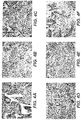
- Figures 3A-C depict the results of PCL alone, with pADM and dioxane, and with pADM and NMP, respectively, after 4 weeks.
- Figures 3D-F depict the results of P4HB alone, with pADM and dioxane, and with pADM and NMP, respectively, after 4 weeks.
- Figures 4A-C depict the results of PCL alone, with pADM and dioxane, and with pADM and NMP, respectively, after 12 weeks.
- FIGS. 4D-F depict the results the P4HB alone, with pADM and dioxane, and with pADM and NMP, respectively, after 12 weeks. Histological analysis of the explants showed that PCL and P4HB in the presence of pADM, when solubilized in either of dioxane or NMP, had an attenuated inflammatory response compared to explants of PCL and P4HB alone.
- Implantation of a regenerative tissue scaffold in the femoral condyle of a rabbit was evaluated.
- An implant comprising BHA, pADM, and NMP was prepared according to the procedure described in Example 1.
- BHA was mixed with pADM in an NMP solvent and injected into an approximately 3.5 x 3 mm osteochondral defect created on the femoral condyle of a rabbit.
- Samples were removed following 4 weeks, and cellular responses were evaluated using routine histological staining with hematoxylin and eosin. The samples were observed under microscope at 20x, 100x, and 400x magnification ( Figs. 5a-c , respectively).
- a photo of the femoral condyle showing the tissue scaffold implant 180b was also taken at 12 weeks ( Fig. 6 ). The results showed the persistence of the implant and a regenerative tissue response as demonstrated by the deposition of hyaline-like cartilage at the site of the defect.
Landscapes
- Health & Medical Sciences (AREA)
- Life Sciences & Earth Sciences (AREA)
- Chemical & Material Sciences (AREA)
- Engineering & Computer Science (AREA)
- Biomedical Technology (AREA)
- Veterinary Medicine (AREA)
- Medicinal Chemistry (AREA)
- Public Health (AREA)
- General Health & Medical Sciences (AREA)
- Animal Behavior & Ethology (AREA)
- Dermatology (AREA)
- Epidemiology (AREA)
- Transplantation (AREA)
- Oral & Maxillofacial Surgery (AREA)
- Chemical Kinetics & Catalysis (AREA)
- Molecular Biology (AREA)
- Botany (AREA)
- Zoology (AREA)
- Urology & Nephrology (AREA)
- Orthopedic Medicine & Surgery (AREA)
- Composite Materials (AREA)
- Materials Engineering (AREA)
- Pharmacology & Pharmacy (AREA)
- Bioinformatics & Cheminformatics (AREA)
- General Chemical & Material Sciences (AREA)
- Nuclear Medicine, Radiotherapy & Molecular Imaging (AREA)
- Organic Chemistry (AREA)
- Materials For Medical Uses (AREA)
Claims (13)
- Kit, der umfasst:eine partikelförmige azelluläre Gewebematrix (ATM);ein wasservermischbares Lösungsmittel; undein Polymer, das im Lösungsmittel auflösbar ist,wobei das wasservermischbare Lösungsmittel nach Mischen der ATM, des Polymers und des Lösungsmittels und nach Inkontaktbringen der Mischung mit einem wässrigen Medium in der Lage ist, aus der Mischung zu diffundieren, um zu ermöglichen, dass die Mischung aus dem Polymer und der ATM ein Gewebe-Scaffold bildet.
- Kit nach Anspruch 1, wobei das Lösungsmittel biokompatibel ist.
- Kit nach einem der Ansprüche 1 bis 2, wobei das Lösungsmittel zumindest eines von Dioxan, N-Methyl-2-pyrrolidon oder Dimethylsulfoxid oder Kombinationen davon umfasst.
- Kit nach einem der Ansprüche 1 bis 3, wobei das Polymer Polycaprolacton umfasst.
- Kit nach einem der Ansprüche 1 bis 4, wobei das Polymer Poly-4-hydroxybutyrat umfasst.
- Kit nach einem der Ansprüche 1 bis 5, wobei das Polymer ein Benzylesterderivat von Hyaluronsäure umfasst.
- Kit nach einem der Ansprüche 1 bis 6, wobei die partikelförmige ATM Partikel einheitlicher Größe umfasst.
- Kit nach einem der Ansprüche 1 bis 7, wobei die partikelförmige ATM eine dermale ATM, eine Knorpelgewebematrix, eine Knochengewebematrix oder eine Kombination davon umfasst.
- Kit nach einem der Ansprüche 1 bis 8, wobei die partikelförmige ATM ATM aus zwei oder mehr verschiedenen Gewebetypen umfasst.
- Kit nach Anspruch 9, wobei die zwei oder mehr verschiedenen Gewebetypen Dermis und Knorpel, Knorpel und Knochen, humane Gewebematrizen, Schweine-Gewebematrizen oder humane Gewebematrizen und Schweine-Gewebematrizen umfassen.
- Kit nach einem der Ansprüche 1 bis 9, wobei das Gewebe-Scaffold in der Lage ist, die Knorpelregeneration zu unterstützen.
- Kit nach einem der Ansprüche 1 bis 9, wobei das Polymer zumindest eines von Polycaprolacton oder Poly-4-hydroxybutyrat umfasst und das Lösungsmittel zumindest eines von Dioxan oder N-Methyl-2-pyrrolidon umfasst.
- Kit nach einem der Ansprüche 1 bis 9, wobei das Polymer ein Benzylesterderivat von Hyaluronsäure umfasst und das Lösungsmittel zumindest eines von Dimethylsulfoxid oder N-Methyl-2-pyrrolidon umfasst.
Priority Applications (1)
| Application Number | Priority Date | Filing Date | Title |
|---|---|---|---|
| EP19196550.8A EP3597226A1 (de) | 2010-08-10 | 2011-08-09 | Regenerative gewebegerüste |
Applications Claiming Priority (3)
| Application Number | Priority Date | Filing Date | Title |
|---|---|---|---|
| US37233910P | 2010-08-10 | 2010-08-10 | |
| EP11748831.2A EP2603248B1 (de) | 2010-08-10 | 2011-08-09 | Regenerative gewebegerüste |
| PCT/US2011/047041 WO2012021490A1 (en) | 2010-08-10 | 2011-08-09 | Regenerative tissue scaffolds |
Related Parent Applications (1)
| Application Number | Title | Priority Date | Filing Date |
|---|---|---|---|
| EP11748831.2A Division EP2603248B1 (de) | 2010-08-10 | 2011-08-09 | Regenerative gewebegerüste |
Related Child Applications (2)
| Application Number | Title | Priority Date | Filing Date |
|---|---|---|---|
| EP19196550.8A Division-Into EP3597226A1 (de) | 2010-08-10 | 2011-08-09 | Regenerative gewebegerüste |
| EP19196550.8A Division EP3597226A1 (de) | 2010-08-10 | 2011-08-09 | Regenerative gewebegerüste |
Publications (2)
| Publication Number | Publication Date |
|---|---|
| EP3323438A1 EP3323438A1 (de) | 2018-05-23 |
| EP3323438B1 true EP3323438B1 (de) | 2019-10-30 |
Family
ID=44511577
Family Applications (3)
| Application Number | Title | Priority Date | Filing Date |
|---|---|---|---|
| EP17210125.5A Active EP3323438B1 (de) | 2010-08-10 | 2011-08-09 | Regenerative gewebegerüste |
| EP11748831.2A Active EP2603248B1 (de) | 2010-08-10 | 2011-08-09 | Regenerative gewebegerüste |
| EP19196550.8A Withdrawn EP3597226A1 (de) | 2010-08-10 | 2011-08-09 | Regenerative gewebegerüste |
Family Applications After (2)
| Application Number | Title | Priority Date | Filing Date |
|---|---|---|---|
| EP11748831.2A Active EP2603248B1 (de) | 2010-08-10 | 2011-08-09 | Regenerative gewebegerüste |
| EP19196550.8A Withdrawn EP3597226A1 (de) | 2010-08-10 | 2011-08-09 | Regenerative gewebegerüste |
Country Status (8)
| Country | Link |
|---|---|
| US (1) | US20120040013A1 (de) |
| EP (3) | EP3323438B1 (de) |
| JP (1) | JP5963208B2 (de) |
| CN (1) | CN103002927B (de) |
| AU (1) | AU2011289557B2 (de) |
| CA (1) | CA2806464C (de) |
| ES (2) | ES2662331T3 (de) |
| WO (1) | WO2012021490A1 (de) |
Families Citing this family (37)
| Publication number | Priority date | Publication date | Assignee | Title |
|---|---|---|---|---|
| GB0224986D0 (en) | 2002-10-28 | 2002-12-04 | Smith & Nephew | Apparatus |
| GB0325126D0 (en) | 2003-10-28 | 2003-12-03 | Smith & Nephew | Apparatus with heat |
| GB0325129D0 (en) | 2003-10-28 | 2003-12-03 | Smith & Nephew | Apparatus in situ |
| US11298453B2 (en) | 2003-10-28 | 2022-04-12 | Smith & Nephew Plc | Apparatus and method for wound cleansing with actives |
| US10413644B2 (en) | 2004-04-27 | 2019-09-17 | Smith & Nephew Plc | Wound treatment apparatus and method |
| US8529548B2 (en) | 2004-04-27 | 2013-09-10 | Smith & Nephew Plc | Wound treatment apparatus and method |
| US8469779B1 (en) | 2009-01-02 | 2013-06-25 | Lifecell Corporation | Method for debristling animal skin |
| US8637067B1 (en) | 2011-03-10 | 2014-01-28 | Lifecell Corporation | Elastic tissue matrix derived hydrogel |
| EP2696908B1 (de) | 2011-04-14 | 2015-03-11 | Lifecell Corporation | Regenerative materialien |
| US9089523B2 (en) | 2011-07-28 | 2015-07-28 | Lifecell Corporation | Natural tissue scaffolds as tissue fillers |
| ES2864104T3 (es) | 2011-12-20 | 2021-10-13 | Lifecell Corp | Productos tisulares laminados |
| BR112014014975B1 (pt) | 2011-12-20 | 2019-06-25 | Lifecell Corporation | Produto de tecido, e método para produzir uma composição de tecido |
| AU2013212592B2 (en) | 2012-01-24 | 2016-06-30 | Lifecell Corporation | Elongated tissue matrices |
| EP2841116A1 (de) | 2012-04-24 | 2015-03-04 | Lifecell Corporation | Fliessfähige gewebematrizen |
| BR112015000547B1 (pt) | 2012-07-13 | 2020-11-10 | Lifecell Corporation | método para tratar tecido |
| AU2013323747B2 (en) | 2012-09-26 | 2017-02-02 | Lifecell Corporation | Processed adipose tissue |
| EP3659633A1 (de) | 2013-02-06 | 2020-06-03 | LifeCell Corporation | Verfahren für lokalisierte modifizierung von gewebeprodukten |
| CN105188795B (zh) | 2013-05-10 | 2018-07-31 | 史密夫及内修公开有限公司 | 用于冲洗和抽吸伤口的流体连接件 |
| WO2016090286A1 (en) | 2014-12-05 | 2016-06-09 | University Of Florida Research Foundation, Inc. | 3d printing using phase changing materials as support |
| EP3795186A1 (de) * | 2015-01-21 | 2021-03-24 | LifeCell Corporation | Gewebematrizen mit kontrollierter porosität oder mechanischen eigenschaften |
| US11007705B2 (en) | 2015-02-13 | 2021-05-18 | University Of Florida Research Foundation, Inc. | High speed 3D printing system for wound and tissue replacement |
| US11390835B2 (en) | 2015-05-08 | 2022-07-19 | University Of Florida Research Foundation, Inc. | Growth media for three-dimensional cell culture |
| WO2017040981A1 (en) | 2015-09-03 | 2017-03-09 | University Of Florida Research Foundation, Inc. | Valve incorporating temporary phase change material |
| WO2017096263A1 (en) | 2015-12-04 | 2017-06-08 | University Of Florida Research Foundation, Incorporated | Crosslinkable or functionalizable polymers for 3d printing of soft materials |
| USD856517S1 (en) | 2016-06-03 | 2019-08-13 | Musculoskeletal Transplant Foundation | Asymmetric tissue graft |
| US10792394B2 (en) | 2016-06-03 | 2020-10-06 | Lifecell Corporation | Methods for localized modification of tissue products |
| US10945831B2 (en) | 2016-06-03 | 2021-03-16 | Musculoskeletal Transplant Foundation | Asymmetric tissue graft |
| US11124644B2 (en) | 2016-09-01 | 2021-09-21 | University Of Florida Research Foundation, Inc. | Organic microgel system for 3D printing of silicone structures |
| EP3558405A1 (de) | 2016-12-22 | 2019-10-30 | LifeCell Corporation | Vorrichtungen und verfahren zum kryogenen mahlen von gewebe |
| US11123375B2 (en) | 2017-10-18 | 2021-09-21 | Lifecell Corporation | Methods of treating tissue voids following removal of implantable infusion ports using adipose tissue products |
| CN111201046B (zh) | 2017-10-18 | 2022-06-28 | 生命细胞公司 | 脂肪组织产品以及产生方法 |
| ES2960617T3 (es) | 2017-10-19 | 2024-03-05 | Lifecell Corp | Productos de matriz de tejido acelular fluida y métodos de producción |
| US11246994B2 (en) | 2017-10-19 | 2022-02-15 | Lifecell Corporation | Methods for introduction of flowable acellular tissue matrix products into a hand |
| US10813743B2 (en) | 2018-09-07 | 2020-10-27 | Musculoskeletal Transplant Foundation | Soft tissue repair grafts and processes for preparing and using same |
| USD895812S1 (en) | 2018-09-07 | 2020-09-08 | Musculoskeletal Transplant Foundation | Soft tissue repair graft |
| US11633521B2 (en) | 2019-05-30 | 2023-04-25 | Lifecell Corporation | Biologic breast implant |
| FR3110077A1 (fr) * | 2020-05-12 | 2021-11-19 | Ph Tech | Procédé de fabrication d’un dispositif médical en trois dimensions et dispositif médical obtenu |
Family Cites Families (11)
| Publication number | Priority date | Publication date | Assignee | Title |
|---|---|---|---|---|
| US5487897A (en) * | 1989-07-24 | 1996-01-30 | Atrix Laboratories, Inc. | Biodegradable implant precursor |
| US6166288A (en) | 1995-09-27 | 2000-12-26 | Nextran Inc. | Method of producing transgenic animals for xenotransplantation expressing both an enzyme masking or reducing the level of the gal epitope and a complement inhibitor |
| CA2221195A1 (en) * | 1997-11-14 | 1999-05-14 | Chantal E. Holy | Biodegradable polymer matrix |
| US6933326B1 (en) | 1998-06-19 | 2005-08-23 | Lifecell Coporation | Particulate acellular tissue matrix |
| US6461631B1 (en) * | 1999-11-16 | 2002-10-08 | Atrix Laboratories, Inc. | Biodegradable polymer composition |
| WO2003017826A2 (en) * | 2001-08-27 | 2003-03-06 | Regeneration Technologies, Inc. | Processed soft tissue for topical or internal application |
| US7291345B2 (en) * | 2002-12-12 | 2007-11-06 | Osteotech, Inc. | Formable and settable polymer bone composite and method of production thereof |
| US20070248575A1 (en) * | 2006-04-19 | 2007-10-25 | Jerome Connor | Bone graft composition |
| CN101553189A (zh) * | 2006-05-09 | 2009-10-07 | 生命细胞公司 | 加强的生物组织 |
| KR100816395B1 (ko) * | 2006-09-21 | 2008-03-27 | (주)필미아젠 | 세포 유래 세포외기질막의 제조방법 |
| JP5930476B2 (ja) * | 2010-03-25 | 2016-06-08 | ライフセル コーポレーションLifeCell Corporation | 再生組織足場の製造 |
-
2011
- 2011-08-09 EP EP17210125.5A patent/EP3323438B1/de active Active
- 2011-08-09 AU AU2011289557A patent/AU2011289557B2/en active Active
- 2011-08-09 EP EP11748831.2A patent/EP2603248B1/de active Active
- 2011-08-09 EP EP19196550.8A patent/EP3597226A1/de not_active Withdrawn
- 2011-08-09 JP JP2013524164A patent/JP5963208B2/ja active Active
- 2011-08-09 CA CA2806464A patent/CA2806464C/en active Active
- 2011-08-09 ES ES11748831.2T patent/ES2662331T3/es active Active
- 2011-08-09 WO PCT/US2011/047041 patent/WO2012021490A1/en active Application Filing
- 2011-08-09 US US13/205,899 patent/US20120040013A1/en not_active Abandoned
- 2011-08-09 ES ES17210125T patent/ES2762118T3/es active Active
- 2011-08-09 CN CN201180036258.1A patent/CN103002927B/zh not_active Expired - Fee Related
Non-Patent Citations (1)
| Title |
|---|
| None * |
Also Published As
| Publication number | Publication date |
|---|---|
| CN103002927B (zh) | 2016-06-15 |
| EP2603248B1 (de) | 2018-01-03 |
| WO2012021490A1 (en) | 2012-02-16 |
| EP3597226A1 (de) | 2020-01-22 |
| AU2011289557B2 (en) | 2015-02-05 |
| CN103002927A (zh) | 2013-03-27 |
| JP5963208B2 (ja) | 2016-08-03 |
| CA2806464C (en) | 2018-10-23 |
| EP3323438A1 (de) | 2018-05-23 |
| ES2762118T3 (es) | 2020-05-22 |
| CA2806464A1 (en) | 2012-02-16 |
| JP2013533093A (ja) | 2013-08-22 |
| US20120040013A1 (en) | 2012-02-16 |
| ES2662331T3 (es) | 2018-04-06 |
| EP2603248A1 (de) | 2013-06-19 |
| AU2011289557A1 (en) | 2013-01-24 |
Similar Documents
| Publication | Publication Date | Title |
|---|---|---|
| EP3323438B1 (de) | Regenerative gewebegerüste | |
| US8784499B2 (en) | Preparation of regenerative tissue scaffolds | |
| KR102604205B1 (ko) | 생물학적 기능성 연조직 스캐폴드 및 임플란트 | |
| EP2517738B1 (de) | Kollagen/hydroxyapatitverbundgerüst | |
| EP2015707B1 (de) | Verstärktes biologisches gewebe | |
| Esposito et al. | PLDLA/PCL-T scaffold for meniscus tissue engineering | |
| US8221777B2 (en) | Structurally modified acellular tissue engineering scaffolds and methods of production | |
| Bondioli et al. | Development and evaluation of a decellularized membrane from human dermis | |
| EP1809735A2 (de) | Verfahren zur lagerung von gewebematrizes | |
| CN112203702A (zh) | 富含脱细胞化骨细胞外基质水凝胶的脱细胞化骨生物材料 | |
| JP2008228744A (ja) | 生体由来移植用組織の石灰化を抑制するための処理方法および処理された組織 | |
| US20220047774A1 (en) | Decellularized muscle matrices and methods for making and using same |
Legal Events
| Date | Code | Title | Description |
|---|---|---|---|
| PUAI | Public reference made under article 153(3) epc to a published international application that has entered the european phase |
Free format text: ORIGINAL CODE: 0009012 |
|
| STAA | Information on the status of an ep patent application or granted ep patent |
Free format text: STATUS: THE APPLICATION HAS BEEN PUBLISHED |
|
| AC | Divisional application: reference to earlier application |
Ref document number: 2603248 Country of ref document: EP Kind code of ref document: P |
|
| AK | Designated contracting states |
Kind code of ref document: A1 Designated state(s): AL AT BE BG CH CY CZ DE DK EE ES FI FR GB GR HR HU IE IS IT LI LT LU LV MC MK MT NL NO PL PT RO RS SE SI SK SM TR |
|
| RAP1 | Party data changed (applicant data changed or rights of an application transferred) |
Owner name: LIFECELL CORPORATION |
|
| STAA | Information on the status of an ep patent application or granted ep patent |
Free format text: STATUS: REQUEST FOR EXAMINATION WAS MADE |
|
| 17P | Request for examination filed |
Effective date: 20181122 |
|
| RBV | Designated contracting states (corrected) |
Designated state(s): AL AT BE BG CH CY CZ DE DK EE ES FI FR GB GR HR HU IE IS IT LI LT LU LV MC MK MT NL NO PL PT RO RS SE SI SK SM TR |
|
| RIC1 | Information provided on ipc code assigned before grant |
Ipc: A61L 27/48 20060101AFI20190322BHEP Ipc: A61L 27/50 20060101ALI20190322BHEP |
|
| GRAP | Despatch of communication of intention to grant a patent |
Free format text: ORIGINAL CODE: EPIDOSNIGR1 |
|
| STAA | Information on the status of an ep patent application or granted ep patent |
Free format text: STATUS: GRANT OF PATENT IS INTENDED |
|
| INTG | Intention to grant announced |
Effective date: 20190503 |
|
| GRAJ | Information related to disapproval of communication of intention to grant by the applicant or resumption of examination proceedings by the epo deleted |
Free format text: ORIGINAL CODE: EPIDOSDIGR1 |
|
| STAA | Information on the status of an ep patent application or granted ep patent |
Free format text: STATUS: REQUEST FOR EXAMINATION WAS MADE |
|
| GRAR | Information related to intention to grant a patent recorded |
Free format text: ORIGINAL CODE: EPIDOSNIGR71 |
|
| GRAS | Grant fee paid |
Free format text: ORIGINAL CODE: EPIDOSNIGR3 |
|
| STAA | Information on the status of an ep patent application or granted ep patent |
Free format text: STATUS: GRANT OF PATENT IS INTENDED |
|
| GRAA | (expected) grant |
Free format text: ORIGINAL CODE: 0009210 |
|
| STAA | Information on the status of an ep patent application or granted ep patent |
Free format text: STATUS: THE PATENT HAS BEEN GRANTED |
|
| INTC | Intention to grant announced (deleted) | ||
| INTG | Intention to grant announced |
Effective date: 20190918 |
|
| AC | Divisional application: reference to earlier application |
Ref document number: 2603248 Country of ref document: EP Kind code of ref document: P |
|
| AK | Designated contracting states |
Kind code of ref document: B1 Designated state(s): AL AT BE BG CH CY CZ DE DK EE ES FI FR GB GR HR HU IE IS IT LI LT LU LV MC MK MT NL NO PL PT RO RS SE SI SK SM TR |
|
| REG | Reference to a national code |
Ref country code: GB Ref legal event code: FG4D |
|
| REG | Reference to a national code |
Ref country code: CH Ref legal event code: EP |
|
| REG | Reference to a national code |
Ref country code: AT Ref legal event code: REF Ref document number: 1195459 Country of ref document: AT Kind code of ref document: T Effective date: 20191115 |
|
| REG | Reference to a national code |
Ref country code: DE Ref legal event code: R096 Ref document number: 602011063136 Country of ref document: DE |
|
| REG | Reference to a national code |
Ref country code: IE Ref legal event code: FG4D |
|
| REG | Reference to a national code |
Ref country code: LT Ref legal event code: MG4D |
|
| PG25 | Lapsed in a contracting state [announced via postgrant information from national office to epo] |
Ref country code: BG Free format text: LAPSE BECAUSE OF FAILURE TO SUBMIT A TRANSLATION OF THE DESCRIPTION OR TO PAY THE FEE WITHIN THE PRESCRIBED TIME-LIMIT Effective date: 20200130 Ref country code: FI Free format text: LAPSE BECAUSE OF FAILURE TO SUBMIT A TRANSLATION OF THE DESCRIPTION OR TO PAY THE FEE WITHIN THE PRESCRIBED TIME-LIMIT Effective date: 20191030 Ref country code: PT Free format text: LAPSE BECAUSE OF FAILURE TO SUBMIT A TRANSLATION OF THE DESCRIPTION OR TO PAY THE FEE WITHIN THE PRESCRIBED TIME-LIMIT Effective date: 20200302 Ref country code: NL Free format text: LAPSE BECAUSE OF FAILURE TO SUBMIT A TRANSLATION OF THE DESCRIPTION OR TO PAY THE FEE WITHIN THE PRESCRIBED TIME-LIMIT Effective date: 20191030 Ref country code: LV Free format text: LAPSE BECAUSE OF FAILURE TO SUBMIT A TRANSLATION OF THE DESCRIPTION OR TO PAY THE FEE WITHIN THE PRESCRIBED TIME-LIMIT Effective date: 20191030 Ref country code: SE Free format text: LAPSE BECAUSE OF FAILURE TO SUBMIT A TRANSLATION OF THE DESCRIPTION OR TO PAY THE FEE WITHIN THE PRESCRIBED TIME-LIMIT Effective date: 20191030 Ref country code: LT Free format text: LAPSE BECAUSE OF FAILURE TO SUBMIT A TRANSLATION OF THE DESCRIPTION OR TO PAY THE FEE WITHIN THE PRESCRIBED TIME-LIMIT Effective date: 20191030 Ref country code: GR Free format text: LAPSE BECAUSE OF FAILURE TO SUBMIT A TRANSLATION OF THE DESCRIPTION OR TO PAY THE FEE WITHIN THE PRESCRIBED TIME-LIMIT Effective date: 20200131 Ref country code: PL Free format text: LAPSE BECAUSE OF FAILURE TO SUBMIT A TRANSLATION OF THE DESCRIPTION OR TO PAY THE FEE WITHIN THE PRESCRIBED TIME-LIMIT Effective date: 20191030 Ref country code: NO Free format text: LAPSE BECAUSE OF FAILURE TO SUBMIT A TRANSLATION OF THE DESCRIPTION OR TO PAY THE FEE WITHIN THE PRESCRIBED TIME-LIMIT Effective date: 20200130 |
|
| REG | Reference to a national code |
Ref country code: NL Ref legal event code: MP Effective date: 20191030 |
|
| REG | Reference to a national code |
Ref country code: ES Ref legal event code: FG2A Ref document number: 2762118 Country of ref document: ES Kind code of ref document: T3 Effective date: 20200522 |
|
| PG25 | Lapsed in a contracting state [announced via postgrant information from national office to epo] |
Ref country code: HR Free format text: LAPSE BECAUSE OF FAILURE TO SUBMIT A TRANSLATION OF THE DESCRIPTION OR TO PAY THE FEE WITHIN THE PRESCRIBED TIME-LIMIT Effective date: 20191030 Ref country code: IS Free format text: LAPSE BECAUSE OF FAILURE TO SUBMIT A TRANSLATION OF THE DESCRIPTION OR TO PAY THE FEE WITHIN THE PRESCRIBED TIME-LIMIT Effective date: 20200229 Ref country code: RS Free format text: LAPSE BECAUSE OF FAILURE TO SUBMIT A TRANSLATION OF THE DESCRIPTION OR TO PAY THE FEE WITHIN THE PRESCRIBED TIME-LIMIT Effective date: 20191030 |
|
| PG25 | Lapsed in a contracting state [announced via postgrant information from national office to epo] |
Ref country code: AL Free format text: LAPSE BECAUSE OF FAILURE TO SUBMIT A TRANSLATION OF THE DESCRIPTION OR TO PAY THE FEE WITHIN THE PRESCRIBED TIME-LIMIT Effective date: 20191030 |
|
| PG25 | Lapsed in a contracting state [announced via postgrant information from national office to epo] |
Ref country code: EE Free format text: LAPSE BECAUSE OF FAILURE TO SUBMIT A TRANSLATION OF THE DESCRIPTION OR TO PAY THE FEE WITHIN THE PRESCRIBED TIME-LIMIT Effective date: 20191030 Ref country code: CZ Free format text: LAPSE BECAUSE OF FAILURE TO SUBMIT A TRANSLATION OF THE DESCRIPTION OR TO PAY THE FEE WITHIN THE PRESCRIBED TIME-LIMIT Effective date: 20191030 Ref country code: RO Free format text: LAPSE BECAUSE OF FAILURE TO SUBMIT A TRANSLATION OF THE DESCRIPTION OR TO PAY THE FEE WITHIN THE PRESCRIBED TIME-LIMIT Effective date: 20191030 Ref country code: DK Free format text: LAPSE BECAUSE OF FAILURE TO SUBMIT A TRANSLATION OF THE DESCRIPTION OR TO PAY THE FEE WITHIN THE PRESCRIBED TIME-LIMIT Effective date: 20191030 |
|
| REG | Reference to a national code |
Ref country code: DE Ref legal event code: R097 Ref document number: 602011063136 Country of ref document: DE |
|
| REG | Reference to a national code |
Ref country code: AT Ref legal event code: MK05 Ref document number: 1195459 Country of ref document: AT Kind code of ref document: T Effective date: 20191030 |
|
| PG25 | Lapsed in a contracting state [announced via postgrant information from national office to epo] |
Ref country code: SK Free format text: LAPSE BECAUSE OF FAILURE TO SUBMIT A TRANSLATION OF THE DESCRIPTION OR TO PAY THE FEE WITHIN THE PRESCRIBED TIME-LIMIT Effective date: 20191030 Ref country code: SM Free format text: LAPSE BECAUSE OF FAILURE TO SUBMIT A TRANSLATION OF THE DESCRIPTION OR TO PAY THE FEE WITHIN THE PRESCRIBED TIME-LIMIT Effective date: 20191030 |
|
| PLBE | No opposition filed within time limit |
Free format text: ORIGINAL CODE: 0009261 |
|
| STAA | Information on the status of an ep patent application or granted ep patent |
Free format text: STATUS: NO OPPOSITION FILED WITHIN TIME LIMIT |
|
| 26N | No opposition filed |
Effective date: 20200731 |
|
| PG25 | Lapsed in a contracting state [announced via postgrant information from national office to epo] |
Ref country code: SI Free format text: LAPSE BECAUSE OF FAILURE TO SUBMIT A TRANSLATION OF THE DESCRIPTION OR TO PAY THE FEE WITHIN THE PRESCRIBED TIME-LIMIT Effective date: 20191030 Ref country code: AT Free format text: LAPSE BECAUSE OF FAILURE TO SUBMIT A TRANSLATION OF THE DESCRIPTION OR TO PAY THE FEE WITHIN THE PRESCRIBED TIME-LIMIT Effective date: 20191030 |
|
| PG25 | Lapsed in a contracting state [announced via postgrant information from national office to epo] |
Ref country code: MC Free format text: LAPSE BECAUSE OF FAILURE TO SUBMIT A TRANSLATION OF THE DESCRIPTION OR TO PAY THE FEE WITHIN THE PRESCRIBED TIME-LIMIT Effective date: 20191030 |
|
| REG | Reference to a national code |
Ref country code: CH Ref legal event code: PL |
|
| PG25 | Lapsed in a contracting state [announced via postgrant information from national office to epo] |
Ref country code: CH Free format text: LAPSE BECAUSE OF NON-PAYMENT OF DUE FEES Effective date: 20200831 Ref country code: LI Free format text: LAPSE BECAUSE OF NON-PAYMENT OF DUE FEES Effective date: 20200831 Ref country code: LU Free format text: LAPSE BECAUSE OF NON-PAYMENT OF DUE FEES Effective date: 20200809 |
|
| REG | Reference to a national code |
Ref country code: BE Ref legal event code: MM Effective date: 20200831 |
|
| PG25 | Lapsed in a contracting state [announced via postgrant information from national office to epo] |
Ref country code: IE Free format text: LAPSE BECAUSE OF NON-PAYMENT OF DUE FEES Effective date: 20200809 Ref country code: BE Free format text: LAPSE BECAUSE OF NON-PAYMENT OF DUE FEES Effective date: 20200831 |
|
| PGFP | Annual fee paid to national office [announced via postgrant information from national office to epo] |
Ref country code: IT Payment date: 20210820 Year of fee payment: 11 Ref country code: FR Payment date: 20210825 Year of fee payment: 11 |
|
| PGFP | Annual fee paid to national office [announced via postgrant information from national office to epo] |
Ref country code: ES Payment date: 20210901 Year of fee payment: 11 Ref country code: DE Payment date: 20210827 Year of fee payment: 11 Ref country code: GB Payment date: 20210827 Year of fee payment: 11 |
|
| PG25 | Lapsed in a contracting state [announced via postgrant information from national office to epo] |
Ref country code: TR Free format text: LAPSE BECAUSE OF FAILURE TO SUBMIT A TRANSLATION OF THE DESCRIPTION OR TO PAY THE FEE WITHIN THE PRESCRIBED TIME-LIMIT Effective date: 20191030 Ref country code: MT Free format text: LAPSE BECAUSE OF FAILURE TO SUBMIT A TRANSLATION OF THE DESCRIPTION OR TO PAY THE FEE WITHIN THE PRESCRIBED TIME-LIMIT Effective date: 20191030 Ref country code: CY Free format text: LAPSE BECAUSE OF FAILURE TO SUBMIT A TRANSLATION OF THE DESCRIPTION OR TO PAY THE FEE WITHIN THE PRESCRIBED TIME-LIMIT Effective date: 20191030 |
|
| PG25 | Lapsed in a contracting state [announced via postgrant information from national office to epo] |
Ref country code: MK Free format text: LAPSE BECAUSE OF FAILURE TO SUBMIT A TRANSLATION OF THE DESCRIPTION OR TO PAY THE FEE WITHIN THE PRESCRIBED TIME-LIMIT Effective date: 20191030 |
|
| REG | Reference to a national code |
Ref country code: DE Ref legal event code: R119 Ref document number: 602011063136 Country of ref document: DE |
|
| GBPC | Gb: european patent ceased through non-payment of renewal fee |
Effective date: 20220809 |
|
| PG25 | Lapsed in a contracting state [announced via postgrant information from national office to epo] |
Ref country code: IT Free format text: LAPSE BECAUSE OF NON-PAYMENT OF DUE FEES Effective date: 20220809 Ref country code: FR Free format text: LAPSE BECAUSE OF NON-PAYMENT OF DUE FEES Effective date: 20220831 Ref country code: DE Free format text: LAPSE BECAUSE OF NON-PAYMENT OF DUE FEES Effective date: 20230301 |
|
| REG | Reference to a national code |
Ref country code: ES Ref legal event code: FD2A Effective date: 20230927 |
|
| PG25 | Lapsed in a contracting state [announced via postgrant information from national office to epo] |
Ref country code: GB Free format text: LAPSE BECAUSE OF NON-PAYMENT OF DUE FEES Effective date: 20220809 Ref country code: ES Free format text: LAPSE BECAUSE OF NON-PAYMENT OF DUE FEES Effective date: 20220810 |