EP3302538B1 - Affinity entities comprising a tcr-like antibody binding domain with high affinity and fine specificity and uses of same - Google Patents
Affinity entities comprising a tcr-like antibody binding domain with high affinity and fine specificity and uses of same Download PDFInfo
- Publication number
- EP3302538B1 EP3302538B1 EP16739266.1A EP16739266A EP3302538B1 EP 3302538 B1 EP3302538 B1 EP 3302538B1 EP 16739266 A EP16739266 A EP 16739266A EP 3302538 B1 EP3302538 B1 EP 3302538B1
- Authority
- EP
- European Patent Office
- Prior art keywords
- peptide
- prt
- cells
- binding
- antibody
- Prior art date
- Legal status (The legal status is an assumption and is not a legal conclusion. Google has not performed a legal analysis and makes no representation as to the accuracy of the status listed.)
- Active
Links
Images


Classifications
-
- C—CHEMISTRY; METALLURGY
- C07—ORGANIC CHEMISTRY
- C07K—PEPTIDES
- C07K16/00—Immunoglobulins [IG], e.g. monoclonal or polyclonal antibodies
- C07K16/18—Immunoglobulins [IG], e.g. monoclonal or polyclonal antibodies against material from animals or humans
- C07K16/28—Immunoglobulins [IG], e.g. monoclonal or polyclonal antibodies against material from animals or humans against receptors, cell surface antigens or cell surface determinants
- C07K16/2803—Immunoglobulins [IG], e.g. monoclonal or polyclonal antibodies against material from animals or humans against receptors, cell surface antigens or cell surface determinants against the immunoglobulin superfamily
- C07K16/2809—Immunoglobulins [IG], e.g. monoclonal or polyclonal antibodies against material from animals or humans against receptors, cell surface antigens or cell surface determinants against the immunoglobulin superfamily against the T-cell receptor (TcR)-CD3 complex
-
- A—HUMAN NECESSITIES
- A61—MEDICAL OR VETERINARY SCIENCE; HYGIENE
- A61P—SPECIFIC THERAPEUTIC ACTIVITY OF CHEMICAL COMPOUNDS OR MEDICINAL PREPARATIONS
- A61P35/00—Antineoplastic agents
-
- C—CHEMISTRY; METALLURGY
- C07—ORGANIC CHEMISTRY
- C07K—PEPTIDES
- C07K16/00—Immunoglobulins [IG], e.g. monoclonal or polyclonal antibodies
- C07K16/18—Immunoglobulins [IG], e.g. monoclonal or polyclonal antibodies against material from animals or humans
- C07K16/28—Immunoglobulins [IG], e.g. monoclonal or polyclonal antibodies against material from animals or humans against receptors, cell surface antigens or cell surface determinants
- C07K16/2803—Immunoglobulins [IG], e.g. monoclonal or polyclonal antibodies against material from animals or humans against receptors, cell surface antigens or cell surface determinants against the immunoglobulin superfamily
- C07K16/2833—Immunoglobulins [IG], e.g. monoclonal or polyclonal antibodies against material from animals or humans against receptors, cell surface antigens or cell surface determinants against the immunoglobulin superfamily against MHC-molecules, e.g. HLA-molecules
-
- C—CHEMISTRY; METALLURGY
- C07—ORGANIC CHEMISTRY
- C07K—PEPTIDES
- C07K16/00—Immunoglobulins [IG], e.g. monoclonal or polyclonal antibodies
- C07K16/18—Immunoglobulins [IG], e.g. monoclonal or polyclonal antibodies against material from animals or humans
- C07K16/28—Immunoglobulins [IG], e.g. monoclonal or polyclonal antibodies against material from animals or humans against receptors, cell surface antigens or cell surface determinants
- C07K16/30—Immunoglobulins [IG], e.g. monoclonal or polyclonal antibodies against material from animals or humans against receptors, cell surface antigens or cell surface determinants from tumour cells
- C07K16/3053—Skin, nerves, brain
-
- C—CHEMISTRY; METALLURGY
- C07—ORGANIC CHEMISTRY
- C07K—PEPTIDES
- C07K16/00—Immunoglobulins [IG], e.g. monoclonal or polyclonal antibodies
- C07K16/40—Immunoglobulins [IG], e.g. monoclonal or polyclonal antibodies against enzymes
-
- G—PHYSICS
- G01—MEASURING; TESTING
- G01N—INVESTIGATING OR ANALYSING MATERIALS BY DETERMINING THEIR CHEMICAL OR PHYSICAL PROPERTIES
- G01N33/00—Investigating or analysing materials by specific methods not covered by groups G01N1/00 - G01N31/00
- G01N33/48—Biological material, e.g. blood, urine; Haemocytometers
- G01N33/50—Chemical analysis of biological material, e.g. blood, urine; Testing involving biospecific ligand binding methods; Immunological testing
- G01N33/53—Immunoassay; Biospecific binding assay; Materials therefor
- G01N33/575—Immunoassay; Biospecific binding assay; Materials therefor for cancer
- G01N33/5751—Immunoassay; Biospecific binding assay; Materials therefor for cancer of the skin, e.g. melanoma
-
- A—HUMAN NECESSITIES
- A61—MEDICAL OR VETERINARY SCIENCE; HYGIENE
- A61K—PREPARATIONS FOR MEDICAL, DENTAL OR TOILETRY PURPOSES
- A61K39/00—Medicinal preparations containing antigens or antibodies
- A61K2039/505—Medicinal preparations containing antigens or antibodies comprising antibodies
-
- C—CHEMISTRY; METALLURGY
- C07—ORGANIC CHEMISTRY
- C07K—PEPTIDES
- C07K2317/00—Immunoglobulins specific features
- C07K2317/20—Immunoglobulins specific features characterized by taxonomic origin
- C07K2317/21—Immunoglobulins specific features characterized by taxonomic origin from primates, e.g. man
-
- C—CHEMISTRY; METALLURGY
- C07—ORGANIC CHEMISTRY
- C07K—PEPTIDES
- C07K2317/00—Immunoglobulins specific features
- C07K2317/30—Immunoglobulins specific features characterized by aspects of specificity or valency
- C07K2317/31—Immunoglobulins specific features characterized by aspects of specificity or valency multispecific
-
- C—CHEMISTRY; METALLURGY
- C07—ORGANIC CHEMISTRY
- C07K—PEPTIDES
- C07K2317/00—Immunoglobulins specific features
- C07K2317/30—Immunoglobulins specific features characterized by aspects of specificity or valency
- C07K2317/32—Immunoglobulins specific features characterized by aspects of specificity or valency specific for a neo-epitope on a complex, e.g. antibody-antigen or ligand-receptor
-
- C—CHEMISTRY; METALLURGY
- C07—ORGANIC CHEMISTRY
- C07K—PEPTIDES
- C07K2317/00—Immunoglobulins specific features
- C07K2317/30—Immunoglobulins specific features characterized by aspects of specificity or valency
- C07K2317/34—Identification of a linear epitope shorter than 20 amino acid residues or of a conformational epitope defined by amino acid residues
-
- C—CHEMISTRY; METALLURGY
- C07—ORGANIC CHEMISTRY
- C07K—PEPTIDES
- C07K2317/00—Immunoglobulins specific features
- C07K2317/50—Immunoglobulins specific features characterized by immunoglobulin fragments
- C07K2317/55—Fab or Fab'
-
- C—CHEMISTRY; METALLURGY
- C07—ORGANIC CHEMISTRY
- C07K—PEPTIDES
- C07K2317/00—Immunoglobulins specific features
- C07K2317/60—Immunoglobulins specific features characterized by non-natural combinations of immunoglobulin fragments
- C07K2317/62—Immunoglobulins specific features characterized by non-natural combinations of immunoglobulin fragments comprising only variable region components
- C07K2317/622—Single chain antibody (scFv)
-
- C—CHEMISTRY; METALLURGY
- C07—ORGANIC CHEMISTRY
- C07K—PEPTIDES
- C07K2317/00—Immunoglobulins specific features
- C07K2317/70—Immunoglobulins specific features characterized by effect upon binding to a cell or to an antigen
- C07K2317/73—Inducing cell death, e.g. apoptosis, necrosis or inhibition of cell proliferation
-
- C—CHEMISTRY; METALLURGY
- C07—ORGANIC CHEMISTRY
- C07K—PEPTIDES
- C07K2317/00—Immunoglobulins specific features
- C07K2317/90—Immunoglobulins specific features characterized by (pharmaco)kinetic aspects or by stability of the immunoglobulin
- C07K2317/92—Affinity (KD), association rate (Ka), dissociation rate (Kd) or EC50 value
-
- C—CHEMISTRY; METALLURGY
- C07—ORGANIC CHEMISTRY
- C07K—PEPTIDES
- C07K2319/00—Fusion polypeptide
-
- G—PHYSICS
- G01—MEASURING; TESTING
- G01N—INVESTIGATING OR ANALYSING MATERIALS BY DETERMINING THEIR CHEMICAL OR PHYSICAL PROPERTIES
- G01N33/00—Investigating or analysing materials by specific methods not covered by groups G01N1/00 - G01N31/00
- G01N33/48—Biological material, e.g. blood, urine; Haemocytometers
- G01N33/50—Chemical analysis of biological material, e.g. blood, urine; Testing involving biospecific ligand binding methods; Immunological testing
- G01N33/53—Immunoassay; Biospecific binding assay; Materials therefor
- G01N33/575—Immunoassay; Biospecific binding assay; Materials therefor for cancer
- G01N33/5758—Immunoassay; Biospecific binding assay; Materials therefor for cancer involving compounds serving as markers for tumours, cancers or neoplasias, e.g. cellular determinants, receptors, heat shock/stress proteins, A-protein, oligosaccharides or metabolites
- G01N33/5759—Immunoassay; Biospecific binding assay; Materials therefor for cancer involving compounds serving as markers for tumours, cancers or neoplasias, e.g. cellular determinants, receptors, heat shock/stress proteins, A-protein, oligosaccharides or metabolites involving compounds localised on the membrane of tumour or cancer cells
Definitions
- the present invention in some embodiments thereof, relates to affinity entities comprising a TCR-like antibody binding domain with high affinity and fine specificity and uses of same.
- Tumor and virus-infected cells are recognised by CD8 + cytotoxic T cells that, in response, are activated to eliminate these cells.
- the clonotypic T-cell receptor TCR
- MHC membrane surface major histocompatibility complex
- Cells that have undergone malignant transformation or viral infection present peptides derived from tumour-associated antigens or viral proteins on their MHC class I molecules. Therefore, disease-specific MHC-peptide complexes are desirable targets for immunotherapeutic approaches.
- TCR-like antibodies transforms the unique fine specificity but low intrinsic affinity of TCRs to MHC-peptide complexes into high-affinity soluble antibody molecules endowed with a TCR-like specificity towards tumour or viral epitopes.
- TCR-like antibodies are being developed as a new class of immunotherapeutics that can target tumour and virus-infected cells and mediate their specific killing.
- TCR-like antibodies are being developed as diagnostic reagents for cancer and infectious diseases, and serve as valuable research tools for studying MHC class I antigen presentation.
- Hillig et al. J Mol Biol. 310(5): 1167-76 (2001 ) discloses an X-ray crystallography structure of HLA-A ⁇ 0201 in complex with a tumour-specific antigenic peptide encoded by the MAGE-A4 gene.
- WO 2012/091563 discloses proteinaceous molecules comprising at least a domain which comprises an amino acid sequence that specifically binds to an MHC-peptide complex on an aberrant cell, functionally connected with a substance that induces apoptosis in aberrant cells, but not in normal cells.
- WO 2007/143104 discloses a methodology of producing antibodies that recognise peptides associated with a tumourigenic or disease state, wherein the peptides are displayed in the context of HLA molecules.
- the present invention provides an affinity binding entity comprising an antigen binding domain comprising:
- the present invention provides an expression vector comprising a polynucleotide comprising a nucleic acid sequence encoding the affinity binding entity as defined in the appended claims; and a cell comprising the expression vector.
- the present invention also provides an in vitro method of detecting a cancer cell, comprising contacting the cell with the antibody as defined in the appended claims, under conditions which allow immunocomplex formation, wherein a presence of said immunocomplex or level thereof is indicative of the cancer cell.
- the present invention provides a method of diagnosing cancer in a subject in need thereof, comprising contacting a cell of the subject with the antibody of any one of claims 2-4, under ex vivo conditions which allow immunocomplex formation, wherein a presence of said immunocomplex or level thereof is indicative of the cancer.
- the present invention provides an affinity binding entity, a vector or a cell as defined in the appended claims for use in the treatment of cancer.
- the present invention also provides an affinity binding entity, the vector or the cell as defined in the appended claims for use as a medicament.
- the affinity binding entity is selected from the group consisting of an antibody, a CAR and a TCR.
- the affinity binding entity is an antibody.
- the affinity binding entity is a TCR.
- the affinity binding entity is a CAR.
- the affinity binding entity is a soluble entity.
- the affinity binding entity is a humanized antibody.
- the affinity binding entity comprises a therapeutic moiety.
- the affinity binding entity comprises a detectable moiety.
- the antibody is a single chain Fv(scFv), a bi-specific antibody or a full length antibody.
- an isolated polynucleotide comprising a nucleic acid sequence encoding the affinity binding entity.
- an expression vector comprising the polynucleotide operaly linked to a cis-acting regulatory element.
- a cell comprising the expression vector.
- a pharmaceutical composition comprising the affinity binding entity, the vector or the cell.
- an in vitro method of detecting a cancer cell comprising contacting the cell with the antibody, under conditions which allow immunocomplex formation, wherein a presence of the immunocomplex or level thereof is indicative of the cancer cell.
- a method of diagnosing and treating cancer in a subject in need thereof comprising:
- a method of diagnosing cancer in a subject in need thereof comprising contacting a cell of the subject with the antibody, under ex vivo conditions which allow immunocomplex formation, wherein a presence of the immunocomplex or level thereof is indicative of the cancer.
- the cell is a skin cell.
- the cancer when the affinity binding entity is for MAGE-A4 the cancer is selected from the group consisting of melanoma, ovarian cancer, T cell leukemia/lymphoma (e.g., ATLL), testicular cancer, head and neck cancer, bladder cancer and esophagus cancer.
- T cell leukemia/lymphoma e.g., ATLL
- testicular cancer e.g., head and neck cancer
- bladder cancer e.g., esophagus cancer.
- the present invention in some embodiments thereof, relates to affinity entities comprising a TCR-like antibody binding domain with affinity and fine specificity and uses of same.
- TCR-like (TCRL) antibodies are endowed with a TCR-like specificity toward tumor epitopes.
- TCRs T Cell Receptor
- TCRLs are characterized by affinity even at their soluble form. TCRLs are being developed as a new therapeutic class for targeting tumor cells and mediating their specific killing.
- these antibodies are valuable research reagents enabling the study of human class I peptide-MHC ligand presentation and TCR-peptide-MHC interactions.
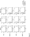
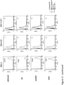
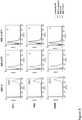
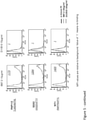
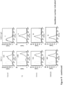
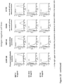
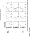
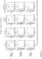
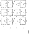
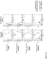
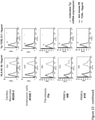
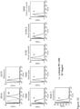
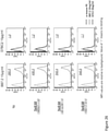
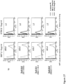
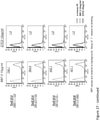
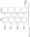
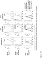
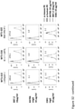
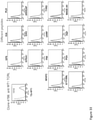
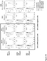
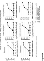
- the present inventors have now indentified through a laborious screen and experimentation novel TCRLs which exhibit unprecedented fine specificity towards TyrD-HLA-A2 (D7 and D11), WT1-HLA-A2 (B47), MAGE-A4-HLA-A2 (C106B9), MAGE-A9-HLA-A2 (F184C7) and PAP (D10A3).
- the CDRs of these antibodies can be implanted in any affinity binding entity such as having an effector activity e.g., a CAR and TCR.
- the present invention provides an affinity binding entity comprising an antigen binding domain comprising:
- T Cell Receptor-like antibody refers to an antibody which binds an MHC being complexed with an HLA-restricted peptide antigen. Binding of the TCRL to its target is with an MHC-restricted specificity. The TCRL antibody does not bind said MHC in the absence of said complexed peptide, and the antibody does not bind said peptide in an absence of said MHC.
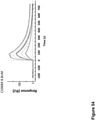
- binding refers to an antibody-antigen mode of binding, which is generally, in the case of clinically relevant TCRLs, in the range of K D below 20 nM, as determined by Surface Plasmon Resonance assay (SPR).
- SPR Surface Plasmon Resonance assay
- the affinity of the antigen binding domain to its antigen is determined using the soluble form of the antibody from which the CDRs of the antigen binding domain of the antibody are derived.
- the antigen is used in its soluble form e.g., as a single chain MHC-peptide complex as further described hereinbelow.
- K D refers to the equilibrium dissociation constant between the antigen binding domain and its respective antigen.
- affinity of the affinity binding entity is determined by the CDRs.
- affinity may be improved using methods known in the art, such as affinity maturation.
- affinity binding entity refers to a binding moiety which binds to a specific antigen with a higher affinity than to a non-specific antigen and is endowed with an affinity of at least 10 -6 M, as determined by assays which are well known in the art, including SPR.
- the affinity is 500 nM- 0.5 nM, 100 nM-1 nM, 50 nM-1 nM, 20 nM-1 nM, 10 nM-1 nM.
- the affinity moiety may be selected from the group consisting of TCR, CAR-T and an antibody.
- the affinity binding entity is an antibody.
- the reference to antibodies is in more details as compared to other affinity binding entities, the description of this embodiment should not be construed as limiting and the present invention is equally related to binding entities as described herein especially in the sense of cell therapy as further described hereinbelow.
- antibody as used in this invention includes intact molecules as well as functional fragments thereof, such as Fab, F(ab')2, Fv, scFv, dsFv, or single domain molecules such as VH and VL that are capable of binding to an epitope of an antigen in an MHC restricted manner.
- antibody aims to encompass any affinity binding entity which binds a cell surface presented molecule with an MHC restricted specificity.
- CDRs of the antibodies of some embodiments of the present invention may be implanted in artificial molecules such as T cell receptors or CARs as further described hereinbelow.
- Suitable antibody fragments for practicing some embodiments of the invention include a complementarity-determining region (CDR) of an immunoglobulin light chain (referred to herein as “light chain”), a complementarity-determining region of an immunoglobulin heavy chain (referred to herein as “heavy chain”), a variable region of a light chain, a variable region of a heavy chain, a light chain, a heavy chain, an Fd fragment, and antibody fragments comprising essentially whole variable regions of both light and heavy chains such as an Fv, a single chain Fv Fv (scFv), a disulfide-stabilized Fv (dsFv), an Fab, an Fab', and an F(ab')2.
- CDR complementarity-determining region
- light chain referred to herein as "light chain”
- heavy chain a complementarity-determining region of an immunoglobulin heavy chain
- variable region of a light chain a variable region of a heavy chain
- a light chain a variable region of
- CDR complementarity-determining region
- VH VH
- VL VL
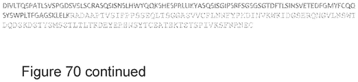
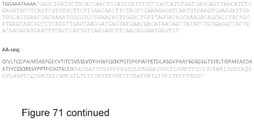
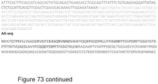
- Examples of such CDR sequences are provide for D7 and D11 - TCRLs produced according to Example I below. Additional examples include, WT1 B47B6, MAGE-A4 C106B9, MAGE-A9 F184C7, PAP D10A3 (shown in Figures 68-73 ).
- the identity of the amino acid residues in a particular antibody that make up a variable region or a CDR can be determined using methods well known in the art and include methods such as sequence variability as defined by Kabat et al. (See, e.g., Kabat et al., 1992, Sequences of Proteins of Immunological Interest, 5th ed., Public Health Service, NIH, Washington D.C .), location of the structural loop regions as defined by Chothia et al.
- variable regions and CDRs may refer to variable regions and CDRs defined by any approach known in the art, including combinations of approaches. According to a specific embodiment, the CDRs are determined according to Kabat et al. (supra).
- Antibody fragments according to some embodiments of the invention can be prepared by proteolytic hydrolysis of the antibody or by expression in E. coli or mammalian cells (e.g. Chinese hamster ovary cell culture or other protein expression systems) of DNA encoding the fragment.
- Antibody fragments can be obtained by pepsin or papain digestion of whole antibodies by conventional methods.
- antibody fragments can be produced by enzymatic cleavage of antibodies with pepsin to provide a 5S fragment denoted F(ab')2.
- This fragment can be further cleaved using a thiol reducing agent, and optionally a blocking group for the sulfhydryl groups resulting from cleavage of disulfide linkages, to produce 3.5S Fab' monovalent fragments.
- a thiol reducing agent optionally a blocking group for the sulfhydryl groups resulting from cleavage of disulfide linkages
- an enzymatic cleavage using pepsin produces two monovalent Fab' fragments and an Fc fragment directly.
- cleaving antibodies such as separation of heavy chains to form monovalent light-heavy chain fragments, further cleavage of fragments, or other enzymatic, chemical, or genetic techniques may also be used, so long as the fragments bind to the antigen that is recognized by the intact antibody.
- Fv fragments comprise an association of VH and VL chains. This association may be noncovalent, as described in Inbar et al. [Proc. Nat'l Acad. Sci. USA 69:2659-62 (19720]. Alternatively, the variable chains can be linked by an intermolecular disulfide bond or cross-linked by chemicals such as glutaraldehyde. Preferably, the Fv fragments comprise VH and VL chains connected by a peptide linker.
- sFv single-chain antigen binding proteins
- the structural gene is inserted into an expression vector, which is subsequently introduced into a host cell such as E. coli.
- the recombinant host cells synthesize a single polypeptide chain with a linker peptide bridging the two V domains.
- Methods for producing sFvs are described, for example, by [ Whitlow and Filpula, Methods 2: 97-105 (1991 ); Bird et al., Science 242:423-426 (1988 ); Pack et al., Bio/Technology 11:1271-77 (1993 ); and U.S. Pat. No. 4,946,778 .
- CDR peptides (“minimal recognition units") can be obtained by constructing genes encoding the CDR of an antibody of interest. Such genes are prepared, for example, by using the polymerase chain reaction to synthesize the variable region from RNA of antibody-producing cells. See, for example, Larrick and Fry [Methods, 2: 106-10 (1991 )].
- Humanized forms of non-human (e.g., murine) antibodies are chimeric molecules of immunoglobulins, immunoglobulin chains or fragments thereof (such as Fv, Fab, Fab', F(ab').sub.2 or other antigen-binding subsequences of antibodies) which contain minimal sequence derived from non-human immunoglobulin.
- Humanized antibodies include human immunoglobulins (recipient antibody) in which residues form a complementary determining region (CDR) of the recipient are replaced by residues from a CDR of a non-human species (donor antibody) such as mouse, rat or rabbit having the desired specificity, affinity and capacity.
- CDR complementary determining region
- donor antibody such as mouse, rat or rabbit having the desired specificity, affinity and capacity.
- Fv framework residues of the human immunoglobulin are replaced by corresponding non-human residues.
- Humanized antibodies may also comprise residues which are found neither in the recipient antibody nor in the imported CDR or framework sequences.
- the humanized antibody will comprise substantially all of at least one, and typically two, variable domains, in which all or substantially all of the CDR regions correspond to those of a non-human immunoglobulin and all or substantially all of the FR regions are those of a human immunoglobulin consensus sequence.
- the humanized antibody optimally also will comprise at least a portion of an immunoglobulin constant region (Fc), typically that of a human immunoglobulin [ Jones et al., Nature, 321:522-525 (1986 ); Riechmann et al., Nature, 332:323-329 (1988 ); and Presta, Curr. Op. Struct. Biol., 2:593-596 (1992 )].
- Fc immunoglobulin constant region
- a humanized antibody has one or more amino acid residues introduced into it from a source which is non-human. These non-human amino acid residues are often referred to as import residues, which are typically taken from an import variable domain. Humanization can be essentially performed following the method of Winter and co-workers [ Jones et al., Nature, 321:522-525 (1986 ); Riechmann et al., Nature 332:323-327 (1988 ); Verhoeyen et al., Science, 239:1534-1536 (1988 )], by substituting rodent CDRs or CDR sequences for the corresponding sequences of a human antibody.
- humanized antibodies are chimeric antibodies ( U.S. Pat. No. 4,816,567 ), wherein substantially less than an intact human variable domain has been substituted by the corresponding sequence from a non-human species.
- humanized antibodies are typically human antibodies in which some CDR residues and possibly some FR residues are substituted by residues from analogous sites in rodent antibodies.
- Human antibodies can also be produced using various techniques known in the art, including phage display libraries [ Hoogenboom and Winter, J. Mol. Biol., 227:381 (1991 ); Marks et al., J. Mol. Biol., 222:581 (1991 )].
- the techniques of Cole et al. and Boerner et al. are also available for the preparation of human monoclonal antibodies ( Cole et al., Monoclonal Antibodies and Cancer Therapy, Alan R. Liss, p. 77 (1985 ) and Boerner et al., J. Immunol., 147(1):86-95 (1991 )].
- human antibodies can be made by introduction of human immunoglobulin loci into transgenic animals, e.g., mice in which the endogenous immunoglobulin genes have been partially or completely inactivated. Upon challenge, human antibody production is observed, which closely resembles that seen in humans in all respects, including gene rearrangement, assembly, and antibody repertoire. This approach is described, for example, in U.S. Pat. Nos.
- the heavy and light chains of an antibody of the invention may be full-length (e.g., an antibody can include at least one, and preferably two, complete heavy chains, and at least one, or two, complete light chains) or may include an antigen-binding portion (a Fab, F(ab').sub.2, Fv or a single chain Fv fragment ("scFv")).
- the antibody heavy chain constant region is chosen from, e.g., IgG1, IgG2, IgG3, IgG4, IgM, IgA1, IgA2, IgD, and IgE.
- the immunoglobulin isotype is selected from IgG1, IgG2, IgG3, and IgG4, more particularly, IgG1 (e.g., human IgG1) or IgG4 (e.g., human IgG4).
- IgG1 e.g., human IgG1
- IgG4 e.g., human IgG4
- a bispecific monoclonal antibody (BsMAb, BsAb) is an artificial protein that is composed of fragments of two different monoclonal antibodies and consequently binds to two different types of antigen.
- the BsMAb is engineered to simultaneously bind to a cytotoxic cell (e.g., using a receptor like CD3) and a target like a tumor cell to be destroyed (further described hereinbelow).
- chimeric antigen receptor refers to a recombinant or synthetic molecule which combines antibody-based specificity for a desired antigen with a T cell receptor-activating intracellular domain to generate a chimeric protein that exhibits cellular immune activity to the specific antigen.
- T Cell Receptor or “TCR” refers to soluble and non-soluble forms of recombinant T cell receptor.
- MHC (or HLA)-restricted peptide refers to a peptide which is potentially presented on an MHC molecule. Such peptides may be identified by "wet” laboratory procedures such as Mass-Spectrometry or by in-silico analysis.
- An MHC (or HLA)-presented peptide refers to a peptide which is confirmed in vitro or in vivo as being presented by an MHC molecule.
- the MHC restricted peptide is from MAGE-A4 and the affinity binding entity comprises the CDRs of C106B9.
- homologous sequences e.g., at least 90 % homology, 95 % homology or even at least 99 % homology as long as the binding affinity to the respective target and optionally specificity are maintained or even improved.
- an isolated polynucleotide comprising a nucleic acid sequence encoding the affinity binding entity as described herein.
- an expression vector comprising the polynucleotide operably linked to a cis- acting regulatory element.
- the nucleic acid construct (also referred to herein as an "expression vector") of some embodiments of the invention includes additional sequences which render this vector suitable for replication and integration in prokaryotes, eukaryotes, or preferably both (e.g., shuttle vectors).
- typical cloning vectors may also contain a transcription and translation initiation sequence, transcription and translation terminator and a polyadenylation signal.
- such constructs will typically include a 5' LTR, a tRNA binding site, a packaging signal, an origin of second-strand DNA synthesis, and a 3' LTR or a portion thereof.
- the nucleic acid construct of some embodiments of the invention typically includes a signal sequence for secretion or presentation of the affinity binding entity from a host cell in which it is placed.
- the signal sequence for this purpose is a mammalian signal sequence.
- Eukaryotic promoters typically contain two types of recognition sequences, the TATA box and upstream promoter elements.
- the TATA box located 25-30 base pairs upstream of the transcription initiation site, is thought to be involved in directing RNA polymerase to begin RNA synthesis.
- the other upstream promoter elements determine the rate at which transcription is initiated.
- the promoter utilized by the nucleic acid construct of some embodiments of the invention is active in the specific cell population transformed.
- cell type-specific and/or tissue-specific promoters include promoters such as albumin that is liver specific [ Pinkert et al., (1987) Genes Dev. 1:268-277 ], lymphoid specific promoters [ Calame et al., (1988) Adv. Immunol. 43:235-275 ]; in particular promoters of T-cell receptors [ Winoto et al., (1989) EMBO J. 8:729-733 ] and immunoglobulins; [ Banerji et al.
- neuron-specific promoters such as the neurofilament promoter [ Byrne et al. (1989) Proc. Natl. Acad. Sci. USA 86:5473-5477 ], pancreas-specific promoters [ Edlunch et al. (1985) Science 230:912-916 ] or mammary gland-specific promoters such as the milk whey promoter ( U.S. Pat. No. 4,873,316 and European Application Publication No. 264,166 ).
- Enhancer elements can stimulate transcription up to 1,000 fold from linked homologous or heterologous promoters. Enhancers are active when placed downstream or upstream from the transcription initiation site. Many enhancer elements derived from viruses have a broad host range and are active in a variety of tissues. For example, the SV40 early gene enhancer is suitable for many cell types. Other enhancer/promoter combinations that are suitable for some embodiments of the invention include those derived from polyoma virus, human or murine cytomegalovirus (CMV), the long term repeat from various retroviruses such as murine leukemia virus, murine or Rous sarcoma virus and HIV. See, Enhancers and Eukaryotic Expression, Cold Spring Harbor Press, Cold Spring Harbor, N.Y. 1983 .
- CMV cytomegalovirus
- the promoter is preferably positioned approximately the same distance from the heterologous transcription start site as it is from the transcription start site in its natural setting. As is known in the art, however, some variation in this distance can be accommodated without loss of promoter function.
- Polyadenylation sequences can also be added to the expression vector in order to increase the efficiency of TCRL mRNA translation.
- Two distinct sequence elements are required for accurate and efficient polyadenylation: GU or U rich sequences located downstream from the polyadenylation site and a highly conserved sequence of six nucleotides, AAUAAA, located 11-30 nucleotides upstream.
- Termination and polyadenylation signals that are suitable for some embodiments of the invention include those derived from SV40.
- the expression vector of some embodiments of the invention may typically contain other specialized elements intended to increase the level of expression of cloned nucleic acids or to facilitate the identification of cells that carry the recombinant DNA.
- a number of animal viruses contain DNA sequences that promote the extra chromosomal replication of the viral genome in permissive cell types. Plasmids bearing these viral replicons are replicated episomally as long as the appropriate factors are provided by genes either carried on the plasmid or with the genome of the host cell.
- the vector may or may not include a eukaryotic replicon. If a eukaryotic replicon is present, then the vector is amplifiable in eukaryotic cells using the appropriate selectable marker. If the vector does not comprise a eukaryotic replicon, no episomal amplification is possible. Instead, the recombinant DNA integrates into the genome of the engineered cell, where the promoter directs expression of the desired nucleic acid.
- cells which comprise the polynucleotides/expression vectors as described herein.
- Such cells are typically selected for high expression of recombinant proteins (e.g., bacterial, plant or eukaryotic cells e.g., CHO, HEK-293 cells), but may also be host cells having a specific immune effector activity (e.g., T cells or NK cells) when for instance the CDRs of the TCRL are implanted in a T Cell Receptor or CAR transduced in said cells which are used in adoptive cell therapy as further described hereinbelow.
- recombinant proteins e.g., bacterial, plant or eukaryotic cells e.g., CHO, HEK-293 cells
- host cells having a specific immune effector activity e.g., T cells or NK cells
- the high specificity of the affinity binding entity renders it particularly suitable for diagnostic and therapeutic applications.
- a method of detecting a cell presenting an HLA-restricted peptide antigen of interest comprises contacting the cell with the affinity binding entity (e.g., antibody) of the present disclosure having specificity to the HLA-restricted peptide antigen of interest.
- the contacting is effected under conditions which allow immunocomplex formation, wherein a presence of the immunocomplex or level thereof is indicative of the cell presenting the HLA-restricted peptide antigen of interest.
- detecting refers to the act of detecting, perceiving, uncovering, exposing, visualizing or identifying a cell.
- the precise method of detecting is dependent on the detectable moiety (also referred to herein as identifiable moiety) to which the antibody is attached as further described herein below.
- Single cells may be used in accordance with the teachings of the present disclosure as well as a plurality of cells.
- the cells may be from any biological sample such as cell-lines, primary (e.g., tumor cultures) and cellular samples, e.g. surgical biopsies including incisional or excisional biopsy, fine needle aspirates and the like. Methods of biopsy retrieval are well known in the art.
- the above-mentioned detection method can be harnessed to the diagnosis of diseases which are characterized by above normal presentation or different tissue distribution of the HLA-peptide complex.
- diagnosis refers to classifying a disease, determining a severity of a disease (grade or stage), monitoring progression, forecasting an outcome of the disease and/or prospects of recovery.
- the subject may be a healthy subject (e.g., human) undergoing a routine wellbeing check up.
- the subject may be at risk of the disease.
- the method may be used to monitor treatment efficacy.
- the TCRL may comprise e.g., attached to an identifiable moiety.
- the TCRL (or a complex comprising same) may be identified indirectly such as by using a secondary antibody.
- the contacting may be effected in vitro (i.e. in a cell line, primary cells), ex vivo or in vivo.
- the method of the present disclosure is effected under conditions sufficient to form an immunocomplex (e.g. a complex between the antibodies of the present disclosure and the peptide complexed to the MHC, typically when the cells are not lysed); such conditions (e.g., appropriate concentrations, buffers, temperatures, reaction times) as well as methods to optimize such conditions are known to those skilled in the art, and examples are disclosed herein.
- an immunocomplex e.g. a complex between the antibodies of the present disclosure and the peptide complexed to the MHC, typically when the cells are not lysed
- such conditions e.g., appropriate concentrations, buffers, temperatures, reaction times
- methods to optimize such conditions are known to those skilled in the art, and examples are disclosed herein.
- the affinity binding entities of the invention are especially useful for the treatment of cancer.
- cancer as used herein is defined as disease characterized by the rapid and uncontrolled growth of aberrant cells. Cancer cells can spread locally or through the bloodstream and lymphatic system to other parts of the body.
- the cancer may be a hematological malignancy, a solid tumor, a primary or a metatastizing tumor.
- various cancers include but are not limited to, breast cancer, prostate cancer, ovarian cancer, cervical cancer, skin cancer, pancreatic cancer, colorectal cancer, renal cancer, liver cancer, brain cancer, lymphoma, Chronic Lymphocytic Leukemia (CLL), leukemia, lung cancer and the like. Additional nonlimiting examples of cancers which can be treated by the method of some embodiments of the invention are provided in Table 1, above.
- Cancers that may be treated include tumors that are not vascularized, or not yet substantially vascularized, as well as vascularized tumors.
- the cancers may comprise non-solid tumors (such as hematological tumors, for example, leukemias and lymphomas) or may comprise solid tumors.
- Types of cancers to be treated with the Antibodies of the invention include, but are not limited to, carcinoma, blastoma, and sarcoma, and certain leukemia or lymphoid malignancies, benign and malignant tumors, and malignancies e.g., sarcomas, carcinomas, and melanomas.
- sarcomas e.g., sarcomas, carcinomas, and melanomas.
- Adult tumors/cancers and pediatric tumors/cancers are also included.
- Hematologic cancers are cancers of the blood or bone marrow.
- hematological (or hematogenous) cancers include leukemias, including acute leukemias (such as acute lymphocytic leukemia, acute myelocytic leukemia, acute myelogenous leukemia and myeloblastic, promyelocytic, myelomonocytic, monocytic and erythroleukemia), chronic leukemias (such as chronic myelocytic (granulocytic) leukemia, chronic myelogenous leukemia, and chronic lymphocytic leukemia), polycythemia vera, lymphoma, Hodgkin's disease, non-Hodgkin's lymphoma (indolent and high grade forms), multiple myeloma, Waldenstrom's macroglobulinemia, heavy chain disease, myelodysplastic syndrome, hairy cell leukemia and myelodysplasia.
- Solid tumors are abnormal masses of tissue that usually do not contain cysts or liquid areas. Solid tumors can be benign or malignant. Different types of solid tumors are named for the type of cells that form them (such as sarcomas, carcinomas, and lymphomas). Examples of solid tumors, such as sarcomas and carcinomas, include fibrosarcoma, myxosarcoma, liposarcoma, chondrosarcoma, osteosarcoma, and other sarcomas, synovioma, mesothelioma, Ewing's tumor, leiomyosarcoma, rhabdomyosarcoma, colon carcinoma, lymphoid malignancy, pancreatic cancer, breast cancer, lung cancers, ovarian cancer, prostate cancer, hepatocellular carcinoma, squamous cell carcinoma, basal cell carcinoma, adenocarcinoma, sweat gland carcinoma, medullary thyroid carcinoma, papillary thyroid carcinoma, pheochromocytomas se
- the pathology is a solid tumor.
- the affinity binding entiry of the invention has an anti-tumor effect.
- anti-tumor effect refers to a biological effect which can be manifested by a decrease in tumor volume, a decrease in the number of tumor cells, a decrease in the number of metastases, an increase in life expectancy, or amelioration of various physiological symptoms associated with the cancerous condition.
- An "anti-tumor effect” can also be manifested by the ability of the medicament of the invention in prevention of the occurrence of tumor in the first place.
- the affinity binding entity is for Tyrosinase (TyrD)
- the cancer is selected from the group consisting of melanoma and glioblastoma.
- the cancer is selected from: Table 1 Leukemia multiple myeloma (MM) acute lymphoblastic leukemia (ALL) acute myeloid/myelogenous leukemia (AML) myelodysplastic syndrome (MDS) mesothelioma ovarian cancer gastrointestinal cancers e.g., colorectal cancer adenocarcinoma, thyroid cancer breast cancer lung cancer (e.g., non small cell lung cancer) melanoma osteosarcoma endomentrial cancer
- MM Leukemia multiple myeloma
- ALL acute lymphoblastic leukemia
- AML acute myeloid/myelogenous leukemia
- MDS myelodysplastic syndrome mesothelioma ovarian cancer gastrointestinal cancers e.g., colorectal cancer adenocarcinoma, thyroid cancer breast cancer lung cancer (e.g., non small cell lung cancer) melanoma osteosarcoma endomentrial cancer
- said affinity binding entity is for MAGE-A4 said cancer is selected from: Table 2 MAGE-A4 Ovarian cancer T cell leukemia/lymphoma (e.g., ATLL) Sarcoma testicular cancer head and neck cancer bladder cancer esophagus cancer.
- MAGE-A4 Ovarian cancer T cell leukemia/lymphoma e.g., ATLL
- Sarcoma testicular cancer head and neck cancer bladder cancer esophagus cancer e.g., ATLL
- said affinity binding entity is for MAGE-A9 said cancer is selected from: Table 3 MAGE-A9 renal cell carcinoma bladder cancer breast cancer hepatocellular carcinoma.
- said affinity binding entity is for PAP said cancer is prostate cancer.
- Determining a presence or level of the immunocomplex of the present invention is dependent on the detectable moiety to which the antibody is attached.
- detectable moieties examples include but are not limited to radioactive isotopes, phosphorescent chemicals, chemiluminescent chemicals, fluorescent chemicals, enzymes, fluorescent polypeptides and epitope tags.
- the detectable moiety can be a member of a binding pair, which is identifiable via its interaction with an additional member of the binding pair, and a label which is directly visualized.
- the member of the binding pair is an antigen which is identified by a corresponding labeled antibody.
- the label is a fluorescent protein or an enzyme producing a colorimetric reaction.
- detectable moieties include those detectable by Positron Emission Tomagraphy (PET) and Magnetic Resonance Imaging (MRI), all of which are well known to those of skill in the art.
- PET Positron Emission Tomagraphy
- MRI Magnetic Resonance Imaging
- the immunolabel i.e. the antibody conjugated to the detectable moiety
- the immunolabel may be produced by recombinant means or may be chemically synthesized by, for example, the stepwise addition of one or more amino acid residues in defined order using solid phase peptide synthetic techniques.
- polypeptide detectable moieties that can be linked to the antibodies of the present invention using recombinant DNA technology (in which the polynucleotide encoding the TCRL is translationally fused to the detectable moiety) include fluorescent polypeptides, phosphorescent polypeptides, enzymes and epitope tags.
- chemical attachment of a detectable moiety to the antibodies of the present invention can be effected using any suitable chemical linkage, direct or indirect, as via a peptide bond (when the detectable moiety is a polypeptide), or via covalent bonding to an intervening linker element, such as a linker peptide or other chemical moiety, such as an organic polymer.
- linker element such as a linker peptide or other chemical moiety, such as an organic polymer.
- Such chimeric peptides may be linked via bonding at the carboxy (C) or amino (N) termini of the peptides, or via bonding to internal chemical groups such as straight, branched or cyclic side chains, internal carbon or nitrogen atoms, and the like.
- modified peptides can be easily identified and prepared by one of ordinary skill in the art, using well known methods of peptide synthesis and/or covalent linkage of peptides.
- Description of fluorescent labeling of antibodies is provided in details in U.S. Pat. Nos. 3,940,475 , 4,289,747 , and 4,376,110 .
- SPDP conjugation method Any SPDP conjugation method known to those skilled in the art can be used.
- a modification of the method of Cumber et al. (1985, Methods of Enzymology 112: 207-224 ) as described below, is used.
- a peptide, such as an identifiable or therapeutic moiety (1.7 mg/ml) is mixed with a 10-fold excess of SPDP (50 mM in ethanol) and the antibody is mixed with a 25-fold excess of SPDP in 20 mM sodium phosphate, 0.10 M NaCl pH 7.2 and each of the reactions incubated, e.g., for 3 hours at room temperature. The reactions are then dialyzed against PBS.
- the peptide is reduced, e.g., with 50 mM DTT for 1 hour at room temperature.
- the reduced peptide is desalted by equilibration on G-25 column (up to 5 % sample/column volume) with 50 mM KH 2 PO 4 pH 6.5.
- the reduced peptide is combined with the SPDP-antibody in a molar ratio of 1:10 antibody:peptide and incubated at 4 °C overnight to form a peptide-antibody conjugate.
- Conjugation of a peptide (e.g., an identifiable or therapeutic moiety) with an antibody can be accomplished by methods known to those skilled in the art using glutaraldehyde.
- glutaraldehyde for example, in one illustrative embodiment, the method of conjugation by G.T. Hermanson (1996, “Antibody Modification and Conjugation, in Bioconjugate Techniques, Academic Press, San Diego ) described below, is used.
- the antibody and the peptide (1.1 mg/ml) are mixed at a 10-fold excess with 0.05 % glutaraldehyde in 0.1 M phosphate, 0.15 M NaCl pH 6.8, and allowed to react for 2 hours at room temperature. 0.01 M lysine can be added to block excess sites. After-the reaction, the excess glutaraldehyde is removed using a G-25 column equilibrated with PBS (10 % v/v sample/column volumes).
- Conjugation of a peptide with an antibody can be accomplished by methods known to those skilled in the art using a dehydrating agent such as a carbodiimide. Most preferably the carbodiimide is used in the presence of 4-dimethyl aminopyridine. As is well known to those skilled in the art, carbodiimide conjugation can be used to form a covalent bond between a carboxyl group of peptide and an hydroxyl group of an antibody (resulting in the formation of an ester bond), or an amino group of an antibody (resulting in the formation of an amide bond) or a sulfhydryl group of an antibody (resulting in the formation of a thioester bond).
- a dehydrating agent such as a carbodiimide.
- carbodiimide is used in the presence of 4-dimethyl aminopyridine.
- carbodiimide conjugation can be used to form a covalent bond between a carboxyl group of peptide and an hydroxyl group of an antibody (resulting in the formation
- carbodiimide coupling can be used to form analogous covalent bonds between a carbon group of an antibody and an hydroxyl, amino or sulfhydryl group of the peptide. See, generally, J. March, Advanced Organic Chemistry: Reaction's, Mechanism, and Structure, pp. 349-50 & 372-74 (3d ed.), 1985 .
- the peptide is conjugated to an antibody via a covalent bond using a carbodiimide, such as dicyclohexylcarbodiimide. See generally, the methods of conjugation by B. Neises et al. (1978, Angew Chem., Int. Ed. Engl. 17:522 ; A.
- the level of immunocomplex may be compared to a control sample from a non-diseased subject, wherein an up-regulation of immunocomplex formation is indicative of melanoma.
- the subject is of the same species e.g. human, preferably matched with the same age, weight, sex etc.
- the control sample may also be of the same subject from a healthy tissue, prior to disease progression or following disease remission.
- the detection is effected by FACS.
- antibodies of the present invention can also be used in therapeutics where the affinity binding entity e.g., antibody comprises a therapeutic moiety.
- the therapeutic moiety can be an integral part of the antibody e.g., in the case of a whole antibody, the Fc domain, which activates antibody-dependent cell-mediated cytotoxicity (ADCC).
- ADCC is a mechanism of cell-mediated immune defense whereby an effector cell of the immune system actively lyses a target cell, whose membrane-surface antigens have been bound by specific antibodies . It is one of the mechanisms through which antibodies, as part of the humoral immune response , can act to limit and contain infection.
- Classical ADCC is mediated by natural killer (NK) cells ; macrophages , neutrophils and eosinophils can also mediate ADCC.
- eosinophils can kill certain parasitic worms known as helminths through ADCC mediated by IgE.
- ADCC is part of the adaptive immune response due to its dependence on a prior antibody response.
- the antibody may be a bispecific antibody in which the therapeutic moiety is a T cell engager for example, such as an anti CD3 antibody or an anti CD16a alternatively the therapeutic moiety may be an anti immune checkpoint molecule (anti PD-1).
- the therapeutic moiety may be an anti immune checkpoint molecule (anti PD-1).
- the antibody may be attached to a heterologous therapeutic moiety (methods of conjugation are described hereinabove).
- the therapeutic moiety can be, for example, a cytotoxic moiety, a toxic moiety, a cytokine moiety, a drug.
- the antibody may be in a soluble or insoluble form.
- Insoluble forms may be those in which a molecule comprising the antibody's CDRs is anchored to or expressed by a cell or a particle (the latter can be used for therapeutic as well as diagnostic applications).
- Examples of such cells include immune cells, T cells, B cells, dendritic cells, CIK, NKT, NK cells (autologous, allogeneic, xenogeneic).
- the antibody (or actually CDRs thereof) form a CAR (as explained above) or an artificial T Cell Receptor.
- a polynucleotide coding for such a molecule is transduced in a cell of interest.
- the cell is a T cell, a natural killer cell, a cell that exerts effector killing function on a target cell, a cell that exerts a suppressive effect on effector T cells, an engineered cell with an effector killing function or an engineered cell with a suppressive function.
- the cell is a T cell, or ⁇ T cell, or ⁇ T cell.
- the cell is a natural killer (NK) cell.
- NK natural killer
- the natural killer cell is used to target cancer.
- the T cell is a cytotoxic T cell (effector T cell).
- the cytotoxic T cell (effector T cell) is used to target cancer antigens.
- the cytotoxic T cell is used to treat a pathology caused by or associated with cancer.
- the T cell comprises a Treg (T regulatory cell).
- the T cell comprises a CD3 T cell.
- the T cell comprises a CD4 T cell.
- the T cell comprises a CD8 T cell.
- the antigen binding domain comprises a single chain Fv (scFv) molecule.
- the cytoplasmic domain (also referred to as "intracellular signaling domain") of the CAR molecule of the invention is responsible for activation of at least one of the normal effector functions of the immune cell in which the CAR has been placed in.
- effector function refers to a specialized function of a cell. Effector function of a T cell, for example, may be cytolytic activity or helper activity including the secretion of cytokines.
- intracellular signaling domain refers to the portion of a protein which transduces the effector function signal and directs the cell to perform a specialized function. While usually the entire intracellular signaling domain can be employed, in many cases it is not necessary to use the entire chain. To the extent that a truncated portion of the intracellular signaling domain is used, such truncated portion may be used in place of the intact chain as long as it transduces the effector function signal.
- intracellular signaling domain is thus meant to include any truncated portion of the intracellular signaling domain sufficient to transduce the effector function signal.
- intracellular signaling domains for use in the CAR molecule of the invention include the cytoplasmic sequences of the T cell receptor (TCR) and co-receptors that act in concert to initiate signal transduction following antigen receptor engagement, as well as any derivative or variant of these sequences and any synthetic sequence that has the same functional capability.
- TCR T cell receptor
- T cell activation can be mediated by two distinct classes of cytoplasmic signaling sequence: those that initiate antigen-dependent primary activation through the TCR (primary cytoplasmic signaling sequences) and those that act in an antigen-independent manner to provide a secondary or co-stimulatory signal (secondary cytoplasmic signaling sequences).
- primary cytoplasmic signaling sequences those that initiate antigen-dependent primary activation through the TCR
- secondary cytoplasmic signaling sequences those that act in an antigen-independent manner to provide a secondary or co-stimulatory signal
- Primary cytoplasmic signaling sequences regulate primary activation of the TCR complex either in a stimulatory way, or in an inhibitory way.
- Primary cytoplasmic signaling sequences that act in a stimulatory manner may contain signaling motifs which are known as immunoreceptor tyrosine-based activation motifs (ITAMs).
- ITAMs immunoreceptor tyrosine-based activation motifs
- ITAM containing primary cytoplasmic signaling sequences examples include those derived from TCR zeta, FcR gamma, FcR beta, CD3 gamma, CD3 delta, CD3 epsilon, CD5, CD22, CD79a, CD79b, and CD66d. It is particularly preferred that cytoplasmic signaling molecule in the CAR of the invention comprises a cytoplasmic signaling sequence derived from CD3 zeta.
- the cytoplasmic domain of the CAR can be designed to comprise the CD3-zeta signaling domain by itself or combined with any other desired cytoplasmic domain(s) useful in the context of the CAR of the invention.
- the cytoplasmic domain of the CAR can comprise a CD3 zeta chain portion and a costimulatory signaling region.
- the costimulatory signaling region refers to a portion of the CAR comprising the intracellular domain of a costimulatory molecule.
- a co-stimulatory molecule is a cell surface molecule other than an antigen receptor or their ligands that is required for an efficient response of lymphocytes to an antigen.
- Examples of such molecules include CD27, CD28, 4-1BB (CD137), OX40, CD30, CD40, PD-1, ICOS, lymphocyte function-associated antigen-1 (LFA-1), CD2, CD7, LIGHT, NKG2C, B7-H3, and a ligand that specifically binds with CD83, and the like.
- 4-1BB as the co-stimulatory signaling element
- LFA-1 lymphocyte function-associated antigen-1
- the intracellular domain comprises, a co-stimulatory signaling region and a zeta chain portion.
- the co-stimulatory signaling region refers to a portion of the CAR molecule comprising the intracellular domain of a co-stimulatory molecule.
- Co-stimulatory molecules are cell surface molecules other than antigen receptors or their ligands that are required for an efficient response of lymphocytes to antigen.
- Co-stimulatory ligand includes a molecule on an antigen presenting cell [e.g., an aAPC (artificial antigen presenting cell), dendritic cell, B cell, and the like] that specifically binds a cognate co-stimulatory molecule on a T cell, thereby providing a signal which, in addition to the primary signal provided by, for instance, binding of a TCR/CD3 complex with an MHC molecule loaded with peptide, mediates a T cell response, including, but not limited to, proliferation, activation, differentiation, and the like.
- an antigen presenting cell e.g., an aAPC (artificial antigen presenting cell), dendritic cell, B cell, and the like
- a co-stimulatory ligand can include, but is not limited to, CD7, B7-1 (CD80), B7-2 (CD86), PD-L1, PD-L2, 4-1BBL, OX40L, inducible costimulatory ligand (ICOS-L), intercellular adhesion molecule (ICAM), CD30L, CD40, CD70, CD83, HLA-G, MICA, MICB, HVEM, lymphotoxin beta receptor, 3/TR6, ILT3, ILT4, HVEM, an agonist or antibody that binds Toll ligand receptor and a ligand that specifically binds with B7-H3.
- a co-stimulatory ligand also encompasses, inter alia, an antibody that specifically binds with a co-stimulatory molecule present on a T cell, such as, but not limited to, CD27, CD28, 4-1BB, OX40, CD30, CD40, PD-1, ICOS, lymphocyte function-associated antigen-1 (LFA-1), CD2, CD7, LIGHT, NKG2C, B7-H3, and a ligand that specifically binds with CD83.
- an antibody that specifically binds with a co-stimulatory molecule present on a T cell such as, but not limited to, CD27, CD28, 4-1BB, OX40, CD30, CD40, PD-1, ICOS, lymphocyte function-associated antigen-1 (LFA-1), CD2, CD7, LIGHT, NKG2C, B7-H3, and a ligand that specifically binds with CD83.
- a "co-stimulatory molecule” refers to the cognate binding partner on a T cell that specifically binds with a co-stimulatory ligand, thereby mediating a co-stimulatory response by the T cell, such as, but not limited to, proliferation.
- Co-stimulatory molecules include, but are not limited to an MHC class 1 molecule, BTLA and a Toll ligand receptor.
- a “co-stimulatory signal”, as used herein, refers to a signal, which in combination with a primary signal, such as TCR/CD3 ligation, leads to T cell proliferation and/or upregulation or down regulation of key molecules.
- stimulation is meant a primary response induced by binding of a stimulatory molecule (e.g., a TCR/CD3 complex) with its cognate ligand thereby mediating a signal transduction event, such as, but not limited to, signal transduction via the TCR/CD3 complex.
- a stimulatory molecule e.g., a TCR/CD3 complex
- Stimulation can mediate altered expression of certain molecules, such as downregulation of TGF- ⁇ , and/or reorganization of cytoskeletal structures, and the like.
- a "stimulatory molecule,” as the term is used herein, means a molecule on a T cell that specifically binds with a cognate stimulatory ligand present on an antigen presenting cell.
- a “stimulatory ligand,” as used herein, means a ligand that when present on an antigen presenting cell (e.g., an aAPC, a dendritic cell, a B-cell, and the like) can specifically bind with a cognate binding partner (referred to herein as a "stimulatory molecule") on a T cell, thereby mediating a primary response by the T cell, including, but not limited to, activation, initiation of an immune response, proliferation, and the like.
- an antigen presenting cell e.g., an aAPC, a dendritic cell, a B-cell, and the like
- a cognate binding partner referred to herein as a "stimulatory molecule”
- Stimulatory ligands are well-known in the art and encompass, inter cilia, an MHC Class I molecule loaded with a peptide, an anti-CD3 antibody, a superagonist anti-CD28 antibody, and a superagonist anti-CD2 antibody.
- the CAR molecule of some embodiments of the invention can be designed to comprise the CD28 and/or 4-1BB signaling domain by itself or be combined with any other desired cytoplasmic domain(s) useful in the context of the CAR molecule of some embodiments of the invention.
- the cytoplasmic domain of the CAR can be designed to further comprise the signaling domain of CD3-zeta.
- the cytoplasmic domain of the CAR can include but is not limited to CD3-zeta, 4-1BB and CD28 signaling modules and combinations thereof.
- the intracellular domain comprises at least one, e.g., at least two, at least three, at least four, at least five, e.g., at least six of the polypeptides selected from the group consisting of: CD3 ⁇ (CD247, CD3z), CD28, 41BB, ICOS, OX40, and CD137.
- the intracellular domain comprises the CD3 ⁇ -chain [CD247 molecule, also known as "CD3-ZETA” and “CD3z”; GenBank Accession NOs. NP_000725.1 and NP_932170.1], which is the primary transmitter of signals from endogenous TCRs.
- the intracellular domain comprises various co-stimulatory protein receptors to the cytoplasmic tail of the CAR to provide additional signals to the T cell (second generation CAR).
- second generation CAR examples include, but are not limited to, CD28 [e.g., GenBank Accession Nos. NP_001230006.1, NP_001230007.1, NP_006130.1], 4-1BB [tumor necrosis factor receptor superfamily, member 9 (TNFRSF9), also known as "CD137", e.g., GenBank Accession No. NP_001552.2], and ICOS [inducible T-cell co-stimulator, e.g., GenBank Accession No. NP_036224.1].
- CD28 e.g., GenBank Accession Nos. NP_001230006.1, NP_001230007.1, NP_006130.1
- 4-1BB tumor necrosis factor receptor superfamily, member 9 (TNFRSF9), also known as "CD137”, e.g.,
- the intracellular domain comprises multiple signaling domains, such as CD3z-CD28-41BB or CD3z-CD28-OX40, to further augment potency.
- OX40 refers to the tumor necrosis factor receptor superfamily, member 4 (TNFRSF4), e.g., GenBank Accession No. NP_003318.1 ("third-generation" CARs).
- the intracellular domain comprises CD28-CD3z, CD3z, CD28-CD137-CD3z.
- CD137 refers to tumor necrosis factor receptor superfamily, member 9 (TNFRSF9), e.g., GenBank Accession No. NP_001552.2.
- the signaling domain when the CAR molecule is designed for a natural killer cell, then the signaling domain can be CD28 and/or CD3 ⁇ .
- the transmembrane domain may be derived either from a natural or from a synthetic source. Where the source is natural, the domain may be derived from any membrane-bound or transmembrane protein. Transmembrane regions of particular use in this invention may be derived from (i.e.
- transmembrane domain may be synthetic, in which case it will comprise predominantly hydrophobic residues such as leucine and valine.
- a triplet of phenylalanine, tryptophan and valine will be found at each end of a synthetic transmembrane domain.
- a short oligo- or polypeptide linker preferably between 2 and 10 amino acids in length may form the linkage between the transmembrane domain and the cytoplasmic signaling domain of the CAR.
- a glycine-serine doublet provides a particularly suitable linker.
- the transmembrane domain comprised in the CAR molecule of some embodiments of the invention is a transmembrane domain that is naturally associated with one of the domains in the CAR.
- the transmembrane domain can be selected or modified by amino acid substitution to avoid binding of such domains to the transmembrane domains of the same or different surface membrane proteins to minimize interactions with other members of the receptor complex.
- spacer domain generally means any oligo- or polypeptide that functions to link the transmembrane domain to, either the extracellular domain or, the cytoplasmic domain in the polypeptide chain.
- a spacer domain may comprise up to 300 amino acids, preferably 10 to 100 amino acids and most preferably 25 to 50 amino acids.
- an affinity binding entity as defined herein for use in a method of treating cancer in a subject in need thereof, comprising administering to the subject the affinity binding entity, thereby treating the cancer in the subject.
- TCRL The selection of the TCRL will naturally depend on its presentation in the pathology. Exemplary TCRLs and their association with pathologies are provided in the Tables hereinabove.
- treating refers to inhibiting, preventing or arresting the development of a pathology (disease, disorder or condition) and/or causing the reduction, remission, or regression of a pathology.
- pathology disease, disorder or condition
- Those of skill in the art will understand that various methodologies and assays can be used to assess the development of a pathology, and similarly, various methodologies and assays may be used to assess the reduction, remission or regression of a pathology.
- the term "subject” includes mammals, preferably human beings at any age which suffer from the pathology.
- the antibodies of some embodiments of the invention can be administered to an organism per se, or in a pharmaceutical composition where it is mixed with suitable carriers or excipients.
- a "pharmaceutical composition” refers to a preparation of one or more of the active ingredients described herein with other chemical components such as physiologically suitable carriers and excipients.
- the purpose of a pharmaceutical composition is to facilitate administration of a compound to an organism.
- active ingredient refers to the antibody accountable for the biological effect.
- physiologically acceptable carrier and “pharmaceutically acceptable carrier” which may be interchangeably used refer to a carrier or a diluent that does not cause significant irritation to an organism and does not abrogate the biological activity and properties of the administered compound.
- An adjuvant is included under these phrases.
- excipient refers to an inert substance added to a pharmaceutical composition to further facilitate administration of an active ingredient.
- excipients include calcium carbonate, calcium phosphate, various sugars and types of starch, cellulose derivatives, gelatin, vegetable oils and polyethylene glycols.
- Suitable routes of administration may, for example, include oral, rectal, transmucosal, especially transnasal, intestinal or parenteral delivery, including intramuscular, subcutaneous and intramedullary injections as well as intrathecal, direct intraventricular, intracardiac, e.g., into the right or left ventricular cavity, into the common coronary artery, intravenous, intraperitoneal, intranasal, or intraocular injections.
- neurosurgical strategies e.g., intracerebral injection or intracerebroventricular infusion
- molecular manipulation of the agent e.g., production of a chimeric fusion protein that comprises a transport peptide that has an affinity for an endothelial cell surface molecule in combination with an agent that is itself incapable of crossing the BBB
- pharmacological strategies designed to increase the lipid solubility of an agent (e.g., conjugation of water-soluble agents to lipid or cholesterol carriers)
- the transitory disruption of the integrity of the BBB by hyperosmotic disruption resulting from the infusion of a mannitol solution into the carotid artery or the use of a biologically active agent such as an angiotensin peptide).
- each of these strategies has limitations, such as the inherent risks associated with an invasive surgical procedure, a size limitation imposed by a limitation inherent in the endogenous transport systems, potentially undesirable biological side effects associated with the systemic administration of a chimeric molecule comprised of a carrier motif that could be active outside of the CNS, and the possible risk of brain damage within regions of the brain where the BBB is disrupted, which renders it a suboptimal delivery method.
- tissue refers to part of an organism consisting of cells designed to perform a function or functions. Examples include, but are not limited to, brain tissue, retina, skin tissue, hepatic tissue, pancreatic tissue, bone, cartilage, connective tissue, blood tissue, muscle tissue, cardiac tissue brain tissue, vascular tissue, renal tissue, pulmonary tissue, gonadal tissue, hematopoietic tissue.
- compositions of some embodiments of the disclosure may be manufactured by processes well known in the art, e.g., by means of conventional mixing, dissolving, granulating, dragee-making, levigating, emulsifying, encapsulating, entrapping or lyophilizing processes.
- compositions for use in accordance with some embodiments of the disclosure thus may be formulated in conventional manner using one or more physiologically acceptable carriers comprising excipients and auxiliaries, which facilitate processing of the active ingredients into preparations which, can be used pharmaceutically. Proper formulation is dependent upon the route of administration chosen.
- the active ingredients of the pharmaceutical composition may be formulated in aqueous solutions, preferably in physiologically compatible buffers such as Hank's solution, Ringer's solution, or physiological salt buffer.
- physiologically compatible buffers such as Hank's solution, Ringer's solution, or physiological salt buffer.
- penetrants appropriate to the barrier to be permeated are used in the formulation. Such penetrants are generally known in the art.
- the pharmaceutical composition can be formulated readily by combining the active compounds with pharmaceutically acceptable carriers well known in the art.
- Such carriers enable the pharmaceutical composition to be formulated as tablets, pills, dragees, capsules, liquids, gels, syrups, slurries, suspensions, and the like, for oral ingestion by a patient.
- Pharmacological preparations for oral use can be made using a solid excipient, optionally grinding the resulting mixture, and processing the mixture of granules, after adding suitable auxiliaries if desired, to obtain tablets or dragee cores.
- Suitable excipients are, in particular, fillers such as sugars, including lactose, sucrose, mannitol, or sorbitol; cellulose preparations such as, for example, maize starch, wheat starch, rice starch, potato starch, gelatin, gum tragacanth, methyl cellulose, hydroxypropylmethyl-cellulose, sodium carbomethylcellulose; and/or physiologically acceptable polymers such as polyvinylpyrrolidone (PVP).
- disintegrating agents may be added, such as cross-linked polyvinyl pyrrolidone, agar, or alginic acid or a salt thereof such as sodium alginate.
- Dragee cores are provided with suitable coatings.
- suitable coatings For this purpose, concentrated sugar solutions may be used which may optionally contain gum arabic, talc, polyvinyl pyrrolidone, carbopol gel, polyethylene glycol, titanium dioxide, lacquer solutions and suitable organic solvents or solvent mixtures.
- Dyestuffs or pigments may be added to the tablets or dragee coatings for identification or to characterize different combinations of active compound doses.
- compositions which can be used orally include push-fit capsules made of gelatin as well as soft, sealed capsules made of gelatin and a plasticizer, such as glycerol or sorbitol.
- the push-fit capsules may contain the active ingredients in admixture with filler such as lactose, binders such as starches, lubricants such as talc or magnesium stearate and, optionally, stabilizers.
- the active ingredients may be dissolved or suspended in suitable liquids, such as fatty oils, liquid paraffin, or liquid polyethylene glycols.
- stabilizers may be added. All formulations for oral administration should be in dosages suitable for the chosen route of administration.
- compositions may take the form of tablets or lozenges formulated in conventional manner.
- the active ingredients for use according to some embodiments of the invention are conveniently delivered in the form of an aerosol spray presentation from a pressurized pack or a nebulizer with the use of a suitable propellant, e.g., dichlorodifluoromethane, trichlorofluoromethane, dichlorotetrafluoroethane or carbon dioxide.
- a suitable propellant e.g., dichlorodifluoromethane, trichlorofluoromethane, dichlorotetrafluoroethane or carbon dioxide.
- the dosage unit may be determined by providing a valve to deliver a metered amount.
- Capsules and cartridges of, e.g., gelatin for use in a dispenser may be formulated containing a powder mix of the compound and a suitable powder base such as lactose or starch.
- compositions described herein may be formulated for parenteral administration, e.g., by bolus injection or continuous infusion.
- Formulations for injection may be presented in unit dosage form, e.g., in ampoules or in multidose containers with optionally, an added preservative.
- the compositions may be suspensions, solutions or emulsions in oily or aqueous vehicles, and may contain formulatory agents such as suspending, stabilizing and/or dispersing agents.
- compositions for parenteral administration include aqueous solutions of the active preparation in water-soluble form. Additionally, suspensions of the active ingredients may be prepared as appropriate oily or water based injection suspensions. Suitable lipophilic solvents or vehicles include fatty oils such as sesame oil, or synthetic fatty acids esters such as ethyl oleate, triglycerides or liposomes. Aqueous injection suspensions may contain substances, which increase the viscosity of the suspension, such as sodium carboxymethyl cellulose, sorbitol or dextran. Optionally, the suspension may also contain suitable stabilizers or agents which increase the solubility of the active ingredients to allow for the preparation of highly concentrated solutions.
- the active ingredient may be in powder form for constitution with a suitable vehicle, e.g., sterile, pyrogen-free water based solution, before use.
- a suitable vehicle e.g., sterile, pyrogen-free water based solution
- compositions of some embodiments of the disclosure may also be formulated in rectal compositions such as suppositories or retention enemas, using, e.g., conventional suppository bases such as cocoa butter or other glycerides.
- compositions suitable for use in context of some embodiments of the disclosure include compositions wherein the active ingredients are contained in an amount effective to achieve the intended purpose. More specifically, a therapeutically effective amount means an amount of active ingredients (TCRL-antibody) effective to prevent, alleviate or ameliorate symptoms of a disorder (e.g., cancer) or prolong the survival of the subject being treated.
- TCRL-antibody active ingredients
- the therapeutically effective amount or dose can be estimated initially from in vitro and cell culture assays.
- a dose can be formulated in animal models to achieve a desired concentration or titer. Such information can be used to more accurately determine useful doses in humans.
- Toxicity and therapeutic efficacy of the active ingredients described herein can be determined by standard pharmaceutical procedures in vitro, in cell cultures or experimental animals.
- the data obtained from these in vitro and cell culture assays and animal studies can be used in formulating a range of dosage for use in human.
- the dosage may vary depending upon the dosage form employed and the route of administration utilized.
- the exact formulation, route of administration and dosage can be chosen by the individual physician in view of the patient's condition. (See e.g., Fingl, et al., 1975, in "The Pharmacological Basis of Therapeutics", Ch. 1 p.1 ).
- Dosage amount and interval may be adjusted individually to provide TCRL (the TCRL tissue) levels of the active ingredient are sufficient to induce or suppress the biological effect (minimal effective concentration, MEC).
- MEC minimum effective concentration
- the MEC will vary for each preparation, but can be estimated from in vitro data. Dosages necessary to achieve the MEC will depend on individual characteristics and route of administration. Detection assays can be used to determine plasma concentrations.
- dosing can be of a single or a plurality of administrations, with course of treatment lasting from several days to several weeks or until cure is effected or diminution of the disease state is achieved.
- compositions to be administered will, of course, be dependent on the subject being treated, the severity of the affliction, the manner of administration, the judgment of the prescribing physician, etc.
- compositions of some embodiments of the disclosure may, if desired, be presented in a pack or dispenser device, such as an FDA approved kit (diagnostic or therapeutic), which may contain one or more unit dosage forms containing the active ingredient.
- the pack may, for example, comprise metal or plastic foil, such as a blister pack.
- the pack or dispenser device may be accompanied by instructions for administration.
- the pack or dispenser may also be accommodated by a notice associated with the container in a form prescribed by a governmental agency regulating the manufacture, use or sale of pharmaceuticals, which notice is reflective of approval by the agency of the form of the compositions or human or veterinary administration. Such notice, for example, may be of labeling approved by the U.S. Food and Drug Administration for prescription drugs or of an approved product insert.
- Compositions comprising a preparation of the disclosure formulated in a compatible pharmaceutical carrier may also be prepared, placed in an appropriate container, and labeled for treatment of an indicated condition, as is further detailed above.
- compositions, method or structure may include additional ingredients, steps and/or parts, but only if the additional ingredients, steps and/or parts do not materially alter the basic and novel characteristics of the claimed composition, method or structure.
- a compound or “at least one compound” may include a plurality of compounds, including mixtures thereof.
- method refers to manners, means, techniques and procedures for accomplishing a given task including, but not limited to, those manners, means, techniques and procedures either known to, or readily developed from known manners, means, techniques and procedures by practitioners of the chemical, pharmacological, biological, biochemical and medical arts.
- treating includes abrogating, substantially inhibiting, slowing or reversing the progression of a condition, substantially ameliorating clinical or aesthetical symptoms of a condition or substantially preventing the appearance of clinical or aesthetical symptoms of a condition.
- sequences that substantially correspond to its complementary sequence as including minor sequence variations, resulting from, e.g., sequencing errors, cloning errors, or other alterations resulting in base substitution, base deletion or base addition, provided that the frequency of such variations is less than 1 in 50 nucleotides, alternatively, less than 1 in 100 nucleotides, alternatively, less than 1 in 200 nucleotides, alternatively, less than 1 in 500 nucleotides, alternatively, less than 1 in 1000 nucleotides, alternatively, less than 1 in 5,000 nucleotides, alternatively, less than 1 in 10,000 nucleotides.
- Single-chain MHC (scMHC) 3 -peptide complexes were produced by in vitro refolding of inclusion bodies produced in Escherichia coli upon isopropyl ⁇ -D-thiogalactoside (IPTG) induction.
- IPTG isopropyl ⁇ -D-thiogalactoside
- a scMHC which contains the ⁇ 2 -microglobulin and the extracellular domains of the HLA-A2 gene connected to each other by a flexible linker, was engineered to contain the BirA recognition sequence for site-specific biotinylation at the C terminus (scMHC-BirA).
- IPTG isopropyl ⁇ -D-thiogalactoside
- T-B hybrid T2 cells were washed with serum-free RPMI 1640 medium and incubated overnight with medium containing 10 -4 -10 -5 M tyrosinase 369-377 YMDGTMSQV (SEQ ID NO: 1)/ WT1 126-134 (RMFPNAPYL, SEQ ID NO: 141) peptide/ MAGE-A4 230-239 SEQ ID NO: 176/MAGE-A9 223-231 203/PAP 112-120 SEQ ID NO: 230 peptide or relevant control peptides (listed in the Table 15 below). Peptide loading efficiency was verified by using the ratio between MFI of HLA-A2-binding antibody BB7.2 on peptide-loaded T2 cells and MFI of unloaded T2 cells (>1) data not shown.
- T2 or primary cells or cell lines (10 6 ) were incubated with 10 ⁇ g/ml of specific Ab (with or without biotinylation) for 1 h at 4 °C, followed by incubation with PE-labeled anti-mouse/human/steptavidin Ab for 45 min at 4 °C. It will be appreciated that the work with anti mouse secondary antibody or with streptavidin gave similar results for D11 and B47B6. Cells were finally washed and analyzed by:
- HHD mice were immunized by 5-6 injections of HLA-A2-peptide complex 50 ⁇ g/mouse. 2-3 first injections were administrated s.c with addition of QuilA adjuvant. Hybridoma clones were generated by fusion of splenocytes isolated from mice immunized with the above complex (as previously described e.g., Weidanz et al. 2011 Int. Rev. Immunol. 30:328-340 ) with NSO myeloma cells and were screened and isolated by differential ELISA assays as described below.
- Hybridomas were grown to >80% confluency in HAT DMEM or serum free DCCM2 medium and supernatant was collected. Purified IgG was isolated from culture supernatant by affinity chromatography using Protein A column. SDS-PAGE analysis of the purified protein revealed homogenous, pure IgG with the expected molecular mass of ⁇ 150 kDa.
- the H and L Fab genes (only for MC1) were cloned for expression as human IgG1 ⁇ Ab into the eukaryotic expression vectors the eukaryotic expression vectors pOptiVEC and pcDNA3.3-TOPO respectively.
- Each shuttle expression vector carries a different gene selection (for pOptiVEC the DHFR/HT- and for pcDNA3.3 Geneticin). Expression was facilitated by co-transfection of the two constructs into the dihydrofolate reductase (DHFR)-deficient, Chinese hamster ovary (CHO)-derived DG44 cells in suspension culture by using the FreeStyle MAX reagent (Invitrogen). After co-transfection, cells were grown on selective medium.
- DHFR dihydrofolate reductase
- CHO Chinese hamster ovary
- Clones that reacted specifically with JY T2 cells pulsed with tyrosinase 369-377 peptide were adapted to growth in 0.5 % serum and were further purified using protein A affinity chromatography. SDS-PAGE analysis of the purified protein revealed homogenous, pure IgG with the expected molecular mass of ⁇ 150 kDa.
- the binding specificities of individual supernatant or purified TCRL antibodies were determined by ELISA using biotinylated scMHC-peptide complexes. Maxi sorp 96 wells ELISA plates (Nunc #442404) were coated overnight with BSA-biotin (1 ⁇ g/well). After having been washed, the plates were incubated (1 h, RT) with streptavidin (1 ⁇ g/well), washed extensively, and further incubated (1 h, RT) with 0.25 ⁇ g of MHC/peptide complexes.
- the plates were blocked for 30 min at RT with PBS/2% skim milk and subsequently were incubated for 1 h at RT with 1 ⁇ g/well supernatant or purified TCRL antibodies. After having been washed, the plates were incubated with HRP-conjugated/anti-human or mouse Ab. Detection was performed using TMB tetramethylbenzidine reagent (DAKO, S1599). The HLA-A2-restricted peptides used for specificity studies of the purified supernatant or purified TCRL antibodies.
- Immobilization of IgG TCR-like antibody was performed on a GLM (General Layer Medium) chip (Bio-Rad Laboratories, Hercules, CA, USA) at 25°C in the vertical orientation and the continuous running buffer was PBST (10 mM Na-phosphate, 150 mM NaCl, and 0.005% Tween 20, pH 7.4).
- PBST 10 mM Na-phosphate, 150 mM NaCl, and 0.005% Tween 20, pH 7.4
- Five channels were activated with 50 ⁇ l of a mixture of 0.04 M N-ethyl-N'-(3-dimethylaminopropyl) carbodiimide (EDC) and 0.01 M sulfo-N-hydroxysuccinimide (Sulfo-NHS) at a flow rate of 30 ⁇ l/min.
- EDC N-ethyl-N'-(3-dimethylaminopropyl) carbodiimide
- the anti-mouse or human IgG/NeutrAvidin was diluted in 10 mM sodium acetate buffer pH 4.5 to a final concentration of 25 ⁇ g/ml and 150 ⁇ l were injected followed by an injection of 150 ⁇ l of 1 M ethanolamine-HCl pH 8.5.
- the IgG TCRL antibody/ purified biotinylated single-chain recombinant HLA-A2/Tyrosinase/WT1/MAGE-A4/MAGE-A9/PAP complex ligand was diluted in PBST to 5-10 ⁇ g/ml and 90 ⁇ l were injected in the vertical orientation with a flow rate of 30 ⁇ l/min.
- the sixth channel remained empty to serve as a reference.
- the analyte purified single-chain recombinant HLA-A2/Tyrosinase/WT1/MAGE-A4/MAGE-A9/PAP complex/Fab TCRL antibody was injected (75 ⁇ l at 50 ⁇ l/min) in the horizontal orientation of the ProteOn using five different concentrations (1000, 500, 250, 125 and 62.5 nM).
- Running buffer was injected simultaneously in the sixth channel for double referencing to correct for loss of the captured antibodies from the chip sensor surface during the experiment. All binding sensorgrams were collected, processed and analyzed using the integrated ProteOn Manager (Bio-Rad Laboratories, Hercules, USA) software. Binding curves were fitted using the Langmuir model describing 1:1 binding stoichiometry, or with the Langmuir and mass transfer limitation model.
- Bispecific TCRL redirected target cell killing was measured in a non-radioactive cytotoxicity assay using CytoTox96 ® (Promega). This assay quantitatively measures lactate dehydrogenase (LDH), an enzyme that is released upon cell lysis. Released LDH in culture supernatants is measured with a 10 minute coupled enzymatic assay, which results in the conversion of a tetrazolium salt (INT) into a red formazan product. The amount of color produced is proportional to the number of lysed cells.
- LDH lactate dehydrogenase
- INT tetrazolium salt
- target cells and effector cells were washed, counted and resuspended in cRPMI medium (1% FBS) without phenol red.
- Target cells were adjusted to a cell density of 2.5 ⁇ 10 5 cells per ml and the effector cells at a cell density of 2.5 ⁇ 10 6 cells per ml.
- 40 ⁇ l (1 ⁇ 10 4 cells) of target cells were cultured in a 96-well V-shaped plate.
- a 5 times concentrated stock of the Bispecific TCRL test reagent was prepared at the highest test concentration, which was serially diluted 1 in 10 in medium without phenol red in a separate plate to obtain other test concentrations.
- the Bispecific TCRL was then added to the target cells in the assay plate at 20 ⁇ l per well to give the final indicated titrated amounts.
- the assay plate containing the target cells mixed with the Bispecific TCRL was then incubated for 20 minutes at 37 °C/5 % CO 2 . Following the incubation, 40 ⁇ l effector cells (1 ⁇ 10 5 cells) were added to each well resulting in an effector to target (E:T) ratio of 10:1.
- E:T effector to target
- Control wells were set up with effector cells alone to calculate effector spontaneous release, target cells alone to calculate target spontaneous release, and target cells with 80 ⁇ g/ml digitonin final to calculate maximum release. Each condition was assayed in triplicates in a final volume of 100 ⁇ l.
- the plate was incubated at 37 °C/5 % CO 2 for 24 hours. Following the incubation period, the plate was centrifuged at 700 ⁇ g for 5 minutes and 50 ⁇ l transferred from each well to the corresponding well in a 96-well flat bottomed Maxisorb plate (Nunc).
- the CytoTox96 ® substrate mix was reconstituted using CytoTox96 ® assay buffer, as per manufacturer's instructions, and 50 ⁇ l added to each well of the plate.
- the plate was covered with aluminum foil and incubated at room temperature for 10 minutes. Then absorbance recorded at 490 nm on a plate reader.
- PBMCs for killing assays are isolated from healthy volunteers and with all regulatory IRBs approvals and written consents. Effector PBMCs are isolated using the Lymphoprep procedure.
- Cells lines A375 (melanoma), U2OS (osteosarcoma), TCCSUP (bladder carcinoma) and Fib (fibroblasts) were cultured in complete DMEM supplemented with 10 % FBS (all supplied by GIBCO).
- 501A, SKMel5, Mewo and 1938 (melanoma), Saos2 (osteosarcoma), Panel (pancreatic carcinoma), J82 and UMUC3 (bladder), H1703 (non-small cell lung adenocarcinoma), JVM2 (Mantle cell lymphoma), IM9 (multiple myeloma), U266 (myeloma) and SW620 (colorectal adenocarcinoma) were cultured in complete RPMI supplemented with 10 % FBS (all supplied by GIBCO).
- Malme3m (melanoma), JEKO1 (mantle-cell lymphoma), SET2 (essential thrombocythemia) and BV173 (B cell precursor leukemia) were cultures in complete RPMI supplemented with 20 % FBS (all supplied by GIBCO).
- THP-1 AML
- OVCAR-3 ovary adenocarcinoma
- All cell lines were maintained at 37°C in a humidified atmosphere of 7.5 % CO2 and were purchased from American Type Culture Collection.
- VH-CH1 and VL-CL genes of Tyr D11 and D7, MAGE-A4 C106B9, WT1 B47B6 and ESK1 IgGs were cloned for expression as Fab in the eukaryotic expression vector pcDNA3.4. His-tag was connected to the C-terminus of the CH1 region.
- Expression was facilitated by co transfection of the two constructs (heavy and light chains) into the Expi293F human cells in Expi293 expression medium (both are components of the Expi293 expression system) by the Fectamine transfection reagent (Life technologies). Following co-transfection, cells were grown for 6 days. After 6 days cells were centrifuged at 700 ⁇ g for 5 minutes. Following centrifugation, the supernatant containing the D11, D7, C106B9, B47B6 or ESK1 Fab was removed from cells and filtered through 0.22 ⁇ filter. The supernatant was then dialyzed overnight against PBS.
- the D11, D7, C106B9, B47B6 or ESK1 Fab recombinant protein was purified by metal affinity column (Talon) and dialyzed overnight against PBS.
- the purified D11, D7, C106B9, B47B6 or ESK1 Fab were analyzed on reduced and non-reduced SDS-PAGE.
- VH-CH1 and VL-CL genes of Tyr D11 and D7, WT1 B47B6 and ESK1 and MAGE-A4 C106B9, IgGs were cloned for expression as bispecific (BS) in the eukaryotic expression vector pcDNA3.4 (sequences are shown in Figures 68-70 , sequences of ESK1 is available from WO 2015/070061 ).
- anti CD3 clone UCHT1
- scFv was connected to the N-terminus of the VL region (BS format 3, #F3).
- His-tag was connected to the C-terminus of the CH1 region.
- anti CD3 (clone UCHT1) scFv was connected to the N-terminus of the VH region of the heavy chain (BS format 1, #F1) and His-tag was connected to the C-terminus of the CH1 region.
- Expression was facilitated by co transfection of the two constructs into the Expi293F human cells in Expi293 expression medium (both are components of the Expi293 expression system) by the Fectamine transfection reagent (Life technologies). After co-transfection, cells were grown for 6 days. Following 6 days cells were centrifuged at 700 ⁇ g for 5 minutes. Following centrifugation, the supernatant containing the TCRL bispecific antibodies were removed from cells and filtered through 0.22 ⁇ m filter. The supernatant was then dialyzed overnight against PBS.
- the BS-TCRLs recombinant proteins were purified by two steps of metal affinity (Talon) and size exclusion chromatography (Superdex 200 10/300 GL GE). The purified BS-TCRLs were analyzed on SDS-PAGE.
- PBMC peripheral blood mononuclear cells
- PBS phosphate-buffered saline
- CD8 T-cells were prepared from human peripheral blood mononuclear cells (PBMC) using a rapid expansion protocol (REP). Naive PBMCs were produced from healthy donor's peripheral blood using SepMateTM-50 tubes (Stemcell), following CD8 T cells enrichment using Dynabeads ® Untouched TM Human CD8 T Cells kit (Invitrogen).
- Activation of the purified CD8 T cells was performed in flasks pre-coated with monoclonal antibodies against CD3 (OKT3) and CD28 for 72 hrs in media supplemented with 10% FBS and 100 IU/mL of human IL-2. Activated cells were expanded over the period of 14 days in media supplemented with 10% FBS, 3000 IU/ml IL-2, 30ng/ml OKT3 and 2 ⁇ 10 8 irradiated PBMCs.
- PBS phosphate-buffered saline
- tumors were measured two times per week with calipers in two perpendicular dimensions and tumor volumes were calculated with the following formula:
- MC1 The generation of MC1 is described in WO2008/120202 .
- ESK1 Dao T, Yan S, Veomett N, Pankov D, Zhou L, Korontsvit T, Scott A, Whitten J, Maslak P, Casey E, Tan T, Liu H, Zakhaleva V, Curcio M, Doubrovina E, O'Reilly RJ, Liu C, Scheinberg DA. Targeting the intracellular WT1 oncogene product with a therapeutic human antibody. Sci Transl Med. 2013 Mar 13;5(176):176ra33 ).
- ESK1 was thus generated by synthetic gene synthesis according to the published sequence WO 2015/070061 ESK1 full VH - SEQ ID NO:128 and ESK1 full VL - SEQ ID NO: 130 in the sequence listing of WO 2015/070061 .
- the antibody was produced in HEK293 cells as IgG using the Expi293 system as described above and was purified from culture supernatants using protein A affinity chromatography.
- cDNA was synthesized from 1-5 ⁇ g RNA, using a combination of oligo dT and random hexamer (1:1) with SuperScript ® III First-Strand Synthesis System (Invitrogen) according to the manufacturer's instructions. F or quantitative PCR, cDNA was diluted 1:5 with H 2 O.
- the PCR cycling conditions were 95 °C for 2 minutes, followed by 40 cycles of 95 °C for 20 s, 60 °C for 1 min and 72 °C for 1 min. T he PCR was ended with a final extension of 72 °C for 10 min. Reactions were performed with KAPA HiFi PCR Kit (Kapa Biosystems) according to the manufacturer's instructions.
- TYR_S TTAGCAAAGCATACCATCA (SEQ ID NO: 3) and TYR_AS: CCAGACAAAGAGGTCATAA (SEQ ID NO: 4) for tyrosinase expression (expected product size: 117bp) and WT1_S: AGGCTGCAATAAGAGATA (SEQ ID NO: 5) and WT1_AS: TTCGCTGACAAGTTTTAC (SEQ ID NO: 6) for WT1 expression (expected product size: 188bp).
- Quantitative PCR was carried out using TaqMan Gene Expression Master Mix on a ABI 7300 instrument (Applied Biosystems), according to the manufacturer's instructions.
- the cycle conditions for real-time PCR were 95°C for 10 min, followed by 40 cycles of 95°C for 15 sec, and 60°C for 1 min.
- Probes for real-time PCR were purchased from Applied Biosystems; at the 5' end, they were conjugated to the fluorochrome FAM. Following assays (primers and probes) were used: for TYR (cat# Hs00165976), for MAGE A4 (cat# Hs00751150), and for WT1 (cat# Hs01103751). Beta-actin was used as a housekeeping gene for normalization (cat# Hs99999903).
- HHD mice were immunized by 5-6 injections of HLA-A2-peptide complex 50 ⁇ g/mouse. 2-3 first injections were administrated s.c with addition of QuilA adjuvant.
- Hybridoma clones were generated by fusion of splenocytes isolated from immunized mice (as previously described e.g., Weidanz et al. 2011 Int. Rev. Immunol. 30:328-340 ) with NSO myeloma cells and were screened and isolated by differential ELISA assays as described above using TyrD369-377 peptide and HLA-A2 complexes folded with p68-DDX5 control peptide.
- SPR surface plasmon resonance
- alanine scanning was performed in which specific residues in the peptide were mutated to alanine and the binding of the TCR-like antibodies to Ala mutated peptides was tested by their loading onto T2 antigen presenting cells. Binding was monitored by flow cytometry and extent of binding of TCR-like antibodies to the mutated presented peptides as measured by mean fluorescence intensity (MFI) was compared in comparison to T2 APCs loaded with the native unmutated Tyrosinase peptide. The proper loading of the various Ala mutated peptides (described in Figure 2 ) was monitored by flow cytometry using BB7.2 a monoclonal antibody for HLA-A2.
- TCR-like antibodies exhibited peptide-specific and restricted binding in the context of HLA-A2 loaded with various Ala mutated Tyrosinase peptides, indicating that these antibodies are TCR-like in their binding properties, thus, they bind the MHC-peptide complex with MHC-restricted and peptide-specific manner.
- the three TCR-like antibodies differ in their fine specificity and peptide-dependent reactivity with the number of positions in the peptide that were sensitive to Ala mutation and affected binding sensitivity.
- MC1 exhibited a marked decrease of 90% in binding to a single Ala mutated peptide at one position # 6
- D11 and D7 exhibited a decrease of >90% at two positions # 3, 6 for D11 and a decrease of >90% for D7 binding at four positions # 3, 4, 6, 7.
- D11 and D7 are more influenced and sensitive to Ala mutations compared to MC1 as observed by the ability of the various Ala mutated Tyr peptide to bind properly the Tyr specific TCR-like antibodies.
- D11 and D7 are more peptide restricted and sensitive in their binding properties compared to MC1; they are sensitive (not including anchor positions) to Ala mutations in 4 out of 9 peptide residues while MC1 only to 3 positions.
- D11 and D7 are even sensitive in their binding properties in a 5 th position 7, and 5, respectively. Specifically, D11 decrease the binding in 68% at position #7, 67% position #5, 59% position #8; D7 decrease the binding in 66% at position #5,63% position #1,63% position #8.
- T2 APCs were loaded with specific or control peptides and incubated with the Ab, followed by incubation with PE-labeled anti-human or mouse. Ab.
- the MC1 Figure 7
- D11 Figures 3-6
- IgGs bound T2 cells loaded with the tyrosinase peptide but did not bind significantly to cells loaded with control peptides (Table 15).
- HLA-A2/tyrosinase TCR-like Abs are capable of binding endogenously derived MHC-tyrosinase complexes on the surface of tumor cells.
- flow cytometry analysis was done on lines derived from melanoma patients. Cells were incubated with anti-tyrosinase 369-377/HLA-A2 TCR-like antibodies Ab followed by incubation with PE-labeled anti-human or anti-mouse Ab.
- the TCR-like antibodies recognized tyrosinase-positive and HLA-A2-positive cells with a very high intensity.
- TCR-like Abs are specific and they recognize only the specific peptide-MHC complex presented on the cell surface when the adequate combination of HLA allele and Ag exist.
- careful evaluation of flow cytometry data exhibited results that demonstrate differential selectivity of MC1 compared to D11 and D7.
- analysis of binding of MC1 to HLA-A2+ and Tyr- cell lines HepG2, SW620, and Loucy as shown in Figure 9 reveals background binding as measured by MFI, however, similar analysis of D11 and D7 on these cells revealed no binding ( Figure 10 and 12 ).
- D11 and D7 are more specific and selective compared to MC1 and that comprehensive flow cytometry studies as well as other assays, for example, functional assays utilizing a large panel of cells of different histological origins that express the appropriate HLA allele and are positive or negative for the antigen are useful tools to evaluate the selectivity of TCR-like antibodies.
- the strategy described here combines in silico analysis of peptide sequence similarity combined with Mass spectroscopy analysis of eluted HLA peptides, peptide data bases and alanine scanning provides a tool box to fully control peptide search parameters, more than other tools such as BLAST or ScanProsite provide. Additional parameters are employed including the range of allowed peptide lengths, the maximum allowed number or differences in sequence, and the requirement for HLA binding score. The tool also applies the ability to define certain amino acids as equivalent. Most important is the ability to highlight peptides that have been found by mass spectrometry or by peptide databases.
- the fine specificity of the three TCR-like antibodies was evaluated by synthesizing a large panel of similar peptides that have been selected for evaluation according to the criteria described herein (Table 5). These similar peptides have been loaded on T2 APCs and the reactivity of the TCR-like antibodies was tested. As shown in Figure 20 , when MC1 was tested on a panel of similar peptides in comparison with binding to native tyrosinase peptide it was observed that it exhibits background binding to peptides with sequence similarity to Tyrosinase such as KIAA0335 and KPNA1.
- the D11 and D7 TCR-like antibodies did not bind any similar peptide from a large panel of such that were analyzed by peptide loading including no recognition of the KIAA0335 and KPNA1 peptides that exhibited background binding with MC1.
- These data demonstrate the superior selectivity and fine specificity of D11 and D7 in comparison to MC1 and demonstrates the usefulness of the similar peptide approach and tools developed as described above as important tools to evaluate the selectivity and fine specificity hierarchy when evaluating a panel of TCR-like antibodies for the best and optimal candidate for further evaluation.
- T2 APCs were loaded with Tyrosinase peptide or control peptides (Table 15) and incubated with the Ab (D7, D11 or MC1), followed by incubation with PE-labeled streptavidin or PE-labeled anti mouse Abs.
- D11, and D7 TCRLs bound T2 cells loaded with the tyrosinase peptide but showed no binding to cells loaded with control peptides.
- MC1 TCRL showed binding to T2 cells loaded with both the Tyrosinase peptide and with the irrelevant peptide used as control.
- MC1 TCRL exhibits readily detectable binding to various peptides with sequence similarity to Tyrosinase peptide such as KIAA0335 and KPNA1 (Table 14) as well as to peptides marked as S2, S4, S5, S9, S11, S13, S18, (S19, S22 and S23). D11 and D7 TCR-like antibodies did not bind any of the peptides from this same panel of similar peptides.
- the present inventors explored binding specificity of the HLA-A2/tyrosinase TCR-like Abs to MHC-tyrosinase peptide complexes endogenously displayed on the surface of melanoma cell lines.
- Cells were incubated with anti-tyrosinase 369-377/HLA-A2 TCR-like antibodies Ab (with or without biotinylation) followed by incubation with PE-labeled streptavidin or anti-mouse Abs.
- a panel of tumor cells and normal primary cells that have been characterized for HLA-A2 (positive) and Tyrosinase (positive or negative) expression was used to compare the binding of the TCR-like antibodies.
- the TCR-like antibodies recognized tyrosinase-positive and HLA-A2-positive cells.
- the TCR-Like antibodies were tested on multiple HLA-A2-positive cell lines of various origin that do not show Tyr RNA expression (Tyr-negative).
- D11 and D7 TCRLs did not bind any of these cells while MC1 readily stained various HLA-A2+/Tyr- cells.
- D7 and D11 TCRLs did not exhibit any binding to normal primary cells, while MC1 displayed detectable binding to some of them ( Figure 40C ).
- D7 and D11 TCRLs demonstrated superior specificity and selectivity recognizing tyrosinase peptide presented by HLA-A2 compared to MC1 TCRL.
- TCRLs variable regions were fused to an anti-CD3 scFv which can re-target effector T cells to kill tumor target cell in a of bi-specific format.
- D7 and D11 CD3 Bi-specific TCR-like antibody constructs showed robust cytotoxicity against melanoma 501A cells in vitro in the presence of human PBMCs.
- Panc-1, Tyrosinase negative cell line served as negative control and demonstrated no cytotoxocitiy. No cytotoxicity was detected against a panel of HLA-A2+/Tyr- normal human primary cells with D7 and D11 TCRLs confirming their selectivity.
- Figure 45 shows in vivo efficacy of D7 BS TCRL in S.C. 501A melanoma tumor formation model in NOD/SCID mice. Clearly, administration of the bispecific antibody completely inhibited tumor formation over 65 days of the experiment, as evidenced by tumor volume. The results support the use of variable sequences of the TCRLs described herein in clinical settings.
- TCR-like antibody termed ESK1 Dao T, Yan S, Veomett N, Pankov D, Zhou L, Korontsvit T, Scott A, Whitten J, Maslak P, Casey E, Tan T, Liu H, Zakhaleva V, Curcio M, Doubrovina E, O'Reilly RJ, Liu C, Scheinberg DA.
- the binding affinity of B47 was evaluated by surface plasmon resonance (SPR) binding analysis in which the isolated purified IgG TCR-like antibody was immobilized to the SPR sensor chip by using anti-mouse IgG to indirectly immobilize the TCR-like antibodies on the chip surface.
- the analyte is the purified single-chain recombinant HLA-A2/WT1 complex used at various concentrations.
- the sensorgrams of SPR analysis revealed an affinity for the HLA-A2/WT1 specific TCR-like antibody clone B47 of 4.4nM.
- T2 APCs were loaded with specific or control peptides (Table 15) and incubated with the Ab, followed by incubation with PE-labeled anti-human or mouse Ab.
- B47 and ESK1 bound T2 cells loaded with the WT1 peptide ( Figure 30 ) but did not bind to cells loaded with control peptides ( Figure 31 ). Of significance difference was the binding intensity observed for B47 and ESK1.
- the ESK1 WT1 TCR-like antibody bound intensely to HLA-A2+/WT+ BV173 and SET2 cells however B47 did not exhibit any binding to these cells to the level of flow cytometry sensitivity.
- the reactivity of ESK1 and B47 was evaluated on cells that are HLA-A2 but do not express the WT1 gene as evaluated by PCR. As shown B47 did not bind to any of these cells while ESK1 bound to 501, A498, and SKMEL cells that were found to be WT1 negative. Other WT1 negative cells were not bound by ESK1.
- the level of HLA-A2 expression was monitored with MAb BB7.2 which recognizes all HLA-A2/peptide molecules on the cell surface.
- a summary of binding data for B47 WT-specific TCR-like antibody is shown in Figure 36 .
- the WT1 peptide was detected by OrbiTrap MS following direct elution from T2 peptide-loaded cells. These T2 cells were loaded with various WT1 peptide concentrations of 10 -5 , 10 -7 , 10 -9 M and the peptide was detected by the MS in elutions from T2 APCs loaded with peptide concentration of 10 -5 and 10 -7 M. Detecting the peptide from T2 cells loaded at 10 -7 M peptide by the MS corresponds to actual presentation of ⁇ 250 sites/cell (using the Orbitrap MS).
- T2 APCs were loaded with specific (WT1, SEQ ID NO: 141) or control peptides (Table 15) and incubated with the B47 and ESK1 antibodies, followed by incubation with PE-labeled streptavidin or anti- mouse Abs. Both B47 and ESK1 TCRLs bound T2 cells loaded with the WT1 peptide but did not bind to cells loaded with control peptides ( Figure 46 ). A panel of similar peptides (Table 6) was synthesized to further characterize specificity of the WT1 TCRLs. The B47 TCRL did not bind to any of the similar peptides loaded onto T2 cells while ESK1 showed detectable binding to several similar peptides ( Figure 47 ).
- ESK1 TCRL showed binding to a similar peptide derived from HDAC2 (Histone deacetylase 2, Table 14) that is ubiquitously presented by many normal cells.
- WT1-S10 SEQ ID NO: 151 is presented in normal tissues as evidenced by mass spectrometry in brain, cerebral cortex, heart, kidney, liver, lung, and other normal tissues (Table 14).
- B47 did not bind to a panel of cells that were all HLA-A2 positive and WT1 mRNA positive or negative cells.
- ESK1 TCRL bound to a number of both tumor and normal primary cells (all HLA-A2+).
- JVM2 and IM9 both HLA-A2 positive and WT1 negative
- normal primary astrocytes showed binding.
- B47 TCRL demonstrate superior specificity in both binding and functional activity in the bi-specific format compared to ESK1 that binds to and re-targets CD3 T-cells toward some cells, including normal primary cells, regardless of WT-1 expression.
- T2 APCs were loaded with MAGE-A4 peptide or control peptides (Table 15) and incubated with the TCRL Ab C106B, followed by incubation with PE-labeled streptavidin or PE-labeled anti mouse Abs.
- C106B9 bound T2 cells loaded with the MAGE-A4 peptide but showed no binding to cells loaded with control peptides.
- C106B9 TCRL did not bind any of the peptides from this panel of similar peptides.
- SPR surface plasmon resonance
- alanine scanning was performed in which specific residues in the peptide were mutated to alanine and the binding of the TCR-like antibody to Ala mutated peptides was tested by their loading onto T2 antigen presenting cells (Table 9). Binding was monitored by flow cytometry and extent of binding of TCR-like antibodies to the mutated presented peptides as measured by mean fluorescence intensity (MFI) was compared in comparison to T2 APCs loaded with the native unmutated MAGE-A4 peptide. The proper loading of the various Ala mutated peptides (described in Figure 2 ) was monitored by flow cytometry using BB7.2 a monoclonal antibody for HLA-A2.
- the C106B9 TCR-like antibody exhibited a marked decrease of 90 % in binding to Ala mutated peptide at four positions # 4, 5, 6, and 7.
- a 5 th position sensitivity can be attributed to position number 2 (decrease of 33%).
- Ala scanning can be used as a measure to determine the selectivity and fine specificity of TCR-like antibodies. As more sensitivity to Ala mutations is exhibited the more specific and peptide-dependent binding will be observed. This strategy can be used to filter and select for the optimal TCR-like antibodies that exhibited the higher and optimized selectivity and specificity properties as MHC-restricted peptide-specific binders.
- the present inventors explored binding specificity of the HLA-A2/MAGE-A4 TCR-like Ab to MHC- MAGE-A4 peptide complexes endogenously displayed on the surface of tumor cell lines.
- Cells were incubated with anti-MAGE-A4- HLA-A2 TCR-like antibodies Ab followed by incubation with PE-labeled streptavidin or anti-mouse Abs.
- a panel of tumor cells and normal primary cells that have been characterized for HLA-A2 (positive) and MAGE-A4 (positive or negative) expression was used to compare the binding of the TCR-like antibodies.
- the TCR-like antibody recognized with low intensity MAGE4-positive and HLA-A2-positive cells.
- TCR-Like antibodies were tested on multiple HLA-A2-positive cell lines of various origin that do not show MAGE-A4 RNA expression (MAGE-A4-negative), killing activity of these cells with a MAGE-A4/HLA-A2 TCRL-Bispecific construct was also tested. As shown in Figures 56 , C106B9 TCRL did not bind any of these cells.
- TCRLs variable regions were fused to an anti-CD3 scFv which can re-target effector T cells to kill tumor target cell in a of bi-specific format.
- the C106B9 Bi-specific TCR-like antibody constructs showed robust cytotoxicity against MAGE-A4 positive cells in vitro in the presence of human PBMCs.
- TCCSUP and OVCAR MAGE-A4 negative cell line served as negative control and demonstrated no cytotoxicity.
- No cytotoxicity was detected against a panel of HLA-A2+/MAGE-A4- normal human primary cells with C106B9 TCRL confirming its selectivity.
- Figure 59 shows in vivo efficacy of C106B9 BS TCRL in S.C. A375 melanoma tumor formation model in NOD/SCID mice. Clearly, administration of the bispecific antibody completely inhibited tumor formation over 35 days of the experiment, as evidenced by tumor volume. The results support the use of variable sequences of the TCRLs described herein in clinical settings.
- T2 APCs were loaded with MAGE-A9 peptide or control peptides and incubated with the TCRL Ab F184C7, followed by incubation with PE-labeled streptavidin or PE-labeled anti mouse Abs.
- F184C7 bound T2 cells loaded with the MAGE-A9 peptide but showed no binding to cells loaded with control peptides.
- F184C7 TCRL did not bind any of the peptides from this panel of similar peptides.
- alanine scanning was performed in which specific residues in the peptide were mutated to alanine and the binding of the TCR-like antibody to Ala mutated peptides was tested by their loading onto T2 antigen presenting cells (Table 11). Binding was monitored by flow cytometry and extent of binding of TCR-like antibodies to the mutated presented peptides as measured by mean fluorescence intensity (MFI) was compared in comparison to T2 APCs loaded with the native unmutated MAGE-A9 peptide. The proper loading of the various Ala mutated peptides (described in Figure 2 ) was monitored by flow cytometry using BB7.2 a monoclonal antibody for HLA-A2.
- the F184C7 TCR-like antibody exhibited a marked decrease of 90 % in binding to five Ala mutated peptide at five positions # 3, 5, 6, 7 and 8.
- Ala scanning can be used as a measure to determine the selectivity and fine specificity of TCR-like antibodies. As more sensitivity to Ala mutations is exhibited the more specific and peptide-dependent binding will be observed. This strategy can be used to filter and select for the optimal TCR-like antibodies that exhibited the higher and optimized selectivity and specificity properties as MHC-restricted peptide-specific binders.
- the present inventors explored binding specificity of the HLA-A2/MAGE-A9 TCR-like Ab to a panel of normal primary cells of various origin that do not show MAGE-A9 RNA expression. As shown in Figures 63 , F184C7 TCRL did not bind any of these cells. Positive control was T2 cells loaded with the MAGE-A9 peptide to which F184C7 bound intensely.
- T2 APCs were loaded with PAP peptide or control peptides and incubated with the TCRL Ab D10A3, followed by incubation with PE-labeled streptavidin or PE-labeled anti mouse Abs.
- D10A3 bound T2 cells loaded with the PAP peptide but showed no binding to cells loaded with control peptides.
- D10A3 TCRL did not bind any of the peptides from this panel of similar peptides.
- alanine scanning was performed in which specific residues in the peptide were mutated to alanine and the binding of the TCR-like antibody to Ala mutated peptides was tested by their loading onto T2 antigen presenting cells (Table 13). Binding was monitored by flow cytometry and extent of binding of TCR-like antibodies to the mutated presented peptides as measured by mean fluorescence intensity (MFI) was compared in comparison to T2 APCs loaded with the native unmutated PAP peptide. The proper loading of the various Ala mutated peptides (described in Figure 2 ) was monitored by flow cytometry using BB7.2 a monoclonal antibody for HLA-A2.
- the D10A3 TCR-like antibody exhibited a marked decrease of 90% in binding to three Ala mutated peptide at three positions # 3, 6, and 8. Decrease of 70% in binding to one Ala mutated peptide at position # 4 was also observed. A 5 th position sensitivity can be attributed to position number 7 (decrease of 45%).
- the present inventors explored binding specificity of the HLA-A2/PAP TCR-like Ab to a panel of normal primary cells of various origin that do not show PAP RNA expression. As shown in Figures 67 , D10A3 TCRL did not bind any of these cells. Positive control was T2 cells loaded with the PAP peptide to which D10A3 TCRL bound strongly.
Landscapes
- Health & Medical Sciences (AREA)
- Chemical & Material Sciences (AREA)
- Immunology (AREA)
- Organic Chemistry (AREA)
- Life Sciences & Earth Sciences (AREA)
- General Health & Medical Sciences (AREA)
- Medicinal Chemistry (AREA)
- Molecular Biology (AREA)
- Biochemistry (AREA)
- Genetics & Genomics (AREA)
- Proteomics, Peptides & Aminoacids (AREA)
- Biophysics (AREA)
- Cell Biology (AREA)
- Engineering & Computer Science (AREA)
- Biomedical Technology (AREA)
- Neurology (AREA)
- Veterinary Medicine (AREA)
- General Chemical & Material Sciences (AREA)
- Public Health (AREA)
- Animal Behavior & Ethology (AREA)
- Pharmacology & Pharmacy (AREA)
- Nuclear Medicine, Radiotherapy & Molecular Imaging (AREA)
- Chemical Kinetics & Catalysis (AREA)
- Hematology (AREA)
- Urology & Nephrology (AREA)
- Microbiology (AREA)
- Analytical Chemistry (AREA)
- Food Science & Technology (AREA)
- Physics & Mathematics (AREA)
- Pathology (AREA)
- General Physics & Mathematics (AREA)
- Biotechnology (AREA)
- Peptides Or Proteins (AREA)
- Micro-Organisms Or Cultivation Processes Thereof (AREA)
- Medicines Containing Antibodies Or Antigens For Use As Internal Diagnostic Agents (AREA)
- Oncology (AREA)
- Measuring Or Testing Involving Enzymes Or Micro-Organisms (AREA)
- Medicines That Contain Protein Lipid Enzymes And Other Medicines (AREA)
- Medicines Containing Material From Animals Or Micro-Organisms (AREA)
- Preparation Of Compounds By Using Micro-Organisms (AREA)
Priority Applications (2)
| Application Number | Priority Date | Filing Date | Title |
|---|---|---|---|
| EP24193198.9A EP4488293A3 (en) | 2015-06-08 | 2016-06-08 | Affinity entities comprising a tcr-like antibody binding domain with high affinity and fine specificity and uses of same |
| EP22154188.1A EP4039270A1 (en) | 2015-06-08 | 2016-06-08 | Affinity entities comprising a tcr-like antibody binding domain with high affinity and fine specificity and uses of same |
Applications Claiming Priority (3)
| Application Number | Priority Date | Filing Date | Title |
|---|---|---|---|
| US201562172264P | 2015-06-08 | 2015-06-08 | |
| NL2014935A NL2014935B1 (en) | 2015-06-08 | 2015-06-08 | T cell receptor like antibodies having fine specificity. |
| PCT/IL2016/050600 WO2016199141A2 (en) | 2015-06-08 | 2016-06-08 | Affinity entities comprising a tcr-like antibody binding domain with high affinity and fine specificity and uses of same |
Related Child Applications (2)
| Application Number | Title | Priority Date | Filing Date |
|---|---|---|---|
| EP22154188.1A Division EP4039270A1 (en) | 2015-06-08 | 2016-06-08 | Affinity entities comprising a tcr-like antibody binding domain with high affinity and fine specificity and uses of same |
| EP24193198.9A Division EP4488293A3 (en) | 2015-06-08 | 2016-06-08 | Affinity entities comprising a tcr-like antibody binding domain with high affinity and fine specificity and uses of same |
Publications (2)
| Publication Number | Publication Date |
|---|---|
| EP3302538A2 EP3302538A2 (en) | 2018-04-11 |
| EP3302538B1 true EP3302538B1 (en) | 2022-02-09 |
Family
ID=54325627
Family Applications (4)
| Application Number | Title | Priority Date | Filing Date |
|---|---|---|---|
| EP16739266.1A Active EP3302538B1 (en) | 2015-06-08 | 2016-06-08 | Affinity entities comprising a tcr-like antibody binding domain with high affinity and fine specificity and uses of same |
| EP22154188.1A Pending EP4039270A1 (en) | 2015-06-08 | 2016-06-08 | Affinity entities comprising a tcr-like antibody binding domain with high affinity and fine specificity and uses of same |
| EP16734763.2A Withdrawn EP3302537A1 (en) | 2015-06-08 | 2016-06-08 | T cell receptor like antibodies having fine specificity |
| EP24193198.9A Withdrawn EP4488293A3 (en) | 2015-06-08 | 2016-06-08 | Affinity entities comprising a tcr-like antibody binding domain with high affinity and fine specificity and uses of same |
Family Applications After (3)
| Application Number | Title | Priority Date | Filing Date |
|---|---|---|---|
| EP22154188.1A Pending EP4039270A1 (en) | 2015-06-08 | 2016-06-08 | Affinity entities comprising a tcr-like antibody binding domain with high affinity and fine specificity and uses of same |
| EP16734763.2A Withdrawn EP3302537A1 (en) | 2015-06-08 | 2016-06-08 | T cell receptor like antibodies having fine specificity |
| EP24193198.9A Withdrawn EP4488293A3 (en) | 2015-06-08 | 2016-06-08 | Affinity entities comprising a tcr-like antibody binding domain with high affinity and fine specificity and uses of same |
Country Status (13)
| Country | Link |
|---|---|
| US (2) | US11001642B2 (enExample) |
| EP (4) | EP3302538B1 (enExample) |
| JP (5) | JP6912392B2 (enExample) |
| KR (2) | KR20180011275A (enExample) |
| CN (4) | CN108025045B (enExample) |
| AU (3) | AU2016276555B2 (enExample) |
| CA (3) | CA2988270A1 (enExample) |
| ES (1) | ES2922236T3 (enExample) |
| IL (3) | IL256178B2 (enExample) |
| MX (3) | MX2017015928A (enExample) |
| NL (1) | NL2014935B1 (enExample) |
| SG (1) | SG10202007398PA (enExample) |
| WO (2) | WO2016199140A1 (enExample) |
Families Citing this family (47)
| Publication number | Priority date | Publication date | Assignee | Title |
|---|---|---|---|---|
| MA49156A (fr) | 2014-12-23 | 2020-03-25 | Immatics Biotechnologies Gmbh | Nouveaux peptides et combinaison de peptides à utiliser dans l'immunothérapie contre le carcinome hépatocellulaire (hcc) et d'autres cancers |
| GB201501017D0 (en) | 2014-12-23 | 2015-03-04 | Immatics Biotechnologies Gmbh | Novel peptides and combination of peptides for use in immunotherapy against hepatocellular carcinoma (HCC) and other cancers |
| NL2014935B1 (en) * | 2015-06-08 | 2017-02-03 | Applied Immune Tech Ltd | T cell receptor like antibodies having fine specificity. |
| HK1258444A1 (zh) * | 2015-10-09 | 2019-11-15 | Immatics Biotechnologies Gmbh | 抗wt1/hla特异性抗体 |
| MX2018004721A (es) | 2015-10-23 | 2018-07-06 | Eureka Therapeutics Inc | Construcciones quimericas anticuerpo/receptor de celulas t y usos de las mismas. |
| EP3573997A4 (en) * | 2017-01-24 | 2020-12-09 | Abexxa Biologics, Inc. | METHODS AND COMPOSITIONS INTENDED TO TARGE A COMPLEX INCLUDING A NON-CLASSIC HLA-I AND A NEO-ANTIGEN IN THE TREATMENT OF CANCER |
| JP7538601B2 (ja) | 2017-04-26 | 2024-08-22 | ユーリカ セラピューティックス, インコーポレイテッド | キメラ活性化受容体及びキメラ刺激受容体を発現する細胞並びにその使用 |
| AU2018269370B2 (en) | 2017-05-16 | 2025-06-05 | The Johns Hopkins University | Manabodies and methods of using |
| JP2020523985A (ja) * | 2017-05-31 | 2020-08-13 | 科済生物医薬(上海)有限公司Carsgen Therapeutics Co.,Ltd. | 細胞免疫治療の組成物及び方法 |
| WO2018225732A1 (ja) * | 2017-06-05 | 2018-12-13 | 国立大学法人三重大学 | Mage-a4由来ペプチドを認識する抗原結合性タンパク質 |
| CN111032076A (zh) * | 2017-06-14 | 2020-04-17 | 阿迪塞特生物公司 | 能够以hla限制性方式结合hla-a2/tyrd的抗体及其用途 |
| CN109776671B (zh) * | 2017-11-14 | 2022-05-27 | 杭州康万达医药科技有限公司 | 分离的t细胞受体、其修饰的细胞、编码核酸、表达载体、制备方法、药物组合物和应用 |
| MX2020006119A (es) | 2017-12-21 | 2020-08-24 | Hoffmann La Roche | Anticuerpos de union a hla-a2/wt1. |
| CN112351795A (zh) * | 2018-02-23 | 2021-02-09 | 安贝科思生物制剂公司 | 采用抗癌药剂和以包含非典型hla-i及新抗原复合物为靶点的抗体的联合抗癌疗法 |
| GB201803750D0 (en) * | 2018-03-08 | 2018-04-25 | Immunocore Ltd | Method |
| EP3775883A1 (en) * | 2018-04-04 | 2021-02-17 | F. Hoffmann-La Roche AG | Diagnostic assays to detect tumor antigens in cancer patients |
| CA3102254A1 (en) * | 2018-06-04 | 2019-12-12 | Apo-T B.V. | Methods and means for attracting immune effector cells to tumor cells |
| JP7090204B2 (ja) * | 2018-07-19 | 2022-06-23 | イーライ リリー アンド カンパニー | 免疫チェックポイントを標的とする二重特異性抗体 |
| JP7156653B2 (ja) * | 2018-08-30 | 2022-10-19 | イノベイティブ セルラー セラピューティクス ホールディングス,エルティディ | 固形腫瘍を治療するためのキメラ抗原受容体細胞 |
| US20210388100A1 (en) | 2018-10-01 | 2021-12-16 | Adicet Bio, Inc. | Compositions and methods regarding engineered and non-engineered gamma delta t-cells for treatment of hematological tumors |
| SG11202103234RA (en) * | 2018-10-01 | 2021-04-29 | Adicet Bio Inc | COMPOSITIONS AND METHODS REGARDING ENGINEERED AND NON-ENGINEERED γδ -T CELLS FOR TREATMENT OF SOLID TUMORS |
| WO2020160189A1 (en) * | 2019-01-29 | 2020-08-06 | Gritstone Oncology, Inc. | Multispecific binding proteins |
| EP3935175A4 (en) | 2019-03-04 | 2023-01-04 | University Health Network | T-LYMPHOCYTE RECEPTORS AND METHODS OF USE THEREOF |
| CN113795584B (zh) | 2019-03-04 | 2025-09-30 | 大学健康网络 | T细胞受体及其使用方法 |
| KR20210144737A (ko) | 2019-03-04 | 2021-11-30 | 유니버시티 헬스 네트워크 | T 세포 수용체 및 이의 사용 방법 |
| MX2021011462A (es) * | 2019-03-25 | 2021-12-15 | Univ Health Network | Receptores de linfocitos t y metodos de uso de los mismos. |
| GB201904328D0 (en) * | 2019-03-28 | 2019-05-15 | Immunocore Ltd | Specific binding molecules |
| CA3139011A1 (en) | 2019-05-08 | 2020-11-12 | Medigene Immunotherapies Gmbh | Engineered t cells |
| PH12021553152A1 (en) * | 2019-06-18 | 2022-08-15 | Regeneron Pharma | Mage-a4 t cell receptors and methods of use thereof |
| EP4004049A1 (en) | 2019-07-24 | 2022-06-01 | Regeneron Pharmaceuticals, Inc. | Chimeric antigen receptors with mage-a4 specificity and uses thereof |
| US12480958B2 (en) | 2019-08-13 | 2025-11-25 | Immatics Biotechnologies Gmbh | Method for the characterization of peptide:MHC binding polypeptides |
| CN112442119B (zh) * | 2019-09-05 | 2023-02-24 | 香雪生命科学技术(广东)有限公司 | 一种识别ssx2的高亲和力t细胞受体 |
| CN110563804A (zh) * | 2019-09-19 | 2019-12-13 | 河南省农业科学院 | 基于天然免疫库筛选识别氧氟沙星单链抗体的多肽序列及应用 |
| NL2024375B1 (en) * | 2019-12-04 | 2021-08-31 | Apo T B V | Methods and means for attracting immune effector cells to tumor cells. |
| KR20220100963A (ko) | 2019-12-18 | 2022-07-18 | 에프. 호프만-라 로슈 아게 | Hla-a2/mage-a4에 결합하는 항체 |
| KR20230025662A (ko) * | 2020-05-05 | 2023-02-22 | 리제너론 파마슈티칼스 인코포레이티드 | Cd28 제타 및 cd3 제타를 포함하는 car |
| CA3183756A1 (en) | 2020-05-19 | 2021-11-25 | Amgen Inc. | Mageb2 binding constructs |
| EP4164696A1 (en) * | 2020-06-11 | 2023-04-19 | Innovative Cellular Therapeutics Holdings, Ltd. | Cell/gene therapies targeting mage-a4 peptide |
| IL276599A (en) | 2020-08-09 | 2022-03-01 | Yeda Res & Dev | T cell receptor unique to mage-a1 and its uses |
| WO2022101120A1 (en) * | 2020-11-10 | 2022-05-19 | F. Hoffmann-La Roche Ag | Prevention or mitigation of t-cell engaging agent-related adverse effects |
| MX2023010541A (es) | 2021-03-09 | 2023-11-24 | Cdr Life Ag | Proteinas de union al antigeno del peptido del mhc mage-a4. |
| US12435444B2 (en) | 2021-03-09 | 2025-10-07 | Cdr-Life Ag | Rabbit-derived antigen binding protein nucleic acid libraries and methods of making the same |
| AU2022271212A1 (en) * | 2021-05-04 | 2023-11-30 | Regeneron Pharmaceuticals, Inc. | Chimeric antigen receptors with mage-a4 specificity and uses thereof |
| WO2023110788A1 (en) * | 2021-12-14 | 2023-06-22 | F. Hoffmann-La Roche Ag | Treatment of cancer using a hla-a2/mage-a4 x cd3 bispecific antibody and a 4-1bb (cd137) agonist |
| CA3264474A1 (en) * | 2022-09-14 | 2024-03-21 | Cdr-Life Ag | MAGE-A4 DOUBLE T LYMPHOCYTE ENGAGEMENT |
| WO2025064029A1 (en) * | 2023-09-22 | 2025-03-27 | Regeneron Pharmaceuticals, Inc. | Methods for obtaining antibody molecules binding to a peptide-mhc interface |
| GB202315181D0 (en) * | 2023-10-03 | 2023-11-15 | Immunocore Ltd | Peptide-HLA binding molecules |
Citations (3)
| Publication number | Priority date | Publication date | Assignee | Title |
|---|---|---|---|---|
| WO2007143104A2 (en) * | 2006-06-01 | 2007-12-13 | Receptor Logic, Ltd. | Antibodies as t cell receptor mimics, methods of production and uses thereof |
| WO2012091563A1 (en) * | 2010-12-27 | 2012-07-05 | Apo-T B.V. | A polypeptide that binds aberrant cells and induces apoptosis |
| WO2018229530A1 (en) * | 2017-06-14 | 2018-12-20 | Adicet Bio Inc. | Antibodies capable of binding hla-a2/tyrd in an hla restricted manner and uses thereof |
Family Cites Families (94)
| Publication number | Priority date | Publication date | Assignee | Title |
|---|---|---|---|---|
| NL154600B (nl) | 1971-02-10 | 1977-09-15 | Organon Nv | Werkwijze voor het aantonen en bepalen van specifiek bindende eiwitten en hun corresponderende bindbare stoffen. |
| US8258260B2 (en) | 1970-02-11 | 2012-09-04 | Immatics Biotechnologies Gmbh | Tumor-associated peptides binding promiscuously to human leukocyte antigen (HLA) class II molecules |
| US3940475A (en) | 1970-06-11 | 1976-02-24 | Biological Developments, Inc. | Radioimmune method of assaying quantitatively for a hapten |
| NL154598B (nl) | 1970-11-10 | 1977-09-15 | Organon Nv | Werkwijze voor het aantonen en bepalen van laagmoleculire verbindingen en van eiwitten die deze verbindingen specifiek kunnen binden, alsmede testverpakking. |
| NL154599B (nl) | 1970-12-28 | 1977-09-15 | Organon Nv | Werkwijze voor het aantonen en bepalen van specifiek bindende eiwitten en hun corresponderende bindbare stoffen, alsmede testverpakking. |
| US3901654A (en) | 1971-06-21 | 1975-08-26 | Biological Developments | Receptor assays of biologically active compounds employing biologically specific receptors |
| US3853987A (en) | 1971-09-01 | 1974-12-10 | W Dreyer | Immunological reagent and radioimmuno assay |
| US3867517A (en) | 1971-12-21 | 1975-02-18 | Abbott Lab | Direct radioimmunoassay for antigens and their antibodies |
| NL171930C (nl) | 1972-05-11 | 1983-06-01 | Akzo Nv | Werkwijze voor het aantonen en bepalen van haptenen, alsmede testverpakkingen. |
| US3850578A (en) | 1973-03-12 | 1974-11-26 | H Mcconnell | Process for assaying for biologically active molecules |
| US3935074A (en) | 1973-12-17 | 1976-01-27 | Syva Company | Antibody steric hindrance immunoassay with two antibodies |
| US3996345A (en) | 1974-08-12 | 1976-12-07 | Syva Company | Fluorescence quenching with immunological pairs in immunoassays |
| US4034074A (en) | 1974-09-19 | 1977-07-05 | The Board Of Trustees Of Leland Stanford Junior University | Universal reagent 2-site immunoradiometric assay using labelled anti (IgG) |
| US3984533A (en) | 1975-11-13 | 1976-10-05 | General Electric Company | Electrophoretic method of detecting antigen-antibody reaction |
| US4036945A (en) | 1976-05-03 | 1977-07-19 | The Massachusetts General Hospital | Composition and method for determining the size and location of myocardial infarcts |
| US4098876A (en) | 1976-10-26 | 1978-07-04 | Corning Glass Works | Reverse sandwich immunoassay |
| US4289747A (en) | 1978-12-26 | 1981-09-15 | E-Y Laboratories, Inc. | Immunological determination using lectin |
| US4331647A (en) | 1980-03-03 | 1982-05-25 | Goldenberg Milton David | Tumor localization and therapy with labeled antibody fragments specific to tumor-associated markers |
| US4376110A (en) | 1980-08-04 | 1983-03-08 | Hybritech, Incorporated | Immunometric assays using monoclonal antibodies |
| US4879219A (en) | 1980-09-19 | 1989-11-07 | General Hospital Corporation | Immunoassay utilizing monoclonal high affinity IgM antibodies |
| US4816567A (en) | 1983-04-08 | 1989-03-28 | Genentech, Inc. | Recombinant immunoglobin preparations |
| US5011771A (en) | 1984-04-12 | 1991-04-30 | The General Hospital Corporation | Multiepitopic immunometric assay |
| US4666828A (en) | 1984-08-15 | 1987-05-19 | The General Hospital Corporation | Test for Huntington's disease |
| US4683202A (en) | 1985-03-28 | 1987-07-28 | Cetus Corporation | Process for amplifying nucleic acid sequences |
| US4801531A (en) | 1985-04-17 | 1989-01-31 | Biotechnology Research Partners, Ltd. | Apo AI/CIII genomic polymorphisms predictive of atherosclerosis |
| DE122007000007I2 (de) | 1986-04-09 | 2010-12-30 | Genzyme Corp | Genetisch transformierte Tiere, die ein gewünschtes Protein in Milch absondern |
| US4946778A (en) | 1987-09-21 | 1990-08-07 | Genex Corporation | Single polypeptide chain binding molecules |
| US4873316A (en) | 1987-06-23 | 1989-10-10 | Biogen, Inc. | Isolation of exogenous recombinant proteins from the milk of transgenic mammals |
| GB8823869D0 (en) | 1988-10-12 | 1988-11-16 | Medical Res Council | Production of antibodies |
| US5272057A (en) | 1988-10-14 | 1993-12-21 | Georgetown University | Method of detecting a predisposition to cancer by the use of restriction fragment length polymorphism of the gene for human poly (ADP-ribose) polymerase |
| US5192659A (en) | 1989-08-25 | 1993-03-09 | Genetype Ag | Intron sequence analysis method for detection of adjacent and remote locus alleles as haplotypes |
| ATE158021T1 (de) | 1990-08-29 | 1997-09-15 | Genpharm Int | Produktion und nützung nicht-menschliche transgentiere zur produktion heterologe antikörper |
| US5661016A (en) | 1990-08-29 | 1997-08-26 | Genpharm International Inc. | Transgenic non-human animals capable of producing heterologous antibodies of various isotypes |
| US5545806A (en) | 1990-08-29 | 1996-08-13 | Genpharm International, Inc. | Ransgenic non-human animals for producing heterologous antibodies |
| US5625126A (en) | 1990-08-29 | 1997-04-29 | Genpharm International, Inc. | Transgenic non-human animals for producing heterologous antibodies |
| US5633425A (en) | 1990-08-29 | 1997-05-27 | Genpharm International, Inc. | Transgenic non-human animals capable of producing heterologous antibodies |
| US5281521A (en) | 1992-07-20 | 1994-01-25 | The Trustees Of The University Of Pennsylvania | Modified avidin-biotin technique |
| US20020150914A1 (en) | 1995-06-30 | 2002-10-17 | Kobenhavns Universitet | Recombinant antibodies from a phage display library, directed against a peptide-MHC complex |
| EP0857176A1 (en) | 1995-10-25 | 1998-08-12 | Centocor B.V. | HUMAN EPITHELIAL ANTIGEN Ep-CAM DERIVED PEPTIDES AND THEIR USE |
| WO2000020445A2 (en) | 1998-10-02 | 2000-04-13 | Ludwig Institute For Cancer Research | Tumor antigens and ctl clones isolated by a novel procedure |
| US7157091B1 (en) * | 1999-06-18 | 2007-01-02 | Ludwig Institute For Cancer Research | MAGE-A1 peptides presented by HLA class II molecules |
| US7521202B2 (en) | 1999-12-17 | 2009-04-21 | The Board Of Regents Of The University Of Oklahoma | Method and apparatus for the production of soluble MHC antigens and uses thereof |
| US20040191260A1 (en) | 2003-03-26 | 2004-09-30 | Technion Research & Development Foundation Ltd. | Compositions capable of specifically binding particular human antigen presenting molecule/pathogen-derived antigen complexes and uses thereof |
| US20070099182A1 (en) | 2000-10-10 | 2007-05-03 | Hildebrand William H | Comparative ligand mapping from MHC class I positive cells |
| US20090062512A1 (en) | 2000-10-10 | 2009-03-05 | Hildebrand William H | Comparative ligand mapping from MHC class I positive cells |
| US20070026433A1 (en) | 2001-03-09 | 2007-02-01 | Hildebrand William H | Epitope testing using soluble HLA |
| US6867283B2 (en) | 2001-05-16 | 2005-03-15 | Technion Research & Development Foundation Ltd. | Peptides capable of binding to MHC molecules, cells presenting such peptides, and pharmaceutical compositions comprising such peptides and/or cells |
| US6992176B2 (en) | 2002-02-13 | 2006-01-31 | Technion Research & Development Foundation Ltd. | Antibody having a T-cell receptor-like specificity, yet higher affinity, and the use of same in the detection and treatment of cancer, viral infection and autoimmune disease |
| AU2003216341A1 (en) | 2002-02-20 | 2003-09-09 | Dyax Corporation | Mhc-peptide complex binding ligands |
| DE10225144A1 (de) | 2002-05-29 | 2003-12-18 | Immatics Biotechnologies Gmbh | An MHC-Moleküle bindende Tumor-assoziierte Peptide |
| DE10313819A1 (de) | 2003-03-24 | 2004-10-07 | Immatics Biotechnologies Gmbh | An MHC-Moleküle bindende Tumor-assoziierte Peptide |
| US7488793B2 (en) | 2003-09-22 | 2009-02-10 | Ludwig Institute For Cancer Research | Isolated peptide which binds to HLA-Cw*07 and uses thereof |
| ATE417065T1 (de) | 2004-05-19 | 2008-12-15 | Medigene Ltd | Hochaffiner ny-eso-t-zellen-rezeptor |
| DE102004026135A1 (de) | 2004-05-25 | 2006-01-05 | Immatics Biotechnologies Gmbh | An MHC-Moleküle bindende Tumor-assoziierte Peptide |
| GB0411773D0 (en) | 2004-05-26 | 2004-06-30 | Avidex Ltd | Method for the identification of polypeptides which bind to a given peptide mhc complex or cd 1-antigen complex |
| ATE430164T1 (de) | 2004-05-26 | 2009-05-15 | Immunocore Ltd | Hochaffine telomerase-t-zellen-rezeptoren |
| US20090226474A1 (en) | 2004-05-27 | 2009-09-10 | Weidanz Jon A | Antibodies as T cell receptor mimics, methods of production and uses thereof |
| EP1642905B1 (en) | 2004-10-02 | 2009-01-21 | Immatics Biotechnologies GmbH | Immunogenic T-helper epitopes from human tumour antigens and immunotherapeutic methods using said epitopes |
| TW200636064A (en) | 2004-10-28 | 2006-10-16 | Centocor Inc | Anti-respiratory syncytial virus antibodies, antigens and uses thereof |
| LT2275441T (lt) | 2005-04-01 | 2016-12-12 | Immunocore Ltd. | Dideliu afiniškumu pasižymintys živ t ląstelių receptoriai |
| ES2367643T3 (es) | 2005-04-26 | 2011-11-07 | Immatics Biotechnologies Gmbh | Identificador de un epítopo de linfocitos t presentado por el antígeno hla-a2 y derivado de la proteína del receptor de laminina inmadura del antígeno oncofetal y usos de este. |
| GB0524477D0 (en) | 2005-11-30 | 2006-01-11 | Avidex Ltd | Isolated T cell receptors which specifically bind to vygfvracl-hla-24 |
| GB0511124D0 (en) | 2005-06-01 | 2005-07-06 | Avidex Ltd | High affinity melan-a t cell receptors |
| SI1806358T1 (sl) | 2005-09-05 | 2010-06-30 | Immatics Biotechnologies Gmbh | S tumorjem povezani peptidi ki se promiskuitetnovežejo na molekule humanega levkocitnega antigena HLA razreda II |
| EP1951891B1 (en) * | 2005-11-14 | 2012-02-15 | Université Laval | Cancer antigen mage-a9 and uses thereof |
| DE102007001370A1 (de) * | 2007-01-09 | 2008-07-10 | Curevac Gmbh | RNA-kodierte Antikörper |
| EP2144935A2 (en) | 2007-03-29 | 2010-01-20 | Technion Research & Development Foundation Ltd. | Antibodies, methods and kits for diagnosing and treating melanoma |
| HRP20150820T1 (xx) | 2007-07-27 | 2015-09-11 | Immatics Biotechnologies Gmbh | Nova imunoterapija protiv neuronskih tumora i tumora mozga |
| CN103864893B (zh) | 2007-07-27 | 2017-01-04 | 伊玛提克斯生物技术有限公司 | 免疫疗法的免疫抗原表位 |
| HUE032379T2 (en) | 2007-07-27 | 2017-09-28 | Immatics Biotechnologies Gmbh | Composition of tumour-associated peptides and related anti-cancer vaccine |
| US20110105724A1 (en) | 2007-08-16 | 2011-05-05 | Stephanie Jane Clegg | Novel compounds |
| WO2009090651A2 (en) | 2008-01-15 | 2009-07-23 | Technion Research And Development Foundation Ltd. | Major histocompatibility complex hla-b2705 ligands useful for therapy and diagnosis |
| EP2113253B1 (en) | 2008-04-30 | 2010-03-31 | Immatics Biotechnologies GmbH | Novel formulations of tumour-associated peptides binding to human leukocyte antigen (HLA) class I or II molecules for vaccines |
| ES2532896T5 (es) | 2008-05-14 | 2018-03-20 | Immatics Biotechnologies Gmbh | Péptidos del MHC de clase II novedosos y potentes derivados de survivina y neurocan |
| RS53782B1 (sr) | 2008-10-01 | 2015-06-30 | Immatics Biotechnologies Gmbh | Preparati tumor-asociranih peptida i odgovarajuća antikancerska vakcina za tretman glioblastoma (gbm) i drugih kancera |
| US20120077696A1 (en) | 2009-03-15 | 2012-03-29 | Technion Research And Development Foundation Ltd. | Soluble hla complexes for use in disease diagnosis |
| WO2010111467A1 (en) | 2009-03-25 | 2010-09-30 | Altor Bioscience Corporation | Hiv vpr-specific t cell receptors |
| GB0911566D0 (en) | 2009-07-03 | 2009-08-12 | Immunocore Ltd | T cell receptors |
| CN102812043A (zh) | 2009-11-19 | 2012-12-05 | 新加坡国立大学 | 用于制备t细胞受体样单克隆抗体的方法及其用途 |
| WO2011073215A2 (en) | 2009-12-14 | 2011-06-23 | Immatics Biotechnologies Gmbh | Hla-binding peptides derived from prostate-associated antigenic molecules and methods of use thereof |
| GB201019331D0 (en) | 2010-03-19 | 2010-12-29 | Immatics Biotechnologies Gmbh | Methods for the diagnosis and treatment of cancer based on AVL9 |
| GB201004551D0 (en) | 2010-03-19 | 2010-05-05 | Immatics Biotechnologies Gmbh | NOvel immunotherapy against several tumors including gastrointestinal and gastric cancer |
| BR112012027917A2 (pt) * | 2010-04-30 | 2017-11-28 | Alexion Pharma Inc | anticorpos que apresentam reduzida imunogenicidade em um indivíduo humano |
| EP2673300B1 (en) | 2011-02-11 | 2016-08-24 | Memorial Sloan-Kettering Cancer Center | Hla-restricted, peptide-specific antigen binding proteins |
| GB201103955D0 (en) * | 2011-03-09 | 2011-04-20 | Antitope Ltd | Antibodies |
| MX2013011363A (es) * | 2011-04-01 | 2014-04-25 | Sloan Kettering Inst Cancer | Anticuerpos para peptidos citosolicos. |
| CA2860914A1 (en) | 2012-01-13 | 2013-07-18 | Apo-T B.V. | Aberrant cell-restricted immunoglobulins provided with a toxic moiety |
| UA114108C2 (uk) | 2012-07-10 | 2017-04-25 | Борд Оф Ріджентс, Дзе Юніверсіті Оф Техас Сістем | Моноклональне антитіло для застосування в діагностиці і терапії злоякісних пухлин і аутоімунного захворювання |
| GB201223172D0 (en) | 2012-12-21 | 2013-02-06 | Immunocore Ltd | Method |
| EP3066130B1 (en) | 2013-11-07 | 2019-05-15 | Memorial Sloan-Kettering Cancer Center | Anti-wt1/hla bi-specific antibody |
| EP3066131A1 (en) | 2013-11-07 | 2016-09-14 | Memorial Sloan-Kettering Cancer Center | Fc-enhanced anti-wt1/hla antibody |
| WO2016161390A1 (en) | 2015-04-03 | 2016-10-06 | Eureka Therapeutics, Inc. | Constructs targeting afp peptide/mhc complexes and uses thereof |
| NL2014935B1 (en) | 2015-06-08 | 2017-02-03 | Applied Immune Tech Ltd | T cell receptor like antibodies having fine specificity. |
| US20180179283A1 (en) | 2015-06-08 | 2018-06-28 | Adicet Bio Inc. | T cell receptor like antibodies having fine specificity |
-
2015
- 2015-06-08 NL NL2014935A patent/NL2014935B1/en active
-
2016
- 2016-06-08 IL IL256178A patent/IL256178B2/en unknown
- 2016-06-08 KR KR1020177037826A patent/KR20180011275A/ko not_active Ceased
- 2016-06-08 WO PCT/IL2016/050599 patent/WO2016199140A1/en not_active Ceased
- 2016-06-08 EP EP16739266.1A patent/EP3302538B1/en active Active
- 2016-06-08 CA CA2988270A patent/CA2988270A1/en active Pending
- 2016-06-08 MX MX2017015928A patent/MX2017015928A/es unknown
- 2016-06-08 KR KR1020187000504A patent/KR20180012851A/ko not_active Ceased
- 2016-06-08 AU AU2016276555A patent/AU2016276555B2/en not_active Ceased
- 2016-06-08 CA CA3220475A patent/CA3220475A1/en active Pending
- 2016-06-08 MX MX2017015801A patent/MX394431B/es unknown
- 2016-06-08 ES ES16739266T patent/ES2922236T3/es active Active
- 2016-06-08 US US15/579,616 patent/US11001642B2/en active Active
- 2016-06-08 CN CN201680040163.XA patent/CN108025045B/zh active Active
- 2016-06-08 JP JP2017563331A patent/JP6912392B2/ja not_active Expired - Fee Related
- 2016-06-08 CA CA2987889A patent/CA2987889A1/en active Pending
- 2016-06-08 AU AU2016276556A patent/AU2016276556C1/en not_active Ceased
- 2016-06-08 SG SG10202007398PA patent/SG10202007398PA/en unknown
- 2016-06-08 JP JP2017563328A patent/JP7013243B2/ja active Active
- 2016-06-08 CN CN202210030010.4A patent/CN114437217B/zh active Active
- 2016-06-08 CN CN202210349590.3A patent/CN114605547B/zh active Active
- 2016-06-08 CN CN201680039192.4A patent/CN107847569A/zh active Pending
- 2016-06-08 EP EP22154188.1A patent/EP4039270A1/en active Pending
- 2016-06-08 EP EP16734763.2A patent/EP3302537A1/en not_active Withdrawn
- 2016-06-08 WO PCT/IL2016/050600 patent/WO2016199141A2/en not_active Ceased
- 2016-06-08 EP EP24193198.9A patent/EP4488293A3/en not_active Withdrawn
-
2017
- 2017-12-06 MX MX2022009493A patent/MX2022009493A/es unknown
- 2017-12-07 IL IL256177A patent/IL256177A/en unknown
-
2021
- 2021-05-11 US US17/317,824 patent/US12252547B2/en active Active
- 2021-07-08 JP JP2021113633A patent/JP7147018B2/ja active Active
-
2022
- 2022-09-21 JP JP2022150286A patent/JP7376655B2/ja active Active
- 2022-11-11 AU AU2022268398A patent/AU2022268398A1/en not_active Abandoned
-
2023
- 2023-07-30 IL IL304821A patent/IL304821A/en unknown
- 2023-10-26 JP JP2023183842A patent/JP2024016092A/ja not_active Ceased
Patent Citations (3)
| Publication number | Priority date | Publication date | Assignee | Title |
|---|---|---|---|---|
| WO2007143104A2 (en) * | 2006-06-01 | 2007-12-13 | Receptor Logic, Ltd. | Antibodies as t cell receptor mimics, methods of production and uses thereof |
| WO2012091563A1 (en) * | 2010-12-27 | 2012-07-05 | Apo-T B.V. | A polypeptide that binds aberrant cells and induces apoptosis |
| WO2018229530A1 (en) * | 2017-06-14 | 2018-12-20 | Adicet Bio Inc. | Antibodies capable of binding hla-a2/tyrd in an hla restricted manner and uses thereof |
Non-Patent Citations (2)
| Title |
|---|
| BRIAN J CAMERON1 ET AL: "Immunotherapy Identification of a Titin-Derived HLA-A1-Presented Peptide as a Cross-Reactive Target for Engineered MAGE A3-Directed T Cells", SCIENCE TRANSLATIONAL MEDICINE, AMERICAN ASSOCIATION FOR THE ADVANCEMENT OF SCIENCE (A A A S), US, vol. 5, no. 197-200, 1 August 2013 (2013-08-01), pages 48 - 58, XP008168202, ISSN: 1946-6234, DOI: 10.1126/SCITRANSLMED.3006034 * |
| HILLIG R C ET AL: "High-resolution structure of HLA-A*0201 in complex with a tumour-specific antigenic peptide encoded by the MAGE-A4 gene", JOURNAL OF MOLECULAR BIO, ACADEMIC PRESS, UNITED KINGDOM, vol. 310, no. 5, 27 July 2001 (2001-07-27), pages 1167 - 1176, XP004466111, ISSN: 0022-2836, DOI: 10.1006/JMBI.2001.4816 * |
Also Published As
Similar Documents
| Publication | Publication Date | Title |
|---|---|---|
| US12252547B2 (en) | Affinity entities comprising a TCR-like antibody binding domain with high affinity and fine specificity and uses of same | |
| US20180179283A1 (en) | T cell receptor like antibodies having fine specificity | |
| US11485785B2 (en) | Antibodies capable of binding HLA-A2/TyrD in an HLA restricted manner and uses thereof | |
| HK40069173A (en) | Affinity entities comprising a tcr-like antibody binding domain with high affinity and fine specificity and uses of same |
Legal Events
| Date | Code | Title | Description |
|---|---|---|---|
| STAA | Information on the status of an ep patent application or granted ep patent |
Free format text: STATUS: THE INTERNATIONAL PUBLICATION HAS BEEN MADE |
|
| PUAI | Public reference made under article 153(3) epc to a published international application that has entered the european phase |
Free format text: ORIGINAL CODE: 0009012 |
|
| STAA | Information on the status of an ep patent application or granted ep patent |
Free format text: STATUS: REQUEST FOR EXAMINATION WAS MADE |
|
| 17P | Request for examination filed |
Effective date: 20180104 |
|
| AK | Designated contracting states |
Kind code of ref document: A2 Designated state(s): AL AT BE BG CH CY CZ DE DK EE ES FI FR GB GR HR HU IE IS IT LI LT LU LV MC MK MT NL NO PL PT RO RS SE SI SK SM TR |
|
| AX | Request for extension of the european patent |
Extension state: BA ME |
|
| DAV | Request for validation of the european patent (deleted) | ||
| DAX | Request for extension of the european patent (deleted) | ||
| PUAG | Search results despatched under rule 164(2) epc together with communication from examining division |
Free format text: ORIGINAL CODE: 0009017 |
|
| STAA | Information on the status of an ep patent application or granted ep patent |
Free format text: STATUS: EXAMINATION IS IN PROGRESS |
|
| 17Q | First examination report despatched |
Effective date: 20190425 |
|
| B565 | Issuance of search results under rule 164(2) epc |
Effective date: 20190425 |
|
| RIC1 | Information provided on ipc code assigned before grant |
Ipc: C07K 16/30 20060101ALI20190418BHEP Ipc: C07K 16/28 20060101ALI20190418BHEP Ipc: A61K 39/00 20060101AFI20190418BHEP Ipc: C07K 16/40 20060101ALI20190418BHEP Ipc: A61P 35/00 20060101ALI20190418BHEP |
|
| GRAP | Despatch of communication of intention to grant a patent |
Free format text: ORIGINAL CODE: EPIDOSNIGR1 |
|
| STAA | Information on the status of an ep patent application or granted ep patent |
Free format text: STATUS: GRANT OF PATENT IS INTENDED |
|
| INTG | Intention to grant announced |
Effective date: 20210810 |
|
| GRAS | Grant fee paid |
Free format text: ORIGINAL CODE: EPIDOSNIGR3 |
|
| GRAA | (expected) grant |
Free format text: ORIGINAL CODE: 0009210 |
|
| STAA | Information on the status of an ep patent application or granted ep patent |
Free format text: STATUS: THE PATENT HAS BEEN GRANTED |
|
| AK | Designated contracting states |
Kind code of ref document: B1 Designated state(s): AL AT BE BG CH CY CZ DE DK EE ES FI FR GB GR HR HU IE IS IT LI LT LU LV MC MK MT NL NO PL PT RO RS SE SI SK SM TR |
|
| REG | Reference to a national code |
Ref country code: GB Ref legal event code: FG4D |
|
| REG | Reference to a national code |
Ref country code: CH Ref legal event code: EP Ref country code: AT Ref legal event code: REF Ref document number: 1467104 Country of ref document: AT Kind code of ref document: T Effective date: 20220215 |
|
| REG | Reference to a national code |
Ref country code: IE Ref legal event code: FG4D |
|
| REG | Reference to a national code |
Ref country code: DE Ref legal event code: R096 Ref document number: 602016068951 Country of ref document: DE |
|
| REG | Reference to a national code |
Ref country code: NL Ref legal event code: FP |
|
| REG | Reference to a national code |
Ref country code: SE Ref legal event code: TRGR |
|
| REG | Reference to a national code |
Ref country code: LT Ref legal event code: MG9D |
|
| REG | Reference to a national code |
Ref country code: AT Ref legal event code: MK05 Ref document number: 1467104 Country of ref document: AT Kind code of ref document: T Effective date: 20220209 |
|
| PG25 | Lapsed in a contracting state [announced via postgrant information from national office to epo] |
Ref country code: RS Free format text: LAPSE BECAUSE OF FAILURE TO SUBMIT A TRANSLATION OF THE DESCRIPTION OR TO PAY THE FEE WITHIN THE PRESCRIBED TIME-LIMIT Effective date: 20220209 Ref country code: PT Free format text: LAPSE BECAUSE OF FAILURE TO SUBMIT A TRANSLATION OF THE DESCRIPTION OR TO PAY THE FEE WITHIN THE PRESCRIBED TIME-LIMIT Effective date: 20220609 Ref country code: NO Free format text: LAPSE BECAUSE OF FAILURE TO SUBMIT A TRANSLATION OF THE DESCRIPTION OR TO PAY THE FEE WITHIN THE PRESCRIBED TIME-LIMIT Effective date: 20220509 Ref country code: LT Free format text: LAPSE BECAUSE OF FAILURE TO SUBMIT A TRANSLATION OF THE DESCRIPTION OR TO PAY THE FEE WITHIN THE PRESCRIBED TIME-LIMIT Effective date: 20220209 Ref country code: HR Free format text: LAPSE BECAUSE OF FAILURE TO SUBMIT A TRANSLATION OF THE DESCRIPTION OR TO PAY THE FEE WITHIN THE PRESCRIBED TIME-LIMIT Effective date: 20220209 Ref country code: BG Free format text: LAPSE BECAUSE OF FAILURE TO SUBMIT A TRANSLATION OF THE DESCRIPTION OR TO PAY THE FEE WITHIN THE PRESCRIBED TIME-LIMIT Effective date: 20220509 |
|
| PG25 | Lapsed in a contracting state [announced via postgrant information from national office to epo] |
Ref country code: PL Free format text: LAPSE BECAUSE OF FAILURE TO SUBMIT A TRANSLATION OF THE DESCRIPTION OR TO PAY THE FEE WITHIN THE PRESCRIBED TIME-LIMIT Effective date: 20220209 Ref country code: LV Free format text: LAPSE BECAUSE OF FAILURE TO SUBMIT A TRANSLATION OF THE DESCRIPTION OR TO PAY THE FEE WITHIN THE PRESCRIBED TIME-LIMIT Effective date: 20220209 Ref country code: GR Free format text: LAPSE BECAUSE OF FAILURE TO SUBMIT A TRANSLATION OF THE DESCRIPTION OR TO PAY THE FEE WITHIN THE PRESCRIBED TIME-LIMIT Effective date: 20220510 Ref country code: FI Free format text: LAPSE BECAUSE OF FAILURE TO SUBMIT A TRANSLATION OF THE DESCRIPTION OR TO PAY THE FEE WITHIN THE PRESCRIBED TIME-LIMIT Effective date: 20220209 Ref country code: AT Free format text: LAPSE BECAUSE OF FAILURE TO SUBMIT A TRANSLATION OF THE DESCRIPTION OR TO PAY THE FEE WITHIN THE PRESCRIBED TIME-LIMIT Effective date: 20220209 |
|
| REG | Reference to a national code |
Ref country code: ES Ref legal event code: FG2A Ref document number: 2922236 Country of ref document: ES Kind code of ref document: T3 Effective date: 20220912 |
|
| PG25 | Lapsed in a contracting state [announced via postgrant information from national office to epo] |
Ref country code: IS Free format text: LAPSE BECAUSE OF FAILURE TO SUBMIT A TRANSLATION OF THE DESCRIPTION OR TO PAY THE FEE WITHIN THE PRESCRIBED TIME-LIMIT Effective date: 20220609 |
|
| PG25 | Lapsed in a contracting state [announced via postgrant information from national office to epo] |
Ref country code: SM Free format text: LAPSE BECAUSE OF FAILURE TO SUBMIT A TRANSLATION OF THE DESCRIPTION OR TO PAY THE FEE WITHIN THE PRESCRIBED TIME-LIMIT Effective date: 20220209 Ref country code: SK Free format text: LAPSE BECAUSE OF FAILURE TO SUBMIT A TRANSLATION OF THE DESCRIPTION OR TO PAY THE FEE WITHIN THE PRESCRIBED TIME-LIMIT Effective date: 20220209 Ref country code: RO Free format text: LAPSE BECAUSE OF FAILURE TO SUBMIT A TRANSLATION OF THE DESCRIPTION OR TO PAY THE FEE WITHIN THE PRESCRIBED TIME-LIMIT Effective date: 20220209 Ref country code: EE Free format text: LAPSE BECAUSE OF FAILURE TO SUBMIT A TRANSLATION OF THE DESCRIPTION OR TO PAY THE FEE WITHIN THE PRESCRIBED TIME-LIMIT Effective date: 20220209 Ref country code: DK Free format text: LAPSE BECAUSE OF FAILURE TO SUBMIT A TRANSLATION OF THE DESCRIPTION OR TO PAY THE FEE WITHIN THE PRESCRIBED TIME-LIMIT Effective date: 20220209 Ref country code: CZ Free format text: LAPSE BECAUSE OF FAILURE TO SUBMIT A TRANSLATION OF THE DESCRIPTION OR TO PAY THE FEE WITHIN THE PRESCRIBED TIME-LIMIT Effective date: 20220209 |
|
| REG | Reference to a national code |
Ref country code: DE Ref legal event code: R097 Ref document number: 602016068951 Country of ref document: DE |
|
| PG25 | Lapsed in a contracting state [announced via postgrant information from national office to epo] |
Ref country code: AL Free format text: LAPSE BECAUSE OF FAILURE TO SUBMIT A TRANSLATION OF THE DESCRIPTION OR TO PAY THE FEE WITHIN THE PRESCRIBED TIME-LIMIT Effective date: 20220209 |
|
| PLBE | No opposition filed within time limit |
Free format text: ORIGINAL CODE: 0009261 |
|
| STAA | Information on the status of an ep patent application or granted ep patent |
Free format text: STATUS: NO OPPOSITION FILED WITHIN TIME LIMIT |
|
| 26N | No opposition filed |
Effective date: 20221110 |
|
| PG25 | Lapsed in a contracting state [announced via postgrant information from national office to epo] |
Ref country code: MC Free format text: LAPSE BECAUSE OF FAILURE TO SUBMIT A TRANSLATION OF THE DESCRIPTION OR TO PAY THE FEE WITHIN THE PRESCRIBED TIME-LIMIT Effective date: 20220209 |
|
| REG | Reference to a national code |
Ref country code: BE Ref legal event code: MM Effective date: 20220630 |
|
| PG25 | Lapsed in a contracting state [announced via postgrant information from national office to epo] |
Ref country code: SI Free format text: LAPSE BECAUSE OF FAILURE TO SUBMIT A TRANSLATION OF THE DESCRIPTION OR TO PAY THE FEE WITHIN THE PRESCRIBED TIME-LIMIT Effective date: 20220209 |
|
| REG | Reference to a national code |
Ref country code: DE Ref legal event code: R081 Ref document number: 602016068951 Country of ref document: DE Owner name: ADICET THERAPEUTICS, INC. (N.D.GES.D.STAATES D, US Free format text: FORMER OWNER: ADICET BIO INC., MENLO PARK, US |
|
| REG | Reference to a national code |
Ref country code: NL Ref legal event code: PD Owner name: ADICET THERAPEUTICS, INC.; US Free format text: DETAILS ASSIGNMENT: CHANGE OF OWNER(S), MERGE; FORMER OWNER NAME: ADICET BIO INC. Effective date: 20230414 |
|
| PG25 | Lapsed in a contracting state [announced via postgrant information from national office to epo] |
Ref country code: LU Free format text: LAPSE BECAUSE OF NON-PAYMENT OF DUE FEES Effective date: 20220608 |
|
| PG25 | Lapsed in a contracting state [announced via postgrant information from national office to epo] |
Ref country code: BE Free format text: LAPSE BECAUSE OF NON-PAYMENT OF DUE FEES Effective date: 20220630 |
|
| P01 | Opt-out of the competence of the unified patent court (upc) registered |
Effective date: 20230519 |
|
| PG25 | Lapsed in a contracting state [announced via postgrant information from national office to epo] |
Ref country code: HU Free format text: LAPSE BECAUSE OF FAILURE TO SUBMIT A TRANSLATION OF THE DESCRIPTION OR TO PAY THE FEE WITHIN THE PRESCRIBED TIME-LIMIT; INVALID AB INITIO Effective date: 20160608 |
|
| PG25 | Lapsed in a contracting state [announced via postgrant information from national office to epo] |
Ref country code: MK Free format text: LAPSE BECAUSE OF FAILURE TO SUBMIT A TRANSLATION OF THE DESCRIPTION OR TO PAY THE FEE WITHIN THE PRESCRIBED TIME-LIMIT Effective date: 20220209 Ref country code: CY Free format text: LAPSE BECAUSE OF FAILURE TO SUBMIT A TRANSLATION OF THE DESCRIPTION OR TO PAY THE FEE WITHIN THE PRESCRIBED TIME-LIMIT Effective date: 20220209 |
|
| PG25 | Lapsed in a contracting state [announced via postgrant information from national office to epo] |
Ref country code: MT Free format text: LAPSE BECAUSE OF FAILURE TO SUBMIT A TRANSLATION OF THE DESCRIPTION OR TO PAY THE FEE WITHIN THE PRESCRIBED TIME-LIMIT Effective date: 20220209 |
|
| PGFP | Annual fee paid to national office [announced via postgrant information from national office to epo] |
Ref country code: DE Payment date: 20250611 Year of fee payment: 10 |
|
| PGFP | Annual fee paid to national office [announced via postgrant information from national office to epo] |
Ref country code: GB Payment date: 20250612 Year of fee payment: 10 |
|
| PGFP | Annual fee paid to national office [announced via postgrant information from national office to epo] |
Ref country code: NL Payment date: 20250613 Year of fee payment: 10 |
|
| PGFP | Annual fee paid to national office [announced via postgrant information from national office to epo] |
Ref country code: FR Payment date: 20250623 Year of fee payment: 10 |
|
| PGFP | Annual fee paid to national office [announced via postgrant information from national office to epo] |
Ref country code: IE Payment date: 20250624 Year of fee payment: 10 |
|
| PGFP | Annual fee paid to national office [announced via postgrant information from national office to epo] |
Ref country code: SE Payment date: 20250610 Year of fee payment: 10 |
|
| PGFP | Annual fee paid to national office [announced via postgrant information from national office to epo] |
Ref country code: ES Payment date: 20250704 Year of fee payment: 10 |
|
| PGFP | Annual fee paid to national office [announced via postgrant information from national office to epo] |
Ref country code: IT Payment date: 20250626 Year of fee payment: 10 |
|
| PGFP | Annual fee paid to national office [announced via postgrant information from national office to epo] |
Ref country code: CH Payment date: 20250701 Year of fee payment: 10 |
|
| PG25 | Lapsed in a contracting state [announced via postgrant information from national office to epo] |
Ref country code: TR Free format text: LAPSE BECAUSE OF FAILURE TO SUBMIT A TRANSLATION OF THE DESCRIPTION OR TO PAY THE FEE WITHIN THE PRESCRIBED TIME-LIMIT Effective date: 20220209 |