EP3282010A1 - Methods for producing enucleated erythroid cells derived from pluripotent stem cells - Google Patents
Methods for producing enucleated erythroid cells derived from pluripotent stem cells Download PDFInfo
- Publication number
- EP3282010A1 EP3282010A1 EP17174456.8A EP17174456A EP3282010A1 EP 3282010 A1 EP3282010 A1 EP 3282010A1 EP 17174456 A EP17174456 A EP 17174456A EP 3282010 A1 EP3282010 A1 EP 3282010A1
- Authority
- EP
- European Patent Office
- Prior art keywords
- cells
- cell
- human
- culture
- pluripotent stem
- Prior art date
- Legal status (The legal status is an assumption and is not a legal conclusion. Google has not performed a legal analysis and makes no representation as to the accuracy of the status listed.)
- Withdrawn
Links
Images


Classifications
-
- C—CHEMISTRY; METALLURGY
- C07—ORGANIC CHEMISTRY
- C07K—PEPTIDES
- C07K14/00—Peptides having more than 20 amino acids; Gastrins; Somatostatins; Melanotropins; Derivatives thereof
- C07K14/435—Peptides having more than 20 amino acids; Gastrins; Somatostatins; Melanotropins; Derivatives thereof from animals; from humans
- C07K14/475—Growth factors; Growth regulators
-
- C—CHEMISTRY; METALLURGY
- C12—BIOCHEMISTRY; BEER; SPIRITS; WINE; VINEGAR; MICROBIOLOGY; ENZYMOLOGY; MUTATION OR GENETIC ENGINEERING
- C12N—MICROORGANISMS OR ENZYMES; COMPOSITIONS THEREOF; PROPAGATING, PRESERVING, OR MAINTAINING MICROORGANISMS; MUTATION OR GENETIC ENGINEERING; CULTURE MEDIA
- C12N5/00—Undifferentiated human, animal or plant cells, e.g. cell lines; Tissues; Cultivation or maintenance thereof; Culture media therefor
- C12N5/06—Animal cells or tissues; Human cells or tissues
- C12N5/0602—Vertebrate cells
- C12N5/0634—Cells from the blood or the immune system
- C12N5/0641—Erythrocytes
-
- A—HUMAN NECESSITIES
- A61—MEDICAL OR VETERINARY SCIENCE; HYGIENE
- A61P—SPECIFIC THERAPEUTIC ACTIVITY OF CHEMICAL COMPOUNDS OR MEDICINAL PREPARATIONS
- A61P7/00—Drugs for disorders of the blood or the extracellular fluid
-
- A—HUMAN NECESSITIES
- A61—MEDICAL OR VETERINARY SCIENCE; HYGIENE
- A61P—SPECIFIC THERAPEUTIC ACTIVITY OF CHEMICAL COMPOUNDS OR MEDICINAL PREPARATIONS
- A61P7/00—Drugs for disorders of the blood or the extracellular fluid
- A61P7/06—Antianaemics
-
- C—CHEMISTRY; METALLURGY
- C12—BIOCHEMISTRY; BEER; SPIRITS; WINE; VINEGAR; MICROBIOLOGY; ENZYMOLOGY; MUTATION OR GENETIC ENGINEERING
- C12N—MICROORGANISMS OR ENZYMES; COMPOSITIONS THEREOF; PROPAGATING, PRESERVING, OR MAINTAINING MICROORGANISMS; MUTATION OR GENETIC ENGINEERING; CULTURE MEDIA
- C12N5/00—Undifferentiated human, animal or plant cells, e.g. cell lines; Tissues; Cultivation or maintenance thereof; Culture media therefor
- C12N5/06—Animal cells or tissues; Human cells or tissues
- C12N5/0602—Vertebrate cells
- C12N5/0634—Cells from the blood or the immune system
- C12N5/0644—Platelets; Megakaryocytes
-
- C—CHEMISTRY; METALLURGY
- C12—BIOCHEMISTRY; BEER; SPIRITS; WINE; VINEGAR; MICROBIOLOGY; ENZYMOLOGY; MUTATION OR GENETIC ENGINEERING
- C12N—MICROORGANISMS OR ENZYMES; COMPOSITIONS THEREOF; PROPAGATING, PRESERVING, OR MAINTAINING MICROORGANISMS; MUTATION OR GENETIC ENGINEERING; CULTURE MEDIA
- C12N5/00—Undifferentiated human, animal or plant cells, e.g. cell lines; Tissues; Cultivation or maintenance thereof; Culture media therefor
- C12N5/06—Animal cells or tissues; Human cells or tissues
- C12N5/0602—Vertebrate cells
- C12N5/0634—Cells from the blood or the immune system
- C12N5/0647—Haematopoietic stem cells; Uncommitted or multipotent progenitors
-
- C—CHEMISTRY; METALLURGY
- C12—BIOCHEMISTRY; BEER; SPIRITS; WINE; VINEGAR; MICROBIOLOGY; ENZYMOLOGY; MUTATION OR GENETIC ENGINEERING
- C12N—MICROORGANISMS OR ENZYMES; COMPOSITIONS THEREOF; PROPAGATING, PRESERVING, OR MAINTAINING MICROORGANISMS; MUTATION OR GENETIC ENGINEERING; CULTURE MEDIA
- C12N5/00—Undifferentiated human, animal or plant cells, e.g. cell lines; Tissues; Cultivation or maintenance thereof; Culture media therefor
- C12N5/06—Animal cells or tissues; Human cells or tissues
- C12N5/0602—Vertebrate cells
- C12N5/0652—Cells of skeletal and connective tissues; Mesenchyme
- C12N5/0662—Stem cells
- C12N5/0668—Mesenchymal stem cells from other natural sources
-
- C—CHEMISTRY; METALLURGY
- C12—BIOCHEMISTRY; BEER; SPIRITS; WINE; VINEGAR; MICROBIOLOGY; ENZYMOLOGY; MUTATION OR GENETIC ENGINEERING
- C12N—MICROORGANISMS OR ENZYMES; COMPOSITIONS THEREOF; PROPAGATING, PRESERVING, OR MAINTAINING MICROORGANISMS; MUTATION OR GENETIC ENGINEERING; CULTURE MEDIA
- C12N5/00—Undifferentiated human, animal or plant cells, e.g. cell lines; Tissues; Cultivation or maintenance thereof; Culture media therefor
- C12N5/06—Animal cells or tissues; Human cells or tissues
- C12N5/0602—Vertebrate cells
- C12N5/069—Vascular Endothelial cells
- C12N5/0691—Vascular smooth muscle cells; 3D culture thereof, e.g. models of blood vessels
-
- C—CHEMISTRY; METALLURGY
- C12—BIOCHEMISTRY; BEER; SPIRITS; WINE; VINEGAR; MICROBIOLOGY; ENZYMOLOGY; MUTATION OR GENETIC ENGINEERING
- C12N—MICROORGANISMS OR ENZYMES; COMPOSITIONS THEREOF; PROPAGATING, PRESERVING, OR MAINTAINING MICROORGANISMS; MUTATION OR GENETIC ENGINEERING; CULTURE MEDIA
- C12N5/00—Undifferentiated human, animal or plant cells, e.g. cell lines; Tissues; Cultivation or maintenance thereof; Culture media therefor
- C12N5/06—Animal cells or tissues; Human cells or tissues
- C12N5/0602—Vertebrate cells
- C12N5/069—Vascular Endothelial cells
- C12N5/0692—Stem cells; Progenitor cells; Precursor cells
-
- A—HUMAN NECESSITIES
- A61—MEDICAL OR VETERINARY SCIENCE; HYGIENE
- A61K—PREPARATIONS FOR MEDICAL, DENTAL OR TOILETRY PURPOSES
- A61K35/00—Medicinal preparations containing materials or reaction products thereof with undetermined constitution
- A61K35/12—Materials from mammals; Compositions comprising non-specified tissues or cells; Compositions comprising non-embryonic stem cells; Genetically modified cells
-
- C—CHEMISTRY; METALLURGY
- C12—BIOCHEMISTRY; BEER; SPIRITS; WINE; VINEGAR; MICROBIOLOGY; ENZYMOLOGY; MUTATION OR GENETIC ENGINEERING
- C12N—MICROORGANISMS OR ENZYMES; COMPOSITIONS THEREOF; PROPAGATING, PRESERVING, OR MAINTAINING MICROORGANISMS; MUTATION OR GENETIC ENGINEERING; CULTURE MEDIA
- C12N2500/00—Specific components of cell culture medium
- C12N2500/05—Inorganic components
- C12N2500/10—Metals; Metal chelators
- C12N2500/20—Transition metals
- C12N2500/24—Iron; Fe chelators; Transferrin
- C12N2500/25—Insulin-transferrin; Insulin-transferrin-selenium
-
- C—CHEMISTRY; METALLURGY
- C12—BIOCHEMISTRY; BEER; SPIRITS; WINE; VINEGAR; MICROBIOLOGY; ENZYMOLOGY; MUTATION OR GENETIC ENGINEERING
- C12N—MICROORGANISMS OR ENZYMES; COMPOSITIONS THEREOF; PROPAGATING, PRESERVING, OR MAINTAINING MICROORGANISMS; MUTATION OR GENETIC ENGINEERING; CULTURE MEDIA
- C12N2500/00—Specific components of cell culture medium
- C12N2500/30—Organic components
- C12N2500/38—Vitamins
-
- C—CHEMISTRY; METALLURGY
- C12—BIOCHEMISTRY; BEER; SPIRITS; WINE; VINEGAR; MICROBIOLOGY; ENZYMOLOGY; MUTATION OR GENETIC ENGINEERING
- C12N—MICROORGANISMS OR ENZYMES; COMPOSITIONS THEREOF; PROPAGATING, PRESERVING, OR MAINTAINING MICROORGANISMS; MUTATION OR GENETIC ENGINEERING; CULTURE MEDIA
- C12N2501/00—Active agents used in cell culture processes, e.g. differentation
- C12N2501/10—Growth factors
- C12N2501/115—Basic fibroblast growth factor (bFGF, FGF-2)
-
- C—CHEMISTRY; METALLURGY
- C12—BIOCHEMISTRY; BEER; SPIRITS; WINE; VINEGAR; MICROBIOLOGY; ENZYMOLOGY; MUTATION OR GENETIC ENGINEERING
- C12N—MICROORGANISMS OR ENZYMES; COMPOSITIONS THEREOF; PROPAGATING, PRESERVING, OR MAINTAINING MICROORGANISMS; MUTATION OR GENETIC ENGINEERING; CULTURE MEDIA
- C12N2501/00—Active agents used in cell culture processes, e.g. differentation
- C12N2501/10—Growth factors
- C12N2501/125—Stem cell factor [SCF], c-kit ligand [KL]
-
- C—CHEMISTRY; METALLURGY
- C12—BIOCHEMISTRY; BEER; SPIRITS; WINE; VINEGAR; MICROBIOLOGY; ENZYMOLOGY; MUTATION OR GENETIC ENGINEERING
- C12N—MICROORGANISMS OR ENZYMES; COMPOSITIONS THEREOF; PROPAGATING, PRESERVING, OR MAINTAINING MICROORGANISMS; MUTATION OR GENETIC ENGINEERING; CULTURE MEDIA
- C12N2501/00—Active agents used in cell culture processes, e.g. differentation
- C12N2501/10—Growth factors
- C12N2501/14—Erythropoietin [EPO]
-
- C—CHEMISTRY; METALLURGY
- C12—BIOCHEMISTRY; BEER; SPIRITS; WINE; VINEGAR; MICROBIOLOGY; ENZYMOLOGY; MUTATION OR GENETIC ENGINEERING
- C12N—MICROORGANISMS OR ENZYMES; COMPOSITIONS THEREOF; PROPAGATING, PRESERVING, OR MAINTAINING MICROORGANISMS; MUTATION OR GENETIC ENGINEERING; CULTURE MEDIA
- C12N2501/00—Active agents used in cell culture processes, e.g. differentation
- C12N2501/10—Growth factors
- C12N2501/145—Thrombopoietin [TPO]
-
- C—CHEMISTRY; METALLURGY
- C12—BIOCHEMISTRY; BEER; SPIRITS; WINE; VINEGAR; MICROBIOLOGY; ENZYMOLOGY; MUTATION OR GENETIC ENGINEERING
- C12N—MICROORGANISMS OR ENZYMES; COMPOSITIONS THEREOF; PROPAGATING, PRESERVING, OR MAINTAINING MICROORGANISMS; MUTATION OR GENETIC ENGINEERING; CULTURE MEDIA
- C12N2501/00—Active agents used in cell culture processes, e.g. differentation
- C12N2501/10—Growth factors
- C12N2501/155—Bone morphogenic proteins [BMP]; Osteogenins; Osteogenic factor; Bone inducing factor
-
- C—CHEMISTRY; METALLURGY
- C12—BIOCHEMISTRY; BEER; SPIRITS; WINE; VINEGAR; MICROBIOLOGY; ENZYMOLOGY; MUTATION OR GENETIC ENGINEERING
- C12N—MICROORGANISMS OR ENZYMES; COMPOSITIONS THEREOF; PROPAGATING, PRESERVING, OR MAINTAINING MICROORGANISMS; MUTATION OR GENETIC ENGINEERING; CULTURE MEDIA
- C12N2501/00—Active agents used in cell culture processes, e.g. differentation
- C12N2501/10—Growth factors
- C12N2501/165—Vascular endothelial growth factor [VEGF]
-
- C—CHEMISTRY; METALLURGY
- C12—BIOCHEMISTRY; BEER; SPIRITS; WINE; VINEGAR; MICROBIOLOGY; ENZYMOLOGY; MUTATION OR GENETIC ENGINEERING
- C12N—MICROORGANISMS OR ENZYMES; COMPOSITIONS THEREOF; PROPAGATING, PRESERVING, OR MAINTAINING MICROORGANISMS; MUTATION OR GENETIC ENGINEERING; CULTURE MEDIA
- C12N2501/00—Active agents used in cell culture processes, e.g. differentation
- C12N2501/20—Cytokines; Chemokines
- C12N2501/23—Interleukins [IL]
-
- C—CHEMISTRY; METALLURGY
- C12—BIOCHEMISTRY; BEER; SPIRITS; WINE; VINEGAR; MICROBIOLOGY; ENZYMOLOGY; MUTATION OR GENETIC ENGINEERING
- C12N—MICROORGANISMS OR ENZYMES; COMPOSITIONS THEREOF; PROPAGATING, PRESERVING, OR MAINTAINING MICROORGANISMS; MUTATION OR GENETIC ENGINEERING; CULTURE MEDIA
- C12N2501/00—Active agents used in cell culture processes, e.g. differentation
- C12N2501/20—Cytokines; Chemokines
- C12N2501/26—Flt-3 ligand (CD135L, flk-2 ligand)
-
- C—CHEMISTRY; METALLURGY
- C12—BIOCHEMISTRY; BEER; SPIRITS; WINE; VINEGAR; MICROBIOLOGY; ENZYMOLOGY; MUTATION OR GENETIC ENGINEERING
- C12N—MICROORGANISMS OR ENZYMES; COMPOSITIONS THEREOF; PROPAGATING, PRESERVING, OR MAINTAINING MICROORGANISMS; MUTATION OR GENETIC ENGINEERING; CULTURE MEDIA
- C12N2501/00—Active agents used in cell culture processes, e.g. differentation
- C12N2501/60—Transcription factors
-
- C—CHEMISTRY; METALLURGY
- C12—BIOCHEMISTRY; BEER; SPIRITS; WINE; VINEGAR; MICROBIOLOGY; ENZYMOLOGY; MUTATION OR GENETIC ENGINEERING
- C12N—MICROORGANISMS OR ENZYMES; COMPOSITIONS THEREOF; PROPAGATING, PRESERVING, OR MAINTAINING MICROORGANISMS; MUTATION OR GENETIC ENGINEERING; CULTURE MEDIA
- C12N2502/00—Coculture with; Conditioned medium produced by
- C12N2502/13—Coculture with; Conditioned medium produced by connective tissue cells; generic mesenchyme cells, e.g. so-called "embryonic fibroblasts"
- C12N2502/1352—Mesenchymal stem cells
- C12N2502/1358—Bone marrow mesenchymal stem cells (BM-MSC)
-
- C—CHEMISTRY; METALLURGY
- C12—BIOCHEMISTRY; BEER; SPIRITS; WINE; VINEGAR; MICROBIOLOGY; ENZYMOLOGY; MUTATION OR GENETIC ENGINEERING
- C12N—MICROORGANISMS OR ENZYMES; COMPOSITIONS THEREOF; PROPAGATING, PRESERVING, OR MAINTAINING MICROORGANISMS; MUTATION OR GENETIC ENGINEERING; CULTURE MEDIA
- C12N2502/00—Coculture with; Conditioned medium produced by
- C12N2502/13—Coculture with; Conditioned medium produced by connective tissue cells; generic mesenchyme cells, e.g. so-called "embryonic fibroblasts"
- C12N2502/1394—Bone marrow stromal cells; whole marrow
-
- C—CHEMISTRY; METALLURGY
- C12—BIOCHEMISTRY; BEER; SPIRITS; WINE; VINEGAR; MICROBIOLOGY; ENZYMOLOGY; MUTATION OR GENETIC ENGINEERING
- C12N—MICROORGANISMS OR ENZYMES; COMPOSITIONS THEREOF; PROPAGATING, PRESERVING, OR MAINTAINING MICROORGANISMS; MUTATION OR GENETIC ENGINEERING; CULTURE MEDIA
- C12N2506/00—Differentiation of animal cells from one lineage to another; Differentiation of pluripotent cells
- C12N2506/02—Differentiation of animal cells from one lineage to another; Differentiation of pluripotent cells from embryonic cells
-
- C—CHEMISTRY; METALLURGY
- C12—BIOCHEMISTRY; BEER; SPIRITS; WINE; VINEGAR; MICROBIOLOGY; ENZYMOLOGY; MUTATION OR GENETIC ENGINEERING
- C12N—MICROORGANISMS OR ENZYMES; COMPOSITIONS THEREOF; PROPAGATING, PRESERVING, OR MAINTAINING MICROORGANISMS; MUTATION OR GENETIC ENGINEERING; CULTURE MEDIA
- C12N2506/00—Differentiation of animal cells from one lineage to another; Differentiation of pluripotent cells
- C12N2506/45—Differentiation of animal cells from one lineage to another; Differentiation of pluripotent cells from artificially induced pluripotent stem cells
-
- C—CHEMISTRY; METALLURGY
- C12—BIOCHEMISTRY; BEER; SPIRITS; WINE; VINEGAR; MICROBIOLOGY; ENZYMOLOGY; MUTATION OR GENETIC ENGINEERING
- C12N—MICROORGANISMS OR ENZYMES; COMPOSITIONS THEREOF; PROPAGATING, PRESERVING, OR MAINTAINING MICROORGANISMS; MUTATION OR GENETIC ENGINEERING; CULTURE MEDIA
- C12N2533/00—Supports or coatings for cell culture, characterised by material
- C12N2533/70—Polysaccharides
- C12N2533/78—Cellulose
Definitions
- the present invention relates to producing human enucleated erythroid cells from pluripotent stem cells.
- the Red Cross and other suppliers of blood report a near constant shortage of blood. This is especially true for patients with unique blood types, patients who are Rh+, or following accidents or disasters resulting in mass casualties. Additionally, in times of war, the military has an acute need for available blood for use in the treatment of traumatic war-related injuries.
- the present invention provides improved methods and compositions for use in blood banking and transfusion. The cells and methods of the present invention will provide a safe and reliable advance beyond the traditional reliance on blood donations, and will help prevent critical shortages in available blood.
- the present invention provides methods for making and using erythroid cells and enucleated erythroid cells derived from pluripotent stem cells.
- the present invention provides for a method of producing a pluripotent stem cell-derived enucleated erythroid cell, comprising: providing a pluripotent stem cell; and differentiating said pluripotent stem cell into an enucleated erythroid cell by culturing said pluripotent stem cell with OP9 mouse stromal cells or human mesenchymal stem cells (MSCs).
- a pluripotent stem cell-derived enucleated erythroid cell comprising: providing a pluripotent stem cell; and differentiating said pluripotent stem cell into an enucleated erythroid cell by culturing said pluripotent stem cell with OP9 mouse stromal cells or human mesenchymal stem cells (MSCs).
- MSCs human mesenchymal stem cells
- differentiating said pluripotent stem cell into an enucleated erythroid cell comprises differentiating said pluripotent stem cell into a hemangioblast, non-engrafting hemangio cell or blast cell.
- said hemangioblast, non-engrafting hemangio cell, or blast cell is expanded prior to being differentiated into said enucleated erythroid cell.
- said hemangioblasts, non-engrafting hemangio cells, or blast cells are expanded in Stemline II medium with Epo, IL-3, and SCF.
- said pluripotent stem cell is a human pluripotent stem cell and differentiating said human pluripotent stem cell into said hemangioblast is done in vitro by a method comprising: (a) culturing a cell culture comprising human pluripotent stem cell in serum-free media in the presence of at least one growth factor in an amount sufficient to induce the differentiation of said human pluripotent stem cell into embryoid bodies; and (b) adding at least two growth factors to said culture comprising embryoid bodies and continuing to culture said culture in serum-free media, wherein said growth factor is in an amount sufficient to expand said human hemangioblast in said embryoid bodies culture, wherein said human pluripotent stem cells, embryoid bodies and hemangioblasts are grown in serum-free media throughout steps (a) and (b) of said method, and wherein said at least two growth factors in step (b) comprise BMP4 and VEGF.
- differentiating said human pluripotent stem cell into said hemangioblast further comprises (c) disaggregating said embryoid bodies into single cells; and (d) adding at least one growth factor to said culture comprising said single cells and continuing to culture said culture in serum-free media, wherein said growth factor is in an amount sufficient to expand human hemangioblasts in said culture comprising said single cells, and wherein said human pluripotent stem cells, embryoid bodies and hemangio-colony forming cells are grown in serum-free media throughout steps (a)-(d) of said method.
- said pluripotent stem cell is a human pluripotent stem cell and differentiating said human pluripotent stem cell into said non-engrafting hemangio cell is done in vitro by a method comprising: (a) culturing a cell culture comprising said human pluripotent stem cell in serum-free media in the presence of at least one growth factor in an amount sufficient to induce the differentiation of said human pluripotent stem cell into embryoid bodies; and (b) adding at least one growth factor to said culture comprising embryoid bodies and continuing to culture said culture in serum-free media, wherein said growth factor is in an amount sufficient to expand said human non-engrafting hemangio cell in said embryoid bodies culture, wherein said embryoid bodies are cultured for 10-13 days, and wherein said human pluripotent stem cell, embryoid bodies and non-engrafting hemangio cells are grown in serum-free media throughout steps (a) and (b) of said method.
- differentiating said pluripotent stem cell into said non-engrafting hemangio cell further comprises (c) disaggregating said embryoid bodies into single cells; and (d) adding at least one growth factor to said culture comprising said single cells and continuing to culture said culture in serum-free media, wherein said growth factor is in an amount sufficient to expand said human non-engrafting hemangio cell in said culture comprising said single cells, wherein said embryo-derived cells, embryoid bodies and non-engrafting hemangio cells are grown in serum-free media throughout steps (a)-(d) of said method.
- differentiating said pluripotent stem cell into said enucleated erythroid cell further comprises culturing said pluripotent stem cell in the culture medium comprising EPO. In certain embodiments, differentiating said pluripotent stem cell into said enucleated erythroid cell further comprises: culturing said pluripotent stem cell in a culture medium comprising a supplement selected from the group consisting of inositol, folic acid, monothioglycerol, transferrin, insulin, ferrous nitrate, ferrous sulfate, BSA, L-glutamine, penicillin-streptomycin and combinations thereof; and culturing said pluripotent stem cell in said culture medium wherein said culture medium further comprises an agent selected from the group consisting of hydrocortisone, SCF, IL3, Epo and combinations thereof.
- said pluripotent stem cell used in the present invention is an embryonic stem cell or embryo-derived cell. In certain embodiments, said pluripotent stem cell is an induced pluripotent stem cell. In certain embodiments, said pluripotent stem cell is a human cell. In certain embodiments, said pluripotent stem cell is genetically manipulated prior to differentiation.
- said growth factor used in the present invention is a fusion protein that comprises HOXB4 and a protein transduction domain (PTD).
- said HOXB4 is mammalian HOXB4.
- said mammalian HOXB4 is mouse or human HOXB4.
- said growth factor used in the present invention is selected from the group consisting of vascular endothelial growth factor (VEGF), bone morphogenic proteins (BMP), stem cell factor (SCF), Flt-3L (FL) thrombopoietin (TPO) and erythropoietin (EPO).
- VEGF vascular endothelial growth factor
- BMP bone morphogenic proteins
- SCF stem cell factor
- Flt-3L FL
- thrombopoietin TPO
- EPO erythropoietin
- said vascular endothelial growth factor (VEGF), bone morphogenic protein (BMP), or both are added to step (a) within 0 - 48 hours of cell culture.
- said stem cell factor (SCF), Flt-3L (FL) or thrombopoietin (TPO), or any combination thereof are added to said culture within 48 - 72 hours from the start of step (a).
- the methods further comprise the step of adding erythropoietin (EPO) to step (a) or further comprises the step of adding erythropoietin (EPO) to step (a) or (d).
- EPO erythropoietin
- the present invention provides enucleated erythroid cells produced by methods as described above.
- inventions of the present invention also provides a method of producing a pluripotent stem cell-derived erythroid cell, comprising: providing a pluripotent stem cell; and differentiating said pluripotent stem cell into an erythroid cell by culturing said pluripotent stem cell in a medium comprising EPO.
- differentiating said pluripotent stem cell into an erythroid cell comprises differentiating said pluripotent stem cell into a hemangioblast, non-engrafting hemangio cell, or blast cell.
- said hemangioblast, non-engrafting hemangio cell, or blast cell is expanded prior being differentiated into said erythroid cell.
- said hemangioblasts, non-engrafting hemangio cells, or blast cells are expanded in Stemline II medium with Epo, IL-3, and SCF.
- said pluripotent stem cell is a human pluripotent stem cell and differentiating said human pluripotent stem cell into said hemangioblast is done in vitro by a method comprising: (a) culturing a cell culture comprising said human pluripotent stem cell in serum-free media in the presence of at least one growth factor in an amount sufficient to induce the differentiation of said human pluripotent stem cell into embryoid bodies; and (b) adding at least two growth factors to said culture comprising embryoid bodies and continuing to culture said culture in serum-free media, wherein said growth factor is in an amount sufficient to expand said human hemangioblast in said embryoid bodies culture, wherein said human pluripotent stem cells, embryoid bodies and hemangioblasts are grown in serum-free media throughout steps (a) and (b) of said method, and wherein said at least two growth factors in step (b) comprise BMP4 and VEGF.
- differentiating said human pluripotent stem cell into said hemangioblast further comprises (c) disaggregating said embryoid bodies into single cells; and (d) adding at least one growth factor to said culture comprising said single cells and continuing to culture said culture in serum-free media, wherein said growth factor is in an amount sufficient to expand human hemangioblasts in said culture comprising said single cells, and wherein said pluripotent stem cells, embryoid bodies and hemangio-colony forming cells are grown in serum-free media throughout steps (a)-(d) of said method.
- said pluripotent stem cell is a human pluripotent stem cell and differentiating said pluripotent stem cell into said non-engrafting hemangio cell is done in vitro by a method comprising: (a) culturing a cell culture comprising human pluripotent stem cell in serum-free media in the presence of at least one growth factor in an amount sufficient to induce the differentiation of said human pluripotent stem cell into embryoid bodies; and (b) adding at least one growth factor to said culture comprising embryoid bodies and continuing to culture said culture in serum-free media, wherein said growth factor is in an amount sufficient to expand said human non-engrafting hemangio cells in said embryoid bodies culture, wherein said embryoid bodies are cultured for 10-13 days, and wherein said human pluripotent stem cell, embryoid bodies and non-engrafting hemangio cells are grown in serum-free media throughout steps (a) and (b) of said method.
- differentiating said pluripotent stem cell into said non-engrafting hemangio cell further comprises (c) disaggregating said embryoid bodies into single cells; and (d) adding at least one growth factor to said culture comprising said single cells and continuing to culture said culture in serum-free media, wherein said growth factor is in an amount sufficient to expand human non-engrafting hemangio cells in said culture comprising said single cells, wherein said human pluripotent stem cell, embryoid bodies and non-engrafting hemangio cells are grown in serum-free media throughout steps (a)-(d) of said method.
- said pluripotent stem cell used in the present invention is an embryonic stem cell or embryo-derived cell. In certain embodiments, said pluripotent stem cell is an induced pluripotent stem cell. In certain embodiments, said pluripotent stem cell is a human cell. In certain embodiments, said pluripotent stem cell is genetically manipulated prior to differentiation.
- said growth factor used in the present invention is a fusion protein that comprises HOXB4 and a protein transduction domain (PTD).
- said HOXB4 is mammalian HOXB4.
- said mammalian HOXB4 is mouse or human HOXB4.
- said growth factor used in the present invention is selected from the group consisting of vascular endothelial growth factor (VEGF), bone morphogenic proteins (BMP), stem cell factor (SCF), Flt-3L (FL) thrombopoietin (TPO) and erythropoietin (EPO).
- VEGF vascular endothelial growth factor
- BMP bone morphogenic proteins
- SCF stem cell factor
- Flt-3L FL
- thrombopoietin TPO
- EPO erythropoietin
- said vascular endothelial growth factor (VEGF), bone morphogenic protein (BMP), or both are added to step (a) within 0 - 48 hours of cell culture.
- said stem cell factor (SCF), Flt-3L (FL) or thrombopoietin (TPO), or any combination thereof are added to said culture within 48 - 72 hours from the start of step (a).
- the methods further comprise the step of adding erythropoietin (EPO) to step (a) or further comprises the step of adding erythropoietin (EPO) to step (a) or (d).
- EPO erythropoietin
- the present invention provides erythroid cells produced by methods as described above.
- Still other embodiments of the present invention provides methods of producing a megakaryocyte or a platelet, comprising: providing a pluripotent stem cell; differentiating said pluripotent stem cell into a hemangioblast, non-engrafting hemangio cell, or blast cell; and differentiating said hemangioblast, non-engrafting hemangio cell, or blast cell into said megakaryocyte or said platelet by culturing in megakaryocyte (MK) culture medium comprising TPO.
- MK megakaryocyte
- said pluripotent stem cell used in the present invention is an embryonic stem cell or embryo-derived cell. In certain embodiments, said pluripotent stem cell is an induced pluripotent stem cell. In certain embodiments, said pluripotent stem cell is a human cell. In certain embodiments, said pluripotent stem cell is genetically manipulated prior to differentiation.
- said hemangioblast, non-engrafting hemangio cell, or blast cell is expanded prior to being differentiated into said megakaryocyte or said platelet.
- said hemangioblasts, non-engrafting hemangio cells, or blast cells are expanded in Stemline II medium with Epo, IL-3, and SCF.
- said pluripotent stem cell is a human pluripotent stem cell and differentiating said human pluripotent stem cell into said hemangioblast is done in vitro by a method comprising: (a) culturing a cell culture comprising human pluripotent stem cell in serum-free media in the presence of at least one growth factor in an amount sufficient to induce the differentiation of said human pluripotent stem cell into embryoid bodies; and (b) adding at least two growth factors to said culture comprising embryoid bodies and continuing to culture said culture in serum-free media, wherein said growth factor is in an amount sufficient to expand said human hemangioblast in said embryoid bodies culture, wherein said human pluripotent stem cells, embryoid bodies and hemangioblasts are grown in serum-free media throughout steps (a) and (b) of said method, and wherein said at least two growth factors in step (b) comprise BMP4 and VEGF.
- differentiating said human pluripotent stem cell into said hemangioblast further comprises: (c) disaggregating said embryoid bodies into single cells; and (d) adding at least one growth factor to said culture comprising said single cells and continuing to culture said culture in serum-free media, wherein said growth factor is in an amount sufficient to expand human hemangioblasts in said culture comprising said single cells, and wherein said human pluripotent stem cells, embryoid bodies and hemangio-colony forming cells are grown in serum-free media throughout steps (a)-(d) of said method.
- differentiating said hemangioblast, non-engrafting hemangio cell, or blast cell into said megakaryocyte or said platelet is done after about 6 to 8 days of hemangioblast, non-engrafting hemangio cell, or blast cell culture.
- said pluripotent stem cell is a human pluripotent stem cell and differentiating said human pluripotent stem cell into said non-engrafting hemangio cell is done in vitro by a method comprising: (a) culturing a cell culture comprising said human pluripotent stem cell in serum-free media in the presence of at least one growth factor in an amount sufficient to induce the differentiation of said human pluripotent stem cell into embryoid bodies; and (b) adding at least one growth factor to said culture comprising embryoid bodies and continuing to culture said culture in serum-free media, wherein said growth factor is in an amount sufficient to expand said human non-engrafting hemangio cell in said embryoid bodies culture, wherein said embryoid bodies are cultured for 10-13 days, and wherein said human pluripotent stem cell, embryoid bodies and non-engrafting hemangio cells are grown in serum-free media throughout steps (a) and (b) of said method.
- differentiating said pluripotent stem cell into said non-engrafting hemangio cell further comprises: (c) disaggregating said embryoid bodies into single cells; and (d) adding at least one growth factor to said culture comprising said single cells and continuing to culture said culture in serum-free media, wherein said growth factor is in an amount sufficient to expand said human non-engrafting hemangio cell in said culture comprising said single cells, wherein said embryo-derived cells, embryoid bodies and non-engrafting hemangio cells are grown in serum-free media throughout steps (a)-(d) of said method.
- said growth factor used in the present invention is a fusion protein that comprises HOXB4 and a protein transduction domain (PTD).
- said HOXB4 is mammalian HOXB4.
- said mammalian HOXB4 is mouse or human HOXB4.
- said growth factor used in the present invention is selected from the group consisting of vascular endothelial growth factor (VEGF), bone morphogenic proteins (BMP), stem cell factor (SCF), Flt-3L (FL) thrombopoietin (TPO) and erythropoietin (EPO).
- VEGF vascular endothelial growth factor
- BMP bone morphogenic proteins
- SCF stem cell factor
- Flt-3L FL
- thrombopoietin TPO
- EPO erythropoietin
- said vascular endothelial growth factor (VEGF), bone morphogenic protein (BMP), or both are added to step (a) within 0 - 48 hours of cell culture.
- said stem cell factor (SCF), Fit-3L (FL) or thrombopoietin (TPO), or any combination thereof are added to said culture within 48 - 72 hours from the start of step (a).
- the present invention also provides a megakaryocyte or a platelet produced by any one of the method as described above.
- the invention provides a method of producing an enucleated erythroid cell comprising the steps of (a) providing a pluripotent stem cell; and (b) differentiating said pluripotent stem cell into enucleated erythroid cells.
- said pluripotent stem cell is an embryonic stem cell or embryo-derived cell.
- said pluripotent stem cell is an induced pluripotent stem cell.
- said pluripotent stem cell is a human cell.
- said pluripotent stem cell is genetically manipulated prior to differentiation.
- said pluripotent stem cell is differentiated into hemangioblasts (e.g., hemangioblasts, hemangio colony forming cells, hemangio cells, non-engrafting hemangio cells, or blast cells) prior to step (b).
- said hemangioblasts or blast cells are expanded prior to step (b).
- hemangioblasts, non-engrafting hemangio cells, or blast cells are expanded about 5, 6, 7, 8, 9, 10, 11, 12, 13, 14 or 15 days.
- hemangioblasts, non-engrafting hemangio cells, or blast cells are expanded from about day 3.5 to about day 10.
- said hemangioblasts, non-engrafting hemangio cells, or blast cells are expanded in Stemline II medium with Epo, IL-3, and SCF.
- hemangioblasts or blast cells are differentiated for about 5, 6, 7, 8, 9, 10, 11, 12, 13, 14 or 15 days.
- hemangioblasts, non-engrafting hemangio cells, or blast cells are differentiated from about day 11 to about day 20.
- said enucleated erythroid cells are cultured with OP9 or MSC cells.
- said culture is supplemented with Epo.
- the present invention provides enucleated erythroid cells produced by methods as described above.
- embryonic stem cells refers to embryo-derived cells and is used herein as it is used in the art. This term includes cells derived from the inner cell mass of human blastocysts or morulae, including those that have been serially passaged as cell lines. When used to refer to cells from humans, the term human embryonic stem cell (hES) cell is used.
- the ES cells may be derived from fertilization of an egg cell with sperm, as well as using DNA, nuclear transfer, parthenogenesis, or by means to generate ES cells with homozygosity in the HLA region.
- ES cells are also cells derived from a zygote, blastomeres, or blastocyst-staged mammalian embryo produced by the fusion of a sperm and egg cell, nuclear transfer, parthenogenesis, androgenesis, or the reprogramming of chromatin and subsequent incorporation of the reprogrammed chromatin into a plasma membrane to produce a cell.
- Embryonic stem cells regardless of their source or the particular method use to produce them, can be identified based on (i) the ability to differentiate into cells of ail three germ layers, (ii) expression of at least Oct-4 and alkaline phosphatase, and (iii) ability to produce teratomas when transplanted into immunodeficient animals.
- pluripotent stem cells includes embryonic stem cells, embryo-derived stem cells, and induced pluripotent stem cells, regardless of the method by which the pluripotent stem cells are derived.
- Pluripotent stem cells are defined functionally as stem cells that are: (a) capable of inducing teratomas when transplanted in immunodeficient (SCID) mice; (b) capable of differentiating to cell types of all three germ layers ( e.g ., can differentiate to ectodermal, mesodermal, and endodermal cell types); and (c) express one or more markers of embryonic stem cells (e.g ., express Oct 4, alkaline phosphatase, SSEA-3 surface antigen, SSEA-4 surface antigen, nanog, TRA-1-60, TRA-1-81, SOX2, REX1, etc).
- SCID immunodeficient
- Exemplary pluripotent stem cells can be generated using, for example, methods known in the art.
- Exemplary pluripotent stem cells include embryonic stem cells derived from the ICM of blastocyst stage embryos, as well as embryonic stem cells derived from one or more blastomeres of a cleavage stage or morula stage embryo (optionally without destroying the remainder of the embryo).
- embryonic stem cells can be generated from embryonic material produced by fertilization or by asexual means, including somatic cell nuclear transfer (SCNT), parthenogenesis, and androgenesis.
- SCNT somatic cell nuclear transfer
- pluripotent stem cells include induced pluripotent stem cells (iPS cells) generated by reprogramming a somatic cell by expressing a combination of factors (herein referred to as reprogramming factors).
- iPS cells can be generated using fetal, postnatal, newborn, juvenile, or adult somatic cells.
- factors that can be used to reprogram somatic cells to pluripotent stem cells include, for example, a combination of Oct4 (sometimes referred to as Oct 3/4), Sox2, c-Myc, and Klf4.
- factors that can be used to reprogram somatic cells to pluripotent stem cells include, for example, a combination of Oct 4, Sox2, Nanog, and Lin28.
- somatic cells are reprogrammed by expressing at least 2 reprogramming factors, at least three reprogramming factors, or four reprogramming factors.
- additional reprogramming factors are identified and used alone or in combination with one or more known reprogramming factors to reprogram a somatic cell to a pluripotent stem cell.
- Induced pluripotent stem cells are defined functionally and include cells that are reprogrammed using any of a variety of methods (integrative vectors, non-integrative vectors, chemical means, etc).
- the pluripotent stem cells can be from any species. Embryonic stem cells have been successfully derived in, for example, mice, multiple species of non-human primates, and humans, and embryonic stem-like cells have been generated from numerous additional species. Thus, one of skill in the art can generate embryonic stem cells and embryo-derived stem cells from any species, including but not limited to, human, non-human primates, rodents (mice, rats), ungulates (cows, sheep, etc), dogs (domestic and wild dogs), cats (domestic and wild cats such as lions, tigers, cheetahs), rabbits, hamsters, gerbils, squirrel, guinea pig, goats, elephants, panda (including giant panda), pigs, raccoon, horse, zebra, marine mammals (dolphin, whales, etc.) and the like. In certain embodiments, the species is an endangered species. In certain embodiments, the species is a currently extinct species.
- iPS cells can be from any species. iPS cells have been successfully generated using mouse and human cells. iPS cells have been successfully generated using embryonic, fetal, newborn, and adult tissue. Accordingly, one can readily generate iPS cells using a donor cell from any species.
- the species is an endangered species.
- the species is a currently extinct species.
- Induced pluripotent stem cells can be generated using, as a starting point, virtually any somatic cell of any developmental stage.
- the cell can be from an embryo, fetus, neonate, juvenile, or adult donor.
- Exemplary somatic cells that can be used include fibroblasts, such as dermal fibroblasts obtained by a skin sample or biopsy, synoviocytes from synovial tissue, foreskin cells, cheek cells, or lung fibroblasts. Although skin and cheek provide a readily available and easily attainable source of appropriate cells, virtually any cell can be used.
- the somatic cell is not a fibroblast.
- the pluripotent stem cells can be, for example, ES cells or induced pluripotent stem cells.
- Induced pluripotent stem cells can be produced by expressing a combination of reprogramming factors in a somatic cell.
- at least two reprogramming factors are expressed in a somatic cell to successfully reprogram the somatic cell.
- at least three reprogramming factors are expressed in a somatic cell to successfully reprogram the somatic cell.
- at least four reprogramming factors are expressed in a somatic cell to successfully reprogram the somatic cell.
- protein transduction domain refers to any amino acid sequence that translocates across a cell membrane into cells or confers or increases the rate of, for example, another molecule (such as, for example, a protein domain) to which the PTD is attached, to translocate across a cell membrane into cells.
- the protein transduction domain may be a domain or sequence that occurs naturally as part of a larger protein (e.g., a PTD of a viral protein such as HIV TAT) or may be a synthetic or artificial amino acid sequence.
- hemangioblast and "hemangio-colony forming cell” will be used interchangeably throughout this application.
- the cells have numerous structural and functional characteristics. Amongst the characteristics of these cells is the ability to engraft into the bone marrow when administered to a host. These cells can be described based on numerous structural and functional properties including, but not limited to, expression (RNA or protein) or lack of expression (RNA or protein) of one or more markers.
- Hemangio-colony forming cells are capable of differentiating to give rise to at least hematopoietic cell types or endothelial cell types.
- Hemangio-colony forming cells are preferably bi-potential and capable of differentiating to give rise to at least hematopoietic cell types and endothelial cell types.
- hemangio-colony forming cells of the present invention are at least uni-potential, and preferably bi-potential. Additionally however, hemangio-colony forming cells may have a greater degree of developmental potential and can, in certain embodiments, differentiate to give rise to cell types of other lineages.
- the hemangio-colony forming cells are capable of differentiating to give rise to other mesodermal derivatives such as cardiac cells (for example, cardiomyocytes) and/or smooth muscle cells.
- non-engrafting hemangio cells is used throughout this application to refer to a novel population of cells that share some of the characteristics of hemangio-colony forming cells. However, the non-engrafting hemangio cells are distinguishable in that they do not engraft into the bone marrow when administered to an immunodeficient host. Despite this difference, non-engrafting hemangio cells may share one or more than one (2, 3, 4, 5, 6, 7, 8, 9, 10) of the functional or structural characteristics/properties of hemangio-colony forming cells. For example, in certain embodiments, the non-engrafting hemangio cells are loosely adherent to each other.
- non-engrafting hemangio cells do not express one or more than one (2, 3, 4) of the following proteins: CD34, KDR, CD133, CD31.
- non-engrafting hemangio cells may provide a distinct stem cell population that is somewhat more committed than hemangio-colony forming cells, and yet still capable of producing a range of hematopoietic cell types.
- Embodiments of present invention generally relates to methods for differentiating human pluripotent stem cells into enucleated erythroid cells.
- Erythroid cells of the invention have a variety of uses in vitro and in vivo. Red blood cells of the invention will be useful in various therapeutic applications. Furthermore, the expanded numbers of red blood cells derived by the present invention may be utilized in novel therapeutic strategies in the treatment of hematopoietic disorders or in blood banking.
- pluripotent stem cells are hemangioblasts (e.g., hemangioblasts, hemangio colony forming cells, non-engrafting hemangio cells, hemangio cells, or blast cells, see e.g., US Patent Application 2008/0014180 , herein incorporated by reference in its entirety).
- hemangioblasts e.g., hemangioblasts, hemangio colony forming cells, non-engrafting hemangio cells, hemangio cells, or blast cells, see e.g., US Patent Application 2008/0014180 , herein incorporated by reference in its entirety).
- the red blood cells of the application may be used in transfusions.
- the ability to generate large numbers of cells for transfusion will alleviate the chronic shortage of blood experienced in blood banks and hospitals across the country.
- the methods of the invention allow for the production of universal cells for transfusion. Specifically, red blood cells that are type O and Rh- can be readily generated and will serve as a universal blood source for transfusion.
- the methods of this invention allow for the in vitro expansion of pluripotent stem cells to large quantities useful for a variety of commercial and clinical applications.
- the cell preparations comprise at least 1 X 10 6 cells.
- the cell preparations comprise at least 2 X 10 6 human pluripotent stem cells and in further embodiments at least 3 X 10 6 human pluripotent stem cells.
- the cell preparations comprise at least 4 X 10 6 human pluripotent stem cells.
- the present invention relates to a solution, a preparation, and a composition comprising between 10,000 and 4 million or more mammalian (such as human) hemangioblast cells.
- the number of hemangioblast cells in such a solution, a preparation, and a composition may be any number between the range of 10,000 to 4 million, or more. This number could be, for example, 20,000, 50,000, 100,000, 500,000, 1 million, etc.
- the invention relates to preparations of red blood cells.
- the invention further relates to methods of producing, storing, and distributing pluripotent stem cells and/or red blood cells.
- the invention also provides methods and solutions suitable for transfusion into human or animal patients.
- the invention provides methods of making red blood cells.
- the invention is suitable for use in blood banks and hospitals to provide blood for transfusion following trauma, or in the treatment of a blood-related disease or disorder.
- the invention provides red blood cells that are universal donor cells.
- the red blood cells are functional and express hemoglobin F prior to transfusion.
- red blood cells are transfused to treat trauma, blood loss during surgery, or blood diseases such as anemia, Sickle cell anemia, or hemolytic disease.
- differentiated red blood cells are transfused to treat trauma, blood loss during surgery, blood diseases such as anemia, Sickle cell anemia, or hemolytic diseases, or malignant disease.
- a mixed population of red blood cells is transfused. It should be noted that many differentiated hematopoietic cell types, particularly red blood cells, typically exist in vivo as a mixed population. Specifically, circulating red blood cells of varying levels of age and differentiation are found in vivo. Additionally, red blood cells mature overtime so as to express less fetal hemoglobin and more adult hemoglobin.
- the present invention contemplates transfusion of either purified populations of red blood cells or of a mixed population of red blood cells having varying levels of age and levels of differentiation.
- the invention contemplates transfusion of red blood cells expressing fetal hemoglobin (hemoglobin F).
- Transfusion of red blood cells that express fetal hemoglobin may be especially useful in the treatment of Sickle cell anemia.
- the ability to generate large numbers of cells for transfusion will alleviate the chronic shortage of blood experienced in blood banks and hospitals across the country.
- the methods of the invention allow for the production of universal cells for transfusion.
- red blood cells that are type O and Rh-can be readily generated and will serve as a universal blood source for transfusion.
- the red blood cells produced from the methods of the application are functional.
- the red blood cells express hemoglobin F prior to transfusion.
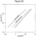
- the red blood cells carry oxygen.
- the red blood cells have a lifespan equal to naturally derived red blood cells.
- the red blood cells have a lifespan that is 75% of that of naturally derived red blood cells.
- the red blood cells have a lifespan that is 50% of that of naturally derived red blood cells.
- the red blood cells have a lifespan that is 25% of that of naturally derived red blood cells.
- Embryonic stem cells are a potential consistent and reliable source of red blood cells, with the benefits of unlimited supply of O-universal blood, and avoiding the additional cost of disease screening and blood typing with each donation.
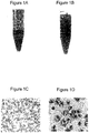
- the hallmark of mature red blood cells is loss of the nucleus, as well as production of mature hemoglobin.
- Many researchers, including our laboratory, have achieved differentiation of ESCs into erythroblasts, which still contain their nuclei, and express immature hemoglobin. To date, enucleation has not been achieved with human embryonic stem cells.
- enucleation has been achieved with CD34+ bone marrow and cord blood stem cells, which are further along in development, thus probably aiding in their enucleation capability.
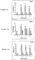
- Malik achieved 10-40% enucleation after 19 days of treatment of CD34+ bone marrow cells with Epo (Malik 1998).
- Miharada achieved a rate of 77% enucleation from CD34+ cord blood stem cells with a 20-day treatment of growth factors and cytokines including SCF, Epo, IL-3, VEGF, IGF-II, and mifepristone (Miharada 2006).
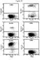
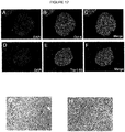
- the present inventive method uses OP9 cells to induce differentiation in human ESCs in a completely in vitro system, which is relevant to clinical therapies.
- the first step consists of differentiating ESCs into hemangioblasts, hemangio colony forming cells, non-engrafting hemangio cells, or blast cells.
- the second step is expansion of these cells in Stemline II medium (Sigma) with Epo, IL-3, SCF and various supplements used by Douay for cord blood enucleation (Douay 2005).
- the third step introduces the OP9 cells to the ESC-derived erythroblasts, as well as the addition of Epo.
- differentiating ESCs into the hemangioblasts, hemangio colony forming cells, and non-engrafting hemangio cells are produced and expanded in accordance to methods described herein.
- blast cells are cultured as described in Lu 2006.
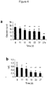
- day 6-8 blast cells from Day 3.25-Day 4.25 embryoid bodies are picked or filtered and plated in Stemline II medium with Epo, IL-3, SCF, hydrocortisone, inositol, folic acid, mono-thioglycerol, transferrin, insulin, ferrous nitrate, ferrous sulfate and bovine serum albumin for 12-30 days.
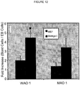
- blast cells are then co-cultured with OP9 mouse stromal cells or human mesenchymal stem cells (MSCs) in the same media listed above, without hydrocortisone.
- cells begin co-culturing between day 12 and 29 days.
- cells are further cultured for 12-18 days before enucleation occurs.
- enucleation initiated by OP9 cells can occur in as little as 3 days after stromal growth.
- enucleation is induced in Stempro34 medium with hydrocortisone, inositol, folic acid, mono-thioglycerol, transferrin, insulin, ferrous nitrate, ferrous sulfate and bovine serum albumin.
- cells are fed every 3-4 days and cultured on a new stromal layer every week.
- the present invention also provides methods of producing a megakaryocyte or a platelet, comprising: providing a pluripotent stem cell; differentiating said pluripotent stem cell into a hemangioblast, non-engrafting hemangio cell, or blast cell; and differentiating said hemangioblast, non-engrafting hemangio cell, or blast cell into said megakaryocyte or said platelet by culturing in megakaryocyte (MK) culture medium comprising TPO.
- MK megakaryocyte
- the present invention also provides methods of producing a megakaryocyte or a platelet, comprising: providing a hemangioblast, non-engrafting hemangio cell, or blast cell; and differentiating said hemangioblast, non-engrafting hemangio cell, or blast cell into said megakaryocyte or said platelet by culturing in megakaryocyte (MK) culture medium comprising TPO.
- MK megakaryocyte
- the hemangioblast, non-engrafting hemangio cell, or blast cell may be obtained or produced by methods described herein.
- said pluripotent stem cell used in the present invention is an embryonic stem cell or embryo-derived cell. In certain embodiments, said pluripotent stem cell is an induced pluripotent stem cell. In certain embodiments, said pluripotent stem cell is a human cell. In certain embodiments, said pluripotent stem cell is genetically manipulated prior to differentiation.
- said hemangioblast, non-engrafting hemangio cell, or blast cell is expanded prior to being differentiated into said megakaryocyte or said platelet.
- said hemangioblasts, non-engrafting hemangio cells, or blast cells are expanded in Stemline II medium with Epo, IL-3, and SCF.
- differentiating said hemangioblast, non-engrafting hemangio cell, or blast cell into said megakaryocyte or said platelet is done after about 6 to 8 days of hemangioblast, non-engrafting hemangio cell, or blast cell culture.
- the present invention also provides a megakaryocyte or a platelet produced by any one of the method as described herein.
- This invention provides a method for generating and expanding human hemangio-colony forming cells from human pluripotent stem cells, preparations and compositions comprising human hemangio-colony forming cells, methods of producing various cell types partially or terminally differentiated from hemangio-colony forming cells, methods of using hemangio-colony forming cells therapeutically, and methods of therapeutically using various cell types partially or terminally differentiated from hemangio-colony forming cells.
- BMP-4 bone morphogenetic protein-4
- VEGF vascular endothelial growth factor
- BMP-4 and VEGF significantly up-regulate T-brachyury, KDR, CD31 and LMO2 gene expression, while dramatically down-regulating Oct-4 expression.
- bFGF basic fibroblast growth factor
- This invention also provides a method for expanding mammalian hemangio-colony forming cells obtained from any source, including ES cells, blastocysts or blastomeres, cord blood from placenta or umbilical tissue, peripheral blood, bone marrow, or other tissue or by any other means known in the art.
- Human hemangio-colony forming cells can also be generated from human pluripotent stem cells.
- Human pluripotent stem cells may be a substantially homogeneous population of cells, a heterogeneous population of cells, or all or a portion of an embryonic tissue.
- pluripotent stem cells that can be used in the methods of the present invention, human hemangio-colony forming cells can be generated from human embryonic stem cells.
- embryonic stem cells include embryonic stem cells derived from or using, for example, blastocysts, plated ICMs, one or more blastomeres, or other portions of a pre-implantation-stage embryo or embryo-like structure, regardless of whether produced by fertilization, somatic cell nuclear transfer (SCNT), parthenogenesis, androgenesis, or other sexual or asexual means.
- SCNT somatic cell nuclear transfer
- hemangioblasts can be further differentiated to hematopoietic cells including, but not limited to, platelets and red blood cells. Such cells may be used in transfusions. The ability to generate large numbers of cells for transfusion will alleviate the chronic shortage of blood experienced in blood banks and hospitals across the country.
- the methods of the invention allow for the production of universal cells for transfusion. Specifically, red blood cells that are type O and Rh- can be readily generated and will serve as a universal blood source for transfusion.
- the methods of this invention allow for the in vitro expansion of hemangioblasts to large quantities useful for a variety of commercial and clinical applications. Expansion of hemangioblasts in vitro refers to the proliferation of hemangioblasts. While the methods of the invention enable the expansion of human hemangioblast cells to reach commercially useful quantities, the present invention also relates to large numbers of hemangioblast cells and to cell preparations comprising large numbers of human hemangioblast cells (for example, at least 10,000, 100,000, or 500,000 cells). In certain embodiments, the cell preparations comprise at least 1 X 10 6 cells.
- the cell preparations comprise at least 2 X 10 6 human hemangioblast cells and in further embodiments at least 3 X 10 6 human hemangioblast cells. In still other embodiments, the cell preparations comprise at least 4 X 10 6 human hemangioblast cells.
- the present invention relates to a solution, a preparation, and a composition comprising between 10,000 and 4 million or more mammalian (such as human) hemangioblast cells.
- the number of hemangioblast cells in such a solution, a preparation, and a composition may be any number between the range of 10,000 to 4 million, or more. This number could be, for example, 20,000, 50,000, 100,000, 500,000, 1 million, etc.
- the invention relates to preparations of human hemangioblast progeny cells (e.g ., human hematopoietic cells including human hematopoietic stem cells, and endothelial cells).
- human hemangioblast progeny cells e.g ., human hematopoietic cells including human hematopoietic stem cells, and endothelial cells.
- the invention further relates to methods of producing, storing, and distributing hemangioblast cells and/or hemangioblast lineage cells.
- the invention also provides methods and solutions suitable for transfusion into human or animal patients.
- the invention provides methods of making red blood cells and/or platelets, and/or other hematopoietic cell types for transfusion.
- the invention is suitable for use in blood banks and hospitals to provide blood for transfusion following trauma, or in the treatment of a blood-related disease or disorder.
- the invention provides red blood cells that are universal donor cells.
- the red blood cells are functional and express hemoglobin F prior to transfusion.
- the invention also provides for human hemangio-colony forming cells, cell cultures comprising a substantially purified population of human hemangio-colony forming cells, pharmaceutical preparations comprising human hemangio-colony forming cells and cryopreserved preparations of the hemangio-colony forming cells.
- the invention provides for the use of the human hemangio-colony forming cells in the manufacture of a medicament to treat a condition in a patient in need thereof.
- the invention provides the use of the cell cultures in the manufacture of a medicament to treat a condition in a patient in need thereof.
- the invention also provides the use of the pharmaceutical preparations in the manufacture of a medicament to treat a condition in a patient in need thereof.
- the hemangio-colony forming cells can be identified and characterized based on their structural properties. Specifically, and in certain embodiments, these cells are unique in that they are only loosely adherent to each other (loosely adherent to other hemangio-colony forming cells). Because these cells are only loosely adherent to each other, cultures or colonies of hemangio-colony forming cells can be dissociated to single cells using only mechanical dissociation techniques and without the need for enzymatic dissociation techniques.
- the cells are sufficiently loosely adherent to each other that mechanical dissociation alone, rather than enzymatic dissociation or a combination of mechanical and enzymatic dissociation, is sufficient to disaggregate the cultures or colonies without substantially impairing the viability of the cells.
- mechanical dissociation does not require so much force as to cause substantial cell injury or death when compared to that observed subsequent to enzymatic dissociation of cell aggregates.
- hemangio-colony forming cells can be identified or characterized based on the expression or lack of expression (as assessed at the level of the gene or the level of the protein) of one or more markers.
- hemangio-colony forming cells can be identified or characterized based on lack of expression of one or more (e.g., the cells can be characterized based on lack of expression of at least one, at least two, at least three or at least four of the following markers) of the following cell surface markers: CD34, KDR, CD133, or CD31.
- hemangio-colony forming cells can be identified or characterized based on expression of GATA2 and/or LMO2. Additionally or alternatively, hemangio-colony forming cells can be identified or characterized based on expression or lack of expression markers.
- Hemangio-colony forming cells of the present invention can be identified or characterized based on one or any combination of these structural or functional characteristics. Note that although these cells can be derived from any of a number of sources, for example, embryonic tissue, prenatal tissue, or perinatal tissue, the term "hemangio-colony forming cells" applies to cells, regardless of source, that are capable of differentiating to give rise to at least hematopoietic cell types and/or endothelial cell types and that have one or more of the foregoing structural or functional properties.
- marker(s) for the progenitor of BCs can be used to select BCs after initial culturing.
- hemangio-colonies are produced from pluripotent cells without forming embryoid bodies.
- the present invention provides a method for generating and expanding human hemangioblasts derived from human pluripotent stem cells, or from human blastocysts or blastomeres.
- the hemangioblasts so produced may be purified and/or isolated.
- Human hemangio-colony forming cells can also be generated from human pluripotent stem cells.
- Human pluripotent stem cells may be a substantially homogeneous population of cells, a heterogeneous population of cells, or all or a portion of an embryonic tissue.
- pluripotent stem cells that can be used in the methods of the present invention, human hemangio-colony forming cells can be generated from human embryonic stem cells.
- embryonic stem cells include embryonic stem cells derived from or using, for example, blastocysts, plated iCMs, one or more blastomeres, or other portions of a pre-implantation-stage embryo or embryo-like structure, regardless of whether produced by fertilization, somatic cell nuclear transfer (SCNT), parthenogenesis, androgenesis, or other sexual or asexual means.
- SCNT somatic cell nuclear transfer
- hemangio-colony forming cells can be generated from other pluripotent stem cells.
- hemangio-colony forming cells can be generated (without necessarily going through a step of embryonic stem cell derivation) from or using plated embryos, ICMs, blastocysts, trophoblast/trophectoderm cells, one or more blastomeres, trophoblast stem cells, embryonic germ cells, or other portions of a pre-implantation-stage embryo or embryo-like structure, regardless of whether produced by fertilization, somatic cell nuclear transfer (SCNT), parthenogenesis, androgenesis, or other sexual or asexual means.
- SCNT somatic cell nuclear transfer
- hemangio-colony forming cells can be generated using cells or cell lines partially differentiated from pluripotent stem cells. For example, if a human embryonic stem cell line is used to produce cells that are more developmentally primitive than hemangio-colony forming cells, in terms of development potential and plasticity, such pluripotent stem cells could then be used to generate hemangio-colony forming cells.
- hemangio-colony forming cells can be generated from other pre-natal or peri-natal sources including, without limitation, umbilical cord, umbilical cord blood, amniotic fluid, amniotic stem cells, and placenta.
- hemangio-colony forming cells are generated from human embryonic tissue a step of embryoid body formation may be needed.
- embryoid body formation serves, at least in part, to help recapitulate the three dimensional interaction of the germ layers that occurs during early development, such a step is not necessarily required when the pluripotent stem cells already have a structure or organization that serves substantially the same purpose as embryoid body formation.
- a step of embryoid body formation is not necessarily required to provide intercellular signals, inductive cues, or three dimensional architecture.
- the methods and uses of the present invention can be used to generate hemangio-colony forming cells from pluripotent stem cells or embryo-derived cells.
- the embryo-derived cells are embryonic stem cells.
- the embryo-derived cells are plated embryos, ICMs, blastocysts, trophoblast/trophectoderm cells, one or more blastomeres, trophoblast stem cells, or other portions of an early pre-implantation embryo.
- the embryo-derived cells may be from embryos produced by fertilization, somatic cell nuclear transfer (SCNT), parthenogenesis, androgenesis, or other sexual or asexual means.
- SCNT somatic cell nuclear transfer
- the invention similarly contemplates generating hemangio-colony forming cells from or using other pluripotent stem cells or embryonic-derived cells, and using the generated cells for any of the same therapeutic applications.
- the human embryonic stem cells may be the starting material of this method.
- the embryonic stem cells may be cultured in any way known in the art, such as in the presence or absence of feeder cells.
- Embryonic stem cells may form embryoid bodies ("EBs") in suspension in medium containing serum ( Wang et al. 2005 J Exp Med (201):1603-1614 ; Wang et al. 2004 Immunity (21): 31-41 ; Chadwick et al. 2003 Blood (102): 906 - 915 ).
- serum Wang et al. 2005 J Exp Med (201):1603-1614 ; Wang et al. 2004 Immunity (21): 31-41 ; Chadwick et al. 2003 Blood (102): 906 - 915 .
- serum presents certain challenges, including variability in experiments, cost, potential for infectious agents, and limited supply. Further, for clinical and certain commercial applications, use of serum necessitates additional U.S. and international regulatory compliance issues that govern biological products.
- the present invention provides methods of generating and expanding human hemangioblasts from pluripotent stem cells in which no serum is used.
- the serum-free conditions are more conducive to scale-up production under good manufacturing process (GMP) guidelines than are conditions which require serum.
- GMP good manufacturing process
- serum-free conditions extend the half-life of certain factors added to the medium (for example, the half-life of proteins including growth factors, cytokines, and HOXB4 in media is increased when no serum is present).
- the media is supplemented with BMP4 and VEGF.
- serum-free media is used throughout the method of this invention for generating and expanding human hemangioblasts.
- human stem cells are grown in serum-free media and are induced to differentiate into embryoid bodies.
- embryonic stem cells may be pelleted and resuspended in serum-free medium (e.g., in Stemline I or II media (SigmaTM)) supplemented with one or more morphogenic factors and cytokines and then plated on low attachment (e.g ., ultra-low attachment) culture dishes.
- morphogenic factors and cytokines may include, but are not limited to, bone morphogenic proteins (e.g ., BMP2, BMP-4, BMP-7, but not BMP-3) and VEGF, SCF and FL.
- Bone morphogenic proteins and VEGF may be used alone or in combination with other factors.
- the morphogenic factors and cytokines may be added to the media from 0-48 hours of cell culture. Following incubation under these conditions, incubation in the presence of early hematopoietic expansion cytokines, including, but not limited to, thrombopoietin (TPO), Flt-3 ligand, and stem cell factor (SCF), allows the plated ES cells to form EBs.
- TPO thrombopoietin
- Flt-3 ligand Flt-3 ligand
- SCF stem cell factor
- VEGF vascular endothelial growth factor
- human ES cells are first grown in the presence of BMP-4 and VEGF 165 (e.g., 25-100 ng/ml), followed by growing in the presence of BMP-4, VEGF 165 , SCF, TPO, and FLT3 ligand ( e.g ., 10-50 ng/ml) and HoxB4 ( e.g ., 1.5-5 ⁇ g/ml of a triple protein transduction domain-HoxB4 fusion protein as disclosed herein).
- BMP-4 and VEGF 165 e.g., 25-100 ng/ml
- SCF vascular endothelial growth factor
- TPO thelial growth protein
- FLT3 ligand e.g . 10-50 ng/ml
- HoxB4 e.g ., 1.5-5 ⁇ g/ml of a triple protein transduction domain-HoxB4 fusion protein as disclosed herein.
- the additional factors may be added 48-72 hours after plating.
- human hemangioblast cells are isolated from early embryoid bodies ("EBs"). Isolating hemangioblast cells from early EBs supports the expansion of the cells in vitro.
- hemangioblast cells may be obtained from EBs grown for less than 10 days.
- hemangioblast cells arise in human EBs grown for 2-6 days.
- hemangioblast cells are identified and may be isolated from human EBs grown for 4-6 days.
- human EBs are grown for 2-5 days before hemangioblast cells are isolated.
- human EBs are grown for 3-4.5 days before hemangioblast cells are isolated.
- early EBs are washed and dissociated (e.g ., by Trypsin/EDTA or collagenase B).
- a select number of cells e.g ., 2-5 X10 5 cells
- serum-free methylcellulose medium optimized for hemangioblast cell growth e.g ., BL-CFU medium, for example Stem Cell Technologies Catalogue H4436, or hemangioblast cell expansion medium (HGM), or any medium containing 1.0% methylcellulose in MDM, 1-2% Bovine serum albumin, 0.1 mM 2-mercaptoethanol, 10 ⁇ g/ml rh-Insulin, 200 ⁇ g/ml iron saturated human transferrin, 20 ng/ml rh-GM-CSF, 20 ng/ml rh-IL-3, 20 ng/ml rh-IL-6, 20 ng/ml rh-G-CSF)("rh” stands for "recombinant human"
- This medium may be supplemented with early stage cytokines (including, but not limited to, EPO, TPO, SCF, FL, FLt-3, VEGF, BMPs such as BMP2, BMP4 and BMP7, but not BMP3) and HOXB4 (or another homeobox protein).
- EPO erythropoietin
- EPO, SCF, VEGF, BMP-4 and HoxB4 are added to the media.
- the cells are grown in the presence of EPO, TPO and FL.
- H9 is the starting human ES cell line
- EPO, TPO and FL are added to the media.
- media for cells derived from H9 or other ES cells may further comprise VEGF, BMP-4, and HoxB4.
- the cells so obtained by this method are plated onto ultra-low attachment culture dishes and incubated in a CO 2 incubator to grow hemangioblast colonies. Some cells may be able to form secondary EBs. Following approximately 3-6 days, and in some instances 3-4.5 days, hemangioblast colonies are observed. Hemangioblast colonies may be distinguished from other cells such as secondary EBs by their distinctive grape-like morphology and/or by their small size.
- hemangioblasts may be identified by the expression of certain markers (e.g., the expression of both early hematopoietic and endothelial cell markers) as well as their ability to differentiate into at least both hematopoietic and endothelial cells (see below, Deriving hemangioblast lineage cells).
- certain markers e.g., the expression of both early hematopoietic and endothelial cell markers
- these cells may be identified by the presence of certain markers (such as, for example, CD71+) and the absence of other markers (for example, CD34-).
- Hemangioblasts may also express GATA-1 and GATA-2 proteins, CXCR-4, and TPO and EPO receptors.
- hemangioblasts may be characterized by the absence or low expression of other markers (e.g. , CD31, CD34, KDR, or other adhesion molecules). Further, hemangioblasts may be characterized by the expression of certain genes, (e.g ., genes associated with hemangioblasts and early primitive erythroblast development, such as, for example, SCL, LMO2, FLT-1, embryonic fetal globin genes, NF-E2, GATA-1, EKLF, ICAM-4, glycophoriuns, and EPO receptor).
- genes associated with hemangioblasts and early primitive erythroblast development such as, for example, SCL, LMO2, FLT-1, embryonic fetal globin genes, NF-E2, GATA-1, EKLF, ICAM-4, glycophoriuns, and EPO receptor.
- hemangioblasts may be isolated by size (being smaller than the other cells) or purified with an anti-CD71+ antibody, such as by immunoaffinity column chromatography.
- the hemangioblast cells may be isolated by size and/or morphology by the following procedure. After 6 to 7 days of growth, the cell mixture contains EBs, which are round and represent a clump of multiple cells, and hemangioblasts, which are grape-like, smaller than the EBs, and are single cells. Accordingly, hemangioblasts may be isolated based on their morphology and size. The hemangioblast cells may be manually picked, for example, when observing the cell mixture under a microscope. The cells may subsequently grow into colonies, each colony having between 100 -150 cells.
- Human hemangioblast colonies derived as described above may be picked and replated onto methylcellulose CFU-medium to form hematopoietic CFUs.
- CFU-medium comprises StemCell Technologies H4436.
- hemangioblasts are plated in Stemline II media supplemented with cytokines and other factors. For example, individual BL-CFC colonies may be handpicked and transferred to a fibronectin-coated plate containing Stemline II with recombinant human SCF (e.g.
- TPO e.g ., 20ng/ml
- FL e.g ., 20 ng/ml
- IL-3 e.g ., 20 ng/ml
- VEGF e.g.
- hemangioblasts expanded by the methods of the invention are obtained from early embryoid bodies derived from human embryonic stem cells as described above.
- hemangioblasts to be expanded may also be isolated from other mammalian sources, such as mammalian embryos ( Ogawa et al. 2001 Int Rev Immunol (20):21-44 , US patent publication no. 2004/0052771 ), cord blood from placenta and umbilical tissues ( Pelosi, et al. 2002 Blood (100): 3203-3208 ; Cogle et al. 2004 Blood (103):133-5 ), peripheral blood and bone marrow ( Pelosi et al. 2002 Hematopoiesis (100): 3203-3208 ).
- mammalian embryos Ogawa et al. 2001 Int Rev Immunol (20):21-44 , US patent publication no. 2004/0052771
- cord blood from placenta and umbilical tissues Pelosi, et al. 2002 Blood (100): 3203-3208 ; Cogle et al. 2004 Blood (103):133-5
- peripheral blood and bone marrow Pelosi
- non-human hemangioblasts to be expanded may be generated from non-human (such as mouse and non-human primates) embryonic stem cells.
- hemangioblasts are obtained from umbilical cord blood (UCB) or bone marrow by methods such as, for example, magnetic bead positive selection or purification techniques (e.g. MACS column). Cells may be selected based on their CD71 + status and may be confirmed as CD34-. Further, the isolated hemangioblasts may be tested for their potential to give rise to both hematopoietic and endothelial cell lineages.
- hemangioblasts isolated or purified and optionally enriched from embryos, cord blood, peripheral blood, bone marrow, or other tissue are more than 95% pure.
- Bone marrow-derived cells may be obtained from any stage of development of the donor individual, including prenatal (e.g ., embryonic or fetal), infant ( e.g., from birth to approximately three years of age in humans), child ( e.g ., from about three years of age to about 13 years of age in humans), adolescent ( e.g., from about 13 years of age to about 18 years of age in humans), young adult ( e.g ., from about 18 years of age to about 35 years of age in humans), adult (from about 35 years of age to about 55 years of age in humans) or elderly ( e.g . from about 55 years and beyond of age in humans).
- prenatal e.g ., embryonic or fetal
- infant e.g., from birth to approximately three years of age in humans
- child e.g ., from about three years of age to about 13 years of age in humans
- adolescent e.g., from about 13 years of age to about 18
- Human bone marrow may be harvested by scraping from the split sternum of a patient undergoing surgery, for example. Bone marrow may then be preserved in tissue clumps of 0.1 to 1 mm 3 in volume and then grown on a mouse embryonic feeder layer (e.g. , a mitomycin C-treated or irradiated feeder layer). The bone marrow cells will attach to the plates and over a period of 1-2 weeks of culture, hemangioblast cells may be identified based on morphological features and/or cell markers and isolated (see US patent publication no. 2004/0052771 ). The cells may then be subsequently grown and expanded in serum-free conditions according to the methods disclosed herein.
- a mouse embryonic feeder layer e.g. , a mitomycin C-treated or irradiated feeder layer.
- the bone marrow cells will attach to the plates and over a period of 1-2 weeks of culture, hemangioblast cells may be identified based on morphological features and/or cell markers and isolated (
- bone marrow cells and cells from blood or other tissue may be fractionated to obtain hemangioblasts cells.
- Methods of fractionation are well known in the art, and generally involve both positive selection (i . e ., retention of cells based on a particular property) and negative selection ( i . e ., elimination of cells based on a particular property).
- Methods for fractionation and enrichment of bone marrow-derived cells are best characterized for human and mouse cells.
- Positive selection methods such as enriching for cells expressing CD71 may be used.
- negative selection methods which remove or reduce cells expressing CD3, CD10, CD11b, CD14, CD16, CD15, CD16, CD19, CD20, CD32, CD45, CD45R/B220 or Ly6G may also be used alone or in combination with positive selection techniques.
- negative selection may be performed on the cell preparation to reduce or eliminate differentiated T cells.
- Immunoaffinity technology may take a variety of forms, as is well known in the art, but generally utilizes an antibody or antibody derivative in combination with some type of segregation technology.
- the segregation technology generally results in physical segregation of cells bound by the antibody and cells not bound by the antibody, although in some instances the segregation technology which kills the cells bound by the antibody may be used for negative selection.
- any suitable immunoaffinity technology may be utilized for selection/enrichment of hemangioblasts from bone marrow-derived, blood, or other cells, including fluorescence-activated cell sorting (FACS), panning, immunomagnetic separation, immunoaffinity chromatography, antibody-mediated complement fixation, immunotoxin, density gradient segregation, and the like.
- FACS fluorescence-activated cell sorting
- the desired cells the cells bound by the immunoaffinity reagent in the case of positive selection, and cells not bound by the immunoaffinity reagent in the case of negative selection
- Immunoaffinity selection/enrichment is typically carried out by incubating a preparation of cells comprising bone marrow-derived cells with an antibody or antibody-derived affinity reagent (e.g ., an antibody specific for a given surface marker), then utilizing the bound affinity reagent to select either for or against the cells to which the antibody is bound.
- the selection process generally involves a physical separation, such as can be accomplished by directing droplets containing single cells into different containers depending on the presence or absence of bound affinity reagent (FACS), by utilizing an antibody bound (directly or indirectly) to a solid phase substrate (panning, immunoaffinity chromatography), or by utilizing a magnetic field to collect the cells which are bound to magnetic particles via the affinity reagent (immunomagnetic separation).
- undesirable cells may be eliminated from the bone marrow-derived cell preparation using an affinity reagent which directs a cytotoxic insult to the cells bound by the affinity reagent.
- the cytotoxic insult may be activated by the affinity reagent (e.g. , complement fixation), or may be localized to the target cells by the affinity reagent (e.g ., immunotoxin, such as ricin B chain).
- hemangioblasts expanded by the methods of the invention are obtained from early embryoid bodies derived from human embryonic stem cells as described above.
- the hemangioblasts are isolated or enriched from human tissue (e.g ., placenta or cord blood, peripheral blood, bone marrow, etc.)
- the hemangioblasts are expanded in the presence of a homeodomain protein (also referred to herein as a homeobox protein).
- a homeodomain protein also referred to herein as a homeobox protein
- the hemangioblasts are expanded in the presence of HOXB4.
- HOXB4 is added to the hemangioblast cells throughout the method for expanding hemangioblast cells.
- HOXB4 is a homeodomain transcription factor (also called HOX2F, HOX2, HOX-2.6, and in the rat HOXA5) that is expressed in vivo in the stem cell fraction of the bone marrow and that is subsequently down-regulated during differentiation. Expression of the HOXB4 gene is associated with the maintenance of primitive stem cell phenotypes ( Sauvageau et al. 1995 Genes Dev 9: 1753-1765 ; Buske et al. 2002 Blood 100: 862-868 ; Thorsteinsdottir et al. 1999 Blood 94: 2605-2612 ; Antonchuk et al. 2001 Exp Hematol 29: 1125-1134 ).
- HOXB4 used in the methods of the present invention to generate and expand hemangioblasts includes, but is not limited to, full length HOXB4 (e.g ., HOXB4 polypeptides specified by public accession numbers GI:13273315 ( Figure 17 ), GI:29351568 ( Figure 18 ), as well as any functional variants and active fragments thereof.
- the wild-type HOXB4 protein may be encoded by the amino acid sequence of SEQ ID NO: 1, SEQ ID NO: 3 or any other alternative allelic forms of such protein. Such sequences may be accessed via publicly available databases, such as Genbank.
- HOXB4 may be ectopically expressed within the cell or may be provided in the media. HOXB4 expressed ectopically may be operably linked to an inducible promoter.
- HOXB4 provided in the media may be excreted by another cell type (e.g ., a feeder layer) or added directly to the media.
- the present invention also relates to fusion proteins comprising HOXB4 (including fusion proteins comprising full length HOXB4, or HOXB4 functional variants or active fragments of HOXB4).
- this fusion protein may also comprise any additional proteins, protein domains or peptides.
- HOXB4 may be joined to a protein transduction domain (PTD) to allow translocation of the protein from the medium into the cells and subsequently into nuclear compartments. Fusion proteins may or may not comprise one or more linker sequences located in between the protein domains.
- Functional variants of HOXB4 include mutants of HOXB4 and allelic variants, and active fragments thereof.
- Functional variants of HOXB4 include any HOXB4 polypeptides and active fragments thereof that are capable of expanding hemangioblasts according to the methods of the present invention.
- HOXB4 functional variants also include HOXB4 polypeptides that exhibit greater transcriptional activity compared to the native HOXB4 protein.
- HOXB4 variants include proteins with one or more amino acid substitution, addition, and/or deletion in relation to a wild-type HOXB4.
- HOXB4 variants also include, but are not limited to, polypeptides that are at least 75% similar to the sequence provided in SEQ ID NO: 1 or SEQ ID NO: 3. Accordingly, HOXB4 variants include polypeptides that are 80%, 85%, 90%, 95%, and 99% similar to the amino acid sequence provided in SEQ ID NO: 1 or SEQ ID NO: 3.
- HOXB4 variants also include polypeptides encoded by nucleic acid sequences that are at least 80% identical to a nucleic acid sequence encoding its complement (e.g ., the wild-type HOXB4 protein may be encoded by nucleic acid sequences of SEQ ID NO: 2 (GI:85376187; Figure 15 ) or SEQ ID NO: 4 (GI:29351567; Figure 16 )).
- the wild-type HOXB4 protein may be encoded by nucleic acid sequences of SEQ ID NO: 2 (GI:85376187; Figure 15 ) or SEQ ID NO: 4 (GI:29351567; Figure 16 )).
- HOXB4 variants include HOXB4 polypeptides that are encoded by nucleic acid sequences that are 85%, 90%, 95%, and 99% identical to the sequence provided in SEQ ID NO: 2 or SEQ ID NO: 4 or complement thereto.
- Nucleic acid sequences encoding HOXB4 also include, but are not limited to, any nucleic acid sequence that hybridizes under stringent conditions to a nucleic acid sequence of SEQ ID NO: 2 or 4, complement thereto, or fragment thereof. Similarly, nucleic acids which differ from the nucleic acids as set forth in SEQ ID NO: 2 or 4 due to degeneracy in the genetic code are also within the scope of the invention.
- HOXB4 variant polypeptides also include splice variants or other naturally occurring HOXB4 proteins or nucleic acid sequences.
- HOXB4 Active fragments of HOXB4 include, but are not limited to, any fragment of full length HOXB4 polypeptide that is capable of maintaining hemangioblasts according to the methods of the present invention. Accordingly, in one embodiment, a HOXB4 protein of the present invention is a HOXB4 protein that lacks part of the N-terminus, such as, for example, the N-terminal 31, 32, or 33 amino acids of full length HOXB4.
- HOXB4 proteins may be fused with additional proteins or protein domains.
- HOXB4 may be joined to a protein transduction domain (PTD).
- PTD protein transduction domain
- Protein transduction domains covalently or non-covalently linked to HOXB4, allow the translocation of HOXB4 across the cell membranes so the protein may ultimately reach the nuclear compartments of the cells.
- PTDs that may be fused with a HOXB4 protein include the PTD of the HIV transactivating protein (TAT) (Tat 47-57) ( Schwarze and Dowdy 2000 Trends Pharmacol. Sci. 21: 45-48 ; Krosl et al. 2003 Nature Medicine (9): 1428-1432 ).
- TAT HIV transactivating protein
- the amino acid sequence conferring membrane translocation activity corresponds to residues 47-57 (YGRKKRRQRRR, SEQ ID NO: 5) ( Ho et al., 2001, Cancer Research 61: 473-477 ; Vives et al., 1997, J. Biol. Chem. 272: 16010-16017 ). This sequence alone can confer protein translocation activity.
- the TAT PTD may also be the nine amino acids peptide sequence RKKRRQRRR (SEQ ID NO: 6) ( Park et al. Mol Cells 2002 (30):202-8 ).
- the TAT PTD sequences may be any of the peptide sequences disclosed in Ho et al., 2001, Cancer Research 61: 473-477 (the disclosure of which is hereby incorporated by reference herein), including YARKARRQARR (SEQ ID NO: 7), YARAAARQARA (SEQ ID NO: 8), YARAARRAARR (SEX ID NO: 9) and RARAARRAARA (SEQ ID NO: 10).
- HSV-1 herpes simplex virus 1
- Amintp Drosophila Antennapedia homeotic transcription factor
- Antp amino acids 43-58 (RQIKIWFQNRRMKWKK, SEQ ID NO: 11) represent the protein transduction domain
- HSV VP22 the PTD is represented by the residues DAATATRGRSAASRPTERPRAPARSASRPRRPVE (SEQ ID NO: 12).
- HeptaARG RRRRRRR, SEQ ID NO: 13
- artificial peptides that confer transduction activity may be used as a PTD of the present invention.
- the PTD may be a PTD peptide that is duplicated or multimerized.
- the PTD is one or more of the TAT PTD peptide YARAAARQARA (SEQ ID NO: 14).
- the PTD is a multimer consisting of three of the TAT PTD peptide YARAAARQARA (SEQ ID NO: 15).
- a HOXB4 protein that is fused or linked to a multimeric PTD such as, for example, a triplicated synthetic protein transduction domain (tPTD), may exhibit reduced lability and increased stability in cells. Such a HOXB4 construct may also be stable in serum-free medium and in the presence of hES cells.
- fusion genes encoding fusion proteins are well known in the art. Essentially, the joining of various DNA fragments coding for different polypeptide sequences is performed in accordance with conventional techniques.
- the fusion gene can be synthesized by conventional techniques including automated DNA synthesizers.
- PCR amplification of gene fragments can be carried out using anchor primers which give rise to complementary overhangs between two consecutive gene fragments which can subsequently be annealed to generate a chimeric gene sequence (see, for example, Current Protocols in Molecular Biology, eds. Ausubel et al., John Wiley & Sons: 1992 ).
- a fusion gene coding for a purification leader sequence may be linked to the N-terminus of the desired portion of the HOXB4 polypeptide or HOXB4-fusion protein, allowing the fusion protein be purified by affinity chromatography using a Ni 2+ metal resin.
- the purification leader sequence can then be subsequently removed by treatment with enterokinase to provide the purified HOXB4 polypeptide ( e.g ., see Hochuli et al., (1987) J. Chromatography 411:177 ; and Janknecht et al., PNAS USA 88:8972 ).
- a HOXB4 protein or functional variant or active domain of it is linked to the C-terminus or the N-terminus of a second protein or protein domain (e.g ., a PTD) with or without an intervening linker sequence.
- the exact length and sequence of the linker and its orientation relative to the linked sequences may vary.
- the linker may comprise, for example, 2, 10, 20, 30, or more amino acids and may be selected based on desired properties such as solubility, length, steric separation, etc.
- the linker may comprise a functional sequence useful for the purification, detection, or modification, for example, of the fusion protein.
- the linker comprises a polypeptide of two or more glycines.
- the protein domains and/or the linker by which the domains are fused may be modified to alter the effectiveness, stability and/or functional characteristics of HOXB4.
- HOXB4 is ectopically expressed within the hemangioblast cell or is provided in the media.
- HOXB4 expressed ectopically may be operably linked to a regulatory sequence. Regulatory sequences are art-recognized and are selected to direct expression of the HOXB4 polypeptide.
- HOXB4 provided in the media may be excreted by another cell type.
- the other cell type may be a feeder layer, such as a mouse stromal cell layer transduced to express excretable HOXB4.
- HOXB4 may be fused to or engineered to comprise a signal peptide, or a hydrophobic sequence that facilitates export and secretion of the protein.
- HOXB4 such as a fusion protein covalently or non-covalently linked to a PTD, may be added directly to the media.
- HOXB4 may be borne on a viral vector, such as a retroviral vector or an adenoviral vector. Such a vector could transduce either the hemangioblasts or other cells in their culture.
- HOXB4 is added to the media at selected times during the expansion of the hemangioblasts. Because the hemangioblasts are expanded in serum-free medium, HOXB4 is relatively stable. Accordingly, in certain embodiments, a HOXB4 protein or fusion protein is added every day to the human hemangioblasts. In other embodiments, a HOXB4 protein or fusion protein is added every other day, and in still other embodiments, a HOXB4 protein or fusion protein is added every 2 days. In one embodiment, a HOXB4 fusion protein, HOXB4-PTD, is added every 2 days to the media.
- the hemangioblasts can be expanded in the presence of any other growth factors or proteins that are present in an amount sufficient to expand such cells.
- Hemangioblasts obtained from any source including human or non-human ES cells, bone marrow, placenta or umbilical cord blood, peripheral blood, or other tissue may be expanded according to the methods described above. Accordingly, in certain embodiments, a select number of purified hemangioblasts or enriched cells are mixed with serum-free methylcellulose medium optimized for hemangioblast growth (e.g ., BL-CFU medium,). This medium may be supplemented with early stage cytokines (including, but not limited to, EPO, TPO, FL, VGF, BMPs like BMP2, BMP4 and BMP7, but not BMP3) and HOXB4.
- serum-free methylcellulose medium optimized for hemangioblast growth e.g ., BL-CFU medium,
- This medium may be supplemented with early stage cytokines (including, but not limited to, EPO, TPO, FL, VGF, BMPs like BMP2, BMP4 and BMP7, but not BMP3)
- EPO erythropoietin
- TPO erythropoietin
- FL erythropoietin
- the cells are then plated onto ultra-low attachment culture dishes and grown in a CO 2 incubator.
- hemangioblast colonies exhibit a distinctive grape-like morphology and are comparatively smaller than other cells and may consequently be distinguished from other cell types.
- the hemangioblasts may also be tested for markers as well as for their ability to differentiate further into either hematopoietic or endothelial cell lineages. The hemangioblasts are subsequently isolated and expanded in vitro.
- Media that may be used for expansion includes serum-free methylcellulose medium optimized for hemangioblasts growth (e.g ., BL-CFU) supplemented with early stage cytokines and HOXB4.
- Early stage cytokines include, but are not limited to, EPO, TPO, FL, VEGF, BMPs like BMP2, BMP4 and BMP7, but not BMP3.
- EPO erythropoietin
- EPO erythropoietin
- a medium for expanding hemangioblasts may comprise VEGF, SCF, EPO, BMP-4, and HoxB4; in certain embodiments the medium may further comprise TPO and FL.
- HGM hemangioblast expansion media
- ml hemangioblast expansion media containing 1.0% methylcellulose in Iscove's MDM, 1-2% Bovine serum albumin, 0.1 mM 2-mercaptoethanol, 10 ⁇ g/ml rh-Insulin, 200 ⁇ g/ml iron saturated human transferrin, 20 ng/ml rh-GM-CSF, 20 ng/ml rh-IL-3, 20 ng/ml rh-IL-6, 20 ng/ml rh-G-CSF, 3 to 6 units/ml rh-Epo, 50 ng/ml rh-SCF, 50ng/ml rh-VEGF and 50 ng/ml rh-BMP-4, and 1.5 ⁇ g/ml of tPTD-HoxB4, with/without 50 ng/ml of Tpo and FL.
- HGM hemangioblast expansion media
- tPTD-HoxB4 1.5
- the present invention provides methods of treating patients in need of cell-based therapy (for example, patients in need of hematopoietic reconstitution or treatment, or blood vessel growth or treatment of vascular injuries including ischemia, see below) using the expanded hemangioblasts or hemangioblast lineage cells of the invention, wherein the hemangioblasts are obtained from the bone marrow, blood, or other tissue of the patient or a patient relative.
- methods of treating a patient in need of hemangioblasts may comprise a step of isolating hemangioblasts from the patient or a patient relative.
- Hemangioblasts isolated from the patient or patient relative may be expanded in vitro according to the methods of the present invention and subsequently administered to the patient.
- the expanded hemangioblasts may be grown further to give rise to hematopoietic cells or endothelial cells before patient treatment.
- Hemangioblasts of that patient may then be generated and expanded from his own ES cells using a method of this invention. Those hemangioblasts or lineage derivatives thereof may be administered to that patient or to his relatives.
- human hemangioblasts are expanded to reach commercially large quantities which can be subsequently used in various therapeutic and clinical applications. Furthermore, the hemangioblasts obtained by the methods disclosed herein may be differentiated further to give rise to either hematopoietic or endothelial cell lineages for use in clinical applications.
- the hemangioblasts obtained from the method of this invention for generating and expanding human hemangioblasts from human ES cells have the potential to differentiate into at least endothelial cells or hematopoietic cells ( i.e ., they are at least bi-potential). Other hemangioblasts may be bi-potential as well. Yet other hemangioblasts may be able to differentiate into cells other than hematopoietic and endothelial cells, i.e., they are multi- or pluri-potential).
- the human embryonic stem cells used as the starting point for the method of generating and expanding human hemangioblast cells of this invention may also be derived from a library of human embryonic stem cells, each of which is hemizygous or homozygous for at least one MHC allele present in a human population.
- each member of said library of stem cells is hemizygous or homozygous for a different set of MHC alleles relative to the remaining members of the library.
- the library of stem cells is hemizygous or homozygous for all MHC alleles that are present in a human population.
- stem cells that are homozygous for one or more histocompatibility antigen genes include cells that are nullizygous for one or more (and in some embodiments, all) such genes.
- Nullizygous for a genetic locus means that the gene is null at that locus, i.e., both alleles of that gene are deleted or inactivated.
- Stem cells that are nullizygous for all MHC genes may be produced by standard methods known in the art, such as, for example, gene targeting and/or loss of heterozygocity (LOH). See, for example, United States patent publications US 20040091936 , US 20030217374 and US 20030232430 , and US provisional application number 60/729,173 , the disclosures of all of which are hereby incorporated by reference herein.
- the present invention relates to methods of obtaining hemangioblasts, including a library of hemangioblasts, with reduced MHC complexity.
- Hemangioblasts and hemangioblast lineage cells with reduced MHC complexity will increase the supply of available cells for therapeutic applications as it will eliminate the difficulties associated with patient matching.
- Such cells may be derived from stem cells that are engineered to be hemizygous or homozygous for genes of the MHC complex.
- a human ES cell may comprise modifications to one of the alleles of sister chromosomes in the cell's MHC complex.
- a variety of methods for generating gene modifications such as gene targeting, may be used to modify the genes in the MHC complex.
- the modified alleles of the MHC complex in the cells may be subsequently engineered to be homozygous so that identical alleles are present on sister chromosomes. Methods such as loss of heterozygosity (LOH) may be utilized to engineer cells to have homozygous alleles in the MHC complex.
- LHO loss of heterozygosity
- one or more genes in a set of MHC genes from a parental allele can be targeted to generate hemizygous cells.
- the other set of MHC genes can be removed by gene targeting or LOH to make a null line.
- This null line can be used further as the embryonic cell line in which to drop arrays of the HLA genes, or individual genes, to make a hemizygous or homozygous bank with an otherwise uniform genetic background.
- a library of ES cell lines wherein each member of the library is homozygous for at least one HLA gene, is used to derive hemangioblasts according to the methods of the present invention.
- the invention provides a library of hemangioblasts (and/or hemangioblast lineage cells), wherein several lines of ES cells are selected and differentiated into hemangioblasts. These hemangioblasts and/or hemangioblast lineage cells may be used for a patient in need of a cell-based therapy.
- certain embodiments of this invention pertain to a method of administering human hemangioblasts, hematopoietic stem cells, or human endothelial cells that have been derived from reduced-complexity embryonic stem cells to a patient in need thereof.
- this method comprises the steps of: (a) identifying a patient that needs treatment involving administering human hemangioblasts, hematopoietic stem cells, or human endothelial cells to him or her; (b) identifying MHC proteins expressed on the surface of the patient's cells; (c) providing a library of human hemangioblasts of reduced MHC complexity made by the method for generating and expanding human hemangioblast cells in vitro of the present invention; (d) selecting the human hemangioblast cells from the library that match this patient's MHC proteins on his or her cells; (e) optionally differentiating the human hemangioblast cells identified in step (d) into human hematopoietic stem cells, endothelial cells or both, or cells that are further differentiated in either or both of these two lineages, depending on need; (f) administering any of the cells from step (d) and/or (e) to said patient.
- This method may be performed in a regional center, such as, for example, a hospital, a clinic, a physician's office, and other health care facilities. Further, the hemangioblasts selected as a match for the patient, if stored in small cell numbers, may be expanded prior to patient treatment.
- the present invention provides human hemangio-colony forming cells. These cells are a unique, primitive cell type with a variety of therapeutic and other uses. Furthermore, this cell type provides an important tool for studying development of at least the hematopoietic and/or endothelial lineages. As such, the invention contemplates various preparations (including pharmaceutical preparations) and compositions comprising human hemangio-colony forming cells, as well as preparations (including pharmaceutical preparations) and compositions comprising one or more cell types partially or terminally differentiated from hemangio-colony forming cells.
- Human hemangio-colony forming cells of the present invention have at least one of the following structural characteristics: (a) can differentiate to give rise to at least hematopoietic cell types or endothelial cell types; (b) can differentiate to give rise to at least hematopoietic cell types and endothelial cell types; (c) are loosely adherent to each other (to other human hemangio-colony forming cells; (d) do not express CD34 protein; (e) do not express CD31 protein; (f) do not express KDR protein; (g) do not express CD133 protein; (h) express GATA2 protein; (i) express LMO2 protein.
- human hemangio-colony forming cells have at least two, at least three, at least four, at least five, at least six, at least seven, at least eight, or at least nine of the structural or functional characteristics detailed herein.
- the invention provides for human hemangio-colony forming cells.
- Such cells can differentiate to produce at least hematopoietic and/or endothelial cell types.
- the cells are characterized as being loosely adherent to other human hemangio-colony forming cells.
- these cells may also be described based on expression or lack of expression of certain markers. For example, these cells may also be described based on lack of expression of at least one of the following proteins: CD34, KDR, CD133, and CD31.
- hemangio-colony forming cells are loosely adherent to each other. Because these cells are only loosely adherent to each other, cultures or colonies of hemangio-colony forming cells can be dissociated to single cells using only mechanical dissociation techniques and without the need for enzymatic dissociation techniques. The cells are sufficiently loosely adherent to each other that mechanical dissociation alone, rather than enzymatic dissociation or a combination thereof, is sufficient to disaggregate the cultures or colonies without substantially impairing the viability of the cells. In other words, mechanical dissociation does not require so much force as to cause substantial cell injury or death.
- loosely adherent is described qualitatively above and refers to behavior of the human hemangio-colony forming cells with respect to each other.
- Cultures or colonies of hemangio-colony forming cells can be dissociated to single cells using only mechanical dissociation techniques and without the need for enzymatic dissociation techniques.
- the cells are sufficiently loosely adherent to each other that mechanical dissociation alone, rather than enzymatic dissociation or a combination thereof, is sufficient to disaggregate the cultures or colonies without substantially impairing the viability of the cells. In other words, mechanical dissociation does not require so much force as to cause substantial cell injury or death.
- the term “loosely adherent” is used to refer to cultures or colonies of hemangio-colony forming cells wherein at least 50% of the cells in the culture can be dissociated to single cells using only mechanical dissociation techniques and without the need for enzymatic dissociation techniques.
- the term refers to cultures in which at least 60%, 65%, 70%, or 75% of the cells in the culture can be dissociated to single cells using only mechanical dissociation techniques and without the need for enzymatic dissociation techniques.
- the term refers to cultures in which at least 80%, 85%, 90%, 95%, 96%, 97%, 98%, 99%, or even 100% of the cells in the culture can be dissociated to single cells using only mechanical dissociation techniques and without the need for enzymatic dissociation techniques.
- the ability to dissociate the hemangio-colony forming cells using only mechanical dissociation techniques and without the need for enzymatic dissociation techniques can be further quantitated based on the health and viability of the cells following mechanical dissociation. In other words, if dissociation without enzymatic techniques requires so much mechanical force that a significant number of the cells are damaged or killed, the cells are not loosely adherent, as defined herein.
- the term "loosely adherent" refers to cultures of cells that can be dissociated to single cells using only mechanical dissociation techniques and without the need for enzymatic dissociation techniques, without substantially impairing the health or viability or the cells in comparison to that observed when the same cells are dissociated using enzymatic dissociation techniques.
- the health or viability of the cells is decreased by less than 15%, 10%, 9%, 8%, 7%, 6%, 5%, 4%, 3%, 2%, or even less than 1% in comparison to that observed when a culture of the same cells are dissociated using enzymatic dissociation techniques.
- Exemplary enzymatic dissociation techniques include, but are not limited to, treatment with trypsin, collagenase, or other enzymes that disrupt cell-cell or cell-matrix interactions.
- Exemplary mechanical dissociation techniques include, but are not limited to, one or more passages through a pipette.
- Human hemangio-colony forming cells are defined structurally and functionally. Such cells can be generated from any of a number of sources including from embryonic tissue, prenatal tissue, perinatal tissue, and even from adult tissue.
- human hemangio-colony forming cells can be generated from human embryonic stem cells, other embryo-derived cells (blastocysts, blastomeres, ICMs, embryos, trophoblasts/trophectoderm cells, trophoblast stem cells, primordial germ cells, embryonic germ cells, etc.), amniotic fluid, amniotic stem cells, placenta, placental stem cells, and umbilical cord.
- the invention provides human hemangio-colony forming cells, compositions comprising human hemangio-colony forming cells, and preparations (including pharmaceutical preparations) comprising human hemangio-colony forming cells. Certain features of these aspects of the invention are described in detail below. The invention contemplates combinations of any of the following aspects and embodiments of the invention.
- the invention provides a human hemangio-colony forming cell.
- the cell can differentiate to produce at least hematopoietic and/or endothelial cell types.
- the cell is loosely adherent to other human hemangio-colony forming cells.
- the cell does not express CD34 protein.
- the cell does not express one or more of (e.g., the cell does not express at least one, at least two, at least three, or at least four of the following proteins) the following proteins: CD34, CD31, CD133, KDR.
- the cell does express GATA2 and/or LMO2 protein.
- the invention provides a human hemangio-colony forming cell.
- the cell which cell can differentiate to produce at least hematopoietic and/or endothelial cell types, and the cell does not express any of the following proteins: CD34, CD31, KDR, and CD133.
- the cell is loosely adherent to other human hemangio-colony forming cells.
- the cell does express GATA2 and/or LMO2 protein.
- the invention provides a cell culture comprising a substantially purified population of human hemangio-colony forming cells.
- the cells can differentiate to produce at least hematopoietic and endothelial cell types, and the cells are loosely adherent to each other.
- the cell does not express CD34 protein.
- the cell does not express one or more of (e.g., the cell does not express at least one, at least two, at least three, or at least four of the following proteins) the following proteins: CD34, CD31, CD133, KDR.
- the cell does express GATA2 and/or LMO2 protein.
- the invention provides a cell culture comprising human hemangio-colony forming cells differentiated from embryonic tissue.
- the hemangio-colony forming cells are loosely adherent to each other.
- the cells can differentiate to produce at least hematopoietic and/or endothelial cell types, and the cells are loosely adherent to each other.
- the cell does not express CD34 protein.
- the cell does not express one or more of (e.g., the cell does not express at least one, at least two, at least three, or at least four of the following proteins) the following proteins: CD34, CD31, CD133, KDR.
- the cell does express GATA2 and/or LMO2 protein.
- the invention provides a cell culture comprising human hemangio-colony forming cells, which cells can differentiate to produce at least hematopoietic and/or endothelial cell types.
- the cells are loosely adherent to each other.
- the cell does not express CD34 protein.
- the cell does not express one or more of (e.g., the cell does not express at least one, at least two, at least three, or at least four of the following proteins) the following proteins: CD34, CD31, CD133, KDR.
- the cell does express GATA2 and/or LMO2 protein.
- the invention provides a pharmaceutical preparation comprising human hemangio-colony forming cells, which cells can differentiate to produce at least hematopoietic and/or endothelial cell types.
- the hemangio-colony forming cells are loosely adherent to each other.
- the cell does not express CD34 protein.
- the cell does not express one or more of (e.g., the cell does not express at least one, at least two, at least three, or at least four of the following proteins) the following proteins: CD34, CD31, CD133, KDR.
- the cell does express GATA2 and/or LMO2 protein.
- the pharmaceutical preparation can be prepared using any pharmaceutically acceptable carrier or excipient.
- the invention provides a pharmaceutical preparation comprising human hemangio-colony forming cells, wherein the hemangio-colony forming cells do not express any of the following proteins: CD34, CD31, KDR, and CD133.
- the hemangio-colony forming cells can differentiate to produce at least hematopoietic and/or endothelial cell types.
- the hemangio-colony forming cells are loosely adherent to each other.
- the cell does express GATA2 and/or LMO2 protein.
- the pharmaceutical preparation can be prepared using any pharmaceutically acceptable carrier or excipient.
- the composition or pharmaceutical preparation comprises at least 1x10 5 human hemangio-colony forming cells. In certain other embodiment, of any of the foregoing, the composition or pharmaceutical preparation comprises at least 1x10 6 , at least 5x10 6 , at least 1x10 7 , or greater than 1x10 7 human hemangio-colony forming cells.
- compositions, and preparations include cells partially or terminally differentiated from human hemangio-colony forming cells.
- the invention contemplates compositions and preparations comprising one or more hematopoietic and/or endothelial cell type differentiated from a hemangio-colony forming cell.
- Exemplary hematopoietic cell types include hematopoietic stem cells, platelets, RBCs, lymphocytes, megakaryocytes, and the like.
- the invention contemplates compositions and preparations comprising one or more other cell type, such as one or more partially or terminally differentiated mesodermal cell type, differentiated from hemangio-colony forming cells.
- the invention provides a cryopreserved preparation of human hemangio-colony cells or cells partially or terminally differentiated therefrom.
- the invention provides for the therapeutic use of human hemangio-colony forming cells, or compositions or preparations of human hemangio-colony forming cells.
- Such cells and preparations can be used in the treatment of any of the conditions or diseases detailed throughout the specification, as well as in the blood banking industry.
- cells differentiated from human hemangio-colony forming cells, or compositions or preparations of human hemangio-colony forming cells can be used therapeutically in the treatment of any of the conditions or diseases detailed throughout the specification, as well as in the blood banking industry.
- human hemangio-colony forming cells of the invention are can be used therapeutically. Additionally or alternatively, human hemangio-colony forming cells can be used to study development of endothelial and hematopoietic lineages or in screening assays to identify factors that can be used, for example, to (i) maintain human hemangio-colony forming cells or (ii) to promote differentiation of human hemangio-colony forming cells to one or more partially or terminally differentiated cell types. Furthermore, human hemangio-colony forming cells can be used to generate one or more partially or terminally differentiated cell types for in vitro or in vivo use.
- the human hemangio-colony forming cells of the invention can be used in any of the methods or application described in the present application including, but not limited to, in the treatment of any of the diseases or conditions described herein.
- mammalian (including human) hemangioblasts are expanded to reach commercial quantities and are used in various therapeutic and clinical applications.
- hemangioblasts are expanded to reach cell numbers on the order of 10,000 to 4 million (or more). These cell numbers may be reached within 3 -4 days of starting the initial preparations.
- the present invention relates to preparations comprising large numbers of hemangioblasts, said preparations comprising at least 10,000, 50,000, 100,000, 500,000, a million, 2 million, 3 million or 4 million cells.
- This invention also provides for a solution, a composition, and a preparation comprising large numbers of hemangioblasts, said solution, said composition, and said preparation comprising at least 10,000, 50,000, 100,000, 500,000, a million, 2 million, 3 million or 4 million cells.
- the hemangioblasts could be human.
- the present invention relate to differentiating the hemangioblasts obtained by the methods disclosed herein into either hematopoietic or endothelial cell lineages, or both, that are subsequently used in clinical applications.
- the present invention also relates to cell preparations comprising large numbers of hematopoietic or endothelial cells.
- the invention also relates to differentiating the hemangioblasts obtained by the methods disclosed herein into other cell lineages, other than hematopoietic and endothelial cells.
- the present invention also relates to cell preparations comprising large numbers of other hemangioblast-derived cells.
- compositions and preparations comprising large numbers (e.g ., thousands or millions) of hemangioblasts may be obtained by expanding hemangioblasts that are obtained as described above. Accordingly, the invention pertains to compositions and preparations comprising large numbers of hemangioblasts achieved by expanding ES cells (such as human ES cells) or hemangioblasts obtained from cord blood, peripheral blood or bone marrow. Further, as the methods of expansion may be applied to hemangioblasts of mouse, rat, bovine, or non-human primate origin, for example, the present invention also relates to compositions and preparations comprising large numbers of hemangioblasts of other species in addition to human.
- the hemangioblasts to be expanded by the methods of this invention may be bi-potential, i.e ., can differentiate into either endothelial cells or hematopoietic stem cells.
- the human hemangioblasts generated and expanded from human ES cells are bi-potential.
- Hemangio-colony forming cells are capable of differentiating to give rise to at least hematopoietic cell types or endothelial cell types.
- Hemangio-colony forming cells are preferably bi-potential and capable of differentiating to give rise to at least hematopoietic cell types and endothelial cell types.
- hemangio-colony forming cells of the present invention are at least uni-potential, and preferably bi-potential. Additionally however, hemangio-colony forming cells may have a greater degree of developmental potential and can, in certain embodiments, differentiate to give rise to cell types of other lineages. In certain embodiments, the hemangio-colony forming cells are capable of differentiating to give rise to other mesodermal derivatives such as cardiac cells (for example, cardiomyocytes) and/or smooth muscle cells.
- the hemangio-colony forming cells lack certain features characteristic of mature endothelial or hematopoietic cells. These hemangio-colony forming cells or hemangioblasts, however, may be identified by various markers such as, for example, CD71+, GATA-1 and GATA-2 proteins, CXCR-4, and TPO and EPO receptors. In additional embodiments, the hemangioblasts express LMO-2. Hemangioblasts may additionally be characterized by the absence or low expression of other markers. Accordingly, hemangioblasts may be CD34- CD31-, and KDR-. In further embodiments, the hemangioblasts may be CD34-, CD31-, KDR-, and CD133-.
- the hemangioblasts generated and expanded by the methods of present invention are characterized by the presence or absence of any one or more of the markers listed in Table 2 of WO2007/120811 , incorporated herein by reference in its entirety.
- the hemangioblasts may test negative for expression of any one or more of the markers listed in Table 2 that is denoted as "-" under "BL-CFC".
- the hemangioblasts may be negative for CD34 expression.
- the cells may additionally or alternatively be negative for CD31, CD133, and/or KDR expression.
- the hemangioblasts may express any of the markers denoted in Table 2 with "+".
- the cells may express one or more of the markers LMO-2 and GATA-2.
- Expression of a marker may be assessed by any method, such as, for example, immunohistochemistry or immunoblotting to test for protein expression, or mRNA analysis to test for expression at the RNA level.
- the methods and cell preparations of the present invention also relate to hemangioblast derivative cells.
- Human hemangioblasts generated and expanded by this invention and mammalian hemangioblasts expanded by the methods of the invention may be differentiated in vitro to obtain hematopoietic cells (including hematopoietic stem cells (HSCs)) or endothelial cells, as well as cells that are further differentiated in these two lineages. These cells may subsequently be used in the therapeutic and commercial applications described below.
- HSCs hematopoietic stem cells
- hematopoietic cells are derived by growing the hemangioblasts in serum-free BL-CFU for 3-10 days.
- single-cell suspensions of hES-derived BL-CFC cells are grown for 10-14 days. Maintaining serum-free conditions is optimal insofar as serum-free conditions facilitate scale-up production and compliance with regulatory guidelines as well as reduce cost.
- Hemangioblasts of the present invention may also be grown in serum-free Hem-culture ( Bhatia et al.
- BSA e.g ., 1% BSA
- insulin e.g ., 5 ⁇ g/ml human insulin
- transferrin media or transferrin e.g ., 100 ⁇ g/ml human transferrin
- L-glutamine e.g ., beta-mercaptoethanol
- beta-mercaptoethanol e.g ., 10 -4 M
- growth factors may comprise SCF (e.g ., 300 ng/ml), granulocytic-colony-stimulating factor (G-CSF) (e.g.
- hemangioblasts may also be grown in serum-free methylcellulose medium supplemented with a multilineage hematopoietic growth factor cocktail.
- the hemangioblasts may be grown in methylcellulose in Iscove modified Dulbecco medium (IMDM) comprising BSA, saturated human transferrin, human LDL, supplemented with early acting growth factors (e.g ., c- kit ligand, fit3 ligand), multilineage growth factors (e.g ., IL-3, granulocyte macrophage-CSF (GM-CSF)), and unilineage growth factors (e.g ., G-CSF, M-CSF, EPO, TPO)), VEGF, and bFGF.
- IMDM Iscove modified Dulbecco medium
- BSA Iscove modified Dulbecco medium
- saturated human transferrin e.g IL-3
- GM-CSF granulocyte macrophage-CSF
- unilineage growth factors e.g ., G-CSF, M-CSF, EPO, TPO
- VEGF vascular endoivascular endoietic cell
- hemangioblast colonies are resuspended in Stemline I media.
- Cells are then mixed with 1 ml of serum-free hematopoietic CFU media (H4436, Stem Cell TechnologiesTM) plus 1.5 ⁇ g/ml of tPTD-HoxB4 and 0.5% EX-CYTE (Serologicals Proteins Inc.TM).
- H4436 serum-free hematopoietic CFU media
- tPTD-HoxB4 1.5 ⁇ g/ml of tPTD-HoxB4 and 0.5% EX-CYTE (Serologicals Proteins Inc.TM).
- EX-CYTE SeX-CYTE
- Hematopoietic CFUs arising following 10-14 days after initial plating may be characterized morphologically, such as by staining with Wright-Giemsa dye.
- Hematopoietic cells may also be derived from the hemangioblast using other conditions known in the art (e.g ., in media comprising IMDM, 30% fetal calf serum (FCS), 1 % bovine serum albumin (BSA), 10 -4 M beta-mercaptoethanol, and 2 mM L-glutamine). Further, in other embodiments basic fibroblast growth factor may be used to promote both BL-CFC frequency within EBs and promote hematopoietic differentiation ( Faloon et al. 2000 Development (127): 1931-1941 ).
- the growth factor hemangiopoietin is used to promote growth and hematopoietic differentiation of the hemangioblasts ( Liu et al. 2004 Blood (103): 4449-4456 ).
- the differentiation into hematopoietic cells may be assessed by CD45 status (CD45+) and the CFU assay, for example.
- human hemangioblasts may be grown for 3-10 days, or optionally for longer periods of time (e.g ., 10-14 days) in CFU-medium.
- Human hemangioblasts of the present invention are able to form CFUs comprising granulocytes, erythrocytes, macrophages, and megakaryocytes (CFU-GEMM/mix) as well as colony forming units containing only one of the latter cell types (e.g ., CFU-G, CFU-E, CFU-M, and CFU-GM).
- single-cell suspensions of hES-derived BL-CFC cells are grown for 10-14 days to derive hematopoietic cells such as, for example, erythroid, myeloid, macrophage, and multilineage hematopoietic cells.
- hemangioblasts derived from the human hemangioblasts obtained and expanded or mammalian hemangioblasts expanded by the methods described herein.
- the hemangioblasts may be grown in conditions favorable to endothelial maturation.
- hemangioblasts are first plated onto a fibronectin-coated surface and following 3-5 days (or in other embodiments 3-7 days), are replated onto a thick layer of Matrigel to support differentiation into endothelial cells. These conditions maintain the serum-free conditions established during hemangioblast development.
- hemangioblasts may be grown in media known to support differentiation into endothelial cells. Such conditions include, for example, Endo-culture comprising 20% fetal bovine serum (FBS), 50 ng/ml endothelial cell growth supplement ( i .
- VEGF-A 165 Terramani et al. 2000 In vitro Cell Dev Biol Anim (36): 125-132 ).
- Other conditions known in the art include medium supplemented with 25% FCS/horse serum, and in some embodiments heparin ( e.g ., 10 U/ml), insulin like growth factor (IGF1) ( e.g ., 2 ng), and EC growth supplement (ECGS, e.g ., 100 ⁇ g).
- IGF1 insulin like growth factor
- ECGS e.g ., 100 ⁇ g.
- the growth factors VEGF and EGF may also be used in combination with HAPO to support endothelial differentiation (Liu et al. 2004).
- the hemangioblasts may also be seeded onto dishes coated with collagen and fibronectin, for example, to promote differentiation into endothelial cells.
- Cells may be analyzed for von Willebrand factor (vWF) and endothelial nitric oxide synthase (eNOS) and the ability to form an endothelial network in vitro.
- vWF von Willebrand factor
- eNOS endothelial nitric oxide synthase
- endothelial cells hemangioblast colonies derived by the methods described above are picked and replated onto fibronectin-coated culture plates optimized for the first step towards endothelial differentiation.
- the cells may be plated in EGM-2 or EGM-2MV complete media (CambrexTM). Following 3 to 5 days, and in alternative embodiments 3 to 7 days, the cells are re-plated on a surface that supports endothelial differentiation, such as on a layer of Matrigel. Following 16-24 hours of incubation, the formation of branched tube-cords suggests typical endothelial cell behavior. Endothelial-specific assays such as LDL-uptake may also be used to confirm that these cells are of endothelial nature.
- human hemangioblasts generated and expanded by this invention and mammalian hemangioblasts expanded by the methods of the invention may be differentiated in vitro to obtain other cells, as well as cells that are further differentiated from these cell lineages.
- additional cell lineages may be derived from the hemangioblasts generated and expanded by this invention and mammalian hemangioblasts expanded by the methods of the invention because the hemangioblast cells may have an even greater degree of developmental potential beyond differentiating into hematopoietic and endothelial cells.
- the present invention provides a novel cell population that shares some characteristics of previously identified hemangioblasts and hemangio-colony forming cells.
- the novel cell population described herein is distinct in that it does not engraft into the bone marrow when administered to immunodeficient animals.
- This novel progenitor cell population is useful for the study of basic developmental and stem cell biology, is useful to generate partially and terminally differentiated cell type in vitro and in vivo , and is useful for the development of therapeutics.
- these cells can be used in screening assays to identify, for example, (i) factors or conditions that promote the expansion of non-engrafting hemangio cells and (ii) factors or conditions that promote the generation of one or more differentiated cell type from non-engrafting hemangio cells.
- Identified factors and conditions can be used in the production of cell-based and cell free therapies, in the production of mediums and formulations, and in the study of developmental and stem cell biology.
- the present invention provides non-engrafting hemangio cells, compositions and preparations comprising non-engrafting hemangio cells, methods of producing and expanding non-engrafting hemangio cells, methods of producing differentiated cell types from non-engrafting hemangio cells, and methods of using non-engrafting hemangio cells or cells derived there from therapeutically.
- cells can be obtained from other species including, but not limited to, mice, rats, rabbits, cows, dogs, cats, sheep, pigs, and non-human primates.
- This invention provides a method for expanding mammalian non-engrafting hemangio cells obtained from any source, including ES cells, blastocysts or blastomeres, cord blood from placenta or umbilical tissue, peripheral blood, bone marrow, or other tissue or by any other means known in the art.
- human non-engrafting hemangio cells are generated from embryonic stem cells or other pluripotent stem cells.
- human non-engrafting hemangio cells can be generated from embryonic stem cells, as well as from iPS cells.
- non-engrafting hemangio cells are generated from human embryo-derived cells.
- Human embryo-derived cells may be a substantially homogeneous population of cells, a heterogeneous population of cells, or all or a portion of an embryonic tissue.
- embryo-derived cells that can be used in the methods of the present invention, human non-engrafting hemangio cells can be generated from human embryonic stem cells.
- embryonic stem cells include embryonic stem cells derived from or using, for example, blastocysts, plated ICMs, one or more blastomeres, or other portions of a pre-implantation-stage embryo or embryo-like structure, regardless of whether produced by fertilization, somatic cell nuclear transfer (SCNT), parthenogenesis, androgenesis, or other sexual or asexual means.
- SCNT somatic cell nuclear transfer
- non-engrafting hemangio cells are generated from pluripotent stem cells.
- pluripotent stem cells include, but are not limited to, embryonic stem cells and iPS cells.
- human non-engrafting hemangio cells are generated from non-pluripotent cells.
- Non-pluripotent cells may include somatic cells, such as cells derived from skin, bone, blood, connective tissue, heart, kidney, lung, liver, or any other internal organ.
- the non-pluripotent cells may be cells derived from connective tissue, such as fibroblasts.
- the non-pluripotent cells are cells derived from an adult tissue.
- non-engrafting hemangio cells can be further differentiated to hematopoietic stem cells and/or hematopoietic cell types including, but not limited to, platelets and red blood cells.
- hematopoietic stem cells and/or hematopoietic cell types including, but not limited to, platelets and red blood cells.
- Such cells may be used in transfusions or in other therapies. Although such cells have numerous uses, a particularly important use would be in improving the availability of blood for transfusions.
- the invention provides red blood cells differentiated from non-engrafting hemangio cells. Such differentiated red blood cells could be used for transfusions.
- differentiated hematopoietic cells are transfused to treat trauma, blood loss during surgery, blood diseases such as anemia, Sickle cell anemia, or hemolytic diseases, or malignant disease.
- red blood cells are transfused to treat trauma, blood loss during surgery, or blood diseases such as anemia, Sickle cell anemia, or hemolytic disease.
- a mixed population of red blood cells is transfused. It should be noted that many differentiated hematopoietic cell types, particularly red blood cells, typically exist in vivo as a mixed population.
- red blood cells of varying levels of age and differentiation are found in vivo. Additionally, red blood cells mature over time so as to express less fetal hemoglobin and more adult hemoglobin.
- the present invention contemplates transfusion of either purified populations of red blood cells or of a mixed population of red blood cells having varying levels of age and levels of differentiation.
- the invention contemplates transfusion of red blood cells expressing fetal hemoglobin (hemoglobin F).
- Transfusion of red blood cells that express fetal hemoglobin may be especially useful in the treatment of Sickle cell anemia. The ability to generate large numbers of cells for transfusion will alleviate the chronic shortage of blood experienced in blood banks and hospitals across the country.
- the methods of the invention allow for the production of universal cells for transfusion.
- red blood cells that are type O and Rh-can be readily generated and will serve as a universal blood source for transfusion.
- the red blood cells produced from the methods of the application are functional.
- the red blood cells express hemoglobin F prior to transfusion.
- the red blood cells carry oxygen.
- the red blood cells have a lifespan equal to naturally derived red blood cells.
- the red blood cells have a lifespan that is 75% of that of naturally derived red blood cells.
- the red blood cells have a lifespan that is 50% of that of naturally derived red blood cells.
- the red blood cells have a lifespan that is 25% of that of naturally derived red blood cells.
- non-engrafting hemangio cells may have a greater developmental potential, and may differentiate to produce endothelial cell types, smooth muscle cell types, or cardiac cell types.
- the methods of this invention allow for the in vitro expansion of non-engrafting hemangio cells to large quantities useful for a variety of commercial and clinical applications.
- Expansion of non-engrafting hemangio cells in vitro refers to the proliferation of non-engrafting hemangio cells.
- the methods of the invention enable the expansion of human non-engrafting hemangio cells to reach commercially useful quantities
- the present invention also relates to large numbers of non-engrafting hemangio cells and to cell preparations comprising large numbers of human non-engrafting hemangio cells (for example, at least 10,000, 100,000, or 500,000 cells).
- the cell preparations comprise at least 1 X 10 6 cells.
- the cell preparations comprise at least 2 X 10 6 human non-engrafting hemangio cells and in further embodiments at least 3 X 10 6 human non-engrafting hemangio cells. In still other embodiments, the cell preparations comprise at least 4 X 10 6 human non-engrafting hemangio cells. Note that these cell preparations may be purified or substantially purified. However, in certain embodiments, suitable cell preparations comprise a mixture of non-engrafting hemangio cells and hemangio-colony forming cells. The mixture may be any ratio, including mixtures comprising a greater proportion of non-engrafting hemangio cells and mixtures comprising a greater proportion of hemangio-colony forming cells.
- the present invention relates to a solution, a preparation, or a composition comprising between 10,000 and 4 million or more mammalian (such as human) non-engrafting hemangio cells.
- the number of non-engrafting hemangio cells in such a solution, a preparation, and a composition may be any number between the range of 10,000 to 4 million, or more. This number could be, for example, 20,000, 50,000, 100,000, 500,000, 1 million, etc.
- the invention relates to preparations of human non-engrafting hemangio progeny cells (e.g ., human hematopoietic cells including human hematopoietic stem cells).
- the invention further relates to methods of producing, storing, and distributing non-engrafting hemangio cells and/or non-engrafting hemangio cell progeny.
- the invention also provides methods and solutions suitable for transfusion into human or animal patients.
- the invention provides methods of making red blood cells and/or platelets, and/or other hematopoietic cell types for transfusion.
- the invention is suitable for use in blood banks and hospitals to provide blood for transfusion following trauma, or in the treatment of a blood-related disease or disorder.
- the invention provides red blood cells that are universal donor cells.
- the red blood cells are functional and express hemoglobin F prior to transfusion.
- the invention also provides for human non-engrafting hemangio cells, cell cultures comprising a substantially purified population of human non-engrafting hemangio cells, pharmaceutical preparations comprising human non-engrafting hemangio cells and cryopreserved preparations of the non-engrafting hemangio cells.
- the invention provides for the use of the human non-engrafting hemangio cells in the manufacture of a medicament to treat a condition in a patient in need thereof.
- the invention provides the use of the cell cultures in the manufacture of a medicament to treat a condition in a patient in need thereof.
- the invention also provides the use of the pharmaceutical preparations in the manufacture of a medicament to treat a condition in a patient in need thereof.
- the non-engrafting hemangio cells can be identified and characterized based on their structural properties and/or function properties. These progenitor cells do not engraft when administered to an immunodeficient host. In certain embodiments, these cells are unique in that they are only loosely adherent to each other (loosely adherent to other non-engrafting hemangio cells). In embodiments in which the cells are loosely adherent, cultures or colonies of non-engrafting hemangio cells can be dissociated to single cells using only mechanical dissociation techniques and without the need for enzymatic dissociation techniques.
- the cells are sufficiently loosely adherent to each other that mechanical dissociation alone, rather than enzymatic dissociation or a combination of mechanical and enzymatic dissociation, is sufficient to disaggregate the cultures or colonies without substantially impairing the viability of the cells.
- mechanical dissociation does not require so much force as to cause substantial cell injury or death when compared to that observed subsequent to enzymatic dissociation of cell aggregates.
- the non-engrafting hemangio cells can be further identified or characterized based on the expression or lack of expression (as assessed at the level of the gene or the level of the protein) of one or more markers.
- the non-engrafting hemangio cells have one or more of the characteristics of human hemangio-colony forming cells.
- non-engrafting hemangio cells can be identified or characterized based on lack of expression of one or more (e.g ., the cells can be characterized based on lack of expression of at least one, at least two, at least three or at least four of the following markers) of the following cell surface markers: CD34, KDR, CD133, or CD31.
- non-engrafting hemangio cells can be identified or characterized based on expression of GATA2 and/or LMO2.
- the present invention provides human non-engrafting hemangio cells. These cells are a unique, primitive cell type with a variety of therapeutic and other uses. Furthermore, this cell type provides an important tool for studying development of at least the hematopoietic lineages. As such, the invention contemplates various preparations (including pharmaceutical preparations) and compositions comprising human non-engrafting hemangio cells, as well as preparations (including pharmaceutical preparations) and compositions comprising one or more cell types partially or terminally differentiated from non-engrafting hemangio cells. Without being bound by any particular theory, these cells represent a distinct, somewhat more committed (than hemangio-colony forming cells) stem cell population that retain the ability to generate numerous hematopoietic cell types.
- Non-engrafting hemangio cells of the present invention can be identified or characterized based on one or any combination of the structural or functional characteristics described for hemangio-colony forming cells. Note that although these cells can be derived from any of a number of sources, for example, embryonic tissue, prenatal tissue, or perinatal tissue, the term "non-engrafting hemangio cells" applies to cells, regardless of source, that do not engraft and that are capable of differentiating to give rise to at least one hematopoietic cell type, and optionally have one or more of the foregoing structural or functional properties.
- human non-engrafting hemangio cells of the present invention do not engraft when administered to a immunodeficient host and have at least one of the following structural characteristics: (a) can differentiate to give rise to at least one hematopoietic cell type; (b) can differentiate to give rise to at least hematopoietic cell types and endothelial cell types; (c) are loosely adherent to each other (to other non-engrafting hemangio cells); (d) do not express CD34 protein; (e) do not express CD31 protein; (f) do not express KDR protein; (g) do not express CD133 protein; (h) express GATA2 protein; (i) express LMO2 protein.
- human non-engrafting hemangio cells have at least two, at least three, at least four, at least five, at least six, at least seven, at least eight, or at least nine of the structural or functional characteristics detailed herein.
- non-engrafting hemangio cells are loosely adherent to each other. Because these cells are only loosely adherent to each other, cultures or colonies of non-engrafting hemangio cells can be dissociated to single cells using only mechanical dissociation techniques and without the need for enzymatic dissociation techniques. The cells are sufficiently loosely adherent to each other that mechanical dissociation alone, rather than enzymatic dissociation or a combination thereof, is sufficient to disaggregate the cultures or colonies without substantially impairing the viability of the cells. In other words, mechanical dissociation does not require so much force as to cause substantial cell injury or death.
- loosely adherent is described qualitatively above and refers to behavior of the human non-engrafting hemangio cells with respect to each other.
- Cultures or colonies of non-engrafting hemangio cells can be dissociated to single cells using only mechanical dissociation techniques and without the need for enzymatic dissociation techniques.
- the cells are sufficiently loosely adherent to each other that mechanical dissociation alone, rather than enzymatic dissociation or a combination thereof, is sufficient to disaggregate the cultures or colonies without substantially impairing the viability of the cells. In other words, mechanical dissociation does not require so much force as to cause substantial cell injury or death.
- the term “loosely adherent” is used to refer to cultures or colonies of non-engrafting hemangio cells wherein at least 50% of the cells in the culture can be dissociated to single cells using only mechanical dissociation techniques and without the need for enzymatic dissociation techniques.
- the term refers to cultures in which at least 60%, 65%, 70%, or 75% of the cells in the culture can be dissociated to single cells using only mechanical dissociation techniques and without the need for enzymatic dissociation techniques.
- the term refers to cultures in which at least 80%, 85%, 90%, 95%, 96%, 97%, 98%, 99%, or even 100% of the cells in the culture can be dissociated to single cells using only mechanical dissociation techniques and without the need for enzymatic dissociation techniques.
- the ability to dissociate the non-engrafting hemangio cells using only mechanical dissociation techniques and without the need for enzymatic dissociation techniques can be further quantitated based on the health and viability of the cells following mechanical dissociation. In other words, if dissociation without enzymatic techniques requires so much mechanical force that a significant number of the cells are damaged or killed, the cells are not loosely adherent, as defined herein.
- the term "loosely adherent" refers to cultures of cells that can be dissociated to single cells using only mechanical dissociation techniques and without the need for enzymatic dissociation techniques, without substantially impairing the health or viability or the cells in comparison to that observed when the same cells are dissociated using enzymatic dissociation techniques.
- the health or viability of the cells is decreased by less than 15%, 10%, 9%, 8%, 7%, 6%, 5%, 4%, 3%, 2%, or even less than 1% in comparison to that observed when a culture of the same cells are dissociated using enzymatic dissociation techniques.
- Exemplary enzymatic dissociation techniques include, but are not limited to, treatment with trypsin, collagenase, or other enzymes that disrupt cell-cell or cell-matrix interactions.
- Exemplary mechanical dissociation techniques include, but are not limited to, one or more passages through a pipette.
- Human non-engrafting hemangio cells are defined structurally and functionally. Such cells can be generated from any of a number of sources including from embryonic tissue, prenatal tissue, perinatal tissue, and even from adult tissue.
- human non-engrafting hemangio cells can be generated from human embryonic stem cells, other embryo-derived cells (blastocysts, blastomeres, ICMs, embryos, trophoblasts/trophectoderm cells, trophoblast stem cells, primordial germ cells, embryonic germ cells, etc.), amniotic fluid, amniotic stem cells, placenta, placental stem cells, and umbilical cord.
- non-engrafting hemangio cells can be generated from pluripotent cells, such as embryonic stem cells or pluripotent stem cells.
- pluripotent stem cells include, but are not limited to, embryonic stem cells and induced pluripotent stem cells (iPS cells).
- Human non-engrafting hemangio cells can also be generated from non-pluripotent cells, such as somatic cells, including but not limited to, cells derived from skin, bone, blood, connective tissue, heart, kidney, lung, liver, or any other internal organ.
- the non-pluripotent cells may be cells derived from connective tissue, such as fibroblasts.
- the non-pluripotent cells are cells derived from an adult tissue.
- the invention provides non-engrafting hemangio cells (such as human cells), compositions comprising human non-engrafting hemangio cells, and preparations (including pharmaceutical preparations) comprising human non-engrafting hemangio cells. Certain features of these aspects of the invention are described in detail below. The invention contemplates combinations of any of the following aspects and embodiments of the invention, as well as combinations with the disclosure provided at US Application Serial No. 11/787,262 , which is incorporated by reference in its entirety.
- hemangio-colony forming cells and/or non-engrafting hemangio cells can be produced from a variety of cells including, but not limited to, pluripotent cells (embryonic stem cells, embryo-derived cells, and induced pluripotent stem cells).
- pluripotent cells embryo-derived cells, and induced pluripotent stem cells.
- the invention provides a non-engrafting hemangio cells (such as human cells).
- the cell can differentiate to produce at least one hematopoietic cell types.
- the cell is loosely adherent to other human non-engrafting hemangio cells.
- the cell does not express CD34 protein.
- the cell does not express one or more of (e.g., the cell does not express at least one, at least two, at least three, or at least four of the following proteins) the following proteins: CD34, CD31, CD133, KDR.
- the cell does express GATA2 and/or LMO2 protein.
- the cell shares one or more than one (2, 3, 4, 5, 6, 7, 8, 9, 10) of the functional or structural characteristics of human hemangio colony forming cells.
- the invention provides a cell culture comprising a substantially purified population of non-engrafting hemangio cells (such as human cells).
- the cells can differentiate to produce at least hematopoietic cell types.
- the cells are loosely adherent to each other.
- the cell does not express CD34 protein.
- the cell does not express one or more of (e.g ., the cell does not express at least one, at least two, at least three, or at least four of the following proteins) the following proteins: CD34, CD31, CD133, KDR.
- the cell does express GATA2 and/or LMO2 protein.
- the cell shares one or more than one (2, 3, 4, 5, 6, 7, 8, 9, 10) of the functional or structural characteristics of human hemangio colony forming cells.
- the invention provides a cell culture comprising non-engrafting hemangio cells differentiated from embryonic tissue.
- the invention provides a cell culture comprising non-engrafting hemangio cells differentiated from pluripotent cells (pluripotent stem cells).
- the non-engrafting hemangio cells are loosely adherent to each other.
- the cells can differentiate to produce at least hematopoietic cell types, and the cells are loosely adherent to each other.
- the cell does not express CD34 protein.
- the cell does not express one or more of (e.g ., the cell does not express at least one, at least two, at least three, or at least four of the following proteins) the following proteins: CD34, CD31, CD133, KDR.
- the cell does express GATA2 and/or LMO2 protein.
- the cell shares one or more than one (2, 3, 4, 5, 6, 7, 8, 9, 10) of the functional or structural characteristics of human hemangio colony forming cells.
- the invention provides a cell culture comprising human non-engrafting hemangio cells, which cells can differentiate to produce at least hematopoietic cell types.
- the cells are loosely adherent to each other.
- the cell does not express CD34 protein.
- the cell does not express one or more of (e.g ., the cell does not express at least one, at least two, at least three, or at least four of the following proteins) the following proteins: CD34, CD31, CD133, KDR.
- the cell does express GATA2 and/or LMO2 protein.
- the cell shares one or more than one (2, 3, 4, 5, 6, 7, 8, 9, 10) of the functional or structural characteristics of human hemangio colony forming cells.
- the invention provides a pharmaceutical preparation comprising human non-engrafting hemangio cells, which cells can differentiate to produce at least hematopoietic cell types.
- the non-engrafting hemangio cells are loosely adherent to each other.
- the cell does not express CD34 protein.
- the cell does not express one or more of (e.g ., the cell does not express at least one, at least two, at least three, or at least four of the following proteins) the following proteins: CD34, CD31, CD133, KDR.
- the cell does express GATA2 and/or LMO2 protein.
- the cell shares one or more than one (2, 3, 4, 5, 6, 7, 8, 9, 10) of the functional or structural characteristics of human hemangio colony forming cells.
- the pharmaceutical preparation can be prepared using any pharmaceutically acceptable carrier or excipient.
- the invention provides a pharmaceutical preparation comprising human non-engrafting hemangio cells.
- the pharmaceutical preparation can be prepared using any pharmaceutically acceptable carrier or excipient.
- the composition or pharmaceutical preparation comprises at least 1x10 5 human non-engrafting hemangio cells. In certain other embodiment, of any of the foregoing, the composition or pharmaceutical preparation comprises at least 1x10 6 , at least 5x10 6 , at least 1x10 7 , or greater than 1x10 7 human non-engrafting hemangio cells. In certain embodiments, the preparation is a purified or substantially purified preparation. In other embodiments, the preparation comprises a mixture of non-engrafting hemangio cells and other cell types. For example, a mixture of non-engrafting hemangio cells and hemangio-colony forming cells.
- compositions, and preparations include cells partially or terminally differentiated from human non-engrafting hemangio cells.
- the invention contemplates compositions and preparations comprising one or more hematopoietic and/or endothelial cell type differentiated from a non-engrafting hemangio cells.
- Exemplary hematopoietic cell types include hematopoietic stem cells, platelets, RBCs, lymphocytes, megakaryocytes, and the like.
- the invention contemplates compositions and preparations comprising one or more other cell type, such as one or more partially or terminally differentiated mesodermal cell type, differentiated from non-engrafting hemangio cells.
- the invention provides a cryopreserved preparation of human non-engrafting hemangio cells or cells partially or terminally differentiated therefrom.
- the invention provides for the therapeutic use of human non-engrafting hemangio cells, or compositions or preparations of human non-engrafting hemangio cells.
- Such cells and preparations can be used in the treatment of any of the conditions or diseases detailed throughout the specification, as well as in the blood banking industry.
- cells differentiated from human non-engrafting hemangio cells, or compositions or preparations of human non-engrafting hemangio cells can be used therapeutically in the treatment of any of the conditions or diseases detailed throughout the specification.
- human non-engrafting hemangio cells of the invention can be used therapeutically. Additionally or alternatively, human non-engrafting hemangio cells can be used to study development of endothelial and hematopoietic lineages or in screening assays to identify factors that can be used, for example, to (i) maintain human non-engrafting hemangio cells or (ii) to promote differentiation of human non-engrafting hemangio cells to one or more partially or terminally differentiated cell types. Furthermore, human non-engrafting hemangio cells can be used to generate one or more partially or terminally differentiated cell types for in vitro or in vivo use.
- human non-engrafting hemangio cells of the invention can be used in any of the methods or application described in the present application including, but not limited to, in the treatment of any of the diseases or conditions described herein. Exemplary diseases and conditions are further discussed in US application serial number 11/787,262 , which is incorporated by reference in its entirety. Further, human hemangio-colony forming cells and non-engrafting hemangio cells can be used to produce differentiated hematopoietic cell types, including functional red blood cells.
- mammalian (including human) non-engrafting hemangio cells are expanded to reach commercial quantities and are used in various therapeutic and clinical applications.
- non-engrafting hemangio cells are expanded to reach cell numbers on the order of 10,000 to 4 million (or more). These cell numbers may be reached within 3-4 days of starting the initial preparations.
- the present invention relates to preparations comprising large numbers of non-engrafting hemangio cells, said preparations comprising at least 10,000, 50,000, 100,000, 500,000, a million, 2 million, 3 million or 4 million cells.
- This invention also provides for a solution, a composition, and a preparation comprising large numbers of non-engrafting hemangio cells, said solution, said composition, and said preparation comprising at least 10,000, 50,000, 100,000, 500,000, a million, 2 million, 3 million or 4 million cells.
- the non-engrafting hemangio cells could be human.
- the solutions can be purified, substantially purified, or mixtures with other progenitor cells types including, but not limited to hemangio-colony forming cells.
- the present invention relate to differentiating the non-engrafting hemangio cells obtained by the methods disclosed herein into hematopoietic or endothelial cell lineages, or both, that are subsequently used in clinical applications.
- the present invention also relates to cell preparations comprising large numbers of partially or terminally differentiated cell types.
- compositions and preparations comprising large numbers (e.g ., thousands or millions) of non-engrafting hemangio cells may be obtained by expanding non-engrafting hemangio cells that are obtained as described above. Accordingly, the invention pertains to compositions and preparations comprising large numbers of non-engrafting hemangio cells achieved by expanding ES cells (such as human ES cells) or non-engrafting hemangio cells obtained from cord blood, peripheral blood or bone marrow.
- ES cells such as human ES cells
- the present invention also relates to compositions and preparations comprising large numbers of non-engrafting hemangio cells of other species in addition to human.
- the non-engrafting hemangio cells to be expanded by the methods of this invention may be bi-potential, i.e ., can differentiate into either endothelial cells or hematopoietic stem cells.
- the human non-engrafting hemangio cells generated and expanded from human ES cells are bi-potential.
- Non-engrafting hemangio cells are capable of differentiating to give rise to at least hematopoietic cell types.
- Non-engrafting hemangio cells are, in certain embodiments, bi-potential and capable of differentiating to give rise to at least hematopoietic cell types and endothelial cell types.
- non-engrafting hemangio cells of the present invention are at least uni-potential, and may be bi-potential. Additionally however, non-engrafting hemangio cells may have a greater degree of developmental potential and can, in certain embodiments, differentiate to give rise to cell types of other lineages.
- the non-engrafting hemangio cells are capable of differentiating to give rise to other mesodermal derivatives such as cardiac cells (for example, cardiomyocytes) and/or smooth muscle cells.
- non-engrafting hemangio cells can be used in screening assays to identify agents that, for example, (i) promote differentiation of the cells to one or more hematopoietic cell type or (ii) promote proliferation and/or survival of the cells to facilitate cell banking and storage.
- the non-engrafting hemangio cells can also be used to study basic developmental biology or can be compared to hemangio-colony forming cells to ascertain the developmental differences between the two related stem cell populations.
- human hemangioblast cells and non-engrafting hemangio cells have the potential to differentiate in vivo into either hematopoietic or endothelial cells, they can be used in cell-based treatments in which either of these two cell types are needed or would improve treatment. Further, a patient may be treated with any therapy or treatment comprising hemangioblast lineage cells or and non-engrafting hemangio lineage cells ( i.e ., hematopoietic cells and/or endothelial cells).
- hemangioblast lineage cells or and non-engrafting hemangio lineage cells i.e ., hematopoietic cells and/or endothelial cells.
- the present invention relates to methods and compositions for treating a patient in need of hematopoietic cells or blood vessel growth or repair.
- the hemangioblasts or non-engrafting hemangio cells may be injected into the blood vessel of a subject or be administered to the blood vessel of a subject through operation.
- the patient or the subject may be human.
- human hemangioblast cells or non-engrafting hemangio cells are used in transplantation, where HSC transplantation would otherwise be used.
- transplantation may be used, for example, in hematopoietic reconstitution for the treatment of patients with acute or chronic leukemia, aplastic anemia and various immunodeficiency syndromes, as well as various non-hematological malignancies and auto-immune disorders, and to rescue patients from treatment-induced aplasia following high-dose chemotherapy and/or radiotherapy.
- Such transplantation may be achieved in vivo or ex vivo (such as in bone marrow transplant).
- human hemangioblast cells or non-engrafting hemangio cells are used to treat patients in need of hematopoietic reconstitution or hematopoietic treatment.
- patients in include, for example, patients with thalassemias, sickle cell anemia, aplastic anemia (also called hypoplastic anemia), cytopenia, marrow hypoplasia, platelet deficiency, hematopoietic malignancies such as leukemias, paroxysmal nocturnal hemoglobinuria (PNH), and ADA (e.g ., deaminase (ADA)-deficient severe combined immunodeficiency (SCID)).
- aplastic anemia also called hypoplastic anemia
- cytopenia cytopenia
- marrow hypoplasia cytopenia
- platelet deficiency hematopoietic malignancies
- leukemias paroxysmal nocturnal hemoglobinuria (PNH)
- ADA e.
- the invention relates to methods of treating a patient in need of hematopoietic reconstitution or treatment using the hemangioblasts of the invention. Accordingly, the invention relates to methods of treating a patient in need of hematopoietic reconstitution or treatment comprising selecting a patient in need thereof, generating and expanding or expanding human hemangioblasts or non-engrafting hemangio cells according to the methods of the present invention, and administering the human hemangioblasts or the non-engrafting hemangio cells into the patient.
- the method may comprise differentiating the generated and expanded or expanded human hemangioblasts or non-engrafting hemangio cells into human hematopoietic cells and subsequently administering the hematopoietic cells to the patient.
- Alternative embodiments include methods in which human hemangioblasts or non-engrafting hemangio cells are produced on a large scale and stored prior to the selection of a patient in need thereof.
- other embodiments of the invention relate to methods of treating a patient in need of hematopoietic reconstitution or treatment comprising selecting a patient in need thereof, placing an order for human hemangioblast or non-engrafting hemangio cells already isolated and expanded according to the methods described above, and administering said human hemangioblasts or non-engrafting hemangio cells to the patient.
- the method may comprise differentiating said human hemangioblasts or non-engrafting hemangio cells into human hematopoietic cells and administering said hematopoietic cells to the patient.
- hemangioblasts or non-engrafting hemangio cells hemizygous or homozygous for at least one MHC allele are grown, optionally grown to commercial quantities, and optionally stored by a business entity.
- a clinician or hospital will place an order with the business for such cells.
- the human hemangioblast cells and non-engrafting hemangio cells of the invention will proliferate and differentiate into endothelial cells under an angiogenic microenvironment
- the human hemangioblast cells may be used in a therapeutic manner to provide new blood vessels or to induce repair of damaged blood vessels at a site of injury in a patient.
- the present invention relates to methods of promoting new blood vessel growth or repairing injured vasculature.
- the human hemangioblasts or non-engrafting hemangio cells of the present invention may be used to treat endothelial injury, such as myocardium infarction, stroke and ischemic brain, ischemic limbs and skin wounds including ischemic limbs and wounds that occur in diabetic animals or patients, and ischemic reperfusion injury in the retina.
- endothelial injury such as myocardium infarction, stroke and ischemic brain, ischemic limbs and skin wounds including ischemic limbs and wounds that occur in diabetic animals or patients, and ischemic reperfusion injury in the retina.
- Other ischemic conditions that may be treated with the hemangioblasts or non-engrafting hemangio cells of the present invention include renal ischemia, pulmonary ischemia, and ischemic cardiomyopathy.
- Hemangioblasts may also be used to help repair injured blood vessels following balloon angioplasty or deployment of an endovascular stent.
- Hemangioblasts or non-engrafting hemangio cells may additionally be used in tissue grafting, surgery and following radiation injury. Further, the hemangioblasts or non-engrafting hemangio cells may be used to treat and/or prevent progression of atherosclerosis as well as to repair endothelial cell damage that occurs in systemic sclerosis and Raynaud's phenomenon (RP) ( Blann et al. 1993 J Rheumatol.(20):1325-30 ).
- RP Raynaud's phenomenon
- the invention provides various methods involved in providing blood vessel growth or repair to a patient in need thereof.
- the invention provides for a method for inducing formation of new blood vessels in an ischemic tissue in a patient in need thereof, comprising administering to said patient an effective amount of the purified preparation of human hemangioblast cells or non-engrafting hemangio cells described above to induce new blood vessel formation in said ischemic tissue.
- certain aspects of the present invention provide a method of enhancing blood vessel formation in a patient in need thereof, comprising selecting the patient in need thereof, isolating human hemangioblast cells or non-engrafting hemangio cells as described above, and administering the hemangioblast cells or non-engrafting hemangio cells to the patient.
- the present invention provides a method for treating an injured blood vessel in a patient in need thereof, comprising selecting the patient in need thereof, expanding or generating and expanding human hemangioblast cells or non-engrafting hemangio cells as described above, and administering the hemangioblast cells or non-engrafting hemangio cells to the patient.
- the hemangioblasts or non-engrafting hemangio cells may be produced on a large scale and stored prior to the selection of patient in need of hemangioblasts.
- hemangioblasts hemizygous or homozygous for at least one MHC allele are grown, optionally grown to commercial quantities, and optionally stored before a patient is selected for hemangioblast or non-engrafting hemangio cell treatment.
- Any of the aforementioned hemangioblasts, non-engrafting hemangio cells, or cell preparations of these cells may be administered directly into the circulation (intravenously).
- the hemangioblast cells, non-engrafting hemangio cells, or cell preparations of these cells may be administered by intra-vitreous injection.
- Administration of the solutions or preparations of hemangioblasts, non-engrafting hemangio cells, and derivative cells thereof may be accomplished by any route and may be determined on a case by case basis. Also, an effective amount to be administered of these solutions or preparations of hemangioblasts or derivative cells thereof is an amount that is therapeutically effective and may be determined on a case by case basis.
- hemangioblast lineage cells or non-engrafting hemangio lineage cells are used in therapeutic applications, including in the treatment of the indications described above, for example. Accordingly, hemangioblasts or non-engrafting hemangio cells generated and expanded or expanded by the methods described herein are differentiated in vitro first to obtain hematopoietic and/or endothelial cells, and then to obtain cells that are further differentiated in these two lineages. These cells may be subsequently administered to a subject or patient to treat hematopoietic conditions or for hematopoietic reconstitution, or for the treatment of ischemia or vascular injury, for example.
- HSCs derived from the human hemangioblasts or non-engrafting hemangio cells obtained by the methods disclosed herein are grown further to expand the HSCs and/or to derive other hematopoietic lineage cell types.
- Certain aspects of the present invention relate to the use of HSCs derived from the hemangioblasts or non-engrafting hemangio cells in transplantation.
- differentiated hematopoietic cells such as, for example, granulocytes, erythrocytes, myeloid cells, megakaryocytes, platelets, macrophages, mast cells and neutrophils ( Wiles and Keller 1991 Development (111): 259 )
- are used in various treatments such as transfusion therapy or for the treatment of infections.
- other embodiments of the present invention relate to methods of treating a patient in need of hematopoietic reconstitution or treatment using the HSCs or hematopoietic lineage cells derived from hemangioblasts of the invention.
- the present invention relates to methods of treating a patient in need of hematopoietic cells or treatment comprising selecting a patient in need thereof, expanding or isolating and expanding human hemangioblasts or non-engrafting hemangio cells according to the methods of the present invention, differentiating said hemangioblast cells or non-engrafting hemangio cells into hematopoietic stem cells and/or mature hematopoietic cells, and administering the hematopoietic cells to the patient.
- the hemangioblasts or non-engrafting hemangio cells are grown to give rise to endothelial cells according to the methods disclosed herein.
- the endothelial may subsequently be used to provide new blood vessels or to induce repair of damaged blood vessels at a site of injury in a patient.
- the present invention relates to methods of promoting new blood vessel growth or repairing injured vasculature in which endothelial cells derived from hemangioblasts or non-engrafting hemangio cells are used as a therapy.
- the endothelial cells may be used to treat endothelial injury, such as myocardium infarction and pulmonary ischemia, stroke and ischemic brain, ischemic limbs and skin wounds including ischemic limbs and wounds that occur in diabetic animals or patients, ischemic reperfusion injury in the retina, renal ischemia.
- the endothelial cells may also be used to help repair injured blood vessels following balloon angioplasty or deployment of an endovascular stent as well as in grafting, surgery and following radiation injury. Further, the endothelial cells may be used to treat and/or prevent progression of atherosclerosis as well as to repair endothelial cell damage that occurs in systemic sclerosis and Raynaud's phenomenon.
- the endothelial cell may be further differentiated and those cells, as appropriate, may be used in treating one or more of the "endothelial cell” disease or conditions, such as those listed in the preceding paragraph.
- certain aspects of the invention relate to methods of treating a patient with endothelial or vascular injury or in need of blood vessel growth or repair comprising selecting a patient in need thereof, expanding or isolating and expanding human hemangioblasts or non-engrafting hemangio cells according to the methods of the present invention, differentiating said hemangioblast cells or non-engrafting hemangio cells into endothelial cells, and administering the endothelial cells to the patient.
- Another aspect of the present invention provides methods of producing hematopoietic cells suitable for transfusion. Although such cells and methods have numerous uses, a particularly important use would be in improving the availability of blood for transfusions.
- the invention provides red blood cells differentiated from hemangioblasts/hemangio-colony forming units or non-engrafting hemangio cells. Such differentiated red blood cells could be used for transfusions.
- differentiated hematopoietic cells are transfused to treat trauma, blood loss during surgery, blood diseases such as anemia, Sickle cell anemia, or hemolytic diseases, or malignant disease.
- red blood cells are transfused to treat trauma, blood loss during surgery, or blood diseases such as anemia, Sickle cell anemia, or hemolytic disease.
- platelets are transfused to treat congenital platelet disorders or malignant disease.
- a mixed population of red blood cells and platelets are transfused.
- red blood cells typically exist in vivo as a mixed population. Specifically, circulating red blood cells of varying levels of age and differentiation are found in vivo. Additionally, red blood cells mature over time so as to express less fetal hemoglobulin and more adult hemoglobin.
- the present invention contemplates transfusion of either purified populations of red blood cells or of a mixed population of red blood cells having varying levels of age and levels of differentiation. In particular embodiments, the invention contemplates transfusion of red blood cells expressing fetal hemoglobin (hemoglobin F).
- hemoglobin F fetal hemoglobin
- This invention provides a method for producing differentiated hematopoietic cells from human hemangio-colony forming cells and non-engrafting hemangio cells in vitro , said method comprising the steps of:
- This invention also provides a method for performing blood transfusions using hematopoietic cells that were differentiated in vitro from human hemangio-colony forming cells or non-engrafting hemangio cells, said method comprising the steps of:
- This invention also provides a method for performing blood transfusions using hematopoietic cells that had been differentiated in vitro from human hemangio-colony forming cells, said method comprising the steps of:
- said stem cells, embryoid bodies and hemangio-colony forming are grown in serum-free media throughout steps (a) and (b) of said method.
- This invention also provides a method for performing blood transfusions using hematopoietic cells that had been differentiated in vitro from human hemangio-colony forming cells, said method comprising the steps of:
- said pluripotent stem cells, embryoid bodies, hemangio-colony forming cells, non-engrafting hemangio cells and single cells are grown in serum-free media throughout steps (a)-(d) of said method.
- the pluripotent stem cell is an embryonic stem cell.
- the growth factor is a protein that comprises a homeobox protein, or a functional variant or an active fragment thereof.
- the homeobox protein comprises a HOXB4 protein, or a functional variant or an active fragment thereof.
- the differentiated hematopoietic cells are produced as a single cell type such as red blood cells, platelets, and phagocytes.
- the cell type may be heterogeneous in terms of the level of maturity or differentiation of the particular cell type.
- differentiated red blood cells may be heterogeneous in terms of level of maturity and cellular age. Without being bound by theory, such heterogeneity of erythrocytic cells may be beneficial because it mimics the way in which red blood cells are found in vivo.
- the single cell types are mixed to equal the proportion of differentiated cell types that is found in blood.
- multiple differentiated hematopoietic cell types are produced in the same step.
- the phagocyte is selected from: granulocytes, neutrophils, basophils, eosinophils, lymphocytes or monocytes.
- the hematopoietic cell types are produced in a proportion approximately equal to the proportion of differentiated hematopoietic cell types found in blood, 96% red blood cells, 1% platelets, and 3% phagocytes.
- plasma is added to the differentiated hematopoietic cells before transfusion.
- packed cells for example packed red blood cells, are transfused in the absence or substantial absence of plasma.
- the differentiated hematopoietic cells produced from the methods of the application are functional.
- the platelets produced from the methods of the application are functional.
- the phagocytes produced from the methods of the application are functional.
- the red blood cells produced from the methods of the application are functional.
- the red blood cells express hemoglobin F prior to transfusion.
- the red blood cells carry oxygen.
- the red blood cells have a lifespan equal to naturally derived red blood cells.
- the red blood cells have a lifespan that is 75% of that of naturally derived red blood cells.
- the red blood cells have a lifespan that is 50% of that of naturally derived red blood cells.
- the red blood cells have a lifespan that is 25% of that of naturally derived red blood cells.
- the methods of the application produce 1 x 10 6 cells per 100 mm dish. In certain embodiments, 2 x 10 6 cells are produced per 100 mm dish. In certain embodiments, 3 x 10 6 cells are produced per 100 mm dish. In certain embodiments, 4 x 10 6 cells are produced per 100 mm dish. In certain embodiments, 5 x 10 6 cells are produced per 100 mm dish. In certain embodiments, 6 x 10 6 cells are produced per 100 mm dish. In certain embodiments, 7 x 10 6 cells are produced per 100 mm dish. In certain embodiments, 8 x 10 6 cells are produced per 100 mm dish. In certain embodiments, 9 x 10 6 cells are produced per 100 mm dish. In certain embodiments, 1 x 10 7 cells are produced per 100 mm dish. In certain embodiments, 5 x 10 7 cells are produced per 100 mm dish. In certain embodiments, 1 x 10 8 cells are produced per 100 mm dish.
- the differentiation step is performed using conditions known to one of skill in the art as discussed above. In certain embodiments, the differentiation step is performed using methods specific to differentiate cells into red blood cells (see WO2005/118780 , herein incorporated by reference). In certain embodiments, the differentiation step is performed using methods specific to differentiate cells into platelets. In certain embodiments, the differentiation step is performed using methods specific to differentiate cells into leukocytes.
- Differentiation agents which can be used according to the present invention include cytokines such as interferon-alpha A, interferon-alpha A/D, interferon-.beta., interferon-gamma, interferon-gamma-inducible protein-10, interleukin-1, interleukin-2, interleukin-3, interleukin-4, interieukin-5, interleukin-6, interleukin-7, interleukin-8, interleukin-9, interleukin-10, interleukin-1, interleukin-12, interleukin-13, interleukin-15, interleukin-17, keratinocyte growth factor, leptin, leukemia inhibitory factor, macrophage colony-stimulating factor, and macrophage inflammatory protein-1 alpha.
- cytokines such as interferon-alpha A, interferon-alpha A/D, interferon-.beta., interferon-gamma, interferon-gamma-
- Differentiation agents according to the invention also include growth factors such as 6Ckine (recombinant), activin A, AlphaA-interferon, alpha-interferon, amphiregulin, angiogenin, B-endothelial cell growth factor, beta cellulin, B-interferon, brain derived neurotrophic factor, Cl0 (recombinant), cardiotrophin-1, ciliary neurotrophic factor, cytokine-induced neutrophil chemoattractant-1, endothelial cell growth supplement, eotaxin, epidermal growth factor, epithelial neutrophil activating peptide-78, erythropoietin, estrogen receptor-alpha, estrogen receptor-B, fibroblast growth factor (acidic/basic, heparin stabilized, recombinant), FLT-3/FLK-2 ligand (FLT-3 ligand), gamma-interferon, glial cell line-derived neurotrophic factor, Gly-His-Lys, granulocyte
- Differentiation agents according to the invention also include hormones and hormone antagonists, such as 17B-estradiol, adrenocorticotropic hormone, adrenomedullin, alpha-melanocyte stimulating hormone, chorionic gonadotropin, corticosteroid-binding globulin, corticosterone, dexamethasone, estriol, follicle stimulating hormone, gastrin 1, glucagon, gonadotropin, hydrocortisone, insulin, insulin-like growth factor binding protein, L-3,3',5'-triiodothyronine, L-3,3',5-triiodothyronine, leptin, leutinizing hormone, L-thyroxine, melatonin, MZ-4, oxytocin, parathyroid hormone, PEC-60, pituitary growth hormone, progesterone, prolactin, secretin, sex hormone binding globulin, thyroid stimulating hormone, thyrotropin
- differentiation agents according to the invention include extracellular matrix components such as fibronectin, proteolytic fragments of fibronectin, laminin, thrombospondin, aggrecan, and syndezan.
- Differentiation agents according to the invention also include antibodies to various factors, such as anti-low density lipoprotein receptor antibody, anti-progesterone receptor, internal antibody, anti-alpha interferon receptor chain 2 antibody, anti-c-c chemokine receptor 1 antibody, anti-CD 118 antibody, anti-CD 119 antibody, anti-colony stimulating factor-1 antibody, anti-CSF-1 receptor/c-fins antibody, anti-epidermal growth factor (AB-3) antibody, anti-epidermal growth factor receptor antibody, anti-epidermal growth factor receptor, phospho-specific antibody, anti-epidernal growth factor (AB-1) antibody, anti-erythropoietin receptor antibody, anti-estrogen receptor antibody, anti-estrogen receptor, C-terminal antibody, anti-estrogen receptor-B antibody, anti-fibroblast growth factor receptor antibody, anti-fibroblast growth factor, basic antibody, anti-gamma-interferon receptor chain antibody, anti-gamma-interferon human recombinant antibody, anti-GFR
- the invention provides a method of conducting a pharmaceutical business, comprising the step of providing differentiated hematopoietic cell preparations that are homozygous for at least one histocompatibility antigen, wherein cells are chosen from a bank of such cells comprising a library of human hemangio-colony forming cells or non-engrafting hemangio cells that can be expanded by the methods disclosed herein, wherein each hemangio-colony forming cell or non-engrafting hemangio cells preparation is hemizygous or homozygous for at least one MHC allele present in the human population, and wherein said bank of hemangio-colony forming cells or non-engrafting hemangio cells comprises cells that are each hemizygous or homozygous for a different set of MHC
- gene targeting or loss of heterozygosity may be used to generate the hemizygous or homozygous MHC allele stem cells used to derive the hemangio-colony forming cells or non-engrafting hemangio cells.
- hemangio-colony forming cells or non-engrafting hemangio cells of all blood types are included in the bank.
- hemangio-colony forming cells or non-engrafting hemangio cells are matched to a patient to ensure that differentiated hematopoietic cells of the patient's own blood type are produced.
- hemangio-colony forming cells or non-engrafting hemangio cells are negative for antigenic factors A, B, Rh, or any combination thereof.
- the differentiated hematopoietic cells are universal donor cells.
- hematopoietic cells that are type O and Rh negative can be universally used for blood transfusion.
- the invention provides methods for producing type O, Rh negative red blood cells for universal transfusion.
- red blood cells differentiated from hemangio-colony forming cells or non-engrafting hemangio cells express fetal hemoglobin. Transfusion of red blood cells that express fetal hemoglobin may be especially useful in the treatment of Sickle cell anemia. As such, the present invention provides improved methods for treating Sickle cell anemia.
- Methods of conducting a pharmaceutical business may also comprise establishing a distribution system for distributing the preparation for sale or may include establishing a sales group for marketing the pharmaceutical preparation.
- hemangio-colony forming cells or non-engrafting hemangio cells can be directly differentiated or hemangio-colony forming cells or non-engrafting hemangio cells can be frozen for later use.
- the invention provides a frozen culture of hemangio-colony forming cells or non-engrafting hemangio cells suitable for later thawing and expansion, and also suitable for differentiation to hematopoietic or endothelial lineages.
- Human hemangio-colony forming cells or non-engrafting hemangio cells can be used to generate substantial numbers of hematopoietic cell types that can be used in blood transfusions.
- substantial numbers of homogeneous or heterogeneous populations RBCs and/or platelets can be generated from human hemangio-colony forming cells.
- Hemangio-colony forming cells, non-engrafting hemangio cells and hematopoietic cell types differentiated therefrom can be banked, as is currently done with donated blood products, and used in transfusions and other treatments. Banking of these products will help alleviate the critical shortage of donated blood products.
- hemangio-colony forming cells, non-engrafting hemangio cells and derivative products can be genetically manipulated in vitro to provide universal donor blood products.
- the invention provides a method of conducting a blood banking business.
- the subject banking business involves the derivation and storage (long or short term) of hemangio-colony forming cells, non-engrafting hemangio cells and/or hematopoietic cell types (e.g ., RBCs, platelets, lymphocytes, etc.) generated therefrom.
- Cells can be cryopreserved for long term storage, or maintained in culture for relatively short term storage.
- Cells can be typed and cross-matched in much the same way the currently available blood products are typed, and the cells can be stored based on type.
- cells can be modified to specifically generate cells that are A negative and/or B negative and/or Rh negative to produce cells that are universally or nearly universally suitable for transfusion into any patient.
- hemangio-colony forming cells non-engrafting hemangio cells and/or differentiated hematopoietic cell types can be generated using any of the methods of the invention detailed through the specification.
- the cells are generated and stored at one or more central facilities. Cells can then be transferred to, for example, hospitals or treatment facilities for use in patient care. In certain other embodiments, cells are maintained in a cryopreserved state and specifically thawed and prepared for transfusion based on orders from hospitals or other treatment facilities. Such orders may be a standing order ( e.g ., generate and provide a certain quantity of cells of a certain number of units
- the method includes a system for billing hospitals or insurance companies for the costs associated with the banked products.
- the cells can be allocated based on cell number, volume, or any unit that permits the user to quantify the dose being administered to patients and/or to compare these doses to that administered during a standard blood transfusion.
- the cells are generated, stored, and administered as a mixed population of cells.
- the preparation may include cells of varying developmental stages, as well as distinct cell types.
- the cells are generated, stored, and/or administered as a substantially purified preparation of a single cell type.
- the preparations of cells are screened for one or more infectious diseases. Screening may occur prior to or subsequent to generation or storage. For example, the preparations of cells may be screened to identify hepatitis, HIV, or other blood-borne infectious disease that could be transmitted to recipients of these products.
- the human hemangioblast cells generated and expanded by the methods of this invention, or expanded by the methods of this invention, may be used to induce immunological tolerance.
- Immunological tolerance refers to the inhibition of a graft recipient's immune response which would otherwise occur, e.g ., in response to the introduction of a nonself MHC antigen (e.g ., an antigen shared with the graft and the tolerizing hemangioblasts) into the recipient.
- a nonself MHC antigen e.g ., an antigen shared with the graft and the tolerizing hemangioblasts
- tolerance refers to inhibition of the immune response induced by a specific donor antigen as opposed to the broad spectrum immune inhibition that may be elicited using immunosuppressants.
- Tolerance may involve humoral, cellular, or both humoral and cellular responses. Tolerance may include the elimination and/or inactivation of preexisting mature donor-reactive T cells as well as long-term ( e.g .
- the methods described in the present invention of generating and expanding human hemangioblasts offer several advantages for inducing tolerance.
- the methods of the present invention result in the generation of large, previously unobtainable numbers of human hemangioblasts. Large numbers of human hemangioblasts allow induction of tolerance in graft recipients with less toxic preconditioning protocols.
- the methods of the present invention provide for the generation of a library of human hemangioblasts, each of which is hemizygous or homozygous for at least one MHC allele present in the human population, wherein each member of said library of hemangioblast cells is hemizygous or homozygous for a different set of MHC alleles relative to the other members in the library.
- Such a library of human hemangioblasts can be used in the selection of tolerizing human hemangioblast cells such that cells can be selected to match any available donor graft.
- Hematopoietic or mixed chimerism refers to the production in a recipient of hematopoietic cells derived from both donor and recipient stem cells. Hence, if a recipient achieves hematopoietic chimerism, the recipient will be tolerant to donor-specific antigens. In many protocols for inducing tolerance, the tolerizing donor cells that are administered to the recipient engraft into the bone marrow of the recipient.
- hematopoietic space in the recipient bone marrow for the donor cells some protocols require a step of creating hematopoietic space (e.g ., by whole body irradiation), and such a step is typically toxic or harmful to the recipient.
- a step of creating hematopoietic space e.g ., by whole body irradiation
- such a step is typically toxic or harmful to the recipient.
- irradiation can be completely eliminated, thereby achieving hematopoietic or mixed chimerism with the advantage of less toxic pre-conditioning regimens.
- mixed chimerism can be achieved, for example, with specific, nonmyeloablative recipient conditioning.
- the present invention offers the advantage of inducing immune tolerance with less rigorous or less toxic conditioning protocols.
- the hematopoietic space-creating step may be eliminated if a sufficient number of tolerizing donor cells are used.
- human hemangioblast cells generated and expanded or expanded by the methods described herein may be used to induce immunological tolerance. While not wishing to be bound by any theory on the mechanism, the human hemangioblast cells may induce immunological tolerance by homing to the recipient's bone marrow and engrafting into the recipient's bone marrow in order to produce mixed chimerism.
- donor human hemangioblast cells are administered to a recipient patient ( e.g ., by intravenous injection) prior to implanting a graft or transplanting an organ, tissue, or cells from the donor into the recipient patient.
- human hemangioblasts are administered to induce tolerance in patients in need thereof ( e.g ., graft or transplant recipients).
- the method of inducing tolerance in a human recipient patient comprises the steps of: (a) selecting a patient in need of a transplant or cellular therapy; (b) administering to said patient human hemangioblast cells derived from a donor or that are matched to the donor, wherein said hemangioblast cells are generated and expanded or expanded according to the methods of this invention, and (c) implanting a donor organ, tissue, or cell graft into the recipient patient, wherein said hemangioblast cells induce tolerance to donor antigens.
- the patient will receive an organ, tissue, or cell therapy, wherein the organ, tissue, or cells are obtained from the donor or a donor cell source.
- hemangioblast cells from a donor can be (1) expanded according to the methods described herein to generate a large number of donor tolerizing cells, and (2) expanded and differentiated in vitro to obtain hematopoietic or endothelial cells or tissues, which can be subsequently implanted into the recipient patient.
- the organ, tissue, or cell therapy is not derived from donor hemangioblast cells but is matched to the donor hemangioblasts.
- the term "matched” relates to how similar the HLA typing is between the donor and the recipient (e.g ., graft).
- the term “matched” with respect to donor hemangioblast cells and graft refers to a degree of match t the MHC class I and/or at the MHC class II alleles such that rejection does not occur.
- the term “matched” with respect to donor hemangioblasts and graft refers to a degree of match at the MHC class I and/or at the MHC class II alleles such that the donor graft is tolerized by its matching donor hemangioblast cells.
- the term "matched" with respect to donor hemangioblast and graft refers to a degree of match at the MHC class I and/or at the MHC class II alleles such that immunosuppression is not required.
- the methods described herein for inducing tolerance to an allogeneic antigen or allogeneic graft may be used where, as between the donor and recipient, there is degree of mismatch at MHC loci or other loci, such that graft rejection results. Accordingly, for example, in certain embodiments, there may be a mismatch at least one MHC locus or at least one other locus that mediates recognition and rejection, e.g ., a minor antigen locus. In some embodiments, for example, the HLA alleles of the recipient and donor are mismatched and result in one or more mismatched antigens.
- the donor and recipient may be, for example: matched at class I and mismatched at class II; mismatched at class I and matched at class II; mismatched at class I and mismatched at class II; matched at class I, matched at class II.
- other loci which control recognition and rejection e.g ., minor antigen loci, may be matched or mismatched.
- Mismatched at MHC class I means mismatched for one or more MHC class I loci, e.g ., mismatched at one or more of HLA-A, HLA-B, or HLA-C.
- Mismatched at MHC class II means mismatched at one or more MHC class II loci, e.g., mismatched at one or more of a DPA, a DPB, a DQA, a DQB, a DRA, or a DRB.
- the hemangioblasts and the graft may be matched at class II HLA-DRB1 and DQB1 alleles.
- the hemangioblasts and graft may further be matched at two or more class I HLA-A, B, or C, alleles (in addition to having matched DRB1 and DQB1 alleles).
- the tolerizing donor cells are cells derived from the hemangioblasts generated and expanded or expanded by the methods described herein.
- donor human hemangioblasts are differentiated in vitro to give rise to donor hematopoietic stem cells, and the donor hematopoietic stem cells are then administered to the recipient patient to induce tolerance.
- the donor hemangioblasts or hematopoietic stem cells derived therefrom and administered to said recipient prepare the recipient patient for the matched (with respect to the donor tolerizing cells) transplant or graft by inducing tolerance in said recipient.
- the method of inducing tolerance further comprises the step(s) of creating hematopoietic space (to promote engraftment of hemangioblasts or hematopoietic stem cells derived therefrom).
- the method of inducing tolerance further comprises the step(s) of temporarily inhibiting rejection of donor hemangioblast cells or hematopoietic stem cells derived therefrom by, for example, eliminating and/or inactivating preexisting donor-reactive T cells.
- the method may include irradiation (e.g ., whole body, lymphoid, or selective thymic irradiation).
- the method may further comprise the administration of drugs or antibodies (e.g. , inhibitors of cell proliferation, anti-metabolites, or anti-T cell or anti-CD8 or anti-CD4 antibodies), and/or other treatments that promote survival and engraftment of the donor cells and the formation of mixed chimerism (e.g. , the administration of stromal cells or growth factors, cytokines, etc. to said recipient, or other agents that deplete or inactive the recipient's natural antibodies).
- the irradiation, antibodies, drugs, and/or other agents administered to create hematopoietic space and/or promote survival of donor cells in the recipient is sufficient to inactivate thymocytes and/or T cells in the recipient.
- Such a step of creating hematopoietic space and/or temporarily inhibiting rejection of donor cells may be performed, for example, before the introduction of the donor hemangioblast cells to said recipient.
- the patient may receive an agent or method for blocking, eliminating, or inactivating T cells concurrently with the administration of the donor tolerizing cells.
- a recipient may receive an anti-T cell antibody in combination with low dose whole body irradiation and/or thymic irradiation.
- the recipient may receive anti-CD4 and anti-CD8 antibodies, followed by a mild, nonmyeloablative dose of whole body irradiation (e.g ., a dose that eliminates a fraction of the recipient's bone marrow without rendering the bone marrow unrecoverable) and selective thymic irradiation or alternatively, an additional dose of T cell-inactivating antibodies or costimulatory blocking reagents (e.g.
- donor hemangioblast cells, or hematopoietic stem cells derived therefrom may be administered to the recipient ( e.g ., by intravenous injection).
- whole body irradiation to promote engraftment of donor cells may be replaced by administering a large number of donor human hemangioblasts or hematopoietic stem cells derived therefrom. Obtaining such large numbers of donor human cells can be achieved according to the methods described herein.
- treatments to deplete or inactivate recipient T cells may help to prevent inhibition of engraftment or promote survival of the administered donor tolerizing human hemangioblast cells.
- the method may include clonal deletion of donor-reactive cells in the recipient patient. For example, a patient may receive a mild dose of whole body irradiation, followed by administration of donor human hemangioblasts and T cell costimulatory blockade. Alternatively, a patient may receive T cell costimulatory blockade and administration of large numbers of donor human hemangioblast cells without receiving irradiation.
- tolerance may be achieved without myeloablative conditioning of the recipient.
- a recipient may receive donor human hemangioblasts in combination with anti-CD40L to facilitate engraftment of donor hemangioblasts.
- a recipient may receive large numbers of donor hemangioblasts, along with anti-CD40L monoclonal antibody, followed within a few days by a dose of CTLA4-Ig.
- Such a protocol may delete donor-reactive T cells and block the CD40-CD40L interaction.
- donor tolerizing human hemangioblasts generated by the methods of the present invention are administered to the recipient.
- Donor human hemangioblasts may be derived from hemangioblasts obtained from a tissue or cell source from the donor. Alternatively, donor human hemangioblasts may be obtained from a different non-donor source that is matched to the donor.
- tolerance is induced in a recipient patient by administering donor human hemangioblasts in multiple administrations (e.g. , by two, three, four, or more administrations of the donor cells). Accordingly, tolerance may be induced by a method comprising multiple administrations of donor tolerizing cells, wherein the multiple administrations are given to the recipient within a timeframe of a week or less.
- the ability of the human hemangioblast cells of this invention to induce immunological tolerance may be evaluated using different experimental model systems.
- the ability to establish a human immune system in a SCID mouse has been used to study the human immune response in an experimental model. It has been previously shown that human fetal liver and thymus tissue may be used to reconstitute a functional human immune system in an immuno-incompetent mouse recipient.
- the functional capacity of the human hemangioblast cells of this invention can be assessed using a similar experimental model system. For example, the ability of human hemangioblasts to replace human fetal liver in establishing a functional human immune system in the mouse can be evaluated using the above-described experimental model.
- human "donor" hemangioblasts may be administered to the hu-SCID mouse, according to any of the methods described above, in order to achieve mixed chimerism.
- Tolerance to donor antigen can be subsequently tested upon implantation of an allograft matched with respect to the donor hemangioblasts into these animals.
- the present invention relates to cell combinations.
- Effective cell combinations comprise two components: a first cell type to induce immunological tolerance, and a second cell type that regenerates the needed function. Both cell types may be produced by the methods of the present invention and obtained from the same donor. For example, human hemangioblast cells from a donor may be used as the tolerizing donor cells.
- Cells from the donor may also be used to generate, for example, hematopoietic cells or endothelial cells (as described herein), neural cells such as oligodendrocytes, hepatocytes, cardiomyocytes or cardiomyocyte precursors, or osteoblasts and their progenitors.
- the donor human hemangioblasts may be used to induce tolerance in a recipient such that the recipient is tolerant to cells or tissues derived from said donor hemangioblast cells or from said donor embryonic or pluripotent stem cells.
- the two cell components of the cell combinations of the present invention may be obtained from different sources or donors, wherein the two sources or donors are matched.
- hemangioblasts may be generated from an embryonic stem cell source
- the graft cells or tissues may be obtained from a source that is different from the embryonic stem cell source used to generate the human hemangioblasts.
- the two sources are matched.
- human hemangioblast or hematopoietic cells derived therefrom for immunotolerance may be supplied in the form of a pharmaceutical composition, comprising an isotonic excipient prepared under sufficiently sterile conditions for human administration.
- hemangioblast cells non-engrafting hemangio cells, or hematopoietic or endothelial cells differentiated therefrom, or in turn cells further differentiated from these cells, in gene therapy.
- the preparation of mammalian hemangioblast cells or non-engrafting hemangio cells of the invention may be used to deliver a therapeutic gene to a patient that has a condition that is amenable to treatment by the gene product of the therapeutic gene.
- the hemangioblasts and non-engrafting hemangio cells are particularly useful to deliver therapeutic genes that are involved in or influence angiogenesis ( e.g .
- VEGF to induce formation of collaterals in ischemic tissue
- hematopoiesis e.g . erythropoietin to induce red cell production
- blood vessel function e.g. growth factors to induce proliferation of vascular smooth muscles to repair aneurysm
- blood cell function e.g. clotting factors to reduce bleeding
- code for secreted proteins e.g. growth hormone.
- the therapeutic gene can be any gene having clinical usefulness, such as a gene encoding a gene product or protein that is involved in disease prevention or treatment, or a gene having a cell regulatory effect that is involved in disease prevention or treatment.
- the gene products may substitute a defective or missing gene product, protein, or cell regulatory effect in the patient, thereby enabling prevention or treatment of a disease or condition in the patient.
- the invention further provides a method of delivering a therapeutic gene to a patient having a condition amenable to gene therapy comprising, selecting the patient in need thereof, modifying the preparation of hemangioblasts or non-engrafting hemangio cells so that the cells carry a therapeutic gene, and administering the modified preparation to the patient.
- the preparation may be modified by techniques that are generally known in the art. The modification may involve inserting a DNA or RNA segment encoding a gene product into the mammalian hemangioblast cells, where the gene enhances the therapeutic effects of the hemangioblast cells or the non-engrafting hemangio cells.
- the genes are inserted in such a manner that the modified hemangioblast cell will produce the therapeutic gene product or have the desired therapeutic effect in the patient's body.
- the hemangioblasts or non-engrafting hemangio cells are prepared from a cell source originally acquired from the patient, such as bone marrow.
- the gene may be inserted into the hemangioblast cells or non-engrafting hemangio cells using any gene transfer procedure, for example, naked DNA incorporation, direct injection of DNA, receptor-mediated DNA uptake, retroviral-mediated transfection, viral-mediated transfection, non-viral transfection, lipid-mediated transfection, electrotransfer, electroporation, calcium phosphate-mediated transfection, microinjection or proteoliposomes, all of which may involve the use of gene therapy vectors.
- Other vectors can be used besides retroviral vectors, including those derived from DNA viruses and other RNA viruses.
- RNA virus includes RNA that encodes the desired agent so that the hemangioblast cells that are transfected with such RNA virus are therefore provided with DNA encoding a therapeutic gene product.
- Methods for accomplishing introduction of genes into cells are well known in the art (see, for example, Ausubel, id. ).
- a purified preparation of human hemangioblast cells or non-engrafting hemangio cells, in which the cells have been modified to carry a therapeutic gene may be provided in containers or commercial packages that further comprise instructions for use of the preparation in gene therapy to prevent and/or treat a disease by delivery of the therapeutic gene.
- the invention further provides a commercial package (i.e ., a kit) comprising a preparation of mammalian hemangioblast cells or non-engrafting hemangio cells of the invention, wherein the preparation has been modified so that the cells of the preparation carry a therapeutic gene, and instructions for treating a patient having a condition amenable to treatment with gene therapy.
- Certain aspects of the present invention pertain to the expansion of human hemangioblasts and non-engrafting hemangio cells to reach commercial quantities.
- human hemangioblasts and non-engrafting hemangio cells are produced on a large scale, stored if necessary, and supplied to hospitals, clinicians or other healthcare facilities. Once a patient presents with an indication such as, for example, ischemia or vascular injury, or is in need of hematopoietic reconstitution, human hemangioblasts or non-engrafting hemangio cells can be ordered and provided in a timely manner.
- the present invention relates to methods of generating and expanding human hemangioblasts and non-engrafting hemangio cells to attain cells on a commercial scale, cell preparations comprising human hemangioblasts or non-engrafting hemangio cells derived from said methods, as well as methods of providing ( i.e ., producing, optionally storing, and selling) human hemangioblasts or non-engrafting hemangio cells to hospitals and clinicians. Further, hemangioblast lineage cells or non-engrafting hemangio lineage cells may be produced in vitro and optionally stored and sold to hospitals and clinicians.
- certain aspects of the present invention relate to methods of production, storage, and distribution of hemangioblasts or non-engrafting hemangio cells expanded by the methods disclosed herein. Following human hemangioblast or non-engrafting hemangio cells generation and expansion in vitro, human hemangioblasts or non-engrafting hemangio cells may be harvested, purified and optionally stored prior to a patient's treatment.
- the present invention provides methods of supplying hemangioblasts or non-engrafting hemangio cells to hospitals, healthcare centers, and clinicians, whereby hemangioblasts, non-engrafting hemangio cells, hemangioblast lineage cells, or non-engrafting hemangio lineage cells produced by the methods disclosed herein are stored, ordered on demand by a hospital, healthcare center, or clinician, and administered to a patient in need of hemangioblast, non-engrafting hemangio cells, hemangioblast lineage, or non-engrafting hemangio lineage therapy.
- a hospital, healthcare center, or clinician orders human hemangioblasts or non-engrafting hemangio cells based on patient specific data, human hemangioblasts or non-engrafting hemangio cells are produced according to the patient's specifications and subsequently supplied to the hospital or clinician placing the order.
- FIG. 1 Further aspects of the invention relate to a library of hemangioblasts, non-engrafting hemangio cells, hemangioblast lineage cells, and/or non-engrafting hemangio lineage cells that can provide matched cells to potential patient recipients.
- the invention provides a method of conducting a pharmaceutical business, comprising the step of providing hemangioblast or non-engrafting hemangio cell preparations that are homozygous for at least one histocompatibility antigen, wherein cells are chosen from a bank of such cells comprising a library of human hemangioblasts or non-engrafting hemangio cells that can be expanded by the methods disclosed herein, wherein each hemangioblast or non-engrafting hemangio cell preparation is hemizygous or homozygous for at least one MHC allele present in the human population, and wherein said bank of hemangioblast cells or non-engrafting hemangio cells comprises cells that are each hemizygous or homozygous for a different set of MHC alleles relative to the other members in the bank of cells.
- gene targeting or loss of heterozygosity may be used to generate the hemizygous or homozygous MHC allele stem cells used to derive the hemangioblasts.
- a particular hemangioblast or non-engrafting hemangio cell preparation is chosen to be suitable for a patient, it is thereafter expanded to reach appropriate quantities for patient treatment.
- Such methods may further comprise the step of differentiating the hemangioblasts or non-engrafting hemangio cells to obtain hematopoietic and/or endothelial cells prior to administering cells to the recipient.
- Methods of conducting a pharmaceutical business may also comprise establishing a distribution system for distributing the preparation for sale or may include establishing a sales group for marketing the pharmaceutical preparation.
- human hemangioblasts and non-engrafting hemangio cells of the present invention relate to the use of the human hemangioblasts and non-engrafting hemangio cells of the present invention as a research tool in settings such as a pharmaceutical, chemical, or biotechnology company, a hospital, or an academic or research institution.
- human hemangioblasts, non-engrafting hemangio cells and derivative cells thereof e.g ., endothelial cells
- endothelial cells may be used to screen and evaluate angiogenic and anti-angiogenic factors or may be used in tissue engineering.
- the hemangioblasts and non-engrafting hemangio cells obtained and expanded by the methods disclosed herein have dual potential to differentiate into hematopoietic and endothelial cells, they may be used for the cellular and molecular biology of hematopoiesis and vasculogenesis. Further, the human hemangioblasts and non-engrafting hemangio cells may be used for the discovery of novel markers of these cells, genes, growth factors, and differentiation factors that play a role in hematopoiesis and vasculogenesis, or for drug discovery and the development of screening assays for potentially toxic or protective agents.
- hemangioblast and non-engrafting hemangio lineage cells are also used commercially.
- Hematopoietic cells may be used to generate blood products, such as hemoglobin and growth factors, that may be used for clinical and research applications.
- the present invention also includes methods of obtaining human ES cells from a patient and then generating and expanding human hemangioblasts or non-engrafting hemangio cells derived from the ES cells. These hemangioblasts and non-engrafting hemangio cells may be stored. In addition, these hemangioblasts and non-engrafting hemangio cells may be used to treat the patient from which the ES were obtained or a relative of that patient.
- the present invention also relates to solutions of hemangioblasts and non-engrafting hemangio cells that are suitable for such applications.
- the present invention accordingly relates to solutions of hemangioblasts and non-engrafting hemangio cells that are suitable for injection into a patient.
- solutions may comprise cells formulated in a physiologically acceptable liquid (e.g ., normal saline, buffered saline, or a balanced salt solution).
- a solution may optionally comprise factors that facilitate cell differentiation in vivo.
- a solution may be administered to a patient by vascular administration (e.g ., intravenous infusion), in accordance with art accepted methods utilized for bone marrow transplantation.
- the cell solution is administered into a peripheral vein, a superficial peripheral vein, or alternatively, by central venous administration ( e.g ., through a central venous catheter).
- the number of cells in the solution may be at least about 10 2 and less than about 10 9 cells.
- the number of cells in the solution may range from about 10 1 , 10 2 , 5x10 2 , 10 3 , 5x10 3 , 10 4 , 10 5 , 10 6 , 10 7 , or 10 8 to about 5x10 2 , 10 3 , 5x10 3 , 10 4 , 10 5 , 10 6 , 10 7 , 10 8 , or 10 9 , where the upper and lower limits are selected independently, except that the lower limit is always less than the upper limit.
- the cells may be administered in a single or in multiple administrations.
- H1 National Institutes of Health registered as WA01
- MA01 and MA99 derived at Advanced Cell Technology
- HuES-3 established by Cowan et al. (N.Engl.J.Med. 2004;350:1353-1356 ) and obtained from the Harvard Stem Cell Institute).
- hESCs were grown on mitomycin C-treated mouse embryonic fibroblast (MEF) in complete hESC media until they reached 80% confluence.
- MEF mouse embryonic fibroblast
- Plating in BGM after the 3.5 day EB dissociation step was denoted as day 0 of erythroid culture.
- the time period for the entire procedure was 19-21 days from the plating of EB cells in BGM medium, with a final culture volume of 3-4 liters for 5-6 x 10 6 MA01 hESCs. It was observed that the efficiency of RBC generation from MA99, H1 and HuES-3 was approximately 5-6 times less than from MA01 hESCs (with a correspondingly lower final culture volume).
- RBCs obtained from this procedure (before put into culture for further maturation and enucleation) were used for functional characterization, flow cytometry and hemoglobin analyses.
- step 3 cells collected at day 18-19 (step 3) were diluted with IMDM containing 0.5% BSA (1:5 dilution) and centrifuged at 450g for 10 min.
- the top white portion of cell pellet was removed using a pipette with a long fine tip.
- the RBCs were then plated in StemPro-34 SCF (Invitrogen) medium containing SCF (100 ng/ml) and Epo (3 unit/ml) at a density of 2x10 6 cells/ml.
- the cells were cultured 6 days with media changes every 2 days, and then switched to StemPro-34 containing Epo (3 unit/ml) for 4-5 more days. These cells were used for ⁇ -globin chain and benzidine stain analyses.
- conjugated antibodies and the corresponding isotype controls were purchased from Pharmingen/BD Biosciences except for the RhD and HbF assay (ComDF) purchased from Chemicon.
- the antibodies used were HLAabc, Duffy group, CD14, CD15, CD34, CD35, CD36, CD41, CD44, CD45, CD71, CD133, CD184 (CXCR4), GPA, RhD and HbF.
- Erythroid cells were collected at 19-21 days and washed 2x in PBS with 0.1 % BSA and stained in accordance with the manufacturer's suggested concentration of conjugated antibody for 30min at 4°C.
- the stained cells were then washed 2x in PBS + 0.1% BSA and fixed with the wash buffer supplemented with 1% paraformaldehyde.
- the RhD and HbF assay was performed per manufacturer's protocol that included a 0.5% glutaraldehyde / 0.1 % BSA in PBS prefixing treatment and a 0.1 % Triton X/ 0.1 % BSA in PBS permeabilization step prior to staining.
- Globin mass spectra were obtained using a Voyager-DE Pro MALDI-TOF mass spectrometer (Applied Biosystems, Foster City, CA) as described by Lee et al. (Rapid Commun.Mass Spectrom. 2005;19:2629-2635 ).
- ZipTips (Millipore, Billerica, MA) packed with C18 and C4 resin were used to prepare the solution for MS analysis of peptide and protein, respectively.
- Cyano-4-hydroxycinnamic acid (CHCA) and sinapinic acid (SA) were used as the matrix for peptide and protein, respectively.
- RhD genotyping of hES cell lines by PCR was reported by Arce et al. (Blood 1993;82:651-655 ) and Simsek et al. (Blood 1995;85:2975-2980 ) with minor modifications. Since all hES cells were maintained on MEF, the inventors designed a pair of human DNA specific PCR primers that only amplified human DNA sequences. Genotyping of ABO blood group was developed based on the polymorphism of glycosyltransferase among ABO blood group individuals ( Yamamoto et al., Nature 1990;345:229-233 ).
- erythroid cells were collected at 19-21 days, cytospun on glass slides and stained with monoclonal anti-human blood group A and B antibodies (Virogen, MA) overnight at 4°C. Slides were then incubated with corresponding secondary antibodies labeled with Rhodamine or FITC (Jackson ImmunoResearch Lab) for 30-60 min. After a final wash, the cells were checked by fluorescence microscopy.
- RNAeasy Micro Kit Qiagen
- cDNA pools were constructed using the SMART cDNA synthesis kit (Clontech) as previously reported ( Lu et al., Blood 2004;103:4134-4141 ).
- Primers specific for ⁇ -, ⁇ - and ⁇ -globin genes were used to amplify corresponding messages.
- PCR products were separated on a 2.5% agarose gel and visualized by ethidium bromide fluorescence.
- Blast cells were cultured as described above up until day 7.
- the area of cells and nuclei on cytospun Wright- Giemsa stained slides were measured during the enucleation protocol using Scion Image.
- the area of the cytoplasm was calculated as the difference between the total cell area and nuclear area and nuclear to cytoplasmic ratio (N/C). Diameter was calculated from the area of the nucleus. Differences between diameter and N/C at each time point were measured by an analysis of variance (ANOVA), followed by the Holm's test. Data was presented as mean +/- standard deviation with significance of at least P ⁇ 0.05.
- BCs Blast cells
- hESCs Blast cells
- a four-step protocol was employed to differentiate the BCs toward the erythroid lineage, which included [1] EB formation from undifferentiated hESCs, [2] BC formation and expansion, [3] erythroid differentiation and amplification into a mass population of red blood cells and [4] enrichment of red blood cells.
- Early-stage EBs were generated from hESCs cultured in serum-free media supplemented with a combination of morphogens and early hematopoietic cytokines.
- the EBs were then dissociated and individual cells were plated in serum-free semi-solid blast-colony growth medium (BGM) for the growth and expansion of BCs. Grape-like blast colonies appeared at the beginning of 3 days, and rapidly expanded from 4 days.
- BGM serum-free semi-solid blast-colony growth medium
- the BCs were then induced to proliferate and differentiate into erythrocytes by adding BGM and Epo for several days.
- BGM and Epo serum-free semi-solid blast-colony growth medium
- Stemline it-based media containing SCF, Epo, and methylcellulose was added every 2 or 3 days for one week. Cells were then diluted in IMDM with added BSA, collected by brief centrifugation and plated in tissue culture flasks overnight to allow the non-erythroid cells to attach.
- hESCs The inventors found that the quality of hESCs is one of the most important factors for high-efficient generation of RBCs; high quality hESCs (i.e ., hESC culture should be composed of colonies with tight borders with minimal signs of differentiation as seen under microscope at about 80% confluent but not touching each other; grown at moderated rate: 1:3 split getting confluent in 3-5 days; stained positive with markers of pluripotency for almost every cells; and formed uniform EBs 24 hours after replating) usually generate a high number of EB cells (e.g. , 2 x 10 6 high quality hESCs will generate ⁇ 2-3 x 10 6 EB cells after 3.5 days). It was also noted that the presence of 0.2-0.5% methylcellulose in the differentiation and expansion medium prevents cells from aggregating, resulting in enhanced expansion.
- high quality hESCs i.e ., hESC culture should be composed of colonies with tight borders with minimal signs of differentiation as seen under microscope at about 80% confluent but not touching each
- the RBCs obtained using the above (19-21 day) protocol were nucleated (>95%) and substantially larger than definitive erythrocytes with an average diameter of approximately 10 ⁇ m. Giemsa-Wright staining showed an abundance of hemoglobin in the cytoplasm ( Figure 1C and 1D ). The identity of the cells was confirmed by immunological characterization (Table 1 and Figure 1F ).
- the oxygen equilibrium curves of the hESC-derived erythroid cells were either very similar to ( Figure 2A ) or somewhat rightward shifted, relative to that of normal adult RBC's.
- the oxygen equilibrium curve illustrated in Fig. 2A has a biphasic appearance. At the low end of the oxygen saturation, its curve is to the left of the normal, and it is hyperbolic in shape (arrow). At their midpoint, the two curves are virtually identical, and at higher saturation levels, the curve of ESC-derived erythroid cells is again displaced slightly to the left of the normal (arrow head). Hill's n coefficient was also similar to that of the normal control ( Figure 2C ).
- the ESC-derived erythroid cells showed a comparable Bohr effect at physiological and higher pH values, but a lesser shift at lower pH ( Figure 2B ).
- the response to 2,3-diphosphoglycerate (2,3-DPG) depletion of these cells was significantly less than in the normal control ( Figure 2C ), consistent with the known lack of interaction between Hb F and 2,3-DPG ( Maurer et al., Nature 1970;227:388-390 ).
- RhD(-) RBCs Generation of RhD(-) RBCs from hESCs
- O/RhD(-) RBCs would substantially aid in the prevention of alloimmunization when transfused into RhD(-) mismatched patients.
- the anticipated need for universal donor RBCs (O-) in Western countries is greater than in Asian countries such as Korea, Japan and China, where the RhD(-) type is less prevalent ( ⁇ 0.5% vs 15%, respectively).
- Genotype analysis by PCR showed that only two out of twenty hESC lines studied, MA99 and MA133, were RhD(-) ( Figure 3A ). Erythroid cells from 19-21 day cultures were used for FACS and immunological analyses.
- hESC-derived erythroid cells can be matured in vitro to generate enucleated erythrocytes.
- several different strategies and culture conditions were studied. It was found that hematopoietic stem cell expansion medium Stemline II plus supplements and cytokines reported by Giarratana et al. (Nat.Biotechnol. 2005;23:69-74 ) supported the growth, expansion, maturation and enucleation of hESC-derived erythroid cells with significantly higher efficiency than other tested conditions .
- Blast cells cultured in this condition without stromal layers resulted in 10-30% enucleation, while culturing on MSC stromal cells resulted in approximately 30% enucleation and OP9 stromal cell layers further enhanced the enucleation process.
- the enucleated erythrocytes show similar staining pattern and size as mature RBCs from normal human blood ( Figure 4D and 4F ).
- erythroblasts were derived from hESCs grown without MEFs using the BD Matrigel system.
- Globin chain specific immunofluorescent analysis showed that the cells increased expression of the adult ⁇ -globin chain (0% at day 17, Figure 5B ) to about 16.37% after 28 days of in vitro culture (some cells expressed the ⁇ -globin chain at very high levels, Figure 5B and Figure 7 ).
- the expression of ⁇ -globin chain gene in these cells was confirmed by globin chain specific RT-PCR analysis ( Qiu et al., Blood 2008;111:2400-2408 ) ( Figure 8 ). Consistent with a recent report ( Zambidis et al., [abstract].
- RhD genotyping of hES cell lines by PCR was reported by Arce et al. and Simsek et al. ( Arce et al., Molecular cloning of RhD cDNA derived from a gene present in RhD-positive, but not RhD-negative individuals. Blood 1993;82:651-655 ; Simsek et al. Rapid Rh D genotyping by polymerase chain reaction-based amplification of DNA. Blood 1995;85:2975-2980 ) with minor modifications.
- PCR primers that only amplified human DNA sequences PCR primers were: RhD-F, 5'- tgaccctgagatggctgtcacc-3' (SEQ ID NO: 34) and RhD-R, 5'-agcaacgatacccagtttgtct -3' (SEQ ID NO: 35), which amplify intron 4 between exons 4 and 5, and generate only a 1,200 bp fragment with DNA from RhD negative individuals, whereas in RhD positive individuals, 100 bp and 1,200 bp (which is weak due to the fragment size of amplification) are generated.
- genomic DNA was isolated from hES cells using a QIAamp DNA Mini Kit (Qiagen, Valencia, CA), and 200 ng DNA per reaction in 50 ⁇ l was used for PCR amplification.
- PCR conditions 94°C for 45 sec, 60°C for 1.5 min, and 72°C for 2.0 min for 35 cycles with final extension at 72°C for 7 min.
- PCR products were separated on a 1.2% agarose gel and visualized by ethidium bromide staining. DNA from mononuclear cells of normal human blood with RhD positive and negative individuals was used as positive and negative controls.
- Genotyping of ABO blood group was developed based on the polymorphism of glycosyltransferase among ABO blood group individuals ( Yamamoto et al., Molecular genetic basis of the histo-blood group ABO system. Nature 1990;345:229-233 .).
- human specific PCR primers were designed to amplify a DNA fragment surrounding nucleotide 258, in which O allele contains one nucleotide (G) deletion at this site and generates a cutting site for restriction enzyme Kpn I, but eliminates a cutting site of restriction enzyme Bst Ell.
- PCR products were then subjected to restriction digestion by Kpn I and Bst Ell: PCR product from O/O genotype can only be digested by Kpn I to generate two new shorter fragments, but is resistant to the digestion of Bst Ell; while PCR product from A/A, B/B and A/B genotypes is resistant to Kpn I digestion, and is only cut by Bst Ell; whereas PCR product from genotypes of A/O or B/O can be digested partially by both enzymes. Therefore, the first PCR amplification and restriction digestion is able to distinguish O blood type and non-O blood type.
- the second set of PCR primers were designed to amplify the region of nucleotide 700, where both A and O alleles contain a G nucleotide that can be digested by Msp I, while the B allele has an A nucleotide at this position that generates an Alu I cutting site.
- the combination of two separate PCR amplification at two diagnostic positions of the glycosyltransferase, and four restriction enzyme digestions can clearly distinguish A, B or O alleles.
- the PCR reaction was carried out with a set of primers amplifying the region of nucleotide 258 (primers: O-type-F, 5'-gccgtgtgccagaggcgcatgt-3' (SEQ ID NO: 36), O-Type-R, 5'-aatgtccacagtcactcgccac-3' (SEQ ID NO: 37), PCR product, 268 bp), the PCR product was purified by a Qiagen Kit, digested by Kpn I and Bst Ell, and separated on a 2% agarose gel and visualized by ethidium bromide staining.
- Kpn I generates 174 bp and 93 bp fragments, and Bst Ell does not cut the PCR product; for the A/A, B/B and A/B genotypes, Kpn I does not cut the PCR product, Bst Ell generates 174 bp and 93 bp fragments; for A/O or B/O genotypes, both Kpn I and Bst Ell partially cuts the PCR product and generates 267 bp (original), 174 bp and 93 bp fragments.
- Second PCR amplification using primers amplifying the region of nucleotide 700 was carried out (primers: AB-Type-F, 5'-tgctggaggtgcgcgcctacaag-3' (SEQ ID NO: 38), AB-Type-R, 5'-gtagaaatcgccctcgtccttg-3' (SEQ ID NO: 39), PCR product, 278 bp), PCR product was purified, digested by Alu I and Msp I and separated as above. For the B/B genotype, Alu I digestion generates 187 bp + 91 bp fragments, and Msp I digestion generates 206 bp + 47 bp.
- Alu I does not cut the PCR product, Msp I generates 187 bp + 47 bp fragments.
- Alu I generates 278 bp (no cut) + 187 bp + 91 bp fragments; and Msp I generates 206 bp and 187 bp + 47 bp fragments.
- hESC lines WA01(H1), HUES3, and MA01 were used and maintained as previously described (16) . Briefly, hESCs were grown on mitomycin C-treated mouse embryonic fibroblast (MEF) in complete hESC media. The hESCs were passaged every 3-5 days before reaching confluence using 0.05% trypsin-0.53mM EDTA. For feeder-free culture, the cells were then grown on hESC-qualified Matrigel matrix (BD Biosciences) in complete Modified TeSRTTM1 (mTeSRTM1) medium (Stem Cell Technologies, Inc), which is based on the formulation of Ludwig et al. (7,8) . Cells were maintained according to manufacture's suggested instructions.
- MEF mouse embryonic fibroblast
- cells were passaged when they reached approximately 90% confluence, usually every 5-7 days with split ratios ranging from 1:3 to 1:6.
- Cells were treated with dispase (1mg/ml BD, Biosciences) and incubated for 3-5 minutes at 37 °C to begin dislodging the colonies. Colonies were washed with DMEM/F12 (Mediatech) to remove dispase solution. To extricate the colonies from the tissue culture plastic, the wells were coated with DMEM/F12 and gently scraped until all of the colonies had been displaced. The colonies were transferred to conical tubes, the wells were washed with DMEM/F12 and the cells pooled to collect any remaining in the wells.
- Feeder-free hESC colonies were assayed for Oct-4 and Tra-1-60 expression using immunofluorescence.
- the cells were fixed with 4% paraformaldhyde (PFA), washed with PBS, and blocked with 5% Normal Goat Serum (Vector Labs), 1% BSA (Sigma) and 0.2% Triton-X-100 (Sigma) in PBS for 30 minutes at room temperature.
- Cells were incubated with primary antibodies against Oct-4 (Santa Cruz Biotechnology) or Tra-1-60 (Millipore/Chemicon), in blocking solution, overnight at 4 °C, washed with PBS and incubated with a biotin conjugated secondary antibody (Jackson ImmunoResearch Labs), in blocking solution, for 45 minutes at room temp.
- BGM blast colony growth medium
- BC precursor surface markers CD31, CD34, KDR, CXCR-4, CD133, ACE, PCLP1, PDGFR ⁇ , Tie-2, Nrp-2, Tpo-R and bFGFR-1 were selected for cell enrichment. All antibodies are mouse monoclonal IgG isotype and they are: CD31 and CD34 (Dako Cytomation), KDR and Tpo-R (R&D Systems, Inc.), CXCR-4 (Abcam Inc.), Nrp-2, ACE, PCLP1 and PDGFR ⁇ (Santa Cruz Biotechnology), Tie-2 (Cell Signaling Technology, Inc.), bFGFR-1 (Zymed Laboratories), and CD133 (Miltenyi Biotech).
- Antibody cocktail assembly was performed by EasySep "Do-it-Yourself' Selection Kit (Stem Cell Technologies). Cell suspensions derived from EBs were centrifuged at 1200 rpm for 4 min and resuspended in PBS with 2% FBS/1mM EDTA buffer at a concentration of 1-2 x 10 6 cells/100 ⁇ l. The cells were mixed with different antibody cocktails for 15 min at RT and then incubated with EasySep Nanoparticle at RT for 10 additional minutes. Positive selected cells were separated after pouring off supernatant when placing tube with cells in a Magnet holder. Antibody selected positive cells (1x 10 5 ) were mix with 2.5ml of BGM and plated for blast colony development.
- Amplification and real-time data acquisition were performed in a Stratagene Mx3005P with MxPro version 3.0 software.
- the following cycle conditions were used: one cycle of 95 °C for ten minutes followed by forty cycles of 95 °C for 30 seconds, 55 °C for 1 minute, 72 °C for 30 seconds followed by a final cycle of 95 °C for 1 minute, 55 °C for 30 seconds and 95 °C for 30 seconds.
- Relative quantification of each target gene was performed based on cycle threshold (C T ) normalization to ⁇ -action ( ⁇ C T ) using the ⁇ C T method (9) .
- Both BMP-4 and VEGFs are required for hemangioblast development
- a serum free system to induce hESC differentiation toward the hemangioblastic and hematopoietic lineages was previously described (2,10) .
- BMP-4, VEGF, and a cocktail of early hematopoietic cytokines were used, the absolute requirement and optimal concentrations of the individual factors were not examined.
- the inventors specifically examined the minimal requirements and effects of VEGFs, BMPs, and three early hematopoietic cytokines (TPO, FL and SCF) on the efficient development of blast colonies from hESCs. It was found that BMP-4 is absolutely required for the development of blast colonies under serum-free conditions.
- BGM blast colony growth medium
- VEGF 121 an isoform of VEGF members that can only bind to KDR and FLT1 receptors (11), can be used as a substitute of VEGF 165 in promoting the development of blast colonies from hESCs; almost identical numbers of blast colonies (68 ⁇ 5 vs. 67 ⁇ 12) were developed when 50 ng/ml of either VEGF 165 or VEGF 121 , which is the optimal dose under serum-free condition, was added in EB medium. However, in contrast to BMP-4, no blast colonies were obtained if VEGF was absent in BGM, demonstrating that VEGF plays a critical role both in early stage of mesoderm/hemangioblastic specification and in the growth and expansion of BCs.
- TPO, FL and SCF were added 48 hours after plating hESCs in EB medium in an effort to further promote early hematopoietic progenitor growth and expansion.
- TPO, FL, and SCF played any role in the specification of hESCs toward the mesoderm/hemangioblast lineage.
- EBs were formed by plating hESCs in Stemline II medium with 50 ng/ml of BMP-4 and VEGF, and divided into two wells after 48 hours: to one well, 20 ng/ml of TPO, FL and SCF was added, to the other well, no additional factor was added, and the EBs were incubated for another 36 hours.
- bFGF promotes the growth, but not commitment, of hemangioblasts from hESCs
- BCs For endothelial lineage differentiation, more BCs (62 ⁇ 3%) from BGM with bFGF (20 ng/ml) differentiated into endothelial cells than BCs (55 ⁇ 3%) derived from BGM without bFGF supplement. Endothelial cells from both sources formed capillary-vascular like structures efficiently after plating on Matrigel ( Figure 10B and 2C ). These results suggest that bFGF promotes the growth of BCs, but does not cause preferential lineage differentiation.
- hESCs maintained on MEF feeders contain the nonhuman sialic acid N-glycolylneuraminic acid (Neu5Gc) (15,7,8) , and that animal sources of Neu5Gc can cause a potential immunogenic reaction with human complement.
- N-glycolylneuraminic acid N-glycolylneuraminic acid
- the culturing of hESCs on MEF feeder layers prevents complete elimination of animal Neu5Gc, and raises concerns for the potential clinical applications of hemangioblasts generated from hESC lines maintained under these conditions. Therefore, we have taken steps to determine whether hemangioblasts can be generated from hESCs maintained without MEF feeders.
- the inventors compared the expression of genes associated with the development of hemangioblasts in 3.5 day-old EBs that were formed in Stemline II medium both with and without each factor, as well as with a combination of BMP-4 and VEGF. Gene expression was analyzed by real-time RT-PCR (qRT-PCR) and compared with their levels in undifferentiated hESCs. EBs formed without any factor expressed higher levels of OCT-4, a marker for hESCs, than undifferentiated hESCs.
- qRT-PCR real-time RT-PCR
- KDR one of the most studied VEGF receptor, has been shown to be expressed in all hESC lines (4,5) ; its expression was dramatically down regulated in EBs derived from media with no addition of exogenous factor, and with supplement of BMP-4 or VEGF alone.
- a moderate but significant increase in KDR expression was observed in EBs formed in the presence of BMP-4 and VEGF (p ⁇ 0.002), a condition that promoted efficient development of hemangioblasts from hESCs.
- BCs were generated by replating day 3.5 EBs cells in 1 % methylcellulose supplemented with defined factors. This strategy is important when identifying BCs that possess the potential to form hematopoietic and endothelial cells, and it is also reproducible when generating BCs from hESCs.
- this approach utilizes dishes in standard tissue culture incubators, and thus cannot be adapted to rotary bioreactors for scale-up.
- This limitation is mainly due to the fact that cells from day3.5 EBs are heterogeneous and include undifferentiated hESCs (only a portion of the cells are BC progenitors).
- KDR+ cells from 3.5 day EBs generated three times more blast colonies than the unfractioned control cells (p ⁇ 0.01), which is consistent with previous studies (5) .
- the unbound (flow through) cells of all antibodies tested also formed similar numbers of blast colonies as the unfractioned cells, suggesting that even KDR+, CD34+ and CD31 + cells represent a very limited portion of the cells that are capable of forming blast colonies. Table 1.
- Human ES cell culture Human ES cell culture.
- the hES cell lines used in this study were previously described HI and H9 (NIH-registered as WA01 and WA09) and four lines (MA01, MA03, MA40, and MA09) derived at Advanced Cell Technology.
- Undifferentiated human ES cells were cultured on inactivated (mitomycin C-treated) mouse embryonic fibroblast (MEF) cells in complete hES media until they reach 80% confluence (Klimanskaya & McMahon; Approaches of derivation and maintenance of human ES cells: Detailed procedures and alternatives, in Handbook of Stem Cells. Volume 1: Embryonic Stem Cells, ed. Lanza, R. et al.
- hES cells were dissociated by 0.05% trypsin-0.53 mM EDTA (Invitrogen) for 2-5 min and collected by centrifugation at 1,000 rpm for 5 minutes.
- EB formation To induce hemangioblast precursor (mesoderm) formation, hES cells (2 to 5 X 10 5 cells/ml) were plated on ultra-low attachment dishes (Corning) in serum-free Stemline media (for e.g., Stemline I or II, SigmaTM) with the addition of BMP-4 and VEGF 165 (50 ng/ml, R&D Systems) and cultured in 5% CO2. Approximately 48 hours later, the EB medium was replenished and supplemented with a cocktail of early hematopoietic/endothelial growth factors.
- serum-free Stemline media for e.g., Stemline I or II, SigmaTM
- BMP-4 and VEGF 165 50 ng/ml, R&D Systems
- EBs were collected and dissociated by 0.05% trypsin-0.53 mM EDTA (Invitrogen) for 2-5 min, and a single cell suspension was prepared by passing through 22G needle 3-5 times. Cells were collected by centrifugation at 1,000 rpm for 5 minutes and counted. Cell pellets were resuspended in 50-200 ⁇ l of serum-free Stemline media.
- BGM BL-CFC/hemangloblast expansion media
- the cell mixtures were plated on ultra-low attachment dishes and incubated at 37°C in 5% CO 2 for 4-7 days. After 4-6 days, grape-like hemangioblast blast colonies (referred to as BL-CFCs or BCs) were visible by microscopy. Cytospin preparation and Wright-Giemsa staining of the hES-derived blast colonies confirmed morphologic features of immature blast cells. To extend these results to other hES cell lines (WA09 [H9], MA01, MA03, MA40 and MA09, supplements of FL and Tpo were necessary for sustained growth of the BC colonies (without FL and Tpo, small (10-20 cell hES-BCs were obtained which died after 4-8 days).
- WA09 [H9] MA01, MA03, MA40 and MA09
- supplements of FL and Tpo were necessary for sustained growth of the BC colonies (without FL and Tpo, small (10-20 cell hES-BCs were obtained which died after 4-8 days).
- Epo was also essential for BC formation and growth in all hES cell lines tested. These cells could be readily expanded (one 6-well plate of hES generated approximately 6.1 ⁇ 0.66 [mean ⁇ SD] million hemangioblasts) under the well-defined and reproducible conditions described above.
- BL-CFC immunocytochemical analysis For BL-CFC immunocytochemical analysis, purified BL-CFCs were cytospun onto polylysine treated glass slides and fixed in 4% paraformaldehyde. For examining the expression of most genes, primary antibodies were incubated at 4° C overnight, followed by fluorescent dye labeled secondary antibodies, and finally examined under fluorescent microscope. Normal human BM cells, K562 cells and HUVEC were used as controls.
- hemangio-colony forming cells were generated following expansion for approximately 4-7 days. Under certain conditions, further culture of EBs beyond 7 days produced large numbers of a distinct cell type. As described throughout, this distinct progenitor cell type is referred to as a non-engrafting hemangio cell.
- EBs were cultured as described above. On day 7 of the expansion protocol, following formation of grape-like clusters indicative of hemangio-colony forming cells, 5 ml of BL-medium was added on top of the these cultures of grape-like clusters of cells. The cultures are semi-solid and contain 10 mL of methylcellulose medium. Following addition of fresh medium, the cells are cultured an additional 3-6 days, for a total of 10-13 days in culture post-EB formation.
- the cells identified after 10-13 days of expansion culture are similar, in many respects, to the previously identified hemangio-colony forming cells. For example, the cells are typically loosely adherent to each other (like hemangio-colony forming cells). Additionally, cells identified after 10-13 days of expansion culture differentiated in vitro to produce hematopoietic cell types. Specifically, non-engrafting hemangio cells retain the capacity to form hematopoietic CFUs. Cells were separated from the grape-like clusters after 10-13 days in culture and plated in semi-solid methylcellulose medium containing cytokines that support growth of hematopoietic CFUs. After 10-12 days in culture, erythrocyte CFUs, granulocyte CFUs, macrophage CFUs, and mixed hematopoietic CFUs were observed, thus demonstrating the potential to produce hematopoietic cell types.
- the non-engrafting hemangio cells may represent a developmentally distinct cell type that, in contrast to hemangio-colony forming cells, are no longer capable of engrafting into the bone marrow upon in vivo delivery to an immunodeficient animal.
- human non-engrafting hemangio cells e.g., cells cultured for 10-13 days post-EB formation
- NOD/SCID mice e.g., cells cultured for 10-13 days post-EB formation
- non-engrafting hemangio cells provided herein may have one or more of the properties of the cells described in US Application Serial No. 11/787,262 , (ii) may be formulated as compositions, preparations, cryopreserved preparations, or purified or mixed solutions as described in US Application Serial No. 11/787,262 , (iii) may be used therapeutically and in blood banking as described in US Application Serial No.
- 11/787,262 may be used to generate partially and terminally differentiated cell types for in vitro or in vivo use as described in US Application Serial No. 11/787,262 .
- the non-engrafting hemangio cells can be derived from ES cells, ED cells, pluripotent stem cells (including iPS cells) etc. using any of the methodologies described herein and in US Application Serial No. 11/787,262 .
- the inventors Based on the method to efficiently and reproducibly generate large numbers of hemangioblasts from multiple hESC lines described herein (see also Lu et al. Nat Methods 2007;4:501-509 ; Lu et al. Regen Med 2008;3:693-704 ), the inventors further used the hemangioblast platform to differentiate hESCs through hemangioblastic progenitors into erythroid cells on a large scale (approximately 10 10 to 10 11 cells/six-well plate hESCs), which is over a thousand-fold more efficient than previously reported.
- the cells possess oxygen-transporting capacity comparable to normal RBCs and respond to changes in pH (Bohr effect) and 2,3-diphosphoglyerate (DPG) (see also, Lu et a/. Blood 2008; 112:4475-4484 ).
- DPG 2,3-diphosphoglyerate
- the erythroid cells underwent multiple maturation events in vitro, including a progressive decrease in size and increase in glycophorin A expression, chromatin and nuclear condensation, and increased expression of definitive adult ⁇ -globin chain.
- Globin chain specific immunofluorescent analysis showed that the cells (0% at 17 days) increased expression of the adult ⁇ -Globin chain to 16.37% after 28 days of in vitro culture.
- iPSCs share a number of characteristics with hESCs, and represent an important new source of stem cells.
- the identification of an iPSC line with a O(-) genotype would permit the production of ABO and RhD compatible (and pathogen-free) "universal donor" RBCs, and using a patient's specific iPSC lines would allow the generation of patient's own platelets in vitro for transfusion.
- hESCs grow at a moderate rate: 1:3 split passaged hESCs will reach confluence in 3-5 days with positive staining of pluripotency markers in almost every cell.
- High quality hESCs usually generate a high number of EB cells ( e.g . 2 x 10 6 high quality hESCs will generate ⁇ 2-3 x 10 6 EB cells after 3.5 days).
- the critical steps for obtaining high quality iPSCs include: (1) passaging with trypsin vs. collagenase: The inventors have demonstrated that hESCs can be routinely passaged by trypsin/EDTA after the initial adaptation from mechanically passaged cultures has been performed ( Klimanskaya et al.
- trypsin/EDTA passaging allows the ability to scale up the culture 3-4 times faster than collagenase and to get a homogenous cell population. These observations may also be valid for human iPSCs.
- the inventors experiments showed that human iPSCs can be adapted to trypsin digestion, and these cells maintain undifferentiated status after more than 20 passages; (2) Maintaining with mouse embryonic fibroblasts (MEF feeder) or feeder-free: long term maintenance of hESCs and iPSCs required MEF feeders.
- MEF feeder mouse embryonic fibroblasts
- the culturing of hESCs and iPSCs on MEF feeder layers prevents complete elimination of animal components, and raises concerns for the potential clinical applications of derivatives generated from hESCs and iPSCs maintained under these conditions.
- the first step has been takent to determine whether hemangioblasts can be generated from hESCs maintained on Matrigel matrix in mTeSR medium.
- the inventors have demonstrated that a significantly higher number (3-fold increase) of hemangioblasts were generated with feeder-free hESCs as compared to hESCs cultured on MEF feeders when identical numbers of EB cells were plated (p ⁇ 0.05) for all three tested hESC lines WA01, MA01 and HuES-3 ( Lu et al. Regen Med 2008;3:693-704 .).
- the inventors then initiated the experiments of culturing human iPSCs in the above feeder-free system, and human iPSCs maintained in feeder-free condition expressed the pluoripotency markers of Nanog, Oct-4, SSEA-4, and Tra-1-60 ( Figure 19 ). Whether human iPSCs from feeder-free condition will differentiate to hemangioblasts with high efficiency will be tested.
- iPSCs show poor survival ability after cell dissociation and during EB formation, a phenomenon also observed for hESCs. It has been shown that addition of a selective Rho-associated kinase (ROCK) inhibitor. Y-27632, to serum-free EB formation medium prevents hESCs from apoptosis, enhances EB formation, and promotes differentiation ( Watanabe et al. Nat Biotechnol 2007;25:681-686 ).
- ROCK Rho-associated kinase
- Pluripotent human embryonic stem cells and iPS cells are potential alternative sources for blood cells used in transfusion therapies.
- directed hESC differentiation into blood can provide a useful tool to study the ontogeny of hematopoiesis.
- Efficient and directed differentiation of hESCs into transfusible megakaryocytes/platelets is of great clinical significances.
- previously reported methods for generating megakaryocytes and platelets from human ESCs are problematic for potential clinical applications, because 1) the yield of megakaryocytes/platelets from hESCs are too low, 2) they require undefined animal stromal cells (e.g., OP9) and 3) these methods will be difficult to scale up for massive production ( Gaur et al.
- Serum-free hES cells ⁇ Embryoid Body Day 3.5-4 ⁇ Blast Culture Day 6 ⁇ Megakaryocyte culture Day 7
- hESC lines Three hESC lines are tested so far for MK generation: H1, H7 and HuES-3. Standard protocol was used to generate hemangioblasts (see also Lu et al. Nat Methods 2007;4:501-509 ; Lu et al. Regen Med 2008;3:693-704 ). Briefly, human ES cells were cultured in serum free media and harvested for embryoid body (EB) culture. Day 3 to 4 EB cells were collected and prepared as single cell suspension. 5x10 5 EB cells were resuspended in 1ml blast growth media for the production of hemangioblasts. Cells from day 8 hemangiobiast culture were harvested for setting up MK culture suspension in suspension.
- EB embryoid body
- the inventors can now routinely produce 10 million blast cells from one million hESCs after 6 to 8 days of hemagioblast culture (see also Lu et al. Regen Med 2008;3:693-704 ).
- these blast cells are harvested and plated in liquid megakaryocyte maturation culture in serum-free media supplemented with defined growth factors including TPO. 1.5 to 2 times increase in cell number at the early stage of this culture is usually obtained.
- CD41 a+ megakaryocytes By day 4 of liquid maturation culture, greater than 90% CD41 a+ megakaryocytes can be achieved without the need of purification ( Figure 21A ). Majority of these CD41a+ megakaryocytes are co-expressing CD42b, an additional marker for megakaryocytes. As a result, 8 to 9 million CD41+ megakaryocytes can be produced from one million hESCs in 14 to15 days. In comparison, the most recent article by Takayama et al.
- hemangioblast system described herein represents a significant improvement for in vitro generation of megakaryocytes from hESCs.
Landscapes
- Health & Medical Sciences (AREA)
- Engineering & Computer Science (AREA)
- Life Sciences & Earth Sciences (AREA)
- Biomedical Technology (AREA)
- Chemical & Material Sciences (AREA)
- Organic Chemistry (AREA)
- Bioinformatics & Cheminformatics (AREA)
- Genetics & Genomics (AREA)
- Zoology (AREA)
- Biotechnology (AREA)
- Wood Science & Technology (AREA)
- General Health & Medical Sciences (AREA)
- Hematology (AREA)
- Biochemistry (AREA)
- Microbiology (AREA)
- General Engineering & Computer Science (AREA)
- Cell Biology (AREA)
- Immunology (AREA)
- Developmental Biology & Embryology (AREA)
- Vascular Medicine (AREA)
- Medicinal Chemistry (AREA)
- Veterinary Medicine (AREA)
- Public Health (AREA)
- Animal Behavior & Ethology (AREA)
- Pharmacology & Pharmacy (AREA)
- Nuclear Medicine, Radiotherapy & Molecular Imaging (AREA)
- General Chemical & Material Sciences (AREA)
- Chemical Kinetics & Catalysis (AREA)
- Diabetes (AREA)
- Rheumatology (AREA)
- Toxicology (AREA)
- Gastroenterology & Hepatology (AREA)
- Biophysics (AREA)
- Molecular Biology (AREA)
- Proteomics, Peptides & Aminoacids (AREA)
- Micro-Organisms Or Cultivation Processes Thereof (AREA)
- Medicines Containing Material From Animals Or Micro-Organisms (AREA)
- Peptides Or Proteins (AREA)
- Measuring Or Testing Involving Enzymes Or Micro-Organisms (AREA)
Applications Claiming Priority (4)
| Application Number | Priority Date | Filing Date | Title |
|---|---|---|---|
| US12680308P | 2008-05-06 | 2008-05-06 | |
| US18949108P | 2008-08-19 | 2008-08-19 | |
| US19028208P | 2008-08-26 | 2008-08-26 | |
| EP09743615.8A EP2288690B1 (en) | 2008-05-06 | 2009-05-06 | Methods for producing enucleated erythroid cells derived from pluripotent stem cells |
Related Parent Applications (2)
| Application Number | Title | Priority Date | Filing Date |
|---|---|---|---|
| EP09743615.8A Division-Into EP2288690B1 (en) | 2008-05-06 | 2009-05-06 | Methods for producing enucleated erythroid cells derived from pluripotent stem cells |
| EP09743615.8A Division EP2288690B1 (en) | 2008-05-06 | 2009-05-06 | Methods for producing enucleated erythroid cells derived from pluripotent stem cells |
Publications (1)
| Publication Number | Publication Date |
|---|---|
| EP3282010A1 true EP3282010A1 (en) | 2018-02-14 |
Family
ID=41265390
Family Applications (2)
| Application Number | Title | Priority Date | Filing Date |
|---|---|---|---|
| EP09743615.8A Not-in-force EP2288690B1 (en) | 2008-05-06 | 2009-05-06 | Methods for producing enucleated erythroid cells derived from pluripotent stem cells |
| EP17174456.8A Withdrawn EP3282010A1 (en) | 2008-05-06 | 2009-05-06 | Methods for producing enucleated erythroid cells derived from pluripotent stem cells |
Family Applications Before (1)
| Application Number | Title | Priority Date | Filing Date |
|---|---|---|---|
| EP09743615.8A Not-in-force EP2288690B1 (en) | 2008-05-06 | 2009-05-06 | Methods for producing enucleated erythroid cells derived from pluripotent stem cells |
Country Status (10)
| Country | Link |
|---|---|
| US (4) | US20110086424A1 (ja) |
| EP (2) | EP2288690B1 (ja) |
| JP (4) | JP5748654B2 (ja) |
| KR (4) | KR20210003301A (ja) |
| CN (3) | CN102083960B (ja) |
| AU (1) | AU2009244236B2 (ja) |
| BR (1) | BRPI0912517A2 (ja) |
| CA (2) | CA3150872A1 (ja) |
| MX (1) | MX2010012089A (ja) |
| WO (1) | WO2009137629A2 (ja) |
Families Citing this family (46)
| Publication number | Priority date | Publication date | Assignee | Title |
|---|---|---|---|---|
| WO2007120811A2 (en) | 2006-04-14 | 2007-10-25 | Advanced Cell Technology, Inc. | Hemangio-colony forming cells |
| CN102083960B (zh) | 2008-05-06 | 2014-12-03 | 先进细胞技术公司 | 用于制备衍生自多能干细胞的去核类红细胞的方法 |
| CA2722693C (en) | 2008-05-06 | 2023-03-21 | Advanced Cell Technology, Inc. | Hemangio colony forming cells and non-engrafting hemangio cells |
| WO2009139930A2 (en) | 2008-05-16 | 2009-11-19 | Taiga Biotechnologies, Inc. | Antibodies and processes for preparing the same |
| WO2011068792A2 (en) * | 2009-12-01 | 2011-06-09 | The Regents Of The University Of California | Compositions and methods for making and using bone marrow mesenchymal stem cells and erythroid progenitor cells |
| KR20180086301A (ko) | 2009-12-04 | 2018-07-30 | 스템 셀 앤드 리제너러티브 메디신 인터내셔날, 인크. | 기질 없는 조건 하에서 인간 배아줄기세포로부터 기능적 거핵구 및 혈소판의 대규모 생성 |
| CA2781969A1 (en) * | 2009-12-04 | 2011-06-09 | Stem Cell & Regenerative Medicine International, Inc. | Method of generating natural killer cells and dendritic cells from human embryonic stem cell-derived hemangioblasts |
| KR20130064720A (ko) * | 2010-02-22 | 2013-06-18 | 유니베르시테 피에르 에 마리에 쿠리에 (파리 6) | 조혈계 세포의 생장 및 분화용 세포 배양 배지 |
| DK2785359T3 (en) * | 2011-11-30 | 2018-10-29 | Astellas Inst For Regenerative Medicine | MESENKYMAL STROMACELLES AND APPLICATIONS RELATED |
| GB201200458D0 (en) * | 2012-01-11 | 2012-02-22 | Nhs Blood & Transplant | Methods of preparing cells and compositions |
| GB201210857D0 (en) | 2012-06-19 | 2012-08-01 | Cambridge Entpr Ltd | Transcription factor mediated programming towards megakaryocytes |
| US8975072B2 (en) | 2012-07-20 | 2015-03-10 | Riken | Human erythroid progenitor cell line comprising HPV E6/E7 operably linked to an inducible promoter and method for producing human enucleated red blood cells |
| GB201212960D0 (en) | 2012-07-20 | 2012-09-05 | Common Services Agency | Erythroid production |
| US9664671B2 (en) | 2012-07-24 | 2017-05-30 | Nissan Chemical Industries, Ltd. | Culture medium composition and method of culturing cell or tissue using thereof |
| US10017805B2 (en) | 2012-08-23 | 2018-07-10 | Nissan Chemical Industries, Ltd. | Enhancing ingredients for protein production from various cells |
| CN105101979B (zh) | 2012-12-21 | 2021-10-08 | 安斯泰来再生医药协会 | 由多能干细胞制备血小板的方法及其组合物 |
| US10272115B2 (en) * | 2013-03-11 | 2019-04-30 | Taiga Biotechnologies, Inc. | Production and use of red blood cells |
| HUE043785T2 (hu) * | 2013-03-13 | 2019-09-30 | Wisconsin Alumni Res Found | Módszerek és anyagok humán pluripotens õssejtek hematoendothelialis differenciálására meghatározott körülmények között |
| EP2987852A4 (en) | 2013-04-17 | 2016-09-07 | Nissan Chemical Ind Ltd | MIDDLE COMPOSITION AND PROCESS FOR PRODUCING RED GLOBLES USING THE SAME |
| EP2997135B1 (en) * | 2013-05-15 | 2020-04-29 | University Of Rochester | Human extensively self-renewing erythroblasts (esre) |
| US20180119102A1 (en) * | 2013-09-06 | 2018-05-03 | Shanghai Sidansai Biotechnology Co., Ltd | Modified cells for production of blood cells |
| CN103740642A (zh) * | 2014-01-28 | 2014-04-23 | 娄彩玲 | 一种生产人红细胞的方法 |
| CN104694471A (zh) * | 2015-03-25 | 2015-06-10 | 奥思达干细胞有限公司 | 体外诱导胚胎干细胞分化为红系细胞的方法 |
| US20180237797A1 (en) * | 2015-03-30 | 2018-08-23 | Jeffrey Thomas Loh | Methods for in vitro production of platelets and compositions and uses thereof |
| US11319528B2 (en) | 2015-07-13 | 2022-05-03 | University Of Utah Research Foundation | Methods of making red blood cells and platelets in vitro and uses thereof |
| KR20230058544A (ko) | 2015-08-18 | 2023-05-03 | 아스텔라스 인스티튜트 포 리제너러티브 메디슨 | 임상 제제 |
| WO2017059477A1 (en) | 2015-10-07 | 2017-04-13 | Sangui Bio Pty. Ltd | Blood preparation and profiling |
| CN108883133A (zh) | 2015-12-22 | 2018-11-23 | 善威生物私人有限公司 | 使用红细胞的治疗方法 |
| EP3405204A4 (en) | 2016-08-26 | 2020-03-18 | Restem Llc | COMPOSITION AND METHOD FOR USING CORD STEM CELLS |
| CN106834224A (zh) * | 2016-11-18 | 2017-06-13 | 西北农林科技大学 | 一种建立人多能干细胞诱导分化为成熟血细胞的方法 |
| AU2017379367B2 (en) | 2016-12-20 | 2023-12-07 | Sangui Bio Pty. Ltd | Blood profiling with protease inhibitors |
| BR112019016951A2 (pt) * | 2017-02-17 | 2020-05-26 | Rubius Therapeutics, Inc. | Células eritroides funcionalizadas |
| JP2020099202A (ja) * | 2017-04-12 | 2020-07-02 | Agc株式会社 | 分化細胞スフェロイドの製造方法 |
| CN107254437A (zh) * | 2017-08-08 | 2017-10-17 | 安徽惠恩生物科技股份有限公司 | 一种人工诱导多能干细胞分化为红细胞的方法 |
| CN111836886A (zh) * | 2018-01-05 | 2020-10-27 | 血小板生源说股份有限公司 | 用于产生巨核细胞的组合物和方法 |
| AU2019291955A1 (en) * | 2018-06-29 | 2021-02-18 | Platelet Biogenesis, Inc. | Compositions for drug delivery and methods of use thereof |
| GB201818045D0 (en) * | 2018-11-05 | 2018-12-19 | Nhs Blood & Transplant | Method for producing erythroid cells |
| CN113207297B (zh) * | 2018-12-20 | 2024-09-24 | 住友化学株式会社 | 包含胚胎型成红细胞的细胞群体及其制备方法、细胞培养组合物以及化合物试验方法 |
| CN110093314B (zh) * | 2019-02-28 | 2021-06-15 | 中国人民解放军军事科学院军事医学研究院 | 红细胞脱核培养基及试剂盒 |
| WO2020191500A1 (en) * | 2019-03-28 | 2020-10-01 | The Hospital For Sick Children | Method of generating hemangioblasts |
| US20200338129A1 (en) * | 2019-04-26 | 2020-10-29 | Rubius Therapeutics, Inc. | Compositions including enucleated erythroid cells |
| WO2021092047A1 (en) * | 2019-11-04 | 2021-05-14 | Rubius Therapeutics, Inc. | Methods of generating enucleated erythroid cells using taurine or hypotaurine |
| US20210130780A1 (en) * | 2019-11-04 | 2021-05-06 | Rubius Therapeutics, Inc. | Methods of generating enucleated erythroid cells using myo-inositol |
| WO2024077146A2 (en) * | 2022-10-05 | 2024-04-11 | Garuda Therapeutics, Inc. | Erythroid lineages derived from pluripotent cells |
| CN116426472A (zh) * | 2023-06-08 | 2023-07-14 | 呈诺再生医学科技(北京)有限公司 | 一种促进造血干细胞或造血祖细胞分化为红细胞的诱导分化体系及其应用 |
| CN116836920B (zh) * | 2023-08-21 | 2024-05-24 | 广东横琴粤澳深度合作区齐美国际干细胞医院有限公司 | 一种无血清培养基及其制备间充质干细胞的方法 |
Citations (11)
| Publication number | Priority date | Publication date | Assignee | Title |
|---|---|---|---|---|
| WO1995005452A2 (en) | 1993-08-12 | 1995-02-23 | Cytotherapeutics, Inc. | Improved compositions and methods for the delivery of biologically active molecules using genetically altered cells contained in biocompatible immunoisolatory capsules |
| US5399346A (en) | 1989-06-14 | 1995-03-21 | The United States Of America As Represented By The Department Of Health And Human Services | Gene therapy |
| US6410015B1 (en) | 1996-11-12 | 2002-06-25 | University Of Southern California | Gene therapy methods using bone marrow-derived cells expressing blood clotting factors |
| US20030217374A1 (en) | 2002-01-15 | 2003-11-20 | Advanced Cell Technology | Cloning B and T lymphocytes |
| US20030232430A1 (en) | 2001-11-26 | 2003-12-18 | Advanced Cell Technology | Methods for making and using reprogrammed human somatic cell nuclei and autologous and isogenic human stem cells |
| US20040052771A1 (en) | 2002-07-12 | 2004-03-18 | Lim Sai Kiang | Hemangioblast progenitor cells |
| US20040091936A1 (en) | 2002-05-24 | 2004-05-13 | Michael West | Bank of stem cells for producing cells for transplantation having HLA antigens matching those of transplant recipients, and methods for making and using such a stem cell bank |
| CN1556197A (zh) * | 2003-12-30 | 2004-12-22 | 上海伯瑞生物技术发展有限公司 | 由脐血cd34+细胞体外诱导生成巨核细胞的方法 |
| WO2005118780A1 (en) | 2004-06-04 | 2005-12-15 | Universite Pierre Et Marie Curie - Paris Vi | A method for producing red blood cells |
| WO2007095064A2 (en) * | 2006-02-09 | 2007-08-23 | Wisconsin Alumni Research Foundation | ERYTHROID CELLS PRODUCING ADULT-TYPE β-HEMOGLOBIN GENERATED FROM HUMAN EMBRYONIC STEM CELLS |
| WO2007120811A2 (en) | 2006-04-14 | 2007-10-25 | Advanced Cell Technology, Inc. | Hemangio-colony forming cells |
Family Cites Families (50)
| Publication number | Priority date | Publication date | Assignee | Title |
|---|---|---|---|---|
| US5128259A (en) * | 1989-10-27 | 1992-07-07 | Hahnemann University | Factor-dependent hematopoietic cell line exhibiting epo-induced erythrocyte maturation |
| AU5686294A (en) * | 1992-12-11 | 1994-07-04 | Systemix, Inc. | Megakaryocyte and platelet growth, production and composition |
| US5599705A (en) * | 1993-11-16 | 1997-02-04 | Cameron; Robert B. | In vitro method for producing differentiated universally compatible mature human blood cells |
| WO1995017500A1 (en) | 1993-12-23 | 1995-06-29 | Abs Global, Inc. | Embryonic stem cells as nuclear donors and nuclear transfer techniques to produce chimeric and transgenic animals |
| US5874301A (en) * | 1994-11-21 | 1999-02-23 | National Jewish Center For Immunology And Respiratory Medicine | Embryonic cell populations and methods to isolate such populations |
| US5914268A (en) * | 1994-11-21 | 1999-06-22 | National Jewish Center For Immunology & Respiratory Medicine | Embryonic cell populations and methods to isolate such populations |
| US6429012B1 (en) | 1997-10-06 | 2002-08-06 | Viacell, Inc. | Cell population containing non-fetal hemangioblasts and method for producing same |
| US6361998B1 (en) | 1998-06-25 | 2002-03-26 | Hemosol Inc. | Efficient culture of stem cells for the production of hemoglobin |
| EP1270719A3 (en) | 1998-08-24 | 2003-06-25 | Viacell, Inc. | Cell population containing non-fetal hemangioblasts and method for producing same |
| CN1377424A (zh) * | 1999-09-07 | 2002-10-30 | 先进细胞技术公司 | 由衰老体细胞克隆的动物中的端粒恢复和细胞寿命延长 |
| CN1413250A (zh) | 1999-10-15 | 2003-04-23 | 先进细胞技术公司 | 用于生成分化祖细胞和谱系缺陷型胚胎干细胞的方法 |
| WO2001050848A2 (en) * | 2000-01-07 | 2001-07-19 | Oregon Health And Science University | Clonal propagation of primate offspring by embryo splitting |
| US7795026B2 (en) * | 2000-01-21 | 2010-09-14 | The Johns Hopkins University School Of Medicine | Methods for obtaining human embryoid body-derived cells |
| US6602711B1 (en) * | 2000-02-21 | 2003-08-05 | Wisconsin Alumni Research Foundation | Method of making embryoid bodies from primate embryonic stem cells |
| EP1259896A1 (en) * | 2000-02-23 | 2002-11-27 | Financial Engines, Inc. | Load aware optimization |
| JP2004504834A (ja) | 2000-08-01 | 2004-02-19 | イスム リサーチ ディベロップメント カンパニー | 胚性細胞の定方向分化 |
| US20030027330A1 (en) * | 2001-04-02 | 2003-02-06 | Robert Lanza | Method for facilitating the production of differentiated cell types and tissues from embryonic and adult pluripotent and multipotent cells |
| CA2447015A1 (en) | 2001-05-15 | 2002-11-21 | Rappaport Family Institute For Research In The Medical Sciences | Insulin producing cells derived from human embryonic stem cells |
| EP1446476A4 (en) | 2001-11-02 | 2005-01-26 | Wisconsin Alumni Res Found | ENDOTHELIAL CELLS FROM EMBRYONIC STEM CELLS OF PRIMATES |
| KR20130072272A (ko) | 2001-12-07 | 2013-07-01 | 제론 코포레이션 | 인간 배아 줄기세포 유래의 조혈세포 |
| EP1563058A4 (en) | 2002-07-12 | 2005-12-28 | Univ Singapore | Haemangioblasts-Progenitor Cells |
| AU2003273332A1 (en) * | 2002-09-26 | 2004-04-19 | Children's Medical Center Corporation | Method of enhancing proliferation and/or hematopoietic differentiation of stem cells |
| AU2002348947A1 (en) | 2002-09-26 | 2004-04-19 | Institut National De La Sante Et De La Recherche Medicale | Compositions and methods for amplification of human stem cells |
| US20040229350A1 (en) * | 2003-05-12 | 2004-11-18 | Nikolai Strelchenko | Morula derived embryonic stem cells |
| WO2005049812A1 (en) * | 2003-11-19 | 2005-06-02 | Australian Stem Cell Centre Limited | Methods for producing blood products from pluripotent cells in cell culture |
| CN1302102C (zh) * | 2003-12-29 | 2007-02-28 | 中国医学科学院血液学研究所 | 通过扩增巨核祖细胞和成熟巨核细胞制备巨核细胞制剂的方法及用途 |
| JP2007521817A (ja) | 2004-02-09 | 2007-08-09 | インディアナ ユニバーシティ リサーチ アンド テクノロジー コーポレイション | クローン原性内皮前駆細胞の単離、増殖および使用 |
| EP1735429A4 (en) * | 2004-03-31 | 2008-06-11 | Newlink Genetics Corp | METHOD AND COMPOSITIONS FOR OBTAINING EMBRYONAL STEM CELLS OF ABSTINENT HEMATOPOETIC STEM CELLS AND USES THEREOF |
| MX2007005200A (es) | 2004-11-01 | 2007-05-11 | Wisconsin Alumni Res Found | Plaquetas a partir de celulas germinales. |
| US7893315B2 (en) * | 2004-11-04 | 2011-02-22 | Advanced Cell Technology, Inc. | Derivation of embryonic stem cells and embryo-derived cells |
| JP4964121B2 (ja) | 2005-02-23 | 2012-06-27 | 財団法人先端医療振興財団 | 血管内皮前駆細胞の生体外増幅方法 |
| KR20080015033A (ko) | 2005-06-01 | 2008-02-15 | 위스콘신 얼럼나이 리서어치 화운데이션 | 배아 줄기 세포로부터 가지 세포를 형성하는 방법 |
| JP2007089432A (ja) * | 2005-09-27 | 2007-04-12 | Reprocell Inc | 幹細胞由来血小板産生増加法 |
| CN101045914A (zh) * | 2006-03-29 | 2007-10-03 | 中国人民解放军军事医学科学院野战输血研究所 | 体外诱导造血干/祖细胞分化为成熟红细胞的方法与应用 |
| CN101045915A (zh) | 2006-03-29 | 2007-10-03 | 旭日干 | 一种卵母细胞生发泡移植技术 |
| AU2013201444B2 (en) | 2006-04-14 | 2015-01-22 | Astellas Institute For Regenerative Medicine | Hemangio-colony forming cells |
| US20070298496A1 (en) * | 2006-06-23 | 2007-12-27 | Hung-Chih Kuo | Method of deriving pluripotent stem cells from a single blastomere |
| US20080108044A1 (en) * | 2006-11-08 | 2008-05-08 | Deepika Rajesh | In vitro differentiation of hematopoietic cells from primate embryonic stem cells |
| JP2008307007A (ja) * | 2007-06-15 | 2008-12-25 | Bayer Schering Pharma Ag | 出生後のヒト組織由来未分化幹細胞から誘導したヒト多能性幹細胞 |
| WO2008151386A1 (en) | 2007-06-15 | 2008-12-18 | Australian Stem Cell Centre Ltd | Megakaryocyte differentiation |
| WO2009104825A1 (en) | 2008-02-18 | 2009-08-27 | Kaist | Method for inducing the defferentiation of embryonic stem cells into hemangioblast |
| JP5198116B2 (ja) * | 2008-03-28 | 2013-05-15 | 富士フイルム株式会社 | インクジェットプリンタ、およびインクジェット記録方法 |
| CN102083960B (zh) | 2008-05-06 | 2014-12-03 | 先进细胞技术公司 | 用于制备衍生自多能干细胞的去核类红细胞的方法 |
| CA2722693C (en) * | 2008-05-06 | 2023-03-21 | Advanced Cell Technology, Inc. | Hemangio colony forming cells and non-engrafting hemangio cells |
| DK2785359T3 (en) | 2011-11-30 | 2018-10-29 | Astellas Inst For Regenerative Medicine | MESENKYMAL STROMACELLES AND APPLICATIONS RELATED |
| CN105101979B (zh) * | 2012-12-21 | 2021-10-08 | 安斯泰来再生医药协会 | 由多能干细胞制备血小板的方法及其组合物 |
| JP6459257B2 (ja) | 2014-07-08 | 2019-01-30 | 富士ゼロックス株式会社 | 画像形成装置 |
| CN106834224A (zh) * | 2016-11-18 | 2017-06-13 | 西北农林科技大学 | 一种建立人多能干细胞诱导分化为成熟血细胞的方法 |
| EP4081035A4 (en) * | 2019-12-23 | 2024-03-27 | Boston Medical Center Corporation | HUMAN IPSC-BASED DERIVATION OF NK AND T CELLS USING EARLY NOTCH INDUCTION |
| CN116731967B (zh) * | 2023-08-16 | 2023-11-17 | 南京大学 | 从多能干细胞通过诱导分化制备巨噬细胞的方法 |
-
2009
- 2009-05-06 CN CN200980125862.4A patent/CN102083960B/zh not_active Expired - Fee Related
- 2009-05-06 WO PCT/US2009/043050 patent/WO2009137629A2/en active Application Filing
- 2009-05-06 EP EP09743615.8A patent/EP2288690B1/en not_active Not-in-force
- 2009-05-06 JP JP2011508646A patent/JP5748654B2/ja not_active Expired - Fee Related
- 2009-05-06 BR BRPI0912517A patent/BRPI0912517A2/pt not_active IP Right Cessation
- 2009-05-06 CA CA3150872A patent/CA3150872A1/en active Pending
- 2009-05-06 US US12/991,111 patent/US20110086424A1/en not_active Abandoned
- 2009-05-06 MX MX2010012089A patent/MX2010012089A/es not_active Application Discontinuation
- 2009-05-06 CN CN202010783311.5A patent/CN112280739A/zh active Pending
- 2009-05-06 KR KR1020207037706A patent/KR20210003301A/ko not_active Application Discontinuation
- 2009-05-06 KR KR1020187037621A patent/KR20190000928A/ko active Search and Examination
- 2009-05-06 CN CN201410596811.2A patent/CN104328087A/zh active Pending
- 2009-05-06 EP EP17174456.8A patent/EP3282010A1/en not_active Withdrawn
- 2009-05-06 KR KR1020177023856A patent/KR20170102048A/ko not_active Application Discontinuation
- 2009-05-06 KR KR1020107027292A patent/KR101803562B1/ko active IP Right Grant
- 2009-05-06 CA CA2722766A patent/CA2722766A1/en not_active Abandoned
- 2009-05-06 AU AU2009244236A patent/AU2009244236B2/en not_active Ceased
-
2014
- 2014-12-25 JP JP2014261935A patent/JP2015061539A/ja active Pending
-
2016
- 2016-03-24 US US15/080,486 patent/US9988602B2/en active Active
-
2017
- 2017-10-26 JP JP2017206725A patent/JP2018027095A/ja active Pending
-
2018
- 2018-04-25 US US15/962,989 patent/US20190002828A1/en not_active Abandoned
-
2020
- 2020-02-25 US US16/800,139 patent/US20200263132A1/en not_active Abandoned
- 2020-03-18 JP JP2020047981A patent/JP2020108397A/ja active Pending
Patent Citations (12)
| Publication number | Priority date | Publication date | Assignee | Title |
|---|---|---|---|---|
| US5399346A (en) | 1989-06-14 | 1995-03-21 | The United States Of America As Represented By The Department Of Health And Human Services | Gene therapy |
| WO1995005452A2 (en) | 1993-08-12 | 1995-02-23 | Cytotherapeutics, Inc. | Improved compositions and methods for the delivery of biologically active molecules using genetically altered cells contained in biocompatible immunoisolatory capsules |
| US6410015B1 (en) | 1996-11-12 | 2002-06-25 | University Of Southern California | Gene therapy methods using bone marrow-derived cells expressing blood clotting factors |
| US20030232430A1 (en) | 2001-11-26 | 2003-12-18 | Advanced Cell Technology | Methods for making and using reprogrammed human somatic cell nuclei and autologous and isogenic human stem cells |
| US20030217374A1 (en) | 2002-01-15 | 2003-11-20 | Advanced Cell Technology | Cloning B and T lymphocytes |
| US20040091936A1 (en) | 2002-05-24 | 2004-05-13 | Michael West | Bank of stem cells for producing cells for transplantation having HLA antigens matching those of transplant recipients, and methods for making and using such a stem cell bank |
| US20040052771A1 (en) | 2002-07-12 | 2004-03-18 | Lim Sai Kiang | Hemangioblast progenitor cells |
| CN1556197A (zh) * | 2003-12-30 | 2004-12-22 | 上海伯瑞生物技术发展有限公司 | 由脐血cd34+细胞体外诱导生成巨核细胞的方法 |
| WO2005118780A1 (en) | 2004-06-04 | 2005-12-15 | Universite Pierre Et Marie Curie - Paris Vi | A method for producing red blood cells |
| WO2007095064A2 (en) * | 2006-02-09 | 2007-08-23 | Wisconsin Alumni Research Foundation | ERYTHROID CELLS PRODUCING ADULT-TYPE β-HEMOGLOBIN GENERATED FROM HUMAN EMBRYONIC STEM CELLS |
| WO2007120811A2 (en) | 2006-04-14 | 2007-10-25 | Advanced Cell Technology, Inc. | Hemangio-colony forming cells |
| US20080014180A1 (en) | 2006-04-14 | 2008-01-17 | Robert Lanza | Hemangio-colony forming cells |
Non-Patent Citations (108)
| Title |
|---|
| ANTONCHUK ET AL., EXP HEMATOL, vol. 29, 2001, pages 1125 - 1134 |
| ARCE ET AL., BLOOD, vol. 82, 1993, pages 651 - 655 |
| ARCE ET AL.: "Molecular cloning of RhD cDNA derived from a gene present in RhD-positive, but not RhD-negative individuals.", BLOOD, vol. 82, 1993, pages 651 - 655 |
| AUSUBEL ET AL.: "Current Protocols in Molecular Biology", 1992, JOHN WILEY & SONS |
| BHATIA, J EXP MED, 1997, pages 619 - 624 |
| BLANN ET AL., J RHEUMATOL., 1993, pages 1325 - 30 |
| BOWIES, STEM CELLS, vol. 24, 2006, pages 1359 - 1369 |
| BUSKE ET AL., BLOOD, vol. 100, 2002, pages 862 - 868 |
| CERDAN C; ROULEAU A; BHATIA M: "VEGF-A165 augments erythropoietic development from human embryonic stem cells", BLOOD, vol. 103, no. 7, 2004, pages 2504 - 2512 |
| CHADWICK ET AL., BLOOD, 2003, pages 906 - 915 |
| CHADWICK K; WANG L; LI L ET AL.: "Cytokines and BMP-4 promote hematopoietic differentiation of human embryonic stem cells.", BLOOD, vol. 102, no. 3, 2003, pages 906 - 915, XP002323624, DOI: doi:10.1182/blood-2003-03-0832 |
| CHANG ET AL., BLOOD, vol. 108, 2006, pages 1515 - 1523 |
| CHOI ET AL., STEM CELLS, vol. 27, no. 3, 2009, pages 559 - 567 |
| COGLE ET AL., BLOOD, 2004, pages 133 - 5 |
| COWAN ET AL., N.ENGI.J.MED., vol. 350, 2004, pages 1353 - 1356 |
| D'AMOUR KA; AGULNICK AD; ELIAZER S; KELLY OG; KROON E; BAETGE EE: "Efficient differentiation of human embryonic stem cells to definitive endoderm", NAT.BIOTECHNOL., vol. 23, no. 12, 2005, pages 1534 - 1541, XP002651213, DOI: doi:10.1038/nbt1163 |
| DE HG; WEERSING E; DONTJE B ET AL.: "In vitro generation of long-term repopulating hematopoietic stem cells by fibroblast growth factor-1", DEV. CELL, vol. 4, no. 2, 2003, pages 241 - 251 |
| DELL'ERA P; RONCA R; COCO L; NICOLI S; METRA M; PRESTA M: "Fibroblast growth factor receptor-1 is essential for in vitro cardiomyocyte development.", CIRC.RES, vol. 93, no. 5, 2003, pages 414 - 420 |
| FALOON ET AL., DEVELOPMENT, 2000, pages 1931 - 1941 |
| FALOON P; ARENTSON E; KAZAROV A ET AL.: "Basic fibroblast growth factor positively regulates hematopoietic development", DEVELOPMENT, vol. 127, no. 9, 2000, pages 1931 - 1941, XP002206121 |
| FREUND ET AL., STEM CELLS, vol. 26, 2008, pages 724 - 733 |
| GAUR, J THROMB HAEMOST, vol. 4, 2006, pages 436 - 442 |
| GIARRATANA ET AL., NAT.BIOTECHNOL., vol. 23, 2005, pages 69 - 74 |
| GINIS I; LUO Y; MIURA T ET AL.: "Differences between human and mouse embryonic stem cells.", DEV.BIOL, vol. 269, no. 2, 2004, pages 360 - 380, XP085067174, DOI: doi:10.1016/j.ydbio.2003.12.034 |
| HELGASON ET AL., BLOOD, vol. 87, 1996, pages 2740 - 2749 |
| HIROYAMA TAKASHI ET AL: "Establishment of mouse embryonic stem cell-derived erythroid progenitor cell lines able to produce functional red blood cells.", PLOS ONE 2008 LNKD- PUBMED:18253492, vol. 3, no. 2, 1 February 2008 (2008-02-01), pages E1544, XP009155585, ISSN: 1932-6203 * |
| HO ET AL., CANCER RESEARCH, vol. 61, 2001, pages 473 - 477 |
| HOCHULI ET AL., J. CHROMATOGRAPHY, vol. 411, 1987, pages 177 |
| HONIG ET AL., AM.J.HEMATOL., vol. 34, 1990, pages 199 - 203 |
| HONIG ET AL., J.BIOL.CHEM., vol. 265, 1990, pages 126 - 132 |
| HOSOI ET AL., TRANSFUSION, vol. 43, 2003, pages 65 - 71 |
| HUBER TL; ZHOU Y; MEAD PE; ZON LI: "Cooperative effects of growth factors involved in the induction of hematopoietic mesoderm.", BLOOD, vol. 92, no. 11, 1998, pages 4128 - 4137, XP007913633 |
| JANKNECHT ET AL., PNAS USA, vol. 88, pages 8972 |
| JOHANSSON BM; WILES MV: "Evidence for involvement of activin A and bone morphogenetic protein 4 in mammalian mesoderm and hematopoietic development", MOL.CELL BIOL, vol. 15, no. 1, 1995, pages 141 - 151, XP002255401 |
| KENNEDY M; D'SOUZA SL; LYNCH-KATTMAN M; SCHWANTZ S; KELLER G: "Development of the hemangioblast defines the onset of hematopoiesis in human ES cell differentiation cultures", BLOOD, vol. 109, no. 7, 2007, pages 2679 - 2687, XP002483687 |
| KROSL ET AL., NATURE MEDICINE, 2003, pages 1428 - 1432 |
| KYBA ET AL., CELL, vol. 109, 2002, pages 29 - 37 |
| LANZA REA: "Handbook of Stem Cells. Volume 1: Embryonic Stem Cells", vol. 1, 2004, ELSEVIER/ACADEMIC PRESS, article KLIMANSKAYA ET AL.: "Approaches of derivation and maintenance of human ES cells: Detailed procedures and alternatives", pages: 437 - 449 |
| LANZA, R. ETAL.: "Handbook of Stem Cells. Volume 1: Embryonic Stem Cells", vol. 1, 2004, ELSEVIER/ACADEMIC PRESS |
| LEE ET AL., RAPID COMMUN.MASS SPECTROM, vol. 19, 2005, pages 2629 - 2635 |
| LEVENSTEIN ME; LUDWIG TE; XU RH ET AL.: "Basic fibroblast growth factor support of human embryonic stem cell self-renewal.", STEM CELLS, vol. 24, no. 3, 2006, pages 568 - 574, XP009133580, DOI: doi:10.1634/stemcells.2005-0247 |
| LI F; LU S-J; VIDA L; THOMSON JA; HONIG GR: "Bone morphogenetic protein-4 induces efficient hematopoietic differentiation of rhesus monkey embryonic stem cells in vitro.", BLOOD, vol. 98, no. 2, 2001, pages 335 - 342, XP001181131, DOI: doi:10.1182/blood.V98.2.335 |
| LIU ET AL., BLOOD, 2004, pages 4449 - 4456 |
| LIVAK KJ; SCHMITTGEN TD: "Analysis of relative gene expression data using real-time quantitative PCR and the 2(-Delta Delta C(T)) Method.", METHODS, vol. 25, no. 4, 2001, pages 402 - 408 |
| LU ET AL., BLOOD, vol. 103, 2004, pages 4134 - 4141 |
| LU ET AL., BLOOD, vol. 112, 2008, pages 4475 - 4484 |
| LU ET AL., NAT METHODS, vol. 4, 2007, pages 501 - 509 |
| LU ET AL., NAT.METHODS, vol. 4, 2007, pages 501 - 509 |
| LU ET AL., REGEN MED, vol. 3, 2008, pages 693 - 704 |
| LU ET AL., STEM CELLS DEV, vol. 16, 2007, pages 547 - 560 |
| LU ET AL., STEM CELLS DEV., vol. 16, 2007, pages 547 - 560 |
| LU SHI-JIANG ET AL: "Recombinant HoxB4 fusion proteins enhance hematopoietic differentiation of human embryonic stem cells.", STEM CELLS AND DEVELOPMENT AUG 2007 LNKD- PUBMED:17784829, vol. 16, no. 4, August 2007 (2007-08-01), pages 547 - 559, XP009155529, ISSN: 1547-3287 * |
| LU S-J ET AL: "Generation of functional hemangioblasts from human embryonic stem cells", NATURE METHODS, NATURE PUBLISHING GROUP, GB, vol. 4, no. 6, 1 June 2007 (2007-06-01), pages 501 - 509, XP008133153, ISSN: 1548-7091, [retrieved on 20070507], DOI: 10.1038/NMETH1041 * |
| LU SJ; FENG Q; CABALLERO S ET AL.: "Generation of functional hemangioblasts from human embryonic stem cells.", NAT.METHODS, vol. 4, no. 6, 2007, pages 501 - 509, XP008133153, DOI: doi:10.1038/nmeth1041 |
| LU SJ; FENG Q; IVANOVA Y ET AL.: "Recombinant HoxB4 Fusion Proteins Enhance Hematopoietic Differentiation of Human Embryonic Stem Cells.", STEM CELLS DEV, vol. 16, no. 4, 2007, pages 547 - 560, XP009155529, DOI: doi:10.1089/scd.2007.0002 |
| LU, BLOOD, vol. 112, 2008, pages 4475 - 4484 |
| LUDWIG TE; BERGENDAHL V; LEVENSTEIN ME; YU J; PROBASCO MD; THOMSON JA: "Feeder-independent culture of human embryonic stem cells", NAT.METHODS, vol. 3, no. 8, 2006, pages 637 - 646, XP008084296, DOI: doi:10.1038/nmeth902 |
| LUDWIG TE; LEVENSTEIN ME; JONES JM ET AL.: "Derivation of human embryonic stem cells in defined conditions", NAT.BIOTECHNOL., vol. 24, no. 2, 2006, pages 185 - 187 |
| MARCH: "Advanced Organic Chemistry Reactions, Mechanisms and Structure, 5th ed.", 2001, J. WILEY & SONS |
| MARTIN MJ; MUOTRI A; GAGE F; VARKI A: "Human embryonic stem cells express an immunogenic nonhuman sialic acid", NAT.MED, vol. 11, no. 2, 2005, pages 228 - 232, XP002389653, DOI: doi:10.1038/nm1181 |
| MAURER ET AL., NATURE, vol. 227, 1970, pages 388 - 390 |
| MICKIE BHATIA: "Hematopoiesis from Human Embryonic Stem Cells", ANNALS OF THE NEW YORK ACADEMY OF SCIENCES, NEW YORK ACADEMY OF SCIENCES, US, vol. 1106, 1 January 2007 (2007-01-01), pages 219 - 222, XP007912752, ISSN: 0077-8923, [retrieved on 20070716], DOI: 10.1196/ANNALS.1392.007 * |
| MIHARADA ET AL., NAT.BIOTECHNOL., vol. 24, 2006, pages 1255 - 1256 |
| NAKAYAMA N; LEE J; CHIU L: "Vascular endothelial growth factor synergistically enhances bone morphogenetic protein-4-dependent lymphohematopoietic cell generation from embryonic stem cells in vitro", BLOOD, vol. 95, no. 7, 2000, pages 2275 - 2283, XP002221609 |
| OGAWA ET AL., INT REV IMMUNOL, 2001, pages 21 - 44 |
| PARK C; AFRIKANOVA I; CHUNG YS ET AL.: "A hierarchical order of factors in the generation of FLK- and SCL-expressing hematopoietic and endothelial progenitors from embryonic stem cells", DEVELOPMENT, vol. 131, no. 11, 2004, pages 2749 - 2762, XP002511712, DOI: doi:10.1242/DEV.01130 |
| PARK ET AL., MOL CELLS, 2002, pages 202 - 8 |
| PEARSON S; SROCZYNSKA P; LACAUD G; KOUSKOFF V: "The stepwise specification of embryonic stem cells to hematopoietic fate is driven by sequential exposure to Bmp4, activin A, bFGF and VEGF", DEVELOPMENT, vol. 135, no. 8, 2008, pages 1525 - 1535, XP008092701, DOI: doi:10.1242/dev.011767 |
| PELOSI ET AL., BLOOD, 2002, pages 3203 - 3208 |
| PELOSI ET AL., HEMATOPOIESIS, 2002, pages 3203 - 3208 |
| PICK M; AZZOLA L; MOSSMAN A; STANLEY EG; ELEFANTY AG: "Differentiation of human embryonic stem cells in serum-free medium reveals distinct roles for bone morphogenetic protein 4, vascular endothelial growth factor, stem cell factor, and fibroblast growth factor 2 in hematopoiesis.", STEM CELLS, vol. 25, no. 9, 2007, pages 2206 - 2214, XP008092704, DOI: doi:10.1634/stemcells.2006-0713 |
| PILAT ET AL., PROC.NATL.ACAD.SCI.U.S.A, vol. 102, 2005, pages 12101 - 12106 |
| QIU ET AL., BLOOD, vol. 1 11, 2008, pages 2400 - 2408 |
| QIU ET AL., BLOOD, vol. 111, 2008, pages 2400 - 2408 |
| SAMBROOK; RUSSEL: "Molecular Cloning: A Laboratory Manual, 3rd ed.", 2001, COLD SPRING HARBOR LABORATORY PRESS |
| SAUVAGEAU ET AL., GENES DEV, vol. 9, 1995, pages 1753 - 1765 |
| SCHWARZE ET AL., TRENDS CELL BIOL., 2000, pages 290 - 295 |
| SCHWARZE; DOWDY, TRENDS PHARMACOL, SCI., vol. 21, 2000, pages 45 - 48 |
| SIMSEK ET AL., BLOOD, 1995 |
| SIMSEK ET AL., BLOOD, vol. 85, 1995, pages 2975 - 2980 |
| SIMSEK ET AL.: "Rapid Rh D genotyping by polymerase chain reaction-based amplification of DNA", BLOOD, vol. 85, 1995, pages 2975 - 2980, XP008005128 |
| SINGLETON: "Dictionary of Microbiology and Molecular Biology, 3rd ed.", 2001, J. WILEY & SONS |
| SOKER S; TAKASHIMA S; MIAO HQ; NEUFELD G; KLAGSBRUN M: "Neuropilin-1 is expressed by endothelial and tumor cells as an isoform-specific receptor for vascular endothelial growth factor.", CE, vol. 92, no. 6, 1998, pages 735 - 745, XP002929329, DOI: doi:10.1016/S0092-8674(00)81402-6 |
| TAKAYAMA ET AL., BLOOD, vol. 111, 2008, pages 5298 - 5306 |
| TERRAMANI ET AL., IN VITRO CELL DEV BIOL ANIM, 2000, pages 125 - 132 |
| THORSTEINSDOTTIR ET AL., BLOOD, vol. 94, 1999, pages 2605 - 2612 |
| TIAN X ET AL: "Cytokine requirements differ for stroma and embryoid body-mediated hematopoiesis from human embryonic stem cells", EXPERIMENTAL HEMATOLOGY, ELSEVIER INC, US, vol. 32, no. 10, 1 October 2004 (2004-10-01), pages 1000 - 1009, XP004613168, ISSN: 0301-472X, DOI: 10.1016/J.EXPHEM.2004.06.013 * |
| TIAN X; MORRIS JK; LINEHAN JL; KAUFMAN DS: "Cytokine requirements differ for stroma and embryoid body-mediated hematopoiesis from human embryonic stem cells.", EXP.HEMATOL., vol. 32, no. 10, 2004, pages 1000 - 1009, XP004613168, DOI: doi:10.1016/j.exphem.2004.06.013 |
| VIVES ET AL., J. BIOL. CHEM., vol. 272, 1997, pages 16010 - 16017 |
| WADA ET AL., BLOOD, vol. 75, 1990, pages 505 - 511 |
| WAGNER RC: "Endothelial cell embryology and growth", ADV.MICROCIRC., vol. 9, 1980, pages 45 - 75 |
| WANG ET AL., ANN NY ACAD SCI, 2005, pages 29 - 40 |
| WANG ET AL., IMMUNITY, 2004, pages 31 - 41 |
| WANG ET AL., J EXP MED, 2005, pages 1603 - 1614 |
| WANG ET AL., PROC.NATI.ACAD.SCI.U.S.A, vol. 102, 2005, pages 19081 - 19086 |
| WANG L; LI L; SHOJAEI F ET AL.: "Endothelial and hematopoietic cell fate of human embryonic stem cells originates from primitive endothelium with hemangioblastic properties", IMMUNITY., vol. 21, no. 1, 2004, pages 31 - 41, XP002484358, DOI: doi:10.1016/j.immuni.2004.06.006 |
| WATANABE ET AL., NAT BIOTECHNOL, vol. 25, 2007, pages 681 - 686 |
| WILES MV; JOHANSSON BM: "Embryonic stem cell development in a chemically defined medium", EXP.CELL RES., vol. 247, no. 1, 1999, pages 241 - 248, XP002209791, DOI: doi:10.1006/excr.1998.4353 |
| WILES; KELLER, DEVELOPMENT, 1991, pages 259 |
| WINNIER G; BLESSING M; LABOSKY PA; HOGAN BL: "Bone morphogenetic protein-4 is required for mesoderm formation and patterning in the mouse", GENES DEV., vol. 9, no. 17, 1995, pages 2105 - 2116 |
| XU C; ROSIER E; JIANG J ET AL.: "Basic fibroblast growth factor supports undifferentiated human embryonic stem cell growth without conditioned medium", STEM CELLS, vol. 23, no. 3, 2005, pages 315 - 323, XP002409422 |
| XU RH; PECK RM; LI DS; FENG X; LUDWIG T; THOMSON JA: "Basic FGF and suppression of BMP signaling sustain undifferentiated proliferation of human ES cells.", NAT.METHODS, vol. 2, no. 3, 2005, pages 185 - 190, XP002340999, DOI: doi:10.1038/nmeth744 |
| YAMAMOTO ET AL., NATURE, vol. 345, 1990, pages 229 - 233 |
| YAMAMOTO ET AL.: "Molecular genetic basis of the histo-blood group ABO system", NATURE, vol. 345, 1990, pages 229 - 233 |
| YEOH JS; VAN OR; WEERSING E ET AL.: "Fibroblast growth factor-1 and -2 preserve long-term repopulating ability of hematopoietic stem cells in serum-free cultures.", STEM CELLS, vol. 24, no. 6, 2006, pages 1564 - 1572, XP009159015, DOI: doi:10.1634/stemcells.2005-0439 |
| ZAMBIDIS ET; PEAULT B; PARK TS; BUNZ F; CIVIN CI: "Hematopoietic differentiation of human embryonic stem cells progresses through sequential hematoendothelial, primitive, and definitive stages resembling human yolk sac development.", BLOOD, vol. 106, no. 3, 2005, pages 860 - 870, XP002658043 |
| ZAMBIDIS, 6TH ISSCR ANNUAL MEETING, 2008, pages 357 |
| ZHANG ET AL., CIRC RES, vol. 104, 2009, pages E30 - E41 |
Also Published As
| Publication number | Publication date |
|---|---|
| EP2288690A2 (en) | 2011-03-02 |
| JP2015061539A (ja) | 2015-04-02 |
| JP5748654B2 (ja) | 2015-07-15 |
| CN112280739A (zh) | 2021-01-29 |
| JP2011519577A (ja) | 2011-07-14 |
| JP2020108397A (ja) | 2020-07-16 |
| CA2722766A1 (en) | 2009-11-12 |
| CN102083960A (zh) | 2011-06-01 |
| US20170121681A1 (en) | 2017-05-04 |
| MX2010012089A (es) | 2011-07-28 |
| JP2018027095A (ja) | 2018-02-22 |
| CN104328087A (zh) | 2015-02-04 |
| WO2009137629A2 (en) | 2009-11-12 |
| US20110086424A1 (en) | 2011-04-14 |
| US20190002828A1 (en) | 2019-01-03 |
| KR20210003301A (ko) | 2021-01-11 |
| WO2009137629A3 (en) | 2010-01-21 |
| CA3150872A1 (en) | 2009-11-12 |
| KR20190000928A (ko) | 2019-01-03 |
| CN102083960B (zh) | 2014-12-03 |
| KR101803562B1 (ko) | 2017-12-01 |
| EP2288690B1 (en) | 2017-07-12 |
| AU2009244236B2 (en) | 2015-03-12 |
| AU2009244236A1 (en) | 2009-11-12 |
| KR20110006705A (ko) | 2011-01-20 |
| KR20170102048A (ko) | 2017-09-06 |
| EP2288690A4 (en) | 2012-02-29 |
| US9988602B2 (en) | 2018-06-05 |
| US20200263132A1 (en) | 2020-08-20 |
| BRPI0912517A2 (pt) | 2019-09-24 |
Similar Documents
| Publication | Publication Date | Title |
|---|---|---|
| US20200263132A1 (en) | Methods for producing enucleated erythroid cells derived from pluripotent stem cells | |
| JP7138134B2 (ja) | 血管コロニー形成細胞および非生着血管細胞 |
Legal Events
| Date | Code | Title | Description |
|---|---|---|---|
| PUAI | Public reference made under article 153(3) epc to a published international application that has entered the european phase |
Free format text: ORIGINAL CODE: 0009012 |
|
| STAA | Information on the status of an ep patent application or granted ep patent |
Free format text: STATUS: THE APPLICATION HAS BEEN PUBLISHED |
|
| AC | Divisional application: reference to earlier application |
Ref document number: 2288690 Country of ref document: EP Kind code of ref document: P |
|
| AK | Designated contracting states |
Kind code of ref document: A1 Designated state(s): AT BE BG CH CY CZ DE DK EE ES FI FR GB GR HR HU IE IS IT LI LT LU LV MC MK MT NL NO PL PT RO SE SI SK TR |
|
| STAA | Information on the status of an ep patent application or granted ep patent |
Free format text: STATUS: REQUEST FOR EXAMINATION WAS MADE |
|
| 17P | Request for examination filed |
Effective date: 20180814 |
|
| RBV | Designated contracting states (corrected) |
Designated state(s): AT BE BG CH CY CZ DE DK EE ES FI FR GB GR HR HU IE IS IT LI LT LU LV MC MK MT NL NO PL PT RO SE SI SK TR |
|
| STAA | Information on the status of an ep patent application or granted ep patent |
Free format text: STATUS: EXAMINATION IS IN PROGRESS |
|
| 17Q | First examination report despatched |
Effective date: 20200511 |
|
| STAA | Information on the status of an ep patent application or granted ep patent |
Free format text: STATUS: EXAMINATION IS IN PROGRESS |
|
| STAA | Information on the status of an ep patent application or granted ep patent |
Free format text: STATUS: THE APPLICATION IS DEEMED TO BE WITHDRAWN |
|
| 18D | Application deemed to be withdrawn |
Effective date: 20220726 |