EP3280795B1 - Combination of chimeric antigen receptor therapy and amino pyrimidine derivatives - Google Patents
Combination of chimeric antigen receptor therapy and amino pyrimidine derivatives Download PDFInfo
- Publication number
- EP3280795B1 EP3280795B1 EP16718786.3A EP16718786A EP3280795B1 EP 3280795 B1 EP3280795 B1 EP 3280795B1 EP 16718786 A EP16718786 A EP 16718786A EP 3280795 B1 EP3280795 B1 EP 3280795B1
- Authority
- EP
- European Patent Office
- Prior art keywords
- seq
- cell
- car
- fluoro
- cyclopropyl
- Prior art date
- Legal status (The legal status is an assumption and is not a legal conclusion. Google has not performed a legal analysis and makes no representation as to the accuracy of the status listed.)
- Active
Links
- 238000002560 therapeutic procedure Methods 0.000 title claims description 31
- 108010019670 Chimeric Antigen Receptors Proteins 0.000 title description 370
- 150000005005 aminopyrimidines Chemical class 0.000 title 1
- 210000004027 cell Anatomy 0.000 claims description 481
- 230000027455 binding Effects 0.000 claims description 213
- 229940124291 BTK inhibitor Drugs 0.000 claims description 139
- 206010028980 Neoplasm Diseases 0.000 claims description 135
- 125000004169 (C1-C6) alkyl group Chemical group 0.000 claims description 119
- 125000003275 alpha amino acid group Chemical group 0.000 claims description 111
- 210000001744 T-lymphocyte Anatomy 0.000 claims description 96
- 230000011664 signaling Effects 0.000 claims description 92
- 230000004068 intracellular signaling Effects 0.000 claims description 91
- 229910052736 halogen Chemical group 0.000 claims description 88
- 150000002367 halogens Chemical group 0.000 claims description 88
- 208000037265 diseases, disorders, signs and symptoms Diseases 0.000 claims description 87
- 229910052739 hydrogen Inorganic materials 0.000 claims description 82
- 239000001257 hydrogen Substances 0.000 claims description 82
- 201000011510 cancer Diseases 0.000 claims description 79
- 108090000623 proteins and genes Proteins 0.000 claims description 78
- 201000010099 disease Diseases 0.000 claims description 76
- 125000004435 hydrogen atom Chemical group [H]* 0.000 claims description 70
- 230000014509 gene expression Effects 0.000 claims description 68
- 108010005327 CD19-specific chimeric antigen receptor Proteins 0.000 claims description 65
- -1 cyano, hydroxyl Chemical group 0.000 claims description 62
- 241000282414 Homo sapiens Species 0.000 claims description 61
- 150000001875 compounds Chemical class 0.000 claims description 60
- 230000000139 costimulatory effect Effects 0.000 claims description 56
- 210000003719 b-lymphocyte Anatomy 0.000 claims description 55
- 102000004169 proteins and genes Human genes 0.000 claims description 54
- 239000002177 L01XE27 - Ibrutinib Substances 0.000 claims description 52
- XYFPWWZEPKGCCK-GOSISDBHSA-N ibrutinib Chemical compound C1=2C(N)=NC=NC=2N([C@H]2CN(CCC2)C(=O)C=C)N=C1C(C=C1)=CC=C1OC1=CC=CC=C1 XYFPWWZEPKGCCK-GOSISDBHSA-N 0.000 claims description 52
- 229960001507 ibrutinib Drugs 0.000 claims description 52
- 125000004191 (C1-C6) alkoxy group Chemical group 0.000 claims description 51
- 238000011282 treatment Methods 0.000 claims description 50
- 239000012642 immune effector Substances 0.000 claims description 46
- 229940121354 immunomodulator Drugs 0.000 claims description 46
- 230000004044 response Effects 0.000 claims description 46
- 230000000295 complement effect Effects 0.000 claims description 44
- 239000000203 mixture Substances 0.000 claims description 44
- 125000002887 hydroxy group Chemical group [H]O* 0.000 claims description 43
- 108091008874 T cell receptors Proteins 0.000 claims description 37
- 102000016266 T-Cell Antigen Receptors Human genes 0.000 claims description 37
- 239000003795 chemical substances by application Substances 0.000 claims description 36
- 208000025205 Mantle-Cell Lymphoma Diseases 0.000 claims description 33
- 241001529936 Murinae Species 0.000 claims description 33
- 102000017420 CD3 protein, epsilon/gamma/delta subunit Human genes 0.000 claims description 30
- 108050005493 CD3 protein, epsilon/gamma/delta subunit Proteins 0.000 claims description 30
- 101000884305 Homo sapiens B-cell receptor CD22 Proteins 0.000 claims description 27
- 230000002489 hematologic effect Effects 0.000 claims description 26
- 230000036961 partial effect Effects 0.000 claims description 26
- 102100038080 B-cell receptor CD22 Human genes 0.000 claims description 25
- 241000124008 Mammalia Species 0.000 claims description 25
- 125000004432 carbon atom Chemical group C* 0.000 claims description 24
- 150000003839 salts Chemical class 0.000 claims description 24
- 208000024893 Acute lymphoblastic leukemia Diseases 0.000 claims description 21
- 208000014697 Acute lymphocytic leukaemia Diseases 0.000 claims description 21
- 208000006664 Precursor Cell Lymphoblastic Leukemia-Lymphoma Diseases 0.000 claims description 21
- 101000914514 Homo sapiens T-cell-specific surface glycoprotein CD28 Proteins 0.000 claims description 20
- 101000851370 Homo sapiens Tumor necrosis factor receptor superfamily member 9 Proteins 0.000 claims description 20
- 102100027213 T-cell-specific surface glycoprotein CD28 Human genes 0.000 claims description 20
- 102100036856 Tumor necrosis factor receptor superfamily member 9 Human genes 0.000 claims description 20
- 210000000822 natural killer cell Anatomy 0.000 claims description 20
- 125000005913 (C3-C6) cycloalkyl group Chemical group 0.000 claims description 19
- 125000000882 C2-C6 alkenyl group Chemical group 0.000 claims description 19
- 102100027207 CD27 antigen Human genes 0.000 claims description 17
- 101000914511 Homo sapiens CD27 antigen Proteins 0.000 claims description 17
- 201000003793 Myelodysplastic syndrome Diseases 0.000 claims description 17
- 230000002401 inhibitory effect Effects 0.000 claims description 16
- 125000003601 C2-C6 alkynyl group Chemical group 0.000 claims description 15
- 108010029445 Agammaglobulinaemia Tyrosine Kinase Proteins 0.000 claims description 14
- 208000010839 B-cell chronic lymphocytic leukemia Diseases 0.000 claims description 14
- 102100029823 Tyrosine-protein kinase BTK Human genes 0.000 claims description 14
- 102100024263 CD160 antigen Human genes 0.000 claims description 13
- 101000761938 Homo sapiens CD160 antigen Proteins 0.000 claims description 13
- 102100025390 Integrin beta-2 Human genes 0.000 claims description 13
- 108010064548 Lymphocyte Function-Associated Antigen-1 Proteins 0.000 claims description 13
- 206010025323 Lymphomas Diseases 0.000 claims description 13
- 125000005466 alkylenyl group Chemical group 0.000 claims description 13
- 125000004429 atom Chemical group 0.000 claims description 13
- 208000032839 leukemia Diseases 0.000 claims description 13
- 101000935040 Homo sapiens Integrin beta-2 Proteins 0.000 claims description 12
- 208000031422 Lymphocytic Chronic B-Cell Leukemia Diseases 0.000 claims description 12
- 125000002837 carbocyclic group Chemical group 0.000 claims description 12
- 229910052799 carbon Inorganic materials 0.000 claims description 12
- 206010012818 diffuse large B-cell lymphoma Diseases 0.000 claims description 11
- 208000031671 Large B-Cell Diffuse Lymphoma Diseases 0.000 claims description 10
- UFHFLCQGNIYNRP-UHFFFAOYSA-N Hydrogen Chemical compound [H][H] UFHFLCQGNIYNRP-UHFFFAOYSA-N 0.000 claims description 9
- 230000035772 mutation Effects 0.000 claims description 9
- 102100022153 Tumor necrosis factor receptor superfamily member 4 Human genes 0.000 claims description 8
- 230000003111 delayed effect Effects 0.000 claims description 8
- 210000004698 lymphocyte Anatomy 0.000 claims description 8
- 102100039498 Cytotoxic T-lymphocyte protein 4 Human genes 0.000 claims description 7
- 208000017604 Hodgkin disease Diseases 0.000 claims description 7
- 208000021519 Hodgkin lymphoma Diseases 0.000 claims description 7
- 208000010747 Hodgkins lymphoma Diseases 0.000 claims description 7
- 208000034578 Multiple myelomas Diseases 0.000 claims description 7
- 206010035226 Plasma cell myeloma Diseases 0.000 claims description 7
- 201000003444 follicular lymphoma Diseases 0.000 claims description 7
- 238000001802 infusion Methods 0.000 claims description 7
- 230000002035 prolonged effect Effects 0.000 claims description 7
- 102100029822 B- and T-lymphocyte attenuator Human genes 0.000 claims description 6
- 101000864344 Homo sapiens B- and T-lymphocyte attenuator Proteins 0.000 claims description 6
- 101000889276 Homo sapiens Cytotoxic T-lymphocyte protein 4 Proteins 0.000 claims description 6
- 101000934346 Homo sapiens T-cell surface antigen CD2 Proteins 0.000 claims description 6
- 101000716102 Homo sapiens T-cell surface glycoprotein CD4 Proteins 0.000 claims description 6
- 108010064593 Intercellular Adhesion Molecule-1 Proteins 0.000 claims description 6
- 102100037877 Intercellular adhesion molecule 1 Human genes 0.000 claims description 6
- 102100025237 T-cell surface antigen CD2 Human genes 0.000 claims description 6
- 102100036011 T-cell surface glycoprotein CD4 Human genes 0.000 claims description 6
- 102100025244 T-cell surface glycoprotein CD5 Human genes 0.000 claims description 6
- 208000023275 Autoimmune disease Diseases 0.000 claims description 5
- 229940125814 BTK kinase inhibitor Drugs 0.000 claims description 5
- 102100032937 CD40 ligand Human genes 0.000 claims description 5
- 101001138062 Homo sapiens Leukocyte-associated immunoglobulin-like receptor 1 Proteins 0.000 claims description 5
- 101001137987 Homo sapiens Lymphocyte activation gene 3 protein Proteins 0.000 claims description 5
- 101000831007 Homo sapiens T-cell immunoreceptor with Ig and ITIM domains Proteins 0.000 claims description 5
- 101000666896 Homo sapiens V-type immunoglobulin domain-containing suppressor of T-cell activation Proteins 0.000 claims description 5
- 102100020943 Leukocyte-associated immunoglobulin-like receptor 1 Human genes 0.000 claims description 5
- 208000015914 Non-Hodgkin lymphomas Diseases 0.000 claims description 5
- 102100024834 T-cell immunoreceptor with Ig and ITIM domains Human genes 0.000 claims description 5
- 101710165473 Tumor necrosis factor receptor superfamily member 4 Proteins 0.000 claims description 5
- 102100038282 V-type immunoglobulin domain-containing suppressor of T-cell activation Human genes 0.000 claims description 5
- 230000003247 decreasing effect Effects 0.000 claims description 5
- IJJVMEJXYNJXOJ-UHFFFAOYSA-N fluquinconazole Chemical compound C=1C=C(Cl)C=C(Cl)C=1N1C(=O)C2=CC(F)=CC=C2N=C1N1C=NC=N1 IJJVMEJXYNJXOJ-UHFFFAOYSA-N 0.000 claims description 5
- VVLHQJDAUIPZFH-UHFFFAOYSA-N 4-[4-[[5-fluoro-4-[3-(prop-2-enoylamino)anilino]pyrimidin-2-yl]amino]phenoxy]-n-methylpyridine-2-carboxamide Chemical compound C1=NC(C(=O)NC)=CC(OC=2C=CC(NC=3N=C(NC=4C=C(NC(=O)C=C)C=CC=4)C(F)=CN=3)=CC=2)=C1 VVLHQJDAUIPZFH-UHFFFAOYSA-N 0.000 claims description 4
- SEJLPXCPMNSRAM-GOSISDBHSA-N 6-amino-9-[(3r)-1-but-2-ynoylpyrrolidin-3-yl]-7-(4-phenoxyphenyl)purin-8-one Chemical compound C1N(C(=O)C#CC)CC[C@H]1N1C(=O)N(C=2C=CC(OC=3C=CC=CC=3)=CC=2)C2=C(N)N=CN=C21 SEJLPXCPMNSRAM-GOSISDBHSA-N 0.000 claims description 4
- ZTUJNJAKTLHBEX-UHFFFAOYSA-N 6-cyclopropyl-8-fluoro-2-[2-(hydroxymethyl)-3-[1-methyl-5-[[5-(4-methylpiperazin-1-yl)pyridin-2-yl]amino]-6-oxopyridin-3-yl]phenyl]isoquinolin-1-one Chemical compound C1CN(C)CCN1C(C=N1)=CC=C1NC1=CC(C=2C(=C(C=CC=2)N2C(C3=C(F)C=C(C=C3C=C2)C2CC2)=O)CO)=CN(C)C1=O ZTUJNJAKTLHBEX-UHFFFAOYSA-N 0.000 claims description 4
- CMSMOCZEIVJLDB-UHFFFAOYSA-N Cyclophosphamide Chemical compound ClCCN(CCCl)P1(=O)NCCCO1 CMSMOCZEIVJLDB-UHFFFAOYSA-N 0.000 claims description 4
- 101000934338 Homo sapiens Myeloid cell surface antigen CD33 Proteins 0.000 claims description 4
- 101000946843 Homo sapiens T-cell surface glycoprotein CD8 alpha chain Proteins 0.000 claims description 4
- 101000914484 Homo sapiens T-lymphocyte activation antigen CD80 Proteins 0.000 claims description 4
- UVSVTDVJQAJIFG-VURMDHGXSA-N LFM-A13 Chemical compound C\C(O)=C(/C#N)C(=O)NC1=CC(Br)=CC=C1Br UVSVTDVJQAJIFG-VURMDHGXSA-N 0.000 claims description 4
- 201000003791 MALT lymphoma Diseases 0.000 claims description 4
- 102100025243 Myeloid cell surface antigen CD33 Human genes 0.000 claims description 4
- 102100034922 T-cell surface glycoprotein CD8 alpha chain Human genes 0.000 claims description 4
- 102100027222 T-lymphocyte activation antigen CD80 Human genes 0.000 claims description 4
- ABSXPNGWJFAPRT-UHFFFAOYSA-N benzenesulfonic acid;n-[3-[[5-fluoro-2-[4-(2-methoxyethoxy)anilino]pyrimidin-4-yl]amino]phenyl]prop-2-enamide Chemical compound OS(=O)(=O)C1=CC=CC=C1.C1=CC(OCCOC)=CC=C1NC1=NC=C(F)C(NC=2C=C(NC(=O)C=C)C=CC=2)=N1 ABSXPNGWJFAPRT-UHFFFAOYSA-N 0.000 claims description 4
- 201000009277 hairy cell leukemia Diseases 0.000 claims description 4
- 230000003211 malignant effect Effects 0.000 claims description 4
- CDOOFZZILLRUQH-GDLZYMKVSA-N n-[3-[6-[4-[(2r)-1,4-dimethyl-3-oxopiperazin-2-yl]anilino]-4-methyl-5-oxopyrazin-2-yl]-2-methylphenyl]-4,5,6,7-tetrahydro-1-benzothiophene-2-carboxamide Chemical compound CN1CCN(C)C(=O)[C@H]1C(C=C1)=CC=C1NC1=NC(C=2C(=C(NC(=O)C=3SC=4CCCCC=4C=3)C=CC=2)C)=CN(C)C1=O CDOOFZZILLRUQH-GDLZYMKVSA-N 0.000 claims description 4
- LZMJNVRJMFMYQS-UHFFFAOYSA-N poseltinib Chemical compound C1CN(C)CCN1C(C=C1)=CC=C1NC1=NC(OC=2C=C(NC(=O)C=C)C=CC=2)=C(OC=C2)C2=N1 LZMJNVRJMFMYQS-UHFFFAOYSA-N 0.000 claims description 4
- 230000000750 progressive effect Effects 0.000 claims description 4
- 208000036170 B-Cell Marginal Zone Lymphoma Diseases 0.000 claims description 3
- 208000032568 B-cell prolymphocytic leukaemia Diseases 0.000 claims description 3
- 208000011691 Burkitt lymphomas Diseases 0.000 claims description 3
- 208000016778 CD4+/CD56+ hematodermic neoplasm Diseases 0.000 claims description 3
- 102100037904 CD9 antigen Human genes 0.000 claims description 3
- 206010061850 Extranodal marginal zone B-cell lymphoma (MALT type) Diseases 0.000 claims description 3
- 102100026122 High affinity immunoglobulin gamma Fc receptor I Human genes 0.000 claims description 3
- 101000738354 Homo sapiens CD9 antigen Proteins 0.000 claims description 3
- 101000913074 Homo sapiens High affinity immunoglobulin gamma Fc receptor I Proteins 0.000 claims description 3
- 101000777628 Homo sapiens Leukocyte antigen CD37 Proteins 0.000 claims description 3
- 101000917858 Homo sapiens Low affinity immunoglobulin gamma Fc region receptor III-A Proteins 0.000 claims description 3
- 101000917839 Homo sapiens Low affinity immunoglobulin gamma Fc region receptor III-B Proteins 0.000 claims description 3
- 101000738771 Homo sapiens Receptor-type tyrosine-protein phosphatase C Proteins 0.000 claims description 3
- 101000934341 Homo sapiens T-cell surface glycoprotein CD5 Proteins 0.000 claims description 3
- 102100031586 Leukocyte antigen CD37 Human genes 0.000 claims description 3
- 208000035416 Prolymphocytic B-Cell Leukemia Diseases 0.000 claims description 3
- 102100037422 Receptor-type tyrosine-protein phosphatase C Human genes 0.000 claims description 3
- 208000016025 Waldenstroem macroglobulinemia Diseases 0.000 claims description 3
- 208000033559 Waldenström macroglobulinemia Diseases 0.000 claims description 3
- 208000035269 cancer or benign tumor Diseases 0.000 claims description 3
- 208000027866 inflammatory disease Diseases 0.000 claims description 3
- 230000001589 lymphoproliferative effect Effects 0.000 claims description 3
- 201000007924 marginal zone B-cell lymphoma Diseases 0.000 claims description 3
- 208000021937 marginal zone lymphoma Diseases 0.000 claims description 3
- SGDBTWWWUNNDEQ-LBPRGKRZSA-N melphalan Chemical compound OC(=O)[C@@H](N)CC1=CC=C(N(CCCl)CCCl)C=C1 SGDBTWWWUNNDEQ-LBPRGKRZSA-N 0.000 claims description 3
- 229960001924 melphalan Drugs 0.000 claims description 3
- IJMHHZDBRUGXNO-UHFFFAOYSA-N n-[3-(8-anilinoimidazo[1,2-a]pyrazin-6-yl)phenyl]-4-tert-butylbenzamide Chemical compound C1=CC(C(C)(C)C)=CC=C1C(=O)NC1=CC=CC(C=2N=C(NC=3C=CC=CC=3)C3=NC=CN3C=2)=C1 IJMHHZDBRUGXNO-UHFFFAOYSA-N 0.000 claims description 3
- 208000007525 plasmablastic lymphoma Diseases 0.000 claims description 3
- 210000005134 plasmacytoid dendritic cell Anatomy 0.000 claims description 3
- NUJVYNDJMXNZNK-NRFANRHFSA-N 2-[3-[6-amino-5-[[(2S)-1-prop-2-enoylazetidin-2-yl]methoxy]pyrimidin-4-yl]-5-fluoro-2-(hydroxymethyl)phenyl]-6-cyclopropyl-3,4-dihydroisoquinolin-1-one Chemical compound Nc1ncnc(c1OC[C@@H]1CCN1C(=O)C=C)-c1cc(F)cc(N2CCc3cc(ccc3C2=O)C2CC2)c1CO NUJVYNDJMXNZNK-NRFANRHFSA-N 0.000 claims description 2
- JJMARAVUCXEXPI-QFIPXVFZSA-N 2-[3-[6-amino-5-[[(2S)-1-prop-2-enoylpyrrolidin-2-yl]methoxy]pyrimidin-4-yl]-5-fluoro-2-(hydroxymethyl)phenyl]-6-cyclopropyl-3,4-dihydroisoquinolin-1-one Chemical compound Nc1ncnc(c1OC[C@@H]1CCCN1C(=O)C=C)-c1cc(F)cc(N2CCc3cc(ccc3C2=O)C2CC2)c1CO JJMARAVUCXEXPI-QFIPXVFZSA-N 0.000 claims description 2
- HMNGHDQWQAJIPP-XZOQPEGZSA-N 2-[3-[6-amino-5-[[(2S,4R)-4-methoxy-1-prop-2-enoylpyrrolidin-2-yl]methoxy]pyrimidin-4-yl]-5-fluoro-2-(hydroxymethyl)phenyl]-6-cyclopropyl-3,4-dihydroisoquinolin-1-one Chemical compound CO[C@@H]1C[C@@H](COc2c(N)ncnc2-c2cc(F)cc(N3CCc4cc(ccc4C3=O)C3CC3)c2CO)N(C1)C(=O)C=C HMNGHDQWQAJIPP-XZOQPEGZSA-N 0.000 claims description 2
- BHCKKAMTPGHEQI-UHFFFAOYSA-N N-[2-[4-amino-6-[3-(6-cyclopropyl-1-oxo-3,4-dihydroisoquinolin-2-yl)-5-fluoro-2-(hydroxymethyl)phenyl]pyrimidin-5-yl]oxyethyl]-N-methylprop-2-enamide Chemical compound CN(CCOc1c(N)ncnc1-c1cc(F)cc(N2CCc3cc(ccc3C2=O)C2CC2)c1CO)C(=O)C=C BHCKKAMTPGHEQI-UHFFFAOYSA-N 0.000 claims description 2
- ROYGBUGGEVOCCJ-UHFFFAOYSA-N N-[2-[4-amino-6-[3-(6-cyclopropyl-8-fluoro-1-oxoisoquinolin-2-yl)phenyl]pyrimidin-5-yl]oxyethyl]-N-methylprop-2-enamide Chemical compound CN(CCOc1c(N)ncnc1-c1cccc(c1)-n1ccc2cc(cc(F)c2c1=O)C1CC1)C(=O)C=C ROYGBUGGEVOCCJ-UHFFFAOYSA-N 0.000 claims description 2
- ZFUQKYYJMUPMTI-UHFFFAOYSA-N N-[2-[4-amino-6-[3-[(4-cyclopropyl-2-fluorobenzoyl)amino]-5-fluoro-2-methylphenyl]pyrimidin-5-yl]oxyethyl]-N-methyloxirane-2-carboxamide Chemical compound CN(CCOc1c(N)ncnc1-c1cc(F)cc(NC(=O)c2ccc(cc2F)C2CC2)c1C)C(=O)C1CO1 ZFUQKYYJMUPMTI-UHFFFAOYSA-N 0.000 claims description 2
- KGTZVEIMKWZCMO-UHFFFAOYSA-N N-[3-[6-amino-5-(1-but-2-ynoylazetidin-3-yl)oxypyrimidin-4-yl]-5-fluoro-2-methylphenyl]-4-cyclopropyl-2-fluorobenzamide Chemical compound CC#CC(=O)N1CC(C1)Oc1c(N)ncnc1-c1cc(F)cc(NC(=O)c2ccc(cc2F)C2CC2)c1C KGTZVEIMKWZCMO-UHFFFAOYSA-N 0.000 claims description 2
- OSLWRSOLKHYZCC-UHFFFAOYSA-N N-[3-[6-amino-5-(1-prop-2-enoylazetidin-3-yl)oxypyrimidin-4-yl]-5-fluoro-2-methylphenyl]-4-cyclopropyl-2-fluorobenzamide Chemical compound Cc1c(NC(=O)c2ccc(cc2F)C2CC2)cc(F)cc1-c1ncnc(N)c1OC1CN(C1)C(=O)C=C OSLWRSOLKHYZCC-UHFFFAOYSA-N 0.000 claims description 2
- SQPBHMYMFCISEC-UHFFFAOYSA-N N-[3-[6-amino-5-(1-prop-2-enoylpiperidin-4-yl)oxypyrimidin-4-yl]-5-fluoro-2-methylphenyl]-4-cyclopropyl-2-fluorobenzamide Chemical compound Cc1c(NC(=O)c2ccc(cc2F)C2CC2)cc(F)cc1-c1ncnc(N)c1OC1CCN(CC1)C(=O)C=C SQPBHMYMFCISEC-UHFFFAOYSA-N 0.000 claims description 2
- QJBQWISHZODVKF-UHFFFAOYSA-N N-[3-[6-amino-5-(1-prop-2-ynoylazetidin-3-yl)oxypyrimidin-4-yl]-5-fluoro-2-methylphenyl]-4-cyclopropyl-2-fluorobenzamide Chemical compound Cc1c(NC(=O)c2ccc(cc2F)C2CC2)cc(F)cc1-c1ncnc(N)c1OC1CN(C1)C(=O)C#C QJBQWISHZODVKF-UHFFFAOYSA-N 0.000 claims description 2
- WWPRHRSBBBVQRE-HNNXBMFYSA-N N-[3-[6-amino-5-[(2S)-2-(but-2-ynoylamino)propoxy]pyrimidin-4-yl]-5-fluoro-2-methylphenyl]-4-cyclopropyl-2-fluorobenzamide Chemical compound NC1=C(C(=NC=N1)C=1C(=C(C=C(C1)F)NC(C1=C(C=C(C=C1)C1CC1)F)=O)C)OC[C@H](C)NC(C#CC)=O WWPRHRSBBBVQRE-HNNXBMFYSA-N 0.000 claims description 2
- DTUJVUIDYMFIAW-AWEZNQCLSA-N N-[3-[6-amino-5-[(2S)-2-(prop-2-enoylamino)propoxy]pyrimidin-4-yl]-5-fluoro-2-methylphenyl]-4-cyclopropyl-2-fluorobenzamide Chemical compound C(C=C)(=O)N[C@H](COC=1C(=NC=NC1N)C=1C(=C(C=C(C1)F)NC(C1=C(C=C(C=C1)C1CC1)F)=O)C)C DTUJVUIDYMFIAW-AWEZNQCLSA-N 0.000 claims description 2
- DDJYALBVAIREOL-INIZCTEOSA-N N-[3-[6-amino-5-[(2S)-2-[but-2-ynoyl(methyl)amino]propoxy]pyrimidin-4-yl]-5-fluoro-2-methylphenyl]-4-cyclopropyl-2-fluorobenzamide Chemical compound NC1=C(C(=NC=N1)C=1C(=C(C=C(C1)F)NC(C1=C(C=C(C=C1)C1CC1)F)=O)C)OC[C@H](C)N(C(C#CC)=O)C DDJYALBVAIREOL-INIZCTEOSA-N 0.000 claims description 2
- CSXCAXHJAHVFQY-HNNXBMFYSA-N N-[3-[6-amino-5-[(2S)-2-[methyl(prop-2-enoyl)amino]propoxy]pyrimidin-4-yl]-5-fluoro-2-methylphenyl]-4-cyclopropyl-2-fluorobenzamide Chemical compound NC1=C(C(=NC=N1)C=1C(=C(C=C(C1)F)NC(C1=C(C=C(C=C1)C1CC1)F)=O)C)OC[C@H](C)N(C(C=C)=O)C CSXCAXHJAHVFQY-HNNXBMFYSA-N 0.000 claims description 2
- CLFDGAFGHVOAPE-ONEGZZNKSA-N N-[3-[6-amino-5-[1-[(E)-but-2-enoyl]azetidin-3-yl]oxypyrimidin-4-yl]-5-fluoro-2-methylphenyl]-4-cyclopropyl-2-fluorobenzamide Chemical compound C1N(C(=O)/C=C/C)CC1Oc1c(N)ncnc1-c1cc(F)cc(NC(=O)c2ccc(cc2F)C2CC2)c1C CLFDGAFGHVOAPE-ONEGZZNKSA-N 0.000 claims description 2
- QYSHAEHPLLSZKX-UHFFFAOYSA-N N-[3-[6-amino-5-[2-(prop-2-enoylamino)ethoxy]pyrimidin-4-yl]-5-fluoro-2-methylphenyl]-4-cyclopropyl-2-fluorobenzamide Chemical compound Cc1c(NC(=O)c2ccc(cc2F)C2CC2)cc(F)cc1-c1ncnc(N)c1OCCNC(=O)C=C QYSHAEHPLLSZKX-UHFFFAOYSA-N 0.000 claims description 2
- FBSLQHNSVMKQQR-UHFFFAOYSA-N N-[3-[6-amino-5-[2-[2-fluoroethyl(prop-2-enoyl)amino]ethoxy]pyrimidin-4-yl]-5-fluoro-2-methylphenyl]-4-cyclopropyl-2-fluorobenzamide Chemical compound Cc1c(NC(=O)c2ccc(cc2F)C2CC2)cc(F)cc1-c1ncnc(N)c1OCCN(CCF)C(=O)C=C FBSLQHNSVMKQQR-UHFFFAOYSA-N 0.000 claims description 2
- LBPYBURZJMWWDQ-SNAWJCMRSA-N N-[3-[6-amino-5-[2-[[(E)-4-methoxybut-2-enoyl]-methylamino]ethoxy]pyrimidin-4-yl]-5-fluoro-2-methylphenyl]-4-cyclopropyl-2-fluorobenzamide Chemical compound COC\C=C\C(=O)N(C)CCOc1c(N)ncnc1-c1cc(F)cc(NC(=O)c2ccc(cc2F)C2CC2)c1C LBPYBURZJMWWDQ-SNAWJCMRSA-N 0.000 claims description 2
- MAEVSXHQLPMRFL-SNAWJCMRSA-N N-[3-[6-amino-5-[2-[[(E)-but-2-enoyl]-methylamino]ethoxy]pyrimidin-4-yl]-5-fluoro-2-methylphenyl]-4-cyclopropyl-2-fluorobenzamide Chemical compound C\C=C\C(=O)N(C)CCOc1c(N)ncnc1-c1cc(F)cc(NC(=O)c2ccc(cc2F)C2CC2)c1C MAEVSXHQLPMRFL-SNAWJCMRSA-N 0.000 claims description 2
- IAYBKCJXAWWUAV-UHFFFAOYSA-N N-[3-[6-amino-5-[2-[but-2-ynoyl(methyl)amino]ethoxy]pyrimidin-4-yl]-5-fluoro-2-methylphenyl]-4-cyclopropyl-2-fluorobenzamide Chemical compound CC#CC(=O)N(C)CCOc1c(N)ncnc1-c1cc(F)cc(NC(=O)c2ccc(cc2F)C2CC2)c1C IAYBKCJXAWWUAV-UHFFFAOYSA-N 0.000 claims description 2
- XYWKJTSMMUJKFG-UHFFFAOYSA-N N-[3-[6-amino-5-[2-[methyl(prop-2-ynoyl)amino]ethoxy]pyrimidin-4-yl]-5-fluoro-2-methylphenyl]-4-cyclopropyl-2-fluorobenzamide Chemical compound CN(CCOc1c(N)ncnc1-c1cc(F)cc(NC(=O)c2ccc(cc2F)C2CC2)c1C)C(=O)C#C XYWKJTSMMUJKFG-UHFFFAOYSA-N 0.000 claims description 2
- CASKTHAUDPFPEI-UHFFFAOYSA-N N-[3-[6-amino-5-[3-[methyl(prop-2-enoyl)amino]propoxy]pyrimidin-4-yl]-5-fluoro-2-methylphenyl]-4-cyclopropyl-2-fluorobenzamide Chemical compound CN(CCCOc1c(N)ncnc1-c1cc(F)cc(NC(=O)c2ccc(cc2F)C2CC2)c1C)C(=O)C=C CASKTHAUDPFPEI-UHFFFAOYSA-N 0.000 claims description 2
- VNHKOUCVWYOOAC-LJQANCHMSA-N N-[3-[6-amino-5-[[(2R)-1-prop-2-enoylazetidin-2-yl]methoxy]pyrimidin-4-yl]-5-fluoro-2-methylphenyl]-4-cyclopropyl-2-fluorobenzamide Chemical compound C(C=C)(=O)N1[C@H](CC1)COC=1C(=NC=NC1N)C=1C(=C(C=C(C1)F)NC(C1=C(C=C(C=C1)C1CC1)F)=O)C VNHKOUCVWYOOAC-LJQANCHMSA-N 0.000 claims description 2
- MRMUBBWFULSDKQ-RPBOFIJWSA-N N-[3-[6-amino-5-[[(2R,3S)-3-methoxy-1-prop-2-enoylpyrrolidin-2-yl]methoxy]pyrimidin-4-yl]-5-fluoro-2-methylphenyl]-4-cyclopropyl-2-fluorobenzamide Chemical compound CO[C@H]1CCN([C@@H]1COc1c(N)ncnc1-c1cc(F)cc(NC(=O)c2ccc(cc2F)C2CC2)c1C)C(=O)C=C MRMUBBWFULSDKQ-RPBOFIJWSA-N 0.000 claims description 2
- KEMHEPGJSVHZOB-NRFANRHFSA-N N-[3-[6-amino-5-[[(2S)-1-but-2-ynoylpyrrolidin-2-yl]methoxy]pyrimidin-4-yl]-5-fluoro-2-methylphenyl]-4-cyclopropyl-2-fluorobenzamide Chemical compound NC1=C(C(=NC=N1)C=1C(=C(C=C(C1)F)NC(C1=C(C=C(C=C1)C1CC1)F)=O)C)OC[C@H]1N(CCC1)C(C#CC)=O KEMHEPGJSVHZOB-NRFANRHFSA-N 0.000 claims description 2
- VNHKOUCVWYOOAC-IBGZPJMESA-N N-[3-[6-amino-5-[[(2S)-1-prop-2-enoylazetidin-2-yl]methoxy]pyrimidin-4-yl]-5-fluoro-2-methylphenyl]-4-cyclopropyl-2-fluorobenzamide Chemical compound C(C=C)(=O)N1[C@@H](CC1)COC=1C(=NC=NC1N)C=1C(=C(C=C(C1)F)NC(C1=C(C=C(C=C1)C1CC1)F)=O)C VNHKOUCVWYOOAC-IBGZPJMESA-N 0.000 claims description 2
- KRUDDVBGJARTCX-IBGZPJMESA-N N-[3-[6-amino-5-[[(2S)-1-prop-2-ynoylazetidin-2-yl]methoxy]pyrimidin-4-yl]-5-fluoro-2-methylphenyl]-4-cyclopropyl-2-fluorobenzamide Chemical compound NC1=C(C(=NC=N1)C=1C(=C(C=C(C1)F)NC(C1=C(C=C(C=C1)C1CC1)F)=O)C)OC[C@H]1N(CC1)C(C#C)=O KRUDDVBGJARTCX-IBGZPJMESA-N 0.000 claims description 2
- OAVDVIKWFBLFLP-RTWAWAEBSA-N N-[3-[6-amino-5-[[(2S,4R)-1-but-2-ynoyl-4-fluoropyrrolidin-2-yl]methoxy]pyrimidin-4-yl]-5-fluoro-2-methylphenyl]-4-cyclopropyl-2-fluorobenzamide Chemical compound CC#CC(=O)N1C[C@H](F)C[C@H]1COc1c(N)ncnc1-c1cc(F)cc(NC(=O)c2ccc(cc2F)C2CC2)c1C OAVDVIKWFBLFLP-RTWAWAEBSA-N 0.000 claims description 2
- QJICETNTYGZLMO-FCHUYYIVSA-N N-[3-[6-amino-5-[[(2S,4R)-1-but-2-ynoyl-4-methoxypyrrolidin-2-yl]methoxy]pyrimidin-4-yl]-5-fluoro-2-methylphenyl]-4-cyclopropyl-2-fluorobenzamide Chemical compound CO[C@@H]1C[C@@H](COc2c(N)ncnc2-c2cc(F)cc(NC(=O)c3ccc(cc3F)C3CC3)c2C)N(C1)C(=O)C#CC QJICETNTYGZLMO-FCHUYYIVSA-N 0.000 claims description 2
- ZVHBHHORKZBNIT-UWJYYQICSA-N N-[3-[6-amino-5-[[(2S,4R)-4-cyano-1-prop-2-enoylpyrrolidin-2-yl]methoxy]pyrimidin-4-yl]-5-fluoro-2-methylphenyl]-4-cyclopropyl-2-fluorobenzamide Chemical compound Cc1c(NC(=O)c2ccc(cc2F)C2CC2)cc(F)cc1-c1ncnc(N)c1OC[C@@H]1C[C@H](CN1C(=O)C=C)C#N ZVHBHHORKZBNIT-UWJYYQICSA-N 0.000 claims description 2
- MYNSOSQDEAAOFJ-UXHICEINSA-N N-[3-[6-amino-5-[[(2S,4R)-4-fluoro-1-prop-2-enoylpyrrolidin-2-yl]methoxy]pyrimidin-4-yl]-5-fluoro-2-methylphenyl]-4-cyclopropyl-2-fluorobenzamide Chemical compound Cc1c(NC(=O)c2ccc(cc2F)C2CC2)cc(F)cc1-c1ncnc(N)c1OC[C@@H]1C[C@@H](F)CN1C(=O)C=C MYNSOSQDEAAOFJ-UXHICEINSA-N 0.000 claims description 2
- NOBVNQWDJSXGNG-LEWJYISDSA-N N-[3-[6-amino-5-[[(2S,4R)-4-methoxy-1-prop-2-enoylpyrrolidin-2-yl]methoxy]pyrimidin-4-yl]-5-fluoro-2-methylphenyl]-4-cyclopropyl-2-fluorobenzamide Chemical compound CO[C@@H]1C[C@@H](COc2c(N)ncnc2-c2cc(F)cc(NC(=O)c3ccc(cc3F)C3CC3)c2C)N(C1)C(=O)C=C NOBVNQWDJSXGNG-LEWJYISDSA-N 0.000 claims description 2
- QJICETNTYGZLMO-VXKWHMMOSA-N N-[3-[6-amino-5-[[(2S,4S)-1-but-2-ynoyl-4-methoxypyrrolidin-2-yl]methoxy]pyrimidin-4-yl]-5-fluoro-2-methylphenyl]-4-cyclopropyl-2-fluorobenzamide Chemical compound CO[C@H]1C[C@@H](COc2c(N)ncnc2-c2cc(F)cc(NC(=O)c3ccc(cc3F)C3CC3)c2C)N(C1)C(=O)C#CC QJICETNTYGZLMO-VXKWHMMOSA-N 0.000 claims description 2
- ZVHBHHORKZBNIT-UTKZUKDTSA-N N-[3-[6-amino-5-[[(2S,4S)-4-cyano-1-prop-2-enoylpyrrolidin-2-yl]methoxy]pyrimidin-4-yl]-5-fluoro-2-methylphenyl]-4-cyclopropyl-2-fluorobenzamide Chemical compound Cc1c(NC(=O)c2ccc(cc2F)C2CC2)cc(F)cc1-c1ncnc(N)c1OC[C@@H]1C[C@@H](CN1C(=O)C=C)C#N ZVHBHHORKZBNIT-UTKZUKDTSA-N 0.000 claims description 2
- NOBVNQWDJSXGNG-SFTDATJTSA-N N-[3-[6-amino-5-[[(2S,4S)-4-methoxy-1-prop-2-enoylpyrrolidin-2-yl]methoxy]pyrimidin-4-yl]-5-fluoro-2-methylphenyl]-4-cyclopropyl-2-fluorobenzamide Chemical compound CO[C@H]1C[C@@H](COc2c(N)ncnc2-c2cc(F)cc(NC(=O)c3ccc(cc3F)C3CC3)c2C)N(C1)C(=O)C=C NOBVNQWDJSXGNG-SFTDATJTSA-N 0.000 claims description 2
- YLXWLGWXHPGIMD-GOSISDBHSA-N N-[3-[6-amino-5-[[(3R)-1-prop-2-enoylpiperidin-3-yl]methoxy]pyrimidin-4-yl]-5-fluoro-2-methylphenyl]-4-cyclopropyl-2-fluorobenzamide Chemical compound C(C=C)(=O)N1C[C@@H](CCC1)COC=1C(=NC=NC1N)C=1C(=C(C=C(C1)F)NC(C1=C(C=C(C=C1)C1CC1)F)=O)C YLXWLGWXHPGIMD-GOSISDBHSA-N 0.000 claims description 2
- YYRLWYMBOAJKGR-UHFFFAOYSA-N N-[3-[6-amino-5-[[1-(prop-2-enoylamino)cyclopropyl]methoxy]pyrimidin-4-yl]-5-fluoro-2-methylphenyl]-4-cyclopropyl-2-fluorobenzamide Chemical compound Cc1c(NC(=O)c2ccc(cc2F)C2CC2)cc(F)cc1-c1ncnc(N)c1OCC1(CC1)NC(=O)C=C YYRLWYMBOAJKGR-UHFFFAOYSA-N 0.000 claims description 2
- 229960004397 cyclophosphamide Drugs 0.000 claims description 2
- 229960000390 fludarabine Drugs 0.000 claims description 2
- GIUYCYHIANZCFB-FJFJXFQQSA-N fludarabine phosphate Chemical compound C1=NC=2C(N)=NC(F)=NC=2N1[C@@H]1O[C@H](COP(O)(O)=O)[C@@H](O)[C@@H]1O GIUYCYHIANZCFB-FJFJXFQQSA-N 0.000 claims description 2
- ZJMZNFQDSWTHLP-FQEVSTJZSA-N n-[3-[6-amino-5-[[(2s)-1-prop-2-enoylpyrrolidin-2-yl]methoxy]pyrimidin-4-yl]-5-fluoro-2-methylphenyl]-4-cyclopropyl-2-fluorobenzamide Chemical compound C1=C(F)C=C(C=2C(=C(N)N=CN=2)OC[C@H]2N(CCC2)C(=O)C=C)C(C)=C1NC(=O)C(C(=C1)F)=CC=C1C1CC1 ZJMZNFQDSWTHLP-FQEVSTJZSA-N 0.000 claims description 2
- CUABMPOJOBCXJI-UHFFFAOYSA-N remibrutinib Chemical compound CN(CCOc1c(N)ncnc1-c1cc(F)cc(NC(=O)c2ccc(cc2F)C2CC2)c1C)C(=O)C=C CUABMPOJOBCXJI-UHFFFAOYSA-N 0.000 claims description 2
- 102100024222 B-lymphocyte antigen CD19 Human genes 0.000 claims 6
- 101000980825 Homo sapiens B-lymphocyte antigen CD19 Proteins 0.000 claims 6
- 150000002431 hydrogen Chemical group 0.000 claims 3
- 208000003950 B-cell lymphoma Diseases 0.000 claims 2
- 206010029461 Nodal marginal zone B-cell lymphomas Diseases 0.000 claims 2
- 208000007452 Plasmacytoma Diseases 0.000 claims 2
- 201000006039 nodal marginal zone lymphoma Diseases 0.000 claims 2
- 208000017726 ALK-positive large B-cell lymphoma Diseases 0.000 claims 1
- 102100026882 Alpha-synuclein Human genes 0.000 claims 1
- 206010002412 Angiocentric lymphomas Diseases 0.000 claims 1
- 206010003921 B-cell unclassifiable lymphomas Diseases 0.000 claims 1
- 108010074708 B7-H1 Antigen Proteins 0.000 claims 1
- 201000000439 HCL-V Diseases 0.000 claims 1
- 208000035481 HHV-8-associated multicentric Castleman disease Diseases 0.000 claims 1
- 208000010956 Hairy cell leukemia variant Diseases 0.000 claims 1
- 102100034458 Hepatitis A virus cellular receptor 2 Human genes 0.000 claims 1
- 101000834898 Homo sapiens Alpha-synuclein Proteins 0.000 claims 1
- 101001068133 Homo sapiens Hepatitis A virus cellular receptor 2 Proteins 0.000 claims 1
- 101000611936 Homo sapiens Programmed cell death protein 1 Proteins 0.000 claims 1
- 101000652359 Homo sapiens Spermatogenesis-associated protein 2 Proteins 0.000 claims 1
- 102000017578 LAG3 Human genes 0.000 claims 1
- 102100029185 Low affinity immunoglobulin gamma Fc region receptor III-B Human genes 0.000 claims 1
- 206010065857 Primary Effusion Lymphoma Diseases 0.000 claims 1
- 208000024588 Primary cutaneous follicle center lymphoma Diseases 0.000 claims 1
- 206010036711 Primary mediastinal large B-cell lymphomas Diseases 0.000 claims 1
- 102100024216 Programmed cell death 1 ligand 1 Human genes 0.000 claims 1
- 208000011783 Splenic diffuse red pulp small B-cell lymphoma Diseases 0.000 claims 1
- 210000000988 bone and bone Anatomy 0.000 claims 1
- 208000037976 chronic inflammation Diseases 0.000 claims 1
- 230000006020 chronic inflammation Effects 0.000 claims 1
- 208000025750 heavy chain disease Diseases 0.000 claims 1
- 208000026876 intravascular large B-cell lymphoma Diseases 0.000 claims 1
- 208000006116 lymphomatoid granulomatosis Diseases 0.000 claims 1
- 201000007919 lymphoplasmacytic lymphoma Diseases 0.000 claims 1
- 208000015325 multicentric Castleman disease Diseases 0.000 claims 1
- 206010062113 splenic marginal zone lymphoma Diseases 0.000 claims 1
- 108091007433 antigens Proteins 0.000 description 157
- 102000036639 antigens Human genes 0.000 description 157
- 239000000427 antigen Substances 0.000 description 153
- 108090000765 processed proteins & peptides Proteins 0.000 description 79
- 150000007523 nucleic acids Chemical group 0.000 description 77
- 102000004196 processed proteins & peptides Human genes 0.000 description 68
- 229920001184 polypeptide Polymers 0.000 description 65
- 102000008394 Immunoglobulin Fragments Human genes 0.000 description 59
- 238000000034 method Methods 0.000 description 59
- 108010021625 Immunoglobulin Fragments Proteins 0.000 description 57
- 235000018102 proteins Nutrition 0.000 description 52
- 125000005647 linker group Chemical group 0.000 description 46
- 239000013598 vector Substances 0.000 description 46
- 108091028043 Nucleic acid sequence Proteins 0.000 description 42
- 125000003729 nucleotide group Chemical group 0.000 description 40
- 235000001014 amino acid Nutrition 0.000 description 39
- 238000006467 substitution reaction Methods 0.000 description 39
- 230000000694 effects Effects 0.000 description 38
- 239000012634 fragment Substances 0.000 description 37
- 230000004048 modification Effects 0.000 description 37
- 238000012986 modification Methods 0.000 description 37
- 102000039446 nucleic acids Human genes 0.000 description 37
- 108020004707 nucleic acids Proteins 0.000 description 37
- 229940024606 amino acid Drugs 0.000 description 36
- 150000001413 amino acids Chemical class 0.000 description 34
- 108060003951 Immunoglobulin Proteins 0.000 description 29
- 102000018358 immunoglobulin Human genes 0.000 description 29
- 239000002773 nucleotide Substances 0.000 description 27
- 108020004999 messenger RNA Proteins 0.000 description 24
- 102100022005 B-lymphocyte antigen CD20 Human genes 0.000 description 23
- 101000897405 Homo sapiens B-lymphocyte antigen CD20 Proteins 0.000 description 23
- 230000004936 stimulating effect Effects 0.000 description 22
- 230000003834 intracellular effect Effects 0.000 description 21
- 102100035361 Cerebellar degeneration-related protein 2 Human genes 0.000 description 20
- 101000737796 Homo sapiens Cerebellar degeneration-related protein 2 Proteins 0.000 description 20
- 101001103039 Homo sapiens Inactive tyrosine-protein kinase transmembrane receptor ROR1 Proteins 0.000 description 20
- 125000000539 amino acid group Chemical group 0.000 description 20
- 230000001086 cytosolic effect Effects 0.000 description 20
- 101001103036 Homo sapiens Nuclear receptor ROR-alpha Proteins 0.000 description 19
- 102100039615 Inactive tyrosine-protein kinase transmembrane receptor ROR1 Human genes 0.000 description 19
- 238000006471 dimerization reaction Methods 0.000 description 19
- 210000004881 tumor cell Anatomy 0.000 description 19
- 101100519207 Mus musculus Pdcd1 gene Proteins 0.000 description 18
- 230000006870 function Effects 0.000 description 18
- 230000001965 increasing effect Effects 0.000 description 18
- 229920002477 rna polymer Polymers 0.000 description 18
- 238000000338 in vitro Methods 0.000 description 16
- 239000003446 ligand Substances 0.000 description 16
- 229940124302 mTOR inhibitor Drugs 0.000 description 16
- 239000003628 mammalian target of rapamycin inhibitor Substances 0.000 description 16
- 108020004414 DNA Proteins 0.000 description 15
- 230000004913 activation Effects 0.000 description 14
- 230000036210 malignancy Effects 0.000 description 14
- 238000013518 transcription Methods 0.000 description 14
- 230000035897 transcription Effects 0.000 description 14
- 102000004127 Cytokines Human genes 0.000 description 13
- 108090000695 Cytokines Proteins 0.000 description 13
- 241000699670 Mus sp. Species 0.000 description 13
- 102100040678 Programmed cell death protein 1 Human genes 0.000 description 13
- 101710089372 Programmed cell death protein 1 Proteins 0.000 description 13
- 230000007423 decrease Effects 0.000 description 13
- 230000002062 proliferating effect Effects 0.000 description 13
- DHMQDGOQFOQNFH-UHFFFAOYSA-N Glycine Chemical compound NCC(O)=O DHMQDGOQFOQNFH-UHFFFAOYSA-N 0.000 description 12
- 241000283984 Rodentia Species 0.000 description 11
- 208000032852 chronic lymphocytic leukemia Diseases 0.000 description 11
- 208000035475 disorder Diseases 0.000 description 11
- 102000040430 polynucleotide Human genes 0.000 description 11
- 108091033319 polynucleotide Proteins 0.000 description 11
- 239000002157 polynucleotide Substances 0.000 description 11
- 230000035755 proliferation Effects 0.000 description 11
- 108020004705 Codon Proteins 0.000 description 10
- 101100383038 Homo sapiens CD19 gene Proteins 0.000 description 10
- 241000699666 Mus <mouse, genus> Species 0.000 description 10
- 238000004422 calculation algorithm Methods 0.000 description 10
- 239000003814 drug Substances 0.000 description 10
- 108020001507 fusion proteins Proteins 0.000 description 10
- 102000037865 fusion proteins Human genes 0.000 description 10
- 230000001939 inductive effect Effects 0.000 description 10
- 238000004519 manufacturing process Methods 0.000 description 10
- 239000000047 product Substances 0.000 description 10
- 239000011541 reaction mixture Substances 0.000 description 10
- 101000633786 Homo sapiens SLAM family member 6 Proteins 0.000 description 9
- 102100032816 Integrin alpha-6 Human genes 0.000 description 9
- 241000713666 Lentivirus Species 0.000 description 9
- 102100029197 SLAM family member 6 Human genes 0.000 description 9
- 102100029215 Signaling lymphocytic activation molecule Human genes 0.000 description 9
- 150000001721 carbon Chemical group 0.000 description 9
- 239000012636 effector Substances 0.000 description 9
- 125000001153 fluoro group Chemical group F* 0.000 description 9
- 230000028993 immune response Effects 0.000 description 9
- 230000005764 inhibitory process Effects 0.000 description 9
- 125000006850 spacer group Chemical group 0.000 description 9
- 210000001519 tissue Anatomy 0.000 description 9
- 101000994375 Homo sapiens Integrin alpha-4 Proteins 0.000 description 8
- 101001055144 Homo sapiens Interleukin-2 receptor subunit alpha Proteins 0.000 description 8
- 102100032818 Integrin alpha-4 Human genes 0.000 description 8
- 102100026878 Interleukin-2 receptor subunit alpha Human genes 0.000 description 8
- 230000002708 enhancing effect Effects 0.000 description 8
- 150000002430 hydrocarbons Chemical group 0.000 description 8
- 239000000463 material Substances 0.000 description 8
- 230000001105 regulatory effect Effects 0.000 description 8
- 230000008685 targeting Effects 0.000 description 8
- 241000282693 Cercopithecidae Species 0.000 description 7
- 102100035932 Cocaine- and amphetamine-regulated transcript protein Human genes 0.000 description 7
- 108010087819 Fc receptors Proteins 0.000 description 7
- 102000009109 Fc receptors Human genes 0.000 description 7
- 101000715592 Homo sapiens Cocaine- and amphetamine-regulated transcript protein Proteins 0.000 description 7
- 101001078158 Homo sapiens Integrin alpha-1 Proteins 0.000 description 7
- 102100025323 Integrin alpha-1 Human genes 0.000 description 7
- OUYCCCASQSFEME-QMMMGPOBSA-N L-tyrosine Chemical compound OC(=O)[C@@H](N)CC1=CC=C(O)C=C1 OUYCCCASQSFEME-QMMMGPOBSA-N 0.000 description 7
- 239000002585 base Substances 0.000 description 7
- 238000000684 flow cytometry Methods 0.000 description 7
- 239000013612 plasmid Substances 0.000 description 7
- 239000002243 precursor Substances 0.000 description 7
- 230000019491 signal transduction Effects 0.000 description 7
- 230000004083 survival effect Effects 0.000 description 7
- OUYCCCASQSFEME-UHFFFAOYSA-N tyrosine Natural products OC(=O)C(N)CC1=CC=C(O)C=C1 OUYCCCASQSFEME-UHFFFAOYSA-N 0.000 description 7
- 239000004471 Glycine Substances 0.000 description 6
- 101000994365 Homo sapiens Integrin alpha-6 Proteins 0.000 description 6
- 101001035237 Homo sapiens Integrin alpha-D Proteins 0.000 description 6
- 101001046687 Homo sapiens Integrin alpha-E Proteins 0.000 description 6
- 101000935043 Homo sapiens Integrin beta-1 Proteins 0.000 description 6
- 101000971538 Homo sapiens Killer cell lectin-like receptor subfamily F member 1 Proteins 0.000 description 6
- 101000633780 Homo sapiens Signaling lymphocytic activation molecule Proteins 0.000 description 6
- 102100039904 Integrin alpha-D Human genes 0.000 description 6
- 102100022341 Integrin alpha-E Human genes 0.000 description 6
- 102100022338 Integrin alpha-M Human genes 0.000 description 6
- 102100022297 Integrin alpha-X Human genes 0.000 description 6
- 102100025304 Integrin beta-1 Human genes 0.000 description 6
- 108010002586 Interleukin-7 Proteins 0.000 description 6
- 102100021458 Killer cell lectin-like receptor subfamily F member 1 Human genes 0.000 description 6
- KZSNJWFQEVHDMF-BYPYZUCNSA-N L-valine Chemical compound CC(C)[C@H](N)C(O)=O KZSNJWFQEVHDMF-BYPYZUCNSA-N 0.000 description 6
- 102100038082 Natural killer cell receptor 2B4 Human genes 0.000 description 6
- 102100027744 Semaphorin-4D Human genes 0.000 description 6
- 108010003723 Single-Domain Antibodies Proteins 0.000 description 6
- 102100028785 Tumor necrosis factor receptor superfamily member 14 Human genes 0.000 description 6
- KZSNJWFQEVHDMF-UHFFFAOYSA-N Valine Natural products CC(C)C(N)C(O)=O KZSNJWFQEVHDMF-UHFFFAOYSA-N 0.000 description 6
- 230000010056 antibody-dependent cellular cytotoxicity Effects 0.000 description 6
- 239000002299 complementary DNA Substances 0.000 description 6
- 238000004020 luminiscence type Methods 0.000 description 6
- 208000037821 progressive disease Diseases 0.000 description 6
- 102000005962 receptors Human genes 0.000 description 6
- 108020003175 receptors Proteins 0.000 description 6
- 241000894007 species Species 0.000 description 6
- 239000004474 valine Substances 0.000 description 6
- MTCFGRXMJLQNBG-REOHCLBHSA-N (2S)-2-Amino-3-hydroxypropansäure Chemical compound OC[C@H](N)C(O)=O MTCFGRXMJLQNBG-REOHCLBHSA-N 0.000 description 5
- 101001023379 Homo sapiens Lysosome-associated membrane glycoprotein 1 Proteins 0.000 description 5
- 101000633784 Homo sapiens SLAM family member 7 Proteins 0.000 description 5
- 108090000172 Interleukin-15 Proteins 0.000 description 5
- 102000003812 Interleukin-15 Human genes 0.000 description 5
- COLNVLDHVKWLRT-QMMMGPOBSA-N L-phenylalanine Chemical compound OC(=O)[C@@H](N)CC1=CC=CC=C1 COLNVLDHVKWLRT-QMMMGPOBSA-N 0.000 description 5
- QIVBCDIJIAJPQS-VIFPVBQESA-N L-tryptophane Chemical compound C1=CC=C2C(C[C@H](N)C(O)=O)=CNC2=C1 QIVBCDIJIAJPQS-VIFPVBQESA-N 0.000 description 5
- 102100029204 Low affinity immunoglobulin gamma Fc region receptor II-a Human genes 0.000 description 5
- 102100035133 Lysosome-associated membrane glycoprotein 1 Human genes 0.000 description 5
- 208000006994 Precancerous Conditions Diseases 0.000 description 5
- 208000007541 Preleukemia Diseases 0.000 description 5
- 102100029198 SLAM family member 7 Human genes 0.000 description 5
- QIVBCDIJIAJPQS-UHFFFAOYSA-N Tryptophan Natural products C1=CC=C2C(CC(N)C(O)=O)=CNC2=C1 QIVBCDIJIAJPQS-UHFFFAOYSA-N 0.000 description 5
- 238000007792 addition Methods 0.000 description 5
- OIRDTQYFTABQOQ-KQYNXXCUSA-N adenosine Chemical compound C1=NC=2C(N)=NC=NC=2N1[C@@H]1O[C@H](CO)[C@@H](O)[C@H]1O OIRDTQYFTABQOQ-KQYNXXCUSA-N 0.000 description 5
- 230000000735 allogeneic effect Effects 0.000 description 5
- 230000000890 antigenic effect Effects 0.000 description 5
- 230000006907 apoptotic process Effects 0.000 description 5
- 230000001413 cellular effect Effects 0.000 description 5
- 210000004602 germ cell Anatomy 0.000 description 5
- 238000001727 in vivo Methods 0.000 description 5
- 229940043355 kinase inhibitor Drugs 0.000 description 5
- 239000012528 membrane Substances 0.000 description 5
- 210000003071 memory t lymphocyte Anatomy 0.000 description 5
- 125000002496 methyl group Chemical group [H]C([H])([H])* 0.000 description 5
- 238000010172 mouse model Methods 0.000 description 5
- COLNVLDHVKWLRT-UHFFFAOYSA-N phenylalanine Natural products OC(=O)C(N)CC1=CC=CC=C1 COLNVLDHVKWLRT-UHFFFAOYSA-N 0.000 description 5
- 239000003757 phosphotransferase inhibitor Substances 0.000 description 5
- 230000008488 polyadenylation Effects 0.000 description 5
- 239000000523 sample Substances 0.000 description 5
- 238000009097 single-agent therapy Methods 0.000 description 5
- 230000000638 stimulation Effects 0.000 description 5
- 238000012360 testing method Methods 0.000 description 5
- 238000013519 translation Methods 0.000 description 5
- 229960004799 tryptophan Drugs 0.000 description 5
- NFGXHKASABOEEW-UHFFFAOYSA-N 1-methylethyl 11-methoxy-3,7,11-trimethyl-2,4-dodecadienoate Chemical compound COC(C)(C)CCCC(C)CC=CC(C)=CC(=O)OC(C)C NFGXHKASABOEEW-UHFFFAOYSA-N 0.000 description 4
- 108010013238 70-kDa Ribosomal Protein S6 Kinases Proteins 0.000 description 4
- 101150013553 CD40 gene Proteins 0.000 description 4
- 102000004039 Caspase-9 Human genes 0.000 description 4
- 108090000566 Caspase-9 Proteins 0.000 description 4
- 102000011727 Caspases Human genes 0.000 description 4
- 108010076667 Caspases Proteins 0.000 description 4
- 102100037362 Fibronectin Human genes 0.000 description 4
- 108010067306 Fibronectins Proteins 0.000 description 4
- 102100029360 Hematopoietic cell signal transducer Human genes 0.000 description 4
- 101000990188 Homo sapiens Hematopoietic cell signal transducer Proteins 0.000 description 4
- 101000809875 Homo sapiens TYRO protein tyrosine kinase-binding protein Proteins 0.000 description 4
- 108010002350 Interleukin-2 Proteins 0.000 description 4
- 102000000588 Interleukin-2 Human genes 0.000 description 4
- HNDVDQJCIGZPNO-YFKPBYRVSA-N L-histidine Chemical compound OC(=O)[C@@H](N)CC1=CN=CN1 HNDVDQJCIGZPNO-YFKPBYRVSA-N 0.000 description 4
- AGPKZVBTJJNPAG-WHFBIAKZSA-N L-isoleucine Chemical compound CC[C@H](C)[C@H](N)C(O)=O AGPKZVBTJJNPAG-WHFBIAKZSA-N 0.000 description 4
- AYFVYJQAPQTCCC-GBXIJSLDSA-N L-threonine Chemical compound C[C@@H](O)[C@H](N)C(O)=O AYFVYJQAPQTCCC-GBXIJSLDSA-N 0.000 description 4
- 102000015636 Oligopeptides Human genes 0.000 description 4
- 108010038807 Oligopeptides Proteins 0.000 description 4
- 108091036407 Polyadenylation Proteins 0.000 description 4
- 102000014128 RANK Ligand Human genes 0.000 description 4
- 108010025832 RANK Ligand Proteins 0.000 description 4
- MTCFGRXMJLQNBG-UHFFFAOYSA-N Serine Natural products OCC(N)C(O)=O MTCFGRXMJLQNBG-UHFFFAOYSA-N 0.000 description 4
- 108010074687 Signaling Lymphocytic Activation Molecule Family Member 1 Proteins 0.000 description 4
- 102100038717 TYRO protein tyrosine kinase-binding protein Human genes 0.000 description 4
- AYFVYJQAPQTCCC-UHFFFAOYSA-N Threonine Natural products CC(O)C(N)C(O)=O AYFVYJQAPQTCCC-UHFFFAOYSA-N 0.000 description 4
- 239000004473 Threonine Substances 0.000 description 4
- 108700019146 Transgenes Proteins 0.000 description 4
- 108060008682 Tumor Necrosis Factor Proteins 0.000 description 4
- 102100040247 Tumor necrosis factor Human genes 0.000 description 4
- 102100040245 Tumor necrosis factor receptor superfamily member 5 Human genes 0.000 description 4
- 238000004458 analytical method Methods 0.000 description 4
- 230000001093 anti-cancer Effects 0.000 description 4
- 230000004071 biological effect Effects 0.000 description 4
- 238000005415 bioluminescence Methods 0.000 description 4
- 230000029918 bioluminescence Effects 0.000 description 4
- 125000000753 cycloalkyl group Chemical group 0.000 description 4
- 230000004069 differentiation Effects 0.000 description 4
- HNDVDQJCIGZPNO-UHFFFAOYSA-N histidine Natural products OC(=O)C(N)CC1=CN=CN1 HNDVDQJCIGZPNO-UHFFFAOYSA-N 0.000 description 4
- 230000003463 hyperproliferative effect Effects 0.000 description 4
- 210000002865 immune cell Anatomy 0.000 description 4
- 229960000310 isoleucine Drugs 0.000 description 4
- AGPKZVBTJJNPAG-UHFFFAOYSA-N isoleucine Natural products CCC(C)C(N)C(O)=O AGPKZVBTJJNPAG-UHFFFAOYSA-N 0.000 description 4
- 239000003550 marker Substances 0.000 description 4
- 230000001404 mediated effect Effects 0.000 description 4
- 229910052757 nitrogen Inorganic materials 0.000 description 4
- 125000004433 nitrogen atom Chemical group N* 0.000 description 4
- 210000002966 serum Anatomy 0.000 description 4
- 150000003384 small molecules Chemical group 0.000 description 4
- 208000024891 symptom Diseases 0.000 description 4
- 238000012546 transfer Methods 0.000 description 4
- 241001430294 unidentified retrovirus Species 0.000 description 4
- 108020005345 3' Untranslated Regions Proteins 0.000 description 3
- 102000006306 Antigen Receptors Human genes 0.000 description 3
- 108010083359 Antigen Receptors Proteins 0.000 description 3
- 108010056102 CD100 antigen Proteins 0.000 description 3
- 108010017009 CD11b Antigen Proteins 0.000 description 3
- 102100038077 CD226 antigen Human genes 0.000 description 3
- 108010062802 CD66 antigens Proteins 0.000 description 3
- 102100027217 CD82 antigen Human genes 0.000 description 3
- 101710139831 CD82 antigen Proteins 0.000 description 3
- 102100024533 Carcinoembryonic antigen-related cell adhesion molecule 1 Human genes 0.000 description 3
- 102100025278 Coxsackievirus and adenovirus receptor Human genes 0.000 description 3
- 102100027816 Cytotoxic and regulatory T-cell molecule Human genes 0.000 description 3
- 102000053602 DNA Human genes 0.000 description 3
- 102000001301 EGF receptor Human genes 0.000 description 3
- 108060006698 EGF receptor Proteins 0.000 description 3
- 102000050627 Glucocorticoid-Induced TNFR-Related Human genes 0.000 description 3
- BCCRXDTUTZHDEU-VKHMYHEASA-N Gly-Ser Chemical compound NCC(=O)N[C@@H](CO)C(O)=O BCCRXDTUTZHDEU-VKHMYHEASA-N 0.000 description 3
- 101000884298 Homo sapiens CD226 antigen Proteins 0.000 description 3
- 101001046683 Homo sapiens Integrin alpha-L Proteins 0.000 description 3
- 101001046686 Homo sapiens Integrin alpha-M Proteins 0.000 description 3
- 101001046668 Homo sapiens Integrin alpha-X Proteins 0.000 description 3
- 101001015037 Homo sapiens Integrin beta-7 Proteins 0.000 description 3
- 101001043809 Homo sapiens Interleukin-7 receptor subunit alpha Proteins 0.000 description 3
- 101000917826 Homo sapiens Low affinity immunoglobulin gamma Fc region receptor II-a Proteins 0.000 description 3
- 101000917824 Homo sapiens Low affinity immunoglobulin gamma Fc region receptor II-b Proteins 0.000 description 3
- 101100495232 Homo sapiens MS4A1 gene Proteins 0.000 description 3
- 101001109503 Homo sapiens NKG2-C type II integral membrane protein Proteins 0.000 description 3
- 101001109501 Homo sapiens NKG2-D type II integral membrane protein Proteins 0.000 description 3
- 101000589305 Homo sapiens Natural cytotoxicity triggering receptor 2 Proteins 0.000 description 3
- 101000873418 Homo sapiens P-selectin glycoprotein ligand 1 Proteins 0.000 description 3
- 101000692259 Homo sapiens Phosphoprotein associated with glycosphingolipid-enriched microdomains 1 Proteins 0.000 description 3
- 101000633778 Homo sapiens SLAM family member 5 Proteins 0.000 description 3
- 101000596234 Homo sapiens T-cell surface protein tactile Proteins 0.000 description 3
- 101000795169 Homo sapiens Tumor necrosis factor receptor superfamily member 13C Proteins 0.000 description 3
- 101000648507 Homo sapiens Tumor necrosis factor receptor superfamily member 14 Proteins 0.000 description 3
- 101000679857 Homo sapiens Tumor necrosis factor receptor superfamily member 3 Proteins 0.000 description 3
- 101000851376 Homo sapiens Tumor necrosis factor receptor superfamily member 8 Proteins 0.000 description 3
- 102100022339 Integrin alpha-L Human genes 0.000 description 3
- 108010041100 Integrin alpha6 Proteins 0.000 description 3
- 108010030465 Integrin alpha6beta1 Proteins 0.000 description 3
- 102100033016 Integrin beta-7 Human genes 0.000 description 3
- 102100021593 Interleukin-7 receptor subunit alpha Human genes 0.000 description 3
- QNAYBMKLOCPYGJ-REOHCLBHSA-N L-alanine Chemical compound C[C@H](N)C(O)=O QNAYBMKLOCPYGJ-REOHCLBHSA-N 0.000 description 3
- ROHFNLRQFUQHCH-YFKPBYRVSA-N L-leucine Chemical compound CC(C)C[C@H](N)C(O)=O ROHFNLRQFUQHCH-YFKPBYRVSA-N 0.000 description 3
- ROHFNLRQFUQHCH-UHFFFAOYSA-N Leucine Natural products CC(C)CC(N)C(O)=O ROHFNLRQFUQHCH-UHFFFAOYSA-N 0.000 description 3
- 108010061593 Member 14 Tumor Necrosis Factor Receptors Proteins 0.000 description 3
- 241001465754 Metazoa Species 0.000 description 3
- 101100236305 Mus musculus Ly9 gene Proteins 0.000 description 3
- 102100022683 NKG2-C type II integral membrane protein Human genes 0.000 description 3
- 102100022680 NKG2-D type II integral membrane protein Human genes 0.000 description 3
- 108010004217 Natural Cytotoxicity Triggering Receptor 1 Proteins 0.000 description 3
- 108010004222 Natural Cytotoxicity Triggering Receptor 3 Proteins 0.000 description 3
- 102100032870 Natural cytotoxicity triggering receptor 1 Human genes 0.000 description 3
- 102100032851 Natural cytotoxicity triggering receptor 2 Human genes 0.000 description 3
- 102100032852 Natural cytotoxicity triggering receptor 3 Human genes 0.000 description 3
- 101710141230 Natural killer cell receptor 2B4 Proteins 0.000 description 3
- 102100034925 P-selectin glycoprotein ligand 1 Human genes 0.000 description 3
- 206010061902 Pancreatic neoplasm Diseases 0.000 description 3
- 102100026066 Phosphoprotein associated with glycosphingolipid-enriched microdomains 1 Human genes 0.000 description 3
- 108091000080 Phosphotransferase Proteins 0.000 description 3
- 108020004511 Recombinant DNA Proteins 0.000 description 3
- 102100029216 SLAM family member 5 Human genes 0.000 description 3
- 102100035268 T-cell surface protein tactile Human genes 0.000 description 3
- 108010065917 TOR Serine-Threonine Kinases Proteins 0.000 description 3
- 102000013530 TOR Serine-Threonine Kinases Human genes 0.000 description 3
- 108060008683 Tumor Necrosis Factor Receptor Proteins 0.000 description 3
- 102100029690 Tumor necrosis factor receptor superfamily member 13C Human genes 0.000 description 3
- 101710187882 Tumor necrosis factor receptor superfamily member 18 Proteins 0.000 description 3
- 102100033733 Tumor necrosis factor receptor superfamily member 1B Human genes 0.000 description 3
- 101710187830 Tumor necrosis factor receptor superfamily member 1B Proteins 0.000 description 3
- 102100022156 Tumor necrosis factor receptor superfamily member 3 Human genes 0.000 description 3
- 102100036857 Tumor necrosis factor receptor superfamily member 8 Human genes 0.000 description 3
- 241000700605 Viruses Species 0.000 description 3
- GQLCLPLEEOUJQC-ZTQDTCGGSA-N [(1r)-3-(3,4-dimethoxyphenyl)-1-[3-[2-[2-[[2-[3-[(1r)-3-(3,4-dimethoxyphenyl)-1-[(2s)-1-[(2s)-2-(3,4,5-trimethoxyphenyl)butanoyl]piperidine-2-carbonyl]oxypropyl]phenoxy]acetyl]amino]ethylamino]-2-oxoethoxy]phenyl]propyl] (2s)-1-[(2s)-2-(3,4,5-trimethoxyph Chemical compound C([C@@H](OC(=O)[C@@H]1CCCCN1C(=O)[C@@H](CC)C=1C=C(OC)C(OC)=C(OC)C=1)C=1C=C(OCC(=O)NCCNC(=O)COC=2C=C(C=CC=2)[C@@H](CCC=2C=C(OC)C(OC)=CC=2)OC(=O)[C@H]2N(CCCC2)C(=O)[C@@H](CC)C=2C=C(OC)C(OC)=C(OC)C=2)C=CC=1)CC1=CC=C(OC)C(OC)=C1 GQLCLPLEEOUJQC-ZTQDTCGGSA-N 0.000 description 3
- 239000002253 acid Substances 0.000 description 3
- 235000004279 alanine Nutrition 0.000 description 3
- 230000004075 alteration Effects 0.000 description 3
- 238000003556 assay Methods 0.000 description 3
- 230000009286 beneficial effect Effects 0.000 description 3
- 230000015572 biosynthetic process Effects 0.000 description 3
- 210000004369 blood Anatomy 0.000 description 3
- 239000008280 blood Substances 0.000 description 3
- 108010072917 class-I restricted T cell-associated molecule Proteins 0.000 description 3
- 238000003776 cleavage reaction Methods 0.000 description 3
- 238000002648 combination therapy Methods 0.000 description 3
- 125000001559 cyclopropyl group Chemical group [H]C1([H])C([H])([H])C1([H])* 0.000 description 3
- 230000002950 deficient Effects 0.000 description 3
- 238000012217 deletion Methods 0.000 description 3
- 230000037430 deletion Effects 0.000 description 3
- 238000005516 engineering process Methods 0.000 description 3
- 239000013604 expression vector Substances 0.000 description 3
- 210000000987 immune system Anatomy 0.000 description 3
- 229940072221 immunoglobulins Drugs 0.000 description 3
- 230000008676 import Effects 0.000 description 3
- 230000001976 improved effect Effects 0.000 description 3
- 102000006495 integrins Human genes 0.000 description 3
- 108010044426 integrins Proteins 0.000 description 3
- 230000003993 interaction Effects 0.000 description 3
- 108010074108 interleukin-21 Proteins 0.000 description 3
- 230000002147 killing effect Effects 0.000 description 3
- 230000004807 localization Effects 0.000 description 3
- 229920002521 macromolecule Polymers 0.000 description 3
- 208000015486 malignant pancreatic neoplasm Diseases 0.000 description 3
- 125000002950 monocyclic group Chemical group 0.000 description 3
- 201000002528 pancreatic cancer Diseases 0.000 description 3
- 208000008443 pancreatic carcinoma Diseases 0.000 description 3
- 102000020233 phosphotransferase Human genes 0.000 description 3
- 229920000642 polymer Polymers 0.000 description 3
- 125000002924 primary amino group Chemical group [H]N([H])* 0.000 description 3
- 230000008569 process Effects 0.000 description 3
- ZAHRKKWIAAJSAO-UHFFFAOYSA-N rapamycin Natural products COCC(O)C(=C/C(C)C(=O)CC(OC(=O)C1CCCCN1C(=O)C(=O)C2(O)OC(CC(OC)C(=CC=CC=CC(C)CC(C)C(=O)C)C)CCC2C)C(C)CC3CCC(O)C(C3)OC)C ZAHRKKWIAAJSAO-UHFFFAOYSA-N 0.000 description 3
- 230000009467 reduction Effects 0.000 description 3
- 231100000628 reference dose Toxicity 0.000 description 3
- 230000004043 responsiveness Effects 0.000 description 3
- 230000001177 retroviral effect Effects 0.000 description 3
- 230000007017 scission Effects 0.000 description 3
- 230000028327 secretion Effects 0.000 description 3
- QFJCIRLUMZQUOT-HPLJOQBZSA-N sirolimus Chemical compound C1C[C@@H](O)[C@H](OC)C[C@@H]1C[C@@H](C)[C@H]1OC(=O)[C@@H]2CCCCN2C(=O)C(=O)[C@](O)(O2)[C@H](C)CC[C@H]2C[C@H](OC)/C(C)=C/C=C/C=C/[C@@H](C)C[C@@H](C)C(=O)[C@H](OC)[C@H](O)/C(C)=C/[C@@H](C)C(=O)C1 QFJCIRLUMZQUOT-HPLJOQBZSA-N 0.000 description 3
- 229960002930 sirolimus Drugs 0.000 description 3
- 230000006641 stabilisation Effects 0.000 description 3
- 238000011105 stabilization Methods 0.000 description 3
- 238000011476 stem cell transplantation Methods 0.000 description 3
- 238000003786 synthesis reaction Methods 0.000 description 3
- 230000001225 therapeutic effect Effects 0.000 description 3
- 238000002054 transplantation Methods 0.000 description 3
- 102000003298 tumor necrosis factor receptor Human genes 0.000 description 3
- 239000013603 viral vector Substances 0.000 description 3
- 230000003612 virological effect Effects 0.000 description 3
- MZOFCQQQCNRIBI-VMXHOPILSA-N (3s)-4-[[(2s)-1-[[(2s)-1-[[(1s)-1-carboxy-2-hydroxyethyl]amino]-4-methyl-1-oxopentan-2-yl]amino]-5-(diaminomethylideneamino)-1-oxopentan-2-yl]amino]-3-[[2-[[(2s)-2,6-diaminohexanoyl]amino]acetyl]amino]-4-oxobutanoic acid Chemical compound OC[C@@H](C(O)=O)NC(=O)[C@H](CC(C)C)NC(=O)[C@H](CCCN=C(N)N)NC(=O)[C@H](CC(O)=O)NC(=O)CNC(=O)[C@@H](N)CCCCN MZOFCQQQCNRIBI-VMXHOPILSA-N 0.000 description 2
- NSBGUMKAXUXKGI-BPNHAYRBSA-N AP20187 Chemical compound C([C@@H](OC(=O)[C@@H]1CCCCN1C(=O)[C@@H](CC)C=1C=C(OC)C(OC)=C(OC)C=1)C=1C=C(OCC(=O)NCC(CNC(=O)COC=2C=C(C=CC=2)[C@@H](CCC=2C=C(OC)C(OC)=CC=2)OC(=O)[C@H]2N(CCCC2)C(=O)[C@@H](CC)C=2C=C(OC)C(OC)=C(OC)C=2)CN(C)C)C=CC=1)CC1=CC=C(OC)C(OC)=C1 NSBGUMKAXUXKGI-BPNHAYRBSA-N 0.000 description 2
- 206010000830 Acute leukaemia Diseases 0.000 description 2
- GFFGJBXGBJISGV-UHFFFAOYSA-N Adenine Chemical compound NC1=NC=NC2=C1N=CN2 GFFGJBXGBJISGV-UHFFFAOYSA-N 0.000 description 2
- 108010049777 Ankyrins Proteins 0.000 description 2
- 102000008102 Ankyrins Human genes 0.000 description 2
- 102100021569 Apoptosis regulator Bcl-2 Human genes 0.000 description 2
- 239000004475 Arginine Substances 0.000 description 2
- DCXYFEDJOCDNAF-UHFFFAOYSA-N Asparagine Natural products OC(=O)C(N)CC(N)=O DCXYFEDJOCDNAF-UHFFFAOYSA-N 0.000 description 2
- 108091012583 BCL2 Proteins 0.000 description 2
- 208000032791 BCR-ABL1 positive chronic myelogenous leukemia Diseases 0.000 description 2
- 102100032412 Basigin Human genes 0.000 description 2
- 206010006187 Breast cancer Diseases 0.000 description 2
- 208000026310 Breast neoplasm Diseases 0.000 description 2
- 102100035875 C-C chemokine receptor type 5 Human genes 0.000 description 2
- 239000002126 C01EB10 - Adenosine Substances 0.000 description 2
- 102100038078 CD276 antigen Human genes 0.000 description 2
- 102100035793 CD83 antigen Human genes 0.000 description 2
- 101100228196 Caenorhabditis elegans gly-4 gene Proteins 0.000 description 2
- 102100025466 Carcinoembryonic antigen-related cell adhesion molecule 3 Human genes 0.000 description 2
- 206010008342 Cervix carcinoma Diseases 0.000 description 2
- 108091026890 Coding region Proteins 0.000 description 2
- 108091033380 Coding strand Proteins 0.000 description 2
- 108090000626 DNA-directed RNA polymerases Proteins 0.000 description 2
- 102000004163 DNA-directed RNA polymerases Human genes 0.000 description 2
- 241000702421 Dependoparvovirus Species 0.000 description 2
- 229920002307 Dextran Polymers 0.000 description 2
- 206010058314 Dysplasia Diseases 0.000 description 2
- 102100025137 Early activation antigen CD69 Human genes 0.000 description 2
- 101000585551 Equus caballus Pregnancy-associated glycoprotein Proteins 0.000 description 2
- 108010021468 Fc gamma receptor IIA Proteins 0.000 description 2
- 102100022086 GRB2-related adapter protein 2 Human genes 0.000 description 2
- WHUUTDBJXJRKMK-UHFFFAOYSA-N Glutamic acid Natural products OC(=O)C(N)CCC(O)=O WHUUTDBJXJRKMK-UHFFFAOYSA-N 0.000 description 2
- NYHBQMYGNKIUIF-UUOKFMHZSA-N Guanosine Chemical compound C1=NC=2C(=O)NC(N)=NC=2N1[C@@H]1O[C@H](CO)[C@@H](O)[C@H]1O NYHBQMYGNKIUIF-UUOKFMHZSA-N 0.000 description 2
- 101000946856 Homo sapiens CD83 antigen Proteins 0.000 description 2
- 101000914337 Homo sapiens Carcinoembryonic antigen-related cell adhesion molecule 3 Proteins 0.000 description 2
- 101000934374 Homo sapiens Early activation antigen CD69 Proteins 0.000 description 2
- 101000900690 Homo sapiens GRB2-related adapter protein 2 Proteins 0.000 description 2
- 101001057504 Homo sapiens Interferon-stimulated gene 20 kDa protein Proteins 0.000 description 2
- 101000945339 Homo sapiens Killer cell immunoglobulin-like receptor 2DS2 Proteins 0.000 description 2
- 101000971533 Homo sapiens Killer cell lectin-like receptor subfamily G member 1 Proteins 0.000 description 2
- 101001047640 Homo sapiens Linker for activation of T-cells family member 1 Proteins 0.000 description 2
- 101001090688 Homo sapiens Lymphocyte cytosolic protein 2 Proteins 0.000 description 2
- 101001124867 Homo sapiens Peroxiredoxin-1 Proteins 0.000 description 2
- 101000702132 Homo sapiens Protein spinster homolog 1 Proteins 0.000 description 2
- 102000009438 IgE Receptors Human genes 0.000 description 2
- 108010073816 IgE Receptors Proteins 0.000 description 2
- 102000017182 Ikaros Transcription Factor Human genes 0.000 description 2
- 108010013958 Ikaros Transcription Factor Proteins 0.000 description 2
- 108020004684 Internal Ribosome Entry Sites Proteins 0.000 description 2
- 208000008839 Kidney Neoplasms Diseases 0.000 description 2
- 102100033630 Killer cell immunoglobulin-like receptor 2DS2 Human genes 0.000 description 2
- 102100021457 Killer cell lectin-like receptor subfamily G member 1 Human genes 0.000 description 2
- XUJNEKJLAYXESH-REOHCLBHSA-N L-Cysteine Chemical compound SC[C@H](N)C(O)=O XUJNEKJLAYXESH-REOHCLBHSA-N 0.000 description 2
- ONIBWKKTOPOVIA-BYPYZUCNSA-N L-Proline Chemical compound OC(=O)[C@@H]1CCCN1 ONIBWKKTOPOVIA-BYPYZUCNSA-N 0.000 description 2
- ODKSFYDXXFIFQN-BYPYZUCNSA-P L-argininium(2+) Chemical compound NC(=[NH2+])NCCC[C@H]([NH3+])C(O)=O ODKSFYDXXFIFQN-BYPYZUCNSA-P 0.000 description 2
- DCXYFEDJOCDNAF-REOHCLBHSA-N L-asparagine Chemical compound OC(=O)[C@@H](N)CC(N)=O DCXYFEDJOCDNAF-REOHCLBHSA-N 0.000 description 2
- CKLJMWTZIZZHCS-REOHCLBHSA-N L-aspartic acid Chemical compound OC(=O)[C@@H](N)CC(O)=O CKLJMWTZIZZHCS-REOHCLBHSA-N 0.000 description 2
- WHUUTDBJXJRKMK-VKHMYHEASA-N L-glutamic acid Chemical compound OC(=O)[C@@H](N)CCC(O)=O WHUUTDBJXJRKMK-VKHMYHEASA-N 0.000 description 2
- ZDXPYRJPNDTMRX-VKHMYHEASA-N L-glutamine Chemical compound OC(=O)[C@@H](N)CCC(N)=O ZDXPYRJPNDTMRX-VKHMYHEASA-N 0.000 description 2
- KDXKERNSBIXSRK-YFKPBYRVSA-N L-lysine Chemical compound NCCCC[C@H](N)C(O)=O KDXKERNSBIXSRK-YFKPBYRVSA-N 0.000 description 2
- FFEARJCKVFRZRR-BYPYZUCNSA-N L-methionine Chemical compound CSCC[C@H](N)C(O)=O FFEARJCKVFRZRR-BYPYZUCNSA-N 0.000 description 2
- 102100024032 Linker for activation of T-cells family member 1 Human genes 0.000 description 2
- 102000019298 Lipocalin Human genes 0.000 description 2
- 108050006654 Lipocalin Proteins 0.000 description 2
- 102100029193 Low affinity immunoglobulin gamma Fc region receptor III-A Human genes 0.000 description 2
- 206010058467 Lung neoplasm malignant Diseases 0.000 description 2
- 102100034709 Lymphocyte cytosolic protein 2 Human genes 0.000 description 2
- KDXKERNSBIXSRK-UHFFFAOYSA-N Lysine Natural products NCCCCC(N)C(O)=O KDXKERNSBIXSRK-UHFFFAOYSA-N 0.000 description 2
- 239000004472 Lysine Substances 0.000 description 2
- 102000048850 Neoplasm Genes Human genes 0.000 description 2
- 108700019961 Neoplasm Genes Proteins 0.000 description 2
- 206010033128 Ovarian cancer Diseases 0.000 description 2
- 206010061535 Ovarian neoplasm Diseases 0.000 description 2
- 102000010292 Peptide Elongation Factor 1 Human genes 0.000 description 2
- 108010077524 Peptide Elongation Factor 1 Proteins 0.000 description 2
- ONIBWKKTOPOVIA-UHFFFAOYSA-N Proline Natural products OC(=O)C1CCCN1 ONIBWKKTOPOVIA-UHFFFAOYSA-N 0.000 description 2
- 206010060862 Prostate cancer Diseases 0.000 description 2
- 208000000236 Prostatic Neoplasms Diseases 0.000 description 2
- 108010029485 Protein Isoforms Proteins 0.000 description 2
- 102000001708 Protein Isoforms Human genes 0.000 description 2
- 108091034057 RNA (poly(A)) Proteins 0.000 description 2
- 206010070308 Refractory cancer Diseases 0.000 description 2
- 206010038389 Renal cancer Diseases 0.000 description 2
- 208000007660 Residual Neoplasm Diseases 0.000 description 2
- 230000006044 T cell activation Effects 0.000 description 2
- 230000005867 T cell response Effects 0.000 description 2
- 102100027208 T-cell antigen CD7 Human genes 0.000 description 2
- 108010017842 Telomerase Proteins 0.000 description 2
- IQFYYKKMVGJFEH-XLPZGREQSA-N Thymidine Chemical compound O=C1NC(=O)C(C)=CN1[C@@H]1O[C@H](CO)[C@@H](O)C1 IQFYYKKMVGJFEH-XLPZGREQSA-N 0.000 description 2
- DRTQHJPVMGBUCF-XVFCMESISA-N Uridine Chemical compound O[C@@H]1[C@H](O)[C@@H](CO)O[C@H]1N1C(=O)NC(=O)C=C1 DRTQHJPVMGBUCF-XVFCMESISA-N 0.000 description 2
- 208000006105 Uterine Cervical Neoplasms Diseases 0.000 description 2
- 101001038499 Yarrowia lipolytica (strain CLIB 122 / E 150) Lysine acetyltransferase Proteins 0.000 description 2
- 230000002378 acidificating effect Effects 0.000 description 2
- 230000009471 action Effects 0.000 description 2
- 229960005305 adenosine Drugs 0.000 description 2
- 125000003545 alkoxy group Chemical group 0.000 description 2
- 125000000217 alkyl group Chemical group 0.000 description 2
- 230000000259 anti-tumor effect Effects 0.000 description 2
- 210000000612 antigen-presenting cell Anatomy 0.000 description 2
- ODKSFYDXXFIFQN-UHFFFAOYSA-N arginine Natural products OC(=O)C(N)CCCNC(N)=N ODKSFYDXXFIFQN-UHFFFAOYSA-N 0.000 description 2
- 125000003118 aryl group Chemical group 0.000 description 2
- 235000009582 asparagine Nutrition 0.000 description 2
- 229960001230 asparagine Drugs 0.000 description 2
- 235000003704 aspartic acid Nutrition 0.000 description 2
- OQFSQFPPLPISGP-UHFFFAOYSA-N beta-carboxyaspartic acid Natural products OC(=O)C(N)C(C(O)=O)C(O)=O OQFSQFPPLPISGP-UHFFFAOYSA-N 0.000 description 2
- 239000012472 biological sample Substances 0.000 description 2
- 210000000601 blood cell Anatomy 0.000 description 2
- 210000001185 bone marrow Anatomy 0.000 description 2
- 125000005467 butylenyl group Chemical group 0.000 description 2
- 230000003197 catalytic effect Effects 0.000 description 2
- 230000030833 cell death Effects 0.000 description 2
- 230000004663 cell proliferation Effects 0.000 description 2
- 201000010881 cervical cancer Diseases 0.000 description 2
- 229960005395 cetuximab Drugs 0.000 description 2
- 238000006243 chemical reaction Methods 0.000 description 2
- 208000024207 chronic leukemia Diseases 0.000 description 2
- 239000002577 cryoprotective agent Substances 0.000 description 2
- 238000012258 culturing Methods 0.000 description 2
- XUJNEKJLAYXESH-UHFFFAOYSA-N cysteine Natural products SCC(N)C(O)=O XUJNEKJLAYXESH-UHFFFAOYSA-N 0.000 description 2
- 235000018417 cysteine Nutrition 0.000 description 2
- 230000001461 cytolytic effect Effects 0.000 description 2
- OPTASPLRGRRNAP-UHFFFAOYSA-N cytosine Chemical compound NC=1C=CNC(=O)N=1 OPTASPLRGRRNAP-UHFFFAOYSA-N 0.000 description 2
- 230000001934 delay Effects 0.000 description 2
- 230000001419 dependent effect Effects 0.000 description 2
- 230000000779 depleting effect Effects 0.000 description 2
- 238000009093 first-line therapy Methods 0.000 description 2
- 238000001476 gene delivery Methods 0.000 description 2
- 230000007274 generation of a signal involved in cell-cell signaling Effects 0.000 description 2
- 235000013922 glutamic acid Nutrition 0.000 description 2
- 239000004220 glutamic acid Substances 0.000 description 2
- ZDXPYRJPNDTMRX-UHFFFAOYSA-N glutamine Natural products OC(=O)C(N)CCC(N)=O ZDXPYRJPNDTMRX-UHFFFAOYSA-N 0.000 description 2
- 125000003630 glycyl group Chemical group [H]N([H])C([H])([H])C(*)=O 0.000 description 2
- 230000036541 health Effects 0.000 description 2
- 201000005787 hematologic cancer Diseases 0.000 description 2
- 102000044389 human CD22 Human genes 0.000 description 2
- 125000004029 hydroxymethyl group Chemical group [H]OC([H])([H])* 0.000 description 2
- 238000003384 imaging method Methods 0.000 description 2
- 230000008004 immune attack Effects 0.000 description 2
- 230000001900 immune effect Effects 0.000 description 2
- 230000002163 immunogen Effects 0.000 description 2
- 239000000411 inducer Substances 0.000 description 2
- 238000003780 insertion Methods 0.000 description 2
- 230000037431 insertion Effects 0.000 description 2
- NOESYZHRGYRDHS-UHFFFAOYSA-N insulin Chemical compound N1C(=O)C(NC(=O)C(CCC(N)=O)NC(=O)C(CCC(O)=O)NC(=O)C(C(C)C)NC(=O)C(NC(=O)CN)C(C)CC)CSSCC(C(NC(CO)C(=O)NC(CC(C)C)C(=O)NC(CC=2C=CC(O)=CC=2)C(=O)NC(CCC(N)=O)C(=O)NC(CC(C)C)C(=O)NC(CCC(O)=O)C(=O)NC(CC(N)=O)C(=O)NC(CC=2C=CC(O)=CC=2)C(=O)NC(CSSCC(NC(=O)C(C(C)C)NC(=O)C(CC(C)C)NC(=O)C(CC=2C=CC(O)=CC=2)NC(=O)C(CC(C)C)NC(=O)C(C)NC(=O)C(CCC(O)=O)NC(=O)C(C(C)C)NC(=O)C(CC(C)C)NC(=O)C(CC=2NC=NC=2)NC(=O)C(CO)NC(=O)CNC2=O)C(=O)NCC(=O)NC(CCC(O)=O)C(=O)NC(CCCNC(N)=N)C(=O)NCC(=O)NC(CC=3C=CC=CC=3)C(=O)NC(CC=3C=CC=CC=3)C(=O)NC(CC=3C=CC(O)=CC=3)C(=O)NC(C(C)O)C(=O)N3C(CCC3)C(=O)NC(CCCCN)C(=O)NC(C)C(O)=O)C(=O)NC(CC(N)=O)C(O)=O)=O)NC(=O)C(C(C)CC)NC(=O)C(CO)NC(=O)C(C(C)O)NC(=O)C1CSSCC2NC(=O)C(CC(C)C)NC(=O)C(NC(=O)C(CCC(N)=O)NC(=O)C(CC(N)=O)NC(=O)C(NC(=O)C(N)CC=1C=CC=CC=1)C(C)C)CC1=CN=CN1 NOESYZHRGYRDHS-UHFFFAOYSA-N 0.000 description 2
- 230000004073 interleukin-2 production Effects 0.000 description 2
- FZWBNHMXJMCXLU-BLAUPYHCSA-N isomaltotriose Chemical compound O[C@@H]1[C@@H](O)[C@H](O)[C@@H](CO)O[C@@H]1OC[C@@H]1[C@@H](O)[C@H](O)[C@@H](O)[C@@H](OC[C@@H](O)[C@@H](O)[C@H](O)[C@@H](O)C=O)O1 FZWBNHMXJMCXLU-BLAUPYHCSA-N 0.000 description 2
- 201000010982 kidney cancer Diseases 0.000 description 2
- 230000000670 limiting effect Effects 0.000 description 2
- 239000002502 liposome Substances 0.000 description 2
- 239000007788 liquid Substances 0.000 description 2
- 201000007270 liver cancer Diseases 0.000 description 2
- 208000014018 liver neoplasm Diseases 0.000 description 2
- 201000005202 lung cancer Diseases 0.000 description 2
- 208000020816 lung neoplasm Diseases 0.000 description 2
- 229960003646 lysine Drugs 0.000 description 2
- 210000004962 mammalian cell Anatomy 0.000 description 2
- 239000011159 matrix material Substances 0.000 description 2
- 229930182817 methionine Natural products 0.000 description 2
- 230000001613 neoplastic effect Effects 0.000 description 2
- 210000004940 nucleus Anatomy 0.000 description 2
- 238000005457 optimization Methods 0.000 description 2
- 210000005259 peripheral blood Anatomy 0.000 description 2
- 239000011886 peripheral blood Substances 0.000 description 2
- 230000002265 prevention Effects 0.000 description 2
- 230000005180 public health Effects 0.000 description 2
- 238000011002 quantification Methods 0.000 description 2
- 208000016691 refractory malignant neoplasm Diseases 0.000 description 2
- 229920006395 saturated elastomer Polymers 0.000 description 2
- 125000003607 serino group Chemical group [H]N([H])[C@]([H])(C(=O)[*])C(O[H])([H])[H] 0.000 description 2
- 239000007787 solid Substances 0.000 description 2
- 230000009870 specific binding Effects 0.000 description 2
- 238000010561 standard procedure Methods 0.000 description 2
- 238000007920 subcutaneous administration Methods 0.000 description 2
- 230000001629 suppression Effects 0.000 description 2
- 230000002463 transducing effect Effects 0.000 description 2
- 102000035160 transmembrane proteins Human genes 0.000 description 2
- 108091005703 transmembrane proteins Proteins 0.000 description 2
- 230000005909 tumor killing Effects 0.000 description 2
- 238000011179 visual inspection Methods 0.000 description 2
- HDTRYLNUVZCQOY-UHFFFAOYSA-N α-D-glucopyranosyl-α-D-glucopyranoside Natural products OC1C(O)C(O)C(CO)OC1OC1C(O)C(O)C(O)C(CO)O1 HDTRYLNUVZCQOY-UHFFFAOYSA-N 0.000 description 1
- 125000004890 (C1-C6) alkylamino group Chemical group 0.000 description 1
- 125000004972 1-butynyl group Chemical group [H]C([H])([H])C([H])([H])C#C* 0.000 description 1
- VGONTNSXDCQUGY-RRKCRQDMSA-N 2'-deoxyinosine Chemical group C1[C@H](O)[C@@H](CO)O[C@H]1N1C(N=CNC2=O)=C2N=C1 VGONTNSXDCQUGY-RRKCRQDMSA-N 0.000 description 1
- VDABVNMGKGUPEY-UHFFFAOYSA-N 6-carboxyfluorescein succinimidyl ester Chemical compound C=1C(O)=CC=C2C=1OC1=CC(O)=CC=C1C2(C1=C2)OC(=O)C1=CC=C2C(=O)ON1C(=O)CCC1=O VDABVNMGKGUPEY-UHFFFAOYSA-N 0.000 description 1
- OGHAROSJZRTIOK-KQYNXXCUSA-O 7-methylguanosine Chemical compound C1=2N=C(N)NC(=O)C=2[N+](C)=CN1[C@@H]1O[C@H](CO)[C@@H](O)[C@H]1O OGHAROSJZRTIOK-KQYNXXCUSA-O 0.000 description 1
- 102100031585 ADP-ribosyl cyclase/cyclic ADP-ribose hydrolase 1 Human genes 0.000 description 1
- 229930024421 Adenine Natural products 0.000 description 1
- 102100034540 Adenomatous polyposis coli protein Human genes 0.000 description 1
- 108700028369 Alleles Proteins 0.000 description 1
- 102100035248 Alpha-(1,3)-fucosyltransferase 4 Human genes 0.000 description 1
- GUBGYTABKSRVRQ-XLOQQCSPSA-N Alpha-Lactose Chemical compound O[C@@H]1[C@@H](O)[C@@H](O)[C@@H](CO)O[C@H]1O[C@@H]1[C@@H](CO)O[C@H](O)[C@H](O)[C@H]1O GUBGYTABKSRVRQ-XLOQQCSPSA-N 0.000 description 1
- 208000025321 B-lymphoblastic leukemia/lymphoma Diseases 0.000 description 1
- 208000035143 Bacterial infection Diseases 0.000 description 1
- 108010064528 Basigin Proteins 0.000 description 1
- DWRXFEITVBNRMK-UHFFFAOYSA-N Beta-D-1-Arabinofuranosylthymine Natural products O=C1NC(=O)C(C)=CN1C1C(O)C(O)C(CO)O1 DWRXFEITVBNRMK-UHFFFAOYSA-N 0.000 description 1
- 102000006734 Beta-Globulins Human genes 0.000 description 1
- 108010087504 Beta-Globulins Proteins 0.000 description 1
- 206010005003 Bladder cancer Diseases 0.000 description 1
- 241000283690 Bos taurus Species 0.000 description 1
- 208000003174 Brain Neoplasms Diseases 0.000 description 1
- 101710149870 C-C chemokine receptor type 5 Proteins 0.000 description 1
- 102100024217 CAMPATH-1 antigen Human genes 0.000 description 1
- 108010029697 CD40 Ligand Proteins 0.000 description 1
- 102100032912 CD44 antigen Human genes 0.000 description 1
- 108010065524 CD52 Antigen Proteins 0.000 description 1
- 108010021064 CTLA-4 Antigen Proteins 0.000 description 1
- 229940045513 CTLA4 antagonist Drugs 0.000 description 1
- 239000004215 Carbon black (E152) Substances 0.000 description 1
- 102100025475 Carcinoembryonic antigen-related cell adhesion molecule 5 Human genes 0.000 description 1
- 102000000844 Cell Surface Receptors Human genes 0.000 description 1
- 108010001857 Cell Surface Receptors Proteins 0.000 description 1
- 108091062157 Cis-regulatory element Proteins 0.000 description 1
- 206010009944 Colon cancer Diseases 0.000 description 1
- 208000001333 Colorectal Neoplasms Diseases 0.000 description 1
- 108010047041 Complementarity Determining Regions Proteins 0.000 description 1
- 108091035707 Consensus sequence Proteins 0.000 description 1
- MIKUYHXYGGJMLM-GIMIYPNGSA-N Crotonoside Natural products C1=NC2=C(N)NC(=O)N=C2N1[C@H]1O[C@@H](CO)[C@H](O)[C@@H]1O MIKUYHXYGGJMLM-GIMIYPNGSA-N 0.000 description 1
- FBPFZTCFMRRESA-FSIIMWSLSA-N D-Glucitol Natural products OC[C@H](O)[C@H](O)[C@@H](O)[C@H](O)CO FBPFZTCFMRRESA-FSIIMWSLSA-N 0.000 description 1
- FBPFZTCFMRRESA-KVTDHHQDSA-N D-Mannitol Chemical compound OC[C@@H](O)[C@@H](O)[C@H](O)[C@H](O)CO FBPFZTCFMRRESA-KVTDHHQDSA-N 0.000 description 1
- FBPFZTCFMRRESA-JGWLITMVSA-N D-glucitol Chemical compound OC[C@H](O)[C@@H](O)[C@H](O)[C@H](O)CO FBPFZTCFMRRESA-JGWLITMVSA-N 0.000 description 1
- NYHBQMYGNKIUIF-UHFFFAOYSA-N D-guanosine Natural products C1=2NC(N)=NC(=O)C=2N=CN1C1OC(CO)C(O)C1O NYHBQMYGNKIUIF-UHFFFAOYSA-N 0.000 description 1
- 206010011968 Decreased immune responsiveness Diseases 0.000 description 1
- BWGNESOTFCXPMA-UHFFFAOYSA-N Dihydrogen disulfide Chemical compound SS BWGNESOTFCXPMA-UHFFFAOYSA-N 0.000 description 1
- 102100031780 Endonuclease Human genes 0.000 description 1
- 108010042407 Endonucleases Proteins 0.000 description 1
- 102000004190 Enzymes Human genes 0.000 description 1
- 108090000790 Enzymes Proteins 0.000 description 1
- 108010066687 Epithelial Cell Adhesion Molecule Proteins 0.000 description 1
- 102000018651 Epithelial Cell Adhesion Molecule Human genes 0.000 description 1
- 241000206602 Eukaryota Species 0.000 description 1
- 108060002716 Exonuclease Proteins 0.000 description 1
- 102000002090 Fibronectin type III Human genes 0.000 description 1
- 108050009401 Fibronectin type III Proteins 0.000 description 1
- 108700028146 Genetic Enhancer Elements Proteins 0.000 description 1
- WQZGKKKJIJFFOK-GASJEMHNSA-N Glucose Natural products OC[C@H]1OC(O)[C@H](O)[C@@H](O)[C@@H]1O WQZGKKKJIJFFOK-GASJEMHNSA-N 0.000 description 1
- 108010017213 Granulocyte-Macrophage Colony-Stimulating Factor Proteins 0.000 description 1
- 102100039620 Granulocyte-macrophage colony-stimulating factor Human genes 0.000 description 1
- 102100030595 HLA class II histocompatibility antigen gamma chain Human genes 0.000 description 1
- 102000006354 HLA-DR Antigens Human genes 0.000 description 1
- 108010058597 HLA-DR Antigens Proteins 0.000 description 1
- 102000008949 Histocompatibility Antigens Class I Human genes 0.000 description 1
- 108010088652 Histocompatibility Antigens Class I Proteins 0.000 description 1
- 241000282412 Homo Species 0.000 description 1
- 101000777636 Homo sapiens ADP-ribosyl cyclase/cyclic ADP-ribose hydrolase 1 Proteins 0.000 description 1
- 101000924577 Homo sapiens Adenomatous polyposis coli protein Proteins 0.000 description 1
- 101001022185 Homo sapiens Alpha-(1,3)-fucosyltransferase 4 Proteins 0.000 description 1
- 101000798441 Homo sapiens Basigin Proteins 0.000 description 1
- 101100005238 Homo sapiens CARTPT gene Proteins 0.000 description 1
- 101000868273 Homo sapiens CD44 antigen Proteins 0.000 description 1
- 101000914324 Homo sapiens Carcinoembryonic antigen-related cell adhesion molecule 5 Proteins 0.000 description 1
- 101000914321 Homo sapiens Carcinoembryonic antigen-related cell adhesion molecule 7 Proteins 0.000 description 1
- 101001082627 Homo sapiens HLA class II histocompatibility antigen gamma chain Proteins 0.000 description 1
- 101001078143 Homo sapiens Integrin alpha-IIb Proteins 0.000 description 1
- 101001046677 Homo sapiens Integrin alpha-V Proteins 0.000 description 1
- 101000960936 Homo sapiens Interleukin-5 receptor subunit alpha Proteins 0.000 description 1
- 101001018097 Homo sapiens L-selectin Proteins 0.000 description 1
- 101000878605 Homo sapiens Low affinity immunoglobulin epsilon Fc receptor Proteins 0.000 description 1
- 101001133056 Homo sapiens Mucin-1 Proteins 0.000 description 1
- 101000623901 Homo sapiens Mucin-16 Proteins 0.000 description 1
- 101000617725 Homo sapiens Pregnancy-specific beta-1-glycoprotein 2 Proteins 0.000 description 1
- 101000914496 Homo sapiens T-cell antigen CD7 Proteins 0.000 description 1
- 101000904724 Homo sapiens Transmembrane glycoprotein NMB Proteins 0.000 description 1
- 101000801433 Homo sapiens Trophoblast glycoprotein Proteins 0.000 description 1
- 101000610605 Homo sapiens Tumor necrosis factor receptor superfamily member 10A Proteins 0.000 description 1
- 101000610604 Homo sapiens Tumor necrosis factor receptor superfamily member 10B Proteins 0.000 description 1
- 101001050476 Homo sapiens Tyrosine-protein kinase ITK/TSK Proteins 0.000 description 1
- 102000003839 Human Proteins Human genes 0.000 description 1
- 108090000144 Human Proteins Proteins 0.000 description 1
- 241000725303 Human immunodeficiency virus Species 0.000 description 1
- 206010020751 Hypersensitivity Diseases 0.000 description 1
- 102000009786 Immunoglobulin Constant Regions Human genes 0.000 description 1
- 108010009817 Immunoglobulin Constant Regions Proteins 0.000 description 1
- 108010054477 Immunoglobulin Fab Fragments Proteins 0.000 description 1
- 102000001706 Immunoglobulin Fab Fragments Human genes 0.000 description 1
- 102000012745 Immunoglobulin Subunits Human genes 0.000 description 1
- 108010079585 Immunoglobulin Subunits Proteins 0.000 description 1
- 108010067060 Immunoglobulin Variable Region Proteins 0.000 description 1
- 102000017727 Immunoglobulin Variable Region Human genes 0.000 description 1
- 102100023915 Insulin Human genes 0.000 description 1
- 108090001061 Insulin Proteins 0.000 description 1
- 102100025306 Integrin alpha-IIb Human genes 0.000 description 1
- 102100022337 Integrin alpha-V Human genes 0.000 description 1
- 102000001617 Interferon Receptors Human genes 0.000 description 1
- 108010054267 Interferon Receptors Proteins 0.000 description 1
- 102100037850 Interferon gamma Human genes 0.000 description 1
- 108010074328 Interferon-gamma Proteins 0.000 description 1
- 102000003814 Interleukin-10 Human genes 0.000 description 1
- 108090000174 Interleukin-10 Proteins 0.000 description 1
- 102000013462 Interleukin-12 Human genes 0.000 description 1
- 108010065805 Interleukin-12 Proteins 0.000 description 1
- 108090000978 Interleukin-4 Proteins 0.000 description 1
- 102000004388 Interleukin-4 Human genes 0.000 description 1
- 102100039881 Interleukin-5 receptor subunit alpha Human genes 0.000 description 1
- 108010038501 Interleukin-6 Receptors Proteins 0.000 description 1
- 102000010781 Interleukin-6 Receptors Human genes 0.000 description 1
- 102000000704 Interleukin-7 Human genes 0.000 description 1
- 108091092195 Intron Proteins 0.000 description 1
- 102100033467 L-selectin Human genes 0.000 description 1
- GUBGYTABKSRVRQ-QKKXKWKRSA-N Lactose Natural products OC[C@H]1O[C@@H](O[C@H]2[C@H](O)[C@@H](O)C(O)O[C@@H]2CO)[C@H](O)[C@@H](O)[C@H]1O GUBGYTABKSRVRQ-QKKXKWKRSA-N 0.000 description 1
- 102100038007 Low affinity immunoglobulin epsilon Fc receptor Human genes 0.000 description 1
- 208000028018 Lymphocytic leukaemia Diseases 0.000 description 1
- 229930195725 Mannitol Natural products 0.000 description 1
- 108010052285 Membrane Proteins Proteins 0.000 description 1
- 102000003735 Mesothelin Human genes 0.000 description 1
- 108090000015 Mesothelin Proteins 0.000 description 1
- 206010027476 Metastases Diseases 0.000 description 1
- 206010027480 Metastatic malignant melanoma Diseases 0.000 description 1
- 102100034256 Mucin-1 Human genes 0.000 description 1
- 102100023123 Mucin-16 Human genes 0.000 description 1
- VZROXOWFGUBXPC-UHFFFAOYSA-N N-[3-[6-amino-5-[2-[ethyl(prop-2-enoyl)amino]ethoxy]pyrimidin-4-yl]-5-fluoro-2-methylphenyl]-4-cyclopropyl-2-fluorobenzamide Chemical compound CCN(CCOc1c(N)ncnc1-c1cc(F)cc(NC(=O)c2ccc(cc2F)C2CC2)c1C)C(=O)C=C VZROXOWFGUBXPC-UHFFFAOYSA-N 0.000 description 1
- 108091008877 NK cell receptors Proteins 0.000 description 1
- 102000010648 Natural Killer Cell Receptors Human genes 0.000 description 1
- 108091005461 Nucleic proteins Proteins 0.000 description 1
- 108700026244 Open Reading Frames Proteins 0.000 description 1
- 241000283973 Oryctolagus cuniculus Species 0.000 description 1
- 108091008606 PDGF receptors Proteins 0.000 description 1
- 208000030852 Parasitic disease Diseases 0.000 description 1
- 241000255972 Pieris <butterfly> Species 0.000 description 1
- 102000011653 Platelet-Derived Growth Factor Receptors Human genes 0.000 description 1
- 108010039918 Polylysine Proteins 0.000 description 1
- 102000015623 Polynucleotide Adenylyltransferase Human genes 0.000 description 1
- 108010024055 Polynucleotide adenylyltransferase Proteins 0.000 description 1
- 108020005161 RNA Caps Proteins 0.000 description 1
- 241000700159 Rattus Species 0.000 description 1
- 241000712907 Retroviridae Species 0.000 description 1
- 240000004808 Saccharomyces cerevisiae Species 0.000 description 1
- 206010039491 Sarcoma Diseases 0.000 description 1
- 102000008115 Signaling Lymphocytic Activation Molecule Family Member 1 Human genes 0.000 description 1
- 208000000453 Skin Neoplasms Diseases 0.000 description 1
- CZMRCDWAGMRECN-UGDNZRGBSA-N Sucrose Chemical compound O[C@H]1[C@H](O)[C@@H](CO)O[C@@]1(CO)O[C@@H]1[C@H](O)[C@@H](O)[C@H](O)[C@@H](CO)O1 CZMRCDWAGMRECN-UGDNZRGBSA-N 0.000 description 1
- 229930006000 Sucrose Natural products 0.000 description 1
- 102000018679 Tacrolimus Binding Proteins Human genes 0.000 description 1
- 108010027179 Tacrolimus Binding Proteins Proteins 0.000 description 1
- 108020004566 Transfer RNA Proteins 0.000 description 1
- 108090001012 Transforming Growth Factor beta Proteins 0.000 description 1
- 102000004887 Transforming Growth Factor beta Human genes 0.000 description 1
- 102100023935 Transmembrane glycoprotein NMB Human genes 0.000 description 1
- HDTRYLNUVZCQOY-WSWWMNSNSA-N Trehalose Natural products O[C@@H]1[C@@H](O)[C@@H](O)[C@@H](CO)O[C@@H]1O[C@@H]1[C@H](O)[C@@H](O)[C@@H](O)[C@@H](CO)O1 HDTRYLNUVZCQOY-WSWWMNSNSA-N 0.000 description 1
- 102100040113 Tumor necrosis factor receptor superfamily member 10A Human genes 0.000 description 1
- 102100040112 Tumor necrosis factor receptor superfamily member 10B Human genes 0.000 description 1
- 102100023345 Tyrosine-protein kinase ITK/TSK Human genes 0.000 description 1
- 108090000848 Ubiquitin Proteins 0.000 description 1
- 102000044159 Ubiquitin Human genes 0.000 description 1
- 208000007097 Urinary Bladder Neoplasms Diseases 0.000 description 1
- 208000002495 Uterine Neoplasms Diseases 0.000 description 1
- 108091008605 VEGF receptors Proteins 0.000 description 1
- 102100033177 Vascular endothelial growth factor receptor 2 Human genes 0.000 description 1
- 208000036142 Viral infection Diseases 0.000 description 1
- 230000001594 aberrant effect Effects 0.000 description 1
- 230000002159 abnormal effect Effects 0.000 description 1
- 150000007513 acids Chemical class 0.000 description 1
- 230000003213 activating effect Effects 0.000 description 1
- 239000000654 additive Substances 0.000 description 1
- 229960000643 adenine Drugs 0.000 description 1
- 208000009956 adenocarcinoma Diseases 0.000 description 1
- 150000003838 adenosines Chemical class 0.000 description 1
- 125000003295 alanine group Chemical group N[C@@H](C)C(=O)* 0.000 description 1
- 125000003342 alkenyl group Chemical group 0.000 description 1
- 150000001350 alkyl halides Chemical class 0.000 description 1
- 208000026935 allergic disease Diseases 0.000 description 1
- 230000007815 allergy Effects 0.000 description 1
- 230000003281 allosteric effect Effects 0.000 description 1
- 229940125528 allosteric inhibitor Drugs 0.000 description 1
- HDTRYLNUVZCQOY-LIZSDCNHSA-N alpha,alpha-trehalose Chemical compound O[C@@H]1[C@@H](O)[C@H](O)[C@@H](CO)O[C@@H]1O[C@@H]1[C@H](O)[C@@H](O)[C@H](O)[C@@H](CO)O1 HDTRYLNUVZCQOY-LIZSDCNHSA-N 0.000 description 1
- HSFWRNGVRCDJHI-UHFFFAOYSA-N alpha-acetylene Natural products C#C HSFWRNGVRCDJHI-UHFFFAOYSA-N 0.000 description 1
- 230000002424 anti-apoptotic effect Effects 0.000 description 1
- 238000002617 apheresis Methods 0.000 description 1
- 238000013459 approach Methods 0.000 description 1
- 208000006673 asthma Diseases 0.000 description 1
- 125000003725 azepanyl group Chemical group 0.000 description 1
- 125000004069 aziridinyl group Chemical group 0.000 description 1
- 230000001580 bacterial effect Effects 0.000 description 1
- 208000022362 bacterial infectious disease Diseases 0.000 description 1
- 239000011324 bead Substances 0.000 description 1
- 230000008901 benefit Effects 0.000 description 1
- WQZGKKKJIJFFOK-VFUOTHLCSA-N beta-D-glucose Chemical compound OC[C@H]1O[C@@H](O)[C@H](O)[C@@H](O)[C@@H]1O WQZGKKKJIJFFOK-VFUOTHLCSA-N 0.000 description 1
- IQFYYKKMVGJFEH-UHFFFAOYSA-N beta-L-thymidine Natural products O=C1NC(=O)C(C)=CN1C1OC(CO)C(O)C1 IQFYYKKMVGJFEH-UHFFFAOYSA-N 0.000 description 1
- DRTQHJPVMGBUCF-PSQAKQOGSA-N beta-L-uridine Natural products O[C@H]1[C@@H](O)[C@H](CO)O[C@@H]1N1C(=O)NC(=O)C=C1 DRTQHJPVMGBUCF-PSQAKQOGSA-N 0.000 description 1
- 238000005842 biochemical reaction Methods 0.000 description 1
- 239000012620 biological material Substances 0.000 description 1
- 230000031018 biological processes and functions Effects 0.000 description 1
- 230000008512 biological response Effects 0.000 description 1
- 239000000090 biomarker Substances 0.000 description 1
- 125000001246 bromo group Chemical group Br* 0.000 description 1
- 239000000872 buffer Substances 0.000 description 1
- 210000004899 c-terminal region Anatomy 0.000 description 1
- 230000009702 cancer cell proliferation Effects 0.000 description 1
- 238000002619 cancer immunotherapy Methods 0.000 description 1
- 150000001720 carbohydrates Chemical class 0.000 description 1
- 125000003178 carboxy group Chemical group [H]OC(*)=O 0.000 description 1
- 230000015556 catabolic process Effects 0.000 description 1
- 230000010261 cell growth Effects 0.000 description 1
- 239000002771 cell marker Substances 0.000 description 1
- 210000000170 cell membrane Anatomy 0.000 description 1
- 239000002458 cell surface marker Substances 0.000 description 1
- 238000002659 cell therapy Methods 0.000 description 1
- 230000005754 cellular signaling Effects 0.000 description 1
- 239000003153 chemical reaction reagent Substances 0.000 description 1
- 208000018805 childhood acute lymphoblastic leukemia Diseases 0.000 description 1
- 125000001309 chloro group Chemical group Cl* 0.000 description 1
- 230000001684 chronic effect Effects 0.000 description 1
- 108700032673 cocaine- and amphetamine-regulated transcript Proteins 0.000 description 1
- 238000004590 computer program Methods 0.000 description 1
- 230000003750 conditioning effect Effects 0.000 description 1
- 230000004940 costimulation Effects 0.000 description 1
- 230000008878 coupling Effects 0.000 description 1
- 238000010168 coupling process Methods 0.000 description 1
- 238000005859 coupling reaction Methods 0.000 description 1
- 150000003983 crown ethers Chemical class 0.000 description 1
- 125000004122 cyclic group Chemical group 0.000 description 1
- 125000001995 cyclobutyl group Chemical group [H]C1([H])C([H])([H])C([H])(*)C1([H])[H] 0.000 description 1
- 125000000113 cyclohexyl group Chemical group [H]C1([H])C([H])([H])C([H])([H])C([H])(*)C([H])([H])C1([H])[H] 0.000 description 1
- 125000001511 cyclopentyl group Chemical group [H]C1([H])C([H])([H])C([H])([H])C([H])(*)C1([H])[H] 0.000 description 1
- 102000003675 cytokine receptors Human genes 0.000 description 1
- 108010057085 cytokine receptors Proteins 0.000 description 1
- 210000000805 cytoplasm Anatomy 0.000 description 1
- 229940104302 cytosine Drugs 0.000 description 1
- 231100000433 cytotoxic Toxicity 0.000 description 1
- 230000001472 cytotoxic effect Effects 0.000 description 1
- 230000034994 death Effects 0.000 description 1
- 238000006731 degradation reaction Methods 0.000 description 1
- 210000004443 dendritic cell Anatomy 0.000 description 1
- 230000000368 destabilizing effect Effects 0.000 description 1
- 238000011161 development Methods 0.000 description 1
- 230000018109 developmental process Effects 0.000 description 1
- 238000009826 distribution Methods 0.000 description 1
- 229940079593 drug Drugs 0.000 description 1
- 238000004520 electroporation Methods 0.000 description 1
- 230000002255 enzymatic effect Effects 0.000 description 1
- 229940088598 enzyme Drugs 0.000 description 1
- 125000003700 epoxy group Chemical group 0.000 description 1
- 230000008029 eradication Effects 0.000 description 1
- 229940082789 erbitux Drugs 0.000 description 1
- 125000001495 ethyl group Chemical group [H]C([H])([H])C([H])([H])* 0.000 description 1
- 125000005469 ethylenyl group Chemical group 0.000 description 1
- 125000002534 ethynyl group Chemical group [H]C#C* 0.000 description 1
- 102000013165 exonuclease Human genes 0.000 description 1
- 238000002474 experimental method Methods 0.000 description 1
- 230000002349 favourable effect Effects 0.000 description 1
- 230000001605 fetal effect Effects 0.000 description 1
- 239000012530 fluid Substances 0.000 description 1
- 230000004907 flux Effects 0.000 description 1
- 102000006815 folate receptor Human genes 0.000 description 1
- 108020005243 folate receptor Proteins 0.000 description 1
- 238000009472 formulation Methods 0.000 description 1
- 238000002825 functional assay Methods 0.000 description 1
- 102000013069 gamma-Crystallins Human genes 0.000 description 1
- 108010079934 gamma-Crystallins Proteins 0.000 description 1
- 238000001415 gene therapy Methods 0.000 description 1
- 230000002068 genetic effect Effects 0.000 description 1
- 239000008103 glucose Substances 0.000 description 1
- 150000004676 glycans Chemical class 0.000 description 1
- 108010033706 glycylserine Proteins 0.000 description 1
- 230000012010 growth Effects 0.000 description 1
- UYTPUPDQBNUYGX-UHFFFAOYSA-N guanine Chemical class O=C1NC(N)=NC2=C1N=CN2 UYTPUPDQBNUYGX-UHFFFAOYSA-N 0.000 description 1
- 229940029575 guanosine Drugs 0.000 description 1
- 125000005843 halogen group Chemical group 0.000 description 1
- 238000005734 heterodimerization reaction Methods 0.000 description 1
- 210000003630 histaminocyte Anatomy 0.000 description 1
- 102000049583 human ROR1 Human genes 0.000 description 1
- 229930195733 hydrocarbon Natural products 0.000 description 1
- 125000001165 hydrophobic group Chemical group 0.000 description 1
- 230000036737 immune function Effects 0.000 description 1
- 230000008629 immune suppression Effects 0.000 description 1
- 230000016784 immunoglobulin production Effects 0.000 description 1
- 238000007901 in situ hybridization Methods 0.000 description 1
- 238000000099 in vitro assay Methods 0.000 description 1
- 238000005462 in vivo assay Methods 0.000 description 1
- 239000003112 inhibitor Substances 0.000 description 1
- 230000000977 initiatory effect Effects 0.000 description 1
- 238000002347 injection Methods 0.000 description 1
- 229940090044 injection Drugs 0.000 description 1
- 239000007924 injection Substances 0.000 description 1
- 238000007689 inspection Methods 0.000 description 1
- 229940125396 insulin Drugs 0.000 description 1
- 238000007918 intramuscular administration Methods 0.000 description 1
- 230000002601 intratumoral effect Effects 0.000 description 1
- 238000001990 intravenous administration Methods 0.000 description 1
- 125000002346 iodo group Chemical group I* 0.000 description 1
- 125000000959 isobutyl group Chemical group [H]C([H])([H])C([H])(C([H])([H])[H])C([H])([H])* 0.000 description 1
- 125000001972 isopentyl group Chemical group [H]C([H])([H])C([H])(C([H])([H])[H])C([H])([H])C([H])([H])* 0.000 description 1
- 125000000555 isopropenyl group Chemical group [H]\C([H])=C(\*)C([H])([H])[H] 0.000 description 1
- 125000001449 isopropyl group Chemical group [H]C([H])([H])C([H])(*)C([H])([H])[H] 0.000 description 1
- 239000008101 lactose Substances 0.000 description 1
- 206010025135 lupus erythematosus Diseases 0.000 description 1
- 210000001165 lymph node Anatomy 0.000 description 1
- 210000004324 lymphatic system Anatomy 0.000 description 1
- 230000000527 lymphocytic effect Effects 0.000 description 1
- 208000003747 lymphoid leukemia Diseases 0.000 description 1
- 230000014759 maintenance of location Effects 0.000 description 1
- 239000000594 mannitol Substances 0.000 description 1
- 235000010355 mannitol Nutrition 0.000 description 1
- 238000005259 measurement Methods 0.000 description 1
- 230000007246 mechanism Effects 0.000 description 1
- 239000002609 medium Substances 0.000 description 1
- 208000021039 metastatic melanoma Diseases 0.000 description 1
- 238000002703 mutagenesis Methods 0.000 description 1
- 231100000350 mutagenesis Toxicity 0.000 description 1
- 125000001419 myristoyl group Chemical group O=C([*])C([H])([H])C([H])([H])C([H])([H])C([H])([H])C([H])([H])C([H])([H])C([H])([H])C([H])([H])C([H])([H])C([H])([H])C([H])([H])C([H])([H])C([H])([H])[H] 0.000 description 1
- 125000004370 n-butenyl group Chemical group [H]\C([H])=C(/[H])C([H])([H])C([H])([H])* 0.000 description 1
- 125000004108 n-butyl group Chemical group [H]C([H])([H])C([H])([H])C([H])([H])C([H])([H])* 0.000 description 1
- 125000001280 n-hexyl group Chemical group C(CCCCC)* 0.000 description 1
- 125000000740 n-pentyl group Chemical group [H]C([H])([H])C([H])([H])C([H])([H])C([H])([H])C([H])([H])* 0.000 description 1
- 125000004123 n-propyl group Chemical group [H]C([H])([H])C([H])([H])C([H])([H])* 0.000 description 1
- 230000017066 negative regulation of growth Effects 0.000 description 1
- 230000011234 negative regulation of signal transduction Effects 0.000 description 1
- 125000001971 neopentyl group Chemical group [H]C([*])([H])C(C([H])([H])[H])(C([H])([H])[H])C([H])([H])[H] 0.000 description 1
- 108091027963 non-coding RNA Proteins 0.000 description 1
- 102000042567 non-coding RNA Human genes 0.000 description 1
- 229920001542 oligosaccharide Polymers 0.000 description 1
- 150000002482 oligosaccharides Chemical class 0.000 description 1
- 210000000056 organ Anatomy 0.000 description 1
- 230000037361 pathway Effects 0.000 description 1
- 230000000149 penetrating effect Effects 0.000 description 1
- 230000002688 persistence Effects 0.000 description 1
- 210000001539 phagocyte Anatomy 0.000 description 1
- 230000004962 physiological condition Effects 0.000 description 1
- 125000003386 piperidinyl group Chemical group 0.000 description 1
- 229920000656 polylysine Polymers 0.000 description 1
- 238000003752 polymerase chain reaction Methods 0.000 description 1
- 229920005862 polyol Polymers 0.000 description 1
- 150000003077 polyols Chemical class 0.000 description 1
- 229920001282 polysaccharide Polymers 0.000 description 1
- 239000005017 polysaccharide Substances 0.000 description 1
- 208000017426 precursor B-cell acute lymphoblastic leukemia Diseases 0.000 description 1
- 238000002203 pretreatment Methods 0.000 description 1
- 238000012545 processing Methods 0.000 description 1
- 238000011321 prophylaxis Methods 0.000 description 1
- 125000005470 propylenyl group Chemical group 0.000 description 1
- 230000002633 protecting effect Effects 0.000 description 1
- 230000001681 protective effect Effects 0.000 description 1
- 125000000719 pyrrolidinyl group Chemical group 0.000 description 1
- 238000010188 recombinant method Methods 0.000 description 1
- 230000002829 reductive effect Effects 0.000 description 1
- 230000022532 regulation of transcription, DNA-dependent Effects 0.000 description 1
- 210000003289 regulatory T cell Anatomy 0.000 description 1
- 230000003362 replicative effect Effects 0.000 description 1
- 229950004008 rimiducid Drugs 0.000 description 1
- 230000000630 rising effect Effects 0.000 description 1
- 229960004641 rituximab Drugs 0.000 description 1
- 102220191892 rs199825512 Human genes 0.000 description 1
- 102220058139 rs372082751 Human genes 0.000 description 1
- 238000012216 screening Methods 0.000 description 1
- 125000002914 sec-butyl group Chemical group [H]C([H])([H])C([H])([H])C([H])(*)C([H])([H])[H] 0.000 description 1
- 230000035945 sensitivity Effects 0.000 description 1
- 230000008054 signal transmission Effects 0.000 description 1
- 238000004088 simulation Methods 0.000 description 1
- 238000002741 site-directed mutagenesis Methods 0.000 description 1
- 201000000849 skin cancer Diseases 0.000 description 1
- 239000000243 solution Substances 0.000 description 1
- 239000000600 sorbitol Substances 0.000 description 1
- 239000003381 stabilizer Substances 0.000 description 1
- 230000000087 stabilizing effect Effects 0.000 description 1
- 238000011301 standard therapy Methods 0.000 description 1
- 210000000130 stem cell Anatomy 0.000 description 1
- 239000000126 substance Substances 0.000 description 1
- 239000005720 sucrose Substances 0.000 description 1
- 230000008093 supporting effect Effects 0.000 description 1
- 230000020382 suppression by virus of host antigen processing and presentation of peptide antigen via MHC class I Effects 0.000 description 1
- 101150047061 tag-72 gene Proteins 0.000 description 1
- 230000002123 temporal effect Effects 0.000 description 1
- 125000000999 tert-butyl group Chemical group [H]C([H])([H])C(*)(C([H])([H])[H])C([H])([H])[H] 0.000 description 1
- 229940124597 therapeutic agent Drugs 0.000 description 1
- 229940104230 thymidine Drugs 0.000 description 1
- 208000008732 thymoma Diseases 0.000 description 1
- 108010078373 tisagenlecleucel Proteins 0.000 description 1
- 230000001988 toxicity Effects 0.000 description 1
- 231100000419 toxicity Toxicity 0.000 description 1
- 230000005030 transcription termination Effects 0.000 description 1
- 238000001890 transfection Methods 0.000 description 1
- 230000001052 transient effect Effects 0.000 description 1
- 230000010474 transient expression Effects 0.000 description 1
- 230000005945 translocation Effects 0.000 description 1
- 238000011269 treatment regimen Methods 0.000 description 1
- 230000006433 tumor necrosis factor production Effects 0.000 description 1
- 230000004222 uncontrolled growth Effects 0.000 description 1
- 241000701161 unidentified adenovirus Species 0.000 description 1
- 241001515965 unidentified phage Species 0.000 description 1
- DRTQHJPVMGBUCF-UHFFFAOYSA-N uracil arabinoside Natural products OC1C(O)C(CO)OC1N1C(=O)NC(=O)C=C1 DRTQHJPVMGBUCF-UHFFFAOYSA-N 0.000 description 1
- 229940045145 uridine Drugs 0.000 description 1
- 201000005112 urinary bladder cancer Diseases 0.000 description 1
- 206010046766 uterine cancer Diseases 0.000 description 1
- 230000035899 viability Effects 0.000 description 1
- 108700026220 vif Genes Proteins 0.000 description 1
- 125000000391 vinyl group Chemical group [H]C([*])=C([H])[H] 0.000 description 1
- 238000001262 western blot Methods 0.000 description 1
Images


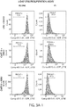


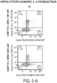


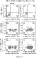


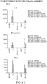


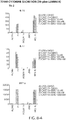


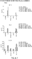


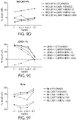

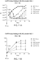


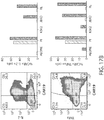


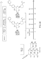


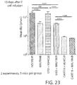


Classifications
-
- A—HUMAN NECESSITIES
- A61—MEDICAL OR VETERINARY SCIENCE; HYGIENE
- A61K—PREPARATIONS FOR MEDICAL, DENTAL OR TOILETRY PURPOSES
- A61K31/00—Medicinal preparations containing organic active ingredients
- A61K31/33—Heterocyclic compounds
- A61K31/395—Heterocyclic compounds having nitrogen as a ring hetero atom, e.g. guanethidine or rifamycins
- A61K31/495—Heterocyclic compounds having nitrogen as a ring hetero atom, e.g. guanethidine or rifamycins having six-membered rings with two or more nitrogen atoms as the only ring heteroatoms, e.g. piperazine or tetrazines
- A61K31/505—Pyrimidines; Hydrogenated pyrimidines, e.g. trimethoprim
- A61K31/506—Pyrimidines; Hydrogenated pyrimidines, e.g. trimethoprim not condensed and containing further heterocyclic rings
-
- A—HUMAN NECESSITIES
- A61—MEDICAL OR VETERINARY SCIENCE; HYGIENE
- A61K—PREPARATIONS FOR MEDICAL, DENTAL OR TOILETRY PURPOSES
- A61K35/00—Medicinal preparations containing materials or reaction products thereof with undetermined constitution
- A61K35/12—Materials from mammals; Compositions comprising non-specified tissues or cells; Compositions comprising non-embryonic stem cells; Genetically modified cells
- A61K35/14—Blood; Artificial blood
- A61K35/17—Lymphocytes; B-cells; T-cells; Natural killer cells; Interferon-activated or cytokine-activated lymphocytes
-
- A—HUMAN NECESSITIES
- A61—MEDICAL OR VETERINARY SCIENCE; HYGIENE
- A61K—PREPARATIONS FOR MEDICAL, DENTAL OR TOILETRY PURPOSES
- A61K39/00—Medicinal preparations containing antigens or antibodies
- A61K39/46—Cellular immunotherapy
- A61K39/461—Cellular immunotherapy characterised by the cell type used
- A61K39/4611—T-cells, e.g. tumor infiltrating lymphocytes [TIL], lymphokine-activated killer cells [LAK] or regulatory T cells [Treg]
-
- A—HUMAN NECESSITIES
- A61—MEDICAL OR VETERINARY SCIENCE; HYGIENE
- A61K—PREPARATIONS FOR MEDICAL, DENTAL OR TOILETRY PURPOSES
- A61K39/00—Medicinal preparations containing antigens or antibodies
- A61K39/46—Cellular immunotherapy
- A61K39/463—Cellular immunotherapy characterised by recombinant expression
- A61K39/4631—Chimeric Antigen Receptors [CAR]
-
- A—HUMAN NECESSITIES
- A61—MEDICAL OR VETERINARY SCIENCE; HYGIENE
- A61K—PREPARATIONS FOR MEDICAL, DENTAL OR TOILETRY PURPOSES
- A61K39/00—Medicinal preparations containing antigens or antibodies
- A61K39/46—Cellular immunotherapy
- A61K39/464—Cellular immunotherapy characterised by the antigen targeted or presented
- A61K39/4643—Vertebrate antigens
- A61K39/4644—Cancer antigens
- A61K39/464402—Receptors, cell surface antigens or cell surface determinants
- A61K39/464411—Immunoglobulin superfamily
- A61K39/464412—CD19 or B4
-
- A—HUMAN NECESSITIES
- A61—MEDICAL OR VETERINARY SCIENCE; HYGIENE
- A61K—PREPARATIONS FOR MEDICAL, DENTAL OR TOILETRY PURPOSES
- A61K45/00—Medicinal preparations containing active ingredients not provided for in groups A61K31/00 - A61K41/00
- A61K45/06—Mixtures of active ingredients without chemical characterisation, e.g. antiphlogistics and cardiaca
-
- A—HUMAN NECESSITIES
- A61—MEDICAL OR VETERINARY SCIENCE; HYGIENE
- A61P—SPECIFIC THERAPEUTIC ACTIVITY OF CHEMICAL COMPOUNDS OR MEDICINAL PREPARATIONS
- A61P35/00—Antineoplastic agents
-
- A—HUMAN NECESSITIES
- A61—MEDICAL OR VETERINARY SCIENCE; HYGIENE
- A61P—SPECIFIC THERAPEUTIC ACTIVITY OF CHEMICAL COMPOUNDS OR MEDICINAL PREPARATIONS
- A61P35/00—Antineoplastic agents
- A61P35/02—Antineoplastic agents specific for leukemia
-
- C—CHEMISTRY; METALLURGY
- C07—ORGANIC CHEMISTRY
- C07K—PEPTIDES
- C07K16/00—Immunoglobulins [IGs], e.g. monoclonal or polyclonal antibodies
- C07K16/18—Immunoglobulins [IGs], e.g. monoclonal or polyclonal antibodies against material from animals or humans
- C07K16/28—Immunoglobulins [IGs], e.g. monoclonal or polyclonal antibodies against material from animals or humans against receptors, cell surface antigens or cell surface determinants
- C07K16/2803—Immunoglobulins [IGs], e.g. monoclonal or polyclonal antibodies against material from animals or humans against receptors, cell surface antigens or cell surface determinants against the immunoglobulin superfamily
-
- C—CHEMISTRY; METALLURGY
- C07—ORGANIC CHEMISTRY
- C07K—PEPTIDES
- C07K16/00—Immunoglobulins [IGs], e.g. monoclonal or polyclonal antibodies
- C07K16/18—Immunoglobulins [IGs], e.g. monoclonal or polyclonal antibodies against material from animals or humans
- C07K16/28—Immunoglobulins [IGs], e.g. monoclonal or polyclonal antibodies against material from animals or humans against receptors, cell surface antigens or cell surface determinants
- C07K16/30—Immunoglobulins [IGs], e.g. monoclonal or polyclonal antibodies against material from animals or humans against receptors, cell surface antigens or cell surface determinants from tumour cells
-
- C—CHEMISTRY; METALLURGY
- C12—BIOCHEMISTRY; BEER; SPIRITS; WINE; VINEGAR; MICROBIOLOGY; ENZYMOLOGY; MUTATION OR GENETIC ENGINEERING
- C12N—MICROORGANISMS OR ENZYMES; COMPOSITIONS THEREOF; PROPAGATING, PRESERVING, OR MAINTAINING MICROORGANISMS; MUTATION OR GENETIC ENGINEERING; CULTURE MEDIA
- C12N5/00—Undifferentiated human, animal or plant cells, e.g. cell lines; Tissues; Cultivation or maintenance thereof; Culture media therefor
- C12N5/06—Animal cells or tissues; Human cells or tissues
- C12N5/0602—Vertebrate cells
- C12N5/0634—Cells from the blood or the immune system
- C12N5/0636—T lymphocytes
-
- A—HUMAN NECESSITIES
- A61—MEDICAL OR VETERINARY SCIENCE; HYGIENE
- A61K—PREPARATIONS FOR MEDICAL, DENTAL OR TOILETRY PURPOSES
- A61K35/00—Medicinal preparations containing materials or reaction products thereof with undetermined constitution
- A61K35/12—Materials from mammals; Compositions comprising non-specified tissues or cells; Compositions comprising non-embryonic stem cells; Genetically modified cells
- A61K2035/124—Materials from mammals; Compositions comprising non-specified tissues or cells; Compositions comprising non-embryonic stem cells; Genetically modified cells the cells being hematopoietic, bone marrow derived or blood cells
-
- A—HUMAN NECESSITIES
- A61—MEDICAL OR VETERINARY SCIENCE; HYGIENE
- A61K—PREPARATIONS FOR MEDICAL, DENTAL OR TOILETRY PURPOSES
- A61K39/00—Medicinal preparations containing antigens or antibodies
- A61K2039/505—Medicinal preparations containing antigens or antibodies comprising antibodies
-
- A—HUMAN NECESSITIES
- A61—MEDICAL OR VETERINARY SCIENCE; HYGIENE
- A61K—PREPARATIONS FOR MEDICAL, DENTAL OR TOILETRY PURPOSES
- A61K39/00—Medicinal preparations containing antigens or antibodies
- A61K2039/545—Medicinal preparations containing antigens or antibodies characterised by the dose, timing or administration schedule
-
- A—HUMAN NECESSITIES
- A61—MEDICAL OR VETERINARY SCIENCE; HYGIENE
- A61K—PREPARATIONS FOR MEDICAL, DENTAL OR TOILETRY PURPOSES
- A61K2239/00—Indexing codes associated with cellular immunotherapy of group A61K39/46
- A61K2239/26—Universal/off- the- shelf cellular immunotherapy; Allogenic cells or means to avoid rejection
-
- A—HUMAN NECESSITIES
- A61—MEDICAL OR VETERINARY SCIENCE; HYGIENE
- A61K—PREPARATIONS FOR MEDICAL, DENTAL OR TOILETRY PURPOSES
- A61K2239/00—Indexing codes associated with cellular immunotherapy of group A61K39/46
- A61K2239/31—Indexing codes associated with cellular immunotherapy of group A61K39/46 characterized by the route of administration
-
- A—HUMAN NECESSITIES
- A61—MEDICAL OR VETERINARY SCIENCE; HYGIENE
- A61K—PREPARATIONS FOR MEDICAL, DENTAL OR TOILETRY PURPOSES
- A61K2239/00—Indexing codes associated with cellular immunotherapy of group A61K39/46
- A61K2239/38—Indexing codes associated with cellular immunotherapy of group A61K39/46 characterised by the dose, timing or administration schedule
-
- A—HUMAN NECESSITIES
- A61—MEDICAL OR VETERINARY SCIENCE; HYGIENE
- A61K—PREPARATIONS FOR MEDICAL, DENTAL OR TOILETRY PURPOSES
- A61K2239/00—Indexing codes associated with cellular immunotherapy of group A61K39/46
- A61K2239/46—Indexing codes associated with cellular immunotherapy of group A61K39/46 characterised by the cancer treated
- A61K2239/48—Blood cells, e.g. leukemia or lymphoma
-
- C—CHEMISTRY; METALLURGY
- C07—ORGANIC CHEMISTRY
- C07K—PEPTIDES
- C07K2317/00—Immunoglobulins specific features
- C07K2317/20—Immunoglobulins specific features characterized by taxonomic origin
- C07K2317/24—Immunoglobulins specific features characterized by taxonomic origin containing regions, domains or residues from different species, e.g. chimeric, humanized or veneered
-
- C—CHEMISTRY; METALLURGY
- C07—ORGANIC CHEMISTRY
- C07K—PEPTIDES
- C07K2317/00—Immunoglobulins specific features
- C07K2317/50—Immunoglobulins specific features characterized by immunoglobulin fragments
- C07K2317/52—Constant or Fc region; Isotype
- C07K2317/53—Hinge
-
- C—CHEMISTRY; METALLURGY
- C07—ORGANIC CHEMISTRY
- C07K—PEPTIDES
- C07K2317/00—Immunoglobulins specific features
- C07K2317/50—Immunoglobulins specific features characterized by immunoglobulin fragments
- C07K2317/56—Immunoglobulins specific features characterized by immunoglobulin fragments variable (Fv) region, i.e. VH and/or VL
- C07K2317/565—Complementarity determining region [CDR]
-
- C—CHEMISTRY; METALLURGY
- C07—ORGANIC CHEMISTRY
- C07K—PEPTIDES
- C07K2317/00—Immunoglobulins specific features
- C07K2317/60—Immunoglobulins specific features characterized by non-natural combinations of immunoglobulin fragments
- C07K2317/62—Immunoglobulins specific features characterized by non-natural combinations of immunoglobulin fragments comprising only variable region components
- C07K2317/622—Single chain antibody (scFv)
-
- C—CHEMISTRY; METALLURGY
- C07—ORGANIC CHEMISTRY
- C07K—PEPTIDES
- C07K2319/00—Fusion polypeptide
- C07K2319/01—Fusion polypeptide containing a localisation/targetting motif
- C07K2319/02—Fusion polypeptide containing a localisation/targetting motif containing a signal sequence
-
- C—CHEMISTRY; METALLURGY
- C07—ORGANIC CHEMISTRY
- C07K—PEPTIDES
- C07K2319/00—Fusion polypeptide
- C07K2319/01—Fusion polypeptide containing a localisation/targetting motif
- C07K2319/03—Fusion polypeptide containing a localisation/targetting motif containing a transmembrane segment
Definitions
- the present invention relates generally to the use of T cells engineered to express a Chimeric Antigen Receptor (CAR), e.g., in combination with another agent such as, e.g., a Bruton's tyrosine kinase (BTK) inhibitor, to treat a disease associated with expression of the Cluster of Differentiation 19 protein (CD19).
- CAR Chimeric Antigen Receptor
- BTK Bruton's tyrosine kinase
- CAR chimeric antigen receptor
- CART modified autologous T cell
- CTL019 The clinical results of the murine derived CART19 (i.e., "CTL019”) have shown promise in establishing complete remissions in patients suffering with CLL as well as in childhood ALL (see, e.g., Kalos et al., Sci Transl Med 3:95ra73 (2011 ), Porter et al., NEJM 365:725-733 (2011 ), Grupp et al., NEJM 368:1509-1518 (2013 )).
- a successful therapeutic T cell therapy needs to have the ability to proliferate and persist over time, and to further monitor for leukemic cell escapees.
- CAR transformed patient T cells need to persist and maintain the ability to proliferate in response to its cognate antigen. It has been shown that ALL patient T cells perform can do this with CART19 comprising a murine scFv (see, e.g., Grupp et al., NEJM 368:1509-1518 (2013 )). Martz, L (Science-Business eXchange (2014); 7(33); doi:10.1038/scibx2014.971 ) relates to overcoming ibrutinib resistance.
- compositions and methods of treating disorders as cancer using immune effector cells (e.g., T cells or NK cells) that express a Chimeric Antigen Receptor (CAR) molecule, e.g., a CAR that binds to a B-cell antigen, e.g., Cluster of Differentiation 19 protein (CD19) (e.g., OMIM Acc. No. 107265, Swiss Prot. Acc No. P15391).
- CAR Chimeric Antigen Receptor
- compositions include, and the methods include administering, immune effector cells (e.g., T cells or NK cells) expressing a B cell targeting CAR, in combination with a BTK inhibitor (e.g., a compound of formula (I) or a pharmaceutically acceptable salt thereof).
- immune effector cells e.g., T cells or NK cells
- BTK inhibitor e.g., a compound of formula (I) or a pharmaceutically acceptable salt thereof.
- the combination maintains or has better clinical effectiveness as compared to either therapy alone.
- the disclosure further pertains to the use of engineered cells, e.g., immune effector cells (e.g., T cells or NK cells), to express a CAR molecule that binds to a B-cell antigen, e.g., CD19, in combination with a BTK inhibitor (e.g., a BTK inhibitor described herein) to treat a disorder associated with expression of a B-cell antigen, e.g., CD19 (e.g., a cancer, e.g., a hematological cancer).
- engineered cells e.g., immune effector cells (e.g., T cells or NK cells)
- a CAR molecule that binds to a B-cell antigen e.g., CD19
- a BTK inhibitor e.g., a BTK inhibitor described herein
- the disclosure pertains to a method of treating a subject, e.g., a mammal, having a disease associated with expression of a B-cell antigen, e.g., CD19, comprising administering to the mammal an effective amount of a cell, e.g., an immune effector cell (e.g., a T cell or NK cell) that expresses a CAR molecule that binds the B-cell antigen, e.g., CD19, e.g., a CAR molecule that binds CD19 described herein, in combination with a BTK inhibitor, e.g., a BTK inhibitor described herein, e.g., a compound of formula (I) or a pharmaceutically acceptable salt thereof.
- a cell e.g., an immune effector cell (e.g., a T cell or NK cell) that expresses a CAR molecule that binds the B-cell antigen, e.g., CD19,
- the CAR molecule binds to CD19, e.g., a CAR molecule that binds CD19 described herein. In other instances, the CAR molecule binds to one or more of CD20, CD22 or ROR1.
- the disease associated with expression of a B-cell antigen is selected from a proliferative disease such as a cancer or malignancy or a precancerous condition such as a myelodysplasia, a myelodysplastic syndrome or a preleukemia, or is a non-cancer related indication associated with expression of a B-cell antigen, e.g., one or more of CD19, CD20, CD22, or ROR1.
- the disease is a solid or liquid tumor.
- the cancer is pancreatic cancer.
- the disease is a hematologic cancer.
- the hematological cancer is leukemia.
- the cancer is selected from the group consisting of one or more acute leukemias including but not limited to B-cell acute lymphoid leukemia (BALL), T-cell acute lymphoid leukemia (TALL), small lymphocytic leukemia (SLL), acute lymphoid leukemia (ALL); one or more chronic leukemias including but not limited to chronic myelogenous leukemia (CML), chronic lymphocytic leukemia (CLL).
- BALL B-cell acute lymphoid leukemia
- TALL T-cell acute lymphoid leukemia
- SLL small lymphocytic leukemia
- ALL acute lymphoid leukemia
- CML chronic myelogenous leukemia
- CLL chronic lymphocytic leukemia
- Additional hematological cancers or hematologic conditions include, but are not limited to, mantle cell lymphoma (MCL), B cell prolymphocytic leukemia, blastic plasmacytoid dendritic cell neoplasm, Burkitt's lymphoma, diffuse large B cell lymphoma, follicular lymphoma, hairy cell leukemia, small cell- or a large cell-follicular lymphoma, malignant lymphoproliferative conditions, MALT lymphoma, Marginal zone lymphoma, multiple myeloma, myelodysplasia and myelodysplastic syndrome, non-Hodgkin lymphoma, Hodgkin lymphoma, plasmablastic lymphoma, plasmacytoid dendritic cell neoplasm, and Waldenstrom macroglobulinemia.
- MCL mantle cell lymphoma
- B cell prolymphocytic leukemia blastic plasmacytoid dendritic cell
- the disease associated with B-cell antigen e.g., e.g., one or more of CD19, CD20, CD22, or ROR1 expression is a "preleukemia” which is a diverse collection of hematological conditions united by ineffective production (or dysplasia) of myeloid blood cells.
- the disease associated with B-cell antigen includes, but is not limited to atypical and/or non-classical cancers, malignancies, precancerous conditions or proliferative diseases expressing a B-cell antigen (e.g., e.g., one or more of CD19, CD20, CD22 or ROR1).
- a B-cell antigen e.g., e.g., one or more of CD19, CD20, CD22 or ROR1
- Any combination of the diseases associated with B-cell antigen (e.g., e.g., one or more of CD19, CD20, CD22 or ROR1) expression described herein can be treated with the methods and compositions described herein.
- the disease associated with expression of a B-cell antigen is a lymphoma, e.g., MCL or Hodgkin lymphoma.
- the disease associated with expression of a B-cell antigen is leukemia, e.g., SLL, CLL and/or ALL.
- the cell expresses a CAR molecule comprising an anti-CD 19 binding domain (e.g., a murine or humanized antibody or antibody fragment that specifically binds to CD19), a transmembrane domain, and an intracellular signaling domain (e.g., an intracellular signaling domain comprising a costimulatory domain and/or a primary signaling domain).
- an anti-CD 19 binding domain e.g., a murine or humanized antibody or antibody fragment that specifically binds to CD19
- a transmembrane domain e.g., a transmembrane domain
- an intracellular signaling domain e.g., an intracellular signaling domain comprising a costimulatory domain and/or a primary signaling domain.
- the CAR comprises an antibody or antibody fragment which includes an anti-CD 19 binding domain described herein (e.g., a murine or humanized antibody or antibody fragment that specifically binds to CD19 as described herein), a transmembrane domain described herein, and an intracellular signaling domain described herein (e.g., an intracellular signaling domain comprising a costimulatory domain and/or a primary signaling domain described herein).
- an anti-CD 19 binding domain described herein e.g., a murine or humanized antibody or antibody fragment that specifically binds to CD19 as described herein
- a transmembrane domain described herein e.g., a transmembrane domain described herein
- an intracellular signaling domain described herein e.g., an intracellular signaling domain comprising a costimulatory domain and/or a primary signaling domain described herein.
- the CAR molecule is capable of binding CD19 (e.g., wild-type or mutant human CD19).
- the CAR molecule comprises an anti-CD19 binding domain comprising one or more (e.g., all three) light chain complementary determining region 1 (LC CDR1), light chain complementary determining region 2 (LC CDR2), and light chain complementary determining region 3 (LC CDR3) of an anti-CD19 binding domain described herein, and one or more (e.g., all three) heavy chain complementary determining region 1 (HC CDR1), heavy chain complementary determining region 2 (HC CDR2), and heavy chain complementary determining region 3 (HC CDR3) of an anti-CD 19 binding domain described herein, e.g., an anti-CD19 binding domain comprising one or more, e.g., all three, LC CDRs and one or more, e.g., all three, HC CDRs.
- an anti-CD19 binding domain comprising one or more, e.g., all three, LC CDRs and one
- the anti-CD19 binding domain comprises one or more (e.g., all three) heavy chain complementary determining region 1 (HC CDR1), heavy chain complementary determining region 2 (HC CDR2), and heavy chain complementary determining region 3 (HC CDR3) of an anti-CD19 binding domain described herein, e.g., the anti-CD19 binding domain has two variable heavy chain regions, each comprising a HC CDR1, a HC CDR2 and a HC CDR3 described herein.
- the anti-CD19 binding domain comprises a murine light chain variable region described herein (e.g., in Table 7) and/or a murine heavy chain variable region described herein (e.g., in Table 7).
- the anti-CD19 binding domain is a scFv comprising a murine light chain and a murine heavy chain of an amino acid sequence of Table 7.
- the anti-CD19 binding domain (e.g., an scFv) comprises: a light chain variable region comprising an amino acid sequence having at least one, two or three modifications (e.g., substitutions) but not more than 30, 20 or 10 modifications (e.g., substitutions) of an amino acid sequence of a light chain variable region provided in Table 7, or a sequence with 95-99% identity with an amino acid sequence of Table 7; and/or a heavy chain variable region comprising an amino acid sequence having at least one, two or three modifications (e.g., substitutions) but not more than 30, 20 or 10 modifications (e.g., substitutions) of an amino acid sequence of a heavy chain variable region provided in Table 7, or a sequence with 95-99% identity to an amino acid sequence of Table 7.
- the anti-CD19 binding domain comprises a sequence of SEQ ID NO:59, or a sequence with 95-99% identify thereof.
- the anti-CD19 binding domain is a scFv, and a light chain variable region comprising an amino acid sequence described herein, e.g., in Table 7, is attached to a heavy chain variable region comprising an amino acid sequence described herein, e.g., in Table 7, via a linker, e.g., a linker described herein.
- the anti-CD19 binding domain includes a (Gly 4 -Ser)n linker, wherein n is 1, 2, 3, 4, 5, or 6, preferably 3 or 4 (SEQ ID NO: 53).
- the light chain variable region and heavy chain variable region of a scFv can be, e.g., in any of the following orientations: light chain variable region-linker-heavy chain variable region or heavy chain variable region-linker-light chain variable region.
- the CAR molecule comprises a humanized anti-CD 19 binding domain that includes one or more (e.g., all three) light chain complementary determining region 1 (LC CDR1), light chain complementary determining region 2 (LC CDR2), and light chain complementary determining region 3 (LC CDR3) of a humanized anti-CD19 binding domain described herein, and one or more (e.g., all three) heavy chain complementary determining region 1 (HC CDR1), heavy chain complementary determining region 2 (HC CDR2), and heavy chain complementary determining region 3 (HC CDR3) of a humanized anti-CD19 binding domain described herein, e.g., a humanized anti-CD19 binding domain comprising one or more, e.g., all three, LC CDRs and one or more, e.g., all three, HC CDRs.
- LC CDR1 light chain complementary determining region 1
- HC CDR2 light chain complementary determining region 2
- HC CDR3 light chain complementary determining region 3
- the humanized anti-CD19 binding domain comprises at least HC CDR2.
- the humanized anti-CD19 binding domain comprises one or more (e.g., all three) heavy chain complementary determining region 1 (HC CDR1), heavy chain complementary determining region 2 (HC CDR2), and heavy chain complementary determining region 3 (HC CDR3) of a humanized anti-CD 19 binding domain described herein, e.g., the humanized anti-CD19 binding domain has two variable heavy chain regions, each comprising a HC CDR1, a HC CDR2 and a HC CDR3 described herein.
- the humanized anti-CD19 binding domain comprises at least HC CDR2.
- the light chain variable region comprises one, two, three or all four framework regions of VK3_L25 germline sequence.
- the light chain variable region has a modification (e.g., substitution, e.g., a substitution of one or more amino acid found in the corresponding position in the murine light chain variable region of SEQ ID NO: 58, e.g., a substitution at one or more of positions 71 and 87).
- the heavy chain variable region comprises one, two, three or all four framework regions of VH4_4-59 germline sequence.
- the heavy chain variable region has a modification (e.g., substitution, e.g., a substitution of one or more amino acid found in the corresponding position in the murine heavy chain variable region of SEQ ID NO: 58, e.g., a substitution at one or more of positions 71, 73 and 78).
- the humanized anti-CD19 binding domain comprises a light chain variable region described herein (e.g., in Table 3) and/or a heavy chain variable region described herein (e.g., in Table 3).
- the humanized anti-CD19 binding domain is a scFv comprising a light chain and a heavy chain of an amino acid sequence of Table 3.
- the humanized anti-CD19 binding domain (e.g., an scFv) comprises: a light chain variable region comprising an amino acid sequence having at least one, two or three modifications (e.g., substitutions) but not more than 30, 20 or 10 modifications (e.g., substitutions) of an amino acid sequence of a light chain variable region provided in Table 3, or a sequence with 95-99% identity with an amino acid sequence of Table 3; and/or a heavy chain variable region comprising an amino acid sequence having at least one, two or three modifications (e.g., substitutions) but not more than 30, 20 or 10 modifications (e.g., substitutions) of an amino acid sequence of a heavy chain variable region provided in Table 3, or a sequence with 95-99% identity to an amino acid sequence of Table 3.
- a light chain variable region comprising an amino acid sequence having at least one, two or three modifications (e.g., substitutions) but not more than 30, 20 or 10 modifications (e.g., substitutions) of an amino acid sequence of a heavy chain variable region provided in
- the humanized anti-CD19 binding domain comprises a sequence selected from a group consisting of SEQ ID NO:1, SEQ ID NO:2, SEQ ID NO:3, SEQ ID NO: 4, SEQ ID NO:5, SEQ ID NO:6, SEQ ID NO:7, SEQ ID NO:8, SEQ ID NO:9, SEQ ID NO:10, SEQ ID NO:11 and SEQ ID NO:12, or a sequence with 95-99% identify thereof.
- the humanized anti-CD19 binding domain is a scFv, and a light chain variable region comprising an amino acid sequence described herein, e.g., in Table 3, is attached to a heavy chain variable region comprising an amino acid sequence described herein, e.g., in Table 3, via a linker, e.g., a linker described herein.
- the humanized anti-CD19 binding domain includes a (Gly 4 -Ser)n linker, wherein n is 1, 2, 3, 4, 5, or 6, preferably 3 or 4 (SEQ ID NO: 53).
- the light chain variable region and heavy chain variable region of a scFv can be, e.g., in any of the following orientations: light chain variable region-linker-heavy chain variable region or heavy chain variable region-linker-light chain variable region.
- the CAR molecule comprises a transmembrane domain of a protein selected from the group consisting of the alpha, beta or zeta chain of the T-cell receptor, CD28, CD3 epsilon, CD45, CD4, CD5, CD8, CD9, CD16, CD22, CD33, CD37, CD64, CD80, CD86, CD134, CD137 and CD154.
- the transmembrane domain comprises a sequence of SEQ ID NO: 15.
- the transmembrane domain comprises an amino acid sequence having at least one, two or three modifications (e.g., substitutions) but not more than 20, 10 or 5 modifications (e.g., substitutions) of an amino acid sequence of SEQ ID NO: 15, or a sequence with 95-99% identity to an amino acid sequence of SEQ ID NO: 15.
- the anti-CD 19 binding domain is connected to the transmembrane domain by a hinge region, e.g., a hinge region described herein.
- the encoded hinge region comprises SEQ ID NO:14 or SEQ ID NO:45, or a sequence with 95-99% identity thereof.
- the CAR molecule further comprises a sequence encoding a costimulatory domain, e.g., a costimulatory domain described herein.
- the costimulatory domain comprises a functional signaling domain of a protein selected from the group consisting of OX40, CD2, CD27, CD28, CDS, ICAM-1, LFA-1 (CD11a/CD18) and 4-1BB (CD137).
- the costimulatory domain comprises a sequence of SEQ ID NO: 16.
- the costimulatory domain comprises a sequence of SEQ ID NO:51.
- the costimulatory domain comprises an amino acid sequence having at least one, two or three modifications (e.g., substitutions) but not more than 20, 10 or 5 modifications (e.g., substitutions) of an amino acid sequence of SEQ ID NO: 16 or SEQ ID NO:51, or a sequence with 95-99% identity to an amino acid sequence of SEQ ID NO: 16 or SEQ ID NO:51.
- the CAR molecule further comprises a sequence encoding an intracellular signaling domain, e.g., an intracellular signaling domain described herein.
- the intracellular signaling domain comprises a functional signaling domain of 4-1BB and/or a functional signaling domain of CD3 zeta.
- the intracellular signaling domain comprises the sequence of SEQ ID NO: 16 and/or the sequence of SEQ ID NO:17.
- the intracellular signaling domain comprises the sequence of SEQ ID NO:16 and/or the sequence of SEQ ID NO:43.
- the intracellular signaling domain comprises a functional signaling domain of CD27 and/or a functional signaling domain of CD3 zeta.
- the intracellular signaling domain comprises the sequence of SEQ ID NO: 51 and/or the sequence of SEQ ID NO:17. In one instance, the intracellular signaling domain comprises the sequence of SEQ ID NO:51 and/or the sequence of SEQ ID NO:43.
- the intracellular signaling domain comprises an amino acid sequence having at least one, two or three modifications (e.g., substitutions) but not more than 20, 10 or 5 modifications (e.g., substitutions) of an amino acid sequence of SEQ ID NO:16 or SEQ ID NO:51 and/or an amino acid sequence of SEQ ID NO:17 or SEQ ID NO:43, or a sequence with 95-99% identity to an amino acid sequence of SEQ ID NO:16 or SEQ ID NO:51 and/or an amino acid sequence of SEQ ID NO:17 or SEQ ID NO:43.
- the intracellular signaling domain comprises the sequence of SEQ ID NO:16 or SEQ ID NO:51 and the sequence of SEQ ID NO: 17 or SEQ ID NO:43, wherein the sequences comprising the intracellular signaling domain are expressed in the same frame and as a single polypeptide chain.
- the CAR molecule further comprises a leader sequence, e.g., a leader sequence described herein.
- the leader sequence comprises an amino acid sequence of SEQ ID NO: 13, or a sequence with 95-99% identity to an amino acid sequence of SEQ ID NO:13.
- the CAR molecule comprises a leader sequence, e.g., a leader sequence described herein, e.g., a leader sequence of SEQ ID NO: 13, or having 95-99% identity thereof; an anti-CD19 binding domain described herein, e.g., an anti-CD19 binding domain comprising a LC CDR1, a LC CDR2, a LC CDR3, a HC CDR1, a HC CDR2 and a HC CDR3 described herein, e.g., a murine anti-CD19 binding domain described in Table 7, a humanized anti-CD19 binding domain described in Table 3, or a sequence with 95-99% identify thereof; a hinge region, e.g., a hinge region described herein, e.g., a hinge region of SEQ ID NO:14 or having 95-99% identity thereof; a transmembrane domain, e.g., a transmembrane domain described herein, e.g., a transmembrane domain described
- the intracellular signaling domain comprises a costimulatory domain, e.g., a costimulatory domain described herein, e.g., a 4-1BB costimulatory domain having a sequence of SEQ ID NO:16 or SEQ ID NO:51, or having 95-99%identity thereof, and/or a primary signaling domain, e.g., a primary signaling domain described herein, e.g., a CD3 zeta stimulatory domain having a sequence of SEQ ID NO:17 or SEQ ID NO:43, or having 95-99% identity thereof.
- a costimulatory domain e.g., a costimulatory domain described herein, e.g., a 4-1BB costimulatory domain having a sequence of SEQ ID NO:16 or SEQ ID NO:51, or having 95-99%identity thereof
- a primary signaling domain e.g., a primary signaling domain described herein, e.g., a CD3 zeta stimul
- the CAR molecule comprises (e.g., consists of) an amino acid sequence of SEQ ID NO:58, SEQ ID NO:31, SEQ ID NO:32, SEQ ID NO:33, SEQ ID NO:34, SEQ ID NO:35, SEQ ID NO:36, SEQ ID NO:37, SEQ ID NO:38, SEQ ID NO:39, SEQ ID NO:40, SEQ ID NO:41 or SEQ ID NO:42, or an amino acid sequence having at least one, two, three, four, five, 10, 15, 20 or 30 modifications (e.g., substitutions) but not more than 60, 50 or 40 modifications (e.g., substitutions) of an amino acid sequence of SEQ ID NO:58, SEQ ID NO:31, SEQ ID NO:32, SEQ ID NO:33, SEQ ID NO:34, SEQ ID NO:35, SEQ ID NO:36, SEQ ID NO:37, SEQ ID NO:38, SEQ ID NO:39, SEQ ID NO:40, SEQ ID NO
- the cell expressing the CAR molecule comprises a vector that includes a nucleic acid sequence encoding the CAR molecule.
- the vector is selected from the group consisting of a DNA, a RNA, a plasmid, a lentivirus vector, adenoviral vector, or a retrovirus vector.
- the vector is a lentivirus vector.
- the vector further comprises a promoter.
- the promoter is an EF-1 promoter.
- the EF-1 promoter comprises a sequence of SEQ ID NO: 100.
- the vector is an in vitro transcribed vector, e.g., a vector that transcribes RNA of a nucleic acid molecule described herein.
- the nucleic acid sequence in the in vitro vector further comprises a poly(A) tail, e.g., a poly A tail described herein, e.g., comprising about 150 adenosine bases (SEQ ID NO:104).
- the nucleic acid sequence in the in vitro vector further comprises a 3'UTR, e.g., a 3' UTR described herein, e.g., comprising at least one repeat of a 3'UTR derived from human beta-globulin.
- the nucleic acid sequence in the in vitro vector further comprises promoter, e.g., a T2A promoter.
- the cell expressing the CAR molecule is a cell or population of cells as described herein, e.g., a human immune effector cell or population of cells (e.g., a human T cell or a human NK cell, e.g., a human T cell described herein or a human NK cell described herein).
- a human immune effector cell or population of cells e.g., a human T cell or a human NK cell, e.g., a human T cell described herein or a human NK cell described herein.
- the human T cell is a CD8+ T cell.
- the cell is an autologous T cell.
- the cell is an allogeneic T cell.
- the cell is a T cell and the T cell is diaglycerol kinase (DGK) deficient.
- DGK diaglycerol kinase
- the cell is a T cell and the T cell is Ikaros deficient. In one instance, the cell is a T cell and the T cell is both DGK and Ikaros deficient. It shall be understood that the compositions and methods disclosed herein reciting the term "cell” encompass compositions and methods comprising one or more cells, e.g., a population of cells.
- the cell expressing the CAR molecule can further express another agent, e.g., an agent which enhances the activity of a CAR-expressing cell.
- the method further includes administering a cell expressing the CAR molecule, as described herein, in combination with a BTK inhibitor described herein, in combination with an agent which enhances the activity of a CAR-expressing cell.
- the agent is a cytokine, e.g., IL-7, IL-15, IL-21, or a combination thereof.
- the method includes administering IL-7 to the subject.
- the cytokine can be delivered in combination with, e.g., simultaneously or shortly after, administration of the CAR-expressing cell. Alternatively, the cytokine can be delivered after a prolonged period of time after administration of the CAR-expressing cell, e.g., after assessment of the subject's response to the CAR-expressing cell.
- the agent which enhances the activity of a CAR-expressing cell can be an agent which inhibits an immune inhibitory molecule.
- immune inhibitory molecules include PD1, PD-L1, CTLA4, TIM3, CEACAM (e.g., CEACAM-1, CEACAM-3 and/or CEACAM-5), LAG3, VISTA, BTLA, TIGIT, LAIR1, CD160, 2B4 and TGFR beta.
- the agent which inhibits an immune inhibitory molecule comprises a first polypeptide, e.g., an immune inhibitory molecule, associated with a second polypeptide that provides a positive signal to the cell, e.g., an intracellular signaling domain described herein.
- the agent comprises a first polypeptide, e.g., of an inhibitory molecule such as PD1, PD-L1, CTLA4, TIM3, CEACAM (e.g., CEACAM-1, CEACAM-3 and/or CEACAM-5), LAG3, VISTA, BTLA, TIGIT, LAIR1, CD160, 2B4 or TGFR beta, or a fragment of any of these (e.g., at least a portion of the extracellular domain of any of these), and a second polypeptide which is an intracellular signaling domain described herein (e.g., comprising a costimulatory domain (e.g., 41BB, CD27 or CD28, e.g., as described herein) and/or a primary signaling domain (e.g., a CD3 zeta signaling domain described herein).
- an inhibitory molecule such as PD1, PD-L1, CTLA4, TIM3, CEACAM (e.g., CEACAM-1, CEACAM-3 and
- the agent comprises a first polypeptide of PD1 or a fragment thereof (e.g., at least a portion of the extracellular domain of PD1), and a second polypeptide of an intracellular signaling domain described herein (e.g., a CD28 signaling domain described herein and/or a CD3 zeta signaling domain described herein).
- a first polypeptide of PD1 or a fragment thereof e.g., at least a portion of the extracellular domain of PD1
- a second polypeptide of an intracellular signaling domain described herein e.g., a CD28 signaling domain described herein and/or a CD3 zeta signaling domain described herein.
- lymphocyte infusion for example allogeneic lymphocyte infusion
- the lymphocyte infusion comprises at least one CAR-expressing cell that binds to a B-cell antigen e.g., CD19 (also referred to herein as CD19 CAR-expressing cell), as described herein.
- CD19 also referred to herein as CD19 CAR-expressing cell
- autologous lymphocyte infusion is used in the treatment of the cancer, wherein the autologous lymphocyte infusion comprises at least one CD19-expressing cell.
- the CD19 CAR expressing cell e.g., T cell
- a subject that has received a previous stem cell transplantation e.g., autologous stem cell transplantation.
- the CD19 CAR expressing cell e.g., T cell
- the CD19 CAR expressing cell is administered to a subject that has received a previus dose of melphalan.
- the cell expressing the CAR molecule e.g., a CAR molecule described herein
- an agent that ameliorates one or more side effect associated with administration of a cell expressing a CAR molecule e.g., an agent described herein.
- the BTK inhibitor is administered in combination with an agent that ameliorates one or more side effect associated with administration of the BTK inhibitor, e.g., an agent described herein.
- the cell expressing the CAR molecule, e.g., a CAR molecule described herein, and the BTK inhibitor are administered in combination with an additional agent that treats the disease associated with a B-cell antigen e.g., CD19, e.g., an additional agent described herein.
- an additional agent that treats the disease associated with a B-cell antigen e.g., CD19, e.g., an additional agent described herein.
- the cells expressing a CAR molecule are administered at a dose and/or dosing schedule described herein.
- the CAR molecule is introduced into T cells, e.g., using in vitro transcription, and the subject (e.g., human) receives an initial administration of cells comprising a CAR molecule, and one or more subsequent administrations of cells comprising a CAR molecule, wherein the one or more subsequent administrations are administered less than 15 days, e.g., 14, 13, 12, 11, 10, 9, 8, 7, 6, 5, 4, 3, or 2 days after the previous administration.
- more than one administration of cells comprising a CAR molecule are administered to the subject (e.g., human) per week, e.g., 2, 3, or 4 administrations of cells comprising a CAR molecule are administered per week.
- the subject receives more than one administration of cells comprising a CAR molecule per week (e.g., 2, 3 or 4 administrations per week) (also referred to herein as a cycle), followed by a week of no administration of cells comprising a CAR molecule, and then one or more additional administration of cells comprising a CAR molecule (e.g., more than one administration of the cells comprising a CAR molecule per week) is administered to the subject.
- the subject receives more than one cycle of cells comprising a CAR molecule, and the time between each cycle is less than 10, 9, 8, 7, 6, 5, 4, or 3 days.
- the cells comprising a CAR molecule are administered every other day for 3 administrations per week.
- the cells comprising a CAR molecule are administered for at least two, three, four, five, six, seven, eight or more weeks.
- the combination of the BTK inhibitor and the cells expressing a CAR molecule are administered as a first line treatment for the disease, e.g., the cancer, e.g., the cancer described herein.
- the combination of the BTK inhibitor and the cells expressing a CAR molecule are administered as a second, third, fourth line treatment for the disease, e.g., the cancer, e.g., the cancer described herein.
- a cell e.g., a population of cells described herein is administered to the subject.
- the method includes administering a population of cells, a plurality of which comprise a CAR molecule described herein.
- the population of CAR-expressing cells comprises a mixture of cells expressing different CARs.
- the population of CAR-expressing cells can include a first cell expressing a CAR having an anti-CD 19 binding domain described herein, and a second cell expressing a CAR having a different anti- CD19 binding domain, e.g., an anti-CD 19 binding domain described herein that differs from the anti-CD 19 binding domain in the CAR expressed by the first cell.
- the population of CAR-expressing cells can include a first cell expressing a CAR that includes an anti- CD19 binding domain, e.g., as described herein, and a second cell expressing a CAR that includes an antigen binding domain to a target other than CD19 (e.g., CD123 or mesothelin).
- the population of CAR-expressing cells includes, e.g., a first cell expressing a CAR that includes a primary intracellular signaling domain, and a second cell expressing a CAR that includes a secondary signaling domain.
- the method includes administering a population of cells wherein at least one cell in the population expresses a CAR having an anti- CD19 domain described herein, and an agent which enhances the activity of a CAR-expressing cell, e.g., a second cell expressing the agent which enhances the activity of a CAR-expressing cell.
- the agent can be an agent which inhibits an immune inhibitory molecule.
- immune inhibitory molecules include PD1, PD-L1, CTLA4, TIM3, CEACAM (e.g., CEACAM-1, CEACAM-3 and/or CEACAM-5), LAG3, VISTA, BTLA, TIGIT, LAIR1, CD160, 2B4 and TGFR beta.
- the agent which inhibits an immune inhibitory molecule comprises a first polypeptide, e.g., an inhibitory molecule, associated with a second polypeptide that provides a positive signal to the cell, e.g., an intracellular signaling domain described herein.
- the agent comprises a first polypeptide, e.g., of an inhibitory molecule such as PD1, PD-L1, CTLA4, TIM3, CEACAM (e.g., CEACAM-1, CEACAM-3 and/or CEACAM-5), LAG3, VISTA, BTLA, TIGIT, LAIR1, CD160, 2B4 or TGFR beta, or a fragment of any of these (e.g., at least a portion of an extracellular domain of any of these), and a second polypeptide which is an intracellular signaling domain described herein (e.g., comprising a costimulatory domain (e.g., 41BB, CD27 or CD28, e.g., as described herein) and/or a primary signaling domain (e.g., a CD3 zeta signaling domain described herein).
- an inhibitory molecule such as PD1, PD-L1, CTLA4, TIM3, CEACAM (e.g., CEACAM-1, CEACAM-3 and
- the agent comprises a first polypeptide of PD1 or a fragment thereof (e.g., at least a portion of the extracellular domain of PD1), and a second polypeptide of an intracellular signaling domain described herein (e.g., a CD28 signaling domain described herein and/or a CD3 zeta signaling domain described herein).
- a first polypeptide of PD1 or a fragment thereof e.g., at least a portion of the extracellular domain of PD1
- a second polypeptide of an intracellular signaling domain described herein e.g., a CD28 signaling domain described herein and/or a CD3 zeta signaling domain described herein.
- the disclosure pertains to a cell expressing a CAR molecule described herein for use as a medicament in combination with a BTK inhibitor described herein, e.g., a compound of formula (I).
- a BTK inhibitor described herein e.g., a compound of formula (I) or a pharmaceutically acceptable salt thereof, for use as a medicament in combination with a cell expressing a CAR molecule described herein.
- the disclosure pertains to a cell expressing a CAR molecule described herein for use in combination with a BTK inhibitor described herein, e.g., a compound of formula (I) or a pharmaceutically acceptable salt thereof, in the treatment of a disease expressing the B-cell antigen (e.g., CD19).
- a BTK inhibitor described herein e.g., a compound of formula (I) or a pharmaceutically acceptable salt thereof, for use in combination with a cell expressing a CAR molecule described herein, in the treatment of a disease expressing a B-cell antigen (e.g., CD19).
- the disease may be, e.g., a cancer such as a hematologic cancer.
- the cancer may be, e.g., a lymphoma, CLL, MCL, ALL, DLBCL, multiple myeloma, or another cancer described herein.
- the disclosure pertains to a cell expressing a CAR molecule described herein for use as a medicament in combination with a BTK inhibitor described herein, e.g., a compound of formula (I) or a pharmaceutically acceptable salt thereof, and a cytokine, e.g., IL-7, IL-15 and/or IL-21 as described herein.
- a BTK inhibitor described herein e.g., a compound of formula (I) or a pharmaceutically acceptable salt thereof
- a cytokine e.g., IL-7, IL-15 and/or IL-21 as described herein.
- the disclosure pertains to a cell expressing a CAR molecule described herein for use in combination with a BTK inhibitor described herein, e.g., a compound of formula (I) or a pharmaceutically acceptable salt thereof, and a cytokine, e.g., IL-7, IL-15 and/or IL-21 as described herein, in the treatment of a disease expressing CD19.
- a cytokine described herein for use in combination with a cell expressing a CAR molecule described herein and a BTK inhibitor described herein, e.g., a compound of formula (I) or a pharmaceutically acceptable salt thereof, in the treatment of a disease expressing CD19.
- the cell expressing a CAR molecule e.g., a CAR molecule described herein
- an agent that increases the efficacy of a cell expressing a CAR molecule e.g., an agent described herein.
- the cell expressing a CAR molecule e.g., a CAR molecule described herein
- an agent that ameliorates one or more side effect associated with administration of a cell expressing a CAR molecule e.g., an agent described herein.
- the cell expressing a CAR molecule is administered in combination with a low, immune enhancing dose of an mTOR inhibitor, e.g., an mTOR inhibitor described herein.
- a low, immune enhancing, dose e.g., a dose that is insufficient to completely suppress the immune system but sufficient to improve immune function
- treatment with a low, immune enhancing, dose is accompanied by a decrease in PD-1 positive T cells or an increase in PD-1 negative cells.
- PD-1 positive T cells, but not PD-1 negative T cells can be exhausted by engagement with cells which express a PD-1 ligand, e.g., PD-L1 or PD-L2.
- this approach can be used to optimize the performance of a CAR cell described herein in the subject. While not wishing to be bound by theory, it is believed that, in an instance, the performance of endogenous, non-modified immune effector cells, e.g., T cells, is improved. While not wishing to be bound by theory, it is believed that, in an instance, the performance of a CD19 CAR expressing cell is improved.
- cells e.g., T cells, which have, or will be engineered to express a CAR
- cells can be treated ex vivo by contact with an amount of an mTOR inhibitor that increases the number of PD1 negative immune effector cells, e.g., T cells or increases the ratio of PD1 negative immune effector cells, e.g., T cells/ PD1 positive immune effector cells, e.g., T cells.
- an mTOR inhibitor that increases the number of PD1 negative immune effector cells, e.g., T cells or increases the ratio of PD1 negative immune effector cells, e.g., T cells/ PD1 positive immune effector cells, e.g., T cells.
- administering is initiated prior to administration of an CAR expressing cell described herein, e.g., T cells.
- an mTOR inhibitor e.g., an allosteric inhibitor, e.g., RAD001, or a catalytic inhibitor
- an CAR expressing cell described herein e.g., T cells.
- the mTOR inhibitor is RAD001 or rapamycin.
- the CAR cells are administered after a sufficient time, or sufficient dosing, of an mTOR inhibitor, such that the level of PD1 negative immune effector cells, e.g., T cells, or the ratio of PD1 negative immune effector cells, e.g., T cells/ PD1 positive immune effector cells, e.g., T cells, has been, at least transiently, increased.
- an mTOR inhibitor such that the level of PD1 negative immune effector cells, e.g., T cells, or the ratio of PD1 negative immune effector cells, e.g., T cells/ PD1 positive immune effector cells, e.g., T cells, has been, at least transiently, increased.
- the cell e.g., an immune effector cell (e.g., a T cell or NK cell), to be engineered to express a CAR
- an immune effector cell e.g., a T cell or NK cell
- the cell is harvested after a sufficient time, or after sufficient dosing of the low, immune enhancing, dose of an mTOR inhibitor, such that the level of PD1 negative immune effector cells, e.g., T cells, or the ratio of PD1 negative immune effector cells, e.g., T cells/ PD1 positive immune effector cells, e.g., T cells, in the subject or harvested from the subject has been, at least transiently, increased.
- any of the methods described herein further comprise performing lymphodepletion on a subject, e.g., prior to administering the one or more cells that express a CAR molecule described herein, e.g., a CAR molecule that binds CD19.
- the lymphodepletion can comprise, e.g., administering one or more of melphalan, cytoxan, cyclophosphamide, and fludarabine.
- the CAR-expressing cell that is administered comprises a regulatable CAR (RCAR), e.g., an RCAR as described herein.
- the RCAR may comprise, e.g., an intracellular signaling member comprising an intracellular signaling domain and a first switch domain, an antigen binding member comprising an antigen binding domain that binds CD19 and a second switch domain; and a transmembrane domain.
- the method may further comprise administering a dimerization molecule, e.g., in an amount sufficient to cause dimerization of the first switch and second switch domains.
- the CAR-expressing cell and the BTK inhibitor are administered simultaneously or substantially simultaneously, e.g., as a first line of therapy.
- the method comprises administering a combination of the BTK inhibitor and the CAR-expressing cell (e.g., a CAR19-expressing cell) to the subject, as a first line therapy.
- the CAR-expressing cell and the BTK inhibitor are administered sequentially.
- the BTK inhibitor is administered before the CAR-expressing cell, or the CAR-expressing cell is administered before the BTK inhibitor.
- the disease associated with expression of CD19 is a hematological cancer (e.g., a hematological cancer described herein such as CLL, MCL, DLBCL, or ALL) and the subject is, or is identified as, a partial responder, non-responder, or relapser to one or more therapies for the hematological cancer, e.g., to a BTK inhibitor such as ibrutinib.
- the subject has, or is identified as having, a BTK mutation such as C481S.
- the mutation may be, e.g., a point mutation, an insertion, or a deletion.
- the mutation may be, e.g., a mutation at the binding site for the BTK inhibitor, e.g., at or near the ATP-binding pocket.
- the mutation may confer a decreased response (e.g., resistance) to the BTK inhibitor.
- the method comprises administering the BTK inhibitor to the subject, reducing the amount (e.g., ceasing administration) of the BTK inhibitor, and subsequently administering the CAR-expressing cell (e.g., a CAR19-expressing cell) to the subject.
- the amount e.g., ceasing administration
- the CAR-expressing cell e.g., a CAR19-expressing cell
- the method comprises administering the BTK inhibitor to the subject and subsequently administering a combination of the BTK inhibitor and the CAR-expressing cell (e.g., a CAR19-expressing cell) to the subject.
- a combination of the BTK inhibitor and the CAR-expressing cell e.g., a CAR19-expressing cell
- the method comprises administering a BTK inhibitor (e.g., ibrutinib, GDC-0834, RN-486, CGT-560, CGT-1764, HM-71224, CC-292, ONO-4059, CNX-774, or LFM-A13, or a combination thereof) to the subject, reducing the amount (e.g., ceasing or discontinuing administration) of the BTK inhibitor, and subsequently administering a combination of the CAR-expressing cell (e.g., a CAR19-expressing cell) and a second BTK inhibitor to the subject, wherein the second BTK inhibitor is a BTK inhibitor described herein.
- a BTK inhibitor e.g., ibrutinib, GDC-0834, RN-486, CGT-560, CGT-1764, HM-71224, CC-292, ONO-4059, CNX-774, or LFM-A13, or a combination thereof
- the patient has been administered a BTK inhibitor such as ibrutinib.
- the patient does not have a complete response to the BTK inhibitor such as ibrutinib.
- the patient has (or is identified as having) a partial response, stable disease, progressive disease, or relapse to treatment with a BTK inhibitor such as ibrutinib, GDC-0834, RN-486, CGI-560, CGI-1764, HM-71224, CC-292, ONO-4059, CNX-774, or LFM-A13, or a combination thereof.
- the patient who does not have a complete response to the BTK inhibitor such as ibrutinib is treated with a combination of a CAR-expressing cell (e.g., a CAR19-expressing cell) and a second BTK inhibitor, wherein the second BTK inhibitor is a compound of formula (I).
- the CAR-expressing cell and the second BTK inhibitor can be administered, e.g., substantially simultaneously or sequentially, e.g., the CAR-expressing cell can be administered before the second BTK inhibitor or the second BTK inhibitor can be administered before the CAR-expressing cell.
- the disease associated with expression of the B-cell antigen is a hematological cancer (e.g., a hematological cancer described herin, e.g., CLL, MCL, or ALL), and the method delays or decreases resistance to the BTK inhibitor described herein, the CAR-expressing cell (e.g., a CAR19-expressing cell), or both.
- a hematological cancer e.g., a hematological cancer described herin, e.g., CLL, MCL, or ALL
- the method delays or decreases resistance to the BTK inhibitor described herein, the CAR-expressing cell (e.g., a CAR19-expressing cell), or both.
- the disease associated with expression of the B-cell antigen is a hematological cancer (e.g., a hematological cancer described herin, e.g., CLL, MCL, DLBCL, or ALL), and wherein the method prolongs remission or delays relapse of the hematological cancer.
- a hematological cancer e.g., a hematological cancer described herin, e.g., CLL, MCL, DLBCL, or ALL
- the method prolongs remission or delays relapse of the hematological cancer.
- remission can be prolonged, relapse can be delayed, resistance can be delayed, or resistance can be decreased, compared to the expected course of disease when treated with a monotherapy of the BTK inhibitor or the CAR-expressing cell.
- Exemplary treatment regimens that can be used in any of the aforesaid methods include one or more of the following:
- the BTK inhibitor e.g., a compound of formula (I)
- the CAR19-expressing cell are administered to the mammal as a first line of therapy.
- the CAR-expressing cell e.g., the CAR19-expressing cell
- the BTK inhibitor e.g., a compound of formula (I).
- the CAR-expressing cell e.g., the CAR19-expressing cell
- the BTK inhibitor e.g., a compound of formula (I).
- administering e.g., a compound of formula (I)
- the CAR-expressing cell e.g., the CAR19-expressing cell
- the CAR-expressing cell is administered in combination with continued administration of the BTK inhibitor.
- a subject is administered a BTK inhibitor, e.g., a compound of formula (I), e.g., as a first line therapy.
- a BTK inhibitor e.g., a compound of formula (I)
- a predetermined time interval e.g., 1 or 2 months but also 2 weeks, 3 weeks, 1 month, 1.5 months, 2 months, 3 months, 4 months, 6 months, 9 months, 12 months, 15 months, or 18 months
- a CAR-expressing cell e.g., a CAR19-expressing cell
- the subject's response to the treatment is assessed at predetermined time intervals, e.g., before or during treatment with the kinase inhibitor and/or CAR-expressing cell.
- the CAR-expressing cell (e.g., a CAR19-expressing cell) is not administered. If the assessment shows that the subject is a partial responder, or has stable disease in response, to the BTK inhibitor, the CAR-expressing cell (e.g., a CAR19-expressing cell) is administered in combination with the BTK inhibitor e.g., as described herein. If the assessment shows that the subject is a non-responder or relapser, the CAR-expressing cell (e.g., a CAR19-expressing cell) is administered in combination with the BTK inhibitor or a second BTK inhibitor, e.g., a second BTK inhibitor as described herein.
- the mammal is, or is identified as being, a complete or partial responder to the BTK inhibitor, e.g., a compound of formula (I), or a complete or partial responder to the CAR19-expressing cell.
- a subject when a subject is (or is identified as being) a complete responder to the BTK inhibitor, e.g., a compound of formula (I), the subject is not administered a CAR-expressing cell (e.g., a CAR19-expressing cell) during the period of complete response.
- a complete responder e.g., a complete responder to the BTK inhibitor
- the subject when a subject is (or is identified as being) a complete responder (e.g., a complete responder to the BTK inhibitor), the subject is administered a CAR-expressing cell (e.g., a CAR19-expressing cell) during the period of complete response.
- the subject after the CAR-expressing cell (e.g., a CAR19-expressing cell), the subject experiences a prolonged response or delayed relapse (e.g., compared to the expected course of disease when treated without the CAR therapy).
- a subject when a subject is (or is identified as being) a partial responder to the BTK inhibitor, e.g., a compound of formula (I), the subject is not administered a CAR-expressing cell (e.g., a CAR19-expressing cell) during the period of partial response.
- a CAR-expressing cell e.g., a CAR19-expressing cell
- the subject when a subject is (or is identified as being) a partial responder to the BTK inhibitor, the subject is administered a CAR-expressing cell (e.g., a CAR19-expressing cell) (alone or in combination with the BTK inhibitor) during the period of partial response.
- the subject after the CAR therapy, the subject experiences a complete response and/or prolonged response or delayed relapse (e.g., compared to the expected course of disease when treated without CAR therapy).
- the subject when a subject has (or is identified as having) stable disease after treatment with the BTK inhibitor, e.g., a compound of formula (I), the subject is not administered a CAR therapy during the period of stable disease.
- the subject when a subject has (or is identified as having) stable disease after treatment with the BTK inhibitor, the subject is administered a CAR therapy during the period of stable disease.
- the subject after the CAR therapy, the subject experiences a partial response, a complete response and/or prolonged response or delayed relapse (e.g., compared to the expected course of disease when treated without CAR therapy).
- the subject when a subject has (or is identified as having) progressive disease after treatment with the BTK inhibitor, e.g., a compound of formula (I), the subject is not administered a CAR-expressing cell (e.g., a CAR19-expressing cell) during the period of progressive disease.
- the subject when a subject has (or is identified as having) progressive disease after treatment with the BTK inhibitor, the subject is administered a CAR-expressing cell (e.g., a CAR19-expressing cell) during the period of progressive disease.
- the subject after the CAR therapy, the subject experiences stable disease, a partial response, a complete response and/or prolonged response or delayed relapse (e.g., compared to the expected course of disease when treated without CAR therapy).
- the CAR-expressing cell (e.g., the CAR19-expressing cell) is administered in combination a second BTK inhibitor, wherein the second kinase inhibitor is a BTK inhibitor described herein, e.g., a compound of formula (I), when the mammal is, or is identified as being, a non-responder or relapser to a first BTK inhibitor, e.g., ibrutinib, GDC-0834, RN-486, CGI-560, CGI-1764, HM-71224, CC-292, ONO-4059, CNX-774, or LFM-A13.
- a first BTK inhibitor e.g., ibrutinib, GDC-0834, RN-486, CGI-560, CGI-1764, HM-71224, CC-292, ONO-4059, CNX-774, or LFM-A13.
- the subject e.g., the mammal
- the mammal is (or is identified as being) a partial responder to the BTK inhibitor, and the mammal is administered the CAR-expressing cell (e.g., the CAR19-expressing cell), alone or in combination with the BTK inhibitor, during the period of partial response.
- the CAR-expressing cell e.g., the CAR19-expressing cell
- the subject e.g., the mammal
- the mammal is (or has identified as being) a non-responder having progressive or stable disease after treatment with the BTK inhibitor, and the mammal is administered the the CAR-expressing cell (e.g., the CAR19-expressing cell), alone or in combination with a second BTK inhibitor, during the period of progressive or stable disease, wherein the second BTK inhibitor is a BTK inhibitor described herein, e.g., a compound of formula (I).
- a method of treating a subject e.g., a mammal, having a disease associated with expression of the B-cell antigen (e.g., CD19).
- the method comprises administering to the subject an effective amount of a BTK inhibitor as described herein, e.g., a compound of formula (I) or a pharmaceutically acceptable salt thereof, and a CAR-expressing cell (e.g., a CAR19-expressing cell) in combination (e.g. simultaneously (or substantially simultaneously), or sequentially).
- the BTK inhibitor e.g., a compound of formula (I) and the CAR-expressing cell (e.g., a CAR19 cell) are administered, in combination, e.g., as a first line of therapy,
- the BTK inhibitor e.g., a compound of formula (I) is administered initially, e.g., a monotherapy or first line of therapy; after reducing the amount (e.g., ceasing or discontinuing administration) of the BTK inhibitor, administering the CAR-expressing cell (e.g., a CAR19-expressing cell) to the subject.
- the CAR-expressing cell e.g., a CAR19-expressing cell
- the BTK inhibitor e.g., a compound of formula (I) is administered initially, e.g., a monotherapy or first line of therapy; and subsequently administering a combination of the BTK inhibitor and the CAR-expressing cell (e.g., a CAR19-expressing cell) to the subject.
- a combination of the BTK inhibitor and the CAR-expressing cell e.g., a CAR19-expressing cell
- the BTK inhibitor e.g., a compound of formula (I) is administered initially, e.g., a monotherapy or first line of therapy; after reducing the amount (e.g., ceasing or discontinuing administration) of the BTK inhibitor, administering a combination of a second BTK inhibitor and the CAR-expressing cell (e.g., a CAR19-expressing cell) to the subject.
- a combination of a second BTK inhibitor and the CAR-expressing cell e.g., a CAR19-expressing cell
- the subject's response to the treatment is assessed at predetermined time intervals, e.g., before or during treatment with the kinase inhibitor and/or CAR-expressing cell. If the assessment shows that the subject is a complete responder, the CAR-expressing cell (e.g., a CAR19-expressing cell) is not administered. If the assessment shows that the subject is a partial responder, or has stable disease in response, to the kinase inhibitor, the CAR-expressing cell (e.g., a CAR19-expressing cell) is administered in combination with the BTK inhibitor as described herein.
- the CAR-expressing cell e.g., a CAR19-expressing cell
- the CAR-expressing cell e.g., a CAR19-expressing cell
- the BTK inhibitor or a second BTK inhibitor e.g., a second BTK inhibitor as described herein.
- the first BTK inhibitor, the second BTK inhibitor, or both are compounds of formula (I).
- the disease associated with expression of the B-cell antigen is a hematological cancer, leukemia, lymphoma, MCL, CLL, ALL, DLBCL, Hodgkin lymphoma, or multiple myeloma.
- the disclosure provides a method of modulating BTK activity in a mammal, comprising administering to the mammal an effective amount of a cell (e.g., a population of cells) that expresses a CAR molecule that binds the B-cell antigen (e.g., CD19), in combination with a BTK inhibitor, wherein the BTK inhibitor comprises a compound of formula (I) or a pharmaceutically acceptable salt thereof.
- a cell e.g., a population of cells
- a CAR molecule that binds the B-cell antigen (e.g., CD19)
- the BTK inhibitor comprises a compound of formula (I) or a pharmaceutically acceptable salt thereof.
- the disclosure features a composition
- a composition comprising a cell that expresses a CAR molecule that binds the B-cell antigen (e.g., one or more of CD19, CD20. CD22 or ROR1), and one or more BTK inhibitors as described herein, e.g., a compound of formula (I).
- the the CAR-expressing cell (e.g., the CAR19-expressing cell) and the one or more BTK inhibitors can be present in a single dose form, or as two or more dose forms.
- compositions disclosed herein are for use as a medicament.
- compositions disclosed herein are use in the treatment of a disease associated with expression of a B-cell antigen (e.g., CD19).
- a B-cell antigen e.g., CD19
- the present disclosure also provides, in certain aspects, a method of making a population of immune effector cells (e.g., T cells or NK cells) that can be engineered to express a CAR (e.g., a CAR described herein), the method comprising: providing a population of immune effector cells; and contacting the immune effector cells with a BTK inhibitor described herein, e.g., a compound of formula (I), under conditions sufficient to inhibit BTK.
- the method can further comprise contacting, e.g., transducing, the immune effector cells with a nucleic acid encoding a CAR molecule.
- the disclosure provides a method of making a CAR-expressing cell (e.g., a CAR-expressing immune effector cell or population of cells), comprising: contacting the cell or population of cells with a BTK inhibitor described herein, e.g., a compound of formula (I); and introducing (e.g., transducing) a nucleic acid encoding a CAR molecule into the cell or population of cells under conditions such that the CAR molecule is expressed.
- a BTK inhibitor described herein e.g., a compound of formula (I)
- the CAR molecule encoded by the nucleic acid is a CAR molecule that binds CD19.
- the method further comprises culturing the cell or cells under conditions that allow the cell or at least a sub-population of the cells to express the CAR molecule.
- the cell is a T cell or NK cell, or the population of cells includes T cells, NK cells, or both.
- the method comprises contacting the cell or cells with the BTK inhibitor and subsequently removing most or all of the BTK inhibitor from the cell or cells.
- the kinase inhibitor is added after the cell or cells are harvested or before the cell or cells are stimulated.
- the population of cells also comprises cancer cells, e.g., leukemia or lymphoma cells.
- the cancer cells may be, e.g., CLL, MCL, or ALL cells.
- the BTK inhibitor inhibits BTK in the cancer cells, e.g., reduces its activity by at least 10%, 20%, 30%, 40%, 50%, 60%, 70%, 80%, 90%, 95%, or 99%.
- the BTK inhibitor inhibits a target in the immune effector cells, e.g., reduces its activity by at least 10%, 20%, 30%, 40%, 50%, 60%, 70%, 80%, 90%, 95%, or 99%.
- the method further comprises depleting T regulatory cells (e.g., CD25+ cells) from the population of cells.
- the present disclosure also provides a reaction mixture comprising a BTK inhibitor, e.g., a compound of formula (I), and a CAR molecule or a nucleic acid encoding a CAR molecule.
- a BTK inhibitor e.g., a compound of formula (I)
- the reaction mixture further comprises a population of immune effector cells.
- one or more of the immune effector cells expresses the CAR molecule or comprises the nucleic acid encoding the CAR molecule.
- the reaction mixture comprises cancer cells, e.g., haematological cancer cells.
- the cancer cells may be, e.g., cells that were harvested from the subject when the immune effector cells were harvested from the subject.
- a reaction mixture as described herein further comprises a buffer or other reagent, e.g., a PBS containing solution.
- the reaction mixture further comprises an agent that activates and/or expands to cells of the population, e.g., an agent that stimulates a CD3/TCR complex associated signal and/or a ligand that stimulates a costimulatory molecule on the surface of the cells.
- the agent is a bead conjugated with anti-CD3 antibody, or a fragment thereof, and/or anti-CD28 antibody, or a fragment thereof.
- the reaction mixture further comprises one or more factors for proliferation and/or viability, including serum (e.g., fetal bovine or human serum), interleukin-2 (IL-2), insulin, IFN- ⁇ , IL-4, IL-7, GM-CSF, IL-10, IL-12, IL-15, TGF ⁇ , and TNF- ⁇ or any other additives for the growth of cells.
- serum e.g., fetal bovine or human serum
- IL-2 interleukin-2
- insulin e.g., IFN- ⁇ , IL-4, IL-7, GM-CSF, IL-10, IL-12, IL-15, TGF ⁇ , and TNF- ⁇ or any other additives for the growth of cells.
- the reaction mixture further comprises IL-15 and/or IL-7.
- a plurality of the cells of the population in the reaction mixture comprise a nucleic acid molecule, e.g., a nucleic acid molecule described herein, that comprises a CAR encoding sequence, e.g., a CD19 CAR encoding sequence, e.g., as described herein.
- a plurality of the cells of the population in the reaction mixture comprise a vector comprising a nucleic acid sequence encoding a CAR, e.g., a CAR described herein, e.g., a CD19 CAR described herein.
- the vector is a vector described herein, e.g., a vector selected from the group consisting of a DNA, a RNA, a plasmid, a lentivirus vector, adenoviral vector, or a retrovirus vector.
- the reaction mixture further comprises a cryoprotectant or stabilizer such as, e.g., a saccharide, an oligosaccharide, a polysaccharide and a polyol (e.g., trehalose, mannitol, sorbitol, lactose, sucrose, glucose and dextran), salts and crown ethers.
- the cryoprotectant is dextran.
- the method of making discosed herein further comprises contacting the population of immune effector cells with a nucleic acid encoding a telomerase subunit, e.g., hTERT.
- a nucleic acid encoding a telomerase subunit e.g., hTERT.
- the the nucleic acid encoding the telomerase subunit can be DNA.
- the method of making discosed herein further comprises culturing the population of immune effector cells in serum comprising 2% hAB serum.
- the BTK inhibitor is a compound of formula (I) or a pharmaceutically acceptable salt thereof; wherein,
- R1 is hydrogen, or C1-C6 alkyl optionally substituted by hydroxy
- R2 is halogen
- R3 is hydrogen
- R4 is hydrogen
- R5 is halogen
- R6 and R7 stand independently from each other for H, C1-C6 alkyl optionally substituted by hydroxyl, C3-C6 cycloalkyl optionally substituted by halogen or hydroxy, or halogen
- R8, R9, R10 and R11 independently from each other stand for H, or C1-C6 alkyl; or any two of R8, R9, R10 and R11 together with the carbon atom to which they are bound may form a 3 - 6 membered saturated carbocyclic ring
- R and R' are hydrogen
- R12 is hydrogen or C1-C6 alkyl optionally substituted by halogen
- R12 and any one of R8, R9, R, R', R10 or R11 together with the atoms to which they are bound may form a 4, 5, 6 or
- R1 is hydrogen, C1-C6 alkyl optionally substituted by hydroxy
- R2 is hydrogen or halogen
- R3 is hydrogen or halogen
- R6 and R7 stand independently from each other for H, C1-C6 alkyl optionally substituted by hydroxyl, C3-C6 cycloalkyl optionally substituted by halogen or hydroxy, or halogen
- R8, R9, R10 and R11 independently from each other stand for H, or C1-C6 alkyl; or any two of R8, R9, R10 and R11 together with the carbon atom to which they are bound may form a 3 - 6 membered saturated carbocyclic ring
- R and R' are hydrogen
- R12 is hydrogen or C1
- R1 is hydrogen, C1-C6 alkyl optionally substituted by hydroxy
- R2 is hydrogen or halogen
- R3 is hydrogen or halogen
- R6 and R7 stand independently from each other for H, C1-C6 alkyl optionally substituted by hydroxyl, C3-C6 cycloalkyl optionally substituted by halogen or hydroxy, or halogen
- R8, R9, R10 and R11 independently from each other stand for H, or C1-C6 alkyl; or any two of R8, R9, R10 and R11 together with the carbon atom to which they are bound may form a 3 - 6 membered saturated carbocyclic ring
- R and R' are hydrogen
- R12 is hydrogen or C1-C6 alkyl optionally substituted by halogen
- R1 is hydrogen, or C1-C6 alkyl optionally substituted by hydroxy
- R2 is halogen
- R3 is hydrogen
- R4 is hydrogen
- R5 is halogen
- R6 and R7 stand independently from each other for H, C1-C6 alkyl optionally substituted by hydroxyl, C3-C6 cycloalkyl optionally substituted by halogen or hydroxy, or halogen
- R8, R9, R10 and R11 independently from each other stand for H, or C1-C6 alkyl
- R and R' are hydrogen
- R12 is hydrogen or C1-C6 alkyl optionally substituted by halogen
- n is 0 or 1
- R13 is C2-C6 alkenyl optionally substituted by C1-C6 alkyl or C1-C6 alkoxy
- C2-C6 alkynyl optionally substituted by C1-C6 alkyl or C1-C6 alkoxy
- R1 is hydrogen, or C1-C6 alkyl optionally substituted by hydroxy
- R2 is halogen
- R3 is hydrogen
- R4 is hydrogen
- R5 is halogen
- R6 and R7 stand independently from each other for H, C1-C6 alkyl optionally substituted by hydroxyl, C3-C6 cycloalkyl optionally substituted by halogen or hydroxy, or halogen
- R8 and R9 independently from each other stand for H, or C1-C6 alkyl
- R and R' are hydrogen
- R12 and any one of R10 or R11 together with the atoms to which they are bound may form a 4, 5, 6 or 7 membered azacyclic ring, which ring may optionally be substituted by halogen, cyano, hydroxyl, C1-C6 alkyl or C1-C6 alkoxy
- n is 0 or 1
- R13 is C2-C6 alkenyl optionally substituted by C1-C6
- R1 is hydrogen, or C1-C6 alkyl optionally substituted by hydroxy
- R2 is halogen
- R3 is hydrogen
- R4 is hydrogen
- R5 is halogen
- R6 and R7 stand independently from each other for H, C1-C6 alkyl optionally substituted by hydroxyl, C3-C6 cycloalkyl optionally substituted by halogen or hydroxy, or halogen
- R8, R9, R10 and R11 independently from each other stand for H, or C1-C6 alkyl
- R and R' are hydrogen
- R12 is hydrogen or C1-C6 alkyl optionally substituted by halogen
- n is 0 or 1
- R13 is C2-C6 alkenyl optionally substituted by C1-C6 alkyl or C1-C6 alkoxy.
- R1 is hydrogen, or C1-C6 alkyl optionally substituted by hydroxy
- R2 is fluoro
- R3 is hydrogen
- R4 is hydrogen
- R5 is halogen
- R6 and R7 stand independently from each other for H, C1-C6 alkyl optionally substituted by hydroxyl, C3-C6 cycloalkyl optionally substituted by halogen or hydroxy, or halogen
- R8 and R9 independently from each other stand for H, or C1-C6 alkyl
- R12 and any one of R10 or R11 together with the atoms to which they are bound may form a 4, 5, 6 or 7 membered azacyclic ring, which ring may optionally be substituted by halogen, cyano, hydroxyl, C1-C6 alkyl or C1-C6 alkoxy
- n is 0
- R13 is C2-C6 alkenyl optionally substituted by C1-C6 alkyl or C1-C6 al
- R1 is C1-C6 alkyl
- R2 is fluoro
- R3 is hydrogen
- R4 is hydrogen
- R5 is fluoro
- R6 and R7 stand independently from each other for H, C3-C6 cycloalkyl, or halogen
- R8, R9, R10 and R11 stand for H
- R12 is hydrogen
- n is 0
- R13 is C2-C6 alkenyl optionally substituted by C1-C6 alkyl.
- R1 is C1-C6 alkyl
- R2 is fluoro
- R3 is hydrogen
- R4 is hydrogen
- R5 is fluoro
- R6 and R7 stand independently from each other for H, C3-C6 cycloalkyl, or halogen
- R8, R9, R10 and R11 stand for H
- R12 is methyl
- n is 0
- R13 is C2-C6 alkenyl optionally substituted by C1-C6 alkyl.
- the BTK inhibitor is chosen from:
- Headings, sub-headings or numbered or lettered elements e.g., (a), (b), (i) etc, are presented merely for ease of reading.
- the use of headings or numbered or lettered elements in this document does not require the steps or elements be performed in alphabetical order or that the steps or elements are necessarily discrete from one another.
- a composition comprising a cell (e.g., a population of cells) that expresses a CAR molecule that binds CD19 (a "CAR19-expressing cell") for use, in combination with a Bruton's tyrosine kinase (BTK) inhibitor, in the treatment of a mammal having a disease associated with expression of CD19, wherein the disease is an autoimmune disease, an inflammatory disorder or a cancer, wherein the BTK inhibitor comprises a compound of formula (I) or a pharmaceutically acceptable salt thereof; wherein,
- an element means one element or more than one element.
- CAR Chimeric Antigen Receptor
- a CAR refers to a set of polypeptides, typically two in the simplest embodiments, which when in an immune effector cell, provides the cell with specificity for a target cell, typically a cancer cell, and with intracellular signal generation.
- a CAR comprises at least an extracellular antigen binding domain, a transmembrane domain and a cytoplasmic signaling domain (also referred to herein as "an intracellular signaling domain”) comprising a functional signaling domain derived from a stimulatory molecule and/or costimulatory molecule as defined below.
- the set of polypeptides are in the same polypeptide chain (e.g., comprise a chimeric fusion protein.). In some embodiments, the set of polypeptides are not contiguous with each other, e.g., are in different polypeptide chains. In some embodiments, the set of polypeptides include a dimerization switch that, upon the presence of a dimerization molecule, can couple the polypeptides to one another, e.g., can couple an antigen binding domain to an intracellular signaling domain. In one aspect, the cytoplasmic signaling domain comprises a primary signaling domain (e.g., a primary signaling domain of CD3-zeta).
- a primary signaling domain e.g., a primary signaling domain of CD3-zeta
- the stimulatory molecule is the zeta chain associated with the T cell receptor complex.
- the cytoplasmic signaling domain further comprises one or more functional signaling domains derived from at least one costimulatory molecule as defined below.
- the costimulatory molecule is chosen from the costimulatory molecules described herein, e.g., 4-1BB (i.e., CD137), CD27, CD28 and/or ICOS.
- the CAR comprises a chimeric fusion protein comprising an extracellular antigen binding domain, a transmembrane domain and an intracellular signaling domain comprising a functional signaling domain derived from a stimulatory molecule.
- the CAR comprises a chimeric fusion protein comprising an extracellular antigen binding domain, a transmembrane domain and an intracellular signaling domain comprising a functional signaling domain derived from a costimulatory molecule and a functional signaling domain derived from a stimulatory molecule.
- the CAR comprises a chimeric fusion protein comprising an extracellular antigen binding domain, a transmembrane domain and an intracellular signaling domain comprising two functional signaling domains derived from one or more costimulatory molecule(s) and a functional signaling domain derived from a stimulatory molecule.
- the CAR comprises a chimeric fusion protein comprising an extracellular antigen binding domain, a transmembrane domain and an intracellular signaling domain comprising at least two functional signaling domains derived from one or more costimulatory molecule(s) and a functional signaling domain derived from a stimulatory molecule.
- the CAR comprises an optional leader sequence at the amino-terminus (N-ter) of the CAR fusion protein.
- the CAR further comprises a leader sequence at the N-terminus of the extracellular antigen binding domain, wherein the leader sequence is optionally cleaved from the antigen binding domain (e.g., a scFv) during cellular processing and localization of the CAR to the cellular membrane.
- signaling domain refers to the functional portion of a protein which acts by transmitting information within the cell to regulate cellular activity via defined signaling pathways by generating second messengers or functioning as effectors by responding to such messengers.
- CD19 refers to the Cluster of Differentiation 19 protein, which is an antigenic determinant detectable on leukemia precursor cells.
- the human and murine amino acid and nucleic acid sequences can be found in a public database, such as GenBank, UniProt and Swiss-Prot.
- the amino acid sequence of human CD19 can be found as UniProt/Swiss-Prot Accession No. P15391 and the nucleotide sequence encoding of the human CD19 can be found at Accession No. NM_001178098.
- CD19 includes proteins comprising mutations, e.g., point mutations, fragments, insertions, deletions and splice variants of full length wild-type CD19.
- CD19 is expressed on most B lineage cancers, including, e.g., acute lymphoblastic leukaemia, chronic lymphocyte leukaemia and non-Hodgkin lymphoma. Other cells with express CD19 are provided below in the definition of "disease associated with expression of CD19.” It is also an early marker of B cell progenitors. See, e.g., Nicholson et al. Mol. Immun. 34 (16-17): 1157-1165 (1997 ).
- the antigen-binding portion of the CART recognizes and binds an antigen within the extracellular domain of the CD19 protein.
- the CD19 protein is expressed on a cancer cell.
- CD20 refers to an antigenic determinant known to be detectable on B cells.
- Human CD20 is also called membrane-spanning 4-domains, subfamily A, member 1 (MS4A1).
- the human and murine amino acid and nucleic acid sequences can be found in a public database, such as GenBank, UniProt and Swiss-Prot.
- the amino acid sequence of human CD20 can be found at Accession Nos. NP_690605.1 and NP_068769.2
- the nucleotide sequence encoding transcript variants 1 and 3 of the human CD20 can be found at Accession No. NM_152866.2 and NM_021950.3, respectively.
- the antigen-binding portion of the CAR recognizes and binds an antigen within the extracellular domain of the CD20 protein.
- the CD20 protein is expressed on a cancer cell.
- CD22 refers to an antigenic determinant known to be detectable on leukemia precursor cells.
- the human and murine amino acid and nucleic acid sequences can be found in a public database, such as GenBank, UniProt and Swiss-Prot.
- the amino acid sequences of isoforms 1-5 human CD22 can be found at Accession Nos. NP 001762.2, NP 001172028.1, NP 001172029.1, NP 001172030.1, and NP 001265346.1, respectively, and the nucleotide sequence encoding variants 1-5 of the human CD22 can be found at Accession No.
- the antigen-binding portion of the CAR recognizes and binds an antigen within the extracellular domain of the CD22 protein.
- the CD22 protein is expressed on a cancer cell.
- ROR1 refers to an antigenic determinant known to be detectable on leukemia precursor cells.
- the human and murine amino acid and nucleic acid sequences can be found in a public database, such as GenBank, UniProt and Swiss-Prot.
- the amino acid sequences of isoforms 1 and 2 precursors of human ROR1 can be found at Accession Nos. NP_005003.2 and NP_001077061.1, respectively, and the mRNA sequences encoding them can be found at Accession Nos. NM_005012.3 and NM_001083592.1, respectively.
- the antigen-binding portion of the CAR recognizes and binds an antigen within the extracellular domain of the ROR1 protein.
- the ROR1 protein is expressed on a cancer cell.
- antibody refers to a protein, or polypeptide sequence derived from an immunoglobulin molecule which specifically binds with an antigen.
- Antibodies can be polyclonal or monoclonal, multiple or single chain, or intact immunoglobulins, and may be derived from natural sources or from recombinant sources. Antibodies can be tetramers of immunoglobulin molecules.
- antibody fragment refers to at least one portion of an antibody, that retains the ability to specifically interact with (e.g., by binding, steric hindrance, stabilizing/destabilizing, spatial distribution) an epitope of an antigen.
- antibody fragments include, but are not limited to, Fab, Fab', F(ab') 2 , Fv fragments, scFv antibody fragments, disulfide-linked Fvs (sdFv), a Fd fragment consisting of the VH and CH1 domains, linear antibodies, single domain antibodies such as sdAb (either VL or VH), camelid VHH domains, multi-specific antibodies formed from antibody fragments such as a bivalent fragment comprising two Fab fragments linked by a disulfide bridge at the hinge region, and an isolated CDR or other epitope binding fragments of an antibody.
- An antigen binding fragment can also be incorporated into single domain antibodies, maxibodies, minibodies, nanobodies, intrabodies, diabodies, triabodies, tetrabodies, v-NAR and bis-scFv (see, e.g., Hollinger and Hudson, Nature Biotechnology 23:1126-1136, 2005 ).
- Antigen binding fragments can also be grafted into scaffolds based on polypeptides such as a fibronectin type III (Fn3)(see U.S. Patent No.: 6,703,199 , which describes fibronectin polypeptide minibodies).
- Fn3 fibronectin type III
- scFv refers to a fusion protein comprising at least one antibody fragment comprising a variable region of a light chain and at least one antibody fragment comprising a variable region of a heavy chain, wherein the light and heavy chain variable regions are contiguously linked, e.g., via a synthetic linker, e.g., a short flexible polypeptide linker, and capable of being expressed as a single chain polypeptide, and wherein the scFv retains the specificity of the intact antibody from which it is derived.
- a synthetic linker e.g., a short flexible polypeptide linker
- an scFv may have the VL and VH variable regions in either order, e.g., with respect to the N-terminal and C-terminal ends of the polypeptide, the scFv may comprise VL-linker-VH or may comprise VH-linker-VL.
- the portion of the CAR of the disclosure comprising an antibody or antibody fragment thereof may exist in a variety of forms where the antigen binding domain is expressed as part of a contiguous polypeptide chain including, for example, a single domain antibody fragment (sdAb), a single chain antibody (scFv), a humanized antibody or bispecific antibody ( Harlow et al., 1999, In: Using Antibodies: A Laboratory Manual, Cold Spring Harbor Laboratory Press, NY ; Harlow et al., 1989, In: Antibodies: A Laboratory Manual, Cold Spring Harbor, New York ; Houston et al., 1988, Proc. Natl. Acad. Sci. USA 85:5879-5883 ; Bird et al., 1988, Science 242:423-426 ).
- sdAb single domain antibody fragment
- scFv single chain antibody
- humanized antibody or bispecific antibody Harlow et al., 1999, In: Using Antibodies: A Laboratory Manual, Cold Spring Harbor Laboratory Press, NY ; Harlow
- the antigen binding domain of a CAR composition of the disclosure comprises an antibody fragment.
- the CAR comprises an antibody fragment that comprises a scFv.
- the precise amino acid sequence boundaries of a given CDR can be determined using any of a number of well-known schemes, including those described by Kabat et al. (1991), "Sequences of Proteins of Immunological Interest," 5th Ed. Public Health Service, National Institutes of Health, Bethesda, MD ("Kabat” numbering scheme), Al-Lazikani et al., (1997) JMB 273,927-948 ("Chothia” numbering scheme), or a combination thereof.
- binding domain refers to a protein, e.g., an immunoglobulin chain or fragment thereof, comprising at least one immunoglobulin variable domain sequence.
- binding domain or “antibody molecule” encompasses antibodies and antibody fragments.
- an antibody molecule is a multispecific antibody molecule, e.g., it comprises a plurality of immunoglobulin variable domain sequences, wherein a first immunoglobulin variable domain sequence of the plurality has binding specificity for a first epitope and a second immunoglobulin variable domain sequence of the plurality has binding specificity for a second epitope.
- a multispecific antibody molecule is a bispecific antibody molecule.
- a bispecific antibody has specificity for no more than two antigens.
- a bispecific antibody molecule is characterized by a first immunoglobulin variable domain sequence which has binding specificity for a first epitope and a second immunoglobulin variable domain sequence that has binding specificity for a second epitope.
- the portion of the CAR of the disclosure comprising an antibody or antibody fragment thereof may exist in a variety of forms where the antigen binding domain is expressed as part of a contiguous polypeptide chain including, for example, a single domain antibody fragment (sdAb), a single chain antibody (scFv), a humanized antibody, or bispecific antibody ( Harlow et al., 1999, In: Using Antibodies: A Laboratory Manual, Cold Spring Harbor Laboratory Press, NY ; Harlow et al., 1989, In: Antibodies: A Laboratory Manual, Cold Spring Harbor, New York ; Houston et al., 1988, Proc. Natl. Acad. Sci. USA 85:5879-5883 ; Bird et al., 1988, Science 242:423-426 ).
- the antigen binding domain of a CAR composition of the disclosure comprises an antibody fragment.
- the CAR comprises an antibody fragment that comprises a scFv.
- antibody heavy chain refers to the larger of the two types of polypeptide chains present in antibody molecules in their naturally occurring conformations, and which normally determines the class to which the antibody belongs.
- antibody light chain refers to the smaller of the two types of polypeptide chains present in antibody molecules in their naturally occurring conformations. Kappa ( ⁇ ) and lambda ( ⁇ ) light chains refer to the two major antibody light chain isotypes.
- CDR complementarity determining region
- HCDR1, HCDR2, and HCDR3 three CDRs in each heavy chain variable region
- LCDR1, LCDR2, and LCDR3 three CDRs in each light chain variable region
- the precise amino acid sequence boundaries of a given CDR can be determined using any of a number of well-known schemes, including those described by Kabat et al. (1991), “Sequences of Proteins of Immunological Interest,” 5th Ed.
- the CDR amino acid residues in the heavy chain variable domain (VH) are numbered 31-35 (HCDR1), 50-65 (HCDR2), and 95-102 (HCDR3); and the CDR amino acid residues in the light chain variable domain (VL) are numbered 24-34 (LCDR1), 50-56 (LCDR2), and 89-97 (LCDR3).
- the CDR amino acids in the VH are numbered 26-32 (HCDR1), 52-56 (HCDR2), and 95-102 (HCDR3); and the CDR amino acid residues in the VL are numbered 26-32 (LCDR1), 50-52 (LCDR2), and 91-96 (LCDR3).
- the CDRs correspond to the amino acid residues that are part of a Kabat CDR, a Chothia CDR, or both.
- the CDRs correspond to amino acid residues 26-35 (HCDR1), 50-65 (HCDR2), and 95-102 (HCDR3) in a VH, e.g., a mammalian VH, e.g., a human VH; and amino acid residues 24-34 (LCDR1), 50-56 (LCDR2), and 89-97 (LCDR3) in a VL, e.g., a mammalian VL, e.g., a human VL.
- recombinant antibody refers to an antibody which is generated using recombinant DNA technology, such as, for example, an antibody expressed by a bacteriophage or yeast expression system.
- the term should also be construed to mean an antibody which has been generated by the synthesis of a DNA molecule encoding the antibody and which DNA molecule expresses an antibody protein, or an amino acid sequence specifying the antibody, wherein the DNA or amino acid sequence has been obtained using recombinant DNA or amino acid sequence technology which is available and well known in the art.
- antigen refers to a molecule that provokes an immune response. This immune response may involve either antibody production, or the activation of specific immunologically-competent cells, or both.
- antigens can be derived from recombinant or genomic DNA. A skilled artisan will understand that any DNA, which comprises a nucleotide sequences or a partial nucleotide sequence encoding a protein that elicits an immune response therefore encodes an "antigen" as that term is used herein.
- an antigen need not be encoded solely by a full length nucleotide sequence of a gene. It is readily apparent that the present disclosure includes, but is not limited to, the use of partial nucleotide sequences of more than one gene and that these nucleotide sequences are arranged in various combinations to encode polypeptides that elicit the desired immune response. Moreover, a skilled artisan will understand that an antigen need not be encoded by a "gene" at all. It is readily apparent that an antigen can be generated synthesized or can be derived from a biological sample, or might be macromolecule besides a polypeptide. Such a biological sample can include, but is not limited to a tissue sample, a tumor sample, a cell or a fluid with other biological components.
- anti-cancer effect refers to a biological effect which can be manifested by various means, including but not limited to, e.g., a decrease in tumor volume, a decrease in the number of cancer cells, a decrease in the number of metastases, an increase in life expectancy, decrease in cancer cell proliferation, decrease in cancer cell survival, or amelioration of various physiological symptoms associated with the cancerous condition.
- An "anti-cancer effect” can also be manifested by the ability of the peptides, polynucleotides, cells and antibodies in prevention of the occurrence of cancer in the first place.
- anti-tumor effect refers to a biological effect which can be manifested by various means, including but not limited to, e.g., a decrease in tumor volume, a decrease in the number of tumor cells, a decrease in tumor cell proliferation, or a decrease in tumor cell survival.
- autologous refers to any material derived from the same individual to whom it is later to be re-introduced into the individual.
- allogeneic refers to any material derived from a different animal of the same species as the individual to whom the material is introduced. Two or more individuals are said to be allogeneic to one another when the genes at one or more loci are not identical. In some aspects, allogeneic material from individuals of the same species may be sufficiently unlike genetically to interact antigenically
- xenogeneic refers to a graft derived from an animal of a different species.
- cancer refers to a disease characterized by the uncontrolled growth of aberrant cells. Cancer cells can spread locally or through the bloodstream and lymphatic system to other parts of the body. Examples of various cancers are described herein and include but are not limited to, breast cancer, prostate cancer, ovarian cancer, cervical cancer, skin cancer, pancreatic cancer, colorectal cancer, renal cancer, liver cancer, brain cancer, lymphoma, leukemia, lung cancer and the like.
- tumor and “cancer” are used interchangeably herein, e.g., both terms encompass solid and liquid, e.g., diffuse or circulating, tumors. As used herein, the term “cancer” or “tumor” includes premalignant, as well as malignant cancers and tumors.
- disease associated with expression of CD19 includes, but is not limited to, a disease associated with expression of CD19 or condition associated with cells which express, or at any time expressed, CD19 including, e.g., proliferative diseases such as a cancer or malignancy or a precancerous condition such as a myelodysplasia, a myelodysplastic syndrome or a preleukemia; or a noncancer related indication associated with cells which express CD19.
- a disease associated with expression of CD19 may include a condition associated with cells which do not presently express CD19, e.g., because CD19 expression has been downregulated, e.g., due to treatment with a molecule targeting CD19, e.g., a CD19 CAR, but which at one time expressed CD19.
- a cancer associated with expression of CD19 is a hematological cancer.
- the hematolical cancer is a leukemia or a lymphoma.
- a cancer associated with expression of CD19 includes cancers and malignancies including, but not limited to, e.g., one or more acute leukemias including but not limited to, e.g., B-cell acute Lymphoid Leukemia (BALL), T-cell acute Lymphoid Leukemia (TALL), acute lymphoid leukemia (ALL); one or more chronic leukemias including but not limited to, e.g., chronic myelogenous leukemia (CML), Chronic Lymphoid Leukemia (CLL).
- BALL B-cell acute Lymphoid Leukemia
- TALL T-cell acute Lymphoid Leukemia
- ALL acute lymphoid leukemia
- chronic leukemias including but not limited to, e.g., chronic myelogenous leukemia (CML), Chronic Lymphoid Leukemia (CLL).
- Additional cancers or hematologic conditions associated with expression of CD19 comprise, but are not limited to, e.g., B cell prolymphocytic leukemia, blastic plasmacytoid dendritic cell neoplasm, Burkitt's lymphoma, diffuse large B cell lymphoma, Follicular lymphoma, Hairy cell leukemia, small cell- or a large cell-follicular lymphoma, malignant lymphoproliferative conditions, MALT lymphoma, mantle cell lymphoma (MCL), Marginal zone lymphoma, multiple myeloma, myelodysplasia and myelodysplastic syndrome, non-Hodgkin lymphoma, Hodgkin lymphoma, plasmablastic lymphoma, plasmacytoid dendritic cell neoplasm, Waldenstrom macroglobulinemia, and "preleukemia" which are a diverse collection of hematological conditions united by ineffective production (or dysplasia)
- Further diseases associated with expression of CD19 expression include, but not limited to, e.g., atypical and/or non-classical cancers, malignancies, precancerous conditions or proliferative diseases associated with expression of CD19.
- Non-cancer related indications associated with expression of CD19 include, but are not limited to, e.g., autoimmune disease, (e.g., lupus), inflammatory disorders (allergy and asthma) and transplantation.
- the tumor antigen-expressing cells express, or at any time expressed, mRNA encoding the tumor antigen.
- the tumor antigen -expressing cells produce the tumor antigen protein (e.g., wild-type or mutant), and the tumor antigen protein may be present at normal levels or reduced levels.
- the tumor antigen -expressing cells produced detectable levels of a tumor antigen protein at one point, and subsequently produced substantially no detectable tumor antigen protein.
- disease associated with expression of a B-cell antigen includes, but is not limited to, a disease associated with expression of one or more of CD19, CD20, CD22 or ROR1, or a condition associated with cells which express, or at any time expressed, one or more of CD19, CD20, CD22 or ROR1, including, e.g., proliferative diseases such as a cancer or malignancy or a precancerous condition such as a myelodysplasia, a myelodysplastic syndrome or a preleukemia; or a noncancer related indication associated with cells which express one or more of CD19, CD20, CD22 or ROR1.
- proliferative diseases such as a cancer or malignancy or a precancerous condition such as a myelodysplasia, a myelodysplastic syndrome or a preleukemia
- a noncancer related indication associated with cells which express one or more of CD19, CD20, CD22 or ROR1.
- a disease associated with expression of the B-cell antigen may include a condition associated with cells which do not presently express the B-cell antigen, e.g., because the antigen expression has been downregulated, e.g., due to treatment with a molecule targeting the B-cell antigen, e.g., a B-cell targeting CAR, but which at one time expressed the antigen.
- the phrase "disease associated with expression of a B-cell antigen" includes a disease associated with expression of CD19, as described herein.
- conservative sequence modifications refers to amino acid modifications that do not significantly affect or alter the binding characteristics of the antibody or antibody fragment containing the amino acid sequence. Such conservative modifications include amino acid substitutions, additions and deletions. Modifications can be introduced into an antibody or antibody fragment of the disclosure by standard techniques known in the art, such as site-directed mutagenesis and PCR-mediated mutagenesis. Conservative amino acid substitutions are ones in which the amino acid residue is replaced with an amino acid residue having a similar side chain. Families of amino acid residues having similar side chains have been defined in the art.
- amino acids with basic side chains e.g., lysine, arginine, histidine
- acidic side chains e.g., aspartic acid, glutamic acid
- uncharged polar side chains e.g., glycine, asparagine, glutamine, serine, threonine, tyrosine, cysteine, tryptophan
- nonpolar side chains e.g., alanine, valine, leucine, isoleucine, proline, phenylalanine, methionine
- beta-branched side chains e.g., threonine, valine, isoleucine
- aromatic side chains e.g., tyrosine, phenylalanine, tryptophan, histidine.
- one or more amino acid residues within a CAR of the disclosure can be replaced with other amino acid residues from the same side chain family and the altered CAR can be tested using the functional assays described herein.
- stimulation refers to a primary response induced by binding of a stimulatory molecule (e.g., a TCR/CD3 complex or CAR) with its cognate ligand (or tumor antigen in the case of a CAR) thereby mediating a signal transduction event, such as, but not limited to, signal transduction via the TCR/CD3 complex or signal transduction via the appropriate NK receptor or signaling domains of the CAR.
- a stimulatory molecule e.g., a TCR/CD3 complex or CAR
- CAR cognate ligand
- Stimulation can mediate altered expression of certain molecules.
- the term "stimulatory molecule,” refers to a molecule expressed by an immune cell (e.g., T cell, NK cell, B cell) that provides the cytoplasmic signaling sequence(s) that regulate activation of the immune cell in a stimulatory way for at least some aspect of the immune cell signaling pathway.
- the signal is a primary signal that is initiated by, for instance, binding of a TCR/CD3 complex with an MHC molecule loaded with peptide, and which leads to mediation of a T cell response, including, but not limited to, proliferation, activation, differentiation, and the like.
- a primary cytoplasmic signaling sequence (also referred to as a "primary signaling domain") that acts in a stimulatory manner may contain a signaling motif which is known as immunoreceptor tyrosine-based activation motif or ITAM.
- ITAM immunoreceptor tyrosine-based activation motif
- Examples of an ITAM containing cytoplasmic signaling sequence that is of particular use in the disclosure includes, but is not limited to, those derived from CD3 zeta, common FcR gamma (FCER1G), Fc gamma RIIa, FcR beta (Fc Epsilon R1b), CD3 gamma, CD3 delta, CD3 epsilon, CD79a, CD79b, DAP10, and DAP12.
- the intracellular signaling domain in any one or more CARS of the disclosure comprises an intracellular signaling sequence, e.g., a primary signaling sequence of CD3-zeta.
- the primary signaling sequence of CD3-zeta is the sequence provided as SEQ ID NO: 17 (mutant human CD3 zeta), or the equivalent residues from a non-human species, e.g., mouse, rodent, monkey, ape and the like.
- the primary signaling sequence of CD3-zeta is the sequence as provided in SEQ ID NO: 43 (wild-type human CD3 zeta), or the equivalent residues from a non-human species, e.g., mouse, rodent, monkey, ape and the like.
- an immune system cell such as an accessory cell (e.g., a B-cell, a dendritic cell, and the like) that displays a foreign antigen complexed with major histocompatibility complexes (MHC's) on its surface.
- MHC's major histocompatibility complexes
- T-cells may recognize these complexes using their T-cell receptors (TCRs).
- APCs process antigens and present them to T-cells.
- intracellular signaling domain refers to an intracellular portion of a molecule.
- the intracellular signaling domain generates a signal that promotes an immune effector function of the CAR containing cell, e.g., a CART cell.
- immune effector function e.g., in a CART cell
- helper activity including the secretion of cytokines.
- the intracellular signaling domain is the portion of a protein which transduces the effector function signal and directs the cell to perform a specialized function. While usually the entire intracellular signaling domain can be employed, in many cases it is not necessary to use the entire chain.
- intracellular signaling domain is thus meant to include any truncated portion of the intracellular signaling domain sufficient to transduce the effector function signal.
- the intracellular signaling domain can comprise a primary intracellular signaling domain.
- Exemplary primary intracellular signaling domains include those derived from the molecules responsible for primary stimulation, or antigen dependent simulation.
- the intracellular signaling domain can comprise a costimulatory intracellular domain.
- Exemplary costimulatory intracellular signaling domains include those derived from molecules responsible for costimulatory signals, or antigen independent stimulation.
- a primary intracellular signaling domain can comprise a cytoplasmic sequence of a T cell receptor
- a costimulatory intracellular signaling domain can comprise cytoplasmic sequence from co-receptor or costimulatory molecule.
- a primary intracellular signaling domain can comprise a signaling motif which is known as an immunoreceptor tyrosine-based activation motif or ITAM.
- ITAM containing primary cytoplasmic signaling sequences include, but are not limited to, those derived from CD3 zeta, FcR gamma, common FcR gamma (FCER1G), Fc gamma RIIa, FcR beta (Fc Epsilon R1b), CD3 gamma, CD3 delta, CD3 epsilon, CD22, CD79a, CD79b, CD278 ("ICOS”), Fc ⁇ RI, CD66d, CD32, DAP10, and DAP12.
- zeta or alternatively “zeta chain”, “CD3-zeta” or “TCR-zeta” is defined as the protein provided as GenBank Acc. No. BAG36664.1, or the equivalent residues from a non-human species, e.g., mouse, rodent, monkey, ape and the like, and a "zeta stimulatory domain” or alternatively a "CD3-zeta stimulatory domain” or a “TCR-zeta stimulatory domain” is defined as the amino acid residues from the cytoplasmic domain of the zeta chain, or functional derivatives thereof, that are sufficient to functionally transmit an initial signal necessary for T cell activation.
- the cytoplasmic domain of zeta comprises residues 52 through 164 of GenBank Acc. No. BAG36664.1 or the equivalent residues from a non-human species, e.g., mouse, rodent, monkey, ape and the like, that are functional orthologs thereof.
- the "zeta stimulatory domain” or a "CD3-zeta stimulatory domain” is the sequence provided as SEQ ID NO:17.
- the "zeta stimulatory domain” or a "CD3-zeta stimulatory domain” is the sequence provided as SEQ ID NO:43.
- costimulatory molecule refers to the cognate binding partner on a T cell that specifically binds with a costimulatory ligand, thereby mediating a costimulatory response by the T cell, such as, but not limited to, proliferation.
- Costimulatory molecules are cell surface molecules other than antigen receptors or their ligands that are contribute to an efficient immune response.
- Costimulatory molecules include, but are not limited to an MHC class I molecule, a TNF receptor protein, an Immunoglobulin-like protein, a cytokine receptor, an integrins, a signalling lymphocytic activation molecule (SLAM protein),an activating NK cell receptor, BTLA, a Toll ligand receptor, OX40, CD2, CD7, CD27, CD28, CD30, CD40, CDS, ICAM-1, LFA-1 (CD11a/CD18), 4-1BB (CD137), B7-H3, CDS, ICAM-1, ICOS (CD278), GITR, BAFFR, LIGHT, HVEM (LIGHTR), KIRDS2, SLAMF7, NKp80 (KLRF1), NKp44, NKp30, NKp46, CD19, CD4, CD8alpha, CD8beta, IL2R beta, IL2R gamma, IL7R alpha, ITGA4, VLA1, CD
- a costimulatory intracellular signaling domain refers to the intracellular portion of a costimulatory molecule.
- the intracellular signaling domain can comprise the entire intracellular portion, or the entire native intracellular signaling domain, of the molecule from which it is derived, or a functional fragment or derivative thereof.
- 4-1BB refers to a member of the TNFR superfamily with an amino acid sequence provided as GenBank Acc. No. AAA62478.2, or the equivalent residues from a non-human species, e.g., mouse, rodent, monkey, ape and the like; and a "4-1BB costimulatory domain” is defined as amino acid residues 214-255 of GenBank Acc No. AAA62478.2, or the equivalent residues from a non-human species, e.g., mouse, rodent, monkey, ape and the like.
- the "4-1BB costimulatory domain” is the sequence provided as SEQ ID NO:16 or the equivalent residues from a non-human species, e.g., mouse, rodent, monkey, ape and the like.
- Immuno effector cell refers to a cell that is involved in an immune response, e.g., in the promotion of an immune effector response.
- immune effector cells include T cells, e.g., alpha/beta T cells and gamma/delta T cells, B cells, natural killer (NK) cells, natural killer T (NKT) cells, mast cells, and myeloic-derived phagocytes.
- Immuno effector function or immune effector response refers to function or response, e.g., of an immune effector cell, that enhances or promotes an immune attack of a target cell.
- an immune effector function or response refers a property of a T or NK cell that promotes killing or the inhibition of growth or proliferation, of a target cell.
- primary stimulation and co-stimulation are examples of immune effector function or response.
- encoding refers to the inherent property of specific sequences of nucleotides in a polynucleotide, such as a gene, a cDNA, or an mRNA, to serve as templates for synthesis of other polymers and macromolecules in biological processes having either a defined sequence of nucleotides (e.g., rRNA, tRNA and mRNA) or a defined sequence of amino acids and the biological properties resulting therefrom.
- a gene, cDNA, or RNA encodes a protein if transcription and translation of mRNA corresponding to that gene produces the protein in a cell or other biological system.
- Both the coding strand the nucleotide sequence of which is identical to the mRNA sequence and is usually provided in sequence listings, and the non-coding strand, used as the template for transcription of a gene or cDNA, can be referred to as encoding the protein or other product of that gene or cDNA.
- nucleotide sequence encoding an amino acid sequence includes all nucleotide sequences that are degenerate versions of each other and that encode the same amino acid sequence.
- the phrase nucleotide sequence that encodes a protein or a RNA may also include introns to the extent that the nucleotide sequence encoding the protein may in some version contain an intron(s).
- a therapeutically effective amount refers to the amount of the compound described herein that, when administered to a subject, is effective to (1) at least partially alleviate, inhibit, preventand/or ameliorate a condition, or a disorder or a disease (i) mediated by BTK, or (ii) associated with BTK activity, or (iii) characterized by activity (normal or abnormal) of BTK; or (2) reducing or inhibiting the activity of BTK; or (3) reducing or inhibiting the expression of BTK.
- a therapeutically effective amount refers to the amount of the compound described herein, that when administered to a cell, or a tissue, or a non-cellular biological material, or a medium, is effective to at least partially reducing or inhibiting the activity of BTK; or reducing or inhibiting the expression of BTK partially or completely.
- endogenous refers to any material from or produced inside an organism, cell, tissue or system.
- exogenous refers to any material introduced from or produced outside an organism, cell, tissue or system.
- expression refers to the transcription and/or translation of a particular nucleotide sequence driven by a promoter.
- transfer vector refers to a composition of matter which comprises an isolated nucleic acid and which can be used to deliver the isolated nucleic acid to the interior of a cell.
- Numerous vectors are known in the art including, but not limited to, linear polynucleotides, polynucleotides associated with ionic or amphiphilic compounds, plasmids, and viruses.
- the term "transfer vector” includes an autonomously replicating plasmid or a virus.
- the term should also be construed to further include non-plasmid and non-viral compounds which facilitate transfer of nucleic acid into cells, such as, for example, a polylysine compound, liposome, and the like.
- Examples of viral transfer vectors include, but are not limited to, adenoviral vectors, adeno-associated virus vectors, retroviral vectors, lentiviral vectors, and the like.
- expression vector refers to a vector comprising a recombinant polynucleotide comprising expression control sequences operatively linked to a nucleotide sequence to be expressed.
- An expression vector comprises sufficient cis-acting elements for expression; other elements for expression can be supplied by the host cell or in an in vitro expression system.
- Expression vectors include all those known in the art, including cosmids, plasmids (e.g., naked or contained in liposomes) and viruses (e.g., lentiviruses, retroviruses, adenoviruses, and adeno-associated viruses) that incorporate the recombinant polynucleotide.
- lentivirus refers to a genus of the Retroviridae family. Lentiviruses are unique among the retroviruses in being able to infect non-dividing cells; they can deliver a significant amount of genetic information into the DNA of the host cell, so they are one of the most efficient methods of a gene delivery vector. HIV, SIV, and FIV are all examples of lentiviruses.
- lentiviral vector refers to a vector derived from at least a portion of a lentivirus genome, including especially a self-inactivating lentiviral vector as provided in Milone et al., Mol. Ther. 17(8): 1453-1464 (2009 ).
- Other examples of lentivirus vectors that may be used in the clinic include but are not limited to, e.g., the LENTIVECTOR® gene delivery technology from Oxford BioMedica, the LENTIMAXTM vector system from Lentigen and the like. Nonclinical types of lentiviral vectors are also available and would be known to one skilled in the art.
- homologous refers to the subunit sequence identity between two polymeric molecules, e.g., between two nucleic acid molecules, such as, two DNA molecules or two RNA molecules, or between two polypeptide molecules.
- two nucleic acid molecules such as, two DNA molecules or two RNA molecules
- polypeptide molecules between two polypeptide molecules.
- a subunit position in both of the two molecules is occupied by the same monomeric subunit; e.g., if a position in each of two DNA molecules is occupied by adenine, then they are homologous or identical at that position.
- the homology between two sequences is a direct function of the number of matching or homologous positions; e.g., if half (e.g., five positions in a polymer ten subunits in length) of the positions in two sequences are homologous, the two sequences are 50% homologous; if 90% of the positions (e.g., 9 of 10), are matched or homologous, the two sequences are 90% homologous.
- Humanized forms of non-human (e.g., murine) antibodies are chimeric immunoglobulins, immunoglobulin chains or fragments thereof (such as Fv, Fab, Fab', F(ab')2 or other antigen-binding subsequences of antibodies) which contain minimal sequence derived from non-human immunoglobulin.
- humanized antibodies and antibody fragments thereof are human immunoglobulins (recipient antibody or antibody fragment) in which residues from a complementary-determining region (CDR) of the recipient are replaced by residues from a CDR of a non-human species (donor antibody) such as mouse, rat or rabbit having the desired specificity, affinity, and capacity.
- CDR complementary-determining region
- Fv framework region (FR) residues of the human immunoglobulin are replaced by corresponding non-human residues.
- a humanized antibody/antibody fragment can comprise residues which are found neither in the recipient antibody nor in the imported CDR or framework sequences. These modifications can further refine and optimize antibody or antibody fragment performance.
- the humanized antibody or antibody fragment thereof will comprise substantially all of at least one, and typically two, variable domains, in which all or substantially all of the CDR regions correspond to those of a non-human immunoglobulin and all or a significant portion of the FR regions are those of a human immunoglobulin sequence.
- the humanized antibody or antibody fragment can also comprise at least a portion of an immunoglobulin constant region (Fc), typically that of a human immunoglobulin.
- Fully human refers to an immunoglobulin, such as an antibody or antibody fragment, where the whole molecule is of human origin or consists of an amino acid sequence identical to a human form of the antibody or immunoglobulin.
- isolated means altered or removed from the natural state.
- a nucleic acid or a peptide naturally present in a living animal is not “isolated,” but the same nucleic acid or peptide partially or completely separated from the coexisting materials of its natural state is “isolated.”
- An isolated nucleic acid or protein can exist in substantially purified form, or can exist in a non-native environment such as, for example, a host cell.
- A refers to adenosine
- C refers to cytosine
- G refers to guanosine
- T refers to thymidine
- U refers to uridine.
- operably linked refers to functional linkage between a regulatory sequence and a heterologous nucleic acid sequence resulting in expression of the latter.
- a first nucleic acid sequence is operably linked with a second nucleic acid sequence when the first nucleic acid sequence is placed in a functional relationship with the second nucleic acid sequence.
- a promoter is operably linked to a coding sequence if the promoter affects the transcription or expression of the coding sequence.
- Operably linked DNA sequences can be contiguous with each other and, e.g., where necessary to join two protein coding regions, are in the same reading frame.
- parenteral administration of an immunogenic composition includes, e.g., subcutaneous (s.c.), intravenous (i.v.), intramuscular (i.m.), or intrasternal injection, intratumoral, or infusion techniques.
- nucleic acid or “polynucleotide” refers to deoxyribonucleic acid (DN).
- nucleic acid includes a gene, cDNA, or an mRNA.
- the nucleic acid molecule is synthetic (e.g., chemically synthesized) or recombinant.
- RNA ribonucleic acids
- the term encompasses nucleic acids containing analogues or derivatives of natural nucleotides that have similar binding properties as the reference nucleic acid and are metabolized in a manner similar to naturally occurring nucleotides.
- nucleic acid sequence also implicitly encompasses conservatively modified variants thereof (e.g., degenerate codon substitutions), alleles, orthologs, SNPs, and complementary sequences as well as the sequence explicitly indicated.
- degenerate codon substitutions may be achieved by generating sequences in which the third position of one or more selected (or all) codons is substituted with mixed-base and/or deoxyinosine residues ( Batzer et al., Nucleic Acid Res. 19:5081 (1991 ); Ohtsuka et al., J. Biol. Chem. 260:2605-2608 (1985 ); and Rossolini et al., Mol. Cell. Probes 8:91-98 (1994 )).
- peptide refers to a compound comprised of amino acid residues covalently linked by peptide bonds.
- a protein or peptide must contain at least two amino acids, and no limitation is placed on the maximum number of amino acids that can comprise a protein's or peptide's sequence.
- Polypeptides include any peptide or protein comprising two or more amino acids joined to each other by peptide bonds.
- the term refers to both short chains, which also commonly are referred to in the art as peptides, oligopeptides and oligomers, for example, and to longer chains, which generally are referred to in the art as proteins, of which there are many types.
- Polypeptides include, for example, biologically active fragments, substantially homologous polypeptides, oligopeptides, homodimers, heterodimers, variants of polypeptides, modified polypeptides, derivatives, analogs, fusion proteins, among others.
- a polypeptide includes a natural peptide, a recombinant peptide, or a combination thereof.
- promoter refers to a DNA sequence recognized by the synthetic machinery of the cell, or introduced synthetic machinery, required to initiate the specific transcription of a polynucleotide sequence.
- promoter/regulatory sequence refers to a nucleic acid sequence which is required for expression of a gene product operably linked to the promoter/regulatory sequence. In some instances, this sequence may be the core promoter sequence and in other instances, this sequence may also include an enhancer sequence and other regulatory elements which are required for expression of the gene product.
- the promoter/regulatory sequence may, for example, be one which expresses the gene product in a tissue specific manner.
- constitutive promoter refers to a nucleotide sequence which, when operably linked with a polynucleotide which encodes or specifies a gene product, causes the gene product to be produced in a cell under most or all physiological conditions of the cell.
- inducible promoter refers to a nucleotide sequence which, when operably linked with a polynucleotide which encodes or specifies a gene product, causes the gene product to be produced in a cell substantially only when an inducer which corresponds to the promoter is present in the cell.
- tissue-specific promoter refers to a nucleotide sequence which, when operably linked with a polynucleotide encodes or specified by a gene, causes the gene product to be produced in a cell substantially only if the cell is a cell of the tissue type corresponding to the promoter.
- flexible polypeptide linker or "linker” as used in the context of a scFv refers to a peptide linker that consists of amino acids such as glycine and/or serine residues used alone or in combination, to link variable heavy and variable light chain regions together.
- the flexible polypeptide linkers include, but are not limited to, (Gly4 Ser)4 (SEQ ID NO:106) or (Gly4 Ser)3 (SEQ ID NO:107).
- the linkers include multiple repeats of (Gly2Ser), (GlySer) or (Gly3Ser) (SEQ ID NO: 108).
- the linker is GSTSGSGKPGSGEGSTKG (SEQ ID NO: 142) Also included within the scope of the disclosure are linkers described in WO2012/138475 .
- a 5' cap (also termed an RNA cap, an RNA 7-methylguanosine cap or an RNA m 7 G cap) is a modified guanine nucleotide that has been added to the "front" or 5' end of a eukaryotic messenger RNA shortly after the start of transcription.
- the 5' cap consists of a terminal group which is linked to the first transcribed nucleotide. Its presence is important for recognition by the ribosome and protection from RNases. Cap addition is coupled to transcription, and occurs co-transcriptionally, such that each influences the other.
- RNA polymerase Shortly after the start of transcription, the 5' end of the mRNA being synthesized is bound by a cap-synthesizing complex associated with RNA polymerase. This enzymatic complex catalyzes the chemical reactions that are required for mRNA capping. Synthesis proceeds as a multi-step biochemical reaction.
- the capping moiety can be modified to modulate functionality of mRNA such as its stability or efficiency of translation.
- in vitro transcribed RNA refers to RNA, e.g., mRNA, that has been synthesized in vitro.
- the in vitro transcribed RNA is generated from an in vitro transcription vector.
- the in vitro transcription vector comprises a template that is used to generate the in vitro transcribed RNA.
- a "poly(A)” is a series of adenosines attached by polyadenylation to the mRNA.
- the polyA is between 50 and 5000 (SEQ ID NO: 109), preferably greater than 64, e.g., greater than 100, e.g., greater than 300 or 400.
- Poly(A) sequences can be modified chemically or enzymatically to modulate mRNA functionality such as localization, stability or efficiency of translation.
- polyadenylation refers to the covalent linkage of a polyadenylyl moiety, or its modified variant, to a messenger RNA molecule.
- mRNA messenger RNA
- the 3' poly(A) tail is a long sequence of adenine nucleotides (often several hundred) added to the pre-mRNA through the action of an enzyme, polyadenylate polymerase.
- poly(A) tail is added onto transcripts that contain a specific sequence, the polyadenylation signal.
- Polyadenylation is also important for transcription termination, export of the mRNA from the nucleus, and translation. Polyadenylation occurs in the nucleus immediately after transcription of DNA into RNA, but additionally can also occur later in the cytoplasm.
- the mRNA chain is cleaved through the action of an endonuclease complex associated with RNA polymerase.
- the cleavage site is usually characterized by the presence of the base sequence AAUAAA near the cleavage site.
- adenosine residues are added to the free 3' end at the cleavage site.
- transient refers to expression of a non-integrated transgene for a period of hours, days or weeks, wherein the period of time of expression is less than the period of time for expression of the gene if integrated into the genome or contained within a stable plasmid replicon in the host cell.
- the terms “treat”, “treatment” and “treating” refer to the reduction or amelioration of the progression, severity and/or duration of a proliferative disorder, or the amelioration of one or more symptoms (e.g., one or more discernible symptoms) of a proliferative disorder resulting from the administration of one or more therapies (e.g., one or more therapeutic agents such as a CAR described herein).
- the terms “treat”, “treatment” and “treating” refer to the amelioration of at least one measurable physical parameter of a proliferative disorder, such as growth of a tumor, not necessarily discernible by the patient.
- the terms “treat”, “treatment” and “treating” - refer to the inhibition of the progression of a proliferative disorder, either physically by, e.g., stabilization of a discernible symptom, physiologically by, e.g., stabilization of a physical parameter, or both.
- the terms “treat”, “treatment” and “treating” refer to the reduction or stabilization of tumor size or cancerous cell count.
- signal transduction pathway refers to the biochemical relationship between a variety of signal transduction molecules that play a role in the transmission of a signal from one portion of a cell to another portion of a cell.
- cell surface receptor includes molecules and complexes of molecules capable of receiving a signal and transmitting signal across the membrane of a cell.
- subject is intended to include living organisms in which an immune response can be elicited (e.g., mammals, human).
- substantially purified cell refers to a cell that is essentially free of other cell types.
- a substantially purified cell also refers to a cell which has been separated from other cell types with which it is normally associated in its naturally occurring state.
- a population of substantially purified cells refers to a homogenous population of cells. In other instances, this term refers simply to cell that have been separated from the cells with which they are naturally associated in their natural state.
- the cells are cultured in vitro. In other aspects, the cells are not cultured in vitro.
- terapéutica as used herein means a treatment.
- a therapeutic effect is obtained by reduction, suppression, remission, or eradication of a disease state.
- prophylaxis means the prevention of or protective treatment for a disease or disease state.
- tumor antigen or “hyperproliferative disorder antigen” or “antigen associated with a hyperproliferative disorder” refers to antigens that are common to specific hyperproliferative disorders.
- the hyperproliferative disorder antigens of the present disclosure are derived from, cancers including but not limited to primary or metastatic melanoma, thymoma, lymphoma, sarcoma, lung cancer, liver cancer, non-Hodgkin lymphoma, Hodgkin lymphoma, leukemias, uterine cancer, cervical cancer, bladder cancer, kidney cancer and adenocarcinomas such as breast cancer, prostate cancer, ovarian cancer, pancreatic cancer, and the like.
- transfected or “transformed” or “transduced” refers to a process by which exogenous nucleic acid is transferred or introduced into the host cell.
- a “transfected” or “transformed” or “transduced” cell is one which has been transfected, transformed or transduced with exogenous nucleic acid.
- the cell includes the primary subject cell and its progeny.
- the term “specifically binds,” refers to an antibody, or a ligand, which recognizes and binds with a binding partner protein present in a sample, but which antibody or ligand does not substantially recognize or bind other molecules in the sample.
- RCAR Registered chimeric antigen receptor
- An RCARX cell relies at least in part, on an antigen binding domain to provide specificity to a target cell that comprises the antigen bound by the antigen binding domain.
- an RCAR includes a dimerization switch that, upon the presence of a dimerization molecule, can couple an intracellular signaling domain to the antigen binding domain.
- Membrane anchor or “membrane tethering domain”, as that term is used herein, refers to a polypeptide or moiety, e.g., a myristoyl group, sufficient to anchor an extracellular or intracellular domain to the plasma membrane.
- Switch domain refers to an entity, typically a polypeptide-based entity, that, in the presence of a dimerization molecule, associates with another switch domain. The association results in a functional coupling of a first entity linked to, e.g., fused to, a first switch domain, and a second entity linked to, e.g., fused to, a second switch domain.
- a first and second switch domain are collectively referred to as a dimerization switch.
- the first and second switch domains are the same as one another, e.g., they are polypeptides having the same primary amino acid sequence, and are referred to collectively as a homodimerization switch. In embodiments, the first and second switch domains are different from one another, e.g., they are polypeptides having different primary amino acid sequences, and are referred to collectively as a heterodimerization switch. In embodiments, the switch is intracellular. In embodiments, the switch is extracellular. In embodiments, the switch domain is a polypeptide-based entity, e.g., FKBP or FRB-based, and the dimerization molecule is small molecule, e.g., a rapalogue.
- the switch domain is a polypeptide-based entity, e.g., an scFv that binds a myc peptide
- the dimerization molecule is a polypeptide, a fragment thereof, or a multimer of a polypeptide, e.g., a myc ligand or multimers of a myc ligand that bind to one or more myc scFvs.
- the switch domain is a polypeptide-based entity, e.g., myc receptor
- the dimerization molecule is an antibody or fragments thereof, e.g., myc antibody.
- dimerization molecule refers to a molecule that promotes the association of a first switch domain with a second switch domain.
- the dimerization molecule does not naturally occur in the subject, or does not occur in concentrations that would result in significant dimerization.
- the dimerization molecule is a small molecule, e.g., rapamycin or a rapalogue, e.g, RAD001.
- bioequivalent refers to an amount of an agent other than the reference compound (e.g., RAD001), required to produce an effect equivalent to the effect produced by the reference dose or reference amount of the reference compound (e.g., RAD001).
- the effect is the level of mTOR inhibition, e.g., as measured by P70 S6 kinase inhibition, e.g., as evaluated in an in vivo or in vitro assay, e.g., as measured by an assay described herein, e.g., the Boulay assay, or measurement of phosphorylated S6 levels by western blot.
- the effect is alteration of the ratio of PD-1 positive/PD-1 negative T cells, as measured by cell sorting.
- a bioequivalent amount or dose of an mTOR inhibitor is the amount or dose that achieves the same level of P70 S6 kinase inhibition as does the reference dose or reference amount of a reference compound. In an embodiment, a bioequivalent amount or dose of an mTOR inhibitor is the amount or dose that achieves the same level of alteration in the ratio of PD-1 positive/PD-1 negative T cells as does the reference dose or reference amount of a reference compound.
- low, immune enhancing, dose when used in conjuction with an mTOR inhibitor, e.g., an allosteric mTOR inhibitor, e.g., RAD001 or rapamycin, or a catalytic mTOR inhibitor, refers to a dose of mTOR inhibitor that partially, but not fully, inhibits mTOR activity, e.g., as measured by the inhibition of P70 S6 kinase activity. Methods for evaluating mTOR activity, e.g., by inhibition of P70 S6 kinase, are discussed herein. The dose is insufficient to result in complete immune suppression but is sufficient to enhance the immune response.
- an mTOR inhibitor e.g., an allosteric mTOR inhibitor, e.g., RAD001 or rapamycin, or a catalytic mTOR inhibitor
- the low, immune enhancing, dose of mTOR inhibitor results in a decrease in the number of PD-1 positive T cells and/or an increase in the number of PD-1 negative T cells, or an increase in the ratio of PD-1 negative T cells/PD-1 positive T cells. In an embodiment, the low, immune enhancing, dose of mTOR inhibitor results in an increase in the number of naive T cells. In an embodiment, the low, immune enhancing, dose of mTOR inhibitor results in one or more of the following:
- Refractory refers to a disease, e.g., cancer, that does not respond to a treatment.
- a refractory cancer can be resistant to a treatment before or at the beginning of the treatment. In other embodiments, the refractory cancer can become refractory during a treatment.
- a "complete responder” as used herein refers to a subject having a disease, e.g., a cancer, who exhibits a complete response, e.g., a complete remission, to a treatment.
- a complete response may be identified, e.g., using the Cheson criteria as described herein.
- a "partial responder” as used herein refers to a subject having a disease, e.g., a cancer, who exhibits a partial response, e.g., a partial remission, to a treatment.
- a partial response may be identified, e.g., using the Cheson criteria.
- non-responder refers to a subject having a disease, e.g., a cancer, who does not exhibit a response to a treatment, e.g., the patient has stable disease or progressive disease.
- a non-responder may be identified, e.g., using the Cheson criteria as described herein.
- relapse refers to reappearance of a disease (e.g., cancer) after an initial period of responsiveness (e.g., complete response or partial response).
- the initial period of responsiveness may involve the level of cancer cells falling below a certain threshold, e.g., below 20%, 1%, 10%, 5%, 4%, 3%, 2%, or 1%.
- the reappearance may involve the level of cancer cells rising above a certain threshold, e.g., above 20%, 1%, 10%, 5%, 4%, 3%, 2%, or 1%.
- Relapse may be identified, e.g., using the Cheson criteria as described herein.
- the reappearance may involve, e.g., a reappearance of blasts in the blood, bone marrow (> 5%), or any extramedullary site, after a complete response.
- a complete response in this context, may involve ⁇ 5% BM blast.
- a response e.g., complete response or partial response
- the initial period of responsiveness lasts at least 1, 2, 3, 4, 5, or 6 days; at least 1, 2, 3, or 4 weeks; at least 1, 2, 3, 4, 6, 8, 10, or 12 months; or at least 1, 2, 3, 4, or 5 years.
- C 1 -C 6 alkyl refers to a fully saturated branched or unbranched hydrocarbon moiety having up to 6 carbon atoms. Unless otherwise provided, it refers to hydrocarbon moieties having 1 to 6 carbon atoms, 1 to 4 carbon atoms or 1 to 2 carbon atoms.
- Representative examples of alkyl include, but are not limited to, methyl, ethyl, n -propyl, iso -propyl, n -butyl, sec -butyl, iso butyl, tert -butyl , n -pentyl, isopentyl, neopentyl, n -hexyl and the like.
- C 2 -C 6 alkenyl refers to an unsaturated branched or unbranched hydrocarbon moiety having 2 to 6 carbon atoms. Unless otherwise provided, C2-C6 alkenyl refers to moieties having 2 to 6 carbon atoms, 2 to 5 carbon atoms, or 2 to 4 carbon atoms.
- alkenyl include, but are not limited to, ethenyl, n-propenyl, iso -propenyl, n -butenyl, sec -butenyl, iso butenyl, tert -butenyl, n -pentenyl, isopentenyl, neopentenyl, n -hexenyl, and the like.
- C 2 -C 6 alkynyl refers to an unsaturated branched or unbranched hydrocarbon moiety having 2 to 6 carbon atoms, containing at least one triple bond, and which is attached to the rest of the molecule by a single bond.
- C 2-4 alkynyl is to be construed accordingly. Examples of C 2-6 alkynyl include, but are not limited to, ethynyl, prop-1-ynyl, but-1-ynyl, pent-1-ynyl and penta-1,4-diynyl and the like.
- C 1 -C 6 alkoxy refers to alkyl-O-, wherein alkyl is defined herein above.
- Representative examples of alkoxy include, but are not limited to, methoxy, ethoxy, propoxy, 2-propoxy, butoxy, tert-butoxy, pentyloxy, hexyloxy, cyclopropyloxy-, cyclohexyloxy- and the like.
- alkoxy groups have about 1 to 6 carbon atoms, 1 to 4 carbon atoms or 1 to 2 carbon atoms.
- di C 1-6 alkylamino refers to a moiety of the formula -N(R a )-R a where each R a is a C 1-6 alkyl, which may be the same or different, as defined above.
- C 3 -C 6 cycloalkyl refers to saturated monocyclic hydrocarbon groups of 3-6 carbon atoms. Cycloalkyl may also be referred to as a carbocyclic ring and vice versa additionally referring to the number of carbon atoms present. Unless otherwise provided, cycloalkyl refers to cyclic hydrocarbon groups having between 3 and 6 ring carbon atoms or between 3 and 4 ring carbon atoms. Exemplary monocyclic hydrocarbon groups include, but are not limited to, cyclopropyl, cyclobutyl, cyclopentyl, and cyclohexyl.
- C 2 -C 6 alkylenyl oxide refers to a branched or unbranched hydrocarbon moiety comprising an epoxy group and having from 2 to 6 carbon atoms.
- Representative examples include ethylenyl oxide, propylenyl oxide, butylenyl 1,2-oxide, butylenyl 2,3-oxide, butylenyl 3,4-oxide, pentylenyl oxide, hexylenyl oxide, and the like.
- azacyclic ring refers to a saturated or unsaturated monocyclic hydrocarbon group of 3 - 7 carbon atoms as defined for “cycloalkyl", wherein one carbon atom is replaced by a nitrogen atom. It may be also referred to "azacycloalkyl” or “aza hydrocarbon”. Unless otherwise provided, azacycloalkyl refers to cyclic aza-hydrocarbon groups having between 2 and 6 ring carbon atoms and one nitrogen atom, between 2 and 4 ring carbon atoms and one nitrogen atom, or between 2 and 3 ring carbon atoms and one nitrogen atom. Exemplary azacyclic groups include, but are not limited to, aziridinyl, azetidinly, pyrrolidinyl, piperidinyl, azepanyl, dihydroazepinyl and the like.
- halogen refers to fluoro, chloro, bromo, and iodo.
- salt refers to an acid addition or base addition salt of a compound of the disclosure.
- Salts include in particular “pharmaceutically acceptable salts”.
- pharmaceutically acceptable salts refers to salts that retain the biological effectiveness and properties of the compounds of this disclosure and, which typically are not biologically or otherwise undesirable.
- the compounds of the present disclosure are capable of forming acid and/or base salts by virtue of the presence of amino and/or carboxyl groups or groups similar thereto.
- ranges throughout this disclosure, various aspects of the disclosure can be presented in a range format. It should be understood that the description in range format is merely for convenience and brevity and should not be construed as an inflexible limitation on the scope of the disclosure. Accordingly, the description of a range should be considered to have specifically disclosed all the possible subranges as well as individual numerical values within that range. For example, description of a range such as from 1 to 6 should be considered to have specifically disclosed subranges such as from 1 to 3, from 1 to 4, from 1 to 5, from 2 to 4, from 2 to 6, from 3 to 6 etc., as well as individual numbers within that range, for example, 1, 2, 2.7, 3, 4, 5, 5.3, and 6.
- a range such as 95-99% identity includes something with 95%, 96%, 97%, 98% or 99% identity, and includes subranges such as 96-99%, 96-98%, 96-97%, 97-99%, 97-98% and 98-99% identity. This applies regardless of the breadth of the range.
- compositions of matter and methods of use for the treatment of a disease such as cancer using immune effector cells (e.g., T cells or NK cells) that express a chimeric antigen receptor (CAR) (e.g., a CAR that targets a B-cell marker, such as CD19).
- a disease such as cancer
- immune effector cells e.g., T cells or NK cells
- CAR chimeric antigen receptor
- the methods include, inter alia, administering immune effector cells (e.g., T cells or NK cells) expressing a B cell targeting CAR described herein in combination with another agent such as a BTK inhibitor, e.g., a BTK inhibitor described herein, e.g., a compound of formula (I).
- a BTK inhibitor e.g., a BTK inhibitor described herein, e.g., a compound of formula (I).
- the present disclosure provides, at least in part, experiments supporting the high efficacy of a combination of a CAR therapy (e.g., a B-cell targeting CAR therapy) and a BTK inhibitor such as a compound of formula (I).
- a CAR therapy e.g., a B-cell targeting CAR therapy
- a BTK inhibitor such as a compound of formula (I)
- a CAR therapy can increase efficacy of the combination therapy relative to a monotherapy of the BTK inhibitor, or a dose of CAR-expressing cells, or both.
- These beneficial effects can, for example, allow for a lower dose of the BTK inhibitor or the CAR-expressing cells, or both, while maintaining efficacy.
- the results herein are applicable to a wide range of cancers, e.g., hematological cancers and other B cell malignancies.
- BTK is elevated in most lymphomas.
- An immune effector cell e.g., T cell or NK cell
- CAR19 targets cancers with CD19 surface expression, which is expressed in most B cell malignancies.
- any other B-cell targeting CAR e.g., a CAR targeting one or more of: CD20, CD22, or ROR1 can be used in the combination therapies described herein.
- a CAR therapy e.g., one or more of a CD19 CAR, CD20 CAR, CD22 CAR or ROR1 CAR therapy
- a BTK inhibitor e.g., a compound of formula (I)
- lymphomas e.g., Hodgkin lymphoma
- MCL e.g., MCL
- CLL e.g., CLL
- DLBCL e.g., multiple myeloma.
- BTK inhibitors can reduce tumor masses and mobilize neoplastic B cells in the peripheral blood (see e.g., Example 42 herein).
- certain lymphomas such as MCL
- CAR-expressing immune effector cells sometimes have difficulty penetrating these densely packed masses.
- a BTK inhibitor can reduce tumor masses and mobilize neoplastic B cells in the peripheral blood, making the lymphoma cells more vulnerable to the CAR-expressing cells.
- BTK inhibitors such as compounds of formula (I)
- BTK inhibitors can also affect the CAR-expressing cells.
- the present invention demonstrates that ibrutinib (a BTK inhibitor) treatment increases the level of circulating CART19 cells (see e.g., data shown in Example 42).
- the increase in the level of circulating CART19 cells may be a result of, for example, increased proliferation, alteration of T cell phenotype, or other factors.
- ibrutinib can inhibit ITK, a kinase with homology to BTK. ITK is expressed in T cells, and its inhibition may alter the T cell phenotype.
- Treatment with a BTK inhibitor can alter the T cell phenotype from a Th2 phenotype to a Th1 phenotype, and thus increase the T cell proliferative capacity.
- Pre-treatment, or coadministration, to a subject, of a BTK inhibitor may increase the T cell proliferative capacity in the subject, thus increasing the level of circulating CAR-expressing cells.
- a subject pre-treated with a BTK inhibitor can have a T cell population with a higher proliferative capacity in their apheresis for CAR manufacturing.
- the disclosure provides a number of chimeric antigen receptors (CAR) comprising an antibody or antibody fragment engineered for specific binding to a B-cell antigen (e.g., chosen from one or more of CD19, CD20, CD22 or ROR1 protein).
- CAR chimeric antigen receptors
- the disclosure provides a cell (e.g., T cell) engineered to express a CAR, wherein the CAR T cell ("CART") exhibits an anticancer property.
- a cell is transformed with the CAR and the CAR is expressed on the cell surface.
- the cell e.g., T cell
- the cell is transduced with a viral vector encoding a CAR.
- the viral vector is a retroviral vector.
- the viral vector is a lentiviral vector.
- the cell may stably express the CAR.
- the cell e.g., T cell
- a nucleic acid e.g., mRNA, cDNA, DNA, encoding a CAR.
- the cell may transiently express the CAR.
- the anti-CD 19 protein binding portion of the CAR is a scFv antibody fragment.
- antibody fragments are functional in that they retain the equivalent binding affinity, e.g., they bind the same antigen with comparable affinity, as the IgG antibody from which it is derived.
- antibody fragments are functional in that they provide a biological response that can include, but is not limited to, activation of an immune response, inhibition of signal-transduction origination from its target antigen, inhibition of kinase activity, and the like, as will be understood by a skilled artisan.
- the anti-CD 19 antigen binding domain of the CAR is a scFv antibody fragment that is humanized compared to the murine sequence of the scFv from which it is derived.
- the parental murine scFv sequence is the CAR19 construct provided in PCT publication WO2012/079000 and provided herein as SEQ ID NO:59.
- the anti-CD19 binding domain is a scFv described in WO2012/079000 and provided in SEQ ID NO:59.
- the antibodies of the disclosure are incorporated into a chimeric antigen receptor (CAR).
- the CAR comprises the polypeptide sequence provided as SEQ ID NO: 12 in PCT publication WO2012/079000 , and provided herein as SEQ ID NO: 58, wherein the scFv domain is substituted by one or more sequences selected from SEQ ID NOS: 1-12.
- the scFv domains of SEQ ID NOS:1-12 are humanized variants of the scFv domain of SEQ ID NO:59, which is an scFv fragment of murine origin that specifically binds to human CD19.
- mouse-specific residues may induce a human-anti-mouse antigen (HAMA) response in patients who receive CART19 treatment, e.g., treatment with T cells transduced with the CAR19 construct.
- HAMA human-anti-mouse antigen
- the anti-CD19 binding domain, e.g., humanized scFv, portion of a CAR of the disclosure is encoded by a transgene whose sequence has been codon optimized for expression in a mammalian cell.
- entire CAR construct of the disclosure is encoded by a transgene whose entire sequence has been codon optimized for expression in a mammalian cell. Codon optimization refers to the discovery that the frequency of occurrence of synonymous codons (i.e., codons that code for the same amino acid) in coding DNA is biased in different species. Such codon degeneracy allows an identical polypeptide to be encoded by a variety of nucleotide sequences.
- a variety of codon optimization methods is known in the art, and include, e.g., methods disclosed in at least US Patent Numbers 5,786,464 and 6,114,148 .
- the humanized CAR19 comprises the scFv portion provided in SEQ ID NO:1. In one aspect, the humanized CAR19 comprises the scFv portion provided in SEQ ID NO:2. In one aspect, the humanized CAR19 comprises the scFv portion provided in SEQ ID NO:3. In one aspect, the humanized CAR19 comprises the scFv portion provided in SEQ ID NO:4. In one aspect, the humanized CAR19 comprises the scFv portion provided in SEQ ID NO:5. In one aspect, the humanized CAR19 comprises the scFv portion provided in SEQ ID NO:6. In one aspect, the humanized CAR19 comprises the scFv portion provided in SEQ ID NO:7.
- the humanized CAR19 comprises the scFv portion provided in SEQ ID NO:8. In one aspect, the humanized CAR19 comprises the scFv portion provided in SEQ ID NO:9. In one aspect, the humanized CAR19 comprises the scFv portion provided in SEQ ID NO:10. In one aspect, the humanized CAR19 comprises the scFv portion provided in SEQ ID NO:11. In one aspect, the humanized CAR19 comprises the scFv portion provided in SEQ ID NO:12.
- the CARs of the disclosure combine an antigen binding domain of a specific antibody with an intracellular signaling molecule.
- the intracellular signaling molecule includes, but is not limited to, CD3-zeta chain, 4-1BB and CD28 signaling modules and combinations thereof.
- the CD19 CAR comprises a CAR selected from the sequence provided in one or more of SEQ ID NOS: 31 - 42.
- the CD19 CAR comprises the sequence provided in SEQ ID NO:31.
- the CD19 CAR comprises the sequence provided in SEQ ID NO:32.
- the CD19 CAR comprises the sequence provided in SEQ ID NO:33.
- the CD19 CAR comprises the sequence provided in SEQ ID NO:34. In one aspect, the CD19 CAR comprises the sequence provided in SEQ ID NO:35. In one aspect, the CD19 CAR comprises the sequence provided in SEQ ID NO:36. In one aspect, the CD19 CAR comprises the sequence provided in SEQ ID NO:37. In one aspect, the CD19 CAR comprises the sequence provided in SEQ ID NO:38. In one aspect, the CD19 CAR comprises the sequence provided in SEQ ID NO:39. In one aspect, the CD19 CAR comprises the sequence provided in SEQ ID NO:40. In one aspect, the CD19 CAR comprises the sequence provided in SEQ ID NO:41. In one aspect, the CD19 CAR comprises the sequence provided in SEQ ID NO:42.
- the present invention provides CD19 CAR compositions and their use in medicaments or methods for treating, among other diseases, cancer or any malignancy or autoimmune diseases involving cells or tissues which express CD19.
- the CAR of the disclosure can be used to eradicate CD19-expressing normal cells, thereby applicable for use as a cellular conditioning therapy prior to cell transplantation.
- the CD19-expressing normal cell is a CD19-expressing normal stem cell and the cell transplantation is a stem cell transplantation.
- the disclosure provides a cell (e.g., T cell) engineered to express a chimeric antigen receptor (CAR), wherein the CAR-expressing cell, e.g., CAR T cell ("CART"), exhibits an anticancer property.
- a preferred antigen is CD19.
- the antigen binding domain of the CAR comprises a partially humanized anti-CD 19 antibody fragment.
- the antigen binding domain of the CAR comprises a partially humanized anti-CD 19 antibody fragment comprising a scFv.
- the disclosure provides a CD19-CAR that comprises a humanized anti-CD 19 binding domain and is engineered into an immune effector cell, e.g., a T cell or an NK cell, and methods of their use for adoptive therapy.
- the CD19-CAR comprises at least one intracellular domain selected from the group of a CD137 (4-1BB) signaling domain, a CD28 signaling domain, a CD3zeta signal domain, and any combination thereof. In one aspect, the CD19-CAR comprises at least one intracellular signaling domain is from one or more co-stimulatory molecule(s) other than a CD137 (4-1BB) or CD28.
- the present disclosure encompasses a recombinant DNA construct comprising sequences encoding a CAR, wherein the CAR comprises an antibody or antibody fragment that binds specifically to a B-cell antigen (e.g., CD19, e.g., human CD19), wherein the sequence of the antibody fragment is contiguous with and in the same reading frame as a nucleic acid sequence encoding an intracellular signaling domain.
- the intracellular signaling domain can comprise a costimulatory signaling domain and/or a primary signaling domain, e.g., a zeta chain.
- the costimulatory signaling domain refers to a portion of the CAR comprising at least a portion of the intracellular domain of a costimulatory molecule.
- the antigen binding domain is a murine antibody or antibody fragment described herein.
- the antigen binding domain is a humanized antibody or antibody fragment.
- a CAR construct of the disclosure comprises a scFv domain selected from the group consisting of SEQ ID NOS:1-12 or an scFV domain of SEQ ID NO:59, wherein the scFv may be preceded by an optional leader sequence such as provided in SEQ ID NO: 13, and followed by an optional hinge sequence such as provided in SEQ ID NO:14 or SEQ ID NO:45 or SEQ ID NO:47 or SEQ ID NO:49, a transmembrane region such as provided in SEQ ID NO:15, an intracellular signalling domain that includes SEQ ID NO:16 or SEQ ID NO:51 and a CD3 zeta sequence that includes SEQ ID NO:17 or SEQ ID NO:43, wherein the domains are contiguous with and in the same reading frame to form a single fusion protein.
- nucleotide sequence that encodes the polypeptide of each of the scFv fragments selected from the group consisting of SEQ ID NO:1, SEQ ID NO:2, SEQ ID NO:3, SEQ ID NO:4, SEQ ID NO:5, SEQ ID NO:6, SEQ ID NO:7, SEQ ID NO:8, SEQ ID NO:9, SEQ ID NO:10, SEQ ID NO:11, SEQ ID NO:12 and SEQ ID NO:59.
- nucleotide sequence that encodes the polypeptide of each of the scFv fragments selected from the group consisting of SEQ ID NO:1, SEQ ID NO:2, SEQ ID NO:3, SEQ ID NO:4, SEQ ID NO:5, SEQ ID NO:6, SEQ ID NO:7, SEQ ID NO:8, SEQ ID NO:9, SEQ ID NO:10, SEQ ID NO:11, SEQ ID NO:12 and SEQ ID NO:59, and each of the domains of SEQ ID NOS: 13-17, plus the encoded CD19CAR fusion protein of the disclosure.
- an exemplary CD19CAR constructs comprise an optional leader sequence, an extracellular antigen binding domain, a hinge, a transmembrane domain, and an intracellular stimulatory domain.
- an exemplary CD19CAR construct comprises an optional leader sequence, an extracellular antigen binding domain, a hinge, a transmembrane domain, an intracellular costimulatory domain and an intracellular stimulatory domain.
- Specific CD19 CAR constructs containing humanized scFv domains of the disclosure are provided as SEQ ID NOS: 31-42, or a murine scFv domain as provided as SEQ ID NO:59.
- SEQ ID NOS: 31-42 and 58 Full-length CAR sequences are also provided herein as SEQ ID NOS: 31-42 and 58, as shown in Table 7 and Table 3.
- An exemplary leader sequence is provided as SEQ ID NO: 13.
- An exemplary hinge/spacer sequence is provided as SEQ ID NO: 14 or SEQ ID NO:45 or SEQ ID NO:47 or SEQ ID NO:49.
- An exemplary transmembrane domain sequence is provided as SEQ ID NO:15.
- An exemplary sequence of the intracellular signaling domain of the 4-1BB protein is provided as SEQ ID NO: 16.
- An exemplary sequence of the intracellular signaling domain of CD27 is provided as SEQ ID NO:51.
- An exemplary CD3zeta domain sequence is provided as SEQ ID NO: 17 or SEQ ID NO:43.
- the present disclosure encompasses a recombinant nucleic acid construct comprising a nucleic acid molecule encoding a CAR, wherein the nucleic acid molecule comprises the nucleic acid sequence encoding an anti-CD19 binding domain, e.g., described herein, that is contiguous with and in the same reading frame as a nucleic acid sequence encoding an intracellular signaling domain.
- the anti-CD19 binding domain is selected from one or more of SEQ ID NOS:1-12 and 58.
- the anti-CD 19 binding domain is encoded by a nucleotide residues 64 to 813 of the sequence provided in one or more of SEQ ID NOS:61-72 and 59.
- the anti-CD19 binding domain is encoded by a nucleotide residues 64 to 813 of SEQ ID NO:61. In one aspect, the anti-CD19 binding domain is encoded by a nucleotide residues 64 to 813 of SEQ ID NO:62. In one aspect, the anti-CD 19 binding domain is encoded by a nucleotide residues 64 to 813 of SEQ ID NO:63. In one aspect, the anti-CD19 binding domain is encoded by a nucleotide residues 64 to 813 of SEQ ID NO:64. In one aspect, the anti-CD19 binding domain is encoded by a nucleotide residues 64 to 813 of SEQ ID NO:65.
- the anti-CD 19 binding domain is encoded by a nucleotide residues 64 to 813 of SEQ ID NO:66. In one aspect, the anti-CD19 binding domain is encoded by a nucleotide residues 64 to 813 of SEQ ID NO:67. In one aspect, the anti-CD19 binding domain is encoded by a nucleotide residues 64 to 813 of SEQ ID NO:68. In one aspect, the anti-CD 19 binding domain is encoded by a nucleotide residues 64 to 813 of SEQ ID NO:69. In one aspect, the anti-CD19 binding domain is encoded by a nucleotide residues 64 to 813 of SEQ ID NO:70.
- the anti-CD19 binding domain is encoded by a nucleotide residues 64 to 813 of SEQ ID NO:71. In one aspect, the anti-CD19 binding domain is encoded by a nucleotide residues 64 to 813 of SEQ ID NO:72.
- the present disclosure encompasses a recombinant nucleic acid construct comprising a transgene encoding a CAR, wherein the nucleic acid molecule comprises a nucleic acid sequence encoding an anti-CD19 binding domain selected from one or more of SEQ ID NOS:61-72, wherein the sequence is contiguous with and in the same reading frame as the nucleic acid sequence encoding an intracellular signaling domain.
- An exemplary intracellular signaling domain that can be used in the CAR includes, but is not limited to, one or more intracellular signaling domains of, e.g., CD3-zeta, CD28, 4-1BB, and the like.
- the CAR can comprise any combination of CD3-zeta, CD28, 4-1BB, and the like.
- the nucleic acid sequence of a CAR construct of the disclosure is selected from one or more of SEQ ID NOS:85-96.
- the nucleic acid sequence of a CAR construct is SEQ ID NO:85.
- the nucleic acid sequence of a CAR construct is SEQ ID NO:86.
- the nucleic acid sequence of a CAR construct is SEQ ID NO:87.
- the nucleic acid sequence of a CAR construct is SEQ ID NO:88.
- the nucleic acid sequence of a CAR construct is SEQ ID NO:89.
- nucleic acid sequence of a CAR construct is SEQ ID NO:90. In one aspect the nucleic acid sequence of a CAR construct is SEQ ID NO:91. In one aspect the nucleic acid sequence of a CAR construct is SEQ ID NO:92. In one aspect the nucleic acid sequence of a CAR construct is SEQ ID NO:93. In one aspect the nucleic acid sequence of a CAR construct is SEQ ID NO:94. In one aspect the nucleic acid sequence of a CAR construct is SEQ ID NO:95. In one aspect the nucleic acid sequence of a CAR construct is SEQ ID NO:96. In one aspect the nucleic acid sequence of a CAR construct is SEQ ID NO:97. In one aspect the nucleic acid sequence of a CAR construct is SEQ ID NO:98. In one aspect the nucleic acid sequence of a CAR construct is SEQ ID NO:99.
- nucleic acid sequences coding for the desired molecules can be obtained using recombinant methods known in the art, such as, for example by screening libraries from cells expressing the gene, by deriving the gene from a vector known to include the same, or by isolating directly from cells and tissues containing the same, using standard techniques.
- the nucleic acid of interest can be produced synthetically, rather than cloned.
- the present disclosure includes retroviral and lentiviral vector constructs expressing a CAR that can be directly transduced into a cell.
- the present disclosure also includes an RNA construct that can be directly transfected into a cell.
- a method for generating mRNA for use in transfection involves in vitro transcription (TVT) of a template with specially designed primers, followed by polyA addition, to produce a construct containing 3' and 5' untranslated sequence ("UTR"), a 5' cap and/or Internal Ribosome Entry Site (IRES), the nucleic acid to be expressed, and a polyA tail, typically 50-2000 bases in length (SEQ ID NO:131).
- RNA so produced can efficiently transfect different kinds of cells.
- the template includes sequences for the CAR.
- an RNA CAR vector is transduced into a T cell by electroporation.
- the CAR of the disclosure comprises a target-specific binding element otherwise referred to as an antigen binding domain.
- the choice of moiety depends upon the type and number of ligands that define the surface of a target cell.
- the antigen binding domain may be chosen to recognize a ligand that acts as a cell surface marker on target cells associated with a particular disease state.
- cell surface markers that may act as ligands for the antigen binding domain in a CAR of the disclosure include those associated with viral, bacterial and parasitic infections, autoimmune disease and cancer cells.
- the CAR-mediated T-cell response can be directed to an antigen of interest by way of engineering an antigen binding domain that specifically binds a desired antigen into the CAR.
- the portion of the CAR comprising the antigen binding domain comprises an antigen binding domain that targets CD19.
- the antigen binding domain targets human CD19.
- the antigen binding domain of the CAR has the same or a similar binding specificity as the FMC63 scFv fragment described in Nicholson et al. Mol. Immun. 34 (16-17): 1157-1165 (1997 ).
- the antigen binding domain of the CAR includes the scFv fragment described in Nicholson et al. Mol. Immun. 34 (16-17): 1157-1165 (1997 ).
- the antigen binding domain can be any domain that binds to the antigen including but not limited to a monoclonal antibody, a polyclonal antibody, a recombinant antibody, a murine antibody, a human antibody, a humanized antibody, and a functional fragment thereof, including but not limited to a single-domain antibody such as a heavy chain variable domain (VH), a light chain variable domain (VL) and a variable domain (VHH) of camelid derived nanobody, and to an alternative scaffold known in the art to function as antigen binding domain, such as a recombinant fibronectin domain, and the like.
- VH heavy chain variable domain
- VL light chain variable domain
- VHH variable domain
- the CAR molecule comprises an anti-CD 19 binding domain comprising one or more (e.g., all three) light chain complementary determining region 1 (LC CDR1), light chain complementary determining region 2 (LC CDR2), and light chain complementary determining region 3 (LC CDR3) of an anti-CD19 binding domain described herein, and one or more (e.g., all three) heavy chain complementary determining region 1 (HC CDR1), heavy chain complementary determining region 2 (HC CDR2), and heavy chain complementary determining region 3 (HC CDR3) of an anti-CD 19 binding domain described herein, e.g., an anti-CD19 binding domain comprising one or more, e.g., all three, LC CDRs and one or more, e.g., all three, HC CDRs.
- an anti-CD19 binding domain comprising one or more, e.g., all three, LC CDRs and one or more, e.g., all three, HC CDRs.
- the anti-CD19 binding domain comprises one or more (e.g., all three) heavy chain complementary determining region 1 (HC CDR1), heavy chain complementary determining region 2 (HC CDR2), and heavy chain complementary determining region 3 (HC CDR3) of an anti-CD19 binding domain described herein, e.g., the anti-CD 19 binding domain has two variable heavy chain regions, each comprising a HC CDR1, a HC CDR2 and a HC CDR3 described herein.
- the anti-CD19 binding domain comprises a murine light chain variable region described herein (e.g., in Table 7) and/or a murine heavy chain variable region described herein (e.g., in Table 7).
- the anti-CD19 binding domain is a scFv comprising a murine light chain and a murine heavy chain of an amino acid sequence of Table 7.
- the anti-CD19 binding domain (e.g., an scFv) comprises: a light chain variable region comprising an amino acid sequence having at least one, two or three modifications (e.g., substitutions) but not more than 30, 20 or 10 modifications (e.g., substitutions) of an amino acid sequence of a light chain variable region provided in Table 7, or a sequence with 95-99% identity with an amino acid sequence of Table 7; and/or a heavy chain variable region comprising an amino acid sequence having at least one, two or three modifications (e.g., substitutions) but not more than 30, 20 or 10 modifications (e.g., substitutions) of an amino acid sequence of a heavy chain variable region provided in Table 7, or a sequence with 95-99% identity to an amino acid sequence of Table 7.
- the anti-CD19 binding domain comprises a sequence of SEQ ID NO:59, or a sequence with 95-99% identify thereof.
- the anti-CD 19 binding domain is a scFv, and a light chain variable region comprising an amino acid sequence described herein, e.g., in Table 7, is attached to a heavy chain variable region comprising an amino acid sequence described herein, e.g., in Table 7, via a linker, e.g., a linker described herein.
- the anti-CD19 binding domain includes a (Gly 4 -Ser)n linker, wherein n is 1, 2, 3, 4, 5, or 6, preferably 3 or 4 (SEQ ID NO: 53).
- the light chain variable region and heavy chain variable region of a scFv can be, e.g., in any of the following orientations: light chain variable region-linker-heavy chain variable region or heavy chain variable region-linker-light chain variable region.
- the antigen binding domain it is beneficial for the antigen binding domain to be derived from the same species in which the CAR will ultimately be used in.
- the antigen binding domain of the CAR it may be beneficial for the antigen binding domain of the CAR to comprise human or humanized residues for the antigen binding domain of an antibody or antibody fragment.
- the antigen binding domain comprises a humanized antibody or an antibody fragment.
- the humanized anti-CD19 binding domain comprises one or more (e.g., all three) light chain complementary determining region 1 (LC CDR1), light chain complementary determining region 2 (LC CDR2), and light chain complementary determining region 3 (LC CDR3) of a murine or humanized anti-CD19 binding domain described herein, and/or one or more (e.g., all three) heavy chain complementary determining region 1 (HC CDR1), heavy chain complementary determining region 2 (HC CDR2), and heavy chain complementary determining region 3 (HC CDR3) of a murine or humanized anti-CD19 binding domain described herein, e.g., a humanized anti-CD19 binding domain comprising one or more, e.g., all three, LC CDRs and one or more, e.g., all three, HC CDRs.
- the humanized anti-CD19 binding domain comprises one or more (e.g., all three) heavy chain complementary determining region 1 (HC CDR1), heavy chain complementary determining region 2 (HC CDR2), and heavy chain complementary determining region 3 (HC CDR3) of a murine or humanized anti-CD 19 binding domain described herein, e.g., the humanized anti-CD19 binding domain has two variable heavy chain regions, each comprising a HC CDR1, a HC CDR2 and a HC CDR3 described herein.
- HC CDR1 heavy chain complementary determining region 1
- HC CDR2 heavy chain complementary determining region 2
- HC CDR3 heavy chain complementary determining region 3
- the humanized anti-CD 19 binding domain comprises a humanized light chain variable region described herein (e.g., in Table 3) and/or a humanized heavy chain variable region described herein (e.g., in Table 3).
- the humanized anti-CD19 binding domain comprises a humanized heavy chain variable region described herein (e.g., in Table 3), e.g., at least two humanized heavy chain variable regions described herein (e.g., in Table 3).
- the anti-CD19 binding domain is a scFv comprising a light chain and a heavy chain of an amino acid sequence of Table 3.
- the anti-CD19 binding domain (e.g., an scFv) comprises: a light chain variable region comprising an amino acid sequence having at least one, two or three modifications (e.g., substitutions) but not more than 30, 20 or 10 modifications (e.g., substitutions) of an amino acid sequence of a light chain variable region provided in Table 3, or a sequence with 95-99% identity with an amino acid sequence of Table 3; and/or a heavy chain variable region comprising an amino acid sequence having at least one, two or three modifications (e.g., substitutions) but not more than 30, 20 or 10 modifications (e.g., substitutions) of an amino acid sequence of a heavy chain variable region provided in Table 3, or a sequence with 95-99% identity to an amino acid sequence of Table 3.
- a light chain variable region comprising an amino acid sequence having at least one, two or three modifications (e.g., substitutions) but not more than 30, 20 or 10 modifications (e.g., substitutions) of an amino acid sequence of a heavy chain variable region provided in Table 3,
- the humanized anti-CD 19 binding domain comprises a sequence selected from a group consisting of SEQ ID NO:1, SEQ ID NO:2, SEQ ID NO:3, SEQ ID NO:4, SEQ ID NO:5, SEQ ID NO:6, SEQ ID NO:7, SEQ ID NO:8, SEQ ID NO:9, SEQ ID NO:10, SEQ ID NO:11, and SEQ ID NO:12, or a sequence with 95-99% identify thereof.
- the nucleic acid sequence encoding the humanized anti-CD 19 binding domain comprises a sequence selected from a group consisting of SEQ ID NO:61, SEQ ID NO:62, SEQ ID NO:63, SEQ ID NO:64, SEQ ID NO:65, SEQ ID NO:66, SEQ ID NO:67, SEQ ID NO:68, SEQ ID NO:70, SEQ ID NO:71 and SEQ ID NO:72, or a sequence with 95-99% identify thereof.
- the humanized anti-CD19 binding domain is a scFv, and a light chain variable region comprising an amino acid sequence described herein, e.g., in Table 3, is attached to a heavy chain variable region comprising an amino acid sequence described herein, e.g., in Table 3, via a linker, e.g., a linker described herein.
- the humanized anti-CD19 binding domain includes a (Gly 4 -Ser)n linker, wherein n is 1, 2, 3, 4, 5, or 6, preferably 3 or 4 (SEQ ID NO:53).
- the light chain variable region and heavy chain variable region of a scFv can be, e.g., in any of the following orientations: light chain variable region-linker-heavy chain variable region or heavy chain variable region-linker-light chain variable region.
- the antigen binding domain portion comprises one or more sequence selected from SEQ ID NOS:1-12.
- the humanized CAR is selected from one or more sequence selected from SEQ ID NOS: 31-42.
- a non-human antibody is humanized, where specific sequences or regions of the antibody are modified to increase similarity to an antibody naturally produced in a human or fragment thereof.
- a humanized antibody can be produced using a variety of techniques known in the art, including but not limited to, CDR-grafting (see, e.g., European Patent No. EP 239,400 ; International Publication No. WO 91/09967 ; and U.S. Pat. Nos. 5,225,539 , 5,530,101 , and 5,585,089 , veneering or resurfacing (see, e.g., European Patent Nos.
- framework substitutions are identified by methods well-known in the art, e.g., by modeling of the interactions of the CDR and framework residues to identify framework residues important for antigen binding and sequence comparison to identify unusual framework residues at particular positions. (See, e.g., Queen et al., U.S. Pat. No. 5,585,089 ; and Riechmann et al., 1988, Nature, 332:323 )
- a humanized antibody or antibody fragment has one or more amino acid residues remaining in it from a source which is nonhuman. These nonhuman amino acid residues are often referred to as "import” residues, which are typically taken from an “import” variable domain.
- humanized antibodies or antibody fragments comprise one or more CDRs from nonhuman immunoglobulin molecules and framework regions wherein the amino acid residues comprising the framework are derived completely or mostly from human germline.
- Humanized antibodies and antibody fragments substantially less than an intact human variable domain has been substituted by the corresponding sequence from a nonhuman species.
- Humanized antibodies are often human antibodies in which some CDR residues and possibly some framework (FR) residues are substituted by residues from analogous sites in rodent antibodies.
- human variable domains both light and heavy
- the choice of human variable domains, both light and heavy, to be used in making the humanized antibodies is to reduce antigenicity.
- the sequence of the variable domain of a rodent antibody is screened against the entire library of known human variable-domain sequences.
- the human sequence which is closest to that of the rodent is then accepted as the human framework (FR) for the humanized antibody ( Sims et al., J. Immunol., 151:2296 (1993 ); Chothia et al., J. Mol. Biol., 196:901 (1987 ).
- Another method uses a particular framework derived from the consensus sequence of all human antibodies of a particular subgroup of light or heavy chains.
- the same framework may be used for several different humanized antibodies (see, e.g., Nicholson et al. Mol. Immun. 34 (16-17): 1157-1165 (1997 ); Carter et al., Proc. Natl. Acad. Sci. USA, 89:4285 (1992 ); Presta et al., J. Immunol., 151:2623 (1993 ).
- the framework region e.g., all four framework regions, of the heavy chain variable region are derived from a VH4_4-59 germline sequence.
- the framework region can comprise, one, two, three, four or five modifications, e.g., substitutions, e.g., from the amino acid at the corresponding murine sequence (e.g., of SEQ ID NO:59).
- the framework region e.g., all four framework regions of the light chain variable region are derived from a VK3_1.25 germline sequence.
- the framework region can comprise, one, two, three, four or five modifications, e.g., substitutions, e.g., from the amino acid at the corresponding murine sequence (e.g., of SEQ ID NO:59).
- the portion of a CAR composition of the disclosure that comprises an antibody fragment is humanized with retention of high affinity for the target antigen and other favorable biological properties.
- humanized antibodies and antibody fragments are prepared by a process of analysis of the parental sequences and various conceptual humanized products using three-dimensional models of the parental and humanized sequences. Three-dimensional immunoglobulin models are commonly available and are familiar to those skilled in the art. Computer programs are available which illustrate and display probable three-dimensional conformational structures of selected candidate immunoglobulin sequences. Inspection of these displays permits analysis of the likely role of the residues in the functioning of the candidate immunoglobulin sequence, e.g., the analysis of residues that influence the ability of the candidate immunoglobulin to bind the target antigen.
- FR residues can be selected and combined from the recipient and import sequences so that the desired antibody or antibody fragment characteristic, such as increased affinity for the target antigen, is achieved.
- the CDR residues are directly and most substantially involved in influencing antigen binding.
- a humanized antibody or antibody fragment may retain a similar antigenic specificity as the original antibody, e.g., in the present disclosure, the ability to bind human CD19.
- a humanized antibody or antibody fragment may have improved affinity and/or specificity of binding to human CD19.
- the anti-CD 19 binding domain is characterized by particular functional features or properties of an antibody or antibody fragment.
- the portion of a CAR composition of the invention that comprises an antigen binding domain specifically binds human CD19.
- the antigen binding domain has the same or a similar binding specificity to human CD19 as the FMC63 scFv described in Nicholson et al. Mol. Immun. 34 (16-17): 1157-1165 (1997 ).
- the invention relates to an antigen binding domain comprising an antibody or antibody fragment, wherein the antibody binding domain specifically binds to a CD19 protein or fragment thereof, wherein the antibody or antibody fragment comprises a variable light chain and/or a variable heavy chain that includes an amino acid sequence of SEQ ID NO: 1-12 or SEQ ID NO:59.
- the antigen binding domain comprises an amino acid sequence of an scFv selected from SEQ ID NOs: 1-12 or SEQ ID NO:59.
- the scFv is contiguous with and in the same reading frame as a leader sequence.
- the leader sequence is the polypeptide sequence provided as SEQ ID NO:13.
- the anti-CD19 binding domain is a fragment, e.g., a single chain variable fragment (scFv).
- the anti-CD 19 binding domain is a Fv, a Fab, a (Fab')2, or a bi-functional (e.g. bispecific) hybrid antibody (e.g., Lanzavecchia et al., Eur. J. Immunol. 17, 105 (1987 )).
- the antibodies and fragments thereof of the disclosure binds a CD19 protein with wild-type or enhanced affinity.
- scFvs can be prepared according to method known in the art (see, for example, Bird et al., (1988) Science 242:423-426 and Huston et al., (1988) Proc. Natl. Acad. Sci. USA 85:5879-5883 ).
- ScFv molecules can be produced by linking VH and VL regions together using flexible polypeptide linkers.
- the scFv molecules comprise a linker (e.g., a Ser-Gly linker) with an optimized length and/or amino acid composition. The linker length can greatly affect how the variable regions of a scFv fold and interact.
- a short polypeptide linker e.g., between 5-10 amino acids
- intrachain folding is prevented.
- Interchain folding is also required to bring the two variable regions together to form a functional epitope binding site.
- linker orientation and size see, e.g., Hollinger et al. 1993 Proc Natl Acad. Sci. U.S.A. 90:6444-6448 , U.S. Patent Application Publication Nos. 2005/0100543 , 2005/0175606 , 2007/0014794 , and PCT publication Nos. WO2006/020258 and WO2007/024715 .
- a scFv can comprise a linker of at least 1, 2, 3, 4, 5, 6, 7, 8, 9, 10, 11, 12, 13, 14, 15, 16, 17, 18, 19, 20, 25, 30, 35, 40, 45, 50, or more amino acid residues between its VL and VH regions.
- the linker sequence may comprise any naturally occurring amino acid.
- the linker sequence comprises amino acids glycine and serine.
- the linker sequence comprises sets of glycine and serine repeats such as (Gly 4 Ser)n, where n is a positive integer equal to or greater than 1 (SEQ ID NO:18).
- the linker can be (Gly 4 Ser) 4 (SEQ ID NO:106) or (Gly 4 Ser) 3 (SEQ ID NO:107). Variation in the linker length may retain or enhance activity, giving rise to superior efficacy in activity studies.
- the amino acid sequence of the antigen binding domain can be modified, e.g., an amino acid sequence described herein can be modified, e.g., by a conservative substitution.
- Families of amino acid residues having similar side chains have been defined in the art, including basic side chains (e.g., lysine, arginine, histidine), acidic side chains (e.g., aspartic acid, glutamic acid), uncharged polar side chains (e.g., glycine, asparagine, glutamine, serine, threonine, tyrosine, cysteine), nonpolar side chains (e.g., alanine, valine, leucine, isoleucine, proline, phenylalanine, methionine, tryptophan), beta-branched side chains (e.g., threonine, valine, isoleucine) and aromatic side chains (e.g., tyrosine
- Percent identity in the context of two or more nucleic acids or polypeptide sequences refers to two or more sequences that are the same. Two sequences are "substantially identical" if two sequences have a specified percentage of amino acid residues or nucleotides that are the same (e.g., 60% identity, optionally 70%, 71%. 72%.
- the identity exists over a region that is at least about 50 nucleotides (or 10 amino acids) in length, or more preferably over a region that is 100 to 500 or 1000 or more nucleotides (or 20, 50, 200 or more amino acids) in length.
- sequence comparison typically one sequence acts as a reference sequence, to which test sequences are compared.
- test and reference sequences are entered into a computer, subsequence coordinates are designated, if necessary, and sequence algorithm program parameters are designated. Default program parameters can be used, or alternative parameters can be designated.
- sequence comparison algorithm then calculates the percent sequence identities for the test sequences relative to the reference sequence, based on the program parameters.
- Methods of alignment of sequences for comparison are well known in the art. Optimal alignment of sequences for comparison can be conducted, e.g., by the local homology algorithm of Smith and Waterman, (1970) Adv. Appl. Math. 2:482c , by the homology alignment algorithm of Needleman and Wunsch, (1970) J.
- BLAST and BLAST 2.0 algorithms Two examples of algorithms that are suitable for determining percent sequence identity and sequence similarity are the BLAST and BLAST 2.0 algorithms, which are described in Altschul et al., (1977) Nuc. Acids Res. 25:3389-3402 ; and Altschul et al., (1990) J. Mol. Biol. 215:403-410 , respectively.
- Software for performing BLAST analyses is publicly available through the National Center for Biotechnology Information.
- the percent identity between two amino acid sequences can also be determined using the algorithm of E. Meyers and W. Miller, (1988) Comput. Appl. Biosci. 4:11-17 ) which has been incorporated into the ALIGN program (version 2.0), using a PAM120 weight residue table, a gap length penalty of 12 and a gap penalty of 4.
- the percent identity between two amino acid sequences can be determined using the Needleman and Wunsch (1970) J. Mol. Biol.
- the present invention contemplates modifications of the starting antibody or fragment (e.g., scFv) amino acid sequence that generate functionally equivalent molecules.
- the VH or VL of an anti-CD19 binding domain, e.g., scFv, comprised in the CAR can be modified to retain at least about 70%, 71%. 72%.
- the present invention contemplates modifications of the entire CAR construct, e.g., modifications in one or more amino acid sequences of the various domains of the CAR construct in order to generate functionally equivalent molecules.
- the CAR construct can be modified to retain at least about 70%, 71%. 72%.
- a multispecific antibody molecule is a bispecific antibody molecule.
- a bispecific antibody has specificity for no more than two antigens.
- a bispecific antibody molecule is characterized by a first immunoglobulin variable domain sequence which has binding specificity for a first epitope and a second immunoglobulin variable domain sequence that has binding specificity for a second epitope.
- the first and second epitopes are on the same antigen, e.g., the same protein (or subunit of a multimeric protein).
- the first and second epitopes overlap.
- the first and second epitopes do not overlap.
- first and second epitopes are on different antigens, e.g., different proteins (or different subunits of a multimeric protein).
- a bispecific antibody molecule comprises a heavy chain variable domain sequence and a light chain variable domain sequence which have binding specificity for a first epitope and a heavy chain variable domain sequence and a light chain variable domain sequence which have binding specificity for a second epitope.
- a bispecific antibody molecule comprises a half antibody having binding specificity for a first epitope and a half antibody having binding specificity for a second epitope.
- a bispecific antibody molecule comprises a half antibody, or fragment thereof, having binding specificity for a first epitope and a half antibody, or fragment thereof, having binding specificity for a second epitope.
- a bispecific antibody molecule comprises a scFv, or fragment thereof, have binding specificity for a first epitope and a scFv, or fragment thereof, have binding specificity for a second epitope.
- the antibody molecule is a multi-specific (e.g., a bispecific or a trispecific) antibody molecule.
- Protocols for generating bispecific or heterodimeric antibody molecules, and various configurations for bispecific antibody molecules, are described in, e.g., paragraphs 455-458 of WO2015/142675, filed March 13, 2015 .
- the bispecific antibody molecule is characterized by a first immunoglobulin variable domain sequence, e.g., a scFv, which has binding specificity for CD19, e.g., comprises a scFv as described herein, or comprises the light chain CDRs and/or heavy chain CDRs from a scFv described herein, and a second immunoglobulin variable domain sequence that has binding specificity for a second epitope on a different antigen.
- a first immunoglobulin variable domain sequence e.g., a scFv
- CD19 e.g., comprises a scFv as described herein, or comprises the light chain CDRs and/or heavy chain CDRs from a scFv described herein
- a second immunoglobulin variable domain sequence that has binding specificity for a second epitope on a different antigen.
- the antibodies and antibody fragments of the present disclosure can be grafted to one or more constant domain of a T cell receptor (“TCR") chain, for example, a TCR alpha or TCR beta chain, to create a chimeric TCR.
- TCR T cell receptor
- an scFv as disclosed herein can be grafted to the constant domain, e.g., at least a portion of the extracellular constant domain, the transmembrane domain and the cytoplasmic domain, of a TCR chain, for example, the TCR alpha chain and/or the TCR beta chain.
- an antibody fragment for example a VL domain as described herein, can be grafted to the constant domain of a TCR alpha chain
- an antibody fragment for example a VH domain as described herein, can be grafted to the constant domain of a TCR beta chain
- a VL domain may be grafted to the constant domain of the TCR beta chain
- a VH domain may be grafted to a TCR alpha chain
- the CDRs of an antibody or antibody fragment may be grafted into a TCR alpha and/or beta chain to create a chimeric TCR.
- the LCDRs disclosed herein may be grafted into the variable domain of a TCR alpha chain and the HCDRs disclosed herein may be grafted to the variable domain of a TCR beta chain, or vice versa.
- Such chimeric TCRs may be produced, e.g., by methods known in the art (For example, Willemsen RA et al, Gene Therapy 2000; 7: 1369-1377 ; Zhang T et al, Cancer Gene Ther 2004; 11: 487-496 ; Aggen et al, Gene Ther. 2012 Apr;19(4):365-74 ).
- the antigen binding domain comprises a non-antibody scaffold, e.g., a fibronectin, ankyrin, domain antibody, lipocalin, small modular immuno-pharmaceutical, maxybody, Protein A, or affilin.
- the non-antibody scaffold has the ability to bind to target antigen on a cell.
- the antigen binding domain is a polypeptide or fragment thereof of a naturally occurring protein expressed on a cell.
- the antigen binding domain comprises a non-antibody scaffold.
- a wide variety of non-antibody scaffolds can be employed so long as the resulting polypeptide includes at least one binding region which specifically binds to the target antigen on a target cell.
- Non-antibody scaffolds include: fibronectin (Novartis, MA), ankyrin (Molecular Partners AG, Zurich, Switzerland), domain antibodies (Domantis, Ltd., Cambridge, MA, and Ablynx nv, Zwijnaarde, Belgium), lipocalin (Pieris Proteolab AG, Freising, Germany), small modular immuno-pharmaceuticals (Trubion Pharmaceuticals Inc., Seattle, WA), maxybodies (Avidia, Inc., Mountain View, CA), Protein A (Affibody AG, Sweden), and affilin (gamma-crystallin or ubiquitin) (Scil Proteins GmbH, Halle, Germany).
- the antigen binding domain comprises the extracellular domain, or a counterligand binding fragment thereof, of molecule that binds a counterligand on the surface of a target cell.
- a CAR can be designed to comprise a transmembrane domain that is attached to the extracellular domain of the CAR.
- a transmembrane domain can include one or more additional amino acids adjacent to the transmembrane region, e.g., one or more amino acid associated with the extracellular region of the protein from which the transmembrane was derived (e.g., 1, 2, 3, 4, 5, 6, 7, 8, 9, 10 up to 15 amino acids of the extracellular region) and/or one or more additional amino acids associated with the intracellular region of the protein from which the transmembrane protein is derived (e.g., 1, 2, 3, 4, 5, 6, 7, 8, 9, 10 up to 15 amino acids of the intracellular region).
- the transmembrane domain is one that is associated with one of the other domains of the CAR, e.g., in one embodiment, the transmembrane domain may be from the same protein that the signaling domain, costimulatory domain or the hinge domain is derived from. In another aspect, the transmembrane domain is not derived from the same protein that any other domain of the CAR is derived from. In some instances, the transmembrane domain can be selected or modified by amino acid substitution to avoid binding of such domains to the transmembrane domains of the same or different surface membrane proteins, e.g., to minimize interactions with other members of the receptor complex.
- the transmembrane domain is capable of homodimerization with another CAR on the cell surface of a CAR-expressing cell.
- the amino acid sequence of the transmembrane domain may be modified or substituted so as to minimize interactions with the binding domains of the native binding partner present in the same CAR-expressing cell.
- the transmembrane domain may be derived either from a natural or from a recombinant source. Where the source is natural, the domain may be derived from any membrane-bound or transmembrane protein. In one aspect the transmembrane domain is capable of signaling to the intracellular domain(s) whenever the CAR has bound to a target.
- a transmembrane domain of particular use in this disclosure may include at least the transmembrane region(s) of e.g., the alpha, beta or zeta chain of the T-cell receptor, CD28, CD3 epsilon, CD45, CD4, CD5, CD8, CD9, CD16, CD22, CD33, CD37, CD64, CD80, CD86, CD134, CD137, CD154.
- a transmembrane domain may include at least the transmembrane region(s) of, e.g., KIRDS2, OX40, CD2, CD27, LFA-1 (CD11a, CD18), ICOS (CD278), 4-1BB (CD137), GITR, CD40, BAFFR, HVEM (LIGHTR), SLAMF7, NKp80 (KLRF1), NKp44, NKp30, NKp46, CD160, CD19, IL2R beta, IL2R gamma, IL7R ⁇ , ITGA1, VLA1, CD49a, ITGA4, IA4, CD49D, ITGA6, VLA-6, CD49f, ITGAD, CD11d, ITGAE, CD103, ITGAL, CD11a, LFA-1, ITGAM, CD11b, ITGAX, CD11c, ITGB1, CD29, ITGB2, CD18, LFA-1, ITGB7, TNFR2, DNAM1 (CD22
- the transmembrane domain can be attached to the extracellular region of the CAR, e.g., the antigen binding domain of the CAR, via a hinge, e.g., a hinge from a human protein.
- the hinge can be a human Ig (immunoglobulin) hinge, e.g., an IgG4 hinge, an IgD hinge), a GS linker (e.g., a GS linker described herein), a KIR2DS2 hinge or a CD8a hinge.
- the hinge or spacer comprises (e.g., consists of) the amino acid sequence of SEQ ID NO:14.
- the transmembrane domain comprises (e.g., consists of) a transmembrane domain of SEQ ID NO: 15.
- the hinge or spacer comprises an IgG4 hinge.
- the hinge or spacer comprises a hinge of the amino acid sequence ESKYGPPCPPCPAPEFLGGPSVFLFPPKPKDTLMISRTPEVTCVVVDVSQEDPEVQFNWYVDGVEV HNAKTKPREEQFNSTYRVVSVLTVLHQDWLNGKEYKCKVSNKGLPSSIEKTISKAKGQPREPQVY TLPPSQEEMTKNQVSLTCLVKGFYPSDIAVEWESNGQPENNYKTTPPVLDSDGSFFLYSRLTVDKS RWQEGNVFSCSVMHEALHNHYTQKSLSLSLGKM (SEQ ID NO:45).
- the hinge or spacer comprises a hinge encoded by a nucleotide sequence of
- the hinge or spacer comprises an IgD hinge.
- the hinge or spacer comprises a hinge of the amino acid sequence RWPESPKAQASSVPTAQPQAEGSLAKATTAPATTRNTGRGGEEKKKEKEKEEQEERETKTPECPS HTQPLGVYLLTPAVQDLWLRDKATFTCFVVGSDLKDAHLTWEVAGKVPTGGVEEGLLERHSNGS QSQHSRLTLPRSLWNAGTSVTCTLNHPSLPPQRLMALREPAAQAPVKLSLNLLASSDPPEAASWLL CEVSGFSPPNILLMWLEDQREVNTSGFAPARPPPQPGSTTFWAWSVLRVPAPPSPQPATYTCWSH EDSRTLLNASRSLEVSYVTDH (SEQ ID NO:47).
- the hinge or spacer comprises a hinge encoded by a nucleotide sequence of
- the transmembrane domain may be recombinant, in which case it will comprise predominantly hydrophobic residues such as leucine and valine.
- a triplet of phenylalanine, tryptophan and valine can be found at each end of a recombinant transmembrane domain.
- the hinge or spacer comprises a KIR2DS2 hinge.
- the cytoplasmic domain or region of the CAR includes an intracellular signaling domain.
- An intracellular signaling domain is generally responsible for activation of at least one of the normal effector functions of the immune cell in which the CAR has been introduced.
- intracellular signaling domains for use in the CAR of the disclosure include the cytoplasmic sequences of the T cell receptor (TCR) and co-receptors that act in concert to initiate signal transduction following antigen receptor engagement, as well as any derivative or variant of these sequences and any recombinant sequence that has the same functional capability.
- TCR T cell receptor
- co-receptors that act in concert to initiate signal transduction following antigen receptor engagement
- T cell activation can be said to be mediated by two distinct classes of cytoplasmic signaling sequences: those that initiate antigen-dependent primary activation through the TCR (primary intracellular signaling domains) and those that act in an antigen-independent manner to provide a secondary or costimulatory signal (secondary cytoplasmic domain, e.g., a costimulatory domain).
- a primary signaling domain regulates primary activation of the TCR complex either in a stimulatory way, or in an inhibitory way.
- Primary intracellular signaling domains that act in a stimulatory manner may contain signaling motifs which are known as immunoreceptor tyrosine-based activation motifs or ITAMs.
- ITAM containing primary intracellular signaling domains examples include those of TCR zeta, FcR gamma, FcR beta, CD3 gamma, CD3 delta, CD3 epsilon, CD5, CD22, CD79a, CD79b, CD278 (also known as "ICOS"), Fc ⁇ RI, DAP10, DAP12, and CD66d.
- a CAR of the disclosure comprises an intracellular signaling domain, e.g., a primary signaling domain of CD3-zeta.
- a primary signaling domain comprises a modified ITAM domain, e.g., a mutated ITAM domain which has altered (e.g., increased or decreased) activity as compared to the native ITAM domain.
- a primary signaling domain comprises a modified ITAM-containing primary intracellular signaling domain, e.g., an optimized and/or truncated ITAM-containing primary intracellular signaling domain.
- a primary signaling domain comprises one, two, three, four or more ITAM motifs.
- molecules containing a primary intracellular signaling domain that are of particular use in the disclosure include those of DAP10, DAP12, and CD32.
- the intracellular signalling domain of the CAR can comprise the CD3-zeta signaling domain by itself or it can be combined with any other desired intracellular signaling domain(s) useful in the context of a CAR of the disclosure.
- the intracellular signaling domain of the CAR can comprise a CD3 zeta chain portion and a costimulatory signaling domain.
- the costimulatory signaling domain refers to a portion of the CAR comprising the intracellular domain of a costimulatory molecule.
- the intracellular domain is designed to comprise the signaling domain of CD3-zeta and the signaling domain of CD28.
- the intracellular domain is designed to comprise the signaling domain of CD3-zeta and the signaling domain of ICOS.
- a costimulatory molecule can be a cell surface molecule other than an antigen receptor or its ligands that is required for an efficient response of lymphocytes to an antigen.
- examples of such molecules include CD27, CD28, 4-1BB (CD137), OX40, CD30, CD40, PD1, ICOS, lymphocyte function-associated antigen-1 (LFA-1), CD2, CD7, LIGHT, NKG2C, B7-H3, and a ligand that specifically binds with CD83, and the like.
- CD27 costimulation has been demonstrated to enhance expansion, effector function, and survival of human CART cells in vitro and augments human T cell persistence and antitumor activity in vivo ( Song et al. Blood.
- costimulatory molecules include CDS, ICAM-1, GITR, BAFFR, HVEM (LIGHTR), SLAMF7, NKp80 (KLRF1), NKp44, NKp30, NKp46, CD160, CD19, CD4, CD8alpha, CD8beta, IL2R beta, IL2R gamma, IL7R alpha, ITGA4, VLA1, CD49a, ITGA4, IA4, CD49D, ITGA6, VLA-6, CD49f, ITGAD, CD11d, ITGAE, CD103, ITGAL, CD11a, LFA-1, ITGAM, CD11b, ITGAX, CD11c, ITGB1, CD29, ITGB2, CD18, LFA-1, ITGB7, TNFR2, TRANCE/RANKL, DNAM1 (CD226), SLAMF4 (CD244, 2B4), CD84, CD96 (Tactile), NKG2D,
- the intracellular signaling sequences within the cytoplasmic portion of the CAR of the disclosure may be linked to each other in a random or specified order.
- a short oligo- or polypeptide linker for example, between 2 and 10 amino acids (e.g., 2, 3, 4, 5, 6, 7, 8, 9, or 10 amino acids) in length may form the linkage between intracellular signaling sequence.
- a glycine-serine doublet can be used as a suitable linker.
- a single amino acid e.g., an alanine, a glycine, can be used as a suitable linker.
- the intracellular signaling domain is designed to comprise two or more, e.g., 2, 3, 4, 5, or more, costimulatory signaling domains.
- the two or more, e.g., 2, 3, 4, 5, or more, costimulatory signaling domains are separated by a linker molecule, e.g., a linker molecule described herein.
- the intracellular signaling domain comprises two costimulatory signaling domains.
- the linker molecule is a glycine residue. In some embodiments, the linker is an alanine residue.
- the intracellular signaling domain is designed to comprise the signaling domain of CD3-zeta and the signaling domain of CD28. In one aspect, the intracellular signaling domain is designed to comprise the signaling domain of CD3-zeta and the signaling domain of 4-1BB. In one aspect, the signaling domain of 4-1BB is a signaling domain of SEQ ID NO: 16. In one aspect, the signaling domain of CD3-zeta is a signaling domain of SEQ ID NO: 17.
- the intracellular signaling domain is designed to comprise the signaling domain of CD3-zeta and the signaling domain of CD27.
- the signaling domain of CD27 comprises an amino acid sequence of QRRKYRSNKGESPVEPAEPCRYSCPREEEGSTIPIQEDYRKPEPACSP (SEQ ID NO:51).
- the signalling domain of CD27 is encoded by a nucleic acid sequence of
- a regulatable CAR where the CAR activity can be controlled is desirable to optimize the safety and efficacy of a CAR therapy.
- CAR activities can be regulated. For example, inducible apoptosis using, e.g., a caspase fused to a dimerization domain (see, e.g., Di Stasa et al., N Engl. J. Med. 2011 Nov. 3; 365(18):1673-1683 ), can be used as a safety switch in the CAR therapy of the instant disclosure.
- the cells e.g., T cells or NK cells
- a CAR of the present disclosure further comprise an inducible apoptosis switch, wherein a human caspase (e.g., caspase 9) or a modified version is fused to a modification of the human FKB protein that allows conditional dimerization.
- a small molecule such as a rapalog (e.g., AP 1903, AP20187)
- the inducible caspase e.g., caspase 9
- the cells e.g., T cells or NK cells
- a CAR of the present disclosure further comprise an inducible apoptosis switch, wherein a human caspase (e.g., caspase 9) or a modified version is fused to a modification of the human FKB protein that allows conditional dimerization.
- a small molecule such as a rapalog (e.g., AP 1903, AP20187)
- caspase based inducible apoptosis switch (or one or more aspects of such a switch) have been described in, e.g., US2004040047 ; US20110286980 ; US20140255360 ; WO1997031899 ; WO2014151960 ; WO2014164348 ; WO2014197638 ; WO2014197638 .
- CAR-expressing cells can also express an inducible Caspase-9 (iCaspase-9) molecule that, upon administration of a dimerizer drug (e.g., rimiducid (also called AP1903 (Bellicum Pharmaceuticals) or AP20187 (Ariad)) leads to activation of the Caspase-9 and apoptosis of the cells.
- a dimerizer drug e.g., rimiducid (also called AP1903 (Bellicum Pharmaceuticals) or AP20187 (Ariad)
- AP1903 also called AP1903 (Bellicum Pharmaceuticals)
- AP20187 AP20187
- the iCaspase-9 molecule is encoded by a nucleic acid molecule separate from the CAR-encoding vector(s). In some cases, the iCaspase-9 molecule is encoded by the same nucleic acid molecule as the CAR-encoding vector.
- the iCaspase-9 can provide a safety switch to avoid any toxicity of CAR-expressing cells. See, e.g., Song et al. Cancer Gene Ther. 2008; 15(10):667-75 ; Clinical Trial Id. No. NCT02107963; and Di Stasi et al. N. Engl. J. Med. 2011; 365:1673-83 .
- CAR-expressing cells described herein may also express an antigen that is recognized by molecules capable of inducing cell death, e.g., ADCC or complement-induced cell death.
- CAR expressing cells described herein may also express a receptor capable of being targeted by an antibody or antibody fragment.
- receptors examples include EpCAM, VEGFR, integrins (e.g., integrins ⁇ 3, ⁇ 4, ⁇ I3 ⁇ 4 ⁇ 3, ⁇ 4 ⁇ 7, ⁇ 5 ⁇ 1, ⁇ 3, ⁇ ), members of the TNF receptor superfamily (e.g., TRAIL-R1 , TRAIL-R2), PDGF Receptor, interferon receptor, folate receptor, GPNMB, ICAM-1 , HLA-DR, CEA, CA-125, MUC1 , TAG-72, IL-6 receptor, 5T4, GD2, GD3, CD2, CD3, CD4, CD5, CD1 1, CD1 1 a/LFA-1 , CD15, CD18/ITGB2, CD19, CD20, CD22, CD23/lgE Receptor, CD25, CD28, CD30, CD33, CD38, CD40, CD41 , CD44, CD51 , CD52, CD62L, CD74, CD80, CD125, CD147/basigin, CD152/CTLA
- a CAR-expressing cell described herein may also express a truncated epidermal growth factor receptor (EGFR) which lacks signaling capacity but retains the epitope that is recognized by molecules capable of inducing ADCC, e.g., cetuximab (ERBITUX®), such that administration of cetuximab induces ADCC and subsequent depletion of the CAR-expressing cells (see, e.g., WO2011/056894 , and Jonnalagadda et al., Gene Ther. 2013; 20(8)853-860 ).
- EGFR epidermal growth factor receptor
- Another strategy includes expressing a highly compact marker/suicide gene that combines target epitopes from both CD32 and CD20 antigens in the CAR-expressing cells described herein, which binds rituximab, resulting in selective depletion of the CAR-expressing cells, e.g., by ADCC (see, e.g., Philip et al., Blood. 2014; 124(8)1277-1287 ).
- Other methods for depleting CAR-expressing cells described herein include administration of CAMPATH, a monoclonal anti-CD52 antibody that selectively binds and targets mature lymphocytes, e.g., CAR-expressing cells, for destruction, e.g., by inducing ADCC.
- the CAR-expressing cell can be selectively targeted using a CAR ligand, e.g., an anti-idiotypic antibody.
- the anti-idiotypic antibody can cause effector cell activity, e.g., ADCC or ADC activities, thereby reducing the number of CAR-expressing cells.
- the CAR ligand, e.g., the anti-idiotypic antibody can be coupled to an agent that induces cell killing, e.g., a toxin, thereby reducing the number of CAR-expressing cells.
- the CAR molecules themselves can be configured such that the activity can be regulated, e.g., turned on and off, as described below.
- a CAR-expressing cell described herein may also express a target protein recognized by the T cell depleting agent.
- the target protein is CD20 and the T cell depleting agent is an anti-CD20 antibody, e.g., rituximab.
- the T cell depleting agent is administered once it is desirable to reduce or eliminate the CAR-expressing cell, e.g., to mitigate the CAR induced toxicity.
- the T cell depleting agent is an anti-CD52 antibody, e.g., alemtuzumab.
- a RCAR comprises a set of polypeptides, typically two in the simplest embodiments, in which the components of a standard CAR described herein, e.g., an antigen binding domain and an intracellular signaling domain, are partitioned on separate polypeptides or members.
- the set of polypeptides include a dimerization switch that, upon the presence of a dimerization molecule, can couple the polypeptides to one another, e.g., can couple an antigen binding domain to an intracellular signaling domain.
- the CARs of the present disclosure utilizes a dimerization switch as those described in, e.g., WO2014127261 . Additional description and exemplary configurations of such regulatable CARs are provided herein and in, e.g., paragraphs 527-551 of International Publication No. WO 2015/090229 filed March 13, 2015 .
- an RCAR involves a switch domain, e.g., a FKBP switch domain, as set out SEQ ID NO: 122, or comprise a fragment of FKBP having the ability to bind with FRB, e.g., as set out in SEQ ID NO: 123.
- the RCAR involves a switch domain comprising a FRB sequence, e.g., as set out in SEQ ID NO: 124, or a mutant FRB sequence, e.g., as set out in any of SEQ ID Nos. 125-130. Table 13. Exemplary mutant FRB having increased affinity for a dimerization molecule.
- FRB mutant Amino Acid Sequence SEQ ID NO: E2032I mutant 125 E2032L mutant 126 T2098L mutant 127 E2032, T2098 mutant 128 E2032I, T2098L mutant 129 E2032L, T2098L mutant 130
- RNA so produced can efficiently transfect different kinds of cells.
- the template includes sequences for the CAR.
- the anti-CD19 CAR is encoded by a messenger RNA (mRNA).
- mRNA messenger RNA
- the mRNA encoding the anti-CD19 CAR is introduced into an immune effector cell, e.g., a T cell or a NK cell, for production of a CAR-expressing cell, e.g., a CART cell or a CAR NK cell.
- the in vitro transcribed RNA CAR can be introduced to a cell as a form of transient transfection.
- the RNA is produced by in vitro transcription using a polymerase chain reaction (PCR)-generated template.
- DNA of interest from any source can be directly converted by PCR into a template for in vitro mRNA synthesis using appropriate primers and RNA polymerase.
- the source of the DNA can be, for example, genomic DNA, plasmid DNA, phage DNA, cDNA, synthetic DNA sequence or any other appropriate source of DNA.
- the desired temple for in vitro transcription is a CAR of the present disclosure.
- the template for the RNA CAR comprises an extracellular region comprising a single chain variable domain of an anti-tumor antibody; a hinge region, a transmembrane domain (e.g., a transmembrane domain of CD8a); and a cytoplasmic region that includes an intracellular signaling domain, e.g., comprising the signaling domain of CD3-zeta and the signaling domain of 4-1BB.
- the DNA to be used for PCR contains an open reading frame.
- the DNA can be from a naturally occurring DNA sequence from the genome of an organism.
- the nucleic acid can include some or all of the 5' and/or 3' untranslated regions (UTRs).
- the nucleic acid can include exons and introns.
- the DNA to be used for PCR is a human nucleic acid sequence.
- the DNA to be used for PCR is a human nucleic acid sequence including the 5' and 3' UTRs.
- the DNA can alternatively be an artificial DNA sequence that is not normally expressed in a naturally occurring organism.
- An exemplary artificial DNA sequence is one that contains portions of genes that are ligated together to form an open reading frame that encodes a fusion protein. The portions of DNA that are ligated together can be from a single organism or from more than one organism.
- PCR is used to generate a template for in vitro transcription of mRNA which is used for transfection.
- Methods for performing PCR are well known in the art.
- Primers for use in PCR are designed to have regions that are substantially complementary to regions of the DNA to be used as a template for the PCR.
- “Substantially complementary,” as used herein, refers to sequences of nucleotides where a majority or all of the bases in the primer sequence are complementary, or one or more bases are non-complementary, or mismatched. Substantially complementary sequences are able to anneal or hybridize with the intended DNA target under annealing conditions used for PCR.
- the primers can be designed to be substantially complementary to any portion of the DNA template.
- the primers can be designed to amplify the portion of a nucleic acid that is normally transcribed in cells (the open reading frame), including 5' and 3' UTRs.
- the primers can also be designed to amplify a portion of a nucleic acid that encodes a particular domain of interest.
- the primers are designed to amplify the coding region of a human cDNA, including all or portions of the 5' and 3' UTRs.
- Primers useful for PCR can be generated by synthetic methods that are well known in the art.
- "Forward primers" are primers that contain a region of nucleotides that are substantially complementary to nucleotides on the DNA template that are upstream of the DNA sequence that is to be amplified.
- Upstream is used herein to refer to a location 5, to the DNA sequence to be amplified relative to the coding strand.
- reverse primers are primers that contain a region of nucleotides that are substantially complementary to a double-stranded DNA template that are downstream of the DNA sequence that is to be amplified.
- Downstream is used herein to refer to a location 3' to the DNA sequence to be amplified relative to the coding strand.
- DNA polymerase useful for PCR can be used in the methods disclosed herein.
- the reagents and polymerase are commercially available from a number of sources.
- the RNA preferably has 5' and 3' UTRs.
- the 5' UTR is between one and 3000 nucleotides in length.
- the length of 5' and 3' UTR sequences to be added to the coding region can be altered by different methods, including, but not limited to, designing primers for PCR that anneal to different regions of the UTRs. Using this approach, one of ordinary skill in the art can modify the 5' and 3' UTR lengths required to achieve optimal translation efficiency following transfection of the transcribed RNA.
- the 5' and 3' UTRs can be the naturally occurring, endogenous 5' and 3' UTRs for the nucleic acid of interest.
- UTR sequences that are not endogenous to the nucleic acid of interest can be added by incorporating the UTR sequences into the forward and reverse primers or by any other modifications of the template.
- the use of UTR sequences that are not endogenous to the nucleic acid of interest can be useful for modifying the stability and/or translation efficiency of the RNA. For example, it is known that AU-rich elements in 3' UTR sequences can decrease the stability of mRNA. Therefore, 3' UTRs can be selected or designed to increase the stability of the transcribed RNA based on properties of UTRs that are well known in the art.
- the 5' UTR can contain the Kozak sequence of the endogenous nucleic acid.
- a consensus Kozak sequence can be redesigned by adding the 5' UTR sequence.
- Kozak sequences can increase the efficiency of translation of some RNA transcripts, but does not appear to be required for all RNAs to enable efficient translation. The requirement for Kozak sequences for many mRNAs is known in the art.
- the 5' UTR can be 5'UTR of an RNA virus whose RNA genome is stable in cells.
- various nucleotide analogues can be used in the 3' or 5' UTR to impede exonuclease degradation of the mRNA.
- a promoter of transcription should be attached to the DNA template upstream of the sequence to be transcribed.
- the RNA polymerase promoter becomes incorporated into the PCR product upstream of the open reading frame that is to be transcribed.
- the promoter is a T7 polymerase promoter, as described elsewhere herein.
- Other useful promoters include, but are not limited to, T3 and SP6 RNA polymerase promoters. Consensus nucleotide sequences for T7, T3 and SP6 promoters are known in the art.
- the mRNA has both a cap on the 5' end and a 3' poly(A) tail which determine ribosome binding, initiation of translation and stability mRNA in the cell.
- RNA polymerase produces a long concatameric product which is not suitable for expression in eukaryotic cells.
- the transcription of plasmid DNA linearized at the end of the 3' UTR results in normal sized mRNA which is not effective in eukaryotic transfection even if it is polyadenylated after transcription.
- phage T7 RNA polymerase can extend the 3' end of the transcript beyond the last base of the template ( Schenborn and Mierendorf, Nuc Acids Res., 13:6223-36 (1985 ); Nacheva and Berzal-Herranz, Eur. J. Biochem., 270:1485-65 (2003 ).
- the polyA/T segment of the transcriptional DNA template can be produced during PCR by using a reverse primer containing a polyT tail, such as 100T tail (SEQ ID NO: 110) (size can be 50-5000 T (SEQ ID NO: 111)), or after PCR by any other method, including, but not limited to, DNA ligation or in vitro recombination.
- Poly(A) tails also provide stability to RNAs and reduce their degradation. Generally, the length of a poly(A) tail positively correlates with the stability of the transcribed RNA. In one instance, the poly(A) tail is between 100 and 5000 adenosines (SEQ ID NO: 112).
- Poly(A) tails of RNAs can be further extended following in vitro transcription with the use of a poly(A) polymerase, such as E. coli polyA polymerase (E-PAP).
- E-PAP E. coli polyA polymerase
- increasing the length of a poly(A) tail from 100 nucleotides to between 300 and 400 nucleotides (SEQ ID NO: 113) results in about a two-fold increase in the translation efficiency of the RNA.
- the attachment of different chemical groups to the 3' end can increase mRNA stability. Such attachment can contain modified/artificial nucleotides, aptamers and other compounds.
- ATP analogs can be incorporated into the poly(A) tail using poly(A) polymerase. ATP analogs can further increase the stability of the RNA.
- RNAs produced by the methods disclosed herein include a 5' cap.
- the 5' cap is provided using techniques known in the art and described herein ( Cougot, et al., Trends in Biochem. Sci., 29:436-444 (2001 ); Stepinski, et al., RNA, 7:1468-95 (2001 ); Elango, et al., Biochim. Biophys. Res. Commun., 330:958-966 (2005 )).
- RNAs produced by the methods disclosed herein can also contain an internal ribosome entry site (IRES) sequence.
- IRES sequence may be any viral, chromosomal or artificially designed sequence which initiates cap-independent ribosome binding to mRNA and facilitates the initiation of translation. Any solutes suitable for cell electroporation, which can contain factors facilitating cellular permeability and viability such as sugars, peptides, lipids, proteins, antioxidants, and surfactants can be included.
- RNA can be introduced into target cells using any of a number of different methods, for instance, commercially available methods which include, but are not limited to, electroporation (Amaxa Nucleofector-II (Amaxa Biosystems, Cologne, Germany)), (ECM 830 (BTX) (Harvard Instruments, Boston, Mass.) or the Gene Pulser II (BioRad, Denver, Colo.), Multiporator (Eppendort, Hamburg Germany), cationic liposome mediated transfection using lipofection, polymer encapsulation, peptide mediated transfection, or biolistic particle delivery systems such as "gene guns” (see, for example, Nishikawa, et al. Hum Gene Ther., 12(8):861-70 (2001 ).
- non-viral methods can be used to deliver a nucleic acid encoding a CAR described herein into a cell or tissue or a subject.
- the non-viral method includes the use of a transposon (also called a transposable element).
- a transposon is a piece of DNA that can insert itself at a location in a genome, for example, a piece of DNA that is capable of self-replicating and inserting its copy into a genome, or a piece of DNA that can be spliced out of a longer nucleic acid and inserted into another place in a genome.
- a transposon comprises a DNA sequence made up of inverted repeats flanking genes for transposition.
- Exemplary methods of nucleic acid delivery using a transposon include a Sleeping Beauty transposon system (SBTS) and a piggyBac (PB) transposon system.
- SBTS Sleeping Beauty transposon system
- PB piggyBac
- the SBTS includes two components: 1) a transposon containing a transgene and 2) a source of transposase enzyme.
- the transposase can transpose the transposon from a carrier plasmid (or other donor DNA) to a target DNA, such as a host cell chromosome/genome.
- a target DNA such as a host cell chromosome/genome.
- the transposase binds to the carrier plasmid/donor DNA, cuts the transposon (including transgene(s)) out of the plasmid, and inserts it into the genome of the host cell. See, e.g., Aronovich et al. supra.
- Exemplary transposons include a pT2-based transposon. See, e.g., Grabundzija et al. Nucleic Acids Res. 41.3(2013):1829-47 ; and Singh et al. Cancer Res. 68.8(2008): 2961-2971 .
- Exemplary transposases include a Tcl/mariner-type transposase, e.g., the SB10 transposase or the SB11 transposase (a hyperactive transposase which can be expressed, e.g., from a cytomegalovirus promoter). See, e.g., Aronovich et al.; Kebriaei et al.; and Grabundzija et al.
- SBTS permits efficient integration and expression of a transgene, e.g., a nucleic acid encoding a CAR described herein.
- a transgene e.g., a nucleic acid encoding a CAR described herein.
- one or more nucleic acids e.g., plasmids, containing the SBTS components are delivered to a cell (e.g., T or NK cell).
- the nucleic acid(s) are delivered by standard methods of nucleic acid (e.g., plasmid DNA) delivery, e.g., methods described herein, e.g., electroporation, transfection, or lipofection.
- the nucleic acid contains a transposon comprising a transgene, e.g., a nucleic acid encoding a CAR described herein.
- the nucleic acid contains a transposon comprising a transgene (e.g., a nucleic acid encoding a CAR described herein) as well as a nucleic acid sequence encoding a transposase enzyme.
- a system with two nucleic acids is provided, e.g., a dual-plasmid system, e.g., where a first plasmid contains a transposon comprising a transgene, and a second plasmid contains a nucleic acid sequence encoding a transposase enzyme.
- the first and the second nucleic acids are co-delivered into a host cell.
- cells e.g., T or NK cells
- a CAR described herein by using a combination of gene insertion using the SBTS and genetic editing using a nuclease (e.g., Zinc finger nucleases (ZFNs), Transcription Activator-Like Effector Nucleases (TALENs), the CRISPR/Cas system, or engineered meganuclease re-engineered homing endonucleases).
- ZFNs Zinc finger nucleases
- TALENs Transcription Activator-Like Effector Nucleases
- CRISPR/Cas system or engineered meganuclease re-engineered homing endonucleases
- non-viral method of delivery permits reprogramming of cells, e.g., T or NK cells, and direct infusion of the cells into a subject.
- Advantages of non-viral vectors include but are not limited to the ease and relatively low cost of producing sufficient amounts required to meet a patient population, stability during storage, and lack of immunogenicity.
- the present disclosure also provides nucleic acid molecules encoding one or more CAR constructs described herein.
- the nucleic acid molecule is provided as a messenger RNA transcript.
- the nucleic acid molecule is provided as a DNA construct.
- the nucleic acid molecules described herein can be a DNA molecule, an RNA molecule, or a combination thereof.
- the nucleic acid molecule is a vector that includes any of the aforesaid nucleic acid molecules.
- the disclosure pertains to an isolated nucleic acid molecule encoding a chimeric antigen receptor (CAR), wherein the CAR comprises a anti-CD19 binding domain (e.g., a humanized anti-CD19 binding domain), a transmembrane domain, and an intracellular signaling domain comprising a stimulatory domain, e.g., a costimulatory signaling domain and/or a primary signaling domain, e.g., zeta chain.
- a chimeric antigen receptor e.g., a humanized anti-CD19 binding domain
- an intracellular signaling domain comprising a stimulatory domain, e.g., a costimulatory signaling domain and/or a primary signaling domain, e.g., zeta chain.
- the anti-CD19 binding domain is an anti-CD19 binding domain described herein, e.g., an anti-CD19 binding domain which comprises a sequence selected from a group consisting of SEQ ID NO:1, SEQ ID NO:2, SEQ ID NO:3, SEQ ID NO:4, SEQ ID NO:5, SEQ ID NO:6, SEQ ID NO:7, SEQ ID NO:8, SEQ ID NO:9, SEQ ID NO:10, SEQ ID NO:11, SEQ ID NO:12 and SEQ ID NO:59, or a sequence with 95-99% identify thereof.
- the transmembrane domain is transmembrane domain of a protein selected from the group consisting of the alpha, beta or zeta chain of the T-cell receptor, CD28, CD3 epsilon, CD45, CD4, CD5, CD8, CD9, CD16, CD22, CD33, CD37, CD64, CD80, CD86, CD134, CD137 and CD154.
- the transmembrane domain comprises a sequence of SEQ ID NO: 15, or a sequence with 95-99% identity thereof.
- the anti-CD19 binding domain is connected to the transmembrane domain by a hinge region, e.g., a hinge described herein.
- the hinge region comprises SEQ ID NO:14 or SEQ ID NO:45 or SEQ ID NO:47 or SEQ ID NO:49, or a sequence with 95-99% identity thereof.
- the isolated nucleic acid molecule further comprises a sequence encoding a costimulatory domain.
- the costimulatory domain is a functional signaling domain of a protein selected from the group consisting of OX40, CD27, CD28, CDS, ICAM-1, LFA-1 (CD11a/CD18), ICOS (CD278), and 4-1BB (CD137).
- the costimulatory domain comprises a sequence of SEQ ID NO:16, or a sequence with 95-99% identity thereof.
- the intracellular signaling domain comprises a functional signaling domain of 4-1BB and a functional signaling domain of CD3 zeta.
- the intracellular signaling domain comprises the sequence of SEQ ID NO: 16 or SEQ ID NO:51, or a sequence with 95-99% identity thereof, and the sequence of SEQ ID NO: 17 or SEQ ID NO:43, or a sequence with 95-99% identity thereof, wherein the sequences comprising the intracellular signaling domain are expressed in the same frame and as a single polypeptide chain.
- the disclosure pertains to an isolated nucleic acid molecule encoding a CAR construct comprising a leader sequence of SEQ ID NO: 13, a scFv domain having a sequence selected from the group consisting of SEQ ID NO:1, SEQ ID NO:2, SEQ ID NO:3, SEQ ID NO:4, SEQ ID NO:5, SEQ ID NO:6, SEQ ID NO:7, SEQ ID NO:8, SEQ ID NO:9, SEQ ID NO:10, SEQ ID NO:11, SEQ ID NO:12, and SEQ ID NO:59, (or a sequence with 95-99% identify thereof), a hinge region of SEQ ID NO:14 or SEQ ID NO:45 or SEQ ID NO:47 or SEQ ID NO:49 (or a sequence with 95-99% identity thereof), a transmembrane domain having a sequence of SEQ ID NO: 15 (or a sequence with 95-99% identity thereof), a 4-1BB costimulatory domain having a sequence of SEQ ID NO:16 or a CD27
- the disclosure pertains to an isolated polypeptide molecule encoded by the nucleic acid molecule.
- the isolated polypeptide molecule comprises a sequence selected from the group consisting of SEQ ID NO:31, SEQ ID NO:32, SEQ ID NO:33, SEQ ID NO:34, SEQ ID NO:35, SEQ ID NO:36, SEQ ID NO:37, SEQ ID NO:38, SEQ ID NO:39, SEQ ID NO:40, SEQ ID NO:41, SEQ ID NO:42, SEQ ID NO:59 or a sequence with 95-99% identify thereof.
- the disclosure pertains to a nucleic acid molecule encoding a chimeric antigen receptor (CAR) molecule that comprises an anti-CD 19 binding domain, a transmembrane domain, and an intracellular signaling domain comprising a stimulatory domain, and wherein said anti-CD 19 binding domain comprises a sequence selected from the group consisting of SEQ ID NO:1, SEQ ID NO:2, SEQ ID NO:3, SEQ ID NO:4, SEQ ID NO:5, SEQ ID NO:6, SEQ ID NO:7, SEQ ID NO:8, SEQ ID NO:9, SEQ ID NO:10, SEQ ID NO:11, SEQ ID NO:12 and SEQ ID NO:59, or a sequence with 95-99% identify thereof.
- CAR chimeric antigen receptor
- the encoded CAR molecule further comprises a sequence encoding a costimulatory domain.
- the costimulatory domain is a functional signaling domain of a protein selected from the group consisting of OX40, CD27, CD28, CDS, ICAM-1, LFA-1 (CD11a/CD18) and 4-1BB (CD137).
- the costimulatory domain comprises a sequence of SEQ ID NO:16.
- the transmembrane domain is a transmembrane domain of a protein selected from the group consisting of the alpha, beta or zeta chain of the T-cell receptor, CD28, CD3 epsilon, CD45, CD4, CD5, CD8, CD9, CD16, CD22, CD33, CD37, CD64, CD80, CD86, CD134, CD137 and CD154.
- the transmembrane domain comprises a sequence of SEQ ID NO:15.
- the intracellular signaling domain comprises a functional signaling domain of 4-1BB and a functional signaling domain of zeta.
- the intracellular signaling domain comprises the sequence of SEQ ID NO: 16 and the sequence of SEQ ID NO: 17, wherein the sequences comprising the intracellular signaling domain are expressed in the same frame and as a single polypeptide chain.
- the anti-CD19 binding domain is connected to the transmembrane domain by a hinge region.
- the hinge region comprises SEQ ID NO:14.
- the hinge region comprises SEQ ID NO:45 or SEQ ID NO:47 or SEQ ID NO:49.
- the disclosure pertains to an encoded CAR molecule comprising a leader sequence of SEQ ID NO: 13, a scFv domain having a sequence selected from the group consisting of SEQ ID NO:1, SEQ ID NO:2, SEQ ID NO:3, SEQ ID NO:4, SEQ ID NO:5, SEQ ID NO:6, SEQ ID NO:7, SEQ ID NO:8, SEQ ID NO:9, SEQ ID NO:10, SEQ ID NO:11, SEQ ID NO:12, and SEQ ID NO:59, or a sequence with 95-99% identify thereof, a hinge region of SEQ ID NO:14 or SEQ ID NO:45 or SEQ ID NO:47 or SEQ ID NO:49, a transmembrane domain having a sequence of SEQ ID NO: 15, a 4-1BB costimulatory domain having a sequence of SEQ ID NO:16 or a CD27 costimulatory domain having a sequence of SEQ ID NO:51, and a CD3 zeta stimulatory domain having a sequence
- the encoded CAR molecule comprises a sequence selected from a group consisting of SEQ ID NO:31, SEQ ID NO:32, SEQ ID NO:33, SEQ ID NO:34, SEQ ID NO:35, SEQ ID NO:36, SEQ ID NO:37, SEQ ID NO:38, SEQ ID NO:39, SEQ ID NO:40, SEQ ID NO:41, SEQ ID NO:42, and SEQ ID NO:59, or a sequence with 95-99% identify thereof.
- nucleic acid sequences coding for the desired molecules can be obtained using recombinant methods known in the art, such as, for example by screening libraries from cells expressing the gene, by deriving the gene from a vector known to include the same, or by isolating directly from cells and tissues containing the same, using standard techniques.
- the gene of interest can be produced synthetically, rather than cloned.
- the present disclosure also provides vectors in which a DNA of the present disclosure is inserted.
- Vectors derived from retroviruses such as the lentivirus are suitable tools to achieve long-term gene transfer since they allow long-term, stable integration of a transgene and its propagation in daughter cells.
- Lentiviral vectors have the added advantage over vectors derived from onco-retroviruses such as murine leukemia viruses in that they can transduce non-proliferating cells, such as hepatocytes. They also have the added advantage of low immunogenicity.
- a retroviral vector may also be, e.g., a gammaretroviral vector.
- a gammaretroviral vector may include, e.g., a promoter, a packaging signal ( ⁇ ), a primer binding site (PBS), one or more (e.g., two) long terminal repeats (LTR), and a transgene of interest, e.g., a gene encoding a CAR.
- a gammaretroviral vector may lack viral structural gens such as gag, pol, and env.
- Exemplary gammaretroviral vectors include Murine Leukemia Virus (MLV), Spleen-Focus Forming Virus (SFFV), and Myeloproliferative Sarcoma Virus (MPSV), and vectors derived therefrom.
- gammaretroviral vectors are described, e.g., in Tobias Maetzig et al., "Gammaretroviral Vectors: Biology, Technology and Application” Viruses. 2011 Jun; 3(6): 677-713 .
- the vector comprising the nucleic acid encoding the desired CAR of the disclosure is an adenoviral vector (A5/35).
- the expression of nucleic acids encoding CARs can be accomplished using of transposons such as sleeping beauty, crisper, CAS9, and zinc finger nucleases. See below June et al. 2009 Nature Reviews Immunology 9.10: 704-716 .
- a vector may also include, e.g., a signal sequence to facilitate secretion, a polyadenylation signal and transcription terminator (e.g., from Bovine Growth Hormone (BGH) gene), an element allowing episomal replication and replication in prokaryotes (e.g. SV40 origin and ColE1 or others known in the art) and/or elements to allow selection (e.g., ampicillin resistance gene and/or zeocin marker).
- BGH Bovine Growth Hormone
- the expression of natural or synthetic nucleic acids encoding CARs is typically achieved by operably linking a nucleic acid encoding the CAR polypeptide or portions thereof to a promoter, and incorporating the construct into an expression vector.
- the vectors can be suitable for replication and integration eukaryotes.
- Typical cloning vectors contain transcription and translation terminators, initiation sequences, and promoters useful for regulation of the expression of the desired nucleic acid sequence.
- the expression constructs of the present disclosure may also be used for nucleic acid immunization and gene therapy, using standard gene delivery protocols. Methods for gene delivery are known in the art. See, e.g., U.S. Pat. Nos. 5,399,346 , 5,580,859 , 5,589,466 . In another instance, the disclosure provides a gene therapy vector.
- the nucleic acid can be cloned into a number of types of vectors.
- the nucleic acid can be cloned into a vector including, but not limited to a plasmid, a phagemid, a phage derivative, an animal virus, and a cosmid.
- Vectors of particular interest include expression vectors, replication vectors, probe generation vectors, and sequencing vectors.
- the expression vector may be provided to a cell in the form of a viral vector.
- Viral vector technology is well known in the art and is described, for example, in Sambrook et al., 2012, MOLECULAR CLONING: A LABORATORY MANUAL, volumes 1 -4, Cold Spring Harbor Press, NY ), and in other virology and molecular biology manuals.
- Viruses, which are useful as vectors include, but are not limited to, retroviruses, adenoviruses, adeno- associated viruses, herpes viruses, and lentiviruses.
- a suitable vector contains an origin of replication functional in at least one organism, a promoter sequence, convenient restriction endonuclease sites, and one or more selectable markers, (e.g., WO 01/96584 ; WO 01/29058 ; and U.S. Pat. No. 6,326,193 ).
- retroviruses provide a convenient platform for gene delivery systems.
- a selected gene can be inserted into a vector and packaged in retroviral particles using techniques known in the art.
- the recombinant virus can then be isolated and delivered to cells of the subject either in vivo or ex vivo.
- retroviral systems are known in the art.
- adenovirus vectors are used.
- a number of adenovirus vectors are known in the art.
- lentivirus vectors are used.
- promoter elements e.g., enhancers
- promoters regulate the frequency of transcriptional initiation.
- these are located in the region 30-110 bp upstream of the start site, although a number of promoters have been shown to contain functional elements downstream of the start site as well.
- the spacing between promoter elements frequently is flexible, so that promoter function is preserved when elements are inverted or moved relative to one another.
- tk thymidine kinase
- the spacing between promoter elements can be increased to 50 bp apart before activity begins to decline.
- individual elements can function either cooperatively or independently to activate transcription.
- promoters include the CMV IE gene, EF-1 ⁇ , ubiquitin C, or phosphoglycerokinase (PGK) promoters.
- the promoter is a PGK promoter, e.g., a truncated PGK promoter as described herein.
- a promoter that is capable of expressing a CAR transgene in a mammalian T cell
- the native EF1a promoter drives expression of the alpha subunit of the elongation factor-1 complex, which is responsible for the enzymatic delivery of aminoacyl tRNAs to the ribosome.
- the EF1a promoter has been extensively used in mammalian expression plasmids and has been shown to be effective in driving CAR expression from transgenes cloned into a lentiviral vector. See, e.g., Milone et al., Mol. Ther. 17(8): 1453-1464 (2009 ).
- the EF1a promoter comprises the sequence provided as SEQ ID NO:100.
- CMV immediate early cytomegalovirus
- This promoter sequence is a strong constitutive promoter sequence capable of driving high levels of expression of any polynucleotide sequence operatively linked thereto.
- other constitutive promoter sequences may also be used, including, but not limited to the simian virus 40 (SV40) early promoter, mouse mammary tumor virus (MMTV), human immunodeficiency virus (HIV) long terminal repeat (LTR) promoter, MoMuLV promoter, an avian leukemia virus promoter, an Epstein-Barr virus immediate early promoter, a Rous sarcoma virus promoter, as well as human gene promoters such as, but not limited to, the actin promoter, the myosin promoter, the elongation factor-1 ⁇ promoter, the hemoglobin promoter, and the creatine kinase promoter.
- SV40 simian virus 40
- MMTV mouse mammary tumor virus
- HSV human immunodeficiency virus
- inducible promoters are also contemplated as part of the disclosure.
- the use of an inducible promoter provides a molecular switch capable of turning on expression of the polynucleotide sequence which it is operatively linked when such expression is desired, or turning off the expression when expression is not desired.
- inducible promoters include, but are not limited to a metallothionine promoter, a glucocorticoid promoter, a progesterone promoter, and a tetracycline promoter.
- a promoter is the phosphoglycerate kinase (PGK) promoter.
- PGK phosphoglycerate kinase
- a truncated PGK promoter e.g., a PGK promoter with one or more, e.g., 1, 2, 5, 10, 100, 200, 300, or 400, nucleotide deletions when compared to the wild-type PGK promoter sequence
- the nucleotide sequences of exemplary PGK promoters are provided below.
- a vector may also include, e.g., a signal sequence to facilitate secretion, a polyadenylation signal and transcription terminator (e.g., from Bovine Growth Hormone (BGH) gene), an element allowing episomal replication and replication in prokaryotes (e.g. SV40 origin and ColE1 or others known in the art) and/or elements to allow selection (e.g., ampicillin resistance gene and/or zeocin marker).
- BGH Bovine Growth Hormone
- the expression vector to be introduced into a cell can also contain either a selectable marker gene or a reporter gene or both to facilitate identification and selection of expressing cells from the population of cells sought to be transfected or infected through viral vectors.
- the selectable marker may be carried on a separate piece of DNA and used in a co- transfection procedure. Both selectable markers and reporter genes may be flanked with appropriate regulatory sequences to enable expression in the host cells.
- Useful selectable markers include, for example, antibiotic-resistance genes, such as neo and the like.
- Reporter genes are used for identifying potentially transfected cells and for evaluating the functionality of regulatory sequences.
- a reporter gene is a gene that is not present in or expressed by the recipient organism or tissue and that encodes a polypeptide whose expression is manifested by some easily detectable property, e.g., enzymatic activity. Expression of the reporter gene is assayed at a suitable time after the DNA has been introduced into the recipient cells.
- Suitable reporter genes may include genes encoding luciferase, beta-galactosidase, chloramphenicol acetyl transferase, secreted alkaline phosphatase, or the green fluorescent protein gene (e.g., Ui-Tei et al., 2000 FEBS Letters 479: 79-82 ).
- Suitable expression systems are well known and may be prepared using known techniques or obtained commercially.
- the construct with the minimal 5' flanking region showing the highest level of expression of reporter gene is identified as the promoter.
- Such promoter regions may be linked to a reporter gene and used to evaluate agents for the ability to modulate promoter- driven transcription.
- the vector may comprise two or more nucleic acid sequences encoding a CAR, e.g., a CAR described herein, e.g., a CD19 CAR, and a second CAR, e.g., an inhibitory CAR or a CAR that specifically binds to an antigen other than CD19.
- the two or more nucleic acid sequences encoding the CAR are encoded by a single nucleic molecule in the same frame and as a single polypeptide chain.
- the two or more CARs can, e.g., be separated by one or more peptide cleavage sites. (e.g., an auto-cleavage site or a substrate for an intracellular protease). Examples of peptide cleavage sites include T2A, P2A, E2A, or F2A sites.
- the vector can be readily introduced into a host cell, e.g., mammalian, bacterial, yeast, or insect cell by any method in the art.
- the expression vector can be transferred into a host cell by physical, chemical, or biological means.
- Physical methods for introducing a polynucleotide into a host cell include calcium phosphate precipitation, lipofection, particle bombardment, microinjection, electroporation, and the like.
- Methods for producing cells comprising vectors and/or exogenous nucleic acids are well-known in the art. See, for example, Sambrook et al., 2012, MOLECULAR CLONING: A LABORATORY MANUAL, volumes 1 -4, Cold Spring Harbor Press, NY ).
- a suitable method for the introduction of a polynucleotide into a host cell is calcium phosphate transfection.
- Biological methods for introducing a polynucleotide of interest into a host cell include the use of DNA and RNA vectors.
- Viral vectors, and especially retroviral vectors have become the most widely used method for inserting genes into mammalian, e.g., human cells.
- Other viral vectors can be derived from lentivirus, poxviruses, herpes simplex virus I, adenoviruses and adeno-associated viruses, and the like. See, for example, U.S. Pat. Nos. 5,350,674 and 5,585,362 .
- Chemical means for introducing a polynucleotide into a host cell include colloidal dispersion systems, such as macromolecule complexes, nanocapsules, microspheres, beads, and lipid-based systems including oil-in-water emulsions, micelles, mixed micelles, and liposomes.
- An exemplary colloidal system for use as a delivery vehicle in vitro and in vivo is a liposome (e.g. , an artificial membrane vesicle).
- Other methods of state-of-the-art targeted delivery of nucleic acids are available, such as delivery of polynucleotides with targeted nanoparticles or other suitable sub-micron sized delivery system.
- an exemplary delivery vehicle is a liposome.
- lipid formulations is contemplated for the introduction of the nucleic acids into a host cell (in vitro, ex vivo or in vivo).
- the nucleic acid may be associated with a lipid.
- the nucleic acid associated with a lipid may be encapsulated in the aqueous interior of a liposome, interspersed within the lipid bilayer of a liposome, attached to a liposome via a linking molecule that is associated with both the liposome and the oligonucleotide, entrapped in a liposome, complexed with a liposome, dispersed in a solution containing a lipid, mixed with a lipid, combined with a lipid, contained as a suspension in a lipid, contained or complexed with a micelle, or otherwise associated with a lipid.
- Lipid, lipid/DNA or lipid/expression vector associated compositions are not limited to any particular structure in solution.
- Lipids are fatty substances which may be naturally occurring or synthetic lipids.
- lipids include the fatty droplets that naturally occur in the cytoplasm as well as the class of compounds which contain long-chain aliphatic hydrocarbons and their derivatives, such as fatty acids, alcohols, amines, amino alcohols, and aldehydes.
- Lipids suitable for use can be obtained from commercial sources.
- DMPC dimyristyl phosphatidylcholine
- DCP dicetyl phosphate
- Choi cholesterol
- DMPG dimyristyl phosphatidylglycerol
- Stock solutions of lipids in chloroform or chloroform/methanol can be stored at about -20°C. Chloroform is used as the only solvent since it is more readily evaporated than methanol.
- Liposome is a generic term encompassing a variety of single and multilamellar lipid vehicles formed by the generation of enclosed lipid bilayers or aggregates. Liposomes can be characterized as having vesicular structures with a phospholipid bilayer membrane and an inner aqueous medium. Multilamellar liposomes have multiple lipid layers separated by aqueous medium. They form spontaneously when phospholipids are suspended in an excess of aqueous solution. The lipid components undergo self-rearrangement before the formation of closed structures and entrap water and dissolved solutes between the lipid bilayers ( Ghosh et al., 1991 Glycobiology 5: 505-10 ).
- compositions that have different structures in solution than the normal vesicular structure are also encompassed.
- the lipids may assume a micellar structure or merely exist as nonuniform aggregates of lipid molecules.
- lipofectamine-nucleic acid complexes are also contemplated.
- assays include, for example, "molecular biological” assays well known to those of skill in the art, such as Southern and Northern blotting, RT-PCR and PCR; "biochemical” assays, such as detecting the presence or absence of a particular peptide, e.g., by immunological means (ELISAs and Western blots) or by assays described herein to identify agents falling within the scope of the disclosure.
- molecular biological assays well known to those of skill in the art, such as Southern and Northern blotting, RT-PCR and PCR
- biochemical assays, such as detecting the presence or absence of a particular peptide, e.g., by immunological means (ELISAs and Western blots) or by assays described herein to identify agents falling within the scope of the disclosure.
- a CAR vector can be directly transduced into a cell, e.g., a T cell.
- the vector is a cloning or expression vector, e.g., a vector including, but not limited to, one or more plasmids (e.g., expression plasmids, cloning vectors, minicircles, minivectors, double minute chromosomes), retroviral and lentiviral vector constructs.
- the vector is capable of expressing the CAR construct in mammalian T cells.
- the mammalian T cell is a human T cell.
- the CAR molecule described herein comprises one or more components of a natural killer cell receptor (NKR), thereby forming an NKR-CAR.
- the NKR component can be a transmembrane domain, a hinge domain, or a cytoplasmic domain from any of the following natural killer cell receptors: killer cell immunoglobulin-like receptor (KIR), e.g., KIR2DL1, KIR2DL2/L3, KIR2DL4, KIR2DL5A, KIR2DL5B, KIR2DS1, KIR2DS2, KIR2DS3, KIR2DS4, DIR2DS5, KIR3DL1/S1, KIR3DL2, KIR3DL3, KIR2DP1, and KIR3DP1; natural cytotoxicity receptor (NCR), e.g., NKp30, NKp44, NKp46; signaling lymphocyte activation molecule (SLAM) family of immune cell receptors, e.g., CD48, CD229, 2B4, CD84, NTB-
- NKR-CAR molecules described herein may interact with an adaptor molecule or intracellular signaling domain, e.g., DAP12.
- an adaptor molecule or intracellular signaling domain e.g., DAP12.
- DAP12 e.g., DAP12.
- exemplary configurations and sequences of CAR molecules comprising NKR components are described in International Publication No. WO2014/145252 .
- the CAR-expressing cell uses a split CAR.
- the split CAR approach is described in more detail in publications WO2014/055442 and WO2014/055657 .
- a split CAR system comprises a cell expressing a first CAR having a first antigen binding domain and a costimulatory domain (e.g., 41BB), and the cell also expresses a second CAR having a second antigen binding domain and an intracellular signaling domain (e.g., CD3 zeta).
- the costimulatory domain is activated, and the cell proliferates.
- the intracellular signaling domain is activated and cell-killing activity begins.
- the CAR-expressing cell is only fully activated in the presence of both antigens.
- cells which contain a CAR molecule described herein or a nucleic acid encoding a CAR as described herein are also described herein. Also described herein are cells which have been transfected or transformed with a nucleic acid described herein, e.g., a nucleic acid encoding a CAR, e.g., as described herein.
- the cell is a cell described herein, e.g., a human T cell, e.g., a human T cell described herein, or a human NK cell, e.g., a human NK cell described herein.
- the human T cell is a CD8+ T cell.
- the cell is autologous to the subject to be treated with the cell.
- the cell is allogeneic to the subject to be treated with the cell.
- the CAR-expressing cell described herein can further comprise a second CAR, e.g., a second CAR that includes a different antigen binding domain, e.g., to the same target or a different target (e.g., a target other than a tumor antigen described herein or a different tumor antigen described herein).
- the second CAR includes an antigen binding domain to a target expressed the same cancer cell type as the tumor antigen.
- the CAR-expressing cell comprises a first CAR that targets a first antigen and includes an intracellular signaling domain having a costimulatory signaling domain but not a primary signaling domain, and a second CAR that targets a second, different, antigen and includes an intracellular signaling domain having a primary signaling domain but not a costimulatory signaling domain.
- a costimulatory signaling domain e.g., 4-1BB, CD28, ICOS, CD27 or OX-40
- the CAR expressing cell comprises a first tumor antigen CAR that includes an antigen binding domain that binds a target antigen described herein, a transmembrane domain and a costimulatory domain and a second CAR that targets a different target antigen (e.g., an antigen expressed on that same cancer cell type as the first target antigen) and includes an antigen binding domain, a transmembrane domain and a primary signaling domain.
- a different target antigen e.g., an antigen expressed on that same cancer cell type as the first target antigen
- the CAR expressing cell comprises a first CAR that includes an antigen binding domain that binds a target antigen described herein, a transmembrane domain and a primary signaling domain and a second CAR that targets an antigen other than the first target antigen (e.g., an antigen expressed on the same cancer cell type as the first target antigen) and includes an antigen binding domain to the antigen, a transmembrane domain and a costimulatory signaling domain.
- a first CAR that includes an antigen binding domain that binds a target antigen described herein, a transmembrane domain and a primary signaling domain
- a second CAR that targets an antigen other than the first target antigen e.g., an antigen expressed on the same cancer cell type as the first target antigen
- the present disclosure provides a population of CAR-expressing cells, e.g., at least one or more of which comprises a nucleic acid molecule described herein.
- the population of CAR-expressing cells comprises a mixture of cells expressing different CARs.
- the population of CART cells can include a first cell expressing a CAR having an antigen binding domain to a tumor antigen described herein, and a second cell expressing a CAR having a different antigen binding domain, e.g., an antigen binding domain to a different tumor antigen described herein, e.g., an antigen binding domain to a tumor antigen described herein that differs from the tumor antigen bound by the antigen binding domain of the CAR expressed by the first cell.
- the population of CAR-expressing cells can include a first cell expressing a CAR that includes an antigen binding domain to a tumor antigen described herein, and a second cell expressing a CAR that includes an antigen binding domain to a target other than a tumor antigen as described herein.
- the population of CAR-expressing cells includes, e.g., a first cell expressing a CAR that includes a primary intracellular signaling domain, and a second cell expressing a CAR that includes a secondary signaling domain.
- the present disclosure provides a population of cells wherein at least one cell in the population expresses a CAR having an antigen binding domain to a tumor antigen described herein, and a second cell expressing another agent, e.g., an agent which enhances the activity of a CAR-expressing cell.
- the agent can be an agent which inhibits an inhibitory molecule.
- Inhibitory molecules e.g., PD-1, can, in some instances, decrease the ability of a CAR-expressing cell to mount an immune effector response.
- inhibitory molecules include PD-1, PD-L1, PD-L2, CTLA4, TIM3, CEACAM (CEACAM-1, CEACAM-3, and/or CEACAM-5), LAG3, VISTA, BTLA, TIGIT, LAIR1, CD160, 2B4, CD80, CD86, B7-H3 (CD276), B7-H4 (VTCN1), HVEM (TNFRSF14 or CD270), KIR, A2aR, MHC class I, MHC class II, GAL9, adenosine, and TGFR (e.g., TGFRbeta).
- PD-1 PD-1, PD-L1, PD-L2, CTLA4, TIM3, CEACAM (CEACAM-1, CEACAM-3, and/or CEACAM-5), LAG3, VISTA, BTLA, TIGIT, LAIR1, CD160, 2B4, CD80, CD86, B7-H3 (CD276), B7-H4 (VTCN1), HVEM
- the agent which inhibits an inhibitory molecule comprises a first polypeptide, e.g., an inhibitory molecule, associated with a second polypeptide that provides a positive signal to the cell, e.g., an intracellular signaling domain described herein.
- the agent comprises a first polypeptide, e.g., of an inhibitory molecule such as PD1, PD-L1, CTLA4, TIM3, CEACAM (e.g., CEACAM-1, CEACAM-3, and/or CEACAM-5), LAG3, VISTA, BTLA, TIGIT, LAIR1, CD160, 2B4 or TGFR beta, or a fragment of any of these, and a second polypeptide which is an intracellular signaling domain described herein (e.g., comprising a costimulatory domain (e.g., 41BB, CD27, OX40 or CD28, e.g., as described herein) and/or a primary signaling domain (e.g., a CD3 zeta signaling domain described herein).
- an inhibitory molecule such as PD1, PD-L1, CTLA4, TIM3, CEACAM (e.g., CEACAM-1, CEACAM-3, and/or CEACAM-5), LAG3, VISTA,
- the agent comprises a first polypeptide of PD-1 or a fragment thereof, and a second polypeptide of an intracellular signaling domain described herein (e.g., a CD28 signaling domain described herein and/or a CD3 zeta signaling domain described herein).
- a second polypeptide of an intracellular signaling domain described herein e.g., a CD28 signaling domain described herein and/or a CD3 zeta signaling domain described herein.
- the CAR-expressing cell described herein can further comprise a second CAR, e.g., a second CAR that includes a different antigen binding domain, e.g., to the same target (e.g., CD19) or a different target (e.g., a target other than CD19, e.g., a target described herein).
- the CAR-expressing cell comprises a first CAR that targets a first antigen and includes an intracellular signaling domain having a costimulatory signaling domain but not a primary signaling domain, and a second CAR that targets a second, different, antigen and includes an intracellular signaling domain having a primary signaling domain but not a costimulatory signaling domain.
- the CAR expressing cell comprises a first CAR that includes an antigen binding domain, a transmembrane domain and a costimulatory domain and a second CAR that targets another antigen and includes an antigen binding domain, a transmembrane domain and a primary signaling domain.
- the CAR expressing cell comprises a first CAR that includes an antigen binding domain, a transmembrane domain and a primary signaling domain and a second CAR that targets another antigen and includes an antigen binding domain to the antigen, a transmembrane domain and a costimulatory signaling domain.
- the CAR-expressing cell comprises an XCAR described herein and an inhibitory CAR.
- the inhibitory CAR comprises an antigen binding domain that binds an antigen found on normal cells but not cancer cells.
- the inhibitory CAR comprises the antigen binding domain, a transmembrane domain and an intracellular domain of an inhibitory molecule.
- the intracellular domain of the inhibitory CAR can be an intracellular domain of PD1, PD-L1, PD-L2, CTLA4, TIM3, CEACAM (e.g., CEACAM-1, CEACAM-3, and/or CEACAM-5), LAG3, VISTA, BTLA, TIGIT, LAIR1, CD160, 2B4, CD80, CD86, B7-H3 (CD276), B7-H4 (VTCN1), HVEM (TNFRSF14 or CD270), KIR, A2aR, MHC class I, MHC class II, GAL9, adenosine, and TGFR (e.g., TGFRbeta).
- CEACAM e.g., CEACAM-1, CEACAM-3, and/or CEACAM-5
- LAG3, VISTA e.g., VISTA, BTLA, TIGIT, LAIR1, CD160, 2B4, CD80, CD86, B7-H3 (CD276), B7-H
- the antigen binding domains of the different CARs can be such that the antigen binding domains do not interact with one another.
- a cell expressing a first and second CAR can have an antigen binding domain of the first CAR, e.g., as a fragment, e.g., an scFv, that does not form an association with the antigen binding domain of the second CAR, e.g., the antigen binding domain of the second CAR is a VHH.
- the antigen binding domain comprises a single domain antigen binding (SDAB) molecules include molecules whose complementary determining regions are part of a single domain polypeptide. Examples include, but are not limited to, heavy chain variable domains, binding molecules naturally devoid of light chains, single domains derived from conventional 4-chain antibodies, engineered domains and single domain scaffolds other than those derived from antibodies. SDAB molecules may be any of the art, or any future single domain molecules. SDAB molecules may be derived from any species including, but not limited to mouse, human, camel, llama, lamprey, fish, shark, goat, rabbit, and bovine. This term also includes naturally occurring single domain antibody molecules from species other than Camelidae and sharks.
- SDAB single domain antigen binding
- an SDAB molecule can be derived from a variable region of the immunoglobulin found in fish, such as, for example, that which is derived from the immunoglobulin isotype known as Novel Antigen Receptor (NAR) found in the serum of shark.
- NAR Novel Antigen Receptor
- an SDAB molecule is a naturally occurring single domain antigen binding molecule known as heavy chain devoid of light chains.
- single domain molecules are disclosed in WO 9404678 and Hamers-Casterman, C. et al. (1993) Nature 363:446-448 , for example.
- this variable domain derived from a heavy chain molecule naturally devoid of light chain is known herein as a VHH or nanobody to distinguish it from the conventional VH of four chain immunoglobulins.
- a VHH molecule can be derived from Camelidae species, for example in camel, llama, dromedary, alpaca and guanaco. Other species besides Camelidae may produce heavy chain molecules naturally devoid of light chain; such VHHs are within the scope of the disclosure.
- the SDAB molecules can be recombinant, CDR-grafted, humanized, camelized, de-immunized and/or in vitro generated (e.g., selected by phage display).
- cells having a plurality of chimeric membrane embedded receptors comprising an antigen binding domain that interactions between the antigen binding domain of the receptors can be undesirable, e.g., because it inhibits the ability of one or more of the antigen binding domains to bind its cognate antigen.
- cells having a first and a second non-naturally occurring chimeric membrane embedded receptor comprising antigen binding domains that minimize such interactions are also disclosed herein.
- nucleic acids encoding a first and a second non-naturally occurring chimeric membrane embedded receptor comprising an antigen binding domains that minimize such interactions, as well as methods of making and using such cells and nucleic acids.
- the antigen binding domain of one of the first and the second non-naturally occurring chimeric membrane embedded receptor comprises an scFv, and the other comprises a single VH domain, e.g., a camelid, shark, or lamprey single VH domain, or a single VH domain derived from a human or mouse sequence.
- the cell comprises a first and second CAR, wherein the antigen binding domain of one of the first CAR and the second CAR does not comprise a variable light domain and a variable heavy domain.
- the antigen binding domain of one of the first CAR and the second CAR is an scFv, and the other is not an scFv.
- the antigen binding domain of one of the first CAR and the second CAR comprises a single VH domain, e.g., a camelid, shark, or lamprey single VH domain, or a single VH domain derived from a human or mouse sequence.
- the antigen binding domain of one of the first CAR and the second CAR comprises a nanobody.
- the antigen binding domain of one of the first CAR and the second CAR comprises a camelid VHH domain.
- the antigen binding domain of one of the first CAR and the second CAR comprises an scFv, and the other comprises a single VH domain, e.g., a camelid, shark, or lamprey single VH domain, or a single VH domain derived from a human or mouse sequence.
- the antigen binding domain of one of the first CAR and the second CAR comprises an scFv, and the other comprises a nanobody.
- the antigen binding domain of one of the first CAR and the second CAR comprises an scFv, and the other comprises a camelid VHH domain.
- binding of the antigen binding domain of the first CAR to its cognate antigen is not substantially reduced by the presence of the second CAR. In some embodiments, binding of the antigen binding domain of the first CAR to its cognate antigen in the presence of the second CAR is 85%, 90%, 95%, 96%, 97%, 98% or 99% of binding of the antigen binding domain of the first CAR to its cognate antigen in the absence of the second CAR.
- the antigen binding domains of the first CAR and the second CAR when present on the surface of a cell, associate with one another less than if both were scFv antigen binding domains. In some embodiments, the antigen binding domains of the first CAR and the second CAR, associate with one another 85%, 90%, 95%, 96%, 97%, 98% or 99% less than if both were scFv antigen binding domains.
- the CAR-expressing cell described herein can further express another agent, e.g., an agent that enhances the activity or fitness of a CAR-expressing cell.
- the agent can be an agent which inhibits a molecule that modulates or regulates, e.g., inhibits, T cell function.
- the molecule that modulates or regulates T cell function is an inhibitory molecule.
- Inhibitory molecules, e.g., PD1 can, in some embodiments, decrease the ability of a CAR-expressing cell to mount an immune effector response.
- inhibitory molecules include PD1, PD-L1, CTLA4, TIM3, LAG3, VISTA, BTLA, TIGIT, LAIR1, CD160, 2B4, CD80, CD86, B7-H3 (CD276), B7-H4 (VTCN1), HVEM (TNFRSF14 or CD270), KIR, A2aR, MHC class I, MHC class II, GAL9, adenosine, or TGFR beta.
- an inhibitory nucleic acid e.g., an inhibitory nucleic acid, e.g., a dsRNA, e.g., an siRNA or shRNA, a clustered regularly interspaced short palindromic repeats (CRISPR), a transcription-activator like effector nuclease (TALEN), or a zinc finger endonuclease (ZFN), e.g., as described herein, can be used to inhibit expression of a molecule that modulates or regulates, e.g., inhibits, T-cell function in the CAR-expressing cell.
- the agent is an shRNA, e.g., an shRNA described herein.
- the agent that modulates or regulates, e.g., inhibits, T-cell function is inhibited within a CAR-expressing cell.
- a dsRNA molecule that inhibits expression of a molecule that modulates or regulates, e.g., inhibits, T-cell function is linked to the nucleic acid that encodes a component, e.g., all of the components, of the CAR.
- the agent which inhibits an inhibitory molecule comprises a first polypeptide, e.g., an inhibitory molecule, associated with a second polypeptide that provides a positive signal to the cell, e.g., an intracellular signaling domain described herein.
- the agent comprises a first polypeptide, e.g., of an inhibitory molecule such as PD1, PD-L1, CTLA4, TIM3, LAG3, VISTA, BTLA, TIGIT, LAIR1, CD160, 2B4, CD80, CD86, B7-H3 (CD276), B7-H4 (VTCN1), HVEM (TNFRSF14 or CD270), KIR, A2aR, MHC class I, MHC class II, GAL9, adenosine, or TGFR beta, or a fragment of any of these (e.g., at least a portion of an extracellular domain of any of these), and a second polypeptide which is an intracellular signaling domain described herein (e.g., comprising a costimulatory domain (e.g., 41BB, CD27 or CD28, e.g., as described herein) and/or a primary signaling domain (e.g., a CD3 zeta signaling domain described here
- the agent comprises a first polypeptide of PD1 or a fragment thereof (e.g., at least a portion of an extracellular domain of PD1), and a second polypeptide of an intracellular signaling domain described herein (e.g., a CD28 signaling domain described herein and/or a CD3 zeta signaling domain described herein).
- PD1 is an inhibitory member of the CD28 family of receptors that also includes CD28, CTLA-4, ICOS, and BTLA.
- PD-1 is expressed on activated B cells, T cells and myeloid cells ( Agata et al. 1996 Int. Immunol 8:765-75 ).
- PD-L1 Two ligands for PD1, PD-L1 and PD-L2 have been shown to downregulate T cell activation upon binding to PD1 ( Freeman et a. 2000 J Exp Med 192:1027-34 ; Latchman et al. 2001 Nat Immunol 2:261-8 ; Carter et al. 2002 Eur J Immunol 32:634-43 ).
- PD-L1 is abundant in human cancers ( Dong et al. 2003 J Mol Med 81:281-7 ; Blank et al. 2005 Cancer Immunol. Immunother 54:307-314 ; Konishi et al. 2004 Clin Cancer Res 10:5094 ). Immune suppression can be reversed by inhibiting the local interaction of PD1 with PD-L1.
- the agent comprises the extracellular domain (ECD) of an inhibitory molecule, e.g., Programmed Death 1 (PD1), can be fused to a transmembrane domain and intracellular signaling domains such as 41BB and CD3 zeta (also referred to herein as a PD1 CAR).
- the PD1 CAR when used incombinations with a CD19 CAR described herein, improves the persistence of the T cell.
- the CAR is a PD1 CAR comprising the extracellular domain of PD1 indicated as underlined in SEQ ID NO: 121.
- the PD1 CAR comprises the amino acid sequence of SEQ ID NO:121.
- the PD1 CAR comprises the amino acid sequence provided below (SEQ ID NO: 132).
- the agent comprises a nucleic acid sequence encoding the PD1 CAR, e.g., the PD1 CAR described herein.
- the nucleic acid sequence for the PD1 CAR is shown below, with the PD1 ECD underlined below in SEQ ID NO: 120
- the agent which enhances the activity of a CAR-expressing cell can be a costimulatory molecule or costimulatory molecule ligand.
- costimulatory molecules include an MHC class I molecule, a TNF receptor protein, an Immunoglobulinlike protein, a cytokine receptor, an integrins, a signalling lymphocytic activation molecule (SLAM protein),an activating NK cell receptor, BTLA, a Toll ligand receptor, OX40, CD2, CD7, CD27, CD28, CD30, CD40, CDS, ICAM-1, LFA-1 (CD11a/CD18), 4-1BB (CD137), B7-H3, CDS, ICAM-1, ICOS (CD278), GITR, BAFFR, LIGHT, HVEM (LIGHTR), KIRDS2, SLAMF7, NKp80 (KLRF1), NKp44, NKp30, NKp46, CD19, CD4, CD8
- costimulatory molecule ligands examples include CD80, CD86, CD40L, ICOSL, CD70, OX40L, 4-1BBL, GITRL, and LIGHT.
- the costimulatory molecule ligand is a ligand for a costimulatory molecule different from the costimulatory molecule domain of the CAR.
- the costimulatory molecule ligand is a ligand for a costimulatory molecule that is the same as the costimulatory molecule domain of the CAR.
- the costimulatory molecule ligand is 4-1BBL.
- the costimulatory ligand is CD80 or CD86.
- the costimulatory molecule ligand is CD70.
- a CAR-expressing immune effector cell described herein can be further engineered to express one or more additional costimulatory molecules or costimulatory molecule ligands.
- a source of cells e.g., T cells or natural killer (NK) cells
- T cells can be obtained from a subject. Examples of subjects include humans, monkeys, chimpanzees, dogs, cats, mice, rats, and transgenic species thereof.
- T cells can be obtained from a number of sources, including peripheral blood mononuclear cells, bone marrow, lymph node tissue, cord blood, thymus tissue, tissue from a site of infection, ascites, pleural effusion, spleen tissue, and tumors.
- immune effector cells can be obtained from a unit of blood collected from a subject using any number of techniques known to the skilled artisan, such as FicollTM separation.
- cells from the circulating blood of an individual are obtained by apheresis.
- the apheresis product typically contains lymphocytes, including T cells, monocytes, granulocytes, B cells, other nucleated white blood cells, red blood cells, and platelets.
- the cells collected by apheresis may be washed to remove the plasma fraction and, optionally, to place the cells in an appropriate buffer or media for subsequent processing steps.
- the cells are washed with phosphate buffered saline (PBS).
- the wash solution lacks calcium and may lack magnesium or may lack many if not all divalent cations.
- a washing step may be accomplished by methods known to those in the art, such as by using a semi-automated "flow-through” centrifuge (for example, the Cobe 2991 cell processor, the Baxter CytoMate, or the Haemonetics Cell Saver 5) according to the manufacturer's instructions.
- a semi-automated "flow-through" centrifuge for example, the Cobe 2991 cell processor, the Baxter CytoMate, or the Haemonetics Cell Saver 5
- the cells may be resuspended in a variety of biocompatible buffers, such as, for example, Ca-free, Mg-free PBS, PlasmaLyte A, or other saline solution with or without buffer.
- the undesirable components of the apheresis sample may be removed and the cells directly resuspended in culture media.
- the methods of the application can utilize culture media conditions comprising 5% or less, for example 2%, human AB serum, and employ known culture media conditions and compositions, for example those described in Smith et al., "Ex vivo expansion of human T cells for adoptive immunotherapy using the novel Xeno-free CTS Immune Cell Serum Replacement” Clinical & Translational Immunology (2015) 4, e31; doi:10.1038/cti.2014.31 .
- T cells are isolated from peripheral blood lymphocytes by lysing the red blood cells and depleting the monocytes, for example, by centrifugation through a PERCOLLTM gradient or by counterflow centrifugal elutriation.
- the methods described herein can include, e.g., selection of a specific subpopulation of immune effector cells, e.g., T cells, that are a T regulatory cell-depleted population, CD25+ depleted cells, using, e.g., a negative selection technique, e.g., described herein.
- the population of T regulatory depleted cells contains less than 30%, 25%, 20%, 15%, 10%, 5%, 4%, 3%, 2%, 1 % of CD25+ cells.
- T regulatory cells e.g., CD25+ T cells
- T regulatory cells are removed from the population using an anti-CD25 antibody, or fragment thereof, or a CD25-binding ligand, e.g., IL-2.
- the anti-CD25 antibody, or fragment thereof, or CD25-binding ligand is conjugated to a substrate, e.g., a bead, or is otherwise coated on a substrate, e.g., a bead.
- the anti-CD25 antibody, or fragment thereof is conjugated to a substrate as described herein.
- the T regulatory cells are removed from the population using CD25 depletion reagent from MiltenyiTM.
- the ratio of cells to CD25 depletion reagent is 1e7 cells to 20 uL, or 1e7 cells to15 uL, or 1e7 cells to 10 uL, or 1e7 cells to 5 uL, or 1e7 cells to 2.5 uL, or 1e7 cells to 1.25 uL.
- for T regulatory cells, e.g., CD25+ depletion greater than 500 million cells/ml is used.
- a concentration of cells of 600, 700, 800, or 900 million cells/ml is used.
- the population of immune effector cells to be depleted includes about 6 x 10 9 CD25+ T cells. In other aspects, the population of immune effector cells to be depleted include about 1 x 10 9 to 1x 10 10 CD25+ T cell, and any integer value in between. In one instance, the resulting population T regulatory depleted cells has 2 x 10 9 T regulatory cells, e.g., CD25+ cells, or less (e.g., 1 x 10 9 , 5 x 10 8 , 1 x 10 8 , 5 x 10 7 , 1 x 10 7 , or less CD25+ cells).
- the T regulatory cells e.g., CD25+ cells
- a depletion tubing set such as, e.g., tubing 162-01.
- the CliniMAC system is run on a depletion setting such as, e.g., DEPLETION2.1.
- decreasing the level of negative regulators of immune cells e.g., decreasing the number of unwanted immune cells, e.g., T REG cells
- T REG cells e.g., decreasing the number of unwanted immune cells, e.g., T REG cells
- methods of depleting T REG cells are known in the art.
- Methods of decreasing T REG cells include, but are not limited to, cyclophosphamide, anti-GITR antibody (an anti-GITR antibody described herein), CD25-depletion, mTOR inhibitor, and combinations thereof.
- the manufacturing methods comprise reducing the number of (e.g., depleting) T REG cells prior to manufacturing of the CAR-expressing cell.
- manufacturing methods comprise contacting the sample, e.g., the apheresis sample, with an anti-GITR antibody and/or an anti-CD25 antibody (or fragment thereof, or a CD25-binding ligand), e.g., to deplete T REG cells prior to manufacturing of the CAR-expressing cell (e.g., T cell, NK cell) product.
- a subject is pre-treated with one or more therapies that reduce T REG cells prior to collection of cells for CAR-expressing cell product manufacturing, thereby reducing the risk of subject relapse to CAR-expressing cell treatment.
- methods of decreasing T REG cells include, but are not limited to, administration to the subject of one or more of cyclophosphamide, anti-GITR antibody, CD25-depletion, or a combination thereof. Administration of one or more of cyclophosphamide, anti-GITR antibody, CD25-depletion, or a combination thereof, can occur before, during or after an infusion of the CAR-expressing cell product.
- a subject is pre-treated with cyclophosphamide prior to collection of cells for CAR-expressing cell product manufacturing, thereby reducing the risk of subject relapse to CAR-expressing cell treatment.
- a subject is pre-treated with an anti-GITR antibody prior to collection of cells for CAR-expressing cell product manufacturing, thereby reducing the risk of subject relapse to CAR-expressing cell treatment.
- the population of cells to be removed are neither the regulatory T cells or tumor cells, but cells that otherwise negatively affect the expansion and/or function of CART cells, e.g. cells expressing CD14, CD11b, CD33, CD15, or other markers expressed by potentially immune suppressive cells.
- such cells are envisioned to be removed concurrently with regulatory T cells and/or tumor cells, or following said depletion, or in another order.
- the CAR-expressing cell (e.g., T cell, NK cell) manufacturing process is modified to deplete T REG cells prior to manufacturing of the CAR-expressing cell (e.g., T cell, NK cell) product (e.g., a CTL019 product).
- CD25-depletion is used to deplete T REG cells prior to manufacturing of the CAR-expressing cell (e.g., T cell, NK cell) product (e.g., a CTL019 product).
- the methods described herein can include more than one selection step, e.g., more than one depletion step.
- Enrichment of a T cell population by negative selection can be accomplished, e.g., with a combination of antibodies directed to surface markers unique to the negatively selected cells.
- One method is cell sorting and/or selection via negative magnetic immunoadherence or flow cytometry that uses a cocktail of monoclonal antibodies directed to cell surface markers present on the cells negatively selected.
- a monoclonal antibody cocktail can include antibodies to CD14, CD20, CD11b, CD16, HLA-DR, and CD8.
- the methods described herein can further include removing cells from the population which express a tumor antigen, e.g., a tumor antigen that does not comprise CD25, e.g., CD19, CD30, CD38, CD123, CD20, CD14 or CD11b, to thereby provide a population of T regulatory depleted, e.g., CD25+ depleted, and tumor antigen depleted cells that are suitable for expression of a CAR, e.g., a CAR described herein.
- tumor antigen expressing cells are removed simultaneously with the T regulatory, e.g., CD25+ cells.
- an anti-CD25 antibody, or fragment thereof, and an anti-tumor antigen antibody, or fragment thereof can be attached to the same substrate, e.g., bead, which can be used to remove the cells or an anti-CD25 antibody, or fragment thereof, or the anti-tumor antigen antibody, or fragment thereof, can be attached to separate beads, a mixture of which can be used to remove the cells.
- the removal of T regulatory cells, e.g., CD25+ cells, and the removal of the tumor antigen expressing cells is sequential, and can occur, e.g., in either order.
- a check point inhibitor e.g., a check point inhibitor described herein, e.g., one or more of PD1+ cells, LAG3+ cells, and TIM3+ cells
- check point inhibitors include PD1, PD-L1, PD-L2, CTLA4, TIM3, CEACAM (e.g., CEACAM-1, CEACAM-3 and/or CEACAM-5), LAG3, VISTA, BTLA, TIGIT, LAIR1, CD160, 2B4, CD80, CD86, B7-H3 (CD276), B7-H4 (VTCN1), HVEM (TNFRSF14 or CD270), KIR, A2aR, MHC class I, MHC class II, GAL9, adenosine, and TGFR (e.g., TGFRbeta), e.g., as described hereinIn one instance, check point inhibitor expressing cells are removed simultaneously with the T regulatory, e.g., CD25+ cells.
- CEACAM e.g., CEACAM-1, CEACAM-3 and/or CEACAM-5
- LAG3, VISTA BTLA
- TIGIT LAIR1
- LAIR1 LAG3, VISTA
- an anti-CD25 antibody, or fragment thereof, and an anti-check point inhibitor antibody, or fragment thereof can be attached to the same bead which can be used to remove the cells, or an anti-CD25 antibody, or fragment thereof, and the anti-check point inhibitor antibody, or fragment there, can be attached to separate beads, a mixture of which can be used to remove the cells.
- the removal of T regulatory cells, e.g., CD25+ cells, and the removal of the check point inhibitor expressing cells is sequential, and can occur, e.g., in either order.
- T cells can isolated by incubation with anti-CD3/anti-CD28 (e.g., 3x28)-conjugated beads, such as DYNABEADS® M-450 CD3/CD28 T, for a time period sufficient for positive selection of the desired T cells.
- the time period is about 30 minutes.
- the time period ranges from 30 minutes to 36 hours or longer and all integer values there between.
- the time period is at least 1, 2, 3, 4, 5, or 6 hours.
- the time period is 10 to 24 hours, e.g., 24 hours.
- TIL tumor infiltrating lymphocytes
- use of longer incubation times can increase the efficiency of capture of CD8+ T cells.
- T cells by simply shortening or lengthening the time T cells are allowed to bind to the CD3/CD28 beads and/or by increasing or decreasing the ratio of beads to T cells (as described further herein), subpopulations of T cells can be preferentially selected for or against at culture initiation or at other time points during the process.
- subpopulations of T cells can be preferentially selected for or against at culture initiation or at other desired time points.
- a T cell population can be selected that expresses one or more of IFN- ⁇ , TNF ⁇ , IL-17A, IL-2, IL-3, IL-4, GM-CSF, IL-10, IL-13, granzyme B, and perforin, or other appropriate molecules, e.g., other cytokines.
- Methods for screening for cell expression can be determined, e.g., by the methods described in PCT Publication No.: WO 2013/126712 .
- the concentration of cells and surface can be varied.
- it may be desirable to significantly decrease the volume in which beads and cells are mixed together e.g., increase the concentration of cells, to ensure maximum contact of cells and beads.
- a concentration of 10 billion cells/ml, 9 billion/ml, 8 billion/ml, 7 billion/ml, 6 billion/ml, or 5 billion/ml is used.
- a concentration of 1 billion cells/ml is used.
- a concentration of cells from 75, 80, 85, 90, 95, or 100 million cells/ml is used.
- concentrations of 125 or 150 million cells/ml can be used.
- Using high concentrations can result in increased cell yield, cell activation, and cell expansion. Further, use of high cell concentrations allows more efficient capture of cells that may weakly express target antigens of interest, such as CD28-negative T cells, or from samples where there are many tumor cells present (e.g., leukemic blood, tumor tissue, etc.). Such populations of cells may have therapeutic value and would be desirable to obtain. For example, using high concentration of cells allows more efficient selection of CD8+ T cells that normally have weaker CD28 expression.
- the concentration of cells used is 5 x 10 6 /ml. In other aspects, the concentration used can be from about 1 x 10 5 /ml to 1 x 10 6 /ml, and any integer value in between.
- the cells may be incubated on a rotator for varying lengths of time at varying speeds at either 2-10°C or at room temperature.
- T cells for stimulation can also be frozen after a washing step.
- the freeze and subsequent thaw step provides a more uniform product by removing granulocytes and to some extent monocytes in the cell population.
- the cells may be suspended in a freezing solution.
- one method involves using PBS containing 20% DMSO and 8% human serum albumin, or culture media containing 10% Dextran 40 and 5% Dextrose, 20% Human Serum Albumin and 7.5% DMSO, or 31.25% Plasmalyte-A, 31.25% Dextrose 5%, 0.45% NaCl, 10% Dextran 40 and 5% Dextrose, 20% Human Serum Albumin, and 7.5% DMSO or other suitable cell freezing media containing for example, Hespan and PlasmaLyte A, the cells then are frozen to -80°C at a rate of 1° per minute and stored in the vapor phase of a liquid nitrogen storage tank. Other methods of controlled freezing may be used as well as uncontrolled freezing immediately at -20° C or in liquid nitrogen.
- cryopreserved cells are thawed and washed as described herein and allowed to rest for one hour at room temperature prior to activation using the methods of the present disclosure.
- a blood sample or an apheresis product is taken from a generally healthy subject.
- a blood sample or an apheresis is taken from a generally healthy subject who is at risk of developing a disease, but who has not yet developed a disease, and the cells of interest are isolated and frozen for later use.
- the T cells may be expanded, frozen, and used at a later time.
- samples are collected from a patient shortly after diagnosis of a particular disease as described herein but prior to any treatments.
- the cells are isolated from a blood sample or an apheresis from a subject prior to any number of relevant treatment modalities, including but not limited to treatment with agents such as natalizumab, efalizumab, antiviral agents, chemotherapy, radiation, immunosuppressive agents, such as cyclosporin, azathioprine, methotrexate, mycophenolate, and FK506, antibodies, or other immunoablative agents such as CAMPATH, anti-CD3 antibodies, cytoxan, fludarabine, cyclosporin, FK506, rapamycin, mycophenolic acid, steroids, FR901228, and irradiation.
- agents such as natalizumab, efalizumab, antiviral agents, chemotherapy, radiation, immunosuppressive agents, such as cyclosporin, azathioprine, methotrexate, mycophenolate, and FK506, antibodies, or other immunoablative agents such as CAMPATH, anti-CD3
- T cells are obtained from a patient directly following treatment that leaves the subject with functional T cells.
- the quality of T cells obtained may be optimal or improved for their ability to expand ex vivo.
- these cells may be in a preferred state for enhanced engraftment and in vivo expansion.
- mobilization for example, mobilization with GM-CSF
- conditioning regimens can be used to create a condition in a subject wherein repopulation, recirculation, regeneration, and/or expansion of particular cell types is favored, especially during a defined window of time following therapy.
- Illustrative cell types include T cells, B cells, dendritic cells, and other cells of the immune system.
- the immune effector cells expressing a CAR molecule are obtained from a subject that has received a low, immune enhancing dose of an mTOR inhibitor.
- the population of immune effector cells, e.g., T cells, to be engineered to express a CAR are harvested after a sufficient time, or after sufficient dosing of the low, immune enhancing, dose of an mTOR inhibitor, such that the level of PD1 negative immune effector cells, e.g., T cells, or the ratio of PD1 negative immune effector cells, e.g., T cells/ PD1 positive immune effector cells, e.g., T cells, in the subject or harvested from the subject has been, at least transiently, increased.
- population of immune effector cells e.g., T cells, which have, or will be engineered to express a CAR
- population of immune effector cells can be treated ex vivo by contact with an amount of an mTOR inhibitor that increases the number of PD1 negative immune effector cells, e.g., T cells or increases the ratio of PD1 negative immune effector cells, e.g., T cells/ PD1 positive immune effector cells, e.g., T cells.
- a T cell population is diaglycerol kinase (DGK)-deficient.
- DGK-deficient cells include cells that do not express DGK RNA or protein, or have reduced or inhibited DGK activity.
- DGK-deficient cells can be generated by genetic approaches, e.g., administering RNA-interfering agents, e.g., siRNA, shRNA, miRNA, to reduce or prevent DGK expression.
- RNA-interfering agents e.g., siRNA, shRNA, miRNA
- DGK-deficient cells can be generated by treatment with DGK inhibitors described herein.
- a T cell population is Ikaros-deficient.
- Ikaros-deficient cells include cells that do not express Ikaros RNA or protein, or have reduced or inhibited Ikaros activity
- Ikaros-deficient cells can be generated by genetic approaches, e.g., administering RNA-interfering agents, e.g., siRNA, shRNA, miRNA, to reduce or prevent Ikaros expression.
- RNA-interfering agents e.g., siRNA, shRNA, miRNA
- Ikaros-deficient cells can be generated by treatment with Ikaros inhibitors, e.g., lenalidomide.
- a T cell population is DGK-deficient and Ikaros-deficient, e.g., does not express DGK and Ikaros, or has reduced or inhibited DGK and Ikaros activity.
- DGK and Ikaros-deficient cells can be generated by any of the methods described herein.
- the NK cells are obtained from the subject.
- the NK cells are an NK cell line, e.g., NK-92 cell line (Conkwest).
- the immune effector cell can be an allogeneic immune effector cell, e.g., T cell or NK cell.
- the cell can be an allogeneic T cell, e.g., an allogeneic T cell lacking expression of a functional T cell receptor (TCR) and/or human leukocyte antigen (HLA), e.g., HLA class I and/or HLA class II.
- TCR T cell receptor
- HLA human leukocyte antigen
- a T cell lacking a functional TCR can be, e.g., engineered such that it does not express any functional TCR on its surface, engineered such that it does not express one or more subunits that comprise a functional TCR (e.g., engineered such that it does not express (or exhibits reduced expression) of TCR alpha, TCR beta, TCR gamma, TCR delta, TCR epsilon, and/or TCR zeta) or engineered such that it produces very little functional TCR on its surface.
- the T cell can express a substantially impaired TCR, e.g., by expression of mutated or truncated forms of one or more of the subunits of the TCR.
- substantially impaired TCR means that this TCR will not elicit an adverse immune reaction in a host.
- a T cell described herein can be, e.g., engineered such that it does not express a functional HLA on its surface.
- a T cell described herein can be engineered such that cell surface expression HLA, e.g., HLA class 1 and/or HLA class II, is downregulated.
- HLA e.g., HLA class 1 and/or HLA class II
- downregulation of HLA may be accomplished by reducing or eliminating expression of beta-2 microglobulin (B2M).
- the T cell can lack a functional TCR and a functional HLA, e.g., HLA class I and/or HLA class II.
- a functional TCR e.g., HLA class I and/or HLA class II.
- Modified T cells that lack expression of a functional TCR and/or HLA can be obtained by any suitable means, including a knock out or knock down of one or more subunit of TCR or HLA.
- the T cell can include a knock down of TCR and/or HLA using siRNA, shRNA, clustered regularly interspaced short palindromic repeats (CRISPR) transcription-activator like effector nuclease (TALEN), or zinc finger endonuclease (ZFN).
- siRNA siRNA
- shRNA clustered regularly interspaced short palindromic repeats
- CRISPR clustered regularly interspaced short palindromic repeats
- TALEN transcription-activator like effector nuclease
- ZFN zinc finger endonuclease
- the allogeneic cell can be a cell which does not express or expresses at low levels an inhibitory molecule, e.g. by any mehod described herein.
- the cell can be a cell that does not express or expresses at low levels an inhibitory molecule, e.g., that can decrease the ability of a CAR-expressing cell to mount an immune effector response.
- inhibitory molecules include PD1, PD-L1, CTLA4, TIM3, CEACAM (e.g., CEACAM-1, CEACAM-3 and/or CEACAM-5), LAG3, VISTA, BTLA, TIGIT, LAIR1, CD160, 2B4 and TGFR (e.g., TGFR beta).
- an inhibitory nucleic acid e.g., an inhibitory nucleic acid, e.g., a dsRNA, e.g., an siRNA or shRNA, a clustered regularly interspaced short palindromic repeats (CRISPR), a transcription-activator like effector nuclease (TALEN), or a zinc finger endonuclease (ZFN), e.g., as described herein, can be used.
- an inhibitory nucleic acid e.g., an inhibitory nucleic acid, e.g., a dsRNA, e.g., an siRNA or shRNA, a clustered regularly interspaced short palindromic repeats (CRISPR), a transcription-activator like effector nuclease (TALEN), or a zinc finger endonuclease (ZFN), e.g., as described herein, can be used.
- CRISPR clustered regularly interspaced short palindromic repeat
- TCR expression and/or HLA expression can be inhibited using siRNA or shRNA that targets a nucleic acid encoding a TCR and/or HLA and/or an inhibitory molecule described herein (e.g., PD1, PD-L1, PD-L2, CTLA4, TIM3, CEACAM (e.g., CEACAM-1, CEACAM-3 and/or CEACAM-5), LAG3, VISTA, BTLA, TIGIT, LAIR1, CD160, 2B4, CD80, CD86, B7-H3 (CD276), B7-H4 (VTCN1), HVEM (TNFRSF14 or CD270), KIR, A2aR, MHC class I, MHC class II, GAL9, adenosine, and TGFR beta), in a cell, e.g., T cell.
- siRNA and shRNAs, and exemplary shRNAs are described, e.g., in paragraphs 649 and 650 of International Application
- CRISPR or “CRISPR to TCR and/or HLA” or “CRISPR to inhibit TCR and/or HLA” as used herein refers to a set of clustered regularly interspaced short palindromic repeats, or a system comprising such a set of repeats.
- Cas refers to a CRISPR-associated protein.
- CRISPR/Cas refers to a system derived from CRISPR and Cas which can be used to silence or mutate a TCR and/or HLA gene and/or an inhibitory molecule described herein (e.g., PD1, PD-L1, PD-L2, CTLA4, TIM3, CEACAM (e.g., CEACAM-1, CEACAM-3 and/or CEACAM-5), LAG3, VISTA, BTLA, TIGIT, LAIR1, CD160, 2B4, CD80, CD86, B7-H3 (CD276), B7-H4 (VTCN1), HVEM (TNFRSF14 or CD270), KIR, A2aR, MHC class I, MHC class II, GAL9, adenosine, and TGFR beta), in a cell, e.g., T cell.
- the CRISPR/Cas system, and uses thereof, are described, e.g., in paragraphs 651-658 of International Application WO2015/
- TALEN or "TALEN to HLA and/or TCR” or “TALEN to inhibit HLA and/or TCR” refers to a transcription activator-like effector nuclease, an artificial nuclease which can be used to edit the HLA and/or TCR gene, and/or an inhibitory molecule described herein (e.g., PD1, PD-L1, PD-L2, CTLA4, TIM3, CEACAM (e.g., CEACAM-1, CEACAM-3 and/or CEACAM-5), LAG3, VISTA, BTLA, TIGIT, LAIR1, CD160, 2B4, CD80, CD86, B7-H3 (CD276), B7-H4 (VTCN1), HVEM (TNFRSF14 or CD270), KIR, A2aR, MHC class I, MHC class II, GAL9, adenosine, and TGFR beta), in a cell, e.g., T cell.
- TALENs and uses thereof, are described, e.g., in paragraphs 659-665 of International Application WO2015/142675, filed March 13, 2015 .
- Zinc finger nuclease to inhibit HLA and / or TCR are described, e.g., in paragraphs 659-665 of International Application WO2015/142675, filed March 13, 2015 .
- ZFN Zinc Finger Nuclease or “ZFN to HLA and/or TCR” or “ZFN to inhibit HLA and/or TCR” refer to a zinc finger nuclease, an artificial nuclease which can be used to edit the HLA and/or TCR gene, and/or an inhibitory molecule described herein (e.g., PD1, PD-L1, PD-L2, CTLA4, TIM3, CEACAM (e.g., CEACAM-1, CEACAM-3 and/or CEACAM-5), LAG3, VISTA, BTLA, TIGIT, LAIR1, CD160, 2B4, CD80, CD86, B7-H3 (CD276), B7-H4 (VTCN1), HVEM (TNFRSF14 or CD270), KIR, A2aR, MHC class I, MHC class II, GAL9, adenosine, and TGFR beta), in a cell, e.g., T cell.
- ZFNs ZFNs,
- a therapeutic T cell has short term persistence in a patient, due to shortened telomeres in the T cell; accordingly, transfection with a telomerase gene can lengthen the telomeres of the T cell and improve persistence of the T cell in the patient.
- an immune effector cell e.g., a T cell
- ectopically expresses a telomerase subunit, e.g., the catalytic subunit of telomerase, e.g., TERT, e.g., hTERT.
- this disclosure provides a method of producing a CAR-expressing cell, comprising contacting a cell with a nucleic acid encoding a telomerase subunit, e.g., the catalytic subunit of telomerase, e.g., TERT, e.g., hTERT.
- the cell may be contacted with the nucleic acid before, simultaneous with, or after being contacted with a construct encoding a CAR.
- the disclosure features a method of making a population of immune effector cells (e.g., T cells or NK cells).
- the method comprises: providing a population of immune effector cells (e.g., T cells or NK cells), contacting the population of immune effector cells with a nucleic acid encoding a CAR; and contacting the population of immune effector cells with a nucleic acid encoding a telomerase subunit, e.g., hTERT, under conditions that allow for CAR and telomerase expression.
- the nucleic acid encoding the telomerase subunit is DNA. In an instance, the nucleic acid encoding the telomerase subunit comprises a promoter capable of driving expression of the telomerase subunit.
- hTERT has the amino acid sequence of GenBank Protein ID AAC51724.1 ( Meyerson et al., "hEST2, the Putative Human Telomerase Catalytic Subunit Gene, Is Up-Regulated in Tumor Cells and during Immortalization” Cell Volume 90, Issue 4, 22 August 1997, Pages 785-795 ) as follows:
- the hTERT has a sequence at least 80%, 85%, 90%, 95%, 96 ⁇ , 97%, 98%, or 99% identical to the sequence of SEQ ID NO: 118. In an instance, the hTERT has a sequence of SEQ ID NO: 118. In an instance, the hTERT comprises a deletion (e.g., of no more than 5, 10, 15, 20, or 30 amino acids) at the N-terminus, the C-terminus, or both. In an instance, the hTERT comprises a transgenic amino acid sequence (e.g., of no more than 5, 10, 15, 20, or 30 amino acids) at the N-terminus, the C-terminus, or both.
- the hTERT is encoded by the nucleic acid sequence of GenBank Accession No. AF018167 ( Meyerson et al., "hEST2, the Putative Human Telomerase Catalytic Subunit Gene, Is Up-Regulated in Tumor Cells and during Immortalization” Cell Volume 90, Issue 4, 22 August 1997, Pages 785-795 ):
- the hTERT is encoded by a nucleic acid having a sequence at least 80%, 85%, 90%, 95%, 96, 97%, 98%, or 99% identical to the sequence of SEQ ID NO: 119. In an instance, the hTERT is encoded by a nucleic acid of SEQ ID NO: 119.
- Immune effector cells such as T cells may be activated and expanded generally using methods as described, for example, in U.S. Patents 6,352,694 ; 6,534,055 ; 6,905,680 ; 6,692,964 ; 5,858,358 ; 6,887,466 ; 6,905,681 ; 7,144,575 ; 7,067,318 ; 7,172,869 ; 7,232,566 ; 7,175,843 ; 5,883,223 ; 6,905,874 ; 6,797,514 ; 6,867,041 ; and U.S. Patent Application Publication No. 20060121005 .
- ex vivo culture and expansion of T cells can comprise: (1) collecting CD34+ hematopoietic stem and progenitor cells from a mammal from peripheral blood harvest or bone marrow explants; and (2) expanding such cells ex vivo.
- other factors such as flt3-L, IL-1, IL-3 and c-kit ligand, can be used for culturing and expansion of the cells.
- a population of immune effector cells e.g., T regulatory cell depleted cells
- T regulatory cell depleted cells may be expanded by contact with a surface having attached thereto an agent that stimulates a CD3/TCR complex associated signal and a ligand that stimulates a costimulatory molecule on the surface of the T cells.
- T cell populations may be stimulated as described herein, such as by contact with an anti-CD3 antibody, or antigen-binding fragment thereof, or an anti-CD2 antibody immobilized on a surface, or by contact with a protein kinase C activator (e.g., bryostatin) in conjunction with a calcium ionophore.
- a protein kinase C activator e.g., bryostatin
- a ligand that binds the accessory molecule is used for co-stimulation of an accessory molecule on the surface of the T cells.
- a population of T cells can be contacted with an anti-CD3 antibody and an anti-CD28 antibody, under conditions appropriate for stimulating proliferation of the T cells.
- an anti-CD3 antibody and an anti-CD28 antibody can be used.
- Examples of an anti-CD28 antibody include 9.3, B-T3, XR-CD28 (Diaclone, Besançon, France) can be used as can other methods commonly known in the art ( Berg et al., Transplant Proc. 30(8):3975-3977, 1998 ; Haanen et al., J. Exp. Med. 190(9):13191328, 1999 ; Garland et al., J. Immunol Meth. 227(1-2):53-63, 1999 ).
- the primary stimulatory signal and the costimulatory signal for the T cell may be provided by different protocols.
- the agents providing each signal may be in solution or coupled to a surface. When coupled to a surface, the agents may be coupled to the same surface (i.e., in "cis” formation) or to separate surfaces (i.e., in "trans” formation). Alternatively, one agent may be coupled to a surface and the other agent in solution.
- the agent providing the costimulatory signal is bound to a cell surface and the agent providing the primary activation signal is in solution or coupled to a surface. In certain aspects, both agents can be in solution.
- the agents may be in soluble form, and then cross-linked to a surface, such as a cell expressing Fc receptors or an antibody or other binding agent which will bind to the agents.
- a surface such as a cell expressing Fc receptors or an antibody or other binding agent which will bind to the agents.
- the two agents are immobilized on beads, either on the same bead, i.e., "cis," or to separate beads, i.e., "trans.”
- the agent providing the primary activation signal is an anti-CD3 antibody or an antigen-binding fragment thereof and the agent providing the costimulatory signal is an anti-CD28 antibody or antigen-binding fragment thereof; and both agents are co-immobilized to the same bead in equivalent molecular amounts.
- a 1:1 ratio of each antibody bound to the beads for CD4+ T cell expansion and T cell growth is used.
- a ratio of anti CD3:CD28 antibodies bound to the beads is used such that an increase in T cell expansion is observed as compared to the expansion observed using a ratio of 1:1. In one particular aspect an increase of from about 1 to about 3 fold is observed as compared to the expansion observed using a ratio of 1:1.
- the ratio of CD3:CD28 antibody bound to the beads ranges from 100:1 to 1:100 and all integer values there between. In one aspect, more anti-CD28 antibody is bound to the particles than anti-CD3 antibody, i.e., the ratio of CD3:CD28 is less than one. In certain aspects, the ratio of anti CD28 antibody to anti CD3 antibody bound to the beads is greater than 2:1.
- a 1:100 CD3:CD28 ratio of antibody bound to beads is used. In one aspect, a 1:75 CD3:CD28 ratio of antibody bound to beads is used. In a further aspect, a 1:50 CD3:CD28 ratio of antibody bound to beads is used. In one aspect, a 1:30 CD3:CD28 ratio of antibody bound to beads is used. In one aspect, a 1:10 CD3:CD28 ratio of antibody bound to beads is used. In one aspect, a 1:3 CD3:CD28 ratio of antibody bound to the beads is used. In yet one aspect, a 3:1 CD3:CD28 ratio of antibody bound to the beads is used.
- Ratios of particles to cells from 1:500 to 500:1 and any integer values in between may be used to stimulate T cells or other target cells.
- the ratio of particles to cells may depend on particle size relative to the target cell. For example, small sized beads could only bind a few cells, while larger beads could bind many.
- the ratio of cells to particles ranges from 1:100 to 100:1 and any integer values in-between and in further aspects the ratio comprises 1:9 to 9:1 and any integer values in between, can also be used to stimulate T cells.
- the ratio of anti-CD3- and anti-CD28-coupled particles to T cells that result in T cell stimulation can vary as noted above, however certain suitablevalues include 1:100, 1:50, 1:40, 1:30, 1:20, 1:10, 1:9, 1:8, 1:7, 1:6, 1:5, 1:4, 1:3, 1:2, 1:1, 2:1, 3:1, 4:1, 5:1, 6:1, 7:1, 8:1, 9:1, 10:1, and 15:1 with one preferred ratio being at least 1:1 particles per T cell.
- a ratio of particles to cells of 1:1 or less is used.
- a suitable particle: cell ratio is 1:5.
- the ratio of particles to cells can be varied depending on the day of stimulation.
- the ratio of particles to cells is from 1:1 to 10:1 on the first day and additional particles are added to the cells every day or every other day thereafter for up to 10 days, at final ratios of from 1:1 to 1:10 (based on cell counts on the day of addition).
- the ratio of particles to cells is 1:1 on the first day of stimulation and adjusted to 1:5 on the third and fifth days of stimulation.
- particles are added on a daily or every other day basis to a final ratio of 1:1 on the first day, and 1:5 on the third and fifth days of stimulation.
- the ratio of particles to cells is 2:1 on the first day of stimulation and adjusted to 1:10 on the third and fifth days of stimulation.
- particles are added on a daily or every other day basis to a final ratio of 1:1 on the first day, and 1:10 on the third and fifth days of stimulation.
- ratios will vary depending on particle size and on cell size and type. In one aspect, the most typical ratios for use are in the neighborhood of 1:1, 2:1 and 3:1 on the first day.
- the cells such as T cells
- the cells are combined with agent-coated beads, the beads and the cells are subsequently separated, and then the cells are cultured.
- the agent-coated beads and cells prior to culture, are not separated but are cultured together.
- the beads and cells are first concentrated by application of a force, such as a magnetic force, resulting in increased ligation of cell surface markers, thereby inducing cell stimulation.
- cell surface proteins may be ligated by allowing paramagnetic beads to which anti-CD3 and anti-CD28 are attached (3x28 beads) to contact the T cells.
- the cells for example, 10 4 to 10 9 T cells
- beads for example, DYNABEADS® M-450 CD3/CD28 T paramagnetic beads at a ratio of 1:1
- a buffer for example PBS (without divalent cations such as, calcium and magnesium).
- the target cell may be very rare in the sample and comprise only 0.01 % of the sample or the entire sample (i.e., 100%) may comprise the target cell of interest.
- any cell number is within the context of the present disclosure.
- it may be desirable to significantly decrease the volume in which particles and cells are mixed together i.e., increase the concentration of cells, to ensure maximum contact of cells and particles.
- a concentration of about 10 billion cells/ml, 9 billion/ml, 8 billion/ml, 7 billion/ml, 6 billion/ml, 5 billion/ml, or 2 billion cells/ml is used.
- greater than 100 million cells/ml is used.
- a concentration of cells of 10, 15, 20, 25, 30, 35, 40, 45, or 50 million cells/ml is used.
- a concentration of cells from 75, 80, 85, 90, 95, or 100 million cells/ml is used. In further aspects, concentrations of 125 or 150 million cells/ml can be used. Using high concentrations can result in increased cell yield, cell activation, and cell expansion. Further, use of high cell concentrations allows more efficient capture of cells that may weakly express target antigens of interest, such as CD28-negative T cells. Such populations of cells may have therapeutic value and would be desirable to obtain in certain aspects. For example, using high concentration of cells allows more efficient selection of CD8+ T cells that normally have weaker CD28 expression.
- cells transduced with a nucleic acid encoding a CAR are expanded, e.g., by a method described herein.
- the cells are expanded in culture for a period of several hours (e.g., about 2, 3, 4, 5, 6, 7, 8, 9, 10, 15, 18, 21 hours) to about 14 days (e.g., 1, 2, 3, 4, 5, 6, 7, 8, 9, 10, 11, 12, 13 or 14 days).
- the cells are expanded for a period of 4 to 9 days.
- the cells are expanded for a period of 8 days or less, e.g., 7, 6 or 5 days.
- the cells are expanded in culture for 5 days, and the resulting cells are more potent than the same cells expanded in culture for 9 days under the same culture conditions. Potency can be defined, e.g., by various T cell functions, e.g. proliferation, target cell killing, cytokine production, activation, migration, or combinations thereof.
- the cells, e.g., a CD19 CAR cell described herein, expanded for 5 days show at least a one, two, three or four fold increase in cells doublings upon antigen stimulation as compared to the same cells expanded in culture for 9 days under the same culture conditions.
- the cells e.g., the cells expressing a CD19 CAR described herein, are expanded in culture for 5 days, and the resulting cells exhibit higher proinflammatory cytokine production, e.g., IFN- ⁇ and/or GM-CSF levels, as compared to the same cells expanded in culture for 9 days under the same culture conditions.
- proinflammatory cytokine production e.g., IFN- ⁇ and/or GM-CSF levels
- the cells e.g., a CD19 CAR cell described herein, expanded for 5 days show at least a one, two, three, four, five, ten fold or more increase in pg/ml of proinflammatory cytokine production, e.g., IFN- ⁇ and/or GM-CSF levels, as compared to the same cells expanded in culture for 9 days under the same culture conditions.
- proinflammatory cytokine production e.g., IFN- ⁇ and/or GM-CSF levels
- T cell culture includes an appropriate media (e.g., Minimal Essential Media or RPMI Media 1640 or, X-vivo 15, (Lonza)) that may contain factors necessary for proliferation and viability, including serum (e.g., fetal bovine or human serum), interleukin-2 (IL-2), insulin, IFN- ⁇ , IL-4, IL-7, GM-CSF, IL-10, IL-12, IL-15, TGF ⁇ , and TNF- ⁇ or any other additives for the growth of cells known to the skilled artisan.
- serum e.g., fetal bovine or human serum
- IL-2 interleukin-2
- insulin IFN- ⁇
- IL-4 interleukin-7
- GM-CSF interleukin-10
- IL-12 interleukin-12
- TGF ⁇ TGF ⁇
- TNF- ⁇ any other additives for the growth of cells known to the skilled artisan.
- additives for the growth of cells include, but are not limited to, surfactant, plasmanate, and reducing agents such as N-acetyl-cysteine and 2-mercaptoethanol.
- Media can include RPMI 1640, AIM-V, DMEM, MEM, ⁇ -MEM, F-12, X-Vivo 15, and X-Vivo 20, Optimizer, with added amino acids, sodium pyruvate, and vitamins, either serum-free or supplemented with an appropriate amount of serum (or plasma) or a defined set of hormones, and/or an amount of cytokine(s) sufficient for the growth and expansion of T cells.
- Antibiotics e.g., penicillin and streptomycin
- the target cells are maintained under conditions necessary to support growth, for example, an appropriate temperature (e.g., 37° C) and atmosphere (e.g., air plus 5% CO 2 ).
- the cells are expanded in an appropriate media (e.g., media described herein) that includes one or more interleukin that result in at least a 200-fold (e.g., 200-fold, 250-fold, 300-fold, 350-fold) increase in cells over a 14 day expansion period, e.g., as measured by a method described herein such as flow cytometry.
- the cells are expanded in the presence of IL-15 and/or IL-7 (e.g., IL-15 and IL-7).
- methods described herein comprise removing T regulatory cells, e.g., CD25+ T cells, from a cell population, e.g., using an anti-CD25 antibody, or fragment thereof, or a CD25-binding ligand, IL-2.
- T regulatory cells e.g., CD25+ T cells
- methods of removing T regulatory cells, e.g., CD25+ T cells, from a cell population are described herein.
- the methods further comprise contacting a cell population (e.g., a cell population in which T regulatory cells, such as CD25+ T cells, have been depleted; or a cell population that has previously contacted an anti-CD25 antibody, fragment thereof, or CD25-binding ligand) with IL-15 and/or IL-7.
- a cell population e.g., a cell population in which T regulatory cells, such as CD25+ T cells, have been depleted; or a cell population that has previously contacted an anti-CD25 antibody, fragment thereof, or CD25-binding ligand
- the cell population e.g., that has previously contacted an anti-CD25 antibody, fragment thereof, or CD25-binding ligand
- a CAR-expressing cell described herein is contacted with a composition comprising a interleukin-15 (IL-15) polypeptide, a interleukin-15 receptor alpha (IL-15Ra) polypeptide, or a combination of both a IL-15 polypeptide and a IL-15Ra polypeptide e.g., hetIL-15, during the manufacturing of the CAR-expressing cell, e.g., ex vivo.
- a CAR-expressing cell described herein is contacted with a composition comprising a IL-15 polypeptide during the manufacturing of the CAR-expressing cell, e.g., ex vivo.
- a CAR-expressing cell described herein is contacted with a composition comprising a combination of both a IL-15 polypeptide and a IL-15 Ra polypeptide during the manufacturing of the CAR-expressing cell, e.g., ex vivo.
- a CAR-expressing cell described herein is contacted with a composition comprising hetIL-15 during the manufacturing of the CAR-expressing cell, e.g., ex vivo.
- the CAR-expressing cell described herein is contacted with a composition comprising hetIL-15 during ex vivo expansion. In an instance, the CAR-expressing cell described herein is contacted with a composition comprising an IL-15 polypeptide during ex vivo expansion. In an instance, the CAR-expressing cell described herein is contacted with a composition comprising both an IL-15 polypeptide and an IL-15Ra polypeptide during ex vivo expansion. In one instance the contacting results in the survival and proliferation of a lymphocyte subpopulation, e.g., CD8+ T cells.
- a lymphocyte subpopulation e.g., CD8+ T cells.
- T cells that have been exposed to varied stimulation times may exhibit different characteristics.
- typical blood or apheresed peripheral blood mononuclear cell products have a helper T cell population (TH, CD4+) that is greater than the cytotoxic or suppressor T cell population (TC, CD8+).
- TH, CD4+ helper T cell population
- TC cytotoxic or suppressor T cell population
- Ex vivo expansion of T cells by stimulating CD3 and CD28 receptors produces a population of T cells that prior to about days 8-9 consists predominately of TH cells, while after about days 8-9, the population of T cells comprises an increasingly greater population of TC cells.
- infusing a subject with a T cell population comprising predominately of TH cells may be advantageous.
- an antigen-specific subset of TC cells has been isolated it may be beneficial to expand this subset to a greater degree.
- CD4 and CD8 markers vary significantly, but in large part, reproducibly during the course of the cell expansion process. Thus, such reproducibility enables the ability to tailor an activated T cell product for specific purposes.
- the method of making disclosed herein further comprises contacting the population of immune effector cells with a nucleic acid encoding a telomerase subunit, e.g., hTERT.
- a nucleic acid encoding a telomerase subunit e.g., hTERT.
- the the nucleic acid encoding the telomerase subunit can be DNA.
- a BTK inhibitor as described hereing e.g., a compound of formula (I) is added during the CAR cell manufacturing process.
- the BTK inhibitor can improve the quality of the population of cells produced.
- CAR-expressing cells are often produced from a cancer patient's own plasma apheresis sample, which can contain cancer cells, and the BTK inhibitor can alter signalling in those cancer cells (e.g., a BTK-expresssing cancer such as CLL or MCL), e.g., reducing their proliferation or increasing levels of apoptosis.
- the BTK inhibitor may alter signalling in the CAR-expressing cells (or immune effector cells before they express CAR), e.g., by inhibiting ITK in T cells.
- the BTK inhibitor may shift the balance of T cells from TH2 cells towards TH1 cells.
- the BTK inhibitor such as a compound of formula (I) can be added to the reaction mixture in a level sufficient to inhibit its target, e.g., BTK.
- the BTK inhibitor is added at a comcentration of about 0.1-0.2, 0.2-0.5, 0.5-1, 1-2, 2-5, or 5-10 ⁇ M.
- the BTK inhibitor is a covalent inhibitor and a short pulse is sufficient to irreversibly inactivate the target while avoiding nonspecific toxicity. Consequently, the BTK inhibitor may be added for, e.g., 10-20, 20-30, 30-40, 40-60, or 60-120 minutes.
- the BTK inhibitor may also be added for longer periods of time, for instance if the BTK inhibitor has a noncovalent mode of action.
- the BTK inhibitor may be added for, e.g., 2-4, 4-6, 6-8, 8-12, 12-18, or 18-24 hours, or for 1-2, 2-3, 3-4, 4-6, 6-8, 8-10 days, or for the entire length of time the cells are being cultured.
- the BTK inhibitor may be added at various points during the manufacturing process, for example, after harvesting the cells, before stimulating with beads, after stimulating with beads, before transduction, after transduction, or before administration of the cells to the patient.
- the BTK inhibitor is added after harvesting the cells or before stimulating, e.g., with beads.
- Before and after, in this context, can refer to, e.g., about 1, 5, 15, 30, 45, or 60 minutes before or after, or 1, 2, 3, 4, 5, or 6 hours before or after.
- CD 19 CAR Once a CD 19 CAR is constructed, various assays can be used to evaluate the activity of the molecule, such as but not limited to, the ability to expand T cells following antigen stimulation, sustain T cell expansion in the absence of re-stimulation, and anti-cancer activities in appropriate in vitro and animal models. Assays to evaluate the effects of a CD 19 CAR are described in further detail below
- T cells (1:1 mixture of CD4 + and CD8 + T cells) expressing the CARs are expanded in vitro for more than 10 days followed by lysis and SDS-PAGE under reducing conditions.
- CARs containing the full length TCR- ⁇ cytoplasmic domain and the endogenous TCR- ⁇ chain are detected by western blotting using an antibody to the TCR- ⁇ chain.
- the same T cell subsets are used for SDS-PAGE analysis under nonreducing conditions to permit evaluation of covalent dimer formation.
- CAR + T cells following antigen stimulation can be measured by flow cytometry.
- a mixture of CD4 + and CD8 + T cells are stimulated with ⁇ CD3/ ⁇ CD28 beads followed by transduction with lentiviral vectors expressing GFP under the control of the promoters to be analyzed.
- promoters include the CMV IE gene, EF-1 ⁇ , ubiquitin C, or phosphoglycerokinase (PGK) promoters.
- PGK phosphoglycerokinase
- a mixture of CD4 + and CD8 + T cells are stimulated with ⁇ CD3/ ⁇ CD28 coated magnetic beads on day 0, and transduced with CAR on day 1 using a bicistronic lentiviral vector expressing CAR along with eGFP using a 2A ribosomal skipping sequence.
- Cultures are re-stimulated with either CD19 + K562 cells (K562-CD19), wild-type K562 cells (K562 wild type) or K562 cells expressing hCD32 and 4-1BBL in the presence of anti-CD3 and anti-CD28 antibody (K562-BBL-3/28) following washing.
- Exogenous IL-2 is added to the cultures every other day at 100 IU/ml.
- GFP + T cells are enumerated by flow cytometry using bead-based counting. See, e.g., Milone et al., Molecular Therapy 17(8): 1453-1464 (2009 ).
- Sustained CAR + T cell expansion in the absence of re-stimulation can also be measured. See, e.g., Milone et al., Molecular Therapy 17(8): 1453-1464 (2009 ). Briefly, mean T cell volume (fl) is measured on day 8 of culture using a Coulter Multisizer particle counter, a Nexcelom Cellometer Vision or Millipore Scepter, following stimulation with ⁇ CD3/ ⁇ CD28 coated magnetic beads on day 0, and transduction with the indicated CAR on day 1.
- Animal models can also be used to measure a CAR-expressing cell activity, e.g., as described in paragraph 698 of International Application WO2015/142675, filed March 13, 2015 .
- Dose dependent CAR treatment response can be evaluated, e.g., as described in paragraph 699 of International Application WO2015/142675, filed March 13, 2015 .
- Cytotoxicity can be assessed by a standard 51 Cr-release assay, e.g., as described in paragraph 701 of International Application WO2015/142675, filed March 13, 2015 .
- Imaging technologies can be used to evaluate specific trafficking and proliferation of CARs in tumor-bearing animal models, e.g., as described in paragraph 702 of International Application WO2015/142675, filed March 13, 2015 .
- the CAR ligand is an antibody that binds to the CAR molecule, e.g., binds to the extracellular antigen binding domain of CAR (e.g., an antibody that binds to the antigen binding domain, e.g., an anti-idiotypic antibody; or an antibody that binds to a constant region of the extracellular binding domain).
- the CAR ligand is a CAR antigen molecule (e.g., a CAR antigen molecule as described herein).
- a method for detecting and/or quantifying CAR-expressing cells is disclosed.
- the CAR ligand can be used to detect and/or quantify CAR-expressing cells in vitro or in vivo (e.g., clinical monitoring of CAR-expressing cells in a patient, or dosing a patient).
- the method includes:
- a method of expanding and/or activating cells e.g., immune effector cells.
- the method includes:
- the CAR ligand is present on a substrate (e.g., is immobilized or attached to a substrate, e.g., a non-naturally occurring substrate).
- the substrate is a non-cellular substrate.
- the non-cellular substrate can be a solid support chosen from, e.g., a plate (e.g., a microtiter plate), a membrane (e.g., a nitrocellulose membrane), a matrix, a chip or a bead.
- the CAR ligand is present in the substrate (e.g., on the substrate surface).
- the CAR ligand can be immobilized, attached, or associated covalently or non-covalently (e.g., cross-linked) to the substrate.
- the CAR ligand is attached (e.g., covalently attached) to a bead.
- the immune cell population can be expanded in vitro or ex vivo.
- the method can further include culturing the population of immune cells in the presence of the ligand of the CAR molecule, e.g., using any of the methods described herein.
- the method of expanding and/or activating the cells further comprises addition of a second stimulatory molecule, e.g., CD28.
- a second stimulatory molecule e.g., CD28.
- the CAR ligand and the second stimulatory molecule can be immobilized to a substrate, e.g., one or more beads, thereby providing increased cell expansion and/or activation.
- a method for selecting or enriching for a CAR expressing cell includes contacting the CAR expressing cell with a CAR ligand as described herein; and selecting the cell on the basis of binding of the CAR ligand.
- a method for depleting, reducing and/or killing a CAR expressing cell includes contacting the CAR expressing cell with a CAR ligand as described herein; and targeting the cell on the basis of binding of the CAR ligand, thereby reducing the number, and/or killing, the CAR-expressing cell.
- the CAR ligand is coupled to a toxic agent (e.g., a toxin or a cell ablative drug).
- the anti-idiotypic antibody can cause effector cell activity, e.g., ADCC or ADC activities.
- anti-CAR antibodies that can be used in the methods disclosed herein are described, e.g., in WO 2014/190273 and by Jena et al., "Chimeric Antigen Receptor (CAR)-Specific Monoclonal Antibody to Detect CD19-Specific T cells in Clinical Trials", PLOS March 2013 8:3 e57838 .
- compositions and methods herein are optimized for a specific subset of T cells, e.g., as described in US Serial No. PCT/US2015/043219 filed July 31, 2015 .
- the optimized subsets of T cells display an enhanced persistence compared to a control T cell, e.g., a T cell of a different type (e.g., CD8+ or CD4+) expressing the same construct.
- a CD4+ T cell comprises a CAR described herein, which CAR comprises an intracellular signaling domain suitable for (e.g., optimized for, e.g., leading to enhanced persistence in) a CD4+ T cell, e.g., an ICOS domain.
- a CD8+ T cell comprises a CAR described herein, which CAR comprises an intracellular signaling domain suitable for (e.g., optimized for, e.g., leading to enhanced persistence of) a CD8+ T cell, e.g., a 4-1BB domain, a CD28 domain, or another costimulatory domain other than an ICOS domain.
- the CAR described herein comprises an antigen binding domain described herein, e.g., a CAR comprising an antigen binding domain.
- a method of treating a subject e.g., a subject having cancer.
- the method includes administering to said subject, an effective amount of:
- the method further includes administering: 3) a second CD8+ T cell comprising a CAR (the second CARCD8+) comprising:
- Any of the methods described herein can further include administration of a BTK inhibitor as described herein.
- one or more CAR-expressing cells as disclosed herein, optionally in combination with a BTK inhibitor, e.g., a compound of formula (I), can be administered or delivered to the subject via a biopolymer scaffold, e.g., a biopolymer implant.
- Biopolymer scaffolds can support or enhance the delivery, expansion, and/or dispersion of the CAR-expressing cells described herein.
- a biopolymer scaffold comprises a biocompatible (e.g., does not substantially induce an inflammatory or immune response) and/or a biodegradable polymer that can be naturally occurring or synthetic. Exemplary biopolymers are described, e.g., in paragraphs 1004-1006 of International Application WO2015/142675, filed March 13,2015 .
- the methods disclosed herein further include administering a T cell depleting agent after treatment with the cell (e.g., an immune effector cell as described herein, e.g., an immune effector cell expressing CAR described herein), thereby reducing (e.g., depleting) the CAR-expressing cells (e.g., the CD19CAR-expressing cells).
- a T cell depleting agent after treatment with the cell (e.g., an immune effector cell as described herein, e.g., an immune effector cell expressing CAR described herein), thereby reducing (e.g., depleting) the CAR-expressing cells (e.g., the CD19CAR-expressing cells).
- T cell depleting agents can be used to effectively deplete CAR-expressing cells (e.g., CD19CAR-expressing cells) to mitigate toxicity.
- the CAR-expressing cells were manufactured according to a method herein, e.g., assayed (e.g., before or after trans
- the T cell depleting agent is administered one, two, three, four, or five weeks after administration of the cell, e.g., the population of immune effector cells, described herein.
- the T cell depleting agent is an agent that depletes CAR-expressing cells, e.g., by inducing antibody dependent cell-mediated cytotoxicity (ADCC) and/or complement-induced cell death.
- CAR-expressing cells described herein may also express an antigen (e.g., a target antigen) that is recognized by molecules capable of inducing cell death, e.g., ADCC or complement-induced cell death.
- CAR expressing cells described herein may also express a target protein (e.g., a receptor) capable of being targeted by an antibody or antibody fragment.
- target proteins include, but are not limited to, EpCAM, VEGFR, integrins (e.g., integrins ⁇ , ⁇ 4, ⁇ I3/4 ⁇ 3, ⁇ 4 ⁇ 7, ⁇ 5 ⁇ 1, ⁇ 3, ⁇ v), members of the TNF receptor superfamily (e.g., TRAIL-R1 , TRAIL-R2), PDGF Receptor, interferon receptor, folate receptor, GPNMB, ICAM-1, HLA-DR, CEA, CA-125, MUC1, TAG-72, IL-6 receptor, 5T4, GD2, GD3, CD2, CD3, CD4, CD5, CD11 , CD11a/LFA-1, CD15, CD18/ITGB2, CD19, CD20, CD22, CD23/lgE Receptor, CD25, CD28, CD30, CD33, CD38, CD40, CD41 , CD44, CD51 , CD52, CD62L, CD74, CD80, CD125, CD147/basigin, CD152/
- the CAR expressing cell co-expresses the CAR and the target protein, e.g., naturally expresses the target protein or is engineered to express the target protein.
- the cell e.g., the population of immune effector cells, can include a nucleic acid (e.g., vector) comprising the CAR nucleic acid (e.g., a CAR nucleic acid as described herein) and a nucleic acid encoding the target protein.
- the T cell depleting agent is a CD52 inhibitor, e.g., an anti-CD52 antibody molecule, e.g., alemtuzumab.
- the cell e.g., the population of immune effector cells, expresses a CAR molecule as described herein (e.g., CD19CAR) and the target protein recognized by the T cell depleting agent.
- the target protein is CD20.
- the T cell depleting agent is an anti-CD20 antibody, e.g., rituximab.
- the methods further include transplanting a cell, e.g., a hematopoietic stem cell, or a bone marrow, into the mammal.
- the disclosure features a method of conditioning a mammal prior to cell transplantation.
- the method includes administering to the mammal an effective amount of the cell comprising a CAR nucleic acid or polypeptide, e.g., a CD19 CAR nucleic acid or polypeptide.
- the cell transplantation is a stem cell transplantation, e.g., a hematopoietic stem cell transplantation, or a bone marrow transplantation.
- conditioning a subject prior to cell transplantation includes reducing the number of target-expressing cells in a subject, e.g., CD19-expressing normal cells or CD19-expressing cancer cells.
- the BTK inhibitor is a compound of formula (I) as described herein, or a pharmaceutically acceptable salt thereof.
- BTK inhibitors Although many of the compounds herein, e.g., compounds of formula (I), are often referred to as BTK inhibitors, it is understood that in some contexts a compound of formula (I) can have one or more activities other than inhibition of BTK. For instance, in some cases the relevant activity is inhibition of a kinase with homology to BTK, such as ITK. This non-BTK inhibition activity may be in addition to or in place of a BTK inhibition activity.
- Agents of the disclosure may be prepared by a reaction sequence involving an alkylation of 4-amino-6-chloro-pyrimidin-5-ol 1 with an alkyl halide (2) using an appropriate base, Suzuki coupling with a boronic ester (4) using an appropriate palladium catalyst, such as bis(triphenylphosphine)-palladium(II) dichloride, deprotection using an appropriate acid, such as TFA or HCl to form intermediate (6), followed by amide formation of the ammonium salt or the free amine with an acid using an appropriate coupling reagent, such as T3P, and an appropriate base, such as DIPEA, or with an acid chloride using an appropriate base, such as DIPEA, to yield compound (7) as shown in Scheme 1 below:
- Compounds of the disclosure may also be prepared by an alternative reaction sequence (shown below) comprising the steps of reacting the amino hydroxypyrimidine 1 with the hydroxyl amino-alkyl-derivative 2' in a Mitsunobu reaction to furnish intermediate 3, which intermediate 3 is then reacted via a Suzuki-coupling to yield intermediate 5, which is then deprotected to yield intermediate 6, which is then amidated with an acid or acid chloride to yield the final product 7 as already described in scheme 1.
- an alternative reaction sequence shown below comprising the steps of reacting the amino hydroxypyrimidine 1 with the hydroxyl amino-alkyl-derivative 2' in a Mitsunobu reaction to furnish intermediate 3, which intermediate 3 is then reacted via a Suzuki-coupling to yield intermediate 5, which is then deprotected to yield intermediate 6, which is then amidated with an acid or acid chloride to yield the final product 7 as already described in scheme 1.
- any formula given herein is also intended to represent unlabeled forms as well as isotopically labeled forms of the compounds.
- Isotopically labeled compounds have structures depicted by the formulas given herein except that one or more atoms are replaced by an atom having a selected atomic mass or mass number.
- isotopes that can be incorporated into compounds of the disclosure include isotopes of hydrogen, carbon, nitrogen, oxygen, phosphorous, fluorine, and chlorine, such as 2 H, 3 H, 11 C, 13 C, 14 C, 15 N, 18 F 31 P, 32 P, 35 S, 36 Cl, 125 I respectively.
- the disclosure includes various isotopically labeled compounds as defined herein, for example those into which radioactive isotopes, such as 3 H and 14 C, or those into which non-radioactive isotopes, such as 2 H and 13 C are present.
- isotopically labeled compounds are useful in metabolic studies (with 14 C), reaction kinetic studies (with, for example 2 H or 3 H), detection or imaging techniques, such as positron emission tomography (PET) or single-photon emission computed tomography (SPECT) including drug or substrate tissue distribution assays, or in radioactive treatment of patients.
- PET positron emission tomography
- SPECT single-photon emission computed tomography
- an 18 F or labeled compound may be particularly desirable for PET or SPECT studies.
- Isotopically-labeled compounds of formula (I) can generally be prepared by conventional techniques known to those skilled in the art or by processes analogous to those described in the accompanying Examples and Preparations using an appropriate isotopically-labeled reagents in place of the non-labeled reagent previously employed.
- isotopic enrichment factor means the ratio between the isotopic abundance and the natural abundance of a specified isotope.
- a substituent in a compound of this disclosure is denoted deuterium, such compound has an isotopic enrichment factor for each designated deuterium atom of at least 3500 (52.5% deuterium incorporation at each designated deuterium atom), at least 4000 (60% deuterium incorporation), at least 4500 (67.5% deuterium incorporation), at least 5000 (75% deuterium incorporation), at least 5500 (82.5% deuterium incorporation), at least 6000 (90% deuterium incorporation), at least 6333.3 (95% deuterium incorporation), at least 6466.7 (97% deuterium incorporation), at least 6600 (99% deuterium incorporation), or at least 6633.3 (99.5% deuterium incorporation).
- solvates in accordance with the invention include those wherein the solvent of crystallization may be isotopically substituted, e.g. D 2 O, d 6 -acetone, d 6 -DMSO.
- the compounds of the present disclosure can also be obtained in the form of their hydrates, or include other solvents used for their crystallization.
- the compounds of the present disclosure may inherently or by design form solvates with pharmaceutically acceptable solvents (including water); therefore, it is intended that the disclosure embrace both solvated and unsolvated forms.
- solvate refers to a molecular complex of a compound of the present disclosure (including pharmaceutically acceptable salts thereof) with one or more solvent molecules.
- solvent molecules are those commonly used in the pharmaceutical art, which are known to be innocuous to the recipient, e.g., water, ethanol, and the like.
- hydrate refers to the complex where the solvent molecule is water.
- the compounds of the present disclosure including salts, hydrates and solvates thereof, may inherently or by design form polymorphs.
- co-crystals i.e. compounds of formula (I) that contain groups capable of acting as donors and/or acceptors for hydrogen bonds may be capable of forming co-crystals with suitable co-crystal formers.
- co-crystals may be prepared from compounds of formula (I) by known co-crystal forming procedures. Such procedures include grinding, heating, co-subliming, co-melting, or contacting in solution compounds of formula (I) with the co-crystal former under crystallization conditions and isolating co-crystals thereby formed.
- Suitable co-crystal formers include those described in WO 2004/078163 .
- the disclosure further provides co-crystals comprising a compound of formula (I).
- any asymmetric atom (e.g., carbon or the like) of the compound(s) of the present disclosure can be present in racemic or enantiomerically enriched, for example the ( R )-, ( S )- or ( R,S )- configuration.
- each asymmetric atom has at least 50 % enantiomeric excess, at least 60 % enantiomeric excess, at least 70 % enantiomeric excess, at least 80 % enantiomeric excess, at least 90 % enantiomeric excess, at least 95 % enantiomeric excess, or at least 99 % enantiomeric excess in the ( R )- or ( S )- configuration.
- Substituents at atoms with unsaturated double bonds may, if possible, be present in cis- (Z)- or trans- ( E )- form.
- a compound of the present disclosure can be in the form of one of the possible isomers, rotamers, atropisomers, tautomers or mixtures thereof, for example, as substantially pure geometric ( cis or trans ) isomers, diastereomers, optical isomers (antipodes), racemates or mixtures thereof.
- Any resulting mixtures of isomers can be separated on the basis of the physicochemical differences of the constituents, into the pure or substantially pure geometric or optical isomers, diastereomers, racemates, for example, by chromatography and/or fractional crystallization.
- Any resulting racemates of final products or intermediates can be resolved into the optical antipodes by known methods, e.g., by separation of the diastereomeric salts thereof, obtained with an optically active acid or base, and liberating the optically active acidic or basic compound.
- a basic moiety may thus be employed to resolve the compounds of the present disclosure into their optical antipodes, e.g., by fractional crystallization of a salt formed with an optically active acid, e.g., tartaric acid, dibenzoyl tartaric acid, diacetyl tartaric acid, di- O , O '- p -toluoyl tartaric acid, mandelic acid, malic acid or camphor-10-sulfonic acid. Racemic products can also be resolved by chiral chromatography, e.g. , high pressure liquid chromatography (HPLC) using a chiral adsorbent.
- HPLC high pressure liquid chromatography
- Pharmaceutically acceptable acid addition salts can be formed with inorganic acids and organic acids, e.g., acetate, aspartate, benzoate, besylate, bromide/hydrobromide, bicarbonate/carbonate, bisulfate/sulfate, camphorsulfonate, chloride/hydrochloride, chlortheophyllonate, citrate, ethandisulfonate, fumarate, gluceptate, gluconate, glucuronate, hippurate, hydroiodide/iodide, isethionate, lactate, lactobionate, laurylsulfate, malate, maleate, malonate, mandelate, mesylate, methylsulphate, naphthoate, napsylate, nicotinate, nitrate, octadecanoate, oleate, oxalate, palmitate, pamoate, phosphate/hydrogen phosphate/dihydrogen
- Inorganic acids from which salts can be derived include, for example, hydrochloric acid, hydrobromic acid, sulfuric acid, nitric acid, phosphoric acid, and the like.
- Organic acids from which salts can be derived include, for example, acetic acid, propionic acid, glycolic acid, oxalic acid, maleic acid, malonic acid, succinic acid, fumaric acid, tartaric acid, citric acid, benzoic acid, mandelic acid, methanesulfonic acid, ethanesulfonic acid, toluenesulfonic acid, sulfosalicylic acid, and the like.
- Pharmaceutically acceptable base addition salts can be formed with inorganic and organic bases.
- Inorganic bases from which salts can be derived include, for example, ammonium salts and metals from columns I to XII of the periodic table.
- the salts are derived from sodium, potassium, ammonium, calcium, magnesium, iron, silver, zinc, and copper; particularly suitable salts include ammonium, potassium, sodium, calcium and magnesium salts.
- Organic bases from which salts can be derived include, for example, primary, secondary, and tertiary amines, substituted amines including naturally occurring substituted amines, cyclic amines, basic ion exchange resins, and the like.
- Certain organic amines include isopropylamine, benzathine, cholinate, diethanolamine, diethylamine, lysine, meglumine, piperazine and tromethamine.
- the pharmaceutically acceptable salts of the present disclosure can be synthesized from a basic or acidic moiety, by conventional chemical methods.
- such salts can be prepared by reacting free acid forms of these compounds with a stoichiometric amount of the appropriate base (such as Na, Ca, Mg, or K hydroxide, carbonate, bicarbonate or the like), or by reacting free base forms of these compounds with a stoichiometric amount of the appropriate acid.
- a stoichiometric amount of the appropriate base such as Na, Ca, Mg, or K hydroxide, carbonate, bicarbonate or the like
- Such reactions are typically carried out in water or in an organic solvent, or in a mixture of the two.
- use of non-aqueous media like ether, ethyl acetate, ethanol, isopropanol, or acetonitrile is desirable, where practicable.
- the kinase inhibitor is a BTK inhibitor selected from ibrutinib (PCI-32765); GDC-0834; RN-486; CGI-560; CGI-1764; HM-71224; CC-292; ONO-4059; CNX-774; and LFM-A13.
- the BTK inhibitor does not reduce or inhibit the kinase activity of interleukin-2-inducible kinase (ITK), and is selected from GDC-0834; RN-486; CGI-560; CGI-1764; HM-71224; CC-292; ONO-4059; CNX-774; and LFM-A13.
- the BTK inhibitor is ibrutinib (PCI-32765), and the ibrutinib is administered at a dose of about 250 mg, 300 mg, 350 mg, 400 mg, 420 mg, 440 mg, 460 mg, 480 mg, 500 mg, 520 mg, 540 mg, 560 mg, 580 mg, 600 mg (e.g., 250 mg, 420 mg or 560 mg) daily for a period of time, e.g., daily for 21 day cycle cycle, or daily for 28 day cycle. In one embodiment, 1, 2, 3, 4, 5, 6, 7, 8, 9, 10, 11, 12 or more cycles of ibrutinib are administered.
- ibrutinib (1-[(3 R )-3-[4-Amino-3-(4-phenoxyphenyl)-1 H -pyrazolo[3,4-d]pyrimidin-1-yl]piperidin-1-yl]prop-2-en-1-one) is shown below.
- a CAR-expressing cell described herein in combination with a BTK inhibitor such as a compound of formula (I), is administered to a subject in combination with a phosphoinositide 3-kinase (PI3K) inhibitor (e.g., a PI3K inhibitor described herein, e.g., idelalisib or duvelisib) and/or rituximab.
- PI3K phosphoinositide 3-kinase
- a CAR-expressing cell described herein is administered to a subject in combination with idelalisib and rituximab.
- a CAR-expressing cell described herein is administered to a subject in combination with duvelisib and rituximab.
- Idelalisib (also called GS-1101 or CAL-101; Gilead) is a small molecule that blocks the delta isoform of PI3K.
- the structure of idelalisib (5-Fluoro-3-phenyl-2-[(1 S )-1-(7 H -purin-6-ylamino)propyl]-4(3 H )-quinazolinone) is shown below.
- Duvelisib is a small molecule that blocks PI3K- ⁇ , ⁇ .
- the structure of duvelisib (8-Chloro-2-phenyl-3-[(1S)-1-(9H-purin-6-ylamino)ethyl]-1(2H)-isoquinolinone) is shown below.
- the subject has CLL.
- the subject has relapsed CLL, e.g., the subject has previously been administered a cancer therapy (e.g., previously been administered an anti-CD20 antibody or previously been administered ibrutinib).
- the subject has a deletion in the short arm of chromosome 17 (del(17p), e.g., in a leukemic cell).
- the subject does not have a del(17p).
- the subject comprises a leukemic cell comprising a mutation in the immunoglobulin heavy-chain variable-region (IgV H ) gene.
- IgV H immunoglobulin heavy-chain variable-region
- the subject does not comprise a leukemic cell comprising a mutation in the immunoglobulin heavy-chain variable-region (IgV H ) gene.
- the subject has a deletion in the long arm of chromosome 11 (del(11q)).
- the subject does not have a del(11q).
- idelalisib is administered at a dosage of about 100-400 mg (e.g., 100-125, 125-150, 150-175, 175-200, 200-225, 225-250, 250-275, 275-300, 325-350, 350-375, or 375-400 mg), e.g., BID.
- duvelisib is administered at a dosage of about 15-100 mg (e.g., about 15-25, 25-50, 50-75, or 75-100 mg), e.g., twice a day.
- rituximab is administered at a dosage of about 350-550 mg/m 2 (e.g., 350-375, 375-400, 400-425, 425-450, 450-475, or 475-500 mg/m 2 ), e.g., intravenously.
- a CAR-expressing cell described herein, in combination with a BTK inhibitor such as a compound of formula (I), is administered to a subject in combination with an anaplastic lymphoma kinase (ALK) inhibitor.
- ALK kinase inhibitors include but are not limited to crizotinib (Pfizer), ceritinib (Novartis), alectinib (Chugai), brigatinib (also called AP26113; Ariad), entrectinib (Ignyta), PF-06463922 (Pfizer), TSR-011 (Tesaro) (see, e.g., Clinical Trial Identifier No. NCT02048488), CEP-37440 (Teva), and X-396 (Xcovery).
- the subject has a solid cancer, e.g., a solid cancer described herein, e.g., lung cancer.
- crizotinib 3-[(1 R )-1-(2,6-dichloro-3-fluorophenyl)ethoxy]-5-(1-piperidin-4-ylpyrazol-4-yl)pyridin-2-amine.
- ceritinib is 5-Chloro- N 2 -[2-isopropoxy-5-methyl-4-(4-piperidinyl)phenyl]- N 4 -[2-(isopropylsulfonyl)phenyl]-2,4-pyrimidinediamine.
- alectinib is 9-ethyl-6,6-dimethyl-8-(4-morpholinopiperidin-1-yl)-11-oxo-6,11-dihydro-5H-benzo[b]carbazole-3-carbonitrile.
- the chemical name of brigatinib is 5-Chloro-N 2 - ⁇ 4-[4-(dimethylamino)-1-piperidinyl]-2-methoxyphenyl ⁇ -N 4 -[2-(dimethylphosphoryl)phenyl]-2,4-pyrimidinediamine.
- entrectinib N-(5-(3,5-difluorobenzyl)-1H-indazol-3-yl)-4-(4-methylpiperazin-1-yl)-2-((tetrahydro-2H-pyran-4-yl)amino)benzamide.
- the chemical name of PF-06463922 is (10R)-7-Amino-12-fluoro-2,10,16-trimethyl-15-oxo-10,15,16,17-tetrahydro-2H-8,4-(metheno)pyrazolo[4,3-h][2,5,11]-benzoxadiazacyclotetradecine-3-carbonitrile.
- CEP-37440 is (S)-2-((5-chloro-2-((6-(4-(2-hydroxyethyl)piperazin-1-yl)-1-methoxy-6,7,8,9-tetrahydro-5H-benzo[7]annulen-2-yl)amino)pyrimidin-4-yl)amino)-N-methylbenzamide.
- the chemical name of X-396 is (R)-6-amino-5-(1-(2,6-dichloro-3-fluorophenyl)ethoxy)-N-(4-(4-methylpiperazine-1-carbonyl)phenyl)pyridazine-3-carboxamide.
- IDO is an enzyme that catalyzes the degradation of the amino acid, L-tryptophan, to kynurenine.
- Many cancers overexpress IDO, e.g., prostatic, colorectal, pancreatic, cervical, gastric, ovarian, head, and lung cancer.
- pDCs, macrophages, and dendritic cells (DCs) can express IDO.
- an IDO inhibitor can enhance the efficacy of a CAR-expressing cell described herein, e.g., by decreasing the suppression or death of a CAR-expressing immune cell.
- the subject has a solid tumor, e.g., a solid tumor described herein, e.g., prostatic, colorectal, pancreatic, cervical, gastric, ovarian, head, or lung cancer.
- inhibitors of IDO include but are not limited to 1-methyl-tryptophan, indoximod (NewLink Genetics) (see, e.g., Clinical Trial Identifier Nos. NCT01191216; NCT01792050), and INCB024360 (Incyte Corp.) (see, e.g., Clinical Trial Identifier Nos. NCT01604889; NCT01685255)
- a CAR-expressing cell described herein is administered to a subject in combination with a modulator of myeloid-derived suppressor cells (MDSCs).
- MDSCs accumulate in the periphery and at the tumor site of many solid tumors. These cells suppress T cell responses, thereby hindering the efficacy of CAR-expressing cell therapy. Without being bound by theory, it is thought that administration of a MDSC modulator enhances the efficacy of a CAR-expressing cell described herein.
- the subject has a solid tumor, e.g., a solid tumor described herein, e.g., glioblastoma.
- Exemplary modulators of MDSCs include but are not limited to MCS110 and BLZ945.
- MCS110 is a monoclonal antibody (mAb) against macrophage colony-stimulating factor (M-CSF). See, e.g., Clinical Trial Identifier No. NCT00757757.
- BLZ945 is a small molecule inhibitor of colony stimulating factor 1 receptor (CSF1R). See, e.g., Pyonteck et al. Nat. Med. 19(2013):1264-72 . The structure of BLZ945 is shown below.
- a CAR-expressing cell described herein, in combination with a BTK inhibitor such as a compound of formula (I), is administered to a subject in combination with a interleukin-15 (IL-15) polypeptide, a interleukin-15 receptor alpha (IL-15Ra) polypeptide, or a combination of both a IL-15 polypeptide and a IL-15Ra polypeptide e.g., hetIL-15 (Admune Therapeutics, LLC).
- hetIL-15 is a heterodimeric non-covalent complex of IL-15 and IL-15Ra.
- hetIL-15 is described in, e.g., U.S. 8,124,084 , U.S.
- het-IL-15 is administered subcutaneously.
- the subject has a cancer, e.g., solid cancer, e.g., melanoma or colon cancer.
- the subject has a metastatic cancer.
- the BTK inhibitor e.g., a compound of formula (I) is administered to a subject that has CLL, mantle cell lymphoma (MCL), or small lymphocytic lymphoma (SLL).
- MCL mantle cell lymphoma
- SLL small lymphocytic lymphoma
- the subject to whom the BTK inhibitor is administered has a deletion in the short arm of chromosome 17 (del(17p), e.g., in a leukemic cell).
- the subject to whom the BTK inhibitor is administered does not have a del(17p).
- the subject to whom the BTK inhibitor is administered has relapsed CLL or SLL, e.g., the subject has previously been administered a cancer therapy (e.g., previously been administered one, two, three, or four prior cancer therapies).
- the subject to whom the BTK inhibitor is administered has refractory CLL or SLL.
- the subject to whom the BTK inhibitor is administered has follicular lymphoma, e.g., relapse or refractory follicular lymphoma.
- the invention relates to treating a disease associated with CD19 expression.
- the invention relates to treating a disease wherein part of the tumor is negative for CD 19 and part of the tumor is positive for CD19.
- the CAR of the disclosure is useful for treating subjects that have undergone treatment for a disease associated with elevated expression of CD19, wherein the subject that has undergone treatment for elevated levels of CD19 exhibits a disease associated with elevated levels of CD19.
- the therapies described herein can be used to treat, e.g., subjects who respond to a BTK inhibitor such as a compound of formula (I) (e.g., partial response or complete response) or subjects who do not (e.g., non-responders or relapsers).
- a number of patients undergoing treatment with BTK inhibitors such as ibrutinib may show a reduced response to the treatment (e.g., are partial or non-responders to the treatment, or relapse during treatment).
- administration of the CAR-therapies disclosed herein, in combination with BTK inhibitors such as compounds of formula (I) can result in beneficial effects.
- the BTK inhibitor when the subject is a non-responder or relapser to a BTK inhibitor such as a compound of formula (I), the BTK inhibitor is withdrawn and CAR therapy is administered. In other cases, when the subject does not respond to a BTK inhibitor such as a compound of formula (I), the BTK inhibitor therapy is continued and CAR therapy is added to the regimen.
- This use is supported, e.g., by experiments in Example 42 herein which indicate that CAR therapy is effective as a monotherapy in ibrutinib-resistant cells.
- continuing BTK inhibitor therapy can improve the efficacy of the CAR therapy, e.g., by increasing the number of CAR-expressing cells in the bloodstream (see Example 42 herein).
- a subject who is a non-responder or relapser to a BTK inhibitor can be non-responsive for at least two reasons: the subjects may have a mutation in the drug target (e.g., BTK, e.g., a C481S mutation) that prevents target inhibition, or can have alterations in other pathways that can drive proliferation even when the target is adequately inhibited (e.g., a mutation in PLCy, such as an activating mutation in PLC ⁇ resulting in constitutive BTK-independent cell signaling).
- BTK drug target
- a mutation in PLCy such as an activating mutation in PLC ⁇ resulting in constitutive BTK-independent cell signaling
- a second BTK inhibitor can be substituted for (or administered in combination with) the first BTK inhibitor.
- a second BTK inhibitor e.g., a BTK inhibitor described herein such as a compound of formula (I) can be substituted for the first BTK inhibitor.
- the second kinase inhibitor may act on, e.g., bind to, a region of BTK that is not disrupted by the mutation, and therefore the subject is sensitive to the second BTK inhibitor.
- the original BTK inhibitor such as ibrutinib is maintained.
- the original kinase inhibitor may have useful activity on the CAR-expressing cells, e.g., promoting a TH1 phenotype, promoting proliferation, or otherwise increasing levels or activity of the cells.
- a subject is non-responsive because the subject has an alteration (e.g., a mutation) in another pathway that can drive proliferation even when the target is adequately inhibited. Accordingly, if the subject has (or is identified has having) an alteration in a pathway that makes the first BTK inhibitor's activity ineffectual, the BTK inhibitor therapy can be maintained.
- the BTK inhibitor such as ibrutinib or a compound of formula (I) can promote useful biological changes in the cancer cells even if the BTK inhibitor alone is not sufficient to slow proliferation.
- the kinase inhibitor can be sufficient to mobilize cancer cells out of the lymph nodes, making them more vulnerable to the CAR therapy.
- a BTK inhibitor such as a compound of formula (I)
- various therapeutic regimens are now described.
- the subject when a subject is (or is identified as being) a complete responder to the BTK inhibitor, the subject is not administered a CAR therapy during the period of complete response.
- the subject when a subject is (or is identified as being) a complete responder to the BTK inhibitor, the subject is administered a CAR therapy during the period of complete response.
- the subject experiences a prolonged response or delayed relapse (e.g., compared to the expected course of disease when treated without CAR therapy).
- MCL treated with ibrutinib monotherapy has a median duration of response of about 17.5 months.
- a subject when a subject is (or is identified as being) a partial responder to the BTK inhibitor such as a compound of formula (I), the subject is not administered a CAR therapy during the period of partial response.
- the subject when a subject is (or is identified as being) a partial responder to the BTK inhibitor, the subject is administered a CAR therapy during the period of partial response.
- the subject after the CAR therapy, the subject experiences a complete response and/or prolonged response or delayed relapse (e.g., compared to the expected course of disease when treated without CAR therapy).
- the subject when a subject has (or is identified as having) stable disease after the beginning of treatment with the BTK inhibitor such as a compound of formula (I), the subject is not administered a CAR therapy during the period of stable disease. In other instances, when a subject has (or is identified as having) stable disease after the beginning of treatment with the BTK inhibitor, the subject is administered a CAR therapy during the period of stable disease. In an instance, after the CAR therapy, the subject experiences a partial response, a complete response and/or prolonged response or delayed relapse (e.g., compared to the expected course of disease when treated without CAR therapy).
- the BTK inhibitor such as a compound of formula (I)
- a subject when a subject has (or is identified as having) progressive disease after the beginning of treatment with the BTK inhibitor such as a compound of formula (I), the subject is administered a CAR therapy during the period of progressive disease.
- the subject after the CAR therapy, the subject experiences stable disease, a partial response, a complete response and/or prolonged response or delayed relapse (e.g., compared to the expected course of disease when treated without CAR therapy).
- one or more disease assessment steps can be performed before or during treatment, to determine which course of treatment is suitable for a given patient.
- a subject can be administered a BTK inhibitor such as a compound of formula (I) as a first line therapy. Then, after a period of time (e.g., 1 or 2 months but also 2 weeks, 3 weeks, 1 month, 1.5 months, 2 months, 3 months, 4 months, 6 months, 9 months, 12 months, 15 months, or 18 months) the patient's response can be assessed. If the assessment shows that the subject is a complete responder, in some instances CAR therapy is not administered, e.g., as described above.
- CAR therapy is administered in combination with the kinase inhibitor e.g., as described above. If the assessment shows that the subject is a non-responder or relapser, in some instances CAR therapy is administered in combination with the BTK inhibitor or a second BTK inhibitor, e.g., as described above. In some instances, the BTK inhibitor controls the disease while a CAR-expressing cell is being manufactured, e.g., while the patient's own T cells are being engineered to express a CAR and/or other factors.
- Clinical standards for classifying a patient's responder status or relapser status are known in the art.
- standardized response criteria are described in Cheson et al, J Clin Oncol 17:1244 (1999 ) and Cheson et al., "Revised Response Criteria for Malignant Lymphoma", J Clin Oncol 25:579-586 (2007 ).
- a subject is considered a complete responder, partial responder, having stable disease, a non-responder, or a relapser according to Cheson criteria or modified Cheson criteria. Criteria for classifying other hematological malignancies are known in the art.
- a complete responder has disappearance of all evidence of disease; a partial responder has regression of measurable disease and no new sites; a patient with stable disease has a failure to attain CR/PR or PD; and a patient with relapsed disease or progressive disease has any new lesion or increase by greater than or equal to 50% of previously involved sites from nadir.
- the assessment can involve a determination of whether the disease is FDG-avid, PET positive or negative, whether nodules are present e.g., palpable in the liver or spleen, and whether bone marrow is cleared or shows involvement.
- the CAR therapy and the BTK inhibitor such as a compound of formula (I) can be administered, e.g., simultaneously or sequentially.
- the CAR therapy is begun at substantially the same time as BTK inhibitor therapy begins.
- the CAR therapy is begun before the BTK inhibitor therapy begins.
- the CAR therapy is begun after the BTK inhibitor therapy begins.
- the CAR therapy can be begun, e.g., at least 1, 2, 3, or 4 weeks, or 1, 2, 3, 4, 6, 9, 12, 15, 18, or 24 months after the BTK inhibitor therapy begins.
- the CAR therapy is begun while a patient has physiologically relevant levels of the BTK inhibitor in their body.
- the CAR therapy and the BTK inhibitor such as a compound of formula (I), or both
- the administered amount or dosage of the CAR therapy, the BTK inhibitor, or both is lower (e.g., at least 20%, at least 30%, at least 40%, or at least 50%) than the amount or dosage of each agent used individually, e.g., as a monotherapy.
- the amount or dosage of the CAR therapy, the BTK inhibitor, or both, that results in a desired effect is lower (e.g., at least 20%, at least 30%, at least 40%, or at least 50% lower) than the amount or dosage of each agent used individually, e.g., as a monotherapy, required to achieve the same therapeutic effect.
- the CAR therapy and the BTK inhibitor such as a compound of formula (I), or both
- the duration of administration of the CAR therapy, the BTK inhibitor, or both is shorter (e.g., at least 20%, at least 30%, at least 40%, or at least 50%) than the duration of each agent used individually, e.g., as a monotherapy.
- the duration of administration of the CAR therapy, the BTK inhibitor, or both, that results in a desired effect is shorter (e.g., at least 20%, at least 30%, at least 40%, or at least 50% shorter) than the duration of each agent used individually, e.g., as a monotherapy, required to achieve the same therapeutic effect.
- the patient is administered an abbreviated course of the BTK inhibitor.
- the abbreviated course of the BTK inhibitor may last about 0-2, 2-4, 4-6, 6-8, 8-10, 10-12, 12-15, 15-18, 18-21, or 21-24 months total or may last about 0-2, 2-4, 4-6, 6-8, 8-10, 10-12, 12-15, 15-18, 18-21, or 21-24 months after administration of the CAR therapy.
- the abbreviated course of the BTK inhibitor ends before relapse.
- the BTK inhibitor is administered at normal (e.g., monotherapy) levels during the abbreviated course.
- a single dose of CAR-expressing cells comprises about 5 x 10 8 CD19 CART cells.
- a dose of CAR-expressing cells may also comprise about 5 x 10 6 , 1 x 10 7 ,2 x 10 7 , 5 x 10 7 , 1 x 10 8 , 2 x 10 8 , 5 x 10 8 , 1 x 10 9 , 2 x 10 9 , or 5 x 10 9 cells, e.g., CD19 CAR cells, e.g., CD19 CART cells.
- the disclosure pertains to a vector comprising CD19 CAR operably linked to promoter for expression in mammalian cells, e.g., T cells.
- the invention provides a recombinant cell, e.g., a T cell, expressing the CD19 CAR for use in treating CD19-expressing tumors, wherein the recombinant T cell expressing the CD19 CAR is termed a CD19 CART.
- the CD19 CART described herein is capable of contacting a tumor cell with at least one CD19 CAR expressed on its surface such that the CART targets the tumor cell and growth of the tumor is inhibited.
- the invention relates to a method of inhibiting growth of a CD19-expressing tumor cell, comprising contacting the tumor cell with a CD19 CAR expressing cell, e.g., a CD19 CART cell, described herein such that the CART is activated in response to the antigen and targets the cancer cell, wherein the growth of the tumor is inhibited.
- a CD19 CAR expressing cell e.g., T cell
- a BTK inhibitor e.g., a compound of formula (I).
- Administered "in combination”, as used herein, means that two (or more) different treatments are delivered to the subject during the course of the subject's affliction with the disorder, e.g., the two or more treatments are delivered after the subject has been diagnosed with the disorder and before the disorder has been cured or eliminated or treatment has ceased for other reasons.
- the delivery of one treatment is still occurring when the delivery of the second begins, so that there is overlap in terms of administration. This is sometimes referred to herein as “simultaneous" or “concurrent delivery”.
- the delivery of one treatment ends before the delivery of the other treatment begins. In some embodiments of either case, the treatment is more effective because of combined administration.
- the second treatment is more effective, e.g., an equivalent effect is seen with less of the second treatment, or the second treatment reduces symptoms to a greater extent, than would be seen if the second treatment were administered in the absence of the first treatment, or the analogous situation is seen with the first treatment.
- delivery is such that the reduction in a symptom, or other parameter related to the disorder is greater than what would be observed with one treatment delivered in the absence of the other.
- the effect of the two treatments can be partially additive, wholly additive, or greater than additive.
- the delivery can be such that an effect of the first treatment delivered is still detectable when the second is delivered.
- the CAR-expressing cell is administered at a dose and/or dosing schedule described herein, and the BTK inhibitor or agent that enhances the activity of the CAR-expressing cell is administered at a dose and/or dosing schedule described herein.
- the invention includes a type of cellular therapy where T cells are genetically modified to express a chimeric antigen receptor (CAR) and the CAR T cell is infused to a recipient in need thereof.
- the infused cell is able to kill tumor cells in the recipient.
- CAR-modified T cells are able to replicate in vivo resulting in long-term persistence that can lead to sustained tumor control.
- the T cells administered to the patient, or their progeny persist in the patient for at least four months, five months, six months, seven months, eight months, nine months, ten months, eleven months, twelve months, thirteen months, fourteen month, fifteen months, sixteen months, seventeen months, eighteen months, nineteen months, twenty months, twenty-one months, twenty-two months, twenty-three months, two years, three years, four years, or five years after administration of the T cell to the patient.
- the invention also includes a type of cellular therapy where T cells are modified, e.g., by in vitro transcribed RNA, to transiently express a chimeric antigen receptor (CAR) and the CAR T cell is infused to a recipient in need thereof.
- the infused cell is able to kill tumor cells in the recipient.
- the T cells administered to the patient is present for less than one month, e.g., three weeks, two weeks, one week, after administration of the T cell to the patient.
- the anti-tumor immunity response elicited by the CAR-modified T cells may be an active or a passive immune response, or alternatively may be due to a direct vs indirect immune response.
- the CAR transduced T cells exhibit specific proinflammatory cytokine secretion and potent cytolytic activity in response to human cancer cells expressing the CD19, resist soluble CD19 inhibition, mediate bystander killing and mediate regression of an established human tumor.
- antigen-less tumor cells within a heterogeneous field of CD19-expressing tumor may be susceptible to indirect destruction by CD19-redirected T cells that has previously reacted against adjacent antigen-positive cancer cells.
- the fully-human CAR-modified T cells of the disclosure may be a type of vaccine for ex vivo immunization and/or in vivo therapy in a mammal.
- the mammal is a human.
- cells are isolated from a mammal (e.g., a human) and genetically modified (i.e., transduced or transfected in vitro) with a vector expressing a CAR disclosed herein.
- the CAR-modified cell can be administered to a mammalian recipient to provide a therapeutic benefit.
- the mammalian recipient may be a human and the CAR-modified cell can be autologous with respect to the recipient.
- the cells can be allogeneic, syngeneic or xenogeneic with respect to the recipient.
- compositions and methods for in vivo immunization to elicit an immune response directed against an antigen in a patient are also included in the methods described herein.
- the cells activated and expanded as described herein may be utilized in the treatment and prevention of diseases that arise in individuals who are immunocompromised.
- the CAR-expressing cells described herein are used in the treatment of diseases, disorders and conditions associated with expression of CD19.
- the cells are used in the treatment of patients at risk for developing diseases, disorders and conditions associated with expression of CD19.
- the present disclosure provides methods for the treatment or prevention of diseases, disorders and conditions associated with expression of CD19 comprising administering to a subject in need thereof, a therapeutically effective amount of the CAR-expressing cells described herein, in combination with a kinase inhibitor, e.g., a kinase inhibitor described herein.
- the present disclosure also provides methods for inhibiting the proliferation or reducing a CD 19-expressing cell population, the methods comprising contacting a population of cells comprising a CD 19-expressing cell with an anti-CD 19 CAR-expressing cell described herein that binds to the CD19-expressing cell, and contacting the population of CD19-expressing cells with a BTK inhibitor, e.g., a compound of formula (I).
- a BTK inhibitor e.g., a compound of formula (I).
- the present disclosure provides methods for inhibiting the proliferation or reducing the population of cancer cells expressing CD19, the methods comprising contacting the CD19-expressing cancer cell population with an anti-CD 19 CAR-expressing cell described herein that binds to the CD19-expressing cell, and contacting the CD19-expressing cell with a BTK inhibitor, e.g., a compound of formula (I).
- a BTK inhibitor e.g., a compound of formula (I).
- the present disclosure provides methods for inhibiting the proliferation or reducing the population of cancer cells expressing CD19, the methods comprising contacting the CD19-expressing cancer cell population with an anti-CD 19 CAR-expressing cell described herein that binds to the CD19-expressing cell and contacting the CD19-expressing cell with a BTK inhibitor, e.g., a compound of formula (I).
- a BTK inhibitor e.g., a compound of formula (I).
- the combination of the anti-CD 19 CAR-expressing cell described herein and the BTK inhibitor reduces the quantity, number, amount or percentage of cells and/or cancer cells by at least 25%, at least 30%, at least 40%, at least 50%, at least 65%, at least 75%, at least 85%, at least 95%, or at least 99% in a subject with or animal model for a hematological cancer or another cancer associated with CD19-expressing cells relative to a negative control.
- the subject is a human.
- the present disclosure also provides methods for preventing, treating and/or managing a disease associated with CD19-expressing cells (e.g., a hematologic cancer or atypical cancer expessing CD19), the methods comprising administering to a subject in need an anti-CD 19 CAR-expressing cell that binds to the CD19-expressing cell and administering a BTK inhibitor, e.g., a compound of formula (I).
- a disease associated with CD19-expressing cells e.g., a hematologic cancer or atypical cancer expessing CD19
- the methods comprising administering to a subject in need an anti-CD 19 CAR-expressing cell that binds to the CD19-expressing cell and administering a BTK inhibitor, e.g., a compound of formula (I).
- the subject is a human.
- disorders associated with CD19-expressing cells include autoimmune disorders (such as lupus), inflammatory disorders (such as allergies and asthma) and cancers (such as hematological cancer
- the present disclosure also provides methods for preventing, treating and/or managing a disease associated with CD19-expressing cells, the methods comprising administering to a subject in need an anti-CD19 CART cell of the disclosure that binds to the CD19-expressing cell.
- the subject is a human.
- the present disclosure provides methods for preventing relapse of cancer associated with CD19-expressing cells, the methods comprising administering to a subject in need thereof an anti-CD19 expressing cell (such as an anti-CD19 CART cell) of the disclosure that binds to the CD19-expressing cell.
- the methods comprise administering to the subject in need thereof an effective amount of an anti-CD19 expressing cell (such as an anti-CD19 CART cell) described herein that binds to the CD19-expressing cell in combination with an effective amount of another therapy.
- the disclosure pertains to a method of treating cancer in a subject.
- the method comprises administering to the subject a cell (e.g., an immune effector cell) expressing a B-cell targeting CAR, e.g., a T cell or NK cell, described herein, in combination with a BTK inhibitor, e.g., a compound of formula (I), such that the cancer is treated in the subject.
- a cancer that is treatable by the methods described herein is a cancer associated with expression of the B-cell antigen, e.g., CD19.
- the disease is a solid or liquid tumor.
- the disease is a hematologic cancer.
- the hematologic cancer is leukemia.
- the hematologic cancer is a mature B cell neoplasm, e.g., according to WHO classification.
- the hematologic cancer is a CD19+ B-lymphocyte-derived malignancy.
- the cancer is selected from the group consisting of one or more acute leukemias including but not limited to B-cell acute lymphoid leukemia (BALL), T-cell acute lymphoid leukemia (TALL), small lymphocytic leukemia (SLL), acute lymphoid leukemia (ALL); one or more chronic leukemias including but not limited to chronic myelogenous leukemia (CML), chronic lymphocytic leukemia (CLL); additional hematologic cancers or hematologic conditions including, but not limited to mantle cell lymphoma (MCL), B cell prolymphocytic leukemia, blastic plasmacytoid dendritic cell neoplasm, Burkitt's lymphoma, diffuse large B cell lymph
- the cancer is Hodgkin lymphoma
- the patient is treated with CAR expressing cells, e.g., as a monotherapy, or in combination with one or more additional therapeutics.
- the Hodgkin lymphoma is stage I, II, III, or IV.
- the additional therapeutic may comprise, e.g., a kinase inhibitor such as a BTK inhibitor like ibrutinib.
- the additional therapeutic may comprise a treatment for Hodgkin lymphoma.
- the additional therapeutic may comprise, e.g., radiation therapy, MOPP (Mustargen, Oncovin, Prednisone, and Procarbazine), ABVD (Adriamycin, bleomycin, vinblastine, and dacarbazine), Stanford V (a regimen with chemotherapy and radiation treatment), or BEACOPP (Bleomycin, Etoposide, Adriamycin, Cyclophosphamide, Oncovin, Procarbazine, Prednisone).
- MOPP Mostargen, Oncovin, Prednisone, and Procarbazine
- ABVD Adriamycin, bleomycin, vinblastine, and dacarbazine
- Stanford V a regimen with chemotherapy and radiation treatment
- BEACOPP Bleomycin, Etoposide, Adriamycin, Cyclophosphamide, Oncovin, Procarbazine, Prednisone.
- the subject has previously been treated with, or is resistant to, or is refractory to, one
- Non-cancer related indications associated with expression of B-cell antigen include, but are not limited to, e.g., autoimmune disease, (e.g., lupus), inflammatory disorders (allergy and asthma) and transplantation.
- a cancer that can be treated with the combination described herein is multiple myeloma.
- Multiple myeloma is a cancer of the blood, characterized by accumulation of a plasma cell clone in the bone marrow.
- Current therapies for multiple myeloma include, but are not limited to, treatment with lenalidomide, which is an analog of thalidomide.
- Lenalidomide has activities which include anti-tumor activity, angiogenesis inhibition, and immunomodulation.
- a CD19 CAR e.g., as described herein, may be used to target myeloma cells.
- the combination described herein can be used with one or more additional therapies, e.g., lenalidomide treatment.
- the CAR-expressing cells described herein may be administered either alone, or as a pharmaceutical composition in combination with diluents and/or with other components such as IL-2 or other cytokines or cell populations.
- a lymphodepleting chemotherapy is administered to the subject prior to, concurrently with, or after administration (e.g., infusion) of CAR cells, e.g., CAR-expressing cells described herein.
- the lymphodepleting chemotherapy is administered to the subject prior to administration of CAR cells.
- the lymphodepleting chemotherapy ends 1-4 days (e.g,. 1, 2, 3, or 4 days) prior to CAR cell infusion.
- multiple doses of CAR cells are administered, e.g., as described herein.
- a single dose comprises about 5 x 10 8 CAR cells.
- a lymphodepleting chemotherapy is administered to the subject prior to, concurrently with, or after administration (e.g., infusion) of a CAR-expressing cell described herein.
- Hematological cancer conditions are the types of cancer such as leukemia, lymphoma and malignant lymphoproliferative conditions that affect blood, bone marrow and the lymphatic system.
- Leukemia can be classified as acute leukemia and chronic leukemia.
- Acute leukemia can be further classified as acute myelogenous leukemia (AML) and acute lymphoid leukemia (ALL).
- Chronic leukemia includes chronic myelogenous leukemia (CML) and chronic lymphoid leukemia (CLL).
- CML chronic myelogenous leukemia
- CLL chronic lymphoid leukemia
- Other related conditions include myelodysplastic syndromes (MDS, formerly known as "preleukemia") which are a diverse collection of hematological conditions united by ineffective production (or dysplasia) of myeloid blood cells and risk of transformation to AML.
- MDS myelodysplastic syndromes
- Lymphoma is a group of blood cell tumors that develop from lymphocytes.
- Exemplary lymphomas include non-Hodgkin lymphoma and Hodgkin lymphoma.
- the cancer is a hematologic cancer including but is not limited to hematological cancer is a leukemia or a lymphoma.
- the CART cells of the disclosure may be used to treat cancers and malignancies such as, but not limited to, e.g., acute leukemias including but not limited to, e.g., B-cell acute lymphoid leukemia ("BALL"), T-cell acute lymphoid leukemia ("TALL”), acute lymphoid leukemia (ALL); one or more chronic leukemias including but not limited to, e.g., chronic myelogenous leukemia (CML), chronic lymphocytic leukemia (CLL); additional hematologic cancers or hematologic conditions including, but not limited to, e.g., B cell prolymphocytic leukemia, blastic plasmacytoid dendritic cell neoplasm, Burkitt'
- a disease associated with a cancer associate antigen as described herein expression includes, but not limited to, e.g., atypical and/or non-classical cancers, malignancies, precancerous conditions or proliferative diseases expressing a cancer associate antigen as described herein.
- the present disclosure also provides methods for inhibiting the proliferation or reducing a tumor antigen -expressing cell population, the methods comprising contacting a population of cells comprising a tumor antigen with a CAR-expressing cell or NK cell of the disclosure that binds to the tumor antigen.
- the present disclosure provides methods for inhibiting the proliferation or reducing the population of cancer cells expressing a tumor antigen as described herein, the methods comprising contacting a tumor antigen -expressing cancer cell population with a CAR-expressing T cell or NK cell of the disclosure that binds to the tumor antigen.
- the present disclosure provides methods for inhibiting the proliferation or reducing the population of cancer cells expressing a tumor antigen, e.g., described herein, the methods comprising contacting a tumor antigen -expressing cancer cell population with a CAR-expressing T cell or NK cell of the disclosure that binds to a tumor antigen.
- a CAR-expressing T cell or NK cell of the disclosure reduces the quantity, number, amount or percentage of cells and/or cancer cells by at least 25%, at least 30%, at least 40%, at least 50%, at least 65%, at least 75%, at least 85%, at least 95%, or at least 99% in a subject with or animal model for myeloid leukemia or another cancer associated with a tumor antigen-expressing cells relative to a negative control.
- the subject is a human.
- the present disclosure also provides methods for preventing, treating and/or managing a disease associated with a tumor antigen -expressing cells (e.g., a hematologic cancer or atypical cancer expressing a tumor antigen, e.g., described herein), the methods comprising administering to a subject in need a CAR T cell or NK cell of the disclosure that binds to a tumor antigen -expressing cell.
- a tumor antigen -expressing cells e.g., a hematologic cancer or atypical cancer expressing a tumor antigen, e.g., described herein
- the methods comprising administering to a subject in need a CAR T cell or NK cell of the disclosure that binds to a tumor antigen -expressing cell.
- the subject is a human.
- disorders associated with a tumor antigen-expressing cells include autoimmune disorders (such as lupus), inflammatory disorders (such as allergies and asthma) and cancers (such as hematological cancers or atypical cancer
- the present disclosure also provides methods for preventing, treating and/or managing a disease associated with a tumor antigen -expressing cells, the methods comprising administering to a subject in need a CAR T cell or NK cell of the disclosure that binds to a tumor antigen -expressing cell.
- the subject is a human.
- the present disclosure provides methods for preventing relapse of cancer associated with a tumor antigen -expressing cells, the methods comprising administering to a subject in need thereof a CAR T cell or NK cell of the disclosure that binds to a tumor antigen -expressing cell.
- the methods comprise administering to the subject in need thereof an effective amount of a CAR-expressing T cell or NK cell described herein that binds to a tumor antigen -expressing cell in combination with an effective amount of another therapy.
- a CAR-expressing cell described herein e.g., and a BTK inhibitor, e.g., a compound of formula (I)
- a BTK inhibitor e.g., a compound of formula (I)
- a CAR-expressing cell described herein, the BTK inhibitor and/or the at least one additional therapeutic agent can be administered simultaneously, in the same or in separate compositions, or sequentially.
- the CAR-expressing cell described herein can be administered first, and the additional agent can be administered second, or the order of administration can be reversed.
- the CAR therapy and/or other therapeutic agents, procedures or modalities can be administered during periods of active disorder, or during a period of remission or less active disease.
- the CAR therapy can be administered before another treatment, concurrently with the treatment, post-treatment, or during remission of the disorder.
- the CAR therapy and one or more additional agent can be administered in an amount or dose that is higher, lower or the same than the amount or dosage of each agent used individually, e.g., as a monotherapy.
- the administered amount or dosage of the CAR therapy, the additional agent (e.g., BTK inhibitor and/or third agent), or all is lower (e.g., at least 20%, at least 30%, at least 40%, or at least 50%) than the amount or dosage of each agent used individually, e.g., as a monotherapy.
- the amount or dosage of the CAR therapy, the additional agent (e.g., BTK inhibitor and/or third agent), or all, that results in a desired effect is lower (e.g., at least 20%, at least 30%, at least 40%, or at least 50% lower) than the amount or dosage of each agent used individually, e.g., as a monotherapy, required to achieve the same therapeutic effect.
- the combination of the CAR-expressing cell described herein may be used in a treatment regimen in combination with surgery, chemotherapy, radiation, immunosuppressive agents, such as cyclosporin, azathioprine, methotrexate, mycophenolate, and FK506, antibodies, or other immunoablative agents such as CAMPATH, anti-CD3 antibodies or other antibody therapies, cytoxin, fludarabine, cyclosporin, FK506, rapamycin, mycophenolic acid, steroids, FR901228, cytokines, and irradiation, peptide vaccine, such as that described in Izumoto et al. 2008 J Neurosurg 108:963-971 .
- immunosuppressive agents such as cyclosporin, azathioprine, methotrexate, mycophenolate, and FK506, antibodies
- immunoablative agents such as CAMPATH, anti-CD3 antibodies or other antibody therapies, cytoxin, fludarabine, cyclosporin, FK506,
- chemotherapeutic agents include an anthracycline (e.g., doxorubicin (e.g., liposomal doxorubicin)); a vinca alkaloid (e.g., vinblastine, vincristine, vindesine, vinorelbine); an alkylating agent (e.g., cyclophosphamide, decarbazine, melphalan, ifosfamide, temozolomide); an immune cell antibody (e.g., alemtuzamab, gemtuzumab, rituximab, ofatumumab, tositumomab, brentuximab); an antimetabolite (including, e.g., folic
- anthracycline e.g., doxorubicin (e.g., liposomal doxorubicin)
- a vinca alkaloid e.g., vinblastine, vincristine
- chemotherapeutic agents considered for use in combination therapies include anastrozole (Arimidex®), bicalutamide (Casodex®), bleomycin sulfate (Blenoxane®), busulfan (Myleran®), busulfan injection (Busulfex®), capecitabine (Xeloda®), N4-pentoxycarbonyl-5-deoxy-5-fluorocytidine, carboplatin (Paraplatin®), carmustine (BiCNU®), chlorambucil (Leukeran®), cisplatin (Platinol®), cladribine (Leustatin®), cyclophosphamide (Cytoxan® or Neosar®), cytarabine, cytosine arabinoside (Cytosar-U®), cytarabine liposome injection (DepoCyt®), dacarbazine (DTIC-Dome®), dactinomycin
- alkylating agents include, without limitation, nitrogen mustards, ethylenimine derivatives, alkyl sulfonates, nitrosoureas and triazenes): uracil mustard (Aminouracil Mustard®, Chlorethaminacil®, Demethyldopan®, Desmethyldopan®, Haemanthamine®, Nordopan®, Uracil nitrogen mustard®, Uracillost®, Uracilmostaza®, Uramustin®, Uramustine®), chlormethine (Mustargen®), cyclophosphamide (Cytoxan®, Neosar®, Clafen®, Endoxan®, Procytox®, RevimmuneTM), ifosfamide (Mitoxana®), melphalan (Alkeran®), Chlorambucil (Leukeran®), pipobroman (Amedel®, Vercyte®), triethylenemelamine (Hemel®, Hexalen®
- Additional exemplary alkylating agents include, without limitation, Oxaliplatin (Eloxatin®); Temozolomide (Temodar® and Temodal®); Dactinomycin (also known as actinomycin-D, Cosmegen®); Melphalan (also known as L-PAM, L-sarcolysin, and phenylalanine mustard, Alkeran®); Altretamine (also known as hexamethylmelamine (HMM), Hexalen®); Carmustine (BiCNU®); Bendamustine (Treanda®); Busulfan (Busulfex® and Myleran®); Carboplatin (Paraplatin®); Lomustine (also known as CCNU, CeeNU®); Cisplatin (also known as CDDP, Platinol® and Platinol®-AQ); Chlorambucil (Leukeran®); Cyclophosphamide (Cytoxan® and Neosar®); dacarbazine (also known
- a CAR-expressing cell described herein optionally in combination with a kinase inhibitor e.g., a BTK inhibitor such as ibrutinib, is administered to a subject in combination with fludarabine, cyclophosphamide, and/or rituximab.
- a CAR-expressing cell described herein is administered to a subject in combination with fludarabine, cyclophosphamide, and rituximab (FCR).
- the subject has CLL.
- the subject has a deletion in the short arm of chromosome 17 (del(17p), e.g., in a leukemic cell).
- the subject does not have a del(17p).
- the subject comprises a leukemic cell comprising a mutation in the immunoglobulin heavy-chain variable-region (IgV H ) gene.
- the subject does not comprise a leukemic cell comprising a mutation in the immunoglobulin heavy-chain variable-region ( IgV H ) gene.
- the fludarabine is administered at a dosage of about 10-50 mg/m 2 (e.g., about 10-15, 15-20, 20-25, 25-30, 30-35, 35-40, 40-45, or 45-50 mg/m 2 ), e.g., intravenously.
- the cyclophosphamide is administered at a dosage of about 200-300 mg/m 2 (e.g., about 200-225, 225-250, 250-275, or 275-300 mg/m 2 ), e.g., intravenously.
- the rituximab is administered at a dosage of about 400-600 mg/m 2 (e.g., 400-450, 450-500, 500-550, or 550-600 mg/m 2 ), e.g., intravenously.
- a CAR-expressing cell described herein, optionally in combination with a BTK inhibitor, e.g., a compound of formula (I), is administered to a subject in combination with bendamustine and rituximab.
- the subject has CLL.
- the subject has a deletion in the short arm of chromosome 17 (del(17p), e.g., in a leukemic cell).
- the subject does not have a del(17p).
- the subject comprises a leukemic cell comprising a mutation in the immunoglobulin heavy-chain variable-region ( IgV H ) gene.
- the subject does not comprise a leukemic cell comprising a mutation in the immunoglobulin heavy-chain variable-region ( IgV H ) gene.
- the bendamustine is administered at a dosage of about 70-110 mg/m 2 (e.g., 70-80, 80-90, 90-100, or 100-110 mg/m 2 ), e.g., intravenously.
- the rituximab is administered at a dosage of about 400-600 mg/m 2 (e.g., 400-450, 450-500, 500-550, or 550-600 mg/m 2 ), e.g., intravenously.
- a CAR-expressing cell described herein optionally in combination with a BTK inhibitor, e.g., a compound of formula (I), is administered to a subject in combination with rituximab, cyclophosphamide, doxorubicine, vincristine, and/or a corticosteroid (e.g., prednisone).
- a CAR-expressing cell described herein is administered to a subject in combination with rituximab, cyclophosphamide, doxorubicine, vincristine, and prednisone (R-CHOP).
- the subject has diffuse large B-cell lymphoma (DLBCL).
- the subject has nonbulky limited-stage DLBCL (e.g., comprises a tumor having a size/diameter of less than 7 cm).
- the subject is treated with radiation in combination with the R-CHOP.
- the subject is administered R-CHOP (e.g., 1-6 cycles, e.g., 1, 2, 3, 4, 5, or 6 cycles of R-CHOP), followed by radiation.
- the subject is administered R-CHOP (e.g., 1-6 cycles, e.g., 1, 2, 3, 4, 5, or 6 cycles of R-CHOP) following radiation.
- a CAR-expressing cell described herein optionally in combination with a BTK inhibitor, e.g., a compound of formula (I), is administered to a subject in combination with etoposide, prednisone, vincristine, cyclophosphamide, doxorubicin, and/or rituximab.
- a CAR-expressing cell described herein is administered to a subject in combination with etoposide, prednisone, vincristine, cyclophosphamide, doxorubicin, and rituximab (EPOCH-R).
- a CAR-expressing cell described herein is administered to a subject in combination with dose-adjusted EPOCH-R (DA-EPOCH-R).
- the subject has a B cell lymphoma, e.g., a Myc-rearranged aggressive B cell lymphoma.
- a CAR-expressing cell described herein optionally in combination with a BTK inhibitor, e.g., a compound of formula (I), is administered to a subject in combination with rituximab and/or lenalidomide.
- a BTK inhibitor e.g., a compound of formula (I)
- Lenalidomide (( RS )-3-(4-Amino-1-oxo 1,3-dihydro-2 H -isoindol-2-yl)piperidine-2,6-dione) is an immunomodulator.
- a CAR-expressing cell described herein is administered to a subject in combination with rituximab and lenalidomide.
- the subject has follicular lymphoma (FL) or mantle cell lymphoma (MCL).
- FL follicular lymphoma
- MCL mantle cell lymphoma
- the subject has FL and has not previously been treated with a cancer therapy.
- lenalidomide is administered at a dosage of about 10-20 mg (e.g., 10-15 or 15-20 mg), e.g., daily.
- rituximab is administered at a dosage of about 350-550 mg/m 2 (e.g., 350-375, 375-400, 400-425, 425-450, 450-475, or 475-500 mg/m 2 ), e.g., intravenously.
- Treatment with a combination of a chemotherapeutic agent and a cell expressing a CAR molecule described herein, optionally in combination with a BTK inhibitor, e.g., a compound of formula (I), can be used to treat a hematologic cancer described herein, e.g., AML.
- a chemotherapeutic agent and a CAR-expressing cell is useful for targeting, e.g., killing, cancer stem cells, e.g., leukemic stem cells, e.g., in subjects with AML.
- the combination of a chemotherapeutic agent and a CAR-expressing cell is useful for treating minimal residual disease (MRD).
- MRD refers to the small number of cancer cells that remain in a subject during treatment, e.g., chemotherapy, or after treatment. MRD is often a major cause for relapse.
- the present disclosure provides a method for treating cancer, e.g., MRD, comprising administering a chemotherapeutic agent in combination with a CAR-expressing cell, e.g., as described herein.
- the chemotherapeutic agent is administered prior to administration of the cell expressing a CAR molecule, e.g., a CAR molecule described herein.
- the chemotherapeutic regimen is initiated or completed prior to administration of a cell expressing a CAR molecule, e.g., a CAR molecule described herein.
- the chemotherapeutic agent is administered at least 1 day, 2 days, 3 days, 4 days, 5 days, 6 days, 7 days, 8 days, 9 days, 10 days, 11 days, 12 days, 13 days, 14 days, 15 days, 20 days, 25 days, or 30 days prior to administration of the cell expressing the CAR molecule.
- the chemotherapeutic regimen is initiated or completed at least 1 day, 2 days, 3 days, 4 days, 5 days, 6 days, 7 days, 8 days, 9 days, 10 days, 11 days, 12 days, 13 days, 14 days, 15 days, 20 days, 25 days, or 30 days prior to administration of the cell expressing the CAR molecule.
- the chemotherapeutic agent is a chemotherapeutic agent that increases expression of CD19, CD20, or CD22 on the cancer cells, e.g., the tumor cells, e.g., as compared to expression on normal or non-cancer cells. Expression can be determined, for example, by immunohistochemical staining or flow cytometry analysis.
- the chemotherapeutic agent is cytarabine (Ara-C).
- Anti-cancer agents of particular interest for combinations with the compounds of the present disclosure include: antimetabolites; drugs that inhibit either the calcium dependent phosphatase calcineurin or the p70S6 kinase FK506) or inhibit the p70S6 kinase; alkylating agents; mTOR inhibitors; immunomodulators; anthracyclines; vinca alkaloids; proteosome inhibitors; GITR agonists; protein tyrosine phosphatase inhibitors; a CDK4 kinase inhibitor; a BTK kinase inhibitor; a MKN kinase inhibitor; a DGK kinase inhibitor; or an oncolytic virus.
- Exemplary antimetabolites include, without limitation, folic acid antagonists (also referred to herein as antifolates), pyrimidine analogs, purine analogs and adenosine deaminase inhibitors): methotrexate (Rheumatrex®, Trexall®), 5-fluorouracil (Adrucil®, Efudex®, Fluoroplex®), floxuridine (FUDF®), cytarabine (Cytosar-U®, Tarabine PFS), 6-mercaptopurine (Puri-Nethol®)), 6-thioguanine (Thioguanine Tabloid®), fludarabine phosphate (Fludara®), pentostatin (Nipent®), pemetrexed (Alimta®), raltitrexed (Tomudex®), cladribine (Leustatin®), clofarabine (Clofarex®, Clolar®), mercaptopurine (P
- Preferred antimetabolites include, e.g., 5-fluorouracil (Adrucil®, Efudex®, Fluoroplex®), floxuridine (FUDF®), capecitabine (Xeloda®), pemetrexed (Alimta®), raltitrexed (Tomudex®) and gemcitabine (Gemzar®).
- a CAR-expressing cell described herein optionally in combination with a BTK inhibitor, e.g., a compound of formula (I), is administered to a subject in combination with fludarabine, cyclophosphamide, and/or rituximab.
- a CAR-expressing cell described herein is administered to a subject in combination with fludarabine, cyclophosphamide, and rituximab (FCR).
- the subject has CLL.
- the subject has a deletion in the short arm of chromosome 17 (del(17p), e.g., in a leukemic cell). In other examples, the subject does not have a del(17p).
- the subject comprises a leukemic cell comprising a mutation in the immunoglobulin heavy-chain variable-region (IgV H ) gene. In other instances, the subject does not comprise a leukemic cell comprising a mutation in the immunoglobulin heavy-chain variable-region ( IgV H ) gene.
- the fludarabine is administered at a dosage of about 10-50 mg/m 2 (e.g., about 10-15, 15-20, 20-25, 25-30, 30-35, 35-40, 40-45, or 45-50 mg/m 2 ), e.g., intravenously.
- the cyclophosphamide is administered at a dosage of about 200-300 mg/m 2 (e.g., about 200-225, 225-250, 250-275, or 275-300 mg/m 2 ), e.g., intravenously.
- the rituximab is administered at a dosage of about 400-600 mg/m2 (e.g., 400-450, 450-500, 500-550, or 550-600 mg/m 2 ), e.g., intravenously.
- a CAR-expressing cell described herein, optionally in combination with a BTK inhibitor, e.g., a compound of formula (I), is administered to a subject in combination with bendamustine and rituximab.
- the subject has CLL.
- the subject has a deletion in the short arm of chromosome 17 (del(17p), e.g., in a leukemic cell).
- the subject does not have a del(17p).
- the subject comprises a leukemic cell comprising a mutation in the immunoglobulin heavy-chain variable-region ( IgV H ) gene.
- the subject does not comprise a leukemic cell comprising a mutation in the immunoglobulin heavy-chain variable-region ( IgV H ) gene.
- the bendamustine is administered at a dosage of about 70-110 mg/m2 (e.g., 70-80, 80-90, 90-100, or 100-110 mg/m2), e.g., intravenously.
- the rituximab is administered at a dosage of about 400-600 mg/m2 (e.g., 400-450, 450-500, 500-550, or 550-600 mg/m 2 ), e.g., intravenously.
- a CAR-expressing cell described herein optionally in combination with a BTK inhibitor, e.g., a compound of formula (I), is administered to a subject in combination with rituximab, cyclophosphamide, doxorubicine, vincristine, and/or a corticosteroid (e.g., prednisone).
- a CAR-expressing cell described herein is administered to a subject in combination with rituximab, cyclophosphamide, doxorubicine, vincristine, and prednisone (R-CHOP).
- the subject has diffuse large B-cell lymphoma (DLBCL).
- the subject has nonbulky limited-stage DLBCL (e.g., comprises a tumor having a size/diameter of less than 7 cm).
- the subject is treated with radiation in combination with the R-CHOP.
- the subject is administered R-CHOP (e.g., 1-6 cycles, e.g., 1, 2, 3, 4, 5, or 6 cycles of R-CHOP), followed by radiation.
- the subject is administered R-CHOP (e.g., 1-6 cycles, e.g., 1, 2, 3, 4, 5, or 6 cycles of R-CHOP) following radiation.
- a CAR-expressing cell described herein optionally in combination with a BTK inhibitor, e.g., a compound of formula (I), is administered to a subject in combination with etoposide, prednisone, vincristine, cyclophosphamide, doxorubicin, and/or rituximab.
- a CAR-expressing cell described herein is administered to a subject in combination with etoposide, prednisone, vincristine, cyclophosphamide, doxorubicin, and rituximab (EPOCH-R).
- a CAR-expressing cell described herein is administered to a subject in combination with dose-adjusted EPOCH-R (DA-EPOCH-R).
- the subject has a B cell lymphoma, e.g., a Myc-rearranged aggressive B cell lymphoma.
- a CAR-expressing cell described herein optionally in combination with a BTK inhibitor, e.g., a compound of formula (I), is administered to a subject in combination with rituximab and/or lenalidomide.
- a BTK inhibitor e.g., a compound of formula (I)
- Lenalidomide (( RS )-3-(4-Amino-1-oxo 1,3-dihydro-2 H -isoindol- 2-yl)piperidine-2,6-dione) is an immunomodulator.
- a CAR-expressing cell described herein is administered to a subject in combination with rituximab and lenalidomide.
- the subject has follicular lymphoma (FL) or mantle cell lymphoma (MCL).
- FL follicular lymphoma
- MCL mantle cell lymphoma
- the subject has FL and has not previously been treated with a cancer therapy.
- lenalidomide is administered at a dosage of about 10-20 mg (e.g., 10-15 or 15-20 mg), e.g., daily.
- rituximab is administered at a dosage of about 350-550 mg/m 2 (e.g., 350-375, 375-400, 400-425, 425-450, 450-475, or 475-500 mg/m 2 ), e.g., intravenously.
- Exemplary mTOR inhibitors include, e.g., temsirolimus; ridaforolimus (formally known as deferolimus, (1 R ,2 R ,4 S )-4-[(2 R )-2[(1 R ,9 S ,12 S ,15 R ,16 E ,18 R ,19 R ,21 R ,23 S ,24 E ,26 E ,28 Z ,30 S ,32 S ,35 R )-1,18-dihydroxy-19,30-dimethoxy-15,17,21,23, 29,35-hexamethyl-2,3,10,14,20-pentaoxo-11,36-dioxa-4-azatricyclo[30.3.1.0 4,9 ] hexatriaconta-16,24,26,28-tetraen-12-yl]propyl]-2-methoxycyclohexyl dimethylphosphinate, also known as AP23573 and MK8669, and described in PC
- immunomodulators include, e.g., afutuzumab (available from Roche®); pegfilgrastim (Neulasta®); lenalidomide (CC-5013, Revlimid®); thalidomide (Thalomid®), pomelidomide, actimid (CC4047); and IRX-2 (mixture of human cytokines including interleukin 1, interleukin 2, and interferon ⁇ , CAS 951209-71-5 , available from IRX Therapeutics).
- anthracyclines include, e.g., doxorubicin (Adriamycin® and Rubex®); bleomycin (lenoxane®); daunorubicin (dauorubicin hydrochloride, daunomycin, and rubidomycin hydrochloride, Cerubidine®); daunorubicin liposomal (daunorubicin citrate liposome, DaunoXome®); mitoxantrone (DHAD, Novantrone®); epirubicin (EllenceTM); idarubicin (Idamycin®, Idamycin PFS®); mitomycin C (Mutamycin®); geldanamycin; herbimycin; ravidomycin; and desacetylravidomycin.
- doxorubicin Adriamycin® and Rubex®
- bleomycin lenoxane®
- daunorubicin daunorubicin hydrochloride, daunomycin, and
- vinca alkaloids include, e.g., vinorelbine tartrate (Navelbine®), Vincristine (Oncovin®), and Vindesine (Eldisine®)); vinblastine (also known as vinblastine sulfate, vincaleukoblastine and VLB, Alkaban-AQ® and Velban®); and vinorelbine (Navelbine®).
- proteosome inhibitors include bortezomib (Velcade®); carfilzomib (PX-171-007, (S)-4-Methyl- N -(( S )-1-((( S )-4-methyl-1-(( R )-2-methyloxiran-2-yl)-1-oxopentan-2-yl)amino)-1-oxo-3-phenylpropan-2-yl)-2-((S)-2-(2-morpholinoacetamido)-4-phenylbutanamido)-pentanamide); marizomib (NPI-0052); ixazomib citrate (MLN-9708); delanzomib (CEP-18770); and O -Methyl- N -[(2-methyl-5-thiazolyl)carbonyl]-L-seryl- O -methyl-N-[(1 S )-2-[(2 R )-2-methyl-2-oxiranyl
- a CAR-expressing cell described herein optionally in combination with a BTK inhibitor, e.g., a compound of formula (I), is administered to a subject in combination with brentuximab.
- Brentuximab is an antibody-drug conjugate of anti-CD30 antibody and monomethyl auristatin E.
- the subject has Hodgkin lymphoma (HL), e.g., relapsed or refractory HL.
- the subject comprises CD30+ HL.
- the subject has undergone an autologous stem cell transplant (ASCT).
- ASCT autologous stem cell transplant
- brentuximab is administered at a dosage of about 1-3 mg/kg (e.g., about 1-1.5, 1.5-2, 2-2.5, or 2.5-3 mg/kg), e.g., intravenously, e.g., every 3 weeks.
- a CAR-expressing cell described herein optionally in combination with a BTK inhibitor, e.g., a compound of formula (I), is administered to a subject in combination with brentuximab and dacarbazine or in combination with brentuximab and bendamustine.
- a BTK inhibitor e.g., a compound of formula (I)
- dacarbazine is an alkylating agent with a chemical name of 5-(3,3-Dimethyl-1-triazenyl)imidazole-4-carboxamide.
- Bendamustine is an alkylating agent with a chemical name of 4-[5-[Bis(2-chloroethyl)amino]-1-methylbenzimidazol-2-yl]butanoic acid.
- the subject has Hodgkin lymphoma (HL). In instances, the subject has not previously been treated with a cancer therapy. In instances, the subject is at least 60 years of age, e.g., 60, 65, 70, 75, 80, 85, or older. In instances, dacarbazine is administered at a dosage of about 300-450 mg/m 2 (e.g., about 300-325, 325-350, 350-375, 375-400, 400-425, or 425-450 mg/m 2 ), e.g., intravenously.
- 300-450 mg/m 2 e.g., about 300-325, 325-350, 350-375, 375-400, 400-425, or 425-450 mg/m 2
- intravenously e.g., intravenously.
- bendamustine is administered at a dosage of about 75-125 mg/m 2 (e.g., 75-100 or 100-125 mg/m 2 , e.g., about 90 mg/m 2 ), e.g., intravenously.
- brentuximab is administered at a dosage of about 1-3 mg/kg (e.g., about 1-1.5, 1.5-2, 2-2.5, or 2.5-3 mg/kg), e.g., intravenously, e.g., every 3 weeks.
- a CAR-expressing cell described herein optionally in combination with a BTK inhibitor, e.g., a compound of formula (I), is administered to a subject in combination with a CD20 inhibitor, e.g., an anti-CD20 antibody (e.g., an anti-CD20 mono- or bispecific antibody) or a fragment thereof.
- a CD20 inhibitor e.g., an anti-CD20 antibody (e.g., an anti-CD20 mono- or bispecific antibody) or a fragment thereof.
- anti-CD20 antibodies include but are not limited to rituximab, ofatumumab, ocrelizumab, veltuzumab, obinutuzumab, TRU-015 (Trubion Pharmaceuticals), ocaratuzumab, and Pro131921 (Genentech). See, e.g., Lim et al. Haematologica. 95.1(2010):135-43 .
- the anti-CD20 antibody comprises rituximab.
- Rituximab is a chimeric mouse/human monoclonal antibody IgG1 kappa that binds to CD20 and causes cytolysis of a CD20 expressing cell, e.g., as described in www.accessdata.fda.gov/drugsatfda_docs/label/2010/103705s531 11bl.pdf.
- a CAR-expressing cell described herein is administered to a subject in combination with rituximab.
- the subject has CLL or SLL.
- rituximab is administered intravenously, e.g., as an intravenous infusion.
- each infusion provides about 500-2000 mg (e.g., about 500-550, 550-600, 600-650, 650-700, 700-750, 750-800, 800-850, 850-900, 900-950, 950-1000, 1000-1100, 1100-1200, 1200-1300, 1300-1400, 1400-1500, 1500-1600, 1600-1700, 1700-1800, 1800-1900, or 1900-2000 mg) of rituximab.
- rituximab is administered at a dose of 150 mg/m 2 to 750 mg/m 2 , e.g., about 150-175 mg/m 2 , 175-200 mg/m 2 , 200-225 mg/m 2 , 225-250 mg/m 2 , 250-300 mg/m 2 , 300-325 mg/m 2 , 325-350 mg/m 2 , 350-375 mg/m 2 , 375-400 mg/m 2 , 400-425 mg/m 2 , 425-450 mg/m 2 , 450-475 mg/m 2 , 475-500 mg/m 2 , 500-525 mg/m 2 , 525-550 mg/m 2 , 550-575 mg/m 2 , 575-600 mg/m 2 , 600-625 mg/m 2 , 625-650 mg/m 2 , 650-675 mg/m 2 , or 675-700 mg/m 2 , where m 2 indicates the body surface area of the subject.
- rituximab is administered at a dosing interval of at least 4 days, e.g., 4, 7, 14, 21, 28, 35 days, or more.
- rituximab is administered at a dosing interval of at least 0.5 weeks, e.g., 0.5, 1, 2, 3, 4, 5, 6, 7, 8 weeks, or more.
- rituximab is administered at a dose and dosing interval described herein for a period of time, e.g., at least 2 weeks, e.g., at least 2, 3, 4, 5, 6, 7, 8, 9, 10, 11, 12, 13, 14, 15, 16, 17, 18, 19, 20 weeks, or greater.
- rituximab is administered at a dose and dosing interval described herein for a total of at least 4 doses per treatment cycle (e.g., at least 4, 5, 6, 7, 8, 9, 10, 11, 12, 13, 14, 15, 16, or more doses per treatment cycle).
- the anti-CD20 antibody comprises ofatumumab.
- Ofatumumab is an anti-CD20 IgG1 ⁇ human monoclonal antibody with a molecular weight of approximately 149 kDa.
- ofatumumab is generated using transgenic mouse and hybridoma technology and is expressed and purified from a recombinant murine cell line (NS0). See, e.g., www.accessdata.fda.gov/drugsatfda_docs/label/2009/1253261bl.pdf; and Clinical Trial Identifier number NCT01363128, NCT01515176, NCT01626352, and NCT01397591.
- a CAR-expressing cell described herein is administered to a subject in combination with ofatumumab. In instances, the subject has CLL or SLL.
- each infusion provides about 150-3000 mg (e.g., about 150-200, 200-250, 250-300, 300-350, 350-400, 400-450, 450-500, 500-550, 550-600, 600-650, 650-700, 700-750, 750-800, 800-850, 850-900, 900-950, 950-1000, 1000-1200, 1200-1400, 1400-1600, 1600-1800, 1800-2000, 2000-2200, 2200-2400, 2400-2600, 2600-2800, or 2800-3000 mg) of ofatumumab.
- each infusion provides about 150-3000 mg (e.g., about 150-200, 200-250, 250-300, 300-350, 350-400, 400-450, 450-500, 500-550, 550-600, 600-650, 650-700, 700-750, 750-800, 800-850, 850-900, 900-950, 950-1000, 1000-1200, 1200-1400, 1400-1600, 1600-18
- ofatumumab is administered at a starting dosage of about 300 mg, followed by 2000 mg, e.g., for about 11 doses, e.g., for 24 weeks.
- ofatumumab is administered at a dosing interval of at least 4 days, e.g., 4, 7, 14, 21, 28, 35 days, or more.
- ofatumumab is administered at a dosing interval of at least 1 week, e.g., 1, 2, 3, 4, 5, 6, 7, 8, 9, 10, 11, 12, 24, 26, 28, 20, 22, 24, 26, 28, 30 weeks, or more.
- ofatumumab is administered at a dose and dosing interval described herein for a period of time, e.g., at least 1 week, e.g., 1, 2, 3, 4, 5, 6, 7, 8, 9, 10, 11, 12, 13, 14, 15, 16, 17, 18, 19, 20, 22, 24, 26, 28, 30, 40, 50, 60 weeks or greater, or 1, 2, 3, 4, 5, 6, 7, 8, 9, 10, 11, 12 months or greater, or 1, 2, 3, 4, 5 years or greater.
- ofatumumab is administered at a dose and dosing interval described herein for a total of at least 2 doses per treatment cycle (e.g., at least 2, 3, 4, 5, 6, 7, 8, 9, 10, 11, 12, 13, 14, 15, 16, 18, 20, or more doses per treatment cycle).
- the anti-CD20 antibody comprises ocrelizumab.
- Ocrelizumab is a humanized anti-CD20 monoclonal antibody, e.g., as described in Clinical Trials Identifier Nos. NCT00077870, NCT01412333, NCT00779220, NCT00673920, NCT01194570, and Kappos et al. Lancet. 19.378(2011):1779-87 .
- the anti-CD20 antibody comprises veltuzumab.
- Veltuzumab is a humanized monoclonal antibody against CD20. See, e.g., Clinical Trial Identifier No. NCT00547066, NCT00546793, NCT01101581, and Goldenberg et al. Leuk Lymphoma. 51(5)(2010):747-55 .
- the anti-CD20 antibody comprises GA101.
- GA101 also called obinutuzumab or RO5072759 is a humanized and glyco-engineered anti-CD20 monoclonal antibody. See, e.g., Robak. Curr. Opin. Investig. Drugs. 10.6(2009):588-96 ; Clinical Trial Identifier Numbers: NCT01995669, NCT01889797, NCT02229422, and NCT01414205; and www.accessdata.fda.gov/drugsatfda_docs/label/2013/125486s0001b1.pdf.
- the anti-CD20 antibody comprises AME-133v.
- AME-133v also called LY2469298 or ocaratuzumab
- ADCC antibody dependent cellular cytotoxicity
- the anti-CD20 antibody comprises PRO131921.
- PRO131921 is a humanized anti-CD20 monoclonal antibody engineered to have better binding to Fc ⁇ RIIIa and enhanced ADCC compared with rituximab. See, e.g., Robak et al. BioDrugs 25.1(2011):13-25 ; and Casulo et al. Clin Immunol. 154.1(2014):37-46 ; and Clinical Trial Identifier No. NCT00452127.
- the anti-CD20 antibody comprises TRU-015.
- TRU-015 is an anti-CD20 fusion protein derived from domains of an antibody against CD20.
- TRU-015 is smaller than monoclonal antibodies, but retains Fc-mediated effector functions. See, e.g., Robak et al. BioDrugs 25.1(2011):13-25 .
- TRU-015 contains an anti-CD20 single-chain variable fragment (scFv) linked to human IgG1 hinge, CH2, and CH3 domains but lacks CH1 and CL domains.
- an anti-CD20 antibody described herein is conjugated or otherwise bound to a therapeutic agent, e.g., a chemotherapeutic agent (e.g., cytoxan, fludarabine, histone deacetylase inhibitor, demethylating agent, peptide vaccine, anti-tumor antibiotic, tyrosine kinase inhibitor, alkylating agent, anti-microtubule or anti-mitotic agent), anti-allergic agent, anti-nausea agent (or anti-emetic), pain reliever, or cytoprotective agent described herein.
- a chemotherapeutic agent e.g., cytoxan, fludarabine, histone deacetylase inhibitor, demethylating agent, peptide vaccine, anti-tumor antibiotic, tyrosine kinase inhibitor, alkylating agent, anti-microtubule or anti-mitotic agent
- anti-allergic agent e.g., anti-nausea agent (or anti-emetic), pain
- a CAR-expressing cell described herein optionally in combination with a BTK inhibitor, e.g., a compound of formula (I), is administered to a subject in combination with a B-cell lymphoma 2 (BCL-2) inhibitor (e.g., venetoclax, also called ABT-199 or GDC-0199) and/or rituximab.
- BCL-2 B-cell lymphoma 2
- a CAR-expressing cell described herein is administered to a subject in combination with venetoclax and rituximab.
- Venetoclax is a small molecule that inhibits the anti-apoptotic protein, BCL-2.
- the subject has CLL.
- the subject has relapsed CLL, e.g., the subject has previously been administered a cancer therapy.
- venetoclax is administered at a dosage of about 15-600 mg (e.g., 15-20, 20-50, 50-75, 75-100, 100-200, 200-300, 300-400, 400-500, or 500-600 mg), e.g., daily.
- rituximab is administered at a dosage of about 350-550 mg/m2 (e.g., 350-375, 375-400, 400-425, 425-450, 450-475, or 475-500 mg/m2), e.g., intravenously, e.g., monthly.
- one or more CAR-expressing cells described herein, optionally in combination with a BTK inhibitor, e.g., a compound of formula (I), is administered in combination with an oncolytic virus.
- oncolytic viruses are capable of selectively replicating in and triggering the death of or slowing the growth of a cancer cell. In some cases, oncolytic viruses have no effect or a minimal effect on non-cancer cells.
- An oncolytic virus includes but is not limited to an oncolytic adenovirus, oncolytic Herpes Simplex Viruses, oncolytic retrovirus, oncolytic parvovirus, oncolytic vaccinia virus, oncolytic Sinbis virus, oncolytic influenza virus, or oncolytic RNA virus (e.g., oncolytic reovirus, oncolytic Newcastle Disease Virus (NDV), oncolytic measles virus, or oncolytic vesicular stomatitis virus (VSV)).
- oncolytic adenovirus e.g., oncolytic Herpes Simplex Viruses, oncolytic retrovirus, oncolytic parvovirus, oncolytic vaccinia virus, oncolytic Sinbis virus, oncolytic influenza virus, or oncolytic RNA virus (e.g., oncolytic reovirus, oncolytic Newcastle Disease Virus (NDV), oncolytic measles virus, or oncolytic vesicular stomatitis virus (V
- the oncolytic virus is a virus, e.g., recombinant oncolytic virus, described in US2010/0178684 A1 .
- a recombinant oncolytic virus comprises a nucleic acid sequence (e.g., heterologous nucleic acid sequence) encoding an inhibitor of an immune or inflammatory response, e.g., as described in US2010/0178684 A1 .
- the recombinant oncolytic virus e.g., oncolytic NDV
- a pro-apoptotic protein e.g., apoptin
- a cytokine e.g., GM-CSF, interferon-gamma, interleukin-2 (IL-2), tumor necrosis factor-alpha
- the oncolytic virus is a chimeric oncolytic NDV described in US 8591881 B2 , US 2012/0122185 A1 , or US 2014/0271677 A1 .
- the oncolytic virus comprises a conditionally replicative adenovirus (CRAd), which is designed to replicate exclusively in cancer cells. See, e.g., Alemany et al. Nature Biotechnol. 18(2000):723-27 .
- CRAd conditionally replicative adenovirus
- an oncolytic adenovirus comprises one described in Table 1 on page 725 of Alemany et al.
- Exemplary oncolytic viruses include but are not limited to the following:
- an oncolytic virus described herein is administering by injection, e.g., subcutaneous, intra-arterial, intravenous, intramuscular, intrathecal, or intraperitoneal injection. In instances, an oncolytic virus described herein is administered intratumorally, transdermally, transmucosally, orally, intranasally, or via pulmonary administration.
- cells expressing a CAR described herein, in combination with a BTK inhibitor, e.g., a compound of formula (I), are administered to a subject in combination with a molecule that decreases the Treg cell population.
- a BTK inhibitor e.g., a compound of formula (I)
- Methods that decrease the number of (e.g., deplete) Treg cells are known in the art and include, e.g., CD25 depletion, cyclophosphamide administration, modulating GITR function.
- Treg cells expressing a CAR described herein, in combination with a BTK inhibitor are administered to a subject in combination with a molecule targeting GITR and/or modulating GITR functions, such as a GITR agonist and/or a GITR antibody that depletes regulatory T cells (Tregs).
- cells expressing a CAR described herein, in combination with a BTK inhibitor, e.g., a compound of formula (I), are administered to a subject in combination with cyclophosphamide.
- a BTK inhibitor e.g., a compound of formula (I)
- the GITR binding molecules and/or molecules modulating GITR functions e.g., GITR agonist and/or Treg depleting GITR antibodies
- the GITR agonist can be administered prior to apheresis of the cells.
- cyclophosphamide is administered to the subject prior to administration (e.g., infusion or re-infusion) of the CAR-expressing cell or prior to apheresis of the cells.
- cyclophosphamide and an anti-GITR antibody are administered to the subject prior to administration (e.g., infusion or re-infusion) of the CAR-expressing cell or prior to apheresis of the cells.
- the subject has cancer (e.g., a solid cancer or a hematological cancer such as ALL or CLL).
- the subject has CLL.
- the subject has ALL.
- the subject has a solid cancer, e.g., a solid cancer described herein.
- Exemplary GITR agonists include, e.g., GITR fusion proteins and anti-GITR antibodies (e.g., bivalent anti-GITR antibodies) such as, e.g., a GITR fusion protein described in U.S. Patent No.: 6,111,090 , European Patent No.: 090505B1 , U.S Patent No.: 8,586,023 , PCT Publication Nos.: WO 2010/003118 and 2011/090754 , or an anti-GITR antibody described, e.g., in U.S. Patent No.: 7,025,962 , European Patent No.: 1947183B1 , U.S. Patent No.: 7,812,135 , U.S.
- anti-GITR antibodies e.g., bivalent anti-GITR antibodies
- Patent No.: 8,388,967 U.S. Patent No.: 8,591,886 , European Patent No.: EP 1866339 , PCT Publication No.: WO 2011/028683 , PCT Publication No.: WO 2013/039954 , PCT Publication No.: WO2005/007190 , PCT Publication No.: WO 2007/133822 , PCT Publication No.: WO2005/055808 , PCT Publication No.: WO 99/40196 , PCT Publication No.: WO 2001/03720 , PCT Publication No.: WO99/20758 , PCT Publication No.: WO2006/083289 , PCT Publication No.: WO 2005/115451 , U.S. Patent No.: 7,618,632 , and PCT Publication No.: WO 2011/051726 .
- the combination of a CAR expressing cell described herein and a BTK inhibitor, e.g., a compound of formula (I), is administered to a subject in combination with a GITR agonist, e.g., a GITR agonist described herein.
- a GITR agonist e.g., a GITR agonist described herein.
- the GITR agonist is administered prior to the CAR-expressing cell.
- the GITR agonist can be administered prior to apheresis of the cells.
- the subject has CLL.
- a CAR expressing cell described herein optionally in combination with a BTK inhibitor, e.g., a compound of formula (I) is administered to a subject in combination with a protein tyrosine phosphatase inhibitor, e.g., a protein tyrosine phosphatase inhibitor described herein.
- a protein tyrosine phosphatase inhibitor e.g., a protein tyrosine phosphatase inhibitor described herein.
- the protein tyrosine phosphatase inhibitor is an SHP-1 inhibitor, e.g., an SHP-1 inhibitor described herein, such as, e.g., sodium stibogluconate.
- the protein tyrosine phosphatase inhibitor is an SHP-2 inhibitor, e.g., an SHP-2 inhibitor described herein.
- a CD19 CAR-expressing cell described herein optionally in combination with a BTK inhibitor, e.g., a compound of formula (I), can be used in combination with another kinase inhibitor.
- the kinase inhibitor is a CDK4 inhibitor, e.g., a CDK4 inhibitor described herein, e.g., a CD4/6 inhibitor, such as, e.g., 6-Acetyl-8-cyclopentyl-5-methyl-2-(5-piperazin-1-yl-pyridin-2-ylamino)-8 H -pyrido[2,3- d ]pyrimidin-7-one, hydrochloride (also referred to as palbociclib or PD0332991).
- a CDK4 inhibitor e.g., a CDK4 inhibitor described herein, e.g., a CD4/6 inhibitor, such as, e.g., 6-Acetyl-8-cyclopentyl-5-methyl-2-(5-
- the kinase inhibitor is a BTK inhibitor, e.g., a BTK inhibitor described herein, such as, e.g., ibrutinib.
- the kinase inhibitor is an mTOR inhibitor, e.g., an mTOR inhibitor described herein, such as, e.g., rapamycin, a rapamycin analog, OSI-027.
- the mTOR inhibitor can be, e.g., an mTORC1 inhibitor and/or an mTORC2 inhibitor, e.g., an mTORC1 inhibitor and/or mTORC2 inhibitor described herein.
- the kinase inhibitor is a MNK inhibitor, e.g., a MNK inhibitor described herein, such as, e.g., 4-amino-5-(4-fluoroanilino)-pyrazolo [3,4-d] pyrimidine.
- the MNK inhibitor can be, e.g., a MNK1a, MNK1b, MNK2a and/or MNK2b inhibitor.
- the kinase inhibitor is a CDK4 inhibitor selected from aloisine A; flavopiridol or HMR-1275, 2-(2-chlorophenyl)-5,7-dihydroxy-8-[(3S,4R)-3-hydroxy-1-methyl-4-piperidinyl]-4-chromenone; crizotinib (PF-02341066; 2-(2-Chlorophenyl)-5,7-dihydroxy-8-[(2 R ,3 S )-2-(hydroxymethyl)-1-methyl-3-pyrrolidinyl]-4H-1-benzopyran-4-one, hydrochloride (P276-00); 1-methyl-5-[[2-[5-(trifluoromethyl)-1 H -imidazol-2-yl]-4-pyridinyl]oxy]- N -[4-(trifluoromethyl)phenyl]-1 H -benzimidazol-2-amine (RAF265); indisulam (E7070); ros
- the kinase inhibitor is a CDK4 inhibitor, e.g., palbociclib (PD0332991), and the palbociclib is administered at a dose of about 50 mg, 60 mg, 70 mg, 75 mg, 80 mg, 90 mg, 100 mg, 105 mg, 110 mg, 115 mg, 120 mg, 125 mg, 130 mg, 135 mg (e.g., 75 mg, 100 mg or 125 mg) daily for a period of time, e.g., daily for 14-21 days of a 28 day cycle, or daily for 7-12 days of a 21 day cycle. In one instance, 1, 2, 3, 4, 5, 6, 7, 8, 9, 10, 11, 12 or more cycles of palbociclib are administered.
- PD0332991 a CDK4 inhibitor
- the palbociclib is administered at a dose of about 50 mg, 60 mg, 70 mg, 75 mg, 80 mg, 90 mg, 100 mg, 105 mg, 110 mg, 115 mg, 120 mg, 125 mg, 130 mg, 135 mg (e.g., 75
- a CAR-expressing cell described herein optionally in combination with a BTK inhibitor, e.g., a compound of formula (I), is administered to a subject in combination with a cyclin-dependent kinase (CDK) 4 or 6 inhibitor, e.g., a CDK4 inhibitor or a CDK6 inhibitor described herein.
- a CAR-expressing cell described herein is administered to a subject in combination with a CDK4/6 inhibitor (e.g., an inhibitor that targets both CDK4 and CDK6), e.g., a CDK4/6 inhibitor described herein.
- the subject has MCL.
- MCL is an aggressive cancer that is poorly responsive to currently available therapies, i.e., essentially incurable.
- cyclin D1 a regulator of CDK4/6
- cyclin D1 a regulator of CDK4/6
- MCL cells are highly sensitive to CDK4/6 inhibition with high specificity (i.e., minimal effect on normal immune cells).
- CDK4/6 inhibitors alone have had some efficacy in treating MCL, but have only achieved partial remission with a high relapse rate.
- An exemplary CDK4/6 inhibitor is LEE011 (also called ribociclib), the structure of which is shown below.
- a CAR-expressing cell described herein optionally in combination with a BTK inhibitor, e.g., a compound of formula (I), with a CDK4/6 inhibitor (e.g., LEE011 or other CDK4/6 inhibitor described herein) can achieve higher responsiveness, e.g., with higher remission rates and/or lower relapse rates, e.g., compared to a CDK4/6 inhibitor alone.
- a BTK inhibitor e.g., a compound of formula (I)
- CDK4/6 inhibitor e.g., LEE011 or other CDK4/6 inhibitor described herein
- the kinase inhibitor is an mTOR inhibitor selected from temsirolimus; ridaforolimus ( 1R,2R,4S )-4-[(2 R ) - 2[(1 R, 9 S, 12 S, 15 R, 16 E, 18 R, 19 R, 21 R, 23 S ,24 E ,26 E ,28 Z ,30 S ,32 S ,35 R )-1,18-dihydroxy-19,30-dimethoxy-15,17,21,23, 29,35-hexamethyl-2,3,10,14,20-pentaoxo-11,36-dioxa-4-azatricyclo[30.3.1.0 4,9 ] hexatriaconta-16,24,26,28-tetraen-12-yl]propyl]-2-methoxycyclohexyl dimethylphosphinate, also known as AP23573 and MK8669; everolimus (RAD001); rapamycin (AY22989);
- the kinase inhibitor is an mTOR inhibitor, e.g., rapamycin, and the rapamycin is administered at a dose of about 3 mg, 4 mg, 5 mg, 6 mg, 7 mg, 8 mg, 9 mg, 10 mg (e.g., 6 mg) daily for a period of time, e.g., daily for 21 day cycle cycle, or daily for 28 day cycle. In one instance, 1, 2, 3, 4, 5, 6, 7, 8, 9, 10, 11, 12 or more cycles of rapamycin are administered.
- the kinase inhibitor is an mTOR inhibitor, e.g., everolimus and the everolimus is administered at a dose of about 2 mg, 2.5 mg, 3 mg, 4 mg, 5 mg, 6 mg, 7 mg, 8 mg, 9 mg, 10 mg, 11 mg, 12 mg, 13 mg, 14 mg, 15 mg (e.g., 10 mg) daily for a period of time, e.g., daily for 28 day cycle. In one instance, 1, 2, 3, 4, 5, 6, 7, 8, 9, 10, 11, 12 or more cycles of everolimus are administered.
- the kinase inhibitor is an MNK inhibitor selected from CGP052088; 4-amino-3-(p-fluorophenylamino)-pyrazolo [3,4- d ] pyrimidine (CGP57380); cercosporamide; ETC-1780445-2; and 4-amino-5-(4-fluoroanilino)-pyrazolo [3,4- d ] pyrimidine.
- a CAR-expressing cell described herein optionally in combination with a BTK inhibitor, e.g., a compound of formula (I), is administered to a subject in combination with a phosphoinositide 3-kinase (PI3K) inhibitor (e.g., a PI3K inhibitor described herein, e.g., idelalisib or duvelisib) and/or rituximab.
- PI3K phosphoinositide 3-kinase
- a CAR-expressing cell described herein is administered to a subject in combination with idelalisib and rituximab.
- a CAR-expressing cell described herein is administered to a subject in combination with duvelisib and rituximab.
- Idelalisib (also called GS-1101 or CAL-101; Gilead) is a small molecule that blocks the delta isoform of PI3K.
- the structure of idelalisib (5-Fluoro-3-phenyl-2-[(1 S )-1-(7 H -purin-6-ylamino)propyl]-4(3 H )-quinazolinone) is shown below.
- Duvelisib (also called IPI-145; Infinity Pharmaceuticals and Abbvie) is a small molecule that blocks PI3K- ⁇ , ⁇ .
- the structure of duvelisib (8-Chloro-2-phenyl-3-[(1S)-1-(9H-purin-6-ylamino)ethyl]-1(2H)-isoquinolinone) is shown below.
- the subject has CLL.
- the subject has relapsed CLL, e.g., the subject has previously been administered a cancer therapy (e.g., previously been administered an anti-CD20 antibody or previously been administered ibrutinib).
- the subject has a deletion in the short arm of chromosome 17 (del(17p), e.g., in a leukemic cell).
- the subject does not have a del(17p).
- the subject comprises a leukemic cell comprising a mutation in the immunoglobulin heavy-chain variable-region ( IgV H ) gene.
- the subject does not comprise a leukemic cell comprising a mutation in the immunoglobulin heavy-chain variable-region ( IgV H ) gene.
- the subject has a deletion in the long arm of chromosome 11 (del(11q)).
- the subject does not have a del(11q).
- idelalisib is administered at a dosage of about 100-400 mg (e.g., 100-125, 125-150, 150-175, 175-200, 200-225, 225-250, 250-275, 275-300, 325-350, 350-375, or 375-400 mg), e.g., BID.
- duvelisib is administered at a dosage of about 15-100 mg (e.g., about 15-25, 25-50, 50-75, or 75-100 mg), e.g., twice a day.
- rituximab is administered at a dosage of about 350-550 mg/m 2 (e.g., 350-375, 375-400, 400-425, 425-450, 450-475, or 475-500 mg/m 2 ), e.g., intravenously.
- the kinase inhibitor is a dual phosphatidylinositol 3-kinase (PI3K) and mTOR inhibitor selected from 2-Amino-8-[ trans -4-(2-hydroxyethoxy)cyclohexyl]-6-(6-methoxy-3-pyridinyl)-4-methyl-pyrido[2,3- d ]pyrimidin-7(8 H )-one (PF-04691502); N -[4-[[4-(Dimethylamino)-1-piperidinyl]carbonyl]phenyl]- N' -[4-(4,6-di-4-morpholinyl-1,3,5-triazin-2-yl)phenyl]urea (PF-05212384, PKI-587); 2-Methyl-2- ⁇ 4-[3-methyl-2-oxo-8-(quinolin-3-yl)-2,3-dihydro-1 H -imidazo[4,5-c]quino
- a CAR-expressing cell described herein optionally in combination with a BTK inhibitor, e.g., a compound of formula (I), is administered to a subject in combination with an anaplastic lymphoma kinase (ALK) inhibitor.
- ALK kinases include but are not limited to crizotinib (Pfizer), ceritinib (Novartis), alectinib (Chugai), brigatinib (also called AP26113; Ariad), entrectinib (Ignyta), PF-06463922 (Pfizer), TSR-011 (Tesaro) (see, e.g., Clinical Trial Identifier No. NCT02048488), CEP-37440 (Teva), and X-396 (Xcovery).
- the subject has a solid cancer, e.g., a solid cancer described herein, e.g., lung cancer.
- crizotinib 3-[(1 R )-1-(2,6-dichloro-3-fluorophenyl)ethoxy]-5-(1-piperidin-4-ylpyrazol-4-yl)pyridin-2-amine.
- ceritinib is 5-Chloro- N 2 -[2-isopropoxy-5-methyl-4-(4-piperidinyl)phenyl]- N 4 -[2-(isopropylsulfonyl)phenyl]-2,4-pyrimidinediamine.
- alectinib 9-ethyl-6,6-dimethyl-8-(4-morpholinopiperidin-1-yl)-11-0x0-6,11-dihydro-5H-benzo[b]carbazole-3-carbonitrile.
- the chemical name of brigatinib is 5-Chloro-N 2 - ⁇ 4-[4-(dimethylamino)-1-piperidinyl]-2-methoxyphenyl ⁇ -N 4 -[2-(dimethylphosphoryl)phenyl]-2,4-pyrimidinediamine.
- entrectinib N-(5-(3,5-difluorobenzyl)-1H-indazol-3-yl)-4-(4-methylpiperazin-1-yl)-2-((tetrahydro-2H-pyran-4-yl)amino)benzamide.
- the chemical name of PF-06463922 is (10R)-7-Amino-12-fluoro-2,10,16-trimethyl-15-oxo-10,15,16,17-tetrahydro-2H-8,4-(metheno)pyrazolo[4,3-h][2,5,11]-benzoxadiazacyclotetradecine-3-carbonitrile.
- CEP-37440 is (S)-2-((5-chloro-2-((6-(4-(2-hydroxyethyl)piperazin-1-yl)-1-methoxy-6,7,8,9-tetrahydro-5H-benzo[7]annulen-2-yl)amino)pyrimidin-4-yl)amino)-N-methylbenzamide.
- the chemical name of X-396 is (R)-6-amino-5-(1-(2,6-dichloro-3-fluorophenyl)ethoxy)-N-(4-(4-methylpiperazine-1-carbonyl)phenyl)pyridazine-3-carboxamide.
- the kinase inhibitor is an ITK inhibitor selected from ibrutinib; N-(5-(5-(4-Acetylpiperazine-1-carbonyl)-4-methoxy-2-methylphenylthio)thiazol-2-yl)-4-((3,3-dimethylbutan-2-ylamino)methyl)benzamide (BMS-509744); 7-benzyl-1-(3-(piperidin-1-yl)propyl)-2-(4-(pyridin-4-yl)phenyl)-1H-imidazo[4,5-g]quinoxalin-6(5H)-one (CTA056); R )-3-(1-(1-Acryloylpiperidin-3-yl)-4-amino-1 H -pyrazolo[3,4-d]pyrimidin-3-yl)- N -(3-methyl-4-(1-methylethyl))benzamide (PF-06465469).
- ITK inhibitor
- Drugs that inhibit either the calcium dependent phosphatase calcineurin (cyclosporine and FK506) or inhibit the p70S6 kinase that is important for growth factor induced signaling can also be used.
- the cell compositions of the present disclosure may be administered to a patient in conjunction with (e.g., before, simultaneously or following) bone marrow transplantation, T cell ablative therapy using chemotherapy agents such as, fludarabine, external-beam radiation therapy (XRT), cyclophosphamide, and/or antibodies such as OKT3 or CAMPATH.
- chemotherapy agents such as, fludarabine, external-beam radiation therapy (XRT), cyclophosphamide, and/or antibodies such as OKT3 or CAMPATH.
- the cell compositions of the present disclosure are administered following B-cell ablative therapy such as agents that react with CD20, e.g., Rituxan.
- subjects may undergo standard treatment with high dose chemotherapy followed by peripheral blood stem cell transplantation.
- subjects receive an infusion of the expanded immune cells of the present disclosure.
- expanded cells are administered before or following surgery.
- a CAR-expressing cell described herein optionally in combination with a BTK inhibitor, e.g., a compound of formula (I), is administered to a subject in combination with an indoleamine 2,3-dioxygenase (IDO) inhibitor.
- IDO is an enzyme that catalyzes the degradation of the amino acid, L-tryptophan, to kynurenine.
- Many cancers overexpress IDO, e.g., prostatic, colorectal, pancreatic, cervical, gastric, ovarian, head, and lung cancer.
- pDCs, macrophages, and dendritic cells (DCs) can express IDO.
- an IDO inhibitor can enhance the efficacy of a CAR-expressing cell described herein, e.g., by decreasing the suppression or death of a CAR-expressing immune cell.
- the subject has a solid tumor, e.g., a solid tumor described herein, e.g., prostatic, colorectal, pancreatic, cervical, gastric, ovarian, head, or lung cancer.
- inhibitors of IDO include but are not limited to 1-methyl-tryptophan, indoximod (NewLink Genetics) (see, e.g., Clinical Trial Identifier Nos. NCT01191216; NCT01792050), and INCB024360 (Incyte Corp.) (see, e.g., Clinical Trial Identifier Nos. NCT01604889; NCT01685255)
- a CAR-expressing cell described herein optionally in combination with a BTK inhibitor, e.g., a compound of formula (I), is administered to a subject in combination with a modulator of myeloid-derived suppressor cells (MDSCs).
- MDSCs accumulate in the periphery and at the tumor site of many solid tumors. These cells suppress T cell responses, thereby hindering the efficacy of CAR-expressing cell therapy. Without being bound by theory, it is thought that administration of a MDSC modulator enhances the efficacy of a CAR-expressing cell described herein.
- the subject has a solid tumor, e.g., a solid tumor described herein, e.g., glioblastoma.
- Exemplary modulators of MDSCs include but are not limited to MCS110 and BLZ945.
- MCS110 is a monoclonal antibody (mAb) against macrophage colony-stimulating factor (M-CSF). See, e.g., Clinical Trial Identifier No. NCT00757757.
- BLZ945 is a small molecule inhibitor of colony stimulating factor 1 receptor (CSF1R). See, e.g., Pyonteck et al. Nat. Med. 19(2013):1264-72 . The structure of BLZ945 is shown below.
- a CAR-expressing cell described herein optionally in combination with a BTK inhibitor, e.g., a compound of formula (I) is administered to a subject in combination with a interleukin-15 (IL-15) polypeptide, a interleukin-15 receptor alpha (IL-15Ra) polypeptide, or a combination of both a IL-15 polypeptide and a IL-15Ra polypeptide e.g., hetIL-15 (Admune Therapeutics, LLC).
- hetIL-15 is a heterodimeric non-covalent complex of IL-15 and IL-15Ra.
- hetIL-15 is described in, e.g., U.S. 8,124,084 , U.S.
- het-IL-15 is administered subcutaneously.
- the subject has a cancer, e.g., solid cancer, e.g., melanoma or colon cancer.
- the subject has a metastatic cancer.
- a subject having a disease described herein e.g., a hematological disorder, e.g., AML or MDS
- a CAR-expressing cell described herein optionally in combination with a BTK inhibitor, e.g., a compound of formula (I), in combination with an agent, e.g., cytotoxic or chemotherapy agent, a biologic therapy (e.g., antibody, e.g., monoclonal antibody, or cellular therapy), or an inhibitor (e.g., kinase inhibitor).
- a BTK inhibitor e.g., a compound of formula (I)
- an agent e.g., cytotoxic or chemotherapy agent
- a biologic therapy e.g., antibody, e.g., monoclonal antibody, or cellular therapy
- an inhibitor e.g., kinase inhibitor
- the subject is administered a CAR-expressing cell described herein, optionally in combination with a BTK inhibitor, e.g., a compound of formula (I), in combination with a cytotoxic agent, e.g., CPX-351 (Celator Pharmaceuticals), cytarabine, daunorubicin, vosaroxin (Sunesis Pharmaceuticals), sapacitabine (Cyclacel Pharmaceuticals), idarubicin, or mitoxantrone.
- CPX-351 is a liposomal formulation comprising cytarabine and daunorubicin at a 5:1 molar ratio.
- the subject is administered a CAR-expressing cell described herein, optionally in combination with a BTK inhibitor, e.g., a compound of formula (I), in combination with a hypomethylating agent, e.g., a DNA methyltransferase inhibitor, e.g., azacitidine or decitabine.
- a BTK inhibitor e.g., a compound of formula (I)
- a hypomethylating agent e.g., a DNA methyltransferase inhibitor, e.g., azacitidine or decitabine.
- the subject is administered a CAR-expressing cell described herein, optionally in combination with a BTK inhibitor, e.g., a compound of formula (I), in combination with a biologic therapy, e.g., an antibody or cellular therapy, e.g., 225Ac-lintuzumab (Actimab-A; Actinium Pharmaceuticals), IPH2102 (Innate Pharma/Bristol Myers Squibb), SGN-CD33A (Seattle Genetics), or gemtuzumab ozogamicin (Mylotarg; Pfizer).
- a BTK inhibitor e.g., a compound of formula (I)
- a biologic therapy e.g., an antibody or cellular therapy, e.g., 225Ac-lintuzumab (Actimab-A; Actinium Pharmaceuticals), IPH2102 (Innate Pharma/Bristol Myers Squibb), SGN-CD33A (Seattle Genetics),
- SGN-CD33A is an antibody-drug conjugate (ADC) comprising a pyrrolobenzodiazepine dimer that is attached to an anti-CD33 antibody.
- Actimab-A is an anti-CD33 antibody (lintuzumab) labeled with actinium.
- IPH2102 is a monoclonal antibody that targets killer immunoglobulin-like receptors (KIRs).
- the subject is administered a CAR-expressing cell described herein, optionally in combination with a BTK inhibitor, e.g., a compound of formula (I), in combination a FLT3 inhibitor, e.g., sorafenib (Bayer), midostaurin (Novartis), quizartinib (Daiichi Sankyo), crenolanib (Arog Pharmaceuticals), PLX3397 (Daiichi Sankyo), AKN-028 (Akinion Pharmaceuticals), or ASP2215 (Astellas).
- a BTK inhibitor e.g., a compound of formula (I)
- a FLT3 inhibitor e.g., sorafenib (Bayer), midostaurin (Novartis), quizartinib (Daiichi Sankyo), crenolanib (Arog Pharmaceuticals), PLX3397 (Daiichi Sankyo), AKN-028 (A
- the subject is administered a CAR-expressing cell described herein in combination with an isocitrate dehydrogenase (IDH) inhibitor, e.g., AG-221 (Celgene/Agios) or AG-120 (Agios/Celgene).
- IDH isocitrate dehydrogenase
- the subject is administered a CAR-expressing cell described herein, optionally in combination with a BTK inhibitor, e.g., a compound of formula (I), in combination with a cell cycle regulator, e.g., inhibitor of polo-like kinase 1 (Plk1), e.g., volasertib (Boehringer Ingelheim); or an inhibitor of cyclin-dependent kinase 9 (Cdk9), e.g., alvocidib (Tolero Pharmaceuticals/Sanofi Aventis).
- a BTK inhibitor e.g., a compound of formula (I)
- a cell cycle regulator e.g., inhibitor of polo-like kinase 1 (Plk1), e.g., volasertib (Boehringer Ingelheim); or an inhibitor of cyclin-dependent kinase 9 (Cdk9), e.g., alvocidib (Tolero
- the subject is administered a CAR-expressing cell described herein, optionally in combination with a BTK inhibitor, e.g., a compound of formula (I), in combination with a B cell receptor signaling network inhibitor, e.g., an inihibitor of B-cell lymphoma 2 (Bcl-2), e.g., venetoclax (Abbvie/Roche); or an inhibitor of Bruton's tyrosine kinase (Btk), e.g., ibrutinib (Pharmacyclics/Johnson & Johnson Janssen Pharmaceutical).
- a BTK inhibitor e.g., a compound of formula (I)
- Bcl-2 B-cell lymphoma 2
- venetoclax Abbvie/Roche
- Btk Bruton's tyrosine kinase
- the subject is administered a CAR-expressing cell described herein, optionally in combination with a BTK inhibitor, e.g., a compound of formula (I), in combination with an inhibitor of M1 aminopeptidase, e.g., tosedostat (CTI BioPharma/Vernalis); an inhibitor of histone deacetylase (HDAC), e.g., pracinostat (MEI Pharma); a multi-kinase inhibitor, e.g., rigosertib (Onconova Therapeutics/Baxter/SymBio); or a peptidic CXCR4 inverse agonist, e.g., BL-8040 (BioLineRx).
- a BTK inhibitor e.g., a compound of formula (I)
- an inhibitor of M1 aminopeptidase e.g., tosedostat (CTI BioPharma/Vernalis)
- HDAC histone deacetylase
- MEI Pharma a multi
- the subjects receive an infusion of the CAR expressing cell, e.g., CD19 CAR-expressing cell, compositions of the present disclosure, optionally in combination with a BTK inhibitor, e.g., a compound of formula (I), prior to transplantation, e.g., allogeneic stem cell transplant, of cells.
- CAR expressing cells transiently express the CAR, e.g., by electroporation of an mRNA CAR, whereby the expression of the antigen targeted by the CAR, e.g., CD19 is terminated prior to infusion of donor stem cells to avoid engraftment failure.
- the subject can be administered an agent which reduces or ameliorates a side effect associated with the administration of a CAR-expressing cell.
- Side effects associated with the administration of a CAR-expressing cell include, but are not limited to CRS, and hemophagocytic lymphohistiocytosis (HLH), also termed Macrophage Activation Syndrome (MAS).
- HHL hemophagocytic lymphohistiocytosis
- MAS Macrophage Activation Syndrome
- Symptoms of CRS include high fevers, nausea, transient hypotension, hypoxia, and the like.
- the methods described herein can comprise administering a CAR-expressing cell described herein to a subject and further administering an agent to manage elevated levels of a soluble factor resulting from treatment with a CAR-expressing cell.
- the soluble factor elevated in the subject is one or more of IFN- ⁇ , TNF ⁇ , IL-2 receptor and IL-6. Therefore, an agent administered to treat this side effect can be an agent that neutralizes one or more of these soluble factors.
- agents include, but are not limited to a steroid (e.g., corticosteroid), an inhibitor of TNF ⁇ , and an inhibitor of IL-6.
- a TNF ⁇ inhibitor is an anti-TNFa antibody molecule such as, infliximab, adalimumab, certolizumab pegol, and golimumab.
- Another example of a TNF ⁇ inhibitor is a fusion protein such as entanercept.
- Small molecule inhibitor of TNF ⁇ include, but are not limited to, xanthine derivatives (e.g. pentoxifylline) and bupropion.
- An example of an IL-6 inhibitor is an anti-IL-6 antibody molecule or anti-IL-6 receptor antibody molecule such as tocilizumab (toc), sarilumab, elsilimomab, CNTO 328, ALD518/BMS-945429, CNTO 136, CPSI-2364, CDP6038, VX30, ARGX-109, FE301, and FM101.
- the anti-IL-6 receptor antibody molecule is tocilizumab.
- An example of an IL-1R based inhibitor is anakinra.
- lymphodepletion is performed on a subject, e.g., prior to administering one or more cells that express a CAR described herein, optionally in combination with a BTK inhibitor, e.g., a compound of formula (I).
- the lymphodepletion comprises administering one or more of melphalan, cytoxan, cyclophosphamide, and fludarabine.
- a lymphodepleting chemotherapy is administered to the subject prior to, concurrently with, or after administration (e.g., infusion) of CAR cells, e.g., cells described herein, optionally in combination with a BTK inhibitor, e.g., a compound of formula (I).
- the lymphodepleting chemotherapy is administered to the subject prior to administration of CAR cells, optionally in combination with a BTK inhibitor, e.g., a compound of formula (I).
- the lymphodepleting chemotherapy ends 1-4 days (e.g., 1, 2, 3, or 4 days) prior to CAR cell infusion, optionally in combination with a BTK inhibitor, e.g., a compound of formula (I).
- multiple doses of CAR cells are administered, e.g., as described herein.
- a single dose comprises about 5 x 10 8 CAR cells.
- a lymphodepleting chemotherapy is administered to the subject prior to, concurrently with, or after administration (e.g., infusion) of a CAR-expressing cell described herein, optionally in combination with a BTK inhibitor, e.g., a compound of formula (I).
- the subject can be administered an agent which enhances the activity of a CAR-expressing cell.
- the agent can be an agent which inhibits an inhibitory molecule.
- Inhibitory molecules e.g., Programmed Death 1 (PD1)
- PD1 can, in some embodiments, decrease the ability of a CAR-expressing cell to mount an immune effector response.
- inhibitory molecules include PD1, PD-L1, CTLA4, TIM3, CEACAM (e.g., CEACAM-1, CEACAM-3 and/or CEACAM-5), LAG3, VISTA, BTLA, TIGIT, LAIR1, CD160, 2B4 and TGFR (e.g., TGFR beta).
- the CAR-expressing cell described herein comprises a switch costimulatory receptor, e.g., as described in WO 2013/019615 .
- the methods described herein can include administration of a CAR-expressing cell, optionally in combination with a BTK inhibitor, e.g., a compound of formula (I), in combination with a checkpoint inhibitor.
- a BTK inhibitor e.g., a compound of formula (I)
- the subject is a complete responder.
- the subject is a partial responder or non-responder, and, e.g., in some instance, the checkpoint inhibitor is administered prior to the CAR-expressing cell, e.g., two weeks, 12 days, 10 days, 8 days, one week, 6 days, 5 days, 4 days, 3 days, 2 days or 1 day before administration of the CAR-expressing cell.
- the checkpoint inhibitor is administered concurrently with the CAR-expressing cell.
- an inhibitory nucleic acid e.g., an inhibitory nucleic acid, e.g., a dsRNA, e.g., an siRNA or shRNA, a clustered regularly interspaced short palindromic repeats (CRISPR), a transcription-activator like effector nuclease (TALEN), or a zinc finger endonuclease (ZFN)
- an inhibitory nucleic acid e.g., an inhibitory nucleic acid, e.g., a dsRNA, e.g., an siRNA or shRNA, a clustered regularly interspaced short palindromic repeats (CRISPR), a transcription-activator like effector nuclease (TALEN), or a zinc finger endonuclease (ZFN)
- CRISPR clustered regularly interspaced short palindromic repeats
- TALEN transcription-activator like effector nuclease
- ZFN zinc finger endonucleas
- the inhibitory molecule is inhibited within a CAR-expressing cell.
- a dsRNA molecule that inhibits expression of the inhibitory molecule is linked to the nucleic acid that encodes a component, e.g., all of the components, of the CAR.
- a nucleic acid molecule that encodes a dsRNA molecule that inhibits expression of the molecule that modulates or regulates, e.g., inhibits, T-cell function is operably linked to a promoter, e.g., a H1- or a U6-derived promoter such that the dsRNA molecule that inhibits expression of the molecule that modulates or regulates, e.g., inhibits, T-cell function is expressed, e.g., is expressed within a CAR-expressing cell.
- a promoter e.g., a H1- or a U6-derived promoter
- the dsRNA molecule that inhibits expression of the molecule that modulates or regulates, e.g., inhibits, T-cell function is expressed, e.g., is expressed within a CAR-expressing cell.
- nucleic acid molecule that encodes a dsRNA molecule that inhibits expression of the molecule that modulates or regulates, e.g., inhibits, T-cell function is present on the same vector, e.g., a lentiviral vector, that comprises a nucleic acid molecule that encodes a component, e.g., all of the components, of the CAR.
- the nucleic acid molecule that encodes a dsRNA molecule that inhibits expression of the molecule that modulates or regulates, e.g., inhibits, T-cell function is located on the vector, e.g., the lentiviral vector, 5'- or 3'- to the nucleic acid that encodes a component, e.g., all of the components, of the CAR.
- the nucleic acid molecule that encodes a dsRNA molecule that inhibits expression of the molecule that modulates or regulates, e.g., inhibits, T-cell function can be transcribed in the same or different direction as the nucleic acid that encodes a component, e.g., all of the components, of the CAR.
- nucleic acid molecule that encodes a dsRNA molecule that inhibits expression of the molecule that modulates or regulates, e.g., inhibits, T-cell function is present on a vector other than the vector that comprises a nucleic acid molecule that encodes a component, e.g., all of the components, of the CAR.
- nucleic acid molecule that encodes a dsRNA molecule that inhibits expression of the molecule that modulates or regulates, e.g., inhibits, T-cell function it transiently expressed within a CAR-expressing cell.
- the nucleic acid molecule that encodes a dsRNA molecule that inhibits expression of the molecule that modulates or regulates, e.g., inhibits, T-cell function is stably integrated into the genome of a CAR-expressing cell.
- the molecule that modulates or regulates, e.g., inhibits, T-cell function is PD-1.
- the inhibitor of an inhibitory signal can be, e.g., an antibody or antibody fragment that binds to an inhibitory molecule.
- the agent can be an antibody or antibody fragment that binds to PD1, PD-L1, PD-L2 or CTLA4 (e.g., ipilimumab (also referred to as MDX-010 and MDX-101, and marketed as Yervoy®; Bristol-Myers Squibb; Tremelimumab (IgG2 monoclonal antibody available from Pfizer, formerly known as ticilimumab, CP-675,206).).
- the agent is an antibody or antibody fragment that binds to TIM3.
- the agent is an antibody or antibody fragment that binds to LAG3.
- the agent is an antibody or antibody fragment that binds to CEACAM (e.g., CEACAM-1, CEACAM-3 and/or CEACAM-5).
- the agent that enhances the activity of a CAR-expressing cell e.g., inhibitor of an inhibitory molecule, is administered in combination with an allogeneic CAR, e.g., an allogeneic CAR described herein (e.g., described in the Allogeneic CAR section herein).
- PD1 is an inhibitory member of the CD28 family of receptors that also includes CD28, CTLA-4, ICOS, and BTLA. PD1 is expressed on activated B cells, T cells and myeloid cells ( Agata et al. 1996 Int. Immunol 8:765-75 ). Two ligands for PD1, PD-L1 and PD-L2 have been shown to downregulate T cell activation upon binding to PD1 ( Freeman et al. 2000 J Exp Med 192:1027-34 ; Latchman et al. 2001 Nat Immunol 2:261-8 ; Carter et al. 2002 Eur J Immunol 32:634-43 ).
- PD-L1 is abundant in human cancers ( Dong et al. 2003 J Mol Med 81:281-7 ; Blank et al. 2005 Cancer Immunol. Immunother 54:307-314 ; Konishi et al. 2004 Clin Cancer Res 10:5094 ). Immune suppression can be reversed by inhibiting the local interaction of PD1 with PD-L1.
- Antibodies, antibody fragments, and other inhibitors of PD1, PD-L1 and PD-L2 are known and may be used combination with a CD19 CAR described herein.
- nivolumab also referred to as BMS-936558 or MDX1106; Bristol-Myers Squibb
- nivolumab is a fully human IgG4 monoclonal antibody which specifically blocks PD1.
- Nivolumab (clone 5C4) and other human monoclonal antibodies that specifically bind to PD1 are disclosed in US 8,008,449 and WO2006/121168 .
- Pidilizumab (CT-011; Cure Tech) is a humanized IgGlk monoclonal antibody that binds to PD1.
- Pidilizumab and other humanized anti-PDl monoclonal antibodies are disclosed in WO2009/101611 .
- Pembrolizumab (formerly known as lambrolizumab, and also referred to as Keytruda, MK03475; Merck) is a humanized IgG4 monoclonal antibody that binds to PD1.
- Pembrolizumab and other humanized anti-PDl antibodies are disclosed in US 8,354,509 and WO2009/114335 .
- MEDI4736 Medimmune
- MDPL3280A (Genentech / Roche) is a human Fc optimized IgG1 monoclonal antibody that binds to PD-L1.
- MDPL3280A and other human monoclonal antibodies to PD-L1 are disclosed in U.S. Patent No.: 7,943,743 and U.S Publication No.: 20120039906 .
- Other anti-PD-Ll binding agents include YW243.55.S70 (heavy and light chain variable regions are shown in SEQ ID NOs 20 and 21 in WO2010/077634 ) and MDX-1 105 (also referred to as BMS-936559, and, e.g., anti-PD-Ll binding agents disclosed in WO2007/005874 ).
- AMP-224 (B7-DCIg; Amplimmune; e.g., disclosed in WO2010/027827 and WO2011/066342 ), is a PD-L2 Fc fusion soluble receptor that blocks the interaction between PD1 and B7-H1.
- Other anti-PDl antibodies include AMP 514 (Amplimmune), among others, e.g., anti-PDl antibodies disclosed in US 8,609,089 , US 2010028330 , and/or US 20120114649 .
- a PD1 inhibitor described herein e.g., a PD1 antibody, e.g., a PD1 antibody described herein
- a CD19 CAR described herein to treat a disease associated with expression of CD19.
- a PD-L1 inhibitor described herein e.g., a PD-L1 antibody, e.g., a PD-L1 antibody described herein
- the disease may be, e.g., a lymphoma such as DLBCL including primary DLBCL or secondary DLBCL.
- the subject has, or is identified as having, at least 5%, 6%, 7%, 8%, 9%, 10%, 20%, 30%, 40%, 50%, 60%, 70%, 80%, or 90% of cancer cells, e.g., DLBCL cells, which are CD3+/PD1+.
- cancer cells e.g., DLBCL cells, which are CD3+/PD1+.
- the subject has, or is identified as having, substantially non-overlapping populations of CD19+ cells and PD-L1+ cells in a cancer, e.g., the cancer microenvironment.
- less than 20%, 10%, 9%, 8%, 7%, 6%, 5%, 4%, 3%, 2%, or 1% of cells in the cancer, e.g., cancer microenvironment, are double positive for CD19 and PD-L1.
- the subject is treated with a combination of a CD19 CAR, a PD1 inhibitor, and a PD-L1 inhibitor. In some embodiments, the subject is treated with a combination of a CD19 CAR, a PD1 inhibitor, and a CD3 inhibitor. In some embodiments, the subject is treated with a combination of a CD19 CAR, a PD1 inhibitor, a PD-L1 inhibitor, and a CD3 inhibitor.
- the methods herein include a step of assaying cells in a biological sample, e.g., a sample comprising DLBCL cells, for CD3 and/or PD-1 (e.g., CD3 and/or PD-1 expression).
- the methods include a step of assaying cells in a biological sample, e.g., a sample comprising DLBCL cells, for CD19 and/or PD-L1 (e.g., CD19 and/or PD-L1 expression).
- the methods include, e.g., providing a sample comprising cancer cells and performing a detection step, e.g., by immunohistochemistry, for one or more of CD3, PD-1, CD19, or PD-L1.
- the methods may comprise a further step of recommending or administering a treatment, e.g., a treatment comprising a CD19 CAR.
- the anti-PD-1 antibody or fragment thereof is an anti-PD-1 antibody molecule as described in US 2015/0210769 , entitled “Antibody Molecules to PD-1 and Uses Thereof,".
- the anti-PD-1 antibody molecule includes at least one, two, three, four, five or six CDRs (or collectively all of the CDRs) from a heavy and light chain variable region from an antibody chosen from any of BAP049-hum01, BAP049-hum02, BAP049-hum03, BAP049-hum04, BAP049-hum05, BAP049-hum06, BAP049-hum07, BAP049-hum08, BAP049-hum09, BAP049-hum10, BAP049-hum1 1, BAP049-hum12, BAP049-hum13, BAP049-hum14, BAP049-hum15, BAP049-hum16, BAP049-Clone-A, BAP049-Cl
- the anti-PD-1 antibody molecule comprises at least one, two, three or four variable regions from an antibody described herein, e.g., an antibody chosen from any of BAP049-hum01, BAP049-hum02, BAP049-hum03, BAP049-hum04, BAP049-hum05, BAP049-hum06, BAP049-hum07, BAP049-hum08, BAP049-hum09, BAP049-hum10, BAP049-hum11, BAP049-hum12, BAP049-hum13, BAP049-hum14, BAP049-hum15, BAP049-hum16, BAP049-Clone-A, BAP049-Clone-B, BAP049-Clone-C, BAP049-Clone-D, or BAP049-Clone-E; or as described in Table 1 of US 2015/0210769 , or encoded by the nucleotide sequence in
- TIM3 (T cell immunoglobulin-3) also negatively regulates T cell function, particularly in IFN-g-secreting CD4+ T helper 1 and CD8+ T cytotoxic 1 cells, and plays a critical role in T cell exhaustion. Inhibition of the interaction between TIM3 and its ligands, e.g., galectin-9 (Gal9), phosphatidylserine (PS), and HMGB1, can increase immune response.
- ligands e.g., galectin-9 (Gal9), phosphatidylserine (PS), and HMGB1
- Antibodies, antibody fragments, and other inhibitors of TIM3 and its ligands are available in the art and may be used combination with a CD19 CAR described herein.
- antibodies, antibody fragments, small molecules, or peptide inhibitors that target TIM3 binds to the IgV domain of TIM3 to inhibit interaction with its ligands.
- Antibodies and peptides that inhibit TIM3 are disclosed in WO2013/006490 and US20100247521 .
- Other anti-TIM3 antibodies include humanized versions of RMT3-23 (disclosed in Ngiow et al., 2011, Cancer Res, 71:3540-3551 ), and clone 8B.2C12 (disclosed in Monney et al., 2002, Nature, 415:536-541 ).
- Bi-specific antibodies that inhibit TIM3 and PD-1 are disclosed in US20130156774 .
- the anti-TIM3 antibody or fragment thereof is an anti-TIM3 antibody molecule as described in US 2015/0218274 , entitled "Antibody Molecules to TIM3 and Uses Thereof,".
- the anti-TIM3 antibody molecule includes at least one, two, three, four, five or six CDRs (or collectively all of the CDRs) from a heavy and light chain variable region from an antibody chosen from any of ABTIM3, ABTIM3-hum01, ABTIM3-hum02, ABTIM3-humO3, ABTIM3-hum04, ABTIM3-hum05, ABTIM3-hum06, ABTIM3-hum07, ABTIM3-hum08, ABTIM3-hum09, ABTIM3-hum10, ABTIM3-hum11, ABTIM3-hum12, ABTIM3-hum13, ABTIM3-hum14, ABTIM3-hum15, ABTIM3-hum16, ABTIM3-hum17, ABTIM3-
Description
- The instant application contains a Sequence Listing which has been submitted electronically in ASCII format. Said ASCII copy, created on April 1, 2016, is named N2067-7092WO_SL.txt and is 257,530 bytes in size.
- The present invention relates generally to the use of T cells engineered to express a Chimeric Antigen Receptor (CAR), e.g., in combination with another agent such as, e.g., a Bruton's tyrosine kinase (BTK) inhibitor, to treat a disease associated with expression of the Cluster of
Differentiation 19 protein (CD19). - Many patients with B cell malignancies are incurable with standard therapy. In addition, traditional treatment options often have serious side effects. Attempts have been made in cancer immunotherapy, however, several obstacles render this a very difficult goal to achieve clinical effectiveness. Although hundreds of so-called tumor antigens have been identified, these are generally derived from self and thus are poorly immunogenic. Furthermore, tumors use several mechanisms to render themselves hostile to the initiation and propagation of immune attack.
- Recent developments using chimeric antigen receptor (CAR) modified autologous T cell (CART) therapy, which relies on redirecting T cells to a suitable cell-surface molecule on cancer cells such as B cell malignancies, show promising results in harnessing the power of the immune system to treat B cell malignancies and other cancers (see, e.g., Sadelain et al., Cancer Discovery 3:388-398 (2013)). The clinical results of the murine derived CART19 (i.e., "CTL019") have shown promise in establishing complete remissions in patients suffering with CLL as well as in childhood ALL (see, e.g., Kalos et al., Sci Transl Med 3:95ra73 (2011), Porter et al., NEJM 365:725-733 (2011), Grupp et al., NEJM 368:1509-1518 (2013)). Besides the ability for the chimeric antigen receptor on the genetically modified T cells to recognize and destroy the targeted cells, a successful therapeutic T cell therapy needs to have the ability to proliferate and persist over time, and to further monitor for leukemic cell escapees. The variable quality of T cells, resulting from anergy, suppression or exhaustion will have effects on CAR-transformed T cells' performance, over which skilled practitioners have limited control at this time. To be effective, CAR transformed patient T cells need to persist and maintain the ability to proliferate in response to its cognate antigen. It has been shown that ALL patient T cells perform can do this with CART19 comprising a murine scFv (see, e.g., Grupp et al., NEJM 368:1509-1518 (2013)). Martz, L (Science-Business eXchange (2014); 7(33); doi:10.1038/scibx2014.971) relates to overcoming ibrutinib resistance.
- The disclosure features, at least in part, compositions and methods of treating disorders as cancer (e.g., hematological cancers or other B-cell malignancies) using immune effector cells (e.g., T cells or NK cells) that express a Chimeric Antigen Receptor (CAR) molecule, e.g., a CAR that binds to a B-cell antigen, e.g., Cluster of
Differentiation 19 protein (CD19) (e.g., OMIM Acc. No. 107265, Swiss Prot. Acc No. P15391). The compositions include, and the methods include administering, immune effector cells (e.g., T cells or NK cells) expressing a B cell targeting CAR, in combination with a BTK inhibitor (e.g., a compound of formula (I) or a pharmaceutically acceptable salt thereof). In some instances, the combination maintains or has better clinical effectiveness as compared to either therapy alone. The disclosure further pertains to the use of engineered cells, e.g., immune effector cells (e.g., T cells or NK cells), to express a CAR molecule that binds to a B-cell antigen, e.g., CD19, in combination with a BTK inhibitor (e.g., a BTK inhibitor described herein) to treat a disorder associated with expression of a B-cell antigen, e.g., CD19 (e.g., a cancer, e.g., a hematological cancer). - Accordingly, in one aspect, the disclosure pertains to a method of treating a subject, e.g., a mammal, having a disease associated with expression of a B-cell antigen, e.g., CD19, comprising administering to the mammal an effective amount of a cell, e.g., an immune effector cell (e.g., a T cell or NK cell) that expresses a CAR molecule that binds the B-cell antigen, e.g., CD19, e.g., a CAR molecule that binds CD19 described herein, in combination with a BTK inhibitor, e.g., a BTK inhibitor described herein, e.g., a compound of formula (I) or a pharmaceutically acceptable salt thereof. In one instance, the CAR molecule binds to CD19, e.g., a CAR molecule that binds CD19 described herein. In other instances, the CAR molecule binds to one or more of CD20, CD22 or ROR1.
- In one instance, the disease associated with expression of a B-cell antigen (e.g., expression of one or more of CD19, CD20, CD22, or ROR1) is selected from a proliferative disease such as a cancer or malignancy or a precancerous condition such as a myelodysplasia, a myelodysplastic syndrome or a preleukemia, or is a non-cancer related indication associated with expression of a B-cell antigen, e.g., one or more of CD19, CD20, CD22, or ROR1. In one instance, the disease is a solid or liquid tumor. In one instance, the cancer is pancreatic cancer. In one instance, the disease is a hematologic cancer. In one instance, the hematological cancer is leukemia. In one instance, the cancer is selected from the group consisting of one or more acute leukemias including but not limited to B-cell acute lymphoid leukemia (BALL), T-cell acute lymphoid leukemia (TALL), small lymphocytic leukemia (SLL), acute lymphoid leukemia (ALL); one or more chronic leukemias including but not limited to chronic myelogenous leukemia (CML), chronic lymphocytic leukemia (CLL). Additional hematological cancers or hematologic conditions include, but are not limited to, mantle cell lymphoma (MCL), B cell prolymphocytic leukemia, blastic plasmacytoid dendritic cell neoplasm, Burkitt's lymphoma, diffuse large B cell lymphoma, follicular lymphoma, hairy cell leukemia, small cell- or a large cell-follicular lymphoma, malignant lymphoproliferative conditions, MALT lymphoma, Marginal zone lymphoma, multiple myeloma, myelodysplasia and myelodysplastic syndrome, non-Hodgkin lymphoma, Hodgkin lymphoma, plasmablastic lymphoma, plasmacytoid dendritic cell neoplasm, and Waldenstrom macroglobulinemia. In certain instances, the disease associated with B-cell antigen (e.g., e.g., one or more of CD19, CD20, CD22, or ROR1) expression is a "preleukemia" which is a diverse collection of hematological conditions united by ineffective production (or dysplasia) of myeloid blood cells. In some instances, the disease associated with B-cell antigen (e.g., e.g., one or more of CD19, CD20, CD22 or ROR1) expression includes, but is not limited to atypical and/or non-classical cancers, malignancies, precancerous conditions or proliferative diseases expressing a B-cell antigen (e.g., e.g., one or more of CD19, CD20, CD22 or ROR1). Any combination of the diseases associated with B-cell antigen (e.g., e.g., one or more of CD19, CD20, CD22 or ROR1) expression described herein can be treated with the methods and compositions described herein.
- In one instance, the disease associated with expression of a B-cell antigen (e.g., e.g., one or more of CD19, CD20, CD22, or ROR1) is a lymphoma, e.g., MCL or Hodgkin lymphoma. In one instance, the disease associated with expression of a B-cell antigen (e.g., e.g., one or more of CD19, CD20, CD22, or ROR1) is leukemia, e.g., SLL, CLL and/or ALL.
- In one instance, the cell expresses a CAR molecule comprising an anti-CD 19 binding domain (e.g., a murine or humanized antibody or antibody fragment that specifically binds to CD19), a transmembrane domain, and an intracellular signaling domain (e.g., an intracellular signaling domain comprising a costimulatory domain and/or a primary signaling domain). In one instance, the CAR comprises an antibody or antibody fragment which includes an anti-CD 19 binding domain described herein (e.g., a murine or humanized antibody or antibody fragment that specifically binds to CD19 as described herein), a transmembrane domain described herein, and an intracellular signaling domain described herein (e.g., an intracellular signaling domain comprising a costimulatory domain and/or a primary signaling domain described herein).
- In one instance, the CAR molecule is capable of binding CD19 (e.g., wild-type or mutant human CD19). In one instance, the CAR molecule comprises an anti-CD19 binding domain comprising one or more (e.g., all three) light chain complementary determining region 1 (LC CDR1), light chain complementary determining region 2 (LC CDR2), and light chain complementary determining region 3 (LC CDR3) of an anti-CD19 binding domain described herein, and one or more (e.g., all three) heavy chain complementary determining region 1 (HC CDR1), heavy chain complementary determining region 2 (HC CDR2), and heavy chain complementary determining region 3 (HC CDR3) of an anti-CD 19 binding domain described herein, e.g., an anti-CD19 binding domain comprising one or more, e.g., all three, LC CDRs and one or more, e.g., all three, HC CDRs. In one instance, the anti-CD19 binding domain comprises one or more (e.g., all three) heavy chain complementary determining region 1 (HC CDR1), heavy chain complementary determining region 2 (HC CDR2), and heavy chain complementary determining region 3 (HC CDR3) of an anti-CD19 binding domain described herein, e.g., the anti-CD19 binding domain has two variable heavy chain regions, each comprising a HC CDR1, a HC CDR2 and a HC CDR3 described herein. In one instance, the anti-CD19 binding domain comprises a murine light chain variable region described herein (e.g., in Table 7) and/or a murine heavy chain variable region described herein (e.g., in Table 7). In one instance, the anti-CD19 binding domain is a scFv comprising a murine light chain and a murine heavy chain of an amino acid sequence of Table 7. In an instance, the anti-CD19 binding domain (e.g., an scFv) comprises: a light chain variable region comprising an amino acid sequence having at least one, two or three modifications (e.g., substitutions) but not more than 30, 20 or 10 modifications (e.g., substitutions) of an amino acid sequence of a light chain variable region provided in Table 7, or a sequence with 95-99% identity with an amino acid sequence of Table 7; and/or a heavy chain variable region comprising an amino acid sequence having at least one, two or three modifications (e.g., substitutions) but not more than 30, 20 or 10 modifications (e.g., substitutions) of an amino acid sequence of a heavy chain variable region provided in Table 7, or a sequence with 95-99% identity to an amino acid sequence of Table 7. In one instance, the anti-CD19 binding domain comprises a sequence of SEQ ID NO:59, or a sequence with 95-99% identify thereof. In one instance, the anti-CD19 binding domain is a scFv, and a light chain variable region comprising an amino acid sequence described herein, e.g., in Table 7, is attached to a heavy chain variable region comprising an amino acid sequence described herein, e.g., in Table 7, via a linker, e.g., a linker described herein. In one instance, the anti-CD19 binding domain includes a (Gly4-Ser)n linker, wherein n is 1, 2, 3, 4, 5, or 6, preferably 3 or 4 (SEQ ID NO: 53). The light chain variable region and heavy chain variable region of a scFv can be, e.g., in any of the following orientations: light chain variable region-linker-heavy chain variable region or heavy chain variable region-linker-light chain variable region.
- In one instance, the CAR molecule comprises a humanized anti-CD 19 binding domain that includes one or more (e.g., all three) light chain complementary determining region 1 (LC CDR1), light chain complementary determining region 2 (LC CDR2), and light chain complementary determining region 3 (LC CDR3) of a humanized anti-CD19 binding domain described herein, and one or more (e.g., all three) heavy chain complementary determining region 1 (HC CDR1), heavy chain complementary determining region 2 (HC CDR2), and heavy chain complementary determining region 3 (HC CDR3) of a humanized anti-CD19 binding domain described herein, e.g., a humanized anti-CD19 binding domain comprising one or more, e.g., all three, LC CDRs and one or more, e.g., all three, HC CDRs. In one instance, the humanized anti-CD19 binding domain comprises at least HC CDR2. In one instance, the humanized anti-CD19 binding domain comprises one or more (e.g., all three) heavy chain complementary determining region 1 (HC CDR1), heavy chain complementary determining region 2 (HC CDR2), and heavy chain complementary determining region 3 (HC CDR3) of a humanized anti-CD 19 binding domain described herein, e.g., the humanized anti-CD19 binding domain has two variable heavy chain regions, each comprising a HC CDR1, a HC CDR2 and a HC CDR3 described herein. In one instance, the humanized anti-CD19 binding domain comprises at least HC CDR2. In one instance, the light chain variable region comprises one, two, three or all four framework regions of VK3_L25 germline sequence. In one instance, the light chain variable region has a modification (e.g., substitution, e.g., a substitution of one or more amino acid found in the corresponding position in the murine light chain variable region of SEQ ID NO: 58, e.g., a substitution at one or more of positions 71 and 87). In one instance, the heavy chain variable region comprises one, two, three or all four framework regions of VH4_4-59 germline sequence. In one instance, the heavy chain variable region has a modification (e.g., substitution, e.g., a substitution of one or more amino acid found in the corresponding position in the murine heavy chain variable region of SEQ ID NO: 58, e.g., a substitution at one or more of positions 71, 73 and 78). In one instance, the humanized anti-CD19 binding domain comprises a light chain variable region described herein (e.g., in Table 3) and/or a heavy chain variable region described herein (e.g., in Table 3). In one instance, the humanized anti-CD19 binding domain is a scFv comprising a light chain and a heavy chain of an amino acid sequence of Table 3. In an instance, the humanized anti-CD19 binding domain (e.g., an scFv) comprises: a light chain variable region comprising an amino acid sequence having at least one, two or three modifications (e.g., substitutions) but not more than 30, 20 or 10 modifications (e.g., substitutions) of an amino acid sequence of a light chain variable region provided in Table 3, or a sequence with 95-99% identity with an amino acid sequence of Table 3; and/or a heavy chain variable region comprising an amino acid sequence having at least one, two or three modifications (e.g., substitutions) but not more than 30, 20 or 10 modifications (e.g., substitutions) of an amino acid sequence of a heavy chain variable region provided in Table 3, or a sequence with 95-99% identity to an amino acid sequence of Table 3. In one instance, the humanized anti-CD19 binding domain comprises a sequence selected from a group consisting of SEQ ID NO:1, SEQ ID NO:2, SEQ ID NO:3, SEQ ID NO: 4, SEQ ID NO:5, SEQ ID NO:6, SEQ ID NO:7, SEQ ID NO:8, SEQ ID NO:9, SEQ ID NO:10, SEQ ID NO:11 and SEQ ID NO:12, or a sequence with 95-99% identify thereof. In one instance, the humanized anti-CD19 binding domain is a scFv, and a light chain variable region comprising an amino acid sequence described herein, e.g., in Table 3, is attached to a heavy chain variable region comprising an amino acid sequence described herein, e.g., in Table 3, via a linker, e.g., a linker described herein. In one instance, the humanized anti-CD19 binding domain includes a (Gly4-Ser)n linker, wherein n is 1, 2, 3, 4, 5, or 6, preferably 3 or 4 (SEQ ID NO: 53). The light chain variable region and heavy chain variable region of a scFv can be, e.g., in any of the following orientations: light chain variable region-linker-heavy chain variable region or heavy chain variable region-linker-light chain variable region.
- In one instance, the CAR molecule comprises a transmembrane domain of a protein selected from the group consisting of the alpha, beta or zeta chain of the T-cell receptor, CD28, CD3 epsilon, CD45, CD4, CD5, CD8, CD9, CD16, CD22, CD33, CD37, CD64, CD80, CD86, CD134, CD137 and CD154. In one instance, the transmembrane domain comprises a sequence of SEQ ID NO: 15. In one instance, the transmembrane domain comprises an amino acid sequence having at least one, two or three modifications (e.g., substitutions) but not more than 20, 10 or 5 modifications (e.g., substitutions) of an amino acid sequence of SEQ ID NO: 15, or a sequence with 95-99% identity to an amino acid sequence of SEQ ID NO: 15.
- In one instance, the anti-CD 19 binding domain is connected to the transmembrane domain by a hinge region, e.g., a hinge region described herein. In one instance, the encoded hinge region comprises SEQ ID NO:14 or SEQ ID NO:45, or a sequence with 95-99% identity thereof.
- In one instance, the CAR molecule further comprises a sequence encoding a costimulatory domain, e.g., a costimulatory domain described herein. In one instance, the costimulatory domain comprises a functional signaling domain of a protein selected from the group consisting of OX40, CD2, CD27, CD28, CDS, ICAM-1, LFA-1 (CD11a/CD18) and 4-1BB (CD137). In one instance, the costimulatory domain comprises a sequence of SEQ ID NO: 16. In one instance, the costimulatory domain comprises a sequence of SEQ ID NO:51. In one instance, the costimulatory domain comprises an amino acid sequence having at least one, two or three modifications (e.g., substitutions) but not more than 20, 10 or 5 modifications (e.g., substitutions) of an amino acid sequence of SEQ ID NO: 16 or SEQ ID NO:51, or a sequence with 95-99% identity to an amino acid sequence of SEQ ID NO: 16 or SEQ ID NO:51.
- In one instance, the CAR molecule further comprises a sequence encoding an intracellular signaling domain, e.g., an intracellular signaling domain described herein. In one instance, the intracellular signaling domain comprises a functional signaling domain of 4-1BB and/or a functional signaling domain of CD3 zeta. In one instance, the intracellular signaling domain comprises the sequence of SEQ ID NO: 16 and/or the sequence of SEQ ID NO:17. In one instance, the intracellular signaling domain comprises the sequence of SEQ ID NO:16 and/or the sequence of SEQ ID NO:43. In one instance, the intracellular signaling domain comprises a functional signaling domain of CD27 and/or a functional signaling domain of CD3 zeta. In one instance, the intracellular signaling domain comprises the sequence of SEQ ID NO: 51 and/or the sequence of SEQ ID NO:17. In one instance, the intracellular signaling domain comprises the sequence of SEQ ID NO:51 and/or the sequence of SEQ ID NO:43. In one instance, the intracellular signaling domain comprises an amino acid sequence having at least one, two or three modifications (e.g., substitutions) but not more than 20, 10 or 5 modifications (e.g., substitutions) of an amino acid sequence of SEQ ID NO:16 or SEQ ID NO:51 and/or an amino acid sequence of SEQ ID NO:17 or SEQ ID NO:43, or a sequence with 95-99% identity to an amino acid sequence of SEQ ID NO:16 or SEQ ID NO:51 and/or an amino acid sequence of SEQ ID NO:17 or SEQ ID NO:43. In one instance, the intracellular signaling domain comprises the sequence of SEQ ID NO:16 or SEQ ID NO:51 and the sequence of SEQ ID NO: 17 or SEQ ID NO:43, wherein the sequences comprising the intracellular signaling domain are expressed in the same frame and as a single polypeptide chain.
- In one instance, the CAR molecule further comprises a leader sequence, e.g., a leader sequence described herein. In one instance, the leader sequence comprises an amino acid sequence of SEQ ID NO: 13, or a sequence with 95-99% identity to an amino acid sequence of SEQ ID NO:13.
- In one instance, the CAR molecule comprises a leader sequence, e.g., a leader sequence described herein, e.g., a leader sequence of SEQ ID NO: 13, or having 95-99% identity thereof; an anti-CD19 binding domain described herein, e.g., an anti-CD19 binding domain comprising a LC CDR1, a LC CDR2, a LC CDR3, a HC CDR1, a HC CDR2 and a HC CDR3 described herein, e.g., a murine anti-CD19 binding domain described in Table 7, a humanized anti-CD19 binding domain described in Table 3, or a sequence with 95-99% identify thereof; a hinge region, e.g., a hinge region described herein, e.g., a hinge region of SEQ ID NO:14 or having 95-99% identity thereof; a transmembrane domain, e.g., a transmembrane domain described herein, e.g., a transmembrane domain having a sequence of SEQ ID NO: 15 or a sequence having 95-99% identity thereof; an intracellular signaling domain, e.g., an intracellular signaling domain described herein (e.g., an intracellular signaling domain comprising a costimulatory domain and/or a primary signaling domain). In one instance, the intracellular signaling domain comprises a costimulatory domain, e.g., a costimulatory domain described herein, e.g., a 4-1BB costimulatory domain having a sequence of SEQ ID NO:16 or SEQ ID NO:51, or having 95-99%identity thereof, and/or a primary signaling domain, e.g., a primary signaling domain described herein, e.g., a CD3 zeta stimulatory domain having a sequence of SEQ ID NO:17 or SEQ ID NO:43, or having 95-99% identity thereof.
- In one instance, the CAR molecule comprises (e.g., consists of) an amino acid sequence of SEQ ID NO:58, SEQ ID NO:31, SEQ ID NO:32, SEQ ID NO:33, SEQ ID NO:34, SEQ ID NO:35, SEQ ID NO:36, SEQ ID NO:37, SEQ ID NO:38, SEQ ID NO:39, SEQ ID NO:40, SEQ ID NO:41 or SEQ ID NO:42, or an amino acid sequence having at least one, two, three, four, five, 10, 15, 20 or 30 modifications (e.g., substitutions) but not more than 60, 50 or 40 modifications (e.g., substitutions) of an amino acid sequence of SEQ ID NO:58, SEQ ID NO:31, SEQ ID NO:32, SEQ ID NO:33, SEQ ID NO:34, SEQ ID NO:35, SEQ ID NO:36, SEQ ID NO:37, SEQ ID NO:38, SEQ ID NO:39, SEQ ID NO:40, SEQ ID NO:41 or SEQ ID NO:42, or an amino acid sequence having 85%, 90%, 95%, 96%, 97%, 98% or 99% identity to an amino acid sequence of SEQ ID NO:58, SEQ ID NO:31, SEQ ID NO:32, SEQ ID NO:33, SEQ ID NO:34, SEQ ID NO:35, SEQ ID NO:36, SEQ ID NO:37, SEQ ID NO:38, SEQ ID NO:39, SEQ ID NO:40, SEQ ID NO:41 or SEQ ID NO:42.
- In one instance, the cell expressing the CAR molecule comprises a vector that includes a nucleic acid sequence encoding the CAR molecule. In one instance, the vector is selected from the group consisting of a DNA, a RNA, a plasmid, a lentivirus vector, adenoviral vector, or a retrovirus vector. In one instance, the vector is a lentivirus vector. In one instance, the vector further comprises a promoter. In one instance, the promoter is an EF-1 promoter. In one instance, the EF-1 promoter comprises a sequence of SEQ ID NO: 100. In one instance, the vector is an in vitro transcribed vector, e.g., a vector that transcribes RNA of a nucleic acid molecule described herein. In one instance, the nucleic acid sequence in the in vitro vector further comprises a poly(A) tail, e.g., a poly A tail described herein, e.g., comprising about 150 adenosine bases (SEQ ID NO:104). In one instance, the nucleic acid sequence in the in vitro vector further comprises a 3'UTR, e.g., a 3' UTR described herein, e.g., comprising at least one repeat of a 3'UTR derived from human beta-globulin. In one instance, the nucleic acid sequence in the in vitro vector further comprises promoter, e.g., a T2A promoter.
- In certain instances of the compositions and methods disclosed herein, the cell expressing the CAR molecule (also referred to herein as a "CAR-expressing cell") is a cell or population of cells as described herein, e.g., a human immune effector cell or population of cells (e.g., a human T cell or a human NK cell, e.g., a human T cell described herein or a human NK cell described herein). In one instance, the human T cell is a CD8+ T cell. In one instance, the cell is an autologous T cell. In one instance, the cell is an allogeneic T cell. In one instance, the cell is a T cell and the T cell is diaglycerol kinase (DGK) deficient. In one instance, the cell is a T cell and the T cell is Ikaros deficient. In one instance, the cell is a T cell and the T cell is both DGK and Ikaros deficient. It shall be understood that the compositions and methods disclosed herein reciting the term "cell" encompass compositions and methods comprising one or more cells, e.g., a population of cells.
- In another instance, the cell expressing the CAR molecule, e.g., as described herein, can further express another agent, e.g., an agent which enhances the activity of a CAR-expressing cell.
- In one instance, the method further includes administering a cell expressing the CAR molecule, as described herein, in combination with a BTK inhibitor described herein, in combination with an agent which enhances the activity of a CAR-expressing cell. In certain instances, the agent is a cytokine, e.g., IL-7, IL-15, IL-21, or a combination thereof. In one instance, the method includes administering IL-7 to the subject. The cytokine can be delivered in combination with, e.g., simultaneously or shortly after, administration of the CAR-expressing cell. Alternatively, the cytokine can be delivered after a prolonged period of time after administration of the CAR-expressing cell, e.g., after assessment of the subject's response to the CAR-expressing cell.
- In other instances, the agent which enhances the activity of a CAR-expressing cell can be an agent which inhibits an immune inhibitory molecule. Examples of immune inhibitory molecules include PD1, PD-L1, CTLA4, TIM3, CEACAM (e.g., CEACAM-1, CEACAM-3 and/or CEACAM-5), LAG3, VISTA, BTLA, TIGIT, LAIR1, CD160, 2B4 and TGFR beta. In one instance, the agent which inhibits an immune inhibitory molecule comprises a first polypeptide, e.g., an immune inhibitory molecule, associated with a second polypeptide that provides a positive signal to the cell, e.g., an intracellular signaling domain described herein. In one instance, the agent comprises a first polypeptide, e.g., of an inhibitory molecule such as PD1, PD-L1, CTLA4, TIM3, CEACAM (e.g., CEACAM-1, CEACAM-3 and/or CEACAM-5), LAG3, VISTA, BTLA, TIGIT, LAIR1, CD160, 2B4 or TGFR beta, or a fragment of any of these (e.g., at least a portion of the extracellular domain of any of these), and a second polypeptide which is an intracellular signaling domain described herein (e.g., comprising a costimulatory domain (e.g., 41BB, CD27 or CD28, e.g., as described herein) and/or a primary signaling domain (e.g., a CD3 zeta signaling domain described herein). In one instance, the agent comprises a first polypeptide of PD1 or a fragment thereof (e.g., at least a portion of the extracellular domain of PD1), and a second polypeptide of an intracellular signaling domain described herein (e.g., a CD28 signaling domain described herein and/or a CD3 zeta signaling domain described herein).
- In one instance, lymphocyte infusion, for example allogeneic lymphocyte infusion, is used in the treatment of the cancer, wherein the lymphocyte infusion comprises at least one CAR-expressing cell that binds to a B-cell antigen e.g., CD19 (also referred to herein as CD19 CAR-expressing cell), as described herein. In one instance, autologous lymphocyte infusion is used in the treatment of the cancer, wherein the autologous lymphocyte infusion comprises at least one CD19-expressing cell.
- In one instance, the CD19 CAR expressing cell, e.g., T cell, is administered to a subject that has received a previous stem cell transplantation, e.g., autologous stem cell transplantation.
- In one instance, the CD19 CAR expressing cell, e.g., T cell, is administered to a subject that has received a previus dose of melphalan.
- In one instance, the cell expressing the CAR molecule, e.g., a CAR molecule described herein, is administered in combination with an agent that ameliorates one or more side effect associated with administration of a cell expressing a CAR molecule, e.g., an agent described herein.
- In one instance, the BTK inhibitor is administered in combination with an agent that ameliorates one or more side effect associated with administration of the BTK inhibitor, e.g., an agent described herein.
- In one instance, the cell expressing the CAR molecule, e.g., a CAR molecule described herein, and the BTK inhibitor are administered in combination with an additional agent that treats the disease associated with a B-cell antigen e.g., CD19, e.g., an additional agent described herein.
- In one instance, the cells expressing a CAR molecule, e.g., a CAR molecule described herein, are administered at a dose and/or dosing schedule described herein.
- In one instance, the CAR molecule is introduced into T cells, e.g., using in vitro transcription, and the subject (e.g., human) receives an initial administration of cells comprising a CAR molecule, and one or more subsequent administrations of cells comprising a CAR molecule, wherein the one or more subsequent administrations are administered less than 15 days, e.g., 14, 13, 12, 11, 10, 9, 8, 7, 6, 5, 4, 3, or 2 days after the previous administration. In one instance, more than one administration of cells comprising a CAR molecule are administered to the subject (e.g., human) per week, e.g., 2, 3, or 4 administrations of cells comprising a CAR molecule are administered per week. In one instance, the subject (e.g., human subject) receives more than one administration of cells comprising a CAR molecule per week (e.g., 2, 3 or 4 administrations per week) (also referred to herein as a cycle), followed by a week of no administration of cells comprising a CAR molecule, and then one or more additional administration of cells comprising a CAR molecule (e.g., more than one administration of the cells comprising a CAR molecule per week) is administered to the subject. In another instance, the subject (e.g., human subject) receives more than one cycle of cells comprising a CAR molecule, and the time between each cycle is less than 10, 9, 8, 7, 6, 5, 4, or 3 days. In one instance, the cells comprising a CAR molecule are administered every other day for 3 administrations per week. In one instance, the cells comprising a CAR molecule are administered for at least two, three, four, five, six, seven, eight or more weeks.
- In one instance, the combination of the BTK inhibitor and the cells expressing a CAR molecule, e.g., a CAR molecule described herein, are administered as a first line treatment for the disease, e.g., the cancer, e.g., the cancer described herein. In another instance, the combination of the BTK inhibitor and the cells expressing a CAR molecule, e.g., a CAR molecule described herein, are administered as a second, third, fourth line treatment for the disease, e.g., the cancer, e.g., the cancer described herein.
- In one instance, a cell (e.g., a population of cells) described herein is administered to the subject.
- In one instance, the method includes administering a population of cells, a plurality of which comprise a CAR molecule described herein. In some instances, the population of CAR-expressing cells comprises a mixture of cells expressing different CARs. For example, in one instance, the population of CAR-expressing cells can include a first cell expressing a CAR having an anti-CD 19 binding domain described herein, and a second cell expressing a CAR having a different anti- CD19 binding domain, e.g., an anti-CD 19 binding domain described herein that differs from the anti-CD 19 binding domain in the CAR expressed by the first cell. As another example, the population of CAR-expressing cells can include a first cell expressing a CAR that includes an anti- CD19 binding domain, e.g., as described herein, and a second cell expressing a CAR that includes an antigen binding domain to a target other than CD19 (e.g., CD123 or mesothelin). In one instance, the population of CAR-expressing cells includes, e.g., a first cell expressing a CAR that includes a primary intracellular signaling domain, and a second cell expressing a CAR that includes a secondary signaling domain.
- In one instance, the method includes administering a population of cells wherein at least one cell in the population expresses a CAR having an anti- CD19 domain described herein, and an agent which enhances the activity of a CAR-expressing cell, e.g., a second cell expressing the agent which enhances the activity of a CAR-expressing cell. For example, in one instance, the agent can be an agent which inhibits an immune inhibitory molecule. Examples of immune inhibitory molecules include PD1, PD-L1, CTLA4, TIM3, CEACAM (e.g., CEACAM-1, CEACAM-3 and/or CEACAM-5), LAG3, VISTA, BTLA, TIGIT, LAIR1, CD160, 2B4 and TGFR beta. In one instance, the agent which inhibits an immune inhibitory molecule comprises a first polypeptide, e.g., an inhibitory molecule, associated with a second polypeptide that provides a positive signal to the cell, e.g., an intracellular signaling domain described herein. In one instance, the agent comprises a first polypeptide, e.g., of an inhibitory molecule such as PD1, PD-L1, CTLA4, TIM3, CEACAM (e.g., CEACAM-1, CEACAM-3 and/or CEACAM-5), LAG3, VISTA, BTLA, TIGIT, LAIR1, CD160, 2B4 or TGFR beta, or a fragment of any of these (e.g., at least a portion of an extracellular domain of any of these), and a second polypeptide which is an intracellular signaling domain described herein (e.g., comprising a costimulatory domain (e.g., 41BB, CD27 or CD28, e.g., as described herein) and/or a primary signaling domain (e.g., a CD3 zeta signaling domain described herein). In one instance, the agent comprises a first polypeptide of PD1 or a fragment thereof (e.g., at least a portion of the extracellular domain of PD1), and a second polypeptide of an intracellular signaling domain described herein (e.g., a CD28 signaling domain described herein and/or a CD3 zeta signaling domain described herein).
- In another aspect, the disclosure pertains to a cell expressing a CAR molecule described herein for use as a medicament in combination with a BTK inhibitor described herein, e.g., a compound of formula (I). In another aspect, the disclosure pertains to a BTK inhibitor described herein, e.g., a compound of formula (I) or a pharmaceutically acceptable salt thereof, for use as a medicament in combination with a cell expressing a CAR molecule described herein.
- In another aspect, the disclosure pertains to a cell expressing a CAR molecule described herein for use in combination with a BTK inhibitor described herein, e.g., a compound of formula (I) or a pharmaceutically acceptable salt thereof, in the treatment of a disease expressing the B-cell antigen (e.g., CD19). In another aspect, the disclosure pertains to a BTK inhibitor described herein, e.g., a compound of formula (I) or a pharmaceutically acceptable salt thereof, for use in combination with a cell expressing a CAR molecule described herein, in the treatment of a disease expressing a B-cell antigen (e.g., CD19). The disease may be, e.g., a cancer such as a hematologic cancer. The cancer may be, e.g., a lymphoma, CLL, MCL, ALL, DLBCL, multiple myeloma, or another cancer described herein.
- In another aspect, the disclosure pertains to a cell expressing a CAR molecule described herein for use as a medicament in combination with a BTK inhibitor described herein, e.g., a compound of formula (I) or a pharmaceutically acceptable salt thereof, and a cytokine, e.g., IL-7, IL-15 and/or IL-21 as described herein.
- In another aspect, the disclosure pertains to a cell expressing a CAR molecule described herein for use in combination with a BTK inhibitor described herein, e.g., a compound of formula (I) or a pharmaceutically acceptable salt thereof, and a cytokine, e.g., IL-7, IL-15 and/or IL-21 as described herein, in the treatment of a disease expressing CD19. In another aspect, the disclosure pertains to a cytokine described herein for use in combination with a cell expressing a CAR molecule described herein and a BTK inhibitor described herein, e.g., a compound of formula (I) or a pharmaceutically acceptable salt thereof, in the treatment of a disease expressing CD19.
- In one instance, the cell expressing a CAR molecule, e.g., a CAR molecule described herein, is administered in combination with an agent that increases the efficacy of a cell expressing a CAR molecule, e.g., an agent described herein.
- In one instance, the cell expressing a CAR molecule, e.g., a CAR molecule described herein, is administered in combination with an agent that ameliorates one or more side effect associated with administration of a cell expressing a CAR molecule, e.g., an agent described herein.
- In one instance, the cell expressing a CAR molecule, e.g., a CAR molecule described herein, is administered in combination with a low, immune enhancing dose of an mTOR inhibitor, e.g., an mTOR inhibitor described herein. While not wishing to be bound by theory, it is believed that treatment with a low, immune enhancing, dose (e.g., a dose that is insufficient to completely suppress the immune system but sufficient to improve immune function) is accompanied by a decrease in PD-1 positive T cells or an increase in PD-1 negative cells. PD-1 positive T cells, but not PD-1 negative T cells, can be exhausted by engagement with cells which express a PD-1 ligand, e.g., PD-L1 or PD-L2.
- In an instance this approach can be used to optimize the performance of a CAR cell described herein in the subject. While not wishing to be bound by theory, it is believed that, in an instance, the performance of endogenous, non-modified immune effector cells, e.g., T cells, is improved. While not wishing to be bound by theory, it is believed that, in an instance, the performance of a CD19 CAR expressing cell is improved. In other instances, cells, e.g., T cells, which have, or will be engineered to express a CAR, can be treated ex vivo by contact with an amount of an mTOR inhibitor that increases the number of PD1 negative immune effector cells, e.g., T cells or increases the ratio of PD1 negative immune effector cells, e.g., T cells/ PD1 positive immune effector cells, e.g., T cells.
- In an instance, administration of a low, immune enhancing, dose of an mTOR inhibitor, e.g., an allosteric inhibitor, e.g., RAD001, or a catalytic inhibitor, is initiated prior to administration of an CAR expressing cell described herein, e.g., T cells. In an instance, the mTOR inhibitor is RAD001 or rapamycin. In an instance, the CAR cells are administered after a sufficient time, or sufficient dosing, of an mTOR inhibitor, such that the level of PD1 negative immune effector cells, e.g., T cells, or the ratio of PD1 negative immune effector cells, e.g., T cells/ PD1 positive immune effector cells, e.g., T cells, has been, at least transiently, increased.
- In an instance, the cell, e.g., an immune effector cell (e.g., a T cell or NK cell), to be engineered to express a CAR, is harvested after a sufficient time, or after sufficient dosing of the low, immune enhancing, dose of an mTOR inhibitor, such that the level of PD1 negative immune effector cells, e.g., T cells, or the ratio of PD1 negative immune effector cells, e.g., T cells/ PD1 positive immune effector cells, e.g., T cells, in the subject or harvested from the subject has been, at least transiently, increased.
- In instances, any of the methods described herein further comprise performing lymphodepletion on a subject, e.g., prior to administering the one or more cells that express a CAR molecule described herein, e.g., a CAR molecule that binds CD19. The lymphodepletion can comprise, e.g., administering one or more of melphalan, cytoxan, cyclophosphamide, and fludarabine.
- In some instances, the CAR-expressing cell that is administered comprises a regulatable CAR (RCAR), e.g., an RCAR as described herein. The RCAR may comprise, e.g., an intracellular signaling member comprising an intracellular signaling domain and a first switch domain, an antigen binding member comprising an antigen binding domain that binds CD19 and a second switch domain; and a transmembrane domain. The method may further comprise administering a dimerization molecule, e.g., in an amount sufficient to cause dimerization of the first switch and second switch domains.
- In some instances, the CAR-expressing cell and the BTK inhibitor are administered simultaneously or substantially simultaneously, e.g., as a first line of therapy. In some instances, the method comprises administering a combination of the BTK inhibitor and the CAR-expressing cell (e.g., a CAR19-expressing cell) to the subject, as a first line therapy.
- In other instances, the CAR-expressing cell and the BTK inhibitor are administered sequentially. For example, the BTK inhibitor is administered before the CAR-expressing cell, or the CAR-expressing cell is administered before the BTK inhibitor.
- In some instances, the disease associated with expression of CD19 is a hematological cancer (e.g., a hematological cancer described herein such as CLL, MCL, DLBCL, or ALL) and the subject is, or is identified as, a partial responder, non-responder, or relapser to one or more therapies for the hematological cancer, e.g., to a BTK inhibitor such as ibrutinib. In some instances, the subject has, or is identified as having, a BTK mutation such as C481S. The mutation may be, e.g., a point mutation, an insertion, or a deletion. The mutation may be, e.g., a mutation at the binding site for the BTK inhibitor, e.g., at or near the ATP-binding pocket. The mutation may confer a decreased response (e.g., resistance) to the BTK inhibitor.
- In some instances of any of the methods disclosed herein, the method comprises administering the BTK inhibitor to the subject, reducing the amount (e.g., ceasing administration) of the BTK inhibitor, and subsequently administering the CAR-expressing cell (e.g., a CAR19-expressing cell) to the subject.
- In some instances, the method comprises administering the BTK inhibitor to the subject and subsequently administering a combination of the BTK inhibitor and the CAR-expressing cell (e.g., a CAR19-expressing cell) to the subject.
- In some instances, the method comprises administering a BTK inhibitor (e.g., ibrutinib, GDC-0834, RN-486, CGT-560, CGT-1764, HM-71224, CC-292, ONO-4059, CNX-774, or LFM-A13, or a combination thereof) to the subject, reducing the amount (e.g., ceasing or discontinuing administration) of the BTK inhibitor, and subsequently administering a combination of the CAR-expressing cell (e.g., a CAR19-expressing cell) and a second BTK inhibitor to the subject, wherein the second BTK inhibitor is a BTK inhibitor described herein.
- In some instances, the patient has been administered a BTK inhibitor such as ibrutinib. In some instances, the patient does not have a complete response to the BTK inhibitor such as ibrutinib. For instance, in some instances, the patient has (or is identified as having) a partial response, stable disease, progressive disease, or relapse to treatment with a BTK inhibitor such as ibrutinib, GDC-0834, RN-486, CGI-560, CGI-1764, HM-71224, CC-292, ONO-4059, CNX-774, or LFM-A13, or a combination thereof. In some instances, the patient who does not have a complete response to the BTK inhibitor such as ibrutinib is treated with a combination of a CAR-expressing cell (e.g., a CAR19-expressing cell) and a second BTK inhibitor, wherein the second BTK inhibitor is a compound of formula (I). The CAR-expressing cell and the second BTK inhibitor can be administered, e.g., substantially simultaneously or sequentially, e.g., the CAR-expressing cell can be administered before the second BTK inhibitor or the second BTK inhibitor can be administered before the CAR-expressing cell.
- In some instances, the disease associated with expression of the B-cell antigen (e.g., CD19) is a hematological cancer (e.g., a hematological cancer described herin, e.g., CLL, MCL, or ALL), and the method delays or decreases resistance to the BTK inhibitor described herein, the CAR-expressing cell (e.g., a CAR19-expressing cell), or both. In some instances, the disease associated with expression of the B-cell antigen (e.g., CD19) is a hematological cancer (e.g., a hematological cancer described herin, e.g., CLL, MCL, DLBCL, or ALL), and wherein the method prolongs remission or delays relapse of the hematological cancer. For example, remission can be prolonged, relapse can be delayed, resistance can be delayed, or resistance can be decreased, compared to the expected course of disease when treated with a monotherapy of the BTK inhibitor or the CAR-expressing cell.
- Exemplary treatment regimens that can be used in any of the aforesaid methods include one or more of the following:
In one instance, the BTK inhibitor, e.g., a compound of formula (I), and the CAR19-expressing cell are administered to the mammal as a first line of therapy. - In another instance, the the CAR-expressing cell (e.g., the CAR19-expressing cell) is administered to the mammal after administration of the BTK inhibitor, e.g., a compound of formula (I).
- In other instances, the the CAR-expressing cell (e.g., the CAR19-expressing cell) is administered after ceasing administration of the BTK inhibitor, e.g., a compound of formula (I).
- In other instances, administration of the BTK inhibitor, e.g., a compound of formula (I), is begun prior to administration of the the CAR-expressing cell (e.g., the CAR19-expressing cell), and the CAR-expressing cell is administered in combination with continued administration of the BTK inhibitor.
- In one instance, a subject is administered a BTK inhibitor, e.g., a compound of formula (I), e.g., as a first line therapy. After a predetermined time interval, (e.g., 1 or 2 months but also 2 weeks, 3 weeks, 1 month, 1.5 months, 2 months, 3 months, 4 months, 6 months, 9 months, 12 months, 15 months, or 18 months), a CAR-expressing cell (e.g., a CAR19-expressing cell) is administered to the subject alone, or in combination with the BTK inhibitor. In some instances, the subject's response to the treatment is assessed at predetermined time intervals, e.g., before or during treatment with the kinase inhibitor and/or CAR-expressing cell. If the assessment shows that the subject is a complete responder, the CAR-expressing cell (e.g., a CAR19-expressing cell) is not administered. If the assessment shows that the subject is a partial responder, or has stable disease in response, to the BTK inhibitor, the CAR-expressing cell (e.g., a CAR19-expressing cell) is administered in combination with the BTK inhibitor e.g., as described herein. If the assessment shows that the subject is a non-responder or relapser, the CAR-expressing cell (e.g., a CAR19-expressing cell) is administered in combination with the BTK inhibitor or a second BTK inhibitor, e.g., a second BTK inhibitor as described herein.
- In other instances, the mammal is, or is identified as being, a complete or partial responder to the BTK inhibitor, e.g., a compound of formula (I), or a complete or partial responder to the CAR19-expressing cell.
- In some instances, when a subject is (or is identified as being) a complete responder to the BTK inhibitor, e.g., a compound of formula (I), the subject is not administered a CAR-expressing cell (e.g., a CAR19-expressing cell) during the period of complete response. In other instances, when a subject is (or is identified as being) a complete responder (e.g., a complete responder to the BTK inhibitor), the subject is administered a CAR-expressing cell (e.g., a CAR19-expressing cell) during the period of complete response. In an instance, after the CAR-expressing cell (e.g., a CAR19-expressing cell), the subject experiences a prolonged response or delayed relapse (e.g., compared to the expected course of disease when treated without the CAR therapy).
- In some instances, when a subject is (or is identified as being) a partial responder to the BTK inhibitor, e.g., a compound of formula (I), the subject is not administered a CAR-expressing cell (e.g., a CAR19-expressing cell) during the period of partial response. In other instances, when a subject is (or is identified as being) a partial responder to the BTK inhibitor, the subject is administered a CAR-expressing cell (e.g., a CAR19-expressing cell) (alone or in combination with the BTK inhibitor) during the period of partial response. In an instance, after the CAR therapy, the subject experiences a complete response and/or prolonged response or delayed relapse (e.g., compared to the expected course of disease when treated without CAR therapy).
- In some instances, when a subject has (or is identified as having) stable disease after treatment with the BTK inhibitor, e.g., a compound of formula (I), the subject is not administered a CAR therapy during the period of stable disease. In other instances, when a subject has (or is identified as having) stable disease after treatment with the BTK inhibitor, the subject is administered a CAR therapy during the period of stable disease. In an instance, after the CAR therapy, the subject experiences a partial response, a complete response and/or prolonged response or delayed relapse (e.g., compared to the expected course of disease when treated without CAR therapy).
- In some instances, when a subject has (or is identified as having) progressive disease after treatment with the BTK inhibitor, e.g., a compound of formula (I), the subject is not administered a CAR-expressing cell (e.g., a CAR19-expressing cell) during the period of progressive disease. In other instances, when a subject has (or is identified as having) progressive disease after treatment with the BTK inhibitor, the subject is administered a CAR-expressing cell (e.g., a CAR19-expressing cell) during the period of progressive disease. In an instance, after the CAR therapy, the subject experiences stable disease, a partial response, a complete response and/or prolonged response or delayed relapse (e.g., compared to the expected course of disease when treated without CAR therapy).
- In other instances, the the CAR-expressing cell (e.g., the CAR19-expressing cell) is administered in combination a second BTK inhibitor, wherein the second kinase inhibitor is a BTK inhibitor described herein, e.g., a compound of formula (I), when the mammal is, or is identified as being, a non-responder or relapser to a first BTK inhibitor, e.g., ibrutinib, GDC-0834, RN-486, CGI-560, CGI-1764, HM-71224, CC-292, ONO-4059, CNX-774, or LFM-A13.
- In other instances, the subject, e.g., the mammal, is (or is identified as being) a partial responder to the BTK inhibitor, and the mammal is administered the CAR-expressing cell (e.g., the CAR19-expressing cell), alone or in combination with the BTK inhibitor, during the period of partial response.
- In other instances, the subject, e.g., the mammal, is (or has identified as being) a non-responder having progressive or stable disease after treatment with the BTK inhibitor, and the mammal is administered the the CAR-expressing cell (e.g., the CAR19-expressing cell), alone or in combination with a second BTK inhibitor, during the period of progressive or stable disease, wherein the second BTK inhibitor is a BTK inhibitor described herein, e.g., a compound of formula (I).
- In another aspect, disclosed herein is a method of treating a subject, e.g., a mammal, having a disease associated with expression of the B-cell antigen (e.g., CD19). The method comprises administering to the subject an effective amount of a BTK inhibitor as described herein, e.g., a compound of formula (I) or a pharmaceutically acceptable salt thereof, and a CAR-expressing cell (e.g., a CAR19-expressing cell) in combination (e.g. simultaneously (or substantially simultaneously), or sequentially).
- In some instances, the BTK inhibitor, e.g., a compound of formula (I) and the CAR-expressing cell (e.g., a CAR19 cell) are administered, in combination, e.g., as a first line of therapy,
- In some instances, the BTK inhibitor, e.g., a compound of formula (I) is administered initially, e.g., a monotherapy or first line of therapy; after reducing the amount (e.g., ceasing or discontinuing administration) of the BTK inhibitor, administering the CAR-expressing cell (e.g., a CAR19-expressing cell) to the subject.
- In other instances, the BTK inhibitor, e.g., a compound of formula (I) is administered initially, e.g., a monotherapy or first line of therapy; and subsequently administering a combination of the BTK inhibitor and the CAR-expressing cell (e.g., a CAR19-expressing cell) to the subject.
- In other instances, the BTK inhibitor, e.g., a compound of formula (I) is administered initially, e.g., a monotherapy or first line of therapy; after reducing the amount (e.g., ceasing or discontinuing administration) of the BTK inhibitor, administering a combination of a second BTK inhibitor and the CAR-expressing cell (e.g., a CAR19-expressing cell) to the subject.
- In some instances, the subject's response to the treatment is assessed at predetermined time intervals, e.g., before or during treatment with the kinase inhibitor and/or CAR-expressing cell. If the assessment shows that the subject is a complete responder, the CAR-expressing cell (e.g., a CAR19-expressing cell) is not administered. If the assessment shows that the subject is a partial responder, or has stable disease in response, to the kinase inhibitor, the CAR-expressing cell (e.g., a CAR19-expressing cell) is administered in combination with the BTK inhibitor as described herein. If the assessment shows that the subject is a non-responder or relapser, the CAR-expressing cell (e.g., a CAR19-expressing cell) is administered in combination with the BTK inhibitor or a second BTK inhibitor, e.g., a second BTK inhibitor as described herein. In some instances, the first BTK inhibitor, the second BTK inhibitor, or both, are compounds of formula (I).
- In some instances, the disease associated with expression of the B-cell antigen (e.g., CD19) is a hematological cancer, leukemia, lymphoma, MCL, CLL, ALL, DLBCL, Hodgkin lymphoma, or multiple myeloma.
- In another aspect, the disclosure provides a method of modulating BTK activity in a mammal, comprising administering to the mammal an effective amount of a cell (e.g., a population of cells) that expresses a CAR molecule that binds the B-cell antigen (e.g., CD19), in combination with a BTK inhibitor, wherein the BTK inhibitor comprises a compound of formula (I) or a pharmaceutically acceptable salt thereof.
- In another aspect, the disclosure features a composition comprising a cell that expresses a CAR molecule that binds the B-cell antigen (e.g., one or more of CD19, CD20. CD22 or ROR1), and one or more BTK inhibitors as described herein, e.g., a compound of formula (I). The the CAR-expressing cell (e.g., the CAR19-expressing cell) and the one or more BTK inhibitors can be present in a single dose form, or as two or more dose forms.
- In instances, the compositions disclosed herein are for use as a medicament.
- In instances, the compositions disclosed herein are use in the treatment of a disease associated with expression of a B-cell antigen (e.g., CD19).
- The present disclosure also provides, in certain aspects, a method of making a population of immune effector cells (e.g., T cells or NK cells) that can be engineered to express a CAR (e.g., a CAR described herein), the method comprising: providing a population of immune effector cells; and contacting the immune effector cells with a BTK inhibitor described herein, e.g., a compound of formula (I), under conditions sufficient to inhibit BTK. The method can further comprise contacting, e.g., transducing, the immune effector cells with a nucleic acid encoding a CAR molecule.
- In some aspects, the disclosure provides a method of making a CAR-expressing cell (e.g., a CAR-expressing immune effector cell or population of cells), comprising: contacting the cell or population of cells with a BTK inhibitor described herein, e.g., a compound of formula (I); and introducing (e.g., transducing) a nucleic acid encoding a CAR molecule into the cell or population of cells under conditions such that the CAR molecule is expressed.
- In certain instances of the methods of producing CAR-expressing cells, the CAR molecule encoded by the nucleic acid is a CAR molecule that binds CD19. In instances, the method further comprises culturing the cell or cells under conditions that allow the cell or at least a sub-population of the cells to express the CAR molecule. In instances, the cell is a T cell or NK cell, or the population of cells includes T cells, NK cells, or both. In instances, the method comprises contacting the cell or cells with the BTK inhibitor and subsequently removing most or all of the BTK inhibitor from the cell or cells. In instances, the kinase inhibitor is added after the cell or cells are harvested or before the cell or cells are stimulated. In instances, the population of cells also comprises cancer cells, e.g., leukemia or lymphoma cells. The cancer cells may be, e.g., CLL, MCL, or ALL cells. In instances, the BTK inhibitor inhibits BTK in the cancer cells, e.g., reduces its activity by at least 10%, 20%, 30%, 40%, 50%, 60%, 70%, 80%, 90%, 95%, or 99%. In instances, the BTK inhibitor inhibits a target in the immune effector cells, e.g., reduces its activity by at least 10%, 20%, 30%, 40%, 50%, 60%, 70%, 80%, 90%, 95%, or 99%. In instances, the method further comprises depleting T regulatory cells (e.g., CD25+ cells) from the population of cells.
- In some aspects, the present disclosure also provides a reaction mixture comprising a BTK inhibitor, e.g., a compound of formula (I), and a CAR molecule or a nucleic acid encoding a CAR molecule. In some instances, the reaction mixture further comprises a population of immune effector cells.
- In some instances, one or more of the immune effector cells expresses the CAR molecule or comprises the nucleic acid encoding the CAR molecule. In instances, the reaction mixture comprises cancer cells, e.g., haematological cancer cells. The cancer cells may be, e.g., cells that were harvested from the subject when the immune effector cells were harvested from the subject.
- In instances, a reaction mixture as described herein further comprises a buffer or other reagent, e.g., a PBS containing solution. In instances, the reaction mixture further comprises an agent that activates and/or expands to cells of the population, e.g., an agent that stimulates a CD3/TCR complex associated signal and/or a ligand that stimulates a costimulatory molecule on the surface of the cells. In instances, the agent is a bead conjugated with anti-CD3 antibody, or a fragment thereof, and/or anti-CD28 antibody, or a fragment thereof. In instances, the reaction mixture further comprises one or more factors for proliferation and/or viability, including serum (e.g., fetal bovine or human serum), interleukin-2 (IL-2), insulin, IFN-γ, IL-4, IL-7, GM-CSF, IL-10, IL-12, IL-15, TGFβ, and TNF-α or any other additives for the growth of cells. In instances, the reaction mixture further comprises IL-15 and/or IL-7. In instances, a plurality of the cells of the population in the reaction mixture comprise a nucleic acid molecule, e.g., a nucleic acid molecule described herein, that comprises a CAR encoding sequence, e.g., a CD19 CAR encoding sequence, e.g., as described herein. In instances, a plurality of the cells of the population in the reaction mixture comprise a vector comprising a nucleic acid sequence encoding a CAR, e.g., a CAR described herein, e.g., a CD19 CAR described herein. In instances, the vector is a vector described herein, e.g., a vector selected from the group consisting of a DNA, a RNA, a plasmid, a lentivirus vector, adenoviral vector, or a retrovirus vector. In instances, the reaction mixture further comprises a cryoprotectant or stabilizer such as, e.g., a saccharide, an oligosaccharide, a polysaccharide and a polyol (e.g., trehalose, mannitol, sorbitol, lactose, sucrose, glucose and dextran), salts and crown ethers. In one instance, the cryoprotectant is dextran.
- In some instances, the method of making discosed herein further comprises contacting the population of immune effector cells with a nucleic acid encoding a telomerase subunit, e.g., hTERT. The the nucleic acid encoding the telomerase subunit can be DNA.
- In some instances, the method of making discosed herein further comprises culturing the population of immune effector cells in serum comprising 2% hAB serum.
- In some instances of the methods, uses, and compositions herein, the BTK inhibitor is a compound of formula (I) or a pharmaceutically acceptable salt thereof;

- R1 is hydrogen, C1-C6 alkyl optionally substituted by hydroxy;
- R2 is hydrogen or halogen;
- R3 is hydrogen or halogen;
- R4 is hydrogen;
- R5 is hydrogen or halogen;
- or R4 and R5 are attached to each other and stand for a bond, -CH2-, -CH2-CH2-, -CH=CH-, - CH=CH-CH2-; -CH2-CH=CH-; or -CH2-CH2-CH2-;
- R6 and R7 stand independently from each other for H, C1-C6 alkyl optionally substituted by hydroxyl, C3-C6 cycloalkyl optionally substituted by halogen or hydroxy, or halogen;
- R8, R9, R, R', R10 and R11 independently from each other stand for H, or C1-C6 alkyl optionally substituted by C1-C6 alkoxy; or any two of R8, R9, R, R', R10 and R11 together with the carbon atom to which they are bound may form a 3 - 6 membered saturated carbocyclic ring;
- R12 is hydrogen or C1-C6 alkyl optionally substituted by halogen or C1-C6 alkoxy;
- or R12 and any one of R8, R9, R, R', R10 or R11 together with the atoms to which they are bound may form a 4, 5, 6 or 7 membered azacyclic ring, which ring may optionally be substituted by halogen, cyano, hydroxyl, C1-C6 alkyl or C1-C6 alkoxy;
- n is 0 or 1; and
- R13 is C2-C6 alkenyl optionally substituted by C1-C6 alkyl, C1-C6 alkoxy or N,N-di-C1-C6 alkyl amino; C2-C6 alkynyl optionally substituted by C1-C6 alkyl or C1-C6 alkoxy; or C2-C6 alkylenyl oxide optionally substituted by C1-C6 alkyl.
- In some instances, R1 is hydrogen, or C1-C6 alkyl optionally substituted by hydroxy;
R2 is halogen;
R3 is hydrogen;
R4 is hydrogen;
R5 is halogen;
or R4 and R5 are attached to each other and stand for a bond, -CH2-, -CH2-CH2-, -CH=CH-, - CH=CH-CH2-; -CH2-CH=CH-; or -CH2-CH2-CH2-;
R6 and R7 stand independently from each other for H, C1-C6 alkyl optionally substituted by hydroxyl, C3-C6 cycloalkyl optionally substituted by halogen or hydroxy, or halogen;
R8, R9, R10 and R11 independently from each other stand for H, or C1-C6 alkyl; or any two of R8, R9, R10 and R11 together with the carbon atom to which they are bound may form a 3 - 6 membered saturated carbocyclic ring;
R and R' are hydrogen;
R12 is hydrogen or C1-C6 alkyl optionally substituted by halogen;
or R12 and any one of R8, R9, R, R', R10 or R11 together with the atoms to which they are bound may form a 4, 5, 6 or 7 membered azacyclic ring, which ring may optionally be substituted by halogen, cyano, hydroxyl, C1-C6 alkyl or C1-C6 alkoxy;
n is 0 or 1; and
R13 is C2-C6 alkenyl optionally substituted by C1-C6 alkyl or C1-C6 alkoxy; C2-C6 alkynyl optionally substituted by C1-C6 alkyl or C1-C6 alkoxy; or C2-C6 alkylenyl oxide optionally substituted by C1-C6 alkyl. - In some instances,
R1 is hydrogen, or C1-C6 alkyl optionally substituted by hydroxy;
R2 is halogen;
R3 is hydrogen;
R4 is hydrogen;
R5 is halogen;
R6 and R7 stand independently from each other for H, C1-C6 alkyl optionally substituted by hydroxyl, C3-C6 cycloalkyl optionally substituted by halogen or hydroxy, or halogen;
R8, R9, R10 and R11 independently from each other stand for H, or C1-C6 alkyl; or any two of R8, R9, R10 and R11 together with the carbon atom to which they are bound may form a 3 - 6 membered saturated carbocyclic ring;
R and R' are hydrogen;
R12 is hydrogen or C1-C6 alkyl optionally substituted by halogen;
or R12 and any one of R8, R9, R, R', R10 or R11 together with the atoms to which they are bound may form a 4, 5, 6 or 7 membered azacyclic ring, which ring may optionally be substituted by halogen, cyano, hydroxyl, C1-C6 alkyl or C1-C6 alkoxy;
n is 0 or 1; and
R13 is C2-C6 alkenyl optionally substituted by C1-C6 alkyl or C1-C6 alkoxy; C2-C6 alkynyl optionally substituted by C1-C6 alkyl or C1-C6 alkoxy; or C2-C6 alkylenyl oxide optionally substituted by C1-C6 alkyl. - In some instances,
R1 is hydrogen, C1-C6 alkyl optionally substituted by hydroxy;
R2 is hydrogen or halogen;
R3 is hydrogen or halogen;
R4 and R5 are attached to each other and stand for a bond, -CH2-, -CH2-CH2-, -CH=CH-, - CH=CH-CH2-; -CH2-CH=CH-; or -CH2-CH2-CH2-;
R6 and R7 stand independently from each other for H, C1-C6 alkyl optionally substituted by hydroxyl, C3-C6 cycloalkyl optionally substituted by halogen or hydroxy, or halogen;
R8, R9, R10 and R11 independently from each other stand for H, or C1-C6 alkyl; or any two of R8, R9, R10 and R11 together with the carbon atom to which they are bound may form a 3 - 6 membered saturated carbocyclic ring;
R and R' are hydrogen;
R12 is hydrogen or C1-C6 alkyl optionally substituted by halogen;
or R12 and any one of R8, R9, R, R', R10 or R11 together with the atoms to which they are bound may form a 4, 5, 6 or 7 membered azacyclic ring, which ring may optionally be substituted by halogen, cyano, hydroxyl, C1-C6 alkyl or C1-C6 alkoxy;
n is 0 or 1; and
R13 is C2-C6 alkenyl optionally substituted by C1-C6 alkyl or C1-C6 alkoxy; C2-C6 alkynyl optionally substituted by C1-C6 alkyl or C1-C6 alkoxy; or C2-C6 alkylenyl oxide optionally substituted by C1-C6 alkyl. - In some instances,
R1 is hydrogen, C1-C6 alkyl optionally substituted by hydroxy;
R2 is hydrogen or halogen;
R3 is hydrogen or halogen;
R4 and R5 are attached to each other and stand for a -CH2-CH2-, or -CH=CH-;
R6 and R7 stand independently from each other for H, C1-C6 alkyl optionally substituted by hydroxyl, C3-C6 cycloalkyl optionally substituted by halogen or hydroxy, or halogen;
R8, R9, R10 and R11 independently from each other stand for H, or C1-C6 alkyl; or any two of R8, R9, R10 and R11 together with the carbon atom to which they are bound may form a 3 - 6 membered saturated carbocyclic ring;
R and R' are hydrogen;
R12 is hydrogen or C1-C6 alkyl optionally substituted by halogen;
or R12 and any one of R8, R9, R, R', R10 or R11 together with the atoms to which they are bound may form a 4, 5, 6 or 7 membered azacyclic ring, which ring may optionally be substituted by halogen, cyano, hydroxyl, C1-C6 alkyl or C1-C6 alkoxy;
n is 0 or 1; and
R13 is C2-C6 alkenyl optionally substituted by C1-C6 alkyl or C1-C6 alkoxy; C2-C6 alkynyl optionally substituted by C1-C6 alkyl or C1-C6 alkoxy; or C2-C6 alkylenyl oxide optionally substituted by C1-C6 alkyl. - In some instances,
R1 is hydrogen, or C1-C6 alkyl optionally substituted by hydroxy;
R2 is halogen;
R3 is hydrogen;
R4 is hydrogen;
R5 is halogen;
R6 and R7 stand independently from each other for H, C1-C6 alkyl optionally substituted by hydroxyl, C3-C6 cycloalkyl optionally substituted by halogen or hydroxy, or halogen;
R8, R9, R10 and R11 independently from each other stand for H, or C1-C6 alkyl;
R and R' are hydrogen;
R12 is hydrogen or C1-C6 alkyl optionally substituted by halogen;
n is 0 or 1; and
R13 is C2-C6 alkenyl optionally substituted by C1-C6 alkyl or C1-C6 alkoxy; C2-C6 alkynyl optionally substituted by C1-C6 alkyl or C1-C6 alkoxy; or C2-C6 alkylenyl oxide optionally substituted by C1-C6 alkyl. - In some instances,
R1 is hydrogen, or C1-C6 alkyl optionally substituted by hydroxy;
R2 is halogen;
R3 is hydrogen;
R4 is hydrogen;
R5 is halogen;
R6 and R7 stand independently from each other for H, C1-C6 alkyl optionally substituted by hydroxyl, C3-C6 cycloalkyl optionally substituted by halogen or hydroxy, or halogen;
R8 and R9, independently from each other stand for H, or C1-C6 alkyl;
R and R' are hydrogen;
R12 and any one of R10 or R11 together with the atoms to which they are bound may form a 4, 5, 6 or 7 membered azacyclic ring, which ring may optionally be substituted by halogen, cyano, hydroxyl, C1-C6 alkyl or C1-C6 alkoxy;
n is 0 or 1; and
R13 is C2-C6 alkenyl optionally substituted by C1-C6 alkyl or C1-C6 alkoxy; C2-C6 alkynyl optionally substituted by C1-C6 alkyl or C1-C6 alkoxy; or C2-C6 alkylenyl oxide optionally substituted by C1-C6 alkyl. - In some instances,
R1 is hydrogen, or C1-C6 alkyl optionally substituted by hydroxy;
R2 is halogen;
R3 is hydrogen;
R4 is hydrogen;
R5 is halogen;
R6 and R7 stand independently from each other for H, C1-C6 alkyl optionally substituted by hydroxyl, C3-C6 cycloalkyl optionally substituted by halogen or hydroxy, or halogen;
R8, R9, R10 and R11 independently from each other stand for H, or C1-C6 alkyl;
R and R' are hydrogen;
R12 is hydrogen or C1-C6 alkyl optionally substituted by halogen;
n is 0 or 1; and
R13 is C2-C6 alkenyl optionally substituted by C1-C6 alkyl or C1-C6 alkoxy. - In some instances,
R1 is hydrogen, or C1-C6 alkyl optionally substituted by hydroxy;
R2 is fluoro;
R3 is hydrogen;
R4 is hydrogen;
R5 is halogen;
R6 and R7 stand independently from each other for H, C1-C6 alkyl optionally substituted by hydroxyl, C3-C6 cycloalkyl optionally substituted by halogen or hydroxy, or halogen;
R8 and R9 independently from each other stand for H, or C1-C6 alkyl;
R12 and any one of R10 or R11 together with the atoms to which they are bound may form a 4, 5, 6 or 7 membered azacyclic ring, which ring may optionally be substituted by halogen, cyano, hydroxyl, C1-C6 alkyl or C1-C6 alkoxy;
n is 0; and
R13 is C2-C6 alkenyl optionally substituted by C1-C6 alkyl or C1-C6 alkoxy; or C2-C6 alkynyl optionally substituted by C1-C6 alkyl or C1-C6 alkoxy. - In some instances,
R1 is C1-C6 alkyl;
R2 is fluoro;
R3 is hydrogen;
R4 is hydrogen;
R5 is fluoro;
R6 and R7 stand independently from each other for H, C3-C6 cycloalkyl, or halogen;
R8, R9, R10 and R11 stand for H;
R12 is hydrogen;
n is 0; and
R13 is C2-C6 alkenyl optionally substituted by C1-C6 alkyl. - In some instances,
R1 is C1-C6 alkyl;
R2 is fluoro;
R3 is hydrogen;
R4 is hydrogen;
R5 is fluoro;
R6 and R7 stand independently from each other for H, C3-C6 cycloalkyl, or halogen;
R8, R9, R10 and R11 stand for H;
R12 is methyl;
n is 0; and
R13 is C2-C6 alkenyl optionally substituted by C1-C6 alkyl. - With regard to a compound of formula (I) the following significances represent further instances independently, collectively or in any combination or in any sub-combination thereof:
- 1. R1 is methyl or hydroxymethyl;
- 2. R2 is hydrogen or fluoro;
- 3. R3 is hydrogen
- 4. R1 is methyl or hydroxymethyl and R2 and R3 are independently hydrogen or fluoro;
- 5. R4 is hydrogen;
- 6. R4 together with R5 is -CH2-CH2- , or -CH=CH- ;
- 7. R5 is fluoro;
- 8. R6 is H and R7 is C3-C6-cycloalkyl and in particular cyclopropyl;
- 9. R7 is H and R6 is C3-C6-cycloalkyl and in particular cyclopropyl;
- 10. R8, R9, R10 and R11 stand for H;
- 11. R12 and any one of R8, R9, R, R',R10 or R11 together with the atoms to which they are bound may form a 4, 5, 6 or 7 membered azacyclic ring, which ring may optionally be substituted by halogen, hydroxyl, C1-C6 alkyl or C1-C6 alkoxy;
- 12. R8, R9, R10 and R11 independently from each other stand for H, or C1-C6 alkyl; or any two of R8, R9, R, R', R10 and R11 together with the carbon atom to which they are bound may form a 3-6 membered saturated carbocyclic ring;
- 13. R12 is hydrogen and R13 stands for C2-C6 alkenyl optionally substituted by C1-C6 alkyl;
- 14. n = 0;
- 15. R12 is methyl.
- In some instances, the BTK inhibitor is chosen from:
- N-(3-(5-((1-Acryloylazetidin-3-yl)oxy)-6-aminopyrimidin-4-yl)-5-fluoro-2-methylphenyl)-4-cyclopropyl-2-fluorobenzamide;
- (E)-N-(3-(6-Amino-5-((1-(but-2-enoyl)azetidin-3-yl)oxy)pyrimidin-4-yl)-5-fluoro-2-methylphenyl)-4-cyclopropyl-2-fluorobenzamide;
- N-(3-(6-Amino-5-((1-propioloylazetidin-3-yl)oxy)pyrimidin-4-yl)-5-fluoro-2-methylphenyl)-4-cyclopropyl-2-fluorobenzamide;
- N-(3-(6-Amino-5-((1-(but-2-ynoyl)azetidin-3-yl)oxy)pyrimidin-4-yl)-5-fluoro-2-methylphenyl)-4-cyclopropyl-2-fluorobenzamide;
- N-(3-(5-((1-Acryloylpiperidin-4-yl)oxy)-6-aminopyrimidin-4-yl)-5-fluoro-2-methylphenyl)-4-cyclopropyl-2-fluorobenzamide;
- N-(3-(6-Amino-5-(2-(N-methylacrylamido)ethoxy)pyrimidin-4-yl)-5-fluoro-2-methylphenyl)-4-cyclopropyl-2-fluorobenzamide;
- (E)-N-(3-(6-Amino-5-(2-(N-methylbut-2-enamido)ethoxy)pyrimidin-4-yl)-5-fluoro-2-methylphenyl)-4-cyclopropyl-2-fluorobenzamide;
- N-(3-(6-Amino-5-(2-(N-methylpropiolamido)ethoxy)pyrimidin-4-yl)-5-fluoro-2-methylphenyl)-4-cyclopropyl-2-fluorobenzamide;
- (E)-N-(3-(6-Amino-5-(2-(4-methoxy-N-methylbut-2-enamido)ethoxy)pyrimidin-4-yl)-5-fluoro-2-methylphenyl)-4-cyclopropyl-2-fluorobenzamide;
- N-(3-(6-Amino-5-(2-(N-methylbut-2-ynamido)ethoxy)pyrimidin-4-yl)-5-fluoro-2-methylphenyl)-4-cyclopropyl-2-fluorobenzamide;
- N-(2-((4-Amino-6-(3-(4-cyclopropyl-2-fluorobenzamido)-5-fluoro-2-methylphenyl)pyrimidin-5-yl)oxy)ethyl)-N-methyloxirane-2-carboxamide;
- N-(2-((4-Amino-6-(3-(6-cyclopropyl-8-fluoro-1-oxoisoquinolin-2(1H)-yl)phenyl)pyrimidin-5-yl)oxy)ethyl)-N-methylacrylamide;
- N-(3-(5-(2-Acrylamidoethoxy)-6-aminopyrimidin-4-yl)-5-fluoro-2-methylphenyl)-4-cyclopropyl-2-fluorobenzamide;
- N-(3-(6-Amino-5-(2-(N-ethylacrylamido)ethoxy)pyrimidin-4-yl)-5-fluoro-2-methylphenyl)-4-cyclopropyl-2-fluorobenzamide;
- N-(3-(6-Amino-5-(2-(N-(2-fluoroethyl)acrylamido)ethoxy)pyrimidin-4-yl)-5-fluoro-2-methylphenyl)-4-cyclopropyl-2-fluorobenzamide;
- N-(3-(5-((1-Acrylamidocyclopropyl)methoxy)-6-aminopyrimidin-4-yl)-5-fluoro-2-methylphenyl)-4-cyclopropyl-2-fluorobenzamide;
- (S)-N-(3-(5-(2-Acrylamidopropoxy)-6-aminopyrimidin-4-yl)-5-fluoro-2-methylphenyl)-4-cyclopropyl-2-fluorobenzamide;
- (S)-N-(3-(6-Amino-5-(2-(but-2-ynamido)propoxy)pyrimidin-4-yl)-5-fluoro-2-methylphenyl)-4-cyclopropyl-2-fluorobenzamide;
- (S)-N-(3-(6-Amino-5-(2-(N-methylacrylamido)propoxy)pyrimidin-4-yl)-5-fluoro-2-methylphenyl)-4-cyclopropyl-2-fluorobenzamide;
- (S)-N-(3-(6-Amino-5-(2-(N-methylbut-2-ynamido)propoxy)pyrimidin-4-yl)-5-fluoro-2-methylphenyl)-4-cyclopropyl-2-fluorobenzamide;
- N-(3-(6-Amino-5-(3-(N-methylacrylamido)propoxy)pyrimidin-4-yl)-5-fluoro-2-methylphenyl)-4-cyclopropyl-2-fluorobenzamide;
- (S)-N-(3-(5-((1-Acryloylpyrrolidin-2-yl)methoxy)-6-aminopyrimidin-4-yl)-5-fluoro-2-methylphenyl)-4-cyclopropyl-2-fluorobenzamide;
- (S)-N-(3-(6-Amino-5-((1-(but-2-ynoyl)pyrrolidin-2-yl)methoxy)pyrimidin-4-yl)-5-fluoro-2-methylphenyl)-4-cyclopropyl-2-fluorobenzamide;
- (S)-2-(3-(5-((1-Acryloylpyrrolidin-2-yl)methoxy)-6-aminopyrimidin-4-yl)-5-fluoro-2-(hydroxymethyl)phenyl)-6-cyclopropyl-3,4-dihydroisoquinolin-1(2H)-one;
- N-(2-((4-Amino-6-(3-(6-cyclopropyl-1-oxo-3,4-dihydroisoquinolin-2(1H)-yl)-5-fluoro-2-(hydroxymethyl)phenyl)pyrimidin-5-yl)oxy)ethyl)-N-methylacrylamide;
- N-(3-(5-(((2S,4R)-1-Acryloyl-4-methoxypyrrolidin-2-yl)methoxy)-6-aminopyrimidin-4-yl)-5-fluoro-2-methylphenyl)-4-cyclopropyl-2-fluorobenzamide;
- N-(3-(6-Amino-5-(((2S,4R)-1-(but-2-ynoyl)-4-methoxypyrrolidin-2-yl)methoxy)pyrimidin-4-yl)-5-fluoro-2-methylphenyl)-4-cyclopropyl-2-fluorobenzamide;
- 2-(3-(5-(((2S,4R)-1-Acryloyl-4-methoxypyrrolidin-2-yl)methoxy)-6-aminopyrimidin-4-yl)-5-fluoro-2-(hydroxymethyl)phenyl)-6-cyclopropyl-3,4-dihydroisoquinolin-1(2H)-one;
- N-(3-(5-(((2S,4S)-1-Acryloyl-4-methoxypyrrolidin-2-yl)methoxy)-6-aminopyrimidin-4-yl)-5-fluoro-2-methylphenyl)-4-cyclopropyl-2-fluorobenzamide;
- N-(3-(6-Amino-5-(((2S,4S)-1-(but-2-ynoyl)-4-methoxypyrrolidin-2-yl)methoxy)pyrimidin-4-yl)-5-fluoro-2-methylphenyl)-4-cyclopropyl-2-fluorobenzamide;
- N-(3-(5-(((2S,4R)-1-Acryloyl-4-fluoropyrrolidin-2-yl)methoxy)-6-aminopyrimidin-4-yl)-5-fluoro-2-methylphenyl)-4-cyclopropyl-2-fluorobenzamide;
- N-(3-(6-Amino-5-(((2S,4R)-1-(but-2-ynoyl)-4-fluoropyrrolidin-2-yl)methoxy)pyrimidin-4-yl)-5-fluoro-2-methylphenyl)-4-cyclopropyl-2-fluorobenzamide;
- (S)-N-(3-(5-((1-Acryloylazetidin-2-yl)methoxy)-6-aminopyrimidin-4-yl)-5-fluoro-2-methylphenyl)-4-cyclopropyl-2-fluorobenzamide;
- (S)-N-(3-(6-Amino-5-((1-propioloylazetidin-2-yl)methoxy)pyrimidin-4-yl)-5-fluoro-2-methylphenyl)-4-cyclopropyl-2-fluorobenzamide;
- (S)-2-(3-(5-((1-Acryloylazetidin-2-yl)methoxy)-6-aminopyrimidin-4-yl)-5-fluoro-2-(hydroxymethyl)phenyl)-6-cyclopropyl-3,4-dihydroisoquinolin-1(2H)-one;
- (R)-N-(3-(5-((1-Acryloylazetidin-2-yl)methoxy)-6-aminopyrimidin-4-yl)-5-fluoro-2-methylphenyl)-4-cyclopropyl-2-fluorobenzamide;
- (R)-N-(3-(5-((1-Acryloylpiperidin-3-yl)methoxy)-6-aminopyrimidin-4-yl)-5-fluoro-2-methylphenyl)-4-cyclopropyl-2-fluorobenzamide;
- N-(3-(5-(((2R,3S)-1-Acryloyl-3-methoxypyrrolidin-2-yl)methoxy)-6-aminopyrimidin-4-yl)-5-fluoro-2-methylphenyl)-4-cyclopropyl-2-fluorobenzamide;
- N-(3-(5-(((2S,4R)-1-Acryloyl-4-cyanopyrrolidin-2-yl)methoxy)-6-aminopyrimidin-4-yl)-5-fluoro-2-methylphenyl)-4-cyclopropyl-2-fluorobenzamide;
- or
- N-(3-(5-(((2S,4S)-1-Acryloyl-4-cyanopyrrolidin-2-yl)methoxy)-6-aminopyrimidin-4-yl)-5-fluoro-2-methylphenyl)-4-cyclopropyl-2-fluorobenzamide.
- Headings, sub-headings or numbered or lettered elements, e.g., (a), (b), (i) etc, are presented merely for ease of reading. The use of headings or numbered or lettered elements in this document does not require the steps or elements be performed in alphabetical order or that the steps or elements are necessarily discrete from one another.
- The disclosure includes all combinations of any one or more of the foregoing aspects and/or embodiments/instances, as well as combinations with any one or more of the embodiments or instances set forth in the detailed description and examples.
- Other features, objects, and advantages of the invention will be apparent from the description and drawings, and from the claims.
- Certain instances of the present disclosure provide embodiments of the present invention. More particularly, the present invention provides the following:
- 1. A composition comprising a cell (e.g., a population of cells) that expresses a CAR molecule that binds CD19 (a "CAR19-expressing cell") for use, in combination with a Bruton's tyrosine kinase (BTK) inhibitor, in the treatment of a mammal having a disease associated with expression of CD19, wherein the disease is an autoimmune disease, an inflammatory disorder or a cancer, wherein the BTK inhibitor comprises a compound of formula (I) or a pharmaceutically acceptable salt thereof;

- R1 is hydrogen, C1-C6 alkyl optionally substituted by hydroxy;
- R2 is hydrogen or halogen;
- R3 is hydrogen or halogen;
- R4 is hydrogen;
- R5 is hydrogen or halogen;
- or R4 and R5 are attached to each other and stand for a bond, -CH2-, -CH2-CH2-, -CH=CH-,-CH=CH-CH2-; -CH2-CH=CH-; or -CH2-CH2-CH2-;
- R6 and R7 stand independently from each other for H, C1-C6 alkyl optionally substituted by hydroxyl, C3-C6 cycloalkyl optionally substituted by halogen or hydroxy, or halogen;
- R8, R9, R, R', R10 and R11 independently from each other stand for H, or C1-C6 alkyl optionally substituted by C1-C6 alkoxy; or any two of R8, R9, R, R', R10 and R11 together with the carbon atom to which they are bound may form a 3 - 6 membered saturated carbocyclic ring;
- R12 is hydrogen or C1-C6 alkyl optionally substituted by halogen or C1-C6 alkoxy;
- or R12 and any one of R8, R9, R, R', R10 or R11 together with the atoms to which they are bound may form a 4, 5, 6 or 7 membered azacyclic ring, which ring may optionally be substituted by halogen, cyano, hydroxyl, C1-C6 alkyl or C1-C6 alkoxy;
- n is 0 or 1; and
- R13 is C2-C6 alkenyl optionally substituted by C1-C6 alkyl, C1-C6 alkoxy or N,N-di-C1-C6 alkyl amino; C2-C6 alkynyl optionally substituted by C1-C6 alkyl or C1-C6 alkoxy; or C2-C6 alkylenyl oxide optionally substituted by C1-C6 alkyl.
- 2. A composition comprising a cell that expresses a CAR molecule that binds CD19 (a "CAR19-expressing cell"), and a Bruton's tyrosine kinase (BTK) inhibitor,
wherein the BTK inhibitor comprises a compound of formula (I) or a pharmaceutically acceptable salt thereof;
- R1 is hydrogen, C1-C6 alkyl optionally substituted by hydroxy;
- R2 is hydrogen or halogen;
- R3 is hydrogen or halogen;
- R4 is hydrogen;
- R5 is hydrogen or halogen;
- or R4 and R5 are attached to each other and stand for a bond, -CH2-, -CH2-CH2-, -CH=CH-,-CH=CH-CH2-; -CH2-CH=CH-; or -CH2-CH2-CH2-;
- R6 and R7 stand independently from each other for H, C1-C6 alkyl optionally substituted by hydroxyl, C3-C6 cycloalkyl optionally substituted by halogen or hydroxy, or halogen;
- R8, R9, R, R', R10 and R11 independently from each other stand for H, or C1-C6 alkyl optionally substituted by C1-C6 alkoxy; or any two of R8, R9, R, R', R10 and R11 together with the carbon atom to which they are bound may form a 3 - 6 membered saturated carbocyclic ring;
- R12 is hydrogen or C1-C6 alkyl optionally substituted by halogen or C1-C6 alkoxy;
- or R12 and any one of R8, R9, R, R', R10 or R11 together with the atoms to which they are bound may form a 4, 5, 6 or 7 membered azacyclic ring, which ring may optionally be substituted by halogen, cyano, hydroxyl, C1-C6 alkyl or C1-C6 alkoxy;
- n is 0 or 1; and
R13 is C2-C6 alkenyl optionally substituted by C1-C6 alkyl, C1-C6 alkoxy or N,N-di-C1-C6 alkyl amino; C2-C6 alkynyl optionally substituted by C1-C6 alkyl or C1-C6 alkoxy; or C2-C6 alkylenyl oxide optionally substituted by C1-C6 alkyl; - optionally wherein the CAR19-expressing cell and the BTK inhibitor are present in a single dose form, or as two or more dose forms.
-
-
FIG. 1 depicts the structures of two exemplary RCAR configurations. The antigen binding members comprise an antigen binding domain, a transmembrane domain, and a switch domain. The intracellular binding members comprise a switch domain, a co-stimulatory signaling domain and a primary signaling domain. The two configurations demonstrate that the first and second switch domains described herein can be in different orientations with respect to the antigen binding member and the intracellular binding member. Other RCAR configurations are further described herein. -
FIG. 2A and 2B are two graphs showing the cell proliferation and cell size of CART19 cells when treated with increasing concentrations of ibrutinib (10 nM, 100 nM, and 1000 nM). -
FIG. 3A and3B shows the proliferation of CART19 cells stimulated with MCL cell lines, while in the presence or absence of ibrutinib.FIG. 3A is a series of histograms showing the proliferation of CART19 cells stimulated with tumor cell lines MOLM14, JEKO-1, and RL, in the presence or absence of increasing concentrations of ibrutinib (10 nM, 100nM, and 1000 nM). Cells were stained by CFSE and analyzed by flow cytometry to determine the percentage of proliferating cells, designated by the bar in each histogram.FIG. 3B is a quantification of representative histograms inFIG. 3A . -
FIG. 4A and4B shows CD107a degranulation of CART19 cells stimulated with MCL cell lines in the presence or absence of ibrutinib.FIG. 4A is a series of flow cytometry profiles showing CD107a degranulation of CART19 cells stimulated with tumor cell lines (MOLM14, JEKO-1, and RL) in the presence or absence of increasing concentrations of ibrutinib (10 nM, 100nM, and 1000 nM). CD107a expression is measured in the y-axis.FIG. 4B is the quantification of the results fromFIG. 4A . -
FIG. 5 is a series of flow cytometry profiles showing intra-cytoplasmatic IL-2 production by CART19 cells stimulated with tumor cell lines (MOLM14, JEKO-1, and RL) in the presence or absence of increasing concentrations of ibrutinib (10 nM, 100nM, and 1000 nM). The y-axis represents IL-2 expression. -
FIG. 6 is a series of flow cytometry profiles showing intra-cytoplasmatic TNF-a production by CART19 cells stimulated with tumor cell lines (MOLM14, JEKO-1, and RL) in the presence or absence of increasing concentrations of ibrutinib (10 nM, 100nM, and 1000 nM). The y-axis represents TNF-a expression. -
FIG. 7 is a series of flow cytometry profiles showing intra-cytoplasmatic IFN-g production by CART19 cells stimulated with tumor cell lines (MOLM14, JEKO-1, and RL) in the presence or absence of increasing concentrations of ibrutinib (10 nM, 100nM, and 1000 nM). The y-axis represents IFN-g expression. -
FIG. 8 is a series of graphs showing cytokine secretion from CART19 cells stimulated with tumor cell lines (MOLM14, JEKO-1, and RL) in the presence or absence of increasing concentrations of ibrutinib (10 nM, 100nM, and 1000 nM). -
FIG. 9A, 9B, 9C ,9D, 9E, and 9F are graphs showing CART19 killing of tumor cells, MOLM14 (FIG. 9A and9D ), JEKO (FIG. 9B and9E ), and RL (FIG. 9C and9F ), alone or in the presence of increasing concentrations of ibrutinib. Untransduced (UTD) or CART19 cells were incubated with tumor cells at varying ratios and the total flux of cells (FIG. 9A, 9B, and 9C ) and percentage of dead cells was assessed (9D, 9E, and 9F). -
FIG. 10A, 10B, and 10C are graphic respresentations of CART19 killing of tumor cells after 24 hours as measured by flow cytometry to count the total number of cells. Tumor cell lines MOLM14 (FIG. 10A ), JEKO (FIG. 10B ), and RL (FIG. 10C ) were incubated with untransduced (UTD) or CART19 cells alone (ALONE), or in combination with varying concentrations of ibrutinib. -
FIG. 11A, 11B ,11C, and 11D are graphic representations of CART19 dose finding in the RL MCL mouse model. Tumor burden was monitored by bioluminescence imaging (BLI) over time (FIG. 11A and 11B ). Overall survival was monitored over time (FIG. 11C ). -
FIG. 12A and12B are graphic representations of CART19 dose finding in the JEKO-1 MCL mouse model. Tumor size is monitored by bioluminescence imaging (BLI) over time (FIG. 12A ) and overall survival was also monitored over time (FIG. 12B ). -
FIG. 13 is a schematic showing the protocol for administering and assessing CART19 and ibrutinib combination therapy in in vivo mouse models. -
FIG. 14A and 14B is a graphic representation demonstrating the sensitivity of MCL cell lines RL (FIG. 14A ) and JEKO-1 (FIG. 14B ) to ibrutinib treatment. -
FIG. 15 is a graphic representation demonstrating the effect of ibrutinib treatment in an in vivo model of MCL. -
FIG. 16A, 16B ,16C ,16D ,16E ,16F , and16G characterize cell lines used to model ibrutinib-sensitive and ibrutinib-resistant MCL.FIG. 16A is an image of a RL cell line.FIG. 16B is a set of flow cytometry scatterplots showing the expression of CD19 and CD5 in RL primary and RL cell lines.FIG. 16C is an image showing t(11;14) translocation by fluorescence in-situ hybridization (FISH).FIG. 16D is a graph showing the IC50 (by percentage MTT conversion) of ibrutinib inhibition in different cell lines.FIG. 16E is a set of images and graphs showing engraftment of RL cells in NOD-SCID-gamma chain knockout (NSG) mice and the resulting tumor burden.FIG. 16F is a set of histological images showing localization of MCL cells to various organs in mice.FIG. 16G is a set of histological images of mice that have been injected with MCL-RL cells. -
FIG. 17A ,17B ,17C, 17D ,17E , and17F show CART19 activity against ibrutinib-sensitive and ibrutinib-resistant MCL cells.FIG. 17A is a set of graphs showing the number of CD107a+ CART19 cells when exposed to various MCL cell lines.FIG. 17B is a set of graphs showing the amount of IL-2 and TNF-alpha produced by CART19 cells when exposed to various MCL cell lines.FIG. 17C is a graph showing the percent killing of various MCL cell lines by CART19 cells at various effector:target cell ratios.FIG. 17D is a graph showing the amount of carboxyfluorescein succinimidyl ester (CFSE), a measure of proliferation, in CART19 cells exposed to various MCL cell lines.FIG. 17E is a set of graphs showing the percentage of T cells before and after expansion.FIG. 17F is a set of graphs showing the percentage of untranduced or CAR-19 transduced T cells that express or produce various biomolecules (e.g., cytokines). -
FIG. 18A ,18B ,18C ,18D ,18E , and18F assay CART19 cells exposed to ibrutinib.FIG. 18A is a set of images showing the activation of interleukin-2-inducible T-cell kinase (ITK) when CART19 cells were stimulated specifically or non-specifically.FIG. 18B is a set of graphs showing CD107a surface expression (a measure of degranulation), IL-2 production, and TNF-alpha production by CART19 cells with various concentrations with ibrutinib.FIG. 18C is a set of histograms showing the amount of CFSE in CART19 cells with various concentrations of ibrutinib and exposed to various MCL cell lines.FIG. 18D is a set of graphs showing the expression or production of various cytokines and biomarkers as indicators of the Th1 or Th2 state of CART19 cells when combined with different concentrations of ibrutinib.FIG. 18E is a set of graphs showing the percentage killing by CART19 cells of various MCL cell lines when combined with different concentrations of ibrutinib.FIG. 18F is a bar graph showing the expression of various markers of intrinsic cytotoxic function of CART19 cells when combined with various concentrations of ibrutinib. -
FIG. 19 is a schematic of an in vivo mouse model experimental setup to test the effect of CART19 and/or ibrutinib on MCL-RL-injected mice, with a readout being luminescence (a measure of the number of tumor cells). -
FIG. 20 is a schematic of an in vivo mouse model experimental setup to test the effect of CART19 and/or ibrutinib on MCL-RL-injected mice, with a readout being luminescence (a measure of the number of tumor cells). -
FIG. 21 is a set of graphs showing the luminescence (a measure of the measure of tumor cell number) in mice treated with ibrutinib at different concentrations and their overall survival after treatment. -
FIG. 22 is a set of graphs showing the luminescence (a measure of tumor cell number) in mice treated with ibrutinib or CART19 cells as well as their overall survival after treatment. -
FIG. 23 is a graph showing the luminescence (a measure of tumor cell number) in mice after treatment with ibrutinib, untransduced T cells, ibrutinib with untransduced T cells, CART19 cells, and CART19 cells with ibrutinib. -
FIG. 24 is a graph showing the luminescence (a measure of tumor cell number) in mice after treatment with ibrutinib alone, CART19 cells alone, or the combination of ibrutinib with CART19 cells. -
FIG. 25A is a set of graphs showing the level of Th1 cytokines produced in mice treated with ibrutinib and/or CART19 cells.FIG. 25B is a set of graphs showing the level of Th2 cytokines produced in mice treated with ibrutinib and/or CART19 cells.FIG. 25C is a graph showing the percentage of cells expressing the proliferation marker Ki67 in mice treated with CART19 cells or CART19 cells plus ibrutinib.FIG. 25D is a graph showing the percentage of cells expressing the anti-apoptotic marker BCL-2 in mice treated with CART19 cells or CART19 cells plus ibrutinib. - Unless defined otherwise, all technical and scientific terms used herein have the same meaning as commonly understood by one of ordinary skill in the art to which the invention pertains.
- The term "a" and "an" refers to one or to more than one (i.e., to at least one) of the grammatical object of the article. By way of example, "an element" means one element or more than one element.
- The term "about" when referring to a measurable value such as an amount, a temporal duration, and the like, is meant to encompass variations of ±20% or in some instances ±10%, or in some instances ±5%, or in some instances ±1%, or in some instances ±0.1% from the specified value, as such variations are appropriate to perform the disclosed methods.
- The term "Chimeric Antigen Receptor" or alternatively a "CAR" refers to a set of polypeptides, typically two in the simplest embodiments, which when in an immune effector cell, provides the cell with specificity for a target cell, typically a cancer cell, and with intracellular signal generation. In some embodiments, a CAR comprises at least an extracellular antigen binding domain, a transmembrane domain and a cytoplasmic signaling domain (also referred to herein as "an intracellular signaling domain") comprising a functional signaling domain derived from a stimulatory molecule and/or costimulatory molecule as defined below. In some embodiments, the set of polypeptides are in the same polypeptide chain (e.g., comprise a chimeric fusion protein.). In some embodiments, the set of polypeptides are not contiguous with each other, e.g., are in different polypeptide chains. In some embodiments, the set of polypeptides include a dimerization switch that, upon the presence of a dimerization molecule, can couple the polypeptides to one another, e.g., can couple an antigen binding domain to an intracellular signaling domain. In one aspect, the cytoplasmic signaling domain comprises a primary signaling domain (e.g., a primary signaling domain of CD3-zeta). In one aspect, the stimulatory molecule is the zeta chain associated with the T cell receptor complex. In one aspect, the cytoplasmic signaling domain further comprises one or more functional signaling domains derived from at least one costimulatory molecule as defined below. In one aspect, the costimulatory molecule is chosen from the costimulatory molecules described herein, e.g., 4-1BB (i.e., CD137), CD27, CD28 and/or ICOS. In one aspect, the CAR comprises a chimeric fusion protein comprising an extracellular antigen binding domain, a transmembrane domain and an intracellular signaling domain comprising a functional signaling domain derived from a stimulatory molecule. In one aspect, the CAR comprises a chimeric fusion protein comprising an extracellular antigen binding domain, a transmembrane domain and an intracellular signaling domain comprising a functional signaling domain derived from a costimulatory molecule and a functional signaling domain derived from a stimulatory molecule. In one aspect, the CAR comprises a chimeric fusion protein comprising an extracellular antigen binding domain, a transmembrane domain and an intracellular signaling domain comprising two functional signaling domains derived from one or more costimulatory molecule(s) and a functional signaling domain derived from a stimulatory molecule. In one aspect, the CAR comprises a chimeric fusion protein comprising an extracellular antigen binding domain, a transmembrane domain and an intracellular signaling domain comprising at least two functional signaling domains derived from one or more costimulatory molecule(s) and a functional signaling domain derived from a stimulatory molecule. In one aspect the CAR comprises an optional leader sequence at the amino-terminus (N-ter) of the CAR fusion protein. In one aspect, the CAR further comprises a leader sequence at the N-terminus of the extracellular antigen binding domain, wherein the leader sequence is optionally cleaved from the antigen binding domain (e.g., a scFv) during cellular processing and localization of the CAR to the cellular membrane.
- The term "signaling domain" refers to the functional portion of a protein which acts by transmitting information within the cell to regulate cellular activity via defined signaling pathways by generating second messengers or functioning as effectors by responding to such messengers.
- As used herein, the term "CD19" refers to the Cluster of
Differentiation 19 protein, which is an antigenic determinant detectable on leukemia precursor cells. The human and murine amino acid and nucleic acid sequences can be found in a public database, such as GenBank, UniProt and Swiss-Prot. For example, the amino acid sequence of human CD19 can be found as UniProt/Swiss-Prot Accession No. P15391 and the nucleotide sequence encoding of the human CD19 can be found at Accession No. NM_001178098. As used herein, "CD19" includes proteins comprising mutations, e.g., point mutations, fragments, insertions, deletions and splice variants of full length wild-type CD19. CD19 is expressed on most B lineage cancers, including, e.g., acute lymphoblastic leukaemia, chronic lymphocyte leukaemia and non-Hodgkin lymphoma. Other cells with express CD19 are provided below in the definition of "disease associated with expression of CD19." It is also an early marker of B cell progenitors. See, e.g., Nicholson et al. Mol. Immun. 34 (16-17): 1157-1165 (1997). In one aspect the antigen-binding portion of the CART recognizes and binds an antigen within the extracellular domain of the CD19 protein. In one aspect, the CD19 protein is expressed on a cancer cell. - As used herein, the term "CD20" refers to an antigenic determinant known to be detectable on B cells. Human CD20 is also called membrane-spanning 4-domains, subfamily A, member 1 (MS4A1). The human and murine amino acid and nucleic acid sequences can be found in a public database, such as GenBank, UniProt and Swiss-Prot. For example, the amino acid sequence of human CD20 can be found at Accession Nos. NP_690605.1 and NP_068769.2, and the nucleotide sequence
encoding transcript variants - As used herein, the term "CD22," refers to an antigenic determinant known to be detectable on leukemia precursor cells. The human and murine amino acid and nucleic acid sequences can be found in a public database, such as GenBank, UniProt and Swiss-Prot. For example, the amino acid sequences of isoforms 1-5 human CD22 can be found at Accession Nos. NP 001762.2, NP 001172028.1, NP 001172029.1, NP 001172030.1, and NP 001265346.1, respectively, and the nucleotide sequence encoding variants 1-5 of the human CD22 can be found at Accession No. NM 001771.3, NM 001185099.1, NM 001185100.1, NM 001185101.1, and NM 001278417.1, respectively. In one aspect the antigen-binding portion of the CAR recognizes and binds an antigen within the extracellular domain of the CD22 protein. In one aspect, the CD22 protein is expressed on a cancer cell.
- As used herein, the term "ROR1" refers to an antigenic determinant known to be detectable on leukemia precursor cells. The human and murine amino acid and nucleic acid sequences can be found in a public database, such as GenBank, UniProt and Swiss-Prot. For example, the amino acid sequences of
isoforms - The term "antibody," as used herein, refers to a protein, or polypeptide sequence derived from an immunoglobulin molecule which specifically binds with an antigen. Antibodies can be polyclonal or monoclonal, multiple or single chain, or intact immunoglobulins, and may be derived from natural sources or from recombinant sources. Antibodies can be tetramers of immunoglobulin molecules.
- The term "antibody fragment" refers to at least one portion of an antibody, that retains the ability to specifically interact with (e.g., by binding, steric hindrance, stabilizing/destabilizing, spatial distribution) an epitope of an antigen. Examples of antibody fragments include, but are not limited to, Fab, Fab', F(ab')2, Fv fragments, scFv antibody fragments, disulfide-linked Fvs (sdFv), a Fd fragment consisting of the VH and CH1 domains, linear antibodies, single domain antibodies such as sdAb (either VL or VH), camelid VHH domains, multi-specific antibodies formed from antibody fragments such as a bivalent fragment comprising two Fab fragments linked by a disulfide bridge at the hinge region, and an isolated CDR or other epitope binding fragments of an antibody. An antigen binding fragment can also be incorporated into single domain antibodies, maxibodies, minibodies, nanobodies, intrabodies, diabodies, triabodies, tetrabodies, v-NAR and bis-scFv (see, e.g., Hollinger and Hudson, Nature Biotechnology 23:1126-1136, 2005). Antigen binding fragments can also be grafted into scaffolds based on polypeptides such as a fibronectin type III (Fn3)(see
U.S. Patent No.: 6,703,199 , which describes fibronectin polypeptide minibodies). - The term "scFv" refers to a fusion protein comprising at least one antibody fragment comprising a variable region of a light chain and at least one antibody fragment comprising a variable region of a heavy chain, wherein the light and heavy chain variable regions are contiguously linked, e.g., via a synthetic linker, e.g., a short flexible polypeptide linker, and capable of being expressed as a single chain polypeptide, and wherein the scFv retains the specificity of the intact antibody from which it is derived. Unless specified, as used herein an scFv may have the VL and VH variable regions in either order, e.g., with respect to the N-terminal and C-terminal ends of the polypeptide, the scFv may comprise VL-linker-VH or may comprise VH-linker-VL.
- The portion of the CAR of the disclosure comprising an antibody or antibody fragment thereof may exist in a variety of forms where the antigen binding domain is expressed as part of a contiguous polypeptide chain including, for example, a single domain antibody fragment (sdAb), a single chain antibody (scFv), a humanized antibody or bispecific antibody (Harlow et al., 1999, In: Using Antibodies: A Laboratory Manual, Cold Spring Harbor Laboratory Press, NY; Harlow et al., 1989, In: Antibodies: A Laboratory Manual, Cold Spring Harbor, New York; Houston et al., 1988, Proc. Natl. Acad. Sci. USA 85:5879-5883; Bird et al., 1988, Science 242:423-426). In one aspect, the antigen binding domain of a CAR composition of the disclosure comprises an antibody fragment. In a further aspect, the CAR comprises an antibody fragment that comprises a scFv. The precise amino acid sequence boundaries of a given CDR can be determined using any of a number of well-known schemes, including those described by Kabat et al. (1991), "Sequences of Proteins of Immunological Interest," 5th Ed. Public Health Service, National Institutes of Health, Bethesda, MD ("Kabat" numbering scheme), Al-Lazikani et al., (1997) JMB 273,927-948 ("Chothia" numbering scheme), or a combination thereof.
- As used herein, the term "binding domain" or "antibody molecule" refers to a protein, e.g., an immunoglobulin chain or fragment thereof, comprising at least one immunoglobulin variable domain sequence. The term "binding domain" or "antibody molecule" encompasses antibodies and antibody fragments. In an embodiment, an antibody molecule is a multispecific antibody molecule, e.g., it comprises a plurality of immunoglobulin variable domain sequences, wherein a first immunoglobulin variable domain sequence of the plurality has binding specificity for a first epitope and a second immunoglobulin variable domain sequence of the plurality has binding specificity for a second epitope. In an embodiment, a multispecific antibody molecule is a bispecific antibody molecule. A bispecific antibody has specificity for no more than two antigens. A bispecific antibody molecule is characterized by a first immunoglobulin variable domain sequence which has binding specificity for a first epitope and a second immunoglobulin variable domain sequence that has binding specificity for a second epitope.
- The portion of the CAR of the disclosure comprising an antibody or antibody fragment thereof may exist in a variety of forms where the antigen binding domain is expressed as part of a contiguous polypeptide chain including, for example, a single domain antibody fragment (sdAb), a single chain antibody (scFv), a humanized antibody, or bispecific antibody (Harlow et al., 1999, In: Using Antibodies: A Laboratory Manual, Cold Spring Harbor Laboratory Press, NY; Harlow et al., 1989, In: Antibodies: A Laboratory Manual, Cold Spring Harbor, New York; Houston et al., 1988, Proc. Natl. Acad. Sci. USA 85:5879-5883; Bird et al., 1988, Science 242:423-426). In one aspect, the antigen binding domain of a CAR composition of the disclosure comprises an antibody fragment. In a further aspect, the CAR comprises an antibody fragment that comprises a scFv.
- The term "antibody heavy chain," refers to the larger of the two types of polypeptide chains present in antibody molecules in their naturally occurring conformations, and which normally determines the class to which the antibody belongs.
- The term "antibody light chain," refers to the smaller of the two types of polypeptide chains present in antibody molecules in their naturally occurring conformations. Kappa (κ) and lambda (λ) light chains refer to the two major antibody light chain isotypes.
- The term "complementarity determining region" or "CDR," as used herein, refers to the sequences of amino acids within antibody variable regions which confer antigen specificity and binding affinity. For example, in general, there are three CDRs in each heavy chain variable region (e.g., HCDR1, HCDR2, and HCDR3) and three CDRs in each light chain variable region (LCDR1, LCDR2, and LCDR3). The precise amino acid sequence boundaries of a given CDR can be determined using any of a number of well-known schemes, including those described by Kabat et al. (1991), "Sequences of Proteins of Immunological Interest," 5th Ed. Public Health Service, National Institutes of Health, Bethesda, MD ("Kabat" numbering scheme), Al-Lazikani et al., (1997) JMB 273,927-948 ("Chothia" numbering scheme), or a combination thereof. Under the Kabat numbering scheme, in some embodiments, the CDR amino acid residues in the heavy chain variable domain (VH) are numbered 31-35 (HCDR1), 50-65 (HCDR2), and 95-102 (HCDR3); and the CDR amino acid residues in the light chain variable domain (VL) are numbered 24-34 (LCDR1), 50-56 (LCDR2), and 89-97 (LCDR3). Under the Chothia numbering scheme, in some embodiments, the CDR amino acids in the VH are numbered 26-32 (HCDR1), 52-56 (HCDR2), and 95-102 (HCDR3); and the CDR amino acid residues in the VL are numbered 26-32 (LCDR1), 50-52 (LCDR2), and 91-96 (LCDR3). In a combined Kabat and Chothia numbering scheme, in some embodiments, the CDRs correspond to the amino acid residues that are part of a Kabat CDR, a Chothia CDR, or both. For instance, in some embodiments, the CDRs correspond to amino acid residues 26-35 (HCDR1), 50-65 (HCDR2), and 95-102 (HCDR3) in a VH, e.g., a mammalian VH, e.g., a human VH; and amino acid residues 24-34 (LCDR1), 50-56 (LCDR2), and 89-97 (LCDR3) in a VL, e.g., a mammalian VL, e.g., a human VL.
- The term "recombinant antibody" refers to an antibody which is generated using recombinant DNA technology, such as, for example, an antibody expressed by a bacteriophage or yeast expression system. The term should also be construed to mean an antibody which has been generated by the synthesis of a DNA molecule encoding the antibody and which DNA molecule expresses an antibody protein, or an amino acid sequence specifying the antibody, wherein the DNA or amino acid sequence has been obtained using recombinant DNA or amino acid sequence technology which is available and well known in the art.
- The term "antigen" or "Ag" refers to a molecule that provokes an immune response. This immune response may involve either antibody production, or the activation of specific immunologically-competent cells, or both. The skilled artisan will understand that any macromolecule, including virtually all proteins or peptides, can serve as an antigen. Furthermore, antigens can be derived from recombinant or genomic DNA. A skilled artisan will understand that any DNA, which comprises a nucleotide sequences or a partial nucleotide sequence encoding a protein that elicits an immune response therefore encodes an "antigen" as that term is used herein. Furthermore, one skilled in the art will understand that an antigen need not be encoded solely by a full length nucleotide sequence of a gene. It is readily apparent that the present disclosure includes, but is not limited to, the use of partial nucleotide sequences of more than one gene and that these nucleotide sequences are arranged in various combinations to encode polypeptides that elicit the desired immune response. Moreover, a skilled artisan will understand that an antigen need not be encoded by a "gene" at all. It is readily apparent that an antigen can be generated synthesized or can be derived from a biological sample, or might be macromolecule besides a polypeptide. Such a biological sample can include, but is not limited to a tissue sample, a tumor sample, a cell or a fluid with other biological components.
- The term "anti-cancer effect" refers to a biological effect which can be manifested by various means, including but not limited to, e.g., a decrease in tumor volume, a decrease in the number of cancer cells, a decrease in the number of metastases, an increase in life expectancy, decrease in cancer cell proliferation, decrease in cancer cell survival, or amelioration of various physiological symptoms associated with the cancerous condition. An "anti-cancer effect" can also be manifested by the ability of the peptides, polynucleotides, cells and antibodies in prevention of the occurrence of cancer in the first place. The term "anti-tumor effect" refers to a biological effect which can be manifested by various means, including but not limited to, e.g., a decrease in tumor volume, a decrease in the number of tumor cells, a decrease in tumor cell proliferation, or a decrease in tumor cell survival.
- The term "autologous" refers to any material derived from the same individual to whom it is later to be re-introduced into the individual.
- The term "allogeneic" refers to any material derived from a different animal of the same species as the individual to whom the material is introduced. Two or more individuals are said to be allogeneic to one another when the genes at one or more loci are not identical. In some aspects, allogeneic material from individuals of the same species may be sufficiently unlike genetically to interact antigenically
- The term "xenogeneic" refers to a graft derived from an animal of a different species.
- The term "cancer" refers to a disease characterized by the uncontrolled growth of aberrant cells. Cancer cells can spread locally or through the bloodstream and lymphatic system to other parts of the body. Examples of various cancers are described herein and include but are not limited to, breast cancer, prostate cancer, ovarian cancer, cervical cancer, skin cancer, pancreatic cancer, colorectal cancer, renal cancer, liver cancer, brain cancer, lymphoma, leukemia, lung cancer and the like. The terms "tumor" and "cancer" are used interchangeably herein, e.g., both terms encompass solid and liquid, e.g., diffuse or circulating, tumors. As used herein, the term "cancer" or "tumor" includes premalignant, as well as malignant cancers and tumors.
- The phrase "disease associated with expression of CD19" includes, but is not limited to, a disease associated with expression of CD19 or condition associated with cells which express, or at any time expressed, CD19 including, e.g., proliferative diseases such as a cancer or malignancy or a precancerous condition such as a myelodysplasia, a myelodysplastic syndrome or a preleukemia; or a noncancer related indication associated with cells which express CD19. For the avoidance of doubt, a disease associated with expression of CD19 may include a condition associated with cells which do not presently express CD19, e.g., because CD19 expression has been downregulated, e.g., due to treatment with a molecule targeting CD19, e.g., a CD19 CAR, but which at one time expressed CD19. In one aspect, a cancer associated with expression of CD19 is a hematological cancer. In one aspect, the hematolical cancer is a leukemia or a lymphoma. In one aspect, a cancer associated with expression of CD19 includes cancers and malignancies including, but not limited to, e.g., one or more acute leukemias including but not limited to, e.g., B-cell acute Lymphoid Leukemia (BALL), T-cell acute Lymphoid Leukemia (TALL), acute lymphoid leukemia (ALL); one or more chronic leukemias including but not limited to, e.g., chronic myelogenous leukemia (CML), Chronic Lymphoid Leukemia (CLL). Additional cancers or hematologic conditions associated with expression of CD19 comprise, but are not limited to, e.g., B cell prolymphocytic leukemia, blastic plasmacytoid dendritic cell neoplasm, Burkitt's lymphoma, diffuse large B cell lymphoma, Follicular lymphoma, Hairy cell leukemia, small cell- or a large cell-follicular lymphoma, malignant lymphoproliferative conditions, MALT lymphoma, mantle cell lymphoma (MCL), Marginal zone lymphoma, multiple myeloma, myelodysplasia and myelodysplastic syndrome, non-Hodgkin lymphoma, Hodgkin lymphoma, plasmablastic lymphoma, plasmacytoid dendritic cell neoplasm, Waldenstrom macroglobulinemia, and "preleukemia" which are a diverse collection of hematological conditions united by ineffective production (or dysplasia) of myeloid blood cells, and the like. Further diseases associated with expression of CD19 expression include, but not limited to, e.g., atypical and/or non-classical cancers, malignancies, precancerous conditions or proliferative diseases associated with expression of CD19. Non-cancer related indications associated with expression of CD19 include, but are not limited to, e.g., autoimmune disease, (e.g., lupus), inflammatory disorders (allergy and asthma) and transplantation. In some embodiments, the tumor antigen-expressing cells express, or at any time expressed, mRNA encoding the tumor antigen. In an embodiment, the tumor antigen -expressing cells produce the tumor antigen protein (e.g., wild-type or mutant), and the tumor antigen protein may be present at normal levels or reduced levels. In an embodiment, the tumor antigen -expressing cells produced detectable levels of a tumor antigen protein at one point, and subsequently produced substantially no detectable tumor antigen protein.
- The phrase "disease associated with expression of a B-cell antigen" includes, but is not limited to, a disease associated with expression of one or more of CD19, CD20, CD22 or ROR1, or a condition associated with cells which express, or at any time expressed, one or more of CD19, CD20, CD22 or ROR1, including, e.g., proliferative diseases such as a cancer or malignancy or a precancerous condition such as a myelodysplasia, a myelodysplastic syndrome or a preleukemia; or a noncancer related indication associated with cells which express one or more of CD19, CD20, CD22 or ROR1. For the avoidance of doubt, a disease associated with expression of the B-cell antigen may include a condition associated with cells which do not presently express the B-cell antigen, e.g., because the antigen expression has been downregulated, e.g., due to treatment with a molecule targeting the B-cell antigen, e.g., a B-cell targeting CAR, but which at one time expressed the antigen. The phrase "disease associated with expression of a B-cell antigen" includes a disease associated with expression of CD19, as described herein.
- The term "conservative sequence modifications" refers to amino acid modifications that do not significantly affect or alter the binding characteristics of the antibody or antibody fragment containing the amino acid sequence. Such conservative modifications include amino acid substitutions, additions and deletions. Modifications can be introduced into an antibody or antibody fragment of the disclosure by standard techniques known in the art, such as site-directed mutagenesis and PCR-mediated mutagenesis. Conservative amino acid substitutions are ones in which the amino acid residue is replaced with an amino acid residue having a similar side chain. Families of amino acid residues having similar side chains have been defined in the art. These families include amino acids with basic side chains (e.g., lysine, arginine, histidine), acidic side chains (e.g., aspartic acid, glutamic acid), uncharged polar side chains (e.g., glycine, asparagine, glutamine, serine, threonine, tyrosine, cysteine, tryptophan), nonpolar side chains (e.g., alanine, valine, leucine, isoleucine, proline, phenylalanine, methionine), beta-branched side chains (e.g., threonine, valine, isoleucine) and aromatic side chains (e.g., tyrosine, phenylalanine, tryptophan, histidine). Thus, one or more amino acid residues within a CAR of the disclosure can be replaced with other amino acid residues from the same side chain family and the altered CAR can be tested using the functional assays described herein.
- The term "stimulation," refers to a primary response induced by binding of a stimulatory molecule (e.g., a TCR/CD3 complex or CAR) with its cognate ligand (or tumor antigen in the case of a CAR) thereby mediating a signal transduction event, such as, but not limited to, signal transduction via the TCR/CD3 complex or signal transduction via the appropriate NK receptor or signaling domains of the CAR. Stimulation can mediate altered expression of certain molecules.
- The term "stimulatory molecule," refers to a molecule expressed by an immune cell (e.g., T cell, NK cell, B cell) that provides the cytoplasmic signaling sequence(s) that regulate activation of the immune cell in a stimulatory way for at least some aspect of the immune cell signaling pathway. In one aspect, the signal is a primary signal that is initiated by, for instance, binding of a TCR/CD3 complex with an MHC molecule loaded with peptide, and which leads to mediation of a T cell response, including, but not limited to, proliferation, activation, differentiation, and the like. A primary cytoplasmic signaling sequence (also referred to as a "primary signaling domain") that acts in a stimulatory manner may contain a signaling motif which is known as immunoreceptor tyrosine-based activation motif or ITAM. Examples of an ITAM containing cytoplasmic signaling sequence that is of particular use in the disclosure includes, but is not limited to, those derived from CD3 zeta, common FcR gamma (FCER1G), Fc gamma RIIa, FcR beta (Fc Epsilon R1b), CD3 gamma, CD3 delta, CD3 epsilon, CD79a, CD79b, DAP10, and DAP12. In a specific CAR of the disclosure, the intracellular signaling domain in any one or more CARS of the disclosure comprises an intracellular signaling sequence, e.g., a primary signaling sequence of CD3-zeta. In a specific CAR of the disclosure, the primary signaling sequence of CD3-zeta is the sequence provided as SEQ ID NO: 17 (mutant human CD3 zeta), or the equivalent residues from a non-human species, e.g., mouse, rodent, monkey, ape and the like. In a specific CAR of the disclosure, the primary signaling sequence of CD3-zeta is the sequence as provided in SEQ ID NO: 43 (wild-type human CD3 zeta), or the equivalent residues from a non-human species, e.g., mouse, rodent, monkey, ape and the like.
- The term "antigen presenting cell" or "APC" refers to an immune system cell such as an accessory cell (e.g., a B-cell, a dendritic cell, and the like) that displays a foreign antigen complexed with major histocompatibility complexes (MHC's) on its surface. T-cells may recognize these complexes using their T-cell receptors (TCRs). APCs process antigens and present them to T-cells.
- An "intracellular signaling domain," as the term is used herein, refers to an intracellular portion of a molecule. The intracellular signaling domain generates a signal that promotes an immune effector function of the CAR containing cell, e.g., a CART cell. Examples of immune effector function, e.g., in a CART cell, include cytolytic activity and helper activity, including the secretion of cytokines. In embodiments, the intracellular signaling domain is the portion of a protein which transduces the effector function signal and directs the cell to perform a specialized function. While usually the entire intracellular signaling domain can be employed, in many cases it is not necessary to use the entire chain. To the extent that a truncated portion of the intracellular signaling domain is used, such truncated portion may be used in place of the intact chain as long as it transduces the effector function signal. The term intracellular signaling domain is thus meant to include any truncated portion of the intracellular signaling domain sufficient to transduce the effector function signal.
- In an embodiment, the intracellular signaling domain can comprise a primary intracellular signaling domain. Exemplary primary intracellular signaling domains include those derived from the molecules responsible for primary stimulation, or antigen dependent simulation. In an embodiment, the intracellular signaling domain can comprise a costimulatory intracellular domain. Exemplary costimulatory intracellular signaling domains include those derived from molecules responsible for costimulatory signals, or antigen independent stimulation. For example, in the case of a CART, a primary intracellular signaling domain can comprise a cytoplasmic sequence of a T cell receptor, and a costimulatory intracellular signaling domain can comprise cytoplasmic sequence from co-receptor or costimulatory molecule.
- A primary intracellular signaling domain can comprise a signaling motif which is known as an immunoreceptor tyrosine-based activation motif or ITAM. Examples of ITAM containing primary cytoplasmic signaling sequences include, but are not limited to, those derived from CD3 zeta, FcR gamma, common FcR gamma (FCER1G), Fc gamma RIIa, FcR beta (Fc Epsilon R1b), CD3 gamma, CD3 delta, CD3 epsilon, CD22, CD79a, CD79b, CD278 ("ICOS"), FcεRI, CD66d, CD32, DAP10, and DAP12.
- The term "zeta" or alternatively "zeta chain", "CD3-zeta" or "TCR-zeta" is defined as the protein provided as GenBank Acc. No. BAG36664.1, or the equivalent residues from a non-human species, e.g., mouse, rodent, monkey, ape and the like, and a "zeta stimulatory domain" or alternatively a "CD3-zeta stimulatory domain" or a "TCR-zeta stimulatory domain" is defined as the amino acid residues from the cytoplasmic domain of the zeta chain, or functional derivatives thereof, that are sufficient to functionally transmit an initial signal necessary for T cell activation. In one aspect the cytoplasmic domain of zeta comprises residues 52 through 164 of GenBank Acc. No. BAG36664.1 or the equivalent residues from a non-human species, e.g., mouse, rodent, monkey, ape and the like, that are functional orthologs thereof. In one aspect, the "zeta stimulatory domain" or a "CD3-zeta stimulatory domain" is the sequence provided as SEQ ID NO:17. In one aspect, the "zeta stimulatory domain" or a "CD3-zeta stimulatory domain" is the sequence provided as SEQ ID NO:43.
- The term "costimulatory molecule" refers to the cognate binding partner on a T cell that specifically binds with a costimulatory ligand, thereby mediating a costimulatory response by the T cell, such as, but not limited to, proliferation. Costimulatory molecules are cell surface molecules other than antigen receptors or their ligands that are contribute to an efficient immune response. Costimulatory molecules include, but are not limited to an MHC class I molecule, a TNF receptor protein, an Immunoglobulin-like protein, a cytokine receptor, an integrins, a signalling lymphocytic activation molecule (SLAM protein),an activating NK cell receptor, BTLA, a Toll ligand receptor, OX40, CD2, CD7, CD27, CD28, CD30, CD40, CDS, ICAM-1, LFA-1 (CD11a/CD18), 4-1BB (CD137), B7-H3, CDS, ICAM-1, ICOS (CD278), GITR, BAFFR, LIGHT, HVEM (LIGHTR), KIRDS2, SLAMF7, NKp80 (KLRF1), NKp44, NKp30, NKp46, CD19, CD4, CD8alpha, CD8beta, IL2R beta, IL2R gamma, IL7R alpha, ITGA4, VLA1, CD49a, ITGA4, IA4, CD49D, ITGA6, VLA-6, CD49f, ITGAD, CD11d, ITGAE, CD103, ITGAL, CD11a, LFA-1, ITGAM, CD11b, ITGAX, CD11c, ITGB1, CD29, ITGB2, CD18, LFA-1, ITGB7, NKG2D, NKG2C, TNFR2, TRANCE/RANKL, DNAM1 (CD226), SLAMF4 (CD244, 2B4), CD84, CD96 (Tactile), CEACAM1, CRTAM, Ly9 (CD229), CD160 (BY55), PSGL1, CD100 (SEMA4D), CD69, SLAMF6 (NTB-A, Ly108), SLAM (SLAMF1, CD150, IPO-3), BLAME (SLAMF8), SELPLG (CD162), LTBR, LAT, GADS, SLP-76, PAG/Cbp, CD19a, and a ligand that specifically binds with CD83.
- A costimulatory intracellular signaling domain refers to the intracellular portion of a costimulatory molecule. The intracellular signaling domain can comprise the entire intracellular portion, or the entire native intracellular signaling domain, of the molecule from which it is derived, or a functional fragment or derivative thereof.
- The term "4-1BB" refers to a member of the TNFR superfamily with an amino acid sequence provided as GenBank Acc. No. AAA62478.2, or the equivalent residues from a non-human species, e.g., mouse, rodent, monkey, ape and the like; and a "4-1BB costimulatory domain" is defined as amino acid residues 214-255 of GenBank Acc No. AAA62478.2, or the equivalent residues from a non-human species, e.g., mouse, rodent, monkey, ape and the like. In one aspect, the "4-1BB costimulatory domain" is the sequence provided as SEQ ID NO:16 or the equivalent residues from a non-human species, e.g., mouse, rodent, monkey, ape and the like.
- "Immune effector cell," as that term is used herein, refers to a cell that is involved in an immune response, e.g., in the promotion of an immune effector response. Examples of immune effector cells include T cells, e.g., alpha/beta T cells and gamma/delta T cells, B cells, natural killer (NK) cells, natural killer T (NKT) cells, mast cells, and myeloic-derived phagocytes.
- "Immune effector function or immune effector response," as that term is used herein, refers to function or response, e.g., of an immune effector cell, that enhances or promotes an immune attack of a target cell. E.g., an immune effector function or response refers a property of a T or NK cell that promotes killing or the inhibition of growth or proliferation, of a target cell. In the case of a T cell, primary stimulation and co-stimulation are examples of immune effector function or response.
- The term "effector function" refers to a specialized function of a cell. Effector function of a T cell, for example, may be cytolytic activity or helper activity including the secretion of cytokines.
- The term "encoding" refers to the inherent property of specific sequences of nucleotides in a polynucleotide, such as a gene, a cDNA, or an mRNA, to serve as templates for synthesis of other polymers and macromolecules in biological processes having either a defined sequence of nucleotides (e.g., rRNA, tRNA and mRNA) or a defined sequence of amino acids and the biological properties resulting therefrom. Thus, a gene, cDNA, or RNA, encodes a protein if transcription and translation of mRNA corresponding to that gene produces the protein in a cell or other biological system. Both the coding strand, the nucleotide sequence of which is identical to the mRNA sequence and is usually provided in sequence listings, and the non-coding strand, used as the template for transcription of a gene or cDNA, can be referred to as encoding the protein or other product of that gene or cDNA.
- Unless otherwise specified, a "nucleotide sequence encoding an amino acid sequence" includes all nucleotide sequences that are degenerate versions of each other and that encode the same amino acid sequence. The phrase nucleotide sequence that encodes a protein or a RNA may also include introns to the extent that the nucleotide sequence encoding the protein may in some version contain an intron(s).
- The term "effective amount" or "therapeutically effective amount" are used interchangeably herein, and refer to an amount of a compound, formulation, material, or composition, as described herein effective to achieve a particular biological result. In one non-limiting instance, the term "a therapeutically effective amount" refers to the amount of the compound described herein that, when administered to a subject, is effective to (1) at least partially alleviate, inhibit, preventand/or ameliorate a condition, or a disorder or a disease (i) mediated by BTK, or (ii) associated with BTK activity, or (iii) characterized by activity (normal or abnormal) of BTK; or (2) reducing or inhibiting the activity of BTK; or (3) reducing or inhibiting the expression of BTK. In another non-limiting instance, the term "a therapeutically effective amount" refers to the amount of the compound described herein, that when administered to a cell, or a tissue, or a non-cellular biological material, or a medium, is effective to at least partially reducing or inhibiting the activity of BTK; or reducing or inhibiting the expression of BTK partially or completely.
- The term "endogenous" refers to any material from or produced inside an organism, cell, tissue or system.
- The term "exogenous" refers to any material introduced from or produced outside an organism, cell, tissue or system.
- The term "expression" refers to the transcription and/or translation of a particular nucleotide sequence driven by a promoter.
- The term "transfer vector" refers to a composition of matter which comprises an isolated nucleic acid and which can be used to deliver the isolated nucleic acid to the interior of a cell. Numerous vectors are known in the art including, but not limited to, linear polynucleotides, polynucleotides associated with ionic or amphiphilic compounds, plasmids, and viruses. Thus, the term "transfer vector" includes an autonomously replicating plasmid or a virus. The term should also be construed to further include non-plasmid and non-viral compounds which facilitate transfer of nucleic acid into cells, such as, for example, a polylysine compound, liposome, and the like. Examples of viral transfer vectors include, but are not limited to, adenoviral vectors, adeno-associated virus vectors, retroviral vectors, lentiviral vectors, and the like.
- The term "expression vector" refers to a vector comprising a recombinant polynucleotide comprising expression control sequences operatively linked to a nucleotide sequence to be expressed. An expression vector comprises sufficient cis-acting elements for expression; other elements for expression can be supplied by the host cell or in an in vitro expression system. Expression vectors include all those known in the art, including cosmids, plasmids (e.g., naked or contained in liposomes) and viruses (e.g., lentiviruses, retroviruses, adenoviruses, and adeno-associated viruses) that incorporate the recombinant polynucleotide.
- The term "lentivirus" refers to a genus of the Retroviridae family. Lentiviruses are unique among the retroviruses in being able to infect non-dividing cells; they can deliver a significant amount of genetic information into the DNA of the host cell, so they are one of the most efficient methods of a gene delivery vector. HIV, SIV, and FIV are all examples of lentiviruses.
- The term "lentiviral vector" refers to a vector derived from at least a portion of a lentivirus genome, including especially a self-inactivating lentiviral vector as provided in Milone et al., Mol. Ther. 17(8): 1453-1464 (2009). Other examples of lentivirus vectors that may be used in the clinic, include but are not limited to, e.g., the LENTIVECTOR® gene delivery technology from Oxford BioMedica, the LENTIMAX™ vector system from Lentigen and the like. Nonclinical types of lentiviral vectors are also available and would be known to one skilled in the art.
- The term "homologous" or "identity" refers to the subunit sequence identity between two polymeric molecules, e.g., between two nucleic acid molecules, such as, two DNA molecules or two RNA molecules, or between two polypeptide molecules. When a subunit position in both of the two molecules is occupied by the same monomeric subunit; e.g., if a position in each of two DNA molecules is occupied by adenine, then they are homologous or identical at that position. The homology between two sequences is a direct function of the number of matching or homologous positions; e.g., if half (e.g., five positions in a polymer ten subunits in length) of the positions in two sequences are homologous, the two sequences are 50% homologous; if 90% of the positions (e.g., 9 of 10), are matched or homologous, the two sequences are 90% homologous.
- "Humanized" forms of non-human (e.g., murine) antibodies are chimeric immunoglobulins, immunoglobulin chains or fragments thereof (such as Fv, Fab, Fab', F(ab')2 or other antigen-binding subsequences of antibodies) which contain minimal sequence derived from non-human immunoglobulin. For the most part, humanized antibodies and antibody fragments thereof are human immunoglobulins (recipient antibody or antibody fragment) in which residues from a complementary-determining region (CDR) of the recipient are replaced by residues from a CDR of a non-human species (donor antibody) such as mouse, rat or rabbit having the desired specificity, affinity, and capacity. In some instances, Fv framework region (FR) residues of the human immunoglobulin are replaced by corresponding non-human residues. Furthermore, a humanized antibody/antibody fragment can comprise residues which are found neither in the recipient antibody nor in the imported CDR or framework sequences. These modifications can further refine and optimize antibody or antibody fragment performance. In general, the humanized antibody or antibody fragment thereof will comprise substantially all of at least one, and typically two, variable domains, in which all or substantially all of the CDR regions correspond to those of a non-human immunoglobulin and all or a significant portion of the FR regions are those of a human immunoglobulin sequence. The humanized antibody or antibody fragment can also comprise at least a portion of an immunoglobulin constant region (Fc), typically that of a human immunoglobulin. For further details, see Jones et al., Nature, 321: 522-525, 1986; Reichmann et al., Nature, 332: 323-329, 1988; Presta, Curr. Op. Struct. Biol., 2: 593-596, 1992.
- "Fully human" refers to an immunoglobulin, such as an antibody or antibody fragment, where the whole molecule is of human origin or consists of an amino acid sequence identical to a human form of the antibody or immunoglobulin.
- The term "isolated" means altered or removed from the natural state. For example, a nucleic acid or a peptide naturally present in a living animal is not "isolated," but the same nucleic acid or peptide partially or completely separated from the coexisting materials of its natural state is "isolated." An isolated nucleic acid or protein can exist in substantially purified form, or can exist in a non-native environment such as, for example, a host cell.
- In the context of the present invention, the following abbreviations for the commonly occurring nucleic acid bases are used. "A" refers to adenosine, "C" refers to cytosine, "G" refers to guanosine, "T" refers to thymidine, and "U" refers to uridine.
- The term "operably linked" or "transcriptional control" refers to functional linkage between a regulatory sequence and a heterologous nucleic acid sequence resulting in expression of the latter. For example, a first nucleic acid sequence is operably linked with a second nucleic acid sequence when the first nucleic acid sequence is placed in a functional relationship with the second nucleic acid sequence. For instance, a promoter is operably linked to a coding sequence if the promoter affects the transcription or expression of the coding sequence. Operably linked DNA sequences can be contiguous with each other and, e.g., where necessary to join two protein coding regions, are in the same reading frame.
- The term "parenteral" administration of an immunogenic composition includes, e.g., subcutaneous (s.c.), intravenous (i.v.), intramuscular (i.m.), or intrasternal injection, intratumoral, or infusion techniques.
- The term "nucleic acid" or "polynucleotide" refers to deoxyribonucleic acid (DN The term "nucleic acid" includes a gene, cDNA, or an mRNA. In one embodiment, the nucleic acid molecule is synthetic (e.g., chemically synthesized) or recombinant. A) or ribonucleic acids (RNA) and polymers thereof in either single- or double-stranded form. Unless specifically limited, the term encompasses nucleic acids containing analogues or derivatives of natural nucleotides that have similar binding properties as the reference nucleic acid and are metabolized in a manner similar to naturally occurring nucleotides. Unless otherwise indicated, a particular nucleic acid sequence also implicitly encompasses conservatively modified variants thereof (e.g., degenerate codon substitutions), alleles, orthologs, SNPs, and complementary sequences as well as the sequence explicitly indicated. Specifically, degenerate codon substitutions may be achieved by generating sequences in which the third position of one or more selected (or all) codons is substituted with mixed-base and/or deoxyinosine residues (Batzer et al., Nucleic Acid Res. 19:5081 (1991); Ohtsuka et al., J. Biol. Chem. 260:2605-2608 (1985); and Rossolini et al., Mol. Cell. Probes 8:91-98 (1994)).
- The terms "peptide," "polypeptide," and "protein" are used interchangeably, and refer to a compound comprised of amino acid residues covalently linked by peptide bonds. A protein or peptide must contain at least two amino acids, and no limitation is placed on the maximum number of amino acids that can comprise a protein's or peptide's sequence. Polypeptides include any peptide or protein comprising two or more amino acids joined to each other by peptide bonds. As used herein, the term refers to both short chains, which also commonly are referred to in the art as peptides, oligopeptides and oligomers, for example, and to longer chains, which generally are referred to in the art as proteins, of which there are many types. "Polypeptides" include, for example, biologically active fragments, substantially homologous polypeptides, oligopeptides, homodimers, heterodimers, variants of polypeptides, modified polypeptides, derivatives, analogs, fusion proteins, among others. A polypeptide includes a natural peptide, a recombinant peptide, or a combination thereof.
- The term "promoter" refers to a DNA sequence recognized by the synthetic machinery of the cell, or introduced synthetic machinery, required to initiate the specific transcription of a polynucleotide sequence.
- The term "promoter/regulatory sequence" refers to a nucleic acid sequence which is required for expression of a gene product operably linked to the promoter/regulatory sequence. In some instances, this sequence may be the core promoter sequence and in other instances, this sequence may also include an enhancer sequence and other regulatory elements which are required for expression of the gene product. The promoter/regulatory sequence may, for example, be one which expresses the gene product in a tissue specific manner.
- The term "constitutive" promoter refers to a nucleotide sequence which, when operably linked with a polynucleotide which encodes or specifies a gene product, causes the gene product to be produced in a cell under most or all physiological conditions of the cell.
- The term "inducible" promoter refers to a nucleotide sequence which, when operably linked with a polynucleotide which encodes or specifies a gene product, causes the gene product to be produced in a cell substantially only when an inducer which corresponds to the promoter is present in the cell.
- The term "tissue-specific" promoter refers to a nucleotide sequence which, when operably linked with a polynucleotide encodes or specified by a gene, causes the gene product to be produced in a cell substantially only if the cell is a cell of the tissue type corresponding to the promoter.
- The term "flexible polypeptide linker" or "linker" as used in the context of a scFv refers to a peptide linker that consists of amino acids such as glycine and/or serine residues used alone or in combination, to link variable heavy and variable light chain regions together. In one embodiment, the flexible polypeptide linker is a Gly/Ser linker and comprises the amino acid sequence (Gly-Gly-Gly-Ser)n, where n is a positive integer equal to or greater than 1. For example, n=1, n=2, n=3, n=4, n=5, n=6, n=7, n=8, n=9 and n=10 (SEQ ID NO:105). In one embodiment, the flexible polypeptide linkers include, but are not limited to, (Gly4 Ser)4 (SEQ ID NO:106) or (Gly4 Ser)3 (SEQ ID NO:107). In another embodiment, the linkers include multiple repeats of (Gly2Ser), (GlySer) or (Gly3Ser) (SEQ ID NO: 108). In one embodiment, the linker is GSTSGSGKPGSGEGSTKG (SEQ ID NO: 142) Also included within the scope of the disclosure are linkers described in
WO2012/138475 . - As used herein, a 5' cap (also termed an RNA cap, an RNA 7-methylguanosine cap or an RNA m7G cap) is a modified guanine nucleotide that has been added to the "front" or 5' end of a eukaryotic messenger RNA shortly after the start of transcription. The 5' cap consists of a terminal group which is linked to the first transcribed nucleotide. Its presence is important for recognition by the ribosome and protection from RNases. Cap addition is coupled to transcription, and occurs co-transcriptionally, such that each influences the other. Shortly after the start of transcription, the 5' end of the mRNA being synthesized is bound by a cap-synthesizing complex associated with RNA polymerase. This enzymatic complex catalyzes the chemical reactions that are required for mRNA capping. Synthesis proceeds as a multi-step biochemical reaction. The capping moiety can be modified to modulate functionality of mRNA such as its stability or efficiency of translation.
- As used herein, "in vitro transcribed RNA" refers to RNA, e.g., mRNA, that has been synthesized in vitro. Generally, the in vitro transcribed RNA is generated from an in vitro transcription vector. The in vitro transcription vector comprises a template that is used to generate the in vitro transcribed RNA.
- As used herein, a "poly(A)" is a series of adenosines attached by polyadenylation to the mRNA. In some instances of a construct for transient expression, the polyA is between 50 and 5000 (SEQ ID NO: 109), preferably greater than 64, e.g., greater than 100, e.g., greater than 300 or 400. Poly(A) sequences can be modified chemically or enzymatically to modulate mRNA functionality such as localization, stability or efficiency of translation.
- As used herein, "polyadenylation" refers to the covalent linkage of a polyadenylyl moiety, or its modified variant, to a messenger RNA molecule. In eukaryotic organisms, most messenger RNA (mRNA) molecules are polyadenylated at the 3' end. The 3' poly(A) tail is a long sequence of adenine nucleotides (often several hundred) added to the pre-mRNA through the action of an enzyme, polyadenylate polymerase. In higher eukaryotes, the poly(A) tail is added onto transcripts that contain a specific sequence, the polyadenylation signal. The poly(A) tail and the protein bound to it aid in protecting mRNA from degradation by exonucleases. Polyadenylation is also important for transcription termination, export of the mRNA from the nucleus, and translation. Polyadenylation occurs in the nucleus immediately after transcription of DNA into RNA, but additionally can also occur later in the cytoplasm. After transcription has been terminated, the mRNA chain is cleaved through the action of an endonuclease complex associated with RNA polymerase. The cleavage site is usually characterized by the presence of the base sequence AAUAAA near the cleavage site. After the mRNA has been cleaved, adenosine residues are added to the free 3' end at the cleavage site.
- As used herein, "transient" refers to expression of a non-integrated transgene for a period of hours, days or weeks, wherein the period of time of expression is less than the period of time for expression of the gene if integrated into the genome or contained within a stable plasmid replicon in the host cell.
- As used herein, the terms "treat", "treatment" and "treating" refer to the reduction or amelioration of the progression, severity and/or duration of a proliferative disorder, or the amelioration of one or more symptoms (e.g., one or more discernible symptoms) of a proliferative disorder resulting from the administration of one or more therapies (e.g., one or more therapeutic agents such as a CAR described herein). In specific embodiments, the terms "treat", "treatment" and "treating" refer to the amelioration of at least one measurable physical parameter of a proliferative disorder, such as growth of a tumor, not necessarily discernible by the patient. In other embodiments the terms "treat", "treatment" and "treating" - refer to the inhibition of the progression of a proliferative disorder, either physically by, e.g., stabilization of a discernible symptom, physiologically by, e.g., stabilization of a physical parameter, or both. In other embodiments the terms "treat", "treatment" and "treating" refer to the reduction or stabilization of tumor size or cancerous cell count.
- The term "signal transduction pathway" refers to the biochemical relationship between a variety of signal transduction molecules that play a role in the transmission of a signal from one portion of a cell to another portion of a cell. The phrase "cell surface receptor" includes molecules and complexes of molecules capable of receiving a signal and transmitting signal across the membrane of a cell.
- The term "subject" is intended to include living organisms in which an immune response can be elicited (e.g., mammals, human).
- The term, a "substantially purified" cell refers to a cell that is essentially free of other cell types. A substantially purified cell also refers to a cell which has been separated from other cell types with which it is normally associated in its naturally occurring state. In some instances, a population of substantially purified cells refers to a homogenous population of cells. In other instances, this term refers simply to cell that have been separated from the cells with which they are naturally associated in their natural state. In some aspects, the cells are cultured in vitro. In other aspects, the cells are not cultured in vitro.
- The term "therapeutic" as used herein means a treatment. A therapeutic effect is obtained by reduction, suppression, remission, or eradication of a disease state.
- The term "prophylaxis" as used herein means the prevention of or protective treatment for a disease or disease state.
- In the context of the present disclosure, "tumor antigen" or "hyperproliferative disorder antigen" or "antigen associated with a hyperproliferative disorder" refers to antigens that are common to specific hyperproliferative disorders. In certain aspects, the hyperproliferative disorder antigens of the present disclosure are derived from, cancers including but not limited to primary or metastatic melanoma, thymoma, lymphoma, sarcoma, lung cancer, liver cancer, non-Hodgkin lymphoma, Hodgkin lymphoma, leukemias, uterine cancer, cervical cancer, bladder cancer, kidney cancer and adenocarcinomas such as breast cancer, prostate cancer, ovarian cancer, pancreatic cancer, and the like.
- The term "transfected" or "transformed" or "transduced" refers to a process by which exogenous nucleic acid is transferred or introduced into the host cell. A "transfected" or "transformed" or "transduced" cell is one which has been transfected, transformed or transduced with exogenous nucleic acid. The cell includes the primary subject cell and its progeny.
- The term "specifically binds," refers to an antibody, or a ligand, which recognizes and binds with a binding partner protein present in a sample, but which antibody or ligand does not substantially recognize or bind other molecules in the sample.
- "Regulatable chimeric antigen receptor (RCAR),"as that term is used herein, refers to a set of polypeptides, typically two in the simplest embodiments, which when in a RCARX cell, provides the RCARX cell with specificity for a target cell, typically a cancer cell, and with regulatable intracellular signal generation or proliferation, which can optimize an immune effector property of the RCARX cell. An RCARX cell relies at least in part, on an antigen binding domain to provide specificity to a target cell that comprises the antigen bound by the antigen binding domain. In an embodiment, an RCAR includes a dimerization switch that, upon the presence of a dimerization molecule, can couple an intracellular signaling domain to the antigen binding domain.
- "Membrane anchor" or "membrane tethering domain", as that term is used herein, refers to a polypeptide or moiety, e.g., a myristoyl group, sufficient to anchor an extracellular or intracellular domain to the plasma membrane.
- "Switch domain," as that term is used herein, e.g., when referring to an RCAR, refers to an entity, typically a polypeptide-based entity, that, in the presence of a dimerization molecule, associates with another switch domain. The association results in a functional coupling of a first entity linked to, e.g., fused to, a first switch domain, and a second entity linked to, e.g., fused to, a second switch domain. A first and second switch domain are collectively referred to as a dimerization switch. In embodiments, the first and second switch domains are the same as one another, e.g., they are polypeptides having the same primary amino acid sequence, and are referred to collectively as a homodimerization switch. In embodiments, the first and second switch domains are different from one another, e.g., they are polypeptides having different primary amino acid sequences, and are referred to collectively as a heterodimerization switch. In embodiments, the switch is intracellular. In embodiments, the switch is extracellular. In embodiments, the switch domain is a polypeptide-based entity, e.g., FKBP or FRB-based, and the dimerization molecule is small molecule, e.g., a rapalogue. In embodiments, the switch domain is a polypeptide-based entity, e.g., an scFv that binds a myc peptide, and the dimerization molecule is a polypeptide, a fragment thereof, or a multimer of a polypeptide, e.g., a myc ligand or multimers of a myc ligand that bind to one or more myc scFvs. In embodiments, the switch domain is a polypeptide-based entity, e.g., myc receptor, and the dimerization molecule is an antibody or fragments thereof, e.g., myc antibody.
- "Dimerization molecule," as that term is used herein, e.g., when referring to an RCAR, refers to a molecule that promotes the association of a first switch domain with a second switch domain. In embodiments, the dimerization molecule does not naturally occur in the subject, or does not occur in concentrations that would result in significant dimerization. In embodiments, the dimerization molecule is a small molecule, e.g., rapamycin or a rapalogue, e.g, RAD001.
- The term "bioequivalent" refers to an amount of an agent other than the reference compound (e.g., RAD001), required to produce an effect equivalent to the effect produced by the reference dose or reference amount of the reference compound (e.g., RAD001). In an embodiment the effect is the level of mTOR inhibition, e.g., as measured by P70 S6 kinase inhibition, e.g., as evaluated in an in vivo or in vitro assay, e.g., as measured by an assay described herein, e.g., the Boulay assay, or measurement of phosphorylated S6 levels by western blot. In an embodiment, the effect is alteration of the ratio of PD-1 positive/PD-1 negative T cells, as measured by cell sorting. In an embodiment a bioequivalent amount or dose of an mTOR inhibitor is the amount or dose that achieves the same level of P70 S6 kinase inhibition as does the reference dose or reference amount of a reference compound. In an embodiment, a bioequivalent amount or dose of an mTOR inhibitor is the amount or dose that achieves the same level of alteration in the ratio of PD-1 positive/PD-1 negative T cells as does the reference dose or reference amount of a reference compound.
- The term "low, immune enhancing, dose" when used in conjuction with an mTOR inhibitor, e.g., an allosteric mTOR inhibitor, e.g., RAD001 or rapamycin, or a catalytic mTOR inhibitor, refers to a dose of mTOR inhibitor that partially, but not fully, inhibits mTOR activity, e.g., as measured by the inhibition of P70 S6 kinase activity. Methods for evaluating mTOR activity, e.g., by inhibition of P70 S6 kinase, are discussed herein. The dose is insufficient to result in complete immune suppression but is sufficient to enhance the immune response. In an embodiment, the low, immune enhancing, dose of mTOR inhibitor results in a decrease in the number of PD-1 positive T cells and/or an increase in the number of PD-1 negative T cells, or an increase in the ratio of PD-1 negative T cells/PD-1 positive T cells. In an embodiment, the low, immune enhancing, dose of mTOR inhibitor results in an increase in the number of naive T cells. In an embodiment, the low, immune enhancing, dose of mTOR inhibitor results in one or more of the following:
- an increase in the expression of one or more of the following markers: CD62Lhigh, CD127hlgh, CD27+, and BCL2, e.g., on memory T cells, e.g., memory T cell precursors;
- a decrease in the expression of KLRG1, e.g., on memory T cells, e.g., memory T cell precursors; and
- an increase in the number of memory T cell precursors, e.g., cells with any one or combination of the following characteristics: increased CD62Lhgh, increased CD127high, increased CD27+, decreased KLRG1, and increased BCL2;
- "Refractory" as used herein refers to a disease, e.g., cancer, that does not respond to a treatment. In embodiments, a refractory cancer can be resistant to a treatment before or at the beginning of the treatment. In other embodiments, the refractory cancer can become refractory during a treatment.
- A "complete responder" as used herein refers to a subject having a disease, e.g., a cancer, who exhibits a complete response, e.g., a complete remission, to a treatment. A complete response may be identified, e.g., using the Cheson criteria as described herein.
- A "partial responder" as used herein refers to a subject having a disease, e.g., a cancer, who exhibits a partial response, e.g., a partial remission, to a treatment. A partial response may be identified, e.g., using the Cheson criteria.
- A "non-responder" as used herein refers to a subject having a disease, e.g., a cancer, who does not exhibit a response to a treatment, e.g., the patient has stable disease or progressive disease. A non-responder may be identified, e.g., using the Cheson criteria as described herein.
- The term "relapse" as used herein refers to reappearance of a disease (e.g., cancer) after an initial period of responsiveness (e.g., complete response or partial response). The initial period of responsiveness may involve the level of cancer cells falling below a certain threshold, e.g., below 20%, 1%, 10%, 5%, 4%, 3%, 2%, or 1%. The reappearance may involve the level of cancer cells rising above a certain threshold, e.g., above 20%, 1%, 10%, 5%, 4%, 3%, 2%, or 1%. Relapse may be identified, e.g., using the Cheson criteria as described herein. For example, e.g., in the context of B-ALL, the reappearance may involve, e.g., a reappearance of blasts in the blood, bone marrow (> 5%), or any extramedullary site, after a complete response. A complete response, in this context, may involve < 5% BM blast. More generally, in an embodiment, a response (e.g., complete response or partial response) can involve the absence of detectable MRD (minimal residual disease). In an embodiment, the initial period of responsiveness lasts at least 1, 2, 3, 4, 5, or 6 days; at least 1, 2, 3, or 4 weeks; at least 1, 2, 3, 4, 6, 8, 10, or 12 months; or at least 1, 2, 3, 4, or 5 years.
- As used herein, the term "C1-C6 alkyl" refers to a fully saturated branched or unbranched hydrocarbon moiety having up to 6 carbon atoms. Unless otherwise provided, it refers to hydrocarbon moieties having 1 to 6 carbon atoms, 1 to 4 carbon atoms or 1 to 2 carbon atoms. Representative examples of alkyl include, but are not limited to, methyl, ethyl, n-propyl, iso-propyl, n-butyl, sec-butyl, isobutyl, tert-butyl, n-pentyl, isopentyl, neopentyl, n-hexyl and the like.
- As used herein, the term "C2-C6 alkenyl" refers to an unsaturated branched or unbranched hydrocarbon moiety having 2 to 6 carbon atoms. Unless otherwise provided, C2-C6 alkenyl refers to moieties having 2 to 6 carbon atoms, 2 to 5 carbon atoms, or 2 to 4 carbon atoms. Representative examples of alkenyl include, but are not limited to, ethenyl, n-propenyl, iso-propenyl, n-butenyl, sec-butenyl, isobutenyl, tert-butenyl, n-pentenyl, isopentenyl, neopentenyl, n-hexenyl, and the like.
- As used herein, the term "C2-C6 alkynyl" refers to an unsaturated branched or unbranched hydrocarbon moiety having 2 to 6 carbon atoms, containing at least one triple bond, and which is attached to the rest of the molecule by a single bond. The term "C2-4alkynyl" is to be construed accordingly. Examples of C2-6alkynyl include, but are not limited to, ethynyl, prop-1-ynyl, but-1-ynyl, pent-1-ynyl and penta-1,4-diynyl and the like.
- As used herein, the term "C1-C6 alkoxy" refers to alkyl-O-, wherein alkyl is defined herein above. Representative examples of alkoxy include, but are not limited to, methoxy, ethoxy, propoxy, 2-propoxy, butoxy, tert-butoxy, pentyloxy, hexyloxy, cyclopropyloxy-, cyclohexyloxy- and the like. Typically, alkoxy groups have about 1 to 6 carbon atoms, 1 to 4 carbon atoms or 1 to 2 carbon atoms.
- As used herein, the term "di C1-6alkylamino" refers to a moiety of the formula -N(Ra)-Ra where each Ra is a C1-6alkyl, which may be the same or different, as defined above.
- As used herein, the term "C3-C6 cycloalkyl" refers to saturated monocyclic hydrocarbon groups of 3-6 carbon atoms. Cycloalkyl may also be referred to as a carbocyclic ring and vice versa additionally referring to the number of carbon atoms present. Unless otherwise provided, cycloalkyl refers to cyclic hydrocarbon groups having between 3 and 6 ring carbon atoms or between 3 and 4 ring carbon atoms. Exemplary monocyclic hydrocarbon groups include, but are not limited to, cyclopropyl, cyclobutyl, cyclopentyl, and cyclohexyl.
- As used herein "C2-C6 alkylenyl oxide" refers to a branched or unbranched hydrocarbon moiety comprising an epoxy group and having from 2 to 6 carbon atoms. Representative examples include ethylenyl oxide, propylenyl oxide,
butylenyl 1,2-oxide,butylenyl 2,3-oxide,butylenyl 3,4-oxide, pentylenyl oxide, hexylenyl oxide, and the like. - As used herein, the term "azacyclic" ring refers to a saturated or unsaturated monocyclic hydrocarbon group of 3 - 7 carbon atoms as defined for "cycloalkyl", wherein one carbon atom is replaced by a nitrogen atom. It may be also referred to "azacycloalkyl" or "aza hydrocarbon". Unless otherwise provided, azacycloalkyl refers to cyclic aza-hydrocarbon groups having between 2 and 6 ring carbon atoms and one nitrogen atom, between 2 and 4 ring carbon atoms and one nitrogen atom, or between 2 and 3 ring carbon atoms and one nitrogen atom. Exemplary azacyclic groups include, but are not limited to, aziridinyl, azetidinly, pyrrolidinyl, piperidinyl, azepanyl, dihydroazepinyl and the like.
- As used herein, the term "halogen" or "halo" refers to fluoro, chloro, bromo, and iodo.
- As used herein, the terms "salt" or "salts" refers to an acid addition or base addition salt of a compound of the disclosure. "Salts" include in particular "pharmaceutically acceptable salts". The term "pharmaceutically acceptable salts" refers to salts that retain the biological effectiveness and properties of the compounds of this disclosure and, which typically are not biologically or otherwise undesirable. In many cases, the compounds of the present disclosure are capable of forming acid and/or base salts by virtue of the presence of amino and/or carboxyl groups or groups similar thereto.
- Ranges: throughout this disclosure, various aspects of the disclosure can be presented in a range format. It should be understood that the description in range format is merely for convenience and brevity and should not be construed as an inflexible limitation on the scope of the disclosure. Accordingly, the description of a range should be considered to have specifically disclosed all the possible subranges as well as individual numerical values within that range. For example, description of a range such as from 1 to 6 should be considered to have specifically disclosed subranges such as from 1 to 3, from 1 to 4, from 1 to 5, from 2 to 4, from 2 to 6, from 3 to 6 etc., as well as individual numbers within that range, for example, 1, 2, 2.7, 3, 4, 5, 5.3, and 6. As another example, a range such as 95-99% identity, includes something with 95%, 96%, 97%, 98% or 99% identity, and includes subranges such as 96-99%, 96-98%, 96-97%, 97-99%, 97-98% and 98-99% identity. This applies regardless of the breadth of the range.
- Provided herein are compositions of matter and methods of use for the treatment of a disease such as cancer (e.g., hematological cancers or other B cell malignancies) using immune effector cells (e.g., T cells or NK cells) that express a chimeric antigen receptor (CAR) (e.g., a CAR that targets a B-cell marker, such as CD19). The methods include, inter alia, administering immune effector cells (e.g., T cells or NK cells) expressing a B cell targeting CAR described herein in combination with another agent such as a BTK inhibitor, e.g., a BTK inhibitor described herein, e.g., a compound of formula (I).
- The present disclosure provides, at least in part, experiments supporting the high efficacy of a combination of a CAR therapy (e.g., a B-cell targeting CAR therapy) and a BTK inhibitor such as a compound of formula (I). The combination of BTK inhibitor such as a compound of formula (I), with a CAR therapy can increase efficacy of the combination therapy relative to a monotherapy of the BTK inhibitor, or a dose of CAR-expressing cells, or both. These beneficial effects can, for example, allow for a lower dose of the BTK inhibitor or the CAR-expressing cells, or both, while maintaining efficacy. The results herein are applicable to a wide range of cancers, e.g., hematological cancers and other B cell malignancies. For example, BTK is elevated in most lymphomas. An immune effector cell (e.g., T cell or NK cell) that expresses CAR19 targets cancers with CD19 surface expression, which is expressed in most B cell malignancies. Alternatively or in combination with CAR19, any other B-cell targeting CAR (e.g., a CAR targeting one or more of: CD20, CD22, or ROR1) can be used in the combination therapies described herein. Therefore, the combination of a CAR therapy (e.g., one or more of a CD19 CAR, CD20 CAR, CD22 CAR or ROR1 CAR therapy) with a BTK inhibitor (e.g., a compound of formula (I)) is suitable for treating a wide range of cancers involving overproliferation of B cells, including lymphomas (e.g., Hodgkin lymphoma), MCL, CLL, DLBCL, and multiple myeloma.
- According to the present disclosure, BTK inhibitors can reduce tumor masses and mobilize neoplastic B cells in the peripheral blood (see e.g., Example 42 herein). Without wishing to be bound by theory, certain lymphomas, such as MCL, are characterized by masses of cancerous cells in proliferation centers in lymph nodes. CAR-expressing immune effector cells sometimes have difficulty penetrating these densely packed masses. Thus, a BTK inhibitor can reduce tumor masses and mobilize neoplastic B cells in the peripheral blood, making the lymphoma cells more vulnerable to the CAR-expressing cells.
- Alternatively or in combination, BTK inhibitors, such as compounds of formula (I), can also affect the CAR-expressing cells. The present invention demonstrates that ibrutinib (a BTK inhibitor) treatment increases the level of circulating CART19 cells (see e.g., data shown in Example 42). Without wishing to be bound by theory, the increase in the level of circulating CART19 cells may be a result of, for example, increased proliferation, alteration of T cell phenotype, or other factors. For example, ibrutinib can inhibit ITK, a kinase with homology to BTK. ITK is expressed in T cells, and its inhibition may alter the T cell phenotype. Treatment with a BTK inhibitor, such as ibrutinib, can alter the T cell phenotype from a Th2 phenotype to a Th1 phenotype, and thus increase the T cell proliferative capacity. Pre-treatment, or coadministration, to a subject, of a BTK inhibitor may increase the T cell proliferative capacity in the subject, thus increasing the level of circulating CAR-expressing cells. In addition, a subject pre-treated with a BTK inhibitor can have a T cell population with a higher proliferative capacity in their apheresis for CAR manufacturing.
- In one aspect, the disclosure provides a number of chimeric antigen receptors (CAR) comprising an antibody or antibody fragment engineered for specific binding to a B-cell antigen (e.g., chosen from one or more of CD19, CD20, CD22 or ROR1 protein). In one aspect, the disclosure provides a cell (e.g., T cell) engineered to express a CAR, wherein the CAR T cell ("CART") exhibits an anticancer property. In one aspect a cell is transformed with the CAR and the CAR is expressed on the cell surface. In some instances, the cell (e.g., T cell) is transduced with a viral vector encoding a CAR. In some instances, the viral vector is a retroviral vector. In some instances, the viral vector is a lentiviral vector. In some such instances, the cell may stably express the CAR. In another instance, the cell (e.g., T cell) is transfected with a nucleic acid, e.g., mRNA, cDNA, DNA, encoding a CAR. In some such instances, the cell may transiently express the CAR.
- In one aspect, the anti-CD 19 protein binding portion of the CAR is a scFv antibody fragment. In one aspect such antibody fragments are functional in that they retain the equivalent binding affinity, e.g., they bind the same antigen with comparable affinity, as the IgG antibody from which it is derived. In one aspect such antibody fragments are functional in that they provide a biological response that can include, but is not limited to, activation of an immune response, inhibition of signal-transduction origination from its target antigen, inhibition of kinase activity, and the like, as will be understood by a skilled artisan. In one aspect, the anti-CD 19 antigen binding domain of the CAR is a scFv antibody fragment that is humanized compared to the murine sequence of the scFv from which it is derived. In one aspect, the parental murine scFv sequence is the CAR19 construct provided in PCT publication
WO2012/079000 WO2012/079000 and provided in SEQ ID NO:59. - In some aspects, the antibodies of the disclosure are incorporated into a chimeric antigen receptor (CAR). In one aspect, the CAR comprises the polypeptide sequence provided as SEQ ID NO: 12 in
PCT publication WO2012/079000 , and provided herein as SEQ ID NO: 58, wherein the scFv domain is substituted by one or more sequences selected from SEQ ID NOS: 1-12. In one aspect, the scFv domains of SEQ ID NOS:1-12 are humanized variants of the scFv domain of SEQ ID NO:59, which is an scFv fragment of murine origin that specifically binds to human CD19. Humanization of this mouse scFv may be desired for the clinical setting, where the mouse-specific residues may induce a human-anti-mouse antigen (HAMA) response in patients who receive CART19 treatment, e.g., treatment with T cells transduced with the CAR19 construct. - In one aspect, the anti-CD19 binding domain, e.g., humanized scFv, portion of a CAR of the disclosure is encoded by a transgene whose sequence has been codon optimized for expression in a mammalian cell. In one aspect, entire CAR construct of the disclosure is encoded by a transgene whose entire sequence has been codon optimized for expression in a mammalian cell. Codon optimization refers to the discovery that the frequency of occurrence of synonymous codons (i.e., codons that code for the same amino acid) in coding DNA is biased in different species. Such codon degeneracy allows an identical polypeptide to be encoded by a variety of nucleotide sequences. A variety of codon optimization methods is known in the art, and include, e.g., methods disclosed in at least
US Patent Numbers 5,786,464 and6,114,148 . - In one aspect, the humanized CAR19 comprises the scFv portion provided in SEQ ID NO:1. In one aspect, the humanized CAR19 comprises the scFv portion provided in SEQ ID NO:2. In one aspect, the humanized CAR19 comprises the scFv portion provided in SEQ ID NO:3. In one aspect, the humanized CAR19 comprises the scFv portion provided in SEQ ID NO:4. In one aspect, the humanized CAR19 comprises the scFv portion provided in SEQ ID NO:5. In one aspect, the humanized CAR19 comprises the scFv portion provided in SEQ ID NO:6. In one aspect, the humanized CAR19 comprises the scFv portion provided in SEQ ID NO:7. In one aspect, the humanized CAR19 comprises the scFv portion provided in SEQ ID NO:8. In one aspect, the humanized CAR19 comprises the scFv portion provided in SEQ ID NO:9. In one aspect, the humanized CAR19 comprises the scFv portion provided in SEQ ID NO:10. In one aspect, the humanized CAR19 comprises the scFv portion provided in SEQ ID NO:11. In one aspect, the humanized CAR19 comprises the scFv portion provided in SEQ ID NO:12.
- In one aspect, the CARs of the disclosure combine an antigen binding domain of a specific antibody with an intracellular signaling molecule. For example, in some aspects, the intracellular signaling molecule includes, but is not limited to, CD3-zeta chain, 4-1BB and CD28 signaling modules and combinations thereof. In one aspect, the CD19 CAR comprises a CAR selected from the sequence provided in one or more of SEQ ID NOS: 31 - 42. In one aspect, the CD19 CAR comprises the sequence provided in SEQ ID NO:31. In one aspect, the CD19 CAR comprises the sequence provided in SEQ ID NO:32. In one aspect, the CD19 CAR comprises the sequence provided in SEQ ID NO:33. In one aspect, the CD19 CAR comprises the sequence provided in SEQ ID NO:34. In one aspect, the CD19 CAR comprises the sequence provided in SEQ ID NO:35. In one aspect, the CD19 CAR comprises the sequence provided in SEQ ID NO:36. In one aspect, the CD19 CAR comprises the sequence provided in SEQ ID NO:37. In one aspect, the CD19 CAR comprises the sequence provided in SEQ ID NO:38. In one aspect, the CD19 CAR comprises the sequence provided in SEQ ID NO:39. In one aspect, the CD19 CAR comprises the sequence provided in SEQ ID NO:40. In one aspect, the CD19 CAR comprises the sequence provided in SEQ ID NO:41. In one aspect, the CD19 CAR comprises the sequence provided in SEQ ID NO:42.
- Furthermore, the present invention provides CD19 CAR compositions and their use in medicaments or methods for treating, among other diseases, cancer or any malignancy or autoimmune diseases involving cells or tissues which express CD19.
- In one aspect, the CAR of the disclosure can be used to eradicate CD19-expressing normal cells, thereby applicable for use as a cellular conditioning therapy prior to cell transplantation. In one aspect, the CD19-expressing normal cell is a CD19-expressing normal stem cell and the cell transplantation is a stem cell transplantation.
- In one aspect, the disclosure provides a cell (e.g., T cell) engineered to express a chimeric antigen receptor (CAR), wherein the CAR-expressing cell, e.g., CAR T cell ("CART"), exhibits an anticancer property. A preferred antigen is CD19. In one aspect, the antigen binding domain of the CAR comprises a partially humanized anti-CD 19 antibody fragment. In one aspect, the antigen binding domain of the CAR comprises a partially humanized anti-CD 19 antibody fragment comprising a scFv. Accordingly, the disclosure provides a CD19-CAR that comprises a humanized anti-CD 19 binding domain and is engineered into an immune effector cell, e.g., a T cell or an NK cell, and methods of their use for adoptive therapy.
- In one aspect, the CD19-CAR comprises at least one intracellular domain selected from the group of a CD137 (4-1BB) signaling domain, a CD28 signaling domain, a CD3zeta signal domain, and any combination thereof. In one aspect, the CD19-CAR comprises at least one intracellular signaling domain is from one or more co-stimulatory molecule(s) other than a CD137 (4-1BB) or CD28.
- The present disclosure encompasses a recombinant DNA construct comprising sequences encoding a CAR, wherein the CAR comprises an antibody or antibody fragment that binds specifically to a B-cell antigen (e.g., CD19, e.g., human CD19), wherein the sequence of the antibody fragment is contiguous with and in the same reading frame as a nucleic acid sequence encoding an intracellular signaling domain. The intracellular signaling domain can comprise a costimulatory signaling domain and/or a primary signaling domain, e.g., a zeta chain. The costimulatory signaling domain refers to a portion of the CAR comprising at least a portion of the intracellular domain of a costimulatory molecule. In one instance, the antigen binding domain is a murine antibody or antibody fragment described herein. In one instance, the antigen binding domain is a humanized antibody or antibody fragment.
- In specific aspects, a CAR construct of the disclosure comprises a scFv domain selected from the group consisting of SEQ ID NOS:1-12 or an scFV domain of SEQ ID NO:59, wherein the scFv may be preceded by an optional leader sequence such as provided in SEQ ID NO: 13, and followed by an optional hinge sequence such as provided in SEQ ID NO:14 or SEQ ID NO:45 or SEQ ID NO:47 or SEQ ID NO:49, a transmembrane region such as provided in SEQ ID NO:15, an intracellular signalling domain that includes SEQ ID NO:16 or SEQ ID NO:51 and a CD3 zeta sequence that includes SEQ ID NO:17 or SEQ ID NO:43, wherein the domains are contiguous with and in the same reading frame to form a single fusion protein. Also included in the disclosure is a nucleotide sequence that encodes the polypeptide of each of the scFv fragments selected from the group consisting of SEQ ID NO:1, SEQ ID NO:2, SEQ ID NO:3, SEQ ID NO:4, SEQ ID NO:5, SEQ ID NO:6, SEQ ID NO:7, SEQ ID NO:8, SEQ ID NO:9, SEQ ID NO:10, SEQ ID NO:11, SEQ ID NO:12 and SEQ ID NO:59. Also included in the disclosure is a nucleotide sequence that encodes the polypeptide of each of the scFv fragments selected from the group consisting of SEQ ID NO:1, SEQ ID NO:2, SEQ ID NO:3, SEQ ID NO:4, SEQ ID NO:5, SEQ ID NO:6, SEQ ID NO:7, SEQ ID NO:8, SEQ ID NO:9, SEQ ID NO:10, SEQ ID NO:11, SEQ ID NO:12 and SEQ ID NO:59, and each of the domains of SEQ ID NOS: 13-17, plus the encoded CD19CAR fusion protein of the disclosure. In one aspect an exemplary CD19CAR constructs comprise an optional leader sequence, an extracellular antigen binding domain, a hinge, a transmembrane domain, and an intracellular stimulatory domain. In one aspect an exemplary CD19CAR construct comprises an optional leader sequence, an extracellular antigen binding domain, a hinge, a transmembrane domain, an intracellular costimulatory domain and an intracellular stimulatory domain. Specific CD19 CAR constructs containing humanized scFv domains of the disclosure are provided as SEQ ID NOS: 31-42, or a murine scFv domain as provided as SEQ ID NO:59.
- Full-length CAR sequences are also provided herein as SEQ ID NOS: 31-42 and 58, as shown in Table 7 and Table 3.
- An exemplary leader sequence is provided as SEQ ID NO: 13. An exemplary hinge/spacer sequence is provided as SEQ ID NO: 14 or SEQ ID NO:45 or SEQ ID NO:47 or SEQ ID NO:49. An exemplary transmembrane domain sequence is provided as SEQ ID NO:15. An exemplary sequence of the intracellular signaling domain of the 4-1BB protein is provided as SEQ ID NO: 16. An exemplary sequence of the intracellular signaling domain of CD27 is provided as SEQ ID NO:51. An exemplary CD3zeta domain sequence is provided as SEQ ID NO: 17 or SEQ ID NO:43.
- In one aspect, the present disclosure encompasses a recombinant nucleic acid construct comprising a nucleic acid molecule encoding a CAR, wherein the nucleic acid molecule comprises the nucleic acid sequence encoding an anti-CD19 binding domain, e.g., described herein, that is contiguous with and in the same reading frame as a nucleic acid sequence encoding an intracellular signaling domain. In one aspect, the anti-CD19 binding domain is selected from one or more of SEQ ID NOS:1-12 and 58. In one aspect, the anti-CD 19 binding domain is encoded by a nucleotide residues 64 to 813 of the sequence provided in one or more of SEQ ID NOS:61-72 and 59. In one aspect, the anti-CD19 binding domain is encoded by a nucleotide residues 64 to 813 of SEQ ID NO:61. In one aspect, the anti-CD19 binding domain is encoded by a nucleotide residues 64 to 813 of SEQ ID NO:62. In one aspect, the anti-CD 19 binding domain is encoded by a nucleotide residues 64 to 813 of SEQ ID NO:63. In one aspect, the anti-CD19 binding domain is encoded by a nucleotide residues 64 to 813 of SEQ ID NO:64. In one aspect, the anti-CD19 binding domain is encoded by a nucleotide residues 64 to 813 of SEQ ID NO:65. In one aspect, the anti-CD 19 binding domain is encoded by a nucleotide residues 64 to 813 of SEQ ID NO:66. In one aspect, the anti-CD19 binding domain is encoded by a nucleotide residues 64 to 813 of SEQ ID NO:67. In one aspect, the anti-CD19 binding domain is encoded by a nucleotide residues 64 to 813 of SEQ ID NO:68. In one aspect, the anti-CD 19 binding domain is encoded by a nucleotide residues 64 to 813 of SEQ ID NO:69. In one aspect, the anti-CD19 binding domain is encoded by a nucleotide residues 64 to 813 of SEQ ID NO:70. In one aspect, the anti-CD19 binding domain is encoded by a nucleotide residues 64 to 813 of SEQ ID NO:71. In one aspect, the anti-CD19 binding domain is encoded by a nucleotide residues 64 to 813 of SEQ ID NO:72.
- In one aspect, the present disclosure encompasses a recombinant nucleic acid construct comprising a transgene encoding a CAR, wherein the nucleic acid molecule comprises a nucleic acid sequence encoding an anti-CD19 binding domain selected from one or more of SEQ ID NOS:61-72, wherein the sequence is contiguous with and in the same reading frame as the nucleic acid sequence encoding an intracellular signaling domain. An exemplary intracellular signaling domain that can be used in the CAR includes, but is not limited to, one or more intracellular signaling domains of, e.g., CD3-zeta, CD28, 4-1BB, and the like. In some instances, the CAR can comprise any combination of CD3-zeta, CD28, 4-1BB, and the like. In one aspect the nucleic acid sequence of a CAR construct of the disclosure is selected from one or more of SEQ ID NOS:85-96. In one aspect the nucleic acid sequence of a CAR construct is SEQ ID NO:85. In one aspect the nucleic acid sequence of a CAR construct is SEQ ID NO:86. In one aspect the nucleic acid sequence of a CAR construct is SEQ ID NO:87. In one aspect the nucleic acid sequence of a CAR construct is SEQ ID NO:88. In one aspect the nucleic acid sequence of a CAR construct is SEQ ID NO:89. In one aspect the nucleic acid sequence of a CAR construct is SEQ ID NO:90. In one aspect the nucleic acid sequence of a CAR construct is SEQ ID NO:91. In one aspect the nucleic acid sequence of a CAR construct is SEQ ID NO:92. In one aspect the nucleic acid sequence of a CAR construct is SEQ ID NO:93. In one aspect the nucleic acid sequence of a CAR construct is SEQ ID NO:94. In one aspect the nucleic acid sequence of a CAR construct is SEQ ID NO:95. In one aspect the nucleic acid sequence of a CAR construct is SEQ ID NO:96. In one aspect the nucleic acid sequence of a CAR construct is SEQ ID NO:97. In one aspect the nucleic acid sequence of a CAR construct is SEQ ID NO:98. In one aspect the nucleic acid sequence of a CAR construct is SEQ ID NO:99.
- The nucleic acid sequences coding for the desired molecules can be obtained using recombinant methods known in the art, such as, for example by screening libraries from cells expressing the gene, by deriving the gene from a vector known to include the same, or by isolating directly from cells and tissues containing the same, using standard techniques. Alternatively, the nucleic acid of interest can be produced synthetically, rather than cloned.
- The present disclosure includes retroviral and lentiviral vector constructs expressing a CAR that can be directly transduced into a cell.
- The present disclosure also includes an RNA construct that can be directly transfected into a cell. A method for generating mRNA for use in transfection involves in vitro transcription (TVT) of a template with specially designed primers, followed by polyA addition, to produce a construct containing 3' and 5' untranslated sequence ("UTR"), a 5' cap and/or Internal Ribosome Entry Site (IRES), the nucleic acid to be expressed, and a polyA tail, typically 50-2000 bases in length (SEQ ID NO:131). RNA so produced can efficiently transfect different kinds of cells. In one instance, the template includes sequences for the CAR. In an instance, an RNA CAR vector is transduced into a T cell by electroporation.
- In one aspect, the CAR of the disclosure comprises a target-specific binding element otherwise referred to as an antigen binding domain. The choice of moiety depends upon the type and number of ligands that define the surface of a target cell. For example, the antigen binding domain may be chosen to recognize a ligand that acts as a cell surface marker on target cells associated with a particular disease state. Thus examples of cell surface markers that may act as ligands for the antigen binding domain in a CAR of the disclosure include those associated with viral, bacterial and parasitic infections, autoimmune disease and cancer cells.
- In one aspect, the CAR-mediated T-cell response can be directed to an antigen of interest by way of engineering an antigen binding domain that specifically binds a desired antigen into the CAR.
- In one aspect, the portion of the CAR comprising the antigen binding domain comprises an antigen binding domain that targets CD19. In one aspect, the antigen binding domain targets human CD19. In one aspect, the antigen binding domain of the CAR has the same or a similar binding specificity as the FMC63 scFv fragment described in Nicholson et al. Mol. Immun. 34 (16-17): 1157-1165 (1997). In one embodiment, the antigen binding domain of the CAR includes the scFv fragment described in Nicholson et al. Mol. Immun. 34 (16-17): 1157-1165 (1997).
- The antigen binding domain can be any domain that binds to the antigen including but not limited to a monoclonal antibody, a polyclonal antibody, a recombinant antibody, a murine antibody, a human antibody, a humanized antibody, and a functional fragment thereof, including but not limited to a single-domain antibody such as a heavy chain variable domain (VH), a light chain variable domain (VL) and a variable domain (VHH) of camelid derived nanobody, and to an alternative scaffold known in the art to function as antigen binding domain, such as a recombinant fibronectin domain, and the like.
- In one embodiment, the CAR molecule comprises an anti-CD 19 binding domain comprising one or more (e.g., all three) light chain complementary determining region 1 (LC CDR1), light chain complementary determining region 2 (LC CDR2), and light chain complementary determining region 3 (LC CDR3) of an anti-CD19 binding domain described herein, and one or more (e.g., all three) heavy chain complementary determining region 1 (HC CDR1), heavy chain complementary determining region 2 (HC CDR2), and heavy chain complementary determining region 3 (HC CDR3) of an anti-CD 19 binding domain described herein, e.g., an anti-CD19 binding domain comprising one or more, e.g., all three, LC CDRs and one or more, e.g., all three, HC CDRs. In one embodiment, the anti-CD19 binding domain comprises one or more (e.g., all three) heavy chain complementary determining region 1 (HC CDR1), heavy chain complementary determining region 2 (HC CDR2), and heavy chain complementary determining region 3 (HC CDR3) of an anti-CD19 binding domain described herein, e.g., the anti-CD 19 binding domain has two variable heavy chain regions, each comprising a HC CDR1, a HC CDR2 and a HC CDR3 described herein. In one embodiment, the anti-CD19 binding domain comprises a murine light chain variable region described herein (e.g., in Table 7) and/or a murine heavy chain variable region described herein (e.g., in Table 7). In one embodiment, the anti-CD19 binding domain is a scFv comprising a murine light chain and a murine heavy chain of an amino acid sequence of Table 7. In an embodiment, the anti-CD19 binding domain (e.g., an scFv) comprises: a light chain variable region comprising an amino acid sequence having at least one, two or three modifications (e.g., substitutions) but not more than 30, 20 or 10 modifications (e.g., substitutions) of an amino acid sequence of a light chain variable region provided in Table 7, or a sequence with 95-99% identity with an amino acid sequence of Table 7; and/or a heavy chain variable region comprising an amino acid sequence having at least one, two or three modifications (e.g., substitutions) but not more than 30, 20 or 10 modifications (e.g., substitutions) of an amino acid sequence of a heavy chain variable region provided in Table 7, or a sequence with 95-99% identity to an amino acid sequence of Table 7. In one embodiment, the anti-CD19 binding domain comprises a sequence of SEQ ID NO:59, or a sequence with 95-99% identify thereof. In one embodiment, the anti-CD 19 binding domain is a scFv, and a light chain variable region comprising an amino acid sequence described herein, e.g., in Table 7, is attached to a heavy chain variable region comprising an amino acid sequence described herein, e.g., in Table 7, via a linker, e.g., a linker described herein. In one embodiment, the anti-CD19 binding domain includes a (Gly4-Ser)n linker, wherein n is 1, 2, 3, 4, 5, or 6, preferably 3 or 4 (SEQ ID NO: 53). The light chain variable region and heavy chain variable region of a scFv can be, e.g., in any of the following orientations: light chain variable region-linker-heavy chain variable region or heavy chain variable region-linker-light chain variable region.
- In some instances, it is beneficial for the antigen binding domain to be derived from the same species in which the CAR will ultimately be used in. For example, for use in humans, it may be beneficial for the antigen binding domain of the CAR to comprise human or humanized residues for the antigen binding domain of an antibody or antibody fragment.
- Thus, in one aspect, the antigen binding domain comprises a humanized antibody or an antibody fragment. In one embodiment, the humanized anti-CD19 binding domain comprises one or more (e.g., all three) light chain complementary determining region 1 (LC CDR1), light chain complementary determining region 2 (LC CDR2), and light chain complementary determining region 3 (LC CDR3) of a murine or humanized anti-CD19 binding domain described herein, and/or one or more (e.g., all three) heavy chain complementary determining region 1 (HC CDR1), heavy chain complementary determining region 2 (HC CDR2), and heavy chain complementary determining region 3 (HC CDR3) of a murine or humanized anti-CD19 binding domain described herein, e.g., a humanized anti-CD19 binding domain comprising one or more, e.g., all three, LC CDRs and one or more, e.g., all three, HC CDRs. In one embodiment, the humanized anti-CD19 binding domain comprises one or more (e.g., all three) heavy chain complementary determining region 1 (HC CDR1), heavy chain complementary determining region 2 (HC CDR2), and heavy chain complementary determining region 3 (HC CDR3) of a murine or humanized anti-CD 19 binding domain described herein, e.g., the humanized anti-CD19 binding domain has two variable heavy chain regions, each comprising a HC CDR1, a HC CDR2 and a HC CDR3 described herein. In one embodiment, the humanized anti-CD 19 binding domain comprises a humanized light chain variable region described herein (e.g., in Table 3) and/or a humanized heavy chain variable region described herein (e.g., in Table 3). In one embodiment, the humanized anti-CD19 binding domain comprises a humanized heavy chain variable region described herein (e.g., in Table 3), e.g., at least two humanized heavy chain variable regions described herein (e.g., in Table 3). In one embodiment, the anti-CD19 binding domain is a scFv comprising a light chain and a heavy chain of an amino acid sequence of Table 3. In an embodiment, the anti-CD19 binding domain (e.g., an scFv) comprises: a light chain variable region comprising an amino acid sequence having at least one, two or three modifications (e.g., substitutions) but not more than 30, 20 or 10 modifications (e.g., substitutions) of an amino acid sequence of a light chain variable region provided in Table 3, or a sequence with 95-99% identity with an amino acid sequence of Table 3; and/or a heavy chain variable region comprising an amino acid sequence having at least one, two or three modifications (e.g., substitutions) but not more than 30, 20 or 10 modifications (e.g., substitutions) of an amino acid sequence of a heavy chain variable region provided in Table 3, or a sequence with 95-99% identity to an amino acid sequence of Table 3. In one embodiment, the humanized anti-CD 19 binding domain comprises a sequence selected from a group consisting of SEQ ID NO:1, SEQ ID NO:2, SEQ ID NO:3, SEQ ID NO:4, SEQ ID NO:5, SEQ ID NO:6, SEQ ID NO:7, SEQ ID NO:8, SEQ ID NO:9, SEQ ID NO:10, SEQ ID NO:11, and SEQ ID NO:12, or a sequence with 95-99% identify thereof. In one embodiment, the nucleic acid sequence encoding the humanized anti-CD 19 binding domain comprises a sequence selected from a group consisting of SEQ ID NO:61, SEQ ID NO:62, SEQ ID NO:63, SEQ ID NO:64, SEQ ID NO:65, SEQ ID NO:66, SEQ ID NO:67, SEQ ID NO:68, SEQ ID NO:70, SEQ ID NO:71 and SEQ ID NO:72, or a sequence with 95-99% identify thereof. In one embodiment, the humanized anti-CD19 binding domain is a scFv, and a light chain variable region comprising an amino acid sequence described herein, e.g., in Table 3, is attached to a heavy chain variable region comprising an amino acid sequence described herein, e.g., in Table 3, via a linker, e.g., a linker described herein. In one embodiment, the humanized anti-CD19 binding domain includes a (Gly4-Ser)n linker, wherein n is 1, 2, 3, 4, 5, or 6, preferably 3 or 4 (SEQ ID NO:53). The light chain variable region and heavy chain variable region of a scFv can be, e.g., in any of the following orientations: light chain variable region-linker-heavy chain variable region or heavy chain variable region-linker-light chain variable region.
- In one aspect, the antigen binding domain portion comprises one or more sequence selected from SEQ ID NOS:1-12. In one aspect the humanized CAR is selected from one or more sequence selected from SEQ ID NOS: 31-42. In some aspects, a non-human antibody is humanized, where specific sequences or regions of the antibody are modified to increase similarity to an antibody naturally produced in a human or fragment thereof.
- A humanized antibody can be produced using a variety of techniques known in the art, including but not limited to, CDR-grafting (see, e.g., European Patent No.
EP 239,400 WO 91/09967 U.S. Pat. Nos. 5,225,539 ,5,530,101 , and5,585,089 , veneering or resurfacing (see, e.g., European Patent Nos.EP 592,106 EP 519,596 U.S. Pat. No. 5,565,332 ), and techniques disclosed in, e.g., U.S. Patent Application Publication No.US2005/0042664 , U.S. Patent Application Publication No.US2005/0048617 ,U.S. Pat. No. 6,407,213 ,U.S. Pat. No. 5,766,886 , International Publication No.WO 9317105 Queen et al., U.S. Pat. No. 5,585,089 ; and Riechmann et al., 1988, Nature, 332:323) - A humanized antibody or antibody fragment has one or more amino acid residues remaining in it from a source which is nonhuman. These nonhuman amino acid residues are often referred to as "import" residues, which are typically taken from an "import" variable domain. As provided herein, humanized antibodies or antibody fragments comprise one or more CDRs from nonhuman immunoglobulin molecules and framework regions wherein the amino acid residues comprising the framework are derived completely or mostly from human germline. Multiple techniques for humanization of antibodies or antibody fragments are well-known in the art and can essentially be performed following the method of Winter and co-workers (Jones et al., Nature, 321:522-525 (1986); Riechmann et al., Nature, 332:323-327 (1988); Verhoeyen et al., Science, 239:1534-1536 (1988)), by substituting rodent CDRs or CDR sequences for the corresponding sequences of a human antibody, i.e., CDR-grafting (
EP 239,400 PCT Publication No. WO 91/09967 U.S. Pat. Nos. 4,816,567 ;6,331,415 ;5,225,539 ;5,530,101 ;5,585,089 ;6,548,640 ). In such humanized antibodies and antibody fragments, substantially less than an intact human variable domain has been substituted by the corresponding sequence from a nonhuman species. Humanized antibodies are often human antibodies in which some CDR residues and possibly some framework (FR) residues are substituted by residues from analogous sites in rodent antibodies. Humanization of antibodies and antibody fragments can also be achieved by veneering or resurfacing (EP 592,106 EP 519,596 U.S. Pat. No. 5,565,332 ). - The choice of human variable domains, both light and heavy, to be used in making the humanized antibodies is to reduce antigenicity. According to the so-called "best-fit" method, the sequence of the variable domain of a rodent antibody is screened against the entire library of known human variable-domain sequences. The human sequence which is closest to that of the rodent is then accepted as the human framework (FR) for the humanized antibody (Sims et al., J. Immunol., 151:2296 (1993); Chothia et al., J. Mol. Biol., 196:901 (1987). Another method uses a particular framework derived from the consensus sequence of all human antibodies of a particular subgroup of light or heavy chains. The same framework may be used for several different humanized antibodies (see, e.g., Nicholson et al. Mol. Immun. 34 (16-17): 1157-1165 (1997); Carter et al., Proc. Natl. Acad. Sci. USA, 89:4285 (1992); Presta et al., J. Immunol., 151:2623 (1993). In some embodiments, the framework region, e.g., all four framework regions, of the heavy chain variable region are derived from a VH4_4-59 germline sequence. In one embodiment, the framework region can comprise, one, two, three, four or five modifications, e.g., substitutions, e.g., from the amino acid at the corresponding murine sequence (e.g., of SEQ ID NO:59). In one embodiment, the framework region, e.g., all four framework regions of the light chain variable region are derived from a VK3_1.25 germline sequence. In one embodiment, the framework region can comprise, one, two, three, four or five modifications, e.g., substitutions, e.g., from the amino acid at the corresponding murine sequence (e.g., of SEQ ID NO:59).
- In some aspects, the portion of a CAR composition of the disclosure that comprises an antibody fragment is humanized with retention of high affinity for the target antigen and other favorable biological properties. According to one aspect of the disclosure, humanized antibodies and antibody fragments are prepared by a process of analysis of the parental sequences and various conceptual humanized products using three-dimensional models of the parental and humanized sequences. Three-dimensional immunoglobulin models are commonly available and are familiar to those skilled in the art. Computer programs are available which illustrate and display probable three-dimensional conformational structures of selected candidate immunoglobulin sequences. Inspection of these displays permits analysis of the likely role of the residues in the functioning of the candidate immunoglobulin sequence, e.g., the analysis of residues that influence the ability of the candidate immunoglobulin to bind the target antigen. In this way, FR residues can be selected and combined from the recipient and import sequences so that the desired antibody or antibody fragment characteristic, such as increased affinity for the target antigen, is achieved. In general, the CDR residues are directly and most substantially involved in influencing antigen binding.
- A humanized antibody or antibody fragment may retain a similar antigenic specificity as the original antibody, e.g., in the present disclosure, the ability to bind human CD19. In some embodiments, a humanized antibody or antibody fragment may have improved affinity and/or specificity of binding to human CD19.
- In one aspect, the anti-CD 19 binding domain is characterized by particular functional features or properties of an antibody or antibody fragment. For example, in one aspect, the portion of a CAR composition of the invention that comprises an antigen binding domain specifically binds human CD19. In one aspect, the antigen binding domain has the same or a similar binding specificity to human CD19 as the FMC63 scFv described in Nicholson et al. Mol. Immun. 34 (16-17): 1157-1165 (1997). In one aspect, the invention relates to an antigen binding domain comprising an antibody or antibody fragment, wherein the antibody binding domain specifically binds to a CD19 protein or fragment thereof, wherein the antibody or antibody fragment comprises a variable light chain and/or a variable heavy chain that includes an amino acid sequence of SEQ ID NO: 1-12 or SEQ ID NO:59. In one aspect, the antigen binding domain comprises an amino acid sequence of an scFv selected from SEQ ID NOs: 1-12 or SEQ ID NO:59. In certain aspects, the scFv is contiguous with and in the same reading frame as a leader sequence. In one aspect the leader sequence is the polypeptide sequence provided as SEQ ID NO:13.
- In one aspect, the anti-CD19 binding domain is a fragment, e.g., a single chain variable fragment (scFv). In one aspect, the anti-CD 19 binding domain is a Fv, a Fab, a (Fab')2, or a bi-functional (e.g. bispecific) hybrid antibody (e.g., Lanzavecchia et al., Eur. J. Immunol. 17, 105 (1987)). In one aspect, the antibodies and fragments thereof of the disclosure binds a CD19 protein with wild-type or enhanced affinity.
- In some instances, scFvs can be prepared according to method known in the art (see, for example, Bird et al., (1988) Science 242:423-426 and Huston et al., (1988) Proc. Natl. Acad. Sci. USA 85:5879-5883). ScFv molecules can be produced by linking VH and VL regions together using flexible polypeptide linkers. The scFv molecules comprise a linker (e.g., a Ser-Gly linker) with an optimized length and/or amino acid composition. The linker length can greatly affect how the variable regions of a scFv fold and interact. In fact, if a short polypeptide linker is employed (e.g., between 5-10 amino acids) intrachain folding is prevented. Interchain folding is also required to bring the two variable regions together to form a functional epitope binding site. For examples of linker orientation and size see, e.g., Hollinger et al. 1993 Proc Natl Acad. Sci. U.S.A. 90:6444-6448,
U.S. Patent Application Publication Nos. 2005/0100543 ,2005/0175606 ,2007/0014794 , and PCT publication Nos.WO2006/020258 WO2007/024715 - A scFv can comprise a linker of at least 1, 2, 3, 4, 5, 6, 7, 8, 9, 10, 11, 12, 13, 14, 15, 16, 17, 18, 19, 20, 25, 30, 35, 40, 45, 50, or more amino acid residues between its VL and VH regions. The linker sequence may comprise any naturally occurring amino acid. In some embodiments, the linker sequence comprises amino acids glycine and serine. In another embodiment, the linker sequence comprises sets of glycine and serine repeats such as (Gly4Ser)n, where n is a positive integer equal to or greater than 1 (SEQ ID NO:18). In one embodiment, the linker can be (Gly4Ser)4 (SEQ ID NO:106) or (Gly4Ser)3 (SEQ ID NO:107). Variation in the linker length may retain or enhance activity, giving rise to superior efficacy in activity studies.
- In some embodiments, the amino acid sequence of the antigen binding domain (or other portions or the entire CAR) can be modified, e.g., an amino acid sequence described herein can be modified, e.g., by a conservative substitution. Families of amino acid residues having similar side chains have been defined in the art, including basic side chains (e.g., lysine, arginine, histidine), acidic side chains (e.g., aspartic acid, glutamic acid), uncharged polar side chains (e.g., glycine, asparagine, glutamine, serine, threonine, tyrosine, cysteine), nonpolar side chains (e.g., alanine, valine, leucine, isoleucine, proline, phenylalanine, methionine, tryptophan), beta-branched side chains (e.g., threonine, valine, isoleucine) and aromatic side chains (e.g., tyrosine, phenylalanine, tryptophan, histidine).
- Percent identity in the context of two or more nucleic acids or polypeptide sequences, refers to two or more sequences that are the same. Two sequences are "substantially identical" if two sequences have a specified percentage of amino acid residues or nucleotides that are the same (e.g., 60% identity, optionally 70%, 71%. 72%. 73%, 74%, 75%, 76%, 77%, 78%, 79%, 80%,81%, 82%, 83%, 84%, 85%, 86%, 87%, 88%, 89%, 90%, 91%, 92%, 93%, 94%, 95%, 96%, 97%, 98%, 99% identity over a specified region, or, when not specified, over the entire sequence), when compared and aligned for maximum correspondence over a comparison window, or designated region as measured using one of the following sequence comparison algorithms or by manual alignment and visual inspection. Optionally, the identity exists over a region that is at least about 50 nucleotides (or 10 amino acids) in length, or more preferably over a region that is 100 to 500 or 1000 or more nucleotides (or 20, 50, 200 or more amino acids) in length.
- For sequence comparison, typically one sequence acts as a reference sequence, to which test sequences are compared. When using a sequence comparison algorithm, test and reference sequences are entered into a computer, subsequence coordinates are designated, if necessary, and sequence algorithm program parameters are designated. Default program parameters can be used, or alternative parameters can be designated. The sequence comparison algorithm then calculates the percent sequence identities for the test sequences relative to the reference sequence, based on the program parameters. Methods of alignment of sequences for comparison are well known in the art. Optimal alignment of sequences for comparison can be conducted, e.g., by the local homology algorithm of Smith and Waterman, (1970) Adv. Appl. Math. 2:482c, by the homology alignment algorithm of Needleman and Wunsch, (1970) J. Mol. Biol. 48:443, by the search for similarity method of Pearson and Lipman, (1988) Proc. Nat'1. Acad. Sci. USA 85:2444, by computerized implementations of these algorithms (GAP, BESTFIT, FASTA, and TFASTA in the Wisconsin Genetics Software Package, Genetics Computer Group, 575 Science Dr., Madison, WI), or by manual alignment and visual inspection (see, e.g., Brent et al., (2003) Current Protocols in Molecular Biology).
- Two examples of algorithms that are suitable for determining percent sequence identity and sequence similarity are the BLAST and BLAST 2.0 algorithms, which are described in Altschul et al., (1977) Nuc. Acids Res. 25:3389-3402; and Altschul et al., (1990) J. Mol. Biol. 215:403-410, respectively. Software for performing BLAST analyses is publicly available through the National Center for Biotechnology Information.
- The percent identity between two amino acid sequences can also be determined using the algorithm of E. Meyers and W. Miller, (1988) Comput. Appl. Biosci. 4:11-17) which has been incorporated into the ALIGN program (version 2.0), using a PAM120 weight residue table, a gap length penalty of 12 and a gap penalty of 4. In addition, the percent identity between two amino acid sequences can be determined using the Needleman and Wunsch (1970) J. Mol. Biol. 48:444-453) algorithm which has been incorporated into the GAP program in the GCG software package (available at www.gcg.com), using either a Blossom 62 matrix or a PAM250 matrix, and a gap weight of 16, 14, 12, 10, 8, 6, or 4 and a length weight of 1, 2, 3, 4, 5, or 6.
- In one aspect, the present invention contemplates modifications of the starting antibody or fragment (e.g., scFv) amino acid sequence that generate functionally equivalent molecules. For example, the VH or VL of an anti-CD19 binding domain, e.g., scFv, comprised in the CAR can be modified to retain at least about 70%, 71%. 72%. 73%, 74%, 75%, 76%, 77%, 78%, 79%, 80%,81%, 82%, 83%, 84%, 85%, 86%, 87%, 88%, 89%, 90%, 91%, 92%, 93%, 94%, 95%, 96%, 97%, 98%, 99% identity of the starting VH or VL framework region of the anti-CD19 binding domain, e.g., scFv. The present invention contemplates modifications of the entire CAR construct, e.g., modifications in one or more amino acid sequences of the various domains of the CAR construct in order to generate functionally equivalent molecules. The CAR construct can be modified to retain at least about 70%, 71%. 72%. 73%, 74%, 75%, 76%, 77%, 78%, 79%, 80%,81%, 82%, 83%, 84%, 85%, 86%, 87%, 88%, 89%, 90%, 91%, 92%, 93%, 94%, 95%, 96%, 97%, 98%, 99% identity of the starting CAR construct.
- In an embodiment a multispecific antibody molecule is a bispecific antibody molecule. A bispecific antibody has specificity for no more than two antigens. A bispecific antibody molecule is characterized by a first immunoglobulin variable domain sequence which has binding specificity for a first epitope and a second immunoglobulin variable domain sequence that has binding specificity for a second epitope. In an embodiment the first and second epitopes are on the same antigen, e.g., the same protein (or subunit of a multimeric protein). In an embodiment the first and second epitopes overlap. In an embodiment the first and second epitopes do not overlap. In an embodiment the first and second epitopes are on different antigens, e.g., different proteins (or different subunits of a multimeric protein). In an embodiment a bispecific antibody molecule comprises a heavy chain variable domain sequence and a light chain variable domain sequence which have binding specificity for a first epitope and a heavy chain variable domain sequence and a light chain variable domain sequence which have binding specificity for a second epitope. In an embodiment a bispecific antibody molecule comprises a half antibody having binding specificity for a first epitope and a half antibody having binding specificity for a second epitope. In an embodiment a bispecific antibody molecule comprises a half antibody, or fragment thereof, having binding specificity for a first epitope and a half antibody, or fragment thereof, having binding specificity for a second epitope. In an embodiment a bispecific antibody molecule comprises a scFv, or fragment thereof, have binding specificity for a first epitope and a scFv, or fragment thereof, have binding specificity for a second epitope.
- In certain embodiments, the antibody molecule is a multi-specific (e.g., a bispecific or a trispecific) antibody molecule. Protocols for generating bispecific or heterodimeric antibody molecules, and various configurations for bispecific antibody molecules, are described in, e.g., paragraphs 455-458 of
WO2015/142675, filed March 13, 2015 . - In one aspect, the bispecific antibody molecule is characterized by a first immunoglobulin variable domain sequence, e.g., a scFv, which has binding specificity for CD19, e.g., comprises a scFv as described herein, or comprises the light chain CDRs and/or heavy chain CDRs from a scFv described herein, and a second immunoglobulin variable domain sequence that has binding specificity for a second epitope on a different antigen.
- In one aspect, the antibodies and antibody fragments of the present disclosure (e.g., CD19 antibodies and fragments) can be grafted to one or more constant domain of a T cell receptor ("TCR") chain, for example, a TCR alpha or TCR beta chain, to create a chimeric TCR. Without being bound by theory, it is believed that chimeric TCRs will signal through the TCR complex upon antigen binding. For example, an scFv as disclosed herein, can be grafted to the constant domain, e.g., at least a portion of the extracellular constant domain, the transmembrane domain and the cytoplasmic domain, of a TCR chain, for example, the TCR alpha chain and/or the TCR beta chain. As another example, an antibody fragment, for example a VL domain as described herein, can be grafted to the constant domain of a TCR alpha chain, and an antibody fragment, for example a VH domain as described herein, can be grafted to the constant domain of a TCR beta chain (or alternatively, a VL domain may be grafted to the constant domain of the TCR beta chain and a VH domain may be grafted to a TCR alpha chain). As another example, the CDRs of an antibody or antibody fragment may be grafted into a TCR alpha and/or beta chain to create a chimeric TCR. For example, the LCDRs disclosed herein may be grafted into the variable domain of a TCR alpha chain and the HCDRs disclosed herein may be grafted to the variable domain of a TCR beta chain, or vice versa. Such chimeric TCRs may be produced, e.g., by methods known in the art (For example, Willemsen RA et al, ; Zhang T et al, ; Aggen et al, Gene Ther. 2012 Apr;19(4):365-74).
- In embodiments, the antigen binding domain comprises a non-antibody scaffold, e.g., a fibronectin, ankyrin, domain antibody, lipocalin, small modular immuno-pharmaceutical, maxybody, Protein A, or affilin. The non-antibody scaffold has the ability to bind to target antigen on a cell. In embodiments, the antigen binding domain is a polypeptide or fragment thereof of a naturally occurring protein expressed on a cell. In some embodiments, the antigen binding domain comprises a non-antibody scaffold. A wide variety of non-antibody scaffolds can be employed so long as the resulting polypeptide includes at least one binding region which specifically binds to the target antigen on a target cell.
- Non-antibody scaffolds include: fibronectin (Novartis, MA), ankyrin (Molecular Partners AG, Zurich, Switzerland), domain antibodies (Domantis, Ltd., Cambridge, MA, and Ablynx nv, Zwijnaarde, Belgium), lipocalin (Pieris Proteolab AG, Freising, Germany), small modular immuno-pharmaceuticals (Trubion Pharmaceuticals Inc., Seattle, WA), maxybodies (Avidia, Inc., Mountain View, CA), Protein A (Affibody AG, Sweden), and affilin (gamma-crystallin or ubiquitin) (Scil Proteins GmbH, Halle, Germany).
- In an embodiment the antigen binding domain comprises the extracellular domain, or a counterligand binding fragment thereof, of molecule that binds a counterligand on the surface of a target cell.
- With respect to the transmembrane domain, in various embodiments, a CAR can be designed to comprise a transmembrane domain that is attached to the extracellular domain of the CAR. A transmembrane domain can include one or more additional amino acids adjacent to the transmembrane region, e.g., one or more amino acid associated with the extracellular region of the protein from which the transmembrane was derived (e.g., 1, 2, 3, 4, 5, 6, 7, 8, 9, 10 up to 15 amino acids of the extracellular region) and/or one or more additional amino acids associated with the intracellular region of the protein from which the transmembrane protein is derived (e.g., 1, 2, 3, 4, 5, 6, 7, 8, 9, 10 up to 15 amino acids of the intracellular region). In one aspect, the transmembrane domain is one that is associated with one of the other domains of the CAR, e.g., in one embodiment, the transmembrane domain may be from the same protein that the signaling domain, costimulatory domain or the hinge domain is derived from. In another aspect, the transmembrane domain is not derived from the same protein that any other domain of the CAR is derived from. In some instances, the transmembrane domain can be selected or modified by amino acid substitution to avoid binding of such domains to the transmembrane domains of the same or different surface membrane proteins, e.g., to minimize interactions with other members of the receptor complex. In one aspect, the transmembrane domain is capable of homodimerization with another CAR on the cell surface of a CAR-expressing cell. In a different aspect the amino acid sequence of the transmembrane domain may be modified or substituted so as to minimize interactions with the binding domains of the native binding partner present in the same CAR-expressing cell.
- The transmembrane domain may be derived either from a natural or from a recombinant source. Where the source is natural, the domain may be derived from any membrane-bound or transmembrane protein. In one aspect the transmembrane domain is capable of signaling to the intracellular domain(s) whenever the CAR has bound to a target. A transmembrane domain of particular use in this disclosure may include at least the transmembrane region(s) of e.g., the alpha, beta or zeta chain of the T-cell receptor, CD28, CD3 epsilon, CD45, CD4, CD5, CD8, CD9, CD16, CD22, CD33, CD37, CD64, CD80, CD86, CD134, CD137, CD154. In some embodiments, a transmembrane domain may include at least the transmembrane region(s) of, e.g., KIRDS2, OX40, CD2, CD27, LFA-1 (CD11a, CD18), ICOS (CD278), 4-1BB (CD137), GITR, CD40, BAFFR, HVEM (LIGHTR), SLAMF7, NKp80 (KLRF1), NKp44, NKp30, NKp46, CD160, CD19, IL2R beta, IL2R gamma, IL7R α, ITGA1, VLA1, CD49a, ITGA4, IA4, CD49D, ITGA6, VLA-6, CD49f, ITGAD, CD11d, ITGAE, CD103, ITGAL, CD11a, LFA-1, ITGAM, CD11b, ITGAX, CD11c, ITGB1, CD29, ITGB2, CD18, LFA-1, ITGB7, TNFR2, DNAM1 (CD226), SLAMF4 (CD244, 2B4), CD84, CD96 (Tactile), CEACAM1, CRTAM, Ly9 (CD229), CD160 (BY55), PSGL1, CD100 (SEMA4D), SLAMF6 (NTB-A, Ly108), SLAM (SLAMF1, CD150, IPO-3), BLAME (SLAMF8), SELPLG (CD162), LTBR, PAG/Cbp, NKG2D, NKG2C, or CD19.
- In some instances, the transmembrane domain can be attached to the extracellular region of the CAR, e.g., the antigen binding domain of the CAR, via a hinge, e.g., a hinge from a human protein. For example, in one embodiment, the hinge can be a human Ig (immunoglobulin) hinge, e.g., an IgG4 hinge, an IgD hinge), a GS linker (e.g., a GS linker described herein), a KIR2DS2 hinge or a CD8a hinge. In one embodiment, the hinge or spacer comprises (e.g., consists of) the amino acid sequence of SEQ ID NO:14. In one aspect, the transmembrane domain comprises (e.g., consists of) a transmembrane domain of SEQ ID NO: 15.
- In one aspect, the hinge or spacer comprises an IgG4 hinge. For example, in one embodiment, the hinge or spacer comprises a hinge of the amino acid sequence ESKYGPPCPPCPAPEFLGGPSVFLFPPKPKDTLMISRTPEVTCVVVDVSQEDPEVQFNWYVDGVEV HNAKTKPREEQFNSTYRVVSVLTVLHQDWLNGKEYKCKVSNKGLPSSIEKTISKAKGQPREPQVY TLPPSQEEMTKNQVSLTCLVKGFYPSDIAVEWESNGQPENNYKTTPPVLDSDGSFFLYSRLTVDKS RWQEGNVFSCSVMHEALHNHYTQKSLSLSLGKM (SEQ ID NO:45). In some embodiments, the hinge or spacer comprises a hinge encoded by a nucleotide sequence of

- In one aspect, the hinge or spacer comprises an IgD hinge. For example, in one embodiment, the hinge or spacer comprises a hinge of the amino acid sequence RWPESPKAQASSVPTAQPQAEGSLAKATTAPATTRNTGRGGEEKKKEKEKEEQEERETKTPECPS HTQPLGVYLLTPAVQDLWLRDKATFTCFVVGSDLKDAHLTWEVAGKVPTGGVEEGLLERHSNGS QSQHSRLTLPRSLWNAGTSVTCTLNHPSLPPQRLMALREPAAQAPVKLSLNLLASSDPPEAASWLL CEVSGFSPPNILLMWLEDQREVNTSGFAPARPPPQPGSTTFWAWSVLRVPAPPSPQPATYTCWSH EDSRTLLNASRSLEVSYVTDH (SEQ ID NO:47). In some embodiments, the hinge or spacer comprises a hinge encoded by a nucleotide sequence of

- In one aspect, the transmembrane domain may be recombinant, in which case it will comprise predominantly hydrophobic residues such as leucine and valine. In one aspect a triplet of phenylalanine, tryptophan and valine can be found at each end of a recombinant transmembrane domain.
- Optionally, a short oligo- or polypeptide linker, between 2 and 10 amino acids in length may form the linkage between the transmembrane domain and the cytoplasmic region of the CAR. A glycine-serine doublet provides a particularly suitable linker. For example, in one aspect, the linker comprises the amino acid sequence of GGGGSGGGGS (SEQ ID NO:49). In some embodiments, the linker is encoded by a nucleotide sequence of GGTGGCGGAGGTTCTGGAGGTGGAGGTTCC (SEQ ID NO:50).
- In one aspect, the hinge or spacer comprises a KIR2DS2 hinge.
- The cytoplasmic domain or region of the CAR includes an intracellular signaling domain. An intracellular signaling domain is generally responsible for activation of at least one of the normal effector functions of the immune cell in which the CAR has been introduced.
- Examples of intracellular signaling domains for use in the CAR of the disclosure include the cytoplasmic sequences of the T cell receptor (TCR) and co-receptors that act in concert to initiate signal transduction following antigen receptor engagement, as well as any derivative or variant of these sequences and any recombinant sequence that has the same functional capability.
- It is known that signals generated through the TCR alone are insufficient for full activation of the T cell and that a secondary and/or costimulatory signal is also required. Thus, T cell activation can be said to be mediated by two distinct classes of cytoplasmic signaling sequences: those that initiate antigen-dependent primary activation through the TCR (primary intracellular signaling domains) and those that act in an antigen-independent manner to provide a secondary or costimulatory signal (secondary cytoplasmic domain, e.g., a costimulatory domain).
- A primary signaling domain regulates primary activation of the TCR complex either in a stimulatory way, or in an inhibitory way. Primary intracellular signaling domains that act in a stimulatory manner may contain signaling motifs which are known as immunoreceptor tyrosine-based activation motifs or ITAMs.
- Examples of ITAM containing primary intracellular signaling domains that are of particular use in the disclosure include those of TCR zeta, FcR gamma, FcR beta, CD3 gamma, CD3 delta, CD3 epsilon, CD5, CD22, CD79a, CD79b, CD278 (also known as "ICOS"), FcεRI, DAP10, DAP12, and CD66d. In one embodiment, a CAR of the disclosure comprises an intracellular signaling domain, e.g., a primary signaling domain of CD3-zeta.
- In one embodiment, a primary signaling domain comprises a modified ITAM domain, e.g., a mutated ITAM domain which has altered (e.g., increased or decreased) activity as compared to the native ITAM domain. In one embodiment, a primary signaling domain comprises a modified ITAM-containing primary intracellular signaling domain, e.g., an optimized and/or truncated ITAM-containing primary intracellular signaling domain. In an embodiment, a primary signaling domain comprises one, two, three, four or more ITAM motifs.
- Further examples of molecules containing a primary intracellular signaling domain that are of particular use in the disclosure include those of DAP10, DAP12, and CD32.
- The intracellular signalling domain of the CAR can comprise the CD3-zeta signaling domain by itself or it can be combined with any other desired intracellular signaling domain(s) useful in the context of a CAR of the disclosure. For example, the intracellular signaling domain of the CAR can comprise a CD3 zeta chain portion and a costimulatory signaling domain. The costimulatory signaling domain refers to a portion of the CAR comprising the intracellular domain of a costimulatory molecule. In one embodiment, the intracellular domain is designed to comprise the signaling domain of CD3-zeta and the signaling domain of CD28. In one aspect, the intracellular domain is designed to comprise the signaling domain of CD3-zeta and the signaling domain of ICOS.
- A costimulatory molecule can be a cell surface molecule other than an antigen receptor or its ligands that is required for an efficient response of lymphocytes to an antigen. Examples of such molecules include CD27, CD28, 4-1BB (CD137), OX40, CD30, CD40, PD1, ICOS, lymphocyte function-associated antigen-1 (LFA-1), CD2, CD7, LIGHT, NKG2C, B7-H3, and a ligand that specifically binds with CD83, and the like. For example, CD27 costimulation has been demonstrated to enhance expansion, effector function, and survival of human CART cells in vitro and augments human T cell persistence and antitumor activity in vivo (Song et al. Blood. 2012; 119(3):696-706). Further examples of such costimulatory molecules include CDS, ICAM-1, GITR, BAFFR, HVEM (LIGHTR), SLAMF7, NKp80 (KLRF1), NKp44, NKp30, NKp46, CD160, CD19, CD4, CD8alpha, CD8beta, IL2R beta, IL2R gamma, IL7R alpha, ITGA4, VLA1, CD49a, ITGA4, IA4, CD49D, ITGA6, VLA-6, CD49f, ITGAD, CD11d, ITGAE, CD103, ITGAL, CD11a, LFA-1, ITGAM, CD11b, ITGAX, CD11c, ITGB1, CD29, ITGB2, CD18, LFA-1, ITGB7, TNFR2, TRANCE/RANKL, DNAM1 (CD226), SLAMF4 (CD244, 2B4), CD84, CD96 (Tactile), NKG2D, CEACAM1, CRTAM, Ly9 (CD229), CD160 (BY55), PSGL1, CD100 (SEMA4D), CD69, SLAMF6 (NTB-A, Ly108), SLAM (SLAMF1, CD150, IPO-3), BLAME (SLAMF8), SELPLG (CD162), LTBR, LAT, GADS, SLP-76, PAG/Cbp, and CD19a.
- The intracellular signaling sequences within the cytoplasmic portion of the CAR of the disclosure may be linked to each other in a random or specified order. Optionally, a short oligo- or polypeptide linker, for example, between 2 and 10 amino acids (e.g., 2, 3, 4, 5, 6, 7, 8, 9, or 10 amino acids) in length may form the linkage between intracellular signaling sequence. In one embodiment, a glycine-serine doublet can be used as a suitable linker. In one embodiment, a single amino acid, e.g., an alanine, a glycine, can be used as a suitable linker.
- In one aspect, the intracellular signaling domain is designed to comprise two or more, e.g., 2, 3, 4, 5, or more, costimulatory signaling domains. In an embodiment, the two or more, e.g., 2, 3, 4, 5, or more, costimulatory signaling domains, are separated by a linker molecule, e.g., a linker molecule described herein. In one embodiment, the intracellular signaling domain comprises two costimulatory signaling domains. In some embodiments, the linker molecule is a glycine residue. In some embodiments, the linker is an alanine residue.
- In one aspect, the intracellular signaling domain is designed to comprise the signaling domain of CD3-zeta and the signaling domain of CD28. In one aspect, the intracellular signaling domain is designed to comprise the signaling domain of CD3-zeta and the signaling domain of 4-1BB. In one aspect, the signaling domain of 4-1BB is a signaling domain of SEQ ID NO: 16. In one aspect, the signaling domain of CD3-zeta is a signaling domain of SEQ ID NO: 17.
- In one aspect, the intracellular signaling domain is designed to comprise the signaling domain of CD3-zeta and the signaling domain of CD27. In one aspect, the signaling domain of CD27 comprises an amino acid sequence of QRRKYRSNKGESPVEPAEPCRYSCPREEEGSTIPIQEDYRKPEPACSP (SEQ ID NO:51). In one aspect, the signalling domain of CD27 is encoded by a nucleic acid sequence of

- Strategies for Regulating Chimeric Antigen Receptors In some embodiments, a regulatable CAR (RCAR) where the CAR activity can be controlled is desirable to optimize the safety and efficacy of a CAR therapy. There are many ways CAR activities can be regulated. For example, inducible apoptosis using, e.g., a caspase fused to a dimerization domain (see, e.g., Di Stasa et al., N Engl. J. Med. 2011 Nov. 3; 365(18):1673-1683), can be used as a safety switch in the CAR therapy of the instant disclosure. In one instance, the cells (e.g., T cells or NK cells) expressing a CAR of the present disclosure further comprise an inducible apoptosis switch, wherein a human caspase (e.g., caspase 9) or a modified version is fused to a modification of the human FKB protein that allows conditional dimerization. In the presence of a small molecule, such as a rapalog (e.g., AP 1903, AP20187), the inducible caspase (e.g., caspase 9) is activated and leads to the rapid apoptosis and death of the cells (e.g., T cells or NK cells) expressing a CAR of the present disclosure. Examples of a caspase based inducible apoptosis switch (or one or more aspects of such a switch) have been described in, e.g.,
US2004040047 ;US20110286980 ;US20140255360 ;WO1997031899 ;WO2014151960 ;WO2014164348 ;WO2014197638 ;WO2014197638 . - In another example, CAR-expressing cells can also express an inducible Caspase-9 (iCaspase-9) molecule that, upon administration of a dimerizer drug (e.g., rimiducid (also called AP1903 (Bellicum Pharmaceuticals) or AP20187 (Ariad)) leads to activation of the Caspase-9 and apoptosis of the cells. The iCaspase-9 molecule contains a chemical inducer of dimerization (CID) binding domain that mediates dimerization in the presence of a CID. This results in inducible and selective depletion of CAR-expressing cells. In some cases, the iCaspase-9 molecule is encoded by a nucleic acid molecule separate from the CAR-encoding vector(s). In some cases, the iCaspase-9 molecule is encoded by the same nucleic acid molecule as the CAR-encoding vector. The iCaspase-9 can provide a safety switch to avoid any toxicity of CAR-expressing cells. See, e.g., Song et al. Cancer Gene Ther. 2008; 15(10):667-75; Clinical Trial Id. No. NCT02107963; and Di Stasi et al. N. Engl. J. Med. 2011; 365:1673-83.
- Alternative strategies for regulating the CAR therapy of the instant disclosure include utilizing small molecules or antibodies that deactivate or turn off CAR activity, e.g., by deleting CAR-expressing cells, e.g., by inducing antibody dependent cell-mediated cytotoxicity (ADCC). For example, CAR-expressing cells described herein may also express an antigen that is recognized by molecules capable of inducing cell death, e.g., ADCC or complement-induced cell death. For example, CAR expressing cells described herein may also express a receptor capable of being targeted by an antibody or antibody fragment. Examples of such receptors include EpCAM, VEGFR, integrins (e.g., integrins ανβ3, α4, αI¾β3, α4β7, α5β1, ανβ3, αν), members of the TNF receptor superfamily (e.g., TRAIL-R1 , TRAIL-R2), PDGF Receptor, interferon receptor, folate receptor, GPNMB, ICAM-1 , HLA-DR, CEA, CA-125, MUC1 , TAG-72, IL-6 receptor, 5T4, GD2, GD3, CD2, CD3, CD4, CD5,
CD1 1, CD1 1 a/LFA-1 , CD15, CD18/ITGB2, CD19, CD20, CD22, CD23/lgE Receptor, CD25, CD28, CD30, CD33, CD38, CD40, CD41 , CD44, CD51 , CD52, CD62L, CD74, CD80, CD125, CD147/basigin, CD152/CTLA-4, CD154/CD40L, CD195/CCR5, CD319/SLAMF7, and EGFR, and truncated versions thereof (e.g., versions preserving one or more extracellular epitopes but lacking one or more regions within the cytoplasmic domain). - For example, a CAR-expressing cell described herein may also express a truncated epidermal growth factor receptor (EGFR) which lacks signaling capacity but retains the epitope that is recognized by molecules capable of inducing ADCC, e.g., cetuximab (ERBITUX®), such that administration of cetuximab induces ADCC and subsequent depletion of the CAR-expressing cells (see, e.g.,
WO2011/056894 , and Jonnalagadda et al., Gene Ther. 2013; 20(8)853-860). Another strategy includes expressing a highly compact marker/suicide gene that combines target epitopes from both CD32 and CD20 antigens in the CAR-expressing cells described herein, which binds rituximab, resulting in selective depletion of the CAR-expressing cells, e.g., by ADCC (see, e.g., Philip et al., Blood. 2014; 124(8)1277-1287). Other methods for depleting CAR-expressing cells described herein include administration of CAMPATH, a monoclonal anti-CD52 antibody that selectively binds and targets mature lymphocytes, e.g., CAR-expressing cells, for destruction, e.g., by inducing ADCC. In other instances, the CAR-expressing cell can be selectively targeted using a CAR ligand, e.g., an anti-idiotypic antibody. In some instances, the anti-idiotypic antibody can cause effector cell activity, e.g., ADCC or ADC activities, thereby reducing the number of CAR-expressing cells. In other instances, the CAR ligand, e.g., the anti-idiotypic antibody, can be coupled to an agent that induces cell killing, e.g., a toxin, thereby reducing the number of CAR-expressing cells. Alternatively, the CAR molecules themselves can be configured such that the activity can be regulated, e.g., turned on and off, as described below. - In other instances, a CAR-expressing cell described herein may also express a target protein recognized by the T cell depleting agent. In one instance, the target protein is CD20 and the T cell depleting agent is an anti-CD20 antibody, e.g., rituximab. In such instances, the T cell depleting agent is administered once it is desirable to reduce or eliminate the CAR-expressing cell, e.g., to mitigate the CAR induced toxicity. In other instances, the T cell depleting agent is an anti-CD52 antibody, e.g., alemtuzumab.
- In an aspect, a RCAR comprises a set of polypeptides, typically two in the simplest embodiments, in which the components of a standard CAR described herein, e.g., an antigen binding domain and an intracellular signaling domain, are partitioned on separate polypeptides or members. In some instances, the set of polypeptides include a dimerization switch that, upon the presence of a dimerization molecule, can couple the polypeptides to one another, e.g., can couple an antigen binding domain to an intracellular signaling domain. In one instance, the CARs of the present disclosure utilizes a dimerization switch as those described in, e.g.,
WO2014127261 . Additional description and exemplary configurations of such regulatable CARs are provided herein and in, e.g., paragraphs 527-551 of International Publication No.WO 2015/090229 filed March 13, 2015 . - In some embodiments, an RCAR involves a switch domain, e.g., a FKBP switch domain, as set out SEQ ID NO: 122, or comprise a fragment of FKBP having the ability to bind with FRB, e.g., as set out in SEQ ID NO: 123. In some embodiments, the RCAR involves a switch domain comprising a FRB sequence, e.g., as set out in SEQ ID NO: 124, or a mutant FRB sequence, e.g., as set out in any of SEQ ID Nos. 125-130.


Table 13. Exemplary mutant FRB having increased affinity for a dimerization molecule. FRB mutant Amino Acid Sequence SEQ ID NO: E2032I mutant 
125 E2032L mutant 
126 T2098L mutant 
127 E2032, T2098 mutant 
128 E2032I, T2098L mutant 
129 E2032L, T2098L mutant 
130 - Disclosed herein are methods for producing an in vitro transcribed RNA CAR. The present disclosure also includes a CAR encoding RNA construct that can be directly transfected into a cell. A method for generating mRNA for use in transfection can involve in vitro transcription (IVT) of a template with specially designed primers, followed by polyA addition, to produce a construct containing 3' and 5' untranslated sequence ("UTR"), a 5' cap and/or Internal Ribosome Entry Site (IRES), the nucleic acid to be expressed, and a polyA tail, typically 50-2000 bases in length (SEQ ID NO: 131). RNA so produced can efficiently transfect different kinds of cells. In one aspect, the template includes sequences for the CAR.
- In one aspect the anti-CD19 CAR is encoded by a messenger RNA (mRNA). In one aspect, the mRNA encoding the anti-CD19 CAR is introduced into an immune effector cell, e.g., a T cell or a NK cell, for production of a CAR-expressing cell, e.g., a CART cell or a CAR NK cell.
- In one instance, the in vitro transcribed RNA CAR can be introduced to a cell as a form of transient transfection. The RNA is produced by in vitro transcription using a polymerase chain reaction (PCR)-generated template. DNA of interest from any source can be directly converted by PCR into a template for in vitro mRNA synthesis using appropriate primers and RNA polymerase. The source of the DNA can be, for example, genomic DNA, plasmid DNA, phage DNA, cDNA, synthetic DNA sequence or any other appropriate source of DNA. The desired temple for in vitro transcription is a CAR of the present disclosure. For example, the template for the RNA CAR comprises an extracellular region comprising a single chain variable domain of an anti-tumor antibody; a hinge region, a transmembrane domain (e.g., a transmembrane domain of CD8a); and a cytoplasmic region that includes an intracellular signaling domain, e.g., comprising the signaling domain of CD3-zeta and the signaling domain of 4-1BB.
- In one instance, the DNA to be used for PCR contains an open reading frame. The DNA can be from a naturally occurring DNA sequence from the genome of an organism. In one instance, the nucleic acid can include some or all of the 5' and/or 3' untranslated regions (UTRs). The nucleic acid can include exons and introns. In one instance, the DNA to be used for PCR is a human nucleic acid sequence. In another instance, the DNA to be used for PCR is a human nucleic acid sequence including the 5' and 3' UTRs. The DNA can alternatively be an artificial DNA sequence that is not normally expressed in a naturally occurring organism. An exemplary artificial DNA sequence is one that contains portions of genes that are ligated together to form an open reading frame that encodes a fusion protein. The portions of DNA that are ligated together can be from a single organism or from more than one organism.
- PCR is used to generate a template for in vitro transcription of mRNA which is used for transfection. Methods for performing PCR are well known in the art. Primers for use in PCR are designed to have regions that are substantially complementary to regions of the DNA to be used as a template for the PCR. "Substantially complementary," as used herein, refers to sequences of nucleotides where a majority or all of the bases in the primer sequence are complementary, or one or more bases are non-complementary, or mismatched. Substantially complementary sequences are able to anneal or hybridize with the intended DNA target under annealing conditions used for PCR. The primers can be designed to be substantially complementary to any portion of the DNA template. For example, the primers can be designed to amplify the portion of a nucleic acid that is normally transcribed in cells (the open reading frame), including 5' and 3' UTRs. The primers can also be designed to amplify a portion of a nucleic acid that encodes a particular domain of interest. In one instance, the primers are designed to amplify the coding region of a human cDNA, including all or portions of the 5' and 3' UTRs. Primers useful for PCR can be generated by synthetic methods that are well known in the art. "Forward primers" are primers that contain a region of nucleotides that are substantially complementary to nucleotides on the DNA template that are upstream of the DNA sequence that is to be amplified. "Upstream" is used herein to refer to a
location 5, to the DNA sequence to be amplified relative to the coding strand. "Reverse primers" are primers that contain a region of nucleotides that are substantially complementary to a double-stranded DNA template that are downstream of the DNA sequence that is to be amplified. "Downstream" is used herein to refer to a location 3' to the DNA sequence to be amplified relative to the coding strand. - Any DNA polymerase useful for PCR can be used in the methods disclosed herein. The reagents and polymerase are commercially available from a number of sources.
- Chemical structures with the ability to promote stability and/or translation efficiency may also be used. The RNA preferably has 5' and 3' UTRs. In one instance, the 5' UTR is between one and 3000 nucleotides in length. The length of 5' and 3' UTR sequences to be added to the coding region can be altered by different methods, including, but not limited to, designing primers for PCR that anneal to different regions of the UTRs. Using this approach, one of ordinary skill in the art can modify the 5' and 3' UTR lengths required to achieve optimal translation efficiency following transfection of the transcribed RNA.
- The 5' and 3' UTRs can be the naturally occurring, endogenous 5' and 3' UTRs for the nucleic acid of interest. Alternatively, UTR sequences that are not endogenous to the nucleic acid of interest can be added by incorporating the UTR sequences into the forward and reverse primers or by any other modifications of the template. The use of UTR sequences that are not endogenous to the nucleic acid of interest can be useful for modifying the stability and/or translation efficiency of the RNA. For example, it is known that AU-rich elements in 3' UTR sequences can decrease the stability of mRNA. Therefore, 3' UTRs can be selected or designed to increase the stability of the transcribed RNA based on properties of UTRs that are well known in the art.
- In one instance, the 5' UTR can contain the Kozak sequence of the endogenous nucleic acid. Alternatively, when a 5' UTR that is not endogenous to the nucleic acid of interest is being added by PCR as described above, a consensus Kozak sequence can be redesigned by adding the 5' UTR sequence. Kozak sequences can increase the efficiency of translation of some RNA transcripts, but does not appear to be required for all RNAs to enable efficient translation. The requirement for Kozak sequences for many mRNAs is known in the art. In other instances the 5' UTR can be 5'UTR of an RNA virus whose RNA genome is stable in cells. In other instances various nucleotide analogues can be used in the 3' or 5' UTR to impede exonuclease degradation of the mRNA.
- To enable synthesis of RNA from a DNA template without the need for gene cloning, a promoter of transcription should be attached to the DNA template upstream of the sequence to be transcribed. When a sequence that functions as a promoter for an RNA polymerase is added to the 5' end of the forward primer, the RNA polymerase promoter becomes incorporated into the PCR product upstream of the open reading frame that is to be transcribed. In one preferred instance, the promoter is a T7 polymerase promoter, as described elsewhere herein. Other useful promoters include, but are not limited to, T3 and SP6 RNA polymerase promoters. Consensus nucleotide sequences for T7, T3 and SP6 promoters are known in the art.
- In a preferred instance, the mRNA has both a cap on the 5' end and a 3' poly(A) tail which determine ribosome binding, initiation of translation and stability mRNA in the cell. On a circular DNA template, for instance, plasmid DNA, RNA polymerase produces a long concatameric product which is not suitable for expression in eukaryotic cells. The transcription of plasmid DNA linearized at the end of the 3' UTR results in normal sized mRNA which is not effective in eukaryotic transfection even if it is polyadenylated after transcription.
- On a linear DNA template, phage T7 RNA polymerase can extend the 3' end of the transcript beyond the last base of the template (Schenborn and Mierendorf, Nuc Acids Res., 13:6223-36 (1985); Nacheva and Berzal-Herranz, Eur. J. Biochem., 270:1485-65 (2003).
- The conventional method of integration of polyA/T stretches into a DNA template is molecular cloning. However polyA/T sequence integrated into plasmid DNA can cause plasmid instability, which is why plasmid DNA templates obtained from bacterial cells are often highly contaminated with deletions and other aberrations. This makes cloning procedures not only laborious and time consuming but often not reliable. That is why a method which allows construction of DNA templates with polyA/T 3' stretch without cloning highly desirable.
- The polyA/T segment of the transcriptional DNA template can be produced during PCR by using a reverse primer containing a polyT tail, such as 100T tail (SEQ ID NO: 110) (size can be 50-5000 T (SEQ ID NO: 111)), or after PCR by any other method, including, but not limited to, DNA ligation or in vitro recombination. Poly(A) tails also provide stability to RNAs and reduce their degradation. Generally, the length of a poly(A) tail positively correlates with the stability of the transcribed RNA. In one instance, the poly(A) tail is between 100 and 5000 adenosines (SEQ ID NO: 112).
- Poly(A) tails of RNAs can be further extended following in vitro transcription with the use of a poly(A) polymerase, such as E. coli polyA polymerase (E-PAP). In one instance, increasing the length of a poly(A) tail from 100 nucleotides to between 300 and 400 nucleotides (SEQ ID NO: 113) results in about a two-fold increase in the translation efficiency of the RNA. Additionally, the attachment of different chemical groups to the 3' end can increase mRNA stability. Such attachment can contain modified/artificial nucleotides, aptamers and other compounds. For example, ATP analogs can be incorporated into the poly(A) tail using poly(A) polymerase. ATP analogs can further increase the stability of the RNA.
- 5' caps on also provide stability to RNA molecules. In a preferred instance, RNAs produced by the methods disclosed herein include a 5' cap. The 5' cap is provided using techniques known in the art and described herein (Cougot, et al., Trends in Biochem. Sci., 29:436-444 (2001); Stepinski, et al., RNA, 7:1468-95 (2001); Elango, et al., Biochim. Biophys. Res. Commun., 330:958-966 (2005)).
- The RNAs produced by the methods disclosed herein can also contain an internal ribosome entry site (IRES) sequence. The IRES sequence may be any viral, chromosomal or artificially designed sequence which initiates cap-independent ribosome binding to mRNA and facilitates the initiation of translation. Any solutes suitable for cell electroporation, which can contain factors facilitating cellular permeability and viability such as sugars, peptides, lipids, proteins, antioxidants, and surfactants can be included.
- RNA can be introduced into target cells using any of a number of different methods, for instance, commercially available methods which include, but are not limited to, electroporation (Amaxa Nucleofector-II (Amaxa Biosystems, Cologne, Germany)), (ECM 830 (BTX) (Harvard Instruments, Boston, Mass.) or the Gene Pulser II (BioRad, Denver, Colo.), Multiporator (Eppendort, Hamburg Germany), cationic liposome mediated transfection using lipofection, polymer encapsulation, peptide mediated transfection, or biolistic particle delivery systems such as "gene guns" (see, for example, Nishikawa, et al. Hum Gene Ther., 12(8):861-70 (2001).
- In some aspects, non-viral methods can be used to deliver a nucleic acid encoding a CAR described herein into a cell or tissue or a subject.
- In some instances, the non-viral method includes the use of a transposon (also called a transposable element). In some instances, a transposon is a piece of DNA that can insert itself at a location in a genome, for example, a piece of DNA that is capable of self-replicating and inserting its copy into a genome, or a piece of DNA that can be spliced out of a longer nucleic acid and inserted into another place in a genome. For example, a transposon comprises a DNA sequence made up of inverted repeats flanking genes for transposition.
- Exemplary methods of nucleic acid delivery using a transposon include a Sleeping Beauty transposon system (SBTS) and a piggyBac (PB) transposon system. See, e.g., Aronovich et al. Hum. Mol. Genet. 20.R1(2011):R14-20; Singh et al. Cancer Res. 15(2008):2961-2971; Huang et al. Mol. Ther. 16(2008):580-589; Grabundzija et al. Mol. Ther. 18(2010):1200-1209; Kebriaei et al. Blood. 122.21(2013):166; Williams. Molecular Therapy 16.9(2008):1515-16; Bell et al. Nat. Protoc. 2.12(2007):3153-65; and Ding et al. Cell. 122.3(2005):473-83.
- The SBTS includes two components: 1) a transposon containing a transgene and 2) a source of transposase enzyme. The transposase can transpose the transposon from a carrier plasmid (or other donor DNA) to a target DNA, such as a host cell chromosome/genome. For example, the transposase binds to the carrier plasmid/donor DNA, cuts the transposon (including transgene(s)) out of the plasmid, and inserts it into the genome of the host cell. See, e.g., Aronovich et al. supra.
- Exemplary transposons include a pT2-based transposon. See, e.g., Grabundzija et al. Nucleic Acids Res. 41.3(2013):1829-47; and Singh et al. Cancer Res. 68.8(2008): 2961-2971. Exemplary transposases include a Tcl/mariner-type transposase, e.g., the SB10 transposase or the SB11 transposase (a hyperactive transposase which can be expressed, e.g., from a cytomegalovirus promoter). See, e.g., Aronovich et al.; Kebriaei et al.; and Grabundzija et al.
- Use of the SBTS permits efficient integration and expression of a transgene, e.g., a nucleic acid encoding a CAR described herein. Provided herein are methods of generating a cell, e.g., T cell or NK cell, that stably expresses a CAR described herein, e.g., using a transposon system such as SBTS.
- In accordance with methods described herein, in some instances, one or more nucleic acids, e.g., plasmids, containing the SBTS components are delivered to a cell (e.g., T or NK cell). For example, the nucleic acid(s) are delivered by standard methods of nucleic acid (e.g., plasmid DNA) delivery, e.g., methods described herein, e.g., electroporation, transfection, or lipofection. In some instances, the nucleic acid contains a transposon comprising a transgene, e.g., a nucleic acid encoding a CAR described herein. In some instances, the nucleic acid contains a transposon comprising a transgene (e.g., a nucleic acid encoding a CAR described herein) as well as a nucleic acid sequence encoding a transposase enzyme. In other instances, a system with two nucleic acids is provided, e.g., a dual-plasmid system, e.g., where a first plasmid contains a transposon comprising a transgene, and a second plasmid contains a nucleic acid sequence encoding a transposase enzyme. For example, the first and the second nucleic acids are co-delivered into a host cell.
- In some instances, cells, e.g., T or NK cells, are generated that express a CAR described herein by using a combination of gene insertion using the SBTS and genetic editing using a nuclease (e.g., Zinc finger nucleases (ZFNs), Transcription Activator-Like Effector Nucleases (TALENs), the CRISPR/Cas system, or engineered meganuclease re-engineered homing endonucleases).
- In some instances, use of a non-viral method of delivery permits reprogramming of cells, e.g., T or NK cells, and direct infusion of the cells into a subject. Advantages of non-viral vectors include but are not limited to the ease and relatively low cost of producing sufficient amounts required to meet a patient population, stability during storage, and lack of immunogenicity.
- The present disclosure also provides nucleic acid molecules encoding one or more CAR constructs described herein. In one aspect, the nucleic acid molecule is provided as a messenger RNA transcript. In one aspect, the nucleic acid molecule is provided as a DNA construct. The nucleic acid molecules described herein can be a DNA molecule, an RNA molecule, or a combination thereof. In other instances, the nucleic acid molecule is a vector that includes any of the aforesaid nucleic acid molecules.
- Accordingly, in one aspect, the disclosure pertains to an isolated nucleic acid molecule encoding a chimeric antigen receptor (CAR), wherein the CAR comprises a anti-CD19 binding domain (e.g., a humanized anti-CD19 binding domain), a transmembrane domain, and an intracellular signaling domain comprising a stimulatory domain, e.g., a costimulatory signaling domain and/or a primary signaling domain, e.g., zeta chain. In one instance, the anti-CD19 binding domain is an anti-CD19 binding domain described herein, e.g., an anti-CD19 binding domain which comprises a sequence selected from a group consisting of SEQ ID NO:1, SEQ ID NO:2, SEQ ID NO:3, SEQ ID NO:4, SEQ ID NO:5, SEQ ID NO:6, SEQ ID NO:7, SEQ ID NO:8, SEQ ID NO:9, SEQ ID NO:10, SEQ ID NO:11, SEQ ID NO:12 and SEQ ID NO:59, or a sequence with 95-99% identify thereof. In one instance, the transmembrane domain is transmembrane domain of a protein selected from the group consisting of the alpha, beta or zeta chain of the T-cell receptor, CD28, CD3 epsilon, CD45, CD4, CD5, CD8, CD9, CD16, CD22, CD33, CD37, CD64, CD80, CD86, CD134, CD137 and CD154. In one instance, the transmembrane domain comprises a sequence of SEQ ID NO: 15, or a sequence with 95-99% identity thereof. In one instance, the anti-CD19 binding domain is connected to the transmembrane domain by a hinge region, e.g., a hinge described herein. In one instance, the hinge region comprises SEQ ID NO:14 or SEQ ID NO:45 or SEQ ID NO:47 or SEQ ID NO:49, or a sequence with 95-99% identity thereof. In one instance, the isolated nucleic acid molecule further comprises a sequence encoding a costimulatory domain. In one instance, the costimulatory domain is a functional signaling domain of a protein selected from the group consisting of OX40, CD27, CD28, CDS, ICAM-1, LFA-1 (CD11a/CD18), ICOS (CD278), and 4-1BB (CD137). In one instance, the costimulatory domain comprises a sequence of SEQ ID NO:16, or a sequence with 95-99% identity thereof. In one instance, the intracellular signaling domain comprises a functional signaling domain of 4-1BB and a functional signaling domain of CD3 zeta. In one instance, the intracellular signaling domain comprises the sequence of SEQ ID NO: 16 or SEQ ID NO:51, or a sequence with 95-99% identity thereof, and the sequence of SEQ ID NO: 17 or SEQ ID NO:43, or a sequence with 95-99% identity thereof, wherein the sequences comprising the intracellular signaling domain are expressed in the same frame and as a single polypeptide chain.
- In another aspect, the disclosure pertains to an isolated nucleic acid molecule encoding a CAR construct comprising a leader sequence of SEQ ID NO: 13, a scFv domain having a sequence selected from the group consisting of SEQ ID NO:1, SEQ ID NO:2, SEQ ID NO:3, SEQ ID NO:4, SEQ ID NO:5, SEQ ID NO:6, SEQ ID NO:7, SEQ ID NO:8, SEQ ID NO:9, SEQ ID NO:10, SEQ ID NO:11, SEQ ID NO:12, and SEQ ID NO:59, (or a sequence with 95-99% identify thereof), a hinge region of SEQ ID NO:14 or SEQ ID NO:45 or SEQ ID NO:47 or SEQ ID NO:49 (or a sequence with 95-99% identity thereof), a transmembrane domain having a sequence of SEQ ID NO: 15 (or a sequence with 95-99% identity thereof), a 4-1BB costimulatory domain having a sequence of SEQ ID NO:16 or a CD27 costimulatory domain having a sequence of SEQ ID NO:51 (or a sequence with 95-99% identity thereof), and a CD3 zeta stimulatory domain having a sequence of SEQ ID NO:17 or SEQ ID NO:43 (or a sequence with 95-99% identity thereof).
- In another aspect, the disclosure pertains to an isolated polypeptide molecule encoded by the nucleic acid molecule. In one instance, the isolated polypeptide molecule comprises a sequence selected from the group consisting of SEQ ID NO:31, SEQ ID NO:32, SEQ ID NO:33, SEQ ID NO:34, SEQ ID NO:35, SEQ ID NO:36, SEQ ID NO:37, SEQ ID NO:38, SEQ ID NO:39, SEQ ID NO:40, SEQ ID NO:41, SEQ ID NO:42, SEQ ID NO:59 or a sequence with 95-99% identify thereof.
- In another aspect, the disclosure pertains to a nucleic acid molecule encoding a chimeric antigen receptor (CAR) molecule that comprises an anti-CD 19 binding domain, a transmembrane domain, and an intracellular signaling domain comprising a stimulatory domain, and wherein said anti-CD 19 binding domain comprises a sequence selected from the group consisting of SEQ ID NO:1, SEQ ID NO:2, SEQ ID NO:3, SEQ ID NO:4, SEQ ID NO:5, SEQ ID NO:6, SEQ ID NO:7, SEQ ID NO:8, SEQ ID NO:9, SEQ ID NO:10, SEQ ID NO:11, SEQ ID NO:12 and SEQ ID NO:59, or a sequence with 95-99% identify thereof.
- In one instance, the encoded CAR molecule further comprises a sequence encoding a costimulatory domain. In one instance, the costimulatory domain is a functional signaling domain of a protein selected from the group consisting of OX40, CD27, CD28, CDS, ICAM-1, LFA-1 (CD11a/CD18) and 4-1BB (CD137). In one instance, the costimulatory domain comprises a sequence of SEQ ID NO:16. In one instance, the transmembrane domain is a transmembrane domain of a protein selected from the group consisting of the alpha, beta or zeta chain of the T-cell receptor, CD28, CD3 epsilon, CD45, CD4, CD5, CD8, CD9, CD16, CD22, CD33, CD37, CD64, CD80, CD86, CD134, CD137 and CD154. In one instance, the transmembrane domain comprises a sequence of SEQ ID NO:15. In one instance, the intracellular signaling domain comprises a functional signaling domain of 4-1BB and a functional signaling domain of zeta. In one instance, the intracellular signaling domain comprises the sequence of SEQ ID NO: 16 and the sequence of SEQ ID NO: 17, wherein the sequences comprising the intracellular signaling domain are expressed in the same frame and as a single polypeptide chain. In one instance, the anti-CD19 binding domain is connected to the transmembrane domain by a hinge region. In one instance, the hinge region comprises SEQ ID NO:14. In one instance, the hinge region comprises SEQ ID NO:45 or SEQ ID NO:47 or SEQ ID NO:49.
- In another aspect, the disclosure pertains to an encoded CAR molecule comprising a leader sequence of SEQ ID NO: 13, a scFv domain having a sequence selected from the group consisting of SEQ ID NO:1, SEQ ID NO:2, SEQ ID NO:3, SEQ ID NO:4, SEQ ID NO:5, SEQ ID NO:6, SEQ ID NO:7, SEQ ID NO:8, SEQ ID NO:9, SEQ ID NO:10, SEQ ID NO:11, SEQ ID NO:12, and SEQ ID NO:59, or a sequence with 95-99% identify thereof, a hinge region of SEQ ID NO:14 or SEQ ID NO:45 or SEQ ID NO:47 or SEQ ID NO:49, a transmembrane domain having a sequence of SEQ ID NO: 15, a 4-1BB costimulatory domain having a sequence of SEQ ID NO:16 or a CD27 costimulatory domain having a sequence of SEQ ID NO:51, and a CD3 zeta stimulatory domain having a sequence of SEQ ID NO:17 or SEQ ID NO:43. In one instance, the encoded CAR molecule comprises a sequence selected from a group consisting of SEQ ID NO:31, SEQ ID NO:32, SEQ ID NO:33, SEQ ID NO:34, SEQ ID NO:35, SEQ ID NO:36, SEQ ID NO:37, SEQ ID NO:38, SEQ ID NO:39, SEQ ID NO:40, SEQ ID NO:41, SEQ ID NO:42, and SEQ ID NO:59, or a sequence with 95-99% identify thereof.
- The nucleic acid sequences coding for the desired molecules can be obtained using recombinant methods known in the art, such as, for example by screening libraries from cells expressing the gene, by deriving the gene from a vector known to include the same, or by isolating directly from cells and tissues containing the same, using standard techniques. Alternatively, the gene of interest can be produced synthetically, rather than cloned.
- The present disclosure also provides vectors in which a DNA of the present disclosure is inserted. Vectors derived from retroviruses such as the lentivirus are suitable tools to achieve long-term gene transfer since they allow long-term, stable integration of a transgene and its propagation in daughter cells. Lentiviral vectors have the added advantage over vectors derived from onco-retroviruses such as murine leukemia viruses in that they can transduce non-proliferating cells, such as hepatocytes. They also have the added advantage of low immunogenicity. A retroviral vector may also be, e.g., a gammaretroviral vector. A gammaretroviral vector may include, e.g., a promoter, a packaging signal (ψ), a primer binding site (PBS), one or more (e.g., two) long terminal repeats (LTR), and a transgene of interest, e.g., a gene encoding a CAR. A gammaretroviral vector may lack viral structural gens such as gag, pol, and env. Exemplary gammaretroviral vectors include Murine Leukemia Virus (MLV), Spleen-Focus Forming Virus (SFFV), and Myeloproliferative Sarcoma Virus (MPSV), and vectors derived therefrom. Other gammaretroviral vectors are described, e.g., in Tobias Maetzig et al., "Gammaretroviral Vectors: Biology, Technology and Application" Viruses. 2011 Jun; 3(6): 677-713.
- In another instance, the vector comprising the nucleic acid encoding the desired CAR of the disclosure is an adenoviral vector (A5/35). In another instance, the expression of nucleic acids encoding CARs can be accomplished using of transposons such as sleeping beauty, crisper, CAS9, and zinc finger nucleases. See below June et al. 2009 Nature Reviews Immunology 9.10: 704-716.
- A vector may also include, e.g., a signal sequence to facilitate secretion, a polyadenylation signal and transcription terminator (e.g., from Bovine Growth Hormone (BGH) gene), an element allowing episomal replication and replication in prokaryotes (e.g. SV40 origin and ColE1 or others known in the art) and/or elements to allow selection (e.g., ampicillin resistance gene and/or zeocin marker).
- In brief summary, the expression of natural or synthetic nucleic acids encoding CARs is typically achieved by operably linking a nucleic acid encoding the CAR polypeptide or portions thereof to a promoter, and incorporating the construct into an expression vector. The vectors can be suitable for replication and integration eukaryotes. Typical cloning vectors contain transcription and translation terminators, initiation sequences, and promoters useful for regulation of the expression of the desired nucleic acid sequence.
- The expression constructs of the present disclosure may also be used for nucleic acid immunization and gene therapy, using standard gene delivery protocols. Methods for gene delivery are known in the art. See, e.g.,
U.S. Pat. Nos. 5,399,346 ,5,580,859 ,5,589,466 . In another instance, the disclosure provides a gene therapy vector. - The nucleic acid can be cloned into a number of types of vectors. For example, the nucleic acid can be cloned into a vector including, but not limited to a plasmid, a phagemid, a phage derivative, an animal virus, and a cosmid. Vectors of particular interest include expression vectors, replication vectors, probe generation vectors, and sequencing vectors.
- Further, the expression vector may be provided to a cell in the form of a viral vector. Viral vector technology is well known in the art and is described, for example, in Sambrook et al., 2012, MOLECULAR CLONING: A LABORATORY MANUAL, volumes 1 -4, Cold Spring Harbor Press, NY), and in other virology and molecular biology manuals. Viruses, which are useful as vectors include, but are not limited to, retroviruses, adenoviruses, adeno- associated viruses, herpes viruses, and lentiviruses. In general, a suitable vector contains an origin of replication functional in at least one organism, a promoter sequence, convenient restriction endonuclease sites, and one or more selectable markers, (e.g.,
WO 01/96584 WO 01/29058 U.S. Pat. No. 6,326,193 ). - A number of viral based systems have been developed for gene transfer into mammalian cells. For example, retroviruses provide a convenient platform for gene delivery systems. A selected gene can be inserted into a vector and packaged in retroviral particles using techniques known in the art. The recombinant virus can then be isolated and delivered to cells of the subject either in vivo or ex vivo. A number of retroviral systems are known in the art. In some instances, adenovirus vectors are used. A number of adenovirus vectors are known in the art. In one instance, lentivirus vectors are used.
- Additional promoter elements, e.g., enhancers, regulate the frequency of transcriptional initiation. Typically, these are located in the region 30-110 bp upstream of the start site, although a number of promoters have been shown to contain functional elements downstream of the start site as well. The spacing between promoter elements frequently is flexible, so that promoter function is preserved when elements are inverted or moved relative to one another. In the thymidine kinase (tk) promoter, the spacing between promoter elements can be increased to 50 bp apart before activity begins to decline. Depending on the promoter, it appears that individual elements can function either cooperatively or independently to activate transcription. Exemplary promoters include the CMV IE gene, EF-1α, ubiquitin C, or phosphoglycerokinase (PGK) promoters. In an instance, the promoter is a PGK promoter, e.g., a truncated PGK promoter as described herein.
- An example of a promoter that is capable of expressing a CAR transgene in a mammalian T cell is the EF1a promoter. The native EF1a promoter drives expression of the alpha subunit of the elongation factor-1 complex, which is responsible for the enzymatic delivery of aminoacyl tRNAs to the ribosome. The EF1a promoter has been extensively used in mammalian expression plasmids and has been shown to be effective in driving CAR expression from transgenes cloned into a lentiviral vector. See, e.g., Milone et al., Mol. Ther. 17(8): 1453-1464 (2009). In one aspect, the EF1a promoter comprises the sequence provided as SEQ ID NO:100.
- Another example of a promoter is the immediate early cytomegalovirus (CMV) promoter sequence. This promoter sequence is a strong constitutive promoter sequence capable of driving high levels of expression of any polynucleotide sequence operatively linked thereto. However, other constitutive promoter sequences may also be used, including, but not limited to the simian virus 40 (SV40) early promoter, mouse mammary tumor virus (MMTV), human immunodeficiency virus (HIV) long terminal repeat (LTR) promoter, MoMuLV promoter, an avian leukemia virus promoter, an Epstein-Barr virus immediate early promoter, a Rous sarcoma virus promoter, as well as human gene promoters such as, but not limited to, the actin promoter, the myosin promoter, the elongation factor-1α promoter, the hemoglobin promoter, and the creatine kinase promoter. Further, the disclosure should not be limited to the use of constitutive promoters. Inducible promoters are also contemplated as part of the disclosure. The use of an inducible promoter provides a molecular switch capable of turning on expression of the polynucleotide sequence which it is operatively linked when such expression is desired, or turning off the expression when expression is not desired. Examples of inducible promoters include, but are not limited to a metallothionine promoter, a glucocorticoid promoter, a progesterone promoter, and a tetracycline promoter.
- Another example of a promoter is the phosphoglycerate kinase (PGK) promoter. In instances, a truncated PGK promoter (e.g., a PGK promoter with one or more, e.g., 1, 2, 5, 10, 100, 200, 300, or 400, nucleotide deletions when compared to the wild-type PGK promoter sequence) may be desired. The nucleotide sequences of exemplary PGK promoters are provided below.
-

-

-

-

-

- A vector may also include, e.g., a signal sequence to facilitate secretion, a polyadenylation signal and transcription terminator (e.g., from Bovine Growth Hormone (BGH) gene), an element allowing episomal replication and replication in prokaryotes (e.g. SV40 origin and ColE1 or others known in the art) and/or elements to allow selection (e.g., ampicillin resistance gene and/or zeocin marker).
- In order to assess the expression of a CAR polypeptide or portions thereof, the expression vector to be introduced into a cell can also contain either a selectable marker gene or a reporter gene or both to facilitate identification and selection of expressing cells from the population of cells sought to be transfected or infected through viral vectors. In other aspects, the selectable marker may be carried on a separate piece of DNA and used in a co- transfection procedure. Both selectable markers and reporter genes may be flanked with appropriate regulatory sequences to enable expression in the host cells. Useful selectable markers include, for example, antibiotic-resistance genes, such as neo and the like.
- Reporter genes are used for identifying potentially transfected cells and for evaluating the functionality of regulatory sequences. In general, a reporter gene is a gene that is not present in or expressed by the recipient organism or tissue and that encodes a polypeptide whose expression is manifested by some easily detectable property, e.g., enzymatic activity. Expression of the reporter gene is assayed at a suitable time after the DNA has been introduced into the recipient cells. Suitable reporter genes may include genes encoding luciferase, beta-galactosidase, chloramphenicol acetyl transferase, secreted alkaline phosphatase, or the green fluorescent protein gene (e.g., Ui-Tei et al., 2000 FEBS Letters 479: 79-82). Suitable expression systems are well known and may be prepared using known techniques or obtained commercially. In general, the construct with the minimal 5' flanking region showing the highest level of expression of reporter gene is identified as the promoter. Such promoter regions may be linked to a reporter gene and used to evaluate agents for the ability to modulate promoter- driven transcription.
- In instances, the vector may comprise two or more nucleic acid sequences encoding a CAR, e.g., a CAR described herein, e.g., a CD19 CAR, and a second CAR, e.g., an inhibitory CAR or a CAR that specifically binds to an antigen other than CD19. In such instances, the two or more nucleic acid sequences encoding the CAR are encoded by a single nucleic molecule in the same frame and as a single polypeptide chain. In this aspect, the two or more CARs, can, e.g., be separated by one or more peptide cleavage sites. (e.g., an auto-cleavage site or a substrate for an intracellular protease). Examples of peptide cleavage sites include T2A, P2A, E2A, or F2A sites.
- Methods of introducing and expressing genes into a cell are known in the art. In the context of an expression vector, the vector can be readily introduced into a host cell, e.g., mammalian, bacterial, yeast, or insect cell by any method in the art. For example, the expression vector can be transferred into a host cell by physical, chemical, or biological means.
- Physical methods for introducing a polynucleotide into a host cell include calcium phosphate precipitation, lipofection, particle bombardment, microinjection, electroporation, and the like. Methods for producing cells comprising vectors and/or exogenous nucleic acids are well-known in the art. See, for example, Sambrook et al., 2012, MOLECULAR CLONING: A LABORATORY MANUAL, volumes 1 -4, Cold Spring Harbor Press, NY). A suitable method for the introduction of a polynucleotide into a host cell is calcium phosphate transfection.
- Biological methods for introducing a polynucleotide of interest into a host cell include the use of DNA and RNA vectors. Viral vectors, and especially retroviral vectors, have become the most widely used method for inserting genes into mammalian, e.g., human cells. Other viral vectors can be derived from lentivirus, poxviruses, herpes simplex virus I, adenoviruses and adeno-associated viruses, and the like. See, for example,
U.S. Pat. Nos. 5,350,674 and5,585,362 . - Chemical means for introducing a polynucleotide into a host cell include colloidal dispersion systems, such as macromolecule complexes, nanocapsules, microspheres, beads, and lipid-based systems including oil-in-water emulsions, micelles, mixed micelles, and liposomes. An exemplary colloidal system for use as a delivery vehicle in vitro and in vivo is a liposome (e.g. , an artificial membrane vesicle). Other methods of state-of-the-art targeted delivery of nucleic acids are available, such as delivery of polynucleotides with targeted nanoparticles or other suitable sub-micron sized delivery system.
- In the case where a non-viral delivery system is utilized, an exemplary delivery vehicle is a liposome. The use of lipid formulations is contemplated for the introduction of the nucleic acids into a host cell (in vitro, ex vivo or in vivo). In another aspect, the nucleic acid may be associated with a lipid. The nucleic acid associated with a lipid may be encapsulated in the aqueous interior of a liposome, interspersed within the lipid bilayer of a liposome, attached to a liposome via a linking molecule that is associated with both the liposome and the oligonucleotide, entrapped in a liposome, complexed with a liposome, dispersed in a solution containing a lipid, mixed with a lipid, combined with a lipid, contained as a suspension in a lipid, contained or complexed with a micelle, or otherwise associated with a lipid. Lipid, lipid/DNA or lipid/expression vector associated compositions are not limited to any particular structure in solution. For example, they may be present in a bilayer structure, as micelles, or with a "collapsed" structure. They may also simply be interspersed in a solution, possibly forming aggregates that are not uniform in size or shape. Lipids are fatty substances which may be naturally occurring or synthetic lipids. For example, lipids include the fatty droplets that naturally occur in the cytoplasm as well as the class of compounds which contain long-chain aliphatic hydrocarbons and their derivatives, such as fatty acids, alcohols, amines, amino alcohols, and aldehydes.
- Lipids suitable for use can be obtained from commercial sources. For example, dimyristyl phosphatidylcholine ("DMPC") can be obtained from Sigma, St. Louis, MO; dicetyl phosphate ("DCP") can be obtained from K & K Laboratories (Plainview, NY); cholesterol ("Choi") can be obtained from Calbiochem-Behring; dimyristyl phosphatidylglycerol ("DMPG") and other lipids may be obtained from Avanti Polar Lipids, Inc. (Birmingham, AL.). Stock solutions of lipids in chloroform or chloroform/methanol can be stored at about -20°C. Chloroform is used as the only solvent since it is more readily evaporated than methanol. "Liposome" is a generic term encompassing a variety of single and multilamellar lipid vehicles formed by the generation of enclosed lipid bilayers or aggregates. Liposomes can be characterized as having vesicular structures with a phospholipid bilayer membrane and an inner aqueous medium. Multilamellar liposomes have multiple lipid layers separated by aqueous medium. They form spontaneously when phospholipids are suspended in an excess of aqueous solution. The lipid components undergo self-rearrangement before the formation of closed structures and entrap water and dissolved solutes between the lipid bilayers (Ghosh et al., 1991 Glycobiology 5: 505-10). However, compositions that have different structures in solution than the normal vesicular structure are also encompassed. For example, the lipids may assume a micellar structure or merely exist as nonuniform aggregates of lipid molecules. Also contemplated are lipofectamine-nucleic acid complexes.
- Regardless of the method used to introduce exogenous nucleic acids into a host cell or otherwise expose a cell to the inhibitor of the present disclosure, in order to confirm the presence of the recombinant DNA sequence in the host cell, a variety of assays may be performed. Such assays include, for example, "molecular biological" assays well known to those of skill in the art, such as Southern and Northern blotting, RT-PCR and PCR; "biochemical" assays, such as detecting the presence or absence of a particular peptide, e.g., by immunological means (ELISAs and Western blots) or by assays described herein to identify agents falling within the scope of the disclosure.
- The present disclosure further provides a vector comprising a CAR encoding nucleic acid molecule. In one aspect, a CAR vector can be directly transduced into a cell, e.g., a T cell. In one aspect, the vector is a cloning or expression vector, e.g., a vector including, but not limited to, one or more plasmids (e.g., expression plasmids, cloning vectors, minicircles, minivectors, double minute chromosomes), retroviral and lentiviral vector constructs. In one aspect, the vector is capable of expressing the CAR construct in mammalian T cells. In one aspect, the mammalian T cell is a human T cell.
- In an instance, the CAR molecule described herein comprises one or more components of a natural killer cell receptor (NKR), thereby forming an NKR-CAR. The NKR component can be a transmembrane domain, a hinge domain, or a cytoplasmic domain from any of the following natural killer cell receptors: killer cell immunoglobulin-like receptor (KIR), e.g., KIR2DL1, KIR2DL2/L3, KIR2DL4, KIR2DL5A, KIR2DL5B, KIR2DS1, KIR2DS2, KIR2DS3, KIR2DS4, DIR2DS5, KIR3DL1/S1, KIR3DL2, KIR3DL3, KIR2DP1, and KIR3DP1; natural cytotoxicity receptor (NCR), e.g., NKp30, NKp44, NKp46; signaling lymphocyte activation molecule (SLAM) family of immune cell receptors, e.g., CD48, CD229, 2B4, CD84, NTB-A, CRACC, BLAME, and CD2F-10; Fc receptor (FcR), e.g., CD16, and CD64; and Ly49 receptors, e.g., LY49A, LY49C. The NKR-CAR molecules described herein may interact with an adaptor molecule or intracellular signaling domain, e.g., DAP12. Exemplary configurations and sequences of CAR molecules comprising NKR components are described in International Publication No.
WO2014/145252 . - In some embodiments, the CAR-expressing cell uses a split CAR. The split CAR approach is described in more detail in publications
WO2014/055442 andWO2014/055657 . Briefly, a split CAR system comprises a cell expressing a first CAR having a first antigen binding domain and a costimulatory domain (e.g., 41BB), and the cell also expresses a second CAR having a second antigen binding domain and an intracellular signaling domain (e.g., CD3 zeta). When the cell encounters the first antigen, the costimulatory domain is activated, and the cell proliferates. When the cell encounters the second antigen, the intracellular signaling domain is activated and cell-killing activity begins. Thus, the CAR-expressing cell is only fully activated in the presence of both antigens. - Also described herein are cells which contain a CAR molecule described herein or a nucleic acid encoding a CAR as described herein. Also described herein are cells which have been transfected or transformed with a nucleic acid described herein, e.g., a nucleic acid encoding a CAR, e.g., as described herein. In one instance, the cell is a cell described herein, e.g., a human T cell, e.g., a human T cell described herein, or a human NK cell, e.g., a human NK cell described herein. In one instance, the human T cell is a CD8+ T cell. In some instances, the cell is autologous to the subject to be treated with the cell. In some instances, the cell is allogeneic to the subject to be treated with the cell.
- In one aspect, the CAR-expressing cell described herein can further comprise a second CAR, e.g., a second CAR that includes a different antigen binding domain, e.g., to the same target or a different target (e.g., a target other than a tumor antigen described herein or a different tumor antigen described herein). In one embodiment, the second CAR includes an antigen binding domain to a target expressed the same cancer cell type as the tumor antigen. In one embodiment, the CAR-expressing cell comprises a first CAR that targets a first antigen and includes an intracellular signaling domain having a costimulatory signaling domain but not a primary signaling domain, and a second CAR that targets a second, different, antigen and includes an intracellular signaling domain having a primary signaling domain but not a costimulatory signaling domain. While not wishing to be bound by theory, placement of a costimulatory signaling domain, e.g., 4-1BB, CD28, ICOS, CD27 or OX-40, onto the first CAR, and the primary signaling domain, e.g., CD3 zeta, on the second CAR can limit the CAR activity to cells where both targets are expressed. In one instance, the CAR expressing cell comprises a first tumor antigen CAR that includes an antigen binding domain that binds a target antigen described herein, a transmembrane domain and a costimulatory domain and a second CAR that targets a different target antigen (e.g., an antigen expressed on that same cancer cell type as the first target antigen) and includes an antigen binding domain, a transmembrane domain and a primary signaling domain. In another instance, the CAR expressing cell comprises a first CAR that includes an antigen binding domain that binds a target antigen described herein, a transmembrane domain and a primary signaling domain and a second CAR that targets an antigen other than the first target antigen (e.g., an antigen expressed on the same cancer cell type as the first target antigen) and includes an antigen binding domain to the antigen, a transmembrane domain and a costimulatory signaling domain.
- In another aspect, the present disclosure provides a population of CAR-expressing cells, e.g., at least one or more of which comprises a nucleic acid molecule described herein. In some instances, the population of CAR-expressing cells comprises a mixture of cells expressing different CARs.
- For example, in one instance, the population of CART cells can include a first cell expressing a CAR having an antigen binding domain to a tumor antigen described herein, and a second cell expressing a CAR having a different antigen binding domain, e.g., an antigen binding domain to a different tumor antigen described herein, e.g., an antigen binding domain to a tumor antigen described herein that differs from the tumor antigen bound by the antigen binding domain of the CAR expressed by the first cell.
- As another example, the population of CAR-expressing cells can include a first cell expressing a CAR that includes an antigen binding domain to a tumor antigen described herein, and a second cell expressing a CAR that includes an antigen binding domain to a target other than a tumor antigen as described herein. In one instance, the population of CAR-expressing cells includes, e.g., a first cell expressing a CAR that includes a primary intracellular signaling domain, and a second cell expressing a CAR that includes a secondary signaling domain.
- In another aspect, the present disclosure provides a population of cells wherein at least one cell in the population expresses a CAR having an antigen binding domain to a tumor antigen described herein, and a second cell expressing another agent, e.g., an agent which enhances the activity of a CAR-expressing cell. In one instance, the agent can be an agent which inhibits an inhibitory molecule. Inhibitory molecules, e.g., PD-1, can, in some instances, decrease the ability of a CAR-expressing cell to mount an immune effector response. Examples of inhibitory molecules include PD-1, PD-L1, PD-L2, CTLA4, TIM3, CEACAM (CEACAM-1, CEACAM-3, and/or CEACAM-5), LAG3, VISTA, BTLA, TIGIT, LAIR1, CD160, 2B4, CD80, CD86, B7-H3 (CD276), B7-H4 (VTCN1), HVEM (TNFRSF14 or CD270), KIR, A2aR, MHC class I, MHC class II, GAL9, adenosine, and TGFR (e.g., TGFRbeta). In one instance, the agent which inhibits an inhibitory molecule comprises a first polypeptide, e.g., an inhibitory molecule, associated with a second polypeptide that provides a positive signal to the cell, e.g., an intracellular signaling domain described herein. In one instance, the agent comprises a first polypeptide, e.g., of an inhibitory molecule such as PD1, PD-L1, CTLA4, TIM3, CEACAM (e.g., CEACAM-1, CEACAM-3, and/or CEACAM-5), LAG3, VISTA, BTLA, TIGIT, LAIR1, CD160, 2B4 or TGFR beta, or a fragment of any of these, and a second polypeptide which is an intracellular signaling domain described herein (e.g., comprising a costimulatory domain (e.g., 41BB, CD27, OX40 or CD28, e.g., as described herein) and/or a primary signaling domain (e.g., a CD3 zeta signaling domain described herein). In one instance, the agent comprises a first polypeptide of PD-1 or a fragment thereof, and a second polypeptide of an intracellular signaling domain described herein (e.g., a CD28 signaling domain described herein and/or a CD3 zeta signaling domain described herein).
- In one aspect, the CAR-expressing cell described herein can further comprise a second CAR, e.g., a second CAR that includes a different antigen binding domain, e.g., to the same target (e.g., CD19) or a different target (e.g., a target other than CD19, e.g., a target described herein). In one embodiment, the CAR-expressing cell comprises a first CAR that targets a first antigen and includes an intracellular signaling domain having a costimulatory signaling domain but not a primary signaling domain, and a second CAR that targets a second, different, antigen and includes an intracellular signaling domain having a primary signaling domain but not a costimulatory signaling domain. Placement of a costimulatory signaling domain, e.g., 4-1BB, CD28, CD27, OX-40 or ICOS, onto the first CAR, and the primary signaling domain, e.g., CD3 zeta, on the second CAR can limit the CAR activity to cells where both targets are expressed. In one embodiment, the CAR expressing cell comprises a first CAR that includes an antigen binding domain, a transmembrane domain and a costimulatory domain and a second CAR that targets another antigen and includes an antigen binding domain, a transmembrane domain and a primary signaling domain. In another embodiment, the CAR expressing cell comprises a first CAR that includes an antigen binding domain, a transmembrane domain and a primary signaling domain and a second CAR that targets another antigen and includes an antigen binding domain to the antigen, a transmembrane domain and a costimulatory signaling domain.
- In one instance, the CAR-expressing cell comprises an XCAR described herein and an inhibitory CAR. In one instance, the inhibitory CAR comprises an antigen binding domain that binds an antigen found on normal cells but not cancer cells. In one instance, the inhibitory CAR comprises the antigen binding domain, a transmembrane domain and an intracellular domain of an inhibitory molecule. For example, the intracellular domain of the inhibitory CAR can be an intracellular domain of PD1, PD-L1, PD-L2, CTLA4, TIM3, CEACAM (e.g., CEACAM-1, CEACAM-3, and/or CEACAM-5), LAG3, VISTA, BTLA, TIGIT, LAIR1, CD160, 2B4, CD80, CD86, B7-H3 (CD276), B7-H4 (VTCN1), HVEM (TNFRSF14 or CD270), KIR, A2aR, MHC class I, MHC class II, GAL9, adenosine, and TGFR (e.g., TGFRbeta).
- In one embodiment, when the CAR-expressing cell comprises two or more different CARs, the antigen binding domains of the different CARs can be such that the antigen binding domains do not interact with one another. For example, a cell expressing a first and second CAR can have an antigen binding domain of the first CAR, e.g., as a fragment, e.g., an scFv, that does not form an association with the antigen binding domain of the second CAR, e.g., the antigen binding domain of the second CAR is a VHH.
- In some embodiments, the antigen binding domain comprises a single domain antigen binding (SDAB) molecules include molecules whose complementary determining regions are part of a single domain polypeptide. Examples include, but are not limited to, heavy chain variable domains, binding molecules naturally devoid of light chains, single domains derived from conventional 4-chain antibodies, engineered domains and single domain scaffolds other than those derived from antibodies. SDAB molecules may be any of the art, or any future single domain molecules. SDAB molecules may be derived from any species including, but not limited to mouse, human, camel, llama, lamprey, fish, shark, goat, rabbit, and bovine. This term also includes naturally occurring single domain antibody molecules from species other than Camelidae and sharks.
- In one aspect, an SDAB molecule can be derived from a variable region of the immunoglobulin found in fish, such as, for example, that which is derived from the immunoglobulin isotype known as Novel Antigen Receptor (NAR) found in the serum of shark. Methods of producing single domain molecules derived from a variable region of NAR ("IgNARs") are described in
WO 03/014161 - According to another aspect, an SDAB molecule is a naturally occurring single domain antigen binding molecule known as heavy chain devoid of light chains. Such single domain molecules are disclosed in
WO 9404678 - The SDAB molecules can be recombinant, CDR-grafted, humanized, camelized, de-immunized and/or in vitro generated (e.g., selected by phage display).
- It has also been discovered, that cells having a plurality of chimeric membrane embedded receptors comprising an antigen binding domain that interactions between the antigen binding domain of the receptors can be undesirable, e.g., because it inhibits the ability of one or more of the antigen binding domains to bind its cognate antigen. Accordingly, disclosed herein are cells having a first and a second non-naturally occurring chimeric membrane embedded receptor comprising antigen binding domains that minimize such interactions. Also disclosed herein are nucleic acids encoding a first and a second non-naturally occurring chimeric membrane embedded receptor comprising an antigen binding domains that minimize such interactions, as well as methods of making and using such cells and nucleic acids. In an embodiment the antigen binding domain of one of the first and the second non-naturally occurring chimeric membrane embedded receptor, comprises an scFv, and the other comprises a single VH domain, e.g., a camelid, shark, or lamprey single VH domain, or a single VH domain derived from a human or mouse sequence.
- In some embodiments, the cell comprises a first and second CAR, wherein the antigen binding domain of one of the first CAR and the second CAR does not comprise a variable light domain and a variable heavy domain. In some embodiments, the antigen binding domain of one of the first CAR and the second CAR is an scFv, and the other is not an scFv. In some embodiments, the antigen binding domain of one of the first CAR and the second CAR comprises a single VH domain, e.g., a camelid, shark, or lamprey single VH domain, or a single VH domain derived from a human or mouse sequence. In some embodiments, the antigen binding domain of one of the first CAR and the second CAR comprises a nanobody. In some embodiments, the antigen binding domain of one of the first CAR and the second CAR comprises a camelid VHH domain.
- In some embodiments, the antigen binding domain of one of the first CAR and the second CAR comprises an scFv, and the other comprises a single VH domain, e.g., a camelid, shark, or lamprey single VH domain, or a single VH domain derived from a human or mouse sequence. In some embodiments, the antigen binding domain of one of the first CAR and the second CAR comprises an scFv, and the other comprises a nanobody. In some embodiments, the antigen binding domain of one of the first CAR and the second CAR comprises an scFv, and the other comprises a camelid VHH domain.
- In some embodiments, when present on the surface of a cell, binding of the antigen binding domain of the first CAR to its cognate antigen is not substantially reduced by the presence of the second CAR. In some embodiments, binding of the antigen binding domain of the first CAR to its cognate antigen in the presence of the second CAR is 85%, 90%, 95%, 96%, 97%, 98% or 99% of binding of the antigen binding domain of the first CAR to its cognate antigen in the absence of the second CAR.
- In some embodiments, when present on the surface of a cell, the antigen binding domains of the first CAR and the second CAR, associate with one another less than if both were scFv antigen binding domains. In some embodiments, the antigen binding domains of the first CAR and the second CAR, associate with one another 85%, 90%, 95%, 96%, 97%, 98% or 99% less than if both were scFv antigen binding domains.
- In another aspect, the CAR-expressing cell described herein can further express another agent, e.g., an agent that enhances the activity or fitness of a CAR-expressing cell.
- For example, in one embodiment, the agent can be an agent which inhibits a molecule that modulates or regulates, e.g., inhibits, T cell function. In some embodiments, the molecule that modulates or regulates T cell function is an inhibitory molecule. Inhibitory molecules, e.g., PD1, can, in some embodiments, decrease the ability of a CAR-expressing cell to mount an immune effector response. Examples of inhibitory molecules include PD1, PD-L1, CTLA4, TIM3, LAG3, VISTA, BTLA, TIGIT, LAIR1, CD160, 2B4, CD80, CD86, B7-H3 (CD276), B7-H4 (VTCN1), HVEM (TNFRSF14 or CD270), KIR, A2aR, MHC class I, MHC class II, GAL9, adenosine, or TGFR beta.
- In one embodiment, an inhibitory nucleic acid, e.g., an inhibitory nucleic acid, e.g., a dsRNA, e.g., an siRNA or shRNA, a clustered regularly interspaced short palindromic repeats (CRISPR), a transcription-activator like effector nuclease (TALEN), or a zinc finger endonuclease (ZFN), e.g., as described herein, can be used to inhibit expression of a molecule that modulates or regulates, e.g., inhibits, T-cell function in the CAR-expressing cell. In an embodiment the agent is an shRNA, e.g., an shRNA described herein. In an embodiment, the agent that modulates or regulates, e.g., inhibits, T-cell function is inhibited within a CAR-expressing cell. For example, a dsRNA molecule that inhibits expression of a molecule that modulates or regulates, e.g., inhibits, T-cell function is linked to the nucleic acid that encodes a component, e.g., all of the components, of the CAR.
- In one embodiment, the agent which inhibits an inhibitory molecule comprises a first polypeptide, e.g., an inhibitory molecule, associated with a second polypeptide that provides a positive signal to the cell, e.g., an intracellular signaling domain described herein. In one embodiment, the agent comprises a first polypeptide, e.g., of an inhibitory molecule such as PD1, PD-L1, CTLA4, TIM3, LAG3, VISTA, BTLA, TIGIT, LAIR1, CD160, 2B4, CD80, CD86, B7-H3 (CD276), B7-H4 (VTCN1), HVEM (TNFRSF14 or CD270), KIR, A2aR, MHC class I, MHC class II, GAL9, adenosine, or TGFR beta, or a fragment of any of these (e.g., at least a portion of an extracellular domain of any of these), and a second polypeptide which is an intracellular signaling domain described herein (e.g., comprising a costimulatory domain (e.g., 41BB, CD27 or CD28, e.g., as described herein) and/or a primary signaling domain (e.g., a CD3 zeta signaling domain described herein). In one embodiment, the agent comprises a first polypeptide of PD1 or a fragment thereof (e.g., at least a portion of an extracellular domain of PD1), and a second polypeptide of an intracellular signaling domain described herein (e.g., a CD28 signaling domain described herein and/or a CD3 zeta signaling domain described herein). PD1 is an inhibitory member of the CD28 family of receptors that also includes CD28, CTLA-4, ICOS, and BTLA. PD-1 is expressed on activated B cells, T cells and myeloid cells (Agata et al. 1996 Int. Immunol 8:765-75). Two ligands for PD1, PD-L1 and PD-L2 have been shown to downregulate T cell activation upon binding to PD1 (Freeman et a. 2000 J Exp Med 192:1027-34; Latchman et al. 2001 Nat Immunol 2:261-8; Carter et al. 2002 Eur J Immunol 32:634-43). PD-L1 is abundant in human cancers (Dong et al. 2003 J Mol Med 81:281-7; Blank et al. 2005 Cancer Immunol. Immunother 54:307-314; Konishi et al. 2004 Clin Cancer Res 10:5094). Immune suppression can be reversed by inhibiting the local interaction of PD1 with PD-L1.
- In one embodiment, the agent comprises the extracellular domain (ECD) of an inhibitory molecule, e.g., Programmed Death 1 (PD1), can be fused to a transmembrane domain and intracellular signaling domains such as 41BB and CD3 zeta (also referred to herein as a PD1 CAR). In one embodiment, the PD1 CAR, when used incombinations with a CD19 CAR described herein, improves the persistence of the T cell. In one embodiment, the CAR is a PD1 CAR comprising the extracellular domain of PD1 indicated as underlined in SEQ ID NO: 121. In one embodiment, the PD1 CAR comprises the amino acid sequence of SEQ ID NO:121.

- In one embodiment, the PD1 CAR comprises the amino acid sequence provided below (SEQ ID NO: 132).

- In one embodiment, the agent comprises a nucleic acid sequence encoding the PD1 CAR, e.g., the PD1 CAR described herein. In one embodiment, the nucleic acid sequence for the PD1 CAR is shown below, with the PD1 ECD underlined below in SEQ ID NO: 120

- In another example, in one embodiment, the agent which enhances the activity of a CAR-expressing cell can be a costimulatory molecule or costimulatory molecule ligand. Examples of costimulatory molecules include an MHC class I molecule, a TNF receptor protein, an Immunoglobulinlike protein, a cytokine receptor, an integrins, a signalling lymphocytic activation molecule (SLAM protein),an activating NK cell receptor, BTLA, a Toll ligand receptor, OX40, CD2, CD7, CD27, CD28, CD30, CD40, CDS, ICAM-1, LFA-1 (CD11a/CD18), 4-1BB (CD137), B7-H3, CDS, ICAM-1, ICOS (CD278), GITR, BAFFR, LIGHT, HVEM (LIGHTR), KIRDS2, SLAMF7, NKp80 (KLRF1), NKp44, NKp30, NKp46, CD19, CD4, CD8alpha, CD8beta, IL2R beta, IL2R gamma, IL7R alpha, ITGA4, VLA1, CD49a, ITGA4, IA4, CD49D, ITGA6, VLA-6, CD49f, ITGAD, CD11d, ITGAE, CD103, ITGAL, CD11a, LFA-1, ITGAM, CD11b, ITGAX, CD11c, ITGB1, CD29, ITGB2, CD18, LFA-1, ITGB7, NKG2D, NKG2C, TNFR2, TRANCE/RANKL, DNAM1 (CD226), SLAMF4 (CD244, 2B4), CD84, CD96 (Tactile), CEACAM1, CRTAM, Ly9 (CD229), CD160 (BY55), PSGL1, CD100 (SEMA4D), CD69, SLAMF6 (NTB-A, Ly108), SLAM (SLAMF1, CD150, IPO-3), BLAME (SLAMF8), SELPLG (CD162), LTBR, LAT, GADS, SLP-76, PAG/Cbp, CD19a, and a ligand that specifically binds with CD83, e.g., as described herein. Examples of costimulatory molecule ligands include CD80, CD86, CD40L, ICOSL, CD70, OX40L, 4-1BBL, GITRL, and LIGHT. In embodiments, the costimulatory molecule ligand is a ligand for a costimulatory molecule different from the costimulatory molecule domain of the CAR. In embodiments, the costimulatory molecule ligand is a ligand for a costimulatory molecule that is the same as the costimulatory molecule domain of the CAR. In an embodiment, the costimulatory molecule ligand is 4-1BBL. In an embodiment, the costimulatory ligand is CD80 or CD86. In an embodiment, the costimulatory molecule ligand is CD70. In embodiments, a CAR-expressing immune effector cell described herein can be further engineered to express one or more additional costimulatory molecules or costimulatory molecule ligands.
- Prior to expansion and genetic modification or other modification, a source of cells, e.g., T cells or natural killer (NK) cells, can be obtained from a subject. Examples of subjects include humans, monkeys, chimpanzees, dogs, cats, mice, rats, and transgenic species thereof. T cells can be obtained from a number of sources, including peripheral blood mononuclear cells, bone marrow, lymph node tissue, cord blood, thymus tissue, tissue from a site of infection, ascites, pleural effusion, spleen tissue, and tumors.
- In certain aspects of the present disclosure, immune effector cells, e.g., T cells, can be obtained from a unit of blood collected from a subject using any number of techniques known to the skilled artisan, such as Ficoll™ separation. In one aspect, cells from the circulating blood of an individual are obtained by apheresis. The apheresis product typically contains lymphocytes, including T cells, monocytes, granulocytes, B cells, other nucleated white blood cells, red blood cells, and platelets. In one aspect, the cells collected by apheresis may be washed to remove the plasma fraction and, optionally, to place the cells in an appropriate buffer or media for subsequent processing steps. In one instance, the cells are washed with phosphate buffered saline (PBS). In an alternative instance, the wash solution lacks calcium and may lack magnesium or may lack many if not all divalent cations.
- Initial activation steps in the absence of calcium can lead to magnified activation. As those of ordinary skill in the art would readily appreciate a washing step may be accomplished by methods known to those in the art, such as by using a semi-automated "flow-through" centrifuge (for example, the Cobe 2991 cell processor, the Baxter CytoMate, or the Haemonetics Cell Saver 5) according to the manufacturer's instructions. After washing, the cells may be resuspended in a variety of biocompatible buffers, such as, for example, Ca-free, Mg-free PBS, PlasmaLyte A, or other saline solution with or without buffer. Alternatively, the undesirable components of the apheresis sample may be removed and the cells directly resuspended in culture media.
- It is recognized that the methods of the application can utilize culture media conditions comprising 5% or less, for example 2%, human AB serum, and employ known culture media conditions and compositions, for example those described in Smith et al., "Ex vivo expansion of human T cells for adoptive immunotherapy using the novel Xeno-free CTS Immune Cell Serum Replacement" Clinical & Translational Immunology (2015) 4, e31; doi:10.1038/cti.2014.31.
- In one aspect, T cells are isolated from peripheral blood lymphocytes by lysing the red blood cells and depleting the monocytes, for example, by centrifugation through a PERCOLL™ gradient or by counterflow centrifugal elutriation.
- The methods described herein can include, e.g., selection of a specific subpopulation of immune effector cells, e.g., T cells, that are a T regulatory cell-depleted population, CD25+ depleted cells, using, e.g., a negative selection technique, e.g., described herein. In some instances, the population of T regulatory depleted cells contains less than 30%, 25%, 20%, 15%, 10%, 5%, 4%, 3%, 2%, 1 % of CD25+ cells.
- In one instance, T regulatory cells, e.g., CD25+ T cells, are removed from the population using an anti-CD25 antibody, or fragment thereof, or a CD25-binding ligand, e.g., IL-2. In one instance, the anti-CD25 antibody, or fragment thereof, or CD25-binding ligand is conjugated to a substrate, e.g., a bead, or is otherwise coated on a substrate, e.g., a bead. In one instance, the anti-CD25 antibody, or fragment thereof, is conjugated to a substrate as described herein.
- In one instance, the T regulatory cells, e.g., CD25+ T cells, are removed from the population using CD25 depletion reagent from Miltenyi™. In one instance, the ratio of cells to CD25 depletion reagent is 1e7 cells to 20 uL, or 1e7 cells to15 uL, or 1e7 cells to 10 uL, or 1e7 cells to 5 uL, or 1e7 cells to 2.5 uL, or 1e7 cells to 1.25 uL. In one instance, e.g., for T regulatory cells, e.g., CD25+ depletion, greater than 500 million cells/ml is used. In a further aspect, a concentration of cells of 600, 700, 800, or 900 million cells/ml is used.
- In one instance, the population of immune effector cells to be depleted includes about 6 x 10⁹ CD25+ T cells. In other aspects, the population of immune effector cells to be depleted include about 1 x 10⁹ to
1x 10¹⁰ CD25+ T cell, and any integer value in between. In one instance, the resulting population T regulatory depleted cells has 2 x 10⁹ T regulatory cells, e.g., CD25+ cells, or less (e.g., 1 x 10⁹, 5 x 10⁸, 1 x 10⁸, 5 x 10⁷, 1 x 10⁷, or less CD25+ cells). - In one instance, the T regulatory cells, e.g., CD25+ cells, are removed from the population using the CliniMAC system with a depletion tubing set, such as, e.g., tubing 162-01. In one instance, the CliniMAC system is run on a depletion setting such as, e.g., DEPLETION2.1.
- Without wishing to be bound by a particular theory, decreasing the level of negative regulators of immune cells (e.g., decreasing the number of unwanted immune cells, e.g., TREG cells), in a subject prior to apheresis or during manufacturing of a CAR-expressing cell product can reduce the risk of subject relapse. For example, methods of depleting TREG cells are known in the art. Methods of decreasing TREG cells include, but are not limited to, cyclophosphamide, anti-GITR antibody (an anti-GITR antibody described herein), CD25-depletion, mTOR inhibitor, and combinations thereof.
- In some instances, the manufacturing methods comprise reducing the number of (e.g., depleting) TREG cells prior to manufacturing of the CAR-expressing cell. For example, manufacturing methods comprise contacting the sample, e.g., the apheresis sample, with an anti-GITR antibody and/or an anti-CD25 antibody (or fragment thereof, or a CD25-binding ligand), e.g., to deplete TREG cells prior to manufacturing of the CAR-expressing cell (e.g., T cell, NK cell) product.
- In an instance, a subject is pre-treated with one or more therapies that reduce TREG cells prior to collection of cells for CAR-expressing cell product manufacturing, thereby reducing the risk of subject relapse to CAR-expressing cell treatment. In an instance, methods of decreasing TREG cells include, but are not limited to, administration to the subject of one or more of cyclophosphamide, anti-GITR antibody, CD25-depletion, or a combination thereof. Administration of one or more of cyclophosphamide, anti-GITR antibody, CD25-depletion, or a combination thereof, can occur before, during or after an infusion of the CAR-expressing cell product.
- In an instance, a subject is pre-treated with cyclophosphamide prior to collection of cells for CAR-expressing cell product manufacturing, thereby reducing the risk of subject relapse to CAR-expressing cell treatment. In an instance, a subject is pre-treated with an anti-GITR antibody prior to collection of cells for CAR-expressing cell product manufacturing, thereby reducing the risk of subject relapse to CAR-expressing cell treatment.
- In one instance, the population of cells to be removed are neither the regulatory T cells or tumor cells, but cells that otherwise negatively affect the expansion and/or function of CART cells, e.g. cells expressing CD14, CD11b, CD33, CD15, or other markers expressed by potentially immune suppressive cells. In one instance, such cells are envisioned to be removed concurrently with regulatory T cells and/or tumor cells, or following said depletion, or in another order.
- In an instance, the CAR-expressing cell (e.g., T cell, NK cell) manufacturing process is modified to deplete TREG cells prior to manufacturing of the CAR-expressing cell (e.g., T cell, NK cell) product (e.g., a CTL019 product). In an instance, CD25-depletion is used to deplete TREG cells prior to manufacturing of the CAR-expressing cell (e.g., T cell, NK cell) product (e.g., a CTL019 product).
- The methods described herein can include more than one selection step, e.g., more than one depletion step. Enrichment of a T cell population by negative selection can be accomplished, e.g., with a combination of antibodies directed to surface markers unique to the negatively selected cells. One method is cell sorting and/or selection via negative magnetic immunoadherence or flow cytometry that uses a cocktail of monoclonal antibodies directed to cell surface markers present on the cells negatively selected. For example, to enrich for CD4+ cells by negative selection, a monoclonal antibody cocktail can include antibodies to CD14, CD20, CD11b, CD16, HLA-DR, and CD8.
- The methods described herein can further include removing cells from the population which express a tumor antigen, e.g., a tumor antigen that does not comprise CD25, e.g., CD19, CD30, CD38, CD123, CD20, CD14 or CD11b, to thereby provide a population of T regulatory depleted, e.g., CD25+ depleted, and tumor antigen depleted cells that are suitable for expression of a CAR, e.g., a CAR described herein. In one instance, tumor antigen expressing cells are removed simultaneously with the T regulatory, e.g., CD25+ cells. For example, an anti-CD25 antibody, or fragment thereof, and an anti-tumor antigen antibody, or fragment thereof, can be attached to the same substrate, e.g., bead, which can be used to remove the cells or an anti-CD25 antibody, or fragment thereof, or the anti-tumor antigen antibody, or fragment thereof, can be attached to separate beads, a mixture of which can be used to remove the cells. In other instances, the removal of T regulatory cells, e.g., CD25+ cells, and the removal of the tumor antigen expressing cells is sequential, and can occur, e.g., in either order.
- Also provided are methods that include removing cells from the population which express a check point inhibitor, e.g., a check point inhibitor described herein, e.g., one or more of PD1+ cells, LAG3+ cells, and TIM3+ cells, to thereby provide a population of T regulatory depleted, e.g., CD25+ depleted cells, and check point inhibitor depleted cells, e.g., PD1+, LAG3+ and/or TIM3+ depleted cells. Exemplary check point inhibitors include PD1, PD-L1, PD-L2, CTLA4, TIM3, CEACAM (e.g., CEACAM-1, CEACAM-3 and/or CEACAM-5), LAG3, VISTA, BTLA, TIGIT, LAIR1, CD160, 2B4, CD80, CD86, B7-H3 (CD276), B7-H4 (VTCN1), HVEM (TNFRSF14 or CD270), KIR, A2aR, MHC class I, MHC class II, GAL9, adenosine, and TGFR (e.g., TGFRbeta), e.g., as described hereinIn one instance, check point inhibitor expressing cells are removed simultaneously with the T regulatory, e.g., CD25+ cells. For example, an anti-CD25 antibody, or fragment thereof, and an anti-check point inhibitor antibody, or fragment thereof, can be attached to the same bead which can be used to remove the cells, or an anti-CD25 antibody, or fragment thereof, and the anti-check point inhibitor antibody, or fragment there, can be attached to separate beads, a mixture of which can be used to remove the cells. In other instances, the removal of T regulatory cells, e.g., CD25+ cells, and the removal of the check point inhibitor expressing cells is sequential, and can occur, e.g., in either order.
- Methods described herein can include a positive selection step. For example, T cells can isolated by incubation with anti-CD3/anti-CD28 (e.g., 3x28)-conjugated beads, such as DYNABEADS® M-450 CD3/CD28 T, for a time period sufficient for positive selection of the desired T cells. In one instance, the time period is about 30 minutes. In a further instance, the time period ranges from 30 minutes to 36 hours or longer and all integer values there between. In a further instance, the time period is at least 1, 2, 3, 4, 5, or 6 hours. In yet another instance, the time period is 10 to 24 hours, e.g., 24 hours. Longer incubation times may be used to isolate T cells in any situation where there are few T cells as compared to other cell types, such in isolating tumor infiltrating lymphocytes (TIL) from tumor tissue or from immunocompromised individuals. Further, use of longer incubation times can increase the efficiency of capture of CD8+ T cells. Thus, by simply shortening or lengthening the time T cells are allowed to bind to the CD3/CD28 beads and/or by increasing or decreasing the ratio of beads to T cells (as described further herein), subpopulations of T cells can be preferentially selected for or against at culture initiation or at other time points during the process. Additionally, by increasing or decreasing the ratio of anti-CD3 and/or anti-CD28 antibodies on the beads or other surface, subpopulations of T cells can be preferentially selected for or against at culture initiation or at other desired time points.
- In one instance, a T cell population can be selected that expresses one or more of IFN-γ, TNFα, IL-17A, IL-2, IL-3, IL-4, GM-CSF, IL-10, IL-13, granzyme B, and perforin, or other appropriate molecules, e.g., other cytokines. Methods for screening for cell expression can be determined, e.g., by the methods described in PCT Publication No.:
WO 2013/126712 - For isolation of a desired population of cells by positive or negative selection, the concentration of cells and surface (e.g., particles such as beads) can be varied. In certain aspects, it may be desirable to significantly decrease the volume in which beads and cells are mixed together (e.g., increase the concentration of cells), to ensure maximum contact of cells and beads. For example, in one aspect, a concentration of 10 billion cells/ml, 9 billion/ml, 8 billion/ml, 7 billion/ml, 6 billion/ml, or 5 billion/ml is used. In one aspect, a concentration of 1 billion cells/ml is used. In yet one aspect, a concentration of cells from 75, 80, 85, 90, 95, or 100 million cells/ml is used. In further aspects, concentrations of 125 or 150 million cells/ml can be used.
- Using high concentrations can result in increased cell yield, cell activation, and cell expansion. Further, use of high cell concentrations allows more efficient capture of cells that may weakly express target antigens of interest, such as CD28-negative T cells, or from samples where there are many tumor cells present (e.g., leukemic blood, tumor tissue, etc.). Such populations of cells may have therapeutic value and would be desirable to obtain. For example, using high concentration of cells allows more efficient selection of CD8+ T cells that normally have weaker CD28 expression.
- In a related aspect, it may be desirable to use lower concentrations of cells. By significantly diluting the mixture of T cells and surface (e.g., particles such as beads), interactions between the particles and cells is minimized. This selects for cells that express high amounts of desired antigens to be bound to the particles. For example, CD4+ T cells express higher levels of CD28 and are more efficiently captured than CD8+ T cells in dilute concentrations. In one aspect, the concentration of cells used is 5 x 10⁶/ml. In other aspects, the concentration used can be from about 1 x 10⁵/ml to 1 x 10⁶/ml, and any integer value in between.
- In other aspects, the cells may be incubated on a rotator for varying lengths of time at varying speeds at either 2-10°C or at room temperature.
- T cells for stimulation can also be frozen after a washing step. Wishing not to be bound by theory, the freeze and subsequent thaw step provides a more uniform product by removing granulocytes and to some extent monocytes in the cell population. After the washing step that removes plasma and platelets, the cells may be suspended in a freezing solution. While many freezing solutions and parameters are known in the art and will be useful in this context, one method involves using PBS containing 20% DMSO and 8% human serum albumin, or culture media containing 10
% Dextran % Dextran - In certain aspects, cryopreserved cells are thawed and washed as described herein and allowed to rest for one hour at room temperature prior to activation using the methods of the present disclosure.
- Also contemplated in the context of the disclosure is the collection of blood samples or apheresis product from a subject at a time period prior to when the expanded cells as described herein might be needed. As such, the source of the cells to be expanded can be collected at any time point necessary, and desired cells, such as T cells, isolated and frozen for later use in immune effector cell therapy for any number of diseases or conditions that would benefit from immune effector cell therapy, such as those described herein. In one aspect a blood sample or an apheresis is taken from a generally healthy subject. In certain aspects, a blood sample or an apheresis is taken from a generally healthy subject who is at risk of developing a disease, but who has not yet developed a disease, and the cells of interest are isolated and frozen for later use. In certain aspects, the T cells may be expanded, frozen, and used at a later time. In certain aspects, samples are collected from a patient shortly after diagnosis of a particular disease as described herein but prior to any treatments. In a further aspect, the cells are isolated from a blood sample or an apheresis from a subject prior to any number of relevant treatment modalities, including but not limited to treatment with agents such as natalizumab, efalizumab, antiviral agents, chemotherapy, radiation, immunosuppressive agents, such as cyclosporin, azathioprine, methotrexate, mycophenolate, and FK506, antibodies, or other immunoablative agents such as CAMPATH, anti-CD3 antibodies, cytoxan, fludarabine, cyclosporin, FK506, rapamycin, mycophenolic acid, steroids, FR901228, and irradiation.
- In a further aspect of the present disclosure, T cells are obtained from a patient directly following treatment that leaves the subject with functional T cells. In this regard, it has been observed that following certain cancer treatments, in particular treatments with drugs that damage the immune system, shortly after treatment during the period when patients would normally be recovering from the treatment, the quality of T cells obtained may be optimal or improved for their ability to expand ex vivo. Likewise, following ex vivo manipulation using the methods described herein, these cells may be in a preferred state for enhanced engraftment and in vivo expansion. Thus, it is contemplated within the context of the present disclosure to collect blood cells, including T cells, dendritic cells, or other cells of the hematopoietic lineage, during this recovery phase. Further, in certain aspects, mobilization (for example, mobilization with GM-CSF) and conditioning regimens can be used to create a condition in a subject wherein repopulation, recirculation, regeneration, and/or expansion of particular cell types is favored, especially during a defined window of time following therapy. Illustrative cell types include T cells, B cells, dendritic cells, and other cells of the immune system.
- In one instance, the immune effector cells expressing a CAR molecule, e.g., a CAR molecule described herein, are obtained from a subject that has received a low, immune enhancing dose of an mTOR inhibitor. In an instance, the population of immune effector cells, e.g., T cells, to be engineered to express a CAR, are harvested after a sufficient time, or after sufficient dosing of the low, immune enhancing, dose of an mTOR inhibitor, such that the level of PD1 negative immune effector cells, e.g., T cells, or the ratio of PD1 negative immune effector cells, e.g., T cells/ PD1 positive immune effector cells, e.g., T cells, in the subject or harvested from the subject has been, at least transiently, increased.
- In other instances, population of immune effector cells, e.g., T cells, which have, or will be engineered to express a CAR, can be treated ex vivo by contact with an amount of an mTOR inhibitor that increases the number of PD1 negative immune effector cells, e.g., T cells or increases the ratio of PD1 negative immune effector cells, e.g., T cells/ PD1 positive immune effector cells, e.g., T cells.
- In one instance, a T cell population is diaglycerol kinase (DGK)-deficient. DGK-deficient cells include cells that do not express DGK RNA or protein, or have reduced or inhibited DGK activity. DGK-deficient cells can be generated by genetic approaches, e.g., administering RNA-interfering agents, e.g., siRNA, shRNA, miRNA, to reduce or prevent DGK expression. Alternatively, DGK-deficient cells can be generated by treatment with DGK inhibitors described herein.
- In one instance, a T cell population is Ikaros-deficient. Ikaros-deficient cells include cells that do not express Ikaros RNA or protein, or have reduced or inhibited Ikaros activity, Ikaros-deficient cells can be generated by genetic approaches, e.g., administering RNA-interfering agents, e.g., siRNA, shRNA, miRNA, to reduce or prevent Ikaros expression. Alternatively, Ikaros-deficient cells can be generated by treatment with Ikaros inhibitors, e.g., lenalidomide.
- In instances, a T cell population is DGK-deficient and Ikaros-deficient, e.g., does not express DGK and Ikaros, or has reduced or inhibited DGK and Ikaros activity. Such DGK and Ikaros-deficient cells can be generated by any of the methods described herein.
- In an instance, the NK cells are obtained from the subject. In another instance, the NK cells are an NK cell line, e.g., NK-92 cell line (Conkwest).
- In embodiments described herein, the immune effector cell can be an allogeneic immune effector cell, e.g., T cell or NK cell. For example, the cell can be an allogeneic T cell, e.g., an allogeneic T cell lacking expression of a functional T cell receptor (TCR) and/or human leukocyte antigen (HLA), e.g., HLA class I and/or HLA class II.
- A T cell lacking a functional TCR can be, e.g., engineered such that it does not express any functional TCR on its surface, engineered such that it does not express one or more subunits that comprise a functional TCR (e.g., engineered such that it does not express (or exhibits reduced expression) of TCR alpha, TCR beta, TCR gamma, TCR delta, TCR epsilon, and/or TCR zeta) or engineered such that it produces very little functional TCR on its surface. Alternatively, the T cell can express a substantially impaired TCR, e.g., by expression of mutated or truncated forms of one or more of the subunits of the TCR. The term "substantially impaired TCR" means that this TCR will not elicit an adverse immune reaction in a host.
- A T cell described herein can be, e.g., engineered such that it does not express a functional HLA on its surface. For example, a T cell described herein, can be engineered such that cell surface expression HLA, e.g.,
HLA class 1 and/or HLA class II, is downregulated. In some embodiments, downregulation of HLA may be accomplished by reducing or eliminating expression of beta-2 microglobulin (B2M). - In some embodiments, the T cell can lack a functional TCR and a functional HLA, e.g., HLA class I and/or HLA class II.
- Modified T cells that lack expression of a functional TCR and/or HLA can be obtained by any suitable means, including a knock out or knock down of one or more subunit of TCR or HLA. For example, the T cell can include a knock down of TCR and/or HLA using siRNA, shRNA, clustered regularly interspaced short palindromic repeats (CRISPR) transcription-activator like effector nuclease (TALEN), or zinc finger endonuclease (ZFN).
- In some embodiments, the allogeneic cell can be a cell which does not express or expresses at low levels an inhibitory molecule, e.g. by any mehod described herein. For example, the cell can be a cell that does not express or expresses at low levels an inhibitory molecule, e.g., that can decrease the ability of a CAR-expressing cell to mount an immune effector response. Examples of inhibitory molecules include PD1, PD-L1, CTLA4, TIM3, CEACAM (e.g., CEACAM-1, CEACAM-3 and/or CEACAM-5), LAG3, VISTA, BTLA, TIGIT, LAIR1, CD160, 2B4 and TGFR (e.g., TGFR beta). Inhibition of an inhibitory molecule, e.g., by inhibition at the DNA, RNA or protein level, can optimize a CAR-expressing cell performance. In embodiments, an inhibitory nucleic acid, e.g., an inhibitory nucleic acid, e.g., a dsRNA, e.g., an siRNA or shRNA, a clustered regularly interspaced short palindromic repeats (CRISPR), a transcription-activator like effector nuclease (TALEN), or a zinc finger endonuclease (ZFN), e.g., as described herein, can be used.
- In some embodiments, TCR expression and/or HLA expression can be inhibited using siRNA or shRNA that targets a nucleic acid encoding a TCR and/or HLA and/or an inhibitory molecule described herein (e.g., PD1, PD-L1, PD-L2, CTLA4, TIM3, CEACAM (e.g., CEACAM-1, CEACAM-3 and/or CEACAM-5), LAG3, VISTA, BTLA, TIGIT, LAIR1, CD160, 2B4, CD80, CD86, B7-H3 (CD276), B7-H4 (VTCN1), HVEM (TNFRSF14 or CD270), KIR, A2aR, MHC class I, MHC class II, GAL9, adenosine, and TGFR beta), in a cell, e.g., T cell.
Expression systems for siRNA and shRNAs, and exemplary shRNAs, are described, e.g., in paragraphs 649 and 650 of International ApplicationWO2015/142675, filed March 13, 2015 .CRISPR to inhibit TCR or HLA - "CRISPR" or "CRISPR to TCR and/or HLA" or "CRISPR to inhibit TCR and/or HLA" as used herein refers to a set of clustered regularly interspaced short palindromic repeats, or a system comprising such a set of repeats. "Cas", as used herein, refers to a CRISPR-associated protein. A "CRISPR/Cas" system refers to a system derived from CRISPR and Cas which can be used to silence or mutate a TCR and/or HLA gene and/or an inhibitory molecule described herein (e.g., PD1, PD-L1, PD-L2, CTLA4, TIM3, CEACAM (e.g., CEACAM-1, CEACAM-3 and/or CEACAM-5), LAG3, VISTA, BTLA, TIGIT, LAIR1, CD160, 2B4, CD80, CD86, B7-H3 (CD276), B7-H4 (VTCN1), HVEM (TNFRSF14 or CD270), KIR, A2aR, MHC class I, MHC class II, GAL9, adenosine, and TGFR beta), in a cell, e.g., T cell.
The CRISPR/Cas system, and uses thereof, are described, e.g., in paragraphs 651-658 of International ApplicationWO2015/142675, filed March 13, 2015 - "TALEN" or "TALEN to HLA and/or TCR" or "TALEN to inhibit HLA and/or TCR" refers to a transcription activator-like effector nuclease, an artificial nuclease which can be used to edit the HLA and/or TCR gene, and/or an inhibitory molecule described herein (e.g., PD1, PD-L1, PD-L2, CTLA4, TIM3, CEACAM (e.g., CEACAM-1, CEACAM-3 and/or CEACAM-5), LAG3, VISTA, BTLA, TIGIT, LAIR1, CD160, 2B4, CD80, CD86, B7-H3 (CD276), B7-H4 (VTCN1), HVEM (TNFRSF14 or CD270), KIR, A2aR, MHC class I, MHC class II, GAL9, adenosine, and TGFR beta), in a cell, e.g., T cell.
- TALENs, and uses thereof, are described, e.g., in paragraphs 659-665 of International Application
WO2015/142675, filed March 13, 2015 . Zinc finger nuclease to inhibit HLA and/or TCR - "ZFN" or "Zinc Finger Nuclease" or "ZFN to HLA and/or TCR" or "ZFN to inhibit HLA and/or TCR" refer to a zinc finger nuclease, an artificial nuclease which can be used to edit the HLA and/or TCR gene, and/or an inhibitory molecule described herein (e.g., PD1, PD-L1, PD-L2, CTLA4, TIM3, CEACAM (e.g., CEACAM-1, CEACAM-3 and/or CEACAM-5), LAG3, VISTA, BTLA, TIGIT, LAIR1, CD160, 2B4, CD80, CD86, B7-H3 (CD276), B7-H4 (VTCN1), HVEM (TNFRSF14 or CD270), KIR, A2aR, MHC class I, MHC class II, GAL9, adenosine, and TGFR beta), in a cell, e.g., T cell.
ZFNs, and uses thereof, are described, e.g., in paragraphs 666-671 of International ApplicationWO2015/142675, filed March 13, 2015 . - While not wishing to be bound by any particular theory, in some instances, a therapeutic T cell has short term persistence in a patient, due to shortened telomeres in the T cell; accordingly, transfection with a telomerase gene can lengthen the telomeres of the T cell and improve persistence of the T cell in the patient. See Carl June, "Adoptive T cell therapy for cancer in the clinic", Journal of Clinical Investigation, 117:1466-1476 (2007). Thus, in an instance, an immune effector cell, e.g., a T cell, ectopically expresses a telomerase subunit, e.g., the catalytic subunit of telomerase, e.g., TERT, e.g., hTERT. In some aspects, this disclosure provides a method of producing a CAR-expressing cell,
comprising contacting a cell with a nucleic acid encoding a telomerase subunit, e.g., the catalytic subunit of telomerase, e.g., TERT, e.g., hTERT. The cell may be contacted with the nucleic acid before, simultaneous with, or after being contacted with a construct encoding a CAR. - In one aspect, the disclosure features a method of making a population of immune effector cells (e.g., T cells or NK cells). In an instance, the method comprises: providing a population of immune effector cells (e.g., T cells or NK cells), contacting the population of immune effector cells with a nucleic acid encoding a CAR; and contacting the population of immune effector cells with a nucleic acid encoding a telomerase subunit, e.g., hTERT, under conditions that allow for CAR and telomerase expression.
- In an instance, the nucleic acid encoding the telomerase subunit is DNA. In an instance, the nucleic acid encoding the telomerase subunit comprises a promoter capable of driving expression of the telomerase subunit.
- In an instance, hTERT has the amino acid sequence of GenBank Protein ID AAC51724.1 (Meyerson et al., "hEST2, the Putative Human Telomerase Catalytic Subunit Gene, Is Up-Regulated in Tumor Cells and during Immortalization" ) as follows:


- In an instance, the hTERT has a sequence at least 80%, 85%, 90%, 95%, 96^, 97%, 98%, or 99% identical to the sequence of SEQ ID NO: 118. In an instance, the hTERT has a sequence of SEQ ID NO: 118. In an instance, the hTERT comprises a deletion (e.g., of no more than 5, 10, 15, 20, or 30 amino acids) at the N-terminus, the C-terminus, or both. In an instance, the hTERT comprises a transgenic amino acid sequence (e.g., of no more than 5, 10, 15, 20, or 30 amino acids) at the N-terminus, the C-terminus, or both.
- In an instance, the hTERT is encoded by the nucleic acid sequence of GenBank Accession No. AF018167 (Meyerson et al., "hEST2, the Putative Human Telomerase Catalytic Subunit Gene, Is Up-Regulated in Tumor Cells and during Immortalization" ):


- In an instance, the hTERT is encoded by a nucleic acid having a sequence at least 80%, 85%, 90%, 95%, 96, 97%, 98%, or 99% identical to the sequence of SEQ ID NO: 119. In an instance, the hTERT is encoded by a nucleic acid of SEQ ID NO: 119.
- Immune effector cells such as T cells may be activated and expanded generally using methods as described, for example, in
U.S. Patents 6,352,694 ;6,534,055 ;6,905,680 ;6,692,964 ;5,858,358 ;6,887,466 ;6,905,681 ;7,144,575 ;7,067,318 ;7,172,869 ;7,232,566 ;7,175,843 ;5,883,223 ;6,905,874 ;6,797,514 ;6,867,041 ; andU.S. Patent Application Publication No. 20060121005 . - The procedure for ex vivo expansion of hematopoietic stem and progenitor cells is described in
U.S. Pat. No. 5,199,942 , can be applied to the cells of the present disclosure. Other suitable methods are known in the art, therefore the present disclosure is not limited to any particular method of ex vivo expansion of the cells. Briefly, ex vivo culture and expansion of T cells can comprise: (1) collecting CD34+ hematopoietic stem and progenitor cells from a mammal from peripheral blood harvest or bone marrow explants; and (2) expanding such cells ex vivo. In addition to the cellular growth factors described inU.S. Pat. No. 5,199,942 , other factors such as flt3-L, IL-1, IL-3 and c-kit ligand, can be used for culturing and expansion of the cells. - Generally, a population of immune effector cells e.g., T regulatory cell depleted cells, may be expanded by contact with a surface having attached thereto an agent that stimulates a CD3/TCR complex associated signal and a ligand that stimulates a costimulatory molecule on the surface of the T cells. In particular, T cell populations may be stimulated as described herein, such as by contact with an anti-CD3 antibody, or antigen-binding fragment thereof, or an anti-CD2 antibody immobilized on a surface, or by contact with a protein kinase C activator (e.g., bryostatin) in conjunction with a calcium ionophore. For co-stimulation of an accessory molecule on the surface of the T cells, a ligand that binds the accessory molecule is used. For example, a population of T cells can be contacted with an anti-CD3 antibody and an anti-CD28 antibody, under conditions appropriate for stimulating proliferation of the T cells. To stimulate proliferation of either CD4+ T cells or CD8+ T cells, an anti-CD3 antibody and an anti-CD28 antibody can be used. Examples of an anti-CD28 antibody include 9.3, B-T3, XR-CD28 (Diaclone, Besançon, France) can be used as can other methods commonly known in the art (Berg et al., Transplant Proc. 30(8):3975-3977, 1998; Haanen et al., J. Exp. Med. 190(9):13191328, 1999; Garland et al., J. Immunol Meth. 227(1-2):53-63, 1999).
- In certain aspects, the primary stimulatory signal and the costimulatory signal for the T cell may be provided by different protocols. For example, the agents providing each signal may be in solution or coupled to a surface. When coupled to a surface, the agents may be coupled to the same surface (i.e., in "cis" formation) or to separate surfaces (i.e., in "trans" formation). Alternatively, one agent may be coupled to a surface and the other agent in solution. In one aspect, the agent providing the costimulatory signal is bound to a cell surface and the agent providing the primary activation signal is in solution or coupled to a surface. In certain aspects, both agents can be in solution. In one aspect, the agents may be in soluble form, and then cross-linked to a surface, such as a cell expressing Fc receptors or an antibody or other binding agent which will bind to the agents. In this regard, see for example,
U.S. Patent Application Publication Nos. 20040101519 and20060034810 for artificial antigen presenting cells (aAPCs) that are contemplated for use in activating and expanding T cells in the present disclosure. - In one aspect, the two agents are immobilized on beads, either on the same bead, i.e., "cis," or to separate beads, i.e., "trans." By way of example, the agent providing the primary activation signal is an anti-CD3 antibody or an antigen-binding fragment thereof and the agent providing the costimulatory signal is an anti-CD28 antibody or antigen-binding fragment thereof; and both agents are co-immobilized to the same bead in equivalent molecular amounts. In one aspect, a 1:1 ratio of each antibody bound to the beads for CD4+ T cell expansion and T cell growth is used. In certain aspects of the present disclosure, a ratio of anti CD3:CD28 antibodies bound to the beads is used such that an increase in T cell expansion is observed as compared to the expansion observed using a ratio of 1:1. In one particular aspect an increase of from about 1 to about 3 fold is observed as compared to the expansion observed using a ratio of 1:1. In one aspect, the ratio of CD3:CD28 antibody bound to the beads ranges from 100:1 to 1:100 and all integer values there between. In one aspect, more anti-CD28 antibody is bound to the particles than anti-CD3 antibody, i.e., the ratio of CD3:CD28 is less than one.
In certain aspects, the ratio of anti CD28 antibody to anti CD3 antibody bound to the beads is greater than 2:1. In one particular aspect, a 1:100 CD3:CD28 ratio of antibody bound to beads is used. In one aspect, a 1:75 CD3:CD28 ratio of antibody bound to beads is used. In a further aspect, a 1:50 CD3:CD28 ratio of antibody bound to beads is used. In one aspect, a 1:30 CD3:CD28 ratio of antibody bound to beads is used. In one aspect, a 1:10 CD3:CD28 ratio of antibody bound to beads is used. In one aspect, a 1:3 CD3:CD28 ratio of antibody bound to the beads is used. In yet one aspect, a 3:1 CD3:CD28 ratio of antibody bound to the beads is used. - Ratios of particles to cells from 1:500 to 500:1 and any integer values in between may be used to stimulate T cells or other target cells. As those of ordinary skill in the art can readily appreciate, the ratio of particles to cells may depend on particle size relative to the target cell. For example, small sized beads could only bind a few cells, while larger beads could bind many. In certain aspects the ratio of cells to particles ranges from 1:100 to 100:1 and any integer values in-between and in further aspects the ratio comprises 1:9 to 9:1 and any integer values in between, can also be used to stimulate T cells. The ratio of anti-CD3- and anti-CD28-coupled particles to T cells that result in T cell stimulation can vary as noted above, however certain suitablevalues include 1:100, 1:50, 1:40, 1:30, 1:20, 1:10, 1:9, 1:8, 1:7, 1:6, 1:5, 1:4, 1:3, 1:2, 1:1, 2:1, 3:1, 4:1, 5:1, 6:1, 7:1, 8:1, 9:1, 10:1, and 15:1 with one preferred ratio being at least 1:1 particles per T cell. In one aspect, a ratio of particles to cells of 1:1 or less is used. In one particular aspect, a suitable particle: cell ratio is 1:5. In further aspects, the ratio of particles to cells can be varied depending on the day of stimulation. For example, in one aspect, the ratio of particles to cells is from 1:1 to 10:1 on the first day and additional particles are added to the cells every day or every other day thereafter for up to 10 days, at final ratios of from 1:1 to 1:10 (based on cell counts on the day of addition). In one particular aspect, the ratio of particles to cells is 1:1 on the first day of stimulation and adjusted to 1:5 on the third and fifth days of stimulation. In one aspect, particles are added on a daily or every other day basis to a final ratio of 1:1 on the first day, and 1:5 on the third and fifth days of stimulation. In one aspect, the ratio of particles to cells is 2:1 on the first day of stimulation and adjusted to 1:10 on the third and fifth days of stimulation. In one aspect, particles are added on a daily or every other day basis to a final ratio of 1:1 on the first day, and 1:10 on the third and fifth days of stimulation. One of skill in the art will appreciate that a variety of other ratios may be suitable for use in the present disclosure. In particular, ratios will vary depending on particle size and on cell size and type. In one aspect, the most typical ratios for use are in the neighborhood of 1:1, 2:1 and 3:1 on the first day.
- In further aspects, the cells, such as T cells, are combined with agent-coated beads, the beads and the cells are subsequently separated, and then the cells are cultured. In an alternative aspect, prior to culture, the agent-coated beads and cells are not separated but are cultured together. In a further aspect, the beads and cells are first concentrated by application of a force, such as a magnetic force, resulting in increased ligation of cell surface markers, thereby inducing cell stimulation.
- By way of example, cell surface proteins may be ligated by allowing paramagnetic beads to which anti-CD3 and anti-CD28 are attached (3x28 beads) to contact the T cells. In one aspect the cells (for example, 104 to 109 T cells) and beads (for example, DYNABEADS® M-450 CD3/CD28 T paramagnetic beads at a ratio of 1:1) are combined in a buffer, for example PBS (without divalent cations such as, calcium and magnesium). Again, those of ordinary skill in the art can readily appreciate any cell concentration may be used. For example, the target cell may be very rare in the sample and comprise only 0.01 % of the sample or the entire sample (i.e., 100%) may comprise the target cell of interest. Accordingly, any cell number is within the context of the present disclosure. In certain aspects, it may be desirable to significantly decrease the volume in which particles and cells are mixed together (i.e., increase the concentration of cells), to ensure maximum contact of cells and particles. For example, in one aspect, a concentration of about 10 billion cells/ml, 9 billion/ml, 8 billion/ml, 7 billion/ml, 6 billion/ml, 5 billion/ml, or 2 billion cells/ml is used. In one aspect, greater than 100 million cells/ml is used. In a further aspect, a concentration of cells of 10, 15, 20, 25, 30, 35, 40, 45, or 50 million cells/ml is used. In yet one aspect, a concentration of cells from 75, 80, 85, 90, 95, or 100 million cells/ml is used. In further aspects, concentrations of 125 or 150 million cells/ml can be used. Using high concentrations can result in increased cell yield, cell activation, and cell expansion. Further, use of high cell concentrations allows more efficient capture of cells that may weakly express target antigens of interest, such as CD28-negative T cells. Such populations of cells may have therapeutic value and would be desirable to obtain in certain aspects. For example, using high concentration of cells allows more efficient selection of CD8+ T cells that normally have weaker CD28 expression.
- In one instance, cells transduced with a nucleic acid encoding a CAR, e.g., a CAR described herein, are expanded, e.g., by a method described herein. In one instance, the cells are expanded in culture for a period of several hours (e.g., about 2, 3, 4, 5, 6, 7, 8, 9, 10, 15, 18, 21 hours) to about 14 days (e.g., 1, 2, 3, 4, 5, 6, 7, 8, 9, 10, 11, 12, 13 or 14 days). In one instance, the cells are expanded for a period of 4 to 9 days. In one instance, the cells are expanded for a period of 8 days or less, e.g., 7, 6 or 5 days. In one instance, the cells, e.g., a CD19 CAR cell described herein, are expanded in culture for 5 days, and the resulting cells are more potent than the same cells expanded in culture for 9 days under the same culture conditions. Potency can be defined, e.g., by various T cell functions, e.g. proliferation, target cell killing, cytokine production, activation, migration, or combinations thereof. In one instance, the cells, e.g., a CD19 CAR cell described herein, expanded for 5 days show at least a one, two, three or four fold increase in cells doublings upon antigen stimulation as compared to the same cells expanded in culture for 9 days under the same culture
conditions. In one instance, the cells, e.g., the cells expressing a CD19 CAR described herein, are expanded in culture for 5 days, and the resulting cells exhibit higher proinflammatory cytokine production, e.g., IFN-γ and/or GM-CSF levels, as compared to the same cells expanded in culture for 9 days under the same culture conditions. In one instance, the cells, e.g., a CD19 CAR cell described herein, expanded for 5 days show at least a one, two, three, four, five, ten fold or more increase in pg/ml of proinflammatory cytokine production, e.g., IFN-γ and/or GM-CSF levels, as compared to the same cells expanded in culture for 9 days under the same culture conditions. - Several cycles of stimulation may also be desired such that culture time of T cells can be 60 days or more. Conditions appropriate for T cell culture include an appropriate media (e.g., Minimal Essential Media or RPMI Media 1640 or,
X-vivo 15, (Lonza)) that may contain factors necessary for proliferation and viability, including serum (e.g., fetal bovine or human serum), interleukin-2 (IL-2), insulin, IFN-γ, IL-4, IL-7, GM-CSF, IL-10, IL-12, IL-15, TGFβ, and TNF-α or any other additives for the growth of cells known to the skilled artisan. Other additives for the growth of cells include, but are not limited to, surfactant, plasmanate, and reducing agents such as N-acetyl-cysteine and 2-mercaptoethanol. Media can include RPMI 1640, AIM-V, DMEM, MEM, α-MEM, F-12, X-Vivo 15, and X-Vivo 20, Optimizer, with added amino acids, sodium pyruvate, and vitamins, either serum-free or supplemented with an appropriate amount of serum (or plasma) or a defined set of hormones, and/or an amount of cytokine(s) sufficient for the growth and expansion of T cells. Antibiotics, e.g., penicillin and streptomycin, are included only in experimental cultures, not in cultures of cells that are to be infused into a subject. The target cells are maintained under conditions necessary to support growth, for example, an appropriate temperature (e.g., 37° C) and atmosphere (e.g., air plus 5% CO2). - In one instance, the cells are expanded in an appropriate media (e.g., media described herein) that includes one or more interleukin that result in at least a 200-fold (e.g., 200-fold, 250-fold, 300-fold, 350-fold) increase in cells over a 14 day expansion period, e.g., as measured by a method described herein such as flow cytometry. In one instance, the cells are expanded in the presence of IL-15 and/or IL-7 (e.g., IL-15 and IL-7).
- In instances, methods described herein, e.g., CAR-expressing cell manufacturing methods, comprise removing T regulatory cells, e.g., CD25+ T cells, from a cell population, e.g., using an anti-CD25 antibody, or fragment thereof, or a CD25-binding ligand, IL-2. Methods of removing T regulatory cells, e.g., CD25+ T cells, from a cell population are described herein. In instances, the methods, e.g., manufacturing methods, further comprise contacting a cell population (e.g., a cell population in which T regulatory cells, such as CD25+ T cells, have been depleted; or a cell population that has previously contacted an anti-CD25 antibody, fragment thereof, or CD25-binding ligand) with
IL-15 and/or IL-7. For example, the cell population (e.g., that has previously contacted an anti-CD25 antibody, fragment thereof, or CD25-binding ligand) is expanded in the presence of IL-15 and/or IL-7. - In some instances a CAR-expressing cell described herein is contacted with a composition comprising a interleukin-15 (IL-15) polypeptide, a interleukin-15 receptor alpha (IL-15Ra) polypeptide, or a combination of both a IL-15 polypeptide and a IL-15Ra polypeptide e.g., hetIL-15, during the manufacturing of the CAR-expressing cell, e.g., ex vivo. In instances, a CAR-expressing cell described herein is contacted with a composition comprising a IL-15 polypeptide during the manufacturing of the CAR-expressing cell, e.g., ex vivo. In instances, a CAR-expressing cell described herein is contacted with a composition comprising a combination of both a IL-15 polypeptide and a IL-15 Ra polypeptide during the manufacturing of the CAR-expressing cell, e.g., ex vivo. In instances, a CAR-expressing cell described herein is contacted with a composition comprising hetIL-15 during the manufacturing of the CAR-expressing cell, e.g., ex vivo.
- In one instance the CAR-expressing cell described herein is contacted with a composition comprising hetIL-15 during ex vivo expansion. In an instance, the CAR-expressing cell described herein is contacted with a composition comprising an IL-15 polypeptide during ex vivo expansion. In an instance, the CAR-expressing cell described herein is contacted with a composition comprising both an IL-15 polypeptide and an IL-15Ra polypeptide during ex vivo expansion. In one instance the contacting results in the survival and proliferation of a lymphocyte subpopulation, e.g., CD8+ T cells.
- T cells that have been exposed to varied stimulation times may exhibit different characteristics. For example, typical blood or apheresed peripheral blood mononuclear cell products have a helper T cell population (TH, CD4+) that is greater than the cytotoxic or suppressor T cell population (TC, CD8+). Ex vivo expansion of T cells by stimulating CD3 and CD28 receptors produces a population of T cells that prior to about days 8-9 consists predominately of TH cells, while after about days 8-9, the population of T cells comprises an increasingly greater population of TC cells. Accordingly, depending on the purpose of treatment, infusing a subject with a T cell population comprising predominately of TH cells may be advantageous. Similarly, if an antigen-specific subset of TC cells has been isolated it may be beneficial to expand this subset to a greater degree.
- Further, in addition to CD4 and CD8 markers, other phenotypic markers vary significantly, but in large part, reproducibly during the course of the cell expansion process. Thus, such reproducibility enables the ability to tailor an activated T cell product for specific purposes.
- In other instances, the method of making disclosed herein further comprises contacting the population of immune effector cells with a nucleic acid encoding a telomerase subunit, e.g., hTERT. The the nucleic acid encoding the telomerase subunit can be DNA.
- In some instances, a BTK inhibitor as described hereing, e.g., a compound of formula (I) is added during the CAR cell manufacturing process. According to the non-limiting theory herein, the BTK inhibitor can improve the quality of the population of cells produced. For instance, CAR-expressing cells are often produced from a cancer patient's own plasma apheresis sample, which can contain cancer cells, and the BTK inhibitor can alter signalling in those cancer cells (e.g., a BTK-expresssing cancer such as CLL or MCL), e.g., reducing their proliferation or increasing levels of apoptosis. As another example, the BTK inhibitor may alter signalling in the CAR-expressing cells (or immune effector cells before they express CAR), e.g., by inhibiting ITK in T cells. The BTK inhibitor may shift the balance of T cells from TH2 cells towards TH1 cells.
- The BTK inhibitor such as a compound of formula (I) can be added to the reaction mixture in a level sufficient to inhibit its target, e.g., BTK. In some instances, the BTK inhibitor is added at a comcentration of about 0.1-0.2, 0.2-0.5, 0.5-1, 1-2, 2-5, or 5-10 µM. In some instances, the BTK inhibitor is a covalent inhibitor and a short pulse is sufficient to irreversibly inactivate the target while avoiding nonspecific toxicity. Consequently, the BTK inhibitor may be added for, e.g., 10-20, 20-30, 30-40, 40-60, or 60-120 minutes. The BTK inhibitor may also be added for longer periods of time, for instance if the BTK inhibitor has a noncovalent mode of action. Thus, the BTK inhibitor may be added for, e.g., 2-4, 4-6, 6-8, 8-12, 12-18, or 18-24 hours, or for 1-2, 2-3, 3-4, 4-6, 6-8, 8-10 days, or for the entire length of time the cells are being cultured. The BTK inhibitor may be added at various points during the manufacturing process, for example, after harvesting the cells, before stimulating with beads, after stimulating with beads, before transduction, after transduction, or before administration of the cells to the patient. In some instances, the BTK inhibitor is added after harvesting the cells or before stimulating, e.g., with beads. Before and after, in this context, can refer to, e.g., about 1, 5, 15, 30, 45, or 60 minutes before or after, or 1, 2, 3, 4, 5, or 6 hours before or after.
- Once a
CD 19 CAR is constructed, various assays can be used to evaluate the activity of the molecule, such as but not limited to, the ability to expand T cells following antigen stimulation, sustain T cell expansion in the absence of re-stimulation, and anti-cancer activities in appropriate in vitro and animal models. Assays to evaluate the effects of aCD 19 CAR are described in further detail below - Western blot analysis of CAR expression in primary T cells can be used to detect the presence of monomers and dimers. See, e.g., Milone et al., Molecular Therapy 17(8): 1453-1464 (2009). Very briefly, T cells (1:1 mixture of CD4+ and CD8+ T cells) expressing the CARs are expanded in vitro for more than 10 days followed by lysis and SDS-PAGE under reducing conditions. CARs containing the full length TCR-ζ cytoplasmic domain and the endogenous TCR-ζ chain are detected by western blotting using an antibody to the TCR-ζ chain. The same T cell subsets are used for SDS-PAGE analysis under nonreducing conditions to permit evaluation of covalent dimer formation.
- In vitro expansion of CAR+ T cells following antigen stimulation can be measured by flow cytometry. For example, a mixture of CD4+ and CD8+ T cells are stimulated with αCD3/αCD28 beads followed by transduction with lentiviral vectors expressing GFP under the control of the promoters to be analyzed. Exemplary promoters include the CMV IE gene, EF-1α, ubiquitin C, or phosphoglycerokinase (PGK) promoters. GFP fluorescence is evaluated on
day 6 of culture in the CD4+ and/or CD8+ T cell subsets by flow cytometry. See, e.g., Milone et al., Molecular Therapy 17(8): 1453-1464 (2009). Alternatively, a mixture of CD4+ and CD8+ T cells are stimulated with αCD3/αCD28 coated magnetic beads onday 0, and transduced with CAR onday 1 using a bicistronic lentiviral vector expressing CAR along with eGFP using a 2A ribosomal skipping sequence. Cultures are re-stimulated with either CD19+ K562 cells (K562-CD19), wild-type K562 cells (K562 wild type) or K562 cells expressing hCD32 and 4-1BBL in the presence of anti-CD3 and anti-CD28 antibody (K562-BBL-3/28) following washing. Exogenous IL-2 is added to the cultures every other day at 100 IU/ml. GFP+ T cells are enumerated by flow cytometry using bead-based counting. See, e.g., Milone et al., Molecular Therapy 17(8): 1453-1464 (2009). - Sustained CAR+ T cell expansion in the absence of re-stimulation can also be measured. See, e.g., Milone et al., Molecular Therapy 17(8): 1453-1464 (2009). Briefly, mean T cell volume (fl) is measured on
day 8 of culture using a Coulter Multisizer particle counter, a Nexcelom Cellometer Vision or Millipore Scepter, following stimulation with αCD3/αCD28 coated magnetic beads onday 0, and transduction with the indicated CAR onday 1. - Animal models can also be used to measure a CAR-expressing cell activity, e.g., as described in paragraph 698 of International Application
WO2015/142675, filed March 13, 2015 .Dose dependent CAR treatment response can be evaluated, e.g., as described in paragraph 699 of International ApplicationWO2015/142675, filed March 13, 2015 . - Assessment of cell proliferation and cytokine production has been previously described, e.g., as described in
paragraph 700 of International ApplicationWO2015/142675, filed March 13, 2015 . - Cytotoxicity can be assessed by a standard 51Cr-release assay, e.g., as described in paragraph 701 of International Application
WO2015/142675, filed March 13, 2015 . - Imaging technologies can be used to evaluate specific trafficking and proliferation of CARs in tumor-bearing animal models, e.g., as described in paragraph 702 of International Application
WO2015/142675, filed March 13, 2015 . - Other assays, including those described in the Example section herein as well as those that are known in the art can also be used to evaluate the
CD 19 CAR constructs of the disclosure. - Alternatively, or in combination to the methods disclosed herein, methods and compositions for one or more of: detection and/or quantification of CAR-expressing cells (e.g., in vitro or in vivo (e.g., clinical monitoring)); immune cell expansion and/or activation; and/or CAR-specific selection, that involve the use of a CAR ligand, are disclosed. In one exemplary instance, the CAR ligand is an antibody that binds to the CAR molecule, e.g., binds to the extracellular antigen binding domain of CAR (e.g., an antibody that binds to the antigen binding domain, e.g., an anti-idiotypic antibody; or an antibody that binds to a constant region of the extracellular binding domain). In other instances, the CAR ligand is a CAR antigen molecule (e.g., a CAR antigen molecule as described herein).
- In one aspect, a method for detecting and/or quantifying CAR-expressing cells is disclosed. For example, the CAR ligand can be used to detect and/or quantify CAR-expressing cells in vitro or in vivo (e.g., clinical monitoring of CAR-expressing cells in a patient, or dosing a patient). The method includes:
- providing the CAR ligand (optionally, a labelled CAR ligand, e.g., a CAR ligand that includes a tag, a bead, a radioactive or fluorescent label);
- acquiring the CAR-expressing cell (e.g., acquiring a sample containing CAR-expressing cells, such as a manufacturing sample or a clinical sample);
- contacting the CAR-expressing cell with the CAR ligand under conditions where binding occurs, thereby detecting the level (e.g., amount) of the CAR-expressing cells present. Binding of the CAR-expressing cell with the CAR ligand can be detected using standard techniques such as FACS, ELISA and the like.
- In another aspect, a method of expanding and/or activating cells (e.g., immune effector cells) is disclosed. The method includes:
- providing a CAR-expressing cell (e.g., a first CAR-expressing cell or a transiently expressing CAR cell);
- contacting said CAR-expressing cell with a CAR ligand, e.g., a CAR ligand as described herein), under conditions where immune cell expansion and/or proliferation occurs, thereby producing the activated and/or expanded cell population.
- In certain instances, the CAR ligand is present on a substrate (e.g., is immobilized or attached to a substrate, e.g., a non-naturally occurring substrate). In some instances, the substrate is a non-cellular substrate. The non-cellular substrate can be a solid support chosen from, e.g., a plate (e.g., a microtiter plate), a membrane (e.g., a nitrocellulose membrane), a matrix, a chip or a bead. In instances, the CAR ligand is present in the substrate (e.g., on the substrate surface). The CAR ligand can be immobilized, attached, or associated covalently or non-covalently (e.g., cross-linked) to the substrate. In one instance, the CAR ligand is attached (e.g., covalently attached) to a bead. In the aforesaid instances, the immune cell population can be expanded in vitro or ex vivo. The method can further include culturing the population of immune cells in the presence of the ligand of the CAR molecule, e.g., using any of the methods described herein.
- In other instances, the method of expanding and/or activating the cells further comprises addition of a second stimulatory molecule, e.g., CD28. For example, the CAR ligand and the second stimulatory molecule can be immobilized to a substrate, e.g., one or more beads, thereby providing increased cell expansion and/or activation.
- In yet another aspect, a method for selecting or enriching for a CAR expressing cell is provided. The method includes contacting the CAR expressing cell with a CAR ligand as described herein; and selecting the cell on the basis of binding of the CAR ligand.
- In yet other instances, a method for depleting, reducing and/or killing a CAR expressing cell is provided. The method includes contacting the CAR expressing cell with a CAR ligand as described herein; and targeting the cell on the basis of binding of the CAR ligand, thereby reducing the number, and/or killing, the CAR-expressing cell. In one instance, the CAR ligand is coupled to a toxic agent (e.g., a toxin or a cell ablative drug). In another instance, the anti-idiotypic antibody can cause effector cell activity, e.g., ADCC or ADC activities.
- Exemplary anti-CAR antibodies that can be used in the methods disclosed herein are described, e.g., in
WO 2014/190273 and by Jena et al., "Chimeric Antigen Receptor (CAR)-Specific Monoclonal Antibody to Detect CD19-Specific T cells in Clinical Trials", PLOS March 2013 8:3 e57838. - In some aspects and instances, the compositions and methods herein are optimized for a specific subset of T cells, e.g., as described in US Serial No.
PCT/US2015/043219 filed July 31, 2015 . In some instances, the optimized subsets of T cells display an enhanced persistence compared to a control T cell, e.g., a T cell of a different type (e.g., CD8+ or CD4+) expressing the same construct. - In some instances, a CD4+ T cell comprises a CAR described herein, which CAR comprises an intracellular signaling domain suitable for (e.g., optimized for, e.g., leading to enhanced persistence in) a CD4+ T cell, e.g., an ICOS domain. In some instances, a CD8+ T cell comprises a CAR described herein, which CAR comprises an intracellular signaling domain suitable for (e.g., optimized for, e.g., leading to enhanced persistence of) a CD8+ T cell, e.g., a 4-1BB domain, a CD28 domain, or another costimulatory domain other than an ICOS domain. In some instances, the CAR described herein comprises an antigen binding domain described herein, e.g., a CAR comprising an antigen binding domain.
- In an aspect, described herein is a method of treating a subject, e.g., a subject having cancer. The method includes administering to said subject, an effective amount of:
- 1) a CD4+ T cell comprising a CAR (the CARCD4+) comprising:
- an antigen binding domain, e.g., an antigen binding domain described herein;
- a transmembrane domain; and
- an intracellular signaling domain, e.g., a first costimulatory domain, e.g., an ICOS domain; and
- 2) a CD8+ T cell comprising a CAR (the CARCD8+) comprising:
- an antigen binding domain, e.g., an antigen binding domain described herein;
- a transmembrane domain; and
- an intracellular signaling domain, e.g., a second costimulatory domain, e.g., a 4-1BB domain, a CD28 domain, or another costimulatory domain other than an ICOS domain;
- wherein the CARCD4+ and the CARCD8+ differ from one another.
- Optionally, the method further includes administering:
3) a second CD8+ T cell comprising a CAR (the second CARCD8+) comprising: - an antigen binding domain, e.g., an antigen binding domain described herein;
- a transmembrane domain; and
- an intracellular signaling domain, wherein the second CARCD8+ comprises an intracellular signaling domain, e.g., a costimulatory signaling domain, not present on the CARCD8+, and, optionally, does not comprise an ICOS signaling domain.
- Any of the methods described herein can further include administration of a BTK inhibitor as described herein.
- In some instances, one or more CAR-expressing cells as disclosed herein, optionally in combination with a BTK inhibitor, e.g., a compound of formula (I), can be administered or delivered to the subject via a biopolymer scaffold, e.g., a biopolymer implant. Biopolymer scaffolds can support or enhance the delivery, expansion, and/or dispersion of the CAR-expressing cells described herein. A biopolymer scaffold comprises a biocompatible (e.g., does not substantially induce an inflammatory or immune response) and/or a biodegradable polymer that can be naturally occurring or synthetic. Exemplary biopolymers are described, e.g., in paragraphs 1004-1006 of International Application
WO2015/142675, filed March 13,2015 . - In some instances, the methods disclosed herein further include administering a T cell depleting agent after treatment with the cell (e.g., an immune effector cell as described herein, e.g., an immune effector cell expressing CAR described herein), thereby reducing (e.g., depleting) the CAR-expressing cells (e.g., the CD19CAR-expressing cells). Such T cell depleting agents can be used to effectively deplete CAR-expressing cells (e.g., CD19CAR-expressing cells) to mitigate toxicity. In some instances, the CAR-expressing cells were manufactured according to a method herein, e.g., assayed (e.g., before or after transfection or transduction) according to a method herein.
- In some instances, the T cell depleting agent is administered one, two, three, four, or five weeks after administration of the cell, e.g., the population of immune effector cells, described herein.
- In one instance, the T cell depleting agent is an agent that depletes CAR-expressing cells, e.g., by inducing antibody dependent cell-mediated cytotoxicity (ADCC) and/or complement-induced cell death. For example, CAR-expressing cells described herein may also express an antigen (e.g., a target antigen) that is recognized by molecules capable of inducing cell death, e.g., ADCC or complement-induced cell death. For example, CAR expressing cells described herein may also express a target protein (e.g., a receptor) capable of being targeted by an antibody or antibody fragment. Examples of such target proteins include, but are not limited to, EpCAM, VEGFR, integrins (e.g., integrins ανββ, α4, αI3/4β3, α4β7, α5β1, ανβ3, αv), members of the TNF receptor superfamily (e.g., TRAIL-R1 , TRAIL-R2), PDGF Receptor, interferon receptor, folate receptor, GPNMB, ICAM-1, HLA-DR, CEA, CA-125, MUC1, TAG-72, IL-6 receptor, 5T4, GD2, GD3, CD2, CD3, CD4, CD5, CD11 , CD11a/LFA-1, CD15, CD18/ITGB2, CD19, CD20, CD22, CD23/lgE Receptor, CD25, CD28, CD30, CD33, CD38, CD40, CD41 , CD44, CD51 , CD52, CD62L, CD74, CD80, CD125, CD147/basigin, CD152/CTLA-4, CD154/CD40L, CD195/CCR5, CD319/SLAMF7, and EGFR, and truncated versions thereof (e.g., versions preserving one or more extracellular epitopes but lacking one or more regions within the cytoplasmic domain).
- In some instances, the CAR expressing cell co-expresses the CAR and the target protein, e.g., naturally expresses the target protein or is engineered to express the target protein. For example, the cell, e.g., the population of immune effector cells, can include a nucleic acid (e.g., vector) comprising the CAR nucleic acid (e.g., a CAR nucleic acid as described herein) and a nucleic acid encoding the target protein.
- In one instance, the T cell depleting agent is a CD52 inhibitor, e.g., an anti-CD52 antibody molecule, e.g., alemtuzumab.
- In other instances, the cell, e.g., the population of immune effector cells, expresses a CAR molecule as described herein (e.g., CD19CAR) and the target protein recognized by the T cell depleting agent. In one instance, the target protein is CD20. In instances where the target protein is CD20, the T cell depleting agent is an anti-CD20 antibody, e.g., rituximab.
- In further instances of any of the aforesaid methods, the methods further include transplanting a cell, e.g., a hematopoietic stem cell, or a bone marrow, into the mammal.
In another aspect, the disclosure features a method of conditioning a mammal prior to cell transplantation. The method includes administering to the mammal an effective amount of the cell comprising a CAR nucleic acid or polypeptide, e.g., a CD19 CAR nucleic acid or polypeptide. In some instances, the cell transplantation is a stem cell transplantation, e.g., a hematopoietic stem cell transplantation, or a bone marrow transplantation. In other instances, conditioning a subject prior to cell transplantation includes reducing the number of target-expressing cells in a subject, e.g., CD19-expressing normal cells or CD19-expressing cancer cells. - In some embodiments, the BTK inhibitor is a compound of formula (I) as described herein, or a pharmaceutically acceptable salt thereof.
- Although many of the compounds herein, e.g., compounds of formula (I), are often referred to as BTK inhibitors, it is understood that in some contexts a compound of formula (I) can have one or more activities other than inhibition of BTK. For instance, in some cases the relevant activity is inhibition of a kinase with homology to BTK, such as ITK. This non-BTK inhibition activity may be in addition to or in place of a BTK inhibition activity.
- Agents of the disclosure, i.e. compounds in accordance to the definition of formula (I), may be prepared by a reaction sequence involving an alkylation of 4-amino-6-chloro-pyrimidin-5-
ol 1 with an alkyl halide (2) using an appropriate base, Suzuki coupling with a boronic ester (4) using an appropriate palladium catalyst, such as bis(triphenylphosphine)-palladium(II) dichloride, deprotection using an appropriate acid, such as TFA or HCl to form intermediate (6), followed by amide formation of the ammonium salt or the free amine with an acid using an appropriate coupling reagent, such as T3P, and an appropriate base, such as DIPEA, or with an acid chloride using an appropriate base, such as DIPEA, to yield compound (7) as shown inScheme 1 below:

- Compounds of the disclosure may also be prepared by an alternative reaction sequence (shown below) comprising the steps of reacting the
amino hydroxypyrimidine 1 with the hydroxyl amino-alkyl-derivative 2' in a Mitsunobu reaction to furnish intermediate 3, which intermediate 3 is then reacted via a Suzuki-coupling to yield intermediate 5, which is then deprotected to yield intermediate 6, which is then amidated with an acid or acid chloride to yield thefinal product 7 as already described inscheme 1.
-
- BISPIN:
- Bis(pinacolato)diboron
- Boc
- t-Butyloxycarbonyl
- DCE:
- Dichloroethane
- DCM:
- Dichloromethane
- DIAD:
- Diisopropyl azodicarboxylate
- DIPEA:
- N-Diisopropylethylamine
- DME:
- 1,2-Dimethoxyethane
- DMF:
- N,N-Dimethylformamide
- DMSO:
- Dimethyl sulfoxide
- EtOAc:
- Ethyl acetate
- EtOH:
- Ethanol
- hr:
- Hour
- M:
- Molar
- MeOH:
- Methanol
- min:
- Minute
- NaHMDS:
- Sodium bis(trimethylsilyl)amide
- rt:
- Retention time
- RT:
- Room temperature
- SFC:
- Supercritical fluid chromatography
- Smopex-301:
- Polymer supported triphenylphosphine
- SPE:
- Solid phase extraction
- TBAF:
- Tetrabutylammonium fluoride
- TBDPS:
- tert-Butyldiphylsilyl
- TBHP:
- tert-Butyl hydroperoxide
- TBME:
- tert-Butyl methyl ether
- TEA:
- Triethylamine
- TFA:
- Trifluoroacetic acid
- THF:
- Tetrahydrofuran
- T3P:
- Propylphosphonic anhydride
- XPhos:
- 2-Dicyclohexylphosphino-2',4',6'-triisopropylbiphenyl
- 1H NMR spectra were recorded on a
Bruker 400 MHz NMR spectrometer. Significant peaks are tabulated in the order: multiplicity (s, singlet; d, doublet; t, triplet; q, quartet; m, multiplet; br, broad; v, very) and number of protons. Electron Spray Ionization (ESI) mass spectra were recorded on a Waters Acquity SQD mass spectrometer. Mass spectrometry results are reported as the ratio of mass over charge. - Waters Acquity UPLC instrument equipped with PDA detector, Waters Acquity SQD mass spectrometer and Waters Acquity HSS T3 1.8 µm 2.1 x 50 mm column. Peak detection is reported at full scan 210 - 450 nM. Mass spectrometry results are reported as the ratio of mass over charge.
- Eluent A: Water + 0.05% formic acid + 3.75 mM ammonium acetate.
- Eluent B: Acetonitrile + 0.04% formic acid.
- Flow: 1 mL/min
- The gradient may be, for example: At time 0.00 minutes, percent Eluant A = 95% and percent Eluent B = 5%; at time 1.40 minutes, percent Eluant A = 2% and percent Eluent B = 98%; at time 1.80 minutes, percent Eluant A = 2% and percent Eluent B = 98%; at time 1.90 minutes, percent Eluant A = 95% and percent Eluent B = 5%; and at time 2.00 minutes, percent Eluant A = 95% and percent Eluent B = 5%.
- Any formula given herein is also intended to represent unlabeled forms as well as isotopically labeled forms of the compounds. Isotopically labeled compounds have structures depicted by the formulas given herein except that one or more atoms are replaced by an atom having a selected atomic mass or mass number. Examples of isotopes that can be incorporated into compounds of the disclosure include isotopes of hydrogen, carbon, nitrogen, oxygen, phosphorous, fluorine, and chlorine, such as 2H, 3H, 11C, 13C, 14C, 15N, 18F 31P, 32P, 35S, 36Cl, 125I respectively. The disclosure includes various isotopically labeled compounds as defined herein, for example those into which radioactive isotopes, such as 3H and 14C, or those into which non-radioactive isotopes, such as 2H and 13C are present. Such isotopically labeled compounds are useful in metabolic studies (with 14C), reaction kinetic studies (with, for example 2H or 3H), detection or imaging techniques, such as positron emission tomography (PET) or single-photon emission computed tomography (SPECT) including drug or substrate tissue distribution assays, or in radioactive treatment of patients. In particular, an 18F or labeled compound may be particularly desirable for PET or SPECT studies. Isotopically-labeled compounds of formula (I) can generally be prepared by conventional techniques known to those skilled in the art or by processes analogous to those described in the accompanying Examples and Preparations using an appropriate isotopically-labeled reagents in place of the non-labeled reagent previously employed.
- Further, substitution with heavier isotopes, particularly deuterium (i.e., 2H or D) may afford certain therapeutic advantages resulting from greater metabolic stability, for example increased in vivo half-life or reduced dosage requirements or an improvement in therapeutic index. It is understood that deuterium in this context is regarded as a substituent of a compound of the formula (I). The concentration of such a heavier isotope, specifically deuterium, may be defined by the isotopic enrichment factor. The term "isotopic enrichment factor" as used herein means the ratio between the isotopic abundance and the natural abundance of a specified isotope. If a substituent in a compound of this disclosure is denoted deuterium, such compound has an isotopic enrichment factor for each designated deuterium atom of at least 3500 (52.5% deuterium incorporation at each designated deuterium atom), at least 4000 (60% deuterium incorporation), at least 4500 (67.5% deuterium incorporation), at least 5000 (75% deuterium incorporation), at least 5500 (82.5% deuterium incorporation), at least 6000 (90% deuterium incorporation), at least 6333.3 (95% deuterium incorporation), at least 6466.7 (97% deuterium incorporation), at least 6600 (99% deuterium incorporation), or at least 6633.3 (99.5% deuterium incorporation).
- Pharmaceutically acceptable solvates in accordance with the invention include those wherein the solvent of crystallization may be isotopically substituted, e.g. D2O, d6-acetone, d6-DMSO.
- The compounds of the present disclosure, including their salts, can also be obtained in the form of their hydrates, or include other solvents used for their crystallization. The compounds of the present disclosure may inherently or by design form solvates with pharmaceutically acceptable solvents (including water); therefore, it is intended that the disclosure embrace both solvated and unsolvated forms. The term "solvate" refers to a molecular complex of a compound of the present disclosure (including pharmaceutically acceptable salts thereof) with one or more solvent molecules. Such solvent molecules are those commonly used in the pharmaceutical art, which are known to be innocuous to the recipient, e.g., water, ethanol, and the like. The term "hydrate" refers to the complex where the solvent molecule is water.
- The compounds of the present disclosure, including salts, hydrates and solvates thereof, may inherently or by design form polymorphs.
- Compounds of the disclosure, i.e. compounds of formula (I) that contain groups capable of acting as donors and/or acceptors for hydrogen bonds may be capable of forming co-crystals with suitable co-crystal formers. These co-crystals may be prepared from compounds of formula (I) by known co-crystal forming procedures. Such procedures include grinding, heating, co-subliming, co-melting, or contacting in solution compounds of formula (I) with the co-crystal former under crystallization conditions and isolating co-crystals thereby formed. Suitable co-crystal formers include those described in
WO 2004/078163 . Hence the disclosure further provides co-crystals comprising a compound of formula (I). - Any asymmetric atom (e.g., carbon or the like) of the compound(s) of the present disclosure can be present in racemic or enantiomerically enriched, for example the (R)-, (S)- or (R,S)- configuration. In certain embodiments, each asymmetric atom has at least 50 % enantiomeric excess, at least 60 % enantiomeric excess, at least 70 % enantiomeric excess, at least 80 % enantiomeric excess, at least 90 % enantiomeric excess, at least 95 % enantiomeric excess, or at least 99 % enantiomeric excess in the (R)- or (S)- configuration. Substituents at atoms with unsaturated double bonds may, if possible, be present in cis-(Z)- or trans- (E)- form.
- Accordingly, as used herein a compound of the present disclosure can be in the form of one of the possible isomers, rotamers, atropisomers, tautomers or mixtures thereof, for example, as substantially pure geometric (cis or trans) isomers, diastereomers, optical isomers (antipodes), racemates or mixtures thereof.
- Any resulting mixtures of isomers can be separated on the basis of the physicochemical differences of the constituents, into the pure or substantially pure geometric or optical isomers, diastereomers, racemates, for example, by chromatography and/or fractional crystallization.
- Any resulting racemates of final products or intermediates can be resolved into the optical antipodes by known methods, e.g., by separation of the diastereomeric salts thereof, obtained with an optically active acid or base, and liberating the optically active acidic or basic compound. In particular, a basic moiety may thus be employed to resolve the compounds of the present disclosure into their optical antipodes, e.g., by fractional crystallization of a salt formed with an optically active acid, e.g., tartaric acid, dibenzoyl tartaric acid, diacetyl tartaric acid, di-O, O'-p-toluoyl tartaric acid, mandelic acid, malic acid or camphor-10-sulfonic acid. Racemic products can also be resolved by chiral chromatography, e.g., high pressure liquid chromatography (HPLC) using a chiral adsorbent.
- Pharmaceutically acceptable acid addition salts can be formed with inorganic acids and organic acids, e.g., acetate, aspartate, benzoate, besylate, bromide/hydrobromide, bicarbonate/carbonate, bisulfate/sulfate, camphorsulfonate, chloride/hydrochloride, chlortheophyllonate, citrate, ethandisulfonate, fumarate, gluceptate, gluconate, glucuronate, hippurate, hydroiodide/iodide, isethionate, lactate, lactobionate, laurylsulfate, malate, maleate, malonate, mandelate, mesylate, methylsulphate, naphthoate, napsylate, nicotinate, nitrate, octadecanoate, oleate, oxalate, palmitate, pamoate, phosphate/hydrogen phosphate/dihydrogen phosphate, polygalacturonate, propionate, stearate, succinate, sulfosalicylate, tartrate, tosylate and trifluoroacetate salts.
- Inorganic acids from which salts can be derived include, for example, hydrochloric acid, hydrobromic acid, sulfuric acid, nitric acid, phosphoric acid, and the like.
- Organic acids from which salts can be derived include, for example, acetic acid, propionic acid, glycolic acid, oxalic acid, maleic acid, malonic acid, succinic acid, fumaric acid, tartaric acid, citric acid, benzoic acid, mandelic acid, methanesulfonic acid, ethanesulfonic acid, toluenesulfonic acid, sulfosalicylic acid, and the like. Pharmaceutically acceptable base addition salts can be formed with inorganic and organic bases.
- Inorganic bases from which salts can be derived include, for example, ammonium salts and metals from columns I to XII of the periodic table. In certain embodiments, the salts are derived from sodium, potassium, ammonium, calcium, magnesium, iron, silver, zinc, and copper; particularly suitable salts include ammonium, potassium, sodium, calcium and magnesium salts.
- Organic bases from which salts can be derived include, for example, primary, secondary, and tertiary amines, substituted amines including naturally occurring substituted amines, cyclic amines, basic ion exchange resins, and the like. Certain organic amines include isopropylamine, benzathine, cholinate, diethanolamine, diethylamine, lysine, meglumine, piperazine and tromethamine.
- The pharmaceutically acceptable salts of the present disclosure can be synthesized from a basic or acidic moiety, by conventional chemical methods. Generally, such salts can be prepared by reacting free acid forms of these compounds with a stoichiometric amount of the appropriate base (such as Na, Ca, Mg, or K hydroxide, carbonate, bicarbonate or the like), or by reacting free base forms of these compounds with a stoichiometric amount of the appropriate acid. Such reactions are typically carried out in water or in an organic solvent, or in a mixture of the two. Generally, use of non-aqueous media like ether, ethyl acetate, ethanol, isopropanol, or acetonitrile is desirable, where practicable. Lists of additional suitable salts can be found, e.g., in "Remington's Pharmaceutical Sciences", 20th ed., Mack Publishing Company, Easton, Pa., (1985); and in "Handbook of Pharmaceutical Salts: Properties, Selection, and Use" by Stahl and Wermuth (Wiley-VCH, Weinheim, Germany, 2002).
- In one embodiment, the kinase inhibitor is a BTK inhibitor selected from ibrutinib (PCI-32765); GDC-0834; RN-486; CGI-560; CGI-1764; HM-71224; CC-292; ONO-4059; CNX-774; and LFM-A13. In an embodiment, the BTK inhibitor does not reduce or inhibit the kinase activity of interleukin-2-inducible kinase (ITK), and is selected from GDC-0834; RN-486; CGI-560; CGI-1764; HM-71224; CC-292; ONO-4059; CNX-774; and LFM-A13.
- In one embodiment, the BTK inhibitor is ibrutinib (PCI-32765), and the ibrutinib is administered at a dose of about 250 mg, 300 mg, 350 mg, 400 mg, 420 mg, 440 mg, 460 mg, 480 mg, 500 mg, 520 mg, 540 mg, 560 mg, 580 mg, 600 mg (e.g., 250 mg, 420 mg or 560 mg) daily for a period of time, e.g., daily for 21 day cycle cycle, or daily for 28 day cycle. In one embodiment, 1, 2, 3, 4, 5, 6, 7, 8, 9, 10, 11, 12 or more cycles of ibrutinib are administered.
- The structure of ibrutinib (1-[(3R)-3-[4-Amino-3-(4-phenoxyphenyl)-1H-pyrazolo[3,4-d]pyrimidin-1-yl]piperidin-1-yl]prop-2-en-1-one) is shown below.

- In instances, a CAR-expressing cell described herein, in combination with a BTK inhibitor such as a compound of formula (I), is administered to a subject in combination with a phosphoinositide 3-kinase (PI3K) inhibitor (e.g., a PI3K inhibitor described herein, e.g., idelalisib or duvelisib) and/or rituximab. In instances, a CAR-expressing cell described herein is administered to a subject in combination with idelalisib and rituximab. In instances, a CAR-expressing cell described herein is administered to a subject in combination with duvelisib and rituximab. Idelalisib (also called GS-1101 or CAL-101; Gilead) is a small molecule that blocks the delta isoform of PI3K. The structure of idelalisib (5-Fluoro-3-phenyl-2-[(1S)-1-(7H-purin-6-ylamino)propyl]-4(3H)-quinazolinone) is shown below.

- Duvelisib is a small molecule that blocks PI3K-δ,γ. The structure of duvelisib (8-Chloro-2-phenyl-3-[(1S)-1-(9H-purin-6-ylamino)ethyl]-1(2H)-isoquinolinone) is shown below.

- In embodiments, the subject has CLL. In embodiments, the subject has relapsed CLL, e.g., the subject has previously been administered a cancer therapy (e.g., previously been administered an anti-CD20 antibody or previously been administered ibrutinib). For example, the subject has a deletion in the short arm of chromosome 17 (del(17p), e.g., in a leukemic cell). In other examples, the subject does not have a del(17p). In embodiments, the subject comprises a leukemic cell comprising a mutation in the immunoglobulin heavy-chain variable-region (IgVH) gene. In other embodiments, the subject does not comprise a leukemic cell comprising a mutation in the immunoglobulin heavy-chain variable-region (IgVH) gene. In embodiments, the subject has a deletion in the long arm of chromosome 11 (del(11q)). In other embodiments, the subject does not have a del(11q). In embodiments, idelalisib is administered at a dosage of about 100-400 mg (e.g., 100-125, 125-150, 150-175, 175-200, 200-225, 225-250, 250-275, 275-300, 325-350, 350-375, or 375-400 mg), e.g., BID. In embodiments, duvelisib is administered at a dosage of about 15-100 mg (e.g., about 15-25, 25-50, 50-75, or 75-100 mg), e.g., twice a day. In embodiments, rituximab is administered at a dosage of about 350-550 mg/m2 (e.g., 350-375, 375-400, 400-425, 425-450, 450-475, or 475-500 mg/m2), e.g., intravenously.
- In instances, a CAR-expressing cell described herein, in combination with a BTK inhibitor such as a compound of formula (I), is administered to a subject in combination with an anaplastic lymphoma kinase (ALK) inhibitor. Exemplary ALK kinase inhibitors include but are not limited to crizotinib (Pfizer), ceritinib (Novartis), alectinib (Chugai), brigatinib (also called AP26113; Ariad), entrectinib (Ignyta), PF-06463922 (Pfizer), TSR-011 (Tesaro) (see, e.g., Clinical Trial Identifier No. NCT02048488), CEP-37440 (Teva), and X-396 (Xcovery). In some instances, the subject has a solid cancer, e.g., a solid cancer described herein, e.g., lung cancer.
- The chemical name of crizotinib is 3-[(1R)-1-(2,6-dichloro-3-fluorophenyl)ethoxy]-5-(1-piperidin-4-ylpyrazol-4-yl)pyridin-2-amine. The chemical name of ceritinib is 5-Chloro-N 2-[2-isopropoxy-5-methyl-4-(4-piperidinyl)phenyl]-N 4-[2-(isopropylsulfonyl)phenyl]-2,4-pyrimidinediamine. The chemical name of alectinib is 9-ethyl-6,6-dimethyl-8-(4-morpholinopiperidin-1-yl)-11-oxo-6,11-dihydro-5H-benzo[b]carbazole-3-carbonitrile. The chemical name of brigatinib is 5-Chloro-N2-{4-[4-(dimethylamino)-1-piperidinyl]-2-methoxyphenyl}-N4-[2-(dimethylphosphoryl)phenyl]-2,4-pyrimidinediamine. The chemical name of entrectinib is N-(5-(3,5-difluorobenzyl)-1H-indazol-3-yl)-4-(4-methylpiperazin-1-yl)-2-((tetrahydro-2H-pyran-4-yl)amino)benzamide. The chemical name of PF-06463922 is (10R)-7-Amino-12-fluoro-2,10,16-trimethyl-15-oxo-10,15,16,17-tetrahydro-2H-8,4-(metheno)pyrazolo[4,3-h][2,5,11]-benzoxadiazacyclotetradecine-3-carbonitrile. The chemical structure of CEP-37440 is (S)-2-((5-chloro-2-((6-(4-(2-hydroxyethyl)piperazin-1-yl)-1-methoxy-6,7,8,9-tetrahydro-5H-benzo[7]annulen-2-yl)amino)pyrimidin-4-yl)amino)-N-methylbenzamide. The chemical name of X-396 is (R)-6-amino-5-(1-(2,6-dichloro-3-fluorophenyl)ethoxy)-N-(4-(4-methylpiperazine-1-carbonyl)phenyl)pyridazine-3-carboxamide.
- In instances, a CAR-expressing cell described herein, in combination with a BTK inhibitor such as a compound of formula (I), is administered to a subject in combination with an
indoleamine 2,3-dioxygenase (IDO) inhibitor. IDO is an enzyme that catalyzes the degradation of the amino acid, L-tryptophan, to kynurenine. Many cancers overexpress IDO, e.g., prostatic, colorectal, pancreatic, cervical, gastric, ovarian, head, and lung cancer. pDCs, macrophages, and dendritic cells (DCs) can express IDO. Without being bound by theory, it is thought that a decrease in L-tryptophan (e.g., catalyzed by IDO) results in an immunosuppressive milieu by inducing T-cell anergy and apoptosis. Thus, without being bound by theory, it is thought that an IDO inhibitor can enhance the efficacy of a CAR-expressing cell described herein, e.g., by decreasing the suppression or death of a CAR-expressing immune cell. In instances, the subject has a solid tumor, e.g., a solid tumor described herein, e.g., prostatic, colorectal, pancreatic, cervical, gastric, ovarian, head, or lung cancer. Exemplary inhibitors of IDO include but are not limited to 1-methyl-tryptophan, indoximod (NewLink Genetics) (see, e.g., Clinical Trial Identifier Nos. NCT01191216; NCT01792050), and INCB024360 (Incyte Corp.) (see, e.g., Clinical Trial Identifier Nos. NCT01604889; NCT01685255) - In instances, a CAR-expressing cell described herein, optionally in combination with a BTK inhibitor such as a compound of formula (I), is administered to a subject in combination with a modulator of myeloid-derived suppressor cells (MDSCs). MDSCs accumulate in the periphery and at the tumor site of many solid tumors. These cells suppress T cell responses, thereby hindering the efficacy of CAR-expressing cell therapy. Without being bound by theory, it is thought that administration of a MDSC modulator enhances the efficacy of a CAR-expressing cell described herein. In an instance, the subject has a solid tumor, e.g., a solid tumor described herein, e.g., glioblastoma. Exemplary modulators of MDSCs include but are not limited to MCS110 and BLZ945. MCS110 is a monoclonal antibody (mAb) against macrophage colony-stimulating factor (M-CSF). See, e.g., Clinical Trial Identifier No. NCT00757757. BLZ945 is a small molecule inhibitor of
colony stimulating factor 1 receptor (CSF1R). See, e.g., Pyonteck et al. Nat. Med. 19(2013):1264-72. The structure of BLZ945 is shown below.
- In some instances , a CAR-expressing cell described herein, in combination with a BTK inhibitor such as a compound of formula (I), is administered to a subject in combination with a interleukin-15 (IL-15) polypeptide, a interleukin-15 receptor alpha (IL-15Ra) polypeptide, or a combination of both a IL-15 polypeptide and a IL-15Ra polypeptide e.g., hetIL-15 (Admune Therapeutics, LLC). hetIL-15 is a heterodimeric non-covalent complex of IL-15 and IL-15Ra. hetIL-15 is described in, e.g.,
U.S. 8,124,084 ,U.S. 2012/0177598 ,U.S. 2009/0082299 ,U.S. 2012/0141413 , andU.S. 2011/0081311 . In instances, het-IL-15 is administered subcutaneously. In instances, the subject has a cancer, e.g., solid cancer, e.g., melanoma or colon cancer. In instances, the subject has a metastatic cancer. - In embodiments, the BTK inhibitor, e.g., a compound of formula (I), is administered to a subject that has CLL, mantle cell lymphoma (MCL), or small lymphocytic lymphoma (SLL). For example, the subject to whom the BTK inhibitor is administered has a deletion in the short arm of chromosome 17 (del(17p), e.g., in a leukemic cell). In other examples, the subject to whom the BTK inhibitor is administered does not have a del(17p). In embodiments, the subject to whom the BTK inhibitor is administered has relapsed CLL or SLL, e.g., the subject has previously been administered a cancer therapy (e.g., previously been administered one, two, three, or four prior cancer therapies). In embodiments, the subject to whom the BTK inhibitor is administered has refractory CLL or SLL. In other embodiments, the subject to whom the BTK inhibitor is administered has follicular lymphoma, e.g., relapse or refractory follicular lymphoma.
- In one aspect, the invention relates to treating a disease associated with CD19 expression. In one aspect, the invention relates to treating a disease wherein part of the tumor is negative for
CD 19 and part of the tumor is positive for CD19. For example, the CAR of the disclosure is useful for treating subjects that have undergone treatment for a disease associated with elevated expression of CD19, wherein the subject that has undergone treatment for elevated levels of CD19 exhibits a disease associated with elevated levels of CD19. - The therapies described herein can be used to treat, e.g., subjects who respond to a BTK inhibitor such as a compound of formula (I) (e.g., partial response or complete response) or subjects who do not (e.g., non-responders or relapsers). Without wishing to be bound by theory, a number of patients undergoing treatment with BTK inhibitors such as ibrutinib may show a reduced response to the treatment (e.g., are partial or non-responders to the treatment, or relapse during treatment). According, administration of the CAR-therapies disclosed herein, in combination with BTK inhibitors such as compounds of formula (I), can result in beneficial effects.
- Exemplary therapeutic regimens for these subjects are described below.
- In some cases, when the subject is a non-responder or relapser to a BTK inhibitor such as a compound of formula (I), the BTK inhibitor is withdrawn and CAR therapy is administered. In other cases, when the subject does not respond to a BTK inhibitor such as a compound of formula (I), the BTK inhibitor therapy is continued and CAR therapy is added to the regimen. This use is supported, e.g., by experiments in Example 42 herein which indicate that CAR therapy is effective as a monotherapy in ibrutinib-resistant cells. Without wishing to be bound by theory, continuing BTK inhibitor therapy can improve the efficacy of the CAR therapy, e.g., by increasing the number of CAR-expressing cells in the bloodstream (see Example 42 herein).
- Without being bound by theory, a subject who is a non-responder or relapser to a BTK inhibitor (e.g., ibrutinib or a compound of formula (I)) can be non-responsive for at least two reasons: the subjects may have a mutation in the drug target (e.g., BTK, e.g., a C481S mutation) that prevents target inhibition, or can have alterations in other pathways that can drive proliferation even when the target is adequately inhibited (e.g., a mutation in PLCy, such as an activating mutation in PLCγ resulting in constitutive BTK-independent cell signaling). The treatment can be altered depending on the reason for non-responsiveness. For instance, in the first situation (in some instances), if the subjects has (or is identified as having) a mutation that prevents the BTK inhibitor from inhibiting its target, a second BTK inhibitor can be substituted for (or administered in combination with) the first BTK inhibitor. More specifically, in some instances where the patient has (or is identified as having) a mutation that prevents the first BTK inhibitor (e.g., ibrutinib, GDC-0834, RN-486, CGI-560, CGI-1764, HM-71224, CC-292, ONO-4059, CNX-774, or LFM-A13) from inhibiting BTK, a second BTK inhibitor, e.g., a BTK inhibitor described herein such as a compound of formula (I) can be substituted for the first BTK inhibitor. Without wishing to be bound by theory, the second kinase inhibitor may act on, e.g., bind to, a region of BTK that is not disrupted by the mutation, and therefore the subject is sensitive to the second BTK inhibitor. In other instances, the original BTK inhibitor such as ibrutinib is maintained. According to the non-limiting theory here, the original kinase inhibitor may have useful activity on the CAR-expressing cells, e.g., promoting a TH1 phenotype, promoting proliferation, or otherwise increasing levels or activity of the cells.
- As noted above, in some cases a subject is non-responsive because the subject has an alteration (e.g., a mutation) in another pathway that can drive proliferation even when the target is adequately inhibited. Accordingly, if the subject has (or is identified has having) an alteration in a pathway that makes the first BTK inhibitor's activity ineffectual, the BTK inhibitor therapy can be maintained. Without wishing to be bound by theory, the BTK inhibitor such as ibrutinib or a compound of formula (I) can promote useful biological changes in the cancer cells even if the BTK inhibitor alone is not sufficient to slow proliferation. For instance, the kinase inhibitor can be sufficient to mobilize cancer cells out of the lymph nodes, making them more vulnerable to the CAR therapy.
- Turning to subjects who respond to a BTK inhibitor such as a compound of formula (I), various therapeutic regimens are now described. In some instances, when a subject is (or is identified as being) a complete responder to the BTK inhibitor, the subject is not administered a CAR therapy during the period of complete response. In other instances, when a subject is (or is identified as being) a complete responder to the BTK inhibitor, the subject is administered a CAR therapy during the period of complete response. In an instance, after the CAR therapy, the subject experiences a prolonged response or delayed relapse (e.g., compared to the expected course of disease when treated without CAR therapy). For instance, MCL treated with ibrutinib monotherapy has a median duration of response of about 17.5 months.
- In some instances, when a subject is (or is identified as being) a partial responder to the BTK inhibitor such as a compound of formula (I), the subject is not administered a CAR therapy during the period of partial response. In other instances, when a subject is (or is identified as being) a partial responder to the BTK inhibitor, the subject is administered a CAR therapy during the period of partial response. In an instance, after the CAR therapy, the subject experiences a complete response and/or prolonged response or delayed relapse (e.g., compared to the expected course of disease when treated without CAR therapy).
- In some instances, when a subject has (or is identified as having) stable disease after the beginning of treatment with the BTK inhibitor such as a compound of formula (I), the subject is not administered a CAR therapy during the period of stable disease. In other instances, when a subject has (or is identified as having) stable disease after the beginning of treatment with the BTK inhibitor, the subject is administered a CAR therapy during the period of stable disease. In an instance, after the CAR therapy, the subject experiences a partial response, a complete response and/or prolonged response or delayed relapse (e.g., compared to the expected course of disease when treated without CAR therapy).
- In some instances, when a subject has (or is identified as having) progressive disease after the beginning of treatment with the BTK inhibitor such as a compound of formula (I), the subject is administered a CAR therapy during the period of progressive disease. In an instance, after the CAR therapy, the subject experiences stable disease, a partial response, a complete response and/or prolonged response or delayed relapse (e.g., compared to the expected course of disease when treated without CAR therapy).
- Thus, one or more disease assessment steps can be performed before or during treatment, to determine which course of treatment is suitable for a given patient. For instance, a subject can be administered a BTK inhibitor such as a compound of formula (I) as a first line therapy. Then, after a period of time (e.g., 1 or 2 months but also 2 weeks, 3 weeks, 1 month, 1.5 months, 2 months, 3 months, 4 months, 6 months, 9 months, 12 months, 15 months, or 18 months) the patient's response can be assessed. If the assessment shows that the subject is a complete responder, in some instances CAR therapy is not administered, e.g., as described above. If the assessment shows that the subject is a partial responder or has stable disease, in some instances CAR therapy is administered in combination with the kinase inhibitor e.g., as described above. If the assessment shows that the subject is a non-responder or relapser, in some instances CAR therapy is administered in combination with the BTK inhibitor or a second BTK inhibitor, e.g., as described above. In some instances, the BTK inhibitor controls the disease while a CAR-expressing cell is being manufactured, e.g., while the patient's own T cells are being engineered to express a CAR and/or other factors.
- Clinical standards for classifying a patient's responder status or relapser status are known in the art. As an example, for malignant lymphoma, standardized response criteria are described in Cheson et al, J Clin Oncol 17:1244 (1999) and Cheson et al., "Revised Response Criteria for Malignant Lymphoma", J Clin Oncol 25:579-586 (2007). Accordingly, in some instances, a subject is considered a complete responder, partial responder, having stable disease, a non-responder, or a relapser according to Cheson criteria or modified Cheson criteria. Criteria for classifying other hematological malignancies are known in the art.
- According to the criteria in Table 2 of Cheson 2007, a complete responder has disappearance of all evidence of disease; a partial responder has regression of measurable disease and no new sites; a patient with stable disease has a failure to attain CR/PR or PD; and a patient with relapsed disease or progressive disease has any new lesion or increase by greater than or equal to 50% of previously involved sites from nadir. The assessment can involve a determination of whether the disease is FDG-avid, PET positive or negative, whether nodules are present e.g., palpable in the liver or spleen, and whether bone marrow is cleared or shows involvement.
- The CAR therapy and the BTK inhibitor such as a compound of formula (I) can be administered, e.g., simultaneously or sequentially. In some embodiments, the CAR therapy is begun at substantially the same time as BTK inhibitor therapy begins. In some embodiments, the CAR therapy is begun before the BTK inhibitor therapy begins. In some embodiments, the CAR therapy is begun after the BTK inhibitor therapy begins. For instance, the CAR therapy can be begun, e.g., at least 1, 2, 3, or 4 weeks, or 1, 2, 3, 4, 6, 9, 12, 15, 18, or 24 months after the BTK inhibitor therapy begins. In some embodiments, the CAR therapy is begun while a patient has physiologically relevant levels of the BTK inhibitor in their body.
- When administered in combination, the CAR therapy and the BTK inhibitor such as a compound of formula (I), or both, can be administered in an amount or dose that is higher, lower or the same than the amount or dosage of each agent used individually, e.g., as a monotherapy. In certain embodiments, the administered amount or dosage of the CAR therapy, the BTK inhibitor, or both, is lower (e.g., at least 20%, at least 30%, at least 40%, or at least 50%) than the amount or dosage of each agent used individually, e.g., as a monotherapy. In other embodiments, the amount or dosage of the CAR therapy, the BTK inhibitor, or both, that results in a desired effect (e.g., treatment of cancer) is lower (e.g., at least 20%, at least 30%, at least 40%, or at least 50% lower) than the amount or dosage of each agent used individually, e.g., as a monotherapy, required to achieve the same therapeutic effect.
- When administered in combination, the CAR therapy and the BTK inhibitor such as a compound of formula (I), or both, can be administered with a duration that is longer, shorter, or the same than the duration of each agent used individually, e.g., as a monotherapy. In certain embodiments, the duration of administration of the CAR therapy, the BTK inhibitor, or both, is shorter (e.g., at least 20%, at least 30%, at least 40%, or at least 50%) than the duration of each agent used individually, e.g., as a monotherapy. In other embodiments, the duration of administration of the CAR therapy, the BTK inhibitor, or both, that results in a desired effect (e.g., treatment of cancer) is shorter (e.g., at least 20%, at least 30%, at least 40%, or at least 50% shorter) than the duration of each agent used individually, e.g., as a monotherapy, required to achieve the same therapeutic effect. In some embodiment, the patient is administered an abbreviated course of the BTK inhibitor. For instance, the abbreviated course of the BTK inhibitor may last about 0-2, 2-4, 4-6, 6-8, 8-10, 10-12, 12-15, 15-18, 18-21, or 21-24 months total or may last about 0-2, 2-4, 4-6, 6-8, 8-10, 10-12, 12-15, 15-18, 18-21, or 21-24 months after administration of the CAR therapy. In embodiments, the abbreviated course of the BTK inhibitor ends before relapse. In embodiments, the BTK inhibitor is administered at normal (e.g., monotherapy) levels during the abbreviated course.
- In embodiments, a single dose of CAR-expressing cells comprises about 5 x 10⁸ CD19 CART cells. A dose of CAR-expressing cells may also comprise about 5 x 10⁶, 1 x 107,2 x 107, 5 x 10⁷, 1 x 10⁸, 2 x 10⁸, 5 x 10⁸, 1 x 10⁹, 2 x 10⁹, or 5 x 10⁹ cells, e.g., CD19 CAR cells, e.g., CD19 CART cells.
- In one aspect, the disclosure pertains to a vector comprising CD19 CAR operably linked to promoter for expression in mammalian cells, e.g., T cells. In one aspect, the invention provides a recombinant cell, e.g., a T cell, expressing the CD19 CAR for use in treating CD19-expressing tumors, wherein the recombinant T cell expressing the CD19 CAR is termed a CD19 CART. In one aspect, the CD19 CART described herein, is capable of contacting a tumor cell with at least one CD19 CAR expressed on its surface such that the CART targets the tumor cell and growth of the tumor is inhibited.
- In one aspect, the invention relates to a method of inhibiting growth of a CD19-expressing tumor cell, comprising contacting the tumor cell with a CD19 CAR expressing cell, e.g., a CD19 CART cell, described herein such that the CART is activated in response to the antigen and targets the cancer cell, wherein the growth of the tumor is inhibited. The CD19 CAR-expressing cell, e.g., T cell, is administered in combination with a BTK inhibitor, e.g., a compound of formula (I).
- Administered "in combination", as used herein, means that two (or more) different treatments are delivered to the subject during the course of the subject's affliction with the disorder, e.g., the two or more treatments are delivered after the subject has been diagnosed with the disorder and before the disorder has been cured or eliminated or treatment has ceased for other reasons. In some embodiments, the delivery of one treatment is still occurring when the delivery of the second begins, so that there is overlap in terms of administration. This is sometimes referred to herein as "simultaneous" or "concurrent delivery". In other embodiments, the delivery of one treatment ends before the delivery of the other treatment begins. In some embodiments of either case, the treatment is more effective because of combined administration. For example, the second treatment is more effective, e.g., an equivalent effect is seen with less of the second treatment, or the second treatment reduces symptoms to a greater extent, than would be seen if the second treatment were administered in the absence of the first treatment, or the analogous situation is seen with the first treatment. In some embodiments, delivery is such that the reduction in a symptom, or other parameter related to the disorder is greater than what would be observed with one treatment delivered in the absence of the other. The effect of the two treatments can be partially additive, wholly additive, or greater than additive. The delivery can be such that an effect of the first treatment delivered is still detectable when the second is delivered. In one embodiment, the CAR-expressing cell is administered at a dose and/or dosing schedule described herein, and the BTK inhibitor or agent that enhances the activity of the CAR-expressing cell is administered at a dose and/or dosing schedule described herein.
- The invention includes a type of cellular therapy where T cells are genetically modified to express a chimeric antigen receptor (CAR) and the CAR T cell is infused to a recipient in need thereof. The infused cell is able to kill tumor cells in the recipient. Unlike antibody therapies, CAR-modified T cells are able to replicate in vivo resulting in long-term persistence that can lead to sustained tumor control. In various aspects, the T cells administered to the patient, or their progeny, persist in the patient for at least four months, five months, six months, seven months, eight months, nine months, ten months, eleven months, twelve months, thirteen months, fourteen month, fifteen months, sixteen months, seventeen months, eighteen months, nineteen months, twenty months, twenty-one months, twenty-two months, twenty-three months, two years, three years, four years, or five years after administration of the T cell to the patient.
- The invention also includes a type of cellular therapy where T cells are modified, e.g., by in vitro transcribed RNA, to transiently express a chimeric antigen receptor (CAR) and the CAR T cell is infused to a recipient in need thereof. The infused cell is able to kill tumor cells in the recipient. Thus, in various aspects, the T cells administered to the patient, is present for less than one month, e.g., three weeks, two weeks, one week, after administration of the T cell to the patient.
- Without wishing to be bound by any particular theory, the anti-tumor immunity response elicited by the CAR-modified T cells may be an active or a passive immune response, or alternatively may be due to a direct vs indirect immune response. In one aspect, the CAR transduced T cells exhibit specific proinflammatory cytokine secretion and potent cytolytic activity in response to human cancer cells expressing the CD19, resist soluble CD19 inhibition, mediate bystander killing and mediate regression of an established human tumor. For example, antigen-less tumor cells within a heterogeneous field of CD19-expressing tumor may be susceptible to indirect destruction by CD19-redirected T cells that has previously reacted against adjacent antigen-positive cancer cells.
- In one aspect, the fully-human CAR-modified T cells of the disclosure may be a type of vaccine for ex vivo immunization and/or in vivo therapy in a mammal. In one aspect, the mammal is a human.
- With respect to ex vivo immunization, at least one of the following occurs in vitro prior to administering the cell into a mammal: i) expansion of the cells, ii) introducing a nucleic acid encoding a CAR to the cells or iii) cryopreservation of the cells.
- Ex vivo procedures are well known in the art and are discussed more fully below. Briefly, cells are isolated from a mammal (e.g., a human) and genetically modified (i.e., transduced or transfected in vitro) with a vector expressing a CAR disclosed herein. The CAR-modified cell can be administered to a mammalian recipient to provide a therapeutic benefit. The mammalian recipient may be a human and the CAR-modified cell can be autologous with respect to the recipient. Alternatively, the cells can be allogeneic, syngeneic or xenogeneic with respect to the recipient. In addition to using a cell-based vaccine in terms of ex vivo immunization, also included in the methods described herein are compositions and methods for in vivo immunization to elicit an immune response directed against an antigen in a patient.
- Generally, the cells activated and expanded as described herein may be utilized in the treatment and prevention of diseases that arise in individuals who are immunocompromised. In particular, the CAR-expressing cells described herein are used in the treatment of diseases, disorders and conditions associated with expression of CD19. In certain aspects, the cells are used in the treatment of patients at risk for developing diseases, disorders and conditions associated with expression of CD19. Thus, the present disclosure provides methods for the treatment or prevention of diseases, disorders and conditions associated with expression of CD19 comprising administering to a subject in need thereof, a therapeutically effective amount of the CAR-expressing cells described herein, in combination with a kinase inhibitor, e.g., a kinase inhibitor described herein.
- The present disclosure also provides methods for inhibiting the proliferation or reducing a CD 19-expressing cell population, the methods comprising contacting a population of cells comprising a CD 19-expressing cell with an anti-CD 19 CAR-expressing cell described herein that binds to the CD19-expressing cell, and contacting the population of CD19-expressing cells with a BTK inhibitor, e.g., a compound of formula (I). In a specific aspect, the present disclosure provides methods for inhibiting the proliferation or reducing the population of cancer cells expressing CD19, the methods comprising contacting the CD19-expressing cancer cell population with an anti-CD 19 CAR-expressing cell described herein that binds to the CD19-expressing cell, and contacting the CD19-expressing cell with a BTK inhibitor, e.g., a compound of formula (I). In one aspect, the present disclosure provides methods for inhibiting the proliferation or reducing the population of cancer cells expressing CD19, the methods comprising contacting the CD19-expressing cancer cell population with an anti-CD 19 CAR-expressing cell described herein that binds to the CD19-expressing cell and contacting the CD19-expressing cell with a BTK inhibitor, e.g., a compound of formula (I). In certain aspects, the combination of the anti-CD 19 CAR-expressing cell described herein and the BTK inhibitor, e.g., a compound of formula (I), reduces the quantity, number, amount or percentage of cells and/or cancer cells by at least 25%, at least 30%, at least 40%, at least 50%, at least 65%, at least 75%, at least 85%, at least 95%, or at least 99% in a subject with or animal model for a hematological cancer or another cancer associated with CD19-expressing cells relative to a negative control. In one aspect, the subject is a human.
- The present disclosure also provides methods for preventing, treating and/or managing a disease associated with CD19-expressing cells (e.g., a hematologic cancer or atypical cancer expessing CD19), the methods comprising administering to a subject in need an anti-CD 19 CAR-expressing cell that binds to the CD19-expressing cell and administering a BTK inhibitor, e.g., a compound of formula (I). In one aspect, the subject is a human. Non-limiting examples of disorders associated with CD19-expressing cells include autoimmune disorders (such as lupus), inflammatory disorders (such as allergies and asthma) and cancers (such as hematological cancers or atypical cancers expessing CD19).
- The present disclosure also provides methods for preventing, treating and/or managing a disease associated with CD19-expressing cells, the methods comprising administering to a subject in need an anti-CD19 CART cell of the disclosure that binds to the CD19-expressing cell. In one aspect, the subject is a human.
- The present disclosure provides methods for preventing relapse of cancer associated with CD19-expressing cells, the methods comprising administering to a subject in need thereof an anti-CD19 expressing cell (such as an anti-CD19 CART cell) of the disclosure that binds to the CD19-expressing cell. In one aspect, the methods comprise administering to the subject in need thereof an effective amount of an anti-CD19 expressing cell (such as an anti-CD19 CART cell) described herein that binds to the CD19-expressing cell in combination with an effective amount of another therapy.
- In one aspect, the disclosure pertains to a method of treating cancer in a subject. The method comprises administering to the subject a cell (e.g., an immune effector cell) expressing a B-cell targeting CAR, e.g., a T cell or NK cell, described herein, in combination with a BTK inhibitor, e.g., a compound of formula (I), such that the cancer is treated in the subject. An example of a cancer that is treatable by the methods described herein is a cancer associated with expression of the B-cell antigen, e.g., CD19. In one instance, the disease is a solid or liquid tumor. In one instance, the disease is a hematologic cancer. In one instance, the hematologic cancer is leukemia. In one instance, the hematologic cancer is a mature B cell neoplasm, e.g., according to WHO classification. In one instance, the hematologic cancer is a CD19+ B-lymphocyte-derived malignancy. In one instance, the cancer is selected from the group consisting of one or more acute leukemias including but not limited to B-cell acute lymphoid leukemia (BALL), T-cell acute lymphoid leukemia (TALL), small lymphocytic leukemia (SLL), acute lymphoid leukemia (ALL); one or more chronic leukemias including but not limited to chronic myelogenous leukemia (CML), chronic lymphocytic leukemia (CLL); additional hematologic cancers or hematologic conditions including, but not limited to mantle cell lymphoma (MCL), B cell prolymphocytic leukemia, blastic plasmacytoid dendritic cell neoplasm, Burkitt's lymphoma, diffuse large B cell lymphoma (DLBCL) (e.g., T-cell/histiocyte rich large B-cell lymphoma, primary DLCBL of the CNS, primary cutaneous DLBCL leg type, or EBV+ DLBCL of the elderly), DLBCL associated with chronic inflammation, follicular lymphoma, pediatric follicular lymphoma, hairy cell leukemia, small cell- or a large cell-follicular lymphoma, malignant lymphoproliferative conditions, MALT lymphoma (extranodal marginal zone lymphoma of mucosa-associated lymphoid tissue), Marginal zone lymphoma, multiple myeloma, myelodysplasia and myelodysplastic syndrome, non-Hodgkin lymphoma, Hodgkin lymphoma, plasmablastic lymphoma, plasmacytoid dendritic cell neoplasm, Waldenstrom macroglobulinemia, splenic marginal zone lymphoma, splenic lymphoma/leukemia (e.g., unclassifiable), splenic diffuse red pulp small B-cell lymphoma, hairy cell leukemia-variant, lymphoplasmacytic lymphoma, a heavy chain disease (e.g., alpha heavy chain disease, gamma heavy chain disease, or mu heavy chain disease), plasma cell myeloma, solitary plasmocytoma of bone, extraosseous plasmocytoma, nodal marginal zone lymphoma, pediatric nodal marginal zone lymphoma, primary cutaneous follicle center lymphoma, lymphomatoid granulomatosis, primary mediastinal (theymic) large B-cell lymphoma, intravascular large B-cell lymphoma, ALK+ large B-cell lymphoma, large B-cell lymphoma arising in HHV8-associated multicenric Castleman disease, primary effusion lymphoma, B-cell lymphoma, unclassifiable (e.g., with features intermediate between DLBCL and Burkitt lymphoma or intermediate between DLBCL and classical Hodgkin lymphoma), and "preleukemia" which are a diverse collection of hematological conditions united by ineffective production (or dysplasia) of myeloid blood cells, and to disease associated with with B-cell antigen (e.g., CD19) expression include, but not limited to atypical and/or non-classical cancers, malignancies, precancerous conditions or proliferative diseases expressing B-cell antigen (e.g., CD19); and any combination thereof.
- In some instances, the cancer is Hodgkin lymphoma, and the patient is treated with CAR expressing cells, e.g., as a monotherapy, or in combination with one or more additional therapeutics. In instances, the Hodgkin lymphoma is stage I, II, III, or IV. The additional therapeutic may comprise, e.g., a kinase inhibitor such as a BTK inhibitor like ibrutinib. The additional therapeutic may comprise a treatment for Hodgkin lymphoma. The additional therapeutic may comprise, e.g., radiation therapy, MOPP (Mustargen, Oncovin, Prednisone, and Procarbazine), ABVD (Adriamycin, bleomycin, vinblastine, and dacarbazine), Stanford V (a regimen with chemotherapy and radiation treatment), or BEACOPP (Bleomycin, Etoposide, Adriamycin, Cyclophosphamide, Oncovin, Procarbazine, Prednisone). In some instances, the subject has previously been treated with, or is resistant to, or is refractory to, one or more of radiation therapy, MOPP, Stanford V, or BEACOPP.
- Non-cancer related indications associated with expression of B-cell antigen, e.g., one or more of CD19, CD20, CD22 or ROR1, include, but are not limited to, e.g., autoimmune disease, (e.g., lupus), inflammatory disorders (allergy and asthma) and transplantation.
- In some instances, a cancer that can be treated with the combination described herein is multiple myeloma. Multiple myeloma is a cancer of the blood, characterized by accumulation of a plasma cell clone in the bone marrow. Current therapies for multiple myeloma include, but are not limited to, treatment with lenalidomide, which is an analog of thalidomide. Lenalidomide has activities which include anti-tumor activity, angiogenesis inhibition, and immunomodulation. In some instances, a CD19 CAR, e.g., as described herein, may be used to target myeloma cells. In some instances, the combination described herein can be used with one or more additional therapies, e.g., lenalidomide treatment.
- The CAR-expressing cells described herein may be administered either alone, or as a pharmaceutical composition in combination with diluents and/or with other components such as IL-2 or other cytokines or cell populations.
- In instances, a lymphodepleting chemotherapy is administered to the subject prior to, concurrently with, or after administration (e.g., infusion) of CAR cells, e.g., CAR-expressing cells described herein. In an example, the lymphodepleting chemotherapy is administered to the subject prior to administration of CAR cells. For example, the lymphodepleting chemotherapy ends 1-4 days (e.g,. 1, 2, 3, or 4 days) prior to CAR cell infusion. In instances, multiple doses of CAR cells are administered, e.g., as described herein. For example, a single dose comprises about 5 x 10⁸ CAR cells. In instances, a lymphodepleting chemotherapy is administered to the subject prior to, concurrently with, or after administration (e.g., infusion) of a CAR-expressing cell described herein.
- Hematological cancer conditions are the types of cancer such as leukemia, lymphoma and malignant lymphoproliferative conditions that affect blood, bone marrow and the lymphatic system.
- Leukemia can be classified as acute leukemia and chronic leukemia. Acute leukemia can be further classified as acute myelogenous leukemia (AML) and acute lymphoid leukemia (ALL). Chronic leukemia includes chronic myelogenous leukemia (CML) and chronic lymphoid leukemia (CLL). Other related conditions include myelodysplastic syndromes (MDS, formerly known as "preleukemia") which are a diverse collection of hematological conditions united by ineffective production (or dysplasia) of myeloid blood cells and risk of transformation to AML.
- Lymphoma is a group of blood cell tumors that develop from lymphocytes. Exemplary lymphomas include non-Hodgkin lymphoma and Hodgkin lymphoma.
- The present invention provides for compositions for use in methods for treating cancer. In one aspect, the cancer is a hematologic cancer including but is not limited to hematological cancer is a leukemia or a lymphoma. In one aspect, the CART cells of the disclosure may be used to treat cancers and malignancies such as, but not limited to, e.g., acute leukemias including but not limited to, e.g., B-cell acute lymphoid leukemia ("BALL"), T-cell acute lymphoid leukemia ("TALL"), acute lymphoid leukemia (ALL); one or more chronic leukemias including but not limited to, e.g., chronic myelogenous leukemia (CML), chronic lymphocytic leukemia (CLL); additional hematologic cancers or hematologic conditions including, but not limited to, e.g., B cell prolymphocytic leukemia, blastic plasmacytoid dendritic cell neoplasm, Burkitt's lymphoma, diffuse large B cell lymphoma, Follicular lymphoma, Hairy cell leukemia, small cell- or a large cell-follicular lymphoma, malignant lymphoproliferative conditions, MALT lymphoma, mantle cell lymphoma, Marginal zone lymphoma, multiple myeloma, myelodysplasia and myelodysplastic syndrome, non-Hodgkin lymphoma, plasmablastic lymphoma, plasmacytoid dendritic cell neoplasm, Waldenstrom macroglobulinemia, and "preleukemia" which are a diverse collection of hematological conditions united by ineffective production (or dysplasia) of myeloid blood cells, and the like. Further a disease associated with a cancer associate antigen as described herein expression includes, but not limited to, e.g., atypical and/or non-classical cancers, malignancies, precancerous conditions or proliferative diseases expressing a cancer associate antigen as described herein.
- The present disclosure also provides methods for inhibiting the proliferation or reducing a tumor antigen -expressing cell population, the methods comprising contacting a population of cells comprising a tumor antigen with a CAR-expressing cell or NK cell of the disclosure that binds to the tumor antigen. In a specific aspect, the present disclosure provides methods for inhibiting the proliferation or reducing the population of cancer cells expressing a tumor antigen as described herein, the methods comprising contacting a tumor antigen -expressing cancer cell population with a CAR-expressing T cell or NK cell of the disclosure that binds to the tumor antigen. In one aspect, the present disclosure provides methods for inhibiting the proliferation or reducing the population of cancer cells expressing a tumor antigen, e.g., described herein, the methods comprising contacting a tumor antigen -expressing cancer cell population with a CAR-expressing T cell or NK cell of the disclosure that binds to a tumor antigen. In certain aspects, a CAR-expressing T cell or NK cell of the disclosure reduces the quantity, number, amount or percentage of cells and/or cancer cells by at least 25%, at least 30%, at least 40%, at least 50%, at least 65%, at least 75%, at least 85%, at least 95%, or at least 99% in a subject with or animal model for myeloid leukemia or another cancer associated with a tumor antigen-expressing cells relative to a negative control. In one aspect, the subject is a human.
- The present disclosure also provides methods for preventing, treating and/or managing a disease associated with a tumor antigen -expressing cells (e.g., a hematologic cancer or atypical cancer expressing a tumor antigen, e.g., described herein), the methods comprising administering to a subject in need a CAR T cell or NK cell of the disclosure that binds to a tumor antigen -expressing cell. In one aspect, the subject is a human. Non-limiting examples of disorders associated with a tumor antigen-expressing cells include autoimmune disorders (such as lupus), inflammatory disorders (such as allergies and asthma) and cancers (such as hematological cancers or atypical cancers expressing a tumor antigen as described herein).
- The present disclosure also provides methods for preventing, treating and/or managing a disease associated with a tumor antigen -expressing cells, the methods comprising administering to a subject in need a CAR T cell or NK cell of the disclosure that binds to a tumor antigen -expressing cell. In one aspect, the subject is a human.
- The present disclosure provides methods for preventing relapse of cancer associated with a tumor antigen -expressing cells, the methods comprising administering to a subject in need thereof a CAR T cell or NK cell of the disclosure that binds to a tumor antigen -expressing cell. In one aspect, the methods comprise administering to the subject in need thereof an effective amount of a CAR-expressing T cell or NK cell described herein that binds to a tumor antigen -expressing cell in combination with an effective amount of another therapy.
- The combination of a CAR-expressing cell described herein (e.g., and a BTK inhibitor, e.g., a compound of formula (I)) may be used in combination with other known agents and therapies.
- A CAR-expressing cell described herein, the BTK inhibitor and/or the at least one additional therapeutic agent can be administered simultaneously, in the same or in separate compositions, or sequentially. For sequential administration, the CAR-expressing cell described herein can be administered first, and the additional agent can be administered second, or the order of administration can be reversed.
- The CAR therapy and/or other therapeutic agents, procedures or modalities can be administered during periods of active disorder, or during a period of remission or less active disease. The CAR therapy can be administered before another treatment, concurrently with the treatment, post-treatment, or during remission of the disorder.
- When administered in combination, the CAR therapy and one or more additional agent (e.g., BTK inhibitor and/or a third agent), or all, can be administered in an amount or dose that is higher, lower or the same than the amount or dosage of each agent used individually, e.g., as a monotherapy. In certain embodiments, the administered amount or dosage of the CAR therapy, the additional agent (e.g., BTK inhibitor and/or third agent), or all, is lower (e.g., at least 20%, at least 30%, at least 40%, or at least 50%) than the amount or dosage of each agent used individually, e.g., as a monotherapy. In other embodiments, the amount or dosage of the CAR therapy, the additional agent (e.g., BTK inhibitor and/or third agent), or all, that results in a desired effect (e.g., treatment of cancer) is lower (e.g., at least 20%, at least 30%, at least 40%, or at least 50% lower) than the amount or dosage of each agent used individually, e.g., as a monotherapy, required to achieve the same therapeutic effect.
- In further aspects, the combination of the CAR-expressing cell described herein (e.g., and the BTK inhibitor) may be used in a treatment regimen in combination with surgery, chemotherapy, radiation, immunosuppressive agents, such as cyclosporin, azathioprine, methotrexate, mycophenolate, and FK506, antibodies, or other immunoablative agents such as CAMPATH, anti-CD3 antibodies or other antibody therapies, cytoxin, fludarabine, cyclosporin, FK506, rapamycin, mycophenolic acid, steroids, FR901228, cytokines, and irradiation, peptide vaccine, such as that described in Izumoto et al. 2008 J Neurosurg 108:963-971.
- In one instance, the combination of a CAR-expressing cell described herein (e.g., and a BTK inhibitor, e.g., a compound of formula (I)) can be used in combination with another chemotherapeutic agent. Exemplary chemotherapeutic agents include an anthracycline (e.g., doxorubicin (e.g., liposomal doxorubicin)); a vinca alkaloid (e.g., vinblastine, vincristine, vindesine, vinorelbine); an alkylating agent (e.g., cyclophosphamide, decarbazine, melphalan, ifosfamide, temozolomide); an immune cell antibody (e.g., alemtuzamab, gemtuzumab, rituximab, ofatumumab, tositumomab, brentuximab); an antimetabolite (including, e.g., folic acid antagonists, pyrimidine analogs, purine analogs and adenosine deaminase inhibitors (e.g., fludarabine)); a TNFR glucocorticoid induced TNFR related protein (GITR) agonist; a proteasome inhibitor (e.g., aclacinomycin A, gliotoxin or bortezomib); an immunomodulator such as thalidomide or a thalidomide derivative (e.g., lenalidomide).
- General Chemotherapeutic agents considered for use in combination therapies include anastrozole (Arimidex®), bicalutamide (Casodex®), bleomycin sulfate (Blenoxane®), busulfan (Myleran®), busulfan injection (Busulfex®), capecitabine (Xeloda®), N4-pentoxycarbonyl-5-deoxy-5-fluorocytidine, carboplatin (Paraplatin®), carmustine (BiCNU®), chlorambucil (Leukeran®), cisplatin (Platinol®), cladribine (Leustatin®), cyclophosphamide (Cytoxan® or Neosar®), cytarabine, cytosine arabinoside (Cytosar-U®), cytarabine liposome injection (DepoCyt®), dacarbazine (DTIC-Dome®), dactinomycin (Actinomycin D, Cosmegan), daunorubicin hydrochloride (Cerubidine®), daunorubicin citrate liposome injection (DaunoXome®), dexamethasone, docetaxel (Taxotere®), doxorubicin hydrochloride (Adriamycin®, Rubex®), etoposide (Vepesid®), fludarabine phosphate (Fludara®), 5-fluorouracil (Adrucil®, Efudex®), flutamide (Eulexin®), tezacitibine, gemcitabine (difluorodeoxycitidine), hydroxyurea (Hydrea®), Idarubicin (Idamycin®), ifosfamide (IFEX®), irinotecan (Camptosar®), L-asparaginase (ELSPAR®), leucovorin calcium, melphalan (Alkeran®), 6-mercaptopurine (Purinethol®), methotrexate (Folex®), mitoxantrone (Novantrone®), mylotarg, paclitaxel (Taxol®), phoenix (Yttrium90/MX-DTPA), pentostatin, polifeprosan 20 with carmustine implant (Gliadel®), tamoxifen citrate (Nolvadex®), teniposide (Vumon®), 6-thioguanine, thiotepa, tirapazamine (Tirazone®), topotecan hydrochloride for injection (Hycamptin®), vinblastine (Velban®), vincristine (Oncovin®), and vinorelbine (Navelbine®).
- Exemplary alkylating agents include, without limitation, nitrogen mustards, ethylenimine derivatives, alkyl sulfonates, nitrosoureas and triazenes): uracil mustard (Aminouracil Mustard®, Chlorethaminacil®, Demethyldopan®, Desmethyldopan®, Haemanthamine®, Nordopan®, Uracil nitrogen mustard®, Uracillost®, Uracilmostaza®, Uramustin®, Uramustine®), chlormethine (Mustargen®), cyclophosphamide (Cytoxan®, Neosar®, Clafen®, Endoxan®, Procytox®, Revimmune™), ifosfamide (Mitoxana®), melphalan (Alkeran®), Chlorambucil (Leukeran®), pipobroman (Amedel®, Vercyte®), triethylenemelamine (Hemel®, Hexalen®, Hexastat®), triethylenethiophosphoramine, Temozolomide (Temodar®), thiotepa (Thioplex®), busulfan (Busilvex®, Myleran®), carmustine (BiCNU®), lomustine (CeeNU®), streptozocin (Zanosar®), and Dacarbazine (DTIC-Dome®). Additional exemplary alkylating agents include, without limitation, Oxaliplatin (Eloxatin®); Temozolomide (Temodar® and Temodal®); Dactinomycin (also known as actinomycin-D, Cosmegen®); Melphalan (also known as L-PAM, L-sarcolysin, and phenylalanine mustard, Alkeran®); Altretamine (also known as hexamethylmelamine (HMM), Hexalen®); Carmustine (BiCNU®); Bendamustine (Treanda®); Busulfan (Busulfex® and Myleran®); Carboplatin (Paraplatin®); Lomustine (also known as CCNU, CeeNU®); Cisplatin (also known as CDDP, Platinol® and Platinol®-AQ); Chlorambucil (Leukeran®); Cyclophosphamide (Cytoxan® and Neosar®); Dacarbazine (also known as DTIC, DIC and imidazole carboxamide, DTIC-Dome®); Altretamine (also known as hexamethylmelamine (HMM), Hexalen®); Ifosfamide (Ifex®); Prednumustine; Procarbazine (Matulane®); Mechlorethamine (also known as nitrogen mustard, mustine and mechloroethamine hydrochloride, Mustargen®); Streptozocin (Zanosar®); Thiotepa (also known as thiophosphoamide, TESPA and TSPA, Thioplex®); Cyclophosphamide (Endoxan®, Cytoxan®, Neosar®, Procytox®, Revimmune®); and Bendamustine HC1 (Treanda®).
- In instances, a CAR-expressing cell described herein, optionally in combination with a kinase inhibitor e.g., a BTK inhibitor such as ibrutinib, is administered to a subject in combination with fludarabine, cyclophosphamide, and/or rituximab. In instances, a CAR-expressing cell described herein is administered to a subject in combination with fludarabine, cyclophosphamide, and rituximab (FCR). In instances, the subject has CLL. For example, the subject has a deletion in the short arm of chromosome 17 (del(17p), e.g., in a leukemic cell). In other examples, the subject does not have a del(17p). In instances, the subject comprises a leukemic cell comprising a mutation in the immunoglobulin heavy-chain variable-region (IgVH) gene. In other instances, the subject does not comprise a leukemic cell comprising a mutation in the immunoglobulin heavy-chain variable-region (IgVH ) gene. In instances, the fludarabine is administered at a dosage of about 10-50 mg/m2 (e.g., about 10-15, 15-20, 20-25, 25-30, 30-35, 35-40, 40-45, or 45-50 mg/m2), e.g., intravenously. In instances, the cyclophosphamide is administered at a dosage of about 200-300 mg/m2 (e.g., about 200-225, 225-250, 250-275, or 275-300 mg/m2), e.g., intravenously. In instances, the rituximab is administered at a dosage of about 400-600 mg/m2 (e.g., 400-450, 450-500, 500-550, or 550-600 mg/m2), e.g., intravenously.
- In instances, a CAR-expressing cell described herein, optionally in combination with a BTK inhibitor, e.g., a compound of formula (I), is administered to a subject in combination with bendamustine and rituximab. In instances, the subject has CLL. For example, the subject has a deletion in the short arm of chromosome 17 (del(17p), e.g., in a leukemic cell). In other examples, the subject does not have a del(17p). In instances, the subject comprises a leukemic cell comprising a mutation in the immunoglobulin heavy-chain variable-region (IgVH ) gene. In other instances, the subject does not comprise a leukemic cell comprising a mutation in the immunoglobulin heavy-chain variable-region (IgVH ) gene. In instances, the bendamustine is administered at a dosage of about 70-110 mg/m2 (e.g., 70-80, 80-90, 90-100, or 100-110 mg/m2), e.g., intravenously. In instances, the rituximab is administered at a dosage of about 400-600 mg/m2 (e.g., 400-450, 450-500, 500-550, or 550-600 mg/m2), e.g., intravenously.
- In instances, a CAR-expressing cell described herein, optionally in combination with a BTK inhibitor, e.g., a compound of formula (I), is administered to a subject in combination with rituximab, cyclophosphamide, doxorubicine, vincristine, and/or a corticosteroid (e.g., prednisone). In instances, a CAR-expressing cell described herein is administered to a subject in combination with rituximab, cyclophosphamide, doxorubicine, vincristine, and prednisone (R-CHOP). In instances, the subject has diffuse large B-cell lymphoma (DLBCL). In instances, the subject has nonbulky limited-stage DLBCL (e.g., comprises a tumor having a size/diameter of less than 7 cm). In instances, the subject is treated with radiation in combination with the R-CHOP. For example, the subject is administered R-CHOP (e.g., 1-6 cycles, e.g., 1, 2, 3, 4, 5, or 6 cycles of R-CHOP), followed by radiation. In some cases, the subject is administered R-CHOP (e.g., 1-6 cycles, e.g., 1, 2, 3, 4, 5, or 6 cycles of R-CHOP) following radiation.
- In instances, a CAR-expressing cell described herein, optionally in combination with a BTK inhibitor, e.g., a compound of formula (I), is administered to a subject in combination with etoposide, prednisone, vincristine, cyclophosphamide, doxorubicin, and/or rituximab. In instances, a CAR-expressing cell described herein is administered to a subject in combination with etoposide, prednisone, vincristine, cyclophosphamide, doxorubicin, and rituximab (EPOCH-R). In instances, a CAR-expressing cell described herein is administered to a subject in combination with dose-adjusted EPOCH-R (DA-EPOCH-R). In instances, the subject has a B cell lymphoma, e.g., a Myc-rearranged aggressive B cell lymphoma.
- In instances, a CAR-expressing cell described herein, optionally in combination with a BTK inhibitor, e.g., a compound of formula (I), is administered to a subject in combination with rituximab and/or lenalidomide. Lenalidomide ((RS)-3-(4-Amino-1-
oxo 1,3-dihydro-2H-isoindol-2-yl)piperidine-2,6-dione) is an immunomodulator. In instances, a CAR-expressing cell described herein is administered to a subject in combination with rituximab and lenalidomide. In instances, the subject has follicular lymphoma (FL) or mantle cell lymphoma (MCL). In instances, the subject has FL and has not previously been treated with a cancer therapy. In instances, lenalidomide is administered at a dosage of about 10-20 mg (e.g., 10-15 or 15-20 mg), e.g., daily. In instances, rituximab is administered at a dosage of about 350-550 mg/m2 (e.g., 350-375, 375-400, 400-425, 425-450, 450-475, or 475-500 mg/m2), e.g., intravenously. - Treatment with a combination of a chemotherapeutic agent and a cell expressing a CAR molecule described herein, optionally in combination with a BTK inhibitor, e.g., a compound of formula (I), can be used to treat a hematologic cancer described herein, e.g., AML. In instances, the combination of a chemotherapeutic agent and a CAR-expressing cell is useful for targeting, e.g., killing, cancer stem cells, e.g., leukemic stem cells, e.g., in subjects with AML. In instances, the combination of a chemotherapeutic agent and a CAR-expressing cell is useful for treating minimal residual disease (MRD). MRD refers to the small number of cancer cells that remain in a subject during treatment, e.g., chemotherapy, or after treatment. MRD is often a major cause for relapse. The present disclosure provides a method for treating cancer, e.g., MRD, comprising administering a chemotherapeutic agent in combination with a CAR-expressing cell, e.g., as described herein.
- In an instance, the chemotherapeutic agent is administered prior to administration of the cell expressing a CAR molecule, e.g., a CAR molecule described herein. In chemotherapeutic regimens where more than one administration of the chemotherapeutic agent is desired, the chemotherapeutic regimen is initiated or completed prior to administration of a cell expressing a CAR molecule, e.g., a CAR molecule described herein. In instances, the chemotherapeutic agent is administered at least 1 day, 2 days, 3 days, 4 days, 5 days, 6 days, 7 days, 8 days, 9 days, 10 days, 11 days, 12 days, 13 days, 14 days, 15 days, 20 days, 25 days, or 30 days prior to administration of the cell expressing the CAR molecule. In instances, the chemotherapeutic regimen is initiated or completed at least 1 day, 2 days, 3 days, 4 days, 5 days, 6 days, 7 days, 8 days, 9 days, 10 days, 11 days, 12 days, 13 days, 14 days, 15 days, 20 days, 25 days, or 30 days prior to administration of the cell expressing the CAR molecule. In instances, the chemotherapeutic agent is a chemotherapeutic agent that increases expression of CD19, CD20, or CD22 on the cancer cells, e.g., the tumor cells, e.g., as compared to expression on normal or non-cancer cells. Expression can be determined, for example, by immunohistochemical staining or flow cytometry analysis. For example, the chemotherapeutic agent is cytarabine (Ara-C).
- Anti-cancer agents of particular interest for combinations with the compounds of the present disclosure include: antimetabolites; drugs that inhibit either the calcium dependent phosphatase calcineurin or the p70S6 kinase FK506) or inhibit the p70S6 kinase; alkylating agents; mTOR inhibitors; immunomodulators; anthracyclines; vinca alkaloids; proteosome inhibitors; GITR agonists; protein tyrosine phosphatase inhibitors; a CDK4 kinase inhibitor; a BTK kinase inhibitor; a MKN kinase inhibitor; a DGK kinase inhibitor; or an oncolytic virus.
- Exemplary antimetabolites include, without limitation, folic acid antagonists (also referred to herein as antifolates), pyrimidine analogs, purine analogs and adenosine deaminase inhibitors): methotrexate (Rheumatrex®, Trexall®), 5-fluorouracil (Adrucil®, Efudex®, Fluoroplex®), floxuridine (FUDF®), cytarabine (Cytosar-U®, Tarabine PFS), 6-mercaptopurine (Puri-Nethol®)), 6-thioguanine (Thioguanine Tabloid®), fludarabine phosphate (Fludara®), pentostatin (Nipent®), pemetrexed (Alimta®), raltitrexed (Tomudex®), cladribine (Leustatin®), clofarabine (Clofarex®, Clolar®), mercaptopurine (Puri-Nethol®), capecitabine (Xeloda®), nelarabine (Arranon®), azacitidine (Vidaza®) and gemcitabine (Gemzar®). Preferred antimetabolites include, e.g., 5-fluorouracil (Adrucil®, Efudex®, Fluoroplex®), floxuridine (FUDF®), capecitabine (Xeloda®), pemetrexed (Alimta®), raltitrexed (Tomudex®) and gemcitabine (Gemzar®).
- In instances, a CAR-expressing cell described herein, optionally in combination with a BTK inhibitor, e.g., a compound of formula (I), is administered to a subject in combination with fludarabine, cyclophosphamide, and/or rituximab. In instances, a CAR-expressing cell described herein is administered to a subject in combination with fludarabine, cyclophosphamide, and rituximab (FCR). In instances, the subject has CLL. For example, the subject has a deletion in the short arm of chromosome 17 (del(17p), e.g., in a leukemic cell). In other examples, the subject does not have a del(17p). In instances, the subject comprises a leukemic cell comprising a mutation in the immunoglobulin heavy-chain variable-region (IgVH) gene. In other instances, the subject does not comprise a leukemic cell comprising a mutation in the immunoglobulin heavy-chain variable-region (IgVH ) gene. In instances, the fludarabine is administered at a dosage of about 10-50 mg/m2 (e.g., about 10-15, 15-20, 20-25, 25-30, 30-35, 35-40, 40-45, or 45-50 mg/m2), e.g., intravenously. In instances, the cyclophosphamide is administered at a dosage of about 200-300 mg/m2 (e.g., about 200-225, 225-250, 250-275, or 275-300 mg/m2), e.g., intravenously. In instances, the rituximab is administered at a dosage of about 400-600 mg/m2 (e.g., 400-450, 450-500, 500-550, or 550-600 mg/m2), e.g., intravenously.
- In instances, a CAR-expressing cell described herein, optionally in combination with a BTK inhibitor, e.g., a compound of formula (I), is administered to a subject in combination with bendamustine and rituximab. In instances, the subject has CLL. For example, the subject has a deletion in the short arm of chromosome 17 (del(17p), e.g., in a leukemic cell). In other examples, the subject does not have a del(17p). In instances, the subject comprises a leukemic cell comprising a mutation in the immunoglobulin heavy-chain variable-region (IgVH ) gene. In other instances, the subject does not comprise a leukemic cell comprising a mutation in the immunoglobulin heavy-chain variable-region (IgVH ) gene. In instances, the bendamustine is administered at a dosage of about 70-110 mg/m2 (e.g., 70-80, 80-90, 90-100, or 100-110 mg/m2), e.g., intravenously. In instances, the rituximab is administered at a dosage of about 400-600 mg/m2 (e.g., 400-450, 450-500, 500-550, or 550-600 mg/m2), e.g., intravenously.
- In instances, a CAR-expressing cell described herein, optionally in combination with a BTK inhibitor, e.g., a compound of formula (I), is administered to a subject in combination with rituximab, cyclophosphamide, doxorubicine, vincristine, and/or a corticosteroid (e.g., prednisone). In instances, a CAR-expressing cell described herein is administered to a subject in combination with rituximab, cyclophosphamide, doxorubicine, vincristine, and prednisone (R-CHOP). In embodiments, the subject has diffuse large B-cell lymphoma (DLBCL). In instances, the subject has nonbulky limited-stage DLBCL (e.g., comprises a tumor having a size/diameter of less than 7 cm). In instances, the subject is treated with radiation in combination with the R-CHOP. For example, the subject is administered R-CHOP (e.g., 1-6 cycles, e.g., 1, 2, 3, 4, 5, or 6 cycles of R-CHOP), followed by radiation. In some cases, the subject is administered R-CHOP (e.g., 1-6 cycles, e.g., 1, 2, 3, 4, 5, or 6 cycles of R-CHOP) following radiation.
- In instances, a CAR-expressing cell described herein, optionally in combination with a BTK inhibitor, e.g., a compound of formula (I), is administered to a subject in combination with etoposide, prednisone, vincristine, cyclophosphamide, doxorubicin, and/or rituximab. In instances, a CAR-expressing cell described herein is administered to a subject in combination with etoposide, prednisone, vincristine, cyclophosphamide, doxorubicin, and rituximab (EPOCH-R). In instances, a CAR-expressing cell described herein is administered to a subject in combination with dose-adjusted EPOCH-R (DA-EPOCH-R). In instances, the subject has a B cell lymphoma, e.g., a Myc-rearranged aggressive B cell lymphoma.
- In instances, a CAR-expressing cell described herein, optionally in combination with a BTK inhibitor, e.g., a compound of formula (I), is administered to a subject in combination with rituximab and/or lenalidomide. Lenalidomide ((RS)-3-(4-Amino-1-
oxo 1,3-dihydro-2H-isoindol- 2-yl)piperidine-2,6-dione) is an immunomodulator. In instances, a CAR-expressing cell described herein is administered to a subject in combination with rituximab and lenalidomide. In instances, the subject has follicular lymphoma (FL) or mantle cell lymphoma (MCL). In instances, the subject has FL and has not previously been treated with a cancer therapy. In instances, lenalidomide is administered at a dosage of about 10-20 mg (e.g., 10-15 or 15-20 mg), e.g., daily. In instances, rituximab is administered at a dosage of about 350-550 mg/m2 (e.g., 350-375, 375-400, 400-425, 425-450, 450-475, or 475-500 mg/m2), e.g., intravenously. - Exemplary mTOR inhibitors include, e.g., temsirolimus; ridaforolimus (formally known as deferolimus, (1R,2R,4S)-4-[(2R)-2[(1R,9S,12S,15R,16E,18R,19R,21R,23S,24E,26E,28Z,30S,32S,35R)-1,18-dihydroxy-19,30-dimethoxy-15,17,21,23, 29,35-hexamethyl-2,3,10,14,20-pentaoxo-11,36-dioxa-4-azatricyclo[30.3.1.04,9] hexatriaconta-16,24,26,28-tetraen-12-yl]propyl]-2-methoxycyclohexyl dimethylphosphinate, also known as AP23573 and MK8669, and described in
PCT Publication No. WO 03/064383 - Exemplary immunomodulators include, e.g., afutuzumab (available from Roche®); pegfilgrastim (Neulasta®); lenalidomide (CC-5013, Revlimid®); thalidomide (Thalomid®), pomelidomide, actimid (CC4047); and IRX-2 (mixture of human
cytokines including interleukin 1,interleukin 2, and interferon γ, CAS 951209-71-5, available from IRX Therapeutics). - Exemplary anthracyclines include, e.g., doxorubicin (Adriamycin® and Rubex®); bleomycin (lenoxane®); daunorubicin (dauorubicin hydrochloride, daunomycin, and rubidomycin hydrochloride, Cerubidine®); daunorubicin liposomal (daunorubicin citrate liposome, DaunoXome®); mitoxantrone (DHAD, Novantrone®); epirubicin (Ellence™); idarubicin (Idamycin®, Idamycin PFS®); mitomycin C (Mutamycin®); geldanamycin; herbimycin; ravidomycin; and desacetylravidomycin.
- Exemplary vinca alkaloids include, e.g., vinorelbine tartrate (Navelbine®), Vincristine (Oncovin®), and Vindesine (Eldisine®)); vinblastine (also known as vinblastine sulfate, vincaleukoblastine and VLB, Alkaban-AQ® and Velban®); and vinorelbine (Navelbine®).
- Exemplary proteosome inhibitors include bortezomib (Velcade®); carfilzomib (PX-171-007, (S)-4-Methyl-N-((S)-1-(((S)-4-methyl-1-((R)-2-methyloxiran-2-yl)-1-oxopentan-2-yl)amino)-1-oxo-3-phenylpropan-2-yl)-2-((S)-2-(2-morpholinoacetamido)-4-phenylbutanamido)-pentanamide); marizomib (NPI-0052); ixazomib citrate (MLN-9708); delanzomib (CEP-18770); and O-Methyl-N-[(2-methyl-5-thiazolyl)carbonyl]-L-seryl-O-methyl-N-[(1S)-2-[(2R)-2-methyl-2-oxiranyl]-2-oxo-1-(phenylmethyl)ethyl]-L-serinamide (ONX-0912).
- In instances, a CAR-expressing cell described herein, optionally in combination with a BTK inhibitor, e.g., a compound of formula (I), is administered to a subject in combination with brentuximab. Brentuximab is an antibody-drug conjugate of anti-CD30 antibody and monomethyl auristatin E. In instances, the subject has Hodgkin lymphoma (HL), e.g., relapsed or refractory HL. In instances, the subject comprises CD30+ HL. In instances, the subject has undergone an autologous stem cell transplant (ASCT). In instances, the subject has not undergone an ASCT. In instances, brentuximab is administered at a dosage of about 1-3 mg/kg (e.g., about 1-1.5, 1.5-2, 2-2.5, or 2.5-3 mg/kg), e.g., intravenously, e.g., every 3 weeks.
- In instances, a CAR-expressing cell described herein, optionally in combination with a BTK inhibitor, e.g., a compound of formula (I), is administered to a subject in combination with brentuximab and dacarbazine or in combination with brentuximab and bendamustine. Dacarbazine is an alkylating agent with a chemical name of 5-(3,3-Dimethyl-1-triazenyl)imidazole-4-carboxamide. Bendamustine is an alkylating agent with a chemical name of 4-[5-[Bis(2-chloroethyl)amino]-1-methylbenzimidazol-2-yl]butanoic acid. In instances, the subject has Hodgkin lymphoma (HL). In instances, the subject has not previously been treated with a cancer therapy. In instances, the subject is at least 60 years of age, e.g., 60, 65, 70, 75, 80, 85, or older. In instances, dacarbazine is administered at a dosage of about 300-450 mg/m2 (e.g., about 300-325, 325-350, 350-375, 375-400, 400-425, or 425-450 mg/m2), e.g., intravenously. In instances, bendamustine is administered at a dosage of about 75-125 mg/m2 (e.g., 75-100 or 100-125 mg/m2, e.g., about 90 mg/m2), e.g., intravenously. In instances, brentuximab is administered at a dosage of about 1-3 mg/kg (e.g., about 1-1.5, 1.5-2, 2-2.5, or 2.5-3 mg/kg), e.g., intravenously, e.g., every 3 weeks.
- In some instances, a CAR-expressing cell described herein, optionally in combination with a BTK inhibitor, e.g., a compound of formula (I), is administered to a subject in combination with a CD20 inhibitor, e.g., an anti-CD20 antibody (e.g., an anti-CD20 mono- or bispecific antibody) or a fragment thereof. Exemplary anti-CD20 antibodies include but are not limited to rituximab, ofatumumab, ocrelizumab, veltuzumab, obinutuzumab, TRU-015 (Trubion Pharmaceuticals), ocaratuzumab, and Pro131921 (Genentech). See, e.g., Lim et al. Haematologica. 95.1(2010):135-43.
- In some instances, the anti-CD20 antibody comprises rituximab. Rituximab is a chimeric mouse/human monoclonal antibody IgG1 kappa that binds to CD20 and causes cytolysis of a CD20 expressing cell, e.g., as described in www.accessdata.fda.gov/drugsatfda_docs/label/2010/103705s531 11bl.pdf. In instances, a CAR-expressing cell described herein is administered to a subject in combination with rituximab. In instances, the subject has CLL or SLL.
- In some instances, rituximab is administered intravenously, e.g., as an intravenous infusion. For example, each infusion provides about 500-2000 mg (e.g., about 500-550, 550-600, 600-650, 650-700, 700-750, 750-800, 800-850, 850-900, 900-950, 950-1000, 1000-1100, 1100-1200, 1200-1300, 1300-1400, 1400-1500, 1500-1600, 1600-1700, 1700-1800, 1800-1900, or 1900-2000 mg) of rituximab. In some instances, rituximab is administered at a dose of 150 mg/m2 to 750 mg/m2, e.g., about 150-175 mg/m2, 175-200 mg/m2, 200-225 mg/m2, 225-250 mg/m2, 250-300 mg/m2, 300-325 mg/m2, 325-350 mg/m2, 350-375 mg/m2, 375-400 mg/m2, 400-425 mg/m2, 425-450 mg/m2, 450-475 mg/m2, 475-500 mg/m2, 500-525 mg/m2, 525-550 mg/m2, 550-575 mg/m2, 575-600 mg/m2, 600-625 mg/m2, 625-650 mg/m2, 650-675 mg/m2, or 675-700 mg/m2, where m2 indicates the body surface area of the subject. In some instances, rituximab is administered at a dosing interval of at least 4 days, e.g., 4, 7, 14, 21, 28, 35 days, or more. For example, rituximab is administered at a dosing interval of at least 0.5 weeks, e.g., 0.5, 1, 2, 3, 4, 5, 6, 7, 8 weeks, or more. In some instances, rituximab is administered at a dose and dosing interval described herein for a period of time, e.g., at least 2 weeks, e.g., at least 2, 3, 4, 5, 6, 7, 8, 9, 10, 11, 12, 13, 14, 15, 16, 17, 18, 19, 20 weeks, or greater. For example, rituximab is administered at a dose and dosing interval described herein for a total of at least 4 doses per treatment cycle (e.g., at least 4, 5, 6, 7, 8, 9, 10, 11, 12, 13, 14, 15, 16, or more doses per treatment cycle).
- In some instances, the anti-CD20 antibody comprises ofatumumab. Ofatumumab is an anti-CD20 IgG1κ human monoclonal antibody with a molecular weight of approximately 149 kDa. For example, ofatumumab is generated using transgenic mouse and hybridoma technology and is expressed and purified from a recombinant murine cell line (NS0). See, e.g., www.accessdata.fda.gov/drugsatfda_docs/label/2009/1253261bl.pdf; and Clinical Trial Identifier number NCT01363128, NCT01515176, NCT01626352, and NCT01397591. In instances, a CAR-expressing cell described herein is administered to a subject in combination with ofatumumab. In instances, the subject has CLL or SLL.
- In some instances, ofatumumab is administered as an intravenous infusion. For example, each infusion provides about 150-3000 mg (e.g., about 150-200, 200-250, 250-300, 300-350, 350-400, 400-450, 450-500, 500-550, 550-600, 600-650, 650-700, 700-750, 750-800, 800-850, 850-900, 900-950, 950-1000, 1000-1200, 1200-1400, 1400-1600, 1600-1800, 1800-2000, 2000-2200, 2200-2400, 2400-2600, 2600-2800, or 2800-3000 mg) of ofatumumab. In instances, ofatumumab is administered at a starting dosage of about 300 mg, followed by 2000 mg, e.g., for about 11 doses, e.g., for 24 weeks. In some instances, ofatumumab is administered at a dosing interval of at least 4 days, e.g., 4, 7, 14, 21, 28, 35 days, or more. For example, ofatumumab is administered at a dosing interval of at least 1 week, e.g., 1, 2, 3, 4, 5, 6, 7, 8, 9, 10, 11, 12, 24, 26, 28, 20, 22, 24, 26, 28, 30 weeks, or more. In some instances, ofatumumab is administered at a dose and dosing interval described herein for a period of time, e.g., at least 1 week, e.g., 1, 2, 3, 4, 5, 6, 7, 8, 9, 10, 11, 12, 13, 14, 15, 16, 17, 18, 19, 20, 22, 24, 26, 28, 30, 40, 50, 60 weeks or greater, or 1, 2, 3, 4, 5, 6, 7, 8, 9, 10, 11, 12 months or greater, or 1, 2, 3, 4, 5 years or greater. For example, ofatumumab is administered at a dose and dosing interval described herein for a total of at least 2 doses per treatment cycle (e.g., at least 2, 3, 4, 5, 6, 7, 8, 9, 10, 11, 12, 13, 14, 15, 16, 18, 20, or more doses per treatment cycle).
- In some cases, the anti-CD20 antibody comprises ocrelizumab. Ocrelizumab is a humanized anti-CD20 monoclonal antibody, e.g., as described in Clinical Trials Identifier Nos. NCT00077870, NCT01412333, NCT00779220, NCT00673920, NCT01194570, and Kappos et al. Lancet. 19.378(2011):1779-87.
- In some cases, the anti-CD20 antibody comprises veltuzumab. Veltuzumab is a humanized monoclonal antibody against CD20. See, e.g., Clinical Trial Identifier No. NCT00547066, NCT00546793, NCT01101581, and Goldenberg et al. Leuk Lymphoma. 51(5)(2010):747-55.
- In some cases, the anti-CD20 antibody comprises GA101. GA101 (also called obinutuzumab or RO5072759) is a humanized and glyco-engineered anti-CD20 monoclonal antibody. See, e.g., Robak. Curr. Opin. Investig. Drugs. 10.6(2009):588-96; Clinical Trial Identifier Numbers: NCT01995669, NCT01889797, NCT02229422, and NCT01414205; and www.accessdata.fda.gov/drugsatfda_docs/label/2013/125486s0001b1.pdf.
- In some cases, the anti-CD20 antibody comprises AME-133v. AME-133v (also called LY2469298 or ocaratuzumab) is a humanized IgG1 monoclonal antibody against CD20 with increased affinity for the FcyRIIIa receptor and an enhanced antibody dependent cellular cytotoxicity (ADCC) activity compared with rituximab. See, e.g., Robak et al. BioDrugs 25.1(2011):13-25; and Forero-Torres et al. Clin Cancer Res. 18.5(2012):1395-403.
- In some cases, the anti-CD20 antibody comprises PRO131921. PRO131921 is a humanized anti-CD20 monoclonal antibody engineered to have better binding to FcγRIIIa and enhanced ADCC compared with rituximab. See, e.g., Robak et al. BioDrugs 25.1(2011):13-25; and Casulo et al. Clin Immunol. 154.1(2014):37-46; and Clinical Trial Identifier No. NCT00452127.
- In some cases, the anti-CD20 antibody comprises TRU-015. TRU-015 is an anti-CD20 fusion protein derived from domains of an antibody against CD20. TRU-015 is smaller than monoclonal antibodies, but retains Fc-mediated effector functions. See, e.g., Robak et al. BioDrugs 25.1(2011):13-25. TRU-015 contains an anti-CD20 single-chain variable fragment (scFv) linked to human IgG1 hinge, CH2, and CH3 domains but lacks CH1 and CL domains.
- In some instances, an anti-CD20 antibody described herein is conjugated or otherwise bound to a therapeutic agent, e.g., a chemotherapeutic agent (e.g., cytoxan, fludarabine, histone deacetylase inhibitor, demethylating agent, peptide vaccine, anti-tumor antibiotic, tyrosine kinase inhibitor, alkylating agent, anti-microtubule or anti-mitotic agent), anti-allergic agent, anti-nausea agent (or anti-emetic), pain reliever, or cytoprotective agent described herein.
- In instances, a CAR-expressing cell described herein, optionally in combination with a BTK inhibitor, e.g., a compound of formula (I), is administered to a subject in combination with a B-cell lymphoma 2 (BCL-2) inhibitor (e.g., venetoclax, also called ABT-199 or GDC-0199) and/or rituximab. In instances, a CAR-expressing cell described herein is administered to a subject in combination with venetoclax and rituximab. Venetoclax is a small molecule that inhibits the anti-apoptotic protein, BCL-2. The structure of venetoclax (4-(4-{[2-(4-chlorophenyl)-4,4-dimethylcyclohex-1-en-1-yl]methyl}piperazin-1-yl)-N-({3-nitro-4-[(tetrahydro-2H-pyran-4-ylmethyl)amino]phenyl}sulfonyl)-2-(1H-pyrrolo[2,3-b]pyridin-5-yloxy)benzamide) is shown below.

- In instances, the subject has CLL. In instances, the subject has relapsed CLL, e.g., the subject has previously been administered a cancer therapy. In instances, venetoclax is administered at a dosage of about 15-600 mg (e.g., 15-20, 20-50, 50-75, 75-100, 100-200, 200-300, 300-400, 400-500, or 500-600 mg), e.g., daily. In instances, rituximab is administered at a dosage of about 350-550 mg/m2 (e.g., 350-375, 375-400, 400-425, 425-450, 450-475, or 475-500 mg/m2), e.g., intravenously, e.g., monthly.
- In some instances, one or more CAR-expressing cells described herein, optionally in combination with a BTK inhibitor, e.g., a compound of formula (I), is administered in combination with an oncolytic virus. In instances, oncolytic viruses are capable of selectively replicating in and triggering the death of or slowing the growth of a cancer cell. In some cases, oncolytic viruses have no effect or a minimal effect on non-cancer cells. An oncolytic virus includes but is not limited to an oncolytic adenovirus, oncolytic Herpes Simplex Viruses, oncolytic retrovirus, oncolytic parvovirus, oncolytic vaccinia virus, oncolytic Sinbis virus, oncolytic influenza virus, or oncolytic RNA virus (e.g., oncolytic reovirus, oncolytic Newcastle Disease Virus (NDV), oncolytic measles virus, or oncolytic vesicular stomatitis virus (VSV)).
- In some instances, the oncolytic virus is a virus, e.g., recombinant oncolytic virus, described in
US2010/0178684 A1 . In some instances, a recombinant oncolytic virus comprises a nucleic acid sequence (e.g., heterologous nucleic acid sequence) encoding an inhibitor of an immune or inflammatory response, e.g., as described inUS2010/0178684 A1 . In instances, the recombinant oncolytic virus, e.g., oncolytic NDV, comprises a pro-apoptotic protein (e.g., apoptin), a cytokine (e.g., GM-CSF, interferon-gamma, interleukin-2 (IL-2), tumor necrosis factor-alpha), an immunoglobulin (e.g., an antibody against ED-B firbonectin), tumor associated antigen, a bispecific adapter protein (e.g., bispecific antibody or antibody fragment directed against NDV HN protein and a T cell co-stimulatory receptor, such as CD3 or CD28; or fusion protein between human IL-2 and single chain antibody directed against NDV HN protein). See, e.g., Zamarin et al. Future Microbiol. 7.3(2012):347-67. In some instances, the oncolytic virus is a chimeric oncolytic NDV described inUS 8591881 B2 ,US 2012/0122185 A1 , orUS 2014/0271677 A1 . - In some instances, the oncolytic virus comprises a conditionally replicative adenovirus (CRAd), which is designed to replicate exclusively in cancer cells. See, e.g., Alemany et al. Nature Biotechnol. 18(2000):723-27. In some instances, an oncolytic adenovirus comprises one described in Table 1 on page 725 of Alemany et al.
- Exemplary oncolytic viruses include but are not limited to the following:
- Group B Oncolytic Adenovirus (ColoAd1) (PsiOxus Therapeutics Ltd.) (see, e.g., Clinical Trial Identifier: NCT02053220);
- ONCOS-102 (previously called CGTG-102), which is an adenovirus comprising granulocyte-macrophage colony stimulating factor (GM-CSF) (Oncos Therapeutics) (see, e.g., Clinical Trial Identifier: NCT01598129);
- VCN-01, which is a genetically modified oncolytic human adenovirus encoding human PH20 hyaluronidase (VCN Biosciences, S.L.) (see, e.g., Clinical Trial Identifiers: NCT02045602 and NCT02045589);
- Conditionally Replicative Adenovirus ICOVIR-5, which is a virus derived from wild-type human adenovirus serotype 5 (Had5) that has been modified to selectively replicate in cancer cells with a deregulated retinoblastoma/E2F pathway (Institut Català d'Oncologia) (see, e.g., Clinical Trial Identifier: NCT01864759);
- Celyvir, which comprises bone marrow-derived autologous mesenchymal stem cells (MSCs) infected with ICOVIR5, an oncolytic adenovirus (Hospital Infantil Universitario Niño Jesus, Madrid, Spain/ Ramon Alemany) (see, e.g., Clinical Trial Identifier: NCT01844661);
- CG0070, which is a conditionally replicating
oncolytic serotype 5 adenovirus (Ad5) in which human E2F-1 promoter drives expression of the essential E1a viral genes, thereby restricting viral replication and cytotoxicity to Rb pathway-defective tumor cells (Cold Genesys, Inc.) (see, e.g., Clinical Trial Identifier: NCT02143804); or - DNX-2401 (formerly named Delta-24-RGD), which is an adenovirus that has been engineered to replicate selectively in retinoblastoma (Rb)-pathway deficient cells and to infect cells that express certain RGD-binding integrins more efficiently (Clinica Universidad de Navarra, Universidad de Navarra/ DNAtrix, Inc.) (see, e.g., Clinical Trial Identifier: NCT01956734).
- In some instances, an oncolytic virus described herein is administering by injection, e.g., subcutaneous, intra-arterial, intravenous, intramuscular, intrathecal, or intraperitoneal injection. In instances, an oncolytic virus described herein is administered intratumorally, transdermally, transmucosally, orally, intranasally, or via pulmonary administration.
- In an instance, cells expressing a CAR described herein, in combination with a BTK inhibitor, e.g., a compound of formula (I), are administered to a subject in combination with a molecule that decreases the Treg cell population. Methods that decrease the number of (e.g., deplete) Treg cells are known in the art and include, e.g., CD25 depletion, cyclophosphamide administration, modulating GITR function. Without wishing to be bound by theory, it is believed that reducing the number of Treg cells in a subject prior to apheresis or prior to administration of a CAR-expressing cell described herein reduces the number of unwanted immune cells (e.g., Tregs) in the tumor microenvironment and reduces the subject's risk of relapse. In one instance, cells expressing a CAR described herein, in combination with a BTK inhibitor, e.g., a compound of formula (I), are administered to a subject in combination with a molecule targeting GITR and/or modulating GITR functions, such as a GITR agonist and/or a GITR antibody that depletes regulatory T cells (Tregs). In instances, cells expressing a CAR described herein, in combination with a BTK inhibitor, e.g., a compound of formula (I), are administered to a subject in combination with cyclophosphamide. In one instance, the GITR binding molecules and/or molecules modulating GITR functions (e.g., GITR agonist and/or Treg depleting GITR antibodies) are administered prior to administration of the CAR-expressing cell. For example, in one instance, the GITR agonist can be administered prior to apheresis of the cells. In instances, cyclophosphamide is administered to the subject prior to administration (e.g., infusion or re-infusion) of the CAR-expressing cell or prior to apheresis of the cells. In instances, cyclophosphamide and an anti-GITR antibody are administered to the subject prior to administration (e.g., infusion or re-infusion) of the CAR-expressing cell or prior to apheresis of the cells. In one instance, the subject has cancer (e.g., a solid cancer or a hematological cancer such as ALL or CLL). In an instance, the subject has CLL. In instances, the subject has ALL. In instances, the subject has a solid cancer, e.g., a solid cancer described herein.
- Exemplary GITR agonists include, e.g., GITR fusion proteins and anti-GITR antibodies (e.g., bivalent anti-GITR antibodies) such as, e.g., a GITR fusion protein described in
U.S. Patent No.: 6,111,090 , European Patent No.:090505B1 U.S Patent No.: 8,586,023 , PCT Publication Nos.:WO 2010/003118 2011/090754 U.S. Patent No.: 7,025,962 , European Patent No.:1947183B1 ,U.S. Patent No.: 7,812,135 ,U.S. Patent No.: 8,388,967 ,U.S. Patent No.: 8,591,886 , European Patent No.:EP 1866339 , PCT Publication No.:WO 2011/028683 , PCT Publication No.:WO 2013/039954 PCT Publication No.: WO2005/007190 ,PCT Publication No.: WO 2007/133822 ,PCT Publication No.: WO2005/055808 ,PCT Publication No.: WO 99/40196 PCT Publication No.: WO 2001/03720 PCT Publication No.: WO99/20758 PCT Publication No.: WO2006/083289 ,PCT Publication No.: WO 2005/115451 ,U.S. Patent No.: 7,618,632 , and PCT Publication No.:WO 2011/051726 - In one instance, the combination of a CAR expressing cell described herein and a BTK inhibitor, e.g., a compound of formula (I), is administered to a subject in combination with a GITR agonist, e.g., a GITR agonist described herein. In one instance, the GITR agonist is administered prior to the CAR-expressing cell. For example, in one instance, the GITR agonist can be administered prior to apheresis of the cells. In one instance, the subject has CLL.
- In one instance, a CAR expressing cell described herein, optionally in combination with a BTK inhibitor, e.g., a compound of formula (I), is administered to a subject in combination with a protein tyrosine phosphatase inhibitor, e.g., a protein tyrosine phosphatase inhibitor described herein. In one instance, the protein tyrosine phosphatase inhibitor is an SHP-1 inhibitor, e.g., an SHP-1 inhibitor described herein, such as, e.g., sodium stibogluconate. In one instance, the protein tyrosine phosphatase inhibitor is an SHP-2 inhibitor, e.g., an SHP-2 inhibitor described herein.
- In one instance, a CD19 CAR-expressing cell described herein, optionally in combination with a BTK inhibitor, e.g., a compound of formula (I), can be used in combination with another kinase inhibitor. In one instance, the kinase inhibitor is a CDK4 inhibitor, e.g., a CDK4 inhibitor described herein, e.g., a CD4/6 inhibitor, such as, e.g., 6-Acetyl-8-cyclopentyl-5-methyl-2-(5-piperazin-1-yl-pyridin-2-ylamino)-8H-pyrido[2,3-d]pyrimidin-7-one, hydrochloride (also referred to as palbociclib or PD0332991). In one instance, the kinase inhibitor is a BTK inhibitor, e.g., a BTK inhibitor described herein, such as, e.g., ibrutinib. In one instance, the kinase inhibitor is an mTOR inhibitor, e.g., an mTOR inhibitor described herein, such as, e.g., rapamycin, a rapamycin analog, OSI-027. The mTOR inhibitor can be, e.g., an mTORC1 inhibitor and/or an mTORC2 inhibitor, e.g., an mTORC1 inhibitor and/or mTORC2 inhibitor described herein. In one instance, the kinase inhibitor is a MNK inhibitor, e.g., a MNK inhibitor described herein, such as, e.g., 4-amino-5-(4-fluoroanilino)-pyrazolo [3,4-d] pyrimidine. The MNK inhibitor can be, e.g., a MNK1a, MNK1b, MNK2a and/or MNK2b inhibitor.
- In one instance, the kinase inhibitor is a CDK4 inhibitor selected from aloisine A; flavopiridol or HMR-1275, 2-(2-chlorophenyl)-5,7-dihydroxy-8-[(3S,4R)-3-hydroxy-1-methyl-4-piperidinyl]-4-chromenone; crizotinib (PF-02341066; 2-(2-Chlorophenyl)-5,7-dihydroxy-8-[(2R,3S)-2-(hydroxymethyl)-1-methyl-3-pyrrolidinyl]-4H-1-benzopyran-4-one, hydrochloride (P276-00); 1-methyl-5-[[2-[5-(trifluoromethyl)-1H-imidazol-2-yl]-4-pyridinyl]oxy]-N-[4-(trifluoromethyl)phenyl]-1H-benzimidazol-2-amine (RAF265); indisulam (E7070); roscovitine (CYC202); palbociclib (PD0332991); dinaciclib (SCH727965); N-[5-[[(5-tert-butyloxazol-2-yl)methyl]thio]thiazol-2-yl]piperidine-4-carboxamide (BMS 387032); 4-[[9-chloro-7-(2,6-difluorophenyl)-5H-pyrimido[5,4-d][2]benzazepin-2-yl]amino]-benzoic acid (MLN8054); 5-[3-(4,6-difluoro-1H-benzimidazol-2-yl)-1H-indazol-5-yl]-N-ethyl-4-methyl-3-pyridinemethanamine (AG-024322); 4-(2,6-dichlorobenzoylamino)-1H-pyrazole-3-carboxylic acid N-(piperidin-4-yl)amide (AT7519); 4-[2-methyl-1-(1-methylethyl)-1H-imidazol-5-yl]-N-[4-(methylsulfonyl)phenyl]- 2-pyrimidinamine (AZD5438); and XL281 (BMS908662).
- In one instance, the kinase inhibitor is a CDK4 inhibitor, e.g., palbociclib (PD0332991), and the palbociclib is administered at a dose of about 50 mg, 60 mg, 70 mg, 75 mg, 80 mg, 90 mg, 100 mg, 105 mg, 110 mg, 115 mg, 120 mg, 125 mg, 130 mg, 135 mg (e.g., 75 mg, 100 mg or 125 mg) daily for a period of time, e.g., daily for 14-21 days of a 28 day cycle, or daily for 7-12 days of a 21 day cycle. In one instance, 1, 2, 3, 4, 5, 6, 7, 8, 9, 10, 11, 12 or more cycles of palbociclib are administered.
- In instances, a CAR-expressing cell described herein, optionally in combination with a BTK inhibitor, e.g., a compound of formula (I), is administered to a subject in combination with a cyclin-dependent kinase (CDK) 4 or 6 inhibitor, e.g., a CDK4 inhibitor or a CDK6 inhibitor described herein. In instances, a CAR-expressing cell described herein is administered to a subject in combination with a CDK4/6 inhibitor (e.g., an inhibitor that targets both CDK4 and CDK6), e.g., a CDK4/6 inhibitor described herein. In an instance, the subject has MCL. MCL is an aggressive cancer that is poorly responsive to currently available therapies, i.e., essentially incurable. In many cases of MCL, cyclin D1 (a regulator of CDK4/6) is expressed (e.g., due to chromosomal translocation involving immunoglobulin and Cyclin D1 genes) in MCL cells. Thus, without being bound by theory, it is thought that MCL cells are highly sensitive to CDK4/6 inhibition with high specificity (i.e., minimal effect on normal immune cells). CDK4/6 inhibitors alone have had some efficacy in treating MCL, but have only achieved partial remission with a high relapse rate. An exemplary CDK4/6 inhibitor is LEE011 (also called ribociclib), the structure of which is shown below.

- Without being bound by theory, it is believed that administration of a CAR-expressing cell described herein, optionally in combination with a BTK inhibitor, e.g., a compound of formula (I), with a CDK4/6 inhibitor (e.g., LEE011 or other CDK4/6 inhibitor described herein) can achieve higher responsiveness, e.g., with higher remission rates and/or lower relapse rates, e.g., compared to a CDK4/6 inhibitor alone.
- In one instance, the kinase inhibitor is an mTOR inhibitor selected from temsirolimus; ridaforolimus (1R,2R,4S)-4-[(2R)-2[(1R,9S,12S,15R,16E,18R,19R,21R,23S,24E,26E,28Z,30S,32S,35R)-1,18-dihydroxy-19,30-dimethoxy-15,17,21,23, 29,35-hexamethyl-2,3,10,14,20-pentaoxo-11,36-dioxa-4-azatricyclo[30.3.1.04,9] hexatriaconta-16,24,26,28-tetraen-12-yl]propyl]-2-methoxycyclohexyl dimethylphosphinate, also known as AP23573 and MK8669; everolimus (RAD001); rapamycin (AY22989); simapimod; (5-{2,4-bis[(3S)-3-methylmorpholin-4-yl]pyrido[2,3-d]pyrimidin-7-yl}-2-methoxyphenyl)methanol (AZD8055); 2-amino-8-[trans-4-(2-hydroxyethoxy)cyclohexyl]-6-(6-methoxy-3-pyridinyl)-4-methyl-pyrido[2,3-d]pyrimidin-7(8H)-one (PF04691502); and N 2-[1,4-dioxo-4-[[4-(4-oxo-8-phenyl-4H-1-benzopyran-2-yl)morpholinium-4-yl]methoxy]butyl]-L-arginylglycyl-L-α-aspartylL-serine-, inner salt (SF1126) (SEQ ID NO: 134); and XL765.
- In one instance, the kinase inhibitor is an mTOR inhibitor, e.g., rapamycin, and the rapamycin is administered at a dose of about 3 mg, 4 mg, 5 mg, 6 mg, 7 mg, 8 mg, 9 mg, 10 mg (e.g., 6 mg) daily for a period of time, e.g., daily for 21 day cycle cycle, or daily for 28 day cycle. In one instance, 1, 2, 3, 4, 5, 6, 7, 8, 9, 10, 11, 12 or more cycles of rapamycin are administered. In one instance, the kinase inhibitor is an mTOR inhibitor, e.g., everolimus and the everolimus is administered at a dose of about 2 mg, 2.5 mg, 3 mg, 4 mg, 5 mg, 6 mg, 7 mg, 8 mg, 9 mg, 10 mg, 11 mg, 12 mg, 13 mg, 14 mg, 15 mg (e.g., 10 mg) daily for a period of time, e.g., daily for 28 day cycle. In one instance, 1, 2, 3, 4, 5, 6, 7, 8, 9, 10, 11, 12 or more cycles of everolimus are administered.
- In one instance, the kinase inhibitor is an MNK inhibitor selected from CGP052088; 4-amino-3-(p-fluorophenylamino)-pyrazolo [3,4-d] pyrimidine (CGP57380); cercosporamide; ETC-1780445-2; and 4-amino-5-(4-fluoroanilino)-pyrazolo [3,4-d] pyrimidine.
- In instances, a CAR-expressing cell described herein, optionally in combination with a BTK inhibitor, e.g., a compound of formula (I), is administered to a subject in combination with a phosphoinositide 3-kinase (PI3K) inhibitor (e.g., a PI3K inhibitor described herein, e.g., idelalisib or duvelisib) and/or rituximab. In instances, a CAR-expressing cell described herein is administered to a subject in combination with idelalisib and rituximab. In instances, a CAR-expressing cell described herein is administered to a subject in combination with duvelisib and rituximab. Idelalisib (also called GS-1101 or CAL-101; Gilead) is a small molecule that blocks the delta isoform of PI3K. The structure of idelalisib (5-Fluoro-3-phenyl-2-[(1S)-1-(7H-purin-6-ylamino)propyl]-4(3H)-quinazolinone) is shown below.

- Duvelisib (also called IPI-145; Infinity Pharmaceuticals and Abbvie) is a small molecule that blocks PI3K-δ,γ. The structure of duvelisib (8-Chloro-2-phenyl-3-[(1S)-1-(9H-purin-6-ylamino)ethyl]-1(2H)-isoquinolinone) is shown below.

- In instances, the subject has CLL. In instances, the subject has relapsed CLL, e.g., the subject has previously been administered a cancer therapy (e.g., previously been administered an anti-CD20 antibody or previously been administered ibrutinib). For example, the subject has a deletion in the short arm of chromosome 17 (del(17p), e.g., in a leukemic cell). In other examples, the subject does not have a del(17p). In instances, the subject comprises a leukemic cell comprising a mutation in the immunoglobulin heavy-chain variable-region (IgVH ) gene. In other instances, the subject does not comprise a leukemic cell comprising a mutation in the immunoglobulin heavy-chain variable-region (IgVH ) gene. In instances, the subject has a deletion in the long arm of chromosome 11 (del(11q)). In other instances, the subject does not have a del(11q). In instances, idelalisib is administered at a dosage of about 100-400 mg (e.g., 100-125, 125-150, 150-175, 175-200, 200-225, 225-250, 250-275, 275-300, 325-350, 350-375, or 375-400 mg), e.g., BID. In instances, duvelisib is administered at a dosage of about 15-100 mg (e.g., about 15-25, 25-50, 50-75, or 75-100 mg), e.g., twice a day. In instances, rituximab is administered at a dosage of about 350-550 mg/m2 (e.g., 350-375, 375-400, 400-425, 425-450, 450-475, or 475-500 mg/m2), e.g., intravenously.
- In one instance, the kinase inhibitor is a dual phosphatidylinositol 3-kinase (PI3K) and mTOR inhibitor selected from 2-Amino-8-[trans-4-(2-hydroxyethoxy)cyclohexyl]-6-(6-methoxy-3-pyridinyl)-4-methyl-pyrido[2,3-d]pyrimidin-7(8H)-one (PF-04691502); N-[4-[[4-(Dimethylamino)-1-piperidinyl]carbonyl]phenyl]-N'-[4-(4,6-di-4-morpholinyl-1,3,5-triazin-2-yl)phenyl]urea (PF-05212384, PKI-587); 2-Methyl-2-{4-[3-methyl-2-oxo-8-(quinolin-3-yl)-2,3-dihydro-1H-imidazo[4,5-c]quinolin-1-yl]phenyl}propanenitrile (BEZ-235); apitolisib (GDC-0980, RG7422); 2,4-Difluoro-N-{2-(methyloxy)-5-[4-(4-pyridazinyl)-6-quinolinyl]-3-pyridinyl}benzenesulfonamide (GSK2126458); 8-(6-methoxypyridin-3-yl)-3-methyl-1-(4-(piperazin-1-yl)-3-(trifluoromethyl)phenyl)-1H-imidazo[4,5-c]quinolin-2(3H)-one Maleic acid (NVP-BGT226); 3-[4-(4-Morpholinylpyrido[3',2':4,5]furo[3,2-d]pyrimidin-2-yl]phenol (PI-103); 5-(9-isopropyl-8-methyl-2-morpholino-9H-purin-6-yl)pyrimidin-2-amine (VS-5584, SB2343); and N-[2-[(3,5-Dimethoxyphenyl)amino]quinoxalin-3-yl]-4-[(4-methyl-3-methoxyphenyl)carbonyl]aminophenylsulfonamide (XL765).
- In instances, a CAR-expressing cell described herein, optionally in combination with a BTK inhibitor, e.g., a compound of formula (I), is administered to a subject in combination with an anaplastic lymphoma kinase (ALK) inhibitor. Exemplary ALK kinases include but are not limited to crizotinib (Pfizer), ceritinib (Novartis), alectinib (Chugai), brigatinib (also called AP26113; Ariad), entrectinib (Ignyta), PF-06463922 (Pfizer), TSR-011 (Tesaro) (see, e.g., Clinical Trial Identifier No. NCT02048488), CEP-37440 (Teva), and X-396 (Xcovery). In some instances, the subject has a solid cancer, e.g., a solid cancer described herein, e.g., lung cancer.
- The chemical name of crizotinib is 3-[(1R)-1-(2,6-dichloro-3-fluorophenyl)ethoxy]-5-(1-piperidin-4-ylpyrazol-4-yl)pyridin-2-amine. The chemical name of ceritinib is 5-Chloro-N 2-[2-isopropoxy-5-methyl-4-(4-piperidinyl)phenyl]-N 4-[2-(isopropylsulfonyl)phenyl]-2,4-pyrimidinediamine. The chemical name of alectinib is 9-ethyl-6,6-dimethyl-8-(4-morpholinopiperidin-1-yl)-11-0x0-6,11-dihydro-5H-benzo[b]carbazole-3-carbonitrile. The chemical name of brigatinib is 5-Chloro-N2-{4-[4-(dimethylamino)-1-piperidinyl]-2-methoxyphenyl}-N4-[2-(dimethylphosphoryl)phenyl]-2,4-pyrimidinediamine. The chemical name of entrectinib is N-(5-(3,5-difluorobenzyl)-1H-indazol-3-yl)-4-(4-methylpiperazin-1-yl)-2-((tetrahydro-2H-pyran-4-yl)amino)benzamide. The chemical name of PF-06463922 is (10R)-7-Amino-12-fluoro-2,10,16-trimethyl-15-oxo-10,15,16,17-tetrahydro-2H-8,4-(metheno)pyrazolo[4,3-h][2,5,11]-benzoxadiazacyclotetradecine-3-carbonitrile. The chemical structure of CEP-37440 is (S)-2-((5-chloro-2-((6-(4-(2-hydroxyethyl)piperazin-1-yl)-1-methoxy-6,7,8,9-tetrahydro-5H-benzo[7]annulen-2-yl)amino)pyrimidin-4-yl)amino)-N-methylbenzamide. The chemical name of X-396 is (R)-6-amino-5-(1-(2,6-dichloro-3-fluorophenyl)ethoxy)-N-(4-(4-methylpiperazine-1-carbonyl)phenyl)pyridazine-3-carboxamide.
- In one instance, the kinase inhibitor is an ITK inhibitor selected from ibrutinib; N-(5-(5-(4-Acetylpiperazine-1-carbonyl)-4-methoxy-2-methylphenylthio)thiazol-2-yl)-4-((3,3-dimethylbutan-2-ylamino)methyl)benzamide (BMS-509744); 7-benzyl-1-(3-(piperidin-1-yl)propyl)-2-(4-(pyridin-4-yl)phenyl)-1H-imidazo[4,5-g]quinoxalin-6(5H)-one (CTA056); R)-3-(1-(1-Acryloylpiperidin-3-yl)-4-amino-1H-pyrazolo[3,4-d]pyrimidin-3-yl)-N-(3-methyl-4-(1-methylethyl))benzamide (PF-06465469).
- Drugs that inhibit either the calcium dependent phosphatase calcineurin (cyclosporine and FK506) or inhibit the p70S6 kinase that is important for growth factor induced signaling (rapamycin). (Liu et al., Cell 66:807-815, 1991; Henderson et al., Immun. 73:316-321, 1991; Bierer et al., Curr. Opin. Immun. 5:763-773, 1993) can also be used. In a further aspect, the cell compositions of the present disclosure may be administered to a patient in conjunction with (e.g., before, simultaneously or following) bone marrow transplantation, T cell ablative therapy using chemotherapy agents such as, fludarabine, external-beam radiation therapy (XRT), cyclophosphamide, and/or antibodies such as OKT3 or CAMPATH. In one aspect, the cell compositions of the present disclosure are administered following B-cell ablative therapy such as agents that react with CD20, e.g., Rituxan. For example, in one instance, subjects may undergo standard treatment with high dose chemotherapy followed by peripheral blood stem cell transplantation. In certain instances, following the transplant, subjects receive an infusion of the expanded immune cells of the present disclosure. In an additional instance, expanded cells are administered before or following surgery.
- In instances, a CAR-expressing cell described herein, optionally in combination with a BTK inhibitor, e.g., a compound of formula (I), is administered to a subject in combination with an
indoleamine 2,3-dioxygenase (IDO) inhibitor. IDO is an enzyme that catalyzes the degradation of the amino acid, L-tryptophan, to kynurenine. Many cancers overexpress IDO, e.g., prostatic, colorectal, pancreatic, cervical, gastric, ovarian, head, and lung cancer. pDCs, macrophages, and dendritic cells (DCs) can express IDO. Without being bound by theory, it is thought that a decrease in L-tryptophan (e.g., catalyzed by IDO) results in an immunosuppressive milieu by inducing T-cell anergy and apoptosis. Thus, without being bound by theory, it is thought that an IDO inhibitor can enhance the efficacy of a CAR-expressing cell described herein, e.g., by decreasing the suppression or death of a CAR-expressing immune cell. In instances, the subject has a solid tumor, e.g., a solid tumor described herein, e.g., prostatic, colorectal, pancreatic, cervical, gastric, ovarian, head, or lung cancer. Exemplary inhibitors of IDO include but are not limited to 1-methyl-tryptophan, indoximod (NewLink Genetics) (see, e.g., Clinical Trial Identifier Nos. NCT01191216; NCT01792050), and INCB024360 (Incyte Corp.) (see, e.g., Clinical Trial Identifier Nos. NCT01604889; NCT01685255) - In instances, a CAR-expressing cell described herein, optionally in combination with a BTK inhibitor, e.g., a compound of formula (I), is administered to a subject in combination with a modulator of myeloid-derived suppressor cells (MDSCs). MDSCs accumulate in the periphery and at the tumor site of many solid tumors. These cells suppress T cell responses, thereby hindering the efficacy of CAR-expressing cell therapy. Without being bound by theory, it is thought that administration of a MDSC modulator enhances the efficacy of a CAR-expressing cell described herein. In an instance, the subject has a solid tumor, e.g., a solid tumor described herein, e.g., glioblastoma. Exemplary modulators of MDSCs include but are not limited to MCS110 and BLZ945. MCS110 is a monoclonal antibody (mAb) against macrophage colony-stimulating factor (M-CSF). See, e.g., Clinical Trial Identifier No. NCT00757757. BLZ945 is a small molecule inhibitor of
colony stimulating factor 1 receptor (CSF1R). See, e.g., Pyonteck et al. Nat. Med. 19(2013):1264-72. The structure of BLZ945 is shown below.
- In some instances , a CAR-expressing cell described herein, optionally in combination with a BTK inhibitor, e.g., a compound of formula (I), is administered to a subject in combination with a interleukin-15 (IL-15) polypeptide, a interleukin-15 receptor alpha (IL-15Ra) polypeptide, or a combination of both a IL-15 polypeptide and a IL-15Ra polypeptide e.g., hetIL-15 (Admune Therapeutics, LLC). hetIL-15 is a heterodimeric non-covalent complex of IL-15 and IL-15Ra. hetIL-15 is described in, e.g.,
U.S. 8,124,084 ,U.S. 2012/0177598 ,U.S. 2009/0082299 ,U.S. 2012/0141413 , andU.S. 2011/0081311 . In instances, het-IL-15 is administered subcutaneously. In instances, the subject has a cancer, e.g., solid cancer, e.g., melanoma or colon cancer. In instances, the subject has a metastatic cancer. - In instances, a subject having a disease described herein, e.g., a hematological disorder, e.g., AML or MDS, is administered a CAR-expressing cell described herein, optionally in combination with a BTK inhibitor, e.g., a compound of formula (I), in combination with an agent, e.g., cytotoxic or chemotherapy agent, a biologic therapy (e.g., antibody, e.g., monoclonal antibody, or cellular therapy), or an inhibitor (e.g., kinase inhibitor). In instances, the subject is administered a CAR-expressing cell described herein, optionally in combination with a BTK inhibitor, e.g., a compound of formula (I), in combination with a cytotoxic agent, e.g., CPX-351 (Celator Pharmaceuticals), cytarabine, daunorubicin, vosaroxin (Sunesis Pharmaceuticals), sapacitabine (Cyclacel Pharmaceuticals), idarubicin, or mitoxantrone. CPX-351 is a liposomal formulation comprising cytarabine and daunorubicin at a 5:1 molar ratio. In instances, the subject is administered a CAR-expressing cell described herein, optionally in combination with a BTK inhibitor, e.g., a compound of formula (I), in combination with a hypomethylating agent, e.g., a DNA methyltransferase inhibitor, e.g., azacitidine or decitabine. In instances, the subject is administered a CAR-expressing cell described herein, optionally in combination with a BTK inhibitor, e.g., a compound of formula (I), in combination with a biologic therapy, e.g., an antibody or cellular therapy, e.g., 225Ac-lintuzumab (Actimab-A; Actinium Pharmaceuticals), IPH2102 (Innate Pharma/Bristol Myers Squibb), SGN-CD33A (Seattle Genetics), or gemtuzumab ozogamicin (Mylotarg; Pfizer). SGN-CD33A is an antibody-drug conjugate (ADC) comprising a pyrrolobenzodiazepine dimer that is attached to an anti-CD33 antibody. Actimab-A is an anti-CD33 antibody (lintuzumab) labeled with actinium. IPH2102 is a monoclonal antibody that targets killer immunoglobulin-like receptors (KIRs). In instances, the subject is administered a CAR-expressing cell described herein, optionally in combination with a BTK inhibitor, e.g., a compound of formula (I), in combination a FLT3 inhibitor, e.g., sorafenib (Bayer), midostaurin (Novartis), quizartinib (Daiichi Sankyo), crenolanib (Arog Pharmaceuticals), PLX3397 (Daiichi Sankyo), AKN-028 (Akinion Pharmaceuticals), or ASP2215 (Astellas). In instances, the subject is administered a CAR-expressing cell described herein in combination with an isocitrate dehydrogenase (IDH) inhibitor, e.g., AG-221 (Celgene/Agios) or AG-120 (Agios/Celgene). In instances, the subject is administered a CAR-expressing cell described herein, optionally in combination with a BTK inhibitor, e.g., a compound of formula (I), in combination with a cell cycle regulator, e.g., inhibitor of polo-like kinase 1 (Plk1), e.g., volasertib (Boehringer Ingelheim); or an inhibitor of cyclin-dependent kinase 9 (Cdk9), e.g., alvocidib (Tolero Pharmaceuticals/Sanofi Aventis). In instances, the subject is administered a CAR-expressing cell described herein, optionally in combination with a BTK inhibitor, e.g., a compound of formula (I), in combination with a B cell receptor signaling network inhibitor, e.g., an inihibitor of B-cell lymphoma 2 (Bcl-2), e.g., venetoclax (Abbvie/Roche); or an inhibitor of Bruton's tyrosine kinase (Btk), e.g., ibrutinib (Pharmacyclics/Johnson & Johnson Janssen Pharmaceutical). In instances, the subject is administered a CAR-expressing cell described herein, optionally in combination with a BTK inhibitor, e.g., a compound of formula (I), in combination with an inhibitor of M1 aminopeptidase, e.g., tosedostat (CTI BioPharma/Vernalis); an inhibitor of histone deacetylase (HDAC), e.g., pracinostat (MEI Pharma); a multi-kinase inhibitor, e.g., rigosertib (Onconova Therapeutics/Baxter/SymBio); or a peptidic CXCR4 inverse agonist, e.g., BL-8040 (BioLineRx).
- In another instance, the subjects receive an infusion of the CAR expressing cell, e.g., CD19 CAR-expressing cell, compositions of the present disclosure, optionally in combination with a BTK inhibitor, e.g., a compound of formula (I), prior to transplantation, e.g., allogeneic stem cell transplant, of cells. In a preferred instance, CAR expressing cells transiently express the CAR, e.g., by electroporation of an mRNA CAR, whereby the expression of the antigen targeted by the CAR, e.g., CD19 is terminated prior to infusion of donor stem cells to avoid engraftment failure.
- In one instance, the subject can be administered an agent which reduces or ameliorates a side effect associated with the administration of a CAR-expressing cell. Side effects associated with the administration of a CAR-expressing cell include, but are not limited to CRS, and hemophagocytic lymphohistiocytosis (HLH), also termed Macrophage Activation Syndrome (MAS). Symptoms of CRS include high fevers, nausea, transient hypotension, hypoxia, and the like. Accordingly, the methods described herein can comprise administering a CAR-expressing cell described herein to a subject and further administering an agent to manage elevated levels of a soluble factor resulting from treatment with a CAR-expressing cell. In one instance, the soluble factor elevated in the subject is one or more of IFN-γ, TNFα, IL-2 receptor and IL-6. Therefore, an agent administered to treat this side effect can be an agent that neutralizes one or more of these soluble factors. Examples of such agents include, but are not limited to a steroid (e.g., corticosteroid), an inhibitor of TNFα, and an inhibitor of IL-6. An example of a TNFα inhibitor is an anti-TNFa antibody molecule such as, infliximab, adalimumab, certolizumab pegol, and golimumab. Another example of a TNFα inhibitor is a fusion protein such as entanercept. Small molecule inhibitor of TNFα include, but are not limited to, xanthine derivatives (e.g. pentoxifylline) and bupropion. An example of an IL-6 inhibitor is an anti-IL-6 antibody molecule or anti-IL-6 receptor antibody molecule such as tocilizumab (toc), sarilumab, elsilimomab, CNTO 328, ALD518/BMS-945429, CNTO 136, CPSI-2364, CDP6038, VX30, ARGX-109, FE301, and FM101. In one instance, the anti-IL-6 receptor antibody molecule is tocilizumab. An example of an IL-1R based inhibitor is anakinra.
- In instances, lymphodepletion is performed on a subject, e.g., prior to administering one or more cells that express a CAR described herein, optionally in combination with a BTK inhibitor, e.g., a compound of formula (I). In instances, the lymphodepletion comprises administering one or more of melphalan, cytoxan, cyclophosphamide, and fludarabine.
- In instances, a lymphodepleting chemotherapy is administered to the subject prior to, concurrently with, or after administration (e.g., infusion) of CAR cells, e.g., cells described herein, optionally in combination with a BTK inhibitor, e.g., a compound of formula (I). In an example, the lymphodepleting chemotherapy is administered to the subject prior to administration of CAR cells, optionally in combination with a BTK inhibitor, e.g., a compound of formula (I). For example, the lymphodepleting chemotherapy ends 1-4 days (e.g., 1, 2, 3, or 4 days) prior to CAR cell infusion, optionally in combination with a BTK inhibitor, e.g., a compound of formula (I). In instances, multiple doses of CAR cells are administered, e.g., as described herein. For example, a single dose comprises about 5 x 10⁸ CAR cells. In instances, a lymphodepleting chemotherapy is administered to the subject prior to, concurrently with, or after administration (e.g., infusion) of a CAR-expressing cell described herein, optionally in combination with a BTK inhibitor, e.g., a compound of formula (I).
- In one embodiment, the subject can be administered an agent which enhances the activity of a CAR-expressing cell. For example, in one embodiment, the agent can be an agent which inhibits an inhibitory molecule. Inhibitory molecules, e.g., Programmed Death 1 (PD1), can, in some embodiments, decrease the ability of a CAR-expressing cell to mount an immune effector response. Examples of inhibitory molecules include PD1, PD-L1, CTLA4, TIM3, CEACAM (e.g., CEACAM-1, CEACAM-3 and/or CEACAM-5), LAG3, VISTA, BTLA, TIGIT, LAIR1, CD160, 2B4 and TGFR (e.g., TGFR beta). In instances, the CAR-expressing cell described herein comprises a switch costimulatory receptor, e.g., as described in
WO 2013/019615 . - The methods described herein can include administration of a CAR-expressing cell, optionally in combination with a BTK inhibitor, e.g., a compound of formula (I), in combination with a checkpoint inhibitor. In one instance, the subject is a complete responder. In another instance, the subject is a partial responder or non-responder, and, e.g., in some instance, the checkpoint inhibitor is administered prior to the CAR-expressing cell, e.g., two weeks, 12 days, 10 days, 8 days, one week, 6 days, 5 days, 4 days, 3 days, 2 days or 1 day before administration of the CAR-expressing cell. In some instances, the checkpoint inhibitor is administered concurrently with the CAR-expressing cell.
- Inhibition of an inhibitory molecule, e.g., by inhibition at the DNA, RNA or protein level, can optimize a CAR-expressing cell performance. In embodiments, an inhibitory nucleic acid, e.g., an inhibitory nucleic acid, e.g., a dsRNA, e.g., an siRNA or shRNA, a clustered regularly interspaced short palindromic repeats (CRISPR), a transcription-activator like effector nuclease (TALEN), or a zinc finger endonuclease (ZFN), can be used to inhibit expression of an inhibitory molecule in the CAR-expressing cell. In an embodiment the inhibitor is an shRNA. In an embodiment, the inhibitory molecule is inhibited within a CAR-expressing cell. In these embodiments, a dsRNA molecule that inhibits expression of the inhibitory molecule is linked to the nucleic acid that encodes a component, e.g., all of the components, of the CAR.
- In an embodiment, a nucleic acid molecule that encodes a dsRNA molecule that inhibits expression of the molecule that modulates or regulates, e.g., inhibits, T-cell function is operably linked to a promoter, e.g., a H1- or a U6-derived promoter such that the dsRNA molecule that inhibits expression of the molecule that modulates or regulates, e.g., inhibits, T-cell function is expressed, e.g., is expressed within a CAR-expressing cell. See e.g., Tiscornia G., "Development of Lentiviral Vectors Expressing siRNA," ; Brummelkamp TR, et al. (2002) Science 296: 550-553; Miyagishi M, et al. (2002) Nat. Biotechnol. 19: 497-500. In an embodiment the nucleic acid molecule that encodes a dsRNA molecule that inhibits expression of the molecule that modulates or regulates, e.g., inhibits, T-cell function is present on the same vector, e.g., a lentiviral vector, that comprises a nucleic acid molecule that encodes a component, e.g., all of the components, of the CAR. In such an embodiment, the nucleic acid molecule that encodes a dsRNA molecule that inhibits expression of the molecule that modulates or regulates, e.g., inhibits, T-cell function is located on the vector, e.g., the lentiviral vector, 5'- or 3'- to the nucleic acid that encodes a component, e.g., all of the components, of the CAR. The nucleic acid molecule that encodes a dsRNA molecule that inhibits expression of the molecule that modulates or regulates, e.g., inhibits, T-cell function can be transcribed in the same or different direction as the nucleic acid that encodes a component, e.g., all of the components, of the CAR. In an embodiment the nucleic acid molecule that encodes a dsRNA molecule that inhibits expression of the molecule that modulates or regulates, e.g., inhibits, T-cell function is present on a vector other than the vector that comprises a nucleic acid molecule that encodes a component, e.g., all of the components, of the CAR. In an embodiment, the nucleic acid molecule that encodes a dsRNA molecule that inhibits expression of the molecule that modulates or regulates, e.g., inhibits, T-cell function it transiently expressed within a CAR-expressing cell. In an embodiment, the nucleic acid molecule that encodes a dsRNA molecule that inhibits expression of the molecule that modulates or regulates, e.g., inhibits, T-cell function is stably integrated into the genome of a CAR-expressing cell. In an embodiment, the molecule that modulates or regulates, e.g., inhibits, T-cell function is PD-1.
- In one embodiment, the inhibitor of an inhibitory signal can be, e.g., an antibody or antibody fragment that binds to an inhibitory molecule. For example, the agent can be an antibody or antibody fragment that binds to PD1, PD-L1, PD-L2 or CTLA4 (e.g., ipilimumab (also referred to as MDX-010 and MDX-101, and marketed as Yervoy®; Bristol-Myers Squibb; Tremelimumab (IgG2 monoclonal antibody available from Pfizer, formerly known as ticilimumab, CP-675,206).). In an embodiment, the agent is an antibody or antibody fragment that binds to TIM3. In an embodiment, the agent is an antibody or antibody fragment that binds to LAG3. In an embodiment, the agent is an antibody or antibody fragment that binds to CEACAM (e.g., CEACAM-1, CEACAM-3 and/or CEACAM-5). In instances, the agent that enhances the activity of a CAR-expressing cell, e.g., inhibitor of an inhibitory molecule, is administered in combination with an allogeneic CAR, e.g., an allogeneic CAR described herein (e.g., described in the Allogeneic CAR section herein).
- PD1 is an inhibitory member of the CD28 family of receptors that also includes CD28, CTLA-4, ICOS, and BTLA. PD1 is expressed on activated B cells, T cells and myeloid cells (Agata et al. 1996 Int. Immunol 8:765-75). Two ligands for PD1, PD-L1 and PD-L2 have been shown to downregulate T cell activation upon binding to PD1 (Freeman et al. 2000 J Exp Med 192:1027-34; Latchman et al. 2001 Nat Immunol 2:261-8; Carter et al. 2002 Eur J Immunol 32:634-43). PD-L1 is abundant in human cancers (Dong et al. 2003 J Mol Med 81:281-7; Blank et al. 2005 Cancer Immunol. Immunother 54:307-314; Konishi et al. 2004 Clin Cancer Res 10:5094). Immune suppression can be reversed by inhibiting the local interaction of PD1 with PD-L1.
- Antibodies, antibody fragments, and other inhibitors of PD1, PD-L1 and PD-L2 are known and may be used combination with a CD19 CAR described herein. For example, nivolumab (also referred to as BMS-936558 or MDX1106; Bristol-Myers Squibb) is a fully human IgG4 monoclonal antibody which specifically blocks PD1. Nivolumab (clone 5C4) and other human monoclonal antibodies that specifically bind to PD1 are disclosed in
US 8,008,449 andWO2006/121168 . Pidilizumab (CT-011; Cure Tech) is a humanized IgGlk monoclonal antibody that binds to PD1. Pidilizumab and other humanized anti-PDl monoclonal antibodies are disclosed inWO2009/101611 . Pembrolizumab (formerly known as lambrolizumab, and also referred to as Keytruda, MK03475; Merck) is a humanized IgG4 monoclonal antibody that binds to PD1. Pembrolizumab and other humanized anti-PDl antibodies are disclosed inUS 8,354,509 andWO2009/114335 . MEDI4736 (Medimmune) is a human monoclonal antibody that binds to PDL1, and inhibits interaction of the ligand with PD1. MDPL3280A (Genentech / Roche) is a human Fc optimized IgG1 monoclonal antibody that binds to PD-L1. MDPL3280A and other human monoclonal antibodies to PD-L1 are disclosed inU.S. Patent No.: 7,943,743 and U.S Publication No.:20120039906 . Other anti-PD-Ll binding agents include YW243.55.S70 (heavy and light chain variable regions are shown inSEQ ID NOs WO2010/077634 ) and MDX-1 105 (also referred to as BMS-936559, and, e.g., anti-PD-Ll binding agents disclosed inWO2007/005874 ). AMP-224 (B7-DCIg; Amplimmune; e.g., disclosed inWO2010/027827 andWO2011/066342 ), is a PD-L2 Fc fusion soluble receptor that blocks the interaction between PD1 and B7-H1. Other anti-PDl antibodies include AMP 514 (Amplimmune), among others, e.g., anti-PDl antibodies disclosed inUS 8,609,089 ,US 2010028330 , and/orUS 20120114649 . - In some embodiments, a PD1 inhibitor described herein (e.g., a PD1 antibody, e.g., a PD1 antibody described herein) is used combination with a CD19 CAR described herein to treat a disease associated with expression of CD19. In some embodiments, a PD-L1 inhibitor described herein (e.g., a PD-L1 antibody, e.g., a PD-L1 antibody described herein) is used combination with a CD19 CAR described herein to treat a disease associated with expression of CD19. The disease may be, e.g., a lymphoma such as DLBCL including primary DLBCL or secondary DLBCL. In some embodiments, the subject has, or is identified as having, at least 5%, 6%, 7%, 8%, 9%, 10%, 20%, 30%, 40%, 50%, 60%, 70%, 80%, or 90% of cancer cells, e.g., DLBCL cells, which are CD3+/PD1+. In some embodiments, the subject has, or is identified as having, substantially non-overlapping populations of CD19+ cells and PD-L1+ cells in a cancer, e.g., the cancer microenvironment. For instance, in some embodiments, less than 20%, 10%, 9%, 8%, 7%, 6%, 5%, 4%, 3%, 2%, or 1% of cells in the cancer, e.g., cancer microenvironment, are double positive for CD19 and PD-L1.
- In some embodiments, the subject is treated with a combination of a CD19 CAR, a PD1 inhibitor, and a PD-L1 inhibitor. In some embodiments, the subject is treated with a combination of a CD19 CAR, a PD1 inhibitor, and a CD3 inhibitor. In some embodiments, the subject is treated with a combination of a CD19 CAR, a PD1 inhibitor, a PD-L1 inhibitor, and a CD3 inhibitor.
- In some instances, the methods herein include a step of assaying cells in a biological sample, e.g., a sample comprising DLBCL cells, for CD3 and/or PD-1 (e.g., CD3 and/or PD-1 expression). In some instances, the methods include a step of assaying cells in a biological sample, e.g., a sample comprising DLBCL cells, for CD19 and/or PD-L1 (e.g., CD19 and/or PD-L1 expression). In some instances, the methods include, e.g., providing a sample comprising cancer cells and performing a detection step, e.g., by immunohistochemistry, for one or more of CD3, PD-1, CD19, or PD-L1. The methods may comprise a further step of recommending or administering a treatment, e.g., a treatment comprising a CD19 CAR.
- In one embodiment, the anti-PD-1 antibody or fragment thereof is an anti-PD-1 antibody molecule as described in
US 2015/0210769 , entitled "Antibody Molecules to PD-1 and Uses Thereof,". In one embodiment, the anti-PD-1 antibody molecule includes at least one, two, three, four, five or six CDRs (or collectively all of the CDRs) from a heavy and light chain variable region from an antibody chosen from any of BAP049-hum01, BAP049-hum02, BAP049-hum03, BAP049-hum04, BAP049-hum05, BAP049-hum06, BAP049-hum07, BAP049-hum08, BAP049-hum09, BAP049-hum10, BAP049-hum1 1, BAP049-hum12, BAP049-hum13, BAP049-hum14, BAP049-hum15, BAP049-hum16, BAP049-Clone-A, BAP049-Clone-B, BAP049-Clone-C, BAP049-Clone-D, or BAP049-Clone-E; or as described in Table 1 ofUS 2015/0210769 , or encoded by the nucleotide sequence in Table 1, or a sequence substantially identical (e.g., at least 80%, 85%, 90%, 92%, 95%, 97%, 98%, 99% or higher identical) to any of the aforesaid sequences; or closely related CDRs, e.g., CDRs which are identical or which have at least one amino acid alteration, but not more than two, three or four alterations (e.g., substitutions, deletions, or insertions, e.g., conservative substitutions). - In yet another embodiment, the anti-PD-1 antibody molecule comprises at least one, two, three or four variable regions from an antibody described herein, e.g., an antibody chosen from any of BAP049-hum01, BAP049-hum02, BAP049-hum03, BAP049-hum04, BAP049-hum05, BAP049-hum06, BAP049-hum07, BAP049-hum08, BAP049-hum09, BAP049-hum10, BAP049-hum11, BAP049-hum12, BAP049-hum13, BAP049-hum14, BAP049-hum15, BAP049-hum16, BAP049-Clone-A, BAP049-Clone-B, BAP049-Clone-C, BAP049-Clone-D, or BAP049-Clone-E; or as described in Table 1 of
US 2015/0210769 , or encoded by the nucleotide sequence in Table 1; or a sequence substantially identical (e.g., at least 80%, 85%, 90%, 92%, 95%, 97%, 98%, 99% or higher identical) to any of the aforesaid sequences. - TIM3 (T cell immunoglobulin-3) also negatively regulates T cell function, particularly in IFN-g-secreting
CD4+ T helper 1 and CD8+ T cytotoxic 1 cells, and plays a critical role in T cell exhaustion. Inhibition of the interaction between TIM3 and its ligands, e.g., galectin-9 (Gal9), phosphatidylserine (PS), and HMGB1, can increase immune response. Antibodies, antibody fragments, and other inhibitors of TIM3 and its ligands are available in the art and may be used combination with a CD19 CAR described herein. For example, antibodies, antibody fragments, small molecules, or peptide inhibitors that target TIM3 binds to the IgV domain of TIM3 to inhibit interaction with its ligands. Antibodies and peptides that inhibit TIM3 are disclosed inWO2013/006490 andUS20100247521 . Other anti-TIM3 antibodies include humanized versions of RMT3-23 (disclosed in Ngiow et al., 2011, Cancer Res, 71:3540-3551), and clone 8B.2C12 (disclosed in Monney et al., 2002, Nature, 415:536-541). Bi-specific antibodies that inhibit TIM3 and PD-1 are disclosed inUS20130156774 . - In one embodiment, the anti-TIM3 antibody or fragment thereof is an anti-TIM3 antibody molecule as described in
US 2015/0218274 , entitled "Antibody Molecules to TIM3 and Uses Thereof,". In one embodiment, the anti-TIM3 antibody molecule includes at least one, two, three, four, five or six CDRs (or collectively all of the CDRs) from a heavy and light chain variable region from an antibody chosen from any of ABTIM3, ABTIM3-hum01, ABTIM3-hum02, ABTIM3-humO3, ABTIM3-hum04, ABTIM3-hum05, ABTIM3-hum06, ABTIM3-hum07, ABTIM3-hum08, ABTIM3-hum09, ABTIM3-hum10, ABTIM3-hum11, ABTIM3-hum12, ABTIM3-hum13, ABTIM3-hum14, ABTIM3-hum15, ABTIM3-hum16, ABTIM3-hum17, ABTIM3-hum18, ABTIM3-hum19, ABTIM3-hum20, ABTIM3-hum21, ABTIM3-hum22, ABTIM3-hum23; or as described in Tables 1-4 ofUS 2015/0218274 ; or encoded by the nucleotide sequence in Tables 1-4; or a sequence substantially identical (e.g., at least 80%, 85%, 90%, 92%, 95%, 97%, 98%, 99% or higher identical) to any of the aforesaid sequences, or closely related CDRs, e.g., CDRs which are identical or which have at least one amino acid alteration, but not more than two, three or four alterations (e.g., substitutions, deletions, or insertions, e.g., conservative substitutions). - In yet another embodiment, the anti-TIM3 antibody molecule comprises at least one, two, three or four variable regions from an antibody described herein, e.g., an antibody chosen from any of ABTIM3, ABTIM3-hum01, ABTIM3-hum02, ABTIM3-hum03, ABTIM3-hum04, ABTIM3-hum05, ABTIM3-hum06, ABTIM3-hum07, ABTIM3-hum08, ABTIM3-hum09, ABTIM3-hum10, ABTIM3-hum11, ABTIM3-hum12, ABTIM3-hum13, ABTIM3-hum14, ABTIM3-hum15, ABTIM3-hum16, ABTIM3-hum17, ABTIM3-hum18, ABTIM3-hum19, ABTIM3-hum20, ABTIM3-hum21, ABTIM3-hum22, ABTIM3-hum23; or as described in Tables 1-4 of
US 2015/0218274 ; or encoded by the nucleotide sequence in Tables 1-4; or a sequence substantially identical (e.g., at least 80%, 85%, 90%, 92%, 95%, 97%, 98%, 99% or higher identical) to any of the aforesaid sequences. - In other embodiments, the agent which enhances the activity of a CAR-expressing cell is a CEACAM inhibitor (e.g., CEACAM-1, CEACAM-3, and/or CEACAM-5 inhibitor). In one embodiment, the inhibitor of CEACAM is an anti-CEACAM antibody molecule. Exemplary anti-CEACAM-1 antibodies are described in
WO 2010/125571 ,WO 2013/082366 WO 2014/059251 andWO 2014/022332 , e.g., a monoclonal antibody 34B1, 26H7, and 5F4; or a recombinant form thereof, as described in, e.g.,US 2004/0047858US 7,132,255 andWO 99/052552 WO 2013/054331 andUS 2014/0271618 . - Without wishing to be bound by theory, carcinoembryonic antigen cell adhesion molecules (CEACAM), such as CEACAM-1 and CEACAM-5, are believed to mediate, at least in part, inhibition of an anti-tumor immune response (see e.g., Markel et al. J Immunol. 2002 ; Markel et al. J Immunol. 2006 ; Markel et al. Immunology. 2009 Feb;126(2):186-200; Markel et al. Cancer Immunol Immunother. 2010 Feb;59(2):215-30; Ortenberg et al. Mol Cancer Ther. 2012 Jun;11(6):1300-10; Stern et al. J Immunol. 2005 ; Zheng et al. PLoS One. 2010 ). For example, CEACAM-1 has been described as a heterophilic ligand for TIM-3 and as playing a role in TIM-3-mediated T cell tolerance and exhaustion (see e.g.,
WO 2014/022332 ; Huang, et al. (2014) Nature doi:10.1038/nature13848). In embodiments, co-blockade of CEACAM-1 and TIM-3 has been shown to enhance an anti-tumor immune response in xenograft colorectal cancer models (see e.g.,WO 2014/022332 ; Huang, et al. (2014), supra). In other embodiments, co-blockade of CEACAM-1 and PD-1 reduce T cell tolerance as described, e.g., inWO 2014/059251 . Thus, CEACAM inhibitors can be used with the other immunomodulators described herein (e.g., anti-PD-1 and/or anti-TIM-3 inhibitors) to enhance an immune response against a cancer, e.g., a melanoma, a lung cancer (e.g., NSCLC), a bladder cancer, a colon cancer an ovarian cancer, and other cancers as described herein. - LAG3 (lymphocyte activation gene-3 or CD223) is a cell surface molecule expressed on activated T cells and B cells that has been shown to play a role in CD8+ T cell exhaustion. Antibodies, antibody fragments, and other inhibitors of LAG3 and its ligands are available in the art and may be used combination with a CD19 CAR described herein. For example, BMS-986016 (Bristol-Myers Squib) is a monoclonal antibody that targets LAG3. IMP701 (Immutep) is an antagonist LAG3 antibody and IMP731 (Immutep and GlaxoSmithKline) is a depleting LAG3 antibody. Other LAG3 inhibitors include IMP321 (Immutep), which is a recombinant fusion protein of a soluble portion of LAG3 and Ig that binds to MHC class II molecules and activates antigen presenting cells (APC). Other antibodies are disclosed, e.g., in
WO2010/019570 . - In one embodiment, the anti-LAG3 antibody or fragment thereof is an anti-LAG3 antibody molecule as described in
US 2015/0259420 , entitled "Antibody Molecules to LAG3 and Uses Thereof,". In one embodiment, the anti- LAG3 antibody molecule includes at least one, two, three, four, five or six CDRs (or collectively all of the CDRs) from a heavy and light chain variable region from an antibody chosen from any of BAP050-hum01, BAP050-hum02, BAP050-hum03, BAP050-hum04, BAP050-hum05, BAP050-hum06, BAP050-hum07, BAP050-hum08, BAP050-hum09, BAP050-hum10, BAP050-hum11, BAP050-hum12, BAP050-hum13, BAP050-hum14, BAP050-hum15, BAP050-hum16, BAP050-hum17, BAP050-hum18, BAP050-hum19, BAP050-hum20, huBAP050(Ser) (e.g., BAP050-hum01-Ser, BAP050-hum02-Ser, BAP050-hum03-Ser, BAP050-hum04-Ser, BAP050-hum05-Ser, BAP050-hum06-Ser, BAP050-hum07-Ser, BAP050-hum08-Ser, BAP050-hum09-Ser, BAP050-hum10-Ser, BAP050-hum11-Ser, BAP050-hum12-Ser, BAP050-hum13-Ser, BAP050-hum14-Ser, BAP050-hum15-Ser, BAP050-hum18-Ser, BAP050-hum19-Ser, or BAP050-hum20-Ser), BAP050-Clone-F, BAP050-Clone-G, BAP050-Clone-H, BAP050-Clone-I, or BAP050-Clone-J; or as described in Table 1 ofUS 2015/0259420 ; or encoded by the nucleotide sequence in Table 1; or a sequence substantially identical (e.g., at least 80%, 85%, 90%, 92%, 95%, 97%, 98%, 99% or higher identical) to any of the aforesaid sequences, or closely related CDRs, e.g., CDRs which are identical or which have at least one amino acid alteration, but not more than two, three or four alterations (e.g., substitutions, deletions, or insertions, e.g., conservative substitutions). - In yet another embodiment, the anti- LAG3 antibody molecule comprises at least one, two, three or four variable regions from an antibody described herein, e.g., an antibody chosen from any of BAP050-hum01, BAP050-hum02, BAP050-hum03, BAP050-hum04, BAP050-hum05, BAP050-hum06, BAP050-hum07, BAP050-hum08, BAP050-hum09, BAP050-hum10, BAP050-hum11, BAP050-hum12, BAP050-hum13, BAP050-hum14, BAP050-hum15, BAP050-hum16, BAP050-hum17, BAP050-hum18, BAP050-hum19, BAP050-hum20, huBAP050(Ser) (e.g., BAP050-hum01-Ser, BAP050-hum02-Ser, BAP050-hum03-Ser, BAP050-hum04-Ser, BAP050-hum05-Ser, BAP050-hum06-Ser, BAP050-hum07-Ser, BAP050-hum08-Ser, BAP050-hum09-Ser, BAP050-hum10-Ser, BAP050-hum11-Ser, BAP050-hum12-Ser, BAP050-hum13-Ser, BAP050-hum14-Ser, BAP050-hum15-Ser, BAP050-hum18-Ser, BAP050-hum19-Ser, or BAP050-hum20-Ser), BAP050-Clone-F, BAP050-Clone-G, BAP050-Clone-H, BAP050-Clone-I, or BAP050-Clone-J; or as described in Table 1 of
US 2015/0259420 ; or encoded by the nucleotide sequence in Tables 1; or a sequence substantially identical (e.g., at least 80%, 85%, 90%, 92%, 95%, 97%, 98%, 99% or higher identical) to any of the aforesaid sequences. - In some embodiments, the CAR therapy and BTK inhibitor are administered in combination with a toll like receptor (TLR) agonist. The TLR agonist can be a TLR9 agonist. In some embodiments, the TLR agonist is an oligodeoxynucleotide, e.g., a CG-enriched oligodeoxynucleotide, e.g., an unmethylated CG-enriched oligodeoxynucleotide. See, e.g., Sagiv-Barfi et al., "Ibrutinib enhances the antitumor immune response induced by intratumoral injection of a TLR9 ligand in syngeneic mouse lymphoma model." Blood. 2015 . In some embodiments, the TLR agonist is administered in combination with a CAR-expressing NK cell. Without being bound by theory, the TLR agonist may promote activation of NK cells such as CAR-expressing NK cells. In some embodiments, the TLR agonist is administered by injection, e.g., intrarumoral injection.
- In some embodiments, the agent which enhances the activity of a CAR-expressing cell can be, e.g., a fusion protein comprising a first domain and a second domain, wherein the first domain is an inhibitory molecule, or fragment thereof, and the second domain is a polypeptide that is associated with a positive signal, e.g., a polypeptide comrpsing an antracellular signaling domain as described herein. In some embodiments, the polypeptide that is associated with a positive signal can include a costimulatory domain of CD28, CD27, ICOS, e.g., an intracellular signaling domain of CD28, CD27 and/or ICOS, and/or a primary signaling domain, e.g., of CD3 zeta, e.g., described herein. In one embodiment, the fusion protein is expressed by the same cell that expressed the CAR. In another embodiment, the fusion protein is expressed by a cell, e.g., a T cell that does not express an anti-CD19 CAR.
- In one embodiment, the agent which enhances activity of a CAR-expressing cell described herein is miR-17-92.
- In one embodiment, the agent which enhances activity of a CAR-described herein is a cytokine. Cytokines have important functions related to T cell expansion, differentiation, survival, and homeostatis. Cytokines that can be administered to the subject receiving a CAR-expressing cell described herein include: IL-2, IL-4, IL-7, IL-9, IL-15, IL-18, and IL-21, or a combination thereof. In preferred embodiments, the cytokine administered is IL-7, IL-15, or IL-21, or a combination thereof. The cytokine can be administered once a day or more than once a day, e.g., twice a day, three times a day, or four times a day. The cytokine can be administered for more than one day, e.g. the cytokine is administered for 2 days, 3 days, 4 days, 5 days, 6 days, 1 week, 2 weeks, 3 weeks, or 4 weeks. For example, the cytokine is administered once a day for 7 days.
- In embodiments, the cytokine is administered in combination with CAR-expressing cells. The cytokine can be administered simultaneously or concurrently with the CAR-expressing cells, e.g., administered on the same day. The cytokine may be prepared in the same pharmaceutical composition as the CAR-expressing cells, or may be prepared in a separate pharmaceutical composition. Alternatively, the cytokine can be administered shortly after administration of the CAR-expressing T cells, e.g., 1 day, 2 days, 3 days, 4 days, 5 days, 6 days, or 7 days after administration of the CAR-expressing cells. In embodiments where the cytokine is administered in a dosing regimen that occurs over more than one day, the first day of the cytokine dosing regimen can be on the same day as administration with the CAR-expressing cells, or the first day of the cytokine dosing regimen can be 1 day, 2 days, 3 days, 4 days, 5 days, 6 days, or 7 days after administration of the CAR-expressing T cells. In one embodiment, on the first day, the CAR-expressing cells are administered to the subject, and on the second day, a cytokine is administered once a day for the next 7 days. In a preferred embodiment, the cytokine to be administered in combination with the CAR-expressing cells is IL-7, IL-15, and/or IL-21.
- In other embodiments, the cytokine is administered a sufficient period of time after administration of the CAR-expressing cells, e.g., at least 2 weeks, 3 weeks, 4 weeks, 6 weeks, 8 weeks, 10 weeks, 12 weeks, 4 months, 5 months, 6 months, 7 months, 8 months, 9 months, 10 months, 11 months, or 1 year or more after administration of CAR-expressing cells. In one embodiment, the cytokine is administered after assessment of the subject's response to the CAR-expressing cells. For example, the subject is administered CAR-expressing cells according to the dosage and regimens described herein. The response of the subject to CART therapy is assessed at 2 weeks, 3 weeks, 4 weeks, 6 weeks, 8 weeks, 10 weeks, 12 weeks, 4 months, 5 months, 6 months, 7 months, 8 months, 9 months, 10 months, 11 months, or 1 year or more after administration of CAR-expressing cells, using any of the methods described herein, including inhibition of tumor growth, reduction of circulating tumor cells, or tumor regression. Subjects that do not exhibit a sufficient response to CART therapy can be administered a cytokine. Administration of the cytokine to the subject that has sub-optimal response to the CART therapy improves CART efficacy and/or anti-tumor activity. In a preferred embodiment, the cytokine administered after administration of CAR-expressing cells is IL-7.
- Further combination therapies may include anti-allergenic agents, anti-emetics, analgesics, adjunct therapies,
- Some patients may experience allergic reactions to the therapeutics described herein and/or other anti-cancer agent(s) during or after administration; therefore, anti-allergic agents are often administered to minimize the risk of an allergic reaction. Suitable anti-allergic agents include corticosteroids, such as dexamethasone (e.g., Decadron®), beclomethasone (e.g., Beclovent®), hydrocortisone (also known as cortisone, hydrocortisone sodium succinate, hydrocortisone sodium phosphate, and sold under the tradenames Ala-Cort®, hydrocortisone phosphate, Solu-Cortef®, Hydrocort Acetate® and Lanacort®), prednisolone (sold under the tradenames Delta-Cortel®, Orapred®, Pediapred® and Prelone®), prednisone (sold under the tradenames Deltasone®, Liquid Red®, Meticorten® and Orasone®), methylprednisolone (also known as 6-methylprednisolone, methylprednisolone acetate, methylprednisolone sodium succinate, sold under the tradenames Duralone®, Medralone®, Medrol®, M-Prednisol® and Solu-Medrol®); antihistamines, such as diphenhydramine (e.g., Benadryl®), hydroxyzine, and cyproheptadine; and bronchodilators, such as the beta-adrenergic receptor agonists, albuterol (e.g., Proventil®), and terbutaline (Brethine®).
- Some patients may experience nausea during and after administration of the therapeutics described herein and/or other anti-cancer agent(s); therefore, anti-emetics are used in preventing nausea (upper stomach) and vomiting. Suitable anti-emetics include aprepitant (Emend®), ondansetron (Zofran®), granisetron HC1 (Kytril®), lorazepam (Ativan®, dexamethasone (Decadron®), prochlorperazine (Compazine®), casopitant (Rezonic® and Zunrisa®), and combinations thereof.
- Medication to alleviate the pain experienced during the treatment period is often prescribed to make the patient more comfortable. Common over-the-counter analgesics, such Tylenol®, are often used. However, opioid analgesic drugs such as hydrocodone/paracetamol or hydrocodone/acetaminophen (e.g., Vicodin®), morphine (e.g., Astramorph® or Avinza®), oxycodone (e.g., OxyContin® or Percocet®), oxymorphone hydrochloride (Opana®), and fentanyl (e.g., Duragesic®) are also useful for moderate or severe pain.
- In an effort to protect normal cells from treatment toxicity and to limit organ toxicities, cytoprotective agents (such as neuroprotectants, free-radical scavengers, cardioprotectors, anthracycline extravasation neutralizers, nutrients and the like) may be used as an adjunct therapy. Suitable cytoprotective agents include Amifostine (Ethyol®), glutamine, dimesna (Tavocept®), mesna (Mesnex®), dexrazoxane (Zinecard® or Totect®), xaliproden (Xaprila®), and leucovorin (also known as calcium leucovorin, citrovorum factor and folinic acid).
- The structure of the active compounds identified by code numbers, generic or trade names may be taken from the actual edition of the standard compendium "The Merck Index" or from databases, e.g. Patents International (e.g. IMS World Publications).
- The above-mentioned compounds, which can be used in combination with a compound of the present disclosure, can be prepared and administered as described in the art, such as in the documents cited above.
- In one embodiment, the present disclosure provides pharmaceutical compositions comprising at least one compound of the present disclosure (e.g., a compound of the present disclosure) or a pharmaceutically acceptable salt thereof together with a pharmaceutically acceptable carrier suitable for administration to a human or animal subject, either alone or together with other anti-cancer agents.
- In one instance, the present disclosure provides methods of treating human or animal subjects suffering from a cellular proliferative disease, such as cancer. The present disclosure provides methods of treating a human or animal subject in need of such treatment, comprising administering to the subject a therapeutically effective amount of a compound of the present disclosure (e.g., a compound of the present disclosure) or a pharmaceutically acceptable salt thereof, either alone or in combination with other anti-cancer agents.
- In particular, compositions will either be formulated together as a combination therapeutic or administered separately.
- In combination therapy, the compound of the present disclosure and other anti-cancer agent(s) may be administered either simultaneously, concurrently or sequentially with no specific time limits, wherein such administration provides therapeutically effective levels of the two compounds in the body of the patient.
- In a preferred instance, the compound of the present disclosure and the other anti-cancer agent(s) is generally administered sequentially in any order by infusion or orally. The dosing regimen may vary depending upon the stage of the disease, physical fitness of the patient, safety profiles of the individual drugs, and tolerance of the individual drugs, as well as other criteria well-known to the attending physician and medical practitioner(s) administering the combination. The compound of the present disclosure and other anti-cancer agent(s) may be administered within minutes of each other, hours, days, or even weeks apart depending upon the particular cycle being used for treatment. In addition, the cycle could include administration of one drug more often than the other during the treatment cycle and at different doses per administration of the drug.
- In another aspect of the present disclosure, kits that include one or more compound of the present disclosure and a combination partner as disclosed herein are provided. Representative kits include (a) a compound of the present disclosure or a pharmaceutically acceptable salt thereof, (b) at least one combination partner, e.g., as indicated above, whereby such kit may comprise a package insert or other labeling including directions for administration.
- A compound of the present disclosure may also be used to advantage in combination with known therapeutic processes, for example, the administration of hormones or especially radiation. A compound of the present disclosure may in particular be used as a radiosensitizer, especially for the treatment of tumors which exhibit poor sensitivity to radiotherapy.
- Methods described herein can use a low, immune enhancing, dose of an mTOR inhibitor e.g., an allosteric mTOR inhibitor, including rapalogs such as RAD001. Administration of a low, immune enhancing, dose of an mTOR inhibitor (e.g., a dose that is insufficient to completely suppress the immune system, but sufficient to improve immune function) can optimize the performance of immune effector cells, e.g., T cells or CAR-expressing cells, in the subject. Methods for measuring mTOR inhibition, dosages, treatment regimens, and suitable pharmaceutical compositions are described in
U.S. Patent Application No. 2015/0140036, filed Nov. 13, 2014 . - In an embodiment, administration of a low, immune enhancing, dose of an mTOR inhibitor can result in one or more of the following:
- i) a decrease in the number of PD-1 positive immune effector cells;
- ii) an increase in the number of PD-1 negative immune effector cells;
- iii) an increase in the ratio of PD-1 negative immune effector cells / PD-1 positive immune effector cells;
- iv) an increase in the number of naive T cells;
- v) an increase in the expression of one or more of the following markers: CD62Lhigh, CD127high, CD27+, and BCL2, e.g., on memory T cells, e.g., memory T cell precursors;
- vi) a decrease in the expression of KLRG1, e.g., on memory T cells, e.g., memory T cell precursors; or
- vii) an increase in the number of memory T cell precursors, e.g., cells with any one or combination of the following characteristics: increased CD62Lhigh, increased CD127high, increased CD27+, decreased KLRG1, and increased BCL2;
- In another embodiment, administration of a low, immune enhancing, dose of an mTOR inhibitor results in increased or prolonged proliferation of CAR-expressing cells, e.g., in culture or in a subject, e.g., as compared to non-treated CAR-expressing cells or a non-treated subject. In embodiments, increased proliferation is associated with in an increase in the number of CAR-expressing cells. In another embodiment, administration of a low, immune enhancing, dose of an mTOR inhibitor results in increased killing of cancer cells by CAR-expressing cells, e.g., in culture or in a subject, e.g., as compared to non-treated CAR-expressing cells or a non-treated subject. In embodiments, increased killing of cancer cells is associated with in a decrease in tumor volume.
- In one instance, the cells expressing a CAR molecule, e.g., a CAR molecule described herein, are administered in combination with a low, immune enhancing dose of an mTOR inhibitor, e.g., an allosteric mTOR inhibitor, e.g., RAD001, or a catalytic mTOR inhibitor. For example, administration of the low, immune enhancing, dose of the mTOR inhibitor can be initiated prior to administration of a CAR-expressing cell described herein; completed prior to administration of a CAR-expressing cell described herein; initiated at the same time as administration of a CAR-expressing cell described herein; overlapping with administration of a CAR-expressing cell described herein; or continuing after administration of a CAR-expressing cell described herein.
- Alternatively or in addition, administration of a low, immune enhancing, dose of an mTOR inhibitor can optimize immune effector cells to be engineered to express a CAR molecule described herein. In such instances, administration of a low, immune enhancing, dose of an mTOR inhibitor, e.g., an allosteric inhibitor, e.g., RAD001, or a catalytic inhibitor, is initiated or completed prior to harvest of immune effector cells, e.g., T cells or NK cells, to be engineered to express a CAR molecule described herein, from a subject.
- In another instance, immune effector cells, e.g., T cells or NK cells, to be engineered to express a CAR molecule described herein, e.g., after harvest from a subject, or CAR-expressing immune effector cells, e.g., T cells or NK cells, e.g., prior to administration to a subject, can be cultured in the presence of a low, immune enhancing, dose of an mTOR inhibitor.
- In an embodiment, administering to the subject a low, immune enhancing, dose of an mTOR inhibitor comprises administering, e.g., once per week, e.g., in an immediate release dosage form, 0.1 to 20, 0.5 to 10, 2.5 to 7.5, 3 to 6, or about 5, mgs of RAD001, or a bioequivalent dose thereof. In an embodiment, administering to the subject a low, immune enhancing, dose of an mTOR inhibitor comprises administering, e.g., once per week, e.g., in a sustained release dosage form, 0.3 to 60, 1.5 to 30, 7.5 to 22.5, 9 to 18, or about 15 mgs of RAD001, or a bioequivalent dose thereof.
- In an embodiment, a dose of an mTOR inhibitor is associated with, or provides, mTOR inhibition of at least 5 but no more than 90%, at least 10 but no more than 90%, at least 15, but no more than 90%, at least 20 but no more than 90%, at least 30 but no more than 90%, at least 40 but no more than 90%, at least 50 but no more than 90%, at least 60 but no more than 90%, at least 70 but no more than 90%, at least 5 but no more than 80%, at least 10 but no more than 80%, at least 15, but no more than 80%, at least 20 but no more than 80%, at least 30 but no more than 80%, at least 40 but no more than 80%, at least 50 but no more than 80%, at least 60 but no more than 80%, at least 5 but no more than 70%, at least 10 but no more than 70%, at least 15, but no more than 70%, at least 20 but no more than 70%, at least 30 but no more than 70%, at least 40 but no more than 70%, at least 50 but no more than 70%, at least 5 but no more than 60%, at least 10 but no more than 60%, at least 15, but no more than 60%, at least 20 but no more than 60%, at least 30 but no more than 60%, at least 40 but no more than 60%, at least 5 but no more than 50%, at least 10 but no more than 50%, at least 15, but no more than 50%, at least 20 but no more than 50%, at least 30 but no more than 50%, at least 40 but no more than 50%, at least 5 but no more than 40%, at least 10 but no more than 40%, at least 15, but no more than 40%, at least 20 but no more than 40%, at least 30 but no more than 40%, at least 35 but no more than 40%, at least 5 but no more than 30%, at least 10 but no more than 30%, at least 15, but no more than 30%, at least 20 but no more than 30%, or at least 25 but no more than 30%.
- The extent of mTOR inhibition can be conveyed as, or corresponds to, the extent of P70 S6 kinase inhibition, e.g., the extent of mTOR inhibition can be determined by the level of decrease in P70 S6 kinase activity, e.g., by the decrease in phosphorylation of a P70 S6 kinase substrate. The level of mTOR inhibition can be evaluated by various methods, such as measuring P70 S6 kinase activity by the Boulay assay, as described in
U.S. Patent Application No. 2015/01240036 U.S. Patent No. 7,727,950 ; measuring the level of phosphorylated S6 by western blot; or evaluating a change in the ratio of PD1 negative immune effector cells to PD1 positive immune effector cells. - As used herein, the term "mTOR inhibitor" refers to a compound or ligand, or a pharmaceutically acceptable salt thereof, which inhibits the mTOR kinase in a cell. In an embodiment, an mTOR inhibitor is an allosteric inhibitor. Allosteric mTOR inhibitors include the neutral tricyclic compound rapamycin (sirolimus), rapamycin-related compounds, that is compounds having structural and functional similarity to rapamycin including, e.g., rapamycin derivatives, rapamycin analogs (also referred to as rapalogs) and other macrolide compounds that inhibit mTOR activity. In an embodiment, an mTOR inhibitor is a catalytic inhibitor.
- Rapamycin is a known macrolide antibiotic produced by Streptomyces hygroscopicus having the structure shown in Formula A.

- See, e.g., McAlpine, J.B., et al., J. Antibiotics (1991) 44: 688; Schreiber, S.L., et al., J. Am. Chem. Soc. (1991) 113: 7433;
U.S. Patent No. 3,929,992 . There are various numbering schemes proposed for rapamycin. To avoid confusion, when specific rapamycin analogs are named herein, the names are given with reference to rapamycin using the numbering scheme of formula A. - Rapamycin analogs useful in the disclosure are, for example, O-substituted analogs in which the hydroxyl group on the cyclohexyl ring of rapamycin is replaced by OR1 in which R1 is hydroxyalkyl, hydroxyalkoxyalkyl, acylaminoalkyl, or aminoalkyl; e.g. RAD001, also known as, everolimus as described in
US 5,665,772 andWO94/09010 position US 6,015,815 ,WO95/14023 WO99/15530 US 7,091,213 ,WO98/02441 WO01/14387 AP23573 - Examples of rapamycin analogs suitable for use in the present disclosure from
US 5,665,772 include, but are not limited to, 40-O-benzyl-rapamycin, 40-0-(4'-hydroxymethyl)benzyl-rapamycin, 40-O-[4'-(1,2-dihydroxyethyl)]benzyl-rapamycin, 40-O-allyl-rapamycin, 40-O-[3'-(2,2-dimethyl-1,3-dioxolan-4(S)-yl)-prop-2'-en-1'-yl]-rapamycin, (2'E,4'S)-40-O-(4',5'-dihydroxypent-2'-en-1'-yl)-rapamycin, 40-O-(2-hydroxy)ethoxycarbonylmethyl-rapamycin, 40-O-(2-hydroxy)ethyl-rapamycin, 40-O-(3-hydroxy)propyl-rapamycin, 40-O-(6-hydroxy)hexyl-rapamycin, 40-O-[2-(2-hydroxy)ethoxy] ethyl-rapamycin, 40-O-[(3S)-2,2-dimethyldioxolan-3-yl]methyl-rapamycin, 40-O-[(2S)-2,3-dihydroxyprop-1-yl]-rapamycin, 40-O-(2-acetoxy)ethyl-rapamycin, 40-O-(2-nicotinoyloxy)ethyl-rapamycin, 40-O-[2-(N-morpholino)acetoxy]ethyl-rapamycin, 40-O-(2-N-imidazolylacetoxy)ethyl-rapamycin, 40-O-[2-(N-methyl-N'-piperazinyl)acetoxy]ethyl-rapamycin, 39-O-desmethyl-39,40-O,O-ethylene-rapamycin, (26R)-26-dihydro-40-O-(2-hydroxy)ethyl-rapamycin, 40-O-(2-aminoethyl)-rapamycin, 40-O-(2-acetaminoethyl)-rapamycin, 40-O-(2-nicotinamidoethyl)-rapamycin, 40-O-(2-(N-methyl-imidazo-2'-ylcarbethoxamido)ethyl)-rapamycin, 40-O-(2-ethoxycarbonylaminoethyl)-rapamycin, 40-O-(2-tolylsulfonamidoethyl)-rapamycin and 40-O-[2-(4',5'-dicarboethoxy-1',2',3'-triazol-1'-yl)-ethyl] - rapamycin. - Other rapamycin analogs useful in the present disclosure are analogs where the hydroxyl group on the cyclohexyl ring of rapamycin and/or the hydroxy group at the 28 position is replaced with an hydroxyester group are known, for example, rapamycin analogs found in
US RE44,768 , e.g. temsirolimus. - Other rapamycin analogs useful in the preset disclosure include those wherein the methoxy group at the 16 position is replaced with another substituent, preferably (optionally hydroxy-substituted) alkynyloxy, benzyl, orthomethoxybenzyl or chlorobenzyl and/or wherein the mexthoxy group at the 39 position is deleted together with the 39 carbon so that the cyclohexyl ring of rapamycin becomes a cyclopentyl ring lacking the 39 position methyoxy group; e.g. as described in
WO95/16691 WO96/41807 - Rapamycin analogs from
WO95/16691 - Rapamycin analogs from
WO96/41807 - Another suitable rapamycin analog is umirolimus as described in
US2005/0101624 . - RAD001, otherwise known as everolimus (Afinitor®), has the chemical name (1R,9S,12S,15R,16E,18R,19R,21R,23S,24E,26E,28E,30S,32S,35R)-1,18-dihydroxy-12-{(1R)-2-[(1S,3R,4R)-4-(2-hydroxyethoxy)-3-methoxycyclohexyl]-1-methylethyl}-19,30-dimethoxy-15,17,21,23,29,35-hexamethyl-11,36-dioxa-4-aza-tricyclo[30.3.1.04,9]hexatriaconta-16,24,26,28-tetraene-2,3,10,14,20-pentaone
- Further examples of allosteric mTOR inhibitors include sirolimus (rapamycin, AY-22989), 40-[3-hydroxy-2-(hydroxymethyl)-2-methylpropanoate]-rapamycin (also called temsirolimus or CCI-779) and ridaforolimus (AP-23573/MK-8669). Other examples of allosteric mTor inhibtors include zotarolimus (ABT578) and umirolimus.
- Alternatively or additionally, catalytic, ATP-competitive mTOR inhibitors have been found to target the mTOR kinase domain directly and target both mTORC1 and mTORC2. These are also more effective inhibitors of mTORC1 than such allosteric mTOR inhibitors as rapamycin, because they modulate rapamycin-resistant mTORC1 outputs such as 4EBP1-T37/46 phosphorylation and cap-dependent translation.
- Catalytic inhibitors include: BEZ235 or 2-methyl-2-[4-(3-methyl-2-oxo-8-quinolin-3-yl-2,3-dihydro-imidazo[4,5-c]quinolin-1-yl)-phenyl]-propionitrile, or the monotosylate salt form. the synthesis of BEZ235 is described in
WO2006/122806 ; CCG168 (otherwise known as AZD-8055, Chresta, C.M., et al., Cancer Res, 2010, 70(1), 288-298) which has the chemical name {5-[2,4-bis-((S)-3-methyl-morpholin-4-yl)-pyrido[2,3d]pyrimidin-7-yl]-2-methoxy-phenyl}-methanol; 3-[2,4-bis[(3S)-3-methylmorpholin-4-yl]pyrido[2,3-d]pyrimidin-7-yl]-N-methylbenzamide (WO09104019 WO10051043 WO2013023184 ); A N-(3-(N-(3-((3,5-dimethoxyphenyl)amino)quinoxaline-2-yl)sulfamoyl)phenyl)-3-methoxy-4-methylbenzamide (WO07044729 WO12006552 chemical name 2,4-difluoro-N-{2-methoxy-5-[4-(4-pyridazinyl)-6-quinolinyl]-3-pyridinyl}benzenesulfonamide;; 5-(9-isopropyl-8-methyl-2-morpholino-9H-purin-6-yl)pyrimidin-2-amine (WO10114484 WO12007926 - Further examples of catalytic mTOR inhibitors include 8-(6-methoxy-pyridin-3-yl)-3-methyl-1-(4-piperazin-1-yl-3-trifluoromethyl-phenyl)-1,3-dihydro-imidazo[4,5-c]quinolin-2-one (
WO2006/122806 ) and Ku-0063794 (Garcia-Martinez JM, et al.,Biochem J., 2009, 421(1), 29-42.. Ku-0063794 is a specific inhibitor of the mammalian target of rapamycin (mTOR).) WYE-354 is another example of a catalytic mTor inhibitor (Yu K, et al. (2009). Biochemical, Cellular, and In vivo Activity of Novel ATP-Competitive and Selective Inhibitors of the Mammalian Target of Rapamycin. Cancer Res. 69(15): 6232-6240). - mTOR inhibitors useful according to the present disclosure also include prodrugs, derivatives, pharmaceutically acceptable salts, or analogs thereof of any of the foregoing.
- mTOR inhibitors, such as RAD001, may be formulated for delivery based on well-established methods in the art based on the particular dosages described herein. In particular,
US Patent 6,004,973 provides examples of formulations useable with the mTOR inhibitors described herein. - Pharmaceutical compositions of the present disclosure may comprise a CAR-expressing cell, e.g., a plurality of CAR-expressing cells, as described herein, or a BTK inhibitor, or both, and optionally one or more additional therapeutic agents, in combination with one or more pharmaceutically or physiologically acceptable carriers, diluents or excipients. Such compositions may comprise buffers such as neutral buffered saline, phosphate buffered saline and the like; carbohydrates such as glucose, mannose, sucrose or dextrans, mannitol; proteins; polypeptides or amino acids such as glycine; antioxidants; chelating agents such as EDTA or glutathione; adjuvants (e.g., aluminum hydroxide); and preservatives. Compositions of the present invention are in one aspect formulated for intravenous administration.
- Pharmaceutical compositions of the present invention may be administered in a manner appropriate to the disease to be treated (or prevented). The quantity and frequency of administration will be determined by such factors as the condition of the patient, and the type and severity of the patient's disease, although appropriate dosages may be determined by clinical trials.
- In an instance, cells expressing a CAR described herein, in combination with a BTK inhibitor described herein, are administered to a subject in combination with a molecule that decreases the Treg cell population. Methods that decrease the number of (e.g., deplete) Treg cells are known in the art and include, e.g., CD25 depletion, cyclophosphamide administration, modulating GITR function. Without wishing to be bound by theory, it is believed that reducing the number of Treg cells in a subject prior to apheresis or prior to administration of a CAR-expressing cell described herein reduces the number of unwanted immune cells (e.g., Tregs) in the tumor microenvironment and reduces the subject's risk of relapse.
- In one instance, a therapy described herein, e.g., a CAR-expressing cell, in combination with a BTK inhibitor described herein, is administered to a subject in combination with a molecule targeting GITR and/or modulating GITR functions, such as a GITR agonist and/or a GITR antibody that depletes regulatory T cells (Tregs). In instances, cells expressing a CAR described herein are administered to a subject in combination with cyclophosphamide. In one instance, the GITR binding molecules and/or molecules modulating GITR functions (e.g., GITR agonist and/or Treg depleting GITR antibodies) are administered prior to the CAR-expressing cell. For example, in one instance, a GITR agonist can be administered prior to apheresis of the cells. In instances, cyclophosphamide is administered to the subject prior to administration (e.g., infusion or re-infusion) of the CAR-expressing cell or prior to apheresis of the cells. In instances, cyclophosphamide and an anti-GITR antibody are administered to the subject prior to administration (e.g., infusion or re-infusion) of the CAR-expressing cell or prior to apheresis of the cells. In one instance, the subject has cancer (e.g., a solid cancer or a hematological cancer such as ALL or CLL). In one instance, the subject has CLL. In instances, the subject has ALL. In instances, the subject has a solid cancer, e.g., a solid cancer described herein.
- Exemplary GITR agonists include, e.g., GITR fusion proteins and anti-GITR antibodies (e.g., bivalent anti-GITR antibodies) such as, e.g., a GITR fusion protein described in
U.S. Patent No.: 6,111,090 , European Patent No.:090505B1 U.S Patent No.: 8,586,023 ,PCT Publication Nos.: WO 2010/003118 and2011/090754 U.S. Patent No.: 7,025,962 , European Patent No.:1947183B1 ,U.S. Patent No.: 7,812,135 ,U.S. Patent No.: 8,388,967 ,U.S. Patent No.: 8,591,886 , European Patent No.:EP 1866339 , PCT Publication No.:WO 2011/028683 WO 2013/039954 PCT Publication No.: WO2005/007190 ,PCT Publication No.: WO 2007/133822 ,PCT Publication No.: WO2005/055808 ,PCT Publication No.: WO 99/40196 PCT Publication No.: WO 2001/03720 PCT Publication No.: WO99/20758 PCT Publication No.: WO2006/083289 ,PCT Publication No.: WO 2005/115451 ,U.S. Patent No.: 7,618,632 , andPCT Publication No.: WO 2011/051726 . - In one instance, a CAR expressing cell described herein, in combination with a BTK inhibitor described herein, is administered to a subject in combination with a GITR agonist, e.g., a GITR agonist described herein. In one instance, the GITR agonist is administered prior to the CAR-expressing cell. For example, in one instance, the GITR agonist can be administered prior to apheresis of the cells. In one instance, the subject has CLL.
- In one embodiment, the pharmaceutical composition is substantially free of, e.g., there are no detectable levels of a contaminant, e.g., selected from the group consisting of endotoxin, mycoplasma, replication competent lentivirus (RCL), p24, VSV-G nucleic acid, HIV gag, residual anti-CD3/anti-CD28 coated beads, mouse antibodies, pooled human serum, bovine serum albumin, bovine serum, culture media components, vector packaging cell or plasmid components, a bacterium and a fungus. In one embodiment, the bacterium is at least one selected from the group consisting of Alcaligenes faecalis, Candida albicans, Escherichia coli, Haemophilus influenza, Neisseria meningitides, Pseudomonas aeruginosa, Staphylococcus aureus, Streptococcus pneumonia, and Streptococcus pyogenes group A.
- When "an immunologically effective amount," "an anti-tumor effective amount," "a tumor-inhibiting effective amount," or "therapeutic amount" is indicated, the precise amount of the compositions of the present invention to be administered can be determined by a physician with consideration of individual differences in age, weight, tumor size, extent of infection or metastasis, and condition of the patient (subject). In some instances, a pharmaceutical composition comprising the T cells described herein may be administered at a dosage of 104 to 109 cells/kg body weight, in some
instances 105 to 106 cells/kg body weight, including all integer values within those ranges. T cell compositions may also be administered multiple times at these dosages. The cells can be administered by using infusion techniques that are commonly known in immunotherapy (see, e.g., Rosenberg et al., New Eng. J. of Med. 319:1676, 1988). - In some embodiments, a dose of CAR cells comprises about 1 x 10⁶, 1.1 x 10⁶, 2 x 10⁶, 3.6 x 10⁶, 5 x 10⁶, 1 x 10⁷, 1.8 x 10⁷, 2 x 10⁷, 5 x 10⁷, 1 x 10⁸, 2 x 10⁸, or 5 x 10⁸ cells/kg. In some embodiments, a dose of CAR cells (e.g., CD19 CAR cells) comprises at least about 1 x 10⁶, 1.1 x 10⁶, 2 x 10⁶, 3.6 x 10⁶, 5 x 10⁶, 1 x 10⁷, 1.8 x 10⁷, 2 x 10⁷, 5 x 10⁷, 1 x 10⁸, 2 x 10⁸, or 5 x 10⁸ cells/kg. In some embodiments, a dose of CAR cells (e.g., CD19 CAR cells) comprises up to about 1 x 10⁶, 1.1 x 10⁶, 2 x 10⁶, 3.6 x 10⁶, 5 x 10⁶, 1 x 10⁷, 1.8 x 10⁷, 2 x 10⁷, 5 x 10⁷, 1 x 10⁸, 2 x 10⁸, or 5 x 10⁸ cells/kg. In some embodiments, a dose of CAR cells (e.g., CD19 CAR cells) comprises about 1.1 x 10⁶ - 1.8
x 107 cells/kg. In some embodiments, a dose of CAR cells (e.g., CD19 CAR cells) comprises about 1 x 10⁷, 2 x 10⁷, 5 x 10⁷, 1 x 10⁸, 2 x 10⁸, 5 x 10⁸, 1 x 10⁹, 2 x 10⁹, or 5 x 10⁹ cells. In some embodiments, a dose of CAR cells (e.g., CD19 CAR cells) comprises at least about 1 x 10⁷, 2 x 10⁷, 5 x 10⁷, 1 x 10⁸, 2 x 10⁸, 5 x 10⁸, 1 x 10⁹, 2 x 10⁹, or 5 x 10⁹ cells. In some embodiments, a dose of CAR cells (e.g., CD19 CAR cells) comprises up to about 1 x 10⁷, 2 x 10⁷, 5 x 10⁷, 1 x 10⁸, 2 x 10⁸, 5 x 10⁸, 1 x 10⁹, 2 x 10⁹, or 5 x 10⁹ cells. - In certain aspects, it may be desired to administer activated T cells to a subject and then subsequently redraw blood (or have an apheresis performed), activate T cells therefrom according to the present disclosure, and reinfuse the patient with these activated and expanded T cells. This process can be carried out multiple times every few weeks. In certain aspects, T cells can be activated from blood draws of from 10cc to 400cc. In certain aspects, T cells are activated from blood draws of 20cc, 30cc, 40cc, 50cc, 60cc, 70cc, 80cc, 90cc, or 100cc.
- The administration of the subject compositions may be carried out in any convenient manner, including by aerosol inhalation, injection, ingestion, transfusion, implantation or transplantation. The compositions described herein may be administered to a patient trans arterially, subcutaneously, intradermally, intratumorally, intranodally, intramedullary, intramuscularly, by intravenous (i.v.) injection, or intraperitoneally. In one aspect, the T cell compositions of the present invention are administered to a patient by intradermal or subcutaneous injection. In one aspect, the T cell compositions of the present invention are administered by i.v. injection. The compositions of T cells may be injected directly into a tumor, lymph node, or site of infection.
- In a particular exemplary aspect, subjects may undergo leukapheresis, wherein leukocytes are collected, enriched, or depleted ex vivo to select and/or isolate the cells of interest, e.g., T cells. These T cell isolates may be expanded by methods known in the art and treated such that one or more CAR constructs of the disclosure may be introduced, thereby creating a CAR T cell of the disclosure. Subjects in need thereof may subsequently undergo standard treatment with high dose chemotherapy followed by peripheral blood stem cell transplantation. In certain aspects, following or concurrent with the transplant, subjects receive an infusion of the expanded CAR T cells of the present invention. In an additional aspect, expanded cells are administered before or following surgery.
- The dosage of the above treatments to be administered to a patient will vary with the precise nature of the condition being treated and the recipient of the treatment. The scaling of dosages for human administration can be performed according to art-accepted practices. The dose for CAMPATH, for example, will generally be in the
range 1 to about 100 mg for an adult patient, usually administered daily for a period between 1 and 30 days. The suitable daily dose is 1 to 10 mg per day although in some instances larger doses of up to 40 mg per day may be used (described inU.S. Patent No. 6,120,766 ). - In one instance, the CAR is introduced into T cells, e.g., using in vitro transcription, and the subject (e.g., human) receives an initial administration of CAR T cells of the disclosure, and one or more subsequent administrations of the CAR T cells of the disclosure, wherein the one or more subsequent administrations are administered less than 15 days, e.g., 14, 13, 12, 11, 10, 9, 8, 7, 6, 5, 4, 3, or 2 days after the previous administration. In one instance, more than one administration of the CAR T cells of the disclosure are administered to the subject (e.g., human) per week, e.g., 2, 3, or 4 administrations of the CAR T cells of the disclosure are administered per week. In one instance, the subject (e.g., human subject) receives more than one administration of the CAR T cells per week (e.g., 2, 3 or 4 administrations per week) (also referred to herein as a cycle), followed by a week of no CAR T cells administrations, and then one or more additional administration of the CAR T cells (e.g., more than one administration of the CAR T cells per week) is administered to the subject. In another instance, the subject (e.g., human subject) receives more than one cycle of CAR T cells, and the time between each cycle is less than 10, 9, 8, 7, 6, 5, 4, or 3 days. In one instance, the CAR T cells are administered every other day for 3 administrations per week. In one instance, the CAR T cells of the disclosure are administered for at least two, three, four, five, six, seven, eight or more weeks.
- In one aspect, CAR-expressing cells are generated using lentiviral viral vectors, such as lentivirus. Cells, e.g., CARTs generated that way will have stable CAR expression.
- In one aspect, CAR-expressing cells, e.g., CARTs, are generated using a viral vector such as a gammaretroviral vector, e.g., a gammaretroviral vector described herein. CARTs generated using these vectors can have stable CAR expression.
- In one aspect, CARTs transiently express CAR vectors for 4, 5, 6, 7, 8, 9, 10, 11, 12, 13, 14, 15 days after transduction. Transient expression of CARs can be effected by RNA CAR vector delivery. In one aspect, the CAR RNA is transduced into the T cell by electroporation.
- A potential issue that can arise in patients being treated using transiently expressing CAR T cells (particularly with murine scFv bearing CARTs) is anaphylaxis after multiple treatments.
- Without being bound by this theory, it is believed that such an anaphylactic response might be caused by a patient developing humoral anti-CAR response, i.e., anti-CAR antibodies having an anti-IgE isotype. It is thought that a patient's antibody producing cells undergo a class switch from IgG isotype (that does not cause anaphylaxis) to IgE isotype when there is a ten to fourteen day break in exposure to antigen.
- If a patient is at high risk of generating an anti-CAR antibody response during the course of transient CAR therapy (such as those generated by RNA transductions), CART infusion breaks should not last more than ten to fourteen days.
- A CD19 antibody molecule can be, e.g., an antibody molecule (e.g., a humanized anti-CD19 antibody molecule) described in
WO2014/153270 .WO2014/153270 also describes methods of assaying the binding and efficacy of various CART constructs. Certain humanized CD19 variable domains are reproduced below in Table 1.
- The order in which the VL and VH domains appear in the scFv can be varied (i.e., VL-VH, or VH-VL orientation), and (for example) three or four copies of the "G4S" (SEQ ID NO: 18) subunit can be used, in which each subunit comprises the sequence GGGGS (SEQ ID NO:18) (e.g., (G4S)3 (SEQ ID NO:107) or (G4S)4(SEQ ID NO:106)), connect the variable domains to create the entirety of the scFv domain, as shown in Table 2.
Table 2. Humanized CD19 scFv constructs showing VH and VL orientation and linker length ("3G4S" is disclosed as SEQ ID NO: 107 and "4G4S" is disclosed as SEQ ID NO: 106). construct ID Length aa annotation Vh change mscFvCTL019 486 VL-VH, 3G4S 104879 491 VL-VH, 4G4S N/S 104880 491 VL-VH, 4G4S N/Q 104881 491 VH-VL, 4G4S N/S 104882 491 VH-VL, 4G4S N/Q 104875 486 VL-VH, 3G4S N/S 104876 486 VL-VH, 3G4S N/Q 104877 486 VH-VL, 3G4S N/S 104878 486 VH-VL, 3G4S N/Q 105974 491 VL-VH, 4G4S S/N 105975 491 VH-VL, 4G4S S/N 105976 486 VL-VH, 3G4S S/N 105977 486 VH-VL, 3G4S S/N - The sequences of humanized scFv fragments (SEQ ID NOS: 1-12) are provided below in Table 3. Full CAR constructs are provided as SEQ ID NOs: 1-12. Additional sequences, SEQ ID NOs: 13-17, shown below, can be used to generate full CAR constructs with SEQ ID NOs: 31-42.
- leader (amino acid sequence) (SEQ ID NO: 13) MALPVTALLLPLALLLHAARP
- leader (nucleic acid sequence) (SEQ ID NO: 54)

- CD8 hinge (amino acid sequence) (SEQ ID NO: 14) TTTPAPRPPTPAPTIASQPLSLRPEACRPAAGGAVHTRGLDFACD
- CD8 hinge (nucleic acid sequence) (SEQ ID NO: 55)

- CD8 transmembrane (amino acid sequence) (SEQ ID NO: 15) IYIWAPLAGTCGVLLLSLVITLYC
- transmembrane (nucleic acid sequence) (SEQ ID NO: 56)

- 4-1BB Intracellular domain (amino acid sequence) (SEQ ID NO: 16) KRGRKKLLYIFKQPFMRPVQTTQEEDGCSCRFPEEEEGGCEL
- 4-1BB Intracellular domain (nucleic acid sequence) (SEQ ID NO: 60)

- ICOS Intracellular domain (amino acid sequence) (SEQ ID NO: 135) TKKKYSSSVHDPNGEYMFMRAVNTAKKSRLTDVTL
- ICOS Intracellular domain (nucleic acid sequence) (SEQ ID NO: 136)

- CD3 zeta domain (amino acid sequence) (SEQ ID NO: 17)

- CD3 zeta (nucleic acid sequence) (SEQ ID NO: 101)

- CD3 zeta domain (amino acid sequence; NCBI Reference Sequence NM_000734.3) (SEQ ID NO:43)

- CD3 zeta (nucleic acid sequence; NCBI Reference Sequence NM_000734.3); (SEQ ID NO:44)
 IgG4 Hinge (amino acid sequence) (SEQ ID NO:102)
 IgG4 Hinge (nucleotide sequence) (SEQ ID NO:103)

- Clones optionally contain a Q/K residue change in the signal domain of the co-stimulatory domain derived from 4-1BB.
Table 3: Humanized CD19 CAR Constructs Name SEQ ID Sequence CAR 1 CAR1 scFv domain 1 
103101 CAR1 Soluble scFv - nt 61 

103101 CAR1 Soluble scFv - aa 73 
104875 CAR1-Full - nt 85 

104875 CAR1 - Full - aa 31 
CAR 2 CAR2 scFv domain 2 
103102 CAR2 - Soluble scFv - nt 62 

103102 CAR2 - Soluble scFv - aa 74 
104876 CAR 2 - Full - nt 86 

104876 CAR 2 - Full - aa 32 
CAR 3 CAR3 scFv domain 3 
103104 CAR 3 - Soluble scFv - nt 63 

103104 CAR 3 - Soluble scFv - aa 75 
104877 CAR 3 - Full - nt 87 

104877 CAR 3 - Full - aa 33 
CAR 4 CAR4 scFv domain 4 
103106 CAR4 - Soluble scFv - nt 64 

103106 CAR4 - Soluble scFv -aa 76 
104878 CAR 4 - Full - nt 88 

104878 CAR 4 - Full - aa 34 
CAR 5 CAR5 scFv domain 5 
99789 CAR5 - Soluble scFv - nt 65 

99789 CAR5 - Soluble scFv -aa 77 
104879 CAR 5 - Full - nt 89 

104879 CAR 5 - Full - aa 35 
CAR 6 CAR6 scFv domain 6 
99790 CAR6 - Soluble scFv - nt 66 

99790 CAR6 - Soluble scFv - aa 78 
104880 CAR6 - Full - nt 90 

104880 CAR6-Full - aa 36 
CAR 7 CAR7 scFv domain 7 
100796 CAR7 - Soluble scFv - nt 67 

100796 CAR7 - Soluble scFv - aa 79 
104881 CAR 7 Full - nt 91 

104881 CAR 7 Full - aa 37 
CAR 8 CAR8 scFv domain 8 
100798 CAR8 - Soluble scFv - nt 68 

100798 CAR8 - Soluble scFv - aa 80 
104882 CAR 8 - Full - nt 92 

104882 CAR 8 - Full - aa 38 
CAR 9 CAR9 scFv domain 9 
99789 CAR9 - Soluble scFv - nt 69 

99789 CAR9 - Soluble scFv - aa 81 
105974 CAR 9 - Full - nt 93 

105974 CAR 9 - Full - aa 39 
CAR10 CAR10 scFv domain 10 
100796 CAR10 - Soluble scFv - nt 70 

100796 CAR10 - Soluble scFv - aa 82 
105975 CAR 10 Full - nt 94 

105975 CAR 10 Full - aa 40 
CAR11 CAR11 scFv domain 11 
103101 CAR11 - Soluble scFv - nt 71 

103101 CAR11 - Soluble scFv - aa 83 
105976 CAR 11 Full - nt 95 

105976 CAR 11 Full - aa 41 
CAR12 CAR12 scFv domain 12 
103104 CAR12 - Soluble scFv - nt 72 

103104 CAR12 - Soluble scFv -aa 84 
105977 CAR 12 - Full - nt 96 

105977 CAR 12 - Full - aa 42 
Table 7: Murine CD19 CAR Constructs CTL019 CTL019 - Soluble scFv-Histag - nt 97 

CTL019 - Soluble scFv-Histag - aa 98 
CTL019 Full - nt 99 

CTL019 Full - aa 58 
CTL019 scFv domain 59 
- The sequences of humanized CDR sequences of the scFv domains are shown in Table 4 for the heavy chain variable domains and in Table 5 for the light chain variable domains. "ID" stands for the respective SEQ ID NO for each CDR.
Table 4. Heavy Chain Variable Domain CDRs (Kabat) Candidate FW HCDR1 ID HCDR2 ID HCDR3 ID murine_CART19 IGVSLPDYGVS 19 VIWGSETTYYNSALKS 20 HYYYGGSYAMDY 24 humanized_CART19 a VH4 GVSLPDYGVS 19 VIWGSETTYYSSSLKS 21 HYYYGGSYAMDY 24 humanized_CART19 b VH4 GVSLPDYGVS 19 VIWGSETTYYQSSLKS 22 HYYYGGSYAMDY 24 humanized_CART19 c VH4 GVSLPDYGVS 19 VIWGSETTYYNSSLKS 23 HYYYGGSYAMDY 24 Table 5. Light Chain Variable Domain CDRs Candidate FW LCDR1 ID LCDR2 ID LCDR3 ID murine_CART19 R1ASQDI SKYLN 25 HTSRLHS 26 QQ GNTLPYT 27 humanized_CART19 a VK3 RASQDISKYLN 25 HTSRLHS 26 QQGNTLPYT 27 humanized_CART19 b VK3 RASQDISKYLN 25 HTSRLHS 26 QQGNTLPYT 27 humanized_CART19 c VK3 RASQDISKYLN 25 HTSRLHS 26 QQGNTLPYT 27 - Table 6 is an identification key correlating the CD19 constructs numerical names to the specific orientation of the light and heavy chains of the scFv, the number of linker units (i.e., (G4S)3 (SEQ ID NO:107) or (G4S)4(SEQ ID NO:106)), separating the heavy and light chains, and the distinguishing amino acid sequences in the heavy chain CDR2.
Table 6: CD19 CAR designations. Clone ID/CAR# Alt. Clone ID Chain Orientation Linkers Site of Heavy CDR2 mutation SEQ ID NO 104875 (CAR1) C2136 L2H 3x YSSSL 28 104876 (CAR2) C2137 L2H 3x YQSSL 29 104877 (CAR3) C2138 H2L 3x YSSSL 28 104878 (CAR4) C2139 H2L 3x YQSSL 29 104879 (CAR5) C2140 L2H 4x YSSSL 28 104880 (CAR6) C2141 L2H 4x YQSSL 29 104881 (CAR7) C2142 H2L 4x YSSSL 28 104882 (CAR8) C2143 H2L 4x YQSSL 29 105974 (CAR9) C2144 L2H 4x YNSSL 30 105975 (CAR10) C2145 H2L 4x YNSSL 30 105976 (CAR11) C2146 L2H 3x YNSSL 30 105977 (CAR12) C2147 H2L 3x YNSSL 30 CTL019 muCART19 L2H 3x YNSAL 57 - The CAR scFv fragments can be cloned into lentiviral vectors to create a full length CAR construct in a single coding frame, using the EF1 alpha promoter for expression (SEQ ID NO: 100). EF1 alpha promoter

- With reference to the heavy CDR2 site (Table 1), each of the three variations YSSSL, YQSSL and YNSSL (SEQ ID NOS:28, 29 and 30, respectively) can be present.
- In addition, a G4S linker containing 3 copies of the subunit (3G4S) (SEQ ID NO: 107) and the G4S linker containing 4 copies of the subunit (4G4S) (SEQ ID NO: 106), can be used.
- The disclosure is further described in detail by reference to the following experimental examples. These examples are provided for purposes of illustration only, and are not intended to be limiting unless otherwise specified. Thus, the disclosure should in no way be construed as being limited to the following examples, but rather, should be construed to encompass any and all variations which become evident as a result of the teaching provided herein.
- Without further description, it is believed that one of ordinary skill in the art can, using the preceding description and the following illustrative examples, make and utilize the compounds of the present disclosure and practice the claimed methods. The following working examples specifically point out various aspects of the present disclosure, and are not to be construed as limiting in any way the remainder of the disclosure.
-

-

- To a solution of N-Boc-3-iodoazetidine (6.84 g, 24.16 mmol) in DMF (37 mL) was added 4-amino-6-chloropyrimidin-5-ol (2.00 g, 13.74 mmol) followed by potassium carbonate (5.70 g, 41.24 mmol). The reaction mixture was stirred at 100 °C for 16 hr. The mixture was diluted with EtOAc and washed with saturated aqueous sodium hydrogen carbonate solution. The aqueous layer was back-extracted with EtOAc. The combined organic layers were washed with water (2x) and brine (2x), dried over magnesium sulfate, filtered and concentrated. The crude was dried in vacuum for 30 min. The residue was purified by flash chromatography (DCM/MeOH gradient, 0-5%). The isolated residue was triturated with cyclohexane. The resulting off-white solid was filtered off, rinsed with cyclohexane, and dried in vacuum to afford the
title compound INT 1 as an off-white solid. - UPLC-MS: MS (ESI): [M+H]+ 301.0, rt = 0.83 min. 1H NMR (DMSO-d 6): δ (ppm) 7.98 (s, 1H), 7.34 (br s, 2H), 4.93-4.70 (m, 1H), 4.23-3.95 (m, 4H), 1.39 (s, 9H).
-

- To a mixture of 1-bromo-5-fluoro-2-methyl-3-nitro-benzene (5.0 g, 21.37 mmol) and bis(diphenylphosphino)ferrocenedichloropalladium(II) (0.78 g, 1.06 mmol) in dioxane (200 mL) was added BISPIN (8.14 g, 32.05 mmol) followed by potassium acetate (7.34 g, 74.79 mmol). The reaction mixture was stirred at 100 °C for 6 hr. After cooling the brownish mixture was diluted with water (200 mL) and extracted with EtOAc. The organic layer was washed with saturated aqueous sodium hydrogen carbonate solution and brine (2x), dried over sodium sulfate, filtered and concentrated. The residue was purified by flash chromatography (silica; cyclohexane/EtOAc 9:1) to afford
INT 2 as a yellow oil. 1H NMR (DMSO-d 6): δ (ppm) 7.79 (d, 1H), 7.55 (d, 1H), 2.48 (s, 3H), 1.31 (s, 12H). -

- To a solution of INT 2 (12.4 g, 44.1 mmol) in EtOAc (300 mL) was added Pd/
C 10% (4.0 g). The reaction mixture was hydrogenated at room temperature and normal pressure for 18 hr. The mixture was filtered over Kieselgur (Supelco) and the filtrate was concentrated. The residue was purified by flash chromatography (silica, EtOAc) to affordINT 3 as a beige solid. - MS (ESI): 252.2 [M+H]+, 1H NMR (DMSO-d 6): δ (ppm) 6.52-6.46 (m, 2H), 5.13 (br s, 2H), 2.17 (s, 3H), 1.29 (s, 12H).
-

- A mixture of methyl 4-bromo-2-fluorobenzoate (20.00 g, 85.82 mmol), cyclopropylboronic acid (9.68 g, 112.69 mmol) and potassium phosphate (35.70 g, 168.00 mmol) in toluene (250 mL) was degassed with argon for 5 min. Then, tricyclohexylphosphine (2.36 g, 8.41 mmol) and water (1.82 mL, 101.00 mmol) were added and the mixture was again degassed with argon for 5 min. Palladium(II) acetate (0.94 g, 4.21 mmol) was added and the reaction mixture was stirred at 100 °C overnight. The mixture was partitioned between EtOAc and water. The suspension was filtered through a pad of Celite. The phases of the filtrate were separated, the aqueous layer was back-extracted with EtOAc. The organic layers were combined, washed with saturated aqueous sodium hydrogen carbonate solution and brine, dried over magnesium sulfate, filtered and concentrated. The residue was purified by flash chromatography (cyclohexane/EtOAc gradient, 0-15%) to afford
INT 4 as an orange oil. - UPLC-MS: MS (ESI): [M+H]+ 195.0, rt = 1.11 min. 1H NMR (CDCl3): δ (ppm) 7.83 (t, 1H), 6.90 (d, 1H), 6.79 (d, 1H), 3.92 (s, 3H), 2.00-1.96 (m, 1H), 1.15-1.03 (m, 2H), 0.84-0.73 (m, 2H).
-

- To a solution of INT 3 (5.88 g, 23.41 mmol) and INT 4 (5.00 g, 25.70 mmol) in THF (200 mL) at 0°C was added dropwise NaHMDS solution (1 M in THF, 35.1 mL, 35.10 mmol). The reaction mixture was stirred at RT for 2 hr, then additional NaHMDS solution (1 M in THF, 5.0 mL, 5.00 mmol) was added. After stirring for another hour more NaHMDS solution (1 M in THF, 5.0 mL, 5.00 mmol) was added and the mixture was stirred for an additional 2 hr. The mixture was diluted with EtOAc and washed with saturated aqueous sodium hydrogen carbonate solution and brine. The organic layer was dried over magnesium sulfate, filtered and concentrated. The crude was suspended in EtOAc and filtered. The collected solid was washed with EtOAc and dried in vacuum to afford
compound INT 5 as a beige solid. - UPLC-MS: MS (ESI): [M+H]+ 414.2, rt = 1.45 min. 1H NMR (DMSO-d 6): δ (ppm) 9.70 (br s, 1H), 7.62 (t, 1H), 7.51 (d, 1H), 7.19 (dd, 1H), 7.10-7.00 (m, 2H), 2.37 (s, 3H), 2.06-1.96 (m, 1H), 1.31 (s, 12H), 1.08-0.99 (m, 2H), 0.82-0.73 (m, 2H).
-

- To a solution of INT 1 (500 mg, 1.66 mmol) in DME (8.4 mL) and water (1.2 mL) was added INT 5 (756 mg, 1.83 mmol) followed by aqueous sodium carbonate solution (1 M, 4.99 mL, 4.99 mmol). The mixture was degassed with argon for 10 min, then bis(triphenylphosphine)palladium(II) dichloride (58.3 mg, 0.083 mmol) was added. The reaction mixture was stirred for 10 min at 110 °C in a microwave reactor. More INT 5 (137 mg, 0.33 mmol) was added. The reaction mixture was stirred at 110 °C for an additional 10 min in a microwave reactor. The mixture was partitioned between EtOAc and saturated aqueous sodium hydrogen carbonate solution. The solid was filtered off, washed with water and EtOAc, and dried in vacuum to afford
compound INT 6 as an off-white solid. The mother liquor of the filtration was transferred in an extraction funnel and the layers were separated. The aqueous layer was back-extracted with EtOAc. The combined organic layers were washed with brine, dried over magnesium sulfate, filtered and concentrated. The residue was purified by flash chromatography (silica; DCM/EtOAc gradient, 0-100%) to affordmore INT 6 as an off-white solid. - UPLC-MS: MS (ESI): [M+H]+ 552.3, rt = 1.15 min. 1H NMR (DMSO-d 6): δ (ppm) 9.86 (s, 1H), 8.19 (s, 1H), 7.66 (t, 1H), 7.58 (d, 1H), 7.21-6.91 (m, 5H), 4.31-4.16 (m, 1H), 3.77-3.46 (m, 4H), 2.08-1.99 (overlapping s, 3H and m, 1H), 1.31 (s, 9H), 1.12-0.98 (m, 2H), 0.87-0.73 (m, 2H).
-

- To a solution of INT 6 (100 mg, 0.18 mmol) in DCM (2.0 mL) was added TFA (0.210 mL, 2.72 mmol) dropwise. The reaction mixture was stirred at RT overnight. The mixture was concentrated and the residue was dried in vacuum to afford
crude INT 7 as the TFA salt as a brown oil. - UPLC-MS: MS (ESI): [M+H]+ 452.3, rt = 0.73 min. 1H NMR (DMSO-d 6): δ (ppm) 10.04 (s, 1H), 8.84 (s, br, 2H), 8.63 (s, 1H), 8.56 (s, br, 2H), 7.73-7.61 (m, 2H), 7.32-7.24 (m, 1H), 7.14-7.03 (m, 2H), 4.54-45 (m, 1H), 3.92-3.46 (m, br, 4H), 2.10-2.01 (overlapping s, 3H and m, 1H), 1.12-1.03 (m, 2H), 0.83-0.77 (m, 2H).
- To a solution of acrylic acid (73 mg, 1.02 mmol) in DMF (1.5 mL) was added DIPEA (0.47 mL, 2.71 mmol) followed by T3P solution (50% in DMF) (0.51 mL, 0.88 mmol). The mixture was stirred at RT for 20 min. To a solution of INT 7 (containing 2.5 eq TFA) (499 mg, 0.68 mmol) and DIPEA (0.36 mL, 2.03 mmol) in DMF (5.3 mL) at 0 °C was added dropwise the above solution. The reaction mixture was stirred at 0 °C for 90 min. The mixture was diluted with EtOAc and washed with saturated aqueous sodium hydrogen carbonate solution. The aqueous layer was back-extracted with EtOAc. The combined organic layers were washed with water and brine (2x), dried over magnesium sulfate, filtered and concentrated. The residue was purified by flash chromatography (silica; DCM/(MeOH with 2% aqueous ammonium hydroxide) gradient, 0-10%) to afford the title compound Example 1 as a white solid after trituration with diethyl ether.
- UPLC-MS: MS (ESI): [M+H]+ 506.2, rt = 0.93 min. 1H NMR (DMSO-d 6): δ (ppm) 9.89 (s, 1H), 8.2 (s, 1H), 7.66 (t, 1H), 7.54 (d, 1H), 7.2-7.0 (m, 5H), 6.15 (dd, 1H), 6.02 (dd, 1H), 5.61 (dd, 1H), 4.37-4.29 (m, 1H), 4.11-3.95 (m, 2H), 3.8-3.66 (m, 2H), 2.08-1.99 (overlapping s, 3H and m, 1H), 1.08-1.02 (m, 2H), 0.83-0.76 (m, 2H).
-

- The title compound was prepared according to
Scheme 1 following a procedure analogous to Example 1 replacing acrylic acid with (E)-but-2-enoic acid instep 8. - UPLC-MS: MS (ESI): [M+H]+ 520.2, rt = 0.97 min.
-

- The title compound was prepared according to
Scheme 1 following a procedure analogous to Example 1 replacing acrylic acid with propiolic acid instep 8. - UPLC-MS: MS (ESI): [M+H]+ 504.2, rt = 0.95 min.
-

- The title compound was prepared according to
Scheme 1 following a procedure analogous to Example 1 replacing acrylic acid with 2-butynoic acid instep 8. - UPLC-MS: MS (ESI): [M+H]+ 518.2, rt = 0.97 min.
-

- The title compound was prepared according to
Scheme 1 following a procedure analogous to Example 1 replacing N-Boc-3-iodoazetidine with N-Boc-4-bromopiperidine instep 1. - UPLC-MS: MS (ESI): [M+H]+ 534.2, rt = 0.94 min.
- Alternatively, agents of the disclosure may be prepared by a reaction sequence involving Mitsunobu reaction of 4-amino-6-chloropyrimidin-5-ol with an alcohol of formula 2' using an appropriate azodicarboxylate, such as DIAD, and Smopex-301 or triphenylphosphine; thereupon the reaction sequences of
scheme 1 are being carried out, i.e. the Suzuki coupling with a boronic ester using an appropriate catalyst, such as bis(triphenyl-phosphine)-palladium(II) dichloride, deprotection using an appropriate acid, such as TFA or HCl, followed by amide formation of the ammonium salt or the free amine with an acid and using an appropriate coupling reagent, such as T3P, and an appropriate base, such as DIPEA, or with an acid chloride using an appropriate base, such as DIPEA, to yield a compound of the disclosure, i.e. a compound offormula 7, as shown inScheme 2 below:
-

-

- To a solution of 4-amino-6-chloropyrimidin-5-ol (
content 90%, 2.00 g, 12.37 mmol) in THF (120 mL) was added N-Boc-N-methyl-2-hydroxyethylamine (6.07 g, 34.64 mmol) followed by SMOPEX-301 (1 mmol/g, 30.90 g, 30.90 mmol). Then, a solution of DIAD (6.01 mL, 30.52 mmol) in THF (20 mL) was added slowly. The reaction mixture was stirred at 60 °C for 3 hr. The mixture was filtered through a pad of Celite. The filtrate was concentrated to afford an oil which was triturated with EtOAc and a white precipitate was formed. The solid was filtered off to affordINT 8. The mother liquor was concentrated and the residue was purified by flash chromatography (silica; DCM/EtOAc gradient, 0-100%) to affordmore INT 8 as a beige solid. - UPLC-MS: MS (ESI): [M+H]+ 303.1, rt = 0.86 min. 1H NMR (DMSO-d 6): δ (ppm) 7.97 (s, 1H), 7.26 (s, br, 2H), 4.02-3.93 (m, 2H), 3.54 (t, 2H), 2.89 (s, br, 3H), 1.39 (s, 9H).
-

- To a solution of INT 8 (447 mg, 1.48 mmol) in DME (7.0 mL) and water (1.0 mL) was added INT 5 (638 mg, 1.54 mmol) followed by aqueous sodium carbonate solution (1 M, 4.21 mL, 4.21 mmol). The mixture was degassed with argon for 10 min and bis(triphenylphosphine)palladium(II) dichloride (49.2 mg, 0.070 mmol) was added. The reaction mixture was stirred at 110 °C for 10 min in a microwave reactor. More INT 5 (232 mg, 0.56 mmol) was added and the reaction mixture was stirred at 110 °C for an additional 15 min in a microwave reactor. The mixture was partitioned between saturated aqueous sodium hydrogen carbonate solution and EtOAc. The organic layer was washed with water and brine, dried over magnesium sulfate, filtered and concentrated. The residue was purified by flash chromatography (silica; DCM/EtOAc gradient, 0-100%) to afford INT 9 as an off-white solid. UPLC-MS: MS (ESI): [M+H]+ 554.3, rt = 1.21 min. 1H NMR (DMSO-d 6): δ (ppm) rotamers 9.76 (s, 1H), 8.19 (s, 1H), 7.74-7.53 (m, 2H) 7.20-6.85 (m, 5H), 3.57-3.48 (m, 2H), 3.29-3.15 (m, 2H), 2.58 (s, 3H), 2.08-1.99 (overlapping s, 3H and m, 1H), 1.34 and 1.28 (s, 9H), 1.10-1.02 (m, 2H), 0.84-0.77 (m, 2H).
-

- To a solution of INT 9 (335 mg, 0.61 mmol) in DCM (5.0 mL) was added TFA (0.47 mL, 6.05 mmol). The reaction mixture was stirred at RT for 15 hr. The mixture was concentrated under reduced pressure. The residue was dried in vacuum to afford
INT 10 as theTFA salt as a brown oil. - UPLC-MS: MS (ESI): [M+H]+ 454.3, rt = 0.73 min. 1H NMR (DMSO-d 6): δ (ppm) 10.02 (s, 1H), 9.07-8.13 (s, v br, number of H cannot be assigned), 8.58 (s, 1H), 8.51 (s, br, 2H), 7.71-7.61 (m, 2H), 7.29-7.22 (m, 1H), 7.14-7.05 (m, 2H), 3.75-3.65 (m, 2H), 3.16-3.07 (m, 2H), 2.48 (s, 3H, overlapping with solvent peak), 2.12 (s, 3H), 2.10-1.99 (m, 1H), 1.11-1.03 (m, 2H), 0.83-0.76 (m, 2H).
- To a solution of acrylic acid (62 mg, 0.87 mmol) in DMF (4.0 mL) was added DIPEA (0.302 mL, 1.73 mmol) followed by T3P solution (50% in DMF) (0.438 mL, 0.750 mmol). The mixture was stirred at RT for 30 min. To a solution of INT 10 (containing 3.0 eq TFA,
content 90%, 510 mg, 0.577 mmol) and DIPEA (0.302 mL, 1.731 mmol) in DMF (2.0 mL) at 0 °C was added dropwise the above solution. The reaction mixture was stirred at 0 °C for 30 min. The mixture was diluted with water and extracted with EtOAc. The organic layer was washed with water (2x) and brine (2x), dried over magnesium sulfate, filtered and concentrated. The residue was purified by flash chromatography (silica; DCM/(MeOH with 2% aqueous ammonium hydroxide) gradient, 0-9%) to afford the title compound Example 6 as a white solid. - UPLC-MS: MS (ESI): [M+H]+ 508.3, rt = 0.95 min. 1H NMR (DMSO-d 6): δ (ppm) rotamers 9.77 and 9.56 (s, total 1H), 8.25-8.14 (m, 1H), 7.79-7.50 (m, 2H), 7.17-6.93 (m, 5H), 6.70-6.55 (m, 1H), 6.06 (t, 1H), 5.59 (d, 1H), 3.63-3.40 (m, 4H), 2.80 and 2.49 (s, total 3H, peak at 2.49 overlapping with solvent peak), 2.09-1.93 (m, 4H), 1.11-1.00 (m, 2H), 0.85-0.76 (m, 2H).
-

- The title compound was prepared according to
Scheme 2 following a procedure analogous to Example 6 replacing acrylic acid with (E)-but-2-enoic acid instep 4. - UPLC-MS: MS (ESI): [M+H]+ 522.2, rt = 0.97 min.
-

- The title compound was prepared according to
Scheme 2 following a procedure analogous to Example 6 replacing acrylic acid with propiolic acid instep 4. - UPLC-MS: MS (ESI): [M+H]+ 506.3, rt = 0.95 min.
-

-

- To a solution of INT 9 (2.50 g, 4.52 mmol) in DCM (30 mL) was added HCl (2 M in diethyl ether, 20.0 mL, 40.00 mmol). The reaction mixture was stirred at RT for 4 hr. The mixture was concentrated under reduced pressure and the residue was dried in vacuum to afford
INT 11 as the hydrochloride salt as a white solid. - UPLC-MS: MS (ESI): [M+H]+ 454.2, rt = 0.70 min. 1H NMR (MeOD-d 3): δ (ppm) 8.60 (s, 1H), 7.82 (t, 1H), 7.69-7.62 (m, 1H), 7.41-7.36 (m, 1H), 7.10 (d, 1H), 7.02 (d, 1H), 4.10-3.80 (m, br, 2H), 3.39-3.20 (m, 2H), 2.70 (s, 3H), 2.26 (s, 3H), 2.11-1.99 (m, 1H), 1.19-1.07 (m, 2H), 0.89-0.77 (m, 2H).
- The title compound was prepared following a procedure analogous to step 4 of Example 6 replacing
INT 10 with INT 11 (hydrochloride salt) and replacing acrylic acid with (E)-4-methoxy-but-2-enoic acid. - UPLC-MS: MS (ESI): [M+H]+ 552.2, rt = 0.93 min.
-

- The title compound was prepared according to
Scheme 2 following a procedure analogous to Example 6 replacing acrylic acid with 2-butynoic acid instep 4. - UPLC-MS: MS (ESI): [M+H]+ 520.2, rt = 0.96 min.
-

- To a solution of TBHP (5.5 M in decane, 0.079 mL, 0.434 mmol) in THF (2.0 mL) at -78 °C was added n-butyl lithium (2.5 M in hexane, 0.145 mL, 0.362 mmol). The mixture was stirred at -78 °C for 10 min. Then, a solution of Example 6 (147 mg, 0.290 mmol) in THF (1.0 mL) was added and the reaction mixture was stirred at RT for 5 hr. The mixture was diluted with water and extracted with EtOAc. The organic layer was washed with water and brine, dried over magnesium sulfate, filtered and concentrated. The residue was purified by preparative HPLC (
Xterra 150, water/acetonitrile gradient) to afford Example 11 as a white solid after lyophilization. - UPLC-MS: MS (ESI): [M+H]+ 524.4, rt = 0.88 min. 1H NMR (DMSO-d 6): δ (ppm) rotamers 9.83 and 9.58 (s, total 1H), 8.26-8.15 (m, 1H), 7.78-7.61 (m, 1H), 7.61-7.48 (m, 1H), 7.22-6.90 (m, 5H), 3.84-3.39 (m, 5H), 2.89 (s, 1.2H), 2.87-2.76 (m, 1H), 2.71-2.61 (m, 1H), 2.44 (s, 1.8H, overlapping with solvent peak), 2.10-1.93 (m, 4H), 1.12-0.99 (m, 2H), 0.87-0.74 (m, 2H).
-

-

- A mixture of 1-chloro-3-iodobenzene (0.439 ml, 3.54 mmol), 6-cyclopropyl-8-fluoro-isoquinolin-1(2H)-one (600 mg, 2.95 mmol), ethyl 2-oxocyclohexanecarboxylate (0.094 mL, 0.591 mmol) and cesium carbonate (2020 mg, 6.20 mmol) in DMSO (15 mL) was degassed with argon for 5 min. Copper(I) iodide (112 mg, 0.59 mmol) was added, the reaction flask was sealed, the mixture stirred at 120 °C for 16 hr. The mixture was cooled to RT and diluted with EtOAc (100 mL). The resulting slurry was filtered over Hyflo and the filter cake was washed with EtOAc. The filtrate was concentrated and the residue was purified by flash chromatography (silica; cyclohexane/EtOAc gradient, 5-40%) to afford
INT 12 as a yellow solid. - UPLC-MS: MS (ESI): [M+H]+ 314.1, rt = 1.25 min. 1H NMR (DMSO-d 6): δ (ppm) 7.61 (s, 1H), 7.59-7.50 (m, 2H), 7.48-7.40 (m, 2H), 7.26 (s, 1H), 6.99 (d, 1H), 6.60 (d, 1H), 2.12-2.02 (m, 1H), 1.14-1.05 (m, 2H), 0.92-0.83 (m, 2H).
-

- A mixture of INT 12 (808 mg, 2.58 mmol), BISPIN (981 mg, 3.86 mmol), X-Phos (123 mg, 0.26 mmol) and potassium acetate (758 mg, 7.73 mmol) in dioxane (13 mL) was degassed under argon for 5 min. Tris(dibenzylideneacetone)dipalladium(0) (118 mg, 0.13 mmol) was added and the reaction flask was sealed. The reaction mixture was stirred at 105 °C for 2 hr. The mixture was cooled to RT, filtered over Hyflo and the filter cake was washed with EtOAc. Triphenylphosphine (169 mg, 0.64 mmol) was added to the filtrate. The filtrate was concentrated and the residue was purified by flash chromatography (silica;cyclohexane/EtOAc gradient, 5-40%). The residue was triturated with a mixture of diethyl ether and pentane (1:1) and filtered. The filter cake was washed with pentane and dried in vacuum to afford
INT 13 as a white solid. - UPLC-MS: MS (ESI): [M+H]+ 406.3, rt = 1.40 min. 1H NMR (DMSO-d 6): δ (ppm) 7.75-7.70 (m, 1H), 7.64 (s, 1H), 7.59-7.54 (m, 2H), 7.44 (d, 1H), 7.25 (s, 1H), 6.98 (d, 1H), 6.59 (d, 1H), 2.11-2.02 (m, 1H), 1.31 (s, 12H), 1.13-1.06 (m, 2H), 0.91-0.84 (m, 2H).
-

-
Intermediate INT 14 was prepared according toScheme 2 following a procedure analogous tosteps INT 5 withINT 13 instep 2, and by doing a basic work-up instep 3 to affordINT 14 as the free amine. - UPLC-MS: MS (ESI): [M+H]+ 446.3, rt = 0.71 min. 1H NMR (DMSO-d 6): δ (ppm) 8.21 (s, 1H), 8.13-8.02 (m, 2H), 7.63 (t, 1H), 7.51 (t, 2H), 7.45-7.31 (m, 2H), 7.27 (s, 1H), 6.99 (d, 1H), 6.62 (d, 1H), 3.73-3.64 (m, 2H), 2.73-2.64 (m, 2H), 2.23 (s, 3H), 2.12-2.03 (m, 1H), 1.14-1.06 (m, 2H), 0.92-0.83 (m, 2H).
- To a solution of INT 14 (73 mg, 0.16 mmol) and DIPEA (86 µl, 0.492 mmol) in THF (1.6 mL) at -20 °C was added acryloyl chloride (14 µl, 0.172 mmol). The reaction mixture was stirred at -20 °C for 10 min. The mixture was diluted with aqueous sodium carbonate solution (2 M) and water and extracted with DCM (3x). The combined organic layers were dried over sodium sulfate, filtered and concentrated. The residue was purified by SFC to afford Example 12 as a white solid.
- UPLC-MS: MS (ESI): [M+H]+ 500.4, rt = 0.93 min. 1H NMR (DMSO-d 6): δ (ppm) rotamers 8.26-8.18 (m, 1H), 8.04-7.87 (m, 2H), 7.64-7.43 (m, 3H), 7.27 (s, 1H), 7.16-7.03 (m, 2H), 7.03-6.95 (m, 1H), 6.85 and 6.69 (dd, total 1H), 6.65-6.58 (m, 1H), 6.09 (d, 1H), 5.60 (t, 1H), 3.84-3.72 (m, 2H), 3.71-3.60 (m, 2H), 3.04 and 2.76 (s, total 3H), 2.13-2.02 (m, 1H), 1.16-1.05 (m, 2H), 0.93-0.83 (m, 2H).
-

- The title compound was prepared according to
Scheme 2 following a procedure analogous to Example 6 replacing N-Boc-N-methyl-2-hydroxyethylamine with N-Boc-2-hydroxyethylamine instep 1. UPLC-MS: MS (ESI): [M+H]+ 494.2, rt = 0.91 min. -

- The title compound was prepared according to
Scheme 2 following a procedure analogous to Example 6 replacing N-Boc-N-methyl-2-hydroxyethylamine with N-Boc-N-ethyl-2-hydroxyethylamine instep 1. UPLC-MS: MS (ESI): [M+H]+ 522.4, rt = 0.99 min. -

-

- To a solution of 2-fluoroethanamine hydrochloride (4.35 g, 43.71 mmol) and 2-(benzyloxy)-acetaldehyde (6.04 g, 5.65 mL, 40.22 mmol) in MeOH (70 mL) was added sodium triacetoxyborohydride (10.44 g, 49.26 mmol). The reaction mixture was stirred at RT for 4 hr. The mixture was concentrated. The residue was taken up in EtOAc and washed with saturated aqueous sodium hydrogen carbonate solution, water and brine. The organic layer was dried over magnesium sulfate, filtered and concentrated. The residue was taken up in aqueous NaOH solution (2 M, 175 mL, 350 mmol) and di-tert-butyl dicarbonate (17.65 g, 80.87 mmol) was added. The reaction mixture was stirred at RT overnight. The mixture was diluted with water and EtOAc. The layers were separated. The aqueous layer was back-extracted with EtOAc. The combined organic layers were washed with water and brine, dried over magnesium sulfate, filtered and concentrated. The residue was purified by flash chromatography (silica, cyclohexane/EtOAc gradient, 0-10%) to afford
INT 15 as a pale colorless oil. MS (ESI): [M+H]+ 298.3. 1H NMR (DMSO-d 6): δ (ppm) 7.41-7.24 (m, 5H), 4.59-4.39 (m, 4H), 3.59-3.45 (m, 4H), 3.44-3.36 (m, 2H), 1.46-1.31 (m, 9H). -

- To a solution of INT 15 (3.40 g, 11.43 mmol) in THF (115 mL) was added Pd-
C 10% (340 mg). The reaction mixture was hydrogenated at RT and normal pressure for 7 hr. Pd-C 10% (340 mg) was added, and the reaction mixture was hydrogenated at RT and normal pressure overnight. More Pd-C 10% (340 mg) was added, and the reaction mixture was hydrogenated at RT and normal pressure for an additional 4 hr. The mixture was diluted with DCM, filtered over a pad of Celite and concentrated to affordcrude INT 16 as a colorless oil. - MS (ESI): [M+H]+ 208.2. 1H NMR (DMSO-d 6): δ (ppm) 4.70-4.63 (m, 1H), 4.54 (t, 1H), 4.42 (t, 1H), 3.53 (t, 1H), 3.46 (t, 3H), 3.28-3.21 (m, 2H), 1.39 (s, 9H).
- The title compound was prepared according to
Scheme 2 following a procedure analogous to Example 6 replacing N-Boc-N-methyl-2-hydroxyethylamine withINT 16 instep 1. - UPLC-MS: MS (ESI): [M+H]+ 540.3, rt = 0.96 min.
-

-

- To a solution of methyl 1-((tert-butoxycarbonyl)amino)cyclopropanecarboxylate (9.30 g, 43.20 mmol) in THF (45 mL) was added lithium borohydride solution (2 M in THF, 40.0 mL, 80.00 mmol). The reaction mixture was stirred at RT overnight. The mixture was cooled to 0 °C and quenched carefully with water. The mixture was extracted with diethyl ether (2x). The combined organic layers were washed with water and brine, dried over magnesium sulfate, filtered and concentrated to afford
crude INT 17 as a white solid. - MS (ESI): [M+H]+ 188.2. 1H NMR (DMSO-d 6): δ (ppm) 7.03 (s, 1H), 4.55 (t, 1H), 3.38 (d, 2H), 1.37 (s, 9H), 0.63-0.50 (m, 4H).
- The title compound was prepared according to
Scheme 2 following a procedure analogous to Example 6 replacing N-Boc-N-methyl-2-hydroxyethylamine withINT 17 instep 1. - UPLC-MS: MS (ESI): [M+H]+ 520.4, rt = 0.95 min.
-

- The title compound was prepared according to
Scheme 2 following a procedure analogous to Example 6 replacing N-Boc-N-methyl-2-hydroxyethylamine with (S)-2-(Boc-amino)-1-propanol instep 1. UPLC-MS: MS (ESI): [M+H]+ 508.2, rt = 0.95 min. -

- The title compound was prepared according to
Scheme 2 following a procedure analogous to Example 6 replacing N-Boc-N-methyl-2-hydroxyethylamine with (S)-2-(Boc-amino)-1-propanol instep 1, and replacing acrylic acid with 2-butynoic acid instep 4. - UPLC-MS: MS (ESI): [M+H]+ 520.2, rt = 0.97 min.
-

-

- INT 18 was prepared according to
Scheme 2 following a procedure analogous toINT 10 replacing N-Boc-N-methyl-2-hydroxyethylamine with (S)-2-(Boc-amino)-1-propanol instep 1, and replacing TFA with HCl instep 3 to afford INT 18 as the hydrochloride salt. - UPLC-MS: MS (ESI): [M+H]+ 454.3, rt = 0.73 min.
-

- To a solution of INT 18 (containing 2 eq of HCl, 590 mg, 1.12 mmol) in MeOH (30 mL) was added DIPEA (0.489 mL, 2.80 mmol), followed by acetic acid (0.321 mL, 5.60 mmol). Then a solution of benzaldehyde (131 mg, 1.23 mmol) in MeOH (3 mL) was added. The mixture was stirred at RT for 1 h, then sodium cyanoborohydride (77 mg, 1.23 mmol) was added. The reaction mixture was stirred at RT for 1 h. More sodium cyanoborohydride (35 mg, 0.561 mmol) was added and the mixture was stirred for an additional hour. Formaldehyde (37% in water, 1.00 mL, 13.45 mmol) was added, and stirring was continued for another hour. The mixture was diluted with DCM and washed with saturated aqueous sodium hydrogen carbonate solution. The organic layer was washed with brine, dried over magnesium sulfate, filtered and concentrated. The residue was purified by flash chromatography (silica; DCM/EtOAc gradient, 0-100%) to afford
INT 19 as a white solid. - UPLC-MS: MS (ESI): [M+H]+ 558.4, rt = 0.90 min. 1H NMR (DMSO-d 6): δ (ppm) 9.79 (s, 1H), 8.20 (s, 1H), 7.63 (t, 1H), 7.55 (d, 1H), 7.34-7.14 (m, 7H), 7.12-6.95 (m, 3H), 3.65-3.56 (m, 1H), 3.48 (d, 1H), 3.39 (d, 1H), 3.34-3.27 (m, 2H), 2.99-2.86 (m, 1H), 2.03-1.99 (m, 4H), 1.94 (s, 3H), (1.11-0.99 (m, 2H), 0.83-0.70 (m, 2H).
-

- To a solution of INT 19 (470 mg, 0.843 mmol) in MeOH (9 mL) was added Pd-
C 10% (47 mg). The reaction mixture was hydrogenated at RT and normal pressure for 18 hr. More Pd-C 10% (47 mg) was added and the reaction was hydrogenated at RT and normal pressure overnight. The mixture was diluted with DCM and filtered over a pad of Celite. The filtrate was concentrated and the residue was dried in vacuum to affordcrude INT 20 as brown-gray solid. - UPLC-MS: MS (ESI): [M+H]+ 468.4, rt = 0.76 min. 1H NMR (DMSO-d 6): δ (ppm) 9.84 (s, 1H), 8.18 (s, 1H), 7.65 (t, 1H), 7.58-7.49 (m, 1H), 7.28 (s, br, 1H), 7.09-7.00 (m, 3H), 3.34-3.25 (m, 3H), 3.17 (s, br, 1H), 2.17-1.98 (m, 7H), 1.67 (s, br, 1H), 1.08-1.01 (m, 2H), 0.81-0.77 (m, 2H).
- The title compound was prepared according to
Scheme 2 following a procedure analogous to step 4 of Example 6 replacingINT 10 withINT 20. - UPLC-MS: MS (ESI): [M+H]+ 522.3, rt = 0.99 min.
-

-

- To a solution of Example 18 (152 mg, 0.29 mmol) in THF (10 mL) was added DIPEA (0.200 mL, 1.15 mmol) followed by di-tert-butyl dicarbonate (233 mg, 1.07 mmol) and 4-(dimethylamino)pyridine (4 mg, 0.033 mmol). The reaction mixture was stirred at RT overnight. More di-tert-butyl dicarbonate (100 mg, 0.46 mmol) was added and the reaction mixture was stirred at RT for 1.5 hr. The mixture was concentrated under reduced pressure. The residue was purified by flash chromatography (silica; cyclohexane/EtOAc gradient, 0-100%) to afford
INT 21 as a yellow residue. - UPLC-MS: MS (ESI): [M+H]+ 820.4, rt = 1.48 min.
-

- To a solution of INT 21 (257 mg, 0.31 mmol) and iodomethane (0.040 mL, 0.64 mmol) in DMF (5.0 mL) at 0 °C was added NaH (60% in mineral oil, 26 mg, 0.65 mmol). The reaction mixture was stirred for 1.5 hr while allowing to warm to RT. The mixture was poured into aqueous HCl (0.5 M) and extracted with EtOAc (2x). The combined organic layers were washed with brine, dried over sodium sulfate, filtered and concentrated. The residue was purified by flash chromatography (silica; cyclohexane/EtOAc gradient, 0-100%) to afford INT 22.
- UPLC-MS: MS (ESI): [M+H]+ 834.5, rt = 1.49 min.
- To a solution of INT 22 (117 mg, 0.14 mmol) in DCM (5.0 mL) was added TFA (0.200 mL, 2.60 mmol) followed by one drop of water. The reaction mixture was stirred at RT overnight. The mixture was concentrated. The residue was taken up in EtOAc and washed with saturated aqueous sodium hydrogen carbonate solution. The aqueous layer was back-extracted with EtOAc. The combined organic layers were washed with brine, dried over magnesium sulfate, filtered and concentrated. The residue was purified by flash chromatography (silica; EtOAc/MeOH gradient, 0-15%) followed by purification by SFC to afford Example 20.
- UPLC-MS: MS (ESI): [M+H]+ 534.3, rt = 1.02 min. . 1H NMR (CDCl3): δ (ppm) rotamers 8.65-8.54 (m, 1H), 8.38 and 8.33 (s, total 1H), 8.19-8.05 (m, 2H), 7.07-6.95 (m, 2H), 6.90-6.82 (m, 1H), 5.76 and 5.23 (s, total 2H), 4.99-4.92 and 4.76-4.68 (m, total 1H), 3.54-3.45 (m, 1H), 3.43-3.37 and 3.28-3.21 (m, total 1H), 2.91 and 2.65 (s, total 3H), 2.16 (s, 3H), 2.03-1.92 (overlapping s and m, total 4H), 1.15-1.08 (m, 2H), 1.01 and 0.95 (d, total 3H), 0.83-0.77 (m, 2H).
-

Scheme 2 following a procedure analogous to Example 6 replacing N-Boc-N-methyl-2-hydroxyethylamine with N-Boc-N-methyl-3-hydroxypropylamine instep 1. - UPLC-MS: MS (ESI): [M+H]+ 522.4, rt = 0.95 min.
-

- The title compound was prepared according to
Scheme 2 following a procedure analogous to Example 6 replacing N-Boc-N-methyl-2-hydroxyethylamine with (S)-N-Boc-2-(hydroxymethyl)pyrrolidine instep 1. - UPLC-MS: MS (ESI): [M+H]+ 534.3, rt = 1.00 min.
-

- The title compound was prepared according to
Scheme 2 following a procedure analogous to Example 6 replacing N-Boc-N-methyl-2-hydroxyethylamine with (S)-N-Boc-2-(hydroxymethyl)pyrrolidine instep 1, and replacing acrylic acid with 2-butynoic acid instep 4. - UPLC-MS: MS (ESI): [M+H]+ 546.3, rt = 1.02 min.
-

-

-
INT 23 was prepared following a procedure analogous toINT 2 replacing 1-bromo-5-fluoro-2-methyl-3-nitro-benzene with acetic acid 2-bromo-6-(6-cyclopropyl-1-oxo-3,4-dihydro-1H-isoquinolin-2-yl)-benzyl ester (WO2010/000633 ). - UPLC-MS: MS (ESI): [M+H]+ 480.4, rt = 1.36 min. 1H NMR (DMSO-d 6): δ (ppm) 7.76 (s, 1H), 7.49-7.46 (m, 1H), 7.38-7.35 (m, 1H), 7.10 (d, 1H), 7.06 (s, 1H), 5.24 (d, 1H), 4.93 (d, 1H), 4.07-3.98 (m, 1H), 3.65-3.58 (m, 1H), 3.15-2.99 (m, 2H), 2.04-1.96 (m, 1H), 1.91 (s, 3H), 1.31 (s, 12H), 1.05-1.00 (m, 2H), 0.80-0.75 (m, 2H).
-

-
INT 24 was prepared according toScheme 2 following a procedure analogous to step 1 of Example 6 replacing N-Boc-N-methyl-2-hydroxyethylamine with (S)-N-Boc-2-(hydroxymethyl)pyrrolidine. UPLC-MS: MS (ESI): [M+H]+ 329.2, rt = 0.97 min. -

- To a solution of INT 24 (content 66%, 200 mg, 0.40 mmol) in DME (3.0 mL) and water (0.43 mL) was added INT 23 (212 mg, 0.44 mmol) followed by aqueous sodium carbonate solution (1 M, 1.20 mL, 1.20 mmol). The mixture was degassed with argon for 10 min, then bis(triphenylphosphine)-palladium(II) dichloride (14 mg, 0.020 mmol) was added. The reaction mixture was stirred at 90 °C for 6 hr. After cooling to RT, aqueous NaOH solution (2 M, 2.0 mL, 4.00 mmol) was added and the mixture was stirred at RT for 20 min. The mixture was diluted with saturated aqueous sodium hydrogen carbonate solution and extracted with EtOAc. The organic layer was washed with water and brine, dried over magnesium sulfate, filtered and concentrated. The residue was purified by flash chromatography (silica; DCM/EtOAc gradient, 0-100%) to afford
INT 25 as a beige solid. - UPLC-MS: MS (ESI): [M+H]+ 604.5, rt = 1.20 min. 1H NMR (DMSO-d 6): δ (ppm) 8.21 (s, 1H), 7.79 (d, 1H), 7.40 (d, 1H), 7.21 (d, 1H), 7.11 (d, 1H), 7.07 (s, 1H), 7.04-6.87 (s, br, 2H), 4.86-4.66 (m, 1H), 4.31 (m, 2H), 4.03-3.93 (m, 1H), 3.81-3.70 (m, 2H), 3.64-3.53 (m, 2H), 3.35-3.00 (m, 4H), 2.03-1.97 (m, 1H), 1.64-1.44 (m, 4H), 1.40-1.24 (m, 9H), 1.06-1.01 (m, 2H), 0.79-0.76 (m, 2H).
-

- INT 26 was prepared according to
Scheme 2 following a procedure analogous to step 3 of Example 6 replacing INT 9 withINT 25. - UPLC-MS: MS (ESI): [M+H]+ 504.4, rt = 0.75 min.
- The title compound was prepared according to
Scheme 2 following a procedure analogous to step 4 of Example 6 replacingINT 10 with INT 26. - UPLC-MS: MS (ESI): [M+H]+ 558.4, rt = 0.98 min.
-

-

- INT 27 was prepared according to
Scheme 2 following a procedure analogous to INT 26 replacingINT 24 withINT 8 instep 3, and purifying the TFA salt over a SPE cartridge (PL-HCO3 MP resin) to afford INT 27 as the free amine instep 4. - UPLC-MS: MS (ESI): [M+H]+ 478.3, rt = 0.62 min.
- To a solution of INT 27 (free amine, 130 mg, 0.272 mmol) and DIPEA (0.238 ml, 1.361 mmol) in DCM (9.0 mL) at -20 °C was added a solution of acryloyl chloride (24.64 mg, 0.272 mmol) in DCM (0.6 mL). The reaction mixture was stirred at -20 °C for 10 min. The mixture was diluted with DCM and poured into brine. The aqueous layer was back-extracted with DCM. The combined organic layers were dried over sodium sulfate and filtered. The filtrate was directly loaded onto a silica cartridge and purified by flash chromatography (silica; heptane/acetone gradient, 0-80%) to afford a white solid. The residue was triturated in acetonitrile, filtered off, and rinsed with acetonitrile. The solid was dried in vacuum to afford Example 25 as a white solid.
- UPLC-MS: MS (ESI): [M+H]+ 530.5, rt = 0.89 min. 1H NMR (DMSO-d6): δ (ppm) rotamers 8.23-8.16 (m, 1H), 7.83-7.77 (m, 1H), 7.43-7.32 (m, 1H), 7.20-7.04 (m, 5H), 6.70-6.60 (m, 1H), 6.11-6.00 (m, 1H), 5.69-5.53 (m, 1H), 4.77-4.61 (m, 1H), 4.37-4.24 (m, 2H), 4.05-3.93 (m, 1H), 3.83-3.73 (m, 1H), 3.68-3.55 (m, 2H), 3.54-3.44 (m, 1H), 3.27-3.15 (m, 2H), 3.09-2.99 (m, 1H), 2.89-2.55 (m, 3H), 2.05-1.95 (m, 1H), 1.08-0.99 (m, 2H), 0.81-0.74 (m, 2H).
-

-

-
INT 28 was prepared following a procedure analogous toWO2002/102790 . - MS (ESI): [M-H]- 244.2. 1H NMR (DMSO-d 6): δ (ppm) rotamers 4.05-3.97 (m, 1H), 3.95-3.87 (m, 1H), 3.45-3.30 (m, 2H), 3.20 (s, 3H), 2.25-2.11 (m, 1H), 1.99-1.91 (m, 1H), 1.39 and 1.33 (s, total 9H).
-

- To solution of INT 28 (5.00 g, 20.39 mmol) in THF (100 mL) at 0 °C was added borane tetrahydrofuran complex solution (1 M in THF, 30.6 mL, 30.6 mmol) dropwise. The reaction mixture was stirred at RT for 6 hr. The mixture was cooled to 0 °C and water (80 mL) was added dropwise. The resulting mixture was stirred at 0 °C for 1 hr, then diluted with EtOAc. The organic layer was washed with aqueous 10% citric acid solution, saturated aqueous sodium hydrogen carbonate solution and brine, dried over magnesium sulfate, filtered and concentrated. The residue was dried in vacuum to afford crude INT 29 as a colorless liquid.
- MS (ESI): [M+H-tBu]+ 176.1. 1H NMR (DMSO-d 6): δ (ppm) 4.69 (t, 1H), 3.94-3.88 (m, 1H), 3.73 (s, v br, 1H), 3.48-3.36 (m, 3H), 3.31-3.22 (m, 1H), 3.20 (s, 3H), 2.08-1.87 (m, 2H), 1.40 (s, 9H).
-

-
INT 30 was prepared according toScheme 2 following a procedure analogous to step 1 of Example 6 replacing N-Boc-N-methyl-2-hydroxyethylamine with INT 29. - UPLC-MS: MS (ESI): [M+H]+ 359.3, rt = 0.91 min.
- The title compound was prepared according to
Scheme 2 following a procedure analogous to Example 6 replacingINT 8 withINT 30 instep 2. - UPLC-MS: MS (ESI): [M+H]+ 564.4, rt = 0.98 min.
-

- The title compound was prepared according to
Scheme 2 following a procedure analogous to Example 6 replacing N-Boc-N-methyl-2-hydroxyethylamine with INT 29 instep 1, and replacing acrylic acid with 2-butynoic acid instep 4. - UPLC-MS: MS (ESI): [M+H]+ 576.4, rt = 1.01 min.
-

- The title compound was prepared according to
Scheme 2 following a procedure analogous to Example 24 replacingINT 24 withINT 30 instep 3. - UPLC-MS: MS (ESI): [M+H]+ 588.5, rt = 0.95 min.
-

-

- To a solution of (2S,4S)-methyl N-Boc-4-hydroxypyrrolidine-2-carboxylate (3.00 g, 12.23 mmol) in acetonitrile (60 mL) was added silver oxide (2.83 g, 12.23 mmol) followed by iodomethane (15.0 mL, 240.95 mmol). The reaction mixture was stirred at 85 °C for 4 hr. More iodomethane (5.0 mL, 80.32 mmol) was added and the mixture was stirred at 85 °C for an additional 5 hr. The mixture was filtered over a pad of Celite. The filtrated was diluted with diethyl ether and washed with saturated aqueous sodium hydrogen carbonate solution. The aqueous layer was back-extracted with diethyl ether. The combined organic layers were washed with brine, dried over magnesium sulfate, filtered and concentrated to afford crude INT 31 as a pale yellow oil.
- MS (ESI): [M+H]+ 260.3. 1H NMR (DMSO-d 6): δ (ppm) rotamers 4.30-4.23 (m, 1H), 3.95-3.91 (m, 1H), 3.64 and 3.61 (s, total 3H), 3.55-3.50 (m, 1H), 3.27-3.21 (m, 1H), 3.17 and 3.16 (s, total 3H), 2.42-2.28 (m, 1H), 2.06-1.97 (m, 1H), 1.41 and 1.34 (s, total 9H).
-

- To a solution of INT 31 (3.10 g, 11.96 mmol) in THF (120 mL) at 0 °C was added lithium borohydride solution (2 M in THF, 11.96 mL, 23.91 mmol). The reaction mixture was stirred at RT overnight. The mixture was cooled to 0 °C and poured onto ice water. The mixture was stirred for 15 min at RT, then extracted with diethyl ether. The aqueous layer was back-extracted with diethyl ether. The combined organic layers were washed with brine, dried over magnesium sulfate, filtered and concentrated to afford
crude INT 32 as a colorless oil. - MS (ESI): [M+H]+ 232.3. 1H NMR (DMSO-d 6): δ (ppm) 4.64 (t, 1H), 3.87 (s, 1H), 3.68-3.44 (m, 3H), 3.32-3.26 (m, 1H), 3.21 (s, 3H), 3.18-3.15 (m, 1H), 2.04-1.97 (m, 1H), 1.42-1.34 (m, 1H), 1.40 (s, 9H).
- The title compound was prepared according to
Scheme 2 following a procedure analogous to Example 6 replacing N-Boc-N-methyl-2-hydroxyethylamine withINT 32 instep 1. - UPLC-MS: MS (ESI): [M+H]+ 564.4, rt = 0.99 min.
-

- The title compound was prepared according to
Scheme 2 following a procedure analogous to Example 6 replacing N-Boc-N-methyl-2-hydroxyethylamine withINT 32 instep 1, and replacing acrylic acid with 2-butynoic acid instep 4. - UPLC-MS: MS (ESI): [M+H]+ 576.4, rt = 1.02 min.
-

-

- A solution of (2S,4R) methyl N-Boc-4-hydroxypyrrolidine-2-carboxylate (250 g, 1.02 mol), triphenylphosphine (401 g, 1.53 mmol) and benzoic acid (187 g, 1.53 mol) in THF (3.50 L) was cooled to reach an internal temperature of -4 °C, then a diethyl azodicarboxylate solution (40% in toluene, 625 mL, 1.43 mmol) in THF (1.50 L) was added within 1 hr. The reaction mixture was warmed to RT and stirred at RT overnight. The mixture was concentrated. The residue was taken up in diethyl ether (2.5 L) and the mixture was refluxed for 1 hr. The suspension was cooled to 0 °C, the white solid was filtered off, and washed with cold ethanol. The filtrate was concentrated. The residue was dissolved in a 4:1 mixture of warm hexane/EtOAc (1.5 L) and stirred at RT for 1 hr. The mixture was cooled to 10 °C and treated with hexane (250 mL). The mixture was stirred at RT for 30 min and a precipitate was formed. The solid was filtered off and washed with cold hexane (150 mL). The filtrate was concentrated. The residue was purified by flash chromatography (silica; hexane/EtOAc 4:1) to afford (2S,4S)-2-methyl N-Boc-4-(benzoyloxy)pyrrolidine-2-carboxylate as a white solid.
- To a solution of (2S,4S)-2-methyl N-Boc-4-(benzoyloxy)pyrrolidine-2-carboxylate (248 g, 0.71 mol) in MeOH (4.5 L) was added sodium carbonate (98 g, 0.92 mol) followed by more MeOH (0.5 L). The reaction mixture was stirred at RT for 4 hr. The mixture was filtered, and the filtrated was concentrated to a volume of approximately 1 L. The solution was diluted with EtOAc (5.0 L), cooled to 5 °C and washed with water. The aqueous layer was back-extracted with EtOAc (2x). The combined organic layers were washed with brine and a 1:1 mixture of brine and water, dried over sodium sulfate, filtered and concentrated. The residue was crystallized from DCM/hexane to afford (2S,4S)-2-methyl N-Boc-4-hydroxy-pyrrolidine-2-carboxylate as a white solid.
- To a solution of (2S,4S)-2-methyl N-Boc-4-hydroxy-pyrrolidine-2-carboxylate (270 g, 1.10 mol) in DCM (2.6 L) at -80 °C was added (diethylamino)sulfur trifluoride (567 mL, 4.29 mol) dropwise. The reaction mixture was stirred at RT overnight. The mixture was cooled to -78 °C and then added to a saturated aquous sodium hydrogen carbonate solution cooled to -10 °C. During the addition the inner temperature was kept below 5 °C. The mixture was then stirred at 0 °C for 30 min. The layers were separted, the aqueous layer was back-extracted with DCM. The combined organic layers were washed with saturated aqueous sodium hydrogen carbonate solution, dried over sodium sulfate, filtered and concentrated. The residue was purified by flash chromatography (silica; hexane/EtOAc gradient, 10-40%) to afford (2S,4R)-2-methyl N-Boc-4-fluoro-pyrrolidine-2-carboxylate as a yellow oil.
- To a solution of (2S,4R)-2-methyl N-Boc-4-fluoro-pyrrolidine-2-carboxylate (13.0 g, 52.58 mmol) in dioxane (270 mL) at 15 °C was added a solution of sodium hydroxide (4.2 g, 105.00 mmol) in water (30 mL) dropwise. The mixture was cooled to 7 °C and the slurry was stirred at 7 °C overnight. Acetic acid (80 mL) was added and the mixture was diluted with DCM. The layers were separated, the aqueous layer was back-extracted with DCM. The combined organic layers were washed with brine, dried over sodium sulfate, filtered and concentrated. The residue was crystallized from diethyl ether/hexane to afford INT 33 as a white solid.
- MS (ESI): [M-H]- 232.2. 1H NMR (DMSO-d 6): δ (ppm) rotamers 12.72 (s, br, 1H), 5.40-5.21 (m, 1H), 4.22-4.13 (m, 1H), 3.72-3.58 (m, 1H), 3.58-3.36 (m, 1H), 2.60-2.44 (m, 1H, overlapping with solvent peak), 2.19-1.97 (m, 1H), 1.41 and 1.36 (s, total 9 H).
-

- To a solution of INT 33 (5.00 g, 21.44 mmol) in THF (105 mL) at 0 °C was added borane tetrahydrofuran complex solution (1 M in THF, 32.2 mL, 32.20 mmol). The reaction mixture was stirred at RT for 3 hr. The mixture was cooled to 0 °C and water (100 mL) was added dropwise. The resulting mixture was stirred at 0 °C for 1 hr, then extracted with EtOAc. The organic layer was washed with aqueous 10% citric acid solution, saturated aqueous sodium hydrogen carbonate solution and brine, dried over magnesium sulfate, filtered and concentrated to afford crude INT 34 as a yellow oil.
- MS (ESI): [M+H-tBu]+ 164.2. 1H NMR (DMSO-d 6): δ (ppm) 5.23 (d, 1H), 4.74 (t, 1H), 3.84 (m, 1H), 3.74-3.62 (m, 1H), 3.57-3.44 (m, 2H), 3.41-3.23 (m, 1H), 2.22-2.05 (m, 2H), 1.41 (s, 9H).
- The title compound was prepared according to
Scheme 2 following a procedure analogous to Example 6 replacing N-Boc-N-methyl-2-hydroxyethylamine with INT 34 instep 1. - UPLC-MS: MS (ESI): [M+H]+ 552.5, rt = 1.00 min.
-

- The title compound was prepared according to
Scheme 2 following a procedure analogous to Example 6 replacing N-Boc-N-methyl-2-hydroxyethylamine with INT 34 instep 1, and replacing acrylic acid with 2-butynoic acid instep 4. - UPLC-MS: MS (ESI): [M+H]+ 564.5, rt = 1.03 min.
-

-

-
INT 35 was prepared according toScheme 2 following a procedure analogous to step 2 of Example 26 replacingINT 28 with (S)-N-Boc-azetidine-2-carboxylic acid. - MS (ESI): [M+H]+ 188.1.
- The title compound was prepared according to
Scheme 2 following a procedure analogous to Example 6 replacing N-Boc-N-methyl-2-hydroxyethylamine withINT 35 instep 1. - UPLC-MS: MS (ESI): [M+H]+ 520.2, rt = 0.96 min.
-

- The title compound was prepared according to
Scheme 2 following a procedure analogous to Example 6 replacing N-Boc-N-methyl-2-hydroxyethylamine withINT 35 instep 1, and replacing acrylic acid with propiolic acid instep 4. - UPLC-MS (ESI): [M+H]+ 518.3, rt = 0.96 min.
-

-

- INT 36 was prepared according to
Scheme 2 following a procedure analogous to step 1 of Example 6 replacing N-Boc-N-methyl-hydroxyethylamine withINT 35. - UPLC-MS: MS (ESI): [M+H]+ 315.1, rt = 0.91 min.
- The title compound was prepared according to
Scheme 2 following a procedure analogous to Example 24 replacingINT 24 with INT 36 instep 3. - UPLC-MS: MS (ESI): [M+H]+ 544.4, rt = 0.94 min.
-

- The title compound was prepared according to
Scheme 2 following a procedure analogous to Example 33 replacing (S)-N-Boc-azetidine-2-carboxylic acid with (R)-N-Boc-azetidine-2-carboxylic acid instep 1. - UPLC-MS: MS (ESI): [M+H]+ 520.3, rt = 0.99 min.
-

- The title compound was prepared according to
Scheme 2 following a procedure analogous to Example 6 replacing N-Boc-N-methyl-hydroxyethylamine with (R)-N-Boc-3-(hydroxylmethyl)piperidine instep 1. UPLC-MS: MS (ESI): [M+H]+ 548.5, rt = 1.02 min. - Alternatively, agents of the disclosure may be prepared by a reaction sequence involving deprotection e.g. with a Lewis acid of 4,6-dichloro-5-
methoxypyrimidine 8 to yield 4,6-dichloro-5-hydroxyoxy-pyrimidine 9, followed by a Mitsunobu reaction of the pyrimidinol with an alcohol compound 2' using an appropriate azodicarboxylate, such as DIAD, and Smopex-301 or triphenylphosphine to yield intermediate 10, followed by a nucleophilic aromatic substitution e.g. with ammonia in water to yield theaminopyrimidine intermediate 3. Thereupon intermediate 3 is converted into a final compound of the disclosure, i.e. acompound 7, by the earlier described reaction sequences ofscheme 1 and/orscheme 2, i.e. a Suzuki coupling with a boronic ester using an appropriate catalyst, such as bis(triphenylphosphine) palladium(II) dichloride, deprotection using an appropriate acid, such as TFA or HCl, followed by amide formation e.g. of the ammonium salt or the free amine with an acid and using an appropriate coupling reagent, such as T3P, and an appropriate base, such as DIPEA, or with an acid chloride using an appropriate base, such as DIPEA, as shown inScheme 3 below:

-

-

- To a solution of 4,6-dichloro-5-methoxypyrimidine (5.00 g, 27.93 mmol) in DCE (80 mL) at 0 °C was added aluminum chloride (5.48 g, 41.10 mmol) in one portion. The reaction mixture was stirred vigorously at 50 °C for 6 hr. The mixture was cooled to 0 °C and aqueous HCl solution (1 M, 40 mL) followed by MeOH (10 mL) were added slowly. The mixture was stirred vigorously at RT for 10 min, then diluted with water and extracted with a mixture of DCM/MeOH (10:1, 2 x 100 mL) and EtOAc (1 x 100 mL). The combined organic layers were dried over sodium sulfate, filtered and concentrated to afford crude INT 37 as beige solid.
- UPLC-MS: MS (ESI): [M-H]- 163.0, rt = 0.45 min. 1H NMR (DMSO-d 6): δ (ppm) 11.71 (s, br, 1H), 8.39 (s, 1H).
-

- To a solution of (2S,3S)-N-Boc-3-hydroxypyrrolidine-2-carboxylic acid (4.10 g, 17.73 mmol) in DMF (100 mL) at 0 °C was added potassium carbonate (4.00 g, 28.94 mmol) followed by iodomethane (1.3 mL, 20.79 mmol). The reaction mixture was warmed to RT and stirred at RT for 4 hr, then at 90 °C for 1 hr. After cooling to RT iodomethane (0.70 mL, 11.19 mmol) was added and the reaction mixture was stirred at RT overnight. The mixture was diluted with brine and extracted with EtOAc (3x). The combined organic layers were dried over sodium sulfate, filtered and concentrated. The residue was purified by flash chromatography (silica; cyclohexane/EtOAc gradient, 0-50%) to afford
INT 38 as a colorless oil. - MS (ESI): [M+H]+ 246.2. 1H NMR (CDCl3): δ (ppm) rotamers 4.42 (s, br, 1H), 4.29 and 4.18 (s, total 1H), 3.74 (s, 3H), 3.66-3.53 (m, 3H), 2.13-2.03 (m, 1H), 1.97-1.88 (m, 1H), 1.46 and 1.41 (s, total 9H).
-

- To a solution of INT 38 (2.53 g, 10.33 mmol) in DMF (25.0 mL) was added iodomethane (3.2 mL, 51.60 mmol) followed by silver(I) oxide (7.18 g, 31.00 mmol). The reaction mixture was stirred at RT over the weekend. The mixture was diluted with EtOAc and filtered through a pad of Celite. The filtrate was washed with brine, aqueous 10% sodium thiosulfate solution and saturated aqueous sodium hydrogen carbonate solution. The organic layer was dried over sodium sulfate, filtered and concentrated to afford crude INT 39 as a colorless oil.
- 1H NMR (CDCl3): δ (ppm) rotamers 4.41 and 4.26 (s, total 1H), 3.94-3.87 (m, br, 1H), 3.75 (s, 3H), 3.69-3.53 (m, 2H), 3.38 (s, 3H), 2.11-1.95 (m, 2H), 1.46 and 1.41 (s, total 9H).
-

- To a solution of INT 39 (2.28 g, 8.81 mmol) in THF (25 mL) was added lithium chloride (1.12 g, 26.40 mmol) followed by sodium borohydride (1.00 g, 26.40 mmol). EtOH (50 mL) was added and the reaction mixture was stirred at RT for 4 hr. The mixture was cooled to 0 °C and water was added slowly. The mixture was extracted with EtOAc. The organic layer was washed with brine, dried over sodium sulfate, filtered and concentrated. The aqueous layer diluted with saturated aqueous ammonium chloride solution and back-extracted with EtOAc. The organic layer was washed with brine, dried over sodium sulfate, filtered and concentrated. The combined residues were purified by flash chromatography (silica; cyclohexane/EtOAc gradient, 15-100%) to afford
INT 40 as a colorless liquid. MS (ESI): [M+H]+ 232.2. 1H NMR (CDCl3): δ (ppm) rotamers 4.03-3.92 and 3.89-3.77 (m, br, total 2H), 3.72-3.55 (m, br, 2H), 3.52-3.30 (overlapping m, 2H and s, 3H), 2.01-1.92 (m, br, 2H), 1.47 (s, 9H). -

- To a solution of INT 37 (105 mg, 0.64 mmol) and INT 40 (221 mg, 0.96 mmol) in THF (12 mL) was added triphenylphosphine (250 mg, 0.96 mmol) followed by the dropwise addition of DIAD (0.186 mL, 0.96 mmol). The reaction mixture was stirred at 60 °C overnight. The mixture was concentrated under reduced pressure. The residue was purified by flash chromatography (silica; cyclohexane/EtOAc gradient, 0-40%) to afford INT 41 as a colorless residue.
- UPLC-MS: MS (ESI): [M+H-tBu]+ 322.1, rt = 1.17 min. 1H NMR (CDCl3): δ (ppm) rotamers 8.57 and 8.54 (s, total 1H), 4.35-3.91 (m, 4H), 3.58-3.46 (m, 2H), 3.42 (s, 3H), 2.24-1.97 (m, 2H), 1.46 (s, 9H).
-

- To a solution of INT 41 (173 mg, 0.46 mmol) in 2-propanol (5.0 mL) was added aqueous 33% ammonium hydroxide solution (2.7 mL, 22.63 mmol). The reaction mixture was stirred in a scaled tube at 80 °C for 5 hr. The mixture was concentrated under reduced pressure. The residue was purified by flash chromatography (silica; DCM/EtOAc gradient, 0-50%) to afford
INT 42 as a colorless oil. UPLC-MS: MS (ESI): [M+H]+ 359.2, rt = 0.92 min. 1H NMR (CDCl3): δ (ppm) rotamers 8.08 (s, 1H), 6.22 and 5.78 (s, br, total 2H), 4.25-3.95 (m, br, 4H), 3.61-3.37 (m, 5H, including s, 3H, at δ 3.40), 2.18-1.95 (m, 2H), 1.46 (s, 9H). -

- INT 43 was prepared according to
Scheme 3 following a procedure analogous to step 2 of Example 6 replacingINT 8 withINT 42. - UPLC-MS: MS (ESI): [M+H]+ 610.5, rt = 1.21 min.
-

- INT 44 was prepared according to
Scheme 3 following a procedure analogous to step 3 of Example 6 replacing INT 9 with INT 43 and purifying the crude by flash chromatography (silica; DCM/(MeOH with 2% aqueous ammonium hydroxide) gradient, 5-65%) to afford INT 44 as the free amine. UPLC-MS: MS (ESI): [M+H]+ 510.3, rt = 0.77 min. - The title compound was prepared according to
Scheme 3 following a procedure analogous to step 4 of Example 6 replacingINT 10 with INT 44. - UPLC-MS: MS (ESI): [M+H]+ 564.3, rt = 0.98 min. 1H NMR (CDCl3): δ (ppm) rotamers 8.60 and 8.55 (s, total 1H), 8.42 and 8.36 (s, total 1H), 8.20-8.13 (m, 1H), 8.13-8.04 (m, 1H), 7.07-7.01 (m, 1H), 6.96-6.83 (m, 2H), 6.47-6.32 (m, 2H), 5.79 (s, v br, 2H), 5.72-5.66 (m, 1H), 4.21-4.16 and 3.70-3.42 and 3.33-3.28 (m, total 6H), 3.26 and 3.20 (s, total 3H), 2.15 (s, 3H), 2.01-1.88 (m, 2H), 1.84-1.74 (m, 1H), 1.16-1.07 (m, 2H), 0.84-0.75 (m, 2H).
- Alternatively, agents of the disclosure may be prepared by a reaction sequence involving alkylation of 4,6-dichloro-5-hydroxy-pyrimidine 9 with benzyl bromide using an appropriate base, such as potassium carbonate, followed by nucleophilic aromatic substitution with ammonium hydroxide to yield the
aminopyrimidine 12, Suzuki coupling with aboronic ester 4 using an appropriate catalyst, such as bis(triphenylphosphine)-palladium(II) dichloride to yield the benzylated intermediate 13. Cleavage of the benzyl group, e.g. by hydrogenation, followed by a Mitsunobu reaction of the pyrimidinol with an alcohol of formula 2' using an appropriate azodicarboxylate, such as DIAD, and Smopex-301 or triphenylphosphine, deprotection using an appropriate acid, such as TFA or HCl, followed by amide formation of the ammonium salt or the free amine with an acid using an appropriate coupling reagent, such as T3P, and an appropriate base, such as DIPEA, or with an acid chloride using an appropriate base, such as DIPEA, to yield a final compound of the disclosure, i.e. a compound offormula 7, as shown inScheme 4 below:

-

-

- To a solution of INT 37 (
content 90%, 6.50 g, 35.50 mmol) in DMF (120 mL) was added benzyl bromide (8.42 mL, 70.90 mmol) followed by potassium carbonate (14.70 g, 106.36 mmol). The reaction mixture was stirred at 60 °C for 1 hr. The mixture was concentrated. The residue was partitioned between EtOAc and water. The organic layer was washed with water and brine, dried over magnesium sulfate, filtered and concentrated. The residue was purified by flash chromatography (silica; cyclohexane/EtOAc gradient, 0-10%) to afford INT 45 as a colorless oil. - UPLC-MS: MS (ESI): [M+H]+ 255.1, rt = 1.15 min. 1H NMR (DMSO-d 6): δ (ppm) 8.72 (s, 1H), 7.57-7.50 (m, 2H), 7.48-7.37 (m, 3H), 5.19 (s, 2H).
-

- To a solution of INT 45 (8.24 g, 32.30 mmol) in 2-propanol (100 mL) was added aqueous 26% ammonium hydroxide solution (93 mL, 614 mmol) in an autoclave. The reaction mixture was stirred at 80 °C for 12 hr. The mixture was concentrated. The residue was partitioned between EtOAc and water. The organic layer was washed with brine, dried over magnesium sulfate, filtered and concentrated to afford crude INT 46 as a white solid.
- UPLC-MS: MS (ESI): [M+H]+ 236.1, rt = 0.84 min. 1H NMR (DMSO-d 6): δ (ppm) 7.98 (s, 1H), 7.58-7.51 (m, 2H), 7.43-7.32 (m, 3H), 7.25 (s, br, 2H), 4.95 (s, 2H).
-

- To a solution of INT 46 (
content 90%, 500 mg, 1.91 mmol) in DME (7.0 mL) and water (1.0 mL) was added INT 5 (947 mg, 2.29 mmol) followed by aqueous sodium carbonate solution (2 M, 2.86 mL, 5.73 mmol). The mixture was degassed with argon for 10 min, then. bis(triphenyl-phosphine)palladium(II) dichloride (67.0 mg, 0.095 mmol) was added and the reaction mixture was stirred at 120 °C for 15 min in a microwave reactor. The mixture was partitioned between saturated aqueous sodium hydrogen carbonate solution and EtOAc. The organic layer was washed with water and brine, dried over magnesium sulfate, filtered and concentrated. The residue was purified by flash chromatography (silica; DCM/EtOAc gradient, 0-100%) to afford INT 47 as a yellow solid. - UPLC-MS: MS (ESI): [M+H]+ 487.4, rt = 1.15 min. 1H NMR (DMSO-d 6): δ (ppm) 9.80 (s, 1H), 8.20 (s, 1H), 7.66 (t, 1H), 7.54 (d, 1H), 7.26-7.18 (m, 3H), 7.11-6.91 (m, 7H), 4.55 (s, 2H), 2.08-1.95 (overlapping s and m, total 4H), 1.10-1.01 (m, 2H), 0.85-0.74 (m, 2H).
-

- To a solution of INT 47 (1.16 g, 2.38 mmol) in THF (20 mL) was added Pd-C (116 mg). The reaction mixture was hydrogenated at RT and normal pressure for 48 hr. The mixture was diluted with MeOH (10 mL) and filtered over a pad of Celite. The filtrate was concentrated. The residue was suspended in DCM (20 mL) and TFA (0.918 mL, 11.92 mmol) was added. The mixture was stirred at RT for 30 min, then poured into a mixture of saturated aqueous sodium hydrogen carbonate solution and EtOAc. The organic layer was washed with brine, dried over magnesium sulfate, filtered and concentrated to afford INT 48 as a beige solid.
- UPLC-MS: MS (ESI): [M+H]+ 397.2, rt = 0.80 min. 1H NMR (DMSO-d 6): δ (ppm) 9.76 (s, 1H), 8.74 (s, 1H), 8.02 (s, 1H), 7.65 (t, 1H), 7.59-7.48 (m, 1H), 7.12-7.03 (m, 2H), 6.98-6.91 (m, 1H), 6.66 (s, br, 2H), 2.11-1.94 (overlapping s and m, total 4H), 1.14-0.98 (m, 2H), 0.87-0.71 (m, 2H).
-

- To a solution of (2S,4S)-methyl N-Boc-4-hydroxypyrrolidine-2-carboxylate (11.50 g, 46.88 mmol) in DCM (100 mL) was added DIPEA (9.70 mL, 55.54 mmol) followed by methanesulfonyl chloride (4.30 mL, 55.18 mmol). The reaction mixture was stirred at RT overnight. More DIPEA (1.50 mL, 8.59 mmol) and methanesulfonyl chloride (0.60 mL, 7.70 mmol) were added and the reaction mixture was stirred at RT for an additional hour. The mixture was concentrated. The residue was purified by flash chromatography (silica, DCM/EtOAc gradient, 5-15%) followed by a second purification by flash chromatography (silica; cyclohexane/EtOAc gradient, 0-100%) to afford
INT 49 as a yellow oil. - MS (ESI): [M+H]+ 324.2. 1H NMR (CDCl3): δ (ppm) rotamers 5.24 (m, br, 1H), 4.55-4.48 and 4.44-4.37 (m, total 1H), 3.84-3.70 (overlapping s and m, total 5H), 3.02 (s, 3H), 2.58-2.47 (m, br, 2H), 1.48 and 1.43 (s, total 9 H).
-

- To a solution of INT 49 (12.52 g, 38.72 mmol) in THF (100 mL) at 0 °C was added dropwise lithium borohydride solution (2 M in THF, 67.6 mL, 135.00 mmol). The reaction mixture was stirred overnight and allowed to warm up to RT. The mixture was cooled to 0 °C and water was added slowly. The mixture was extracted with EtOAc, the organic layer was washed with brine, dried over sodium sulfate, filtered and concentrated. The aqueous layer was diluted with saturated aqueous ammonium chloride solution and back-extracted with EtOAc. The organic layer was washed with brine, dried over sodium sulfate, filtered and concentrated. The two residues were combined and purified by flash chromatography (silica; cyclohexane/EtOAc gradient, 25-100%; followed by EtOAc/MeOH gradient, 0-10%) to afford
INT 50 as a colorless resin. - MS (ESI): [M+H-tBu]+ 240.1. 1H NMR (CDCl3): δ (ppm) rotamers 5.15-5.10 (m, br, 1H), 4.37-4.29 and 4.07-3.87 (m, total 2H), 3.81-3.62 (m, 2H), 3.59-3.47 (m, 2H), 3.00 (s, 3H), 2.37-2.25 and 2.11-2.02 (m, total 2H), 1.40 and 1.38 (s, total 9H).
-

- To a solution of INT 50 (11.00 g, 37.24 mmol) in DCM (100 mL) was added imidazole (4.30 g, 63.16 mmol) followed by tert-butylchlorodiphenylsilane (11.0 mL, 42.82 mmol). The reaction mixture was stirred at RT for 3 hr. The suspension was filtered over a thin layer of Celite. The filtrate was washed with water and brine, dried over sodium sulfate, filtered and concentrated. The residue was purified by flash chromatography (silica; cyclohexane/EtOAc gradient, 0-50%) to afford INT 51 as a colorless oil. UPLC-MS: MS (ESI): [M+H-tBu]+ 534.3, rt = 1.50 min. 1H NMR (CDCl3): δ (ppm) rotamers 7.69-7.62 (m, 4H), 7.45-7.35 (m, 6H), 5.28-5.16 (m, br, 1H), 4.17-4.07 and 4.05-3.97 (m, total 1H), 3.94-3.87 (m, 1H), 3.87-3.80 (m, 1H), 3.64-3.50 (m, 2H), 2.91 (s, br, 3H), 2.71-2.61 and 2.40-2.30 (m, total 2H), 1.43 and 1.33 (s, total 9H), 1.06 (s, 9H).
-

- To a solution of INT 51 (5.06 g, 9.48 mmol) in DMF (75 mL) was added sodium cyanide (1.39 g, 28.40 mmol). The reaction mixture was stirred at 100 °C for 3 hr. The mixture was partitioned between EtOAc and water. The organic layer was washed with brine, dried over sodium sulfate, filtered and concentrated. The residue was purified by flash chromatography (silica; cyclohexane/EtOAc gradient, 0-25%) to afford INT 52 as a colorless resin.
- 1H NMR (CDCl3): δ (ppm) rotamers 7.65-7.55 (m, 4H), 7.47-7.31 (m, 6H), 4.13-4.05 and 4.02-3.91 and 3.78-3.57 (m, total 5H), 3.39-3.29 (m, 1H), 2.52-2.21 (m, 2H), 1.48 and 1.34 (s, total 9H), 1.05 (s, 9H).
-

- To a solution of INT 52 (2.95 g, 6.35 mmol) in THF (30 mL) was added TBAF (1.0 M in THF, 7.5 mL, 7.50 mmol). The reaction mixture was stirred at RT for 2.5 hr. The mixture was concentrated and the residue was taken up in EtOAc. The organic phase was washed with water and brine, dried over sodium sulfate, filtered and concentrated. The residue was purified by flash chromatography (silica; cyclohexane/EtOAc gradient, 0-100%) to afford INT 53 as a colorless residue.
- MS (ESI): [M+H-tBu]+ 171.1. 1H NMR (CDCl3): δ (ppm) 4.14-3.83 (m, br, 2H), 3.75-3.53 (m, 4H), 3.35-3.19 (m, br, 1H), 2.40-2.26 and 2.23-2.10 (m, total 2H), 1.47 (s, 9H).
-

- To a solution of INT 48 (240 mg, 0.61 mmol) and INT 53 (274 mg, 1.21 mmol) in THF (15 mL) was added SMOPEX-301 (1 mmol/g, 1.51 g, 1.51 mmol). The mixture was heated to 60 °C and DIAD was added dropwise at this temperature. The reaction mixture was stirred at 60 °C for 2 hr. The mixture was filtered through a pad of Celite, the filtrate was concentrated. The residue was purified by flash chromatography (silica; TBME/EtOAc gradient, 0-100%) to afford INT 54 as a colorless oil. UPLC-MS: MS (ESI): [M+H]+ 605.3, rt = 1.14 min.
-

- To a solution of INT 54 (content 83%, 313 mg, 0.43 mmol) in DCM (10 mL) was added TFA (1.0 mL, 12.98 mmol) followed by one drop of water. The reaction mixture was stirred at RT for 1.5 hr. The mixture was concentrated. The residue was purified by flash chromatography (silica; DCM/(MeOH with 2% aqueous ammonium hydroxide) gradient, 0-40%) to afford INT 55 as the free amine as a colorless residue.
- UPLC-MS: MS (ESI): [M+H]+ 505.3, rt = 0.75 min.
-

- To a solution of INT 55 (102 mg, 0.20 mmol) and DIPEA (0.200 mL, 1.15 mmol) in DCM (4.0 mL) at 0°C was slowly added dropwise acryloyl chloride (0.020 mL, 0.24 mmol). The reaction mixture was stirred at 0°C for 30 min. The mixture was concentrated. The residue was purified by flash chromatography (silica; EtOAc/MeOH gradient, 0-20%), followed by SFC purification to afford Example 39 as white solid after lyophilization.
- UPLC-MS: MS (ESI): [M+H]+ 559.4, rt = 0.96 min. 1H NMR (DMSO-d 6): δ (ppm) rotamers 9.81 (s, 1H), 8.21 (d, 1H), 7.72-7.63 (m, 1H), 7.57-7.47 (m, 1H), 7.17-6.91 (m, 5H), 6.48-6.39 and 6.32-6.21 (m, total 1H), 6.15-6.05 (m, 1H), 5.68-5.56 (m, 1H), 4.29-4.22 and 4.18-4.12 (m, total 1H), 3.73-3.62 and 3.53-3.45 (m, total 3H), 3.35-3.25 and 3.17-3.08 (m, total 2H), 2.26-1.95 (overlapping m and s, total 6H), 1.10-1.01 (m, 2H), 0.85-0.75 (m, 2H).
-

- The title compound was prepared according to
Scheme 4 following a procedure analogous to Example 39 replacing (2S,4S)-methyl N-Boc-4-hydroxypyrrolidine-2-carboxylate with (2S,4R)-methyl N-Boc-4-hydroxypyrrolidine-2-carboxylate instep 5. - UPLC-MS: MS (ESI): [M+H]+ 559.4, rt = 0.94 min.
- Adoptive T-cell therapy holds considerable promise for the treatment of lymphoid malignancies. Mantle cell lymphoma (MCL), both before and after large cell transformation, will also likely to benefit from the CART19-based adoptive therapy, in particular when combined with BTK inhibitors such as those that directly affect MCL cells.
- To further analyze the combination of CART19-based adoptive therapy in combination with BTK inhibitors, high throughput screens will be used to evaluate BTK inhibitors described herein in combination with CART19 cells. The most promising combinations will be evaluated in greater detail, both in vitro and in vivo, in MCL xenotransplant mouse model, which ultimately may guide the development of a clinical protocol to evaluate combination of BTK inhibitor and the CART cell immunotherapy in MCL patients.
- In this study, preclinical studies will be performed to determine potential clinical efficacy of this approach in the various subtypes of MCL and to evaluate the ability to therapeutically target MCL cells using CART19 cells either alone or in will combination with small molecule BTK inhibitors.
- In a pre-clinical setting, the ability to therapeutically target MCL cells, both cultured and primary-type cells, using inhibitors of BTK will be evaluated. A high throughput MTT assay will be used to determine the effect of these agents to identify potential optimal combinations, dosing and timing of the agent application. The most promising 2-3 combinations will be evaluated in the greater detail in regard to cell function, phosphorylation-based cell signaling, and gene expression first in vitro and later in vivo in the MCL xenotransplant model.
- In this aim, detailed functional, phenotypic, biochemical, and molecular assays listed above to study in-vitro the impact of the BTK inhibitors on MCL cells as well as to examine interactions between CART19 cells and MCL cells and the impact of the inhibitors on these interactions will be examined.
- The benchmarks for accomplishing this aim will be to generate a comprehensive data set to:
- i. document that CART19 cells are activated by and lyse the cultured and primary MCL cells;
- ii. demonstrate that the BTK inhibitors enhance the ability of each other and/or CART19 cells to eliminate MCL cells without negatively impacting CART19 cell function when appropriately applied in regard to the dose and, for some, timing of the inhibitor vs. CART19 cell administration; and
- iii. establish a regimen for the schedule and dosing for the BTK inhibitor to be used in the in vivo MCL xenotransplant experiments using the NSG mice.
- The goals of this study are, e.g., to evaluate whether identify the optimal therapeutic combinations of BTK inhibitors together with CART19 cells, monitor CART19 activity, and characterize the functional, biochemical, and molecular effects of the therapy on MCL.
- These studies should to establish a rational schema for schedule for the timing and dose of BTK treatment kinase inhibitor in conjunction with CART19 therapy to be evaluated in vivo.
- In this aim, we will test in animal models the ability of the selected inhibitor/CART19 cell combination(s) to affect growth of established and primary MCL cells.
- The benchmarks for accomplishing this aim will be to generate a data set to:
- i. demonstrate that the selected inhibitor/CART19 cell combination markedly enhances the survival of animals engrafted with MCL as compared to the controls (single and mock agent treated animals);
- ii. establish a regimen for the schedule and dosing for the selected kinase inhibitor/CART19 combination to be used as the basis for a future clinical trial.
- The goals of this study include evaluating the treatment and dose schedule defined in
aim 1 for the identified kinase inhibitor/CART19 plus BTK inhibitor combination, and to test whether BTK treatment synergizes with CART19 to target MCL in NSG mice xenotransplanted with MCL cells, both cultured and primary. - The following cell types, compounds, animals and experimental methodologies will be used to accomplish the proposed aims:
- Four MCL cell lines (Jeko-1, Mino, SP-49, and SP-53) and viably frozen samples from 15 primary MCL (12 typical and 3 blastoid). While the cell lines grow well spontaneously, the primary cells will be cultured alone as well as in the presence of conditioned medium collected from HS5 bone marrow stromal cells to improve their viability.
- Primary human T cells engineered to express CAR19 will be generated using lentivirus transduction and using the established protocols ((Kalos M, et al. (2011) Sci Transl Med. 3: 95ra73; and Porter DL, et al. (2011) N Engl J Med. 365: 725-733). Following a single transduction event T cells typically express CAR19 at frequencies exceeding 30%.
- Our studies will use
CART 19 populations from five SLL/CLL patients (50-100 vials/patient at with 1x10⁷ cells/vial are already available). CART19 cells will be identified using an anti-CAR19-specific idiotype antibody (STM). CART19 activity will be controlled both in vitro and in vivo in NSG mice in the standardized manner using CD19+ NALM-6, CD19-negative K562, and CD19-transduced K562 cell lines. Although CART19 cell function is not MHC restricted, CART19 cell from at least 5 MCL patients will also be used. - BTK inhibitors described herein, e.g., compounds of formula (I) can be tested using the methods described in this Example.
- The compounds will be evaluated first at the pre-determined spectrum of effective doses, including the non-toxic concentrations reached in patients' sera, to assure optimal BTK inhibition.
- The in-vivo experiments will be performed using NOD-SCID-IL-2Rgc null (NSG) mice which are bred and available from the Stem Cell and Xenograft Core using breeders obtained from Jackson Laboratory (Bar Harbor). Mice will be housed in sterile conditions using HEPAfiltered microisolators and fed with irradiated food and acidified water.
- Transplanted mice are treated with antibiotics (neomycin and polymixin) for the duration of the experiment. Six to eight week-old animals, equal mixes of males and females, will be utilized for all studies in accordance with protocols approved by the Institutional Animal Care and Use Committee.
- We have used NSG animals in previous T cell adoptive transfer studies specifically to evaluate differential activity of CART19 cells (Witzig TE, et al. (2010) Hematology Am Soc Hematol Educ Program. 2010:265-270, MTT assay). The high throughput MTT assay to evaluate MCL cell growth will be performed first in response to the BTK inhibitors applied either alone or in various combinations. This assay is able to simultaneously determine cell proliferation rate and viability, allowing efficient evaluation of many possible combinations of small molecule inhibitors in the presence or absence of CART19 cells. The key aspects of this analysis will be to characterize the drug effect in regard to potential synergistic, additive, or antagonistic effect. In addition, the effect of the BTK inhibitors on CART19 cells will be evaluated. Establishing the proper timing of the drug application to minimize or optimize their potential effect on CART19 cells will be one of the aims of these experiments.
- To perform the test, MCL cells will be seeded in 96-well plates at 1x10⁴ cells/well, in triplicates, and exposed to medium or BTK inhibitors in various combinations and various concentrations of CART-19 cells. After 48 and 74 hrs, the relative number of metabolically active cells will be determined by the use of MTT reduction colorimetric assay (Promega).
- The significance of difference between the mean values (+/- S.D.) of the controls and different treatment conditions will be evaluated using Student's t-test with the P value of <0.05 considered to be statistically significant.
- The most promising drug combinations will be next evaluated in the CFSE labeling and terminal dUTP nick-end labeling (tunel) assays to determine both cytostatic and cytotoxic components of MCL cell growth inhibition, respectively. In the former assay, MCL cells will be labeled with CFSE addition of the BTK inhibitor and/or unlabeled CART-19 cells. After 48 hrs, the cultured cells will be the analyzed by FACS for the CFSE labeling pattern of the MCL-type cells. The tunel assay will be done using the ApoAlert DNA Fragmentation Assay Kit from BD Biosciences according to the manufacturer's protocol.
- In brief, MCL cells will be cultured with the inhibitors and/or CART19 cells for 48 or 72 hours. After being washed, cells will be stained with labeled anti-CD20 antibody and permeabilized, washed, and incubated in TdT buffer for 1 hour at 37°C. The reaction will be stopped, the cells washed, resuspended, and analyzed by flow cytometry using the CellQuest PRO software.
- We will measure effector activity of CART19 cells against MCL cell lines using CD107 degranulation, Intracellular Cytokine Secretion (ICS) assays, proliferationcytolysis assays, and multiplex cytokine detection assays. For degranulation and ICS assays, effector (T cells) and Target (tumor cells) will be co-incubated in the presence of anti-CD107 antibody for 4 hours at E:T of 0.2:1 followed by staining for surface (CAR19, CD3, CD8, CD4) and intracellular cytokine markers as per established protocols. Cytolysis of MCL cells will be assessed using flow-cytometry-based cytolysis assays. For proliferation assays, effector cells will be pre-loaded with CFSE (Carboxyfluorescein succinimidyl estercarboxy-fluoroscein-succinil esterase), mixed with target cells at E:T of 0.2:1, co-incubated at 37°C for 4 days, stained for surface markers (CAR19, CD3, CD8, CD4) and analyzed for dilution of CFSE by flow-cytometry.
- We will measure production of cytokines by CART19 cells in response to MCL targets using Luminex-based bead assays. For these analyses we will employ the Invitrogen 30-plex kit that simultaneously measures IL-1β, IL-1RA, IL-2, IL-2R, IL-4, IL-5, IL-6, IL-7, IL-8, IL-10, IL-12 (p40/p70), IL-13, IL-15, IL-17, TNF-α, IFN-α, IFN-γ, GM-CSF, MIP-1α, MIP-1β, IP-10, MIG, Eotaxin, RANTES, MCP-1, VEGF, G-CSF, EGF, FGF-basic, and HGF in serum, plasma, or tissue culture supernatant.
- We will measure the modulation of surface markers associated with functional activation and suppression on CART19 cells following co-incubation with tumor cells using four color flow cytometry and a custom BD LSR II equipped with 4 lasers (blue (488 nM), violet (405 nM), Green (532 nM), and Red (633 nM) available through the University of Pennsylvania Abramson Cancer Center Flow Cytometry Core. All flow cytometry data will be analyzed using FlowJo software (TreeStar, San Carlos, CA). These analyses can be performed using a dump channel to exclude dead cells and target cells (CD19+), and a CAR19 idiotype-specific reagent to detect CART19 cells. We will evaluate the following markers on CART19-positive and -negative cells (CD3+/CD8+ and CD3+/CD4+) post co-incubation with tumor cells, on either intact or permeabilized cells as needed. We have established multi-parametric panels for these markers:
- activation/effector function: CD25, CD154, CD134, CD137, CD69, CD57, CD28, T-bet
- inhibition: CD152 (CTLA4), PD1, LAG3, CD200
- suppression (Treg) CD4+/CD25++/CD127-, Fox-P3+
- Simultaneously, MCL cells identified by CD19 and CD5 staining, will be examined for expression of the immunosuppressive proteins: CD174 (PD-L1) CD173 (PD-L2) and CD152.
- This part of the study will focus only on the selected compounds; the ones that proved to be the most effective in the functional assays (cell growth, proliferation, and apoptosis) described above. The effect will be studied separately for each drug and for the selected combinations and the studies will be adjusted to the specific compounds. For example, assays related to BTK inhibition will focus on the PI3K-AKT and MEK-ERK pathways. These studies will be performed by Western blotting using phospho-specific antibodies as described (Marzec M, et al. (2006) Blood. 108:1744-1750; Marzec M, et al. (2008) Blood 111: 2181-2189; Zhang Q, et al. (2011) Proc Natl Acad Sci USA 108: 11977-11982). In brief, the MCL cells will be lysed and the protein extracts will be assayed using the Lowry method (Bio-Rad) and loaded into the polyacrylamide gel. To examine protein phosphorylation, the blotted membranes will incubated with the phosphor-specific antibodies. Next, the membranes will be incubated with the appropriate secondary, peroxidase-conjugated antibodies. The blots will be developed using the ECL Plus System from Amersham.
- Inhibition of cell signaling typically leads to changes in gene transcription. To determine the effects of the selected inhibitor, or a few inhibitors on gene transcription in MCL, a genome-scale gene expression analysis will be performed as done as described in Marzec M, et al. (2008) Blood 111: 2181-2189; Zhang Q, et al. (2011) Proc Natl Acad Sci USA 108: 11977-11982. In brief, the cells will be treated in triplicate cultures with the selected inhibitor or its diluent for 0, 4, and 8 hours. The total RNA will be further purified to enrich for mRNA which will be reverse transcribed, labeled and examined by hybridization to the Affimetrix microchip against all known gene exons. The microarray data will be normalized and summarized using RMA as implemented in GeneSpring and MAS5 algorithm. The resulting pvalues will be corrected for multiple testing using false discovery rate (FDR) by the Benjamini- Hochberg step-up method. Differential expression testing will be accomplished using a variety of tools including SAM and PartekPro. The emerging genes of interest will then be clustered based on expression patterns (GeneSpring or Spotfire), and clusters will be analyzed for functional groups and pathways in KEGG, Ingenuity Pathway Analysis, and Gene Ontology databases using the NIH-David as the search tool. For the genes identified based on the data, the independent expression conformation by the quantitative RTPCR will be performed on a larger pool of samples (at least 20) of various types of MCL (standard vs. blastoid and SOX11-positive vs. SOX11-negative).
- To better characterize the MCL cases in regard to their pathogenesis and, to the extent possible, response to the proposed here combination therapies, the sequence of exomic DNA will be examined. Whole-exome capture and next generation sequencing of the MCL and normal peripheral blood DNA samples will performed using the NimbleGen Sequence Capture 2.1M Human Exome Array and the
HiSeq 2000/1000 Illumina instrument. - The NSG mice will carry the MCL tumors tumors (derived from both MCL cell lines: Jeko and Mino and primary cells implanted as either tissue fragments or, less preferably, cell suspensions). The tumors will be propagated by subcutaneous implantation of the small tumor fragments. The therapy will be initiated once the tumors reach 0.2-0.3 cm in the diameter. The BTK inhibitor(s) will administered by gavage at dose and timing preselected in vitro (for example, we expect to apply BTK inhibitor simultaneously with CART19 cells, given its B-cell specificity and expected lack of any inhibitory effect on CART19 cells). CART-19 cells will be injected into the tail vein of the tumor-bearing mice at a dose of 1x10⁷/animal, the BTK inhibitor(s) will administered by gavage at dose and timing preselected in vitro. a dose we have established to be sufficient to reproducibly eradicate malignant cells and, at the same time, not to induce xeno-graft versus host disease. A large master stock of CART19 cells (1x 10¹⁰) will be generated and frozen to minimize variability associated with effector cell differences. The primary measure from these experiments will be survival, which we will assess using Kaplan/Meier curves. As a secondary measure we will evaluate differential expansion of CART19 cells in animals following T cell infusion. This will be made possible by the fact that the infused T cell product will be composed of CART19-positive and -negative cells at a defined ratio. For these analyses, animals will be bled weekly by tail vein bleed (25 microliters each time), followed by red blood cell lysis and staining for human CD3, CD4, CD8, and CART19. Preferential expansion of CART19 cells (at least a 2-fold increase in the CART19+/CART19- ratio will be evidence for selective MCL-driven CART19 cell expansion. To assess the treatment results, volumes of the implanted subcutaneous tumors will be measured determined as follows, according to the formula: volume = 0.4ab2, where a and b designate respectively long and short diameters of the tumor. Tumor volumes differences between the treated and untreated groups of mice will be statistically analyzed using a standard t-test. Mice will be sacrificed at either the end-point of the experiments (>30 days), or if tumors reach > 1.2 cm in diameter, or when any evidence of the animal distress noted. Tumor volumes differences between the treated and untreated groups of mice will be statistically analyzed using a standard t-test. The tumors as well as the internal organs will be harvested, processed and analyzed by histology and, for selected tissues, by immunohistochemistry using the battery of antibodies against B cells (CD20, CD79a, Pax-5, CD10, BCL-6) and T cells (CD2, CD3, CD4, CD5, CD7, CD8, TIA-1), and the proliferation marker Ki-67.
- In the in vitro functional studies, the significance of difference between the mean values (+/-S.D.) of the controls and different treatment conditions will be evaluated using Student's t-test with the P value of < 0.05 considered to be statistically significant. Based on our previous experiences, the differences between the experimental mouse groups are expected to be large. Thus, 10 NSG mice will be used for each treatment group, which will ensure at least 90% power at 0.05
type 1 error level with a two-sided two sample /-test, given the ratio between the difference in treatment means and the standard deviation is at least 3, which is expected. Data will be presented as mean ± SEM. Comparison among groups will be made using the two sample t-test. A value of p < 0.05 is considered to be significant. For tumor-free survival studies, groups of 10 mice will be used for survival comparison, and the disease status (tumor vs. no tumor) and tumor-free time for each mouse will be recorded. The Kaplan-Meier survival curve will be plotted and the log-rank test will be performed to compare the survival curves. The significance level is controlled at 0.05. - This example illustrates the efficacy of a CAR19 therapy in combination with the BTK inhibitor ibrutinib. The experiments in this example provide a guide for quantifying the efficacy of other BTK inhibitors in combination with CAR therapies, and also provide evidence for efficacy of a BTK inhibitor described herein in combination with a CAR therapy.
- The experiments described in this example characterize CART19 activity in combination with ibrutinib treatment for treating mantle cell lymphoma in vitro and in vivo. Ibrutinib is a small molecule inhibitor of BTK often used for treatment of some hematological cancers. The in vitro experiments described herein include assessment of proliferation, cytokine production, CD107a degranulation, and cytotoxicity. Xenoplant mouse models were utilized to investigate the efficacy and optimal dosage of CART19 with ibrutinib treatment in vivo.Although ibrutinib displays considerable activity in MCL, about 30% of patients do not respond, and among the responders, only 21% to about one-third experience complete remission (Wang et al. NEJM 369.6(2013):507-16). Achievement of a complete remission is associated with improved progression-free survival. Furthermore, therapy can lead to drug resistance with the duration of median response of 17.5 months. In some settings, mutations in BTK binding sites or immediately downstream have been observed after ibrutinib therapy, highlighting a mechanism of drug resistance that may become increasingly frequent. See, e.g., Woyach et al. NEJM. 370.24(2014):2286-94. Also, blockade of BTK function leads to inhibition of B cell receptor (BCR) signaling and is not directly cytotoxic. See, e.g., Ponader et al. Blood. 119.5(2012):1182-89. Lack of cytotoxicity and failure to eradicate malignant clones predispose to clonal evolution under a selection pressure. Also, preliminary findings of increased transformation to aggressive disease in patients treated with ibrutinib for CLL are concerning. See, e.g., Byrd et al. NEJM. 369.1(2013):32-42; and Parikh et al. Blood. 123.11(2014):1647-57.
- Infusion of autologous T cells transduced with chimeric antigen receptors (CAR) against the B-cell specific CD19 antigen (CTL019, CART19) leads to dramatic clinical responses in the majority of patients with various B-cell neoplasms, foremost acute lymphoblastic leukemia (ALL). See, e.g., Maude et al. NEJM. 371.16(2014):1507-17; and Ruella et al. Expert Opin. Biol. Ther. (2015):1-6. The presence of lymph node masses or bulky disease may lead to decreased T cell infiltration and consequent reduced anti-tumor activity. Bulky lymphadenopathy does not appear to impair the response to ibrutinib. Wang et al. NEJM. 369.6(2013):507-16. Also, ibrutinib has shown particular efficacy in reducing tumor masses and mobilizing neoplastic B cells in the peripheral blood.
- Cell lines and primary samples. MCL cell lines were obtained from ATCC (Mino, Jeko-1, SP-49) while MCL-RL was generated from a progressive pleural effusion of a MCL patient. For in vitro experiments, cell lines were maintained in culture with RPMI media supplemented with 10% fetal calf serum, penicillin, and streptomycin. For some experiments, MCL-RL and Jeko-1 cells were transduced with click beetle green luciferase/eGFP and then sorted to obtain a >99% positive population. The acute leukemia cell lines MOLM-14, K562 or NALM-6 and the T-ALL cell line JURKAT were used as controls. These cell lines were originally obtained from the ATCC. De-identified primary human MCL bone marrow (BM) and peripheral blood (PB) specimens were obtained from the clinical practices of University of Pennsylvania. For all functional studies, primary cells were thawed at least 12 hours before experiment and rested at 37° C.
- Generation of CAR constructs and CAR T cells. The murine anti-CD 19 Chimeric antigen receptor (containing a CD8 hinge, 41BB costimulatory domain and CD3 zeta signaling domain) was generated as previously described. See, e.g., Milone et al. Molecular Therapy: the Journal of the American Society of Gene Therapy. 17.8(2009):1453-64. Production of CAR-expressing T cells was performed as previously described. See, e.g., Gill et al. Blood. 123.15(2014):2343-54. Normal donor CD4 and CD8 T cells or PB mononuclear cells (PBMC) were obtained from the Human Immunology Core of the University of Pennsylvania. T cells were plated at 1x10⁶/ml, with a CD4:CD8 ratio of 1:1 and expanded in
X-vivo 15 media (Lonza, 04-418Q),human serum AB 5% (Gemini, 100-512), penicillin/streptomycin (Gibco, 15070063) and Glutamax (Gibco, 35050061) using anti-CD3/CD28 Dynabeads (Life Technologies, 11161D) added on theday 1 of culture and removed onday 6. T cells were transduced with lentivirus onday 2. T cells were expanded in culture for 8-15 days and harvested when the median cell volume was below 300 fl. T cells were then cryopreserved inFBS 10% DMSO for future experiments. Prior to all experiments, T cells were thawed and rested overnight at 37° C. - Ibrutinib. Ibrutinib (PCI-32765) was purchased from MedKoo (#202171) or Selleck Biochemicals (#S2680) as a powder or DMSO solution. For in vitro experiments, ibrutinib was diluted to the concentrations of 10, 100 and 1000 nM. For in vivo experiments, ibrutinib powder was dissolved in a 10% HP-beta-cyclodextrin solution (1.6 mg/ml) and administered to mice in the drinking water.
- Multiparametric flow cytometry analysis. Anti-human antibodies were purchased from Biolegend, eBioscience, or Becton Dickinson. Cells were isolated from in vitro culture or from animals, washed once in PBS supplemented with 2% fetal calf serum, and stained for 15 minutes at room temperature. For cell number quantitation, Countbright (Invitrogen) beads were used according to the manufacturer's instructions. In all analyses, the population of interest was gated based on forward vs. side scatter characteristics followed by singlet gating, and live cells were gated using Live Dead Aqua (Invitrogen). Time gating was included for quality control. Surface expression of CAR19 was detected as previously described. See, e.g., Kalos et al. Science Translational Medicine. 3.95(2011):95ra73. Flow cytometry was performed on a four-laser Fortessa-LSR cytometer (Becton-Dickinson) and analyzed with FlowJo X 10.0.7r2 (Tree Star).
- Degranulation assay. Degranulation assay was performed as previously described. See, e.g., Kalos et al. Science Translational Medicine. 3.95(2011):95ra73. T cells were incubated with target cells at a 1:5 ratio in T cell media. Anti-CD107a-PECY7 (Biolegend), anti-CD28 (BD Biosciences), anti-CD49d (BD Biosciences) antibodies and monensin (BD Biosciences) were added to the co-culture. After 4 hours, cells were harvested and stained for CAR expression, CD3, CD8 and Live Dead aqua staining (Invitrogen). Cells were fixed and permeabilized (Invitrogen Fix/Perm buffers) and intracellular staining was then performed to detect multiple cytokines (IFN, TNFa, IL-2, GM-CSF, MIP1b).
- Proliferation assay. T cells were washed and resuspended at 1x10⁷/ml in 100 ul of PBS and stained with 100 ul of CFSE 2.5 uM (Invitrogen) for 5 minutes at 37° C. The reaction was then quenched with cold media, and cells were washed three times. Targets were irradiated at a dose of 100 Gy. T cells were incubated at a 1:1 ratio with irradiated target cells for 120 hours, adding media at 24 hours. Cells were then harvested, stained for CD3, CAR and Live Dead aqua (Invitrogen), and Countbright beads (Invitrogen) were added prior to flow cytometric analysis for absolute quantification.
- Cytotoxicity assays. Luciferase/eGFP+ NALM-6 or RL cells were used for cytotoxicity assay as previously described. See, e.g., Gill et al. Blood. 123.15(2014):2343-54. Targets were incubated at the indicated ratios with effector T cells for 4 or 16 hours. Killing was calculated by bioluminescence imaging on a Xenogen IVIS-200 Spectrum camera.
- Cytokine measurements. Effector and target cells were co-incubated at a 1:1 ratio in T cell media for 24 h. Supernatant was harvested and analyzed by 30-plex Luminex array (Luminex Corp, FLEXMAP 3D) according to the manufacturer's protocol (Invitrogen). See, e.g., Kalos et al. Science Translational Medicine. 3.95(2011):95ra73.
- In vivo experiments. NOD-SCID-γ chain-/- (NSG) mice originally obtained from Jackson Laboratories were purchased from the Stem Cell and Xenograft Core of the University of Pennsylvania. All experiments were performed on protocols approved by the Institutional Animal Care and Use Committee (IACUC). Schematics of the utilized xenograft models are discussed herein. Cells (MCL cell lines or T cells) were injected in 200 ul of PBS at the indicated concentration into the tail veins of mice. Bioluminescent imaging was performed using a Xenogen IVIS-200 Spectrum camera and analyzed with LivingImage software v. 4.3.1 (Caliper LifeSciencies). Animals were euthanized at the end of the experiment or when they met pre-specified endpoints according to the IACUC protocols.
- Immunohistochemistry. Immuno-histochemical (IHC) staining of formalin fixed paraffin embedded tissues was performed on a Leica Bond-III instrument using the Bond Polymer Refine Detection System. Antibodies against CD3, CD4, CD8, Pax5 and CyclinD1 were used undiluted. Heat-induced epitope retrieval was done for 20 minutes with ER2 solution (Leica Microsystems AR9640). Images were digitally acquired using the Aperio ScanScope™.
- Statistical Analysis. All statistics were performed using
GraphPad Prism 6 for windows, version 6.04. - Most mantle cell lymphoma (MCL) lines in existence have been immortalized and propagated for many generations in vitro, thus losing their dependence on B cell receptor signaling. Consequently, they are poorly sensitive to ibrutinib. In vitro experiments were performed to determine the sensitivity of MCL cell lines on ibrutinib treatment. These cell lines were also used to assess the efficacy of ibrutinib/CART19 combination treatment in experiments discussed further in this example. MCL cells were harvested from the pleural effusion of a patient with multiply relapsed MCL. Both the original cells (RLprimary) and a cell line derived from them (RL) had a blastoid morphology, typical MCL immunophenotype and were positive for the classical t(11;14) translocation by fluorescence in-situ hybridization (FISH) (
FIG.16A, 16B ,16C ). - RL and JEKO-1 cells were cultured with different doses of ibrutinib (0.1 nM, 1nM, 10nM, 100nM, 1µM, and 10 µM) and sensitivity to ibrutinib was determined by measuring the reduction of bioluminescence (BLI). As shown in
FIG. 14 , RL cells were sensitive to ibrutinib treatment in a dose-dependent manner. However, JEKO-1 cells were resistant to ibrutinib treatment (no demonstration of reduced bioluminescence as a function of increased ibrutinib dosage). - Sensitivity of RL, Jeko-1, and Mino cells to ibrutinib were also assayed using a MTT assay. Exposure of RL to increasing concentrations of ibrutinib in vitro led to a dose-dependent inhibition of proliferation and of downstream mediator phosphorylation, indicating an on-target effect of ibrutinib, with an IC50 of 10nM. In contrast, the established MCL cell lines Jeko-1 and Mino were relatively resistant to ibrutinib, with IC50 results up to 10 µM (
FIG. 16D ). To confirm RL as a viable model for in vivo experiments, immunodeficient NOD-SCID-γ chain knockout (NSG) mice were engrafted with 1x10⁶ luciferase-expressing RL cells (FIG. 16E ). Their tumor burden and survival were assessed. After intravenous injection, MCL engrafted in all mice and localized to the spleen and liver, followed by dissemination to bone marrow, blood and lymph nodes (FIG. 15F ). Histology of affected spleen and liver is consistent with parenchymal infiltration of MCL. (FIG. 16G ). - These results showed that these different cell lines could therefore be used to model both ibrutinib-sensitive and ibrutinib-resistant MCL.
- Most preclinical work showing the efficacy of CART19 has been done using B-ALL cell lines, which are not sensitive to ibrutinib. Furthermore, the best clinical responses to date have been reported in patients with B-ALL, whereas patients with indolent B-cell malignancies have reportedly lower responses.
- To show that MCL is sensitive to killing by CART19 in this model, healthy donor T cells were transduced with an anti-CD19 CAR construct that has been used in clinical trials. See, e.g., Porter, NEJM 2011. A series of in vitro experiments were performed to show that the ibrutinib-sensitive cell line RL and the ibrutinib-resistant cell line Jeko-1 lead to equivalent CART-19 degranulation, cytokine production, killing and proliferation (
FIG. 17A ,17B ,17C, 17D ). In addition, peripheral blood or bone marrow was obtained from two patients with MCL in leukemic phase, permitting the expansion and transduction of autologous T cells with anti-CD19 CAR. Autologous patient-derived CART19 cells were reactive to MCL whereas the untransduced T cells were unreactive to their respective autologous MCL (FIG. 17E ,17F ). These results indicate that MCL are sensitive to the effector functions of CART19. - An effect of ibrutinib on T cells was previously discounted based on short-term activity assays, as described in Honigberg et al. Proc. Natl. Acad. Sci. USA. 107(2010):13075-80. Later, an analysis of the effect of ibrutinib on the T cell kinase ITK was reported to support an immunomodulatory role of ibrutinib on CD4 T cells by inhibiting Th2-type polarization. See Dubovsky et al. Blood 122.15(2013):2539-49. Cytokine analysis of patients treated with CART19 by several groups indicates that CART19 therapy is associated with both Th1 (IL2, IFNγ, TNF), Th2 (IL-4, IL-5, IL-10) and other cytokines (see, e.g., Kalos et al. Science Translational Medicine 3.95(2011):95ra73). In this example, the effect of CART19 function was evaluated with ibrutinib at, above, and below the ibrutinib concentrations that would be expected in patients (mean peak concentration in serum 100-150ng/ml) See Advani et al. J. Clin. Oncol. 2013;31:88.
- CART19 cells were found to contain ITK. Non-specific stimulation of CART19 cells via the TCR in the presence of ibrutinib led to a reduction in phosphorylated ITK (pITK-Y180) as previously reported for CD4+ T cells. See Dubovsky et al. Blood 122.15(2013):2539-49. In contrast, specific stimulation of CART19 cells via the CAR did not lead to diminished ITK activation (
FIG. 18A ). This observation indicated that CART19 function would likely not be adversely affected by exposure to ibrutinib. - In addition, the short- and long-term in vitro function of CART19 cells in the presence of ibrutinib was determined. Ibrutinib at clinically relevant concentrations did not impair CART19 cell proliferation, degranulation or cytokine production; although at supra-physiologic concentrations, there was inhibition of CART19 cell functions, likely representing non-specific toxicity (
FIG. 18B ,18C ,3B ). - In particular, PBMCs isolated from a normal healthy donor was transduced with a lentiviral anti-CD19 CAR construct as described above. The resulting CAR19-expressing T cells (CART19) were cultured and passaged to determine proliferation and expansion capacity. A 1:1 ratio of CD4:CD8 expressing T cells were cultured culture with or without different concentrations of ibrutinib (10nM, 100nM, and 1000 nM ibrutinib). Ibrutinib was added at each cell passage. The number of cells was counted at
day 0,day 5,day 6,day 7, day 9, and day 10 (FIG. 2A ). Cell volume was also monitored (FIG. 2B ). - CFSE-staining and flow cytometry analysis was also used to assess proliferation of the CART19 cells in the presence of ibrutinib after stimulation by tumor cell lines MOLM14, JEKO-1, and RL. MOLM14 is an AML cell line, and JEKO-1 and RL are mantle cell lymphoma cell lines. Specifically, the RL cells are a novel MCL cell line derived from neoplastic B-cells obtained from a pleural effusion of a relapsed MCL patient. CART19 cells and tumor cells were mixed in a 1:1 ratio and proliferation was assessed over 5 days. Percentage of proliferating cells are designated in each histogram in
FIG. 3A . Quantification of proliferation as shown inFIG. 3B shows that high doses of ibrutinib might inhibitCART 19 cell proliferation during co-culture with MCL cell lines. - Degranulation of T cells indicates the activation of cytolytic T cells and the ability to initiate antigen-specific cytotoxicity. CD107a is a functional marker of degranulation of T cells transiently expressed on the cell surface after T cell stimulation. Flow cytometry analysis was used to quantify CD107a-expressing CART19 T cells after stimulation with tumor cell lines MOLM14, JEKO-1, and RL. CD107a-expressing cells were present in Q2 (quadrant 2) of the cell profiles shown in
FIG. 4A . Quantification of the results obtained from the profiles ofFIG. 4A is shown inFIG. 4B . These results indicate that co-culture of CART19 cells with either MCL-RL or JEKO-1 led to massive CAR-specific CD107a degranulation. - Cytokine production by CART19 T cells in the presence of ibrutinib was also quantified after stimulation by different tumor cell lines. IL-2, TNF-α, and IFN-γ production was assessed by flow cytometry. Cells producing the cytokines are present in
quadrant 2 of the profiles shown inFIG. 5 ,FIG. 6 , andFIG. 7 . CART19 T cells stimulated by JEKO-1 and RL showed an increase in cytokine expression. Also, increasing concentrations of ibrutinib treatment did not affect the percentage of the CART19 cytokine-producing cells. - Cytokine secretion from CART19 cells after stimulation by tumor cells and in the presence of varying concentration of ibrutinib was analyzed by 30-plex LUMINEX assay. Cytokines secreted by
T H1 cells, such as IL-2, IFN-γ, and TNF-α, was assayed. Cytokines secreted byT H2 cells, including IL-4, IL-5, IL-6, IL-10, IL-13, IL-15, IL-17, MIP1a, GM-CSF, MIP-1b, CP-1, IL-IRa, IL-7, IP-10, IL-1b, VEGF, G-CSF, EGF, HGF, IFNa, IL-12, RANTES, Eotaxin, IL-2R, MIG, and IL-8, was assayed. As shown inFIG. 8 ,T H1 andT H2 cytokines were secreted by the CART19 T cells. - Also, experiments using two different techniques indicated that there were no differences in Th1/Th2 polarization between ibrutinib-exposed and ibrutinib-unexposed CART19 cells (
FIG. 18 ). - Killing of MCL cells by CART19 cells was augmented in the presence of ibrutinib, suggesting an at least additive effect from the combination (
FIG. 18E ). However, intrinsic cytotoxic function of CART19 cells was not augmented in the presence of ibrutinib. (FIG. 18F ). - Additional bioluminescence assays were performed to assess CART19 cell killing of tumor cells. CART19 cells were plated with tumor cells MOLM14, JEKO-1, and RL carrying a luciferase reporter in varying ratios, such as 1:1; 1:0.5; 1:0.25; and 1:0 in a 96 well plate, in duplicate. After 24 hours, the bioluminescence was detected and quantified. Results indicated that bioluminescence in MOLM14 samples did not decrease after incubation with CART19 cells. However, for JEKO-1 and RL cells, the bioluminescence decreased in the presence of CART19 cells, indicating that CART19 cells mediated JEKO-1 and RL cell killing (
FIG. 9A, 9B, 9C ,9D, 9E, and 9F ). There was a further decrease in bioluminescence when treated with ibrutinib, indicating that the combination of ibrutinib and CART19 caused increased JEKO-1 and RL cell killing than with CART19 treatment alone. Consistent with these results, calculation of total cells after each treatment showed that CART19 treatment of JEKO-1 and RL cells caused reduction in cell number and a further reduction when treated with CART19 and ibrutinib (FIG. 10B and 10C ), suggesting that the combination therapy was not only efficacious for killing MCL cells, but led to efficient killing of MCL cell lines. - In these experiments, mouse models of mantle cell lymphoma were used to assess CART19 and ibrutinib combination therapy in vivo.
- Schematics such as those shown in
FIG.13 ,19 , and20 were used in this example.FIG. 13 shows a schematic of testing CART19/ibrutinib combination therapy in the in vivo mouse models of MCL. 1x10⁶ cells from RL (MCL-3), JEKO-1 (MCL-4), and NALM6 (MCL-5) cell lines are injected in NSG mice (20 mice for each experiment). After 1 week to allow engraftment, treatment is initiated atDay 0, in which the treatment is: 0.5-1x10⁶ CART19 cells (via injection), 25 mg/kg/day ibrutinib (oral gavage), or CART19 + ibrutinib treatment. AtDay 7,Day 14,Day 21, andDay 28, the bioluminescence is imaged to monitor tumor size. Mice that are receiving ibrutinib treatment are continuously treated with ibrutinib. Survival of the mice is also monitored.FIG. 19 and20 show additional schematics of testing CART19/ibrutinib combination therapy in the in vivo mouse models of MCL. 2 x 10⁶ MCL-RL cells are injected into NSG mice. After a week to allow engraftment (engraftment confirmed by bioluminescence imaging), treatment is initiated at Day 7 (after MCL-RL cell injection), in which treatment is: vehicle control, CART19 cells (2 x 10⁶ cells), and/or ibrutinib (125 mg/kg/day). Atdays - The effect of ibrutinib treatment alone on an in vivo mouse model of MCL was examined. RL cells transfected with the GFP/luciferase gene were intravenously injected into immunodeficient NSG mice, resulting in 100% MCL engraftment in liver and spleen, with eventual spread into lymph nodes and bone marrow. Mice were treated with varying doses of ibrutinib, 25 mg/kg/day and 250 mg/kg/day. Mean bioluminescence, representing tumor growth, was assessed at various timepoints. As shown in
FIG. 15 , RL-derived tumors demonstrated dose-related sensitivity. Additional experiments titrating ibrutinib doses in RL cell-containing mice were performed and are shown inFIG. 21 . A higher dose led to a better antitumor activity without increasing toxicity, in line with the higher dose of ibrutinib used in the clinic for MCL (Wang et al. NEJM 369.6(2013):507-16). - CART19 dose finding was also performed. Two MCL cell lines, RL (MCL-1) and JEKO-1 (MCL-2), carrying a GFP-luciferase reporter were injected into NSG mice at
day 0. CART19 T cells were injected atday 7 at varying dosages, for example at 0.5 x e6 cells, 1 x e6 cells, or 2 x e6 cells. Mice were monitored, for example, for 100 days. At various timepoints, the mice were monitored for tumor size (e.g., bioluminescence imaging) (FIG. 11A and 11B , andFIG. 12A ), and for overall survival (e.g., Kaplan-Meier survival curve) (FIG. 11C andFIG. 12B ). Different doses of CART19 cells showing a dose-dependent anti-tumor efficacy, with 2 x 10⁶ CART19cells/mouse being the most effective dose (FIG. 12A ). - These studies provided an opportunity to conduct a head-to-head comparison of the two of therapies for MCL. As shown in
FIG. 22 , long-term survival was achieved only in mice treated with CART-19 cells. There was no difference in anti-tumor effect when comparing untransduced T cells plus ibrutinib with ibrutinib alone. Therefore, in all subsequent experiments, the control groups were vehicle and ibrutinib alone (FIG. 23 ). - The addition of CART19 to ibrutinib was also tested, as detailed in the schematic in
FIG. 20 . Evaluation of the effect of ibrutinib on tumor burden indicated modestly delayed tumor growth at early time points. In contrast, CART19 cell therapy led to a clear decrease in tumor burden for several weeks. In mice receiving CART19 cells alone, this was followed by an indolent relapse beginning atDay 40, whereas mice that were treated with CART19 cells as well as ibrutinib had no detectable disease until Day 80 (FIG. 24 ). Histopathology of organs harvested at the end of the experiment showed persistence of disease in all control and ibrutinib treated mice with foci of tumor necrosis in the ibrutinib-treated. Most of the mice treated with CART19 alone showed indolent relapse at long term that was companied by the persistence of CART19 cells, while mice treated with CART19-ibrutinib showed clearance of the tumor and disappearance of CART19 from involved organs (data not shown). - The in vitro experiments herein indicated that ibrutinib neither impaired nor clearly augmented short-term CART19 effector functions. The in vivo studies showed that ibrutinib monotherapy had a modest anti-tumor effect. The results indicated that ibrutinib could significantly enhance the anti-tumor function of CART19 cells (
FIG. 24 ). Therefore, experiments were performed to determine the mechanism for this effect. - Inhibition of ITK has been shown to inhibit Th2 polarization and skew towards a Th1 phenotype (Dubovsky et al. Blood 122.15(2013):2539-49). In mice treated with CART19 cells and ibrutinib, an increase in Th1 cells when compared with CART19 cell monotherapy was not observed using this assay (
FIG. 25A ,25B ). However, exposure of mice to ibrutinib led to an increase in peripheral CART19 cells. There was no difference in the proliferation marker Ki67 between the treatment group and the control group (FIG. 25C ), so this assay did not detect a difference in proliferation. Similarly, there was no difference in the anti-apoptotic marker Bcl2 or the apoptosis marker phosphotidyl serine, suggesting that the difference in CART cell numbers was not related to an impairment of apoptosis (FIG. 25D ). As ibrutinib has been associated with peripheral lymphocytosis in patients, experiments were done to determine whether this was also found in mice treated with ibrutinib alone. One week after beginning ibrutinib, there were more circulating MCL cells and fewer nodal/organ MCL cells in ibrutinib-treated mice. Interestingly, this increase was observed in both ibrutinib-sensitive and -resistant in vivo model, as it was also observed in NSG mice engrafted with the acute leukemia cell line NALM-6 (data not shown). - In order to understand the role of ibrutinib in T cell expansion in vivo we engrafted NSG mice with MCL-RL WT cells and treated them with luciferase-positive T cells. Both CTL019 and CTL019-ibrutinib treated mice showed intense T cell expansion compared to UTD or UTD-ibrutinib (data not shown). We then investigated the frequency of different T cells subsets in vivo and did not see differences in
PB T cells 1 week after T cells infusion, (data not shown). Since CXCR4 is involved in ibrutinib-driven B cell mobilization in humans, we checked the expression of CXCR4 in vivo in PB T cells of mice treated with CTL019 or CTL019-ibrutinib: CXCR4 level were similar in the 2 groups (data not shown). Lastly, expression of inhibitory/costimulatory receptor on PB of T cells of mice treated with CART19 and CART19-ibrutinib was analized. No difference in expression of TIM3, LAG3, CD137 or CTLA4 was evident, however a trend to a reduced PD-1 expression was noted in mice treated with CTL019 and UTD combined with ibrutinib. - Therapies for B-cell malignancies include small molecule inhibitors of BCR signaling and CD19-directed T cell based therapies. In the setting of relapsed MCL, the BTK inhibitor ibrutinib is now approved by the FDA and engenders high initial response rates. Unfortunately, these responses tend to be transient and require higher drug doses than those used for CLL. CART-19 leads to durable responses in patients with high-risk B-ALL, and it may be efficacious in other B-cell malignancies as well. Preliminary data suggest that the responses of mature B-cell malignancies to CART-19 may be lower than those of B-ALL, but the mechanism of this disparity has not yet been ascertained. This example investigated the impact of adding ibrutinib to CART19 in the treatment of MCL.
- Different MCL cell lines with variable sensitivities to ibrutinib (IC50 ranging from 10 nM to 10 µM) were used for in vitro experiments. These different cell lines were used to model both ibrutinib-sensitive and ibrutinib-resistant MCL. At all but the highest doses of ibrutinib, CART19 cell function was unimpaired, with intact T cell expansion kinetics, tumor recognition and killing, and cytokine production. Furthermore, the results did not reveal a T helper polarization upon ibrutinib exposure. This finding may be due to a combination of factors, including the use of a mixed culture of CD4 and CD8 cells, in contrast to the model of CD4-only experimentation performed by Dubovksy et al. Blood 122.15(2013):2539-49. Both ibrutinib-sensitive and ibrutinib-resistant cell lines strongly activated CART19 cells and induced killing, cytokine production and proliferation. Combination of CART19 and ibrutinib in vitro led to at least additive tumor killing. The results in this example show a superiority of CART19 over ibrutinib when each was used as monotherapy at clinically relevant doses and schedules of administration (single dose for CART19, continuous administration for ibrutinib).
- A systemic xenograft MCL model was also generated in this example, using the MCL-RL cell line generated in a laboratory. Treatment of these mice with different doses of allogeneic CAR19 T cells led to a dose dependent anti-tumor effect. A similar dose response to CART-19 was also observed in the ibrutinib resistant JEKO-1 cell line. MCL-RL was treated in vivo with different doses (e.g., 0, 25 and 125 mg/kg/day) of Ibrutinib, leading to a median overall survival respectively of 70, 81 and 100 days (p<0.001). A direct in vivo comparison of the ibrutinib 125mg/kg and CART19 showed a significantly improved tumor control for CART19 treated mice. Also, MCL-RL engrafted mice were treated with vehicle, ibrutinib, CART19 or the combination of CART19 and ibrutinib (iCART19). At clinically relevant doses, monotherapy of MCL with CART19 was superior over monotherapy with ibrutinib, and the combination of ibrutinib with CART19 led to an augmented anti-tumor effect. In particular, the iCART19 combination in vivo led to initially higher circulating levels of CART19 cells, followed by deep tumor responses, and relapses were significantly delayed when ibrutinib was added to CART19. The iCART19 combination resulted in an improved tumor control with 80% of mice reaching complete remission and long-term disease-free survival. Mechanistically, mice treated with ibrutinib had higher numbers of circulating CART19 cells without changes in Th1/Th2 or memory phenotype. Thus, the results herein show that ibrutinib can be combined with CART19 in a rational manner and suggest that the properties of each of these therapies may compensate for deficiencies of the other, thus leading to enhanced long-term anti-tumor effect. The experiments and results of combining BCR signaling inhibition with anti-CD 19 directed T cell therapy pave the way to rational combinations of non-crossresistant therapies for B cell malignancies.
- The kinetics of tumor response and relapse suggest that ibrutinib serves either to deepen the initial response achieved by CART19 alone, or to enhance the long-term immunosurveillance capacity of CART19 cells.
- This Example describes exemplary protocols for quantifying the efficacy of BTK inhibitors as described herein, e.g., compounds of formula (I), in combination with CAR therapy.
- The therapies can be tested, for example, in a cell culture system or in an animal model. Numerous such systems and models are available. For instance, with respect to CLL, a cell culture system can be used such as primary CLL cells or an established cell line such as Hs 505.T (ATCC CRL-7306). Animal models for CLL are also known, including the Eµ-TCL1 transgenic mouse model described in Iacovelli et al., Blood. 2015 . With respect to cell culture systems for B-ALL, a primary cell line can be used, as can a cell line such as RCH-ACV. An exemplary animal model for B-ALL is described in Perova et al., . With respect to DLBCL, a primary cell line can be used, or an established line such as those described in Thompson et al. PLoS One. 2013 May 7;8(5):e62822. DLBCL animal models are described in, e.g., Donnou et al., Advances in Hematology, Volume 2012 (2012), . With respect to multiple myeloma, one can use a primary cell line or a line described in Greenstein et al., Exp Hematol. 2003 Apr;31(4):271-82. Animal models for multiple myeloma include the Vk*MYC transgenic mouse model described in Chesi et al., Blood. 2012 .
- When using a cell culture system, the CAR-expressing cells and BTK inhibitor can be added to the test cells together or sequentially. Multiple doses can be tested. Vehicle-treated cancer cells can be used as a control, as can cancer cells treated with the CAR-expressing cells as a monotherapy or the BTK inhibitor as a monotherapy. Cancer cell death can be assayed by, e.g., cell counts. For instance, if the cancer cells are engineered to express renilla luciferase, bioluminescence can be detected and quantified. CART19 function can be assayed by determining, e.g., cell proliferation, degranulation, or cytokine production.
- When using an animal model, the CAR-expressing cells and BTK inhibitor can be administered to the animal together or sequentially. Multiple doses can be tested. Controls can include vehicle-treated mice, as well as mice treated with the CAR-expressing cell as a monotherapy and mice treated with the BTK inhibitor as a monotherapy. Efficacy can be quantified by, e.g., measuring animal survival or cancer cell proliferation. Proliferation can be assayed, e.g., using bioluminescence if the cancer cells are engineered to express a bioluminescent marker.
- The inhibitory activity of the present compounds against BTK was assessed in a biochemical enzyme assay. Assay plates in 384 well format were prepared with 8-point serial dilutions for the test compounds on a Thermo CatX workstation equipped with a Innovadyne Nanodrop Express. The assay plates were prepared by addition of 50 nl per well of compound solution in 90 % DMSO. The kinase reactions were started by stepwise addition of 4.5 µl per well of peptide/ATP-solution (4 µM FITC-Ahx-TSELKKVVALYDYMPMNAND-NH2 (SEQ ID NO: 133), 164 µM ATP) in kinase buffer (50mM HEPES, pH 7.5, 1mM DTT, 0.02% Tween20, 0.02% BSA, 0.6% DMSO, 10 mM beta-glycerophosphate, and 10 µM sodium orthovanadate, 18 mM MgCl2, 1 mM MnCl2) and 4.5 µl per well of enzyme solution (6.4nM full-lenght human recombinant BTK) in kinase buffer. Kinase reactions were incubated at 30°C for 60 minutes and subsequently terminated by addition of 16 µl per well of stop solution (100 mM HEPES pH 7.5, 5 % DMSO, 0.1 % Caliper coating reagent, 10 mM EDTA, and 0.015 % Brij35). Kinase reactions were analyzed on a Caliper LC3000 workstation by separating phosphorylated and unphosphorylated peptides and kinase activities were calculated from the amounts of newly formed phospho-peptide. Inhibition data were calculated by comparison to control reactions without enzyme (100 % inhibition) and without inhibitors (0 % inhibition). The concentration of inhibitor required for 50 % inhibition (IC50) was calculated from the inhibition in response to inhibitor concentrations.
Table 8.Inhibition of BTK enzymatic activity. Example Inhibition of BTK enzymatic activity IC50 [uM] Example 1 0.002 Example 2 0.038 Example 3 0.001 Example 4 0.009 Example 5 0.004 Example 6 0.001 Example 7 0.042 Example 8 0.002 Example 9 0.01 Example 10 0.004 Example 11 0.01 Example 12 0.012 Example 13 0.007 Example 14 0.001 Example 15 0.001 Example 16 0.015 Example 17 0.005 Example 18 0.001 Example 19 0.016 Example 20 0.005 Example 21 0.002 Example 22 <0.0001 Example 23 0.001 Example 24 0.0005 Example 25 0.001 Example 26 0.0004 Example 27 0.003 Example 28 0.001 Example 29 0.004 Example 30 0.006 Example 31 0.002 Example 32 0.004 Example 33 0.001 Example 34 0.002 Example 35 0.002 Example 36 0.017 Example 37 0.032 Example 38 0.002 Example 39 0.001 Example 40 0.002 - The inhibitory activity of the present compounds in human blood was assessed in the following in vitro B cell activation assay. Whole blood was collected with written consent from healthy volunteers by venipuncture into sodium heparin vials. Then, 90 (µl blood was mixed in 96 well U-bottomed microtiter plates (Thermo Scientific #163320) with 0.5 (µl of serial dilutions of test compounds in DMSO. Cultures were incubated at 37°C, 5% CO2 for 1 hour. B cells were then stimulated by adding 10 µl of a dilution containing mouse anti-human IgM antibody (clone CW11) and recombinant human IL-4 (Immunotools) to final concentrations of 30 µg/ml and 5 ng/ml, respectively. The cultures were incubated for 20 hours and activation of B cells was measured by flow cytometry after staining for the B cell subset with APC-labeled anti-human CD19 (Beckton-Dickinson) and for the activation marker CD69 (PE-labeled anti-human CD69, Beckton-Dickinson). All staining procedures were performed at room temperature for 30 min in the dark in 96-deep well V-bottomed microtiter plates (Eppendorf) with FACS Lysing Solution (Beckton-Dickinson). Cytometric data was acquired on a CyAn cytometer (Beckman Coulter) and the subpopulation of lymphocytes were gated according to size and granularity, then further analyzed for expression of CD19 and the activation marker. Data for the inhibition of B cell activation were calculated from the percentage of cells positively stained for activation markers within the CD19 positive population. Inhibition data were calculated by comparison to control cultures without anti-IgM/IL-4 (100 % inhibition) and without inhibitors (0 % inhibition). The concentration of inhibitor required for 50 % inhibition (IC50) was calculated from the inhibition in response to inhibitor concentrations.
Table 9. Inhibition of BTK activity in blood. Example Inhibition of BTK activity in blood IC50 [uM] 1 0.112 2 1.111 3 0.124 4 0.376 5 0.201 6 0.023 7 0.983 8 0.048 9 0.240 10 0.161 11 0.323 12 0.459 13 0.105 14 0.028 15 0.029 16 0.558 17 0.246 18 0.419 19 0.136 20 0.330 21 0.090 22 0.057 23 0.057 24 0.032 25 0.065 26 0.051 27 0.076 28 0.033 29 0.134 30 0.222 31 0.025 32 0.055 33 0.050 34 0.208 35 0.072 36 0.354 37 0.968 38 0.070 39 0.176 40 0.080 -
- <110> NOVARTIS AG
THE TRUSTEES OF THE UNIVERSITY OF PENNSYLVANIA - <120> COMBINATION OF CHIMERIC ANTIGEN RECEPTOR THERAPY AND AMINO PYRIMIDINE DERIVATIVES
- <130> N2067-7092WO
- <140>
<141> - <150>
62/144,188
<151> 2015-04-07 - <160> 142
- <170> PatentIn version 3.5
- <210> 1
<211> 242
<212> PRT
<213> Artificial Sequence - <220>
<221> source
<223> /note="Description of Artificial Sequence: Synthetic polypeptide" - <400> 1


- <210> 2
<211> 242
<212> PRT
<213> Artificial Sequence - <220>
<221> source
<223> /note="Description of Artificial Sequence: Synthetic polypeptide" - <400> 2


- <210> 3
<211> 242
<212> PRT
<213> Artificial Sequence - <220>
<221> source
<223> /note="Description of Artificial Sequence: Synthetic polypeptide" - <400> 3


- <210> 4
<211> 242
<212> PRT
<213> Artificial Sequence - <220>
<221> source
<223> /note="Description of Artificial Sequence: Synthetic polypeptide" - <400> 4


- <210> 5
<211> 247
<212> PRT
<213> Artificial Sequence - <220>
<221> source
<223> /note="Description of Artificial Sequence: Synthetic polypeptide" - <400> 5


- <210> 6
<211> 247
<212> PRT
<213> Artificial Sequence - <220>
<221> source
<223> /note="Description of Artificial Sequence: Synthetic polypeptide" - <400> 6


- <210> 7
<211> 247
<212> PRT
<213> Artificial Sequence - <220>
<221> source
<223> /note="Description of Artificial Sequence: Synthetic polypeptide" - <400> 7


- <210> 8
<211> 247
<212> PRT
<213> Artificial Sequence - <220>
<221> source
<223> /note="Description of Artificial Sequence: Synthetic polypeptide" - <400> 8


- <210> 9
<211> 247
<212> PRT
<213> Artificial Sequence - <220>
<221> source
<223> /note="Description of Artificial Sequence: Synthetic polypeptide" - <400> 9

- <210> 10
<211> 247
<212> PRT
<213> Artificial Sequence - <220>
<221> source
<223> /note="Description of Artificial Sequence: Synthetic polypeptide" - <400> 10


- <210> 11
<211> 242
<212> PRT
<213> Artificial Sequence - <220>
<221> source
<223> /note="Description of Artificial Sequence: Synthetic polypeptide" - <400> 11


- <210> 12
<211> 242
<212> PRT
<213> Artificial Sequence - <220>
<221> source
<223> /note="Description of Artificial Sequence: Synthetic polypeptide" - <400> 12


- <210> 13
<211> 21
<212> PRT
<213> Artificial Sequence - <220>
<221> source
<223> /note="Description of Artificial Sequence: Synthetic peptide" - <400> 13

- <210> 14
<211> 45
<212> PRT
<213> Artificial Sequence - <220>
<221> source
<223> /note="Description of Artificial Sequence: Synthetic polypeptide" - <400> 14

- <210> 15
<211> 24
<212> PRT
<213> Artificial Sequence - <220>
<221> source
<223> /note="Description of Artificial Sequence: Synthetic peptide" - <400> 15

- <210> 16
<211> 42
<212> PRT
<213> Artificial Sequence - <220>
<221> source
<223> /note="Description of Artificial Sequence: Synthetic polypeptide" - <400> 16

- <210> 17
<211> 112
<212> PRT
<213> Artificial Sequence - <220>
<221> source
<223> /note="Description of Artificial Sequence: Synthetic polypeptide" - <400> 17

- <210> 18
<211> 5
<212> PRT
<213> Artificial Sequence - <220>
<221> source
<223> /note="Description of Artificial Sequence: Synthetic peptide" - <400> 18

- <210> 19
<211> 10
<212> PRT
<213> Artificial Sequence - <220>
<221> source
<223> /note="Description of Artificial Sequence: Synthetic peptide" - <400> 19

- <210> 20
<211> 16
<212> PRT
<213> Artificial Sequence - <220>
<221> source
<223> /note="Description of Artificial Sequence: Synthetic peptide" - <400> 20

- <210> 21
<211> 16
<212> PRT
<213> Artificial Sequence - <220>
<221> source
<223> /note="Description of Artificial Sequence: Synthetic peptide" - <400> 21

- <210> 22
<211> 16
<212> PRT
<213> Artificial Sequence - <220>
<221> source
<223> /note="Description of Artificial Sequence: Synthetic peptide" - <400> 22

- <210> 23
<211> 16
<212> PRT
<213> Artificial Sequence - <220>
<221> source
<223> /note="Description of Artificial Sequence: Synthetic peptide" - <400> 23

- <210> 24
<211> 12
<212> PRT
<213> Artificial Sequence - <220>
<221> source
<223> /note="Description of Artificial Sequence: Synthetic peptide" - <400> 24

- <210> 25
<211> 11
<212> PRT
<213> Artificial Sequence - <220>
<221> source
<223> /note="Description of Artificial Sequence: Synthetic peptide" - <400> 25

- <210> 26
<211> 7
<212> PRT
<213> Artificial Sequence - <220>
<221> source
<223> /note="Description of Artificial Sequence: Synthetic peptide" - <400> 26

- <210> 27
<211> 9
<212> PRT
<213> Artificial Sequence - <220>
<221> source
<223> /note="Description of Artificial Sequence: Synthetic peptide" - <400> 27

- <210> 28
<211> 5
<212> PRT
<213> Artificial Sequence - <220>
<221> source
<223> /note="Description of Artificial Sequence: Synthetic peptide" - <400> 28

- <210> 29
<211> 5
<212> PRT
<213> Artificial Sequence - <220>
<221> source
<223> /note="Description of Artificial Sequence: Synthetic peptide" - <400> 29

- <210> 30
<211> 5
<212> PRT
<213> Artificial Sequence - <220>
<221> source
<223> /note="Description of Artificial Sequence: Synthetic peptide" - <400> 30

- <210> 31
<211> 486
<212> PRT
<213> Artificial Sequence - <220>
<221> source
<223> /note="Description of Artificial Sequence: Synthetic polypeptide" - <400> 31


- <210> 32
<211> 486
<212> PRT
<213> Artificial Sequence - <220>
<221> source
<223> /note="Description of Artificial Sequence: Synthetic polypeptide" - <400> 32


- <210> 33
<211> 486
<212> PRT
<213> Artificial Sequence - <220>
<221> source
<223> /note="Description of Artificial Sequence: Synthetic polypeptide" - <400> 33


- <210> 34
<211> 486
<212> PRT
<213> Artificial Sequence - <220>
<221> source
<223> /note="Description of Artificial Sequence: Synthetic polypeptide" - <400> 34


- <210> 35
<211> 491
<212> PRT
<213> Artificial Sequence - <220>
<221> source
<223> /note="Description of Artificial Sequence: Synthetic polypeptide" - <400> 35


- <210> 36
<211> 491
<212> PRT
<213> Artificial Sequence - <220>
<221> source
<223> /note="Description of Artificial Sequence: Synthetic polypeptide" - <400> 36


- <210> 37
<211> 491
<212> PRT
<213> Artificial Sequence - <220>
<221> source
<223> /note="Description of Artificial Sequence: Synthetic polypeptide" - <400> 37


- <210> 38
<211> 491
<212> PRT
<213> Artificial Sequence - <220>
<221> source
<223> /note="Description of Artificial Sequence: Synthetic polypeptide" - <400> 38


- <210> 39
<211> 491
<212> PRT
<213> Artificial Sequence - <220>
<221> source
<223> /note="Description of Artificial Sequence: Synthetic polypeptide" - <400> 39


- <210> 40
<211> 491
<212> PRT
<213> Artificial Sequence - <220>
<221> source
<223> /note="Description of Artificial Sequence: Synthetic polypeptide" - <400> 40


- <210> 41
<211> 491
<212> PRT
<213> Artificial Sequence - <220>
<221> source
<223> /note="Description of Artificial Sequence: Synthetic polypeptide" - <400> 41


- <210> 42
<211> 486
<212> PRT
<213> Artificial Sequence - <220>
<221> source
<223> /note="Description of Artificial Sequence: Synthetic polypeptide" - <400> 42


- <210> 43
<211> 112
<212> PRT
<213> Homo sapiens - <400> 43

- <210> 44
<211> 336
<212> DNA
<213> Homo sapiens - <400> 44

- <210> 45
<211> 230
<212> PRT
<213> Artificial Sequence - <220>
<221> source
<223> /note="Description of Artificial Sequence: Synthetic polypeptide" - <400> 45


- <210> 46
<211> 690
<212> DNA
<213> Artificial Sequence - <220>
<221> source
<223> /note="Description of Artificial Sequence: Synthetic polynucleotide" - <400> 46

- <210> 47
<211> 282
<212> PRT
<213> Artificial Sequence - <220>
<221> source
<223> /note="Description of Artificial Sequence: Synthetic polypeptide" - <400> 47


- <210> 48
<211> 847
<212> DNA
<213> Artificial Sequence - <220>
<221> source
<223> /note="Description of Artificial Sequence: Synthetic polynucleotide" - <400> 48

- <210> 49
<211> 10
<212> PRT
<213> Artificial Sequence - <220>
<221> source
<223> /note="Description of Artificial Sequence: Synthetic peptide" - <400> 49

- <210> 50
<211> 30
<212> DNA
<213> Artificial Sequence - <220>
<221> source
<223> /note="Description of Artificial Sequence: Synthetic oligonucleotide" - <400> 50
ggtggcggag gttctggagg tggaggttcc 30 - <210> 51
<211> 48
<212> PRT
<213> Artificial Sequence - <220>
<221> source
<223> /note="Description of Artificial Sequence: Synthetic polypeptide" - <400> 51

- <210> 52
<211> 123
<212> DNA
<213> Artificial Sequence - <220>
<221> source
<223> /note="Description of Artificial Sequence: Synthetic polynucleotide" - <400> 52

- <210> 53
<211> 30
<212> PRT
<213> Artificial Sequence - <220>
<221> source
<223> /note="Description of Artificial Sequence: Synthetic polypeptide" - <220>
<221> misc_feature
<222> (1) . . (30)
<223> /note="This sequence may encompass 1-6 'Gly Gly Gly Gly Ser' repeating units" - <220>
<221> source
<223> /note="See specification as filed for detailed description of substitutions and preferred embodiments" - <400> 53

- <210> 54
<211> 63
<212> DNA
<213> Artificial Sequence - <220>
<221> source
<223> /note="Description of Artificial Sequence: Synthetic oligonucleotide" - <400> 54

- <210> 55
<211> 135
<212> DNA
<213> Artificial Sequence - <220>
<221> source
<223> /note="Description of Artificial Sequence: Synthetic polynucleotide" - <400> 55

- <210> 56
<211> 72
<212> DNA
<213> Artificial Sequence - <220>
<221> source
<223> /note="Description of Artificial Sequence: Synthetic oligonucleotide" - <400> 56

- <210> 57
<211> 5
<212> PRT
<213> Artificial Sequence - <220>
<221> source
<223> /note="Description of Artificial Sequence: Synthetic peptide" - <400> 57

- <210> 58
<211> 486
<212> PRT
<213> Artificial Sequence - <220>
<221> source
<223> /note="Description of Artificial Sequence: Synthetic polypeptide" - <400> 58


- <210> 59
<211> 242
<212> PRT
<213> Artificial Sequence - <220>
<221> source
<223> /note="Description of Artificial Sequence: Synthetic polypeptide" - <400> 59


- <210> 60
<211> 126
<212> DNA
<213> Artificial Sequence - <220>
<221> source
<223> /note="Description of Artificial Sequence: Synthetic polynucleotide" - <400> 60

- <210> 61
<211> 813
<212> DNA
<213> Artificial Sequence - <220>
<221> source
<223> /note="Description of Artificial Sequence: Synthetic polynucleotide" - <400> 61

- <210> 62
<211> 813
<212> DNA
<213> Artificial Sequence - <220>
<221> source
<223> /note="Description of Artificial Sequence: Synthetic polynucleotide" - <400> 62


- <210> 63
<211> 813
<212> DNA
<213> Artificial Sequence - <220>
<221> source
<223> /note="Description of Artificial Sequence: Synthetic polynucleotide" - <400> 63


- <210> 64
<211> 813
<212> DNA
<213> Artificial Sequence - <220>
<221> source
<223> /note="Description of Artificial Sequence: Synthetic polynucleotide" - <400> 64

- <210> 65
<211> 828
<212> DNA
<213> Artificial Sequence - <220>
<221> source
<223> /note="Description of Artificial Sequence: Synthetic polynucleotide" - <400> 65


- <210> 66
<211> 828
<212> DNA
<213> Artificial Sequence - <220>
<221> source
<223> /note="Description of Artificial Sequence: Synthetic polynucleotide" - <400> 66

- <210> 67
<211> 828
<212> DNA
<213> Artificial Sequence - <220>
<221> source
<223> /note="Description of Artificial Sequence: Synthetic polynucleotide" - <400> 67

- <210> 68
<211> 828
<212> DNA
<213> Artificial Sequence - <220>
<221> source
<223> /note="Description of Artificial Sequence: Synthetic polynucleotide" - <400> 68


- <210> 69
<211> 828
<212> DNA
<213> Artificial Sequence - <220>
<221> source
<223> /note="Description of Artificial Sequence: Synthetic polynucleotide" - <400> 69

- <210> 70
<211> 828
<212> DNA
<213> Artificial Sequence - <220>
<221> source
<223> /note="Description of Artificial Sequence: Synthetic polynucleotide" - <400> 70

- <210> 71
<211> 813
<212> DNA
<213> Artificial Sequence - <220>
<221> source
<223> /note="Description of Artificial Sequence: Synthetic polynucleotide" - <400> 71


- <210> 72
<211> 813
<212> DNA
<213> Artificial Sequence - <220>
<221> source
<223> /note="Description of Artificial Sequence: Synthetic polynucleotide" - <400> 72

- <210> 73
<211> 271
<212> PRT
<213> Artificial Sequence - <220>
<221> source
<223> /note="Description of Artificial Sequence: Synthetic polypeptide" - <400> 73


- <210> 74
<211> 271
<212> PRT
<213> Artificial Sequence - <220>
<221> source
<223> /note="Description of Artificial Sequence: Synthetic polypeptide" - <400> 74


- <210> 75
<211> 271
<212> PRT
<213> Artificial Sequence - <220>
<221> source
<223> /note="Description of Artificial Sequence: Synthetic polypeptide" - <400> 75


- <210> 76
<211> 271
<212> PRT
<213> Artificial Sequence - <220>
<221> source
<223> /note="Description of Artificial Sequence: Synthetic polypeptide" - <400> 76


- <210> 77
<211> 276
<212> PRT
<213> Artificial Sequence - <220>
<221> source
<223> /note="Description of Artificial Sequence: Synthetic polypeptide" - <400> 77


- <210> 78
<211> 276
<212> PRT
<213> Artificial Sequence - <220>
<221> source
<223> /note="Description of Artificial Sequence: Synthetic polypeptide" - <400> 78


- <210> 79
<211> 276
<212> PRT
<213> Artificial Sequence - <220>
<221> source
<223> /note="Description of Artificial Sequence: Synthetic polypeptide" - <400> 79


- <210> 80
<211> 276
<212> PRT
<213> Artificial Sequence - <220>
<221> source
<223> /note="Description of Artificial Sequence: Synthetic polypeptide" - <400> 80


- <210> 81
<211> 276
<212> PRT
<213> Artificial Sequence - <220>
<221> source
<223> /note="Description of Artificial Sequence: Synthetic polypeptide" - <400> 81


- <210> 82
<211> 276
<212> PRT
<213> Artificial Sequence - <220>
<221> source
<223> /note="Description of Artificial Sequence: Synthetic polypeptide" - <400> 82


- <210> 83
<211> 271
<212> PRT
<213> Artificial Sequence - <220>
<221> source
<223> /note="Description of Artificial Sequence: Synthetic polypeptide" - <400> 83


- <210> 84
<211> 271
<212> PRT
<213> Artificial Sequence - <220>
<221> source
<223> /note="Description of Artificial Sequence: Synthetic polypeptide" - <400> 84


- <210> 85
<211> 1458
<212> DNA
<213> Artificial Sequence - <220>
<221> source
<223> /note="Description of Artificial Sequence: Synthetic polynucleotide" - <400> 85


- <210> 86
<211> 1458
<212> DNA
<213> Artificial Sequence - <220>
<221> source
<223> /note="Description of Artificial Sequence: Synthetic polynucleotide" - <400> 86


- <210> 87
<211> 1458
<212> DNA
<213> Artificial Sequence - <220>
<221> source
<223> /note="Description of Artificial Sequence: Synthetic polynucleotide" - <400> 87


- <210> 88
<211> 1458
<212> DNA
<213> Artificial Sequence - <220>
<221> source
<223> /note="Description of Artificial Sequence: Synthetic polynucleotide" - <400> 88


- <210> 89
<211> 1473
<212> DNA
<213> Artificial Sequence - <220>
<221> source
<223> /note="Description of Artificial Sequence: Synthetic polynucleotide" - <400> 89


- <210> 90
<211> 1473
<212> DNA
<213> Artificial Sequence - <220>
<221> source
<223> /note="Description of Artificial Sequence: Synthetic polynucleotide" - <400> 90


- <210> 91
<211> 1473
<212> DNA
<213> Artificial Sequence - <220>
<221> source
<223> /note="Description of Artificial Sequence: Synthetic polynucleotide" - <400> 91


- <210> 92
<211> 1473
<212> DNA
<213> Artificial Sequence - <220>
<221> source
<223> /note="Description of Artificial Sequence: Synthetic polynucleotide" - <400> 92


- <210> 93
<211> 1473
<212> DNA
<213> Artificial Sequence - <220>
<221> source
<223> /note="Description of Artificial Sequence: Synthetic polynucleotide" - <400> 93


- <210> 94
<211> 1473
<212> DNA
<213> Artificial Sequence - <220>
<221> source
<223> /note="Description of Artificial Sequence: Synthetic polynucleotide" - <400> 94

- <210> 95
<211> 1473
<212> DNA
<213> Artificial Sequence - <220>
<221> source
<223> /note="Description of Artificial Sequence: Synthetic polynucleotide" - <400> 95

- <210> 96
<211> 1458
<212> DNA
<213> Artificial Sequence - <220>
<221> source
<223> /note="Description of Artificial Sequence: Synthetic polynucleotide" - <400> 96

- <210> 97
<211> 813
<212> DNA
<213> Artificial Sequence - <220>
<221> source
<223> /note="Description of Artificial Sequence: Synthetic polynucleotide" - <400> 97

- <210> 98
<211> 271
<212> PRT
<213> Artificial Sequence - <220>
<221> source
<223> /note="Description of Artificial Sequence: Synthetic polypeptide" - <400> 98


- <210> 99
<211> 1458
<212> DNA
<213> Artificial Sequence - <220>
<221> source
<223> /note="Description of Artificial Sequence: Synthetic polynucleotide" - <400> 99

- <210> 100
<211> 1184
<212> DNA
<213> Artificial Sequence - <220>
<221> source
<223> /note="Description of Artificial Sequence: Synthetic polynucleotide" - <400> 100

- <210> 101
<211> 336
<212> DNA
<213> Artificial Sequence - <220>
<221> source
<223> /note="Description of Artificial Sequence: Synthetic polynucleotide" - <400> 101


- <210> 102
<211> 230
<212> PRT
<213> Artificial Sequence - <220>
<221> source
<223> /note="Description of Artificial Sequence: Synthetic polypeptide" - <400> 102


- <210> 103
<211> 690
<212> DNA
<213> Artificial Sequence - <220>
<221> source
<223> /note="Description of Artificial Sequence: Synthetic polynucleotide" - <400> 103

- <210> 104
<211> 150
<212> DNA
<213> Artificial Sequence - <220>
<221> source
<223> /note="Description of Artificial Sequence: Synthetic polynucleotide" - <400> 104


- <210> 105
<211> 40
<212> PRT
<213> Artificial Sequence - <220>
<221> source
<223> /note="Description of Artificial Sequence: Synthetic polypeptide" - <220>
<221> misc_feature
<222> (1)..(40)
<223> /note="This sequence may encompass 1-10 'Gly Gly Gly Ser' repeating units" - <220>
<221> source
<223> /note="See specification as filed for detailed description of substitutions and preferred embodiments" - <400> 105

- <210> 106
<211> 20
<212> PRT
<213> Artificial Sequence - <220>
<221> source
<223> /note="Description of Artificial Sequence: Synthetic peptide" - <400> 106

- <210> 107
<211> 15
<212> PRT
<213> Artificial Sequence - <220>
<221> source
<223> /note="Description of Artificial Sequence: Synthetic peptide" - <400> 107

- <210> 108
<211> 4
<212> PRT
<213> Artificial Sequence - <220>
<221> source
<223> /note="Description of Artificial Sequence: Synthetic peptide" - <400> 108

- <210> 109
<211> 5000
<212> DNA
<213> Artificial Sequence - <220>
<221> source
<223> /note="Description of Artificial Sequence: Synthetic polynucleotide" - <220>
<221> misc_feature
<222> (1)..(5000)
<223> /note="This sequence may encompass 50-5000 nucleotides" - <220>
<221> source
<223> /note="See specification as filed for detailed description of substitutions and preferred embodiments" - <400> 109


- <210> 110
<211> 100
<212> DNA
<213> Artificial Sequence - <220>
<221> source
<223> /note="Description of Artificial Sequence: Synthetic polynucleotide" - <400> 110

- <210> 111
<211> 5000
<212> DNA
<213> Artificial Sequence - <220>
<221> source
<223> /note="Description of Artificial Sequence: Synthetic polynucleotide" - <220>
<221> misc_feature
<222> (1)..(5000)
<223> /note="This sequence may encompass 50-5000 nucleotides" - <220>
<221> source
<223> /note="See specification as filed for detailed description of substitutions and preferred embodiments" - <400> 111


- <210> 112
<211> 5000
<212> DNA
<213> Artificial Sequence - <220>
<221> source
<223> /note="Description of Artificial Sequence: Synthetic polynucleotide" - <220>
<221> misc_feature
<222> (1)..(5000)
<223> /note="This sequence may encompass 100-5000 nucleotides" - <220>
<221> source
<223> /note="See specification as filed for detailed description of substitutions and preferred embodiments" - <400> 112


- <210> 113
<211> 400
<212> DNA
<213> Artificial Sequence - <220>
<221> source
<223> /note="Description of Artificial Sequence: Synthetic polynucleotide" - <220>
<221> misc_feature
<222> (1)..(400)
<223> /note="This sequence may encompass 100-400 nucleotides" - <220>
<221> source
<223> /note="See specification as filed for detailed description of substitutions and preferred embodiments" - <400> 113

- <210> 114
<211> 120
<212> PRT
<213> Artificial Sequence - <220>
<221> source
<223> /note="Description of Artificial Sequence: Synthetic polypeptide" - <400> 114

- <210> 115
<211> 120
<212> PRT
<213> Artificial Sequence - <220>
<221> source
<223> /note="Description of Artificial Sequence: Synthetic polypeptide" - <400> 115

- <210> 116
<211> 120
<212> PRT
<213> Artificial Sequence - <220>
<221> source
<223> /note="Description of Artificial Sequence: Synthetic polypeptide" - <400> 116


- <210> 117
<211> 107
<212> PRT
<213> Artificial Sequence - <220>
<221> source
<223> /note="Description of Artificial Sequence: Synthetic polypeptide" - <400> 117

- <210> 118
<211> 1132
<212> PRT
<213> Homo sapiens - <400> 118


- <210> 119
<211> 4027
<212> DNA
<213> Homo sapiens - <400> 119


- <210> 120
<211> 1182
<212> DNA
<213> Artificial Sequence - <220>
<221> source
<223> /note="Description of Artificial Sequence: Synthetic polynucleotide" - <400> 120

- <210> 121
<211> 394
<212> PRT
<213> Artificial Sequence - <220>
<221> source
<223> /note="Description of Artificial Sequence: Synthetic polypeptide" - <400> 121


- <210> 122
<211> 132
<212> PRT
<213> Artificial Sequence - <220>
<221> source
<223> /note="Description of Artificial Sequence: Synthetic polypeptide" - <400> 122


- <210> 123
<211> 108
<212> PRT
<213> Artificial Sequence - <220>
<221> source
<223> /note="Description of Artificial Sequence: Synthetic polypeptide" - <400> 123

- <210> 124
<211> 93
<212> PRT
<213> Artificial Sequence - <220>
<221> source
<223> /note="Description of Artificial Sequence: Synthetic polypeptide" - <400> 124

- <210> 125
<211> 95
<212> PRT
<213> Artificial Sequence - <220>
<221> source
<223> /note="Description of Artificial Sequence: Synthetic polypeptide" - <400> 125

- <210> 126
<211> 95
<212> PRT
<213> Artificial Sequence - <220>
<221> source
<223> /note="Description of Artificial Sequence: Synthetic polypeptide" - <400> 126

- <210> 127
<211> 95
<212> PRT
<213> Artificial Sequence - <220>
<221> source
<223> /note="Description of Artificial Sequence: Synthetic polypeptide" - <400> 127


- <210> 128
<211> 95
<212> PRT
<213> Artificial Sequence - <220>
<221> source
<223> /note="Description of Artificial Sequence: Synthetic polypeptide" - <220>
<221> MOD_RES
<222> (12)..(12)
<223> Any amino acid - <220>
<221> MOD_RES
<222> (78)..(78)
<223> Any amino acid - <400> 128

- <210> 129
<211> 95
<212> PRT
<213> Artificial Sequence - <220>
<221> source
<223> /note="Description of Artificial Sequence: Synthetic polypeptide" - <400> 129

- <210> 130
<211> 95
<212> PRT
<213> Artificial Sequence - <220>
<221> source
<223> /note="Description of Artificial Sequence: Synthetic polypeptide" - <400> 130

- <210> 131
<211> 2000
<212> DNA
<213> Artificial Sequence - <220>
<221> source
<223> /note="Description of Artificial Sequence: Synthetic polynucleotide" - <220>
<221> misc_feature
<222> (1)..(2000)
<223> /note="This sequence may encompass 50-2000 nucleotides" - <220>
<221> source
<223> /note="See specification as filed for detailed description of substitutions and preferred embodiments" - <400> 131


- <210> 132
<211> 373
<212> PRT
<213> Artificial Sequence - <220>
<221> source
<223> /note="Description of Artificial Sequence: Synthetic polypeptide" - <400> 132


- <210> 133
<211> 21
<212> PRT
<213> Artificial Sequence - <220>
<221> source
<223> /note="Description of Artificial Sequence: Synthetic peptide" - <220>
<221> MOD_RES
<222> (1)..(1)
<223> Ahx - <400> 133

- <210> 134
<211> 4
<212> PRT
<213> Artificial Sequence - <220>
<221> source
<223> /note="Description of Artificial Sequence: Synthetic peptide" - <400> 134

- <210> 135
<211> 35
<212> PRT
<213> Artificial Sequence - <220>
<221> source
<223> /note="Description of Artificial Sequence: Synthetic polypeptide" - <400> 135


- <210> 136
<211> 105
<212> DNA
<213> Artificial Sequence - <220>
<221> source
<223> /note="Description of Artificial Sequence: Synthetic polynucleotide" - <400> 136

- <210> 137
<211> 521
<212> DNA
<213> Unknown - <220>
<221> source
<223> /note="Description of Unknown: Phosphoglycerate kinase (PGK) promoter polynucleotide" - <400> 137

- <210> 138
<211> 118
<212> DNA
<213> Unknown - <220>
<221> source
<223> /note="Description of Unknown: Phosphoglycerate kinase (PGK) promoter polynucleotide" - <400> 138

- <210> 139
<211> 221
<212> DNA
<213> Unknown - <220>
<221> source
<223> /note="Description of Unknown: Phosphoglycerate kinase (PGK) promoter polynucleotide" - <400> 139

- <210> 140
<211> 324
<212> DNA
<213> Unknown - <220>
<221> source
<223> /note="Description of Unknown: Phosphoglycerate kinase (PGK) promoter polynucleotide" - <400> 140

- <210> 141
<211> 422
<212> DNA
<213> Unknown - <220>
<221> source
<223> /note="Description of Unknown: Phosphoglycerate kinase (PGK) promoter polynucleotide" - <400> 141


- <210> 142
<211> 18
<212> PRT
<213> Artificial Sequence - <220>
<221> source
<223> /note="Description of Artificial Sequence: Synthetic peptide" - <400> 142

Claims (16)
- A composition comprising a cell or a population of cells that expresses a CAR molecule that binds CD19 (a "CAR19-expressing cell") for use, in combination with a Bruton's tyrosine kinase (BTK) inhibitor, in the treatment of a disease associated with expression of CD19 in a mammal, wherein the disease is an autoimmune disease, an inflammatory disorder or a cancer, wherein the BTK inhibitor comprises a compound of formula (I) or a pharmaceutically acceptable salt thereof;
 R1 is hydrogen, C1-C6 alkyl optionally substituted by hydroxy;R2 is hydrogen or halogen;R3 is hydrogen or halogen;R4 is hydrogen;R5 is hydrogen or halogen;or R4 and R5 are attached to each other and stand for a bond, -CH2-, -CH2-CH2-, -CH=CH-, -CH=CH-CH2-; -CH2-CH=CH-; or -CH2-CH2-CH2-;R6 and R7 stand independently from each other for H, C1-C6 alkyl optionally substituted by hydroxyl, C3-C6 cycloalkyl optionally substituted by halogen or hydroxy, or halogen;R8, R9, R, R', R10 and R11 independently from each other stand for H, or C1-C6 alkyl optionally substituted by C1-C6 alkoxy; or any two of R8, R9, R, R', R10 and R11 together with the carbon atom to which they are bound may form a 3 - 6 membered saturated carbocyclic ring;R12 is hydrogen or C1-C6 alkyl optionally substituted by halogen or C1-C6 alkoxy;or R12 and any one of R8, R9, R, R', R10 or R11 together with the atoms to which they are bound may form a 4, 5, 6 or 7 membered azacyclic ring, which ring may optionally be substituted by halogen, cyano, hydroxyl, C1-C6 alkyl or C1-C6 alkoxy;n is 0 or 1; andR13 is C2-C6 alkenyl optionally substituted by C1-C6 alkyl, C1-C6 alkoxy or N,N-di-C1-C6 alkyl amino; C2-C6 alkynyl optionally substituted by C1-C6 alkyl or C1-C6 alkoxy; or C2-C6 alkylenyl oxide optionally substituted by C1-C6 alkyl.
 - The composition for use of claim 1, whereinR1 is hydrogen, or C1-C6 alkyl optionally substituted by hydroxy;R2 is halogen;R3 is hydrogen;R4 is hydrogen;R5 is halogen;or R4 and R5 are attached to each other and stand for a bond, -CH2-, -CH2-CH2- , -CH=CH-, -CH=CH-CH2-; -CH2-CH=CH-; or -CH2-CH2-CH2-;R6 and R7 stand independently from each other for H, C1-C6 alkyl optionally substituted by hydroxyl, C3-C6 cycloalkyl optionally substituted by halogen or hydroxy, or halogen;R8, R9, R10 and R11 independently from each other stand for H, or C1-C6 alkyl; or any two of R8, R9, R10 and R11 together with the carbon atom to which they are bound may form a 3 - 6 membered saturated carbocyclic ring;R and R' are hydrogen;R12 is hydrogen or C1-C6 alkyl optionally substituted by halogen;or R12 and any one of R8, R9, R, R', R10 or R11 together with the atoms to which they are bound may form a 4, 5, 6 or 7 membered azacyclic ring, which ring may optionally be substituted by halogen, cyano, hydroxyl, C1-C6 alkyl or C1-C6 alkoxy;n is 0 or 1; andR13 is C2-C6 alkenyl optionally substituted by C1-C6 alkyl or C1-C6 alkoxy; C2-C6 alkynyl optionally substituted by C1-C6 alkyl or C1-C6 alkoxy; or C2-C6 alkylenyl oxide optionally substituted by C1-C6 alkyl.
- The composition for use of claim 1 or 2, wherein the compound of formula (I) is chosen from:N-(3-(5-((1-Acryloylazetidin-3-yl)oxy)-6-aminopyrimidin-4-yl)-5-fluoro-2-methylphenyl)-4-cyclopropyl-2-fluorobenzamide;(E)-N-(3-(6-Amino-5-((1-(but-2-enoyl)azetidin-3-yl)oxy)pyrimidin-4-yl)-5-fluoro-2-methylphenyl)-4-cyclopropyl-2-fluorobenzamide;N-(3-(6-Amino-5-((1-propioloylazetidin-3-yl)oxy)pyrimidin-4-yl)-5-fluoro-2-methylphenyl)-4-cyclopropyl-2-fluorobenzamide;N-(3-(6-Amino-5-((1-(but-2-ynoyl)azetidin-3-yl)oxy)pyrimidin-4-yl)-5-fluoro-2-methylphenyl)-4-cyclopropyl-2-fluorobenzamide;N-(3-(5-((1-Acryloylpiperidin-4-yl)oxy)-6-aminopyrimidin-4-yl)-5-fluoro-2-methylphenyl)-4-cyclopropyl-2-fluorobenzamide;N-(3-(6-Amino-5-(2-(N-methylacrylamido)ethoxy)pyrimidin-4-yl)-5-fluoro-2-methylphenyl)-4-cyclopropyl-2-fluorobenzamide;(E)-N-(3-(6-Amino-5-(2-(N-methylbut-2-enamido)ethoxy)pyrimidin-4-yl)-5-fluoro-2-methylphenyl)-4-cyclopropyl-2-fluorobenzamide;N-(3-(6-Amino-5-(2-(N-methylpropiolamido)ethoxy)pyrimidin-4-yl)-5-fluoro-2-methylphenyl)-4-cyclopropyl-2-fluorobenzamide;(E)-N-(3-(6-Amino-5-(2-(4-methoxy-N-methylbut-2-enamido)ethoxy)pyrimidin-4-yl)-5-fluoro-2-methylphenyl)-4-cyclopropyl-2-fluorobenzamide;N-(3-(6-Amino-5-(2-(N-methylbut-2-ynamido)ethoxy)pyrimidin-4-yl)-5-fluoro-2-methylphenyl)-4-cyclopropyl-2-fluorobenzamide;N-(2-((4-Amino-6-(3-(4-cyclopropyl-2-fluorobenzamido)-5-fluoro-2-methylphenyl)pyrimidin-5-yl)oxy)ethyl)-N-methyloxirane-2-carboxamide;N-(2-((4-Amino-6-(3-(6-cyclopropyl-8-fluoro-1-oxoisoquinolin-2(1H)-yl)phenyl)pyrimidin-5-yl)oxy)ethyl)-N-methylacrylamide;N-(3-(5-(2-Acrylamidoethoxy)-6-aminopyrimidin-4-yl)-5-fluoro-2-methylphenyl)-4 cyclopropyl-2-fluorobenzamide;N-(3-(6-Amino-5-(2-(N-ethylacrylamido)ethoxy)pyrimidin-4-y1)-5-fluoro-2-methylphenyl)-4-cyclopropyl-2-fluorobenzamide;N-(3-(6-Amino-5-(2-(N-(2-fluoroethyl)acrylamido)ethoxy)pyrimidin-4-yl)-5-fluoro-2-methylphenyl)-4-cyclopropyl-2-fluorobenzamide;N-(3-(5 -((1-Acrylamidocyclopropyl)methoxy)-6-aminopyrimidin-4-yl)-5 -fluoro-2-methylphenyl)-4-cyclopropyl-2-fluorobenzamide;(S)-N-(3-(5-(2-Acrylamidopropoxy)-6-aminopyrimidin-4-yl)-5-fluoro-2-methylphenyl)-4-cyclopropyl-2-fluorobenzamide;(S)-N-(3-(6-Amino-5-(2-(but-2-ynamido)propoxy)pyrimidin-4-yl)-5-fluoro-2-methylphenyl)-4-cyclopropyl-2-fluorobenzamide;(S)-N-(3-(6-Amino-5-(2-(N-methylacrylamido)propoxy)pyrimidin-4-yl)-5-fluoro-2-methylphenyl)-4-cyclopropyl-2-fluorobenzamide;(S)-N-(3-(6-Amino-5-(2-(N-methylbut-2-ynamido)propoxy)pyrimidin-4-yl)-5-fluoro-2-methylphenyl)-4-cyclopropyl-2-fluorobenzamide;N-(3-(6-Amino-5-(3-(N-methylacrylamido)propoxy)pyrimidin-4-yl)-5-fluoro-2-methylphenyl)-4-cyclopropyl-2-fluorobenzamide;(S)-N-(3-(5-((1-Acryloylpyrrolidin-2-yl)methoxy)-6-aminopyrimidin-4-yl)-5-fluoro-2-methylphenyl)-4-cyclopropyl-2-fluorobenzamide;(S)-N-(3-(6-Amino-5-((1-(but-2-ynoyl)pyrrolidin-2-yl)methoxy)pyrimidin-4-yl)-5-fluoro-2-methylphenyl)-4-cyclopropyl-2-fluorobenzamide;(S)-2-(3-(5-((1-Acryloylpyrrolidin-2-yl)methoxy)-6-aminopyrimidin-4-yl)-5-fluoro-2-(hydroxymethyl)phenyl)-6-cyclopropyl-3,4-dihydroisoquinolin-1(2H)-one;N-(2-((4-Amino-6-(3-(6-cyclopropyl-1-oxo-3,4-dihydroisoquinolin-2(1H)-yl)-5-fluoro-2-(hydroxymethyl)phenyl)pyrimidin-5-yl)oxy)ethyl)-N-methylacrylamide;N-(3-(5-(((2S,4R)-1-Acryloyl-4-methoxypyrrolidin-2-yl)methoxy)-6-aminopyrimidin-4-yl)-5-fluoro-2-methylphenyl)-4-cyclopropyl-2-fluorobenzamide;N-(3-(6-Amino-5-(((2S,4R)-1-(but-2-ynoyl)-4-methoxypyrrolidin-2-yl)methoxy)pyrimidin-4-yl)-5-fluoro-2-methylphenyl)-4-cyclopropyl-2-fluorobenzamide;2-(3-(5-(((2S,4R)-1-Acryloyl-4-methoxypyrrolidin-2-yl)methoxy)-6-aminopyrimidin-4-yl)-5-fluoro-2-(hydroxymethyl)phenyl)-6-cyclopropyl-3,4-dihydroisoquinolin-1(2H)-one;N-(3-(5-(((2S,4S)-1-Acryloyl-4-methoxypyrrolidin-2-yl)methoxy)-6-aminopyrimidin-4-yl)-5 - fluoro-2-methylphenyl)-4-cyclopropyl-2-fluorobenzamide;N-(3-(6-Amino-5-(((2S,4S)-1-(but-2-ynoyl)-4-methoxypyrrolidin-2-yl)methoxy)pyrimidin-4-yl)-5-fluoro-2-methylphenyl)-4-cyclopropyl-2-fluorobenzamide;N-(3-(5-(((2S,4R)-1-Acryloyl-4-fluoropyrrolidin-2-yl)methoxy)-6-aminopyrimidin-4-yl)-5-fluoro-2-methylphenyl)-4-cyclopropyl-2-fluorobenzamide;N-(3-(6-Amino-5-(((2S,4R)-1-(but-2-ynoyl)-4-fluoropyrrolidin-2-yl)methoxy)pyrimidin-4-yl)-5-fluoro-2-methylphenyl)-4-cyclopropyl-2-fluorobenzamide;(S)-N-(3-(5-((1-Acryloylazetidin-2-yl)methoxy)-6-aminopyrimidin-4-yl)-5-fluoro-2-methylphenyl)-4-cyclopropyl-2-fluorobenzamide;(S)-N-(3-(6-Amino-5-((1-propioloylazetidin-2-yl)methoxy)pyrimidin-4-yl)-5-fluoro-2-methylphenyl)-4-cyclopropyl-2-fluorobenzamide;(S)-2-(3-(5-((1-Acryloylazetidin-2-yl)methoxy)-6-aminopyrimidin-4-yl)-5-fluoro-2-(hydroxymethyl)phenyl)-6-cyclopropyl-3,4-dihydroisoquinolin-1(2H)-one;(R)-N-(3-(5-((1-Acryloylazetidin-2-yl)methoxy)-6-aminopyrimidin-4-yl)-5-fluoro-2-methylphenyl)-4-cyclopropyl-2-fluorobenzamide;(R)-N-(3-(5-((1-Acryloylpiperidin-3-yl)methoxy)-6-aminopyrimidin-4-yl)-5-fluoro-2-methylphenyl)-4-cyclopropyl-2-fluorobenzamide;N-(3-(5-(((2R,3S)-1-Acryloyl-3 -methoxypyrrolidin-2-yl)methoxy)-6-aminopyrimidin-4-yl)-5-fluoro-2-methylphenyl)-4-cyclopropyl-2-fluorobenzamide;N-(3-(5-(((2S,4R)-1-Acryloyl-4-cyanopyrrolidin-2-yl)methoxy)-6-aminopyrimidin-4-yl)-5-fluoro-2-methylphenyl)-4-cyclopropyl-2-fluorobenzamide;orN-(3-(5-(((2S,4S)-1-Acryloyl-4-cyanopyrrolidin-2-yl)methoxy)-6-aminopyrimidin-4-yl)-5-fluoro-2-methylphenyl)-4-cyclopropyl-2-fluorobenzamide.
- The composition for use of any one of claims 1-3, wherein the mammal is, or is identified as being, a complete or partial responder to a BTK inhibitor, e.g., a compound of formula (I) or ibrutinib, or a complete or partial responder to the CAR19-expressing cell.
- The composition for use of any one of the preceding claims, wherein the cell expresses a CAR molecule comprising an anti-CD 19 binding domain, a transmembrane domain, and an intracellular signaling domain, wherein optionally the intracellular signaling domain comprises a costimulatory domain and a primary signaling domain.
- The composition for use of claim 5, wherein:(a) the CAR molecule comprises an anti-CD 19 binding domain comprising:(i) a light chain complementary determining region 1 (LC CDR1), a light chain complementary determining region 2 (LC CDR2), a light chain complementary determining region 3 (LC CDR3), a heavy chain complementary determining region 1 (HC CDR1), a heavy chain complementary determining region 2 (HC CDR2), and a heavy chain complementary determining region 3 (HC CDR3) of an anti-CD 19 binding domain; or(ii) a LC CDR1 of SEQ ID NO: 25, a LC CDR2 of SEQ ID NO: 26, and a LC CDR3 of SEQ ID NO: 27; and a HC CDR1 of SEQ ID NO: 19, a HC CDR2 of any of SEQ ID NOS: 20-23, and a HC CDR3 of SEQ ID NO: 24; and/or(b) the anti-CD 19 binding domain:(i) comprises a murine light chain variable region of SEQ ID NO: 59, a murine heavy chain variable region of SEQ ID NO: 59, or both;(ii) comprises the amino acid sequence of SEQ ID NO:59, or an amino acid sequence with 95-99% identify thereto;(iii) is a humanized anti-CD 19 binding domain, wherein optionally the humanized anti-CD19 binding domain comprises an amino acid sequence chosen from: SEQ ID NO:1, SEQ ID NO:2, SEQ ID NO:3, SEQ ID NO: 4, SEQ ID NO:5, SEQ ID NO:6, SEQ ID NO:7, SEQ ID NO:8, SEQ ID NO:9, SEQ ID NO: 10, SEQ ID NO:11 and SEQ ID NO: 12, or an amino acid sequence with 95-99% identity thereto;(iv) is a humanized anti-CD 19 binding domain comprising the amino acid sequence of SEQ ID NO:2, or an amino acid sequence with 95-99% identity thereto; or(v) is a humanized anti-CD 19 binding domain which is a scFv that comprises a light chain variable region attached to a heavy chain variable region via a linker, e.g., wherein the linker comprises the amino acid sequence of SEQ ID NO: 53.
- The composition for use of any one of the preceding claims, wherein the CAR molecule comprises a transmembrane domain of a protein chosen from: the alpha, beta or zeta chain of the T-cell receptor, CD28, CD3 epsilon, CD45, CD4, CD5, CD8, CD9, CD16, CD22, CD33, CD37, CD64, CD80, CD86, CD134, CD137 or CD154, wherein optionally:(i) the transmembrane domain comprises the amino acid sequence of SEQ ID NO: 15; and/or(ii) the anti-CD 19 binding domain is connected to the transmembrane domain by a hinge region, e.g., wherein the hinge region comprises the amino acid sequence of SEQ ID NO: 14 or SEQ ID NO:45.
- The composition for use of any one of the preceding claims, wherein the CAR molecule comprises:(a) a costimulatory domain, wherein the costimulatory domain comprises:(i) a functional signaling domain of a protein chosen from: OX40, CD2, CD27, CD28, CDS, ICAM-1, LFA-1 (CD11a/CD18) or 4-1BB (CD137);(ii) the amino acid sequence of SEQ ID NO: 16, or an amino acid sequence with 95-99% identity to the amino acid sequence of SEQ ID NO: 16; or(iii) the amino acid sequence of SEQ ID NO:51, or an amino acid sequence with 95-99% identity to the amino acid sequence of SEQ ID NO:51; and/or(b) an intracellular signaling domain, wherein the intracellular signaling domain comprises:(i) a functional signaling domain of 4-1BB, a functional signaling domain of CD3 zeta, or both;(ii) the amino acid sequence of CD27, a functional signaling domain of CD3 zeta, or both;(iii) the amino acid sequence of SEQ ID NO: 16, the amino acid sequence of SEQ ID NO: 17, or both;(iv) the amino acid sequence of SEQ ID NO: 16, the amino acid sequence of SEQ ID NO:43, or both;(v) the amino acid sequence of SEQ ID NO: 51, the amino acid sequence of SEQ ID NO: 17, or both; or(vi) the amino acid sequence of SEQ ID NO:51, the amino acid sequence of SEQ ID NO:43, or both.
- The composition for use of any one of the preceding claims, wherein the CAR molecule:(i) further comprises a leader sequence comprising the amino acid sequence of SEQ ID NO: 13; and/or(ii) comprises the amino acid sequence of SEQ ID NO:58, SEQ ID NO:31, SEQ ID NO:32, SEQ ID NO:33, SEQ ID NO:34, SEQ ID NO:35, SEQ ID NO:36, SEQ ID NO:37, SEQ ID NO:38, SEQ ID NO:39, SEQ ID N0:40, SEQ ID NO:41, or SEQ ID NO:42.
- The composition for use of any one of the preceding claims, wherein:(i) the composition is for use in combination with an agent which inhibits an immune inhibitory molecule chosen from: PD1, PD-L1, CTLA4, TIM3, CEACAM (e.g., CEACAM-1, CEACAM-3 and/or CEACAM-5), LAG3, VISTA, BTLA, TIGIT, LAIR1, CD160, or 2B4; and/or(ii) the composition comprises 1-5 x 10⁸ CAR-expressing cells.
- The composition for use of any one of the preceding claims, wherein the disease associated with expression of CD19 is a cancer, e.g., a hematological cancer, e.g., a hematological cancer chosen from a leukemia or lymphoma, optionally wherein the cancer is chosen from: chronic lymphocytic leukemia (CLL), mantle cell lymphoma (MCL), multiple myeloma, acute lymphoid leukemia (ALL), Hodgkin lymphoma, B-cell acute lymphoid leukemia (BALL), T-cell acute lymphoid leukemia (TALL), small lymphocytic lymphoma (SLL), B cell prolymphocytic leukemia, blastic plasmacytoid dendritic cell neoplasm, Burkitt's lymphoma, diffuse large B cell lymphoma (DLBCL), DLBCL associated with chronic inflammation, follicular lymphoma, pediatric follicular lymphoma, hairy cell leukemia, small cell- or a large cell-follicular lymphoma, malignant lymphoproliferative conditions, MALT lymphoma (extranodal marginal zone lymphoma of mucosa-associated lymphoid tissue), Marginal zone lymphoma, myelodysplasia and myelodysplastic syndrome, non-Hodgkin lymphoma, plasmablastic lymphoma, plasmacytoid dendritic cell neoplasm, Waldenstrom macroglobulinemia, splenic marginal zone lymphoma, splenic lymphoma/leukemia, splenic diffuse red pulp small B-cell lymphoma, hairy cell leukemia-variant, lymphoplasmacytic lymphoma, a heavy chain disease, plasma cell myeloma, solitary plasmocytoma of bone, extraosseous plasmocytoma, nodal marginal zone lymphoma, pediatric nodal marginal zone lymphoma, primary cutaneous follicle center lymphoma, lymphomatoid granulomatosis, primary mediastinal (thymic) large B-cell lymphoma, intravascular large B-cell lymphoma, ALK+ large B-cell lymphoma, large B-cell lymphoma arising in HHV8-associated multicentric Castleman disease, primary effusion lymphoma, B-cell lymphoma, or unclassifiable lymphoma.
- The composition for use of any one of the preceding claims, wherein:(a) the mammal has, or is identified as having, a BTK mutation;(b) the disease associated with expression of CD19 is a hematological cancer, and wherein:(i) resistance to the BTK inhibitor, the cell that expresses a CAR molecule, or both, is delayed or decreased; or(ii) remission of the hematological cancer is prolonged or relapse of the hematological cancer is delayed;(c) the mammal is, or is identified as being, a non-responder or relapser to a BTK inhibitor, e.g., ibrutinib, GDC-0834, RN-486, CGI-560, CGI-1764, HM-71224, CC-292, ONO-4059, CNX-774, or LFM-A13; and/or(d) the mammal is, or is identified as being:(i) a partial responder to the BTK inhibitor, and the mammal is administered the CAR19-expressing cell, alone or in combination with the BTK inhibitor, during the period of partial response; or(ii) a non-responder having progressive or stable disease after treatment with ibrutinib, and the mammal is administered the CAR19-expressing cell, alone or in combination with the BTK inhibitor, during the period of progressive or stable disease
- The composition for use of any one of the preceding claims, wherein:(i) said use comprises performing a lymphocyte infusion with at least one CD19 CAR-expressing cell;(ii) the cell and the BTK inhibitor are formulated for simultaneous administration or wherein the cell and the BTK inhibitor are formulated for sequential delivery; and/or(iii) the mammal has undergone lymphodepletion, wherein optionally the lymphodepletion comprises administration of one or more of melphalan, cytoxan, cyclophosphamide, and fludarabine.
- A composition comprising a cell that expresses a CAR molecule that binds CD19 (a "CAR19-expressing cell"), and a Bruton's tyrosine kinase (BTK) inhibitor,
wherein the BTK inhibitor comprises a compound of formula (I) or a pharmaceutically acceptable salt thereof; R1 is hydrogen, C1-C6 alkyl optionally substituted by hydroxy;R2 is hydrogen or halogen;R3 is hydrogen or halogen;R4 is hydrogen;R5 is hydrogen or halogen;or R4 and R5 are attached to each other and stand for a bond, -CH2-, -CH2-CH2-, -CH=CH-, -CH=CH-CH2-; -CH2-CH=CH-; or -CH2-CH2-CH2-;R6 and R7 stand independently from each other for H, C1-C6 alkyl optionally substituted by hydroxyl, C3-C6 cycloalkyl optionally substituted by halogen or hydroxy, or halogen;R8, R9, R, R', R10 and R11 independently from each other stand for H, or C1-C6 alkyl optionally substituted by C1-C6 alkoxy; or any two of R8, R9, R, R', R10 and R11 together with the carbon atom to which they are bound may form a 3 - 6 membered saturated carbocyclic ring;R12 is hydrogen or C1-C6 alkyl optionally substituted by halogen or C1-C6 alkoxy;or R12 and any one of R8, R9, R, R', R10 or R11 together with the atoms to which they are bound may form a 4, 5, 6 or 7 membered azacyclic ring, which ring may optionally be substituted by halogen, cyano, hydroxyl, C1-C6 alkyl or C1-C6 alkoxy;n is 0 or 1; and
R13 is C2-C6 alkenyl optionally substituted by C1-C6 alkyl, C1-C6 alkoxy or N,N-di-C1-C6 alkyl amino; C2-C6 alkynyl optionally substituted by C1-C6 alkyl or C1-C6 alkoxy; or C2-C6 alkylenyl oxide optionally substituted by C1-C6 alkyl;optionally wherein the CAR19-expressing cell and the BTK inhibitor are present in a single dose form, or as two or more dose forms. - The composition for use of any one of claims 1-13, wherein:(a) the BTK inhibitor and the CAR19-expressing cell are administered to the mammal as a first line of therapy; or(b) the CAR19-expressing cell is administered to the mammal after administration of the BTK inhibitor,optionally wherein:(i) the CAR19-expressing cell is administered after ceasing administration of the BTK inhibitor; or(ii) administration of the BTK inhibitor is begun prior to administration of the CAR19-expressing cell, and the CAR19-expressing cell is administered in combination with continued administration of the BTK inhibitor.
- The composition for use of any one of claims 1-13, or the composition of claim 14, wherein the CAR19-expressing cell is a human immune effector cell, e.g., a human T cell or a human NK cell, or a population of human immune effector cells.
Applications Claiming Priority (2)
| Application Number | Priority Date | Filing Date | Title |
|---|---|---|---|
| US201562144188P | 2015-04-07 | 2015-04-07 | |
| PCT/US2016/026437 WO2016164580A1 (en) | 2015-04-07 | 2016-04-07 | Combination of chimeric antigen receptor therapy and amino pyrimidine derivatives |
Publications (2)
| Publication Number | Publication Date |
|---|---|
| EP3280795A1 EP3280795A1 (en) | 2018-02-14 |
| EP3280795B1 true EP3280795B1 (en) | 2021-03-24 |
Family
ID=55809207
Family Applications (1)
| Application Number | Title | Priority Date | Filing Date |
|---|---|---|---|
| EP16718786.3A Active EP3280795B1 (en) | 2015-04-07 | 2016-04-07 | Combination of chimeric antigen receptor therapy and amino pyrimidine derivatives |
Country Status (4)
| Country | Link |
|---|---|
| US (1) | US20180140602A1 (en) |
| EP (1) | EP3280795B1 (en) |
| ES (1) | ES2876974T3 (en) |
| WO (1) | WO2016164580A1 (en) |
Families Citing this family (66)
| Publication number | Priority date | Publication date | Assignee | Title |
|---|---|---|---|---|
| ES2814962T3 (en) | 2013-02-20 | 2021-03-29 | Novartis Ag | Efficient targeting of primary human leukemia using anti-CD123 chimeric antigen receptor modified T cells |
| CN111139256A (en) | 2013-02-20 | 2020-05-12 | 诺华股份有限公司 | Treatment of cancer using humanized anti-EGFRvIII chimeric antigen receptors |
| US9745368B2 (en) | 2013-03-15 | 2017-08-29 | The Trustees Of The University Of Pennsylvania | Targeting cytotoxic cells with chimeric receptors for adoptive immunotherapy |
| UY35468A (en) | 2013-03-16 | 2014-10-31 | Novartis Ag | CANCER TREATMENT USING AN ANTI-CD19 CHEMERIC ANTIGEN RECEIVER |
| JP6779785B2 (en) | 2013-12-19 | 2020-11-04 | ノバルティス アーゲー | Human mesothelin chimeric antigen receptor and its use |
| WO2015090229A1 (en) | 2013-12-20 | 2015-06-25 | Novartis Ag | Regulatable chimeric antigen receptor |
| ES2963718T3 (en) | 2014-01-21 | 2024-04-01 | Novartis Ag | Antigen-presenting capacity of CAR-T cells enhanced by co-introduction of co-stimulatory molecules |
| RU2718542C2 (en) | 2014-04-07 | 2020-04-08 | Новартис Аг | Treating a malignant tumor using a chimeric antigen cd19 receptor |
| US11542488B2 (en) | 2014-07-21 | 2023-01-03 | Novartis Ag | Sortase synthesized chimeric antigen receptors |
| KR20170037625A (en) | 2014-07-21 | 2017-04-04 | 노파르티스 아게 | Treatment of cancer using a cll-1 chimeric antigen receptor |
| ES2805475T3 (en) | 2014-07-21 | 2021-02-12 | Novartis Ag | Cancer treatment using a chimeric CD33 antigen receptor |
| EP3172237A2 (en) | 2014-07-21 | 2017-05-31 | Novartis AG | Treatment of cancer using humanized anti-bcma chimeric antigen receptor |
| TW202140557A (en) | 2014-08-19 | 2021-11-01 | 瑞士商諾華公司 | Treatment of cancer using a cd123 chimeric antigen receptor |
| CA2961636A1 (en) | 2014-09-17 | 2016-03-24 | Boris ENGELS | Targeting cytotoxic cells with chimeric receptors for adoptive immunotherapy |
| KR20170068504A (en) | 2014-10-08 | 2017-06-19 | 노파르티스 아게 | Biomarkers predictive of therapeutic responsiveness to chimeric antigen receptor therapy and uses thereof |
| AU2015374296B2 (en) | 2014-12-29 | 2021-09-02 | Novartis Ag | Methods of making chimeric antigen receptor-expressing cells |
| WO2016115482A1 (en) | 2015-01-16 | 2016-07-21 | Novartis Pharma Ag | Phosphoglycerate kinase 1 (pgk) promoters and methods of use for expressing chimeric antigen receptor |
| WO2016126608A1 (en) | 2015-02-02 | 2016-08-11 | Novartis Ag | Car-expressing cells against multiple tumor antigens and uses thereof |
| LT3280729T (en) | 2015-04-08 | 2022-08-10 | Novartis Ag | Cd20 therapies, cd22 therapies, and combination therapies with a cd19 chimeric antigen receptor (car) - expressing cell |
| TWI609687B (en) | 2015-04-14 | 2018-01-01 | 美國禮來大藥廠 | Targeted treatment of leiomyosarcoma |
| JP7114457B2 (en) | 2015-04-17 | 2022-08-08 | ザ トラスティーズ オブ ザ ユニバーシティ オブ ペンシルバニア | Methods for Improving Efficacy and Growth of Chimeric Antigen Receptor-Expressing Cells |
| WO2017015427A1 (en) | 2015-07-21 | 2017-01-26 | Novartis Ag | Methods for improving the efficacy and expansion of immune cells |
| EP3331913A1 (en) | 2015-08-07 | 2018-06-13 | Novartis AG | Treatment of cancer using chimeric cd3 receptor proteins |
| US11747346B2 (en) | 2015-09-03 | 2023-09-05 | Novartis Ag | Biomarkers predictive of cytokine release syndrome |
| MA44909A (en) * | 2015-09-15 | 2018-07-25 | Acerta Pharma Bv | THERAPEUTIC ASSOCIATION OF A CD19 INHIBITOR AND A BTK INHIBITOR |
| EP3393504A1 (en) | 2015-12-22 | 2018-10-31 | Novartis AG | Mesothelin chimeric antigen receptor (car) and antibody against pd-l1 inhibitor for combined use in anticancer therapy |
| WO2017165683A1 (en) | 2016-03-23 | 2017-09-28 | Novartis Ag | Cell secreted minibodies and uses thereof |
| WO2017180587A2 (en) | 2016-04-11 | 2017-10-19 | Obsidian Therapeutics, Inc. | Regulated biocircuit systems |
| KR102418765B1 (en) | 2016-04-12 | 2022-07-08 | 일라이 릴리 앤드 캄파니 | Combination Therapy of Notch and CDK4/6 Inhibitors for the Treatment of Cancer |
| CN109310685B (en) | 2016-04-12 | 2021-10-29 | 伊莱利利公司 | Combination therapy of NOTCH and PI3K/MTOR inhibitor for the treatment of cancer |
| JP7194022B2 (en) | 2016-05-20 | 2022-12-21 | イーライ リリー アンド カンパニー | Combination therapy with Notch inhibitors and PD-1 or PD-L1 inhibitors |
| US10525083B2 (en) | 2016-10-07 | 2020-01-07 | Novartis Ag | Nucleic acid molecules encoding chimeric antigen receptors comprising a CD20 binding domain |
| WO2018071307A1 (en) | 2016-10-12 | 2018-04-19 | Eli Lilly And Company | Targeted treatment of mature t-cell lymphoma |
| CN110545826A (en) * | 2016-12-03 | 2019-12-06 | 朱诺治疗学股份有限公司 | Methods and compositions for using therapeutic T cells in combination with kinase inhibitors |
| WO2018140725A1 (en) | 2017-01-26 | 2018-08-02 | Novartis Ag | Cd28 compositions and methods for chimeric antigen receptor therapy |
| CA3052779A1 (en) | 2017-02-17 | 2018-08-23 | Fred Hutchinson Cancer Research Center | Combination therapies for treatment of bcma-related cancers and autoimmune disorders |
| MX2019011272A (en) | 2017-03-22 | 2019-10-24 | Novartis Ag | Compositions and methods for immunooncology. |
| WO2018201088A1 (en) * | 2017-04-27 | 2018-11-01 | Washington University | Activation of natural cytotoxicity receptor 2 (ncr2) |
| BR112019022893A2 (en) * | 2017-05-01 | 2020-05-19 | Juno Therapeutics Inc | combination of a cell therapy and an immunomodulatory compound |
| CN107177632B (en) * | 2017-05-27 | 2020-02-07 | 上海优卡迪生物医药科技有限公司 | OCTS technology-based myeloid leukemia CAR-T treatment vector and construction method and application thereof |
| MX2019014268A (en) | 2017-06-02 | 2020-08-03 | Juno Therapeutics Inc | Articles of manufacture and methods for treatment using adoptive cell therapy. |
| ES2959953T3 (en) | 2017-08-09 | 2024-02-29 | Juno Therapeutics Inc | Methods for producing genetically modified cellular compositions and related compositions |
| KR20200116077A (en) | 2017-11-01 | 2020-10-08 | 주노 쎄러퓨티크스 인코퍼레이티드 | Chimeric antigen receptor and coding polynucleotide specific for B cell maturation antigen |
| WO2019089982A1 (en) | 2017-11-01 | 2019-05-09 | Juno Therapeutics, Inc. | Method of assessing activity of recombinant antigen receptors |
| AU2018359907A1 (en) * | 2017-11-06 | 2020-05-07 | Fred Hutchinson Cancer Center | Combination of a cell therapy and a gamma secretase inhibitor |
| US20200384025A1 (en) * | 2017-12-08 | 2020-12-10 | Juno Therapeutics, Inc. | Process for producing a composition of engineered t cells |
| WO2019204740A1 (en) * | 2018-04-19 | 2019-10-24 | University Of Virginia Patent Foundation | Compositions and methods for preparing and using azetidines |
| WO2019227003A1 (en) * | 2018-05-25 | 2019-11-28 | Novartis Ag | Combination therapy with chimeric antigen receptor (car) therapies |
| US20220403001A1 (en) | 2018-06-12 | 2022-12-22 | Obsidian Therapeutics, Inc. | Pde5 derived regulatory constructs and methods of use in immunotherapy |
| BR112020025048A2 (en) | 2018-06-13 | 2021-04-06 | Novartis Ag | BCMA CHEMICAL ANTIGEN RECEPTORS AND USES OF THE SAME |
| WO2020092455A2 (en) | 2018-10-29 | 2020-05-07 | The Broad Institute, Inc. | Car t cell transcriptional atlas |
| CN109459372B (en) * | 2018-10-29 | 2021-03-26 | 迪瑞医疗科技股份有限公司 | Nucleated erythrocyte simulated particle and preparation method and application thereof |
| CA3132840A1 (en) | 2019-03-08 | 2020-09-17 | Obsidian Therapeutics, Inc. | Human carbonic anhydrase 2 compositions and methods for tunable regulation |
| CN109734655B (en) * | 2019-03-11 | 2020-06-02 | 都创(上海)医药科技有限公司 | Synthesis process of homodimer of FKBP ligand |
| WO2020234780A1 (en) * | 2019-05-23 | 2020-11-26 | Novartis Ag | Methods of treating asthma using a bruton's tyrosine kinase inhibitor |
| CA3140668A1 (en) | 2019-06-12 | 2020-12-17 | Obsidian Therapeutics, Inc. | Ca2 compositions and methods for tunable regulation |
| WO2020252404A1 (en) | 2019-06-12 | 2020-12-17 | Obsidian Therapeutics, Inc. | Ca2 compositions and methods for tunable regulation |
| US20230092895A1 (en) | 2019-08-30 | 2023-03-23 | Obsidian Therapeutics, Inc. | Tandem cd19 car-based compositions and methods for immunotherapy |
| EP4087861A1 (en) | 2020-01-08 | 2022-11-16 | Obsidian Therapeutics, Inc. | Compositions and methods for tunable regulation of transcription |
| WO2021231954A1 (en) * | 2020-05-15 | 2021-11-18 | Precision Biosciences, Inc. | Methods for immunotherapy |
| CN116209444A (en) * | 2020-08-14 | 2023-06-02 | 苏州晶云药物科技股份有限公司 | Crystal forms of benzamide compounds and preparation method thereof |
| US11883432B2 (en) | 2020-12-18 | 2024-01-30 | Century Therapeutics, Inc. | Chimeric antigen receptor system with adaptable receptor specificity |
| EP4329887A1 (en) | 2021-04-28 | 2024-03-06 | Minotaur Therapeutics, Inc. | Humanized chimeric bovine antibodies and methods of use |
| WO2022261138A1 (en) * | 2021-06-08 | 2022-12-15 | Tg Therapeutics, Inc. | Disrupted ikaros signaling as biomarker for btk inhibition |
| WO2024069507A1 (en) * | 2022-09-30 | 2024-04-04 | Novartis Ag | Synthesis methods and intermediates for the production of remibrutinib |
| CN117024354B (en) * | 2023-10-08 | 2023-12-08 | 天津凯莱英制药有限公司 | Preparation method of Rui Mi Buti Ni |
Family Cites Families (104)
| Publication number | Priority date | Publication date | Assignee | Title |
|---|---|---|---|---|
| ZA737247B (en) | 1972-09-29 | 1975-04-30 | Ayerst Mckenna & Harrison | Rapamycin and process of preparation |
| EP0090505B1 (en) | 1982-03-03 | 1990-08-08 | Genentech, Inc. | Human antithrombin iii, dna sequences therefor, expression vehicles and cloning vectors containing such sequences and cell cultures transformed thereby, a process for expressing human antithrombin iii, and pharmaceutical compositions comprising it |
| US5858358A (en) | 1992-04-07 | 1999-01-12 | The United States Of America As Represented By The Secretary Of The Navy | Methods for selectively stimulating proliferation of T cells |
| US6905680B2 (en) | 1988-11-23 | 2005-06-14 | Genetics Institute, Inc. | Methods of treating HIV infected subjects |
| US6352694B1 (en) | 1994-06-03 | 2002-03-05 | Genetics Institute, Inc. | Methods for inducing a population of T cells to proliferate using agents which recognize TCR/CD3 and ligands which stimulate an accessory molecule on the surface of the T cells |
| US6534055B1 (en) | 1988-11-23 | 2003-03-18 | Genetics Institute, Inc. | Methods for selectively stimulating proliferation of T cells |
| US5585362A (en) | 1989-08-22 | 1996-12-17 | The Regents Of The University Of Michigan | Adenovirus vectors for gene therapy |
| EP0519596B1 (en) | 1991-05-17 | 2005-02-23 | Merck & Co. Inc. | A method for reducing the immunogenicity of antibody variable domains |
| US5199942A (en) | 1991-06-07 | 1993-04-06 | Immunex Corporation | Method for improving autologous transplantation |
| DK0590058T3 (en) | 1991-06-14 | 2004-03-29 | Genentech Inc | Humanized heregulin antibody |
| GB9125768D0 (en) | 1991-12-04 | 1992-02-05 | Hale Geoffrey | Therapeutic method |
| CA2507749C (en) | 1991-12-13 | 2010-08-24 | Xoma Corporation | Methods and materials for preparation of modified antibody variable domains and therapeutic uses thereof |
| GB9203459D0 (en) | 1992-02-19 | 1992-04-08 | Scotgen Ltd | Antibodies with germ-line variable regions |
| DE69330523D1 (en) | 1992-08-21 | 2001-09-06 | Vrije Universiteit Brussel Bru | IMMUNOGLOBULINE WITHOUT LIGHT CHAINS |
| US5350674A (en) | 1992-09-04 | 1994-09-27 | Becton, Dickinson And Company | Intrinsic factor - horse peroxidase conjugates and a method for increasing the stability thereof |
| US5639641A (en) | 1992-09-09 | 1997-06-17 | Immunogen Inc. | Resurfacing of rodent antibodies |
| DK0734389T3 (en) | 1993-12-17 | 2000-08-21 | Novartis Ag | Rapamycin derivatives useful as immunosuppressants |
| US5362718A (en) | 1994-04-18 | 1994-11-08 | American Home Products Corporation | Rapamycin hydroxyesters |
| US7175843B2 (en) | 1994-06-03 | 2007-02-13 | Genetics Institute, Llc | Methods for selectively stimulating proliferation of T cells |
| US5786464C1 (en) | 1994-09-19 | 2012-04-24 | Gen Hospital Corp | Overexpression of mammalian and viral proteins |
| US7067318B2 (en) | 1995-06-07 | 2006-06-27 | The Regents Of The University Of Michigan | Methods for transfecting T cells |
| US6692964B1 (en) | 1995-05-04 | 2004-02-17 | The United States Of America As Represented By The Secretary Of The Navy | Methods for transfecting T cells |
| CZ292233B6 (en) | 1995-06-09 | 2003-08-13 | Novartis Ag | Rapamycin derivatives and their use as medicaments |
| BE1009856A5 (en) | 1995-07-14 | 1997-10-07 | Sandoz Sa | Pharmaceutical composition in the form of a solid release including macrolide and a vehicle. |
| DK0920505T3 (en) | 1996-08-16 | 2008-09-08 | Schering Corp | Mammalian cell surface antigens and associated reagents |
| US6111090A (en) | 1996-08-16 | 2000-08-29 | Schering Corporation | Mammalian cell surface antigens; related reagents |
| US6114148C1 (en) | 1996-09-20 | 2012-05-01 | Gen Hospital Corp | High level expression of proteins |
| EP2380906A2 (en) | 1997-06-12 | 2011-10-26 | Novartis International Pharmaceutical Ltd. | Artificial antibody polypeptides |
| US6509173B1 (en) | 1997-10-21 | 2003-01-21 | Human Genome Sciences, Inc. | Human tumor necrosis factor receptor-like proteins TR11, TR11SV1, and TR11SV2 |
| JP2002502607A (en) | 1998-02-09 | 2002-01-29 | ジェネンテク・インコーポレイテッド | Novel tumor necrosis factor receptor homologs and nucleic acids encoding the same |
| WO1999052552A1 (en) | 1998-04-15 | 1999-10-21 | Brigham & Women's Hospital, Inc. | T cell inhibitory receptor compositions and uses thereof |
| ES2295040T3 (en) | 1999-07-12 | 2008-04-16 | Genentech, Inc. | PROMOTION OR INHIBITION OF ANGIOGENESIS AND CARDIOVASCULARIZATION BY LEGEND / RECEIVER OF TUMOR NECROSIS FACTOR. |
| WO2001029058A1 (en) | 1999-10-15 | 2001-04-26 | University Of Massachusetts | Rna interference pathway genes as tools for targeted genetic interference |
| US6326193B1 (en) | 1999-11-05 | 2001-12-04 | Cambria Biosciences, Llc | Insect control agent |
| US6797514B2 (en) | 2000-02-24 | 2004-09-28 | Xcyte Therapies, Inc. | Simultaneous stimulation and concentration of cells |
| US6867041B2 (en) | 2000-02-24 | 2005-03-15 | Xcyte Therapies, Inc. | Simultaneous stimulation and concentration of cells |
| AU4328801A (en) | 2000-02-24 | 2001-09-03 | Xcyte Therapies Inc | Simultaneous stimulation and concentration of cells |
| US7572631B2 (en) | 2000-02-24 | 2009-08-11 | Invitrogen Corporation | Activation and expansion of T cells |
| WO2001096584A2 (en) | 2000-06-12 | 2001-12-20 | Akkadix Corporation | Materials and methods for the control of nematodes |
| AR036053A1 (en) | 2001-06-15 | 2004-08-04 | Versicor Inc | N-FORMIL-HYDROXYLAMINE COMPOUNDS, A PROCESS FOR PREPARATION AND PHARMACEUTICAL COMPOSITIONS |
| ES2337986T3 (en) | 2001-08-10 | 2010-05-03 | Aberdeen University | FISHING ANTIGENS UNION DOMAINS. |
| US7745140B2 (en) | 2002-01-03 | 2010-06-29 | The Trustees Of The University Of Pennsylvania | Activation and expansion of T-cells using an engineered multivalent signaling platform as a research tool |
| MXPA04007402A (en) | 2002-02-01 | 2005-06-17 | Ariad Gene Therapeutics Inc | Phosphorus-containing compounds & uses thereof. |
| US20040047858A1 (en) | 2002-09-11 | 2004-03-11 | Blumberg Richard S. | Therapeutic anti-BGP(C-CAM1) antibodies and uses thereof |
| US7488802B2 (en) | 2002-12-23 | 2009-02-10 | Wyeth | Antibodies against PD-1 |
| JP2007524596A (en) | 2003-02-28 | 2007-08-30 | トランスフォーム・ファーマシューティカルズ・インコーポレイテッド | Co-crystal pharmaceutical composition |
| BRPI0410785A (en) | 2003-05-23 | 2006-06-20 | Wyeth Corp | isolated nucleic acid molecule, host cell, non-human transgenic animal, isolated protein, antisense oligonucleotide, sirna molecule, isolated antibody, screening methods for test compounds capable of inhibiting, enhancing or mimicking gitrl interaction with gitr, to diagnose disease, to treat a patient at risk or diagnosed with a disease, to induce and to inhibit the proliferation of a cell population containing effector cells, to block the suppression and suppression of a cell population that includes t cells. effectors in the presence of cd4 + cd25 + regulatory t cells, and to treat a disease, pharmaceutical composition, and vaccine adjuvant |
| WO2005007190A1 (en) | 2003-07-11 | 2005-01-27 | Schering Corporation | Agonists or antagonists of the clucocorticoid-induced tumour necrosis factor receptor (gitr) or its ligand for the treatment of immune disorders, infections and cancer |
| EP2272566A3 (en) | 2003-08-18 | 2013-01-02 | MedImmune, LLC | Humanisation of antibodies |
| AU2004280333A1 (en) | 2003-08-22 | 2005-04-21 | Medimmune, Llc | Humanization of antibodies |
| US7220755B2 (en) | 2003-11-12 | 2007-05-22 | Biosensors International Group, Ltd. | 42-O-alkoxyalkyl rapamycin derivatives and compositions comprising same |
| EP1692318A4 (en) | 2003-12-02 | 2008-04-02 | Genzyme Corp | Compositions and methods to diagnose and treat lung cancer |
| GB0409799D0 (en) | 2004-04-30 | 2004-06-09 | Isis Innovation | Method of generating improved immune response |
| WO2005118788A2 (en) | 2004-05-27 | 2005-12-15 | The Trustees Of The University Of Pennsylvania | Novel artificial antigen presenting cells and uses therefor |
| WO2006083289A2 (en) | 2004-06-04 | 2006-08-10 | Duke University | Methods and compositions for enhancement of immunity by in vivo depletion of immunosuppressive cell activity |
| DK2343320T3 (en) | 2005-03-25 | 2018-01-29 | Gitr Inc | ANTI-GITR ANTIBODIES AND APPLICATIONS THEREOF |
| DK2161336T4 (en) | 2005-05-09 | 2017-04-24 | Ono Pharmaceutical Co | Human monoclonal antibodies for programmed death 1 (PD-1) and methods for treating cancer using anti-PD-1 antibodies alone or in combination with other immunotherapies |
| GB0510390D0 (en) | 2005-05-20 | 2005-06-29 | Novartis Ag | Organic compounds |
| EP1907424B1 (en) | 2005-07-01 | 2015-07-29 | E. R. Squibb & Sons, L.L.C. | Human monoclonal antibodies to programmed death ligand 1 (pd-l1) |
| JP5270353B2 (en) | 2005-10-07 | 2013-08-21 | エクセリクシス, インク. | Phosphatidylinositol 3-kinase inhibitor and method of use thereof |
| WO2007133822A1 (en) | 2006-01-19 | 2007-11-22 | Genzyme Corporation | Gitr antibodies for the treatment of cancer |
| PL2170959T3 (en) | 2007-06-18 | 2014-03-31 | Merck Sharp & Dohme | Antibodies to human programmed death receptor pd-1 |
| PT2175884T (en) | 2007-07-12 | 2016-09-21 | Gitr Inc | Combination therapies employing gitr binding molecules |
| US9416165B2 (en) | 2007-10-26 | 2016-08-16 | The Regents Of The University Of California | Methods of inhibiting viral replication and improving T cell function employing soluble Tim-3 inhibitors |
| RU2531758C2 (en) | 2008-02-11 | 2014-10-27 | Куретек Лтд. | Monoclonal antibodies for tumour treatment |
| MX2010009156A (en) | 2008-02-21 | 2010-09-09 | Astrazeneca Ab | Combination therapy 238. |
| US8168757B2 (en) | 2008-03-12 | 2012-05-01 | Merck Sharp & Dohme Corp. | PD-1 binding proteins |
| BRPI0913879A2 (en) | 2008-07-02 | 2019-09-24 | Hoffmann La Roche | phenylpyrazinones as kinase inhibitors |
| WO2010003118A1 (en) | 2008-07-02 | 2010-01-07 | Trubion Pharmaceuticals, Inc. | Tgf-b antagonist multi-target binding proteins |
| AR072999A1 (en) | 2008-08-11 | 2010-10-06 | Medarex Inc | HUMAN ANTIBODIES THAT JOIN GEN 3 OF LYMPHOCYTARY ACTIVATION (LAG-3) AND THE USES OF THESE |
| AU2009288730B2 (en) | 2008-08-25 | 2013-06-20 | Amplimmune, Inc. | Compositions of PD-1 antagonists and methods of use |
| US20110223188A1 (en) | 2008-08-25 | 2011-09-15 | Solomon Langermann | Targeted costimulatory polypeptides and methods of use to treat cancer |
| CN102149820B (en) | 2008-09-12 | 2014-07-23 | 国立大学法人三重大学 | Cell capable of expressing exogenous GITR ligand |
| US8476431B2 (en) | 2008-11-03 | 2013-07-02 | Itellikine LLC | Benzoxazole kinase inhibitors and methods of use |
| PL2376535T3 (en) | 2008-12-09 | 2017-09-29 | F.Hoffmann-La Roche Ag | Anti-pd-l1 antibodies and their use to enhance t-cell function |
| ES2629337T3 (en) | 2009-02-09 | 2017-08-08 | Inserm - Institut National De La Santé Et De La Recherche Médicale | Antibodies against PD-1 and antibodies against PD-L1 and uses thereof |
| BRPI0925059A2 (en) | 2009-04-03 | 2015-07-28 | S Bio Pte Ltd | Substituted pyrimidine of purine compounds as inhibitor kinase (s). |
| CA2760385C (en) | 2009-04-30 | 2021-04-20 | Tel Hashomer Medical Research Infrastructure And Services Ltd. | Anti ceacam1 antibodies and methods of using same |
| KR101790802B1 (en) | 2009-09-03 | 2017-10-27 | 머크 샤프 앤드 돔 코포레이션 | Anti-gitr antibodies |
| GB0919054D0 (en) | 2009-10-30 | 2009-12-16 | Isis Innovation | Treatment of obesity |
| ES2911246T3 (en) | 2009-11-03 | 2022-05-18 | Hope City | Epidermal growth factor receptor truncation (EGFRT) for selection of transduced T cells |
| JP2013512251A (en) | 2009-11-24 | 2013-04-11 | アンプリミューン、インコーポレーテッド | Simultaneous inhibition of PD-L1 / PD-L2 |
| EP2519544A1 (en) | 2009-12-29 | 2012-11-07 | Emergent Product Development Seattle, LLC | Polypeptide heterodimers and uses thereof |
| JP2013532153A (en) | 2010-06-18 | 2013-08-15 | ザ ブリガム アンド ウィメンズ ホスピタル インコーポレイテッド | Bispecific antibodies against TIM-3 and PD-1 for immunotherapy against chronic immune disease |
| AR084469A1 (en) | 2010-07-09 | 2013-05-22 | Exelixis Inc | COMBINATIONS OF QUINASE INHIBITORS FOR CANCER TREATMENT |
| AR084706A1 (en) | 2010-07-16 | 2013-06-05 | Piramal Life Sciences Ltd | SUBSTITUTED DERIVATIVES OF IMIDAZOQUINOLINAS AS QUINASE INHIBITORS AND PROCESS FOR PREPARATION |
| TR201815488T4 (en) | 2011-04-08 | 2018-11-21 | Health | Use of anti-epidermal growth factor receptor variant III for chimeric antigen receptors and cancer treatment. |
| WO2013006490A2 (en) | 2011-07-01 | 2013-01-10 | Cellerant Therapeutics, Inc. | Antibodies that specifically bind to tim3 |
| MX2014001662A (en) | 2011-08-11 | 2014-08-26 | Intellikine Llc | Kinase inhibitor polymorphs. |
| WO2013039954A1 (en) | 2011-09-14 | 2013-03-21 | Sanofi | Anti-gitr antibodies |
| WO2013054320A1 (en) | 2011-10-11 | 2013-04-18 | Tel Hashomer Medical Research Infrastructure And Services Ltd. | Antibodies to carcinoembryonic antigen-related cell adhesion molecule (ceacam) |
| WO2013082366A1 (en) | 2011-12-01 | 2013-06-06 | The Brigham And Women's Hospital, Inc. | Anti-ceacam1 recombinant antibodies for cancer therapy |
| MX2014010183A (en) | 2012-02-22 | 2015-03-20 | Univ Pennsylvania | Compositions and methods for generating a persisting population of t cells useful for the treatment of cancer. |
| CA2916638C (en) | 2012-07-31 | 2021-01-12 | The Brigham And Women's Hospital, Inc. | Modulation of the immune response |
| EP2904106A4 (en) | 2012-10-01 | 2016-05-11 | Univ Pennsylvania | Compositions and methods for targeting stromal cells for the treatment of cancer |
| WO2014055657A1 (en) | 2012-10-05 | 2014-04-10 | The Trustees Of The University Of Pennsylvania | Use of a trans-signaling approach in chimeric antigen receptors |
| US20150273056A1 (en) | 2012-10-12 | 2015-10-01 | The Brigham And Women's Hospital, Inc. | Enhancement of the immune response |
| UY35468A (en) | 2013-03-16 | 2014-10-31 | Novartis Ag | CANCER TREATMENT USING AN ANTI-CD19 CHEMERIC ANTIGEN RECEIVER |
| AU2014268364A1 (en) | 2013-05-24 | 2015-12-10 | Board Of Regents, The University Of Texas System | Chimeric antigen receptor-targeting monoclonal antibodies |
| US9512084B2 (en) * | 2013-11-29 | 2016-12-06 | Novartis Ag | Amino pyrimidine derivatives |
| JOP20200094A1 (en) | 2014-01-24 | 2017-06-16 | Dana Farber Cancer Inst Inc | Antibody molecules to pd-1 and uses thereof |
| JOP20200096A1 (en) | 2014-01-31 | 2017-06-16 | Children’S Medical Center Corp | Antibody molecules to tim-3 and uses thereof |
| CN113583129A (en) | 2014-03-14 | 2021-11-02 | 诺华股份有限公司 | Antibody molecules against LAG-3 and uses thereof |
| RU2718542C2 (en) * | 2014-04-07 | 2020-04-08 | Новартис Аг | Treating a malignant tumor using a chimeric antigen cd19 receptor |
-
2016
- 2016-04-07 WO PCT/US2016/026437 patent/WO2016164580A1/en active Application Filing
- 2016-04-07 EP EP16718786.3A patent/EP3280795B1/en active Active
- 2016-04-07 US US15/564,463 patent/US20180140602A1/en not_active Abandoned
- 2016-04-07 ES ES16718786T patent/ES2876974T3/en active Active
Non-Patent Citations (1)
| Title |
|---|
| None * |
Also Published As
| Publication number | Publication date |
|---|---|
| ES2876974T3 (en) | 2021-11-15 |
| US20180140602A1 (en) | 2018-05-24 |
| WO2016164580A1 (en) | 2016-10-13 |
| EP3280795A1 (en) | 2018-02-14 |
Similar Documents
| Publication | Publication Date | Title |
|---|---|---|
| EP3280795B1 (en) | Combination of chimeric antigen receptor therapy and amino pyrimidine derivatives | |
| ES2923894T3 (en) | Combination therapy with chimeric antigen receptor and amino pyrimidine derivatives | |
| US20210139595A1 (en) | Treatment of cancer using a cd33 chimeric antigen receptor | |
| US20190388471A1 (en) | Treatment of cancer using anti-cd19 chimeric antigen receptor | |
| US20200215171A1 (en) | Treatment of cancer using a cll-1 chimeric antigen receptor | |
| CN107108744B (en) | anti-CD 123 Chimeric Antigen Receptor (CAR) for cancer therapy | |
| BR112016022798B1 (en) | USES OF CELLS EXPRESSING CAR19, METHOD OF PRODUCING CELLS EXPRESSING CAR19, REACTIONAL MIXTURES, AND COMPOSITIONS AND THEIR USES |
Legal Events
| Date | Code | Title | Description |
|---|---|---|---|
| STAA | Information on the status of an ep patent application or granted ep patent |
Free format text: STATUS: THE INTERNATIONAL PUBLICATION HAS BEEN MADE |
|
| PUAI | Public reference made under article 153(3) epc to a published international application that has entered the european phase |
Free format text: ORIGINAL CODE: 0009012 |
|
| STAA | Information on the status of an ep patent application or granted ep patent |
Free format text: STATUS: REQUEST FOR EXAMINATION WAS MADE |
|
| 17P | Request for examination filed |
Effective date: 20171107 |
|
| AK | Designated contracting states |
Kind code of ref document: A1 Designated state(s): AL AT BE BG CH CY CZ DE DK EE ES FI FR GB GR HR HU IE IS IT LI LT LU LV MC MK MT NL NO PL PT RO RS SE SI SK SM TR |
|
| AX | Request for extension of the european patent |
Extension state: BA ME |
|
| DAV | Request for validation of the european patent (deleted) | ||
| DAX | Request for extension of the european patent (deleted) | ||
| STAA | Information on the status of an ep patent application or granted ep patent |
Free format text: STATUS: EXAMINATION IS IN PROGRESS |
|
| 17Q | First examination report despatched |
Effective date: 20190408 |
|
| REG | Reference to a national code |
Ref country code: DE Ref legal event code: R079 Ref document number: 602016054787 Country of ref document: DE Free format text: PREVIOUS MAIN CLASS: C12N0005078300 Ipc: A61K0031505000 |
|
| GRAP | Despatch of communication of intention to grant a patent |
Free format text: ORIGINAL CODE: EPIDOSNIGR1 |
|
| STAA | Information on the status of an ep patent application or granted ep patent |
Free format text: STATUS: GRANT OF PATENT IS INTENDED |
|
| RIC1 | Information provided on ipc code assigned before grant |
Ipc: A61P 35/00 20060101ALI20200302BHEP Ipc: A61K 31/506 20060101ALI20200302BHEP Ipc: A61P 35/02 20060101ALI20200302BHEP Ipc: A61K 45/06 20060101ALI20200302BHEP Ipc: A61K 31/505 20060101AFI20200302BHEP |
|
| INTG | Intention to grant announced |
Effective date: 20200320 |
|
| GRAJ | Information related to disapproval of communication of intention to grant by the applicant or resumption of examination proceedings by the epo deleted |
Free format text: ORIGINAL CODE: EPIDOSDIGR1 |
|
| STAA | Information on the status of an ep patent application or granted ep patent |
Free format text: STATUS: EXAMINATION IS IN PROGRESS |
|
| INTC | Intention to grant announced (deleted) | ||
| GRAP | Despatch of communication of intention to grant a patent |
Free format text: ORIGINAL CODE: EPIDOSNIGR1 |
|
| STAA | Information on the status of an ep patent application or granted ep patent |
Free format text: STATUS: GRANT OF PATENT IS INTENDED |
|
| RAP1 | Party data changed (applicant data changed or rights of an application transferred) |
Owner name: THE TRUSTEES OF THE UNIVERSITY OF PENNSYLVANIA Owner name: NOVARTIS AG |
|
| INTG | Intention to grant announced |
Effective date: 20200930 |
|
| GRAS | Grant fee paid |
Free format text: ORIGINAL CODE: EPIDOSNIGR3 |
|
| GRAA | (expected) grant |
Free format text: ORIGINAL CODE: 0009210 |
|
| STAA | Information on the status of an ep patent application or granted ep patent |
Free format text: STATUS: THE PATENT HAS BEEN GRANTED |
|
| AK | Designated contracting states |
Kind code of ref document: B1 Designated state(s): AL AT BE BG CH CY CZ DE DK EE ES FI FR GB GR HR HU IE IS IT LI LT LU LV MC MK MT NL NO PL PT RO RS SE SI SK SM TR |
|
| REG | Reference to a national code |
Ref country code: GB Ref legal event code: FG4D |
|
| REG | Reference to a national code |
Ref country code: CH Ref legal event code: EP |
|
| REG | Reference to a national code |
Ref country code: IE Ref legal event code: FG4D |
|
| REG | Reference to a national code |
Ref country code: AT Ref legal event code: REF Ref document number: 1373781 Country of ref document: AT Kind code of ref document: T Effective date: 20210415 Ref country code: DE Ref legal event code: R096 Ref document number: 602016054787 Country of ref document: DE |
|
| REG | Reference to a national code |
Ref country code: LT Ref legal event code: MG9D |
|
| PG25 | Lapsed in a contracting state [announced via postgrant information from national office to epo] |
Ref country code: BG Free format text: LAPSE BECAUSE OF FAILURE TO SUBMIT A TRANSLATION OF THE DESCRIPTION OR TO PAY THE FEE WITHIN THE PRESCRIBED TIME-LIMIT Effective date: 20210624 Ref country code: FI Free format text: LAPSE BECAUSE OF FAILURE TO SUBMIT A TRANSLATION OF THE DESCRIPTION OR TO PAY THE FEE WITHIN THE PRESCRIBED TIME-LIMIT Effective date: 20210324 Ref country code: HR Free format text: LAPSE BECAUSE OF FAILURE TO SUBMIT A TRANSLATION OF THE DESCRIPTION OR TO PAY THE FEE WITHIN THE PRESCRIBED TIME-LIMIT Effective date: 20210324 Ref country code: GR Free format text: LAPSE BECAUSE OF FAILURE TO SUBMIT A TRANSLATION OF THE DESCRIPTION OR TO PAY THE FEE WITHIN THE PRESCRIBED TIME-LIMIT Effective date: 20210625 Ref country code: NO Free format text: LAPSE BECAUSE OF FAILURE TO SUBMIT A TRANSLATION OF THE DESCRIPTION OR TO PAY THE FEE WITHIN THE PRESCRIBED TIME-LIMIT Effective date: 20210624 |
|
| PG25 | Lapsed in a contracting state [announced via postgrant information from national office to epo] |
Ref country code: LV Free format text: LAPSE BECAUSE OF FAILURE TO SUBMIT A TRANSLATION OF THE DESCRIPTION OR TO PAY THE FEE WITHIN THE PRESCRIBED TIME-LIMIT Effective date: 20210324 Ref country code: RS Free format text: LAPSE BECAUSE OF FAILURE TO SUBMIT A TRANSLATION OF THE DESCRIPTION OR TO PAY THE FEE WITHIN THE PRESCRIBED TIME-LIMIT Effective date: 20210324 Ref country code: SE Free format text: LAPSE BECAUSE OF FAILURE TO SUBMIT A TRANSLATION OF THE DESCRIPTION OR TO PAY THE FEE WITHIN THE PRESCRIBED TIME-LIMIT Effective date: 20210324 |
|
| REG | Reference to a national code |
Ref country code: NL Ref legal event code: MP Effective date: 20210324 |
|
| REG | Reference to a national code |
Ref country code: AT Ref legal event code: MK05 Ref document number: 1373781 Country of ref document: AT Kind code of ref document: T Effective date: 20210324 |
|
| PG25 | Lapsed in a contracting state [announced via postgrant information from national office to epo] |
Ref country code: NL Free format text: LAPSE BECAUSE OF FAILURE TO SUBMIT A TRANSLATION OF THE DESCRIPTION OR TO PAY THE FEE WITHIN THE PRESCRIBED TIME-LIMIT Effective date: 20210324 |
|
| PG25 | Lapsed in a contracting state [announced via postgrant information from national office to epo] |
Ref country code: SM Free format text: LAPSE BECAUSE OF FAILURE TO SUBMIT A TRANSLATION OF THE DESCRIPTION OR TO PAY THE FEE WITHIN THE PRESCRIBED TIME-LIMIT Effective date: 20210324 Ref country code: AT Free format text: LAPSE BECAUSE OF FAILURE TO SUBMIT A TRANSLATION OF THE DESCRIPTION OR TO PAY THE FEE WITHIN THE PRESCRIBED TIME-LIMIT Effective date: 20210324 Ref country code: CZ Free format text: LAPSE BECAUSE OF FAILURE TO SUBMIT A TRANSLATION OF THE DESCRIPTION OR TO PAY THE FEE WITHIN THE PRESCRIBED TIME-LIMIT Effective date: 20210324 Ref country code: EE Free format text: LAPSE BECAUSE OF FAILURE TO SUBMIT A TRANSLATION OF THE DESCRIPTION OR TO PAY THE FEE WITHIN THE PRESCRIBED TIME-LIMIT Effective date: 20210324 Ref country code: LT Free format text: LAPSE BECAUSE OF FAILURE TO SUBMIT A TRANSLATION OF THE DESCRIPTION OR TO PAY THE FEE WITHIN THE PRESCRIBED TIME-LIMIT Effective date: 20210324 |
|
| REG | Reference to a national code |
Ref country code: ES Ref legal event code: FG2A Ref document number: 2876974 Country of ref document: ES Kind code of ref document: T3 Effective date: 20211115 |
|
| PG25 | Lapsed in a contracting state [announced via postgrant information from national office to epo] |
Ref country code: IS Free format text: LAPSE BECAUSE OF FAILURE TO SUBMIT A TRANSLATION OF THE DESCRIPTION OR TO PAY THE FEE WITHIN THE PRESCRIBED TIME-LIMIT Effective date: 20210724 Ref country code: SK Free format text: LAPSE BECAUSE OF FAILURE TO SUBMIT A TRANSLATION OF THE DESCRIPTION OR TO PAY THE FEE WITHIN THE PRESCRIBED TIME-LIMIT Effective date: 20210324 Ref country code: RO Free format text: LAPSE BECAUSE OF FAILURE TO SUBMIT A TRANSLATION OF THE DESCRIPTION OR TO PAY THE FEE WITHIN THE PRESCRIBED TIME-LIMIT Effective date: 20210324 Ref country code: PT Free format text: LAPSE BECAUSE OF FAILURE TO SUBMIT A TRANSLATION OF THE DESCRIPTION OR TO PAY THE FEE WITHIN THE PRESCRIBED TIME-LIMIT Effective date: 20210726 Ref country code: PL Free format text: LAPSE BECAUSE OF FAILURE TO SUBMIT A TRANSLATION OF THE DESCRIPTION OR TO PAY THE FEE WITHIN THE PRESCRIBED TIME-LIMIT Effective date: 20210324 |
|
| PG25 | Lapsed in a contracting state [announced via postgrant information from national office to epo] |
Ref country code: LU Free format text: LAPSE BECAUSE OF NON-PAYMENT OF DUE FEES Effective date: 20210407 |
|
| REG | Reference to a national code |
Ref country code: DE Ref legal event code: R097 Ref document number: 602016054787 Country of ref document: DE |
|
| REG | Reference to a national code |
Ref country code: BE Ref legal event code: MM Effective date: 20210430 |
|
| PG25 | Lapsed in a contracting state [announced via postgrant information from national office to epo] |
Ref country code: AL Free format text: LAPSE BECAUSE OF FAILURE TO SUBMIT A TRANSLATION OF THE DESCRIPTION OR TO PAY THE FEE WITHIN THE PRESCRIBED TIME-LIMIT Effective date: 20210324 Ref country code: CH Free format text: LAPSE BECAUSE OF NON-PAYMENT OF DUE FEES Effective date: 20210430 Ref country code: LI Free format text: LAPSE BECAUSE OF NON-PAYMENT OF DUE FEES Effective date: 20210430 Ref country code: MC Free format text: LAPSE BECAUSE OF FAILURE TO SUBMIT A TRANSLATION OF THE DESCRIPTION OR TO PAY THE FEE WITHIN THE PRESCRIBED TIME-LIMIT Effective date: 20210324 Ref country code: DK Free format text: LAPSE BECAUSE OF FAILURE TO SUBMIT A TRANSLATION OF THE DESCRIPTION OR TO PAY THE FEE WITHIN THE PRESCRIBED TIME-LIMIT Effective date: 20210324 |
|
| PLBE | No opposition filed within time limit |
Free format text: ORIGINAL CODE: 0009261 |
|
| STAA | Information on the status of an ep patent application or granted ep patent |
Free format text: STATUS: NO OPPOSITION FILED WITHIN TIME LIMIT |
|
| PG25 | Lapsed in a contracting state [announced via postgrant information from national office to epo] |
Ref country code: SI Free format text: LAPSE BECAUSE OF FAILURE TO SUBMIT A TRANSLATION OF THE DESCRIPTION OR TO PAY THE FEE WITHIN THE PRESCRIBED TIME-LIMIT Effective date: 20210324 |
|
| 26N | No opposition filed |
Effective date: 20220104 |
|
| PG25 | Lapsed in a contracting state [announced via postgrant information from national office to epo] |
Ref country code: IE Free format text: LAPSE BECAUSE OF NON-PAYMENT OF DUE FEES Effective date: 20210407 |
|
| PG25 | Lapsed in a contracting state [announced via postgrant information from national office to epo] |
Ref country code: IS Free format text: LAPSE BECAUSE OF FAILURE TO SUBMIT A TRANSLATION OF THE DESCRIPTION OR TO PAY THE FEE WITHIN THE PRESCRIBED TIME-LIMIT Effective date: 20210724 |
|
| PG25 | Lapsed in a contracting state [announced via postgrant information from national office to epo] |
Ref country code: BE Free format text: LAPSE BECAUSE OF NON-PAYMENT OF DUE FEES Effective date: 20210430 |
|
| REG | Reference to a national code |
Ref country code: FR Ref legal event code: PLFP Year of fee payment: 8 |
|
| PG25 | Lapsed in a contracting state [announced via postgrant information from national office to epo] |
Ref country code: HU Free format text: LAPSE BECAUSE OF FAILURE TO SUBMIT A TRANSLATION OF THE DESCRIPTION OR TO PAY THE FEE WITHIN THE PRESCRIBED TIME-LIMIT; INVALID AB INITIO Effective date: 20160407 |
|
| PGFP | Annual fee paid to national office [announced via postgrant information from national office to epo] |
Ref country code: GB Payment date: 20230323 Year of fee payment: 8 |
|
| PG25 | Lapsed in a contracting state [announced via postgrant information from national office to epo] |
Ref country code: CY Free format text: LAPSE BECAUSE OF FAILURE TO SUBMIT A TRANSLATION OF THE DESCRIPTION OR TO PAY THE FEE WITHIN THE PRESCRIBED TIME-LIMIT Effective date: 20210324 |
|
| PGFP | Annual fee paid to national office [announced via postgrant information from national office to epo] |
Ref country code: IT Payment date: 20230328 Year of fee payment: 8 Ref country code: FR Payment date: 20230411 Year of fee payment: 8 Ref country code: ES Payment date: 20230512 Year of fee payment: 8 Ref country code: DE Payment date: 20230331 Year of fee payment: 8 |