EP3265481B1 - Protéines de fusion immunomodulatrices et leurs utilisations - Google Patents
Protéines de fusion immunomodulatrices et leurs utilisations Download PDFInfo
- Publication number
- EP3265481B1 EP3265481B1 EP16711081.6A EP16711081A EP3265481B1 EP 3265481 B1 EP3265481 B1 EP 3265481B1 EP 16711081 A EP16711081 A EP 16711081A EP 3265481 B1 EP3265481 B1 EP 3265481B1
- Authority
- EP
- European Patent Office
- Prior art keywords
- cells
- fusion protein
- cell
- extracellular
- domain
- Prior art date
- Legal status (The legal status is an assumption and is not a legal conclusion. Google has not performed a legal analysis and makes no representation as to the accuracy of the status listed.)
- Active
Links
- 108020001507 fusion proteins Proteins 0.000 title claims description 236
- 102000037865 fusion proteins Human genes 0.000 title claims description 230
- 230000002519 immonomodulatory effect Effects 0.000 title description 7
- 210000001744 T-lymphocyte Anatomy 0.000 claims description 310
- 210000004027 cell Anatomy 0.000 claims description 200
- 101000914514 Homo sapiens T-cell-specific surface glycoprotein CD28 Proteins 0.000 claims description 127
- 102100027213 T-cell-specific surface glycoprotein CD28 Human genes 0.000 claims description 127
- 150000001413 amino acids Chemical class 0.000 claims description 93
- 230000027455 binding Effects 0.000 claims description 81
- 235000001014 amino acid Nutrition 0.000 claims description 70
- 101000969553 Homo sapiens Cell surface glycoprotein CD200 receptor 1 Proteins 0.000 claims description 68
- 206010028980 Neoplasm Diseases 0.000 claims description 68
- 239000013598 vector Substances 0.000 claims description 66
- 102100021396 Cell surface glycoprotein CD200 receptor 1 Human genes 0.000 claims description 65
- 150000007523 nucleic acids Chemical class 0.000 claims description 65
- 230000004068 intracellular signaling Effects 0.000 claims description 61
- 102000039446 nucleic acids Human genes 0.000 claims description 60
- 108020004707 nucleic acids Proteins 0.000 claims description 60
- 230000003834 intracellular effect Effects 0.000 claims description 56
- 230000014509 gene expression Effects 0.000 claims description 54
- 239000000427 antigen Substances 0.000 claims description 53
- 108091007433 antigens Proteins 0.000 claims description 52
- 102000036639 antigens Human genes 0.000 claims description 52
- 101000851370 Homo sapiens Tumor necrosis factor receptor superfamily member 9 Proteins 0.000 claims description 43
- 102100036856 Tumor necrosis factor receptor superfamily member 9 Human genes 0.000 claims description 43
- 230000002209 hydrophobic effect Effects 0.000 claims description 42
- 238000000034 method Methods 0.000 claims description 37
- 101000863873 Homo sapiens Tyrosine-protein phosphatase non-receptor type substrate 1 Proteins 0.000 claims description 28
- 102100040678 Programmed cell death protein 1 Human genes 0.000 claims description 28
- 102100029948 Tyrosine-protein phosphatase non-receptor type substrate 1 Human genes 0.000 claims description 28
- 208000037265 diseases, disorders, signs and symptoms Diseases 0.000 claims description 28
- 201000010099 disease Diseases 0.000 claims description 25
- 230000000638 stimulation Effects 0.000 claims description 24
- 101710089372 Programmed cell death protein 1 Proteins 0.000 claims description 23
- 201000011510 cancer Diseases 0.000 claims description 23
- 230000004083 survival effect Effects 0.000 claims description 21
- 125000000151 cysteine group Chemical group N[C@@H](CS)C(=O)* 0.000 claims description 20
- 101150064015 FAS gene Proteins 0.000 claims description 19
- 230000004044 response Effects 0.000 claims description 19
- 101001137987 Homo sapiens Lymphocyte activation gene 3 protein Proteins 0.000 claims description 18
- 102000017578 LAG3 Human genes 0.000 claims description 18
- 239000013603 viral vector Substances 0.000 claims description 18
- 102100034458 Hepatitis A virus cellular receptor 2 Human genes 0.000 claims description 17
- 101001068133 Homo sapiens Hepatitis A virus cellular receptor 2 Proteins 0.000 claims description 17
- 101000868279 Homo sapiens Leukocyte surface antigen CD47 Proteins 0.000 claims description 16
- 102100032913 Leukocyte surface antigen CD47 Human genes 0.000 claims description 16
- -1 CD86 Proteins 0.000 claims description 15
- 108010019670 Chimeric Antigen Receptors Proteins 0.000 claims description 14
- 108090000695 Cytokines Proteins 0.000 claims description 12
- 102000004127 Cytokines Human genes 0.000 claims description 12
- 230000001177 retroviral effect Effects 0.000 claims description 12
- 101000801234 Homo sapiens Tumor necrosis factor receptor superfamily member 18 Proteins 0.000 claims description 10
- 102100024216 Programmed cell death 1 ligand 1 Human genes 0.000 claims description 10
- 102100029215 Signaling lymphocytic activation molecule Human genes 0.000 claims description 10
- 102100033728 Tumor necrosis factor receptor superfamily member 18 Human genes 0.000 claims description 10
- 102100022153 Tumor necrosis factor receptor superfamily member 4 Human genes 0.000 claims description 10
- 108010074708 B7-H1 Antigen Proteins 0.000 claims description 8
- 102100024213 Programmed cell death 1 ligand 2 Human genes 0.000 claims description 8
- 101001057504 Homo sapiens Interferon-stimulated gene 20 kDa protein Proteins 0.000 claims description 7
- 101001055144 Homo sapiens Interleukin-2 receptor subunit alpha Proteins 0.000 claims description 7
- 102100025237 T-cell surface antigen CD2 Human genes 0.000 claims description 7
- 102100029822 B- and T-lymphocyte attenuator Human genes 0.000 claims description 6
- 102100027207 CD27 antigen Human genes 0.000 claims description 6
- 102100039498 Cytotoxic T-lymphocyte protein 4 Human genes 0.000 claims description 6
- 101000864344 Homo sapiens B- and T-lymphocyte attenuator Proteins 0.000 claims description 6
- 101000914511 Homo sapiens CD27 antigen Proteins 0.000 claims description 6
- 101000889276 Homo sapiens Cytotoxic T-lymphocyte protein 4 Proteins 0.000 claims description 6
- 101001103036 Homo sapiens Nuclear receptor ROR-alpha Proteins 0.000 claims description 6
- 101100407308 Mus musculus Pdcd1lg2 gene Proteins 0.000 claims description 6
- 108700030875 Programmed Cell Death 1 Ligand 2 Proteins 0.000 claims description 6
- 102100027205 B-cell antigen receptor complex-associated protein alpha chain Human genes 0.000 claims description 5
- 102100027203 B-cell antigen receptor complex-associated protein beta chain Human genes 0.000 claims description 5
- 101150013553 CD40 gene Proteins 0.000 claims description 5
- 102100029360 Hematopoietic cell signal transducer Human genes 0.000 claims description 5
- 101000914489 Homo sapiens B-cell antigen receptor complex-associated protein alpha chain Proteins 0.000 claims description 5
- 101000914491 Homo sapiens B-cell antigen receptor complex-associated protein beta chain Proteins 0.000 claims description 5
- 101000990188 Homo sapiens Hematopoietic cell signal transducer Proteins 0.000 claims description 5
- 101001109501 Homo sapiens NKG2-D type II integral membrane protein Proteins 0.000 claims description 5
- 101000611936 Homo sapiens Programmed cell death protein 1 Proteins 0.000 claims description 5
- 101000946860 Homo sapiens T-cell surface glycoprotein CD3 epsilon chain Proteins 0.000 claims description 5
- 101000679851 Homo sapiens Tumor necrosis factor receptor superfamily member 4 Proteins 0.000 claims description 5
- 101000611023 Homo sapiens Tumor necrosis factor receptor superfamily member 6 Proteins 0.000 claims description 5
- 101001103033 Homo sapiens Tyrosine-protein kinase transmembrane receptor ROR2 Proteins 0.000 claims description 5
- 101100268066 Mus musculus Zap70 gene Proteins 0.000 claims description 5
- 102100022680 NKG2-D type II integral membrane protein Human genes 0.000 claims description 5
- 102000007497 Patched-2 Receptor Human genes 0.000 claims description 5
- 108010071083 Patched-2 Receptor Proteins 0.000 claims description 5
- 108010074687 Signaling Lymphocytic Activation Molecule Family Member 1 Proteins 0.000 claims description 5
- 102100035794 T-cell surface glycoprotein CD3 epsilon chain Human genes 0.000 claims description 5
- 102000011408 Tripartite Motif Proteins Human genes 0.000 claims description 5
- 108010023649 Tripartite Motif Proteins Proteins 0.000 claims description 5
- 102100028785 Tumor necrosis factor receptor superfamily member 14 Human genes 0.000 claims description 5
- 101710165473 Tumor necrosis factor receptor superfamily member 4 Proteins 0.000 claims description 5
- 102100040245 Tumor necrosis factor receptor superfamily member 5 Human genes 0.000 claims description 5
- 102100040403 Tumor necrosis factor receptor superfamily member 6 Human genes 0.000 claims description 5
- 102100039616 Tyrosine-protein kinase transmembrane receptor ROR2 Human genes 0.000 claims description 5
- 125000006850 spacer group Chemical group 0.000 claims description 5
- 208000024891 symptom Diseases 0.000 claims description 4
- 101150051188 Adora2a gene Proteins 0.000 claims description 3
- 102100024965 Caspase recruitment domain-containing protein 11 Human genes 0.000 claims description 3
- 102100031351 Galectin-9 Human genes 0.000 claims description 3
- 101100229077 Gallus gallus GAL9 gene Proteins 0.000 claims description 3
- 101000761179 Homo sapiens Caspase recruitment domain-containing protein 11 Proteins 0.000 claims description 3
- 101000914484 Homo sapiens T-lymphocyte activation antigen CD80 Proteins 0.000 claims description 3
- 101000809875 Homo sapiens TYRO protein tyrosine kinase-binding protein Proteins 0.000 claims description 3
- 108010061593 Member 14 Tumor Necrosis Factor Receptors Proteins 0.000 claims description 3
- 102100027222 T-lymphocyte activation antigen CD80 Human genes 0.000 claims description 3
- 102100038717 TYRO protein tyrosine kinase-binding protein Human genes 0.000 claims description 3
- 101001038037 Homo sapiens Lysophosphatidic acid receptor 5 Proteins 0.000 claims description 2
- 102100040404 Lysophosphatidic acid receptor 5 Human genes 0.000 claims description 2
- 230000028327 secretion Effects 0.000 claims description 2
- 125000003275 alpha amino acid group Chemical group 0.000 claims 9
- 102100026878 Interleukin-2 receptor subunit alpha Human genes 0.000 claims 2
- 230000003013 cytotoxicity Effects 0.000 claims 1
- 231100000135 cytotoxicity Toxicity 0.000 claims 1
- 229940024606 amino acid Drugs 0.000 description 68
- 101001098352 Homo sapiens OX-2 membrane glycoprotein Proteins 0.000 description 50
- 102100037589 OX-2 membrane glycoprotein Human genes 0.000 description 50
- 230000011664 signaling Effects 0.000 description 48
- 108090000623 proteins and genes Proteins 0.000 description 47
- 241000699670 Mus sp. Species 0.000 description 38
- 210000000428 immunological synapse Anatomy 0.000 description 37
- 238000000338 in vitro Methods 0.000 description 37
- 241000282414 Homo sapiens Species 0.000 description 34
- 102000004169 proteins and genes Human genes 0.000 description 31
- 235000018102 proteins Nutrition 0.000 description 27
- 210000004881 tumor cell Anatomy 0.000 description 27
- 238000009825 accumulation Methods 0.000 description 25
- 239000012636 effector Substances 0.000 description 25
- 108700018351 Major Histocompatibility Complex Proteins 0.000 description 24
- 230000001965 increasing effect Effects 0.000 description 24
- 230000020382 suppression by virus of host antigen processing and presentation of peptide antigen via MHC class I Effects 0.000 description 24
- 101000946843 Homo sapiens T-cell surface glycoprotein CD8 alpha chain Proteins 0.000 description 23
- 102100034922 T-cell surface glycoprotein CD8 alpha chain Human genes 0.000 description 23
- 230000035755 proliferation Effects 0.000 description 23
- 108010002350 Interleukin-2 Proteins 0.000 description 21
- PEDCQBHIVMGVHV-UHFFFAOYSA-N Glycerine Chemical compound OCC(O)CO PEDCQBHIVMGVHV-UHFFFAOYSA-N 0.000 description 20
- 108010043121 Green Fluorescent Proteins Proteins 0.000 description 20
- 102000004144 Green Fluorescent Proteins Human genes 0.000 description 20
- 239000005090 green fluorescent protein Substances 0.000 description 20
- 239000003446 ligand Substances 0.000 description 20
- 108090000765 processed proteins & peptides Proteins 0.000 description 20
- 102000005962 receptors Human genes 0.000 description 20
- 108020003175 receptors Proteins 0.000 description 20
- 101000716102 Homo sapiens T-cell surface glycoprotein CD4 Proteins 0.000 description 19
- 102100036011 T-cell surface glycoprotein CD4 Human genes 0.000 description 19
- 239000012634 fragment Substances 0.000 description 19
- 238000010361 transduction Methods 0.000 description 19
- 230000026683 transduction Effects 0.000 description 19
- 101100044298 Drosophila melanogaster fand gene Proteins 0.000 description 18
- 101100335198 Pneumocystis carinii fol1 gene Proteins 0.000 description 18
- 108091008874 T cell receptors Proteins 0.000 description 18
- 102000016266 T-Cell Antigen Receptors Human genes 0.000 description 18
- 238000000684 flow cytometry Methods 0.000 description 18
- 230000002401 inhibitory effect Effects 0.000 description 18
- 230000003993 interaction Effects 0.000 description 17
- 208000032839 leukemia Diseases 0.000 description 17
- 210000004379 membrane Anatomy 0.000 description 17
- 239000012528 membrane Substances 0.000 description 17
- 241001529936 Murinae Species 0.000 description 15
- 230000000694 effects Effects 0.000 description 15
- 210000000612 antigen-presenting cell Anatomy 0.000 description 14
- 210000002865 immune cell Anatomy 0.000 description 14
- 238000012546 transfer Methods 0.000 description 14
- 241000700605 Viruses Species 0.000 description 12
- 230000004913 activation Effects 0.000 description 12
- 230000006870 function Effects 0.000 description 12
- 102000018697 Membrane Proteins Human genes 0.000 description 11
- 108010052285 Membrane Proteins Proteins 0.000 description 11
- 210000000170 cell membrane Anatomy 0.000 description 11
- 238000002347 injection Methods 0.000 description 11
- 239000007924 injection Substances 0.000 description 11
- 102000004196 processed proteins & peptides Human genes 0.000 description 11
- 241000699666 Mus <mouse, genus> Species 0.000 description 10
- 235000018417 cysteine Nutrition 0.000 description 10
- XUJNEKJLAYXESH-UHFFFAOYSA-N cysteine Natural products SCC(N)C(O)=O XUJNEKJLAYXESH-UHFFFAOYSA-N 0.000 description 10
- 239000000203 mixture Substances 0.000 description 10
- CMSMOCZEIVJLDB-UHFFFAOYSA-N Cyclophosphamide Chemical compound ClCCN(CCCl)P1(=O)NCCCO1 CMSMOCZEIVJLDB-UHFFFAOYSA-N 0.000 description 9
- FAPWRFPIFSIZLT-UHFFFAOYSA-M Sodium chloride Chemical compound [Na+].[Cl-] FAPWRFPIFSIZLT-UHFFFAOYSA-M 0.000 description 9
- 230000000139 costimulatory effect Effects 0.000 description 9
- 238000009169 immunotherapy Methods 0.000 description 9
- 208000015181 infectious disease Diseases 0.000 description 9
- ZHNUHDYFZUAESO-UHFFFAOYSA-N Formamide Chemical compound NC=O ZHNUHDYFZUAESO-UHFFFAOYSA-N 0.000 description 8
- 102100022748 Wilms tumor protein Human genes 0.000 description 8
- 238000003556 assay Methods 0.000 description 8
- 230000016396 cytokine production Effects 0.000 description 8
- 210000000225 synapse Anatomy 0.000 description 8
- 102000003735 Mesothelin Human genes 0.000 description 7
- 108090000015 Mesothelin Proteins 0.000 description 7
- 230000006052 T cell proliferation Effects 0.000 description 7
- 230000008901 benefit Effects 0.000 description 7
- 238000010790 dilution Methods 0.000 description 7
- 239000012895 dilution Substances 0.000 description 7
- 238000001415 gene therapy Methods 0.000 description 7
- 102000040430 polynucleotide Human genes 0.000 description 7
- 108091033319 polynucleotide Proteins 0.000 description 7
- 239000002157 polynucleotide Substances 0.000 description 7
- 229920001184 polypeptide Polymers 0.000 description 7
- 210000004988 splenocyte Anatomy 0.000 description 7
- 210000001519 tissue Anatomy 0.000 description 7
- 102100036475 Alanine aminotransferase 1 Human genes 0.000 description 6
- 108010082126 Alanine transaminase Proteins 0.000 description 6
- 108010003415 Aspartate Aminotransferases Proteins 0.000 description 6
- 102000004625 Aspartate Aminotransferases Human genes 0.000 description 6
- 101001018097 Homo sapiens L-selectin Proteins 0.000 description 6
- 101000934346 Homo sapiens T-cell surface antigen CD2 Proteins 0.000 description 6
- 102100033467 L-selectin Human genes 0.000 description 6
- 241000713666 Lentivirus Species 0.000 description 6
- 210000003719 b-lymphocyte Anatomy 0.000 description 6
- 210000003162 effector t lymphocyte Anatomy 0.000 description 6
- 238000009396 hybridization Methods 0.000 description 6
- 230000008595 infiltration Effects 0.000 description 6
- 238000001764 infiltration Methods 0.000 description 6
- 238000007912 intraperitoneal administration Methods 0.000 description 6
- 230000001404 mediated effect Effects 0.000 description 6
- 230000002829 reductive effect Effects 0.000 description 6
- 230000019491 signal transduction Effects 0.000 description 6
- 208000011580 syndromic disease Diseases 0.000 description 6
- 230000001225 therapeutic effect Effects 0.000 description 6
- 238000011282 treatment Methods 0.000 description 6
- 230000003612 virological effect Effects 0.000 description 6
- 238000011740 C57BL/6 mouse Methods 0.000 description 5
- 102000017420 CD3 protein, epsilon/gamma/delta subunit Human genes 0.000 description 5
- 108050005493 CD3 protein, epsilon/gamma/delta subunit Proteins 0.000 description 5
- 241000725303 Human immunodeficiency virus Species 0.000 description 5
- 108060003951 Immunoglobulin Proteins 0.000 description 5
- 102100027268 Interferon-stimulated gene 20 kDa protein Human genes 0.000 description 5
- 230000004988 N-glycosylation Effects 0.000 description 5
- 230000006044 T cell activation Effects 0.000 description 5
- 125000000539 amino acid group Chemical group 0.000 description 5
- 230000001363 autoimmune Effects 0.000 description 5
- 230000036755 cellular response Effects 0.000 description 5
- 230000003247 decreasing effect Effects 0.000 description 5
- 230000004927 fusion Effects 0.000 description 5
- 210000000987 immune system Anatomy 0.000 description 5
- 102000018358 immunoglobulin Human genes 0.000 description 5
- 230000001506 immunosuppresive effect Effects 0.000 description 5
- 230000001976 improved effect Effects 0.000 description 5
- 210000004698 lymphocyte Anatomy 0.000 description 5
- 201000001441 melanoma Diseases 0.000 description 5
- 230000004048 modification Effects 0.000 description 5
- 238000012986 modification Methods 0.000 description 5
- 239000011780 sodium chloride Substances 0.000 description 5
- 238000010186 staining Methods 0.000 description 5
- 238000011830 transgenic mouse model Methods 0.000 description 5
- 210000003171 tumor-infiltrating lymphocyte Anatomy 0.000 description 5
- 241001430294 unidentified retrovirus Species 0.000 description 5
- 102000006306 Antigen Receptors Human genes 0.000 description 4
- 108010083359 Antigen Receptors Proteins 0.000 description 4
- 102100036301 C-C chemokine receptor type 7 Human genes 0.000 description 4
- 102000000844 Cell Surface Receptors Human genes 0.000 description 4
- 108010001857 Cell Surface Receptors Proteins 0.000 description 4
- 208000035473 Communicable disease Diseases 0.000 description 4
- 108020004414 DNA Proteins 0.000 description 4
- 101000716065 Homo sapiens C-C chemokine receptor type 7 Proteins 0.000 description 4
- 206010067125 Liver injury Diseases 0.000 description 4
- 241000124008 Mammalia Species 0.000 description 4
- 241001465754 Metazoa Species 0.000 description 4
- 241000699660 Mus musculus Species 0.000 description 4
- 108091034117 Oligonucleotide Proteins 0.000 description 4
- 206010035226 Plasma cell myeloma Diseases 0.000 description 4
- 108020004459 Small interfering RNA Proteins 0.000 description 4
- 206010067584 Type 1 diabetes mellitus Diseases 0.000 description 4
- JLCPHMBAVCMARE-UHFFFAOYSA-N [3-[[3-[[3-[[3-[[3-[[3-[[3-[[3-[[3-[[3-[[3-[[5-(2-amino-6-oxo-1H-purin-9-yl)-3-[[3-[[3-[[3-[[3-[[3-[[5-(2-amino-6-oxo-1H-purin-9-yl)-3-[[5-(2-amino-6-oxo-1H-purin-9-yl)-3-hydroxyoxolan-2-yl]methoxy-hydroxyphosphoryl]oxyoxolan-2-yl]methoxy-hydroxyphosphoryl]oxy-5-(5-methyl-2,4-dioxopyrimidin-1-yl)oxolan-2-yl]methoxy-hydroxyphosphoryl]oxy-5-(6-aminopurin-9-yl)oxolan-2-yl]methoxy-hydroxyphosphoryl]oxy-5-(6-aminopurin-9-yl)oxolan-2-yl]methoxy-hydroxyphosphoryl]oxy-5-(6-aminopurin-9-yl)oxolan-2-yl]methoxy-hydroxyphosphoryl]oxy-5-(6-aminopurin-9-yl)oxolan-2-yl]methoxy-hydroxyphosphoryl]oxyoxolan-2-yl]methoxy-hydroxyphosphoryl]oxy-5-(5-methyl-2,4-dioxopyrimidin-1-yl)oxolan-2-yl]methoxy-hydroxyphosphoryl]oxy-5-(4-amino-2-oxopyrimidin-1-yl)oxolan-2-yl]methoxy-hydroxyphosphoryl]oxy-5-(5-methyl-2,4-dioxopyrimidin-1-yl)oxolan-2-yl]methoxy-hydroxyphosphoryl]oxy-5-(5-methyl-2,4-dioxopyrimidin-1-yl)oxolan-2-yl]methoxy-hydroxyphosphoryl]oxy-5-(6-aminopurin-9-yl)oxolan-2-yl]methoxy-hydroxyphosphoryl]oxy-5-(6-aminopurin-9-yl)oxolan-2-yl]methoxy-hydroxyphosphoryl]oxy-5-(4-amino-2-oxopyrimidin-1-yl)oxolan-2-yl]methoxy-hydroxyphosphoryl]oxy-5-(4-amino-2-oxopyrimidin-1-yl)oxolan-2-yl]methoxy-hydroxyphosphoryl]oxy-5-(4-amino-2-oxopyrimidin-1-yl)oxolan-2-yl]methoxy-hydroxyphosphoryl]oxy-5-(6-aminopurin-9-yl)oxolan-2-yl]methoxy-hydroxyphosphoryl]oxy-5-(4-amino-2-oxopyrimidin-1-yl)oxolan-2-yl]methyl [5-(6-aminopurin-9-yl)-2-(hydroxymethyl)oxolan-3-yl] hydrogen phosphate Polymers Cc1cn(C2CC(OP(O)(=O)OCC3OC(CC3OP(O)(=O)OCC3OC(CC3O)n3cnc4c3nc(N)[nH]c4=O)n3cnc4c3nc(N)[nH]c4=O)C(COP(O)(=O)OC3CC(OC3COP(O)(=O)OC3CC(OC3COP(O)(=O)OC3CC(OC3COP(O)(=O)OC3CC(OC3COP(O)(=O)OC3CC(OC3COP(O)(=O)OC3CC(OC3COP(O)(=O)OC3CC(OC3COP(O)(=O)OC3CC(OC3COP(O)(=O)OC3CC(OC3COP(O)(=O)OC3CC(OC3COP(O)(=O)OC3CC(OC3COP(O)(=O)OC3CC(OC3COP(O)(=O)OC3CC(OC3COP(O)(=O)OC3CC(OC3COP(O)(=O)OC3CC(OC3COP(O)(=O)OC3CC(OC3COP(O)(=O)OC3CC(OC3CO)n3cnc4c(N)ncnc34)n3ccc(N)nc3=O)n3cnc4c(N)ncnc34)n3ccc(N)nc3=O)n3ccc(N)nc3=O)n3ccc(N)nc3=O)n3cnc4c(N)ncnc34)n3cnc4c(N)ncnc34)n3cc(C)c(=O)[nH]c3=O)n3cc(C)c(=O)[nH]c3=O)n3ccc(N)nc3=O)n3cc(C)c(=O)[nH]c3=O)n3cnc4c3nc(N)[nH]c4=O)n3cnc4c(N)ncnc34)n3cnc4c(N)ncnc34)n3cnc4c(N)ncnc34)n3cnc4c(N)ncnc34)O2)c(=O)[nH]c1=O JLCPHMBAVCMARE-UHFFFAOYSA-N 0.000 description 4
- 210000004369 blood Anatomy 0.000 description 4
- 239000008280 blood Substances 0.000 description 4
- 108091008034 costimulatory receptors Proteins 0.000 description 4
- 230000009089 cytolysis Effects 0.000 description 4
- 230000001086 cytosolic effect Effects 0.000 description 4
- 238000002784 cytotoxicity assay Methods 0.000 description 4
- 231100000263 cytotoxicity test Toxicity 0.000 description 4
- 210000004443 dendritic cell Anatomy 0.000 description 4
- 239000000539 dimer Substances 0.000 description 4
- 238000005516 engineering process Methods 0.000 description 4
- 231100000234 hepatic damage Toxicity 0.000 description 4
- 208000027866 inflammatory disease Diseases 0.000 description 4
- 230000002147 killing effect Effects 0.000 description 4
- 210000000265 leukocyte Anatomy 0.000 description 4
- 230000008818 liver damage Effects 0.000 description 4
- 210000001165 lymph node Anatomy 0.000 description 4
- 210000003071 memory t lymphocyte Anatomy 0.000 description 4
- 239000002773 nucleotide Substances 0.000 description 4
- 125000003729 nucleotide group Chemical group 0.000 description 4
- 210000004986 primary T-cell Anatomy 0.000 description 4
- 210000003289 regulatory T cell Anatomy 0.000 description 4
- 210000002966 serum Anatomy 0.000 description 4
- 239000001509 sodium citrate Substances 0.000 description 4
- NLJMYIDDQXHKNR-UHFFFAOYSA-K sodium citrate Chemical compound O.O.[Na+].[Na+].[Na+].[O-]C(=O)CC(O)(CC([O-])=O)C([O-])=O NLJMYIDDQXHKNR-UHFFFAOYSA-K 0.000 description 4
- 210000000952 spleen Anatomy 0.000 description 4
- 230000004936 stimulating effect Effects 0.000 description 4
- 239000006228 supernatant Substances 0.000 description 4
- WYWHKKSPHMUBEB-UHFFFAOYSA-N tioguanine Chemical compound N1C(N)=NC(=S)C2=C1N=CN2 WYWHKKSPHMUBEB-UHFFFAOYSA-N 0.000 description 4
- 230000003442 weekly effect Effects 0.000 description 4
- 206010001052 Acute respiratory distress syndrome Diseases 0.000 description 3
- 102100034540 Adenomatous polyposis coli protein Human genes 0.000 description 3
- 208000023275 Autoimmune disease Diseases 0.000 description 3
- 208000010839 B-cell chronic lymphocytic leukemia Diseases 0.000 description 3
- 210000001266 CD8-positive T-lymphocyte Anatomy 0.000 description 3
- 108091026890 Coding region Proteins 0.000 description 3
- 108020004705 Codon Proteins 0.000 description 3
- 108010060267 Cyclin A1 Proteins 0.000 description 3
- 102000008160 Cyclin A1 Human genes 0.000 description 3
- 241000713800 Feline immunodeficiency virus Species 0.000 description 3
- GHASVSINZRGABV-UHFFFAOYSA-N Fluorouracil Chemical compound FC1=CNC(=O)NC1=O GHASVSINZRGABV-UHFFFAOYSA-N 0.000 description 3
- 229920000209 Hexadimethrine bromide Polymers 0.000 description 3
- 101000924577 Homo sapiens Adenomatous polyposis coli protein Proteins 0.000 description 3
- 101001043809 Homo sapiens Interleukin-7 receptor subunit alpha Proteins 0.000 description 3
- 208000010159 IgA glomerulonephritis Diseases 0.000 description 3
- 206010021263 IgA nephropathy Diseases 0.000 description 3
- 102100021593 Interleukin-7 receptor subunit alpha Human genes 0.000 description 3
- FBOZXECLQNJBKD-ZDUSSCGKSA-N L-methotrexate Chemical compound C=1N=C2N=C(N)N=C(N)C2=NC=1CN(C)C1=CC=C(C(=O)N[C@@H](CCC(O)=O)C(O)=O)C=C1 FBOZXECLQNJBKD-ZDUSSCGKSA-N 0.000 description 3
- 102100024032 Linker for activation of T-cells family member 1 Human genes 0.000 description 3
- 208000034578 Multiple myelomas Diseases 0.000 description 3
- 101100226902 Mus musculus Fcrlb gene Proteins 0.000 description 3
- 208000005927 Myosarcoma Diseases 0.000 description 3
- 208000015914 Non-Hodgkin lymphomas Diseases 0.000 description 3
- 102000001759 Notch1 Receptor Human genes 0.000 description 3
- 108091028043 Nucleic acid sequence Proteins 0.000 description 3
- 208000013616 Respiratory Distress Syndrome Diseases 0.000 description 3
- 241000713311 Simian immunodeficiency virus Species 0.000 description 3
- 230000005867 T cell response Effects 0.000 description 3
- 208000031981 Thrombocytopenic Idiopathic Purpura Diseases 0.000 description 3
- 108700019146 Transgenes Proteins 0.000 description 3
- 206010047115 Vasculitis Diseases 0.000 description 3
- 238000011360 adjunctive therapy Methods 0.000 description 3
- 238000004458 analytical method Methods 0.000 description 3
- 210000001185 bone marrow Anatomy 0.000 description 3
- 238000001516 cell proliferation assay Methods 0.000 description 3
- 238000002659 cell therapy Methods 0.000 description 3
- 230000001413 cellular effect Effects 0.000 description 3
- 238000003501 co-culture Methods 0.000 description 3
- 230000008045 co-localization Effects 0.000 description 3
- 150000001875 compounds Chemical class 0.000 description 3
- 238000011161 development Methods 0.000 description 3
- 230000018109 developmental process Effects 0.000 description 3
- 208000035475 disorder Diseases 0.000 description 3
- 230000002708 enhancing effect Effects 0.000 description 3
- 239000007850 fluorescent dye Substances 0.000 description 3
- 229960002949 fluorouracil Drugs 0.000 description 3
- 239000000833 heterodimer Substances 0.000 description 3
- 239000000710 homodimer Substances 0.000 description 3
- 102000057542 human CD200R1 Human genes 0.000 description 3
- 230000001900 immune effect Effects 0.000 description 3
- 230000028993 immune response Effects 0.000 description 3
- 238000001727 in vivo Methods 0.000 description 3
- 239000003112 inhibitor Substances 0.000 description 3
- 230000009878 intermolecular interaction Effects 0.000 description 3
- 150000002632 lipids Chemical class 0.000 description 3
- 210000004185 liver Anatomy 0.000 description 3
- 206010025135 lupus erythematosus Diseases 0.000 description 3
- 230000002101 lytic effect Effects 0.000 description 3
- 239000003550 marker Substances 0.000 description 3
- 210000003593 megakaryocyte Anatomy 0.000 description 3
- GLVAUDGFNGKCSF-UHFFFAOYSA-N mercaptopurine Chemical compound S=C1NC=NC2=C1NC=N2 GLVAUDGFNGKCSF-UHFFFAOYSA-N 0.000 description 3
- 229960000485 methotrexate Drugs 0.000 description 3
- KKZJGLLVHKMTCM-UHFFFAOYSA-N mitoxantrone Chemical compound O=C1C2=C(O)C=CC(O)=C2C(=O)C2=C1C(NCCNCCO)=CC=C2NCCNCCO KKZJGLLVHKMTCM-UHFFFAOYSA-N 0.000 description 3
- 201000002077 muscle cancer Diseases 0.000 description 3
- 210000000822 natural killer cell Anatomy 0.000 description 3
- 238000004806 packaging method and process Methods 0.000 description 3
- 239000013600 plasmid vector Substances 0.000 description 3
- 201000006292 polyarteritis nodosa Diseases 0.000 description 3
- 238000012552 review Methods 0.000 description 3
- 241000894007 species Species 0.000 description 3
- 230000008685 targeting Effects 0.000 description 3
- 238000002560 therapeutic procedure Methods 0.000 description 3
- 238000001890 transfection Methods 0.000 description 3
- 102000035160 transmembrane proteins Human genes 0.000 description 3
- 108091005703 transmembrane proteins Proteins 0.000 description 3
- 230000005909 tumor killing Effects 0.000 description 3
- 238000005406 washing Methods 0.000 description 3
- 108091032973 (ribonucleotides)n+m Proteins 0.000 description 2
- STQGQHZAVUOBTE-UHFFFAOYSA-N 7-Cyan-hept-2t-en-4,6-diinsaeure Natural products C1=2C(O)=C3C(=O)C=4C(OC)=CC=CC=4C(=O)C3=C(O)C=2CC(O)(C(C)=O)CC1OC1CC(N)C(O)C(C)O1 STQGQHZAVUOBTE-UHFFFAOYSA-N 0.000 description 2
- 208000024893 Acute lymphoblastic leukemia Diseases 0.000 description 2
- 208000031261 Acute myeloid leukaemia Diseases 0.000 description 2
- 201000003076 Angiosarcoma Diseases 0.000 description 2
- IJGRMHOSHXDMSA-UHFFFAOYSA-N Atomic nitrogen Chemical compound N#N IJGRMHOSHXDMSA-UHFFFAOYSA-N 0.000 description 2
- 208000031212 Autoimmune polyendocrinopathy Diseases 0.000 description 2
- 208000032791 BCR-ABL1 positive chronic myelogenous leukemia Diseases 0.000 description 2
- 206010006187 Breast cancer Diseases 0.000 description 2
- 208000026310 Breast neoplasm Diseases 0.000 description 2
- 208000011691 Burkitt lymphomas Diseases 0.000 description 2
- GAGWJHPBXLXJQN-UORFTKCHSA-N Capecitabine Chemical compound C1=C(F)C(NC(=O)OCCCCC)=NC(=O)N1[C@H]1[C@H](O)[C@H](O)[C@@H](C)O1 GAGWJHPBXLXJQN-UORFTKCHSA-N 0.000 description 2
- 102000014914 Carrier Proteins Human genes 0.000 description 2
- 208000005243 Chondrosarcoma Diseases 0.000 description 2
- 208000010833 Chronic myeloid leukaemia Diseases 0.000 description 2
- 208000015943 Coeliac disease Diseases 0.000 description 2
- UHDGCWIWMRVCDJ-CCXZUQQUSA-N Cytarabine Chemical compound O=C1N=C(N)C=CN1[C@H]1[C@@H](O)[C@H](O)[C@@H](CO)O1 UHDGCWIWMRVCDJ-CCXZUQQUSA-N 0.000 description 2
- 108010092160 Dactinomycin Proteins 0.000 description 2
- 201000004624 Dermatitis Diseases 0.000 description 2
- BWGNESOTFCXPMA-UHFFFAOYSA-N Dihydrogen disulfide Chemical compound SS BWGNESOTFCXPMA-UHFFFAOYSA-N 0.000 description 2
- AOJJSUZBOXZQNB-TZSSRYMLSA-N Doxorubicin Chemical compound O([C@H]1C[C@@](O)(CC=2C(O)=C3C(=O)C=4C=CC=C(C=4C(=O)C3=C(O)C=21)OC)C(=O)CO)[C@H]1C[C@H](N)[C@H](O)[C@H](C)O1 AOJJSUZBOXZQNB-TZSSRYMLSA-N 0.000 description 2
- 102000004190 Enzymes Human genes 0.000 description 2
- 108090000790 Enzymes Proteins 0.000 description 2
- 241000713730 Equine infectious anemia virus Species 0.000 description 2
- 241000283073 Equus caballus Species 0.000 description 2
- 208000031637 Erythroblastic Acute Leukemia Diseases 0.000 description 2
- 208000036566 Erythroleukaemia Diseases 0.000 description 2
- 201000003542 Factor VIII deficiency Diseases 0.000 description 2
- 201000008808 Fibrosarcoma Diseases 0.000 description 2
- 241001663880 Gammaretrovirus Species 0.000 description 2
- 208000024869 Goodpasture syndrome Diseases 0.000 description 2
- 208000009329 Graft vs Host Disease Diseases 0.000 description 2
- 208000030836 Hashimoto thyroiditis Diseases 0.000 description 2
- 208000001258 Hemangiosarcoma Diseases 0.000 description 2
- 208000035186 Hemolytic Autoimmune Anemia Diseases 0.000 description 2
- 208000009292 Hemophilia A Diseases 0.000 description 2
- 208000017604 Hodgkin disease Diseases 0.000 description 2
- 208000010747 Hodgkins lymphoma Diseases 0.000 description 2
- 241000282412 Homo Species 0.000 description 2
- 101001063392 Homo sapiens Lymphocyte function-associated antigen 3 Proteins 0.000 description 2
- 101000738771 Homo sapiens Receptor-type tyrosine-protein phosphatase C Proteins 0.000 description 2
- 101000621309 Homo sapiens Wilms tumor protein Proteins 0.000 description 2
- 241000713340 Human immunodeficiency virus 2 Species 0.000 description 2
- 206010021245 Idiopathic thrombocytopenic purpura Diseases 0.000 description 2
- 206010062016 Immunosuppression Diseases 0.000 description 2
- 108020004684 Internal Ribosome Entry Sites Proteins 0.000 description 2
- ROHFNLRQFUQHCH-YFKPBYRVSA-N L-leucine Chemical compound CC(C)C[C@H](N)C(O)=O ROHFNLRQFUQHCH-YFKPBYRVSA-N 0.000 description 2
- OUYCCCASQSFEME-QMMMGPOBSA-N L-tyrosine Chemical compound OC(=O)[C@@H](N)CC1=CC=C(O)C=C1 OUYCCCASQSFEME-QMMMGPOBSA-N 0.000 description 2
- 208000018142 Leiomyosarcoma Diseases 0.000 description 2
- ROHFNLRQFUQHCH-UHFFFAOYSA-N Leucine Natural products CC(C)CC(N)C(O)=O ROHFNLRQFUQHCH-UHFFFAOYSA-N 0.000 description 2
- 208000034624 Leukocytoclastic Cutaneous Vasculitis Diseases 0.000 description 2
- 208000032514 Leukocytoclastic vasculitis Diseases 0.000 description 2
- 102100030984 Lymphocyte function-associated antigen 3 Human genes 0.000 description 2
- 208000031422 Lymphocytic Chronic B-Cell Leukemia Diseases 0.000 description 2
- 241000714177 Murine leukemia virus Species 0.000 description 2
- 208000033761 Myelogenous Chronic BCR-ABL Positive Leukemia Diseases 0.000 description 2
- NWIBSHFKIJFRCO-WUDYKRTCSA-N Mytomycin Chemical compound C1N2C(C(C(C)=C(N)C3=O)=O)=C3[C@@H](COC(N)=O)[C@@]2(OC)[C@@H]2[C@H]1N2 NWIBSHFKIJFRCO-WUDYKRTCSA-N 0.000 description 2
- 206010029260 Neuroblastoma Diseases 0.000 description 2
- 206010033128 Ovarian cancer Diseases 0.000 description 2
- 206010061332 Paraganglion neoplasm Diseases 0.000 description 2
- 241000009328 Perro Species 0.000 description 2
- 208000003670 Pure Red-Cell Aplasia Diseases 0.000 description 2
- 241000700159 Rattus Species 0.000 description 2
- 102100037422 Receptor-type tyrosine-protein phosphatase C Human genes 0.000 description 2
- 206010039491 Sarcoma Diseases 0.000 description 2
- 241000700584 Simplexvirus Species 0.000 description 2
- 206010042033 Stevens-Johnson syndrome Diseases 0.000 description 2
- 201000009594 Systemic Scleroderma Diseases 0.000 description 2
- 206010042953 Systemic sclerosis Diseases 0.000 description 2
- 108700012920 TNF Proteins 0.000 description 2
- FOCVUCIESVLUNU-UHFFFAOYSA-N Thiotepa Chemical compound C1CN1P(N1CC1)(=S)N1CC1 FOCVUCIESVLUNU-UHFFFAOYSA-N 0.000 description 2
- 208000008383 Wilms tumor Diseases 0.000 description 2
- HCHKCACWOHOZIP-UHFFFAOYSA-N Zinc Chemical compound [Zn] HCHKCACWOHOZIP-UHFFFAOYSA-N 0.000 description 2
- 239000002253 acid Substances 0.000 description 2
- RJURFGZVJUQBHK-UHFFFAOYSA-N actinomycin D Natural products CC1OC(=O)C(C(C)C)N(C)C(=O)CN(C)C(=O)C2CCCN2C(=O)C(C(C)C)NC(=O)C1NC(=O)C1=C(N)C(=O)C(C)=C2OC(C(C)=CC=C3C(=O)NC4C(=O)NC(C(N5CCCC5C(=O)N(C)CC(=O)N(C)C(C(C)C)C(=O)OC4C)=O)C(C)C)=C3N=C21 RJURFGZVJUQBHK-UHFFFAOYSA-N 0.000 description 2
- 230000003213 activating effect Effects 0.000 description 2
- 208000021841 acute erythroid leukemia Diseases 0.000 description 2
- 238000007792 addition Methods 0.000 description 2
- 208000011341 adult acute respiratory distress syndrome Diseases 0.000 description 2
- 201000000028 adult respiratory distress syndrome Diseases 0.000 description 2
- 230000004075 alteration Effects 0.000 description 2
- 206010002026 amyotrophic lateral sclerosis Diseases 0.000 description 2
- 238000013459 approach Methods 0.000 description 2
- 208000010668 atopic eczema Diseases 0.000 description 2
- 201000000448 autoimmune hemolytic anemia Diseases 0.000 description 2
- 201000003710 autoimmune thrombocytopenic purpura Diseases 0.000 description 2
- 108091008324 binding proteins Proteins 0.000 description 2
- KQNZDYYTLMIZCT-KQPMLPITSA-N brefeldin A Chemical compound O[C@@H]1\C=C\C(=O)O[C@@H](C)CCC\C=C\[C@@H]2C[C@H](O)C[C@H]21 KQNZDYYTLMIZCT-KQPMLPITSA-N 0.000 description 2
- JUMGSHROWPPKFX-UHFFFAOYSA-N brefeldin-A Natural products CC1CCCC=CC2(C)CC(O)CC2(C)C(O)C=CC(=O)O1 JUMGSHROWPPKFX-UHFFFAOYSA-N 0.000 description 2
- 210000004899 c-terminal region Anatomy 0.000 description 2
- 230000003915 cell function Effects 0.000 description 2
- 239000002771 cell marker Substances 0.000 description 2
- 210000003169 central nervous system Anatomy 0.000 description 2
- 239000003153 chemical reaction reagent Substances 0.000 description 2
- 239000003795 chemical substances by application Substances 0.000 description 2
- 238000002512 chemotherapy Methods 0.000 description 2
- 229960004630 chlorambucil Drugs 0.000 description 2
- JCKYGMPEJWAADB-UHFFFAOYSA-N chlorambucil Chemical compound OC(=O)CCCC1=CC=C(N(CCCl)CCCl)C=C1 JCKYGMPEJWAADB-UHFFFAOYSA-N 0.000 description 2
- 230000004186 co-expression Effects 0.000 description 2
- 201000003278 cryoglobulinemia Diseases 0.000 description 2
- 229960004397 cyclophosphamide Drugs 0.000 description 2
- 210000001151 cytotoxic T lymphocyte Anatomy 0.000 description 2
- 229940127089 cytotoxic agent Drugs 0.000 description 2
- STQGQHZAVUOBTE-VGBVRHCVSA-N daunorubicin Chemical compound O([C@H]1C[C@@](O)(CC=2C(O)=C3C(=O)C=4C=CC=C(C=4C(=O)C3=C(O)C=21)OC)C(C)=O)[C@H]1C[C@H](N)[C@H](O)[C@H](C)O1 STQGQHZAVUOBTE-VGBVRHCVSA-N 0.000 description 2
- 230000037430 deletion Effects 0.000 description 2
- 238000012217 deletion Methods 0.000 description 2
- 206010012818 diffuse large B-cell lymphoma Diseases 0.000 description 2
- 238000006471 dimerization reaction Methods 0.000 description 2
- 238000010494 dissociation reaction Methods 0.000 description 2
- 230000005593 dissociations Effects 0.000 description 2
- 206010014599 encephalitis Diseases 0.000 description 2
- 208000030172 endocrine system disease Diseases 0.000 description 2
- 238000002474 experimental method Methods 0.000 description 2
- OVBPIULPVIDEAO-LBPRGKRZSA-N folic acid Chemical compound C=1N=C2NC(N)=NC(=O)C2=NC=1CNC1=CC=C(C(=O)N[C@@H](CCC(O)=O)C(O)=O)C=C1 OVBPIULPVIDEAO-LBPRGKRZSA-N 0.000 description 2
- CHPZKNULDCNCBW-UHFFFAOYSA-N gallium nitrate Chemical compound [Ga+3].[O-][N+]([O-])=O.[O-][N+]([O-])=O.[O-][N+]([O-])=O CHPZKNULDCNCBW-UHFFFAOYSA-N 0.000 description 2
- 230000004077 genetic alteration Effects 0.000 description 2
- 231100000118 genetic alteration Toxicity 0.000 description 2
- ZDXPYRJPNDTMRX-UHFFFAOYSA-N glutamine Natural products OC(=O)C(N)CCC(N)=O ZDXPYRJPNDTMRX-UHFFFAOYSA-N 0.000 description 2
- 208000024908 graft versus host disease Diseases 0.000 description 2
- 239000003102 growth factor Substances 0.000 description 2
- 239000001963 growth medium Substances 0.000 description 2
- 210000003958 hematopoietic stem cell Anatomy 0.000 description 2
- 206010073071 hepatocellular carcinoma Diseases 0.000 description 2
- 208000003532 hypothyroidism Diseases 0.000 description 2
- 229960001101 ifosfamide Drugs 0.000 description 2
- HOMGKSMUEGBAAB-UHFFFAOYSA-N ifosfamide Chemical compound ClCCNP1(=O)OCCCN1CCCl HOMGKSMUEGBAAB-UHFFFAOYSA-N 0.000 description 2
- 230000001939 inductive effect Effects 0.000 description 2
- 230000002757 inflammatory effect Effects 0.000 description 2
- 238000002955 isolation Methods 0.000 description 2
- 206010024627 liposarcoma Diseases 0.000 description 2
- 210000005228 liver tissue Anatomy 0.000 description 2
- 210000004072 lung Anatomy 0.000 description 2
- 208000012804 lymphangiosarcoma Diseases 0.000 description 2
- 210000002540 macrophage Anatomy 0.000 description 2
- 229960001428 mercaptopurine Drugs 0.000 description 2
- 229960001156 mitoxantrone Drugs 0.000 description 2
- 239000000178 monomer Substances 0.000 description 2
- 238000010172 mouse model Methods 0.000 description 2
- 208000001611 myxosarcoma Diseases 0.000 description 2
- 201000004071 non-specific interstitial pneumonia Diseases 0.000 description 2
- 208000003154 papilloma Diseases 0.000 description 2
- 208000007312 paraganglioma Diseases 0.000 description 2
- 210000003819 peripheral blood mononuclear cell Anatomy 0.000 description 2
- 230000002093 peripheral effect Effects 0.000 description 2
- 239000008194 pharmaceutical composition Substances 0.000 description 2
- 239000000546 pharmaceutical excipient Substances 0.000 description 2
- 239000013612 plasmid Substances 0.000 description 2
- BASFCYQUMIYNBI-UHFFFAOYSA-N platinum Chemical compound [Pt] BASFCYQUMIYNBI-UHFFFAOYSA-N 0.000 description 2
- 230000002035 prolonged effect Effects 0.000 description 2
- 230000000069 prophylactic effect Effects 0.000 description 2
- RXWNCPJZOCPEPQ-NVWDDTSBSA-N puromycin Chemical compound C1=CC(OC)=CC=C1C[C@H](N)C(=O)N[C@H]1[C@@H](O)[C@H](N2C3=NC=NC(=C3N=C2)N(C)C)O[C@@H]1CO RXWNCPJZOCPEPQ-NVWDDTSBSA-N 0.000 description 2
- 238000011002 quantification Methods 0.000 description 2
- 230000022120 response to tumor cell Effects 0.000 description 2
- 201000009410 rhabdomyosarcoma Diseases 0.000 description 2
- 108091006024 signal transducing proteins Proteins 0.000 description 2
- 102000034285 signal transducing proteins Human genes 0.000 description 2
- 230000007781 signaling event Effects 0.000 description 2
- 239000007787 solid Substances 0.000 description 2
- 230000009870 specific binding Effects 0.000 description 2
- 206010041823 squamous cell carcinoma Diseases 0.000 description 2
- 210000000130 stem cell Anatomy 0.000 description 2
- UCSJYZPVAKXKNQ-HZYVHMACSA-N streptomycin Chemical compound CN[C@H]1[C@H](O)[C@@H](O)[C@H](CO)O[C@H]1O[C@@H]1[C@](C=O)(O)[C@H](C)O[C@H]1O[C@@H]1[C@@H](NC(N)=N)[C@H](O)[C@@H](NC(N)=N)[C@H](O)[C@H]1O UCSJYZPVAKXKNQ-HZYVHMACSA-N 0.000 description 2
- PVYJZLYGTZKPJE-UHFFFAOYSA-N streptonigrin Chemical compound C=1C=C2C(=O)C(OC)=C(N)C(=O)C2=NC=1C(C=1N)=NC(C(O)=O)=C(C)C=1C1=CC=C(OC)C(OC)=C1O PVYJZLYGTZKPJE-UHFFFAOYSA-N 0.000 description 2
- 201000000596 systemic lupus erythematosus Diseases 0.000 description 2
- 238000012360 testing method Methods 0.000 description 2
- 229960001196 thiotepa Drugs 0.000 description 2
- 210000001541 thymus gland Anatomy 0.000 description 2
- 229960003087 tioguanine Drugs 0.000 description 2
- 230000002463 transducing effect Effects 0.000 description 2
- 230000009466 transformation Effects 0.000 description 2
- 238000012250 transgenic expression Methods 0.000 description 2
- IUCJMVBFZDHPDX-UHFFFAOYSA-N tretamine Chemical compound C1CN1C1=NC(N2CC2)=NC(N2CC2)=N1 IUCJMVBFZDHPDX-UHFFFAOYSA-N 0.000 description 2
- 239000013638 trimer Substances 0.000 description 2
- 208000035408 type 1 diabetes mellitus 1 Diseases 0.000 description 2
- OUYCCCASQSFEME-UHFFFAOYSA-N tyrosine Natural products OC(=O)C(N)CC1=CC=C(O)C=C1 OUYCCCASQSFEME-UHFFFAOYSA-N 0.000 description 2
- 241000701161 unidentified adenovirus Species 0.000 description 2
- 229910052725 zinc Inorganic materials 0.000 description 2
- 239000011701 zinc Substances 0.000 description 2
- NNJPGOLRFBJNIW-HNNXBMFYSA-N (-)-demecolcine Chemical compound C1=C(OC)C(=O)C=C2[C@@H](NC)CCC3=CC(OC)=C(OC)C(OC)=C3C2=C1 NNJPGOLRFBJNIW-HNNXBMFYSA-N 0.000 description 1
- WDQLRUYAYXDIFW-RWKIJVEZSA-N (2r,3r,4s,5r,6r)-4-[(2s,3r,4s,5r,6r)-3,5-dihydroxy-4-[(2r,3r,4s,5s,6r)-3,4,5-trihydroxy-6-(hydroxymethyl)oxan-2-yl]oxy-6-[[(2r,3r,4s,5s,6r)-3,4,5-trihydroxy-6-(hydroxymethyl)oxan-2-yl]oxymethyl]oxan-2-yl]oxy-6-(hydroxymethyl)oxane-2,3,5-triol Chemical compound O[C@@H]1[C@@H](CO)O[C@@H](O)[C@H](O)[C@H]1O[C@H]1[C@H](O)[C@@H](O[C@H]2[C@@H]([C@@H](O)[C@H](O)[C@@H](CO)O2)O)[C@H](O)[C@@H](CO[C@H]2[C@@H]([C@@H](O)[C@H](O)[C@@H](CO)O2)O)O1 WDQLRUYAYXDIFW-RWKIJVEZSA-N 0.000 description 1
- FLWWDYNPWOSLEO-HQVZTVAUSA-N (2s)-2-[[4-[1-(2-amino-4-oxo-1h-pteridin-6-yl)ethyl-methylamino]benzoyl]amino]pentanedioic acid Chemical compound C=1N=C2NC(N)=NC(=O)C2=NC=1C(C)N(C)C1=CC=C(C(=O)N[C@@H](CCC(O)=O)C(O)=O)C=C1 FLWWDYNPWOSLEO-HQVZTVAUSA-N 0.000 description 1
- CGMTUJFWROPELF-YPAAEMCBSA-N (3E,5S)-5-[(2S)-butan-2-yl]-3-(1-hydroxyethylidene)pyrrolidine-2,4-dione Chemical compound CC[C@H](C)[C@@H]1NC(=O)\C(=C(/C)O)C1=O CGMTUJFWROPELF-YPAAEMCBSA-N 0.000 description 1
- XRBSKUSTLXISAB-XVVDYKMHSA-N (5r,6r,7r,8r)-8-hydroxy-7-(hydroxymethyl)-5-(3,4,5-trimethoxyphenyl)-5,6,7,8-tetrahydrobenzo[f][1,3]benzodioxole-6-carboxylic acid Chemical compound COC1=C(OC)C(OC)=CC([C@@H]2C3=CC=4OCOC=4C=C3[C@H](O)[C@@H](CO)[C@@H]2C(O)=O)=C1 XRBSKUSTLXISAB-XVVDYKMHSA-N 0.000 description 1
- XRBSKUSTLXISAB-UHFFFAOYSA-N (7R,7'R,8R,8'R)-form-Podophyllic acid Natural products COC1=C(OC)C(OC)=CC(C2C3=CC=4OCOC=4C=C3C(O)C(CO)C2C(O)=O)=C1 XRBSKUSTLXISAB-UHFFFAOYSA-N 0.000 description 1
- AESVUZLWRXEGEX-DKCAWCKPSA-N (7S,9R)-7-[(2S,4R,5R,6R)-4-amino-5-hydroxy-6-methyloxan-2-yl]oxy-6,9,11-trihydroxy-9-(2-hydroxyacetyl)-4-methoxy-8,10-dihydro-7H-tetracene-5,12-dione iron(3+) Chemical compound [Fe+3].COc1cccc2C(=O)c3c(O)c4C[C@@](O)(C[C@H](O[C@@H]5C[C@@H](N)[C@@H](O)[C@@H](C)O5)c4c(O)c3C(=O)c12)C(=O)CO AESVUZLWRXEGEX-DKCAWCKPSA-N 0.000 description 1
- JXVAMODRWBNUSF-KZQKBALLSA-N (7s,9r,10r)-7-[(2r,4s,5s,6s)-5-[[(2s,4as,5as,7s,9s,9ar,10ar)-2,9-dimethyl-3-oxo-4,4a,5a,6,7,9,9a,10a-octahydrodipyrano[4,2-a:4',3'-e][1,4]dioxin-7-yl]oxy]-4-(dimethylamino)-6-methyloxan-2-yl]oxy-10-[(2s,4s,5s,6s)-4-(dimethylamino)-5-hydroxy-6-methyloxan-2 Chemical compound O([C@@H]1C2=C(O)C=3C(=O)C4=CC=CC(O)=C4C(=O)C=3C(O)=C2[C@@H](O[C@@H]2O[C@@H](C)[C@@H](O[C@@H]3O[C@@H](C)[C@H]4O[C@@H]5O[C@@H](C)C(=O)C[C@@H]5O[C@H]4C3)[C@H](C2)N(C)C)C[C@]1(O)CC)[C@H]1C[C@H](N(C)C)[C@H](O)[C@H](C)O1 JXVAMODRWBNUSF-KZQKBALLSA-N 0.000 description 1
- FPVKHBSQESCIEP-UHFFFAOYSA-N (8S)-3-(2-deoxy-beta-D-erythro-pentofuranosyl)-3,6,7,8-tetrahydroimidazo[4,5-d][1,3]diazepin-8-ol Natural products C1C(O)C(CO)OC1N1C(NC=NCC2O)=C2N=C1 FPVKHBSQESCIEP-UHFFFAOYSA-N 0.000 description 1
- FDKXTQMXEQVLRF-ZHACJKMWSA-N (E)-dacarbazine Chemical compound CN(C)\N=N\c1[nH]cnc1C(N)=O FDKXTQMXEQVLRF-ZHACJKMWSA-N 0.000 description 1
- AGNGYMCLFWQVGX-AGFFZDDWSA-N (e)-1-[(2s)-2-amino-2-carboxyethoxy]-2-diazonioethenolate Chemical compound OC(=O)[C@@H](N)CO\C([O-])=C\[N+]#N AGNGYMCLFWQVGX-AGFFZDDWSA-N 0.000 description 1
- 102000040650 (ribonucleotides)n+m Human genes 0.000 description 1
- KKVYYGGCHJGEFJ-UHFFFAOYSA-N 1-n-(4-chlorophenyl)-6-methyl-5-n-[3-(7h-purin-6-yl)pyridin-2-yl]isoquinoline-1,5-diamine Chemical compound N=1C=CC2=C(NC=3C(=CC=CN=3)C=3C=4N=CNC=4N=CN=3)C(C)=CC=C2C=1NC1=CC=C(Cl)C=C1 KKVYYGGCHJGEFJ-UHFFFAOYSA-N 0.000 description 1
- BTOTXLJHDSNXMW-POYBYMJQSA-N 2,3-dideoxyuridine Chemical compound O1[C@H](CO)CC[C@@H]1N1C(=O)NC(=O)C=C1 BTOTXLJHDSNXMW-POYBYMJQSA-N 0.000 description 1
- BOMZMNZEXMAQQW-UHFFFAOYSA-N 2,5,11-trimethyl-6h-pyrido[4,3-b]carbazol-2-ium-9-ol;acetate Chemical compound CC([O-])=O.C[N+]1=CC=C2C(C)=C(NC=3C4=CC(O)=CC=3)C4=C(C)C2=C1 BOMZMNZEXMAQQW-UHFFFAOYSA-N 0.000 description 1
- FADQCEBBTITJBI-UHFFFAOYSA-N 2-[(2-methoxyphenyl)methoxymethyl]oxirane Chemical compound COC1=CC=CC=C1COCC1OC1 FADQCEBBTITJBI-UHFFFAOYSA-N 0.000 description 1
- QCXJFISCRQIYID-IAEPZHFASA-N 2-amino-1-n-[(3s,6s,7r,10s,16s)-3-[(2s)-butan-2-yl]-7,11,14-trimethyl-2,5,9,12,15-pentaoxo-10-propan-2-yl-8-oxa-1,4,11,14-tetrazabicyclo[14.3.0]nonadecan-6-yl]-4,6-dimethyl-3-oxo-9-n-[(3s,6s,7r,10s,16s)-7,11,14-trimethyl-2,5,9,12,15-pentaoxo-3,10-di(propa Chemical compound C[C@H]1OC(=O)[C@H](C(C)C)N(C)C(=O)CN(C)C(=O)[C@@H]2CCCN2C(=O)[C@H](C(C)C)NC(=O)[C@H]1NC(=O)C1=C(N=C2C(C(=O)N[C@@H]3C(=O)N[C@H](C(N4CCC[C@H]4C(=O)N(C)CC(=O)N(C)[C@@H](C(C)C)C(=O)O[C@@H]3C)=O)[C@@H](C)CC)=C(N)C(=O)C(C)=C2O2)C2=C(C)C=C1 QCXJFISCRQIYID-IAEPZHFASA-N 0.000 description 1
- VNBAOSVONFJBKP-UHFFFAOYSA-N 2-chloro-n,n-bis(2-chloroethyl)propan-1-amine;hydrochloride Chemical compound Cl.CC(Cl)CN(CCCl)CCCl VNBAOSVONFJBKP-UHFFFAOYSA-N 0.000 description 1
- NDMPLJNOPCLANR-UHFFFAOYSA-N 3,4-dihydroxy-15-(4-hydroxy-18-methoxycarbonyl-5,18-seco-ibogamin-18-yl)-16-methoxy-1-methyl-6,7-didehydro-aspidospermidine-3-carboxylic acid methyl ester Natural products C1C(CC)(O)CC(CC2(C(=O)OC)C=3C(=CC4=C(C56C(C(C(O)C7(CC)C=CCN(C67)CC5)(O)C(=O)OC)N4C)C=3)OC)CN1CCC1=C2NC2=CC=CC=C12 NDMPLJNOPCLANR-UHFFFAOYSA-N 0.000 description 1
- PWMYMKOUNYTVQN-UHFFFAOYSA-N 3-(8,8-diethyl-2-aza-8-germaspiro[4.5]decan-2-yl)-n,n-dimethylpropan-1-amine Chemical compound C1C[Ge](CC)(CC)CCC11CN(CCCN(C)C)CC1 PWMYMKOUNYTVQN-UHFFFAOYSA-N 0.000 description 1
- AOJJSUZBOXZQNB-VTZDEGQISA-N 4'-epidoxorubicin Chemical compound O([C@H]1C[C@@](O)(CC=2C(O)=C3C(=O)C=4C=CC=C(C=4C(=O)C3=C(O)C=21)OC)C(=O)CO)[C@H]1C[C@H](N)[C@@H](O)[C@H](C)O1 AOJJSUZBOXZQNB-VTZDEGQISA-N 0.000 description 1
- 102000002627 4-1BB Ligand Human genes 0.000 description 1
- 108010082808 4-1BB Ligand Proteins 0.000 description 1
- FWMNVWWHGCHHJJ-SKKKGAJSSA-N 4-amino-1-[(2r)-6-amino-2-[[(2r)-2-[[(2r)-2-[[(2r)-2-amino-3-phenylpropanoyl]amino]-3-phenylpropanoyl]amino]-4-methylpentanoyl]amino]hexanoyl]piperidine-4-carboxylic acid Chemical compound C([C@H](C(=O)N[C@H](CC(C)C)C(=O)N[C@H](CCCCN)C(=O)N1CCC(N)(CC1)C(O)=O)NC(=O)[C@H](N)CC=1C=CC=CC=1)C1=CC=CC=C1 FWMNVWWHGCHHJJ-SKKKGAJSSA-N 0.000 description 1
- TVZGACDUOSZQKY-LBPRGKRZSA-N 4-aminofolic acid Chemical compound C1=NC2=NC(N)=NC(N)=C2N=C1CNC1=CC=C(C(=O)N[C@@H](CCC(O)=O)C(O)=O)C=C1 TVZGACDUOSZQKY-LBPRGKRZSA-N 0.000 description 1
- UZOVYGYOLBIAJR-UHFFFAOYSA-N 4-isocyanato-4'-methyldiphenylmethane Chemical compound C1=CC(C)=CC=C1CC1=CC=C(N=C=O)C=C1 UZOVYGYOLBIAJR-UHFFFAOYSA-N 0.000 description 1
- 102100033051 40S ribosomal protein S19 Human genes 0.000 description 1
- IDPUKCWIGUEADI-UHFFFAOYSA-N 5-[bis(2-chloroethyl)amino]uracil Chemical compound ClCCN(CCCl)C1=CNC(=O)NC1=O IDPUKCWIGUEADI-UHFFFAOYSA-N 0.000 description 1
- NMUSYJAQQFHJEW-KVTDHHQDSA-N 5-azacytidine Chemical compound O=C1N=C(N)N=CN1[C@H]1[C@H](O)[C@H](O)[C@@H](CO)O1 NMUSYJAQQFHJEW-KVTDHHQDSA-N 0.000 description 1
- WYXSYVWAUAUWLD-SHUUEZRQSA-N 6-azauridine Chemical compound O[C@@H]1[C@H](O)[C@@H](CO)O[C@H]1N1C(=O)NC(=O)C=N1 WYXSYVWAUAUWLD-SHUUEZRQSA-N 0.000 description 1
- 229960005538 6-diazo-5-oxo-L-norleucine Drugs 0.000 description 1
- YCWQAMGASJSUIP-YFKPBYRVSA-N 6-diazo-5-oxo-L-norleucine Chemical compound OC(=O)[C@@H](N)CCC(=O)C=[N+]=[N-] YCWQAMGASJSUIP-YFKPBYRVSA-N 0.000 description 1
- ZGXJTSGNIOSYLO-UHFFFAOYSA-N 88755TAZ87 Chemical compound NCC(=O)CCC(O)=O ZGXJTSGNIOSYLO-UHFFFAOYSA-N 0.000 description 1
- XGWFJBFNAQHLEF-UHFFFAOYSA-N 9-anthroic acid Chemical compound C1=CC=C2C(C(=O)O)=C(C=CC=C3)C3=CC2=C1 XGWFJBFNAQHLEF-UHFFFAOYSA-N 0.000 description 1
- HDZZVAMISRMYHH-UHFFFAOYSA-N 9beta-Ribofuranosyl-7-deazaadenin Natural products C1=CC=2C(N)=NC=NC=2N1C1OC(CO)C(O)C1O HDZZVAMISRMYHH-UHFFFAOYSA-N 0.000 description 1
- 208000010400 APUDoma Diseases 0.000 description 1
- 208000007876 Acrospiroma Diseases 0.000 description 1
- 206010000830 Acute leukaemia Diseases 0.000 description 1
- 208000014697 Acute lymphocytic leukaemia Diseases 0.000 description 1
- 208000026872 Addison Disease Diseases 0.000 description 1
- 208000003200 Adenoma Diseases 0.000 description 1
- 206010001233 Adenoma benign Diseases 0.000 description 1
- 201000010000 Agranulocytosis Diseases 0.000 description 1
- 108010088751 Albumins Proteins 0.000 description 1
- 102000009027 Albumins Human genes 0.000 description 1
- 206010027654 Allergic conditions Diseases 0.000 description 1
- CEIZFXOZIQNICU-UHFFFAOYSA-N Alternaria alternata Crofton-weed toxin Natural products CCC(C)C1NC(=O)C(C(C)=O)=C1O CEIZFXOZIQNICU-UHFFFAOYSA-N 0.000 description 1
- 108091093088 Amplicon Proteins 0.000 description 1
- 206010002412 Angiocentric lymphomas Diseases 0.000 description 1
- 208000005034 Angiolymphoid Hyperplasia with Eosinophilia Diseases 0.000 description 1
- 206010002556 Ankylosing Spondylitis Diseases 0.000 description 1
- 208000003343 Antiphospholipid Syndrome Diseases 0.000 description 1
- 208000032467 Aplastic anaemia Diseases 0.000 description 1
- 108010031480 Artificial Receptors Proteins 0.000 description 1
- 201000001320 Atherosclerosis Diseases 0.000 description 1
- 208000032116 Autoimmune Experimental Encephalomyelitis Diseases 0.000 description 1
- 206010003827 Autoimmune hepatitis Diseases 0.000 description 1
- 206010064539 Autoimmune myocarditis Diseases 0.000 description 1
- 206010055128 Autoimmune neutropenia Diseases 0.000 description 1
- 206010050245 Autoimmune thrombocytopenia Diseases 0.000 description 1
- NOWKCMXCCJGMRR-UHFFFAOYSA-N Aziridine Chemical class C1CN1 NOWKCMXCCJGMRR-UHFFFAOYSA-N 0.000 description 1
- 208000003950 B-cell lymphoma Diseases 0.000 description 1
- 208000032568 B-cell prolymphocytic leukaemia Diseases 0.000 description 1
- 102100038080 B-cell receptor CD22 Human genes 0.000 description 1
- 102100024222 B-lymphocyte antigen CD19 Human genes 0.000 description 1
- 102100022005 B-lymphocyte antigen CD20 Human genes 0.000 description 1
- 241000193738 Bacillus anthracis Species 0.000 description 1
- 241000894006 Bacteria Species 0.000 description 1
- 208000027496 Behcet disease Diseases 0.000 description 1
- 208000009137 Behcet syndrome Diseases 0.000 description 1
- 208000035821 Benign schwannoma Diseases 0.000 description 1
- VGGGPCQERPFHOB-MCIONIFRSA-N Bestatin Chemical compound CC(C)C[C@H](C(O)=O)NC(=O)[C@@H](O)[C@H](N)CC1=CC=CC=C1 VGGGPCQERPFHOB-MCIONIFRSA-N 0.000 description 1
- 102100027314 Beta-2-microglobulin Human genes 0.000 description 1
- 208000003609 Bile Duct Adenoma Diseases 0.000 description 1
- 208000008439 Biliary Liver Cirrhosis Diseases 0.000 description 1
- 208000033222 Biliary cirrhosis primary Diseases 0.000 description 1
- 208000033932 Blackfan-Diamond anemia Diseases 0.000 description 1
- 108010006654 Bleomycin Proteins 0.000 description 1
- 241000283690 Bos taurus Species 0.000 description 1
- 208000000529 Branchioma Diseases 0.000 description 1
- 208000033386 Buerger disease Diseases 0.000 description 1
- 208000023611 Burkitt leukaemia Diseases 0.000 description 1
- COVZYZSDYWQREU-UHFFFAOYSA-N Busulfan Chemical compound CS(=O)(=O)OCCCCOS(C)(=O)=O COVZYZSDYWQREU-UHFFFAOYSA-N 0.000 description 1
- 238000011523 CAR-T cell immunotherapy Methods 0.000 description 1
- 102100038078 CD276 antigen Human genes 0.000 description 1
- 101710185679 CD276 antigen Proteins 0.000 description 1
- 201000002829 CREST Syndrome Diseases 0.000 description 1
- GAGWJHPBXLXJQN-UHFFFAOYSA-N Capecitabine Natural products C1=C(F)C(NC(=O)OCCCCC)=NC(=O)N1C1C(O)C(O)C(C)O1 GAGWJHPBXLXJQN-UHFFFAOYSA-N 0.000 description 1
- 241000283707 Capra Species 0.000 description 1
- SHHKQEUPHAENFK-UHFFFAOYSA-N Carboquone Chemical compound O=C1C(C)=C(N2CC2)C(=O)C(C(COC(N)=O)OC)=C1N1CC1 SHHKQEUPHAENFK-UHFFFAOYSA-N 0.000 description 1
- 206010007270 Carcinoid syndrome Diseases 0.000 description 1
- 201000009030 Carcinoma Diseases 0.000 description 1
- 201000000274 Carcinosarcoma Diseases 0.000 description 1
- AOCCBINRVIKJHY-UHFFFAOYSA-N Carmofur Chemical compound CCCCCCNC(=O)N1C=C(F)C(=O)NC1=O AOCCBINRVIKJHY-UHFFFAOYSA-N 0.000 description 1
- DLGOEMSEDOSKAD-UHFFFAOYSA-N Carmustine Chemical compound ClCCNC(=O)N(N=O)CCCl DLGOEMSEDOSKAD-UHFFFAOYSA-N 0.000 description 1
- 108010078791 Carrier Proteins Proteins 0.000 description 1
- 108010067225 Cell Adhesion Molecules Proteins 0.000 description 1
- 102000016289 Cell Adhesion Molecules Human genes 0.000 description 1
- 208000007389 Cementoma Diseases 0.000 description 1
- 206010007953 Central nervous system lymphoma Diseases 0.000 description 1
- 206010008263 Cervical dysplasia Diseases 0.000 description 1
- 102000019034 Chemokines Human genes 0.000 description 1
- 108010012236 Chemokines Proteins 0.000 description 1
- 241000606161 Chlamydia Species 0.000 description 1
- JWBOIMRXGHLCPP-UHFFFAOYSA-N Chloditan Chemical compound C=1C=CC=C(Cl)C=1C(C(Cl)Cl)C1=CC=C(Cl)C=C1 JWBOIMRXGHLCPP-UHFFFAOYSA-N 0.000 description 1
- XCDXSSFOJZZGQC-UHFFFAOYSA-N Chlornaphazine Chemical compound C1=CC=CC2=CC(N(CCCl)CCCl)=CC=C21 XCDXSSFOJZZGQC-UHFFFAOYSA-N 0.000 description 1
- MKQWTWSXVILIKJ-LXGUWJNJSA-N Chlorozotocin Chemical compound OC[C@@H](O)[C@@H](O)[C@H](O)[C@H](C=O)NC(=O)N(N=O)CCCl MKQWTWSXVILIKJ-LXGUWJNJSA-N 0.000 description 1
- 206010008642 Cholesteatoma Diseases 0.000 description 1
- 201000005262 Chondroma Diseases 0.000 description 1
- 201000009047 Chordoma Diseases 0.000 description 1
- 206010008748 Chorea Diseases 0.000 description 1
- 208000016216 Choristoma Diseases 0.000 description 1
- 241000193163 Clostridioides difficile Species 0.000 description 1
- 241000193155 Clostridium botulinum Species 0.000 description 1
- 241000193468 Clostridium perfringens Species 0.000 description 1
- 102100026735 Coagulation factor VIII Human genes 0.000 description 1
- 108091033380 Coding strand Proteins 0.000 description 1
- 208000010007 Cogan syndrome Diseases 0.000 description 1
- 206010009900 Colitis ulcerative Diseases 0.000 description 1
- 206010009944 Colon cancer Diseases 0.000 description 1
- 206010053138 Congenital aplastic anaemia Diseases 0.000 description 1
- 208000009798 Craniopharyngioma Diseases 0.000 description 1
- 208000011231 Crohn disease Diseases 0.000 description 1
- 201000005171 Cystadenoma Diseases 0.000 description 1
- 230000004568 DNA-binding Effects 0.000 description 1
- WEAHRLBPCANXCN-UHFFFAOYSA-N Daunomycin Natural products CCC1(O)CC(OC2CC(N)C(O)C(C)O2)c3cc4C(=O)c5c(OC)cccc5C(=O)c4c(O)c3C1 WEAHRLBPCANXCN-UHFFFAOYSA-N 0.000 description 1
- 206010011968 Decreased immune responsiveness Diseases 0.000 description 1
- 208000006313 Delayed Hypersensitivity Diseases 0.000 description 1
- NNJPGOLRFBJNIW-UHFFFAOYSA-N Demecolcine Natural products C1=C(OC)C(=O)C=C2C(NC)CCC3=CC(OC)=C(OC)C(OC)=C3C2=C1 NNJPGOLRFBJNIW-UHFFFAOYSA-N 0.000 description 1
- 241000702421 Dependoparvovirus Species 0.000 description 1
- 201000004449 Diamond-Blackfan anemia Diseases 0.000 description 1
- 206010051392 Diapedesis Diseases 0.000 description 1
- 206010061818 Disease progression Diseases 0.000 description 1
- 206010059866 Drug resistance Diseases 0.000 description 1
- 208000007033 Dysgerminoma Diseases 0.000 description 1
- 238000002965 ELISA Methods 0.000 description 1
- 208000003468 Ehrlich Tumor Carcinoma Diseases 0.000 description 1
- SAMRUMKYXPVKPA-VFKOLLTISA-N Enocitabine Chemical compound O=C1N=C(NC(=O)CCCCCCCCCCCCCCCCCCCCC)C=CN1[C@H]1[C@@H](O)[C@H](O)[C@@H](CO)O1 SAMRUMKYXPVKPA-VFKOLLTISA-N 0.000 description 1
- 206010014967 Ependymoma Diseases 0.000 description 1
- HTIJFSOGRVMCQR-UHFFFAOYSA-N Epirubicin Natural products COc1cccc2C(=O)c3c(O)c4CC(O)(CC(OC5CC(N)C(=O)C(C)O5)c4c(O)c3C(=O)c12)C(=O)CO HTIJFSOGRVMCQR-UHFFFAOYSA-N 0.000 description 1
- OBMLHUPNRURLOK-XGRAFVIBSA-N Epitiostanol Chemical compound C1[C@@H]2S[C@@H]2C[C@]2(C)[C@H]3CC[C@](C)([C@H](CC4)O)[C@@H]4[C@@H]3CC[C@H]21 OBMLHUPNRURLOK-XGRAFVIBSA-N 0.000 description 1
- 241000588724 Escherichia coli Species 0.000 description 1
- 208000000461 Esophageal Neoplasms Diseases 0.000 description 1
- 229930189413 Esperamicin Natural products 0.000 description 1
- JOYRKODLDBILNP-UHFFFAOYSA-N Ethyl urethane Chemical compound CCOC(N)=O JOYRKODLDBILNP-UHFFFAOYSA-N 0.000 description 1
- 206010016207 Familial Mediterranean fever Diseases 0.000 description 1
- 241000714165 Feline leukemia virus Species 0.000 description 1
- 241000714174 Feline sarcoma virus Species 0.000 description 1
- 241000282326 Felis catus Species 0.000 description 1
- 206010053717 Fibrous histiocytoma Diseases 0.000 description 1
- 241000710831 Flavivirus Species 0.000 description 1
- 102100035233 Furin Human genes 0.000 description 1
- 108090001126 Furin Proteins 0.000 description 1
- 208000007569 Giant Cell Tumors Diseases 0.000 description 1
- 208000007465 Giant cell arteritis Diseases 0.000 description 1
- 208000032612 Glial tumor Diseases 0.000 description 1
- 206010018338 Glioma Diseases 0.000 description 1
- 206010018364 Glomerulonephritis Diseases 0.000 description 1
- 201000005618 Glomus Tumor Diseases 0.000 description 1
- WQZGKKKJIJFFOK-GASJEMHNSA-N Glucose Natural products OC[C@H]1OC(O)[C@H](O)[C@@H](O)[C@@H]1O WQZGKKKJIJFFOK-GASJEMHNSA-N 0.000 description 1
- 102100041003 Glutamate carboxypeptidase 2 Human genes 0.000 description 1
- 102000003886 Glycoproteins Human genes 0.000 description 1
- 108090000288 Glycoproteins Proteins 0.000 description 1
- 206010072579 Granulomatosis with polyangiitis Diseases 0.000 description 1
- 208000005234 Granulosa Cell Tumor Diseases 0.000 description 1
- 102000001398 Granzyme Human genes 0.000 description 1
- 108060005986 Granzyme Proteins 0.000 description 1
- 208000003807 Graves Disease Diseases 0.000 description 1
- 208000015023 Graves' disease Diseases 0.000 description 1
- 208000035895 Guillain-Barré syndrome Diseases 0.000 description 1
- 208000035773 Gynandroblastoma Diseases 0.000 description 1
- 208000031886 HIV Infections Diseases 0.000 description 1
- 102000025850 HLA-A2 Antigen Human genes 0.000 description 1
- 108010074032 HLA-A2 Antigen Proteins 0.000 description 1
- 208000002927 Hamartoma Diseases 0.000 description 1
- 208000002125 Hemangioendothelioma Diseases 0.000 description 1
- 208000006050 Hemangiopericytoma Diseases 0.000 description 1
- 201000004331 Henoch-Schoenlein purpura Diseases 0.000 description 1
- 206010019617 Henoch-Schonlein purpura Diseases 0.000 description 1
- 208000002291 Histiocytic Sarcoma Diseases 0.000 description 1
- 102000008949 Histocompatibility Antigens Class I Human genes 0.000 description 1
- 101000884305 Homo sapiens B-cell receptor CD22 Proteins 0.000 description 1
- 101000980825 Homo sapiens B-lymphocyte antigen CD19 Proteins 0.000 description 1
- 101000897405 Homo sapiens B-lymphocyte antigen CD20 Proteins 0.000 description 1
- 101100273713 Homo sapiens CD2 gene Proteins 0.000 description 1
- 101000911390 Homo sapiens Coagulation factor VIII Proteins 0.000 description 1
- 101000851181 Homo sapiens Epidermal growth factor receptor Proteins 0.000 description 1
- 101000892862 Homo sapiens Glutamate carboxypeptidase 2 Proteins 0.000 description 1
- 101001103039 Homo sapiens Inactive tyrosine-protein kinase transmembrane receptor ROR1 Proteins 0.000 description 1
- 101001002657 Homo sapiens Interleukin-2 Proteins 0.000 description 1
- 101000608935 Homo sapiens Leukosialin Proteins 0.000 description 1
- 101000884270 Homo sapiens Natural killer cell receptor 2B4 Proteins 0.000 description 1
- 101000638251 Homo sapiens Tumor necrosis factor ligand superfamily member 9 Proteins 0.000 description 1
- 241000713772 Human immunodeficiency virus 1 Species 0.000 description 1
- VSNHCAURESNICA-UHFFFAOYSA-N Hydroxyurea Chemical compound NC(=O)NO VSNHCAURESNICA-UHFFFAOYSA-N 0.000 description 1
- 206010021067 Hypopituitarism Diseases 0.000 description 1
- MPBVHIBUJCELCL-UHFFFAOYSA-N Ibandronate Chemical compound CCCCCN(C)CCC(O)(P(O)(O)=O)P(O)(O)=O MPBVHIBUJCELCL-UHFFFAOYSA-N 0.000 description 1
- XDXDZDZNSLXDNA-TZNDIEGXSA-N Idarubicin Chemical compound C1[C@H](N)[C@H](O)[C@H](C)O[C@H]1O[C@@H]1C2=C(O)C(C(=O)C3=CC=CC=C3C3=O)=C3C(O)=C2C[C@@](O)(C(C)=O)C1 XDXDZDZNSLXDNA-TZNDIEGXSA-N 0.000 description 1
- XDXDZDZNSLXDNA-UHFFFAOYSA-N Idarubicin Natural products C1C(N)C(O)C(C)OC1OC1C2=C(O)C(C(=O)C3=CC=CC=C3C3=O)=C3C(O)=C2CC(O)(C(C)=O)C1 XDXDZDZNSLXDNA-UHFFFAOYSA-N 0.000 description 1
- 208000031814 IgA Vasculitis Diseases 0.000 description 1
- 102100039615 Inactive tyrosine-protein kinase transmembrane receptor ROR1 Human genes 0.000 description 1
- 208000022559 Inflammatory bowel disease Diseases 0.000 description 1
- 102100025390 Integrin beta-2 Human genes 0.000 description 1
- 108090000172 Interleukin-15 Proteins 0.000 description 1
- 102000003812 Interleukin-15 Human genes 0.000 description 1
- 208000029523 Interstitial Lung disease Diseases 0.000 description 1
- 208000009164 Islet Cell Adenoma Diseases 0.000 description 1
- 208000003456 Juvenile Arthritis Diseases 0.000 description 1
- 206010059176 Juvenile idiopathic arthritis Diseases 0.000 description 1
- 102000002698 KIR Receptors Human genes 0.000 description 1
- 108010043610 KIR Receptors Proteins 0.000 description 1
- 208000011200 Kawasaki disease Diseases 0.000 description 1
- 201000010743 Lambert-Eaton myasthenic syndrome Diseases 0.000 description 1
- 208000031671 Large B-Cell Diffuse Lymphoma Diseases 0.000 description 1
- 229920001491 Lentinan Polymers 0.000 description 1
- 201000001779 Leukocyte adhesion deficiency Diseases 0.000 description 1
- 102100039564 Leukosialin Human genes 0.000 description 1
- 201000004462 Leydig Cell Tumor Diseases 0.000 description 1
- 206010024612 Lipoma Diseases 0.000 description 1
- 241000186781 Listeria Species 0.000 description 1
- GQYIWUVLTXOXAJ-UHFFFAOYSA-N Lomustine Chemical compound ClCCN(N=O)C(=O)NC1CCCCC1 GQYIWUVLTXOXAJ-UHFFFAOYSA-N 0.000 description 1
- 206010058467 Lung neoplasm malignant Diseases 0.000 description 1
- 206010025219 Lymphangioma Diseases 0.000 description 1
- 208000004138 Lymphangiomyoma Diseases 0.000 description 1
- 108010064548 Lymphocyte Function-Associated Antigen-1 Proteins 0.000 description 1
- 201000003791 MALT lymphoma Diseases 0.000 description 1
- 229940124647 MEK inhibitor Drugs 0.000 description 1
- 108091054437 MHC class I family Proteins 0.000 description 1
- 108091054438 MHC class II family Proteins 0.000 description 1
- 102000043131 MHC class II family Human genes 0.000 description 1
- 208000008095 Malignant Carcinoid Syndrome Diseases 0.000 description 1
- 208000025205 Mantle-Cell Lymphoma Diseases 0.000 description 1
- VJRAUFKOOPNFIQ-UHFFFAOYSA-N Marcellomycin Natural products C12=C(O)C=3C(=O)C4=C(O)C=CC(O)=C4C(=O)C=3C=C2C(C(=O)OC)C(CC)(O)CC1OC(OC1C)CC(N(C)C)C1OC(OC1C)CC(O)C1OC1CC(O)C(O)C(C)O1 VJRAUFKOOPNFIQ-UHFFFAOYSA-N 0.000 description 1
- 208000000172 Medulloblastoma Diseases 0.000 description 1
- 201000009906 Meningitis Diseases 0.000 description 1
- IVDYZAAPOLNZKG-KWHRADDSSA-N Mepitiostane Chemical compound O([C@@H]1[C@]2(CC[C@@H]3[C@@]4(C)C[C@H]5S[C@H]5C[C@@H]4CC[C@H]3[C@@H]2CC1)C)C1(OC)CCCC1 IVDYZAAPOLNZKG-KWHRADDSSA-N 0.000 description 1
- 208000010153 Mesonephroma Diseases 0.000 description 1
- 102100025096 Mesothelin Human genes 0.000 description 1
- 206010027406 Mesothelioma Diseases 0.000 description 1
- 206010049567 Miller Fisher syndrome Diseases 0.000 description 1
- VFKZTMPDYBFSTM-KVTDHHQDSA-N Mitobronitol Chemical compound BrC[C@@H](O)[C@@H](O)[C@H](O)[C@H](O)CBr VFKZTMPDYBFSTM-KVTDHHQDSA-N 0.000 description 1
- 229930192392 Mitomycin Natural products 0.000 description 1
- 241000713869 Moloney murine leukemia virus Species 0.000 description 1
- 101100381978 Mus musculus Braf gene Proteins 0.000 description 1
- 208000007727 Muscle Tissue Neoplasms Diseases 0.000 description 1
- 206010028424 Myasthenic syndrome Diseases 0.000 description 1
- 208000003926 Myelitis Diseases 0.000 description 1
- 208000033776 Myeloid Acute Leukemia Diseases 0.000 description 1
- 201000002481 Myositis Diseases 0.000 description 1
- OVBPIULPVIDEAO-UHFFFAOYSA-N N-Pteroyl-L-glutaminsaeure Natural products C=1N=C2NC(N)=NC(=O)C2=NC=1CNC1=CC=C(C(=O)NC(CCC(O)=O)C(O)=O)C=C1 OVBPIULPVIDEAO-UHFFFAOYSA-N 0.000 description 1
- ZDZOTLJHXYCWBA-VCVYQWHSSA-N N-debenzoyl-N-(tert-butoxycarbonyl)-10-deacetyltaxol Chemical compound O([C@H]1[C@H]2[C@@](C([C@H](O)C3=C(C)[C@@H](OC(=O)[C@H](O)[C@@H](NC(=O)OC(C)(C)C)C=4C=CC=CC=4)C[C@]1(O)C3(C)C)=O)(C)[C@@H](O)C[C@H]1OC[C@]12OC(=O)C)C(=O)C1=CC=CC=C1 ZDZOTLJHXYCWBA-VCVYQWHSSA-N 0.000 description 1
- 102100038082 Natural killer cell receptor 2B4 Human genes 0.000 description 1
- 108700019961 Neoplasm Genes Proteins 0.000 description 1
- 102000048850 Neoplasm Genes Human genes 0.000 description 1
- 206010061309 Neoplasm progression Diseases 0.000 description 1
- 206010029240 Neuritis Diseases 0.000 description 1
- 201000004404 Neurofibroma Diseases 0.000 description 1
- 208000005890 Neuroma Diseases 0.000 description 1
- SYNHCENRCUAUNM-UHFFFAOYSA-N Nitrogen mustard N-oxide hydrochloride Chemical compound Cl.ClCC[N+]([O-])(C)CCCl SYNHCENRCUAUNM-UHFFFAOYSA-N 0.000 description 1
- 206010029461 Nodal marginal zone B-cell lymphomas Diseases 0.000 description 1
- KGTDRFCXGRULNK-UHFFFAOYSA-N Nogalamycin Natural products COC1C(OC)(C)C(OC)C(C)OC1OC1C2=C(O)C(C(=O)C3=C(O)C=C4C5(C)OC(C(C(C5O)N(C)C)O)OC4=C3C3=O)=C3C=C2C(C(=O)OC)C(C)(O)C1 KGTDRFCXGRULNK-UHFFFAOYSA-N 0.000 description 1
- 102000001756 Notch2 Receptor Human genes 0.000 description 1
- 102000001760 Notch3 Receptor Human genes 0.000 description 1
- 102000001753 Notch4 Receptor Human genes 0.000 description 1
- 206010029888 Obliterative bronchiolitis Diseases 0.000 description 1
- 206010030155 Oesophageal carcinoma Diseases 0.000 description 1
- 108010038807 Oligopeptides Proteins 0.000 description 1
- 102000015636 Oligopeptides Human genes 0.000 description 1
- 229930187135 Olivomycin Natural products 0.000 description 1
- 241000713112 Orthobunyavirus Species 0.000 description 1
- 208000007571 Ovarian Epithelial Carcinoma Diseases 0.000 description 1
- 206010061535 Ovarian neoplasm Diseases 0.000 description 1
- 241000713747 Ovine lentivirus Species 0.000 description 1
- 240000007019 Oxalis corniculata Species 0.000 description 1
- 229930012538 Paclitaxel Natural products 0.000 description 1
- 206010033661 Pancytopenia Diseases 0.000 description 1
- 241001631646 Papillomaviridae Species 0.000 description 1
- 206010034277 Pemphigoid Diseases 0.000 description 1
- 241000721454 Pemphigus Species 0.000 description 1
- 229930182555 Penicillin Natural products 0.000 description 1
- JGSARLDLIJGVTE-MBNYWOFBSA-N Penicillin G Chemical compound N([C@H]1[C@H]2SC([C@@H](N2C1=O)C(O)=O)(C)C)C(=O)CC1=CC=CC=C1 JGSARLDLIJGVTE-MBNYWOFBSA-N 0.000 description 1
- 108010057150 Peplomycin Proteins 0.000 description 1
- KHGNFPUMBJSZSM-UHFFFAOYSA-N Perforine Natural products COC1=C2CCC(O)C(CCC(C)(C)O)(OC)C2=NC2=C1C=CO2 KHGNFPUMBJSZSM-UHFFFAOYSA-N 0.000 description 1
- 208000031845 Pernicious anaemia Diseases 0.000 description 1
- 108091000080 Phosphotransferase Proteins 0.000 description 1
- 208000002163 Phyllodes Tumor Diseases 0.000 description 1
- 206010071776 Phyllodes tumour Diseases 0.000 description 1
- 241000709664 Picornaviridae Species 0.000 description 1
- 208000007641 Pinealoma Diseases 0.000 description 1
- KMSKQZKKOZQFFG-HSUXVGOQSA-N Pirarubicin Chemical compound O([C@H]1[C@@H](N)C[C@@H](O[C@H]1C)O[C@H]1C[C@@](O)(CC=2C(O)=C3C(=O)C=4C=CC=C(C=4C(=O)C3=C(O)C=21)OC)C(=O)CO)[C@H]1CCCCO1 KMSKQZKKOZQFFG-HSUXVGOQSA-N 0.000 description 1
- 208000007452 Plasmacytoma Diseases 0.000 description 1
- 206010065159 Polychondritis Diseases 0.000 description 1
- 208000007048 Polymyalgia Rheumatica Diseases 0.000 description 1
- 206010036105 Polyneuropathy Diseases 0.000 description 1
- 206010036297 Postpartum hypopituitarism Diseases 0.000 description 1
- 208000006664 Precursor Cell Lymphoblastic Leukemia-Lymphoma Diseases 0.000 description 1
- HFVNWDWLWUCIHC-GUPDPFMOSA-N Prednimustine Chemical compound O=C([C@@]1(O)CC[C@H]2[C@H]3[C@@H]([C@]4(C=CC(=O)C=C4CC3)C)[C@@H](O)C[C@@]21C)COC(=O)CCCC1=CC=C(N(CCCl)CCCl)C=C1 HFVNWDWLWUCIHC-GUPDPFMOSA-N 0.000 description 1
- 206010065857 Primary Effusion Lymphoma Diseases 0.000 description 1
- 208000012654 Primary biliary cholangitis Diseases 0.000 description 1
- 206010036697 Primary hypothyroidism Diseases 0.000 description 1
- 206010036711 Primary mediastinal large B-cell lymphomas Diseases 0.000 description 1
- 241000288906 Primates Species 0.000 description 1
- ONIBWKKTOPOVIA-UHFFFAOYSA-N Proline Natural products OC(=O)C1CCCN1 ONIBWKKTOPOVIA-UHFFFAOYSA-N 0.000 description 1
- 102100021923 Prolow-density lipoprotein receptor-related protein 1 Human genes 0.000 description 1
- 208000035416 Prolymphocytic B-Cell Leukemia Diseases 0.000 description 1
- 101800001494 Protease 2A Proteins 0.000 description 1
- 101800001066 Protein 2A Proteins 0.000 description 1
- 208000010362 Protozoan Infections Diseases 0.000 description 1
- 201000004681 Psoriasis Diseases 0.000 description 1
- 201000001263 Psoriatic Arthritis Diseases 0.000 description 1
- 208000036824 Psoriatic arthropathy Diseases 0.000 description 1
- 206010037549 Purpura Diseases 0.000 description 1
- 241001672981 Purpura Species 0.000 description 1
- 206010037742 Rabies Diseases 0.000 description 1
- AHHFEZNOXOZZQA-ZEBDFXRSSA-N Ranimustine Chemical compound CO[C@H]1O[C@H](CNC(=O)N(CCCl)N=O)[C@@H](O)[C@H](O)[C@H]1O AHHFEZNOXOZZQA-ZEBDFXRSSA-N 0.000 description 1
- 208000034541 Rare lymphatic malformation Diseases 0.000 description 1
- 208000033464 Reiter syndrome Diseases 0.000 description 1
- 241000702263 Reovirus sp. Species 0.000 description 1
- 206010038802 Reticuloendothelial system stimulated Diseases 0.000 description 1
- 241000712909 Reticuloendotheliosis virus Species 0.000 description 1
- 241000712907 Retroviridae Species 0.000 description 1
- 208000005678 Rhabdomyoma Diseases 0.000 description 1
- 241000606701 Rickettsia Species 0.000 description 1
- 241000293869 Salmonella enterica subsp. enterica serovar Typhimurium Species 0.000 description 1
- 208000003274 Sertoli cell tumor Diseases 0.000 description 1
- 208000002669 Sex Cord-Gonadal Stromal Tumors Diseases 0.000 description 1
- 201000009895 Sheehan syndrome Diseases 0.000 description 1
- 229920000519 Sizofiran Polymers 0.000 description 1
- 208000021712 Soft tissue sarcoma Diseases 0.000 description 1
- 231100000168 Stevens-Johnson syndrome Toxicity 0.000 description 1
- 206010072148 Stiff-Person syndrome Diseases 0.000 description 1
- 208000027522 Sydenham chorea Diseases 0.000 description 1
- 229940123237 Taxane Drugs 0.000 description 1
- CGMTUJFWROPELF-UHFFFAOYSA-N Tenuazonic acid Natural products CCC(C)C1NC(=O)C(=C(C)/O)C1=O CGMTUJFWROPELF-UHFFFAOYSA-N 0.000 description 1
- 206010043276 Teratoma Diseases 0.000 description 1
- 206010043540 Thromboangiitis obliterans Diseases 0.000 description 1
- 208000024770 Thyroid neoplasm Diseases 0.000 description 1
- 206010043781 Thyroiditis chronic Diseases 0.000 description 1
- 206010043784 Thyroiditis subacute Diseases 0.000 description 1
- 206010044223 Toxic epidermal necrolysis Diseases 0.000 description 1
- 231100000087 Toxic epidermal necrolysis Toxicity 0.000 description 1
- 102000040945 Transcription factor Human genes 0.000 description 1
- 108091023040 Transcription factor Proteins 0.000 description 1
- 102100023935 Transmembrane glycoprotein NMB Human genes 0.000 description 1
- 206010052779 Transplant rejections Diseases 0.000 description 1
- UMILHIMHKXVDGH-UHFFFAOYSA-N Triethylene glycol diglycidyl ether Chemical compound C1OC1COCCOCCOCCOCC1CO1 UMILHIMHKXVDGH-UHFFFAOYSA-N 0.000 description 1
- 102100032101 Tumor necrosis factor ligand superfamily member 9 Human genes 0.000 description 1
- 201000006704 Ulcerative Colitis Diseases 0.000 description 1
- 206010046851 Uveitis Diseases 0.000 description 1
- 108010079206 V-Set Domain-Containing T-Cell Activation Inhibitor 1 Proteins 0.000 description 1
- 102100038929 V-set domain-containing T-cell activation inhibitor 1 Human genes 0.000 description 1
- 206010046865 Vaccinia virus infection Diseases 0.000 description 1
- 238000005411 Van der Waals force Methods 0.000 description 1
- 241000607626 Vibrio cholerae Species 0.000 description 1
- JXLYSJRDGCGARV-WWYNWVTFSA-N Vinblastine Natural products O=C(O[C@H]1[C@](O)(C(=O)OC)[C@@H]2N(C)c3c(cc(c(OC)c3)[C@]3(C(=O)OC)c4[nH]c5c(c4CCN4C[C@](O)(CC)C[C@H](C3)C4)cccc5)[C@@]32[C@H]2[C@@]1(CC)C=CCN2CC3)C JXLYSJRDGCGARV-WWYNWVTFSA-N 0.000 description 1
- 241000713325 Visna/maedi virus Species 0.000 description 1
- ZYVSOIYQKUDENJ-ASUJBHBQSA-N [(2R,3R,4R,6R)-6-[[(6S,7S)-6-[(2S,4R,5R,6R)-4-[(2R,4R,5R,6R)-4-[(2S,4S,5S,6S)-5-acetyloxy-4-hydroxy-4,6-dimethyloxan-2-yl]oxy-5-hydroxy-6-methyloxan-2-yl]oxy-5-hydroxy-6-methyloxan-2-yl]oxy-7-[(3S,4R)-3,4-dihydroxy-1-methoxy-2-oxopentyl]-4,10-dihydroxy-3-methyl-5-oxo-7,8-dihydro-6H-anthracen-2-yl]oxy]-4-[(2R,4R,5R,6R)-4-hydroxy-5-methoxy-6-methyloxan-2-yl]oxy-2-methyloxan-3-yl] acetate Chemical class COC([C@@H]1Cc2cc3cc(O[C@@H]4C[C@@H](O[C@@H]5C[C@@H](O)[C@@H](OC)[C@@H](C)O5)[C@H](OC(C)=O)[C@@H](C)O4)c(C)c(O)c3c(O)c2C(=O)[C@H]1O[C@H]1C[C@@H](O[C@@H]2C[C@@H](O[C@H]3C[C@](C)(O)[C@@H](OC(C)=O)[C@H](C)O3)[C@H](O)[C@@H](C)O2)[C@H](O)[C@@H](C)O1)C(=O)[C@@H](O)[C@@H](C)O ZYVSOIYQKUDENJ-ASUJBHBQSA-N 0.000 description 1
- SPJCRMJCFSJKDE-ZWBUGVOYSA-N [(3s,8s,9s,10r,13r,14s,17r)-10,13-dimethyl-17-[(2r)-6-methylheptan-2-yl]-2,3,4,7,8,9,11,12,14,15,16,17-dodecahydro-1h-cyclopenta[a]phenanthren-3-yl] 2-[4-[bis(2-chloroethyl)amino]phenyl]acetate Chemical compound O([C@@H]1CC2=CC[C@H]3[C@@H]4CC[C@@H]([C@]4(CC[C@@H]3[C@@]2(C)CC1)C)[C@H](C)CCCC(C)C)C(=O)CC1=CC=C(N(CCCl)CCCl)C=C1 SPJCRMJCFSJKDE-ZWBUGVOYSA-N 0.000 description 1
- IFJUINDAXYAPTO-UUBSBJJBSA-N [(8r,9s,13s,14s,17s)-17-[2-[4-[4-[bis(2-chloroethyl)amino]phenyl]butanoyloxy]acetyl]oxy-13-methyl-6,7,8,9,11,12,14,15,16,17-decahydrocyclopenta[a]phenanthren-3-yl] benzoate Chemical compound C([C@@H]1[C@@H](C2=CC=3)CC[C@]4([C@H]1CC[C@@H]4OC(=O)COC(=O)CCCC=1C=CC(=CC=1)N(CCCl)CCCl)C)CC2=CC=3OC(=O)C1=CC=CC=C1 IFJUINDAXYAPTO-UUBSBJJBSA-N 0.000 description 1
- XZSRRNFBEIOBDA-CFNBKWCHSA-N [2-[(2s,4s)-4-[(2r,4s,5s,6s)-4-amino-5-hydroxy-6-methyloxan-2-yl]oxy-2,5,12-trihydroxy-7-methoxy-6,11-dioxo-3,4-dihydro-1h-tetracen-2-yl]-2-oxoethyl] 2,2-diethoxyacetate Chemical compound O([C@H]1C[C@](CC2=C(O)C=3C(=O)C4=CC=CC(OC)=C4C(=O)C=3C(O)=C21)(O)C(=O)COC(=O)C(OCC)OCC)[C@H]1C[C@H](N)[C@H](O)[C@H](C)O1 XZSRRNFBEIOBDA-CFNBKWCHSA-N 0.000 description 1
- ZOZKYEHVNDEUCO-XUTVFYLZSA-N aceglatone Chemical compound O1C(=O)[C@H](OC(C)=O)[C@@H]2OC(=O)[C@@H](OC(=O)C)[C@@H]21 ZOZKYEHVNDEUCO-XUTVFYLZSA-N 0.000 description 1
- 229950002684 aceglatone Drugs 0.000 description 1
- 150000007513 acids Chemical class 0.000 description 1
- 229930183665 actinomycin Natural products 0.000 description 1
- RJURFGZVJUQBHK-IIXSONLDSA-N actinomycin D Chemical compound C[C@H]1OC(=O)[C@H](C(C)C)N(C)C(=O)CN(C)C(=O)[C@@H]2CCCN2C(=O)[C@@H](C(C)C)NC(=O)[C@H]1NC(=O)C1=C(N)C(=O)C(C)=C2OC(C(C)=CC=C3C(=O)N[C@@H]4C(=O)N[C@@H](C(N5CCC[C@H]5C(=O)N(C)CC(=O)N(C)[C@@H](C(C)C)C(=O)O[C@@H]4C)=O)C(C)C)=C3N=C21 RJURFGZVJUQBHK-IIXSONLDSA-N 0.000 description 1
- 239000004480 active ingredient Substances 0.000 description 1
- 230000001154 acute effect Effects 0.000 description 1
- 238000011374 additional therapy Methods 0.000 description 1
- 208000009956 adenocarcinoma Diseases 0.000 description 1
- 239000002671 adjuvant Substances 0.000 description 1
- 238000009098 adjuvant therapy Methods 0.000 description 1
- 230000032683 aging Effects 0.000 description 1
- 229940045714 alkyl sulfonate alkylating agent Drugs 0.000 description 1
- 150000008052 alkyl sulfonates Chemical class 0.000 description 1
- 229940100198 alkylating agent Drugs 0.000 description 1
- 239000002168 alkylating agent Substances 0.000 description 1
- SHGAZHPCJJPHSC-YCNIQYBTSA-N all-trans-retinoic acid Chemical compound OC(=O)\C=C(/C)\C=C\C=C(/C)\C=C\C1=C(C)CCCC1(C)C SHGAZHPCJJPHSC-YCNIQYBTSA-N 0.000 description 1
- 230000000172 allergic effect Effects 0.000 description 1
- 230000000735 allogeneic effect Effects 0.000 description 1
- 229960000473 altretamine Drugs 0.000 description 1
- 208000010029 ameloblastoma Diseases 0.000 description 1
- 229960003437 aminoglutethimide Drugs 0.000 description 1
- ROBVIMPUHSLWNV-UHFFFAOYSA-N aminoglutethimide Chemical compound C=1C=C(N)C=CC=1C1(CC)CCC(=O)NC1=O ROBVIMPUHSLWNV-UHFFFAOYSA-N 0.000 description 1
- 229960002749 aminolevulinic acid Drugs 0.000 description 1
- 229960003896 aminopterin Drugs 0.000 description 1
- 229960001220 amsacrine Drugs 0.000 description 1
- XCPGHVQEEXUHNC-UHFFFAOYSA-N amsacrine Chemical compound COC1=CC(NS(C)(=O)=O)=CC=C1NC1=C(C=CC=C2)C2=NC2=CC=CC=C12 XCPGHVQEEXUHNC-UHFFFAOYSA-N 0.000 description 1
- BBDAGFIXKZCXAH-CCXZUQQUSA-N ancitabine Chemical compound N=C1C=CN2[C@@H]3O[C@H](CO)[C@@H](O)[C@@H]3OC2=N1 BBDAGFIXKZCXAH-CCXZUQQUSA-N 0.000 description 1
- 229950000242 ancitabine Drugs 0.000 description 1
- 239000003098 androgen Substances 0.000 description 1
- 229940030486 androgens Drugs 0.000 description 1
- 201000009431 angiokeratoma Diseases 0.000 description 1
- 208000000252 angiomatosis Diseases 0.000 description 1
- 239000003242 anti bacterial agent Substances 0.000 description 1
- 230000000340 anti-metabolite Effects 0.000 description 1
- 230000000692 anti-sense effect Effects 0.000 description 1
- 230000000259 anti-tumor effect Effects 0.000 description 1
- 229940088710 antibiotic agent Drugs 0.000 description 1
- 229940100197 antimetabolite Drugs 0.000 description 1
- 239000002256 antimetabolite Substances 0.000 description 1
- 229940045687 antimetabolites folic acid analogs Drugs 0.000 description 1
- 239000002246 antineoplastic agent Substances 0.000 description 1
- 239000007864 aqueous solution Substances 0.000 description 1
- 150000008209 arabinosides Chemical class 0.000 description 1
- 206010003230 arteritis Diseases 0.000 description 1
- 206010003246 arthritis Diseases 0.000 description 1
- 208000006673 asthma Diseases 0.000 description 1
- 230000002238 attenuated effect Effects 0.000 description 1
- 230000005812 autoimmune toxicity Effects 0.000 description 1
- 231100001152 autoimmune toxicity Toxicity 0.000 description 1
- 229960002756 azacitidine Drugs 0.000 description 1
- VSRXQHXAPYXROS-UHFFFAOYSA-N azanide;cyclobutane-1,1-dicarboxylic acid;platinum(2+) Chemical compound [NH2-].[NH2-].[Pt+2].OC(=O)C1(C(O)=O)CCC1 VSRXQHXAPYXROS-UHFFFAOYSA-N 0.000 description 1
- 229950011321 azaserine Drugs 0.000 description 1
- 150000001541 aziridines Chemical class 0.000 description 1
- 230000001580 bacterial effect Effects 0.000 description 1
- 210000000270 basal cell Anatomy 0.000 description 1
- 238000007630 basic procedure Methods 0.000 description 1
- 239000011324 bead Substances 0.000 description 1
- 208000021592 benign granular cell tumor Diseases 0.000 description 1
- 108010081355 beta 2-Microglobulin Proteins 0.000 description 1
- WQZGKKKJIJFFOK-VFUOTHLCSA-N beta-D-glucose Chemical compound OC[C@H]1O[C@@H](O)[C@H](O)[C@@H](O)[C@@H]1O WQZGKKKJIJFFOK-VFUOTHLCSA-N 0.000 description 1
- 230000000975 bioactive effect Effects 0.000 description 1
- 230000008236 biological pathway Effects 0.000 description 1
- 230000008512 biological response Effects 0.000 description 1
- 230000015572 biosynthetic process Effects 0.000 description 1
- 229950008548 bisantrene Drugs 0.000 description 1
- OYVAGSVQBOHSSS-UAPAGMARSA-O bleomycin A2 Chemical class N([C@H](C(=O)N[C@H](C)[C@@H](O)[C@H](C)C(=O)N[C@@H]([C@H](O)C)C(=O)NCCC=1SC=C(N=1)C=1SC=C(N=1)C(=O)NCCC[S+](C)C)[C@@H](O[C@H]1[C@H]([C@@H](O)[C@H](O)[C@H](CO)O1)O[C@@H]1[C@H]([C@@H](OC(N)=O)[C@H](O)[C@@H](CO)O1)O)C=1N=CNC=1)C(=O)C1=NC([C@H](CC(N)=O)NC[C@H](N)C(N)=O)=NC(N)=C1C OYVAGSVQBOHSSS-UAPAGMARSA-O 0.000 description 1
- 206010006007 bone sarcoma Diseases 0.000 description 1
- 210000000481 breast Anatomy 0.000 description 1
- 201000008274 breast adenocarcinoma Diseases 0.000 description 1
- 201000008275 breast carcinoma Diseases 0.000 description 1
- 201000003848 bronchiolitis obliterans Diseases 0.000 description 1
- 208000023367 bronchiolitis obliterans with obstructive pulmonary disease Diseases 0.000 description 1
- 208000003362 bronchogenic carcinoma Diseases 0.000 description 1
- 208000000594 bullous pemphigoid Diseases 0.000 description 1
- 229960002092 busulfan Drugs 0.000 description 1
- 102220354910 c.4C>G Human genes 0.000 description 1
- 108700002839 cactinomycin Proteins 0.000 description 1
- 229950009908 cactinomycin Drugs 0.000 description 1
- 229930195731 calicheamicin Natural products 0.000 description 1
- HXCHCVDVKSCDHU-LULTVBGHSA-N calicheamicin Chemical compound C1[C@H](OC)[C@@H](NCC)CO[C@H]1O[C@H]1[C@H](O[C@@H]2C\3=C(NC(=O)OC)C(=O)C[C@](C/3=C/CSSSC)(O)C#C\C=C/C#C2)O[C@H](C)[C@@H](NO[C@@H]2O[C@H](C)[C@@H](SC(=O)C=3C(=C(OC)C(O[C@H]4[C@@H]([C@H](OC)[C@@H](O)[C@H](C)O4)O)=C(I)C=3C)OC)[C@@H](O)C2)[C@@H]1O HXCHCVDVKSCDHU-LULTVBGHSA-N 0.000 description 1
- IVFYLRMMHVYGJH-PVPPCFLZSA-N calusterone Chemical compound C1C[C@]2(C)[C@](O)(C)CC[C@H]2[C@@H]2[C@@H](C)CC3=CC(=O)CC[C@]3(C)[C@H]21 IVFYLRMMHVYGJH-PVPPCFLZSA-N 0.000 description 1
- 229950009823 calusterone Drugs 0.000 description 1
- 230000005907 cancer growth Effects 0.000 description 1
- 238000002619 cancer immunotherapy Methods 0.000 description 1
- 229940022399 cancer vaccine Drugs 0.000 description 1
- 238000009566 cancer vaccine Methods 0.000 description 1
- 229960004117 capecitabine Drugs 0.000 description 1
- 229960004562 carboplatin Drugs 0.000 description 1
- 229960002115 carboquone Drugs 0.000 description 1
- 208000005761 carcinoid heart disease Diseases 0.000 description 1
- 229960003261 carmofur Drugs 0.000 description 1
- 229960005243 carmustine Drugs 0.000 description 1
- 108010047060 carzinophilin Proteins 0.000 description 1
- 230000021164 cell adhesion Effects 0.000 description 1
- 238000004113 cell culture Methods 0.000 description 1
- 201000008191 cerebritis Diseases 0.000 description 1
- 230000000973 chemotherapeutic effect Effects 0.000 description 1
- 108700010039 chimeric receptor Proteins 0.000 description 1
- 229950008249 chlornaphazine Drugs 0.000 description 1
- 229960001480 chlorozotocin Drugs 0.000 description 1
- 201000005217 chondroblastoma Diseases 0.000 description 1
- 208000019069 chronic childhood arthritis Diseases 0.000 description 1
- 230000012085 chronic inflammatory response Effects 0.000 description 1
- 208000032852 chronic lymphocytic leukemia Diseases 0.000 description 1
- 208000025302 chronic primary adrenal insufficiency Diseases 0.000 description 1
- DQLATGHUWYMOKM-UHFFFAOYSA-L cisplatin Chemical compound N[Pt](N)(Cl)Cl DQLATGHUWYMOKM-UHFFFAOYSA-L 0.000 description 1
- 229960004316 cisplatin Drugs 0.000 description 1
- 208000009060 clear cell adenocarcinoma Diseases 0.000 description 1
- 238000003776 cleavage reaction Methods 0.000 description 1
- 238000010367 cloning Methods 0.000 description 1
- 206010009887 colitis Diseases 0.000 description 1
- 210000001072 colon Anatomy 0.000 description 1
- 208000029742 colonic neoplasm Diseases 0.000 description 1
- 230000005757 colony formation Effects 0.000 description 1
- 239000002299 complementary DNA Substances 0.000 description 1
- 239000012141 concentrate Substances 0.000 description 1
- 230000021615 conjugation Effects 0.000 description 1
- 238000010276 construction Methods 0.000 description 1
- 208000029078 coronary artery disease Diseases 0.000 description 1
- 230000004940 costimulation Effects 0.000 description 1
- 208000018261 cutaneous leukocytoclastic angiitis Diseases 0.000 description 1
- 208000004921 cutaneous lupus erythematosus Diseases 0.000 description 1
- 208000002445 cystadenocarcinoma Diseases 0.000 description 1
- 229960000684 cytarabine Drugs 0.000 description 1
- 210000005220 cytoplasmic tail Anatomy 0.000 description 1
- 210000000172 cytosol Anatomy 0.000 description 1
- 231100000433 cytotoxic Toxicity 0.000 description 1
- 239000002254 cytotoxic agent Substances 0.000 description 1
- 231100000599 cytotoxic agent Toxicity 0.000 description 1
- 230000001472 cytotoxic effect Effects 0.000 description 1
- 229960003901 dacarbazine Drugs 0.000 description 1
- 229960000640 dactinomycin Drugs 0.000 description 1
- 229960000975 daunorubicin Drugs 0.000 description 1
- 230000007812 deficiency Effects 0.000 description 1
- 230000002950 deficient Effects 0.000 description 1
- 229960005052 demecolcine Drugs 0.000 description 1
- 201000001981 dermatomyositis Diseases 0.000 description 1
- 238000013461 design Methods 0.000 description 1
- 229950003913 detorubicin Drugs 0.000 description 1
- 239000008121 dextrose Substances 0.000 description 1
- WVYXNIXAMZOZFK-UHFFFAOYSA-N diaziquone Chemical compound O=C1C(NC(=O)OCC)=C(N2CC2)C(=O)C(NC(=O)OCC)=C1N1CC1 WVYXNIXAMZOZFK-UHFFFAOYSA-N 0.000 description 1
- 229950002389 diaziquone Drugs 0.000 description 1
- 230000004069 differentiation Effects 0.000 description 1
- 239000003085 diluting agent Substances 0.000 description 1
- 230000005750 disease progression Effects 0.000 description 1
- 239000003534 dna topoisomerase inhibitor Substances 0.000 description 1
- 229960003668 docetaxel Drugs 0.000 description 1
- 210000003515 double negative t cell Anatomy 0.000 description 1
- ZWAOHEXOSAUJHY-ZIYNGMLESA-N doxifluridine Chemical compound O[C@@H]1[C@H](O)[C@@H](C)O[C@H]1N1C(=O)NC(=O)C(F)=C1 ZWAOHEXOSAUJHY-ZIYNGMLESA-N 0.000 description 1
- 229950005454 doxifluridine Drugs 0.000 description 1
- 229960004679 doxorubicin Drugs 0.000 description 1
- NOTIQUSPUUHHEH-UXOVVSIBSA-N dromostanolone propionate Chemical compound C([C@@H]1CC2)C(=O)[C@H](C)C[C@]1(C)[C@@H]1[C@@H]2[C@@H]2CC[C@H](OC(=O)CC)[C@@]2(C)CC1 NOTIQUSPUUHHEH-UXOVVSIBSA-N 0.000 description 1
- 229950004683 drostanolone propionate Drugs 0.000 description 1
- 239000003937 drug carrier Substances 0.000 description 1
- 241001493065 dsRNA viruses Species 0.000 description 1
- 239000000975 dye Substances 0.000 description 1
- VLCYCQAOQCDTCN-UHFFFAOYSA-N eflornithine Chemical compound NCCCC(N)(C(F)F)C(O)=O VLCYCQAOQCDTCN-UHFFFAOYSA-N 0.000 description 1
- 238000001493 electron microscopy Methods 0.000 description 1
- 238000001962 electrophoresis Methods 0.000 description 1
- 238000004520 electroporation Methods 0.000 description 1
- 230000009881 electrostatic interaction Effects 0.000 description 1
- 229950000549 elliptinium acetate Drugs 0.000 description 1
- 229950011487 enocitabine Drugs 0.000 description 1
- 229960001904 epirubicin Drugs 0.000 description 1
- 229950002973 epitiostanol Drugs 0.000 description 1
- 201000004101 esophageal cancer Diseases 0.000 description 1
- 229950002017 esorubicin Drugs 0.000 description 1
- ITSGNOIFAJAQHJ-BMFNZSJVSA-N esorubicin Chemical compound O([C@H]1C[C@@](O)(CC=2C(O)=C3C(=O)C=4C=CC=C(C=4C(=O)C3=C(O)C=21)OC)C(=O)CO)[C@H]1C[C@H](N)C[C@H](C)O1 ITSGNOIFAJAQHJ-BMFNZSJVSA-N 0.000 description 1
- 239000003797 essential amino acid Substances 0.000 description 1
- 235000020776 essential amino acid Nutrition 0.000 description 1
- 229960001842 estramustine Drugs 0.000 description 1
- FRPJXPJMRWBBIH-RBRWEJTLSA-N estramustine Chemical compound ClCCN(CCCl)C(=O)OC1=CC=C2[C@H]3CC[C@](C)([C@H](CC4)O)[C@@H]4[C@@H]3CCC2=C1 FRPJXPJMRWBBIH-RBRWEJTLSA-N 0.000 description 1
- QSRLNKCNOLVZIR-KRWDZBQOSA-N ethyl (2s)-2-[[2-[4-[bis(2-chloroethyl)amino]phenyl]acetyl]amino]-4-methylsulfanylbutanoate Chemical compound CCOC(=O)[C@H](CCSC)NC(=O)CC1=CC=C(N(CCCl)CCCl)C=C1 QSRLNKCNOLVZIR-KRWDZBQOSA-N 0.000 description 1
- 229960005237 etoglucid Drugs 0.000 description 1
- VJJPUSNTGOMMGY-MRVIYFEKSA-N etoposide Chemical compound COC1=C(O)C(OC)=CC([C@@H]2C3=CC=4OCOC=4C=C3[C@@H](O[C@H]3[C@@H]([C@@H](O)[C@@H]4O[C@H](C)OC[C@H]4O3)O)[C@@H]3[C@@H]2C(OC3)=O)=C1 VJJPUSNTGOMMGY-MRVIYFEKSA-N 0.000 description 1
- 230000001747 exhibiting effect Effects 0.000 description 1
- 239000013604 expression vector Substances 0.000 description 1
- 201000006569 extramedullary plasmacytoma Diseases 0.000 description 1
- 206010016629 fibroma Diseases 0.000 description 1
- 201000008825 fibrosarcoma of bone Diseases 0.000 description 1
- 229960000961 floxuridine Drugs 0.000 description 1
- ODKNJVUHOIMIIZ-RRKCRQDMSA-N floxuridine Chemical compound C1[C@H](O)[C@@H](CO)O[C@H]1N1C(=O)NC(=O)C(F)=C1 ODKNJVUHOIMIIZ-RRKCRQDMSA-N 0.000 description 1
- 229960000390 fludarabine Drugs 0.000 description 1
- GIUYCYHIANZCFB-FJFJXFQQSA-N fludarabine phosphate Chemical compound C1=NC=2C(N)=NC(F)=NC=2N1[C@@H]1O[C@H](COP(O)(O)=O)[C@@H](O)[C@@H]1O GIUYCYHIANZCFB-FJFJXFQQSA-N 0.000 description 1
- 239000012530 fluid Substances 0.000 description 1
- 229960000304 folic acid Drugs 0.000 description 1
- 235000019152 folic acid Nutrition 0.000 description 1
- 239000011724 folic acid Substances 0.000 description 1
- 150000002224 folic acids Chemical class 0.000 description 1
- 201000003444 follicular lymphoma Diseases 0.000 description 1
- 229960004783 fotemustine Drugs 0.000 description 1
- YAKWPXVTIGTRJH-UHFFFAOYSA-N fotemustine Chemical compound CCOP(=O)(OCC)C(C)NC(=O)N(CCCl)N=O YAKWPXVTIGTRJH-UHFFFAOYSA-N 0.000 description 1
- ZZUFCTLCJUWOSV-UHFFFAOYSA-N furosemide Chemical compound C1=C(Cl)C(S(=O)(=O)N)=CC(C(O)=O)=C1NCC1=CC=CO1 ZZUFCTLCJUWOSV-UHFFFAOYSA-N 0.000 description 1
- 229940044658 gallium nitrate Drugs 0.000 description 1
- 201000008361 ganglioneuroma Diseases 0.000 description 1
- 229960005277 gemcitabine Drugs 0.000 description 1
- SDUQYLNIPVEERB-QPPQHZFASA-N gemcitabine Chemical compound O=C1N=C(N)C=CN1[C@H]1C(F)(F)[C@H](O)[C@@H](CO)O1 SDUQYLNIPVEERB-QPPQHZFASA-N 0.000 description 1
- 230000002068 genetic effect Effects 0.000 description 1
- 238000010353 genetic engineering Methods 0.000 description 1
- 201000005626 glomangioma Diseases 0.000 description 1
- 229930182470 glycoside Natural products 0.000 description 1
- 230000013595 glycosylation Effects 0.000 description 1
- 238000006206 glycosylation reaction Methods 0.000 description 1
- 239000008187 granular material Substances 0.000 description 1
- 210000003714 granulocyte Anatomy 0.000 description 1
- 201000009277 hairy cell leukemia Diseases 0.000 description 1
- 230000036541 health Effects 0.000 description 1
- 201000011066 hemangioma Diseases 0.000 description 1
- 201000005787 hematologic cancer Diseases 0.000 description 1
- 208000007475 hemolytic anemia Diseases 0.000 description 1
- 208000006454 hepatitis Diseases 0.000 description 1
- 231100000283 hepatitis Toxicity 0.000 description 1
- 210000003494 hepatocyte Anatomy 0.000 description 1
- 238000005734 heterodimerization reaction Methods 0.000 description 1
- UUVWYPNAQBNQJQ-UHFFFAOYSA-N hexamethylmelamine Chemical compound CN(C)C1=NC(N(C)C)=NC(N(C)C)=N1 UUVWYPNAQBNQJQ-UHFFFAOYSA-N 0.000 description 1
- 201000005133 hidradenoma Diseases 0.000 description 1
- 201000009379 histiocytoid hemangioma Diseases 0.000 description 1
- 201000000284 histiocytoma Diseases 0.000 description 1
- 102000045108 human EGFR Human genes 0.000 description 1
- 210000005260 human cell Anatomy 0.000 description 1
- 239000001257 hydrogen Substances 0.000 description 1
- 229910052739 hydrogen Inorganic materials 0.000 description 1
- 229960001330 hydroxycarbamide Drugs 0.000 description 1
- 230000003463 hyperproliferative effect Effects 0.000 description 1
- 201000006362 hypersensitivity vasculitis Diseases 0.000 description 1
- 230000002989 hypothyroidism Effects 0.000 description 1
- 229940015872 ibandronate Drugs 0.000 description 1
- 229960000908 idarubicin Drugs 0.000 description 1
- 230000005746 immune checkpoint blockade Effects 0.000 description 1
- 208000026278 immune system disease Diseases 0.000 description 1
- 208000015446 immunoglobulin a vasculitis Diseases 0.000 description 1
- 230000001024 immunotherapeutic effect Effects 0.000 description 1
- DBIGHPPNXATHOF-UHFFFAOYSA-N improsulfan Chemical compound CS(=O)(=O)OCCCNCCCOS(C)(=O)=O DBIGHPPNXATHOF-UHFFFAOYSA-N 0.000 description 1
- 229950008097 improsulfan Drugs 0.000 description 1
- 230000006872 improvement Effects 0.000 description 1
- 201000008319 inclusion body myositis Diseases 0.000 description 1
- 239000012678 infectious agent Substances 0.000 description 1
- 230000002458 infectious effect Effects 0.000 description 1
- 206010022000 influenza Diseases 0.000 description 1
- 230000005764 inhibitory process Effects 0.000 description 1
- 102000006495 integrins Human genes 0.000 description 1
- 108010044426 integrins Proteins 0.000 description 1
- 238000010212 intracellular staining Methods 0.000 description 1
- 208000026876 intravascular large B-cell lymphoma Diseases 0.000 description 1
- UWKQSNNFCGGAFS-XIFFEERXSA-N irinotecan Chemical compound C1=C2C(CC)=C3CN(C(C4=C([C@@](C(=O)OC4)(O)CC)C=4)=O)C=4C3=NC2=CC=C1OC(=O)N(CC1)CCC1N1CCCCC1 UWKQSNNFCGGAFS-XIFFEERXSA-N 0.000 description 1
- 201000002529 islet cell tumor Diseases 0.000 description 1
- 201000002215 juvenile rheumatoid arthritis Diseases 0.000 description 1
- 229940115286 lentinan Drugs 0.000 description 1
- 231100000636 lethal dose Toxicity 0.000 description 1
- 201000002364 leukopenia Diseases 0.000 description 1
- 231100001022 leukopenia Toxicity 0.000 description 1
- 230000000670 limiting effect Effects 0.000 description 1
- 229960002247 lomustine Drugs 0.000 description 1
- 229960003538 lonidamine Drugs 0.000 description 1
- WDRYRZXSPDWGEB-UHFFFAOYSA-N lonidamine Chemical compound C12=CC=CC=C2C(C(=O)O)=NN1CC1=CC=C(Cl)C=C1Cl WDRYRZXSPDWGEB-UHFFFAOYSA-N 0.000 description 1
- 201000005202 lung cancer Diseases 0.000 description 1
- 208000020816 lung neoplasm Diseases 0.000 description 1
- 230000000527 lymphocytic effect Effects 0.000 description 1
- 210000003738 lymphoid progenitor cell Anatomy 0.000 description 1
- 210000003563 lymphoid tissue Anatomy 0.000 description 1
- 208000006116 lymphomatoid granulomatosis Diseases 0.000 description 1
- 201000007919 lymphoplasmacytic lymphoma Diseases 0.000 description 1
- 230000002934 lysing effect Effects 0.000 description 1
- 230000036210 malignancy Effects 0.000 description 1
- 230000003211 malignant effect Effects 0.000 description 1
- 208000014699 malignant epithelioid mesothelioma Diseases 0.000 description 1
- 201000006812 malignant histiocytosis Diseases 0.000 description 1
- 238000007726 management method Methods 0.000 description 1
- MQXVYODZCMMZEM-ZYUZMQFOSA-N mannomustine Chemical compound ClCCNC[C@@H](O)[C@@H](O)[C@H](O)[C@H](O)CNCCCl MQXVYODZCMMZEM-ZYUZMQFOSA-N 0.000 description 1
- 229950008612 mannomustine Drugs 0.000 description 1
- 238000004519 manufacturing process Methods 0.000 description 1
- 239000000463 material Substances 0.000 description 1
- 229960004961 mechlorethamine Drugs 0.000 description 1
- HAWPXGHAZFHHAD-UHFFFAOYSA-N mechlorethamine Chemical compound ClCCN(C)CCCl HAWPXGHAZFHHAD-UHFFFAOYSA-N 0.000 description 1
- 239000002609 medium Substances 0.000 description 1
- 229960001924 melphalan Drugs 0.000 description 1
- SGDBTWWWUNNDEQ-LBPRGKRZSA-N melphalan Chemical compound OC(=O)[C@@H](N)CC1=CC=C(N(CCCl)CCCl)C=C1 SGDBTWWWUNNDEQ-LBPRGKRZSA-N 0.000 description 1
- 206010027191 meningioma Diseases 0.000 description 1
- 229950009246 mepitiostane Drugs 0.000 description 1
- 210000000716 merkel cell Anatomy 0.000 description 1
- 208000004197 mesenchymoma Diseases 0.000 description 1
- 208000011831 mesonephric neoplasm Diseases 0.000 description 1
- VJRAUFKOOPNFIQ-TVEKBUMESA-N methyl (1r,2r,4s)-4-[(2r,4s,5s,6s)-5-[(2s,4s,5s,6s)-5-[(2s,4s,5s,6s)-4,5-dihydroxy-6-methyloxan-2-yl]oxy-4-hydroxy-6-methyloxan-2-yl]oxy-4-(dimethylamino)-6-methyloxan-2-yl]oxy-2-ethyl-2,5,7,10-tetrahydroxy-6,11-dioxo-3,4-dihydro-1h-tetracene-1-carboxylat Chemical compound O([C@H]1[C@@H](O)C[C@@H](O[C@H]1C)O[C@H]1[C@H](C[C@@H](O[C@H]1C)O[C@H]1C[C@]([C@@H](C2=CC=3C(=O)C4=C(O)C=CC(O)=C4C(=O)C=3C(O)=C21)C(=O)OC)(O)CC)N(C)C)[C@H]1C[C@H](O)[C@H](O)[C@H](C)O1 VJRAUFKOOPNFIQ-TVEKBUMESA-N 0.000 description 1
- HPNSFSBZBAHARI-UHFFFAOYSA-N micophenolic acid Natural products OC1=C(CC=C(C)CCC(O)=O)C(OC)=C(C)C2=C1C(=O)OC2 HPNSFSBZBAHARI-UHFFFAOYSA-N 0.000 description 1
- 244000005700 microbiome Species 0.000 description 1
- 206010063344 microscopic polyangiitis Diseases 0.000 description 1
- 238000000386 microscopy Methods 0.000 description 1
- 230000003278 mimic effect Effects 0.000 description 1
- 229960005485 mitobronitol Drugs 0.000 description 1
- 239000002829 mitogen activated protein kinase inhibitor Substances 0.000 description 1
- 229960003539 mitoguazone Drugs 0.000 description 1
- MXWHMTNPTTVWDM-NXOFHUPFSA-N mitoguazone Chemical compound NC(N)=N\N=C(/C)\C=N\N=C(N)N MXWHMTNPTTVWDM-NXOFHUPFSA-N 0.000 description 1
- VFKZTMPDYBFSTM-GUCUJZIJSA-N mitolactol Chemical compound BrC[C@H](O)[C@@H](O)[C@@H](O)[C@H](O)CBr VFKZTMPDYBFSTM-GUCUJZIJSA-N 0.000 description 1
- 229950010913 mitolactol Drugs 0.000 description 1
- 229960004857 mitomycin Drugs 0.000 description 1
- 229960000350 mitotane Drugs 0.000 description 1
- 238000010369 molecular cloning Methods 0.000 description 1
- 210000001616 monocyte Anatomy 0.000 description 1
- FOYWNSCCNCUEPU-UHFFFAOYSA-N mopidamol Chemical compound C12=NC(N(CCO)CCO)=NC=C2N=C(N(CCO)CCO)N=C1N1CCCCC1 FOYWNSCCNCUEPU-UHFFFAOYSA-N 0.000 description 1
- 229950010718 mopidamol Drugs 0.000 description 1
- 208000001725 mucocutaneous lymph node syndrome Diseases 0.000 description 1
- 210000004877 mucosa Anatomy 0.000 description 1
- 208000037890 multiple organ injury Diseases 0.000 description 1
- 201000006417 multiple sclerosis Diseases 0.000 description 1
- 230000035772 mutation Effects 0.000 description 1
- 206010028417 myasthenia gravis Diseases 0.000 description 1
- 229960000951 mycophenolic acid Drugs 0.000 description 1
- HPNSFSBZBAHARI-RUDMXATFSA-N mycophenolic acid Chemical compound OC1=C(C\C=C(/C)CCC(O)=O)C(OC)=C(C)C2=C1C(=O)OC2 HPNSFSBZBAHARI-RUDMXATFSA-N 0.000 description 1
- 210000000066 myeloid cell Anatomy 0.000 description 1
- 208000025113 myeloid leukemia Diseases 0.000 description 1
- 201000000050 myeloid neoplasm Diseases 0.000 description 1
- 210000003643 myeloid progenitor cell Anatomy 0.000 description 1
- 201000004130 myoblastoma Diseases 0.000 description 1
- 208000009091 myxoma Diseases 0.000 description 1
- NJSMWLQOCQIOPE-OCHFTUDZSA-N n-[(e)-[10-[(e)-(4,5-dihydro-1h-imidazol-2-ylhydrazinylidene)methyl]anthracen-9-yl]methylideneamino]-4,5-dihydro-1h-imidazol-2-amine Chemical compound N1CCN=C1N\N=C\C(C1=CC=CC=C11)=C(C=CC=C2)C2=C1\C=N\NC1=NCCN1 NJSMWLQOCQIOPE-OCHFTUDZSA-N 0.000 description 1
- 229940086322 navelbine Drugs 0.000 description 1
- QZGIWPZCWHMVQL-UIYAJPBUSA-N neocarzinostatin chromophore Chemical compound O1[C@H](C)[C@H](O)[C@H](O)[C@@H](NC)[C@H]1O[C@@H]1C/2=C/C#C[C@H]3O[C@@]3([C@@H]3OC(=O)OC3)C#CC\2=C[C@H]1OC(=O)C1=C(O)C=CC2=C(C)C=C(OC)C=C12 QZGIWPZCWHMVQL-UIYAJPBUSA-N 0.000 description 1
- 201000008383 nephritis Diseases 0.000 description 1
- 208000007538 neurilemmoma Diseases 0.000 description 1
- 208000029986 neuroepithelioma Diseases 0.000 description 1
- 208000008795 neuromyelitis optica Diseases 0.000 description 1
- 201000001119 neuropathy Diseases 0.000 description 1
- 230000007823 neuropathy Effects 0.000 description 1
- 229960001420 nimustine Drugs 0.000 description 1
- VFEDRRNHLBGPNN-UHFFFAOYSA-N nimustine Chemical compound CC1=NC=C(CNC(=O)N(CCCl)N=O)C(N)=N1 VFEDRRNHLBGPNN-UHFFFAOYSA-N 0.000 description 1
- YMVWGSQGCWCDGW-UHFFFAOYSA-N nitracrine Chemical compound C1=CC([N+]([O-])=O)=C2C(NCCCN(C)C)=C(C=CC=C3)C3=NC2=C1 YMVWGSQGCWCDGW-UHFFFAOYSA-N 0.000 description 1
- 229950008607 nitracrine Drugs 0.000 description 1
- 229910052757 nitrogen Inorganic materials 0.000 description 1
- 229950009266 nogalamycin Drugs 0.000 description 1
- KGTDRFCXGRULNK-JYOBTZKQSA-N nogalamycin Chemical compound CO[C@@H]1[C@@](OC)(C)[C@@H](OC)[C@H](C)O[C@H]1O[C@@H]1C2=C(O)C(C(=O)C3=C(O)C=C4[C@@]5(C)O[C@H]([C@H]([C@@H]([C@H]5O)N(C)C)O)OC4=C3C3=O)=C3C=C2[C@@H](C(=O)OC)[C@@](C)(O)C1 KGTDRFCXGRULNK-JYOBTZKQSA-N 0.000 description 1
- 208000004128 odontoma Diseases 0.000 description 1
- CZDBNBLGZNWKMC-MWQNXGTOSA-N olivomycin Chemical class O([C@@H]1C[C@@H](O[C@H](C)[C@@H]1O)OC=1C=C2C=C3C[C@H]([C@@H](C(=O)C3=C(O)C2=C(O)C=1)O[C@H]1O[C@@H](C)[C@H](O)[C@@H](OC2O[C@@H](C)[C@H](O)[C@@H](O)C2)C1)[C@H](OC)C(=O)[C@@H](O)[C@@H](C)O)[C@H]1C[C@H](O)[C@H](OC)[C@H](C)O1 CZDBNBLGZNWKMC-MWQNXGTOSA-N 0.000 description 1
- 208000005963 oophoritis Diseases 0.000 description 1
- 201000005737 orchitis Diseases 0.000 description 1
- 210000000056 organ Anatomy 0.000 description 1
- 201000008482 osteoarthritis Diseases 0.000 description 1
- 208000008798 osteoma Diseases 0.000 description 1
- 201000008968 osteosarcoma Diseases 0.000 description 1
- 210000001672 ovary Anatomy 0.000 description 1
- 229960001592 paclitaxel Drugs 0.000 description 1
- 208000022102 pancreatic neuroendocrine neoplasm Diseases 0.000 description 1
- 244000045947 parasite Species 0.000 description 1
- 230000003071 parasitic effect Effects 0.000 description 1
- 239000002245 particle Substances 0.000 description 1
- 244000052769 pathogen Species 0.000 description 1
- 230000001717 pathogenic effect Effects 0.000 description 1
- 229940049954 penicillin Drugs 0.000 description 1
- 229960002340 pentostatin Drugs 0.000 description 1
- FPVKHBSQESCIEP-JQCXWYLXSA-N pentostatin Chemical compound C1[C@H](O)[C@@H](CO)O[C@H]1N1C(N=CNC[C@H]2O)=C2N=C1 FPVKHBSQESCIEP-JQCXWYLXSA-N 0.000 description 1
- QIMGFXOHTOXMQP-GFAGFCTOSA-N peplomycin Chemical compound N([C@H](C(=O)N[C@H](C)[C@@H](O)[C@H](C)C(=O)N[C@@H]([C@H](O)C)C(=O)NCCC=1SC=C(N=1)C=1SC=C(N=1)C(=O)NCCCN[C@@H](C)C=1C=CC=CC=1)[C@@H](O[C@H]1[C@H]([C@@H](O)[C@H](O)[C@H](CO)O1)O[C@@H]1[C@H]([C@@H](OC(N)=O)[C@H](O)[C@@H](CO)O1)O)C=1NC=NC=1)C(=O)C1=NC([C@H](CC(N)=O)NC[C@H](N)C(N)=O)=NC(N)=C1C QIMGFXOHTOXMQP-GFAGFCTOSA-N 0.000 description 1
- 229950003180 peplomycin Drugs 0.000 description 1
- 229930192851 perforin Natural products 0.000 description 1
- 208000033808 peripheral neuropathy Diseases 0.000 description 1
- 150000003904 phospholipids Chemical class 0.000 description 1
- 102000020233 phosphotransferase Human genes 0.000 description 1
- 230000004962 physiological condition Effects 0.000 description 1
- 230000006461 physiological response Effects 0.000 description 1
- 208000024724 pineal body neoplasm Diseases 0.000 description 1
- 201000004123 pineal gland cancer Diseases 0.000 description 1
- 229960000952 pipobroman Drugs 0.000 description 1
- NJBFOOCLYDNZJN-UHFFFAOYSA-N pipobroman Chemical compound BrCCC(=O)N1CCN(C(=O)CCBr)CC1 NJBFOOCLYDNZJN-UHFFFAOYSA-N 0.000 description 1
- NUKCGLDCWQXYOQ-UHFFFAOYSA-N piposulfan Chemical compound CS(=O)(=O)OCCC(=O)N1CCN(C(=O)CCOS(C)(=O)=O)CC1 NUKCGLDCWQXYOQ-UHFFFAOYSA-N 0.000 description 1
- 229950001100 piposulfan Drugs 0.000 description 1
- 229960001221 pirarubicin Drugs 0.000 description 1
- 229910052697 platinum Inorganic materials 0.000 description 1
- 150000003057 platinum Chemical class 0.000 description 1
- 229920001481 poly(stearyl methacrylate) Polymers 0.000 description 1
- 229920000642 polymer Polymers 0.000 description 1
- 208000005987 polymyositis Diseases 0.000 description 1
- 230000007824 polyneuropathy Effects 0.000 description 1
- 210000004896 polypeptide structure Anatomy 0.000 description 1
- 238000010837 poor prognosis Methods 0.000 description 1
- 238000009258 post-therapy Methods 0.000 description 1
- 208000017805 post-transplant lymphoproliferative disease Diseases 0.000 description 1
- 239000002243 precursor Substances 0.000 description 1
- 229960004694 prednimustine Drugs 0.000 description 1
- 230000003449 preventive effect Effects 0.000 description 1
- 208000016800 primary central nervous system lymphoma Diseases 0.000 description 1
- CPTBDICYNRMXFX-UHFFFAOYSA-N procarbazine Chemical compound CNNCC1=CC=C(C(=O)NC(C)C)C=C1 CPTBDICYNRMXFX-UHFFFAOYSA-N 0.000 description 1
- 229960000624 procarbazine Drugs 0.000 description 1
- 239000000047 product Substances 0.000 description 1
- 230000000750 progressive effect Effects 0.000 description 1
- 230000002062 proliferating effect Effects 0.000 description 1
- 210000002307 prostate Anatomy 0.000 description 1
- 201000005825 prostate adenocarcinoma Diseases 0.000 description 1
- 239000013636 protein dimer Substances 0.000 description 1
- 108020001580 protein domains Proteins 0.000 description 1
- 230000006916 protein interaction Effects 0.000 description 1
- 238000001243 protein synthesis Methods 0.000 description 1
- 239000013639 protein trimer Substances 0.000 description 1
- WOLQREOUPKZMEX-UHFFFAOYSA-N pteroyltriglutamic acid Chemical compound C=1N=C2NC(N)=NC(=O)C2=NC=1CNC1=CC=C(C(=O)NC(CCC(=O)NC(CCC(=O)NC(CCC(O)=O)C(O)=O)C(O)=O)C(O)=O)C=C1 WOLQREOUPKZMEX-UHFFFAOYSA-N 0.000 description 1
- 150000003212 purines Chemical class 0.000 description 1
- 229950010131 puromycin Drugs 0.000 description 1
- 150000003230 pyrimidines Chemical class 0.000 description 1
- 229960002185 ranimustine Drugs 0.000 description 1
- 201000008158 rapidly progressive glomerulonephritis Diseases 0.000 description 1
- BMKDZUISNHGIBY-UHFFFAOYSA-N razoxane Chemical compound C1C(=O)NC(=O)CN1C(C)CN1CC(=O)NC(=O)C1 BMKDZUISNHGIBY-UHFFFAOYSA-N 0.000 description 1
- 229960000460 razoxane Drugs 0.000 description 1
- 208000002574 reactive arthritis Diseases 0.000 description 1
- 230000006798 recombination Effects 0.000 description 1
- 230000001105 regulatory effect Effects 0.000 description 1
- 230000003252 repetitive effect Effects 0.000 description 1
- 108091008146 restriction endonucleases Proteins 0.000 description 1
- 229930002330 retinoic acid Natural products 0.000 description 1
- 201000003068 rheumatic fever Diseases 0.000 description 1
- 206010039073 rheumatoid arthritis Diseases 0.000 description 1
- 206010048628 rheumatoid vasculitis Diseases 0.000 description 1
- 229950004892 rodorubicin Drugs 0.000 description 1
- VHXNKPBCCMUMSW-FQEVSTJZSA-N rubitecan Chemical compound C1=CC([N+]([O-])=O)=C2C=C(CN3C4=CC5=C(C3=O)COC(=O)[C@]5(O)CC)C4=NC2=C1 VHXNKPBCCMUMSW-FQEVSTJZSA-N 0.000 description 1
- 150000003839 salts Chemical class 0.000 description 1
- 201000000306 sarcoidosis Diseases 0.000 description 1
- 206010039667 schwannoma Diseases 0.000 description 1
- 230000007017 scission Effects 0.000 description 1
- 230000009758 senescence Effects 0.000 description 1
- 229950001403 sizofiran Drugs 0.000 description 1
- FQENQNTWSFEDLI-UHFFFAOYSA-J sodium diphosphate Chemical compound [Na+].[Na+].[Na+].[Na+].[O-]P([O-])(=O)OP([O-])([O-])=O FQENQNTWSFEDLI-UHFFFAOYSA-J 0.000 description 1
- 229940048086 sodium pyrophosphate Drugs 0.000 description 1
- 201000006576 solitary osseous plasmacytoma Diseases 0.000 description 1
- 229950006315 spirogermanium Drugs 0.000 description 1
- 206010062113 splenic marginal zone lymphoma Diseases 0.000 description 1
- 201000005671 spondyloarthropathy Diseases 0.000 description 1
- 230000006641 stabilisation Effects 0.000 description 1
- 238000011105 stabilization Methods 0.000 description 1
- 229960005322 streptomycin Drugs 0.000 description 1
- 229960001052 streptozocin Drugs 0.000 description 1
- ZSJLQEPLLKMAKR-GKHCUFPYSA-N streptozocin Chemical compound O=NN(C)C(=O)N[C@H]1[C@@H](O)O[C@H](CO)[C@@H](O)[C@@H]1O ZSJLQEPLLKMAKR-GKHCUFPYSA-N 0.000 description 1
- 208000011834 subacute cutaneous lupus erythematosus Diseases 0.000 description 1
- 201000007497 subacute thyroiditis Diseases 0.000 description 1
- 238000006467 substitution reaction Methods 0.000 description 1
- RCINICONZNJXQF-MZXODVADSA-N taxol Chemical compound O([C@@H]1[C@@]2(C[C@@H](C(C)=C(C2(C)C)[C@H](C([C@]2(C)[C@@H](O)C[C@H]3OC[C@]3([C@H]21)OC(C)=O)=O)OC(=O)C)OC(=O)[C@H](O)[C@@H](NC(=O)C=1C=CC=CC=1)C=1C=CC=CC=1)O)C(=O)C1=CC=CC=C1 RCINICONZNJXQF-MZXODVADSA-N 0.000 description 1
- 206010043207 temporal arteritis Diseases 0.000 description 1
- NRUKOCRGYNPUPR-QBPJDGROSA-N teniposide Chemical compound COC1=C(O)C(OC)=CC([C@@H]2C3=CC=4OCOC=4C=C3[C@@H](O[C@H]3[C@@H]([C@@H](O)[C@@H]4O[C@@H](OC[C@H]4O3)C=3SC=CC=3)O)[C@@H]3[C@@H]2C(OC3)=O)=C1 NRUKOCRGYNPUPR-QBPJDGROSA-N 0.000 description 1
- 229960001278 teniposide Drugs 0.000 description 1
- 210000001550 testis Anatomy 0.000 description 1
- BPEWUONYVDABNZ-DZBHQSCQSA-N testolactone Chemical compound O=C1C=C[C@]2(C)[C@H]3CC[C@](C)(OC(=O)CC4)[C@@H]4[C@@H]3CCC2=C1 BPEWUONYVDABNZ-DZBHQSCQSA-N 0.000 description 1
- 229960005353 testolactone Drugs 0.000 description 1
- 235000019818 tetrasodium diphosphate Nutrition 0.000 description 1
- 239000001577 tetrasodium phosphonato phosphate Substances 0.000 description 1
- 208000001644 thecoma Diseases 0.000 description 1
- 230000001732 thrombotic effect Effects 0.000 description 1
- 208000008732 thymoma Diseases 0.000 description 1
- 201000002510 thyroid cancer Diseases 0.000 description 1
- YFTWHEBLORWGNI-UHFFFAOYSA-N tiamiprine Chemical compound CN1C=NC([N+]([O-])=O)=C1SC1=NC(N)=NC2=C1NC=N2 YFTWHEBLORWGNI-UHFFFAOYSA-N 0.000 description 1
- 229950011457 tiamiprine Drugs 0.000 description 1
- 238000004448 titration Methods 0.000 description 1
- 229940044693 topoisomerase inhibitor Drugs 0.000 description 1
- 230000014616 translation Effects 0.000 description 1
- 108091007466 transmembrane glycoproteins Proteins 0.000 description 1
- 238000011269 treatment regimen Methods 0.000 description 1
- 229950001353 tretamine Drugs 0.000 description 1
- 229960001727 tretinoin Drugs 0.000 description 1
- 229960004560 triaziquone Drugs 0.000 description 1
- PXSOHRWMIRDKMP-UHFFFAOYSA-N triaziquone Chemical compound O=C1C(N2CC2)=C(N2CC2)C(=O)C=C1N1CC1 PXSOHRWMIRDKMP-UHFFFAOYSA-N 0.000 description 1
- 229960001670 trilostane Drugs 0.000 description 1
- KVJXBPDAXMEYOA-CXANFOAXSA-N trilostane Chemical compound OC1=C(C#N)C[C@]2(C)[C@H]3CC[C@](C)([C@H](CC4)O)[C@@H]4[C@@H]3CC[C@@]32O[C@@H]31 KVJXBPDAXMEYOA-CXANFOAXSA-N 0.000 description 1
- NOYPYLRCIDNJJB-UHFFFAOYSA-N trimetrexate Chemical compound COC1=C(OC)C(OC)=CC(NCC=2C(=C3C(N)=NC(N)=NC3=CC=2)C)=C1 NOYPYLRCIDNJJB-UHFFFAOYSA-N 0.000 description 1
- 229960001099 trimetrexate Drugs 0.000 description 1
- 229960000875 trofosfamide Drugs 0.000 description 1
- UMKFEPPTGMDVMI-UHFFFAOYSA-N trofosfamide Chemical compound ClCCN(CCCl)P1(=O)OCCCN1CCCl UMKFEPPTGMDVMI-UHFFFAOYSA-N 0.000 description 1
- 208000029387 trophoblastic neoplasm Diseases 0.000 description 1
- HDZZVAMISRMYHH-LITAXDCLSA-N tubercidin Chemical compound C1=CC=2C(N)=NC=NC=2N1[C@@H]1O[C@@H](CO)[C@H](O)[C@H]1O HDZZVAMISRMYHH-LITAXDCLSA-N 0.000 description 1
- 201000008827 tuberculosis Diseases 0.000 description 1
- 230000005751 tumor progression Effects 0.000 description 1
- 229940121358 tyrosine kinase inhibitor Drugs 0.000 description 1
- 239000005483 tyrosine kinase inhibitor Substances 0.000 description 1
- 150000004917 tyrosine kinase inhibitor derivatives Chemical class 0.000 description 1
- 229950009811 ubenimex Drugs 0.000 description 1
- 241000701447 unidentified baculovirus Species 0.000 description 1
- 241001529453 unidentified herpesvirus Species 0.000 description 1
- 229960001055 uracil mustard Drugs 0.000 description 1
- 210000002229 urogenital system Anatomy 0.000 description 1
- 229960005486 vaccine Drugs 0.000 description 1
- 208000007089 vaccinia Diseases 0.000 description 1
- 206010055031 vascular neoplasm Diseases 0.000 description 1
- 229960003048 vinblastine Drugs 0.000 description 1
- JXLYSJRDGCGARV-XQKSVPLYSA-N vincaleukoblastine Chemical compound C([C@@H](C[C@]1(C(=O)OC)C=2C(=CC3=C([C@]45[C@H]([C@@]([C@H](OC(C)=O)[C@]6(CC)C=CCN([C@H]56)CC4)(O)C(=O)OC)N3C)C=2)OC)C[C@@](C2)(O)CC)N2CCC2=C1NC1=CC=CC=C21 JXLYSJRDGCGARV-XQKSVPLYSA-N 0.000 description 1
- 229960004528 vincristine Drugs 0.000 description 1
- OGWKCGZFUXNPDA-XQKSVPLYSA-N vincristine Chemical compound C([N@]1C[C@@H](C[C@]2(C(=O)OC)C=3C(=CC4=C([C@]56[C@H]([C@@]([C@H](OC(C)=O)[C@]7(CC)C=CCN([C@H]67)CC5)(O)C(=O)OC)N4C=O)C=3)OC)C[C@@](C1)(O)CC)CC1=C2NC2=CC=CC=C12 OGWKCGZFUXNPDA-XQKSVPLYSA-N 0.000 description 1
- OGWKCGZFUXNPDA-UHFFFAOYSA-N vincristine Natural products C1C(CC)(O)CC(CC2(C(=O)OC)C=3C(=CC4=C(C56C(C(C(OC(C)=O)C7(CC)C=CCN(C67)CC5)(O)C(=O)OC)N4C=O)C=3)OC)CN1CCC1=C2NC2=CC=CC=C12 OGWKCGZFUXNPDA-UHFFFAOYSA-N 0.000 description 1
- 229960004355 vindesine Drugs 0.000 description 1
- UGGWPQSBPIFKDZ-KOTLKJBCSA-N vindesine Chemical compound C([C@@H](C[C@]1(C(=O)OC)C=2C(=CC3=C([C@]45[C@H]([C@@]([C@H](O)[C@]6(CC)C=CCN([C@H]56)CC4)(O)C(N)=O)N3C)C=2)OC)C[C@@](C2)(O)CC)N2CCC2=C1N=C1[C]2C=CC=C1 UGGWPQSBPIFKDZ-KOTLKJBCSA-N 0.000 description 1
- GBABOYUKABKIAF-IELIFDKJSA-N vinorelbine Chemical compound C1N(CC=2C3=CC=CC=C3NC=22)CC(CC)=C[C@H]1C[C@]2(C(=O)OC)C1=CC([C@]23[C@H]([C@@]([C@H](OC(C)=O)[C@]4(CC)C=CCN([C@H]34)CC2)(O)C(=O)OC)N2C)=C2C=C1OC GBABOYUKABKIAF-IELIFDKJSA-N 0.000 description 1
- 229960002066 vinorelbine Drugs 0.000 description 1
- CILBMBUYJCWATM-PYGJLNRPSA-N vinorelbine ditartrate Chemical compound OC(=O)[C@H](O)[C@@H](O)C(O)=O.OC(=O)[C@H](O)[C@@H](O)C(O)=O.C1N(CC=2C3=CC=CC=C3NC=22)CC(CC)=C[C@H]1C[C@]2(C(=O)OC)C1=CC([C@]23[C@H]([C@@]([C@H](OC(C)=O)[C@]4(CC)C=CCN([C@H]34)CC2)(O)C(=O)OC)N2C)=C2C=C1OC CILBMBUYJCWATM-PYGJLNRPSA-N 0.000 description 1
- 238000011179 visual inspection Methods 0.000 description 1
- XLYOFNOQVPJJNP-UHFFFAOYSA-N water Substances O XLYOFNOQVPJJNP-UHFFFAOYSA-N 0.000 description 1
- 238000001262 western blot Methods 0.000 description 1
- 229940053867 xeloda Drugs 0.000 description 1
- 229950009268 zinostatin Drugs 0.000 description 1
- 229960000641 zorubicin Drugs 0.000 description 1
- FBTUMDXHSRTGRV-ALTNURHMSA-N zorubicin Chemical compound O([C@H]1C[C@@](O)(CC=2C(O)=C3C(=O)C=4C=CC=C(C=4C(=O)C3=C(O)C=21)OC)C(\C)=N\NC(=O)C=1C=CC=CC=1)[C@H]1C[C@H](N)[C@H](O)[C@H](C)O1 FBTUMDXHSRTGRV-ALTNURHMSA-N 0.000 description 1
Images


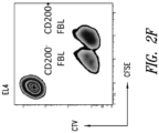
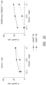
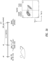


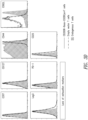
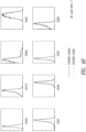

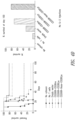

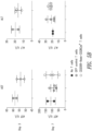
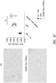
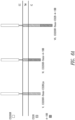

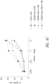
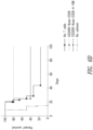
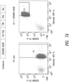
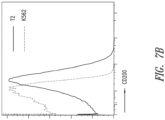
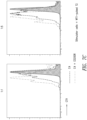
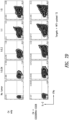


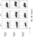
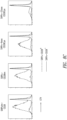
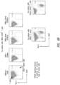
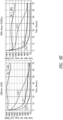
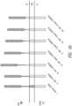
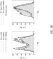


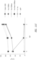
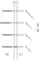


Classifications
-
- C—CHEMISTRY; METALLURGY
- C07—ORGANIC CHEMISTRY
- C07K—PEPTIDES
- C07K14/00—Peptides having more than 20 amino acids; Gastrins; Somatostatins; Melanotropins; Derivatives thereof
- C07K14/435—Peptides having more than 20 amino acids; Gastrins; Somatostatins; Melanotropins; Derivatives thereof from animals; from humans
- C07K14/705—Receptors; Cell surface antigens; Cell surface determinants
- C07K14/70596—Molecules with a "CD"-designation not provided for elsewhere
-
- C—CHEMISTRY; METALLURGY
- C12—BIOCHEMISTRY; BEER; SPIRITS; WINE; VINEGAR; MICROBIOLOGY; ENZYMOLOGY; MUTATION OR GENETIC ENGINEERING
- C12N—MICROORGANISMS OR ENZYMES; COMPOSITIONS THEREOF; PROPAGATING, PRESERVING, OR MAINTAINING MICROORGANISMS; MUTATION OR GENETIC ENGINEERING; CULTURE MEDIA
- C12N5/00—Undifferentiated human, animal or plant cells, e.g. cell lines; Tissues; Cultivation or maintenance thereof; Culture media therefor
- C12N5/06—Animal cells or tissues; Human cells or tissues
- C12N5/0602—Vertebrate cells
- C12N5/0634—Cells from the blood or the immune system
- C12N5/0636—T lymphocytes
-
- C—CHEMISTRY; METALLURGY
- C07—ORGANIC CHEMISTRY
- C07K—PEPTIDES
- C07K14/00—Peptides having more than 20 amino acids; Gastrins; Somatostatins; Melanotropins; Derivatives thereof
- C07K14/435—Peptides having more than 20 amino acids; Gastrins; Somatostatins; Melanotropins; Derivatives thereof from animals; from humans
- C07K14/705—Receptors; Cell surface antigens; Cell surface determinants
- C07K14/70503—Immunoglobulin superfamily
- C07K14/70517—CD8
-
- A—HUMAN NECESSITIES
- A61—MEDICAL OR VETERINARY SCIENCE; HYGIENE
- A61K—PREPARATIONS FOR MEDICAL, DENTAL OR TOILETRY PURPOSES
- A61K35/00—Medicinal preparations containing materials or reaction products thereof with undetermined constitution
- A61K35/12—Materials from mammals; Compositions comprising non-specified tissues or cells; Compositions comprising non-embryonic stem cells; Genetically modified cells
- A61K35/14—Blood; Artificial blood
- A61K35/17—Lymphocytes; B-cells; T-cells; Natural killer cells; Interferon-activated or cytokine-activated lymphocytes
-
- A—HUMAN NECESSITIES
- A61—MEDICAL OR VETERINARY SCIENCE; HYGIENE
- A61K—PREPARATIONS FOR MEDICAL, DENTAL OR TOILETRY PURPOSES
- A61K39/00—Medicinal preparations containing antigens or antibodies
- A61K39/46—Cellular immunotherapy
- A61K39/461—Cellular immunotherapy characterised by the cell type used
- A61K39/4611—T-cells, e.g. tumor infiltrating lymphocytes [TIL], lymphokine-activated killer cells [LAK] or regulatory T cells [Treg]
-
- A—HUMAN NECESSITIES
- A61—MEDICAL OR VETERINARY SCIENCE; HYGIENE
- A61K—PREPARATIONS FOR MEDICAL, DENTAL OR TOILETRY PURPOSES
- A61K39/00—Medicinal preparations containing antigens or antibodies
- A61K39/46—Cellular immunotherapy
- A61K39/463—Cellular immunotherapy characterised by recombinant expression
- A61K39/4632—T-cell receptors [TCR]; antibody T-cell receptor constructs
-
- A—HUMAN NECESSITIES
- A61—MEDICAL OR VETERINARY SCIENCE; HYGIENE
- A61K—PREPARATIONS FOR MEDICAL, DENTAL OR TOILETRY PURPOSES
- A61K39/00—Medicinal preparations containing antigens or antibodies
- A61K39/46—Cellular immunotherapy
- A61K39/464—Cellular immunotherapy characterised by the antigen targeted or presented
- A61K39/4643—Vertebrate antigens
- A61K39/4644—Cancer antigens
- A61K39/464402—Receptors, cell surface antigens or cell surface determinants
-
- A—HUMAN NECESSITIES
- A61—MEDICAL OR VETERINARY SCIENCE; HYGIENE
- A61K—PREPARATIONS FOR MEDICAL, DENTAL OR TOILETRY PURPOSES
- A61K39/00—Medicinal preparations containing antigens or antibodies
- A61K39/46—Cellular immunotherapy
- A61K39/464—Cellular immunotherapy characterised by the antigen targeted or presented
- A61K39/4643—Vertebrate antigens
- A61K39/4644—Cancer antigens
- A61K39/464452—Transcription factors, e.g. SOX or c-MYC
- A61K39/464453—Wilms tumor 1 [WT1]
-
- A—HUMAN NECESSITIES
- A61—MEDICAL OR VETERINARY SCIENCE; HYGIENE
- A61K—PREPARATIONS FOR MEDICAL, DENTAL OR TOILETRY PURPOSES
- A61K48/00—Medicinal preparations containing genetic material which is inserted into cells of the living body to treat genetic diseases; Gene therapy
-
- A—HUMAN NECESSITIES
- A61—MEDICAL OR VETERINARY SCIENCE; HYGIENE
- A61P—SPECIFIC THERAPEUTIC ACTIVITY OF CHEMICAL COMPOUNDS OR MEDICINAL PREPARATIONS
- A61P31/00—Antiinfectives, i.e. antibiotics, antiseptics, chemotherapeutics
- A61P31/04—Antibacterial agents
-
- A—HUMAN NECESSITIES
- A61—MEDICAL OR VETERINARY SCIENCE; HYGIENE
- A61P—SPECIFIC THERAPEUTIC ACTIVITY OF CHEMICAL COMPOUNDS OR MEDICINAL PREPARATIONS
- A61P31/00—Antiinfectives, i.e. antibiotics, antiseptics, chemotherapeutics
- A61P31/12—Antivirals
-
- A—HUMAN NECESSITIES
- A61—MEDICAL OR VETERINARY SCIENCE; HYGIENE
- A61P—SPECIFIC THERAPEUTIC ACTIVITY OF CHEMICAL COMPOUNDS OR MEDICINAL PREPARATIONS
- A61P35/00—Antineoplastic agents
-
- A—HUMAN NECESSITIES
- A61—MEDICAL OR VETERINARY SCIENCE; HYGIENE
- A61P—SPECIFIC THERAPEUTIC ACTIVITY OF CHEMICAL COMPOUNDS OR MEDICINAL PREPARATIONS
- A61P37/00—Drugs for immunological or allergic disorders
- A61P37/02—Immunomodulators
-
- A—HUMAN NECESSITIES
- A61—MEDICAL OR VETERINARY SCIENCE; HYGIENE
- A61P—SPECIFIC THERAPEUTIC ACTIVITY OF CHEMICAL COMPOUNDS OR MEDICINAL PREPARATIONS
- A61P37/00—Drugs for immunological or allergic disorders
- A61P37/02—Immunomodulators
- A61P37/06—Immunosuppressants, e.g. drugs for graft rejection
-
- C—CHEMISTRY; METALLURGY
- C07—ORGANIC CHEMISTRY
- C07K—PEPTIDES
- C07K14/00—Peptides having more than 20 amino acids; Gastrins; Somatostatins; Melanotropins; Derivatives thereof
- C07K14/435—Peptides having more than 20 amino acids; Gastrins; Somatostatins; Melanotropins; Derivatives thereof from animals; from humans
- C07K14/705—Receptors; Cell surface antigens; Cell surface determinants
- C07K14/70503—Immunoglobulin superfamily
- C07K14/70507—CD2
-
- C—CHEMISTRY; METALLURGY
- C07—ORGANIC CHEMISTRY
- C07K—PEPTIDES
- C07K14/00—Peptides having more than 20 amino acids; Gastrins; Somatostatins; Melanotropins; Derivatives thereof
- C07K14/435—Peptides having more than 20 amino acids; Gastrins; Somatostatins; Melanotropins; Derivatives thereof from animals; from humans
- C07K14/705—Receptors; Cell surface antigens; Cell surface determinants
- C07K14/70503—Immunoglobulin superfamily
- C07K14/7051—T-cell receptor (TcR)-CD3 complex
-
- C—CHEMISTRY; METALLURGY
- C07—ORGANIC CHEMISTRY
- C07K—PEPTIDES
- C07K14/00—Peptides having more than 20 amino acids; Gastrins; Somatostatins; Melanotropins; Derivatives thereof
- C07K14/435—Peptides having more than 20 amino acids; Gastrins; Somatostatins; Melanotropins; Derivatives thereof from animals; from humans
- C07K14/705—Receptors; Cell surface antigens; Cell surface determinants
- C07K14/70503—Immunoglobulin superfamily
- C07K14/70521—CD28, CD152
-
- C—CHEMISTRY; METALLURGY
- C07—ORGANIC CHEMISTRY
- C07K—PEPTIDES
- C07K14/00—Peptides having more than 20 amino acids; Gastrins; Somatostatins; Melanotropins; Derivatives thereof
- C07K14/435—Peptides having more than 20 amino acids; Gastrins; Somatostatins; Melanotropins; Derivatives thereof from animals; from humans
- C07K14/705—Receptors; Cell surface antigens; Cell surface determinants
- C07K14/70578—NGF-receptor/TNF-receptor superfamily, e.g. CD27, CD30, CD40, CD95
-
- C—CHEMISTRY; METALLURGY
- C12—BIOCHEMISTRY; BEER; SPIRITS; WINE; VINEGAR; MICROBIOLOGY; ENZYMOLOGY; MUTATION OR GENETIC ENGINEERING
- C12N—MICROORGANISMS OR ENZYMES; COMPOSITIONS THEREOF; PROPAGATING, PRESERVING, OR MAINTAINING MICROORGANISMS; MUTATION OR GENETIC ENGINEERING; CULTURE MEDIA
- C12N5/00—Undifferentiated human, animal or plant cells, e.g. cell lines; Tissues; Cultivation or maintenance thereof; Culture media therefor
- C12N5/06—Animal cells or tissues; Human cells or tissues
- C12N5/0602—Vertebrate cells
- C12N5/0634—Cells from the blood or the immune system
- C12N5/0636—T lymphocytes
- C12N5/0637—Immunosuppressive T lymphocytes, e.g. regulatory T cells or Treg
-
- C—CHEMISTRY; METALLURGY
- C12—BIOCHEMISTRY; BEER; SPIRITS; WINE; VINEGAR; MICROBIOLOGY; ENZYMOLOGY; MUTATION OR GENETIC ENGINEERING
- C12N—MICROORGANISMS OR ENZYMES; COMPOSITIONS THEREOF; PROPAGATING, PRESERVING, OR MAINTAINING MICROORGANISMS; MUTATION OR GENETIC ENGINEERING; CULTURE MEDIA
- C12N5/00—Undifferentiated human, animal or plant cells, e.g. cell lines; Tissues; Cultivation or maintenance thereof; Culture media therefor
- C12N5/06—Animal cells or tissues; Human cells or tissues
- C12N5/0602—Vertebrate cells
- C12N5/0634—Cells from the blood or the immune system
- C12N5/0636—T lymphocytes
- C12N5/0638—Cytotoxic T lymphocytes [CTL] or lymphokine activated killer cells [LAK]
-
- A—HUMAN NECESSITIES
- A61—MEDICAL OR VETERINARY SCIENCE; HYGIENE
- A61K—PREPARATIONS FOR MEDICAL, DENTAL OR TOILETRY PURPOSES
- A61K38/00—Medicinal preparations containing peptides
-
- C—CHEMISTRY; METALLURGY
- C07—ORGANIC CHEMISTRY
- C07K—PEPTIDES
- C07K2319/00—Fusion polypeptide
- C07K2319/01—Fusion polypeptide containing a localisation/targetting motif
- C07K2319/03—Fusion polypeptide containing a localisation/targetting motif containing a transmembrane segment
-
- C—CHEMISTRY; METALLURGY
- C07—ORGANIC CHEMISTRY
- C07K—PEPTIDES
- C07K2319/00—Fusion polypeptide
- C07K2319/70—Fusion polypeptide containing domain for protein-protein interaction
- C07K2319/74—Fusion polypeptide containing domain for protein-protein interaction containing a fusion for binding to a cell surface receptor
-
- C—CHEMISTRY; METALLURGY
- C12—BIOCHEMISTRY; BEER; SPIRITS; WINE; VINEGAR; MICROBIOLOGY; ENZYMOLOGY; MUTATION OR GENETIC ENGINEERING
- C12N—MICROORGANISMS OR ENZYMES; COMPOSITIONS THEREOF; PROPAGATING, PRESERVING, OR MAINTAINING MICROORGANISMS; MUTATION OR GENETIC ENGINEERING; CULTURE MEDIA
- C12N2510/00—Genetically modified cells
Definitions
- T cell-based immunotherapies began to be developed when tumor-reactive T cells were found among a population of tumor-infiltrating lymphocytes (TILs) ( Clark et al., Cancer Res. 29:705, 1969 ).
- TILs tumor-infiltrating lymphocytes
- One strategy known as adoptive T cell transfer, in some contexts involves the isolation of tumor infiltrating lymphocytes pre-selected for tumor-reactivity, clonal expansion of the tumor-reactive T cells induced by anti-CD3 and anti-CD28 antibodies in the presence of IL-2, and finally infusing the expanded cell population back to the tumor-bearing patient (together with chemotherapy and repetitive administration of IL-2) ( Dudley et al., Science 298:850, 2002 ).
- TCRs recombinant T cell receptors
- MHC major histocompatibility complex
- HLA human leukocyte antigen
- the basic procedure for engineered TCR or CAR T cell immunotherapy is generally to genetically modify human T cells with a transgene encoding a tumor targeting moiety, ex vivo expansion of the recombinant T cells, and transfusing the expanded recombinant T cells back into patients.
- Adoptive T cell therapy using T cells expressing recombinant TCRs has been shown to have a promising clinical benefit, especially in certain B cell cancers.
- effective T cell activation often requires or is enhanced by a concurrent co-stimulatory signal ( Chen and Flies, Nat. Rev. Immunol. 13: 227-242, 2013 ).
- co-stimulatory molecules are generally downregulated.
- exogenous stimulus via IL-2 is typically needed for T cells that express recombinant TCRs specific for cancer antigens.
- T cells Activation of T cells is initiated when the TCR engages a specific peptide presented in MHC on an antigen-presenting cell (APC) ( Rossy et al., Frontiers in Immunol. 3: 1-12, 2012 ).
- APC antigen-presenting cell
- the point of interaction of the T cell and the APC becomes the immunological synapse, which is comprised of three concentric supramolecular activation clusters (SMACs), including the central cSMAC, peripheral pSMAC, and the distal dSMAC ( Rossy et al., Frontiers in Immunol. 3: 1-12, 2012 ).
- SMACs concentric supramolecular activation clusters
- co-stimulatory receptors can recruit signaling molecules to amplify the TCR signal.
- Such co-stimulatory receptors can include CD28, and in some contexts form microclusters with the TCR to lower the threshold of activation ( Chen and Flies, Nat. Rev. Immunol. 13: 227-242, 2013 ).
- Access to the cSMAC by transmembrane proteins expressed by T cells may be restricted by the size of the extracellular domain.
- CD45 has a large ectodomain and is generally excluded from the immunological synapse, thereby preventing its ability to inhibit TCR signaling ( James and Vale, Nature 487:64-69, 2012 ).
- WO 2013/123061 A1 discloses a bispecific chimeric antigen receptor comprising at least two antigen-specific targeting regions, an extracellular spacer domain, a transmembrane domain, at least one co-stimulatory domain and an intracellular signaling domain, wherein each antigen-specific targeting region comprises an antigen-specific single chain Fv (scFv) fragment, and binds a different antigen, and wherein the bispecific chimeric antigen receptor is co-expressed with a therapeutic control.
- scFv antigen-specific single chain Fv
- the length of a fusion protein::target complex spans a distance similar to a distance between membranes in an immunological synapse.
- a length or spatial distance of a complex formed between the fusion protein and the target or a portion of such fusion protein::target complex is or spans a particular distance, e.g. , in some embodiments, is a distance that is less than or less than about a certain distance. In some aspects, a distance of the fusion protein::target complex (or, typically, the extracellular portion thereof) is less than at or about 50 nm, less than at or about 40 nm, less than at or about 30 nm, or less than at or about 20 nm or equal to or less than at or about 15 nm.
- the distance is one that is similar to a distance between membranes in an immunological synapse or is a distance that is the same, about the same, or substantially the same, as a distance between the membrane proximal-most portion, e.g. , residue, of the extracellular domain of a TCR and the membrane proximal-most portion, e.g. , residue, of an MHC ( e.g.
- HLA such as an MHCI or MHCII
- spatial distances of complexes refer to a distance between membranes of two different cells, wherein a first cell and a second cell each express on their surface a binding partner that can form a complex between the membranes when the cells are in proximity to each other.
- the distance is a distance that is the same, about the same, or substantially the same, as a distance spanned by the extracellular portions of a complex formed between a TCR and cognate interaction with an MHC molecule.
- the distance is similar to or the same as that spanned by a complex formed between the molecule (having the binding domain used in the fusion protein), and a natural binding partner thereof.
- the distance is different than, e.g. , less than or substantially less than, that spanned by a complex formed between the molecule (having the binding domain or functional portion thereof used in the fusion protein), and a natural binding partner thereof.
- a fusion protein does not include a primary signaling domain such as a CD3 ⁇ signaling domain or other domain capable of delivering a primary signal to a T cell.
- the extracellular component further includes one or more additional regions or domains.
- the one or more additional extracellular domain(s) may include a spacer region, such as one from an immunoglobulin molecule, which may contain all or a portion of a hinge, or constant region domain such as CH2 or CH3 domain, or from another cell surface molecule such as a costimulatory receptor, such as CD28.
- the additional extracellular domain(s) may include, in some aspects, a multimerization domain, e.g., a dimerization domain or sequence that may promote homo- or heterodimerization with another molecule, such as multimerization of two or more of the fusion proteins.
- such a domain includes a portion of an extracellular domain of a CD28 molecule including at least the transmembrane-proximal-most cysteine, and generally an extracellular portion between such cysteine and the membrane, or modified variant thereof.
- such a domain includes an amino acid sequence as set forth in SEQ ID NO: 32, or portion thereof, or variant thereof such as having at least 90%, 95%, or 99 % identity thereto.
- such a domain may be included in order to facilitate or promote multimerization.
- a fusion protein contains an extracellular portion of CD200R having an amino acid sequence as set forth in SEQ ID NO: 25 or encoded by a nucleic acid molecule as set forth in SEQ ID NO: 2, or a CD200-binding portion thereof or variant thereof or binding portion thereof.
- the extracellular portion of the fusion protein further includes a portion of an extracellular region of CD28, such as up to about 9 to about 12 amino acids thereof ( e.g. , 9 amino acids or 12 amino acids), and in some aspects including a membrane-proximal-most cysteine residue of a CD28 extracellular region.
- the length of the CD200R portion of the extracellular region is reduced in length corresponding to the number of additional residues in the CD28-derived portion, such as by about 9 to about 12 amino acids (e.g. , 9 amino acids or 12 amino acids), or by a sufficient number of amino acids that the distance spanned by the extracellular portion of a complex between the fusion protein and a CD200 molecule is similar to, substantially similar to, or the same as that spanned by the extracellular portion of a complex between a human CD200R, e.g. , a CD200R, and CD200; or that spanned by the extracellular portion of a complex between a TCR in cognate interaction with an MHC molecule (e.g.
- the fusion protein further includes a transmembrane domain, such as a CD28 transmembrane, such as a transmembrane domain encoded by the sequence set forth as SEQ ID NO: 4 or portion thereof, or a modified version thereof, such as a variant modified to contain additional charged regions or residues or hydrophilic residues to facilitate intermolecular interactions.
- the CD28 intracellular signaling domain is capable of recruiting one or more adapter molecules to a CD28 in response to ligation.
- the CD28 intracellular domain includes or is a sequence encoded by the nucleotide sequence of SEQ ID NO: 5 or a portion or functional variant thereof.
- the present disclosure is directed to a fusion protein comprising an extracellular component comprised of a binding domain that specifically binds a target, an intracellular component comprised of an intracellular signaling domain, and a hydrophobic component connecting the extracellular and intracellular components, provided that the length of a fusion protein:target complex spans a distance similar to a distance between membranes in an immunological synapse, wherein (a) the extracellular component comprises an extracellular portion of a CD200R, (b) the hydrophobic component comprises a transmembrane domain of a CD28, and (c) the intracellular component comprises an intracellular signaling domain of a CD28.
- the present disclosure is directed to a fusion protein comprising an extracellular component comprised of a binding domain that specifically binds a target, an intracellular component comprised of an intracellular signaling domain, and a hydrophobic component connecting the extracellular and intracellular components, provided that the length of a fusion protein:target complex spans a distance similar to a distance between membranes in an immunological synapse, wherein (a) the extracellular component comprises an extracellular portion of a CD200R, (b) the hydrophobic component comprises a transmembrane domain of a CD28, and (c) the intracellular component comprises an intracellular signaling domain of a CD28 and an intracellular signaling domain of a CD137 (4-1BB).
- the present disclosure is directed to a nucleic acid molecule encoding a fusion protein as described herein.
- the present disclosure is directed to a vector comprising a nucleic molecule that encodes a fusion protein as described herein.
- the present disclosure is directed to a host cell comprising a fusion protein, nucleic acid, or vector as described herein.
- the present invention provides a host cell as described herein for use in a method of treating a disease.
- the method increases the activity of an immune cell and comprises administering to a subject in need of increased immune cell activity an effective amount of the host cell.
- the method enhances or prolongs an immune response and comprises administering to a subject in need of enhanced or prolonged immune cell activity an effective amount of the host cell.
- the method stimulates an antigen-specific T cell response and comprises administering to a subject in need of increased immune cell activity an effective amount of the host cell.
- the method inhibits an immunosuppressive signaling pathway and comprises administering to a subject in need thereof an effective amount of the host cell.
- the disease is cancer and the method comprises administering to a subject having cancer a therapeutically effective amount of the host cell.
- the method inhibits immune resistance of cancer cells and comprises administering to a subject in need thereof an effective amount of the host cell.
- the host cell as described herein is for use in a method for treating a tumor, comprising administering to a subject having a tumor a therapeutically effective amount of the host cell, wherein the administered host cell is capable of proliferating in an immunosuppressive tumor microenvironment.
- the host cell as described herein for use in a method of treating an infection, comprising administering to a subject having the infection a therapeutically effective amount of a host cell, is also provided by the present disclosure.
- Figures 1A and 1B show CD200R-CD28 constructs expressed at high levels on primary murine CD8 + T cells.
- A Schematic representation of exemplary CD200R-CD28 constructs.
- Construct “I” contains CD200R extracellular (“EC") and transmembrane (“TM”) domains and a CD28 intracellular (“IC") signaling domain (CD200Rtm-CD28).
- Construct “II” contains the extracellular domain of CD200R and the transmembrane and intracellular domains of CD28 (CD200R-CD28tm).
- Constructs "III-V” also incorporate a portion of the extracellular domain of CD28 to the transmembrane-proximal cysteine to promote multimerization and enhance CD28 signaling.
- constructs have a truncated portion of an extracellular or intracellular domain (e.g., a CD200R that preserves an N linked glycosylation site).
- construct IV has a truncated portion of CD200R that is truncated by 3 amino acids.
- Construct V has a truncated portion of CD200R that is truncated 9 amino acids. Constructs "I”, “II”, and “V” maintain the short spatial distance between the cells (e.g.
- FIGS. 2A to 2G show that CD200R-CD28 constructs promote proliferation, accumulation, and effector function in response to CD200 + tumor target cells in vitro, and accumulate in the immunological synapse.
- Splenocytes from naive TCR gag mice were stimulated in vitro with anti-CD3, anti-CD28, and recombinant human IL-2 (100 U/ml) and transduced with retroviral supernatant for 2 days.
- Cells were restimulated every 7 days with irradiated FBL and splenocytes and cultured with rhlL-2 (50 U/mL) for up to three stimulations. T cells were used for assays 5-7 days after the last stimulation.
- A Proliferation of CD200R-CD28 and GFP control TCR gag T cells as measured by CellTrace Violet dilution. T cells were stimulated with CD200 - FBL (upper panels) or CD200 + FBL (lower panels) for 3 days.
- B Preferential expansion/survival of transduced TCR gag T cells during co-culture with non-transduced TCR gag T cells during weekly cycles of stimulation with irradiated CD200 + FBL and splenocytes.
- C Enrichment of transduced T cells.
- Target tumor cells were labeled with different dilutions of the fluorescent dye 5,6-carboxyfluorescein diacetate succinimidyl ester (CFSE), as indicated.
- Effector TCR gag T cells transduced with the indicated CD200R-CD28 fusion protein or an empty vector control were incubated at the indicated effector to target ratio with a 1:1 mix of CD200 + FBL (CFSE hi ) and non-specific EL4 (CFSE lo ) control targets for 5 hours.
- the percentage of FBL of the sum of FBL and control tumor cells was determined by flow cytometry. The percentage lysis was determined by dividing the percent of FBL incubated with T cells by the percent of FBL incubated without T cells.
- Target tumor cells were labeled with different dilutions of the fluorescent dyes CellTrace Violet (CTV) or CFSE.
- CTV+ CellTrace Violet
- CD200 + FBL CD200 + FBL
- CFSE lo non-specific EL4
- G CFSE cytotoxicity assay.
- TCR gag T cells were transduced with CD200R-CD28 receptor or GFP control vector. Effector TCR gag T cells were incubated at the indicated effector to target ratio with a 1:1 mix of CD200 - FBL or CD200 + FBL and non-specific EL4 control targets for 4 hours.
- the percentage of FBL of the sum of FBL and control tumor cells was determined by flow cytometry. The percentage lysis was determined by dividing the percent of FBL incubated with T cells by the percent of FBL incubated without T cells.
- FIGS 3A to 3D show that T cells transduced with CD200R-9aas-CD28Cys preferentially accumulate in response to tumor challenge in vivo and express surface proteins consistent with an effector phenotype after injection into Cytoxan-treated, FBL-bearing mice.
- Transduced TCR gag T cells were generated as described in Example 2.
- IL-2 was administered every 2 days (2 ⁇ 10 4 U/dose). On day 8 post-T cell transfer, mice were euthanized and spleens and inguinal lymph nodes harvested.
- C Comparison of surface proteins 3 days post-transfer for T cells transduced to express CD200R-9aas-CD28Cys, T cells transduced with an empty vector, and endogenous T cells.
- CD200R-9aas-CD28Cys TCR gag T cells expressed reduced CD62L compared to control TCR gag T cells, suggesting an effector T cell phenotype.
- D Comparison of surface proteins 15 days post-transfer for cells transduced to express CD200R -9aas-CD28Cys + T cells, T cells transduced with an empty vector, and endogenous T cells. CD200R-9aas-CD28Cys TCR gag T cells express similar levels of cell surface proteins compared to control TCR gag T cells.
- Figures 4A to 4D show that adoptive immunotherapy with CD200R-CD28-transduced T cells can eradicate disseminated leukemia.
- A Experiment schematic. C57BL/6 mice were injected with 4 ⁇ 10 6 CD200 + FBL cells. Five days later, CD200R-CD28tm, CD200R-CD28Cys, CD200R-9aas-CD28Cys, or eGFP TCR gag T cells were injected i.p. into Cy-treated FBL-bearing mice at 10 5 cells/mouse. IL-2 was administered every 2 days (2 ⁇ 10 4 U/dose) in a cohort of mice as indicated.
- FIG. 1 Representative example of expression of cell surface proteins in CD200R-CD28tm transduced T cells and non-transduced T cells on day of injection with IL-2, as determined by flow cytometry.
- C Survival of mice treated in the presence of IL-2 injections.
- D Survival of mice treated in the absence of IL-2 injections. Transfer of CD200R-9aas-CD28Cys TCR gag T cells significantly improved survival in the absence of IL-2 injections (p ⁇ 0.05, log-rank Mantel-Cox test).
- FIGS 5A to 5C show that T cells expressing CD200R-9aas-CD28Cys do not induce detectable autoimmune liver damage or infiltrate normal tissues.
- A Experiment schematic. Cytoxan-treated Alb/Gag mice were injected with 4 ⁇ 10 6 CD200 + FBL cells. Five days later, CD200R-9aas-CD28Cys, and eGFP TCR gag T cells were injected i.p. into the Cytoxan-treated FBL-bearing mice at 10 5 cells/mouse. IL-2 was administered every 2 days (2 ⁇ 10 4 U/dose) in a cohort of mice as indicated.
- liver damage was assessed by quantification of serum levels of liver enzymes aspartate aminotransferase (AST) and alanine aminotransferase (ALT).
- AST and ALT levels measured at 3 and 7 days post-transfer for mice receiving no T cells, control T cells expressing GFP, or T cells expressing CD200R-9aas-CD28Cys did not vary by treatment.
- C Assessment of T cell infiltration of normal tissue. Limited presence of T cells in liver tissue was observed using antibodies specific to the T cell marker CD3 (left panel), with no significant difference between recipients of CD200R-9aas-CD28Cys TCR gag or control TCR gag T cells (right panel).
- Figures 6A to 6D show that 4-1BB co-stimulatory signaling domains promote accumulation and effector function of transduced T cells in vitro and promote survival of tumor-bearing recipients of transduced T cell in response to CD200 + tumor target cells.
- A Schematic representation of CD200R-CD28 ("V"), -4-1BB ("VI”), and -CD28-4-1BB (“VII”) constructs.
- B Expansion of transduced TCR gag T cells relative to non-transduced TCR gag T cells after weekly stimulation with irradiated CD200 + FBL and splenocytes.
- CD200R-4-1BB and CD200R-CD28-4-1BB also promote accumulation of transduced T cells in vitro.
- CD200R-9aas-4-1BB + CD8 + T cells displayed an enhanced ability to lyse CD200 + FBL cells in vitro relative to controls, using a standard CFSE-based cytotoxicity assay. The percentage of FBL of the sum of FBL and control tumor cells was determined by flow cytometry. The percentage lysis was determined by dividing the percent of FBL incubated with T cells by the percent of FBL incubated without T cells.
- CD200R-41BB-transduced T cells also promote survival relative to controls. C57BL/6 mice were injected with 4 ⁇ 10 6 CD200 + FBL cells.
- CD200R-9aas-CD28, CD200R-9aas-4-1BB, CD200R-9aas-CD28-4-1BB, or eGFP TCR gag T cells were injected i.p. into Cytoxan-treated FBL-bearing mice at 10 5 cells/mouse.
- Figures 7A to 7D show that human primary T cells transduced to express a WT1-specific TCR and a CD200Rtm-CD28 fusion protein exhibit enhanced proliferation to target cells that express CD200 and increased cytokine production in response to tumor cells that express CD200.
- A Expression of the WT1 126 -specific TCR, C4, and CD200Rtm-CD28.
- B Expression of CD200 in T2 and K562 cells. T2 cells exhibit low-level endogenous CD200 expression.
- C Proliferation of T cells as indicated by CFSE. Cells that proliferate in response to antigen show reduced CFSE fluorescence intensity.
- T cells transduced with both C4 and the IFP show enhanced proliferation to target cells expressing low levels of CD200 relative to T cells transduced with C4 only.
- D Cytokine production in response to exposure to CD200dim tumor cells, as measured by flow cytometry. Relative to control T cells transduced with the TCR C4 alone, T cells transduced with both C4 and the IFP CD200Rtm-CD28 show increased cytokine production.
- FIGS 8A to 8E show that fusion proteins comprising SIRP ⁇ extracellular components and CD28 co-stimulatory signaling domains promote accumulation and proliferation of transduced T cells in vitro.
- Construct “I” contains SIRP ⁇ extracellular (“EC") and transmembrane (“TM”) domains and a CD28 intracellular (“IC") signaling domain (SIRP ⁇ tm-CD28).
- Construct “II” contains the extracellular domain of SIRP ⁇ and the transmembrane and intracellular domains of CD28 (SIRP ⁇ -CD28tm).
- Constructs "III-VI” also incorporate a portion of the extracellular domain of CD28 to the transmembrane-proximal cysteine to promote multimerization and enhance CD28 signaling.
- some constructs have a truncated portion of an extracellular or intracellular domain (e.g. , a SIRP ⁇ that preserves an N linked glycosylation site).
- Construct IV has a truncated portion of SIRP ⁇ that is truncated 6 amino acids to preserve an N linked glycosylation site.
- Construct V has a truncated portion of SIRP ⁇ that is truncated 9 amino acids.
- Construct VI has a truncated portion of SIRP ⁇ that is truncated 23 amino acids.
- Constructs "I”, “II”, and “V” maintain the short spatial distance between the cells ( e.g. , between a T cell and an antigen presenting cell) and may co-localize with the TCR within the cSMAC and deliver a strong co-stimulatory signal.
- B Expansion of transduced TCR gag T cells relative to non-transduced TCR gag T cells after weekly stimulation with irradiated SIRP ⁇ + FBL and splenocytes.
- SIRP ⁇ -CD28 constructs promote accumulation of transduced T cells in vitro, with SIRP ⁇ -9aas-CD28Cys exhibiting enhanced accumulation.
- C Proliferation of T cells transduced with SIRP ⁇ -CD28 constructs in a CellTrace Violet (CTV) dilution proliferation assay. T cells expressing SIRP ⁇ -CD28 constructs engineered to maintain T cell-tumor cell distance exhibited enhanced proliferation relative to nontransduced T cells.
- CD47 + tumor cells were killed after co-culture with SIRP ⁇ -CD28 + T cells transduced to express SIRP ⁇ tm-CD28 or SIRP ⁇ -9aas-CD28Cys constructs.
- Figures 9A and 9B show that fusion proteins comprising PD-1 extracellular components and CD28 co-stimulatory signaling domains promote cytokine production in vitro.
- Construct “I” contains PD-1 extracellular (“EC”) and transmembrane (“TM”) domains and a CD28 intracellular (“IC”) signaling domain (PD1tm-CD28).
- Construct “II” contains the extracellular domain of PD-1 and the transmembrane and intracellular domains of CD28 (PD1-CD28tm).
- Constructs "III-VII” also incorporate a portion of the extracellular domain of CD28 adjacent to the transmembrane-proximal cysteine to promote multimerization and enhance CD28 signaling.
- constructs IV-VII have a truncated portion of PD-1.
- Construct IV has a truncated portion of PD-1 that is truncated 9 amino acids.
- Construct V has a truncated portion of PD-1 that is truncated 12 amino acids.
- Construct VI has a truncated portion of PD-1 that is truncated 15 amino acids.
- Construct VII has a truncated portion of PD-1 that is truncated 21 amino acids.
- Constructs "I”, “II”, and “V” maintain the short spatial distance between the cells (e.g. , between a T cell and an antigen presenting cell) and may co-localize with the TCR within the cSMAC and deliver a strong co-stimulatory signal.
- (B) PD1-CD28 + T cells exhibited increased cytokine production in response to stimulation for 5 hours in the presence of Brefeldin A with FBL cells that endogenously express the PD-1 ligands, PD-L1 and PD-L2. Stimulated T cells were assessed for intracellular expression of the effector cytokines, IFNy and TNFa, by flow cytometry.
- FIG. 10 shows co-expression of the TCR C4 and a PD-1 IFP (PD1-12aas-CD28Cys, PD1-1Saas-CD28Cys, or PD1-21aas-CD28Cys).
- T cells transduced with C4 and PD1-12aas-CD28Cys or PD1-15aas-CD28Cys exhibited high transduction efficiencies and expression of both proteins.
- FIGS 11A to 11C show that fusion proteins comprising Fas extracellular components and CD28 co-stimulatory signaling domains accumulate in vitro upon stimulation with irradiated FBL cells.
- A Schematic representation of exemplary Fas-CD28 constructs.
- Construct “I” contains Fas extracellular (“EC") and transmembrane (“TM”) domains and a CD28 intracellular (“IC”) signaling domain (Fastm-CD28).
- Construct “II” contains the extracellular domain of Fas and the transmembrane and intracellular domains of CD28 (Fas-CD28tm).
- Constructs "III” and “IV” also incorporate a portion of the extracellular domain of CD28 adjacent to the transmembrane-proximal cysteine to promote multimerization and enhance CD28 signaling.
- construct IV has a truncated portion of Fas, wherein the Fas extracellular domain is truncated 9 amino acids.
- Constructs "I”, “II”, and “IV” maintain the short spatial distance between the cells (e.g.
- TCR gag T cells transduced with Fas constructs over multiple stimulations with irradiated FBL cells. All of the constructs promoted accumulation of T cells relative to control T cells.
- C Expression of Fas-CD28 constructs but not full-length (FL) Fas promoted survival or expansion of T cells upon multiple stimulations in vitro.
- Figures 12A and 12B show the structure and expression of fusion proteins comprising LAG3 extracellular components and CD28 co-stimulatory signaling domains.
- Constructs "III” and “IV” also incorporate a portion of the extracellular domain of CD28 adjacent to the transmembrane-proximal cysteine to promote multimerization and enhance CD28 signaling.
- construct IV has a truncated portion of LAG3, wherein the LAG3 extracellular domain is truncated 9 amino acids.
- LAG3-CD28 constructs by murine CD8 + T cells, as determined by anti-LAG3 antibody staining and flow cytometry.
- T cells transduced to express LAG3-CD28 constructs (LAG3tm-CD28; LAG3-CD28tm; LAG3-CD28Cys; LAG3-9aas-CD28Cys) exhibited expression of the constructs, in contrast with control T cells that received empty vector.
- FIGS 13A and 13B show the structure and expression of fusion proteins comprising TIM3 extracellular components and CD28 co-stimulatory signaling domains.
- A Schematic representation of exemplary TIM3-CD28 constructs.
- Construct “I” contains TIM3 extracellular (“EC") and transmembrane (“TM”) domains and a CD28 intracellular (“IC") signaling domain (TIM3tm-CD28).
- Construct “II” contains the extracellular domain of TIM3 and the transmembrane and intracellular domains of CD28 (TIM3-CD28tm).
- Constructs "III” and “IV” also incorporate a portion of the extracellular domain of CD28 adjacent to the transmembrane-proximal cysteine to promote multimerization and enhance CD28 signaling.
- construct IV has a truncated portion of TIM3, wherein the TIM3 extracellular domain is truncated 9 amino acids.
- Constructs "I”, “II”, and “IV” maintain the short spatial distance between the cells (e.g. , between a T cell and an antigen presenting cell) and may co-localize with the TCR within the cSMAC and deliver a strong co-stimulatory signal.
- B Expression of TIM3-CD28 constructs by murine CD8 + T cells, as determined by anti-TIM3 antibody staining and flow cytometry.
- T cells transduced to express TIM3-CD28 constructs typically exhibited expression of the constructs, in contrast with control T cells that received empty vector.
- fusion proteins of this disclosure can provide an activation or co-stimulatory signal in a human T cell, wherein the T cell may optionally be engineered to have a preferred antigen-specific TCR.
- These immunomodulatory fusion proteins can interact with ubiquitously expressed targets or with targets that are commonly upregulated or overexpressed in non-normal cells ( e.g. , a cancer cell).
- IFPs have an extracellular binding domain and an intracellular signaling domain.
- the present disclosure provides host cells (e.g. , immune cells such as T cells, dendritic cells, NK cells or the like) comprising an IFP, vectors encoding IFPs, and methods of activating T cells comprising an IFP for various therapeutic applications, including the treatment of a disease in subject (e.g. , cancer, infectious disease).
- host cells e.g. , immune cells such as T cells, dendritic cells, NK cells or the like
- vectors encoding IFPs e.g. , vectors encoding IFPs
- methods of activating T cells comprising an IFP for various therapeutic applications, including the treatment of a disease in subject (e.g. , cancer, infectious disease).
- any concentration range, percentage range, ratio range, or integer range is to be understood to include the value of any integer within the recited range and, when appropriate, fractions thereof (such as one tenth and one hundredth of an integer), unless otherwise indicated.
- any number range recited herein relating to any physical feature, such as polymer subunits, size or thickness are to be understood to include any integer within the recited range, unless otherwise indicated.
- the term “about” means ⁇ 20% of the indicated range, value, or structure, unless otherwise indicated. It should be understood that the terms “a” and “an” as used herein refer to “one or more" of the enumerated components. The use of the alternative (e.g.
- a protein domain, region, or module e.g. , a binding domain, hinge region, linker module
- a protein which may have one or more domains, regions, or modules
- consists essentially of a particular amino acid sequence when the amino acid sequence of a domain, region, or module or protein includes extensions, deletions, mutations, or any combination thereof (e.g. , amino acids at the amino- or carboxy-terminus or between domains) that, in combination, contribute to at most 20% (e.g.
- heterologous or non-endogenous or exogenous refers to any gene, protein, compound, molecule, or activity that is not native to a host cell or a subject, or is any gene, protein, compound, molecule, or activity native to a host or host cell that has been altered or mutated such that the structure, activity or both is different as between the native and mutated molecules.
- heterologous, non-endogenous or exogenous molecules e.g.
- receptors, ligands may not be endogenous to a host cell or subject, but instead nucleic acids encoding such molecules may have been added to a host cell by conjugation, transformation, transfection, electroporation, or the like, wherein the added nucleic acid molecule may integrate into a host cell genome or can exist as extra-chromosomal genetic material ( e.g. , as a plasmid or other self-replicating vector).
- the term "homologous” or “homolog” refers to a molecule or activity found in or derived from a host cell, species, or strain.
- a heterologous or exogenous molecule or gene encoding the molecule may be homologous to a native host or host cell molecule or gene that encodes the molecule, respectively, but may have an altered structure, sequence, expression level or combinations thereof.
- a non-endogenous molecule may be from the same species, a different species, or a combination thereof.
- endogenous or “native” refers to a gene, protein, compound, molecule, or activity that is normally present in a host or host cell and has no engineered alterations.
- binding domain refers to a molecule, such as a peptide, oligopeptide, polypeptide or protein, that possesses the ability to specifically and non-covalently associate, unite, or combine with a target molecule (e.g. , CD200, CD47, CD19, CD20, CD22, ROR1, mesothelin, PD-L1, PD-L2, PSMA, WT-1, cyclin-A1).
- a binding domain includes any naturally occurring, synthetic, semisynthetic, or recombinantly produced binding partner for a biological molecule or other target of interest or binding protein thereof.
- binding domains include receptor ectodomains (e.g. , those of CD200R, PD-1, CTLA4, BTLA, CD2, Fas) or binding portions thereof, ligands (e.g. , cytokines such as IL35, chemokines) or binding portions thereof, single chain antibody variable regions (e.g. , domain antibodies, sFv, scFv, Fab) or binding portions thereof, antigen-binding regions of T cell receptors (TCRs), such as single chain TCRs (scTCRs), or synthetic polypeptides selected for the specific ability to bind to a biological molecule.
- receptor ectodomains e.g. , those of CD200R, PD-1, CTLA4, BTLA, CD2, Fas
- ligands e.g. , cytokines such as IL35, chemokines
- single chain antibody variable regions e.g. , domain antibodies, sFv, scFv,
- binding domains refers to an association or union of a binding domain, or a fusion protein thereof, to a target molecule with an affinity or K a (i.e. , an equilibrium association constant of a particular binding interaction with units of 1/M) equal to or greater than 10 5 M -1 , or binds to such target molecule while not significantly associating or uniting with any other molecules or components in a sample.
- Binding domains (or fusion proteins thereof) may be classified as "high affinity” binding domains (or fusion proteins thereof) or "low affinity” binding domains (or fusion proteins thereof).
- “High affinity” binding domains refer to those binding domains with a K a of at least 10 7 M -1 , at least 10 8 M -1 , at least 10 9 M -1 , at least 10 10 M -1 , at least 10 11 M -1 , at least 10 12 M -1 , or at least 10 13 M -1 .
- “Low affinity” binding domains refer to those binding domains with a K a of up to 10 7 M -1 , up to 10 6 M -1 , up to 10 5 M -1 .
- affinity may be defined as an equilibrium dissociation constant (K d ) of a particular binding interaction with units of M (e.g., 10 -5 M to 10 -13 M).
- a binding domain may have "enhanced affinity," which refers to a selected or engineered binding domain with stronger binding to a target antigen than a wild type (or parent) binding domain.
- enhanced affinity may be due to a Ka (equilibrium association constant) for the target antigen that is higher than the wild type binding domain, or due to a K d (dissociation constant) for the target antigen that is less than that of the wild type binding domain, or due to an off-rate (K off ) for the target antigen that is less than that of the wild type binding domain.
- binding domains of the present disclosure that specifically bind a particular target, as well as determining binding domain or fusion protein affinities, such as Western blot, ELISA, and Biacore ® analysis (see also, e.g., Scatchard et al., Ann. N.Y. Acad. Sci. 51:660, 1949 ; and U.S. Patent Nos. 5,283,173 , 5,468,614 , or the equivalent).
- a "fusion protein” refers to a polypeptide that, in a single chain, has at least two distinct domains, wherein the domains are not naturally found together in a protein.
- a nucleic acid molecule encoding a fusion protein may be constructed using PCR, recombinantly engineered, or the like, or such fusion proteins can be made using methods of protein synthesis.
- a fusion protein may further contain other components (e.g., covalently bound), such as a tag or bioactive molecule.
- a fusion protein expressed or produced by a host cell e.g.
- T cell locates to the cell surface, where the fusion protein is anchored to the cell membrane with a portion of the fusion protein located extracellularly (e.g. , containing a binding domain) and a portion of the fusion protein located intracellularly ( e.g. , containing a signaling domain).
- hydrophobic component means any amino acid sequence having a three-dimensional structure that is thermodynamically stable in a cell membrane, and generally ranges in length from about 15 amino acids to about 30 amino acids.
- the structure of a hydrophobic component may comprise an alpha helix, a beta barrel, a beta sheet, a beta helix, or any combination thereof.
- a hydrophobic component is comprised of a "transmembrane domain" from a known transmembrane protein, which is a portion of a transmembrane protein that can insert into or span a cell membrane. Additionally, the hydrophobic component may be modified to contain charged regions or hydrophilic residues to facilitate intermolecular interactions.
- an "intracellular signaling domain” is an intracellular portion of molecule, such as one used in a fusion protein of this disclosure, that can directly or indirectly promote a response such as a co-stimulatory, positive, or activating biological or physiological response in a cell when receiving the appropriate signal.
- An intracellular signaling domain may directly promote a cellular response when it contains one or more signaling domains or motifs, such as an immunoreceptor tyrosine-based activation motif (ITAM), a kinase domain, a co-stimulatory domain, or the like.
- An intracellular signaling domain may indirectly promote a cellular response by associating with one or more other proteins that in turn directly promote a cellular response.
- An intracellular signaling domain or functional fragment thereof may be from a CD3 ⁇ , CD3 ⁇ , CD3 ⁇ , CD25, CD27, CD28, CD40, CD47, CD79A, CD79B, CD134 (OX40), CD137 (4 1BB), CD150 (SLAMF1), CD278 (ICOS), CD357 (GITR), CARD11, DAP10, DAP12, FcRa, FcR ⁇ , FcRy, Fyn, Lck, LAT, LRP, NKG2D, NOTCH1, NOTCH2, NOTCH3, NOTCH4, ROR2, Ryk, Slp76, pTa, TCR ⁇ , TCR ⁇ , TRIM, Zap70, PTCH2, or any combination thereof.
- the intracellular signaling domain comprises an intracellular signaling domain of CD28.
- an intracellular signaling domain or functional fragment thereof does not comprise a CD3 ⁇ .
- a “multimerization domain,” as used herein, refers to a polypeptide molecule or region that preferentially interacts or associates with another polypeptide molecule or region, directly or indirectly, wherein the interaction of multimerization domains substantially contribute to or efficiently promote multimerization ( i.e. , the formation of a dimer, trimer, tetramer, or higher order multimers, which may be a homodimer, heterodimer, homotrimer, heterotrimer, homomultimer, heteromultimer, or the like).
- multimerization may be due to one or more types of molecular forces, including covalent bonds (e.g.
- a multimer is stable under appropriate conditions (e.g. , physiological conditions, in an aqueous solution suitable for expressing, purifying, or storing recombinant or engineered proteins, or under conditions for non-denaturing or non-reducing electrophoresis).
- Exemplary multimerization domains may comprise one or more disulfide bonds, zinc finger motif, a leucine zipper motif, helix-turn-helix, helix-loop-helix, or the like.
- a fusion protein may contain a "linker,” which can provide a spacer function to facilitate the interaction of two single chain fusion proteins, or positioning of one or more binding domains, so that the resulting polypeptide structure maintains a specific binding affinity to a target molecule or maintains signaling activity (e.g. , effector domain activity) or both.
- linkers include from one to about ten repeats of Gly x Ser y , wherein x and y are independently an integer from 1 to 5.
- junction amino acids refer to one or more (e.g. , about 2-20) amino acid residues between two adjacent motifs, regions, or domains of a fusion protein, such as between a binding domain and an adjacent hydrophobic component, or on one or both ends of a hydrophobic component. Junction amino acids may result from the construct design of a fusion protein (e.g. , amino acid residues resulting from the use of a restriction enzyme site during the construction of a nucleic acid molecule encoding a fusion protein). In certain embodiments, junction amino acids form a linker, such as those having from one to about ten repeats of Gly x Ser y , wherein x and y are independently an integer from 1 to s.
- an “immune system cell” means any cell of the immune system that originates from a hematopoietic stem cell in the bone marrow, which gives rise to two major lineages, a myeloid progenitor cell (which give rise to myeloid cells such as monocytes, macrophages, dendritic cells, megakaryocytes and granulocytes) and a lymphoid progenitor cell (which give rise to lymphoid cells such as T cells, B cells and natural killer (NK) cells).
- myeloid progenitor cell which give rise to myeloid cells such as monocytes, macrophages, dendritic cells, megakaryocytes and granulocytes
- lymphoid progenitor cell which give rise to lymphoid cells such as T cells, B cells and natural killer (NK) cells).
- Exemplary immune system cells include a CD4+ T cell, a CD8+ T cell, a CD4- CD8- double negative T cell, a ⁇ T cell, a regulatory T cell, a natural killer cell, and a dendritic cell.
- Macrophages and dendritic cells may be referred to as "antigen presenting cells” or "APCs,” which are specialized cells that can activate T cells when a major histocompatibility complex (MHC) receptor on the surface of the APC complexed with a peptide interacts with a TCR on the surface of a T cell.
- MHC major histocompatibility complex
- T cell is an immune system cell that matures in the thymus and produces T cell receptors (TCRs).
- T cells can be na ⁇ ve (not exposed to antigen; increased expression of CD62L, CCR7, CD28, CD3, CD127, and CD45RA, and decreased expression of CD45RO as compared to T CM ), memory T cells (T M ) (antigen-experienced and long-lived), and effector cells (antigen-experienced, cytotoxic).
- T M can be further divided into subsets of central memory T cells (T CM , increased expression of CD62L, CCR7, CD28, CD127, CD45RO, and CD95, and decreased expression of CD54RA as compared to na ⁇ ve T cells) and effector memory T cells (T EM , decreased expression of CD62L, CCR7, CD28, CD45RA, and increased expression of CD127 as compared to na ⁇ ve T cells or T CM ).
- Effector T cells refers to antigen-experienced CD8+ cytotoxic T lymphocytes that have decreased expression of CD62L ,CCR7, CD28, and are positive for granzyme and perforin as compared to T CM .
- Other exemplary T cells include regulatory T cells, such as CD4+ CD25+ (Foxp3+) regulatory T cells and Treg17 cells, as well as Tr1, Th3, CD8+CD28-, and Qa-1 restricted T cells.
- T cell receptor refers to a molecule found on the surface of T cells (or T lymphocytes) that, in association with CD3, is generally responsible for recognizing antigens bound to major histocompatibility complex (MHC) molecules.
- MHC major histocompatibility complex
- the TCR has a disulfide-linked heterodimer of the highly variable ⁇ and ⁇ chains (also known as TCR ⁇ and TCR ⁇ , respectively) in most T cells. In a small subset of T cells, the TCR is made up of a heterodimer of variable ⁇ and ⁇ chains (also known as TCR ⁇ and TCR ⁇ , respectively).
- TCR Each chain of the TCR is a member of the immunoglobulin superfamily and possesses one N-terminal immunoglobulin variable domain, one immunoglobulin constant domain, a transmembrane region, and a short cytoplasmic tail at the C-terminal end (see Janeway et al., Immunobiology: The Immune System in Health and Disease, 3rd Ed., Current Biology Publications, p. 4:33, 1997 ).
- TCR may be from various animal species, including human, mouse, rat, cat, dog, goat, horse, or other mammals. TCRs may be cell-bound (i.e., have a transmembrane region or domain) or in soluble form.
- MHC molecules Major histocompatibility complex molecules
- HLA molecules human leukocyte antigen
- MHC class I molecules are heterodimers consisting of a membrane spanning ⁇ chain (with three ⁇ domains) and a non-covalently associated ⁇ 2 microglobulin.
- MHC class II molecules are composed of two transmembrane glycoproteins, ⁇ and ⁇ , both of which span the membrane. Each chain has two domains.
- MHC (HLA) class I molecules deliver peptides originating in the cytosol to the cell surface, where peptide:MHC (or peptide:HLA in humans) complex is recognized by CD8 + T cells.
- MHC (HLA) class II molecules deliver peptides originating in the vesicular system to the cell surface, where they are recognized by CD4 + T cells.
- An MHC molecule may be from various animal species, including human, mouse, rat, or other mammals.
- Nucleic acid molecule may be in the form of RNA or DNA, which includes cDNA, genomic DNA, and synthetic DNA.
- a nucleic acid molecule may be double stranded or single stranded, and if single stranded, may be the coding strand or non-coding (anti-sense strand).
- a coding molecule may have a coding sequence identical to a coding sequence known in the art or may have a different coding sequence, which, as the result of the redundancy or degeneracy of the genetic code, or by splicing, can encode the same polypeptide.
- Variants of the nucleic acid molecules or polynucleotides of this disclosure are also contemplated.
- Variant polynucleotides are at least 90%, and preferably 95%, 99%, or 99.9% identical to one of the polynucleotides of defined sequence as described herein, or that hybridizes to one of those polynucleotides of defined sequence under stringent hybridization conditions of 0.015M sodium chloride, 0.0015M sodium citrate at about 65-68oC or 0.015M sodium chloride, 0.0015M sodium citrate, and 50% formamide at about 42oC.
- the polynucleotide variants retain the capacity to encode a binding domain or fusion protein thereof having the functionality described herein.
- stringent is used to refer to conditions that are commonly understood in the art as stringent.
- Hybridization stringency is principally determined by temperature, ionic strength, and the concentration of denaturing agents such as formamide.
- Examples of stringent conditions for hybridization and washing are 0.015M sodium chloride, 0.0015M sodium citrate at about 65-68°C or 0.015M sodium chloride, 0.0015M sodium citrate, and 50% formamide at about 42°C (see Sambrook et al., Molecular Cloning: A Laboratory Manual, 2nd Ed., Cold Spring Harbor Laboratory, Cold Spring Harbor, N.Y., 1989 ).
- More stringent conditions may also be used; however, the rate of hybridization will be affected.
- additional exemplary stringent hybridization conditions include washing in 6x SSC, 0.05% sodium pyrophosphate at 37°C (for 14-base oligonucleotides), 48°C (for 17-base oligonucleotides), 55°C (for 20-base oligonucleotides), and 60°C (for 23-base oligonucleotides).
- a “vector” is a nucleic acid molecule that is capable of transporting another nucleic acid.
- Vectors may be, for example, plasmids, cosmids, viruses, or phage.
- An "expression vector” is a vector that is capable of directing the expression of a protein encoded by one or more genes carried by the vector when it is present in the appropriate environment.
- “Retroviruses” are viruses having an RNA genome. “Gammaretrovirus” refers to a genus of the retroviridae family. Exemplary gammaretroviruses include mouse stem cell virus, murine leukemia virus, feline leukemia virus, feline sarcoma virus, and avian reticuloendotheliosis viruses.
- lentivirus refers to a genus of retroviruses that are capable of infecting dividing and non-dividing cells.
- HIV human immunodeficiency virus: including HIV type 1, and HIV type 2
- equine infectious anemia virus feline immunodeficiency virus (FIV); bovine immune deficiency virus (BIV); and simian immunodeficiency virus (SIV).
- identity in the context of two or more polypeptide or nucleic acid molecule sequences, means two or more sequences or subsequences that are the same or have a specified percentage of amino acid residues or nucleotides that are the same over a specified region (e.g. , 60%, 65%, 70%, 75%, 80%, 85%, 90%, 91%, 92%, 93%, 94%, 95%, 96%, 97%, 98%, 99%, or 100% identity), when compared and aligned for maximum correspondence over a comparison window, or designated region, as measured using methods known in the art, such as a sequence comparison algorithm, by manual alignment, or by visual inspection.
- BLAST and BLAST 2.0 algorithms are described in Altschul et al. (1977) Nucleic Acids Res. 25:3389 and Altschul et al. (1990) J. Mol. Biol. 215:403 , respectively.
- Treatment refers to medical management of a disease, disorder, or condition of a subject (e.g., a human or non-human mammal, such as a primate, horse, dog, mouse, or rat).
- a subject e.g., a human or non-human mammal, such as a primate, horse, dog, mouse, or rat.
- an appropriate dose or treatment regimen comprising a host cell expressing a fusion protein of this disclosure, and optionally an adjuvant or adjunctive therapy, is administered in an amount sufficient to elicit a therapeutic or prophylactic benefit.
- Therapeutic or prophylactic/preventive benefit includes improved clinical outcome; lessening or alleviation of symptoms associated with a disease; decreased occurrence of symptoms; improved quality of life; longer disease-free status; diminishment of extent of disease, stabilization of disease state; delay of disease progression; remission; survival; prolonged survival; or any combination thereof.
- a “therapeutically effective amount” or “effective amount” of a fusion protein or cell expressing a fusion protein of this disclosure refers to that amount of fusion protein or number of cells sufficient to result in amelioration of one or more symptoms of the disease being treated in a statistically significant manner (e.g. , reducing infection, reducing tumor size, inhibiting cancer growth or the like).
- IFPs Immunomodulatory Fusion Proteins
- the fusion proteins are capable of delivering a positive or costimulatory signal in response to a binding event that in a natural setting would result in an inhibitory signal.
- the fusion protein is such that a particular distance is achieved.
- a fusion protein::target complex (such as one comprised of an extracellular portion of a complex formed between the fusion protein and the target by specific binding thereto) is of a particular length or spans a particular distance, such as a distance of up to a distance between membranes in an immunological synapse, or that spanned by the extracellular portion of a cognate complex between a TCR and MHC molecule, e.g. , following specific recognition thereof by a TCR, or the distance spanned by the extracellular portion of a complex formed between the natural molecule and its natural binding partner.
- the distance or length is sufficient to promote the colocalization of a fusion protein with antigen receptor or other signaling molecule when expressed in an immune cell, such as a T cell, or entry into an immunologic synapse.
- an immunological synapse is an interface between cells, which can form between a variety of cells, such as between immune cells ( Rossy et al., Frontiers in Immunol. 3: 1-12, 2012 ; Hatherley et al., Structure 21:820, 2013 ).
- an immunological synapse can be formed by the binding of a TCR (found on the surface of a T cell) with an HLA-peptide (MHC-peptide for non-human) complex (found on the surface of, for example, APCs; HLA class I molecules can be found on the surface of all nucleated cells, while HLA class II can conditionally be expressed on all cell types but are regularly found on APCs).
- TCR found on the surface of a T cell
- HLA-peptide MHC-peptide for non-human complex
- HLA class I molecules can be found on the surface of all nucleated cells, while HLA class II can conditionally be expressed on all cell types but are regularly found on APCs.
- an immunological synapse may be organized into supramolecular activation clusters (SMACs), which can affect lymphocyte activation, direct antigen-HLA (or antigen-MHC) complex presentation to lymphocytes, and direct secretion of cytokines or lytic granules between cells.
- SMAC supramolecular activation clusters
- a SMAC can be comprised of three structures arranged in concentric circles: a central region (cSMAC) containing a high number of TCRs as well as co-stimulatory and inhibitory molecules, a peripheral region (pSMAC) where LFA-1 and talins are clustered, and a distal region (dSMAC) that is enriched for CD43 and CD45 molecules.
- an immunological synapse will span from about 10nm to about 15nm.
- protein interactions found within the immunological synapse such as the TCR::HLA-peptide interaction or a fusion protein-target interaction, generally span about 14nm between membranes.
- the width of a SMAC in an immunological synapse does not exceed 15nm.
- the extracellular span of a fusion protein::target complex is such that it can localize to a particular compartment of an immunological synapse.
- Some complexes thought to localize to various compartments of the immunological synapse are well-characterized with regard to the length of their extracellular span.
- the MHC-TCR complex is thought to have an extracellular span of approximately 10-15 nm and more integrin-based complexes are thought to have extracellular spans on the order of approximately 40 nm ( Alakoskela et al., Biophys J 100:2865, 2011 ).
- exemplary complexes include the CD2-CD48 complex, which is thought to have an extracellular span of approximately 12.8 nm ( Milstein et al., J Biol Chem 283:34414, 2008 ).
- exemplary ligand-binding molecules thought to localize to the cSMAC include the TCR and MHC complexes, CD2, CD4, CD8, CD28, and ligands thereof ( Dustin et al., CSH Perspectives in Biology 2:a002311, 2010 ); thus, it is contemplated that these molecules complexed with their natural ligands are of an appropriate size to localize to the cSMAC.
- the length or distance or approximate length or distance of a particular construct or engineered extracellular portion thereof such as an extracellular portion of a fusion protein, or complex of any of the foregoing such as with a binding partner thereof, may be determined or modeled by known methods.
- a protein's tertiary structure, binding domains, and other characteristics may be approximated using an input amino acid or nucleic acid sequence.
- the tertiary structure of a protein may be used to approximate extracellular portion size, flexibility, and other characteristics useful for determining the approximate length of the extracellular portion of the protein or complex thereof.
- methods for modeling or approximating the length of the extracellular portion of a protein are known. For example, molbiol-tools.ca and Swiss-Model contain multiple tools useful for predicting protein structure ( see also Schwede, T., Structure 21:1531, 2013 ).
- a fusion protein of this disclosure complexed, associated or interacting with a target is capable of residing within an immunological synapse.
- the extracellular portion of a fusion protein::target complex spans an immunological synapse.
- a fusion protein::target complex is localized in a supramolecular activation cluster (SMAC), such as a cSMAC.
- SMAC supramolecular activation cluster
- the extracellular portion of a fusion protein::target complex spans an immunological synapse defined by the extracellular portion of a TCR::HLA-peptide interaction.
- the length of the extracellular portion of a fusion protein::target complex is about 12nm to about 15nm, or is about 14 nm.
- the distance between the cell membranes of cells interacting in an immunological synapse may be measured by any method known in the art.
- the distance may be measured by a subdiffraction-resolution method or electron microscopy ( James and Vale, Nature 487:64-69, 2012 ).
- a fusion protein as disclosed herein comprises an extracellular portion that extends less than 40 nm from the cell membrane. In some embodiments, a fusion protein as disclosed herein comprises an extracellular portion that extends less than 30 nm from the cell membrane. In some embodiments, a fusion protein as disclosed herein comprises an extracellular portion that extends less than 20 nm from the cell membrane. In some embodiments, a fusion protein as disclosed herein comprises an extracellular portion that extends less than 15 nm from the cell membrane.
- the provided fusion proteins provide the advantage of having an extracellular length or spatial distance as compared to the distance between cell membrane(s) that allows for entry into a synapse or co-localization with antigen receptor, or that mimic a distance or length present in the natural proteins.
- the extracellular portion of the fusion protein includes domain(s) from an additional molecule, which is from a different molecule from which a binding domain is obtained, the length of the extracellular component containing the binding domain is reduced, e.g. , truncated, as compared to the extracellular region of the natural molecule, to provide for such similar length or distance.
- a fusion protein as described herein comprises an extracellular component comprising an extracellular domain of a cell-surface receptor and a second domain (e.g. , a linker or an extracellular domain of a second cell-surface receptor).
- a second domain e.g. , a linker or an extracellular domain of a second cell-surface receptor.
- one or more domains of the extracellular component may be truncated.
- the amplitude and quality of a T cell response resulting from antigen recognition by a T cell receptor (TCR) can be dysregulated ( e.g. , reduced) due to an imbalance between co-stimulatory and inhibitory signals, which can result in immune resistance.
- TCR T cell receptor
- One advantage of certain fusion proteins of the instant disclosure is that a first signal can be converted into a qualitatively different second signal.
- the fusion proteins are such that a negative or inhibitory signal can effectively be converted into a positive or co-stimulatory signal to thereby relieve or minimize immune resistance associated with a disease, such as cancer.
- a fusion protein as provided herein is capable of instead delivering a positive, e.g. , costimulatory signal, to a cell in which it is expressed, such as in a T cell.
- a fusion protein of this disclosure comprises an extracellular component associated with a negative signal and an intracellular component associated with a positive signal.
- Fusion proteins of the instant disclosure may block or reduce the number of inhibitory signals received by an immune cell.
- a fusion protein as disclosed herein converts an inhibitory signal into a positive signal, thereby reducing the total number of inhibitory signals received by an immune cell or converting an ordinarily negative or inhibitory signal to a positive one.
- a fusion protein as disclosed herein blocks the signaling of a wild-type receptor.
- dominant negative fusion proteins are included within the scope of the disclosure.
- a fusion protein as disclosed herein binds to a wild-type receptor and blocks signaling of the wild-type receptor by forming an oligomer with the wild-type receptor.
- fusion proteins of the instant disclosure may be expressed by a cell, providing multiple stimulatory signals. It has been observed that recombinant TCRs possessing multiple co-stimulatory domains may not produce adequate co-stimulatory signaling. Co-expressing multiple immunomodulatory fusion proteins, especially those capable of residing within an immunological synapse, may provide the co-stimulatory signaling necessary for T cells to avoid anergy and proliferate.
- a fusion protein of the instant disclosure operates in trans relative to a TCR or chimeric antigen receptor (CAR) or other antigen receptor.
- a fusion protein as disclosed herein operates outside of the immunological synapse.
- a fusion protein of the instant disclosure allows for enrichment of transduced T cells by restimulation with tumor cells expressing a ligand that binds to the fusion protein, without the need for sorting.
- a fusion protein comprising (a) an extracellular portion of a CD200R, (b) a transmembrane domain of a CD28, and (c) an intracellular signaling domain of a CD28.
- the extracellular portion further comprises an extracellular portion of a CD28 extending from the CD28 transmembrane domain.
- the extracellular portion of the CD200R comprises at least about 231 amino acids from the N-terminus of CD200R.
- the fusion protein further comprises an intracellular signaling domain of a CD137 (4-1BB).
- a fusion protein of the present disclosure generally comprises an extracellular component comprising a binding domain that specifically binds a target. Binding of a target by the fusion protein binding domain may (1) block the interaction of target with another molecule (e.g. , block or interfere with a receptor-ligand interaction), (2) interfere, reduce or eliminate certain functions of the target (e.g. , inhibitory signal transduction), (3) induce certain biological pathways not normally induced when the target is bound (e.g., converting an inhibitory or negative signal into a stimulatory or positive signal), such as in a cell in which the fusion protein is expressed, or any combination thereof.
- another molecule e.g. , block or interfere with a receptor-ligand interaction
- interfere, reduce or eliminate certain functions of the target e.g. , inhibitory signal transduction
- induce certain biological pathways not normally induced when the target is bound e.g., converting an inhibitory or negative signal into a stimulatory or positive signal
- Binding domains comprising an ectodomain of CD200R are within the scope of this disclosure.
- an "ectodomain" from a cell-surface receptor or ligand includes a complete extracellular domain or a functional (binding) fragment thereof.
- an ectodomain comprises a mutated extracellular domain or a functional (binding) fragment thereof that has a higher avidity for target as compared to a wild-type or reference protein.
- an ectodomain comprises a variable-like domain or a CDR of a variable-like domain.
- a fusion protein of the invention contains an extracellular component comprising a CD200R ectodomain or CD200-binding portion thereof.
- CD200R is a receptor that binds to CD200, a type-1 membrane protein of the immunoglobulin superfamily ( Tonks et al., Leukemia 21:566-568, 2007 ).
- CD200 has been reported to be upregulated on various malignancies, including leukemias, multiple myeloma, and various solid tumors (e.g., melanoma, breast, and squamous cell carcinoma).
- a CD200R ectodomain includes a full length extracellular portion of a CD200R protein, a full length mature extracellular portion of a CD200R protein, a binding fragment of an extracellular portion of a CD200R protein, or a binding fragment of an extracellular portion of a CD200R protein along with a portion of the transmembrane domain of CD200R, or any combination thereof.
- a CD200R is encoded by a nucleic acid molecule as set forth in SEQ ID NO.:2.
- a CD200R ectodomain comprises at least 200 amino acids from the N-terminus of CD200R.
- a CD200R is encoded by a nucleic acid molecule as set forth in SEQ ID NO.:11.
- an extracellular portion of the CD200R comprises at least 180, 190, 200, 210, 220, 230, 231, 234, or 243 amino acids from the N-terminus of CD200R.
- a CD200R is encoded by the nucleic acid molecule as set forth in SEQ ID NO.:8.
- a CD200R, a CD200R ectodomain, or any CD200R fragment thereof used in a fusion protein of this disclosure is a human CD200R.
- CD200R ectodomains that have a sequence that is at least 80%, at least 85%, at least 90%, at least 91%, at least 92%, at least 93%, at least 94%, at least 95%, at least 96%, at least 97%, at least 98%, at least 99%, at least 99.5%, or at least 100% identical to an ectodomain of a molecule having an amino acid sequence encoded by a nucleic acid molecule as set forth in SEQ ID NO.:2.
- a CD200R comprises an amino acid sequence as set forth in SEQ ID NO.:25. In some embodiments, a CD200R comprises an amino acid sequence as set forth in SEQ ID NO.:34. In certain embodiments, a CD200R comprises an amino acid sequence as set forth in SEQ ID NO.:31. In any of the aforementioned embodiments, a CD200R, a CD200R ectodomain, or any CD200R fragment thereof used in a fusion protein of this disclosure is a human CD200R.
- CD200R ectodomains that have a sequence that is at least 80%, at least 85%, at least 90%, at least 91%, at least 92%, at least 93%, at least 94%, at least 95%, at least 96%, at least 97%, at least 98%, at least 99%, at least 99.5%, or at least 100% identical to an ectodomain of a molecule having an amino acid sequence as set forth in SEQ ID NO.:25.
- a fusion protein according to the present disclosure has an extracellular component comprised of an extracellular portion of a CD200R, wherein the extracellular component optionally includes one or more other functional subcomponents or domains, such as a multimerization domain, a linker, junction amino acids, or any combination thereof.
- a fusion protein disclosed herein further comprises an additional extracellular region, such as a spacer or a multimerization domain.
- a multimerization domain is contained in or is a part of the extracellular component of the fusion protein.
- a multimerization domain may be created by altering (e.g., mutating) the extracellular component, or a multimerization domain may be created by adding 1 to about 50 amino acid residues to the extracellular component.
- a multimerization domain may be located between the binding domain of the extracellular component and hydrophobic component of a fusion protein of this disclosure.
- a fusion protein expressed on a cell surface comprises a multimerization domain within the extracellular component and is proximal to the cell membrane, within one to 50 amino acids from the hydrophobic component.
- a fusion protein multimerization domain may comprise one or more cysteine residues located within 30, 25, 20, 15, 14, 13, 12, 11, 10, 9 8, 7, 6, 5, 4, 3, 2, 1 or 0 amino acids from the fusion protein hydrophobic component, wherein such one or more cysteine residues from one fusion protein can form one or more disulfide bridges with one or more other fusion proteins.
- the additional extracellular portion is derived from the same molecule from which a transmembrane or stimulatory region of the fusion protein is derived.
- interaction(s) between multimerization domains of two or more fusion proteins substantially contribute to or efficiently promote signal transduction (e.g., immune cell stimulation or activation) as compared to a fusion protein monomer.
- multimerization of fusion proteins promote signal transduction in a host cell in a statistically significant manner over fusion protein monomers.
- multimerization of fusion proteins that promotes or enhances signal transduction in a host cell is via a disulfide bridge.
- An exemplary multimer is a "dimer,” which refers to a biological entity containing two molecules, such as two fusion proteins, associated with each other. Such a dimer is considered a “homodimer” when the two associated fusion proteins have substantially similar or identical amino acid sequences. Similarly, multimerization of three substantially or fully identical fusion proteins is referred to as a "homotrimer.”
- a multimerization domain comprises at least one cysteine residue, wherein a multimerization domain cysteine residue from a first fusion protein can form a disulfide bridge with a multimerization domain cysteine residue from a second fusion protein.
- a fusion protein dimer forms via a disulfide bridge.
- a fusion protein trimer forms via two or more disulfide bridges.
- a dimer, homodimer, trimer or homotrimer may multimerize via a zinc finger motif or a leucine zipper motif.
- a fusion protein comprises a plurality of multimerization domains, which can be located extracellularly, intracellularly or both.
- a multimerization domain contained in the extracellular component of a fusion protein comprises an extracellular portion extending from the hydrophobic component.
- a multimerization domain contained in the extracellular component of a fusion protein comprises an extracellular portion of a CD28 extending from a CD28 transmembrane domain.
- an extracellular portion of the CD28 comprises about 5, 6, 7, 8, 9, 10, 11, 12, 13, 14, 15, or up to about 25 amino acids adjacent to the transmembrane domain.
- the extracellular portion of the CD28 comprises 9 amino acids or 12 amino acids adjacent to the transmembrane domain.
- the extracellular portion of a CD28 comprises the amino acid sequence encoded by a nucleic acid molecule as set forth in SEQ ID NO.:9. In some embodiments, the extracellular portion of a CD28 comprises the amino acid sequence as set forth in SEQ ID NO.:32.
- a multimerization domain contained in the extracellular component of a fusion protein comprises an extracellular portion of a CD137 (4-1BB) (e.g., ranging from one to about 50 amino acids) extending from a CD137 (4-1BB) transmembrane domain. In certain embodiments, the multimerization domain and the hydrophobic component are from different proteins.
- a multimerization domain contained in the extracellular component of a fusion protein comprises an extracellular portion of a CD28 extending from a CD137 transmembrane domain, or comprises an extracellular portion of a CD137 extending from a CD28 transmembrane domain.
- a multimerization domain may further comprise a glycosylation site.
- a fusion protein may contain a linker or junction amino acids connecting, for example, an extracellular component with a multimerization domain or connecting an extracellular component with a hydrophobic component or connecting a hydrophobic component with an intracellular component.
- the linker is a Gly x Ser y , wherein x and y are independently an integer from 1 to 5.
- target molecule which is specifically bound by a binding domain contained in a fusion protein of the present disclosure, may be found on or in association with a cell of interest ("target cell").
- target cells include an immune cell, a cancer cell, a cell associated with an autoimmune disease or disorder or with an inflammatory disease or disorder, or any cell presenting antigen complexed with a MHC or human leukocyte antigen (HLA).
- HLA human leukocyte antigen
- An intracellular component contained in a fusion protein of the present disclosure will have an intracellular signaling domain capable of transmitting functional signals to a cell.
- an intracellular signaling domain will indirectly promote a cellular response by associating with one or more other proteins that directly promote a cellular response.
- An intracellular signaling domain may include one, two, three or more receptor signaling domains, costimulatory domains, or combinations thereof.
- an "intracellular signaling domain" from a cell-surface receptor or ligand includes a complete intracellular domain, a portion comprising an intracellular signaling domain, or a functional (signaling) fragment thereof.
- an intracellular signaling domain comprises a mutated intracellular domain or a functional (signaling) fragment thereof that has increased signaling activity as compared to a wild-type or reference intracellular signaling domain.
- a "co-stimulatory molecule” as used herein refers to a receptor or cell-surface molecule that can transduce signals into T cells to positively modulate T cell activation ( Chen and Flies, Nat. Rev. Immunol. 13: 227-242, 2013 ).
- T cell activation and proliferation requires two signals mediated through engagement of the T cell antigen-specific receptor (TCR) and a co-stimulatory signal, most typically binding of CD28 by CD80 and CD86 ( Ledbetter et al., Blood 75:1531, 1990 ).
- the intracellular signaling domain comprises an intracellular signaling domain of CD28.
- a further intracellular signaling domain or functional fragment thereof useful in the fusion proteins of this disclosure may be from a CD3 ⁇ , CD3 ⁇ , CD3 ⁇ , CD25, CD27, CD40, CD47, CD79A, CD79B, CD134 (OX40), CD137 (4-1BB), CD150 (SLAMF1), CD278 (ICOS), CD357 (GITR), CARD11, DAP10, DAP12, FcRa, FcR ⁇ , FcRy, Fyn, Lck, LAT, LRP, NKG2D, NOTCH1, NOTCH2, NOTCH3, NOTCH4, ROR2, Ryk, Slp76, pT ⁇ , TCR ⁇ , TCR ⁇ , TRIM, Zap70, PTCH2, or any combination thereof.
- an intracellular signaling domain or functional fragment thereof does not comprise a primary signal.
- an intracellular signaling domain does not comprise a CD3 ⁇ .
- the intracellular signaling domain of a fusion protein of this disclosure comprises a CD28.
- CD28 signaling promotes proliferation of T cells stimulated via the TCR ( Chen and Flies, Nat. Rev. Immunol. 13: 227-242, 2013 ).
- CD28 forms disulfide-linked homodimers, as a result of the cysteine residue proximal to the transmembrane domain ( Lazar-Molnar et al., Cell Immunol. 244: 125-129, 2006 ).
- a CD28 signaling domain includes a full length intracellular portion of a CD28 protein, a full length mature intracellular portion of a CD28 protein, a signaling fragment of an intracellular portion of a CD28 protein, and a signaling fragment of an intracellular portion of a CD28 protein along with a transmembrane domain or fragment thereof of CD28, or any combination thereof.
- CD137 is a co-stimulatory molecule, wherein binding of CD137 to its ligand (4-1BBL or CD137L) is associated with T cell activation and proliferation ( Cheuk et al., Cancer Gene Therapy 11: 215-226, 2004 ).
- a CD137 signaling domain includes a full length intracellular portion of a CD137 protein, a full length mature intracellular portion of a CD137 protein, a signaling fragment of an intracellular portion of a CD137 protein, and a signaling fragment of an intracellular portion of a CD137 protein along with a transmembrane domain or fragment thereof of CD137, or any combination thereof.
- an intracellular signaling domain comprises a lymphocyte receptor signaling domain or comprises an amino acid sequences having one or a plurality of immunoreceptor tyrosine-based activation motifs (ITAMs).
- an intracellular signaling domain comprises a cytoplasmic portion that associates with a cytoplasmic signaling protein, wherein the cytoplasmic signaling protein is a lymphocyte receptor or signaling domain thereof, a protein comprising a plurality of ITAMs, a costimulatory factor, or any combination thereof.
- the present disclosure provides a fusion protein having an extracellular component comprising an extracellular portion of a CD200R that specifically binds CD200, an intracellular component comprising an intracellular portion of CD28, and a hydrophobic component connecting the extracellular and intracellular components, provided that a fusion protein::target complex spans a distance similar to a distance between membranes in an immunological synapse.
- an intracellular component of a fusion protein of the instant disclosure comprises a CD28 intracellular signaling domain and a CD137 (4-1BB) intracellular signaling domain.
- an intracellular component comprises an amino acid sequence encoded by a nucleic acid molecule as set forth in SEQ ID NO.:5 and the amino acid sequence encoded by the nucleotide sequence as set SEQ ID NO.:13.
- a hydrophobic portion contained in a single chain fusion protein of the present disclosure will allow a fusion protein of this disclosure to associate with a cellular membrane such that a portion of the fusion protein will be located extracellularly and a portion will be located intracellularly (e.g. , intracellular signaling domain).
- a hydrophobic component will generally be disposed within the cellular membrane phospholipid bilayer.
- one or more junction amino acids may be disposed between and connecting a hydrophobic portion with an intracellular signaling domain.
- a hydrophobic domain is a transmembrane domain, such as one derived from an integral membrane protein (e.g. , receptor, cluster of differentiation (CD) molecule, enzyme, transporter, cell adhesion molecule, or the like).
- the hydrophobic domain comprises a transmembrane domain found in or derived from an integral membrane protein, wherein the transmembrane domain has been modified by the addition, removal, or replacement of one or more amino acids with at least one different amino acid, or any combination thereof, such as charged or hydrophilic residues that facilitate intermolecular interactions.
- the term "hydrophobic domain” includes transmembrane domains having, for example, modifications that may reduce hydrophobicity.
- the hydrophobic component comprises a transmembrane domain of a CD2, CD3 ⁇ , CD3 ⁇ , CD3 ⁇ , CD25, CD27, CD28, CD40, CD47, CD79A, CD79B, CD80, CD86, CD95 (Fas), CD134 (OX40), CD137 (4-1BB), CD150 (SLAMF1), CD152 (CTLA4), CD200R, CD223 (LAG3), CD270 (HVEM), CD272 (BTLA), CD273 (PD-L2), CD274 (PD-L1), CD278 (ICOS), CD279 (PD-1), TIM3, CD300, CD357 (GITR), A2aR, DAP10, FcRa, FcR ⁇ , FcRy, Fyn, GAL9, KIR, Lck, LAT, LPA5, LRP, NKG2D, NOTCH1, NOTCH2, NOTCH3, NOTCH4, PTCH2, ROR2, Ryk, Slp76, SIRP ⁇ ,
- a hydrophobic portion is a transmembrane domain from CD28, CD4, CD8, CD27, or CD137 (4-1BB).
- a transmembrane domain is a CD28 transmembrane domain having an amino acid sequence encoded by a nucleic acid molecule as set forth in SEQ ID NO.:4.
- a transmembrane domain is a CD200R transmembrane domain having an amino acid sequence encoded by a nucleic acid molecule as set forth in SEQ ID NO.:3.
- a transmembrane domain is a SIRP ⁇ transmembrane domain having an amino acid sequence encoded by a nucleic acid molecule as set forth in SEQ ID NO.:18.
- a transmembrane domain is a CD2 transmembrane domain having an amino acid sequence encoded by a nucleic acid molecule as set forth in SEQ ID NO.:63.
- a transmembrane domain is a Fas transmembrane domain having an amino acid sequence encoded by a nucleic acid molecule as set forth in SEQ ID NO.:77.
- a transmembrane domain is a TIM3 transmembrane domain.
- a transmembrane domain is a TIM3 transmembrane domain having an amino acid sequence encoded by a nucleic acid molecule as set forth in SEQ ID NO.:169. In still further embodiments, a transmembrane domain is a LAG3 transmembrane domain. In some embodiments, a transmembrane domain is a LAG3 transmembrane domain having an amino acid sequence encoded by a nucleic acid molecule as set forth in SEQ ID NO.:155.
- nucleic acid molecules that encode any one or more of the fusion proteins described herein, which may be immunomodulatory fusion proteins (IFPs).
- IFPs immunomodulatory fusion proteins
- Such nucleic acid molecules can be inserted into an appropriate vector (e.g. , viral vector or non-viral plasmid vector) for introduction in a host cell of interest (e.g. , hematopoietic progenitor cell, T cell).
- an appropriate vector e.g. , viral vector or non-viral plasmid vector
- a host cell of interest e.g. , hematopoietic progenitor cell, T cell.
- the term "recombinant” or “non-natural” refers to an organism, microorganism, cell, nucleic acid molecule, or vector that includes at least one genetic alteration or has been modified by introduction of an exogenous nucleic acid molecule, wherein such alterations or modifications are introduced by genetic engineering.
- Genetic alterations include, for example, modifications introducing expressible nucleic acid molecules encoding proteins, fusion proteins or enzymes, or other nucleic acid molecule additions, deletions, substitutions or other functional disruption of a cell's genetic material. Additional modifications include, for example, non-coding regulatory regions in which the modifications alter expression of a gene or operon.
- a cell such as a T cell, obtained from a subject may be converted into a non-natural or recombinant cell (e.g., a non-natural or recombinant T cell) by introducing a nucleic acid that encodes a fusion protein as described herein and whereby the cell expresses a fusion protein.
- a non-natural or recombinant cell e.g., a non-natural or recombinant T cell
- nucleic acid molecules encoding fusion proteins may be codon optimized to enhance or maximize expression in certain types of cells, such as T cells ( Scholten et al., Clin. Immunol. 119: 135-145, 2006 ).
- the present disclosure provides a nucleic acid molecule that encodes a CD200R-CD28 construct (huCD200Rtm-CD28), wherein the extracellular component comprises a CD200R ectodomain, the hydrophobic component comprises the transmembrane domain of a CD200R, and the intracellular component comprises the intracellular signaling domain of a CD28.
- a nucleic acid molecule as set forth in SEQ ID NO.:1 is provided.
- the present disclosure provides a nucleic acid molecule that encodes a CD200R-CD28 construct (huCD200R-CD28tm), wherein the hydrophobic component comprises the transmembrane domain of a CD28.
- the disclosure provides a nucleic acid molecule as set forth in SEQ ID NO.:6.
- the present disclosure provides a nucleic acid molecule that encodes a CD200R-CD28 construct, wherein the extracellular comprises a truncated extracellular domain of CD200R and an extracellular portion of CD28.
- the CD200R extracellular domain may be truncated by 9 amino acids (e.g. , huCD200R-9aas-CD28Cys, SEQ ID NO.:7) or by 12 amino acids (e.g. , huCD200R-12aas-CD28Cys, SEQ ID NO.:10).
- the present disclosure provides a nucleic acid molecule that encodes a CD200R-CD28-4-1BB construct (huCD200R-9aas-CD28Cys tm ic 41BBic or huCD200R-12aas-CD28Cys tm ic-41BBic), wherein the intracellular component comprises the intracellular signaling domain of CD28 and of CD137 (4-1BB).
- the nucleic acid of the present disclosure has the nucleotide sequence as set forth in SEQ ID NO.:9 or SEQ ID NO.:15.
- a vector that encodes a core virus is referred to herein as a "viral vector.”
- viral vectors include vectors based on RNA viruses, such as retrovirus-derived vectors, e.g. , Moloney murine leukemia virus (MLV)-derived vectors, and include more complex retrovirus-derived vectors, e.g. , lentivirus-derived vectors. HIV-1-derived vectors belong to this category.
- lentivirus vectors derived from HIV-2, FIV, equine infectious anemia virus, SIV, and Maedi-Visna virus ovine lentivirus.
- Methods of using retroviral and lentiviral viral vectors and packaging cells for transducing mammalian host cells with viral particles containing chimeric antigen receptor transgenes are known in the art and have been previous described, for example, in U.S. Patent 8,119,772 ; Walchli et al., PLoS One 6:327930, 2011 ; Zhao et al., J. Immunol. 174:4415, 2005 ; Engels et al., Hum. Gene Ther.
- Retroviral and lentiviral vector constructs and expression systems are also commercially available.
- a viral vector is used to introduce a non-endogenous nucleic acid sequence encoding a fusion protein or a non-endogenous nucleic acid sequence encoding a fusion protein specific for a target.
- a viral vector may be a retroviral vector or a lentiviral vector.
- a viral vector may also include nucleic acid sequences encoding a marker for transduction. Transduction markers for viral vectors are known in the art and include selection markers, which may confer drug resistance, or detectable markers, such as fluorescent markers or cell surface proteins that can be detected by methods such as flow cytometry.
- a viral vector further comprises a gene marker for transduction comprising green fluorescent protein (GFP), an extracellular domain of human CD2, or a truncated human EGFR (huEGFRt; see Wang et al., Blood 118:1155, 2011 ).
- GFP green fluorescent protein
- huEGFRt truncated human EGFR
- the viral vector may also comprise additional sequences between the two (or more) transcripts allowing bicistronic or multicistronic expression. Examples of such sequences used in viral vectors include internal ribosome entry sites (IRES), furin cleavage sites, viral 2A peptide, or any combination thereof.
- vectors also can be used for polynucleotide delivery including DNA viral vectors, including, for example adenovirus-based vectors and adeno-associated virus (AAV)-based vectors; vectors derived from herpes simplex viruses (HSVs), including amplicon vectors, replication-defective HSV and attenuated HSV ( Krisky et al., Gene Ther. 5: 1517, 1998 ).
- DNA viral vectors including, for example adenovirus-based vectors and adeno-associated virus (AAV)-based vectors
- HSVs herpes simplex viruses
- amplicon vectors including amplicon vectors, replication-defective HSV and attenuated HSV ( Krisky et al., Gene Ther. 5: 1517, 1998 ).
- a viral or plasmid vector further comprises a gene marker for transduction (e.g . green fluorescent protein, huEGFRt).
- a vector encoding a fusion protein as disclosed herein may encode more than one fusion protein.
- a vector may encode two different fusion proteins.
- a vector encoding a fusion protein as disclosed herein may further comprise an antigen-specific TCR.
- the antigen-specific TCR is exogenous.
- the antigen-specific TCR is specific to a HLA (MHC) class I restricted antigen.
- the antigen is a cancer-specific antigen.
- the cancer-specific antigen comprises WT-1, mesothelin, or cyclin-A1 are also within the scope of the disclosure.
- a vector that encodes a fusion protein as disclosed herein further encodes a ligand, which may be CD200, CD47, PD-L1, or CD58.
- a vector that encodes a fusion protein as disclosed herein further encodes an siRNA for reducing the expression of an endogenous receptor.
- the endogenous receptor is CD200R, SIRP ⁇ , CD279 (PD-1), CD95 (Fas) or CD2.
- host cells capable of expressing a fusion protein of this disclosure on the cell surface are immune cells.
- host cells capable of expressing a fusion protein of this disclosure on the cell surface are T cells, including primary cells or cell lines derived from human, mouse, rat, or other mammals. If obtained from a mammal, a T cell can be obtained from numerous sources, including blood, bone marrow, lymph node, thymus, or other tissues or fluids. A T cell may be enriched or purified.
- T cell lines are well known in the art, some of which are described in Sandberg et al., Leukemia 21:130, 2000 . In certain embodiments, T cells that lack endogenous expression of TCRa and ⁇ chains are used.
- T cells may naturally lack endogenous expression of TCRa and ⁇ chains or may have been modified to block expression (e.g. , T cells from a transgenic mouse that does not express TCR ⁇ and ⁇ chains or cells that have been manipulated to inhibit expression of TCR ⁇ and ⁇ chains) or to knockout TCRa chain, TCR ⁇ chain, or both genes.
- T cells may be engineered to express a TCR specific to a particular antigen.
- a host cell transfected to express a fusion protein of this disclosure is a functional T cell, such as a virus-specific T cell, a tumor antigen specific cytotoxic T cell, a naive T cell, a memory stem T cell, a central or effector memory T cell, ⁇ T cells, or a CD4+ CD25+ regulatory T cell.
- a nucleic acid molecule encoding a fusion protein of this disclosure is introduced into bulk CD8+ T cells, naive CD8+ T cells, CD8+ T CM cells, CD8+ T EM cells, or any combination thereof.
- a nucleic acid molecule encoding a fusion protein of this disclosure is introduced into bulk CD4+ T cells, na ⁇ ve CD4+ T cells, CD4+ T CM cells, CD4+ T EM cells, or any combination thereof.
- a nucleic acid molecule encoding a fusion protein of this disclosure is introduced into a population of T cells enriched for naive CD8+ T cells and CD8+ T CM cells.
- a nucleic acid molecule encoding a fusion protein of this disclosure is introduced into a population of T cells enriched for na ⁇ ve CD4+ T cells and CD4+ T CM cells.
- the T cells further contain a nucleic acid molecule encoding an engineered antigen-specific T cell receptor (TCR), an engineered antigen-specific high affinity TCR, an exogenous co-stimulatory molecule, a chimeric antigen receptor (CAR), or any combination thereof.
- TCR engineered antigen-specific T cell receptor
- CAR chimeric antigen receptor
- a host cell transfected to express a fusion protein of this disclosure is a functional natural killer cell.
- One or more growth factor cytokines that promote proliferation of T cells expressing a fusion protein of this disclosure may be added to the culture used to expand T cells.
- the cytokines may be human or non-human.
- Exemplary growth factor cytokines that may be used promote T cell proliferation include IL2, IL15, or the like.
- a host T cell transfected to express a fusion protein of this disclosure is a CD4 + T cell that also expresses an antigen-specific high-affinity TCR specific to a HLA (MHC) class I restricted antigen (see Soto et al., Cancer Immunol Immunother. 62: 359-369, 2013 ).
- MHC HLA
- a host T cell transfected to express a fusion protein of this disclosure also expresses a recombinant TCR specific to a cancer antigen.
- the cancer antigen is a WT1.
- WT1 refers to Wilm's tumor 1, a transcription factor that contains four zinc-finger motifs at the C-terminus and a proline/glutamine-rich DNA binding domain at the N-terminus. WT1 has an essential role in the normal development of the urogenital system and is mutated in a small subset of patients with Wilm's tumors.
- WT1 High expression of WT1 has been observed in various cancers, including, breast cancer, ovarian cancer, acute leukemias, vascular neoplasms, melanomas, colon cancer, lung cancer, thyroid cancer, bone and soft tissue sarcoma, and esophageal cancer. Alternative splicing has been noted for WT1.
- a host T cell transfected to express a fusion protein of this disclosure also expresses a recombinant TCR specific to mesothelin.
- MSLN mesothelin
- Megakaryocyte potentiation factor functions as a cytokine that can stimulate colony formation in bone marrow megakaryocytes.
- Mesothelin is a glycosylphosphatidylinositol-anchored cell-surface protein that may function as a cell adhesion protein. This protein is overexpressed in epithelial mesotheliomas, ovarian cancers and in specific squamous cell carcinomas. Alternative splicing results in multiple transcript variants.
- a host T cell transfected to express a fusion protein of this disclosure also expresses a recombinant TCR specific to cyclin-A1.
- a host T cell transfected to express a fusion protein of this disclosure also expresses a CAR.
- a host cell that expresses a fusion protein as disclosed herein further comprises a ligand, which may be CD200, CD47, PD-L1, or CD58.
- a host cell that expresses a fusion protein as disclosed herein further expresses an siRNA for reducing the expression of an endogenous receptor.
- the endogenous receptor is CD200R, SIRP ⁇ , CD279 (PD-1), CD95 (Fas), or CD2.
- a host cell that expresses a fusion protein as disclosed herein may express more than one fusion protein.
- the host cell may express two different fusion proteins (e.g. , a first fusion protein comprising a PD-1 ectodomain and a second fusion protein comprising a TIM3 ectodomain).
- Diseases that may be treated with cells expressing fusion proteins as described in the present disclosure include cancer, infectious diseases (viral, bacterial, protozoan infections), immune diseases (e.g. , autoimmune), or aging-related diseases (e.g. , senescence).
- Adoptive immune and gene therapy are promising treatments for various types of cancer ( Morgan et al., Science 314:126, 2006 ; Schmitt et al., Hum. Gene Ther. 20:1240, 2009 ; June, J. Clin. Invest. 117:1466, 2007 ) and infectious disease ( Kitchen et al., PLoS One 4:38208, 2009 ; Rossi et al., Nat. Biotechnol. 25:1444, 2007 ; Zhang et al., PLoS Pathog. 6:e1001018, 2010 ; Luo et al., J. Mol. Med. 89:903, 2011 ).
- cancers including solid tumors and leukemias are amenable to the compositions disclosed herein.
- exemplary types of cancer that may be treated include adenocarcinoma of the breast, prostate, and colon; all forms of bronchogenic carcinoma of the lung; myeloid leukemia; melanoma; hepatoma; neuroblastoma; papilloma; apudoma; choristoma; branchioma; malignant carcinoid syndrome; carcinoid heart disease; and carcinoma ( e.g.
- cancers include histiocytic disorders; malignant histiocytosis; leukemia; Hodgkin's disease; immunoproliferative small; non-Hodgkin's lymphoma; plasmacytoma; reticuloendotheliosis; melanoma; chondroblastoma; chondroma; chondrosarcoma; fibroma; fibrosarcoma; giant cell tumors; histiocytoma; lipoma; liposarcoma; mesothelioma; myxoma; myxosarcoma; osteoma; osteosarcoma; chordoma; craniopharyngioma; dysgerminoma; hamartoma; mesenchymoma; mesonephroma; myosarcoma; ameloblastoma; cementoma; odontoma; teratoma; th
- cancers are also contemplated as amenable to treatment: adenoma; cholangioma; cholesteatoma; cyclindroma; cystadenocarcinoma; cystadenoma; granulosa cell tumor; gynandroblastoma; hepatoma; hidradenoma; islet cell tumor; Leydig cell tumor; papilloma; sertoli cell tumor; theca cell tumor; leimyoma; leiomyosarcoma; myoblastoma; myomma; myosarcoma; rhabdomyoma; rhabdomyosarcoma; ependymoma; ganglioneuroma; glioma; medulloblastoma; meningioma; neurilemmoma; neuroblastoma; neuroepithelioma; neurofibroma; neuroma; paraganglioma;
- the types of cancers that may be treated also include angiokeratoma; angiolymphoid hyperplasia with eosinophilia; angioma sclerosing; angiomatosis; glomangioma; hemangioendothelioma; hemangioma; hemangiopericytoma; hemangiosarcoma; lymphangioma; lymphangiomyoma; lymphangiosarcoma; pinealoma; carcinosarcoma; chondrosarcoma; cystosarcoma phyllodes; fibrosarcoma; hemangiosarcoma; leiomyosarcoma; leukosarcoma; liposarcoma; lymphangiosarcoma; myosarcoma; myxosarcoma; ovarian carcinoma; rhabdomyosarcoma; sarcoma; neoplasms; nerofibromatosis;
- B-cell cancers including B-cell lymphomas (such as various forms of Hodgkin's disease, non-Hodgkins lymphoma (NHL) or central nervous system lymphomas), leukemias (such as acute lymphoblastic leukemia (ALL), chronic lymphocytic leukemia (CLL), Hairy cell leukemia, B cell blast transformation of chronic myeloid leukemia) and myelomas (such as multiple myeloma).
- B-cell lymphomas such as various forms of Hodgkin's disease, non-Hodgkins lymphoma (NHL) or central nervous system lymphomas
- leukemias such as acute lymphoblastic leukemia (ALL), chronic lymphocytic leukemia (CLL), Hairy cell leukemia, B cell blast transformation of chronic myeloid leukemia
- myelomas such as multiple myeloma.
- Additional B cell cancers include small lymphocytic lymphoma, B-cell prolymphocytic leukemia, lymphoplasmacytic lymphoma, splenic marginal zone lymphoma, plasma cell myeloma, solitary plasmacytoma of bone, extraosseous plasmacytoma, extra-nodal marginal zone B-cell lymphoma of mucosa-associated (MALT) lymphoid tissue, nodal marginal zone B-cell lymphoma, follicular lymphoma, mantle cell lymphoma, diffuse large B-cell lymphoma, mediastinal (thymic) large B-cell lymphoma, intravascular large B-cell lymphoma, primary effusion lymphoma, Burkitt's lymphoma/leukemia, B-cell proliferations of uncertain malignant potential, lymphomatoid granulomatosis, and post-transplant lymphoproliferative disorder.
- MALT mucosa-associated lymphoid tissue
- Inflammatory and autoimmune diseases include arthritis, rheumatoid arthritis, juvenile rheumatoid arthritis, osteoarthritis, polychondritis, psoriatic arthritis, psoriasis, dermatitis, polymyositis/dermatomyositis, inclusion body myositis, inflammatory myositis, toxic epidermal necrolysis, systemic scleroderma and sclerosis, CREST syndrome, inflammatory bowel disease, Crohn's disease, ulcerative colitis, respiratory distress syndrome, adult respiratory distress syndrome (ARDS), meningitis, encephalitis, uveitis, colitis, glomerulonephritis, allergic conditions, eczema, asthma, conditions involving infiltration of T cells and chronic inflammatory responses, atherosclerosis, autoimmune myocarditis, leukocyte adhesion deficiency, systemic lupus erythematosus (SLE), subacute cutaneous lupus
- the fusion protein as disclosed herein is for use in a method of treating acute myelocytic leukemia, acute lymphocytic leukemia, or chronic myelocytic leukemia in a subject.
- Infectious diseases include those associated with infectious agents and include any of a variety of bacteria (e.g. , pathogenic E. coli, S. typhimurium, P. aeruginosa, B. anthracis, C. botulinum, C. difficile, C. perfringens, H. pylori, V. cholerae, Listeria spp., Rickettsia spp., Chlamydia spp., and the like), mycobacteria, and parasites (including any known parasitic member of the Protozoa).
- bacteria e.g. , pathogenic E. coli, S. typhimurium, P. aeruginosa, B. anthracis, C. botulinum, C. difficile, C. perfringens, H. pylori, V. cholerae, Listeria spp., Rickettsia spp., Chlamydia spp.,
- Infectious viruses include eukaryotic viruses, such as adenovirus, bunyavirus, herpesvirus, papovavirus, papillomavirus (e.g. , HPV), paramyxovirus, picornavirus, rhabdovirus (e.g. , Rabies), orthomyxovirus (e.g. , influenza), poxvirus ( e.g. , Vaccinia), reovirus, retrovirus, lentivirus (e.g. , HIV), flavivirus ( e.g. , HCV, HBV) or the like.
- adenovirus such as adenovirus, bunyavirus, herpesvirus, papovavirus, papillomavirus (e.g. , HPV), paramyxovirus, picornavirus, rhabdovirus ( e.g. , Rabies), orthomyxovirus ( e.g. , influenza), poxvirus ( e.g. , Vaccin
- a fusion protein of this disclosure may be administered to a subject in cell-bound form (e.g. , gene therapy of target cell population (mature T cells (e.g. , CD8 + or CD4 + T cells) or other cells of T cell lineage)).
- cells of T cell lineage expressing fusion proteins administered to a subject are syngeneic, allogeneic, or autologous cells.
- compositions including fusion proteins of this disclosure may be administered in a manner appropriate to the disease or condition to be treated (or prevented) as determined by persons skilled in the medical art.
- An appropriate dose, suitable duration, and frequency of administration of the compositions will be determined by such factors as the condition of the patient, size, type and severity of the disease, particular form of the active ingredient, and the method of administration.
- the present disclosure provides pharmaceutical compositions comprising cells expressing a fusion protein as disclosed herein and a pharmaceutically acceptable carrier, diluents, or excipient. Suitable excipients include water, saline, dextrose, glycerol, or the like and combinations thereof.
- the disclosure is directed to the host cell expressing the fusion protein as described herein for use in a method of increasing the activity of an immune cell, enhancing or prolonging an immune response, stimulating an antigen-specific T cell response, inhibiting an immunosuppressive signaling pathway, treating cancer or a tumor, inhibiting immune resistance of cancer cells, or treating an infection, comprising administering to a subject in need thereof an effective amount of the host cell.
- a host cell for use in any of the aforementioned methods further expresses an engineered antigen-specific TCR, an engineered antigen-specific high affinity TCR, a CAR, a co-stimulatory molecule, or any combination thereof.
- the fusion protein as disclosed herein is for use in methods of treating leukemia comprising co-expressing the fusion protein and a recombinant, antigen-specific TCR.
- a CD4+ T cell expressing the fusion protein as described herein for use in methods of inducing or enhancing a Class I HLA response by a CD4+ T cell, comprising administering to a subject in need thereof an effective amount of the CD4+ T cell.
- a host cell for use in inducing or enhancing a Class I HLA response by a CD4+ T cell further expresses an engineered antigen-specific TCR, an engineered antigen-specific high affinity TCR, a CAR, a co-stimulatory molecule, or any combination thereof.
- the methods are effective in the absence of administering exogenous IL-2.
- chemotherapeutic agents include, for example, alkylating agents such as thiotepa and cyclosphosphamide; alkyl sulfonates such as busulfan, improsulfan and piposulfan; aziridines such as benzodopa, carboquone, meturedopa, and uredopa; ethylenimines and methylamelamines including altretamine, triethylenemelamine, trietylenephosphoramide, triethylenethiophosphaoramide and trimethylolomelamine; nitrogen mustards such as chlorambucil, chlornaphazine, cholophosphamide, estramustine, ifosfamide, mechlorethamine, mechlorethamine oxide hydrochloride, melphalan, novembichin, phenesterine, pred
- alkylating agents such as thiotepa and cyclosphosphamide
- paclitaxel (Taxol TM , Bristol-Myers Squibb Oncology, Princeton, N.J.) and docetaxel (Taxotere TM , Rhone-Poulenc Rorer, Antony, France); chlorambucil; gemcitabine; 6-thioguanine; mercaptopurine; methotrexate; platinum analogs such as cisplatin and carboplatin; vinblastine; platinum; etoposide (VP-16); ifosfamide; mitomycin C; mitoxantrone; vincristine; vinorelbine; navelbine; novantrone; teniposide; daunomycin; aminopterin; xeloda; ibandronate; CPT-11; topoisomerase inhibitor RFS 2000; difluoromethylornithine (DMFO); retinoic acid; esperamicins, capecitabine; and pharmaceutically acceptable salts, acids or derivatives
- the adjunctive therapy is a vaccine, an inhibitor of an immunosuppression signal, a B-Raf inhibitor, a MEK inhibitor, a tyrosine kinase inhibitor, a cytotoxic agent, a chemotherapeutic, or any combination thereof.
- the inhibitor of an immunosuppression signal is an antibody or siRNA.
- the antibody or siRNA is specific for PD-1, PD-L1, PD-L2, CTLA4, LAG3, KIR, CD244, B7-H3, B7-H4, BTLA, HVEM, GAL9, TIM3, A2aR, or any combination thereof.
- Exemplary fusion proteins as described herein are illustrated using schematic representations in Figure 1A .
- Exemplary fusion proteins include immunomodulatory fusion proteins (IFPs) comprised of the extracellular domain of CD200R or a portion thereof, and an intracellular signaling domain of CD28 or a portion thereof ( Figure 1A , constructs I-V).
- IFPs immunomodulatory fusion proteins
- the hydrophobic component may be comprised of the transmembrane domain of either CD200R ( Figure 1A , construct I) or CD28 ( Figure 1A , constructs II-V), or portions thereof.
- the hydrophobic component comprises the transmembrane domain of CD28 and the extracellular component further comprises an extracellular portion of CD28, particularly an extracellular cysteine residue adjacent to the hydrophobic component (e.g. , Figure 1A construct III, CD200R-CD28Cys; construct IV, CD200R-3aas-CD28Cys; and construct V, CD200R-9aas-CD28Cys).
- the extracellular component may comprise all or a portion of the extracellular domain of CD200R. In some embodiments, the extracellular component comprises the entire extracellular domain of CD200R ( Figure 1A , constructs I-III).
- the extracellular component comprises the first 235 amino acids (preserving an N-linked glycosylation site) (e.g. , Figure 1A , construct IV, CD200R-3aas-CD28Cys) or the first 229 amino acids ( e.g. , Figure 1A , construct V, CD200R-9aas-CD28Cys) from the N-terminus of CD200R.
- the size of the extracellular component which may be manipulated by adjusting the fusion protein construct, may affect the ability of the fusion protein to enter the immunological synapse and co-localize with the TCR within the cSMAC to deliver a strong co-stimulatory signal.
- the CD200R-CD28 construct has the capacity to convert what would typically be an inhibitory signal from the binding of CD200R to its target into a positive signal generated by the CD28 intracellular signaling domain.
- An exemplary nucleic acid molecule encoding a CD200R-CD28 fusion protein comprises the following elements (5' to 3'): Extracellular Component (CD200R)-Multimerization Domain (CD28 Cysteine)-Hydrophobic Component (CD28 transmembrane)-Intracellular Component (CD28 intracellular).
- a nucleic acid molecule encoding a CD200R-CD28 fusion protein comprises a nucleic acid molecule as set forth in any one of SEQ ID NOS.:47-51 or 1, 6, 7, 10, 12, 14, or 15.
- Nucleic acids encoding the constructs were ordered from Invitrogen or generated in-house by PCR then directionally TOPO-cloned into the pENTR TM /D-TOPO ® vector (Invitrogen), and transferred into the retroviral vector pMP71-attR using Gateway ® technology (Invitrogen).
- the nucleic acid molecules encoding IFPs of the instant disclosure were codon optimized before cloning into the pMP71-attR retroviral vector.
- a preclinical mouse model for disseminated leukemia based on the murine C57BL/6 Friend virus-induced erythroleukemia (FBL) and TCR gag transgenic mice, was used to determine if CD200R-CD28 chimeric receptors can improve T cell function.
- FBL Friend virus-induced erythroleukemia
- TCR transgenic mice were generated to produce CD8 + T cells specific for the gag epitope (TCR gag ).
- C57BL/6 (B6) mice were purchased from the Jackson Laboratory.
- TCR gag transgenic mice express a TCR transgene specific for the Friend virus gag epitope in CD8 + T cell ( ⁇ hlén et al., J. Immunol. 166: 2863-2870, 2001 ). All animal studies performed were approved under the University of Washington Institutional Animal Care and Use Committee protocol (Protocol # 2013-01).
- the murine B6 Friend virus induced erythroleukemia (FBL) expresses the F-MuLV encoded gag epitope (peptide CCLCLTVFL).
- CD200R-CD28 chimeric constructs based on murine genes were inserted into the pMP71 retroviral vector and used to transduce primary mouse splenocytes stimulated with anti-CD3 and anti-CD28 antibodies.
- Constructs were designed as described in Example 1, and ordered from Invitrogen or generated in-house by PCR. The constructs were then directionally TOPO-cloned into the pENTR TM /D-TOPO ® vector (Invitrogen), and transferred into the retroviral vector pMP71-attR using Gateway ® technology (Invitrogen).
- the retroviral packaging cell line Plat-E ( Morita et al., 2000, Gene Therapy 7:1063-1066, 2000 ; Cell Biolabs, Inc.) was transduced with the retroviral vector using effectene transduction reagent (Qiagen). Viral supernatant was collected on days 2 and 3 and then used to transduce TCR gag T cells.
- TCR gag T cells were stimulated with anti-CD3/CD28 and 100 U/mL rhlL-2.
- Transduction of TCR gag T cells was performed in 12 well plates in the presence of IL-2 and polybrene by spinfection for 90 minutes at 1000g.
- FBL cells were transduced with CD200 with polybrene spinfection, similar to T cell transduction, and subsequently sorted to generate a homogenous population.
- CD8 + T cells were analyzed for construct expression by anti-CD200R antibody staining and flow cytometry ( Figure 1B ).
- a vector encoding green fluorescent protein (GFP) was used as a control.
- Transduction efficiency ranged from 4-36% and the mean fluorescent intensity (MFI) of the transduced cells was similar between constructs.
- CD200R-CD28 constructs described in Examples 1 and 2 were assessed for their abilities to promote proliferation, accumulation, and effector function of TCR gag T cells.
- TCR gag effector cells were generated in vitro as previously described ( Stromnes et al., J. Clin. Invest. 120: 3722-34, 2010 ). Irradiated antigen presenting splenocytes (5 ⁇ 10 6 ), irradiated FBL (3 ⁇ 10 6 ), and TCR gag tg cells (10 6 ) were cultured together with IL-2 (50 U/mL) in 10 mL of culture media (IMDM supplemented with non-essential amino acids, 2 ⁇ M glutamine, 100 U/mL penicillin/streptomycin, 10% FBS, and 50 ⁇ M 2-mercapatoethanol). T cells were restimulated weekly and assessed by flow cytometry 5-7 days after the last stimulation.
- TCR gag T cells were transduced as in Example 2. To assess T cell proliferation in vitro, TCR gag T cells were stained with Cell Trace Violet (CTV, Life Technologies) according to the manufacturer's protocol. CTV-labeled Tg T cells (10 5 ) and GFP control T cells were stimulated with titrating numbers of CD200 - FBL or CD200 + FBL cells. After 3 days, CTV dilution of TCR gag T cells was assessed by flow cytometry.
- CTV Cell Trace Violet
- the proportion of transduced cells in the total TCR gag population over multiple cycles of stimulation with irradiated CD200 + FBL was measured.
- CD200R-CD28tm Several of the constructs promoted accumulation of transduced T cells, including CD200R-CD28tm, CD200R-CD28Cys, CD200R-3aas-CD28Cys, and CD200R-9aas-CD28Cys ( Figure 2B ).
- CD200R-9aas-CD28Cys exhibited the greatest increase in transduced T cells over multiple stimulations, resulting in more than a 3-fold expansion over 3 stimulations.
- a mixed population of transduced and nontransduced CD8 + T cells were restimulated with CD200 + or CD200 - irradiated FBL cells to determine if restimulation would enrich the population for the transduced CD200R-9aas-CD28Cys IFP + T cells.
- Repeated restimulation with irradiated CD200 + tumor cells enriched the cells transduced with the IFP compared to wild type T cells, demonstrating that recognition of a target expressing the ligand for the CD200R-9aas-CD28Cys IRP enhances the response ( Figure 2C ).
- Transduced T cells were imaged by microscopy to determine if the CD200R-9aas-CD28Cys IFP colocalized with the cognate ligand in the immunological synapse (IS) during T cell activation.
- CTxB was used to stain lipids within the cell membrane, which are enriched at the synapse ( Figure 2D , panel I).
- Labeled antibodies that target CD200 expressed by the FBL cell ( Figure 2D , panel II) or CD200R expressed by the T cell ( Figure 2D , panel III) were used to visualize the location of the molecules in relation to the IS.
- CD200 ligand and CD200R colocalized within the IS ( Figure 2D , panel IV), demonstrating that the construct is sized appropriately to be accommodated by the immunologic synapse.
- CD200R-CD28 fusion protein-transduced T cells were tested for increased killing of target tumor cells.
- FBL and control EL4 tumors were incubated for 10 minutes at room temperature with 2.5 ⁇ M (CFSE hi ) or 0.25 ⁇ M (CFSE lo ) CFSE in PBS, respectively. Excess dye was removed by washing tumor cells in serum-containing media.
- a 1:1 mixture of EL4 and FBL tumor cells was incubated with titrated numbers of CD200R-CD28 or GFP vector transduced TCR gag in vitro expanded effector T cells for 4 hours in 96-well, roundbottom plates at 37°C and 5% CO 2 .
- Specific FBL lysis was determined by flow cytometric analyses of the % CFSE hi (FBL) of total CFSE positive cells (FBL+ EL4) remaining in the well.
- TCR gag T cells transduced with CD200R-CD28 constructs displayed an enhanced ability to lyse FBL tumor in vitro compared to TCR gag T cells transduced with an empty vector ( Figures 2E , 2G ).
- Target tumor cells were labeled with different dilutions of the fluorescent dyes CellTrace Violet (CTV) or CFSE to generate a 1:1:1 mix of EL4 cells (CTV+), CD200 + FBL (CFSE hi ) and non-specific EL4 (CFSE lo ) control targets ( Figure 2F ).
- CTV+ CellTrace Violet
- CFSE hi CD200 + FBL
- CFSE lo non-specific EL4
- TCR gag T cells transduced with CD200R-9aas-CD28Cys exhibited increased killing of CD200 + FBL cells compared to control T cells, lysing over 40% of CD200 + FBL at the lowest E:T ratio tested ( Figure 2G ).
- CD200R-CD28 constructs function to increase accumulation and the lytic activity of transduced T cells in response to tumor cell stimulation.
- mice were injected with 4 ⁇ 10 6 live FBL leukemia intraperitoneal (i.p.) as previously described ( Stromnes et al., J. Clin. Invest. 120: 3722-34, 2010 ). After allowing 5 days for the FBL to disseminate, mice received 180 mg/kg cyclophosphamide (Cy, "Cytoxan”) i.p. at least 6 hours before transfer of the effector T cells. For survival studies, 10 5 TCR gag T cells which previously underwent 1-3 stimulations in vitro were transferred into tumor-bearing mice.
- cyclophosphamide Cy, "Cytoxan
- mice To assess short-term proliferation and accumulation, 2 ⁇ 10 6 of each of fusion protein-transduced and a GFP-control-transduced T cells were co-injected into tumor-bearing mice and the mice euthanized for analysis 8 days later. Mice were regularly monitored for tumor burden and euthanized if evidence of tumor progression predicted mortality would occur within 24-48 hours.
- Transduced CD200R-9aas-CD28Cys + TCR gag T cells exhibited reduced CD62L expression 3 days post-transfer to tumor-bearing mice, suggesting an effector T cell phenotype ( Figure 3C ).
- transduced and control T cells exhibited similar phenotypes, including a lack of exhaustion markers ( Figure 3D ).
- T cells that expressed CD200R-9aas-CD28Cys displayed increased accumulation in response to tumor stimulation in vivo.
- mice were injected with a lethal dose of CD200 + FBL leukemia and five days later, cohorts of Cy-treated mice received additional therapy with 10 5 T cells ( Figure 4A ).
- the contribution of the CD28 cysteine bond to efficacy mediated by the CD200R-CD28 construct was assessed by comparing T cells transduced with CD200R-CD28tm, CD200R-9aas-CD28Cys, and GFP control constructs as shown in Figure 1A .
- IL-2 was administered for 10 days as an additional therapeutic reagent to a cohort of mice to promote the activity of the T cells ( Stromnes et al., J. Clin. Invest. 120: 3722-34, 2010 ).
- T cells were assessed for various surface proteins by flow cytometry. Transduced and control TCR gag T cells displayed similar phenotypes, indicating that transduction did not alter the phenotype of the cells prior to injection ( Figure 4B ).
- TCR gag T cells were assessed in transgenic mice engineered to express the FBL gag tumor Ag as a self-antigen in hepatocytes, under control of the albumin promoter ( Figure 5A ).
- TCR gag effectors were generated in vitro and 10 6 were transferred into Cytoxan-treated Alb:Gag mice with disseminated leukemia.
- liver damage was assessed by quantification of serum levels of the liver enzymes aspartate aminotransferase (AST) and alanine aminotransferase (ALT).
- T cells transduced with IFP do not exhibit increased infiltration of normal tissues compared to control T cells.
- Mice were euthanized 7 days post-transfer and liver sections were stained with an antibody to the T cell marker CD3 to quantify T cell infiltration. Limited presence of T cells in liver tissue was observed, with no significant difference between recipients of CD200R-9aas-CD28Cys + or control TCR gag , indicating no increased lymphocytic cellular infiltration as a result of IFP expression ( Figure 5C ).
- Co-stimulatory receptor 4-1BB is upregulated on activated T cells, which promotes T cell survival and cytokine production ( Chen and Flies, Not. Rev. Immunol. 13: 227-242, 2013 ).
- IFPs using 4-1BB (CD200R-9aas-4-1BB) or combining 4-1BB with CD28 (CD200R-9aas-CD28-4-1BB) were generated ( Figure 6A ) using the methods described in Example 2.
- TCR gag T cells were transduced as in Example 2, and TCR gag effector cells were generated in vitro as in Example 3.
- T cells transduced with the 4-1BB constructs accumulated over multiple rounds of stimulation in vitro ( Figure 6B ). These data indicate that 4-1BB IFPs also promote proliferation and survival of T cells.
- TCR gag T cells transduced with a CD200R-4-1BB displayed an enhanced ability to lyse FBL tumor in vitro using the CFSE-based cytotoxicity assay described in Example 3 ( Figure 6C ).
- CD200R-41BB-transduced T cells also promote survival ( Figure 6D ).
- a human CD200Rtm-CD28 construct (SEQ ID NO.:1) was generated to determine if IFP expression enhanced T cell function of human primary T cells.
- the construct was combined with the beta and alpha chains of the HLA-A2-restricted WT1 126 -specific TCR "C4" by linking the genes with P2A elements ( Figure 7A ).
- the first P2A sequence was codon optimized to prevent genetic recombination with the second P2A sequence.
- 293 T/17 cells 3 ⁇ 10 6 cells/plate) were transduced with human constructs in the pRRLSIN and the packaging vectors pMDLg/pRRE, pMD2-G, and pRSV-REV using Effectene (Qiagen). Culture media was changed on day 1 post-transfection and virus-containing supernatant collected on days 2 and 3, and aliquots frozen for future use.
- the Jurkat human T cell subline which lacks an endogenous TCR, was used to test expression of the IFP and TCRs. These Jurkat T cells were transduced by spinfection of 2 ⁇ 10 6 cells with 2 ml of retroviral supernatant at 1000 g for 90 min at 32° C. Transduction of the Jurkat human T cell line with the three-gene construct resulted in high expression of the IFP and expression of the TCR at a similar MFI as T cells transduced with the TCR only ( Figure 7A ).
- PBMC peripheral blood mononuclear cells
- CD8 + T cells were purified using Miltenyi magnetic beads and stimulated with Human T cell Expander CD3/CD28 Dynabeads (Life Technologies) and 50 IU/ml IL-2.
- T cells were transduced as described above for Jurkat T cells.
- T cells were restimulated every 10-14 days with a rapid expansion protocol (REP), as has been previously described ( Ho et al., J Immunol Methods 310:40-52, 2006 ).
- REP rapid expansion protocol
- the human cell line T2 was used as an APC, because it is deficient in TAP and thus cannot present endogenous peptides, while low level MHCl expression allows presentation of exogenously loaded peptides.
- Expression of CD200 by the T2 cells was assessed by flow cytometry ( Figure 7B ). T2 cells exhibited a low level of endogenous CD200 expression ( Figure 7B ).
- Transduced T cells were stimulated with WT1 126 -pulsed T2 cells. Despite a low level of CD200 expression on the target cells, CD200Rtm-CD28-transduced T cells exhibited enhanced proliferation as compared to T cells transduced with the C4 TCR alone ( Figure 7C ). In addition, stimulated CD200Rtm-CD28-transduced T cells ( i . e ., IFP + T cells) produced increased levels of IFNy and IL-2 compared to control T cells when exposed to CD200dim tumor cells ( Figure 7D ).
- Exemplary fusion proteins as described herein also include IFPs comprised of the extracellular domain of SIRP ⁇ , or portions thereof, and an intracellular signaling domain of CD28 ( Figure 8A ).
- the hydrophobic component may be comprised of the transmembrane domain of either SIRP ⁇ or CD28, or portions thereof.
- the hydrophobic component comprises the transmembrane domain of CD28 and the extracellular component further comprises an extracellular portion of CD28, particularly an extracellular cysteine residue adjacent to the hydrophobic component ( e .
- the extracellular component may comprise all or a portion of the extracellular domain of SIRP ⁇ . In some embodiments, the extracellular component comprises the entire extracellular domain of SIRP ⁇ . In other examples, the extracellular component comprises the first 367 amino acids ( e . g ., SIRPa-6aas-CD28Cys), the first 364 amino acids ( e .
- the extracellular component may affect the ability of the fusion protein to enter the immunological synapse and co-localize with the TCR within the cSMAC to deliver a strong co-stimulatory signal.
- the extracellular component comprises a truncated SIRP ⁇ , which may alter the size of the extracellular component. For example, to account for the additional extracellular amino acids of the extracellular domain of the fusion protein ( e .
- SIRP ⁇ -6aas-CD28 has a truncated portion of SIRP ⁇ that preserves a natural N-linked glycosylation site.
- SIRP ⁇ -23aas-CD28 has a truncated portion of SIRP ⁇ that lacks the entire stem region of the SIRP ⁇ extracellular domain.
- a SIRP ⁇ -CD28 construct has the capacity to convert a signal initiated by the binding of SIRP ⁇ to its target into a positive ( e . g ., costimulatory) signal generated by the CD28 intracellular signaling domain.
- TCR gag T cells were transduced as in Example 2, and TCR gag effector cells were generated in vitro as in Example 3.
- FBL cells were transduced with CD47 or mCherry with polybrene spinfection, similar to T cell transduction, and subsequently sorted to generate a homogenous population.
- T cells transduced with the SIRP ⁇ constructs engineered to maintain the T cell-tumor cell synapse distance exhibited enhanced proliferation as compared to control T cells ( Figure 8C ).
- CD47 + tumor cells were efficiently killed after 3 days of co-culture with SIRP ⁇ -CD28 + T cells but not control T cells or T cells transduced with a SIRP ⁇ construct that lacked an intracellular signaling domain ( Figure 8D ).
- an IncuCyte assay was used to quantify killing of CD47 + FBL.
- a total of 10 5 mCherry + CD47 + FBL were co-cultured in 24-well plates with a titration of human T cells transduced with SIRP ⁇ -CD28 constructs.
- the plate was incubated in an IncuCyte (Essen BioScience) inside a cell culture incubator for 70 hours. Images were captured every hour to monitor killing of tumor cells, as determined by loss of red signal.
- SIRP ⁇ -CD28 + T cells killed CD47 + tumor cells, even at the lowest effector-to-target ratio tested (0.4:1; Figure 8E ).
- Exemplary fusion proteins as described herein also include IFPs comprised of the extracellular domain of PD-1, or portions thereof, and an intracellular signaling domain of CD28 ( Figure 9A ).
- the transmembrane component may be comprised of the transmembrane domain of either PD-1 or CD28, or portions thereof.
- the transmembrane component comprises the transmembrane domain of CD28 and the extracellular component further comprises an extracellular portion of CD28, particularly an extracellular cysteine residue adjacent to the transmembrane component ( e .
- the extracellular component may comprise all or a portion of the extracellular domain of PD-1, or may be truncated ( e . g ., -9aas in murine constructs, -12aas or -15aas in human constructs; lacking the stem region of PD-1, -21aas) to maintain the short spatial distance between the cells to facilitate access of the liganded receptor to the immunologic synapse.
- a PD1-CD28 construct has the capacity to convert what would typically be an inhibitory signal from the binding of PD1 to its target into a positive (e . g ., costimulatory) signal generated by the CD28 intracellular signaling domain.
- IFPs comprising PD-1 extracellular components were generated ( Figure 9A ) using the methods described in Example 2.
- TCR gag T cells were transduced as in Example 2, and TCR gag effector cells were generated in vitro as in Example 3.
- Murine PD1-CD28 IFPs were generated using constructs I-IV and VII ( FIG. 9A ).
- PD1-CD28 + T cells were restimulated in the presence of Brefeldin A (to retain produced cytokines) with FBL cells endogenously expressing the PD-1 ligands, PD-L1 and PD-L2. After 5 hours, cells were fixed and treated with the BD Cytofix/Cytoperm kit, to allow intracellular staining of the effector cytokines, IFN ⁇ and TNFa. Transduction with each of the five PD1-CD28 constructs enhanced production of intracellular cytokines compared to control T cells ( Figure 9B ).
- Human PD1-CD28 IFPs were generated using constructs I-III and V-VII ( Figure 9A ). Vectors containing the PD1-CD28 IFP and C4 TCR were generated as described above. Jurkat T cells were transduced as described above. T cells transduced with the TCR and PD1-12aas-CD28Cys or PD1-15aas-CD28Cys exhibited high transduction efficiencies and expression of both proteins ( Figure 10 ).
- Exemplary fusion proteins as described herein also include IFPs comprised of the extracellular domain of Fas, or portions thereof, and an intracellular signaling domain of CD28 ( Figure 11A ).
- the transmembrane component may be comprised of the domain of either Fas or CD28, or portions thereof.
- the transmembrane component comprises the transmembrane domain of CD28 and the extracellular component further comprises an extracellular portion of CD28, particularly an extracellular cysteine residue adjacent to the transmembrane component ( e . g ., Fas-CD28Cys and Fas-9aas-CD28Cys).
- the extracellular component may comprise all or a portion of the extracellular domain of Fas or may be truncated to preserve maintain a short spatial distance between the cells (-9aas) upon receptor-ligand interaction.
- a Fas-CD28 construct has the capacity to convert a signal initiated by the binding of Fas to its target into a positive (e . g ., costimulatory) signal generated by the CD28 intracellular signaling domain.
- IFPs comprising Fas extracellular components were generated ( Figure 11A ) using the methods described in Example 2.
- TCR gag T cells were transduced as in Example 2, and TCR gag effector cells were generated in vitro as in Example 3.
- Fas-CD28 IFP results in increased accumulation of transduced cells
- the proportion of transduced cells from the mixed population in the total TCR gag population was measured over multiple cycles of stimulation with irradiated FBL, as described in Example 3. All of the constructs promoted accumulation of transduced T cells compared to control T cells ( Figure 11B ).
- expression of Fas-CD28 constructs but not full-length (FL) Fas promoted survival or expansion of T cells upon multiple stimulations in vitro Figure 11C ).
- Exemplary fusion proteins as described herein also include IFPs comprised of the extracellular domain of LAG3, or portions thereof, and an intracellular signaling domain of CD28 ( Figure 12A ).
- the transmembrane component may be comprised of the domain of either LAG3 or CD28, or portions thereof.
- the transmembrane component comprises the transmembrane domain of CD28 and the extracellular component further comprises an extracellular portion of CD28, particularly an extracellular cysteine residue adjacent to the transmembrane component ( e . g ., LAG3-CD28Cys and LAG3-9aas-CD28Cys).
- the extracellular component may comprise all or a portion of the extracellular domain of LAG3 or may be truncated to maintain a short spatial distance between the cells ( e . g ., -9aas) upon receptor-ligand interaction.
- a LAG3-CD28 construct has the capacity to convert what would typically be an inhibitory signal from the binding of LAG3 to its target into a positive (e . g ., costimulatory) signal generated by the CD28 intracellular signaling domain.
- IFPs using LAG3 extracellular components were generated ( Figure 12A ) using the methods described in Example 2.
- T cells were transduced with LAG3-eGFP constructs as described.
- CD8 + T cells were analyzed for construct expression by anti-LAG3 antibody staining and flow cytometry ( Figure 12B ).
- a vector encoding only green fluorescent protein (GFP) was used as a control. All constructs exhibited expression of LAG3 ( Figure 12B ).
- Exemplary fusion proteins as described herein also include IFPs comprised of the extracellular domain of TIM3, or portions thereof, and an intracellular signaling domain of CD28 ( Figure 13A ).
- the transmembrane component may be comprised of the domain of either TIM3 or CD28, or portions thereof.
- the transmembrane component comprises the transmembrane domain of CD28 and the extracellular component further comprises an extracellular portion of CD28, particularly an extracellular cysteine residue adjacent to the transmembrane component ( e . g ., TIM3-CD28Cys and TIM3-9aas-CD28Cys).
- the extracellular component may comprise all or a portion of the extracellular domain of TIM3 or may be truncated to maintain the short spatial distance between the cells ( e . g ., -9aas). Additionally, a TIM3-CD28 construct has the capacity to convert what would typically be an inhibitory signal from the binding of TIM3 to its target into a positive signal generated by the CD28 intracellular signaling domain.
- FIG. 13A New IFPs using TIM3 extracellular components were generated ( Figure 13A ) using the methods described in Example 2. T cells were transduced with GFP-TIM3 constructs as described. Five days after transduction, CD8 + T cells were analyzed for construct expression by anti-TIM3 antibody staining and flow cytometry ( Figure 13B ). A vector encoding only green fluorescent protein (GFP) was used as a control. Most constructs exhibited similar expression ofTIM3 ( Figure 13B ).
- GFP green fluorescent protein
Claims (22)
- Protéine de fusion, comprenant (a) un composant extracellulaire constitué d'un domaine de liaison qui lie spécifiquement une cible, (b) un composant intracellulaire composé d'un domaine de signalisation intracellulaire, et (c) un composant hydrophobe reliant les composants extracellulaire et intracellulaire, dans laquelle le composant extracellulaire comprend une partie extracellulaire d'un CD200R et le domaine de signalisation intracellulaire comprend un domaine de signalisation intracellulaire de CD28.
- Protéine de fusion selon la revendication 1, dans laquelle l'expression de la protéine de fusion dans une cellule T comprenant un TCR ou un récepteur antigénique chimérique spécifique d'un antigène aboutit à au moins une augmentation d'environ 1,5 fois, 2 fois ou 3 fois de la survie, de l'expansion, de la cytotoxicité, de la sécrétion de cytokines et/ou de la réponse à de multiples cycles de stimulation, par la cellule T, en réponse à une liaison de l'antigène et/ou après une administration à un sujet, et/ou aboutit à au moins une augmentation d'environ 1,5 fois, 2 fois ou 3 fois la durée de survie, la survie sans maladie ou l'amélioration d'un ou de plusieurs symptômes de la maladie, d'un sujet auquel la cellule est administrée, comparativement à une cellule sensiblement la même que la cellule T mais ne contenant pas la protéine de fusion.
- Protéine de fusion selon l'une quelconque des revendications 1 à 2, dans laquelle le composant extracellulaire comprend en outre une partie extracellulaire supplémentaire.
- Protéine de fusion selon la revendication 3, dans laquelle le composant extracellulaire ou la partie extracellulaire supplémentaire comprend un domaine de multimérisation et/ou un espaceur.
- Protéine de fusion selon la revendication 4, dans laquelle le domaine de multimérisation comprend un composant extracellulaire modifié pour contenir un résidu de cystéine à l'intérieur d'un intervalle d'environ 2 à environ 15 acides aminés à partir du composant hydrophobe.
- Protéine de fusion selon l'une quelconque des revendications 1 à 5, dans laquelle le composant hydrophobe comprend un domaine transmembranaire d'un CD2, CD3ε, CD3δ, CD3ζ, CD25, CD27, CD28, CD40, CD79A, CD79B, CD80, CD86, CD95 (Fas), CD 134 (OX40), CD 137 (4-1BB), CD 150 (SLAMF1), CD 152 (CTLA4), CD200R, CD223 (LAG3), CD270 (HVEM), CD272 (BTLA), CD273 (PD-L2), CD274 (PD-L1), CD278 (ICOS), CD279 (PD-1), CD300, CD357 (GITR), A2aR, DAP10, FcRα, FcRβ, FcRγ, Fyn, GAL9, KIR, Lck, LAT, LRP, NKG2D, NOTCH1, NOTCH2, NOTCH3, NOTCH4, PTCH2, ROR2, Ryk, Slp76, SIRPα, pTα, TCRα, TCRβ, TIM3, TRIM, LPA5, ou Zap70.
- Protéine de fusion selon l'une quelconque des revendications 1 à 6, dans laquelle le domaine de signalisation intracellulaire comprend en outre un domaine de signalisation intracellulaire d'un CD3ε, CD3δ, CD3ζ, CD25, CD27, CD40, CD47, CD79A, CD79B, CD134 (OX40), CD137 (4-1BB), CD150 (SLAMF1), CD278 (ICOS), CD357 (GITR), CARD11, DAP10, DAP12, FcRα, FcRβ, FcRγ, Fyn, Lck, LAT, LRP, NKG2D, NOTCH1, NOTCH2, NOTCH3, NOTCH4, ROR2, Ryk, Slp76, pTα, TCRα, TCRβ, TRIM, Zap70, PTCH2, ou de toute combinaison de ceux-ci.
- Protéine de fusion selon l'une quelconque des revendications 1 à 7, dans laquelle le composant hydrophobe comprend un domaine transmembranaire d'un CD28 ou d'un CD137 (4-1BB).
- Protéine de fusion selon la revendication 8, dans laquelle le composant intracellulaire comprend un second domaine de signalisation intracellulaire.
- Protéine de fusion selon la revendication 8 ou la revendication 9, dans laquelle le composant extracellulaire comprend une partie extracellulaire d'un CD28 qui s'étend à partir du domaine transmembranaire du CD28.
- Protéine de fusion selon la revendication 1, dans laquelle le composant extracellulaire comprend une séquence d'acides aminés codée par une molécule d'acide nucléique telle que définie dans la SEQ ID NO.: 2, 8 ou 11, le composant hydrophobe comprend une séquence d'acides aminés codée par une molécule d'acide nucléique telle que définie dans la SEQ ID NO.: 3 ou 4, et le composant intracellulaire comprend une séquence d'acides aminés codée par une molécule d'acide nucléique telle que définie dans la SEQ ID NO.: 5.
- Protéine de fusion selon la revendication 11, dans laquelle le composant extracellulaire comprend en outre un domaine de multimérisation présentant une séquence d'acides aminés codée par une molécule d'acide nucléique telle que définie dans la SEQ ID NO.:9.
- Protéine de fusion selon la revendication 12, dans laquelle le composant intracellulaire comprend en outre un second domaine de signalisation intracellulaire présentant une séquence d'acides aminés codée par une molécule d'acide nucléique telle que définie dans la SEQ ID NO.: 13.
- Protéine de fusion selon la revendication 1, dans laquelle le composant extracellulaire est associé à un signal négatif et le composant intracellulaire est associé à un signal positif et dans laquelle le composant extracellulaire comprend un domaine de liaison avec une séquence d'acides aminés telle que définie dans la SEQ ID NO.:31 ou 34, et un domaine de multimérisation avec une séquence d'acides aminés telle que définie dans la SEQ ID NO.:32, le composant hydrophobe comprend une séquence d'acides aminés telle que définie dans la SEQ ID NO.:27, et le composant intracellulaire comprend un acide aminé tel que défini dans la SEQ ID NO.:28.
- Protéine de fusion selon la revendication 14, dans laquelle le composant intracellulaire comprend en outre un second domaine de signalisation intracellulaire présentant une séquence d'acides aminés telle que définie dans la SEQ ID NO.:36.
- Molécule d'acide nucléique codant une protéine de fusion selon l'une quelconque des revendications 1 à 15.
- Vecteur comprenant une molécule d'acide nucléique selon la revendication 16.
- Vecteur selon la revendication 17, dans lequel le vecteur est un vecteur viral, facultativement dans lequel le vecteur viral est un vecteur lentiviral ou rétroviral.
- Vecteur selon la revendication 17 ou la revendication 18, codant en outre pour un TCR spécifique à un antigène.
- Cellule hôte, comprenant une protéine de fusion selon l'une quelconque des revendications 1 à 15.
- Protéine de fusion selon l'une quelconque des revendications 1 à 15, vecteur selon l'une quelconque des revendications 17 à 19, ou cellule hôte selon la revendication 20, à utiliser dans un procédé de traitement d'une maladie chez un sujet.
- Protéine de fusion, vecteur ou cellule hôte à utiliser selon la revendication 21, dans laquelle ou lequel la maladie est le cancer.
Applications Claiming Priority (2)
| Application Number | Priority Date | Filing Date | Title |
|---|---|---|---|
| US201562128979P | 2015-03-05 | 2015-03-05 | |
| PCT/US2016/021064 WO2016141357A1 (fr) | 2015-03-05 | 2016-03-04 | Protéines de fusion immunomodulatrices et leurs utilisations |
Publications (2)
| Publication Number | Publication Date |
|---|---|
| EP3265481A1 EP3265481A1 (fr) | 2018-01-10 |
| EP3265481B1 true EP3265481B1 (fr) | 2024-05-01 |
Family
ID=55587371
Family Applications (1)
| Application Number | Title | Priority Date | Filing Date |
|---|---|---|---|
| EP16711081.6A Active EP3265481B1 (fr) | 2015-03-05 | 2016-03-04 | Protéines de fusion immunomodulatrices et leurs utilisations |
Country Status (16)
| Country | Link |
|---|---|
| US (2) | US20180044404A1 (fr) |
| EP (1) | EP3265481B1 (fr) |
| JP (3) | JP6917896B2 (fr) |
| KR (1) | KR20170120701A (fr) |
| CN (1) | CN107531805A (fr) |
| AU (3) | AU2016226022B2 (fr) |
| BR (1) | BR112017018919A8 (fr) |
| CA (2) | CA2978186A1 (fr) |
| HK (1) | HK1246317A1 (fr) |
| IL (1) | IL254165B (fr) |
| MX (4) | MX2017011201A (fr) |
| MY (2) | MY196588A (fr) |
| RU (1) | RU2755227C2 (fr) |
| SA (1) | SA517382216B1 (fr) |
| SG (2) | SG11201707182WA (fr) |
| WO (1) | WO2016141357A1 (fr) |
Families Citing this family (46)
| Publication number | Priority date | Publication date | Assignee | Title |
|---|---|---|---|---|
| EP2948544A4 (fr) | 2013-01-28 | 2016-08-03 | St Jude Childrens Res Hospital | Récepteur chimérique à spécificité nkg2d adapté pour être utilisé en thérapie cellulaire contre le cancer et les maladies infectieuses |
| US10428305B2 (en) | 2014-05-15 | 2019-10-01 | National University Of Singapore | Modified natural killer cells that express IL15 and uses thereof |
| PL3298033T5 (pl) | 2015-05-18 | 2023-10-30 | TCR2 Therapeutics Inc. | Kompozycje i zastosowania medyczne do reprogramowania TCR z zastosowaniem białek fuzyjnych |
| US11026969B2 (en) | 2015-12-23 | 2021-06-08 | Fred Hutchinson Cancer Research Center | High affinity T cell receptors and uses thereof |
| ES2962588T3 (es) * | 2016-03-23 | 2024-03-20 | Helmholtz Zentrum Muenchen Deutsches Forschungszentrum Gesundheit & Umwelt Gmbh | Proteínas de fusión de PD-1 y 4-1BB |
| US11505591B2 (en) | 2016-05-18 | 2022-11-22 | Cue Biopharma, Inc. | T-cell modulatory multimeric polypeptides and methods of use thereof |
| WO2018009972A1 (fr) * | 2016-07-14 | 2018-01-18 | Peter Maccallum Cancer Institute | Lymphocytes t modifiés exprimant un récepteur d'antigène chimérique |
| AU2017297506A1 (en) | 2016-07-14 | 2019-02-21 | Bristol-Myers Squibb Company | Antibodies against TIM3 and uses thereof |
| US11851491B2 (en) | 2016-11-22 | 2023-12-26 | TCR2 Therapeutics Inc. | Compositions and methods for TCR reprogramming using fusion proteins |
| US20200095547A1 (en) * | 2016-12-02 | 2020-03-26 | Darya ALIZADEH | Methods for manufacturing t cells expressing of chimeric antigen receptors and other receptors |
| CN116970060A (zh) | 2016-12-22 | 2023-10-31 | 库尔生物制药有限公司 | T细胞调节性多聚体多肽及其使用方法 |
| US20190330306A1 (en) * | 2016-12-22 | 2019-10-31 | Windmil Therapeutics, Inc. | Compositions and Methods for Modulating the Immune System |
| US11851471B2 (en) | 2017-01-09 | 2023-12-26 | Cue Biopharma, Inc. | T-cell modulatory multimeric polypeptides and methods of use thereof |
| EP3596118A4 (fr) | 2017-03-15 | 2021-04-07 | Cue Biopharma, Inc. | Procédés pour moduler une réponse immunitaire |
| AU2018236461A1 (en) | 2017-03-17 | 2019-09-19 | Fred Hutchinson Cancer Center | Immunomodulatory fusion proteins and uses thereof |
| CA3056591A1 (fr) | 2017-03-27 | 2018-10-04 | National University Of Singapore | Lignees cellulaires stimulatrices pour l'expansion et l'activation ex vivo de cellules tueuses naturelles |
| CA3056439A1 (fr) | 2017-03-27 | 2018-10-04 | National University Of Singapore | Recepteurs chimeriques de nkg2d tronques et leurs utilisations dans une immunotherapie de cellules tueuses naturelles |
| AU2018312222A1 (en) | 2017-08-02 | 2020-02-27 | Phanes Therapeutics, Inc. | Anti-CD47 antibodies and uses thereof |
| WO2019032628A1 (fr) | 2017-08-07 | 2019-02-14 | The Regents Of The University Of California | Plate-forme de génération d'agents thérapeutiques cellulaires sans risque |
| US10960071B2 (en) | 2017-08-07 | 2021-03-30 | The Regents Of The University Of California | Platform for generating safe cell therapeutics |
| EP3678677A4 (fr) * | 2017-09-07 | 2021-06-16 | Cue Biopharma, Inc. | Polypeptides de présentation d'antigène et leurs procédés d'utilisation |
| WO2019055862A1 (fr) | 2017-09-14 | 2019-03-21 | Fred Hutchinson Cancer Research Center | Récepteurs de lymphocytes t à haute affinité et leurs utilisations |
| WO2019090355A1 (fr) * | 2017-11-06 | 2019-05-09 | Children's National Medical Center | Cellules exprimant des anticorps et procédés de traitement les utilisant |
| US11866731B2 (en) | 2017-11-10 | 2024-01-09 | Chineo Medical Technology Co., Ltd | Modified immune cells and uses thereof |
| EP3737689A4 (fr) | 2018-01-09 | 2021-12-01 | Cue Biopharma, Inc. | Polypeptides multimères modulateurs de lymphocytes t et leurs procédés d'utilisation |
| KR20210025054A (ko) * | 2018-06-21 | 2021-03-08 | 샤턱 랩스 인코포레이티드 | 이형이량체 단백질 및 이의 용도 |
| KR102261407B1 (ko) * | 2018-10-10 | 2021-06-08 | 한국화학연구원 | Hvem을 표적으로 하는 키메라 항원 수용체 |
| TW202039542A (zh) | 2018-12-19 | 2020-11-01 | 美商庫爾生物製藥有限公司 | 多聚體t細胞調節多肽及其使用方法 |
| GB201900858D0 (en) * | 2019-01-22 | 2019-03-13 | Price Nicola Kaye | Receptors providing targeted costimulation for adoptive cell therapy |
| WO2020180882A1 (fr) | 2019-03-05 | 2020-09-10 | Nkarta, Inc. | Récepteurs d'antigènes chimériques anti-cd19 et leurs utilisations en immunothérapie |
| CA3132845A1 (fr) | 2019-03-11 | 2020-09-17 | Fred Hutchinson Cancer Research Center | Recepteurs de lymphocytes t wt1 a haute avidite et leurs utilisations |
| WO2020190771A1 (fr) * | 2019-03-15 | 2020-09-24 | The United States Of America, As Represented By The Secretary, Department Of Health And Human Services | Adaptateur chimérique, protéines de signalisation de kinase et leur utilisation en immunothérapie |
| AU2020242305A1 (en) * | 2019-03-21 | 2021-09-30 | Onk Therapeutics Limited | Modified immune effector cells with increased resistance to cell death |
| CN115666594A (zh) * | 2019-11-29 | 2023-01-31 | 苏州诺沃泰医药科技有限公司 | Cart细胞在制备治疗癌症的药物中的应用 |
| CA3174812A1 (fr) * | 2020-04-09 | 2021-10-14 | Autolus Limited | Cellule |
| GB202101491D0 (en) * | 2021-02-03 | 2021-03-17 | Autolus Ltd | Molecule |
| US20230133554A1 (en) * | 2020-04-09 | 2023-05-04 | Autolus Limited | Molecule |
| MX2022013208A (es) | 2020-05-12 | 2022-11-14 | Cue Biopharma Inc | Polipeptidos multimericos moduladores de linfocitos t y metodos de uso de estos. |
| CN113698490B (zh) * | 2020-05-22 | 2024-04-30 | 重庆精准生物技术有限公司 | 靶向cea的缺氧诱导启动的car结构及免疫细胞和应用 |
| WO2022020745A1 (fr) * | 2020-07-23 | 2022-01-27 | Emory University | Agents de liaison spécifiques de la galectine-9 destinés à être utilisés dans le traitement du cancer |
| CN117813106A (zh) * | 2021-05-20 | 2024-04-02 | 阿奇洛伊斯生物制药公司 | 免疫检查点多价颗粒组合物和使用方法 |
| WO2022246041A2 (fr) * | 2021-05-20 | 2022-11-24 | Achelois Biopharma, Inc. | Compositions et procédés pour un affichage de surface multivalent sur des particules enveloppées |
| EP4355340A1 (fr) | 2021-06-16 | 2024-04-24 | Instil Bio, Inc. | Récepteurs fournissant une costimulation ciblée destinés à une thérapie cellulaire adoptive |
| WO2023288278A1 (fr) | 2021-07-16 | 2023-01-19 | Instil Bio (Uk) Limited | Molécules chimériques fournissant une co-stimulation ciblée pour une thérapie cellulaire adoptive |
| CN113621077B (zh) * | 2021-09-02 | 2023-01-31 | 山东大学 | 一种tim-3/cd28融合蛋白及所述融合蛋白修饰的car-t细胞 |
| WO2023150801A2 (fr) * | 2022-02-07 | 2023-08-10 | Seattle Children's Hospital D/B/A Seattle Children's Research Institute | Protéines recombinantes qui stimulent une réponse immunitaire en présence d'une liaison de ligand naturellement inhibitrice |
Family Cites Families (23)
| Publication number | Priority date | Publication date | Assignee | Title |
|---|---|---|---|---|
| US5283173A (en) | 1990-01-24 | 1994-02-01 | The Research Foundation Of State University Of New York | System to detect protein-protein interactions |
| ATE277081T1 (de) | 1994-01-11 | 2004-10-15 | Dyax Corp | Inhibitoren des humanplasmins, die sich von den kunitz domänen ableiten |
| AUPP221098A0 (en) | 1998-03-06 | 1998-04-02 | Diatech Pty Ltd | V-like domain binding molecules |
| JP5563194B2 (ja) | 2004-06-29 | 2014-07-30 | イムノコア リミテッド | 改変t細胞レセプターを発現する細胞 |
| CN101098891B (zh) | 2005-01-05 | 2014-05-21 | F-星生物技术研究与开发有限公司 | 分子中互补决定区以外的区域中工程改造了的具有结合特性的合成免疫球蛋白结构域 |
| GB0504767D0 (en) | 2005-03-08 | 2005-04-13 | Ares Trading Sa | Lipocalin protein |
| EP1829895A1 (fr) | 2006-03-03 | 2007-09-05 | f-star Biotechnologische Forschungs- und Entwicklungsges.m.b.H. | Molécule bispécifique anti-TLR9 et anti-CD32 contenant un épitope de lymphocyte T pour le traitement des allergies |
| WO2008042814A2 (fr) | 2006-09-29 | 2008-04-10 | California Institute Of Technology | Récepteurs mart-1 des lymphocytes t |
| WO2009040338A1 (fr) | 2007-09-24 | 2009-04-02 | University Of Zürich | Protéines de répétition de tatou mises au point |
| WO2012012695A2 (fr) * | 2010-07-23 | 2012-01-26 | Fred Hutchinson Cancer Research Center | Méthode de traitement de l'obésité |
| CN106220739A (zh) * | 2010-12-09 | 2016-12-14 | 宾夕法尼亚大学董事会 | 嵌合抗原受体‑修饰的t细胞治疗癌症的用途 |
| KR101976882B1 (ko) * | 2011-03-23 | 2019-05-09 | 프레드 헛친슨 켄서 리서치 센터 | 세포 면역요법용 방법 및 조성물 |
| CN104114233B (zh) * | 2011-07-29 | 2021-11-12 | 宾夕法尼亚大学董事会 | 转换共刺激受体 |
| KR101471647B1 (ko) * | 2011-10-26 | 2014-12-11 | 국립암센터 | 변이 ctla4 유전자 이입 t 세포 및 이를 포함하는 항암 면역치료용 조성물 |
| AU2013221672B2 (en) * | 2012-02-13 | 2017-11-09 | Seattle Children's Hospital D/B/A Seattle Children's Research Institute | Bispecific chimeric antigen receptors and therapeutic uses thereof |
| US20160008399A1 (en) * | 2013-01-14 | 2016-01-14 | Fred Hutchinson Cancer Research Center | Compositions and methods for delivery of immune cells to treat un-resectable or non-resected tumor cells and tumor relapse |
| WO2016022400A1 (fr) * | 2014-08-04 | 2016-02-11 | Fred Hutchinson Cancer Research Center | Immunothérapie par cellules t spécifiques de la wt-1 |
| CN107108748A (zh) * | 2014-08-15 | 2017-08-29 | 默克专利有限公司 | SIRPα免疫球蛋白融合蛋白 |
| WO2016118780A1 (fr) * | 2015-01-21 | 2016-07-28 | Fred Hutchinson Cancer Research Center | Plate-forme point-of-care et/ou portative pour thérapie génique |
| US11026969B2 (en) * | 2015-12-23 | 2021-06-08 | Fred Hutchinson Cancer Research Center | High affinity T cell receptors and uses thereof |
| US10188749B2 (en) * | 2016-04-14 | 2019-01-29 | Fred Hutchinson Cancer Research Center | Compositions and methods to program therapeutic cells using targeted nucleic acid nanocarriers |
| EP3445847A1 (fr) * | 2016-04-20 | 2019-02-27 | Fred Hutchinson Cancer Research Center | Protéines de fusion il2r immunomodulatrices et leurs utilisations |
| AU2018236461A1 (en) * | 2017-03-17 | 2019-09-19 | Fred Hutchinson Cancer Center | Immunomodulatory fusion proteins and uses thereof |
-
2016
- 2016-03-04 MY MYPI2021007596A patent/MY196588A/en unknown
- 2016-03-04 CN CN201680023758.4A patent/CN107531805A/zh active Pending
- 2016-03-04 AU AU2016226022A patent/AU2016226022B2/en active Active
- 2016-03-04 SG SG11201707182WA patent/SG11201707182WA/en unknown
- 2016-03-04 KR KR1020177027794A patent/KR20170120701A/ko not_active Application Discontinuation
- 2016-03-04 RU RU2017133637A patent/RU2755227C2/ru active
- 2016-03-04 EP EP16711081.6A patent/EP3265481B1/fr active Active
- 2016-03-04 MY MYPI2017001281A patent/MY189195A/en unknown
- 2016-03-04 CA CA2978186A patent/CA2978186A1/fr active Pending
- 2016-03-04 JP JP2017546715A patent/JP6917896B2/ja active Active
- 2016-03-04 BR BR112017018919A patent/BR112017018919A8/pt active Search and Examination
- 2016-03-04 SG SG10201908203S patent/SG10201908203SA/en unknown
- 2016-03-04 WO PCT/US2016/021064 patent/WO2016141357A1/fr active Application Filing
- 2016-03-04 CA CA3177938A patent/CA3177938A1/fr active Pending
- 2016-03-04 MX MX2017011201A patent/MX2017011201A/es unknown
- 2016-03-04 US US15/555,951 patent/US20180044404A1/en not_active Abandoned
-
2017
- 2017-08-27 IL IL254165A patent/IL254165B/en unknown
- 2017-08-30 SA SA517382216A patent/SA517382216B1/ar unknown
- 2017-08-31 MX MX2022006002A patent/MX2022006002A/es unknown
- 2017-08-31 MX MX2022006006A patent/MX2022006006A/es unknown
- 2017-08-31 MX MX2022006004A patent/MX2022006004A/es unknown
-
2018
- 2018-05-03 HK HK18105732.2A patent/HK1246317A1/zh unknown
-
2020
- 2020-05-01 JP JP2020081118A patent/JP7026161B2/ja active Active
- 2020-09-30 AU AU2020244495A patent/AU2020244495B2/en active Active
-
2021
- 2021-02-03 US US17/166,903 patent/US20210403532A1/en active Pending
- 2021-09-15 JP JP2021150184A patent/JP7441201B2/ja active Active
-
2022
- 2022-11-16 AU AU2022271411A patent/AU2022271411A1/en active Pending
Non-Patent Citations (2)
| Title |
|---|
| A. G. RAMSAY ET AL: "Multiple inhibitory ligands induce impaired T-cell immunologic synapse function in chronic lymphocytic leukemia that can be blocked with lenalidomide: establishing a reversible immune evasion mechanism in human cancer", BLOOD, vol. 120, no. 7, 30 April 2012 (2012-04-30), US, pages 1412 - 1421, XP055279935, ISSN: 0006-4971, DOI: 10.1182/blood-2012-02-411678 * |
| S J COLES ET AL: "The immunosuppressive ligands PD-L1 and CD200 are linked in AML T-cell immunosuppression: identification of a new immunotherapeutic synapse", LEUKEMIA, vol. 29, no. 9, 9 March 2015 (2015-03-09), London, pages 1952 - 1954, XP055278980, ISSN: 0887-6924, DOI: 10.1038/leu.2015.62 * |
Also Published As
Similar Documents
| Publication | Publication Date | Title |
|---|---|---|
| US20210403532A1 (en) | Immunomodulatory fusion proteins and uses thereof | |
| US11725210B2 (en) | Immunomodulatory fusion proteins and uses thereof | |
| US20240141041A1 (en) | CHIMERIC ANTIGEN RECEPTORS (CARs), COMPOSITIONS AND METHODS THEREOF | |
| US11723962B2 (en) | Cell-based neoantigen vaccines and uses thereof | |
| US20190127435A1 (en) | Immunomodulatory il2r fusion proteins and uses thereof | |
| CA3098184A1 (fr) | Recepteurs de defense auto/allo-immuns pour le ciblage selectif de lymphocytes t pathogenes et de cellules nk actives | |
| US20240117002A1 (en) | Compositions and methods for tcr reprogramming using fusion proteins | |
| JP2024502170A (ja) | 癌のための改良された養子細胞移植療法 | |
| WO2023220644A1 (fr) | Compositions et méthodes à base de queues ctla-4 synthétiques pour la reprogrammation de lymphocytes car-t et l'amélioration de l'efficacité anti-tumorale | |
| WO2023091420A2 (fr) | Compositions et méthodes pour modification de lymphocytes t |
Legal Events
| Date | Code | Title | Description |
|---|---|---|---|
| STAA | Information on the status of an ep patent application or granted ep patent |
Free format text: STATUS: THE INTERNATIONAL PUBLICATION HAS BEEN MADE |
|
| PUAI | Public reference made under article 153(3) epc to a published international application that has entered the european phase |
Free format text: ORIGINAL CODE: 0009012 |
|
| STAA | Information on the status of an ep patent application or granted ep patent |
Free format text: STATUS: REQUEST FOR EXAMINATION WAS MADE |
|
| 17P | Request for examination filed |
Effective date: 20170831 |
|
| AK | Designated contracting states |
Kind code of ref document: A1 Designated state(s): AL AT BE BG CH CY CZ DE DK EE ES FI FR GB GR HR HU IE IS IT LI LT LU LV MC MK MT NL NO PL PT RO RS SE SI SK SM TR |
|
| AX | Request for extension of the european patent |
Extension state: BA ME |
|
| DAV | Request for validation of the european patent (deleted) | ||
| DAX | Request for extension of the european patent (deleted) | ||
| REG | Reference to a national code |
Ref country code: HK Ref legal event code: DE Ref document number: 1246317 Country of ref document: HK |
|
| STAA | Information on the status of an ep patent application or granted ep patent |
Free format text: STATUS: EXAMINATION IS IN PROGRESS |
|
| 17Q | First examination report despatched |
Effective date: 20181105 |
|
| STAA | Information on the status of an ep patent application or granted ep patent |
Free format text: STATUS: EXAMINATION IS IN PROGRESS |
|
| RAP1 | Party data changed (applicant data changed or rights of an application transferred) |
Owner name: FRED HUTCHINSON CANCER CENTER |
|
| REG | Reference to a national code |
Ref country code: DE Ref legal event code: R079 Free format text: PREVIOUS MAIN CLASS: C07K0014705000 Ipc: A61K0038000000 Ref country code: DE Ref legal event code: R079 Ref document number: 602016087261 Country of ref document: DE Free format text: PREVIOUS MAIN CLASS: C07K0014705000 Ipc: A61K0038000000 |
|
| GRAP | Despatch of communication of intention to grant a patent |
Free format text: ORIGINAL CODE: EPIDOSNIGR1 |
|
| STAA | Information on the status of an ep patent application or granted ep patent |
Free format text: STATUS: GRANT OF PATENT IS INTENDED |
|
| RIC1 | Information provided on ipc code assigned before grant |
Ipc: A61P 31/04 20060101ALI20230516BHEP Ipc: A61P 31/12 20060101ALI20230516BHEP Ipc: C12N 5/0783 20100101ALI20230516BHEP Ipc: A61K 35/17 20150101ALI20230516BHEP Ipc: C07K 14/705 20060101ALI20230516BHEP Ipc: A61P 37/06 20060101ALI20230516BHEP Ipc: A61P 37/02 20060101ALI20230516BHEP Ipc: A61P 35/00 20060101ALI20230516BHEP Ipc: A61K 31/12 20060101ALI20230516BHEP Ipc: A61K 38/00 20060101AFI20230516BHEP |
|
| P01 | Opt-out of the competence of the unified patent court (upc) registered |
Effective date: 20230522 |
|
| INTG | Intention to grant announced |
Effective date: 20230612 |
|
| GRAJ | Information related to disapproval of communication of intention to grant by the applicant or resumption of examination proceedings by the epo deleted |
Free format text: ORIGINAL CODE: EPIDOSDIGR1 |
|
| STAA | Information on the status of an ep patent application or granted ep patent |
Free format text: STATUS: EXAMINATION IS IN PROGRESS |
|
| INTC | Intention to grant announced (deleted) | ||
| GRAP | Despatch of communication of intention to grant a patent |
Free format text: ORIGINAL CODE: EPIDOSNIGR1 |
|
| STAA | Information on the status of an ep patent application or granted ep patent |
Free format text: STATUS: GRANT OF PATENT IS INTENDED |
|
| INTG | Intention to grant announced |
Effective date: 20231130 |
|
| GRAS | Grant fee paid |
Free format text: ORIGINAL CODE: EPIDOSNIGR3 |
|
| GRAA | (expected) grant |
Free format text: ORIGINAL CODE: 0009210 |
|
| STAA | Information on the status of an ep patent application or granted ep patent |
Free format text: STATUS: THE PATENT HAS BEEN GRANTED |
|
| AK | Designated contracting states |
Kind code of ref document: B1 Designated state(s): AL AT BE BG CH CY CZ DE DK EE ES FI FR GB GR HR HU IE IS IT LI LT LU LV MC MK MT NL NO PL PT RO RS SE SI SK SM TR |
|
| REG | Reference to a national code |
Ref country code: GB Ref legal event code: FG4D |