EP3215527B1 - Humanized anti-complement factor c1q antibodies and uses thereof - Google Patents
Humanized anti-complement factor c1q antibodies and uses thereof Download PDFInfo
- Publication number
- EP3215527B1 EP3215527B1 EP15857258.6A EP15857258A EP3215527B1 EP 3215527 B1 EP3215527 B1 EP 3215527B1 EP 15857258 A EP15857258 A EP 15857258A EP 3215527 B1 EP3215527 B1 EP 3215527B1
- Authority
- EP
- European Patent Office
- Prior art keywords
- antibody
- amino acid
- antibodies
- acid sequence
- seq
- Prior art date
- Legal status (The legal status is an assumption and is not a legal conclusion. Google has not performed a legal analysis and makes no representation as to the accuracy of the status listed.)
- Active
Links
Images


Classifications
-
- C—CHEMISTRY; METALLURGY
- C07—ORGANIC CHEMISTRY
- C07K—PEPTIDES
- C07K16/00—Immunoglobulins [IGs], e.g. monoclonal or polyclonal antibodies
- C07K16/18—Immunoglobulins [IGs], e.g. monoclonal or polyclonal antibodies against material from animals or humans
-
- A—HUMAN NECESSITIES
- A61—MEDICAL OR VETERINARY SCIENCE; HYGIENE
- A61K—PREPARATIONS FOR MEDICAL, DENTAL OR TOILETRY PURPOSES
- A61K39/00—Medicinal preparations containing antigens or antibodies
- A61K39/395—Antibodies; Immunoglobulins; Immune serum, e.g. antilymphocytic serum
-
- A—HUMAN NECESSITIES
- A61—MEDICAL OR VETERINARY SCIENCE; HYGIENE
- A61K—PREPARATIONS FOR MEDICAL, DENTAL OR TOILETRY PURPOSES
- A61K49/00—Preparations for testing in vivo
- A61K49/0002—General or multifunctional contrast agents, e.g. chelated agents
-
- A—HUMAN NECESSITIES
- A61—MEDICAL OR VETERINARY SCIENCE; HYGIENE
- A61P—SPECIFIC THERAPEUTIC ACTIVITY OF CHEMICAL COMPOUNDS OR MEDICINAL PREPARATIONS
- A61P1/00—Drugs for disorders of the alimentary tract or the digestive system
- A61P1/04—Drugs for disorders of the alimentary tract or the digestive system for ulcers, gastritis or reflux esophagitis, e.g. antacids, inhibitors of acid secretion, mucosal protectants
-
- A—HUMAN NECESSITIES
- A61—MEDICAL OR VETERINARY SCIENCE; HYGIENE
- A61P—SPECIFIC THERAPEUTIC ACTIVITY OF CHEMICAL COMPOUNDS OR MEDICINAL PREPARATIONS
- A61P11/00—Drugs for disorders of the respiratory system
-
- A—HUMAN NECESSITIES
- A61—MEDICAL OR VETERINARY SCIENCE; HYGIENE
- A61P—SPECIFIC THERAPEUTIC ACTIVITY OF CHEMICAL COMPOUNDS OR MEDICINAL PREPARATIONS
- A61P11/00—Drugs for disorders of the respiratory system
- A61P11/06—Antiasthmatics
-
- A—HUMAN NECESSITIES
- A61—MEDICAL OR VETERINARY SCIENCE; HYGIENE
- A61P—SPECIFIC THERAPEUTIC ACTIVITY OF CHEMICAL COMPOUNDS OR MEDICINAL PREPARATIONS
- A61P13/00—Drugs for disorders of the urinary system
- A61P13/12—Drugs for disorders of the urinary system of the kidneys
-
- A—HUMAN NECESSITIES
- A61—MEDICAL OR VETERINARY SCIENCE; HYGIENE
- A61P—SPECIFIC THERAPEUTIC ACTIVITY OF CHEMICAL COMPOUNDS OR MEDICINAL PREPARATIONS
- A61P17/00—Drugs for dermatological disorders
-
- A—HUMAN NECESSITIES
- A61—MEDICAL OR VETERINARY SCIENCE; HYGIENE
- A61P—SPECIFIC THERAPEUTIC ACTIVITY OF CHEMICAL COMPOUNDS OR MEDICINAL PREPARATIONS
- A61P19/00—Drugs for skeletal disorders
- A61P19/02—Drugs for skeletal disorders for joint disorders, e.g. arthritis, arthrosis
-
- A—HUMAN NECESSITIES
- A61—MEDICAL OR VETERINARY SCIENCE; HYGIENE
- A61P—SPECIFIC THERAPEUTIC ACTIVITY OF CHEMICAL COMPOUNDS OR MEDICINAL PREPARATIONS
- A61P21/00—Drugs for disorders of the muscular or neuromuscular system
- A61P21/02—Muscle relaxants, e.g. for tetanus or cramps
-
- A—HUMAN NECESSITIES
- A61—MEDICAL OR VETERINARY SCIENCE; HYGIENE
- A61P—SPECIFIC THERAPEUTIC ACTIVITY OF CHEMICAL COMPOUNDS OR MEDICINAL PREPARATIONS
- A61P21/00—Drugs for disorders of the muscular or neuromuscular system
- A61P21/04—Drugs for disorders of the muscular or neuromuscular system for myasthenia gravis
-
- A—HUMAN NECESSITIES
- A61—MEDICAL OR VETERINARY SCIENCE; HYGIENE
- A61P—SPECIFIC THERAPEUTIC ACTIVITY OF CHEMICAL COMPOUNDS OR MEDICINAL PREPARATIONS
- A61P25/00—Drugs for disorders of the nervous system
-
- A—HUMAN NECESSITIES
- A61—MEDICAL OR VETERINARY SCIENCE; HYGIENE
- A61P—SPECIFIC THERAPEUTIC ACTIVITY OF CHEMICAL COMPOUNDS OR MEDICINAL PREPARATIONS
- A61P25/00—Drugs for disorders of the nervous system
- A61P25/14—Drugs for disorders of the nervous system for treating abnormal movements, e.g. chorea, dyskinesia
- A61P25/16—Anti-Parkinson drugs
-
- A—HUMAN NECESSITIES
- A61—MEDICAL OR VETERINARY SCIENCE; HYGIENE
- A61P—SPECIFIC THERAPEUTIC ACTIVITY OF CHEMICAL COMPOUNDS OR MEDICINAL PREPARATIONS
- A61P25/00—Drugs for disorders of the nervous system
- A61P25/28—Drugs for disorders of the nervous system for treating neurodegenerative disorders of the central nervous system, e.g. nootropic agents, cognition enhancers, drugs for treating Alzheimer's disease or other forms of dementia
-
- A—HUMAN NECESSITIES
- A61—MEDICAL OR VETERINARY SCIENCE; HYGIENE
- A61P—SPECIFIC THERAPEUTIC ACTIVITY OF CHEMICAL COMPOUNDS OR MEDICINAL PREPARATIONS
- A61P27/00—Drugs for disorders of the senses
- A61P27/02—Ophthalmic agents
-
- A—HUMAN NECESSITIES
- A61—MEDICAL OR VETERINARY SCIENCE; HYGIENE
- A61P—SPECIFIC THERAPEUTIC ACTIVITY OF CHEMICAL COMPOUNDS OR MEDICINAL PREPARATIONS
- A61P27/00—Drugs for disorders of the senses
- A61P27/02—Ophthalmic agents
- A61P27/04—Artificial tears; Irrigation solutions
-
- A—HUMAN NECESSITIES
- A61—MEDICAL OR VETERINARY SCIENCE; HYGIENE
- A61P—SPECIFIC THERAPEUTIC ACTIVITY OF CHEMICAL COMPOUNDS OR MEDICINAL PREPARATIONS
- A61P27/00—Drugs for disorders of the senses
- A61P27/02—Ophthalmic agents
- A61P27/06—Antiglaucoma agents or miotics
-
- A—HUMAN NECESSITIES
- A61—MEDICAL OR VETERINARY SCIENCE; HYGIENE
- A61P—SPECIFIC THERAPEUTIC ACTIVITY OF CHEMICAL COMPOUNDS OR MEDICINAL PREPARATIONS
- A61P29/00—Non-central analgesic, antipyretic or antiinflammatory agents, e.g. antirheumatic agents; Non-steroidal antiinflammatory drugs [NSAID]
-
- A—HUMAN NECESSITIES
- A61—MEDICAL OR VETERINARY SCIENCE; HYGIENE
- A61P—SPECIFIC THERAPEUTIC ACTIVITY OF CHEMICAL COMPOUNDS OR MEDICINAL PREPARATIONS
- A61P3/00—Drugs for disorders of the metabolism
-
- A—HUMAN NECESSITIES
- A61—MEDICAL OR VETERINARY SCIENCE; HYGIENE
- A61P—SPECIFIC THERAPEUTIC ACTIVITY OF CHEMICAL COMPOUNDS OR MEDICINAL PREPARATIONS
- A61P3/00—Drugs for disorders of the metabolism
- A61P3/04—Anorexiants; Antiobesity agents
-
- A—HUMAN NECESSITIES
- A61—MEDICAL OR VETERINARY SCIENCE; HYGIENE
- A61P—SPECIFIC THERAPEUTIC ACTIVITY OF CHEMICAL COMPOUNDS OR MEDICINAL PREPARATIONS
- A61P3/00—Drugs for disorders of the metabolism
- A61P3/08—Drugs for disorders of the metabolism for glucose homeostasis
- A61P3/10—Drugs for disorders of the metabolism for glucose homeostasis for hyperglycaemia, e.g. antidiabetics
-
- A—HUMAN NECESSITIES
- A61—MEDICAL OR VETERINARY SCIENCE; HYGIENE
- A61P—SPECIFIC THERAPEUTIC ACTIVITY OF CHEMICAL COMPOUNDS OR MEDICINAL PREPARATIONS
- A61P35/00—Antineoplastic agents
-
- A—HUMAN NECESSITIES
- A61—MEDICAL OR VETERINARY SCIENCE; HYGIENE
- A61P—SPECIFIC THERAPEUTIC ACTIVITY OF CHEMICAL COMPOUNDS OR MEDICINAL PREPARATIONS
- A61P37/00—Drugs for immunological or allergic disorders
- A61P37/02—Immunomodulators
-
- A—HUMAN NECESSITIES
- A61—MEDICAL OR VETERINARY SCIENCE; HYGIENE
- A61P—SPECIFIC THERAPEUTIC ACTIVITY OF CHEMICAL COMPOUNDS OR MEDICINAL PREPARATIONS
- A61P37/00—Drugs for immunological or allergic disorders
- A61P37/02—Immunomodulators
- A61P37/06—Immunosuppressants, e.g. drugs for graft rejection
-
- A—HUMAN NECESSITIES
- A61—MEDICAL OR VETERINARY SCIENCE; HYGIENE
- A61P—SPECIFIC THERAPEUTIC ACTIVITY OF CHEMICAL COMPOUNDS OR MEDICINAL PREPARATIONS
- A61P7/00—Drugs for disorders of the blood or the extracellular fluid
- A61P7/06—Antianaemics
-
- A—HUMAN NECESSITIES
- A61—MEDICAL OR VETERINARY SCIENCE; HYGIENE
- A61P—SPECIFIC THERAPEUTIC ACTIVITY OF CHEMICAL COMPOUNDS OR MEDICINAL PREPARATIONS
- A61P9/00—Drugs for disorders of the cardiovascular system
- A61P9/10—Drugs for disorders of the cardiovascular system for treating ischaemic or atherosclerotic diseases, e.g. antianginal drugs, coronary vasodilators, drugs for myocardial infarction, retinopathy, cerebrovascula insufficiency, renal arteriosclerosis
-
- G—PHYSICS
- G01—MEASURING; TESTING
- G01N—INVESTIGATING OR ANALYSING MATERIALS BY DETERMINING THEIR CHEMICAL OR PHYSICAL PROPERTIES
- G01N33/00—Investigating or analysing materials by specific methods not covered by groups G01N1/00 - G01N31/00
- G01N33/48—Biological material, e.g. blood, urine; Haemocytometers
- G01N33/50—Chemical analysis of biological material, e.g. blood, urine; Testing involving biospecific ligand binding methods; Immunological testing
- G01N33/53—Immunoassay; Biospecific binding assay; Materials therefor
- G01N33/564—Immunoassay; Biospecific binding assay; Materials therefor for pre-existing immune complex or autoimmune disease, i.e. systemic lupus erythematosus, rheumatoid arthritis, multiple sclerosis, rheumatoid factors or complement components C1-C9
-
- G—PHYSICS
- G01—MEASURING; TESTING
- G01N—INVESTIGATING OR ANALYSING MATERIALS BY DETERMINING THEIR CHEMICAL OR PHYSICAL PROPERTIES
- G01N33/00—Investigating or analysing materials by specific methods not covered by groups G01N1/00 - G01N31/00
- G01N33/48—Biological material, e.g. blood, urine; Haemocytometers
- G01N33/50—Chemical analysis of biological material, e.g. blood, urine; Testing involving biospecific ligand binding methods; Immunological testing
- G01N33/68—Chemical analysis of biological material, e.g. blood, urine; Testing involving biospecific ligand binding methods; Immunological testing involving proteins, peptides or amino acids
- G01N33/6893—Chemical analysis of biological material, e.g. blood, urine; Testing involving biospecific ligand binding methods; Immunological testing involving proteins, peptides or amino acids related to diseases not provided for elsewhere
- G01N33/6896—Neurological disorders, e.g. Alzheimer's disease
-
- A—HUMAN NECESSITIES
- A61—MEDICAL OR VETERINARY SCIENCE; HYGIENE
- A61K—PREPARATIONS FOR MEDICAL, DENTAL OR TOILETRY PURPOSES
- A61K39/00—Medicinal preparations containing antigens or antibodies
- A61K2039/505—Medicinal preparations containing antigens or antibodies comprising antibodies
-
- C—CHEMISTRY; METALLURGY
- C07—ORGANIC CHEMISTRY
- C07K—PEPTIDES
- C07K2317/00—Immunoglobulins specific features
- C07K2317/20—Immunoglobulins specific features characterized by taxonomic origin
- C07K2317/24—Immunoglobulins specific features characterized by taxonomic origin containing regions, domains or residues from different species, e.g. chimeric, humanized or veneered
-
- C—CHEMISTRY; METALLURGY
- C07—ORGANIC CHEMISTRY
- C07K—PEPTIDES
- C07K2317/00—Immunoglobulins specific features
- C07K2317/30—Immunoglobulins specific features characterized by aspects of specificity or valency
- C07K2317/31—Immunoglobulins specific features characterized by aspects of specificity or valency multispecific
-
- C—CHEMISTRY; METALLURGY
- C07—ORGANIC CHEMISTRY
- C07K—PEPTIDES
- C07K2317/00—Immunoglobulins specific features
- C07K2317/30—Immunoglobulins specific features characterized by aspects of specificity or valency
- C07K2317/34—Identification of a linear epitope shorter than 20 amino acid residues or of a conformational epitope defined by amino acid residues
-
- C—CHEMISTRY; METALLURGY
- C07—ORGANIC CHEMISTRY
- C07K—PEPTIDES
- C07K2317/00—Immunoglobulins specific features
- C07K2317/50—Immunoglobulins specific features characterized by immunoglobulin fragments
- C07K2317/52—Constant or Fc region; Isotype
-
- C—CHEMISTRY; METALLURGY
- C07—ORGANIC CHEMISTRY
- C07K—PEPTIDES
- C07K2317/00—Immunoglobulins specific features
- C07K2317/50—Immunoglobulins specific features characterized by immunoglobulin fragments
- C07K2317/55—Fab or Fab'
-
- C—CHEMISTRY; METALLURGY
- C07—ORGANIC CHEMISTRY
- C07K—PEPTIDES
- C07K2317/00—Immunoglobulins specific features
- C07K2317/50—Immunoglobulins specific features characterized by immunoglobulin fragments
- C07K2317/56—Immunoglobulins specific features characterized by immunoglobulin fragments variable (Fv) region, i.e. VH and/or VL
-
- C—CHEMISTRY; METALLURGY
- C07—ORGANIC CHEMISTRY
- C07K—PEPTIDES
- C07K2317/00—Immunoglobulins specific features
- C07K2317/70—Immunoglobulins specific features characterized by effect upon binding to a cell or to an antigen
- C07K2317/76—Antagonist effect on antigen, e.g. neutralization or inhibition of binding
-
- C—CHEMISTRY; METALLURGY
- C07—ORGANIC CHEMISTRY
- C07K—PEPTIDES
- C07K2317/00—Immunoglobulins specific features
- C07K2317/90—Immunoglobulins specific features characterized by (pharmaco)kinetic aspects or by stability of the immunoglobulin
- C07K2317/92—Affinity (KD), association rate (Ka), dissociation rate (Kd) or EC50 value
-
- C—CHEMISTRY; METALLURGY
- C07—ORGANIC CHEMISTRY
- C07K—PEPTIDES
- C07K2317/00—Immunoglobulins specific features
- C07K2317/90—Immunoglobulins specific features characterized by (pharmaco)kinetic aspects or by stability of the immunoglobulin
- C07K2317/94—Stability, e.g. half-life, pH, temperature or enzyme-resistance
-
- G—PHYSICS
- G01—MEASURING; TESTING
- G01N—INVESTIGATING OR ANALYSING MATERIALS BY DETERMINING THEIR CHEMICAL OR PHYSICAL PROPERTIES
- G01N2333/00—Assays involving biological materials from specific organisms or of a specific nature
- G01N2333/435—Assays involving biological materials from specific organisms or of a specific nature from animals; from humans
- G01N2333/46—Assays involving biological materials from specific organisms or of a specific nature from animals; from humans from vertebrates
- G01N2333/47—Assays involving proteins of known structure or function as defined in the subgroups
- G01N2333/4701—Details
- G01N2333/4716—Complement proteins, e.g. anaphylatoxin, C3a, C5a
Definitions
- the present invention is defined by the claims and relates to humanized antiC1q antibodies as defined by the claims.
- the present invention is defined by the claims and relates to a humanized anti-C1q antibody , or an antigen-binding fragment thereof, wherein the antibody or antigen-binding fragment thereof comprises: a) a heavy chain variable domain comprising an amino acid sequence of SEQ ID NO: 1 and a light chain variable domain comprising an amino acid sequence of SEQ ID NO: 5; b) a heavy chain variable domain comprising an amino acid sequence of SEQ ID NO: 3 and a light chain variable domain comprising an amino acid sequence of SEQ ID NO: 8; c) a heavy chain variable domain comprising an amino acid sequence of SEQ ID NO: 3 and a light chain variable domain comprising an amino acid sequence of SEQ ID NO: 7; or d) a heavy chain variable domain comprising an amino acid sequence of SEQ ID NO: 4 and a light chain variable domain comprising an amino acid sequence of SEQ ID NO: 7..
- the antibody or antigen-binding fragment comprises a human IgG4 heavy chain constant region.
- the human IgG4 heavy chain constant region comprises the amino acid sequence of SEQ ID NO: 37.
- the human IgG4 heavy chain constant region comprises a Fc region and the Fc region comprises an amino acid substitution at position 248 and/or position 241 according to Kabat numbering convention.
- the amino acid substitution at position 248 is a leucine to glutamate amino acid substitution.
- the amino acid substitution at position 241 is a serine to proline amino acid substitution.
- the antigen-binding fragment of the invention is a Fab, F(ab')2 or Fab' fragment.
- the HVRs in each chain are held together in close proximity by the FR regions and, with the HVRs from the other chain, contribute to the formation of the antigen binding site of antibodies (see Kabat et al., Sequences of Immunological Interest, Fifth Edition, National Institute of Health, Bethesda, MD (1991 )).
- the constant domains are not involved directly in the binding of antibody to an antigen, but exhibit various effector functions, such as participation of the antibody in antibody-dependent-cellular toxicity.
- monoclonal antibody refers to an antibody, such as an anti- C1q antibody of the present disclosure, obtained from a population of substantially homogeneous antibodies, i.e. , the individual antibodies comprising the population are identical except for possible naturally occurring mutations and/or post-translation modifications (e.g., isomerizations, amidations) that may be present in minor amounts.
- Monoclonal antibodies are highly specific, being directed against a single antigenic site. In contrast to polyclonal antibody preparations which typically include different antibodies directed against different determinants (epitopes), each monoclonal antibody is directed against a single determinant on the antigen.
- the monoclonal antibodies are advantageous in that they are synthesized by the hybridoma culture, uncontaminated by other immunoglobulins.
- the modifier "monoclonal” indicates the character of the antibody as being obtained from a substantially homogeneous population of antibodies, and is not to be construed as requiring production of the antibody by any particular method.
- the monoclonal antibodies to be used in accordance with the present disclosure may be made by a variety of techniques, including, for example, the hybridoma method (e.g., Kohler and Milstein., Nature, 256:495-97 (1975 ); Hongo et al., Hybridoma, 14 (3):253-260 (1995 ), Harlow et al., Antibodies: A Laboratory Manual, (Cold Spring Harbor Laboratory Press, 2d ed. 1988 ); Hammerling et al., in: Monoclonal Antibodies and T-Cell Hybridomas 563-681 (Elsevier, N.Y., 1981 )), recombinant DNA methods (see, e.g., U.S. Patent No.
- Methods 284(1-2):119-132 (2004 ), and technologies for producing human or human-like antibodies in animals that have parts or all of the human immunoglobulin loci or genes encoding human immunoglobulin sequences see, e.g., WO 1998/24893 ; WO 1996/34096 ; WO 1996/33735 ; WO 1991/10741 ; Jakobovits et al., Proc. Nat'l Acad. Sci. USA 90:2551 (1993 ); Jakobovits et al., Nature 362:255-258 (1993 ); Bruggemann et al., Year in Immunol. 7:33 (1993 ); U.S. Patent Nos.
- full-length antibody “intact antibody” or “whole antibody” are used interchangeably to refer to an antibody, such as and anti-Clq antibody of the present disclosure, in its substantially intact form, as opposed to an antibody fragment.
- whole antibodies include those with heavy and light chains including an Fc region.
- the constant domains may be native sequence constant domains (e.g., human native sequence constant domains) or amino acid sequence variants thereof.
- the intact antibody may have one or more effector functions.
- antibody fragment comprises a portion of an intact antibody, the antigen binding and/or the variable region of the intact antibody.
- antibody fragments include Fab, Fab', F(ab') 2 and Fv fragments; diabodies; linear antibodies (see U.S. Patent 5,641,870 , Example 2; Zapata et al., Protein Eng. 8(10):1057-1062 (1995 )); single-chain antibody molecules and multispecific antibodies formed from antibody fragments.
- Papain digestion of antibodies produces two identical antigen-binding fragments or regions, called “Fab” fragments or regions, and a residual " Fc " fragment or region, a designation reflecting the ability to crystallize readily.
- the Fab fragment or region consists of an entire L chain along with the variable region domain of the H chain (V H ), and the first constant domain of one heavy chain (C H 1).
- Each Fab fragment or region is monovalent with respect to antigen binding, i.e., it has a single antigen-binding site.
- Fab' fragments or regions differ from Fab fragments by having a few additional residues at the carboxy terminus of the C H 1 domain including one or more cysteines from the antibody hinge region.
- Fab'-SH is the designation herein for Fab' in which the cysteine residue(s) of the constant domains bear a free thiol group.
- F(ab') 2 antibody fragments originally were produced as pairs of Fab' fragments which have hinge cysteines between them. Other chemical couplings of antibody fragments are also known.
- Fully fragments of antibodies such as anti-Clq antibodies of the present disclosure, comprise a portion of an intact antibody, generally including the antigen binding or variable region of the intact antibody or the Fc region of an antibody which retains or has modified FcR binding capability.
- antibody fragments include linear antibody, single-chain antibody molecules and multispecific antibodies formed from antibody fragments.
- hypervariable region when used herein refers to the regions of an antibody-variable domain, such as that of an anti-Clq antibody of the present disclosure, that are hypervariable in sequence and/or form structurally defined loops.
- antibodies comprise six HVRs; three in the VH (H1, H2, H3), and three in the VL (L1, L2, L3).
- H3 and L3 display the most diversity of the six HVRs, and H3 in particular is believed to play a unique role in conferring fine specificity to antibodies.
- HVR delineations are in use and are encompassed herein.
- the HVRs that are Kabat complementarity-determining regions (CDRs) are based on sequence variability and are the most commonly used (Kabat et al., supra ). Chothia refers instead to the location of the structural loops ( Chothia and Lesk J. Mol. Biol. 196:901-917 (1987 )).
- the AbM HVRs represent a compromise between the Kabat CDRs and Chothia structural loops, and are used by Oxford Molecular's AbM antibody-modeling software.
- the "contact" HVRs are based on an analysis of the available complex crystal structures. The residues from each of these HVRs are noted below.
- HVRs may comprise "extended HVRs" as follows: 24-36 or 24-34 (L1), 46-56 or 50-56 (L2), and 89-97 or 89-96 (L3) in the VL, and 26-35 (H1), 50-65 or 49-65 (H2), and 93-102, 94-102, or 95-102 (H3) in the VH.
- the variable-domain residues are numbered according to Kabat et al., supra, for each of these extended-HVR definitions.
- Framework " or "FR” residues are those variable-domain residues other than the HVR residues as herein defined.
- variable-domain residue-numbering as in Kabat or "amino-acid-position numbering as in Kabat ,” and variations thereof, refers to the numbering system used for heavy-chain variable domains or light-chain variable domains of the compilation of antibodies in Kabat et al., supra. Using this numbering system, the actual linear amino acid sequence may contain fewer or additional amino acids corresponding to a shortening of, or insertion into, a FR or HVR of the variable domain.
- a heavy-chain variable domain may include a single amino acid insert (residue 52a according to Kabat) after residue 52 of H2 and inserted residues (e.g., residues 82a, 82b, and 82c, etc. according to Kabat) after heavy-chain FR residue 82.
- the Kabat numbering of residues may be determined for a given antibody by alignment at regions of homology of the sequence of the antibody with a "standard" Kabat numbered sequence.
- the Kabat numbering system is generally used when referring to a residue in the variable domain (approximately residues 1-107 of the light chain and residues 1-113 of the heavy chain) (e.g., Kabat et al., Sequences of Immunological Interest. 5th Ed. Public Health Service, National Institutes of Health, Bethesda, Md. (1991 )).
- the "EU numbering system” or "EU index” is generally used when referring to a residue in an immunoglobulin heavy chain constant region (e.g., the EU index reported in Kabat et al. , supra ) .
- the "EU index as in Kabat” refers to the residue numbering of the human IgG1 EU antibody.
- VL acceptor human framework is identical in sequence to the VL human immunoglobulin framework sequence or human consensus framework sequence.
- a "human consensus framework" is a framework that represents the most commonly occurring amino acid residues in a selection of human immunoglobulin VL or VH framework sequences.
- the selection of human immunoglobulin VL or VH sequences is from a subgroup of variable domain sequences.
- the subgroup of sequences is a subgroup as in Kabat et al., Sequences of Proteins of Immunological Interest, 5th Ed. Public Health Service, National Institutes of Health, Bethesda, MD (1991 ). Examples include for the VL, the subgroup may be subgroup kappa I, kappa II, kappa III or kappa IV as in Kabat et al. , supra. Additionally, for the VH, the subgroup may be subgroup I, subgroup II, or subgroup III as in Kabat et al. , supra.
- amino-acid modification at a specified position, e.g., of an anti-Clq antibody of the present disclosure, refers to the substitution or deletion of the specified residue, or the insertion of at least one amino acid residue adjacent the specified residue. Insertion "adjacent" to a specified residue means insertion within one to two residues thereof. The insertion may be N-terminal or C-terminal to the specified residue.
- the amino acid modification herein is a substitution.
- an " affinity-matured" antibody such as an anti- C1q antibody of the present disclosure, is one with one or more alterations in one or more HVRs thereof that result in an improvement in the affinity of the antibody for antigen, compared to a parent antibody that does not possess those alteration(s).
- an affinity-matured antibody has nanomolar or even picomolar affinities for the target antigen.
- Affinity-matured antibodies are produced by procedures known in the art. For example, Marks et al., Bio/Technology 10:779-783 (1992 ) describes affinity maturation by VH- and VL-domain shuffling. Random mutagenesis of HVR and/or framework residues is described by, for example: Barbas et al.
- nucleic acid molecule encoding an antibody is a nucleic acid molecule that is identified and separated from at least one contaminant nucleic acid molecule with which it is ordinarily associated in the environment in which it was produced. In some implementations, the isolated nucleic acid is free of association with all components associated with the production environment.
- the isolated nucleic acid molecules encoding the polypeptides and antibodies herein is in a form other than in the form or setting in which it is found in nature. Isolated nucleic acid molecules therefore are distinguished from nucleic acid encoding the polypeptides and antibodies herein existing naturally in cells.
- vector is intended to refer to a nucleic acid molecule capable of transporting another nucleic acid to which it has been linked.
- plasmid refers to a circular double stranded DNA into which additional DNA segments may be ligated.
- phage vector refers to a viral vector, wherein additional DNA segments may be ligated into the viral genome.
- viral vector capable of autonomous replication in a host cell into which they are introduced (e.g., bacterial vectors having a bacterial origin of replication and episomal mammalian vectors).
- vectors e.g., non-episomal mammalian vectors
- vectors can be integrated into the genome of a host cell upon introduction into the host cell, and thereby are replicated along with the host genome.
- certain vectors are capable of directing the expression of genes to which they are operatively linked.
- Such vectors are referred to herein as "recombinant expression vectors," or simply, "expression vectors.”
- expression vectors of utility in recombinant DNA techniques are often in the form of plasmids.
- plasmid and vector may be used interchangeably as the plasmid is the most commonly used form of vector.
- Polynucleotide refers to polymers of nucleotides of any length, and includes DNA and RNA.
- the nucleotides can be deoxyribonucleotides, ribonucleotides, modified nucleotides or bases, and/or their analogs, or any substrate that can be incorporated into a polymer by DNA or RNA polymerase or by a synthetic reaction.
- a polynucleotide may comprise modified nucleotides, such as methylated nucleotides and their analogs. If present, modification to the nucleotide structure may be imparted before or after assembly of the polymer.
- the sequence of nucleotides may be interrupted by non-nucleotide components.
- a polynucleotide may comprise modification(s) made after synthesis, such as conjugation to a label.
- modifications include, for example, "caps," substitution of one or more of the naturally occurring nucleotides with an analog, internucleotide modifications such as, for example, those with uncharged linkages (e.g., methyl phosphonates, phosphotriesters, phosphoamidates, carbamates, etc.) and with charged linkages (e.g., phosphorothioates, phosphorodithioates, etc.), those containing pendant moieties, such as, for example, proteins (e.g., nucleases, toxins, antibodies, signal peptides, ply-L-lysine, etc.), those with intercalators (e.g., acridine, psoralen, etc.), those containing chelators (e.g., metals,
- any of the hydroxyl groups ordinarily present in the sugars may be replaced, for example, by phosphonate groups, phosphate groups, protected by standard protecting groups, or activated to prepare additional linkages to additional nucleotides, or may be conjugated to solid or semi-solid supports.
- the 5' and 3' terminal OH can be phosphorylated or substituted with amines or organic capping group moieties of from 1 to 20 carbon atoms.
- Other hydroxyls may also be derivatized to standard protecting groups.
- Polynucleotides can also contain analogous forms of ribose or deoxyribose sugars that are generally known in the art, including, for example, 2'-O-methyl-, 2'-O-allyl-, 2'-fluoro- or 2'-azido-ribose, carbocyclic sugar analogs, ⁇ -anomeric sugars, epimeric sugars such as arabinose, xyloses or lyxoses, pyranose sugars, furanose sugars, sedoheptuloses, acyclic analogs, and basic nucleoside analogs such as methyl riboside.
- One or more phosphodiester linkages may be replaced by alternative linking groups.
- physiologically acceptable carriers include buffers such as phosphate, citrate, and other organic acids; antioxidants including ascorbic acid; low molecular weight (less than about 10 residues) polypeptide; proteins, such as serum albumin, gelatin, or immunoglobulins; hydrophilic polymers such as polyvinylpyrrolidone; amino acids such as glycine, glutamine, asparagine, arginine or lysine; monosaccharides, disaccharides, and other carbohydrates including glucose, mannose, or dextrins; chelating agents such as EDTA; sugar alcohols such as mannitol or sorbitol; salt-forming counterions such as sodium; and/or nonionic surfactants such as TWEEN TM , polyethylene glycol (PEG), and PLURONICS TM .
- buffers such as phosphate, citrate, and other organic acids
- antioxidants including ascorbic acid
- proteins such as
- an “antibody” is a reference to from one to many antibodies, such as molar amounts, and includes equivalents thereof known to those skilled in the art, and so forth.
- the present disclosure provides humanized anti-Clq antibodies and uses thereof.
- the humanized anti-Clq antibodies of the present disclosure specifically bind a C1q protein of this disclosure.
- the humanized anti-Clq antibodies are C1q neutralizing antibodies.
- the humanized antiC1q antibodies of this disclosure may bind to C1 complex.
- the present disclosure provides a humanized anti-Clq antibody, or an antigen-binding fragment thereof, the antibody comprising: a heavy chain variable domain comprising an amino acid sequence selected from SEQ ID NOs: 1-4, or an amino acid sequence with at least about 90% homology to the amino acid sequence selected from SEQ ID NOs: 1-4; and/or a light chain variable domain comprising an amino acid sequence selected from SEQ ID NOs: 5-8, or an amino acid sequence with at least about 90% homology to the amino acid sequence selected from SEQ ID NOs: 5-8.
- the humanized anti-Clq antibodies of the present disclosure neutralize a biological activity of C1q.
- Uses for humanized anti-Clq antibodies include, without limitation, the detection of complement factor C1q, e.g., in individuals having a neurodegenerative disorder associated with complement factor 1 (CF1)-dependent pathological synapse loss. Additional non-limiting uses include the inhibition of the classical pathway of complement activation, e.g. , in cases where the classical complement pathway is activated by autoantibodies. Further non-limiting uses for humanized anti-Clq antibodies include the diagnosis and treatment of disorders that are associated with elevated expression of complement factors, such as C1q, or associated with the activation of the complement pathway. Such disorders may include, without limitation, autoimmune disorders, inflammatory disorders, and neurodegenerative disorders, including neurodegenerative disorders associated with synapse loss.
- a humanized anti-Clq antibody of the present disclosure may bind to polypeptide chain A, polypeptide chain B, and/or polypeptide chain C of a C1q protein.
- a humanized anti-C1q antibody of the present disclosure binds to polypeptide chain A, polypeptide chain B, and/or polypeptide chain C of human C1q or a homolog thereof, such as mouse, rat, rabbit, monkey, dog, cat, cow, horse, camel, sheep, goat, or pig C1q.
- a non-complement factor may include phosphatidylserine, pentraxin-3, C-reactive protein (CRP), globular C1q receptor (gC1qR), complement receptor 1 (CR1), ⁇ -amyloid, and calreticulin.
- the antibodies inhibit the classical complement activation pathway. In certain implementations, the antibodies further inhibit the alternative pathway.
- the antibodies inhibit autoantibody- and complement-dependent cytotoxicity (CDC).
- the antibodies inhibit complement-dependent cell-mediated cytotoxicity (CDCC).
- the antibodies inhibit B-cell antibody production, dendritic cell maturation, T-cell proliferation, cytokine production, or microglia activation.
- the antibodies inhibit the Arthus reaction.
- the antibodies inhibit phagocytosis of synapses or nerve endings.
- the antibodies inhibit the activation of complement receptor 3 (CR3/C3) expressing cells.
- Dissociation constants may be determined through any analytical technique, including any biochemical or biophysical technique such as ELISA, surface plasmon resonance (SPR), bio-layer interferometry (see, e.g., Octet System by ForteBio), isothermal titration calorimetry (ITC), differential scanning calorimetry (DSC), circular dichroism (CD), stopped-flow analysis, and colorimetric or fluorescent protein melting analyses.
- Dissociation constants (K D ) of the anti-Clq antibodies for C1q may be determined, e.g. , using full-length antibodies or antibody fragments, such as Fab fragments.
- Equilibrium dissociation constant (K D ) values are calculated as k off /k on . This protocol is suitable for use in determining binding affinity of an antibody to any C1q, including human C1q, C1q of another mammal (such as mouse C1q, rat C1q, primate C1q), as well as different forms of C1q. Binding affinity of an antibody is generally measured at 25°C, but can also be measured at 37°C.
- the humanized antibodies of the present disclosure may bind to C1q antigens derived from any organism having a complement system, including any mammalian organism such as human, mouse, rat, rabbit, monkey, dog, cat, cow, horse, camel, sheep, goat, or pig.
- the anti-Clq antibodies bind specifically to epitopes on human C1q.
- the anti-Clq antibodies specifically bind to epitopes on both human and mouse C1q.
- the anti-Clq antibodies specifically bind to epitopes on human, mouse, and rat C1q.
- C1q may be immobilized to a 96-well plate or may be placed in a homogenous solution.
- the ability of unlabeled candidate antibody(ies) to block the binding of the labeled anti-Clq antibody, e.g. M1 can be measured using radioactive, enzyme or other labels.
- the ability of unlabeled antibodies to interfere with the interaction of a labeled anti-Clq antibody with C1q wherein said labeled anti-Clq antibody, e.g., M1, and C1q are already bound is determined. The readout is through measurement of bound label.
- C1q and the candidate antibody(ies) may be added in any order or at the same time.
- a humanized anti-Clq antibody of the present disclosure binds essentially the same C1q epitope as antibody M1 produced by the hybridoma cell line with ATCC Accession Number PTA-120399 or anti-Clq binding fragments thereof.
- the humanized anti-Clq antibody is an antibody, or an antigen-binding fragment thereof, comprising a heavy chain variable domain comprising an amino acid sequence selected from SEQ ID NOs: 1-4, or an amino acid sequence with at least about 70%, at least about 75%, at least about 80% at least about 85% at least about 90%, or at least about 95% homology to the amino acid sequence selected from SEQ ID NOs: 1-4.
- the humanized anti-Clq antibody is an antibody, or an antigen-binding fragment thereof, comprising a light chain variable domain comprising an amino acid sequence selected from SEQ ID NOs: 5-8, or an amino acid sequence with at least about 70%, at least about 75%, at least about 80% at least about 85% at least about 90%, or at least about 95% homology to the amino acid sequence selected from SEQ ID NOs: 5-8.
- the humanized anti-Clq antibody, or an antigen-binding fragment thereof comprises a heavy chain variable domain comprising the amino acid sequence of SEQ ID NO: 1, or an amino acid sequence with at least about 70%, at least about 75%, at least about 80% at least about 85% at least about 90%, or at least about 95% homology to the amino acid sequence of SEQ ID NO: 1; and a light chain variable domain comprising the amino acid sequence of SEQ ID NO: 8, or an amino acid sequence with at least about 70%, at least about 75%, at least about 80% at least about 85% at least about 90%, or at least about 95% homology to the amino acid sequence of SEQ ID NO: 8.
- the humanized anti-Clq antibody, or an antigen-binding fragment thereof comprises a heavy chain variable domain comprising the amino acid sequence of SEQ ID NO: 2, or an amino acid sequence with at least about 70%, at least about 75%, at least about 80% at least about 85% at least about 90%, or at least about 95% homology to the amino acid sequence of SEQ ID NO: 2; and a light chain variable domain comprising the amino acid sequence of SEQ ID NO: 5, or an amino acid sequence with at least about 70%, at least about 75%, at least about 80% at least about 85% at least about 90%, or at least about 95% homology to the amino acid sequence of SEQ ID NO: 5.
- the humanized anti-Clq antibody, or an antigen-binding fragment thereof comprises a heavy chain variable domain comprising the amino acid sequence of SEQ ID NO: 2, or an amino acid sequence with at least about 70%, at least about 75%, at least about 80% at least about 85% at least about 90%, or at least about 95% homology to the amino acid sequence of SEQ ID NO: 2; and a light chain variable domain comprising the amino acid sequence of SEQ ID NO: 6, or an amino acid sequence with at least about 70%, at least about 75%, at least about 80% at least about 85% at least about 90%, or at least about 95% homology to the amino acid sequence of SEQ ID NO: 6.
- the humanized anti-Clq antibody, or an antigen-binding fragment thereof comprises a heavy chain variable domain comprising the amino acid sequence of SEQ ID NO: 3, or an amino acid sequence with at least about 70%, at least about 75%, at least about 80% at least about 85% at least about 90%, or at least about 95% homology to the amino acid sequence of SEQ ID NO: 3; and a light chain variable domain comprising the amino acid sequence of SEQ ID NO: 5, or an amino acid sequence with at least about 70%, at least about 75%, at least about 80% at least about 85% at least about 90%, or at least about 95% homology to the amino acid sequence of SEQ ID NO: 5.
- the humanized anti-Clq antibody, or an antigen-binding fragment thereof comprises a heavy chain variable domain comprising the amino acid sequence of SEQ ID NO: 3, or an amino acid sequence with at least about 70%, at least about 75%, at least about 80% at least about 85% at least about 90%, or at least about 95% homology to the amino acid sequence of SEQ ID NO: 3; and a light chain variable domain comprising the amino acid sequence of SEQ ID NO: 6, or an amino acid sequence with at least about 70%, at least about 75%, at least about 80% at least about 85% at least about 90%, or at least about 95% homology to the amino acid sequence of SEQ ID NO: 6.
- the humanized anti-Clq antibody, or an antigen-binding fragment thereof comprises a heavy chain variable domain comprising the amino acid sequence of SEQ ID NO: 3, or an amino acid sequence with at least about 70%, at least about 75%, at least about 80% at least about 85% at least about 90%, or at least about 95% homology to the amino acid sequence of SEQ ID NO: 3; and a light chain variable domain comprising the amino acid sequence of SEQ ID NO: 7, or an amino acid sequence with at least about 70%, at least about 75%, at least about 80% at least about 85% at least about 90%, or at least about 95% homology to the amino acid sequence of SEQ ID NO: 7.
- the humanized anti-Clq antibody, or an antigen-binding fragment thereof comprises a heavy chain variable domain comprising the amino acid sequence of SEQ ID NO: 4, or an amino acid sequence with at least about 70%, at least about 75%, at least about 80% at least about 85% at least about 90%, or at least about 95% homology to the amino acid sequence of SEQ ID NO: 4; and a light chain variable domain comprising the amino acid sequence of SEQ ID NO: 5, or an amino acid sequence with at least about 70%, at least about 75%, at least about 80% at least about 85% at least about 90%, or at least about 95% homology to the amino acid sequence of SEQ ID NO: 5.
- the humanized anti-Clq antibody, or an antigen-binding fragment thereof comprises a heavy chain variable domain comprising the amino acid sequence of SEQ ID NO: 4, or an amino acid sequence with at least about 70%, at least about 75%, at least about 80% at least about 85% at least about 90%, or at least about 95% homology to the amino acid sequence of SEQ ID NO: 4; and a light chain variable domain comprising the amino acid sequence of SEQ ID NO: 7, or an amino acid sequence with at least about 70%, at least about 75%, at least about 80% at least about 85% at least about 90%, or at least about 95% homology to the amino acid sequence of SEQ ID NO: 7.
- the humanized anti-Clq antibody, or an antigen-binding fragment thereof comprises a heavy chain variable domain comprising the amino acid sequence of SEQ ID NO: 4, or an amino acid sequence with at least about 70%, at least about 75%, at least about 80% at least about 85% at least about 90%, or at least about 95% homology to the amino acid sequence of SEQ ID NO: 4; and a light chain variable domain comprising the amino acid sequence of SEQ ID NO: 8, or an amino acid sequence with at least about 70%, at least about 75%, at least about 80% at least about 85% at least about 90%, or at least about 95%homology to the amino acid sequence of SEQ ID NO: 8.
- humanized anti-Clq antibodies of the present disclosure may comprise at least one HVR selected from HVR-L1, HVR-L2, and HVR-L3 of the light chain variable domains of monoclonal antibody M1 produced by the hybridoma cell line having ATCC Accession Number PTA-120399, or progeny thereof.
- humanized anti-Clq antibodies of the present disclosure may comprise at least one HVR selected from HVR-H1, HVR-H2, and HVR-H3 of the heavy chain variable domains of monoclonal antibody M1 produced by the hybridoma cell line having ATCC Accession Number PTA-120399, or progeny thereof.
- humanized anti-Clq antibodies of the present disclosure may bind to a C1q protein and binds to one or more amino acids of the C1q protein within amino acid residues selected from (a) amino acid residues 196-226 of SEQ ID NO: 9 (SEQ ID NO: 12), or amino acid residues of a C1q protein chain A (C1qA) corresponding to amino acid residues 196-226
- the humanized anti-Clq antibodies may further bind to one or more amino acids of the C1q protein within amino acid residues selected from: (a) amino acid residues 218-240 of SEQ ID NO: 11 (SEQ ID NO:16) or amino acid residues of a C1q protein chain C (ClqC) corresponding to amino acid residues 218-240 (WLAVNDYYDMVGI QGSDSVFSGF) of SEQ ID NO: 11 (SEQ ID NO:16); (b) amino acid residues 225-240 of SEQ ID NO: 11 (SEQ ID NO:17) or amino acid residues of a ClqC corresponding to amino acid residues 225-240 (YDMVGI QGSDSVFSGF) of SEQ ID NO: 11 (SEQ ID NO:17); (c) amino acid residues 225-232 of SEQ ID NO: 11 (SEQ ID NO:18) or amino acid residues of a ClqC corresponding to amino acid residues 225-232 (YDMVGIQG
- humanized anti-Clq antibodies of the present disclosure may bind to amino acid residues Lys 219 and Ser 202 of the human ClqA as shown in SEQ ID NO: 9 or amino acids of a human ClqA corresponding to Lys 219 and Ser 202 as shown in SEQ ID NO: 9, and amino acid residue Tyr 225 of the human ClqC as shown in SEQ ID NO: 11 or an amino acid residue of a human ClqC corresponding to Tyr 225 as shown in SEQ ID NO: 11.
- the anti-Clq antibody binds to amino acid residue Lys 219 of the human ClqA as shown in SEQ ID NO: 9 or an amino acid residue of a human ClqA corresponding to Lys 219 as shown in SEQ ID NO: 9, and amino acid residue Ser 185 of the human ClqC as shown in SEQ ID NO: 11 or an amino acid residue of a human C1qC corresponding to Ser 185 as shown in SEQ ID NO: 11.
- humanized anti-Clq antibodies of the present disclosure may bind to a C1q protein and binds to one or more amino acids of the C1q protein within amino acid residues selected from: (a) amino acid residues 218-240 of SEQ ID NO: 11 (SEQ ID NO:16) or amino acid residues of a C1qC corresponding to amino acid residues 218-240 (WLAVNDYYDMVGI QGSDSVFSGF) of SEQ ID NO: 11 (SEQ ID NO:16); (b) amino acid residues 225-240 of SEQ ID NO: 11 (SEQ ID NO:17) or amino acid residues of a ClqC corresponding to amino acid residues 225-240 (YDMVGI QGSDSVFSGF) of SEQ ID NO: 11 (SEQ ID NO:17); (c) amino acid residues 225-232 of SEQ ID NO: 11 (SEQ ID NO:18) or amino acid residues of a ClqC corresponding to amino acid residues 225-232 (SEQ ID NO
- a humanized anti-Clq antibody of the present disclosure inhibits the interaction between C1q and C1s. In some implementations, the humanized anti-Clq antibody inhibits the interaction between C1q and C1r. In some implementations the humanized anti-Clq antibody inhibits the interaction between C1q and C1s and between C1q and C1r. In some implementations, the humanized anti-Clq antibody inhibits the interaction between C1q and another antibody, such as an autoantibody. In some implementations, the humanized anti-Clq antibody inhibits the respective interactions, at a stoichiometry of less than 2.5:1; 2.0:1; 1.5:1; or 1.0:1.
- the humanized C1q antibody inhibits an interaction, such as the C1q-C1s interaction, at approximately equimolar concentrations of C1q and the anti-Clq antibody.
- the anti-C1q antibody binds to C1q with a stoichiometry of less than 20:1; less than 19.5:1; less than 19: 1; less than 18.5:1; less than 18:1; less than 17.5:1; less than 17:1; less than 16.5:1; less than 16:1; less than 15.5:1; less than 15:1; less than 14.5:1; less than 14:1; less than 13.5:1; less than 13:1; less than 12.5:1; less than 12:1; less than 11.5:1; less than 11:1; less than 10.5:1; less than 10:1; less than 9.5:1; less than 9:1; less than 8.5:1; less than 8:1; less than 7.5:1; less than 7:1
- the humanized anti-Clq antibody binds C1q with a binding stoichiometry that ranges from 20:1 to 1.0:1 or less than1.0:1. In certain implementations, the humanized anti-Clq antibody binds C1q with a binding stoichiometry that ranges from 6:1 to 1.0:1 or less than1.0:1. In certain implementations, the humanized anti-Clq antibody binds C1q with a binding stoichiometry that ranges from 2.5:1 to 1.0:1 or less than1.0:1.
- an anti-Clq antibody of the present disclosure having a binding stoichiometry for C1q of 1.0:1 yeilds approximately 50% inhibition of C1F hemolysis, as deptermined for example by CH50 assays of the present disclosure.
- the humanized anti-Clq antibody inhibits the interaction between C1q and C1r, or between C1q and C1s, or between C1q and both C1r and C1s.
- the humanized anti-Clq antibody inhibits the interaction between C1q and C1r, between C1q and C1s, and/or between C1q and both C1r and C1s.
- humanized antibodies of this disclosure inhibit the interaction between two or more complement factors, such as the interaction of C1q and C1s, or the interaction between C1q and C1r
- the interaction occurring in the presence of the antibody may be reduced by at least 10%, at least 20%, at least 30%, at least 40%, at least 50%, at least 60%, at least 70%, at least 80%, at least 90%, at least 95%, or at least 99% relative to a control wherein the antibodies of this disclosure are absent.
- the interaction occurring in the presence of the humanized antibody is reduced by an amount that ranges from at least 30% to at least 99% relative to a control wherein the humanized antibodies of this disclosure are absent.
- humanized anti-Clq antibodies of the present disclosure inhibit C4-cleavage by at least 20%, at least 30%, at least 40%, at least 50%, at least 60%, at least 70%, at least 80%, at least 90%, at least 95%, or at least 99%, or by an amount that ranges from at least 30% to at least 99%, relative to a control wherein the antibodies of this disclosure are absent.
- Methods for measuring C4-cleavage are well known in the art.
- the EC 50 values for antibodies of this disclosure with respect C4-cleavage may be less than 3 ⁇ g/ml; 2.5 ⁇ g/ml; 2.0 ⁇ g/ml; 1.5 ⁇ g/ml; 1.0 ⁇ g/ml; 0.5 ⁇ g/ml; 0.25 ⁇ g/ml; 0.1 ⁇ g/ml; 0.05 ⁇ g/ml.
- the antibodies of this disclosure inhibit C4-cleavage at approximately equimolar concentrations of C1q and the respective anti-Clq antibody.
- humanized anti-Clq antibodies of the present disclosure inhibit autoantibody-dependent and complement-dependent cytotoxicity (CDC) by at least 20%, at least 30%, at least 40%, at least 50%, at least 60%, at least 70%, at least 80%, at least 90%, at least 95%, or at least 99%, or by an amount that ranges from at least 30% to at least 99%, relative to a control wherein the antibodies of this disclosure are absent.
- CDC complement-dependent cytotoxicity
- the EC 50 values for antibodies of this disclosure with respect to inhibition of autoantibody-dependent and complement-dependent cytotoxicity may be less than 3 ⁇ g/ml; 2.5 ⁇ g/ml; 2.0 ⁇ g/ml; 1.5 ⁇ g/ml; 1.0 ⁇ g/ml; 0.5 ⁇ g/ml; 0.25 ⁇ g/ml; 0.1 ⁇ g/ml; 0.05 ⁇ g/ml.
- humanized anti-Clq antibodies of the present disclosure inhibit complement-dependent cell-mediated cytotoxicity (CDCC) by at least 20%, at least 30%, at least 40%, at least 50%, at least 60%, at least 70%, at least 80%, at least 90%, at least 95%, or at least 99%, or by an amount that ranges from at least 30% to at least 99%, relative to a control wherein the antibodies of this disclosure are absent.
- CDCC complement-dependent cell-mediated cytotoxicity
- the EC 50 values for antibodies of this disclosure with respect CDCC inhibition may be less than 3 ⁇ g/ml; 2.5 ⁇ g/ml; 2.0 ⁇ g/ml; 1.5 ⁇ g/ml; 1.0 ⁇ g/ml; 0.5 ⁇ g/ml; 0.25 ⁇ g/ml; 0.1 ⁇ g/ml; 0.05 ⁇ g/ml.
- the antibodies of this disclosure inhibit CDCC but not antibody-dependent cellular cytotoxicity (ADCC).
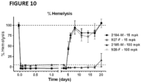
- humanized anti-Clq antibodies of the present disclosure inhibit C1F hemolysis (also referred to as CH50 hemolysis) by at least 20%, at least 30%, at least 40%, at least 50%, at least 60%, at least 70%, at least 80%, at least 90%, at least 95%, or at least 99%, or by an amount that ranges from at least 30% to at least 99%,relative to a control wherein the antibodies of this disclosure are absent or wherein control antibodies are used that do not bind to a complement factor or another antibody such as an autoantibody (see, e.g., Examples section below).
- Methods for measuring C1F hemolysis are well known in the art (see, e.g., Examples section below).
- the EC 50 values for humanized antibodies of this disclosure with respect to C1F hemolysis may be less than 3 ⁇ g/ml; 2.5 ⁇ g/ml; 2.0 ⁇ g/ml; 1.5 ⁇ g/ml; 1.0 ⁇ g/ml; 0.5 ⁇ g/ml; 0.25 ⁇ g/ml; 0.1 ⁇ g/ml; 0.05 ⁇ g/ml.
- humanized anti-Clq antibodies of this disclosure neutralize at least 50% of C1F hemolysis at a dose of less than 200 ng/ml, less than 100 ng/ml, less than 50 ng/ml, or less than 20 ng/ml.
- humanized antibodies of this disclosure neutralize C1F hemolysis at approximately equimolar concentrations of C1q and the anti-Clq antibody. In some implementations, humanized anti-Clq antibodies of this disclosure neutralize hemolysis in a human C1F hemolysis assay. In some implementations, humanized anti-Clq antibodies of this disclosure neutralize hemolysis in a human and rat C1F hemolysis assay (see, e.g., Examples section below).
- the alternative pathway may amplify CDC initiated by C1q binding and subsequent C1s activation; in at least some of these implementations, the antibodies of this disclosure inhibit the alternative pathway by at least 30%, at least 40%, at least 50%, at least 60%, at least 70%, at least 80%, at least 90%, at least 95%, or at least 99%, or by an amount that ranges from at least 30% to at least 99%, relative to a control wherein the antibodies of this disclosure were absent.
- humanized anti-Clq antibodies of the present disclosure prevent synaptic loss in a cellular in vitro model or an in vivo model of synaptic loss, such as an in vivo mouse model.
- In vivo mouse models may include Tg2576, a mouse amyloid precursor protein (APP) transgenic model of Alzheimer's disease, R6/2 NT-CAG150, a transgenic model for Huntington's disease, or SMA ⁇ 7, a mouse model for Spinal Muscular Atrophy, or DBA/2J, a genetic mouse model of glaucoma.
- any neurodegenerative disease model may be used that displays synapse loss.
- In vitro lesion formation may be reduced by at least 30%, at least 40%, at least 50%, at least 60%, at least 70%, at least 80%, at least 90%, or at least 95%, or by an amount that ranges from at least 30% to at least 95%, relative to a control experiment in which antibodies of this disclosure are absent.
- the EC 50 values for antibodies of this disclosure with respect to the prevention of in vitro lesion formation may be less than 3 ⁇ g/ml; 2.5 ⁇ g/ml; 2.0 ⁇ g/ml; 1.5 ⁇ g/ml; 1.0 ⁇ g/ml; 0.5 ⁇ g/ml; 0.25 ⁇ g/ml; 0.1 ⁇ g/ml; 0.05 ⁇ g/ml.
- In vivo synaptic loss may be reduced by at least 5%, at least 10%, at least 15%, at least 20%, at least 35%, at least 40%, or at least 50%, or by an amount that ranges from at least 5% to at least 50%, relative to a control experiment in which antibodies of this disclosure are absent.
- humanized anti-Clq antibodies of the present disclosure prevent lesion formation in an ex vivo spinal cord slice model of NMO or in an in vivo mouse model of NMO.
- Methods for measuring lesion formation ex vivo or in vivo are well known in the art.
- Ex vivo lesion formation may be reduced at least by a relative score of 0.5, 1.0, 1.5, 2.0, 2.5, 3.0, 3.5, or 4.0.
- the EC 50 values for antibodies of this disclosure with respect to the prevention of ex vivo lesion formation may be less than 3 ⁇ g/ml; less than 2.5 ⁇ g/ml; less than 2.0 ⁇ g/ml; less than 1.5 ⁇ g/ml; less than 1.0 ⁇ g/ml; less than 0.5 ⁇ g/ml; less than 0.25 ⁇ g/ml; less than 0.1 ⁇ g/ml; or less than 0.05 ⁇ g/ml.
- In vivo lesion formation may be reduced by at least 5%, at least 10%, at least 15%, at least 20%, at least 35%, at least 40%, or at least 50%, or by an amount that ranges from at least 5% to at least 50%, in terms of loss of staining (% of area). Staining may be assessed, without limitation, by AQP4 staining, GFAP staining, or MBP staining.
- the present disclosure provides humanized anti-Clq antibodies.
- the humanized antibodies of the present disclosure may have one or more of the following characteristics.
- the antibodies of this disclosure may be polyclonal antibodies, monoclonal antibodies, chimeric antibodies, human antibodies, antibody fragments, bispecific and polyspecific antibodies, multivalent antibodies, or heteroconjugate antibodies.
- Antibody fragments of this disclosure may be functional fragments that bind the same epitope as any of the humanized anti-Clq antibodies of this disclosure. In some implementations, the antibody fragments of this disclosure specifically bind to and neutralize a biological activity of C1q.
- the antibody fragments are miniaturized versions of the humanized anti-Clq antibodies or antibody fragments of this disclosure that have the same epitope of the corresponding full-length antibody, but have much smaller molecule weight.
- Such miniaturized anti-Clq antibody fragments may have better brain penetration ability and a shorter half-life, which is advantageous for imaging and diagnostic utilities (see e.g., Lütje S et al., Bioconjug Chem. 2014 Feb 19;25(2):335-41 ; Tavaré R et al., Proc Natl Acad Sci USA. 2014 Jan 21;111(3):1108-13 ; and Wiehr S et al., Prostate. 2014 May;74(7):743-55 ).
- humanized anti-Clq antibody fragments of this disclosure have better brain penetration as compared to their corresponding full-length antibodies and/or have a shorter half-life as compared to their corresponding full-length antibodies.
- humanized anti-Clq antibodies of the present disclosure are bispecific antibodies recognizing a first antigen and a second antigen.
- the first antigen is a C1q antigen.
- the second antigen is an antigen facilitating transport across the blood-brain-barrier, including without limitation, transferrin receptor (TR), insulin receptor (HIR), insulin-like growth factor receptor (IGFR), low-density lipoprotein receptor related proteins 1 and 2 (LPR-1 and 2), diphtheria toxin receptor, CRM197, a llama single domain antibody, TMEM 30(A), a protein transduction domain, TAT, Syn-B, penetratin, a poly-arginine peptide, an angiopep peptide, and ANG1005.
- TR transferrin receptor
- HIR insulin receptor
- IGFR insulin-like growth factor receptor
- LPR-1 and 2 low-density lipoprotein receptor related proteins 1 and 2
- CRM197 a llama single domain antibody
- TMEM 30(A) a protein transduction domain
- TAT Syn-B
- penetratin a poly-arginine peptide
- an angiopep peptide angiopep peptid
- Humanized anti-Clq antibodies of the present disclosure may further contain engineered effector functions, amino acid sequence modifications or other antibody modifications known in the art; e.g., the constant region of the anti-Clq antibodies described herein may be modified to impair complement activation.
- the Fc region of human IgG4 unlike the Fc region of human IgG1, IgG2, and IgG3, the Fc region of human IgG4 does not bind to C1q. Accordingly, in some implementations, humanized anti-Clq antibodies of this disclosure may further comprise the Fc region of human IgG4.
- humanized anti-Clq antibodies of this disclosure comprise one or more amino acid substitutions within the Fc region that, for example, prevent arm switching and/or reduces or otherwise inhibits the ability of Fc region from interacting with Fc receptors expressed on cells (see e.g., Angal S et al., Mol Immunol. 1993 Jan;30(1):105-8 ; and Morgan A et al., Immunology 1995 86 319-324 ).
- humanized anti-Clq antibodies of this disclosure comprise an Fc region that comprises an amino acid substitution at position 241 or 248 according to Kabat numbering convention.
- the Fc region comprises a serine to proline amino acid substitution at position 241 that prevent arm switching.
- the Fc region comprises a serine to proline amino acid substitution at position 241 according to Kabat numbering convention. In some implementations, the Fc region comprises a leucine to glutamate amino acid substitution at position 248 that reduces or otherwise inhibits the ability of Fc region from interacting with an Fc receptor. In some implementations, the Fc region comprises a leucine to glutamate amino acid substitution at position 248 according to Kabat numbering convention. In some implementations humanized anti-Clq antibodies of this disclosure comprise an Fc region comprising the amino acid sequence of SEQ ID NO: 37.
- Additional humanized anti-C1q antibodies e.g., humanized antibodies that specifically bind to a C1q protein of the present disclosure, may be identified, screened, and/or characterized for their physical/chemical properties and/or biological activities by various assays known in the art.
- Anti-Clq antibodies of the present disclosure may be produced using any methods described herein or known in the art.
- Monoclonal antibodies (e.g., humanized antibodies) of the of the present disclosure can be produced using a variety of known techniques, such as the standard somatic cell hybridization technique described by Kohler and Milstein, Nature 256: 495 (1975 ). Although somatic cell hybridization procedures are preferred, in principle, other techniques for producing monoclonal antibodies also can be employed, e.g., viral or oncogenic transformation of B lymphocytes and phage display technique using libraries of human antibody genes.
- Hybridoma production in the mouse is well-known in the art, including immunization protocols and techniques for isolating and fusing immunized splenocytes.
- Polyclonal antibodies can be prepared by immunizing a suitable subject with a polypeptide immunogen.
- the polypeptide antibody titer in the immunized subject can be monitored over time by standard techniques, such as with an enzyme linked immunosorbent assay (ELISA) using immobilized polypeptide.
- ELISA enzyme linked immunosorbent assay
- the antibody directed against the antigen can be isolated from the mammal (e.g., from the blood) and further purified by well-known techniques, such as protein A chromatography to obtain the IgG fraction.
- antibody-producing cells can be obtained from the subject and used to prepare monoclonal antibodies by standard techniques, such as the hybridoma technique originally described by Kohler and Milstein (1975) Nature 256:495-497 ) (see also Brown et al. (1981) J. Immunol. 127:539-46 ; Brown et al. (1980) J. Biol. Chem. 255:4980-83 ; Yeh et al. (1976) Proc. Natl. Acad. Sci. 76:2927-31 ; and Yeh et al. (1982) Int. J.
- an immortal cell line typically a myeloma
- lymphocytes typically splenocytes
- the culture supernatants of the resulting hybridoma cells are screened to identify a hybridoma producing a monoclonal antibody that binds to the polypeptide antigen, preferably specifically.
- any of the many well-known protocols used for fusing lymphocytes and immortalized cell lines can be applied for the purpose of generating an anti-PD-1, PD-L1, or PD-L2 monoclonal antibody (see, e.g., Galfre, G. et al. (1977) Nature 266:55052 ; Getter et al. (1977) supra; Lerner (1981) supra; Kenneth (1980) supra).
- the immortal cell line e.g., a myeloma cell line
- the immortal cell line is derived from the same mammalian species as the lymphocytes.
- murine hybridomas can be made by fusing lymphocytes from a mouse immunized with an immunogenic preparation of the present disclosure with an immortalized mouse cell line.
- Preferred immortal cell lines are mouse myeloma cell lines that are sensitive to culture medium containing hypoxanthine, aminopterin and thymidine ("HAT medium").
- HAT medium culture medium containing hypoxanthine, aminopterin and thymidine
- Any of a number of myeloma cell lines can be used as a fusion partner according to standard techniques, e.g. , the P3-NS1/1-Ag4-1, P3-x63-Ag8.653 or Sp2/O-Ag14 myeloma lines. These myeloma lines are available from the American Type Culture Collection (ATCC), Rockville, Md.
- ATCC American Type Culture Collection
- HAT-sensitive mouse myeloma cells are fused to mouse splenocytes using polyethylene glycol ("PEG").
- PEG polyethylene glycol
- Hybridoma cells resulting from the fusion are then selected using HAT medium, which kills unfused and unproductively fused myeloma cells (unfused splenocytes die after several days because they are not transformed).
- Hybridoma cells producing a monoclonal antibody of the present disclosure are detected by screening the hybridoma culture supernatants for antibodies that bind a given polypeptide, e.g. , using a standard ELISA assay.
- a monoclonal specific for a desired polypeptide can be identified and isolated by screening a recombinant combinatorial immunoglobulin library (e.g., an antibody phage display library) with the appropriate polypeptide to thereby isolate immunoglobulin library members that bind the polypeptide.
- Kits for generating and screening phage display libraries are commercially available (e.g., the Pharmacia Recombinant Phage Antibody System, Catalog No. 27-9400-01; and the Stratagene SurfZAP TM Phage Display Kit, Catalog No. 240612).
- examples of methods and reagents particularly amenable for use in generating and screening an antibody display library can be found in, for example, Ladner et al. U.S. Patent No. 5,223,409 ; Kang et al. International Publication No. WO 92/18619 ; Dower et al. International Publication No. WO 91/17271 ; Winter et al. International Publication WO 92/20791 ; Markland et al. International Publication No. WO 92/15679 ; Breitling et al. International Publication WO 93/01288 ; McCafferty et al. International Publication No. WO 92/01047 ; Garrard et al.
- recombinant anti-Clq antibodies such as humanized and chimeric monoclonal antibodies, which can be made using standard recombinant DNA techniques, can be generated.
- humanized and chimeric monoclonal antibodies can be produced by recombinant DNA techniques known in the art, for example using methods described in Robinson et al. International Patent Publication PCT/US86/02269 ; Akira et al. European Patent Application 184,187 ; Taniguchi, M. European Patent Application 171,496 ; Morrison et al. European Patent Application 173,494 ; Neuberger et al. PCT Application WO 86/01533 ; Cabilly et al. U.S. Patent No.
- humanized antibodies can be made according to standard protocols such as those disclosed in US patent 5,565,332 .
- antibody chains or specific binding pair members can be produced by recombination between vectors comprising nucleic acid molecules encoding a fusion of a polypeptide chain of a specific binding pair member and a component of a replicable generic display package and vectors containing nucleic acid molecules encoding a second polypeptide chain of a single binding pair member using techniques known in the art, e.g., as described in US patents 5,565,332 , 5,871,907 , or 5,733,743 .
- the use of intracellular antibodies to inhibit protein function in a cell is also known in the art (see e.g., Carlson, J.
- human monoclonal anti-Clq antibodies can be generated using transgenic or transchromosomal mice carrying parts of the human immune system rather than the mouse system.
- transgenic mice referred to herein as "HuMAb mice” which contain a human immunoglobulin gene miniloci that encodes unrearranged human heavy ( ⁇ and ⁇ ) and ⁇ light chain immunoglobulin sequences, together with targeted mutations that inactivate the endogenous ⁇ and ⁇ chain loci ( Lonberg, N. et al. (1994) Nature 368(6474): 856 859 ).
- mice exhibit reduced expression of mouse IgM or ⁇ , and in response to immunization, the introduced human heavy and light chain transgenes undergo class switching and somatic mutation to generate high affinity human IgG ⁇ monoclonal antibodies (Lonberg, N. et al. (1994), supra; reviewed in Lonberg, N. (1994) Handbook of Experimental Pharmacology 113:49 101 ; Lonberg, N. and Huszar, D. (1995) Intern. Rev. Immunol. Vol. 13: 65 93 , and Harding, F. and Lonberg, N. (1995) Ann. N. Y Acad. Sci 764:536 546 ).
- the preparation of HuMAb mice is described in Taylor, L. et al.
- partial or known antibody sequences can be used to generate and/or express new antibodies.
- Antibodies interact with target antigens predominantly through amino acid residues that are located in the six heavy and light chain complementarity determining regions (CDRs). For this reason, the amino acid sequences within CDRs are more diverse between individual antibodies than sequences outside of CDRs. Because CDR sequences are responsible for most antibody-antigen interactions, it is possible to express recombinant antibodies that mimic the properties of specific naturally occurring antibodies by constructing expression vectors that include CDR sequences from the specific naturally occurring antibody grafted onto framework sequences from a different antibody with different properties (see, e.g., Riechmann, L.
- Such framework sequences can be obtained from public DNA databases that include germline or non-germline antibody gene sequences. These germline sequences will differ from mature antibody gene sequences because they will not include completely assembled variable genes, which are formed by V(D)J joining during B cell maturation. Germline gene sequences will also differ from the sequences of a high affinity secondary repertoire antibody at individual evenly across the variable region.
- somatic mutations are relatively infrequent in the amino-terminal portion of framework region.
- somatic mutations are relatively infrequent in the amino terminal portion of framework region 1 and in the carboxy-terminal portion of framework region 4.
- many somatic mutations do not significantly alter the binding properties of the antibody. For this reason, it is not necessary to obtain the entire DNA sequence of a particular antibody in order to recreate an intact recombinant antibody having binding properties similar to those of the original antibody (see PCT/US99/05535 filed on Mar. 12, 1999 ).
- Partial heavy and light chain sequence spanning the CDR regions is typically sufficient for this purpose. The partial sequence is used to determine which germline and/or non-germline variable and joining gene segments contributed to the recombined antibody variable genes.
- variable regions Heavy and light chain leader sequences are cleaved during protein maturation and do not contribute to the properties of the final antibody.
- cloned cDNA sequences can be combined with synthetic oligonucleotides by ligation or PCR amplification.
- the entire variable region can be synthesized as a set of short, overlapping, oligonucleotides and combined by PCR amplification to create an entirely synthetic variable region clone. This process has certain advantages such as elimination or inclusion or particular restriction sites, or optimization of particular codons.
- the process can also be used to screen libraries of particular immunoglobulin encoding sequences in one species (e.g., human) to design cognate immunoglobulin encoding sequences from known antibody sequence in another species (e.g., mouse) (see, for example, the Examples section below).
- the nucleotide sequences of heavy and light chain transcripts from a hybridoma may be used to design an overlapping set of synthetic oligonucleotides to create synthetic V sequences with identical amino acid coding capacities as the natural sequences.
- the synthetic heavy and kappa chain sequences can differ from the natural sequences in three ways: strings of repeated nucleotide bases are interrupted to facilitate oligonucleotide synthesis and PCR amplification; optimal translation initiation sites are incorporated according to Kozak's rules ( Kozak, 1991, J. Biol. Chem. 266L19867019870 ); and, HindIII sites are engineered upstream of the translation initiation sites.
- the optimized coding, and corresponding non-coding, strand sequences are broken down into 30-50 nucleotide approximately the midpoint of the corresponding non-coding oligonucleotide.
- the oligonucleotides can be assembled into overlapping double stranded sets that span segments of 150-400 nucleotides. The pools are then used as templates to produce PCR amplification products of 150-400 nucleotides.
- a single variable region oligonucleotide set will be broken down into two pools which are separately amplified to generate two overlapping PCR products. These overlapping products are then combined by PCR amplification to form the complete variable region. It may also be desirable to include an overlapping fragment of the heavy or light chain constant region in the PCR amplification to generate fragments that can easily be cloned into the expression vector constructs.
- the reconstructed heavy and light chain variable regions are then combined with cloned promoter, leader sequence, translation initiation, leader sequence, constant region, 3' untranslated, polyadenylation, and transcription termination, sequences to form expression vector constructs.
- the heavy and light chain expression constructs can be combined into a single vector, co-transfected, serially transfected, or separately transfected into host cells which are then fused to form a host cell expressing both chains.
- Plasmids for this use are known in the art and include the plasmids provided in the Examples section below.
- Fully human and chimeric antibodies of the present disclosure also include IgG1, IgG2, IgG3, IgG4, IgE, IgA, IgM, and IgD antibodies, and variants and mutants thereof.
- Similar plasmids can be constructed for expression of other heavy chain isotypes, or for expression of antibodies comprising lambda light chains.
- the structural features of known, non-human or human antibodies are used to create structurally related human anti-human C1q antibodies that retain at least one functional property of the antibodies of the present disclosure, such as binding to a C1q protein.
- Another functional property includes inhibiting binding of the monoclonal antibody M1 to C1q in a competition ELISA assay.
- the structurally related anti-human C1q antibodies have a comparable binding affinity to the antigen as compared to the monoclonal antibody M1 as measured by the IC 50 value as described in the Examples section below. In some implementations, the structurally related anti-human C1q antibodies have a higher affinity to the antigen as compared to the monoclonal antibody M1 as measured by the IC50 value as described in the Examples section below.
- one or more CDR or variable regions of an anti-C1q antibody can be combined recombinantly with known human framework regions and CDRs to create additional, recombinantly-engineered, human anti-C1q antibodies of the present disclosure.
- the recombinant antibodies of the present disclosure prepared as set forth above may, in some implementations, comprise the heavy and light chain CDR3s of variable regions of the monoclonal antibody M1 produced by the hybridoma cell line having ATCC Accession Number PTA-120399.
- the antibodies further can comprise the CDR2s of variable regions of the monoclonal antibody M1.
- the antibodies further can comprise the CDR1s of variable regions of the monoclonal antibody M1.
- the antibodies can further comprise any combinations of the CDRs.
- the CDR1, 2, and/or 3 regions of the engineered antibodies described above may comprise the exact amino acid sequence(s) as those of variable regions of the monoclonal antibody M1 produced by the hybridoma cell line having ATCC Accession Number PTA-120399.
- the ordinarily skilled artisan will appreciate that some deviation from the exact CDR sequences may be possible while still retaining the ability of the antibody to bind C1q effectively ( e.g., conservative sequence modifications).
- the engineered antibody may be composed of one or more CDRs that are, for example, 50%, 60%, 70%, 80%, 85%, 90%, 91%, 92%, 93%, 94%, 95%, 96%, 97%, 98%, 99%, or 99.5% identical to one or more CDRs of the monoclonal antibody M1.
- a anti-C1q antibody fragments can also be isolated from the antibody phage libraries as discussed above.
- Fab'-SH fragments can be directly recovered from E. coli and chemically coupled to form F(ab') 2 fragments ( Carter et al., Bio/Technology 10:163-167 (1992 )).
- F(ab') 2 fragments can be isolated directly from recombinant host-cell culture. Production of Fab and F(ab') 2 antibody fragments with increased in vivo half-lives are described in U.S. Patent No. 5,869,046 .
- the antibody of choice is a single chain Fv fragment (scFv). See WO 93/16185 ; U.S.
- the anti-C1q, anti-C1r, or anti-C1q antibody fragment may also be a "linear antibody," e.g ., as described in U.S. Patent 5,641,870 . Such linear antibody fragments may be monospecific or bispecific.
- antibodies of the present disclosure encompass bispecific antibodies and polyspecific antibodies.
- Bispecific antibodies are antibodies that have binding specificities for at least two different epitopes, including those on the same or another protein (e.g., one or more C1q proteins of the present disclosure). Alternatively, one part of a BsAb can be armed to bind to the target C1q antigen, and another can be combined with an arm that binds to a second protein. Such antibodies can be derived from full length antibodies or antibody fragments ( e.g., F(ab') 2 bispecific antibodies).
- bispecific antibodies are known in the art. Traditional production of full length bispecific antibodies is based on the coexpression of two immunoglobulin heavy-chain/light chain pairs, where the two chains have different specificities. Millstein et al., Nature, 305:537-539 (1983 ). Because of the random assortment of immunoglobulin heavy and light chains, these hybridomas (quadromas) produce a potential mixture of 10 different antibody molecules, of which only one has the correct bispecific structure. Purification of the correct molecule, which is usually done by affinity chromatography steps, is rather cumbersome, and the product yields are low. Similar procedures are disclosed in WO 93/08829 and in Traunecker et al., EMBO J., 10:3655-3659 (1991 ).
- antibody variable domains with the desired binding specificities are fused to immunoglobulin constant domain sequences.
- the fusion may be with an immunoglobulin heavy chain constant domain, comprising at least part of the hinge, C H 2, and C H 3 regions.
- the first heavy-chain constant region (C H 1) containing the site necessary for light chain binding is present in at least one of the fusions.
- the bispecific antibodies are composed of a hybrid immunoglobulin heavy chain with a first binding specificity in one arm, and a hybrid immunoglobulin heavy chain-light chain pair (providing a second binding specificity) in the other arm. It was found that this asymmetric structure facilitates the separation of the desired bispecific compound from unwanted immunoglobulin chain combinations, as the presence of an immunoglobulin light chain in only half of the bispecific molecules provides an easy way of separation. This approach is disclosed in WO 94/04690 . For further details of generating bispecific antibodies, see, for example, Suresh et al., Methods in Enzymology 121: 210 (1986 ).
- the interface between a pair of antibody molecules can be engineered to maximize the percentage of heterodimers which are recovered from recombinant-cell culture.
- the interface may comprise at least a part of the C H 3 region of an antibody constant domain.
- one or more small amino acid side chains from the interface of the first antibody molecule are replaced with larger side chains (e.g., tyrosine or tryptophan).
- Compensatory "cavities" of identical or similar size to the large side chains(s) are created on the interface of the second antibody molecule by replacing large amino acid side chains with smaller ones ( e.g., alanine or threonine). This provides a mechanism for increasing the yield of the heterodimer over other unwanted end-products such as homodimers.
- bispecific antibodies can be prepared using chemical linkage.
- Brennan et al., Science 229:81 (1985 ) describe a procedure wherein intact antibodies are proteolytically cleaved to generate F(ab') 2 fragments. These fragments are reduced in the presence of the dithiol complexing agent sodium arsenite to stabilize vicinal dithiols and prevent intermolecular disulfide formation.
- the Fab' fragments generated are then converted to thionitrobenzoate (TNB) derivatives.
- TAB thionitrobenzoate
- One of the Fab'-TNB derivatives is then reconverted to the Fab'-TNB derivative to form the bispecific antibody.
- the bispecific antibodies produced can be used as agents for the selective immobilization of enzymes.
- Fab' fragments may be directly recovered from E. coli and chemically coupled to form bispecific antibodies.
- Shalaby et al., J. Exp. Med. 175: 217-225 (1992 ) describes the production of fully humanized bispecific antibody F(ab') 2 molecules.
- Each Fab' fragment was separately secreted from E. coli and subjected to directed chemical coupling in vitro to form the bispecific antibody.
- the bispecific antibody thus formed was able to bind to cells overexpressing the ErbB2 receptor and normal human T-cells, as well as trigger the lytic activity of human cytotoxic lymphocytes against human breast tumor targets.
- bivalent heterodimers have been produced using leucine zippers.
- the leucine zipper peptides from the Fos and Jun proteins were linked to the Fab' portions of two different antibodies by gene fusion.
- the antibody homodimers were reduced at the hinge region to form monomers and then re-oxidized to form the antibody heterodimers.
- the "diabody" technology described by Hollinger et al., Proc. Nat'l Acad. Sci.
- the fragments comprise a heavy-chain variable domain (V H ) connected to a light-chain variable domain (V L ) by a linker which is too short to allow pairing between the two domains on the same chain. Accordingly, the V H and V L domains of one fragment are forced to pair with the complementary V L and V H domains of another fragment, thereby forming two antigen-binding sites.
- V H and V L domains of one fragment are forced to pair with the complementary V L and V H domains of another fragment, thereby forming two antigen-binding sites.
- sFv single-chain Fv
- Antibodies with more than two valencies are also contemplated.
- trispecific antibodies can be prepared. Tutt et al., J. Immunol. 147:60 (1991 ).
- Exemplary bispecific antibodies may bind to two different antigens.
- a bispecific antibody binds to a first antigen, C1q, and a second antigen facilitating transport across the blood-brain barrier.
- C1q a first antigen
- second antigen facilitating transport across the blood-brain barrier.
- Numerous antigens are known in the art that facilitate transport across the blood-brain barrier ( see, e.g., Gabathuler R., Approaches to transport therapeutic drugs across the blood-brain barrier to treat brain diseases, Neurobiol. Dis. 37 (2010) 48-57 ).
- Such second antigens include, without limitation, transferrin receptor (TR), insulin receptor (HIR), Insulin-like growth factor receptor (IGFR), low-density lipoprotein receptor related proteins 1 and 2 (LPR-1 and 2), diphtheria toxin receptor, including CRM197 (a non-toxic mutant of diphtheria toxin), llama single domain antibodies such as TMEM 30(A) (Flippase), protein transduction domains such as TAT, Syn-B, or penetratin, poly-arginine or generally positively charged peptides, and Angiopep peptides such as ANG1005 ( see, e.g., Gabathuler, 2010).
- TR transferrin receptor
- HIR insulin receptor
- IGFR Insulin-like growth factor receptor
- LPR-1 and 2 low-density lipoprotein receptor related proteins 1 and 2
- diphtheria toxin receptor including CRM197 (a non-toxic mutant of diphtheria toxin)
- llama single domain antibodies such
- antibodies of the present disclosure encompass multivalent antibodies.
- a multivalent antibody may be internalized (and/or catabolized) faster than a bivalent antibody by a cell expressing an antigen to which the antibodies bind.
- the anti-C1q antibodies of the present disclosure or antibody fragments thereof can be multivalent antibodies (which are other than of the IgM class) with three or more antigen binding sites (e.g., tetravalent antibodies), which can be readily produced by recombinant expression of nucleic acid encoding the polypeptide chains of the antibody.
- the multivalent antibody can comprise a dimerization domain and three or more antigen binding sites.
- the dimerization domain comprises an Fc region or a hinge region.
- the antibody will comprise an Fc region and three or more antigen binding sites amino-terminal to the Fc region.
- the multivalent antibody herein contains three to about eight, and in some implementations four, antigen binding sites.
- the multivalent antibody contains at least one polypeptide chain (and in some implementations two polypeptide chains), wherein the polypeptide chain or chains comprise two or more variable domains.
- the polypeptide chain or chains may comprise VD1-(X1)n-VD2-(X2)n-Fc, wherein VD1 is a first variable domain, VD2 is a second variable domain, Fc is one polypeptide chain of an Fc region, X1 and X2 represent an amino acid or polypeptide, and n is 0 or 1.
- the polypeptide chain or chains may comprise V H -C H 1-flexible linker-V H -C H 1-Fc region chain; or V H -C H 1-V H -C H 1-Fc region chain.
- the multivalent antibody herein may further comprise at least two (and in some implementations four) light chain variable domain polypeptides.
- the multivalent antibody herein may, for instance, comprise from about two to about eight light chain variable domain polypeptides.
- the light chain variable domain polypeptides contemplated here comprise a light chain variable domain and, optionally, further comprise a CL domain.
- Heteroconjugate antibodies are also within the scope of the present disclosure.
- Heteroconjugate antibodies are composed of two covalently joined antibodies (e.g., antiC1q antibodies of the present disclosure or antibody fragments thereof).
- one of the antibodies in the heteroconjugate can be coupled to avidin, the other to biotin.
- Such antibodies have, for example, been proposed to target immune system cells to unwanted cells, U.S. Patent No. 4,676,980 , and have been used to treat HIV infection.
- International Publication Nos. WO 91/00360 , WO 92/200373 and EP 0308936 It is contemplated that the antibodies may be prepared in vitro using known methods in synthetic protein chemistry, including those involving crosslinking agents.
- immunotoxins may be constructed using a disulfide exchange reaction or by forming a thioether bond.
- suitable reagents for this purpose include iminothiolate and methyl-4-mercaptobutyrimidate and those disclosed, for example, in U.S. Patent No. 4,676,980 .
- Heteroconjugate antibodies may be made using any convenient cross-linking methods. Suitable cross-linking agents are well known in the art, and are disclosed in U.S. Patent No. 4,676,980 , along with a number of cross-linking techniques.
- the Fc receptor binding site on the constant region may be modified or mutated to remove or reduce binding affinity to certain Fc receptors, such as FcyRI, FcyRII, and/or Fc ⁇ RIII.
- the effector function is impaired by removing N-glycosylation of the Fc region ( e.g., in the CH 2 domain of IgG) of the antibody.
- the effector function is impaired by modifying regions such as 233-236, 297, and/or 327-331 of human IgG as described in PCT WO 99/58572 and Armour et al., Molecular Immunology 40: 585-593 (2003 ); Reddy et al., J. Immunology 164:1925-1933 (2000 ).
- the constant region of the anti-complement antibodies described herein may also be modified to impair complement activation.
- complement activation of IgG antibodies following binding of the C1 component of complement may be reduced by mutating amino acid residues in the constant region in a C1 binding motif (e.g., C1q binding motif).
- C1 binding motif e.g., C1q binding motif
- alkyl-substituted non-ionic residues such as Gly, Ile, Leu, or Val, or such aromatic non-polar residues as Phe, Tyr, Trp, and Pro in place of any one of the three residues in order to abolish C1q binding.
- aromatic non-polar residues such as Phe, Tyr, Trp, and Pro
- polar non-ionic residues such as Ser, Thr, Cys, and Met in place of residues 320 and 322, but not 318, in order to abolish C1s binding activity.
- Deglycosylation can be performed using glycosidase enzymes for example Endoglycosidase S (EndoS), a 108kDa enzyme encoded by the gene endoS of Streptococcus pyogenes that selectively digests asparagine-linked glycans on the heavy chain of all IgG subclasses, without action on other immunoglobulin classes or other glycoproteins ( Collin et al. (2001) EMBO J 2001;20:3046-3055 ).
- EndoS Endoglycosidase S
- Streptococcus pyogenes a 108kDa enzyme encoded by the gene endoS of Streptococcus pyogenes that selectively digests asparagine-linked glycans on the heavy chain of all IgG subclasses, without action on other immunoglobulin classes or other glycoproteins
- a salvage receptor binding epitope refers to an epitope of the Fc region of an IgG molecule (e.g., IgG 1 , IgG 2 , IgG 3 , or IgG 4 ) that is responsible for increasing the in vivo serum half-life of the IgG molecule.
- Amino acid sequence modifications of humanized anti-C1q antibodies of the present disclosure, or antibody fragments thereof, are also contemplated. For example, it may be desirable to improve the binding affinity and/or other biological properties of the antibodies or antibody fragments.
- Amino acid sequence variants of the antibodies or antibody fragments are prepared by introducing appropriate nucleotide changes into the nucleic acid encoding the antibodies or antibody fragments, or by peptide synthesis. Such modifications include, for example, deletions from, and/or insertions into and/or substitutions of, residues within the amino acid sequences of the antibody.
- deletion, insertion, and substitution are made to arrive at the final construct, provided that the final construct possesses the desired characteristics (i.e., the ability to bind or physically interact with a C1q protein of the present disclosure).
- the amino acid changes also may alter post-translational processes of the antibody, such as changing the number or position of glycosylation sites.
- a useful method for identification of certain residues or regions of the anti-C1q antibody that are preferred locations for mutagenesis is called "alanine scanning mutagenesis" as described by Cunningham and Wells in Science, 244:1081-1085 (1989 ).
- a residue or group of target residues are identified (e.g., charged residues such as arg, asp, his, lys, and glu) and replaced by a neutral or negatively charged amino acid (most preferably alanine or polyalanine) to affect the interaction of the amino acids with the target antigen.
- Those amino acid locations demonstrating functional sensitivity to the substitutions then are refined by introducing further or other variants at, or for, the sites of substitution.
- the site for introducing an amino acid sequence variation is predetermined, the nature of the mutation per se need not be predetermined. For example, to analyze the performance of a mutation at a given site, alanine scanning or random mutagenesis is conducted at the target codon or region and the expressed antibody variants are screened for the desired activity.
- Amino acid sequence insertions include amino- ("N") and/or carboxy- ("C") terminal fusions ranging in length from one residue to polypeptides containing a hundred or more residues, as well as intrasequence insertions of single or multiple amino acid residues.
- terminal insertions include an antibody with an N-terminal methionyl residue or the antibody fused to a cytotoxic polypeptide.
- Other insertional variants of the antibody molecule include the fusion to the N- or C-terminus of the antibody to an enzyme or a polypeptide which increases the serum half-life of the antibody.
- variants are an amino acid substitution variant. These variants have at least one amino acid residue in the antibody molecule replaced by a different residue.
- the sites of greatest interest for substitutional mutagenesis include the hypervariable regions, but FR alterations are also contemplated. Conservative substitutions are shown in the Table A below under the heading of "preferred substitutions". If such substitutions result in a change in biological activity, then more substantial changes, denominated "exemplary substitutions" in Table A, or as further described below in reference to amino acid classes, may be introduced and the products screened.
- Substantial modifications in the biological properties of the antibody are accomplished by selecting substitutions that differ significantly in their effect on maintaining (a) the structure of the polypeptide backbone in the area of the substitution, for example, as a sheet or helical conformation, (b) the charge or hydrophobicity of the molecule at the target site, or (c) the bulk of the side chain.
- Naturally occurring residues are divided into groups based on common side-chain properties:
- Non-conservative substitutions entail exchanging a member of one of these classes for another class.
- cysteine residue not involved in maintaining the proper conformation of the antibody also may be substituted, generally with serine, to improve the oxidative stability of the molecule and prevent aberrant crosslinking.
- cysteine bond(s) may be added to the antibody to improve its stability (particularly where the antibody is an antibody fragment, such as an Fv fragment).
- the substitutional variant involves substituting one or more hypervariable region residues of a parent antibody (e.g. a humanized or human antiC1q antibody).
- a parent antibody e.g. a humanized or human antiC1q antibody.
- the resulting variant(s) selected for further development will have improved biological properties relative to the parent antibody from which they are generated.
- a convenient way for generating such substitutional variants involves affinity maturation using phage display. Briefly, several hypervariable region sites (e.g., 6-7 sites) are mutated to generate all possible amino substitutions at each site.
- the antibody variants thus generated are displayed in a monovalent fashion from filamentous phage particles as fusions to the gene III product of M13 packaged within each particle.
- the phage-displayed variants are then screened for their biological activity (e.g., binding affinity) as herein disclosed.
- alanine scanning mutagenesis can be performed to identify hypervariable region residues contributing significantly to antigen binding.
- the panel of variants is subjected to screening as described herein and antibodies with superior properties in one or more relevant assays may be selected for further development.
- Another type of amino acid variant of the antibody alters the original glycosylation pattern of the antibody. By altering is meant deleting one or more carbohydrate moieties found in the antibody, and/or adding one or more glycosylation sites that are not present in the antibody.
- N-linked refers to the attachment of the carbohydrate moiety to the side chain of an asparagine residue.
- the tripeptide sequences asparagine-X-serine and asparagine-X-threonine, where X is any amino acid except proline, are the recognition sequences for enzymatic attachment of the carbohydrate moiety to the asparagine side chain.
- X is any amino acid except proline
- O-linked glycosylation refers to the attachment of one of the sugars N-aceylgalactosamine, galactose, or xylose to a hydroxyamino acid, most commonly serine or threonine, although 5-hydroxyproline or 5-hydroxylysine may also be used.
- glycosylation sites to the antibody is conveniently accomplished by altering the amino acid sequence such that it contains one or more of the above-described tripeptide sequences (for N-linked glycosylation sites).
- the alteration may also be made by the addition of, or substitution by, one or more serine or threonine residues to the sequence of the original antibody (for O-linked glycosylation sites).
- Nucleic acid molecules encoding amino acid sequence variants of the anti-IgE antibody are prepared by a variety of methods known in the art. These methods include, but are not limited to, isolation from a natural source (in the case of naturally occurring amino acid sequence variants) or preparation by oligonucleotide-mediated (or site-directed) mutagenesis, PCR mutagenesis, and cassette mutagenesis of an earlier prepared variant or a non-variant version of the antibodies (e.g., anti-C1q antibody of the present disclosure) or antibody fragments.
- humanized anti-C1q antibodies of the present disclosure, or antibody fragments thereof may be further modified to contain additional non-proteinaceous moieties that are known in the art and readily available.
- the moieties suitable for derivatization of the antibody are water-soluble polymers.
- Non-limiting examples of water-soluble polymers include, but are not limited to, polyethylene glycol (PEG), copolymers of ethylene glycol/propylene glycol, carboxymethylcellulose, dextran, polyvinyl alcohol, polyvinyl pyrrolidone, poly-1, 3-dioxolane, poly-1,3,6-trioxane, ethylene/maleic anhydride copolymer, polyaminoacids (either homopolymers or random copolymers), and dextran or poly(n-vinyl pyrrolidone)polyethylene glycol, polypropylene glycol homopolymers, polypropylene oxide/ethylene oxide co-polymers, polyoxyethylated polyols ( e.g., glycerol), polyvinyl alcohol, and mixtures thereof.
- PEG polyethylene glycol
- copolymers of ethylene glycol/propylene glycol carboxymethylcellulose
- dextran polyvinyl alcohol
- Polyethylene glycol propionaldehyde may have advantages in manufacturing due to its stability in water.
- the polymer may be of any molecular weight, and may be branched or unbranched.
- the number of polymers attached to the antibody may vary, and if more than one polymer is attached, they can be the same or different molecules. In general, the number and/or type of polymers used for derivatization can be determined based on considerations including, but not limited to, the particular properties or functions of the antibody to be improved, whether the antibody derivative will be used in a therapy under defined conditions, etc.
- Such techniques and other suitable formulations are disclosed in Remington: The Science and Practice of Pharmacy, 20th Ed., Alfonso Gennaro, Ed., Philadelphia College of Pharmacy and Science (2000 ).
- Humanized anti-C1q antibodies of the present disclosure may be produced using recombinant methods and compositions, e.g. , as described in U.S. Patent No. 4,816,567 .
- isolated nucleic acids having a nucleotide sequence encoding any of the anti-C1q antibodies of the present disclosure are provided.
- Such nucleic acids may encode an amino acid sequence containing the VL and/or an amino acid sequence containing the VH of the anti-C1q antibody (e.g., the light and/or heavy chains of the antibody).
- one or more vectors e.g., expression vectors
- containing such nucleic acids are provided.
- a host cell containing such nucleic acid is also provided.
- the host cell contains ( e.g., has been transduced with): (1) a vector containing a nucleic acid that encodes an amino acid sequence containing the VL of the antibody and an amino acid sequence containing the VH of the antibody, or (2) a first vector containing a nucleic acid that encodes an amino acid sequence containing the VL of the antibody and a second vector containing a nucleic acid that encodes an amino acid sequence containing the VH of the antibody.
- the host cell is eukaryotic, e.g. , a Chinese Hamster Ovary (CHO) cell or lymphoid cell (e.g., Y0, NS0, Sp20 cell).
- the method includes culturing a host cell of the present disclosure containing a nucleic acid encoding the anti-C1q antibody, under conditions suitable for expression of the antibody.
- the antibody is subsequently recovered from the host cell (or host cell culture medium).
- a nucleic acid encoding the anti-C1q antibody is isolated and inserted into one or more vectors for further cloning and/or expression in a host cell.
- Such nucleic acid may be readily isolated and sequenced using conventional procedures (e.g., by using oligonucleotide probes that are capable of binding specifically to genes encoding the heavy and light chains of the antibody).
- Suitable vectors containing a nucleic acid sequence encoding any of the antiC1q antibodies of the present disclosure, or fragments thereof polypeptides (including antibodies) described herein include, without limitation, cloning vectors and expression vectors.
- Suitable cloning vectors can be constructed according to standard techniques, or may be selected from a large number of cloning vectors available in the art. While the cloning vector selected may vary according to the host cell intended to be used, useful cloning vectors generally have the ability to self-replicate, may possess a single target for a particular restriction endonuclease, and/or may carry genes for a marker that can be used in selecting clones containing the vector.
- Suitable examples include plasmids and bacterial viruses, e.g., pUC18, pUC19, Bluescript (e.g., pBS SK+) and its derivatives, mpl8, mpl9, pBR322, pMB9, ColE1, pCR1, RP4, phage DNAs, and shuttle vectors such as pSA3 and pAT28.
- plasmids and bacterial viruses e.g., pUC18, pUC19, Bluescript (e.g., pBS SK+) and its derivatives, mpl8, mpl9, pBR322, pMB9, ColE1, pCR1, RP4, phage DNAs, and shuttle vectors such as pSA3 and pAT28.
- Bluescript e.g., pBS SK+
- mpl8 mpl9
- pBR322 pMB9
- ColE1 pCR1
- RP4 phage DNA
- Expression vectors generally are replicable polynucleotide constructs that contain a nucleic acid of the present disclosure.
- the expression vector may replicable in the host cells either as episomes or as an integral part of the chromosomal DNA.
- Suitable expression vectors include but are not limited to plasmids, viral vectors, including adenoviruses, adeno-associated viruses, retroviruses, cosmids, and expression vector(s) disclosed in PCT Publication No. WO 87/04462 .
- Vector components may generally include, but are not limited to, one or more of the following: a signal sequence; an origin of replication; one or more marker genes; suitable transcriptional controlling elements (such as promoters, enhancers and terminator).
- suitable transcriptional controlling elements such as promoters, enhancers and terminator
- For expression i.e., translation), one or more translational controlling elements are also usually required, such as ribosome binding sites, translation initiation sites, and stop codons.
- the vectors containing the nucleic acids of interest can be introduced into the host cell by any of a number of appropriate means, including electroporation, transfection employing calcium chloride, rubidium chloride, calcium phosphate, DEAE-dextran, or other substances; microprojectile bombardment; lipofection; and infection (e.g., where the vector is an infectious agent such as vaccinia virus).
- electroporation employing calcium chloride, rubidium chloride, calcium phosphate, DEAE-dextran, or other substances
- microprojectile bombardment e.g., where the vector is an infectious agent such as vaccinia virus.
- infection e.g., where the vector is an infectious agent such as vaccinia virus.
- the vector contains a nucleic acid containing one or more amino acid sequences encoding an anti-C1q antibody of the present disclosure.
- Suitable host cells for cloning or expression of antibody-encoding vectors include prokaryotic or eukaryotic cells.
- anti-C1q antibodies of the present disclosure may be produced in bacteria, in particular when glycosylation and Fc effector function are not needed.
- For expression of antibody fragments and polypeptides in bacteria e.g., U.S. Patent Nos. 5,648,237 , 5,789,199 , and 5,840,523 ; and Charlton, Methods in Molecular Biology, Vol. 248 (B.K.C. Lo, ed., Humana Press, Totowa, NJ, 2003), pp. 245-254 , describing expression of antibody fragments in E. coli .).
- the antibody may be isolated from the bacterial cell paste in a soluble fraction and can be further purified.
- eukaryotic microorganisms such as filamentous fungi or yeast
- suitable cloning or expression hosts for antibody-encoding vectors including fungi and yeast strains whose glycosylation pathways have been "humanized,” resulting in the production of an antibody with a partially or fully human glycosylation pattern ( e.g., Gerngross, Nat. Biotech. 22:1409-1414 (2004 ); and Li et al., Nat. Biotech. 24:210-215 (2006 )).
- Suitable host cells for the expression of glycosylated antibody can also be derived from multicellular organisms (invertebrates and vertebrates). Examples of invertebrate cells include plant and insect cells. Numerous baculoviral strains have been identified which may be used in conjunction with insect cells, particularly for transfection of Spodoptera frugiperda cells. Plant cell cultures can also be utilized as hosts ( e.g., U.S. Patent Nos. 5,959,177 , 6,040,498 , 6,420,548 , 7,125,978 , and 6,417,429 , describing PLANTIBODIES TM technology for producing antibodies in transgenic plants.).
- Vertebrate cells may also be used as hosts.
- mammalian cell lines that are adapted to grow in suspension may be useful.
- Other examples of useful mammalian host cell lines are monkey kidney CV1 line transformed by SV40 (COS-7); human embryonic kidney line (293 or 293 cells as described, e.g., in Graham et al., J. Gen Virol. 36:59 (1977 )); baby hamster kidney cells (BHK); mouse sertoli cells (TM4 cells as described, e.g., in Mather, Biol. Reprod.
- monkey kidney cells (CV1); African green monkey kidney cells (VERO-76); human cervical carcinoma cells (HELA); canine kidney cells (MDCK; buffalo rat liver cells (BRL 3A); human lung cells (W138); human liver cells (Hep G2); mouse mammary tumor (MMT 060562); TRI cells, as described, e.g., in Mather et al., Annals N.Y. Acad. Sci. 383:44-68 (1982 ); MRC 5 cells; and FS4 cells.
- Other useful mammalian host cell lines include Chinese hamster ovary (CHO) cells, including DHFR-CHO cells ( Urlaub et al., Proc. Natl. Acad. Sci.
- Humanized anti-C1q antibodies of the present disclosure can be incorporated into a variety of formulations for therapeutic use (e.g., by administration) or in the manufacture of a medicament (e.g., for treating or preventing a neurodegenerative disease or autoimmune disease) by combining the antibodies with appropriate pharmaceutically acceptable carriers or diluents, and may be formulated into preparations in solid, semi-solid, liquid or gaseous forms.
- formulations include, without limitation, tablets, capsules, powders, granules, ointments, solutions, suppositories, injections, inhalants, gels, microspheres, and aerosols.
- compositions can include, depending on the formulation desired, pharmaceutically-acceptable, non-toxic carriers of diluents, which are vehicles commonly used to formulate pharmaceutical compositions for animal or human administration.
- diluents are vehicles commonly used to formulate pharmaceutical compositions for animal or human administration.
- the diluent is selected so as not to affect the biological activity of the combination.
- examples of such diluents include, without limitation, distilled water, buffered water, physiological saline, PBS, Ringer's solution, dextrose solution, and Hank's solution.
- a pharmaceutical composition or formulation of the present disclosure can further include other carriers, adjuvants, or non-toxic, nontherapeutic, nonimmunogenic stabilizers, excipients and the like.
- the compositions can also include additional substances to approximate physiological conditions, such as pH adjusting and buffering agents, toxicity adjusting agents, wetting agents and detergents.
- a pharmaceutical composition of the present disclosure can also include any of a variety of stabilizing agents, such as an antioxidant for example.
- the pharmaceutical composition includes a polypeptide
- the polypeptide can be complexed with various well-known compounds that enhance the in vivo stability of the polypeptide, or otherwise enhance its pharmacological properties (e.g., increase the half-life of the polypeptide, reduce its toxicity, and enhance solubility or uptake).
- modifications or complexing agents include, without limitation, sulfate, gluconate, citrate and phosphate.
- the polypeptides of a composition can also be complexed with molecules that enhance their in vivo attributes. Such molecules include, without limitation, carbohydrates, polyamines, amino acids, other peptides, ions ( e.g., sodium, potassium, calcium, magnesium, manganese), and lipids.
- the active ingredient can be administered in solid dosage forms, such as capsules, tablets, and powders, or in liquid dosage forms, such as elixirs, syrups, and suspensions.
- the active component(s) can be encapsulated in gelatin capsules together with inactive ingredients and powdered carriers, such as glucose, lactose, sucrose, mannitol, starch, cellulose or cellulose derivatives, magnesium stearate, stearic acid, sodium saccharin, talcum, magnesium carbonate.
- inactive ingredients and powdered carriers such as glucose, lactose, sucrose, mannitol, starch, cellulose or cellulose derivatives, magnesium stearate, stearic acid, sodium saccharin, talcum, magnesium carbonate.
- additional inactive ingredients that may be added to provide desirable color, taste, stability, buffering capacity, dispersion or other known desirable features are red iron oxide, silica gel, sodium lauryl sulfate, titanium dioxide, and edible white ink.
- Similar diluents can be used to make compressed tablets. Both tablets and capsules can be manufactured as sustained release products to provide for continuous release of medication over a period of hours. Compressed tablets can be sugar coated or film coated to mask any unpleasant taste and protect the tablet from the atmosphere, or enteric-coated for selective disintegration in the gastrointestinal tract. Liquid dosage forms for oral administration can contain coloring and flavoring to increase patient acceptance.
- Formulations suitable for parenteral administration include aqueous and non-aqueous, isotonic sterile injection solutions, which can contain antioxidants, buffers, bacteriostats, and solutes that render the formulation isotonic with the blood of the intended recipient, and aqueous and non-aqueous sterile suspensions that can include suspending agents, solubilizers, thickening agents, stabilizers, and preservatives.
- compositions intended for in vivo use are usually sterile. To the extent that a given compound must be synthesized prior to use, the resulting product is typically substantially free of any potentially toxic agents, particularly any endotoxins, which may be present during the synthesis or purification process.
- compositions for parental administration are also sterile, substantially isotonic and made under GMP conditions.
- Formulations may be optimized for retention and stabilization in the brain or central nervous system.
- Stabilization techniques include cross-linking, multimerizing, or linking to groups such as polyethylene glycol, polyacrylamide, neutral protein carriers, etc. in order to achieve an increase in molecular weight.
- Other strategies for increasing retention include the entrapment of the antibody, such as a humanized anti-C1q antibody of the present disclosure, in a biodegradable or bioerodible implant.
- the rate of release of the therapeutically active agent is controlled by the rate of transport through the polymeric matrix, and the biodegradation of the implant.
- the transport of drug through the polymer barrier will also be affected by compound solubility, polymer hydrophilicity, extent of polymer cross-linking, expansion of the polymer upon water absorption so as to make the polymer barrier more permeable to the drug, geometry of the implant, and the like.
- the implants are of dimensions commensurate with the size and shape of the region selected as the site of implantation. Implants may be particles, sheets, patches, plaques, fibers, microcapsules and the like and may be of any size or shape compatible with the selected site of insertion.
- the implants may be monolithic, i.e. having the active agent homogenously distributed through the polymeric matrix, or encapsulated, where a reservoir of active agent is encapsulated by the polymeric matrix.
- the selection of the polymeric composition to be employed will vary with the site of administration, the desired period of treatment, patient tolerance, the nature of the disease to be treated and the like. Characteristics of the polymers will include biodegradability at the site of implantation, compatibility with the agent of interest, ease of encapsulation, a half-life in the physiological environment.
- Biodegradable polymeric compositions which may be employed may be organic esters or ethers, which when degraded result in physiologically acceptable degradation products, including the monomers. Anhydrides, amides, orthoesters or the like, by themselves or in combination with other monomers, may find use.
- the polymers will be condensation polymers.
- the polymers may be cross-linked or non-cross-linked.
- polymers of hydroxyaliphatic carboxylic acids either homo- or copolymers, and polysaccharides. Included among the polyesters of interest are polymers of D-lactic acid, L-lactic acid, racemic lactic acid, glycolic acid, polycaprolactone, and combinations thereof.
- a slowly biodegrading polymer is achieved, while degradation is substantially enhanced with the racemate.
- Copolymers of glycolic and lactic acid are of particular interest, where the rate of biodegradation is controlled by the ratio of glycolic to lactic acid.
- the most rapidly degraded copolymer has roughly equal amounts of glycolic and lactic acid, where either homopolymer is more resistant to degradation.
- the ratio of glycolic acid to lactic acid will also affect the brittleness of in the implant, where a more flexible implant is desirable for larger geometries.
- polysaccharides of interest are calcium alginate, and functionalized celluloses, particularly carboxymethylcellulose esters characterized by being water insoluble, a molecular weight of about 5 kD to 500 kD, etc.
- Biodegradable hydrogels may also be employed in the implants of the present disclosure. Hydrogels are typically a copolymer material, characterized by the ability to imbibe a liquid. Exemplary biodegradable hydrogels which may be employed are described in Heller in: Hydrogels in Medicine and Pharmacy, N. A. Peppes ed., Vol. III, CRC Press, Boca Raton, Fla., 1987, pp 137-149 .
- compositions of the present disclosure containing a humanized anti-C1q antibody of the present disclosure may be used (e.g., administered to an individual in need of treatment with an anti-C1q antibody, such as a human individual) in accordance with known methods, such as intravenous administration as a bolus or by continuous infusion over a period of time, by intramuscular, intraperitoneal, intracerobrospinal, intracranial, intraspinal, subcutaneous, intra-articular, intrasynovial, intrathecal, oral, topical, or inhalation routes.
- known methods such as intravenous administration as a bolus or by continuous infusion over a period of time, by intramuscular, intraperitoneal, intracerobrospinal, intracranial, intraspinal, subcutaneous, intra-articular, intrasynovial, intrathecal, oral, topical, or inhalation routes.
- Dosages and desired drug concentration of pharmaceutical compositions of the present disclosure may vary depending on the particular use envisioned. The determination of the appropriate dosage or route of administration is well within the skill of an ordinary artisan. Animal experiments provide reliable guidance for the determination of effective doses for human therapy. Interspecies scaling of effective doses can be performed following the principles described in Mordenti, J. and Chappell, W. "The Use of Interspecies Scaling in Toxicokinetics," In Toxicokinetics and New Drug Development, Yacobi et al., Eds, Pergamon Press, New York 1989, pp.42-46 .
- normal dosage amounts may vary from about 10 ng/kg up to about 100 mg/kg of an individual's body weight or more per day, depending upon the route of administration. In some implementations, the dose amount is about 1 mg/kg/day to 10 mg/kg/day. For repeated administrations over several days or longer, depending on the severity of the disease, disorder, or condition to be treated, the treatment is sustained until a desired suppression of symptoms is achieved.
- An exemplary dosing regimen may include administering an initial dose of a humanized anti-C1q antibody, of about 2 mg/kg, followed by a weekly maintenance dose of about 1 mg/kg every other week.
- Other dosage regimens may be useful, depending on the pattern of pharmacokinetic decay that the physician wishes to achieve. For example, dosing an individual from one to twenty-one times a week is contemplated herein. In certain implementations, dosing ranging from about 3 ⁇ g/kg to about 2 mg/kg (such as about 3 ⁇ g/kg, about 10 ⁇ g/kg, about 30 ⁇ g/kg, about 100 ⁇ g/kg, about 300 ⁇ g/kg, about 1 mg/kg, or about 2 mg/kg) may be used.
- dosing frequency is three times per day, twice per day, once per day, once every other day, once weekly, once every two weeks, once every four weeks, once every five weeks, once every six weeks, once every seven weeks, once every eight weeks, once every nine weeks, once every ten weeks, or once monthly, once every two months, once every three months, or longer. Progress of the therapy is easily monitored by conventional techniques and assays.
- the dosing regimen, including the humanized anti-C1q antibody administered, can vary over time independently of the dose used.
- Dosages for a particular humanized anti-C1q antibody may be determined empirically in individuals who have been given one or more administrations of the humanized anti-C1q antibody. Individuals are given incremental doses of a humanized anti-C1q antibody. To assess efficacy of a humanized anti-C1q antibody, any clinical symptom of a neurodegenerative disorder, inflammatory disorder, or autoimmune disorder can be monitored.
- Administration of a humanized anti-C1q antibody of the present disclosure can be continuous or intermittent, depending, for example, on the recipient's physiological condition, whether the purpose of the administration is therapeutic or prophylactic, and other factors known to skilled practitioners.
- the administration of a humanized anti-C1q antibody may be essentially continuous over a preselected period of time or may be in a series of spaced doses.
- dosages and methods of delivery are provided in the literature; see, for example, U.S. Patent Nos. 4,657,760 ; 5,206,344 ; or 5,225,212 . It is within the scope of the present disclosure that different formulations will be effective for different treatments and different disorders, and that administration intended to treat a specific organ or tissue may necessitate delivery in a manner different from that to another organ or tissue. Moreover, dosages may be administered by one or more separate administrations, or by continuous infusion. For repeated administrations over several days or longer, depending on the condition, the treatment is sustained until a desired suppression of disease symptoms occurs. However, other dosage regimens may be useful. The progress of this therapy is easily monitored by conventional techniques and assays.
- humanized anti-C1q antibodies and antigen-binding fragments thereof, which can bind to and neutralize a biologic activity of C1q.
- These humanized anti-C1q antibodies are useful for preventing, reducing risk, or treating a range of diseases associated with complement activation, including, without limitation, neurodegenerative disorders, inflammatory disorders, and autoimmune disorders.
- humanized anti-C1q antibodies of the present disclosure may be used for treating, preventing, or reducing risk of a disease associated with complement activation, including, without limitation, neurodegenerative disorders, inflammatory disorders, and autoimmune disorders, in an individual.
- the individual has such a disease.
- the individual is a human.
- Neurodegenerative disorders that may be treated with humanized anti-C1q antibodies of this disclosure include disorders associated with loss of nerve connections or synapses, including CF1-dependent synapse loss.
- Such disorders may include, without limitation, Alzheimer's disease, amyotrophic lateral sclerosis, multiple sclerosis, glaucoma, myotonic dystrophy, Guillain-Barre' syndrome (GBS), Myasthenia Gravis, Bullous Pemphigoid, spinal muscular atrophy, Down syndrome, Parkinson's disease, and Huntington's disease.
- synapse loss is dependent on the complement receptor 3 (CR3)/C3 or complement receptor CR1.
- the humanized anti-C1q antibodies of the present disclosure may be used to treat, prevent, or improve one or more symptoms of a neurodegenerative disorder of the present disclosure.
- the present disclosure provides methods of treating, preventing, or improving one or more symptoms in individuals having a neurodegenerative disorder of the present disclosure by administering a humanized antiC1q antibody of the present disclosure to, for example, inhibit the interaction between C1q and an autoantibody, the interaction of C1q and C1r, and/or the interaction of C1q and C1s.
- Inflammatory or autoimmune diseases that may be treated with humanized antiC1q antibodies of this disclosure include, without limitation, rheumatoid arthritis (RA), acute respiratory distress syndrome (ARDS), remote tissue injury after ischemia and reperfusion, complement activation during cardiopulmonary bypass surgery, dermatomyositis, pemphigus, lupus nephritis and resultant glomerulonephritis and vasculitis, cardiopulmonary bypass, cardioplegia-induced coronary endothelial dysfunction, type II membranoproliferative glomerulonephritis, IgA nephropathy, acute renal failure, cryoglobulemia, antiphospholipid syndrome, Chronic open-angle glaucoma, acute closed angle glaucoma, macular degenerative diseases, age-related macular degeneration (AMD), (AMD-wet), Geographic atrophy, choroidal neovascularization (CNV), uveitis, diabetic retinopathy, ischemia-related retin
- autoimmune disease may further include, without limitation, Guillain-Barré syndrome, myasthenia gravis, Diabetes mellitus type 1, Hashimoto's thyroiditis, Addison's disease, Coeliac disease, Crohn's disease, pernicious anaemia, Pemphigus vulgaris, vitiligo, autoimmune hemolytic anemias, paraneoplastic syndromes, a vasculitis disease, hypocomplementemic urticarial vasculitis (HUV), polymyalgia rheumatica, temporal arteritis, and Wegener's granulomatosis.
- Guillain-Barré syndrome myasthenia gravis
- Diabetes mellitus type 1 Hashimoto's thyroiditis
- Addison's disease Coeliac disease
- Crohn's disease pernicious anaemia
- Pemphigus vulgaris Pemphigus vulgaris
- vitiligo vitiligo
- autoimmune hemolytic anemias parane
- Autoantibodies activate the complement system.
- NMO Neuromyelitis Optica
- the classical complement pathway is triggered by the binding of an autoantibody, such as an AQP4-targeted autoantibody, to its autoantigen, AQP4.
- AQP4 thereby activates the classical pathway of complement activation.
- complement factor C1q binds to the autoantibody-autoantigen-immune complex.
- Autoantibodies may include naturally occurring antibodies, such as serum antibodies from NMO patients (commonly referred to as NMO-IgG) or monoclonal antibodies, such as rAb-53.
- the humanized anti-C1q antibodies of the present disclosure may be used to treat, prevent, or improve one or more symptoms of an inflammatory or autoimmune disease of the present disclosure.
- the present disclosure provides methods of treating, preventing, or improving one or more symptoms in individuals having an inflammatory or autoimmune disease of the present disclosure by administering a humanized anti-C1q antibody of the present disclosure to, for example, inhibit the interaction between C1q and an autoantibody, the interaction of C1q and C1r, and/or the interaction of C1q and C1s.
- Metabolic diseases that may be treated with humanized anti-C1q antibodies include, without limitation, diabetes, such as type II diabetes, and obesity.
- diabetes such as type II diabetes, and obesity.
- in vitro and in vivo models of metabolic disorders that can be used for the testing of humanized anti-C1q antibodies are well known in the art. Accordingly, the humanized anti-C1q antibodies of the present disclosure may be used to treat, prevent, or improve one or more symptoms of a metabolic disease of the present disclosure.
- the present disclosure provides methods of treating, preventing, or improving one or more symptoms in individuals having metabolic disease of the present disclosure by administering a humanized anti-C1q antibody of the present disclosure to, for example, inhibit the interaction between C1q and an autoantibody, such as an anti-ganglioside autoantibody, the interaction of C1q and C1r, and/or the interaction of C1q and C1s.
- a humanized anti-C1q antibody of the present disclosure to, for example, inhibit the interaction between C1q and an autoantibody, such as an anti-ganglioside autoantibody, the interaction of C1q and C1r, and/or the interaction of C1q and C1s.
- the antibodies of the present disclosure may be used, without limitation, in combination with any additional treatment for neurodegenerative disorders, inflammatory disorders, and/or autoimmune disorders.
- a humanized anti-C1q antibody of this disclosure is administered in therapeutically effective amounts in combination with a second anti-complement factor antibody (e.g., a neutralizing anti-complement factor antibody), such as an anti-C1s or anti-C1r antibody, or a second anti-C1q antibody.
- a humanized anti-C1q antibody of this disclosure is administered in therapeutically effective amounts with a second and a third neutralizing anti-complement factor antibody, such as a second anti-C1q antibody, an anti-C1s antibody, and/or an anti-C1r antibody.
- ADCC inhibitors may include, without limitation, soluble NK cell inhibitory receptors such as the killer cell Ig-like receptors (KIRs), which recognize HLA-A, HLA-B, or HLA-C and C-type lectin CD94/NKG2A heterodimers, which recognize HLA-E (see, e.g., López-Botet M., T. Bellón, M. Llano, F. Navarro, P. Garcia & M. de Miguel. (2000), Paired inhibitory and triggering NK cell receptors for HLA class I molecules. Hum. Immunol.
- KIRs killer cell Ig-like receptors
- the humanized anti-C1q antibodies of this disclosure are administered in combination with an inhibitor of the alternative pathway of complement activation.
- inhibitors may include, without limitation, factor B blocking antibodies, factor D blocking antibodies, soluble, membrane-bound, tagged or fusion-protein forms of CD59, DAF, CR1, CR2, Crry or Comstatin-like peptides that block the cleavage of C3, non-peptide C3aR antagonists such as SB 290157, Cobra venom factor or non-specific complement inhibitors such as nafamostat mesilate (FUTHAN; FUT-175), aprotinin, K-76 monocarboxylic acid (MX-1) and heparin ( see, e.g., T.E.
- the humanized anti-C1q antibodies of this disclosure are administered in combination with an inhibitor of the interaction between the autoantibody and its autoantigen.
- inhibitors may include purified soluble forms of the autoantigen, or antigen mimetics such as peptide or RNA-derived mimotopes, including mimotopes of the AQP4 antigen.
- antigen mimetics such as peptide or RNA-derived mimotopes, including mimotopes of the AQP4 antigen.
- such inhibitors may include blocking agents that recognize the autoantigen and prevent binding of the autoantibody without triggering the classical complement pathway.
- Such blocking agents may include, e.g ., autoantigen-binding RNA aptamers or antibodies lacking functional C1q binding sites in their Fc domains (e.g., Fab fragments or antibody otherwise engineered not to bind C1q).
- the humanized anti-C1q antibodies of the present disclosure, or functional fragments thereof also have diagnostic utility.
- This disclosure therefore provides methods of using the antibodies of this disclosure, or functional fragments thereof, for diagnostic purposes, such as the detection of C1q in an individual or in tissue samples derived from an individual.
- the individual is a human.
- the individual is a human patient suffering from a neurodegenerative disorder or an inflammatory, or autoimmune disease.
- the humanized anti-C1q antibodies of this disclosure are used to detect synapses and synapse loss. For example, synapse loss may be measured in an individual suffering from a neurodegenerative disorder such as Alzheimer's disease or glaucoma.
- the diagnostic methods involve the steps of administering a humanized anti-C1q antibody of this disclosure, or functional fragment thereof, to an individual and detecting the antibody bound to a synapse of the individual.
- Antibody-binding to synapses may be quantified, for example, by non-invasive techniques such as positron emission tomography (PET), X-ray computed tomography, single-photon emission computed tomography (SPECT), computed tomography (CT), and computed axial tomography (CAT).
- PET positron emission tomography
- SPECT single-photon emission computed tomography
- CT computed tomography
- CAT computed axial tomography
- the diagnostic methods involve detecting synapses in a biological sample, such as a biopsy specimen, a tissue, or a cell.
- a humanized anti-C1q antibody, or functional fragment thereof is contacted with the biological sample and synapse-bound antibody is detected.
- the detection method may involve quantification of the synapse-bound antibody.
- Antibody detection in biological samples may occur with any method known in the art, including immunofluorescence microscopy, immunocytochemistry, immunohistochemistry, ELISA, FACS analysis, immunoprecipitation, or micro-positron emission tomography.
- the antibody is radiolabeled, for example with 18 F and subsequently detected utilizing micro-positron emission tomography analysis.
- the quantification of synapse-bound antibodies provides a relative measure for the number of synapses present in the individual.
- synapses are quantified repeatedly over a period of time.
- the exact periodicity of synapse quantification depends on many factors, including the nature of the neurodegenerative disease, the stage of disease progression, treatment modalities and many other factors. Repeat measurements commonly reveal progressive synapse loss in individuals having a neurodegenerative disorder.
- relative synapse counts may be compared in populations of diseased individuals and healthy control individuals at a single time point. In diseased individuals undergoing treatment, the treatment's efficacy can be assessed by comparing the rates of synapse loss in the treated individuals with the rates of synapse loss in a control group. Control group members have received either no treatment or a control treatment, such as a placebo control.
- kits containing a humanized anti-C1q antibody of the present disclosure include one or more containers comprising a purified humanized anti-C1q antibody of this disclosure.
- the kits further include instructions for use in accordance with the methods of this disclosure.
- these instructions comprise a description of administration of the humanized anti-C1q antibody to treat or diagnose a disease associated with complement activation including, without limitation a neurodegenerative disorder (e.g., Alzheimer's disease), inflammatory disease, autoimmune disease, and/or metabolic disorder, according to any methods of this disclosure.
- the instructions comprise a description of how to detect C1q, for example in an individual, in a tissue sample, or in a cell.
- the kit may further comprise a description of selecting an individual suitable for treatment based on identifying whether that individual has the disease and the stage of the disease.
- the instructions generally include information as to dosage, dosing schedule, and route of administration for the intended treatment.
- the containers may be unit doses, bulk packages (e.g., multi-dose packages) or sub-unit doses.
- Instructions supplied in the kits of the present disclosure are typically written instructions on a label or package insert (e.g., a paper sheet included in the kit), but machine-readable instructions (e.g., instructions carried on a magnetic or optical storage disk) are also acceptable.
- the label or package insert indicates that the composition is used for treating, e.g., a neurodegenerative disease. Instructions may be provided for practicing any of the methods described herein.
- kits of this disclosure are in suitable packaging.
- suitable packaging includes, but is not limited to, vials, bottles, jars, flexible packaging (e.g., sealed Mylar or plastic bags), and the like.
- packages for use in combination with a specific device such as an inhaler, nasal administration device (e.g., an atomizer) or an infusion device such as a minipump.
- a kit may have a sterile access port (for example the container may be an intravenous solution bag or a vial having a stopper pierceable by a hypodermic injection needle).
- the container may also have a sterile access port (e.g., the container may be an intravenous solution bag or a vial having a stopper pierceable by a hypodermic injection needle).
- At least one active agent in the composition is an inhibitor of classical complement pathway.
- the container may further comprise a second pharmaceutically active agent.
- Kits may optionally provide additional components such as buffers and interpretive information.
- the kit comprises a container and a label or package insert(s) on or associated with the container.
- This example describes the generation of fully humanized antibodies from the murine hybridoma M1 (expressing the mouse anti-human C1q antibody M1).
- Composite human antibody variable region genes were generated using synthetic oligonucleotides encoding combinations of selected human sequence segments. These were then cloned into vectors encoding human IgG4 (S241P L248E) heavy chain and human kappa light chain.
- Humanized antibodies were stably expressed in NS0 (mouse myeloma cell-line) cells, Protein A purified and tested for binding to human C1q using a competition ELISA assay against biotinylated murine M1 antibody. Selected antibodies were also tested for binding to mouse C1q using competition ELISA assay against biotinylated chimeric antibody.
- RNA was extracted from the hybridoma cell pellet expressing M1 antibody using an RNAqueousR-4PCR kit (Ambion cat. no. AM1914). Initially, RT-PCR was performed using degenerate primer pools for murine signal sequences together with constant region primers for both of IgG and Ig ⁇ . Heavy chain V region RNA was amplified using a set of six degenerate primer pools (HA to HF) and light chain V region mRNA was amplified using a set of seven degenerate primer pools for the ⁇ cluster (KA to KG).
- VH region amplification products of the expected size were found in IgG primer pools HB and HE.
- V ⁇ region amplification products of the expected size were found in kappa primer pools KC, KE, and KG.
- the PCR products obtained from each of the successful amplifications were purified and cloned into a 'TA' cloning vector (pGEM-T Easy, Promega cat. no. A1360) and sequenced. A total of 14 VH and 24 V ⁇ clones were sequenced.
- a single functional VH gene was identified in 14 clones from IgG pools HB and HE.
- a single functional V ⁇ gene sequence was identified from 9 clones from primer pool KC. The 3' coding sequence downstream of the variable region obtained from IgG primer pools was consistent with the antibody isotype being IgG.
- VH and V ⁇ gene sequences were identical to the hybridoma sequences with the exception of five amino acids at the beginning of the VH sequence and two amino acids at the beginning of the V ⁇ sequence. These differences were most likely due to the method of sequencing, and were a result of using primers that are degenerate to the signal sequence rather than primers that are degenerate to 5' end of V regions.
- the amino acid sequence of the functional VH is: QVQLQQPGAELVKPGASVKLSCKSS GYHFTSYWMH WVKQRPGQGLEWIGVIHP NSGSINYNEKFES KATLTVDKSSSTAYMOLSSLTSEDSAVYYCAG ERDSTEVLPM DY WGOGTSVTVSS (SEQ ID NO: 21).
- the hyper variable regions (HVRs) of the VH are depicted in bolded and underlined text.
- the amino acid sequence of the functional V ⁇ is: DVOITOSPSYLAASPGETITINC RASKSINKYLA WYOEKPGKTNKLLIY SGSTLOS G IPSRFSGSGSGTDFTLTISSLEPEDFAMYYC QQHNEYPLT FGAGTKLELK (SEQ ID NO: 22).
- the hyper variable regions (HVRs) of the V ⁇ are depicted in bolded and underlined text.
- VH and V ⁇ sequences of the murine M1 antibody were PCR amplified using primers that introduced flanking restriction enzyme sites for cloning into the IgG4 (S241P L248E) heavy and kappa chain expression vectors ( FIG. 1 ).
- the Bam HI , Hind III and Ssp I restriction sites were removed from the V ⁇ sequence in order to clone the gene.
- the VH region was cloned using Mlu I and Hind III sites, and the V ⁇ region was cloned using BssH II and BamH I restriction sites. Both constructs were confirmed by sequencing.
- Structural models of the murine M1 antibody V regions were produced using Swiss PDB and analyzed in order to identify important "constraining" amino acids in the V regions that were likely to be essential for the binding properties of the antibody. Most residues contained within the HVRs (using both Kabat and Chothia definitions) together with a number of framework residues were considered to be important.
- the VH and V ⁇ sequences of M1 contain typical framework residues and the HVR 1, 2 and 3 motifs are comparable to many murine antibodies.
- VH1-VH4 heavy chain sequences
- V ⁇ 1- V ⁇ 4 light chain sequences
- VH heavy chain variable domain
- V ⁇ kappa light chain variable domain
- VH1 heavy chain variable domain variant 1
- the amino acid sequence of heavy chain variable domain variant 1 is: QVQLVQSGAELKKPGASVKVSCKSS GYHFTSYWMH WVKQAPGQGLEWIG VIHP NSGSINYNEKFES KATITVDKSTSTAYMOLSSLTSEDSAVYYCAG ERDSTEVLPM DY WGOGTSVTVSS (SEQ ID NO: 1).
- the hyper variable regions (HVRs) of VH1 are depicted in bolded and underlined text.
- VH2 heavy chain variable domain variant 2
- the amino acid sequence of heavy chain variable domain variant 2 is: QVQLVQSGAELKKPGASVKVSCKSS GYHFTSYWMH WVKQAPGQGLEWIG VIHP NSGSINYNEKFES RATITVDKSTSTAYMELSSLRSEDTAVYYCAG ERDSTEVLPM DY WGQGTTVTVSS (SEQ ID NO: 2).
- the hyper variable regions (HVRs) of VH2 are depicted in bolded and underlined text.
- VH3 heavy chain variable domain variant 3
- the amino acid sequence of heavy chain variable domain variant 3 is: QVQLVQSGAELKKPGASVKVSCKSS GYHFTSYWMH WVKQAPGQGLEWIG VIHP NSGSINYNEKFES RVTITVDKSTSTAYMELSSLRSEDTAVYYCAG ERDSTEVLPM DY WGQGTTVTVSS (SEQ ID NO: 3).
- the hyper variable regions (HVRs) of VH3 are depicted in bolded and underlined text.
- VH4 heavy chain variable domain variant 4
- the amino acid sequence of heavy chain variable domain variant 4 is: QVQLVQSGAELKKPGASVKVSCKSS GYHFTSYWMH WVRQAPGQGLEWIG VIHP NSGSINYNEKFES RVTITVDKSTSTAYMELSSLRSEDTAVYYCAG ERDSTEVLPM DY WGQGTTVTVSS (SEQ ID NO: 4).
- the hyper variable regions (HVRs) of VH4 are depicted in bolded and underlined text.
- the HVR-H1 of any one of VH1, VH2, VH3, or VH4 has the sequence GYHFTSYWMH (SEQ ID NO: 23), the HVR-H2 of V any one of VH1, VH2, VH3, or VH4 has the sequence VIHPNSGSINYNEKFES (SEQ ID NO: 24), and the HVR-H3 of any one of VH1, VH2, VH3, or VH4 has the sequence ERDSTEVLPMDY (SEQ ID NO: 25).
- VH1 heavy chain variable domain variant 1
- VH2 heavy chain variable domain variant 2
- VH3 heavy chain variable domain variant 3
- VH4 heavy chain variable domain variant 4
- V ⁇ 1 The amino acid sequence of kappa light chain variable domain variant 1 (V ⁇ 1) is: DVOITOSPSYLAASLGERATINC RASKSINKYLA WYOOKPGKTNKLLIY SGSTLOS GIPARFSGSGSGTDFTLTISSLEPEDFAMYYC QQHNEYPLT FGOGTKLEIK (SEQ ID NO: 5).
- the hyper variable regions (HVRs) of V ⁇ 1 are depicted in bolded and underlined text.
- V ⁇ 2 The amino acid sequence of kappa light chain variable domain variant 2 (V ⁇ 2) is: DVOITOSPSSLSASLGERATINC RASKSINKYLA WYOOKPGKANKLLIY SGSTLOS GIPARFSGSGSGTDFTLTISSLEPEDFAMYYC QQHNEYPLT FGOGTKLEIK (SEQ ID NO: 6).
- the hyper variable regions (HVRs) of V ⁇ 2 are depicted in bolded and underlined text.
- V ⁇ 3 The amino acid sequence of kappa light chain variable domain variant 3 (V ⁇ 3) is: DVOITOSPSSLSASLGERATINC RASKSINKYLA WYOOKPGKAPKLLIY SGSTLOS GIPARFSGSGSGTDFTLTISSLEPEDFAMYYC QQHNEYPLT FGOGTKLEIK (SEQ ID NO: 7).
- the hyper variable regions (HVRs) of V ⁇ 3 are depicted in bolded and underlined text.
- V ⁇ 4 The amino acid sequence of kappa light chain variable domain variant 4 (V ⁇ 4) is: DIOLTOSPSSLSASLGERATINC RASKSINKYLA WYOOKPGKAPKLLIY SGSTLQS G IPARFSGSGSGTDFTLTISSLEPEDFAMYYC QQHNEYPLT FGQGTKLEIK (SEQ ID NO: 8).
- the hyper variable regions (HVRs) of V ⁇ 4 are depicted in bolded and underlined text.
- the HVR-L1 of any one of V ⁇ 1, V ⁇ 2, V ⁇ 3, or V ⁇ 4 has the sequence has the sequence RASKSINKYLA (SEQ ID NO: 30)
- the HVR-L2 of any one of V ⁇ 1, V ⁇ 2, V ⁇ 3, or V ⁇ 4 has the sequence SGSTLQS (SEQ ID NO: 31)
- the HVR-L3 of any one of V ⁇ 1, V ⁇ 2, V ⁇ 3, or V ⁇ 4 has the sequence QQHNEYPLT (SEQ ID NO: 32).
- V ⁇ 1 The nucleic acid sequence encoding kappa light chain variable domain variant 1 (V ⁇ 1) is:
- V ⁇ 2 kappa light chain variable domain variant 2
- V ⁇ 3 The nucleic acid sequence encoding kappa light chain variable domain variant 3 (V ⁇ 3) is:
- V ⁇ 4 kappa light chain variable domain variant 4
- amino acid and nucleic acid sequences encoding the human IgG4 (S241P L248E) heavy chain constant domain i.e., CH1, CH2, CH3, and hinge region, were determined.
- the amino acid sequence of human IgG4 (S241P L248E) heavy chain constant domain is: ASTKGPSVFPLAPCSRSTSESTAALGCLVKDYFPEPVTVSWNSGALTSGVHTFPAVL QSSGLYSLSSVVTVPSSSLGTKTYTCNVDHKPSNTKVDKRVESKYGPPCP P CPAPEF E GGPSVFLFPPKPKDTLMISRTPEVTCVVVDVSQEDPEVQFNWYVDGVEVHNAKT KPREEQFNSTYRVVSVLTVLHQDWLNGKEYKCKVSNKGLPSSIEKTISKAKGQPRE PQVYTLPPSQEEMTKNQVSLTCLVKGFYPSDIAVEWESNGQPENNYKTTPPVLDSD GSFFLYSRLTVDKSRWQEGNVFSCSVMHEALHNHYTQKSLSLSLGK (SEQ ID NO: 37).
- S241P mutation and the L248E mutation are depicted in bolde
- the amino acid sequence of the human IgG4 (S241P L248E) CH1 is:
- the amino acid sequence of the human IgG4 (S241P L248E) hinge region is: ESKYGPPCP P CP (SEQ ID NO: 39).
- the S241P mutation is depicted in bolded and underlined text.
- the amino acid sequence of the human IgG4 (S241P L248E) CH2 is: APEF E GGPSVFLFPPKPKDTLMISRTPEVTCVVVDVSQEDPEVQFNWYVDGVEVHN AKTKPREEQFNSTYRVVSVLTVLHQDWLNGKEYKCKVSNKGLPSSIEKTISKAK (SEQ ID NO: 40).
- the L248E mutation is depicted in bolded and underlined text.
- the amino acid sequence of the human IgG4 (S241P L248E) CH3 is:
- the nucleic acid sequence encoding human IgG4 (S241P L248E) heavy chain constant domain is:
- the nucleic acid sequence encoding the human IgG4 (S241P L248E) heavy chain CH1 is:
- the nucleic acid sequence encoding the human IgG4 (S241P L248E) heavy chain hinge is: AGTCCAAATATGGTCCCCCATGCCCACCATGCCCAG (SEQ ID NO: 44).
- the nucleic acid sequence encoding the human IgG4 (S241P L248E) heavy chain CH2 is:
- the nucleic acid sequence encoding the human IgG4 (S241P L248E) heavy chain CH3 is:
- FIG. 2 shows an alignment of the amino acid sequences of the heavy chain variable region (VH) of M1 and the amino acid sequences of the humanized VH variants VH1-VH4, and of the amino acid sequences of the kappa light chain variable region (V ⁇ ) of M1 and the amino acid sequences of the humanized V ⁇ variants V ⁇ 1-V ⁇ 4.
- VH and V ⁇ region genes for M1 were synthesized using a series of overlapping oligonucleotides that were annealed, ligated and PCR amplified to give full length synthetic V regions with any restriction sites removed. The assembled variants were then cloned directly into the pANT expression vector system for IgG4 (S241 L248E) heavy chain and kappa light chain ( FIG. 1 ). The VH region was cloned using Mlu I and Hind III sites, and the V ⁇ region was cloned using BssH II and BamH I restriction sites. All constructs were confirmed by sequencing.
- a total of 16 fully humanized antibodies were stably transfected into NS0 cells via electroporation.
- the chimeric antibody M1 along with two controls - chimeric VH (ChVH) with variant V ⁇ 1 and variant VH1 with chimeric V ⁇ (ChV ⁇ ) - were included.
- Stable transfectants were selected using 200 nM methotrexate (Sigma Cat. No. M8407). Methotrexate-resistant colonies for each construct were tested for IgG expression levels using an IgG4 ELISA, and the best expressing lines were selected, expanded and frozen under liquid nitrogen.
- Antibodies were purified from cell culture supernatants on a Protein A sepharose column (GE Healthcare cat. co. 110034-93), buffer exchanged into PBS, pH 7.4 and quantified by OD 280nm using an extinction coefficient based on the predicted amino acid sequences. Chimeric and humanized variant IgGs were analyzed by reducing SDS-PAGE. Bands corresponding to the predicted sizes of the VH and V ⁇ chains were observed with no evidence of any aggregation, degradation or other unusual features ( FIG. 3 ).
- the binding of humanized M1 variants to human C1q was analyzed by competition ELISA.
- a three-fold dilution series of test antibody (5 ⁇ g/ml to 0.002 ⁇ g/ml) was premixed with a constant concentration of biotinylated mouse M1 antibody (0.02 ⁇ g/ml, final concentration) before incubating for 1 hour at 37°C with shaking on plates pre-coated with human C1q at 1.0 ⁇ g/ml.
- the binding of mouse M1 antibody was detected with streptavidin-peroxidase conjugate (Sigma-Aldrich cat. no. S5512) and TMB (3,3',5,5'-tetramethylbenzidine) substrate (Thermo Scientific cat. no. 34029).
- the reaction was stopped with 1M HCl, absorbance read at 450 nm on a Dynex Technologies MRX TC II plate reader and the binding curves plotted.
- FIG. 4 shows that all humanized M1 variants generated have similar binding profiles to the chimeric M1 antibody.
- the binding curves were used to calculate IC 50 values (concentration of test antibody that inhibits labelled competitor binding by 50%) for each antibody normalized to the IC 50 of chimeric M1 and antibody yields from NS0 transfectants were also compared (Table 1).
- Table 1 shows calculated relative IC 50 values for humanized M1 variants binding to human C1q and protein expression levels of corresponding NS0 cell line.
- the binding of humanized M1 variants to mouse C1q was analyzed by competition ELISA on four selected antibodies, VH1/V ⁇ 1, VH3/V ⁇ 3, VH3/V ⁇ 4 and VH4/V ⁇ 3.
- An irrelevant IgG4 (S241P L248E) antibody was also included as a binding control.
- a three-fold dilution series of test antibody (100 ⁇ g/ml to 0.046 ⁇ g/ml) was premixed with a constant concentration of biotinylated chimeric M1 antibody (0.03 ⁇ g/ml, final concentration) before incubating for 1 hour at 37°C with shaking on plates pre-coated with mouse C1q at 5.0 ⁇ g/ml.
- the binding of chimeric M1 antibody was detected with streptavidin-peroxidase conjugate (Sigma-Aldrich cat. no. S5512) and TMB substrate (Thermo Scientific cat. no. 34029). The reaction was stopped with 1M HCl, absorbance read at 450 nm on a Dynex Technologies MRX TC II plate reader and the binding curves plotted.
- FIG. 5 shows that humanized M1 variants generated have similar binding profiles to the chimeric M1 antibody.
- the binding curves were used to calculate IC 50 values for each antibody normalized to the IC 50 of chimeric M1 (Table 2).
- TABLE 2 Relative IC 50 values for binding to mouse C1q
- Antibody Relative IC 50 Chimeric M1 1.00 VH1/V ⁇ 1 1.62 VH3/V ⁇ 3 1.50 VH3/V ⁇ 4 1.91 VH4/V ⁇ 3 1.84
- Table 2 shows calculated relative IC 50 values for humanized M1 variants binding to mouse C1q.
- V region genes from the murine antibody M1 were cloned into vectors to generate a chimeric antibody comprising the murine V regions combined with the human IgG4 (S241P L248E) heavy chain constant region and ⁇ light chain constant regions. Additionally, a series of four humanized VH regions for IgG4 (S241P L248E) and four humanized V ⁇ regions were designed and constructed.
- the chimeric antibody and the combinations of humanized V region genes (16 antibodies in total) were expressed in NS0 cells, purified and tested for binding to human C1q in a competition ELISA assay.
- the binding data (Table 1) were used to rank the humanized M1 variants in comparison with the chimeric M1 antibody. No significant differences in quality of the heavy and light chain bands were detected by SDS-PAGE.
- Immunological biosensors for example Biacore TM surface plasmon resonance (SPR) instruments, that measure the binding and dissociation of antigen-antibody complexes in real time allow the elucidation of binding kinetics.
- the rate of dissociation and its subsequent optimization is especially important for biopharmaceutical antibody development.
- the Biacore uses SPR to monitor the interaction between a surface bound molecule ⁇ ligand' and its binding partner in solution ⁇ analyte', in real time.
- SPR is an electron charge-density wave phenomenon, which arises at the surface of a metallic layer when light is reflected at the layer under conditions of total internal reflectance.
- the surface plasmons that are generated are sensitive to any changes in the refractive index of the medium on the opposite side of the metallic layer from the reflected light. Protein-protein interactions occurring at the surface affect the refractive index of the medium and can therefore be detected.
- Binding of molecules to the ligand modified sensor surface generates a response, which is proportional to the bound mass allowing small changes in the amount of bound analyte to be detected (down to low picogram levels).
- the technique can be used to measure affinity constants (K D ) over the range 10 -5 M to 10 -12 M, association rate constants (k a ) between 10 3 M -1 s -1 and 10 7 M -1 s -1 , and dissociation rate constants (k d ) between 10 -1 s -1 and 10 -6 s -1 .
- This example describes the kinetic characterization of the interaction between the humanized anti-C1q antibody VH3/V ⁇ 3 (both Fab fragment and full-length IgG) and the C1q protein utilizing the Biacore T200 surface plasmon resonance instrument for the high resolution.
- Biacore Preventative Maintenance Kit 2 BR-1006-51, Lot No. 164110 Series S CM5 Sensor Chips: BR-1006-68, Lot No. 10102398 Amine Coupling Kit: BR-1000-50, Lot No. 2027942/41 10 mM Acetate pH 5.0: BR-1003-51, Lot No. 21702813 HBS-EP Running buffer: BR-1006-69, Lot No. 2027942/59
- BSA was obtained from Sigma (A3294).
- the Fab fragment of the anti-C1q humanized antibody VH3/V ⁇ 3 was prepared using a Fab Micro Preparation Kit following the manufacturer's protocol. The starting concentration of the full-length IgG VH3/V ⁇ 3 was 1.88 mg/ml. To obtain sufficient amount of the Fab fragments, 6 reactions of 225 ⁇ g each of full-length IgG were digested, pooled, and purified. The purified Fab and the full-length IgG were further purified by size exclusion chromatography using a Superdex 200 Increase 10/300 GL column (GE Healthcare Cat. No. 28-9909-44) with 1 ⁇ PBS as running buffer.
- FIG. 6 depicts a Coomassie Blue-stained SDS-PAGE gel of gel filtration-purified antibodies. 1 ⁇ g of each sample was loaded on a NuPage 4-12% Bis-Tris gel (Invitrogen Cat. No. NP0322BOX) and run at 200V for 35min. Size marker (M) is prestained protein standard Fermentas PageRuler Plus (Cat. No. SM1811).
- Lane 1 shows the reduced VH3/V ⁇ 3 Fab
- lane 2 shows the nonreduced VH3/V ⁇ 3 Fab
- lane 3 shows the reduced VH3/V ⁇ 3 IgG
- lane 4 shows the nonreduced VH3/V ⁇ 3 IgG.
- mice C1q (mClq) and human C1q (hC1q) antigens were stored at -80°C, and upon initial defrosting, multiple aliquots were prepared, re-frozen and stored at -80°C. Further dilutions of the analytes into HBS-EP (10 mM HEPES pH 7.4 and 150 mM NaCl, 3 mM EDTA and 0.05 % (v/v) P20) were performed for the kinetic runs.
- CM5 chip Upon insertion of a CM5 chip the system was primed and then normalized with BIA normalizing solution (Biacore Preventative Maintenance Kit 2). All samples were run at 25°C with a sample rack incubated at 10°C. The chip was added to the system with HBS-EP used as the running buffer; prior to immobilization the chip surface was primed with two injections of 50 mM NaOH.
- BIA normalizing solution Biacore Preventative Maintenance Kit 2
- Table 4 depicts the final immobilization levels achieved from Chips A11 and A13 for each flow cell (F c ).
- NBS Non-specific binding
- Non-specific binding can be due to either the analyte or analyte contaminants, interacting with the ligand (non-specific and difficult to detect), the capture protein, or the sensor chip surface.
- the ligand non-specific and difficult to detect
- the capture protein or the sensor chip surface.
- NBS non-specific binding
- the BSA blocking step was also repeated on the reference channel (Fc1); for both, immobilization levels were -8000 RU. No NSB was observed using Fab, or IgG at 500 nM on the CM5 surface, and at the concentration used in the kinetic runs, no NSB to the BSA blocked surface was seen.
- either a single injection or two sequential 30 second injections of 1 M NaCl / 50 mM NaOH were used to regenerate all surfaces.
- a 180 second wait step was introduced after the last regeneration injection to allow the surface to stabilize before starting the next binding cycle.
- the performance of the surface was analyzed by repeated control injections of analyte at the start, interspersed and at the end of a kinetic run. Stable binding was typically observed throughout the kinetic run, highlighting the suitability of the system for kinetic analysis.
- Mass transport limitation occurs when the rate of association contains a significant component associated with the rate of transport of the analyte to and from the chip surface. Where mass transfer is found to be significant, the resulting kinetic analysis could be inaccurate. Lowering the density of immobilized ligand, or increasing the flow rate, can reduce mass transport limitations. From previous experience of using low density surfaces and similar MW antigens a flow rate of 40 ⁇ L/min was selected for this study.
- the linked reaction control experiment is used to assess the ligand-analyte interaction to check for deviations from a 1-to-1 binding model.
- the analyte is injected over the surface for different periods of time (contact times) and the dissociation rate is analyzed to determine if it varies with the contact time. If such a relationship is observed, it indicates that a second interaction event is taking place after the initial binding event that results in a stabilized complex at the surface.
- Equation 2 A 1:1 binding model was initially assumed for all ligand/complex interactions (see Equation 2 ). Due to ligand activity and drift in the baseline (BSA blocked surfaces) the parameter R max was set to local as opposed to global analysis for selected kinetic analyses. If necessary, additional models, such as Heterogeneous Ligand (see Equation 3 ) and Bivalent Binding (see Equation 4 ), were also assessed for goodness of fit.
- Both C1q complexes were immobilized based on concerns over avidity (i.e., two immobilized antibodies binding C1q), and the very high levels of NSB observed using mC1q and to a lesser extent hC1q to the CM5 surface (charge mediated estimated C1q: pI 8-9).
- Single cycle kinetics SCK were used initially to derive estimated kinetic constants due to uncertainty over the stability of the C1q complex and the regeneration conditions. Full kinetic analysis was performed after SCK.
- the ligand stability control injections performed during the kinetic run indicated either the regeneration conditions used were inactivating the ligand, or the ligand itself was unstable at 25°C during the 48 hours required for kinetic analysis.
- the lower affinity displayed by mClq for Fab allowed kinetic analysis to be performed without surface regeneration.
- Kinetic data was obtained at a flow rate of 40 ⁇ L/min to minimize any potential mass transfer effects.
- Two repeats of the blank (no antigen) and a single concentration of the analyte were programmed into the kinetic run in order to check the stability of both the surface and analyte over the kinetic cycles.
- 3.33-fold dilutions of analyte were run.
- 2-fold dilution range was selected.
- the association phase was monitored for 500 seconds to allow some of the higher concentrations of analyte to reach steady state. In order to observe a sufficient signal decrease ( ⁇ 10 %) during the dissociation phase of the kinetic cycle, dissociation was measured for up to 10,800 seconds to allow sufficient dissociation to occur for accurate assessment of the kinetics.
- the signal from the reference channel F c 1 was subtracted from that of F c 2, F c 3, and F c 4.
- the kinetic parameters for the interaction of mC1q and the Fab fragment of VH3/V ⁇ 3 in various assay formats are shown in Table 5.
- the K D value for mClq when used as an analyte was 123 nM
- the K D value for mClq when used as a ligand was 677 nM.
- the difference in the KD values reported could be due to the mode of interaction or the effect of chemically coupling a multi-subunit mClq to a surface, resulting in changes to the secondary or tertiary structure of the protein.
- Table 5 depicts the kinetic parameters for the 1-to-1 interactions of mClq and Fab as determined using the Biacore T200.
- the Chi 2 values show how well the association and dissociation data fit the proposed binding model. The lower the value the better the fit.
- the associated SE values for the rate constants represent the uncertainty associated with fitting the data to the model described, and do not represent the total uncertainty for the true kinetic values.
- the mean response data represents the average kinetic values and the associated SD from 2 independent analyses.
- Table 6 depicts the kinetic parameters for the 1-to-1 interactions of hClq with Fab, or IgG as determined using the Biacore T200. Data highlighted in red indicate poor fitting criteria, these data sets have therefore been analyzed using a more complex model (Table 7).
- Table 7 depicts the kinetic parameters for the heterogeneous ligand interactions of hClq and Fab, or IgG as determined using the Biacore T200. Data fit to a heterogeneous ligand model to represent the expected heterogeneity of immobilizing a multi-subunit protein on a surface.
- Humanized anti-Clq antibodies were tested in human and rodent hemolytic assays (CH50) for their ability to neutralize C1q and block its activation of the downstream complement cascade.
- the humanized anti-Clq antibodies utilized in this example were produced as described in Example 1.
- the following humanized antibodies from Example 1 were utilized: the antibody VH1/V ⁇ 1 (2B12), the antibody VH3/V ⁇ 3 (5H7), the antibody VH3/V ⁇ 4 (3F1), and the antibody VH4/V ⁇ 3 (1D3).
- the mouse monoclonal antibody M1 (ANN-005) and the chimeric Mlantibody (3E2) were also utilized as controls.
- CH50 assays were conducted essentially as described in Current Protocols in Immunology (1994) Supplement 9 Unit 13.1 .
- 5 microliters ( ⁇ l) of human serum (Cedarlane, Burlington, NC) or 0.625 ⁇ l of Wistar rat serum was diluted to 50 ⁇ l of GVB buffer (Cedarlane, Burlington, NC) and added to 50 ⁇ l of the humanized antibodies (1 ⁇ g) diluted in GVB buffer.
- the antibody: serum mixture was pre-incubated for 30 minutes on ice and then added to 100 ⁇ l of EA cells (2 ⁇ 10 8 /ml) for both rat and human assays.
- the EA cells were generated exactly as specified in Current Protocols using Sheeps blood in Alsever's (Cedarlane Cat #CL2581) and hemolysin (Cedalane Cat #CL9000).
- the EA cells, serum and antibody mixture was incubated for 30 minutes at 37°C. and then placed on ice. Next 1.2 ml of 0.15 M NaCl was added to the mixture and the OD 412 of the sample was read in a spectrophotometer to determine the amount of cell lysis.
- the percent inhibition of the test antibodies was determined relative to a control mouse IgG1 antibody (Abeam ab18447).
- C1q-binding antibodies (2B12, 5H7, 3F1, and 1D3) were tested for their C1q neutralizing activity in the human CH50 hemolysis assay in a dose-response format ( FIG. 7A ). Each of the antibodies was tested at doses of 3.9 ng, 15.9 ng, 62.5 ng, and 260 ng, which correspond to an effective dosing range that results in the anti-Clq antibody binding to C1q with a stoichiometry that ranges from approximately 10:1 to approximately 1:1.
- the murine anti-C1q antibody M1 (ANN-005) and the chimeric M1 antibody (3E2) were used as references.
- the VH3/V ⁇ 3 antibody (5H7) inhibited CH50 hemolysis in a dose-dependent manner to degree that was comparable to both the murine M1 antibody and the chimeric M1 antibody ( FIG. 7A ). Moreover, approximately 60 ng of the VH3/V ⁇ 3 antibody (5H7), the VH4/V ⁇ 3 antibody (1D3), and the VH1/V ⁇ 1 antibody (2B12) was required to inhibit 50% of the hemolysis observed ( FIG. 7A ). Approximately 250 ng of the antibody VH3/V ⁇ 4 (3F1) was required to inhibit approximately 95% of the hemolysis observed ( FIG. 7A ).
- C1q-binding antibodies (2B12, 5H7, 3F1, and 1D3) were also tested for their C1q neutralizing activity in the rat CH50 assays ( FIG. 7B ).
- Each of the antibodies was tested at doses of 3.9 ng, 15.9 ng, 62.5 ng, and 260 ng, which correspond to an effective dosing range that results in the anti-Clq antibody binding to C1q with a stoichiometry that ranges from approximately 10:1 to approximately 1:1. Testing was conducted in dose-response formats.
- the murine anti-C1q antibody M1 (ANN-005) and the chimeric M1 antibody (3E2) were used as references.
- the VH1/V ⁇ 1 antibody (2B12) inhibited CH50 hemolysis in a dose-dependent manner to degree that was comparable to both the murine M1 antibody and the chimeric M1 antibody ( FIG. 7B ). Moreover, approximately 60 ng of the VH1/V ⁇ 1 antibody (2B12), the antibody VH3/V ⁇ 4 (3F1), the VH3/V ⁇ 3 antibody (5H7), and the VH4/V ⁇ 3 antibody (1D3) was required to inhibit approximately 50% to approximately 80% of the hemolysis observed ( FIG. 7B ).
- Blood samples were collected at the following time points - Day 1: pre-study, 0.5, 2, 4, 8, 12, 24, 72, 96 and 120 hours post dose and on Days: 7, 9, 12, 15, 18 and 21. Blood samples were allowed to clot, serum was separated by centrifugation and then stored frozen at -80C until analysis.
- Serum anti-C1q antibody levels were measured using a direct ELISA with hC1q used as the capture analyte, followed by detection of human 5H7 antibody.
- Black 96 well ELISA plates (Corning, Cat# 3925) were coated with human C1q (Complement Technology A099) at 2 ⁇ g/mL. After overnight incubation at 4C, plates were washed thrice with Dulbecco's phosphate buffered saline (DPBS) (Thermo Scientific 28372) and blocked overnight at 4°C with DPBS containing 3% BSA.
- DPBS Dulbecco's phosphate buffered saline
- Serum levels of C1q were determined using two distinct hC1q specific ELISA assays. In both ELISA assays, JL1, an antibody that binds to the collagen tail of C1q was used as the capture antibody (Abeam ab71940). In the first assay, the murine version of VH3/V ⁇ 3 (5H7) or M1, which binds to the same site as 5H7, was used as detection antibody to isolate Free C1q levels. In the second assay, JL1 was used as the detection antibody to measure C1q which is both free and bound to ANX in the serum samples.
- Black 96 well ELISA plates (Corning, Cat# 3925) were coated with JL1 at 1 ⁇ g/mL. After overnight incubation at 4C, plates were washed thrice with Dulbecco's phosphate buffered saline (DPBS) (Thermo Scientific 28372) and blocked overnight with DPBS containing 3% BSA at 4°C. Next day, blocking solution was removed and C1q standards or individual serum samples were run at dilutions in the range of 1000x to 10000x, in assay buffer DPBS with 0.3% BSA and 0.1% tween, at 50 ⁇ L per sample.
- DPBS Dulbecco's phosphate buffered saline
Landscapes
- Health & Medical Sciences (AREA)
- Life Sciences & Earth Sciences (AREA)
- Engineering & Computer Science (AREA)
- Chemical & Material Sciences (AREA)
- General Health & Medical Sciences (AREA)
- Medicinal Chemistry (AREA)
- Immunology (AREA)
- Organic Chemistry (AREA)
- Veterinary Medicine (AREA)
- Public Health (AREA)
- Animal Behavior & Ethology (AREA)
- Biomedical Technology (AREA)
- Bioinformatics & Cheminformatics (AREA)
- Hematology (AREA)
- Pharmacology & Pharmacy (AREA)
- Chemical Kinetics & Catalysis (AREA)
- General Chemical & Material Sciences (AREA)
- Nuclear Medicine, Radiotherapy & Molecular Imaging (AREA)
- Molecular Biology (AREA)
- Urology & Nephrology (AREA)
- Biochemistry (AREA)
- Neurology (AREA)
- Proteomics, Peptides & Aminoacids (AREA)
- Microbiology (AREA)
- Neurosurgery (AREA)
- Pathology (AREA)
- Cell Biology (AREA)
- General Physics & Mathematics (AREA)
- Analytical Chemistry (AREA)
- Physics & Mathematics (AREA)
- Food Science & Technology (AREA)
- Biotechnology (AREA)
- Diabetes (AREA)
- Epidemiology (AREA)
- Rheumatology (AREA)
- Genetics & Genomics (AREA)
- Biophysics (AREA)
- Ophthalmology & Optometry (AREA)
- Rehabilitation Therapy (AREA)
- Physical Education & Sports Medicine (AREA)
Priority Applications (5)
| Application Number | Priority Date | Filing Date | Title |
|---|---|---|---|
| RS20250249A RS66604B1 (sr) | 2014-11-05 | 2015-11-05 | Humanizovana antitela na faktor komplementa c1q i njihova upotreba |
| EP23197405.6A EP4295911A3 (en) | 2014-11-05 | 2015-11-05 | Humanized anti-complement factor c1q antibodies and uses thereof |
| SM20250118T SMT202500118T1 (it) | 2014-11-05 | 2015-11-05 | Anticorpi umanizzati anti-fattore del complemento c1q e utilizzi degli stessi |
| SI201532057T SI3215527T1 (sl) | 2014-11-05 | 2015-11-05 | Humanizirana protitelesa proti komplementnemu faktorju C1q in njihova uporaba |
| HRP20250246TT HRP20250246T1 (hr) | 2014-11-05 | 2015-11-05 | Humanizirana protutijela na faktor komplementa c1q i njihova uporaba |
Applications Claiming Priority (2)
| Application Number | Priority Date | Filing Date | Title |
|---|---|---|---|
| US201462075793P | 2014-11-05 | 2014-11-05 | |
| PCT/US2015/059185 WO2016073685A1 (en) | 2014-11-05 | 2015-11-05 | Humanized anti-complement factor c1q antibodies and uses thereof |
Related Child Applications (2)
| Application Number | Title | Priority Date | Filing Date |
|---|---|---|---|
| EP23197405.6A Division-Into EP4295911A3 (en) | 2014-11-05 | 2015-11-05 | Humanized anti-complement factor c1q antibodies and uses thereof |
| EP23197405.6A Division EP4295911A3 (en) | 2014-11-05 | 2015-11-05 | Humanized anti-complement factor c1q antibodies and uses thereof |
Publications (3)
| Publication Number | Publication Date |
|---|---|
| EP3215527A1 EP3215527A1 (en) | 2017-09-13 |
| EP3215527A4 EP3215527A4 (en) | 2018-04-18 |
| EP3215527B1 true EP3215527B1 (en) | 2025-01-08 |
Family
ID=55909791
Family Applications (2)
| Application Number | Title | Priority Date | Filing Date |
|---|---|---|---|
| EP15857258.6A Active EP3215527B1 (en) | 2014-11-05 | 2015-11-05 | Humanized anti-complement factor c1q antibodies and uses thereof |
| EP23197405.6A Pending EP4295911A3 (en) | 2014-11-05 | 2015-11-05 | Humanized anti-complement factor c1q antibodies and uses thereof |
Family Applications After (1)
| Application Number | Title | Priority Date | Filing Date |
|---|---|---|---|
| EP23197405.6A Pending EP4295911A3 (en) | 2014-11-05 | 2015-11-05 | Humanized anti-complement factor c1q antibodies and uses thereof |
Country Status (22)
Families Citing this family (23)
| Publication number | Priority date | Publication date | Assignee | Title |
|---|---|---|---|---|
| US20160090425A1 (en) * | 2013-05-15 | 2016-03-31 | Annexon, Inc. | Anti-complement factor c1s antibodies and uses thereof |
| DK3019240T3 (da) | 2013-07-09 | 2024-06-03 | Annexon Inc | Anti-komplementfaktor C1Q antistoffer og anvendelser deraf |
| BR112018010360A2 (pt) | 2015-11-24 | 2018-12-04 | Annexon Inc | fragmentos fab de fator de complemento anti-c1q e utilizações dos mesmos |
| SG11201809875VA (en) * | 2016-05-09 | 2018-12-28 | Annexon Inc | Compositions and methods for treating spinal muscular atrophy |
| WO2018017711A1 (en) * | 2016-07-19 | 2018-01-25 | Annexon, Inc. | Compositions and methods for treating frontotemporal dementia |
| WO2018081250A1 (en) * | 2016-10-26 | 2018-05-03 | The Board Of Trustees Of The Leland Stanford Junior University | Neuronal and oligodendrocyte survival modulation |
| CN111566214B (zh) * | 2017-07-06 | 2024-03-08 | 日东纺绩株式会社 | 抗人IgG4单克隆抗体及利用该抗体的人IgG4测定试剂 |
| JP7731196B2 (ja) * | 2017-11-14 | 2025-08-29 | 中外製薬株式会社 | 抗C1s抗体および使用方法 |
| PE20201447A1 (es) * | 2018-04-13 | 2020-12-10 | Chugai Pharmaceutical Co Ltd | Anticuerpos anti-componente de complemento y metodos de uso |
| AU2019370485A1 (en) * | 2018-11-02 | 2021-05-13 | Annexon, Inc. | Compositions and methods for treating brain injury |
| US12187800B2 (en) * | 2018-11-22 | 2025-01-07 | Suzhou Kanova Biopharmaceutical Co., Ltd. | Anti-B7-H3 antibody |
| PL3893924T3 (pl) | 2018-12-13 | 2025-01-13 | argenx BV | Przeciwciała przeciwko ludzkiemu czynnikowi c2b dopełniacza i sposoby zastosowania |
| CN110297093B (zh) * | 2019-03-18 | 2022-04-22 | 山西瑞豪生物科技有限公司 | 一种检测人免疫球蛋白g4的方法和试剂盒 |
| GB2584105B (en) * | 2019-05-21 | 2023-08-02 | Argenx Bvba | Methods of treating neuropathy |
| CA3157873A1 (en) * | 2019-10-17 | 2021-04-22 | Annexon, Inc. | Compositions and methods for treating blood disorders |
| JP2023524819A (ja) * | 2020-05-05 | 2023-06-13 | ザ ジェイ. デヴィッド グラッドストーン インスティテューツ, ア テスタメンタリー トラスト エスタブリッシュド アンダー ザ ウィル オブ ジェイ. デヴィッド グラッドストーン | てんかんを治療するための組成物及び方法 |
| US20230391858A1 (en) * | 2020-10-16 | 2023-12-07 | Annexon, Inc. | Compositions and methods for treating blood disorders |
| CN117241828A (zh) | 2021-03-31 | 2023-12-15 | 美国比奥维拉迪维股份有限公司 | 减少冷凝集素病患者的手术相关溶血 |
| GB202113673D0 (en) | 2021-09-24 | 2021-11-10 | Reflection Therapeutics Ltd | Targeted cell therapies |
| GB202113674D0 (en) | 2021-09-24 | 2021-11-10 | Reflection Therapeutics Ltd | Targeted cell therapies |
| WO2023126821A1 (en) * | 2021-12-29 | 2023-07-06 | Curateq Biologics Private Limited | C1q binding assay |
| WO2023221833A1 (zh) * | 2022-05-20 | 2023-11-23 | 浙江大学 | 阻断胸膜间皮细胞cd93的活性物质及应用 |
| CN119371528B (zh) * | 2024-10-30 | 2025-06-24 | 泰州市人民医院 | 一种C1q抗体及其应用、一种抑制补体经典途径活化的药剂及其应用 |
Family Cites Families (112)
| Publication number | Priority date | Publication date | Assignee | Title |
|---|---|---|---|---|
| US16567A (en) | 1857-02-03 | Amos jacobs | ||
| US5545A (en) | 1848-05-02 | Improvement in machinery for separating gold | ||
| US4657760A (en) | 1979-03-20 | 1987-04-14 | Ortho Pharmaceutical Corporation | Methods and compositions using monoclonal antibody to human T cells |
| US4816567A (en) | 1983-04-08 | 1989-03-28 | Genentech, Inc. | Recombinant immunoglobin preparations |
| US4595654A (en) | 1983-11-07 | 1986-06-17 | Immunomedics Inc. | Method for detecting immune complexes in serum |
| JPS6147500A (ja) | 1984-08-15 | 1986-03-07 | Res Dev Corp Of Japan | キメラモノクロ−ナル抗体及びその製造法 |
| EP0173494A3 (en) | 1984-08-27 | 1987-11-25 | The Board Of Trustees Of The Leland Stanford Junior University | Chimeric receptors by dna splicing and expression |
| GB8422238D0 (en) | 1984-09-03 | 1984-10-10 | Neuberger M S | Chimeric proteins |
| JPS61134325A (ja) | 1984-12-04 | 1986-06-21 | Teijin Ltd | ハイブリツド抗体遺伝子の発現方法 |
| US5206344A (en) | 1985-06-26 | 1993-04-27 | Cetus Oncology Corporation | Interleukin-2 muteins and polymer conjugation thereof |
| US4676980A (en) | 1985-09-23 | 1987-06-30 | The United States Of America As Represented By The Secretary Of The Department Of Health And Human Services | Target specific cross-linked heteroantibodies |
| GB8601597D0 (en) | 1986-01-23 | 1986-02-26 | Wilson R H | Nucleotide sequences |
| US5225539A (en) | 1986-03-27 | 1993-07-06 | Medical Research Council | Recombinant altered antibodies and methods of making altered antibodies |
| US6548640B1 (en) | 1986-03-27 | 2003-04-15 | Btg International Limited | Altered antibodies |
| CA1338518C (en) | 1987-09-23 | 1996-08-13 | Joyce M. Zarling | Antibody heteroconjugates for the killing of hiv-infected cells |
| US5223409A (en) | 1988-09-02 | 1993-06-29 | Protein Engineering Corp. | Directed evolution of novel binding proteins |
| EP1892296A1 (en) | 1988-09-02 | 2008-02-27 | Dyax Corporation | Generation and selection of recombinant varied binding proteins |
| GB8823869D0 (en) | 1988-10-12 | 1988-11-16 | Medical Res Council | Production of antibodies |
| DE3920358A1 (de) | 1989-06-22 | 1991-01-17 | Behringwerke Ag | Bispezifische und oligospezifische, mono- und oligovalente antikoerperkonstrukte, ihre herstellung und verwendung |
| DK0479909T3 (da) | 1989-06-29 | 1997-04-07 | Medarex Inc | Bispecifikke reagenser til AIDS-behandling |
| US5225212A (en) | 1989-10-20 | 1993-07-06 | Liposome Technology, Inc. | Microreservoir liposome composition and method |
| US5959177A (en) | 1989-10-27 | 1999-09-28 | The Scripps Research Institute | Transgenic plants expressing assembled secretory antibodies |
| US6150584A (en) | 1990-01-12 | 2000-11-21 | Abgenix, Inc. | Human antibodies derived from immunized xenomice |
| WO1991010741A1 (en) | 1990-01-12 | 1991-07-25 | Cell Genesys, Inc. | Generation of xenogeneic antibodies |
| US6075181A (en) | 1990-01-12 | 2000-06-13 | Abgenix, Inc. | Human antibodies derived from immunized xenomice |
| US5427908A (en) | 1990-05-01 | 1995-06-27 | Affymax Technologies N.V. | Recombinant library screening methods |
| JPH06508511A (ja) | 1990-07-10 | 1994-09-29 | ケンブリッジ アンティボディー テクノロジー リミティド | 特異的な結合ペアーの構成員の製造方法 |
| GB9015198D0 (en) | 1990-07-10 | 1990-08-29 | Brien Caroline J O | Binding substance |
| ATE158021T1 (de) | 1990-08-29 | 1997-09-15 | Genpharm Int | Produktion und nützung nicht-menschliche transgentiere zur produktion heterologe antikörper |
| US5661016A (en) | 1990-08-29 | 1997-08-26 | Genpharm International Inc. | Transgenic non-human animals capable of producing heterologous antibodies of various isotypes |
| US5814318A (en) | 1990-08-29 | 1998-09-29 | Genpharm International Inc. | Transgenic non-human animals for producing heterologous antibodies |
| US5770429A (en) | 1990-08-29 | 1998-06-23 | Genpharm International, Inc. | Transgenic non-human animals capable of producing heterologous antibodies |
| US5545806A (en) | 1990-08-29 | 1996-08-13 | Genpharm International, Inc. | Ransgenic non-human animals for producing heterologous antibodies |
| US5625126A (en) | 1990-08-29 | 1997-04-29 | Genpharm International, Inc. | Transgenic non-human animals for producing heterologous antibodies |
| US5877397A (en) | 1990-08-29 | 1999-03-02 | Genpharm International Inc. | Transgenic non-human animals capable of producing heterologous antibodies of various isotypes |
| US6300129B1 (en) | 1990-08-29 | 2001-10-09 | Genpharm International | Transgenic non-human animals for producing heterologous antibodies |
| US5874299A (en) | 1990-08-29 | 1999-02-23 | Genpharm International, Inc. | Transgenic non-human animals capable of producing heterologous antibodies |
| US5789650A (en) | 1990-08-29 | 1998-08-04 | Genpharm International, Inc. | Transgenic non-human animals for producing heterologous antibodies |
| US5633425A (en) | 1990-08-29 | 1997-05-27 | Genpharm International, Inc. | Transgenic non-human animals capable of producing heterologous antibodies |
| ES2113940T3 (es) | 1990-12-03 | 1998-05-16 | Genentech Inc | Metodo de enriquecimiento para variantes de proteinas con propiedades de union alteradas. |
| US5571894A (en) | 1991-02-05 | 1996-11-05 | Ciba-Geigy Corporation | Recombinant antibodies specific for a growth factor receptor |
| DK1279731T3 (da) | 1991-03-01 | 2007-09-24 | Dyax Corp | Fremgangsmåde til udvikling af bindende miniproteiner |
| ES2315612T3 (es) | 1991-04-10 | 2009-04-01 | The Scripps Research Institute | Genotecas de receptores heterodimericos usando fagemidos. |
| US5871907A (en) | 1991-05-15 | 1999-02-16 | Medical Research Council | Methods for producing members of specific binding pairs |
| WO1992022645A1 (en) | 1991-06-14 | 1992-12-23 | Genpharm International, Inc. | Transgenic immunodeficient non-human animals |
| DE4122599C2 (de) | 1991-07-08 | 1993-11-11 | Deutsches Krebsforsch | Phagemid zum Screenen von Antikörpern |
| CA2113113A1 (en) | 1991-07-08 | 1993-01-21 | Simon W. Kantor | Thermotropic liquid crystal segmented block copolymer |
| EP0604580A1 (en) | 1991-09-19 | 1994-07-06 | Genentech, Inc. | EXPRESSION IN E. COLI OF ANTIBODY FRAGMENTS HAVING AT LEAST A CYSTEINE PRESENT AS A FREE THIOL, USE FOR THE PRODUCTION OF BIFUNCTIONAL F(ab') 2? ANTIBODIES |
| US5565332A (en) | 1991-09-23 | 1996-10-15 | Medical Research Council | Production of chimeric antibodies - a combinatorial approach |
| FI941572A7 (fi) | 1991-10-07 | 1994-05-27 | Oncologix Inc | Anti-erbB-2-monoklonaalisten vasta-aineiden yhdistelmä ja käyttömenete lmä |
| WO1993008829A1 (en) | 1991-11-04 | 1993-05-13 | The Regents Of The University Of California | Compositions that mediate killing of hiv-infected cells |
| EP0617706B1 (en) | 1991-11-25 | 2001-10-17 | Enzon, Inc. | Multivalent antigen-binding proteins |
| DE69334351D1 (de) | 1992-02-06 | 2011-05-12 | Novartis Vaccines & Diagnostic | Biosynthetisches Bindeprotein für Tumormarker |
| US5733743A (en) | 1992-03-24 | 1998-03-31 | Cambridge Antibody Technology Limited | Methods for producing members of specific binding pairs |
| DE69334095T2 (de) | 1992-07-17 | 2007-04-12 | Dana-Farber Cancer Institute, Boston | Verfahren zur intrazellulären Bindung von zielgerichteten Molekülen |
| ATE149570T1 (de) | 1992-08-17 | 1997-03-15 | Genentech Inc | Bispezifische immunoadhesine |
| EP0754225A4 (en) | 1993-04-26 | 2001-01-31 | Genpharm Int | HETEROLOGIC ANTIBODY-PRODUCING TRANSGENIC NON-HUMAN ANIMALS |
| EP0721350A4 (en) | 1993-07-30 | 1999-08-11 | Univ Jefferson | INTRACELLULAR IMMUNIZATION |
| US5789199A (en) | 1994-11-03 | 1998-08-04 | Genentech, Inc. | Process for bacterial production of polypeptides |
| US5731168A (en) | 1995-03-01 | 1998-03-24 | Genentech, Inc. | Method for making heteromultimeric polypeptides |
| US5840523A (en) | 1995-03-01 | 1998-11-24 | Genetech, Inc. | Methods and compositions for secretion of heterologous polypeptides |
| US5641870A (en) | 1995-04-20 | 1997-06-24 | Genentech, Inc. | Low pH hydrophobic interaction chromatography for antibody purification |
| US5869046A (en) | 1995-04-14 | 1999-02-09 | Genentech, Inc. | Altered polypeptides with increased half-life |
| US5739277A (en) | 1995-04-14 | 1998-04-14 | Genentech Inc. | Altered polypeptides with increased half-life |
| KR100654645B1 (ko) | 1995-04-27 | 2007-04-04 | 아브게닉스, 인크. | 면역화된 제노마우스 유래의 인간 항체 |
| WO1996034096A1 (en) | 1995-04-28 | 1996-10-31 | Abgenix, Inc. | Human antibodies derived from immunized xenomice |
| US6717031B2 (en) | 1995-06-07 | 2004-04-06 | Kate Dora Games | Method for selecting a transgenic mouse model of alzheimer's disease |
| US20020058311A1 (en) * | 1995-06-13 | 2002-05-16 | Browne Michael Jospeh | Chimeric leptin fused to immunoglobulin domain and use |
| DK0941344T3 (da) | 1996-11-27 | 2004-09-27 | Genentech Inc | Humaniserede anti-CD11a-antistoffer |
| CA2722378C (en) | 1996-12-03 | 2015-02-03 | Amgen Fremont Inc. | Human antibodies that bind tnf.alpha. |
| US20050197285A1 (en) | 1997-03-07 | 2005-09-08 | Rosen Craig A. | Human secreted proteins |
| US6040498A (en) | 1998-08-11 | 2000-03-21 | North Caroline State University | Genetically engineered duckweed |
| ATE324443T1 (de) | 1997-08-26 | 2006-05-15 | Zymogenetics Inc | Für adipozyten spezifische protein homologe |
| AU760562B2 (en) | 1997-12-05 | 2003-05-15 | Scripps Research Institute, The | Humanization of murine antibody |
| GB9809951D0 (en) | 1998-05-08 | 1998-07-08 | Univ Cambridge Tech | Binding molecules |
| US20030170781A1 (en) | 1999-07-01 | 2003-09-11 | Zymogenetics, Inc. | Secreted protein zacrp4 |
| US7125978B1 (en) | 1999-10-04 | 2006-10-24 | Medicago Inc. | Promoter for regulating expression of foreign genes |
| WO2001025454A2 (en) | 1999-10-04 | 2001-04-12 | Medicago Inc. | Method for regulating transcription of foreign genes in the presence of nitrogen |
| WO2002005634A2 (en) | 2000-07-13 | 2002-01-24 | University Of South Florida | Transgenic animal and methods |
| US6872559B2 (en) | 2000-10-26 | 2005-03-29 | Wisconsin Alumni Research Foundation | E. coli O157:H7 C1 esterase inhibitor-binding protein and methods of use |
| KR20040074587A (ko) * | 2001-02-19 | 2004-08-25 | 메르크 파텐트 게엠베하 | 감소된 면역원성을 갖는 인공 단백질 |
| AU2002365153A1 (en) | 2001-11-07 | 2003-06-30 | Human Genome Sciences, Inc. | 41 human secreted proteins |
| US20040248156A1 (en) | 2001-12-03 | 2004-12-09 | Tianhua Hu | Methods and materials relating to novel C1q domain-containing polypeptides and polynucleotides |
| US20050214786A1 (en) | 2002-02-22 | 2005-09-29 | Human Genome Sciences, Inc. | 26 human secreted proteins |
| US20040119010A1 (en) | 2002-11-01 | 2004-06-24 | The Regents Of The University Of Colorado | Quantitative analysis of protein isoforms using matrix-assisted laser desorption/ionization time of flight mass spectrometry |
| EP1628530B8 (en) | 2003-05-15 | 2012-08-01 | Genentech, Inc. | Methods and compositions for the prevention and treatment of sepsis |
| US20050019326A1 (en) * | 2003-06-16 | 2005-01-27 | The Brigham And Women's Hospital, Inc. | C1q complement inhibitors and methods of use thereof |
| US8501705B2 (en) | 2003-09-11 | 2013-08-06 | The Board Of Regents Of The University Of Texas System | Methods and materials for treating autoimmune and/or complement mediated diseases and conditions |
| US7544855B2 (en) | 2004-04-23 | 2009-06-09 | Buck Institute | Transgenic mouse whose genome comprises an APP having a mutation at amino acid 664 |
| US8420783B2 (en) | 2004-12-08 | 2013-04-16 | Immunomedics, Inc. | Method and compositions for immunotherapy of inflammatory and immune-dysregulatory diseases |
| EP1838735A2 (en) | 2004-12-31 | 2007-10-03 | Genentech, Inc. | Polypeptides that bind br3 and uses thereof |
| TWI319708B (en) | 2005-10-14 | 2010-01-21 | Methods and compositions for altering cell fuction | |
| WO2007070375A2 (en) | 2005-12-09 | 2007-06-21 | The Board Of Trustees Of The Leland Stanford Junior University | Modulation of synaptic maintenance |
| US9480658B2 (en) | 2005-12-09 | 2016-11-01 | The Board Of Trustees Of The Leland Stanford Junior University | Modulation of synaptic maintenance |
| KR101413615B1 (ko) | 2006-03-23 | 2014-07-18 | 바이오악틱 뉴로사이언스 에이비 | 인간 프로토피브릴에 선택적인 개선된 항체 및 이의 용도 |
| EP2913342A1 (en) | 2006-09-05 | 2015-09-02 | Alexion Pharmaceuticals, Inc. | Compositions for use in the treatment of antibody mediated neuropathies |
| CN101657097A (zh) * | 2007-03-01 | 2010-02-24 | 先进视觉疗法公司 | 以炎症为特征的疾病的治疗 |
| WO2009074583A1 (en) * | 2007-12-11 | 2009-06-18 | Glaxo Group Limited | Antigen binding proteins |
| CN102203610A (zh) * | 2008-12-01 | 2011-09-28 | 利兰·斯坦福青年大学托管委员会 | 用于检测补体结合性抗体的方法和组合物 |
| IT1395574B1 (it) | 2009-09-14 | 2012-10-16 | Guala Dispensing Spa | Dispositivo di erogazione |
| JPWO2012176765A1 (ja) | 2011-06-24 | 2015-02-23 | 株式会社ペルセウスプロテオミクス | 抗ヒトp−カドへリン(cdh3)遺伝子組み換え抗体 |
| SI2794905T1 (sl) * | 2011-12-20 | 2020-08-31 | Medimmune, Llc | Modificirani polipeptidi za ogrodja bispecifičnega protitelesa |
| CN102636654A (zh) * | 2012-04-28 | 2012-08-15 | 上海北加生化试剂有限公司 | 检测人血清中补体Clq浓度的试剂盒及其方法 |
| RS61755B1 (sr) | 2012-06-18 | 2021-05-31 | Omeros Corp | Kompozicije i postupci za inhibiciju masp-1 i/ili masp-2 i/ili masp-3 za tretman različitih bolesti i poremećaja |
| EP3722320A3 (en) | 2012-10-25 | 2020-12-30 | Bioverativ USA Inc. | Anti-complement c1s antibodies and uses thereof |
| PT2914291T (pt) | 2012-11-02 | 2022-05-05 | Bioverativ Usa Inc | Anticorpos anti-complemento c1s e suas utilizações |
| WO2014161570A1 (en) * | 2013-04-03 | 2014-10-09 | Roche Glycart Ag | Antibodies against human il17 and uses thereof |
| US20160053023A1 (en) * | 2013-04-09 | 2016-02-25 | Annexon, Inc. | Methods of treatment for neuromyelitis optica |
| US20160090425A1 (en) * | 2013-05-15 | 2016-03-31 | Annexon, Inc. | Anti-complement factor c1s antibodies and uses thereof |
| DK3019240T3 (da) | 2013-07-09 | 2024-06-03 | Annexon Inc | Anti-komplementfaktor C1Q antistoffer og anvendelser deraf |
| KR102264548B1 (ko) | 2014-11-21 | 2021-06-16 | 삼성전자주식회사 | 반도체 패키지 및 그 제조 방법 |
| BR112018010360A2 (pt) | 2015-11-24 | 2018-12-04 | Annexon Inc | fragmentos fab de fator de complemento anti-c1q e utilizações dos mesmos |
-
2015
- 2015-11-05 ES ES15857258T patent/ES3013108T3/es active Active
- 2015-11-05 JP JP2017543310A patent/JP6951246B2/ja active Active
- 2015-11-05 DK DK15857258.6T patent/DK3215527T3/da active
- 2015-11-05 FI FIEP15857258.6T patent/FI3215527T3/fi active
- 2015-11-05 PL PL15857258.6T patent/PL3215527T3/pl unknown
- 2015-11-05 SG SG10202008810QA patent/SG10202008810QA/en unknown
- 2015-11-05 CN CN201580072390.6A patent/CN107207587B/zh active Active
- 2015-11-05 PT PT158572586T patent/PT3215527T/pt unknown
- 2015-11-05 BR BR112017009297-2A patent/BR112017009297B1/pt active IP Right Grant
- 2015-11-05 HR HRP20250246TT patent/HRP20250246T1/hr unknown
- 2015-11-05 LT LTEPPCT/US2015/059185T patent/LT3215527T/lt unknown
- 2015-11-05 WO PCT/US2015/059185 patent/WO2016073685A1/en active IP Right Grant
- 2015-11-05 CA CA2966894A patent/CA2966894A1/en active Pending
- 2015-11-05 US US14/933,517 patent/US10316081B2/en active Active
- 2015-11-05 SM SM20250118T patent/SMT202500118T1/it unknown
- 2015-11-05 SI SI201532057T patent/SI3215527T1/sl unknown
- 2015-11-05 EP EP15857258.6A patent/EP3215527B1/en active Active
- 2015-11-05 RS RS20250249A patent/RS66604B1/sr unknown
- 2015-11-05 SG SG11201703667SA patent/SG11201703667SA/en unknown
- 2015-11-05 HU HUE15857258A patent/HUE070383T2/hu unknown
- 2015-11-05 EP EP23197405.6A patent/EP4295911A3/en active Pending
- 2015-11-05 KR KR1020177015260A patent/KR102626877B1/ko active Active
- 2015-11-05 AU AU2015343029A patent/AU2015343029B2/en active Active
-
2017
- 2017-04-30 IL IL252026A patent/IL252026B/en unknown
-
2019
- 2019-05-17 US US16/415,278 patent/US20190315845A1/en not_active Abandoned
-
2020
- 2020-04-08 US US16/843,449 patent/US20210115119A1/en not_active Abandoned
-
2021
- 2021-09-24 JP JP2021155093A patent/JP7275223B2/ja active Active
-
2023
- 2023-01-24 US US18/100,885 patent/US20240025974A1/en active Pending
Non-Patent Citations (2)
| Title |
|---|
| TUZUN ET AL: "Pros and cons of treating murine myasthenia gravis with anti-C1q antibody", JOURNAL OF NEUROIMMUNOLOGY, ELSEVIER SCIENCE PUBLISHERS BV, NL, vol. 182, no. 1-2, 2 January 2007 (2007-01-02), pages 167 - 176, XP005819503, ISSN: 0165-5728, DOI: 10.1016/J.JNEUROIM.2006.10.014 * |
| WILLIAM R. PEARSON: "An Introduction to Sequence Similarity ("Homology") Searching", CURRENT PROTOCOLS IN BIOINFORMATICS, vol. 42, no. 1, 1 June 2013 (2013-06-01), pages 3.1.1 - 3.1.8, XP055562568, ISSN: 1934-3396, DOI: 10.1002/0471250953.bi0301s42 * |
Also Published As
Similar Documents
| Publication | Publication Date | Title |
|---|---|---|
| US20240025974A1 (en) | Humanized anti-complement factor c1q antibodies and uses thereof | |
| US11649279B2 (en) | Anti-complement factor C1Q antibodies and uses thereof | |
| US20160090425A1 (en) | Anti-complement factor c1s antibodies and uses thereof | |
| EP2983710B1 (en) | Methods of treatment for neuromyelitis optica | |
| HK1244494B (en) | Humanized anti-complement factor c1q antibodies and uses thereof | |
| Class et al. | Patent application title: ANTI-COMPLEMENT FACTOR C1S ANTIBODIES AND USES THEREOF Inventors: Arnon Rosenthal (Woodside, CA, US) Michael Leviten (Palo Alto, CA, US) |
Legal Events
| Date | Code | Title | Description |
|---|---|---|---|
| REG | Reference to a national code |
Ref country code: HR Ref legal event code: TUEP Ref document number: P20250246T Country of ref document: HR |
|
| STAA | Information on the status of an ep patent application or granted ep patent |
Free format text: STATUS: THE INTERNATIONAL PUBLICATION HAS BEEN MADE |
|
| PUAI | Public reference made under article 153(3) epc to a published international application that has entered the european phase |
Free format text: ORIGINAL CODE: 0009012 |
|
| STAA | Information on the status of an ep patent application or granted ep patent |
Free format text: STATUS: REQUEST FOR EXAMINATION WAS MADE |
|
| 17P | Request for examination filed |
Effective date: 20170602 |
|
| AK | Designated contracting states |
Kind code of ref document: A1 Designated state(s): AL AT BE BG CH CY CZ DE DK EE ES FI FR GB GR HR HU IE IS IT LI LT LU LV MC MK MT NL NO PL PT RO RS SE SI SK SM TR |
|
| AX | Request for extension of the european patent |
Extension state: BA ME |
|
| RIN1 | Information on inventor provided before grant (corrected) |
Inventor name: ROSENTHAL, ARNON Inventor name: LEVITEN, MICHAEL |
|
| DAV | Request for validation of the european patent (deleted) | ||
| DAX | Request for extension of the european patent (deleted) | ||
| A4 | Supplementary search report drawn up and despatched |
Effective date: 20180320 |
|
| RIC1 | Information provided on ipc code assigned before grant |
Ipc: A61K 39/395 20060101ALI20180314BHEP Ipc: C12N 15/13 20060101ALI20180314BHEP Ipc: A61K 49/16 20060101ALI20180314BHEP Ipc: A61P 29/00 20060101ALI20180314BHEP Ipc: G01N 33/564 20060101ALI20180314BHEP Ipc: A61P 25/28 20060101ALI20180314BHEP Ipc: C07K 16/18 20060101AFI20180314BHEP Ipc: G01N 33/53 20060101ALI20180314BHEP |
|
| STAA | Information on the status of an ep patent application or granted ep patent |
Free format text: STATUS: EXAMINATION IS IN PROGRESS |
|
| 17Q | First examination report despatched |
Effective date: 20190305 |
|
| RAP3 | Party data changed (applicant data changed or rights of an application transferred) |
Owner name: ANNEXON, INC. |
|
| P01 | Opt-out of the competence of the unified patent court (upc) registered |
Effective date: 20230511 |
|
| GRAP | Despatch of communication of intention to grant a patent |
Free format text: ORIGINAL CODE: EPIDOSNIGR1 |
|
| STAA | Information on the status of an ep patent application or granted ep patent |
Free format text: STATUS: GRANT OF PATENT IS INTENDED |
|
| RIC1 | Information provided on ipc code assigned before grant |
Ipc: A61K 49/00 20060101ALI20240209BHEP Ipc: A61K 39/395 20060101ALI20240209BHEP Ipc: C07K 16/18 20060101AFI20240209BHEP Ipc: A61P 25/00 20060101ALI20240209BHEP Ipc: A61P 21/04 20060101ALI20240209BHEP Ipc: A61P 9/10 20060101ALI20240209BHEP Ipc: A61P 7/06 20060101ALI20240209BHEP Ipc: A61P 3/10 20060101ALI20240209BHEP Ipc: A61P 3/04 20060101ALI20240209BHEP Ipc: A61P 3/00 20060101ALI20240209BHEP Ipc: G01N 33/68 20060101ALI20240209BHEP Ipc: G01N 33/564 20060101ALI20240209BHEP Ipc: A61K 49/16 20060101ALI20240209BHEP Ipc: A61P 37/06 20060101ALI20240209BHEP Ipc: A61P 37/02 20060101ALI20240209BHEP Ipc: A61P 35/00 20060101ALI20240209BHEP Ipc: A61P 29/00 20060101ALI20240209BHEP Ipc: A61P 25/28 20060101ALI20240209BHEP Ipc: A61P 25/16 20060101ALI20240209BHEP Ipc: A61P 25/14 20060101ALI20240209BHEP |
|
| INTG | Intention to grant announced |
Effective date: 20240301 |
|
| GRAJ | Information related to disapproval of communication of intention to grant by the applicant or resumption of examination proceedings by the epo deleted |
Free format text: ORIGINAL CODE: EPIDOSDIGR1 |
|
| STAA | Information on the status of an ep patent application or granted ep patent |
Free format text: STATUS: EXAMINATION IS IN PROGRESS |
|
| INTC | Intention to grant announced (deleted) | ||
| GRAP | Despatch of communication of intention to grant a patent |
Free format text: ORIGINAL CODE: EPIDOSNIGR1 |
|
| STAA | Information on the status of an ep patent application or granted ep patent |
Free format text: STATUS: GRANT OF PATENT IS INTENDED |
|
| INTG | Intention to grant announced |
Effective date: 20240802 |
|
| GRAS | Grant fee paid |
Free format text: ORIGINAL CODE: EPIDOSNIGR3 |
|
| GRAA | (expected) grant |
Free format text: ORIGINAL CODE: 0009210 |
|
| STAA | Information on the status of an ep patent application or granted ep patent |
Free format text: STATUS: THE PATENT HAS BEEN GRANTED |
|
| AK | Designated contracting states |
Kind code of ref document: B1 Designated state(s): AL AT BE BG CH CY CZ DE DK EE ES FI FR GB GR HR HU IE IS IT LI LT LU LV MC MK MT NL NO PL PT RO RS SE SI SK SM TR |
|
| REG | Reference to a national code |
Ref country code: GB Ref legal event code: FG4D |
|
| REG | Reference to a national code |
Ref country code: CH Ref legal event code: EP |
|
| REG | Reference to a national code |
Ref country code: DE Ref legal event code: R096 Ref document number: 602015090834 Country of ref document: DE |
|
| REG | Reference to a national code |
Ref country code: IE Ref legal event code: FG4D |
|
| REG | Reference to a national code |
Ref country code: FI Ref legal event code: FGE |
|
| REG | Reference to a national code |
Ref country code: PT Ref legal event code: SC4A Ref document number: 3215527 Country of ref document: PT Date of ref document: 20250227 Kind code of ref document: T Free format text: AVAILABILITY OF NATIONAL TRANSLATION Effective date: 20250220 |
|
| REG | Reference to a national code |
Ref country code: NL Ref legal event code: FP |
|
| REG | Reference to a national code |
Ref country code: DK Ref legal event code: T3 Effective date: 20250303 |
|
| REG | Reference to a national code |
Ref country code: SE Ref legal event code: TRGR |
|
| REG | Reference to a national code |
Ref country code: HR Ref legal event code: T1PR Ref document number: P20250246 Country of ref document: HR Ref country code: ES Ref legal event code: FG2A Ref document number: 3013108 Country of ref document: ES Kind code of ref document: T3 Effective date: 20250411 |
|
| REG | Reference to a national code |
Ref country code: GR Ref legal event code: EP Ref document number: 20250400555 Country of ref document: GR Effective date: 20250409 |
|
| REG | Reference to a national code |
Ref country code: SK Ref legal event code: T3 Ref document number: E 46192 Country of ref document: SK |
|
| REG | Reference to a national code |
Ref country code: EE Ref legal event code: FG4A Ref document number: E025012 Country of ref document: EE Effective date: 20250319 |
|
| REG | Reference to a national code |
Ref country code: HU Ref legal event code: AG4A Ref document number: E070383 Country of ref document: HU |