EP3204119B1 - Mehrfach-variabler il-2-dosierungsplan zur behandlung von immunerkrankungen - Google Patents
Mehrfach-variabler il-2-dosierungsplan zur behandlung von immunerkrankungen Download PDFInfo
- Publication number
- EP3204119B1 EP3204119B1 EP15849237.1A EP15849237A EP3204119B1 EP 3204119 B1 EP3204119 B1 EP 3204119B1 EP 15849237 A EP15849237 A EP 15849237A EP 3204119 B1 EP3204119 B1 EP 3204119B1
- Authority
- EP
- European Patent Office
- Prior art keywords
- tregs
- dose
- subject
- biomarker
- cells
- Prior art date
- Legal status (The legal status is an assumption and is not a legal conclusion. Google has not performed a legal analysis and makes no representation as to the accuracy of the status listed.)
- Active
Links
- 208000026278 immune system disease Diseases 0.000 title claims description 147
- 108010002350 Interleukin-2 Proteins 0.000 claims description 414
- 102000000588 Interleukin-2 Human genes 0.000 claims description 414
- 210000003289 regulatory T cell Anatomy 0.000 claims description 199
- 210000004027 cell Anatomy 0.000 claims description 154
- 150000007523 nucleic acids Chemical class 0.000 claims description 126
- 102000039446 nucleic acids Human genes 0.000 claims description 113
- 108020004707 nucleic acids Proteins 0.000 claims description 113
- 230000000694 effects Effects 0.000 claims description 106
- 230000004044 response Effects 0.000 claims description 63
- 210000001744 T-lymphocyte Anatomy 0.000 claims description 52
- 230000006698 induction Effects 0.000 claims description 49
- 230000001965 increasing effect Effects 0.000 claims description 46
- 239000000203 mixture Substances 0.000 claims description 46
- 208000009329 Graft vs Host Disease Diseases 0.000 claims description 44
- 208000024908 graft versus host disease Diseases 0.000 claims description 44
- 241000282414 Homo sapiens Species 0.000 claims description 37
- 230000008901 benefit Effects 0.000 claims description 32
- 238000012423 maintenance Methods 0.000 claims description 32
- 101001057504 Homo sapiens Interferon-stimulated gene 20 kDa protein Proteins 0.000 claims description 30
- 101001055144 Homo sapiens Interleukin-2 receptor subunit alpha Proteins 0.000 claims description 30
- 238000007920 subcutaneous administration Methods 0.000 claims description 30
- 230000035755 proliferation Effects 0.000 claims description 29
- 102100027581 Forkhead box protein P3 Human genes 0.000 claims description 24
- 101000861452 Homo sapiens Forkhead box protein P3 Proteins 0.000 claims description 24
- 238000011134 hematopoietic stem cell transplantation Methods 0.000 claims description 24
- 238000001802 infusion Methods 0.000 claims description 23
- 238000001990 intravenous administration Methods 0.000 claims description 18
- 150000003431 steroids Chemical class 0.000 claims description 16
- 230000001684 chronic effect Effects 0.000 claims description 14
- 241000124008 Mammalia Species 0.000 claims description 11
- 230000006907 apoptotic process Effects 0.000 claims description 11
- 238000009472 formulation Methods 0.000 claims description 11
- 230000009885 systemic effect Effects 0.000 claims description 8
- 230000002085 persistent effect Effects 0.000 claims description 6
- 102100024222 B-lymphocyte antigen CD19 Human genes 0.000 claims description 5
- 101000980825 Homo sapiens B-lymphocyte antigen CD19 Proteins 0.000 claims description 5
- 230000037396 body weight Effects 0.000 claims description 5
- 230000004952 protein activity Effects 0.000 claims description 5
- 102000001712 STAT5 Transcription Factor Human genes 0.000 claims description 4
- 108010029477 STAT5 Transcription Factor Proteins 0.000 claims description 4
- 238000011374 additional therapy Methods 0.000 claims description 4
- 238000007918 intramuscular administration Methods 0.000 claims description 4
- 238000007912 intraperitoneal administration Methods 0.000 claims description 4
- 230000000306 recurrent effect Effects 0.000 claims description 4
- 238000009121 systemic therapy Methods 0.000 claims description 4
- 238000010171 animal model Methods 0.000 claims description 3
- 230000003833 cell viability Effects 0.000 claims description 3
- 210000003958 hematopoietic stem cell Anatomy 0.000 claims description 3
- 239000012620 biological material Substances 0.000 claims description 2
- 102100026878 Interleukin-2 receptor subunit alpha Human genes 0.000 claims 2
- 239000000090 biomarker Substances 0.000 description 245
- 238000000034 method Methods 0.000 description 198
- 108090000623 proteins and genes Proteins 0.000 description 197
- 230000014509 gene expression Effects 0.000 description 137
- 102000004169 proteins and genes Human genes 0.000 description 137
- 239000000523 sample Substances 0.000 description 137
- 235000018102 proteins Nutrition 0.000 description 135
- 238000002560 therapeutic procedure Methods 0.000 description 135
- 108090000765 processed proteins & peptides Proteins 0.000 description 90
- 102000004196 processed proteins & peptides Human genes 0.000 description 84
- 238000011282 treatment Methods 0.000 description 83
- 229920001184 polypeptide Polymers 0.000 description 80
- 108020004414 DNA Proteins 0.000 description 77
- 210000002381 plasma Anatomy 0.000 description 57
- 239000012634 fragment Substances 0.000 description 55
- 210000001519 tissue Anatomy 0.000 description 54
- 239000003795 chemical substances by application Substances 0.000 description 53
- 125000003729 nucleotide group Chemical group 0.000 description 53
- 108091032973 (ribonucleotides)n+m Proteins 0.000 description 50
- 208000037265 diseases, disorders, signs and symptoms Diseases 0.000 description 47
- 238000003556 assay Methods 0.000 description 39
- 230000035772 mutation Effects 0.000 description 39
- 239000002773 nucleotide Substances 0.000 description 39
- 210000004369 blood Anatomy 0.000 description 38
- 239000008280 blood Substances 0.000 description 38
- 230000027455 binding Effects 0.000 description 36
- 239000000427 antigen Substances 0.000 description 34
- 238000003199 nucleic acid amplification method Methods 0.000 description 34
- 108091007433 antigens Proteins 0.000 description 33
- 102000036639 antigens Human genes 0.000 description 33
- 201000010099 disease Diseases 0.000 description 33
- 230000004083 survival effect Effects 0.000 description 33
- 125000003275 alpha amino acid group Chemical group 0.000 description 32
- 230000003321 amplification Effects 0.000 description 32
- 238000009396 hybridization Methods 0.000 description 32
- 238000005259 measurement Methods 0.000 description 30
- 238000012360 testing method Methods 0.000 description 30
- 206010028980 Neoplasm Diseases 0.000 description 29
- 230000002354 daily effect Effects 0.000 description 29
- 239000003550 marker Substances 0.000 description 29
- 102100027268 Interferon-stimulated gene 20 kDa protein Human genes 0.000 description 28
- 230000000875 corresponding effect Effects 0.000 description 26
- 238000001727 in vivo Methods 0.000 description 26
- 108020004999 messenger RNA Proteins 0.000 description 26
- 238000004458 analytical method Methods 0.000 description 24
- 230000006870 function Effects 0.000 description 24
- 230000004913 activation Effects 0.000 description 23
- 239000012472 biological sample Substances 0.000 description 23
- 238000001514 detection method Methods 0.000 description 22
- 102000004190 Enzymes Human genes 0.000 description 21
- 108090000790 Enzymes Proteins 0.000 description 21
- 108010038453 Interleukin-2 Receptors Proteins 0.000 description 21
- 102000010789 Interleukin-2 Receptors Human genes 0.000 description 21
- 108091028043 Nucleic acid sequence Proteins 0.000 description 21
- 229940088598 enzyme Drugs 0.000 description 21
- 230000028993 immune response Effects 0.000 description 21
- 239000013598 vector Substances 0.000 description 21
- 239000002585 base Substances 0.000 description 19
- 201000011510 cancer Diseases 0.000 description 19
- 239000013068 control sample Substances 0.000 description 19
- 239000003814 drug Substances 0.000 description 19
- 238000003752 polymerase chain reaction Methods 0.000 description 19
- 235000001014 amino acid Nutrition 0.000 description 18
- DDRJAANPRJIHGJ-UHFFFAOYSA-N creatinine Chemical compound CN1CC(=O)NC1=N DDRJAANPRJIHGJ-UHFFFAOYSA-N 0.000 description 18
- 239000012071 phase Substances 0.000 description 18
- 238000004422 calculation algorithm Methods 0.000 description 17
- 239000003153 chemical reaction reagent Substances 0.000 description 17
- 108020001507 fusion proteins Proteins 0.000 description 17
- 102000037865 fusion proteins Human genes 0.000 description 17
- 210000000056 organ Anatomy 0.000 description 17
- 108700028369 Alleles Proteins 0.000 description 16
- 108091008874 T cell receptors Proteins 0.000 description 16
- 102000016266 T-Cell Antigen Receptors Human genes 0.000 description 16
- 239000002299 complementary DNA Substances 0.000 description 16
- 229940079593 drug Drugs 0.000 description 16
- 230000001900 immune effect Effects 0.000 description 16
- 210000002966 serum Anatomy 0.000 description 16
- 230000001988 toxicity Effects 0.000 description 16
- 231100000419 toxicity Toxicity 0.000 description 16
- 125000000539 amino acid group Chemical group 0.000 description 15
- 108010076504 Protein Sorting Signals Proteins 0.000 description 14
- 150000001413 amino acids Chemical class 0.000 description 14
- 230000004071 biological effect Effects 0.000 description 14
- 208000035475 disorder Diseases 0.000 description 14
- 210000002865 immune cell Anatomy 0.000 description 14
- 241000894007 species Species 0.000 description 14
- 102000004127 Cytokines Human genes 0.000 description 13
- 108090000695 Cytokines Proteins 0.000 description 13
- 108091034117 Oligonucleotide Proteins 0.000 description 13
- 229940024606 amino acid Drugs 0.000 description 13
- 238000003776 cleavage reaction Methods 0.000 description 13
- 230000003247 decreasing effect Effects 0.000 description 13
- 210000004602 germ cell Anatomy 0.000 description 13
- 239000000463 material Substances 0.000 description 13
- 230000001225 therapeutic effect Effects 0.000 description 13
- 230000004075 alteration Effects 0.000 description 12
- -1 i.e. Substances 0.000 description 12
- 239000002679 microRNA Substances 0.000 description 12
- 238000002360 preparation method Methods 0.000 description 12
- 230000008569 process Effects 0.000 description 12
- 230000007017 scission Effects 0.000 description 12
- 210000003491 skin Anatomy 0.000 description 12
- 239000007787 solid Substances 0.000 description 12
- 239000000758 substrate Substances 0.000 description 12
- 230000000735 allogeneic effect Effects 0.000 description 11
- 210000001185 bone marrow Anatomy 0.000 description 11
- 230000000295 complement effect Effects 0.000 description 11
- 230000002068 genetic effect Effects 0.000 description 11
- 208000015181 infectious disease Diseases 0.000 description 11
- 230000003993 interaction Effects 0.000 description 11
- 239000002502 liposome Substances 0.000 description 11
- 230000036961 partial effect Effects 0.000 description 11
- 239000013615 primer Substances 0.000 description 11
- 238000002054 transplantation Methods 0.000 description 11
- 238000007792 addition Methods 0.000 description 10
- 230000008859 change Effects 0.000 description 10
- 238000012217 deletion Methods 0.000 description 10
- 230000037430 deletion Effects 0.000 description 10
- 230000005764 inhibitory process Effects 0.000 description 10
- 230000001404 mediated effect Effects 0.000 description 10
- 238000006467 substitution reaction Methods 0.000 description 10
- 239000003981 vehicle Substances 0.000 description 10
- 241000282412 Homo Species 0.000 description 9
- 241001465754 Metazoa Species 0.000 description 9
- 238000012228 RNA interference-mediated gene silencing Methods 0.000 description 9
- 238000006243 chemical reaction Methods 0.000 description 9
- 150000001875 compounds Chemical class 0.000 description 9
- 229940109239 creatinine Drugs 0.000 description 9
- 238000001476 gene delivery Methods 0.000 description 9
- 230000009368 gene silencing by RNA Effects 0.000 description 9
- 230000001939 inductive effect Effects 0.000 description 9
- 238000004519 manufacturing process Methods 0.000 description 9
- 238000000746 purification Methods 0.000 description 9
- 238000012163 sequencing technique Methods 0.000 description 9
- 208000023275 Autoimmune disease Diseases 0.000 description 8
- 238000003491 array Methods 0.000 description 8
- 238000005251 capillar electrophoresis Methods 0.000 description 8
- 239000013604 expression vector Substances 0.000 description 8
- 230000002401 inhibitory effect Effects 0.000 description 8
- 239000012528 membrane Substances 0.000 description 8
- 201000006417 multiple sclerosis Diseases 0.000 description 8
- 238000003753 real-time PCR Methods 0.000 description 8
- 102000005962 receptors Human genes 0.000 description 8
- 108020003175 receptors Proteins 0.000 description 8
- 238000000926 separation method Methods 0.000 description 8
- 239000000126 substance Substances 0.000 description 8
- 201000000596 systemic lupus erythematosus Diseases 0.000 description 8
- 210000002700 urine Anatomy 0.000 description 8
- 241000725303 Human immunodeficiency virus Species 0.000 description 7
- 206010020751 Hypersensitivity Diseases 0.000 description 7
- 108060003951 Immunoglobulin Proteins 0.000 description 7
- 206010067584 Type 1 diabetes mellitus Diseases 0.000 description 7
- 230000002411 adverse Effects 0.000 description 7
- 239000005557 antagonist Substances 0.000 description 7
- 230000001413 cellular effect Effects 0.000 description 7
- 230000007423 decrease Effects 0.000 description 7
- 239000000499 gel Substances 0.000 description 7
- 102000018358 immunoglobulin Human genes 0.000 description 7
- 230000001506 immunosuppresive effect Effects 0.000 description 7
- 238000007901 in situ hybridization Methods 0.000 description 7
- 230000000977 initiatory effect Effects 0.000 description 7
- 238000003780 insertion Methods 0.000 description 7
- 230000037431 insertion Effects 0.000 description 7
- 238000002372 labelling Methods 0.000 description 7
- 239000003446 ligand Substances 0.000 description 7
- 238000007834 ligase chain reaction Methods 0.000 description 7
- 230000007774 longterm Effects 0.000 description 7
- 230000007246 mechanism Effects 0.000 description 7
- 210000004379 membrane Anatomy 0.000 description 7
- 230000037230 mobility Effects 0.000 description 7
- 230000002829 reductive effect Effects 0.000 description 7
- 208000024891 symptom Diseases 0.000 description 7
- 230000008685 targeting Effects 0.000 description 7
- 238000011269 treatment regimen Methods 0.000 description 7
- 241001529936 Murinae Species 0.000 description 6
- 108091005461 Nucleic proteins Proteins 0.000 description 6
- 108020004459 Small interfering RNA Proteins 0.000 description 6
- 241000700605 Viruses Species 0.000 description 6
- JLCPHMBAVCMARE-UHFFFAOYSA-N [3-[[3-[[3-[[3-[[3-[[3-[[3-[[3-[[3-[[3-[[3-[[5-(2-amino-6-oxo-1H-purin-9-yl)-3-[[3-[[3-[[3-[[3-[[3-[[5-(2-amino-6-oxo-1H-purin-9-yl)-3-[[5-(2-amino-6-oxo-1H-purin-9-yl)-3-hydroxyoxolan-2-yl]methoxy-hydroxyphosphoryl]oxyoxolan-2-yl]methoxy-hydroxyphosphoryl]oxy-5-(5-methyl-2,4-dioxopyrimidin-1-yl)oxolan-2-yl]methoxy-hydroxyphosphoryl]oxy-5-(6-aminopurin-9-yl)oxolan-2-yl]methoxy-hydroxyphosphoryl]oxy-5-(6-aminopurin-9-yl)oxolan-2-yl]methoxy-hydroxyphosphoryl]oxy-5-(6-aminopurin-9-yl)oxolan-2-yl]methoxy-hydroxyphosphoryl]oxy-5-(6-aminopurin-9-yl)oxolan-2-yl]methoxy-hydroxyphosphoryl]oxyoxolan-2-yl]methoxy-hydroxyphosphoryl]oxy-5-(5-methyl-2,4-dioxopyrimidin-1-yl)oxolan-2-yl]methoxy-hydroxyphosphoryl]oxy-5-(4-amino-2-oxopyrimidin-1-yl)oxolan-2-yl]methoxy-hydroxyphosphoryl]oxy-5-(5-methyl-2,4-dioxopyrimidin-1-yl)oxolan-2-yl]methoxy-hydroxyphosphoryl]oxy-5-(5-methyl-2,4-dioxopyrimidin-1-yl)oxolan-2-yl]methoxy-hydroxyphosphoryl]oxy-5-(6-aminopurin-9-yl)oxolan-2-yl]methoxy-hydroxyphosphoryl]oxy-5-(6-aminopurin-9-yl)oxolan-2-yl]methoxy-hydroxyphosphoryl]oxy-5-(4-amino-2-oxopyrimidin-1-yl)oxolan-2-yl]methoxy-hydroxyphosphoryl]oxy-5-(4-amino-2-oxopyrimidin-1-yl)oxolan-2-yl]methoxy-hydroxyphosphoryl]oxy-5-(4-amino-2-oxopyrimidin-1-yl)oxolan-2-yl]methoxy-hydroxyphosphoryl]oxy-5-(6-aminopurin-9-yl)oxolan-2-yl]methoxy-hydroxyphosphoryl]oxy-5-(4-amino-2-oxopyrimidin-1-yl)oxolan-2-yl]methyl [5-(6-aminopurin-9-yl)-2-(hydroxymethyl)oxolan-3-yl] hydrogen phosphate Polymers Cc1cn(C2CC(OP(O)(=O)OCC3OC(CC3OP(O)(=O)OCC3OC(CC3O)n3cnc4c3nc(N)[nH]c4=O)n3cnc4c3nc(N)[nH]c4=O)C(COP(O)(=O)OC3CC(OC3COP(O)(=O)OC3CC(OC3COP(O)(=O)OC3CC(OC3COP(O)(=O)OC3CC(OC3COP(O)(=O)OC3CC(OC3COP(O)(=O)OC3CC(OC3COP(O)(=O)OC3CC(OC3COP(O)(=O)OC3CC(OC3COP(O)(=O)OC3CC(OC3COP(O)(=O)OC3CC(OC3COP(O)(=O)OC3CC(OC3COP(O)(=O)OC3CC(OC3COP(O)(=O)OC3CC(OC3COP(O)(=O)OC3CC(OC3COP(O)(=O)OC3CC(OC3COP(O)(=O)OC3CC(OC3COP(O)(=O)OC3CC(OC3CO)n3cnc4c(N)ncnc34)n3ccc(N)nc3=O)n3cnc4c(N)ncnc34)n3ccc(N)nc3=O)n3ccc(N)nc3=O)n3ccc(N)nc3=O)n3cnc4c(N)ncnc34)n3cnc4c(N)ncnc34)n3cc(C)c(=O)[nH]c3=O)n3cc(C)c(=O)[nH]c3=O)n3ccc(N)nc3=O)n3cc(C)c(=O)[nH]c3=O)n3cnc4c3nc(N)[nH]c4=O)n3cnc4c(N)ncnc34)n3cnc4c(N)ncnc34)n3cnc4c(N)ncnc34)n3cnc4c(N)ncnc34)O2)c(=O)[nH]c1=O JLCPHMBAVCMARE-UHFFFAOYSA-N 0.000 description 6
- 239000000556 agonist Substances 0.000 description 6
- 108700025316 aldesleukin Proteins 0.000 description 6
- 208000026935 allergic disease Diseases 0.000 description 6
- 238000001574 biopsy Methods 0.000 description 6
- 210000001124 body fluid Anatomy 0.000 description 6
- 238000004587 chromatography analysis Methods 0.000 description 6
- 238000003745 diagnosis Methods 0.000 description 6
- 238000001962 electrophoresis Methods 0.000 description 6
- 230000013632 homeostatic process Effects 0.000 description 6
- 210000004185 liver Anatomy 0.000 description 6
- 210000004698 lymphocyte Anatomy 0.000 description 6
- 230000003211 malignant effect Effects 0.000 description 6
- 108091070501 miRNA Proteins 0.000 description 6
- 229960004618 prednisone Drugs 0.000 description 6
- XOFYZVNMUHMLCC-ZPOLXVRWSA-N prednisone Chemical compound O=C1C=C[C@]2(C)[C@H]3C(=O)C[C@](C)([C@@](CC4)(O)C(=O)CO)[C@@H]4[C@@H]3CCC2=C1 XOFYZVNMUHMLCC-ZPOLXVRWSA-N 0.000 description 6
- 238000011160 research Methods 0.000 description 6
- 210000003296 saliva Anatomy 0.000 description 6
- 239000000243 solution Substances 0.000 description 6
- 102000002260 Alkaline Phosphatase Human genes 0.000 description 5
- 108020004774 Alkaline Phosphatase Proteins 0.000 description 5
- 102000014914 Carrier Proteins Human genes 0.000 description 5
- 108010078791 Carrier Proteins Proteins 0.000 description 5
- 108091026890 Coding region Proteins 0.000 description 5
- DHMQDGOQFOQNFH-UHFFFAOYSA-N Glycine Chemical compound NCC(O)=O DHMQDGOQFOQNFH-UHFFFAOYSA-N 0.000 description 5
- 101001002657 Homo sapiens Interleukin-2 Proteins 0.000 description 5
- 206010061218 Inflammation Diseases 0.000 description 5
- 208000022559 Inflammatory bowel disease Diseases 0.000 description 5
- 108010002586 Interleukin-7 Proteins 0.000 description 5
- 102000000704 Interleukin-7 Human genes 0.000 description 5
- 238000000636 Northern blotting Methods 0.000 description 5
- 241000700159 Rattus Species 0.000 description 5
- 108020004511 Recombinant DNA Proteins 0.000 description 5
- 238000013528 artificial neural network Methods 0.000 description 5
- 210000003719 b-lymphocyte Anatomy 0.000 description 5
- 230000000903 blocking effect Effects 0.000 description 5
- 239000010839 body fluid Substances 0.000 description 5
- 210000001175 cerebrospinal fluid Anatomy 0.000 description 5
- 210000000349 chromosome Anatomy 0.000 description 5
- 230000002596 correlated effect Effects 0.000 description 5
- 230000034994 death Effects 0.000 description 5
- 238000005516 engineering process Methods 0.000 description 5
- 238000011156 evaluation Methods 0.000 description 5
- 239000012530 fluid Substances 0.000 description 5
- 238000001415 gene therapy Methods 0.000 description 5
- 230000001771 impaired effect Effects 0.000 description 5
- 230000001976 improved effect Effects 0.000 description 5
- 238000000338 in vitro Methods 0.000 description 5
- 230000004054 inflammatory process Effects 0.000 description 5
- 238000002347 injection Methods 0.000 description 5
- 239000007924 injection Substances 0.000 description 5
- 229940100994 interleukin-7 Drugs 0.000 description 5
- 230000000670 limiting effect Effects 0.000 description 5
- 150000002632 lipids Chemical class 0.000 description 5
- 230000004048 modification Effects 0.000 description 5
- 238000012986 modification Methods 0.000 description 5
- 238000012544 monitoring process Methods 0.000 description 5
- 238000002703 mutagenesis Methods 0.000 description 5
- 231100000350 mutagenesis Toxicity 0.000 description 5
- 229960004866 mycophenolate mofetil Drugs 0.000 description 5
- RTGDFNSFWBGLEC-SYZQJQIISA-N mycophenolate mofetil Chemical compound COC1=C(C)C=2COC(=O)C=2C(O)=C1C\C=C(/C)CCC(=O)OCCN1CCOCC1 RTGDFNSFWBGLEC-SYZQJQIISA-N 0.000 description 5
- 210000000822 natural killer cell Anatomy 0.000 description 5
- 230000002093 peripheral effect Effects 0.000 description 5
- 102000054765 polymorphisms of proteins Human genes 0.000 description 5
- 102000040430 polynucleotide Human genes 0.000 description 5
- 108091033319 polynucleotide Proteins 0.000 description 5
- 239000002157 polynucleotide Substances 0.000 description 5
- 230000002035 prolonged effect Effects 0.000 description 5
- 230000002441 reversible effect Effects 0.000 description 5
- 206010039073 rheumatoid arthritis Diseases 0.000 description 5
- 230000000392 somatic effect Effects 0.000 description 5
- 230000002459 sustained effect Effects 0.000 description 5
- 238000011285 therapeutic regimen Methods 0.000 description 5
- 238000013518 transcription Methods 0.000 description 5
- 230000035897 transcription Effects 0.000 description 5
- 238000001890 transfection Methods 0.000 description 5
- 230000003612 virological effect Effects 0.000 description 5
- 102000040650 (ribonucleotides)n+m Human genes 0.000 description 4
- 108010088751 Albumins Proteins 0.000 description 4
- 102000009027 Albumins Human genes 0.000 description 4
- 108020004705 Codon Proteins 0.000 description 4
- WQZGKKKJIJFFOK-GASJEMHNSA-N Glucose Natural products OC[C@H]1OC(O)[C@H](O)[C@@H](O)[C@@H]1O WQZGKKKJIJFFOK-GASJEMHNSA-N 0.000 description 4
- MHAJPDPJQMAIIY-UHFFFAOYSA-N Hydrogen peroxide Chemical compound OO MHAJPDPJQMAIIY-UHFFFAOYSA-N 0.000 description 4
- 102000037982 Immune checkpoint proteins Human genes 0.000 description 4
- 108091008036 Immune checkpoint proteins Proteins 0.000 description 4
- 108010021625 Immunoglobulin Fragments Proteins 0.000 description 4
- 102000008394 Immunoglobulin Fragments Human genes 0.000 description 4
- 108090000174 Interleukin-10 Proteins 0.000 description 4
- 102000003814 Interleukin-10 Human genes 0.000 description 4
- 108090000172 Interleukin-15 Proteins 0.000 description 4
- 102000003812 Interleukin-15 Human genes 0.000 description 4
- AYFVYJQAPQTCCC-GBXIJSLDSA-N L-threonine Chemical compound C[C@@H](O)[C@H](N)C(O)=O AYFVYJQAPQTCCC-GBXIJSLDSA-N 0.000 description 4
- 241000699670 Mus sp. Species 0.000 description 4
- 206010037660 Pyrexia Diseases 0.000 description 4
- 208000006265 Renal cell carcinoma Diseases 0.000 description 4
- FAPWRFPIFSIZLT-UHFFFAOYSA-M Sodium chloride Chemical compound [Na+].[Cl-] FAPWRFPIFSIZLT-UHFFFAOYSA-M 0.000 description 4
- 238000012896 Statistical algorithm Methods 0.000 description 4
- IQFYYKKMVGJFEH-XLPZGREQSA-N Thymidine Chemical compound O=C1NC(=O)C(C)=CN1[C@@H]1O[C@H](CO)[C@@H](O)C1 IQFYYKKMVGJFEH-XLPZGREQSA-N 0.000 description 4
- 238000008050 Total Bilirubin Reagent Methods 0.000 description 4
- 206010047115 Vasculitis Diseases 0.000 description 4
- 230000001594 aberrant effect Effects 0.000 description 4
- 230000002159 abnormal effect Effects 0.000 description 4
- 230000006786 activation induced cell death Effects 0.000 description 4
- 230000003044 adaptive effect Effects 0.000 description 4
- 229960005310 aldesleukin Drugs 0.000 description 4
- 230000007815 allergy Effects 0.000 description 4
- 230000003466 anti-cipated effect Effects 0.000 description 4
- 230000009286 beneficial effect Effects 0.000 description 4
- 230000015572 biosynthetic process Effects 0.000 description 4
- 238000000533 capillary isoelectric focusing Methods 0.000 description 4
- 238000001649 capillary isotachophoresis Methods 0.000 description 4
- 238000005515 capillary zone electrophoresis Methods 0.000 description 4
- 230000015556 catabolic process Effects 0.000 description 4
- 230000010261 cell growth Effects 0.000 description 4
- 210000002939 cerumen Anatomy 0.000 description 4
- 230000000052 comparative effect Effects 0.000 description 4
- 238000007796 conventional method Methods 0.000 description 4
- 238000011161 development Methods 0.000 description 4
- 230000018109 developmental process Effects 0.000 description 4
- 230000004069 differentiation Effects 0.000 description 4
- 239000002552 dosage form Substances 0.000 description 4
- 239000012636 effector Substances 0.000 description 4
- 238000000909 electrodialysis Methods 0.000 description 4
- 230000005714 functional activity Effects 0.000 description 4
- 238000004817 gas chromatography Methods 0.000 description 4
- 239000008103 glucose Substances 0.000 description 4
- 239000003102 growth factor Substances 0.000 description 4
- UYTPUPDQBNUYGX-UHFFFAOYSA-N guanine Chemical group O=C1NC(N)=NC2=C1N=CN2 UYTPUPDQBNUYGX-UHFFFAOYSA-N 0.000 description 4
- 238000003384 imaging method Methods 0.000 description 4
- 230000008105 immune reaction Effects 0.000 description 4
- 239000003018 immunosuppressive agent Substances 0.000 description 4
- 210000002602 induced regulatory T cell Anatomy 0.000 description 4
- 230000002452 interceptive effect Effects 0.000 description 4
- 208000032839 leukemia Diseases 0.000 description 4
- 125000005647 linker group Chemical group 0.000 description 4
- 210000000207 lymphocyte subset Anatomy 0.000 description 4
- 210000004962 mammalian cell Anatomy 0.000 description 4
- 238000002493 microarray Methods 0.000 description 4
- 210000003205 muscle Anatomy 0.000 description 4
- 230000009707 neogenesis Effects 0.000 description 4
- 230000001575 pathological effect Effects 0.000 description 4
- 230000037361 pathway Effects 0.000 description 4
- 210000004909 pre-ejaculatory fluid Anatomy 0.000 description 4
- 238000004321 preservation Methods 0.000 description 4
- 238000004393 prognosis Methods 0.000 description 4
- 230000010076 replication Effects 0.000 description 4
- 230000001177 retroviral effect Effects 0.000 description 4
- 230000028327 secretion Effects 0.000 description 4
- 230000035945 sensitivity Effects 0.000 description 4
- 150000003384 small molecules Chemical class 0.000 description 4
- XLYOFNOQVPJJNP-UHFFFAOYSA-N water Chemical compound O XLYOFNOQVPJJNP-UHFFFAOYSA-N 0.000 description 4
- MTCFGRXMJLQNBG-REOHCLBHSA-N (2S)-2-Amino-3-hydroxypropansäure Chemical compound OC[C@H](N)C(O)=O MTCFGRXMJLQNBG-REOHCLBHSA-N 0.000 description 3
- 208000030507 AIDS Diseases 0.000 description 3
- WEVYAHXRMPXWCK-UHFFFAOYSA-N Acetonitrile Chemical compound CC#N WEVYAHXRMPXWCK-UHFFFAOYSA-N 0.000 description 3
- 108091032955 Bacterial small RNA Proteins 0.000 description 3
- 108090000994 Catalytic RNA Proteins 0.000 description 3
- 102000053642 Catalytic RNA Human genes 0.000 description 3
- 108010047041 Complementarity Determining Regions Proteins 0.000 description 3
- 102000053602 DNA Human genes 0.000 description 3
- 241000702421 Dependoparvovirus Species 0.000 description 3
- 238000002965 ELISA Methods 0.000 description 3
- 108700039887 Essential Genes Proteins 0.000 description 3
- LFQSCWFLJHTTHZ-UHFFFAOYSA-N Ethanol Chemical compound CCO LFQSCWFLJHTTHZ-UHFFFAOYSA-N 0.000 description 3
- 101150064015 FAS gene Proteins 0.000 description 3
- 102000001690 Factor VIII Human genes 0.000 description 3
- 108010054218 Factor VIII Proteins 0.000 description 3
- WSFSSNUMVMOOMR-UHFFFAOYSA-N Formaldehyde Chemical compound O=C WSFSSNUMVMOOMR-UHFFFAOYSA-N 0.000 description 3
- WHUUTDBJXJRKMK-UHFFFAOYSA-N Glutamic acid Natural products OC(=O)C(N)CCC(O)=O WHUUTDBJXJRKMK-UHFFFAOYSA-N 0.000 description 3
- 208000002250 Hematologic Neoplasms Diseases 0.000 description 3
- 101001005139 Homo sapiens Protein limb expression 1 homolog Proteins 0.000 description 3
- 206010062016 Immunosuppression Diseases 0.000 description 3
- XUJNEKJLAYXESH-REOHCLBHSA-N L-Cysteine Chemical compound SC[C@H](N)C(O)=O XUJNEKJLAYXESH-REOHCLBHSA-N 0.000 description 3
- DCXYFEDJOCDNAF-REOHCLBHSA-N L-asparagine Chemical compound OC(=O)[C@@H](N)CC(N)=O DCXYFEDJOCDNAF-REOHCLBHSA-N 0.000 description 3
- WHUUTDBJXJRKMK-VKHMYHEASA-N L-glutamic acid Chemical compound OC(=O)[C@@H](N)CCC(O)=O WHUUTDBJXJRKMK-VKHMYHEASA-N 0.000 description 3
- ZDXPYRJPNDTMRX-VKHMYHEASA-N L-glutamine Chemical compound OC(=O)[C@@H](N)CCC(N)=O ZDXPYRJPNDTMRX-VKHMYHEASA-N 0.000 description 3
- AGPKZVBTJJNPAG-WHFBIAKZSA-N L-isoleucine Chemical compound CC[C@H](C)[C@H](N)C(O)=O AGPKZVBTJJNPAG-WHFBIAKZSA-N 0.000 description 3
- KDXKERNSBIXSRK-YFKPBYRVSA-N L-lysine Chemical compound NCCCC[C@H](N)C(O)=O KDXKERNSBIXSRK-YFKPBYRVSA-N 0.000 description 3
- COLNVLDHVKWLRT-QMMMGPOBSA-N L-phenylalanine Chemical compound OC(=O)[C@@H](N)CC1=CC=CC=C1 COLNVLDHVKWLRT-QMMMGPOBSA-N 0.000 description 3
- QIVBCDIJIAJPQS-VIFPVBQESA-N L-tryptophane Chemical compound C1=CC=C2C(C[C@H](N)C(O)=O)=CNC2=C1 QIVBCDIJIAJPQS-VIFPVBQESA-N 0.000 description 3
- OUYCCCASQSFEME-QMMMGPOBSA-N L-tyrosine Chemical compound OC(=O)[C@@H](N)CC1=CC=C(O)C=C1 OUYCCCASQSFEME-QMMMGPOBSA-N 0.000 description 3
- KDXKERNSBIXSRK-UHFFFAOYSA-N Lysine Natural products NCCCCC(N)C(O)=O KDXKERNSBIXSRK-UHFFFAOYSA-N 0.000 description 3
- 206010027480 Metastatic malignant melanoma Diseases 0.000 description 3
- 108091093105 Nuclear DNA Proteins 0.000 description 3
- DNIAPMSPPWPWGF-UHFFFAOYSA-N Propylene glycol Chemical compound CC(O)CO DNIAPMSPPWPWGF-UHFFFAOYSA-N 0.000 description 3
- 239000004365 Protease Substances 0.000 description 3
- 201000004681 Psoriasis Diseases 0.000 description 3
- 108010083644 Ribonucleases Proteins 0.000 description 3
- 102000006382 Ribonucleases Human genes 0.000 description 3
- 241000700584 Simplexvirus Species 0.000 description 3
- 108091027967 Small hairpin RNA Proteins 0.000 description 3
- 238000002105 Southern blotting Methods 0.000 description 3
- AYFVYJQAPQTCCC-UHFFFAOYSA-N Threonine Natural products CC(O)C(N)C(O)=O AYFVYJQAPQTCCC-UHFFFAOYSA-N 0.000 description 3
- 239000004473 Threonine Substances 0.000 description 3
- QIVBCDIJIAJPQS-UHFFFAOYSA-N Tryptophan Natural products C1=CC=C2C(CC(N)C(O)=O)=CNC2=C1 QIVBCDIJIAJPQS-UHFFFAOYSA-N 0.000 description 3
- 238000010521 absorption reaction Methods 0.000 description 3
- 230000009471 action Effects 0.000 description 3
- 239000004480 active ingredient Substances 0.000 description 3
- 230000001154 acute effect Effects 0.000 description 3
- 239000000443 aerosol Substances 0.000 description 3
- 201000009961 allergic asthma Diseases 0.000 description 3
- 230000000890 antigenic effect Effects 0.000 description 3
- 238000013459 approach Methods 0.000 description 3
- 208000006673 asthma Diseases 0.000 description 3
- 230000001363 autoimmune Effects 0.000 description 3
- 239000011230 binding agent Substances 0.000 description 3
- 238000002045 capillary electrochromatography Methods 0.000 description 3
- 208000006111 contracture Diseases 0.000 description 3
- 210000001151 cytotoxic T lymphocyte Anatomy 0.000 description 3
- 238000006731 degradation reaction Methods 0.000 description 3
- 239000003398 denaturant Substances 0.000 description 3
- 230000029087 digestion Effects 0.000 description 3
- 239000003085 diluting agent Substances 0.000 description 3
- 231100000371 dose-limiting toxicity Toxicity 0.000 description 3
- 230000002222 downregulating effect Effects 0.000 description 3
- 239000003937 drug carrier Substances 0.000 description 3
- 230000008030 elimination Effects 0.000 description 3
- 238000003379 elimination reaction Methods 0.000 description 3
- 239000000839 emulsion Substances 0.000 description 3
- 230000002255 enzymatic effect Effects 0.000 description 3
- 238000000605 extraction Methods 0.000 description 3
- 229960000301 factor viii Drugs 0.000 description 3
- 230000004927 fusion Effects 0.000 description 3
- 239000011521 glass Substances 0.000 description 3
- 230000024924 glomerular filtration Effects 0.000 description 3
- 239000003862 glucocorticoid Substances 0.000 description 3
- 239000001963 growth medium Substances 0.000 description 3
- 238000004128 high performance liquid chromatography Methods 0.000 description 3
- HNDVDQJCIGZPNO-UHFFFAOYSA-N histidine Natural products OC(=O)C(N)CC1=CN=CN1 HNDVDQJCIGZPNO-UHFFFAOYSA-N 0.000 description 3
- 230000003284 homeostatic effect Effects 0.000 description 3
- 230000005931 immune cell recruitment Effects 0.000 description 3
- 230000006058 immune tolerance Effects 0.000 description 3
- 238000009169 immunotherapy Methods 0.000 description 3
- 230000003116 impacting effect Effects 0.000 description 3
- 230000002757 inflammatory effect Effects 0.000 description 3
- 150000002500 ions Chemical class 0.000 description 3
- AGPKZVBTJJNPAG-UHFFFAOYSA-N isoleucine Natural products CCC(C)C(N)C(O)=O AGPKZVBTJJNPAG-UHFFFAOYSA-N 0.000 description 3
- 229960000310 isoleucine Drugs 0.000 description 3
- 210000003734 kidney Anatomy 0.000 description 3
- 201000011486 lichen planus Diseases 0.000 description 3
- 239000007788 liquid Substances 0.000 description 3
- 238000007449 liver function test Methods 0.000 description 3
- 210000004072 lung Anatomy 0.000 description 3
- 239000002609 medium Substances 0.000 description 3
- 230000001394 metastastic effect Effects 0.000 description 3
- 208000021039 metastatic melanoma Diseases 0.000 description 3
- 206010061289 metastatic neoplasm Diseases 0.000 description 3
- 230000005012 migration Effects 0.000 description 3
- 238000013508 migration Methods 0.000 description 3
- 210000000214 mouth Anatomy 0.000 description 3
- 210000004789 organ system Anatomy 0.000 description 3
- 239000002245 particle Substances 0.000 description 3
- 239000013610 patient sample Substances 0.000 description 3
- 239000000816 peptidomimetic Substances 0.000 description 3
- 210000005259 peripheral blood Anatomy 0.000 description 3
- 239000011886 peripheral blood Substances 0.000 description 3
- 210000003819 peripheral blood mononuclear cell Anatomy 0.000 description 3
- COLNVLDHVKWLRT-UHFFFAOYSA-N phenylalanine Natural products OC(=O)C(N)CC1=CC=CC=C1 COLNVLDHVKWLRT-UHFFFAOYSA-N 0.000 description 3
- 238000012545 processing Methods 0.000 description 3
- 238000003127 radioimmunoassay Methods 0.000 description 3
- ZAHRKKWIAAJSAO-UHFFFAOYSA-N rapamycin Natural products COCC(O)C(=C/C(C)C(=O)CC(OC(=O)C1CCCCN1C(=O)C(=O)C2(O)OC(CC(OC)C(=CC=CC=CC(C)CC(C)C(=O)C)C)CCC2C)C(C)CC3CCC(O)C(C3)OC)C ZAHRKKWIAAJSAO-UHFFFAOYSA-N 0.000 description 3
- 238000010188 recombinant method Methods 0.000 description 3
- 230000001105 regulatory effect Effects 0.000 description 3
- 230000003252 repetitive effect Effects 0.000 description 3
- 230000000284 resting effect Effects 0.000 description 3
- 108091092562 ribozyme Proteins 0.000 description 3
- 230000009919 sequestration Effects 0.000 description 3
- 229960002930 sirolimus Drugs 0.000 description 3
- QFJCIRLUMZQUOT-HPLJOQBZSA-N sirolimus Chemical compound C1C[C@@H](O)[C@H](OC)C[C@@H]1C[C@@H](C)[C@H]1OC(=O)[C@@H]2CCCCN2C(=O)C(=O)[C@](O)(O2)[C@H](C)CC[C@H]2C[C@H](OC)/C(C)=C/C=C/C=C/[C@@H](C)C[C@@H](C)C(=O)[C@H](OC)[C@H](O)/C(C)=C/[C@@H](C)C(=O)C1 QFJCIRLUMZQUOT-HPLJOQBZSA-N 0.000 description 3
- 238000002741 site-directed mutagenesis Methods 0.000 description 3
- 239000004055 small Interfering RNA Substances 0.000 description 3
- 210000000130 stem cell Anatomy 0.000 description 3
- 238000003860 storage Methods 0.000 description 3
- 230000001629 suppression Effects 0.000 description 3
- 238000003786 synthesis reaction Methods 0.000 description 3
- 210000001138 tear Anatomy 0.000 description 3
- 230000002123 temporal effect Effects 0.000 description 3
- 230000002992 thymic effect Effects 0.000 description 3
- RWQNBRDOKXIBIV-UHFFFAOYSA-N thymine Chemical group CC1=CNC(=O)NC1=O RWQNBRDOKXIBIV-UHFFFAOYSA-N 0.000 description 3
- 230000000699 topical effect Effects 0.000 description 3
- 230000009466 transformation Effects 0.000 description 3
- OUYCCCASQSFEME-UHFFFAOYSA-N tyrosine Natural products OC(=O)C(N)CC1=CC=C(O)C=C1 OUYCCCASQSFEME-UHFFFAOYSA-N 0.000 description 3
- 238000005199 ultracentrifugation Methods 0.000 description 3
- 241000701161 unidentified adenovirus Species 0.000 description 3
- 241001430294 unidentified retrovirus Species 0.000 description 3
- 239000013603 viral vector Substances 0.000 description 3
- 230000008673 vomiting Effects 0.000 description 3
- 238000005406 washing Methods 0.000 description 3
- YBJHBAHKTGYVGT-ZKWXMUAHSA-N (+)-Biotin Chemical compound N1C(=O)N[C@@H]2[C@H](CCCCC(=O)O)SC[C@@H]21 YBJHBAHKTGYVGT-ZKWXMUAHSA-N 0.000 description 2
- FPVKHBSQESCIEP-UHFFFAOYSA-N (8S)-3-(2-deoxy-beta-D-erythro-pentofuranosyl)-3,6,7,8-tetrahydroimidazo[4,5-d][1,3]diazepin-8-ol Natural products C1C(O)C(CO)OC1N1C(NC=NCC2O)=C2N=C1 FPVKHBSQESCIEP-UHFFFAOYSA-N 0.000 description 2
- KDCGOANMDULRCW-UHFFFAOYSA-N 7H-purine Chemical compound N1=CNC2=NC=NC2=C1 KDCGOANMDULRCW-UHFFFAOYSA-N 0.000 description 2
- BUNGCZLFHHXKBX-UHFFFAOYSA-N 8-methoxypsoralen Natural products C1=CC(=O)OC2=C1C=C1CCOC1=C2OC BUNGCZLFHHXKBX-UHFFFAOYSA-N 0.000 description 2
- 206010000234 Abortion spontaneous Diseases 0.000 description 2
- 206010069754 Acquired gene mutation Diseases 0.000 description 2
- 201000004384 Alopecia Diseases 0.000 description 2
- 239000004475 Arginine Substances 0.000 description 2
- 208000006820 Arthralgia Diseases 0.000 description 2
- DCXYFEDJOCDNAF-UHFFFAOYSA-N Asparagine Natural products OC(=O)C(N)CC(N)=O DCXYFEDJOCDNAF-UHFFFAOYSA-N 0.000 description 2
- DWRXFEITVBNRMK-UHFFFAOYSA-N Beta-D-1-Arabinofuranosylthymine Natural products O=C1NC(=O)C(C)=CN1C1C(O)C(O)C(CO)O1 DWRXFEITVBNRMK-UHFFFAOYSA-N 0.000 description 2
- 241000713704 Bovine immunodeficiency virus Species 0.000 description 2
- 206010050337 Cerumen impaction Diseases 0.000 description 2
- 241001227713 Chiron Species 0.000 description 2
- 108020004635 Complementary DNA Proteins 0.000 description 2
- 102100039498 Cytotoxic T-lymphocyte protein 4 Human genes 0.000 description 2
- FBPFZTCFMRRESA-KVTDHHQDSA-N D-Mannitol Chemical compound OC[C@@H](O)[C@@H](O)[C@H](O)[C@H](O)CO FBPFZTCFMRRESA-KVTDHHQDSA-N 0.000 description 2
- 208000003556 Dry Eye Syndromes Diseases 0.000 description 2
- 206010013774 Dry eye Diseases 0.000 description 2
- 208000000059 Dyspnea Diseases 0.000 description 2
- 206010013975 Dyspnoeas Diseases 0.000 description 2
- 238000008157 ELISA kit Methods 0.000 description 2
- 206010014950 Eosinophilia Diseases 0.000 description 2
- 241000588724 Escherichia coli Species 0.000 description 2
- 208000010201 Exanthema Diseases 0.000 description 2
- 108700039691 Genetic Promoter Regions Proteins 0.000 description 2
- PEDCQBHIVMGVHV-UHFFFAOYSA-N Glycerine Chemical compound OCC(O)CO PEDCQBHIVMGVHV-UHFFFAOYSA-N 0.000 description 2
- 239000004471 Glycine Substances 0.000 description 2
- WZUVPPKBWHMQCE-UHFFFAOYSA-N Haematoxylin Chemical compound C12=CC(O)=C(O)C=C2CC2(O)C1C1=CC=C(O)C(O)=C1OC2 WZUVPPKBWHMQCE-UHFFFAOYSA-N 0.000 description 2
- 206010019280 Heart failures Diseases 0.000 description 2
- 108091027305 Heteroduplex Proteins 0.000 description 2
- 108010001336 Horseradish Peroxidase Proteins 0.000 description 2
- 108010054477 Immunoglobulin Fab Fragments Proteins 0.000 description 2
- 102000001706 Immunoglobulin Fab Fragments Human genes 0.000 description 2
- 108010067060 Immunoglobulin Variable Region Proteins 0.000 description 2
- 102000017727 Immunoglobulin Variable Region Human genes 0.000 description 2
- QNAYBMKLOCPYGJ-REOHCLBHSA-N L-alanine Chemical compound C[C@H](N)C(O)=O QNAYBMKLOCPYGJ-REOHCLBHSA-N 0.000 description 2
- CKLJMWTZIZZHCS-REOHCLBHSA-N L-aspartic acid Chemical compound OC(=O)[C@@H](N)CC(O)=O CKLJMWTZIZZHCS-REOHCLBHSA-N 0.000 description 2
- HNDVDQJCIGZPNO-YFKPBYRVSA-N L-histidine Chemical compound OC(=O)[C@@H](N)CC1=CN=CN1 HNDVDQJCIGZPNO-YFKPBYRVSA-N 0.000 description 2
- ROHFNLRQFUQHCH-YFKPBYRVSA-N L-leucine Chemical compound CC(C)C[C@H](N)C(O)=O ROHFNLRQFUQHCH-YFKPBYRVSA-N 0.000 description 2
- FFEARJCKVFRZRR-BYPYZUCNSA-N L-methionine Chemical compound CSCC[C@H](N)C(O)=O FFEARJCKVFRZRR-BYPYZUCNSA-N 0.000 description 2
- KZSNJWFQEVHDMF-BYPYZUCNSA-N L-valine Chemical compound CC(C)[C@H](N)C(O)=O KZSNJWFQEVHDMF-BYPYZUCNSA-N 0.000 description 2
- 108091026898 Leader sequence (mRNA) Proteins 0.000 description 2
- ROHFNLRQFUQHCH-UHFFFAOYSA-N Leucine Natural products CC(C)CC(N)C(O)=O ROHFNLRQFUQHCH-UHFFFAOYSA-N 0.000 description 2
- 102100025584 Leukocyte immunoglobulin-like receptor subfamily B member 1 Human genes 0.000 description 2
- 239000004472 Lysine Substances 0.000 description 2
- 229930195725 Mannitol Natural products 0.000 description 2
- QXKHYNVANLEOEG-UHFFFAOYSA-N Methoxsalen Chemical compound C1=CC(=O)OC2=C1C=C1C=COC1=C2OC QXKHYNVANLEOEG-UHFFFAOYSA-N 0.000 description 2
- 241000714177 Murine leukemia virus Species 0.000 description 2
- 241000699666 Mus <mouse, genus> Species 0.000 description 2
- 208000000112 Myalgia Diseases 0.000 description 2
- 239000000020 Nitrocellulose Substances 0.000 description 2
- 108091092724 Noncoding DNA Proteins 0.000 description 2
- 241000283973 Oryctolagus cuniculus Species 0.000 description 2
- 102000004316 Oxidoreductases Human genes 0.000 description 2
- 108090000854 Oxidoreductases Proteins 0.000 description 2
- 238000012408 PCR amplification Methods 0.000 description 2
- 108090000526 Papain Proteins 0.000 description 2
- 208000030852 Parasitic disease Diseases 0.000 description 2
- 241001494479 Pecora Species 0.000 description 2
- 102000057297 Pepsin A Human genes 0.000 description 2
- 108090000284 Pepsin A Proteins 0.000 description 2
- NQRYJNQNLNOLGT-UHFFFAOYSA-N Piperidine Chemical compound C1CCNCC1 NQRYJNQNLNOLGT-UHFFFAOYSA-N 0.000 description 2
- ONIBWKKTOPOVIA-UHFFFAOYSA-N Proline Natural products OC(=O)C1CCCN1 ONIBWKKTOPOVIA-UHFFFAOYSA-N 0.000 description 2
- 208000003251 Pruritus Diseases 0.000 description 2
- 239000013614 RNA sample Substances 0.000 description 2
- 238000011529 RT qPCR Methods 0.000 description 2
- 241000220317 Rosa Species 0.000 description 2
- 240000004808 Saccharomyces cerevisiae Species 0.000 description 2
- 238000012300 Sequence Analysis Methods 0.000 description 2
- MTCFGRXMJLQNBG-UHFFFAOYSA-N Serine Natural products OCC(N)C(O)=O MTCFGRXMJLQNBG-UHFFFAOYSA-N 0.000 description 2
- 102100023105 Sialin Human genes 0.000 description 2
- DBMJMQXJHONAFJ-UHFFFAOYSA-M Sodium laurylsulphate Chemical compound [Na+].CCCCCCCCCCCCOS([O-])(=O)=O DBMJMQXJHONAFJ-UHFFFAOYSA-M 0.000 description 2
- 229920002472 Starch Polymers 0.000 description 2
- CZMRCDWAGMRECN-UGDNZRGBSA-N Sucrose Chemical compound O[C@H]1[C@H](O)[C@@H](CO)O[C@@]1(CO)O[C@@H]1[C@H](O)[C@@H](O)[C@H](O)[C@@H](CO)O1 CZMRCDWAGMRECN-UGDNZRGBSA-N 0.000 description 2
- 210000000662 T-lymphocyte subset Anatomy 0.000 description 2
- 206010043087 Tachyphylaxis Diseases 0.000 description 2
- GKLVYJBZJHMRIY-OUBTZVSYSA-N Technetium-99 Chemical compound [99Tc] GKLVYJBZJHMRIY-OUBTZVSYSA-N 0.000 description 2
- 108060008682 Tumor Necrosis Factor Proteins 0.000 description 2
- 208000025865 Ulcer Diseases 0.000 description 2
- ISAKRJDGNUQOIC-UHFFFAOYSA-N Uracil Chemical compound O=C1C=CNC(=O)N1 ISAKRJDGNUQOIC-UHFFFAOYSA-N 0.000 description 2
- LEHOTFFKMJEONL-UHFFFAOYSA-N Uric Acid Chemical compound N1C(=O)NC(=O)C2=C1NC(=O)N2 LEHOTFFKMJEONL-UHFFFAOYSA-N 0.000 description 2
- TVWHNULVHGKJHS-UHFFFAOYSA-N Uric acid Natural products N1C(=O)NC(=O)C2NC(=O)NC21 TVWHNULVHGKJHS-UHFFFAOYSA-N 0.000 description 2
- 241000700618 Vaccinia virus Species 0.000 description 2
- KZSNJWFQEVHDMF-UHFFFAOYSA-N Valine Natural products CC(C)C(N)C(O)=O KZSNJWFQEVHDMF-UHFFFAOYSA-N 0.000 description 2
- 208000036142 Viral infection Diseases 0.000 description 2
- 230000005856 abnormality Effects 0.000 description 2
- 239000002253 acid Substances 0.000 description 2
- 239000013543 active substance Substances 0.000 description 2
- GFFGJBXGBJISGV-UHFFFAOYSA-N adenyl group Chemical group N1=CN=C2N=CNC2=C1N GFFGJBXGBJISGV-UHFFFAOYSA-N 0.000 description 2
- 235000004279 alanine Nutrition 0.000 description 2
- 239000013566 allergen Substances 0.000 description 2
- 210000004381 amniotic fluid Anatomy 0.000 description 2
- 230000000692 anti-sense effect Effects 0.000 description 2
- 239000002246 antineoplastic agent Substances 0.000 description 2
- 238000011225 antiretroviral therapy Methods 0.000 description 2
- 239000000074 antisense oligonucleotide Substances 0.000 description 2
- 238000012230 antisense oligonucleotides Methods 0.000 description 2
- 210000001742 aqueous humor Anatomy 0.000 description 2
- ODKSFYDXXFIFQN-UHFFFAOYSA-N arginine Natural products OC(=O)C(N)CCCNC(N)=N ODKSFYDXXFIFQN-UHFFFAOYSA-N 0.000 description 2
- 235000009582 asparagine Nutrition 0.000 description 2
- 229960001230 asparagine Drugs 0.000 description 2
- 235000003704 aspartic acid Nutrition 0.000 description 2
- 230000002238 attenuated effect Effects 0.000 description 2
- 230000003416 augmentation Effects 0.000 description 2
- 230000006470 autoimmune attack Effects 0.000 description 2
- 238000000376 autoradiography Methods 0.000 description 2
- 239000011324 bead Substances 0.000 description 2
- IQFYYKKMVGJFEH-UHFFFAOYSA-N beta-L-thymidine Natural products O=C1NC(=O)C(C)=CN1C1OC(CO)C(O)C1 IQFYYKKMVGJFEH-UHFFFAOYSA-N 0.000 description 2
- OQFSQFPPLPISGP-UHFFFAOYSA-N beta-carboxyaspartic acid Natural products OC(=O)C(N)C(C(O)=O)C(O)=O OQFSQFPPLPISGP-UHFFFAOYSA-N 0.000 description 2
- 210000000941 bile Anatomy 0.000 description 2
- 239000000872 buffer Substances 0.000 description 2
- 238000010804 cDNA synthesis Methods 0.000 description 2
- AIYUHDOJVYHVIT-UHFFFAOYSA-M caesium chloride Chemical compound [Cl-].[Cs+] AIYUHDOJVYHVIT-UHFFFAOYSA-M 0.000 description 2
- 239000012876 carrier material Substances 0.000 description 2
- 229920002678 cellulose Polymers 0.000 description 2
- 239000001913 cellulose Substances 0.000 description 2
- 238000005119 centrifugation Methods 0.000 description 2
- 239000012707 chemical precursor Substances 0.000 description 2
- 230000002759 chromosomal effect Effects 0.000 description 2
- 210000001268 chyle Anatomy 0.000 description 2
- 210000004913 chyme Anatomy 0.000 description 2
- 239000000084 colloidal system Substances 0.000 description 2
- 238000004590 computer program Methods 0.000 description 2
- 238000011443 conventional therapy Methods 0.000 description 2
- 230000000139 costimulatory effect Effects 0.000 description 2
- 239000006071 cream Substances 0.000 description 2
- 238000004132 cross linking Methods 0.000 description 2
- XUJNEKJLAYXESH-UHFFFAOYSA-N cysteine Natural products SCC(N)C(O)=O XUJNEKJLAYXESH-UHFFFAOYSA-N 0.000 description 2
- 235000018417 cysteine Nutrition 0.000 description 2
- 229940127089 cytotoxic agent Drugs 0.000 description 2
- 230000001472 cytotoxic effect Effects 0.000 description 2
- 230000006378 damage Effects 0.000 description 2
- 230000007812 deficiency Effects 0.000 description 2
- 230000002950 deficient Effects 0.000 description 2
- 238000003935 denaturing gradient gel electrophoresis Methods 0.000 description 2
- 238000013461 design Methods 0.000 description 2
- 239000003599 detergent Substances 0.000 description 2
- 206010012601 diabetes mellitus Diseases 0.000 description 2
- 239000010432 diamond Substances 0.000 description 2
- 238000009826 distribution Methods 0.000 description 2
- 230000003828 downregulation Effects 0.000 description 2
- 230000004064 dysfunction Effects 0.000 description 2
- 210000003162 effector t lymphocyte Anatomy 0.000 description 2
- 239000002158 endotoxin Substances 0.000 description 2
- 235000019441 ethanol Nutrition 0.000 description 2
- MMXKVMNBHPAILY-UHFFFAOYSA-N ethyl laurate Chemical compound CCCCCCCCCCCC(=O)OCC MMXKVMNBHPAILY-UHFFFAOYSA-N 0.000 description 2
- 210000003527 eukaryotic cell Anatomy 0.000 description 2
- 230000003203 everyday effect Effects 0.000 description 2
- 201000005884 exanthem Diseases 0.000 description 2
- 230000007717 exclusion Effects 0.000 description 2
- 210000003722 extracellular fluid Anatomy 0.000 description 2
- 210000001508 eye Anatomy 0.000 description 2
- 210000003195 fascia Anatomy 0.000 description 2
- 206010016256 fatigue Diseases 0.000 description 2
- 230000002349 favourable effect Effects 0.000 description 2
- 210000003754 fetus Anatomy 0.000 description 2
- 235000013305 food Nutrition 0.000 description 2
- 238000002825 functional assay Methods 0.000 description 2
- 210000001035 gastrointestinal tract Anatomy 0.000 description 2
- 230000004077 genetic alteration Effects 0.000 description 2
- 231100000118 genetic alteration Toxicity 0.000 description 2
- 235000013922 glutamic acid Nutrition 0.000 description 2
- 239000004220 glutamic acid Substances 0.000 description 2
- ZDXPYRJPNDTMRX-UHFFFAOYSA-N glutamine Natural products OC(=O)C(N)CCC(N)=O ZDXPYRJPNDTMRX-UHFFFAOYSA-N 0.000 description 2
- 231100000226 haematotoxicity Toxicity 0.000 description 2
- 201000005787 hematologic cancer Diseases 0.000 description 2
- 208000024200 hematopoietic and lymphoid system neoplasm Diseases 0.000 description 2
- 230000002440 hepatic effect Effects 0.000 description 2
- 231100000008 high dose toxicity Toxicity 0.000 description 2
- 229940088597 hormone Drugs 0.000 description 2
- 239000005556 hormone Substances 0.000 description 2
- 210000005260 human cell Anatomy 0.000 description 2
- 235000020256 human milk Nutrition 0.000 description 2
- 210000004251 human milk Anatomy 0.000 description 2
- 210000004408 hybridoma Anatomy 0.000 description 2
- 230000002209 hydrophobic effect Effects 0.000 description 2
- 230000009610 hypersensitivity Effects 0.000 description 2
- 230000002519 immonomodulatory effect Effects 0.000 description 2
- 239000012642 immune effector Substances 0.000 description 2
- 230000036737 immune function Effects 0.000 description 2
- 230000008629 immune suppression Effects 0.000 description 2
- 230000036039 immunity Effects 0.000 description 2
- 229940121354 immunomodulator Drugs 0.000 description 2
- 229940125721 immunosuppressive agent Drugs 0.000 description 2
- 238000011534 incubation Methods 0.000 description 2
- 230000002458 infectious effect Effects 0.000 description 2
- 239000003112 inhibitor Substances 0.000 description 2
- NOESYZHRGYRDHS-UHFFFAOYSA-N insulin Chemical compound N1C(=O)C(NC(=O)C(CCC(N)=O)NC(=O)C(CCC(O)=O)NC(=O)C(C(C)C)NC(=O)C(NC(=O)CN)C(C)CC)CSSCC(C(NC(CO)C(=O)NC(CC(C)C)C(=O)NC(CC=2C=CC(O)=CC=2)C(=O)NC(CCC(N)=O)C(=O)NC(CC(C)C)C(=O)NC(CCC(O)=O)C(=O)NC(CC(N)=O)C(=O)NC(CC=2C=CC(O)=CC=2)C(=O)NC(CSSCC(NC(=O)C(C(C)C)NC(=O)C(CC(C)C)NC(=O)C(CC=2C=CC(O)=CC=2)NC(=O)C(CC(C)C)NC(=O)C(C)NC(=O)C(CCC(O)=O)NC(=O)C(C(C)C)NC(=O)C(CC(C)C)NC(=O)C(CC=2NC=NC=2)NC(=O)C(CO)NC(=O)CNC2=O)C(=O)NCC(=O)NC(CCC(O)=O)C(=O)NC(CCCNC(N)=N)C(=O)NCC(=O)NC(CC=3C=CC=CC=3)C(=O)NC(CC=3C=CC=CC=3)C(=O)NC(CC=3C=CC(O)=CC=3)C(=O)NC(C(C)O)C(=O)N3C(CCC3)C(=O)NC(CCCCN)C(=O)NC(C)C(O)=O)C(=O)NC(CC(N)=O)C(O)=O)=O)NC(=O)C(C(C)CC)NC(=O)C(CO)NC(=O)C(C(C)O)NC(=O)C1CSSCC2NC(=O)C(CC(C)C)NC(=O)C(NC(=O)C(CCC(N)=O)NC(=O)C(CC(N)=O)NC(=O)C(NC(=O)C(N)CC=1C=CC=CC=1)C(C)C)CC1=CN=CN1 NOESYZHRGYRDHS-UHFFFAOYSA-N 0.000 description 2
- 210000002977 intracellular fluid Anatomy 0.000 description 2
- 238000010253 intravenous injection Methods 0.000 description 2
- 238000002955 isolation Methods 0.000 description 2
- 238000000370 laser capture micro-dissection Methods 0.000 description 2
- 231100000518 lethal Toxicity 0.000 description 2
- 230000001665 lethal effect Effects 0.000 description 2
- 230000021633 leukocyte mediated immunity Effects 0.000 description 2
- 229920006008 lipopolysaccharide Polymers 0.000 description 2
- 238000004811 liquid chromatography Methods 0.000 description 2
- 238000012317 liver biopsy Methods 0.000 description 2
- 230000033001 locomotion Effects 0.000 description 2
- 238000005461 lubrication Methods 0.000 description 2
- 210000002751 lymph Anatomy 0.000 description 2
- 210000001165 lymph node Anatomy 0.000 description 2
- 238000010801 machine learning Methods 0.000 description 2
- 230000036210 malignancy Effects 0.000 description 2
- 239000000594 mannitol Substances 0.000 description 2
- 235000010355 mannitol Nutrition 0.000 description 2
- 238000013507 mapping Methods 0.000 description 2
- 238000002483 medication Methods 0.000 description 2
- 210000004914 menses Anatomy 0.000 description 2
- 229930182817 methionine Natural products 0.000 description 2
- 229960004469 methoxsalen Drugs 0.000 description 2
- SQBBOVROCFXYBN-UHFFFAOYSA-N methoxypsoralen Natural products C1=C2OC(=O)C(OC)=CC2=CC2=C1OC=C2 SQBBOVROCFXYBN-UHFFFAOYSA-N 0.000 description 2
- 230000011987 methylation Effects 0.000 description 2
- 238000007069 methylation reaction Methods 0.000 description 2
- 239000000693 micelle Substances 0.000 description 2
- 230000033607 mismatch repair Effects 0.000 description 2
- 210000005087 mononuclear cell Anatomy 0.000 description 2
- 210000000865 mononuclear phagocyte system Anatomy 0.000 description 2
- 210000003097 mucus Anatomy 0.000 description 2
- 208000010125 myocardial infarction Diseases 0.000 description 2
- 229920001220 nitrocellulos Polymers 0.000 description 2
- 230000009871 nonspecific binding Effects 0.000 description 2
- 230000002018 overexpression Effects 0.000 description 2
- 229940055729 papain Drugs 0.000 description 2
- 235000019834 papain Nutrition 0.000 description 2
- 230000003071 parasitic effect Effects 0.000 description 2
- 238000007911 parenteral administration Methods 0.000 description 2
- 229960002340 pentostatin Drugs 0.000 description 2
- FPVKHBSQESCIEP-JQCXWYLXSA-N pentostatin Chemical compound C1[C@H](O)[C@@H](CO)O[C@H]1N1C(N=CNC[C@H]2O)=C2N=C1 FPVKHBSQESCIEP-JQCXWYLXSA-N 0.000 description 2
- 229940111202 pepsin Drugs 0.000 description 2
- 238000002823 phage display Methods 0.000 description 2
- 239000008194 pharmaceutical composition Substances 0.000 description 2
- 239000000546 pharmaceutical excipient Substances 0.000 description 2
- 239000000825 pharmaceutical preparation Substances 0.000 description 2
- 230000000144 pharmacologic effect Effects 0.000 description 2
- 230000036470 plasma concentration Effects 0.000 description 2
- 239000013600 plasmid vector Substances 0.000 description 2
- 210000004910 pleural fluid Anatomy 0.000 description 2
- 229920002401 polyacrylamide Polymers 0.000 description 2
- 230000032361 posttranscriptional gene silencing Effects 0.000 description 2
- 230000002265 prevention Effects 0.000 description 2
- 108091007428 primary miRNA Proteins 0.000 description 2
- 208000037821 progressive disease Diseases 0.000 description 2
- 229940087463 proleukin Drugs 0.000 description 2
- 230000001737 promoting effect Effects 0.000 description 2
- 238000001742 protein purification Methods 0.000 description 2
- 230000002685 pulmonary effect Effects 0.000 description 2
- 210000004915 pus Anatomy 0.000 description 2
- 238000000163 radioactive labelling Methods 0.000 description 2
- 238000007637 random forest analysis Methods 0.000 description 2
- 206010037844 rash Diseases 0.000 description 2
- 238000003259 recombinant expression Methods 0.000 description 2
- 238000011084 recovery Methods 0.000 description 2
- 230000002787 reinforcement Effects 0.000 description 2
- 108091008146 restriction endonucleases Proteins 0.000 description 2
- 238000010839 reverse transcription Methods 0.000 description 2
- 238000007423 screening assay Methods 0.000 description 2
- 210000002374 sebum Anatomy 0.000 description 2
- 238000011519 second-line treatment Methods 0.000 description 2
- 210000000582 semen Anatomy 0.000 description 2
- 238000003196 serial analysis of gene expression Methods 0.000 description 2
- 208000013220 shortness of breath Diseases 0.000 description 2
- 230000019491 signal transduction Effects 0.000 description 2
- 239000011780 sodium chloride Substances 0.000 description 2
- 235000019333 sodium laurylsulphate Nutrition 0.000 description 2
- 230000037439 somatic mutation Effects 0.000 description 2
- 210000000952 spleen Anatomy 0.000 description 2
- 208000000995 spontaneous abortion Diseases 0.000 description 2
- 238000010561 standard procedure Methods 0.000 description 2
- 235000019698 starch Nutrition 0.000 description 2
- 239000008227 sterile water for injection Substances 0.000 description 2
- 235000000346 sugar Nutrition 0.000 description 2
- 238000001356 surgical procedure Methods 0.000 description 2
- 239000000725 suspension Substances 0.000 description 2
- 230000009747 swallowing Effects 0.000 description 2
- 210000004243 sweat Anatomy 0.000 description 2
- 210000001179 synovial fluid Anatomy 0.000 description 2
- 229940037128 systemic glucocorticoids Drugs 0.000 description 2
- QJJXYPPXXYFBGM-SHYZHZOCSA-N tacrolimus Natural products CO[C@H]1C[C@H](CC[C@@H]1O)C=C(C)[C@H]2OC(=O)[C@H]3CCCCN3C(=O)C(=O)[C@@]4(O)O[C@@H]([C@H](C[C@H]4C)OC)[C@@H](C[C@H](C)CC(=C[C@@H](CC=C)C(=O)C[C@H](O)[C@H]2C)C)OC QJJXYPPXXYFBGM-SHYZHZOCSA-N 0.000 description 2
- 108091035539 telomere Proteins 0.000 description 2
- 102000055501 telomere Human genes 0.000 description 2
- 210000003411 telomere Anatomy 0.000 description 2
- 231100000378 teratogenic Toxicity 0.000 description 2
- 230000003390 teratogenic effect Effects 0.000 description 2
- 238000004809 thin layer chromatography Methods 0.000 description 2
- 229940104230 thymidine Drugs 0.000 description 2
- 238000012546 transfer Methods 0.000 description 2
- 210000004881 tumor cell Anatomy 0.000 description 2
- 102000003390 tumor necrosis factor Human genes 0.000 description 2
- 231100000397 ulcer Toxicity 0.000 description 2
- 238000011144 upstream manufacturing Methods 0.000 description 2
- 229940116269 uric acid Drugs 0.000 description 2
- 238000002255 vaccination Methods 0.000 description 2
- 239000004474 valine Substances 0.000 description 2
- 230000029812 viral genome replication Effects 0.000 description 2
- 230000009385 viral infection Effects 0.000 description 2
- 210000004127 vitreous body Anatomy 0.000 description 2
- 210000004916 vomit Anatomy 0.000 description 2
- 230000004580 weight loss Effects 0.000 description 2
- 238000001262 western blot Methods 0.000 description 2
- JPSHPWJJSVEEAX-OWPBQMJCSA-N (2s)-2-amino-4-fluoranylpentanedioic acid Chemical compound OC(=O)[C@@H](N)CC([18F])C(O)=O JPSHPWJJSVEEAX-OWPBQMJCSA-N 0.000 description 1
- UHDGCWIWMRVCDJ-UHFFFAOYSA-N 1-beta-D-Xylofuranosyl-NH-Cytosine Natural products O=C1N=C(N)C=CN1C1C(O)C(O)C(CO)O1 UHDGCWIWMRVCDJ-UHFFFAOYSA-N 0.000 description 1
- HVCOBJNICQPDBP-UHFFFAOYSA-N 3-[3-[3,5-dihydroxy-6-methyl-4-(3,4,5-trihydroxy-6-methyloxan-2-yl)oxyoxan-2-yl]oxydecanoyloxy]decanoic acid;hydrate Chemical compound O.OC1C(OC(CC(=O)OC(CCCCCCC)CC(O)=O)CCCCCCC)OC(C)C(O)C1OC1C(O)C(O)C(O)C(C)O1 HVCOBJNICQPDBP-UHFFFAOYSA-N 0.000 description 1
- 102100023990 60S ribosomal protein L17 Human genes 0.000 description 1
- ZCYVEMRRCGMTRW-UHFFFAOYSA-N 7553-56-2 Chemical compound [I] ZCYVEMRRCGMTRW-UHFFFAOYSA-N 0.000 description 1
- 102100026445 A-kinase anchor protein 17A Human genes 0.000 description 1
- 208000004998 Abdominal Pain Diseases 0.000 description 1
- HRPVXLWXLXDGHG-UHFFFAOYSA-N Acrylamide Chemical compound NC(=O)C=C HRPVXLWXLXDGHG-UHFFFAOYSA-N 0.000 description 1
- 229930024421 Adenine Natural products 0.000 description 1
- 101150051188 Adora2a gene Proteins 0.000 description 1
- 229920001817 Agar Polymers 0.000 description 1
- 229920000936 Agarose Polymers 0.000 description 1
- 108010082126 Alanine transaminase Proteins 0.000 description 1
- GUBGYTABKSRVRQ-XLOQQCSPSA-N Alpha-Lactose Chemical compound O[C@@H]1[C@@H](O)[C@@H](O)[C@@H](CO)O[C@H]1O[C@@H]1[C@@H](CO)O[C@H](O)[C@H](O)[C@H]1O GUBGYTABKSRVRQ-XLOQQCSPSA-N 0.000 description 1
- 241000710929 Alphavirus Species 0.000 description 1
- 206010002383 Angina Pectoris Diseases 0.000 description 1
- 108020000948 Antisense Oligonucleotides Proteins 0.000 description 1
- 108020005544 Antisense RNA Proteins 0.000 description 1
- 208000019901 Anxiety disease Diseases 0.000 description 1
- 102100021569 Apoptosis regulator Bcl-2 Human genes 0.000 description 1
- 108091023037 Aptamer Proteins 0.000 description 1
- 241000712891 Arenavirus Species 0.000 description 1
- 101710142885 Arginine N-succinyltransferase Proteins 0.000 description 1
- 206010053555 Arthritis bacterial Diseases 0.000 description 1
- 108010003415 Aspartate Aminotransferases Proteins 0.000 description 1
- 102000004625 Aspartate Aminotransferases Human genes 0.000 description 1
- 241000416162 Astragalus gummifer Species 0.000 description 1
- 201000001320 Atherosclerosis Diseases 0.000 description 1
- 206010003694 Atrophy Diseases 0.000 description 1
- 208000034172 Autoimmune Experimental Myasthenia Gravis Diseases 0.000 description 1
- 208000030767 Autoimmune encephalitis Diseases 0.000 description 1
- 108090001008 Avidin Proteins 0.000 description 1
- 208000037157 Azotemia Diseases 0.000 description 1
- 102100029822 B- and T-lymphocyte attenuator Human genes 0.000 description 1
- 108010074708 B7-H1 Antigen Proteins 0.000 description 1
- 102100026189 Beta-galactosidase Human genes 0.000 description 1
- 102000015081 Blood Coagulation Factors Human genes 0.000 description 1
- 108010039209 Blood Coagulation Factors Proteins 0.000 description 1
- 208000019838 Blood disease Diseases 0.000 description 1
- 241000283690 Bos taurus Species 0.000 description 1
- 102100024263 CD160 antigen Human genes 0.000 description 1
- 102100038078 CD276 antigen Human genes 0.000 description 1
- 108010029697 CD40 Ligand Proteins 0.000 description 1
- 101150013553 CD40 gene Proteins 0.000 description 1
- 102100032937 CD40 ligand Human genes 0.000 description 1
- 102100032912 CD44 antigen Human genes 0.000 description 1
- 102100036008 CD48 antigen Human genes 0.000 description 1
- 210000001266 CD8-positive T-lymphocyte Anatomy 0.000 description 1
- 108010021064 CTLA-4 Antigen Proteins 0.000 description 1
- 229940045513 CTLA4 antagonist Drugs 0.000 description 1
- 229940122739 Calcineurin inhibitor Drugs 0.000 description 1
- 101710192106 Calcineurin-binding protein cabin-1 Proteins 0.000 description 1
- 102100024123 Calcineurin-binding protein cabin-1 Human genes 0.000 description 1
- OYPRJOBELJOOCE-UHFFFAOYSA-N Calcium Chemical compound [Ca] OYPRJOBELJOOCE-UHFFFAOYSA-N 0.000 description 1
- UXVMQQNJUSDDNG-UHFFFAOYSA-L Calcium chloride Chemical compound [Cl-].[Cl-].[Ca+2] UXVMQQNJUSDDNG-UHFFFAOYSA-L 0.000 description 1
- 101001002613 Canis lupus familiaris Interleukin-2 Proteins 0.000 description 1
- OKTJSMMVPCPJKN-UHFFFAOYSA-N Carbon Chemical compound [C] OKTJSMMVPCPJKN-UHFFFAOYSA-N 0.000 description 1
- 208000005623 Carcinogenesis Diseases 0.000 description 1
- 206010007559 Cardiac failure congestive Diseases 0.000 description 1
- 241000282693 Cercopithecidae Species 0.000 description 1
- 206010061764 Chromosomal deletion Diseases 0.000 description 1
- KRKNYBCHXYNGOX-UHFFFAOYSA-K Citrate Chemical compound [O-]C(=O)CC(O)(CC([O-])=O)C([O-])=O KRKNYBCHXYNGOX-UHFFFAOYSA-K 0.000 description 1
- 208000035473 Communicable disease Diseases 0.000 description 1
- 206010010356 Congenital anomaly Diseases 0.000 description 1
- 206010010741 Conjunctivitis Diseases 0.000 description 1
- 229920002261 Corn starch Polymers 0.000 description 1
- 206010011013 Corneal erosion Diseases 0.000 description 1
- 241000711573 Coronaviridae Species 0.000 description 1
- 241000557626 Corvus corax Species 0.000 description 1
- 229920000742 Cotton Polymers 0.000 description 1
- 206010011224 Cough Diseases 0.000 description 1
- 101710170245 Countin-2 Proteins 0.000 description 1
- 229930105110 Cyclosporin A Natural products 0.000 description 1
- PMATZTZNYRCHOR-CGLBZJNRSA-N Cyclosporin A Chemical compound CC[C@@H]1NC(=O)[C@H]([C@H](O)[C@H](C)C\C=C\C)N(C)C(=O)[C@H](C(C)C)N(C)C(=O)[C@H](CC(C)C)N(C)C(=O)[C@H](CC(C)C)N(C)C(=O)[C@@H](C)NC(=O)[C@H](C)NC(=O)[C@H](CC(C)C)N(C)C(=O)[C@H](C(C)C)NC(=O)[C@H](CC(C)C)N(C)C(=O)CN(C)C1=O PMATZTZNYRCHOR-CGLBZJNRSA-N 0.000 description 1
- 108010036949 Cyclosporine Proteins 0.000 description 1
- UHDGCWIWMRVCDJ-PSQAKQOGSA-N Cytidine Natural products O=C1N=C(N)C=CN1[C@@H]1[C@@H](O)[C@@H](O)[C@H](CO)O1 UHDGCWIWMRVCDJ-PSQAKQOGSA-N 0.000 description 1
- FBPFZTCFMRRESA-FSIIMWSLSA-N D-Glucitol Natural products OC[C@H](O)[C@H](O)[C@@H](O)[C@H](O)CO FBPFZTCFMRRESA-FSIIMWSLSA-N 0.000 description 1
- FBPFZTCFMRRESA-JGWLITMVSA-N D-glucitol Chemical compound OC[C@H](O)[C@@H](O)[C@H](O)[C@H](O)CO FBPFZTCFMRRESA-JGWLITMVSA-N 0.000 description 1
- 108020001738 DNA Glycosylase Proteins 0.000 description 1
- 230000004544 DNA amplification Effects 0.000 description 1
- 102000028381 DNA glycosylase Human genes 0.000 description 1
- 102000003844 DNA helicases Human genes 0.000 description 1
- 108090000133 DNA helicases Proteins 0.000 description 1
- 239000003155 DNA primer Substances 0.000 description 1
- 239000003298 DNA probe Substances 0.000 description 1
- 241000450599 DNA viruses Species 0.000 description 1
- 108010049207 Death Domain Receptors Proteins 0.000 description 1
- 102000009058 Death Domain Receptors Human genes 0.000 description 1
- 206010011968 Decreased immune responsiveness Diseases 0.000 description 1
- YZCKVEUIGOORGS-OUBTZVSYSA-N Deuterium Chemical compound [2H] YZCKVEUIGOORGS-OUBTZVSYSA-N 0.000 description 1
- 229920002307 Dextran Polymers 0.000 description 1
- 206010012735 Diarrhoea Diseases 0.000 description 1
- SHIBSTMRCDJXLN-UHFFFAOYSA-N Digoxigenin Natural products C1CC(C2C(C3(C)CCC(O)CC3CC2)CC2O)(O)C2(C)C1C1=CC(=O)OC1 SHIBSTMRCDJXLN-UHFFFAOYSA-N 0.000 description 1
- 206010061818 Disease progression Diseases 0.000 description 1
- 206010061819 Disease recurrence Diseases 0.000 description 1
- 206010013710 Drug interaction Diseases 0.000 description 1
- 206010013786 Dry skin Diseases 0.000 description 1
- 206010058314 Dysplasia Diseases 0.000 description 1
- 238000012286 ELISA Assay Methods 0.000 description 1
- 102100025137 Early activation antigen CD69 Human genes 0.000 description 1
- LVGKNOAMLMIIKO-UHFFFAOYSA-N Elaidinsaeure-aethylester Natural products CCCCCCCCC=CCCCCCCCC(=O)OCC LVGKNOAMLMIIKO-UHFFFAOYSA-N 0.000 description 1
- 241000196324 Embryophyta Species 0.000 description 1
- 241000991587 Enterovirus C Species 0.000 description 1
- YQYJSBFKSSDGFO-UHFFFAOYSA-N Epihygromycin Natural products OC1C(O)C(C(=O)C)OC1OC(C(=C1)O)=CC=C1C=C(C)C(=O)NC1C(O)C(O)C2OCOC2C1O YQYJSBFKSSDGFO-UHFFFAOYSA-N 0.000 description 1
- 241000283073 Equus caballus Species 0.000 description 1
- 208000007217 Esophageal Stenosis Diseases 0.000 description 1
- 239000001856 Ethyl cellulose Substances 0.000 description 1
- ZZSNKZQZMQGXPY-UHFFFAOYSA-N Ethyl cellulose Chemical compound CCOCC1OC(OC)C(OCC)C(OCC)C1OC1C(O)C(O)C(OC)C(CO)O1 ZZSNKZQZMQGXPY-UHFFFAOYSA-N 0.000 description 1
- 208000006168 Ewing Sarcoma Diseases 0.000 description 1
- 208000009386 Experimental Arthritis Diseases 0.000 description 1
- 108010039471 Fas Ligand Protein Proteins 0.000 description 1
- 102000015212 Fas Ligand Protein Human genes 0.000 description 1
- 206010016228 Fasciitis Diseases 0.000 description 1
- 241000282326 Felis catus Species 0.000 description 1
- 230000005526 G1 to G0 transition Effects 0.000 description 1
- 108010010803 Gelatin Proteins 0.000 description 1
- 206010071602 Genetic polymorphism Diseases 0.000 description 1
- 208000009139 Gilbert Disease Diseases 0.000 description 1
- 208000022412 Gilbert syndrome Diseases 0.000 description 1
- 239000004366 Glucose oxidase Substances 0.000 description 1
- 108010015776 Glucose oxidase Proteins 0.000 description 1
- SXRSQZLOMIGNAQ-UHFFFAOYSA-N Glutaraldehyde Chemical compound O=CCCCC=O SXRSQZLOMIGNAQ-UHFFFAOYSA-N 0.000 description 1
- 229930186217 Glycolipid Natural products 0.000 description 1
- 102000003886 Glycoproteins Human genes 0.000 description 1
- 108090000288 Glycoproteins Proteins 0.000 description 1
- 108010043121 Green Fluorescent Proteins Proteins 0.000 description 1
- 102000004144 Green Fluorescent Proteins Human genes 0.000 description 1
- 208000031886 HIV Infections Diseases 0.000 description 1
- 208000037357 HIV infectious disease Diseases 0.000 description 1
- 102100028972 HLA class I histocompatibility antigen, A alpha chain Human genes 0.000 description 1
- 108010075704 HLA-A Antigens Proteins 0.000 description 1
- 108010007712 Hepatitis A Virus Cellular Receptor 1 Proteins 0.000 description 1
- 102100034459 Hepatitis A virus cellular receptor 1 Human genes 0.000 description 1
- 102100034458 Hepatitis A virus cellular receptor 2 Human genes 0.000 description 1
- 101710083479 Hepatitis A virus cellular receptor 2 homolog Proteins 0.000 description 1
- 208000005176 Hepatitis C Diseases 0.000 description 1
- 206010019842 Hepatomegaly Diseases 0.000 description 1
- 206010019851 Hepatotoxicity Diseases 0.000 description 1
- 241000238631 Hexapoda Species 0.000 description 1
- 101000718019 Homo sapiens A-kinase anchor protein 17A Proteins 0.000 description 1
- 101000971171 Homo sapiens Apoptosis regulator Bcl-2 Proteins 0.000 description 1
- 101000864344 Homo sapiens B- and T-lymphocyte attenuator Proteins 0.000 description 1
- 101000761938 Homo sapiens CD160 antigen Proteins 0.000 description 1
- 101000868273 Homo sapiens CD44 antigen Proteins 0.000 description 1
- 101000716130 Homo sapiens CD48 antigen Proteins 0.000 description 1
- 101000889276 Homo sapiens Cytotoxic T-lymphocyte protein 4 Proteins 0.000 description 1
- 101000934374 Homo sapiens Early activation antigen CD69 Proteins 0.000 description 1
- 101000984190 Homo sapiens Leukocyte immunoglobulin-like receptor subfamily B member 1 Proteins 0.000 description 1
- 101000984189 Homo sapiens Leukocyte immunoglobulin-like receptor subfamily B member 2 Proteins 0.000 description 1
- 101000868279 Homo sapiens Leukocyte surface antigen CD47 Proteins 0.000 description 1
- 101000615488 Homo sapiens Methyl-CpG-binding domain protein 2 Proteins 0.000 description 1
- 101000831007 Homo sapiens T-cell immunoreceptor with Ig and ITIM domains Proteins 0.000 description 1
- 101000914514 Homo sapiens T-cell-specific surface glycoprotein CD28 Proteins 0.000 description 1
- 101000611023 Homo sapiens Tumor necrosis factor receptor superfamily member 6 Proteins 0.000 description 1
- 101000863873 Homo sapiens Tyrosine-protein phosphatase non-receptor type substrate 1 Proteins 0.000 description 1
- 101000666896 Homo sapiens V-type immunoglobulin domain-containing suppressor of T-cell activation Proteins 0.000 description 1
- AVXURJPOCDRRFD-UHFFFAOYSA-N Hydroxylamine Chemical compound ON AVXURJPOCDRRFD-UHFFFAOYSA-N 0.000 description 1
- 206010020649 Hyperkeratosis Diseases 0.000 description 1
- 208000001953 Hypotension Diseases 0.000 description 1
- 102100034980 ICOS ligand Human genes 0.000 description 1
- 108010093096 Immobilized Enzymes Proteins 0.000 description 1
- 206010061598 Immunodeficiency Diseases 0.000 description 1
- 208000029462 Immunodeficiency disease Diseases 0.000 description 1
- 108010009817 Immunoglobulin Constant Regions Proteins 0.000 description 1
- 102000009786 Immunoglobulin Constant Regions Human genes 0.000 description 1
- 208000026350 Inborn Genetic disease Diseases 0.000 description 1
- 208000004575 Infectious Arthritis Diseases 0.000 description 1
- 206010022095 Injection Site reaction Diseases 0.000 description 1
- 102100023915 Insulin Human genes 0.000 description 1
- 108090001061 Insulin Proteins 0.000 description 1
- 102000006992 Interferon-alpha Human genes 0.000 description 1
- 108010047761 Interferon-alpha Proteins 0.000 description 1
- 102000003996 Interferon-beta Human genes 0.000 description 1
- 108090000467 Interferon-beta Proteins 0.000 description 1
- 208000029523 Interstitial Lung disease Diseases 0.000 description 1
- 206010023126 Jaundice Diseases 0.000 description 1
- 206010023201 Joint contracture Diseases 0.000 description 1
- 102000002698 KIR Receptors Human genes 0.000 description 1
- 108010043610 KIR Receptors Proteins 0.000 description 1
- 208000009319 Keratoconjunctivitis Sicca Diseases 0.000 description 1
- ONIBWKKTOPOVIA-BYPYZUCNSA-N L-Proline Chemical compound OC(=O)[C@@H]1CCCN1 ONIBWKKTOPOVIA-BYPYZUCNSA-N 0.000 description 1
- 108010092694 L-Selectin Proteins 0.000 description 1
- ODKSFYDXXFIFQN-BYPYZUCNSA-P L-argininium(2+) Chemical compound NC(=[NH2+])NCCC[C@H]([NH3+])C(O)=O ODKSFYDXXFIFQN-BYPYZUCNSA-P 0.000 description 1
- FBOZXECLQNJBKD-ZDUSSCGKSA-N L-methotrexate Chemical compound C=1N=C2N=C(N)N=C(N)C2=NC=1CN(C)C1=CC=C(C(=O)N[C@@H](CCC(O)=O)C(O)=O)C=C1 FBOZXECLQNJBKD-ZDUSSCGKSA-N 0.000 description 1
- 102000016551 L-selectin Human genes 0.000 description 1
- 102100033467 L-selectin Human genes 0.000 description 1
- 102000017578 LAG3 Human genes 0.000 description 1
- GUBGYTABKSRVRQ-QKKXKWKRSA-N Lactose Natural products OC[C@H]1O[C@@H](O[C@H]2[C@H](O)[C@@H](O)C(O)O[C@@H]2CO)[C@H](O)[C@@H](O)[C@H]1O GUBGYTABKSRVRQ-QKKXKWKRSA-N 0.000 description 1
- 101150030213 Lag3 gene Proteins 0.000 description 1
- 241000713666 Lentivirus Species 0.000 description 1
- 102100025583 Leukocyte immunoglobulin-like receptor subfamily B member 2 Human genes 0.000 description 1
- 101710145805 Leukocyte immunoglobulin-like receptor subfamily B member 3 Proteins 0.000 description 1
- 102100032913 Leukocyte surface antigen CD47 Human genes 0.000 description 1
- 238000012897 Levenberg–Marquardt algorithm Methods 0.000 description 1
- 102000003960 Ligases Human genes 0.000 description 1
- 108090000364 Ligases Proteins 0.000 description 1
- 239000000232 Lipid Bilayer Substances 0.000 description 1
- 206010025280 Lymphocytosis Diseases 0.000 description 1
- 241000282567 Macaca fascicularis Species 0.000 description 1
- 206010025476 Malabsorption Diseases 0.000 description 1
- 208000004155 Malabsorption Syndromes Diseases 0.000 description 1
- 108091027974 Mature messenger RNA Proteins 0.000 description 1
- 108010061593 Member 14 Tumor Necrosis Factor Receptors Proteins 0.000 description 1
- 206010027476 Metastases Diseases 0.000 description 1
- 206010050513 Metastatic renal cell carcinoma Diseases 0.000 description 1
- 102100021299 Methyl-CpG-binding domain protein 2 Human genes 0.000 description 1
- 108091092878 Microsatellite Proteins 0.000 description 1
- 101000930477 Mus musculus Albumin Proteins 0.000 description 1
- 101001043827 Mus musculus Interleukin-2 Proteins 0.000 description 1
- 101100407308 Mus musculus Pdcd1lg2 gene Proteins 0.000 description 1
- 208000007101 Muscle Cramp Diseases 0.000 description 1
- 206010028372 Muscular weakness Diseases 0.000 description 1
- 201000002481 Myositis Diseases 0.000 description 1
- GXCLVBGFBYZDAG-UHFFFAOYSA-N N-[2-(1H-indol-3-yl)ethyl]-N-methylprop-2-en-1-amine Chemical compound CN(CCC1=CNC2=C1C=CC=C2)CC=C GXCLVBGFBYZDAG-UHFFFAOYSA-N 0.000 description 1
- 108091061960 Naked DNA Proteins 0.000 description 1
- 102100029527 Natural cytotoxicity triggering receptor 3 ligand 1 Human genes 0.000 description 1
- 206010029260 Neuroblastoma Diseases 0.000 description 1
- 208000015914 Non-Hodgkin lymphomas Diseases 0.000 description 1
- 102000007999 Nuclear Proteins Human genes 0.000 description 1
- 108010089610 Nuclear Proteins Proteins 0.000 description 1
- 101710163270 Nuclease Proteins 0.000 description 1
- 108020004711 Nucleic Acid Probes Proteins 0.000 description 1
- 239000004677 Nylon Substances 0.000 description 1
- 206010029888 Obliterative bronchiolitis Diseases 0.000 description 1
- 206010030194 Oesophageal stenosis Diseases 0.000 description 1
- 108020005187 Oligonucleotide Probes Proteins 0.000 description 1
- 108700026244 Open Reading Frames Proteins 0.000 description 1
- 206010031009 Oral pain Diseases 0.000 description 1
- 206010067472 Organising pneumonia Diseases 0.000 description 1
- 241000282577 Pan troglodytes Species 0.000 description 1
- 206010033661 Pancytopenia Diseases 0.000 description 1
- 235000019483 Peanut oil Nutrition 0.000 description 1
- 108091005804 Peptidases Proteins 0.000 description 1
- 102000035195 Peptidases Human genes 0.000 description 1
- 102000003992 Peroxidases Human genes 0.000 description 1
- 241000009328 Perro Species 0.000 description 1
- 241000709664 Picornaviridae Species 0.000 description 1
- 229920003171 Poly (ethylene oxide) Polymers 0.000 description 1
- 239000004698 Polyethylene Substances 0.000 description 1
- 239000002202 Polyethylene glycol Substances 0.000 description 1
- 206010036141 Polyserositis Diseases 0.000 description 1
- 241000288906 Primates Species 0.000 description 1
- 206010036790 Productive cough Diseases 0.000 description 1
- 108700030875 Programmed Cell Death 1 Ligand 2 Proteins 0.000 description 1
- 102100024216 Programmed cell death 1 ligand 1 Human genes 0.000 description 1
- 102100024213 Programmed cell death 1 ligand 2 Human genes 0.000 description 1
- 241000125945 Protoparvovirus Species 0.000 description 1
- CZPWVGJYEJSRLH-UHFFFAOYSA-N Pyrimidine Chemical compound C1=CN=CN=C1 CZPWVGJYEJSRLH-UHFFFAOYSA-N 0.000 description 1
- 108010066717 Q beta Replicase Proteins 0.000 description 1
- 108091034057 RNA (poly(A)) Proteins 0.000 description 1
- 206010037868 Rash maculo-papular Diseases 0.000 description 1
- 101001043830 Rattus norvegicus Interleukin-2 Proteins 0.000 description 1
- 208000035999 Recurrence Diseases 0.000 description 1
- 208000007660 Residual Neoplasm Diseases 0.000 description 1
- 241000219061 Rheum Species 0.000 description 1
- 235000019485 Safflower oil Nutrition 0.000 description 1
- 206010039710 Scleroderma Diseases 0.000 description 1
- 101710105284 Sialin Proteins 0.000 description 1
- 108020004682 Single-Stranded DNA Proteins 0.000 description 1
- 206010041660 Splenomegaly Diseases 0.000 description 1
- 206010041969 Steatorrhoea Diseases 0.000 description 1
- 108010090804 Streptavidin Proteins 0.000 description 1
- 229930006000 Sucrose Natural products 0.000 description 1
- 241000282887 Suidae Species 0.000 description 1
- NINIDFKCEFEMDL-UHFFFAOYSA-N Sulfur Chemical compound [S] NINIDFKCEFEMDL-UHFFFAOYSA-N 0.000 description 1
- 239000005864 Sulphur Substances 0.000 description 1
- 208000018359 Systemic autoimmune disease Diseases 0.000 description 1
- 102100039367 T-cell immunoglobulin and mucin domain-containing protein 4 Human genes 0.000 description 1
- 101710174757 T-cell immunoglobulin and mucin domain-containing protein 4 Proteins 0.000 description 1
- 229940126547 T-cell immunoglobulin mucin-3 Drugs 0.000 description 1
- 102100024834 T-cell immunoreceptor with Ig and ITIM domains Human genes 0.000 description 1
- 102100027213 T-cell-specific surface glycoprotein CD28 Human genes 0.000 description 1
- QJJXYPPXXYFBGM-LFZNUXCKSA-N Tacrolimus Chemical compound C1C[C@@H](O)[C@H](OC)C[C@@H]1\C=C(/C)[C@@H]1[C@H](C)[C@@H](O)CC(=O)[C@H](CC=C)/C=C(C)/C[C@H](C)C[C@H](OC)[C@H]([C@H](C[C@H]2C)OC)O[C@@]2(O)C(=O)C(=O)N2CCCC[C@H]2C(=O)O1 QJJXYPPXXYFBGM-LFZNUXCKSA-N 0.000 description 1
- 108010017842 Telomerase Proteins 0.000 description 1
- 208000020312 Thickened skin Diseases 0.000 description 1
- ZMZDMBWJUHKJPS-UHFFFAOYSA-M Thiocyanate anion Chemical compound [S-]C#N ZMZDMBWJUHKJPS-UHFFFAOYSA-M 0.000 description 1
- 108091036066 Three prime untranslated region Proteins 0.000 description 1
- 108010022394 Threonine synthase Proteins 0.000 description 1
- 208000034841 Thrombotic Microangiopathies Diseases 0.000 description 1
- 102000006601 Thymidine Kinase Human genes 0.000 description 1
- 108020004440 Thymidine kinase Proteins 0.000 description 1
- 102000005497 Thymidylate Synthase Human genes 0.000 description 1
- 229920001615 Tragacanth Polymers 0.000 description 1
- 108090000340 Transaminases Proteins 0.000 description 1
- 102000003929 Transaminases Human genes 0.000 description 1
- 108090000901 Transferrin Proteins 0.000 description 1
- 102000004338 Transferrin Human genes 0.000 description 1
- 102000004887 Transforming Growth Factor beta Human genes 0.000 description 1
- 108090001012 Transforming Growth Factor beta Proteins 0.000 description 1
- 108700019146 Transgenes Proteins 0.000 description 1
- YZCKVEUIGOORGS-NJFSPNSNSA-N Tritium Chemical compound [3H] YZCKVEUIGOORGS-NJFSPNSNSA-N 0.000 description 1
- 108060008683 Tumor Necrosis Factor Receptor Proteins 0.000 description 1
- 102100028785 Tumor necrosis factor receptor superfamily member 14 Human genes 0.000 description 1
- 102100040245 Tumor necrosis factor receptor superfamily member 5 Human genes 0.000 description 1
- 102100040403 Tumor necrosis factor receptor superfamily member 6 Human genes 0.000 description 1
- 102100029948 Tyrosine-protein phosphatase non-receptor type substrate 1 Human genes 0.000 description 1
- 208000007814 Unstable Angina Diseases 0.000 description 1
- 102100038929 V-set domain-containing T-cell activation inhibitor 1 Human genes 0.000 description 1
- 102100038282 V-type immunoglobulin domain-containing suppressor of T-cell activation Human genes 0.000 description 1
- 206010046914 Vaginal infection Diseases 0.000 description 1
- 206010053496 Vaginal stricture Diseases 0.000 description 1
- 201000008100 Vaginitis Diseases 0.000 description 1
- 206010047281 Ventricular arrhythmia Diseases 0.000 description 1
- 206010047642 Vitiligo Diseases 0.000 description 1
- 206010047700 Vomiting Diseases 0.000 description 1
- 208000005946 Xerostomia Diseases 0.000 description 1
- 239000004015 abortifacient agent Substances 0.000 description 1
- 231100000641 abortifacient agent Toxicity 0.000 description 1
- 231100000230 acceptable toxicity Toxicity 0.000 description 1
- DPXJVFZANSGRMM-UHFFFAOYSA-N acetic acid;2,3,4,5,6-pentahydroxyhexanal;sodium Chemical compound [Na].CC(O)=O.OCC(O)C(O)C(O)C(O)C=O DPXJVFZANSGRMM-UHFFFAOYSA-N 0.000 description 1
- 230000002378 acidificating effect Effects 0.000 description 1
- 230000003213 activating effect Effects 0.000 description 1
- 230000033289 adaptive immune response Effects 0.000 description 1
- 239000000654 additive Substances 0.000 description 1
- 229960000643 adenine Drugs 0.000 description 1
- 238000009098 adjuvant therapy Methods 0.000 description 1
- 238000005377 adsorption chromatography Methods 0.000 description 1
- 238000001042 affinity chromatography Methods 0.000 description 1
- 238000002299 affinity electrophoresis Methods 0.000 description 1
- 239000008272 agar Substances 0.000 description 1
- 239000011543 agarose gel Substances 0.000 description 1
- 230000004520 agglutination Effects 0.000 description 1
- 229960000548 alemtuzumab Drugs 0.000 description 1
- 239000000783 alginic acid Substances 0.000 description 1
- 235000010443 alginic acid Nutrition 0.000 description 1
- 229920000615 alginic acid Polymers 0.000 description 1
- 229960001126 alginic acid Drugs 0.000 description 1
- 150000004781 alginic acids Chemical class 0.000 description 1
- 239000003513 alkali Substances 0.000 description 1
- 238000011316 allogeneic transplantation Methods 0.000 description 1
- 231100000360 alopecia Toxicity 0.000 description 1
- WNROFYMDJYEPJX-UHFFFAOYSA-K aluminium hydroxide Chemical compound [OH-].[OH-].[OH-].[Al+3] WNROFYMDJYEPJX-UHFFFAOYSA-K 0.000 description 1
- 208000007502 anemia Diseases 0.000 description 1
- 238000000137 annealing Methods 0.000 description 1
- 208000022531 anorexia Diseases 0.000 description 1
- 239000003242 anti bacterial agent Substances 0.000 description 1
- 238000011224 anti-cancer immunotherapy Methods 0.000 description 1
- 230000000259 anti-tumor effect Effects 0.000 description 1
- 230000005809 anti-tumor immunity Effects 0.000 description 1
- 229940088710 antibiotic agent Drugs 0.000 description 1
- 230000005975 antitumor immune response Effects 0.000 description 1
- 230000036506 anxiety Effects 0.000 description 1
- 238000002617 apheresis Methods 0.000 description 1
- 230000001640 apoptogenic effect Effects 0.000 description 1
- 238000003782 apoptosis assay Methods 0.000 description 1
- 239000007864 aqueous solution Substances 0.000 description 1
- 206010003119 arrhythmia Diseases 0.000 description 1
- 238000010420 art technique Methods 0.000 description 1
- 210000004507 artificial chromosome Anatomy 0.000 description 1
- 125000003118 aryl group Chemical group 0.000 description 1
- 206010003549 asthenia Diseases 0.000 description 1
- 244000309743 astrovirus Species 0.000 description 1
- QVGXLLKOCUKJST-UHFFFAOYSA-N atomic oxygen Chemical compound [O] QVGXLLKOCUKJST-UHFFFAOYSA-N 0.000 description 1
- 230000037444 atrophy Effects 0.000 description 1
- 229910052788 barium Inorganic materials 0.000 description 1
- DSAJWYNOEDNPEQ-UHFFFAOYSA-N barium atom Chemical compound [Ba] DSAJWYNOEDNPEQ-UHFFFAOYSA-N 0.000 description 1
- 230000004888 barrier function Effects 0.000 description 1
- 229960004669 basiliximab Drugs 0.000 description 1
- 210000003651 basophil Anatomy 0.000 description 1
- WQZGKKKJIJFFOK-VFUOTHLCSA-N beta-D-glucose Chemical compound OC[C@H]1O[C@@H](O)[C@H](O)[C@@H](O)[C@@H]1O WQZGKKKJIJFFOK-VFUOTHLCSA-N 0.000 description 1
- 108010005774 beta-Galactosidase Proteins 0.000 description 1
- 208000027119 bilirubin metabolic disease Diseases 0.000 description 1
- 230000033228 biological regulation Effects 0.000 description 1
- 230000008512 biological response Effects 0.000 description 1
- 229960002685 biotin Drugs 0.000 description 1
- 235000020958 biotin Nutrition 0.000 description 1
- 239000011616 biotin Substances 0.000 description 1
- 239000003114 blood coagulation factor Substances 0.000 description 1
- 238000004820 blood count Methods 0.000 description 1
- 230000037182 bone density Effects 0.000 description 1
- 210000000481 breast Anatomy 0.000 description 1
- 201000003848 bronchiolitis obliterans Diseases 0.000 description 1
- 208000023367 bronchiolitis obliterans with obstructive pulmonary disease Diseases 0.000 description 1
- 239000006172 buffering agent Substances 0.000 description 1
- 210000004899 c-terminal region Anatomy 0.000 description 1
- 229910052792 caesium Inorganic materials 0.000 description 1
- TVFDJXOCXUVLDH-UHFFFAOYSA-N caesium atom Chemical compound [Cs] TVFDJXOCXUVLDH-UHFFFAOYSA-N 0.000 description 1
- 229940046731 calcineurin inhibitors Drugs 0.000 description 1
- 229910052791 calcium Inorganic materials 0.000 description 1
- 239000011575 calcium Substances 0.000 description 1
- 239000001110 calcium chloride Substances 0.000 description 1
- 229910001628 calcium chloride Inorganic materials 0.000 description 1
- 239000001506 calcium phosphate Substances 0.000 description 1
- 229910000389 calcium phosphate Inorganic materials 0.000 description 1
- 235000011010 calcium phosphates Nutrition 0.000 description 1
- 230000036952 cancer formation Effects 0.000 description 1
- 229910052799 carbon Inorganic materials 0.000 description 1
- 125000003178 carboxy group Chemical group [H]OC(*)=O 0.000 description 1
- 239000001768 carboxy methyl cellulose Substances 0.000 description 1
- 231100000504 carcinogenesis Toxicity 0.000 description 1
- 230000000747 cardiac effect Effects 0.000 description 1
- 230000020411 cell activation Effects 0.000 description 1
- 238000004113 cell culture Methods 0.000 description 1
- 230000030833 cell death Effects 0.000 description 1
- 230000024245 cell differentiation Effects 0.000 description 1
- 230000003915 cell function Effects 0.000 description 1
- 230000004663 cell proliferation Effects 0.000 description 1
- 230000010001 cellular homeostasis Effects 0.000 description 1
- 230000005754 cellular signaling Effects 0.000 description 1
- 229920002301 cellulose acetate Polymers 0.000 description 1
- 238000012512 characterization method Methods 0.000 description 1
- 208000003167 cholangitis Diseases 0.000 description 1
- 238000011098 chromatofocusing Methods 0.000 description 1
- 238000011260 co-administration Methods 0.000 description 1
- 238000000975 co-precipitation Methods 0.000 description 1
- 229940110456 cocoa butter Drugs 0.000 description 1
- 235000019868 cocoa butter Nutrition 0.000 description 1
- 238000001246 colloidal dispersion Methods 0.000 description 1
- 210000001072 colon Anatomy 0.000 description 1
- 238000009096 combination chemotherapy Methods 0.000 description 1
- 238000002648 combination therapy Methods 0.000 description 1
- 230000002301 combined effect Effects 0.000 description 1
- 238000012875 competitive assay Methods 0.000 description 1
- 230000002860 competitive effect Effects 0.000 description 1
- 239000003184 complementary RNA Substances 0.000 description 1
- 239000002131 composite material Substances 0.000 description 1
- 108091036078 conserved sequence Proteins 0.000 description 1
- 230000001276 controlling effect Effects 0.000 description 1
- 235000005687 corn oil Nutrition 0.000 description 1
- 239000002285 corn oil Substances 0.000 description 1
- 239000008120 corn starch Substances 0.000 description 1
- 239000003246 corticosteroid Substances 0.000 description 1
- 235000012343 cottonseed oil Nutrition 0.000 description 1
- 239000002385 cottonseed oil Substances 0.000 description 1
- 230000008878 coupling Effects 0.000 description 1
- 238000010168 coupling process Methods 0.000 description 1
- 238000005859 coupling reaction Methods 0.000 description 1
- 238000000315 cryotherapy Methods 0.000 description 1
- 125000000151 cysteine group Chemical group N[C@@H](CS)C(=O)* 0.000 description 1
- UHDGCWIWMRVCDJ-ZAKLUEHWSA-N cytidine Chemical compound O=C1N=C(N)C=CN1[C@H]1[C@H](O)[C@@H](O)[C@H](CO)O1 UHDGCWIWMRVCDJ-ZAKLUEHWSA-N 0.000 description 1
- 230000009089 cytolysis Effects 0.000 description 1
- 230000001461 cytolytic effect Effects 0.000 description 1
- 238000004163 cytometry Methods 0.000 description 1
- 208000024389 cytopenia Diseases 0.000 description 1
- OPTASPLRGRRNAP-UHFFFAOYSA-N cytosine Chemical group NC=1C=CNC(=O)N=1 OPTASPLRGRRNAP-UHFFFAOYSA-N 0.000 description 1
- 230000001086 cytosolic effect Effects 0.000 description 1
- 231100000433 cytotoxic Toxicity 0.000 description 1
- 230000003013 cytotoxicity Effects 0.000 description 1
- 231100000135 cytotoxicity Toxicity 0.000 description 1
- 206010061428 decreased appetite Diseases 0.000 description 1
- 230000007547 defect Effects 0.000 description 1
- 230000003111 delayed effect Effects 0.000 description 1
- 230000002939 deleterious effect Effects 0.000 description 1
- 238000004925 denaturation Methods 0.000 description 1
- 230000036425 denaturation Effects 0.000 description 1
- 210000004443 dendritic cell Anatomy 0.000 description 1
- 108010017271 denileukin diftitox Proteins 0.000 description 1
- 229960002923 denileukin diftitox Drugs 0.000 description 1
- 230000001419 dependent effect Effects 0.000 description 1
- 230000008021 deposition Effects 0.000 description 1
- 238000011033 desalting Methods 0.000 description 1
- 230000001627 detrimental effect Effects 0.000 description 1
- 229910052805 deuterium Inorganic materials 0.000 description 1
- 238000002405 diagnostic procedure Methods 0.000 description 1
- 238000010586 diagram Methods 0.000 description 1
- QONQRTHLHBTMGP-UHFFFAOYSA-N digitoxigenin Natural products CC12CCC(C3(CCC(O)CC3CC3)C)C3C11OC1CC2C1=CC(=O)OC1 QONQRTHLHBTMGP-UHFFFAOYSA-N 0.000 description 1
- SHIBSTMRCDJXLN-KCZCNTNESA-N digoxigenin Chemical compound C1([C@@H]2[C@@]3([C@@](CC2)(O)[C@H]2[C@@H]([C@@]4(C)CC[C@H](O)C[C@H]4CC2)C[C@H]3O)C)=CC(=O)OC1 SHIBSTMRCDJXLN-KCZCNTNESA-N 0.000 description 1
- 238000010790 dilution Methods 0.000 description 1
- 239000012895 dilution Substances 0.000 description 1
- 229940042399 direct acting antivirals protease inhibitors Drugs 0.000 description 1
- 230000006806 disease prevention Effects 0.000 description 1
- 230000005750 disease progression Effects 0.000 description 1
- BNIILDVGGAEEIG-UHFFFAOYSA-L disodium hydrogen phosphate Chemical compound [Na+].[Na+].OP([O-])([O-])=O BNIILDVGGAEEIG-UHFFFAOYSA-L 0.000 description 1
- 229910000397 disodium phosphate Inorganic materials 0.000 description 1
- 235000019800 disodium phosphate Nutrition 0.000 description 1
- BFMYDTVEBKDAKJ-UHFFFAOYSA-L disodium;(2',7'-dibromo-3',6'-dioxido-3-oxospiro[2-benzofuran-1,9'-xanthene]-4'-yl)mercury;hydrate Chemical compound O.[Na+].[Na+].O1C(=O)C2=CC=CC=C2C21C1=CC(Br)=C([O-])C([Hg])=C1OC1=C2C=C(Br)C([O-])=C1 BFMYDTVEBKDAKJ-UHFFFAOYSA-L 0.000 description 1
- 238000006073 displacement reaction Methods 0.000 description 1
- 239000006196 drop Substances 0.000 description 1
- 229940000406 drug candidate Drugs 0.000 description 1
- 238000007876 drug discovery Methods 0.000 description 1
- 206010013781 dry mouth Diseases 0.000 description 1
- 230000037336 dry skin Effects 0.000 description 1
- 241001493065 dsRNA viruses Species 0.000 description 1
- 230000009977 dual effect Effects 0.000 description 1
- 239000000975 dye Substances 0.000 description 1
- 230000005684 electric field Effects 0.000 description 1
- 239000003792 electrolyte Substances 0.000 description 1
- 238000004520 electroporation Methods 0.000 description 1
- 238000000132 electrospray ionisation Methods 0.000 description 1
- 238000010828 elution Methods 0.000 description 1
- 230000002996 emotional effect Effects 0.000 description 1
- 230000007613 environmental effect Effects 0.000 description 1
- 239000003797 essential amino acid Substances 0.000 description 1
- 235000020776 essential amino acid Nutrition 0.000 description 1
- 150000002148 esters Chemical class 0.000 description 1
- 229940011871 estrogen Drugs 0.000 description 1
- 239000000262 estrogen Substances 0.000 description 1
- 235000019325 ethyl cellulose Nutrition 0.000 description 1
- 229920001249 ethyl cellulose Polymers 0.000 description 1
- LVGKNOAMLMIIKO-QXMHVHEDSA-N ethyl oleate Chemical compound CCCCCCCC\C=C/CCCCCCCC(=O)OCC LVGKNOAMLMIIKO-QXMHVHEDSA-N 0.000 description 1
- 229940093471 ethyl oleate Drugs 0.000 description 1
- 238000002474 experimental method Methods 0.000 description 1
- 230000006624 extrinsic pathway Effects 0.000 description 1
- 239000003889 eye drop Substances 0.000 description 1
- 229940012356 eye drops Drugs 0.000 description 1
- 210000003608 fece Anatomy 0.000 description 1
- 210000004700 fetal blood Anatomy 0.000 description 1
- 230000003176 fibrotic effect Effects 0.000 description 1
- 239000000945 filler Substances 0.000 description 1
- 210000004904 fingernail bed Anatomy 0.000 description 1
- 238000000684 flow cytometry Methods 0.000 description 1
- GNBHRKFJIUUOQI-UHFFFAOYSA-N fluorescein Chemical compound O1C(=O)C2=CC=CC=C2C21C1=CC=C(O)C=C1OC1=CC(O)=CC=C21 GNBHRKFJIUUOQI-UHFFFAOYSA-N 0.000 description 1
- IJJVMEJXYNJXOJ-UHFFFAOYSA-N fluquinconazole Chemical compound C=1C=C(Cl)C=C(Cl)C=1N1C(=O)C2=CC(F)=CC=C2N=C1N1C=NC=N1 IJJVMEJXYNJXOJ-UHFFFAOYSA-N 0.000 description 1
- 239000006260 foam Substances 0.000 description 1
- 239000013568 food allergen Substances 0.000 description 1
- 108010074605 gamma-Globulins Proteins 0.000 description 1
- 238000001502 gel electrophoresis Methods 0.000 description 1
- 239000008273 gelatin Substances 0.000 description 1
- 229920000159 gelatin Polymers 0.000 description 1
- 235000019322 gelatine Nutrition 0.000 description 1
- 235000011852 gelatine desserts Nutrition 0.000 description 1
- 238000002523 gelfiltration Methods 0.000 description 1
- 230000004545 gene duplication Effects 0.000 description 1
- 238000010363 gene targeting Methods 0.000 description 1
- 230000008570 general process Effects 0.000 description 1
- 238000012252 genetic analysis Methods 0.000 description 1
- 208000016361 genetic disease Diseases 0.000 description 1
- 229940116332 glucose oxidase Drugs 0.000 description 1
- 235000019420 glucose oxidase Nutrition 0.000 description 1
- 235000011187 glycerol Nutrition 0.000 description 1
- 150000002334 glycols Chemical class 0.000 description 1
- 102000035122 glycosylated proteins Human genes 0.000 description 1
- 108091005608 glycosylated proteins Proteins 0.000 description 1
- 230000013595 glycosylation Effects 0.000 description 1
- 238000006206 glycosylation reaction Methods 0.000 description 1
- 239000008187 granular material Substances 0.000 description 1
- 239000005090 green fluorescent protein Substances 0.000 description 1
- 230000012010 growth Effects 0.000 description 1
- 208000024963 hair loss Diseases 0.000 description 1
- 230000003676 hair loss Effects 0.000 description 1
- 210000002216 heart Anatomy 0.000 description 1
- 208000019622 heart disease Diseases 0.000 description 1
- 210000002443 helper t lymphocyte Anatomy 0.000 description 1
- 208000014951 hematologic disease Diseases 0.000 description 1
- 230000002489 hematologic effect Effects 0.000 description 1
- 230000011132 hemopoiesis Effects 0.000 description 1
- 208000027700 hepatic dysfunction Diseases 0.000 description 1
- 208000002672 hepatitis B Diseases 0.000 description 1
- 231100000304 hepatotoxicity Toxicity 0.000 description 1
- 230000007686 hepatotoxicity Effects 0.000 description 1
- 210000003630 histaminocyte Anatomy 0.000 description 1
- 230000009097 homeostatic mechanism Effects 0.000 description 1
- 230000003054 hormonal effect Effects 0.000 description 1
- 108091008147 housekeeping proteins Proteins 0.000 description 1
- 208000033519 human immunodeficiency virus infectious disease Diseases 0.000 description 1
- 238000010237 hybrid technique Methods 0.000 description 1
- 229910052739 hydrogen Inorganic materials 0.000 description 1
- 239000001257 hydrogen Substances 0.000 description 1
- ZMZDMBWJUHKJPS-UHFFFAOYSA-N hydrogen thiocyanate Natural products SC#N ZMZDMBWJUHKJPS-UHFFFAOYSA-N 0.000 description 1
- 150000002433 hydrophilic molecules Chemical class 0.000 description 1
- 208000036796 hyperbilirubinemia Diseases 0.000 description 1
- 208000000069 hyperpigmentation Diseases 0.000 description 1
- 230000003810 hyperpigmentation Effects 0.000 description 1
- 206010020718 hyperplasia Diseases 0.000 description 1
- 230000036543 hypotension Effects 0.000 description 1
- 230000005934 immune activation Effects 0.000 description 1
- 230000006450 immune cell response Effects 0.000 description 1
- 210000000987 immune system Anatomy 0.000 description 1
- 230000006028 immune-suppresssive effect Effects 0.000 description 1
- 238000003018 immunoassay Methods 0.000 description 1
- 230000007813 immunodeficiency Effects 0.000 description 1
- 230000000951 immunodiffusion Effects 0.000 description 1
- 238000000760 immunoelectrophoresis Methods 0.000 description 1
- 229940072221 immunoglobulins Drugs 0.000 description 1
- 230000002055 immunohistochemical effect Effects 0.000 description 1
- 238000003364 immunohistochemistry Methods 0.000 description 1
- 229940124589 immunosuppressive drug Drugs 0.000 description 1
- 238000002650 immunosuppressive therapy Methods 0.000 description 1
- 238000012296 in situ hybridization assay Methods 0.000 description 1
- 238000011503 in vivo imaging Methods 0.000 description 1
- 238000011065 in-situ storage Methods 0.000 description 1
- 238000010348 incorporation Methods 0.000 description 1
- 229910052738 indium Inorganic materials 0.000 description 1
- APFVFJFRJDLVQX-UHFFFAOYSA-N indium atom Chemical compound [In] APFVFJFRJDLVQX-UHFFFAOYSA-N 0.000 description 1
- 230000008595 infiltration Effects 0.000 description 1
- 238000001764 infiltration Methods 0.000 description 1
- 210000004969 inflammatory cell Anatomy 0.000 description 1
- 230000028709 inflammatory response Effects 0.000 description 1
- 239000004615 ingredient Substances 0.000 description 1
- 230000000266 injurious effect Effects 0.000 description 1
- 230000015788 innate immune response Effects 0.000 description 1
- 206010022437 insomnia Diseases 0.000 description 1
- 229940125396 insulin Drugs 0.000 description 1
- 230000004073 interleukin-2 production Effects 0.000 description 1
- 230000003834 intracellular effect Effects 0.000 description 1
- 238000010255 intramuscular injection Methods 0.000 description 1
- 238000007919 intrasynovial administration Methods 0.000 description 1
- 238000007913 intrathecal administration Methods 0.000 description 1
- 230000006623 intrinsic pathway Effects 0.000 description 1
- 229910052740 iodine Inorganic materials 0.000 description 1
- 239000011630 iodine Substances 0.000 description 1
- 238000004255 ion exchange chromatography Methods 0.000 description 1
- 230000002427 irreversible effect Effects 0.000 description 1
- 230000007794 irritation Effects 0.000 description 1
- 208000028867 ischemia Diseases 0.000 description 1
- 238000001155 isoelectric focusing Methods 0.000 description 1
- YWXYYJSYQOXTPL-SLPGGIOYSA-N isosorbide mononitrate Chemical compound [O-][N+](=O)O[C@@H]1CO[C@@H]2[C@@H](O)CO[C@@H]21 YWXYYJSYQOXTPL-SLPGGIOYSA-N 0.000 description 1
- 238000005304 joining Methods 0.000 description 1
- 239000008101 lactose Substances 0.000 description 1
- 229910052747 lanthanoid Inorganic materials 0.000 description 1
- 150000002602 lanthanoids Chemical class 0.000 description 1
- 210000000265 leukocyte Anatomy 0.000 description 1
- 125000003473 lipid group Chemical group 0.000 description 1
- 238000001638 lipofection Methods 0.000 description 1
- 230000003908 liver function Effects 0.000 description 1
- 230000004807 localization Effects 0.000 description 1
- 239000003055 low molecular weight heparin Substances 0.000 description 1
- 229940127215 low-molecular weight heparin Drugs 0.000 description 1
- 230000005980 lung dysfunction Effects 0.000 description 1
- 210000004880 lymph fluid Anatomy 0.000 description 1
- 238000007403 mPCR Methods 0.000 description 1
- 229920002521 macromolecule Polymers 0.000 description 1
- 208000012965 maculopapular rash Diseases 0.000 description 1
- VTHJTEIRLNZDEV-UHFFFAOYSA-L magnesium dihydroxide Chemical compound [OH-].[OH-].[Mg+2] VTHJTEIRLNZDEV-UHFFFAOYSA-L 0.000 description 1
- 239000000347 magnesium hydroxide Substances 0.000 description 1
- 229910001862 magnesium hydroxide Inorganic materials 0.000 description 1
- 210000001161 mammalian embryo Anatomy 0.000 description 1
- 238000012083 mass cytometry Methods 0.000 description 1
- 238000004949 mass spectrometry Methods 0.000 description 1
- 239000011159 matrix material Substances 0.000 description 1
- 201000001441 melanoma Diseases 0.000 description 1
- 238000002844 melting Methods 0.000 description 1
- 210000003071 memory t lymphocyte Anatomy 0.000 description 1
- 230000036630 mental development Effects 0.000 description 1
- 230000003340 mental effect Effects 0.000 description 1
- 208000030159 metabolic disease Diseases 0.000 description 1
- 229910021645 metal ion Inorganic materials 0.000 description 1
- 230000031864 metaphase Effects 0.000 description 1
- 230000009401 metastasis Effects 0.000 description 1
- 208000037819 metastatic cancer Diseases 0.000 description 1
- 208000011575 metastatic malignant neoplasm Diseases 0.000 description 1
- MYWUZJCMWCOHBA-VIFPVBQESA-N methamphetamine Chemical compound CN[C@@H](C)CC1=CC=CC=C1 MYWUZJCMWCOHBA-VIFPVBQESA-N 0.000 description 1
- 229960000485 methotrexate Drugs 0.000 description 1
- 238000001471 micro-filtration Methods 0.000 description 1
- 238000000386 microscopy Methods 0.000 description 1
- 239000004005 microsphere Substances 0.000 description 1
- 230000003278 mimic effect Effects 0.000 description 1
- 230000000116 mitigating effect Effects 0.000 description 1
- 238000002156 mixing Methods 0.000 description 1
- 238000010369 molecular cloning Methods 0.000 description 1
- 238000010172 mouse model Methods 0.000 description 1
- 210000004400 mucous membrane Anatomy 0.000 description 1
- 101150029137 mutY gene Proteins 0.000 description 1
- 230000036473 myasthenia Effects 0.000 description 1
- 238000001728 nano-filtration Methods 0.000 description 1
- 239000002088 nanocapsule Substances 0.000 description 1
- 239000002105 nanoparticle Substances 0.000 description 1
- 238000009099 neoadjuvant therapy Methods 0.000 description 1
- 210000000440 neutrophil Anatomy 0.000 description 1
- 239000002547 new drug Substances 0.000 description 1
- 210000002445 nipple Anatomy 0.000 description 1
- 239000012457 nonaqueous media Substances 0.000 description 1
- 231100000252 nontoxic Toxicity 0.000 description 1
- 230000003000 nontoxic effect Effects 0.000 description 1
- 231100000956 nontoxicity Toxicity 0.000 description 1
- 238000009206 nuclear medicine Methods 0.000 description 1
- 238000007899 nucleic acid hybridization Methods 0.000 description 1
- 239000002853 nucleic acid probe Substances 0.000 description 1
- 230000000474 nursing effect Effects 0.000 description 1
- 235000015097 nutrients Nutrition 0.000 description 1
- 230000035764 nutrition Effects 0.000 description 1
- 235000016709 nutrition Nutrition 0.000 description 1
- 229920001778 nylon Polymers 0.000 description 1
- 239000003921 oil Substances 0.000 description 1
- 235000019198 oils Nutrition 0.000 description 1
- 239000002674 ointment Substances 0.000 description 1
- 238000006384 oligomerization reaction Methods 0.000 description 1
- 239000002751 oligonucleotide probe Substances 0.000 description 1
- 239000004006 olive oil Substances 0.000 description 1
- 235000008390 olive oil Nutrition 0.000 description 1
- 244000039328 opportunistic pathogen Species 0.000 description 1
- 230000003287 optical effect Effects 0.000 description 1
- 210000003463 organelle Anatomy 0.000 description 1
- 239000012285 osmium tetroxide Substances 0.000 description 1
- 229910000489 osmium tetroxide Inorganic materials 0.000 description 1
- 239000001301 oxygen Substances 0.000 description 1
- 229910052760 oxygen Inorganic materials 0.000 description 1
- 238000004806 packaging method and process Methods 0.000 description 1
- 239000012188 paraffin wax Substances 0.000 description 1
- 239000006072 paste Substances 0.000 description 1
- 230000008506 pathogenesis Effects 0.000 description 1
- 230000007170 pathology Effects 0.000 description 1
- 239000000312 peanut oil Substances 0.000 description 1
- 230000006320 pegylation Effects 0.000 description 1
- 239000000137 peptide hydrolase inhibitor Substances 0.000 description 1
- 238000010647 peptide synthesis reaction Methods 0.000 description 1
- 210000004976 peripheral blood cell Anatomy 0.000 description 1
- 210000005105 peripheral blood lymphocyte Anatomy 0.000 description 1
- 210000000578 peripheral nerve Anatomy 0.000 description 1
- 108040007629 peroxidase activity proteins Proteins 0.000 description 1
- IZRPKIZLIFYYKR-UHFFFAOYSA-N phenyltoloxamine Chemical compound CN(C)CCOC1=CC=CC=C1CC1=CC=CC=C1 IZRPKIZLIFYYKR-UHFFFAOYSA-N 0.000 description 1
- 239000008363 phosphate buffer Substances 0.000 description 1
- 229910052698 phosphorus Inorganic materials 0.000 description 1
- 230000026731 phosphorylation Effects 0.000 description 1
- 238000006366 phosphorylation reaction Methods 0.000 description 1
- 210000005134 plasmacytoid dendritic cell Anatomy 0.000 description 1
- 239000013612 plasmid Substances 0.000 description 1
- 229920000573 polyethylene Polymers 0.000 description 1
- 229920001223 polyethylene glycol Polymers 0.000 description 1
- 229920002704 polyhistidine Polymers 0.000 description 1
- 229920000642 polymer Polymers 0.000 description 1
- 229920005862 polyol Polymers 0.000 description 1
- 150000003077 polyols Chemical class 0.000 description 1
- 230000004481 post-translational protein modification Effects 0.000 description 1
- 230000001124 posttranscriptional effect Effects 0.000 description 1
- 229920001592 potato starch Polymers 0.000 description 1
- 239000000843 powder Substances 0.000 description 1
- 239000002243 precursor Substances 0.000 description 1
- 230000035935 pregnancy Effects 0.000 description 1
- 238000009597 pregnancy test Methods 0.000 description 1
- 239000003755 preservative agent Substances 0.000 description 1
- 125000002924 primary amino group Chemical group [H]N([H])* 0.000 description 1
- 239000002987 primer (paints) Substances 0.000 description 1
- 230000005522 programmed cell death Effects 0.000 description 1
- 230000002250 progressing effect Effects 0.000 description 1
- 210000001236 prokaryotic cell Anatomy 0.000 description 1
- 230000002062 proliferating effect Effects 0.000 description 1
- 238000011321 prophylaxis Methods 0.000 description 1
- 230000004853 protein function Effects 0.000 description 1
- 230000017854 proteolysis Effects 0.000 description 1
- 208000020016 psychiatric disease Diseases 0.000 description 1
- 208000005069 pulmonary fibrosis Diseases 0.000 description 1
- 238000009613 pulmonary function test Methods 0.000 description 1
- 238000011002 quantification Methods 0.000 description 1
- 238000013139 quantization Methods 0.000 description 1
- 230000005855 radiation Effects 0.000 description 1
- 238000002601 radiography Methods 0.000 description 1
- 238000001959 radiotherapy Methods 0.000 description 1
- 238000002708 random mutagenesis Methods 0.000 description 1
- 208000015347 renal cell adenocarcinoma Diseases 0.000 description 1
- 238000002271 resection Methods 0.000 description 1
- 230000029058 respiratory gaseous exchange Effects 0.000 description 1
- 210000002345 respiratory system Anatomy 0.000 description 1
- 230000004043 responsiveness Effects 0.000 description 1
- 238000001223 reverse osmosis Methods 0.000 description 1
- PYWVYCXTNDRMGF-UHFFFAOYSA-N rhodamine B Chemical compound [Cl-].C=12C=CC(=[N+](CC)CC)C=C2OC2=CC(N(CC)CC)=CC=C2C=1C1=CC=CC=C1C(O)=O PYWVYCXTNDRMGF-UHFFFAOYSA-N 0.000 description 1
- 229960004641 rituximab Drugs 0.000 description 1
- 235000005713 safflower oil Nutrition 0.000 description 1
- 239000003813 safflower oil Substances 0.000 description 1
- 238000003118 sandwich ELISA Methods 0.000 description 1
- 230000037390 scarring Effects 0.000 description 1
- 230000002784 sclerotic effect Effects 0.000 description 1
- 238000012216 screening Methods 0.000 description 1
- 238000009094 second-line therapy Methods 0.000 description 1
- 230000003248 secreting effect Effects 0.000 description 1
- 238000004062 sedimentation Methods 0.000 description 1
- 201000001223 septic arthritis Diseases 0.000 description 1
- 239000008159 sesame oil Substances 0.000 description 1
- 235000011803 sesame oil Nutrition 0.000 description 1
- 230000035939 shock Effects 0.000 description 1
- 208000007056 sickle cell anemia Diseases 0.000 description 1
- 230000011664 signaling Effects 0.000 description 1
- 238000011125 single therapy Methods 0.000 description 1
- 210000000813 small intestine Anatomy 0.000 description 1
- 235000019812 sodium carboxymethyl cellulose Nutrition 0.000 description 1
- 229920001027 sodium carboxymethylcellulose Polymers 0.000 description 1
- 239000001509 sodium citrate Substances 0.000 description 1
- NLJMYIDDQXHKNR-UHFFFAOYSA-K sodium citrate Chemical compound O.O.[Na+].[Na+].[Na+].[O-]C(=O)CC(O)(CC([O-])=O)C([O-])=O NLJMYIDDQXHKNR-UHFFFAOYSA-K 0.000 description 1
- AJPJDKMHJJGVTQ-UHFFFAOYSA-M sodium dihydrogen phosphate Chemical compound [Na+].OP(O)([O-])=O AJPJDKMHJJGVTQ-UHFFFAOYSA-M 0.000 description 1
- 238000002415 sodium dodecyl sulfate polyacrylamide gel electrophoresis Methods 0.000 description 1
- 235000021055 solid food Nutrition 0.000 description 1
- 239000007790 solid phase Substances 0.000 description 1
- 239000002904 solvent Substances 0.000 description 1
- 238000000638 solvent extraction Methods 0.000 description 1
- 239000000600 sorbitol Substances 0.000 description 1
- 235000010356 sorbitol Nutrition 0.000 description 1
- 238000001179 sorption measurement Methods 0.000 description 1
- 239000003549 soybean oil Substances 0.000 description 1
- 235000012424 soybean oil Nutrition 0.000 description 1
- 230000003595 spectral effect Effects 0.000 description 1
- 230000002269 spontaneous effect Effects 0.000 description 1
- 239000007921 spray Substances 0.000 description 1
- 210000003802 sputum Anatomy 0.000 description 1
- 208000024794 sputum Diseases 0.000 description 1
- 230000010473 stable expression Effects 0.000 description 1
- 238000003153 stable transfection Methods 0.000 description 1
- 239000008107 starch Substances 0.000 description 1
- 208000001162 steatorrhea Diseases 0.000 description 1
- 239000008174 sterile solution Substances 0.000 description 1
- 230000003637 steroidlike Effects 0.000 description 1
- 230000004936 stimulating effect Effects 0.000 description 1
- 230000000638 stimulation Effects 0.000 description 1
- 238000010254 subcutaneous injection Methods 0.000 description 1
- 239000005720 sucrose Substances 0.000 description 1
- 150000008163 sugars Chemical class 0.000 description 1
- 238000012706 support-vector machine Methods 0.000 description 1
- 239000000829 suppository Substances 0.000 description 1
- 208000011580 syndromic disease Diseases 0.000 description 1
- 230000002195 synergetic effect Effects 0.000 description 1
- 239000000454 talc Substances 0.000 description 1
- 229910052623 talc Inorganic materials 0.000 description 1
- 238000002626 targeted therapy Methods 0.000 description 1
- ZRKFYGHZFMAOKI-QMGMOQQFSA-N tgfbeta Chemical compound C([C@H](NC(=O)[C@H](C(C)C)NC(=O)CNC(=O)[C@H](CCC(O)=O)NC(=O)[C@H](CCCNC(N)=N)NC(=O)[C@H](CC(N)=O)NC(=O)[C@H](CC(C)C)NC(=O)[C@H]([C@@H](C)O)NC(=O)[C@H](CCC(O)=O)NC(=O)[C@H]([C@@H](C)O)NC(=O)[C@H](CC(C)C)NC(=O)CNC(=O)[C@H](C)NC(=O)[C@H](CO)NC(=O)[C@H](CCC(N)=O)NC(=O)[C@@H](NC(=O)[C@H](C)NC(=O)[C@H](C)NC(=O)[C@@H](NC(=O)[C@H](CC(C)C)NC(=O)[C@@H](N)CCSC)C(C)C)[C@@H](C)CC)C(=O)N[C@@H]([C@@H](C)O)C(=O)N[C@@H](C(C)C)C(=O)N[C@@H](CC=1C=CC=CC=1)C(=O)N[C@@H](C)C(=O)N1[C@@H](CCC1)C(=O)N[C@@H]([C@@H](C)O)C(=O)N[C@@H](CC(N)=O)C(=O)N[C@@H](CCC(O)=O)C(=O)N[C@@H](C)C(=O)N[C@@H](CC=1C=CC=CC=1)C(=O)N[C@@H](CCCNC(N)=N)C(=O)N[C@@H](C)C(=O)N[C@@H](CC(C)C)C(=O)N1[C@@H](CCC1)C(=O)N1[C@@H](CCC1)C(=O)N[C@@H](CCCNC(N)=N)C(=O)N[C@@H](CCC(O)=O)C(=O)N[C@@H](CCCNC(N)=N)C(=O)N[C@@H](CO)C(=O)N[C@@H](CCCNC(N)=N)C(=O)N[C@@H](CC(C)C)C(=O)N[C@@H](CC(C)C)C(O)=O)C1=CC=C(O)C=C1 ZRKFYGHZFMAOKI-QMGMOQQFSA-N 0.000 description 1
- 229940124597 therapeutic agent Drugs 0.000 description 1
- 231100001274 therapeutic index Toxicity 0.000 description 1
- 206010043554 thrombocytopenia Diseases 0.000 description 1
- 229940113082 thymine Drugs 0.000 description 1
- 210000001541 thymus gland Anatomy 0.000 description 1
- 238000009601 thyroid function test Methods 0.000 description 1
- 230000036962 time dependent Effects 0.000 description 1
- 231100000331 toxic Toxicity 0.000 description 1
- 230000002588 toxic effect Effects 0.000 description 1
- 231100000155 toxicity by organ Toxicity 0.000 description 1
- 230000007675 toxicity by organ Effects 0.000 description 1
- 239000003053 toxin Substances 0.000 description 1
- 231100000765 toxin Toxicity 0.000 description 1
- 239000000196 tragacanth Substances 0.000 description 1
- 235000010487 tragacanth Nutrition 0.000 description 1
- 229940116362 tragacanth Drugs 0.000 description 1
- 108091006106 transcriptional activators Proteins 0.000 description 1
- 230000002103 transcriptional effect Effects 0.000 description 1
- 239000012581 transferrin Substances 0.000 description 1
- 230000001131 transforming effect Effects 0.000 description 1
- 230000010474 transient expression Effects 0.000 description 1
- 238000013519 translation Methods 0.000 description 1
- 230000014621 translational initiation Effects 0.000 description 1
- 230000032258 transport Effects 0.000 description 1
- QORWJWZARLRLPR-UHFFFAOYSA-H tricalcium bis(phosphate) Chemical compound [Ca+2].[Ca+2].[Ca+2].[O-]P([O-])([O-])=O.[O-]P([O-])([O-])=O QORWJWZARLRLPR-UHFFFAOYSA-H 0.000 description 1
- 229910052722 tritium Inorganic materials 0.000 description 1
- 210000005239 tubule Anatomy 0.000 description 1
- 102000003298 tumor necrosis factor receptor Human genes 0.000 description 1
- WFKWXMTUELFFGS-UHFFFAOYSA-N tungsten Chemical compound [W] WFKWXMTUELFFGS-UHFFFAOYSA-N 0.000 description 1
- 238000000108 ultra-filtration Methods 0.000 description 1
- 230000009452 underexpressoin Effects 0.000 description 1
- 241001529453 unidentified herpesvirus Species 0.000 description 1
- 229940035893 uracil Drugs 0.000 description 1
- 230000035899 viability Effects 0.000 description 1
- 108700026220 vif Genes Proteins 0.000 description 1
- 238000011179 visual inspection Methods 0.000 description 1
- 238000012800 visualization Methods 0.000 description 1
- 239000001993 wax Substances 0.000 description 1
- 208000016261 weight loss Diseases 0.000 description 1
- 210000005253 yeast cell Anatomy 0.000 description 1
- 238000007805 zymography Methods 0.000 description 1
Images
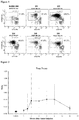


Classifications
-
- A—HUMAN NECESSITIES
- A61—MEDICAL OR VETERINARY SCIENCE; HYGIENE
- A61K—PREPARATIONS FOR MEDICAL, DENTAL OR TOILETRY PURPOSES
- A61K38/00—Medicinal preparations containing peptides
- A61K38/16—Peptides having more than 20 amino acids; Gastrins; Somatostatins; Melanotropins; Derivatives thereof
- A61K38/17—Peptides having more than 20 amino acids; Gastrins; Somatostatins; Melanotropins; Derivatives thereof from animals; from humans
- A61K38/19—Cytokines; Lymphokines; Interferons
- A61K38/20—Interleukins [IL]
- A61K38/2013—IL-2
-
- A—HUMAN NECESSITIES
- A61—MEDICAL OR VETERINARY SCIENCE; HYGIENE
- A61K—PREPARATIONS FOR MEDICAL, DENTAL OR TOILETRY PURPOSES
- A61K35/00—Medicinal preparations containing materials or reaction products thereof with undetermined constitution
- A61K35/12—Materials from mammals; Compositions comprising non-specified tissues or cells; Compositions comprising non-embryonic stem cells; Genetically modified cells
- A61K35/14—Blood; Artificial blood
- A61K35/17—Lymphocytes; B-cells; T-cells; Natural killer cells; Interferon-activated or cytokine-activated lymphocytes
-
- A—HUMAN NECESSITIES
- A61—MEDICAL OR VETERINARY SCIENCE; HYGIENE
- A61K—PREPARATIONS FOR MEDICAL, DENTAL OR TOILETRY PURPOSES
- A61K39/00—Medicinal preparations containing antigens or antibodies
- A61K39/46—Cellular immunotherapy
- A61K39/461—Cellular immunotherapy characterised by the cell type used
- A61K39/4611—T-cells, e.g. tumor infiltrating lymphocytes [TIL], lymphokine-activated killer cells [LAK] or regulatory T cells [Treg]
-
- A—HUMAN NECESSITIES
- A61—MEDICAL OR VETERINARY SCIENCE; HYGIENE
- A61K—PREPARATIONS FOR MEDICAL, DENTAL OR TOILETRY PURPOSES
- A61K39/00—Medicinal preparations containing antigens or antibodies
- A61K39/46—Cellular immunotherapy
- A61K39/462—Cellular immunotherapy characterized by the effect or the function of the cells
- A61K39/4621—Cellular immunotherapy characterized by the effect or the function of the cells immunosuppressive or immunotolerising
-
- A—HUMAN NECESSITIES
- A61—MEDICAL OR VETERINARY SCIENCE; HYGIENE
- A61K—PREPARATIONS FOR MEDICAL, DENTAL OR TOILETRY PURPOSES
- A61K39/00—Medicinal preparations containing antigens or antibodies
- A61K39/46—Cellular immunotherapy
- A61K39/464—Cellular immunotherapy characterised by the antigen targeted or presented
- A61K39/4643—Vertebrate antigens
- A61K39/46434—Antigens related to induction of tolerance to non-self
-
- A—HUMAN NECESSITIES
- A61—MEDICAL OR VETERINARY SCIENCE; HYGIENE
- A61P—SPECIFIC THERAPEUTIC ACTIVITY OF CHEMICAL COMPOUNDS OR MEDICINAL PREPARATIONS
- A61P37/00—Drugs for immunological or allergic disorders
- A61P37/02—Immunomodulators
- A61P37/06—Immunosuppressants, e.g. drugs for graft rejection
-
- G—PHYSICS
- G01—MEASURING; TESTING
- G01N—INVESTIGATING OR ANALYSING MATERIALS BY DETERMINING THEIR CHEMICAL OR PHYSICAL PROPERTIES
- G01N33/00—Investigating or analysing materials by specific methods not covered by groups G01N1/00 - G01N31/00
- G01N33/48—Biological material, e.g. blood, urine; Haemocytometers
- G01N33/50—Chemical analysis of biological material, e.g. blood, urine; Testing involving biospecific ligand binding methods; Immunological testing
- G01N33/68—Chemical analysis of biological material, e.g. blood, urine; Testing involving biospecific ligand binding methods; Immunological testing involving proteins, peptides or amino acids
- G01N33/6893—Chemical analysis of biological material, e.g. blood, urine; Testing involving biospecific ligand binding methods; Immunological testing involving proteins, peptides or amino acids related to diseases not provided for elsewhere
-
- A—HUMAN NECESSITIES
- A61—MEDICAL OR VETERINARY SCIENCE; HYGIENE
- A61K—PREPARATIONS FOR MEDICAL, DENTAL OR TOILETRY PURPOSES
- A61K2239/00—Indexing codes associated with cellular immunotherapy of group A61K39/46
- A61K2239/38—Indexing codes associated with cellular immunotherapy of group A61K39/46 characterised by the dose, timing or administration schedule
-
- A—HUMAN NECESSITIES
- A61—MEDICAL OR VETERINARY SCIENCE; HYGIENE
- A61K—PREPARATIONS FOR MEDICAL, DENTAL OR TOILETRY PURPOSES
- A61K2239/00—Indexing codes associated with cellular immunotherapy of group A61K39/46
- A61K2239/46—Indexing codes associated with cellular immunotherapy of group A61K39/46 characterised by the cancer treated
- A61K2239/48—Blood cells, e.g. leukemia or lymphoma
-
- G—PHYSICS
- G01—MEASURING; TESTING
- G01N—INVESTIGATING OR ANALYSING MATERIALS BY DETERMINING THEIR CHEMICAL OR PHYSICAL PROPERTIES
- G01N2800/00—Detection or diagnosis of diseases
- G01N2800/52—Predicting or monitoring the response to treatment, e.g. for selection of therapy based on assay results in personalised medicine; Prognosis
Definitions
- Tregs are required for immune tolerance and function as dominant suppressors of adaptive and innate immune effector cells.
- Tregs-based therapeutics offer a homeostatic mechanism to control maladaptive immune activation noted in immune disorders such as solid organ transplantation rejections and graft-versus-host disease (GVHD), and also in other disorders of impaired peripheral tolerance where Tregs dysfunction is increasingly implicated (e.g., systemic autoimmune diseases including vasculitis, systemic lupus erythematosus (SLE), type 1 diabetes (T1D), multiple sclerosis (MS), psoriasis, rheumatoid arthritis (RA), inflammatory bowel disease (IBD), and allergic asthma) ( Saadoun et al.
- SLE systemic autoimmune diseases including vasculitis, systemic lupus erythematosus (SLE), type 1 diabetes (T1D), multiple sclerosis (MS), psoriasis, rheumatoid arthritis (RA), inflammatory
- compositions and methods for improving the ability of Tregs to ameliorate unwanted immune reactions are urgently required, especially for children, who have many years to live with the debilitating consequences of cGVHD, such as sclerotic skin, joint contractures or pulmonary fibrosis.
- WO2012/123381A1 is directed to the use of IL-2 regimen to treat autoimmune or immune-related diseases.
- the claimed subject matter is neither disclosed nor suggested by any of the above prior art documents.
- the present invention is directed to Interleukin-2 (IL-2) for use in treating graft-versus-host disease (GVHD) in a pediatric subject, wherein the IL-2 is administered as
- a multiple-variable IL-2 dose method of treating a subject afflicted with an immune disorder comprising: a) administering to the subject an induction regimen comprising continuously administering to the subject interleukin-2 (IL-2) at a dose that increases the subject's plasma IL-2 level and increases the subject's ratio of regulatory T lymphocytes (Tregs) to conventional T lymphocytes (Tcons) (Tregs:Tcons); and b) subsequently administering to the subject at least one maintenance regimen comprising continuously administering to the subject an IL-2 maintenance dose that is higher than the induction regimen dose and that i) further increases the subject's plasma IL-2 level and ii) further increases the ratio of Tregs to Tcons, thereby treating the subject, is provided.
- IL-2 interleukin-2
- the IL-2 maintenance regimen increases the subject's plasma IL-2 level beyond the peak plasma IL-2 level induced by the induction regimen.
- the plasma IL-2 levels are determined by analyzing one or more of time after IL-2 administration, IL-2 protein levels, IL-2 protein activity, IL-2 nucleic acid levels, Tregs proliferation, Tregs activity, Tregs phosphorylated STAT5 levels, Tregs FOXP3 levels, and Tregs apoptosis.
- the induction regimen dose is about 0.3 x 10 6 IU/m 2 /day to about 3.0 x 10 6 IU/m 2 /day.
- the induction regimen dose is less than about 6.0 x 10 6 IU/m 2 /day.
- the continuous administration of the induction regimen comprises administration once per day and continues indefinitely as long as the patient continues to experience clinical benefit.
- the continuous administration of the induction regimen comprises administration once per day during at least 1-14 consecutive days.
- the Tregs:Tcons ratio in the maintenance regimen is increased by at least 20% over the maximal Tregs:Tcons during the induction regimen.
- the maiintenance regimen dose is at least about 20% higher than the induction regimen dose.
- the maintenance regimen dose is about 0.3 x 10 6 IU/m 2 /day to about 3.0 x 10 6 IU/m 2 /day.
- the maintenance regimen dose is less than about 6.0 x 10 6 IU/m 2 /day.
- the continuous administration of the maintenance regimen comprises administration indefinitely as long as the patient continues to experience clinical benefit.
- the continuous administration of the maintenance regimen comprises administration once per day during at least 1-42 consecutive days.
- the IL-2 is administered in a pharmaceutically acceptable formulation.
- the IL-2 is administered by an administration route selected from the group consisting of subcutaneous, intravenous, intraperitoneal, and intramuscular.
- the IL-2 is administered subcutaneously.
- the immune disorder is selected from the group consisting of graft-versus-host disease (GVHD), solid organ transplantation rejection, vasculitis, systemic lupus erythematosus (SLE), type 1 diabetes (T1D), multiple sclerosis (MS), psoriasis, rheumatoid arthritis (RA), inflammatory bowel disease (IBD), and allergic asthma.
- the immune disorder is cGVHD.
- the subject has had an inadequate response to systemic steroids.
- the subject has persistent or recurrent chronic GVHD despite at least 2 prior systemic therapies including steroids.
- the subject has had extra-corporeal photopheresis (ECP) prior to IL-2 administration.
- ECP extra-corporeal photopheresis
- the induction regimen and/or the maintenance regimen further comprises administration of one or more additional therapies to treat the immune disorder.
- the one or more additional therapies is selected from the group consisting of ECP and Tregs.
- the Tregs are administered as a composition comprising T cells other than Tregs.
- the composition has a Tregs:Tcons ratio of at least 1:2.
- the composition is obtained from CD8+ and CD19+ co-depletion and CD25+ positive selection of biological material comprising T cells.
- the Tregs are administered at between about 0.1 x 10 6 cells/kg body weight to 1.0 x 10 6 cells/kg body weight.
- the subject's own Tregs are administered to the subject.
- Tregs from the same hematopoietic stem cell donor from which hematopoietic stem cell transplantation was obtained is used.
- the Tregs composition has >70% total cell viability, a negative gram stain, ⁇ 90% CD4+CD25+ cells, and/or ⁇ 50% FoxP3+ cells.
- the Tregs are administered as an infusion. In yet another embodiment, the Tregs are administered before, concurrently with, or after IL-2 administration. In another ambodiment, the Tregs are administered before IL-2 administration. In still another embodiment, the subject is a mammal, such as a human or an animal model of an immune disorder.
- a method of stratifying subjects afflicted with an immune disorder according to benefit from a fixed daily IL-2 dose treatment method comprising obtaining a biological sample comprising T lymphocytes from a subject and determining the ratio of regulatory T lymphocytes (Tregs) to conventional T lymphocytes (Tcons) (Tregs:Tcons) in the subject sample, wherein the Tregs:Tcons ratio of greater than or equal to about 0.07 indicates that the subject would benefit from the fixed daily IL-2 dose treatment method and wherein the Tregs:Tcons ratio of less than about 0.07 indicates that the subject would not benefit from the fixed daily IL-2 dose treatment method, is provided.
- Tregs regulatory T lymphocytes
- Tcons conventional T lymphocytes
- the method further comprises recommending, prescribing, or administering a fixed daily IL-2 dose method or a method of treatment described herein if the immune disorder is determined to benefit from the fixed daily IL-2 dose treatment method.
- the method further comprises recommending, prescribing, or administering anti-immune disorder therapy other than a fixed daily IL-2 dose treatment method if the immune disorder is determined to not benefit from a fixed daily IL-2 dose treatment method.
- a method of stratifying subjects afflicted with an immune disorder according to benefit from a fixed daily IL-2 dose treatment method comprising obtaining a biological sample comprising T lymphocytes from a subject after the induction regiment and determining the ratio of regulatory T lymphocytes (Tregs) to conventional T lymphocytes (Tcons) (Tregs:Tcons) in the subject sample, wherein the Tregs:Tcons ratio of greater than or equal to about 0.20 indicates that the subject would benefit from the fixed daily IL-2 dose treatment method and wherein the Tregs:Tcons ratio of less than about 0.20 indicates that the subject would not benefit from the fixed daily IL-2 dose treatment method, is provided.
- Tregs regulatory T lymphocytes
- Tcons conventional T lymphocytes
- the method further comprises recommending, prescribing, or administering a fixed daily IL-2 dose method or a method of treatment described herein if the immune disorder is determined to benefit from the fixed daily IL-2 dose treatment method.
- the method further comprises recommending, prescribing, or administering anti-immune disorder therapy other than a fixed daily IL-2 dose treatment method if the immune disorder is determined to not benefit from a fixed daily IL-2 dose treatment method.
- Tregs proliferation occurs by 1 week after start of an IL-2 induction regimen in all Treg subsets along with an increase in Tregs population size.
- Tregs proliferation subsides after week 2 with a fall in Ki-67 expression (proliferation) alongside the preservation of expanded Treg subpopulations at week 12 of IL-2 treatment.
- Ki-67 expression proliferation
- a similar temporal pattern of Tregs activation is observed using pSTAT5 and FoxP3 markers, with an initial early generalized enhancement across Treg subpopulations that later subsided during IL-2 treatment despite the continued preservation of enhanced Treg population size.
- a fall in Treg activation marker expression and a considerable depletion of Treg populations was then observed by 4 weeks after IL-2 discontinuation.
- IL-2 induced Treg enhancement occurs in vivo and tachyphylaxis due to diminution of plasma IL-2 levels after binding by increased numbers of circulating Treg with higher CD25 expression is avoided. Accordingly, the methods of the present invention provide unexpectedly enhanced treatment of immune disorders using IL-2 and allow for Tregs and Tcons, either directly or indirectly, to be used as biomarkers of IL-2 efficacy in treating such immune disorders.
- an element means one element or more than one element.
- altered amount refers to increased or decreased copy number (e.g., germline and/or somatic) of a biomarker nucleic acid, e.g., increased or decreased expression level in a biological sample, as compared to the expression level or copy number of the biomarker nucleic acid in a control sample.
- altered amount of a biomarker also includes an increased or decreased protein level of a biomarker protein in a sample, e.g., an immune disorder sample, as compared to the corresponding protein level in a normal, control sample.
- an altered amount of a biomarker protein may be determined by detecting posttranslational modification such as methylation status of the marker, which may affect the expression or activity of the biomarker protein.
- the amount of a biomarker in a subject is "significantly" higher or lower than the normal amount of the biomarker, if the amount of the biomarker is greater or less, respectively, than the normal level by an amount greater than the standard error of the assay employed to assess amount, and preferably at least 20%, 30%, 40%, 50%, 60%, 70%, 80%, 90%, 100%, 150%, 200%, 300%, 350%, 400%, 500%, 600%, 700%, 800%, 900%, 1000% or than that amount.
- the amount of the biomarker in the subject can be considered "significantly" higher or lower than the normal amount if the amount is at least about two, and preferably at least about three, four, or five times, higher or lower, respectively, than the normal amount of the biomarker.
- altered level of expression of a biomarker refers to an expression level or copy number of the biomarker in a test sample, e.g., a sample derived from a patient suffering from an immune disorder, that is greater or less than the standard error of the assay employed to assess expression or copy number, and is preferably at least twice, and more preferably three, four, five or ten or more times the expression level or copy number of the biomarker in a control sample (e.g., sample from a healthy subjects not having the associated disease) and preferably, the average expression level or copy number of the biomarker in several control samples.
- a test sample e.g., a sample derived from a patient suffering from an immune disorder
- a control sample e.g., sample from a healthy subjects not having the associated disease
- the altered level of expression is greater or less than the standard error of the assay employed to assess expression or copy number, and is preferably at least twice, and more preferably three, four, five or ten or more times the expression level or copy number of the biomarker in a control sample (e.g., sample from a healthy subjects not having the associated disease) and preferably, the average expression level or copy number of the biomarker in several control samples.
- a control sample e.g., sample from a healthy subjects not having the associated disease
- altered activity of a biomarker refers to an activity of the biomarker which is increased or decreased in a disease state, e.g., in an immune disorder sample, as compared to the activity of the biomarker in a normal, control sample.
- Altered activity of the biomarker may be the result of, for example, altered expression of the biomarker, altered protein level of the biomarker, altered structure of the biomarker, or, e.g., an altered interaction with other proteins involved in the same or different pathway as the biomarker or altered interaction with transcriptional activators or inhibitors.
- altered structure of a biomarker refers to the presence of mutations or allelic variants within a biomarker nucleic acid or protein, e.g ., mutations which affect expression or activity of the biomarker nucleic acid or protein, as compared to the normal or wild-type gene or protein.
- mutations include, but are not limited to substitutions, deletions, or addition mutations. Mutations may be present in the coding or non-coding region of the biomarker nucleic acid.
- antibody broadly encompass naturally-occurring forms of antibodies (e.g. IgG, IgA, IgM, IgE) and recombinant antibodies such as single-chain antibodies, chimeric and humanized antibodies and multi-specific antibodies, as well as fragments and derivatives of all of the foregoing, which fragments and derivatives have at least an antigenic binding site.
- Antibody derivatives may comprise a protein or chemical moiety conjugated to an antibody.
- antibody as used herein also includes an "antigen-binding portion" of an antibody (or simply “antibody portion”).
- antigen-binding portion refers to one or more fragments of an antibody that retain the ability to specifically bind to an antigen (e.g., a biomarker polypeptide or fragment thereof). It has been shown that the antigen-binding function of an antibody can be performed by fragments of a full-length antibody.
- binding fragments encompassed within the term "antigen-binding portion" of an antibody include (i) a Fab fragment, a monovalent fragment consisting of the VL, VH, CL and CH1 domains; (ii) a F(ab') 2 fragment, a bivalent fragment comprising two Fab fragments linked by a disulfide bridge at the hinge region; (iii) a Fd fragment consisting of the VH and CH1 domains; (iv) a Fv fragment consisting of the VL and VH domains of a single arm of an antibody, (v) a dAb fragment ( Ward et al., (1989) Nature 341:544-546 ), which consists of a VH domain; and (vi) an isolated complementarity determining region (CDR).
- a Fab fragment a monovalent fragment consisting of the VL, VH, CL and CH1 domains
- a F(ab') 2 fragment a bivalent fragment comprising two Fab fragments linked by
- the two domains of the Fv fragment, VL and VH are coded for by separate genes, they can be joined, using recombinant methods, by a synthetic linker that enables them to be made as a single protein chain in which the VL and VH regions pair to form monovalent polypeptides (known as single chain Fv (scFv); see e.g., Bird et al. (1988) Science 242:423-426 ; and Huston et al. (1988) Proc. Natl. Acad. Sci. USA 85:5879-5883 ; and Osbourn et al. 1998, Nature Biotechnology 16: 778 ).
- scFv single chain Fv
- Such single chain antibodies are also intended to be encompassed within the term "antigen-binding portion" of an antibody.
- Any VH and VL sequences of specific scFv can be linked to human immunoglobulin constant region cDNA or genomic sequences, in order to generate expression vectors encoding complete IgG polypeptides or other isotypes.
- VH and VL can also be used in the generation of Fab, Fv or other fragments of immunoglobulins using either protein chemistry or recombinant DNA technology.
- Other forms of single chain antibodies, such as diabodies are also encompassed.
- Diabodies are bivalent, bispecific antibodies in which VH and VL domains are expressed on a single polypeptide chain, but using a linker that is too short to allow for pairing between the two domains on the same chain, thereby forcing the domains to pair with complementary domains of another chain and creating two antigen binding sites (see e.g., Holliger et al. (1993) Proc. Natl. Acad. Sci. U.S.A. 90:6444-6448 ; Poljak et al. (1994) Structure 2:1121-1123 ).
- an antibody or antigen-binding portion thereof may be part of larger immunoadhesion polypeptides, formed by covalent or noncovalent association of the antibody or antibody portion with one or more other proteins or peptides.
- immunoadhesion polypeptides include use of the streptavidin core region to make a tetrameric scFv polypeptide ( Kipriyanov et al. (1995) Human Antibodies and Hybridomas 6:93-101 ) and use of a cysteine residue, biomarker peptide and a C-terminal polyhistidine tag to make bivalent and biotinylated scFv polypeptides ( Kipriyanov et al. (1994) Mol. Immunol.
- Antibody portions such as Fab and F(ab') 2 fragments, can be prepared from whole antibodies using conventional techniques, such as papain or pepsin digestion, respectively, of whole antibodies.
- antibodies, antibody portions and immunoadhesion polypeptides can be obtained using standard recombinant DNA techniques, as described herein.
- Antibodies may be polyclonal or monoclonal; xenogeneic, allogeneic, or syngeneic; or modified forms thereof (e.g. humanized, chimeric, etc.). Antibodies may also be fully human. Preferably, antibodies of the invention bind specifically or substantially specifically to a biomarker polypeptide or fragment thereof.
- monoclonal antibodies and “monoclonal antibody composition”, as used herein, refer to a population of antibody polypeptides that contain only one species of an antigen binding site capable of immunoreacting with a particular epitope of an antigen
- polyclonal antibodies and “polyclonal antibody composition” refer to a population of antibody polypeptides that contain multiple species of antigen binding sites capable of interacting with a particular antigen.
- a monoclonal antibody composition typically displays a single binding affinity for a particular antigen with which it immunoreacts.
- Antibodies may also be "humanized,” which is intended to include antibodies made by a non-human cell having variable and constant regions which have been altered to more closely resemble antibodies that would be made by a human cell. For example, by altering the non-human antibody amino acid sequence to incorporate amino acids found in human germline immunoglobulin sequences.
- the humanized antibodies of the invention may include amino acid residues not encoded by human germline immunoglobulin sequences (e.g., mutations introduced by random or site-specific mutagenesis in vitro or by somatic mutation in vivo ), for example in the CDRs.
- the term "humanized antibody”, as used herein, also includes antibodies in which CDR sequences derived from the germline of another mammalian species, such as a mouse, have been grafted onto human framework sequences.
- the term "assigned score” refers to the numerical value designated for each of the biomarkers after being measured in a patient sample.
- the assigned score correlates to the absence, presence or inferred amount of the biomarker in the sample.
- the assigned score can be generated manually ( e.g., by visual inspection) or with the aid of instrumentation for image acquisition and analysis.
- the assigned score is determined by a qualitative assessment, for example, detection of a fluorescent readout on a graded scale, or quantitative assessment.
- an "aggregate score” which refers to the combination of assigned scores from a plurality of measured biomarkers, is determined.
- the aggregate score is a summation of assigned scores.
- combination of assigned scores involves performing mathematical operations on the assigned scores before combining them into an aggregate score.
- the aggregate score is also referred to herein as the predictive score.
- Biomarker refers to a measurable entity of the present invention that has been determined to be predictive of anti-immune disorder therapy and/or effect treatment of the anti-immune disorder (e.g., multiple-variable dose IL-2 therapy alone or in combination with one or more other anti-immune disorder therapies).
- Biomarkers can include, without limitation, cell types (e.g., Tregs and/or Tcons), cell ratios (e.g., Tregs to Tcons ratio), nucleic acids (e.g., genomic nucleic acids and/or transcribed nucleic acids) and proteins, particularly those involved shown in Table 1.
- Biomarkers can further include immunological targets or agents that downregulate unwanted immune reactions in order to treat the immune disorder of interest as described further herein.
- blocking antibody or an antibody “antagonist” is one which inhibits or reduces at least one biological activity of the antigen(s) it binds.
- the blocking antibodies or antagonist antibodies or fragments thereof described herein substantially or completely inhibit a given biological activity of the antigen(s).
- body fluid refers to fluids that are excreted or secreted from the body as well as fluid that are normally not (e.g. amniotic fluid, aqueous humor, bile, blood and blood plasma, cerebrospinal fluid, cerumen and earwax, cowper's fluid or pre-ejaculatory fluid, chyle, chyme, stool, female ejaculate, interstitial fluid, intracellular fluid, lymph, menses, breast milk, mucus, pleural fluid, pus, saliva, sebum, semen, serum, sweat, synovial fluid, tears, urine, vaginal lubrication, vitreous humor, and vomit).
- lymphocytes such as T lymphocytes and subpopulations thereof, are used.
- coding region refers to regions of a nucleotide sequence comprising codons which are translated into amino acid residues
- non-coding region refers to regions of a nucleotide sequence that are not translated into amino acids (e.g., 5' and 3' untranslated regions).
- complementary refers to the broad concept of sequence complementarity between regions of two nucleic acid strands or between two regions of the same nucleic acid strand. It is known that an adenine residue of a first nucleic acid region is capable of forming specific hydrogen bonds ("base pairing") with a residue of a second nucleic acid region which is antiparallel to the first region if the residue is thymine or uracil. Similarly, it is known that a cytosine residue of a first nucleic acid strand is capable of base pairing with a residue of a second nucleic acid strand which is antiparallel to the first strand if the residue is guanine.
- a first region of a nucleic acid is complementary to a second region of the same or a different nucleic acid if, when the two regions are arranged in an antiparallel fashion, at least one nucleotide residue of the first region is capable of base pairing with a residue of the second region.
- the first region comprises a first portion and the second region comprises a second portion, whereby, when the first and second portions are arranged in an antiparallel fashion, at least about 50%, and preferably at least about 75%, at least about 90%, or at least about 95% of the nucleotide residues of the first portion are capable of base pairing with nucleotide residues in the second portion. More preferably, all nucleotide residues of the first portion are capable of base pairing with nucleotide residues in the second portion.
- control refers to any reference standard suitable to provide a comparison to the expression products in the test sample.
- the control comprises obtaining a "control sample” from which expression product levels are detected and compared to the expression product levels from the test sample.
- a control sample may comprise any suitable sample, including but not limited to a sample from a control immune disorder patient (can be stored sample or previous sample measurement) with a known outcome; normal tissue or cells isolated from a subject, such as a normal patient or the immune disorder patient, cultured primary cells/tissues isolated from a subject such as a normal subject or the immune disorder patient, adjacent normal cells/tissues obtained from the same organ or body location of the immune disorder patient, a tissue or cell sample isolated from a normal subject, or a primary cells/tissues obtained from a depository.
- control may comprise a reference standard expression product level from any suitable source, including but not limited to housekeeping genes, an expression product level range from normal tissue (or other previously analyzed control sample), a previously determined expression product level range within a test sample from a group of patients, or a set of patients with a certain outcome (for example, survival for one, two, three, four years, etc.) or receiving a certain treatment (for example, standard of care immune disorder therapy).
- a certain outcome for example, survival for one, two, three, four years, etc.
- a certain treatment for example, standard of care immune disorder therapy
- control samples and reference standard expression product levels can be used in combination as controls in the methods of the present invention.
- control may comprise normal or non-immune disorder cell/tissue sample.
- control may comprise an expression level for a set of patients, such as a set of immune disorder patients, or for a set of immune disorder patients receiving a certain treatment, or for a set of patients with one outcome versus another outcome.
- the specific expression product level of each patient can be assigned to a percentile level of expression, or expressed as either higher or lower than the mean or average of the reference standard expression level.
- the control may comprise normal cells, cells from patients treated with combination chemotherapy, and cells from patients having an immune disorder that has responded to a treatment of interest.
- the control may also comprise a measured value for example, average level of expression of a particular gene in a population compared to the level of expression of a housekeeping gene in the same population.
- the control comprises a ratio transformation of expression product levels, including but not limited to determining a ratio of expression product levels of two cell types and/or genes in the test sample and comparing it to any suitable ratio of the same two cell types and/or genes in a reference standard; determining expression product levels of the two or more cell types and/or genes in the test sample and determining a difference in expression product levels in any suitable control; and determining expression product levels of the two or more cell types and/or genes in the test sample, normalizing their expression to expression of housekeeping cell types and/or genes in the test sample, and comparing to any suitable control.
- control comprises a control sample which is of the same lineage and/or type as the test sample.
- control may comprise expression product levels grouped as percentiles within or based on a set of patient samples, such as all patients with the immune disorder.
- a control expression product level is established wherein higher or lower levels of expression product relative to, for instance, a particular percentile, are used as the basis for predicting outcome.
- a control expression product level is established using expression product levels from immune disorder control patients with a known outcome, and the expression product levels from the test sample are compared to the control expression product level as the basis for predicting outcome.
- the methods of the invention are not limited to use of a specific cut-point in comparing the level of expression product in the test sample to the control.
- the "copy number" of a biomarker nucleic acid refers to the number of DNA sequences in a cell (e.g., germline and/or somatic) encoding a particular gene product. Generally, for a given gene, a mammal has two copies of each gene. The copy number can be increased, however, by gene amplification or duplication, or reduced by deletion. For example, germline copy number changes include changes at one or more genomic loci, wherein said one or more genomic loci are not accounted for by the number of copies in the normal complement of germline copies in a control (e.g., the normal copy number in germline DNA for the same species as that from which the specific germline DNA and corresponding copy number were determined).
- Somatic copy number changes include changes at one or more genomic loci, wherein said one or more genomic loci are not accounted for by the number of copies in germline DNA of a control (e.g., copy number in germline DNA for the same subject as that from which the somatic DNA and corresponding copy number were determined).
- the "normal" copy number (e.g., germline and/or somatic) of a biomarker nucleic acid or "normal” level of expression of a biomarker nucleic acid, or protein is the activity/level of expression or copy number in a biological sample, e.g., a sample containing tissue, whole blood, serum, plasma, buccal scrape, saliva, cerebrospinal fluid, urine, stool, and bone marrow, from a subject, e.g., a human, not afflicted with an immune disorder, or from a corresponding non-immune disorder tissue in the same subject who has an immune disorder.
- a biological sample e.g., a sample containing tissue, whole blood, serum, plasma, buccal scrape, saliva, cerebrospinal fluid, urine, stool, and bone marrow
- determining a suitable treatment regimen for the subject is taken to mean the determination of a treatment regimen (i.e., a single therapy or a combination of different therapies that are used for the prevention and/or treatment of an immune disorder in the subject) for a subject that is started, modified and/or ended based or essentially based or at least partially based on the results of the analysis according to the present invention.
- a treatment regimen i.e., a single therapy or a combination of different therapies that are used for the prevention and/or treatment of an immune disorder in the subject
- One example is determining whether to provide targeted therapy against the immune disorder to provide anti-immune disorder therapy (e.g., multiple-variable dose IL-2 therapy alone or in combination with one or more other anti-immune disorder therapies).
- the determination can, in addition to the results of the analysis according to the present invention, be based on personal characteristics of the subject to be treated. In most cases, the actual determination of the suitable treatment regimen for the subject will be performed by the attending physician or doctor.
- expression signature refers to a group of two or more coordinately expressed biomarkers.
- the genes, proteins, and the like making up this signature may be expressed in a specific cell lineage, stage of differentiation, or during a particular biological response.
- the biomarkers can reflect biological aspects of the cell types in which they are expressed.
- Expression data and gene expression levels can be stored on computer readable media, e.g., the computer readable medium used in conjunction with a microarray or chip reading device. Such expression data can be manipulated to generate expression signatures.
- a molecule or cell is "fixed” or "affixed” to a substrate if it is covalently or non-covalently associated with the substrate such that the substrate can be rinsed with a fluid (e.g. standard saline citrate, pH 7.4) without a substantial fraction of the molecule or cell dissociating from the substrate.
- a fluid e.g. standard saline citrate, pH 7.4
- homologous refers to nucleotide sequence similarity between two regions of the same nucleic acid strand or between regions of two different nucleic acid strands. When a nucleotide residue position in both regions is occupied by the same nucleotide residue, then the regions are homologous at that position. A first region is homologous to a second region if at least one nucleotide residue position of each region is occupied by the same residue. Homology between two regions is expressed in terms of the proportion of nucleotide residue positions of the two regions that are occupied by the same nucleotide residue.
- a region having the nucleotide sequence 5'-ATTGCC-3' and a region having the nucleotide sequence 5'-TATGGC-3' share 50% homology.
- the first region comprises a first portion and the second region comprises a second portion, whereby, at least about 50%, and preferably at least about 75%, at least about 90%, or at least about 95% of the nucleotide residue positions of each of the portions are occupied by the same nucleotide residue. More preferably, all nucleotide residue positions of each of the portions are occupied by the same nucleotide residue.
- immune disorders refers to conditions characterized by an unwanted immune response such that a desired anti-immune disorder response suppresses the immune response.
- Such conditions in which downregulation of an immune response is desired are well known in the art and include, without limitation, situations of tissue, skin and organ transplantation, in graft-versus-host disease (GVHD), inflammation, or in autoimmune diseases, such as systemic lupus erythematosus, multiple sclerosis, allergy, hypersensitivity response, parasitic and viral infections, a disorder requiring increased CD4+ T cell production or function, a disorder requiring improved vaccination efficiency, and a disorder requiring increased regulatory T cell production or function, as described further herein.
- GVHD graft-versus-host disease
- an immune disorder includes the decrease, limitation, or blockage, of, for example a particular action, function, or interaction.
- an immune disorder is "inhibited” if at least one symptom of the immune disorder is alleviated, terminated, slowed, or prevented.
- an immune disorder is also “inhibited” if recurrence or spread of the immune disorder is reduced, slowed, delayed, or prevented.
- interaction when referring to an interaction between two molecules, refers to the physical contact (e.g., binding) of the molecules with one another. Generally, such an interaction results in an activity (which produces a biological effect) of one or both of said molecules.
- isolated protein refers to a protein that is substantially free of other proteins, cellular material, separation medium, and culture medium when isolated from cells or produced by recombinant DNA techniques, or chemical precursors or other chemicals when chemically synthesized.
- isolated or purified protein or biologically active portion thereof is substantially free of cellular material or other contaminating proteins from the cell or tissue source from which the antibody, polypeptide, peptide or fusion protein is derived, or substantially free from chemical precursors or other chemicals when chemically synthesized.
- substantially free of cellular material includes preparations of a biomarker polypeptide or fragment thereof, in which the protein is separated from cellular components of the cells from which it is isolated or recombinantly produced.
- the language "substantially free of cellular material” includes preparations of a biomarker protein or fragment thereof, having less than about 30% (by dry weight) of non-biomarker protein (also referred to herein as a "contaminating protein”), more preferably less than about 20% of non-biomarker protein, still more preferably less than about 10% of non-biomarker protein, and most preferably less than about 5% non-biomarker protein.
- non-biomarker protein also referred to herein as a "contaminating protein”
- polypeptide, peptide or fusion protein or fragment thereof e.g., a biologically active fragment thereof
- it is also preferably substantially free of culture medium, i.e., culture medium represents less than about 20%, more preferably less than about 10%, and most preferably less than about 5% of the volume of the protein preparation.
- kits is any manufacture (e.g. a package or container) comprising at least one reagent, e.g. a therapeutic, probe, small molecule, and the like, for specifically detecting and/or therapeutically affecting the expression of a marker of the present invention.
- the kit may be promoted, distributed, or sold as a unit for performing the methods of the present invention.
- the kit may comprise one or more reagents necessary to express a composition useful in the methods of the present invention.
- the kit may further comprise a reference standard, e.g., a nucleic acid encoding a protein that does not affect or regulate signaling pathways controlling immunological responses, cell growth, division, migration, survival, or apoptosis.
- control proteins including, but not limited to, common molecular tags (e.g., green fluorescent protein and beta-galactosidase), proteins not classified in any of pathway encompassing cell growth, division, migration, survival or apoptosis by GeneOntology reference, or ubiquitous housekeeping proteins.
- Reagents in the kit may be provided in individual containers or as mixtures of two or more reagents in a single container.
- instructional materials which describe the use of the compositions within the kit can be included.
- the "normal" level of expression of a biomarker is the level of expression of the biomarker in cells of a subject, e.g., a human patient, not afflicted with an immune disorder.
- An “over-expression” or “significantly higher level of expression” of a biomarker refers to an expression level in a test sample that is greater than the standard error of the assay employed to assess expression, and is preferably at least 10%, and more preferably 1.2, 1.3, 1.4, 1.5, 1.6, 1.7, 1.8, 1.9, 2.0, 2.1, 2.2, 2.3, 2.4, 2.5, 2.6, 2.7, 2.8, 2.9, 3, 3.5, 4, 4.5, 5, 5.5, 6, 6.5, 7, 7.5, 8, 8.5, 9, 9.5, 10, 10.5, 11, 12, 13, 14, 15, 16, 17, 18, 19, 20 times or more higher than the expression activity or level of the biomarker in a control sample (e.g., sample from a healthy subject not having the biomarker associated disease) and preferably, the average expression level
- a "significantly lower level of expression" of a biomarker refers to an expression level in a test sample that is at least 10%, and more preferably 1.2, 1.3, 1.4, 1.5, 1.6, 1.7, 1.8, 1.9, 2.0, 2.1, 2.2, 2.3, 2.4, 2.5, 2.6, 2.7, 2.8, 2.9, 3, 3.5, 4, 4.5, 5, 5.5, 6, 6.5, 7, 7.5, 8, 8.5, 9, 9.5, 10, 10.5, 11, 12, 13, 14, 15, 16, 17, 18, 19, 20 times or more lower than the expression level of the biomarker in a control sample (e.g., sample from a healthy subject not having the biomarker associated disease) and preferably, the average expression level of the biomarker in several control samples.
- a control sample e.g., sample from a healthy subject not having the biomarker associated disease
- Such "significance" levels can also be applied to any other measured parameter described herein, such as for expression, inhibition, cytotoxicity, cell growth, and the like.
- the term "predictive" includes the use of a biomarker for determining the likelihood of response of an immune disorder to anti-immune disorder therapy, such as multiple-variable dose IL-2 therapy alone or in combination with one or more other anti-immune disorder therapies.
- Such predictive use of the biomarker may be confirmed by, e.g., (1) increased or decreased copy number (e.g., by FISH, FISH plus SKY, single-molecule sequencing, e.g ., as described in the art at least at J.
- Biotechnol., 86:289-301 , or qPCR overexpression or underexpression of a biomarker nucleic acid (e.g., by ISH, Northern Blot, or qPCR), increased or decreased biomarker cells or cell ratios ( e.g., by cell sorting and/or counting), protein ( e.g., by IHC) and/or biomarker target, or increased or decreased activity, e.g., in more than about 5%, 6%, 7%, 8%, 9%, 10%, 11%, 12%, 13%, 14%, 15%, 20%, 25%, 30%, 40%, 50%, 60%, 70%, 80%, 90%, 95%, 100%, or more of assayed immune disorder samples; (2) its absolute or relatively modulated presence or absence in a biological sample, e.g., a sample containing tissue, whole blood, serum, plasma, buccal scrape, saliva, cerebrospinal fluid, urine, stool, or bone marrow, from a subject, e.g.,
- a human, afflicted with an immune disorder (3) its absolute or relatively modulated presence or absence in clinical subset of patients with an immune disorder (e.g., those responding to a particular anti-immune disorder therapy (e.g., multiple-variable dose IL-2 therapy alone or in combination with one or more other anti-immune disorder therapies) or those developing resistance thereto).
- an immune disorder e.g., those responding to a particular anti-immune disorder therapy (e.g., multiple-variable dose IL-2 therapy alone or in combination with one or more other anti-immune disorder therapies) or those developing resistance thereto).
- prevent refers to reducing the probability of developing a disease, disorder, or condition in a subject, who does not have, but is at risk of or susceptible to developing a disease, disorder, or condition.
- probe refers to any molecule which is capable of selectively binding to a specifically intended target molecule, for example, a nucleotide transcript or protein encoded by or corresponding to a biomarker nucleic acid. Probes can be either synthesized by one skilled in the art, or derived from appropriate biological preparations. For purposes of detection of the target molecule, probes may be specifically designed to be labeled, as described herein. Examples of molecules that can be utilized as probes include, but are not limited to, RNA, DNA, proteins, antibodies, and organic molecules.
- prognosis includes a prediction of the probable course and outcome of an immune disorder or the likelihood of recovery from the disease.
- use of statistical algorithms provides a prognosis of the immune disorder in an individual.
- the prognosis can be surgery, development of a clinical subtype of the immune disorder (e.g., GVHD subtype such as chronic GVHD), development of one or more clinical factors, or recovery from the disease.
- GVHD subtype such as chronic GVHD
- anti-immune disorder therapy e.g., multiple-variable dose IL-2 therapy alone or in combination with one or more other anti-immune disorder therapies
- a response of the immune disorder e.g., GVHD
- an anti-immune disorder therapy e.g., multiple-variable dose IL-2 therapy alone or in combination with one or more other anti-immune disorder therapies.
- Anti-immune disorder response may be assessed according to well-known methods in the art, including those criteria described in the Examples.
- CBR clinical benefit rate
- the clinical benefit rate is measured by determining the sum of the percentage of patients who are in complete remission (CR), the number of patients who are in partial remission (PR) and the number of patients having stable disease (SD) at a time point at least 6 months out from the end of therapy.
- the CBR for a particular anti-immune disorder therapeutic regimen is at least 25%, 30%, 35%, 40%, 45%, 50%, 55%, 60%, 65%, 70%, 75%, 80%, 85%, or more.
- Additional criteria for evaluating the response to anti-immune disorder therapies are related to "survival,” which includes all of the following: survival until mortality, also known as overall survival (wherein said mortality may be either irrespective of cause or tumor related); "recurrence-free survival” (wherein the term recurrence shall include both localized and distant recurrence); disease free survival (wherein the term disease shall include immune disorders and diseases associated therewith).
- the length of said survival may be calculated by reference to a defined start point (e.g., time of diagnosis or start of treatment) and end point (e.g., death or recurrence).
- criteria for efficacy of treatment can be expanded to include probability of survival, probability of recurrence within a given time period, and the like.
- a particular therapeutic regimen can be administered to a population of subjects and the outcome can be correlated to biomarker measurements that were determined prior to administration of any therapy.
- the outcome measurement may be pathologic response to therapy given in the neoadjuvant setting.
- outcome measures such as overall survival and disease-free survival can be monitored over a period of time for subjects following therapy for whom biomarker measurement values are known.
- the period of time for which subjects are monitored can vary. For example, subjects may be monitored for at least 0.25, 0.5, 0.75, 1, 2, 3, 4, 5, 6, 7, 8, 9, 10, 12, 14, 16, 18, 20, 25, 30, 35, 40, 45, 50, 55, or 60 months.
- Biomarker measurement threshold values that correlate to outcome of an anti-immune disorder therapy can be determined using well-known methods in the art, such as those described in the Examples section.
- the terms can also refer to an improved prognosis, for example, as reflected by an increased time to recurrence, which is the period to first recurrence censoring for second independent immune disorders as a first event or death without evidence of recurrence, or an increased overall survival, which is the period from treatment to death from any cause.
- To respond or to have a response means there is a beneficial endpoint attained when exposed to a stimulus. Alternatively, a negative or detrimental symptom is minimized, mitigated or attenuated on exposure to a stimulus. It will be appreciated that evaluating the likelihood that a tumor or subject will exhibit a favorable response is equivalent to evaluating the likelihood that the tumor or subject will not exhibit favorable response ( i.e., will exhibit a lack of response or be non-responsive).
- RNA interfering agent is defined as any agent which interferes with or inhibits expression of a target biomarker gene by RNA interference (RNAi).
- RNA interfering agents include, but are not limited to, nucleic acid molecules including RNA molecules which are homologous to the target biomarker gene of the invention, or a fragment thereof, short interfering RNA (siRNA), and small molecules which interfere with or inhibit expression of a target biomarker nucleic acid by RNA interference (RNAi).
- RNA interference is an evolutionally conserved process whereby the expression or introduction of RNA of a sequence that is identical or highly similar to a target biomarker nucleic acid results in the sequence specific degradation or specific post-transcriptional gene silencing (PTGS) of messenger RNA (mRNA) transcribed from that targeted gene (see Coburn, G. and Cullen, B. (2002) J. of Virology 76(18):9225 ), thereby inhibiting expression of the target biomarker nucleic acid.
- mRNA messenger RNA
- the RNA is double stranded RNA (dsRNA). This process has been described in plants, invertebrates, and mammalian cells.
- RNAi is initiated by the dsRNA-specific endonuclease Dicer, which promotes processive cleavage of long dsRNA into double-stranded fragments termed siRNAs.
- siRNAs are incorporated into a protein complex that recognizes and cleaves target mRNAs.
- RNAi can also be initiated by introducing nucleic acid molecules, e.g. , synthetic siRNAs, shRNAs, or other RNA interfering agents, to inhibit or silence the expression of target biomarker nucleic acids.
- inhibiting target biomarker nucleic acid expression includes any decrease in expression or protein activity or level of the target biomarker nucleic acid or protein encoded by the target biomarker nucleic acid.
- the decrease may be of at least 30%, 40%, 50%, 60%, 70%, 80%, 90%, 95% or 99% or more as compared to the expression of a target biomarker nucleic acid or the activity or level of the protein encoded by a target biomarker nucleic acid which has not been targeted by an RNA interfering agent.
- sample used for detecting or determining the presence or level of at least one biomarker is typically whole blood, plasma, serum, saliva, urine, stool (e.g., feces), tears, and any other bodily fluid (e.g., as described above under the definition of "body fluids"), or a tissue sample (e.g., biopsy) such as a small intestine, colon sample, or surgical resection tissue.
- body fluids e.g., as described above under the definition of "body fluids”
- tissue sample e.g., biopsy
- the method of the present invention further comprises obtaining the sample from the individual prior to detecting or determining the presence or level of at least one marker in the sample.
- any sample comprising T lymphocytes or subsets thereof are useful according to the present invention.
- the term "synergistic effect” refers to the combined effect of two or more anti-immune disorder agents or therapies can be greater than the sum of the separate effects of each such agent or therapy alone.
- subject refers to any healthy animal, mammal or human, or any animal, mammal or human afflicted with an immune disorder.
- subject is interchangeable with “patient.”
- survival includes all of the following: survival until mortality, also known as overall survival (wherein said mortality may be either irrespective of cause or tumor related); "recurrence-free survival” (wherein the term recurrence shall include both localized and distant recurrence); disease free survival (wherein the term disease shall include immune disorders and diseases associated therewith).
- the length of said survival may be calculated by reference to a defined start point ( e.g. time of diagnosis or start of treatment) and end point ( e.g. death, recurrence or metastasis).
- criteria for efficacy of treatment can be expanded to include response to therapy, probability of survival, probability of recurrence within a given time period, and the like.
- therapeutic effect refers to a local or systemic effect in animals, particularly mammals, and more particularly humans, caused by a pharmacologically active substance.
- the term thus means any substance intended for use in the diagnosis, cure, mitigation, treatment or prevention of disease or in the enhancement of desirable physical or mental development and conditions in an animal or human.
- therapeutically-effective amount means that amount of such a therapy or substance that produces some desired local or systemic effect at a reasonable benefit/risk ratio applicable to any treatment.
- a therapeutically effective amount of a compound will depend on its therapeutic index, solubility, and the like.
- certain compounds discovered by the methods of the present invention may be administered in a sufficient amount to produce a reasonable benefit/risk ratio applicable to such treatment.
- a “transcribed polynucleotide” or “nucleotide transcript” is a polynucleotide (e.g. an mRNA, hnRNA, a cDNA, or an analog of such RNA or cDNA) which is complementary to or homologous with all or a portion of a mature mRNA made by transcription of a biomarker nucleic acid and normal post-transcriptional processing (e.g. splicing), if any, of the RNA transcript, and reverse transcription of the RNA transcript.
- a polynucleotide e.g. an mRNA, hnRNA, a cDNA, or an analog of such RNA or cDNA
- Regulatory T-cells are naturally occurring CD4+CD25+FOXP3+ T lymphocytes that comprise ⁇ 5-10% of the circulating CD4+ T cell population, act to dominantly suppress autoreactive lymphocytes, and control innate and adaptive immune responses ( Piccirillo and Shevach (2004) Semin. Immunol. 16:81-88 ; Fehervari and Sakaguchi (2004) Curr. Opin. Immunol. 16:203-208 ; Azuma et al. (2003) Cancer Res. 63:4516-4520 ; Cederbom et al. (2000) Eur. J. Immunol. 30:1538-1543 ; Maloy et al. (2003) J. Exp. Med.
- Tregs achieve this suppressing, at least in part, by inhibiting the proliferation, expansion, and effector activity of conventional T cells (Tcons).
- T reg cells suppress effector T cells from destroying their (self-)target, either through cell-cell contact by inhibiting T cell help and activation, or through release of immunosuppressive cytokines such as IL-10 or TGF- ⁇ . Depletion of T reg cells was shown to enhance IL-2 induced antitumor immunity ( Imai et al. (2007) Cancer Sci. 98:416-23 ).
- Tcons are conventional T cells, also known as Tconv, that have effector functions (e.g., cytokine secretion, cytotoxic activity, and the like) to increase immune responses by virtue of their expression of one or more T cell receptors.
- Tcons are defined as any T cell population that is not a Treg and include, for example, naive T cells, activated T cells, memory T cells, resting Tcons, or Tcons that have differentiated toward, for example, the Th1 or Th2 lineages.
- “Naive Tcons” are CD4 + T cells that have differentiated in bone marrow, and successfully underwent a positive and negative processes of central selection in a thymus, but have not yet been activated by exposure to an antigen.
- Naive Tcons are commonly characterized by surface expression of L-selectin (CD62L), absence of activation markers such as CD25, CD44 or CD69, and absence of memory markers such as CD45RO. Naive Tcons are therefore believed to be quiescent and non-dividing, requiring interleukin-7 (IL-7) and interleukin-15 (IL- 15) for homeostatic survival (see, at least WO 2010/101870 ). The presence and activity of such cells are undesired in the context of suppressing immune responses. Unlike Tregs, Tcons are not anergic and can proliferate in response to antigen-based T cell receptor activation ( Lechler et al. (2001) Philos. Trans. R. Soc. Lond. Biol. Sci. 356:625-637 ).
- Tregs are useful for suppressing unwanted immune reactions associated with a range of immune disorders (e.g., cGVHD).
- cGVHD immune disorders
- a murine model a 1:1 mix of CD4+CD25+ Tregs and CD25- effector T cells added to donor bone marrow stem cells suppressed alloimmune activation and GVHD without increasing malignant relapse post-transplant ( Edinger et al. (2003) Nat. Med. 9:1144-1150 ).
- impaired Treg reconstitution in HSCT recipients occurs with active cGVHD ( Zorn et al.
- Tregs In participants with active cGVHD, impaired Tregs reconstitution, low levels of telomerase, and shortened telomeres, are believed to contribute to decreased survival of Tregs ( Zorn et al. (2005) Blood 106:2903-2911 ; Matsuoka et al. (2010) J. Clin. Invest. 120:1479-1493 ; Kawano et al. (2011) Blood 118:5021-5030 ).
- IL-2 in Tregs homeostasis and function is believed to account for its limited efficacy as an anti-immune disorder therapy, and explain in part the finding that in vivo administration of IL-2 plus syngeneic T-cell-depleted donor marrow prevents GVHD after MHC-mismatched murine allo-SCT, without impacting GVL responses ( Sykes et al. (1990) Proc. Natl. Acad. Sci. U.S.A. 87:5633-5647 ; Sykes et al. (1990) J. Exp. Med. 171:645-658 ).
- Tregs are also important in suppressing inflammation as well. In the context of ongoing inflammation, it is critical that treatments preferentially enhance Tregs without activating conventional T cells (Tcons) or other effectors that may worsen GVHD. Effective augmentation of Tregs in vivo is also directly relevant to other disorders of impaired peripheral tolerance (e.g., autoimmune diseases like SLE, T1D, MS, psoriasis, RA, IBD, vasculitis), where Treg dysfunction is increasingly implicated ( Grinberg-Bleyer et al. (2010) J. Exp. Med. 207:1871-1878 ; Buckner (2010) Nat. Rev. Immunol. 10:849-859 ; Humrich et al. (2010) Proc. Natl. Acad. Sci. U.S.A. 107:204-209 ; Carbone et al. (2014) Nat. Med. 20:69-74 ).
- autoimmune diseases like SLE, T1D, MS, psoriasis, RA, IBD,
- Interleukin-2 IL-2
- interleukin-2 or "T cell growth factor (TCGF)” refers to a 15.5 kDa globular glycoprotein playing a central role in lymphocyte generation, survival and homeostasis.
- the term includes any purified or recombinant IL-2 molecule that possesses an immune inhibiting effect (e.g., by way of an increase in Tregs, an increase in the ratio of Tregs to Tcons, an increase in Treg proliferation, an increase in Treg function, and the like), including, but not limited to, modified native IL-2 molecules, truncated IL-2 molecules, variant IL-2 molecules, and covalently modified IL-2 molecules (e.g., glycosylated or fusion protein forms).
- IL-2 consists of four antiparallel, amphiphatic ⁇ -helices that form a quaternary structure important for its function ( Smith (1988) Science 240:1169-1176 ; Bazan (1992) Science 257:410-413 ).
- Nucleic acid and polypeptide sequences of human IL-2 are well known in the art.
- the cDNA sequence of NM_000586.3 available on the GenBank database maintained by the U.S. National Center for Biotechnology Information encodes precursor IL-2 protein (NP_000577.2), wherein residues 1-20 represent the signal peptide and the remaining residues represent the mature cytokine.
- the first 20, 21, 22, or more amino acids can be removed as the signal peptide in order to produce the mature cytokine.
- Nucleic acid and pro-polypeptide sequences of IL-2 orthologs in species other than humans are also well known and include, for example, chimpanzee IL-2 (XM_517425.3 and XP_517425.1), monkey IL-2 (NM_001047130.1 and NP_001040595.1), dog IL-2 (NM_001003305.1 and NP_001003305.1), mouse IL-2 (NM_008366.3 and NP_032392.1), and rat IL-2 (NM_053836.1 and NP_446288.1). Residues 1-20 of each pro-polypeptide represent the signal peptide and the remaining residues represent the mature cytokine.
- IL-2 orthologs are presented below in Table 1. It is to be noted that the term can further be used to refer to any combination of features described herein regarding IL-2 molecules. For example, any combination of sequence composition, percentage identify, sequence length, domain structure, functional activity, etc. can be used to describe an IL-2 molecule of the present invention.
- IL-2 comprises des-alanyl-1, serine-125 human IL-2 (Proleukin® (aldesleukin); Novartis Inc., & Prometheus Laboratories, Inc.) and/or a recombinant human IL-2 produced in yeast (Roncoleukin®).
- Aldesleukin is supplied as a sterile, white to off-white, lyophilized cake in single-use vials containing 22 MIU of aldesleukin.
- each mL contains 18 MIU (1.1mg) IL-2, 50 mg mannitol and ⁇ 180 mcg sodium dodecyl sulphate, buffered with ⁇ 170 mcg sodium phosphate monobasic and 890 mcg sodium phosphate dibasic to a pH of 7.5 (range: 7.2-7.8).
- MIU international unit
- recombinant IL-2 is non-glycosylated and differs at two amino acid positions. There are no discernable functional differences between native and recombinant forms of IL-2.
- Exemplary IL-2 variants, recombinant IL-2, methods of IL-2 production, methods of IL-2 purification, methods of formulation, and the like are well known in the art and can be found, for example, at least in U.S. Patent Nos. 4,530,787 , 4,569,790 , 4,572,798 , 4,604,377 , 4,748,234 , 4,853,332 , 4,959,314 , 5,464,939 , RE33,653 , 5,229,109 , 7,514,073 , and 7,569,215 .
- a biomarker nucleic acid molecules that differ, due to degeneracy of the genetic code, from the nucleotide sequence of nucleic acid molecules encoding a protein which corresponds to the biomarker, and thus encode the same protein, are contemplated.
- the nucleotide sequence of a particular nucleic acid and the amino acid sequence encoded by that nucleic acid, as defined by the genetic code.
- nucleotide triplet An important and well known feature of the genetic code is its redundancy, whereby, for most of the amino acids used to make proteins, more than one coding nucleotide triplet may be employed (illustrated above). Therefore, a number of different nucleotide sequences may code for a given amino acid sequence. Such nucleotide sequences are considered functionally equivalent since they result in the production of the same amino acid sequence in all organisms (although certain organisms may translate some sequences more efficiently than they do others). Moreover, occasionally, a methylated variant of a purine or pyrimidine may be found in a given nucleotide sequence. Such methylations do not affect the coding relationship between the trinucleotide codon and the corresponding amino acid.
- nucleotide sequence of a DNA or RNA encoding a biomarker nucleic acid can be used to derive the polypeptide amino acid sequence, using the genetic code to translate the DNA or RNA into an amino acid sequence.
- polypeptide amino acid sequence corresponding nucleotide sequences that can encode the polypeptide can be deduced from the genetic code (which, because of its redundancy, will produce multiple nucleic acid sequences for any given amino acid sequence).
- description and/or disclosure herein of a nucleotide sequence which encodes a polypeptide should be considered to also include description and/or disclosure of the amino acid sequence encoded by the nucleotide sequence.
- description and/or disclosure of a polypeptide amino acid sequence herein should be considered to also include description and/or disclosure of all possible nucleotide sequences that can encode the amino acid sequence.
- DNA sequence polymorphisms that lead to changes in the amino acid sequence can exist within a population (e.g., the human population). Such genetic polymorphisms can exist among individuals within a population due to natural allelic variation. An allele is one of a group of genes which occur alternatively at a given genetic locus. In addition, it will be appreciated that DNA polymorphisms that affect RNA expression levels can also exist that may affect the overall expression level of that gene (e.g., by affecting regulation or degradation).
- allele refers to alternative forms of a gene or portions thereof. Alleles occupy the same locus or position on homologous chromosomes. When a subject has two identical alleles of a gene, the subject is said to be homozygous for the gene or allele. When a subject has two different alleles of a gene, the subject is said to be heterozygous for the gene or allele.
- biomarker alleles can differ from each other in a single nucleotide, or several nucleotides, and can include substitutions, deletions, and insertions of nucleotides.
- An allele of a gene can also be a form of a gene containing one or more mutations.
- allelic variant of a polymorphic region of gene refers to an alternative form of a gene having one of several possible nucleotide sequences found in that region of the gene in the population.
- allelic variant is meant to encompass functional allelic variants, non-functional allelic variants, SNPs, mutations and polymorphisms.
- single nucleotide polymorphism refers to a polymorphic site occupied by a single nucleotide, which is the site of variation between allelic sequences.
- the site is usually preceded by and followed by highly conserved sequences of the allele (e.g., sequences that vary in less than 1/100 or 1/1000 members of a population).
- a SNP usually arises due to substitution of one nucleotide for another at the polymorphic site.
- SNPs can also arise from a deletion of a nucleotide or an insertion of a nucleotide relative to a reference allele.
- the polymorphic site is occupied by a base other than the reference base.
- the altered allele can contain a "C” (cytidine), “G” (guanine), or "A” (adenine) at the polymorphic site.
- SNP's may occur in protein-coding nucleic acid sequences, in which case they may give rise to a defective or otherwise variant protein, or genetic disease. Such a SNP may alter the coding sequence of the gene and therefore specify another amino acid (a "missense” SNP) or a SNP may introduce a stop codon (a "nonsense” SNP).
- SNP When a SNP does not alter the amino acid sequence of a protein, the SNP is called "silent.” SNP's may also occur in noncoding regions of the nucleotide sequence. This may result in defective protein expression, e.g ., as a result of alternative spicing, or it may have no effect on the function of the protein.
- the terms "gene” and “recombinant gene” refer to nucleic acid molecules comprising an open reading frame encoding a polypeptide corresponding to a marker of the invention.
- Such natural allelic variations can typically result in 1-5% variance in the nucleotide sequence of a given gene.
- Alternative alleles can be identified by sequencing the gene of interest in a number of different individuals. This can be readily carried out by using hybridization probes to identify the same genetic locus in a variety of individuals. Any and all such nucleotide variations and resulting amino acid polymorphisms or variations that are the result of natural allelic variation and that do not alter the functional activity are intended to be within the scope of the invention.
- a biomarker nucleic acid molecule is at least 7, 15, 20, 25, 30, 40, 60, 80, 100, 150, 200, 250, 300, 350, 400, 450, 550, 650, 700, 800, 900, 1000, 1100, 1200, 1300, 1400, 1500, 1600, 1700, 1800, 1900, 2000, 2200, 2400, 2600, 2800, 3000, 3500, 4000, 4500, or more nucleotides in length and hybridizes under stringent conditions to a nucleic acid molecule corresponding to a marker of the invention or to a nucleic acid molecule encoding a protein corresponding to a marker of the invention.
- hybridizes under stringent conditions is intended to describe conditions for hybridization and washing under which nucleotide sequences at least 60% (65%, 70%, 75%, 80%, preferably 85%) identical to each other typically remain hybridized to each other.
- stringent conditions are known to those skilled in the art and can be found in sections 6.3.1-6.3.6 of Current Protocols in Molecular Biology, John Wiley & Sons, N.Y. (1989 ).
- a preferred, non-limiting example of stringent hybridization conditions are hybridization in 6X sodium chloride/sodium citrate (SSC) at about 45°C, followed by one or more washes in 0.2X SSC, 0.1% SDS at 50-65°C.
- allelic variants of a nucleic acid molecule of the invention can exist in the population, the skilled artisan will further appreciate that sequence changes can be introduced by mutation thereby leading to changes in the amino acid sequence of the encoded protein, without altering the biological activity of the protein encoded thereby.
- sequence changes can be introduced by mutation thereby leading to changes in the amino acid sequence of the encoded protein, without altering the biological activity of the protein encoded thereby.
- a "non-essential" amino acid residue is a residue that can be altered from the wild-type sequence without altering the biological activity, whereas an "essential" amino acid residue is required for biological activity.
- amino acid residues that are not conserved or only semi-conserved among homologs of various species may be non-essential for activity and thus would be likely targets for alteration.
- amino acid residues that are conserved among the homologs of various species e.g., murine and human
- amino acid residues that are conserved among the homologs of various species may be essential for activity and thus would not be likely targets for alteration.
- nucleic acid molecules encoding a polypeptide of the invention that contain changes in amino acid residues that are not essential for activity.
- polypeptides differ in amino acid sequence from the naturally-occurring proteins which correspond to the markers of the invention, yet retain biological activity.
- a biomarker protein has an amino acid sequence that is at least about 40% identical, 50%, 60%, 70%, 75%, 80%, 83%, 85%, 87.5%, 90%, 91%, 92%, 93%, 94%, 95%, 96%, 97%, 98%, 99% or identical to the amino acid sequence of a biomarker protein described herein.
- An isolated nucleic acid molecule encoding a variant protein can be created by introducing one or more nucleotide substitutions, additions or deletions into the nucleotide sequence of nucleic acids of the invention, such that one or more amino acid residue substitutions, additions, or deletions are introduced into the encoded protein. Mutations can be introduced by standard techniques, such as site-directed mutagenesis and PCR-mediated mutagenesis. Preferably, conservative amino acid substitutions are made at one or more predicted non-essential amino acid residues.
- a "conservative amino acid substitution” is one in which the amino acid residue is replaced with an amino acid residue having a similar side chain. Families of amino acid residues having similar side chains have been defined in the art.
- amino acids with basic side chains e.g., lysine, arginine, histidine
- acidic side chains e.g., aspartic acid, glutamic acid
- uncharged polar side chains e.g., glycine, asparagine, glutamine, serine, threonine, tyrosine, cysteine
- non-polar side chains e.g., alanine, valine, leucine, isoleucine, proline, phenylalanine, methionine, tryptophan
- beta-branched side chains e.g., threonine, valine, isoleucine
- aromatic side chains e.g., tyrosine, phenylalanine, tryptophan, histidine
- mutations can be introduced randomly along all or part of the coding sequence, such as by saturation mutagenesis, and the resultant mutants can be screened for biological activity to identify mutants that retain activity.
- the encoded protein can be expressed recombinantly and the activity of the protein can be determined.
- the native polypeptide corresponding to a marker can be isolated from cells or tissue sources by an appropriate purification scheme using standard protein purification techniques.
- polypeptides corresponding to a marker of the invention are produced by recombinant DNA techniques.
- a polypeptide corresponding to a marker of the invention can be synthesized chemically using standard peptide synthesis techniques.
- Biomarker polypeptides include polypeptides comprising amino acid sequences sufficiently identical to or derived from a biomarker protein amino acid sequence described herein, but which includes fewer amino acids than the full length protein, and exhibit at least one activity of the corresponding full-length protein.
- biologically active portions comprise a domain or motif with at least one activity of the corresponding protein.
- a biologically active portion of a protein of the invention can be a polypeptide which is, for example, 50, 55, 60, 65, 70, 75, 80, 85, 90, 95, 100, 101, 102, 103, 104, 105, 106, 107, 108, 109, 110, 111, 112, 113, 114, 115, 116, 117, 118, 119, 120, 121, 122, 123, 124, 125, 126, 127, 128, 129, 130, or more amino acids in length.
- other biologically active portions, in which other regions of the protein are deleted can be prepared by recombinant techniques and evaluated for one or more of the functional activities of the native form of a polypeptide of the invention.
- Preferred polypeptides have an amino acid sequence of a biomarker protein encoded by a nucleic acid molecule described herein.
- Other useful proteins are substantially identical (e.g., at least about 40%, preferably 50%, 60%, 70%, 75%, 80%, 83%, 85%, 88%, 90%, 91%, 92%, 93%, 94%, 95%, 96%, 97%, 98%, or 99%) to one of these sequences and retain the functional activity of the protein of the corresponding naturally-occurring protein yet differ in amino acid sequence due to natural allelic variation or mutagenesis.
- the sequences are aligned for optimal comparison purposes (e.g., gaps can be introduced in the sequence of a first amino acid or nucleic acid sequence for optimal alignment with a second amino or nucleic acid sequence).
- the amino acid residues or nucleotides at corresponding amino acid positions or nucleotide positions are then compared. When a position in the first sequence is occupied by the same amino acid residue or nucleotide as the corresponding position in the second sequence, then the molecules are identical at that position.
- the determination of percent identity between two sequences can be accomplished using a mathematical algorithm.
- a preferred, non-limiting example of a mathematical algorithm utilized for the comparison of two sequences is the algorithm of Karlin and Altschul (1990) Proc. Natl. Acad. Sci. USA 87:2264-2268 , modified as in Karlin and Altschul (1993) Proc. Natl. Acad. Sci. USA 90:5873-5877 .
- Such an algorithm is incorporated into the NBLAST and XBLAST programs of Altschul, et al. (1990) J. Mol. Biol. 215:403-410 .
- Gapped BLAST can be utilized as described in Altschul et al. (1997) Nucleic Acids Res. 25:3389-3402 .
- PSI-Blast can be used to perform an iterated search which detects distant relationships between molecules.
- a PAM120 weight residue table When utilizing the ALIGN program for comparing amino acid sequences, a PAM120 weight residue table, a gap length penalty of 12, and a gap penalty of 4 can be used. Yet another useful algorithm for identifying regions of local sequence similarity and alignment is the FASTA algorithm as described in Pearson and Lipman (1988) Proc. Natl. Acad. Sci. USA 85:2444-2448 . When using the FASTA algorithm for comparing nucleotide or amino acid sequences, a PAM120 weight residue table can, for example, be used with a k -tuple value of 2.
- the percent identity between two sequences can be determined using techniques similar to those described above, with or without allowing gaps. In calculating percent identity, only exact matches are counted.
- the invention also provides chimeric or fusion proteins corresponding to a biomarker protein.
- a "chimeric protein” or “fusion protein” comprises all or part (preferably a biologically active part) of a polypeptide corresponding to a marker of the invention operably linked to a heterologous polypeptide (i.e., a polypeptide other than the polypeptide corresponding to the marker).
- a heterologous polypeptide i.e., a polypeptide other than the polypeptide corresponding to the marker.
- the term "operably linked” is intended to indicate that the polypeptide of the invention and the heterologous polypeptide are fused in-frame to each other.
- the heterologous polypeptide can be fused to the amino-terminus or the carboxyl-terminus of the polypeptide of the invention.
- One useful fusion protein is a GST fusion protein in which a polypeptide corresponding to a marker of the invention is fused to the carboxyl terminus of GST sequences. Such fusion proteins can facilitate the purification of a recombinant polypeptide of the invention.
- the fusion protein contains a heterologous signal sequence, immunoglobulin fusion protein, toxin, or other useful protein sequence.
- Chimeric and fusion proteins of the invention can be produced by standard recombinant DNA techniques.
- the fusion gene can be synthesized by conventional techniques including automated DNA synthesizers. Alternatively, PCR amplification of gene fragments can be carried out using anchor primers which give rise to complementary overhangs between two consecutive gene fragments which can subsequently be annealed and re-amplified to generate a chimeric gene sequence (see, e.g., Ausubel et al., supra ).
- fusion moiety e.g., a GST polypeptide
- a nucleic acid encoding a polypeptide of the invention can be cloned into such an expression vector such that the fusion moiety is linked in-frame to the polypeptide of the invention.
- a signal sequence can be used to facilitate secretion and isolation of the secreted protein or other proteins of interest.
- Signal sequences are typically characterized by a core of hydrophobic amino acids which are generally cleaved from the mature protein during secretion in one or more cleavage events.
- Such signal peptides contain processing sites that allow cleavage of the signal sequence from the mature proteins as they pass through the secretory pathway.
- the invention pertains to the described polypeptides having a signal sequence, as well as to polypeptides from which the signal sequence has been proteolytically cleaved (i.e., the cleavage products).
- a nucleic acid sequence encoding a signal sequence can be operably linked in an expression vector to a protein of interest, such as a protein which is ordinarily not secreted or is otherwise difficult to isolate.
- the signal sequence directs secretion of the protein, such as from a eukaryotic host into which the expression vector is transformed, and the signal sequence is subsequently or concurrently cleaved.
- the protein can then be readily purified from the extracellular medium by art recognized methods.
- the signal sequence can be linked to the protein of interest using a sequence which facilitates purification, such as with a GST domain.
- the present invention also pertains to variants of the biomarker polypeptides described herein.
- Such variants have an altered amino acid sequence which can function as either agonists (mimetics) or as antagonists.
- Variants can be generated by mutagenesis, e.g ., discrete point mutation or truncation.
- An agonist can retain substantially the same, or a subset, of the biological activities of the naturally occurring form of the protein.
- An antagonist of a protein can inhibit one or more of the activities of the naturally occurring form of the protein by, for example, competitively binding to a downstream or upstream member of a cellular signaling cascade which includes the protein of interest.
- specific biological effects can be elicited by treatment with a variant of limited function. Treatment of a subject with a variant having a subset of the biological activities of the naturally occurring form of the protein can have fewer side effects in a subject relative to treatment with the naturally occurring form of the protein.
- host cell and "recombinant host cell” are used interchangeably herein. It is understood that such terms refer not only to the particular subject cell but to the progeny or potential progeny of such a cell. Because certain modifications may occur in succeeding generations due to either mutation or environmental influences, such progeny may not, in fact, be identical to the parent cell, but are still included within the scope of the term as used herein.
- a host cell can be any prokaryotic (e.g., E. coli ) or eukaryotic cell (e.g., insect cells, yeast or mammalian cells).
- prokaryotic e.g., E. coli
- eukaryotic cell e.g., insect cells, yeast or mammalian cells.
- Vector DNA can be introduced into prokaryotic or eukaryotic cells via conventional transformation or transfection techniques.
- transformation and “transfection” are intended to refer to a variety of art-recognized techniques for introducing foreign nucleic acid into a host cell, including calcium phosphate or calcium chloride coprecipitation, DEAE-dextran-mediated transfection, lipofection, or electroporation. Suitable methods for transforming or transfecting host cells can be found in Sambrook, et al. ( supra ), and other laboratory manuals.
- a gene that encodes a selectable marker (e.g., for resistance to antibiotics) is generally introduced into the host cells along with the gene of interest.
- selectable markers include those which confer resistance to drugs, such as G418, hygromycin and methotrexate.
- Cells stably transfected with the introduced nucleic acid can be identified by drug selection (e.g., cells that have incorporated the selectable marker gene will survive, while the other cells die).
- IL-2 mediates its action by binding to IL-2 receptors (IL-2R), which consist of up to three individual subunits, the different association of which can produce receptor forms that differ in their affinity to IL-2.
- IL-2R IL-2 receptors
- Association of the ⁇ (CD25), ⁇ (CD122), and ⁇ ( ⁇ c , CD132) subunits results in a trimeric, high-affinity receptor for IL-2.
- Dimeric IL-2 receptor consisting of the ⁇ and ⁇ subunits is termed intermediate-affinity IL-2R.
- the ⁇ subunit forms the monomeric low affinity IL-2 receptor.
- the dimeric intermediate-affinity IL-2 receptor binds IL-2 with approximately 100-fold lower affinity than the trimeric high-affinity receptor
- both the dimeric and the trimeric IL-2 receptor variants are able to transmit signal upon IL-2 binding
- the ⁇ -subunit, CD25 is not essential for IL-2 signaling. It confers high-affinity binding to its receptor, whereas the ⁇ subunit, CD122, and the ⁇ -subunit are crucial for signal transduction ( Krieg et al. (2010) Proc. Natl. Acad. Sci. U.S.A. 107:11906-11911 ).
- Trimeric IL-2 receptors including CD25 are expressed by (resting) CD4 + forkhead box P3 (FoxP3) + regulatory T cells (Tregs). They are also transiently induced on conventional activated T cells, whereas in the resting state these cells express only dimeric IL-2 receptors. Tregs consistently express the highest level of CD25 in vivo ( Fontenot et al. (2005) Nat. Immunol. 6:1142-1151 ).
- Native IL-2 was first identified in 1976 as a growth factor for T lymphocytes. It is produced by human cluster designation (CD) 4+ and some CD8+ T-cells and is synthesized mainly by activated T-cells, in particular CD4 + helper T cells.
- CD human cluster designation
- cytotoxic T lymphocytes CTLs
- LAK lymphokine-activated killer
- IL-2 is known to play a central role in the generation of immune responses.
- high-dose recombinant IL-2 e.g., IV bolus dose of 600,000 international units (IU)/kg every 8 hours for up to 14 doses
- RCC metastatic renal cell carcinoma
- IL-2 was approved for the treatment of metastatic RCC in Europe in 1989 and in the US in 1992.
- approval was obtained to treat patients with metastatic melanoma.
- Recombinant human IL-2 (Aldesleukin) (Proleukin®-Novartis Inc. & Prometheus Labs Inc.) is currently approved by the United States Food and Drug Administration (US FDA).
- IL-2 has a dual function in the immune response in that it not only mediates expansion and activity of effector cells, but also is crucially involved in maintaining peripheral immune tolerance.
- a major mechanism underlying peripheral self-tolerance is IL-2 induced activation-induced cell death (AICD) in T cells.
- AICD is a process by which fully activated T cells undergo programmed cell death through engagement of cell surface-expressed death receptors such as CD95 (also known as Fas) or the TNF receptor.
- T cells expressing a high-affinity IL-2 receptor (after previous exposure to IL-2) during proliferation are re-stimulated with antigen via the T cell receptor (TCR)/CD3 complex
- FasL Fas ligand
- TNF tumor necrosis factor
- This process is IL-2 dependent ( Lenardo (1991) Nature 353:858-861 ) and mediated via STAT5.
- IL-2 is also involved in the maintenance of peripheral Tregs ( Fontenot et al. (2005) Nat. Immunol. 6:1142-1151 ; D'Cruz and Klein (2005) Nat. Immunol. 6:1152-1159 ; Maloy and Powrie (2005) Nat. Immunol. 6:1171-1172 ).
- IL-2 is required for Tregs development, expansion and survival ( Malek and Bayer (2004) Nat. Rev. Immunol. 4:665-674 ; Nelson (2004) J. Immunol. 172:3983-3988 ).
- IL-2 regulates FOXP3 expression in Tregs and induces Treg expansion in vivo ( Zorn et al. (2006) Blood 108:1571-1579 ).
- Patients having steroid refractory cGVHD respond to low dose IL-2 by preferentially increasing functional Treg in vivo (e.g., a >7-fold rise in Tregs count without impacting CD4+ Tcons count and a median Tregs:Tcons ratio increase of >5 times from baseline (p ⁇ 0.001)), and ⁇ 50% of evaluable patients had objective partial responses (PR) per NIH criteria at sites of cGVHD involvement that included skin, joint/fascia/muscle, liver and peripheral nerves ( Koreth et al. (2011) N. Engl. J. Med. 365:2055-2066 ; Pavletic et al. (2006) Biol. BloodMarrow Transplant. 12:252-266 ).
- functional Treg in vivo e.g., a >7-fold rise in Tregs count without impacting CD4+ Tcons count and a median Tregs:Tcons ratio increase of >5 times from baseline (p ⁇ 0.001)
- PR objective partial responses
- Treg homeostasis was profoundly enhanced, with rapid induction of Treg proliferation within 1 week of initiating IL-2; increase in thymic Treg neogenesis that peaked at week 4-6; and rise in cell survival marker Bcl-2 in Treg from week 4 onwards, that were reversible upon IL-2 discontinuation at week 8 ( Matsuoka et al. (2013) Sci. Transl.
- cGVHD is characterized by constitutive Stat5 activation of CD4 Tcons due to elevated levels of IL-7 and IL-15, with a relative functional deficiency of IL-2 and Treg pSTAT5 activation ( Matsuoka et al. (2013) Sci. Transl. Med. 5:179ra43 ).
- Low-dose IL-2 therapy preferentially enhanced Treg Stat5 phosphorylation in cGVHD patients within 1 week of starting IL-2.
- the Tregs:Tcons pSTAT5 activation ratio was restored by 2 weeks to the normal range, where it remained for the duration of IL-2 therapy.
- Apoptosis resistance assessed by spontaneous and Fas-induced assays was also preferentially induced in Tregs compared to Tcons.
- Low-dose IL-2 could restore the homeostatic balance in favor of Treg versus Tcon, and promote immune tolerance ( Matsuoka et al. (2013) Sci. Transl. Med. 5:179ra43 ).
- IL-2 immunotherapy or "IL-2 therapy” includes administration of IL-2 to a subject in need thereof via any known route, such as intravenous administration, e.g ., as a bolus or by continuous infusion over a period of time, by intramuscular, intraperitoneal, intracerobrospinal, subcutaneous, intra-articular, intrasynovial, intrathecal, oral, topical, inhalation, and other known routes.
- intravenous administration e.g ., as a bolus or by continuous infusion over a period of time
- intramuscular, intraperitoneal, intracerobrospinal subcutaneous, intra-articular, intrasynovial, intrathecal
- oral topical, inhalation, and other known routes.
- the pharmacokinetics of IL-2 appear to be linear across humans, mice, rats, rabbits, sheep, pigs, and cynomolgus monkeys. After subcutaneous administration, absorption of IL-2 was relatively slow and the mean resident time
- IL-2 administration by the subcutaneous (SC) route has been evaluated in several multiple-dose studies in rats for up to 13 weeks of daily administration. They confirmed that the SC high-dose toxicity profile in rats was comparable to the high-dose toxicity profile after IV administration.
- IL-2-related findings included lymphocytosis, eosinophilia, slight anemia, extramedullary hematopoiesis, lymphoid hyperplasia, hepatomegaly and splenomegaly. Infiltrative and proliferative changes were seen in many organs, including liver, lung, lymph nodes, kidney, spleen and bone marrow.
- SC tolerability studies after 7 days of dosing, local inflammatory responses at the injection site were consistent with the known effects of IL-2 and with injection-site reactions reported in human trials.
- IL-2 IL-2 in animals
- toxicity of IL-2 in animals has been shown to be dose- and duration-related, with all toxic effects being directly or secondarily related to IL-2 pharmacological activity.
- treatment-related effects were fully or partially reversible after a treatment-free period of 2-4 weeks. Findings were comparable after repeated IV or SC administration of IL-2.
- the severity of target organ toxicity has been correlated with the extent of inflammatory cell infiltration into these organs.
- Biological effects in animals were generally similar to effects reported in clinical trials.
- these MTD studies relate to the high-dose IL-2 used for treatment of metastatic RCC and melanoma, rather than the low-dose IL-2 proposed for in vivo Treg enhancement.
- IL-2 pharmacokinetics has been evaluated in patients with cancer and with HIV. Following a short IV infusion, the pharmacokinetic profile of IL-2 was characterized by high plasma concentrations, rapid distribution into the extravascular space, and elimination from the body with a half-life of 1-2 hours. After SC administration absorption of IL-2 was slow and the mean resident time was prolonged compared with IV administration. Maximum plasma concentration of IL-2 in HIV patients was achieved within 2-5 hours after SC dosing and the terminal phase half-life was estimated at 5 hours, suggesting prolonged absorption.
- glomerular filtration There are at least 3 mechanisms for the systemic removal of IL-2: glomerular filtration, peritubular extraction, and an inducible receptor-mediated mechanism (in man).
- Peritubular extraction of small peptides and proteins from the postglomerular capillaries into the renal tubules and subsequent intracellular catabolism is a renal mechanism of elimination that occurs independent of glomerular filtration.
- Receptor-mediated clearance is primarily a function of IL-2 engaging its specific cellular receptor on responsive cells. The majority of IL-2 receptors are on T cells, and natural killer (NK) cells, though other cell types such as B cells have functional IL-2 receptors.
- IL-2 has also been used after allogeneic HSCT.
- IL-2 (Amgen, Roche) was administered by continuous IV infusion for periods of up to 3 months to 29 asymptomatic hematologic malignancy patients after CD6+ T-cell depleted (TCD) transplants at doses ranging from 2-6 x 10 5 IU/m 2 /d, ( ⁇ 0.6-1.8 x 10 6 U/m 2 /d of IL-2 (Chiron)) in order to enhance immunologic graft-versus-leukemia (GVL) effects ( Soiffer et al. (1994) Blood 84:964-971 ).
- VTL immunologic graft-versus-leukemia
- Acute GVHD developed in only 1 of the 29 participants.
- a 45% median increase in CD4+CD25+ T lymphocytes occurred, likely representing Treg.
- a median ⁇ 8.5 fold increase of FOXP3 expression was noted, indicating substantial Treg enhancement ( Zorn et al. (2006) Blood 108:1571-1579 ).
- Low-dose IL-2 appeared well tolerated with significant Treg expansion post allogeneic HSCT.
- Non-limiting examples of IL-2 immunotherapy are well known in the art and can be found, for example, at least in Rosenberg et al. (1993) J. Natl. Cancer Inst. 85:622-632 ; Guirguis et al. (2002) J. Immunother. 25:82-87 ; Griffiths and Mellon (2004) Postgrad Med. J. 80:320-327 ; Yang et al. (2003) J. Clin. Oncol. 21:3127-2132 ; McDermott et al. (2005) J. Clin. Oncol. 23:133-141 ; Negrier et al. (2007) Cancer 110:2468-2477 ; McDermott (2009) Med. Oncol. 26:13-17 ; US Patent Nos. 5,419,900 , 5,696,079 , 6,045,788 , and 6,548,055 .
- the subject for whom anti-immune disorder treatment is administered or who is predicted to efficaciously respond to anti-immune disorder therapy is determined, is a mammal (e.g., mouse, rat, primate, non-human mammal, domestic animal such as dog, cat, cow, horse), and is preferably a human.
- a mammal e.g., mouse, rat, primate, non-human mammal, domestic animal such as dog, cat, cow, horse
- adults as well as pediatric subjects, are contemplated. Pediatric subjects can be treated as described herein, as well as using doses of therapeutic agents up to those used for adult subjects.
- the subject has not undergone treatment, such as anti-immune disorder therapy (e.g., multiple-variable dose IL-2 alone or in combination with one or more other anti-immune disorder therapies).
- treatment such as anti-immune disorder therapy (e.g., multiple-variable dose IL-2 alone or in combination with one or more other anti-immune disorder therapies).
- the subject has undergone such treatment, such as with steroids.
- the methods of the present invention can be used to treat and/or determine the responsiveness to anti-immune disorder therapy (e.g., multiple-variable dose IL-2 alone or in combination with one or more other anti-immune disorder therapies) of many different immune disorders in which suppressing or otherwise downregulating immune responses is desired.
- anti-immune disorder therapy e.g., multiple-variable dose IL-2 alone or in combination with one or more other anti-immune disorder therapies
- the functions of activated immune cells can be inhibited by down-regulating immune cell responses, by inducing specific anergy in immune cells, or both.
- the methods of the present invention can be used to induce tolerance against specific antigens by co-administering an antigen with the therapeutic compositions of such methods.
- Tolerance can be induced to specific proteins.
- immune responses to allergens e.g., food allergens
- foreign proteins to which an immune response is undesirable can be inhibited.
- patients that receive Factor VIII frequently generate antibodies against this clotting factor.
- Co-administration of recombinant factor VIII (or by physically linked to Factor VIII, e.g., by cross-linking) in the methods of the present invention can result in downmodulation of immune responses.
- reduced clonal deletion and/or increased exhaustion e.g., T cell exhaustion
- Downregulating immune responses is useful for treating a number of other "immune disorders" according to the present invention including, without limitation, situations of tissue, skin and other solid organ transplantation (e.g., kidney, liver, heart, and vascularized composite allotransplantation transplants), in hematopoietic stem cell transplantation rejection (e.g., graft-versus-host disease (GVHD)), in autoimmune diseases such as systemic lupus erythematosus, multiple sclerosis, allergy, a transplant, hypersensitivity response, in a disorder requiring increased CD4+ T cell production or function, in a disorder requiring improved vaccination efficiency, and in a disorder requiring increased regulatory T cell production or function.
- tissue skin and other solid organ transplantation
- hematopoietic stem cell transplantation rejection e.g., graft-versus-host disease (GVHD)
- autoimmune diseases such as systemic lupus erythematosus, multiple sclerosis, allergy, a transplant, hypersensitivity response, in a
- blockage of immune cell function results in reduced tissue destruction in tissue transplantation.
- rejection of the transplant is initiated through its recognition as foreign by immune cells, followed by an immune reaction that destroys the transplant.
- the administration of an agent described herein prior to or at the time of transplantation can promote the generation of an inhibitory signal.
- inhibition may also be sufficient to anergize the immune cells, thereby inducing tolerance in a subject. Induction of long-term tolerance avoids the necessity of repeated administration of these blocking reagents.
- autoimmune disease such as type 1 diabetes (T1D) and multiple sclerosis.
- T1D type 1 diabetes
- Many autoimmune disorders are the result of inappropriate activation of immune cells that are reactive against self-tissue and which promote the production of cytokines and autoantibodies involved in the pathology of the diseases.
- Preventing the activation of autoreactive immune cells may reduce or eliminate disease symptoms.
- Administration of agents described herein are useful for preventing the generating of autoantibodies or cytokines which may be involved in the disease process.
- the methods of the present invention can induce antigen-specific tolerance of autoreactive immune cells, which could lead to long-term relief from the disease.
- the efficacy of reagents in preventing or alleviating autoimmune disorders can be determined using a number of well-characterized animal models of human autoimmune diseases. Examples include murine experimental autoimmune encephalitis, systemic lupus erythematosus in MRL/ lpr / lpr mice or NZB hybrid mice, murine autoimmune collagen arthritis, diabetes mellitus in NOD mice and BB rats, and murine experimental myasthenia gravis (see, e.g., Paul ed., Fundamental Immunology, Raven Press, New York, Third Edition 1993, chapter 30 ).
- Inhibition of immune cell activation is also useful therapeutically in the treatment of allergy and allergic reactions, e.g ., by inhibiting IgE production.
- Allergic reactions can be systemic or local in nature, depending on the route of entry of the allergen and the pattern of deposition of IgE on mast cells or basophils.
- inhibition of immune cell mediated allergic responses e.g., to food
- the allergy is allergic asthma.
- Inhibition of immune cell activation may also be important therapeutically in parasitic and viral infections of immune cells.
- AIDS acquired immune deficiency syndrome
- viral replication is stimulated by immune cell activation. Modulation of these interactions may result in inhibition of viral replication and thereby ameliorate the course of AIDS. Modulation of these interactions may also be useful in promoting the maintenance of pregnancy.
- Females at risk for spontaneous abortion e.g., those who have previously had a spontaneous abortion or those who have had difficulty conceiving
- immunologic rejection of the embryo or fetus can be treated with agents that modulate these interactions.
- Downregulation of an immune response according to the methods of the present invention may also be useful in treating an autoimmune attack of autologous tissues. It is therefore within the scope of the invention to modulate conditions exacerbated by autoimmune attack, such as autoimmune disorders, as well as conditions such as heart disease, myocardial infarction, and atherosclerosis.
- the immune disorder is graft-versus-host-disease (e.g., chronic GVHD).
- graft-versus-host-disease e.g., chronic GVHD.
- HSCT allogeneic hematopoietic stem cell transplant
- significant obstacles remain, most notably disease recurrence and GVHD
- cGVHD a debilitating condition with multi-system immune manifestations associated with a considerable morbidity and mortality
- cGVHD remains a leading cause of non-relapse morbidity and mortality following allogeneic HSCT for malignant disease, occurring in 20 to 50% of children surviving greater than 100 days post-HSCT ( Baird et al. (2010) Pediatr. Clin. North Am. 57:297-322 ).
- Donor cell-mediated immune responses are responsible for GVL and GVHD reactions. Inadequate recognition and destruction of residual tumor cells by a newly engrafted donor immune system permits recurrence of a patient's malignancy, while uncontrolled reactions against host antigens lead to GVHD ( Antin (1993) Blood 82:2273-2277 ; Ferrara et al.
- Chronic GVHD pathogenesis involves inflammatory T- and B-cell responses to allogeneic (donor/recipient polymorphic) and autologous (donor/recipient non-polymorphic) antigens and it remains a common problem and major therapeutic challenge after allogeneic HSCT, and long-term survivors often experience impaired quality of life and increased late mortality ( Subramaniam et al. (2007) Leukemia 21:853-859 ).
- the increasing use of mobilized peripheral blood progenitor cells rather than bone marrow as a source of stem cells for HCT has resulted in a clear increase in the incidence of cGVHD ( Cutler et al. (2001) J. Clin. Oncol.
- the methods of the present invention can be administered to a desired subject or once a subject is indicated as being a likely responder to such therapy.
- the therapeutic methods of the present invention can be avoided if a subject is indicated as not being a likely responder to the therapy and an alternative treatment regimen, such as targeted and/or untargeted anti-immune therapies, can be administered.
- a multiple-variable IL-2 dose method of treating a subject afflicted with an immune disorder comprising a) administering to the subject an induction regimen comprising continuously administering to the subject interleukin-2 (IL-2) at a dose that increases the subject's plasma IL-2 level and increases the subject's ratio of regulatory T lymphocytes (Tregs) to conventional T lymphocytes (Tcons) (Tregs:Tcons); and b) subsequently administering to the subject at least one maintenance regimen comprising continuously administering to the subject an IL-2 maintenance dose that is higher than the induction regimen dose and that i) further increases the subject's plasma IL-2 level and ii) further increases the ratio of Tregs to Tcons, thereby treating the subject, is provided.
- IL-2 interleukin-2
- the level of plasma IL-2 resulting from the induction regimen is depleted below that of the prior peak plasma IL-2 level before the induction regimen.
- the IL-2 maintainence regimen can, in certain embodiments, increase the subject's plasma IL-2 level beyond the peak plasma IL-2 level induced by the induction regimen.
- the term "multiple-variable IL-2 dose method" refers to a therapeutic intervention comprising more than one IL-2 administration, wherein the more than one IL-2 administration uses more than one IL-2 dose. Such a method is contrasted from a "fixed" dosing method wherein a fixed amount of IL-2 is administered in a scheduled manner, such as daily.
- the term "induction regimen” refers to the continuous administration of IL-2 at a dose that increases the subject's plasma IL-2 level and increases the subject's Tregs:Tcons ratio. In some embodiments, the regimen occurs until a peak level of plasma IL-2 is achieved.
- the subject's plasma IL-2 level and/or Tregs:Tcons ratio can be increased by at least 5%, 10%, 15%, 20%, 25%, 30%, 35%, 40%, 45%, 50%, 55%, 60%, 65%, 70%, 75%, 80%, 85%, 90%, 95%, 100%, 110%, 120%, 130%, 140%, 150%, 160%, 170%, 180%, 190%, 200% or more relative to the baseline ratio prior to initiation of therapy.
- Tcons are preferentially activated relative to Tregs such that the Tregs:Tcons ratio actually decreases.
- the methods of the present invention increase the Tregs:Tcons ratio by using "low-dose IL-2" in a range determined herein to preferentially promote Tregs over Tcons and that are safe and efficacious in subjects having an immune disorder.
- low-dose IL-2 refers to the dosage range wherein Tregs are preferentially enhanced relative to Tcons.
- low-dose IL-2 refers to IL-2 doses that are less than or equal to 50% of the "high-dose IL-2" doses (e.g., 18 million IU per m 2 per day to 20 million IU per m 2 per day, or more) used for anti-cancer immunotherapy.
- the upper limit of "low-dose IL-2” can further be limited by treatement adverse events, such as fever, chills, asthenia, and fatigue.
- IL-2 is generally dosed according to an amount measured in international units (IU) administered in comparison to body surface area (BSA) per given time unit.
- BSA can be calculated by direct measurement or by any number of well-known methods (e.g., the Dubois & Dubois formula), such as those described in the Examples. Generally, IL-2 is administered according in terms of IU per m 2 of BSA per day.
- Exemplary low-dose IL-2 doses include, in terms of 10 6 IU/m 2 /day, any one of 0.3, 0.4, 0.5, 0.6, 0.7, 0.8, 0.9, 1.0, 1.1, 1.2, 1.3, 1.4, 1.5, 1.6, 1.7, 1.8, 1.9, 2.0, 2.1, 2.2, 2.3, 2.4, 2.5, 2.6, 2.7, 2.8, 2.9, and 3.0 x 10 6 IU/m 2 /day, including any values in between and/or ranges in between.
- an induction regimen dose can range between 0.3 x 10 6 IU/m 2 /day and 3.0 x 10 6 IU/m 2 /day with any value or range in between.
- continuous administration refers to administration of IL-2 at regular intervals without any intermittent breaks in between. Thus, no interruptions in IL-2 occur.
- the induction dose can be administered every day (e.g., once or more per day) during at least 1-14 consecutive days or any range in between ( e.g., at least 4-7 consecutive days).
- longer acting IL-2 agents and/or IL-2 agents administered by routes other than subcutaneous administration are contemplated.
- Intermittent intravenous administration of IL-2 described in the art results in short IL-2 half lives incompatible with increasing plasma IL-2 levels and increasing the Tregs:Tcons ratio according to the present invention.
- once-daily subcutaneous IL-2 dosing, continuous IV infusion, long-acting subcutaneous IL-2 formulations, and the like are contemplated for achieving a persistent steady state IL-2 level.
- Plasma IL-2 levels and peak values thereof can be assessed either directly or indirectly.
- IL-2 nucleic acids and protein levels and/or activity can be directly analyzed using well known methods and reagents in the art, such as by using nucleic acid probes, ELISA kits, IL-2 receptor activation assays, and the like (see, for example, Sigma-Aldrich human IL-2 ELISA kit RAB-0286 and U.S. Patent Nos.
- plasma IL-2 levels and/or activity can be indirectly analyzed by analyzing IL-2 effects such as on Tregs proliferation, Tregs activity, Tregs apoptosis, and the like.
- Methods for determining Tregs proliferation, Tregs activity, and/or Tregs apoptosis are well known in the art and as exemplified in the Examples described herein.
- biomarkers of Tregs and/or Tcons cell differentiation and/or activation can be analyzed, such as by analyzing the status of CD25, phosphorylated STAT5 (pSTAT5), FOXP3, KI67, TUNEL, and the like.
- phenotypic analyses of lymphocyte subsets, functional assays of immunomodulation leading to reduced immune responses, plasma cytokines, and the like can be analyzed as described further herein.
- plasma IL-2 levels and/or activity can be indirectly determined according to time after initiation of the induction regimen. For example, it has been determined herein that peak plasma IL-2 levels are reached within approximately 7 days after initiation of the induction regimen. In some embodiments of the present invention, plasma IL-2 levels, such as peak plasma IL-2 levels, are determined according to 1, 2, 3, 4, 5, 6, 7, 8, 9, 10, 11, 12, 13, 14, or more days after initiation of the induction regimen.
- a maintenance regimen can be initiated.
- the term "maintenance regimen" refers to the continuous administration to the subject IL-2 at a dose that is higher than the induction regimen dose and that i) further increases the subject's plasma IL-2 level and ii) further increases the ratio of Tregs to Tcons.
- the maintenance regimen begins at or after the point at which the level of plasma IL-2 resulting from the induction regimen is depleted below that of the prior peak plasma IL-2 level before the induction regimen. As described above, this can be measured in terms of days after initiation of the induction regimen, such as 1, 2, 3, 4, 5, 6, 7, 8, 9, 10, 11, 12, 13, 14, or more days after initiation of the induction regimen.
- IL-2 is administered to the subject at a dose that is higher than the induction regimen dose.
- the dose is any one of 0.3, 0.4, 0.5, 0.6, 0.7, 0.8, 0.9, 1.0, 1.1, 1.2, 1.3, 1.4, 1.5, 1.6, 1.7, 1.8, 1.9, 2.0, 2.1, 2.2, 2.3, 2.4, 2.5, 2.6, 2.7, 2.8, 2.9, and 3.0 x 10 6 IU/m 2 /day, including any values in between and/or range in between.
- an induction regimen dose can range between 0.3 x 10 6 IU/m 2 /day and 3.0 x 10 6 IU/m 2 /day with any value or range in between.
- the maintenance regimen dose can be at least about 5%, 10%, 15%, 20%, 25%, 30%, 35%, 40%, 45%, 50%, 55%, 60%, 65%, 70%, 75%, 80%, 85%, 90%, 95%, 100%, or more, or any value in between and/or range in between.
- the increased level of plasma IL-2 also increases the Tregs:Tcons ratio relative to the induction regimen Tregs:Tcons, such as by increasing the ratio by at least about 5%, 10%, 15%, 20%, 25%, 30%, 35%, 40%, 45%, 50%, 55%, 60%, 65%, 70%, 75%, 80%, 85%, 90%, 95%, 100%, or more, or any value in between and/or range in between.
- the term "continuous administration” refers to administration of IL-2 at regular intervals without any intermittent breaks in between. Thus, no interruptions in IL-2 occur. Although this can be achieved by using IL-2 administration protocols without intermittent breaks, IL-2 continuous administration can also be achieved by using IL-2 agents that provide steady-state plasma IL-2 levels.
- the maintenance dose can be administered every day (e.g., once or more per day) on a long-term therapeutic basis, such as indefinitely. In some embodiments, the during at least 1-42 consecutive days or any range in between ( e.g., at least 14-42 consecutive days).
- IL-2 can be administered in a pharmaceutically acceptable formulation and by any suitable administration route, such as by subcutaneous, intravenous, intraperitoneal, oral, nasal, transdermal, or intramuscular administration.
- the present invention provides pharmaceutically acceptable compositions which comprise IL-2 at a therapeutically-effective amount, formulated together with one or more pharmaceutically acceptable carriers (additives) and/or diluents.
- compositions of the present invention may be specially formulated for administration in solid or liquid form, including those adapted for the following: (1) oral administration, for example, drenches (aqueous or non-aqueous solutions or suspensions), tablets, boluses, powders, granules, pastes; (2) parenteral administration, for example, by subcutaneous, intramuscular or intravenous injection as, for example, a sterile solution or suspension; (3) topical application, for example, as a cream, ointment or spray applied to the skin; (4) intravaginally or intrarectally, for example, as a pessary, cream or foam; or (5) aerosol, for example, as an aqueous aerosol, liposomal preparation or solid particles containing the compound.
- oral administration for example, drenches (aqueous or non-aqueous solutions or suspensions), tablets, boluses, powders, granules, pastes
- parenteral administration for example, by subcutaneous, intramuscular or intravenous injection
- phrases "pharmaceutically acceptable” is employed herein to refer to those agents, materials, compositions, and/or dosage forms which are, within the scope of sound medical judgment, suitable for use in contact with the tissues of human beings and animals without excessive toxicity, irritation, allergic response, or other problem or complication, commensurate with a reasonable benefit/risk ratio.
- pharmaceutically-acceptable carrier means a pharmaceutically-acceptable material, composition or vehicle, such as a liquid or solid filler, diluent, excipient, solvent or encapsulating material, involved in carrying or transporting the subject chemical from one organ, or portion of the body, to another organ, or portion of the body.
- a pharmaceutically-acceptable material such as a liquid or solid filler, diluent, excipient, solvent or encapsulating material, involved in carrying or transporting the subject chemical from one organ, or portion of the body, to another organ, or portion of the body.
- Each carrier must be “acceptable” in the sense of being compatible with the other ingredients of the formulation and not injurious to the subject.
- materials which can serve as pharmaceutically-acceptable carriers include: (1) sugars, such as lactose, glucose and sucrose; (2) starches, such as corn starch and potato starch; (3) cellulose, and its derivatives, such as sodium carboxymethyl cellulose, ethyl cellulose and cellulose acetate; (4) powdered tragacanth; (5) malt; (6) gelatin; (7) talc; (8) excipients, such as cocoa butter and suppository waxes; (9) oils, such as peanut oil, cottonseed oil, safflower oil, sesame oil, olive oil, corn oil and soybean oil; (10) glycols, such as propylene glycol; (11) polyols, such as glycerin, sorbitol, mannitol and polyethylene glycol; (12) esters, such as ethyl oleate and ethyl laurate; (13) agar; (14) buffering agents, such as magnesium hydroxide and aluminum hydrox
- Formulations useful in the methods of the present invention include those suitable for oral, nasal, topical (including buccal and sublingual), rectal, vaginal, aerosol and/or parenteral administration.
- the formulations may conveniently be presented in unit dosage form and may be prepared by any methods well known in the art of pharmacy.
- the amount of active ingredient which can be combined with a carrier material to produce a single dosage form will vary depending upon the host being treated, the particular mode of administration.
- the amount of active ingredient, which can be combined with a carrier material to produce a single dosage form will generally be that amount of the compound which produces a therapeutic effect. Generally, out of one hundred per cent, this amount will range from about 1 per cent to about ninety-nine percent of active ingredient, preferably from about 5 per cent to about 70 per cent, most preferably from about 10 per cent to about 30 per cent.
- IL-2 formulations suitable for in vivo administration are well known in the art (see, for example, U.S. Patent Nos. 4,530,787 , 4,569,790 , 4,572,798 , 4,604,377 , 4,748,234 , 4,853,332 , 4,959,314 , 5,464,939 , RE33,653 , 5,229,109 , 5,419,900 , 5,696,079 , 6,045,788 , 6,548,055 , 7,514,073 , and 7,569,215 ).
- IL-2 or other useful biomarker nucleic acid molecules are useful and can be inserted into vectors and used as gene therapy vectors.
- Gene therapy vectors can be delivered to a subject by, for example, intravenous injection, local administration (see U.S. Pat. No. 5,328,470 ) or by stereotactic injection (see e.g., Chen et al. (1994) Proc. Natl. Acad. Sci. USA 91:3054 3057 ).
- the pharmaceutical preparation of the gene therapy vector can include the gene therapy vector in an acceptable diluent, or can comprise a slow release matrix in which the gene delivery vehicle is imbedded.
- the pharmaceutical preparation can include one or more cells which produce the gene delivery system.
- any means for the introduction of a polynucleotide into mammals, human or non-human, or cells thereof may be adapted to the practice of this invention for the delivery of the various constructs of the invention into the intended recipient.
- the DNA constructs are delivered to cells by transfection, i.e ., by delivery of "naked" DNA or in a complex with a colloidal dispersion system.
- a colloidal system includes macromolecule complexes, nanocapsules, microspheres, beads, and lipid-based systems including oil-in-water emulsions, micelles, mixed micelles, and liposomes.
- the preferred colloidal system of this invention is a lipid-complexed or liposome-formulated DNA.
- a plasmid containing a transgene bearing the desired DNA constructs may first be experimentally optimized for expression (e.g. , inclusion of an intron in the 5' untranslated region and elimination of unnecessary sequences ( Feigner, et al., Ann NY Acad Sci 126-139, 1995 ).
- Formulation of DNA, e.g . with various lipid or liposome materials may then be effected using known methods and materials and delivered to the recipient mammal.
- the targeting of liposomes can be classified based on anatomical and mechanistic factors.
- Anatomical classification is based on the level of selectivity, for example, organ-specific, cell-specific, and organelle-specific.
- Mechanistic targeting can be distinguished based upon whether it is passive or active. Passive targeting utilizes the natural tendency of liposomes to distribute to cells of the reticulo-endothelial system (RES) in organs, which contain sinusoidal capillaries.
- RES reticulo-endothelial system
- Active targeting involves alteration of the liposome by coupling the liposome to a specific ligand such as a monoclonal antibody, sugar, glycolipid, or protein, or by changing the composition or size of the liposome in order to achieve targeting to organs and cell types other than the naturally occurring sites of localization.
- a specific ligand such as a monoclonal antibody, sugar, glycolipid, or protein
- the surface of the targeted delivery system may be modified in a variety of ways.
- lipid groups can be incorporated into the lipid bilayer of the liposome in order to maintain the targeting ligand in stable association with the liposomal bilayer.
- Various linking groups can be used for joining the lipid chains to the targeting ligand. Naked DNA or DNA associated with a delivery vehicle, e.g ., liposomes, can be administered to several sites in a subject (see below).
- Nucleic acids can be delivered in any desired vector. These include viral or non-viral vectors, including adenovirus vectors, adeno-associated virus vectors, retrovirus vectors, lentivirus vectors, and plasmid vectors. Exemplary types of viruses include HSV (herpes simplex virus), AAV (adeno associated virus), HIV (human immunodeficiency virus), BIV (bovine immunodeficiency virus), and MLV (murine leukemia virus). Nucleic acids can be administered in any desired format that provides sufficiently efficient delivery levels, including in virus particles, in liposomes, in nanoparticles, and complexed to polymers.
- viral or non-viral vectors including adenovirus vectors, adeno-associated virus vectors, retrovirus vectors, lentivirus vectors, and plasmid vectors. Exemplary types of viruses include HSV (herpes simplex virus), AAV (adeno associated virus), HIV (human immunodeficiency virus), BIV (bovine
- the nucleic acids encoding a protein or nucleic acid of interest may be in a plasmid or viral vector, or other vector as is known in the art. Such vectors are well known and any can be selected for a particular application.
- the gene delivery vehicle comprises a promoter and a demethylase coding sequence.
- Preferred promoters are tissue-specific promoters and promoters which are activated by cellular proliferation, such as the thymidine kinase and thymidylate synthase promoters.
- promoters which are activatable by infection with a virus such as the ⁇ - and ⁇ -interferon promoters, and promoters which are activatable by a hormone, such as estrogen.
- promoters which can be used include the Moloney virus LTR, the CMV promoter, and the mouse albumin promoter.
- a promoter may be constitutive or inducible.
- naked polynucleotide molecules are used as gene delivery vehicles, as described in WO 90/11092 and U.S. Patent 5,580,859 .
- Such gene delivery vehicles can be either growth factor DNA or RNA and, in certain embodiments, are linked to killed adenovirus. Curiel et al., Hum. Gene. Ther. 3:147-154, 1992 .
- Other vehicles which can optionally be used include DNA-ligand ( Wu et al., J. Biol. Chem. 264:16985-16987, 1989 ), lipid-DNA combinations ( Feigner et al., Proc. Natl. Acad. Sci. USA 84:7413 7417, 1989 ), liposomes ( Wang et al., Proc. Natl. Acad. Sci. 84:7851-7855, 1987 ) and microprojectiles ( Williams et al., Proc. Natl. Acad. Sci. 88:2726-2730, 1991 ).
- a gene delivery vehicle can optionally comprise viral sequences such as a viral origin of replication or packaging signal. These viral sequences can be selected from viruses such as astrovirus, coronavirus, orthomyxovirus, papovavirus, paramyxovirus, parvovirus, picornavirus, poxvirus, retrovirus, togavirus or adenovirus.
- the growth factor gene delivery vehicle is a recombinant retroviral vector. Recombinant retroviruses and various uses thereof have been described in numerous references including, for example, Mann et al., Cell 33:153, 1983 , Cane and Mulligan, Proc. Nat'l. Acad. Sci.
- Herpes virus e.g ., Herpes Simplex Virus ( U.S. Patent No. 5,631,236 by Woo et al., issued May 20, 1997 and WO 00/08191 by Neurovex ), vaccinia virus ( Ridgeway (1988) Ridgeway, "Mammalian expression vectors," In: Rodriguez R L, Denhardt D T, ed.
- Vectors A survey of molecular cloning vectors and their uses.
- RNA viruses include an alphavirus, a poxivirus, an arena virus, a vaccinia virus, a polio virus, and the like.
- target DNA in the genome can be manipulated using well-known methods in the art. This can be useful for recombinantly engineering subject T cell populations ex vivo prior to infusion into the subject.
- the target DNA in the genome can be manipulated by deletion, insertion, and/or mutation are retroviral insertion, artificial chromosome techniques, gene insertion, random insertion with tissue specific promoters, gene targeting, transposable elements and/or any other method for introducing foreign DNA or producing modified DNA/modified nuclear DNA.
- Other modification techniques include deleting DNA sequences from a genome and/or altering nuclear DNA sequences. Nuclear DNA sequences, for example, may be altered by site-directed mutagenesis.
- biomarker polypeptides, and fragments thereof can be administered to subjects.
- fusion proteins can be constructed and administered which have enhanced biological properties.
- biomarker polypeptides, and fragment thereof can be modified according to well-known pharmacological methods in the art (e.g., pegylation, glycosylation, oligomerization, etc.) in order to further enhance desirable biological activities, such as increased bioavailability and decreased proteolytic degradation.
- multiple-variable dose IL-2 therapy can be combined with one or more other anti-immune disorder therapies.
- ECP extracorporeal photopheresis
- ECP is an apheresis procedure that consists of UVA irradiation of apheresis-collected, autologous leukocytes that are sensitized with 8-methoxypsoralen (8-MOP) and subsequently reinfused to increase Tregs ( Gatza et al. (2008) Blood 112:1515-1521 ; Capitini et al. (2011) Biol. Blood Marrow Transplant. 17:790-799 ; Maeda et al. (2008) J. Immunol.
- ECP is a routinely recommended and CMS-approved second-line treatment for cGVHD ( Dignan et al. (2012) Br. J. Haematol. 158:62-78 ). Although safe in practice, ECP alone provides limited clinical benefit. Approximately half of treated patients do not have a clinical response; partial response is the norm for responders; and taper of immune suppressants during extended ECP is slow and often incomplete.
- regulatory T-cell infusion is useful.
- Infusion of Tregs has been well tolerated in the HSCT context.
- DUCB double umbilical cord blood
- HSCT recipients evaluated the safety of infusing UCB Treg expanded ex vivo with IL-2. No IL-2 was administered in vivo. Patients received a dose of 0.1-30 x 10 5 Treg cells/kg after transplantation. No infusional toxicities were observed.
- UCB Tregs could only be detected in vivo for 14 days.
- peri-transplant infusion of donor Treg-enriched cells, followed thereafter by Tcon infusion prevented GVHD even in the absence of post-transplantation immunosuppression, promoted lymphoid reconstitution, improved immunity to opportunistic pathogens, and did not weaken the graft-versus-leukemia effect ( Di Ianni et al. (2011) Blood 117:3921-3928 ).
- the Tregs can be purified or can be enriched.
- Enriched Tregs refer to a composition comprising Tregs in addition to other T cells in a proportion where the composition has at least a 1:2, 1:1.9, 1:1.8, 1:1.7, 1:1.6, 1:1.5, 1:1.4, 1:1.3, 1:1.2, 1:1.1, 1:1, 1:0.9, 1:0.8, 1:0.7, 1:0.6, 1:0.5, 1:0.4, 1:0.3, 1:0.2, 1:0.1, or more, or any range in between or any value in between, ratio of Tregs to Tcons.
- Such ratios can be achieved by purifying a composition comprising T cells with CD8+ and CD19+ co-depletion in combination with positive selection for CD25+ cells.
- the cell populations comprising enriched Tregs can be administered at 0.1 x 10 6 , 0.2 x 10 6 , 0.3 x 10 6 , 0.4 x 10 6 , 0.5 x 10 6 , 0.6 x 10 6 , 0.7 x 10 6 , 0.8 x 10 6 , 0.9 x 10 6 , 1.0 x 10 6 , or more, or any range in between or any value in between, cells per kilogram of subject body weight.
- Such enriched Tregs can further be defined in terms of cell markers and/or viability.
- an enriched Tregs cell composition can have greater than 40%, 45%, 50%, 55%, 60%, 65%, 70%, 75%, 80%, 85%, 90%, 95%, 99%, or more, or any range in between or any value in between, total cell viability. It can have a negative gram stain. It can comprise greater than 40%, 45%, 50%, 55%, 60%, 65%, 70%, 75%, 80%, 85%, 90%, 95%, 99%, or more, or any range in between or any value in between, CD4+CD25+ cells.
- Tregs can be administered in any suitable route as described herein, such as by infusion. Tregs can also be administered before, concurrently with, or after, IL-2 administration.
- treatment methods may further use agents that block an activity of costimulatory pathways, such as that of other B lymphocyte antigen like B7-1, B7-2, or B7-3) to further downmodulate immune responses.
- agents that block an activity of costimulatory pathways such as that of other B lymphocyte antigen like B7-1, B7-2, or B7-3)
- B7-1, B7-2, or B7-3 B7-1, B7-2, or B7-3
- Two separate agents that downmodulate immune responses can be combined as a single composition or administered separately (simultaneously or sequentially) to more effectively downregulate immune cell mediated immune responses in a subject.
- a therapeutically active amount of one or more of the subject agents can be used in conjunction with other downmodulating reagents to influence immune responses.
- immunomodulating reagents include, without limitation, antibodies that block a costimulatory signal, (e.g., against CD28 or ICOS), antibodies that act as agonists of CTLA4, and/or antibodies against other immune cell markers (e.g., against CD40, against CD40 ligand, or against cytokines), fusion proteins (e.g., CTLA4-Fc), and immunosuppressive drugs, (e.g., rapamycin, cyclosporine A or FK506).
- a costimulatory signal e.g., against CD28 or ICOS
- antibodies that act as agonists of CTLA4 e.g., against CD40, against CD40 ligand, or against cytokines
- fusion proteins e.g., CTLA4-Fc
- immunosuppressive drugs e.g., rapamycin, cyclosporine A or FK506
- immune checkpoint protein refers to a group of molecules on the cell surface of CD4+ and/or CD8+ T cells that fine-tune immune responses by downmodulating or inhibiting an anti-tumor immune response.
- Immune checkpoint proteins are well known in the art and include, without limitation, CTLA-4 as described above, as well as PD-1, VISTA, B7-H2, B7-H3, PD-L1, B7-H4, B7-H6, 2B4, ICOS, HVEM, PD-L2, CD160, gp49B, PIR-B, KIR family receptors, TIM-1, TIM-3, TIM-4, LAG-3, BTLA, SIRPalpha (CD47), CD48, 2B4 (CD244), B7.1, B7.2, ILT-2, ILT-4, TIGIT, and A2aR (see, for example, WO 2012/177624 ). Agents useful for promoting immune checkpoint protein levels and activity are well known in the art.
- any first- or second-line immune disorder treatment can be combined with the methods of the present invention.
- Represenative examples include, but are not limited to, steroidal, mycophenolate mofetil (MMF), and pentostatin (see, for example, Busca et al. (2000) Bone Marrow Transplant 25:1067-1071 ; Berger et al. (2007) J. Pediatr. Hematol. Oncol. 29:678-687 ; Jacobsohn et al. (2009) Blood 114:4354-4360 ).
- Clinical efficacy can be measured by any method known in the art. For example, response may be recorded in a quantitative fashion like percentage change in affected area or using a semi-quantitativ in a qualitative fashion like “pathological complete response” (pCR), “clinical complete remission” (cCR), “clinical partial remission” (cPR), “clinical stable disease” (cSD), “clinical progressive disease” (cPD) or other qualitative criteria.
- Assessment of tumor response may be performed at any time after the onset of therapy, e.g., after a few hours, days, weeks or preferably after a few months.
- clinical efficacy of the therapeutic treatments described herein may be determined by measuring the clinical benefit rate (CBR).
- CBR clinical benefit rate
- the clinical benefit rate is measured by determining the sum of the percentage of patients who are in complete remission (CR), the number of patients who are in partial remission (PR) and the number of patients having stable disease (SD) at a time point at least 6 months out from the end of therapy.
- the CBR for a particular therapeutic regimen is at least 25%, 30%, 35%, 40%, 45%, 50%, 55%, 60%, 65%, 70%, 75%, 80%, 85%, or more.
- Additional criteria for evaluating a response are related to "survival,” which includes all of the following: survival until mortality, also known as overall survival (wherein said mortality may be either irrespective of cause); “recurrence-free survival” (wherein the term recurrence shall include both localized and distant recurrence); disease free survival (wherein the term disease shall include immune disorders and diseases associated therewith).
- the length of said survival may be calculated by reference to a defined start point (e.g., time of diagnosis or start of treatment) and end point (e.g., death or recurrence).
- a particular therapeutic regimen can be administered to a population of subjects and the outcome can be correlated to biomarker measurements that were determined prior to administration of any therapy.
- the outcome measurement may be pathologic response to therapy given in the neoadjuvant setting.
- outcome measures such as overall survival and disease-free survival can be monitored over a period of time for subjects following anti-immune disorder therapy for whom biomarker measurement values are known.
- the same doses of active agents are administered to each subject.
- the period of time for which subjects are monitored can vary. For example, subjects may be monitored for at least 2, 4, 6, 8, 10, 12, 14, 16, 18, 20, 25, 30, 35, 40, 45, 50, 55, or 60 months.
- Biomarker measurement threshold values that correlate to outcome of an anti-immune disorder therapy can be determined using methods such as those described in the Examples section.
- Biomarker nucleic acids and/or biomarker polypeptides can be analyzed according to the methods described herein and techniques known to the skilled artisan to identify such genetic or expression alterations useful for diagnostic and prognostic purposes. For example, biomarkers identifying Tregs and/or Tcons can be detected, as well as cellular biomarkers of activity thereof. Such methods include, but are not limited to, 1) an alteration in the level of a biomarker transcript or polypeptide, 2) a deletion or addition of one or more nucleotides from a biomarker gene, 4) a substitution of one or more nucleotides of a biomarker gene, 5) aberrant modification of a biomarker gene, such as an expression regulatory region, and the like.
- biomarker presence, absence, amount, and/or activity measurement(s) in a sample from a subject is compared to a predetermined control (standard) sample.
- the sample from the subject is typically from a diseased tissue, such as immune disorder cells or tissues.
- the control sample can be from the same subject or from a different subject.
- the control sample is typically a normal, non-diseased sample.
- the control sample can be from a diseased tissue.
- the control sample can be a combination of samples from several different subjects.
- the biomarker amount and/or activity measurement(s) from a subject is compared to a pre-determined level.
- This pre-determined level is typically obtained from normal samples, such as the normal copy number, amount, or activity of a biomarker in the cell or tissue type of a member of the same species as from which the test sample was obtained or a non-diseased cell or tissue from the subject from which the test samples was obtained.
- a "pre-determined" biomarker amount and/or activity measurement(s) may be a biomarker amount and/or activity measurement(s) used to, by way of example only, evaluate a subject that may be selected for treatment, evaluate a response to an anti-immune disorder therapy (e.g., multiple-variable dose IL-2 therapy alone or in combination with one or more other anti-immune disorder therapies).
- an anti-immune disorder therapy e.g., multiple-variable dose IL-2 therapy alone or in combination with one or more other anti-immune disorder therapies.
- a pre-determined biomarker amount and/or activity measurement(s) may be determined in populations of patients with or without an immune disorder.
- the pre-determined biomarker amount and/or activity measurement(s) can be a single number, equally applicable to every patient, or the pre-determined biomarker amount and/or activity measurement(s) can vary according to specific subpopulations of patients. Age, weight, height, and other factors of a subject may affect the pre-determined biomarker amount and/or activity measurement(s) of the individual.
- the pre-determined biomarker amount and/or activity can be determined for each subject individually.
- the amounts determined and/or compared in a method described herein are based on absolute measurements.
- the amounts determined and/or compared in a method described herein are based on relative measurements, such as ratios (e.g., biomarker expression normalized to the expression of a housekeeping gene, or gene expression at various time points).
- the pre-determined biomarker amount and/or activity measurement(s) can be any suitable standard.
- the pre-determined biomarker amount and/or activity measurement(s) can be obtained from the same or a different human for whom a patient selection is being assessed.
- the pre-determined biomarker amount and/or activity measurement(s) can be obtained from a previous assessment of the same patient. In such a manner, the progress of the selection of the patient can be monitored over time.
- the control can be obtained from an assessment of another human or multiple humans, e.g ., selected groups of humans, if the subject is a human.
- the extent of the selection of the human for whom selection is being assessed can be compared to suitable other humans, e.g ., other humans who are in a similar situation to the human of interest, such as those suffering from similar or the same condition(s) and/or of the same ethnic group.
- the change of biomarker amount and/or activity measurement(s) and/or ratio from the pre-determined level is about 0.5 fold, about 1.0 fold, about 1.5 fold, about 2.0 fold, about 2.5 fold, about 3.0 fold, about 3.5 fold, about 4.0 fold, about 4.5 fold, or about 5.0 fold or greater.
- the fold change is less than about 1, less than about 5, less than about 10, less than about 20, less than about 30, less than about 40, or less than about 50.
- the fold change in biomarker amount and/or activity measurement(s) compared to a predetermined level is more than about 1, more than about 5, more than about 10, more than about 20, more than about 30, more than about 40, or more than about 50.
- Body fluids refer to fluids that are excreted or secreted from the body as well as fluids that are normally not (e.g., amniotic fluid, aqueous humor, bile, blood and blood plasma, cerebrospinal fluid, cerumen and earwax, cowper's fluid or pre-ejaculatory fluid, chyle, chyme, stool, female ejaculate, interstitial fluid, intracellular fluid, lymph, menses, breast milk, mucus, pleural fluid, pus, saliva, sebum, semen, serum, sweat, synovial fluid, tears, urine, vaginal lubrication, vitreous humor, vomit).
- the subject and/or control sample is selected from the group consisting of cells, cell lines, histological slides, paraffin embedded tissues, biopsies, whole blood, nipple aspirate, serum, plasma, buccal scrape, saliva, cerebrospinal fluid, urine, stool, and bone marrow.
- the sample is serum, plasma, or urine.
- the sample is serum.
- biological samples comprising T lymphocytes are useful including, but not limited to, whole blood, purified blood, spleen tissue, lymph fluid, lymph node tissue, and the like.
- the samples can be collected from individuals repeatedly over a longitudinal period of time (e.g., once or more on the order of days, weeks, months, annually, biannually, etc.). Obtaining numerous samples from an individual over a period of time can be used to verify results from earlier detections and/or to identify an alteration in biological pattern as a result of, for example, disease progression, drug treatment, etc. For example, subject samples can be taken and monitored every month, every two months, or combinations of one, two, or three month intervals according to the invention.
- biomarker amount and/or activity measurements of the subject obtained over time can be conveniently compared with each other, as well as with those of normal controls during the monitoring period, thereby providing the subject's own values, as an internal, or personal, control for long-term monitoring.
- Sample preparation and separation can involve any of the procedures, depending on the type of sample collected and/or analysis of biomarker measurement(s).
- Such procedures include, by way of example only, concentration, dilution, adjustment of pH, removal of high abundance polypeptides (e.g., albumin, gamma globulin, and transferrin, etc.), addition of preservatives and calibrants, addition of protease inhibitors, addition of denaturants, desalting of samples, concentration of sample proteins, extraction and purification of lipids.
- the sample preparation can also isolate molecules that are bound in non-covalent complexes to other protein (e.g., carrier proteins).
- carrier proteins e.g., albumin
- This process may isolate those molecules bound to a specific carrier protein (e.g., albumin), or use a more general process, such as the release of bound molecules from all carrier proteins via protein denaturation, for example using an acid, followed by removal of the carrier proteins.
- Removal of undesired proteins (e.g., high abundance, uninformative, or undetectable proteins) from a sample can be achieved using high affinity reagents, high molecular weight filters, ultracentrifugation and/or electrodialysis.
- High affinity reagents include antibodies or other reagents (e.g., aptamers) that selectively bind to high abundance proteins.
- Sample preparation could also include ion exchange chromatography, metal ion affinity chromatography, gel filtration, hydrophobic chromatography, chromatofocusing, adsorption chromatography, isoelectric focusing and related techniques.
- Molecular weight filters include membranes that separate molecules on the basis of size and molecular weight. Such filters may further employ reverse osmosis, nanofiltration, ultrafiltration and microfiltration.
- Ultracentrifugation is a method for removing undesired polypeptides from a sample. Ultracentrifugation is the centrifugation of a sample at about 15,000-60,000 rpm while monitoring with an optical system the sedimentation (or lack thereof) of particles. Electrodialysis is a procedure which uses an electromembrane or semipermable membrane in a process in which ions are transported through semi-permeable membranes from one solution to another under the influence of a potential gradient.
- the membranes used in electrodialysis may have the ability to selectively transport ions having positive or negative charge, reject ions of the opposite charge, or to allow species to migrate through a semipermable membrane based on size and charge, it renders electrodialysis useful for concentration, removal, or separation of electrolytes.
- Separation and purification in the present invention may include any procedure known in the art, such as capillary electrophoresis (e.g., in capillary or on-chip) or chromatography (e.g., in capillary, column or on a chip).
- Electrophoresis is a method which can be used to separate ionic molecules under the influence of an electric field. Electrophoresis can be conducted in a gel, capillary, or in a microchannel on a chip. Examples of gels used for electrophoresis include starch, acrylamide, polyethylene oxides, agarose, or combinations thereof.
- a gel can be modified by its cross-linking, addition of detergents, or denaturants, immobilization of enzymes or antibodies (affinity electrophoresis) or substrates (zymography) and incorporation of a pH gradient.
- capillaries used for electrophoresis include capillaries that interface with an electrospray.
- CE Capillary electrophoresis
- CZE capillary zone electrophoresis
- CIEF capillary isoelectric focusing
- cITP capillary isotachophoresis
- CEC capillary electrochromatography
- Capillary isotachophoresis is a technique in which the analytes move through the capillary at a constant speed but are nevertheless separated by their respective mobilities.
- Capillary zone electrophoresis also known as free-solution CE (FSCE)
- FSCE free-solution CE
- CIEF Capillary isoelectric focusing
- CEC is a hybrid technique between traditional high performance liquid chromatography (HPLC) and CE.
- Chromatography can be based on the differential adsorption and elution of certain analytes or partitioning of analytes between mobile and stationary phases.
- Different examples of chromatography include, but not limited to, liquid chromatography (LC), gas chromatography (GC), high performance liquid chromatography (HPLC), etc.
- a biological sample is tested for the presence of copy number changes in genomic loci containing the genomic marker.
- Methods of evaluating the copy number of a biomarker locus include, but are not limited to, hybridization-based assays.
- Hybridization-based assays include, but are not limited to, traditional "direct probe” methods, such as Southern blots, in situ hybridization (e.g., FISH and FISH plus SKY) methods, and "comparative probe” methods, such as comparative genomic hybridization (CGH), e.g ., cDNA-based or oligonucleotide-based CGH.
- CGH comparative genomic hybridization
- the methods can be used in a wide variety of formats including, but not limited to, substrate (e.g. membrane or glass) bound methods or array-based approaches.
- evaluating the biomarker gene copy number in a sample involves a Southern Blot.
- a Southern Blot the genomic DNA (typically fragmented and separated on an electrophoretic gel) is hybridized to a probe specific for the target region. Comparison of the intensity of the hybridization signal from the probe for the target region with control probe signal from analysis of normal genomic DNA (e.g., a non-amplified portion of the same or related cell, tissue, organ, etc.) provides an estimate of the relative copy number of the target nucleic acid.
- a Northern blot may be utilized for evaluating the copy number of encoding nucleic acid in a sample.
- mRNA is hybridized to a probe specific for the target region.
- Comparison of the intensity of the hybridization signal from the probe for the target region with control probe signal from analysis of normal RNA provides an estimate of the relative copy number of the target nucleic acid.
- RNA e.g., a non-amplified portion of the same or related cell, tissue, organ, etc.
- other methods well known in the art to detect RNA can be used, such that higher or lower expression relative to an appropriate control (e.g., a non-amplified portion of the same or related cell tissue, organ, etc.) provides an estimate of the relative copy number of the target nucleic acid.
- in situ hybridization comprises the following steps: (1) fixation of tissue or biological structure to be analyzed; (2) prehybridization treatment of the biological structure to increase accessibility of target DNA, and to reduce nonspecific binding; (3) hybridization of the mixture of nucleic acids to the nucleic acid in the biological structure or tissue; (4) post-hybridization washes to remove nucleic acid fragments not bound in the hybridization and (5) detection of the hybridized nucleic acid fragments.
- the reagent used in each of these steps and the conditions for use vary depending on the particular application.
- a nucleic acid In a typical in situ hybridization assay, cells are fixed to a solid support, typically a glass slide. If a nucleic acid is to be probed, the cells are typically denatured with heat or alkali. The cells are then contacted with a hybridization solution at a moderate temperature to permit annealing of labeled probes specific to the nucleic acid sequence encoding the protein.
- the targets e.g., cells
- the probes are then typically washed at a predetermined stringency or at an increasing stringency until an appropriate signal to noise ratio is obtained.
- the probes are typically labeled, e.g., with radioisotopes or fluorescent reporters. In one embodiment, probes are sufficiently long so as to specifically hybridize with the target nucleic acid(s) under stringent conditions. Probes generally range in length from about 200 bases to about 1000 bases. In some applications it is necessary to block the hybridization capacity of repetitive sequences. Thus, in some embodiments, tRNA, human genomic DNA, or Co
- genomic DNA is isolated from normal reference cells, as well as from test cells (e.g., tumor cells) and amplified, if necessary.
- the two nucleic acids are differentially labeled and then hybridized in situ to metaphase chromosomes of a reference cell.
- the repetitive sequences in both the reference and test DNAs are either removed or their hybridization capacity is reduced by some means, for example by prehybridization with appropriate blocking nucleic acids and/or including such blocking nucleic acid sequences for said repetitive sequences during said hybridization.
- the bound, labeled DNA sequences are then rendered in a visualizable form, if necessary.
- Chromosomal regions in the test cells which are at increased or decreased copy number can be identified by detecting regions where the ratio of signal from the two DNAs is altered. For example, those regions that have decreased in copy number in the test cells will show relatively lower signal from the test DNA than the reference compared to other regions of the genome. Regions that have been increased in copy number in the test cells will show relatively higher signal from the test DNA. Where there are chromosomal deletions or multiplications, differences in the ratio of the signals from the two labels will be detected and the ratio will provide a measure of the copy number.
- array CGH array CGH
- the immobilized chromosome element is replaced with a collection of solid support bound target nucleic acids on an array, allowing for a large or complete percentage of the genome to be represented in the collection of solid support bound targets.
- Target nucleic acids may comprise cDNAs, genomic DNAs, oligonucleotides (e.g., to detect single nucleotide polymorphisms) and the like.
- Array-based CGH may also be performed with single-color labeling (as opposed to labeling the control and the possible tumor sample with two different dyes and mixing them prior to hybridization, which will yield a ratio due to competitive hybridization of probes on the arrays).
- amplification-based assays can be used to measure copy number.
- the nucleic acid sequences act as a template in an amplification reaction (e.g., Polymerase Chain Reaction (PCR)).
- PCR Polymerase Chain Reaction
- the amount of amplification product will be proportional to the amount of template in the original sample.
- Comparison to appropriate controls, e.g. healthy tissue, provides a measure of the copy number.
- Fluorogenic quantitative PCR may also be used in the methods of the invention. In fluorogenic quantitative PCR, quantitation is based on amount of fluorescence signals, e.g. , TaqMan and SYBR green.
- ligase chain reaction (LCR) (see Wu and Wallace (1989) Genomics 4: 560 , Landegren, et al. (1988) Science 241:1077 , and Barringer et al. (1990) Gene 89: 117 ), transcription amplification ( Kwoh, et al. (1989) Proc. Natl. Acad. Sci. USA 86: 1173 ), self-sustained sequence replication ( Guatelli, et al. (1990) Proc. Nat. Acad. Sci. USA 87: 1874 ), dot PCR, and linker adapter PCR, etc.
- LCR ligase chain reaction
- Loss of heterozygosity (LOH) and major copy proportion (MCP) mapping may also be used to identify regions of amplification or deletion.
- Biomarker expression may be assessed by any of a wide variety of well-known methods for detecting expression of a transcribed molecule or protein.
- Non-limiting examples of such methods include immunological methods for detection of secreted, cell-surface, cytoplasmic, or nuclear proteins, protein purification methods, protein function or activity assays, nucleic acid hybridization methods, nucleic acid reverse transcription methods, and nucleic acid amplification methods.
- activity of a particular gene is characterized by a measure of gene transcript (e.g. mRNA), by a measure of the quantity of translated protein, or by a measure of gene product activity.
- Biomarker expression can be monitored in a variety of ways, including by detecting mRNA levels, protein levels, or protein activity, any of which can be measured using standard techniques. Detection can involve quantification of the level of gene expression (e.g., genomic DNA, cDNA, mRNA, protein, or enzyme activity), or, alternatively, can be a qualitative assessment of the level of gene expression, in particular in comparison with a control level. The type of level being detected will be clear from the context.
- detecting or determining expression levels of a biomarker and functionally similar homologs thereof, including a fragment or genetic alteration thereof (e.g., in regulatory or promoter regions thereof) comprises detecting or determining RNA levels for the marker of interest.
- one or more cells from the subject to be tested are obtained and RNA is isolated from the cells.
- a sample of breast tissue cells is obtained from the subject.
- RNA is obtained from a single cell.
- a cell can be isolated from a tissue sample by laser capture microdissection (LCM).
- LCM laser capture microdissection
- a cell can be isolated from a tissue section, including a stained tissue section, thereby assuring that the desired cell is isolated (see, e.g., Bonner et al. (1997) Science 278: 1481 ; Emmert-Buck et al. (1996) Science 274:998 ; Fend et al. (1999) Am. J. Path. 154: 61 and Murakami et al. (2000) Kidney Int. 58:1346 ).
- Murakami et al., supra describe isolation of a cell from a previously immunostained tissue section.
- RNA can be extracted.
- Methods for establishing cultures of non-transformed cells, i.e., primary cell cultures, are known in the art.
- RNA in the tissue and cells may quickly become degraded. Accordingly, in a preferred embodiment, the tissue or cells obtained from a subject is snap frozen as soon as possible.
- RNA can be extracted from the tissue sample by a variety of methods, e.g ., the guanidium thiocyanate lysis followed by CsCl centrifugation ( Chirgwin et al., 1979, Biochemistry 18:5294-5299 ).
- RNA from single cells can be obtained as described in methods for preparing cDNA libraries from single cells, such as those described in Dulac, C. (1998) Curr. Top. Dev. Biol. 36, 245 and Jena et al. (1996) J. Immunol. Methods 190:199 . Care to avoid RNA degradation must be taken, e.g., by inclusion of RNAsin.
- RNA sample can then be enriched in particular species.
- poly(A)+ RNA is isolated from the RNA sample.
- such purification takes advantage of the poly-A tails on mRNA.
- poly-T oligonucleotides may be immobilized within on a solid support to serve as affinity ligands for mRNA. Kits for this purpose are commercially available, e.g ., the MessageMaker kit (Life Technologies, Grand Island, NY).
- the RNA population is enriched in marker sequences. Enrichment can be undertaken, e.g ., by primer-specific cDNA synthesis, or multiple rounds of linear amplification based on cDNA synthesis and template-directed in vitro transcription (see, e.g., Wang et al. (1989) PNAS 86, 9717 ; Dulac et al., supra, and Jena et al., supra).
- RNA enriched or not in particular species or sequences
- an "amplification process" is designed to strengthen, increase, or augment a molecule within the RNA.
- an amplification process such as RT-PCR can be utilized to amplify the mRNA, such that a signal is detectable or detection is enhanced.
- Such an amplification process is beneficial particularly when the biological, tissue, or tumor sample is of a small size or volume.
- RNAscribe mRNA into cDNA followed by polymerase chain reaction RT-PCR
- RT-AGLCR reverse transcribe mRNA into cDNA followed by symmetric gap ligase chain reaction
- amplification methods which can be utilized herein include but are not limited to the so-called "NASBA” or “3SR” technique described in PNAS USA 87: 1874-1878 (1990 ) and also described in Nature 350 (No. 6313): 91-92 (1991 ); Q-beta amplification as described in published European Patent Application ( EPA) No. 4544610 ; strand displacement amplification (as described in G. T. Walker et al., Clin. Chem. 42: 9-13 (1996 ) and European Patent Application No.
- Northern analysis involves running a preparation of RNA on a denaturing agarose gel, and transferring it to a suitable support, such as activated cellulose, nitrocellulose or glass or nylon membranes. Radiolabeled cDNA or RNA is then hybridized to the preparation, washed and analyzed by autoradiography.
- In situ hybridization visualization may also be employed, wherein a radioactively labeled antisense RNA probe is hybridized with a thin section of a biopsy sample, washed, cleaved with RNase and exposed to a sensitive emulsion for autoradiography.
- the samples may be stained with hematoxylin to demonstrate the histological composition of the sample, and dark field imaging with a suitable light filter shows the developed emulsion.
- Non-radioactive labels such as digoxigenin may also be used.
- mRNA expression can be detected on a DNA array, chip or a microarray.
- Labeled nucleic acids of a test sample obtained from a subject may be hybridized to a solid surface comprising biomarker DNA. Positive hybridization signal is obtained with the sample containing biomarker transcripts.
- Methods of preparing DNA arrays and their use are well known in the art (see, e.g., U.S. Pat. Nos: 6,618,6796 ; 6,379,897 ; 6,664,377 ; 6,451,536 ; 548,257 ; U.S. 20030157485 and Schena et al. (1995) Science 20, 467-470 ; Gerhold et al. (1999) Trends In Biochem. Sci. 24, 168-173 ; and Lennon et al. (2000) Drug Discovery Today 5, 59-65 ).
- Serial Analysis of Gene Expression can also be performed (See for example U.S. Patent Application 20030215858 ).
- mRNA is extracted from the biological sample to be tested, reverse transcribed, and fluorescently-labeled cDNA probes are generated.
- the microarrays capable of hybridizing to marker cDNA are then probed with the labeled cDNA probes, the slides scanned and fluorescence intensity measured. This intensity correlates with the hybridization intensity and expression levels.
- probes that can be used in the methods described herein include cDNA, riboprobes, synthetic oligonucleotides and genomic probes.
- the type of probe used will generally be dictated by the particular situation, such as riboprobes for in situ hybridization, and cDNA for Northern blotting, for example.
- the probe is directed to nucleotide regions unique to the RNA.
- the probes may be as short as is required to differentially recognize marker mRNA transcripts, and may be as short as, for example, 15 bases; however, probes of at least 17, 18, 19 or 20 or more bases can be used.
- the primers and probes hybridize specifically under stringent conditions to a DNA fragment having the nucleotide sequence corresponding to the marker.
- stringent conditions means hybridization will occur only if there is at least 95% identity in nucleotide sequences. In another embodiment, hybridization under "stringent conditions" occurs when there is at least 97% identity between the sequences.
- the form of labeling of the probes may be any that is appropriate, such as the use of radioisotopes, for example, 32 P and 35 S. Labeling with radioisotopes may be achieved, whether the probe is synthesized chemically or biologically, by the use of suitably labeled bases.
- the biological sample contains polypeptide molecules from the test subject.
- the biological sample can contain mRNA molecules from the test subject or genomic DNA molecules from the test subject.
- the methods further involve obtaining a control biological sample from a control subject, contacting the control sample with a compound or agent capable of detecting marker polypeptide, mRNA, genomic DNA, or fragments thereof, such that the presence of the marker polypeptide, mRNA, genomic DNA, or fragments thereof, is detected in the biological sample, and comparing the presence of the marker polypeptide, mRNA, genomic DNA, or fragments thereof, in the control sample with the presence of the marker polypeptide, mRNA, genomic DNA, or fragments thereof in the test sample.
- the activity or level of a biomarker protein can be detected and/or quantified by detecting or quantifying the expressed polypeptide.
- the polypeptide can be detected and quantified by any of a number of means well known to those of skill in the art. Aberrant levels of polypeptide expression of the polypeptides encoded by a biomarker nucleic acid and functionally similar homologs thereof, including a fragment or genetic alteration thereof (e.g., in regulatory or promoter regions thereof) are associated with the likelihood of response of an immune disorder to an anti-immune disorder therapy (e.g., multiple-variable dose IL-2 therapy alone or in combination with one or more other anti-immune disorder therapies). Any method known in the art for detecting polypeptides can be used.
- Such methods include, but are not limited to, immunodiffusion, immunoelectrophoresis, radioimmunoassay (RIA), enzyme-linked immunosorbent assays (ELISAs), immunofluorescent assays, Western blotting, binder-ligand assays, immunohistochemical techniques, agglutination, complement assays, high performance liquid chromatography (HPLC), thin layer chromatography (TLC), hyperdiffusion chromatography, and the like (e.g., Basic and Clinical Immunology, Sites and Terr, eds., Appleton and Lange, Norwalk, Conn. pp 217-262, 1991 ).
- binder-ligand immunoassay methods including reacting antibodies with an epitope or epitopes and competitively displacing a labeled polypeptide or derivative thereof.
- ELISA and RIA procedures may be conducted such that a desired biomarker protein standard is labeled (with a radioisotope such as 125 I or 35 S, or an assayable enzyme, such as horseradish peroxidase or alkaline phosphatase), and, together with the unlabelled sample, brought into contact with the corresponding antibody, whereon a second antibody is used to bind the first, and radioactivity or the immobilized enzyme assayed (competitive assay).
- a radioisotope such as 125 I or 35 S, or an assayable enzyme, such as horseradish peroxidase or alkaline phosphatase
- biomarker protein in the sample is allowed to react with the corresponding immobilized antibody, radioisotope- or enzyme-labeled anti-biomarker proteinantibody is allowed to react with the system, and radioactivity or the enzyme assayed (ELISA-sandwich assay).
- radioactivity or the enzyme assayed ELISA-sandwich assay.
- Other conventional methods may also be employed as suitable.
- a “one-step” assay involves contacting antigen with immobilized antibody and, without washing, contacting the mixture with labeled antibody.
- a “two-step” assay involves washing before contacting, the mixture with labeled antibody.
- Other conventional methods may also be employed as suitable.
- a method for measuring biomarker protein levels comprises the steps of: contacting a biological specimen with an antibody or variant (e.g., fragment) thereof which selectively binds the biomarker protein, and detecting whether said antibody or variant thereof is bound to said sample and thereby measuring the levels of the biomarker protein.
- an antibody or variant e.g., fragment
- Enzymatic and radiolabeling of biomarker protein and/or the antibodies may be effected by conventional means.
- Such means will generally include covalent linking of the enzyme to the antigen or the antibody in question, such as by glutaraldehyde, specifically so as not to adversely affect the activity of the enzyme, by which is meant that the enzyme must still be capable of interacting with its substrate, although it is not necessary for all of the enzyme to be active, provided that enough remains active to permit the assay to be effected.
- some techniques for binding enzyme are non-specific (such as using formaldehyde), and will only yield a proportion of active enzyme.
- Enzymes employable for labeling are not particularly limited, but may be selected from the members of the oxidase group, for example. These catalyze production of hydrogen peroxide by reaction with their substrates, and glucose oxidase is often used for its good stability, ease of availability and cheapness, as well as the ready availability of its substrate (glucose). Activity of the oxidase may be assayed by measuring the concentration of hydrogen peroxide formed after reaction of the enzyme-labeled antibody with the substrate under controlled conditions well-known in the art.
- biomarker protein may be detected according to a practitioner's preference based upon the present disclosure.
- One such technique is Western blotting ( Towbin et at., Proc. Nat. Acad. Sci. 76:4350 (1979 )), wherein a suitably treated sample is run on an SDS-PAGE gel before being transferred to a solid support, such as a nitrocellulose filter.
- Anti-biomarker protein antibodies (unlabeled) are then brought into contact with the support and assayed by a secondary immunological reagent, such as labeled protein A or anti-immunoglobulin (suitable labels including 125 I, horseradish peroxidase and alkaline phosphatase). Chromatographic detection may also be used.
- Immunohistochemistry may be used to detect expression of biomarker protein, e.g., in a biopsy sample.
- a suitable antibody is brought into contact with, for example, a thin layer of cells, washed, and then contacted with a second, labeled antibody.
- Labeling may be by fluorescent markers, enzymes, such as peroxidase, avidin, or radiolabelling. The assay is scored visually, using microscopy.
- Anti-biomarker protein antibodies may also be used for imaging purposes, for example, to detect the presence of biomarker protein in cells and tissues of a subject.
- Suitable labels include radioisotopes, iodine ( 125 I, 121 I), carbon ( 14 C), sulphur ( 35 S), tritium ( 3 H), indium ( 112 In), and technetium ( 99 mTc), fluorescent labels, such as fluorescein and rhodamine, and biotin.
- antibodies are not detectable, as such, from outside the body, and so must be labeled, or otherwise modified, to permit detection.
- Markers for this purpose may be any that do not substantially interfere with the antibody binding, but which allow external detection.
- Suitable markers may include those that may be detected by X-radiography, NMR or MRI.
- suitable markers include any radioisotope that emits detectable radiation but that is not overtly harmful to the subject, such as barium or cesium, for example.
- Suitable markers for NMR and MRI generally include those with a detectable characteristic spin, such as deuterium, which may be incorporated into the antibody by suitable labeling of nutrients for the relevant hybridoma, for example.
- the size of the subject, and the imaging system used, will determine the quantity of imaging moiety needed to produce diagnostic images.
- the quantity of radioactivity injected will normally range from about 5 to 20 millicuries of technetium-99.
- the labeled antibody or antibody fragment will then preferentially accumulate at the location of cells which contain biomarker protein. The labeled antibody or antibody fragment can then be detected using known techniques.
- Antibodies that may be used to detect biomarker protein include any antibody, whether natural or synthetic, full length or a fragment thereof, monoclonal or polyclonal, that binds sufficiently strongly and specifically to the biomarker protein to be detected.
- An antibody may have a K d of at most about 10 -6 M, 10 -7 M, 10 -8 M, 10 -9 M, 10 -10 M, 10 -11 M, or 10 -12 M.
- the phrase "specifically binds" refers to binding of, for example, an antibody to an epitope or antigen or antigenic determinant in such a manner that binding can be displaced or competed with a second preparation of identical or similar epitope, antigen or antigenic determinant.
- An antibody may bind preferentially to the biomarker protein relative to other proteins, such as related proteins.
- Antibodies are commercially available or may be prepared according to methods known in the art.
- Antibodies and derivatives thereof that may be used encompass polyclonal or monoclonal antibodies, chimeric, human, humanized, primatized (CDR-grafted), veneered or single-chain antibodies as well as functional fragments, i.e., biomarker protein binding fragments, of antibodies.
- antibody fragments capable of binding to a biomarker protein or portions thereof including, but not limited to, Fv, Fab, Fab' and F(ab') 2 fragments can be used.
- Such fragments can be produced by enzymatic cleavage or by recombinant techniques. For example, papain or pepsin cleavage can generate Fab or F(ab') 2 fragments, respectively.
- Fab or F(ab') 2 fragments can also be used to generate Fab or F(ab') 2 fragments.
- Antibodies can also be produced in a variety of truncated forms using antibody genes in which one or more stop codons have been introduced upstream of the natural stop site.
- a chimeric gene encoding a F(ab') 2 heavy chain portion can be designed to include DNA sequences encoding the CH, domain and hinge region of the heavy chain.
- agents that specifically bind to a biomarker protein other than antibodies are used, such as peptides.
- Peptides that specifically bind to a biomarker protein can be identified by any means known in the art. For example, specific peptide binders of a biomarker protein can be screened for using peptide phage display libraries.
- the following illustrative methods can be used to identify the presence of a structural alteration in a biomarker nucleic acid and/or biomarker polypeptide molecule in order to, for example, identify sequences or agents that affect Tregs, Tcons, and/or the Tregs:Tcons ratio.
- detection of the alteration involves the use of a probe/primer in a polymerase chain reaction (PCR) (see, e.g., U.S. Pat. Nos. 4,683,195 and 4,683,202 ), such as anchor PCR or RACE PCR, or, alternatively, in a ligation chain reaction (LCR) (see, e.g., Landegran et al. (1988) Science 241:1077-1080 ; an d Nakazawa et al. (1994) Proc. Natl. Acad. Sci.
- PCR polymerase chain reaction
- LCR ligation chain reaction
- This method can include the steps of collecting a sample of cells from a subject, isolating nucleic acid (e.g., genomic, mRNA or both) from the cells of the sample, contacting the nucleic acid sample with one or more primers which specifically hybridize to a biomarker gene under conditions such that hybridization and amplification of the biomarker gene (if present) occurs, and detecting the presence or absence of an amplification product, or detecting the size of the amplification product and comparing the length to a control sample. It is anticipated that PCR and/or LCR may be desirable to use as a preliminary amplification step in conjunction with any of the techniques used for detecting mutations described herein.
- nucleic acid e.g., genomic, mRNA or both
- Alternative amplification methods include: self sustained sequence replication ( Guatelli, J. C. et al. (1990) Proc. Natl. Acad. Sci. USA 87:1874-1878 ), transcriptional amplification system ( Kwoh, D. Y. et al. (1989) Proc. Natl. Acad. Sci. USA 86:1173-1177 ), Q-Beta Replicase ( Lizardi, P. M. et al. (1988) Bio-Technology 6:1197 ), or any other nucleic acid amplification method, followed by the detection of the amplified molecules using techniques well known to those of skill in the art. These detection schemes are especially useful for the detection of nucleic acid molecules if such molecules are present in very low numbers.
- mutations in a biomarker nucleic acid from a sample cell can be identified by alterations in restriction enzyme cleavage patterns.
- sample and control DNA is isolated, amplified (optionally), digested with one or more restriction endonucleases, and fragment length sizes are determined by gel electrophoresis and compared. Differences in fragment length sizes between sample and control DNA indicates mutations in the sample DNA.
- sequence specific ribozymes see, for example, U.S. Pat. No. 5,498,531 ) can be used to score for the presence of specific mutations by development or loss of a ribozyme cleavage site.
- biomarker nucleic acid can be identified by hybridizing a sample and control nucleic acids, e.g., DNA or RNA, to high density arrays containing hundreds or thousands of oligonucleotide probes ( Cronin, M. T. et al. (1996) Hum. Mutat. 7:244-255 ; Kozal, M. J. et al. (1996) Nat. Med. 2:753-759 ).
- biomarker genetic mutations can be identified in two dimensional arrays containing light-generated DNA probes as described in Cronin et al. (1996) supra.
- a first hybridization array of probes can be used to scan through long stretches of DNA in a sample and control to identify base changes between the sequences by making linear arrays of sequential, overlapping probes. This step allows the identification of point mutations. This step is followed by a second hybridization array that allows the characterization of specific mutations by using smaller, specialized probe arrays complementary to all variants or mutations detected. Each mutation array is composed of parallel probe sets, one complementary to the wild-type gene and the other complementary to the mutant gene.
- biomarker genetic mutations can be identified in a variety of contexts, including, for example, germline and somatic mutations.
- any of a variety of sequencing reactions known in the art can be used to directly sequence a biomarker gene and detect mutations by comparing the sequence of the sample biomarker with the corresponding wild-type (control) sequence.
- Examples of sequencing reactions include those based on techniques developed by Maxam and Gilbert (1977) Proc. Natl. Acad. Sci. USA 74:560 or Sanger (1977) Proc. Natl. Acad Sci. USA 74:5463 .
- any of a variety of automated sequencing procedures can be utilized when performing the diagnostic assays ( Naeve (1995) Biotechniques 19:448-53 ), including sequencing by mass spectrometry (see, e.g., PCT International Publication No. WO 94/16101 ; Cohen et al. (1996) Adv. Chromatogr. 36:127-162 ; and Griffin et al. (1993) Appl. Biochem. Biotechnol. 38:147-159 ).
- RNA/RNA or RNA/DNA heteroduplexes Other methods for detecting mutations in a biomarker gene include methods in which protection from cleavage agents is used to detect mismatched bases in RNA/RNA or RNA/DNA heteroduplexes ( Myers et al. (1985) Science 230:1242 ).
- the art technique of "mismatch cleavage" starts by providing heteroduplexes formed by hybridizing (labeled) RNA or DNA containing the wild-type biomarker sequence with potentially mutant RNA or DNA obtained from a tissue sample.
- the double-stranded duplexes are treated with an agent which cleaves single-stranded regions of the duplex such as which will exist due to base pair mismatches between the control and sample strands.
- RNA/DNA duplexes can be treated with RNase and DNA/DNA hybrids treated with SI nuclease to enzymatically digest the mismatched regions.
- either DNA/DNA or RNA/DNA duplexes can be treated with hydroxylamine or osmium tetroxide and with piperidine in order to digest mismatched regions. After digestion of the mismatched regions, the resulting material is then separated by size on denaturing polyacrylamide gels to determine the site of mutation. See, for example, Cotton et al. (1988) Proc. Natl. Acad. Sci. USA 85:4397 and Saleeba et al. (1992) Methods Enzymol. 217:286-295 .
- the control DNA or RNA can be labeled for detection.
- the mismatch cleavage reaction employs one or more proteins that recognize mismatched base pairs in double-stranded DNA (so called "DNA mismatch repair" enzymes) in defined systems for detecting and mapping point mutations in biomarker cDNAs obtained from samples of cells.
- DNA mismatch repair enzymes
- the mutY enzyme of E. coli cleaves A at G/A mismatches and the thymidine DNA glycosylase from HeLa cells cleaves T at G/T mismatches ( Hsu et al. (1994) Carcinogenesis 15:1657-1662 ).
- a probe based on a biomarker sequence e.g., a wild-type biomarker treated with a DNA mismatch repair enzyme, and the cleavage products, if any, can be detected from electrophoresis protocols or the like (e.g., U.S. Pat. No. 5,459,039 .)
- alterations in electrophoretic mobility can be used to identify mutations in biomarker genes.
- SSCP single strand conformation polymorphism
- Single-stranded DNA fragments of sample and control biomarker nucleic acids will be denatured and allowed to renature.
- the secondary structure of single-stranded nucleic acids varies according to sequence, the resulting alteration in electrophoretic mobility enables the detection of even a single base change.
- the DNA fragments may be labeled or detected with labeled probes.
- the sensitivity of the assay may be enhanced by using RNA (rather than DNA), in which the secondary structure is more sensitive to a change in sequence.
- the subject method utilizes heteroduplex analysis to separate double stranded heteroduplex molecules on the basis of changes in electrophoretic mobility ( Keen et al. (1991) Trends Genet. 7:5 ).
- the movement of mutant or wild-type fragments in polyacrylamide gels containing a gradient of denaturant is assayed using denaturing gradient gel electrophoresis (DGGE) ( Myers et al. (1985) Nature 313:495 ).
- DGGE denaturing gradient gel electrophoresis
- DNA will be modified to ensure that it does not completely denature, for example by adding a GC clamp of approximately 40 bp of high-melting GC-rich DNA by PCR.
- a temperature gradient is used in place of a denaturing gradient to identify differences in the mobility of control and sample DNA ( Rosenbaum and Reissner (1987) Biophys. Chem. 265:12753 ).
- oligonucleotide primers may be prepared in which the known mutation is placed centrally and then hybridized to target DNA under conditions which permit hybridization only if a perfect match is found ( Saiki et al. (1986) Nature 324:163 ; Saiki et al. (1989) Proc. Natl. Acad. Sci. USA 86:6230 ).
- Such allele specific oligonucleotides are hybridized to PCR amplified target DNA or a number of different mutations when the oligonucleotides are attached to the hybridizing membrane and hybridized with labeled target DNA.
- Oligonucleotides used as primers for specific amplification may carry the mutation of interest in the center of the molecule (so that amplification depends on differential hybridization) ( Gibbs et al. (1989) Nucleic Acids Res. 17:2437-2448 ) or at the extreme 3' end of one primer where, under appropriate conditions, mismatch can prevent, or reduce polymerase extension ( Prossner (1993) Tibtech 11:238 ).
- amplification may also be performed using Taq ligase for amplification ( Barany (1991) Proc. Natl. Acad. Sci USA 88:189 ). In such cases, ligation will occur only if there is a perfect match at the 3' end of the 5' sequence making it possible to detect the presence of a known mutation at a specific site by looking for the presence or absence of amplification.
- compositions described herein can be used in a variety of diagnostic, prognostic, and therapeutic applications regarding biomarkers described herein.
- the present disclosure invention also pertains to the field of predictive medicine in which diagnostic assays, prognostic assays, and monitoring clinical trials are used for prognostic (predictive) purposes to thereby treat an individual prophylactically and/or determine the likelihood of efficacy.
- one aspect of the present invention relates to diagnostic assays for determining the presence, absence, amount, and/or activity level of a biomarker described herein, such as those listed in Table 1, in the context of a biological sample (e.g., blood, serum, cells, or tissue) to thereby determine whether an individual afflicted with an immune disorder is likely to respond to anti-immune disorder therapy (e.g., multiple-variable dose IL-2 therapy alone or in combination with one or more other anti-immune disorder therapies), whether in an original or recurrent immune disorder.
- anti-immune disorder therapy e.g., multiple-variable dose IL-2 therapy alone or in combination with one or more other anti-immune disorder therapies
- Such assays can be used for prognostic or predictive purpose to thereby prophylactically treat an individual prior to the onset or after recurrence of a disorder characterized by or associated with biomarker polypeptide, nucleic acid expression or activity.
- biomarker polypeptide nucleic acid expression or activity.
- any method can use one or more (e.g., combinations) of biomarkers described herein, such as those listed in Table 1.
- Another aspect of the present disclosure pertains to monitoring the influence of agents (e.g., drugs, compounds, and small nucleic acid-based molecules) on the expression or activity of a biomarker, such as Tregs proliferation, Tregs numbers, Tregs activity, Tregs:Tcons ratio, and the like.
- agents e.g., drugs, compounds, and small nucleic acid-based molecules
- a computer program can be used to perform the algorithms described herein.
- a computer system can also store and manipulate data generated by the methods of the present invention which comprises a plurality of biomarker signal changes/profiles which can be used by a computer system in implementing the methods of this invention.
- a computer system receives biomarker expression data; (ii) stores the data; and (iii) compares the data in any number of ways described herein (e.g., analysis relative to appropriate controls) to determine the state of informative biomarkers from tissue affected by an immune disorder.
- a computer system (i) compares the determined expression biomarker level to a threshold value; and (ii) outputs an indication of whether said biomarker level is significantly modulated (e.g., above or below) the threshold value, or a phenotype based on said indication.
- the software components can comprise both software components that are standard in the art and components that are special to the present invention (e.g., dCHIP software described in Lin et al. (2004) Bioinformatics 20, 1233-1240 ; radial basis machine learning algorithms (RBM) known in the art).
- dCHIP software described in Lin et al. (2004) Bioinformatics 20, 1233-1240
- RBM radial basis machine learning algorithms
- the methods of the invention can also be programmed or modeled in mathematical software packages that allow symbolic entry of equations and high-level specification of processing, including specific algorithms to be used, thereby freeing a user of the need to procedurally program individual equations and algorithms.
- Such packages include, e.g ., Matlab from Mathworks (Natick, Mass.), Mathematica from Wolfram Research (Champaign, Ill.) or S-Plus from MathSoft (Seattle, Wash.).
- the computer may comprise a database for storage of biomarker data.
- biomarker data can be accessed and used to perform comparisons of interest at a later point in time.
- biomarker expression profiles of a sample derived from tissue of a subject not affected by an immune disorder and/or profiles generated from population-based distributions of informative loci of interest in relevant populations of the same species can be stored and later compared to that of a sample derived from the subject's tissue affected by an immune disorder or subject's tissue suspected of being affected by an immune disorder.
- the present invention provides, in part, methods, systems, and code for accurately classifying whether a biological sample is associated with an immune disorder that is likely to respond to anti-immune disorder therapy (e.g., multiple-variable dose IL-2 therapy alone or in combination with one or more other anti-immune disorder therapies).
- these methods are useful for classifying a sample (e.g., from a subject) as associated with or at risk for responding to or not responding to anti-immune disorder therapy (e.g., multiple-variable dose IL-2 therapy alone or in combination with one or more other anti-immune disorder therapies) using a statistical algorithm and/or empirical data (e.g., the amount or activity of a biomarker described herein).
- An exemplary method for detecting the amount or activity of a biomarker involves obtaining a biological sample from a test subject and contacting the biological sample with an agent, such as a protein-binding agent like an antibody or antigen-binding fragment thereof, or a nucleic acid-binding agent like an oligonucleotide, capable of detecting the amount or activity of the biomarker in the biological sample.
- an agent such as a protein-binding agent like an antibody or antigen-binding fragment thereof, or a nucleic acid-binding agent like an oligonucleotide, capable of detecting the amount or activity of the biomarker in the biological sample.
- the statistical algorithm is a single learning statistical classifier system.
- a single learning statistical classifier system can be used to classify a sample as a based upon a prediction or probability value and the presence or level of the biomarker.
- a single learning statistical classifier system typically classifies the sample as, for example, a likely anti-immune disorder therapy (e.g., multiple-variable dose IL-2 therapy alone or in combination with one or more other anti-immune disorder therapies) responder or progressor sample with a sensitivity, specificity, positive predictive value, negative predictive value, and/or overall accuracy of at least about 75%, 76%, 77%, 78%, 79%, 80%, 81%, 82%, 83%, 84%, 85%, 86%, 87%, 88%, 89%, 90%, 91%, 92%, 93%, 94%, 95%, 96%, 97%, 98%, or 99%.
- a likely anti-immune disorder therapy e.g., multiple-variable dose IL-2 therapy alone or in combination with one or more other anti-immune disorder therapies
- responder or progressor sample with a sensitivity, specificity, positive predictive value, negative predictive value, and/or overall accuracy of at least about 75%, 76%, 77%, 7
- learning statistical classifier systems include a machine learning algorithmic technique capable of adapting to complex data sets (e.g., panel of markers of interest) and making decisions based upon such data sets.
- a single learning statistical classifier system such as a classification tree (e.g., random forest) is used.
- a combination of 2, 3, 4, 5, 6, 7, 8, 9, 10, or more learning statistical classifier systems are used, preferably in tandem.
- Examples of learning statistical classifier systems include, but are not limited to, those using inductive learning (e.g., decision/classification trees such as random forests, classification and regression trees (C&RT), boosted trees, etc.), Probably Approximately Correct (PAC) learning, connectionist learning (e.g., neural networks (NN), artificial neural networks (ANN), neuro fuzzy networks (NFN), network structures, perceptrons such as multi-layer perceptrons, multi-layer feed-forward networks, applications of neural networks, Bayesian learning in belief networks, etc.), reinforcement learning (e.g., passive learning in a known environment such as naive learning, adaptive dynamic learning, and temporal difference learning, passive learning in an unknown environment, active learning in an unknown environment, learning action-value functions, applications of reinforcement learning, etc.), and genetic algorithms and evolutionary programming.
- inductive learning e.g., decision/classification trees such as random forests, classification and regression trees (C&RT), boosted trees, etc.
- PAC Probably Approximately Correct
- connectionist learning e.g., neural networks
- the method further comprises sending the sample classification results to a clinician, e.g ., an oncologist.
- a clinician e.g ., an oncologist.
- diagnosis of a subject is followed by administering to the individual a therapeutically effective amount of a defined treatment based upon the diagnosis.
- the methods further involve obtaining a control biological sample (e.g ., biological sample from a subject who does not have an immune disorder or whose immune disorder is susceptible to anti-immune disorder therapy (e.g., multiple-variable dose IL-2 therapy alone or in combination with one or more other anti-immune disorder therapies), a biological sample from the subject during remission, or a biological sample from the subject during treatment for developing a progressing immune disorder despite anti-immune disorder therapy (e.g., multiple-variable dose IL-2 therapy alone or in combination with one or more other anti-immune disorder therapies).
- a control biological sample e.g ., biological sample from a subject who does not have an immune disorder or whose immune disorder is susceptible to anti-immune disorder therapy (e.g., multiple-variable dose IL-2 therapy alone or in combination with one or more other anti-immune disorder therapies)
- anti-immune disorder therapy e.g., multiple-variable dose IL-2 therapy alone or in combination with one or
- the diagnostic methods described herein can furthermore be utilized to identify subjects having or at risk of developing an immune disorder that is likely or unlikely to be responsive to anti-immune disorder therapy (e.g., multiple-variable dose IL-2 therapy alone or in combination with one or more other anti-immune disorder therapies).
- anti-immune disorder therapy e.g., multiple-variable dose IL-2 therapy alone or in combination with one or more other anti-immune disorder therapies.
- the assays described herein such as the preceding diagnostic assays or the following assays, can be utilized to identify a subject having or at risk of developing a disorder associated with a misregulation of the amount or activity of at least one biomarker of the present invention, such as in an immune disorder.
- the prognostic assays can be utilized to identify a subject having or at risk for developing a disorder associated with a misregulation of the at least one biomarker of the present invention, such as in an immune disorder.
- the prognostic assays described herein can be used to determine whether a subject can be administered an agent (e.g., an agonist, antagonist, peptidomimetic, polypeptide, peptide, nucleic acid, small molecule, or other drug candidate) to treat a disease or disorder associated with the aberrant biomarker expression or activity.
- an agent e.g., an agonist, antagonist, peptidomimetic, polypeptide, peptide, nucleic acid, small molecule, or other drug candidate
- Another aspect of the invention pertains to methods of modulating the expression or activity of one or more biomarkers described herein (e.g., Treg proliferation, Treg numbers, Treg activity, Treg apoptosis, Tregs:Tcons ratio, biomarkers listed in Table 1 and the Examples or fragments thereof, and the like) for therapeutic purposes.
- the biomarkers of the present invention have been demonstrated to correlate with treatment of immune disorders. Accordingly, the activity and/or expression of the biomarker, as well as the interaction between one or more biomarkers or a fragment thereof and its natural binding partner(s) or a fragment(s) thereof, can be modulated in order to treat immune disorders.
- Modulatory methods as described above involve contacting a cell with one or more biomarkers including one or more biomarkers listed in Table 1 and the Examples or a fragment thereof or agent that modulates one or more of the activities of biomarker activity associated with the cell.
- An agent that modulates biomarker activity can be an agent as described herein, such as a nucleic acid or a polypeptide, a naturally-occurring binding partner of the biomarker, an antibody against the biomarker, a combination of antibodies against the biomarker and antibodies against other immune related targets, one or more biomarkers agonist or antagonist, a peptidomimetic of one or more biomarkers agonist or antagonist, one or more biomarkers peptidomimetic, other small molecule, or small RNA directed against or a mimic of one or more biomarkers nucleic acid gene expression product.
- An agent that modulates the expression of one or more biomarkers one or more biomarkers listed in Table 1 and the Examples or a fragment thereof is, e.g., an antisense nucleic acid molecule, RNAi molecule, shRNA, mature miRNA, pre-miRNA, pri-miRNA, miRNA*, anti-miRNA, or a miRNA binding site, or a variant thereof, or other small RNA molecule, triplex oligonucleotide, ribozyme, or recombinant vector for expression of one or more biomarkers polypeptide.
- an oligonucleotide complementary to the area around one or more biomarkers polypeptide translation initiation site can be synthesized.
- One or more antisense oligonucleotides can be added to cell media, typically at 200 ⁇ g/ml, or administered to a patient to prevent the synthesis of one or more biomarkers polypeptide.
- the antisense oligonucleotide is taken up by cells and hybridizes to one or more biomarkers mRNA to prevent translation.
- an oligonucleotide which binds double-stranded DNA to form a triplex construct to prevent DNA unwinding and transcription can be used. As a result of either, synthesis of biomarker polypeptide is blocked.
- biomarker expression is modulated, preferably, such modulation occurs by a means other than by knocking out the biomarker gene.
- Agents which modulate expression by virtue of the fact that they control the amount of biomarker in a cell, also modulate the total amount of biomarker activity in a cell.
- the agent stimulates one or more activities of one or more biomarkers of the invention, including one or more biomarkers listed in Table 1 and the Examples or a fragment thereof.
- stimulatory agents include active biomarker polypeptide or a fragment thereof and a nucleic acid molecule encoding the biomarker or a fragment thereof that has been introduced into the cell (e.g., cDNA, mRNA, shRNAs, siRNAs, small RNAs, mature miRNA, pre-miRNA, pri-miRNA, miRNA*, anti-miRNA, or a miRNA binding site, or a variant thereof, or other functionally equivalent molecule known to a skilled artisan).
- the agent inhibits one or more biomarker activities.
- the agent inhibits or enhances the interaction of the biomarker with its natural binding partner(s).
- inhibitory agents include antisense nucleic acid molecules, anti-biomarker antibodies, biomarker inhibitors, and compounds identified in the screening assays described herein.
- modulatory methods can be performed in vitro (e.g., by contacting the cell with the agent) or, alternatively, by contacting an agent with cells in vivo (e.g., by administering the agent to a subject).
- the method involves administering an agent (e.g., an agent identified by a screening assay described herein), or combination of agents that modulates (e.g., upregulates or downregulates) biomarker expression or activity.
- the method involves administering one or more biomarkers polypeptide or nucleic acid molecule as therapy to compensate for reduced, aberrant, or unwanted biomarker expression or activity.
- Stimulation of biomarker activity is desirable in situations in which the biomarker is abnormally downregulated and/or in which increased biomarker activity is likely to have a beneficial effect.
- inhibition of biomarker activity is desirable in situations in which biomarker is abnormally upregulated and/or in which decreased biomarker activity is likely to have a beneficial effect.
- these modulatory agents can also be administered in combination therapy with, e.g ., chemotherapeutic agents, hormones, antiangiogens, radiolabelled, compounds, or with surgery, cryotherapy, and/or radiotherapy.
- chemotherapeutic agents e.g., hormones, antiangiogens, radiolabelled, compounds, or with surgery, cryotherapy, and/or radiotherapy.
- the preceding treatment methods can be administered in conjunction with other forms of conventional therapy (e.g., standard-of-care treatments for immune disorders well known to the skilled artisan), either consecutively with, pre- or post-conventional therapy.
- these modulatory agents can be administered with a therapeutically effective dose of an immunosuppressive agent or therapy.
- kits for detecting and/or modulating biomarkers described herein may also include instructional materials disclosing or describing the use of the kit or an antibody in a method as provided herein.
- a kit may also include additional components to facilitate the particular application for which the kit is designed.
- a kit may additionally contain means of detecting the label (e.g., enzyme substrates for enzymatic labels, filter sets to detect fluorescent labels, appropriate secondary labels such as a sheep anti-mouse-HRP, etc.) and reagents necessary for controls (e.g., control biological samples or standards).
- a kit may additionally include buffers and other reagents recognized for use in a method herein disclosed. Non-limiting examples include agents to reduce non-specific binding, such as a carrier protein or a detergent.
- Example 1 Low-dose IL-2 enhances Tregs and the Tregs:Tcons ratio
- IL-2 can enhance Treg.
- 8-week daily subcutaneous IL-2 (1 x 10 6 IU/m 2 ) therapy was safe and tolerable (dosing up to 3 x 10 IU/m 2 ) ( Koreth et al. (2011) N. Engl. J. Med. 365:2055-2066 ).
- Eligibility included cGVHD that had not responded to at least 0.25 mg/kg prednisone for a 4 week period, the absence of infection, and stable doses of immune suppression for 4 weeks prior.
- the study had a Phase 1 dose escalation design with 3 dose levels (0.3-, 1-, and 3 x 10 6 IU/m 2 /day for 8 weeks).
- IL-2 at 1 x 10 6 IU/m 2 /day was determined to be MTD. Two participants developed dose-limiting-toxicity (thrombotic microangiopathy). None experienced GVHD flare. There was no malignant disease relapse. Twelve of 23 participants had objective clinical responses. Low-dose IL-2 selectively increased Treg counts in vivo ( Figure 1 ) without impacting conventional CD4+ T (Tcon) counts. The Tregs:Tcons ratio also rose ( Figure 2 ). NK cell counts rose to a lesser extent. Low-dose IL-2 did not impact CD8+ T, B, or NKT counts.
- Tregs count and Tregs:Tcons ratio remained elevated at 8 weeks of IL-2, then declined off IL-2.
- IL-2-induced Tregs expressed FOXP3+ and were functional in Tcons suppression assays.
- clinical and immunologic responses were sustained in responders on extended-duration IL-2 therapy beyond 12 weeks, enabling a taper of concomitant immunosuppression.
- Tregs proliferation occured by 1 week after start of an IL-2 induction regimen in all Treg subsets along with an increase in Tregs population size.
- Mass cytometry by time-of-flight (CyTOF) analysis using SPADE (spanning-tree progression analysis of density-normalized events) was chosen since CyTOF avoids background noise inherent in fluorescence cytometry due to spectral overlap and auto-fluorescence, and provides up to 37 lanthanide isotopes simultaneously for measuring antigen-bound antibodies or other probes with high sensitivity.
- Tregs proliferation limited Ki-67 proliferation restricted to small subsets of activated memory and naive Treg was observed at baseline.
- Treg proliferation occurred by 1 week after start of IL-2 in all Treg subsets along with an increase in Treg population size ( Figure 5 ).
- Tregs proliferation however, subsided after week 2 with a fall in Ki-67 expression (proliferation) alongside the preservation of expanded Treg subpopulations at week 12 of IL-2 treatment.
- Ki-67 Treg proliferation levels and Treg populations were depleted.
- a similar temporal pattern of Tregs activation was observed using pSTAT5 and FoxP3 markers, with an initial early generalized enhancement across Treg subpopulations that later subsided during IL-2 treatment despite the continued preservation of enhanced Treg population size ( Figure 5 ).
- a fall in Treg activation marker expression and a considerable depletion of Treg populations was then observed by 4 weeks after IL-2 discontinuation.
- Such additional interventions have been determined herein to comprise maintenance regimens involving individual patient IL-2 dose escalation after induced plasma IL-2 levels in order to more fully restore plasma IL-2 levels and further augment Treg proliferation, activation and neogenesis without inducing Tcon activation or excess adverse events. It is believed that since Treg cells constitutively express CD25, which contributes to the formation of the high affinity IL-2 receptor, there is an increase in IL-2 sequestration and plasma IL-2 levels drop despite continued daily IL-2 dosing. Additionally, IL-2 induces increased CD25 expression on Treg, further increasing IL-2 uptake. IL-2 induced Treg proliferation, pSTAT5 and FoxP3 Treg activation.
- Treg repertoire is one of the mechanisms by which low-dose IL-2 enhances T cell tolerance and suppression of immune-mediated inflammation in vivo and further demonstrate the selective effects of low-dose IL-2 on Tregs in vivo.
- TCR T cell receptor
- the assay utilizes primers to 45 V ⁇ and all 13 J ⁇ segments with multiplex PCR to amplify the rearranged CDR3 region of the TCR, spanning the variable region formed by the junction of the V, D, and J segments and their associated non-templated insertions.
- the number and frequency of TCR sequences was used to characterize the diversity of the T cell repertoire as measured by entropy, wherein higher entropy scores reflected greater log diversity within TCR frequencies for each sample.
- TCR sequence analysis also enabled tracking of individual T cell clones in serial patient samples over time.
- the results in cGVHD patients indicated a 2-3 log increase in Treg TCR diversity and no change in Tcon diversity after low-dose IL-2 therapy ( Figure 6 ).
- Figure 7 shows response predictors whereby predicted response is stratified according to baseline or week1 ratio of Tregs:Tcons.
- Example 2 Representative multiple-variable dose IL-2 therapeutic regimen
- the following provides a representative, non-limiting embodiment of the multiple-variable dose IL-2 therapeutic methods of the present invention.
- the primary endpoint is the safety, toxicity, and MTD of individual patient dose-escalated IL-2.
- Secondary endpoints are the clinical and immunologic impact of individual patient dose-escalated IL-2, including 1) assessment of immunologic impact in refractory cGVHD; 2) assessment of clinical cGVHD response; and 3) correlation of immunologic effects with clinical response, and comparison with immunologic and clinical responses of fixed-dose IL-2 treated patients.
- Evaluation (by 8 weeks) is as follows: 1) assessment of severe hematologic toxicity; 2) assessment of grade 3 or higher non-hematologic toxicities; 3) assessment of life-threatening infections; 4) assessment of immunologic impact; and 5) assessment of GVHD response or progression.
- cGVHD therapies can continue previously initiated cGVHD therapies during the 8-week study.
- the specific doses and schedule of these agent(s) are not important for the objectives of the study, and dose adjustments per institutional standards (e.g. adjustments based on drug levels) are permitted.
- dose adjustments per institutional standards e.g. adjustments based on drug levels
- taper of concomitant cGVHD therapies during the 8-week study is not generally intended, such modifications, if clinically necessary per the treating physician, are permitted.
- the dose of steroids is documented at study entry and at week 8.
- Peripheral blood samples obtained before, during, and after therapy are utilized to assess the immunologic effects of treatment. Such studies include:
- Participants are selected according to the following eligibility criteria: 1) Recipient of 7-8/8 HLA-matched (HLA-A, -B, -C, -DRB1) allogeneic hematopoietic stem cell transplantation; and 2) Participants must have steroid-refractory cGVHD despite use of 2 or more therapies.
- Steroid-refractory cGVHD is defined as having persistent signs and symptoms of cGVHD (Tables 2 and 3) despite the use of prednisone at ⁇ 0.25 mg/kg/day (or 0.5 mg/kg every other day) for at least 4 weeks (or equivalent dosing of alternate glucocorticoids) without complete resolution of signs and symptoms. Participants with either extensive or limited chronic GVHD requiring systemic therapy are eligible.
- immunosuppressive medications e.g., calcineurin-inhibitors, sirolimus, mycophenolate-mofetil
- the dose of immunosuppressive medicines may be adjusted based on the therapeutic range of that drug.
- Participants are excluded according to the following exclusion criteria: 1) participants with ongoing prednisone (equivalent) dose requirement > 1 mg/kg/day (or equivalent); 2) participants with concurrent use of calcineurin-inhibitor plus sirolimus (either agent alone is acceptable); 3) participants with new immunosuppressive medication, extra-corporeal photopheresis or rituximab therapy initiated in the 4 weeks prior; 4) participant with post-transplant exposure to donor lymphocyte infusion (DLI), or T-cell or IL-2 targeted medication (e.g., ATG, alemtuzumab, basiliximab, denileukin diftitox) within 100 days prior; 5) other investigational drugs within 4 weeks prior to enrollment, unless cleared by the principal investigator.
- DLI donor lymphocyte infusion
- T-cell or IL-2 targeted medication e.g., ATG, alemtuzumab, basiliximab, denileukin diftitox
- Treg, Tcon, CD8, B, NK and DC as well as plasma cytokine levels (e.g., IL-2, IL-7, IL-10, IL-15).
- plasma cytokine levels e.g., IL-2, IL-7, IL-10, IL-15.
- Prednisone (or equivalent steroid) and other cGVHD agents are continued concomitantly with IL-2, typically without dose taper. Taper of prednisone is, however, permitted at the discretion of the treating physician if deemed in the participant's interest (e.g. steroid toxicity). If prednisone must be tapered before Week 8, a 'Week 8 equivalent' cGVHD assessment is done at the time of taper to document response. Of note, progression of cGVHD prior to week 8, if during taper of other immunosuppressive therapy is not considered evidence of toxicity or lack of efficacy.
- Extended-duration therapy After completing the study period (8 week IL-2 study treatment), participants experiencing clinical benefit (complete or partial response; as well as minor response not meeting NIH criteria for partial response) with an acceptable toxicity profile are permitted to continue on extended-duration IL-2 treatment at the discretion of the treating physician. Participants are reassessed after every 6 months of extended IL-2 therapy to determine if IL-2 therapy should continue, at the discretion of the treating physician, who documents the rationale for continued IL-2 therapy.
- Participants on extended-duration IL-2 therapy are evaluable for phase I toxicity endpoints. Taper of other immune suppression medications during extended-duration IL-2 are at the discretion of the treating physician. Addition of other cGVHD therapies to enhance response are permitted for participants continuing on extended-duration therapy, at the discretion of the treating physician. In the event of toxicity attributable to IL-2, dose modifications are permitted at the discretion of the treating physician. Participants are assessed on the following schedule while on extended-duration IL-2 therapy:
- Blood collection for research purposes in pediatric patients For immunology studies in pediatric patients, the volume of blood collected at each blood draw does not exceed 30 ml or 3 ml/kg, whichever is less. This volume is within the maximum allowable total blood draw volume for research purposes. Per institutional guidelines, blood draws for research purposes do not occur more frequently than 2 times per week. In addition, institutional guidelinesare followed for limits on the volume of blood that can be drawn for research purposes in a 28 day period, based on the weight of the patient.
- assessments are required at end of 8 weeks of study treatment, unless otherwise indicated, and at time of steroid taper (if earlier): 1) Steroid dose; 2) Ocular examination with a Schirmer's test, for participants with ocular cGVHD (optional); 3) Dermatologic assessment ( ⁇ biopsy for adults), for participants with cutaneous cGVHD; 4) Oral examination ( ⁇ biopsy for adults), for participants with oral cGVHD (optional); 5) Pulmonary function tests, for participants with pulmonary manifestations of cGVHD; and 6) Flexion assessment of affected joints, for individuals with contractures or musculoskeletal involvement related to cGVHD
- IL-2 administration Recombinant human IL-2 (Aldesleukin) (Proleukin®-Novartis Inc. & Prometheus Labs, Inc.) is supplied by Prometheus Laboratories, Inc.
- Recombinant human IL-2 (Proleukin®) is supplied as a sterile, white to off-white, lyophilized cake in single-use vials containing 22 MIU of aldesleukin intended for intravenous (IV) administration.
- each mL contains 18 MIU (1.1mg) IL-2, 50 mg mannitol and ⁇ 180 mcg sodium dodecyl sulphate, buffered with ⁇ 170 mcg sodium phosphate monobasic and 890 mcg sodium phosphate dibasic to a pH of 7.5 (range: 7.2-7.8).
- the resulting solution should be a clear, colorless to slightly yellow liquid. Reconstitution and dilution procedures other than those described may alter the deliver and/or pharmacology of IL-2 and are not recommended.
- Proleukin is an unpreserved sterile product.
- IL-2 When reconstituted and diluted according to directions, IL-2 is stable for up to 48 hours in plastic bags (e.g., PVC bags) when stored at refrigerated and room temperatures [2-25°C (36-77°F)].
- the reconstituted and diluted solutions should be stored in a refrigerator at 2-8°C. Do not freeze.
- the product should be inspected visually for particulate matter or discoloration and brought to room temperature before administration.
- Data support stability and sterility of reconstituted diluted IL-2 preparations reconstituted with SWFI and further diluted with D5W
- the stability and sterility of product reconstituted with SWFI but not further diluted, for up to 14 days at 2-8°C (36-46°F) when single-use syringes for daily use are prepared by qualified health-care professionals under aseptic conditions (per Proleukin® Investigator's Brochure (Chiron Corporation, 10 Sep 2003, p. 113). Therefore, if reconstitution and dilution are performed under controlled and validated conditions using a laminar flow hood, the dose or doses thus prepared and stored at 2-8°C (36-46°F) need to be used within 14 days.
- IL-2 should be reconstituted with sterile water for injection (SWFI) plus D5W. Reconstitution or dilution with Bacteriostatic Water for Injection or 0.9% Sodium Chloride for Injection should be avoided due to increased aggregates.
- SWFI sterile water for injection
- the single-daily-use syringes containing reconstituted and diluted solutions should be stored in a refrigerator at 2-8°C. Do not freeze. The product should be inspected visually for particulate matter or discoloration and brought to room temperature before administration.
- the SWFI should be directed at the sides of the vial to avoid foaming, and the contents of the vial should be gently swirled.
- the vial should not be shaken.
- an up to 2-week IL-2 supply will be provided in single-use syringes (in a cool-pack if necessary), for home refrigerator storage at 2-8 °C.
- One single-use syringe will be used each day during home SC self-administration, and discarded in the sharps' containers provided.
- IL-2 may be self-administered in the home, out-patient or in-patient setting by daily subcutaneous injection. Participants will be recommended to rotate injection sites.
- the pharmacist (or designee under the supervision of a pharmacist) prepares the drug under aseptic conditions.
- the amount (in IU) of drug to be administered is determined based on body surface area (BSA).
- BSA body surface area
- BSA is calculated based on body weight using the DuBois formula (Table 3), or an equivalent pediatric alternative.
- the dose should be calculated based on body weight at study entry. Subsequent dose modifications, if any, are described herein.
- An intravenous push or bolus are not administered. Pre-medications are not required prior to the first IL-2 dose or later doses.
- BSA Body Surface Area
- BSA W 0.425 ⁇ H 0.725 ⁇ 0.007184 where the weight is in kilograms and the height is in centimeters.
- Antiviral, antifungal and antibacterial prophylaxis and monitoring should follow institutional practice for cGVHD management, as per the HSCT standard operating protocol (SOP). These typically include: daily acyclovir (or equivalent) for HSV prophylaxis, bactrim (or equivalent) for PCP prophylaxis, IV gammaglobulin for hypo-gammaglobulinemia, azole use for fungal prophylaxis in higher risk participants; as well as monitoring of beta-glucan and galactomannan levels in higher risk participants.
- SOP standard operating protocol
- the duration of therapy is per the schema described above.
- treatment may continue until one of the following criteria applies: 1) the subject withdraws consent; 2) non-compliance; 3) administrative reasons; 4) unacceptable adverse event; 5) life threatening anaphylactic reaction ; 6) other grade 4 toxic event; 7) recurrent or non-resolving grade 3 toxic event; 8) severe hematologic toxicity that persists or recurs (section 6.2); 9) life threatening infection on IL-2, at the discretion of the treating physician; 10) hematologic malignancy relapse; 11) clinical worsening of GVHD requiring the addition of a new immunosuppressive medication prior to week 8, per the judgment of the treating physician.
- Participants are removed when any of the criteria listed applies.
- the reason for study removal and the date the participant was removed are documented in the study-specific case report form (CRF). Alternative care options are discussed with the participant.
- CRF case report form
- a QACT Treatment Ended/Off Study Form is filled out when a participant is removed.
- Toxicity assessments are done using the CTEP Version 4.0 of the NCI Common Terminology Criteria for Adverse Events (CTCAE) which is identified and located on the CTEP website on the World Wide Web at ctep.cancer.gov/protocolDevelopment/electronic_applications/ctc.htm. If possible, symptoms are managed symptomatically. In the case of toxicity, appropriate medical treatment is used (including anti-emetics, anti-diarrheals, etc.). All CTCAE grade 3 and higher adverse events experienced by participants are collected from the time of the first dose of study treatment, through the study and until the final study visit. Participants continuing to experience toxicity at the off study visit are contacted for additional assessments until the toxicity has resolved or is deemed irreversible. The dose modifications are recommended but not mandatory for participants on extended-duration IL-2.
- IL-2 (Proleukin) is a commercial agent. The relevant side effects of low-dose IL-2 in HSCT participants are described below. Additional detailed toxicity information that relates primarily to high-dose IL-2 may be found in the Proleukin package insert.
- Thyroid dysfunction Thyroid function test abnormalities were noted in some HSCT participants on low-dose IL-2. Two participants developed clinical hypothyroidism necessitating therapy while on IL-2. After cessation of IL-2, thyroid function returned to normal. Hence, a thyroid panel (TSH, T4, free T4) levels will be checked at study entry and week 8 of study. Participants with evidence for hypothyroidism will be worked up (antimicrosomal, antithyroglobulin antibodies) and given replacement thyroxine as clinically indicated.
- Hematopoiesis Early post-HSCT, low-dose IL-2 caused an initial decrease in the absolute lymphocyte count in most participants after 1 week of therapy. Thereafter, with continued infusion, a steady increase in lymphocyte count occurred in all participants. Low dose IL-2 also caused an initial increase in eosinophil counts (peak at 3 weeks) followed by a gradual decline. No changes in monocyte or neutrophil counts were observed. The platelet count decreased by >20% in some HSCT participants on low-dose IL-2. This decrease was noted within the first 2 weeks on IL-2, and continued treatment was not associated with further declines in platelet count. No participants required platelet transfusions or had bleeding episodes. No significant impact of low-dose IL-2 on hemoglobin levels or reticuloctye counts was noted.
- TMA Thrombotic Microangiopathy
- CNI calcineurin-inhibitor
- sirolimus both of which both the participants were on
- Toxicities are to be assessed according to the NCI Common Toxicity Criteria for Adverse Events (CTCAE), Version 4.0.
- Table 7 Within 2 weeks prior to IL-2 During IL-2 Therapy (End of Wks 1, 2, 3, 4, 5, 6) a End of Wk 8 a Medical History X X X Physical Exam X X X Toxicity Assessment X X cGVHD Symptom Score X X Infectious Disease Markers X EKG X Pregnancy Test ⁇ X Pulmonary Function ⁇ ⁇ Dermatologic Assessment b ⁇ ⁇ Oral Assessment ⁇ ⁇ Flexion Assessment ⁇ ⁇ Ocular Assessment ⁇ ⁇ CBC with Diff X X* X Serum Chemistry X X* X 24 hr CrCl or nuclear medince GFR # X Immunology 1 X X X X Steroid Assessment 2 X X CMV Viral Load X X X X Thyroid Function X X Drug Diary + X X X X- Required Evaluation ⁇ - Required for participants with clinical involvement of these organ systems (oral and ocular
- Partial Response Organ response: at least 50% improvement in the scale used to measure disease manifestations related to cGVHD (e.g., a 50% decrease in skin rash from 80% BSA to 40% BSA), with a minimum of 25% improvement in the full scale as opposed solely to a percentage of the starting value (Table 8); Overall response: improvement in measure at least one organ or site, without progression in measures at any other organ or site.
- cGVHD e.g., a 50% decrease in skin rash from 80% BSA to 40% BSA
- Overall response improvement in measure at least one organ or site, without progression in measures at any other organ or site.
- a 1-point change in a 3- or 7- point scale or a 2- to 3- change on a 0- to 10- point scale (0.5 SD change) is considered clinically meaningful.
- bronchiolitis obliteraans syndrome BOS
- BOS bronchiolitis obliteraans syndrome
- Non-responders e.g., minor response, stable disease
- Table 8 cGVHD Response Criteria Suggested calculations for partial organ response in cGVHD.
- categorical organ-specific and global scores may be used to determine response.
- Progressive Disease Organ progression: an absolute increase of at least 25% in the scale used to measure disease manifestations related to cGVHD (Table 9).
- cGVHD Organ progression: an absolute increase of at least 25% in the scale used to measure disease manifestations related to cGVHD (Table 9).
- a 1-point change in a 3- or 7- point scale or a 2- to 3-change on a 0- to 10- point scale (0.5 SD change) is considered clinically meaningful.
- 'clinical worsening of cGVHD' is not synonymous with progressive cGVHD per NIH criteria, as participants may experience worsening symptoms that do not meet objective NIH criteria for progression.
- cGVHD Progression Criteria Suggested calculations for organ progression in cGVHD Organ and Starting Score or Value Progression Criterion* Skin (percent of body surface) e - s ⁇ 25 Platelet count s - e ⁇ 50,000/uL and e ⁇ LLN Gastrointestinal (and other 0 - 3 scales) Liver (ALT, alkaline phosphatase and bilirubin) e - s ⁇ 1 s ⁇ 3x ULN e - s ⁇ 3 x ULN s ⁇ 3x ULN e - s ⁇ 2 x ULN Lungs (12-point Lung Function Scale) ⁇ e - s ⁇ 3 ⁇ *s, starting score or value; e, ending score or value; ULN, upper limit of normal
- Participants have their total daily dose of corticosteroids recorded at baseline, and at the end of 8 weeks of IL-2. In the case of alternate daily dosing of corticosteroids, the average daily dose is recorded for study purposes. Participants also undergo testing for immunologic function, performed prior to start of IL-2, and at end of weeks 1, 2, 3, 4, 6, and 8. Testing includes quantitative immune globulins, plasma banking, and storage of additional mononuclear cells.
- Intra-patient dose escalation is believed to maximize IL-2 induced Treg enhancement in vivo and avoid tachyphylaxis due to diminution of plasma IL-2 levels after binding by increased numbers of circulating Treg with higher CD25 expression. Moreover, toxicity/DLT rate is believed to be lower with the sequential increase of dose within each patient, as a larger Treg pool would be available for IL-2 uptake and sequestration at the time of dose escalation.
- escalating doses of IL-2 are considered to determine MTD in each patient: 0.67x10 6 (dose level A), 1.35x10 6 (dose level B), and 2x10 6 IU/m 2 /d (dose level C) for adult patients and 0.33x10 6 ( dose level A), 0.67x10 6 (dose level B), and 1x10 6 IU/m 2 /d (dose level C) for pediatric patients.
- Initial enrollment for each participant is at dose-level A.
- Each adult or pediatric participant is dose-escalated at week 2 (to dose-level B) and week 4 (to dose-level C), in the absence of DLTs or severe non-DLT AEs, and continues on MTD IL-2 for 4 weeks total.
- a patient experiences a DLT at any dose level the patient is removed from the study. If a patient experiences unacceptable IL-2 related AE (non-DLT) upon dose escalation, the patient can be de-escalated to the previously tolerated dose.
- MTD is defined as the maximum dose level in the absence of DLT in each patient. Table 11 below shows the probability of individual dose escalation under various DLT rates. For example, if the true but unknown DLT rate for an individual is 10%, then the probability of dose escalation is 90%, which is a complementary probability of DLT rate.
- IL-2 at 1 x 10 6 IU/m 2 /day was determined to be MTD.
- the current design of intra patient dose escalation it is believed that patients experience less toxicity and thus the MTD dose level is higher.
- the current study design is considered feasible.
- the probability of concluding the study design feasible is 0.93 if the true but unknown feasibility rate is 85%, 0.8 if the rate is 80%, and 0.06 if the rate is 55%.
- Secondary endpoints include chronic GVHD response rate by week 8, overall survival and relapse by 1 year after study entry and immunologic assessment during the 8-week treatment. Secondary endpoints are analyzed descriptively and graphically. In correlative studies, the relationship between IL-2 dose, plasma IL-2 level and Treg proliferation and activation and clinical response for adult and pediatric patients combined, as well as separately, is investigated.
- Example 3 Representative low-dose IL-2 therapeutic regimen in combination with regulatory T-cell administration
- Example 2 provides a representative, non-limiting embodiment of a low dose IL-2 therapeutic regimen in combination with regulatory T-cell administration.
- the low-dose IL-2 therapeutic regimen can be replaced with the multiple-variable dose IL-2 therapeutic regimen of Example 1 and/or the methods described herein, including any combination from any Example.
- criteria from Example 2 can be used in full or in part with those described in Example 3 below.
- low-dose IL-2 can preferentially augment Tregs in vivo
- the durable clinical response is only ⁇ 50% of cGVHD patients.
- CD4+ enriched donor lymphocyte infusion CD4+ enriched donor lymphocyte infusion
- 12-week low-dose IL-2 0.6 x 10 6 IU/m 2 /d
- Treg-enriched products with 0.58-1.76 x 10 8 viable TNC comprising 0-0.4% CD20+ B cells, 0.01-1.2% CD8+ T cells, 83.1-92% CD4+CD25+ Treg-enriched cells and 72.8-93.9% CD4+CD25+CD127- Treg cells were achieved.
- Canonical FOXP3+ Treg comprised 41.3-69.6% of the product, also above the 1:2 Treg:Tcon ratio for suppression.
- patient population criteria are used: 1) participants with chronic GVHD requiring systemic therapy (either extensive or limited chronic GVHD requiring systemic therapy); 2) active cGVHD despite 2 or more therapies; that includes at least 4 weeks of prednisone at a dose of ⁇ 0.25 mg/kg/day (or equivalent); 3) no uncontrolled active infection; and 4) no malignant disease relapse.
- the primary endpoint is the toxicity and MTD of Treg-enriched donor cell infusion plus low-dose daily SC IL-2 (1 x 10 6 U/m 2 /d).
- Secondary endpoints are 1) the feasibility of Treg-enriched infusion plus 8-week low-dose IL-2; 2) clinical response of Treg-enriched infusion plus 8-week low-dose IL-2; 3) immunologic effects of Treg-enriched infusion plus 8-week low-dose IL-2; and 4) predictors of clinical response.
- Evaluation (by 8 weeks) is as follows: 1) assessment of feasibility of Treg-enriched cell infusion; 2) assessment of infusional toxicity of Treg-enriched cell infusion; 3) assessment of grade 3 or higher hematologic toxicity not related to malignancy; 4) assessment of grade 3 or higher non-hematologic toxicities unrelated to GVHD; 5) assessment of life-threatening infections; 6) assessment of chronic GVHD response or progression; and 7) assessment of immunologic impact.
- T cell repertoire diversity of Treg and Tcon populations in donor-derived Treg-enriched cell products and in the study patients at baseline and after 8 weeks of IL-2 treatment are assessed.
- the resulting sequence data can be used to track individual T cell clones across serial patient samples, enabling a following of in vivo expansion and survival of T cell clones unique to the adoptive Treg cell product during low-dose IL-2 therapy.
- the extent to which TCR repertoire is shared between Treg and Tcon and how it changes after combination adoptive Treg cell infusion plus low-dose IL-2 treatment as a parameter for re-establishment of Treg homeostasis is also determined.
- Sample component isolation and cryopreservation For isolation of mononuclear cells, anticoagulated samples are diluted with Hank's Balanced Salt Solution (HBSS), layered on Ficoll-PaqueTM PLUS and then centrifuged at 1,000 x g for 20 min at 17°C. After centrifugation, cells at the interface between ficoll and media are harvested, washed twice in HBSS and counted. Cryovials are barcode-labeled with coded CM numbers and stored in vapor phase liquid nitrogen, and location information is recorded in freezerworks. For storage of plasma, the primary anticoagulated sample is gently spun and plasma is removed prior to diluting the cell pellet with media as described above.
- HBSS Hank's Balanced Salt Solution
- Ficoll-PaqueTM PLUS Ficoll-PaqueTM PLUS
- dsDNA Double stranded DNA
- Samples are logged in and out in accordance with lab SOPs.
- Double stranded DNA (dsDNA) is isolated for TCR sequencing from between 2.5x10 4 to 10x10 6 cells using Qiagen Blood Mini and Micro columns (depending on cell amounts) using the manufacturer's recommended protocol.
- Samples are eluted into 100 ⁇ L elution buffer.
- Eluted dsDNA is quantified after the addition of Quant-iTTM PicoGreenTM reagent (Invitrogen), generation of a standard DNA curve, and measurement on a microplate fluorescence reader, as per the manufacturer's instructions.
- Phenotypic analysis Immune reconstitution is evaluated by enumeration of defined lymphocyte subsets in peripheral blood. Whole blood collected with 15% EDTA is analyzed. Fluorochrome-conjugated antibodies (Beckman Coulter, BD Biosciences, Invitrogen or Miltenyi) are added to each reaction tube followed by addition of 100 ⁇ L whole blood. After 10 min incubation, 1 mL BD FACSLyse is added and the sample is vortexed and incubated for 15 minutes. The antibody-labeled cells are analyzed using a BD FACSCanto® II flow cytometer and FACSDiva® software.
- Fluorochrome-conjugated antibodies (Beckman Coulter, BD Biosciences, Invitrogen or Miltenyi) are added to each reaction tube followed by addition of 100 ⁇ L whole blood. After 10 min incubation, 1 mL BD FACSLyse is added and the sample is vortexed and incubated for 15 minutes. The antibody
- Table 14 Reagent / fluorochrome panel for analysis of lymphocyte subsets Tube Subset FITC PE PE-Cy5 PE-Cy7 APC APC-Cy7 Pac Blue Pac Orange 1 T cells / Treg CD45R O PD-1 CD127 CD25 CD62L CD4 CD3 CD8 2 B cells BAFF-R IgD CD27 CD38 CD19 CD45 CD5 CD20 3 NK cells CD16 CD56 CD8 NKG2 D CD45 CD3 CD4 4 Dendritic cells CD123 CD14 1 CD11c LINEAGE CD86 CD45 CD14 HLA-DR
- Treg and Tcon homeostasis The analysis of Treg and Tcon subsets utilizes phenotypic and functional assays previously developed ( Matsuoka et al. (2010) J. Clin. Invest. 120:1479-1493 ). The phenotypic panel of markers is summarized in Table 15 below. Table 15 Multi-parameter monoclonal antibody panel for analysis of Treg and Tcon homeostasis Tube V450 Pac Orange APC-H7 PE-Cy7 APC PerCP PE FITC 1 CD3 CD8 CD4 CD45RA CD62L FOXP3 CD31 Ki67 2 CD3 CD8 CD4 CD45RA CD62L FOXP3 CD95 BCL2
- T cell neogenesis e.g., CD45RA+CD31+
- proliferation e.g., Ki-67
- activation e.g., pSTAT5
- survival e.g., Bcl-2
- Flow-sorted cell populations are also available for in vitro functional assays (e.g., apoptosis resistance via Fas-induction assay; Tcon inhibition via CFSE dilution, thymidine incorporation, or IFN- ⁇ assays).
- apoptosis resistance via Fas-induction assay e.g., apoptosis resistance via Fas-induction assay; Tcon inhibition via CFSE dilution, thymidine incorporation, or IFN- ⁇ assays.
- the internal cross-comparison of Treg and Tcon subsets in the same sample provides a unique way of assessing differential effects and responses to a common in vivo environment. Comparison of results at defined intervals identifies changes in homeostatic balance over time.
- Plasma is isolated by whole blood centrifugation at 1000 x g for 15 min, and repository aliquots are stored at -70°C.
- ELISA is used to measure homeostatic cytokine levels at various time points. Results are correlated with flow cytometry, functional assays and clinical outcomes.
- Commercially available highly sensitive and reproducible kits are currently used: Human IL-2 chemiluminescent ELISA (Pierce: Product# 84772); Quantikine Immunoassay Human IL-7 HS (R&D: Product# HS750); and QuantiGlo® Chemiluminescent Immunoassay Human IL-15 (R&D: Product# Q1500B).
- Treg and Tcon are purified by cell sorting based on the above immunophenotypic profile, with 400-1200 ng of extracted dsDNA plated for each sample prior to shipment for ImmunoSeq analysis.
- Participants are excluded according to the following exclusion criteria: 1) ongoing prednisone requirement >1 mg/kg/day (or equivalent); 2) concurrent use of calcineurin-inhibitor plus sirolimus (either agent alone is acceptable); 3) history of thrombotic microangiopathy, hemolytic-uremic syndrome or thrombotic thrombocytopenic purpura; 4) new chronic GVHD therapies (e.g ., gleevec, extracorporeal photopheresis, rituximab, immunosuppressive medications) in the 4 weeks prior; 5) low-dose IL-2 therapy in the 4 weeks prior; 6) post-transplant exposure to T-cell or alternative IL-2 targeted medication (e.g., ATG, alemtuzumab, basiliximab, denileukin diftitox) within 100 days prior; 7) donor lymphocyte infusion within 100 days prior; 8) active malignant relapse; 9) active uncontrolled infection; 10) inability to comply with IL
- Donor leukapheresis Apheresis for this study does not entail any added risk to the donor compared with usual unmodified leukapheresis.
- the same original hematopoietic stem cell donors will undergo apheresis to obtain donor lymphocytes. When possible, this is performed via peripheral intravenous access.
- Donor evaluation and apheresis may be performed at other centers (including international sites), and requires completion of appropriate consents and medical evaluations, per each donor center's standard operating procedures. All donors are assessed following Part C of 21 CFR 1271 and handled in compliance with those regulations for transplant recipients and their respective donors.
- Donor lymphocytes are preferably collected from a single apheresis.
- Treg-enriched Cell Product Upon receipt of donor leukapheresis product, it undergoes sequential 2-step Treg-cell enrichment using clinical-grade CliniMACS Reagent System according to manufacturer's instructions (Miltenyi Biotec): i) CD8+/CD19+ co-depletion (2.1 depletion program, CliniMACS); followed by ii) CD25+ positive selection (3.1 enrichment program, CliniMACS).
- the detailed protocol for the preparation and use of the CliniMACS reagent system is as per the CliniMACS User Manual (Table 16). For example, prior stem cell donors for a subject can undergo apheresis to obtain donor lymphocytes for the subject.
- the products if adequate (minimum dose ⁇ 3x10 7 CD3+ cells/kg), are processed as described below and infused within the stated product expiration time.
- donor leukapheresis product Upon receipt of donor leukapheresis product, it undergoes sequential 2-step Treg-cell enrichment with CD8+/CD19+ co-depletion followed by CD25+ positive selection using clinical-grade CliniMACS Reagent System according to manufacturer's instructions (Miltenyi Biotec). Samples removed prior to processing and after completion of the Treg-enrichment are used to determine cell viability and enumerate CD3+, CD4+, CD8+, CD25+, CD127+, and FoxP3+ cells.
- the product is denoted as "Treg-enriched cells”. Release criteria include: >70% cell viability, negative gram stain; ⁇ 90% CD4+CD25+; and ⁇ 50% FoxP3+ cells in the product.
- Cell Product Evaluation Samples are removed prior to processing and after completion of the Treg-enrichment. These are used to determine cell viability and enumerate CD3+, CD4+, CD8+, CD25+, CD127+, and FOXP3+ cells (in combination as necessary, e.g. CD4+CD25+FOXP3+ cells) before and/or after processing. Gram stain, endotoxin and sterility testing are also performed. The product is denoted as 'Treg-enriched cells'. Release criteria include: ⁇ 70% cell viability by trypan blue, negative gram stain/endotoxin, ⁇ 70% CD4+CD25+ and ⁇ 50% CD4+CD25+CD127- cells in the product (Table 17).
- CMCF Post-ClinMACS selection/Pre-infusion tests for release criteria Samples Test Performed Test Facility Release Criteria Post-selection / Pre-infusion Total Cell Dose* CMCF
- Dose A 0.1 x 10 6 cells/kg (start level)
- Dose B 0.3 x 10 6 cells/kg
- Dose C 1 x 10 6 cells/kg Cell Viability CMCF ⁇ 70% by trypan blue Gram Stain CMCF No organisms Endotoxin CMCF ⁇ 5 EU/ml Immuno-phenotyping CMCF ⁇ 70% CD4+CD25+ cells Immuno-phenotyping CMCF ⁇ 50% CD25+CD127- cells B.
- the target Treg-enriched cell dose/kg is not a release criterion.
- Product is infused even if the cell dose/kg is below target at the maximum number of cells available after requisite QC samples obtained.
- Treg-enriched Cell Dose Participants are targeted to a defined dose of donor Treg-enriched total nucleated cells. Initial enrollment is at target dose-level A. Subsequent cohorts will be dose escalated per the schema below Cohort Treg-enriched Cell Dose (Viable Cells/kg*) Dose-level A (starting dose) 0.1 x 10 6 Dose-level B 0.3 x 10 6 Dose-level C 1 x 10 6 * Recipient weight
- Treg-enriched Cell Infusion The CliniMACS cell product is not be administered until it has passed the release criteria (Table 17). If release criteria are not met, the product is not infused, and the patient is not unevaluable for DLT assessment, but product is assessable for feasibility. The feasibility of generating a suitable Treg-enriched product is determined as described herein.
- the product is infused into the recipient through a central or peripheral venous catheter.
- the cells are infused over ⁇ 5-10 minutes through an intravenous catheter.
- Tylenol 650mg PO and Benadryl 25mg IV premedication are usual (but not required) prior to the infusion. Participants are monitored for development of infusion reactions for ⁇ 1 hour after the end of the infusion.
- Aliquots of Treg-enriched cells in excess of dose infused are utilized for phenotypic, functional, and/or DNA, RNA, and protein studies. Any remaining Treg-enriched cells are cryopreserved per standard operating procedure for future quality control use and correlative analysis.
- Interleukin-2 Starting the day of Treg-enriched cell infusion, each participant receives daily subcutaneous IL-2 for self-administration for 8 weeks, followed by a 4-week hiatus (unless a multiple-variable dose IL-2 regimen according to the methods of the present invention are applied). IL-2 is administered on an outpatient basis. Expected toxicities and potential risks as well as dose modifications are described in Example 2
- Prednisone (or equivalent steroid) and other agents are continued concomitantly with IL-2 without dose modification.
- Taper of prednisone is not be permitted during the initial 6 weeks of the study, but can be reduced thereafter in responders (after documentation of response per NIH cGVHD criteria) at the discretion of the treating physician (e.g., steroid toxicity).
- NIH cGVHD criteria e.g., steroid toxicity
- Extended-duration therapy After completing study period (8 week IL-2 study treatment and 4 weeks off IL-2), participants experiencing clinical benefit (complete or partial response; as well as minor response not meeting NIH criteria for partial response) with an acceptable toxicity profile are permitted to continue on extended-duration IL-2 treatment at the discretion of the treating physician. Participants are reassessed after every 6 months of extended IL-2 therapy to determine if IL-2 therapy should continue, at the discretion of the treating physician, who documents the rationale for continued IL-2 therapy.
- Participants on extended-duration IL-2 therapy are not be evaluable for phase I toxicity endpoints (e.g ., for cell dose escalation/de-escalation purposes).
- Taper of other immune suppression medications during extended-duration IL-2 is at the discretion of the treating physician.
- Addition of other cGVHD therapies to enhance response is permitted for participants continuing on extended-duration therapy, at the discretion of the treating physician.
- dose modifications per guidelines are permitted, at the discretion of the treating physician.
- Participants are assessed on the following suggested schedule while on extended-duration IL-2 therapy: 1) clinic visits and labs (CBC, Creatinine, ALT, AST, Total bilirubin) for evaluation of toxicity and clinical benefit of IL-2 every 4 weeks ( ⁇ 2 weeks); 2) immune assays every 8 weeks ( ⁇ 2 weeks) that include quantitative serum immune globulins; plasma banking; and storage of additional mononuclear cells; and 3) cGVHD Assessments and cGVHD symptom score sheet every 16 weeks ( ⁇ 4 weeks) until 1 year from the start of IL-2 treatment or the participant stops IL-2 therapy, whichever comes first.
- Evaluations during treatment include: 1) medical history and clinical examination; 2) toxicity assessment done on the same day as history and clinical examination; 3) hematology: CBC with differential; 4) serum chemistries: glucose, BUN, creatinine, uric acid, total bilirubin, alkaline phosphatase, LDH, total protein, albumin, AST, ALT, and calcium.; 5) CMV viral load; 6) immunology: quantitative immune globulins; plasma banking; and storage of additional mononuclear cells; and 7) thyroid function tests (TSH, T4, free-T4) (week 8, 16, 24).
- assessments are required at end of 8 weeks of study treatment, unless otherwise indicated, and at time of steroid taper (if earlier): 1) steroid dose; 2) ocular examination with a Schirmer's test, for participants with ocular cGVHD (optional); 3) dermatologic assessment, for participants with cutaneous cGVHD; 4) oral examination ( ⁇ biopsy), for participants with oral cGVHD (optional); 5) pulmonary function tests, for participants with pulmonary manifestations of cGVHD; and 6) flexion assessment of affected joints, for individuals with contractures or musculoskeletal involvement related to cGVHD
- Treg-enriched cell infusion Unselected donor lymphocyte infusion for treatment of malignant disease relapse has been associated with Tcon-mediated GVHD flare, and with myelosuppression primarily in cases of significant eukemia/lymphoma involving the bone marrow.
- Treg are dominant suppressors of Tcon, and the CliniMACS Treg-enriched product is suppressive in vitro even at Treg:Tcon ratio of 1:2, with no toxicities reported after infusion of up to 4 x 10 6 cell/kg in HSCT recipients without active GVHD or malignant disease relapse ( Di Ianni et al. (2011) Blood 117:3921-3928 ).
- Treg-enriched product infusion of Treg-enriched product at a starting dose-level of 0.5 x 10 6 cells/kg containing ⁇ 50% CD4+CD25+CD127- Treg (above the 1:2 Treg:Tcon ratio of suppressive effect) is therefore believed to be safe.
- GVHD progression and myelosuppression are monitored to confirm safety of Treg-enriched cell infusion in active cGVHD.
- Chronic GVHD Symptom Score refers to participants self-reports symptoms and signs of cGVHD using the validated chronic GVHD Symptom Scale. Self-Reported symptom Scalesare obtained at baseline and end of weeks 8, 10, 12 (and at time of early steroid taper prior to week 8, if necessary).
- Steroid Use for Chronic GVHD refers to participants having their total daily dose of corticosteroids recorded at baseline, and at end of 8 weeks of IL-2. In the case of alternate daily dosing of corticosteroids, the average daily dose will be recorded for study purposes.
- Immune Assessment refers to participants undergoing testing for immunologic function, performed prior to start of IL-2, and at end of weeks 1, 2, 3, 4, 6, 8, 10, 12, 16 and 24. Testing includes quantitative immune globulins, plasma banking, and storage of additional mononuclear cells.
- the primary endpoint of this phase I study is to determine the MTD of Treg-enriched cells given in conjunction with 1 x 10 6 IU/m 2 /day subcutaneous IL-2 to participants who have steroid-refractory chronic GVHD requiring systemic therapy. MTD is assessed after 8 weeks of IL2 treatment, by DLT. Three escalating doses of Treg-enriched cells are considered to determine the MTD: 0.1 x 10 6 cells/kg (Dose-level A); 0.3 x 10 6 cells/kg (dose-level B); 1 x 10 6 cells/kg (dose-level C).
- Dose level C this dose is the MTD. If ⁇ 2 DLTs are observed in a cohort, then the MTD is considered exceeded. If this is Dose level A, accrual stops. If this is Dose level B, then Dose level A is the MTD. If this is Dose level C, then Dose level B is the MTD.
- the probability of dose escalation is 0.92 if the true but unknown rate of DLT is 10%; 0.74 if the rate is 20%, but 0.34 if the rate is 40%.
- Table 19 below provides the operating characteristics of dose escalation. Table 19 Operating Characteristics True but Unknown DLT Rate 10% 20% 30% 40% 50% 60% Probability of Dose Escalation ( ⁇ 1 in 5) 0.92 0.74 0.53 0.34 0.19 0.09
- the MTD is established, an additional 10 evaluable participants are treated at the MTD. With 10 evaluable participants, the maximum width of 90% confidence interval for toxicity is within ⁇ 28%. The sample size will approximately range from 2-25 evaluable participants, depending on the number of dose levels tested.
- CliniMACS assessment is performed.
- the feasibility of achieving a successful Treg-enriched infusion product using the CliniMACS device is assessed.
- Leukapheresis product is considered eligible for determination of feasibility of CliniMACS selection if it contains ⁇ 70% cell viability, negative microbiology.
- Infused CliniMACS product contains ⁇ 70% cell viability, negative gram stain/endotoxin, ⁇ 70% CD4+CD25+ cells and ⁇ 50% CD4+CD25+CD127- Treg after processing, and is eligible for MTD assessment.
- the CliniMACS system is considered feasible if the rate of eligible products for the determination of MTD (feasibility rate) is 80% or higher and infeasible if 50% or lower. In the first 10 products, if 6 or fewer products meet these criteria, the system is considered infeasible. Conversely, if ⁇ 7 in the first 10 products meet the criteria, the study proceeds. With this decision rule, the probability of stopping early is 0.83 if the true but unknown feasibility rate is 50% and 0.12 if the rate is 80%. The feasibility of manufacturing Treg-enriched product meeting the targeted cell dose-level is also assessed. If the actual achieved cell doses are lower than the targeted dose-level, the actual cell doses administered inform the presumptive MTD chosen for the 10 patient expanded cohort.
- Secondary endpoints also include clinical response, as summarized by simple descriptive summary statistics delineating complete and partial response, stable and progressive disease, as well as immunologic effects of Treg-enriched infusion plus 8-week low-dose IL-2. Secondary endpoints are primarily analyzed descriptively and graphically. In particular, the immunologic effects of Treg-enriched infusion plus 8-week low-dose IL-2 on B cells, NK, cells, or dendritic cells, both in terms of pre and post treatment changes in cell numbers and cytokine production are characterized. In addition, if the sample size permits, clinical (patient and transplant-related) and biological (e.g. numbers of Treg, Tcon, Treg:Tcon ratio and Treg function) predictors of response are explored.
- participant responses are also monitored. Participants who receive at least 6 weeks of IL-2 are evaluable for lack of response, even if there are major protocol treatment deviations or if they are ineligible. Each participant is assigned one of the following categories: 1) complete cGVHD response per NIH criteria, 2) partial cGVHD response per NIH criteria, 3) non-response (includes stable disease) per NIH criteria, 4) progressive cGVHD per NIH criteria, 5) malignant disease relapse, 6) early death from toxicity, 7) early death because of other cause, or 9) unknown (not assessable, insufficient data).
- category 9 usually designates the "unknown" status of any type of data in a clinical database.
- Toxicity is also monitored. All participants who receive Treg-enriched cell product are evaluable for toxicity. Progression of cGVHD (per NIH criteria) is considered a DLT. In a previous study, no patient experienced cGVHD progression during 8-week IL-2 such that they needed additional therapy or increased steroids per the judgment of the treating physician. Participants are also assessed for malignant disease relapse and infection.
- Example 4 Representative low-dose IL-2 therapeutic regimen in combination with extra-corporeal photopheresis (ECP)
- low-dose IL-2 therapeutic regimen in combination with ECP.
- the low-dose IL-2 therapeutic regimen can be replaced with the multiple-variable dose IL-2 therapeutic regimen of Example 1 and/or the methods described herein, including any combination from any Example.
- criteria from Example 2 can be used in full or in part with those described in Example 4 below.
- ECP glucocorticoid-refractory cGVHD after allogeneic HSCT.
- T reg CD4 + CD25 + FOXP3 + regulatory T cells
- T con conventional T cells
- human data in support of the ECP T reg hypothesis are scant.
- human trials of low-dose interleukin-2 (IL-2) for glucocorticoid-refractory cGVHD show clinical responses alongside in vivo increases in T reg number and function.
- IL-2 interleukin-2
- IL-2 delivers a proliferative and survival signal to T reg both in vitro and in vivo.
- the study is expanded to a a 16 week trial of twice-weekly ECP in weeks 1-16, with the sequential addition of daily low-dose IL-2 dosed at 1x10 6 IU/m 2 /day during weeks 8-16, with the goal of optimizing ECP treatment for cGVHD.
- the study is a 12 week study in which patients with steroid refractory cGVHD will receive ECP for weeks 1-12, with the sequential addition of low-dose IL-2 for weeks 6-12.
- patient population criteria are used: 1) patients with cGVHD requiring systemic therapy. Patients with either extensive cGVHD or limited cGVHD requiring systemic therapy are eligible; and 2) inadequate response to at least 4 weeks of prednisone at a dose of ⁇ 0.25 mg/kg/day (or equivalent).
- the primary endpoint is determining the overall clinical response rate of ECP plus low-dose daily SC IL-2 in steroid-refractory cGVHD. For example, overall cGVHD response rate at week 16, toxicity of 8 week daily IL-2 added to ECP, immunologic effects of ECP plus low-dose IL-2, prednisone use during ECP plus low-dose IL-2, and overall and progression-free survival, non-relapse mortality, and relapse at 1 year after study entry, can be assessed.
- Secondary endpoints are 1) determining toxicity of ECP plus low-dose SC IL-2 therapy; 2) assessing the immunologic effects of ECP plus low-dose daily SC IL-2; 3) determining ongoing prednisone use with ECP plus low-dose IL-2 therapy; and 4) assessing overall survival, progression-free survival, non-relapse mortality and relapse at 1 year after start of ECP plus low-dose IL-2.
- Participants are excluded according to the following exclusion criteria: 1) ongoing prednisone requirement >1 mg/kg/day (or equivalent); 2) concurrent use of calcineurin-inhibitors plus sirolimus. Either agent alone is acceptable; 3) history of thrombotic microangiopathy, hemolytic-uremic syndrome or thrombotic thrombocytopenic purpura; 4) exposure to any new immunosuppressive medication in the 4 weeks prior to enrollment; 5) extra-corporeal photopheresis (ECP) or rituximab therapy within 4 weeks prior to enrollment; 6) any contraindication to ECP, i.e.
- Subjects are treated according to the following treatment regimen: Each study participant receives standard-of-care twice-weekly ECP for 16 weeks as per the transfusion medicine SOP. Briefly, ECP is performed with the Therakos UVAR XTS system. The blood volume processed and 8-MOP dosing is determined by subject hematocrit and blood volume per manufacturer guidelines. A 500 ⁇ L buffy coat aliquot from the ECP recirculation bag can also be collected before photoactivation and characterized for absolute numbers of WBC subsets. A detailed assessment of the immunologic impact of ECP and ECP plus IL-2 can be performed on peripheral blood Treg, Tcon, CD8, B, NK and DC cell subsets, including apoptotic populations. Cytokines are measured after each ECP cycle.
- Associations of response with immunologic variables over time are also determined including analysis of prednisone use, overall survival, progression-free survival, non-relapse mortality, and relapse.
- Apheresis physicians can evaluate for ECP-related toxicities, including hemorrhage, cytopenias, and procedure intolerance ( i.e ., constitutional symptoms or cardiovascular instability).
- Treatment toxicity in participants who receive any ECP or ECP plus IL-2 are also monitored.
- Predictors for response are also determined, such as using univariable analysie, including the impact of number of prior cGVHD therapies.
- Clinical responders continue ECP as standard-of-care after week 16, with or without extended-duration IL-2 therapy. If participants on ECP experience worsening of cGVHD requiring additional therapy prior to week 8, they may initiate low-dose IL-2 early, in consultation with the study PI. After week 8, participants initiate daily low-dose SC IL-2 (1 x 10 6 IU/m 2 /day) for self-administration for the remaining 8 weeks of ECP, i.e., from end of week 8-16. IL-2 is typically administered on an outpatient basis. Expected toxicities and potential risks as well as dose modifications are described in Example 2. Prednisone (or equivalent steroid) is continued concomitantly with IL-2 without dose modification.
- Taper of prednisone is permitted at the discretion of the treating physician if deemed in the participant's interest (e.g. , steroid toxicity).
- clinically stable cGVHD during taper of other immune suppression medications is considered evidence of IL-2 efficacy and progression of cGVHD during taper of other immunosuppressive therapy is not considered evidence of IL-2 toxicity or lack of efficacy.
- a one stage single arm trial of twice-weekly ECP for 12 weeks and 6 weeks of IL-2 at 1x10 6 IU/m 2 /day during weeks 6-12 is administered to cGVHD subjects, such as those with an inadequate response to at least 4 weeks of prednisone or its equivalent at a dose of 0.25 /mg/kg/day; total leukocyte count ⁇ 1000/mm 3 , platelets ⁇ 25,000/mm 3 ; and no prior ECP or IL-2 therapy.
- Extended-duration therapy After completing the 16 week study, patients experiencing clinical benefit (complete or partial response; as well as minor response not meeting NIH criteria for partial response) with an acceptable toxicity profile are permitted to continue extended-duration IL-2 treatment indefinitely at the discretion of the treating physician. Restarting on extended-duration treatment can be delayed for only up to 2 weeks for justifiable clinical or administrative reasons. Longer delays in restarting treatment must be approved by the principal investigator. While on extended-duration IL-2, patients are reassessed every 4 weeks to determine if IL-2 therapy should continue, at the discretion of the treating physician, who documents the rationale for the continued IL-2 therapy.
- Toxicity data for extended-duration IL-2 is collected on an ongoing basis and all treatment-related SAEs are reported to the principal investigator and the IRB. Taper of other immune suppression medications during extended-duration IL-2 is at the discretion of the treating physician. In the event of toxicity attributable to IL-2, dose modifications as described herein are permitted at the discretion of the treating physician.
- the required assessments for patients on extended-duration IL-2 therapy include: 1) clinic visits locally or at study center for evaluation of toxicity and clinical benefit of IL-2 every 4 weeks ( ⁇ 2 weeks).
- Required laboratory tests are CBC with differential and serum chemistries- glucose, BUN, creatinine, total bilirubin, alkaline phosphatase, AST, ALT, calcium. 2) clinic visits at study center every 8 weeks ( ⁇ 4 weeks).
- Required laboratory tests include immune assays that include quantitative serum immune globulins, plasma banking, and storage of additional mononuclear cells.
- Chronic GVHD Symptom Score refers to participants self-reports symptoms and signs of cGVHD using the validated chronic GVHD Symptom Scale. Self-Reported symptom Scales are obtained at baseline and weeks 8 and 16. Steroid Use for Chronic GVHD refers to participants having their total daily dose of corticosteroids recorded at baseline, and at 8 and 16 weeks.
- Immune Assessment refers to participants undergoing testing for immunologic function, performed at baseline, and at weeks 1, 2, 4, 6, 8, 9, 10, 12, 14 and 16. Testing includes plasma banking,and storage of additional mononuclear cells. Quantiative immune globulins are tested at baseline and weeks 4, 8, 12, and 16.
- ECP extracorporeal photopheresis
- SC subcutaneous
- IL-2 interleukin-2
- cGVHD chronic graft-versus-host disease
Claims (15)
- Interleukin-2 (IL-2) zur Verwendung in der Behandlung der Graft-versus-Host-Erkrankung (GVHD) bei einem pädiatrischen Patienten, wobei das IL-2 verabreicht wirda) im Rahmen eines Induktionsschemas, umfassend die kontinuierliche Verabreichung von Interleukin-2 (IL-2) an ein pädiatrisches Individuum in einer Dosis, die den IL-2-Plasmaspiegel des Individuums und das Verhältnis von regulatorischen T-Lymphozyten (Tregs) zu konventionellen T-Lymphozyten (Tcons) des Individuums erhöht (Tregs:Tcons); und dannb) im Rahmen mindestens eines Erhattungsschemas, umfassend die kontinuierliche Anwendung einer IL-2-Erhaltungsdosis, die höher ist als die Dosis des Induktionsschemas und die i) den IL-2-Plasmaspiegel des Individuums weiter erhöht sowie ii) das Verhältnis von Tregs zu Tcons weiter erhöht.
- IL-2 zur Verwendung nach Anspruch 1, wobei das IL-2-Erhaltungsschema den IL-2-Plasmaspiegel des Individuums über den durch die Induktionsbehandlung induzierten IL-2-Spitzenplasmaspiegel hinaus erhöht.
- IL-2 zur Verwendung nach Anspruch 1 oder 2, wobei die IL-2-Plasmaspiegel durch Analysieren einer oder mehrerer der folgenden Größen bestimmt werden: Zeit nach der IL-2-Verabreichung, IL-2-Proteinspiegel, IL-2-Proteinaktivität, IL-2-Nukleinsäurespiegel, Tregs-Proliferation, Tregs-Aktivität, Spiegel von phosphoryliertem STAT5 in den Tregs, FOXP3-Spiegel der Tregs und Tregs-Apoptose.
- IL-2 zur Verwendung nach einem der Ansprüche 1 bis 3, wobei die Induktionsschemadosis etwa 0,3 x 10⁶ IU/m2/Tag bis etwa 3,0 x 10⁶ IU/m2/Tag beträgt, vorzugsweise wobei die Induktionsschemadosis weniger als etwa 6,0 x 10⁶ IU/m2/Tag beträgt.
- IL-2 zur Verwendung nach einem der Ansprüche 1 bis 4, wobei die kontinuierliche Anwendung des Induktionsschemas die Verabreichung einmal pro Tag umfasst und auf unbestimmte Zeit fortgesetzt wird, solange der Patient weiterhin einen klinischen Nutzen erfährt, vorzugsweise
wobei die kontinuierliche Anwendung des Induktionsschemas die Verabreichung einmal pro Tag an mindestens 1-14 aufeinanderfolgenden Tagen umfasst. - IL-2 zur Verwendung nach einem der Ansprüche 1-5, wobei ein Tregs:Tcons-Verhältnis im Erhaltungsschema um mindestens 20% über das maximale Tregs:Tcons-Verhältnis während des Induktionsschemas erhöht ist, vorzugsweise
wobei die Dosis des Erhaltungsschemas mindestens etwa 20 % höher als die Dosis des Induktionsschemas ist oder/und wobei die Dosis des Erhaltungsschemas etwa 0,3 x 10⁶ IU/m2/Tag bis etwa 3,0 x 10⁶ IU/m2/Tag beträgt, oder
wobei die Dosis des Erhaltungsschemas weniger als etwa 6,0 x 10⁶ IU/m2/Tag beträgt. - IL-2 zur Verwendung nach einem der Ansprüche 1 bis 6, wobei die kontinuierliche Anwendung des Erhaltungsschemas die Verabreichung auf unbestimmte Zeit umfasst, solange der Patient weiterhin einen klinischen Nutzen erfährt, vorzugsweise
wobei die kontinuierliche Verabreichung des Erhaltungsschemas die Anwendung einmal pro Tag an mindestens 1-42 aufeinanderfolgenden Tagen umfasst, oder/und
wobei das IL-2 in einer pharmazeutisch akzeptablen Formulierung verwendet wird, oder/und
wobei das IL-2 über einen Verabreichungsweg verwendet wird, der ausgewählt ist aus der Gruppe, die aus subkutan, intravenös, intraperitoneal und intramuskulär besteht, vorzugsweise
wobei das IL-2 subkutan verwendet wird. - IL-2 zur Verwendung nach einem der Ansprüche 1 bis 7, wobei die Immunstörung cGVHD ist.
- IL-2 zur Verwendung nach einem der Ansprüche 1 bis 8, wobei der Patient eine unzureichende Reaktion auf systemische Steroide gezeigt hat oder/und
wobei der Patient trotz mindestens 2 vorheriger systemischer Therapien mit Steroiden eine persistierende oder rezidivierende chronische GVHD aufweist, oder/und
wobei der Patient vor der IL-2-Verabreichung eine extrakorporale Photopherese (ECP) erhalten hat. - IL-2 zur Verwendung nach einem der Ansprüche 1 bis 9, wobei das Induktionsschema und/oder das Erhaltungsschema weiterhin die Verwendung einer oder mehrerer zusätzlicher Therapien zur Behandlung der Immunstörung umfasst, vorzugsweise
wobei die eine oder die mehreren zusätzlichen Therapien ausgewählt sind aus der Gruppe, die aus ECP und Tregs besteht. - IL-2 zur Verwendung nach Anspruch 10, wobei die Tregs als eine Zusammensetzung verwendet werden, die andere T-Zellen als Tregs umfasst, oder/und
wobei die Zusammensetzung ein Tregs:Tcons-Verhältnis von mindestens 1:2 aufweist, oder/und
wobei die Zusammensetzung durch CD8+- und CD19+-Codepletion und CD25+-positive Selektion von biologischem Material, das T-Zellen umfasst, erhalten wird. - IL-2 zur Verwendung nach Anspruch 10 oder 11, wobei die Tregs in einer Menge zwischen 0,1 x 10⁶ Zellen/kg Körpergewicht bis 1,0 x 10⁶ Zellen/kg Körpergewicht verabreicht werden, oder/und wobei die eigenen Tregs des Patienten verwendet werden.
- IL-2 zur Verwendung nach einem der Ansprüche 10 bis 12, wobei Tregs von demselben hämatopoetischen Stammzellspender verwendet werden, von dem die hämatopoetische Stammzelltransplantation erhalten wurde, vorzugsweise
wobei die Tregs-Zusammensetzung >70 % Gesamtzellviabilität, eine negative Gram-Färbung, ≥90 % CD4+CD25+-Zellen und/oder ≥50 % FoxP3+-Zellen aufweist, vorzugsweise
wobei die Tregs als Infusion verwendet werden. - IL-2 zur Verwendung nach einem der Ansprüche 10 bis 13, wobei die Tregs vor, gleichzeitig mit oder nach der IL-2-Verabreichung verwendet werden, vorzugsweise
wobei die Tregs vor der IL-2-Verabreichung verwendet werden. - IL-2 zur Verwendung nach einem der Ansprüche 1 bis 14, wobei der Patient ein Säugetier ist, vorzugsweise
wobei das Säugetier ein Tiermodell einer Immunstörung ist, oder
wobei das Säugetier ein Mensch ist.
Applications Claiming Priority (2)
| Application Number | Priority Date | Filing Date | Title |
|---|---|---|---|
| US201462061952P | 2014-10-09 | 2014-10-09 | |
| PCT/US2015/054466 WO2016057651A1 (en) | 2014-10-09 | 2015-10-07 | Multiple-variable il-2 dose regimen for treating immune disorders |
Publications (3)
| Publication Number | Publication Date |
|---|---|
| EP3204119A1 EP3204119A1 (de) | 2017-08-16 |
| EP3204119A4 EP3204119A4 (de) | 2018-06-20 |
| EP3204119B1 true EP3204119B1 (de) | 2021-06-09 |
Family
ID=55653714
Family Applications (1)
| Application Number | Title | Priority Date | Filing Date |
|---|---|---|---|
| EP15849237.1A Active EP3204119B1 (de) | 2014-10-09 | 2015-10-07 | Mehrfach-variabler il-2-dosierungsplan zur behandlung von immunerkrankungen |
Country Status (7)
| Country | Link |
|---|---|
| US (2) | US10806773B2 (de) |
| EP (1) | EP3204119B1 (de) |
| JP (1) | JP6941561B2 (de) |
| CN (2) | CN107106876B (de) |
| AU (1) | AU2015328163B2 (de) |
| CA (1) | CA2963602A1 (de) |
| WO (1) | WO2016057651A1 (de) |
Families Citing this family (11)
| Publication number | Priority date | Publication date | Assignee | Title |
|---|---|---|---|---|
| HUE043038T2 (hu) | 2014-08-11 | 2019-07-29 | Delinia Inc | Regulator T-sejteket szelektíven aktiváló, módosított IL-2-változatok autoimmun betegségek kezelésére |
| US20170204154A1 (en) | 2016-01-20 | 2017-07-20 | Delinia, Inc. | Molecules that selectively activate regulatory t cells for the treatment of autoimmune diseases |
| EA201991142A1 (ru) | 2016-11-08 | 2019-10-31 | Варианты il-2 для лечения аутоиммунных заболеваний | |
| AU2018234640B2 (en) * | 2017-03-14 | 2024-03-14 | Juno Therapeutics, Inc. | Methods for cryogenic storage |
| JP2020028278A (ja) * | 2018-08-24 | 2020-02-27 | 国立大学法人九州大学 | 被検体に生じるイベントを予測するための判別器の生成方法、及び前記判別器を用いた被検体の層別化方法 |
| JP2022553370A (ja) * | 2019-10-25 | 2022-12-22 | ネオルーキン セラピューティクス,インコーポレイティド | Il-2受容体作動薬の投与方法 |
| WO2022109477A1 (en) * | 2020-11-23 | 2022-05-27 | University Of Florida Research Foundation, Incorporated | Use of insulin-like growth factors with gamma-chain cytokines to induce homeostatic proliferation of lymphocytes |
| EP4016078A1 (de) * | 2020-12-18 | 2022-06-22 | Rheinische Friedrich-Wilhelms-Universität Bonn | Verfahren zur toleranzinduktionstherapie und kits dafür |
| WO2023072853A1 (en) * | 2021-10-25 | 2023-05-04 | Ellennbe Gmbh | Injectable dosage form and kit comprising an immunomodulatory substance for treating diseases |
| EP4169524A1 (de) * | 2021-10-25 | 2023-04-26 | Ellennbe GmbH | Immunmodulatorische substanz zur verwendung in einem verfahren zur behandlung und/oder prävention einer krankheit |
| WO2023081320A1 (en) * | 2021-11-04 | 2023-05-11 | Orca Biosystems, Inc. | Therapeutic compositions and methods for allogeneic hematopoietic stem cell transplantation |
Family Cites Families (75)
| Publication number | Priority date | Publication date | Assignee | Title |
|---|---|---|---|---|
| US548257A (en) | 1895-10-22 | Hay rake and loader | ||
| US33653A (en) | 1861-11-05 | Improvement in swivel hooks and rings | ||
| US4405712A (en) | 1981-07-01 | 1983-09-20 | The United States Of America As Represented By The Department Of Health And Human Services | LTR-Vectors |
| US4853332A (en) | 1982-10-19 | 1989-08-01 | Cetus Corporation | Structural genes, plasmids and transformed cells for producing cysteine depleted muteins of biologically active proteins |
| GB8308235D0 (en) | 1983-03-25 | 1983-05-05 | Celltech Ltd | Polypeptides |
| US4816567A (en) | 1983-04-08 | 1989-03-28 | Genentech, Inc. | Recombinant immunoglobin preparations |
| JPS60115528A (ja) | 1983-11-28 | 1985-06-22 | Takeda Chem Ind Ltd | ヒトインタ―ロイキン―2蛋白質を含有する抗腫瘍用または免疫機能低下疾患治療用組成物 |
| US4530787A (en) | 1984-03-28 | 1985-07-23 | Cetus Corporation | Controlled oxidation of microbially produced cysteine-containing proteins |
| US4569790A (en) | 1984-03-28 | 1986-02-11 | Cetus Corporation | Process for recovering microbially produced interleukin-2 and purified recombinant interleukin-2 compositions |
| US4604377A (en) | 1984-03-28 | 1986-08-05 | Cetus Corporation | Pharmaceutical compositions of microbially produced interleukin-2 |
| GB8422238D0 (en) | 1984-09-03 | 1984-10-10 | Neuberger M S | Chimeric proteins |
| US4959314A (en) | 1984-11-09 | 1990-09-25 | Cetus Corporation | Cysteine-depleted muteins of biologically active proteins |
| US4572798A (en) | 1984-12-06 | 1986-02-25 | Cetus Corporation | Method for promoting disulfide bond formation in recombinant proteins |
| US4683195A (en) | 1986-01-30 | 1987-07-28 | Cetus Corporation | Process for amplifying, detecting, and/or-cloning nucleic acid sequences |
| US4683202A (en) | 1985-03-28 | 1987-07-28 | Cetus Corporation | Process for amplifying nucleic acid sequences |
| US4748234A (en) | 1985-06-26 | 1988-05-31 | Cetus Corporation | Process for recovering refractile bodies containing heterologous proteins from microbial hosts |
| US4777127A (en) | 1985-09-30 | 1988-10-11 | Labsystems Oy | Human retrovirus-related products and methods of diagnosing and treating conditions associated with said retrovirus |
| GB8607679D0 (en) | 1986-03-27 | 1986-04-30 | Winter G P | Recombinant dna product |
| US5225539A (en) | 1986-03-27 | 1993-07-06 | Medical Research Council | Recombinant altered antibodies and methods of making altered antibodies |
| US4861719A (en) | 1986-04-25 | 1989-08-29 | Fred Hutchinson Cancer Research Center | DNA constructs for retrovirus packaging cell lines |
| US5322770A (en) | 1989-12-22 | 1994-06-21 | Hoffman-Laroche Inc. | Reverse transcription with thermostable DNA polymerases - high temperature reverse transcription |
| US4946778A (en) | 1987-09-21 | 1990-08-07 | Genex Corporation | Single polypeptide chain binding molecules |
| GB8702816D0 (en) | 1987-02-07 | 1987-03-11 | Al Sumidaie A M K | Obtaining retrovirus-containing fraction |
| US5219740A (en) | 1987-02-13 | 1993-06-15 | Fred Hutchinson Cancer Research Center | Retroviral gene transfer into diploid fibroblasts for gene therapy |
| US4980289A (en) | 1987-04-27 | 1990-12-25 | Wisconsin Alumni Research Foundation | Promoter deficient retroviral vector |
| DE3852823T2 (de) | 1987-09-11 | 1995-05-24 | Whitehead Biomedical Inst | Transduktionsveränderte fibroblasten und ihre anwendung. |
| WO1989005349A1 (en) | 1987-12-09 | 1989-06-15 | The Australian National University | Method of combating viral infections |
| AP129A (en) | 1988-06-03 | 1991-04-17 | Smithkline Biologicals S A | Expression of retrovirus gag protein eukaryotic cells |
| EP0432216A1 (de) | 1988-09-01 | 1991-06-19 | Whitehead Institute For Biomedical Research | Rekombinante retroviren mit amphotropen und ecotropen wirtsspektren |
| IL162181A (en) | 1988-12-28 | 2006-04-10 | Pdl Biopharma Inc | A method of producing humanized immunoglubulin, and polynucleotides encoding the same |
| WO1990007936A1 (en) | 1989-01-23 | 1990-07-26 | Chiron Corporation | Recombinant therapies for infection and hyperproliferative disorders |
| ES2200016T3 (es) | 1989-03-21 | 2004-03-01 | Vical Incorporated | Expresion de secuencias polinucleotidicas exogenas en un vertebrado. |
| US5703055A (en) | 1989-03-21 | 1997-12-30 | Wisconsin Alumni Research Foundation | Generation of antibodies through lipid mediated DNA delivery |
| US5328470A (en) | 1989-03-31 | 1994-07-12 | The Regents Of The University Of Michigan | Treatment of diseases by site-specific instillation of cells or site-specific transformation of cells and kits therefor |
| US5459039A (en) | 1989-05-12 | 1995-10-17 | Duke University | Methods for mapping genetic mutations |
| EP1001032A3 (de) | 1989-08-18 | 2005-02-23 | Chiron Corporation | Rekombinante Retroviren um Vektorkonstruktionen an Zielzellen zu liefern |
| GB8919607D0 (en) | 1989-08-30 | 1989-10-11 | Wellcome Found | Novel entities for cancer therapy |
| AU647741B2 (en) | 1989-12-01 | 1994-03-31 | Regents Of The University Of California, The | Methods and compositions for chromosome-specific staining |
| DE69132843T2 (de) | 1990-12-06 | 2002-09-12 | Affymetrix Inc N D Ges D Staat | Identifizierung von Nukleinsäuren in Proben |
| EP0519596B1 (de) | 1991-05-17 | 2005-02-23 | Merck & Co. Inc. | Verfahren zur Verminderung der Immunogenität der variablen Antikörperdomänen |
| WO1993010218A1 (en) | 1991-11-14 | 1993-05-27 | The United States Government As Represented By The Secretary Of The Department Of Health And Human Services | Vectors including foreign genes and negative selective markers |
| GB9125623D0 (en) | 1991-12-02 | 1992-01-29 | Dynal As | Cell modification |
| ATE205542T1 (de) | 1992-03-04 | 2001-09-15 | Univ California | Vergleichende genomhybridisierung |
| US5965362A (en) | 1992-03-04 | 1999-10-12 | The Regents Of The University Of California | Comparative genomic hybridization (CGH) |
| US5229109A (en) | 1992-04-14 | 1993-07-20 | Board Of Regents, The University Of Texas System | Low toxicity interleukin-2 analogues for use in immunotherapy |
| CA2135073C (en) | 1992-05-06 | 2002-11-19 | Daniel L. Kacian | Nucleic acid sequence amplification method, composition and kit |
| JPH07507689A (ja) | 1992-06-08 | 1995-08-31 | ザ リージェンツ オブ ザ ユニバーシティ オブ カリフォルニア | 特定組織のターゲティング方法及び組成物 |
| WO1993025698A1 (en) | 1992-06-10 | 1993-12-23 | The United States Government As Represented By The | Vector particles resistant to inactivation by human serum |
| GB2269175A (en) | 1992-07-31 | 1994-02-02 | Imperial College | Retroviral vectors |
| EP1262564A3 (de) | 1993-01-07 | 2004-03-31 | Sequenom, Inc. | Dns-Sequenzierung durch Massenspektrometrie |
| US5419900A (en) | 1993-05-19 | 1995-05-30 | The United States Of America As Represented By The Department Of Of Health And Human Services | Immunologic enhancement with intermittent interleukin-2 therapy |
| US5696079A (en) | 1993-05-19 | 1997-12-09 | The United States Of America As Represented By The Department Of Health And Human Services | Immunologic enhancement with intermittent interleukin-2 therapy |
| US5631236A (en) | 1993-08-26 | 1997-05-20 | Baylor College Of Medicine | Gene therapy for solid tumors, using a DNA sequence encoding HSV-Tk or VZV-Tk |
| US5679647A (en) | 1993-08-26 | 1997-10-21 | The Regents Of The University Of California | Methods and devices for immunizing a host against tumor-associated antigens through administration of naked polynucleotides which encode tumor-associated antigenic peptides |
| US5498531A (en) | 1993-09-10 | 1996-03-12 | President And Fellows Of Harvard College | Intron-mediated recombinant techniques and reagents |
| DE4344726C2 (de) | 1993-12-27 | 1997-09-25 | Deutsches Krebsforsch | Verfahren zum Nachweis von nicht balanciertem genetischen Material einer Spezies oder zum Nachweis der Genexpression in Zellen einer Spezies |
| US5648211A (en) | 1994-04-18 | 1997-07-15 | Becton, Dickinson And Company | Strand displacement amplification using thermophilic enzymes |
| US6379897B1 (en) | 2000-11-09 | 2002-04-30 | Nanogen, Inc. | Methods for gene expression monitoring on electronic microarrays |
| US5830645A (en) | 1994-12-09 | 1998-11-03 | The Regents Of The University Of California | Comparative fluorescence hybridization to nucleic acid arrays |
| US6045788A (en) | 1996-02-28 | 2000-04-04 | Cornell Research Foundation, Inc. | Method of stimulation of immune response with low doses of IL-2 |
| US6465611B1 (en) | 1997-02-25 | 2002-10-15 | Corixa Corporation | Compounds for immunotherapy of prostate cancer and methods for their use |
| GB9816781D0 (en) | 1998-07-31 | 1998-09-30 | Univ London | Herpes virus vectors for dendritic cells |
| CA2255430C (en) | 1998-12-10 | 2003-08-26 | P. Wedge Co. Ltd. | A swivel device for a windcone tower assembly |
| US6921530B1 (en) * | 1999-09-24 | 2005-07-26 | Cornell Research Foundation, Inc. | Low dose IL-2 for potentiation of immunity |
| JP2005508141A (ja) | 2001-05-25 | 2005-03-31 | セローノ ジェネティクス インスティテュート ソシエテ アニニム | ヒトcDNAおよびタンパク質、ならびにそれらの使用 |
| AU2002355955A1 (en) | 2001-08-13 | 2003-03-03 | University Of Southern California | Interleukin-2 mutants with reduced toxicity |
| US20030215858A1 (en) | 2002-04-08 | 2003-11-20 | Baylor College Of Medicine | Enhanced gene expression system |
| US7651855B2 (en) * | 2003-04-17 | 2010-01-26 | The Trustees Of The University Of Pennsylvania | Regulatory T cells and their use in immunotherapy and suppression of autoimmune responses |
| WO2005007121A2 (en) | 2003-07-18 | 2005-01-27 | Massachusetts Institute Of Technology | Mutant interleukin-2(il-2) polypeptides |
| WO2007136518A2 (en) | 2006-05-17 | 2007-11-29 | Torrey Pines Institute For Molecular Studies | Treatment of autoimmune disorders |
| WO2010101870A1 (en) | 2009-03-03 | 2010-09-10 | St. Jude Children's Research Hospital | Compositions and methods for generating interleukin-35-induced regulatory t cells |
| JP2013520208A (ja) | 2010-02-24 | 2013-06-06 | ザ・ボード・オブ・トラスティーズ・オブ・ザ・レランド・スタンフォード・ジュニア・ユニバーシティ | 自己免疫疾患の診断、予後判定、及び処置法 |
| WO2011139738A2 (en) | 2010-04-28 | 2011-11-10 | Tenx Biopharma, Inc. | Therapies using zanolimumab to enhance the immune response |
| CN103732241A (zh) * | 2011-03-11 | 2014-04-16 | 公共事业救济局-巴黎医院 | 低剂量il-2用于治疗自身免疫相关病症或炎性病症的应用 |
| CA2840170A1 (en) | 2011-06-21 | 2012-12-27 | The Johns Hopkins University | Focused radiation for augmenting immune-based therapies against neoplasms |
-
2015
- 2015-10-07 US US15/516,775 patent/US10806773B2/en active Active
- 2015-10-07 WO PCT/US2015/054466 patent/WO2016057651A1/en active Application Filing
- 2015-10-07 JP JP2017518533A patent/JP6941561B2/ja active Active
- 2015-10-07 CA CA2963602A patent/CA2963602A1/en active Pending
- 2015-10-07 CN CN201580067147.5A patent/CN107106876B/zh active Active
- 2015-10-07 CN CN202110564807.8A patent/CN113230384A/zh active Pending
- 2015-10-07 EP EP15849237.1A patent/EP3204119B1/de active Active
- 2015-10-07 AU AU2015328163A patent/AU2015328163B2/en active Active
-
2020
- 2020-08-25 US US17/002,072 patent/US20210077580A1/en not_active Abandoned
Non-Patent Citations (1)
| Title |
|---|
| None * |
Also Published As
| Publication number | Publication date |
|---|---|
| US10806773B2 (en) | 2020-10-20 |
| EP3204119A1 (de) | 2017-08-16 |
| WO2016057651A1 (en) | 2016-04-14 |
| AU2015328163B2 (en) | 2020-10-15 |
| US20210077580A1 (en) | 2021-03-18 |
| JP6941561B2 (ja) | 2021-09-29 |
| CN107106876A (zh) | 2017-08-29 |
| EP3204119A4 (de) | 2018-06-20 |
| CN113230384A (zh) | 2021-08-10 |
| CN107106876B (zh) | 2021-06-11 |
| CA2963602A1 (en) | 2016-04-14 |
| JP2018500276A (ja) | 2018-01-11 |
| US20170348391A1 (en) | 2017-12-07 |
| AU2015328163A1 (en) | 2017-05-04 |
Similar Documents
| Publication | Publication Date | Title |
|---|---|---|
| US20210077580A1 (en) | Multiple-variable il-2 dose regimen for treating immune disorders | |
| US11944647B2 (en) | Articles of manufacture and methods for treatment using adoptive cell therapy | |
| EP3585402B1 (de) | Zusammensetzungen, erzeugnisse und verfahren im zusammenhang mit der dosierung in der zelltherapie | |
| AU2013289967B2 (en) | Toxicity management for anti-tumor activity of CARs | |
| US20210396739A1 (en) | Biomarkers for evaluating car-t cells to predict clinical outcome | |
| US20220088070A1 (en) | Methods for treatment using adoptive cell therapy | |
| CA2964363A1 (en) | Compositions and methods for identification, assessment, prevention, and treatment of t-cell exhaustion using cd39 biomarkers and modulators | |
| CN111148518A (zh) | 使用cdk4/6抑制剂调控调节性t细胞和免疫应答的方法 | |
| US20220249637A1 (en) | Combination therapy of a cell-mediated cytotoxic therapy and an inhibitor of a prosurvival bcl2 family protein | |
| CN110945030A (zh) | 使用april-taci相互作用的调节剂调节调控性t细胞、调控性b细胞和免疫响应的方法 | |
| US20220298222A1 (en) | Combination therapy of a t cell therapy and an enhancer of zeste homolog 2 (ezh2) inhibitor and related methods | |
| CN111225675B (zh) | 使用过继细胞疗法治疗的制品和方法 | |
| RU2795984C2 (ru) | Изделия и способы для лечения с использованием адоптивной клеточной терапии | |
| CA3212132A1 (en) | Methods of treating red blood cell disorders |
Legal Events
| Date | Code | Title | Description |
|---|---|---|---|
| STAA | Information on the status of an ep patent application or granted ep patent |
Free format text: STATUS: THE INTERNATIONAL PUBLICATION HAS BEEN MADE |
|
| PUAI | Public reference made under article 153(3) epc to a published international application that has entered the european phase |
Free format text: ORIGINAL CODE: 0009012 |
|
| STAA | Information on the status of an ep patent application or granted ep patent |
Free format text: STATUS: REQUEST FOR EXAMINATION WAS MADE |
|
| 17P | Request for examination filed |
Effective date: 20170505 |
|
| AK | Designated contracting states |
Kind code of ref document: A1 Designated state(s): AL AT BE BG CH CY CZ DE DK EE ES FI FR GB GR HR HU IE IS IT LI LT LU LV MC MK MT NL NO PL PT RO RS SE SI SK SM TR |
|
| AX | Request for extension of the european patent |
Extension state: BA ME |
|
| DAV | Request for validation of the european patent (deleted) | ||
| DAX | Request for extension of the european patent (deleted) | ||
| REG | Reference to a national code |
Ref country code: DE Ref legal event code: R079 Ref document number: 602015070319 Country of ref document: DE Free format text: PREVIOUS MAIN CLASS: A61P0029000000 Ipc: A61P0037060000 |
|
| RIC1 | Information provided on ipc code assigned before grant |
Ipc: A61K 35/17 20150101ALI20180508BHEP Ipc: A61P 37/06 20060101AFI20180508BHEP Ipc: G01N 33/68 20060101ALI20180508BHEP Ipc: A61K 38/20 20060101ALI20180508BHEP |
|
| A4 | Supplementary search report drawn up and despatched |
Effective date: 20180522 |
|
| STAA | Information on the status of an ep patent application or granted ep patent |
Free format text: STATUS: EXAMINATION IS IN PROGRESS |
|
| 17Q | First examination report despatched |
Effective date: 20190703 |
|
| GRAP | Despatch of communication of intention to grant a patent |
Free format text: ORIGINAL CODE: EPIDOSNIGR1 |
|
| STAA | Information on the status of an ep patent application or granted ep patent |
Free format text: STATUS: GRANT OF PATENT IS INTENDED |
|
| INTG | Intention to grant announced |
Effective date: 20210115 |
|
| GRAS | Grant fee paid |
Free format text: ORIGINAL CODE: EPIDOSNIGR3 |
|
| GRAA | (expected) grant |
Free format text: ORIGINAL CODE: 0009210 |
|
| STAA | Information on the status of an ep patent application or granted ep patent |
Free format text: STATUS: THE PATENT HAS BEEN GRANTED |
|
| AK | Designated contracting states |
Kind code of ref document: B1 Designated state(s): AL AT BE BG CH CY CZ DE DK EE ES FI FR GB GR HR HU IE IS IT LI LT LU LV MC MK MT NL NO PL PT RO RS SE SI SK SM TR |
|
| REG | Reference to a national code |
Ref country code: GB Ref legal event code: FG4D |
|
| REG | Reference to a national code |
Ref country code: CH Ref legal event code: EP Ref country code: AT Ref legal event code: REF Ref document number: 1399970 Country of ref document: AT Kind code of ref document: T Effective date: 20210615 |
|
| REG | Reference to a national code |
Ref country code: DE Ref legal event code: R096 Ref document number: 602015070319 Country of ref document: DE |
|
| REG | Reference to a national code |
Ref country code: IE Ref legal event code: FG4D |
|
| REG | Reference to a national code |
Ref country code: LT Ref legal event code: MG9D |
|
| PG25 | Lapsed in a contracting state [announced via postgrant information from national office to epo] |
Ref country code: HR Free format text: LAPSE BECAUSE OF FAILURE TO SUBMIT A TRANSLATION OF THE DESCRIPTION OR TO PAY THE FEE WITHIN THE PRESCRIBED TIME-LIMIT Effective date: 20210609 Ref country code: BG Free format text: LAPSE BECAUSE OF FAILURE TO SUBMIT A TRANSLATION OF THE DESCRIPTION OR TO PAY THE FEE WITHIN THE PRESCRIBED TIME-LIMIT Effective date: 20210909 Ref country code: LT Free format text: LAPSE BECAUSE OF FAILURE TO SUBMIT A TRANSLATION OF THE DESCRIPTION OR TO PAY THE FEE WITHIN THE PRESCRIBED TIME-LIMIT Effective date: 20210609 Ref country code: FI Free format text: LAPSE BECAUSE OF FAILURE TO SUBMIT A TRANSLATION OF THE DESCRIPTION OR TO PAY THE FEE WITHIN THE PRESCRIBED TIME-LIMIT Effective date: 20210609 |
|
| REG | Reference to a national code |
Ref country code: AT Ref legal event code: MK05 Ref document number: 1399970 Country of ref document: AT Kind code of ref document: T Effective date: 20210609 |
|
| REG | Reference to a national code |
Ref country code: NL Ref legal event code: MP Effective date: 20210609 |
|
| PG25 | Lapsed in a contracting state [announced via postgrant information from national office to epo] |
Ref country code: GR Free format text: LAPSE BECAUSE OF FAILURE TO SUBMIT A TRANSLATION OF THE DESCRIPTION OR TO PAY THE FEE WITHIN THE PRESCRIBED TIME-LIMIT Effective date: 20210910 Ref country code: LV Free format text: LAPSE BECAUSE OF FAILURE TO SUBMIT A TRANSLATION OF THE DESCRIPTION OR TO PAY THE FEE WITHIN THE PRESCRIBED TIME-LIMIT Effective date: 20210609 Ref country code: RS Free format text: LAPSE BECAUSE OF FAILURE TO SUBMIT A TRANSLATION OF THE DESCRIPTION OR TO PAY THE FEE WITHIN THE PRESCRIBED TIME-LIMIT Effective date: 20210609 Ref country code: SE Free format text: LAPSE BECAUSE OF FAILURE TO SUBMIT A TRANSLATION OF THE DESCRIPTION OR TO PAY THE FEE WITHIN THE PRESCRIBED TIME-LIMIT Effective date: 20210609 Ref country code: NO Free format text: LAPSE BECAUSE OF FAILURE TO SUBMIT A TRANSLATION OF THE DESCRIPTION OR TO PAY THE FEE WITHIN THE PRESCRIBED TIME-LIMIT Effective date: 20210909 |
|
| PG25 | Lapsed in a contracting state [announced via postgrant information from national office to epo] |
Ref country code: SM Free format text: LAPSE BECAUSE OF FAILURE TO SUBMIT A TRANSLATION OF THE DESCRIPTION OR TO PAY THE FEE WITHIN THE PRESCRIBED TIME-LIMIT Effective date: 20210609 Ref country code: SK Free format text: LAPSE BECAUSE OF FAILURE TO SUBMIT A TRANSLATION OF THE DESCRIPTION OR TO PAY THE FEE WITHIN THE PRESCRIBED TIME-LIMIT Effective date: 20210609 Ref country code: EE Free format text: LAPSE BECAUSE OF FAILURE TO SUBMIT A TRANSLATION OF THE DESCRIPTION OR TO PAY THE FEE WITHIN THE PRESCRIBED TIME-LIMIT Effective date: 20210609 Ref country code: CZ Free format text: LAPSE BECAUSE OF FAILURE TO SUBMIT A TRANSLATION OF THE DESCRIPTION OR TO PAY THE FEE WITHIN THE PRESCRIBED TIME-LIMIT Effective date: 20210609 Ref country code: AT Free format text: LAPSE BECAUSE OF FAILURE TO SUBMIT A TRANSLATION OF THE DESCRIPTION OR TO PAY THE FEE WITHIN THE PRESCRIBED TIME-LIMIT Effective date: 20210609 Ref country code: NL Free format text: LAPSE BECAUSE OF FAILURE TO SUBMIT A TRANSLATION OF THE DESCRIPTION OR TO PAY THE FEE WITHIN THE PRESCRIBED TIME-LIMIT Effective date: 20210609 Ref country code: PT Free format text: LAPSE BECAUSE OF FAILURE TO SUBMIT A TRANSLATION OF THE DESCRIPTION OR TO PAY THE FEE WITHIN THE PRESCRIBED TIME-LIMIT Effective date: 20211011 Ref country code: RO Free format text: LAPSE BECAUSE OF FAILURE TO SUBMIT A TRANSLATION OF THE DESCRIPTION OR TO PAY THE FEE WITHIN THE PRESCRIBED TIME-LIMIT Effective date: 20210609 Ref country code: ES Free format text: LAPSE BECAUSE OF FAILURE TO SUBMIT A TRANSLATION OF THE DESCRIPTION OR TO PAY THE FEE WITHIN THE PRESCRIBED TIME-LIMIT Effective date: 20210609 |
|
| PG25 | Lapsed in a contracting state [announced via postgrant information from national office to epo] |
Ref country code: PL Free format text: LAPSE BECAUSE OF FAILURE TO SUBMIT A TRANSLATION OF THE DESCRIPTION OR TO PAY THE FEE WITHIN THE PRESCRIBED TIME-LIMIT Effective date: 20210609 |
|
| REG | Reference to a national code |
Ref country code: DE Ref legal event code: R097 Ref document number: 602015070319 Country of ref document: DE |
|
| PLBE | No opposition filed within time limit |
Free format text: ORIGINAL CODE: 0009261 |
|
| STAA | Information on the status of an ep patent application or granted ep patent |
Free format text: STATUS: NO OPPOSITION FILED WITHIN TIME LIMIT |
|
| PG25 | Lapsed in a contracting state [announced via postgrant information from national office to epo] |
Ref country code: DK Free format text: LAPSE BECAUSE OF FAILURE TO SUBMIT A TRANSLATION OF THE DESCRIPTION OR TO PAY THE FEE WITHIN THE PRESCRIBED TIME-LIMIT Effective date: 20210609 |
|
| 26N | No opposition filed |
Effective date: 20220310 |
|
| PG25 | Lapsed in a contracting state [announced via postgrant information from national office to epo] |
Ref country code: AL Free format text: LAPSE BECAUSE OF FAILURE TO SUBMIT A TRANSLATION OF THE DESCRIPTION OR TO PAY THE FEE WITHIN THE PRESCRIBED TIME-LIMIT Effective date: 20210609 |
|
| REG | Reference to a national code |
Ref country code: BE Ref legal event code: MM Effective date: 20211031 |
|
| PG25 | Lapsed in a contracting state [announced via postgrant information from national office to epo] |
Ref country code: MC Free format text: LAPSE BECAUSE OF FAILURE TO SUBMIT A TRANSLATION OF THE DESCRIPTION OR TO PAY THE FEE WITHIN THE PRESCRIBED TIME-LIMIT Effective date: 20210609 |
|
| PG25 | Lapsed in a contracting state [announced via postgrant information from national office to epo] |
Ref country code: LU Free format text: LAPSE BECAUSE OF NON-PAYMENT OF DUE FEES Effective date: 20211007 Ref country code: IT Free format text: LAPSE BECAUSE OF FAILURE TO SUBMIT A TRANSLATION OF THE DESCRIPTION OR TO PAY THE FEE WITHIN THE PRESCRIBED TIME-LIMIT Effective date: 20210609 Ref country code: BE Free format text: LAPSE BECAUSE OF NON-PAYMENT OF DUE FEES Effective date: 20211031 |
|
| REG | Reference to a national code |
Ref country code: DE Ref legal event code: R082 Ref document number: 602015070319 Country of ref document: DE Representative=s name: HOEFER & PARTNER PATENTANWAELTE MBB, DE |
|
| PG25 | Lapsed in a contracting state [announced via postgrant information from national office to epo] |
Ref country code: IE Free format text: LAPSE BECAUSE OF NON-PAYMENT OF DUE FEES Effective date: 20211007 |
|
| PG25 | Lapsed in a contracting state [announced via postgrant information from national office to epo] |
Ref country code: HU Free format text: LAPSE BECAUSE OF FAILURE TO SUBMIT A TRANSLATION OF THE DESCRIPTION OR TO PAY THE FEE WITHIN THE PRESCRIBED TIME-LIMIT; INVALID AB INITIO Effective date: 20151007 |
|
| P01 | Opt-out of the competence of the unified patent court (upc) registered |
Effective date: 20230524 |
|
| PG25 | Lapsed in a contracting state [announced via postgrant information from national office to epo] |
Ref country code: CY Free format text: LAPSE BECAUSE OF FAILURE TO SUBMIT A TRANSLATION OF THE DESCRIPTION OR TO PAY THE FEE WITHIN THE PRESCRIBED TIME-LIMIT Effective date: 20210609 |
|
| PGFP | Annual fee paid to national office [announced via postgrant information from national office to epo] |
Ref country code: GB Payment date: 20231027 Year of fee payment: 9 |
|
| PGFP | Annual fee paid to national office [announced via postgrant information from national office to epo] |
Ref country code: FR Payment date: 20231025 Year of fee payment: 9 Ref country code: DE Payment date: 20231027 Year of fee payment: 9 Ref country code: CH Payment date: 20231102 Year of fee payment: 9 |