EP3189135B1 - Retinal ganglion cells and progenitors thereof - Google Patents
Retinal ganglion cells and progenitors thereof Download PDFInfo
- Publication number
- EP3189135B1 EP3189135B1 EP15838263.0A EP15838263A EP3189135B1 EP 3189135 B1 EP3189135 B1 EP 3189135B1 EP 15838263 A EP15838263 A EP 15838263A EP 3189135 B1 EP3189135 B1 EP 3189135B1
- Authority
- EP
- European Patent Office
- Prior art keywords
- cells
- cell
- brn3a
- progenitor cells
- medium
- Prior art date
- Legal status (The legal status is an assumption and is not a legal conclusion. Google has not performed a legal analysis and makes no representation as to the accuracy of the status listed.)
- Active
Links
Images


Classifications
-
- A—HUMAN NECESSITIES
- A61—MEDICAL OR VETERINARY SCIENCE; HYGIENE
- A61K—PREPARATIONS FOR MEDICAL, DENTAL OR TOILETRY PURPOSES
- A61K35/00—Medicinal preparations containing materials or reaction products thereof with undetermined constitution
- A61K35/12—Materials from mammals; Compositions comprising non-specified tissues or cells; Compositions comprising non-embryonic stem cells; Genetically modified cells
- A61K35/30—Nerves; Brain; Eyes; Corneal cells; Cerebrospinal fluid; Neuronal stem cells; Neuronal precursor cells; Glial cells; Oligodendrocytes; Schwann cells; Astroglia; Astrocytes; Choroid plexus; Spinal cord tissue
-
- A—HUMAN NECESSITIES
- A61—MEDICAL OR VETERINARY SCIENCE; HYGIENE
- A61P—SPECIFIC THERAPEUTIC ACTIVITY OF CHEMICAL COMPOUNDS OR MEDICINAL PREPARATIONS
- A61P27/00—Drugs for disorders of the senses
- A61P27/02—Ophthalmic agents
-
- A—HUMAN NECESSITIES
- A61—MEDICAL OR VETERINARY SCIENCE; HYGIENE
- A61P—SPECIFIC THERAPEUTIC ACTIVITY OF CHEMICAL COMPOUNDS OR MEDICINAL PREPARATIONS
- A61P27/00—Drugs for disorders of the senses
- A61P27/02—Ophthalmic agents
- A61P27/06—Antiglaucoma agents or miotics
-
- C—CHEMISTRY; METALLURGY
- C12—BIOCHEMISTRY; BEER; SPIRITS; WINE; VINEGAR; MICROBIOLOGY; ENZYMOLOGY; MUTATION OR GENETIC ENGINEERING
- C12N—MICROORGANISMS OR ENZYMES; COMPOSITIONS THEREOF; PROPAGATING, PRESERVING, OR MAINTAINING MICROORGANISMS; MUTATION OR GENETIC ENGINEERING; CULTURE MEDIA
- C12N5/00—Undifferentiated human, animal or plant cells, e.g. cell lines; Tissues; Cultivation or maintenance thereof; Culture media therefor
- C12N5/0018—Culture media for cell or tissue culture
-
- C—CHEMISTRY; METALLURGY
- C12—BIOCHEMISTRY; BEER; SPIRITS; WINE; VINEGAR; MICROBIOLOGY; ENZYMOLOGY; MUTATION OR GENETIC ENGINEERING
- C12N—MICROORGANISMS OR ENZYMES; COMPOSITIONS THEREOF; PROPAGATING, PRESERVING, OR MAINTAINING MICROORGANISMS; MUTATION OR GENETIC ENGINEERING; CULTURE MEDIA
- C12N5/00—Undifferentiated human, animal or plant cells, e.g. cell lines; Tissues; Cultivation or maintenance thereof; Culture media therefor
- C12N5/06—Animal cells or tissues; Human cells or tissues
- C12N5/0602—Vertebrate cells
- C12N5/0618—Cells of the nervous system
- C12N5/0621—Eye cells, e.g. cornea, iris pigmented cells
-
- C—CHEMISTRY; METALLURGY
- C12—BIOCHEMISTRY; BEER; SPIRITS; WINE; VINEGAR; MICROBIOLOGY; ENZYMOLOGY; MUTATION OR GENETIC ENGINEERING
- C12N—MICROORGANISMS OR ENZYMES; COMPOSITIONS THEREOF; PROPAGATING, PRESERVING, OR MAINTAINING MICROORGANISMS; MUTATION OR GENETIC ENGINEERING; CULTURE MEDIA
- C12N2501/00—Active agents used in cell culture processes, e.g. differentation
- C12N2501/01—Modulators of cAMP or cGMP, e.g. non-hydrolysable analogs, phosphodiesterase inhibitors, cholera toxin
-
- C—CHEMISTRY; METALLURGY
- C12—BIOCHEMISTRY; BEER; SPIRITS; WINE; VINEGAR; MICROBIOLOGY; ENZYMOLOGY; MUTATION OR GENETIC ENGINEERING
- C12N—MICROORGANISMS OR ENZYMES; COMPOSITIONS THEREOF; PROPAGATING, PRESERVING, OR MAINTAINING MICROORGANISMS; MUTATION OR GENETIC ENGINEERING; CULTURE MEDIA
- C12N2501/00—Active agents used in cell culture processes, e.g. differentation
- C12N2501/10—Growth factors
- C12N2501/13—Nerve growth factor [NGF]; Brain-derived neurotrophic factor [BDNF]; Cilliary neurotrophic factor [CNTF]; Glial-derived neurotrophic factor [GDNF]; Neurotrophins [NT]; Neuregulins
-
- C—CHEMISTRY; METALLURGY
- C12—BIOCHEMISTRY; BEER; SPIRITS; WINE; VINEGAR; MICROBIOLOGY; ENZYMOLOGY; MUTATION OR GENETIC ENGINEERING
- C12N—MICROORGANISMS OR ENZYMES; COMPOSITIONS THEREOF; PROPAGATING, PRESERVING, OR MAINTAINING MICROORGANISMS; MUTATION OR GENETIC ENGINEERING; CULTURE MEDIA
- C12N2501/00—Active agents used in cell culture processes, e.g. differentation
- C12N2501/10—Growth factors
- C12N2501/155—Bone morphogenic proteins [BMP]; Osteogenins; Osteogenic factor; Bone inducing factor
-
- C—CHEMISTRY; METALLURGY
- C12—BIOCHEMISTRY; BEER; SPIRITS; WINE; VINEGAR; MICROBIOLOGY; ENZYMOLOGY; MUTATION OR GENETIC ENGINEERING
- C12N—MICROORGANISMS OR ENZYMES; COMPOSITIONS THEREOF; PROPAGATING, PRESERVING, OR MAINTAINING MICROORGANISMS; MUTATION OR GENETIC ENGINEERING; CULTURE MEDIA
- C12N2501/00—Active agents used in cell culture processes, e.g. differentation
- C12N2501/30—Hormones
- C12N2501/33—Insulin
-
- C—CHEMISTRY; METALLURGY
- C12—BIOCHEMISTRY; BEER; SPIRITS; WINE; VINEGAR; MICROBIOLOGY; ENZYMOLOGY; MUTATION OR GENETIC ENGINEERING
- C12N—MICROORGANISMS OR ENZYMES; COMPOSITIONS THEREOF; PROPAGATING, PRESERVING, OR MAINTAINING MICROORGANISMS; MUTATION OR GENETIC ENGINEERING; CULTURE MEDIA
- C12N2501/00—Active agents used in cell culture processes, e.g. differentation
- C12N2501/30—Hormones
- C12N2501/38—Hormones with nuclear receptors
- C12N2501/385—Hormones with nuclear receptors of the family of the retinoic acid recptor, e.g. RAR, RXR; Peroxisome proliferator-activated receptor [PPAR]
-
- C—CHEMISTRY; METALLURGY
- C12—BIOCHEMISTRY; BEER; SPIRITS; WINE; VINEGAR; MICROBIOLOGY; ENZYMOLOGY; MUTATION OR GENETIC ENGINEERING
- C12N—MICROORGANISMS OR ENZYMES; COMPOSITIONS THEREOF; PROPAGATING, PRESERVING, OR MAINTAINING MICROORGANISMS; MUTATION OR GENETIC ENGINEERING; CULTURE MEDIA
- C12N2501/00—Active agents used in cell culture processes, e.g. differentation
- C12N2501/40—Regulators of development
- C12N2501/42—Notch; Delta; Jagged; Serrate
-
- C—CHEMISTRY; METALLURGY
- C12—BIOCHEMISTRY; BEER; SPIRITS; WINE; VINEGAR; MICROBIOLOGY; ENZYMOLOGY; MUTATION OR GENETIC ENGINEERING
- C12N—MICROORGANISMS OR ENZYMES; COMPOSITIONS THEREOF; PROPAGATING, PRESERVING, OR MAINTAINING MICROORGANISMS; MUTATION OR GENETIC ENGINEERING; CULTURE MEDIA
- C12N2506/00—Differentiation of animal cells from one lineage to another; Differentiation of pluripotent cells
- C12N2506/02—Differentiation of animal cells from one lineage to another; Differentiation of pluripotent cells from embryonic cells
Definitions
- Retinal diseases often result in blindness due to loss of post-mitotic neuronal cells.
- retinal diseases are glaucoma, retinitis pigmentosa, rod or cone dystrophies, retinal degeneration, diabetic retinopathy, macular degeneration, Leber congenital amaurosis and Stargardt disease.
- Glaucoma inter alia, involves injury or degeneration of retinal ganglion (RG) cells.
- a potential replacement source of retinal ganglion (RG) cells is stem cells such as pluripotent stem cells.
- stem cells such as pluripotent stem cells.
- Early studies reported in vitro generation of retinal precursor cells from mouse embryonic stem cells Ikeda et al. PNAS 102(32): 11331-11336, 2005 ), generation of retinal progenitor cells from postnatal day 1 mouse retinas ( Klassen et al. Invest. Ophthal. Vis. Sci. 45(11):4167-4175, 2004 ), implantation of bone marrow mesenchymal stem cells in an RCS rat model of retinal degeneration ( Inoue et al. Exp. Eye Res.
- iPS induced pluripotent stem cells
- retinal progenitor cells Lamba et al. PLoS ONE 5(1):e8763.doi:10.1371/journal.pone.0008763
- retinal progenitor cells including amacrine cells, photoreceptors, bipolar cells and horizontal cells, from the HI human embryonic stem cell line ( Lamba et al. Proc. Natl. Acad. Sci. 10(34): 12769-12774, 2006 ).
- the immature and mature retinal cells were generated as a mixed population of different cell types, and putative retinal ganglion cells represented a small percentage of the population.
- the markers used to identify particular cell types including neurofilament, Tuj 1 and HuC/D, are not specific to retinal ganglion (RG) cells and/or their precursors.
- RG retinal ganglion
- One of these studies identified cells based on Brn3 expression, and found that few cells expressed the marker. Accordingly, none of these approaches produced a homogeneous, or near homogenous, population of retinal ganglion (RG) cells or of RG progenitor cells. None of these approaches produced sufficient numbers of RG cells or of RG progenitor cells, for example to be useful in in vitro methods, such as screening assays, or in in vivo methods.
- RG cells may be isolated (such as cadavers, fetal tissue, and live animals) are limited. Attempts to culture primary RG cells have resulted in cultures that lasted for at most about 2 weeks, after which the mature RG cells were non-viable. Isolation of primary RG progenitor cells has not been reported.
- Pluripotent stem cells can be propagated and expanded in vitro indefinitely, providing a potentially inexhaustible source of non-donor derived cells for human therapy.
- Differentiation of pluripotent stem cells into mature RG cells including a homogeneous, or near homogeneous, population of mature RG cells and/or early and/or late RG progenitor cells, including a homogeneous, or near homogeneous, population of RG progenitor cells, may provide an abundant supply of non-donor derived cells for implantation and treatment of retinal diseases or in vitro uses such as screening assays.
- Highly purified populations of mature RG cells have not been previously obtained, and populations of RG progenitor cells of sufficient number or purity also have not been previously obtained.
- the invention overcomes at least these limitations of the prior art methods and compositions.
- the invention provides, inter alia, compositions or preparations comprising RG progenitor cells and/or mature RG cells, as well as methods for preparing such cells and such preparations.
- the RG progenitor cells and mature RG cells of the invention have been found to exhibit better integration and migration properties and longer survival times in vivo as compared to primary ganglion cells.
- the present invention provides a method of producing a plurality of cells, comprising culturing eye field progenitor cells, as cell clusters and under low adherence or non-adherent conditions, in a cell media for a period of time sufficient for the cell clusters to form a plurality of cells, wherein the eye field progenitor cells are capable of differentiating into retinal ganglion progenitor cells in the cell media, and wherein the cell media comprises forskolin, BDNF, and/or CNTF, wherein the plurality of cells express: one or more of Math5, Brn3a, Brn3b, Isl1, Neurofilament, and Thyl, wherein the eye field progenitor cells are characterized as Pax6(+), Rx1(+), Oct4(-) and Nanog(-), and preferably are also characterized as Six3(+), Six6(+), Lhx2(+), Tbx3(+), Sox2(+), Otx2(+) and Nestin(
- the present invention also provides a composition of cells, comprising a plurality of cells at least 60% of which express
- the invention provides a preparation comprising a plurality of RG progenitor cells, and a medium suitable for maintaining the viability of the RG progenitor cells.
- a medium may comprise glucose, insulin, factors that increase cAMP levels such as forskolin, and neurotrophic factors such as ciliary neurotrophic factor (CNTF) and brain derived neurotrophic factor (BDNF).
- CNTF ciliary neurotrophic factor
- BDNF brain derived neurotrophic factor
- the invention provides a preparation of RG progenitor cells, comprising a plurality of cells containing at least 50% RG progenitor cells, and a medium suitable for maintaining the viability of the RG progenitor cells.
- the invention provides a preparation of RG progenitor cells, comprising a plurality of RG progenitor cells substantially free of cells that are not RG progenitor cells such as pluripotent stem cells, eye field progenitor cells, photoreceptor progenitor cells, mature photoreceptors, and/or amacrine cells.
- the preparation includes less than 10% of cells that are not RG progenitor cells, and even more preferably less than 5%, 2%, 1%, 0.1% or even less than 0.01% of those cells.
- the preparation may be substantially free of RG cells (i.e., mature RG cells). Any of these preparations may further comprise a medium suitable for maintaining the viability of the RG progenitor cells.
- the preparation may comprise a mixture of mature RG cells and RG progenitors but may lack other cell types such as pluripotent stem cells, eye field progenitor cells, photoreceptor progenitor cells, mature photoreceptors, and/or amacrine cells.
- the invention provides a pharmaceutical preparation of RG progenitor cells that is suitable for use in a mammalian patient, comprising a plurality of RG progenitor cells; and a pharmaceutically acceptable carrier for maintaining the viability of the RG progenitor cells for transplantation into a mammalian patient.
- cryogenic cell preparation comprising at least 10 9 RG progenitor cells, and a cryopreservative system compatible with the RG progenitor cells and able to maintain the viability of such cells after thawing.
- the preparations comprising RG progenitor cells at least 70% of the cells in the preparation are immunocytochemically Math5(+), and even more preferably at least 80%, 90%, 95% or 98% of the cells in the preparation are immunocytochemically Math5(+).
- cells in the preparation are also Brn3a(+).
- cells in the preparation are also Brn3b(+).
- cells in the preparation are also Isl1(+).
- cells in the preparation are also Brn3a(+) and Brn3b(+).
- cells in the preparation are also Brn3a(+), Brn3b(+) and Isl1(+).
- At least 70% of the cells in the preparation are immunocytochemically Math5(+) and Brn3a(+) (i.e., positive for Math5 and Brn3a), and even more preferably at least 80%, 90%, 95% or 98% of the cells in the preparation are immunocytochemically Math5(+) and Brn3a(+).
- cells in the preparation are also Brn3b(+).
- at least 50%, 60%, 70%, 80%, 90%, 95% or 98% of the cells in the preparation are immunocytochemically Math5(+), Brn3a(+) and Brn3b(+).
- cells in the preparation are also Isl1(+).
- At least 50%, 60%, 70%, 80%, 90%, 95% or 98% of the cells in the preparation are immunocytochemically Math5(+), Brn3a(+), Brn3b(+) and Isl(+). In some embodiments, less than 30%, 20%, 10%, 5%, 1% of cells or none of the cells express Thy1.
- the RG progenitor cells may also express Tuj 1.
- RG progenitor cells In some preparations comprising RG progenitor cells, at least 50%, 60% or 70% of the cells in the preparation are immunocytochemically Brn3a(+) and/or Neurofilament(+), and even more preferably at least 80%, 90%, 95% or 98% of the cells in the preparation are immunocytochemically Brn3a(+) and/or Neurofilament(+). In some embodiments, at least 50%, 60% or 70% of the cells in the preparation are immunocytochemically Brn3a(+) and Neurofilament(+), and even more preferably at least 80%, 90%, 95% or 98% of the cells in the preparation are immunocytochemically Brn3a(+) and Neurofilament(+). In some embodiments, cells in the preparation are also Thyl(+). In some embodiments, at least 50%, 60% or 70% of the cells in the preparation are immunocytochemically Brn3a(+), Neurofilament(+) and Thyl(+).
- the RG progenitor cells may
- the RG progenitor cells are proliferative. In some embodiments, at least 70%, 80%, 90%, 95% or 98% of the cells in the preparations of RG progenitor cells are proliferative.
- the RG progenitor cells are HLA-genotypically identical, and preferably are genomically identical.
- the RG progenitor cells have a mean terminal restriction fragment length (TRF) that is longer than 7 kb, 7.5 kb, 8 kb, 8.5 kb, 9 kb, 9.5 kb, 10 kb, 10.5 kb, 11 kb, 11.5 kb or even 12 kb.
- TRF mean terminal restriction fragment length
- the RG progenitor cells are suitable for administration to a human patient.
- the RG progenitor cells are suitable for administration to a non-human veterinarian patient.
- the RG progenitor cells are derived from mammalian pluripotent stem cells, especially human pluripotent stem cells, preferably selected from the group consisting of embryonic stem cells and induced pluripotent stem cells.
- the RG progenitor cells are differentiated from a common pluripotent stem cell source.
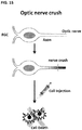
- the RG progenitor cells can be transplanted into the vitreous or subretinal space of intraocular hypertension glaucoma mice or rat model systems, or of optical nerve (ON) injury crush mice model systems, and will migrate to the ganglion cell layer and will improve pattern ERG (electroretinography) responses and visual acuity in any of these model systems.
- the RG cells and RG progenitor cells may regenerate the optical nerve.
- the RG progenitor cells and RG cells secrete one or more neuroprotective factors, and thereby provide neuroprotection.
- neuroprotection may be measured using animal models of optic nerve injury or glaucoma.
- the medium suitable for maintaining the viability of the RG progenitor cells is selected from the group consisting of a culture medium, a cryopreservative, and a biocompatible injection medium suitable for injection in a human patient.
- the RG progenitor cell preparation is pyrogen and mycogen free.
- Another aspect of the present invention provides a pharmaceutical preparation of RG cells that is suitable for use in a mammalian patient, comprising pluripotent stem cell derived RG cells, wherein cells in the preparation are immunocytochemically positive for Neurofilament and/or Brn3a, and thus may be in some instance Neurofilament(+) Brn3a(+).
- the RG cells may be Tuj1(+), and they may also optionally be Thyl(+).
- the pharmaceutical preparations may further comprise a pharmaceutically acceptable carrier for maintaining the viability of the RG cells for transplantation into a mammalian patient.
- Another aspect of the present invention provides a pharmaceutical preparation of RG cells that is suitable for use in a mammalian patient, comprising pluripotent stem cell derived RG cells, wherein cells in the preparation are immunocytochemically Neurofilament(+) Brn3a(+),and Tuj1(+), and optionally Thy1(+), and a pharmaceutically acceptable carrier for maintaining the viability of the RG cells for transplantation into a mammalian patient.
- Another aspect of the present invention provides a pharmaceutical preparation of RG cells that is suitable for use in a mammalian patient, comprising pluripotent stem cell derived RG cells, wherein greater than 70%, 80%, 90%, 95% or even 98% of the cells are immunocytochemically Neurofilament(+) and Brn3a(+), and a pharmaceutically acceptable carrier for maintaining the viability of the RG cells for transplantation into a mammalian patient.
- greater than 50%, 60% or 70% of the cells in the preparation are immunocytochemically Thyl(+).
- at least 70%, 80%, 90%, 95% or even 98% of the cells are immunocytochemically Tuj1(+).
- cells in the pharmaceutical preparation of RG cells are mitotically inactive (or post-mitotic), meaning that they are non-proliferative. This can be determined using proliferation assays known in the art such as tritiated thymidine uptake assays, etc. In some embodiments, greater than 50%, 60%, 70%, 80%, 90%, 95%, or 98% of the cells in the preparation are mitotically inactive.
- cells in the pharmaceutical preparation of RG cells comprise a greater number of dendrites and/or more complex dendrites than an RG progenitor class. In some embodiments, greater than 50%, 60%, 70%, 80%, 90%, 95%, or 98% of the cells in the preparation comprise a greater number of dendrites and/or more complex dendrites (as compared to the progenitor classes).
- mature RG cells exhibit an increased number of primary dendrites and/or increased dendritic branching at secondary, tertiary and higher levels, and/or increased total dendritic length compared to RG progenitor cells.
- RG cells may have 2-5 fold more dendrites on a per cell basis than RG progenitor cells (e.g., RG cells may have 4-5 dendrites per cell and RG progenitor cells may have 1-2 dendrites per cell).
- Still another aspect of the present invention provides a pharmaceutical preparation comprising RG progenitor cells and/or RG cells together with retinal pigment epithelial cells, photoreceptor progenitor cells, and/or photoreceptor cells; and a pharmaceutically acceptable carrier for maintaining the viability of the RG progenitor cells and/or the RG cells for transplantation into a mammalian patient.
- the preparation may also include bipolar cells and/or amacrine cells.
- the preparation of cells can be provided as cell suspensions (either admixed together, or in the form of a kit with separate doses of cells to be delivered conjointly), as three-dimensional structures, including for example as a sheet or monolayer or multi-layer cell graft (optionally disposed on a biocompatible matrix or solid support).
- the RPE cells can be provided as a monolayer, preferably a polarized monolayer.
- the RG cells and/or RG progenitor cells are used together with other neuroretinal sensory cells including RPE cells, photoreceptor cells or photoreceptor progenitor cells.
- compositions for use in a method of treatment of a mammalian patient in need thereof include methods for treating diseases and disorders caused by loss or dysfunction of retinal ganglion cells in a patient, comprising administering such pharmaceutical preparations as described herein, such as preparations of RG progenitor cells or RG cells, or both.
- the preparations can be injected locally, such as into the sub-retinal space of the patient's eye, into the vitreous of the patient's eye, or delivered systemically or into other body cavities where the cells can persist.
- the diseases or disorders caused by loss of retinal ganglion cells include glaucoma, optic nerve injury, ischemic optic neuropathy, optic neuritis, diabetic retinopathy, inherited ganglion degeneration, and inherited retinal dystrophy.
- compositions comprising the RG progenitor cell populations described herein for use in a method of treating a mammalian patient in need thereof, including treating a disease or disorder caused by loss or dysfunction of retinal ganglion cells in a subject.
- compositions comprising the RG cell populations described herein for use in a method of treating a mammalian patient in need thereof, including treating a disease or disorder caused by loss or dysfunction of retinal ganglion cells in a subject.
- the invention provides a method of producing RG cells, comprising the steps of
- the invention provides a method, comprising culturing eye field progenitor cells, preferably as cell clusters and preferably under low adherence or non-adherent conditions, in a ganglion cell medium for a period of time sufficient for the cell clusters to form RG progenitor cells.
- the RG progenitor cells may be optionally cultured in a retinal ganglion cell differentiation medium under adherent conditions, preferably on a matrix (such as a biomaterial scaffold) until a majority of cells in the culture are RG cells characterized as Brn3a(+), Neurofilament(+) and Tuj 1(+), and optionally Thyl(+).
- the eye field progenitor cells are characterized, such as immunocytochemically characterized, as Pax6(+) and Rx1(+) and Oct4(-) and Nanog(-), and even more preferably are also characterized as Six3(+), Six6(+), Lhx2(+), Tbx3(+), Sox2(+), Otx2(+) and Nestin(+), such as may be determined by immunostaining and/or flow cytometry.
- the RG progenitor cells are characterized as Math5(+), or as Math5(+) and Brn3a(+), or as Math5(+), Brn3a(+) and Brn3b(+), or as Math5(+), Brn3a(+), Brn3b(+) and Isl1(+), or as Brn3a(+) and Neurofilament(+), or as Brn3a(+), Neurofilament(+) and Thy1(+), such as may be determined by immunostaining and/or flow cytometry or other standard assay used to characterize marker expression in cells.
- the adherent conditions include a culture system having a surface to which the cells can adhere that includes an adherent material, which may comprise, merely to illustrate, one or more of a polyester, a polypropylene, a polyalkylene, a polyfluorochloroethylene, a polyvinyl chloride, a polyvinyl fluoride resin, a polystyrene, a polysulfone, a polyurethane, a polyethyene terephtalate, a cellulose, a glass fiber, a ceramic particle, a biomaterial scaffold, a poly-L-lactic acid, a dextran, an inert metal fiber, silica, natron glass, borosilicate glass, chitosan, or a vegetable sponge.
- an adherent material which may comprise, merely to illustrate, one or more of a polyester, a polypropylene, a polyalkylene, a polyfluorochloroethylene, a polyviny
- the adherent material is electrostatically charged.
- the biomaterial scaffold is extracellular matrix, such as collagen (such as collagen type IV or type I), 804G-derived matrix, fibronectin, vitronectin, chondronectin, laminin or MatrigelTM.
- the biomaterial is gelatin, alginate, polyglycolide, fibrin, or self-assembling peptides.
- the RG progenitor cells and as a consequence the RG cells, are derived from pluripotent stem cells, such as embryonic stem cells or induced pluripotent stem cells.
- the resulting preparation of RG progenitor cells are provided substantially free of pluripotent stem cells, i.e., less than 10% of the cells in the preparation are pluripotent stem cells, and even more preferably less than 5%, 2%, 1%, 0.1% or even less than 0.01% of the cells in the preparation are pluripotent stem cells.
- the resulting preparation of RG progenitor cells are provided substantially free of eye field progenitor cells, i.e., less than 10%, and even more preferably less than 5%, 2%, 1%, 0.1% or even less than 0.01% of the cells in the preparation are eye field progenitor cells.
- the cellular component of the resulting preparation of RG progenitor cells is at least 50% pure with respect to other cell types (i.e., at least 50% of the cells in the preparation are RG progenitor cells), and preferably at least 75%, at least 85%, at least 95%, at least 99% or about 100% of the cells in the preparation are RG progenitors.
- the method includes the further step of cryopreserving the RG progenitor cells or the RG cells.
- the cells are preferably frozen in a cryopreservative which is compatible with the thawing of the frozen cells (and the thawed cells themselves) and, after optionally washing the cells to remove the cryopreservative, the RG progenitor cells or the RG cells retain at least 25% cell viability (such as based on culture efficiency), and more preferably at least 50%, 60%, 70%, 80% or even at least 90% cell viability.
- the RG progenitor cells or the RG cells may be cryopreserved.
- the RG progenitor cells are cryopreserved as spheres.
- the ganglion cell medium may comprise a basal medium such as NeurobasalTM Medium (Life Technologies) (or other medium comprising similar constituents), D-glucose, antibiotics such as penicillin and streptomycin, GlutaMAXTM, N2 supplement (Invitrogen), B27 supplement (also from Invitrogen), forskolin, BDNF and CNTF.
- B27 supplement contains, inter alia, SOD, catalase and other anti-oxidants (GSH), and unique fatty acids, such as linoleic acid, linolenic acid, and lipoic acids.
- N2 supplement can be replaced with, for example, the following cocktail: transferrin (10 g/L), insulin (500 mg/L), progesterone (0.63 mg/L), putrescine (1611 mg/L) and selenite (0.52 mg/L).
- the RG progenitor cells are differentiated from a pluripotent stem cell source, such as a pluripotent stem cell that expresses Oct4, alkaline phosphatase, Sox2, SSEA-3, SSEA-4, TRA-1-60, and TRA-1-80 (such as an embryonic stem (ES) cell line or induced pluripotency stem (iPS) cell line), and even more preferably from a common pluripotent stem cell source.
- a pluripotent stem cell source such as a pluripotent stem cell that expresses Oct4, alkaline phosphatase, Sox2, SSEA-3, SSEA-4, TRA-1-60, and TRA-1-80 (such as an embryonic stem (ES) cell line or induced pluripotency stem (iPS) cell line), and even more preferably from a common pluripotent stem cell source.
- a pluripotent stem cell source such as a pluripotent stem cell that expresses Oct4, alkaline phosphatase, Sox2,
- the RG progenitor cells have a mean terminal restriction fragment length (TRF) that is longer than 7 kb, 7.5 kb, 8 kb, 8.5 kb, 9 kb, 9.5 kb, 10 kb, 10.5 kb, 11 kb, 11.5 kb or even 12 kb.
- TRF mean terminal restriction fragment length
- a preparation is suitable for administration to a human patient, and more preferably pyrogen-free and/or free of non-human animal products.
- a preparation is suitable for administration to a non-human veterinarian mammal, such as a dog, cat or horse.
- the disclosure provides a method of producing RG progenitor cells, comprising culturing pluripotent stem cells, sequentially, in (i) a retinal induction (RI) medium, (ii) neural differentiation (ND) medium, and (iii) ganglion cell (GC) medium.
- RI retinal induction
- ND neural differentiation
- GC ganglion cell
- the pluripotent stem cells may be human.
- the pluripotent stem cells may be human ES cells or human iPS cells, in some embodiments.
- the pluripotent stem cells may be cultured, in an undifferentiated state, under feeder-free and/or xeno-free conditions, optionally in the presence of a matrix.
- the pluripotent stem cells may be cultured, in an undifferentiated state, on a substrate such as a substrate comprising MatrigelTM and optionally in mTESR1 medium.
- the retinal induction medium may comprise a basal medium such as DMEM/F12, DMEM/high glucose, or DMEM/knock-out, or a medium comprising the components of DMEM/F12, or a medium comprising the components of DMEM/high glucose, or a medium comprising the components of DMEM/knock-out.
- the retinal induction medium may comprise D-glucose.
- the neural differentiation medium may comprise D-glucose.
- D-glucose may be present in a concentration between 0-10 mg/ml, 2.5-7.5 mg/ml, 3-6 mg/ml, 4-5 mg/ml, or about 4.5 mg/ml.
- the retinal induction medium may comprise one or more antibiotics.
- the antibiotics may include either or both penicillin and streptomycin, optionally in concentrations of about 0-100 units/ml or about 100 units/ml of penicillin and optionally about 0-100 ⁇ g/ml or about 100 ⁇ g/ml of streptomycin.
- the retinal induction medium may comprise one or more serum supplements.
- the retinal induction medium may comprise N2 supplement.
- the N2 supplement may be present in a concentration of about 0.1 to 5% or about 0.1 to 2% or about 1% (volume/volume, or v/v).
- the retinal induction medium may comprise B-27 supplement.
- the B-27 supplement may be present in a concentration of about 0.05-2.0% or about 0.2% (v/v).
- the retinal induction medium may comprise non-essential amino acids or minimal essential medium (MEM) non-essential amino acids.
- the non-essential amino acids or MEM non-essential amino acids may be present at a IX concentration from stock, wherein stock is typically considered 100X.
- the final concentration of glycine (at the IX concentration of stock) is about 0.1 mM, and thus the stock is 10 mM glycine.
- the stock typically also includes L-alanine, L-asparagine, L-aspartic acid, L-glutamic acid, L-proline, and L-serine.
- the retinal induction medium may comprise insulin.
- the insulin may be human.
- the insulin may be present in a concentration of about 5-50 ⁇ g/ml or about 20 ⁇ g/ml.
- the retinal induction medium may comprise a BMP signaling inhibitor.
- the BMP signaling inhibitor may be selected from the group consisting of Noggin polypeptide, dorsomorphin, LDN-193189, and any combination thereof.
- the retinal induction medium may comprise Noggin polypeptide.
- Noggin polypeptide may be present at a concentration of between about 5-100 ng/ml or about 10-100 ng/ml or about 10-500 ng/ml or about 50 ng/ml. In some embodiments, Noggin polypeptide is present at about 10-500 ng/ml.
- the retinal induction medium may be replaced, in whole or in part, with fresh retinal induction medium daily.
- the cells are grown at a density in the range of 2 x 10 4 /cm 2 to 6 x 10 4 /cm 2 .
- Culturing in the presence of retinal induction medium may occur for about 4-6 days, or about 4 days, about 5 days, or about 6 days.
- the neural differentiation medium may comprise a basal medium such as NeurobasalTM Medium (Life Technologies), or a basal medium comprising components of NeurobasalTM Medium.
- a basal medium such as NeurobasalTM Medium (Life Technologies)
- a basal medium comprising components of NeurobasalTM Medium.
- the neural differentiation medium may comprise D-glucose.
- D-glucose may be present in a concentration between 0-10 mg/ml, 2.5-7.5 mg/ml, 3-6 mg/ml, 4-5 mg/ml, or about 4.5 mg/ml.
- the neural differentiation medium may comprise one or more antibiotics.
- the antibiotics may include either or both penicillin and streptomycin, optionally in concentrations of between 0-100 units/ml or about 100 units/ml of penicillin and optionally between 0-100 ⁇ g/ml or about 100 ⁇ g/ml of streptomycin.
- the neural differentiation medium may comprise one or more serum supplements.
- the neural differentiation medium may comprise N2 supplement.
- the N2 supplement may be present in a concentration of about 0.1 to 5% or 0.1 to 2% or about 1% (v/v).
- the neural differentiation medium may comprise B-27 supplement (Life Technologies).
- the B-27 supplement may be present in a concentration of about 0.05 to 5.0% , about 0.5 to 2.0% or about 2% (v/v).
- the neural differentiation medium may comprise non-essential amino acids or MEM non-essential amino acids or glutamine or GlutaMAXTM.
- the non-essential amino acids or MEM non-essential amino acids may be present at about a IX concentration of stock, wherein stock is about 200 mM (and considered 100X).
- GlutaMAXTM may be present at 2 mM or a IX dilution of stock (using a 100X stock solution that is 200 mM).
- the neural differentiation medium may comprise a BMP signaling inhibitor.
- the BMP signaling inhibitor may be selected from the group consisting of Noggin polypeptide, dorsomorphin, LDN-193189, and any combination thereof.
- the neural differentiation culture medium may comprise Noggin polypeptide.
- Noggin polypeptide may be present at a concentration of between about 10 to 1000 ng/ml, or about 10 to 500 ng/ml, or 10 to 100 ng/ml, or about 50 ng/ml.
- Culturing in the presence of neural differentiation medium may occur for about 8-12 days, or about 9-10 days, or about 9 days.
- the neural differentiation medium may be changed, in whole or in part, daily, or every 2 days, or every three days.
- the ganglion cell medium may comprise a basal medium such as NeurobasalTM Medium (Life Technologies) or a medium comprising the components of NeurobasalTM Medium.
- a basal medium such as NeurobasalTM Medium (Life Technologies) or a medium comprising the components of NeurobasalTM Medium.
- the ganglion cell medium may comprise D-glucose.
- the neural differentiation medium may comprise D-glucose.
- D-glucose may be present in a concentration between 0-10 mg/ml, 2.5-7.5 mg/ml, 3-6 mg/ml, 4-5 mg/ml, or about 4.5 mg/ml.
- the ganglion cell medium may comprise one or more antibiotics.
- the antibiotics may include either or both penicillin and streptomycin, optionally in concentrations of between 0-100 units/ml or about 100 units/ml of penicillin and optionally between 0-100 ⁇ g/ml or about 100 ⁇ g/ml of streptomycin.
- the ganglion cell medium may comprise one or more serum supplements.
- the ganglion cell medium may comprise N2 supplement.
- the N2 supplement may be present in a concentration of about 0.1 to 5% or 0.1 to 2% or about 1% (v/v).
- the ganglion cell medium may comprise B-27 supplement (formula number 080085-SA) (Life Technologies).
- the B-27 supplement (formula number 080085-SA) may be present in a concentration of about 0.05 to 5.0% , about 1.5 to 2.5% or about 2% (v/v).
- the ganglion cell medium may comprise GlutaMAXTM.
- GlutaMAXTM may be present at a IX dilution of stock (using a 100X stock solution that is 200 mM).
- the ganglion cell medium may comprise one, two or all of forskolin, BDNF and CNTF.
- Forskolin may be present in a concentration of 1-20 ⁇ M or 1-10 ⁇ M or about 5 ⁇ M.
- BDNF may be present at a concentration of 1-200 ng/ml or 5-50 ng/ml or 5-25 ng/ml or about 10 ng/ml.
- CNTF may be present at a concentration of 1-200 ng/ml or 5-50 ng/ml or 5-25 ng/ml or about 10 ng/ml.
- BDNF may be human BDNF.
- CTNF may be human CTNF.
- Culturing in the presence of ganglion cell medium may occur for about 40-50 days, or about 45 days.
- the ganglion cell medium may be changed, in whole or in part, daily, or every 2 days, or every three days, or less frequently.
- the RG progenitor cells produced by the method may comprise at least 50%, at least 75%, at least 85%, at least 95%, at least 99% or about 100% of the cells in the culture, and optionally such culture may be propagated for 1 week, 2 weeks, or more than 2 weeks, including 3 weeks, 4 weeks, or longer.
- the RG progenitor cells may express one or more of the markers: Math5, Brn3a, Brn3b, Isl1, Neurofilament, and Thyl, and thus may be characterized as Math5(+), Brn3a(+), Brn3b(+), Isl1(+), Neurofilament(+), and/or Thyl(+).
- the RG progenitor cells may express one or more of the markers: Math5, Brn3a, Brn3b, and Isl1, and thus may be characterized as Math5(+), Brn3a(+), Brn3b(+), and/or Isl1(+).
- the RG progenitor cells may express one or more of the markers: Brn3a, Neurofilament, and Thyl, and thus may be characterized as Brn3a(+), Neurofilament(+), and/or Thyl(+).
- the RG progenitors may not express Pax6 and/or Rx1, and thus may be characterized as Pax6(-) and/or Rx1(-).
- Certain RG progenitor cells may not express Math5 and/or Brn3b, and thus may be characterized as Math5(-) and/or Brn3b(-).
- the RG progenitor cells may be characterized as Math5(+), or Math5(+), Brn3a(+), or Math5(+), Brn3a(+), Brn3b(+), or Math5(+), Brn3a(+), Brn3b(+), Isl1(+), or Brn3a(+), or Neurofilament(+), or Brn3a(+), Neurofilament(+), or Brn3a(+), Neurofilament(+), Thyl(+).
- the RG progenitors may not express Pax6 and/or Rx1, and thus may be characterized as Pax6(-) and/or Rx1(-).
- the RG progenitor cells may be Tuj1(+).
- the RG progenitor cells may be proliferative.
- the RG progenitor cells may be cryopreserved.
- the RG progenitor cells may be human.
- the method may further comprise a subsequent culturing step in the presence of retinal ganglion cell (RGC) differentiation medium. This may occur without an intermediate cryopreservation step or it may occur after cryopreservation and thawing of the RG progenitor cells.
- RRC retinal ganglion cell
- the RGC differentiation medium may comprise a basal medium such as NeurobasalTM Medium (Life Technologies) or a medium comprising the components of NeurobasalTM Medium.
- a basal medium such as NeurobasalTM Medium (Life Technologies) or a medium comprising the components of NeurobasalTM Medium.
- the RGC differentiation medium may comprise D-glucose.
- the neural differentiation medium may comprise D-glucose.
- D-glucose may be present in a concentration between 0-10 mg/ml, 2.5-7.5 mg/ml, 3-6 mg/ml, 4-5 mg/ml, or about 4.5 mg/ml.
- the RGC differentiation medium may comprise one or more antibiotics.
- the antibiotics may include either or both penicillin and streptomycin, optionally in concentrations of between 0-100 units/ml or about 100 units/ml of penicillin and optionally between 0-100 ⁇ g/ml or about 100 ⁇ g/ml of streptomycin.
- the RGC differentiation medium may comprise one or more serum supplements.
- the RGC differentiation medium may comprise B-27 supplement (formula 080085-SA).
- the B-27 supplement may be present in a concentration of about 0.05 to 5.0%, about 1.5 to 2.5% or about 2% (v/v).
- the RGC differentiation medium may comprise GlutaMAXTM.
- GlutaMAXTM may be present at a IX dilution of stock.
- the RGC differentiation medium may comprise retinoic acid.
- Retinoic acid may be present at a concentration of about 0.5-20 ⁇ M, about 0.5-10 ⁇ M, about 1-5 ⁇ M, or about 2 ⁇ M.
- the RGC differentiation medium may comprise one or more including all of forskolin, BDNF, CNTF, cAMP, and DAPT.
- the RGC differentiation medium may comprise one or both of BDNF and CNTF.
- the RGC differentiation medium may comprise one or more including all of forskolin, cAMP, and DAPT or other Notch pathway inhibitor or Notch inhibitor (such as Notch blocking antibody or antibody fragment, Notch negative regulatory region antibody or antibody fragment, alpha-secretase inhibitor, gamma-secretase inhibitor, stapled peptide, small molecule blockers and siRNA, shRNA and miRNA).
- Notch blocking antibody or antibody fragment such as Notch blocking antibody or antibody fragment, Notch negative regulatory region antibody or antibody fragment, alpha-secretase inhibitor, gamma-secretase inhibitor, stapled peptide, small molecule blockers and siRNA, shRNA and miRNA.
- Forskolin may be present in a concentration of 1-20 ⁇ M or 1-10 ⁇ M or about 5
- BDNF may be present at a concentration of 1-200 ng/ml or 5-50 ng/ml or 5-25 ng/ml or about 10 ng/ml.
- CNTF may be present at a concentration of 1-200 ng/ml or 5-50 ng/ml or 5-25 ng/ml or about 10 ng/ml.
- BDNF may be human BDNF.
- CTNF may be human CTNF.
- cAMP may be present at a concentration of 1-500 ng/ml or 10-250 ng/ml or 5-150 ng/ml or about 100 ng/ml.
- DAPT may be present at a concentration of 1-100 ⁇ M or 1-50 ⁇ M or 1-20 ⁇ M, or about 10 ⁇ M.
- the RGC differentiation medium may comprise insulin.
- the insulin may be human.
- the insulin may be present in a concentration of about 5-50 ⁇ g/ml or about 20 ⁇ g/ml.
- Culturing in the presence of RGC differentiation medium may occur for about 1-4 weeks or about 2-4 weeks or about 2-3 weeks or about 2 weeks.
- the RGC differentiation medium may be changed, in whole or in part, daily, or every 2 days, or every three days, or less frequently.
- the RG cells produced by the method may comprise at least 50%, at least 75%, at least 85%, at least 95%, at least 99% or about 100% of the cells in the culture, and optionally such culture may be propagated for 1 week, 2 weeks, or more than 2 weeks, including 3 weeks, 4 weeks, or longer.
- the RG cells may express one or more of the markers: Brn3a, Neurofilament, Tuj 1 and Thyl, and thus may be characterized as Brn3a(+), Neurofilament(+), Tuj1(+), and/or Thyl(+).
- the RG cells may be characterized as Brn3a(+), Neurofilament(+), or as Brn3a(+), Neurofilament(+), Thy1(+), or as Brn3a(+), Neurofilament(+), Thy1(+), or as Brn3a(+), Neurofilament(+), Tuj1(+), or as Brn3a(+), Neurofilament(+), Tuj1(+), Thyl(+).
- the RG cells may be post-mitotic and may comprise a higher number, longer and/or more complex dendrites than RG progenitor cells.
- the RG cells may be cryopreserved.
- the RG cells may be human.
- the disclosure provides a method of producing RG progenitor cells, comprising culturing eye field progenitor cells in a ganglion cell medium.
- the eye field progenitor cells may be provided as a population of cells, at least 50%, at least 75%, at least 85%, at least 95%, at least 99% or about 100% of which are eye field progenitor cells.
- the eye field progenitor cells may be Pax6(+) and Rx1(+) and optionally also Six3(+), Six6(+), Lhx2(+), and/or Tbx3(+).
- the eye field progenitor cells may also be Sox2(+).
- the eye field progenitor cells may be Nestin(+).
- the eye field progenitor cells may also be Otx2(+).
- the eye field progenitors may be obtained by in vitro differentiation of pluripotent stem cells.
- the pluripotent stem cells may be ES cells or iPS cells, in some embodiments.
- the pluripotent stem cells may be cultured under feeder-free and/or xeno-free conditions.
- the culture of eye field progenitor cells may occur for about 5-45 days, or about 5-20 days, or about 35-45 days.
- the ganglion cell medium and culture parameters in the ganglion cell medium may be as described above.
- the RG progenitor cells produced by the method may comprise at least 50%, at least 75%, at least 85%, at least 95%, at least 99% or about 100% of the cells in the culture, and optionally such culture (comprising such % of RG progenitor cells) may be propagated for 1 week, 2 weeks, or more than 2 weeks, including 3 weeks, 4 weeks, or longer.
- the RG progenitor cell phenotype is as described above.
- the RG progenitor cells may express one or more of the markers: Math5, Brn3a, Brn3b, Isl1, Neurofilament, and Thyl, and thus may be characterized as Math5(+), Brn3a(+), Brn3b(+), Isl1(+), Neurofilament(+), and/or Thyl(+).
- the RG progenitor cells may express one or more of the markers: Math5, Brn3a, Brn3b, and Isl1, and thus may be characterized as Math5(+), Brn3a(+), Brn3b(+), and/or Isl1(+).
- the RG progenitor cells may express one or more of the markers: Brn3a, Neurofilament, and Thyl, and thus may be characterized as Brn3a(+), Neurofilament(+), and/or Thyl(+).
- the RG progenitors may not express Pax6 or Rx1, and thus may be characterized as Pax6(-) and/or Rx1(-).
- the RG progenitor cells may be proliferative. Other phenotypes are described above.
- the RG progenitor cells may be cryopreserved.
- the RG progenitor cells may be human.
- the disclosure provides a method of producing RG cells, comprising culturing eye field progenitor cells sequentially in a ganglion cell medium and a retinal ganglion cell (RGC) differentiation medium.
- RGC retinal ganglion cell
- the eye field progenitor cells may be provided as a population of cells, at least 50%, at least 75%, at least 85%, at least 95%, at least 99% or about 100% of which are eye field progenitor cells.
- the eye field progenitor cells may be Pax6(+) and Rx1(+) and optionally also Six3(+), Six6(+), Lhx2(+), and/or Tbx3(+).
- the eye field progenitor cells may also be Sox2(+).
- the eye field progenitor cells may be Nestin(+).
- the eye field progenitor cells may also be Otx2(+).
- the eye field progenitors may be obtained by in vitro differentiation of pluripotent stem cells.
- the pluripotent stem cells may be ES cells or iPS cells, in some embodiments.
- the pluripotent stem cells may be cultured under feeder-free and/or xeno-free conditions.
- the ganglion cell medium and culture parameters in the ganglion cell medium may be as described above.
- the RGC differentiation medium and culture parameters in the RGC differentiation medium may be as described above.
- the RG cells produced by the method may comprise at least 50%, at least 75%, at least 85%, at least 95%, at least 99% or about 100% of the cells in the culture, and optionally such culture (comprising such % of RG cells) may be propagated for 1 week, 2 weeks, or more than 2 weeks, including 3 weeks, 4 weeks, or longer.
- the phenotype of the RG cells is as described above.
- the RG cells may express one or more of the markers: Brn3a, Neurofilament, Tuj1, and Thyl, and thus may be characterized as Brn3a(+), Neurofilament(+), Tuj1(+), and/or Thyl(+).
- the RG cells may be post-mitotic.
- the RG cells may have more complex dendrites, longer dendrites, and/or more dendrites than RG progenitor cells. Other phenotypes are described above.
- the RG cells may be cryopreserved.
- the RG cells may be human.
- the disclosure provides a method of producing RG cells, comprising culturing RG progenitor cells in a retinal ganglion cell (RGC) differentiation medium.
- RRC retinal ganglion cell
- the RG progenitor cells may be provided as a population of cells, at least 50%, at least 75%, at least 85%, at least 95%, at least 99% or about 100% of which are RG progenitor cells.
- the RG progenitor cells may express one or more of the markers: Math5, Brn3a, Brn3b, Isl1, Neurofilament, and Thyl, and thus may be characterized as Math5(+), Brn3a(+), Brn3b(+), Isl1(+), Neurofilament(+), and/or Thyl(+).
- the RG progenitor cells may express one or more of the markers: Math5, Brn3a, Brn3b, and Isl1, and thus may be characterized as Math5(+), Brn3a(+), Brn3b(+), and/or Isl1(+).
- the RG progenitor cells may express one or more of the markers: Brn3a, Neurofilament, and Thyl, and thus may be characterized as Brn3a(+), Neurofilament(+), and/or Thyl(+).
- the RG progenitors may not express Pax6 or Rx1 and thus may be characterized as Pax6(-) and/or Rx1(-).
- the RG progenitors may be obtained by in vitro differentiation of pluripotent stem cells.
- the pluripotent stem cells may be ES cells or iPS cells, in some embodiments.
- the pluripotent stem cells may be cultured under feeder-free and/or xeno-free conditions.
- the RGC differentiation medium and culture parameters in the RGC differentiation medium may be as described above.
- the RG cells produced by the method may comprise at least 50%, at least 75%, at least 85%, at least 95%, at least 99% or about 100% of the cells in the culture, and optionally such culture (comprising such % of RG cells) may be propagated for 1 week, 2 weeks, or more than 2 weeks, including 3 weeks, 4 weeks, or longer.
- the RG cells may express one or more of the markers: Brn3a, Neurofilament, Tuj 1, and Thyl, and may be characterized as Brn3a(+), Neurofilament(+), Tuj1(+), and Thyl(+).
- the RG cells may be cryopreserved.
- the RG cells may be human.
- the disclosure provides a composition comprising RG progenitor cells produced using a method as described herein, e.g., as described in the preceding paragraphs.
- the disclosure provides a composition comprising RG progenitor cells, which are optionally human.
- the RG progenitor cells may comprise at least 50%, at least 75%, at least 85%, at least 95%, at least 99% or about 100% of the cells in the composition.
- the RG progenitor cells may express one or more of the markers: Math5, Brn3a, Brn3b, Isl1, Neurofilament, and Thyl, and thus may be characterized as Math5(+), Brn3a(+), Brn3b(+), Isl1(+), Neurofilament(+), and/or Thyl(+).
- the RG progenitor cells may express Math5, Brn3a, Brn3b, and Isl1, and thus may be characterized as Math5(+), Brn3a(+), Brn3b(+), and Isl1(+).
- the RG progenitor cells may express Brn3a, Neurofilament, and Thyl, and thus may be characterized as Brn3a(+), Neurofilament(+), and Thyl(+). Other phenotypes for RG progenitor cells are described above.
- the RG progenitor cells may be cryopreserved.
- the RG progenitor cells may be human.
- the invention provides a composition for use in a method of treatment of an individual in need thereof, comprising administering a composition comprising RG progenitor cells (e.g., a composition as described herein or a composition produced using a method as described herein) to said individual.
- a composition comprising RG progenitor cells (e.g., a composition as described herein or a composition produced using a method as described herein) to said individual.
- the composition may be administered to the eye, vitreous, subretinal space, or intravenously.
- the RG progenitor cells may be human.
- the disclosure provides a composition comprising RG cells produced according to a method as described herein, e.g., in the preceding paragraphs.
- the RG cells produced by the method may comprise at least 50%, at least 75%, at least 85%, at least 95%, at least 99% or about 100% of the cells in the composition, and optionally such composition (comprising such % of RG cells) may be propagated in culture for 1 week, 2 weeks, or more than 2 weeks, including 3 weeks, 4 weeks, or longer.
- the RG cells may express one or more of the markers: Brn3a, Neurofilament, Tuj 1, and Thyl, and thus may be characterized as Brn3a(+), Neurofilament(+), Tuj1(+), and/or Thy1 (+).
- the RG cells may be cryopreserved.
- the RG cells may be human.
- the disclosure provides a method of treatment of an individual in need thereof, comprising administering a composition comprising RG cells, e.g., a composition as described herein such as in the preceding paragraphs or a composition produced by a method as described herein e.g., in the preceding paragraphs, to said individual.
- a composition comprising RG cells e.g., a composition as described herein such as in the preceding paragraphs or a composition produced by a method as described herein e.g., in the preceding paragraphs, to said individual.
- composition may be administered to the eye, the vitreous, the subretinal space, or intravenously.
- the invention relates to a substantially pure preparation of RG progenitor cells or RG cells of human origin, preferably non-donor derived RG progenitor cells or RG cells, originating from cells not grown on a mouse fibroblast feeder platform.
- the preparation may be 85%-95% pure.
- the invention relates to a method of preparing the substantially pure preparation of RG progenitor cells or RG cells of human origin which omits the need for cells derived from a mouse fibroblast feeder platform. Replacing a feeder system with the methods of the present invention produces a greater homogeneity of RG progenitor cells or RG cells, e.g., at 75% to 100% or 85% to 95%.
- the differentiation of the feeder-free stem cells can also occur in the absence of the introduction of exogenous inducing factors, which is a substantial improvement over the prior art.
- the optional addition of Noggin polypeptide can accelerate differentiation of the stem cells, even though it is not essential for differentiation.
- the differentiation methods are feeder-free and xeno-free.
- human cells are cultured in the absence of any media component that is derived from a non-human species.
- the invention provides methods for generating retinal ganglion (RG) cells and RG progenitor cells. These methods involve in vitro differentiation from earlier progenitors including pluripotent stem cells and eye field (EF) progenitors.
- RG retinal ganglion
- EF eye field
- the methods provided herein may use as a starting material any of the foregoing progenitor (including stem cell) populations.
- the invention further contemplates generating RG cells and RG progenitor cells in vitro from primary eye field (EF) progenitors (i.e., EF progenitor cells that are obtained from a subject).
- EF primary eye field
- RG progenitor cells which may comprise early RG progenitor cells and late RG progenitor cells, which in turn differentiate into mature RG cells (referred to herein as RG cells).
- RG progenitor cells within the RG progenitor cells, the early RG progenitor cells may differentiate into late RG progenitor cells.
- the late RG progenitor cells may differentiate into mature RG cells.
- the early RG progenitor cells may differentiate directly into mature RG cells. The methods provided herein efficiently generate RG cells and RG progenitor cells.
- the overall developmental pathway from pluripotent stem cells to RG progenitor cell and RG cell development is schematically illustrated in FIG. 9 . Also shown is the marker expression profile for each stage of development.
- Progenitor cells refer to cells that remain mitotic and can produce more progenitor cells or can differentiate to an end fate cell lineage.
- progenitor and precursor are used interchangeably. Cells at each of these stages will be discussed in greater detail herein.
- the RG progenitors and RG cells provided herein may be used in a variety of in vivo and in vitro methods.
- the RG progenitor cells may be used in vivo to treat conditions of the retina, including glaucoma, such as open-angle glaucoma, angle-closure glaucoma, normal-tension glaucoma, congenital glaucoma, pigmentary glaucoma, secondary glaucoma, pseudoexfoliative glaucoma, traumatic glaucoma, neovascular glaucoma, and irido corneal endothelial syndrome, as well as optic nerve injury, optionally optic nerve injury associated with autoimmune disease, trauma, viral infection, and ischemic-reperfusion injury, optionally ischemic-reperfusion injury associated with stroke, diabetic retinopathy, or other condition that impairs blood supply to the eye.
- optic nerve injury optionally optic nerve injury associated with autoimmune disease, trauma, viral infection, and ischemic-re
- the invention further provides RG progenitor cells and RG cells obtained by the methods described herein.
- RG progenitor cells and RG cells are obtained by in vitro differentiation of pluripotent stem cells or their differentiated progeny such as eye field progenitor cells.
- Eye field progenitor cells may themselves be obtained from in vitro differentiation of pluripotent stem cells, or they may be primary eye field progenitors obtained from a subject.
- the invention provides populations of RG progenitor cells and populations of RG cells that have not been attained or are not attainable from primary sources. These populations may be homogenous or near homogeneous in their cell content. For example, at least 50%, at least 60%, at least 70%, at least 80%, at least 90%, at least 95%, at least 99%, or about 100% of the cells in such a population may be RG progenitor cells. As another example, at least 50%, at least 60%, at least 70%, at least 80%, at least 90%, at least 95%, at least 99%, or about 100% of the cells in such a population may be RG cells. These cells in these populations may be of a single haplotype. For example, they may be HLA-matched. These cells in these populations may be genetically identical. The present disclosure also describes a bank of HLA-typed RG progenitor cells. Such cells may be used in a variety of human subjects.
- the methods provided herein can be used to generate large numbers of RG progenitor cells and RG cells that would not otherwise be possible from primary sources.
- the methods may produce 10 8 RG progenitor cells or 10 8 RG cells per starting 1 million pluripotent cells.
- EF progenitor cells Eye field progenitor cells.
- EF progenitor cells are minimally defined as Pax6(+) and Rx1(+) cells. They may also be Six3(+), Six6(+), Lhx2(+), and Tbx3(+). All of these markers are transcription factors.
- the EF progenitor cells may also be Nestin(+) and/or Sox2(+), and/or Otx2(+).
- EF progenitor cells do not express Oct4 and Nanog (i.e., they are Oct4(-) and Nanog(-)).
- EF progenitor cells can be formed through in vitro differentiation of pluripotent stem cells. Such differentiation may occur in the presence of retinal induction (RI) medium, and may occur in the presence or absence of exogenous factors such as Noggin polypeptide or Noggin-like compounds.
- RI retinal induction
- RG progenitor cells Retinal ganglion (RG) progenitor cells.
- the differentiative methods of the invention have allowed RG development to be dissected even further than was previously possible, thereby revealing two RG progenitor stages.
- the first stage to develop from EF progenitor cells is the early RG progenitor cell stage.
- These cells are defined by expression of one or more or all of Math5, Brn3a, Brn3b, and Isl1 (i.e., Math5(+) and/or Brn3a(+) and/or Brn3b(+) and/or Isl1(+)) and no or undetectable expression of Pax6 and Rx1 (i.e., Pax6(-) and Rx1(-)).
- RG progenitors may be Math5(+), or Math5(+), Brn3a(+),or Math5(+), Brn3a(+), Brn3b(+), or Math5(+), Brn3a(+), Brn3b(+), Isl1(+). These early RG progenitor cells differentiate further into late RG progenitor cells.
- RG progenitor cells are defined by the expression of one or more or all of Brn3a, Neurofilament, and Thy1 (i.e., Brn3a(+) and/or Neurofilament(+) and/or Thy1(+)) and no or undetectable expression of Math5, Brn3b, and Isl1 (i.e., Math5(-), Brn3b(-) and Isl1(-)).
- RG progenitors may be Brn3a(+), Neurofilament(+) and Thyl(+).
- These later RG progenitor cells then differentiate into RG cells.
- Early RG progenitor cells can also differentiate directly into RG cells, for example when cultured in RGC differentiation medium. RG progenitor cells are proliferative.
- RG cells are minimally defined by the expression of one or more or all of Neurofilament, Tuj 1, Thy1 and Brn3a (i.e., Neurofilament(+) and/or Tuj 1(+) and/or Thy1(+) and/or Brn3a(+)).
- RG cells may be Brn3a(+), Neurofilament(+), Tuj 1(+) and optionally Thyl(+).
- RG cells are post-mitotic. RG cells may have more dendrites, more complex dendrites, and longer dendrites than RG progenitor cells.
- the pluripotent stem cells may be maintained and thus propagated on a feeder-free system, optionally on a matrix.
- Suitable matrices include those comprising or consisting of laminin, fibronectin, vitronectin, proteoglycan, entactin, collagen, collagen I, collagen IV, collagen VIII, heparan sulfate, MatrigelTM (a soluble preparation from Engelbreth-Holm-Swarm (EHS) mouse sarcoma cells), CellStart (a human basement membrane extract), or any combination thereof.
- the matrix comprises, consists of, or consists essentially of MatrigelTM.
- MatrigelTM is available from commercial sources such as BD Biosciences.
- Laminin, including recombinant human laminin may be used as an alternative to MatrigelTM.
- the pluripotent stem cells are maintained in their undifferentiated state under feeder-free conditions on MatrigelTM or laminin.
- the cells are typically passaged or frozen at about 80-90% confluency. Passaging can be performed using enzymatic (e.g., dispase) or non-enzymatic (e.g., PBS-based dissociation buffer, Invitrogen) means.
- the pluripotent stem cells may be cultured under xeno-free conditions, and in some instances may include culture in the presence of serum supplements such as N2 serum supplement.
- the pluripotent stem cells may be directly differentiated, intending that their differentiation into RG cells or RG progenitor cells does not involve differentiation into non-ganglion cell lineages, as may occur for example in an embryoid body (EB).
- EB embryoid body
- RG progenitor cells or RG cells may be generated by suspension culture involving the formation of neurospheres, or attached to a matrix such as MatrigelTM or laminin.
- the following is an exemplary process for differentiating pluripotent stem cells into RG progenitor cells and RG cells.
- the pluripotent stem cells are first differentiated into EF progenitor cells by culturing the pluripotent stem cells in retinal induction (RI) medium and neural differentiation (ND) medium.
- RI retinal induction
- ND neural differentiation
- RI medium is added to pluripotent stem cells that are at about 15-20% confluency, at day 0.
- RI medium comprises a basal medium such as DMEM/F12 or a medium comprising the components of DMEM/F12, D-glucose, antibiotics such as penicillin and streptomycin, one or more serum supplements such as N2 supplement and B27 supplement, non-essential amino acids, insulin, and optionally exogenous differentiation factors such as Noggin polypeptide or Noggin-like factors as described below.
- Culture in RI medium may occur for about 5 days. A complete media change is performed on days 1-4.
- ND medium comprises a basal medium such as NeurobasalTM Medium (Life Technologies) or a medium comprising the components of NeurobasalTM Medium, D-glucose, antibiotics such as penicillin and streptomycin, GlutaMAXTM, one or more serum supplements such as N2 supplement and B27 supplement, non-essential amino acids, and optionally exogenous differentiation factors such as Noggin polypeptide or Noggin-like factors as described below.
- Basal medium such as NeurobasalTM Medium (Life Technologies) or a medium comprising the components of NeurobasalTM Medium, D-glucose, antibiotics such as penicillin and streptomycin, GlutaMAXTM, one or more serum supplements such as N2 supplement and B27 supplement, non-essential amino acids, and optionally exogenous differentiation factors such as Noggin polypeptide or Noggin-like factors as described below.
- NeurobasalTM Medium (Life Technologies) are provided in Table 1.
- GlutaMAXTM is L-alanyl-L-glutamine which is a stabilized form of L-glutamine. It experiences less degradation in culture, thereby reducing the ammonia build-up that can occur in long-term cultures.
- the cells are cultured for about another 8 days (days 6-13) in ND medium.
- the medium is changed regularly (e.g., about every 2 days).
- the cells grow as colonies, with the edge cells becoming flat and large while the more centrally located cells being smaller and forming compact cell clusters.
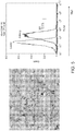
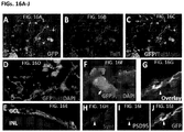
- the cells start to express Pax6, followed by the onset of Rx1 expression on day 12 ( FIG. 1A ).
- RT-PCR analysis showed loss of Oct4 and Nanog expression ( FIG. 1B ).
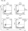
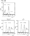
- On day 13 over 90% cells co-express Pax6 and Rx1 ( FIG. 2A ) as quantified flow cytometry, and over 99% of cells express Nestin, Otx2 and Sox2 ( FIG. 2B ).
- GC medium comprises a basal medium such as NeurobasalTM Medium (Life Technologies) or a medium comprising the components of NeurobasalTM Medium, D-glucose, one or more antibiotics such as penicillin and streptomycin, GlutaMAXTM, one or more serum supplements such as N2 supplement and B27 supplement (formula 080085-SA), and one or more of including all of forskolin, BDNF and CNTF.
- Basal medium such as NeurobasalTM Medium (Life Technologies) or a medium comprising the components of NeurobasalTM Medium, D-glucose, one or more antibiotics such as penicillin and streptomycin, GlutaMAXTM, one or more serum supplements such as N2 supplement and B27 supplement (formula 080085-SA), and one or more of including all of forskolin, BDNF and CNTF.
- the cells continue to be cultured in GC medium until about day 20.
- the neural spheres are cultured in GC medium, with media changes about every 3-4 days.
- RG progenitor cells Differentiation of RG progenitor cells to form RG cells.
- RG progenitor cells are dissociated into single cells using Accutase® cell detachment solution. Cells are plated on poly-D-lysine/laminin coated plates at the density of about 10-40/cm 2 in retinal ganglion cell (RGC) differentiation medium.
- RRC retinal ganglion cell
- RCG differentiation medium comprises a basal medium such as NeurobasalTM Medium (Life Technologies) or a medium comprising the components of NeurobasalTM Medium, D-glucose, antibiotics such as penicillin and streptomycin, GlutaMAXTM, one or more serum supplements such as B27 supplement (formula 080085-SA), insulin, one or both of BDNF and CNTF, and one or more including all of forskolin, cAMP and DAPT or other Notch inhibitor.
- the components of NeurobasalTM Medium are provided in Table 1. The media was changed about every 2 days.
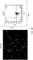
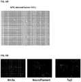
- FIG. 5 After 2 weeks, about 72% of cells stained positive for Thy1 ( FIG. 5 ). Cells had dendrites and axons ( FIG. 6A ). Immunostaining showed expression of Brn3a, neurofilament and Tuj 1 ( FIG. 6B ). These results indicate the presence of RG cells.
- RG progenitors and RG cells Cryopreservation of RG progenitors and RG cells.
- Early RG progenitor cells, late RG progenitor cells, and RG cells can be frozen/cryopreserved in an animal-free cryopreservation buffer, such as Cryostor CS10, as neurospheres.
- the cells are stored, proliferated or differentiated in various cell culture media. These media are described in greater detail below.
- Retinal induction (RI) medium is used to differentiate pluripotent stem cells into EF progenitor cells.
- Retinal induction (RI) medium comprises a basal medium such as DMEM/F12 or a medium comprising the components of DMEM/F12 (as shown in Table 1), D-glucose (optionally between 0-10 mg/ml, 2.5-7.5 mg/ml, 3-6 mg/ml, 4-5 mg/ml, or about 4.5 mg/ml), one or more antibiotics such as penicillin (optionally between 0-100 units/ml or about 100 units/ml) and streptomycin (optionally between 0-100 ⁇ g/ml or about 100 ⁇ g/ml), one or more serum supplements such as N2 supplement (optionally at about 0.1 to 5% or about 0.1 to 2% or about 1% (v/v) and B27 supplement (optionally about 0.05-2.0% or about 0.2% (v/v), MEM non-essential amino acids
- the RI medium may comprise Noggin polypeptide at a concentration of between about 5-100 ng/ml or about 10-100 ng/ml or about 10-500 ng/ml or about 50 ng/ml.
- the retinal induction medium may comprise a BMP signaling inhibitor, optionally selected from the group consisting of Noggin polypeptide, dorsomorphin, LDN-193189, and any combination thereof.
- RI medium may include insulin, Noggin polypeptide, or insulin and Noggin polypeptide.
- Noggin polypeptide is not necessary although increased differentiation into EF progenitors cells (as indicated by increased expression of EF transcription factors) is observed when Noggin polypeptide is present.
- Noggin polypeptide can be substituted with other moieties having similar activity, including SB431542.
- Noggin polypeptide is a secreted BMP inhibitor that reportedly binds BMP2, BMP4, and BMP7 with high affinity to block TGF ⁇ family activity.
- SB431542 is a small molecule that reportedly inhibits TGF ⁇ /Activin/Nodal by blocking phosphorylation of ACTRIB, TGF ⁇ R1, and ACTRIC receptors. SB431542 is thought to destabilize the Activin- and Nanog-mediated pluripotency network as well as suppress BMP-induced trophoblast, mesoderm, and endodermal cell fates by blocking endogenous Activin and BMP signals.
- Noggin polypeptide and SB431542 could replace or augment the functions of one or both of Noggin polypeptide and SB431542, e.g., as they are used in the context of the disclosed methods.
- Noggin polypeptide and/or the small molecule SB4312542 could be replaced or augmented by one or more inhibitors that affect any or all of the following three target areas: 1) preventing the binding of the ligand to the receptor; 2) blocking activation of receptor (e.g., dorsomorphin), and 3) inhibition of SMAD intracellular proteins/transcription factors.
- Exemplary potentially suitable factors include the natural secreted BMP inhibitors Chordin (which blocks BMP4) and Follistatin (which blocks Activin), as well as analogs or mimetics thereof. Additional exemplary factors that may mimic the effect of Noggin polypeptide include use of dominant negative receptors or blocking antibodies that would sequester BMP2, BMP4, and/or BMP7. Additionally, with respect to blocking receptor phosphorylation, dorsomorphin (or Compound C) has been reported to have similar effects on stem cells.
- Inhibition of SMAD proteins may also be effected using soluble inhibitors such as SIS3 (6,7-Dimethoxy-2-((2E)-3-(1-methyl-2-phenyl-1H-pyrrolo[2,3-b]pyridin-3-yl-prop-2-enoyl))-1,2,3,4-tetrahydroisoquinoline, Specific Inhibitor of Smad3, SIS3), overexpression of one or more of the inhibitor SMADs (e.g., SMAD6, SMAD7, SMAD10) or RNAi for one of the receptor SMADs (SMAD1, SMAD2, SMAD3, SMAD5, SMAD8/9).
- soluble inhibitors such as SIS3 (6,7-Dimethoxy-2-((2E)-3-(1-methyl-2-phenyl-1H-pyrrolo[2,3-b]pyridin-3-yl-prop-2-enoyl))-1,2,3,4-tetrahydrois
- Another combination of factors expected to be suitable for generating neural progenitors comprises a cocktail of Leukemia Inhibitory Factor (LIF), GSK3 inhibitor (CHIR 99021), Compound E (y secretase inhibitor XXI) and the TGF ⁇ inhibitor SB431542 which has been previously shown to be efficacious for generating neural crest stem cells ( Li et al., Proc Natl Acad Sci U S A. 2011 May 17;108(20):8299-304 ).
- Additional exemplary factors may include derivatives of SB431542, e.g., molecules that include one or more added or different substituents, analogous functional groups, etc. and that have a similar inhibitory effect on one or more SMAD proteins.
- Suitable factors or combinations of factors may be identified, for example, by contacting pluripotent cells with said factor(s) and monitoring for adoption of EF progenitor cell phenotypes, such as characteristic gene expression (including expression of the markers described herein, expression of a reporter gene coupled to an EF progenitor cell promoter, or the like) or the ability to form a cell type disclosed herein such as retinal neural progenitor cells, RG progenitor cells or RG cells.
- characteristic gene expression including expression of the markers described herein, expression of a reporter gene coupled to an EF progenitor cell promoter, or the like
- a cell type disclosed herein such as retinal neural progenitor cells, RG progenitor cells or RG cells.
- the cells are treated with or cultured in a retinal induction medium prior to culture with a neural differentiation medium.
- Neural differentiation (ND) medium is used to differentiate pluripotent stem cells into eye field (EF) progenitor cells.
- ND medium comprises a basal medium such as NeurobasalTM Medium (Life Technologies) or medium comprising components of NeurobasalTM Medium as provided in Table 1, D-glucose (optionally between 0-10 mg/ml, 2.5-7.5 mg/ml, 3-6 mg/ml, 4-5 mg/ml, or about 4.5 mg/ml), one or more antibiotics such as penicillin (optionally between 0-100 units/ml or about 100 units/ml) and streptomycin (optionally between 0-100 ⁇ g/ml or about 100 ⁇ g/ml), GlutaMAXTM (optionally IX, approximately 2 mM), one or more serum supplements such as N2 supplement (optionally 0.1-5% v/v or 0.1-2% v/v, including 1% v/v) and B27 supplement (optionally 0.05-2% v/v, including 2% v
- N2 supplement
- Ganglion cell (GC) medium is used to differentiate pluripotent stem cells into retinal ganglion (RG) progenitor cells, and more specifically to differentiate eye field progenitor cells into retinal ganglion (RG) progenitor cells.
- GC medium comprises a basal medium such as NeurobasalTM Medium (Life Technologies) or a medium comprising the components of NeurobasalTM Medium as provided in Table 1, D-glucose (optionally between 0-10 mg/ml, 2.5-7.5 mg/ml, 3-6 mg/ml, 4-5 mg/ml, or about 4.5 mg/ml), one or more antibiotics such as penicillin (optionally between 0-100 units/ml or about 100 units/ml) and streptomycin (optionally between 0-100 ⁇ g/ml or about 100 ⁇ g/ml), GlutaMAXTM (optionally IX, or approximately 2 mM), one or more serum supplements such as N2 supplement (optionally 0.1-5% v/v or 0.1-2% v/v, including 1% v/v) and B27 supplement (formula 080085-SA) (optionally 0.5-2% v/v, including 2% v/v), and one or more of forskolin (optionally 1-20 ⁇ M, and
- RGC differentiation medium is used to differentiate pluripotent stem cells into retinal ganglion (RG) cells, and more specifically to differentiate RG progenitor cells into RG cells.
- RGC differentiation medium comprises a basal medium such as NeurobasalTM Medium (Life Technologies) or a medium comprising the components of NeurobasalTM Medium as provided in Table 1, D-glucose (optionally between 0-10 mg/ml, 2.5-7.5 mg/ml, 3-6 mg/ml, 4-5 mg/ml, or about 4.5 mg/ml), one or more antibiotics such as penicillin (optionally between 0-100 units/ml or about 100 units/ml) and streptomycin (optionally between 0-100 ⁇ g/ml or about 100 ⁇ g/ml), one or more serum supplements such as B-27 supplement (formula 080085-SA) (optionally at about 0.05 to 5.0% , about 1.5 to 2.5% or about 2% (v/v)), GlutaMAXTM (optionally
- BDNF may be human BDNF.
- CTNF may be human CTNF.
- the RGC differentiation medium may comprise one or both of BDNF and CNTF.
- the RGC differentiation medium may comprise one or more including all of forskolin, cAMP, and DAPT or other Notch inhibitor.
- the RGC differentiation medium may comprise insulin, including human insulin (optionally about 5-50 ⁇ g/ml or about 20 ⁇ g/ml).
- DMEM/F12 The components of DMEM/F12, NeurobasalTM Medium (Life Technologies), N2 serum supplement, and B27 serum supplement and B27 serum supplement (formula 080085-SA) are provided in Table 1. It is to be understood that the invention contemplates the use of these particular media and supplements or media or supplements comprising, consisting essentially of, or consisting of these components.
- the term preparation particularly as it relates to a cell preparation, is used interchangeably with the terms population and composition.
- population and composition For brevity, aspects and embodiments may be described in terms of a preparation but it is to be understood that such descriptions apply equally to populations and compositions. It is to be understood that the preparation may comprise other non-cellular substituents.
- a level of purity of a cell preparation may be quantified as the proportion of cells in a preparation that express one or more markers of a desired cell type (including those markers identified in this application or others known in the art) relative to the total number of cells in the preparation. Purity may be determined by detecting and enumerating cells that express and/or cells that do not express one or more markers of interest, and optionally enumerating total cell number. Exemplary methods that may be used to enumerate cells include fluorescence activated cell sorting (FACS), immunohistochemistry, in situ hybridization, and other suitable methods known in the art. Typically such an analysis excludes non-viable cells in the preparation.
- FACS fluorescence activated cell sorting
- immunohistochemistry immunohistochemistry
- in situ hybridization and other suitable methods known in the art. Typically such an analysis excludes non-viable cells in the preparation.
- a majority of cells means at least 50%, and depending on the embodiment may include at least 60%, at least 70%, at least 80%, at least 90%, at least 95%, or about 100% of cells.
- the disclosure provides substantially pure (or homogeneous) preparations of various cell populations based on the ability of the disclosed methods to directly differentiate progenitor cells such as pluripotent stem cells.
- directed differentiation intends that the progenitor cell population differentiates into or towards a desired lineage, due in part to the factors or other stimuli provided to such progenitor cells, thereby avoiding differentiation into other undesired, and thus potentially contaminating, lineages.
- the methods provided herein drive differentiation of for example pluripotent stem cells to eye field progenitors without generating embryoid bodies (EB).
- EB embryoid bodies
- EBs are three dimensional cell clusters that can form during differentiation of pluripotent stem cells including embryonic stem (ES) cells, and that typically contain cells, including progenitors, of mesodermal, ectodermal and endodermal lineages.
- ES embryonic stem
- the three dimensional nature of the EB may create a different environment, including different cell-cell interactions and different cell-cell signaling, than occurs in the non-EB based methods described herein.
- cells within EBs may not all receive a similar dose of an exogenously added agent, such as a differentiation factor present in the surrounding medium, and this can result in various differentiation events and decisions during development of the EB.
- the culture methods of the invention do not require and preferably avoid EB formation. Instead, these methods culture cells in conditions that provide the cells with equal contact with the surrounding medium, including factors in such medium.
- the cells may grow as a monolayer or near monolayer attached to a culture surface, as an example.
- the ability of all or a majority of the progenitor cells to be in contact with their surrounding medium and thus the factors in such medium to an approximately equal degree results in those progenitor cells differentiating at similar times and to similar degrees.
- This similar differentiation timeline for a population of progenitor cells indicates that such cells are synchronized.
- the cells may be cell cycle synchronized in some instances also.
- Such synchronicity results in populations of cells that are homogeneous or near homogeneous in their cellular make-up.
- the methods described herein can produce cellular populations wherein at least 50%, at least 60%, at least 70%, at least 80%, at least 90%, at least 95%, or about 100% of cells are a particular cell of interest.
- the cell of interest may be defined phenotypically, for example by intracellular or extracellular marker expression.
- the cell of interest may be an eye field progenitor cell, an RG progenitor cell (whether early or late), or an RG cell.
- a “substantially pure preparation of cells” refers to a preparation of cells wherein the cells are at least 60% pure. Thus, at least 60% of the cells in the preparation express one or more markers of the particular cell type (including those markers identified in this application or others known in the art).
- the cells may be at least 65% pure or at least 70% pure.
- the cells may be about more than 70% pure including at least 75%, 80%, 85% or 90% pure.
- a “substantially pure preparation of RG cells” refers to a preparation of cells wherein at least 60% of the cells in the preparation express one or more markers of RG cells (including those markers identified in this application or others known in the art). At least 70% of the cells in the preparation may express one or more markers of RG cells, in which case the preparation is 70% pure. At least 85% of the cells in the preparation may express one or more markers of RG cells, in which case the preparation is 85% pure. At least 95% of the cells in the preparation may express one or more markers of RG cells, in which case the preparation is 95% pure. In some embodiments, the preparation may be about 85% to 95% pure.
- a "substantially pure preparation of RG progenitor cells” refers to a preparation of cells wherein at least 60% of the cells in the preparation express one or more markers of RG progenitor cells (including those markers identified in this application or others known in the art). At least 70% of the cells in the preparation may express one or more markers of RG progenitor cells, in which case the preparation is 70% pure. At least 85% of the cells in the preparation may express one or more markers of RG progenitor cells, in which case the preparation is 85% pure. At least 95% of the cells in the preparation may express one or more markers of RG progenitor cells, in which case the preparation is 95% pure. In some embodiments, the preparation may be about 85% to 95% pure.
- cells including RG progenitor cells and RG cells have a mean terminal restriction fragment length (TRF) that is longer than 7 kb, 7.5 kb, 8 kb, 8.5 kb, 9 kb, 9.5 kb, 10 kb, 10.5 kb, 11 kb, 11.5 kb or even 12 kb.
- TRF mean terminal restriction fragment length
- markers such as Pax6, Rx1, Six3, Six6, Lhx2, Tbx3, Sox2, Chx10, Math5, Brn3 including Brn3a and Brn3b, Isl1, Neurofilament, Tuj1, Thy1, Nestin, Otx2, Nanog, Oct4, may be assessed at the protein and/or mRNA level (see Fischer AJ, Reh TA, Dev Neurosci. 2001:23(4-5):268-76 ; Baumer et al., Development. 2003 Jul;130(13):2903-15 ; Swaroop et al., Nat Rev Neurosci. 2010 Aug;11(8):563-76 ; Agathocleous and Harris, Annu. Rev. Cell Dev. Biol. 2009. 25:45-69 ; Beby F, Lamonerie T, Exp. Eye Res. 2013; 111, 9-16 ).
- the markers are generally human, e.g., except where the context indicates otherwise.
- the cell markers can be identified using conventional immunocytochemical methods or conventional PCR methods which techniques are well known to those of ordinary skill in the art.
- pluripotent stem cells are cells that are: (a) capable of inducing teratomas when transplanted in immunodeficient (SCID) mice; (b) capable of differentiating to cell types of all three germ layers (e.g., ectodermal, mesodermal, and endodermal cell types); and (c) express one or more markers of pluripotency (e.g., Oct 4, alkaline phosphatase, SSEA-3 surface antigen, SSEA-4 surface antigen, nanog, TRA-1-60, TRA-1-81, SOX2, REX1, etc).
- pluripotent stem cells express one or more markers selected from the group consisting of OCT-4, alkaline phosphatase, SSEA-3, SSEA-4, TRA-1-60, and TRA-1-81.
- Pluripotent stem cells include embryonic stem (ES) cells, embryo-derived stem cells, embryonic germ (EG) cells, embryonic carcinoma (EC) cells, induced pluripotent stem (iPS) cells, and stimulus-triggered acquisition of pluripotency (STAP) cells.
- ES embryonic stem
- EG embryonic germ
- EC embryonic carcinoma
- iPS induced pluripotent stem
- STAP stimulus-triggered acquisition of pluripotency
- Exemplary pluripotent stem cells can be generated using any variety of methods known in the art. As will be recognized in the art, embryoid bodies can be formed from ES cells or EG cells, among others. As will also be recognized in the art, ES cells may be generated without embryo destruction.
- ES cells may be generated from parthenogenetic ova (egg cells) which are non-embryos, ES cells may be generated from non-viable embryos including those that are deliberately engineered to be non-viable (e.g., not capable of implantation), and ES cells may be generated from single blastomeres of an embryo including a non-viable embryo.
- egg cells parthenogenetic ova
- ES cells may be generated from non-viable embryos including those that are deliberately engineered to be non-viable (e.g., not capable of implantation)
- ES cells may be generated from single blastomeres of an embryo including a non-viable embryo.
- Pluripotent stem cells may be modified, including genetically modified, to increase longevity, potency, and/or homing, and/or to prevent or reduce alloimmune responses, and/or to deliver a desired factor to the differentiated progeny of the pluripotent stem cells, and/or to express a desired factor in differentiated progeny of the pluripotent stem cells.
- the pluripotent stem cells can be from any species.
- Various types of pluripotent stem cells including ES cells and iPS cells, have been generated from mouse, human and other sources.
- the pluripotent stem cells used in the present methods may be obtained from any species including human, non-human primates, rodents (mice, rats), ungulates (cows, sheep, etc.), dogs (domestic and wild dogs), cats (domestic and wild cats such as lions, tigers, cheetahs), rabbits, hamsters, goats, elephants, panda (including giant panda), pigs, raccoon, horse, zebra, marine mammals (dolphin, whales, etc.) and the like.
- the species is an endangered species.
- the species is a currently extinct species.
- Embryonic stem (ES) cells are cells that (i) are able to differentiate into cells of all three germ layers, (ii) express at least Oct 4 and alkaline phosphatase, and (iii) are able to produce teratomas when transplanted into immunodeficient animals.
- the term includes cells derived from the inner cell mass (ICM) of human blastocysts or morulae, including those that have been serially passaged as cell lines; cells derived from a zygote, a blastomere (e.g., of a cleavage stage or morula stage embryo), or a blastocyst-staged mammalian embryo, produced by the artificial fusion of a sperm and egg cell, fertilization of an egg cell with sperm, nuclear transfer such as somatic cell nuclear transfer (SCNT), parthenogenesis, androgenesis, or through reprogramming of chromatin and subsequent incorporation of the reprogrammed chromatin into a cell.
- ICM inner cell mass
- ES cells does not infer, and should not be inferred to mean, that the cells were generated through the destruction of an embryo.
- various methods are available and can be used to generate ES cells without destruction of an embryo, such as a human embryo.
- ES cells may be generated from single blastomeres derived from an embryo, in a manner similar to the extraction of blastomeres for pre-implantation genetic diagnosis (PGD). Examples of such cell lines include NED1, NED2, NED3, NED4, NED5, and NED7.
- PGD pre-implantation genetic diagnosis
- Examples of such cell lines include NED1, NED2, NED3, NED4, NED5, and NED7.
- An exemplary human embryonic stem cell line that may be used is MA09 cells. The isolation and preparation of MA09 cells was previously described in Klimanskaya, et al.
- Additional ES cells that may be used include human ES cells such as MA01, MA09, ACT-4, MA142, MA143, MA144, MA145, MA146, MA126 (NED1), MA127, (NED2), MA128, (NED3), MA129, (NED4), NED7, No. 3, H1, H7, H9, H14 and ACT30 ES cells.
- human ES cells such as MA01, MA09, ACT-4, MA142, MA143, MA144, MA145, MA146, MA126 (NED1), MA127, (NED2), MA128, (NED3), MA129, (NED4), NED7, No. 3, H1, H7, H9, H14 and ACT30 ES cells.
- the human ES cells used in accordance with this disclosure may be derived and maintained in accordance with GMP standards which are known in the art.
- ES cells have been derived from a variety of species including mice, multiple species of non-human primates, and humans, and embryo-derived stem cells have been generated from numerous additional species.
- Induced pluripotent stem (iPS) cells are cells generated through the reprogramming of a somatic cell by expressing a combination of factors (herein referred to as reprogramming factors).
- iPS cells can be generated using, as a starting point, virtually any somatic cell at any developmental stage.
- the cell can be from an embryo, fetus, neonate, juvenile, or adult donor.
- the cell is from a neonate, juvenile, or adult donor, or is obtained from the amniotic fluid, and thus does not require destruction of an embryo or fetus.
- Exemplary somatic cells that can be used include fibroblasts, such as dermal fibroblasts obtained by a skin sample or biopsy, synoviocytes from synovial tissue, foreskin cells, cheek cells, and lung fibroblasts.
- the somatic cell is not a fibroblast.
- At least one, at least two, or at least three, or at least four reprogramming factors are expressed in a somatic cell to successfully reprogram the somatic cell.
- the reprogramming factors may be selected from Oct 4 (sometimes referred to as Oct 3/4), SOX2, c-Myc, Klf4, Nanog and Lin28.
- the reprogramming factors include a combination of Oct 4 (sometimes referred to as Oct 3/4), SOX2, c-Myc and Klf4, or a combination of Oct 4, SOX2, Nanog and Lin28.
- the reprogramming factors may be introduced into somatic cells using any of a variety of methods including integrative vectors, non-integrative vectors (e.g., an episomal plasmid, such as that described in Yu et al., Science. 2009 May 8;324(5928):797-801 ), chemical means, protein transduction, electroporation, microinjection, cationic amphiphiles, fusion with lipid bilayers, detergent permeabilization, etc., or a combination thereof. Additionally, expression of the reprogramming factors may be induced by contacting the somatic cells with at least one agent, such as a small organic molecule agents, that induce expression of reprogramming factors.
- a small organic molecule agents such as a small organic molecule agents
- iPS cells may be obtained from a cell bank. iPS cells may be specifically generated using material from a particular patient or matched donor with the goal of generating tissue-matched RG cells or RG progenitor cells. iPS cells can be produced from cells that are not substantially immunogenic in an intended recipient, e.g., produced from autologous cells or from cells histocompatible to an intended recipient.
- Stimulus-triggered acquisition of pluripotency (STAP) cells are pluripotent stem cells produced by reprogramming somatic cells with sublethal stimuli such as low-pH exposure. The reprogramming does not require nuclear transfer into or genetic manipulation of the somatic cells.
- STAP pluripotency
- Adult stem cell refers to a multipotent cell isolated from adult tissue and can include bone marrow stem cells, cord blood stem cells and adipose stem cells and is of human origin.
- the methods of the invention do not employ an EB differentiation step (i.e., differentiation occurs from the pluripotent stem cells through to the retinal ganglion cells without the formation of EBs).
- an EB differentiation step i.e., differentiation occurs from the pluripotent stem cells through to the retinal ganglion cells without the formation of EBs.
- Embryoid bodies refers to clumps or clusters of cells including pluripotent stem cells (e.g., iPSC or ESC), which may be formed by culturing pluripotent stem cells under non-attached conditions, e.g., on a low-adherent substrate or in a "hanging drop.” In these cultures, pluripotent cells can form clumps or clusters of cells denominated as embryoid bodies. See Itskovitz-Eldor et al., Mol Med. 2000 Feb;6(2):88-95 .
- pluripotent stem cells e.g., iPSC or ESC
- embryoid bodies initially form as solid clumps or clusters of pluripotent cells, and over time some of the embryoid bodies come to include fluid filled cavities, the former being referred to in the literature as “simple” EBs and the latter as “cystic" embryoid bodies.
- the present disclosure describes methods for screening various agents that modulate the differentiation of an eye field progenitor cell towards the retinal ganglion lineage. It could also be used to discover therapeutic agents that support and/or rescue RG progenitor cells that may be generated in culture from eye field progenitor cells.
- the present disclosure describes methods for screening various agents that modulate the differentiation of an RG progenitor cell. It could also be used to discover therapeutic agents that support and/or rescue mature RG cells that are generated in culture from RG progenitor cells.
- an "agent” is intended to include a biological or chemical compound such as a simple or complex organic or inorganic molecule, a peptide, a protein (e.g. antibody), a polynucleotide (e.g. anti-sense, RNA interference (RNAi, shRNA, microRNA)) or a ribozyme.
- a biological or chemical compound such as a simple or complex organic or inorganic molecule, a peptide, a protein (e.g. antibody), a polynucleotide (e.g. anti-sense, RNA interference (RNAi, shRNA, microRNA)) or a ribozyme.
- RNAi RNA interference
- shRNA RNA interference
- an isolated population of cells can be obtained as described herein.
- the agent is a composition other than a DNA or RNA, such as a small molecule as described above
- the agent can be directly added to the cells or added to culture medium.
- an "effective" amount must be added which can be empirically determined.
- the agent is a polynucleotide, it can be directly added by use of a gene gun or electroporation. Alternatively, it can be inserted into the cell using a gene delivery vehicle or other method as described above. Positive and negative controls can be assayed to confirm the purported activity of the drug or other agent.
- the assay can be used to identify agents which promote the proliferation of native retinal ganglion cells and/or their progenitors, and/or which may induce the differentiation/maturation of native retinal ganglion progenitors to mature retinal ganglion cells.
- Agents that may be tested in these assays include small molecules (e.g., organic compounds that have molecular weights of less than 10,000 atomic mass units (amu)), proteins and peptides, nucleic acids, etc. Agents to be tested can be produced, for example, by bacteria, yeast or other organisms (e.g. natural products), produced chemically (e.g. small molecules, including peptidomimetics), or produced recombinantly.
- small molecules e.g., organic compounds that have molecular weights of less than 10,000 atomic mass units (amu)
- proteins and peptides e.g., nucleic acids, etc.
- Agents to be tested can be produced, for example, by bacteria, yeast or other organisms (e.g. natural products), produced chemically (e.g. small molecules, including peptidomimetics), or produced recombinantly.
- the assay may evaluate the ability of a compound to protect retinal ganglion cells (such as may be derived by differentiation of pluripotent stem cells, eye field progenitors, or RG progenitor cells using the methods provided herein) in culture from glutamate induced toxicity.
- retinal ganglion cells such as may be derived by differentiation of pluripotent stem cells, eye field progenitors, or RG progenitor cells using the methods provided herein
- a variety of assay formats will suffice and, in light of the present disclosure, those not expressly described herein will nevertheless be comprehended by one of ordinary skill in the art.
- high throughput assays are desirable in order to maximize the number of compounds surveyed in a given period of time.
- Detection and quantification of protective activity of a test agent can be carried out, for example, by detecting the relative changes in markers of apoptosis - agents which are protective, should produce a decrease in the prevalence/level of expression of markers for apoptosis relative to control untreated cells under neurotoxic conditions such as elevated glutamate levels.
- the efficacy of the agent can be assessed by generating dose response curves from data obtained using various concentrations of the test compound.
- Control assays can also be performed to provide a baseline for comparison. In the control assay, the degree of apoptosis can be quantitated in the absence of the test compound. It will be understood that, in general, the order in which the reactants may be admixed can be varied, and can be admixed simultaneously.
- the RG progenitor cells can be used to generate neurosensory retinal structures that include retinal pigment epithelial (RPE) cells, photoreceptor cells and the RG progenitor or RG cells themselves.
- RPE retinal pigment epithelial
- the invention contemplates the generation of multilayer cellular structures comprised of RG cells or RG progenitor cells, RPE cells, and photoreceptor cells (or photoreceptor progenitor cells).
- These structures can be used for drug screening, as models for diseases, or in a pharmaceutical preparation.
- the pharmaceutical preparation can be an RPE-photoreceptor-RG graft, which may be disposed on a biocompatible solid support or matrix (preferably a bioresorbable matrix or support) that can be implanted like a "patch".
- RG cells or RG progenitor cells can be generated along with photoreceptors from retinal neural progenitors (e.g., Chx10(+) cells), and these cells can be transplanted along with an RPE cell layer (e.g., a monolayer).
- retinal neural progenitors e.g., Chx10(+) cells
- RPE cell layer e.g., a monolayer
- the biocompatible support for the cells can be a biodegradable polyester film support for retinal progenitor cells.
- the biodegradable polyester can be any biodegradable polyester suitable for use as a substrate or scaffold for supporting the proliferation and differentiation of retinal progenitor cells.
- the polyester should be capable of forming a thin film, preferably a micro-textured film, and should be biodegradable if used for tissue or cell transplantation.
- Suitable biodegradable polyesters for use in the invention include polylactic acid (PLA), polylactides, polyhydroxyalkanoates, both homopolymers and co-polymers, such as polyhydoxybutyrate (PHB), polyhydroxybutyrate co-hydroxyvalerate (PHBV), polyhydroxybutyrate co-hydroxyhexanote (PHBHx), polyhydroxybutyrate co-hydroxyoctonoate (PHBO) and polyhydroxybutyrate co-hydroxyoctadecanoate (PHBOd), polycaprolactone (PCL), polyesteramide (PEA), aliphatic copolyesters, such as polybutylene succinate (PBS) and polybutylene succinate/adipate (PBSA), aromatic copolyesters. Both high and low molecular weight polyesters, substituted and unsubstituted polyester, block, branched or random, and polyester mixtures and blends can be used.
- the biodegradable polyester is polycaprolactone (PCL).
- the biocompatible support is a poly(p-xylylene) polymer, such as parylene N, parylene D, parylene-C, parylene AF-4, parylene SF, parylene HT, parylene VT-4 and Parylene CF, and most preferably parylene-C.
- the polymeric support can typically be formed into a thin film using known techniques.
- the film thickness is advantageously from about 1 micron to about 50 microns, and preferably about 5 microns in thickness.
- the surface of the film can be smooth, or the film surface can be partially or completely micro-textured. Suitable surface textures include micro-grooves or micro-posts, for instance.
- the film can be cut and shaped to form a suitable shape for implantation.
- the RPE, photoreceptor cells or photoreceptor progenitor cells, and RG progenitor cells and/or RG cells can be plated directly, together or sequentially (e.g., photoreceptor cells or photoreceptor progenitor cells after an RPE layer is formed), onto the film to form a biocompatible scaffold.
- the polymer film can be coated with a suitable coating material such as poly-D-lysine, poly-L-lysine, fibronectin, laminin, collagen I, collagen IV, vitronectin and MatrigelTM.
- the cells can be plated to any desired density, but a single layer of RPE cells (an RPE monolayer) is preferred.
- the present invention provides a composition in accordance with the claims for use in a method of treatment of a mammalian patient, comprising administering the composition to said patient, or administering the composition after thawing to said patient.
- the cells may be transplanted into a rat in need thereof, e.g., an intraocular hypertension rat, or other animal model of disease, and the resulting effect on visual function may be detected by the Optomotor response test, ERG (electroretinography), luminance threshold recording and/or the visual center blood flow assay.
- “Signs” of disease refers broadly to any abnormality indicative of disease, discoverable on examination of the patient; an objective indication of disease, in contrast to a symptom, which is a subjective indication of disease.
- Symptoms of disease refers broadly to any morbid phenomenon or departure from the normal in structure, function, or sensation, experienced by the patient and indicative of disease.
- “Therapy,” “therapeutic,” “treating,” “treat” or “treatment”, as used herein, refers broadly to treating a disease, arresting or reducing the development of the disease or its clinical symptoms, and/or relieving the disease, causing regression of the disease or its clinical symptoms.
- Therapy encompasses prophylaxis, prevention, treatment, cure, remedy, reduction, alleviation, and/or providing relief from a disease, signs, and/or symptoms of a disease. Therapy encompasses an alleviation of signs and/or symptoms in patients with ongoing disease signs and/or symptoms. Therapy also encompasses "prophylaxis" and "prevention". Prophylaxis includes preventing disease occurring subsequent to treatment of a disease in a patient or reducing the incidence or severity of the disease in a patient.
- the term “reduced”, for purpose of therapy, refers broadly to the clinical significant reduction in signs and/or symptoms.
- Therapy includes treating relapses or recurrent signs and/or symptoms. Therapy encompasses precluding the appearance of signs and/or symptoms anytime as well as reducing existing signs and/or symptoms and eliminating existing signs and/or symptoms.
- Therapy includes treating chronic disease ("maintenance") and acute disease. For example, treatment includes treating or preventing relapses or the recurrence of signs and/or symptoms.
- treatment may be indicated by stable or improved scoptic threshold response, stable or improved optokinetic reflex (OKR), stable or improved vision evoked potential (VEP), and/or stable or improved visual acuity (e.g., as measured by ERG).
- Stable or improved levels are relative to levels pre-treatment or relative to non-treated controls such as contralateral eyes. It is to be understood that in some instances one or more of these functions or responses will deteriorate with time in the absence of treatment, and that the transplanted cells of the invention may impede, reduce or limit that deterioration. Thus in some instances the function or response remains stable relative to control while in other instances it improves, and both situations are considered to be beneficial to the subject and thus to be regarded as treatment.
- the subject may experience an underlying benefit to its retinal ganglion cell layer.
- the native RG cell layer may experience improved survival, or reduced cell loss, compared to a control.
- the subject may experience an increased formation of axons in the optic nerve.
- compositions that may be for use in methods for replacing or repairing RG cells in a patient in need of this treatment comprising administering a pharmaceutical preparation including the RG progenitor cells of the present invention, or RG cells derived therefrom, or a combination thereof, to a patient.
- the pharmaceutical preparation can be a suspension of cells or cells which are formed into transplantable tissue in vitro.
- the cells will be administered by intraocular injection (including vitreal injection and subretinal injection).
- the cells may be administered intraocularly into the sub-retinal space of a diseased or degenerated retina.
- the RG progenitor cells also have a neuroprotective effect, the cells can be administered locally but outside of the retina (such as in the vitreous) or by depot or systemic delivery to other parts of the body.
- the pharmaceutical preparations of the present invention can be used in a wide range of diseases and disorders that result in visual system deterioration, including retinal degeneration-related disease.
- diseases and disorders may be caused by aging, such that there appears to be an absence of an injury or disease that is identifiable as a substantial source of the deterioration.
- Skilled artisans will understand the established methods for diagnosing such disease states, and/or inspecting for known signs of such injuries.
- the literature is replete with information on age-related decline or deterioration in aspects of the visual systems of animals.
- the term "retinal degeneration-related disease” is intended to refer to any disease resulting from innate or postnatal retinal degeneration or abnormalities.
- retinal degeneration-related diseases include retinal dysplasia, aged macular degeneration, diabetic retinopathy, retinitis pigmentosa, congenital retinal dystrophy, Leber congenital amaurosis, retinal detachment, glaucoma, optic neuropathy, and trauma.
- the deterioration of the visual system components can be caused by injury, for example trauma to the visual system itself (e.g., an eye), to the head or brain, or the body more generally.
- Certain such injuries are known to be age-related injuries, i.e., their likelihood, or frequency increases with age. Examples of such injuries include retinal tears, macular holes, epi-retinal membrane, and retinal detachments, each of which might occur in an animal of any age, but which are more likely to occur, or occur with greater frequency in aging animals, including otherwise healthy aging animals.
- deterioration of the visual system components may occur due to ischemic and/or reperfusion injury.
- the deterioration of the visual system or components thereof also can be caused by disease. Included among the diseases are various age-related diseases that impact the visual system. Such diseases occur with greater likelihood and/or frequency in older animals than in the young. Examples of diseases which may affect the visual system, including for example the neurosensory retinal layers, and cause deterioration thereof are various forms of retinitis, optic neuritis, macular degeneration, proliferative or nonproliferative diabetic retinopathy, diabetic macular edema, progressive retinal atrophy, progressive retinal degeneration, sudden acquired retinal degeneration, immune-mediated retinopathy, retinal dysplasia, chorioretinitis, retinal ischemia, retinal hemorrhage (preretinal, intraretinal and/or subretinal), hypertensive retinopathy, retinal inflammation, retinal edema, retinoblastoma, or retinitis pigmentosa.
- diseases which may affect the visual system including for example
- Some of the foregoing diseases tend to be specific to certain animals such as companion animals, e.g., dogs and/or cats. Some of the diseases are listed generically, i.e., there may be many types of retinitis, or retinal hemorrhage; thus some of the diseases are not caused by one specific etiologic agent, but are more descriptive of the type of disease or the result. Many of the diseases that can cause decline or deterioration of one or more components of the visual system can have both primary and secondary or more remote effects on an animal's visual system.
- the pharmaceutical preparations of the present invention may be used to treat or ameliorate a number of conditions including ischemia reperfusion injury, retinal vascular disease, optic nerve injury (whether autoimmune, trauma, and/or viral-based), glaucoma (regardless of type), diabetic retinopathy (whether hypoxic and angiogenic based), inherited ganglion degeneration, and hereditary optic neuropathy (such as Leber's hereditary optic neuropathy (LHON) or Leber optic atrophy).
- ischemia reperfusion injury whether autoimmune, trauma, and/or viral-based
- optic nerve injury whether autoimmune, trauma, and/or viral-based
- glaucoma regardless of type
- diabetic retinopathy whether hypoxic and angiogenic based
- inherited ganglion degeneration inherited ganglion degeneration
- hereditary optic neuropathy such as Leber's hereditary optic neuropathy (LHON) or Leber optic atrophy
- the cells can treat or alleviate the symptoms of any of the foregoing conditions in a patient in need of the treatment.
- the cells can be autologous or allogeneic to the patient.
- the cells of the invention can be administered in combination with other treatments.
- the RG progenitor cells of the present invention find use in the treatment of degenerative diseases.
- the cells are administered in a manner that permits them to graft or migrate to the intended ocular site, such as the ganglion cell layer of the retina, and reconstitute or regenerate the functionally deficient area.
- RG progenitor cells or RG cells can also be used to target gene products to sites of degeneration, and can, for example, be engineered with one or more genes that encode secreted biologically active proteins, peptides, or nucleic acids (such as RNA interference molecules or aptamers), or the like. These gene products can include survival-promoting factors to rescue native degenerating neurons, factors that can act in an autocrine manner to promote survival and differentiation of grafted cells into site-specific neurons or to deliver neurotransmitter(s) to permit functional recovery.
- the one or more biologically active molecule(s) can include anti-angiogenic antibodies and molecules, anti-angiogenic antibody scaffolds, soluble receptors, agents targeting and inhibiting or modulating immunologic pathway molecules, growth factor inhibitors, growth factors, neurotrophic factors, angiogenic factors, neurotransmitters, hormones, enzymes, anti-inflammatory factors, or other therapeutic proteins.
- such molecules can include C3a inhibitors, C3b inhibitors, other agents targeting and inhibiting or modulating immunologic pathway molecules, brain derived neurotrophic factor (BDNF), NT-4, ciliary neurotrophic factor (CNTF), Axokine, basic fibroblast growth factor (bFGF), insulin-like growth factor I (IGF I), insulin-like growth factor II (IGF II), acid fibroblast growth factor (aFGF), fibroblast growth factor (aFGF), epidermal growth factor (EGF), transforming growth factor-alpha (TGF-alpha), transforming growth factor-beta (TGF-beta), nerve growth factor (NGF), platelet derived growth factor (PDGF), glia-derived neurotrophic factor (GDNF), Midkine, tryophotin, activin, thyrotropin releasing hormone, interleukins, bone morphogenic protein, macrophage inflammatory proteins, heparin sulfate, amphiregulin, retinoic acid, tumor necros
- the recombinantly engineered RG progenitor cells or RG cells are engineered to express one or more recombinant growth factors and neurotrophins such as FGF2, NGF, ciliary neurotrophic factor (CNTF), and brain derived neurotrophic factor (BDNF), which factors have been shown to significantly slow the process of cell death in models of retinal degeneration.
- FGF2 FGF2, NGF, ciliary neurotrophic factor (CNTF), and brain derived neurotrophic factor (BDNF)
- BDNF brain derived neurotrophic factor
- the gene construct used to engineer the RG progenitor or RG cell can include transcriptional and/or translational control elements (such as enhancers or promoters) which are sensitive to conditions which would promote ganglion cell death and/or glaucoma, such as elevated intraocular pressure (IOP), elevated levels of glutamate, etc.
- transcriptional and/or translational control elements such as enhancers or promoters
- IOP intraocular pressure
- An exemplary promoter sensitive to elevated IOP is the promoter from the matrix Gla protein (MGP) gene (see, for example Gonzalez et al., Investigative Ophthalmology & Visual Science, May 2004, 45(5): 1389 ).
- cells to be transplanted are transferred to a recipient in any physiologically acceptable excipient comprising an isotonic excipient prepared under sufficiently sterile conditions for human administration.
- any physiologically acceptable excipient comprising an isotonic excipient prepared under sufficiently sterile conditions for human administration.
- Cell Therapy Stem Cell Transplantation, Gene Therapy, and Cellular Immunotherapy, by G. Morstyn & W. Sheridan eds, Cambridge University Press, 1996 .
- Choice of the cellular excipient and any accompanying elements of the composition will be adapted in accordance with the route and device used for administration.
- the cells may be introduced by injection, catheter, or the like.
- the cells may be frozen at liquid nitrogen temperatures and stored for long periods of time, being capable of use on thawing. If frozen, the cells may be stored in a 10% DMSO, 50% FCS, 40% RPMI 1640 medium. In a preferred embodiment, the cells will be stored in an animal-free cryopreservation buffer such as Cryostor
- compositions of the invention are optionally packaged in a suitable container with written instructions for a desired purpose.
- Such formulations may comprise a cocktail of retinal differentiation and/or trophic factors, in a form suitable for combining with RG progenitor or RG cells.
- Such a composition may further comprise suitable buffers and/or excipients appropriate for transfer into an animal.
- Such compositions may further comprise the cells to be engrafted.
- the RG progenitor cells or RG cells may be formulated with a pharmaceutically acceptable carrier.
- RG progenitor cells or RG cells may be administered alone or as a component of a pharmaceutical formulation.
- the subject compounds may be formulated for administration in any convenient way for use in medicine.
- compositions suitable for administration may comprise the RG progenitor cells or RG cells, in combination with one or more pharmaceutically acceptable sterile isotonic aqueous or non-aqueous solutions (e.g., balanced salt solution (BSS)), dispersions, suspensions or emulsions, or sterile powders which may be reconstituted into sterile injectable solutions or dispersions just prior to use, which may contain antioxidants, buffers, bacteriostats, solutes or suspending or thickening agents.
- BSS balanced salt solution
- Exemplary pharmaceutical preparations comprise the RG progenitor cells or RG cells in combination with ALCON® BSS PLUS® (a balanced salt solution containing, in each ml, sodium chloride 7.14 mg, potassium chloride 0.38 mg, calcium chloride dihydrate 0.154 mg, magnesium chloride hexahydrate 0.2 mg, dibasic sodium phosphate 0.42 mg, sodium bicarbonate 2.1 mg, dextrose 0.92 mg, glutathione disulfide (oxidized glutathione) 0.184 mg, hydrochloric acid and/or sodium hydroxide (to adjust pH to approximately 7.4) in water).
- ALCON® BSS PLUS® a balanced salt solution containing, in each ml, sodium chloride 7.14 mg, potassium chloride 0.38 mg, calcium chloride dihydrate 0.154 mg, magnesium chloride hexahydrate 0.2 mg, dibasic sodium phosphate 0.42 mg, sodium bicarbonate 2.1 mg, dextrose 0.92 mg, glutathione disulfide (oxidized glutathione) 0.184 mg, hydroch
- the pharmaceutical preparations for use in this disclosure may be in a pyrogen-free, physiologically acceptable form.
- the preparation comprising RG progenitor cells or RG cells may be transplanted in a suspension, gel, colloid, slurry, or mixture. Further, the preparation may desirably be encapsulated or injected in a viscous form into the vitreous humor for delivery to the site of retinal or choroidal damage. Also, at the time of injection, cryopreserved RG progenitor cells or RG cells may be resuspended with balanced salt solution to achieve the desired osmolality and concentration for administration by subretinal injection. The preparation may be administered to an area of the pericentral macula that was not completely lost to disease, which may promote attachment and/or survival of the administered cells. In some instances, including for example the glaucoma animal model, the RG progenitors and/or RG cells are administered intravitreously.
- the RG progenitors cells and/or RG cells may be frozen (cryopreserved) as described herein. Upon thawing, the viability of such cells may be at least 20%, at least 30%, at least 40%, at least 50%, at least 60%, at least 70%, at least 80%, at least 90% at least 95% or about 100% (e.g., at least 20%, at least 30%, at least 40%, at least 50%, at least 60%, at least 70%, at least 80%, at least 90% at least 95% or about 100% of the cells harvested after thawing are viable or at least 20%, at least 30%, at least 40%, at least 50%, at least 60%, at least 70%, at least 80%, at least 90% at least 95% or about 100% of the cell number initially frozen are harvested in a viable state after thawing).
- the RG progenitor cells or RG cells may be formulated and delivered in a pharmaceutically acceptable ophthalmic vehicle or formulation suitable for intraocular injection.
- the preparation is designed such that it is maintained in contact with the ocular surface for a sufficient time period to allow the cells to penetrate the affected regions of the eye, as for example, the anterior chamber, posterior chamber, vitreous body, aqueous humor, vitreous humor, cornea, iris/ciliary, lens, choroid, retina, sclera, suprachoridal space, conjunctiva, subconjunctival space, episcleral space, intracorneal space, epicorneal space, pars plana, surgically-induced avascular regions, or the macula.
- the RG progenitor cells or RG cells may be contained in a sheet of cells.
- a sheet of cells comprising RG progenitor cells or RG cells may be prepared by culturing RG progenitor cells or RG cells on a substrate from which an intact sheet of cells can be released, e.g., a thermoresponsive polymer such as a thermoresponsive poly(N-isopropylacrylamide) (PNIPAAm)-grafted surface, upon which cells adhere and proliferate at the culture temperature, and then upon a temperature shift, the surface characteristics are altered causing release of the cultured sheet of cells (e.g., by cooling to below the lower critical solution temperature (LCST).
- a thermoresponsive polymer such as a thermoresponsive poly(N-isopropylacrylamide) (PNIPAAm)-grafted surface
- the sheet of cells may be adherent to a substrate suitable for transplantation, such as a substrate that may dissolve in vivo when the sheet is transplanted into a host organism, e.g., prepared by culturing the cells on a substrate suitable for transplantation, or releasing the cells from another substrate (such as a thermoresponsive polymer) onto a substrate suitable for transplantation.
- a substrate suitable for transplantation may comprise gelatin (see Hsiue et al., supra).
- Alternative substrates suitable for transplantation include fibrin-based matrixes and others.
- the sheet of cells may be used in the manufacture of a medicament for the prevention or treatment of a disease of retinal degeneration.
- the sheet of cells may be introduced into an eye in need thereof in conjunction with surgery or other invasive therapy including for example subfoveal membranectomy with transplantation of the sheet of RG progenitor cells or RG cells. Accordingly, the sheet of cells may be used in the manufacture of a medicament for transplantation in conjunction with subfoveal membranectomy.
- the volume of preparation administered according to the methods described herein may be dependent on factors such as the mode of administration, number of RG progenitor cells or RG cells, age and weight of the patient, and type and severity of the disease being treated.
- the formulation should be sufficiently concentrated so that minimal volumes may be delivered.
- the volume of a pharmaceutical preparations of RG progenitor cells or RG cells may be about 1, 1.5, 2, 2.5, 3, 4, or 5 ml, or about 1-2 ml.
- the volume of a pharmaceutical preparation of RG progenitor cells or RG cells may be at least about 1, 2, 3, 4, 5, 6, 7, 8, 9, 10, 11, 12, 13, 14, 15, 16, 17, 18, 19, 20, 21, 22, 23, 24, 25, 26, 27, 28, 29, 30, 31, 32, 33, 34, 35, 36, 37, 38, 39, 40, 41, 42, 43, 44, 45, 46, 47, 48, 49, 50, 51, 52, 53, 54, 55, 56, 57, 58, 59, 60, 61, 62, 63, 64, 65, 66, 67, 68, 69, 70, 71, 72, 73, 74, 75, 76, 77, 78, 79, 80, 81, 82, 83, 84, 85, 86, 87, 88, 89, 90, 91, 92, 93, 94, 95, 96, 97, 98, 99, 100, 101, 102, 103, 104, 105, 106, 107,
- the volume of a preparation of the disclosure may be about 10-50, 20-50, 25-50, or 1-200 ⁇ L.
- the volume of a preparation of the disclosure may be about 10, 20, 30, 40, 50, 100, 110, 120, 130, 140, 150, 160, 170, 180, 190, or 200 ⁇ L, or higher.
- Concentrations for injections may be at any level that is effective and non-toxic, depending upon the factors described herein.
- the pharmaceutical preparations of RG progenitor cells or RG cells for treatment of a patient may be formulated at doses of at least about 10 3 cells/ml, including at least about 10 3 , 10 4 , 10 5 , 10 6 , 107, 10 8 , 10 9 , or 10 10 RG progenitor cells or RG cells /ml.
- the pharmaceutical preparation may comprise at least about 1x10 3 , 2x10 3 , 3x10 3 , 4x10 3 , 5x10 3 , 6x10 3 , 7x10 3 , 8x10 3 , 9x10 3 , 1x10 4 , 2x10 4 , 3x10 4 , 4x10 4 , 5x10 4 , 6x10 4 , 7x10 4 , 8x10 4 , or 9x10 4 RG progenitor cells or RG cells per ⁇ L.
- the preparation may comprise 2000 RG progenitor cells or RG cells per ⁇ L, including 100,000 RG progenitor cells or RG cells per 50 ⁇ L or 180,000 RG progenitor cells or RG cells per 90 ⁇ L.
- the pharmaceutical preparations of RG progenitor cells or RG cells may comprise at least about 1,000; 2,000; 3,000; 4,000; 5,000; 6,000; 7,000; 8,000; or 9,000 RG progenitor cells or RG cells.
- the pharmaceutical preparations of RG progenitor cells or RG cells may comprise at least about 1x10 4 , 2x10 4 , 3x10 4 , 4x10 4 , 5x10 4 , 6x10⁴, 7x10 4 , 8x10 4 , 9x10 4 , 1x10 5 , 2x10 5 , 3x10 5 , 4x10 5 , 5x10 5 , 6x10 5 , 7x10 5 , 8x10 5 , 9x10 5 , 1x10 6 , 2x10 6 , 3x10 6 , 4x10 6 , 5x10 6 , 6x10 6 , 7x10 6 , 8x10 6 , 9x10 6 , 1x10 7 , 2x10 7 ,
- the pharmaceutical preparations of RG progenitor cells or RG cells may comprise at least about 1x10 2 -1x10 3 , 1x10 2 -1x10 4 , 1x10 4 -1x10 5 , or 1x10 3 -1x10 6 RG progenitor cells or RG cells.
- the pharmaceutical preparations of RG progenitor cells or RG cells may comprise at least about 10,000, 20,000, 25,000, 50,000, 75,000, 100,000, 125,000, 150,000, 175,000, 180,000, 185,000, 190,000, or 200,000 RG progenitor cells or RG cells.
- the pharmaceutical preparation of RG progenitor cells or RG cells may comprise at least about 20,000-200,000 RG progenitor cells or RG cells in a volume at least about 50-200 ⁇ L. Further, the pharmaceutical preparation of RG progenitor cells or RG cells may comprise about 50,000 RG progenitor cells or RG cells in a volume of 150 ⁇ L, about 200,000 RG progenitor cells or RG cells in a volume of 150 ⁇ L, or at least about 180,000 RG progenitor cells or RG cells in a volume at least about 150 ⁇ L.
- the number of RG progenitor cells or RG cells or concentration of RG progenitor cells or RG cells may be determined by counting viable cells and excluding non-viable cells.
- non-viable RG progenitor cells or RG cells may be detected by failure to exclude a vital dye (such as Trypan Blue), or using a functional assay (such as the ability to adhere to a culture substrate, etc.).
- the number of RG progenitor cells or RG cells or concentration of RG progenitor cells or RG cells may be determined by counting cells that express one or more RG progenitor cell or RG cell markers and/or excluding cells that express one or more markers indicative of a cell type other than RG progenitor cells or RG cells.
- the method of treating retinal degeneration may further comprise administration of an immunosuppressant.
- Immunosuppressants that may be used include anti-lymphocyte globulin (ALG) polyclonal antibody, anti-thymocyte globulin (ATG) polyclonal antibody, azathioprine, BASILIXIMAB® (anti-IL-2R ⁇ receptor antibody), cyclosporin (cyclosporin A), DACLIZUMAB® (anti-IL-2R ⁇ receptor antibody), everolimus, mycophenolic acid, RITUXIMAB® (anti-CD20 antibody), sirolimus, and tacrolimus.
- the immunosuppressants may be dosed at least about 1, 2, 4, 5, 6, 7, 8, 9, or 10 mg/kg.
- immunosuppressants When used, they may be administered systemically or locally, and they may be administered prior to, concomitantly with, or following administration of the RG progenitor cells or RG cells. Immunosuppressive therapy may continue for weeks, months, years, or indefinitely following administration of RG progenitor cells or RG cells. For example, the patient may be administered 5 mg/kg cyclosporin for 6 weeks following administration of the RG progenitor cells or RG cells.
- the method of treatment of retinal degeneration may comprise the administration of a single dose of RG progenitor cells or RG cells.
- the methods of treatment described herein may comprise a course of therapy where RG progenitor cells or RG cells are administered multiple times over some period.
- Exemplary courses of treatment may comprise weekly, biweekly, monthly, quarterly, biannually, or yearly treatments.
- treatment may proceed in phases whereby multiple doses are administered initially (e.g., daily doses for the first week), and subsequently fewer and less frequent doses are needed.
- the RG progenitor cells or RG cells may be delivered one or more times periodically throughout the life of a patient.
- the RG progenitor cells or RG cells may be delivered once per year, once every 6-12 months, once every 3-6 months, once every 1-3 months, or once every 1-4 weeks.
- more frequent administration may be desirable for certain conditions or disorders.
- the RG progenitor cells or RG cells may be administered one time, or one or more times periodically throughout the lifetime of the patient, as necessary for the particular patient and disorder or condition being treated.
- a therapeutic regimen that changes over time. For example, more frequent treatment may be needed at the outset (e.g., daily or weekly treatment). Over time, as the patient's condition improves, less frequent treatment or even no further treatment may be needed.
- the methods described herein may further comprise the step of monitoring the efficacy of treatment or prevention by measuring electroretinogram responses, optomotor acuity threshold, or luminance threshold in the subject.
- the method may also comprise monitoring the efficacy of treatment or prevention by monitoring immunogenicity of the cells or migration or intregration or survival of the cells in the eye.
- the RG progenitor cells or RG cells may be used in the manufacture of a medicament to treat retinal degeneration.
- the disclosure also encompasses the use of the preparation comprising RG progenitor cells or RG cells in the treatment of blindness.
- the preparations comprising human RG progenitor cells or RG cells may be used to treat retinal degeneration associated with a number of vision-altering ailments that result in retinal damage and blindness, such as ischemia reperfusion injury, retinal vascular disease, optic nerve injury (whether autoimmune, trauma, and/or viral-based), glaucoma (regardless of type), diabetic retinopathy (whether hypoxic and angiogenic-based), and hereditary optic neuropathy (such as Leber's hereditary optic neuropathy (LHON) or Leber optic atrophy).
- the preparations comprising human RG progenitor cells or RG cells may be used alone or together with other therapeutics (such as other cellular therapies such as RPE cell and/or photoreceptor cell populations) to treat macular degeneration (including age related macular degeneration, e.g., wet age related macular degeneration and dry age related macular degeneration), retinitis pigmentosa, and Stargardt's Disease (fundus flavimaculatus), night blindness and color blindness.
- the preparation may comprise at least about 5,000-500,000 RG progenitor cells or RG cells (e.g., 100,000 RG progenitor cells or RG cells).
- RG progenitor cells Either or both RG progenitor cells and RG cells may be used.
- Human cells may be used in human patients, as well as in animal models or animal patients.
- the human cells may be tested in mouse, rat, cat, dog, or non-human primate models of retinal degeneration.
- the human cells may be used therapeutically to treat animals in need thereof, such as in veterinary medicine.
- Example 1 RG progenitor cell and RG cell production from pluripotent stem cells
- RG progenitor cells The effect of the RG progenitor cells in vivo was studied using an experimental glaucoma animal model, as outlined in FIG. 7 .
- chronic intraocular pressure (IOP) elevation was induced by trabecular laser photocoagulation in 3 month-old male Brown Norway rats.
- 2x10 5 RG (early) progenitor cells suspended in 5 ⁇ l of PBS were injected into the vitreous cavity on the next day.
- PBS was injected into control eye balls. All rats received dark and light phase IOP measurement by TonoLab 2 times per week (from 2 weeks before the treatment to 5 weeks after the treatment).
- Cyclosporin A was administered in water from 1 week before treatment to 5 weeks after treatment.
- RG retinal ganglion
- Example 3 Cryopreservation of human RG progenitor cells and RG cells
- Early RG progenitor cells (step 8 in Example 1) and late RG progenitor cells (step 9 in Example 2) can be frozen as neurospheres in an animal-free cryopreservation buffer, such as Cryostor CS10, or another cyropreservation buffer such as 90% FBS and 10% DMSO. RG cells may be cryopreserved in these buffers as well.
- an animal-free cryopreservation buffer such as Cryostor CS10
- another cyropreservation buffer such as 90% FBS and 10% DMSO.
- RG cells may be cryopreserved in these buffers as well.
- Example 4 Transplantation of RGCPs derived from hESCs and / or iPSCs in an experimental glaucoma model (microbead (MB) injection)
- the hESC line GFP-H1 (NIH registered under WA01, expressing GFP for easy identification in vivo ) and iPSC line HA1 (generated by mRNA with no genomic integration 31 ) are maintained as previously reported. 31 In a process routinely and continuously performed as described herein, these cells are first differentiated into EFPs, followed by generation of RGCPs at different developmental stages by replating EFPs onto Matrigel and culturing under conditions as described herein. During the differentiation process, the expression of EFP genes (Pax6, Rx1, Nestin and Sox2) is monitored. Before in vivo transplantation, late-stage RGCPs (Neurofilament+ Brn3a+ Math5-) are collected and dissociated into single cell suspensions using accutase.
- GFP-hESC- or HA1-iPSC-derived RGCPs 100,000 cells/ ⁇ l or vehicle control solution were injected intravitreally into both normal IOP (PBS-injected) and glaucomatous (microbead-injected) mice. This time point was selected for transplantation to better align with clinical presentation of patients manifesting disease symptoms.
- 9 in preliminary studies the grafted cells were observed to survive up to 8 weeks post transplantation in the absence of an immune suppressor in the mouse eye which is considered an immune privileged site ( FIGs. 13 , 14 , and 16 ).
- pSTR which monitors electrical activities of RGCs in the retina
- OKR optokinetic reflex
- mice subjected to treatment with the immune suppressor cyclosporine-A are studied. This group of mice receives daily treatment of cyclosporine-A in drinking water starting 1 day before transplantation until sacrifice.
- VEP vision evoked potential
- VEP and SD-OCT is assessed at 4, 8, and 12 weeks after PBS or microbead injection for continuing evaluation of visual function and retinal morphology changes.
- pSTR and OKR are analyzed through 12 weeks post PBS or microbead injection. Mice are sacrificed at 12 weeks after PBS or microbead injection, following the last VEP, OKR, pSTR, and SD-OCT assessments.
- Retinal and optic nerve sections are prepared as described previously. 29 Retinal sections are immunolabeled with antibody against an RGC specific marker, such as Tuj 1 or Brn3b, and counterstained by nuclear marker DAPI to reveal host RGC morphology and retinal laminar structure. Tumor formation are monitored by SD-OCT in live animals as well as examined in retinal sections. Axon counts are carried out in optic nerve sections as described 29 to measure axon degeneration/protection after RGCP transplantation.
- mice with and without IOP elevation PBS-injection vs. microbead-injection
- mice with or without IOP elevation PBS-injection vs. microbead-injection
- SD-OCT spectral domain optical coherent tomography
- GCL ganglion cell layer
- NNL nerve fiber layer
- Example 5 investigates (1) the percentage of RGCPs differentiating into RGC-like cells after transplantation into glaucomatous mouse retina, (2) the morphological and molecular properties of human ESC and iPSC-derived RGC-like cells using immunohistochemistry and RNA sequencing (RNA-seq), and (3) analyzes the gene profile of these cells.
- RGCPs are generated from GFP+ hESCs or iPSCs as described in Examples 1 and 4.
- Adult C57BL/6J mice receive anterior chamber injections of microbeads to induce ocular hypertension in this glaucoma model.
- GFP-hESC- or HA1-iPSC-derived RGCPs (100,000 cells/ ⁇ l) are injected intravitreally into the glaucomatous mice, with or without daily treatment of cyclosporine-A.
- RGCP survival and differentiation While preliminary analysis has not detected any abnormalities in RGCP survival and differentiation in the absence of cyclosporine-A, comparison of cell phenotypes and transcriptomes provides additional information regarding the effect of an immune suppressor on engrafted RGCPs.
- RGC specific markers e.g., Brn3b and Tuj1.
- Engrafted RGCPs are identified by GFP expression or human specific mitochondrial antibody.
- Engrafted cells (GFP+) and RGC-like cells (GFP+/Brn3b+) are counted, respectively, and percentage of engrafted RGCPs becoming RGC-like cells (GFP+/Brn3b+) are calculated.
- Immunodetection for other retinal cell markers e.g., amacrine cells, bipolar cells, photoreceptors, and Müller cells
- amacrine cells e.g., bipolar cells, photoreceptors, and Müller cells
- Engrafted RGCPs are isolated from the mouse retinas at 3 weeks post transplantation (to allow cell differentiation), based on their GFP expression with FACS.
- Total RNAs is extracted from isolated GFP+ RGCPs, as well as from cultured EFPs (verified by Pax6+, Rx1+, Nestin+ and Sox2+ expression) and RGCPs (Neurofilement+/Brn3a+ /Math5-). ⁇ 40,000 engrafted cells, which could be easily isolated from 4 injected retinas, are sufficient to provide the required amount of RNAs for this analysis.
- libraries for RNA-Seq are generated using Illumina Truseq kit v2 and sequenced with Illumina HiSeq.
- sequences are aligned to the human transcriptome to produce an aligned BAM file, and known genes and transcripts are quantified using the Cufflinks algorithm. Differential expression of genes and known transcripts are determined with gene annotation. Gene Set Enrichment Analysis with GSEA algorithm and Ingenuity Pathway Analysis are performed. Interesting candidate transcripts are verified by qPCR. Data obtained from engrafted RGCPs, and EFPs and RGCPs directly taken from ESC- and iPSC-derived cultures are compared.
- RGC lineage identity e.g., POU, Islet1, Islet2, Thy1.1
- Example 6 investigates if transplantation of RGCPs reverses vision loss or restores function by differentiation of the transplanted cells into mature RGCs and long axon extension into the optic nerve that connects to the brain.
- RGCPs were generated from GFP+ hESCs or iPSCs as described in Examples 1 and 4. GFP+ hESC-RGCPs were used since it was easier to follow the fate of hESC-RGCPs after transplantation due to their GFP expression.
- Adult C57BL/6J mice received optic nerve crush injury that, as previously described, lead to complete damage of all optic nerve axons.
- 22 GFP+ hESC-RGCPs (100,000 cells/ ⁇ l) or vehicle control solution was injected intravitreally in these mice at 7 days post injury. Mice were sacrificed at 8 weeks post-injury.
- Retinal flat-mounts and longitudinal optic nerve sections were prepared for quantification of RGC survival, differentiation, and nerve growth as follows: (1) to analyze RGC survival and differentiation, retinal flat-mounts were immunolabeled with Tuj 1 to identify endogenous (host) and new (GFP+) RGCs. Numbers of surviving host RGCs (Tuj1+ only) and numbers of newly generated RGCs (GFP+/Tuj 1+) were recorded; percentage of transplanted (GFP+) cells expressing RGC marker Tuj 1 were calculated (by dividing GFP+/Tuj 1+ cells by total GFP+ cells counted).
- Immunodetection for other retinal cell markers was also carried out to determine if engrafted RGCPs develop into additional cell types; and (2) to evaluate axon survival and regrowth, longitudinal optic nerve sections were stained with primary antibody against neurofilament protein to reveal endogenous axons, whereas new axons were identified by GFP labeling.
- mice are sacrificed at 4, 8, and 12 weeks post-injury. Potential functional benefit of RGCP transplantation is assessed by recording VEP, pSTR, and OKR at 1-2 days before injury and before sacrifice. SD-OCT is also performed to monitor retinal morphology in live mice.
- FIGs. 16A-C Using both confocal and light sheet microscopy, engrafted RGCPs were observed to integrate into the ganglion cell layer ( FIGs. 16D-E ) and develop functional synapses as demonstrated by co-localization of pre- and post-synaptic markers ( Figs. 16F-J ). These data support that hESC-derived RGCPs can differentiate into RGC-like cells, integrate into the host retina, develop functional synapses, and replace lost cells following transplantation into optic nerve injured mice.
- RGCPs retinal ganglion cell progenitors
- PSCs pluripotent stem cells
- This method has been carried out using multiple hESC and iPSC lines (5 hESC lines from blastocysts or single blastomeres; 6 iPSC lines obtained by episomal vectors or mRNA reprogramming) to generate a highly homogenous (>95%) population of RGCPs.
- This method was able to synchronize the retinal differentiation process in all these lines, consistently leading to a stepwise differentiation of PSCs into eye field progenitors (EFPs, FIG.
Landscapes
- Health & Medical Sciences (AREA)
- Engineering & Computer Science (AREA)
- Life Sciences & Earth Sciences (AREA)
- Biomedical Technology (AREA)
- Chemical & Material Sciences (AREA)
- Biotechnology (AREA)
- Zoology (AREA)
- Bioinformatics & Cheminformatics (AREA)
- Organic Chemistry (AREA)
- Cell Biology (AREA)
- General Health & Medical Sciences (AREA)
- Genetics & Genomics (AREA)
- Wood Science & Technology (AREA)
- Ophthalmology & Optometry (AREA)
- Developmental Biology & Embryology (AREA)
- Neurology (AREA)
- Neurosurgery (AREA)
- Veterinary Medicine (AREA)
- Animal Behavior & Ethology (AREA)
- Medicinal Chemistry (AREA)
- Public Health (AREA)
- Pharmacology & Pharmacy (AREA)
- Microbiology (AREA)
- General Engineering & Computer Science (AREA)
- Biochemistry (AREA)
- Immunology (AREA)
- Virology (AREA)
- Epidemiology (AREA)
- Nuclear Medicine, Radiotherapy & Molecular Imaging (AREA)
- Chemical Kinetics & Catalysis (AREA)
- General Chemical & Material Sciences (AREA)
- Medicines Containing Material From Animals Or Micro-Organisms (AREA)
- Micro-Organisms Or Cultivation Processes Thereof (AREA)
Priority Applications (2)
| Application Number | Priority Date | Filing Date | Title |
|---|---|---|---|
| EP21168050.9A EP3922717A1 (en) | 2014-09-05 | 2015-09-05 | Retinal ganglion cells and progenitors thereof |
| PL15838263T PL3189135T3 (pl) | 2014-09-05 | 2015-09-05 | Komórki zwojowe siatkówki i ich progenitory |
Applications Claiming Priority (2)
| Application Number | Priority Date | Filing Date | Title |
|---|---|---|---|
| US201462046165P | 2014-09-05 | 2014-09-05 | |
| PCT/US2015/048741 WO2016037159A1 (en) | 2014-09-05 | 2015-09-05 | Retinal ganglion cells and progenitors thereof |
Related Child Applications (2)
| Application Number | Title | Priority Date | Filing Date |
|---|---|---|---|
| EP21168050.9A Division-Into EP3922717A1 (en) | 2014-09-05 | 2015-09-05 | Retinal ganglion cells and progenitors thereof |
| EP21168050.9A Division EP3922717A1 (en) | 2014-09-05 | 2015-09-05 | Retinal ganglion cells and progenitors thereof |
Publications (3)
| Publication Number | Publication Date |
|---|---|
| EP3189135A1 EP3189135A1 (en) | 2017-07-12 |
| EP3189135A4 EP3189135A4 (en) | 2018-04-25 |
| EP3189135B1 true EP3189135B1 (en) | 2021-06-02 |
Family
ID=55440432
Family Applications (2)
| Application Number | Title | Priority Date | Filing Date |
|---|---|---|---|
| EP15838263.0A Active EP3189135B1 (en) | 2014-09-05 | 2015-09-05 | Retinal ganglion cells and progenitors thereof |
| EP21168050.9A Withdrawn EP3922717A1 (en) | 2014-09-05 | 2015-09-05 | Retinal ganglion cells and progenitors thereof |
Family Applications After (1)
| Application Number | Title | Priority Date | Filing Date |
|---|---|---|---|
| EP21168050.9A Withdrawn EP3922717A1 (en) | 2014-09-05 | 2015-09-05 | Retinal ganglion cells and progenitors thereof |
Country Status (13)
| Country | Link |
|---|---|
| US (1) | US20170274019A1 (enExample) |
| EP (2) | EP3189135B1 (enExample) |
| JP (2) | JP6778181B2 (enExample) |
| KR (1) | KR20170077117A (enExample) |
| CN (1) | CN107148276A (enExample) |
| AU (2) | AU2015311702A1 (enExample) |
| CA (1) | CA2960211A1 (enExample) |
| ES (1) | ES2883600T3 (enExample) |
| IL (3) | IL250924B (enExample) |
| PL (1) | PL3189135T3 (enExample) |
| PT (1) | PT3189135T (enExample) |
| SG (3) | SG11201701709YA (enExample) |
| WO (1) | WO2016037159A1 (enExample) |
Families Citing this family (13)
| Publication number | Priority date | Publication date | Assignee | Title |
|---|---|---|---|---|
| IL287381B2 (en) * | 2011-11-14 | 2024-04-01 | Astellas Inst For Regenerative Medicine | Pharmaceutical preparations of human rpe cells and uses thereof |
| SG10201907918VA (en) | 2015-03-23 | 2019-10-30 | Astellas Inst For Regenerative Medicine | Improved assays for potency of human retinal pigment epithelium (rpe) cells and photoreceptor progenitors |
| CN108697641A (zh) | 2015-08-18 | 2018-10-23 | 安斯泰来再生医药协会 | 临床制剂 |
| WO2018119142A1 (en) | 2016-12-21 | 2018-06-28 | Amgen Inc. | Anti-tnf alpha antibody formulations |
| WO2019078781A1 (en) | 2017-10-17 | 2019-04-25 | National University Of Singapore | SYSTEMS AND METHODS FOR PRODUCING PHOTORECEPTOR PROGENITORS |
| US12213996B2 (en) | 2018-02-02 | 2025-02-04 | University Of Maryland, Baltimore | Methods of treating optic nerve diseases using neural progenitor cell growth factors |
| US11621058B2 (en) | 2018-02-08 | 2023-04-04 | Ohio State Innovation Foundation | Synthetic IHC-stained digital sides generated using artificial neural networks |
| PT4234024T (pt) * | 2018-06-20 | 2026-01-21 | Biolamina Ab | Métodos e composições para produzir células do epitélio do pigmento da retina |
| KR102196069B1 (ko) | 2019-04-17 | 2020-12-29 | 경북대학교 산학협력단 | 망막신경절세포의 분리방법 및 그에 의해 분리된 세포의 용도 |
| CN112251408B (zh) * | 2020-09-14 | 2022-04-12 | 郑州大学 | 视网膜神经节细胞的制备方法 |
| CN114917367A (zh) * | 2021-06-02 | 2022-08-19 | 中国科学院动物研究所 | Lhx2在促进中枢系统神经元的损伤修复再生中的应用 |
| CN115404219B (zh) * | 2022-09-09 | 2023-10-27 | 中国医学科学院医学生物学研究所 | 一种树鼩视网膜神经节细胞的分离培养方法 |
| CN117821382A (zh) * | 2023-12-29 | 2024-04-05 | 广州赛莱拉干细胞科技股份有限公司 | 一种毛囊干细胞无血清培养基和培养方法 |
Family Cites Families (5)
| Publication number | Priority date | Publication date | Assignee | Title |
|---|---|---|---|---|
| WO2009029173A1 (en) * | 2007-08-23 | 2009-03-05 | The Board Of Trustees Of The Leland Stanford Junior University | Modulation of synaptogenesis |
| KR101718375B1 (ko) * | 2007-12-04 | 2017-03-22 | 프로테오바이오엑티브스 피티와이 엘티디 | 전구 세포의 보호 및 그의 분화 조절 |
| EP2470645B1 (en) * | 2009-08-24 | 2019-06-26 | Wisconsin Alumni Research Foundation | Substantially pure human retinal progenitor, forebrain progenitor, and retinal pigment epithelium cell cultures and methods of making the same |
| EP2486126B1 (en) * | 2009-10-06 | 2017-12-06 | SNU R & DB Foundation | Method for differentiation into retinal cells from stem cells |
| KR101712556B1 (ko) | 2014-08-27 | 2017-03-08 | 서울대학교산학협력단 | 줄기세포를 망막신경절세포로 분화시키는 방법 |
-
2015
- 2015-09-05 EP EP15838263.0A patent/EP3189135B1/en active Active
- 2015-09-05 JP JP2017512937A patent/JP6778181B2/ja active Active
- 2015-09-05 KR KR1020177008900A patent/KR20170077117A/ko not_active Ceased
- 2015-09-05 SG SG11201701709YA patent/SG11201701709YA/en unknown
- 2015-09-05 US US15/508,533 patent/US20170274019A1/en active Pending
- 2015-09-05 PT PT158382630T patent/PT3189135T/pt unknown
- 2015-09-05 AU AU2015311702A patent/AU2015311702A1/en not_active Abandoned
- 2015-09-05 SG SG10201901559SA patent/SG10201901559SA/en unknown
- 2015-09-05 PL PL15838263T patent/PL3189135T3/pl unknown
- 2015-09-05 CN CN201580056489.7A patent/CN107148276A/zh active Pending
- 2015-09-05 ES ES15838263T patent/ES2883600T3/es active Active
- 2015-09-05 WO PCT/US2015/048741 patent/WO2016037159A1/en not_active Ceased
- 2015-09-05 EP EP21168050.9A patent/EP3922717A1/en not_active Withdrawn
- 2015-09-05 SG SG10201912296RA patent/SG10201912296RA/en unknown
- 2015-09-05 CA CA2960211A patent/CA2960211A1/en not_active Abandoned
-
2017
- 2017-03-05 IL IL250924A patent/IL250924B/en active IP Right Grant
-
2020
- 2020-07-20 JP JP2020124010A patent/JP7263293B2/ja active Active
- 2020-10-04 IL IL277780A patent/IL277780B/en unknown
-
2021
- 2021-08-01 IL IL285258A patent/IL285258A/en unknown
- 2021-09-28 AU AU2021240145A patent/AU2021240145A1/en not_active Abandoned
Non-Patent Citations (1)
| Title |
|---|
| None * |
Also Published As
| Publication number | Publication date |
|---|---|
| IL250924A0 (en) | 2017-04-30 |
| JP2017532954A (ja) | 2017-11-09 |
| JP6778181B2 (ja) | 2020-10-28 |
| JP7263293B2 (ja) | 2023-04-24 |
| US20170274019A1 (en) | 2017-09-28 |
| IL250924B (en) | 2020-10-29 |
| WO2016037159A1 (en) | 2016-03-10 |
| JP2020185004A (ja) | 2020-11-19 |
| AU2021240145A1 (en) | 2021-10-28 |
| IL285258A (en) | 2021-09-30 |
| SG10201901559SA (en) | 2019-03-28 |
| PL3189135T3 (pl) | 2021-12-20 |
| PT3189135T (pt) | 2021-08-19 |
| IL277780A (en) | 2020-11-30 |
| CA2960211A1 (en) | 2016-03-10 |
| IL277780B (en) | 2021-08-31 |
| EP3189135A4 (en) | 2018-04-25 |
| KR20170077117A (ko) | 2017-07-05 |
| AU2015311702A1 (en) | 2017-04-06 |
| ES2883600T3 (es) | 2021-12-09 |
| EP3922717A1 (en) | 2021-12-15 |
| EP3189135A1 (en) | 2017-07-12 |
| SG11201701709YA (en) | 2017-04-27 |
| SG10201912296RA (en) | 2020-02-27 |
| CN107148276A (zh) | 2017-09-08 |
Similar Documents
| Publication | Publication Date | Title |
|---|---|---|
| JP7263293B2 (ja) | 網膜神経節細胞およびその前駆体 | |
| US12558380B2 (en) | Photoreceptors and photoreceptor progenitors produced from pluripotent stem cells | |
| HK40064920A (en) | Retinal ganglion cells and progenitors thereof | |
| HK1240622B (en) | Retinal ganglion cells and progenitors thereof | |
| HK1240622A1 (en) | Retinal ganglion cells and progenitors thereof | |
| HK40097681A (en) | Photoreceptors and photoreceptor progenitors produced from pluripotent stem cells | |
| HK40119302A (zh) | 由多能干细胞产生的光感受器和光感受器祖细胞 | |
| HK1218931B (en) | Photoreceptors and photoreceptor progenitors produced from pluripotent stem cells |
Legal Events
| Date | Code | Title | Description |
|---|---|---|---|
| STAA | Information on the status of an ep patent application or granted ep patent |
Free format text: STATUS: THE INTERNATIONAL PUBLICATION HAS BEEN MADE |
|
| PUAI | Public reference made under article 153(3) epc to a published international application that has entered the european phase |
Free format text: ORIGINAL CODE: 0009012 |
|
| STAA | Information on the status of an ep patent application or granted ep patent |
Free format text: STATUS: REQUEST FOR EXAMINATION WAS MADE |
|
| 17P | Request for examination filed |
Effective date: 20170310 |
|
| AK | Designated contracting states |
Kind code of ref document: A1 Designated state(s): AL AT BE BG CH CY CZ DE DK EE ES FI FR GB GR HR HU IE IS IT LI LT LU LV MC MK MT NL NO PL PT RO RS SE SI SK SM TR |
|
| AX | Request for extension of the european patent |
Extension state: BA ME |
|
| DAV | Request for validation of the european patent (deleted) | ||
| DAX | Request for extension of the european patent (deleted) | ||
| A4 | Supplementary search report drawn up and despatched |
Effective date: 20180323 |
|
| RIC1 | Information provided on ipc code assigned before grant |
Ipc: C12N 5/0793 20100101AFI20180319BHEP Ipc: C12N 5/0797 20100101ALI20180319BHEP Ipc: A61P 27/02 20060101ALI20180319BHEP Ipc: A61K 35/30 20150101ALI20180319BHEP |
|
| REG | Reference to a national code |
Ref country code: HK Ref legal event code: DE Ref document number: 1240622 Country of ref document: HK |
|
| STAA | Information on the status of an ep patent application or granted ep patent |
Free format text: STATUS: EXAMINATION IS IN PROGRESS |
|
| 17Q | First examination report despatched |
Effective date: 20190926 |
|
| GRAP | Despatch of communication of intention to grant a patent |
Free format text: ORIGINAL CODE: EPIDOSNIGR1 |
|
| STAA | Information on the status of an ep patent application or granted ep patent |
Free format text: STATUS: GRANT OF PATENT IS INTENDED |
|
| RIC1 | Information provided on ipc code assigned before grant |
Ipc: C12N 5/0797 20100101ALI20200709BHEP Ipc: A61K 35/30 20150101ALI20200709BHEP Ipc: C12N 5/0793 20100101AFI20200709BHEP Ipc: A61P 27/02 20060101ALI20200709BHEP |
|
| INTG | Intention to grant announced |
Effective date: 20200728 |
|
| RAP1 | Party data changed (applicant data changed or rights of an application transferred) |
Owner name: ASTELLAS INSTITUTE FOR REGENERATIVE MEDICINE |
|
| GRAJ | Information related to disapproval of communication of intention to grant by the applicant or resumption of examination proceedings by the epo deleted |
Free format text: ORIGINAL CODE: EPIDOSDIGR1 |
|
| STAA | Information on the status of an ep patent application or granted ep patent |
Free format text: STATUS: EXAMINATION IS IN PROGRESS |
|
| GRAP | Despatch of communication of intention to grant a patent |
Free format text: ORIGINAL CODE: EPIDOSNIGR1 |
|
| STAA | Information on the status of an ep patent application or granted ep patent |
Free format text: STATUS: GRANT OF PATENT IS INTENDED |
|
| INTC | Intention to grant announced (deleted) | ||
| INTG | Intention to grant announced |
Effective date: 20201223 |
|
| GRAS | Grant fee paid |
Free format text: ORIGINAL CODE: EPIDOSNIGR3 |
|
| GRAA | (expected) grant |
Free format text: ORIGINAL CODE: 0009210 |
|
| STAA | Information on the status of an ep patent application or granted ep patent |
Free format text: STATUS: THE PATENT HAS BEEN GRANTED |
|
| REG | Reference to a national code |
Ref country code: CH Ref legal event code: EP |
|
| AK | Designated contracting states |
Kind code of ref document: B1 Designated state(s): AL AT BE BG CH CY CZ DE DK EE ES FI FR GB GR HR HU IE IS IT LI LT LU LV MC MK MT NL NO PL PT RO RS SE SI SK SM TR |
|
| REG | Reference to a national code |
Ref country code: GB Ref legal event code: FG4D |
|
| REG | Reference to a national code |
Ref country code: AT Ref legal event code: REF Ref document number: 1398445 Country of ref document: AT Kind code of ref document: T Effective date: 20210615 |
|
| REG | Reference to a national code |
Ref country code: IE Ref legal event code: FG4D |
|
| REG | Reference to a national code |
Ref country code: DE Ref legal event code: R096 Ref document number: 602015070075 Country of ref document: DE |
|
| REG | Reference to a national code |
Ref country code: PT Ref legal event code: SC4A Ref document number: 3189135 Country of ref document: PT Date of ref document: 20210819 Kind code of ref document: T Free format text: AVAILABILITY OF NATIONAL TRANSLATION Effective date: 20210813 |
|
| REG | Reference to a national code |
Ref country code: NL Ref legal event code: FP |
|
| REG | Reference to a national code |
Ref country code: SE Ref legal event code: TRGR |
|
| REG | Reference to a national code |
Ref country code: LT Ref legal event code: MG9D |
|
| PG25 | Lapsed in a contracting state [announced via postgrant information from national office to epo] |
Ref country code: HR Free format text: LAPSE BECAUSE OF FAILURE TO SUBMIT A TRANSLATION OF THE DESCRIPTION OR TO PAY THE FEE WITHIN THE PRESCRIBED TIME-LIMIT Effective date: 20210602 Ref country code: BG Free format text: LAPSE BECAUSE OF FAILURE TO SUBMIT A TRANSLATION OF THE DESCRIPTION OR TO PAY THE FEE WITHIN THE PRESCRIBED TIME-LIMIT Effective date: 20210902 Ref country code: FI Free format text: LAPSE BECAUSE OF FAILURE TO SUBMIT A TRANSLATION OF THE DESCRIPTION OR TO PAY THE FEE WITHIN THE PRESCRIBED TIME-LIMIT Effective date: 20210602 Ref country code: LT Free format text: LAPSE BECAUSE OF FAILURE TO SUBMIT A TRANSLATION OF THE DESCRIPTION OR TO PAY THE FEE WITHIN THE PRESCRIBED TIME-LIMIT Effective date: 20210602 |
|
| REG | Reference to a national code |
Ref country code: GR Ref legal event code: EP Ref document number: 20210402340 Country of ref document: GR Effective date: 20211013 |
|
| PG25 | Lapsed in a contracting state [announced via postgrant information from national office to epo] |
Ref country code: NO Free format text: LAPSE BECAUSE OF FAILURE TO SUBMIT A TRANSLATION OF THE DESCRIPTION OR TO PAY THE FEE WITHIN THE PRESCRIBED TIME-LIMIT Effective date: 20210902 Ref country code: RS Free format text: LAPSE BECAUSE OF FAILURE TO SUBMIT A TRANSLATION OF THE DESCRIPTION OR TO PAY THE FEE WITHIN THE PRESCRIBED TIME-LIMIT Effective date: 20210602 Ref country code: LV Free format text: LAPSE BECAUSE OF FAILURE TO SUBMIT A TRANSLATION OF THE DESCRIPTION OR TO PAY THE FEE WITHIN THE PRESCRIBED TIME-LIMIT Effective date: 20210602 |
|
| REG | Reference to a national code |
Ref country code: ES Ref legal event code: FG2A Ref document number: 2883600 Country of ref document: ES Kind code of ref document: T3 Effective date: 20211209 |
|
| PG25 | Lapsed in a contracting state [announced via postgrant information from national office to epo] |
Ref country code: CZ Free format text: LAPSE BECAUSE OF FAILURE TO SUBMIT A TRANSLATION OF THE DESCRIPTION OR TO PAY THE FEE WITHIN THE PRESCRIBED TIME-LIMIT Effective date: 20210602 Ref country code: EE Free format text: LAPSE BECAUSE OF FAILURE TO SUBMIT A TRANSLATION OF THE DESCRIPTION OR TO PAY THE FEE WITHIN THE PRESCRIBED TIME-LIMIT Effective date: 20210602 Ref country code: SK Free format text: LAPSE BECAUSE OF FAILURE TO SUBMIT A TRANSLATION OF THE DESCRIPTION OR TO PAY THE FEE WITHIN THE PRESCRIBED TIME-LIMIT Effective date: 20210602 Ref country code: SM Free format text: LAPSE BECAUSE OF FAILURE TO SUBMIT A TRANSLATION OF THE DESCRIPTION OR TO PAY THE FEE WITHIN THE PRESCRIBED TIME-LIMIT Effective date: 20210602 Ref country code: RO Free format text: LAPSE BECAUSE OF FAILURE TO SUBMIT A TRANSLATION OF THE DESCRIPTION OR TO PAY THE FEE WITHIN THE PRESCRIBED TIME-LIMIT Effective date: 20210602 |
|
| REG | Reference to a national code |
Ref country code: AT Ref legal event code: UEP Ref document number: 1398445 Country of ref document: AT Kind code of ref document: T Effective date: 20210602 |
|
| REG | Reference to a national code |
Ref country code: DE Ref legal event code: R097 Ref document number: 602015070075 Country of ref document: DE |
|
| PLBE | No opposition filed within time limit |
Free format text: ORIGINAL CODE: 0009261 |
|
| STAA | Information on the status of an ep patent application or granted ep patent |
Free format text: STATUS: NO OPPOSITION FILED WITHIN TIME LIMIT |
|
| PG25 | Lapsed in a contracting state [announced via postgrant information from national office to epo] |
Ref country code: DK Free format text: LAPSE BECAUSE OF FAILURE TO SUBMIT A TRANSLATION OF THE DESCRIPTION OR TO PAY THE FEE WITHIN THE PRESCRIBED TIME-LIMIT Effective date: 20210602 |
|
| 26N | No opposition filed |
Effective date: 20220303 |
|
| PG25 | Lapsed in a contracting state [announced via postgrant information from national office to epo] |
Ref country code: MC Free format text: LAPSE BECAUSE OF FAILURE TO SUBMIT A TRANSLATION OF THE DESCRIPTION OR TO PAY THE FEE WITHIN THE PRESCRIBED TIME-LIMIT Effective date: 20210602 Ref country code: AL Free format text: LAPSE BECAUSE OF FAILURE TO SUBMIT A TRANSLATION OF THE DESCRIPTION OR TO PAY THE FEE WITHIN THE PRESCRIBED TIME-LIMIT Effective date: 20210602 |
|
| PG25 | Lapsed in a contracting state [announced via postgrant information from national office to epo] |
Ref country code: LU Free format text: LAPSE BECAUSE OF NON-PAYMENT OF DUE FEES Effective date: 20210905 |
|
| PG25 | Lapsed in a contracting state [announced via postgrant information from national office to epo] |
Ref country code: HU Free format text: LAPSE BECAUSE OF FAILURE TO SUBMIT A TRANSLATION OF THE DESCRIPTION OR TO PAY THE FEE WITHIN THE PRESCRIBED TIME-LIMIT; INVALID AB INITIO Effective date: 20150905 |
|
| P01 | Opt-out of the competence of the unified patent court (upc) registered |
Effective date: 20230519 |
|
| PG25 | Lapsed in a contracting state [announced via postgrant information from national office to epo] |
Ref country code: CY Free format text: LAPSE BECAUSE OF FAILURE TO SUBMIT A TRANSLATION OF THE DESCRIPTION OR TO PAY THE FEE WITHIN THE PRESCRIBED TIME-LIMIT Effective date: 20210602 |
|
| PG25 | Lapsed in a contracting state [announced via postgrant information from national office to epo] |
Ref country code: MK Free format text: LAPSE BECAUSE OF FAILURE TO SUBMIT A TRANSLATION OF THE DESCRIPTION OR TO PAY THE FEE WITHIN THE PRESCRIBED TIME-LIMIT Effective date: 20210602 |
|
| PG25 | Lapsed in a contracting state [announced via postgrant information from national office to epo] |
Ref country code: MT Free format text: LAPSE BECAUSE OF FAILURE TO SUBMIT A TRANSLATION OF THE DESCRIPTION OR TO PAY THE FEE WITHIN THE PRESCRIBED TIME-LIMIT Effective date: 20210602 |
|
| PGFP | Annual fee paid to national office [announced via postgrant information from national office to epo] |
Ref country code: IE Payment date: 20240927 Year of fee payment: 10 Ref country code: DE Payment date: 20240927 Year of fee payment: 10 |
|
| PGFP | Annual fee paid to national office [announced via postgrant information from national office to epo] |
Ref country code: GR Payment date: 20240926 Year of fee payment: 10 |
|
| PGFP | Annual fee paid to national office [announced via postgrant information from national office to epo] |
Ref country code: GB Payment date: 20240927 Year of fee payment: 10 |
|
| PGFP | Annual fee paid to national office [announced via postgrant information from national office to epo] |
Ref country code: BE Payment date: 20240927 Year of fee payment: 10 |
|
| PGFP | Annual fee paid to national office [announced via postgrant information from national office to epo] |
Ref country code: FR Payment date: 20240925 Year of fee payment: 10 |
|
| PGFP | Annual fee paid to national office [announced via postgrant information from national office to epo] |
Ref country code: NL Payment date: 20240926 Year of fee payment: 10 |
|
| PGFP | Annual fee paid to national office [announced via postgrant information from national office to epo] |
Ref country code: AT Payment date: 20240821 Year of fee payment: 10 |
|
| PGFP | Annual fee paid to national office [announced via postgrant information from national office to epo] |
Ref country code: SE Payment date: 20240927 Year of fee payment: 10 Ref country code: IT Payment date: 20240919 Year of fee payment: 10 |
|
| PGFP | Annual fee paid to national office [announced via postgrant information from national office to epo] |
Ref country code: ES Payment date: 20241001 Year of fee payment: 10 |
|
| PGFP | Annual fee paid to national office [announced via postgrant information from national office to epo] |
Ref country code: CH Payment date: 20241002 Year of fee payment: 10 |
|
| PGFP | Annual fee paid to national office [announced via postgrant information from national office to epo] |
Ref country code: PT Payment date: 20250828 Year of fee payment: 11 |
|
| PGFP | Annual fee paid to national office [announced via postgrant information from national office to epo] |
Ref country code: PL Payment date: 20250822 Year of fee payment: 11 Ref country code: TR Payment date: 20250822 Year of fee payment: 11 |