EP3147346A1 - Hohle mikrofaser - Google Patents
Hohle mikrofaser Download PDFInfo
- Publication number
- EP3147346A1 EP3147346A1 EP15796411.5A EP15796411A EP3147346A1 EP 3147346 A1 EP3147346 A1 EP 3147346A1 EP 15796411 A EP15796411 A EP 15796411A EP 3147346 A1 EP3147346 A1 EP 3147346A1
- Authority
- EP
- European Patent Office
- Prior art keywords
- cell
- hydrogel
- adhesive
- microfiber
- preparing
- Prior art date
- Legal status (The legal status is an assumption and is not a legal conclusion. Google has not performed a legal analysis and makes no representation as to the accuracy of the status listed.)
- Pending
Links
Images


Classifications
-
- C—CHEMISTRY; METALLURGY
- C12—BIOCHEMISTRY; BEER; SPIRITS; WINE; VINEGAR; MICROBIOLOGY; ENZYMOLOGY; MUTATION OR GENETIC ENGINEERING
- C12M—APPARATUS FOR ENZYMOLOGY OR MICROBIOLOGY; APPARATUS FOR CULTURING MICROORGANISMS FOR PRODUCING BIOMASS, FOR GROWING CELLS OR FOR OBTAINING FERMENTATION OR METABOLIC PRODUCTS, i.e. BIOREACTORS OR FERMENTERS
- C12M25/00—Means for supporting, enclosing or fixing the microorganisms, e.g. immunocoatings
- C12M25/10—Hollow fibers or tubes
-
- B—PERFORMING OPERATIONS; TRANSPORTING
- B01—PHYSICAL OR CHEMICAL PROCESSES OR APPARATUS IN GENERAL
- B01D—SEPARATION
- B01D69/00—Semi-permeable membranes for separation processes or apparatus characterised by their form, structure or properties; Manufacturing processes specially adapted therefor
- B01D69/08—Hollow fibre membranes
- B01D69/081—Hollow fibre membranes characterised by the fibre diameter
-
- B—PERFORMING OPERATIONS; TRANSPORTING
- B01—PHYSICAL OR CHEMICAL PROCESSES OR APPARATUS IN GENERAL
- B01D—SEPARATION
- B01D69/00—Semi-permeable membranes for separation processes or apparatus characterised by their form, structure or properties; Manufacturing processes specially adapted therefor
- B01D69/08—Hollow fibre membranes
- B01D69/085—Details relating to the spinneret
-
- B—PERFORMING OPERATIONS; TRANSPORTING
- B01—PHYSICAL OR CHEMICAL PROCESSES OR APPARATUS IN GENERAL
- B01D—SEPARATION
- B01D69/00—Semi-permeable membranes for separation processes or apparatus characterised by their form, structure or properties; Manufacturing processes specially adapted therefor
- B01D69/12—Composite membranes; Ultra-thin membranes
- B01D69/1212—Coextruded layers
-
- B—PERFORMING OPERATIONS; TRANSPORTING
- B01—PHYSICAL OR CHEMICAL PROCESSES OR APPARATUS IN GENERAL
- B01D—SEPARATION
- B01D69/00—Semi-permeable membranes for separation processes or apparatus characterised by their form, structure or properties; Manufacturing processes specially adapted therefor
- B01D69/14—Dynamic membranes
- B01D69/141—Heterogeneous membranes, e.g. containing dispersed material; Mixed matrix membranes
- B01D69/142—Heterogeneous membranes, e.g. containing dispersed material; Mixed matrix membranes with "carriers"
- B01D69/144—Heterogeneous membranes, e.g. containing dispersed material; Mixed matrix membranes with "carriers" containing embedded or bound biomolecules
-
- C—CHEMISTRY; METALLURGY
- C12—BIOCHEMISTRY; BEER; SPIRITS; WINE; VINEGAR; MICROBIOLOGY; ENZYMOLOGY; MUTATION OR GENETIC ENGINEERING
- C12M—APPARATUS FOR ENZYMOLOGY OR MICROBIOLOGY; APPARATUS FOR CULTURING MICROORGANISMS FOR PRODUCING BIOMASS, FOR GROWING CELLS OR FOR OBTAINING FERMENTATION OR METABOLIC PRODUCTS, i.e. BIOREACTORS OR FERMENTERS
- C12M25/00—Means for supporting, enclosing or fixing the microorganisms, e.g. immunocoatings
- C12M25/10—Hollow fibers or tubes
- C12M25/12—Hollow fibers or tubes the culture medium flowing outside the fiber or tube
-
- B—PERFORMING OPERATIONS; TRANSPORTING
- B01—PHYSICAL OR CHEMICAL PROCESSES OR APPARATUS IN GENERAL
- B01D—SEPARATION
- B01D17/00—Separation of liquids, not provided for elsewhere, e.g. by thermal diffusion
- B01D17/08—Thickening liquid suspensions by filtration
-
- B—PERFORMING OPERATIONS; TRANSPORTING
- B01—PHYSICAL OR CHEMICAL PROCESSES OR APPARATUS IN GENERAL
- B01D—SEPARATION
- B01D2323/00—Details relating to membrane preparation
- B01D2323/42—Details of membrane preparation apparatus
-
- B—PERFORMING OPERATIONS; TRANSPORTING
- B01—PHYSICAL OR CHEMICAL PROCESSES OR APPARATUS IN GENERAL
- B01D—SEPARATION
- B01D71/00—Semi-permeable membranes for separation processes or apparatus characterised by the material; Manufacturing processes specially adapted therefor
- B01D71/06—Organic material
- B01D71/08—Polysaccharides
Definitions
- the present invention relates to a hollow microfiber comprising a cell layer, a method for producing the hollow microfiber, and a kit for carrying out the method for producing the microfiber.
- a microfiber is known to be able to serve as the basic units for forming such three-dimensional cellular tissue, and the microfiber has a core-shell structure in which a fiber core (core portion), obtained by mixing cells into an extracellular matrix component in the form of collagen or fibrin, is covered with an outer shell portion such as an alginate gel (Patent Document 1).
- the microfiber has sufficient mechanical strength for handling, and enables the construction of three-dimensional cellular tissue while maintaining cell function.
- the microfiber can be prepared using various types of cells, including nerve cells, muscle cells, fibroblasts and epithelial cells.
- a conventionally known method for producing blood vessel-like structures from cells comprises preparing long, narrow openings in a mass of collagen gel by molding and then culturing cells such as vascular endothelial cells in the inner wall thereof.
- Non-Patent Document 1 It is known that when vascular endothelial cells are introduced into the core portion of the aforementioned microfiber and cultured with an extracellular matrix component, the vascular endothelial cells spontaneously forms lumen within the microfibers (Non-Patent Document 1).
- Patent Document 1 International Publication No. WO 2011/046105
- Non-Patent Document 1 Nature Materials, Vol. 12, pp. 584-590, 2013
- the cell layer was formed randomly and it was difficult to form a continuous luminal structure having a constant length.
- an object of the present invention is to provide a microfiber capable of transporting a liquid in which a continuous luminal structure having a constant length has been formed by a cell layer.
- the inventors of the present invention found that, by passing a cell suspension through the hollow portion of a microfiber composed of a tubular cell-adhesive layer containing a cell-adhesive hydrogel and an outer shell layer containing a high-strength hydrogel that covers the outer periphery of the cell-adhesive layer followed by culturing the cells, a continuous cell layer can be formed that covers the inner periphery of the cell-adhesive layer, thereby leading to completion of the present invention.
- the present invention has the aspects indicated below.
- a microfiber capable of transporting a liquid which has a constant length and in which a continuous luminal structure has been formed by a cell layer.
- This microfiber is able to function as an alternative to a luminal structure such as a blood vessel or lymph duct in the body, and can be used in fields such as regenerative medicine.
- the microfiber of the present invention can be incorporated in three-dimensional tissue after having been produced.
- the microfiber of the present invention produced using vascular endothelial cells can be incorporated in three-dimensional tissue to easily fabricate a vascular network.
- One aspect of the present invention is a hollow microfiber comprising: (1) one or more cell-adhesive layers containing a cell-adhesive hydrogel, (2) an outer shell layer containing a high-strength hydrogel that covers the outer periphery of the cell-adhesive layer located farthest from the central axis among the one or more cell-adhesive layers, and (3) a cell layer that covers the inner periphery of the cell-adhesive layer located closest to the central axis among the one or more cell-adhesive layers.
- a "microfiber” refers to a fibrous structure having an outer diameter of, for example, about 10 ⁇ m to 1 mm, the outer diameter is not particularly limited to the aforementioned range.
- the cross-sectional shape in the direction perpendicular to the central axis may be circular, oval or in the shape of a polygon such as a quadrangle or pentangle.
- the cross-sectional shape is preferably circular.
- a “hollow microfiber” refers to that having the form of a microfiber while also having a hollow portion that passes along the central axis thereof.
- the cross-sectional shape of the hollow microfiber of the present invention is preferably circular. Although there are no particular limitations on the diameter of the hollow portion in that case, it is preferably 5 ⁇ m to 500 ⁇ m, more preferably 5 ⁇ m to 400 ⁇ m, even more preferably 5 ⁇ m to 300 ⁇ m, and particularly preferably 5 ⁇ m to 200 ⁇ m.
- the inner diameter of the outer shell layer of the hollow microfiber of the present invention is preferably 10 ⁇ m to 500 ⁇ m and more preferably 10 ⁇ m to 400 ⁇ m.
- the outer diameter of the outer shell layer of the hollow microfiber of the present invention is preferably 20 ⁇ m to 500 ⁇ m.
- the diameter of the hollow portion of the hollow microfiber of the present invention is 5 ⁇ m to 200 ⁇ m
- the inner diameter of the outer shell layer is 10 ⁇ m to 400 ⁇ m
- the outer diameter of the outer shell layer is 20 ⁇ m to 500 ⁇ m.
- the diameter of the hollow portion and the inner and outer diameters of the outer shell layer of the aforementioned hollow microfiber are, for example, measured values from images obtained with a phase-contrast microscope, and are represented as the average value of measured values obtained at several locations on the microfiber.
- the length of the hollow microfiber of the present invention is preferably 1 mm to 100 cm and more preferably 5 mm to 20 cm.
- the cell-adhesive layer that composes the hollow microfiber of the present invention contains a base material in the form of a cell-adhesive hydrogel.
- a cell-adhesive hydrogel there are no particular limitations on the cell-adhesive hydrogel provided that it allows the formation of a cell layer by allowing the cells to be adhered on the hydrogel followed by culturing the cells, and is able to have adequate permeability to cell culture medium components.
- the cell-adhesive hydrogel is able to replace body tissue over a long period of time by being decomposed or remodeled by the body's cells.
- the cell-adhesive hydrogel is preferably a hydrogel that has been gelled by an external stimulus.
- the external stimulus is a stimulus that occurs under physiological conditions and/or a stimulus that does not have cytotoxicity, and examples thereof include, but are not limited to, addition of metal ions (such as calcium ions), addition of enzyme, pH change, heating, UV irradiation and radiation exposure.
- the cell-adhesive hydrogel is preferably an extracellular matrix component.
- the cell-adhesive hydrogel of the present invention is preferably selected from the group consisting of chitosan gel, collagen gel, gelatin, peptide gel, laminin gel, fibrin gel and mixtures thereof.
- chitosan gel, collagen gel, gelatin, peptide gel and laminin gel are gelled by changing temperature, pH or salt concentration.
- Fibrin gel is gelled by allowing fibrinogen which is a monomer to act with thrombin which is an enzyme.
- a hydrophilic organic solvent having a water-miscible property for example, ethanol, acetone, ethylene glycol, propylene glycol, glycerol, dimethylformamide, and dimethylsulfoxide
- ethanol acetone
- ethylene glycol propylene glycol
- glycerol ethylene glycol
- dimethylformamide dimethylsulfoxide
- One or more biogenic substances such as cells, proteins, lipids, saccharides, nucleic acids, and antibodies may be added to the cell-adhesive layer.
- the type of the cells is not particularly limited, and examples include, for example, ES cells and iPS cells having pluripotency, various kinds of stem cells having multipotency (hematopoietic stem cells, neural stem cells, mesenchymal stem cells and the like), stem cells having unipotency (liver stem cells, reproduction stem cells and the like), as well as various kinds of differentiated cells, for example, myocytes such as skeletal muscle cells and cardiac muscle cells, nerve cells such as cerebral cortex cells, fibroblasts, epithelium cells, hepatocytes, pancreatic ⁇ -cells, skin cells, and the like.
- myocytes such as skeletal muscle cells and cardiac muscle cells
- nerve cells such as cerebral cortex cells, fibroblasts, epithelium cells, hepatocytes, pancreatic ⁇ -cells, skin cells, and the like.
- the cell-adhesive layer may contain cell culture obtained by culturing cells in the cell-adhesive layer.
- the cells and biogenic substances are not limited to those exemplified above.
- Various kinds of growth factors suitable for culture of the aforementioned cells, maintenance and proliferation of the cells, or functional expression of the cells for example, epidermal growth factor (EGF), platelet-derived growth factor (PDGF), transforming growth factor (TGF), insulin-like growth factor (IGF), fibroblast growth factor (FGF), nerve growth factor (NGF), and the like, may be added to the cell-adhesive layer.
- EGF epidermal growth factor
- PDGF platelet-derived growth factor
- TGF transforming growth factor
- IGF insulin-like growth factor
- FGF fibroblast growth factor
- NGF nerve growth factor
- an appropriate concentration can be chosen according to the type of the growth factor.
- a non-biogenic substance may be added to the cell-adhesive layer.
- fibers such as carbon nanofibers, inorganic substances such as catalytic substances, beads covered with antibodies, or artifacts such as microchips.
- the hollow microfiber of the present invention has one or more cell-adhesive layers.
- Each cell-adhesive layer is present in a continuously laminated state.
- the constituents of each cell-adhesive layer may be the same or different. Although there are no particular limitations on the number of cell-adhesive layers, it is preferably 1 to 5 layers and more preferably 1 to 3 layers. In one embodiment of the present invention, the number of adhesive-cell layers is one layer.
- the thickness of the cell-adhesive layer is preferably 10 ⁇ m to 250 ⁇ m.
- the cell-adhesive layer normally has a substantially uniform thickness.
- the cell-adhesive layer has thickness uniformity within the range of ⁇ 5%. This thickness uniformity is measured with, for example, a phase-contrast microscope, and is calculated as a percentage of variation with respect to the average of measured values of thickness of the cell-adhesive layer obtained at several locations on the microfiber.
- the outer shell layer that composes the hollow microfiber of the present invention contains a base material in the form of a high-strength hydrogel.
- a high-strength hydrogel there are no particular limitations on the high-strength hydrogel provided that it is a hydrogel that has higher mechanical strength than the cell-adhesive layer to be covered and is able to have adequate permeability to cell culture medium components.
- Gel mechanical strength can be determined in accordance with a method known among persons with ordinary skill in the art, such as by measuring tensile strength or load strength by a method such as using a tensile tester in water.
- Biological components or non-biological components can also be added to the high-strength hydrogel as necessary.
- the high-strength hydrogel is preferably a hydrogel that is gelled by an external stimulus.
- external stimuli include, but are not limited to, the addition of metal ions (such as calcium ions), enzyme treatment, pH change, heating, UV irradiation and radiation exposure.
- the external stimulus used to form the high-strength hydrogel may be the same as or different from the external stimulus used to form the cell-adhesive hydrogel. Preferably, the external stimuli are respectively different.
- the high-strength hydrogel is preferably a hydrogel that can be removed from the microfiber of the present invention by a chemical reaction or enzymatic reaction and the like following formation of the microfiber.
- the high-strength hydrogel is more preferably an alginate gel or agarose gel.
- Alginate gel can be gelled by the addition of calcium ions, and can be removed by enzyme treatment using alginate lyase and the like or by removing the calcium ions by allowing a chelating agent such as EDTA to act at an appropriate concentration.
- agarose gel can be gelled by heating and can be removed by enzyme treatment.
- the thickness of the outer shell layer is preferably 5 ⁇ m to 250 ⁇ m.
- the outer shell layer normally has a substantially uniform thickness.
- the outer shell layer has thickness uniformity within the range of ⁇ 5%. This thickness uniformity is measured with, for example, a phase-contrast microscope, and is calculated as a percentage of variation with respect to the average of measured values of thickness of the outer shell layer obtained at several locations on the microfiber.
- the composition of constituents of the cell-adhesive layer may be the same or different at any arbitrary location of that layer.
- the cell-adhesive layer may be formed so that, although the constituent components may be the same between one cell-adhesive layer and the other cell-adhesive layer, the concentrations of those constituent components differ.
- a tubular structure having anisotropy can be formed, in which portions having different properties are present within a single cell-adhesive layer, by patterning the cell-adhesive layer.
- the composition of constituents of the outer shell layer may be the same or different at any arbitrary location of that layer.
- the outer shell layer may be formed so that, although the constituent components may be the same between one outer shell layer and the other outer shell layer, the concentrations of those constituent components differ.
- the combination of cell-adhesive hydrogel and high-strength hydrogel used in the hollow microfiber of the present invention is preferably such that the cell-adhesive hydrogel is collagen gel and the high-strength hydrogel is an alginate gel.
- cells in the cell layer that composes the hollow microfiber of the present invention are cells that can adhere to the cell-adhesive layer and be cultured.
- the cells are preferably cells having the ability to compose a luminal structure in the body such as vascular endothelial cells, lymphatic endothelial cells or renal tubular cells.
- the cell layer that composes the hollow microfiber of the present invention is preferably a single layer of cell layer.
- the cell-adhesive layer that composes the hollow microfiber of the present invention may contain cells that differ from the cells that compose the cell layer.
- a cell-adhesive layer adjacent to the cell layer may contain vascular smooth muscle cells.
- a layer composed of vascular smooth muscle cells can be formed so as to cover the outside of the cell layer composed of vascular endothelial cells.
- One aspect of the present invention is a microfiber in which the hollow portion of the hollow microfiber of the present invention is filled with a suspension of cells that compose the cell layer.
- a suspension of cells that compose the cell layer.
- the suspension is preferably prepared by suspending cells in a liquid selected from the group consisting of polyethylene glycol, glycerol, alginate ester, dextran and mixtures thereof.
- One aspect of the present invention is a hollow microfiber that is obtainable by removing the outer shell layer from the hollow microfiber of the present invention.
- another embodiment of the present invention is a microfiber obtainable by removing the outer shell layer from a microfiber in which the hollow portion of the hollow microfiber of the present invention is filled with a suspension of cells that compose the cell layer.
- a microfiber after producing the microfiber of the present invention by using alginate gel as the high-strength hydrogel and using collagen gel as the cell-adhesive hydrogel, by then subjecting to treatment using an enzyme such as alginate lyase or removing calcium ions by using a chelating agent such as EDTA with an appropriate concentration, a microfiber can be prepared in which only the outer shell layer containing alginate gel has been removed.
- an enzyme such as alginate lyase or removing calcium ions by using a chelating agent such as EDTA with an appropriate concentration
- the microfiber of the present invention can be sucked into a silicone tube and stored in a state that the gel is stretched along the longitudinal direction of the tube. It is generally difficult to maintain a gelled microfiber in a linear shape when the gelled microfiber is stored in water, buffer, or the like.
- the microfiber is put into an aqueous medium such as water and buffer, and sucked through a silicone tube having an internal diameter of about 100 ⁇ m to several millimeters, of which one end is immersed in the aqueous medium, the microfiber is sucked into the silicone tube from an end thereof in a state that the microfiber is stretched along the longitudinal direction of the tube.
- the gel can be stored in this state, and upon use, the silicone tube can be cut in an appropriate length to prepare the gel of a desired length.
- appropriate agents such as preservative, pH modifier and buffering agent can be added to the medium in the tube, as required.
- One aspect of the present invention is a method for producing a microfiber comprising: (1) one or more cell-adhesive layers containing a cell-adhesive hydrogel, (2) an outer shell layer containing a high-strength hydrogel that covers the outer periphery of the cell-adhesive layer located farthest from the central axis among the one or more cell-adhesive layers, (3) a cell layer that covers the inner periphery of the cell-adhesive layer located closest to the central axis among the one or more cell-adhesive layers, and (4) a cell suspension that fills a hollow portion, comprising the following steps:
- the aforementioned method for producing a microfiber can be carried out using a coaxial microfluidic device provided with a line for introducing the cell suspension, one or more lines for introducing solutions for preparing the cell-adhesive hydrogel that is coaxial to the line for introducing the cell suspension, a line for introducing the solution for preparing the high-strength hydrogel that is coaxial to the line for introducing the cell suspension and the one or more lines for introducing solutions for preparing the cell-adhesive hydrogel, a region for gelling the solution for preparing the high-strength hydrogel, and a region for gelling the solutions for preparing the cell-adhesive hydrogel.
- FIG. 5 is a drawing indicating a schematic diagram of one example of the method for producing a microfiber of the present invention having a single cell-adhesive layer using a triple coaxial laminar flow device.
- a cell suspension 1 is injected from a line for introducing the cell suspension 2 to form a laminar flow
- a solution 3 for preparing a cell-adhesive hydrogel is injected from a solution introducing line 4 for preparing a cell-adhesive hydrogel to form a laminar flow of the solution for preparing a cell-adhesive hydrogel that covers the outer periphery of the laminar flow of the cell suspension
- a solution 5 for preparing a high-strength hydrogel is injected from a solution introducing line 6 for preparing a high-strength hydrogel to form a laminar flow of the solution for preparing the high-strength hydrogel that covers the outer periphery of the laminar flow of the solution for
- An aggregate of the laminar flows formed is passed through a region 8 for gelling the solution for preparing the cell-adhesive hydrogel and the solution for preparing the high-strength hydrogel, and the solution for preparing the cell-adhesive hydrogel and the solution for preparing the high-strength hydrogel are respectively gelled.
- gelling can be carried out by introducing the aggregate of the laminar flows formed into a gelling agent solution 7 and/or by applying another external stimulus. Cells are then cultured in the resulting microfiber to form a cell layer that covers the inner periphery of the cell-adhesive layer.
- FIG. 6 shows a cross-sectional view of a microfiber obtained using the device of FIG. 5 .
- line for introducing the cell suspension line for introducing a solution for preparing the cell-adhesive hydrogel and line for introducing a solution for preparing the high-strength hydrogel
- examples thereof include glass, silicone rubber, polymeric resin, metal and ceramics.
- the inner diameter thereof is, for example, 1 mm to 10 mm.
- a microfiber of the present invention having two or more cell-adhesive layers can be produced by, for example, providing a line for introducing a solution preparing an additional cell-adhesive hydrogel between the line 4 for introducing a solution for preparing a cell-adhesive hydrogel and the line 6 for introducing a solution for preparing a high-strength hydrogel shown in FIG.
- the liquid in which the cells are suspended there are no particular limitations on the liquid in which the cells are suspended provided that it does not have cytotoxicity, and that it has viscosity that enables the laminar flow of the solution for preparing the adhesive-cell hydrogel to be formed so as to cover the outer periphery of the laminar flow of the cell suspension.
- a liquid having a viscosity of about 10 cP to 500 cP is preferable.
- the liquid in which the cells are suspended is a liquid selected from the group consisting of polyethylene glycol, glycerol, alginate ester, dextran and mixtures thereof.
- Cell density in the cell suspension there are no particular limitations on cell density in the cell suspension provided that it allows cells to be uniformly cultured on the cell-adhesive layer in the resulting microfiber.
- Cell density is preferably 1.0 ⁇ 10 6 cells/mL to 5.0 ⁇ 10 8 cells/mL and more preferably 1.0 ⁇ 10 6 cells/mL to 1.0 ⁇ 10 8 cells/mL.
- laminar flow refers to a flow in which the streamline of a fluid is parallel to the direction in which the fluid is injected.
- two “laminar flows” of adjacent fluids are not mutually mixed, and the streamline thereof are maintained in a regular form.
- the Reynolds numbers of the flows of each of the cell suspension, solution for preparing cell-adhesive hydrogel and solution for preparing high-strength hydrogel provided that they are values sufficient for being able to form laminar flow.
- a laminar flow can be formed for each of these liquids when their Reynolds numbers are 2000 or less.
- Gelling of the solution for preparing the high-strength hydrogel and solution for preparing the cell-adhesive hydrogel is carried out by applying an external stimulus.
- the external stimulus is applied in a region for gelling the solution for preparing the high-strength hydrogel and a region for gelling the solution for preparing the cell-adhesive hydrogel, respectively.
- the region for gelling the solution for preparing the high-strength hydrogel and the region for gelling the solution for preparing the cell-adhesive hydrogel may be the same or different.
- Examples of external stimuli include, but are not limited to, the addition of metal ions (such as calcium ions), addition of enzyme, pH change, heating, UV irradiation and radiation exposure.
- Gelling conditions for the cell-adhesive hydrogel and high-strength hydrogel may be the same or different. Preferably, gelling is carried out under different conditions.
- the solution for preparing the cell-adhesive hydrogel is a collagen solution
- the solution for preparing the high-strength hydrogel is a sodium alginate solution
- the solution is gelled to form an alginate gel by passing a laminar flow of the sodium alginate solution through an aqueous solution containing metal ions such as calcium ions (such as an aqueous calcium chloride solution), which is a gelling agent solution.
- gelling of the solution for preparing the high-strength hydrogel is carried out more rapidly than gelling of the solution for preparing the cell-adhesive hydrogel.
- the solution for preparing the cell-adhesive hydrogel can be prevented from diffusing to the outside rather than the outer shell layer.
- composition of constituents of the formed cell-adhesive layer may be the same or different at any arbitrary location on the layer.
- the constituent components may be the same between one cell-adhesive layer and the other cell-adhesive layer, the concentrations of those constituent components differ.
- a tubular structure having anisotropy can be formed, in which portions having different properties are present within a single cell-adhesive layer, by patterning the cell-adhesive layer.
- This type of tubular structure can be fabricated by, for example, forming laminar flows of these solutions so that the concentration of the solution for preparing a cell-adhesive hydrogel for forming one cell-adhesive layer and the concentration of the solution for preparing a cell-adhesive hydrogel for forming the other cell-adhesive layer are different.
- the composition of constituents of the outer shell layer may be the same or different at any arbitrary location of that layer.
- the outer cell layer may be formed so that, although the constituent components may be the same between one outer shell layer and the other outer shell layer, the concentrations of those constituent components differ.
- This type of tubular structure can be fabricated by, for example, forming laminar flows of these solutions so that the solution for preparing a high-strength hydrogel for forming one outer shell layer and the solution for preparing a high-strength hydrogel for forming the other outer shell layer have different concentrations.
- cells introduced into the hollow portion of the microfiber in the form of a cell suspension are cultured, and a cell layer is formed that covers the inner periphery of the cell-adhesive layer located closest to the central axis.
- Culturing is carried out by, for example, directly immersing the resulting microfiber in a cell culture medium.
- nutrient components contained in the cell culture medium are able to pass through the outer shell layer and cell-adhesive layer by diffusion.
- culturing is carried out, for example, for 24 to 72 hours at 37°C.
- a microfiber capable of transporting a liquid can be produced, which have a constant length (of, for example, 0.5 cm to 100 cm), and in which a continuous luminal structure has been formed by a cell layer.
- the cell-adhesive layer and outer shell layer that form the microfiber have a substantially uniform thickness.
- One aspect of the present invention is a kit for carrying out the aforementioned method for producing a microfiber, comprising: (i) a solution for preparing a cell-adhesive hydrogel that forms a cell-adhesive hydrogel by gelling, (ii) a solution for preparing a high-strength hydrogel that forms a high-strength hydrogel by gelling, (iii) a cell suspension, and (iv) a manual for producing the microfiber.
- One aspect of the present invention is a method for producing a hollow microfiber comprising: (1) one or more cell-adhesive layers containing a cell-adhesive hydrogel, (2) an outer shell layer containing a high-strength hydrogel that covers the outer periphery of the cell-adhesive layer located farthest from the central axis among the one or more cell-adhesive layers, and (3) a cell layer that covers the inner periphery of the cell-adhesive layer located closest to the central axis among the one or more cell-adhesive layers.
- This type of hollow microfiber can be produced by removing a cell suspension from a microfiber produced according to the aforementioned method comprising (1) one or more cell-adhesive layers containing a cell-adhesive hydrogel, (2) an outer shell layer containing a high-strength hydrogel that covers the outer periphery of the cell-adhesive layer located farthest from the central axis among the one or more cell-adhesive layers, (3) a cell layer that covers the inner periphery of the cell-adhesive layer located closest to the central axis among the one or more cell-adhesive layers, and (4) a cell suspension that fills a hollow portion.
- the cell suspension can be removed by, for example, pumping a liquid other than the cell suspension into the hollow portion.
- Conventionally known artificial blood vessels are, for example, tubes made of a synthetic polymer and are associated with the problems of constriction and material deterioration by thrombi formed after transplantation.
- the hollow microfiber of the present invention produced using vascular endothelial cells is composed of vascular components, the risk of thrombus formation is expected to be extremely low in comparison with artificial vessels produced from artificial materials.
- the hollow microfiber of the present invention is composed of biological components, once it becomes connected to body tissue, it is thought to be gradually replaced by divided cells or recipient cells after transplant, thereby reducing the need for replantation.
- a new vascular network can be formed autonomously corresponding to the internal environment in the vicinity of the transplant site.
- the microfiber of the present invention can be used in transplant applications in the field of regenerative medicine, it is not limited to these applications.
- the microfiber of the present invention can also be applied to drug screening by constructing a model such as a pharmacokinetics model, in vitro model of metastasis or in vitro model of thrombus formation using the microfiber of the present invention and three-dimensional tissue fabricated using this microfiber.
- a microfiber was produced according to the method described in Non-Patent Document 1 using a triple coaxial laminar flow device.
- the microfiber was cultured, although a cell layer composed of vascular endothelial cells formed spontaneously, the cell layer was formed randomly and a continuous luminal structure was unable to be formed.
- a microfiber was produced using the device shown in FIG. 5 .
- a polyethylene glycol solution of vascular endothelial cells (2.0 ⁇ 10 7 cells/mL) was prepared as a cell suspension 1, and the cell suspension 1 was injected from a line for introducing the cell suspension 2 at a flow rate of 10 ⁇ L/min to form a laminar flow of the solution.
- An aqueous collagen solution (4 mg/ml) was prepared as a solution 3 for preparing a cell-adhesive hydrogel, and the aqueous collagen solution was injected from a line 4 for introducing a solution for preparing a cell-adhesive hydrogel at a flow rate of 200 ⁇ l/min to form a laminar flow of the aqueous collagen solution that covered the outer periphery of the laminar flow of the cell suspension.
- An aqueous sodium alginate solution (1.5 g/ml) was prepared as a solution 5 for preparing a high-strength hydrogel, and the aqueous sodium alginate solution was injected from a line 6 for introducing a solution for preparing a high-strength hydrogel at a flow rate of 125 ⁇ l/min to form a laminar flow of the aqueous sodium alginate solution that covered the outer periphery of the laminar flow of the aqueous collagen solution.
- An aggregate of the resulting laminar flows was gelled in a region 8 for gelling the solution for preparing the high-strength hydrogel and the solution for preparing the cell-adhesive hydrogel.
- an aqueous calcium chloride solution 100 mM, flow rate: 2500 ⁇ l/min
- a gelling agent solution 7 was introduced followed by heating for 15 minutes at 37°C to produce a microfiber
- inner diameter of outer shell layer 270 ⁇ m
- outer diameter of outer shell layer 350 ⁇ m
- thickness of collagen layer 100 ⁇ m
- each of these values was calculated as the average value of measured values from images obtained with a phase-contrast microscope.
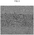
- a single layer of a vascular endothelial cell layer that uniformly covered the inner periphery of the collagen layer was formed by culturing the cells in the resulting microfiber.
- the alginate gel of the outer shell layer was dissolved by allowing alginate lyase to act on the microfiber obtained in (a) ( FIG. 2 ).
- Microfiber Comprising Cell-Adhesive Layer Containing Vascular Smooth Muscle Cells, Outer Shell Layer Containing Alginate Gel that Covers Outer Periphery of the Cell-Adhesive Layer, and Vascular Endothelial Cell Layer Covering Inner Periphery of the Cell-Adhesive Layer
- a microfiber was obtained in the same manner as Example 1 with the exception of preparing an aqueous collagen solution (4 mg/ml) containing vascular smooth muscle cells (1.25 ⁇ 10 6 cells/mL) as the solution 3 for preparing a cell-adhesive hydrogel, and injecting from the line 4 for introducing a solution for preparing a cell-adhesive hydrogel at a flow rate of 200 ⁇ l/min to form a laminar flow of the aqueous collagen solution that covered the outer periphery of laminar flow of the cell suspension (inner diameter of outer shell layer: 270 ⁇ m, outer diameter of outer shell layer: 350 ⁇ m, thickness of collagen layer: 100 ⁇ m, and each of these values was calculated as the average value of measured values from images obtained with a phase-contrast microscope).
- a microfiber was obtained in which a vascular smooth muscle cell layer was laminated on the outside of a single layer of the vascular endothelial cell layer by culturing the cells in the
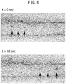
- Liquid was pumped into the hollow portion of the microfiber obtained in Example 1 by clamping the microfiber between narrowed glass tubes.
- liquid was pumped at a flow rate of 1 ⁇ L/min using a syringe pump, liquid was able to be pumped into the resulting hollow portion of the vascular endothelium layer.
- the hollow microfiber of the present invention can be preferably used as an alternative to luminal structures in the body such as blood vessels or lymph ducts.
Landscapes
- Chemical & Material Sciences (AREA)
- Health & Medical Sciences (AREA)
- Life Sciences & Earth Sciences (AREA)
- Chemical Kinetics & Catalysis (AREA)
- Organic Chemistry (AREA)
- Engineering & Computer Science (AREA)
- Bioinformatics & Cheminformatics (AREA)
- Zoology (AREA)
- Wood Science & Technology (AREA)
- Biochemistry (AREA)
- Sustainable Development (AREA)
- Microbiology (AREA)
- Biotechnology (AREA)
- Biomedical Technology (AREA)
- General Engineering & Computer Science (AREA)
- General Health & Medical Sciences (AREA)
- Genetics & Genomics (AREA)
- Immunology (AREA)
- Molecular Biology (AREA)
- Dispersion Chemistry (AREA)
- Micro-Organisms Or Cultivation Processes Thereof (AREA)
- Apparatus Associated With Microorganisms And Enzymes (AREA)
- Materials For Medical Uses (AREA)
Applications Claiming Priority (2)
| Application Number | Priority Date | Filing Date | Title |
|---|---|---|---|
| JP2014104763 | 2014-05-20 | ||
| PCT/JP2015/064524 WO2015178427A1 (ja) | 2014-05-20 | 2015-05-20 | 中空マイクロファイバ |
Publications (2)
| Publication Number | Publication Date |
|---|---|
| EP3147346A1 true EP3147346A1 (de) | 2017-03-29 |
| EP3147346A4 EP3147346A4 (de) | 2018-01-10 |
Family
ID=54554093
Family Applications (1)
| Application Number | Title | Priority Date | Filing Date |
|---|---|---|---|
| EP15796411.5A Pending EP3147346A4 (de) | 2014-05-20 | 2015-05-20 | Hohle mikrofaser |
Country Status (4)
| Country | Link |
|---|---|
| US (1) | US10221382B2 (de) |
| EP (1) | EP3147346A4 (de) |
| JP (1) | JP6710000B2 (de) |
| WO (1) | WO2015178427A1 (de) |
Cited By (2)
| Publication number | Priority date | Publication date | Assignee | Title |
|---|---|---|---|---|
| US20200248136A1 (en) * | 2019-02-01 | 2020-08-06 | National Tsing Hua University | Biomimetic microtube and preparation method thereof |
| EP3545080B1 (de) * | 2016-11-23 | 2024-11-13 | Université de Bordeaux | Zellulares mikrokompartiment und herstellungsverfahren |
Families Citing this family (19)
| Publication number | Priority date | Publication date | Assignee | Title |
|---|---|---|---|---|
| JP6439918B2 (ja) * | 2014-10-17 | 2018-12-19 | 国立大学法人 東京大学 | 3次元細胞構造体の製造方法 |
| JP6895674B2 (ja) * | 2015-10-21 | 2021-06-30 | 国立大学法人 東京大学 | マイクロチューブ、マイクロチューブの製造方法、及びマイクロチューブの製造装置 |
| NL2016404B1 (en) * | 2016-03-09 | 2017-09-26 | Mimetas B V | Double tubular structures. |
| FR3063736B1 (fr) * | 2017-03-09 | 2021-06-25 | Univ Bordeaux | Microfibre cellulaire creuse et procede de fabrication d'une telle microfibre cellulaire creuse |
| JP7090868B2 (ja) * | 2017-10-19 | 2022-06-27 | 国立大学法人 東京大学 | 接着剤及びその使用 |
| WO2019123886A1 (ja) * | 2017-12-23 | 2019-06-27 | 国立大学法人東京大学 | アレイ及びその使用 |
| JP7134464B2 (ja) * | 2018-05-15 | 2022-09-12 | 国立大学法人千葉大学 | コラーゲンチューブの作製方法 |
| JP7388722B2 (ja) | 2018-08-10 | 2023-11-29 | 国立大学法人 東京大学 | アルギン酸中空マイクロファイバ |
| CN109929760A (zh) * | 2019-04-08 | 2019-06-25 | 华子昂 | 同轴多层中空凝胶纤维管的制备装置及其使用方法和应用 |
| WO2021025005A1 (ja) * | 2019-08-05 | 2021-02-11 | 国立大学法人佐賀大学 | 中空コラーゲンゲル |
| WO2021165905A1 (en) | 2020-02-19 | 2021-08-26 | Association For The Advancement Of Tissue Engineering And Cell Based Technologies & Therapies (A4Tec) - Associação | Multicompartement hydrogel fibre their preparation and uses thereof |
| JP7766303B2 (ja) * | 2020-09-01 | 2025-11-10 | 株式会社セルファイバ | 足場、足場の製造方法、細胞培養物、細胞培養方法 |
| KR20230127997A (ko) | 2020-12-28 | 2023-09-01 | 모찌다 세이야쿠 가부시끼가이샤 | 신규의 다층 폴리머 코팅 가교 알긴산 겔 파이버 |
| CN116710110A (zh) | 2020-12-28 | 2023-09-05 | 持田制药株式会社 | 新型的多层聚合物涂层交联海藻酸凝胶纤维 |
| CN113318273B (zh) * | 2021-06-25 | 2022-11-25 | 温州医科大学慈溪生物医药研究院 | Ecm梯度微纤维管及其制备方法 |
| WO2023286852A1 (ja) | 2021-07-15 | 2023-01-19 | 株式会社セルファイバ | 構造体及びその用途 |
| JP2025090874A (ja) * | 2022-04-27 | 2025-06-18 | 国立大学法人 東京大学 | 活性成分含有溶液処理装置および体外活性成分含有溶液循環システム |
| WO2024116831A1 (ja) * | 2022-11-29 | 2024-06-06 | 株式会社セルファイバ | 細胞もしくは細胞産生物含有組成物、細胞もしくは細胞集合体及び/又は細胞産生物の製造方法 |
| TW202515993A (zh) * | 2023-06-26 | 2025-04-16 | 日商細胞纖維股份有限公司 | 細胞膠囊之製造方法、細胞集團或細胞產生物之製造方法、溶液及細胞集團 |
Family Cites Families (3)
| Publication number | Priority date | Publication date | Assignee | Title |
|---|---|---|---|---|
| JP4496360B2 (ja) * | 2003-04-24 | 2010-07-07 | 国立大学法人九州大学 | 医療用高分子ナノ・マイクロファイバー |
| JPWO2011046104A1 (ja) | 2009-10-14 | 2013-03-07 | 国立大学法人 東京大学 | 配向された細胞をゲル内に含む培養物 |
| WO2011046105A1 (ja) * | 2009-10-14 | 2011-04-21 | 国立大学法人 東京大学 | 被覆されたマイクロゲルファイバ |
-
2015
- 2015-05-20 US US15/312,561 patent/US10221382B2/en active Active
- 2015-05-20 JP JP2016521133A patent/JP6710000B2/ja active Active
- 2015-05-20 WO PCT/JP2015/064524 patent/WO2015178427A1/ja not_active Ceased
- 2015-05-20 EP EP15796411.5A patent/EP3147346A4/de active Pending
Cited By (4)
| Publication number | Priority date | Publication date | Assignee | Title |
|---|---|---|---|---|
| EP3545080B1 (de) * | 2016-11-23 | 2024-11-13 | Université de Bordeaux | Zellulares mikrokompartiment und herstellungsverfahren |
| US12146153B2 (en) | 2016-11-23 | 2024-11-19 | Universite de Bordeaux | Cellular microcompartment and preparation processes |
| EP4464776A3 (de) * | 2016-11-23 | 2025-02-26 | Université de Bordeaux | Zellmikrokompartiment und herstellungsverfahren |
| US20200248136A1 (en) * | 2019-02-01 | 2020-08-06 | National Tsing Hua University | Biomimetic microtube and preparation method thereof |
Also Published As
| Publication number | Publication date |
|---|---|
| US10221382B2 (en) | 2019-03-05 |
| JPWO2015178427A1 (ja) | 2017-04-20 |
| EP3147346A4 (de) | 2018-01-10 |
| US20170130184A1 (en) | 2017-05-11 |
| JP6710000B2 (ja) | 2020-06-17 |
| WO2015178427A1 (ja) | 2015-11-26 |
Similar Documents
| Publication | Publication Date | Title |
|---|---|---|
| US10221382B2 (en) | Hollow microfiber | |
| Zhang et al. | An effective strategy for preparing macroporous and self-healing bioactive hydrogels for cell delivery and wound healing | |
| Onoe et al. | Cell-laden microfibers for bottom-up tissue engineering | |
| CN102481389B (zh) | 三维纳米结构化复合支架及其制备方法 | |
| JP5674442B2 (ja) | 血管様構造物を含む三次元細胞培養物 | |
| Duong et al. | Coaxial printing of double-layered and free-standing blood vessel analogues without ultraviolet illumination for high-volume vascularised tissue | |
| CN103298498A (zh) | 用于器官增强的注射制剂 | |
| Liu et al. | Synthesis of cell composite alginate microfibers by microfluidics with the application potential of small diameter vascular grafts | |
| Li et al. | Rapid fabrication of ready-to-use gelatin scaffolds with prevascular networks using alginate hollow fibers as sacrificial templates | |
| Zhang et al. | Effect of multiwall carbon nanotube reinforcement on coaxially extruded cellular vascular conduits | |
| WO2005014774A1 (ja) | 動物細胞の培養担体と、該培養担体を用いた動物細胞の培養方法および移植方法 | |
| JP6241890B2 (ja) | 血管組織およびその作製方法 | |
| Maggiotto et al. | 3D bioprinting for the production of a perfusable vascularized model of a cancer niche | |
| Vanlauwe et al. | Small molecular weight alginate gel porogen for the 3D bioprinting of microvasculature | |
| EP3418375A1 (de) | Verfahren zur herstellung von dreidimensionalem gewebe mit gefässsystemstruktur und dreidimensionales gewebe mit gel mit gefässsystemstruktur | |
| CN108272532B (zh) | 一种双锥形圆管腔结构的水凝胶芯片的制备方法 | |
| US20230407220A1 (en) | Novel tissue culture systems and reduced gravity culture method for the production of vascularized tissue | |
| JP2024093080A (ja) | 立体的組織の製造方法 | |
| Zhang | 3D bioprinting of vasculature network for tissue engineering | |
| Seifu et al. | Fabrication of vascular tissue engineering scaffolds with enhanced oxygen diffusivity and cell infiltration | |
| US20220088272A1 (en) | Graft and use thereof | |
| Golas et al. | Vascular smooth muscle cell optimization of vasculogenesis within naturally derived, biodegradable, hybrid hydrogel scaffolds | |
| US20240400981A1 (en) | Cell population and cell population production method | |
| US12570952B2 (en) | Devices and methods of producing tubular systems for cell culture | |
| 즈엉 et al. | Human vascular tissue fabrication using the co-culture of vascular cells |
Legal Events
| Date | Code | Title | Description |
|---|---|---|---|
| STAA | Information on the status of an ep patent application or granted ep patent |
Free format text: STATUS: THE INTERNATIONAL PUBLICATION HAS BEEN MADE |
|
| PUAI | Public reference made under article 153(3) epc to a published international application that has entered the european phase |
Free format text: ORIGINAL CODE: 0009012 |
|
| STAA | Information on the status of an ep patent application or granted ep patent |
Free format text: STATUS: REQUEST FOR EXAMINATION WAS MADE |
|
| 17P | Request for examination filed |
Effective date: 20161118 |
|
| AK | Designated contracting states |
Kind code of ref document: A1 Designated state(s): AL AT BE BG CH CY CZ DE DK EE ES FI FR GB GR HR HU IE IS IT LI LT LU LV MC MK MT NL NO PL PT RO RS SE SI SK SM TR |
|
| AX | Request for extension of the european patent |
Extension state: BA ME |
|
| RIN1 | Information on inventor provided before grant (corrected) |
Inventor name: ONOE, HIROAKI Inventor name: MIURA, SHIGENORI Inventor name: TAKEUCHI, SHOJI |
|
| DAV | Request for validation of the european patent (deleted) | ||
| DAX | Request for extension of the european patent (deleted) | ||
| A4 | Supplementary search report drawn up and despatched |
Effective date: 20171208 |
|
| RIC1 | Information provided on ipc code assigned before grant |
Ipc: C12M 1/00 20060101AFI20171204BHEP Ipc: B01D 63/02 20060101ALI20171204BHEP |