EP3134124B1 - Igf-1r antibody-drug-conjugate and its use for the treatment of cancer - Google Patents
Igf-1r antibody-drug-conjugate and its use for the treatment of cancer Download PDFInfo
- Publication number
- EP3134124B1 EP3134124B1 EP15720045.2A EP15720045A EP3134124B1 EP 3134124 B1 EP3134124 B1 EP 3134124B1 EP 15720045 A EP15720045 A EP 15720045A EP 3134124 B1 EP3134124 B1 EP 3134124B1
- Authority
- EP
- European Patent Office
- Prior art keywords
- antibody
- sequence seq
- variable domain
- heavy chain
- light chain
- Prior art date
- Legal status (The legal status is an assumption and is not a legal conclusion. Google has not performed a legal analysis and makes no representation as to the accuracy of the status listed.)
- Active
Links
- 229940049595 antibody-drug conjugate Drugs 0.000 title claims description 159
- 239000000611 antibody drug conjugate Substances 0.000 title claims description 141
- 206010028980 Neoplasm Diseases 0.000 title claims description 63
- 201000011510 cancer Diseases 0.000 title claims description 25
- 238000011282 treatment Methods 0.000 title claims description 21
- 239000003814 drug Substances 0.000 claims description 131
- 229940079593 drug Drugs 0.000 claims description 129
- 230000027455 binding Effects 0.000 claims description 114
- 239000000203 mixture Substances 0.000 claims description 95
- 241000282414 Homo sapiens Species 0.000 claims description 74
- 239000000427 antigen Substances 0.000 claims description 29
- 102000036639 antigens Human genes 0.000 claims description 29
- 108091007433 antigens Proteins 0.000 claims description 29
- 239000012634 fragment Substances 0.000 claims description 23
- 125000003275 alpha amino acid group Chemical group 0.000 claims description 21
- 150000003839 salts Chemical class 0.000 claims description 12
- 125000004169 (C1-C6) alkyl group Chemical group 0.000 claims description 10
- 125000005439 maleimidyl group Chemical group C1(C=CC(N1*)=O)=O 0.000 claims description 7
- 229910052757 nitrogen Inorganic materials 0.000 claims description 7
- 125000004448 alkyl carbonyl group Chemical group 0.000 claims description 6
- 125000004433 nitrogen atom Chemical group N* 0.000 claims description 6
- 125000006528 (C2-C6) alkyl group Chemical group 0.000 claims description 4
- 125000006781 (C4-C10) cycloalkylcarbonyl group Chemical group 0.000 claims description 4
- 210000000481 breast Anatomy 0.000 claims description 4
- 208000017897 Carcinoma of esophagus Diseases 0.000 claims description 3
- 206010014733 Endometrial cancer Diseases 0.000 claims description 3
- 206010014759 Endometrial neoplasm Diseases 0.000 claims description 3
- 208000032612 Glial tumor Diseases 0.000 claims description 3
- 206010018338 Glioma Diseases 0.000 claims description 3
- 206010027406 Mesothelioma Diseases 0.000 claims description 3
- 206010030155 Oesophageal carcinoma Diseases 0.000 claims description 3
- 201000005619 esophageal carcinoma Diseases 0.000 claims description 3
- 201000001441 melanoma Diseases 0.000 claims description 3
- 201000008968 osteosarcoma Diseases 0.000 claims description 3
- 208000016691 refractory malignant neoplasm Diseases 0.000 claims description 3
- 201000009410 rhabdomyosarcoma Diseases 0.000 claims description 3
- 125000000335 thiazolyl group Chemical group 0.000 claims description 3
- 210000001072 colon Anatomy 0.000 claims description 2
- 230000002496 gastric effect Effects 0.000 claims description 2
- 210000004072 lung Anatomy 0.000 claims description 2
- 230000002611 ovarian Effects 0.000 claims description 2
- 210000002307 prostate Anatomy 0.000 claims description 2
- 206010041823 squamous cell carcinoma Diseases 0.000 claims description 2
- 210000001685 thyroid gland Anatomy 0.000 claims description 2
- 125000000896 monocarboxylic acid group Chemical group 0.000 claims 1
- 150000001875 compounds Chemical class 0.000 description 374
- 210000004027 cell Anatomy 0.000 description 263
- WEVYAHXRMPXWCK-UHFFFAOYSA-N Acetonitrile Chemical compound CC#N WEVYAHXRMPXWCK-UHFFFAOYSA-N 0.000 description 232
- YMWUJEATGCHHMB-UHFFFAOYSA-N Dichloromethane Chemical compound ClCCl YMWUJEATGCHHMB-UHFFFAOYSA-N 0.000 description 150
- XEKOWRVHYACXOJ-UHFFFAOYSA-N Ethyl acetate Chemical compound CCOC(C)=O XEKOWRVHYACXOJ-UHFFFAOYSA-N 0.000 description 120
- OKKJLVBELUTLKV-UHFFFAOYSA-N Methanol Chemical compound OC OKKJLVBELUTLKV-UHFFFAOYSA-N 0.000 description 114
- XLYOFNOQVPJJNP-UHFFFAOYSA-N water Substances O XLYOFNOQVPJJNP-UHFFFAOYSA-N 0.000 description 111
- 230000001747 exhibiting effect Effects 0.000 description 97
- 239000007787 solid Substances 0.000 description 96
- DTQVDTLACAAQTR-UHFFFAOYSA-N Trifluoroacetic acid Chemical compound OC(=O)C(F)(F)F DTQVDTLACAAQTR-UHFFFAOYSA-N 0.000 description 92
- 239000000243 solution Substances 0.000 description 83
- 102100035360 Cerebellar degeneration-related antigen 1 Human genes 0.000 description 76
- 101710117290 Aldo-keto reductase family 1 member C4 Proteins 0.000 description 75
- ZMXDDKWLCZADIW-UHFFFAOYSA-N N,N-Dimethylformamide Chemical compound CN(C)C=O ZMXDDKWLCZADIW-UHFFFAOYSA-N 0.000 description 75
- 230000002829 reductive effect Effects 0.000 description 72
- 238000000034 method Methods 0.000 description 70
- VYPSYNLAJGMNEJ-UHFFFAOYSA-N Silicium dioxide Chemical compound O=[Si]=O VYPSYNLAJGMNEJ-UHFFFAOYSA-N 0.000 description 64
- 241001529936 Murinae Species 0.000 description 61
- 238000006243 chemical reaction Methods 0.000 description 58
- 125000005647 linker group Chemical group 0.000 description 58
- 150000001412 amines Chemical class 0.000 description 56
- JGFZNNIVVJXRND-UHFFFAOYSA-N N,N-Diisopropylethylamine (DIPEA) Chemical compound CCN(C(C)C)C(C)C JGFZNNIVVJXRND-UHFFFAOYSA-N 0.000 description 50
- WYURNTSHIVDZCO-UHFFFAOYSA-N Tetrahydrofuran Chemical compound C1CCOC1 WYURNTSHIVDZCO-UHFFFAOYSA-N 0.000 description 48
- 101100490051 Lactococcus lactis subsp. lactis (strain IL1403) accD gene Proteins 0.000 description 44
- -1 for example Substances 0.000 description 44
- 239000003643 water by type Substances 0.000 description 42
- 238000013019 agitation Methods 0.000 description 41
- 238000001943 fluorescence-activated cell sorting Methods 0.000 description 40
- 125000002496 methyl group Chemical group [H]C([H])([H])* 0.000 description 40
- 239000000872 buffer Substances 0.000 description 39
- 239000012074 organic phase Substances 0.000 description 39
- PMZURENOXWZQFD-UHFFFAOYSA-L Sodium Sulfate Chemical compound [Na+].[Na+].[O-]S([O-])(=O)=O PMZURENOXWZQFD-UHFFFAOYSA-L 0.000 description 38
- 239000012298 atmosphere Substances 0.000 description 37
- 239000007924 injection Substances 0.000 description 37
- 238000002347 injection Methods 0.000 description 37
- 239000003921 oil Substances 0.000 description 37
- 235000019198 oils Nutrition 0.000 description 37
- 239000011541 reaction mixture Substances 0.000 description 37
- 229910052938 sodium sulfate Inorganic materials 0.000 description 35
- 239000012071 phase Substances 0.000 description 34
- 235000011152 sodium sulphate Nutrition 0.000 description 33
- 238000002953 preparative HPLC Methods 0.000 description 31
- FAPWRFPIFSIZLT-UHFFFAOYSA-M Sodium chloride Chemical compound [Na+].[Cl-] FAPWRFPIFSIZLT-UHFFFAOYSA-M 0.000 description 29
- ZMANZCXQSJIPKH-UHFFFAOYSA-N Triethylamine Chemical compound CCN(CC)CC ZMANZCXQSJIPKH-UHFFFAOYSA-N 0.000 description 29
- 229940024606 amino acid Drugs 0.000 description 29
- 235000001014 amino acid Nutrition 0.000 description 29
- OKKJLVBELUTLKV-MZCSYVLQSA-N Deuterated methanol Chemical compound [2H]OC([2H])([2H])[2H] OKKJLVBELUTLKV-MZCSYVLQSA-N 0.000 description 28
- 108090000623 proteins and genes Proteins 0.000 description 26
- 239000002904 solvent Substances 0.000 description 26
- 239000000377 silicon dioxide Substances 0.000 description 25
- UIIMBOGNXHQVGW-UHFFFAOYSA-M Sodium bicarbonate Chemical compound [Na+].OC([O-])=O UIIMBOGNXHQVGW-UHFFFAOYSA-M 0.000 description 24
- 239000002253 acid Substances 0.000 description 24
- 230000000875 corresponding effect Effects 0.000 description 24
- 238000011534 incubation Methods 0.000 description 24
- 241000282693 Cercopithecidae Species 0.000 description 23
- 150000001413 amino acids Chemical class 0.000 description 23
- 239000007864 aqueous solution Substances 0.000 description 23
- 235000018102 proteins Nutrition 0.000 description 23
- 102000004169 proteins and genes Human genes 0.000 description 23
- 239000004698 Polyethylene Substances 0.000 description 22
- 125000006850 spacer group Chemical group 0.000 description 22
- 101000852815 Homo sapiens Insulin receptor Proteins 0.000 description 20
- 238000007792 addition Methods 0.000 description 20
- 238000004458 analytical method Methods 0.000 description 20
- 238000002474 experimental method Methods 0.000 description 20
- 239000012429 reaction media Substances 0.000 description 20
- VEXZGXHMUGYJMC-UHFFFAOYSA-N Hydrochloric acid Chemical compound Cl VEXZGXHMUGYJMC-UHFFFAOYSA-N 0.000 description 19
- 239000003153 chemical reaction reagent Substances 0.000 description 19
- 235000018417 cysteine Nutrition 0.000 description 19
- 230000000694 effects Effects 0.000 description 19
- 238000011156 evaluation Methods 0.000 description 19
- 238000004895 liquid chromatography mass spectrometry Methods 0.000 description 19
- IAZDPXIOMUYVGZ-UHFFFAOYSA-N Dimethylsulphoxide Chemical compound CS(C)=O IAZDPXIOMUYVGZ-UHFFFAOYSA-N 0.000 description 18
- KDLHZDBZIXYQEI-UHFFFAOYSA-N Palladium Chemical compound [Pd] KDLHZDBZIXYQEI-UHFFFAOYSA-N 0.000 description 18
- NQRYJNQNLNOLGT-UHFFFAOYSA-N Piperidine Chemical compound C1CCNCC1 NQRYJNQNLNOLGT-UHFFFAOYSA-N 0.000 description 18
- 150000001732 carboxylic acid derivatives Chemical class 0.000 description 18
- 238000003786 synthesis reaction Methods 0.000 description 18
- 102000003746 Insulin Receptor Human genes 0.000 description 17
- 108010001127 Insulin Receptor Proteins 0.000 description 17
- ZWWWLCMDTZFSOO-UHFFFAOYSA-N diethoxyphosphorylformonitrile Chemical compound CCOP(=O)(C#N)OCC ZWWWLCMDTZFSOO-UHFFFAOYSA-N 0.000 description 17
- 210000004602 germ cell Anatomy 0.000 description 17
- 102000047882 human INSR Human genes 0.000 description 17
- 238000005160 1H NMR spectroscopy Methods 0.000 description 16
- RTZKZFJDLAIYFH-UHFFFAOYSA-N Diethyl ether Chemical compound CCOCC RTZKZFJDLAIYFH-UHFFFAOYSA-N 0.000 description 16
- 241001465754 Metazoa Species 0.000 description 16
- 125000006367 bivalent amino carbonyl group Chemical group [H]N([*:1])C([*:2])=O 0.000 description 16
- 238000005859 coupling reaction Methods 0.000 description 16
- HEMHJVSKTPXQMS-UHFFFAOYSA-M Sodium hydroxide Chemical compound [OH-].[Na+] HEMHJVSKTPXQMS-UHFFFAOYSA-M 0.000 description 15
- 125000003178 carboxy group Chemical group [H]OC(*)=O 0.000 description 15
- 230000008878 coupling Effects 0.000 description 15
- 238000010168 coupling process Methods 0.000 description 15
- 239000012043 crude product Substances 0.000 description 15
- XUJNEKJLAYXESH-UHFFFAOYSA-N cysteine Natural products SCC(N)C(O)=O XUJNEKJLAYXESH-UHFFFAOYSA-N 0.000 description 15
- 238000010494 dissociation reaction Methods 0.000 description 15
- 230000005593 dissociations Effects 0.000 description 15
- 230000014509 gene expression Effects 0.000 description 15
- 230000026731 phosphorylation Effects 0.000 description 15
- 238000006366 phosphorylation reaction Methods 0.000 description 15
- PAYRUJLWNCNPSJ-UHFFFAOYSA-N Aniline Chemical compound NC1=CC=CC=C1 PAYRUJLWNCNPSJ-UHFFFAOYSA-N 0.000 description 14
- 241000699670 Mus sp. Species 0.000 description 14
- PXIPVTKHYLBLMZ-UHFFFAOYSA-N Sodium azide Chemical compound [Na+].[N-]=[N+]=[N-] PXIPVTKHYLBLMZ-UHFFFAOYSA-N 0.000 description 14
- 230000015572 biosynthetic process Effects 0.000 description 14
- 230000021615 conjugation Effects 0.000 description 14
- 210000004408 hybridoma Anatomy 0.000 description 14
- 238000001727 in vivo Methods 0.000 description 14
- 239000013598 vector Substances 0.000 description 14
- LFQSCWFLJHTTHZ-UHFFFAOYSA-N Ethanol Chemical compound CCO LFQSCWFLJHTTHZ-UHFFFAOYSA-N 0.000 description 13
- 210000003712 lysosome Anatomy 0.000 description 13
- 230000001868 lysosomic effect Effects 0.000 description 13
- 230000000269 nucleophilic effect Effects 0.000 description 13
- 239000000741 silica gel Substances 0.000 description 13
- 229910002027 silica gel Inorganic materials 0.000 description 13
- 239000011780 sodium chloride Substances 0.000 description 13
- VLARLSIGSPVYHX-UHFFFAOYSA-N (2,5-dioxopyrrolidin-1-yl) 6-(2,5-dioxopyrrol-1-yl)hexanoate Chemical compound O=C1CCC(=O)N1OC(=O)CCCCCN1C(=O)C=CC1=O VLARLSIGSPVYHX-UHFFFAOYSA-N 0.000 description 12
- 125000003277 amino group Chemical group 0.000 description 12
- 230000007423 decrease Effects 0.000 description 12
- 238000011068 loading method Methods 0.000 description 12
- 230000004044 response Effects 0.000 description 12
- 229920006395 saturated elastomer Polymers 0.000 description 12
- 229910000030 sodium bicarbonate Inorganic materials 0.000 description 12
- 101000599951 Homo sapiens Insulin-like growth factor I Proteins 0.000 description 11
- 230000001472 cytotoxic effect Effects 0.000 description 11
- 230000003834 intracellular effect Effects 0.000 description 11
- 239000000047 product Substances 0.000 description 11
- 102000005962 receptors Human genes 0.000 description 11
- 108020003175 receptors Proteins 0.000 description 11
- AXKGIPZJYUNAIW-UHFFFAOYSA-N (4-aminophenyl)methanol Chemical compound NC1=CC=C(CO)C=C1 AXKGIPZJYUNAIW-UHFFFAOYSA-N 0.000 description 10
- 108010047041 Complementarity Determining Regions Proteins 0.000 description 10
- 108090000723 Insulin-Like Growth Factor I Proteins 0.000 description 10
- 102000004218 Insulin-Like Growth Factor I Human genes 0.000 description 10
- 102100037852 Insulin-like growth factor I Human genes 0.000 description 10
- 150000001299 aldehydes Chemical class 0.000 description 10
- 238000002360 preparation method Methods 0.000 description 10
- XJMOSONTPMZWPB-UHFFFAOYSA-M propidium iodide Chemical compound [I-].[I-].C12=CC(N)=CC=C2C2=CC=C(N)C=C2[N+](CCC[N+](C)(CC)CC)=C1C1=CC=CC=C1 XJMOSONTPMZWPB-UHFFFAOYSA-M 0.000 description 10
- 125000003396 thiol group Chemical group [H]S* 0.000 description 10
- CSCPPACGZOOCGX-UHFFFAOYSA-N Acetone Chemical compound CC(C)=O CSCPPACGZOOCGX-UHFFFAOYSA-N 0.000 description 9
- 0 CC[C@](C)[C@@]([C@@](CC(N(CCC1)[C@@]1[C@@]([C@@](C)C(N[C@@](Cc1ccccc1)C(OC)=*)=O)OC)=O)OC)N(C)C([C@](C(C)C)NC([C@](C(C)C)N(C)CCc(cc1)ccc1N(C)C(OCc(cc1)ccc1NC([C@](CCCNC(N)=O)NC([C@](C(C)C)NC(CCCCCN(C(CC1C(C)(C)*)=O)C1=O)=O)=O)=O)=O)=O)=O Chemical compound CC[C@](C)[C@@]([C@@](CC(N(CCC1)[C@@]1[C@@]([C@@](C)C(N[C@@](Cc1ccccc1)C(OC)=*)=O)OC)=O)OC)N(C)C([C@](C(C)C)NC([C@](C(C)C)N(C)CCc(cc1)ccc1N(C)C(OCc(cc1)ccc1NC([C@](CCCNC(N)=O)NC([C@](C(C)C)NC(CCCCCN(C(CC1C(C)(C)*)=O)C1=O)=O)=O)=O)=O)=O)=O 0.000 description 9
- KXDHJXZQYSOELW-UHFFFAOYSA-M Carbamate Chemical compound NC([O-])=O KXDHJXZQYSOELW-UHFFFAOYSA-M 0.000 description 9
- BVKZGUZCCUSVTD-UHFFFAOYSA-L Carbonate Chemical compound [O-]C([O-])=O BVKZGUZCCUSVTD-UHFFFAOYSA-L 0.000 description 9
- DHMQDGOQFOQNFH-UHFFFAOYSA-N Glycine Chemical compound NCC(O)=O DHMQDGOQFOQNFH-UHFFFAOYSA-N 0.000 description 9
- KDXKERNSBIXSRK-YFKPBYRVSA-N L-lysine Chemical compound NCCCC[C@H](N)C(O)=O KDXKERNSBIXSRK-YFKPBYRVSA-N 0.000 description 9
- KDXKERNSBIXSRK-UHFFFAOYSA-N Lysine Natural products NCCCCC(N)C(O)=O KDXKERNSBIXSRK-UHFFFAOYSA-N 0.000 description 9
- 102000035195 Peptidases Human genes 0.000 description 9
- 108091005804 Peptidases Proteins 0.000 description 9
- 125000003917 carbamoyl group Chemical group [H]N([H])C(*)=O 0.000 description 9
- 230000008045 co-localization Effects 0.000 description 9
- 230000003013 cytotoxicity Effects 0.000 description 9
- 231100000135 cytotoxicity Toxicity 0.000 description 9
- 150000002148 esters Chemical class 0.000 description 9
- 229920001223 polyethylene glycol Polymers 0.000 description 9
- BWHMMNNQKKPAPP-UHFFFAOYSA-L potassium carbonate Chemical compound [K+].[K+].[O-]C([O-])=O BWHMMNNQKKPAPP-UHFFFAOYSA-L 0.000 description 9
- 230000009467 reduction Effects 0.000 description 9
- 238000006722 reduction reaction Methods 0.000 description 9
- 239000006228 supernatant Substances 0.000 description 9
- 210000004881 tumor cell Anatomy 0.000 description 9
- QTBSBXVTEAMEQO-UHFFFAOYSA-N Acetic acid Chemical compound CC(O)=O QTBSBXVTEAMEQO-UHFFFAOYSA-N 0.000 description 8
- COLNVLDHVKWLRT-QMMMGPOBSA-N L-phenylalanine Chemical compound OC(=O)[C@@H](N)CC1=CC=CC=C1 COLNVLDHVKWLRT-QMMMGPOBSA-N 0.000 description 8
- CSNNHWWHGAXBCP-UHFFFAOYSA-L Magnesium sulfate Chemical compound [Mg+2].[O-][S+2]([O-])([O-])[O-] CSNNHWWHGAXBCP-UHFFFAOYSA-L 0.000 description 8
- DTQVDTLACAAQTR-UHFFFAOYSA-M Trifluoroacetate Chemical compound [O-]C(=O)C(F)(F)F DTQVDTLACAAQTR-UHFFFAOYSA-M 0.000 description 8
- 125000001797 benzyl group Chemical group [H]C1=C([H])C([H])=C(C([H])=C1[H])C([H])([H])* 0.000 description 8
- 125000001495 ethyl group Chemical group [H]C([H])([H])C([H])([H])* 0.000 description 8
- 238000001914 filtration Methods 0.000 description 8
- 230000006870 function Effects 0.000 description 8
- 230000012010 growth Effects 0.000 description 8
- 238000004128 high performance liquid chromatography Methods 0.000 description 8
- NOESYZHRGYRDHS-UHFFFAOYSA-N insulin Chemical compound N1C(=O)C(NC(=O)C(CCC(N)=O)NC(=O)C(CCC(O)=O)NC(=O)C(C(C)C)NC(=O)C(NC(=O)CN)C(C)CC)CSSCC(C(NC(CO)C(=O)NC(CC(C)C)C(=O)NC(CC=2C=CC(O)=CC=2)C(=O)NC(CCC(N)=O)C(=O)NC(CC(C)C)C(=O)NC(CCC(O)=O)C(=O)NC(CC(N)=O)C(=O)NC(CC=2C=CC(O)=CC=2)C(=O)NC(CSSCC(NC(=O)C(C(C)C)NC(=O)C(CC(C)C)NC(=O)C(CC=2C=CC(O)=CC=2)NC(=O)C(CC(C)C)NC(=O)C(C)NC(=O)C(CCC(O)=O)NC(=O)C(C(C)C)NC(=O)C(CC(C)C)NC(=O)C(CC=2NC=NC=2)NC(=O)C(CO)NC(=O)CNC2=O)C(=O)NCC(=O)NC(CCC(O)=O)C(=O)NC(CCCNC(N)=N)C(=O)NCC(=O)NC(CC=3C=CC=CC=3)C(=O)NC(CC=3C=CC=CC=3)C(=O)NC(CC=3C=CC(O)=CC=3)C(=O)NC(C(C)O)C(=O)N3C(CCC3)C(=O)NC(CCCCN)C(=O)NC(C)C(O)=O)C(=O)NC(CC(N)=O)C(O)=O)=O)NC(=O)C(C(C)CC)NC(=O)C(CO)NC(=O)C(C(C)O)NC(=O)C1CSSCC2NC(=O)C(CC(C)C)NC(=O)C(NC(=O)C(CCC(N)=O)NC(=O)C(CC(N)=O)NC(=O)C(NC(=O)C(N)CC=1C=CC=CC=1)C(C)C)CC1=CN=CN1 NOESYZHRGYRDHS-UHFFFAOYSA-N 0.000 description 8
- 235000018977 lysine Nutrition 0.000 description 8
- 230000036961 partial effect Effects 0.000 description 8
- 125000001997 phenyl group Chemical group [H]C1=C([H])C([H])=C(*)C([H])=C1[H] 0.000 description 8
- CHKVPAROMQMJNQ-UHFFFAOYSA-M potassium bisulfate Chemical compound [K+].OS([O-])(=O)=O CHKVPAROMQMJNQ-UHFFFAOYSA-M 0.000 description 8
- 125000002924 primary amino group Chemical group [H]N([H])* 0.000 description 8
- 125000006239 protecting group Chemical group 0.000 description 8
- 241000894007 species Species 0.000 description 8
- 238000006467 substitution reaction Methods 0.000 description 8
- GLGNXYJARSMNGJ-VKTIVEEGSA-N (1s,2s,3r,4r)-3-[[5-chloro-2-[(1-ethyl-6-methoxy-2-oxo-4,5-dihydro-3h-1-benzazepin-7-yl)amino]pyrimidin-4-yl]amino]bicyclo[2.2.1]hept-5-ene-2-carboxamide Chemical compound CCN1C(=O)CCCC2=C(OC)C(NC=3N=C(C(=CN=3)Cl)N[C@H]3[C@H]([C@@]4([H])C[C@@]3(C=C4)[H])C(N)=O)=CC=C21 GLGNXYJARSMNGJ-VKTIVEEGSA-N 0.000 description 7
- 206010006187 Breast cancer Diseases 0.000 description 7
- 108010016626 Dipeptides Proteins 0.000 description 7
- 238000002965 ELISA Methods 0.000 description 7
- 241000735235 Ligustrum vulgare Species 0.000 description 7
- 108091028043 Nucleic acid sequence Proteins 0.000 description 7
- 230000002378 acidificating effect Effects 0.000 description 7
- 125000000217 alkyl group Chemical group 0.000 description 7
- 229940125758 compound 15 Drugs 0.000 description 7
- 125000000151 cysteine group Chemical group N[C@@H](CS)C(=O)* 0.000 description 7
- 231100000433 cytotoxic Toxicity 0.000 description 7
- 125000002887 hydroxy group Chemical group [H]O* 0.000 description 7
- NPZTUJOABDZTLV-UHFFFAOYSA-N hydroxybenzotriazole Chemical class O=C1C=CC=C2NNN=C12 NPZTUJOABDZTLV-UHFFFAOYSA-N 0.000 description 7
- 150000007523 nucleic acids Chemical class 0.000 description 7
- 108020004707 nucleic acids Proteins 0.000 description 7
- 102000039446 nucleic acids Human genes 0.000 description 7
- 229910000343 potassium bisulfate Inorganic materials 0.000 description 7
- 238000000746 purification Methods 0.000 description 7
- 238000000611 regression analysis Methods 0.000 description 7
- 239000012146 running buffer Substances 0.000 description 7
- 229910000104 sodium hydride Inorganic materials 0.000 description 7
- 239000007858 starting material Substances 0.000 description 7
- 125000000999 tert-butyl group Chemical group [H]C([H])([H])C(*)(C([H])([H])[H])C([H])([H])[H] 0.000 description 7
- VXUYXOFXAQZZMF-UHFFFAOYSA-N titanium(IV) isopropoxide Chemical compound CC(C)O[Ti](OC(C)C)(OC(C)C)OC(C)C VXUYXOFXAQZZMF-UHFFFAOYSA-N 0.000 description 7
- WSFSSNUMVMOOMR-UHFFFAOYSA-N Formaldehyde Chemical compound O=C WSFSSNUMVMOOMR-UHFFFAOYSA-N 0.000 description 6
- 102100039688 Insulin-like growth factor 1 receptor Human genes 0.000 description 6
- 101150048357 Lamp1 gene Proteins 0.000 description 6
- 239000004365 Protease Substances 0.000 description 6
- 230000001028 anti-proliverative effect Effects 0.000 description 6
- 238000003556 assay Methods 0.000 description 6
- 239000002585 base Substances 0.000 description 6
- 210000004899 c-terminal region Anatomy 0.000 description 6
- FJDQFPXHSGXQBY-UHFFFAOYSA-L caesium carbonate Chemical compound [Cs+].[Cs+].[O-]C([O-])=O FJDQFPXHSGXQBY-UHFFFAOYSA-L 0.000 description 6
- 230000003833 cell viability Effects 0.000 description 6
- 239000003795 chemical substances by application Substances 0.000 description 6
- 150000001945 cysteines Chemical class 0.000 description 6
- 238000010511 deprotection reaction Methods 0.000 description 6
- 238000010790 dilution Methods 0.000 description 6
- 239000012895 dilution Substances 0.000 description 6
- 239000001963 growth medium Substances 0.000 description 6
- 238000007912 intraperitoneal administration Methods 0.000 description 6
- 125000003588 lysine group Chemical group [H]N([H])C([H])([H])C([H])([H])C([H])([H])C([H])([H])C([H])(N([H])[H])C(*)=O 0.000 description 6
- 239000011159 matrix material Substances 0.000 description 6
- 125000001160 methoxycarbonyl group Chemical group [H]C([H])([H])OC(*)=O 0.000 description 6
- VLKZOEOYAKHREP-UHFFFAOYSA-N n-Hexane Chemical compound CCCCCC VLKZOEOYAKHREP-UHFFFAOYSA-N 0.000 description 6
- 239000002773 nucleotide Substances 0.000 description 6
- 230000008823 permeabilization Effects 0.000 description 6
- 239000008194 pharmaceutical composition Substances 0.000 description 6
- JQWHASGSAFIOCM-UHFFFAOYSA-M sodium periodate Chemical compound [Na+].[O-]I(=O)(=O)=O JQWHASGSAFIOCM-UHFFFAOYSA-M 0.000 description 6
- 239000000126 substance Substances 0.000 description 6
- DYHSDKLCOJIUFX-UHFFFAOYSA-N tert-butoxycarbonyl anhydride Chemical compound CC(C)(C)OC(=O)OC(=O)OC(C)(C)C DYHSDKLCOJIUFX-UHFFFAOYSA-N 0.000 description 6
- 150000003573 thiols Chemical class 0.000 description 6
- 238000000954 titration curve Methods 0.000 description 6
- 230000004614 tumor growth Effects 0.000 description 6
- 239000003981 vehicle Substances 0.000 description 6
- WSLDOOZREJYCGB-UHFFFAOYSA-N 1,2-Dichloroethane Chemical compound ClCCCl WSLDOOZREJYCGB-UHFFFAOYSA-N 0.000 description 5
- RYHBNJHYFVUHQT-UHFFFAOYSA-N 1,4-Dioxane Chemical compound C1COCCO1 RYHBNJHYFVUHQT-UHFFFAOYSA-N 0.000 description 5
- DJQYYYCQOZMCRC-UHFFFAOYSA-N 2-aminopropane-1,3-dithiol Chemical group SCC(N)CS DJQYYYCQOZMCRC-UHFFFAOYSA-N 0.000 description 5
- 108020004414 DNA Proteins 0.000 description 5
- 101001034652 Homo sapiens Insulin-like growth factor 1 receptor Proteins 0.000 description 5
- MHAJPDPJQMAIIY-UHFFFAOYSA-N Hydrogen peroxide Chemical compound OO MHAJPDPJQMAIIY-UHFFFAOYSA-N 0.000 description 5
- RHGKLRLOHDJJDR-BYPYZUCNSA-N L-citrulline Chemical compound NC(=O)NCCC[C@H]([NH3+])C([O-])=O RHGKLRLOHDJJDR-BYPYZUCNSA-N 0.000 description 5
- OUYCCCASQSFEME-QMMMGPOBSA-N L-tyrosine Chemical compound OC(=O)[C@@H](N)CC1=CC=C(O)C=C1 OUYCCCASQSFEME-QMMMGPOBSA-N 0.000 description 5
- 239000004472 Lysine Substances 0.000 description 5
- 241000699666 Mus <mouse, genus> Species 0.000 description 5
- 101001055320 Myxine glutinosa Insulin-like growth factor Proteins 0.000 description 5
- FZWLAAWBMGSTSO-UHFFFAOYSA-N Thiazole Chemical group C1=CSC=N1 FZWLAAWBMGSTSO-UHFFFAOYSA-N 0.000 description 5
- 230000000259 anti-tumor effect Effects 0.000 description 5
- 230000004071 biological effect Effects 0.000 description 5
- 150000001720 carbohydrates Chemical class 0.000 description 5
- 125000002915 carbonyl group Chemical group [*:2]C([*:1])=O 0.000 description 5
- 230000015556 catabolic process Effects 0.000 description 5
- 238000003776 cleavage reaction Methods 0.000 description 5
- 229940125904 compound 1 Drugs 0.000 description 5
- 229940125810 compound 20 Drugs 0.000 description 5
- 229940126214 compound 3 Drugs 0.000 description 5
- 239000002254 cytotoxic agent Substances 0.000 description 5
- 229940127089 cytotoxic agent Drugs 0.000 description 5
- 238000006731 degradation reaction Methods 0.000 description 5
- 238000005516 engineering process Methods 0.000 description 5
- 238000001704 evaporation Methods 0.000 description 5
- 238000000684 flow cytometry Methods 0.000 description 5
- JAXFJECJQZDFJS-XHEPKHHKSA-N gtpl8555 Chemical compound OC(=O)C[C@H](N)C(=O)N[C@@H](CCC(O)=O)C(=O)N[C@@H](C(C)C)C(=O)N[C@@H](C(C)C)C(=O)N1CCC[C@@H]1C(=O)N[C@H](B1O[C@@]2(C)[C@H]3C[C@H](C3(C)C)C[C@H]2O1)CCC1=CC=C(F)C=C1 JAXFJECJQZDFJS-XHEPKHHKSA-N 0.000 description 5
- 230000005764 inhibitory process Effects 0.000 description 5
- 239000002609 medium Substances 0.000 description 5
- 239000012528 membrane Substances 0.000 description 5
- 125000003729 nucleotide group Chemical group 0.000 description 5
- 229960005190 phenylalanine Drugs 0.000 description 5
- 239000003880 polar aprotic solvent Substances 0.000 description 5
- 230000008569 process Effects 0.000 description 5
- 235000019419 proteases Nutrition 0.000 description 5
- 238000011160 research Methods 0.000 description 5
- 230000007017 scission Effects 0.000 description 5
- 238000007920 subcutaneous administration Methods 0.000 description 5
- 150000003512 tertiary amines Chemical class 0.000 description 5
- 230000001988 toxicity Effects 0.000 description 5
- 231100000419 toxicity Toxicity 0.000 description 5
- JWDFQMWEFLOOED-UHFFFAOYSA-N (2,5-dioxopyrrolidin-1-yl) 3-(pyridin-2-yldisulfanyl)propanoate Chemical compound O=C1CCC(=O)N1OC(=O)CCSSC1=CC=CC=N1 JWDFQMWEFLOOED-UHFFFAOYSA-N 0.000 description 4
- GHYOCDFICYLMRF-UTIIJYGPSA-N (2S,3R)-N-[(2S)-3-(cyclopenten-1-yl)-1-[(2R)-2-methyloxiran-2-yl]-1-oxopropan-2-yl]-3-hydroxy-3-(4-methoxyphenyl)-2-[[(2S)-2-[(2-morpholin-4-ylacetyl)amino]propanoyl]amino]propanamide Chemical compound C1(=CCCC1)C[C@@H](C(=O)[C@@]1(OC1)C)NC([C@H]([C@@H](C1=CC=C(C=C1)OC)O)NC([C@H](C)NC(CN1CCOCC1)=O)=O)=O GHYOCDFICYLMRF-UTIIJYGPSA-N 0.000 description 4
- AGGWFDNPHKLBBV-YUMQZZPRSA-N (2s)-2-[[(2s)-2-amino-3-methylbutanoyl]amino]-5-(carbamoylamino)pentanoic acid Chemical group CC(C)[C@H](N)C(=O)N[C@H](C(O)=O)CCCNC(N)=O AGGWFDNPHKLBBV-YUMQZZPRSA-N 0.000 description 4
- KKHFRAFPESRGGD-UHFFFAOYSA-N 1,3-dimethyl-7-[3-(n-methylanilino)propyl]purine-2,6-dione Chemical compound C1=NC=2N(C)C(=O)N(C)C(=O)C=2N1CCCN(C)C1=CC=CC=C1 KKHFRAFPESRGGD-UHFFFAOYSA-N 0.000 description 4
- QXOGPTXQGKQSJT-UHFFFAOYSA-N 1-amino-4-[4-(3,4-dimethylphenyl)sulfanylanilino]-9,10-dioxoanthracene-2-sulfonic acid Chemical compound Cc1ccc(Sc2ccc(Nc3cc(c(N)c4C(=O)c5ccccc5C(=O)c34)S(O)(=O)=O)cc2)cc1C QXOGPTXQGKQSJT-UHFFFAOYSA-N 0.000 description 4
- LMDZBCPBFSXMTL-UHFFFAOYSA-N 1-ethyl-3-(3-dimethylaminopropyl)carbodiimide Chemical compound CCN=C=NCCCN(C)C LMDZBCPBFSXMTL-UHFFFAOYSA-N 0.000 description 4
- 125000004214 1-pyrrolidinyl group Chemical group [H]C1([H])N(*)C([H])([H])C([H])([H])C1([H])[H] 0.000 description 4
- QKNYBSVHEMOAJP-UHFFFAOYSA-N 2-amino-2-(hydroxymethyl)propane-1,3-diol;hydron;chloride Chemical compound Cl.OCC(N)(CO)CO QKNYBSVHEMOAJP-UHFFFAOYSA-N 0.000 description 4
- PBVAJRFEEOIAGW-UHFFFAOYSA-N 3-[bis(2-carboxyethyl)phosphanyl]propanoic acid;hydrochloride Chemical compound Cl.OC(=O)CCP(CCC(O)=O)CCC(O)=O PBVAJRFEEOIAGW-UHFFFAOYSA-N 0.000 description 4
- 101100248451 Arabidopsis thaliana RICE2 gene Proteins 0.000 description 4
- 239000004475 Arginine Substances 0.000 description 4
- 108091003079 Bovine Serum Albumin Proteins 0.000 description 4
- 208000026310 Breast neoplasm Diseases 0.000 description 4
- 238000012413 Fluorescence activated cell sorting analysis Methods 0.000 description 4
- 239000004471 Glycine Substances 0.000 description 4
- 101000998947 Homo sapiens Immunoglobulin heavy variable 1-46 Proteins 0.000 description 4
- 101001138089 Homo sapiens Immunoglobulin kappa variable 1-39 Proteins 0.000 description 4
- 102100036888 Immunoglobulin heavy variable 1-46 Human genes 0.000 description 4
- 102100020910 Immunoglobulin kappa variable 1-39 Human genes 0.000 description 4
- 102000004877 Insulin Human genes 0.000 description 4
- 108090001061 Insulin Proteins 0.000 description 4
- ODKSFYDXXFIFQN-BYPYZUCNSA-P L-argininium(2+) Chemical compound NC(=[NH2+])NCCC[C@H]([NH3+])C(O)=O ODKSFYDXXFIFQN-BYPYZUCNSA-P 0.000 description 4
- ZDXPYRJPNDTMRX-VKHMYHEASA-N L-glutamine Chemical compound OC(=O)[C@@H](N)CCC(N)=O ZDXPYRJPNDTMRX-VKHMYHEASA-N 0.000 description 4
- AGPKZVBTJJNPAG-WHFBIAKZSA-N L-isoleucine Chemical compound CC[C@H](C)[C@H](N)C(O)=O AGPKZVBTJJNPAG-WHFBIAKZSA-N 0.000 description 4
- MZRVEZGGRBJDDB-UHFFFAOYSA-N N-Butyllithium Chemical compound [Li]CCCC MZRVEZGGRBJDDB-UHFFFAOYSA-N 0.000 description 4
- JUJWROOIHBZHMG-UHFFFAOYSA-N Pyridine Chemical compound C1=CC=NC=C1 JUJWROOIHBZHMG-UHFFFAOYSA-N 0.000 description 4
- CDBYLPFSWZWCQE-UHFFFAOYSA-L Sodium Carbonate Chemical compound [Na+].[Na+].[O-]C([O-])=O CDBYLPFSWZWCQE-UHFFFAOYSA-L 0.000 description 4
- OKJPEAGHQZHRQV-UHFFFAOYSA-N Triiodomethane Natural products IC(I)I OKJPEAGHQZHRQV-UHFFFAOYSA-N 0.000 description 4
- KZSNJWFQEVHDMF-UHFFFAOYSA-N Valine Chemical compound CC(C)C(N)C(O)=O KZSNJWFQEVHDMF-UHFFFAOYSA-N 0.000 description 4
- 150000007513 acids Chemical class 0.000 description 4
- 230000004913 activation Effects 0.000 description 4
- 125000000539 amino acid group Chemical group 0.000 description 4
- 239000008346 aqueous phase Substances 0.000 description 4
- ODKSFYDXXFIFQN-UHFFFAOYSA-N arginine Natural products OC(=O)C(N)CCCNC(N)=N ODKSFYDXXFIFQN-UHFFFAOYSA-N 0.000 description 4
- 230000008901 benefit Effects 0.000 description 4
- 238000004422 calculation algorithm Methods 0.000 description 4
- 235000014633 carbohydrates Nutrition 0.000 description 4
- 238000005119 centrifugation Methods 0.000 description 4
- 230000000052 comparative effect Effects 0.000 description 4
- 229940125797 compound 12 Drugs 0.000 description 4
- 229940125782 compound 2 Drugs 0.000 description 4
- 230000001086 cytosolic effect Effects 0.000 description 4
- 231100000599 cytotoxic agent Toxicity 0.000 description 4
- 230000003247 decreasing effect Effects 0.000 description 4
- VHJLVAABSRFDPM-QWWZWVQMSA-N dithiothreitol Chemical compound SC[C@@H](O)[C@H](O)CS VHJLVAABSRFDPM-QWWZWVQMSA-N 0.000 description 4
- 231100000673 dose–response relationship Toxicity 0.000 description 4
- 210000001163 endosome Anatomy 0.000 description 4
- 239000000262 estrogen Substances 0.000 description 4
- 239000012894 fetal calf serum Substances 0.000 description 4
- 238000003818 flash chromatography Methods 0.000 description 4
- 230000004927 fusion Effects 0.000 description 4
- 210000005260 human cell Anatomy 0.000 description 4
- 239000001257 hydrogen Chemical group 0.000 description 4
- 229910052739 hydrogen Chemical group 0.000 description 4
- 238000006460 hydrolysis reaction Methods 0.000 description 4
- 239000003112 inhibitor Substances 0.000 description 4
- 150000007529 inorganic bases Chemical class 0.000 description 4
- 229940125396 insulin Drugs 0.000 description 4
- INQOMBQAUSQDDS-UHFFFAOYSA-N iodomethane Chemical compound IC INQOMBQAUSQDDS-UHFFFAOYSA-N 0.000 description 4
- 239000012139 lysis buffer Substances 0.000 description 4
- 229910052943 magnesium sulfate Inorganic materials 0.000 description 4
- NUJOXMJBOLGQSY-UHFFFAOYSA-N manganese dioxide Chemical compound O=[Mn]=O NUJOXMJBOLGQSY-UHFFFAOYSA-N 0.000 description 4
- 238000004519 manufacturing process Methods 0.000 description 4
- 230000004048 modification Effects 0.000 description 4
- 238000012986 modification Methods 0.000 description 4
- LWGJTAZLEJHCPA-UHFFFAOYSA-N n-(2-chloroethyl)-n-nitrosomorpholine-4-carboxamide Chemical compound ClCCN(N=O)C(=O)N1CCOCC1 LWGJTAZLEJHCPA-UHFFFAOYSA-N 0.000 description 4
- 150000007530 organic bases Chemical class 0.000 description 4
- CTSLXHKWHWQRSH-UHFFFAOYSA-N oxalyl chloride Chemical compound ClC(=O)C(Cl)=O CTSLXHKWHWQRSH-UHFFFAOYSA-N 0.000 description 4
- 229910000027 potassium carbonate Inorganic materials 0.000 description 4
- 239000002243 precursor Substances 0.000 description 4
- 108090000765 processed proteins & peptides Proteins 0.000 description 4
- 230000028327 secretion Effects 0.000 description 4
- 235000017557 sodium bicarbonate Nutrition 0.000 description 4
- 238000002415 sodium dodecyl sulfate polyacrylamide gel electrophoresis Methods 0.000 description 4
- 238000003756 stirring Methods 0.000 description 4
- 239000000758 substrate Substances 0.000 description 4
- 229910052717 sulfur Inorganic materials 0.000 description 4
- 125000004434 sulfur atom Chemical group 0.000 description 4
- 210000001519 tissue Anatomy 0.000 description 4
- RIOQSEWOXXDEQQ-UHFFFAOYSA-N triphenylphosphine Chemical compound C1=CC=CC=C1P(C=1C=CC=CC=1)C1=CC=CC=C1 RIOQSEWOXXDEQQ-UHFFFAOYSA-N 0.000 description 4
- SZUVGFMDDVSKSI-WIFOCOSTSA-N (1s,2s,3s,5r)-1-(carboxymethyl)-3,5-bis[(4-phenoxyphenyl)methyl-propylcarbamoyl]cyclopentane-1,2-dicarboxylic acid Chemical compound O=C([C@@H]1[C@@H]([C@](CC(O)=O)([C@H](C(=O)N(CCC)CC=2C=CC(OC=3C=CC=CC=3)=CC=2)C1)C(O)=O)C(O)=O)N(CCC)CC(C=C1)=CC=C1OC1=CC=CC=C1 SZUVGFMDDVSKSI-WIFOCOSTSA-N 0.000 description 3
- QFLWZFQWSBQYPS-AWRAUJHKSA-N (3S)-3-[[(2S)-2-[[(2S)-2-[5-[(3aS,6aR)-2-oxo-1,3,3a,4,6,6a-hexahydrothieno[3,4-d]imidazol-4-yl]pentanoylamino]-3-methylbutanoyl]amino]-3-(4-hydroxyphenyl)propanoyl]amino]-4-[1-bis(4-chlorophenoxy)phosphorylbutylamino]-4-oxobutanoic acid Chemical compound CCCC(NC(=O)[C@H](CC(O)=O)NC(=O)[C@H](Cc1ccc(O)cc1)NC(=O)[C@@H](NC(=O)CCCCC1SC[C@@H]2NC(=O)N[C@H]12)C(C)C)P(=O)(Oc1ccc(Cl)cc1)Oc1ccc(Cl)cc1 QFLWZFQWSBQYPS-AWRAUJHKSA-N 0.000 description 3
- UNILWMWFPHPYOR-KXEYIPSPSA-M 1-[6-[2-[3-[3-[3-[2-[2-[3-[[2-[2-[[(2r)-1-[[2-[[(2r)-1-[3-[2-[2-[3-[[2-(2-amino-2-oxoethoxy)acetyl]amino]propoxy]ethoxy]ethoxy]propylamino]-3-hydroxy-1-oxopropan-2-yl]amino]-2-oxoethyl]amino]-3-[(2r)-2,3-di(hexadecanoyloxy)propyl]sulfanyl-1-oxopropan-2-yl Chemical compound O=C1C(SCCC(=O)NCCCOCCOCCOCCCNC(=O)COCC(=O)N[C@@H](CSC[C@@H](COC(=O)CCCCCCCCCCCCCCC)OC(=O)CCCCCCCCCCCCCCC)C(=O)NCC(=O)N[C@H](CO)C(=O)NCCCOCCOCCOCCCNC(=O)COCC(N)=O)CC(=O)N1CCNC(=O)CCCCCN\1C2=CC=C(S([O-])(=O)=O)C=C2CC/1=C/C=C/C=C/C1=[N+](CC)C2=CC=C(S([O-])(=O)=O)C=C2C1 UNILWMWFPHPYOR-KXEYIPSPSA-M 0.000 description 3
- NFGXHKASABOEEW-UHFFFAOYSA-N 1-methylethyl 11-methoxy-3,7,11-trimethyl-2,4-dodecadienoate Chemical compound COC(C)(C)CCCC(C)CC=CC(C)=CC(=O)OC(C)C NFGXHKASABOEEW-UHFFFAOYSA-N 0.000 description 3
- 241000283707 Capra Species 0.000 description 3
- 108090000712 Cathepsin B Proteins 0.000 description 3
- 102000004225 Cathepsin B Human genes 0.000 description 3
- 241000282326 Felis catus Species 0.000 description 3
- 239000007821 HATU Substances 0.000 description 3
- UFHFLCQGNIYNRP-UHFFFAOYSA-N Hydrogen Chemical group [H][H] UFHFLCQGNIYNRP-UHFFFAOYSA-N 0.000 description 3
- 108060003951 Immunoglobulin Proteins 0.000 description 3
- KFZMGEQAYNKOFK-UHFFFAOYSA-N Isopropanol Chemical compound CC(C)O KFZMGEQAYNKOFK-UHFFFAOYSA-N 0.000 description 3
- ROHFNLRQFUQHCH-YFKPBYRVSA-N L-leucine Chemical compound CC(C)C[C@H](N)C(O)=O ROHFNLRQFUQHCH-YFKPBYRVSA-N 0.000 description 3
- QIVBCDIJIAJPQS-VIFPVBQESA-N L-tryptophane Chemical compound C1=CC=C2C(C[C@H](N)C(O)=O)=CNC2=C1 QIVBCDIJIAJPQS-VIFPVBQESA-N 0.000 description 3
- WMFOQBRAJBCJND-UHFFFAOYSA-M Lithium hydroxide Chemical compound [Li+].[OH-] WMFOQBRAJBCJND-UHFFFAOYSA-M 0.000 description 3
- 241001421711 Mithras Species 0.000 description 3
- 241000699660 Mus musculus Species 0.000 description 3
- NQTADLQHYWFPDB-UHFFFAOYSA-N N-Hydroxysuccinimide Chemical compound ON1C(=O)CCC1=O NQTADLQHYWFPDB-UHFFFAOYSA-N 0.000 description 3
- 238000005481 NMR spectroscopy Methods 0.000 description 3
- BELBBZDIHDAJOR-UHFFFAOYSA-N Phenolsulfonephthalein Chemical compound C1=CC(O)=CC=C1C1(C=2C=CC(O)=CC=2)C2=CC=CC=C2S(=O)(=O)O1 BELBBZDIHDAJOR-UHFFFAOYSA-N 0.000 description 3
- DNIAPMSPPWPWGF-UHFFFAOYSA-N Propylene glycol Chemical compound CC(O)CO DNIAPMSPPWPWGF-UHFFFAOYSA-N 0.000 description 3
- 102100039641 Protein MFI Human genes 0.000 description 3
- 102000004022 Protein-Tyrosine Kinases Human genes 0.000 description 3
- 108090000412 Protein-Tyrosine Kinases Proteins 0.000 description 3
- QAOWNCQODCNURD-UHFFFAOYSA-N Sulfuric acid Chemical compound OS(O)(=O)=O QAOWNCQODCNURD-UHFFFAOYSA-N 0.000 description 3
- QIVBCDIJIAJPQS-UHFFFAOYSA-N Tryptophan Natural products C1=CC=C2C(CC(N)C(O)=O)=CNC2=C1 QIVBCDIJIAJPQS-UHFFFAOYSA-N 0.000 description 3
- 239000002671 adjuvant Substances 0.000 description 3
- 239000011230 binding agent Substances 0.000 description 3
- 201000008274 breast adenocarcinoma Diseases 0.000 description 3
- 229910000024 caesium carbonate Inorganic materials 0.000 description 3
- 125000004432 carbon atom Chemical group C* 0.000 description 3
- 125000002843 carboxylic acid group Chemical group 0.000 description 3
- 238000003570 cell viability assay Methods 0.000 description 3
- 238000012054 celltiter-glo Methods 0.000 description 3
- 230000001413 cellular effect Effects 0.000 description 3
- JYFJNCCRKBBRKZ-UHFFFAOYSA-N chembl194764 Chemical compound C=1C=CC=C(F)C=1CCN1C(=O)C(CC)=C(C)N=C1C1=CC=CC=C1O JYFJNCCRKBBRKZ-UHFFFAOYSA-N 0.000 description 3
- 239000003638 chemical reducing agent Substances 0.000 description 3
- 239000000460 chlorine Substances 0.000 description 3
- 229910052801 chlorine Inorganic materials 0.000 description 3
- 229960002173 citrulline Drugs 0.000 description 3
- 238000010367 cloning Methods 0.000 description 3
- 229940126543 compound 14 Drugs 0.000 description 3
- 238000011161 development Methods 0.000 description 3
- 230000018109 developmental process Effects 0.000 description 3
- UAOMVDZJSHZZME-UHFFFAOYSA-N diisopropylamine Chemical compound CC(C)NC(C)C UAOMVDZJSHZZME-UHFFFAOYSA-N 0.000 description 3
- 230000002255 enzymatic effect Effects 0.000 description 3
- 239000000706 filtrate Substances 0.000 description 3
- 229920000159 gelatin Polymers 0.000 description 3
- 235000019322 gelatine Nutrition 0.000 description 3
- 238000007429 general method Methods 0.000 description 3
- 230000007062 hydrolysis Effects 0.000 description 3
- 230000003053 immunization Effects 0.000 description 3
- 238000002649 immunization Methods 0.000 description 3
- 102000018358 immunoglobulin Human genes 0.000 description 3
- 238000000338 in vitro Methods 0.000 description 3
- 239000003446 ligand Substances 0.000 description 3
- 230000000670 limiting effect Effects 0.000 description 3
- 230000002132 lysosomal effect Effects 0.000 description 3
- 230000035772 mutation Effects 0.000 description 3
- 230000007935 neutral effect Effects 0.000 description 3
- 238000011580 nude mouse model Methods 0.000 description 3
- 125000000636 p-nitrophenyl group Chemical group [H]C1=C([H])C(=C([H])C([H])=C1*)[N+]([O-])=O 0.000 description 3
- 239000000546 pharmaceutical excipient Substances 0.000 description 3
- 229960003531 phenolsulfonphthalein Drugs 0.000 description 3
- 239000013641 positive control Substances 0.000 description 3
- 102000004196 processed proteins & peptides Human genes 0.000 description 3
- 125000001436 propyl group Chemical group [H]C([*])([H])C([H])([H])C([H])([H])[H] 0.000 description 3
- 239000012047 saturated solution Substances 0.000 description 3
- 238000001542 size-exclusion chromatography Methods 0.000 description 3
- 239000011734 sodium Substances 0.000 description 3
- 239000012279 sodium borohydride Substances 0.000 description 3
- 229910000033 sodium borohydride Inorganic materials 0.000 description 3
- 230000000638 stimulation Effects 0.000 description 3
- KZNICNPSHKQLFF-UHFFFAOYSA-N succinimide Chemical class O=C1CCC(=O)N1 KZNICNPSHKQLFF-UHFFFAOYSA-N 0.000 description 3
- JJAHTWIKCUJRDK-UHFFFAOYSA-N succinimidyl 4-(N-maleimidomethyl)cyclohexane-1-carboxylate Chemical compound C1CC(CN2C(C=CC2=O)=O)CCC1C(=O)ON1C(=O)CCC1=O JJAHTWIKCUJRDK-UHFFFAOYSA-N 0.000 description 3
- 235000000346 sugar Nutrition 0.000 description 3
- 238000012360 testing method Methods 0.000 description 3
- FPGGTKZVZWFYPV-UHFFFAOYSA-M tetrabutylammonium fluoride Chemical compound [F-].CCCC[N+](CCCC)(CCCC)CCCC FPGGTKZVZWFYPV-UHFFFAOYSA-M 0.000 description 3
- 125000000437 thiazol-2-yl group Chemical group [H]C1=C([H])N=C(*)S1 0.000 description 3
- CNHYKKNIIGEXAY-UHFFFAOYSA-N thiolan-2-imine Chemical compound N=C1CCCS1 CNHYKKNIIGEXAY-UHFFFAOYSA-N 0.000 description 3
- 230000001173 tumoral effect Effects 0.000 description 3
- ADXGZQRJIYRSGK-JTQLQIEISA-N (1s)-2-phenyl-1-(1,3-thiazol-2-yl)ethanamine Chemical compound C([C@H](N)C=1SC=CN=1)C1=CC=CC=C1 ADXGZQRJIYRSGK-JTQLQIEISA-N 0.000 description 2
- ALBODLTZUXKBGZ-JUUVMNCLSA-N (2s)-2-amino-3-phenylpropanoic acid;(2s)-2,6-diaminohexanoic acid Chemical compound NCCCC[C@H](N)C(O)=O.OC(=O)[C@@H](N)CC1=CC=CC=C1 ALBODLTZUXKBGZ-JUUVMNCLSA-N 0.000 description 2
- SZXBQTSZISFIAO-ZETCQYMHSA-N (2s)-3-methyl-2-[(2-methylpropan-2-yl)oxycarbonylamino]butanoic acid Chemical compound CC(C)[C@@H](C(O)=O)NC(=O)OC(C)(C)C SZXBQTSZISFIAO-ZETCQYMHSA-N 0.000 description 2
- JPXSDNBZAYWWCL-GXTWGEPZSA-N (2s,3s)-2-[benzyl(methyl)amino]-3-methylpentan-1-ol Chemical compound CC[C@H](C)[C@@H](CO)N(C)CC1=CC=CC=C1 JPXSDNBZAYWWCL-GXTWGEPZSA-N 0.000 description 2
- UMCNRCSUWIPPOC-AAEUAGOBSA-N (2s,3s)-2-[benzyl(methyl)amino]-3-methylpentanoic acid Chemical compound CC[C@H](C)[C@@H](C(O)=O)N(C)CC1=CC=CC=C1 UMCNRCSUWIPPOC-AAEUAGOBSA-N 0.000 description 2
- VCNQGPNDXZSQTF-INDBHYJMSA-N (3r,4s,5s)-4-[[(2s)-2-(9h-fluoren-9-ylmethoxycarbonylamino)-3-methylbutanoyl]-methylamino]-3-methoxy-5-methylheptanoic acid Chemical compound C1=CC=C2C(COC(=O)N[C@H](C(=O)N(C)[C@H]([C@@H](CC(O)=O)OC)[C@@H](C)CC)C(C)C)C3=CC=CC=C3C2=C1 VCNQGPNDXZSQTF-INDBHYJMSA-N 0.000 description 2
- QXHDYMUPPXAMPQ-UHFFFAOYSA-N 2-(4-aminophenyl)ethanol Chemical compound NC1=CC=C(CCO)C=C1 QXHDYMUPPXAMPQ-UHFFFAOYSA-N 0.000 description 2
- NGNBDVOYPDDBFK-UHFFFAOYSA-N 2-[2,4-di(pentan-2-yl)phenoxy]acetyl chloride Chemical compound CCCC(C)C1=CC=C(OCC(Cl)=O)C(C(C)CCC)=C1 NGNBDVOYPDDBFK-UHFFFAOYSA-N 0.000 description 2
- WEZDRVHTDXTVLT-GJZGRUSLSA-N 2-[[(2s)-2-[[(2s)-2-[(2-aminoacetyl)amino]-3-phenylpropanoyl]amino]-4-methylpentanoyl]amino]acetic acid Chemical compound OC(=O)CNC(=O)[C@H](CC(C)C)NC(=O)[C@@H](NC(=O)CN)CC1=CC=CC=C1 WEZDRVHTDXTVLT-GJZGRUSLSA-N 0.000 description 2
- HJBLUNHMOKFZQX-UHFFFAOYSA-N 3-hydroxy-1,2,3-benzotriazin-4-one Chemical compound C1=CC=C2C(=O)N(O)N=NC2=C1 HJBLUNHMOKFZQX-UHFFFAOYSA-N 0.000 description 2
- QGZKDVFQNNGYKY-UHFFFAOYSA-N Ammonia Chemical compound N QGZKDVFQNNGYKY-UHFFFAOYSA-N 0.000 description 2
- XKRFYHLGVUSROY-UHFFFAOYSA-N Argon Chemical compound [Ar] XKRFYHLGVUSROY-UHFFFAOYSA-N 0.000 description 2
- IJGRMHOSHXDMSA-UHFFFAOYSA-N Atomic nitrogen Chemical compound N#N IJGRMHOSHXDMSA-UHFFFAOYSA-N 0.000 description 2
- BTBUEUYNUDRHOZ-UHFFFAOYSA-N Borate Chemical compound [O-]B([O-])[O-] BTBUEUYNUDRHOZ-UHFFFAOYSA-N 0.000 description 2
- 208000005623 Carcinogenesis Diseases 0.000 description 2
- 102000003902 Cathepsin C Human genes 0.000 description 2
- 108090000267 Cathepsin C Proteins 0.000 description 2
- 108090000258 Cathepsin D Proteins 0.000 description 2
- 102000003908 Cathepsin D Human genes 0.000 description 2
- ZAMOUSCENKQFHK-UHFFFAOYSA-N Chlorine atom Chemical compound [Cl] ZAMOUSCENKQFHK-UHFFFAOYSA-N 0.000 description 2
- 206010009944 Colon cancer Diseases 0.000 description 2
- GSNUFIFRDBKVIE-UHFFFAOYSA-N DMF Natural products CC1=CC=C(C)O1 GSNUFIFRDBKVIE-UHFFFAOYSA-N 0.000 description 2
- BWGNESOTFCXPMA-UHFFFAOYSA-N Dihydrogen disulfide Chemical compound SS BWGNESOTFCXPMA-UHFFFAOYSA-N 0.000 description 2
- 206010059866 Drug resistance Diseases 0.000 description 2
- 239000006144 Dulbecco’s modified Eagle's medium Substances 0.000 description 2
- KCXVZYZYPLLWCC-UHFFFAOYSA-N EDTA Chemical compound OC(=O)CN(CC(O)=O)CCN(CC(O)=O)CC(O)=O KCXVZYZYPLLWCC-UHFFFAOYSA-N 0.000 description 2
- GKQLYSROISKDLL-UHFFFAOYSA-N EEDQ Chemical compound C1=CC=C2N(C(=O)OCC)C(OCC)C=CC2=C1 GKQLYSROISKDLL-UHFFFAOYSA-N 0.000 description 2
- 102000004190 Enzymes Human genes 0.000 description 2
- 108090000790 Enzymes Proteins 0.000 description 2
- LYCAIKOWRPUZTN-UHFFFAOYSA-N Ethylene glycol Chemical compound OCCO LYCAIKOWRPUZTN-UHFFFAOYSA-N 0.000 description 2
- 108010010803 Gelatin Proteins 0.000 description 2
- 108010009504 Gly-Phe-Leu-Gly Proteins 0.000 description 2
- 102100031181 Glyceraldehyde-3-phosphate dehydrogenase Human genes 0.000 description 2
- 102000003886 Glycoproteins Human genes 0.000 description 2
- 108090000288 Glycoproteins Proteins 0.000 description 2
- OAKJQQAXSVQMHS-UHFFFAOYSA-N Hydrazine Chemical compound NN OAKJQQAXSVQMHS-UHFFFAOYSA-N 0.000 description 2
- DGAQECJNVWCQMB-PUAWFVPOSA-M Ilexoside XXIX Chemical compound C[C@@H]1CC[C@@]2(CC[C@@]3(C(=CC[C@H]4[C@]3(CC[C@@H]5[C@@]4(CC[C@@H](C5(C)C)OS(=O)(=O)[O-])C)C)[C@@H]2[C@]1(C)O)C)C(=O)O[C@H]6[C@@H]([C@H]([C@@H]([C@H](O6)CO)O)O)O.[Na+] DGAQECJNVWCQMB-PUAWFVPOSA-M 0.000 description 2
- 102100025087 Insulin receptor substrate 1 Human genes 0.000 description 2
- 101710201824 Insulin receptor substrate 1 Proteins 0.000 description 2
- AHLPHDHHMVZTML-BYPYZUCNSA-N L-Ornithine Chemical compound NCCC[C@H](N)C(O)=O AHLPHDHHMVZTML-BYPYZUCNSA-N 0.000 description 2
- FADYJNXDPBKVCA-UHFFFAOYSA-N L-Phenylalanyl-L-lysin Natural products NCCCCC(C(O)=O)NC(=O)C(N)CC1=CC=CC=C1 FADYJNXDPBKVCA-UHFFFAOYSA-N 0.000 description 2
- 229930182816 L-glutamine Natural products 0.000 description 2
- PEEHTFAAVSWFBL-UHFFFAOYSA-N Maleimide Chemical group O=C1NC(=O)C=C1 PEEHTFAAVSWFBL-UHFFFAOYSA-N 0.000 description 2
- KWYHDKDOAIKMQN-UHFFFAOYSA-N N,N,N',N'-tetramethylethylenediamine Chemical compound CN(C)CCN(C)C KWYHDKDOAIKMQN-UHFFFAOYSA-N 0.000 description 2
- IMNFDUFMRHMDMM-UHFFFAOYSA-N N-Heptane Chemical compound CCCCCCC IMNFDUFMRHMDMM-UHFFFAOYSA-N 0.000 description 2
- QOVYHDHLFPKQQG-NDEPHWFRSA-N N[C@@H](CCC(=O)N1CCC(CC1)NC1=C2C=CC=CC2=NC(NCC2=CN(CCCNCCCNC3CCCCC3)N=N2)=N1)C(O)=O Chemical compound N[C@@H](CCC(=O)N1CCC(CC1)NC1=C2C=CC=CC2=NC(NCC2=CN(CCCNCCCNC3CCCCC3)N=N2)=N1)C(O)=O QOVYHDHLFPKQQG-NDEPHWFRSA-N 0.000 description 2
- 239000007832 Na2SO4 Substances 0.000 description 2
- RHGKLRLOHDJJDR-UHFFFAOYSA-N Ndelta-carbamoyl-DL-ornithine Natural products OC(=O)C(N)CCCNC(N)=O RHGKLRLOHDJJDR-UHFFFAOYSA-N 0.000 description 2
- AHLPHDHHMVZTML-UHFFFAOYSA-N Orn-delta-NH2 Natural products NCCCC(N)C(O)=O AHLPHDHHMVZTML-UHFFFAOYSA-N 0.000 description 2
- UTJLXEIPEHZYQJ-UHFFFAOYSA-N Ornithine Natural products OC(=O)C(C)CCCN UTJLXEIPEHZYQJ-UHFFFAOYSA-N 0.000 description 2
- 241000283973 Oryctolagus cuniculus Species 0.000 description 2
- 206010033128 Ovarian cancer Diseases 0.000 description 2
- 229940122907 Phosphatase inhibitor Drugs 0.000 description 2
- 102000003993 Phosphatidylinositol 3-kinases Human genes 0.000 description 2
- 108090000430 Phosphatidylinositol 3-kinases Proteins 0.000 description 2
- 102000007982 Phosphoproteins Human genes 0.000 description 2
- 108010089430 Phosphoproteins Proteins 0.000 description 2
- 206010035226 Plasma cell myeloma Diseases 0.000 description 2
- 229940124158 Protease/peptidase inhibitor Drugs 0.000 description 2
- 239000012980 RPMI-1640 medium Substances 0.000 description 2
- 108020004511 Recombinant DNA Proteins 0.000 description 2
- 102000007056 Recombinant Fusion Proteins Human genes 0.000 description 2
- 108010008281 Recombinant Fusion Proteins Proteins 0.000 description 2
- 206010038389 Renal cancer Diseases 0.000 description 2
- 101100221606 Saccharomyces cerevisiae (strain ATCC 204508 / S288c) COS7 gene Proteins 0.000 description 2
- 239000006180 TBST buffer Substances 0.000 description 2
- DKGAVHZHDRPRBM-UHFFFAOYSA-N Tert-Butanol Chemical compound CC(C)(C)O DKGAVHZHDRPRBM-UHFFFAOYSA-N 0.000 description 2
- LJOOWESTVASNOG-UFJKPHDISA-N [(1s,3r,4ar,7s,8s,8as)-3-hydroxy-8-[2-[(4r)-4-hydroxy-6-oxooxan-2-yl]ethyl]-7-methyl-1,2,3,4,4a,7,8,8a-octahydronaphthalen-1-yl] (2s)-2-methylbutanoate Chemical compound C([C@H]1[C@@H](C)C=C[C@H]2C[C@@H](O)C[C@@H]([C@H]12)OC(=O)[C@@H](C)CC)CC1C[C@@H](O)CC(=O)O1 LJOOWESTVASNOG-UFJKPHDISA-N 0.000 description 2
- 125000002777 acetyl group Chemical group [H]C([H])([H])C(*)=O 0.000 description 2
- 239000004480 active ingredient Substances 0.000 description 2
- 230000002776 aggregation Effects 0.000 description 2
- 238000004220 aggregation Methods 0.000 description 2
- 150000001408 amides Chemical class 0.000 description 2
- 239000003708 ampul Substances 0.000 description 2
- 150000008064 anhydrides Chemical class 0.000 description 2
- 230000006909 anti-apoptosis Effects 0.000 description 2
- 238000013459 approach Methods 0.000 description 2
- QVGXLLKOCUKJST-UHFFFAOYSA-N atomic oxygen Chemical compound [O] QVGXLLKOCUKJST-UHFFFAOYSA-N 0.000 description 2
- HUMNYLRZRPPJDN-UHFFFAOYSA-N benzaldehyde Chemical compound O=CC1=CC=CC=C1 HUMNYLRZRPPJDN-UHFFFAOYSA-N 0.000 description 2
- AGEZXYOZHKGVCM-UHFFFAOYSA-N benzyl bromide Chemical compound BrCC1=CC=CC=C1 AGEZXYOZHKGVCM-UHFFFAOYSA-N 0.000 description 2
- AFYNADDZULBEJA-UHFFFAOYSA-N bicinchoninic acid Chemical compound C1=CC=CC2=NC(C=3C=C(C4=CC=CC=C4N=3)C(=O)O)=CC(C(O)=O)=C21 AFYNADDZULBEJA-UHFFFAOYSA-N 0.000 description 2
- 230000001588 bifunctional effect Effects 0.000 description 2
- 210000004369 blood Anatomy 0.000 description 2
- 239000008280 blood Substances 0.000 description 2
- 239000012267 brine Substances 0.000 description 2
- 230000036952 cancer formation Effects 0.000 description 2
- 229910052799 carbon Inorganic materials 0.000 description 2
- 231100000504 carcinogenesis Toxicity 0.000 description 2
- 230000008859 change Effects 0.000 description 2
- 238000012512 characterization method Methods 0.000 description 2
- PSEHHVRCDVOTID-NAVXHOJHSA-N chloro-bis[(1s,3s,4r,5s)-4,6,6-trimethyl-3-bicyclo[3.1.1]heptanyl]borane Chemical compound C([C@@H]([C@H]1C)B(Cl)[C@@H]2[C@@H](C)[C@@]3(C[C@](C2)(C3(C)C)[H])[H])[C@]2([H])C(C)(C)[C@@]1([H])C2 PSEHHVRCDVOTID-NAVXHOJHSA-N 0.000 description 2
- 235000013477 citrulline Nutrition 0.000 description 2
- 229940127204 compound 29 Drugs 0.000 description 2
- 238000009833 condensation Methods 0.000 description 2
- 230000005494 condensation Effects 0.000 description 2
- 238000006482 condensation reaction Methods 0.000 description 2
- 238000004624 confocal microscopy Methods 0.000 description 2
- 239000000562 conjugate Substances 0.000 description 2
- 230000001276 controlling effect Effects 0.000 description 2
- 239000007822 coupling agent Substances 0.000 description 2
- 230000009260 cross reactivity Effects 0.000 description 2
- 230000001085 cytostatic effect Effects 0.000 description 2
- 229960003964 deoxycholic acid Drugs 0.000 description 2
- 238000013461 design Methods 0.000 description 2
- NKLCNNUWBJBICK-UHFFFAOYSA-N dess–martin periodinane Chemical compound C1=CC=C2I(OC(=O)C)(OC(C)=O)(OC(C)=O)OC(=O)C2=C1 NKLCNNUWBJBICK-UHFFFAOYSA-N 0.000 description 2
- 239000003599 detergent Substances 0.000 description 2
- 238000000502 dialysis Methods 0.000 description 2
- 239000010432 diamond Substances 0.000 description 2
- FAMRKDQNMBBFBR-BQYQJAHWSA-N diethyl azodicarboxylate Substances CCOC(=O)\N=N\C(=O)OCC FAMRKDQNMBBFBR-BQYQJAHWSA-N 0.000 description 2
- 230000004069 differentiation Effects 0.000 description 2
- 238000009826 distribution Methods 0.000 description 2
- 230000003828 downregulation Effects 0.000 description 2
- 238000001035 drying Methods 0.000 description 2
- 239000012636 effector Substances 0.000 description 2
- 238000001962 electrophoresis Methods 0.000 description 2
- 229940088598 enzyme Drugs 0.000 description 2
- 102000052116 epidermal growth factor receptor activity proteins Human genes 0.000 description 2
- 108700015053 epidermal growth factor receptor activity proteins Proteins 0.000 description 2
- 229940011871 estrogen Drugs 0.000 description 2
- 230000008020 evaporation Effects 0.000 description 2
- IRXSLJNXXZKURP-UHFFFAOYSA-N fluorenylmethyloxycarbonyl chloride Chemical compound C1=CC=C2C(COC(=O)Cl)C3=CC=CC=C3C2=C1 IRXSLJNXXZKURP-UHFFFAOYSA-N 0.000 description 2
- 235000019256 formaldehyde Nutrition 0.000 description 2
- 229960004279 formaldehyde Drugs 0.000 description 2
- 125000002485 formyl group Chemical group [H]C(*)=O 0.000 description 2
- 125000000524 functional group Chemical group 0.000 description 2
- 239000000499 gel Substances 0.000 description 2
- 239000008273 gelatin Substances 0.000 description 2
- 235000011852 gelatine desserts Nutrition 0.000 description 2
- 108020004445 glyceraldehyde-3-phosphate dehydrogenase Proteins 0.000 description 2
- XKUKSGPZAADMRA-UHFFFAOYSA-N glycyl-glycyl-glycine Chemical compound NCC(=O)NCC(=O)NCC(O)=O XKUKSGPZAADMRA-UHFFFAOYSA-N 0.000 description 2
- 150000004820 halides Chemical class 0.000 description 2
- 230000003862 health status Effects 0.000 description 2
- 238000010438 heat treatment Methods 0.000 description 2
- GNOIPBMMFNIUFM-UHFFFAOYSA-N hexamethylphosphoric triamide Chemical compound CN(C)P(=O)(N(C)C)N(C)C GNOIPBMMFNIUFM-UHFFFAOYSA-N 0.000 description 2
- 150000002430 hydrocarbons Chemical group 0.000 description 2
- 230000002209 hydrophobic effect Effects 0.000 description 2
- 238000010166 immunofluorescence Methods 0.000 description 2
- 238000002513 implantation Methods 0.000 description 2
- 230000003993 interaction Effects 0.000 description 2
- 238000007918 intramuscular administration Methods 0.000 description 2
- 239000007928 intraperitoneal injection Substances 0.000 description 2
- 238000001990 intravenous administration Methods 0.000 description 2
- 150000002576 ketones Chemical class 0.000 description 2
- 238000012417 linear regression Methods 0.000 description 2
- 238000004811 liquid chromatography Methods 0.000 description 2
- 210000004698 lymphocyte Anatomy 0.000 description 2
- 239000006166 lysate Substances 0.000 description 2
- HQKMJHAJHXVSDF-UHFFFAOYSA-L magnesium stearate Chemical compound [Mg+2].CCCCCCCCCCCCCCCCCC([O-])=O.CCCCCCCCCCCCCCCCCC([O-])=O HQKMJHAJHXVSDF-UHFFFAOYSA-L 0.000 description 2
- 210000004962 mammalian cell Anatomy 0.000 description 2
- 238000004949 mass spectrometry Methods 0.000 description 2
- 230000007246 mechanism Effects 0.000 description 2
- 229940126601 medicinal product Drugs 0.000 description 2
- BDAGIHXWWSANSR-UHFFFAOYSA-N methanoic acid Natural products OC=O BDAGIHXWWSANSR-UHFFFAOYSA-N 0.000 description 2
- AHKNMEGHOFEBAR-BSWAZPDLSA-N methyl (2s)-2-[[(2r,3r)-3-methoxy-2-methyl-3-[(2s)-pyrrolidin-2-yl]propanoyl]amino]-3-phenylpropanoate Chemical compound C([C@H](NC(=O)[C@H](C)[C@@H](OC)[C@H]1NCCC1)C(=O)OC)C1=CC=CC=C1 AHKNMEGHOFEBAR-BSWAZPDLSA-N 0.000 description 2
- 230000002297 mitogenic effect Effects 0.000 description 2
- 239000000178 monomer Substances 0.000 description 2
- 201000000050 myeloid neoplasm Diseases 0.000 description 2
- YOHYSYJDKVYCJI-UHFFFAOYSA-N n-[3-[[6-[3-(trifluoromethyl)anilino]pyrimidin-4-yl]amino]phenyl]cyclopropanecarboxamide Chemical compound FC(F)(F)C1=CC=CC(NC=2N=CN=C(NC=3C=C(NC(=O)C4CC4)C=CC=3)C=2)=C1 YOHYSYJDKVYCJI-UHFFFAOYSA-N 0.000 description 2
- 125000000449 nitro group Chemical group [O-][N+](*)=O 0.000 description 2
- 239000012299 nitrogen atmosphere Substances 0.000 description 2
- 229960003104 ornithine Drugs 0.000 description 2
- 230000002018 overexpression Effects 0.000 description 2
- 239000001301 oxygen Substances 0.000 description 2
- 229910052760 oxygen Inorganic materials 0.000 description 2
- 230000037361 pathway Effects 0.000 description 2
- 239000008188 pellet Substances 0.000 description 2
- 125000000538 pentafluorophenyl group Chemical group FC1=C(F)C(F)=C(*)C(F)=C1F 0.000 description 2
- 125000004675 pentylcarbonyl group Chemical group C(CCCC)C(=O)* 0.000 description 2
- 239000000137 peptide hydrolase inhibitor Substances 0.000 description 2
- WLJVXDMOQOGPHL-UHFFFAOYSA-N phenylacetic acid Chemical compound OC(=O)CC1=CC=CC=C1 WLJVXDMOQOGPHL-UHFFFAOYSA-N 0.000 description 2
- COLNVLDHVKWLRT-UHFFFAOYSA-N phenylalanine Natural products OC(=O)C(N)CC1=CC=CC=C1 COLNVLDHVKWLRT-UHFFFAOYSA-N 0.000 description 2
- 229940012957 plasmin Drugs 0.000 description 2
- 108091033319 polynucleotide Proteins 0.000 description 2
- 102000040430 polynucleotide Human genes 0.000 description 2
- 239000002157 polynucleotide Substances 0.000 description 2
- 229920001184 polypeptide Polymers 0.000 description 2
- 230000003389 potentiating effect Effects 0.000 description 2
- 230000035755 proliferation Effects 0.000 description 2
- 235000019833 protease Nutrition 0.000 description 2
- UMJSCPRVCHMLSP-UHFFFAOYSA-N pyridine Natural products COC1=CC=CN=C1 UMJSCPRVCHMLSP-UHFFFAOYSA-N 0.000 description 2
- 238000010791 quenching Methods 0.000 description 2
- 230000000171 quenching effect Effects 0.000 description 2
- 230000006798 recombination Effects 0.000 description 2
- 238000011084 recovery Methods 0.000 description 2
- 238000004064 recycling Methods 0.000 description 2
- 230000002441 reversible effect Effects 0.000 description 2
- 239000012898 sample dilution Substances 0.000 description 2
- 230000003248 secreting effect Effects 0.000 description 2
- 238000013207 serial dilution Methods 0.000 description 2
- 210000002966 serum Anatomy 0.000 description 2
- 239000012679 serum free medium Substances 0.000 description 2
- 229910052708 sodium Inorganic materials 0.000 description 2
- 229910000029 sodium carbonate Inorganic materials 0.000 description 2
- FHHPUSMSKHSNKW-SMOYURAASA-M sodium deoxycholate Chemical compound [Na+].C([C@H]1CC2)[C@H](O)CC[C@]1(C)[C@@H]1[C@@H]2[C@@H]2CC[C@H]([C@@H](CCC([O-])=O)C)[C@@]2(C)[C@@H](O)C1 FHHPUSMSKHSNKW-SMOYURAASA-M 0.000 description 2
- 239000012321 sodium triacetoxyborohydride Substances 0.000 description 2
- HPALAKNZSZLMCH-UHFFFAOYSA-M sodium;chloride;hydrate Chemical compound O.[Na+].[Cl-] HPALAKNZSZLMCH-UHFFFAOYSA-M 0.000 description 2
- 239000007790 solid phase Substances 0.000 description 2
- 210000004988 splenocyte Anatomy 0.000 description 2
- 238000007619 statistical method Methods 0.000 description 2
- 239000000725 suspension Substances 0.000 description 2
- RDNYSJATGQDZQN-BTHPGYMESA-N tert-butyl (2s)-2-[(1r,2r)-1-methoxy-3-[[(2s)-1-methoxy-1-oxo-3-phenylpropan-2-yl]amino]-2-methyl-3-oxopropyl]pyrrolidine-1-carboxylate Chemical compound C([C@H](NC(=O)[C@H](C)[C@@H](OC)[C@H]1N(CCC1)C(=O)OC(C)(C)C)C(=O)OC)C1=CC=CC=C1 RDNYSJATGQDZQN-BTHPGYMESA-N 0.000 description 2
- LVAACBVNZRPHFE-UHFFFAOYSA-N tert-butyl n-[4-(2-hydroxyethyl)phenyl]carbamate Chemical compound CC(C)(C)OC(=O)NC1=CC=C(CCO)C=C1 LVAACBVNZRPHFE-UHFFFAOYSA-N 0.000 description 2
- BCNZYOJHNLTNEZ-UHFFFAOYSA-N tert-butyldimethylsilyl chloride Chemical compound CC(C)(C)[Si](C)(C)Cl BCNZYOJHNLTNEZ-UHFFFAOYSA-N 0.000 description 2
- 230000001225 therapeutic effect Effects 0.000 description 2
- 238000002560 therapeutic procedure Methods 0.000 description 2
- 231100000041 toxicology testing Toxicity 0.000 description 2
- 238000013518 transcription Methods 0.000 description 2
- 230000035897 transcription Effects 0.000 description 2
- 230000009466 transformation Effects 0.000 description 2
- PYOKUURKVVELLB-UHFFFAOYSA-N trimethyl orthoformate Chemical compound COC(OC)OC PYOKUURKVVELLB-UHFFFAOYSA-N 0.000 description 2
- 238000000108 ultra-filtration Methods 0.000 description 2
- 238000012800 visualization Methods 0.000 description 2
- DLNKOYKMWOXYQA-VXNVDRBHSA-N (+)-norephedrine Chemical compound C[C@@H](N)[C@@H](O)C1=CC=CC=C1 DLNKOYKMWOXYQA-VXNVDRBHSA-N 0.000 description 1
- HWMNNNUMCSZLOL-SNVBAGLBSA-N (1r)-2-phenyl-1-(1,3-thiazol-2-yl)ethanol Chemical compound C([C@@H](O)C=1SC=CN=1)C1=CC=CC=C1 HWMNNNUMCSZLOL-SNVBAGLBSA-N 0.000 description 1
- POBDBYGSGKMZPH-NSHDSACASA-N (2,5-dioxopyrrolidin-1-yl) (2s)-3-methyl-2-[(2-methylpropan-2-yl)oxycarbonylamino]butanoate Chemical compound CC(C)(C)OC(=O)N[C@@H](C(C)C)C(=O)ON1C(=O)CCC1=O POBDBYGSGKMZPH-NSHDSACASA-N 0.000 description 1
- UFIVODCEJLHUTQ-UHFFFAOYSA-N (2,5-dioxopyrrolidin-1-yl) 2-(1-phenylethyldisulfanyl)-2h-pyridine-1-carboxylate Chemical compound C=1C=CC=CC=1C(C)SSC1C=CC=CN1C(=O)ON1C(=O)CCC1=O UFIVODCEJLHUTQ-UHFFFAOYSA-N 0.000 description 1
- FLCQLSRLQIPNLM-UHFFFAOYSA-N (2,5-dioxopyrrolidin-1-yl) 2-acetylsulfanylacetate Chemical compound CC(=O)SCC(=O)ON1C(=O)CCC1=O FLCQLSRLQIPNLM-UHFFFAOYSA-N 0.000 description 1
- JKHVDAUOODACDU-UHFFFAOYSA-N (2,5-dioxopyrrolidin-1-yl) 3-(2,5-dioxopyrrol-1-yl)propanoate Chemical compound O=C1CCC(=O)N1OC(=O)CCN1C(=O)C=CC1=O JKHVDAUOODACDU-UHFFFAOYSA-N 0.000 description 1
- VQZYZXLBKBUOHE-UHFFFAOYSA-N (2,5-dioxopyrrolidin-1-yl) 3-(pyridin-2-yldisulfanyl)butanoate Chemical compound C=1C=CC=NC=1SSC(C)CC(=O)ON1C(=O)CCC1=O VQZYZXLBKBUOHE-UHFFFAOYSA-N 0.000 description 1
- POBDBYGSGKMZPH-UHFFFAOYSA-N (2,5-dioxopyrrolidin-1-yl) 3-methyl-2-[(2-methylpropan-2-yl)oxycarbonylamino]butanoate Chemical compound CC(C)(C)OC(=O)NC(C(C)C)C(=O)ON1C(=O)CCC1=O POBDBYGSGKMZPH-UHFFFAOYSA-N 0.000 description 1
- PVGATNRYUYNBHO-UHFFFAOYSA-N (2,5-dioxopyrrolidin-1-yl) 4-(2,5-dioxopyrrol-1-yl)butanoate Chemical compound O=C1CCC(=O)N1OC(=O)CCCN1C(=O)C=CC1=O PVGATNRYUYNBHO-UHFFFAOYSA-N 0.000 description 1
- JSHOVKSMJRQOGY-UHFFFAOYSA-N (2,5-dioxopyrrolidin-1-yl) 4-(pyridin-2-yldisulfanyl)butanoate Chemical compound O=C1CCC(=O)N1OC(=O)CCCSSC1=CC=CC=N1 JSHOVKSMJRQOGY-UHFFFAOYSA-N 0.000 description 1
- BQWBEDSJTMWJAE-UHFFFAOYSA-N (2,5-dioxopyrrolidin-1-yl) 4-[(2-iodoacetyl)amino]benzoate Chemical compound C1=CC(NC(=O)CI)=CC=C1C(=O)ON1C(=O)CCC1=O BQWBEDSJTMWJAE-UHFFFAOYSA-N 0.000 description 1
- GKSPIZSKQWTXQG-UHFFFAOYSA-N (2,5-dioxopyrrolidin-1-yl) 4-[1-(pyridin-2-yldisulfanyl)ethyl]benzoate Chemical compound C=1C=C(C(=O)ON2C(CCC2=O)=O)C=CC=1C(C)SSC1=CC=CC=N1 GKSPIZSKQWTXQG-UHFFFAOYSA-N 0.000 description 1
- PMJWDPGOWBRILU-UHFFFAOYSA-N (2,5-dioxopyrrolidin-1-yl) 4-[4-(2,5-dioxopyrrol-1-yl)phenyl]butanoate Chemical compound O=C1CCC(=O)N1OC(=O)CCCC(C=C1)=CC=C1N1C(=O)C=CC1=O PMJWDPGOWBRILU-UHFFFAOYSA-N 0.000 description 1
- WCMOHMXWOOBVMZ-UHFFFAOYSA-N (2,5-dioxopyrrolidin-1-yl) 6-[3-(2,5-dioxopyrrol-1-yl)propanoylamino]hexanoate Chemical compound O=C1CCC(=O)N1OC(=O)CCCCCNC(=O)CCN1C(=O)C=CC1=O WCMOHMXWOOBVMZ-UHFFFAOYSA-N 0.000 description 1
- IHVODYOQUSEYJJ-UHFFFAOYSA-N (2,5-dioxopyrrolidin-1-yl) 6-[[4-[(2,5-dioxopyrrol-1-yl)methyl]cyclohexanecarbonyl]amino]hexanoate Chemical compound O=C1CCC(=O)N1OC(=O)CCCCCNC(=O)C(CC1)CCC1CN1C(=O)C=CC1=O IHVODYOQUSEYJJ-UHFFFAOYSA-N 0.000 description 1
- BSPMWFRGZQDRIU-UHFFFAOYSA-N (2-amino-1h-imidazol-5-yl)methanol Chemical class NC1=NC(CO)=CN1 BSPMWFRGZQDRIU-UHFFFAOYSA-N 0.000 description 1
- BSXWEXOBQZMXID-APOSMMQSSA-N (2S)-2-amino-N-[(3R,5S)-3-methoxy-1-[(2S)-2-[(1R,2R)-1-methoxy-2-methyl-3-oxo-3-[[(1S)-2-phenyl-1-(1,3-thiazol-2-yl)ethyl]amino]propyl]pyrrolidin-1-yl]-5-methyl-1-oxoheptan-4-yl]-N,3-dimethylbutanamide Chemical compound N[C@H](C(=O)N(C)C([C@@H](CC(=O)N1[C@@H](CCC1)[C@@H]([C@H](C(N[C@@H](CC1=CC=CC=C1)C=1SC=CN=1)=O)C)OC)OC)[C@H](CC)C)C(C)C BSXWEXOBQZMXID-APOSMMQSSA-N 0.000 description 1
- UKOYNUQBCJLLCW-NSHDSACASA-N (2S)-3-methyl-2-[methyl(pyridin-4-ylmethyl)amino]butanoic acid Chemical compound CC(C)[C@H](N(C)Cc1ccncc1)C(O)=O UKOYNUQBCJLLCW-NSHDSACASA-N 0.000 description 1
- OFXCLYOYFZJSKY-INIZCTEOSA-N (2S)-3-methyl-2-[methyl-[2-[4-[(2-methylpropan-2-yl)oxycarbonylamino]phenyl]ethyl]amino]butanoic acid Chemical compound CC(C)[C@H](N(C)CCc1ccc(NC(=O)OC(C)(C)C)cc1)C(O)=O OFXCLYOYFZJSKY-INIZCTEOSA-N 0.000 description 1
- LOMNOQFNDBTRMU-NSHDSACASA-N (2S)-3-methyl-2-[methyl-[3-[(2-methylpropan-2-yl)oxycarbonylamino]propyl]amino]butanoic acid Chemical compound C(C)(C)(C)OC(=O)NCCCN([C@H](C(=O)O)C(C)C)C LOMNOQFNDBTRMU-NSHDSACASA-N 0.000 description 1
- FGPLXUHJKPHGMD-AWEZNQCLSA-N (2S)-3-methyl-2-[methyl-[[2-[(2-methylpropan-2-yl)oxycarbonylamino]pyridin-4-yl]methyl]amino]butanoic acid Chemical compound C(C)(C)(C)OC(=O)NC1=NC=CC(=C1)CN([C@H](C(=O)O)C(C)C)C FGPLXUHJKPHGMD-AWEZNQCLSA-N 0.000 description 1
- ARRDQERKJYYAOS-HNNXBMFYSA-N (2S)-3-methyl-2-[methyl-[[3-[(2-methylpropan-2-yl)oxycarbonylamino]phenyl]methyl]amino]butanoic acid Chemical compound CC(C)[C@H](N(C)Cc1cccc(NC(=O)OC(C)(C)C)c1)C(O)=O ARRDQERKJYYAOS-HNNXBMFYSA-N 0.000 description 1
- HIQYTCYQOKMFPP-WNQIDUERSA-N (2r)-2-amino-3-sulfanylpropanoic acid;pyrrole-2,5-dione Chemical compound SC[C@H](N)C(O)=O.O=C1NC(=O)C=C1 HIQYTCYQOKMFPP-WNQIDUERSA-N 0.000 description 1
- NIEWBEWCDFGNTM-KXUCPTDWSA-N (2r,3r)-3-hydroxy-2-methyl-3-[(2s)-1-[(2-methylpropan-2-yl)oxycarbonyl]pyrrolidin-2-yl]propanoic acid Chemical compound OC(=O)[C@H](C)[C@@H](O)[C@@H]1CCCN1C(=O)OC(C)(C)C NIEWBEWCDFGNTM-KXUCPTDWSA-N 0.000 description 1
- LNEHHTWYEBGHBY-OUAUKWLOSA-N (2r,3r)-3-methoxy-2-methyl-3-[(2s)-1-[(2-methylpropan-2-yl)oxycarbonyl]pyrrolidin-2-yl]propanoic acid Chemical compound OC(=O)[C@H](C)[C@@H](OC)[C@@H]1CCCN1C(=O)OC(C)(C)C LNEHHTWYEBGHBY-OUAUKWLOSA-N 0.000 description 1
- UXJXLBUATFRXDK-LAVFITLUSA-N (2r,3r)-3-methoxy-2-methyl-n-[(1s)-2-phenyl-1-(1,3-thiazol-2-yl)ethyl]-3-[(2s)-pyrrolidin-2-yl]propanamide Chemical compound C([C@H](NC(=O)[C@H](C)[C@@H](OC)[C@H]1NCCC1)C=1SC=CN=1)C1=CC=CC=C1 UXJXLBUATFRXDK-LAVFITLUSA-N 0.000 description 1
- KQRHTCDQWJLLME-XUXIUFHCSA-N (2s)-2-[[(2s)-2-[[(2s)-2-[[(2s)-2-aminopropanoyl]amino]-4-methylpentanoyl]amino]propanoyl]amino]-4-methylpentanoic acid Chemical compound CC(C)C[C@@H](C(O)=O)NC(=O)[C@H](C)NC(=O)[C@H](CC(C)C)NC(=O)[C@H](C)N KQRHTCDQWJLLME-XUXIUFHCSA-N 0.000 description 1
- VQSMLSMVLAFVGC-RCLZJNSRSA-N (2s)-2-amino-n-[(3r,4s,5s)-1-[(2s)-2-[(1r,2r)-3-[[(1s,2r)-1-hydroxy-1-phenylpropan-2-yl]amino]-1-methoxy-2-methyl-3-oxopropyl]pyrrolidin-1-yl]-3-methoxy-5-methyl-1-oxoheptan-4-yl]-n,3-dimethylbutanamide Chemical compound CC(C)[C@H](N)C(=O)N(C)[C@@H]([C@@H](C)CC)[C@H](OC)CC(=O)N1CCC[C@H]1[C@H](OC)[C@@H](C)C(=O)N[C@H](C)[C@@H](O)C1=CC=CC=C1 VQSMLSMVLAFVGC-RCLZJNSRSA-N 0.000 description 1
- CANZBRDGRHNSGZ-NSHDSACASA-N (2s)-3-methyl-2-(phenylmethoxycarbonylamino)butanoic acid Chemical compound CC(C)[C@@H](C(O)=O)NC(=O)OCC1=CC=CC=C1 CANZBRDGRHNSGZ-NSHDSACASA-N 0.000 description 1
- XPUAXAVJMJDPDH-QMMMGPOBSA-N (2s)-3-methyl-2-[methyl-[(2-methylpropan-2-yl)oxycarbonyl]amino]butanoic acid Chemical compound CC(C)[C@@H](C(O)=O)N(C)C(=O)OC(C)(C)C XPUAXAVJMJDPDH-QMMMGPOBSA-N 0.000 description 1
- ZZLNUAVYYRBTOZ-QWRGUYRKSA-N (2s)-5-(carbamoylamino)-2-[[(2s)-3-methyl-2-[(2-methylpropan-2-yl)oxycarbonylamino]butanoyl]amino]pentanoic acid Chemical compound CC(C)(C)OC(=O)N[C@@H](C(C)C)C(=O)N[C@H](C(O)=O)CCCNC(N)=O ZZLNUAVYYRBTOZ-QWRGUYRKSA-N 0.000 description 1
- GPAVORZIWQTJJQ-JQWIXIFHSA-N (2s,3s)-2-(benzylazaniumyl)-3-methylpentanoate Chemical compound CC[C@H](C)[C@@H](C([O-])=O)[NH2+]CC1=CC=CC=C1 GPAVORZIWQTJJQ-JQWIXIFHSA-N 0.000 description 1
- NSAOXKPVHLQAKC-GXTWGEPZSA-N (2s,3s)-2-[benzyl(methyl)amino]-3-methylpentanal Chemical compound CC[C@H](C)[C@@H](C=O)N(C)CC1=CC=CC=C1 NSAOXKPVHLQAKC-GXTWGEPZSA-N 0.000 description 1
- GYOWFCNRTAPUGL-ZFWWWQNUSA-N (2s,3s)-n-benzyl-1,1-dimethoxy-n,3-dimethylpentan-2-amine Chemical compound CC[C@H](C)[C@@H](C(OC)OC)N(C)CC1=CC=CC=C1 GYOWFCNRTAPUGL-ZFWWWQNUSA-N 0.000 description 1
- OJZQOQNSUZLSMV-UHFFFAOYSA-N (3-aminophenyl)methanol Chemical compound NC1=CC=CC(CO)=C1 OJZQOQNSUZLSMV-UHFFFAOYSA-N 0.000 description 1
- HRLVOOOCMKYYPZ-LCFWWTTBSA-N (3r,4s,5s)-3-methoxy-5-methyl-4-(methylamino)heptanoic acid;hydrochloride Chemical compound Cl.CC[C@H](C)[C@H](NC)[C@H](OC)CC(O)=O HRLVOOOCMKYYPZ-LCFWWTTBSA-N 0.000 description 1
- PPIBJOQGAJBQDF-VXNVDRBHSA-N (4r,5s)-4-methyl-5-phenyl-1,3-oxazolidin-2-one Chemical compound C[C@H]1NC(=O)O[C@H]1C1=CC=CC=C1 PPIBJOQGAJBQDF-VXNVDRBHSA-N 0.000 description 1
- ZMRFZBKROHCLIL-BXKDBHETSA-N (4r,5s)-4-methyl-5-phenyl-3-propanoyl-1,3-oxazolidin-2-one Chemical compound O1C(=O)N(C(=O)CC)[C@H](C)[C@@H]1C1=CC=CC=C1 ZMRFZBKROHCLIL-BXKDBHETSA-N 0.000 description 1
- IEUUDEWWMRQUDS-UHFFFAOYSA-N (6-azaniumylidene-1,6-dimethoxyhexylidene)azanium;dichloride Chemical compound Cl.Cl.COC(=N)CCCCC(=N)OC IEUUDEWWMRQUDS-UHFFFAOYSA-N 0.000 description 1
- 125000006526 (C1-C2) alkyl group Chemical group 0.000 description 1
- 125000004178 (C1-C4) alkyl group Chemical group 0.000 description 1
- 125000006546 (C4-C10) cycloalkyl group Chemical group 0.000 description 1
- VILFTWLXLYIEMV-UHFFFAOYSA-N 1,5-difluoro-2,4-dinitrobenzene Chemical compound [O-][N+](=O)C1=CC([N+]([O-])=O)=C(F)C=C1F VILFTWLXLYIEMV-UHFFFAOYSA-N 0.000 description 1
- ASOKPJOREAFHNY-UHFFFAOYSA-N 1-Hydroxybenzotriazole Chemical compound C1=CC=C2N(O)N=NC2=C1 ASOKPJOREAFHNY-UHFFFAOYSA-N 0.000 description 1
- DIYPCWKHSODVAP-UHFFFAOYSA-N 1-[3-(2,5-dioxopyrrol-1-yl)benzoyl]oxy-2,5-dioxopyrrolidine-3-sulfonic acid Chemical compound O=C1C(S(=O)(=O)O)CC(=O)N1OC(=O)C1=CC=CC(N2C(C=CC2=O)=O)=C1 DIYPCWKHSODVAP-UHFFFAOYSA-N 0.000 description 1
- CULQNACJHGHAER-UHFFFAOYSA-N 1-[4-[(2-iodoacetyl)amino]benzoyl]oxy-2,5-dioxopyrrolidine-3-sulfonic acid Chemical compound O=C1C(S(=O)(=O)O)CC(=O)N1OC(=O)C1=CC=C(NC(=O)CI)C=C1 CULQNACJHGHAER-UHFFFAOYSA-N 0.000 description 1
- YBBNVCVOACOHIG-UHFFFAOYSA-N 2,2-diamino-1,4-bis(4-azidophenyl)-3-butylbutane-1,4-dione Chemical compound C=1C=C(N=[N+]=[N-])C=CC=1C(=O)C(N)(N)C(CCCC)C(=O)C1=CC=C(N=[N+]=[N-])C=C1 YBBNVCVOACOHIG-UHFFFAOYSA-N 0.000 description 1
- UFCRQKWENZPCAD-UHFFFAOYSA-N 2-(2-aminophenyl)propanamide Chemical class NC(=O)C(C)C1=CC=CC=C1N UFCRQKWENZPCAD-UHFFFAOYSA-N 0.000 description 1
- KNLDUZYOURDALF-JTQLQIEISA-N 2-[(1s)-1-azido-2-phenylethyl]-1,3-thiazole Chemical compound C([C@H](N=[N+]=[N-])C=1SC=CN=1)C1=CC=CC=C1 KNLDUZYOURDALF-JTQLQIEISA-N 0.000 description 1
- FZDFGHZZPBUTGP-UHFFFAOYSA-N 2-[[2-[bis(carboxymethyl)amino]-3-(4-isothiocyanatophenyl)propyl]-[2-[bis(carboxymethyl)amino]propyl]amino]acetic acid Chemical compound OC(=O)CN(CC(O)=O)C(C)CN(CC(O)=O)CC(N(CC(O)=O)CC(O)=O)CC1=CC=C(N=C=S)C=C1 FZDFGHZZPBUTGP-UHFFFAOYSA-N 0.000 description 1
- QXPXILGITKUCLM-UHFFFAOYSA-N 2-benzylidenepropane-1,3-diol Chemical compound OCC(CO)=CC1=CC=CC=C1 QXPXILGITKUCLM-UHFFFAOYSA-N 0.000 description 1
- RXNZFHIEDZEUQM-UHFFFAOYSA-N 2-bromo-1,3-thiazole Chemical compound BrC1=NC=CS1 RXNZFHIEDZEUQM-UHFFFAOYSA-N 0.000 description 1
- AVQUQUPRIYRNFZ-UHFFFAOYSA-N 2-phenyl-1-(1,3-thiazol-2-yl)ethanone Chemical compound N=1C=CSC=1C(=O)CC1=CC=CC=C1 AVQUQUPRIYRNFZ-UHFFFAOYSA-N 0.000 description 1
- 125000004105 2-pyridyl group Chemical group N1=C([*])C([H])=C([H])C([H])=C1[H] 0.000 description 1
- 125000004485 2-pyrrolidinyl group Chemical group [H]N1C([H])([H])C([H])([H])C([H])([H])C1([H])* 0.000 description 1
- JMTMSDXUXJISAY-UHFFFAOYSA-N 2H-benzotriazol-4-ol Chemical compound OC1=CC=CC2=C1N=NN2 JMTMSDXUXJISAY-UHFFFAOYSA-N 0.000 description 1
- PXXMSHBZYAOHBD-UHFFFAOYSA-N 3,3-diethoxypropan-1-amine Chemical compound CCOC(CCN)OCC PXXMSHBZYAOHBD-UHFFFAOYSA-N 0.000 description 1
- OSWFIVFLDKOXQC-UHFFFAOYSA-N 4-(3-methoxyphenyl)aniline Chemical compound COC1=CC=CC(C=2C=CC(N)=CC=2)=C1 OSWFIVFLDKOXQC-UHFFFAOYSA-N 0.000 description 1
- ZMRMMAOBSFSXLN-UHFFFAOYSA-N 4-[4-(2,5-dioxopyrrol-1-yl)phenyl]butanehydrazide Chemical compound C1=CC(CCCC(=O)NN)=CC=C1N1C(=O)C=CC1=O ZMRMMAOBSFSXLN-UHFFFAOYSA-N 0.000 description 1
- WCVPFJVXEXJFLB-UHFFFAOYSA-N 4-aminobutanamide Chemical class NCCCC(N)=O WCVPFJVXEXJFLB-UHFFFAOYSA-N 0.000 description 1
- BXRFQSNOROATLV-UHFFFAOYSA-N 4-nitrobenzaldehyde Chemical compound [O-][N+](=O)C1=CC=C(C=O)C=C1 BXRFQSNOROATLV-UHFFFAOYSA-N 0.000 description 1
- WOJKKJKETHYEAC-UHFFFAOYSA-N 6-Maleimidocaproic acid Chemical compound OC(=O)CCCCCN1C(=O)C=CC1=O WOJKKJKETHYEAC-UHFFFAOYSA-N 0.000 description 1
- AAQGRPOPTAUUBM-ZLUOBGJFSA-N Ala-Ala-Asn Chemical compound [H]N[C@@H](C)C(=O)N[C@@H](C)C(=O)N[C@@H](CC(N)=O)C(O)=O AAQGRPOPTAUUBM-ZLUOBGJFSA-N 0.000 description 1
- ZIBWKCRKNFYTPT-ZKWXMUAHSA-N Ala-Asn-Val Chemical compound [H]N[C@@H](C)C(=O)N[C@@H](CC(N)=O)C(=O)N[C@@H](C(C)C)C(O)=O ZIBWKCRKNFYTPT-ZKWXMUAHSA-N 0.000 description 1
- XZWXFWBHYRFLEF-FSPLSTOPSA-N Ala-His Chemical compound C[C@H](N)C(=O)N[C@H](C(O)=O)CC1=CN=CN1 XZWXFWBHYRFLEF-FSPLSTOPSA-N 0.000 description 1
- QXRNAOYBCYVZCD-BQBZGAKWSA-N Ala-Lys Chemical compound C[C@H](N)C(=O)N[C@H](C(O)=O)CCCCN QXRNAOYBCYVZCD-BQBZGAKWSA-N 0.000 description 1
- OMNVYXHOSHNURL-WPRPVWTQSA-N Ala-Phe Chemical compound C[C@H](N)C(=O)N[C@H](C(O)=O)CC1=CC=CC=C1 OMNVYXHOSHNURL-WPRPVWTQSA-N 0.000 description 1
- IPWKGIFRRBGCJO-IMJSIDKUSA-N Ala-Ser Chemical compound C[C@H]([NH3+])C(=O)N[C@@H](CO)C([O-])=O IPWKGIFRRBGCJO-IMJSIDKUSA-N 0.000 description 1
- LIWMQSWFLXEGMA-WDSKDSINSA-N Ala-Val Chemical compound CC(C)[C@@H](C(O)=O)NC(=O)[C@H](C)N LIWMQSWFLXEGMA-WDSKDSINSA-N 0.000 description 1
- GUBGYTABKSRVRQ-XLOQQCSPSA-N Alpha-Lactose Chemical compound O[C@@H]1[C@@H](O)[C@@H](O)[C@@H](CO)O[C@H]1O[C@@H]1[C@@H](CO)O[C@H](O)[C@H](O)[C@H]1O GUBGYTABKSRVRQ-XLOQQCSPSA-N 0.000 description 1
- USFZMSVCRYTOJT-UHFFFAOYSA-N Ammonium acetate Chemical compound N.CC(O)=O USFZMSVCRYTOJT-UHFFFAOYSA-N 0.000 description 1
- 239000005695 Ammonium acetate Substances 0.000 description 1
- 108010083359 Antigen Receptors Proteins 0.000 description 1
- 102000006306 Antigen Receptors Human genes 0.000 description 1
- IIFDPDVJAHQFSR-WHFBIAKZSA-N Asn-Glu Chemical compound NC(=O)C[C@H](N)C(=O)N[C@H](C(O)=O)CCC(O)=O IIFDPDVJAHQFSR-WHFBIAKZSA-N 0.000 description 1
- JHFNSBBHKSZXKB-VKHMYHEASA-N Asp-Gly Chemical compound OC(=O)C[C@H](N)C(=O)NCC(O)=O JHFNSBBHKSZXKB-VKHMYHEASA-N 0.000 description 1
- 238000011725 BALB/c mouse Methods 0.000 description 1
- 241000701822 Bovine papillomavirus Species 0.000 description 1
- COVZYZSDYWQREU-UHFFFAOYSA-N Busulfan Chemical compound CS(=O)(=O)OCCCCOS(C)(=O)=O COVZYZSDYWQREU-UHFFFAOYSA-N 0.000 description 1
- FERIUCNNQQJTOY-UHFFFAOYSA-M Butyrate Chemical compound CCCC([O-])=O FERIUCNNQQJTOY-UHFFFAOYSA-M 0.000 description 1
- GZJKEIGBIZKXKR-KRWDZBQOSA-N C(C)(C)(C)OC(=O)N(C1=CC=C(C=C1)CCN([C@H](C(=O)O)C(C)C)C)C Chemical compound C(C)(C)(C)OC(=O)N(C1=CC=C(C=C1)CCN([C@H](C(=O)O)C(C)C)C)C GZJKEIGBIZKXKR-KRWDZBQOSA-N 0.000 description 1
- RVIRHKNTUBCDHZ-RYUDHWBXSA-N C(C)(C)(C)OC(=O)N[C@H](C(=O)N[C@H](C(=O)OC)CCCNC(=O)N)C(C)C Chemical compound C(C)(C)(C)OC(=O)N[C@H](C(=O)N[C@H](C(=O)OC)CCCNC(=O)N)C(C)C RVIRHKNTUBCDHZ-RYUDHWBXSA-N 0.000 description 1
- KAJJUFUPJGVIFJ-UHFFFAOYSA-N CC(CC(N1)=O)C1=O Chemical compound CC(CC(N1)=O)C1=O KAJJUFUPJGVIFJ-UHFFFAOYSA-N 0.000 description 1
- WRLSBCWNXGCEEU-UHFFFAOYSA-N CC(CCC1CC=C(C)CC1)O Chemical compound CC(CCC1CC=C(C)CC1)O WRLSBCWNXGCEEU-UHFFFAOYSA-N 0.000 description 1
- UMFLVLLXUFOWPL-SBUREZEXSA-N CC([C@@H](C(N[C@H](C(NC1=CC=C(C=C1)COC(=O)OC1=C(C(=C(C(=C1F)F)F)F)F)=O)CCCNC(=O)N)=O)NC(OC(C)(C)C)=O)C Chemical compound CC([C@@H](C(N[C@H](C(NC1=CC=C(C=C1)COC(=O)OC1=C(C(=C(C(=C1F)F)F)F)F)=O)CCCNC(=O)N)=O)NC(OC(C)(C)C)=O)C UMFLVLLXUFOWPL-SBUREZEXSA-N 0.000 description 1
- MNRWRYRCOGOQPK-UNXYVOJBSA-N CCC([C@@H](C)CC1CC=CCC1)O Chemical compound CCC([C@@H](C)CC1CC=CCC1)O MNRWRYRCOGOQPK-UNXYVOJBSA-N 0.000 description 1
- HDFGOPSGAURCEO-UHFFFAOYSA-N CCN(C(C=C1)=O)C1=O Chemical compound CCN(C(C=C1)=O)C1=O HDFGOPSGAURCEO-UHFFFAOYSA-N 0.000 description 1
- DUCDWWQNWQFOLU-WEALKVPMSA-N CC[C@H](C)N([C@@H](CC(N(CCC1)[C@@H]1[C@@H]([C@@H](C)C(N[C@@H](Cc1ccccc1)C(OC)=O)=O)OC)=O)OC)N(C)C([C@H](C(C)C)NC(C(C(C)C)N(C)Cc1cccc(N)c1)O)=O Chemical compound CC[C@H](C)N([C@@H](CC(N(CCC1)[C@@H]1[C@@H]([C@@H](C)C(N[C@@H](Cc1ccccc1)C(OC)=O)=O)OC)=O)OC)N(C)C([C@H](C(C)C)NC(C(C(C)C)N(C)Cc1cccc(N)c1)O)=O DUCDWWQNWQFOLU-WEALKVPMSA-N 0.000 description 1
- YBNFOHZDDIDDKV-QIRXCEKVSA-N CC[C@H](C)[C@@H]([C@@H](CC(N(CCC1)C1[C@@H]([C@@H](C)C(N[C@@H](Cc1ccccc1)C(O)=O)=O)OC)=O)OC)N(C)C([C@H](C(C)C)NC([C@H](C(C)C)N(C)CCc(cc1)ccc1N(C)C(OCc(cc1)ccc1NC([C@H](CCCNC(N)=O)NC([C@H](C(C)C)NC(CCCCCN(C(C=C1)=O)C1=O)=O)=O)=O)=O)=O)=O Chemical compound CC[C@H](C)[C@@H]([C@@H](CC(N(CCC1)C1[C@@H]([C@@H](C)C(N[C@@H](Cc1ccccc1)C(O)=O)=O)OC)=O)OC)N(C)C([C@H](C(C)C)NC([C@H](C(C)C)N(C)CCc(cc1)ccc1N(C)C(OCc(cc1)ccc1NC([C@H](CCCNC(N)=O)NC([C@H](C(C)C)NC(CCCCCN(C(C=C1)=O)C1=O)=O)=O)=O)=O)=O)=O YBNFOHZDDIDDKV-QIRXCEKVSA-N 0.000 description 1
- OTLIBMAOIKIWPR-WRDCMVQTSA-N CC[C@H](C)[C@@H]([C@@H](CC(N(CCC1)[C@@H]1C([C@@H](C)C(N[C@@H](Cc1ccccc1)C(OC)=O)=O)OC)=O)OC)N(C)C([C@H](C(C)C)NC([C@H](C(C)C)N(C)CCc(cc1)ccc1N(C)C(OCc(cc1)ccc1NC([C@H](CCCNC(N)=O)NC([C@H](C(C)C)NC(OC(C)(C)C)=O)=O)=O)=O)=O)=O Chemical compound CC[C@H](C)[C@@H]([C@@H](CC(N(CCC1)[C@@H]1C([C@@H](C)C(N[C@@H](Cc1ccccc1)C(OC)=O)=O)OC)=O)OC)N(C)C([C@H](C(C)C)NC([C@H](C(C)C)N(C)CCc(cc1)ccc1N(C)C(OCc(cc1)ccc1NC([C@H](CCCNC(N)=O)NC([C@H](C(C)C)NC(OC(C)(C)C)=O)=O)=O)=O)=O)=O OTLIBMAOIKIWPR-WRDCMVQTSA-N 0.000 description 1
- NYFJNMVNHIQWDN-DCLSRZEJSA-N CC[C@H](C)[C@@H]([C@@H](CC(N(CCC1)[C@@H]1[C@@H]([C@@H](C)C(N[C@@H](Cc1ccccc1)C(O)=O)=O)OC)=O)OC)N(C)C([C@H](C(C)C)NC([C@H](C(C)C)N(C)CCc(cc1)ccc1N(C)C)=O)=O Chemical compound CC[C@H](C)[C@@H]([C@@H](CC(N(CCC1)[C@@H]1[C@@H]([C@@H](C)C(N[C@@H](Cc1ccccc1)C(O)=O)=O)OC)=O)OC)N(C)C([C@H](C(C)C)NC([C@H](C(C)C)N(C)CCc(cc1)ccc1N(C)C)=O)=O NYFJNMVNHIQWDN-DCLSRZEJSA-N 0.000 description 1
- JUCUPVWHYXJKEX-SQVMCPGLSA-N CC[C@H](C)[C@@H]([C@@H](CC(N(CCC1)[C@@H]1[C@@H]([C@@H](C)C(N[C@@H](Cc1ccccc1)C(O)=O)=O)OC)=O)OC)N(C)C([C@H](C(C)C)NC([C@H](C(C)C)N(C)CCc(cc1)ccc1NC([C@H](CCCNC(N)=O)NC([C@H](C(C)C)N)=O)=O)=O)=O Chemical compound CC[C@H](C)[C@@H]([C@@H](CC(N(CCC1)[C@@H]1[C@@H]([C@@H](C)C(N[C@@H](Cc1ccccc1)C(O)=O)=O)OC)=O)OC)N(C)C([C@H](C(C)C)NC([C@H](C(C)C)N(C)CCc(cc1)ccc1NC([C@H](CCCNC(N)=O)NC([C@H](C(C)C)N)=O)=O)=O)=O JUCUPVWHYXJKEX-SQVMCPGLSA-N 0.000 description 1
- IIUXWRHNCRYBGT-HUHOAWIRSA-N CC[C@H](C)[C@@H]([C@@H](CC(N(CCC1)[C@@H]1[C@@H]([C@@H](C)C(N[C@@H](Cc1ccccc1)C(O)=O)=O)OC)=O)OC)N(C)C([C@H](C(C)C)NC([C@H](C(C)C)N(C)CCc(cc1)ccc1NC([C@H](CCCNC(N)=O)NC([C@H](C(C)C)NC(CCCCCN(C(C=C1)=O)C1=O)=O)=O)=O)=O)=O Chemical compound CC[C@H](C)[C@@H]([C@@H](CC(N(CCC1)[C@@H]1[C@@H]([C@@H](C)C(N[C@@H](Cc1ccccc1)C(O)=O)=O)OC)=O)OC)N(C)C([C@H](C(C)C)NC([C@H](C(C)C)N(C)CCc(cc1)ccc1NC([C@H](CCCNC(N)=O)NC([C@H](C(C)C)NC(CCCCCN(C(C=C1)=O)C1=O)=O)=O)=O)=O)=O IIUXWRHNCRYBGT-HUHOAWIRSA-N 0.000 description 1
- ZBJLUIXXWOUEDC-OOPDJBIUSA-N CC[C@H](C)[C@@H]([C@@H](CC(N(CCC1)[C@@H]1[C@@H]([C@@H](C)C(N[C@@H](Cc1ccccc1)C(O)=O)=O)OC)=O)OC)N(C)C([C@H](C(C)C)NC([C@H](C(C)C)N(C)Cc1ccncc1)=O)=O Chemical compound CC[C@H](C)[C@@H]([C@@H](CC(N(CCC1)[C@@H]1[C@@H]([C@@H](C)C(N[C@@H](Cc1ccccc1)C(O)=O)=O)OC)=O)OC)N(C)C([C@H](C(C)C)NC([C@H](C(C)C)N(C)Cc1ccncc1)=O)=O ZBJLUIXXWOUEDC-OOPDJBIUSA-N 0.000 description 1
- AZLJCCHMZIUEKS-AFZBHBJZSA-N CC[C@H](C)[C@@H]([C@@H](CC(N(CCC1)[C@@H]1[C@@H]([C@@H](C)C(N[C@@H](Cc1ccccc1)C(OC)=O)=O)OC)=O)OC)N(C)C([C@H](C(C)C)NC([C@H](C(C)C)N(C)CCc(cc1)ccc1N(C)C(CCCCCN(C(CC1C)=O)C1=O)=O)=O)=O Chemical compound CC[C@H](C)[C@@H]([C@@H](CC(N(CCC1)[C@@H]1[C@@H]([C@@H](C)C(N[C@@H](Cc1ccccc1)C(OC)=O)=O)OC)=O)OC)N(C)C([C@H](C(C)C)NC([C@H](C(C)C)N(C)CCc(cc1)ccc1N(C)C(CCCCCN(C(CC1C)=O)C1=O)=O)=O)=O AZLJCCHMZIUEKS-AFZBHBJZSA-N 0.000 description 1
- WUGXMEZCUINQPV-WAQVCSGZSA-N CC[C@H](C)[C@@H]([C@@H](CC(N(CCC1)[C@@H]1[C@@H]([C@@H](C)C(N[C@@H](Cc1ccccc1)C(OC)=O)=O)OC)=O)OC)N(C)C([C@H](C(C)C)NC([C@H](C(C)C)N(C)CCc(cc1)ccc1N(C)C(OCc(cc1)ccc1NC([C@H](CCCNC(N)=O)NC([C@H](C(C)C)N)=O)=O)=O)=O)=O Chemical compound CC[C@H](C)[C@@H]([C@@H](CC(N(CCC1)[C@@H]1[C@@H]([C@@H](C)C(N[C@@H](Cc1ccccc1)C(OC)=O)=O)OC)=O)OC)N(C)C([C@H](C(C)C)NC([C@H](C(C)C)N(C)CCc(cc1)ccc1N(C)C(OCc(cc1)ccc1NC([C@H](CCCNC(N)=O)NC([C@H](C(C)C)N)=O)=O)=O)=O)=O WUGXMEZCUINQPV-WAQVCSGZSA-N 0.000 description 1
- MXJFYWUQJNWCCJ-HLWVASNASA-N CC[C@H](C)[C@@H]([C@@H](CC(N(CCC1)[C@@H]1[C@@H]([C@@H](C)C(N[C@@H](Cc1ccccc1)C(OC)=O)=O)OC)=O)OC)N(C)C([C@H](C(C)C)NC([C@H](C(C)C)N(C)CCc(cc1)ccc1NC([C@H](CCCCNC(N)=O)NC([C@H](C(C)C)NC(CCCCCN(C(CC1C)=O)C1=O)=O)=O)=O)=O)=O Chemical compound CC[C@H](C)[C@@H]([C@@H](CC(N(CCC1)[C@@H]1[C@@H]([C@@H](C)C(N[C@@H](Cc1ccccc1)C(OC)=O)=O)OC)=O)OC)N(C)C([C@H](C(C)C)NC([C@H](C(C)C)N(C)CCc(cc1)ccc1NC([C@H](CCCCNC(N)=O)NC([C@H](C(C)C)NC(CCCCCN(C(CC1C)=O)C1=O)=O)=O)=O)=O)=O MXJFYWUQJNWCCJ-HLWVASNASA-N 0.000 description 1
- XDKFCUCEIJVQQS-SYXIRMBNSA-N CC[C@H](C)[C@@H]([C@@H](CC(N(CCC1)[C@@H]1[C@@H]([C@@H](C)C(N[C@@H](Cc1ccccc1)c1ncc[s]1)=O)OC)=O)OC)N(C)C([C@H](C(C)C)NC([C@H](C(C)C)N(C)CCc(cc1)ccc1NC([C@H](CCCNC(N)=O)NC([C@H](C(C)C)N)=O)=O)=O)=O Chemical compound CC[C@H](C)[C@@H]([C@@H](CC(N(CCC1)[C@@H]1[C@@H]([C@@H](C)C(N[C@@H](Cc1ccccc1)c1ncc[s]1)=O)OC)=O)OC)N(C)C([C@H](C(C)C)NC([C@H](C(C)C)N(C)CCc(cc1)ccc1NC([C@H](CCCNC(N)=O)NC([C@H](C(C)C)N)=O)=O)=O)=O XDKFCUCEIJVQQS-SYXIRMBNSA-N 0.000 description 1
- YLPVIWBMWDZSDD-NOYRLPSQSA-N CC[C@H](C)[C@@H]([C@@H](CC(N)=O)OC)N(C)C([C@H](C(C)C)NC([C@H](C(C)C)N(C)N(C)C(OCc(cc1)ccc1NC([C@H](CCCNC(N)=O)NC([C@H](C(C)C)N)=O)=O)=O)=O)=O Chemical compound CC[C@H](C)[C@@H]([C@@H](CC(N)=O)OC)N(C)C([C@H](C(C)C)NC([C@H](C(C)C)N(C)N(C)C(OCc(cc1)ccc1NC([C@H](CCCNC(N)=O)NC([C@H](C(C)C)N)=O)=O)=O)=O)=O YLPVIWBMWDZSDD-NOYRLPSQSA-N 0.000 description 1
- JOJNHKAQBKZPJA-UHFFFAOYSA-N CNc1ccc(COC)cc1 Chemical compound CNc1ccc(COC)cc1 JOJNHKAQBKZPJA-UHFFFAOYSA-N 0.000 description 1
- JVJIDUNQGFPNLO-YFKPBYRVSA-N COC([C@H](CCCNC(N)=O)N)=O Chemical compound COC([C@H](CCCNC(N)=O)N)=O JVJIDUNQGFPNLO-YFKPBYRVSA-N 0.000 description 1
- UXVMQQNJUSDDNG-UHFFFAOYSA-L Calcium chloride Chemical compound [Cl-].[Cl-].[Ca+2] UXVMQQNJUSDDNG-UHFFFAOYSA-L 0.000 description 1
- KXDHJXZQYSOELW-UHFFFAOYSA-N Carbamic acid Chemical group NC(O)=O KXDHJXZQYSOELW-UHFFFAOYSA-N 0.000 description 1
- 239000004215 Carbon black (E152) Substances 0.000 description 1
- OKTJSMMVPCPJKN-NJFSPNSNSA-N Carbon-14 Chemical compound [14C] OKTJSMMVPCPJKN-NJFSPNSNSA-N 0.000 description 1
- 102000005600 Cathepsins Human genes 0.000 description 1
- 108010084457 Cathepsins Proteins 0.000 description 1
- YXFVVABEGXRONW-UHFFFAOYSA-N Cc1ccccc1 Chemical compound Cc1ccccc1 YXFVVABEGXRONW-UHFFFAOYSA-N 0.000 description 1
- VEXZGXHMUGYJMC-UHFFFAOYSA-M Chloride anion Chemical compound [Cl-] VEXZGXHMUGYJMC-UHFFFAOYSA-M 0.000 description 1
- 241000282552 Chlorocebus aethiops Species 0.000 description 1
- 208000001333 Colorectal Neoplasms Diseases 0.000 description 1
- 108091035707 Consensus sequence Proteins 0.000 description 1
- 239000004971 Cross linker Substances 0.000 description 1
- WYVKPHCYMTWUCW-YUPRTTJUSA-N Cys-Thr Chemical compound C[C@@H]([C@@H](C(=O)O)NC(=O)[C@H](CS)N)O WYVKPHCYMTWUCW-YUPRTTJUSA-N 0.000 description 1
- 230000004568 DNA-binding Effects 0.000 description 1
- 229920002307 Dextran Polymers 0.000 description 1
- 231100000491 EC50 Toxicity 0.000 description 1
- 238000012286 ELISA Assay Methods 0.000 description 1
- LVGKNOAMLMIIKO-UHFFFAOYSA-N Elaidinsaeure-aethylester Natural products CCCCCCCCC=CCCCCCCCC(=O)OCC LVGKNOAMLMIIKO-UHFFFAOYSA-N 0.000 description 1
- 241000196324 Embryophyta Species 0.000 description 1
- 108010088842 Fibrinolysin Proteins 0.000 description 1
- 108010015133 Galactose oxidase Proteins 0.000 description 1
- 239000001828 Gelatine Substances 0.000 description 1
- SSHIXEILTLPAQT-WHFBIAKZSA-N Gln-Asp Chemical compound NC(=O)CC[C@H](N)C(=O)N[C@@H](CC(O)=O)C(O)=O SSHIXEILTLPAQT-WHFBIAKZSA-N 0.000 description 1
- PABVKUJVLNMOJP-WHFBIAKZSA-N Glu-Cys Chemical compound OC(=O)CC[C@H](N)C(=O)N[C@@H](CS)C(O)=O PABVKUJVLNMOJP-WHFBIAKZSA-N 0.000 description 1
- SXRSQZLOMIGNAQ-UHFFFAOYSA-N Glutaraldehyde Chemical compound O=CCCCC=O SXRSQZLOMIGNAQ-UHFFFAOYSA-N 0.000 description 1
- YLEIWGJJBFBFHC-KBPBESRZSA-N Gly-Phe-Lys Chemical compound NCCCC[C@@H](C(O)=O)NC(=O)[C@@H](NC(=O)CN)CC1=CC=CC=C1 YLEIWGJJBFBFHC-KBPBESRZSA-N 0.000 description 1
- 241000238631 Hexapoda Species 0.000 description 1
- WSDOHRLQDGAOGU-BQBZGAKWSA-N His-Asn Chemical compound NC(=O)C[C@@H](C(O)=O)NC(=O)[C@@H](N)CC1=CN=CN1 WSDOHRLQDGAOGU-BQBZGAKWSA-N 0.000 description 1
- 101001076292 Homo sapiens Insulin-like growth factor II Proteins 0.000 description 1
- 101000692455 Homo sapiens Platelet-derived growth factor receptor beta Proteins 0.000 description 1
- 108010001336 Horseradish Peroxidase Proteins 0.000 description 1
- 102000038455 IGF Type 1 Receptor Human genes 0.000 description 1
- 108010031794 IGF Type 1 Receptor Proteins 0.000 description 1
- 108010021625 Immunoglobulin Fragments Proteins 0.000 description 1
- 102000008394 Immunoglobulin Fragments Human genes 0.000 description 1
- 102100025092 Insulin receptor substrate 2 Human genes 0.000 description 1
- 101710201820 Insulin receptor substrate 2 Proteins 0.000 description 1
- 101710184277 Insulin-like growth factor 1 receptor Proteins 0.000 description 1
- 102100025947 Insulin-like growth factor II Human genes 0.000 description 1
- 208000008839 Kidney Neoplasms Diseases 0.000 description 1
- PWKSKIMOESPYIA-BYPYZUCNSA-N L-N-acetyl-Cysteine Chemical compound CC(=O)N[C@@H](CS)C(O)=O PWKSKIMOESPYIA-BYPYZUCNSA-N 0.000 description 1
- ONIBWKKTOPOVIA-BYPYZUCNSA-N L-Proline Chemical compound OC(=O)[C@@H]1CCCN1 ONIBWKKTOPOVIA-BYPYZUCNSA-N 0.000 description 1
- QNAYBMKLOCPYGJ-REOHCLBHSA-N L-alanine Chemical compound C[C@H](N)C(O)=O QNAYBMKLOCPYGJ-REOHCLBHSA-N 0.000 description 1
- DEFJQIDDEAULHB-IMJSIDKUSA-N L-alanyl-L-alanine Chemical compound C[C@H](N)C(=O)N[C@@H](C)C(O)=O DEFJQIDDEAULHB-IMJSIDKUSA-N 0.000 description 1
- HNDVDQJCIGZPNO-YFKPBYRVSA-N L-histidine Chemical compound OC(=O)[C@@H](N)CC1=CN=CN1 HNDVDQJCIGZPNO-YFKPBYRVSA-N 0.000 description 1
- 229930182844 L-isoleucine Natural products 0.000 description 1
- FFEARJCKVFRZRR-BYPYZUCNSA-N L-methionine Chemical compound CSCC[C@H](N)C(O)=O FFEARJCKVFRZRR-BYPYZUCNSA-N 0.000 description 1
- KZSNJWFQEVHDMF-BYPYZUCNSA-N L-valine Chemical compound CC(C)[C@H](N)C(O)=O KZSNJWFQEVHDMF-BYPYZUCNSA-N 0.000 description 1
- GUBGYTABKSRVRQ-QKKXKWKRSA-N Lactose Natural products OC[C@H]1O[C@@H](O[C@H]2[C@H](O)[C@@H](O)C(O)O[C@@H]2CO)[C@H](O)[C@@H](O)[C@H]1O GUBGYTABKSRVRQ-QKKXKWKRSA-N 0.000 description 1
- 208000018142 Leiomyosarcoma Diseases 0.000 description 1
- ROHFNLRQFUQHCH-UHFFFAOYSA-N Leucine Natural products CC(C)CC(N)C(O)=O ROHFNLRQFUQHCH-UHFFFAOYSA-N 0.000 description 1
- 229910010084 LiAlH4 Inorganic materials 0.000 description 1
- 206010058467 Lung neoplasm malignant Diseases 0.000 description 1
- 206010025323 Lymphomas Diseases 0.000 description 1
- NVGBPTNZLWRQSY-UWVGGRQHSA-N Lys-Lys Chemical compound NCCCC[C@H](N)C(=O)N[C@H](C(O)=O)CCCCN NVGBPTNZLWRQSY-UWVGGRQHSA-N 0.000 description 1
- 241000282553 Macaca Species 0.000 description 1
- 241000282567 Macaca fascicularis Species 0.000 description 1
- 241000124008 Mammalia Species 0.000 description 1
- IMTUWVJPCQPJEE-IUCAKERBSA-N Met-Lys Chemical compound CSCC[C@H](N)C(=O)N[C@H](C(O)=O)CCCCN IMTUWVJPCQPJEE-IUCAKERBSA-N 0.000 description 1
- 101100365690 Mus musculus Shc1 gene Proteins 0.000 description 1
- WTBIAPVQQBCLFP-UHFFFAOYSA-N N.N.N.CC(O)=O.CC(O)=O.CC(O)=O.CC(O)=O.CC(O)=O Chemical compound N.N.N.CC(O)=O.CC(O)=O.CC(O)=O.CC(O)=O.CC(O)=O WTBIAPVQQBCLFP-UHFFFAOYSA-N 0.000 description 1
- 229910020889 NaBH3 Inorganic materials 0.000 description 1
- 206010029260 Neuroblastoma Diseases 0.000 description 1
- LQZMLBORDGWNPD-UHFFFAOYSA-N O=C(CCC1=O)N1I Chemical compound O=C(CCC1=O)N1I LQZMLBORDGWNPD-UHFFFAOYSA-N 0.000 description 1
- 108091034117 Oligonucleotide Proteins 0.000 description 1
- 206010061535 Ovarian neoplasm Diseases 0.000 description 1
- 108091007960 PI3Ks Proteins 0.000 description 1
- MIDZLCFIAINOQN-WPRPVWTQSA-N Phe-Ala Chemical compound OC(=O)[C@H](C)NC(=O)[C@@H](N)CC1=CC=CC=C1 MIDZLCFIAINOQN-WPRPVWTQSA-N 0.000 description 1
- RFCVXVPWSPOMFJ-STQMWFEESA-N Phe-Leu Chemical compound CC(C)C[C@@H](C(O)=O)NC(=O)[C@@H](N)CC1=CC=CC=C1 RFCVXVPWSPOMFJ-STQMWFEESA-N 0.000 description 1
- RBRNEFJTEHPDSL-ACRUOGEOSA-N Phe-Phe-Lys Chemical compound C([C@@H](C(=O)N[C@@H](CCCCN)C(O)=O)NC(=O)[C@@H](N)CC=1C=CC=CC=1)C1=CC=CC=C1 RBRNEFJTEHPDSL-ACRUOGEOSA-N 0.000 description 1
- NBIIXXVUZAFLBC-UHFFFAOYSA-N Phosphoric acid Chemical compound OP(O)(O)=O NBIIXXVUZAFLBC-UHFFFAOYSA-N 0.000 description 1
- 108091000080 Phosphotransferase Proteins 0.000 description 1
- 102100026547 Platelet-derived growth factor receptor beta Human genes 0.000 description 1
- 241000276498 Pollachius virens Species 0.000 description 1
- 239000002202 Polyethylene glycol Substances 0.000 description 1
- 229920001213 Polysorbate 20 Polymers 0.000 description 1
- KWYUFKZDYYNOTN-UHFFFAOYSA-M Potassium hydroxide Chemical compound [OH-].[K+] KWYUFKZDYYNOTN-UHFFFAOYSA-M 0.000 description 1
- HMNSRTLZAJHSIK-YUMQZZPRSA-N Pro-Arg Chemical compound NC(=N)NCCC[C@@H](C(O)=O)NC(=O)[C@@H]1CCCN1 HMNSRTLZAJHSIK-YUMQZZPRSA-N 0.000 description 1
- ONIBWKKTOPOVIA-UHFFFAOYSA-N Proline Natural products OC(=O)C1CCCN1 ONIBWKKTOPOVIA-UHFFFAOYSA-N 0.000 description 1
- 206010060862 Prostate cancer Diseases 0.000 description 1
- 208000000236 Prostatic Neoplasms Diseases 0.000 description 1
- 102000004245 Proteasome Endopeptidase Complex Human genes 0.000 description 1
- 108090000708 Proteasome Endopeptidase Complex Proteins 0.000 description 1
- 102000004278 Receptor Protein-Tyrosine Kinases Human genes 0.000 description 1
- 108090000873 Receptor Protein-Tyrosine Kinases Proteins 0.000 description 1
- UJTZHGHXJKIAOS-WHFBIAKZSA-N Ser-Gln Chemical compound OC[C@H](N)C(=O)N[C@H](C(O)=O)CCC(N)=O UJTZHGHXJKIAOS-WHFBIAKZSA-N 0.000 description 1
- LZLREEUGSYITMX-JQWIXIFHSA-N Ser-Trp Chemical compound C1=CC=C2C(C[C@H](NC(=O)[C@H](CO)N)C(O)=O)=CNC2=C1 LZLREEUGSYITMX-JQWIXIFHSA-N 0.000 description 1
- MTCFGRXMJLQNBG-UHFFFAOYSA-N Serine Natural products OCC(N)C(O)=O MTCFGRXMJLQNBG-UHFFFAOYSA-N 0.000 description 1
- KEAYESYHFKHZAL-UHFFFAOYSA-N Sodium Chemical compound [Na] KEAYESYHFKHZAL-UHFFFAOYSA-N 0.000 description 1
- 229920002472 Starch Polymers 0.000 description 1
- 208000005718 Stomach Neoplasms Diseases 0.000 description 1
- 229930006000 Sucrose Natural products 0.000 description 1
- CZMRCDWAGMRECN-UGDNZRGBSA-N Sucrose Chemical compound O[C@H]1[C@H](O)[C@@H](CO)O[C@@]1(CO)O[C@@H]1[C@H](O)[C@@H](O)[C@H](O)[C@@H](CO)O1 CZMRCDWAGMRECN-UGDNZRGBSA-N 0.000 description 1
- 108091008874 T cell receptors Proteins 0.000 description 1
- 102000016266 T-Cell Antigen Receptors Human genes 0.000 description 1
- 208000024770 Thyroid neoplasm Diseases 0.000 description 1
- 101710183280 Topoisomerase Proteins 0.000 description 1
- LWFWZRANSFAJDR-JSGCOSHPSA-N Trp-Val Chemical compound C1=CC=C2C(C[C@H](N)C(=O)N[C@@H](C(C)C)C(O)=O)=CNC2=C1 LWFWZRANSFAJDR-JSGCOSHPSA-N 0.000 description 1
- 102000004142 Trypsin Human genes 0.000 description 1
- 108090000631 Trypsin Proteins 0.000 description 1
- 102000004243 Tubulin Human genes 0.000 description 1
- 108090000704 Tubulin Proteins 0.000 description 1
- VNYDHJARLHNEGA-RYUDHWBXSA-N Tyr-Pro Chemical compound C([C@H](N)C(=O)N1[C@@H](CCC1)C(O)=O)C1=CC=C(O)C=C1 VNYDHJARLHNEGA-RYUDHWBXSA-N 0.000 description 1
- JAQGKXUEKGKTKX-HOTGVXAUSA-N Tyr-Tyr Chemical compound C([C@H](N)C(=O)N[C@@H](CC=1C=CC(O)=CC=1)C(O)=O)C1=CC=C(O)C=C1 JAQGKXUEKGKTKX-HOTGVXAUSA-N 0.000 description 1
- HSRXSKHRSXRCFC-WDSKDSINSA-N Val-Ala Chemical compound CC(C)[C@H](N)C(=O)N[C@@H](C)C(O)=O HSRXSKHRSXRCFC-WDSKDSINSA-N 0.000 description 1
- QRZVUAAKNRHEOP-GUBZILKMSA-N Val-Ala-Val Chemical compound [H]N[C@@H](C(C)C)C(=O)N[C@@H](C)C(=O)N[C@@H](C(C)C)C(O)=O QRZVUAAKNRHEOP-GUBZILKMSA-N 0.000 description 1
- AEMPCGRFEZTWIF-IHRRRGAJSA-N Val-Leu-Lys Chemical compound CC(C)[C@H](N)C(=O)N[C@@H](CC(C)C)C(=O)N[C@@H](CCCCN)C(O)=O AEMPCGRFEZTWIF-IHRRRGAJSA-N 0.000 description 1
- JKHXYJKMNSSFFL-IUCAKERBSA-N Val-Lys Chemical compound CC(C)[C@H](N)C(=O)N[C@H](C(O)=O)CCCCN JKHXYJKMNSSFFL-IUCAKERBSA-N 0.000 description 1
- SKWLDDHVHXQPOG-ZEQRLZLVSA-N [4-[[(2s)-5-(carbamoylamino)-2-[[(2s)-3-methyl-2-[(2-methylpropan-2-yl)oxycarbonylamino]butanoyl]amino]pentanoyl]amino]phenyl]methyl (4-nitrophenyl) carbonate Chemical compound C1=CC(NC(=O)[C@H](CCCNC(N)=O)NC(=O)[C@@H](NC(=O)OC(C)(C)C)C(C)C)=CC=C1COC(=O)OC1=CC=C([N+]([O-])=O)C=C1 SKWLDDHVHXQPOG-ZEQRLZLVSA-N 0.000 description 1
- SORGEQQSQGNZFI-UHFFFAOYSA-N [azido(phenoxy)phosphoryl]oxybenzene Chemical compound C=1C=CC=CC=1OP(=O)(N=[N+]=[N-])OC1=CC=CC=C1 SORGEQQSQGNZFI-UHFFFAOYSA-N 0.000 description 1
- DHKHKXVYLBGOIT-UHFFFAOYSA-N acetaldehyde Diethyl Acetal Natural products CCOC(C)OCC DHKHKXVYLBGOIT-UHFFFAOYSA-N 0.000 description 1
- 150000001241 acetals Chemical class 0.000 description 1
- WETWJCDKMRHUPV-UHFFFAOYSA-N acetyl chloride Chemical compound CC(Cl)=O WETWJCDKMRHUPV-UHFFFAOYSA-N 0.000 description 1
- 239000012346 acetyl chloride Substances 0.000 description 1
- 229960004308 acetylcysteine Drugs 0.000 description 1
- 150000001263 acyl chlorides Chemical class 0.000 description 1
- 150000001266 acyl halides Chemical class 0.000 description 1
- 238000001042 affinity chromatography Methods 0.000 description 1
- 239000000556 agonist Substances 0.000 description 1
- 230000001270 agonistic effect Effects 0.000 description 1
- 235000004279 alanine Nutrition 0.000 description 1
- 108010054982 alanyl-leucyl-alanyl-leucine Proteins 0.000 description 1
- 108010056243 alanylalanine Proteins 0.000 description 1
- 108010070944 alanylhistidine Proteins 0.000 description 1
- 108010011559 alanylphenylalanine Proteins 0.000 description 1
- 229910001420 alkaline earth metal ion Inorganic materials 0.000 description 1
- 159000000013 aluminium salts Chemical class 0.000 description 1
- REDXJYDRNCIFBQ-UHFFFAOYSA-N aluminium(3+) Chemical compound [Al+3] REDXJYDRNCIFBQ-UHFFFAOYSA-N 0.000 description 1
- 229910021529 ammonia Inorganic materials 0.000 description 1
- 235000019257 ammonium acetate Nutrition 0.000 description 1
- 229940043376 ammonium acetate Drugs 0.000 description 1
- APKFDSVGJQXUKY-INPOYWNPSA-N amphotericin B Chemical compound O[C@H]1[C@@H](N)[C@H](O)[C@@H](C)O[C@H]1O[C@H]1/C=C/C=C/C=C/C=C/C=C/C=C/C=C/[C@H](C)[C@@H](O)[C@@H](C)[C@H](C)OC(=O)C[C@H](O)C[C@H](O)CC[C@@H](O)[C@H](O)C[C@H](O)C[C@](O)(C[C@H](O)[C@H]2C(O)=O)O[C@H]2C1 APKFDSVGJQXUKY-INPOYWNPSA-N 0.000 description 1
- 210000004102 animal cell Anatomy 0.000 description 1
- 210000002403 aortic endothelial cell Anatomy 0.000 description 1
- 230000006907 apoptotic process Effects 0.000 description 1
- 239000012736 aqueous medium Substances 0.000 description 1
- 229910052786 argon Inorganic materials 0.000 description 1
- 150000001491 aromatic compounds Chemical class 0.000 description 1
- 108010047857 aspartylglycine Proteins 0.000 description 1
- 125000004429 atom Chemical group 0.000 description 1
- 230000002238 attenuated effect Effects 0.000 description 1
- 210000003719 b-lymphocyte Anatomy 0.000 description 1
- 230000001580 bacterial effect Effects 0.000 description 1
- XDHNQDDQEHDUTM-JQWOJBOSSA-N bafilomycin A1 Chemical compound CO[C@H]1\C=C\C=C(C)\C[C@H](C)[C@H](O)[C@H](C)\C=C(/C)\C=C(OC)\C(=O)O[C@@H]1[C@@H](C)[C@@H](O)[C@H](C)[C@]1(O)O[C@H](C(C)C)[C@@H](C)[C@H](O)C1 XDHNQDDQEHDUTM-JQWOJBOSSA-N 0.000 description 1
- XDHNQDDQEHDUTM-ZGOPVUMHSA-N bafilomycin A1 Natural products CO[C@H]1C=CC=C(C)C[C@H](C)[C@H](O)[C@H](C)C=C(C)C=C(OC)C(=O)O[C@@H]1[C@@H](C)[C@@H](O)[C@H](C)[C@]1(O)O[C@H](C(C)C)[C@@H](C)[C@H](O)C1 XDHNQDDQEHDUTM-ZGOPVUMHSA-N 0.000 description 1
- XDHNQDDQEHDUTM-UHFFFAOYSA-N bafliomycin A1 Natural products COC1C=CC=C(C)CC(C)C(O)C(C)C=C(C)C=C(OC)C(=O)OC1C(C)C(O)C(C)C1(O)OC(C(C)C)C(C)C(O)C1 XDHNQDDQEHDUTM-UHFFFAOYSA-N 0.000 description 1
- WPYMKLBDIGXBTP-UHFFFAOYSA-M benzoate Chemical compound [O-]C(=O)C1=CC=CC=C1 WPYMKLBDIGXBTP-UHFFFAOYSA-M 0.000 description 1
- WPYMKLBDIGXBTP-UHFFFAOYSA-N benzoic acid group Chemical group C(C1=CC=CC=C1)(=O)O WPYMKLBDIGXBTP-UHFFFAOYSA-N 0.000 description 1
- AQUKGEIVMIHNMI-HNNXBMFYSA-N benzyl (2S)-3-methyl-2-[methyl-[(2-methylpropan-2-yl)oxycarbonyl]amino]butanoate Chemical compound CC(C)[C@H](N(C)C(=O)OC(C)(C)C)C(=O)OCc1ccccc1 AQUKGEIVMIHNMI-HNNXBMFYSA-N 0.000 description 1
- CJNYWOFFGIEXRY-QHCPKHFHSA-N benzyl (2S)-3-methyl-2-[methyl-[2-[4-[(2-methylpropan-2-yl)oxycarbonylamino]phenyl]ethyl]amino]butanoate Chemical compound CC(C)[C@H](N(C)CCc1ccc(NC(=O)OC(C)(C)C)cc1)C(=O)OCc1ccccc1 CJNYWOFFGIEXRY-QHCPKHFHSA-N 0.000 description 1
- HVKDDSWVRFTELN-NRFANRHFSA-N benzyl (2S)-3-methyl-2-[methyl-[[2-[(2-methylpropan-2-yl)oxycarbonylamino]pyridin-4-yl]methyl]amino]butanoate Chemical compound CC(C)[C@H](N(C)Cc1ccnc(NC(=O)OC(C)(C)C)c1)C(=O)OCc1ccccc1 HVKDDSWVRFTELN-NRFANRHFSA-N 0.000 description 1
- HVYZFVPZRAIEGY-QFIPXVFZSA-N benzyl (2S)-3-methyl-2-[methyl-[[3-[(2-methylpropan-2-yl)oxycarbonylamino]phenyl]methyl]amino]butanoate Chemical compound CC(C)[C@H](N(C)Cc1cccc(NC(=O)OC(C)(C)C)c1)C(=O)OCc1ccccc1 HVYZFVPZRAIEGY-QFIPXVFZSA-N 0.000 description 1
- IIOWXLQHGRGBOA-YDALLXLXSA-N benzyl (2s)-3-methyl-2-(methylamino)butanoate;hydrochloride Chemical compound Cl.CN[C@@H](C(C)C)C(=O)OCC1=CC=CC=C1 IIOWXLQHGRGBOA-YDALLXLXSA-N 0.000 description 1
- 125000001584 benzyloxycarbonyl group Chemical group C(=O)(OCC1=CC=CC=C1)* 0.000 description 1
- 230000033228 biological regulation Effects 0.000 description 1
- 239000000090 biomarker Substances 0.000 description 1
- IOVVFSGCNWQFQT-UHFFFAOYSA-N bis(2,3,4,5,6-pentafluorophenyl) carbonate Chemical compound FC1=C(F)C(F)=C(F)C(F)=C1OC(=O)OC1=C(F)C(F)=C(F)C(F)=C1F IOVVFSGCNWQFQT-UHFFFAOYSA-N 0.000 description 1
- ACBQROXDOHKANW-UHFFFAOYSA-N bis(4-nitrophenyl) carbonate Chemical compound C1=CC([N+](=O)[O-])=CC=C1OC(=O)OC1=CC=C([N+]([O-])=O)C=C1 ACBQROXDOHKANW-UHFFFAOYSA-N 0.000 description 1
- 210000004204 blood vessel Anatomy 0.000 description 1
- 230000037396 body weight Effects 0.000 description 1
- 208000014581 breast ductal adenocarcinoma Diseases 0.000 description 1
- 201000010983 breast ductal carcinoma Diseases 0.000 description 1
- 229910052794 bromium Inorganic materials 0.000 description 1
- 210000003678 bronchial smooth muscle cell Anatomy 0.000 description 1
- 125000000484 butyl group Chemical group [H]C([*])([H])C([H])([H])C([H])([H])C([H])([H])[H] 0.000 description 1
- 239000001110 calcium chloride Substances 0.000 description 1
- 229910001628 calcium chloride Inorganic materials 0.000 description 1
- AXCZMVOFGPJBDE-UHFFFAOYSA-L calcium dihydroxide Chemical compound [OH-].[OH-].[Ca+2] AXCZMVOFGPJBDE-UHFFFAOYSA-L 0.000 description 1
- 239000000920 calcium hydroxide Substances 0.000 description 1
- 229910001861 calcium hydroxide Inorganic materials 0.000 description 1
- 238000004364 calculation method Methods 0.000 description 1
- 244000309466 calf Species 0.000 description 1
- 230000004611 cancer cell death Effects 0.000 description 1
- 239000002775 capsule Substances 0.000 description 1
- 125000000837 carbohydrate group Chemical group 0.000 description 1
- 238000004113 cell culture Methods 0.000 description 1
- 230000010261 cell growth Effects 0.000 description 1
- 238000001516 cell proliferation assay Methods 0.000 description 1
- 239000006285 cell suspension Substances 0.000 description 1
- 230000007541 cellular toxicity Effects 0.000 description 1
- 239000001913 cellulose Substances 0.000 description 1
- 229920002678 cellulose Polymers 0.000 description 1
- 239000002738 chelating agent Substances 0.000 description 1
- 238000007385 chemical modification Methods 0.000 description 1
- 150000001805 chlorine compounds Chemical class 0.000 description 1
- 238000004587 chromatography analysis Methods 0.000 description 1
- 239000011248 coating agent Substances 0.000 description 1
- 238000000576 coating method Methods 0.000 description 1
- 230000001427 coherent effect Effects 0.000 description 1
- 208000029742 colonic neoplasm Diseases 0.000 description 1
- 238000004040 coloring Methods 0.000 description 1
- 230000004154 complement system Effects 0.000 description 1
- 238000012790 confirmation Methods 0.000 description 1
- 230000006552 constitutive activation Effects 0.000 description 1
- 230000008094 contradictory effect Effects 0.000 description 1
- 239000012059 conventional drug carrier Substances 0.000 description 1
- 238000001816 cooling Methods 0.000 description 1
- 230000002596 correlated effect Effects 0.000 description 1
- GICLSALZHXCILJ-UHFFFAOYSA-N ctk5a5089 Chemical compound NCC(O)=O.NCC(O)=O GICLSALZHXCILJ-UHFFFAOYSA-N 0.000 description 1
- 210000004748 cultured cell Anatomy 0.000 description 1
- 125000004122 cyclic group Chemical group 0.000 description 1
- 125000000753 cycloalkyl group Chemical group 0.000 description 1
- 125000000113 cyclohexyl group Chemical group [H]C1([H])C([H])([H])C([H])([H])C([H])(*)C([H])([H])C1([H])[H] 0.000 description 1
- 125000001511 cyclopentyl group Chemical group [H]C1([H])C([H])([H])C([H])([H])C([H])(*)C1([H])[H] 0.000 description 1
- 239000000824 cytostatic agent Substances 0.000 description 1
- 230000002939 deleterious effect Effects 0.000 description 1
- 238000012217 deletion Methods 0.000 description 1
- 230000037430 deletion Effects 0.000 description 1
- 230000001419 dependent effect Effects 0.000 description 1
- 238000009795 derivation Methods 0.000 description 1
- FAVAVMFXAKZTMV-UHFFFAOYSA-N dibutylboranyl trifluoromethanesulfonate Chemical compound CCCCB(CCCC)OS(=O)(=O)C(F)(F)F FAVAVMFXAKZTMV-UHFFFAOYSA-N 0.000 description 1
- 125000005442 diisocyanate group Chemical group 0.000 description 1
- 229940043279 diisopropylamine Drugs 0.000 description 1
- JMRYOSQOYJBDOI-UHFFFAOYSA-N dilithium;di(propan-2-yl)azanide Chemical compound [Li+].CC(C)[N-]C(C)C.CC(C)N([Li])C(C)C JMRYOSQOYJBDOI-UHFFFAOYSA-N 0.000 description 1
- 238000007865 diluting Methods 0.000 description 1
- 239000003085 diluting agent Substances 0.000 description 1
- MKRTXPORKIRPDG-UHFFFAOYSA-N diphenylphosphoryl azide Chemical compound C=1C=CC=CC=1P(=O)(N=[N+]=[N-])C1=CC=CC=C1 MKRTXPORKIRPDG-UHFFFAOYSA-N 0.000 description 1
- 239000002270 dispersing agent Substances 0.000 description 1
- 238000004821 distillation Methods 0.000 description 1
- ZWIBGKZDAWNIFC-UHFFFAOYSA-N disuccinimidyl suberate Chemical compound O=C1CCC(=O)N1OC(=O)CCCCCCC(=O)ON1C(=O)CCC1=O ZWIBGKZDAWNIFC-UHFFFAOYSA-N 0.000 description 1
- 150000002019 disulfides Chemical class 0.000 description 1
- DLNKOYKMWOXYQA-UHFFFAOYSA-N dl-pseudophenylpropanolamine Natural products CC(N)C(O)C1=CC=CC=C1 DLNKOYKMWOXYQA-UHFFFAOYSA-N 0.000 description 1
- OFDNQWIFNXBECV-VFSYNPLYSA-N dolastatin 10 Chemical class CC(C)[C@H](N(C)C)C(=O)N[C@@H](C(C)C)C(=O)N(C)[C@@H]([C@@H](C)CC)[C@H](OC)CC(=O)N1CCC[C@H]1[C@H](OC)[C@@H](C)C(=O)N[C@H](C=1SC=CN=1)CC1=CC=CC=C1 OFDNQWIFNXBECV-VFSYNPLYSA-N 0.000 description 1
- 239000003118 drug derivative Substances 0.000 description 1
- 238000004520 electroporation Methods 0.000 description 1
- 238000000132 electrospray ionisation Methods 0.000 description 1
- 239000003995 emulsifying agent Substances 0.000 description 1
- 230000001804 emulsifying effect Effects 0.000 description 1
- 239000000839 emulsion Substances 0.000 description 1
- 210000002919 epithelial cell Anatomy 0.000 description 1
- 125000001033 ether group Chemical group 0.000 description 1
- FAMRKDQNMBBFBR-UHFFFAOYSA-N ethyl n-ethoxycarbonyliminocarbamate Chemical compound CCOC(=O)N=NC(=O)OCC FAMRKDQNMBBFBR-UHFFFAOYSA-N 0.000 description 1
- LVGKNOAMLMIIKO-QXMHVHEDSA-N ethyl oleate Chemical compound CCCCCCCC\C=C/CCCCCCCC(=O)OCC LVGKNOAMLMIIKO-QXMHVHEDSA-N 0.000 description 1
- 229940093471 ethyl oleate Drugs 0.000 description 1
- 239000013604 expression vector Substances 0.000 description 1
- 150000002222 fluorine compounds Chemical class 0.000 description 1
- 235000019253 formic acid Nutrition 0.000 description 1
- 239000012458 free base Substances 0.000 description 1
- 238000004108 freeze drying Methods 0.000 description 1
- 206010017758 gastric cancer Diseases 0.000 description 1
- 108010067216 glycyl-glycyl-glycine Proteins 0.000 description 1
- 239000008187 granular material Substances 0.000 description 1
- 239000003102 growth factor Substances 0.000 description 1
- 230000036541 health Effects 0.000 description 1
- 206010073071 hepatocellular carcinoma Diseases 0.000 description 1
- 239000008241 heterogeneous mixture Substances 0.000 description 1
- UCMFXAIFSBSDAQ-UHFFFAOYSA-N hexanamide Chemical compound CCCCCC(N)=O.CCCCCC(N)=O UCMFXAIFSBSDAQ-UHFFFAOYSA-N 0.000 description 1
- 125000004051 hexyl group Chemical group [H]C([H])([H])C([H])([H])C([H])([H])C([H])([H])C([H])([H])C([H])([H])* 0.000 description 1
- HNDVDQJCIGZPNO-UHFFFAOYSA-N histidine Natural products OC(=O)C(N)CC1=CN=CN1 HNDVDQJCIGZPNO-UHFFFAOYSA-N 0.000 description 1
- 229960002885 histidine Drugs 0.000 description 1
- 102000044162 human IGF1 Human genes 0.000 description 1
- 210000004754 hybrid cell Anatomy 0.000 description 1
- BRWIZMBXBAOCCF-UHFFFAOYSA-N hydrazinecarbothioamide Chemical compound NNC(N)=S BRWIZMBXBAOCCF-UHFFFAOYSA-N 0.000 description 1
- 150000007857 hydrazones Chemical class 0.000 description 1
- 229930195733 hydrocarbon Natural products 0.000 description 1
- 125000004435 hydrogen atom Chemical group [H]* 0.000 description 1
- USZLCYNVCCDPLQ-UHFFFAOYSA-N hydron;n-methoxymethanamine;chloride Chemical compound Cl.CNOC USZLCYNVCCDPLQ-UHFFFAOYSA-N 0.000 description 1
- WGCNASOHLSPBMP-UHFFFAOYSA-N hydroxyacetaldehyde Natural products OCC=O WGCNASOHLSPBMP-UHFFFAOYSA-N 0.000 description 1
- 238000003384 imaging method Methods 0.000 description 1
- 150000002463 imidates Chemical class 0.000 description 1
- 230000001900 immune effect Effects 0.000 description 1
- 210000000987 immune system Anatomy 0.000 description 1
- 229940127121 immunoconjugate Drugs 0.000 description 1
- 230000002998 immunogenetic effect Effects 0.000 description 1
- 229940072221 immunoglobulins Drugs 0.000 description 1
- 230000006872 improvement Effects 0.000 description 1
- 238000010348 incorporation Methods 0.000 description 1
- 239000003701 inert diluent Substances 0.000 description 1
- 230000002401 inhibitory effect Effects 0.000 description 1
- 230000007154 intracellular accumulation Effects 0.000 description 1
- 206010073095 invasive ductal breast carcinoma Diseases 0.000 description 1
- 229910052740 iodine Inorganic materials 0.000 description 1
- 125000000959 isobutyl group Chemical group [H]C([H])([H])C([H])(C([H])([H])[H])C([H])([H])* 0.000 description 1
- 239000012948 isocyanate Substances 0.000 description 1
- 150000002513 isocyanates Chemical class 0.000 description 1
- AGPKZVBTJJNPAG-UHFFFAOYSA-N isoleucine Natural products CCC(C)C(N)C(O)=O AGPKZVBTJJNPAG-UHFFFAOYSA-N 0.000 description 1
- 229960000310 isoleucine Drugs 0.000 description 1
- 125000001449 isopropyl group Chemical group [H]C([H])([H])C([H])(*)C([H])([H])[H] 0.000 description 1
- 150000002540 isothiocyanates Chemical class 0.000 description 1
- 239000007951 isotonicity adjuster Substances 0.000 description 1
- 125000000468 ketone group Chemical group 0.000 description 1
- 201000010982 kidney cancer Diseases 0.000 description 1
- 238000011005 laboratory method Methods 0.000 description 1
- 239000008101 lactose Substances 0.000 description 1
- 238000001638 lipofection Methods 0.000 description 1
- 239000002502 liposome Substances 0.000 description 1
- 239000007788 liquid Substances 0.000 description 1
- 239000012280 lithium aluminium hydride Substances 0.000 description 1
- DLEDOFVPSDKWEF-UHFFFAOYSA-N lithium butane Chemical compound [Li+].CCC[CH2-] DLEDOFVPSDKWEF-UHFFFAOYSA-N 0.000 description 1
- XGZVUEUWXADBQD-UHFFFAOYSA-L lithium carbonate Chemical compound [Li+].[Li+].[O-]C([O-])=O XGZVUEUWXADBQD-UHFFFAOYSA-L 0.000 description 1
- 229910052808 lithium carbonate Inorganic materials 0.000 description 1
- 208000014018 liver neoplasm Diseases 0.000 description 1
- 239000000314 lubricant Substances 0.000 description 1
- 201000005202 lung cancer Diseases 0.000 description 1
- 208000020816 lung neoplasm Diseases 0.000 description 1
- 210000001165 lymph node Anatomy 0.000 description 1
- 108010054155 lysyllysine Proteins 0.000 description 1
- 235000019359 magnesium stearate Nutrition 0.000 description 1
- 238000012423 maintenance Methods 0.000 description 1
- 239000003550 marker Substances 0.000 description 1
- 230000010534 mechanism of action Effects 0.000 description 1
- 230000001404 mediated effect Effects 0.000 description 1
- 230000023881 membrane protein ectodomain proteolysis Effects 0.000 description 1
- 230000037353 metabolic pathway Effects 0.000 description 1
- 230000004060 metabolic process Effects 0.000 description 1
- 229910021645 metal ion Inorganic materials 0.000 description 1
- 150000001455 metallic ions Chemical class 0.000 description 1
- 229930182817 methionine Natural products 0.000 description 1
- 125000000956 methoxy group Chemical group [H]C([H])([H])O* 0.000 description 1
- VBEGTEHENMJNMD-RHQZGBQGSA-N methyl (2s)-2-[[(2r,3r)-3-[(2s)-1-[(3r,4s,5s)-4-[[(2s)-2-amino-3-methylbutanoyl]-methylamino]-3-methoxy-5-methylheptanoyl]pyrrolidin-2-yl]-3-methoxy-2-methylpropanoyl]amino]-3-phenylpropanoate Chemical compound CC(C)[C@H](N)C(=O)N(C)[C@@H]([C@@H](C)CC)[C@H](OC)CC(=O)N1CCC[C@H]1[C@H](OC)[C@@H](C)C(=O)N[C@H](C(=O)OC)CC1=CC=CC=C1 VBEGTEHENMJNMD-RHQZGBQGSA-N 0.000 description 1
- MFFJCYQQDQBLEU-JEDNCBNOSA-N methyl (2s)-2-amino-5-(carbamoylamino)pentanoate;hydrochloride Chemical compound Cl.COC(=O)[C@@H](N)CCCNC(N)=O MFFJCYQQDQBLEU-JEDNCBNOSA-N 0.000 description 1
- TZCLGBJFBGZAQO-UHFFFAOYSA-N methyl 2-[(2-methylpropan-2-yl)oxycarbonylamino]pyridine-4-carboxylate Chemical compound COC(=O)C1=CC=NC(NC(=O)OC(C)(C)C)=C1 TZCLGBJFBGZAQO-UHFFFAOYSA-N 0.000 description 1
- SVWWNEYBEFASMP-UHFFFAOYSA-N methyl 2-aminopyridine-4-carboxylate Chemical compound COC(=O)C1=CC=NC(N)=C1 SVWWNEYBEFASMP-UHFFFAOYSA-N 0.000 description 1
- VSDUZFOSJDMAFZ-VIFPVBQESA-N methyl L-phenylalaninate Chemical compound COC(=O)[C@@H](N)CC1=CC=CC=C1 VSDUZFOSJDMAFZ-VIFPVBQESA-N 0.000 description 1
- 244000005700 microbiome Species 0.000 description 1
- 210000004925 microvascular endothelial cell Anatomy 0.000 description 1
- 239000002480 mineral oil Substances 0.000 description 1
- 235000010446 mineral oil Nutrition 0.000 description 1
- 150000007522 mineralic acids Chemical class 0.000 description 1
- 231100000324 minimal toxicity Toxicity 0.000 description 1
- 239000007758 minimum essential medium Substances 0.000 description 1
- 239000003068 molecular probe Substances 0.000 description 1
- OWIUPIRUAQMTTK-UHFFFAOYSA-M n-aminocarbamate Chemical compound NNC([O-])=O OWIUPIRUAQMTTK-UHFFFAOYSA-M 0.000 description 1
- DFLGWAFOSVGKKK-UHFFFAOYSA-N n-methoxy-n-methyl-2-phenylacetamide Chemical compound CON(C)C(=O)CC1=CC=CC=C1 DFLGWAFOSVGKKK-UHFFFAOYSA-N 0.000 description 1
- 239000013642 negative control Substances 0.000 description 1
- 208000007538 neurilemmoma Diseases 0.000 description 1
- 208000002154 non-small cell lung carcinoma Diseases 0.000 description 1
- 239000012457 nonaqueous media Substances 0.000 description 1
- 231100000252 nontoxic Toxicity 0.000 description 1
- 230000003000 nontoxic effect Effects 0.000 description 1
- 239000012038 nucleophile Substances 0.000 description 1
- 235000008390 olive oil Nutrition 0.000 description 1
- 239000004006 olive oil Substances 0.000 description 1
- 230000003287 optical effect Effects 0.000 description 1
- 150000007524 organic acids Chemical class 0.000 description 1
- 235000005985 organic acids Nutrition 0.000 description 1
- 150000002895 organic esters Chemical class 0.000 description 1
- 239000003960 organic solvent Substances 0.000 description 1
- 150000002905 orthoesters Chemical class 0.000 description 1
- XSXHWVKGUXMUQE-UHFFFAOYSA-N osmium dioxide Inorganic materials O=[Os]=O XSXHWVKGUXMUQE-UHFFFAOYSA-N 0.000 description 1
- 150000002923 oximes Chemical class 0.000 description 1
- QNGNSVIICDLXHT-UHFFFAOYSA-N para-ethylbenzaldehyde Natural products CCC1=CC=C(C=O)C=C1 QNGNSVIICDLXHT-UHFFFAOYSA-N 0.000 description 1
- 238000007911 parenteral administration Methods 0.000 description 1
- 230000001575 pathological effect Effects 0.000 description 1
- 230000006320 pegylation Effects 0.000 description 1
- 125000001147 pentyl group Chemical group C(CCCC)* 0.000 description 1
- 238000010647 peptide synthesis reaction Methods 0.000 description 1
- 230000010412 perfusion Effects 0.000 description 1
- 230000035699 permeability Effects 0.000 description 1
- 239000003208 petroleum Substances 0.000 description 1
- 230000000144 pharmacologic effect Effects 0.000 description 1
- 108010024607 phenylalanylalanine Proteins 0.000 description 1
- 108010073101 phenylalanylleucine Proteins 0.000 description 1
- 239000008363 phosphate buffer Substances 0.000 description 1
- 102000020233 phosphotransferase Human genes 0.000 description 1
- 239000006187 pill Substances 0.000 description 1
- 239000013612 plasmid Substances 0.000 description 1
- 239000002798 polar solvent Substances 0.000 description 1
- 229920001484 poly(alkylene) Polymers 0.000 description 1
- 239000000256 polyoxyethylene sorbitan monolaurate Substances 0.000 description 1
- 235000010486 polyoxyethylene sorbitan monolaurate Nutrition 0.000 description 1
- 229940072033 potash Drugs 0.000 description 1
- 235000015320 potassium carbonate Nutrition 0.000 description 1
- 239000000843 powder Substances 0.000 description 1
- 239000002244 precipitate Substances 0.000 description 1
- 230000003449 preventive effect Effects 0.000 description 1
- 108010004914 prolylarginine Proteins 0.000 description 1
- ZSZBVCQVYPCNIF-UHFFFAOYSA-N propanamide;2,2,2-trifluoroacetic acid Chemical compound CCC(N)=O.OC(=O)C(F)(F)F ZSZBVCQVYPCNIF-UHFFFAOYSA-N 0.000 description 1
- RZWZRACFZGVKFM-UHFFFAOYSA-N propanoyl chloride Chemical compound CCC(Cl)=O RZWZRACFZGVKFM-UHFFFAOYSA-N 0.000 description 1
- 238000001243 protein synthesis Methods 0.000 description 1
- 230000002797 proteolythic effect Effects 0.000 description 1
- 230000002685 pulmonary effect Effects 0.000 description 1
- BGUWFUQJCDRPTL-UHFFFAOYSA-N pyridine-4-carbaldehyde Chemical compound O=CC1=CC=NC=C1 BGUWFUQJCDRPTL-UHFFFAOYSA-N 0.000 description 1
- 230000005855 radiation Effects 0.000 description 1
- 230000035484 reaction time Effects 0.000 description 1
- 238000006268 reductive amination reaction Methods 0.000 description 1
- 238000010992 reflux Methods 0.000 description 1
- 230000008929 regeneration Effects 0.000 description 1
- 238000011069 regeneration method Methods 0.000 description 1
- 230000001105 regulatory effect Effects 0.000 description 1
- 230000003578 releasing effect Effects 0.000 description 1
- 201000010174 renal carcinoma Diseases 0.000 description 1
- 230000000717 retained effect Effects 0.000 description 1
- 238000004007 reversed phase HPLC Methods 0.000 description 1
- 238000007363 ring formation reaction Methods 0.000 description 1
- 238000007127 saponification reaction Methods 0.000 description 1
- 229930195734 saturated hydrocarbon Natural products 0.000 description 1
- 206010039667 schwannoma Diseases 0.000 description 1
- 125000002914 sec-butyl group Chemical group [H]C([H])([H])C([H])([H])C([H])(*)C([H])([H])[H] 0.000 description 1
- 150000003335 secondary amines Chemical group 0.000 description 1
- 230000009291 secondary effect Effects 0.000 description 1
- 238000010517 secondary reaction Methods 0.000 description 1
- 150000007659 semicarbazones Chemical class 0.000 description 1
- 125000003607 serino group Chemical group [H]N([H])[C@]([H])(C(=O)[*])C(O[H])([H])[H] 0.000 description 1
- 101150012554 shc gene Proteins 0.000 description 1
- 230000035939 shock Effects 0.000 description 1
- 230000019491 signal transduction Effects 0.000 description 1
- 239000012312 sodium hydride Substances 0.000 description 1
- GEHJYWRUCIMESM-UHFFFAOYSA-L sodium sulfite Chemical compound [Na+].[Na+].[O-]S([O-])=O GEHJYWRUCIMESM-UHFFFAOYSA-L 0.000 description 1
- AKHNMLFCWUSKQB-UHFFFAOYSA-L sodium thiosulfate Chemical compound [Na+].[Na+].[O-]S([O-])(=O)=S AKHNMLFCWUSKQB-UHFFFAOYSA-L 0.000 description 1
- MKNJJMHQBYVHRS-UHFFFAOYSA-M sodium;1-[11-(2,5-dioxopyrrol-1-yl)undecanoyloxy]-2,5-dioxopyrrolidine-3-sulfonate Chemical compound [Na+].O=C1C(S(=O)(=O)[O-])CC(=O)N1OC(=O)CCCCCCCCCCN1C(=O)C=CC1=O MKNJJMHQBYVHRS-UHFFFAOYSA-M 0.000 description 1
- ULARYIUTHAWJMU-UHFFFAOYSA-M sodium;1-[4-(2,5-dioxopyrrol-1-yl)butanoyloxy]-2,5-dioxopyrrolidine-3-sulfonate Chemical compound [Na+].O=C1C(S(=O)(=O)[O-])CC(=O)N1OC(=O)CCCN1C(=O)C=CC1=O ULARYIUTHAWJMU-UHFFFAOYSA-M 0.000 description 1
- VUFNRPJNRFOTGK-UHFFFAOYSA-M sodium;1-[4-[(2,5-dioxopyrrol-1-yl)methyl]cyclohexanecarbonyl]oxy-2,5-dioxopyrrolidine-3-sulfonate Chemical compound [Na+].O=C1C(S(=O)(=O)[O-])CC(=O)N1OC(=O)C1CCC(CN2C(C=CC2=O)=O)CC1 VUFNRPJNRFOTGK-UHFFFAOYSA-M 0.000 description 1
- MIDXXTLMKGZDPV-UHFFFAOYSA-M sodium;1-[6-(2,5-dioxopyrrol-1-yl)hexanoyloxy]-2,5-dioxopyrrolidine-3-sulfonate Chemical compound [Na+].O=C1C(S(=O)(=O)[O-])CC(=O)N1OC(=O)CCCCCN1C(=O)C=CC1=O MIDXXTLMKGZDPV-UHFFFAOYSA-M 0.000 description 1
- 239000008247 solid mixture Substances 0.000 description 1
- 230000009870 specific binding Effects 0.000 description 1
- 210000000952 spleen Anatomy 0.000 description 1
- 239000003381 stabilizer Substances 0.000 description 1
- 238000010186 staining Methods 0.000 description 1
- 238000010561 standard procedure Methods 0.000 description 1
- 239000008107 starch Substances 0.000 description 1
- 235000019698 starch Nutrition 0.000 description 1
- 238000011146 sterile filtration Methods 0.000 description 1
- 239000008223 sterile water Substances 0.000 description 1
- 230000001954 sterilising effect Effects 0.000 description 1
- 238000004659 sterilization and disinfection Methods 0.000 description 1
- 201000011549 stomach cancer Diseases 0.000 description 1
- 229960005322 streptomycin Drugs 0.000 description 1
- 125000001424 substituent group Chemical group 0.000 description 1
- 229960002317 succinimide Drugs 0.000 description 1
- 239000005720 sucrose Substances 0.000 description 1
- 150000008163 sugars Chemical class 0.000 description 1
- 125000000472 sulfonyl group Chemical group *S(*)(=O)=O 0.000 description 1
- YBBRCQOCSYXUOC-UHFFFAOYSA-N sulfuryl dichloride Chemical class ClS(Cl)(=O)=O YBBRCQOCSYXUOC-UHFFFAOYSA-N 0.000 description 1
- 238000002198 surface plasmon resonance spectroscopy Methods 0.000 description 1
- 230000009885 systemic effect Effects 0.000 description 1
- 239000003826 tablet Substances 0.000 description 1
- 239000000454 talc Substances 0.000 description 1
- 229910052623 talc Inorganic materials 0.000 description 1
- FADHZFDKNRVYGL-QWXYKEQDSA-N tert-butyl (2s)-2-[(1r,2r)-1-hydroxy-2-methyl-3-[(4r,5s)-4-methyl-2-oxo-5-phenyl-1,3-oxazolidin-3-yl]-3-oxopropyl]pyrrolidine-1-carboxylate Chemical compound C([C@H]1[C@H](O)[C@@H](C)C(=O)N2C(O[C@H]([C@H]2C)C=2C=CC=CC=2)=O)CCN1C(=O)OC(C)(C)C FADHZFDKNRVYGL-QWXYKEQDSA-N 0.000 description 1
- IUCQRIRVQMEYPP-BVOGOJNSSA-N tert-butyl (2s)-2-[(1r,2r)-1-methoxy-2-methyl-3-oxo-3-[[(1s)-2-phenyl-1-(1,3-thiazol-2-yl)ethyl]amino]propyl]pyrrolidine-1-carboxylate Chemical compound C([C@H](NC(=O)[C@H](C)[C@@H](OC)[C@H]1N(CCC1)C(=O)OC(C)(C)C)C=1SC=CN=1)C1=CC=CC=C1 IUCQRIRVQMEYPP-BVOGOJNSSA-N 0.000 description 1
- XJDVGABQIFOWFC-SCQOQHIRSA-N tert-butyl (2s)-2-[(1r,2r)-3-[[(1s,2r)-1-hydroxy-1-phenylpropan-2-yl]amino]-1-methoxy-2-methyl-3-oxopropyl]pyrrolidine-1-carboxylate Chemical compound C1([C@H](O)[C@@H](C)NC(=O)[C@H](C)[C@@H](OC)[C@H]2N(CCC2)C(=O)OC(C)(C)C)=CC=CC=C1 XJDVGABQIFOWFC-SCQOQHIRSA-N 0.000 description 1
- YDBPZCVWPFMBDH-QMMMGPOBSA-N tert-butyl (2s)-2-formylpyrrolidine-1-carboxylate Chemical compound CC(C)(C)OC(=O)N1CCC[C@H]1C=O YDBPZCVWPFMBDH-QMMMGPOBSA-N 0.000 description 1
- NBLWRFNWDBPHKB-FSDCSDTHSA-N tert-butyl (3r,4s,5s)-4-[[(2s)-2-amino-3-methylbutanoyl]-methylamino]-3-methoxy-5-methylheptanoate Chemical compound CC(C)[C@H](N)C(=O)N(C)[C@@H]([C@@H](C)CC)[C@@H](CC(=O)OC(C)(C)C)OC NBLWRFNWDBPHKB-FSDCSDTHSA-N 0.000 description 1
- FJVJLCHQFIDUCO-HQRMLTQVSA-N tert-butyl (3r,4s,5s)-4-[benzyl(methyl)amino]-3-methoxy-5-methylheptanoate Chemical compound CC(C)(C)OC(=O)C[C@@H](OC)[C@H]([C@@H](C)CC)N(C)CC1=CC=CC=C1 FJVJLCHQFIDUCO-HQRMLTQVSA-N 0.000 description 1
- WMOVHXAZOJBABW-UHFFFAOYSA-N tert-butyl acetate Chemical compound CC(=O)OC(C)(C)C WMOVHXAZOJBABW-UHFFFAOYSA-N 0.000 description 1
- RNZCLXOBJLHYLC-UHFFFAOYSA-N tert-butyl n-(3,3-diethoxypropyl)carbamate Chemical compound CCOC(OCC)CCNC(=O)OC(C)(C)C RNZCLXOBJLHYLC-UHFFFAOYSA-N 0.000 description 1
- JEUBPGIHGQWLAJ-UHFFFAOYSA-N tert-butyl n-(3-formylphenyl)carbamate Chemical compound CC(C)(C)OC(=O)NC1=CC=CC(C=O)=C1 JEUBPGIHGQWLAJ-UHFFFAOYSA-N 0.000 description 1
- MLDSDVASYUUDLT-UHFFFAOYSA-N tert-butyl n-(3-oxopropyl)carbamate Chemical compound CC(C)(C)OC(=O)NCCC=O MLDSDVASYUUDLT-UHFFFAOYSA-N 0.000 description 1
- KLQQTJMQHIOKJL-UHFFFAOYSA-N tert-butyl n-(4-formylpyridin-2-yl)carbamate Chemical compound CC(C)(C)OC(=O)NC1=CC(C=O)=CC=N1 KLQQTJMQHIOKJL-UHFFFAOYSA-N 0.000 description 1
- XKQIMMSGBAFRMN-ZDUSSCGKSA-N tert-butyl n-[(1s)-2-phenyl-1-(1,3-thiazol-2-yl)ethyl]carbamate Chemical compound C([C@H](NC(=O)OC(C)(C)C)C=1SC=CN=1)C1=CC=CC=C1 XKQIMMSGBAFRMN-ZDUSSCGKSA-N 0.000 description 1
- VYCVAPFXSOAOCL-ROUUACIJSA-N tert-butyl n-[(2s)-1-[[(2s)-5-(carbamoylamino)-1-[4-(hydroxymethyl)anilino]-1-oxopentan-2-yl]amino]-3-methyl-1-oxobutan-2-yl]carbamate Chemical compound CC(C)(C)OC(=O)N[C@@H](C(C)C)C(=O)N[C@@H](CCCNC(N)=O)C(=O)NC1=CC=C(CO)C=C1 VYCVAPFXSOAOCL-ROUUACIJSA-N 0.000 description 1
- OPMWQSDBBSURGY-UHFFFAOYSA-N tert-butyl n-[3-(hydroxymethyl)phenyl]carbamate Chemical compound CC(C)(C)OC(=O)NC1=CC=CC(CO)=C1 OPMWQSDBBSURGY-UHFFFAOYSA-N 0.000 description 1
- LAWNSYWKHNZIBE-UHFFFAOYSA-N tert-butyl n-[4-(2-oxoethyl)phenyl]carbamate Chemical compound CC(C)(C)OC(=O)NC1=CC=C(CC=O)C=C1 LAWNSYWKHNZIBE-UHFFFAOYSA-N 0.000 description 1
- WWIMVFCGNYLUMJ-UHFFFAOYSA-N tert-butyl n-[4-(hydroxymethyl)pyridin-2-yl]carbamate Chemical compound CC(C)(C)OC(=O)NC1=CC(CO)=CC=N1 WWIMVFCGNYLUMJ-UHFFFAOYSA-N 0.000 description 1
- YSGPQBUGNZSBGY-UHFFFAOYSA-N tert-butyl-dimethyl-[1-[(2-methylpropan-2-yl)oxy]ethenoxy]silane Chemical compound CC(C)(C)OC(=C)O[Si](C)(C)C(C)(C)C YSGPQBUGNZSBGY-UHFFFAOYSA-N 0.000 description 1
- YLQBMQCUIZJEEH-UHFFFAOYSA-N tetrahydrofuran Natural products C=1C=COC=1 YLQBMQCUIZJEEH-UHFFFAOYSA-N 0.000 description 1
- SRVJKTDHMYAMHA-WUXMJOGZSA-N thioacetazone Chemical compound CC(=O)NC1=CC=C(\C=N\NC(N)=S)C=C1 SRVJKTDHMYAMHA-WUXMJOGZSA-N 0.000 description 1
- 150000003568 thioethers Chemical class 0.000 description 1
- ATGUDZODTABURZ-UHFFFAOYSA-N thiolan-2-ylideneazanium;chloride Chemical compound Cl.N=C1CCCS1 ATGUDZODTABURZ-UHFFFAOYSA-N 0.000 description 1
- 125000000341 threoninyl group Chemical group [H]OC([H])(C([H])([H])[H])C([H])(N([H])[H])C(*)=O 0.000 description 1
- 201000002510 thyroid cancer Diseases 0.000 description 1
- HPGGPRDJHPYFRM-UHFFFAOYSA-J tin(iv) chloride Chemical compound Cl[Sn](Cl)(Cl)Cl HPGGPRDJHPYFRM-UHFFFAOYSA-J 0.000 description 1
- 238000004448 titration Methods 0.000 description 1
- RUELTTOHQODFPA-UHFFFAOYSA-N toluene 2,6-diisocyanate Chemical compound CC1=C(N=C=O)C=CC=C1N=C=O RUELTTOHQODFPA-UHFFFAOYSA-N 0.000 description 1
- 125000002088 tosyl group Chemical group [H]C1=C([H])C(=C([H])C([H])=C1C([H])([H])[H])S(*)(=O)=O 0.000 description 1
- 231100000167 toxic agent Toxicity 0.000 description 1
- 239000003440 toxic substance Substances 0.000 description 1
- 230000001131 transforming effect Effects 0.000 description 1
- 230000014616 translation Effects 0.000 description 1
- 230000014621 translational initiation Effects 0.000 description 1
- 230000032258 transport Effects 0.000 description 1
- 125000002827 triflate group Chemical group FC(S(=O)(=O)O*)(F)F 0.000 description 1
- 238000001665 trituration Methods 0.000 description 1
- 239000012588 trypsin Substances 0.000 description 1
- 208000029729 tumor suppressor gene on chromosome 11 Diseases 0.000 description 1
- 108010020532 tyrosyl-proline Proteins 0.000 description 1
- 108010003137 tyrosyltyrosine Proteins 0.000 description 1
- 229960004295 valine Drugs 0.000 description 1
- 239000004474 valine Substances 0.000 description 1
- 108010073969 valyllysine Proteins 0.000 description 1
- 239000002966 varnish Substances 0.000 description 1
- 235000015112 vegetable and seed oil Nutrition 0.000 description 1
- 239000008158 vegetable oil Substances 0.000 description 1
- 230000003612 virological effect Effects 0.000 description 1
- 238000001262 western blot Methods 0.000 description 1
- 238000009736 wetting Methods 0.000 description 1
- 239000000080 wetting agent Substances 0.000 description 1
- 210000005253 yeast cell Anatomy 0.000 description 1
Images
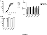

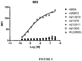
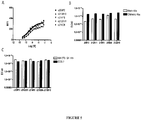

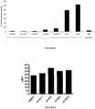
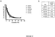
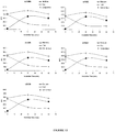
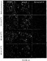
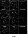

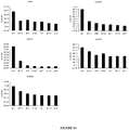
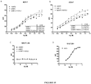
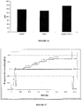


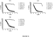


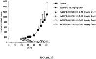

Classifications
-
- C—CHEMISTRY; METALLURGY
- C07—ORGANIC CHEMISTRY
- C07K—PEPTIDES
- C07K16/00—Immunoglobulins [IGs], e.g. monoclonal or polyclonal antibodies
- C07K16/18—Immunoglobulins [IGs], e.g. monoclonal or polyclonal antibodies against material from animals or humans
- C07K16/28—Immunoglobulins [IGs], e.g. monoclonal or polyclonal antibodies against material from animals or humans against receptors, cell surface antigens or cell surface determinants
- C07K16/2863—Immunoglobulins [IGs], e.g. monoclonal or polyclonal antibodies against material from animals or humans against receptors, cell surface antigens or cell surface determinants against receptors for growth factors, growth regulators
-
- A—HUMAN NECESSITIES
- A61—MEDICAL OR VETERINARY SCIENCE; HYGIENE
- A61K—PREPARATIONS FOR MEDICAL, DENTAL OR TOILETRY PURPOSES
- A61K31/00—Medicinal preparations containing organic active ingredients
- A61K31/33—Heterocyclic compounds
- A61K31/395—Heterocyclic compounds having nitrogen as a ring hetero atom, e.g. guanethidine or rifamycins
- A61K31/40—Heterocyclic compounds having nitrogen as a ring hetero atom, e.g. guanethidine or rifamycins having five-membered rings with one nitrogen as the only ring hetero atom, e.g. sulpiride, succinimide, tolmetin, buflomedil
- A61K31/4015—Heterocyclic compounds having nitrogen as a ring hetero atom, e.g. guanethidine or rifamycins having five-membered rings with one nitrogen as the only ring hetero atom, e.g. sulpiride, succinimide, tolmetin, buflomedil having oxo groups directly attached to the heterocyclic ring, e.g. piracetam, ethosuximide
-
- A—HUMAN NECESSITIES
- A61—MEDICAL OR VETERINARY SCIENCE; HYGIENE
- A61K—PREPARATIONS FOR MEDICAL, DENTAL OR TOILETRY PURPOSES
- A61K39/00—Medicinal preparations containing antigens or antibodies
- A61K39/395—Antibodies; Immunoglobulins; Immune serum, e.g. antilymphocytic serum
-
- A—HUMAN NECESSITIES
- A61—MEDICAL OR VETERINARY SCIENCE; HYGIENE
- A61K—PREPARATIONS FOR MEDICAL, DENTAL OR TOILETRY PURPOSES
- A61K38/00—Medicinal preparations containing peptides
- A61K38/04—Peptides having up to 20 amino acids in a fully defined sequence; Derivatives thereof
- A61K38/08—Peptides having 5 to 11 amino acids
-
- A—HUMAN NECESSITIES
- A61—MEDICAL OR VETERINARY SCIENCE; HYGIENE
- A61K—PREPARATIONS FOR MEDICAL, DENTAL OR TOILETRY PURPOSES
- A61K47/00—Medicinal preparations characterised by the non-active ingredients used, e.g. carriers or inert additives; Targeting or modifying agents chemically bound to the active ingredient
- A61K47/50—Medicinal preparations characterised by the non-active ingredients used, e.g. carriers or inert additives; Targeting or modifying agents chemically bound to the active ingredient the non-active ingredient being chemically bound to the active ingredient, e.g. polymer-drug conjugates
- A61K47/51—Medicinal preparations characterised by the non-active ingredients used, e.g. carriers or inert additives; Targeting or modifying agents chemically bound to the active ingredient the non-active ingredient being chemically bound to the active ingredient, e.g. polymer-drug conjugates the non-active ingredient being a modifying agent
- A61K47/68—Medicinal preparations characterised by the non-active ingredients used, e.g. carriers or inert additives; Targeting or modifying agents chemically bound to the active ingredient the non-active ingredient being chemically bound to the active ingredient, e.g. polymer-drug conjugates the non-active ingredient being a modifying agent the modifying agent being an antibody, an immunoglobulin or a fragment thereof, e.g. an Fc-fragment
- A61K47/6801—Drug-antibody or immunoglobulin conjugates defined by the pharmacologically or therapeutically active agent
- A61K47/6803—Drugs conjugated to an antibody or immunoglobulin, e.g. cisplatin-antibody conjugates
-
- A—HUMAN NECESSITIES
- A61—MEDICAL OR VETERINARY SCIENCE; HYGIENE
- A61K—PREPARATIONS FOR MEDICAL, DENTAL OR TOILETRY PURPOSES
- A61K47/00—Medicinal preparations characterised by the non-active ingredients used, e.g. carriers or inert additives; Targeting or modifying agents chemically bound to the active ingredient
- A61K47/50—Medicinal preparations characterised by the non-active ingredients used, e.g. carriers or inert additives; Targeting or modifying agents chemically bound to the active ingredient the non-active ingredient being chemically bound to the active ingredient, e.g. polymer-drug conjugates
- A61K47/51—Medicinal preparations characterised by the non-active ingredients used, e.g. carriers or inert additives; Targeting or modifying agents chemically bound to the active ingredient the non-active ingredient being chemically bound to the active ingredient, e.g. polymer-drug conjugates the non-active ingredient being a modifying agent
- A61K47/68—Medicinal preparations characterised by the non-active ingredients used, e.g. carriers or inert additives; Targeting or modifying agents chemically bound to the active ingredient the non-active ingredient being chemically bound to the active ingredient, e.g. polymer-drug conjugates the non-active ingredient being a modifying agent the modifying agent being an antibody, an immunoglobulin or a fragment thereof, e.g. an Fc-fragment
- A61K47/6835—Medicinal preparations characterised by the non-active ingredients used, e.g. carriers or inert additives; Targeting or modifying agents chemically bound to the active ingredient the non-active ingredient being chemically bound to the active ingredient, e.g. polymer-drug conjugates the non-active ingredient being a modifying agent the modifying agent being an antibody, an immunoglobulin or a fragment thereof, e.g. an Fc-fragment the modifying agent being an antibody or an immunoglobulin bearing at least one antigen-binding site
- A61K47/6849—Medicinal preparations characterised by the non-active ingredients used, e.g. carriers or inert additives; Targeting or modifying agents chemically bound to the active ingredient the non-active ingredient being chemically bound to the active ingredient, e.g. polymer-drug conjugates the non-active ingredient being a modifying agent the modifying agent being an antibody, an immunoglobulin or a fragment thereof, e.g. an Fc-fragment the modifying agent being an antibody or an immunoglobulin bearing at least one antigen-binding site the antibody targeting a receptor, a cell surface antigen or a cell surface determinant
-
- A—HUMAN NECESSITIES
- A61—MEDICAL OR VETERINARY SCIENCE; HYGIENE
- A61P—SPECIFIC THERAPEUTIC ACTIVITY OF CHEMICAL COMPOUNDS OR MEDICINAL PREPARATIONS
- A61P35/00—Antineoplastic agents
Definitions
- the present invention relates to an antibody-drug-conjugate capable of binding to the IGF-1R.
- the invention relates to an antibody-drug-conjugate comprising an antibody capable of binding to IGF-1R, said antibody being conjugated to at least one drug selected from derivatives of dolastatin 10 and auristatins.
- the invention also comprises said antibody-drug-conjugate for use in the treatment of cancer and discloses a method of treatment.
- IGF-1R insulin-like growth factor 1 receptor
- IGF-1R is a receptor with tyrosine kinase activity having 70% homology with the insulin receptor IR.
- IGF-1R is a glycoprotein of molecular weight approximately 350,000. It is a hetero-tetrameric receptor of which each half -linked by disulfide bridges- is composed of an extracellular ⁇ -subunit and of a transmembrane ⁇ -subunit.
- IGF-1R binds IGF1 and IGF2 with a very high affinity (Kd #1 nM) but is equally capable of binding to insulin with an affinity 100 to 1000 times lower.
- the IR binds insulin with a very high affinity although the IGFs only bind to the insulin receptor with a 100 times lower affinity.
- the tyrosine kinase domain of IGF-1R and of IR has a very high sequence homology although the zones of weaker homology respectively concern the cysteine-rich region situated on the ⁇ -subunit and the C-terminal part of the ⁇ -subunit.
- the sequence differences observed in the ⁇ -subunit are situated in the binding zone of the ligands and are therefore at the origin of the relative affinities of IGF-1R and of IR for the IGFs and insulin respectively.
- the differences in the C-terminal part of the ⁇ -subunit result in a divergence in the signalling pathways of the two receptors; IGF-1R mediating mitogenic, differentiation and antiapoptosis effects, while the activation of the IR principally involves effects at the level of the metabolic pathways.
- the cytoplasmic tyrosine kinase proteins are activated by the binding of the ligand to the extracellular domain of the receptor.
- the activation of the kinases in turn involves the stimulation of different intra-cellular substrates, including IRS-1, IRS-2, Shc and Grb 10.
- the two major substrates of IGF-1R are IRS and Shc which mediate, by the activation of numerous effectors downstream, the majority of growth and differentiation effects connected with the attachment of the IGFs to this receptor.
- the availability of substrates can consequently dictate the final biological effect connected with the activation of the IGF-1R.
- IRS-1 predominates, the cells tend to proliferate and to transform.
- Shc dominates the cells tend to differentiate. It seems that the route principally involved for the effects of protection against apoptosis is the phosphatidyl-inositol 3-kinases (PI 3-kinases) route.
- PI 3-kinases phosphatidyl-inositol 3-kin
- IGF-1R The role of the IGF system in carcinogenesis has become the subject of intensive research in the last ten years. This interest followed the discovery of the fact that in addition to its mitogenic and antiapoptosis properties, IGF-1R seems to be required for the establishment and the maintenance of a transformed phenotype. In fact, it has been well established that an overexpression or a constitutive activation of IGF-1R leads, in a great variety of cells, to a growth of the cells independent of the support in media devoid of foetal calf serum, and to the formation of tumors in nude mice. This in itself is not a unique property since a great variety of products of overexpressed genes can transform cells, including a good number of receptors of growth factors.
- IGF-1R - cells in which the gene coding for IGF-1R has been inactivated, are totally refractory to transformation by different agents which are usually capable of transforming cells, such as the E5 protein of bovine papilloma virus, an overexpression of EGFR or PDGFR, the T antigen of SV40, activated ras or the combination of these two last factors.
- agents which are usually capable of transforming cells, such as the E5 protein of bovine papilloma virus, an overexpression of EGFR or PDGFR, the T antigen of SV40, activated ras or the combination of these two last factors.
- IGF-1R is expressed in a great variety of tumors and of tumor lines and WO 2010/146059 discloses biomarkers for selecting patients for therapy with an IGF-1R inhibitor, notably for breast and colorectal cancer.
- the IGFs amplify the tumor growth via their attachment to IGF-1R.
- Other arguments in favor of the role of IGF-1R in carcinogenesis come from studies using murine monoclonal antibodies directed against the receptor or using negative dominants of IGF-1R.
- murine monoclonal antibodies directed against IGF-1R inhibit the proliferation of numerous cell lines in culture and the growth of tumor cells in vivo. It has likewise been shown that a negative dominant of IGF-1R is capable of inhibiting tumor proliferation.
- IGF-1R is not considered now as a major target and, in the research of potential therapeutic antibodies, IGF-1R is no more considered as of particular interest.
- IGF-1R is considered as a target not suitable for the generation of an ADC such as an antibody-drug-conjugate (referred as "ADC") as IGF-1R is described as a target also widely expressed by normal cells, including blood vessels.
- ADC antibody-drug-conjugate
- the invention relates to an ADC or conjugate and its use for the treatment of cancer, and more particularly IGF-1R-expressing cancers.
- ADCs combine the binding specificity of an antibody with the potency of drugs such as, for example, cytotoxic agents.
- drugs such as, for example, cytotoxic agents.
- ADCs allow the local delivery of drugs which, if administered as unconjugated drugs, may result in unacceptable levels of toxicity to normal cells.
- Efforts to design and refine ADC have focused on the selectivity of antibody as well as drug mechanism of action, drug-linking, drug/antibody ratio (loading or DAR), and drug-releasing properties.
- Drug moieties may impart their cytotoxic and cytostatic effects by mechanisms including tubulin binding, DNA binding, proteasome, impairement of ribosome function, protein synthesis and/or topoisomerase inhibition. Some cytotoxic drugs tend to be inactive or less active when conjugated to large antibody.
- Each antibody must be characterized separately, an appropriate linker designed, and a suitable cytotoxic agent identified that retains its potency upon delivery to tumor cells.
- One must consider the antigen density on the cancer target and whether normal tissues express the target antigen. Other considerations include whether the entire ADC is internalized upon binding the target; whether a cytostatic or cytotoxic drug is preferable when considering possible normal tissue exposure and/or the type and stage of the cancer being treated; and, whether the linker connecting the antibody to the drug payload is a cleavable or a non-cleavable linkage.
- the antibody to drug moiety conjugation ratio must be sufficient without compromising the binding activity of the antibody and/or the potency of the drug and without modifying physicochemical properties of the ADC resulting on aggregation or deleterious properties regarding to the future development process of the compound.
- An ADC is a complex biological molecule and the challenges to develop an effective ADC remain a significant issue.
- the present invention intends to address this issue and relates to an ADC of the following formula (I): Ab-(L-D) n (I) or a pharmaceutically acceptable salt thereof, wherein
- the invention relates to an ADC wherein Ab is selected from the antibodies 208F2, 212A11, 214F8, 219D6 and 213B10.
- An embodiment of the invention relates to an ADC wherein Ab is a humanized antibody.
- Ab is selected from:
- Ab is selected from:
- Ab is selected from:
- Ab is selected from:
- Ab is selected from:
- An embodiment of the invention relates to an ADC wherein L 2 is of the following formula: wherein the asterisk indicates the point of attachment to (W) w ; and the wavy line indicates the point of attachment to the nitrogen atom of the maleimide moiety of formula:
- An embodiment of the invention relates to an ADC wherein L is selected from: and wherein the asterisk indicates the point of attachment to D, and the wavy line indicates the point of attachment to Ab.
- An embodiment of the invention relates to an ADC wherein (L-D) is selected from: and wherein the wavy line indicates the point of attachment to Ab.
- An embodiment of the invention relates to an ADC having the formula selected from: and the pharmaceutically acceptable salts thereof, wherein Ab is selected in the group consisting of:
- An embodiment of the invention relates to an ADC wherein n is 2.
- An embodiment of the invention relates to an ADC wherein n is 4.
- An embodiment of the invention relates to an ADC for use as a medicament.
- An embodiment of the invention relates to a composition comprising an ADC as above described.
- An embodiment of the invention relates to a composition further comprising a pharmaceutically acceptable vehicle.
- An embodiment of the invention relates to a composition for use in the treatment of an IGF-1R-expressing cancer, or IGF-1R related cancers.
- IGF-1R-expressing cancer or IGF-1R related cancers include tumoral cells expressing or over-expressing whole or part of the IGF-1R at their surface.
- An embodiment of the invention relates to a composition, wherein said IGF-1R-expressing cancer is a cancer chosen from breast, colon, esophageal carcinoma, hepatocellular, gastric, glioma, lung, melanoma, osteosarcoma, ovarian, prostate, rhabdomyosarcoma, renal, thyroid, uterine endometrial cancer, mesothelioma, oral squamous carcinoma and any drug resistant cancer.
- said IGF-1R-expressing cancer is a cancer chosen from breast, colon, esophageal carcinoma, hepatocellular, gastric, glioma, lung, melanoma, osteosarcoma, ovarian, prostate, rhabdomyosarcoma, renal, thyroid, uterine endometrial cancer, mesothelioma, oral squamous carcinoma and any drug resistant cancer.
- the present description discloses also a method for the treatment of an IGF-1R-expressing cancer in a subject in need thereof, comprising administering to the subject an effective amount of at least one antibody-drug-conjugate or of a composition according to the invention.
- An embodiment of the invention relates to a kit comprising at least i) an antibody-drug-conjugate and/or a composition as above described and ii) a syringe or vial or ampoule in which the said antibody-drug-conjugate and/or composition is disposed.
- antibody refers to monoclonal antibodies, isolated, engineered or recombinant antibodies (e.g., full length or intact monoclonal antibodies), polyclonal antibodies, multivalent antibodies or multispecific antibodies (e.g., bispecific antibodies) and also antibody fragment thereof, so long as they exhibit the desired biological activity.
- the antibody of the ADC of the invention consists of a recombinant antibody.
- the term "recombinant antibody” refers to an antibody that results from the expression of recombinant DNA within living cells.
- a recombinant antibody of ADC of the invention is obtained by using laboratory methods of genetic recombination, well known by a person skilled in the art, creating DNA sequences that would not be found in biological organisms.
- the antibody of the ADC of the invention consists of a chemically synthesized antibody.
- such a molecule consists of a glycoprotein comprising at least two heavy (H) chains and two light (L) chains inter-connected by disulfide bonds.
- Each heavy chain comprises a heavy chain variable region (or domain) (abbreviated herein as HCVR or VH) and a heavy chain constant region.
- the heavy chain constant region comprises three domains, CH1, CH2 and CH3.
- Each light chain comprises a light chain variable region (abbreviated herein as LCVR or VL) and a light chain constant region.
- the light chain constant region comprises one domain, CL.
- the VH and VL regions can be further subdivided into regions of hypervariability, termed complementarity determining regions (CDR), interspersed with regions that are more conserved, termed framework regions (FR).
- CDR complementarity determining regions
- Each VH and VL is composed of three CDRs and four FRs, arranged from amino-terminus to carboxy-terminus in the following order: FR1, CDR1, FR2, CDR2, FR3, CDR3, FR4.
- the variable regions of the heavy and light chains contain a binding domain that interacts with an antigen.
- the constant regions of the antibodies may mediate the binding of the immunoglobulin to host tissues or factors, including various cells of the immune system (e.g. effector cells) and the first component (Clq) of the classical complement system.
- antigen binding fragment or "IGF-IR binding fragment” of an antibody of the ADC according to the invention, it is intended to indicate any peptide, polypeptide, or protein retaining the ability to bind to the target (also generally referred as antigen) of the antibody..
- such "antigen binding fragments” are selected in the group consisting of Fv, scFv (sc for single chain), Fab, F(ab') 2 , Fab', scFv-Fc fragments or diabodies, or any fragment of which the half-life time would have been increased by chemical modification, such as the addition of poly(alkylene) glycol such as poly(ethylene) glycol (“PEGylation") (pegylated fragments called Fv-PEG, scFv-PEG, Fab-PEG, F(ab') 2 -PEG or Fab'-PEG) ("PEG” for Poly(Ethylene) Glycol), or by incorporation in a liposome, said fragments having at least one of the characteristic CDRs of the antibody according to the invention.
- poly(alkylene) glycol such as poly(ethylene) glycol (“PEGylation")
- said "antigen binding fragments” will be constituted or will comprise a partial sequence of the heavy or light variable chain of the antibody from which they are derived, said partial sequence being sufficient to retain the same specificity of binding as the antibody from which it is descended and a sufficient affinity, preferably at least equal to 1/100, in a more preferred manner to at least 1/10, of the affinity of the antibody from which it is descended, with respect to the target.
- said "antigen binding fragments" will be constituted of or will comprise at least the three CDRs CDR-H1, CDR-H2 and CDR-H3 of the heavy variable chain and the three CDRs CDR-L1, CDR-L2 and CDR-L3 of the light variable chain of the antibody from which they are derived.
- binding By “binding”, “binds”, or the like, it is intended that the antibody, or any antigen binding fragment thereof, forms a complex with an antigen that is relatively stable under physiologic conditions. Specific binding can be characterized by an equilibrium dissociation constant of at least about 1x10 -6 M. Methods for determining whether two molecules bind are well known in the art and include, for example, equilibrium dialysis, surface plasmon resonance, radiolabelled assays and the like. For the avoidance of doubt, it does not mean that the said antibody could not bind or interfere, at a low level, to another antigen. Nevertheless, as an embodiment, the said antibody binds only to the said antigen.
- IGF-1R antibody should be interpreted as similar to "anti-IGF-lR antibody” and means an antibody capable of binding to IGF-1R.
- the epitope of the antibody is preferentially localized into the extracellular domain of the human IGF-1R (also referred as IGF-1R ECD).
- the antibody, or any antigen binding fragment thereof is capable of binding to IGF-1R with an EC 50 comprised between 10x10 -10 to 1x10 -10 , and more preferentially between 8x10 -10 to 2x10 -10 .
- the term half maximal effective concentration corresponds to the concentration of a drug, antibody or toxicant which induces a response halfway between the baseline and maximum after some specified exposure time. It is commonly used as a measure of drug's potency.
- the EC 50 of a graded dose response curve therefore represents the concentration of a compound where 50% of its maximal effect is observed.
- the EC 50 of a quantal dose response curve represents the concentration of a compound where 50% of the population exhibits a response, after specified exposure duration. Concentration measures typically follow a sigmoidal curve, increasing rapidly over a relatively small change in concentration. This can be determined mathematically by derivation of the best-fit line.
- the EC 50 characterizes the potency of antibody to bind on the IGF-1R ECD exposed on human tumor cells.
- the EC 50 parameter is determined using FACS analysis.
- the EC 50 parameter reflects the antibody concentration for which 50% of the maximal binding on the human IGF-1R expressed on human tumor cells is obtained.
- Each EC 50 value was calculated as the midpoint of the dose response curve using a four-parameter regression curve fitting program (Prism Software). This parameter has been selected as to be representative of physiological/pathological conditions.
- epitopes is a region of an antigen that is bound by an antibody.
- Epitopes may be defined as structural or functional. Functional epitopes are generally a subset of the structural epitopes and have those residues that directly contribute to the affinity of the interaction. Epitopes may also be conformational, that is, composed of non-linear amino acids.
- epitopes may include determinants that are chemically active surface groupings of molecules such as amino acids, sugar side chains, phosphoryl groups, or sulfonyl groups, and, in certain embodiments, may have specific three-dimensional structural characteristics, and/or specific charge characteristics.
- the competition for binding to IGF-1R can be determined by any methods or techniques known by the person skilled in the art such as, without limitation, radioactivity, Biacore, ELISA, Flow cytometry, etc.
- which competes for binding to IGF-1R it is meant a competition of at least 20%, preferentially at least 50% and more preferentially at least 70%.
- the determination of the binding to the same epitope can be determined by any methods or techniques known by the person skilled in the art such as, without limitation, radioactivity, Biacore, ELISA, Flow cytometry, etc.
- an antibody that "is internalized” or that "internalized” is one that is taken up by (meaning it "enters”) the cell upon binding to IGF-1R on a mammalian cell.
- Such an antibody is interesting as part of the ADC, so it addresses or directs the linked cytotoxic into the targeted cancer cells. Once internalized the cytotoxic triggers cancer cell death.
- the antibodies according to the invention are all presenting the same sequences for the CDR-H2, CDR-H3 and CDR-L2, the other 3 CDRs being different. This observation seems coherent as it is part of the general knowledge that, regarding the binding specificity of an antibody, the CDR-H3 is described as being the most important and the most implicated with the recognition of the epitope.
- the cytotoxic confers the cytotoxic activity and the used antibody is responsible for the specificity against cancer cells, as well as a vector for entering within the cells to correctly address the cytotoxic.
- the antibody can exhibit high ability to internalize into the targeted cancer cells.
- the efficiency of the antibody mediated internalisation differs significantly depending on the epitope targeted. Selection of potent internalizing IGF-1R antibodies requires various experimental data studying not only IGF-1R downregulation but also following IGF-1R antibody internalization into the cells.
- the internalization of the antibody of the ADC according to the invention can be evaluated by immunofluorescence or FACS (Flow Cytometry) (as exemplified hereinafter in the present application) or any method or process known by the person skilled in the art specific for the internalization mechanism.
- the antibody od the ADC according to the invention can induce internalization after binding to IGF-1R of at least 30%, preferentially 50% and more preferentially 80%.
- the complex IGF-1R/antibody is internalized after binding of the antibody to the ECD of said IGF-1R, and a reduction in the quantity of IGF-1R at the surface of the cells is induced.
- This reduction can be quantified by any method known by the person skilled in the art such as non limitative examples western-blot, FACS, and immunofluorescence.
- this reduction thus reflecting the internalization, can be preferably measured by FACS and expressed as the difference or delta between the Mean Fluorescence Intensity (MFI) measured at 4°C with the MFI measured at 37°C after 4 hours incubation with the antibody.
- MFI Mean Fluorescence Intensity
- this delta is determined based on MFIs obtained with untreated cells and cells treated with the antibody using i) breast cancer cells MCF7 after a 4 hour incubation period with the antibody herein described and ii) a secondary antibody labelled with Alexa488.
- This parameter is defined as calculated with the following formula: ⁇ (MFI 4°C -MFI 37°C ).
- MFIs This difference between MFIs reflects the IGF-1R downregulation as MFIs are proportional to IGF-1R expressed on the cell-surface.
- the antibodies consist of antibodies triggering a ⁇ (MFI 4°C -MFI 37°C ) on MCF-7 of at least 280, preferably of at least 400.
- an internalization percentage can be determined as: 100x(MFI 4°C -MFI 37°C ) / MFI 4°C .
- the antibodies of the ADC according to the invention present, preferably, on MCF7 an internalization percentage comprised between 50% and 99%, 70% and 90%, preferentially between 75% and 87%.
- the used antibodies exhibit a rapid rate of internalization, preferably within 24 hours from administration of the antibody and, more preferably within 12 hours and, even more preferably within 6 hours.
- the internalization rate also referred as cell surface bound antibody decrease or cell surface antibody decay
- t1/2 half life
- ⁇ MFI cell surface bound antibody decay
- a particular advantage is that the antibodies of the ADC of the invention have a t1/2 comprised between 5 and 25 minutes, and preferentially between 10 and 20 minutes.
- a particular embodiment of the invention relates to an ADC wherein the antibody Ab comprises three heavy chain CDRs with CDR-H2 of sequence SEQ ID No. 2 and CDR-H3 of sequence SEQ ID No. 3, and three light chain CDRs with CDR-L2 of sequence SEQ ID No. 5.
- a particular embodiment of the invention relates to an ADC wherein the antibody Ab comprises the three heavy chain CDRs of sequences SEQ ID Nos. 1, 2 and 3 and the three light chain CDRs of sequences SEQ ID Nos. 4, 5 and 6.
- An embodiment of the ADC comprises an antibody comprising the three heavy chain CDRs comprising or consisting of the sequences SEQ ID Nos. 1, 2 and 3, or any sequence exhibiting at least 80%, preferably 85%, 90%, 95% and 98% identity with SEQ ID Nos. 1, 2 or3; and the three light chain CDRs comprising or consisting of the sequences SEQ ID Nos. 4, 5 and 6, or any sequence exhibiting at least 80%, preferably 85%, 90%, 95% and 98% identity with SEQ ID Nos. 4, 5 or 6.
- the antibody, or any antigen binding fragment thereof comprises the three heavy chain CDRs comprising the sequences SEQ ID Nos. 1, 2 and 3; and the three light chain CDRs comprising the sequences SEQ ID Nos. 4, 5 and 6.
- the IMGT unique numbering has been defined to compare the variable domains whatever the antigen receptor, the chain type, or the species [ Lefranc M.-P., Immunology Today 18, 509 (1997 ) / Lefranc M.-P., The Immunologist, 7, 132-136 (1999 ) / Lefranc, M.-P., Pommié, C., Ruiz, M., Giudicelli, V., Foulquier, E., Truong, L., Thouvenin-Contet, V. and Lefranc, Dev. Comp. Immunol., 27, 55-77 (2003 )].
- cystein 23 Ist-CYS
- tryptophan 41 CONSERVED-TRP
- hydrophobic amino acid 89 cystein 104
- cystein 104 2nd-CYS
- phenylalanine or tryptophan 118 J-PHE or J-TRP
- the IMGT unique numbering provides a standardized delimitation of the framework regions (FR1-IMGT: positions 1 to 26, FR2-IMGT: 39 to 55, FR3-IMGT: 66 to 104 and FR4-IMGT: 118 to 128) and of the complementarity determining regions: CDR1-IMGT: 27 to 38, CDR2-IMGT: 56 to 65 and CDR3-IMGT: 105 to 117. As gaps represent unoccupied positions, the CDR-IMGT lengths (shown between brackets and separated by dots, e.g. [8.8.13]) become crucial information.
- the IMGT unique numbering is used in 2D graphical representations, designated as IMGT Colliers de Perles [ Ruiz, M.
- complementarity-determining regions or CDRs mean the hypervariable regions of the heavy and light chains of immunoglobulins as defined according to the IMGT numbering system.
- CDRs can also be defined according to the Kabat numbering system ( Kabat et al., Sequences of proteins of immunological interest, 5th Ed., U.S. Department of Health and Human Services, NIH, 1991, and later editions ). There are three heavy chain CDRs and three light chain CDRs.
- CDR and “CDRs” are used to indicate, depending on the case, one or more, or even all, of the regions containing the majority of the amino acid residues responsible for the antibody's binding affinity for the antigen or epitope it recognizes.
- the CDRs according to Kabat are not defined. Nevertheless, it would be obvious for the person skilled in the art, using the definition of the CDRs according to IMGT, to define the CDRs according to Kabat.
- the "identity” or “percentage identity” between two sequences of nucleic acids or amino acids means the percentage of identical nucleotides or amino acid residues between the two sequences to be compared, obtained after optimal alignment, this percentage being purely statistical and the differences between the two sequences being distributed randomly along their length.
- the comparison of two nucleic acid or amino acid sequences is traditionally carried out by comparing the sequences after having optimally aligned them, said comparison being able to be conducted by segment or by using an "alignment window”.
- Optimal alignment of the sequences for comparison can be carried out, in addition to comparison by hand, by means of the local homology algorithm of Smith and Waterman (1981) [Ad. App. Math.
- Percentage identity is calculated by determining the number of positions at which the amino acid nucleotide or residue is identical between the two sequences, preferably between the two complete sequences, dividing the number of identical positions by the total number of positions in the alignment window and multiplying the result by 100 to obtain the percentage identity between the two sequences.
- the BLAST program "BLAST 2 sequences” ( Tatusova et al., "Blast 2 sequences - a new tool for comparing protein and nucleotide sequences", FEMS Microbiol., 1999, Lett. 174:247-250 ) available on the site http://www.ncbi.nlm.nih.gov/gorf/bl2.html, can be used with the default parameters (notably for the parameters "open gap penalty”: 5, and “extension gap penalty”: 2; the selected matrix being for example the "BLOSUM 62" matrix proposed by the program); the percentage identity between the two sequences to compare is calculated directly by the program.
- amino acid sequence exhibiting at least 80%, preferably 85%, 90%, 95% and 98% identity with a reference amino acid sequence
- preferred examples include those containing the reference sequence, certain modifications, notably a deletion, addition or substitution of at least one amino acid, truncation or extension.
- substitutions are preferred in which the substituted amino acids are replaced by "equivalent” amino acids.
- the expression “equivalent amino acids” is meant to indicate any amino acids likely to be substituted for one of the structural amino acids without however modifying the biological activities of the corresponding antibodies and of those specific examples defined below.
- Equivalent amino acids can be determined either on their structural homology with the amino acids for which they are substituted or on the results of comparative tests of biological activity between the various antibodies likely to be generated.
- Table 1 below summarizes the possible substitutions likely to be carried out without resulting in a significant modification of the biological activity of the corresponding modified antibody; inverse substitutions are naturally possible under the same conditions.
- Table 1 Original residue Substitution(s) Ala (A) Val, Gly, Pro Arg (R) Lys, His Asn (N) Gln Asp (D) Glu Cys (C) Ser Gln (Q) Asn Glu (E) Asp Gly (G) Ala His (H) Arg Ile (I) Leu Leu (L) Ile, Val, Met Lys (K) Arg Met (M) Leu Phe (F) Tyr Pro (P) Ala Ser (S) Thr, Cys Thr (T) Ser Trp (W) Tyr Tyr (Y) Phe, Trp Val (V) Leu, Ala
- a particular aspect of the invention is that the antibody of the ADC, does not bind to the Insulin receptor (IR).
- IR Insulin receptor
- still another advantageous aspect of the antibody of the ADC of the invention is that it is capable of binding not only to the human IGF-1R but also to the monkey IGF-1R, and more particularly to the cynomolgus IGF-1R. This aspect is also of interest as it will facilitate the toxicity assessement required for clinical trials.
- the antibody of the ADC of the invention consists of a monoclonal antibody.
- the term "monoclonal antibody” or “Mab” as used herein refers to an antibody obtained from a population of substantially homogeneous antibodies, i.e. the individual antibodies of the population are identical except for possible naturally occurring mutations that may be present in minor amounts. Monoclonal antibodies are highly specific, being directed against a single epitope. Such monoclonal antibody may be produced by a single clone of B cells or hybridoma. Monoclonal antibodies may also be recombinant, i.e. produced by protein engineering or chemical synthesis. Monoclonal antibodies may also be isolated from phage antibody libraries. In addition, in contrast with preparations of polyclonal antibodies which typically include various antibodies directed against various determinants, or epitopes, each monoclonal antibody is directed against a single epitope of the antigen.
- the monoclonal antibody herein includes murine, chimeric and humanized antibody, such as described after.
- the antibody is preferably derived from an hybridoma of murine origin filed within the French collection for microorganism cultures (CNCM, Pasteur Institute, 25 rue du Dondel Roux, 75724 Paris Cedex 15, France), said hybridoma being obtained by the fusion of Balb/C immunized mice splenocytes/lymphocytes and cells of the myeloma Sp 2/O-Ag 14 cell line.
- the IGF-1R antibody of the ADC of the invention consists of a murine antibody, then referred as m[ name of the antibody ].
- the IGF-1R antibody consists of a chimeric antibody, then referred as c[ name of the antibody ].
- the IGF-1R antibody consists of a humanized antibody, then referred as hz[ name of the antibody ].
- IGF-1R antibody and “[ name of the antibody]” are similar and include (without contrary specification) the murine, the chimeric and the humanized versions of the said IGF-1R antibody or of the said "[ name of the antibody]".
- the prefix m-(murine), c- (chimeric) or hz- (humanized) is used.
- Table 2 illustrates the CDR sequences, defined according to IMGT, for the preferred antibodies.
- Table 2 Heavy chain Light chain SEQ ID No. Consensus CDR-H1 1 CDR-H2 2 CDR-H3 3 CDR-L1 4 CDR-L2 5 CDR-L3 6 208F2 CDR-H1 7 CDR-H2 2 CDR-H3 3 CDR-L1 9 CDR-L2 5 CDR-L3 11 212A11 CDR-H1 7 CDR-H2 2 CDR-H3 3 CDR-L1 10 CDR-L2 5 CDR-L3 11 214F8 & 213B10 CDR-H1 7 CDR-H2 2 CDR-H3 3 CDR-L1 9 CDR-L2 5 CDR-L3 12 219D6 CDR-H1 8 CDR-H2 2 CDR-H3 3 CDR-L1 9 CDR-L2 5 CDR-L3 11
- a specific aspect relates to an ADC wherein the antibody is a murine antibody characterized in that said antibody also comprises light chain and heavy chain constant regions derived from an antibody of a species heterologous with the mouse, notably man.
- Another specific aspect relates to an ADC wherein the antibody is a chimeric (c) antibody characterized in that said antibody also comprises light chain and heavy chain constant regions derived from an antibody of a species heterologous with the mouse, notably human.
- a chimeric antibody is one containing a natural variable region (light chain and heavy chain) derived from an antibody of a given species in combination with constant regions of the light chain and the heavy chain of an antibody of a species heterologous to said given species.
- the chimeric antibodies can be prepared by using the techniques of recombinant genetics.
- the chimeric antibody could be produced by cloning recombinant DNA containing a promoter and a sequence coding for the variable region of a nonhuman monoclonal antibody, notably murine, and a sequence coding for heterologous species antibody constant region, preferably human.
- a chimeric antibody of the ADC according to the invention coded by one such recombinant gene could be, for example, a mouse-human chimera, the specificity of this antibody being determined by the variable region derived from the murine DNA and its isotype determined by the constant region derived from human DNA.
- the antibody of the ADC of the invention is selected from:
- any sequence exhibiting at least 80%, preferably 85%, 90%, 95% and 98% identity with SEQ ID No. 13 to 17 its is intended to designate the sequences exhibiting the three heavy chain CDRs SEQ ID Nos. 1, 2 and 3 and, in addition, exhibiting at least 80%, preferably 85%, 90%, 95% and 98%, identity with the full sequence SEQ ID No. 13 to 17 outside the sequences corresponding to the CDRs (i.e. SEQ ID No. 1, 2 and 3).
- the antibody of the ADC of the invention is selected from:
- any sequence exhibiting at least 80%, preferably 85%, 90%, 95% and 98% identity with SEQ ID No. 18 to 22 its is intended to designate respectively the sequences exhibiting the three light chain CDRs SEQ ID Nos. 4, 5 and 6 and, in addition, exhibiting at least 80%, preferably 85%, 90%, 95% and 98% , identity with the full sequence SEQ ID No. 18 to 22 outside the sequences corresponding to the CDRs (i.e. SEQ ID No. 4, 5 and 6).
- Chimeric antibodies herein described can be also characterized by the constant domain and, more particularly, said chimeric antibodies can be selected or designed such as, without limitation, IgG1, IgG2, IgG3, IgM, IgA, IgD or IgE. More preferably, in the context of the present invention, said chimeric antibodies are IgG1 or IgG4.
- An embodiment of the invention relates to an ADC wherein Ab is a chimeric antibody comprising variable domains VH and VL as above described in the format IgG1. More preferably, said chimeric antibody comprises a constant domain for the VH of sequence SEQ ID No. 43 and a Kappa domain for the VL of sequence SEQ ID No. 45.
- An embodiment of the invention relates to an ADC wherein Ab is a chimeric antibody comprising variable domains VH and VL as above described in the format IgG4. More preferably, said chimeric antibody comprises a constant domain for the VH of sequence SEQ ID No. 44 and a Kappa domain for the VL of sequence SEQ ID No. 45.
- the antibody of the ADC of the invention is selected from:
- Table 3 illustrates the sequences of the VH and VL, respectively, for the preferred chimeric antibodies.
- Yet another specific aspect of the present invention relates to an ADC wherein "Ab” is a humanized antibody characterized in that the constant regions of the light chain and the heavy chain derived from human antibody are, respectively, the lambda or kappa region and the gamma-1, gamma-2 or gamma-4 region.
- Humanized antibodies means an antibody that contains CDR regions derived from an antibody of nonhuman origin, the other parts of the antibody molecule being derived from one (or several) human antibodies. In addition, some of the skeleton segment residues (called FR) can be modified to preserve binding affinity.
- humanized antibodies or fragments of same can be prepared by techniques known to a person skilled in the art. Such humanized antibodies are preferred for their use in methods involving in vitro diagnoses or preventive and/or therapeutic treatment in vivo.
- Other humanization techniques also known to a person skilled in the art, such as, for example, the "CDR grafting" technique described by PDL in patents EP 0 451 216 , EP 0 682 040 , EP 0 939 127 , EP 0 566 647 or US 5,530,101 , US 6,180,370 , US 5,585,089 and US 5,693,761 .
- US patents 5,639,641 or 6,054,297 , 5,886,152 and 5,877,293 can also be cited.
- the antibody of the ADC according to the present invention comprises a heavy chain variable domain (VH) having:
- the antibody of the ADC according to the present invention comprises a light chain variable domain (VL) having:
- the antibody comprises:
- the antibody of the ADC according to the invention comprises a heavy chain variable domain (VH) of sequence SEQ ID No. 33 and a light chain variable domain (VL) of sequence SEQ ID No. 35.
- VH heavy chain variable domain
- VL light chain variable domain
- Said humanized antibody will be called thereinafter hz208F2 ("Variant 1" or "Var. 1").
- the antibody of the ADC according to the present invention comprises a heavy chain variable domain (VH) of sequence SEQ ID No. 33 wherein said sequence SEQ ID No. 33 comprises at least 1 back-mutation selected from the residues 20, 34, 35, 38, 48, 50, 59, 61, 62, 70, 72, 74, 76, 77, 79, 82 and 95.
- VH heavy chain variable domain
- back-mutation or “back mutation” it is meant a mutation or replacement of the human residue present in the germline by the corresponding residue initially present in the murine sequence.
- the antibody of the ADC according to the present invention comprises a heavy chain variable domain (VH) of sequence SEQ ID No. 33 wherein said sequence SEQ ID No. 33 comprises 2, 3, 4, 5, 6, 7, 8, 9, 10, 11, 12, 13, 14, 15, 16 or 17 back-mutations selected from the residues 20, 34, 35, 38, 48, 50, 59, 61, 62, 70, 72, 74, 76, 77, 79, 82 and 95.
- VH heavy chain variable domain
- the antibody of the ADC according to the present invention comprises a light chain variable domain (VL) of sequence SEQ ID No. 35, wherein said sequence SEQ ID No. 35 comprises at least 1 back-mutation selected from the residues 22, 53, 55, 65, 71, 72, 77 and 87.
- VL light chain variable domain
- the antibody of the ADC according to the present invention comprises a light chain variable domain (VL) of sequence SEQ ID No. 35, wherein said sequence SEQ ID No. 35 comprises 2, 3, 4, 5, 6, 7 or 8 back-mutations selected from the residues 22, 53, 55, 65, 71, 72, 77 or 87.
- VL light chain variable domain
- the antibody of the ADC according to the present invention comprises:
- the antibody of the ADC according to the invention comprises all the back-mutations above mentioned and corresponds to an antibody comprising a heavy chain variable domain (VH) of sequence SEQ ID No. 34 and a light chain variable domain (VL) of sequence SEQ ID No. 36.
- Said humanized antibody will be called thereinafter hz208F2 ("Variant 3" or "Var. 3").
- the antibody according to the invention corresponds to an antibody comprising a heavy chain variable domain (VH) of "consensus” sequence SEQ ID No. 41 and a light chain variable domain (VL) of "consensus” sequence SEQ ID No. 42.
- VH heavy chain variable domain
- VL light chain variable domain
- Said humanized antibody, as a whole, will be called thereinafter hz208F2 ("Variant2" or "Var.2").
- the antibody of the ADC of the invention is selected from:
- any sequence exhibiting at least 80%, preferably 85%, 90%, 95% and 98% identity with SEQ ID No. 33 or 34 its is intended to designate the sequences exhibiting the three heavy chain CDRs SEQ ID Nos. 1, 2 and 3 and, in addition, exhibiting at least 80%, preferably 85%, 90%, 95% and 98%, identity with the full sequence SEQ ID No. 33 or 34 outside the sequences corresponding to the CDRs (i.e. SEQ ID Nos. 1,2 and 3).
- any sequence or by a sequence exhibiting at least 80% with a particular sequence it must be understood that said sequence exhibits at least 80% and preferably 85%, 90%, 95% and 98% identity with the referenced sequence.
- these sequences contain CDR sequences, its is intended to designate that the sequences exhibiting at least these CDRs identically to the reference sequence CDRs, the 80%, preferably 85%, 90%, 95% and 98%, identity with the full sequence having to be calculated for the remaining sequence located outside the sequences corresponding to these CDRs.
- the antibody of the invention is selected from:
- any sequence exhibiting at least 80%, preferably 85%, 90%, 95% and 98% identity with SEQ ID No. 35 or 36 its is intended to designate the sequences exhibiting the three light chain CDRs SEQ ID Nos. 4, 5 and 6 and, in addition, exhibiting at least 80%, preferably 85%, 90%, 95% and 98%, identity with the full sequence SEQ ID No. 35 or 36 outside the sequences corresponding to the CDRs (i.e. SEQ ID Nos. 4, 5 and 6).
- Humanized antibodies herein described can be also characterized by the constant domain and, more particularly, said humanized antibodies can be selected or designed such as, without limitation, IgG1, IgG2, IgG3, IgM, IgA, IgD or IgE. More preferably, in the context of the present invention, said humanized antibodies are IgG1 or IgG4.
- An embodiment of the invention relates to an ADC wherein "Ab” is a humanized antibody comprising variable domains VH and VL as above described in the format IgG1. More preferably, said humanized antibody comprises a constant domain for the VH of sequence SEQ ID No. 43 and a Kappa domain for the VL of sequence SEQ ID No. 45.
- An embodiment of the invention relates to an ADC wherein "Ab” is a humanized antibody comprising variable domains VH and VL as above described in the format IgG4. More preferably, said humanized antibody comprises a constant domain for the VH of sequence SEQ ID No. 44 and a Kappa domain for the VL of sequence SEQ ID No. 45.
- Still another embodiment of the invention relates to an ADC wherein "Ab” is an antibody selected from:
- Table 6a illustrates non limitative examples of sequences of the VH and VL for the variant 1 (Var. 1) and the variant 3 (Var. 3) of the humanized antibody hz208F2. It also comprises the consensus sequence for the variant 2 (Var. 2).
- Table 6a Heavy Chain Light chain SEQ ID No. hz208F2 var. 1) Variable domain (VH) 33 Variable domain (VL) 35 Full length 37 Full length 39 hz208F2 (Var. 3) Variable domain (VH) 34 Variable domain (VL) 36 Full length 38 Full length 40 hz208F2 (Var. 2) Variable domain (VH) 41 Variable domain (VL) 42
- the antibody of the ADC of the invention is selected from:
- Still another embodiment of the invention relates to an ADC wherein "Ab” antibody selected from:
- the invention relates to an ADC wherein Ab is an antibody comprising:
- Another aspect of the present invention is an ADC wherein Ab is an antibody produced by the hybridoma 1-4757, 1-4773, 1-4775, 1-4736 or 1-4774 deposited at the CNCM, Institut Pasteur France on the 30 May 2013, 26 June 2013, 26 June 2013, 24 April 2013 and 26 June 2013, respectively.
- the murine hybridoma selected from the hybridoma 1-4757, 1-4773, 1-4775, 1-4736 and 1-4774 deposited at the CNCM, Institut Pasteur France on the 30 May 2013, 26 June 2013, 26 June 2013, 24 April 2013 and 26 June 2013, respectively.
- nucleic acid means a precise sequence of nucleotides, modified or not, defining a fragment or a region of a nucleic acid, containing unnatural nucleotides or not, and being either a double-strand DNA, a single-strand DNA or transcription products of said DNAs.
- sequences have been isolated and/or purified, i.e., they were sampled directly or indirectly, for example by a copy, their environment having been at least partially modified.
- Isolated nucleic acids obtained by recombinant genetics, by means, for example, of host cells, or obtained by chemical synthesis should also be mentioned here.
- vector comprising a nucleic acid coding for an antibody, or for an antigen binding fragment thereof, of the ADC according to the invention, particularly cloning and/or expression vectors that contain such a nucleotide sequence.
- the vectors preferably contain elements which allow the expression and/or the secretion of nucleotide sequences in a given host cell.
- the vector thus may contain a promoter, translation initiation and termination signals, as well as suitable transcription regulation regions. It must be able to be maintained in a stable manner in the host cell and may optionally have specific signals which specify secretion of the translated protein.
- These various elements are selected and optimized by a person skilled in the art according to the host cell used.
- the nucleotide sequences can be inserted in self-replicating vectors within the chosen host or be integrative vectors of the chosen host.
- the vectors are, for example, vectors of plasmid or viral origin. They are used to transform host cells in order to clone or express the nucleotide sequences of the invention.
- Such vectors are prepared by methods typically used by a person skilled in the art and the resulting clones can be introduced into a suitable host by standard methods such as lipofection, electroporation, conjugation, heat shock or chemical methods.
- These isolated host cells are transformed by or comprising a vector as above described.
- the host cell can be selected among prokaryotic or eukaryotic systems such as bacterial cells, for example, but also yeast cells or animal cells, notably mammal cells (with the exception of human). Insect or plant cells can also be used.
- prokaryotic or eukaryotic systems such as bacterial cells, for example, but also yeast cells or animal cells, notably mammal cells (with the exception of human). Insect or plant cells can also be used.
- the transformed cells are of use in methods for the preparation of recombinant antibodies of the ADC according to the invention.
- Methods for the preparation of antibodies in recombinant form using a vector and/or a cell transformed by a vector as above disclosed, are also comprised in the present specification.
- a cell transformed by a vector as above described is cultured under conditions that allow the expression of the aforesaid antibody and recovery of said antibody.
- the host cell can be selected among prokaryotic or eukaryotic systems.
- a vector as above disclosed carrying such a sequence can thus be used advantageously for the production of recombinant proteins to be secreted. Indeed, the purification of these recombinant proteins of interest will be facilitated by the fact that they are present in the supernatant of the cellular culture rather than inside host cells.
- the antibody of the ADC of the present invention can also be prepared by chemical synthesis.
- a person skilled in the art knows methods for chemical synthesis, such as solid-phase techniques or partial solid-phase techniques, by condensation of fragments or by conventional synthesis in solution.
- Polypeptides obtained by chemical synthesis and capable of containing corresponding unnatural amino acids can be also cited.
- the antibody likely to be obtained by the method above described are also comprised in the ADC of the present invention.
- the invention concerns an ADC wherein AB is an antibody, or an antigen binding fragment thereof, as above described for use as an addressing vehicle for delivering a cytotoxic agent at a host target site, said host target site consisting of an epitope localized into IGF-1R, preferably the IGF-1R extracellular domain, more preferably the human IGF-1R (SEQ ID No. 50) and still more preferably the human IGF-1R extracellular domain (SEQ ID No. 51), and still more preferably to the N-terminal of the human IGF-1R extracellular domain (SEQ ID No. 52), or any natural variant sequence thereof.
- AB is an antibody, or an antigen binding fragment thereof, as above described for use as an addressing vehicle for delivering a cytotoxic agent at a host target site, said host target site consisting of an epitope localized into IGF-1R, preferably the IGF-1R extracellular domain, more preferably the human IGF-1R (SEQ ID No. 50) and still more preferably the human IGF-1R extra
- said host target site is a target site of a mammalian cell, more preferably of a human cell, more preferably cells which naturally or by way of genetic recombination, express IGF-1R.
- said host target site is a target site of a cell of patient, preferably human, having a cancer, preferably an IGF-1R expressing cancer, or IGF-1R related cancers.
- IGF-1R expressing cancers or IGF-1R related cancers include particularly cancers wherein the tumoral cells express or over-express whole or part of the IGF-1R at their surface.
- the drug moiety according to the invention has the following formula (II) where:
- alkyl in the present invention is meant a straight-chain or branched, saturated hydrocarbon chain.
- alkyl in the present invention is meant a straight-chain or branched, saturated hydrocarbon chain.
- (C x -C y )alkyl in the present invention is meant an alkyl chain such as defined above comprising x to y carbon atoms. Therefore, a (C 1 -C 6 )alkyl group is an alkyl chain having 1 to 6 carbon atoms.
- the (C 1 -C 6 )alkyl is advantageously a (C 1 -C 4 )alkyl, peferably a (C 1 -C 2 )alkyl.
- one particularly appreciated class of drug moieties corresponds to the formula (II) drug moieties in which R 2 represents a COOH group.
- moieties corresponds to the formula (II) moieties in which R 2 is a thiazole (in particular a thiazol-2-yl group).
- R 2 is more particularly a COOH, COOMe or thiazol-2-yl group.
- R 2 is COOH
- R 2 is COOMe
- R 3 particularly represents a (C 1 -C 6 )alkyl, advantageously a methyl group.
- n is an integer comprised between 1 and 8, in particular between 1 and 6, advantageously between 1 and 4, preferably is 1 or 2.
- R 2 is COOH
- R 3 is a methyl group and m is 1 or 2.
- one particularly appreciated class of drug moieties corresponds to the formula (II) drug moieties in which R 9 is a methyl group or a hydrogen.
- the NR 9 group is located on the phenyl ring in a para position in relation to the (CH 2 ) m group.
- the drug moiety is chosen from among the following moieties: and
- the drug can be prepared using the general methods described in the following synthesis schemes, optionally supplemented by any standard operation when needed that is described in the literature or well known to persons skilled in the art, or described in the examples in the experimental part hereof.
- Scheme 1 illustrates the first general method which can be used to prepare the drug.
- the first step consists of the condensing of compound (II), protected on its amine function by a protective group G, with compound (III).
- X may represent a leaving group such as a chlorine.
- the first step consists of the reaction between an acid chloride and an amine.
- This reaction can be conducted using methods and techniques well known to those skilled in the art.
- the two entities are caused to react in the presence of an organic or inorganic base e.g. Et 3 N, iPr 2 NEt, pyridine, NaH, Cs 2 CO 3 , K 2 CO 3 in a solvent such as THF, dichloromethane, DMF, DMSO, at a temperature notably between -20°C and 100°C.
- X may also be a hydroxyl (OH).
- the first step is a condensation reaction between the carboxylic acid (II) and the amine (III).
- This reaction can be performed following methods and techniques well known to skilled persons.
- these two entities are caused to react in the presence of a coupling agent such as 1-(3-dimethylaminopropyl)-3-ethyl-carbodiimide (EDC), 3-hydroxy-1,2,3-benzotriazin-4(3H)-one, a tertiary amine such as diisopropylethylamine, in a polar aprotic solvent such as dichloromethane or DMF, at a temperature notably between -15°C and 40°C.
- a coupling agent such as 1-(3-dimethylaminopropyl)-3-ethyl-carbodiimide (EDC), 3-hydroxy-1,2,3-benzotriazin-4(3H)-one
- these two entities are caused to react in the presence of diethyl phosphorocyanidate (DEPC), a tertiary amine such as triethylamine, in a polar aprotic solvent such as dichloromethane or DMF, at a temperature of between -15°C and 40°C.
- DEPC diethyl phosphorocyanidate
- a tertiary amine such as triethylamine
- a polar aprotic solvent such as dichloromethane or DMF
- Another particularly appreciated method consists of causing these two entities to react in the presence of O-(7-azabenzotriazol-1-yl)-1,1,3,3-tetramethyl-uroniumhexafluorophosphate (HATU), a tertiary amine such as diisopropylethylamine, in a polar aprotic solvent such as dichloromethane or DMF, at a temperature of between-15°C and 100°C.
- HATU O-(7-azabenzotriazol-1-yl)-1,1,3,3-tetramethyl-uroniumhexafluorophosphate
- a tertiary amine such as diisopropylethylamine
- compound (IV) can be condensed with compound (V) following the methods and techniques described above to lead to compound (VI) after a deprotection step. This compound can then, after condensation with the intermediate (VII) and optional deprotection, lead to the formation of the drug.
- Compound (VI) can also be coupled with a compound (VII') in whichR' 3 is a precursor of R 3 , in particular an R 3 group protected by a protective group. Coupling followed by deprotection of group R' 3 to lead to R 3 can be carried out following the same procedures as described previously.
- Scheme 2 illustrates the second general method which can be used to prepare the drug.
- G is a protective group
- R 1 H
- R 2 , R 3 and R 4a are such as previously defined
- R 4b represents
- compound (IX) protected on its amine function by a protective group G is condensed with compound (VI).
- X may represent a leaving group e.g. a chlorine.
- the first step consists of the reaction between an acid chloride and an amine. This reaction can be performed using methods and techniques well known to persons skilled in the art. In one particularly appreciated method the two entities are caused to react in the presence of an organic or inorganic base such as Et 3 N, iPr 2 NEt, pyridine, NaH, Cs 2 CO 3 , K 2 CO 3 in a solvent such as THF, dichloromethane, DMF, DMSO at a temperature notably between -20° and 100°C.
- X may also represent a hydroxyl.
- the first step is a condensation reaction between the carboxylic acid (IX) and the amine (VI).
- This reaction can be conducted following methods and techniques well known to skilled persons.
- the two entities are caused to react in the presence of 1-(3-dimethylaminopropyl)-3-ethyl-carbodiimide (EDC), 3-hydroxy-1,2,3-benzotriazin-4(3H)-one, a tertiary amine such as diisopropylethylamine, in a polar aprotic solvent such as dichloromethane or DMF, at a temperature notably between -15°C and 40°C.
- EDC 1-(3-dimethylaminopropyl)-3-ethyl-carbodiimide
- a tertiary amine such as diisopropylethylamine
- a polar aprotic solvent such as dichloromethane or DMF
- these two entities are caused to react in the presence of diethyl phosphorocyanidate (DEPC), a tertiary amine such as triethylamine, in a polar aprotic solvent such as dichloromethane or DMF, at a temperature notably between -15°C and 40°C.
- DEPC diethyl phosphorocyanidate
- a tertiary amine such as triethylamine
- a polar aprotic solvent such as dichloromethane or DMF
- the obtained compound (VIII) can lead to the drug after reaction with R 4 Y.
- Y is a leaving group such as Cl, Br, I, OSO 2 CH 3 , OSO 2 CF 3 or O-Tosyl.
- the reaction is conducted in the presence of an organic or inorganic base such as Et 3 N, iPr 2 NEt, NaH, Cs 2 CO 3 , K 2 CO 3 , in a polar anhydrous solvent such as dichloromethane, THF, DMF, DMSO at a temperature notably between -20° and 100°C.
- compound (VIII) is caused to react with an aldehyde of formula R 4b -CHO where R 4b corresponds to a precursor of R 4 .
- the reaction is a reductive amination in the presence of a reducing agent such as NaBH 4 , NaBH 3 CN, NaBH(OAc) 3 , in a polar solvent such as 1,2-dichloroethane, dichloromethane, THF, DMF, MeOH, in the optional presence of titanium isopropoxide (IV), at a pH which can be controlled by the addition of an acid such as acetic acid at a temperature notably between-20°C and 100°C.
- a reducing agent such as NaBH 4 , NaBH 3 CN, NaBH(OAc) 3
- a polar solvent such as 1,2-dichloroethane, dichloromethane, THF, DMF, MeOH
- titanium isopropoxide (IV) at a pH which can be controlled by the addition of an acid such as ace
- a drug may lead to another drug after an additional reaction step such as saponification for example using methods well known to skilled persons whereby an R 2 group representing an ester (COOMe), is changed to an R 2 group representing a carboxylic acid (COOH).
- an R 2 group representing an ester COOMe
- COOH carboxylic acid
- a drug containing at least one base function in the state of an acid addition salt, this is possible by treating the free base of the drug (containing at least one base function) with a suitable acid, preferably in equivalent quantity.
- the suitable acid may in particular be trifluoroacetic acid.
- Link means, in the present invention, a chemical moiety comprising a covalent bond or a chain of atoms that covalently attaches an antibody to at least one drug.
- Linkers may be made using a variety of bifunctional protein coupling agents such as N-succinimidyl-3-(2-pyridyldithio) propionate (SPDP), succinimidyl-4-(N-maleimidomethyl)cyclohexane-1-carboxylate (SMCC), iminothiolane (IT), bifunctional derivatives of imidoesters (such as dimethyl adipimidate HCl), active esters (such as disuccinimidyl suberate), aldehydes (such as glutaraldehyde), bis-azido compounds (such as bis (p-azidobenzoyl) hexanediamine), bis-diazonium derivatives (such as bis-(p-diazoniumbenzoyl)-ethylenediamine), diisocyanates (such as toluene 2,6-diisocyanate), and bis-active fluorine compounds (such as 1,5-difluoro-2,4-
- Carbon-14-labeled 1-isothiocyanatobenzyl-3-methyldiethylene triaminepentaacetic acid is an exemplary chelating agent for conjugation of cyctotoxic agents to the addressing system.
- Other cross-linker reagents may be BMPS, EMCS, GMBS, HBVS, LC-SMCC, MBS, MPBH, SBAP, SIA, SIAB, SMCC, SMPB, SMPH, sulfo-EMCS, sulfo-GMBS, sulfo-KMUS, sulfo-MBS, sulfo-SIAB, sulfo-SMCC, and sulfo-SMPB, and SVSB (succinimidyl-(4-vinylsulfone)benzoate) which are commercially available (e.g., from Pierce Biotechnology, Inc., Rockford, Ill., U.S.A).
- the linker may be a "non cleavable” or “cleavable”.
- cleavable linker facilitating release of the drug in the cell.
- a "cleavable linker” facilitating release of the drug in the cell.
- an acid-labile linker, peptidase-sensitive linker, photolabile linker, dimethyl linker or disulfide-containing linker may be used.
- the linker is, in a preferred embodiment, cleavable under intracellular conditions, such that cleavage of the linker releases the drug from the antibody in the intracellular environment.
- the linker is cleavable by a cleaving agent that is present in the intracellular environment (e.g., within a lysosome or endosome or caveolea).
- the linker can be, for example, a peptidyl linker that is cleaved by an intracellular peptidase or protease enzyme, including, but not limited to, a lysosomal or endosomal protease.
- the peptidyl linker comprises at least two successive amino acids or at least three successive amino acids or is at least two amino acids long or at least three amino acids long.
- Cleaving agents can include cathepsins B and D and plasmin, all of which are known to hydrolyze dipeptide drug derivatives resulting in the release of active drug inside target cells.
- a peptidyl linker that is cleavable by the thiol-dependent protease cathepsin-B, which is highly expressed in cancerous tissue can be used (e.g., a linker comprising or being Phe-Leu or Gly-Phe-Leu-Gly).
- the peptidyl linker cleavable by an intracellular protease comprises or is Val-Cit or Phe-Lys.
- the cleavable linker is pH-sensitive, i.e., sensitive to hydrolysis at certain pH values.
- the pH-sensitive linker is hydrolyzable under acidic conditions.
- an acid-labile linker that is hydrolyzable in the lysosome e.g., a hydrazone, semicarbazone, thiosemicarbazone, cis-aconitic amide, orthoester, acetal, ketal, or the like
- Such linkers are relatively stable under neutral pH conditions, such as those in the blood, but are unstable at below pH 5.5 or 5.0, the approximate pH of the lysosome.
- the hydrolyzable linker is a thioether linker (such as, e.g., a thioether attached to the drug via an acylhydrazone bond).
- the linker is cleavable under reducing conditions (e.g., a disulfide linker).
- a disulfide linker e.g., a disulfide linker.
- disulfide linkers are known in the art, including, for example, those that can be formed using SATA (N-succinimidyl-S-acetylthioacetate), SPDP (N-succinimidyl-3-(2-pyridyldithio)propionate), SPDB (N-succinimidyl-3-(2-pyridyldithio)butyrate) and SMPT (N-succinimidyl-oxycarbonyl-alpha-methyl-alpha-(2-pyridyl-dithio)toluene).
- SATA N-succinimidyl-S-acetylthioacetate
- SPDP N-succinimidyl-3-(2-pyridy
- the linker unit may have the following general formula: -(T) a -(W) w -(Y) y - wherein:
- useful functional groups that can be present on the antibody, either naturally or via chemical manipulation, include sulfhydryl, amino, hydroxyl, the anomeric hydroxyl group of a carbohydrate, and carboxyl. Suitable functional groups are sulfhydryl and amino. Sulfhydryl groups can be generated by reduction of the intramolecular disulfide bonds of the antibody, if present.
- sulfhydryl groups can be generated by reaction of an amino group of a lysine moiety of the antibody with 2-iminothiolane or other sulfhydryl generating reagents.
- the antibody is engineered to carry one or more lysines. More preferably, the antibody can be engineered to carry one or more Cysteines (cf. ThioMabs).
- the stretcher unit forms a bond with a sulfur atom of the antibody.
- the sulfur atom can be derived from a sulfhydryl (-SH) group of a reduced antibody.
- the stretcher unit is linked to the antibody via a disulfide bond between a sulfur atom of the antibody and a sulfur atom of the stretcher unit.
- the reactive group of the stretcher contains a reactive site that can be reactive to an amino group of the antibody.
- the amino group can be that of an arginine or a lysine.
- Suitable amine reactive sites include, but are not limited to, activated esters (such as succinimide esters, 4-nitrophenyl esters, pentafluorophenyl esters), anhydrides, acid chlorides, sulfonyl chlorides, isocyanates and isothiocyanates.
- the reactive function of the stretcher contains a reactive site that is reactive to a modified carbohydrate group that can be present on the antibody.
- the antibody is glycosylated enzymatically to provide a carbohydrate moiety or is naturally glycosylated.
- the carbohydrate may be mildly oxidized with a reagent such as sodium periodate and the resulting carbonyl unit of the oxidized carbohydrate can be condensed with a stretcher that contains a functionality such as a hydrazide, an oxime, a reactive amine, a hydrazine, a thiosemicarbazide, a hydrazine carboxylate, or an arylhydrazide.
- the stretcher unit has the following formula: wherein
- (C 4 -C 10 )cycloalkyl in the present invention is meant a hydrocarbon cycle having 4 to 10 carbon atoms including, but not limited to, cyclopentyl, cyclohexyl and the like.
- L 2 can be advantageously (C 2 -C 6 )alkyl-carbonyl such as a pentyl-carbonyl of the following formula: wherein
- the amino acid unit (W) when present, links the stretcher unit (T) if present, or otherwise the antibody to the spacer unit (Y) if the spacer unit is present, or to the drug if the spacer unit is absent.
- (W) w can be represented by the following formula: (W1) w1 (W2) w2 (W3) w3 (W4) w4 (W5) w5 , wherein each W1 to W5 represents, independently from one another, an amino acid unit and each w1 to w5 is 0 or 1.
- the amino acid unit (W) w may comprise amino acid residues such as those occurring naturally, as well as minor amino acids and non-naturally occurring amino acid analogs, such as citrulline.
- the amino acid residues of the amino acid unit (W) w include, without limitation, alanine, valine, leucine, isoleucine, methionine, phenylalanine, tryptophan, proline, lysine protected or not with acetyl or formyl, arginine, arginine protected or not with tosyl or nitro groups, histidine, ornithine, ornithine protected with acetyl or formyl, and citrulline.
- Exemplary amino acid linker components include preferably a dipeptide, a tripeptide, a tetrapeptide or a pentapeptide, notably a dipeptide or a tripeptide.
- Exemplary dipeptides include: Val-Cit, Ala-Val, Ala-Ala, Val-Ala, Lys-Lys, Cit-Cit, Val-Lys, Ala-Phe, Phe-Lys, Ala-Lys, Phe-Cit, Leu-Cit, Ile-Cit, Trp-Cit, Phe-Ala, Phe-N 9 -tosyl-Arg, Phe-N 9 -Nitro-Arg.
- Exemplary tripeptides include: Val-Ala-Val, Ala-Asn-Val, Val-Leu-Lys, Ala-Ala-Asn, Phe-Phe-Lys, Gly-Gly-Gly, D-Phe-Phe-Lys, Gly-Phe-Lys.
- Exemplary tetrapeptide include: Gly-Phe-Leu-Gly (SEQ ID NO. 53), Ala-Leu-Ala-Leu (SEQ ID NO. 54).
- Exemplary pentapeptide include: Pro-Val-Gly-Val-Val (SEQ ID NO. 55).
- the linker lacks an amino acid unit, preferably it lacks also a spacer unit.
- Amino acid linker components can be designed and optimized in their selectivity for enzymatic cleavage by a particular enzyme, for example, a tumor-associated protease, cathepsin B, C and D, or a plasmin protease.
- the amino acid unit of the linker can be enzymatically cleaved by an enzyme including, but not limited to, a tumor-associated protease to liberate the drug.
- the amino acid unit can be designed and optimized in its selectivity for enzymatic cleavage by a particular tumor-associated protease.
- the suitable units are those whose cleavage is catalyzed by the proteases, cathepsin B, C and D, and plasmin.
- Spacer units are of two general types: self-immolative and non self-immolative.
- a non self-immolative spacer unit is one in which part or all of the spacer unit remains bound to the drug after enzymatic cleavage of an amino acid unit from the antibody-drug conjugate.
- Examples of a non self-immolative spacer unit include, but are not limited to a (glycine-glycine) spacer unit and a glycine spacer unit. To liberate the drug, an independent hydrolysis reaction should take place within the target cell to cleave the glycine-drug unit bond.
- a non self-immolative the spacer unit (Y) is Gly.
- an antibody-drug conjugate containing a self-immolative spacer unit can release the drug without the need for a separate hydrolysis step.
- (Y) is a residue of p-aminobenzyl alcohol (PAB) unit that is linked to (W) w via the nitrogen atom of the PAB group, and connected directly to the drug via a ester, carbonate, carbamate or ether group.
- PAB p-aminobenzyl alcohol
- self-immolative spacers include, but are not limited to, aromatic compounds that are electronically equivalent to the PAB group such as residues of 2-aminoimidazol-5-methanol derivatives and ortho or para-aminobenzylacetals.
- Spacers can be used that undergo facile cyclization upon amide bond hydrolysis, such as substituted and unsubstituted 4-aminobutyric acid amides, appropriately substituted bicyclo[2.2.1] and bicyclo[2.2.2] ring systems and 2-aminophenylpropionic acid amides.
- the spacer unit is a branched bis(hydroxymethyl)styrene (BHMS) unit, which can be used to incorporate additional drugs.
- BHMS branched bis(hydroxymethyl)styrene
- the linker has the following formula (III): wherein
- L 2 is (C 2 -C 6 )alkyl-carbonyl such as a pentyl-carbonyl of the following formula: wherein
- the linker L is selected from: and wherein the asterisk indicates the point of attachment to the drug D, and the wavy line indicates the point of attachment to the antibody Ab.
- the antibody-drug conjugate of the invention may be prepared by any method known by the person skilled in the art such as, without limitation, i) reaction of a nucleophilic group of the antibody with a bivalent linker reagent followed by reaction with a nucleophilic group of the drug or ii) reaction of a nucleophilic group of the drug with a bivalent linker reagent followed by reaction with a nucleophilic group of the antibody.
- Nucleophilic groups on antibody include, without limitation, N-terminal amine groups, side chain amine groups (e.g. lysine), side chain thiol groups, and sugar hydroxyl or amino groups when the antibody is glycosylated.
- Nucleophilic groups on the drug include, without limitation, amine, thiol, and hydroxyl groups, and preferably amine groups.
- Amine, thiol, and hydroxyl groups are nucleophilic and capable of reacting to form covalent bonds with electrophilic groups on linker moieties and linker reagents including, without limitation, active esters such as NHS esters, HOBt esters, haloformates, and acid halides; alkyl and benzyl halides such as haloacetamides; aldehydes; ketones; carboxyl; and maleimide groups.
- the antibody may have reducible interchain disulfides, i.e. cysteine bridges.
- the antibody may be made reactive for conjugation with linker reagents by treatment with a reducing agent such as DTT (dithiothreitol).
- Each cysteine bridge will thus form, theoretically, two reactive thiol nucleophiles.
- Additional nucleophilic groups can be introduced into the antibody through any reaction known by the person skilled in the art.
- reactive thiol groups may be introduced into the antibody by introducing one or more cysteine residues.
- Antibody-drug conjugates may also be produced by modification of the antibody to introduce electrophilic moieties, which can react with nucleophilic substituents on the linker reagent.
- the sugars of glycosylated antibody may be oxidized to form aldehyde or ketone groups which may react with the amine group of linker reagents or drug.
- the resulting imine Schiff base groups may form a stable linkage, or may be reduced to form stable amine linkages.
- reaction of the carbohydrate portion of a glycosylated antibody with either galactose oxidase or sodium meta-periodate may yield carbonyl (aldehyde and ketone) groups in the protein that can react with appropriate groups on the drug.
- proteins containing N-terminal serine or threonine residues can react with sodium meta-periodate, resulting in production of an aldehyde in place of the first amino acid.
- the antibody-drug conjugate of the invention is prepared by preparation of the drug-linker moiety followed by coupling between a nucleophilic group of the antibody (for ex. the SH group of a cysteine moiety) and an electrophilic group of the drug-linker moiety (for ex. a maleimide).
- the Drug-Linker moiety can be prepared by coupling:
- the coupling reactions are well known reactions for the one skilled in the art between a nucleopilic group and an electrophilic group.
- the nucleophilic group can be in particular an amine, thiol or hydroxyl group. In a preferred embodiment it is a primary or secondary amine group.
- the electrophilic group can be a carboxylic acid group (COOH) optionally in an activated form or an activated carbonate ester moiety.
- activated form of a carboxylic acid is meant a carboxylic acid in which the OH moiety of the COOH function has been replaced with an activated leaving group (LG) enabling coupling of the activated carboxylic acid group with an amino group in order to form an amide bond and release the compound LG-H.
- Activated forms may be activated esters, activated amides, anhydrides or acyl halides such as acyl chlorides.
- Activated esters include derivatives formed by reaction of the carboxylic acid group with N-hydroxybenzotriazole or N-hydroxysuccinimide.
- activated carbonate ester is meant a carbonate ester comprising a -OC(O)OR moiety in which OR represents a good leaving group enabling coupling of the activated carbonate ester with an amino group in order to form a carbamate moiety and release the compound ROH.
- the R group of the activated carbonate ester includes, without limitation, the p-nitro-phenyl, pentafluorophenyl, 2,5-dioxo-2,5-dihydro-1 H- pyrrol-1-yl and benzyl groups, preferably the p-nitro-phenyl and pentafluorophenyl groups.
- the Drug-Linker moiety has the following formula (IV): and the last step of the synthesis of the Drug-Linker moiety is generally the coupling between a compound of the following formula (V): where L 2 is as defined previously and LG represents a leaving group notably a halide such as a chloride or a group derived from N-hydroxysuccinimide, and a compound of the following formula (VI): H-(W) w -(Y) y -D (VI) .
- the compound of formula (VI) can be prepared by the coupling between the drug (DH) and a compound of the following formula (VII), preferably a protected form thereof: G-(W) w -PAB-CO-OR (VII) where W and w are as defined previously and R is as defined in the definition of the "activated carbonate ester", and G is H or a protecting group.
- the compound (VI) has the formula H-(W) w -D, wherein (W) w and preferably D are composed of amino acid units. Consequently, the compound (VI) can be prepared in this case by a conventional peptide synthesis method well known to the one skilled in the art.
- a preferred embodiment according to the invention consists of a coupling between a cysteine present on the antibody and an electrophilic group of the Drug-Linker moiety, preferably with a maleimide moiety present on the Drug-Linker moiety.
- the maleimide-cysteine coupling can be performed by methods well known to the person skilled in the art.
- antibodies do not contain many, if any, free and reactive cysteine thiol groups which can be linked to a drug moiety.
- Most cysteine thiol residues in antibodies exist as disulfide bridges and must be reduced with a reducing agent such as dithiothreitol (DTT) or TCEP, under partial or total reducing conditions.
- DTT dithiothreitol
- the loading (drug/antibody ratio) of an ADC may be controlled in several different manners, including: (i) limiting the molar excess of drug-linker intermediate (D-L) or linker reagent relative to antibody, (ii) limiting the conjugation reaction time or temperature, and (iii) partial or limited reducing conditions for cysteine thiol modification.
- the disulfide bond structure of human IgGs is now well established (reviewed in Liu and May, mAbs 4 (2012): 17-23 ). There are in fact many similarities and some differences with regard to the disulfide bond structures of the 4 human IgG subclasses, namely IgG1, IgG2, IgG3 and IgG4. All IgG subclasses contain invariably 12 intra-chain disulfide bridges and the differences reside in their inter-chain disulfide bonds formed between heavy and light chains. Each intra-chain disulfide bond is associated with an individual IgG domain, i.e. variable (VL and VH) and constant (CL, CH1, CH2 and CH3) domains.
- VL and VH variable
- CL CH1, CH2 and CH3
- the 2 heavy chains are linked in their hinge region by a variable number of disulfide bridges: 2 for IgGI and IgG4, 4 for IgG2 and 11 for IgG3.
- the heavy and light chains of the IgG1 are connected by a disulfide bond between the last cysteine residue of the light chain and the fifth residue of the heavy chain, whereas for the other subclasses, IgG2, IgG3 and IgG4, the light chain is linked to the heavy chain by a disulfide bond between the last cysteine residue of the light chain and the third cysteine residue of the heavy chain, which is located at the interface of VH and CH1 domains.
- Disulfide bond structures other than these classical structures have been described for IgG2 and IgG4 (reviewed in Liu and May, mAbs 4 (2012): 17-23 ). Inter-chain disulfide bonds are highly solvent exposed and are consequently much more reactive than the intra-chain disulfide bonds, which are buried in anti-parallel beta-sheet structures within each domain and are not solvent exposed. For these reasons, whatever the antibody isotype, coupling will take place on inter-chain exposed cysteine residues after mild reduction. Each inter-chain disulfide bridge can thus form, theoretically, two sites of conjugation.
- Additional nucleophilic groups can be introduced into antibodies through the reaction of lysines with 2-iminothiolane (Traut's reagent) resulting in the conversion of an amine into a thiol.
- Reactive thiol groups may also be introduced into the antibody (or fragment thereof) by engineering one, two, three, four, or more cysteine residues (e.g., preparing mutant antibodies comprising one or more non-native cysteine amino acid residues).
- US 7521541 teaches engineering antibodies by introduction of reactive cysteine amino acids.
- Cysteine amino acids may be engineered at reactive sites in an antibody and which do not form intrachain or intermolecular disulfide linkages ( Junutula, et al., 2008b Nature Biotech., 26(8):925-932 ; Dornan et al (2009) Blood 114(13):2721-2729 ; US 7521541 ; US 7723485 ; WO2009/052249 ).
- the engineered cysteine thiols may react with linker reagents or the drug-linker reagents of the present invention which have thiol-reactive, electrophilic groups such as maleimide or alpha-halo amides to form ADC with cysteine engineered antibodies and the drug moieties.
- the location of the drug moiety can thus be designed, controlled, and known.
- the drug loading can be controlled since the engineered cysteine thiol groups typically react with thiol-reactive linker reagents or drug-linker reagents in high yield.
- Engineering an IgG antibody to introduce a cysteine amino acid by substitution at a single site on the heavy or light chain gives two new cysteines on the symmetrical antibody.
- a drug loading near 2 can be achieved with near homogeneity of the conjugation product ADC.
- the resulting product is a mixture of ADC compounds with a distribution of drug moieties attached to an antibody, e.g. 1, 2, 3, etc.
- Liquid chromatography methods such as polymeric reverse phase (PLRP) and hydrophobic interaction (HIC) may separate compounds in the mixture by drug loading value.
- Preparations of ADC with a single drug loading value (p) may be isolated, however, these single loading value ADCs may still be heterogeneous mixtures because the drug moieties may be attached, via the linker, at different sites on the antibody.
- drug ratio may be limited by the number of attachment sites on the antibody.
- High drug loading e.g. drug ratio >5, may cause aggregation, insolubility, toxicity, or loss of cellular permeability of certain antibody-drug conjugates.
- less drug moieties than the theoretical maximum are conjugated to an antibody during a conjugation reaction.
- the drug loading also referred as the Drug-Antibody ratio (DAR) is the average number of drugs per cell binding agent.
- DAR Drug-Antibody ratio
- drug loading may range from 1 to 8 drugs (D) per antibody, i.e. where 1, 2, 3, 4, 5, 6, 7, and 8 drug moieties are covalently attached to the antibody.
- drug loading may range from 1 to 12 drugs (D) per antibody, i.e. where 1, 2, 3, 4, 5, 6, 7, 8, 9, 10, 11 and 12 drug moieties are covalently attached to the antibody.
- compositions of ADC include collections of cell binding agents, e.g. antibodies, conjugated with a range of drugs, from 1 to 8 or 1 to 12.
- the average number of drugs per antibody in preparations of ADC from conjugation reactions may be characterized by conventional means such as UV, reverse phase HPLC, HIC, mass spectrometry, ELISA assay, and electrophoresis.
- the drug is coupled to at least one cysteine selected from i) for the light chain of sequence SEQ ID No. 28, the residue Cys. in position 214 and ii) for the heavy chain of sequence SEQ ID No. 23, the residues Cys. in position 223, 229 and 232.
- the drug is coupled to two, three or four, cysteines selected from i) for the light chain of sequence SEQ ID No. 28, the residue Cys. in position 214 and ii) for the heavy chain of sequence SEQ ID No. 23, the residues Cys. in position 223, 229 and 232
- the conjugation with the antibody hz208F2 (ar. 1).
- the drug is coupled to at least one cysteine selected from i) for the light chain of sequence SEQ ID No. 39, the residue Cys. in position 214 and ii) for the heavy chain of sequence SEQ ID No. 37, the residues Cys. in position 223, 229 and 232.
- the conjugation with the antibody hz208F2 (var. 3).
- the drug is coupled to two, three or four, cysteines selected from i) for the light chain of sequence SEQ ID No. 40, the residue Cys. in position 214 and ii) for the heavy chain of sequence SEQ ID No. 38, the residues Cys. in position 223, 229 and 232
- An alternative consists of lysine coupling.
- An antibody may contain, for example, many lysine residues that do not react with the drug-linker intermediate (D-L) or linker reagent. Only the most reactive lysine groups may react with an amine-reactive linker reagent. Also, only the most reactive cysteine thiol groups may react with a thiol-reactive linker reagent.
- drug loading may range from 1 to 80 drugs (D) per cell antibody, although an upper limit of 40, 20, 10 or 8 may be preferred.
- Compositions of ADC include collections of cell binding agents, e.g. antibodies, conjugated with a range of drugs, from 1 to 80, 1 to 40, 1 to 20, 1 to 10 or 1 to 8.
- the ADC of formula (I) according to the invention can be in the form of a pharmaceutically acceptable salt.
- pharmaceutically acceptable is meant that which can be used in the preparation of a pharmaceutical composition which is generally, safe non-toxic and neither biologically nor otherwise undesirable, and which is acceptable for veterinary use as well as for human pharmaceutical use.
- salt of a compound is meant a salt which is pharmaceutically acceptable as defined herein and which has the desired pharmacological activity of the parent compound.
- compositions notably comprise:
- salts can be prepared from the compounds of the invention containing a base or acid function, and the corresponding acids or bases using conventional chemical methods.
- the invention relates to an ADC as above described for use as a drug, in particular in the treatment of cancer.
- a further subject of the present invention is a formula (I) compound such as defined above for use as medicinal product, in particular for the treatment of cancer.
- the present invention also concerns the use of a formula (I) compound such as defined above for producing a medicinal product, particularly intended for the treatment of cancer.
- the present description also discloses a method for treating cancer comprising the administration to a person in need thereof of an effective mount of a formula (I) compound such as defined above.
- Cancers can be preferably selected through IGF-1R-related cancers including tumoral cells expressing or over-expressing whole or part of the protein IGF-1R at their surface.
- said cancers are breast cancer, colon cancer, esophageal carcinoma, hepatocellular cancer, gastric cancer, glioma, lung cancer, melanoma, osteosarcoma, ovarian cancer, prostate cancer, rhabdomyosarcoma, renal cancer, thyroid cancer, uterine endometrial cancer, schwannoma, neuroblastoma, oral squamous cancer, mesothelioma, leiomyosarcoma and any drug resistance phenomena or cancers.
- Another object of the invention is a pharmaceutical composition comprising the ADC as described in the specification.
- the invention relates to a pharmaceutical composition
- a pharmaceutical composition comprising the ADC of the invention with at least an excipient and/or a pharmaceutical acceptable vehicle.
- the expression "pharmaceutically acceptable vehicle” or “excipient” is intended to indicate a compound or a combination of compounds entering into a pharmaceutical composition not provoking secondary reactions and which allows, for example, facilitation of the administration of the active compound(s), an increase in its lifespan and/or in its efficacy in the body, an increase in its solubility in solution or else an improvement in its conservation.
- pharmaceutically acceptable vehicles and excipients are well known and will be adapted by the person skilled in the art as a function of the nature and of the mode of administration of the active compound(s) chosen.
- the active ingredient can be administered in unit forms of administration, in a mixture with conventional pharmaceutical carriers, to animals or to human beings.
- Suitable unit forms of administration comprise forms via oral route and forms for administration via parenteral route (subcutaneous, intradermal, intramuscular or intravenous).
- compositions for oral administration, use can be made of tablets, pills, powders (hard or soft gelatine capsules) or granules.
- the active ingredient of the invention is mixed with one or more inert diluents such as starch, cellulose, sucrose, lactose or silica, in a stream of argon.
- these compositions may also comprise substances other than diluents, for example one or more lubricants such as magnesium stearate or talc, a colouring agent, a coating (coated tablets) or a varnish.
- the sterile compositions for parenteral administration may preferably be aqueous or non-aqueous solutions, suspensions or emulsions.
- solvent or vehicle use can be made of water, propylene glycol, a polyethylene glycol, vegetable oils, in particular olive oil, injectable organic esters e.g. ethyl oleate or other suitable organic solvents.
- These compositions may also contain adjuvants, in particular wetting, isotonic, emulsifying, dispersing and stabilising agents.
- Sterilisation can be performed in several manners, for example by sanitising filtration, by incorporating sterilising agents into the composition, by radiation or by heating. They can also be prepared in the form of solid sterile compositions which can be dissolved at the time of use in sterile water or any other injectable sterile medium.
- these ADCs will be administered by the systemic route, in particular by the intravenous route, by the intramuscular, intradermal, intraperitoneal or subcutaneous route, or by the oral route.
- the composition comprising the ADCs according to the invention will be administered several times, in a sequential manner.
- the invention concerns thus also a kit comprising at least i) an antibody-drug-conjugate according to the invention and/or a pharmaceutical composition according to the invention and ii) a syringe or vial or ampoule in which the said antibody-drug-conjugate and/or pharmaceutical composition is disposed.
- mice To generate murine monoclonal antibodies (Mabs) against human extracellular domain (ECD) of the human IGF-1 receptor (hIGF-1R), 5 BALB/c mice were immunized 3-times s.c. with 10 ⁇ g of the rhIGF-1R protein (R&D Systems, Cat N°391-GR).
- three additional immunizations with 10 ⁇ g of the murine extracellular domain (ECD) of IGF-1R (R&D Systems, Cat N° 6630-GR /Fc) were performed on some animals.
- the first immunization was done in presence of Complete Freund Adjuvant (Sigma, St Louis, MD, USA). Incomplete Freund adjuvant (Sigma) was added for following immunizations.
- mice Three days prior to the fusion, immunized mice were boosted with 10 ⁇ g of the rhIGF-1R protein. Then splenocytes and lymphocytes were prepared by perfusion of the spleen and by mincing of the proximal lymph nodes, respectively, harvested from 1 out of the 5 immunized mice (selected after sera titration of all mice) and fused to SP2/0-Ag14 myeloma cells (ATCC, Rockville, MD, USA). The fusion protocol is described by Kohler and Milstein (Nature, 256:495-497, 1975 ). Fused cells are then subjected to HAT selection.
- FACS Fluorescence Activated Cell Sorting
- MCF7 tumor cell line was cultured in RMPI 1640 without phenol red with 1% L-glutamine and 10% of FACS for 3 days before experiment. Cells were then detached using trypsin and 100 ⁇ l of a cell suspension at 4.10 5 cell/ml are plated in 96-multiwell plates in RPMI1640 without phenol red with 1% L-glutamine and 5% FBS. After a 2 min centrifugation at 2000 rpm, cells were resupended in 50 ⁇ l of either hybridoma supernatants or control antibody solutions (positive and isotype controls at 1 ⁇ g/ml).
- % remaining IGF-1R (MFI Ab 37°C /MFI Ab 4°C ) x 100.
- ELISAs were performed (either before or after cloning) to study the binding of antibodies on the recombinant human (hIGF-1R) and murine (mIGF-1R) proteins, and on the recombinant human Insulin Receptor (hIR) protein.
- Hybridoma secreting antibody showing binding on rh- and/or rm-IGF-1R and no binding on rhIR were retained.
- 96-well ELISA plates (Costar 3690, Corning, NY, USA) were coated 100 ⁇ l/well of either the rhIGF-1R protein (R&D Systems, cat N° 391-GR) at 0.6 ⁇ g/ml or rmIGF-1R protein (R&D Systems, cat N°6630-GR/Fc) at 1 ⁇ g/ml or rhIR protein (R&D Systems, cat N°1544-IR/CF) at 1 ⁇ g/ml in PBS overnight at 4°C. The plates were then blocked with PBS containing 0.5% gelatin (#22151, Serva Electrophoresis GmbH, Heidelberg, Germany) for 2 h at 37°C.
- ELISA plates were washed 3-times and the TMB (#UP664782, Uptima, Interchim, France) substrate is added. After a 10 min incubation time at room temperature, the reaction was stopped using 1 M sulfuric acid and the optical density at 450 nm is measured.
- Hybridoma secreting antibody of interest were expanded and cloned by limit dilution. Once isotyped, one clone of each code was expanded and frozen. Each antibody of interest was produced in in vitro production systems named CellLine (Integra Biosciences) for further characterization.
- IM9 cells human IR expressing B lymphoblasts
- hIGF-1R transfected cells versus non transfected cells.
- the five murine IGF-1R antibodies were chimerized.
- the binding properties of both the murine and the chimeric IGF-1R antibodies were evaluated by FACS analyses on the human MCF-7 breast adenocarcinoma cell line (ATCC#HTB-22) using increasing antibody concentrations.
- cells (1x10 6 cells/ml) were incubated with IGF-1R antibodies for 20 min. at 4°C in FACS buffer (PBS, 0.1% BSA, 0.01% NaN 3 ). They were then washed 3 times and incubated with the appropriate secondary antibody coupled with Alexa 488 for 20 additional minutes at 4°C in the dark before being washed 3 times in FACS buffer.
- the binding of anti-IGF-1R antibodies was immediately performed on viable cells which were identified using propidium iodide (that stains dead cells).
- the maximum of signal intensity obtained with each antibody was designed as B max and expressed in mean of fluorescence intensity (MFI).
- MFI mean of fluorescence intensity
- the EC 50 of binding expressed in molarity (M) was calculated using a nonlinear regression analysis (GraphPad Prims 4.0).
- each murine or chimeric Ab demonstrated that all generated antibodies are capable to recognize the native IGF-1R form with a typical saturation profile ( Figure 1A ).
- the binding EC 50 of each compound was determined using a non linear regression analysis. The comparison of the EC 50 of each murine Ab with its corresponding chimeric form showed that the 2 forms displayed the same binding properties demonstrating that the Ab chimerization did not affect IGF-1R recognition ( Figures 1B-C ).
- EC 50 and B max values of chimeric antibodies were summarized in Table 8.
- Example 3 Confirmation of Antibody specificity by using either IGF-1R or IR transfected cells or IM9 cells that express significant levels of IR
- hIR human IR
- the binding of anti-IGF-1R antibodies was immediately performed on viable cells which were identified using propidium iodide (that stains dead cells).
- the binding EC 50 expressed in molarity (M) was calculated using a nonlinear regression analysis (GraphPad Prims 4.0).
- the anti-hIR antibody clone GRO5 was used as positive controls.
- the murine and chimeric 9G4 antibodies were introduced as irrelevant antibodies.
- One of the first pre-requisite for regulatory toxicology studies is to find a relevant animal specie in order to evaluate the selected compound.
- the series of antibodies described herein is not able to recognize murine IGF-1R, the most likely specie for toxicological evaluation is the non human primate (NHP).
- the binding of anti-IGF-1R antibodies was immediately evaluated on viable cells identified using propidium iodide (that stains dead cells).
- the binding EC 50 expressed in molarity (M) was calculated using a nonlinear regression analysis (GraphPad Prims 4.0).
- the recognition experiments were run on a Biacore X100 device using a CM5 sensor chip activated by an anti-Tag His antibody (His capture kit GE Healthcare catalogue number 28-9950-56). More than 11000 RU of antibodies are chemically grafted on the carboxymethyldextan matrix using the amine kit chemistry. The experiments were carried out at 25°C with a flow rate of 30 ⁇ l/min using the HBS-EP buffer (GE Healthcare) as the running and sample dilution buffer. The single cycle kinetic scheme was used to defined the kinetic parameters of the binding of the chimeric form of the 208F2 antibody (c208F2) on hIGF-1R compared to Macaca IGF-1R
- the surface was then regenerated with an injection of a 10 mM Glycine,HCl pH 1.5 buffer during 30 s.
- the computed signal corresponds to the difference between the response of the flowcell 2 (with captured IGF-1R) and the response of the flowcell 1 (without any IGF-1R molecules) ( Figure 6 ).
- the signal due to the injections of the growing range of concentrations of c208F2 was corrected by subtraction of the signal obtained with the 5 injections of the buffer (double reference).
- the resulting sensorgrams were analysed using the Biaevaluation software with a 1:1 model.
- the kinetic rates are evaluated either independently (2 kinetics rates of the binding of c208F2 on each IGF-1R) or commonly (the same kinetic rates of the binding of c208F2 on the human and the cynomolgus IGF-1R).
- the quality of the fitting was assessed by a Chi2/Rmax ratio lower than 0.05 RU.
- the c208F2 antibody recognizes as well the recombinant human and cynomolgus IGF-1Rs with a dissociation constant (KD) about 0.2 nM.
- the affinities defined in tis study correspond to the functional affinities (avidities) of the antibodies for a level of captured human and cynomolgus IGF-1R around 160 RU.
- MCF-7 cells were incubated in serum-free medium overnight. Then, either IGF-1 (100 nM) or Abs to be tested were added (10 ⁇ g/ml) for 10 minutes at 37°C. Medium was discarded and cells were scraped in a lysis buffer (pH 7.5) containing 10 mM Tris HCl buffer (pH 7.5), 15% NaCl (1 M), 10% detergent mix (10 mM Tris-HCl, 10% Igepal lysis buffer) (Sigma Chemical Co.), 5% sodium deoxycholate (Sigma Chemical Co.), 1 protease inhibitor cocktail complete TM tablet (Roche), 1% phosphatase inhibitor Cocktail Set II (Calbiochem), for 90 min at 4°C.
- the lysates were clarified by centrifugation at 4°C, heated for 5 min at 100°C and kept at - 20°C or directly loaded on 4-12% SDS-PAGE gels. Incubation of the primary antibody was performed for 2 hr at room temperature and then incubation with HRP-linked secondary antibodies was done for 1 hr at room temperature. Membranes were washed in TBST prior to visualization of proteins with ECL. Blots were quantified using Image J software. Phospho- protein values were normalized with GAPDH. Phosphorylation of hIGF-1R in response to IGF-1 was considered as 100 % of stimulation. The effect of anti-hIGF-1R Abs on the phosphorylation of hIGF-1R was determined as % of phosphorylation induced by IGF-1.
- Example 6 Inhibition of IGF-1R phosphorylation in response to IGF-1 by murine IGF-1R antibodies
- MCF-7 cells were incubated in serum-free medium overnight. Then, cells were incubated for 5 minutes with murine anti-hIGF-1R Abs before addition of IGF-1 for 2 minutes at 37°C.
- Example 7 Study of IGF-1R internalization after binding of the generated IGF-1R antibodies by FACS analyses
- MCF-7 cells were incubated with 10 ⁇ g/ml of chimeric antibodies at 4°C for 20 min. Then, cells were washed and incubated at 4°C or 37°C for 4 h. The quantity of cell-surface bound antibody was determined using a secondary antibody.
- the ⁇ MFI defined as the difference between MFI measured at 4°C and MFI measured at 37°C after a 4 hour incubation time corresponded to the quantity of internalized Ab.
- the ⁇ MFI was presented in Figure 9 and Table 11.
- the percentage of internalization at 10 ⁇ g/ml of Ab were calculated as followed 100*(MFI at 4°C -MFI at 37°C)/MFI at 4°C and presented in Table 11.
- MCF-7 cells were seeded in 96-well plates and incubated with 10 ⁇ g/ml of murine for 20min at 4°C. Cells were then washed to remove unbound antibody and in media at 37°C for 10, 20, 30, 60 or 120 min. At each time point, cells were centrifuged and then surface labeled on ice with a secondary anti-mouse IgG-Alexa488 to determine the amount of antibody remaining on the cell surface. The fluorescence intensity for each murine Ab and for each time point was normalized by the signal at 4°C (% remaining IGF-1R) and fitted to an exponential decay to determine the half life (t1/2).
- t1/2 was considered as the time needed to obtain a decrease of 50% of the signal.
- the surface level of all murine Abs dropped rapidly over the first 30 min and the decrease was almost maximum after 60 min of incubation ( Figure 10A ).
- the calculated half life was comprised between 10 to 18 min according to the murine Ab ( Fig 10B ).
- Example 8 Study of IGF-1R internalization after binding of the generated IGF-1R antibodies by confocal analyses
- confocal microscopy was done to assess the subcellular distribution of antibodies following cellular trafficking.
- Cells were incubated with anti-hIGF-1R Abs 37°C, fixed and permeabilized. Therefore, cells were stained using a secondary antibody Alexa-488 and with rabbit anti-Lamp-1 antibody that was revealed using a secondary anti-Rabbit IgG Alexa 555.
- the murine 208F2 Ab was localized on the membrane of MCF-7 cells ( Figure 12A ). No colocalization with the lysosome marker, lamp-1 was noted using the colocalization highliter plug-in of the Image J software. The cell surface bound antibody decreased dramatically after 15 min of incubation at 37°C.
- bafilomycine A1 a potent inhibitor of lysosome functions. Cells were then incubated with 10 ⁇ g/ml of Ab to be tested at 4°C, washed and incubated for 2 h at 37°C. The internalized Ab was detected after cell permeabilisation using a secondary anti-mouse IgG-Alexa 488 Ab. Addition of bafilomycine A1 prevented the degradation of intracellular Ab ( Figure 13 ) indicating that Abs were effectively internalized and degraded into lysosomes.
- the binding of the murine anti-hIGF-1R Mabs on MCF-7 cell line was done in buffers at different pH. Increasing concentrations of murine mAbs were incubated on MCF-7 cell line for 20 min at 4°C in different pH ranging from 5 to 8. Cells were then washed 3 times and incubated with the appropriate secondary antibody coupled with Alexa 488 in FACS buffer. Cells were incubated for 20 additional minutes at 4°C in the dark and then washed 3 times in FACS buffer. The binding of anti-hIGF-1R antibodies was immediately performed on viable cells which were identified using propidium iodide that stained dead cells. The binding EC 50 expressed in molarity (M) was calculated using a nonlinear regression analysis (GraphPad Prims 4.0). All murine anti-IGF-1R antibodies selected showed a lower binding capacity at acidic pH as illustrated in Figure 14 .
- the binding of the humanized anti-IGF-1R Mabs on MCF-7 cell line was done in buffers at different pH. Increasing concentrations of humanized mAbs were incubated on MCF-7 cell line for 20 min at 4°C in different pH ranging from 5 to 8. Cells were then washed 3 times and incubated with the appropriate secondary antibody coupled with Alexa 488 in FACS buffer. Cells were incubated for 20 additional minutes at 4°C in the dark and then washed 3 times in FACS buffer. The binding of anti-IGF-1R humanized antibodies was immediately performed on viable cells which were identified using propidium iodide that stained dead cells. The binding EC 50 expressed in molarity (M) was calculated using a nonlinear regression analysis (GraphPad Prims 4.0). The humanized anti-IGFR-antibodies showed a lower binding capacity at acidic pH as illustrated in Figure 24 .
- Example 12 Evaluation of a humanized form of the 208F2 Mab
- the binding of the first humanized form of the c208F2 mAb was evaluated on MCF-7, COS-7 and NIH 3T3 IR + cell lines. Increasing concentrations of m208F2, c208F2 or hz208F2 VH3VL3 were added on each cell line for 20 min. at 4°C. Cells were then washed and the binding of the tested mAb was revealed using the corresponding secondary antibody.
- the commercial anti-hIR antibody clone GRO5 was used and its recognition profile exemplified on ( Figure 15D ).
- the calculated EC 50s of the first humanized form of 208F2 on the human cell line MCF-7 and the monkey cell line COS-7 were similar to the one determined with either the murine or the chimeric form of the mAb 208F2.
- the capacity of the mAb hz208F2 VH3/VL3 to be internalized was assessed by flow cytometry.
- MCF-7 cells were incubated with 10 ⁇ g/ml of antibodies at 4°C for 20 min. Then, cells were washed and incubated at 4°C or 37°C for 4 h. The quantity of cell-surface bound antibody was determined using a secondary antibody.
- the ⁇ MFI defined as the difference between MFI measured at 4°C and MFI measured at 37°C after a 4 hour incubation time corresponded to the quantity of internalized Ab.
- the ⁇ MFI was presented in Figure 16 and Table 13.
- the mAb 208F2 was humanized and the binding properties of sixteen humanized variants (including the first form described in 12.1) were evaluated.
- the binding properties of the humanized variants were evaluated by FACS analyses on the human MCF-7 breast adenocarcinoma cell line and the monkey cell line Cos-7 using increasing antibody concentrations.
- cells (1x10 6 cells/ml) were incubated with anti-IGF-1R antibodies for 20 min. at 4°C in FACS buffer (PBS, 0.1% BSA, 0.01% NaN3). They were then washed 3 times and incubated with the appropriate secondary antibody coupled with Alexa 488 for 20 additional minutes at 4°C in the dark before being washed 3 times in FACS buffer.
- the binding of anti-IGF-1R antibodies was immediately performed on viable cells which were identified using propidium iodide (that stains dead cells).
- the EC 50 of binding expressed in molarity (M) was calculated using a nonlinear regression analysis (GraphPad Prims 4.0).
- the EC 50 of humanized variants showed that all the humanized variants displayed the equivalent binding properties on both human and monkey cell lines.
- EC 50 of humanized antibodies were summarized in Table 13b.
- Table 13b EC50 (M) MCF-7 Cos-7 hz208F2 H026/L024 7.09E-10 5.1E-10 hz208F2 H037/L018 4.9E-10 7.4E-10 hz208F2 H047/L018 7.7E-10 9.2E-10 hz208F2 H049/L018 4.9E-10 6.9E-10 hz208F2 H051/L018 5.7E-10 7.2E-10 hz208F2 H052/L018 8.4E-10 9.9E-10 hz208F2 H057/L018 5.8E-10 8.3E-10 Humanized hz208F2 H068/L018 1.1E-09 1.2E-09 variants hz208F2 H070/L018 4.6E-10 7.3E-10 hz208F2 H071/L018 5.5E-10 1.1E-09 hz208F2 H0
- MCF-7 cells were incubated with 10 ⁇ g/ml of humanized antibodies at 4°C for 20 min. Then, cells were washed and incubated at 4°C or 37°C for 4 h. The quantity of cell-surface bound antibody was determined using a secondary antibody on a FacsCalibur Flow cytometer (Becton Dickinson).
- the ⁇ MFI defined as the difference between MFI measured at 4°C and MFI measured at 37°C after a 4 hour incubation time corresponded to the quantity of internalized Ab.
- the ⁇ MFI was presented in Table 13c.
- the percentage of internalization at 10 ⁇ g/ml of Ab was calculated as followed 100*(MFI at 4°C -MFI at 37°C)/MFI at 4°C.
- the humanized antibody hz208F2 H077/L018 is able to induce a sifgnowing internalization of IGF-1R.
- Example 13 Definition of the dissociation constant (K D ) of the binding of five chimeric anti-IGF-1R antibodies (c208F2, c213B10, c212A11, c214F8 and c219D6) and a humanized version (VH3/VL3) of the 208F2 antibody on a soluble recombinant human IGF-1R
- the dissociation constants (K D ) of the binding of the antibodies on a recombinant soluble human-IGF-1R were defined by the ratio between the dissociation rate (k off ) and the association rate (k on ).
- the kinetic experiments were run on a Biacore X100 device using a CM5 sensor chip activated by a mouse anti-Tag His monoclobnal antibody. Around 12000 RU of antibodies are chemically grafted on the carboxymethyldextan matrix using the amine kit chemistry.
- the single cycle kinetic scheme was used to define the kinetic parameters of the binding of the anti-IGF-1R antibodies on a soluble recombinant human IGF-1R captured by its two C-terminal 10 Histidine-tag.
- the computed signal corresponds to the difference between the response of the flowcell 2 (with captured IGF-1R) and the response of the flowcell 1 (without any IGF-1R molecules).
- the dissociation constants are in the range of 10 to 100 pM.
- the c208F2 antibody presents the weaker affinity (higher dissociation constant value) for the h-IGF-1R (with a K D around 75 pM) and its humanized version is at least as good as the chimeric version (with a K D around 60 pM).
- the four other anti-IGF-1R chimeric antibodies present a quite similar affinity for the hIGF1-R (with a K D around 30 pM).
- the difference of the affinities is principally linked to the dissociation rate or the resultant half life of the complexes.
- the half-life of the complex is between 2 and 3 hour with the chimeric and the humanized (VH3/VL3) versions.
- the means half lives are between 7.0 and 9.4 h.
- the affinities defined in this study correspond to the functional affinities (or avidities) of the antibodies for a level of captured h-IGF-1R around 600 RU.
- the 3 fold difference of KD observed between data shown above (table 10) and values presented in example 13 is linked to a change of the level of capture of hIGF-1R (600RU versus 160 RU in example 4).
- reaction mixture was agitated 14 hours at ambient temperature, then neutralised with 150 mL of cold sodium bicarbonate saturated solution and washed 3 times with 50 mL of DCM.
- the pH of the aqueous solution was adjusted to 2-3 with a 1M aqueous solution of KHSO 4 .
- This aqueous solution was extracted 3 times with 100 mL of EtOAc.
- the organic phases were combined, washed once with saturated NaCl solution, dried over sodium sulfate, filtered and concentrated to yield 7.31 g (88 %) of compound 1C in the form of a colourless oil.
- TEDA Tetramethylethylenediamine
- (2S,3S)-2-amino-3-methylpentanoic acid (98.4 g, 750 mmol, 1.00 equiv) was added at ambient temperature and in portions to a 2N sodium hydroxide solution (375 mL).
- Benzaldehyde (79.7 g, 751.02 mmol, 1.00 equiv) was quickly added and the resulting solution was agitated 30 minutes.
- Sodium borohydride (10.9 g, 288.17 mmol, 0.38 equiv) was added in small portions, whilst holding the temperature at between 5 and 15°C. Agitation was continued for 4 hours at ambient temperature.
- reaction mixture was diluted with 200 mL of water, then washed twice with 200 mL of EtOAc.
- the pH of the aqueous solution was adjusted to 7 with a 2N hydrochloric acid solution.
- the formed precipitate was collected by filtering and gave 149.2 g (90 %) of compound 1L in the form of a white solid.
- Oxalyl chloride (0.4 mL) was dissolved in DCM (15 mL) in an inert atmosphere. The solution was cooled to -70°C and a solution of dimethylsulfoxide (DMSO (0.5 mL) in DCM (10 mL) was added drop-wise for 15 minutes. The reaction mixture was agitated 30 minutes after which a solution of compound 1N (820 mg, 3.70 mmol, 1.00 equiv) in DCM (10 mL) was added drop-wise for 15 minutes. The reaction mixture was agitated a further 30 minutes at low temperature, then triethylamine (2.5 mL) was slowly added.
- DMSO dimethylsulfoxide
- reaction mixture was agitated 1 hour at -50°C, the cold bath was then removed and the reaction neutralised with 25 mL of water whilst allowing the temperature to return to normal.
- the solution was washed once with 30 mL of NaCl-saturated aqueous solution, then dried over sodium sulfate, filtered and concentrated.
- the residue was purified on a silica column with a mixture of EtOAc and PE (1:200) to yield 0.42 g (52 %) of compound 1O in the form of a yellow oil.
- the reaction mixture was agitated 20 minutes at -78°C before adding hexamethylphosphoramide (HMPA, 25.8 mL) and a solution of tert butyldimethylchlorosilane (TBDMSCl, 28 g, 185.80 mmol, 1.08 equiv) in 35 mL of THF. Agitation was continued for 20 additional minutes at low temperature, and the cold bath was then removed. The solution was concentrated under reduced pressure. The residue was re-dissolved in 100 mL of water and extracted 3 times with 100 mL of PE. The organic phases were combined, washed once with 500 mL of NaCl-saturated aqueous solution, dried over sodium sulfate, filtered and concentrated. The residue was purified by distillation to yield 16.6 g (83 %) of compound 1Q in the form of a colourless oil.
- HMPA hexamethylphosphoramide
- TBDMSCl tert butyldimethylchlorosi
- the residual aqueous phase was diluted with 4 litres of water, washed once with 200 mL of EtOAc and its pH adjusted to between 3 and 4 with a IN solution of hydrochloric acid. The mixture obtained was extracted 3 times with 1.2 L of EtOAc. The organic phases were combined, dried over sodium sulfate, filtered and concentrated to yield 60 g (89 %) of compound 1ZA in the form of a yellow oil.
- Compound 2J was prepared in similar manner to compound 1ZG from the amine 2D (85.2 mg, 0.14 mmol, 1.50 equiv), the acid 2I (31.7 mg, 0.09 mmol, 1.00 equiv), HATU (42.9 mg, 0.11 mmol, 1.20 equiv) and DIEA (36.7 mg, 0.28 mmol, 3.02 equiv) in DMF (3 mL). After evaporation to dryness, 100 mg of crude product were obtained in the form of a white solid.
- the reaction medium was concentrated under reduced pressure and the residue (70 mg) was purified by preparative HPLC (Pre-HPLC-001 SHIMADZU, SunFire Prep C18 OBD column, 5 ⁇ m, 19 x 150 mm; Eluting phase: water / ACN buffered with 0.05 % of TFA; Gradient of 20 % to 40 % ACN in 10 minutes then 40 % to 100 % ACN in 2 minutes; Waters 2545 UV Detector at 254 nm and 220 nm). Compound 3 was obtained with a yield of 27 % (20 mg) in the form of a white solid.
- Compound 11F was prepared in similar manner to compound 6A from the amine 1Y (150 mg, 0.22 mmol, 1.2 equiv) and the acid 11E (70 mg, 0.19 mmol, 1.0 equiv). After purification on silica gel (EtOAc/PE 1:1) 100 mg (52 %) of desired product were obtained in the form of a pale yellow solid.
- Compound 11 was prepared in the same manner as for compound 1 from the intermediate 11F (100 mg, 0.1 mmol). The residue was purified by preparative HPLC (Pre-HPLC-001 SHIMADZU, SunFire Prep C18 OBD column, 5 ⁇ m, 19 x 150 mm; Eluting phase: water / ACN buffered with 0.05 % TFA; Gradient of 20 % to 40 % ACN in 10 minutes then 40 % to 100 % ACN in 2 minutes; Waters 2489 UV Detector at 254 nm and 220 nm). Compound 11 was obtained with a yield of 39 % (39.7 mg) in the form of a white solid.
- compound 12 was prepared in two steps from the amine 3D (118 mg, 0.19 mmol) and the acid 11E (82 mg, 0.22 mmol).
- the final residue was purified by preparative HPLC (Pre-HPLC-001 SHIMADZU, SunFire Prep C18 OBD column, 5 ⁇ m, 19 x 150 mm; Eluting phase: water / ACN buffered with 0.05 % TFA; Gradient of 20 % to 40 % ACN in 10 minutes then 40 % to 100 % ACN in 2 minutes; Waters 2489 UV Detector at 254 nm and 220 nm).
- Compound 13 was prepared in the same manner as for compound 7 from compound 12 (100 mg, 0.10 mmol). The residue was purified by preparative HPLC (Pre-HPLC-001 SHIMADZU, SunFire Prep C18 OBD column, 5 ⁇ m, 19 x 150 mm; Eluting phase: water / ACN buffered with 0.05 % TFA; Gradient of 20 % to 40 % ACN in 10 minutes then 40 % to 100 % ACN in 2 minutes; Waters 2489 UV Detector at 254 nm and 220 nm). Compound 13 was obtained with a yield of 20 % (20 mg) in the form of a white solid.
- Compound 14 was synthesised in the same manner as for compound 2 from the intermediate 14E (100 mg, 0.10 mmol, 1.00 equiv).
- the crude product (80 mg) was purified by preparative HPLC (Pre-HPLC-001 SHIMADZU, SunFire Prep C18 OBD column, 5 ⁇ m, 19 x 150 mm; Eluting phase: water / ACN buffered with 0.05 % TFA; Gradient of 20 % to 40 % ACN in 10 minutes then 40 % to 100 % ACN in 2 minutes; Waters 2545 UV Detecctor at 254 nm and 220 nm).
- Compound 14 was obtained with a yield of 10 % (10 mg) in the form of a white solid.
- Compound 15A was synthesised in the same manner as for compound 3 from the amine 3D (200 mg, 0.32 mmol, 1.00 equiv), the acid 14D (212.6 mg, 0.63 mmol, 2.00 equiv), DEPC (0.1103 mL) and DIEA (0.157 mL, 3.00 equiv) in DCM (5 mL).
- the crude product was purified on a silica column with a mixture of EtOAc and PE (1:1) to yield 200 mg (67 %) of compound 15A in the form of a yellow solid.
- Compound 15 was synthesised in the same manner as for compound 2 from the intermediate 15A (200 mg, 0.21 mmol, 1.00 equiv). The crude product was purified by preparative HPLC (Pre-HPLC-001 SHIMADZU, SunFire Prep C18 OBD column, 5 ⁇ m, 19 x 150 mm; Eluting phase: water / ACN buffered with 0.05 % TFA; Gradient of 20 % to 40 % ACN in 10 minutes then 40 % to 100 % ACN in 2 minutes; Waters UV Detector 2545 at 254 nm and 220 nm). Compound 15 was obtained with a yield of 19 % (38.6 mg) in the form of a white solid.
- Compound 20 was prepared in the same manner as for compound 1, from the amine 1ZC and corresponding aldehyde.
- the compound was purified by preparative HPLC (Pre-HPLC-001 SHIMADZU, SunFire Prep C18 OBD column, 5 ⁇ m, 19 x 150 mm; Eluting phase: water / ACN buffered with 0.05 % TFA; Gradient of 20 % to 40 % ACN in 10 minutes then 40 % to 100 % ACN in 2 minutes; Waters 2489 UV Detector at 254 nm and 220 nm), to give the corresponding TFA salts in the form of white solids.
- Compound 62 was prepared in the same manner as for compound 61, using carboxylic acid 61A (69 mg, 0.21 mmol, 1 eq.), amine 3D (135 mg, 0.21 mmol, 1 eq.), DIEA (75 ⁇ l, 0.43 mmol, 2 eq.) and DECP (49 ⁇ l, 0.32 mmol, 1.5 eq.).
- the crude product was purified by flash chromatography on silica gel (DCM/MeOH) to furnish compound 62 as a yellowish solid (82 mg, 45 %).
- A549 Non Small Cell Lung Cancer - ATCC CCL-185) and MDA-MB-231 (breast adenocarcinoma - ATCC HTB-26) cells were cultured in Minimum Essential Medium Eagle (MEM) with 5% fetal calf serum (FCS) and Dulbecco's modified Eagle Medium (DMEM) with 10% FCS respectively.
- MCF7 (breast ductal carcinoma - ATCC HTB-22) and SN-12C (kidney carcinoma - ATCC) cells were maintained in RPMI1640 medium (without phenol red for MCF7 cells) containing 10% FCS.
- fungizone (1.25 ⁇ g/mL) and penicillin-streptomycin (100 U / 100 ⁇ g/mL).
- penicillin-streptomycin 100 U / 100 ⁇ g/mL.
- Cells were cultured under standard conditions in an incubator at 37°C, 5% CO 2 and 95% atmospheric humidity.
- Antiproliferative activity on 4 tumor cell lines Selected drugs were investigated for their antiproliferative activity using an ATPlite proliferation assay (Perkin Elmer, Villebon sur Yvette, France) on a comprehensive panel of 4 cell lines.
- Cells were seeded in 96 well plates (10 3 cells/well for A549, 2.10 3 for MCF7, MDA-MB-231 and SN12C) at day 0 at a concentration to ensure cells remained in logarithmic cell growth phase throughout the 72 h drug treatment period. After a 24h incubation period, all the cells were treated with serial dilutions of the tested compounds (11 ⁇ L of a 10X solution in 1% DMSO - 6 wells/ condition).
- tips were changed between two consecutive dilutions. Cells were then placed in 37°C, 5% CO 2 incubator. On day 4, cell viability was evaluated by dosing the ATP released by viable cells. The number of viable cells was analyzed in comparison with the number of solvent treated cells. The EC 50 values were determined with curve fitting analysis (non linear regression model with a sigmoidal dose response, variable hill slope coefficient), performed with the algorithm provided by the GraphPad Software (GraphPad Software Inc., CA, USA).
- Example 15: EC 50 1.70x10 -10 M;
- Compound 15 was tested on different cell lines (A549, MDA-MB-231, MCF-7, SN12C) following the above-described method. The measured activities gave values of EC 50 ⁇ 0.1 ⁇ M on all the tested cell lines.
- EC 50 (M) A549 MDA-MB-231 MCF-7 SN12C
- Compound 15 1.45x10 -10 1.70x10 -10 7.15x10 -10 2.18x10 -10
- the product was repurified by preparative HPLC (Waters 600E, SunFire Prep C18 OBD column, 5 ⁇ m, 19 x 100 mm; Eluting phase: water / MeCN buffered with 0.1 % TFA; Gradient of 5 % to 100 % MeCN in 15 minutes; Waters 2487 UV Detector at 220 nm). The selected fractions were combined and lyophilised to furnish compound E-11-6 as a white solid (89 mg, 39 %).
- Aniline 12 (165 mg, 0.189 mmol, 1.0 eq.) was dissolved in DMF (5 mL) at 0°C under an inert atmosphere. Carbonate E-12-1 (194 mg, 0.282 mmol, 1.5 eq.), HOBt (51 mg, 0.375 mmol, 2 eq.) and DIEA (66 ⁇ L, 0.375 mmol, 2 eq.) were added and the mixture stirred at room temperature for 8 hours.
- Compound E-15-1 was prepared according to the same method as for compound E-11-6, using carbonate E-11-5 (28 mg, 0.044 mmol, 1 eq.), aniline 15 (42 mg, 0.044 mmol, 1 eq.), HOBt (3 mg, 0.022 mmol, 0.5 eq.), and DIEA (15 ⁇ L, 0.087 mmol, 2 eq.) in DMF (2 mL).
- Compound E-15-1 was isolated as a white solid (8.2 mg, 13 %).
- Compound E-15-2 methyl ((2 R ,3 R )-3-(( S )-1-((3 R ,4 S ,5 S )-4-(( S )-2-(( S )-2-((3-((((4-(( S )-2-(( S )-2-amino-3-methylbutanamido)-5-ureidopentanamido)benzyl)oxy) carbonyl)amino)benzyl)(methyl)amino)-3-methylbutanamido)- N ,3-dimethylbutanamido)-3-methoxy-5-methylheptanoyl)pyrrolidin-2-yl)-3-methoxy-2-methylpropanoyl)- L -phenylalaninate bis(2,2,2-trifluoroacetate)
- Compound E-15 was prepared according to the same method as for compound E-12, using amine E-15-2 (7.6 mg, 5.55 ⁇ mol, 1 eq.), 2,5-dioxopyrrolidin-1-yl 6-(2,5-dioxo-2,5-dihydro-1 H -pyrrol-1-yl)hexanoate (2 mg, 6.65 ⁇ mol, 1.2 eq.) and DIEA (5 ⁇ L, 0.028 mmol, 5 eq.) in acetonitrile (0.5 mL).
- Compound E-15 was isolated as a white solid (4.2 mg, 48 %).
- Boc-protected aniline F-13-1 (250 mg, 0.550 mmol, 1 eq) was dissolved in MeOH (5 mL) followed by the addition of 1 mL of a commercially-available solution of HCl in i PrOH (5 - 6 M). The solution was stirred at room temperature for 2 hours before evaporating to dryness under reduced pressure. The resulting yellow oil was triturated with Et 2 O to yield compound F-13-2 as a yellow solid (202 mg, 94 %).
- Acid E-11-3 (190 mg, 0.508 mmol, 1.5 eq.) was dissolved in dry DMF (1 ml), followed by the addition of DIEA (118 ⁇ L, 0.677 mmol, 2 eq.), benzotriazol-1-yl-oxytripyrrolidinophosphonium hexafluorophosphate (PyBOP - 264 mg, 0.508 mmol, 1.5 eq.) and aniline F-13-2 (120 mg, 0.339 mmol, 1 eq.). The mixture was stirred at room temperature overnight and the solvents evaporated under reduced pressure.
- DIEA 118 ⁇ L, 0.677 mmol, 2 eq.
- PyBOP - 264 mg benzotriazol-1-yl-oxytripyrrolidinophosphonium hexafluorophosphate
- aniline F-13-2 120 mg, 0.339 mmol, 1 eq.
- the crude product was purified by preparative HPLC (Waters 600E, SunFire Prep C18 OBD column, 5 ⁇ m, 19 x 100 mm; Eluting phase: water / MeCN buffered with 0.1 % TFA; Gradient of 5 % to 100 % MeCN in 15 minutes; Waters 2487 UV Detector at 220 nm). The selected fractions were combined and lyophilised to furnish compound F-13-5 as a white solid (140 mg, 73 %).
- Compound F-13 was prepared according to the same method as for compound E-11, using Boc-protected amine F-13-6 (55 mg, 0.041 mmol, 1.0 eq.) in DCM (0.5 mL) and TFA (100 ⁇ L, 30 eq.), followed by dilution with DMF (1 mL), quenching with (DIEA (320 ⁇ L, 45 eq) then reaction with 2,5-dioxopyrrolidin-1-yl 6-(2,5-dioxo-2,5-dihydro-1 H -pyrrol-1-yl)hexanoate (15 mg, 0.049 mmol, 1.2 eq.).
- Acid E-11-3 (769 mg, 2.05 mmol, 1.5 eq.) was dissolved in dry DMF (2.5 ml) followed by the addition of DIEA (957 ⁇ L, 5.48 mmol, 4 eq.) and PyBOP (1.07 g, 2.05 mmol, 1.5 eq.).
- Aniline F-61-1 (566 mg, 1.369 mmol, 1 eq.) was added and the mixture stirred at room temperature overnight. The solvents were evaporated under reduced pressure, and the residue purified on silica gel (DCM/MeOH) to yield 969 mg (102 %) of compound F-61-2 as a white solid.
- Acid F-61-3 (67.5 mg, 0.111 mmol, 1.5 eq.) was dissolved in dry DMF (2 mL) and DECP (17 ⁇ L, 0.111 mmol, 1.5 eq.) and DIEA (39 ⁇ L, 0.223 mmol, 3 eq.) were added. After stirring for 15 minutes at room temperature, amine 1Y (50 mg, 0.074 mmol, 1 eq.) was added and the solution stirred overnight.
- Compound F-62-1 was prepared in similar manner to compound F-61-4 from amine 3D (100 mg, 0.158 mmol, 0.9 eq.), acid F-61-3 (108 mg, 0.178 mmol, 1 eq.), DECP (41 ⁇ L, 0.267 mmol, 1.5 eq.) and DIEA (93 ⁇ L, 0.534 mmol, 3 eq.) in DMF (2 mL). After purification by preparative HPLC, compound F-62-1 was obtained as a white solid (93 mg, 39 %).
- Compound F-62-2 methyl ((2 R ,3 R )-3-(( S )-1-((3 R ,4 S ,5 S )-4-(( S )-2-(( S )-2-((4-(( S )-2-(( S )-2-amino-3-methylbutanamido)-5-ureidopentanamido)phenethyl)(methyl) amino)-3-methylbutanamido)- N ,3-dimethylbutanamido)-3-methoxy-5-methylheptanoyl) pyrrolidin-2-yl)-3-methoxy-2-methylpropanoyl)- L -phenylalaninate bis(2,2,2-trifluoroacetate)
- Amine F-62-2 (34 mg, 5.55 ⁇ mol, 1 eq.) was dissolved in acetonitrile (3 mL). DIEA (5 ⁇ L, 0.028 mmol, 5 eq.) and 2,5-dioxopyrrolidin-1-yl 6-(2,5-dioxo-2,5-dihydro-1 H -pyrrol-1-yl)hexanoate (2 mg, 6.65 ⁇ mol, 1.2 eq.) were added. HPLC analysis showed complete consumption of the starting material. The solvent was evaporated to dryness and the residue triturated with a mixture of EtOAc/Et 2 O (80/20).
- the crude product was purified by preparative HPLC (Waters 600E, SunFire Prep C18 OBD column, 5 ⁇ m, 19 x 100 mm; Eluting phase: water / MeCN buffered with 0.1 % TFA; Gradient of 5 % to 100 % MeCN in 15 minutes; Waters 2487 UV Detector at 220 nm). The selected fractions were combined and lyophilised to furnish compound F-62 as a white solid (5.5 mg, 13 %).
- Amine F-63-2 (100 mg, 0.082 mmol, 1 eq.) was dissolved in a mixture of acetonitrile (2 mL) and DMF (0.5 mL), and 2,5-dioxopyrrolidin-1-yl 6-(2,5-dioxo-2,5-dihydro-1 H -pyrrol-1-yl)hexanoate (45 mg, 0.147 mmol, 1.8 eq.) and DIEA (71 ⁇ L, 0.409 mmol, 5 eq.) were added. After stirring at room temperature for 4.5 hours, the solvent was evaporated under reduced pressure.
- the crude product was purified by preparative HPLC (Waters 600E, SunFire Prep C18 OBD column, 5 ⁇ m, 19 x 100 mm; Eluting phase: water / MeCN buffered with 0.1 % TFA; Gradient of 5 % to 100 % MeCN in 15 minutes; Waters 2487 UV Detector at 220 nm). The selected fractions were combined and lyophilised to furnish compound F-63 as a white solid after (42 mg, 36 %).
- Aniline 12 (40 mg, 0.045 mmol, 1 eq.) was dissolved in dry DCM (1 mL) at 0°C and DIEA (8 ⁇ L, 0.045 mmol, 1 eq.) was added. After stirring for 30 minutes, a solution of compound G-12-1 (10 mg, 0.45 mmol, 1 eq.) in dry DCM (1 mL) was introduced and the reaction stirred for 1 hour at 0°C. The mixture was diluted with DCM (25 ml) and washed twice with water (20 mL), once with brine (10 mL). The organic phase was dried over Na 2 SO 4 , filtered and evaporated under reduced pressure to yield the crude product as a light brown solid (54 mg).
- Aniline 13 (15 mg, 0.015 mmol, 1 eq.) was dissolved in dry DCM (1.5 mL) at 0°C and DIEA (8 ⁇ L, 0.046 mmol, 3 eq.) was added.
- a solution of compound G-12-1 (3.5 mg, 0.046 mmol, 1 eq.) in dry DCM (0.5 mL) was introduced and the reaction stirred for 1.5 hours at 0°C.
- Antibodies (1-5 mg/ml) were partially reduced with Tris(2-carboxyethyl)phosphine hydrochloride (TCEP) in 10 mM borate buffer pH 8.4 containing 150 mM NaCl and 2 mM EDTA for 2 h at 37°C.
- TCEP Tris(2-carboxyethyl)phosphine hydrochloride
- DAR Drug-to-Antibody Ratios
- the antibody concentration was then adjusted to 1 mg/ml with 10 mM borate buffer pH 8.4 containing 150 mM NaCl and 2 mM EDTA, and a 5 molar excess of drug to antibody was added from a 10 mM solution in dimethyl sulfoxide (DMSO).
- DMSO dimethyl sulfoxide
- the final DMSO concentration was adjusted to 10% to maintain the solubility of the drug in the aqueous medium during coupling.
- the reaction was carried out for 1 h at room temperature.
- the drug excess was quenched by addition of 1.5 moles of N-acetylcysteine per mole of drug and incubation for 1 h at room temperature.
- the antibody-drug-conjugates were purified by using methods known to persons skilled in the art based with commercial chromatography columns and ultrafiltration units.
- the non coupled drug and the ADC aggregates were eliminated by size exclusion chromatography (SEC) on S200 (GE Life Sciences) or TSK G3000 SW (Tosoh) column.
- SEC size exclusion chromatography
- the purified ADC monomers were then concentrated to 2-3 mg/ml by ultrafiltration on 30 or 50 kDa MWCO filtration units or by affinity chromatography on Protein A.
- the purified ADCs were stored at 4°C after sterile filtration on 0.2 ⁇ m filter.
- the five IGF-1R antibodies were shown to be rapidly internalized into lysosomes and to have a lower binding capacity into acidic environments. In that respect, those Abs had all properties to be used as ADCs. Thus, the five chimeric anti-IGF-1R antibodies were coupled with three different compounds (G-13; E-13 and F-63). The drug antibody ratio of those ADCs was about 4. In order to evaluate the non specific cytotoxicity, an irrelevant chimeric antibody c9G4 was also coupled with those compounds at the same DAR. MCF-7 cells were incubated with increasing concentrations of each ADCs at 37°C for 6 days in complete culture medium. Cell viability was assessed using a luminescent cell viability assay (CellTiter-Glo, Promega).
- Luminescent signal was read using a the Mithras plate reader (Berthold Technologies).
- the irrelevant chimeric antibody c9G4 coupled with either E-13, G-13 or F-63 showed no or modest cytotoxic activity on MCF-7 cells ( Figure 21 ).
- addition of all other ADCs obtained after coupling anti-IGF-1R antibodies with either E-13, G-13 or F-63 decreased dramatically MCF-7 cell viability.
- IGF-1R The expression levels of IGF-1R were evaluated on primary normal cells (PromoCell GmbH) using c208F2 mAb. For that purpose, cells (0.5x10 6 cells/ml) were incubated with 10 ⁇ g/ml of c208F2 antibody for 20 min. at 4°C in FACS buffer (PBS, 0.1% BSA, 0.01% NaN3). They were then washed 3 times and incubated with the appropriate secondary antibody coupled with Alexa 488 for 20 additional minutes at 4°C in the dark before being washed 3 times in FACS buffer. The binding of anti-IGF-1R antibody was immediately performed on viable cells which were identified using propidium iodide (that stains dead cells).
- the expression level (Bmax) was low on normal cells (Table 14) compared to IGF-1R expression on MCF-7 cells (see example 2, table 8).
- Table 14 Normal Cells Bmax Human Aortic Endothelial Cells (HAoEC) 21 Human Pulmonary Microvascular Endothelial Cells (HPMEC) 33 Human Bronchial Smooth Muscle Cells (HBSMC) 26 Human Renal Epithelial Cells (HREpC) 110 Human Urethelial Cells (HUC) 181
- the cytotoxicity of the ADC c208F2-G-13 was evaluated on normal cells.
- the cells were incubated with increasing concentrations of c208F2-G-13 at 37°C for 6 days in complete culture medium.
- Cell viability was assessed using a luminescent cell viability assay (CellTiter-Glo, Promega).
- Luminescent signal was read using a the Mithras plate reader (Berthold Technologies). No major cytotoxicity was observed on HBSMC, HPMEC, HAoEC and HREpC ( Figure 25 ). Minor cell toxicity was measured on HUC only at high concentrations of c208F2-G-13.
- the sixteen humanized variants of the 208F2 were coupled with the compound G-13.
- the drug antibody ratio of those ADCs was about 4.
- an irrelevant chimeric antibody c9G4 was also coupled with those compounds at the same DAR.
- the chimeric antibody c208F2 was also coupled with G-13.
- MCF-7 cells were incubated with increasing concentrations of each ADCs at 37°C for 6 days in complete culture medium. Cell viability was assessed using a luminescent cell viability assay (CellTiter-Glo, Promega). Luminescent signal was read using a Mithras plate reader (Berthold Technologies).
- the irrelevant chimeric antibody c9G4 coupled with either G-13 showed no or modest cytotoxic activity on MCF-7 cells ( Figure 26 ).
- addition of all other ADCs obtained after coupling anti-IGF-1R antibodies with G-13 decreased dramatically MCF-7 cell viability.
- the ability of the sixteen humanized variants to induce cell cytotoxicity was at least equivalent even better to the one measured with the chimeric form c208F2-G-13 as shown in Table 15 and illustrated with one humanized variant in figure 26 .
- Example 19 In vivo activity of the c208F2 antibody conjugated to either E-13, G-13 or F-63 compounds in the MCF-7 xenograft model.
- MCF-7 cells were injected subcutaneous into 7 weeks old Swiss/Nude mice. Prior to cell injection, oestrogen pellets (Innovative Research of America) were implanted to the left flank to mice in order to release estrogens necessary to the in vivo growth of MCF-7 tumors.
- mice Twenty days after MCF-7 cell implantation, when tumors reached an average size of 120-150 mm 3 , the animals were divided into groups of 5 mice according to tumor size and aspect. The different treatments were inoculated by intraperitoneal injections. The health status of animals was monitored daily. Tumor volume was measured twice a week with an electronic calliper until study end. Tumor volume is calculated with the following formula: ⁇ /6 x length x width x height. Toxicity was evaluated following the weight of animals three times per week. Statistical analyses were performed at each measure using a Mann-Whitney test. All compounds were injected intraperitoneally (i.p.).
- the anti-tumor activity of c208F2 mAb coupled with either E-13, F-13 or F-63 at about DAR 4 was evaluated after 2 injections of a 7 mg/kg dose at D20 and D27 ( Figure 22A , 22B and 22C ).
- the capped-drug moieties E-13, F-13 and F-63 were injected at the equivalent dose of the one corresponding to 7mg/kg of c208F2-E-13, c208F2-F-13 and c208F2-F-63 DAR about 4.
- Example 20 In vivo activity of the hz208F2 antibody conjugated to G-13 compound in the 3 + MCF-7 xenograft model.
- MCF-7 cells were injected subcutaneous into 7 weeks old Swiss/Nude mice. Prior to cell injection, oestrogen pellets (Innovative Research of America) were implanted to the left flank to mice in order to release estrogens necessary to the in vivo growth of MCF-7 tumors.
- mice Twenty days after MCF-7 cell implantation, when tumors reached an average size of 120-150 mm 3 , the animals were divided into groups of 6 mice according to tumor size and aspect. The different treatments were inoculated by intraperitoneal injections as a 4 injection protocol ; one injection every four days (Q4d4). The health status of animals was monitored daily. Tumor volume was measured twice a week with an electronic calliper until study end. Tumor volume is calculated with the following formula: ⁇ /6 x length x width x height. Toxicity was evaluated following the weight of animals three times per week. Statistical analyses were performed at each measure using a Mann-Whitney test. All compounds were injected intraperitoneally (i.p.).
- Example 21 In vivo activity of the 208F2 antibody conjugated to G-13 or E-13 compounds in the 2 + CaOV-3 xenograft model.
- mice were injected subcutaneously at D0 with 7x10 6 cells.
- tumours reached approximately 120 mm 3 (19 days post tumour cell injection)
- animals were divided into 5 groups of 5 mice with comparable tumour size and treated intraperitoneally with c208F2 coupled with either E-13 or G-13 and with the irrelevant antibody c9G4 coupled with either E-13 or G-13.
- Mice were injected i.p. with 3 mg/kg of each ADCs for a 6 injections cycle; one injection every four days. The mice were followed for the observation of xenograft growth rate.
- Tumour volume was calculated by the formula: ⁇ /6 X length X width X height.
Landscapes
- Health & Medical Sciences (AREA)
- Life Sciences & Earth Sciences (AREA)
- Chemical & Material Sciences (AREA)
- Bioinformatics & Cheminformatics (AREA)
- Engineering & Computer Science (AREA)
- Medicinal Chemistry (AREA)
- Immunology (AREA)
- General Health & Medical Sciences (AREA)
- Pharmacology & Pharmacy (AREA)
- Public Health (AREA)
- Veterinary Medicine (AREA)
- Animal Behavior & Ethology (AREA)
- Epidemiology (AREA)
- Organic Chemistry (AREA)
- Cell Biology (AREA)
- Proteomics, Peptides & Aminoacids (AREA)
- Molecular Biology (AREA)
- Genetics & Genomics (AREA)
- Biophysics (AREA)
- Biochemistry (AREA)
- Nuclear Medicine, Radiotherapy & Molecular Imaging (AREA)
- Gastroenterology & Hepatology (AREA)
- Mycology (AREA)
- Microbiology (AREA)
- Chemical Kinetics & Catalysis (AREA)
- General Chemical & Material Sciences (AREA)
- Peptides Or Proteins (AREA)
- Medicines Containing Antibodies Or Antigens For Use As Internal Diagnostic Agents (AREA)
- Pharmaceuticals Containing Other Organic And Inorganic Compounds (AREA)
- Medicines That Contain Protein Lipid Enzymes And Other Medicines (AREA)
Priority Applications (6)
| Application Number | Priority Date | Filing Date | Title |
|---|---|---|---|
| SI201530751T SI3134124T1 (sl) | 2014-04-25 | 2015-04-27 | Konjugat protitelesa zdravila IGF-1R in uporaba le-tega za zdravljenje raka |
| PL15720045T PL3134124T3 (pl) | 2014-04-25 | 2015-04-27 | Koniugat przeciwciało IGF-IR-lek i jego zastosowanie do leczenia raka |
| RS20190615A RS58742B1 (sr) | 2014-04-25 | 2015-04-27 | Konjugat antitelo na igf-1r-lek i njegova upotreba u lečenju kancera |
| EP19154902.1A EP3498301A1 (en) | 2014-04-25 | 2015-04-27 | Kit comprising igf-1r antibody-drug-conjugate and its use for the treatment of cancer |
| MA39909A MA39909B1 (fr) | 2014-04-25 | 2015-04-27 | Conjugué anticorps de l'igf-1r-médicament et son utilisation pour le traitement du cancer |
| HRP20190888TT HRP20190888T8 (hr) | 2014-04-25 | 2019-05-15 | Konjugat antitijela za igf-1r i lijeka i njegova upotreba u liječenju karcinoma |
Applications Claiming Priority (2)
| Application Number | Priority Date | Filing Date | Title |
|---|---|---|---|
| EP14305620 | 2014-04-25 | ||
| PCT/EP2015/059045 WO2015162291A1 (en) | 2014-04-25 | 2015-04-27 | Igf-1r antibody-drug-conjugate and its use for the treatment of cancer |
Related Child Applications (1)
| Application Number | Title | Priority Date | Filing Date |
|---|---|---|---|
| EP19154902.1A Division EP3498301A1 (en) | 2014-04-25 | 2015-04-27 | Kit comprising igf-1r antibody-drug-conjugate and its use for the treatment of cancer |
Publications (2)
| Publication Number | Publication Date |
|---|---|
| EP3134124A1 EP3134124A1 (en) | 2017-03-01 |
| EP3134124B1 true EP3134124B1 (en) | 2019-02-20 |
Family
ID=50630736
Family Applications (2)
| Application Number | Title | Priority Date | Filing Date |
|---|---|---|---|
| EP15720045.2A Active EP3134124B1 (en) | 2014-04-25 | 2015-04-27 | Igf-1r antibody-drug-conjugate and its use for the treatment of cancer |
| EP19154902.1A Withdrawn EP3498301A1 (en) | 2014-04-25 | 2015-04-27 | Kit comprising igf-1r antibody-drug-conjugate and its use for the treatment of cancer |
Family Applications After (1)
| Application Number | Title | Priority Date | Filing Date |
|---|---|---|---|
| EP19154902.1A Withdrawn EP3498301A1 (en) | 2014-04-25 | 2015-04-27 | Kit comprising igf-1r antibody-drug-conjugate and its use for the treatment of cancer |
Country Status (27)
| Country | Link |
|---|---|
| US (2) | US10633448B2 (pl) |
| EP (2) | EP3134124B1 (pl) |
| JP (1) | JP6258523B2 (pl) |
| KR (1) | KR101795984B1 (pl) |
| CN (1) | CN106456800B (pl) |
| AU (1) | AU2015250759B2 (pl) |
| BR (1) | BR112016024619A2 (pl) |
| CA (1) | CA2946469C (pl) |
| CY (1) | CY1121783T1 (pl) |
| DK (1) | DK3134124T3 (pl) |
| ES (1) | ES2727103T3 (pl) |
| HR (1) | HRP20190888T8 (pl) |
| HU (1) | HUE044862T2 (pl) |
| IL (1) | IL248455B (pl) |
| LT (1) | LT3134124T (pl) |
| MA (2) | MA47811A (pl) |
| MX (1) | MX2016013704A (pl) |
| MY (1) | MY186711A (pl) |
| PL (1) | PL3134124T3 (pl) |
| PT (1) | PT3134124T (pl) |
| RS (1) | RS58742B1 (pl) |
| RU (1) | RU2692563C2 (pl) |
| SA (1) | SA516380120B1 (pl) |
| SI (1) | SI3134124T1 (pl) |
| TN (1) | TN2016000472A1 (pl) |
| UA (1) | UA120364C2 (pl) |
| WO (1) | WO2015162291A1 (pl) |
Families Citing this family (9)
| Publication number | Priority date | Publication date | Assignee | Title |
|---|---|---|---|---|
| JP6258523B2 (ja) * | 2014-04-25 | 2018-01-10 | ピエール、ファーブル、メディカマン | Igf−1r抗体−薬物複合体および癌の処置のためのその使用 |
| CN108650882B (zh) | 2015-10-26 | 2021-09-17 | 皮埃尔法布雷医药公司 | 用于治疗表达igf-1r的癌症的组合物 |
| US11340233B2 (en) | 2016-03-07 | 2022-05-24 | Pierre Fabre Medicament | Universal method to capture and analyze ADCs for characterization of drug distribution and the drug-to-antibody ratio in biological samples |
| CN113453724A (zh) | 2018-09-27 | 2021-09-28 | 皮埃尔法布雷医药公司 | 基于磺酰基马来酰亚胺的连接子和相应的偶联物 |
| CN111327570B (zh) | 2018-12-14 | 2021-09-17 | 北京京东尚科信息技术有限公司 | 验证方法、装置和计算机可读存储介质 |
| EP3735991A1 (fr) | 2019-05-06 | 2020-11-11 | Pierre Fabre Medicament | Adc pour un traitement concomitant ou postérieur au docétaxel |
| EP3909612A1 (en) | 2020-05-12 | 2021-11-17 | Life Science Inkubator Betriebs GmbH & Co. KG | Composition of nanoparticles |
| WO2023240223A2 (en) * | 2022-06-10 | 2023-12-14 | Acelyrin, Inc. | Anti-igf-1r antibody compositions |
| EP4353220A1 (en) | 2022-10-12 | 2024-04-17 | Pierre Fabre Medicament | Use of a liquid aqueous composition for solubilization and stabilization of an antibody-drug conjugate |
Citations (1)
| Publication number | Priority date | Publication date | Assignee | Title |
|---|---|---|---|---|
| WO2010146059A2 (en) * | 2009-06-16 | 2010-12-23 | F. Hoffmann-La Roche Ag | Biomarkers for igf-1r inhibitor therapy |
Family Cites Families (38)
| Publication number | Priority date | Publication date | Assignee | Title |
|---|---|---|---|---|
| US5530101A (en) | 1988-12-28 | 1996-06-25 | Protein Design Labs, Inc. | Humanized immunoglobulins |
| IL162181A (en) | 1988-12-28 | 2006-04-10 | Pdl Biopharma Inc | A method of producing humanized immunoglubulin, and polynucleotides encoding the same |
| GB9014932D0 (en) | 1990-07-05 | 1990-08-22 | Celltech Ltd | Recombinant dna product and method |
| WO1994004679A1 (en) | 1991-06-14 | 1994-03-03 | Genentech, Inc. | Method for making humanized antibodies |
| JPH05244982A (ja) | 1991-12-06 | 1993-09-24 | Sumitomo Chem Co Ltd | 擬人化b−b10 |
| US5639641A (en) | 1992-09-09 | 1997-06-17 | Immunogen Inc. | Resurfacing of rodent antibodies |
| AU689131B2 (en) | 1993-10-01 | 1998-03-26 | Teikoku Hormone Mfg. Co., Ltd. | Novel peptide derivative |
| US7241444B2 (en) | 2002-01-18 | 2007-07-10 | Pierre Fabre Medicament | Anti-IGF-IR antibodies and uses thereof |
| US20050276812A1 (en) * | 2004-06-01 | 2005-12-15 | Genentech, Inc. | Antibody-drug conjugates and methods |
| AU2005286607B2 (en) | 2004-09-23 | 2011-01-27 | Genentech, Inc. | Cysteine engineered antibodies and conjugates |
| CA2623652C (en) | 2005-09-26 | 2013-11-26 | Medarex, Inc. | Antibody-drug conjugates and methods of use |
| AR064464A1 (es) | 2006-12-22 | 2009-04-01 | Genentech Inc | Anticuerpos anti - receptor del factor de crecimiento insulinico |
| PE20090190A1 (es) | 2007-03-23 | 2009-03-22 | Smithkline Beecham Corp | Combinacion de anticuerpos anti-cd20 y polipeptidos de il-18 |
| CL2008001334A1 (es) | 2007-05-08 | 2008-09-22 | Genentech Inc | Anticuerpo anti-muc16 disenado con cisteina; conjugado que lo comprende; metodo de produccion; formulacion farmaceutica que lo comprende; y su uso para tratar el cancer. |
| ES2450755T3 (es) | 2007-10-19 | 2014-03-25 | Genentech, Inc. | Anticuerpos anti-TENB2 modificados por ingeniería genética con cisteína, y conjugados de anticuerpo y fármaco |
| MY159553A (en) | 2008-04-11 | 2017-01-13 | Seattle Genetics Inc | Detection and treatment of pancreatic, ovarian and other cancers |
| CA2754531A1 (en) | 2009-03-06 | 2010-09-30 | Seattle Genetics, Inc. | Antibody drug conjugates (adc) that bind to 24p4c12 proteins |
| TW201138821A (en) | 2010-03-26 | 2011-11-16 | Roche Glycart Ag | Bispecific antibodies |
| DK2528625T3 (da) | 2010-04-15 | 2013-10-14 | Spirogen Sarl | Pyrrolobenzodiazepiner og konjugater deraf |
| EP2579887B1 (de) | 2010-06-10 | 2014-11-12 | Seattle Genetics, Inc. | Neue auristatin-derivate und ihre verwendung |
| US8987209B2 (en) | 2010-09-29 | 2015-03-24 | Seattle Genetics, Inc. | N-carboxyalkyl-auristatin and the use thereof |
| TWI545134B (zh) * | 2010-10-22 | 2016-08-11 | 西雅圖遺傳學公司 | 以奧裡斯他汀(AURISTATIN)為主之抗體藥物結合物及PI3K-AKT mTOR路徑抑制劑間的協同功效 |
| CA2930801C (en) | 2010-11-05 | 2019-05-28 | Rinat Neuroscience Corporation | Engineered polypeptide conjugates and methods for making thereof using transglutaminase |
| US9029406B2 (en) | 2011-03-16 | 2015-05-12 | Seattle Genetics, Inc | N-carboxyalkylauristatins and use thereof |
| WO2012142164A1 (en) * | 2011-04-12 | 2012-10-18 | The United States Of America, As Represented By The Secretary, Department Of Health & Human Services | Human monoclonal antibodies that bind insulin-like growth factor (igf) i and ii |
| KR102356286B1 (ko) | 2011-05-27 | 2022-02-08 | 암브룩스, 인코포레이티드 | 비-천연 아미노산 연결된 돌라스타틴 유도체를 함유하는 조성물, 이를 수반하는 방법, 및 용도 |
| DK3170821T3 (da) | 2011-05-27 | 2021-10-11 | Ambrx Inc | Sammensætninger som indeholder, fremgangsmåder som involverer, og anvendelser af ikke-naturlige aminosyrebundne dolastatinderivater |
| US8815226B2 (en) | 2011-06-10 | 2014-08-26 | Mersana Therapeutics, Inc. | Protein-polymer-drug conjugates |
| CN108727466B (zh) | 2012-06-07 | 2023-04-28 | Ambrx公司 | 前列腺特异性膜抗原抗体药物结合物 |
| WO2013192360A1 (en) | 2012-06-19 | 2013-12-27 | Ambrx, Inc. | Anti-cd70 antibody drug conjugates |
| US20140017265A1 (en) | 2012-07-05 | 2014-01-16 | Mersana Therapeutics, Inc. | Terminally Modified Polymers and Conjugates Thereof |
| JP2015526435A (ja) | 2012-08-14 | 2015-09-10 | アンジオケム インコーポレーテッド | 抗体成分と血液脳関門を通過するポリペプチドと細胞毒とを含むコンジュゲート |
| EP2934596A1 (en) | 2012-12-21 | 2015-10-28 | Glykos Finland Oy | Linker-payload molecule conjugates |
| FR3005051A1 (fr) | 2013-04-25 | 2014-10-31 | Pf Medicament | Derives de la dolastatine 10 et d'auristatines |
| JP6258523B2 (ja) * | 2014-04-25 | 2018-01-10 | ピエール、ファーブル、メディカマン | Igf−1r抗体−薬物複合体および癌の処置のためのその使用 |
| CN106470708B (zh) | 2014-04-25 | 2019-12-31 | 皮埃尔法布雷医药公司 | 抗体-药物-缀合物和其用于治疗癌症的用途 |
| PT3134438T (pt) * | 2014-04-25 | 2020-12-31 | Pf Medicament | Anticorpo igf-1r e sua utilização como veículo de endereçamento para o tratamento de cancro |
| CN108650882B (zh) * | 2015-10-26 | 2021-09-17 | 皮埃尔法布雷医药公司 | 用于治疗表达igf-1r的癌症的组合物 |
-
2015
- 2015-04-27 JP JP2016564149A patent/JP6258523B2/ja active Active
- 2015-04-27 LT LTEP15720045.2T patent/LT3134124T/lt unknown
- 2015-04-27 BR BR112016024619A patent/BR112016024619A2/pt not_active Application Discontinuation
- 2015-04-27 RU RU2016145444A patent/RU2692563C2/ru active
- 2015-04-27 AU AU2015250759A patent/AU2015250759B2/en not_active Ceased
- 2015-04-27 PT PT15720045T patent/PT3134124T/pt unknown
- 2015-04-27 CA CA2946469A patent/CA2946469C/en active Active
- 2015-04-27 HU HUE15720045 patent/HUE044862T2/hu unknown
- 2015-04-27 RS RS20190615A patent/RS58742B1/sr unknown
- 2015-04-27 CN CN201580033382.0A patent/CN106456800B/zh active Active
- 2015-04-27 MA MA047811A patent/MA47811A/fr unknown
- 2015-04-27 EP EP15720045.2A patent/EP3134124B1/en active Active
- 2015-04-27 MA MA39909A patent/MA39909B1/fr unknown
- 2015-04-27 EP EP19154902.1A patent/EP3498301A1/en not_active Withdrawn
- 2015-04-27 SI SI201530751T patent/SI3134124T1/sl unknown
- 2015-04-27 DK DK15720045.2T patent/DK3134124T3/da active
- 2015-04-27 ES ES15720045T patent/ES2727103T3/es active Active
- 2015-04-27 WO PCT/EP2015/059045 patent/WO2015162291A1/en active Application Filing
- 2015-04-27 UA UAA201611833A patent/UA120364C2/uk unknown
- 2015-04-27 PL PL15720045T patent/PL3134124T3/pl unknown
- 2015-04-27 US US15/306,351 patent/US10633448B2/en active Active
- 2015-04-27 TN TN2016000472A patent/TN2016000472A1/en unknown
- 2015-04-27 MX MX2016013704A patent/MX2016013704A/es active IP Right Grant
- 2015-04-27 KR KR1020167031683A patent/KR101795984B1/ko active IP Right Grant
- 2015-04-27 MY MYPI2016703874A patent/MY186711A/en unknown
-
2016
- 2016-10-23 IL IL248455A patent/IL248455B/en active IP Right Grant
- 2016-10-23 SA SA516380120A patent/SA516380120B1/ar unknown
-
2019
- 2019-05-15 HR HRP20190888TT patent/HRP20190888T8/hr unknown
- 2019-05-15 CY CY20191100523T patent/CY1121783T1/el unknown
-
2020
- 2020-02-26 US US16/801,491 patent/US11661457B2/en active Active
Patent Citations (1)
| Publication number | Priority date | Publication date | Assignee | Title |
|---|---|---|---|---|
| WO2010146059A2 (en) * | 2009-06-16 | 2010-12-23 | F. Hoffmann-La Roche Ag | Biomarkers for igf-1r inhibitor therapy |
Also Published As
Similar Documents
| Publication | Publication Date | Title |
|---|---|---|
| US11918656B2 (en) | Antibody-drug-conjugate and its use for the treatment of cancer | |
| US11661457B2 (en) | IGF-1R antibody-drug-conjugate and its use for the treatment of cancer | |
| AU2016344663B2 (en) | Composition for the treatment of IGF-1R expressing cancer |
Legal Events
| Date | Code | Title | Description |
|---|---|---|---|
| STAA | Information on the status of an ep patent application or granted ep patent |
Free format text: STATUS: THE INTERNATIONAL PUBLICATION HAS BEEN MADE |
|
| PUAI | Public reference made under article 153(3) epc to a published international application that has entered the european phase |
Free format text: ORIGINAL CODE: 0009012 |
|
| STAA | Information on the status of an ep patent application or granted ep patent |
Free format text: STATUS: REQUEST FOR EXAMINATION WAS MADE |
|
| 17P | Request for examination filed |
Effective date: 20161124 |
|
| AK | Designated contracting states |
Kind code of ref document: A1 Designated state(s): AL AT BE BG CH CY CZ DE DK EE ES FI FR GB GR HR HU IE IS IT LI LT LU LV MC MK MT NL NO PL PT RO RS SE SI SK SM TR |
|
| AX | Request for extension of the european patent |
Extension state: BA ME |
|
| STAA | Information on the status of an ep patent application or granted ep patent |
Free format text: STATUS: EXAMINATION IS IN PROGRESS |
|
| DAX | Request for extension of the european patent (deleted) | ||
| 17Q | First examination report despatched |
Effective date: 20170727 |
|
| GRAP | Despatch of communication of intention to grant a patent |
Free format text: ORIGINAL CODE: EPIDOSNIGR1 |
|
| STAA | Information on the status of an ep patent application or granted ep patent |
Free format text: STATUS: GRANT OF PATENT IS INTENDED |
|
| INTG | Intention to grant announced |
Effective date: 20180817 |
|
| GRAS | Grant fee paid |
Free format text: ORIGINAL CODE: EPIDOSNIGR3 |
|
| GRAA | (expected) grant |
Free format text: ORIGINAL CODE: 0009210 |
|
| STAA | Information on the status of an ep patent application or granted ep patent |
Free format text: STATUS: THE PATENT HAS BEEN GRANTED |
|
| AK | Designated contracting states |
Kind code of ref document: B1 Designated state(s): AL AT BE BG CH CY CZ DE DK EE ES FI FR GB GR HR HU IE IS IT LI LT LU LV MC MK MT NL NO PL PT RO RS SE SI SK SM TR |
|
| REG | Reference to a national code |
Ref country code: GB Ref legal event code: FG4D |
|
| REG | Reference to a national code |
Ref country code: CH Ref legal event code: EP |
|
| REG | Reference to a national code |
Ref country code: DE Ref legal event code: R096 Ref document number: 602015024847 Country of ref document: DE |
|
| REG | Reference to a national code |
Ref country code: AT Ref legal event code: REF Ref document number: 1097316 Country of ref document: AT Kind code of ref document: T Effective date: 20190315 |
|
| REG | Reference to a national code |
Ref country code: IE Ref legal event code: FG4D |
|
| REG | Reference to a national code |
Ref country code: CH Ref legal event code: NV Representative=s name: MICHELI AND CIE SA, CH |
|
| REG | Reference to a national code |
Ref country code: DK Ref legal event code: T3 Effective date: 20190430 |
|
| REG | Reference to a national code |
Ref country code: RO Ref legal event code: EPE |
|
| REG | Reference to a national code |
Ref country code: HR Ref legal event code: TUEP Ref document number: P20190888 Country of ref document: HR |
|
| REG | Reference to a national code |
Ref country code: NL Ref legal event code: FP |
|
| REG | Reference to a national code |
Ref country code: MA Ref legal event code: VAGR Ref document number: 39909 Country of ref document: MA Kind code of ref document: B1 |
|
| REG | Reference to a national code |
Ref country code: PT Ref legal event code: SC4A Ref document number: 3134124 Country of ref document: PT Date of ref document: 20190603 Kind code of ref document: T Free format text: AVAILABILITY OF NATIONAL TRANSLATION Effective date: 20190517 |
|
| REG | Reference to a national code |
Ref country code: SE Ref legal event code: TRGR |
|
| REG | Reference to a national code |
Ref country code: HR Ref legal event code: ODRP Ref document number: P20190888 Country of ref document: HR Payment date: 20190520 Year of fee payment: 5 |
|
| REG | Reference to a national code |
Ref country code: NO Ref legal event code: T2 Effective date: 20190220 |
|
| REG | Reference to a national code |
Ref country code: HR Ref legal event code: T1PR Ref document number: P20190888 Country of ref document: HR |
|
| REG | Reference to a national code |
Ref country code: EE Ref legal event code: FG4A Ref document number: E017417 Country of ref document: EE Effective date: 20190517 |
|
| REG | Reference to a national code |
Ref country code: GR Ref legal event code: EP Ref document number: 20190401414 Country of ref document: GR Effective date: 20190708 |
|
| PG25 | Lapsed in a contracting state [announced via postgrant information from national office to epo] |
Ref country code: IS Free format text: LAPSE BECAUSE OF FAILURE TO SUBMIT A TRANSLATION OF THE DESCRIPTION OR TO PAY THE FEE WITHIN THE PRESCRIBED TIME-LIMIT Effective date: 20190620 |
|
| REG | Reference to a national code |
Ref country code: HR Ref legal event code: T8IS Ref document number: P20190888 Country of ref document: HR |
|
| REG | Reference to a national code |
Ref country code: ES Ref legal event code: FG2A Ref document number: 2727103 Country of ref document: ES Kind code of ref document: T3 Effective date: 20191014 |
|
| PG25 | Lapsed in a contracting state [announced via postgrant information from national office to epo] |
Ref country code: AL Free format text: LAPSE BECAUSE OF FAILURE TO SUBMIT A TRANSLATION OF THE DESCRIPTION OR TO PAY THE FEE WITHIN THE PRESCRIBED TIME-LIMIT Effective date: 20190220 |
|
| REG | Reference to a national code |
Ref country code: SK Ref legal event code: T3 Ref document number: E 31775 Country of ref document: SK |
|
| REG | Reference to a national code |
Ref country code: DE Ref legal event code: R097 Ref document number: 602015024847 Country of ref document: DE |
|
| REG | Reference to a national code |
Ref country code: HU Ref legal event code: AG4A Ref document number: E044862 Country of ref document: HU |
|
| PG25 | Lapsed in a contracting state [announced via postgrant information from national office to epo] |
Ref country code: SM Free format text: LAPSE BECAUSE OF FAILURE TO SUBMIT A TRANSLATION OF THE DESCRIPTION OR TO PAY THE FEE WITHIN THE PRESCRIBED TIME-LIMIT Effective date: 20190220 |
|
| PLBE | No opposition filed within time limit |
Free format text: ORIGINAL CODE: 0009261 |
|
| STAA | Information on the status of an ep patent application or granted ep patent |
Free format text: STATUS: NO OPPOSITION FILED WITHIN TIME LIMIT |
|
| PG25 | Lapsed in a contracting state [announced via postgrant information from national office to epo] |
Ref country code: MC Free format text: LAPSE BECAUSE OF FAILURE TO SUBMIT A TRANSLATION OF THE DESCRIPTION OR TO PAY THE FEE WITHIN THE PRESCRIBED TIME-LIMIT Effective date: 20190220 |
|
| 26N | No opposition filed |
Effective date: 20191121 |
|
| PLAA | Information modified related to event that no opposition was filed |
Free format text: ORIGINAL CODE: 0009299DELT |
|
| PLBE | No opposition filed within time limit |
Free format text: ORIGINAL CODE: 0009261 |
|
| STAA | Information on the status of an ep patent application or granted ep patent |
Free format text: STATUS: NO OPPOSITION FILED WITHIN TIME LIMIT |
|
| REG | Reference to a national code |
Ref country code: CH Ref legal event code: PK Free format text: BERICHTIGUNGEN |
|
| R26N | No opposition filed (corrected) |
Effective date: 20191121 |
|
| RIN2 | Information on inventor provided after grant (corrected) |
Inventor name: ROBERT, ALAIN Inventor name: BROUSSAS, MATTHIEU Inventor name: BEAU-LARVOR, CHARLOTTE Inventor name: GOETSCH, LILIANE Inventor name: HAEUW, JEAN-FRANCOIS Inventor name: PEREZ, MICHEL Inventor name: RILATT, IAN Inventor name: CHAMPION, THIERRY Inventor name: LAMOTHE, MARIE |
|
| REG | Reference to a national code |
Ref country code: HR Ref legal event code: ODRP Ref document number: P20190888 Country of ref document: HR Payment date: 20200317 Year of fee payment: 6 |
|
| VSFP | Annual fee paid to validation state [announced via postgrant information from national office to epo] |
Ref country code: MA Payment date: 20190426 Year of fee payment: 5 |
|
| REG | Reference to a national code |
Ref country code: HR Ref legal event code: ODRP Ref document number: P20190888 Country of ref document: HR Payment date: 20210326 Year of fee payment: 7 |
|
| VSFP | Annual fee paid to validation state [announced via postgrant information from national office to epo] |
Ref country code: MA Payment date: 20200813 Year of fee payment: 6 |
|
| REG | Reference to a national code |
Ref country code: AT Ref legal event code: UEP Ref document number: 1097316 Country of ref document: AT Kind code of ref document: T Effective date: 20190220 |
|
| REG | Reference to a national code |
Ref country code: HR Ref legal event code: ODRP Ref document number: P20190888 Country of ref document: HR Payment date: 20220408 Year of fee payment: 8 |
|
| PGFP | Annual fee paid to national office [announced via postgrant information from national office to epo] |
Ref country code: SK Payment date: 20220325 Year of fee payment: 8 Ref country code: RO Payment date: 20220328 Year of fee payment: 8 Ref country code: PT Payment date: 20220316 Year of fee payment: 8 Ref country code: MT Payment date: 20220322 Year of fee payment: 8 Ref country code: LU Payment date: 20220329 Year of fee payment: 8 Ref country code: GR Payment date: 20220317 Year of fee payment: 8 Ref country code: CZ Payment date: 20220322 Year of fee payment: 8 |
|
| PG25 | Lapsed in a contracting state [announced via postgrant information from national office to epo] |
Ref country code: MK Free format text: LAPSE BECAUSE OF FAILURE TO SUBMIT A TRANSLATION OF THE DESCRIPTION OR TO PAY THE FEE WITHIN THE PRESCRIBED TIME-LIMIT Effective date: 20190220 |
|
| PGFP | Annual fee paid to national office [announced via postgrant information from national office to epo] |
Ref country code: RS Payment date: 20220411 Year of fee payment: 8 Ref country code: LT Payment date: 20220406 Year of fee payment: 8 Ref country code: IE Payment date: 20220421 Year of fee payment: 8 Ref country code: HR Payment date: 20220408 Year of fee payment: 8 Ref country code: EE Payment date: 20220407 Year of fee payment: 8 Ref country code: BG Payment date: 20220331 Year of fee payment: 8 |
|
| PGFP | Annual fee paid to national office [announced via postgrant information from national office to epo] |
Ref country code: TR Payment date: 20220412 Year of fee payment: 8 Ref country code: SI Payment date: 20220321 Year of fee payment: 8 |
|
| PGFP | Annual fee paid to national office [announced via postgrant information from national office to epo] |
Ref country code: NO Payment date: 20230324 Year of fee payment: 9 Ref country code: DK Payment date: 20230327 Year of fee payment: 9 |
|
| PGFP | Annual fee paid to national office [announced via postgrant information from national office to epo] |
Ref country code: PL Payment date: 20230321 Year of fee payment: 9 Ref country code: LV Payment date: 20230322 Year of fee payment: 9 |
|
| P01 | Opt-out of the competence of the unified patent court (upc) registered |
Effective date: 20230516 |
|
| PGFP | Annual fee paid to national office [announced via postgrant information from national office to epo] |
Ref country code: IT Payment date: 20230417 Year of fee payment: 9 Ref country code: FR Payment date: 20230425 Year of fee payment: 9 Ref country code: ES Payment date: 20230504 Year of fee payment: 9 Ref country code: DE Payment date: 20230412 Year of fee payment: 9 Ref country code: CY Payment date: 20230328 Year of fee payment: 9 Ref country code: CH Payment date: 20230502 Year of fee payment: 9 |
|
| PGFP | Annual fee paid to national office [announced via postgrant information from national office to epo] |
Ref country code: SE Payment date: 20230424 Year of fee payment: 9 Ref country code: HU Payment date: 20230331 Year of fee payment: 9 Ref country code: AT Payment date: 20230321 Year of fee payment: 9 |
|
| PGFP | Annual fee paid to national office [announced via postgrant information from national office to epo] |
Ref country code: BE Payment date: 20230424 Year of fee payment: 9 |
|
| PGFP | Annual fee paid to national office [announced via postgrant information from national office to epo] |
Ref country code: GB Payment date: 20230424 Year of fee payment: 9 |
|
| REG | Reference to a national code |
Ref country code: HR Ref legal event code: PBON Ref document number: P20190888 Country of ref document: HR Effective date: 20230427 |
|
| REG | Reference to a national code |
Ref country code: EE Ref legal event code: MM4A Ref document number: E017417 Country of ref document: EE Effective date: 20230430 |
|
| REG | Reference to a national code |
Ref country code: LT Ref legal event code: MM4D Effective date: 20230427 |
|
| REG | Reference to a national code |
Ref country code: SK Ref legal event code: MM4A Ref document number: E 31775 Country of ref document: SK Effective date: 20230427 |
|
| PG25 | Lapsed in a contracting state [announced via postgrant information from national office to epo] |
Ref country code: LU Free format text: LAPSE BECAUSE OF NON-PAYMENT OF DUE FEES Effective date: 20230427 |
|
| PG25 | Lapsed in a contracting state [announced via postgrant information from national office to epo] |
Ref country code: SK Free format text: LAPSE BECAUSE OF NON-PAYMENT OF DUE FEES Effective date: 20230427 |
|
| PG25 | Lapsed in a contracting state [announced via postgrant information from national office to epo] |
Ref country code: GR Free format text: LAPSE BECAUSE OF NON-PAYMENT OF DUE FEES Effective date: 20231110 |
|
| PG25 | Lapsed in a contracting state [announced via postgrant information from national office to epo] |
Ref country code: SK Free format text: LAPSE BECAUSE OF NON-PAYMENT OF DUE FEES Effective date: 20230427 Ref country code: SI Free format text: LAPSE BECAUSE OF NON-PAYMENT OF DUE FEES Effective date: 20230428 Ref country code: RS Free format text: LAPSE BECAUSE OF NON-PAYMENT OF DUE FEES Effective date: 20230427 Ref country code: RO Free format text: LAPSE BECAUSE OF NON-PAYMENT OF DUE FEES Effective date: 20230427 Ref country code: PT Free format text: LAPSE BECAUSE OF NON-PAYMENT OF DUE FEES Effective date: 20231027 Ref country code: LT Free format text: LAPSE BECAUSE OF NON-PAYMENT OF DUE FEES Effective date: 20230427 Ref country code: HR Free format text: LAPSE BECAUSE OF NON-PAYMENT OF DUE FEES Effective date: 20230427 Ref country code: GR Free format text: LAPSE BECAUSE OF NON-PAYMENT OF DUE FEES Effective date: 20231110 Ref country code: EE Free format text: LAPSE BECAUSE OF NON-PAYMENT OF DUE FEES Effective date: 20230430 Ref country code: CZ Free format text: LAPSE BECAUSE OF NON-PAYMENT OF DUE FEES Effective date: 20230427 |
|
| REG | Reference to a national code |
Ref country code: IE Ref legal event code: MM4A |
|
| REG | Reference to a national code |
Ref country code: SI Ref legal event code: KO00 Effective date: 20240125 |
|
| PG25 | Lapsed in a contracting state [announced via postgrant information from national office to epo] |
Ref country code: IE Free format text: LAPSE BECAUSE OF NON-PAYMENT OF DUE FEES Effective date: 20230427 |
|
| PGFP | Annual fee paid to national office [announced via postgrant information from national office to epo] |
Ref country code: NL Payment date: 20240325 Year of fee payment: 10 |
|
| PG25 | Lapsed in a contracting state [announced via postgrant information from national office to epo] |
Ref country code: IE Free format text: LAPSE BECAUSE OF NON-PAYMENT OF DUE FEES Effective date: 20230427 |
|
| PGFP | Annual fee paid to national office [announced via postgrant information from national office to epo] |
Ref country code: FI Payment date: 20240320 Year of fee payment: 10 |