EP3132023B1 - Method of preparing cells for 3d tissue culture - Google Patents
Method of preparing cells for 3d tissue culture Download PDFInfo
- Publication number
- EP3132023B1 EP3132023B1 EP15723435.2A EP15723435A EP3132023B1 EP 3132023 B1 EP3132023 B1 EP 3132023B1 EP 15723435 A EP15723435 A EP 15723435A EP 3132023 B1 EP3132023 B1 EP 3132023B1
- Authority
- EP
- European Patent Office
- Prior art keywords
- cells
- tissue culture
- cell
- tissue
- culture
- Prior art date
- Legal status (The legal status is an assumption and is not a legal conclusion. Google has not performed a legal analysis and makes no representation as to the accuracy of the status listed.)
- Active
Links
Images


Classifications
-
- C—CHEMISTRY; METALLURGY
- C12—BIOCHEMISTRY; BEER; SPIRITS; WINE; VINEGAR; MICROBIOLOGY; ENZYMOLOGY; MUTATION OR GENETIC ENGINEERING
- C12N—MICROORGANISMS OR ENZYMES; COMPOSITIONS THEREOF; PROPAGATING, PRESERVING, OR MAINTAINING MICROORGANISMS; MUTATION OR GENETIC ENGINEERING; CULTURE MEDIA
- C12N5/00—Undifferentiated human, animal or plant cells, e.g. cell lines; Tissues; Cultivation or maintenance thereof; Culture media therefor
- C12N5/0062—General methods for three-dimensional culture
-
- C—CHEMISTRY; METALLURGY
- C12—BIOCHEMISTRY; BEER; SPIRITS; WINE; VINEGAR; MICROBIOLOGY; ENZYMOLOGY; MUTATION OR GENETIC ENGINEERING
- C12N—MICROORGANISMS OR ENZYMES; COMPOSITIONS THEREOF; PROPAGATING, PRESERVING, OR MAINTAINING MICROORGANISMS; MUTATION OR GENETIC ENGINEERING; CULTURE MEDIA
- C12N5/00—Undifferentiated human, animal or plant cells, e.g. cell lines; Tissues; Cultivation or maintenance thereof; Culture media therefor
- C12N5/0068—General culture methods using substrates
-
- C—CHEMISTRY; METALLURGY
- C12—BIOCHEMISTRY; BEER; SPIRITS; WINE; VINEGAR; MICROBIOLOGY; ENZYMOLOGY; MUTATION OR GENETIC ENGINEERING
- C12N—MICROORGANISMS OR ENZYMES; COMPOSITIONS THEREOF; PROPAGATING, PRESERVING, OR MAINTAINING MICROORGANISMS; MUTATION OR GENETIC ENGINEERING; CULTURE MEDIA
- C12N5/00—Undifferentiated human, animal or plant cells, e.g. cell lines; Tissues; Cultivation or maintenance thereof; Culture media therefor
- C12N5/06—Animal cells or tissues; Human cells or tissues
- C12N5/0602—Vertebrate cells
- C12N5/067—Hepatocytes
- C12N5/0671—Three-dimensional culture, tissue culture or organ culture; Encapsulated cells
-
- G—PHYSICS
- G01—MEASURING; TESTING
- G01N—INVESTIGATING OR ANALYSING MATERIALS BY DETERMINING THEIR CHEMICAL OR PHYSICAL PROPERTIES
- G01N33/00—Investigating or analysing materials by specific methods not covered by groups G01N1/00 - G01N31/00
- G01N33/48—Biological material, e.g. blood, urine; Haemocytometers
- G01N33/50—Chemical analysis of biological material, e.g. blood, urine; Testing involving biospecific ligand binding methods; Immunological testing
- G01N33/5005—Chemical analysis of biological material, e.g. blood, urine; Testing involving biospecific ligand binding methods; Immunological testing involving human or animal cells
- G01N33/5008—Chemical analysis of biological material, e.g. blood, urine; Testing involving biospecific ligand binding methods; Immunological testing involving human or animal cells for testing or evaluating the effect of chemical or biological compounds, e.g. drugs, cosmetics
- G01N33/5014—Chemical analysis of biological material, e.g. blood, urine; Testing involving biospecific ligand binding methods; Immunological testing involving human or animal cells for testing or evaluating the effect of chemical or biological compounds, e.g. drugs, cosmetics for testing toxicity
-
- G—PHYSICS
- G01—MEASURING; TESTING
- G01N—INVESTIGATING OR ANALYSING MATERIALS BY DETERMINING THEIR CHEMICAL OR PHYSICAL PROPERTIES
- G01N33/00—Investigating or analysing materials by specific methods not covered by groups G01N1/00 - G01N31/00
- G01N33/48—Biological material, e.g. blood, urine; Haemocytometers
- G01N33/50—Chemical analysis of biological material, e.g. blood, urine; Testing involving biospecific ligand binding methods; Immunological testing
- G01N33/5005—Chemical analysis of biological material, e.g. blood, urine; Testing involving biospecific ligand binding methods; Immunological testing involving human or animal cells
- G01N33/5008—Chemical analysis of biological material, e.g. blood, urine; Testing involving biospecific ligand binding methods; Immunological testing involving human or animal cells for testing or evaluating the effect of chemical or biological compounds, e.g. drugs, cosmetics
- G01N33/5044—Chemical analysis of biological material, e.g. blood, urine; Testing involving biospecific ligand binding methods; Immunological testing involving human or animal cells for testing or evaluating the effect of chemical or biological compounds, e.g. drugs, cosmetics involving specific cell types
- G01N33/5067—Liver cells
-
- C—CHEMISTRY; METALLURGY
- C12—BIOCHEMISTRY; BEER; SPIRITS; WINE; VINEGAR; MICROBIOLOGY; ENZYMOLOGY; MUTATION OR GENETIC ENGINEERING
- C12N—MICROORGANISMS OR ENZYMES; COMPOSITIONS THEREOF; PROPAGATING, PRESERVING, OR MAINTAINING MICROORGANISMS; MUTATION OR GENETIC ENGINEERING; CULTURE MEDIA
- C12N2513/00—3D culture
-
- C—CHEMISTRY; METALLURGY
- C12—BIOCHEMISTRY; BEER; SPIRITS; WINE; VINEGAR; MICROBIOLOGY; ENZYMOLOGY; MUTATION OR GENETIC ENGINEERING
- C12N—MICROORGANISMS OR ENZYMES; COMPOSITIONS THEREOF; PROPAGATING, PRESERVING, OR MAINTAINING MICROORGANISMS; MUTATION OR GENETIC ENGINEERING; CULTURE MEDIA
- C12N2533/00—Supports or coatings for cell culture, characterised by material
Definitions
- the present invention relates to the field of 3D tissue cultures.
- a 3D (three dimensional) tissue culture is an artificially-created environment in which biological cells are permitted to grow or interact with its surrounding environment in all three dimensions.
- 3D cultures are an improvement over 2D (two dimensional) cultures (also called “monolayers”) for many reasons.
- 2D two dimensional cultures
- cells exist in 3D microenvironments with intricate cell-cell and cell-matrix interactions and complex transport dynamics for nutrients and cells.
- Standard 2D, or monolayer, cell cultures are inadequate representations of this environment.
- the main applications for 3D tissue culture lie in drug efficacy and/or toxicity screenings, investigative/mechanistic toxicology, target discovery/identification, drug repositioning studies, and pharmacokinetics and pharmacodynamics assays, although other applications are also available, like for regenerative medicine.
- 3D constructs more closely resemble in vivo tissue in terms of cellular communication, the formation of biochemical and physico-chemical gradients and the development of extracellular matrices.
- 3D constructs are thus improved models for cell migration, differentiation, survival, and growth.
- 3D tissue cultures provide more accurate depiction of cell polarization, since in 2D, the cells can only be partially polarized. The latter is particularly important for hepatocytes, which are one of the preferred cell types in 3D tissue cultures used for drug efficacy and toxicity screenings.
- hepatocytes are one of the preferred cell types in 3D tissue cultures used for drug efficacy and toxicity screenings.
- cells grown in 3D exhibit different gene expression than those grown in 2D (Kelm et al 2010, Ridky et al 2010, Cody et. al. 2008).
- a real 3D environment is often necessary for cells in vitro to form important physiological structures and functions.
- the third dimension of cell growth provides more contact space for mechanical inputs and for cell adhesion, which is necessary for integrin ligation, cell contraction and even intracellular signaling.
- Normal solute diffusion and binding to effector proteins is also reliant on the 3D cellular matrix, so it is critical for the establishment of tissue scale solute concentration gradients.
- the state of the art provides cell viability assays which yields information on the viability of cells used in a culture.
- cell viability assays provide an overall signal on cell viability only, but do not provide any information on the viability of individual cells. For these reasons, cell viability assays do not enable the separation of unsuitable cells and thus cannot be discarded which, due to lack of viability or vitality, would negatively interfere with the 3D tissue culture process.
- US 2012/276068 A1 discloses a method for treating a degenerative or traumatic injury to a nerve tissue or the brain by administering a composition containing adherent bone marrow stem cells, which are obtained from a population of isolated bone marrow cells.
- WO 2013/040078 relates to living, three-dimensional tissue constructs for in vitro scientific and medical research, arrays thereof, and methods of making the tissues and arrays.
- the present invention is defined by the method of preparing cells for 3D tissue culture according to appended claim 1.
- a method of preparing cells for 3D tissue culture comprises the steps of
- the said process is also called "vital cell selection".
- cell viability live or dead
- cell viability assays which determine the ability of cells to maintain or recover their viability.
- suitable viability assays which determine the ability of cells to maintain or recover their viability.
- the trypan blue test which is a membrane leakage assay, tests whether cells are permeable to trypan blue. If yes, they are considered non-viable.
- Cell viability is for example calculated as the number of viable cells divided by the total number of cells within the grids of a hemocytometer, onto which a subsample of the cells that are meant to be transferred into a 3D tissue culture process is placed.
- the respective test provides an overall signal on cell viability, which, although it has been obtained by assessment of individual cells, does not provide any information on the viability of individual cells which are meant to be transferred to the, 3D tissue culture process, mainly because the cells which are actually subjected to said test are discarded later on, and are not being further used for the 3D tissue culture, because they have been treated with the testing agent.
- the viability assay is in most cases performed with a sample of cells which is meant to be representative for the cells that are used for the 3D tissue culture process.
- viability tests do not allow the separation of dead or unsuitable cells from the total cell pool which is meant to be used for the 3-D tissue culture process. They only provide information about the overall viability of the cells which are meant to be used.
- a 3D tissue is characterized by (i) a high density of cells, (ii) the development of an extracellular matrix, and (iii) the establishment of cell-cell contacts, the presence of nonviable cells and cells with a low vitality can seriously affect the function of a 3D tissue, with the ultimate effect that the 3D tissue becomes dysfunctional in case the amount of nonviable cells in the tissue is too high.
- cells which are not capable of integrating into a 3D tissue may still pass the viability test, for example because their membrane is still intact and thus impermeable to the test reagent, e.g. trypan blue.
- cells may be sorted through FACS prior to using them, in order to separate viable from non-viable cells, e.g., on the basis of a dual fluorescent CalceinAM/PI approach.
- the inventors have found that, while such FACS assay delivers a cell-specific information which helps to sort out viable from non-viable cells, the group classified as "viable” encompasses cells would not be fully functional in a 3D tissue. Thus, the latter group forms a subgroup of all viable cells, which can not be sorted out by the above techniques.
- the method according to the invention solves this problem by providing a tool which allows an efficient selection of those cells which are viable enough that they can be used for forming a 3D tissue, while it further allows to discard those cells which lack such viability.
- the standard applied by the method according to the invention relies on the plateability of the cells, i.e., on their capability to adhere to a cell culture surface, which again is an excellent measure for their viability, and for their suitability to integrate into a 3D network of cells as it occurs in 3D tissue culture.
- the plateability depends on the cell's capability to form an extracellular matrix, which in turn is a feature that is vital for their integration into a 3D network of cells.
- Plating cells is a standard approach that is mainly used in 2D cell culture, not in 3D cell culture.
- 3D cell culture such step is apparently useless, because it adds further effort to a cell culturing method, which as such is subject of many uncertainties anyway, and it subjects the cells to additional stress, with the risk that cells get lost, or lose viability, which otherwise could still be used in 3D cell culture.
- hepatocytes which were obtained from a human donor liver by means of collagenase digestion, as disclosed in Pichard et al. (2006). This process yielded up to 300 vials with up to 8 x 10 6 hepatocytes each.
- the plating step that is implemented according to the invention prior to the 3D tissue fabrication can causes a loss of up to 60 % of the cells, i.e., 4.8 out of 8 million cells get lost because they do not adhere sufficiently to the plate surface, and 3.2 million hepatocytes are retained, which can then be used for the 3D tissue fabrication, providing up to 3000 microtissues of superior quality.
- the plating process thus results in a loss of cells, but helps to improve the quality of the 3D tissues, which otherwise would have been wasted due to the negative interference of cells with low vitality.
- the step of plating the cells and discarding those cells which have not adhered is not a viability assay according to the state-of-the-art. In strictu sensu, it is not an assay at all, because, in its broadest sense, it simply involves a plating step and a subsequent washing step to remove the cells that have not adhered.
- the plating step as such may last anything between 30 mins und 98 hrs. Further details will be discussed elsewhere herein.
- viability assay provides an overall signal on cell viability only, (ii) does not provide any information on the viability of individual cells, (iii) does not allow the separation of unsuitable cells and (iv) does not help to discard those cells which, due to lack of viability or vitality, would deteriorate the 3D tissue culture process.
- the 3D tissue culture process is at least one selected from the group consisting of
- Preferred examples for cellular self-assembly are hanging drop cultures, liquid overlay cultures or cultures on micro-patterned surfaces.
- the hanging-drop technique was initially developed for cultivating stem cell embryoid bodies.
- cell suspension drops are suspended at the lower side of a suitable plate.
- so-called hanging drop plates are used for this purpose which resemble microtiter plates, and come in similar formats, but have holes through which the cell suspension liquid can be delivered.
- Such type of system is available from InSphero AG, Switzerland, under the brand name "GravityPLUSTM 3D Culture and Assay Platform.”
- the hanging drops can assume volumes of between 2 and 100 ⁇ l, and accommodate between 100 and 50000 cells which then are meant to form the 3D tissue.
- the cells are concentrates at the bottom of the drop simply by gravity, which then form single spheroids at the air-liquid interface. Background and protocols and are disclosed in Kelm et al, 2003, and Kelm & Fussenegger 2004.
- the hanging-drop technique allows cells to form a tissue which in other vessels would tend to adhere to a solid-liquid interface, like the flat plane of culture dishes.
- a hanging drop is devoid of such interfaces, thus allowing cells to form a 3D construct that resembles, in its structure, a true tissue.
- the liquid overlay culture technique is another technique to form 3D tissue spheroids.
- a multititer plate is made non adherent, e.g., by coating with agarose or by photodynamic chemical patterning. Cells are then seeded in the wells at a density of 100 - 10000 cells per well and are then incubated in culture medium. Protocols are disclosed, e.g., in Yuhas et al (1977) or Metzger et al (2011).
- 3D bio-printing is a process in which cells are plotted on a matrix where they form a tissue.
- 3D bio-printing may involve techniques like photolithography, magnetic bioprinting, stereolithography, and cell extrusion.
- Scaffold-based 3D tissue culture uses a scaffold on which cells are seeded.
- the scaffold may, or may not, be biodegradeable (e.g., chitosan, polylactic acid, polystyrene, etc).
- biodegradeable e.g., chitosan, polylactic acid, polystyrene, etc.
- Hydrogel-based 3D tissue culture (also called “scaffoldless 3D tissue”) is an approach were cells are grown in a hydrogel matrix and form a 3D tissue. This technique is for example disclosed in Geckil et al. (2010).
- One key feature of the present invention is that non-viable cells are sorted out prior to the culture process. Therefore, the quality of the resulting tissue increases significantly, because dysfunctional cells which could disturb the tissue-forming process have been removed.
- This advantage does not only apply to the hanging drop technique, for which examples are shown herein, but also for scaffold-based 3D tissue culture, hydrogel-based 3D tissue culture, 3D bioprinting, and other cellular self-assembly 3D tissue culture methods, like liquid overlay.
- the cells are crypopreserved cells which are thawed before plating.
- cryopreservation is important to have sufficient cell mass on stock for on demand production capacities of 3D tissues of a given kind.
- the cells are non-frozen cells. This approach allows on-the-fly production of 3D tissues if desired so, but requires the availability of unfrozen, viable cells, e.g., obtained directly from a donor organ.
- the cells used are selected from
- Cells that are cultured directly from a subject are known as primary cells. With the exception of some derived from tumors, most primary cell cultures have limited lifespan. After a certain number of population doublings (called the Hayflick limit), cells undergo the process of senescence and stop dividing, while generally retaining viability.
- Stem cells are undifferentiated, or partly differentiated, biological cells that can differentiate into specialized cells and can divide (through mitosis) to produce more stem cells. They are found in multicellular organisms. In mammals, there are two broad types of stem cells: embryonic stem cells, which are isolated from the inner cell mass of blastocysts, and adult stem cells, which are found in various tissues. In adult organisms, stem cells and progenitor cells act as a repair system for the body, replenishing adult tissues.
- hPSC human pluripotent stem cells
- An immortalized cell line is a population of cells from a multicellular organism which would normally not proliferate indefinitely but, due to mutation, have evaded normal cellular senescence and instead can keep undergoing division. The cells can therefore be grown for prolonged periods in vitro. The mutations required for immortality can occur naturally or be intentionally induced for experimental purposes. Immortal cell lines are a very important tool for research into the biochemistry and cell biology of multicellular organisms.
- HepaRG cells which are terminally differentiated hepatic cells derived from a human hepatic progenitor cell line that retains many characteristics of primary human hepatocytes.
- the cells are not expanded prior to, during or after plating, and/or the cells are not passaged after plating.
- Plating of cells plays a role only in one particular subdivision of 3D cell culture, namely in those types of cell culture where cells are expanded prior to transferring them into a 3D tissue culture process. As discussed above this applies only to a very limited number of primary cells, as well as to stem cells, tumor cells and immortalized cells.
- expansion requires serial passaging of the cells, which is performed mainly in monolayer cultures such as T-flasks or Roller bottles later in the process.
- One major drawback of cell expansion is potential loss of the native cell phenotype (de-differentiation), which is followed by a loss of native functionality, and genetic/chromosomal alterations.
- the cells are mammalian cells, preferably selected from the group consisting of:
- the cells used are Hepatocytes
- Hepatocytes are cells of the main tissue of the liver, which make up 70-85% of the liver's cytoplasmic mass.
- Primary cultures of human hepatocytes are an in vitro model widely used to investigate numerous aspects of liver physiology and pathology, as well as for screening purposes, e.g., toxicity assays and the like.
- the technique used to isolate human hepatocytes is based on a two-step collagenase perfusion of a donated liver.
- hepatocytes The functionality of hepatocytes is highly dependent on their capability of forming a polar phenotype. This does not only affect their usability for screening purposes, but also their life span, which under sub optimal conditions does rarely exceed 5 days.
- Such polar phenotype is only established in 3D culture, which mimics the cell's natural environment, but not in 2D culture. 3D tissues made from hepatocytes are thus often nicknamed "organ on a chip or "organ in a well".
- a 3D tissue culture process is provided, said process being selected from the group consisting of
- a 3D tissue culture is provided that has been obtained with the above process and/or from cells that have been prepared according to the method of the invention.
- said which 3D tissue culture adopts the shape of a spheroid, which assumes preferably, a volume of between ⁇ 2 and ⁇ 500 ⁇ l, and accommodates, preferably, between ⁇ 100 and ⁇ 50000 cells which then are meant to form the 3D tissue.
- a 3D tissue according to the invention for at least one purpose selected from the group consisting of
- Example 1 Optimized method to produce human liver microtissues
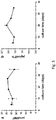
- liver microtissues were monitored over time. It turned out that they remained stable over 5 weeks in culture as shown by a constant ATP content ( Fig. 5a ). This extended life span compared to 2D cultures of hepatocytes is most likely due to extensive cell-cell contacts, which are essential for maintaining the differentiated status of hepatocytes. Besides the stable viability, functionality of liver microtissues is preserved over 5 weeks, as indicated by persistent albumin secretion ( Fig. 5b ).
- Example 3 Use of 3D tissue culture produced according to the invention for hepatoxicity screening
- the prolonged lifetime and functionality of the 3D tissue cultures produced according to the invention allows for long-term studies with repeated dosing to evaluate chronic hepatotoxic effects.
- the hepatotoxic compounds acetaminophen and diclofenac were tested with respect to their long-term toxicological profile.
- Acetaminophen is the major cause of drug-induced liver injury (DILI) in humans.
- DILI drug-induced liver injury
- CYP2E1, CYP1A2 and CYP3A4 The reactive intermediate depletes intracellular glutathione pools leading to hepatocyte cell death. So far, 2D cultures of hepatocytes have not been able to convincingly recapitulate acetaminophen-induced toxicity in vitro (Fey and Wrzesinski 2012).
- Diclofenac is a non-steroidal anti-inflammatory drug that has a strong association with hepatotoxicity. The mechanism is thought to involve phase I enzyme activity (multiple P450-catalyzed oxidations), phase II enzyme activity (glucoronylation) and mechanism-based inhibition. In comparison with 2D cultures of human hepatocytes (calculated IC50 value of 331 ⁇ M), long-term treated liver microtissues displayed an increased sensitivity toward this drug with an IC50 value of 178.6 ⁇ M ( Fig. 6b ).
- trovafloxacin is only hepatotoxic in combination with an inflammatory stimulus, such as lipopolysaccharide (LPS) or TNF-a.
- LPS lipopolysaccharide
- TNF-a a inflammatory stimulus
- the mechanism is thought to involve enhanced cytokine secretion and accumulation in the liver, causing caspase activation and subsequent liver injury.
- Induction of the inflammatory response in liver microtissues by LPS resulted in elevated levels of IL-6 secretion, verifying the responsiveness of incorporated macrophages in the liver microtissues ( Fig. 6c ).
- liver microtissues resemble liver-like cell composition and an extended stability in culture.
- the long-term viability and functionality of liver microtissues allows for routine compound testing as well as chronic and inflammation-mediated toxicity.
- the 96-well format allows for microtissue mass production enabling the implementation of an organotypic liver model at an early time point in drug development.
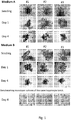
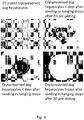
- Histological preparations were made from formalin fixed and paraffin embedded human liver microtissues produced either directly after thawing of the cryopreserved hepatocytes or after pre-plating of the hepatocytes for 24h on a collagen-coated cell culture dish. 3-5um thick sections were stained with Hematoxylin and eosin (H&E). Images were taken with a 10x objective.
- Fig. 7 shows microtissues which have been created without prior pre-plating, while the low reo shows microtissues which have been created with prior pre-plating.
Landscapes
- Health & Medical Sciences (AREA)
- Engineering & Computer Science (AREA)
- Life Sciences & Earth Sciences (AREA)
- Biomedical Technology (AREA)
- Biotechnology (AREA)
- Chemical & Material Sciences (AREA)
- Bioinformatics & Cheminformatics (AREA)
- Organic Chemistry (AREA)
- Wood Science & Technology (AREA)
- Zoology (AREA)
- Genetics & Genomics (AREA)
- Cell Biology (AREA)
- Microbiology (AREA)
- Biochemistry (AREA)
- General Health & Medical Sciences (AREA)
- General Engineering & Computer Science (AREA)
- Immunology (AREA)
- Hematology (AREA)
- Urology & Nephrology (AREA)
- Molecular Biology (AREA)
- Gastroenterology & Hepatology (AREA)
- Toxicology (AREA)
- Tropical Medicine & Parasitology (AREA)
- Food Science & Technology (AREA)
- Medicinal Chemistry (AREA)
- Physics & Mathematics (AREA)
- Analytical Chemistry (AREA)
- General Physics & Mathematics (AREA)
- Pathology (AREA)
- Micro-Organisms Or Cultivation Processes Thereof (AREA)
- Apparatus Associated With Microorganisms And Enzymes (AREA)
- Measuring Or Testing Involving Enzymes Or Micro-Organisms (AREA)
Applications Claiming Priority (2)
| Application Number | Priority Date | Filing Date | Title |
|---|---|---|---|
| GBGB1406716.9A GB201406716D0 (en) | 2014-04-15 | 2014-04-15 | Method of preparing cells for 3D tissue culture |
| PCT/EP2015/058174 WO2015158777A1 (en) | 2014-04-15 | 2015-04-15 | Method of preparing cells for 3d tissue culture |
Publications (2)
| Publication Number | Publication Date |
|---|---|
| EP3132023A1 EP3132023A1 (en) | 2017-02-22 |
| EP3132023B1 true EP3132023B1 (en) | 2021-06-02 |
Family
ID=50844988
Family Applications (1)
| Application Number | Title | Priority Date | Filing Date |
|---|---|---|---|
| EP15723435.2A Active EP3132023B1 (en) | 2014-04-15 | 2015-04-15 | Method of preparing cells for 3d tissue culture |
Country Status (10)
| Country | Link |
|---|---|
| US (2) | US20170037364A1 (enExample) |
| EP (1) | EP3132023B1 (enExample) |
| JP (1) | JP6492106B2 (enExample) |
| CN (1) | CN106459895B (enExample) |
| AU (1) | AU2015248875B9 (enExample) |
| CA (1) | CA2945548C (enExample) |
| DK (1) | DK3132023T3 (enExample) |
| GB (1) | GB201406716D0 (enExample) |
| SG (1) | SG11201608463WA (enExample) |
| WO (1) | WO2015158777A1 (enExample) |
Families Citing this family (10)
| Publication number | Priority date | Publication date | Assignee | Title |
|---|---|---|---|---|
| JP6758026B2 (ja) * | 2015-04-17 | 2020-09-23 | 株式会社日立ハイテク | 成分分析装置、薬剤成分分析装置、成分分析方法及び薬剤成分分析方法 |
| WO2017117333A1 (en) * | 2015-12-30 | 2017-07-06 | Cellular Dynamics International, Inc. | Microtissue formation using stem cell-derived human hepatocytes |
| GB201609663D0 (en) | 2016-06-02 | 2016-07-20 | Stemtek Therapeutics Sl | Methods for producing cancer stem cell spheroids |
| CA3055666A1 (en) * | 2017-03-10 | 2018-09-13 | Merck Patent Gmbh | Infected cell cultures |
| CN109294971A (zh) * | 2018-09-29 | 2019-02-01 | 西安交通大学 | 一种用于药物筛选的颈动脉分叉三维细胞培养模型及其构建方法 |
| DE102019216886A1 (de) | 2019-10-31 | 2021-05-06 | Hahn-Schickard-Gesellschaft für angewandte Forschung e.V. | Transfer zumindest eines partikels in einer flüssigkeit von einer abgabeeinheit zu einer empfangseinheit |
| CN111471641B (zh) * | 2020-02-03 | 2021-11-05 | 东华大学 | 多片层单元水凝胶包被的仿生毛细血管网的3d打印制法 |
| CN111150885A (zh) * | 2020-03-05 | 2020-05-15 | 陕西佰傲干细胞再生医学有限公司 | 软骨微囊及其制备方法和应用 |
| US12076437B2 (en) | 2021-07-12 | 2024-09-03 | Brown University | Proangiogenic protein cocktails delivered in custom biomaterials to revascularize ischemic tissue |
| WO2025073379A1 (en) | 2023-10-06 | 2025-04-10 | Insphero Ag | Device for studying interactions of biological specimen |
Citations (1)
| Publication number | Priority date | Publication date | Assignee | Title |
|---|---|---|---|---|
| US20120276068A1 (en) * | 2009-10-16 | 2012-11-01 | University Of Medicine And Dentistry Of New Jersey | Method for treating chronic nerve tissue injury using a cell therapy strategy |
Family Cites Families (15)
| Publication number | Priority date | Publication date | Assignee | Title |
|---|---|---|---|---|
| FR2545100B1 (fr) * | 1983-04-29 | 1985-12-20 | Inst Nat Sante Rech Med | Procede d'obtention de cultures d'hepatocytes humains, les cultures obtenues et leurs applications biologiques et biochimiques |
| WO2003055487A1 (en) * | 2002-01-04 | 2003-07-10 | The University Of Adelaide | METHOD OF CONTROLLING DAMAGE MEDIATED BY α, β- UNSATURATED ALDEHYDES |
| WO2003084468A2 (en) * | 2002-04-03 | 2003-10-16 | Artecel Sciences, Inc. | Improvements of adipocytic differentiated adipose derived adult stem cells and uses thereof |
| JP2004254674A (ja) * | 2003-02-25 | 2004-09-16 | Mutsumi Takagi | 動物細胞の分化誘導培養方法 |
| EP2348105B1 (en) * | 2004-09-24 | 2018-10-24 | Mesoblast, Inc. | Multipotential expanded mesenchymal precursor cell progeny (MEMP) and uses thereof |
| US20090208466A1 (en) * | 2006-04-21 | 2009-08-20 | James Yoo | Ink-jet printing of tissues |
| NZ595854A (en) * | 2006-10-23 | 2013-04-26 | Anthrogenesis Corp | Methods and compositions for treatment of bone defects with placental cell populations (ELOVL2, ST3GAL6, STGALNAC5, SLC12A8) |
| MX2009008559A (es) * | 2007-02-12 | 2009-08-21 | Anthrogenesis Corp | Hepatocitos y condorcitos de celulas madre de la placenta adherentes, y poblaciones de celulas enriquecidas con celulas madre de la placenta cd34+, cd45-. |
| ES2749500T3 (es) * | 2008-11-19 | 2020-03-20 | Celularity Inc | Células adherentes derivadas del amnios |
| WO2010060080A1 (en) * | 2008-11-24 | 2010-05-27 | Immunotrex Corporation | Three dimensional tissue generation |
| ES2553712T3 (es) * | 2009-11-30 | 2015-12-11 | Pluristem Ltd. | Células adherentes de la placenta y uso de las mismas en el tratamiento de enfermedad |
| US20110229970A1 (en) * | 2010-03-05 | 2011-09-22 | Florida State University Research Foundation | Dual-chamber perfusion bioreactor for orthopedic tissue interfaces and methods of use |
| US9127255B2 (en) * | 2010-06-11 | 2015-09-08 | Takara Bio Europe Ab | 3-dimensional scaffolds for improved differentiation of pluripotent stem cells to hepatocytes |
| US20120201787A1 (en) * | 2010-12-17 | 2012-08-09 | Abbot Stewart | Treatment of spinal cord injury and traumatic brain injury using amnion derived adherent cells |
| CA2848043C (en) * | 2011-09-12 | 2022-07-12 | Organovo, Inc. | Engineered tissues for in vitro research uses, arrays thereof, and methods of making the same |
-
2014
- 2014-04-15 GB GBGB1406716.9A patent/GB201406716D0/en not_active Ceased
-
2015
- 2015-04-15 CN CN201580020057.0A patent/CN106459895B/zh active Active
- 2015-04-15 CA CA2945548A patent/CA2945548C/en active Active
- 2015-04-15 AU AU2015248875A patent/AU2015248875B9/en not_active Ceased
- 2015-04-15 JP JP2016562830A patent/JP6492106B2/ja active Active
- 2015-04-15 EP EP15723435.2A patent/EP3132023B1/en active Active
- 2015-04-15 WO PCT/EP2015/058174 patent/WO2015158777A1/en not_active Ceased
- 2015-04-15 DK DK15723435.2T patent/DK3132023T3/da active
- 2015-04-15 SG SG11201608463WA patent/SG11201608463WA/en unknown
- 2015-04-15 US US15/303,455 patent/US20170037364A1/en not_active Abandoned
-
2023
- 2023-01-26 US US18/101,751 patent/US20230235277A1/en not_active Abandoned
Patent Citations (1)
| Publication number | Priority date | Publication date | Assignee | Title |
|---|---|---|---|---|
| US20120276068A1 (en) * | 2009-10-16 | 2012-11-01 | University Of Medicine And Dentistry Of New Jersey | Method for treating chronic nerve tissue injury using a cell therapy strategy |
Also Published As
| Publication number | Publication date |
|---|---|
| JP6492106B2 (ja) | 2019-03-27 |
| GB201406716D0 (en) | 2014-05-28 |
| US20170037364A1 (en) | 2017-02-09 |
| WO2015158777A1 (en) | 2015-10-22 |
| CN106459895B (zh) | 2021-06-01 |
| US20230235277A1 (en) | 2023-07-27 |
| AU2015248875A1 (en) | 2016-10-27 |
| CA2945548A1 (en) | 2015-10-22 |
| EP3132023A1 (en) | 2017-02-22 |
| DK3132023T3 (da) | 2021-08-09 |
| AU2015248875B2 (en) | 2019-04-04 |
| AU2015248875B9 (en) | 2019-05-23 |
| CN106459895A (zh) | 2017-02-22 |
| JP2017511146A (ja) | 2017-04-20 |
| SG11201608463WA (en) | 2016-11-29 |
| CA2945548C (en) | 2019-11-12 |
Similar Documents
| Publication | Publication Date | Title |
|---|---|---|
| US20230235277A1 (en) | 3D Tissue Culture Materials and Processes for Producing Same | |
| Mohr et al. | Accelerating cardiovascular research: recent advances in translational 2D and 3D heart models | |
| CA2968655C (en) | Methods for generation of podocytes from pluripotent stem cells and cells produced by the same | |
| JP2024150583A (ja) | 細胞ミクロコンパートメントおよびその調製方法 | |
| US20180305669A1 (en) | Microtissue formation using stem cell-derived human hepatocytes | |
| Heidari Khoei et al. | Generating human blastoids modeling blastocyst-stage embryos and implantation | |
| EP3864137A1 (en) | Compositions and methods for cell culture | |
| WO2022016165A2 (en) | Differentiation of trophectoderm lineage cells from pluripotent stem cells | |
| US11208629B2 (en) | Non-human primate induced pluripotent stem cell derived hepatocytes and uses thereof | |
| US20070148767A1 (en) | Method of forming multicellular spheroids from the cultured cells | |
| WO2011016485A1 (ja) | iPS細胞から肝実質細胞への分化誘導方法 | |
| Fuegemann et al. | Differentiation of mouse embryonic stem cells into cardiomyocytes via the hanging‐drop and mass culture methods | |
| Tao et al. | Enhancement and maintenance of hepatic metabolic functions by controlling 3D aggregation of cryopreserved human iPS cell-derived hepatocyte-like cells | |
| US20230392123A1 (en) | Spheroidal self-assembled peptide hydrogels comprising cells | |
| JP6486619B2 (ja) | 薬物評価用細胞及び薬物評価方法 | |
| TW201734206A (zh) | 高機能肝細胞及其利用 | |
| Raggi et al. | Generation of complex syngeneic liver organoids from induced pluripotent stem cells to model human liver pathophysiology | |
| Källén et al. | 3D Culture in Functionalized FN‐Silk Networks Facilitate Proliferation, Differentiation and Phenotypic Stability of Mature Human Primary Cells and Stem Cells | |
| Gu et al. | A uterus-inspired 3D niche drives embryo development beyond implantation | |
| Tokarchuk et al. | Recent developments in the production of 2D-and 3D colon and stomach adenocarcinomas primary cell models | |
| TWI303278B (en) | A method of forming spheroids from the cultured cells |
Legal Events
| Date | Code | Title | Description |
|---|---|---|---|
| STAA | Information on the status of an ep patent application or granted ep patent |
Free format text: STATUS: THE INTERNATIONAL PUBLICATION HAS BEEN MADE |
|
| PUAI | Public reference made under article 153(3) epc to a published international application that has entered the european phase |
Free format text: ORIGINAL CODE: 0009012 |
|
| STAA | Information on the status of an ep patent application or granted ep patent |
Free format text: STATUS: REQUEST FOR EXAMINATION WAS MADE |
|
| 17P | Request for examination filed |
Effective date: 20161109 |
|
| AK | Designated contracting states |
Kind code of ref document: A1 Designated state(s): AL AT BE BG CH CY CZ DE DK EE ES FI FR GB GR HR HU IE IS IT LI LT LU LV MC MK MT NL NO PL PT RO RS SE SI SK SM TR |
|
| AX | Request for extension of the european patent |
Extension state: BA ME |
|
| DAV | Request for validation of the european patent (deleted) | ||
| DAX | Request for extension of the european patent (deleted) | ||
| STAA | Information on the status of an ep patent application or granted ep patent |
Free format text: STATUS: EXAMINATION IS IN PROGRESS |
|
| 17Q | First examination report despatched |
Effective date: 20180525 |
|
| GRAP | Despatch of communication of intention to grant a patent |
Free format text: ORIGINAL CODE: EPIDOSNIGR1 |
|
| STAA | Information on the status of an ep patent application or granted ep patent |
Free format text: STATUS: GRANT OF PATENT IS INTENDED |
|
| INTG | Intention to grant announced |
Effective date: 20201111 |
|
| GRAS | Grant fee paid |
Free format text: ORIGINAL CODE: EPIDOSNIGR3 |
|
| GRAA | (expected) grant |
Free format text: ORIGINAL CODE: 0009210 |
|
| STAA | Information on the status of an ep patent application or granted ep patent |
Free format text: STATUS: THE PATENT HAS BEEN GRANTED |
|
| REG | Reference to a national code |
Ref country code: CH Ref legal event code: EP |
|
| AK | Designated contracting states |
Kind code of ref document: B1 Designated state(s): AL AT BE BG CH CY CZ DE DK EE ES FI FR GB GR HR HU IE IS IT LI LT LU LV MC MK MT NL NO PL PT RO RS SE SI SK SM TR |
|
| REG | Reference to a national code |
Ref country code: GB Ref legal event code: FG4D |
|
| REG | Reference to a national code |
Ref country code: AT Ref legal event code: REF Ref document number: 1398444 Country of ref document: AT Kind code of ref document: T Effective date: 20210615 |
|
| REG | Reference to a national code |
Ref country code: IE Ref legal event code: FG4D |
|
| REG | Reference to a national code |
Ref country code: DE Ref legal event code: R096 Ref document number: 602015069975 Country of ref document: DE |
|
| RAP4 | Party data changed (patent owner data changed or rights of a patent transferred) |
Owner name: INSPHERO AG |
|
| REG | Reference to a national code |
Ref country code: DK Ref legal event code: T3 Effective date: 20210802 |
|
| REG | Reference to a national code |
Ref country code: SE Ref legal event code: TRGR |
|
| REG | Reference to a national code |
Ref country code: NL Ref legal event code: FP |
|
| REG | Reference to a national code |
Ref country code: LT Ref legal event code: MG9D |
|
| PG25 | Lapsed in a contracting state [announced via postgrant information from national office to epo] |
Ref country code: FI Free format text: LAPSE BECAUSE OF FAILURE TO SUBMIT A TRANSLATION OF THE DESCRIPTION OR TO PAY THE FEE WITHIN THE PRESCRIBED TIME-LIMIT Effective date: 20210602 Ref country code: LT Free format text: LAPSE BECAUSE OF FAILURE TO SUBMIT A TRANSLATION OF THE DESCRIPTION OR TO PAY THE FEE WITHIN THE PRESCRIBED TIME-LIMIT Effective date: 20210602 Ref country code: BG Free format text: LAPSE BECAUSE OF FAILURE TO SUBMIT A TRANSLATION OF THE DESCRIPTION OR TO PAY THE FEE WITHIN THE PRESCRIBED TIME-LIMIT Effective date: 20210902 Ref country code: HR Free format text: LAPSE BECAUSE OF FAILURE TO SUBMIT A TRANSLATION OF THE DESCRIPTION OR TO PAY THE FEE WITHIN THE PRESCRIBED TIME-LIMIT Effective date: 20210602 |
|
| REG | Reference to a national code |
Ref country code: AT Ref legal event code: MK05 Ref document number: 1398444 Country of ref document: AT Kind code of ref document: T Effective date: 20210602 |
|
| PG25 | Lapsed in a contracting state [announced via postgrant information from national office to epo] |
Ref country code: PL Free format text: LAPSE BECAUSE OF FAILURE TO SUBMIT A TRANSLATION OF THE DESCRIPTION OR TO PAY THE FEE WITHIN THE PRESCRIBED TIME-LIMIT Effective date: 20210602 Ref country code: NO Free format text: LAPSE BECAUSE OF FAILURE TO SUBMIT A TRANSLATION OF THE DESCRIPTION OR TO PAY THE FEE WITHIN THE PRESCRIBED TIME-LIMIT Effective date: 20210902 Ref country code: RS Free format text: LAPSE BECAUSE OF FAILURE TO SUBMIT A TRANSLATION OF THE DESCRIPTION OR TO PAY THE FEE WITHIN THE PRESCRIBED TIME-LIMIT Effective date: 20210602 Ref country code: GR Free format text: LAPSE BECAUSE OF FAILURE TO SUBMIT A TRANSLATION OF THE DESCRIPTION OR TO PAY THE FEE WITHIN THE PRESCRIBED TIME-LIMIT Effective date: 20210903 Ref country code: LV Free format text: LAPSE BECAUSE OF FAILURE TO SUBMIT A TRANSLATION OF THE DESCRIPTION OR TO PAY THE FEE WITHIN THE PRESCRIBED TIME-LIMIT Effective date: 20210602 |
|
| PG25 | Lapsed in a contracting state [announced via postgrant information from national office to epo] |
Ref country code: EE Free format text: LAPSE BECAUSE OF FAILURE TO SUBMIT A TRANSLATION OF THE DESCRIPTION OR TO PAY THE FEE WITHIN THE PRESCRIBED TIME-LIMIT Effective date: 20210602 Ref country code: CZ Free format text: LAPSE BECAUSE OF FAILURE TO SUBMIT A TRANSLATION OF THE DESCRIPTION OR TO PAY THE FEE WITHIN THE PRESCRIBED TIME-LIMIT Effective date: 20210602 Ref country code: SK Free format text: LAPSE BECAUSE OF FAILURE TO SUBMIT A TRANSLATION OF THE DESCRIPTION OR TO PAY THE FEE WITHIN THE PRESCRIBED TIME-LIMIT Effective date: 20210602 Ref country code: SM Free format text: LAPSE BECAUSE OF FAILURE TO SUBMIT A TRANSLATION OF THE DESCRIPTION OR TO PAY THE FEE WITHIN THE PRESCRIBED TIME-LIMIT Effective date: 20210602 Ref country code: AT Free format text: LAPSE BECAUSE OF FAILURE TO SUBMIT A TRANSLATION OF THE DESCRIPTION OR TO PAY THE FEE WITHIN THE PRESCRIBED TIME-LIMIT Effective date: 20210602 Ref country code: ES Free format text: LAPSE BECAUSE OF FAILURE TO SUBMIT A TRANSLATION OF THE DESCRIPTION OR TO PAY THE FEE WITHIN THE PRESCRIBED TIME-LIMIT Effective date: 20210602 Ref country code: RO Free format text: LAPSE BECAUSE OF FAILURE TO SUBMIT A TRANSLATION OF THE DESCRIPTION OR TO PAY THE FEE WITHIN THE PRESCRIBED TIME-LIMIT Effective date: 20210602 Ref country code: PT Free format text: LAPSE BECAUSE OF FAILURE TO SUBMIT A TRANSLATION OF THE DESCRIPTION OR TO PAY THE FEE WITHIN THE PRESCRIBED TIME-LIMIT Effective date: 20211004 |
|
| REG | Reference to a national code |
Ref country code: DE Ref legal event code: R097 Ref document number: 602015069975 Country of ref document: DE |
|
| PLBE | No opposition filed within time limit |
Free format text: ORIGINAL CODE: 0009261 |
|
| STAA | Information on the status of an ep patent application or granted ep patent |
Free format text: STATUS: NO OPPOSITION FILED WITHIN TIME LIMIT |
|
| 26N | No opposition filed |
Effective date: 20220303 |
|
| PG25 | Lapsed in a contracting state [announced via postgrant information from national office to epo] |
Ref country code: AL Free format text: LAPSE BECAUSE OF FAILURE TO SUBMIT A TRANSLATION OF THE DESCRIPTION OR TO PAY THE FEE WITHIN THE PRESCRIBED TIME-LIMIT Effective date: 20210602 |
|
| PG25 | Lapsed in a contracting state [announced via postgrant information from national office to epo] |
Ref country code: IT Free format text: LAPSE BECAUSE OF FAILURE TO SUBMIT A TRANSLATION OF THE DESCRIPTION OR TO PAY THE FEE WITHIN THE PRESCRIBED TIME-LIMIT Effective date: 20210602 |
|
| PG25 | Lapsed in a contracting state [announced via postgrant information from national office to epo] |
Ref country code: MC Free format text: LAPSE BECAUSE OF FAILURE TO SUBMIT A TRANSLATION OF THE DESCRIPTION OR TO PAY THE FEE WITHIN THE PRESCRIBED TIME-LIMIT Effective date: 20210602 Ref country code: LU Free format text: LAPSE BECAUSE OF NON-PAYMENT OF DUE FEES Effective date: 20220415 |
|
| PG25 | Lapsed in a contracting state [announced via postgrant information from national office to epo] |
Ref country code: IE Free format text: LAPSE BECAUSE OF NON-PAYMENT OF DUE FEES Effective date: 20220415 |
|
| P01 | Opt-out of the competence of the unified patent court (upc) registered |
Effective date: 20230526 |
|
| PG25 | Lapsed in a contracting state [announced via postgrant information from national office to epo] |
Ref country code: HU Free format text: LAPSE BECAUSE OF FAILURE TO SUBMIT A TRANSLATION OF THE DESCRIPTION OR TO PAY THE FEE WITHIN THE PRESCRIBED TIME-LIMIT; INVALID AB INITIO Effective date: 20150415 |
|
| PG25 | Lapsed in a contracting state [announced via postgrant information from national office to epo] |
Ref country code: MK Free format text: LAPSE BECAUSE OF FAILURE TO SUBMIT A TRANSLATION OF THE DESCRIPTION OR TO PAY THE FEE WITHIN THE PRESCRIBED TIME-LIMIT Effective date: 20210602 Ref country code: CY Free format text: LAPSE BECAUSE OF FAILURE TO SUBMIT A TRANSLATION OF THE DESCRIPTION OR TO PAY THE FEE WITHIN THE PRESCRIBED TIME-LIMIT Effective date: 20210602 |
|
| PG25 | Lapsed in a contracting state [announced via postgrant information from national office to epo] |
Ref country code: MT Free format text: LAPSE BECAUSE OF FAILURE TO SUBMIT A TRANSLATION OF THE DESCRIPTION OR TO PAY THE FEE WITHIN THE PRESCRIBED TIME-LIMIT Effective date: 20210602 |
|
| PGFP | Annual fee paid to national office [announced via postgrant information from national office to epo] |
Ref country code: NL Payment date: 20250418 Year of fee payment: 11 |
|
| PGFP | Annual fee paid to national office [announced via postgrant information from national office to epo] |
Ref country code: DE Payment date: 20250429 Year of fee payment: 11 |
|
| PGFP | Annual fee paid to national office [announced via postgrant information from national office to epo] |
Ref country code: GB Payment date: 20250423 Year of fee payment: 11 Ref country code: DK Payment date: 20250429 Year of fee payment: 11 |
|
| PGFP | Annual fee paid to national office [announced via postgrant information from national office to epo] |
Ref country code: BE Payment date: 20250418 Year of fee payment: 11 |
|
| PGFP | Annual fee paid to national office [announced via postgrant information from national office to epo] |
Ref country code: FR Payment date: 20250425 Year of fee payment: 11 |
|
| PGFP | Annual fee paid to national office [announced via postgrant information from national office to epo] |
Ref country code: CH Payment date: 20250501 Year of fee payment: 11 |
|
| PGFP | Annual fee paid to national office [announced via postgrant information from national office to epo] |
Ref country code: SE Payment date: 20250429 Year of fee payment: 11 |
|
| PG25 | Lapsed in a contracting state [announced via postgrant information from national office to epo] |
Ref country code: TR Free format text: LAPSE BECAUSE OF FAILURE TO SUBMIT A TRANSLATION OF THE DESCRIPTION OR TO PAY THE FEE WITHIN THE PRESCRIBED TIME-LIMIT Effective date: 20210602 |