EP3124600A1 - Verfahren zur herstellung eines zellaggregats zur selbstorganisation - Google Patents
Verfahren zur herstellung eines zellaggregats zur selbstorganisation Download PDFInfo
- Publication number
- EP3124600A1 EP3124600A1 EP15754683.9A EP15754683A EP3124600A1 EP 3124600 A1 EP3124600 A1 EP 3124600A1 EP 15754683 A EP15754683 A EP 15754683A EP 3124600 A1 EP3124600 A1 EP 3124600A1
- Authority
- EP
- European Patent Office
- Prior art keywords
- cells
- cell
- stiffness
- tissues
- gel
- Prior art date
- Legal status (The legal status is an assumption and is not a legal conclusion. Google has not performed a legal analysis and makes no representation as to the accuracy of the status listed.)
- Granted
Links
Images


Classifications
-
- C—CHEMISTRY; METALLURGY
- C12—BIOCHEMISTRY; BEER; SPIRITS; WINE; VINEGAR; MICROBIOLOGY; ENZYMOLOGY; MUTATION OR GENETIC ENGINEERING
- C12N—MICROORGANISMS OR ENZYMES; COMPOSITIONS THEREOF; PROPAGATING, PRESERVING, OR MAINTAINING MICROORGANISMS; MUTATION OR GENETIC ENGINEERING; CULTURE MEDIA
- C12N5/00—Undifferentiated human, animal or plant cells, e.g. cell lines; Tissues; Cultivation or maintenance thereof; Culture media therefor
- C12N5/0062—General methods for three-dimensional culture
-
- A—HUMAN NECESSITIES
- A61—MEDICAL OR VETERINARY SCIENCE; HYGIENE
- A61L—METHODS OR APPARATUS FOR STERILISING MATERIALS OR OBJECTS IN GENERAL; DISINFECTION, STERILISATION OR DEODORISATION OF AIR; CHEMICAL ASPECTS OF BANDAGES, DRESSINGS, ABSORBENT PADS OR SURGICAL ARTICLES; MATERIALS FOR BANDAGES, DRESSINGS, ABSORBENT PADS OR SURGICAL ARTICLES
- A61L27/00—Materials for grafts or prostheses or for coating grafts or prostheses
- A61L27/36—Materials for grafts or prostheses or for coating grafts or prostheses containing ingredients of undetermined constitution or reaction products thereof, e.g. transplant tissue, natural bone, extracellular matrix
- A61L27/38—Materials for grafts or prostheses or for coating grafts or prostheses containing ingredients of undetermined constitution or reaction products thereof, e.g. transplant tissue, natural bone, extracellular matrix containing added animal cells
- A61L27/3886—Materials for grafts or prostheses or for coating grafts or prostheses containing ingredients of undetermined constitution or reaction products thereof, e.g. transplant tissue, natural bone, extracellular matrix containing added animal cells comprising two or more cell types
-
- A—HUMAN NECESSITIES
- A61—MEDICAL OR VETERINARY SCIENCE; HYGIENE
- A61L—METHODS OR APPARATUS FOR STERILISING MATERIALS OR OBJECTS IN GENERAL; DISINFECTION, STERILISATION OR DEODORISATION OF AIR; CHEMICAL ASPECTS OF BANDAGES, DRESSINGS, ABSORBENT PADS OR SURGICAL ARTICLES; MATERIALS FOR BANDAGES, DRESSINGS, ABSORBENT PADS OR SURGICAL ARTICLES
- A61L27/00—Materials for grafts or prostheses or for coating grafts or prostheses
- A61L27/36—Materials for grafts or prostheses or for coating grafts or prostheses containing ingredients of undetermined constitution or reaction products thereof, e.g. transplant tissue, natural bone, extracellular matrix
- A61L27/38—Materials for grafts or prostheses or for coating grafts or prostheses containing ingredients of undetermined constitution or reaction products thereof, e.g. transplant tissue, natural bone, extracellular matrix containing added animal cells
- A61L27/3895—Materials for grafts or prostheses or for coating grafts or prostheses containing ingredients of undetermined constitution or reaction products thereof, e.g. transplant tissue, natural bone, extracellular matrix containing added animal cells using specific culture conditions, e.g. stimulating differentiation of stem cells, pulsatile flow conditions
-
- A—HUMAN NECESSITIES
- A61—MEDICAL OR VETERINARY SCIENCE; HYGIENE
- A61L—METHODS OR APPARATUS FOR STERILISING MATERIALS OR OBJECTS IN GENERAL; DISINFECTION, STERILISATION OR DEODORISATION OF AIR; CHEMICAL ASPECTS OF BANDAGES, DRESSINGS, ABSORBENT PADS OR SURGICAL ARTICLES; MATERIALS FOR BANDAGES, DRESSINGS, ABSORBENT PADS OR SURGICAL ARTICLES
- A61L27/00—Materials for grafts or prostheses or for coating grafts or prostheses
- A61L27/50—Materials characterised by their function or physical properties, e.g. injectable or lubricating compositions, shape-memory materials, surface modified materials
- A61L27/52—Hydrogels or hydrocolloids
-
- C—CHEMISTRY; METALLURGY
- C12—BIOCHEMISTRY; BEER; SPIRITS; WINE; VINEGAR; MICROBIOLOGY; ENZYMOLOGY; MUTATION OR GENETIC ENGINEERING
- C12N—MICROORGANISMS OR ENZYMES; COMPOSITIONS THEREOF; PROPAGATING, PRESERVING, OR MAINTAINING MICROORGANISMS; MUTATION OR GENETIC ENGINEERING; CULTURE MEDIA
- C12N5/00—Undifferentiated human, animal or plant cells, e.g. cell lines; Tissues; Cultivation or maintenance thereof; Culture media therefor
- C12N5/06—Animal cells or tissues; Human cells or tissues
- C12N5/0697—Artificial constructs associating cells of different lineages, e.g. tissue equivalents
-
- A—HUMAN NECESSITIES
- A61—MEDICAL OR VETERINARY SCIENCE; HYGIENE
- A61L—METHODS OR APPARATUS FOR STERILISING MATERIALS OR OBJECTS IN GENERAL; DISINFECTION, STERILISATION OR DEODORISATION OF AIR; CHEMICAL ASPECTS OF BANDAGES, DRESSINGS, ABSORBENT PADS OR SURGICAL ARTICLES; MATERIALS FOR BANDAGES, DRESSINGS, ABSORBENT PADS OR SURGICAL ARTICLES
- A61L2430/00—Materials or treatment for tissue regeneration
- A61L2430/26—Materials or treatment for tissue regeneration for kidney reconstruction
-
- A—HUMAN NECESSITIES
- A61—MEDICAL OR VETERINARY SCIENCE; HYGIENE
- A61L—METHODS OR APPARATUS FOR STERILISING MATERIALS OR OBJECTS IN GENERAL; DISINFECTION, STERILISATION OR DEODORISATION OF AIR; CHEMICAL ASPECTS OF BANDAGES, DRESSINGS, ABSORBENT PADS OR SURGICAL ARTICLES; MATERIALS FOR BANDAGES, DRESSINGS, ABSORBENT PADS OR SURGICAL ARTICLES
- A61L2430/00—Materials or treatment for tissue regeneration
- A61L2430/28—Materials or treatment for tissue regeneration for liver reconstruction
-
- C—CHEMISTRY; METALLURGY
- C12—BIOCHEMISTRY; BEER; SPIRITS; WINE; VINEGAR; MICROBIOLOGY; ENZYMOLOGY; MUTATION OR GENETIC ENGINEERING
- C12N—MICROORGANISMS OR ENZYMES; COMPOSITIONS THEREOF; PROPAGATING, PRESERVING, OR MAINTAINING MICROORGANISMS; MUTATION OR GENETIC ENGINEERING; CULTURE MEDIA
- C12N2535/00—Supports or coatings for cell culture characterised by topography
Definitions
- the present invention relates to a method of preparing a cell condensate for self-organization. More specifically, the present invention relates to a method of preparing a cell condensate that is necessary for directing self-organization into a tissue or an organ of interest.
- Non-Patent Documents Nos. 1 and 2 Self-organization is a process in which one or a few elements construct complex higher structures by exerting intrinsic properties of their own without receiving specific "instructions" (information) from the outside. For example, natural phenomena in which spontaneous order arises from patternless aggregates to form patterns, as in crystallization of snow, are observed. Self-organization is also used in the field of engineering, e.g. in nanotechnology or in preparing optical crystals.
- the present inventors have already established a technological three-dimensional culture technique using spatiotemporal interactions of three different cell lineages; this technique has realized "directed differentiation of organ cells based on reconstitution of organs". Briefly, the present inventors have established a platform technology which recapitulates interactions among organ cells, vascular cells and mesenchymal cells that are essential for early processes of organogenesis, to thereby induce 3D organ primordia (starting material for organs) and enable generation of vascularized functional organs ( Nature, 499 (7459), 481-484 ; PCT/JP2012/074840 Method for Preparing Tissue and Organ).
- tissue constructs which enable reconstitution of continuity with diverse higher structures and other organs. According to conventionally devised methods, however, only tissue constructs having a vascular structure alone have been prepared from the three types of cells or tissues. No technique has been invented for preparing more complex, higher structures (such as ureteral structure, biliary structure and tracheal structure).
- the present inventors have succeeded in preparing three-dimensional tissues/organs having complex higher structures from isolated, multiple types of cells or tissues by the operations 1 to 4 described below. Thus, the present invention has been achieved.
- condensates can also be prepared from the cells if they are small in number.
- tissues/organs can be prepared that have a highly ordered tissue structure comparable to that of adult tissues.
- the gist of the present invention is as described below.
- a cell condensate of theoretically any complex composition can be formed by combining a mesenchymal stem cell and a support or a substrate that will allow cells to gather in the bottom.
- tissues and organs can be constructed without using scaffolds.
- the cell condensate of the present invention is expected to find use as an artificial constitution system for more complex tissues and organs.
- the cell condensate of the present invention it may be possible to prepare three-dimensional complex structures that are provided with not only a vascular network but also higher structures such as ureteral structure, biliary structure, tracheal structure, etc.
- a great number of organs essentially require that reconstitution associated with other organs be realized in order to exhibit their functions; e.g., in liver, reconstitution of junctions with bile duct and pancreatic duct and connection to duodenum is essential for exhibiting its function.
- a cell condensate which recapitulates interactions with other organs is prepared. This cell condensate is expected to find use as a system for inducing self-organization into complex organs existing in the body.
- the present invention uses an inexpensive and comparatively easy-to-process support, its industrial applicability toward mass production of tissues is high. Mass production of tissues of a desired shape, size and number can be realized at low cost by combining the cell condensate with the multi-patterning of the support or other techniques.
- the technique of generating a 3D tissue construct self-organized from a cell condensate prepared from stem cells such as iPS cells is applicable to generation of human functional cells which has been difficult to achieve to date; transplantation of tissues and organs; screening in drug discovery; a novel analysis system for evaluating the relationships between development of drug effects and supporting tissues (blood vessels, nerves, stroma, etc.) and so on.
- the present invention provides a method of preparing a cell condensate in vitro, comprising culturing a mixture of cells and/or tissues of a desired type and mesenchymal cells to form a cell condensate.
- Mesenchymal cells are connective tissue cells that are mainly located in mesoderm-derived connective tissues and which form support structures for cells that function in tissues.
- the term "mesenchymal cell” is a concept that encompasses those cells which are destined to, but are yet to, differentiate into mesenchymal cells.
- Mesenchymal cells to be used in the present invention may be either differentiated or undifferentiated. Preferably, undifferentiated mesenchymal cells are used.
- Whether a cell is an undifferentiated mesenchymal cell or not can be determined by checking to see if the cell expresses marker proteins such as Stro-1, CD29, CD44, CD73, CD90, CD105, CD133, CD271 or Nestin (if any one or more of the above-listed marker proteins are expressed, the cell can safely be regarded as an undifferentiated mesenchymal cell).
- marker proteins such as Stro-1, CD29, CD44, CD73, CD90, CD105, CD133, CD271 or Nestin (if any one or more of the above-listed marker proteins are expressed, the cell can safely be regarded as an undifferentiated mesenchymal cell).
- a mesenchymal cell in which none of the above-listed markers is expressed can be regarded as a differentiated mesenchymal cell.
- mesenchymal stem cells mesenchymal progenitor cells
- mesenchymal cells R. Peters et al. PLoS One. 30; 5(12):e15689 (2010 )
- mesenchymal cells R. Peters et al. PLoS One. 30; 5(12):e15689 (2010 )
- mesenchymal cells human-derived ones are mainly used.
- mesenchymal cells derived from non-human animals may also be used.
- animals used for example, as experimental animals, pet animals, working animals, race horses or fighting dogs; more specifically, mouse, rat, rabbit, pig, dog, monkey, cattle, horse, sheep, chicken, shark, devilfish, ratfish, salmon, shrimp, crab or the like
- mouse, rat, rabbit, pig, dog, monkey, cattle, horse, sheep, chicken, shark, devilfish, ratfish, salmon, shrimp, crab or the like may also be used.
- the cells and/or tissues of a desired type to be mixed with mesenchymal cells are independent of the types or numbers to be combined and may be any cells and/or tissues. Moreover, the origin of such cells and/or tissues also does not matter and they may be derived from any organ (e.g. liver, pancreas, intestine, lung, kidney, heart and brain) or any tissue; alternatively, they may be derived from cancer.
- Cells to be mixed with mesenchymal cells may be functional cells which constitute organs or tissues, or undifferentiated or pluripotent cells which will differentiate into functional cells.
- tissues to be mixed with mesenchymal cells may be tissues isolated from individuals, or tissues induced from functional cells which constitute organs or tissues, or tissues induced from undifferentiated or pluripotent cells which will differentiate into functional cells.
- Undifferentiated cells may be cells capable of differentiating into an organ such as kidney, heart, lung, spleen, esophagus, stomach, thyroid, parathyroid, thymus, gonad, brain or spinal cord; cells capable of differentiating into an ectodermal organ such as brain, spinal cord, adrenal medulla, epidermis, hair/nail/dermal gland, sensory organ, peripheral nerve or lens; cells capable of differentiating into a mesodermal organ such as kidney, urinary duct, heart, blood, gonad, adrenal cortex, muscle, skeleton, dermis, connective tissue or mesothelium; and cells capable of differentiating into an endodermal organ such as liver, pancreas, intestine, lung, thyroid, parathyroid or urinary tract.
- an organ such as kidney, heart, lung, spleen, esophagus, stomach, thyroid, parathyroid, thymus, gonad, brain or spinal cord
- Whether or not a cell is capable of differentiating into an ectodermal organ, mesodermal organ or endodermal organ can be determined by checking for the expression of marker proteins (if any one or a plurality of marker proteins are expressed, the cell can be regarded as a cell capable of differentiating into an endodermal organ).
- cells capable of differentiating into liver have such markers as HHEX, SOX2, HNF4A, AFP and ALB; cells capable of differentiating into pancreas have such markers as PDX1, SOX17 and SOX9; cells capable of differentiating into intestine have such markers as CDX2 and SOX9; cells capable of differentiating into kidney have such markers as SIX2 and SALL1; cells capable of differentiating into heart have such markers as NKX2-5, MYH6, ACTN2, MYL7 and HPPA; cells capable of differentiating into blood have such markers as C-KIT, SCA1, TER119 and HOXB4; and cells capable of differentiating into brain or spinal cord have such markers as HNK1, AP2 and NESTIN.
- hepatoblast hepatic progenitor cells
- pancreatoblast hepatic precursor cells
- pancreatic progenitors pancreatic progenitor cells
- pancreatic precursor cells endocrine precursors
- intestinal progenitor cells intestinal precursor cells
- intermediate mesoderm intermediate mesoderm
- metanephric mesenchymal precursor cells multipotent nephron progenitor
- renal progenitor cells cardiac mesoderm
- cardiovascular progenitor cells cardiac progenitor cells
- cardiac progenitor cells JR. Spence et al.
- pluripotent cells include pluripotent cells obtained from living bodies (e.g., ES cells), pluripotent cells obtained by induction from reprogramming [e.g., iPS cells, STAP cells (Stimulus-triggered fate conversion of somatic cells into pluripotency.
- MUSE cells Multilineage-differentiating stress-enduring (Muse) cells are a primary source of induced pluripotent stem cells in human fibroblasts.
- PNAS, 2011 iMPC cells (induced multipotent progenitor cell; Mouse liver repopulation with hepatocytes generated from human fibroblasts. Nature, 2014 )] and combinations thereof.
- Undifferentiated cells may be prepared from pluripotent stem cells such as induced pluripotent stem cells (iPS cells) or embryonic stem cells (ES cells) according to known methods. For example, cells capable of differentiating into liver may be prepared as previously described ( K.Si-Taiyeb et al.
- cells capable of differentiating into brain or spinal cord may be prepared as previously described ( G. Lee et al. Nature Biotechnology 25, 1468 - 1475 (2007 )).
- functional cells that constitute organs or tissues include endocrine cells in pancreas, pancreatic duct epithelial cells in pancreas, hepatocytes in liver, epithelial cells in intestine, tubular epithelial cells in kidney, glomerular epithelial cells in kidney, cardiomyocytes in heart, lymphocytes, granulocytes and erythrocytes in blood, neurons and glial cells in brain, as well as neurons and Schwan cells in spiral cord.
- Human-derived cells are mainly used, but cells derived from non-human animals (e.g., animals used, for example, as experimental animals, pet animals, working animals, race horses or fighting dogs; more specifically, mouse, rat, rabbit, pig, dog, monkey, cattle, horse, sheep, chicken, shark, devilfish, ratfish, salmon, shrimp, crab or the like) may also be used.
- animals used for example, as experimental animals, pet animals, working animals, race horses or fighting dogs; more specifically, mouse, rat, rabbit, pig, dog, monkey, cattle, horse, sheep, chicken, shark, devilfish, ratfish, salmon, shrimp, crab or the like.
- vascular cells may be added to a mixture of cells and/or tissues of a desired type with mesenchymal cells.
- Vascular cells may be isolated from vascular tissues but they are in no way limited to those isolated from vascular tissues.
- Vascular cells may be derived from totipotent or pluripotent cells (such as iPS cells and ES cells) by directed differentiation.
- vascular endothelial cells are preferable.
- the term "vascular endothelial cells” means cells that constitute vascular endothelium or cells that are capable of differentiating into such cells (for example, vascular endothelial progenitor cells and vascular endothelial stem cells).
- Whether a cell is a vascular endothelial cell or not can be determined by checking to see if it expresses marker proteins such as TIE2, VEGFR-1, VEGFR-2, VEGFR-3, VE-cadherin and CD31 (if any one or more of the above-listed marker proteins are expressed, the cell can safely be regarded as a vascular endothelial cell). Further, as markers for vascular endothelial progenitor cells, c-kit, Sca-1, etc. have been reported. If these markers are expressed, the cell of interest can be identified as a vascular endothelial progenitor cell ( S. Fang et al., PLOS Biology, 2012; 10(10): e1001407 ).
- marker proteins such as TIE2, VEGFR-1, VEGFR-2, VEGFR-3, VE-cadherin and CD31 (if any one or more of the above-listed marker proteins are expressed, the cell can safely be regarded as a vascular endothelial
- vascular endothelial cell of the present invention: endothelial cells, umbilical vein endothelial cells, endothelial progenitor cells, endothelial precursor cells, vasculogenic progenitors, hemangioblast ( HJ. Joo et al. Blood. 25; 118(8): 2094-104 (2011 )) and so on.
- vascular cells human-derived cells are mainly used.
- vascular cells derived from non-human animals may also be used.
- non-human animals e.g., animals used, for example, as experimental animals, pet animals, working animals, race horses or fighting dogs; more specifically, mouse, rat, rabbit, pig, dog, monkey, cattle, horse, sheep, chicken, shark, devilfish, ratfish, salmon, shrimp, crab or the like
- vascular cells may be obtained from umbilical cord blood, umbilical cord vessels, neonatal tissues, liver, aorta, brain, bone marrow, adipose tissues, and so forth.
- vascular system refers to a structure composed of vascular endothelial cells and their supporting cells.
- Vascular systems not only maintain tissues but also play an important role in the maturation process of tissues.
- Vascular structures have such a role that, once transplanted, they supply the interior of tissues with oxygen and nutrients that are necessary for their survival.
- avascular tissues not only fail to engraft upon transplantation, resulting in necrosis of their interior, but at the same time, tissue maturation associated with vascularization is not achieved. It has, therefore, been difficult for avascular tissues to exhibit adequate functions.
- the terms "providing a vasculature system” and “vascularization” mean that a vascular system composed of vascular endothelial cells and their supporting cells is made directly integral with a target tissue.
- a mixture of cells and/or tissues of a desired type in a total cell count of 400,000 or more, preferably 400,000 to 4,400,000, and more preferably about 2,000,000
- mesenchymal cells 40,000 or more, preferably 50,000 to 1,000,000, and more preferably 100,000 to 400,000 cells
- a cell condensate is formed autonomously and.
- Cell condensates of various sizes can be formed, e.g., in sizes of 1 mm or more (preferably 1-20 mm and more preferably 1-8 mm).
- the ratio between the cells and/or tissues of a desired type and the mesenchymal cells is not particularly limited as long as it falls within a range which permits formation of cell condensates of a desired size.
- An advantageous cell count ratio between the cells and/or tissues of a desired type and the mesenchymal cells is 10 : 0.5-3.
- vascular cells When vascular cells are added, 4,000 or more (preferably 20,000 to 400,000, more preferably about 40,000 to 280,000) vascular cells may be added to cells and/or tissues of a desired type (in a total cell count of 400,000 or more, preferably 400,000 to 4,400,000, and more preferably about 2,000,000) and mesenchymal cells (40,000 or more, preferably 50,000 to 1,000,000, and more preferably 100,000 to 400,000 cells).
- the ratio between the cells and/or tissues of a desired type, mesenchymal cells and vascular cells is not particularly limited as long as it falls within a range which permits formation of cell condensates of a desired size.
- An advantageous cell count ratio between the cells and/or tissues of a desired type, mesenchymal cells and vascular cells is 10 : 1-3 : 0.1-7.
- the mixture of the cells and/or tissues of a desired type and the mesenchymal cells is capable of forming cell condensates in two-dimensional culture.
- the medium used for culture may be any medium that enables the formation of cell condensates.
- the medium has a composition that promotes induction of self-organization into a tissue of interest.
- a medium prepared by mixing a vascular endothelial cell culture medium and a medium for culturing the organ of interest at 1:1 may be used.
- vascular endothelial cell culture media include, but are not limited to, EGMTM BulletKitTM (Lonza CC-4133) and EGM-2TM, BulletKit (Lonza CC-3162), EGM-2TM and MV (Lonza CC-3156).
- Examples of media for culturing organs include, but are not limited to, RPMI1640 (Wako) supplemented with 20% fetal bovine serum (BWT Lot.S-1560), 100 ⁇ g/ml penicillin/streptomycin (Gibco) and Insulin-Transferrin-Selenium X (GIBCO), which may be used for adult renal cells.
- D-MEM High-Glucose (Wako 043-30085), 10% fetal bovine serum (BWT Lot.S-1560), 100 ⁇ g/ml penicillin/streptomycin (Gibco), and the like may be preferably used.
- the mixture of the cells and/or tissues of a desired type and the mesenchymal cells may be cultured on a gel-like support on which the mesenchymal cells are capable of contraction.
- Contraction of mesenchymal cells may be confirmed, for example, by microscopically or macroscopically noting the formation of a 3D tissue morphologically or by showing that the tissue has such a strength that it retain its shape as it is collected as with a spatula ( Takebe et al. Nature 499 (7459), 481-484, 2013 ).
- the support may be a gel-like substrate having an appropriate stiffness [e.g., a Young's modulus of 200 kPa of less (in the case of a Matrigel-coated gel of a flat shape); however, the appropriate stiffness of the support may vary depending on the coating and shape].
- suitable stiffness e.g., a Young's modulus of 200 kPa of less (in the case of a Matrigel-coated gel of a flat shape); however, the appropriate stiffness of the support may vary depending on the coating and shape].
- substrates include, but are not limited to, hydrogels (such as acrylamide gel, gelatin and Matrigel).
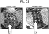
- the stiffness of the support need not be uniform and may vary depending on the shape, size and quantity of an condensate of interest. It is possible to provide the stiffness with a spatial/temporal gradient (as in Example 6 to be described later) or a pattern (as in Example 7 to be described later).
- the stiffness of the support is uniform, it is preferably 100 kPa or less, more preferably 1-50 kPa.
- the gel-like support may be planar, or the side on which culture is to be performed may have a U- or V-shaped cross section. If the side of the gel-like support on which culture is to be performed has a U- or V-shaped cross section, cells tend to gather on the culture surface and a cell condensate can advantageously be formed from a smaller number of cells and/or tissues.
- the support may be modified chemically or physically. Examples of modifying substances include, but are not limited to, Matrigel, laminin, entactin, collagen, fibronectin and vitronectin.
- the stiffness of the central part is 200 kPa or less and it suffices that the peripheral part is softer than the central part.
- Appropriate values for the stiffness of the central and peripheral parts of the substrate are variable depending on the coating and the shape.
- Another example of the gel-like culture support that is provided with a spatial gradient of stiffness is a gel-like culture support whose stiffness in the peripheral part is greater than the stiffness in the central part.
- the patterned, gel-like culture support is a gel-like culture support having one or more patterns in which the stiffness of the central part is greater than the stiffness of the peripheral part (see Example 7 to be described later; Fig. 22 , left panel: positive pattern).
- the stiffness of the central part is 200 kPa or less; it suffices that the peripheral part is softer than the central part.
- Appropriate values for the stiffness of the central and peripheral parts of the substrate are variable depending on the coating and the shape.
- Another example of the patterned, gel-like culture support is a gel-like culture support having one or more patterns in which the stiffness of the peripheral part is greater than the stiffness of the central part (see Example 7 to be described later; Fig.
- the stiffness of the peripheral part is 200 kPa or less; it suffices that the central part is softer than the peripheral part.
- Appropriate values for the stiffness of the central and peripheral parts of the substrate are variable depending on the coating and the shape.
- the temperature at the time of culture is not particularly limited but it is preferably 30-40°C and more preferably 37°C.
- an increased amount of oxygen is preferably supplied into the incubator.
- the amount of oxygen supply is appropriately 4-50%, preferably 10-30%, and more preferably 18-25%.
- the culture period is not particularly limited but it is preferably 12-144 hr.
- the culture period is preferably 12-48 hr.
- the culture period is preferably 12-144 hr.
- the culture period is preferably 12-96 hr.
- the culture period is preferably 12-96 hr.
- the culture period is preferably 12-96 hr.
- the culture period is preferably 12-144 hr.
- the culture period is preferably 12-144 hr.
- the culture period is preferably 12-144 hr.
- the culture period is preferably 12-144 hr.
- the culture period is preferably 48-144 hr.
- cell-cell interactions have taken place in such a close manner that a biological environment as occurs in the womb is recapitulated.
- induction of early differentiation into organ progenitor cells occurs efficiently and this would improve the frequency and number of such cells.
- cells adhere to each other so strongly that they can be collected in a non-destructive manner.
- the cell condensate described in the present application is a concept typically encompassing organ buds and organoids [organ bud ( WO2013/047639 ), liver bud, liver diverticula, liver organoid, pancreatic (dorsal or ventral) buds, pancreatic diverticula, pancreatic organoid, intestinal bud, intestinal diverticula, intestinal organoid ( K. Matsumoto et al. Science.19; 294 (5542): 559-63 (2001 )].
- organ bud WO2013/047639
- liver bud liver diverticula
- liver organoid pancreatic (dorsal or ventral) buds
- pancreatic diverticula pancreatic organoid
- intestinal bud intestinal diverticula
- intestinal organoid K. Matsumoto et al. Science.19; 294 (5542): 559-63 (2001 )
- the cell condensates are independent of the types of constituent cells and the number of such types.
- organ buds correspond to cell condensates that are formed at an early stage of organogenesis and are in principle composed of the following three types of cells: functional cells that constitute organs or tissues (or undifferentiated cells which will differentiate into functional cells); vascular cells; and mesenchymal cells.
- Organoids are solely composed of cells that constitute epithelial tissues and they are basically of a small size (1 mm or less).
- Cell condensates undergo self-organization to form three-dimensional tissue structures provided with higher structures, whereby progenitor cells can be directed to terminal differentiation.
- Self-organization may be performed either in vivo or in vitro. For example, when a cell condensate prepared by the method of the present invention is transplanted into a living body, vascular networks are formed, blood perfusion is induced, and self-organization into a higher tissue with a complex structure occurs, enabling the preparation of tissues/organs that have a highly ordered tissue structure comparable to that of adult tissues.
- the cell condensate of the present invention it may be possible to prepare a higher tissue that is provided with not only a vascular network but also higher structures such as ureteral structure, biliary structure, tracheal structure, etc. Further, a great number of organs essentially require that reconstitution associated with other organs be realized in order to exhibit their functions; e.g., in liver, reconstitution of junctions with bile duct and pancreatic duct and connection to duodenum is essential for exhibiting its function. According to the present invention, a cell condensate which recapitulates interactions with other organs is prepared. This cell condensate is expected to find use as a system for inducing self-organization into complex organs existing in the body.
- the present invention also provides a cell condensate prepared by the above-described method.
- the present invention also provides a method of three-dimensional tissue structure, comprising allowing self-organization of a cell condensate prepared by the above-described method to form a three-dimensional tissue structure that has been provided with higher structures.
- the present invention also provides a gel-like culture support wherein the side on which culture is performed has a U- or V-shaped cross-section.
- the gel-like culture support of the present invention having a U- or V-shaped cross-section on the side where culture is performed, allows cells to gather on the culture surface to ensure that a cell condensate is advantageously formed from a smaller number of cells and/or tissues.
- the gel-like culture support wherein the side on which culture is performed has a U- or V-shaped cross-section is as defined above.
- the present invention also provides a gel-like culture support wherein the stiffness of the central part thereof is greater than the stiffness of the peripheral part thereof.
- a gel-like culture support wherein the stiffness of the central part thereof is greater than the stiffness of the peripheral part thereof.
- One embodiment of such culture support is shown in Example 6 to be described later ( Figs. 20 and 21 ).
- the stiffness of the central part is 200 kPa or less; it suffices that the peripheral part is softer than the central part.
- Appropriate values for the stiffness of the central and peripheral parts of the support are variable depending on the coating and the shape.
- the present invention also provides a gel-like culture support in which the stiffness of the peripheral part thereof is greater than the stiffness of the central part thereof.
- the present invention also provides a gel-like culture support having one or more patterns in which the stiffness of the central part is greater than the stiffness of the peripheral part.
- a gel-like culture support having one or more patterns in which the stiffness of the central part is greater than the stiffness of the peripheral part.
- One embodiment of such culture support is given in Example 7 to be described later ( Fig. 22 , left panel: positive pattern).
- the stiffness of the central part is 200 kPa or less; it suffices that the peripheral part is softer than the central part.
- Appropriate values for the stiffness of the central and peripheral parts of the support are variable depending on the coating and the shape.
- the present invention also provides a gel-like culture support having one or more patterns in which the stiffness of the peripheral part is greater than the stiffness of the central part.
- a gel-like culture support having one or more patterns in which the stiffness of the peripheral part is greater than the stiffness of the central part.
- One embodiment of such culture support is given in Example 7 described later ( Fig. 22 , right panel: negative pattern).
- the stiffness of the peripheral part is 200 kPa or less; it suffices that the central part is softer than the central part.
- Appropriate values for the stiffness of the central and peripheral parts of the support are variable depending on the coating and the shape.
- the present invention also provides a method of preparing a cell condensate in vitro , comprising culturing a mixture of cells and/or tissues of a desired type and mesenchymal cells on the above-described gel-like culture support to thereby form a cell condensate. Culturing of the mixture of the cells and/or tissues of a desired type and the mesenchymal cells is as defined above.
- liver primordia (of millimeter scale) were autonomously formed from isolated human liver progenitor cells in vitro by recapitulating the cell-cell interactions which would occur at organogenesis stages.
- the present inventors revealed that this 3D tissue formation started from self-assembly behavior of multiple cell units and that the presence of the cytoskeletal contractile force of myosin II occurring in mesenchymal stem cells was crucial for the progress of such behavior.
- This dynamic cell collective behavior is regulated by the stiffness conditions of substrate matrix.
- the present inventors succeeded under optimized substrate conditions in preparing three-dimensional organ primordia from cells/tissues isolated from diverse organs including liver, pancreas, intestine, lung, heart, kidney, brain and even cancer.
- the thus prepared three-dimensional primordia were immediately vascularized upon transplantation (since vascular endothelial cells had been incorporated therein), followed by autonomous formation of self-organized three-dimensional tissue structures having therapeutic effects.
- this principle will serve to establish a highly versatile platform for reconstituting a plurality of vascularized, complex organ systems from stem cells via dynamic cell condensation and the subsequent self-organization.
- liver is formed from a condensed tissue mass termed “liver bud” at week 5-8 of gestation in human during physiological organogenesis.
- Liver budding also called “liver bud”
- liver budding also called “liver bud”
- PSCs pluripotent stem cells
- single PSC-derived hepatocytes autonomously form 3-D condensates by co-culture with endothelial cells and mesenchymal cells (2). Once condensates are established, they continue to self-organize after several days under complete in vitro conditions into liver bud tissues having a structure resembling the organs that exist in the womb (3).
- the in vitro grown organ bud is transplanted into a living body, where it undergoes further self-organization (is matured) to eventually become a vascularized and functional liver. This method opens a new road for artificial reconstitution of vascularized organ systems (4).
- the present inventors performed a time-lapse imaging analysis to track cellular movements during organoid formation.
- Hepatic endoderm cells derived from human induced pluripotent stem cells (iPSCs), umbilical cord-derived endothelial cells (HUVECs), and mesenchymal stem cells (MSCs) were labeled with distinctive fluorescent markers and cocultured on a solidified matrix gel which was already described.
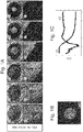
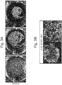
- Live cell tracking revealed that after rapid cell convergence, the assembly of vascularized organoids was initiated; this was followed by spatial rearrangements via self-organization as demonstrated by the formation of an endothelial-like network ( Fig. 1 ).
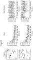
- Fig. 1 To elucidate dynamics of such condensate formation in more detail, the present inventors examined the temporal development of the position of the edge of the cell condensate (square root of cell area) and circularity by image analysis ( Fig. 1b ). The results showed that cell condensates contracted gently at 10 ⁇ m/h or less up to about 7 hr after seeding, and then the contraction accelerated to about 500 ⁇ m/h at naxunyn over the next several hours and finally decreased exponentially to converge. On the other hand, its circularity decreased almost monotonically right after cell seeding and reached a minimal value of about 0.5 in 10-13 hr. The circularity then increased and finally achieved an almost constant value (0.85) at 20 hr after seeding.
- MSCs mesenchymal stem cells
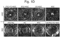
- cell condensate formation was possible in coculture of iPSC-derived hepatic endoderm cells and MSC (iPSC+MSC) or coculture of vascular endothelial cells and MSC (EC+MSC) ( Fig. 1 ).
- iPSC+MSC coculture of iPSC-derived hepatic endoderm cells and MSC
- EC+MSC coculture of vascular endothelial cells and MSC
- the present inventors subsequently assessed the contributions of the contraction force of MSCs at the molecular level against their substratum and the surrounding cells.
- MII myosin II
- the present inventors therefore assessed MII activity by measuring time-course-dependent changes in MIIA phosphorylation with MIIA inactivating S1943 (pS1943) through decomposition of myofilament by phosphate-specific antibodies (7) and intracellular flow cytometry.
- MIIA activity Based on the formula reported to estimate MIIA activity (8), the present inventors showed that active MIIA was remarkably up-regulated in stromal cells during condensate formation and reached its peak at 6 hr, which corresponds to the time at which cells moved at maximum velocity (1). On the other hand, it is seen that activated MIIA is almost constant throughout condensate formation in iPSC-derived hepatocytes. This suggests that the MSC-driven activation of MIIA is responsible for this strong three-dimensional rearrangement. As data indicating direct evidence for the decrease of this activated MIIA, the present inventors showed that this condensate formation could be completely antagonized by treatment with blebbistatin (an MII ATPase inhibitor) (9).
- Rho kinase inhibitor Y-27632 it was found that addition of Rho kinase inhibitor Y-27632 to the cocultures partially delayed condensate formation ( Fig. 1 ).
- Rho kinase inhibitor Y-27632 it was assumed that such mechanism is hard to apply because pharmacological inhibition of chemokine receptor pathways by addition of AMD3100 could not hinder condensate formation (10).
- MSCs that are the key cell in condensate formation in the system of the present invention are known to exhibit mechano-response in diverse processes including differentiation and attachment.
- condensates such as spheroids
- the present inventors have concluded that contraction of mesenchymal cells against softer substrate might have caused these collective behaviors in coculture systems.
- the proposed principle can be expanded to self-organization systems for other organs irrespective of the origin of germ layers that are to be used in the future for the purpose of regenerative medicine.
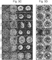
- the present inventors first selected pancreatic cells and subjected them to coculture, since there is increasing evidence that pancreas follows a developmental program relatively close to that of liver.
- isolated mouse pancreas ⁇ cells MIN6 were cocultured with HUVEC and MSC, a similar formation of cell condensate was observed ( Fig. 3 ).
- the present inventors isolated multiple cells or tissue fragments (up to 200 ⁇ m) from embryonic or adult mice. Surprisingly, the directed and autonomic assembling phenomenon was retained in all the cell/tissue types tested, including pancreas, liver, intestine, lung, heart, kidney, brain, and even cancer ( Fig. 3 ). Time-lapse imaging analyses revealed that both the embryonic and adult cells/tissues successfully resisted additional manipulations (including surgical transplantation) to form single 3D organoids autonomously ( Fig. 3 ).
- Condensates as designed to contain cultured endothelial cells turned out to permit a much more rapid perfusion with recipient circulation after transplantation (average perfusion time: ⁇ 72 hr) compared with reliable conventional tissue engineering approaches (average perfusion time: ⁇ 192 hr).
- pancreatic cells for in-depth characterization.
- the transplantation of 3-D pancreatic organoids resulted in rapid ( ⁇ 48 hr) reperfusion and successful ⁇ cell engraftment. These were confirmed by live imaging analysis.
- the transplants developed islet-like structures ( Fig. 4 , E ) with functional microvascular networks that connected to the recipient circulatory system ( Fig. 4C ).
- Such blood perfusion was not recognized when condensates not containing vascular endothelial cells were transplanted ( Fig. 9 ).
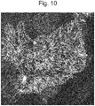
- the reconstituted islets directly connected to peripheral mouse blood vessels to be highly vascularized with a tight network of microvessels ( Fig. 10 ).
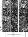
- the capillary network in the islet in a living body is known to be approximately 5 times as dense as the capillary network surrounding exocrine secretion tissues. Consistent with this, intravital quantification of the functional vascular density showed that the capillary network was much denser (by 4.2 times) in the reconstituted islet-like tissues than in the areas surrounding the normal tissues ( Fig.4 , Fig. 9 ). Histological analysis also showed that the islet-like tissues had a structure resembling the adult islet, suggesting the reconstitution of a mature tissue via self-organization ( Fig. 11 ).
- in vitro -derived ⁇ cell organoids were transplanted into kidney subcapsule of type 1 fulminant diabetic model mouse.
- the present inventors used a toxin receptor-mediated cell knockout (TREK) Tg mouse having a diphtheria toxin (DT) receptor cDNA transgene in insulin promoter. While mice in non-transplantation group died at day 6 of DT administration-mediated induction of diabetes, those mice which received transplantation of ⁇ cell organoids maintained normal blood glucose levels and survived ( Fig. 4 , G ).
- TREK toxin receptor-mediated cell knockout
- mice in non-transplantation group died at day 6 of DT administration-mediated induction of diabetes
- those mice which received transplantation of ⁇ cell organoids maintained normal blood glucose levels and survived ( Fig. 4 , G ).
- the present inventors have demonstrated the applicablility of the foregoing principle to other organ systems by experimentally recapitulating vascularization and reconstituting a functional three-dimensional tissue in vivo.
- the principle under consideration ensures that starting with larger numbers of the desired cells/tissues, self-organized organoids can be designed via condensation.
- the condensates may be used for examining the subsequent self-organization capacity both in vitro and in vivo.
- rapid vasculogenesis and subsequent functionalization were evaluated by incorporating endothelial cells experimentally.
- evaluating the contribution of undeveloped supporting cells such as neurons is also an interesting topic for the present inventors and other research groups.
- MCs mesenchymal cells
- MCs any of the following cells was used: cells isolated from human bone marrow, cells isolated from umbilical cord stroma (Wharton's sheath), cells isolated from human auricle, cells isolated from mouse bone marrow, human fibroblast cells or the like.
- the mesenchymal stem cells isolated from human bone marrow (hMSCs) that were mainly used in this experiment had been cultured using MSCGMTM BulletKitTM (Lonza PT-3001), a medium prepared exclusively for hMSC culture.
- C57BL/6-Tg mice (Nippon SLC) at days 12-17 of gestation were disinfected with 70% ethanol and incised to remove embryos. Brain, heart, lung, liver, metanephros or intestine was removed from the embryos. Brain, heart, lung, liver, kidney or intestine was also removed from C57BL/6-BALB/c RFP hairy mice 6 or more weeks of age (purchased from Anticancer Inc.). When cells isolated from these removed tissues were used, they were put in 200 ⁇ l of 0.05% Tryspin-EDTA (GIBCO) and incubated for 20 min at 37°C.
- GEBCO Tryspin-EDTA
- the tissues were disrupted with a pipette and added to 4.8 ml of a medium. After centrifugation, medium was added and the number of cells was counted. Then, enzyme treatment was conducted to give single cells, which were subsequently used for coculture.
- the removed tissues were to be used in a state of small tissues, the removed embryonic tissues were minced with scissors, put in 10 ml of 0.05% Tryspin-EDTA and shaken for 20 min at 37°C. After addition of medium, the resultant cells were passed through a 100 ⁇ m cell strainer and centrifuged. After centrifugation, medium was added for use in cell culture.
- the brain, heart, lung and kidney of the adult mice were minced with scissors and passed through a 100 ⁇ m cell strainer.
- the resultant flow-through was filtered with a 40 ⁇ m cell strainer.
- the cell mass remaining on this cell strainer was collected with medium for use in coculture of cells.
- the contents were washed with physiological saline.
- the washed small intestine was cut lengthwise at intervals of 4 cm.
- the resultant sections were put in 2 mM EDTA, 0.5 mM DTT in PBS and shaken for 20 min at 37°C.
- the cells were passed through a 100 ⁇ m cell strainer, followed by addition of PBS. After centrifugation, the supernatant was suctioned and PBS was added for washing. Then the cells were centrifuged, and medium was added for use in cell culture.
- EGMTM BulletKitTM Lonza CC-4133
- HepG2 was cultured in DMEM supplemented with 10% FBS. Each type of cells were cultured in a 37°C, 5%CO 2 incubator.
- 4x10 3 cells or more of multiple types in any combination tissues isolated from embryo or adult, or KO-HepG2, HUVEC
- 5x10 3 or more MSCs were mixed with 5x10 3 or more MSCs and seeded.
- the cells were subsequently cultured in a 37°C incubator for a day.
- chronological observation of cell coculture was performed with a stereomicroscope or a confocal microscope.
- the cell types in "any combination” For example, cells derived from different tissues such as pancreas, liver, intestine, nerve, etc. may be mixed and used. Subsequently, an optimal composition for self-organization of an organ of interest could be used.
- a 10 ml solution was prepared by mixing aqueous acrylamide solution (40% w/v, A4058, Sigma), aqueous bis-acrylamide solution (2% w/v, M1533, Sigma) and distilled water. In the process, the Young's modulus of the gel was adjusted by changing the mixing ratio of the individual solutions. The resultant reaction solution was bumped using a vacuum chamber. Then, 100 ⁇ l of APS (5 g/DW 50 ml, 01307-00, KANTO, 0.20 mm filtered) and 10 ⁇ l of TEMED (T9281, Sigma) were sequentially added to the reaction solution.
- APS g/DW 50 ml, 01307-00, KANTO, 0.20 mm filtered
- 10 ⁇ l of TEMED T9281, Sigma
- Coating of adhesion molecules (Matrigel or laminin) onto the PA gel surface was performed by the procedures described below. First, 0.2 mg/ml N-sulfosuccinimidyl-6-(4'-azido-2'-nitrophenylamino)hexanoate (Sulfo-SANPAH, 22589, Pierce) in 20 mM HEPES (pH 8.5) was dripped onto the PA gel substrate, followed by irradiation with a UV lamp (Z169633-1EA, Sigma) for 20 min.
- a UV lamp Z169633-1EA, Sigma
- a 10 ml solution was prepared by mixing aqueous acrylamide solution (40% w/v, A4058, Sigma), aqueous bis-acrylamide solution (2% w/v, M1533, Sigma) and distilled water. In the process, the Young's modulus of the gel was adjusted by changing the mixing ratio of the individual solutions.
- This reaction solution 500 ⁇ l was added to a 24-well tissue culture plate (353047, BD). Then, 0.5 ⁇ l of TEMED (T9281, Sigma) and 5 ⁇ l of APS (5 g/DW 50 ml, 01307-00, KANTO, 0.20 mm filtered) were added to the reaction solution in this order, immediately followed by thorough mixing.
- the cells were mixed with mesenchymal stem cells isolated from human bone marrow (hMSCs) and normal umbilical vein endothelial cells (HUVECs) and seeded on wells where a solution obtained by mixing Matrigel (the stock solution of Matrigel (BD) used in Example 2) and a medium for vascular endothelial cells (EGM BulletKitTM, Lonza CC-4133) at 1:1 had been solidified.
- Matrigel the stock solution of Matrigel (BD) used in Example 2
- EMM BulletKitTM the medium for vascular endothelial cells
- the renal primordium formed was transplanted into the wombs of immunodeficiency mice.
- blood perfusion was recognized in two to three days after transplantation ( Fig. 15 , upper row).
- the white dotted lines in Fig. 15 indicate the transplantation areas.
- Scattered cells formed spherical, glomerular tissues at day 8 of transplantation ( Fig. 15 , bottom row).
- the results of fluorescence observation as shown in the left panel of Fig. 16 revealed that a great number of glomerular structures were formed by culturing on a support but that this was not the case when the conventional method (pellet transplantation group) was applied.
- the tissues formed first flowed into blood vessels, were filtered inside glomeruli, and collected in proximal tubules, indicating that they had the primitive urine producing function of the kidney ( Fig. 18 ).
- the renal primordium artificially prepared according to the present invention autonomous maturation could successfully be induced to prepare functional renal tissues.
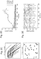
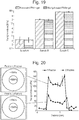
- Example 5 Changes in the Young's Modulus of supports (see “preparation of PA gel planar substrate") with different stiffness conditions (Samples A, B and C) before (oblique lines) and after (solid black) Matrigel coating. It was shown that the stiffness conditions can be strictly controlled regardless of the presence or absence of the coating ( Fig. 19 ).
- the gel substrate used in Example 5 was prepared according to the method described in Example 1.
- Gels having multiple stiffness patterns providing different stiffness conditions could successfully be prepared on one substrate ( Fig. 20 ).
- pattern 1 a gel with a hard central part was prepared and according to pattern 2, a gel with a less hard central part was prepared ( Fig. 20 , left panel).
- the right panel of Fig. 20 shows the results of measurement of stiffness conditions along the major axis, indicating that the intended stiffness conditions could be achieved.
- Gel substrates having spatial patterns of stiffness were prepared by the method described below.
- a gel reaction solution a 10 ml solution was prepared by mixing aqueous acrylamide solution (40% w/v, A4058, Sigma), aqueous bis-acrylamide solution (2% w/v, M1533, Sigma) and distilled water. Subsequently, with light shielded, 50 mg of Irgacure 2959 (0.5 % w/v, DY15444, Ciba) was added and dissolved in a hot water bath at 37°C. The resultant reaction solution was bumped in a vacuum chamber.
- a circular mask (12 mm o.d. and 2-4 mm i.d.) was prepared.
- a mercury lamp C - HGFI, Nikon
- fiber optics was combined with a light projection tube to ensure uniform irradiation of the reaction solution.
- the time of UV irradiation was adjusted by minutes depending on the desired stiffness.
- distilled water was added to the sandwiched sample and the glass coverslip was peeled off from the glass slide, leaving a gel coat on the former.
- Phosphate buffer was added to the resultant glass coverslip, which was then left to stand for one day to remove the unreacted monomers.
- the Young's moduli of gels were determined by nanoindentation measurements performed with an atomic force microscope (Nanowizard 3, JPK Instruments, Germany). Coating of adhesion molecules (Matrigel or laminin) onto the PA gel surface was performed by the procedures described below. First, 0.2 mg/ml N-sulfosuccinimidyl-6-(4'-azido-2'-nitrophenylamino)hexanoate (Sulfo-SANPAH, 22589, Pierce) in 20 mM HEPES (pH 8.5) was dripped onto the PA gel substrate, followed by irradiation with a UV lamp (Z169633-1EA, Sigma) for 20 min.
- a UV lamp Z169633-1EA, Sigma
- iPS cell-derived hepatic endoderm cells, HUVECs and MSCs were mixed at a ratio of 10:7:2 and the mixture was seeded on the patterned gels to give a total cell count of about 2x10 6 cells ( Fig. 21 ).
- the optimal condition for the stiffness of the central part was 100 kPa.
- a positive pattern was so designed that individual circular parts were hard and their periphery was soft ( Fig. 22 , left panel).
- a negative pattern was so designed that individual circular parts were soft and their periphery was hard ( Fig. 22 , right panel).
- Gel substrates with such multiple patterns of stiffness were prepared based on the technique described in Example 6 and by exposing a gel substrate (25 mm in diameter) through a photomask with a 4x4 pattern of circles (diameter: about 2 mm; center-to-center distance between circles: about 2.7 mm). It is believed that by using these patterned supports, cell condensates of any size may be formed at any place.
- the present invention is applicable in various fields including search for new drugs and evaluation of their efficacy, regenerative medicine, diagnosis of diseases and pathology, and production of useful substances.
Landscapes
- Health & Medical Sciences (AREA)
- Life Sciences & Earth Sciences (AREA)
- Engineering & Computer Science (AREA)
- Chemical & Material Sciences (AREA)
- Biomedical Technology (AREA)
- Zoology (AREA)
- General Health & Medical Sciences (AREA)
- Genetics & Genomics (AREA)
- Biotechnology (AREA)
- Wood Science & Technology (AREA)
- Bioinformatics & Cheminformatics (AREA)
- Cell Biology (AREA)
- Organic Chemistry (AREA)
- Animal Behavior & Ethology (AREA)
- Transplantation (AREA)
- Veterinary Medicine (AREA)
- Public Health (AREA)
- Epidemiology (AREA)
- Dermatology (AREA)
- Medicinal Chemistry (AREA)
- Oral & Maxillofacial Surgery (AREA)
- Chemical Kinetics & Catalysis (AREA)
- General Engineering & Computer Science (AREA)
- Microbiology (AREA)
- Botany (AREA)
- Biochemistry (AREA)
- Dispersion Chemistry (AREA)
- Developmental Biology & Embryology (AREA)
- Micro-Organisms Or Cultivation Processes Thereof (AREA)
Applications Claiming Priority (2)
| Application Number | Priority Date | Filing Date | Title |
|---|---|---|---|
| JP2014037341 | 2014-02-27 | ||
| PCT/JP2015/055695 WO2015129822A1 (ja) | 2014-02-27 | 2015-02-26 | 自己組織化用細胞集合体の作製方法 |
Publications (3)
| Publication Number | Publication Date |
|---|---|
| EP3124600A1 true EP3124600A1 (de) | 2017-02-01 |
| EP3124600A4 EP3124600A4 (de) | 2017-12-06 |
| EP3124600B1 EP3124600B1 (de) | 2021-12-29 |
Family
ID=54009136
Family Applications (1)
| Application Number | Title | Priority Date | Filing Date |
|---|---|---|---|
| EP15754683.9A Active EP3124600B1 (de) | 2014-02-27 | 2015-02-26 | Verfahren zur herstellung eines zellkondensats zur selbstorganisation |
Country Status (10)
| Country | Link |
|---|---|
| US (1) | US20170067014A1 (de) |
| EP (1) | EP3124600B1 (de) |
| JP (1) | JP6489484B2 (de) |
| KR (1) | KR102338698B1 (de) |
| CN (1) | CN106062181A (de) |
| AU (1) | AU2015223798B2 (de) |
| BR (1) | BR112016019677A8 (de) |
| CA (1) | CA2937882A1 (de) |
| SG (2) | SG10202107992XA (de) |
| WO (1) | WO2015129822A1 (de) |
Families Citing this family (28)
| Publication number | Priority date | Publication date | Assignee | Title |
|---|---|---|---|---|
| US9719068B2 (en) | 2010-05-06 | 2017-08-01 | Children's Hospital Medical Center | Methods and systems for converting precursor cells into intestinal tissues through directed differentiation |
| EP3739041A1 (de) | 2014-03-27 | 2020-11-18 | The Salk Institute for Biological Studies | Zusammensetzungen und verfahren zur behandlung von typ-1-diabetes und typ-2-diabetes sowie von verwandten erkrankungen |
| CN106661548B (zh) | 2014-05-28 | 2020-12-11 | 儿童医院医疗中心 | 用于经由定向分化将前体细胞转化为胃组织的方法和系统 |
| JP6804438B2 (ja) | 2014-10-17 | 2020-12-23 | チルドレンズ ホスピタル メディカル センター | 多能性幹細胞を使用するヒト小腸のin vivoモデル、並びにそれを作製、及び使用する方法 |
| AU2016225076B2 (en) | 2015-02-27 | 2018-09-13 | Salk Institute For Biological Studies | Reprogramming progenitor compositions and methods of use therefore |
| JP2017085945A (ja) * | 2015-11-06 | 2017-05-25 | 国立研究開発法人産業技術総合研究所 | 3次元培養による肝臓組織の構築方法 |
| KR20180095538A (ko) | 2015-12-22 | 2018-08-27 | 고리츠다이가쿠호진 요코하마시리츠다이가쿠 | 바이러스 감염 모델, 그 제조 방법 및 그 이용 |
| US20190093082A1 (en) * | 2016-03-07 | 2019-03-28 | Centre National De La Recherche Scientifique (Cnrs) | Method of Differentiating Pluripotent Stem Cells |
| ES2929758T3 (es) | 2016-05-05 | 2022-12-01 | Childrens Hospital Med Ct | Métodos para la fabricación in vitro de tejido del fondo gástrico y composiciones relacionadas con el mismo |
| AU2017269364B2 (en) * | 2016-05-25 | 2023-08-31 | Salk Institute For Biological Studies | Compositions and methods for organoid generation and disease modeling |
| EP3527655A4 (de) * | 2016-10-11 | 2020-05-06 | Tokushima University | Herstellungsverfahren für nierenartiges gewebe |
| KR102729404B1 (ko) | 2016-11-04 | 2024-11-14 | 칠드런즈 호스피탈 메디칼 센터 | 간 유사 장기 조성물 및 이를 제조 및 사용하는 방법 |
| JP7068305B2 (ja) | 2016-12-05 | 2022-05-16 | チルドレンズ ホスピタル メディカル センター | 結腸オルガノイドならびにその作製方法および使用方法 |
| US10767164B2 (en) | 2017-03-30 | 2020-09-08 | The Research Foundation For The State University Of New York | Microenvironments for self-assembly of islet organoids from stem cells differentiation |
| US12281334B2 (en) | 2017-04-14 | 2025-04-22 | Children's Hospital Medical Center | Multi donor stem cell compositions and methods of making same |
| AU2018336901B2 (en) | 2017-09-21 | 2024-07-18 | President And Fellows Of Harvard College | Tissue construct, methods of producing and using the same |
| EP3694603B1 (de) | 2017-10-10 | 2026-04-08 | Children's Hospital Medical Center | Ösophagusgewebe- und/oder organoidzusammensetzungen und verfahren zu ihrer herstellung |
| JP7233717B2 (ja) * | 2017-11-30 | 2023-03-07 | 公立大学法人横浜市立大学 | 多能性幹細胞からの立体臓器の構築 |
| US12379372B2 (en) | 2017-12-21 | 2025-08-05 | Children's Hospital Medical Center | Digitalized human organoids and methods of using same |
| KR102887406B1 (ko) | 2018-07-26 | 2025-11-19 | 칠드런즈 호스피탈 메디칼 센터 | 간-담도-췌장 조직 및 이를 제조하는 방법 |
| AU2019339410A1 (en) | 2018-09-12 | 2021-04-15 | Children's Hospital Medical Center | Organoid compositions for the production of hematopoietic stem cells and derivatives thereof |
| WO2020160371A1 (en) | 2019-02-01 | 2020-08-06 | The University Of Hong Kong | Innervated organoid compositions and methods of making same |
| WO2020243633A1 (en) | 2019-05-31 | 2020-12-03 | Children's Hospital Medical Center | Shaped organoid compositions and methods of making same |
| WO2020243613A1 (en) | 2019-05-31 | 2020-12-03 | Children's Hospital Medical Center | Methods of generating and expanding hematopoietic stem cells |
| EP4047082A4 (de) | 2019-10-17 | 2023-08-16 | Public University Corporation Yokohama City University | Verfahren zum bewerten der toxizität von arzneimitteln |
| US20230212492A1 (en) | 2020-06-08 | 2023-07-06 | National University Corporation Tokyo Medical And Dental University | Cell culture method |
| FR3122883A1 (fr) * | 2021-05-11 | 2022-11-18 | Treefrog Therapeutics | Microcompartiments cellulaires comprenant des cellules dont l’intégrité génomique est maintenue après amplification et procédé de préparation |
| CN113350574B (zh) * | 2021-05-26 | 2022-11-18 | 泸州国之荣耀酒业有限公司 | 图案化类肝小叶微组织制造方法 |
Family Cites Families (8)
| Publication number | Priority date | Publication date | Assignee | Title |
|---|---|---|---|---|
| CN103627671A (zh) * | 2007-01-30 | 2014-03-12 | 佐治亚大学研究基金会 | 产生中内胚层细胞及多能游走细胞的方法与细胞群及用途 |
| US9062283B2 (en) * | 2007-02-26 | 2015-06-23 | Stemcell Technologies Inc. | Method of reducing curvature in a meniscus of liquid medium |
| WO2012119074A1 (en) * | 2011-03-03 | 2012-09-07 | Massachusetts Institute Of Technology | Apparatus and method for organizing three-dimensional cell structures using stiffness gradients and sacrificial gels |
| KR101282926B1 (ko) * | 2011-07-28 | 2013-07-08 | 고려대학교 산학협력단 | 표면장력을 이용한 반구형 마이크로웰의 제조 및 이를 이용한 세포 집합체의 형성 |
| WO2013047639A1 (ja) * | 2011-09-27 | 2013-04-04 | 公立大学法人横浜市立大学 | 組織及び臓器の作製方法 |
| ES2741969T3 (es) * | 2011-10-31 | 2020-02-12 | Riken | Método para el cultivo de células madre |
| US20130252337A1 (en) * | 2012-03-21 | 2013-09-26 | Shengyuan Yang | Substrates with micrometer and nanometer scale stiffness patterns for use in cell and tissue culturing and a method for making same |
| US11583860B2 (en) * | 2014-12-22 | 2023-02-21 | Ecole Polytechnique Federale De Lausanne (Epfl) | Microstructured thin hydrogel films |
-
2015
- 2015-02-26 SG SG10202107992XA patent/SG10202107992XA/en unknown
- 2015-02-26 WO PCT/JP2015/055695 patent/WO2015129822A1/ja not_active Ceased
- 2015-02-26 JP JP2016505305A patent/JP6489484B2/ja active Active
- 2015-02-26 KR KR1020167025902A patent/KR102338698B1/ko active Active
- 2015-02-26 SG SG11201606750UA patent/SG11201606750UA/en unknown
- 2015-02-26 CA CA2937882A patent/CA2937882A1/en not_active Abandoned
- 2015-02-26 CN CN201580003897.6A patent/CN106062181A/zh active Pending
- 2015-02-26 AU AU2015223798A patent/AU2015223798B2/en active Active
- 2015-02-26 BR BR112016019677A patent/BR112016019677A8/pt active Search and Examination
- 2015-02-26 EP EP15754683.9A patent/EP3124600B1/de active Active
- 2015-02-26 US US15/121,934 patent/US20170067014A1/en not_active Abandoned
Also Published As
| Publication number | Publication date |
|---|---|
| WO2015129822A1 (ja) | 2015-09-03 |
| SG11201606750UA (en) | 2016-10-28 |
| AU2015223798B2 (en) | 2020-10-15 |
| EP3124600A4 (de) | 2017-12-06 |
| US20170067014A1 (en) | 2017-03-09 |
| KR20160125440A (ko) | 2016-10-31 |
| CA2937882A1 (en) | 2015-09-03 |
| CN106062181A (zh) | 2016-10-26 |
| JPWO2015129822A1 (ja) | 2017-03-30 |
| BR112016019677A2 (pt) | 2017-08-15 |
| SG10202107992XA (en) | 2021-09-29 |
| EP3124600B1 (de) | 2021-12-29 |
| BR112016019677A8 (pt) | 2021-07-13 |
| AU2015223798A1 (en) | 2016-08-18 |
| KR102338698B1 (ko) | 2021-12-10 |
| JP6489484B2 (ja) | 2019-03-27 |
Similar Documents
| Publication | Publication Date | Title |
|---|---|---|
| US20240336898A1 (en) | Formation of three-dimensional organ from pluripotent stem cells, method for generating cell condensate for self-organization | |
| EP3124600B1 (de) | Verfahren zur herstellung eines zellkondensats zur selbstorganisation | |
| JP7125717B2 (ja) | 培養方法 | |
| Bernstein et al. | Stem cell therapy for cardiac disease | |
| US20150368618A1 (en) | Modulation of cardiac stem-progenitor cell differentiation, assays and uses thereof | |
| Liu et al. | Generation of functional organs from stem cells | |
| Ajmal et al. | Organ regeneration through stem cells and tissue engineering | |
| JP2013510582A (ja) | 間葉幹細胞の球状集合体 | |
| Polak et al. | Stem cells bioprocessing: an important milestone to move regenerative medicine research into the clinical arena | |
| US20200399613A1 (en) | Cell Mass Fusion Method | |
| Ren et al. | On the road to bioartificial organs | |
| Oliinyk et al. | Chorioallantoic membrane assay at the cross-roads of adipose-tissue-derived stem cell research | |
| JP5893562B2 (ja) | 乳動脈由来細胞並びに組織修復及び再生における使用方法 | |
| WO2009080794A1 (en) | Method for preparing cell-specific extracellular matrices | |
| WO2018225705A1 (ja) | 細胞培養物の製造方法 | |
| JP7444367B2 (ja) | 増幅毛包間葉系細胞の製造方法及びその使用 | |
| JPWO2017150294A1 (ja) | 多能性幹細胞様スフェロイドの製造方法および多能性幹細胞様スフェロイド | |
| Matsumoto et al. | Renal regeneration: stem cell-based therapies to battle kidney disease | |
| JPWO2020067435A1 (ja) | 多能性幹細胞由来細胞のシート化方法 | |
| JPWO2020067436A1 (ja) | 多能性幹細胞由来細胞の移植片形成方法 | |
| Hu et al. | Recellularization of Decellularized Whole Organ Scaffolds: Elements, Progresses, and Challenges | |
| HK1228952A1 (en) | Method for generating cell condensate for self-organization | |
| Barbarisi et al. | Regenerative medicine: current and potential applications | |
| JPWO2020067438A1 (ja) | 多能性幹細胞由来細胞のシート化方法 | |
| Zeilinger et al. | Strategies for the therapeutic use of adult stem cells in regenerative medicine: Cell biological and technological approaches |
Legal Events
| Date | Code | Title | Description |
|---|---|---|---|
| STAA | Information on the status of an ep patent application or granted ep patent |
Free format text: STATUS: THE INTERNATIONAL PUBLICATION HAS BEEN MADE |
|
| PUAI | Public reference made under article 153(3) epc to a published international application that has entered the european phase |
Free format text: ORIGINAL CODE: 0009012 |
|
| STAA | Information on the status of an ep patent application or granted ep patent |
Free format text: STATUS: REQUEST FOR EXAMINATION WAS MADE |
|
| 17P | Request for examination filed |
Effective date: 20160914 |
|
| AK | Designated contracting states |
Kind code of ref document: A1 Designated state(s): AL AT BE BG CH CY CZ DE DK EE ES FI FR GB GR HR HU IE IS IT LI LT LU LV MC MK MT NL NO PL PT RO RS SE SI SK SM TR |
|
| AX | Request for extension of the european patent |
Extension state: BA ME |
|
| DAX | Request for extension of the european patent (deleted) | ||
| A4 | Supplementary search report drawn up and despatched |
Effective date: 20171106 |
|
| RIC1 | Information provided on ipc code assigned before grant |
Ipc: A61L 27/00 20060101ALI20171027BHEP Ipc: C12N 5/10 20060101ALI20171027BHEP Ipc: C12N 5/077 20100101ALI20171027BHEP Ipc: C12N 5/071 20100101AFI20171027BHEP Ipc: C12N 5/0735 20100101ALI20171027BHEP Ipc: C12M 3/00 20060101ALI20171027BHEP Ipc: C12N 5/0775 20100101ALI20171027BHEP |
|
| STAA | Information on the status of an ep patent application or granted ep patent |
Free format text: STATUS: EXAMINATION IS IN PROGRESS |
|
| 17Q | First examination report despatched |
Effective date: 20181210 |
|
| GRAP | Despatch of communication of intention to grant a patent |
Free format text: ORIGINAL CODE: EPIDOSNIGR1 |
|
| STAA | Information on the status of an ep patent application or granted ep patent |
Free format text: STATUS: GRANT OF PATENT IS INTENDED |
|
| INTG | Intention to grant announced |
Effective date: 20210420 |
|
| RAP1 | Party data changed (applicant data changed or rights of an application transferred) |
Owner name: PUBLIC UNIVERSITY CORPORATION YOKOHAMA CITY UNIVERSITY |
|
| RIN1 | Information on inventor provided before grant (corrected) |
Inventor name: YOSHIKAWA, HIROSHI Inventor name: TANIGUCHI, HIDEKI Inventor name: TAKEBE, TAKANORI |
|
| GRAJ | Information related to disapproval of communication of intention to grant by the applicant or resumption of examination proceedings by the epo deleted |
Free format text: ORIGINAL CODE: EPIDOSDIGR1 |
|
| STAA | Information on the status of an ep patent application or granted ep patent |
Free format text: STATUS: EXAMINATION IS IN PROGRESS |
|
| INTC | Intention to grant announced (deleted) | ||
| GRAP | Despatch of communication of intention to grant a patent |
Free format text: ORIGINAL CODE: EPIDOSNIGR1 |
|
| STAA | Information on the status of an ep patent application or granted ep patent |
Free format text: STATUS: GRANT OF PATENT IS INTENDED |
|
| INTG | Intention to grant announced |
Effective date: 20211001 |
|
| GRAS | Grant fee paid |
Free format text: ORIGINAL CODE: EPIDOSNIGR3 |
|
| GRAA | (expected) grant |
Free format text: ORIGINAL CODE: 0009210 |
|
| STAA | Information on the status of an ep patent application or granted ep patent |
Free format text: STATUS: THE PATENT HAS BEEN GRANTED |
|
| AK | Designated contracting states |
Kind code of ref document: B1 Designated state(s): AL AT BE BG CH CY CZ DE DK EE ES FI FR GB GR HR HU IE IS IT LI LT LU LV MC MK MT NL NO PL PT RO RS SE SI SK SM TR |
|
| REG | Reference to a national code |
Ref country code: GB Ref legal event code: FG4D |
|
| REG | Reference to a national code |
Ref country code: CH Ref legal event code: EP |
|
| REG | Reference to a national code |
Ref country code: DE Ref legal event code: R096 Ref document number: 602015076072 Country of ref document: DE |
|
| REG | Reference to a national code |
Ref country code: AT Ref legal event code: REF Ref document number: 1458680 Country of ref document: AT Kind code of ref document: T Effective date: 20220115 |
|
| REG | Reference to a national code |
Ref country code: IE Ref legal event code: FG4D |
|
| REG | Reference to a national code |
Ref country code: LT Ref legal event code: MG9D |
|
| PG25 | Lapsed in a contracting state [announced via postgrant information from national office to epo] |
Ref country code: RS Free format text: LAPSE BECAUSE OF FAILURE TO SUBMIT A TRANSLATION OF THE DESCRIPTION OR TO PAY THE FEE WITHIN THE PRESCRIBED TIME-LIMIT Effective date: 20211229 Ref country code: LT Free format text: LAPSE BECAUSE OF FAILURE TO SUBMIT A TRANSLATION OF THE DESCRIPTION OR TO PAY THE FEE WITHIN THE PRESCRIBED TIME-LIMIT Effective date: 20211229 Ref country code: FI Free format text: LAPSE BECAUSE OF FAILURE TO SUBMIT A TRANSLATION OF THE DESCRIPTION OR TO PAY THE FEE WITHIN THE PRESCRIBED TIME-LIMIT Effective date: 20211229 Ref country code: BG Free format text: LAPSE BECAUSE OF FAILURE TO SUBMIT A TRANSLATION OF THE DESCRIPTION OR TO PAY THE FEE WITHIN THE PRESCRIBED TIME-LIMIT Effective date: 20220329 |
|
| REG | Reference to a national code |
Ref country code: NL Ref legal event code: MP Effective date: 20211229 |
|
| REG | Reference to a national code |
Ref country code: AT Ref legal event code: MK05 Ref document number: 1458680 Country of ref document: AT Kind code of ref document: T Effective date: 20211229 |
|
| PG25 | Lapsed in a contracting state [announced via postgrant information from national office to epo] |
Ref country code: SE Free format text: LAPSE BECAUSE OF FAILURE TO SUBMIT A TRANSLATION OF THE DESCRIPTION OR TO PAY THE FEE WITHIN THE PRESCRIBED TIME-LIMIT Effective date: 20211229 Ref country code: NO Free format text: LAPSE BECAUSE OF FAILURE TO SUBMIT A TRANSLATION OF THE DESCRIPTION OR TO PAY THE FEE WITHIN THE PRESCRIBED TIME-LIMIT Effective date: 20220329 Ref country code: LV Free format text: LAPSE BECAUSE OF FAILURE TO SUBMIT A TRANSLATION OF THE DESCRIPTION OR TO PAY THE FEE WITHIN THE PRESCRIBED TIME-LIMIT Effective date: 20211229 Ref country code: HR Free format text: LAPSE BECAUSE OF FAILURE TO SUBMIT A TRANSLATION OF THE DESCRIPTION OR TO PAY THE FEE WITHIN THE PRESCRIBED TIME-LIMIT Effective date: 20211229 Ref country code: GR Free format text: LAPSE BECAUSE OF FAILURE TO SUBMIT A TRANSLATION OF THE DESCRIPTION OR TO PAY THE FEE WITHIN THE PRESCRIBED TIME-LIMIT Effective date: 20220330 |
|
| PG25 | Lapsed in a contracting state [announced via postgrant information from national office to epo] |
Ref country code: NL Free format text: LAPSE BECAUSE OF FAILURE TO SUBMIT A TRANSLATION OF THE DESCRIPTION OR TO PAY THE FEE WITHIN THE PRESCRIBED TIME-LIMIT Effective date: 20211229 |
|
| PG25 | Lapsed in a contracting state [announced via postgrant information from national office to epo] |
Ref country code: SM Free format text: LAPSE BECAUSE OF FAILURE TO SUBMIT A TRANSLATION OF THE DESCRIPTION OR TO PAY THE FEE WITHIN THE PRESCRIBED TIME-LIMIT Effective date: 20211229 Ref country code: SK Free format text: LAPSE BECAUSE OF FAILURE TO SUBMIT A TRANSLATION OF THE DESCRIPTION OR TO PAY THE FEE WITHIN THE PRESCRIBED TIME-LIMIT Effective date: 20211229 Ref country code: RO Free format text: LAPSE BECAUSE OF FAILURE TO SUBMIT A TRANSLATION OF THE DESCRIPTION OR TO PAY THE FEE WITHIN THE PRESCRIBED TIME-LIMIT Effective date: 20211229 Ref country code: PT Free format text: LAPSE BECAUSE OF FAILURE TO SUBMIT A TRANSLATION OF THE DESCRIPTION OR TO PAY THE FEE WITHIN THE PRESCRIBED TIME-LIMIT Effective date: 20220429 Ref country code: ES Free format text: LAPSE BECAUSE OF FAILURE TO SUBMIT A TRANSLATION OF THE DESCRIPTION OR TO PAY THE FEE WITHIN THE PRESCRIBED TIME-LIMIT Effective date: 20211229 Ref country code: EE Free format text: LAPSE BECAUSE OF FAILURE TO SUBMIT A TRANSLATION OF THE DESCRIPTION OR TO PAY THE FEE WITHIN THE PRESCRIBED TIME-LIMIT Effective date: 20211229 Ref country code: CZ Free format text: LAPSE BECAUSE OF FAILURE TO SUBMIT A TRANSLATION OF THE DESCRIPTION OR TO PAY THE FEE WITHIN THE PRESCRIBED TIME-LIMIT Effective date: 20211229 |
|
| PG25 | Lapsed in a contracting state [announced via postgrant information from national office to epo] |
Ref country code: PL Free format text: LAPSE BECAUSE OF FAILURE TO SUBMIT A TRANSLATION OF THE DESCRIPTION OR TO PAY THE FEE WITHIN THE PRESCRIBED TIME-LIMIT Effective date: 20211229 Ref country code: AT Free format text: LAPSE BECAUSE OF FAILURE TO SUBMIT A TRANSLATION OF THE DESCRIPTION OR TO PAY THE FEE WITHIN THE PRESCRIBED TIME-LIMIT Effective date: 20211229 |
|
| PG25 | Lapsed in a contracting state [announced via postgrant information from national office to epo] |
Ref country code: MC Free format text: LAPSE BECAUSE OF FAILURE TO SUBMIT A TRANSLATION OF THE DESCRIPTION OR TO PAY THE FEE WITHIN THE PRESCRIBED TIME-LIMIT Effective date: 20211229 Ref country code: IS Free format text: LAPSE BECAUSE OF FAILURE TO SUBMIT A TRANSLATION OF THE DESCRIPTION OR TO PAY THE FEE WITHIN THE PRESCRIBED TIME-LIMIT Effective date: 20220429 |
|
| REG | Reference to a national code |
Ref country code: DE Ref legal event code: R097 Ref document number: 602015076072 Country of ref document: DE |
|
| REG | Reference to a national code |
Ref country code: BE Ref legal event code: MM Effective date: 20220228 |
|
| PG25 | Lapsed in a contracting state [announced via postgrant information from national office to epo] |
Ref country code: LU Free format text: LAPSE BECAUSE OF NON-PAYMENT OF DUE FEES Effective date: 20220226 Ref country code: DK Free format text: LAPSE BECAUSE OF FAILURE TO SUBMIT A TRANSLATION OF THE DESCRIPTION OR TO PAY THE FEE WITHIN THE PRESCRIBED TIME-LIMIT Effective date: 20211229 Ref country code: AL Free format text: LAPSE BECAUSE OF FAILURE TO SUBMIT A TRANSLATION OF THE DESCRIPTION OR TO PAY THE FEE WITHIN THE PRESCRIBED TIME-LIMIT Effective date: 20211229 |
|
| PLBE | No opposition filed within time limit |
Free format text: ORIGINAL CODE: 0009261 |
|
| STAA | Information on the status of an ep patent application or granted ep patent |
Free format text: STATUS: NO OPPOSITION FILED WITHIN TIME LIMIT |
|
| 26N | No opposition filed |
Effective date: 20220930 |
|
| PG25 | Lapsed in a contracting state [announced via postgrant information from national office to epo] |
Ref country code: IE Free format text: LAPSE BECAUSE OF NON-PAYMENT OF DUE FEES Effective date: 20220226 |
|
| PG25 | Lapsed in a contracting state [announced via postgrant information from national office to epo] |
Ref country code: SI Free format text: LAPSE BECAUSE OF FAILURE TO SUBMIT A TRANSLATION OF THE DESCRIPTION OR TO PAY THE FEE WITHIN THE PRESCRIBED TIME-LIMIT Effective date: 20211229 Ref country code: BE Free format text: LAPSE BECAUSE OF NON-PAYMENT OF DUE FEES Effective date: 20220228 |
|
| PG25 | Lapsed in a contracting state [announced via postgrant information from national office to epo] |
Ref country code: IT Free format text: LAPSE BECAUSE OF FAILURE TO SUBMIT A TRANSLATION OF THE DESCRIPTION OR TO PAY THE FEE WITHIN THE PRESCRIBED TIME-LIMIT Effective date: 20211229 |
|
| P01 | Opt-out of the competence of the unified patent court (upc) registered |
Effective date: 20230420 |
|
| PG25 | Lapsed in a contracting state [announced via postgrant information from national office to epo] |
Ref country code: HU Free format text: LAPSE BECAUSE OF FAILURE TO SUBMIT A TRANSLATION OF THE DESCRIPTION OR TO PAY THE FEE WITHIN THE PRESCRIBED TIME-LIMIT; INVALID AB INITIO Effective date: 20150226 |
|
| PG25 | Lapsed in a contracting state [announced via postgrant information from national office to epo] |
Ref country code: MK Free format text: LAPSE BECAUSE OF FAILURE TO SUBMIT A TRANSLATION OF THE DESCRIPTION OR TO PAY THE FEE WITHIN THE PRESCRIBED TIME-LIMIT Effective date: 20211229 Ref country code: CY Free format text: LAPSE BECAUSE OF FAILURE TO SUBMIT A TRANSLATION OF THE DESCRIPTION OR TO PAY THE FEE WITHIN THE PRESCRIBED TIME-LIMIT Effective date: 20211229 |
|
| PG25 | Lapsed in a contracting state [announced via postgrant information from national office to epo] |
Ref country code: TR Free format text: LAPSE BECAUSE OF FAILURE TO SUBMIT A TRANSLATION OF THE DESCRIPTION OR TO PAY THE FEE WITHIN THE PRESCRIBED TIME-LIMIT Effective date: 20211229 |
|
| PG25 | Lapsed in a contracting state [announced via postgrant information from national office to epo] |
Ref country code: MT Free format text: LAPSE BECAUSE OF FAILURE TO SUBMIT A TRANSLATION OF THE DESCRIPTION OR TO PAY THE FEE WITHIN THE PRESCRIBED TIME-LIMIT Effective date: 20211229 |
|
| PGFP | Annual fee paid to national office [announced via postgrant information from national office to epo] |
Ref country code: CH Payment date: 20250301 Year of fee payment: 11 |
|
| REG | Reference to a national code |
Ref country code: CH Ref legal event code: U11 Free format text: ST27 STATUS EVENT CODE: U-0-0-U10-U11 (AS PROVIDED BY THE NATIONAL OFFICE) Effective date: 20260301 |
|
| PGFP | Annual fee paid to national office [announced via postgrant information from national office to epo] |
Ref country code: GB Payment date: 20260119 Year of fee payment: 12 |
|
| PGFP | Annual fee paid to national office [announced via postgrant information from national office to epo] |
Ref country code: DE Payment date: 20260114 Year of fee payment: 12 |
|
| PGFP | Annual fee paid to national office [announced via postgrant information from national office to epo] |
Ref country code: FR Payment date: 20260121 Year of fee payment: 12 |