EP3124600A1 - Procédé de fabrication d'un agrégat cellulaire dans l'objectif de son organisation automatique - Google Patents
Procédé de fabrication d'un agrégat cellulaire dans l'objectif de son organisation automatique Download PDFInfo
- Publication number
- EP3124600A1 EP3124600A1 EP15754683.9A EP15754683A EP3124600A1 EP 3124600 A1 EP3124600 A1 EP 3124600A1 EP 15754683 A EP15754683 A EP 15754683A EP 3124600 A1 EP3124600 A1 EP 3124600A1
- Authority
- EP
- European Patent Office
- Prior art keywords
- cells
- cell
- stiffness
- tissues
- gel
- Prior art date
- Legal status (The legal status is an assumption and is not a legal conclusion. Google has not performed a legal analysis and makes no representation as to the accuracy of the status listed.)
- Granted
Links
- 238000000034 method Methods 0.000 title claims abstract description 103
- 210000004185 liver Anatomy 0.000 claims abstract description 36
- 239000000203 mixture Substances 0.000 claims abstract description 26
- 210000003734 kidney Anatomy 0.000 claims abstract description 24
- 238000000338 in vitro Methods 0.000 claims abstract description 19
- 238000012258 culturing Methods 0.000 claims abstract description 12
- 230000002093 peripheral effect Effects 0.000 claims description 38
- 230000008602 contraction Effects 0.000 claims description 19
- 210000004556 brain Anatomy 0.000 claims description 17
- 210000000496 pancreas Anatomy 0.000 claims description 15
- 210000002216 heart Anatomy 0.000 claims description 14
- 210000004072 lung Anatomy 0.000 claims description 14
- 210000000936 intestine Anatomy 0.000 claims description 13
- 206010028980 Neoplasm Diseases 0.000 claims description 6
- 201000011510 cancer Diseases 0.000 claims description 6
- 230000006698 induction Effects 0.000 claims description 6
- 239000000463 material Substances 0.000 claims description 4
- 230000008672 reprogramming Effects 0.000 claims description 3
- 210000000056 organ Anatomy 0.000 abstract description 60
- 230000003993 interaction Effects 0.000 abstract description 9
- 210000004027 cell Anatomy 0.000 description 402
- 210000001519 tissue Anatomy 0.000 description 158
- 230000015572 biosynthetic process Effects 0.000 description 56
- 239000000499 gel Substances 0.000 description 45
- 239000000243 solution Substances 0.000 description 32
- 239000000758 substrate Substances 0.000 description 26
- 238000002054 transplantation Methods 0.000 description 25
- 210000002901 mesenchymal stem cell Anatomy 0.000 description 22
- 210000000130 stem cell Anatomy 0.000 description 19
- 230000002792 vascular Effects 0.000 description 19
- 108010082117 matrigel Proteins 0.000 description 18
- 210000003556 vascular endothelial cell Anatomy 0.000 description 18
- 239000002609 medium Substances 0.000 description 17
- 210000002220 organoid Anatomy 0.000 description 16
- 238000002360 preparation method Methods 0.000 description 15
- 239000011248 coating agent Substances 0.000 description 14
- 238000000576 coating method Methods 0.000 description 14
- 210000005167 vascular cell Anatomy 0.000 description 14
- 101000958041 Homo sapiens Musculin Proteins 0.000 description 13
- 238000006243 chemical reaction Methods 0.000 description 13
- 230000008569 process Effects 0.000 description 13
- 241000699666 Mus <mouse, genus> Species 0.000 description 12
- 238000002156 mixing Methods 0.000 description 12
- 241001465754 Metazoa Species 0.000 description 11
- 210000004271 bone marrow stromal cell Anatomy 0.000 description 11
- 230000006870 function Effects 0.000 description 11
- 210000002889 endothelial cell Anatomy 0.000 description 10
- 239000011521 glass Substances 0.000 description 10
- 238000001727 in vivo Methods 0.000 description 10
- 108010085895 Laminin Proteins 0.000 description 9
- 241000699670 Mus sp. Species 0.000 description 9
- 210000000227 basophil cell of anterior lobe of hypophysis Anatomy 0.000 description 9
- 239000003814 drug Substances 0.000 description 9
- 238000010899 nucleation Methods 0.000 description 9
- 230000006399 behavior Effects 0.000 description 8
- 210000004204 blood vessel Anatomy 0.000 description 8
- 238000002474 experimental method Methods 0.000 description 8
- 230000001172 regenerating effect Effects 0.000 description 8
- 238000004458 analytical method Methods 0.000 description 7
- 210000004369 blood Anatomy 0.000 description 7
- 239000008280 blood Substances 0.000 description 7
- 210000004039 endoderm cell Anatomy 0.000 description 7
- 230000002440 hepatic effect Effects 0.000 description 7
- 230000005305 organ development Effects 0.000 description 7
- 102000004169 proteins and genes Human genes 0.000 description 7
- 108090000623 proteins and genes Proteins 0.000 description 7
- 239000011550 stock solution Substances 0.000 description 7
- XLYOFNOQVPJJNP-UHFFFAOYSA-N water Chemical compound O XLYOFNOQVPJJNP-UHFFFAOYSA-N 0.000 description 7
- JKMHFZQWWAIEOD-UHFFFAOYSA-N 2-[4-(2-hydroxyethyl)piperazin-1-yl]ethanesulfonic acid Chemical compound OCC[NH+]1CCN(CCS([O-])(=O)=O)CC1 JKMHFZQWWAIEOD-UHFFFAOYSA-N 0.000 description 6
- 241000282472 Canis lupus familiaris Species 0.000 description 6
- RTZKZFJDLAIYFH-UHFFFAOYSA-N Diethyl ether Chemical compound CCOCC RTZKZFJDLAIYFH-UHFFFAOYSA-N 0.000 description 6
- 239000007995 HEPES buffer Substances 0.000 description 6
- 230000008081 blood perfusion Effects 0.000 description 6
- 230000018109 developmental process Effects 0.000 description 6
- 239000012153 distilled water Substances 0.000 description 6
- 230000000694 effects Effects 0.000 description 6
- 230000003511 endothelial effect Effects 0.000 description 6
- 239000003550 marker Substances 0.000 description 6
- 230000035800 maturation Effects 0.000 description 6
- 230000007246 mechanism Effects 0.000 description 6
- 239000000126 substance Substances 0.000 description 6
- HRPVXLWXLXDGHG-UHFFFAOYSA-N Acrylamide Chemical compound NC(=O)C=C HRPVXLWXLXDGHG-UHFFFAOYSA-N 0.000 description 5
- 108010045128 Myosin Type II Proteins 0.000 description 5
- 102000005640 Myosin Type II Human genes 0.000 description 5
- 210000001185 bone marrow Anatomy 0.000 description 5
- 238000011161 development Methods 0.000 description 5
- 230000004069 differentiation Effects 0.000 description 5
- 210000003494 hepatocyte Anatomy 0.000 description 5
- 230000000968 intestinal effect Effects 0.000 description 5
- 239000011159 matrix material Substances 0.000 description 5
- 239000008363 phosphate buffer Substances 0.000 description 5
- 210000001778 pluripotent stem cell Anatomy 0.000 description 5
- 239000002243 precursor Substances 0.000 description 5
- 230000036962 time dependent Effects 0.000 description 5
- LFQSCWFLJHTTHZ-UHFFFAOYSA-N Ethanol Chemical compound CCO LFQSCWFLJHTTHZ-UHFFFAOYSA-N 0.000 description 4
- 238000013459 approach Methods 0.000 description 4
- 238000005119 centrifugation Methods 0.000 description 4
- 238000009833 condensation Methods 0.000 description 4
- 230000005494 condensation Effects 0.000 description 4
- LIKFHECYJZWXFJ-UHFFFAOYSA-N dimethyldichlorosilane Chemical compound C[Si](C)(Cl)Cl LIKFHECYJZWXFJ-UHFFFAOYSA-N 0.000 description 4
- 210000002257 embryonic structure Anatomy 0.000 description 4
- 239000000017 hydrogel Substances 0.000 description 4
- 210000004263 induced pluripotent stem cell Anatomy 0.000 description 4
- 230000008611 intercellular interaction Effects 0.000 description 4
- 238000005259 measurement Methods 0.000 description 4
- ZIUHHBKFKCYYJD-UHFFFAOYSA-N n,n'-methylenebisacrylamide Chemical compound C=CC(=O)NCNC(=O)C=C ZIUHHBKFKCYYJD-UHFFFAOYSA-N 0.000 description 4
- 210000004789 organ system Anatomy 0.000 description 4
- 210000000278 spinal cord Anatomy 0.000 description 4
- UCSJYZPVAKXKNQ-HZYVHMACSA-N streptomycin Chemical compound CN[C@H]1[C@H](O)[C@@H](O)[C@H](CO)O[C@H]1O[C@@H]1[C@](C=O)(O)[C@H](C)O[C@H]1O[C@@H]1[C@@H](NC(N)=N)[C@H](O)[C@@H](NC(N)=N)[C@H](O)[C@H]1O UCSJYZPVAKXKNQ-HZYVHMACSA-N 0.000 description 4
- 230000008093 supporting effect Effects 0.000 description 4
- 230000002123 temporal effect Effects 0.000 description 4
- 210000003954 umbilical cord Anatomy 0.000 description 4
- 241000972773 Aulopiformes Species 0.000 description 3
- 241000283690 Bos taurus Species 0.000 description 3
- 241000282693 Cercopithecidae Species 0.000 description 3
- 241000251476 Chimaera monstrosa Species 0.000 description 3
- 241000251730 Chondrichthyes Species 0.000 description 3
- 241000777300 Congiopodidae Species 0.000 description 3
- 241000238557 Decapoda Species 0.000 description 3
- 102000016607 Diphtheria Toxin Human genes 0.000 description 3
- 108010053187 Diphtheria Toxin Proteins 0.000 description 3
- 206010013530 Diverticula Diseases 0.000 description 3
- 206010013554 Diverticulum Diseases 0.000 description 3
- 241000283073 Equus caballus Species 0.000 description 3
- 241000287828 Gallus gallus Species 0.000 description 3
- 241000283973 Oryctolagus cuniculus Species 0.000 description 3
- 241001494479 Pecora Species 0.000 description 3
- 241000700159 Rattus Species 0.000 description 3
- 241000282898 Sus scrofa Species 0.000 description 3
- 241000722085 Synanceia horrida Species 0.000 description 3
- 238000010171 animal model Methods 0.000 description 3
- QVGXLLKOCUKJST-UHFFFAOYSA-N atomic oxygen Chemical compound [O] QVGXLLKOCUKJST-UHFFFAOYSA-N 0.000 description 3
- 238000004113 cell culture Methods 0.000 description 3
- 230000009087 cell motility Effects 0.000 description 3
- 230000004087 circulation Effects 0.000 description 3
- 238000007796 conventional method Methods 0.000 description 3
- 230000003436 cytoskeletal effect Effects 0.000 description 3
- 230000003247 decreasing effect Effects 0.000 description 3
- 206010012601 diabetes mellitus Diseases 0.000 description 3
- 210000002308 embryonic cell Anatomy 0.000 description 3
- 210000002919 epithelial cell Anatomy 0.000 description 3
- 239000012091 fetal bovine serum Substances 0.000 description 3
- 210000002950 fibroblast Anatomy 0.000 description 3
- 230000001434 glomerular Effects 0.000 description 3
- 239000008103 glucose Substances 0.000 description 3
- 230000001965 increasing effect Effects 0.000 description 3
- 230000001939 inductive effect Effects 0.000 description 3
- 230000000977 initiatory effect Effects 0.000 description 3
- 238000004519 manufacturing process Methods 0.000 description 3
- 230000001404 mediated effect Effects 0.000 description 3
- 210000003716 mesoderm Anatomy 0.000 description 3
- 239000000178 monomer Substances 0.000 description 3
- 210000002569 neuron Anatomy 0.000 description 3
- 229910052760 oxygen Inorganic materials 0.000 description 3
- 239000001301 oxygen Substances 0.000 description 3
- 210000000277 pancreatic duct Anatomy 0.000 description 3
- 239000008188 pellet Substances 0.000 description 3
- 230000010412 perfusion Effects 0.000 description 3
- 230000035935 pregnancy Effects 0.000 description 3
- 230000008707 rearrangement Effects 0.000 description 3
- 238000011160 research Methods 0.000 description 3
- 235000019515 salmon Nutrition 0.000 description 3
- 238000001338 self-assembly Methods 0.000 description 3
- 230000001225 therapeutic effect Effects 0.000 description 3
- 210000003606 umbilical vein Anatomy 0.000 description 3
- UPNUQQDXHCUWSG-UHFFFAOYSA-N 1-[6-(4-azido-2-nitroanilino)hexanoyloxy]-2,5-dioxopyrrolidine-3-sulfonic acid Chemical compound O=C1C(S(=O)(=O)O)CC(=O)N1OC(=O)CCCCCNC1=CC=C(N=[N+]=[N-])C=C1[N+]([O-])=O UPNUQQDXHCUWSG-UHFFFAOYSA-N 0.000 description 2
- 108091003079 Bovine Serum Albumin Proteins 0.000 description 2
- 108010035532 Collagen Proteins 0.000 description 2
- 102000008186 Collagen Human genes 0.000 description 2
- 102000004190 Enzymes Human genes 0.000 description 2
- 108090000790 Enzymes Proteins 0.000 description 2
- WQZGKKKJIJFFOK-GASJEMHNSA-N Glucose Natural products OC[C@H]1OC(O)[C@H](O)[C@@H](O)[C@@H]1O WQZGKKKJIJFFOK-GASJEMHNSA-N 0.000 description 2
- 101000711846 Homo sapiens Transcription factor SOX-9 Proteins 0.000 description 2
- 102000007547 Laminin Human genes 0.000 description 2
- KWYHDKDOAIKMQN-UHFFFAOYSA-N N,N,N',N'-tetramethylethylenediamine Chemical compound CN(C)CCN(C)C KWYHDKDOAIKMQN-UHFFFAOYSA-N 0.000 description 2
- 108010088225 Nestin Proteins 0.000 description 2
- 102000008730 Nestin Human genes 0.000 description 2
- 102100037369 Nidogen-1 Human genes 0.000 description 2
- 229930182555 Penicillin Natural products 0.000 description 2
- JGSARLDLIJGVTE-MBNYWOFBSA-N Penicillin G Chemical compound N([C@H]1[C@H]2SC([C@@H](N2C1=O)C(O)=O)(C)C)C(=O)CC1=CC=CC=C1 JGSARLDLIJGVTE-MBNYWOFBSA-N 0.000 description 2
- 102100034204 Transcription factor SOX-9 Human genes 0.000 description 2
- 210000001015 abdomen Anatomy 0.000 description 2
- 210000000013 bile duct Anatomy 0.000 description 2
- 230000000747 cardiac effect Effects 0.000 description 2
- 239000006143 cell culture medium Substances 0.000 description 2
- 230000012292 cell migration Effects 0.000 description 2
- 238000012512 characterization method Methods 0.000 description 2
- 229920001436 collagen Polymers 0.000 description 2
- 210000002808 connective tissue Anatomy 0.000 description 2
- 230000003111 delayed effect Effects 0.000 description 2
- 230000001066 destructive effect Effects 0.000 description 2
- 201000010099 disease Diseases 0.000 description 2
- 208000037265 diseases, disorders, signs and symptoms Diseases 0.000 description 2
- 210000001198 duodenum Anatomy 0.000 description 2
- 210000003890 endocrine cell Anatomy 0.000 description 2
- 238000011156 evaluation Methods 0.000 description 2
- 230000001747 exhibiting effect Effects 0.000 description 2
- 230000014509 gene expression Effects 0.000 description 2
- 210000002149 gonad Anatomy 0.000 description 2
- 230000002209 hydrophobic effect Effects 0.000 description 2
- 238000010191 image analysis Methods 0.000 description 2
- 238000003384 imaging method Methods 0.000 description 2
- NOESYZHRGYRDHS-UHFFFAOYSA-N insulin Chemical compound N1C(=O)C(NC(=O)C(CCC(N)=O)NC(=O)C(CCC(O)=O)NC(=O)C(C(C)C)NC(=O)C(NC(=O)CN)C(C)CC)CSSCC(C(NC(CO)C(=O)NC(CC(C)C)C(=O)NC(CC=2C=CC(O)=CC=2)C(=O)NC(CCC(N)=O)C(=O)NC(CC(C)C)C(=O)NC(CCC(O)=O)C(=O)NC(CC(N)=O)C(=O)NC(CC=2C=CC(O)=CC=2)C(=O)NC(CSSCC(NC(=O)C(C(C)C)NC(=O)C(CC(C)C)NC(=O)C(CC=2C=CC(O)=CC=2)NC(=O)C(CC(C)C)NC(=O)C(C)NC(=O)C(CCC(O)=O)NC(=O)C(C(C)C)NC(=O)C(CC(C)C)NC(=O)C(CC=2NC=NC=2)NC(=O)C(CO)NC(=O)CNC2=O)C(=O)NCC(=O)NC(CCC(O)=O)C(=O)NC(CCCNC(N)=N)C(=O)NCC(=O)NC(CC=3C=CC=CC=3)C(=O)NC(CC=3C=CC=CC=3)C(=O)NC(CC=3C=CC(O)=CC=3)C(=O)NC(C(C)O)C(=O)N3C(CCC3)C(=O)NC(CCCCN)C(=O)NC(C)C(O)=O)C(=O)NC(CC(N)=O)C(O)=O)=O)NC(=O)C(C(C)CC)NC(=O)C(CO)NC(=O)C(C(C)O)NC(=O)C1CSSCC2NC(=O)C(CC(C)C)NC(=O)C(NC(=O)C(CCC(N)=O)NC(=O)C(CC(N)=O)NC(=O)C(NC(=O)C(N)CC=1C=CC=CC=1)C(C)C)CC1=CN=CN1 NOESYZHRGYRDHS-UHFFFAOYSA-N 0.000 description 2
- 238000010859 live-cell imaging Methods 0.000 description 2
- 230000033001 locomotion Effects 0.000 description 2
- 210000001161 mammalian embryo Anatomy 0.000 description 2
- 210000004379 membrane Anatomy 0.000 description 2
- 239000012528 membrane Substances 0.000 description 2
- 210000004088 microvessel Anatomy 0.000 description 2
- 230000004048 modification Effects 0.000 description 2
- 238000012986 modification Methods 0.000 description 2
- 208000033937 musculocontractural type Ehlers-Danlos syndrome Diseases 0.000 description 2
- 229940028444 muse Drugs 0.000 description 2
- 210000004457 myocytus nodalis Anatomy 0.000 description 2
- 210000000885 nephron Anatomy 0.000 description 2
- 210000005036 nerve Anatomy 0.000 description 2
- 210000005055 nestin Anatomy 0.000 description 2
- 108010008217 nidogen Proteins 0.000 description 2
- 230000000849 parathyroid Effects 0.000 description 2
- 230000007170 pathology Effects 0.000 description 2
- 229940049954 penicillin Drugs 0.000 description 2
- 210000000557 podocyte Anatomy 0.000 description 2
- 210000000512 proximal kidney tubule Anatomy 0.000 description 2
- 102000005962 receptors Human genes 0.000 description 2
- 108020003175 receptors Proteins 0.000 description 2
- 230000001105 regulatory effect Effects 0.000 description 2
- 210000005084 renal tissue Anatomy 0.000 description 2
- 210000000813 small intestine Anatomy 0.000 description 2
- 239000008279 sol Substances 0.000 description 2
- 229960005322 streptomycin Drugs 0.000 description 2
- 210000003518 stress fiber Anatomy 0.000 description 2
- 210000001685 thyroid gland Anatomy 0.000 description 2
- 210000002700 urine Anatomy 0.000 description 2
- 210000005166 vasculature Anatomy 0.000 description 2
- 230000004862 vasculogenesis Effects 0.000 description 2
- BJHCYTJNPVGSBZ-YXSASFKJSA-N 1-[4-[6-amino-5-[(Z)-methoxyiminomethyl]pyrimidin-4-yl]oxy-2-chlorophenyl]-3-ethylurea Chemical compound CCNC(=O)Nc1ccc(Oc2ncnc(N)c2\C=N/OC)cc1Cl BJHCYTJNPVGSBZ-YXSASFKJSA-N 0.000 description 1
- GJKGAPPUXSSCFI-UHFFFAOYSA-N 2-Hydroxy-4'-(2-hydroxyethoxy)-2-methylpropiophenone Chemical compound CC(C)(O)C(=O)C1=CC=C(OCCO)C=C1 GJKGAPPUXSSCFI-UHFFFAOYSA-N 0.000 description 1
- 102100022464 5'-nucleotidase Human genes 0.000 description 1
- 229940121819 ATPase inhibitor Drugs 0.000 description 1
- 108010043137 Actomyosin Proteins 0.000 description 1
- 229920001817 Agar Polymers 0.000 description 1
- 102100027211 Albumin Human genes 0.000 description 1
- 102100032964 Alpha-actinin-2 Human genes 0.000 description 1
- 102100023635 Alpha-fetoprotein Human genes 0.000 description 1
- 102100022014 Angiopoietin-1 receptor Human genes 0.000 description 1
- 102000007372 Ataxin-1 Human genes 0.000 description 1
- 108010032963 Ataxin-1 Proteins 0.000 description 1
- 102100032912 CD44 antigen Human genes 0.000 description 1
- 108010083123 CDX2 Transcription Factor Proteins 0.000 description 1
- 102000000905 Cadherin Human genes 0.000 description 1
- 108050007957 Cadherin Proteins 0.000 description 1
- 102000009410 Chemokine receptor Human genes 0.000 description 1
- 108050000299 Chemokine receptor Proteins 0.000 description 1
- 102000019034 Chemokines Human genes 0.000 description 1
- 108010012236 Chemokines Proteins 0.000 description 1
- 229920002307 Dextran Polymers 0.000 description 1
- 239000006144 Dulbecco’s modified Eagle's medium Substances 0.000 description 1
- KCXVZYZYPLLWCC-UHFFFAOYSA-N EDTA Chemical compound OC(=O)CN(CC(O)=O)CCN(CC(O)=O)CC(O)=O KCXVZYZYPLLWCC-UHFFFAOYSA-N 0.000 description 1
- 102100037241 Endoglin Human genes 0.000 description 1
- 241000483002 Euproctis similis Species 0.000 description 1
- 102100037362 Fibronectin Human genes 0.000 description 1
- 108010067306 Fibronectins Proteins 0.000 description 1
- 108010010803 Gelatin Proteins 0.000 description 1
- 102100035961 Hematopoietically-expressed homeobox protein HHEX Human genes 0.000 description 1
- 102100022054 Hepatocyte nuclear factor 4-alpha Human genes 0.000 description 1
- 102100031671 Homeobox protein CDX-2 Human genes 0.000 description 1
- 102100028404 Homeobox protein Hox-B4 Human genes 0.000 description 1
- 102100027875 Homeobox protein Nkx-2.5 Human genes 0.000 description 1
- 102100027332 Homeobox protein SIX2 Human genes 0.000 description 1
- 241000282412 Homo Species 0.000 description 1
- 101000678236 Homo sapiens 5'-nucleotidase Proteins 0.000 description 1
- 101000693913 Homo sapiens Albumin Proteins 0.000 description 1
- 101000797275 Homo sapiens Alpha-actinin-2 Proteins 0.000 description 1
- 101000753291 Homo sapiens Angiopoietin-1 receptor Proteins 0.000 description 1
- 101000868273 Homo sapiens CD44 antigen Proteins 0.000 description 1
- 101000881679 Homo sapiens Endoglin Proteins 0.000 description 1
- 101001065295 Homo sapiens Fas-binding factor 1 Proteins 0.000 description 1
- 101001021503 Homo sapiens Hematopoietically-expressed homeobox protein HHEX Proteins 0.000 description 1
- 101001045740 Homo sapiens Hepatocyte nuclear factor 4-alpha Proteins 0.000 description 1
- 101000839788 Homo sapiens Homeobox protein Hox-B4 Proteins 0.000 description 1
- 101000632197 Homo sapiens Homeobox protein Nkx-2.5 Proteins 0.000 description 1
- 101000651912 Homo sapiens Homeobox protein SIX2 Proteins 0.000 description 1
- 101000935043 Homo sapiens Integrin beta-1 Proteins 0.000 description 1
- 101001120813 Homo sapiens Myosin regulatory light chain 2, atrial isoform Proteins 0.000 description 1
- 101000958741 Homo sapiens Myosin-6 Proteins 0.000 description 1
- 101000610551 Homo sapiens Prominin-1 Proteins 0.000 description 1
- 101000740205 Homo sapiens Sal-like protein 1 Proteins 0.000 description 1
- 101000800116 Homo sapiens Thy-1 membrane glycoprotein Proteins 0.000 description 1
- 101000652324 Homo sapiens Transcription factor SOX-17 Proteins 0.000 description 1
- 101000687905 Homo sapiens Transcription factor SOX-2 Proteins 0.000 description 1
- 101000801254 Homo sapiens Tumor necrosis factor receptor superfamily member 16 Proteins 0.000 description 1
- 206010062767 Hypophysitis Diseases 0.000 description 1
- 101710123134 Ice-binding protein Proteins 0.000 description 1
- 101710082837 Ice-structuring protein Proteins 0.000 description 1
- 206010061598 Immunodeficiency Diseases 0.000 description 1
- 208000029462 Immunodeficiency disease Diseases 0.000 description 1
- 102100023915 Insulin Human genes 0.000 description 1
- 108090001061 Insulin Proteins 0.000 description 1
- 102100025304 Integrin beta-1 Human genes 0.000 description 1
- 101150017554 LGR5 gene Proteins 0.000 description 1
- 101100025201 Mus musculus Msc gene Proteins 0.000 description 1
- 108091005975 Myofilaments Proteins 0.000 description 1
- 108060008487 Myosin Proteins 0.000 description 1
- 102000003505 Myosin Human genes 0.000 description 1
- 102100026057 Myosin regulatory light chain 2, atrial isoform Human genes 0.000 description 1
- 102100038319 Myosin-6 Human genes 0.000 description 1
- 206010053159 Organ failure Diseases 0.000 description 1
- 229910019142 PO4 Inorganic materials 0.000 description 1
- 102100041030 Pancreas/duodenum homeobox protein 1 Human genes 0.000 description 1
- 102100024616 Platelet endothelial cell adhesion molecule Human genes 0.000 description 1
- 102100040120 Prominin-1 Human genes 0.000 description 1
- 102000016971 Proto-Oncogene Proteins c-kit Human genes 0.000 description 1
- 108010014608 Proto-Oncogene Proteins c-kit Proteins 0.000 description 1
- 101710183548 Pyridoxal 5'-phosphate synthase subunit PdxS Proteins 0.000 description 1
- 239000012980 RPMI-1640 medium Substances 0.000 description 1
- 102100037204 Sal-like protein 1 Human genes 0.000 description 1
- 208000009415 Spinocerebellar Ataxias Diseases 0.000 description 1
- -1 Stro-1 Proteins 0.000 description 1
- 102100033523 Thy-1 membrane glycoprotein Human genes 0.000 description 1
- 102100030243 Transcription factor SOX-17 Human genes 0.000 description 1
- 102100024270 Transcription factor SOX-2 Human genes 0.000 description 1
- 108700019146 Transgenes Proteins 0.000 description 1
- 102100033725 Tumor necrosis factor receptor superfamily member 16 Human genes 0.000 description 1
- 101710107540 Type-2 ice-structuring protein Proteins 0.000 description 1
- 102000008790 VE-cadherin Human genes 0.000 description 1
- 108010053096 Vascular Endothelial Growth Factor Receptor-1 Proteins 0.000 description 1
- 108010053099 Vascular Endothelial Growth Factor Receptor-2 Proteins 0.000 description 1
- 108010053100 Vascular Endothelial Growth Factor Receptor-3 Proteins 0.000 description 1
- 102100033178 Vascular endothelial growth factor receptor 1 Human genes 0.000 description 1
- 102100033177 Vascular endothelial growth factor receptor 2 Human genes 0.000 description 1
- 102100033179 Vascular endothelial growth factor receptor 3 Human genes 0.000 description 1
- 108010031318 Vitronectin Proteins 0.000 description 1
- 102100035140 Vitronectin Human genes 0.000 description 1
- SMEGJBVQLJJKKX-HOTMZDKISA-N [(2R,3S,4S,5R,6R)-5-acetyloxy-3,4,6-trihydroxyoxan-2-yl]methyl acetate Chemical compound CC(=O)OC[C@@H]1[C@H]([C@@H]([C@H]([C@@H](O1)O)OC(=O)C)O)O SMEGJBVQLJJKKX-HOTMZDKISA-N 0.000 description 1
- 229940081735 acetylcellulose Drugs 0.000 description 1
- 230000004913 activation Effects 0.000 description 1
- 239000000362 adenosine triphosphatase inhibitor Substances 0.000 description 1
- 210000000577 adipose tissue Anatomy 0.000 description 1
- 210000004404 adrenal cortex Anatomy 0.000 description 1
- 210000001943 adrenal medulla Anatomy 0.000 description 1
- 239000008272 agar Substances 0.000 description 1
- 230000002776 aggregation Effects 0.000 description 1
- 238000004220 aggregation Methods 0.000 description 1
- 230000003872 anastomosis Effects 0.000 description 1
- 230000019552 anatomical structure morphogenesis Effects 0.000 description 1
- 238000004873 anchoring Methods 0.000 description 1
- 230000001093 anti-cancer Effects 0.000 description 1
- 210000000709 aorta Anatomy 0.000 description 1
- 230000002567 autonomic effect Effects 0.000 description 1
- 210000002469 basement membrane Anatomy 0.000 description 1
- LZAXPYOBKSJSEX-UHFFFAOYSA-N blebbistatin Chemical compound C1CC2(O)C(=O)C3=CC(C)=CC=C3N=C2N1C1=CC=CC=C1 LZAXPYOBKSJSEX-UHFFFAOYSA-N 0.000 description 1
- 230000017531 blood circulation Effects 0.000 description 1
- 108010018828 cadherin 5 Proteins 0.000 description 1
- 210000004413 cardiac myocyte Anatomy 0.000 description 1
- 230000034303 cell budding Effects 0.000 description 1
- 230000011712 cell development Effects 0.000 description 1
- 239000006285 cell suspension Substances 0.000 description 1
- 230000017455 cell-cell adhesion Effects 0.000 description 1
- 230000001413 cellular effect Effects 0.000 description 1
- 230000008614 cellular interaction Effects 0.000 description 1
- 230000036755 cellular response Effects 0.000 description 1
- 229920002301 cellulose acetate Polymers 0.000 description 1
- 238000003501 co-culture Methods 0.000 description 1
- 230000009407 collective cell migration Effects 0.000 description 1
- 239000002299 complementary DNA Substances 0.000 description 1
- 239000002131 composite material Substances 0.000 description 1
- 210000001608 connective tissue cell Anatomy 0.000 description 1
- 239000000470 constituent Substances 0.000 description 1
- 210000005257 cortical tissue Anatomy 0.000 description 1
- 239000013078 crystal Substances 0.000 description 1
- 210000000695 crystalline len Anatomy 0.000 description 1
- 238000002425 crystallisation Methods 0.000 description 1
- 230000008025 crystallization Effects 0.000 description 1
- 210000004748 cultured cell Anatomy 0.000 description 1
- 210000004292 cytoskeleton Anatomy 0.000 description 1
- 238000000354 decomposition reaction Methods 0.000 description 1
- 230000001419 dependent effect Effects 0.000 description 1
- 210000004207 dermis Anatomy 0.000 description 1
- 238000013461 design Methods 0.000 description 1
- 238000013118 diabetic mouse model Methods 0.000 description 1
- 238000003745 diagnosis Methods 0.000 description 1
- 238000006073 displacement reaction Methods 0.000 description 1
- 229940079593 drug Drugs 0.000 description 1
- 238000007876 drug discovery Methods 0.000 description 1
- 230000000857 drug effect Effects 0.000 description 1
- 230000002526 effect on cardiovascular system Effects 0.000 description 1
- 230000002500 effect on skin Effects 0.000 description 1
- 238000001493 electron microscopy Methods 0.000 description 1
- 210000001671 embryonic stem cell Anatomy 0.000 description 1
- 210000003989 endothelium vascular Anatomy 0.000 description 1
- 238000005516 engineering process Methods 0.000 description 1
- 108010048367 enhanced green fluorescent protein Proteins 0.000 description 1
- 210000002615 epidermis Anatomy 0.000 description 1
- 210000003999 epithelial cell of bile duct Anatomy 0.000 description 1
- 210000000981 epithelium Anatomy 0.000 description 1
- 210000003743 erythrocyte Anatomy 0.000 description 1
- 210000002304 esc Anatomy 0.000 description 1
- 210000003238 esophagus Anatomy 0.000 description 1
- 238000010195 expression analysis Methods 0.000 description 1
- 210000004700 fetal blood Anatomy 0.000 description 1
- 239000000835 fiber Substances 0.000 description 1
- 238000001914 filtration Methods 0.000 description 1
- 238000000684 flow cytometry Methods 0.000 description 1
- 239000012634 fragment Substances 0.000 description 1
- 238000007306 functionalization reaction Methods 0.000 description 1
- 230000007045 gastrulation Effects 0.000 description 1
- 239000008273 gelatin Substances 0.000 description 1
- 229920000159 gelatin Polymers 0.000 description 1
- 235000019322 gelatine Nutrition 0.000 description 1
- 235000011852 gelatine desserts Nutrition 0.000 description 1
- 210000001654 germ layer Anatomy 0.000 description 1
- 210000004907 gland Anatomy 0.000 description 1
- 210000003714 granulocyte Anatomy 0.000 description 1
- 210000004209 hair Anatomy 0.000 description 1
- 210000003566 hemangioblast Anatomy 0.000 description 1
- 229920006130 high-performance polyamide Polymers 0.000 description 1
- 102000046949 human MSC Human genes 0.000 description 1
- 230000007813 immunodeficiency Effects 0.000 description 1
- 238000012744 immunostaining Methods 0.000 description 1
- 230000006872 improvement Effects 0.000 description 1
- 230000000415 inactivating effect Effects 0.000 description 1
- 238000011534 incubation Methods 0.000 description 1
- 239000003112 inhibitor Substances 0.000 description 1
- 230000002401 inhibitory effect Effects 0.000 description 1
- 230000005764 inhibitory process Effects 0.000 description 1
- 229940125396 insulin Drugs 0.000 description 1
- 230000003834 intracellular effect Effects 0.000 description 1
- 230000007774 longterm Effects 0.000 description 1
- 210000004698 lymphocyte Anatomy 0.000 description 1
- 238000012423 maintenance Methods 0.000 description 1
- 230000008774 maternal effect Effects 0.000 description 1
- 238000011135 mature cell transplantation Methods 0.000 description 1
- QSHDDOUJBYECFT-UHFFFAOYSA-N mercury Chemical compound [Hg] QSHDDOUJBYECFT-UHFFFAOYSA-N 0.000 description 1
- 229910052753 mercury Inorganic materials 0.000 description 1
- 210000003584 mesangial cell Anatomy 0.000 description 1
- 210000003632 microfilament Anatomy 0.000 description 1
- 230000001002 morphogenetic effect Effects 0.000 description 1
- 210000003205 muscle Anatomy 0.000 description 1
- 230000017074 necrotic cell death Effects 0.000 description 1
- 210000004498 neuroglial cell Anatomy 0.000 description 1
- 239000002547 new drug Substances 0.000 description 1
- 235000015097 nutrients Nutrition 0.000 description 1
- 230000003287 optical effect Effects 0.000 description 1
- 238000005457 optimization Methods 0.000 description 1
- 230000007174 organ induction Effects 0.000 description 1
- 230000008520 organization Effects 0.000 description 1
- 230000037361 pathway Effects 0.000 description 1
- 238000000059 patterning Methods 0.000 description 1
- 210000000578 peripheral nerve Anatomy 0.000 description 1
- 230000009038 pharmacological inhibition Effects 0.000 description 1
- NBIIXXVUZAFLBC-UHFFFAOYSA-K phosphate Chemical compound [O-]P([O-])([O-])=O NBIIXXVUZAFLBC-UHFFFAOYSA-K 0.000 description 1
- 239000010452 phosphate Substances 0.000 description 1
- 230000026731 phosphorylation Effects 0.000 description 1
- 238000006366 phosphorylation reaction Methods 0.000 description 1
- 239000002504 physiological saline solution Substances 0.000 description 1
- 210000003635 pituitary gland Anatomy 0.000 description 1
- YIQPUIGJQJDJOS-UHFFFAOYSA-N plerixafor Chemical compound C=1C=C(CN2CCNCCCNCCNCCC2)C=CC=1CN1CCCNCCNCCCNCC1 YIQPUIGJQJDJOS-UHFFFAOYSA-N 0.000 description 1
- 229960002169 plerixafor Drugs 0.000 description 1
- 229920002401 polyacrylamide Polymers 0.000 description 1
- 238000007639 printing Methods 0.000 description 1
- 230000035755 proliferation Effects 0.000 description 1
- GMVPRGQOIOIIMI-DWKJAMRDSA-N prostaglandin E1 Chemical compound CCCCC[C@H](O)\C=C\[C@H]1[C@H](O)CC(=O)[C@@H]1CCCCCCC(O)=O GMVPRGQOIOIIMI-DWKJAMRDSA-N 0.000 description 1
- 238000011002 quantification Methods 0.000 description 1
- 230000008929 regeneration Effects 0.000 description 1
- 238000011069 regeneration method Methods 0.000 description 1
- 230000010410 reperfusion Effects 0.000 description 1
- 230000004044 response Effects 0.000 description 1
- 230000000717 retained effect Effects 0.000 description 1
- 239000003590 rho kinase inhibitor Substances 0.000 description 1
- 238000012216 screening Methods 0.000 description 1
- 230000028327 secretion Effects 0.000 description 1
- 229910052711 selenium Inorganic materials 0.000 description 1
- 239000011669 selenium Substances 0.000 description 1
- 210000000697 sensory organ Anatomy 0.000 description 1
- 210000002356 skeleton Anatomy 0.000 description 1
- 239000007787 solid Substances 0.000 description 1
- 210000001082 somatic cell Anatomy 0.000 description 1
- 201000003624 spinocerebellar ataxia type 1 Diseases 0.000 description 1
- 210000000952 spleen Anatomy 0.000 description 1
- 230000002269 spontaneous effect Effects 0.000 description 1
- 239000007858 starting material Substances 0.000 description 1
- 210000002784 stomach Anatomy 0.000 description 1
- 210000002536 stromal cell Anatomy 0.000 description 1
- 238000012916 structural analysis Methods 0.000 description 1
- 239000006228 supernatant Substances 0.000 description 1
- 230000004083 survival effect Effects 0.000 description 1
- 238000003786 synthesis reaction Methods 0.000 description 1
- 230000008685 targeting Effects 0.000 description 1
- 210000001541 thymus gland Anatomy 0.000 description 1
- 230000009772 tissue formation Effects 0.000 description 1
- 239000003053 toxin Substances 0.000 description 1
- 231100000765 toxin Toxicity 0.000 description 1
- 108700012359 toxins Proteins 0.000 description 1
- 230000001960 triggered effect Effects 0.000 description 1
- 210000004926 tubular epithelial cell Anatomy 0.000 description 1
- 210000005239 tubule Anatomy 0.000 description 1
- 238000009281 ultraviolet germicidal irradiation Methods 0.000 description 1
- 241001430294 unidentified retrovirus Species 0.000 description 1
- 210000002438 upper gastrointestinal tract Anatomy 0.000 description 1
- 230000002485 urinary effect Effects 0.000 description 1
- 210000001635 urinary tract Anatomy 0.000 description 1
- 230000000982 vasogenic effect Effects 0.000 description 1
- 230000009278 visceral effect Effects 0.000 description 1
- 238000005406 washing Methods 0.000 description 1
Images


Classifications
-
- C—CHEMISTRY; METALLURGY
- C12—BIOCHEMISTRY; BEER; SPIRITS; WINE; VINEGAR; MICROBIOLOGY; ENZYMOLOGY; MUTATION OR GENETIC ENGINEERING
- C12N—MICROORGANISMS OR ENZYMES; COMPOSITIONS THEREOF; PROPAGATING, PRESERVING, OR MAINTAINING MICROORGANISMS; MUTATION OR GENETIC ENGINEERING; CULTURE MEDIA
- C12N5/00—Undifferentiated human, animal or plant cells, e.g. cell lines; Tissues; Cultivation or maintenance thereof; Culture media therefor
- C12N5/0062—General methods for three-dimensional culture
-
- A—HUMAN NECESSITIES
- A61—MEDICAL OR VETERINARY SCIENCE; HYGIENE
- A61L—METHODS OR APPARATUS FOR STERILISING MATERIALS OR OBJECTS IN GENERAL; DISINFECTION, STERILISATION OR DEODORISATION OF AIR; CHEMICAL ASPECTS OF BANDAGES, DRESSINGS, ABSORBENT PADS OR SURGICAL ARTICLES; MATERIALS FOR BANDAGES, DRESSINGS, ABSORBENT PADS OR SURGICAL ARTICLES
- A61L27/00—Materials for grafts or prostheses or for coating grafts or prostheses
- A61L27/36—Materials for grafts or prostheses or for coating grafts or prostheses containing ingredients of undetermined constitution or reaction products thereof, e.g. transplant tissue, natural bone, extracellular matrix
- A61L27/38—Materials for grafts or prostheses or for coating grafts or prostheses containing ingredients of undetermined constitution or reaction products thereof, e.g. transplant tissue, natural bone, extracellular matrix containing added animal cells
- A61L27/3886—Materials for grafts or prostheses or for coating grafts or prostheses containing ingredients of undetermined constitution or reaction products thereof, e.g. transplant tissue, natural bone, extracellular matrix containing added animal cells comprising two or more cell types
-
- A—HUMAN NECESSITIES
- A61—MEDICAL OR VETERINARY SCIENCE; HYGIENE
- A61L—METHODS OR APPARATUS FOR STERILISING MATERIALS OR OBJECTS IN GENERAL; DISINFECTION, STERILISATION OR DEODORISATION OF AIR; CHEMICAL ASPECTS OF BANDAGES, DRESSINGS, ABSORBENT PADS OR SURGICAL ARTICLES; MATERIALS FOR BANDAGES, DRESSINGS, ABSORBENT PADS OR SURGICAL ARTICLES
- A61L27/00—Materials for grafts or prostheses or for coating grafts or prostheses
- A61L27/36—Materials for grafts or prostheses or for coating grafts or prostheses containing ingredients of undetermined constitution or reaction products thereof, e.g. transplant tissue, natural bone, extracellular matrix
- A61L27/38—Materials for grafts or prostheses or for coating grafts or prostheses containing ingredients of undetermined constitution or reaction products thereof, e.g. transplant tissue, natural bone, extracellular matrix containing added animal cells
- A61L27/3895—Materials for grafts or prostheses or for coating grafts or prostheses containing ingredients of undetermined constitution or reaction products thereof, e.g. transplant tissue, natural bone, extracellular matrix containing added animal cells using specific culture conditions, e.g. stimulating differentiation of stem cells, pulsatile flow conditions
-
- A—HUMAN NECESSITIES
- A61—MEDICAL OR VETERINARY SCIENCE; HYGIENE
- A61L—METHODS OR APPARATUS FOR STERILISING MATERIALS OR OBJECTS IN GENERAL; DISINFECTION, STERILISATION OR DEODORISATION OF AIR; CHEMICAL ASPECTS OF BANDAGES, DRESSINGS, ABSORBENT PADS OR SURGICAL ARTICLES; MATERIALS FOR BANDAGES, DRESSINGS, ABSORBENT PADS OR SURGICAL ARTICLES
- A61L27/00—Materials for grafts or prostheses or for coating grafts or prostheses
- A61L27/50—Materials characterised by their function or physical properties, e.g. injectable or lubricating compositions, shape-memory materials, surface modified materials
- A61L27/52—Hydrogels or hydrocolloids
-
- C—CHEMISTRY; METALLURGY
- C12—BIOCHEMISTRY; BEER; SPIRITS; WINE; VINEGAR; MICROBIOLOGY; ENZYMOLOGY; MUTATION OR GENETIC ENGINEERING
- C12N—MICROORGANISMS OR ENZYMES; COMPOSITIONS THEREOF; PROPAGATING, PRESERVING, OR MAINTAINING MICROORGANISMS; MUTATION OR GENETIC ENGINEERING; CULTURE MEDIA
- C12N5/00—Undifferentiated human, animal or plant cells, e.g. cell lines; Tissues; Cultivation or maintenance thereof; Culture media therefor
- C12N5/06—Animal cells or tissues; Human cells or tissues
- C12N5/0697—Artificial constructs associating cells of different lineages, e.g. tissue equivalents
-
- A—HUMAN NECESSITIES
- A61—MEDICAL OR VETERINARY SCIENCE; HYGIENE
- A61L—METHODS OR APPARATUS FOR STERILISING MATERIALS OR OBJECTS IN GENERAL; DISINFECTION, STERILISATION OR DEODORISATION OF AIR; CHEMICAL ASPECTS OF BANDAGES, DRESSINGS, ABSORBENT PADS OR SURGICAL ARTICLES; MATERIALS FOR BANDAGES, DRESSINGS, ABSORBENT PADS OR SURGICAL ARTICLES
- A61L2430/00—Materials or treatment for tissue regeneration
- A61L2430/26—Materials or treatment for tissue regeneration for kidney reconstruction
-
- A—HUMAN NECESSITIES
- A61—MEDICAL OR VETERINARY SCIENCE; HYGIENE
- A61L—METHODS OR APPARATUS FOR STERILISING MATERIALS OR OBJECTS IN GENERAL; DISINFECTION, STERILISATION OR DEODORISATION OF AIR; CHEMICAL ASPECTS OF BANDAGES, DRESSINGS, ABSORBENT PADS OR SURGICAL ARTICLES; MATERIALS FOR BANDAGES, DRESSINGS, ABSORBENT PADS OR SURGICAL ARTICLES
- A61L2430/00—Materials or treatment for tissue regeneration
- A61L2430/28—Materials or treatment for tissue regeneration for liver reconstruction
-
- C—CHEMISTRY; METALLURGY
- C12—BIOCHEMISTRY; BEER; SPIRITS; WINE; VINEGAR; MICROBIOLOGY; ENZYMOLOGY; MUTATION OR GENETIC ENGINEERING
- C12N—MICROORGANISMS OR ENZYMES; COMPOSITIONS THEREOF; PROPAGATING, PRESERVING, OR MAINTAINING MICROORGANISMS; MUTATION OR GENETIC ENGINEERING; CULTURE MEDIA
- C12N2535/00—Supports or coatings for cell culture characterised by topography
Definitions
- the present invention relates to a method of preparing a cell condensate for self-organization. More specifically, the present invention relates to a method of preparing a cell condensate that is necessary for directing self-organization into a tissue or an organ of interest.
- Non-Patent Documents Nos. 1 and 2 Self-organization is a process in which one or a few elements construct complex higher structures by exerting intrinsic properties of their own without receiving specific "instructions" (information) from the outside. For example, natural phenomena in which spontaneous order arises from patternless aggregates to form patterns, as in crystallization of snow, are observed. Self-organization is also used in the field of engineering, e.g. in nanotechnology or in preparing optical crystals.
- the present inventors have already established a technological three-dimensional culture technique using spatiotemporal interactions of three different cell lineages; this technique has realized "directed differentiation of organ cells based on reconstitution of organs". Briefly, the present inventors have established a platform technology which recapitulates interactions among organ cells, vascular cells and mesenchymal cells that are essential for early processes of organogenesis, to thereby induce 3D organ primordia (starting material for organs) and enable generation of vascularized functional organs ( Nature, 499 (7459), 481-484 ; PCT/JP2012/074840 Method for Preparing Tissue and Organ).
- tissue constructs which enable reconstitution of continuity with diverse higher structures and other organs. According to conventionally devised methods, however, only tissue constructs having a vascular structure alone have been prepared from the three types of cells or tissues. No technique has been invented for preparing more complex, higher structures (such as ureteral structure, biliary structure and tracheal structure).
- the present inventors have succeeded in preparing three-dimensional tissues/organs having complex higher structures from isolated, multiple types of cells or tissues by the operations 1 to 4 described below. Thus, the present invention has been achieved.
- condensates can also be prepared from the cells if they are small in number.
- tissues/organs can be prepared that have a highly ordered tissue structure comparable to that of adult tissues.
- the gist of the present invention is as described below.
- a cell condensate of theoretically any complex composition can be formed by combining a mesenchymal stem cell and a support or a substrate that will allow cells to gather in the bottom.
- tissues and organs can be constructed without using scaffolds.
- the cell condensate of the present invention is expected to find use as an artificial constitution system for more complex tissues and organs.
- the cell condensate of the present invention it may be possible to prepare three-dimensional complex structures that are provided with not only a vascular network but also higher structures such as ureteral structure, biliary structure, tracheal structure, etc.
- a great number of organs essentially require that reconstitution associated with other organs be realized in order to exhibit their functions; e.g., in liver, reconstitution of junctions with bile duct and pancreatic duct and connection to duodenum is essential for exhibiting its function.
- a cell condensate which recapitulates interactions with other organs is prepared. This cell condensate is expected to find use as a system for inducing self-organization into complex organs existing in the body.
- the present invention uses an inexpensive and comparatively easy-to-process support, its industrial applicability toward mass production of tissues is high. Mass production of tissues of a desired shape, size and number can be realized at low cost by combining the cell condensate with the multi-patterning of the support or other techniques.
- the technique of generating a 3D tissue construct self-organized from a cell condensate prepared from stem cells such as iPS cells is applicable to generation of human functional cells which has been difficult to achieve to date; transplantation of tissues and organs; screening in drug discovery; a novel analysis system for evaluating the relationships between development of drug effects and supporting tissues (blood vessels, nerves, stroma, etc.) and so on.
- the present invention provides a method of preparing a cell condensate in vitro, comprising culturing a mixture of cells and/or tissues of a desired type and mesenchymal cells to form a cell condensate.
- Mesenchymal cells are connective tissue cells that are mainly located in mesoderm-derived connective tissues and which form support structures for cells that function in tissues.
- the term "mesenchymal cell” is a concept that encompasses those cells which are destined to, but are yet to, differentiate into mesenchymal cells.
- Mesenchymal cells to be used in the present invention may be either differentiated or undifferentiated. Preferably, undifferentiated mesenchymal cells are used.
- Whether a cell is an undifferentiated mesenchymal cell or not can be determined by checking to see if the cell expresses marker proteins such as Stro-1, CD29, CD44, CD73, CD90, CD105, CD133, CD271 or Nestin (if any one or more of the above-listed marker proteins are expressed, the cell can safely be regarded as an undifferentiated mesenchymal cell).
- marker proteins such as Stro-1, CD29, CD44, CD73, CD90, CD105, CD133, CD271 or Nestin (if any one or more of the above-listed marker proteins are expressed, the cell can safely be regarded as an undifferentiated mesenchymal cell).
- a mesenchymal cell in which none of the above-listed markers is expressed can be regarded as a differentiated mesenchymal cell.
- mesenchymal stem cells mesenchymal progenitor cells
- mesenchymal cells R. Peters et al. PLoS One. 30; 5(12):e15689 (2010 )
- mesenchymal cells R. Peters et al. PLoS One. 30; 5(12):e15689 (2010 )
- mesenchymal cells human-derived ones are mainly used.
- mesenchymal cells derived from non-human animals may also be used.
- animals used for example, as experimental animals, pet animals, working animals, race horses or fighting dogs; more specifically, mouse, rat, rabbit, pig, dog, monkey, cattle, horse, sheep, chicken, shark, devilfish, ratfish, salmon, shrimp, crab or the like
- mouse, rat, rabbit, pig, dog, monkey, cattle, horse, sheep, chicken, shark, devilfish, ratfish, salmon, shrimp, crab or the like may also be used.
- the cells and/or tissues of a desired type to be mixed with mesenchymal cells are independent of the types or numbers to be combined and may be any cells and/or tissues. Moreover, the origin of such cells and/or tissues also does not matter and they may be derived from any organ (e.g. liver, pancreas, intestine, lung, kidney, heart and brain) or any tissue; alternatively, they may be derived from cancer.
- Cells to be mixed with mesenchymal cells may be functional cells which constitute organs or tissues, or undifferentiated or pluripotent cells which will differentiate into functional cells.
- tissues to be mixed with mesenchymal cells may be tissues isolated from individuals, or tissues induced from functional cells which constitute organs or tissues, or tissues induced from undifferentiated or pluripotent cells which will differentiate into functional cells.
- Undifferentiated cells may be cells capable of differentiating into an organ such as kidney, heart, lung, spleen, esophagus, stomach, thyroid, parathyroid, thymus, gonad, brain or spinal cord; cells capable of differentiating into an ectodermal organ such as brain, spinal cord, adrenal medulla, epidermis, hair/nail/dermal gland, sensory organ, peripheral nerve or lens; cells capable of differentiating into a mesodermal organ such as kidney, urinary duct, heart, blood, gonad, adrenal cortex, muscle, skeleton, dermis, connective tissue or mesothelium; and cells capable of differentiating into an endodermal organ such as liver, pancreas, intestine, lung, thyroid, parathyroid or urinary tract.
- an organ such as kidney, heart, lung, spleen, esophagus, stomach, thyroid, parathyroid, thymus, gonad, brain or spinal cord
- Whether or not a cell is capable of differentiating into an ectodermal organ, mesodermal organ or endodermal organ can be determined by checking for the expression of marker proteins (if any one or a plurality of marker proteins are expressed, the cell can be regarded as a cell capable of differentiating into an endodermal organ).
- cells capable of differentiating into liver have such markers as HHEX, SOX2, HNF4A, AFP and ALB; cells capable of differentiating into pancreas have such markers as PDX1, SOX17 and SOX9; cells capable of differentiating into intestine have such markers as CDX2 and SOX9; cells capable of differentiating into kidney have such markers as SIX2 and SALL1; cells capable of differentiating into heart have such markers as NKX2-5, MYH6, ACTN2, MYL7 and HPPA; cells capable of differentiating into blood have such markers as C-KIT, SCA1, TER119 and HOXB4; and cells capable of differentiating into brain or spinal cord have such markers as HNK1, AP2 and NESTIN.
- hepatoblast hepatic progenitor cells
- pancreatoblast hepatic precursor cells
- pancreatic progenitors pancreatic progenitor cells
- pancreatic precursor cells endocrine precursors
- intestinal progenitor cells intestinal precursor cells
- intermediate mesoderm intermediate mesoderm
- metanephric mesenchymal precursor cells multipotent nephron progenitor
- renal progenitor cells cardiac mesoderm
- cardiovascular progenitor cells cardiac progenitor cells
- cardiac progenitor cells JR. Spence et al.
- pluripotent cells include pluripotent cells obtained from living bodies (e.g., ES cells), pluripotent cells obtained by induction from reprogramming [e.g., iPS cells, STAP cells (Stimulus-triggered fate conversion of somatic cells into pluripotency.
- MUSE cells Multilineage-differentiating stress-enduring (Muse) cells are a primary source of induced pluripotent stem cells in human fibroblasts.
- PNAS, 2011 iMPC cells (induced multipotent progenitor cell; Mouse liver repopulation with hepatocytes generated from human fibroblasts. Nature, 2014 )] and combinations thereof.
- Undifferentiated cells may be prepared from pluripotent stem cells such as induced pluripotent stem cells (iPS cells) or embryonic stem cells (ES cells) according to known methods. For example, cells capable of differentiating into liver may be prepared as previously described ( K.Si-Taiyeb et al.
- cells capable of differentiating into brain or spinal cord may be prepared as previously described ( G. Lee et al. Nature Biotechnology 25, 1468 - 1475 (2007 )).
- functional cells that constitute organs or tissues include endocrine cells in pancreas, pancreatic duct epithelial cells in pancreas, hepatocytes in liver, epithelial cells in intestine, tubular epithelial cells in kidney, glomerular epithelial cells in kidney, cardiomyocytes in heart, lymphocytes, granulocytes and erythrocytes in blood, neurons and glial cells in brain, as well as neurons and Schwan cells in spiral cord.
- Human-derived cells are mainly used, but cells derived from non-human animals (e.g., animals used, for example, as experimental animals, pet animals, working animals, race horses or fighting dogs; more specifically, mouse, rat, rabbit, pig, dog, monkey, cattle, horse, sheep, chicken, shark, devilfish, ratfish, salmon, shrimp, crab or the like) may also be used.
- animals used for example, as experimental animals, pet animals, working animals, race horses or fighting dogs; more specifically, mouse, rat, rabbit, pig, dog, monkey, cattle, horse, sheep, chicken, shark, devilfish, ratfish, salmon, shrimp, crab or the like.
- vascular cells may be added to a mixture of cells and/or tissues of a desired type with mesenchymal cells.
- Vascular cells may be isolated from vascular tissues but they are in no way limited to those isolated from vascular tissues.
- Vascular cells may be derived from totipotent or pluripotent cells (such as iPS cells and ES cells) by directed differentiation.
- vascular endothelial cells are preferable.
- the term "vascular endothelial cells” means cells that constitute vascular endothelium or cells that are capable of differentiating into such cells (for example, vascular endothelial progenitor cells and vascular endothelial stem cells).
- Whether a cell is a vascular endothelial cell or not can be determined by checking to see if it expresses marker proteins such as TIE2, VEGFR-1, VEGFR-2, VEGFR-3, VE-cadherin and CD31 (if any one or more of the above-listed marker proteins are expressed, the cell can safely be regarded as a vascular endothelial cell). Further, as markers for vascular endothelial progenitor cells, c-kit, Sca-1, etc. have been reported. If these markers are expressed, the cell of interest can be identified as a vascular endothelial progenitor cell ( S. Fang et al., PLOS Biology, 2012; 10(10): e1001407 ).
- marker proteins such as TIE2, VEGFR-1, VEGFR-2, VEGFR-3, VE-cadherin and CD31 (if any one or more of the above-listed marker proteins are expressed, the cell can safely be regarded as a vascular endothelial
- vascular endothelial cell of the present invention: endothelial cells, umbilical vein endothelial cells, endothelial progenitor cells, endothelial precursor cells, vasculogenic progenitors, hemangioblast ( HJ. Joo et al. Blood. 25; 118(8): 2094-104 (2011 )) and so on.
- vascular cells human-derived cells are mainly used.
- vascular cells derived from non-human animals may also be used.
- non-human animals e.g., animals used, for example, as experimental animals, pet animals, working animals, race horses or fighting dogs; more specifically, mouse, rat, rabbit, pig, dog, monkey, cattle, horse, sheep, chicken, shark, devilfish, ratfish, salmon, shrimp, crab or the like
- vascular cells may be obtained from umbilical cord blood, umbilical cord vessels, neonatal tissues, liver, aorta, brain, bone marrow, adipose tissues, and so forth.
- vascular system refers to a structure composed of vascular endothelial cells and their supporting cells.
- Vascular systems not only maintain tissues but also play an important role in the maturation process of tissues.
- Vascular structures have such a role that, once transplanted, they supply the interior of tissues with oxygen and nutrients that are necessary for their survival.
- avascular tissues not only fail to engraft upon transplantation, resulting in necrosis of their interior, but at the same time, tissue maturation associated with vascularization is not achieved. It has, therefore, been difficult for avascular tissues to exhibit adequate functions.
- the terms "providing a vasculature system” and “vascularization” mean that a vascular system composed of vascular endothelial cells and their supporting cells is made directly integral with a target tissue.
- a mixture of cells and/or tissues of a desired type in a total cell count of 400,000 or more, preferably 400,000 to 4,400,000, and more preferably about 2,000,000
- mesenchymal cells 40,000 or more, preferably 50,000 to 1,000,000, and more preferably 100,000 to 400,000 cells
- a cell condensate is formed autonomously and.
- Cell condensates of various sizes can be formed, e.g., in sizes of 1 mm or more (preferably 1-20 mm and more preferably 1-8 mm).
- the ratio between the cells and/or tissues of a desired type and the mesenchymal cells is not particularly limited as long as it falls within a range which permits formation of cell condensates of a desired size.
- An advantageous cell count ratio between the cells and/or tissues of a desired type and the mesenchymal cells is 10 : 0.5-3.
- vascular cells When vascular cells are added, 4,000 or more (preferably 20,000 to 400,000, more preferably about 40,000 to 280,000) vascular cells may be added to cells and/or tissues of a desired type (in a total cell count of 400,000 or more, preferably 400,000 to 4,400,000, and more preferably about 2,000,000) and mesenchymal cells (40,000 or more, preferably 50,000 to 1,000,000, and more preferably 100,000 to 400,000 cells).
- the ratio between the cells and/or tissues of a desired type, mesenchymal cells and vascular cells is not particularly limited as long as it falls within a range which permits formation of cell condensates of a desired size.
- An advantageous cell count ratio between the cells and/or tissues of a desired type, mesenchymal cells and vascular cells is 10 : 1-3 : 0.1-7.
- the mixture of the cells and/or tissues of a desired type and the mesenchymal cells is capable of forming cell condensates in two-dimensional culture.
- the medium used for culture may be any medium that enables the formation of cell condensates.
- the medium has a composition that promotes induction of self-organization into a tissue of interest.
- a medium prepared by mixing a vascular endothelial cell culture medium and a medium for culturing the organ of interest at 1:1 may be used.
- vascular endothelial cell culture media include, but are not limited to, EGMTM BulletKitTM (Lonza CC-4133) and EGM-2TM, BulletKit (Lonza CC-3162), EGM-2TM and MV (Lonza CC-3156).
- Examples of media for culturing organs include, but are not limited to, RPMI1640 (Wako) supplemented with 20% fetal bovine serum (BWT Lot.S-1560), 100 ⁇ g/ml penicillin/streptomycin (Gibco) and Insulin-Transferrin-Selenium X (GIBCO), which may be used for adult renal cells.
- D-MEM High-Glucose (Wako 043-30085), 10% fetal bovine serum (BWT Lot.S-1560), 100 ⁇ g/ml penicillin/streptomycin (Gibco), and the like may be preferably used.
- the mixture of the cells and/or tissues of a desired type and the mesenchymal cells may be cultured on a gel-like support on which the mesenchymal cells are capable of contraction.
- Contraction of mesenchymal cells may be confirmed, for example, by microscopically or macroscopically noting the formation of a 3D tissue morphologically or by showing that the tissue has such a strength that it retain its shape as it is collected as with a spatula ( Takebe et al. Nature 499 (7459), 481-484, 2013 ).
- the support may be a gel-like substrate having an appropriate stiffness [e.g., a Young's modulus of 200 kPa of less (in the case of a Matrigel-coated gel of a flat shape); however, the appropriate stiffness of the support may vary depending on the coating and shape].
- suitable stiffness e.g., a Young's modulus of 200 kPa of less (in the case of a Matrigel-coated gel of a flat shape); however, the appropriate stiffness of the support may vary depending on the coating and shape].
- substrates include, but are not limited to, hydrogels (such as acrylamide gel, gelatin and Matrigel).
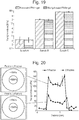
- the stiffness of the support need not be uniform and may vary depending on the shape, size and quantity of an condensate of interest. It is possible to provide the stiffness with a spatial/temporal gradient (as in Example 6 to be described later) or a pattern (as in Example 7 to be described later).
- the stiffness of the support is uniform, it is preferably 100 kPa or less, more preferably 1-50 kPa.
- the gel-like support may be planar, or the side on which culture is to be performed may have a U- or V-shaped cross section. If the side of the gel-like support on which culture is to be performed has a U- or V-shaped cross section, cells tend to gather on the culture surface and a cell condensate can advantageously be formed from a smaller number of cells and/or tissues.
- the support may be modified chemically or physically. Examples of modifying substances include, but are not limited to, Matrigel, laminin, entactin, collagen, fibronectin and vitronectin.
- the stiffness of the central part is 200 kPa or less and it suffices that the peripheral part is softer than the central part.
- Appropriate values for the stiffness of the central and peripheral parts of the substrate are variable depending on the coating and the shape.
- Another example of the gel-like culture support that is provided with a spatial gradient of stiffness is a gel-like culture support whose stiffness in the peripheral part is greater than the stiffness in the central part.
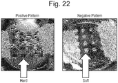
- the patterned, gel-like culture support is a gel-like culture support having one or more patterns in which the stiffness of the central part is greater than the stiffness of the peripheral part (see Example 7 to be described later; Fig. 22 , left panel: positive pattern).
- the stiffness of the central part is 200 kPa or less; it suffices that the peripheral part is softer than the central part.
- Appropriate values for the stiffness of the central and peripheral parts of the substrate are variable depending on the coating and the shape.
- Another example of the patterned, gel-like culture support is a gel-like culture support having one or more patterns in which the stiffness of the peripheral part is greater than the stiffness of the central part (see Example 7 to be described later; Fig.
- the stiffness of the peripheral part is 200 kPa or less; it suffices that the central part is softer than the peripheral part.
- Appropriate values for the stiffness of the central and peripheral parts of the substrate are variable depending on the coating and the shape.
- the temperature at the time of culture is not particularly limited but it is preferably 30-40°C and more preferably 37°C.
- an increased amount of oxygen is preferably supplied into the incubator.
- the amount of oxygen supply is appropriately 4-50%, preferably 10-30%, and more preferably 18-25%.
- the culture period is not particularly limited but it is preferably 12-144 hr.
- the culture period is preferably 12-48 hr.
- the culture period is preferably 12-144 hr.
- the culture period is preferably 12-96 hr.
- the culture period is preferably 12-96 hr.
- the culture period is preferably 12-96 hr.
- the culture period is preferably 12-144 hr.
- the culture period is preferably 12-144 hr.
- the culture period is preferably 12-144 hr.
- the culture period is preferably 12-144 hr.
- the culture period is preferably 48-144 hr.
- cell-cell interactions have taken place in such a close manner that a biological environment as occurs in the womb is recapitulated.
- induction of early differentiation into organ progenitor cells occurs efficiently and this would improve the frequency and number of such cells.
- cells adhere to each other so strongly that they can be collected in a non-destructive manner.
- the cell condensate described in the present application is a concept typically encompassing organ buds and organoids [organ bud ( WO2013/047639 ), liver bud, liver diverticula, liver organoid, pancreatic (dorsal or ventral) buds, pancreatic diverticula, pancreatic organoid, intestinal bud, intestinal diverticula, intestinal organoid ( K. Matsumoto et al. Science.19; 294 (5542): 559-63 (2001 )].
- organ bud WO2013/047639
- liver bud liver diverticula
- liver organoid pancreatic (dorsal or ventral) buds
- pancreatic diverticula pancreatic organoid
- intestinal bud intestinal diverticula
- intestinal organoid K. Matsumoto et al. Science.19; 294 (5542): 559-63 (2001 )
- the cell condensates are independent of the types of constituent cells and the number of such types.
- organ buds correspond to cell condensates that are formed at an early stage of organogenesis and are in principle composed of the following three types of cells: functional cells that constitute organs or tissues (or undifferentiated cells which will differentiate into functional cells); vascular cells; and mesenchymal cells.
- Organoids are solely composed of cells that constitute epithelial tissues and they are basically of a small size (1 mm or less).
- Cell condensates undergo self-organization to form three-dimensional tissue structures provided with higher structures, whereby progenitor cells can be directed to terminal differentiation.
- Self-organization may be performed either in vivo or in vitro. For example, when a cell condensate prepared by the method of the present invention is transplanted into a living body, vascular networks are formed, blood perfusion is induced, and self-organization into a higher tissue with a complex structure occurs, enabling the preparation of tissues/organs that have a highly ordered tissue structure comparable to that of adult tissues.
- the cell condensate of the present invention it may be possible to prepare a higher tissue that is provided with not only a vascular network but also higher structures such as ureteral structure, biliary structure, tracheal structure, etc. Further, a great number of organs essentially require that reconstitution associated with other organs be realized in order to exhibit their functions; e.g., in liver, reconstitution of junctions with bile duct and pancreatic duct and connection to duodenum is essential for exhibiting its function. According to the present invention, a cell condensate which recapitulates interactions with other organs is prepared. This cell condensate is expected to find use as a system for inducing self-organization into complex organs existing in the body.
- the present invention also provides a cell condensate prepared by the above-described method.
- the present invention also provides a method of three-dimensional tissue structure, comprising allowing self-organization of a cell condensate prepared by the above-described method to form a three-dimensional tissue structure that has been provided with higher structures.
- the present invention also provides a gel-like culture support wherein the side on which culture is performed has a U- or V-shaped cross-section.
- the gel-like culture support of the present invention having a U- or V-shaped cross-section on the side where culture is performed, allows cells to gather on the culture surface to ensure that a cell condensate is advantageously formed from a smaller number of cells and/or tissues.
- the gel-like culture support wherein the side on which culture is performed has a U- or V-shaped cross-section is as defined above.
- the present invention also provides a gel-like culture support wherein the stiffness of the central part thereof is greater than the stiffness of the peripheral part thereof.
- a gel-like culture support wherein the stiffness of the central part thereof is greater than the stiffness of the peripheral part thereof.
- One embodiment of such culture support is shown in Example 6 to be described later ( Figs. 20 and 21 ).
- the stiffness of the central part is 200 kPa or less; it suffices that the peripheral part is softer than the central part.
- Appropriate values for the stiffness of the central and peripheral parts of the support are variable depending on the coating and the shape.
- the present invention also provides a gel-like culture support in which the stiffness of the peripheral part thereof is greater than the stiffness of the central part thereof.
- the present invention also provides a gel-like culture support having one or more patterns in which the stiffness of the central part is greater than the stiffness of the peripheral part.
- a gel-like culture support having one or more patterns in which the stiffness of the central part is greater than the stiffness of the peripheral part.
- One embodiment of such culture support is given in Example 7 to be described later ( Fig. 22 , left panel: positive pattern).
- the stiffness of the central part is 200 kPa or less; it suffices that the peripheral part is softer than the central part.
- Appropriate values for the stiffness of the central and peripheral parts of the support are variable depending on the coating and the shape.
- the present invention also provides a gel-like culture support having one or more patterns in which the stiffness of the peripheral part is greater than the stiffness of the central part.
- a gel-like culture support having one or more patterns in which the stiffness of the peripheral part is greater than the stiffness of the central part.
- One embodiment of such culture support is given in Example 7 described later ( Fig. 22 , right panel: negative pattern).
- the stiffness of the peripheral part is 200 kPa or less; it suffices that the central part is softer than the central part.
- Appropriate values for the stiffness of the central and peripheral parts of the support are variable depending on the coating and the shape.
- the present invention also provides a method of preparing a cell condensate in vitro , comprising culturing a mixture of cells and/or tissues of a desired type and mesenchymal cells on the above-described gel-like culture support to thereby form a cell condensate. Culturing of the mixture of the cells and/or tissues of a desired type and the mesenchymal cells is as defined above.
- liver primordia (of millimeter scale) were autonomously formed from isolated human liver progenitor cells in vitro by recapitulating the cell-cell interactions which would occur at organogenesis stages.
- the present inventors revealed that this 3D tissue formation started from self-assembly behavior of multiple cell units and that the presence of the cytoskeletal contractile force of myosin II occurring in mesenchymal stem cells was crucial for the progress of such behavior.
- This dynamic cell collective behavior is regulated by the stiffness conditions of substrate matrix.
- the present inventors succeeded under optimized substrate conditions in preparing three-dimensional organ primordia from cells/tissues isolated from diverse organs including liver, pancreas, intestine, lung, heart, kidney, brain and even cancer.
- the thus prepared three-dimensional primordia were immediately vascularized upon transplantation (since vascular endothelial cells had been incorporated therein), followed by autonomous formation of self-organized three-dimensional tissue structures having therapeutic effects.
- this principle will serve to establish a highly versatile platform for reconstituting a plurality of vascularized, complex organ systems from stem cells via dynamic cell condensation and the subsequent self-organization.
- liver is formed from a condensed tissue mass termed “liver bud” at week 5-8 of gestation in human during physiological organogenesis.
- Liver budding also called “liver bud”
- liver budding also called “liver bud”
- PSCs pluripotent stem cells
- single PSC-derived hepatocytes autonomously form 3-D condensates by co-culture with endothelial cells and mesenchymal cells (2). Once condensates are established, they continue to self-organize after several days under complete in vitro conditions into liver bud tissues having a structure resembling the organs that exist in the womb (3).
- the in vitro grown organ bud is transplanted into a living body, where it undergoes further self-organization (is matured) to eventually become a vascularized and functional liver. This method opens a new road for artificial reconstitution of vascularized organ systems (4).
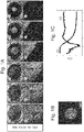
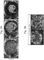
- the present inventors performed a time-lapse imaging analysis to track cellular movements during organoid formation.
- Hepatic endoderm cells derived from human induced pluripotent stem cells (iPSCs), umbilical cord-derived endothelial cells (HUVECs), and mesenchymal stem cells (MSCs) were labeled with distinctive fluorescent markers and cocultured on a solidified matrix gel which was already described.
- Live cell tracking revealed that after rapid cell convergence, the assembly of vascularized organoids was initiated; this was followed by spatial rearrangements via self-organization as demonstrated by the formation of an endothelial-like network ( Fig. 1 ).
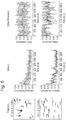
- Fig. 1 To elucidate dynamics of such condensate formation in more detail, the present inventors examined the temporal development of the position of the edge of the cell condensate (square root of cell area) and circularity by image analysis ( Fig. 1b ). The results showed that cell condensates contracted gently at 10 ⁇ m/h or less up to about 7 hr after seeding, and then the contraction accelerated to about 500 ⁇ m/h at naxunyn over the next several hours and finally decreased exponentially to converge. On the other hand, its circularity decreased almost monotonically right after cell seeding and reached a minimal value of about 0.5 in 10-13 hr. The circularity then increased and finally achieved an almost constant value (0.85) at 20 hr after seeding.
- MSCs mesenchymal stem cells
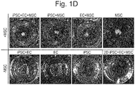
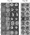
- cell condensate formation was possible in coculture of iPSC-derived hepatic endoderm cells and MSC (iPSC+MSC) or coculture of vascular endothelial cells and MSC (EC+MSC) ( Fig. 1 ).
- iPSC+MSC coculture of iPSC-derived hepatic endoderm cells and MSC
- EC+MSC coculture of vascular endothelial cells and MSC
- the present inventors subsequently assessed the contributions of the contraction force of MSCs at the molecular level against their substratum and the surrounding cells.
- MII myosin II
- the present inventors therefore assessed MII activity by measuring time-course-dependent changes in MIIA phosphorylation with MIIA inactivating S1943 (pS1943) through decomposition of myofilament by phosphate-specific antibodies (7) and intracellular flow cytometry.
- MIIA activity Based on the formula reported to estimate MIIA activity (8), the present inventors showed that active MIIA was remarkably up-regulated in stromal cells during condensate formation and reached its peak at 6 hr, which corresponds to the time at which cells moved at maximum velocity (1). On the other hand, it is seen that activated MIIA is almost constant throughout condensate formation in iPSC-derived hepatocytes. This suggests that the MSC-driven activation of MIIA is responsible for this strong three-dimensional rearrangement. As data indicating direct evidence for the decrease of this activated MIIA, the present inventors showed that this condensate formation could be completely antagonized by treatment with blebbistatin (an MII ATPase inhibitor) (9).
- Rho kinase inhibitor Y-27632 it was found that addition of Rho kinase inhibitor Y-27632 to the cocultures partially delayed condensate formation ( Fig. 1 ).
- Rho kinase inhibitor Y-27632 it was assumed that such mechanism is hard to apply because pharmacological inhibition of chemokine receptor pathways by addition of AMD3100 could not hinder condensate formation (10).
- MSCs that are the key cell in condensate formation in the system of the present invention are known to exhibit mechano-response in diverse processes including differentiation and attachment.
- condensates such as spheroids
- the present inventors have concluded that contraction of mesenchymal cells against softer substrate might have caused these collective behaviors in coculture systems.
- the proposed principle can be expanded to self-organization systems for other organs irrespective of the origin of germ layers that are to be used in the future for the purpose of regenerative medicine.
- the present inventors first selected pancreatic cells and subjected them to coculture, since there is increasing evidence that pancreas follows a developmental program relatively close to that of liver.
- isolated mouse pancreas ⁇ cells MIN6 were cocultured with HUVEC and MSC, a similar formation of cell condensate was observed ( Fig. 3 ).
- the present inventors isolated multiple cells or tissue fragments (up to 200 ⁇ m) from embryonic or adult mice. Surprisingly, the directed and autonomic assembling phenomenon was retained in all the cell/tissue types tested, including pancreas, liver, intestine, lung, heart, kidney, brain, and even cancer ( Fig. 3 ). Time-lapse imaging analyses revealed that both the embryonic and adult cells/tissues successfully resisted additional manipulations (including surgical transplantation) to form single 3D organoids autonomously ( Fig. 3 ).
- Condensates as designed to contain cultured endothelial cells turned out to permit a much more rapid perfusion with recipient circulation after transplantation (average perfusion time: ⁇ 72 hr) compared with reliable conventional tissue engineering approaches (average perfusion time: ⁇ 192 hr).
- pancreatic cells for in-depth characterization.
- the transplantation of 3-D pancreatic organoids resulted in rapid ( ⁇ 48 hr) reperfusion and successful ⁇ cell engraftment. These were confirmed by live imaging analysis.
- the transplants developed islet-like structures ( Fig. 4 , E ) with functional microvascular networks that connected to the recipient circulatory system ( Fig. 4C ).
- Such blood perfusion was not recognized when condensates not containing vascular endothelial cells were transplanted ( Fig. 9 ).
- the reconstituted islets directly connected to peripheral mouse blood vessels to be highly vascularized with a tight network of microvessels ( Fig. 10 ).
- the capillary network in the islet in a living body is known to be approximately 5 times as dense as the capillary network surrounding exocrine secretion tissues. Consistent with this, intravital quantification of the functional vascular density showed that the capillary network was much denser (by 4.2 times) in the reconstituted islet-like tissues than in the areas surrounding the normal tissues ( Fig.4 , Fig. 9 ). Histological analysis also showed that the islet-like tissues had a structure resembling the adult islet, suggesting the reconstitution of a mature tissue via self-organization ( Fig. 11 ).
- in vitro -derived ⁇ cell organoids were transplanted into kidney subcapsule of type 1 fulminant diabetic model mouse.
- the present inventors used a toxin receptor-mediated cell knockout (TREK) Tg mouse having a diphtheria toxin (DT) receptor cDNA transgene in insulin promoter. While mice in non-transplantation group died at day 6 of DT administration-mediated induction of diabetes, those mice which received transplantation of ⁇ cell organoids maintained normal blood glucose levels and survived ( Fig. 4 , G ).
- TREK toxin receptor-mediated cell knockout
- mice in non-transplantation group died at day 6 of DT administration-mediated induction of diabetes
- those mice which received transplantation of ⁇ cell organoids maintained normal blood glucose levels and survived ( Fig. 4 , G ).
- the present inventors have demonstrated the applicablility of the foregoing principle to other organ systems by experimentally recapitulating vascularization and reconstituting a functional three-dimensional tissue in vivo.
- the principle under consideration ensures that starting with larger numbers of the desired cells/tissues, self-organized organoids can be designed via condensation.
- the condensates may be used for examining the subsequent self-organization capacity both in vitro and in vivo.
- rapid vasculogenesis and subsequent functionalization were evaluated by incorporating endothelial cells experimentally.
- evaluating the contribution of undeveloped supporting cells such as neurons is also an interesting topic for the present inventors and other research groups.
- MCs mesenchymal cells
- MCs any of the following cells was used: cells isolated from human bone marrow, cells isolated from umbilical cord stroma (Wharton's sheath), cells isolated from human auricle, cells isolated from mouse bone marrow, human fibroblast cells or the like.
- the mesenchymal stem cells isolated from human bone marrow (hMSCs) that were mainly used in this experiment had been cultured using MSCGMTM BulletKitTM (Lonza PT-3001), a medium prepared exclusively for hMSC culture.
- C57BL/6-Tg mice (Nippon SLC) at days 12-17 of gestation were disinfected with 70% ethanol and incised to remove embryos. Brain, heart, lung, liver, metanephros or intestine was removed from the embryos. Brain, heart, lung, liver, kidney or intestine was also removed from C57BL/6-BALB/c RFP hairy mice 6 or more weeks of age (purchased from Anticancer Inc.). When cells isolated from these removed tissues were used, they were put in 200 ⁇ l of 0.05% Tryspin-EDTA (GIBCO) and incubated for 20 min at 37°C.
- GEBCO Tryspin-EDTA
- the tissues were disrupted with a pipette and added to 4.8 ml of a medium. After centrifugation, medium was added and the number of cells was counted. Then, enzyme treatment was conducted to give single cells, which were subsequently used for coculture.
- the removed tissues were to be used in a state of small tissues, the removed embryonic tissues were minced with scissors, put in 10 ml of 0.05% Tryspin-EDTA and shaken for 20 min at 37°C. After addition of medium, the resultant cells were passed through a 100 ⁇ m cell strainer and centrifuged. After centrifugation, medium was added for use in cell culture.
- the brain, heart, lung and kidney of the adult mice were minced with scissors and passed through a 100 ⁇ m cell strainer.
- the resultant flow-through was filtered with a 40 ⁇ m cell strainer.
- the cell mass remaining on this cell strainer was collected with medium for use in coculture of cells.
- the contents were washed with physiological saline.
- the washed small intestine was cut lengthwise at intervals of 4 cm.
- the resultant sections were put in 2 mM EDTA, 0.5 mM DTT in PBS and shaken for 20 min at 37°C.
- the cells were passed through a 100 ⁇ m cell strainer, followed by addition of PBS. After centrifugation, the supernatant was suctioned and PBS was added for washing. Then the cells were centrifuged, and medium was added for use in cell culture.
- EGMTM BulletKitTM Lonza CC-4133
- HepG2 was cultured in DMEM supplemented with 10% FBS. Each type of cells were cultured in a 37°C, 5%CO 2 incubator.
- 4x10 3 cells or more of multiple types in any combination tissues isolated from embryo or adult, or KO-HepG2, HUVEC
- 5x10 3 or more MSCs were mixed with 5x10 3 or more MSCs and seeded.
- the cells were subsequently cultured in a 37°C incubator for a day.
- chronological observation of cell coculture was performed with a stereomicroscope or a confocal microscope.
- the cell types in "any combination” For example, cells derived from different tissues such as pancreas, liver, intestine, nerve, etc. may be mixed and used. Subsequently, an optimal composition for self-organization of an organ of interest could be used.
- a 10 ml solution was prepared by mixing aqueous acrylamide solution (40% w/v, A4058, Sigma), aqueous bis-acrylamide solution (2% w/v, M1533, Sigma) and distilled water. In the process, the Young's modulus of the gel was adjusted by changing the mixing ratio of the individual solutions. The resultant reaction solution was bumped using a vacuum chamber. Then, 100 ⁇ l of APS (5 g/DW 50 ml, 01307-00, KANTO, 0.20 mm filtered) and 10 ⁇ l of TEMED (T9281, Sigma) were sequentially added to the reaction solution.
- APS g/DW 50 ml, 01307-00, KANTO, 0.20 mm filtered
- 10 ⁇ l of TEMED T9281, Sigma
- Coating of adhesion molecules (Matrigel or laminin) onto the PA gel surface was performed by the procedures described below. First, 0.2 mg/ml N-sulfosuccinimidyl-6-(4'-azido-2'-nitrophenylamino)hexanoate (Sulfo-SANPAH, 22589, Pierce) in 20 mM HEPES (pH 8.5) was dripped onto the PA gel substrate, followed by irradiation with a UV lamp (Z169633-1EA, Sigma) for 20 min.
- a UV lamp Z169633-1EA, Sigma
- a 10 ml solution was prepared by mixing aqueous acrylamide solution (40% w/v, A4058, Sigma), aqueous bis-acrylamide solution (2% w/v, M1533, Sigma) and distilled water. In the process, the Young's modulus of the gel was adjusted by changing the mixing ratio of the individual solutions.
- This reaction solution 500 ⁇ l was added to a 24-well tissue culture plate (353047, BD). Then, 0.5 ⁇ l of TEMED (T9281, Sigma) and 5 ⁇ l of APS (5 g/DW 50 ml, 01307-00, KANTO, 0.20 mm filtered) were added to the reaction solution in this order, immediately followed by thorough mixing.
- the cells were mixed with mesenchymal stem cells isolated from human bone marrow (hMSCs) and normal umbilical vein endothelial cells (HUVECs) and seeded on wells where a solution obtained by mixing Matrigel (the stock solution of Matrigel (BD) used in Example 2) and a medium for vascular endothelial cells (EGM BulletKitTM, Lonza CC-4133) at 1:1 had been solidified.
- Matrigel the stock solution of Matrigel (BD) used in Example 2
- EMM BulletKitTM the medium for vascular endothelial cells
- the renal primordium formed was transplanted into the wombs of immunodeficiency mice.
- blood perfusion was recognized in two to three days after transplantation ( Fig. 15 , upper row).
- the white dotted lines in Fig. 15 indicate the transplantation areas.
- Scattered cells formed spherical, glomerular tissues at day 8 of transplantation ( Fig. 15 , bottom row).
- the results of fluorescence observation as shown in the left panel of Fig. 16 revealed that a great number of glomerular structures were formed by culturing on a support but that this was not the case when the conventional method (pellet transplantation group) was applied.
- the tissues formed first flowed into blood vessels, were filtered inside glomeruli, and collected in proximal tubules, indicating that they had the primitive urine producing function of the kidney ( Fig. 18 ).
- the renal primordium artificially prepared according to the present invention autonomous maturation could successfully be induced to prepare functional renal tissues.
- Example 5 Changes in the Young's Modulus of supports (see “preparation of PA gel planar substrate") with different stiffness conditions (Samples A, B and C) before (oblique lines) and after (solid black) Matrigel coating. It was shown that the stiffness conditions can be strictly controlled regardless of the presence or absence of the coating ( Fig. 19 ).
- the gel substrate used in Example 5 was prepared according to the method described in Example 1.
- Gels having multiple stiffness patterns providing different stiffness conditions could successfully be prepared on one substrate ( Fig. 20 ).
- pattern 1 a gel with a hard central part was prepared and according to pattern 2, a gel with a less hard central part was prepared ( Fig. 20 , left panel).
- the right panel of Fig. 20 shows the results of measurement of stiffness conditions along the major axis, indicating that the intended stiffness conditions could be achieved.
- Gel substrates having spatial patterns of stiffness were prepared by the method described below.
- a gel reaction solution a 10 ml solution was prepared by mixing aqueous acrylamide solution (40% w/v, A4058, Sigma), aqueous bis-acrylamide solution (2% w/v, M1533, Sigma) and distilled water. Subsequently, with light shielded, 50 mg of Irgacure 2959 (0.5 % w/v, DY15444, Ciba) was added and dissolved in a hot water bath at 37°C. The resultant reaction solution was bumped in a vacuum chamber.
- a circular mask (12 mm o.d. and 2-4 mm i.d.) was prepared.
- a mercury lamp C - HGFI, Nikon
- fiber optics was combined with a light projection tube to ensure uniform irradiation of the reaction solution.
- the time of UV irradiation was adjusted by minutes depending on the desired stiffness.
- distilled water was added to the sandwiched sample and the glass coverslip was peeled off from the glass slide, leaving a gel coat on the former.
- Phosphate buffer was added to the resultant glass coverslip, which was then left to stand for one day to remove the unreacted monomers.
- the Young's moduli of gels were determined by nanoindentation measurements performed with an atomic force microscope (Nanowizard 3, JPK Instruments, Germany). Coating of adhesion molecules (Matrigel or laminin) onto the PA gel surface was performed by the procedures described below. First, 0.2 mg/ml N-sulfosuccinimidyl-6-(4'-azido-2'-nitrophenylamino)hexanoate (Sulfo-SANPAH, 22589, Pierce) in 20 mM HEPES (pH 8.5) was dripped onto the PA gel substrate, followed by irradiation with a UV lamp (Z169633-1EA, Sigma) for 20 min.
- a UV lamp Z169633-1EA, Sigma
- iPS cell-derived hepatic endoderm cells, HUVECs and MSCs were mixed at a ratio of 10:7:2 and the mixture was seeded on the patterned gels to give a total cell count of about 2x10 6 cells ( Fig. 21 ).
- the optimal condition for the stiffness of the central part was 100 kPa.
- a positive pattern was so designed that individual circular parts were hard and their periphery was soft ( Fig. 22 , left panel).
- a negative pattern was so designed that individual circular parts were soft and their periphery was hard ( Fig. 22 , right panel).
- Gel substrates with such multiple patterns of stiffness were prepared based on the technique described in Example 6 and by exposing a gel substrate (25 mm in diameter) through a photomask with a 4x4 pattern of circles (diameter: about 2 mm; center-to-center distance between circles: about 2.7 mm). It is believed that by using these patterned supports, cell condensates of any size may be formed at any place.
- the present invention is applicable in various fields including search for new drugs and evaluation of their efficacy, regenerative medicine, diagnosis of diseases and pathology, and production of useful substances.
Landscapes
- Health & Medical Sciences (AREA)
- Life Sciences & Earth Sciences (AREA)
- Engineering & Computer Science (AREA)
- Chemical & Material Sciences (AREA)
- Biomedical Technology (AREA)
- Zoology (AREA)
- General Health & Medical Sciences (AREA)
- Genetics & Genomics (AREA)
- Cell Biology (AREA)
- Organic Chemistry (AREA)
- Wood Science & Technology (AREA)
- Bioinformatics & Cheminformatics (AREA)
- Biotechnology (AREA)
- Oral & Maxillofacial Surgery (AREA)
- Transplantation (AREA)
- Epidemiology (AREA)
- Dermatology (AREA)
- Animal Behavior & Ethology (AREA)
- Medicinal Chemistry (AREA)
- Veterinary Medicine (AREA)
- Public Health (AREA)
- Microbiology (AREA)
- Chemical Kinetics & Catalysis (AREA)
- Biochemistry (AREA)
- General Engineering & Computer Science (AREA)
- Botany (AREA)
- Dispersion Chemistry (AREA)
- Developmental Biology & Embryology (AREA)
- Micro-Organisms Or Cultivation Processes Thereof (AREA)
Applications Claiming Priority (2)
| Application Number | Priority Date | Filing Date | Title |
|---|---|---|---|
| JP2014037341 | 2014-02-27 | ||
| PCT/JP2015/055695 WO2015129822A1 (fr) | 2014-02-27 | 2015-02-26 | Procédé de fabrication d'un agrégat cellulaire dans l'objectif de son organisation automatique |
Publications (3)
| Publication Number | Publication Date |
|---|---|
| EP3124600A1 true EP3124600A1 (fr) | 2017-02-01 |
| EP3124600A4 EP3124600A4 (fr) | 2017-12-06 |
| EP3124600B1 EP3124600B1 (fr) | 2021-12-29 |
Family
ID=54009136
Family Applications (1)
| Application Number | Title | Priority Date | Filing Date |
|---|---|---|---|
| EP15754683.9A Active EP3124600B1 (fr) | 2014-02-27 | 2015-02-26 | Rocédé de fabrication d'un condensé cellulaire dans l'objectif de son organisation automatique |
Country Status (10)
| Country | Link |
|---|---|
| US (1) | US20170067014A1 (fr) |
| EP (1) | EP3124600B1 (fr) |
| JP (1) | JP6489484B2 (fr) |
| KR (1) | KR102338698B1 (fr) |
| CN (1) | CN106062181A (fr) |
| AU (1) | AU2015223798B2 (fr) |
| BR (1) | BR112016019677A8 (fr) |
| CA (1) | CA2937882A1 (fr) |
| SG (2) | SG10202107992XA (fr) |
| WO (1) | WO2015129822A1 (fr) |
Families Citing this family (15)
| Publication number | Priority date | Publication date | Assignee | Title |
|---|---|---|---|---|
| US9719068B2 (en) | 2010-05-06 | 2017-08-01 | Children's Hospital Medical Center | Methods and systems for converting precursor cells into intestinal tissues through directed differentiation |
| CN113249297A (zh) | 2014-05-28 | 2021-08-13 | 儿童医院医疗中心 | 用于经由定向分化将前体细胞转化为胃组织的方法和系统 |
| EP3207123A1 (fr) | 2014-10-17 | 2017-08-23 | Children's Hospital Center D/b/a Cincinnati Children's Hospital Medical Center | Modèle in vivo d'intestin grêle humain faisant intervenir des cellules souches pluripotentes et ses procédés de fabrication et d'utilisation |
| KR102015205B1 (ko) | 2015-02-27 | 2019-08-27 | 더 솔크 인스티튜트 포 바이올로지칼 스터디즈 | 리프로그래밍 전구체 조성물 및 그의 사용 방법 |
| CA3008218A1 (fr) | 2015-12-22 | 2017-06-29 | Public University Corporation Yokohama City University | Modele d'infection virale, son procede de preparation, et son utilisation |
| US20190093082A1 (en) * | 2016-03-07 | 2019-03-28 | Centre National De La Recherche Scientifique (Cnrs) | Method of Differentiating Pluripotent Stem Cells |
| US11066650B2 (en) | 2016-05-05 | 2021-07-20 | Children's Hospital Medical Center | Methods for the in vitro manufacture of gastric fundus tissue and compositions related to same |
| JP7178264B2 (ja) | 2016-05-25 | 2022-11-25 | ソーク インスティチュート フォー バイオロジカル スタディーズ | オルガノイド作製および疾患モデル化のための組成物および方法 |
| EP3527655A4 (fr) * | 2016-10-11 | 2020-05-06 | Tokushima University | Procédé de production d'un tissu biologique de type rénal |
| US11767515B2 (en) | 2016-12-05 | 2023-09-26 | Children's Hospital Medical Center | Colonic organoids and methods of making and using same |
| US10767164B2 (en) | 2017-03-30 | 2020-09-08 | The Research Foundation For The State University Of New York | Microenvironments for self-assembly of islet organoids from stem cells differentiation |
| AU2018336901B2 (en) * | 2017-09-21 | 2024-07-18 | President And Fellows Of Harvard College | Tissue construct, methods of producing and using the same |
| US20240125767A1 (en) | 2019-10-17 | 2024-04-18 | Public University Corporation Yokohama City University | Method for evaluating drug toxicity |
| CN115916960A (zh) | 2020-06-08 | 2023-04-04 | 国立大学法人东京医科齿科大学 | 细胞培养方法 |
| CN113350574B (zh) * | 2021-05-26 | 2022-11-18 | 泸州国之荣耀酒业有限公司 | 图案化类肝小叶微组织制造方法 |
Family Cites Families (8)
| Publication number | Priority date | Publication date | Assignee | Title |
|---|---|---|---|---|
| WO2008094597A2 (fr) * | 2007-01-30 | 2008-08-07 | University Of Georgia Research Foundation, Inc. | Cellules mésodermiques précoces, une population stable de cellules mésendodermiques qui a une utilité pour la génération de lignées endodermiques et mésodermiques et de cellules migratoires multipotentes (mmc) |
| EP2140267B1 (fr) * | 2007-02-26 | 2017-11-15 | StemCell Technologies Inc. | Procede permettant de reduire la courbure du menisque d'un milieu liquide |
| WO2012119074A1 (fr) * | 2011-03-03 | 2012-09-07 | Massachusetts Institute Of Technology | Appareil et procédé pour organiser des structures de cellules en trois dimensions au moyen de gradients de rigidité et de gels sacrificiels |
| KR101282926B1 (ko) * | 2011-07-28 | 2013-07-08 | 고려대학교 산학협력단 | 표면장력을 이용한 반구형 마이크로웰의 제조 및 이를 이용한 세포 집합체의 형성 |
| US11326150B2 (en) * | 2011-09-27 | 2022-05-10 | Public University Corporation Yokohama City University | Method for producing tissue and organ |
| WO2013065763A1 (fr) * | 2011-10-31 | 2013-05-10 | 独立行政法人理化学研究所 | Procédé de culture d'une cellule souche |
| US20130252337A1 (en) * | 2012-03-21 | 2013-09-26 | Shengyuan Yang | Substrates with micrometer and nanometer scale stiffness patterns for use in cell and tissue culturing and a method for making same |
| CA2972057C (fr) * | 2014-12-22 | 2023-06-13 | Ecole Polytechnique Federale De Lausanne (Epfl) | Films d'hydrogel minces microstructures |
-
2015
- 2015-02-26 AU AU2015223798A patent/AU2015223798B2/en active Active
- 2015-02-26 SG SG10202107992XA patent/SG10202107992XA/en unknown
- 2015-02-26 KR KR1020167025902A patent/KR102338698B1/ko active IP Right Grant
- 2015-02-26 BR BR112016019677A patent/BR112016019677A8/pt active Search and Examination
- 2015-02-26 JP JP2016505305A patent/JP6489484B2/ja active Active
- 2015-02-26 CN CN201580003897.6A patent/CN106062181A/zh active Pending
- 2015-02-26 US US15/121,934 patent/US20170067014A1/en active Pending
- 2015-02-26 EP EP15754683.9A patent/EP3124600B1/fr active Active
- 2015-02-26 WO PCT/JP2015/055695 patent/WO2015129822A1/fr active Application Filing
- 2015-02-26 CA CA2937882A patent/CA2937882A1/fr not_active Abandoned
- 2015-02-26 SG SG11201606750UA patent/SG11201606750UA/en unknown
Also Published As
| Publication number | Publication date |
|---|---|
| AU2015223798B2 (en) | 2020-10-15 |
| EP3124600B1 (fr) | 2021-12-29 |
| SG11201606750UA (en) | 2016-10-28 |
| WO2015129822A1 (fr) | 2015-09-03 |
| CA2937882A1 (fr) | 2015-09-03 |
| KR20160125440A (ko) | 2016-10-31 |
| EP3124600A4 (fr) | 2017-12-06 |
| AU2015223798A1 (en) | 2016-08-18 |
| US20170067014A1 (en) | 2017-03-09 |
| JPWO2015129822A1 (ja) | 2017-03-30 |
| JP6489484B2 (ja) | 2019-03-27 |
| BR112016019677A8 (pt) | 2021-07-13 |
| SG10202107992XA (en) | 2021-09-29 |
| CN106062181A (zh) | 2016-10-26 |
| KR102338698B1 (ko) | 2021-12-10 |
| BR112016019677A2 (pt) | 2017-08-15 |
Similar Documents
| Publication | Publication Date | Title |
|---|---|---|
| EP3124600B1 (fr) | Rocédé de fabrication d'un condensé cellulaire dans l'objectif de son organisation automatique | |
| JP7125717B2 (ja) | 培養方法 | |
| Ong et al. | In vivo therapeutic applications of cell spheroids | |
| Bernstein et al. | Stem cell therapy for cardiac disease | |
| US20150368618A1 (en) | Modulation of cardiac stem-progenitor cell differentiation, assays and uses thereof | |
| US12012616B2 (en) | Formation of three-dimensional organ from pluripotent stem cells | |
| JP2013510582A (ja) | 間葉幹細胞の球状集合体 | |
| Liu et al. | Generation of functional organs from stem cells | |
| Polak et al. | Stem cells bioprocessing: an important milestone to move regenerative medicine research into the clinical arena | |
| Ren et al. | On the road to bioartificial organs | |
| JP5893562B2 (ja) | 乳動脈由来細胞並びに組織修復及び再生における使用方法 | |
| JP7444367B2 (ja) | 増幅毛包間葉系細胞の製造方法及びその使用 | |
| Sun et al. | Isolation of ready-made rat microvessels and its applications in effective in vivo vascularization and in angiogenic studies in vitro | |
| WO2018225705A1 (fr) | Procédé de fabrication de culture cellulaire | |
| WO2009080794A1 (fr) | Procédé de préparation de matrices extracellulaires spécifiques de cellules | |
| Kim et al. | A fibrin-supported myocardial organ culture for isolation of cardiac stem cells via the recapitulation of cardiac homeostasis | |
| JP7228269B2 (ja) | 細胞塊融合法 | |
| JPWO2020067436A1 (ja) | 多能性幹細胞由来細胞の移植片形成方法 | |
| JPWO2020067435A1 (ja) | 多能性幹細胞由来細胞のシート化方法 | |
| Hu et al. | Recellularization of Decellularized Whole Organ Scaffolds: Elements, Progresses, and Challenges | |
| Barbarisi et al. | Regenerative medicine: current and potential applications | |
| JPWO2020067438A1 (ja) | 多能性幹細胞由来細胞のシート化方法 | |
| Sarson | Shortened Poly-N-Acetyl Glucosamine (sNAG) Nanofibers Induce Rapid Vascular Assembly in 3-Dimensional Microtissue Spheroids | |
| Chong | Defining the origin of adult cardiac multipotent colony forming stem cells |
Legal Events
| Date | Code | Title | Description |
|---|---|---|---|
| STAA | Information on the status of an ep patent application or granted ep patent |
Free format text: STATUS: THE INTERNATIONAL PUBLICATION HAS BEEN MADE |
|
| PUAI | Public reference made under article 153(3) epc to a published international application that has entered the european phase |
Free format text: ORIGINAL CODE: 0009012 |
|
| STAA | Information on the status of an ep patent application or granted ep patent |
Free format text: STATUS: REQUEST FOR EXAMINATION WAS MADE |
|
| 17P | Request for examination filed |
Effective date: 20160914 |
|
| AK | Designated contracting states |
Kind code of ref document: A1 Designated state(s): AL AT BE BG CH CY CZ DE DK EE ES FI FR GB GR HR HU IE IS IT LI LT LU LV MC MK MT NL NO PL PT RO RS SE SI SK SM TR |
|
| AX | Request for extension of the european patent |
Extension state: BA ME |
|
| DAX | Request for extension of the european patent (deleted) | ||
| A4 | Supplementary search report drawn up and despatched |
Effective date: 20171106 |
|
| RIC1 | Information provided on ipc code assigned before grant |
Ipc: A61L 27/00 20060101ALI20171027BHEP Ipc: C12N 5/10 20060101ALI20171027BHEP Ipc: C12N 5/077 20100101ALI20171027BHEP Ipc: C12N 5/071 20100101AFI20171027BHEP Ipc: C12N 5/0735 20100101ALI20171027BHEP Ipc: C12M 3/00 20060101ALI20171027BHEP Ipc: C12N 5/0775 20100101ALI20171027BHEP |
|
| STAA | Information on the status of an ep patent application or granted ep patent |
Free format text: STATUS: EXAMINATION IS IN PROGRESS |
|
| 17Q | First examination report despatched |
Effective date: 20181210 |
|
| STAA | Information on the status of an ep patent application or granted ep patent |
Free format text: STATUS: EXAMINATION IS IN PROGRESS |
|
| GRAP | Despatch of communication of intention to grant a patent |
Free format text: ORIGINAL CODE: EPIDOSNIGR1 |
|
| STAA | Information on the status of an ep patent application or granted ep patent |
Free format text: STATUS: GRANT OF PATENT IS INTENDED |
|
| INTG | Intention to grant announced |
Effective date: 20210420 |
|
| RAP1 | Party data changed (applicant data changed or rights of an application transferred) |
Owner name: PUBLIC UNIVERSITY CORPORATION YOKOHAMA CITY UNIVERSITY |
|
| RIN1 | Information on inventor provided before grant (corrected) |
Inventor name: YOSHIKAWA, HIROSHI Inventor name: TANIGUCHI, HIDEKI Inventor name: TAKEBE, TAKANORI |
|
| GRAJ | Information related to disapproval of communication of intention to grant by the applicant or resumption of examination proceedings by the epo deleted |
Free format text: ORIGINAL CODE: EPIDOSDIGR1 |
|
| STAA | Information on the status of an ep patent application or granted ep patent |
Free format text: STATUS: EXAMINATION IS IN PROGRESS |
|
| INTC | Intention to grant announced (deleted) | ||
| GRAP | Despatch of communication of intention to grant a patent |
Free format text: ORIGINAL CODE: EPIDOSNIGR1 |
|
| STAA | Information on the status of an ep patent application or granted ep patent |
Free format text: STATUS: GRANT OF PATENT IS INTENDED |
|
| INTG | Intention to grant announced |
Effective date: 20211001 |
|
| GRAS | Grant fee paid |
Free format text: ORIGINAL CODE: EPIDOSNIGR3 |
|
| GRAA | (expected) grant |
Free format text: ORIGINAL CODE: 0009210 |
|
| STAA | Information on the status of an ep patent application or granted ep patent |
Free format text: STATUS: THE PATENT HAS BEEN GRANTED |
|
| AK | Designated contracting states |
Kind code of ref document: B1 Designated state(s): AL AT BE BG CH CY CZ DE DK EE ES FI FR GB GR HR HU IE IS IT LI LT LU LV MC MK MT NL NO PL PT RO RS SE SI SK SM TR |
|
| REG | Reference to a national code |
Ref country code: GB Ref legal event code: FG4D |
|
| REG | Reference to a national code |
Ref country code: CH Ref legal event code: EP |
|
| REG | Reference to a national code |
Ref country code: DE Ref legal event code: R096 Ref document number: 602015076072 Country of ref document: DE |
|
| REG | Reference to a national code |
Ref country code: AT Ref legal event code: REF Ref document number: 1458680 Country of ref document: AT Kind code of ref document: T Effective date: 20220115 |
|
| REG | Reference to a national code |
Ref country code: IE Ref legal event code: FG4D |
|
| REG | Reference to a national code |
Ref country code: LT Ref legal event code: MG9D |
|
| PG25 | Lapsed in a contracting state [announced via postgrant information from national office to epo] |
Ref country code: RS Free format text: LAPSE BECAUSE OF FAILURE TO SUBMIT A TRANSLATION OF THE DESCRIPTION OR TO PAY THE FEE WITHIN THE PRESCRIBED TIME-LIMIT Effective date: 20211229 Ref country code: LT Free format text: LAPSE BECAUSE OF FAILURE TO SUBMIT A TRANSLATION OF THE DESCRIPTION OR TO PAY THE FEE WITHIN THE PRESCRIBED TIME-LIMIT Effective date: 20211229 Ref country code: FI Free format text: LAPSE BECAUSE OF FAILURE TO SUBMIT A TRANSLATION OF THE DESCRIPTION OR TO PAY THE FEE WITHIN THE PRESCRIBED TIME-LIMIT Effective date: 20211229 Ref country code: BG Free format text: LAPSE BECAUSE OF FAILURE TO SUBMIT A TRANSLATION OF THE DESCRIPTION OR TO PAY THE FEE WITHIN THE PRESCRIBED TIME-LIMIT Effective date: 20220329 |
|
| REG | Reference to a national code |
Ref country code: NL Ref legal event code: MP Effective date: 20211229 |
|
| REG | Reference to a national code |
Ref country code: AT Ref legal event code: MK05 Ref document number: 1458680 Country of ref document: AT Kind code of ref document: T Effective date: 20211229 |
|
| PG25 | Lapsed in a contracting state [announced via postgrant information from national office to epo] |
Ref country code: SE Free format text: LAPSE BECAUSE OF FAILURE TO SUBMIT A TRANSLATION OF THE DESCRIPTION OR TO PAY THE FEE WITHIN THE PRESCRIBED TIME-LIMIT Effective date: 20211229 Ref country code: NO Free format text: LAPSE BECAUSE OF FAILURE TO SUBMIT A TRANSLATION OF THE DESCRIPTION OR TO PAY THE FEE WITHIN THE PRESCRIBED TIME-LIMIT Effective date: 20220329 Ref country code: LV Free format text: LAPSE BECAUSE OF FAILURE TO SUBMIT A TRANSLATION OF THE DESCRIPTION OR TO PAY THE FEE WITHIN THE PRESCRIBED TIME-LIMIT Effective date: 20211229 Ref country code: HR Free format text: LAPSE BECAUSE OF FAILURE TO SUBMIT A TRANSLATION OF THE DESCRIPTION OR TO PAY THE FEE WITHIN THE PRESCRIBED TIME-LIMIT Effective date: 20211229 Ref country code: GR Free format text: LAPSE BECAUSE OF FAILURE TO SUBMIT A TRANSLATION OF THE DESCRIPTION OR TO PAY THE FEE WITHIN THE PRESCRIBED TIME-LIMIT Effective date: 20220330 |
|
| PG25 | Lapsed in a contracting state [announced via postgrant information from national office to epo] |
Ref country code: NL Free format text: LAPSE BECAUSE OF FAILURE TO SUBMIT A TRANSLATION OF THE DESCRIPTION OR TO PAY THE FEE WITHIN THE PRESCRIBED TIME-LIMIT Effective date: 20211229 |
|
| PG25 | Lapsed in a contracting state [announced via postgrant information from national office to epo] |
Ref country code: SM Free format text: LAPSE BECAUSE OF FAILURE TO SUBMIT A TRANSLATION OF THE DESCRIPTION OR TO PAY THE FEE WITHIN THE PRESCRIBED TIME-LIMIT Effective date: 20211229 Ref country code: SK Free format text: LAPSE BECAUSE OF FAILURE TO SUBMIT A TRANSLATION OF THE DESCRIPTION OR TO PAY THE FEE WITHIN THE PRESCRIBED TIME-LIMIT Effective date: 20211229 Ref country code: RO Free format text: LAPSE BECAUSE OF FAILURE TO SUBMIT A TRANSLATION OF THE DESCRIPTION OR TO PAY THE FEE WITHIN THE PRESCRIBED TIME-LIMIT Effective date: 20211229 Ref country code: PT Free format text: LAPSE BECAUSE OF FAILURE TO SUBMIT A TRANSLATION OF THE DESCRIPTION OR TO PAY THE FEE WITHIN THE PRESCRIBED TIME-LIMIT Effective date: 20220429 Ref country code: ES Free format text: LAPSE BECAUSE OF FAILURE TO SUBMIT A TRANSLATION OF THE DESCRIPTION OR TO PAY THE FEE WITHIN THE PRESCRIBED TIME-LIMIT Effective date: 20211229 Ref country code: EE Free format text: LAPSE BECAUSE OF FAILURE TO SUBMIT A TRANSLATION OF THE DESCRIPTION OR TO PAY THE FEE WITHIN THE PRESCRIBED TIME-LIMIT Effective date: 20211229 Ref country code: CZ Free format text: LAPSE BECAUSE OF FAILURE TO SUBMIT A TRANSLATION OF THE DESCRIPTION OR TO PAY THE FEE WITHIN THE PRESCRIBED TIME-LIMIT Effective date: 20211229 |
|
| PG25 | Lapsed in a contracting state [announced via postgrant information from national office to epo] |
Ref country code: PL Free format text: LAPSE BECAUSE OF FAILURE TO SUBMIT A TRANSLATION OF THE DESCRIPTION OR TO PAY THE FEE WITHIN THE PRESCRIBED TIME-LIMIT Effective date: 20211229 Ref country code: AT Free format text: LAPSE BECAUSE OF FAILURE TO SUBMIT A TRANSLATION OF THE DESCRIPTION OR TO PAY THE FEE WITHIN THE PRESCRIBED TIME-LIMIT Effective date: 20211229 |
|
| PG25 | Lapsed in a contracting state [announced via postgrant information from national office to epo] |
Ref country code: MC Free format text: LAPSE BECAUSE OF FAILURE TO SUBMIT A TRANSLATION OF THE DESCRIPTION OR TO PAY THE FEE WITHIN THE PRESCRIBED TIME-LIMIT Effective date: 20211229 Ref country code: IS Free format text: LAPSE BECAUSE OF FAILURE TO SUBMIT A TRANSLATION OF THE DESCRIPTION OR TO PAY THE FEE WITHIN THE PRESCRIBED TIME-LIMIT Effective date: 20220429 |
|
| REG | Reference to a national code |
Ref country code: DE Ref legal event code: R097 Ref document number: 602015076072 Country of ref document: DE |
|
| REG | Reference to a national code |
Ref country code: BE Ref legal event code: MM Effective date: 20220228 |
|
| PG25 | Lapsed in a contracting state [announced via postgrant information from national office to epo] |
Ref country code: LU Free format text: LAPSE BECAUSE OF NON-PAYMENT OF DUE FEES Effective date: 20220226 Ref country code: DK Free format text: LAPSE BECAUSE OF FAILURE TO SUBMIT A TRANSLATION OF THE DESCRIPTION OR TO PAY THE FEE WITHIN THE PRESCRIBED TIME-LIMIT Effective date: 20211229 Ref country code: AL Free format text: LAPSE BECAUSE OF FAILURE TO SUBMIT A TRANSLATION OF THE DESCRIPTION OR TO PAY THE FEE WITHIN THE PRESCRIBED TIME-LIMIT Effective date: 20211229 |
|
| PLBE | No opposition filed within time limit |
Free format text: ORIGINAL CODE: 0009261 |
|
| STAA | Information on the status of an ep patent application or granted ep patent |
Free format text: STATUS: NO OPPOSITION FILED WITHIN TIME LIMIT |
|
| 26N | No opposition filed |
Effective date: 20220930 |
|
| PG25 | Lapsed in a contracting state [announced via postgrant information from national office to epo] |
Ref country code: IE Free format text: LAPSE BECAUSE OF NON-PAYMENT OF DUE FEES Effective date: 20220226 |
|
| PG25 | Lapsed in a contracting state [announced via postgrant information from national office to epo] |
Ref country code: SI Free format text: LAPSE BECAUSE OF FAILURE TO SUBMIT A TRANSLATION OF THE DESCRIPTION OR TO PAY THE FEE WITHIN THE PRESCRIBED TIME-LIMIT Effective date: 20211229 Ref country code: BE Free format text: LAPSE BECAUSE OF NON-PAYMENT OF DUE FEES Effective date: 20220228 |
|
| PG25 | Lapsed in a contracting state [announced via postgrant information from national office to epo] |
Ref country code: IT Free format text: LAPSE BECAUSE OF FAILURE TO SUBMIT A TRANSLATION OF THE DESCRIPTION OR TO PAY THE FEE WITHIN THE PRESCRIBED TIME-LIMIT Effective date: 20211229 |
|
| P01 | Opt-out of the competence of the unified patent court (upc) registered |
Effective date: 20230420 |
|
| PG25 | Lapsed in a contracting state [announced via postgrant information from national office to epo] |
Ref country code: HU Free format text: LAPSE BECAUSE OF FAILURE TO SUBMIT A TRANSLATION OF THE DESCRIPTION OR TO PAY THE FEE WITHIN THE PRESCRIBED TIME-LIMIT; INVALID AB INITIO Effective date: 20150226 |
|
| PG25 | Lapsed in a contracting state [announced via postgrant information from national office to epo] |
Ref country code: MK Free format text: LAPSE BECAUSE OF FAILURE TO SUBMIT A TRANSLATION OF THE DESCRIPTION OR TO PAY THE FEE WITHIN THE PRESCRIBED TIME-LIMIT Effective date: 20211229 Ref country code: CY Free format text: LAPSE BECAUSE OF FAILURE TO SUBMIT A TRANSLATION OF THE DESCRIPTION OR TO PAY THE FEE WITHIN THE PRESCRIBED TIME-LIMIT Effective date: 20211229 |
|
| PGFP | Annual fee paid to national office [announced via postgrant information from national office to epo] |
Ref country code: DE Payment date: 20231229 Year of fee payment: 10 Ref country code: GB Payment date: 20240108 Year of fee payment: 10 Ref country code: CH Payment date: 20240301 Year of fee payment: 10 |
|
| PGFP | Annual fee paid to national office [announced via postgrant information from national office to epo] |
Ref country code: FR Payment date: 20240103 Year of fee payment: 10 |
|
| PG25 | Lapsed in a contracting state [announced via postgrant information from national office to epo] |
Ref country code: TR Free format text: LAPSE BECAUSE OF FAILURE TO SUBMIT A TRANSLATION OF THE DESCRIPTION OR TO PAY THE FEE WITHIN THE PRESCRIBED TIME-LIMIT Effective date: 20211229 |