EP3114139B1 - Anticorps contre eotaxin-2 reconnaissant des chemokines supplementaires se liants à ccr3 - Google Patents
Anticorps contre eotaxin-2 reconnaissant des chemokines supplementaires se liants à ccr3 Download PDFInfo
- Publication number
- EP3114139B1 EP3114139B1 EP15714963.4A EP15714963A EP3114139B1 EP 3114139 B1 EP3114139 B1 EP 3114139B1 EP 15714963 A EP15714963 A EP 15714963A EP 3114139 B1 EP3114139 B1 EP 3114139B1
- Authority
- EP
- European Patent Office
- Prior art keywords
- eotaxin
- antibody
- seq
- binding
- ccr3
- Prior art date
- Legal status (The legal status is an assumption and is not a legal conclusion. Google has not performed a legal analysis and makes no representation as to the accuracy of the status listed.)
- Active
Links
- 230000027455 binding Effects 0.000 title claims description 123
- 102000019034 Chemokines Human genes 0.000 title claims description 94
- 108010012236 Chemokines Proteins 0.000 title claims description 94
- 108010083647 Chemokine CCL24 Proteins 0.000 title claims description 87
- 102000006383 Chemokine CCL24 Human genes 0.000 title 1
- 102100036849 C-C motif chemokine 24 Human genes 0.000 claims description 89
- 102100024167 C-C chemokine receptor type 3 Human genes 0.000 claims description 76
- 101710149862 C-C chemokine receptor type 3 Proteins 0.000 claims description 76
- 238000011282 treatment Methods 0.000 claims description 59
- 208000037265 diseases, disorders, signs and symptoms Diseases 0.000 claims description 56
- 102100023688 Eotaxin Human genes 0.000 claims description 52
- 108010055166 Chemokine CCL5 Proteins 0.000 claims description 48
- 239000000427 antigen Substances 0.000 claims description 47
- 108091007433 antigens Proteins 0.000 claims description 47
- 102000036639 antigens Human genes 0.000 claims description 47
- 108010082548 Chemokine CCL11 Proteins 0.000 claims description 45
- 102100032366 C-C motif chemokine 7 Human genes 0.000 claims description 44
- 201000009794 Idiopathic Pulmonary Fibrosis Diseases 0.000 claims description 39
- 201000010099 disease Diseases 0.000 claims description 39
- 208000036971 interstitial lung disease 2 Diseases 0.000 claims description 39
- 239000012634 fragment Substances 0.000 claims description 37
- 206010039710 Scleroderma Diseases 0.000 claims description 34
- 101710155834 C-C motif chemokine 7 Proteins 0.000 claims description 31
- 102000001327 Chemokine CCL5 Human genes 0.000 claims description 31
- 150000007523 nucleic acids Chemical class 0.000 claims description 28
- 230000003176 fibrotic effect Effects 0.000 claims description 27
- 230000000172 allergic effect Effects 0.000 claims description 21
- 208000010668 atopic eczema Diseases 0.000 claims description 21
- 208000037979 autoimmune inflammatory disease Diseases 0.000 claims description 21
- 208000012657 Atopic disease Diseases 0.000 claims description 20
- 102000039446 nucleic acids Human genes 0.000 claims description 19
- 108020004707 nucleic acids Proteins 0.000 claims description 19
- 206010016654 Fibrosis Diseases 0.000 claims description 17
- 239000008194 pharmaceutical composition Substances 0.000 claims description 15
- 239000013604 expression vector Substances 0.000 claims description 13
- 208000008338 non-alcoholic fatty liver disease Diseases 0.000 claims description 8
- 206010053219 non-alcoholic steatohepatitis Diseases 0.000 claims description 8
- 239000003937 drug carrier Substances 0.000 claims description 5
- 208000001145 Metabolic Syndrome Diseases 0.000 claims description 4
- 201000004983 autoimmune atherosclerosis Diseases 0.000 claims description 4
- 230000007882 cirrhosis Effects 0.000 claims description 4
- 208000019425 cirrhosis of liver Diseases 0.000 claims description 4
- 206010061989 glomerulosclerosis Diseases 0.000 claims description 4
- FWMNVWWHGCHHJJ-SKKKGAJSSA-N 4-amino-1-[(2r)-6-amino-2-[[(2r)-2-[[(2r)-2-[[(2r)-2-amino-3-phenylpropanoyl]amino]-3-phenylpropanoyl]amino]-4-methylpentanoyl]amino]hexanoyl]piperidine-4-carboxylic acid Chemical compound C([C@H](C(=O)N[C@H](CC(C)C)C(=O)N[C@H](CCCCN)C(=O)N1CCC(N)(CC1)C(O)=O)NC(=O)[C@H](N)CC=1C=CC=CC=1)C1=CC=CC=C1 FWMNVWWHGCHHJJ-SKKKGAJSSA-N 0.000 claims description 3
- 125000003275 alpha amino acid group Chemical group 0.000 description 89
- 210000004027 cell Anatomy 0.000 description 75
- 229960001561 bleomycin Drugs 0.000 description 51
- 239000002953 phosphate buffered saline Substances 0.000 description 49
- 241000282414 Homo sapiens Species 0.000 description 48
- LOKCTEFSRHRXRJ-UHFFFAOYSA-I dipotassium trisodium dihydrogen phosphate hydrogen phosphate dichloride Chemical compound P(=O)(O)(O)[O-].[K+].P(=O)(O)([O-])[O-].[Na+].[Na+].[Cl-].[K+].[Cl-].[Na+] LOKCTEFSRHRXRJ-UHFFFAOYSA-I 0.000 description 47
- 241000699670 Mus sp. Species 0.000 description 41
- 108010006654 Bleomycin Proteins 0.000 description 40
- 235000001014 amino acid Nutrition 0.000 description 40
- OYVAGSVQBOHSSS-UAPAGMARSA-O bleomycin A2 Chemical compound N([C@H](C(=O)N[C@H](C)[C@@H](O)[C@H](C)C(=O)N[C@@H]([C@H](O)C)C(=O)NCCC=1SC=C(N=1)C=1SC=C(N=1)C(=O)NCCC[S+](C)C)[C@@H](O[C@H]1[C@H]([C@@H](O)[C@H](O)[C@H](CO)O1)O[C@@H]1[C@H]([C@@H](OC(N)=O)[C@H](O)[C@@H](CO)O1)O)C=1N=CNC=1)C(=O)C1=NC([C@H](CC(N)=O)NC[C@H](N)C(N)=O)=NC(N)=C1C OYVAGSVQBOHSSS-UAPAGMARSA-O 0.000 description 35
- 230000000694 effects Effects 0.000 description 34
- 238000000034 method Methods 0.000 description 34
- 210000001616 monocyte Anatomy 0.000 description 28
- 229940024606 amino acid Drugs 0.000 description 27
- 150000001413 amino acids Chemical class 0.000 description 27
- 108090000623 proteins and genes Proteins 0.000 description 25
- 238000003556 assay Methods 0.000 description 24
- 210000004072 lung Anatomy 0.000 description 24
- 102100032367 C-C motif chemokine 5 Human genes 0.000 description 23
- 210000002950 fibroblast Anatomy 0.000 description 23
- 230000005012 migration Effects 0.000 description 23
- 238000013508 migration Methods 0.000 description 23
- 102000008186 Collagen Human genes 0.000 description 20
- 108010035532 Collagen Proteins 0.000 description 20
- 125000000539 amino acid group Chemical group 0.000 description 20
- 229920001436 collagen Polymers 0.000 description 20
- 108010047041 Complementarity Determining Regions Proteins 0.000 description 19
- 208000035475 disorder Diseases 0.000 description 17
- 206010042953 Systemic sclerosis Diseases 0.000 description 16
- 230000009467 reduction Effects 0.000 description 16
- 210000003491 skin Anatomy 0.000 description 16
- 201000009594 Systemic Scleroderma Diseases 0.000 description 15
- 239000007924 injection Substances 0.000 description 15
- 238000002347 injection Methods 0.000 description 15
- ISWRGOKTTBVCFA-UHFFFAOYSA-N pirfenidone Chemical compound C1=C(C)C=CC(=O)N1C1=CC=CC=C1 ISWRGOKTTBVCFA-UHFFFAOYSA-N 0.000 description 15
- 101000797758 Homo sapiens C-C motif chemokine 7 Proteins 0.000 description 14
- 230000004761 fibrosis Effects 0.000 description 14
- 239000012530 fluid Substances 0.000 description 14
- 102000004169 proteins and genes Human genes 0.000 description 14
- 230000035605 chemotaxis Effects 0.000 description 13
- 230000000295 complement effect Effects 0.000 description 13
- 210000003979 eosinophil Anatomy 0.000 description 13
- 229960003073 pirfenidone Drugs 0.000 description 13
- 230000000770 proinflammatory effect Effects 0.000 description 13
- 235000018102 proteins Nutrition 0.000 description 13
- 241001529936 Murinae Species 0.000 description 12
- 210000000265 leukocyte Anatomy 0.000 description 12
- 239000003981 vehicle Substances 0.000 description 12
- 102100035875 C-C chemokine receptor type 5 Human genes 0.000 description 11
- 101710149870 C-C chemokine receptor type 5 Proteins 0.000 description 11
- 206010061218 Inflammation Diseases 0.000 description 11
- 239000003814 drug Substances 0.000 description 11
- 230000004054 inflammatory process Effects 0.000 description 11
- 102100031172 C-C chemokine receptor type 1 Human genes 0.000 description 10
- 101710149814 C-C chemokine receptor type 1 Proteins 0.000 description 10
- 102100031151 C-C chemokine receptor type 2 Human genes 0.000 description 10
- 101710149815 C-C chemokine receptor type 2 Proteins 0.000 description 10
- 108020004414 DNA Proteins 0.000 description 10
- 102000053602 DNA Human genes 0.000 description 10
- 108091028043 Nucleic acid sequence Proteins 0.000 description 10
- 230000004913 activation Effects 0.000 description 10
- 238000011534 incubation Methods 0.000 description 10
- 230000001617 migratory effect Effects 0.000 description 10
- 238000006467 substitution reaction Methods 0.000 description 10
- 208000023275 Autoimmune disease Diseases 0.000 description 9
- 210000001744 T-lymphocyte Anatomy 0.000 description 9
- 230000004044 response Effects 0.000 description 9
- 238000010186 staining Methods 0.000 description 9
- 210000001519 tissue Anatomy 0.000 description 9
- 108010076504 Protein Sorting Signals Proteins 0.000 description 8
- 230000005847 immunogenicity Effects 0.000 description 8
- 230000005764 inhibitory process Effects 0.000 description 8
- 229940090044 injection Drugs 0.000 description 8
- 102000004196 processed proteins & peptides Human genes 0.000 description 8
- 108090000765 processed proteins & peptides Proteins 0.000 description 8
- 238000011321 prophylaxis Methods 0.000 description 8
- 229920002477 rna polymer Polymers 0.000 description 8
- 201000000596 systemic lupus erythematosus Diseases 0.000 description 8
- 229940124597 therapeutic agent Drugs 0.000 description 8
- 108060003951 Immunoglobulin Proteins 0.000 description 7
- 239000003795 chemical substances by application Substances 0.000 description 7
- 238000000684 flow cytometry Methods 0.000 description 7
- 102000018358 immunoglobulin Human genes 0.000 description 7
- 210000002540 macrophage Anatomy 0.000 description 7
- 230000002265 prevention Effects 0.000 description 7
- 230000035755 proliferation Effects 0.000 description 7
- 230000002829 reductive effect Effects 0.000 description 7
- 101710139422 Eotaxin Proteins 0.000 description 6
- LFQSCWFLJHTTHZ-UHFFFAOYSA-N Ethanol Chemical compound CCO LFQSCWFLJHTTHZ-UHFFFAOYSA-N 0.000 description 6
- WZUVPPKBWHMQCE-UHFFFAOYSA-N Haematoxylin Chemical compound C12=CC(O)=C(O)C=C2CC2(O)C1C1=CC=C(O)C(O)=C1OC2 WZUVPPKBWHMQCE-UHFFFAOYSA-N 0.000 description 6
- 101000797762 Homo sapiens C-C motif chemokine 5 Proteins 0.000 description 6
- 206010020751 Hypersensitivity Diseases 0.000 description 6
- 241000699666 Mus <mouse, genus> Species 0.000 description 6
- 208000026935 allergic disease Diseases 0.000 description 6
- 230000002500 effect on skin Effects 0.000 description 6
- 230000006870 function Effects 0.000 description 6
- 230000008595 infiltration Effects 0.000 description 6
- 238000001764 infiltration Methods 0.000 description 6
- 230000003902 lesion Effects 0.000 description 6
- 125000003729 nucleotide group Chemical group 0.000 description 6
- 230000003287 optical effect Effects 0.000 description 6
- 230000001575 pathological effect Effects 0.000 description 6
- 229920001184 polypeptide Polymers 0.000 description 6
- 102000005962 receptors Human genes 0.000 description 6
- 108020003175 receptors Proteins 0.000 description 6
- 210000002966 serum Anatomy 0.000 description 6
- 102000001902 CC Chemokines Human genes 0.000 description 5
- 108010040471 CC Chemokines Proteins 0.000 description 5
- 102000004127 Cytokines Human genes 0.000 description 5
- 108090000695 Cytokines Proteins 0.000 description 5
- 241000282412 Homo Species 0.000 description 5
- 101000713078 Homo sapiens C-C motif chemokine 24 Proteins 0.000 description 5
- 101000978392 Homo sapiens Eotaxin Proteins 0.000 description 5
- 101000946889 Homo sapiens Monocyte differentiation antigen CD14 Proteins 0.000 description 5
- 241001465754 Metazoa Species 0.000 description 5
- 102100035877 Monocyte differentiation antigen CD14 Human genes 0.000 description 5
- 238000004458 analytical method Methods 0.000 description 5
- 239000011575 calcium Substances 0.000 description 5
- 210000004698 lymphocyte Anatomy 0.000 description 5
- 238000013507 mapping Methods 0.000 description 5
- 238000005259 measurement Methods 0.000 description 5
- 230000001404 mediated effect Effects 0.000 description 5
- 210000005087 mononuclear cell Anatomy 0.000 description 5
- 210000000651 myofibroblast Anatomy 0.000 description 5
- 239000002773 nucleotide Substances 0.000 description 5
- 210000003819 peripheral blood mononuclear cell Anatomy 0.000 description 5
- 230000008569 process Effects 0.000 description 5
- 230000007704 transition Effects 0.000 description 5
- 201000001320 Atherosclerosis Diseases 0.000 description 4
- 108091003079 Bovine Serum Albumin Proteins 0.000 description 4
- 102100023702 C-C motif chemokine 13 Human genes 0.000 description 4
- 102100021935 C-C motif chemokine 26 Human genes 0.000 description 4
- 102000004499 CCR3 Receptors Human genes 0.000 description 4
- 108010017316 CCR3 Receptors Proteins 0.000 description 4
- OYPRJOBELJOOCE-UHFFFAOYSA-N Calcium Chemical compound [Ca] OYPRJOBELJOOCE-UHFFFAOYSA-N 0.000 description 4
- 108010083698 Chemokine CCL26 Proteins 0.000 description 4
- 238000012286 ELISA Assay Methods 0.000 description 4
- 102000010834 Extracellular Matrix Proteins Human genes 0.000 description 4
- 108010037362 Extracellular Matrix Proteins Proteins 0.000 description 4
- DHMQDGOQFOQNFH-UHFFFAOYSA-N Glycine Chemical compound NCC(O)=O DHMQDGOQFOQNFH-UHFFFAOYSA-N 0.000 description 4
- KDXKERNSBIXSRK-YFKPBYRVSA-N L-lysine Chemical compound NCCCC[C@H](N)C(O)=O KDXKERNSBIXSRK-YFKPBYRVSA-N 0.000 description 4
- KDXKERNSBIXSRK-UHFFFAOYSA-N Lysine Natural products NCCCCC(N)C(O)=O KDXKERNSBIXSRK-UHFFFAOYSA-N 0.000 description 4
- 241000283973 Oryctolagus cuniculus Species 0.000 description 4
- 201000004681 Psoriasis Diseases 0.000 description 4
- 206010067584 Type 1 diabetes mellitus Diseases 0.000 description 4
- 210000001367 artery Anatomy 0.000 description 4
- 230000002238 attenuated effect Effects 0.000 description 4
- 239000012148 binding buffer Substances 0.000 description 4
- 229940098773 bovine serum albumin Drugs 0.000 description 4
- 229910052791 calcium Inorganic materials 0.000 description 4
- 238000006243 chemical reaction Methods 0.000 description 4
- HVYWMOMLDIMFJA-DPAQBDIFSA-N cholesterol Chemical compound C1C=C2C[C@@H](O)CC[C@]2(C)[C@@H]2[C@@H]1[C@@H]1CC[C@H]([C@H](C)CCCC(C)C)[C@@]1(C)CC2 HVYWMOMLDIMFJA-DPAQBDIFSA-N 0.000 description 4
- 230000008021 deposition Effects 0.000 description 4
- 231100000673 dose–response relationship Toxicity 0.000 description 4
- 210000002744 extracellular matrix Anatomy 0.000 description 4
- 210000002865 immune cell Anatomy 0.000 description 4
- 229940127121 immunoconjugate Drugs 0.000 description 4
- 230000002163 immunogen Effects 0.000 description 4
- 238000000338 in vitro Methods 0.000 description 4
- 238000001727 in vivo Methods 0.000 description 4
- 230000001965 increasing effect Effects 0.000 description 4
- 208000027866 inflammatory disease Diseases 0.000 description 4
- 230000002757 inflammatory effect Effects 0.000 description 4
- 239000000203 mixture Substances 0.000 description 4
- 238000010172 mouse model Methods 0.000 description 4
- 201000006417 multiple sclerosis Diseases 0.000 description 4
- 238000002360 preparation method Methods 0.000 description 4
- 208000005069 pulmonary fibrosis Diseases 0.000 description 4
- 230000007115 recruitment Effects 0.000 description 4
- 206010039073 rheumatoid arthritis Diseases 0.000 description 4
- 239000000126 substance Substances 0.000 description 4
- CSCPPACGZOOCGX-UHFFFAOYSA-N Acetone Chemical compound CC(C)=O CSCPPACGZOOCGX-UHFFFAOYSA-N 0.000 description 3
- 101710112613 C-C motif chemokine 13 Proteins 0.000 description 3
- 238000011735 C3H mouse Methods 0.000 description 3
- 206010012438 Dermatitis atopic Diseases 0.000 description 3
- 238000002965 ELISA Methods 0.000 description 3
- PEDCQBHIVMGVHV-UHFFFAOYSA-N Glycerine Chemical compound OCC(O)CO PEDCQBHIVMGVHV-UHFFFAOYSA-N 0.000 description 3
- FFEARJCKVFRZRR-BYPYZUCNSA-N L-methionine Chemical compound CSCC[C@H](N)C(O)=O FFEARJCKVFRZRR-BYPYZUCNSA-N 0.000 description 3
- OKKJLVBELUTLKV-UHFFFAOYSA-N Methanol Chemical compound OC OKKJLVBELUTLKV-UHFFFAOYSA-N 0.000 description 3
- 229920001213 Polysorbate 20 Polymers 0.000 description 3
- DNIAPMSPPWPWGF-UHFFFAOYSA-N Propylene glycol Chemical compound CC(O)CO DNIAPMSPPWPWGF-UHFFFAOYSA-N 0.000 description 3
- 108010092799 RNA-directed DNA polymerase Proteins 0.000 description 3
- -1 Rantes Proteins 0.000 description 3
- HEMHJVSKTPXQMS-UHFFFAOYSA-M Sodium hydroxide Chemical compound [OH-].[Na+] HEMHJVSKTPXQMS-UHFFFAOYSA-M 0.000 description 3
- DBMJMQXJHONAFJ-UHFFFAOYSA-M Sodium laurylsulphate Chemical compound [Na+].CCCCCCCCCCCCOS([O-])(=O)=O DBMJMQXJHONAFJ-UHFFFAOYSA-M 0.000 description 3
- 208000024780 Urticaria Diseases 0.000 description 3
- 206010046851 Uveitis Diseases 0.000 description 3
- 238000010171 animal model Methods 0.000 description 3
- 230000003510 anti-fibrotic effect Effects 0.000 description 3
- 229940121363 anti-inflammatory agent Drugs 0.000 description 3
- 239000002260 anti-inflammatory agent Substances 0.000 description 3
- 208000006673 asthma Diseases 0.000 description 3
- 201000008937 atopic dermatitis Diseases 0.000 description 3
- 230000001363 autoimmune Effects 0.000 description 3
- 210000003651 basophil Anatomy 0.000 description 3
- 239000011324 bead Substances 0.000 description 3
- 230000008827 biological function Effects 0.000 description 3
- 238000001574 biopsy Methods 0.000 description 3
- 230000015572 biosynthetic process Effects 0.000 description 3
- 210000004369 blood Anatomy 0.000 description 3
- 239000008280 blood Substances 0.000 description 3
- 230000012292 cell migration Effects 0.000 description 3
- 230000001413 cellular effect Effects 0.000 description 3
- 239000003153 chemical reaction reagent Substances 0.000 description 3
- 230000001684 chronic effect Effects 0.000 description 3
- 238000010367 cloning Methods 0.000 description 3
- 206010009887 colitis Diseases 0.000 description 3
- 239000002299 complementary DNA Substances 0.000 description 3
- 210000002808 connective tissue Anatomy 0.000 description 3
- 239000003085 diluting agent Substances 0.000 description 3
- 210000002889 endothelial cell Anatomy 0.000 description 3
- 238000005516 engineering process Methods 0.000 description 3
- YQGOJNYOYNNSMM-UHFFFAOYSA-N eosin Chemical compound [Na+].OC(=O)C1=CC=CC=C1C1=C2C=C(Br)C(=O)C(Br)=C2OC2=C(Br)C(O)=C(Br)C=C21 YQGOJNYOYNNSMM-UHFFFAOYSA-N 0.000 description 3
- 238000002474 experimental method Methods 0.000 description 3
- 239000001963 growth medium Substances 0.000 description 3
- 102000043798 human CCL11 Human genes 0.000 description 3
- 210000000987 immune system Anatomy 0.000 description 3
- 230000003053 immunization Effects 0.000 description 3
- 238000002649 immunization Methods 0.000 description 3
- 230000000977 initiatory effect Effects 0.000 description 3
- 238000001990 intravenous administration Methods 0.000 description 3
- 210000003734 kidney Anatomy 0.000 description 3
- 239000012528 membrane Substances 0.000 description 3
- 229930182817 methionine Natural products 0.000 description 3
- 230000028550 monocyte chemotaxis Effects 0.000 description 3
- 210000000056 organ Anatomy 0.000 description 3
- 238000002823 phage display Methods 0.000 description 3
- 239000000546 pharmaceutical excipient Substances 0.000 description 3
- 239000000256 polyoxyethylene sorbitan monolaurate Substances 0.000 description 3
- 235000010486 polyoxyethylene sorbitan monolaurate Nutrition 0.000 description 3
- 239000011148 porous material Substances 0.000 description 3
- 238000012216 screening Methods 0.000 description 3
- 210000000952 spleen Anatomy 0.000 description 3
- 238000007920 subcutaneous administration Methods 0.000 description 3
- 239000007929 subcutaneous injection Substances 0.000 description 3
- 238000010254 subcutaneous injection Methods 0.000 description 3
- 238000002198 surface plasmon resonance spectroscopy Methods 0.000 description 3
- 208000024891 symptom Diseases 0.000 description 3
- 230000001225 therapeutic effect Effects 0.000 description 3
- 239000013598 vector Substances 0.000 description 3
- 238000005406 washing Methods 0.000 description 3
- 102000007469 Actins Human genes 0.000 description 2
- 108010085238 Actins Proteins 0.000 description 2
- 206010002091 Anaesthesia Diseases 0.000 description 2
- 239000004475 Arginine Substances 0.000 description 2
- DCXYFEDJOCDNAF-UHFFFAOYSA-N Asparagine Natural products OC(=O)C(N)CC(N)=O DCXYFEDJOCDNAF-UHFFFAOYSA-N 0.000 description 2
- 101150019010 CCR3 gene Proteins 0.000 description 2
- 241000283707 Capra Species 0.000 description 2
- 102000009410 Chemokine receptor Human genes 0.000 description 2
- 108050000299 Chemokine receptor Proteins 0.000 description 2
- 206010061818 Disease progression Diseases 0.000 description 2
- 208000000059 Dyspnea Diseases 0.000 description 2
- 206010013975 Dyspnoeas Diseases 0.000 description 2
- KCXVZYZYPLLWCC-UHFFFAOYSA-N EDTA Chemical compound OC(=O)CN(CC(O)=O)CCN(CC(O)=O)CC(O)=O KCXVZYZYPLLWCC-UHFFFAOYSA-N 0.000 description 2
- WHUUTDBJXJRKMK-UHFFFAOYSA-N Glutamic acid Natural products OC(=O)C(N)CCC(O)=O WHUUTDBJXJRKMK-UHFFFAOYSA-N 0.000 description 2
- 239000004471 Glycine Substances 0.000 description 2
- 102000015779 HDL Lipoproteins Human genes 0.000 description 2
- 108010010234 HDL Lipoproteins Proteins 0.000 description 2
- 108010021625 Immunoglobulin Fragments Proteins 0.000 description 2
- 102000008394 Immunoglobulin Fragments Human genes 0.000 description 2
- 102100034343 Integrase Human genes 0.000 description 2
- PIWKPBJCKXDKJR-UHFFFAOYSA-N Isoflurane Chemical compound FC(F)OC(Cl)C(F)(F)F PIWKPBJCKXDKJR-UHFFFAOYSA-N 0.000 description 2
- QNAYBMKLOCPYGJ-REOHCLBHSA-N L-alanine Chemical compound C[C@H](N)C(O)=O QNAYBMKLOCPYGJ-REOHCLBHSA-N 0.000 description 2
- DCXYFEDJOCDNAF-REOHCLBHSA-N L-asparagine Chemical compound OC(=O)[C@@H](N)CC(N)=O DCXYFEDJOCDNAF-REOHCLBHSA-N 0.000 description 2
- CKLJMWTZIZZHCS-REOHCLBHSA-N L-aspartic acid Chemical compound OC(=O)[C@@H](N)CC(O)=O CKLJMWTZIZZHCS-REOHCLBHSA-N 0.000 description 2
- AGPKZVBTJJNPAG-WHFBIAKZSA-N L-isoleucine Chemical compound CC[C@H](C)[C@H](N)C(O)=O AGPKZVBTJJNPAG-WHFBIAKZSA-N 0.000 description 2
- ROHFNLRQFUQHCH-YFKPBYRVSA-N L-leucine Chemical compound CC(C)C[C@H](N)C(O)=O ROHFNLRQFUQHCH-YFKPBYRVSA-N 0.000 description 2
- COLNVLDHVKWLRT-QMMMGPOBSA-N L-phenylalanine Chemical compound OC(=O)[C@@H](N)CC1=CC=CC=C1 COLNVLDHVKWLRT-QMMMGPOBSA-N 0.000 description 2
- QIVBCDIJIAJPQS-VIFPVBQESA-N L-tryptophane Chemical compound C1=CC=C2C(C[C@H](N)C(O)=O)=CNC2=C1 QIVBCDIJIAJPQS-VIFPVBQESA-N 0.000 description 2
- OUYCCCASQSFEME-QMMMGPOBSA-N L-tyrosine Chemical compound OC(=O)[C@@H](N)CC1=CC=C(O)C=C1 OUYCCCASQSFEME-QMMMGPOBSA-N 0.000 description 2
- KZSNJWFQEVHDMF-BYPYZUCNSA-N L-valine Chemical compound CC(C)[C@H](N)C(O)=O KZSNJWFQEVHDMF-BYPYZUCNSA-N 0.000 description 2
- ROHFNLRQFUQHCH-UHFFFAOYSA-N Leucine Natural products CC(C)CC(N)C(O)=O ROHFNLRQFUQHCH-UHFFFAOYSA-N 0.000 description 2
- 239000004472 Lysine Substances 0.000 description 2
- 125000000729 N-terminal amino-acid group Chemical group 0.000 description 2
- 108091007491 NSP3 Papain-like protease domains Proteins 0.000 description 2
- 239000002202 Polyethylene glycol Substances 0.000 description 2
- 241000288906 Primates Species 0.000 description 2
- 241000700159 Rattus Species 0.000 description 2
- MTCFGRXMJLQNBG-UHFFFAOYSA-N Serine Natural products OCC(N)C(O)=O MTCFGRXMJLQNBG-UHFFFAOYSA-N 0.000 description 2
- QAOWNCQODCNURD-UHFFFAOYSA-N Sulfuric acid Chemical compound OS(O)(=O)=O QAOWNCQODCNURD-UHFFFAOYSA-N 0.000 description 2
- 230000006052 T cell proliferation Effects 0.000 description 2
- 230000005867 T cell response Effects 0.000 description 2
- AYFVYJQAPQTCCC-UHFFFAOYSA-N Threonine Natural products CC(O)C(N)C(O)=O AYFVYJQAPQTCCC-UHFFFAOYSA-N 0.000 description 2
- 239000004473 Threonine Substances 0.000 description 2
- 239000013504 Triton X-100 Substances 0.000 description 2
- 229920004890 Triton X-100 Polymers 0.000 description 2
- QIVBCDIJIAJPQS-UHFFFAOYSA-N Tryptophan Natural products C1=CC=C2C(CC(N)C(O)=O)=CNC2=C1 QIVBCDIJIAJPQS-UHFFFAOYSA-N 0.000 description 2
- KZSNJWFQEVHDMF-UHFFFAOYSA-N Valine Natural products CC(C)C(N)C(O)=O KZSNJWFQEVHDMF-UHFFFAOYSA-N 0.000 description 2
- 239000004480 active ingredient Substances 0.000 description 2
- 230000001464 adherent effect Effects 0.000 description 2
- 239000002671 adjuvant Substances 0.000 description 2
- 235000004279 alanine Nutrition 0.000 description 2
- 230000007815 allergy Effects 0.000 description 2
- 230000003321 amplification Effects 0.000 description 2
- 230000037005 anaesthesia Effects 0.000 description 2
- 239000003242 anti bacterial agent Substances 0.000 description 2
- 230000005875 antibody response Effects 0.000 description 2
- 230000000890 antigenic effect Effects 0.000 description 2
- ODKSFYDXXFIFQN-UHFFFAOYSA-N arginine Natural products OC(=O)C(N)CCCNC(N)=N ODKSFYDXXFIFQN-UHFFFAOYSA-N 0.000 description 2
- 235000009582 asparagine Nutrition 0.000 description 2
- 229960001230 asparagine Drugs 0.000 description 2
- 235000003704 aspartic acid Nutrition 0.000 description 2
- QVGXLLKOCUKJST-UHFFFAOYSA-N atomic oxygen Chemical compound [O] QVGXLLKOCUKJST-UHFFFAOYSA-N 0.000 description 2
- 210000003719 b-lymphocyte Anatomy 0.000 description 2
- OQFSQFPPLPISGP-UHFFFAOYSA-N beta-carboxyaspartic acid Natural products OC(=O)C(N)C(C(O)=O)C(O)=O OQFSQFPPLPISGP-UHFFFAOYSA-N 0.000 description 2
- 238000003766 bioinformatics method Methods 0.000 description 2
- 229940040609 bleomycin injection Drugs 0.000 description 2
- 239000000872 buffer Substances 0.000 description 2
- 210000004899 c-terminal region Anatomy 0.000 description 2
- 230000003185 calcium uptake Effects 0.000 description 2
- 230000034196 cell chemotaxis Effects 0.000 description 2
- 238000001516 cell proliferation assay Methods 0.000 description 2
- 230000006041 cell recruitment Effects 0.000 description 2
- 210000004978 chinese hamster ovary cell Anatomy 0.000 description 2
- 235000012000 cholesterol Nutrition 0.000 description 2
- 239000002131 composite material Substances 0.000 description 2
- XUJNEKJLAYXESH-UHFFFAOYSA-N cysteine Natural products SCC(N)C(O)=O XUJNEKJLAYXESH-UHFFFAOYSA-N 0.000 description 2
- 235000018417 cysteine Nutrition 0.000 description 2
- 230000002939 deleterious effect Effects 0.000 description 2
- 238000003745 diagnosis Methods 0.000 description 2
- 238000010586 diagram Methods 0.000 description 2
- 230000004069 differentiation Effects 0.000 description 2
- 230000005750 disease progression Effects 0.000 description 2
- 239000002612 dispersion medium Substances 0.000 description 2
- 238000011156 evaluation Methods 0.000 description 2
- 230000009795 fibrotic process Effects 0.000 description 2
- 238000001943 fluorescence-activated cell sorting Methods 0.000 description 2
- 238000002825 functional assay Methods 0.000 description 2
- 230000004927 fusion Effects 0.000 description 2
- 230000002068 genetic effect Effects 0.000 description 2
- 235000013922 glutamic acid Nutrition 0.000 description 2
- 239000004220 glutamic acid Substances 0.000 description 2
- ZDXPYRJPNDTMRX-UHFFFAOYSA-N glutamine Natural products OC(=O)C(N)CCC(N)=O ZDXPYRJPNDTMRX-UHFFFAOYSA-N 0.000 description 2
- 235000004554 glutamine Nutrition 0.000 description 2
- 238000003306 harvesting Methods 0.000 description 2
- 210000002216 heart Anatomy 0.000 description 2
- 229940022353 herceptin Drugs 0.000 description 2
- 102000043801 human CCL24 Human genes 0.000 description 2
- 210000004408 hybridoma Anatomy 0.000 description 2
- 230000028993 immune response Effects 0.000 description 2
- 230000006698 induction Effects 0.000 description 2
- 230000001939 inductive effect Effects 0.000 description 2
- 230000002401 inhibitory effect Effects 0.000 description 2
- 239000007928 intraperitoneal injection Substances 0.000 description 2
- 229960002725 isoflurane Drugs 0.000 description 2
- AGPKZVBTJJNPAG-UHFFFAOYSA-N isoleucine Natural products CCC(C)C(N)C(O)=O AGPKZVBTJJNPAG-UHFFFAOYSA-N 0.000 description 2
- 229960000310 isoleucine Drugs 0.000 description 2
- 230000000670 limiting effect Effects 0.000 description 2
- 238000007885 magnetic separation Methods 0.000 description 2
- 239000000463 material Substances 0.000 description 2
- 210000004379 membrane Anatomy 0.000 description 2
- 230000035772 mutation Effects 0.000 description 2
- 210000003098 myoblast Anatomy 0.000 description 2
- 230000003472 neutralizing effect Effects 0.000 description 2
- XZXHXSATPCNXJR-ZIADKAODSA-N nintedanib Chemical compound O=C1NC2=CC(C(=O)OC)=CC=C2\C1=C(C=1C=CC=CC=1)\NC(C=C1)=CC=C1N(C)C(=O)CN1CCN(C)CC1 XZXHXSATPCNXJR-ZIADKAODSA-N 0.000 description 2
- 229960004378 nintedanib Drugs 0.000 description 2
- 229940021182 non-steroidal anti-inflammatory drug Drugs 0.000 description 2
- 238000003199 nucleic acid amplification method Methods 0.000 description 2
- 229910052760 oxygen Inorganic materials 0.000 description 2
- 239000001301 oxygen Substances 0.000 description 2
- 244000052769 pathogen Species 0.000 description 2
- 230000037361 pathway Effects 0.000 description 2
- 239000008188 pellet Substances 0.000 description 2
- 229940111202 pepsin Drugs 0.000 description 2
- COLNVLDHVKWLRT-UHFFFAOYSA-N phenylalanine Natural products OC(=O)C(N)CC1=CC=CC=C1 COLNVLDHVKWLRT-UHFFFAOYSA-N 0.000 description 2
- 229920001223 polyethylene glycol Polymers 0.000 description 2
- 102000040430 polynucleotide Human genes 0.000 description 2
- 108091033319 polynucleotide Proteins 0.000 description 2
- 239000002157 polynucleotide Substances 0.000 description 2
- 230000003449 preventive effect Effects 0.000 description 2
- 230000000750 progressive effect Effects 0.000 description 2
- 230000001681 protective effect Effects 0.000 description 2
- 230000002685 pulmonary effect Effects 0.000 description 2
- 235000004400 serine Nutrition 0.000 description 2
- 238000007390 skin biopsy Methods 0.000 description 2
- 206010040882 skin lesion Diseases 0.000 description 2
- 231100000444 skin lesion Toxicity 0.000 description 2
- 210000002460 smooth muscle Anatomy 0.000 description 2
- 239000000243 solution Substances 0.000 description 2
- 239000002904 solvent Substances 0.000 description 2
- 241000894007 species Species 0.000 description 2
- 230000009870 specific binding Effects 0.000 description 2
- 150000003431 steroids Chemical class 0.000 description 2
- UCSJYZPVAKXKNQ-HZYVHMACSA-N streptomycin Chemical compound CN[C@H]1[C@H](O)[C@@H](O)[C@H](CO)O[C@H]1O[C@@H]1[C@](C=O)(O)[C@H](C)O[C@H]1O[C@@H]1[C@@H](NC(N)=N)[C@H](O)[C@@H](NC(N)=N)[C@H](O)[C@H]1O UCSJYZPVAKXKNQ-HZYVHMACSA-N 0.000 description 2
- 238000012360 testing method Methods 0.000 description 2
- 235000008521 threonine Nutrition 0.000 description 2
- OUYCCCASQSFEME-UHFFFAOYSA-N tyrosine Natural products OC(=O)C(N)CC1=CC=C(O)C=C1 OUYCCCASQSFEME-UHFFFAOYSA-N 0.000 description 2
- 235000002374 tyrosine Nutrition 0.000 description 2
- 241001515965 unidentified phage Species 0.000 description 2
- 239000004474 valine Substances 0.000 description 2
- 210000001835 viscera Anatomy 0.000 description 2
- 229940099073 xolair Drugs 0.000 description 2
- MTCFGRXMJLQNBG-REOHCLBHSA-N (2S)-2-Amino-3-hydroxypropansäure Chemical compound OC[C@H](N)C(O)=O MTCFGRXMJLQNBG-REOHCLBHSA-N 0.000 description 1
- UAIUNKRWKOVEES-UHFFFAOYSA-N 3,3',5,5'-tetramethylbenzidine Chemical compound CC1=C(N)C(C)=CC(C=2C=C(C)C(N)=C(C)C=2)=C1 UAIUNKRWKOVEES-UHFFFAOYSA-N 0.000 description 1
- OXEUETBFKVCRNP-UHFFFAOYSA-N 9-ethyl-3-carbazolamine Chemical compound NC1=CC=C2N(CC)C3=CC=CC=C3C2=C1 OXEUETBFKVCRNP-UHFFFAOYSA-N 0.000 description 1
- 206010066728 Acute interstitial pneumonitis Diseases 0.000 description 1
- 208000035285 Allergic Seasonal Rhinitis Diseases 0.000 description 1
- 208000032671 Allergic granulomatous angiitis Diseases 0.000 description 1
- 206010003210 Arteriosclerosis Diseases 0.000 description 1
- 208000032116 Autoimmune Experimental Encephalomyelitis Diseases 0.000 description 1
- 102100022005 B-lymphocyte antigen CD20 Human genes 0.000 description 1
- 208000023328 Basedow disease Diseases 0.000 description 1
- 208000019838 Blood disease Diseases 0.000 description 1
- 241000283690 Bos taurus Species 0.000 description 1
- 101710112539 C-C motif chemokine 24 Proteins 0.000 description 1
- 101150020847 CCL11 gene Proteins 0.000 description 1
- 101150111331 CCL5 gene Proteins 0.000 description 1
- 241000282465 Canis Species 0.000 description 1
- 241000282472 Canis lupus familiaris Species 0.000 description 1
- 241000700198 Cavia Species 0.000 description 1
- 108010055124 Chemokine CCL7 Proteins 0.000 description 1
- 208000006344 Churg-Strauss Syndrome Diseases 0.000 description 1
- 208000015943 Coeliac disease Diseases 0.000 description 1
- 206010009900 Colitis ulcerative Diseases 0.000 description 1
- 229920000742 Cotton Polymers 0.000 description 1
- 208000011231 Crohn disease Diseases 0.000 description 1
- PMATZTZNYRCHOR-CGLBZJNRSA-N Cyclosporin A Chemical compound CC[C@@H]1NC(=O)[C@H]([C@H](O)[C@H](C)C\C=C\C)N(C)C(=O)[C@H](C(C)C)N(C)C(=O)[C@H](CC(C)C)N(C)C(=O)[C@H](CC(C)C)N(C)C(=O)[C@@H](C)NC(=O)[C@H](C)NC(=O)[C@H](CC(C)C)N(C)C(=O)[C@H](C(C)C)NC(=O)[C@H](CC(C)C)N(C)C(=O)CN(C)C1=O PMATZTZNYRCHOR-CGLBZJNRSA-N 0.000 description 1
- 108010036949 Cyclosporine Proteins 0.000 description 1
- 238000001712 DNA sequencing Methods 0.000 description 1
- 201000003066 Diffuse Scleroderma Diseases 0.000 description 1
- ZGTMUACCHSMWAC-UHFFFAOYSA-L EDTA disodium salt (anhydrous) Chemical compound [Na+].[Na+].OC(=O)CN(CC([O-])=O)CCN(CC(O)=O)CC([O-])=O ZGTMUACCHSMWAC-UHFFFAOYSA-L 0.000 description 1
- 206010048554 Endothelial dysfunction Diseases 0.000 description 1
- 102000004190 Enzymes Human genes 0.000 description 1
- 108090000790 Enzymes Proteins 0.000 description 1
- 208000018428 Eosinophilic granulomatosis with polyangiitis Diseases 0.000 description 1
- 241000283074 Equus asinus Species 0.000 description 1
- 241000283073 Equus caballus Species 0.000 description 1
- 241000282324 Felis Species 0.000 description 1
- 241000282326 Felis catus Species 0.000 description 1
- 102000016359 Fibronectins Human genes 0.000 description 1
- 108010067306 Fibronectins Proteins 0.000 description 1
- 229920001917 Ficoll Polymers 0.000 description 1
- 108091006027 G proteins Proteins 0.000 description 1
- 102000003688 G-Protein-Coupled Receptors Human genes 0.000 description 1
- 108090000045 G-Protein-Coupled Receptors Proteins 0.000 description 1
- 102000030782 GTP binding Human genes 0.000 description 1
- 108091000058 GTP-Binding Proteins 0.000 description 1
- 208000015872 Gaucher disease Diseases 0.000 description 1
- 208000034826 Genetic Predisposition to Disease Diseases 0.000 description 1
- 208000015023 Graves' disease Diseases 0.000 description 1
- 208000031071 Hamman-Rich Syndrome Diseases 0.000 description 1
- 101000897405 Homo sapiens B-lymphocyte antigen CD20 Proteins 0.000 description 1
- 101000978379 Homo sapiens C-C motif chemokine 13 Proteins 0.000 description 1
- 101100005563 Homo sapiens CCL24 gene Proteins 0.000 description 1
- 108010001336 Horseradish Peroxidase Proteins 0.000 description 1
- 108010054477 Immunoglobulin Fab Fragments Proteins 0.000 description 1
- 102000001706 Immunoglobulin Fab Fragments Human genes 0.000 description 1
- 108700005091 Immunoglobulin Genes Proteins 0.000 description 1
- 238000012404 In vitro experiment Methods 0.000 description 1
- 208000022559 Inflammatory bowel disease Diseases 0.000 description 1
- 102100037850 Interferon gamma Human genes 0.000 description 1
- 102000003996 Interferon-beta Human genes 0.000 description 1
- 108090000467 Interferon-beta Proteins 0.000 description 1
- 108010074328 Interferon-gamma Proteins 0.000 description 1
- 108010002350 Interleukin-2 Proteins 0.000 description 1
- 102000010781 Interleukin-6 Receptors Human genes 0.000 description 1
- 108010038501 Interleukin-6 Receptors Proteins 0.000 description 1
- XUJNEKJLAYXESH-REOHCLBHSA-N L-Cysteine Chemical compound SC[C@H](N)C(O)=O XUJNEKJLAYXESH-REOHCLBHSA-N 0.000 description 1
- ONIBWKKTOPOVIA-BYPYZUCNSA-N L-Proline Chemical compound OC(=O)[C@@H]1CCCN1 ONIBWKKTOPOVIA-BYPYZUCNSA-N 0.000 description 1
- ODKSFYDXXFIFQN-BYPYZUCNSA-P L-argininium(2+) Chemical compound NC(=[NH2+])NCCC[C@H]([NH3+])C(O)=O ODKSFYDXXFIFQN-BYPYZUCNSA-P 0.000 description 1
- WHUUTDBJXJRKMK-VKHMYHEASA-N L-glutamic acid Chemical compound OC(=O)[C@@H](N)CCC(O)=O WHUUTDBJXJRKMK-VKHMYHEASA-N 0.000 description 1
- ZDXPYRJPNDTMRX-VKHMYHEASA-N L-glutamine Chemical compound OC(=O)[C@@H](N)CCC(N)=O ZDXPYRJPNDTMRX-VKHMYHEASA-N 0.000 description 1
- HNDVDQJCIGZPNO-YFKPBYRVSA-N L-histidine Chemical compound OC(=O)[C@@H](N)CC1=CN=CN1 HNDVDQJCIGZPNO-YFKPBYRVSA-N 0.000 description 1
- FBOZXECLQNJBKD-ZDUSSCGKSA-N L-methotrexate Chemical compound C=1N=C2N=C(N)N=C(N)C2=NC=1CN(C)C1=CC=C(C(=O)N[C@@H](CCC(O)=O)C(O)=O)C=C1 FBOZXECLQNJBKD-ZDUSSCGKSA-N 0.000 description 1
- AYFVYJQAPQTCCC-GBXIJSLDSA-N L-threonine Chemical compound C[C@@H](O)[C@H](N)C(O)=O AYFVYJQAPQTCCC-GBXIJSLDSA-N 0.000 description 1
- 102000007330 LDL Lipoproteins Human genes 0.000 description 1
- 108010007622 LDL Lipoproteins Proteins 0.000 description 1
- 201000005099 Langerhans cell histiocytosis Diseases 0.000 description 1
- 201000003088 Limited Scleroderma Diseases 0.000 description 1
- 208000024140 Limited cutaneous systemic sclerosis Diseases 0.000 description 1
- 208000010557 Lipid storage disease Diseases 0.000 description 1
- 241000124008 Mammalia Species 0.000 description 1
- 206010027906 Monocytosis Diseases 0.000 description 1
- 206010028980 Neoplasm Diseases 0.000 description 1
- 208000014060 Niemann-Pick disease Diseases 0.000 description 1
- 239000000020 Nitrocellulose Substances 0.000 description 1
- 108090000526 Papain Proteins 0.000 description 1
- 229930040373 Paraformaldehyde Natural products 0.000 description 1
- 241000721454 Pemphigus Species 0.000 description 1
- 229930182555 Penicillin Natural products 0.000 description 1
- JGSARLDLIJGVTE-MBNYWOFBSA-N Penicillin G Chemical compound N([C@H]1[C@H]2SC([C@@H](N2C1=O)C(O)=O)(C)C)C(=O)CC1=CC=CC=C1 JGSARLDLIJGVTE-MBNYWOFBSA-N 0.000 description 1
- 102000057297 Pepsin A Human genes 0.000 description 1
- 108090000284 Pepsin A Proteins 0.000 description 1
- 208000037581 Persistent Infection Diseases 0.000 description 1
- 206010035226 Plasma cell myeloma Diseases 0.000 description 1
- 206010035664 Pneumonia Diseases 0.000 description 1
- ONIBWKKTOPOVIA-UHFFFAOYSA-N Proline Natural products OC(=O)C1CCCN1 ONIBWKKTOPOVIA-UHFFFAOYSA-N 0.000 description 1
- 239000004365 Protease Substances 0.000 description 1
- 208000029464 Pulmonary infiltrates Diseases 0.000 description 1
- 239000012980 RPMI-1640 medium Substances 0.000 description 1
- 241000700157 Rattus norvegicus Species 0.000 description 1
- 108020004511 Recombinant DNA Proteins 0.000 description 1
- 206010039163 Right ventricular failure Diseases 0.000 description 1
- 240000004808 Saccharomyces cerevisiae Species 0.000 description 1
- 208000034189 Sclerosis Diseases 0.000 description 1
- 208000021386 Sjogren Syndrome Diseases 0.000 description 1
- 101001086866 Sus scrofa Pulmonary surfactant-associated protein B Proteins 0.000 description 1
- 208000018359 Systemic autoimmune disease Diseases 0.000 description 1
- 230000006044 T cell activation Effects 0.000 description 1
- IQFYYKKMVGJFEH-XLPZGREQSA-N Thymidine Chemical compound O=C1NC(=O)C(C)=CN1[C@@H]1O[C@H](CO)[C@@H](O)C1 IQFYYKKMVGJFEH-XLPZGREQSA-N 0.000 description 1
- 108060008682 Tumor Necrosis Factor Proteins 0.000 description 1
- 108060008683 Tumor Necrosis Factor Receptor Proteins 0.000 description 1
- 102000000852 Tumor Necrosis Factor-alpha Human genes 0.000 description 1
- 201000006704 Ulcerative Colitis Diseases 0.000 description 1
- 241000700605 Viruses Species 0.000 description 1
- 230000002159 abnormal effect Effects 0.000 description 1
- 230000005856 abnormality Effects 0.000 description 1
- 239000000370 acceptor Substances 0.000 description 1
- 238000009825 accumulation Methods 0.000 description 1
- 239000002253 acid Substances 0.000 description 1
- 230000002378 acidificating effect Effects 0.000 description 1
- 230000009471 action Effects 0.000 description 1
- 230000001154 acute effect Effects 0.000 description 1
- 201000004073 acute interstitial pneumonia Diseases 0.000 description 1
- 238000001042 affinity chromatography Methods 0.000 description 1
- 239000003513 alkali Substances 0.000 description 1
- 239000013566 allergen Substances 0.000 description 1
- 230000002491 angiogenic effect Effects 0.000 description 1
- 230000000844 anti-bacterial effect Effects 0.000 description 1
- 230000003110 anti-inflammatory effect Effects 0.000 description 1
- 230000002095 anti-migrative effect Effects 0.000 description 1
- 230000000692 anti-sense effect Effects 0.000 description 1
- 229940088710 antibiotic agent Drugs 0.000 description 1
- 229940121375 antifungal agent Drugs 0.000 description 1
- 239000003429 antifungal agent Substances 0.000 description 1
- 230000006907 apoptotic process Effects 0.000 description 1
- 208000011775 arteriosclerosis disease Diseases 0.000 description 1
- 206010003246 arthritis Diseases 0.000 description 1
- 239000012131 assay buffer Substances 0.000 description 1
- 239000005667 attractant Substances 0.000 description 1
- 230000005784 autoimmunity Effects 0.000 description 1
- 229940120638 avastin Drugs 0.000 description 1
- 230000001580 bacterial effect Effects 0.000 description 1
- 210000002469 basement membrane Anatomy 0.000 description 1
- 230000008901 benefit Effects 0.000 description 1
- IQFYYKKMVGJFEH-UHFFFAOYSA-N beta-L-thymidine Natural products O=C1NC(=O)C(C)=CN1C1OC(CO)C(O)C1 IQFYYKKMVGJFEH-UHFFFAOYSA-N 0.000 description 1
- 230000004071 biological effect Effects 0.000 description 1
- 230000008512 biological response Effects 0.000 description 1
- 229960000074 biopharmaceutical Drugs 0.000 description 1
- OYVAGSVQBOHSSS-WXFSZRTFSA-O bleomycin Chemical compound N([C@H](C(=O)N[C@H](C)[C@@H](O)[C@H](C)C(=O)N[C@@H]([C@H](O)C)C(=O)NCCC=1SC=C(N=1)C=1SC=C(N=1)C(=O)NCCC[S+](C)C)[C@@H](OC1C(C(O)C(O)C(CO)O1)OC1C(C(OC(N)=O)C(O)C(CO)O1)O)C=1NC=NC=1)C(=O)C1=NC([C@H](CC(N)=O)NC[C@H](N)C(N)=O)=NC(N)=C1C OYVAGSVQBOHSSS-WXFSZRTFSA-O 0.000 description 1
- 230000000903 blocking effect Effects 0.000 description 1
- GJPICJJJRGTNOD-UHFFFAOYSA-N bosentan Chemical compound COC1=CC=CC=C1OC(C(=NC(=N1)C=2N=CC=CN=2)OCCO)=C1NS(=O)(=O)C1=CC=C(C(C)(C)C)C=C1 GJPICJJJRGTNOD-UHFFFAOYSA-N 0.000 description 1
- 229960003065 bosentan Drugs 0.000 description 1
- 239000006172 buffering agent Substances 0.000 description 1
- ZFXVRMSLJDYJCH-UHFFFAOYSA-N calcium magnesium Chemical compound [Mg].[Ca] ZFXVRMSLJDYJCH-UHFFFAOYSA-N 0.000 description 1
- 229940112129 campath Drugs 0.000 description 1
- 230000020411 cell activation Effects 0.000 description 1
- 230000007248 cellular mechanism Effects 0.000 description 1
- 238000005119 centrifugation Methods 0.000 description 1
- 239000002975 chemoattractant Substances 0.000 description 1
- 230000003399 chemotactic effect Effects 0.000 description 1
- 210000000349 chromosome Anatomy 0.000 description 1
- 208000037976 chronic inflammation Diseases 0.000 description 1
- 230000006020 chronic inflammation Effects 0.000 description 1
- 230000012085 chronic inflammatory response Effects 0.000 description 1
- 229960001265 ciclosporin Drugs 0.000 description 1
- 239000000701 coagulant Substances 0.000 description 1
- 238000000576 coating method Methods 0.000 description 1
- 150000001875 compounds Chemical class 0.000 description 1
- 239000000356 contaminant Substances 0.000 description 1
- 238000004132 cross linking Methods 0.000 description 1
- 229930182912 cyclosporin Natural products 0.000 description 1
- 210000000172 cytosol Anatomy 0.000 description 1
- 238000000326 densiometry Methods 0.000 description 1
- 230000001627 detrimental effect Effects 0.000 description 1
- 238000011161 development Methods 0.000 description 1
- 206010012601 diabetes mellitus Diseases 0.000 description 1
- 238000010790 dilution Methods 0.000 description 1
- 239000012895 dilution Substances 0.000 description 1
- 238000001962 electrophoresis Methods 0.000 description 1
- 238000010828 elution Methods 0.000 description 1
- 108010030074 endodeoxyribonuclease MluI Proteins 0.000 description 1
- 230000008694 endothelial dysfunction Effects 0.000 description 1
- 230000003511 endothelial effect Effects 0.000 description 1
- 229940088598 enzyme Drugs 0.000 description 1
- 230000013764 eosinophil chemotaxis Effects 0.000 description 1
- 230000002327 eosinophilic effect Effects 0.000 description 1
- 229940017733 esbriet Drugs 0.000 description 1
- 210000003238 esophagus Anatomy 0.000 description 1
- 208000012997 experimental autoimmune encephalomyelitis Diseases 0.000 description 1
- 239000003925 fat Substances 0.000 description 1
- 230000003352 fibrogenic effect Effects 0.000 description 1
- 238000001502 gel electrophoresis Methods 0.000 description 1
- 238000010353 genetic engineering Methods 0.000 description 1
- 230000008303 genetic mechanism Effects 0.000 description 1
- 230000035876 healing Effects 0.000 description 1
- 208000014951 hematologic disease Diseases 0.000 description 1
- 208000018706 hematopoietic system disease Diseases 0.000 description 1
- 238000007490 hematoxylin and eosin (H&E) staining Methods 0.000 description 1
- HNDVDQJCIGZPNO-UHFFFAOYSA-N histidine Natural products OC(=O)C(N)CC1=CN=CN1 HNDVDQJCIGZPNO-UHFFFAOYSA-N 0.000 description 1
- 210000003917 human chromosome Anatomy 0.000 description 1
- 210000004754 hybrid cell Anatomy 0.000 description 1
- 230000002209 hydrophobic effect Effects 0.000 description 1
- 125000002887 hydroxy group Chemical group [H]O* 0.000 description 1
- 230000009610 hypersensitivity Effects 0.000 description 1
- 230000005934 immune activation Effects 0.000 description 1
- 230000016784 immunoglobulin production Effects 0.000 description 1
- 229940072221 immunoglobulins Drugs 0.000 description 1
- 238000011532 immunohistochemical staining Methods 0.000 description 1
- 238000003364 immunohistochemistry Methods 0.000 description 1
- 238000000126 in silico method Methods 0.000 description 1
- 238000012606 in vitro cell culture Methods 0.000 description 1
- 208000015181 infectious disease Diseases 0.000 description 1
- 210000004969 inflammatory cell Anatomy 0.000 description 1
- 239000004615 ingredient Substances 0.000 description 1
- 230000000266 injurious effect Effects 0.000 description 1
- 208000014674 injury Diseases 0.000 description 1
- 230000010354 integration Effects 0.000 description 1
- 229960001388 interferon-beta Drugs 0.000 description 1
- 230000003834 intracellular effect Effects 0.000 description 1
- 238000010212 intracellular staining Methods 0.000 description 1
- 238000007918 intramuscular administration Methods 0.000 description 1
- 238000007912 intraperitoneal administration Methods 0.000 description 1
- 239000002085 irritant Substances 0.000 description 1
- 231100000021 irritant Toxicity 0.000 description 1
- 238000011813 knockout mouse model Methods 0.000 description 1
- 150000002611 lead compounds Chemical class 0.000 description 1
- 239000003446 ligand Substances 0.000 description 1
- 239000007788 liquid Substances 0.000 description 1
- 210000001165 lymph node Anatomy 0.000 description 1
- 208000014416 lysosomal lipid storage disease Diseases 0.000 description 1
- 238000002826 magnetic-activated cell sorting Methods 0.000 description 1
- 210000004962 mammalian cell Anatomy 0.000 description 1
- 238000004519 manufacturing process Methods 0.000 description 1
- 230000007246 mechanism Effects 0.000 description 1
- 239000002609 medium Substances 0.000 description 1
- 108020004999 messenger RNA Proteins 0.000 description 1
- 229960000485 methotrexate Drugs 0.000 description 1
- 229940126619 mouse monoclonal antibody Drugs 0.000 description 1
- 210000003205 muscle Anatomy 0.000 description 1
- 201000000050 myeloid neoplasm Diseases 0.000 description 1
- 230000007935 neutral effect Effects 0.000 description 1
- 210000000440 neutrophil Anatomy 0.000 description 1
- 229920001220 nitrocellulos Polymers 0.000 description 1
- 238000003305 oral gavage Methods 0.000 description 1
- 210000004789 organ system Anatomy 0.000 description 1
- 239000007800 oxidant agent Substances 0.000 description 1
- 229940055729 papain Drugs 0.000 description 1
- 235000019834 papain Nutrition 0.000 description 1
- 239000012188 paraffin wax Substances 0.000 description 1
- 229920002866 paraformaldehyde Polymers 0.000 description 1
- 230000001717 pathogenic effect Effects 0.000 description 1
- 230000007170 pathology Effects 0.000 description 1
- 229940049954 penicillin Drugs 0.000 description 1
- 210000004303 peritoneum Anatomy 0.000 description 1
- DHRLEVQXOMLTIM-UHFFFAOYSA-N phosphoric acid;trioxomolybdenum Chemical compound O=[Mo](=O)=O.O=[Mo](=O)=O.O=[Mo](=O)=O.O=[Mo](=O)=O.O=[Mo](=O)=O.O=[Mo](=O)=O.O=[Mo](=O)=O.O=[Mo](=O)=O.O=[Mo](=O)=O.O=[Mo](=O)=O.O=[Mo](=O)=O.O=[Mo](=O)=O.OP(O)(O)=O DHRLEVQXOMLTIM-UHFFFAOYSA-N 0.000 description 1
- OXNIZHLAWKMVMX-UHFFFAOYSA-N picric acid Chemical compound OC1=C([N+]([O-])=O)C=C([N+]([O-])=O)C=C1[N+]([O-])=O OXNIZHLAWKMVMX-UHFFFAOYSA-N 0.000 description 1
- 239000013612 plasmid Substances 0.000 description 1
- 229920003023 plastic Polymers 0.000 description 1
- 239000004033 plastic Substances 0.000 description 1
- 229920000642 polymer Polymers 0.000 description 1
- 229920005862 polyol Polymers 0.000 description 1
- 150000003077 polyols Chemical class 0.000 description 1
- 238000011533 pre-incubation Methods 0.000 description 1
- 125000002924 primary amino group Chemical group [H]N([H])* 0.000 description 1
- 239000000047 product Substances 0.000 description 1
- 238000004393 prognosis Methods 0.000 description 1
- 230000002250 progressing effect Effects 0.000 description 1
- 230000006337 proteolytic cleavage Effects 0.000 description 1
- 238000010379 pull-down assay Methods 0.000 description 1
- 210000003456 pulmonary alveoli Anatomy 0.000 description 1
- 238000007388 punch biopsy Methods 0.000 description 1
- 238000000746 purification Methods 0.000 description 1
- 239000002516 radical scavenger Substances 0.000 description 1
- 238000011552 rat model Methods 0.000 description 1
- 238000011084 recovery Methods 0.000 description 1
- 230000001105 regulatory effect Effects 0.000 description 1
- 108091008146 restriction endonucleases Proteins 0.000 description 1
- 201000000306 sarcoidosis Diseases 0.000 description 1
- 238000009738 saturating Methods 0.000 description 1
- 238000003345 scintillation counting Methods 0.000 description 1
- 238000000926 separation method Methods 0.000 description 1
- 238000002864 sequence alignment Methods 0.000 description 1
- 208000013220 shortness of breath Diseases 0.000 description 1
- 150000003384 small molecules Chemical class 0.000 description 1
- 210000000329 smooth muscle myocyte Anatomy 0.000 description 1
- 238000002415 sodium dodecyl sulfate polyacrylamide gel electrophoresis Methods 0.000 description 1
- 210000004988 splenocyte Anatomy 0.000 description 1
- 230000000638 stimulation Effects 0.000 description 1
- 229960005322 streptomycin Drugs 0.000 description 1
- 239000000758 substrate Substances 0.000 description 1
- 239000006228 supernatant Substances 0.000 description 1
- 230000000153 supplemental effect Effects 0.000 description 1
- 230000003319 supportive effect Effects 0.000 description 1
- 230000004083 survival effect Effects 0.000 description 1
- 238000003786 synthesis reaction Methods 0.000 description 1
- 230000009885 systemic effect Effects 0.000 description 1
- 230000008685 targeting Effects 0.000 description 1
- 238000002560 therapeutic procedure Methods 0.000 description 1
- 229940104230 thymidine Drugs 0.000 description 1
- 238000011200 topical administration Methods 0.000 description 1
- 210000003437 trachea Anatomy 0.000 description 1
- 238000001890 transfection Methods 0.000 description 1
- 238000012546 transfer Methods 0.000 description 1
- 230000032258 transport Effects 0.000 description 1
- 230000008733 trauma Effects 0.000 description 1
- 210000004881 tumor cell Anatomy 0.000 description 1
- 102000003298 tumor necrosis factor receptor Human genes 0.000 description 1
- 230000003966 vascular damage Effects 0.000 description 1
- 235000015112 vegetable and seed oil Nutrition 0.000 description 1
- 239000008158 vegetable oil Substances 0.000 description 1
- 230000035899 viability Effects 0.000 description 1
- XLYOFNOQVPJJNP-UHFFFAOYSA-N water Substances O XLYOFNOQVPJJNP-UHFFFAOYSA-N 0.000 description 1
Images


Classifications
-
- A—HUMAN NECESSITIES
- A61—MEDICAL OR VETERINARY SCIENCE; HYGIENE
- A61K—PREPARATIONS FOR MEDICAL, DENTAL OR TOILETRY PURPOSES
- A61K39/00—Medicinal preparations containing antigens or antibodies
- A61K39/395—Antibodies; Immunoglobulins; Immune serum, e.g. antilymphocytic serum
-
- C—CHEMISTRY; METALLURGY
- C07—ORGANIC CHEMISTRY
- C07K—PEPTIDES
- C07K16/00—Immunoglobulins [IGs], e.g. monoclonal or polyclonal antibodies
- C07K16/18—Immunoglobulins [IGs], e.g. monoclonal or polyclonal antibodies against material from animals or humans
- C07K16/24—Immunoglobulins [IGs], e.g. monoclonal or polyclonal antibodies against material from animals or humans against cytokines, lymphokines or interferons
-
- A—HUMAN NECESSITIES
- A61—MEDICAL OR VETERINARY SCIENCE; HYGIENE
- A61P—SPECIFIC THERAPEUTIC ACTIVITY OF CHEMICAL COMPOUNDS OR MEDICINAL PREPARATIONS
- A61P1/00—Drugs for disorders of the alimentary tract or the digestive system
- A61P1/10—Laxatives
-
- A—HUMAN NECESSITIES
- A61—MEDICAL OR VETERINARY SCIENCE; HYGIENE
- A61P—SPECIFIC THERAPEUTIC ACTIVITY OF CHEMICAL COMPOUNDS OR MEDICINAL PREPARATIONS
- A61P11/00—Drugs for disorders of the respiratory system
-
- A—HUMAN NECESSITIES
- A61—MEDICAL OR VETERINARY SCIENCE; HYGIENE
- A61P—SPECIFIC THERAPEUTIC ACTIVITY OF CHEMICAL COMPOUNDS OR MEDICINAL PREPARATIONS
- A61P13/00—Drugs for disorders of the urinary system
- A61P13/12—Drugs for disorders of the urinary system of the kidneys
-
- A—HUMAN NECESSITIES
- A61—MEDICAL OR VETERINARY SCIENCE; HYGIENE
- A61P—SPECIFIC THERAPEUTIC ACTIVITY OF CHEMICAL COMPOUNDS OR MEDICINAL PREPARATIONS
- A61P17/00—Drugs for dermatological disorders
-
- A—HUMAN NECESSITIES
- A61—MEDICAL OR VETERINARY SCIENCE; HYGIENE
- A61P—SPECIFIC THERAPEUTIC ACTIVITY OF CHEMICAL COMPOUNDS OR MEDICINAL PREPARATIONS
- A61P29/00—Non-central analgesic, antipyretic or antiinflammatory agents, e.g. antirheumatic agents; Non-steroidal antiinflammatory drugs [NSAID]
-
- A—HUMAN NECESSITIES
- A61—MEDICAL OR VETERINARY SCIENCE; HYGIENE
- A61P—SPECIFIC THERAPEUTIC ACTIVITY OF CHEMICAL COMPOUNDS OR MEDICINAL PREPARATIONS
- A61P3/00—Drugs for disorders of the metabolism
- A61P3/08—Drugs for disorders of the metabolism for glucose homeostasis
- A61P3/10—Drugs for disorders of the metabolism for glucose homeostasis for hyperglycaemia, e.g. antidiabetics
-
- A—HUMAN NECESSITIES
- A61—MEDICAL OR VETERINARY SCIENCE; HYGIENE
- A61P—SPECIFIC THERAPEUTIC ACTIVITY OF CHEMICAL COMPOUNDS OR MEDICINAL PREPARATIONS
- A61P37/00—Drugs for immunological or allergic disorders
-
- A—HUMAN NECESSITIES
- A61—MEDICAL OR VETERINARY SCIENCE; HYGIENE
- A61P—SPECIFIC THERAPEUTIC ACTIVITY OF CHEMICAL COMPOUNDS OR MEDICINAL PREPARATIONS
- A61P37/00—Drugs for immunological or allergic disorders
- A61P37/02—Immunomodulators
-
- A—HUMAN NECESSITIES
- A61—MEDICAL OR VETERINARY SCIENCE; HYGIENE
- A61P—SPECIFIC THERAPEUTIC ACTIVITY OF CHEMICAL COMPOUNDS OR MEDICINAL PREPARATIONS
- A61P37/00—Drugs for immunological or allergic disorders
- A61P37/08—Antiallergic agents
-
- A—HUMAN NECESSITIES
- A61—MEDICAL OR VETERINARY SCIENCE; HYGIENE
- A61P—SPECIFIC THERAPEUTIC ACTIVITY OF CHEMICAL COMPOUNDS OR MEDICINAL PREPARATIONS
- A61P43/00—Drugs for specific purposes, not provided for in groups A61P1/00-A61P41/00
-
- A—HUMAN NECESSITIES
- A61—MEDICAL OR VETERINARY SCIENCE; HYGIENE
- A61P—SPECIFIC THERAPEUTIC ACTIVITY OF CHEMICAL COMPOUNDS OR MEDICINAL PREPARATIONS
- A61P9/00—Drugs for disorders of the cardiovascular system
- A61P9/10—Drugs for disorders of the cardiovascular system for treating ischaemic or atherosclerotic diseases, e.g. antianginal drugs, coronary vasodilators, drugs for myocardial infarction, retinopathy, cerebrovascula insufficiency, renal arteriosclerosis
-
- C—CHEMISTRY; METALLURGY
- C07—ORGANIC CHEMISTRY
- C07K—PEPTIDES
- C07K14/00—Peptides having more than 20 amino acids; Gastrins; Somatostatins; Melanotropins; Derivatives thereof
- C07K14/435—Peptides having more than 20 amino acids; Gastrins; Somatostatins; Melanotropins; Derivatives thereof from animals; from humans
- C07K14/46—Peptides having more than 20 amino acids; Gastrins; Somatostatins; Melanotropins; Derivatives thereof from animals; from humans from vertebrates
- C07K14/47—Peptides having more than 20 amino acids; Gastrins; Somatostatins; Melanotropins; Derivatives thereof from animals; from humans from vertebrates from mammals
-
- A—HUMAN NECESSITIES
- A61—MEDICAL OR VETERINARY SCIENCE; HYGIENE
- A61K—PREPARATIONS FOR MEDICAL, DENTAL OR TOILETRY PURPOSES
- A61K39/00—Medicinal preparations containing antigens or antibodies
- A61K2039/505—Medicinal preparations containing antigens or antibodies comprising antibodies
-
- A—HUMAN NECESSITIES
- A61—MEDICAL OR VETERINARY SCIENCE; HYGIENE
- A61K—PREPARATIONS FOR MEDICAL, DENTAL OR TOILETRY PURPOSES
- A61K39/00—Medicinal preparations containing antigens or antibodies
- A61K2039/545—Medicinal preparations containing antigens or antibodies characterised by the dose, timing or administration schedule
-
- C—CHEMISTRY; METALLURGY
- C07—ORGANIC CHEMISTRY
- C07K—PEPTIDES
- C07K2317/00—Immunoglobulins specific features
- C07K2317/20—Immunoglobulins specific features characterized by taxonomic origin
- C07K2317/24—Immunoglobulins specific features characterized by taxonomic origin containing regions, domains or residues from different species, e.g. chimeric, humanized or veneered
-
- C—CHEMISTRY; METALLURGY
- C07—ORGANIC CHEMISTRY
- C07K—PEPTIDES
- C07K2317/00—Immunoglobulins specific features
- C07K2317/30—Immunoglobulins specific features characterized by aspects of specificity or valency
- C07K2317/31—Immunoglobulins specific features characterized by aspects of specificity or valency multispecific
-
- C—CHEMISTRY; METALLURGY
- C07—ORGANIC CHEMISTRY
- C07K—PEPTIDES
- C07K2317/00—Immunoglobulins specific features
- C07K2317/50—Immunoglobulins specific features characterized by immunoglobulin fragments
- C07K2317/56—Immunoglobulins specific features characterized by immunoglobulin fragments variable (Fv) region, i.e. VH and/or VL
- C07K2317/565—Complementarity determining region [CDR]
-
- C—CHEMISTRY; METALLURGY
- C07—ORGANIC CHEMISTRY
- C07K—PEPTIDES
- C07K2317/00—Immunoglobulins specific features
- C07K2317/70—Immunoglobulins specific features characterized by effect upon binding to a cell or to an antigen
- C07K2317/76—Antagonist effect on antigen, e.g. neutralization or inhibition of binding
-
- C—CHEMISTRY; METALLURGY
- C07—ORGANIC CHEMISTRY
- C07K—PEPTIDES
- C07K2317/00—Immunoglobulins specific features
- C07K2317/90—Immunoglobulins specific features characterized by (pharmaco)kinetic aspects or by stability of the immunoglobulin
- C07K2317/92—Affinity (KD), association rate (Ka), dissociation rate (Kd) or EC50 value
Definitions
- the present invention concerns the use of anti eotaxin-2(CCL24) monoclonal antibodies that possess poly-specific binding properties to other chemokines in the treatment of fibrotic, inflammatory, autoimmune and allergic diseases.
- Eotaxin-2 is a chemokine that promotes cell trafficking and regulates inflammatory activities at the CCR3 gene complex site especially by inducing chemotaxis of eosinophils (2-4), basophils (4), and Th2-type lymphocytes (5).
- Eotaxin-2 was well known in the context of allergy. It was well documented that there is a significant increase in the levels of Eotaxin-2 during the allergic response (6-8). Recently, the inventors discovered that Eotaxin-2 is also involved in autoimmune and inflammatory diseases( WO 2010/086854 ). WO 2010/086854 also discloses an antibody that binds to Eotaxin-2 but lacks binding to Eotaxin-1. As described in Ablin et al. (9), inhibition of Eotaxin-2 demonsrated a protective effect in the Rat model of Rheumatod arthritis. In addition it was observed that Eotaxin-2 blockade attenuated experimental autoimmune encephalomyelitis as published by Mausner et al. (10). WO 2011/025962 discloses antikine antibodies that can bind several chemokines and can block in vitro chemotaxis.
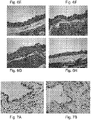
- Scleroderma or systemic sclerosis (SSc) is a chronic, rare multisystem autoimmune disease characterized by immune system activation, endothelial dysfunction, and an active fibrotic process involving fibroblasts (11).
- the earliest stage in the development of the scleroderma lesions is endothelial cell activation and vascular damage. This is followed by the migration of inflammatory cells, primarily, monocytes and then lymphocytes. Eventually, a population of fibroblasts is activated. The activated fibroblasts continue to produce the extracellular matrix that underlies the ultimate fibrotic pathology of scleroderma (11).
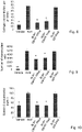
- elevated levels of Eotaxin were observed in lung fibrosis and in a bleomycine induced sclerosis mice model (14). Knockout mice to Eotaxin and CCR3 develop significantly reduced lung fibrosis (14, 15).
- the proinflammatory CCR3 binding chemokines Eotaxin 1, Rantes and MCP-3 belong also to the CC chemokine family and serve as ligands to the CCR3 receptor. They are also involved in the migration of immune cells, a fact that explains their efficacy in inflammatory preclinical models (16, 17).
- Idiopathic pulmonary fibrosis is a progressive fibrotic disease limited to the lungs, occurring in older individuals, more frequently men, and characterized by a dismal prognosis, with a median survival of 3 to 5 years since diagnosis.
- the clinical features characterizing IPF include shortness of breath, radiographically evident diffuse pulmonary infiltrates, and varying degrees of inflammation, fibrosis, or both on biopsy.
- the cause of IPF remains unknown, however mechanisms underlying the recruitment and proliferation of fibroblasts and immune cells cells as well as their pathologic differentiation are thought to be a hallmark to disease progression.
- Pirfenidone Esbriet
- Nintedanib are the only FDA/EMA approved treatment currently available for individuals with IPF (22-24). Pirfenidone has antifibrotic and anti-inflammatory properties in various in vitro systems and animal models of fibrosis.
- references in the description to methods of treatment refer to the compounds, pharmaceutical compositions and medicaments of the present invention for use in a method for treatment of the human (or animal) body by therapy (or for diagnosis).
- the present invention provides an isolated polyspecific antibody, or any antigen-binding fragment thereof, that binds to at least two CCR3-binding chemokines for use in the treatment of fibrotic diseases, autoimmune inflammatory disorders, monocyte related disorders or allergic atopic disorders, wherein the present invention in particular provides an isolated polyspecific antibody, or any antigen-binding fragment thereof, that binds to at least two CCR3-binding chemokines for use in the treatment of fibrotic diseases, autoimmune inflammatory disorders, atherosclerosis, or allergic atopic disorders, wherein said antibody or antigen-binding fragment thereof is fully humanized and comprises a heavy chain variable region denoted by SEQ ID NO:3 and a light chain variable region denoted by SEQ ID NO: 4 and wherein the at least two CCR3-binding chemokines are selected from the group consisting of Eotaxin 1, Eotaxin-2, Rantes and MCP-3.
- Said antigen-binding fragment thereof may be selected from the group consisting of Fv, single chain Fv (scFv), heavy chain variable region capable of binding the antigen, light chain variable region capable of binding the antigen, Fab, F(ab) 2 ' and any combination thereof.
- the fibrotic disease as herein defined is selected from the group consisting of scleroderma, idiopathic pulmonary fibrosis (IPF), non alcoholic steatohepatitis (NASH), glomerulosclerosis, cirrhosis and metabolic syndromes.
- Said autoimmune inflammatory disorder may be selected from the group consisting of systemic lupus erythematosus (SLE), rheumatoid arthritis, psoriasis, colitis, uveitis, multiple sclerosis and type I diabetes.
- SLE systemic lupus erythematosus
- rheumatoid arthritis rheumatoid arthritis
- psoriasis psoriasis
- colitis uveitis
- multiple sclerosis multiple sclerosis
- type I diabetes type I diabetes
- the monocyte related disorder according to the invention may be atherosclerosis.
- the allergic atopic disorder as herein defined may be selected from the group consisting of asthma, atopic dermatitis, urticaria and hypersensitivity reactions.
- the isolated polyspecific antibody for use according to the invention is wherein the at least two CCR3-binding chemokines are selected from the group consisting of Eotaxin 1, Eotaxin-2, Rantes and MCP-3.
- the isolated polyspecific antibody for use according to the invention is wherein the antibody binds Eotaxin 1, Eotaxin-2, Rantes and MCP-3.
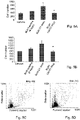
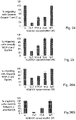
- the isolated polyspecific antibody for use as herein defined may attenuate the migratory properties of CCR3, CCR1, CCR2 and CCR5 expressing cells.
- the antibody according to the invention is a fully humanized antibody comprising the heavy chain variable region denoted by SEQ ID NO:3 or a variant thereof and the light chain variable region denoted by SEQ ID NO: 4 or a variant thereof.
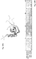
- the isolated antibody may bind a conformational epitope in the N-loop region of a CCR3-binding chemokine, wherein said conformational epitope is characterized by a relatively high concentration of positive amino acid residues located between amino acid positions 14 and 24 in the amino acid sequence of said CCR3-binding chemokine.
- the conformational epitope as herein defined may comprise at least three positive amino acid residues between amino acid positions 14 and 24 in the amino acid sequence of said CCR3-binding chemokine.
- the positive amino acid residues as herein defined may be selected from the group consisting of Arg, Lys and His.
- the conformational epitope as herein defined may comprise an amino acid sequences selected from: the amino acid sequences denoted by SEQ ID NO: 15, SEQ ID NO: 16, SEQ ID NO: 17, SEQ ID NO: 18 or SEQ ID NO: 19.
- the present invention further provides a nucleic acid molecule encoding the antibody as herein defined.
- the present invention provides a nucleic acid molecule encoding a humanized antibody that binds to at least two CCR3-binding chemokines wherin said nucleic acid molecule comprises the sequence denoted by SEQ ID NO: 1 and SEQ ID NO: 2.
- the present invention further provides an expression vector comprising the nucleic acid molecule according to the invention.
- the present invetion provides a host cell comprising the nucleic acid molecule according to the invention.
- the present invention further provides a pharmaceutical composition
- a pharmaceutical composition comprising the antibody as herein defined and a pharmaceutically acceptable carrier.
- the pharmaceutical composition according to the invention is for use in the treatment of fibrotic diseases, autoimmune inflammatory disorders, atherosclerosis or allergic atopic disorders.
- the present invention further provides a method of treating fibrotic diseases, autoimmune inflammatory disorders, atherosclerosis or allergic atopic disorders comprising administering to a patient in need thereof a therapeutically acceptable amount of the antibody or the pharmaceutical composition as herein defined.
- the present invention is based on the surprising finding that an antibody directed to a conformational epitope in the eotaxin 2 (CCL24) polypeptide binds and inhibits the activity of eotaxin 2 as well as additional chemotactic agents, the proinflammatory CCR3 binding chemokines: Eotaxin 1, Rantes and MCP-3.
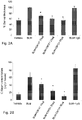
- the present invention thus provides a unique fully humanized monoclonal antibody (termed herein hCM101) capable of preventing the migration of immune cells by neutrilizing at least one of the chemokines responsible for their chemotaxis.
- the antibody hCM101 inhibits the binding of these CCR3-bindnig chemokines (some of which bind additional receptors) to their respective receptors and functionally attenuates the migratory properties of CCR3, CCR1, CCR2 and CCR5 expressing cells (eosinophil and monocytic cell lines as well as human fibroblasts).
- this antibody may be useful in the treatment of autoimmune diseases which are associated with chronic inflammation and characterized by the migration and infiltration of immune cells, including allergic and fibrotic diseases.
- the present disclosure thus provides an isolated polyspecific antibody, or any antigen-binding fragment thereof, that may bind to at least two CCR3-binding chemokines for use in the treatment of fibrotic diseases, autoimmune inflammatory disorders, monocyte related disorders or allergic atopic disorders.
- poly-specific (or polyspecific) antibody refers to poly reactive antibodies which are able to recognize multiple antigens, specifically the invention encompasses poly-specific antibodies which are able to recognize several, different, proinflammatory CCR3-binding chemokines.
- the antibody of the invention was generated against eotaxin 2 and was found subsequently to bind and effectively attenuate the activity of additional chemokines (for example, eotaxin 1, Rantes and MCP-3).
- additional chemokines for example, eotaxin 1, Rantes and MCP-3.
- the poly-specific antibody disclosed herein may bind to the various chemokines with similar affinities.
- the antibody may have differential binding affinity to the various chemokines.
- the antibody may bind with a higher affinity to eotaxin 2 than to the other tested chemokines.
- the poly-specific antibodies may recognize a cross-reactive epitope in these proinflammatory CCR3-binding chemokines.
- CCR3 (C-C chemokine receptor type 3) is a protein that in humans is encoded by the CCR3 gene.
- CCR3 has also recently been designated CD193 (cluster of differentiation 193).
- the protein encoded by this gene is a receptor for C-C type chemokines. It is a 7-transmembrane G protein-coupled receptor which is expressed by eosinophils as well as by a wide array of cell types including macrophages and endothelial cells.
- This receptor binds and responds to a variety of chemokines, including eotaxin (also termed eotaxin 1 or CCL11), eotaxin-2 (CCL24), eotaxin-3 (CCL26), MCP-3 (CCL7), MCP-4 (CCL13), and RANTES (CCL5).
- eotaxin also termed eotaxin 1 or CCL11
- CCL24 eotaxin-2
- CCL26 eotaxin-3
- MCP-3 CCL7
- MCP-4 CCL13
- RANTES RANTES
- CCR3-binding chemokines refers to any chemokine that binds to the protein CCR3 and encompasses for example but not limited to Eotaxin 1, Eotaxin-2, Eotaxin-3, Rantes, MCP-3 and MCP-4. It should be emphasized that some of the CCR3-binding chemokines also bind additional chemokine receptors, e.g. CCR1, CCR2 or CCR5.
- the isolated polyspecific antibody for use of the invention may bind at least two CCR3-binding chemokines that are selected from the group consisting of Eotaxin 1, Eotaxin-2, Rantes and MCP-3.
- eotaxin 2 eosinophil chemotactic protein 2
- CCL24 Chemokine (C-C motif) ligand 24
- MPIF-2 myeloid progenitor inhibitory factor 2
- CCL24 activity includes induction of chemotaxis in eosinophils, basophils, T lymphocytes and neutrophils, as well as induction of angiogenic and migratory responses in endothelial and smooth muscle cells.
- SEQ ID NO: 11 amino acid sequence of eotaxin 2 (without the N-terminal signal peptide) is denoted by SEQ ID NO: 11.
- CCR3-binding chemokines encompassed herein may be Eotaxin 1, Eotaxin-3, Rantes, MCP-3 and MCP-4, to name but a few.
- Eotaxin 1 also known as C-C motif chemokine 11 and eosinophil chemotactic protein
- Eotaxin 1 selectively recruits eosinophils by inducing their chemotaxis, and therefore, is implicated in allergic responses.
- the effects of Eotaxin 1 are mediated by its binding to a G-protein-linked receptor known as a chemokine receptor, including CCR2, CCR3 and CCR5.
- the amino acid sequence of human Eotaxin 1 (without the N-terminal signal peptide) is denoted for example by SEQ ID NO: 12.
- Rantes regulated on activation, normal T cell expressed and secreted
- CCL5 chemokine (C-C motif) ligand 5
- Rantes is an 8 kDa protein classified as a chemotactic cytokine or chemokine for T cells, eosinophils, and basophils, and plays an active role in recruiting leukocytes into inflammatory sites.
- Rantes With the help of particular cytokines (i.e., IL-2 and IFN-gamma) that are released by T cells, Rantes also induces the proliferation and activation of certain natural-killer (NK) cells to form CHAK (CC-Chemokine-activated killer) cells. Rantes binds, inter alia, the CCR3 receptor.
- NK natural-killer
- CHAK CC-Chemokine-activated killer
- MCP-3 (monocyte-specific chemokine 3) also termed chemokine (C-C motif) ligand 7 (CCL7) as herein defined is classified among the subfamily of chemokines known as CC chemokines. MCP-3 specifically attracts monocytes, and regulates macrophages function. It is produced by certain tumor cell lines and by macrophages. This chemokine is located on chromosome 17 in humans, in a large cluster containing many other CC chemokines. Rantes binds, inter alia, the CCR3 receptor.
- SEQ ID NO: 14 The amino acid sequence of human MCP-3 (without the N-terminal signal peptide) is denoted for example by SEQ ID NO: 14.
- the isolated polyspecific antibody for use of the invention binds Eotaxin 1, Eotaxin-2, Rantes and MCP-3.
- Said isolated polyspecific antibody may attenuate the migratory properties of CCR3, CCR1, CCR2 and CCR5 expressing cells.
- Attenuation of migratory properties means any statistically significant reduction in cell migration as compared with non-treated cells. For example, a reduction of at least 50% in the number of migrating cells.
- the antibody disclosed herein may be capable of binding to several proinflammatory chemokines and may inhibit their various activities, such as inhibition of cell recruitment or chemotaxis (for example recruitment or chemotaxis of eosinophils or monocytes or fibroblasts), inhibition of the transition of fibroblasts to myoblasts, as well as reduction of cellular activation.
- these antibodies can be useful in the treatment of diseases whose detrimental symptoms or effects are mediated at least partly by proinflammatory CCR3 chemokines.
- the antibody disclosed herein may cause a significant decrease in several features in models of idiopathic pulmonary fibrosis (IPF) and scleroderma, which are classified as fibrotic diseases.
- IPF idiopathic pulmonary fibrosis
- scleroderma scleroderma
- Fibrosis is the formation of excess fibrous connective tissue in an organ or tissue in a reparative or reactive process. This can be a reactive, benign, or pathological state. Fibrosis can be used to describe the pathological state of excess deposition of fibrous tissue, as well as the process of connective tissue deposition in healing. Thus as used herein the term "fibrotic disease” relates to an abnormal condition in which fibrous connective tissue spreads over or replaces normal smooth muscle or other normal organ tissue. Fibrosis can involve the heart, lung, peritoneum, skin or kidney.
- Non-limiting examples of a fibrotic disease include scleroderma, idiopathic pulmonary fibrosis (IPF) non alcoholic steatohepatitis (NASH), glomerulosclerosis, cirrhosis and metabolic syndromes.
- IPF idiopathic pulmonary fibrosis
- NASH non alcoholic steatohepatitis
- glomerulosclerosis glomerulosclerosis
- cirrhosis cirrhosis and metabolic syndromes.
- the fibrotic disease is selected from the group consisting of scleroderma, idiopathic pulmonary fibrosis (IPF), non alcoholic steatohepatitis (NASH), glomerulosclerosis, cirrhosis and metabolic syndromes.
- Scleroderma also known as systemic sclerosis as herein defined refers to a chronic systemic autoimmune disease characterised by hardening (sclero) of the skin ( derma ). In the more severe form, it also affects internal organs. Scleroderma can be limited scleroderma and involve cutaneous manifestations that mainly affect the hands, arms and face or diffuse scleroderma, which is rapidly progressing and affects a large area of the skin and one or more internal organs, frequently the kidneys, esophagus, heart and/or lungs. This form of scleroderma can be quite disabling. There are no treatments for scleroderma itself, but individual organ system complications are treated.
- Idiopathic pulmonary fibrosis refers to progessive fibrosis of the pulmonary alveolar walls, with progressive dyspnea and potentially fatal lack of oxygen or right heart failure.
- the acute form is called Hamman-Rich syndrome.
- inflammation refers to the complex biological response of the immune system to harmful stimuli, such as pathogens, damaged cells (caused by e.g. burns, trauma, neoplasma) or irritants such as chemicals, heat or cold.
- harmful stimuli such as pathogens, damaged cells (caused by e.g. burns, trauma, neoplasma) or irritants such as chemicals, heat or cold.
- inflammatory disease or "inflammatory disorder” refers to diseases associated with inflammation, including, but not limited to autoimmune inflammatory disorders.
- Autoimmune disease describes a pathological condition resulting from an overactive immune response of the body against substances and tissues normally present in the body.
- the immune system mistakes some part of the body as a pathogen and attacks it. This may be restricted to certain organs or involve a particular tissue in different places which may affect the basement membrane in both the lung and the kidney.
- autoimmune diseases include rheumatoid arthritis, multiple sclerosis, coeliac disease, Diabetes Mellitus type I (IDDM), systemic lupus erythematosus (SLE), Sjogren's syndrome, Churg-Strauss Syndrome, Hashimoto's thyroditis, Graves' disease, psoriasis, inflammatory bowel disease (ulcerative colitis and Crohn's disease), scleroderma and pemphigus.
- IDDM Diabetes Mellitus type I
- SLE systemic lupus erythematosus
- Sjogren's syndrome Churg-Strauss Syndrome
- Hashimoto's thyroditis Hashimoto's thyroditis
- Graves' disease psoriasis
- inflammatory bowel disease inflammatory bowel disease (ulcerative colitis and Crohn's disease)
- pemphigus examples include rheumatoid arthritis, multiple sclerosis,
- autoimmune inflammatory disorders relates to disorders resulting from the immune system attacking the body's own tissues that are also characterized by increased inflammation.
- Non-limiting examples of an autoimmune inflammatory disorder include systemic lupus erythematosus (SLE), rheumatoid arthritis, psoriasis, colitis, uveitis, multiple sclerosis and type I diabetes.
- the autoimmune inflammatory disorder as herein defined may be selected from the group consisting of systemic lupus erythematosus (SLE), rheumatoid arthritis, psoriasis, colitis, uveitis, multiple sclerosis and type I diabetes.
- SLE systemic lupus erythematosus
- rheumatoid arthritis rheumatoid arthritis
- psoriasis colitis
- uveitis multiple sclerosis
- type I diabetes type I diabetes
- monocyte related disorder relates to genetic abnormalities that affect the function of monocytes and macrophages and cause buildup of debris within the cells resulting for example in the lipid storage diseases (such as Gaucher disease and Niemann-Pick disease) or in atherosclerosis.
- An increase of monocytes in the blood occurs in response to chronic infections, in autoimmune disorders, in blood disorders, and in certain cancers.
- a proliferation of macrophages in tissues can occur in response to infections, sarcoidosis, and Langerhans cell histiocytosis.
- Furtmermore the monocyte related disorder disclosed herein may be atherosclerosis.
- the term "atherosclerosis” (also known as Arteriosclerotic Vascular Disease or ASVD) is a pathological condition in which an artery wall thickens as the result of a build-up of fatty materials such as cholesterol. This process is a result of a chronic inflammatory response in the walls of arteries, in large part due to the accumulation of macrophages and promoted by low-density lipoproteins without adequate removal of fats and cholesterol from the macrophages by functional high density lipoproteins (HDL). This hardening or furring of the arteries is caused by the formation of multiple plaques within the arteries.
- ASVD Arteriosclerotic Vascular Disease
- allergic atopic disorder relates to a form of allergy that afflicts persons with a genetic predisposition to hypersensitivity to certain allergens.
- Non-limiting examples of an allergic atopic disorder include asthma, atopic dermatitis, hay fever urticaria and hypersensitivity reactions.
- the allergic atopic disorder disclosed herein may be selected from the group consisting of asthma, atopic dermatitis, urticaria and hypersensitivity reactions.
- the present invention provides isolated poly-specific antibodies that bind to at least two proinflammatory CCR3-binding chemokines.
- the term "antibody” refers to a polypeptide encoded by an immunoglobulin gene or functional fragments thereof that specifically bind and recognize an antigen, namely proinflammatory CCR3-binding chemokines. Specifically, the antibody of the invention binds and recognizes at least eotaxin 2, eotaxin 1, RANTES and MCP-3.
- the antibody disclosed herein may be a monoclonal antibody.
- the term “monoclonal antibody”, “monoclonal antibodies” or “mAb” as herein defined refers to a population of substantially homogenous antibodies, i.e., the individual antibodies comprising the population are identical except for possibly naturally occurring mutations that may be present in minor amounts. Monoclonal antibodies are directed against a single antigenic site.
- Monoclonal antibodies may be prepared and purified by any method known in the art.
- monoclonal antibodies may be prepared from B cells taken from the spleen or lymph nodes of immunized animals (e.g. rats or mice), by fusion with immortalized B cells under conditions which favor the growth of hybrid cells.
- Immunization of mice may be carried out for example as described in WO 2010/086854 . Briefly, immunization of mice may be carried out for example by primary sub-cutaneous (s.c.) immunization with the desired antigen, namely with a CCR3-binding chemokine, e.g. eotaxin 2, or with a fragment of a CCR3-binding chemokine comprising a conformational epitope in the N-loop (e.g. 50 ⁇ g) emulsified with complete Freund's adjuvant. Two subcutaneous booster injections with the antigen (e.g. 50 ⁇ g) emulsified with incomplete Freund's adjuvant are then administered every 2 weeks. The mice with the highest neutralizing antibody titer receive an additional intravenous (i.v.) boost of the antigen (e.g. 5 ⁇ g) in PBS four days prior to spleen removal.
- a CCR3-binding chemokine e.g. eo
- the spleen of the mouse with the highest neutralizing antibody titer is removed and splenocytes are fused to mouse myeloma cells (e.g. NS0 cells) using polyethylene glycol, as previously described ( Kohler, G. and Milstein, C. (1975) Nature 256: 495-497 ).
- the hybridoma cells are selected by growing the cells in HAT (hypoxantine-aminopterin-thymidine) medium. Cell clones are then screened for specific antibody production, for example using the ELISA assays described below.
- Purification of monoclonal antibodies may be based for example on affinity chromatography, namely, using an affinity column to which the specific epitope is conjugated.
- An exemplary antibody structural unit comprises a tetramer, as known in the art.
- Each tetramer is composed of two identical pairs of polypeptide chains, each pair having one "light chain” and one "heavy chain”.
- the N-terminus of each chain defines a variable region of about 100 to 110 or more amino acids primarily responsible for antigen (or epitope) recognition.
- variable region refers to these heavy and light chains, respectively.
- V H heavy chain variable region
- V L light chain variable region
- the variable region is subdivided into hypervariable and framework (FR) regions.
- FR hypervariable and framework regions.
- Hypervariable regions have a high ratio of different amino acids in a given position, relative to the most common amino acid in that position.
- Four FR regions which have more stable amino acids sequences separate the hypervariable regions.
- the hypervariable regions directly contact a portion of the antigen's surface. For this reason, hypervariable regions are herein referred to as “complementarity determining regions", or "CDRs”.
- both light and heavy chains comprise the domains FR1, CDR1, FR2, CDR2, FR3, CDR3 and FR4.
- the CDRs are primarily responsible for binding to an epitope of an antigen.
- the CDRs of each chain are typically referred to as CDR1, CDR2, and CDR3, numbered sequentially starting from the N-terminus, and are also typically identified by the chain in which the particular CDR is located.
- complementarity determining regions CDRH1, CDRH2 and CDRH3 refer to the three complementarity determining regions starting from the N-terminus of the antibody's heavy chain and the complementarity determining regions CDRL1, CDRL2 and CDRL3 refer to the three complementarity determining regions starting from the N-terminus of the antibody's light chain.
- the isolated poly-specific monoclonal antibody disclosed herein may be a chimeric antibody, a human antibody, a humanized antibody or a fully humanized antibody.
- chimeric antibodies as herein defined refers to antibodies in which a portion of the heavy and/or light chain is derived from a particular species, while the remainder of the chain(s) is derived from another species, as well as fragments of such antibodies, which exhibit the same biological activity.
- Chimeric antibodies may be prepared by any method known in the art, for example as described below.
- a murine-human chimeric antibody may be prepared by the amplification and cloning of murine V H and V L genes, encoding the antibody variable regions, followed by murine-human chimeric antibody expression.
- total RNA is isolated from murine anti-eotaxin 2 hybridoma cells that are shown to secrete antibodies with the desired characteristics and cDNA is synthesized using oligo (dT) 15 primer, M-MLV and AMV reverse transcriptases.
- Amplification of the heavy and the light variable genes (V H and V L ) may be carried out using a panel of primers directed at the 5' terminus of framework 1 of each gene, essentially as described in Benhar and Reiter ( Benhar, I. and Reiter, Y. (2002) Curr. Protoc. Immunol. Chapter 10: Unit 10 19B ), and to the constant region (C H 1 or C k , respectively) at the 3' end.
- variable genes are then re-amplified using non-degenerate primers introducing restriction sites at both ends for cloning, for example, into a pCMV-based antibody expression vector.
- amplified heavy and light variable genes are separately purified, digested and cloned into appropriate mammalian full-length Ig expression vectors, providing each chain with a corresponding signal-peptide and constant gene, resulting in IgG1/k murine human chimeric antibody expression.
- a stable cell line expressing the antibody can be prepared, by transfecting cells (e.g. CHO cells) with the Ig expression vector containing both heavy and light chains of the chimeric antibody.
- Highly anti-eotaxin 2 (or any other CCR3-binding chemokine used for the production of the antibodies) antibody producing clones may be then selected and expanded based on antibody levels in the supernatant, as tested by any method known in the art, for example, a CCR3-binding chemokine-specific ELISA assay, as detailed herein below.
- humanized antibodies traditionally refers to forms of non-human (for example, murine) antibodies that contain a human-derived immunoglobulin framework with minimal sequences derived from non-human immunoglobulin at the CDRs and optionally at additional relevant positions.
- humanized antibodies are human immunoglobulins (recipient antibody) in which residues from a hypervariable region of the recipient are replaced by residues from a hypervariable region of a non-human species (donor antibody) such as mouse, rat, rabbit or nonhuman primate having the desired specificity, affinity, and activity.
- the term "fully humanized” antibodies relates to antibodies designed to have only human sequences.
- the fully humanized antibodies of the present invention were prepared using the Composite Human Antibodies TM technology that minimizes immunogenicity of the antibodies in patients.
- multiple sequence segments derived from variable (V) regions of unrelated human antibodies are used as acceptors for the complementarity determining regions (CDRs) of the starting antibodies.
- CDRs complementarity determining regions
- human antibody refers to an antibody that possesses an amino acid sequence corresponding to that of an antibody produced by a human and/or has been made using any of the techniques for making human antibodies known in the art. This definition specifically excludes a humanized antibody that comprises non-human antigen-binding residues.
- Antibodies may also be prepared using phage display.
- antibody phage display is based on genetic engineering of bacteriophages and repeated rounds of antigen-guided selection and phage propagation.
- the APD process begins with antibody-library preparation, by preparation of quality RNA from the cell source chosen (e.g., peripheral blood mononuclear cells). This RNA is reverse-transcribed into cDNA, which is used for PCR of the VH and VL chains of the encoded antibodies. This step is followed by ligation of the variable heavy (VH) and variable light (VL) PCR products into a phage display vector, culminating in analysis of clones of mAbs.
- VH variable heavy
- VL variable light
- the present disclosure provides an isolated fully humanized poly-specific monoclonal antibody, which may comprise a heavy chain variable region comprising the CDRH1 comprising the amino acid sequence of NSGMN denoted by SEQ ID NO. 5, or a variant thereof, the CDRH2 comprising the amino acid sequence WINTYNGEPTYTDDFKG denoted by SEQ ID NO. 6, or a variant thereof, and the CDRH3 comprising the amino acid sequence HSYGSSYAMDN denoted by SEQ ID NO. 7 or a variant thereof; and a light chain variable region comprising: the CDRL1 comprising the amino acid sequence KASQSVDYDGDSYMN denoted by SEQ ID NO.
- CDRL2 comprising the amino acid sequence VASNLKS denoted by SEQ ID NO. 9 or a variant thereof
- CDRL3 comprising the amino acid sequence QQSNEEPWT denoted by SEQ ID NO. 10 or a variant thereof.
- the present disclosure provides an isolated polyspecific antibody, or any antigen-binding fragment thereof, that may bind to at least two CCR3-binding chemokines, wherein the antibody may be a fully humanized antibody and may comprise a heavy chain variable region comprising:
- the present disclosure provides an isolated polyspecific antibody, or any antigen-binding fragment thereof, that may bind to at least two proinflammatory CCR3-binding chemokines for use in the treatment of fibrotic diseases, autoimmune inflammatory disorders, monocyte related disorders or allergic atopic disorders, wherein said antibody may be a fully humanized antibody and may comprise a heavy chain variable region comprising:
- amino acid sequence of the heavy chain of the isolated fully humanized poly-specific monoclonal antibody exemplified herein is denoted by SEQ ID NO: 3 and is of the amino 'acid sequence:QIQLVQSGPELKKPGASVKVSCRASGYPFTNSGMNWVKQAPGKGLKW MGWINTYNGEPTYTDDFKGRFAFSLETSASTAYLQINNLRNEDTATYFCASHSY GSSYAMDNWGQGTSVTVSS
- amino acid sequence of the light chain of the isolated humanized poly-specific monoclonal antibody exemplified herein is denoted herein by SEQ ID NO: 4 and is of the amino acid sequence:
- the isolated polyspecific antibody for use according to the invention is a fully humanized antibody comprising the heavy chain variable region denoted by SEQ ID NO:3 or a variant thereof and the light chain variable region denoted by SEQ ID NO: 4 or a variant thereof.
- the present disclosure also may encompass variants of the heavy and light chain variable regions.
- the variants may include mutations in the complementarity determining regions of the heavy and light chains which do not alter the activity of the antibodies herein described, or in the framework region.
- variant sequences of amino acids or nucleotides different from the sequences specifically identified herein, in which one or more amino acid residues or nucleotides are deleted, substituted or added.
- Variants encompass various amino acid substitutions.
- An amino acid "substitution” is the result of replacing one amino acid with another amino acid which has similar or different structural and/or chemical properties. Amino acid substitutions may be made on the basis of similarity in polarity, charge, solubility, hydrophobicity, hydrophilicity, and/or the amphipathic nature of the residues involved.
- variants encompass conservative amino acid substitutions.
- Conservative substitution tables providing functionally similar amino acids are well known in the art.
- nonpolar (hydrophobic) amino acids include alanine, leucine, isoleucine, valine, proline, phenylalanine, tryptophan, and methionine
- polar neutral amino acids include glycine, serine, threonine, cysteine, tyrosine, asparagine, and glutamine
- positively charged (basic) amino acids include arginine, lysine, and histidine
- negatively charged (acidic) amino acids include aspartic acid and glutamic acid.
- amino acid substitutions are nucleic acid substitutions resulting in conservative amino acid substitutions as defined above.
- Variants disclosed herein may also encompass non-polar to polar amino acid substitutions and vice-versa.
- amino acid or “amino acid residue” refers to naturally occurring and synthetic amino acids, as well as amino acid analogs and amino acid mimetics that function in a manner similar to the naturally occurring amino acids.
- Variant sequences refer to amino acid or nucleic acids sequences that may be characterized by the percentage of the identity of their amino acid or nucleotide sequences with the amino acid or nucleotide sequences described herein (for example, the amino acid or nucleotide sequences of the heavy and light chains of the antibodies herein described).
- Variant sequences as herein defined may refer to nucleic acid sequences that encode the heavy and light chain variable regions, each having a sequence of nucleotides with at least 70% or 75% of sequence identity, around 80% or 85% of sequence identity, around 90%, 91%, 92%, 93%, 94%, 95%, 96%, 97%, 98% or 99% of sequence identity when compared to the sequences of the heavy and light chain variable regions described herein. #
- the isolated poly-specific humanized monoclonal antibody disclosed herein may be wherein its heavy chain variable region is encoded by a nucleic acid sequence which is at least 70% identical to the nucleic acid sequence of the heavy chain variable domain denoted by SEQ ID NO. 1 and wherein its light chain variable region is encoded by a nucleic acid sequence which is at least 70% identical to the nucleic acid sequence of the light chain variable domain denoted by SEQ ID NO. 2.
- the term “activity of the antibodies” it is meant the ability of the antibodies to bind to at least two CCR3-binding chemokines, and prerferably to inhibit a biological function mediated by such CCR3-binding chemokines.
- Non-limiting examples of such biological functions are: inhibition of cell recruitment or chemotaxis (for example recruitment or chemotaxis of eosinophils or monocytes or fibroblasts), inhibition of the transition of fibroblasts to myoblasts, or reduction of cellular activation (for example as measured by Ca + update).
- the biological functions can be measured in vivo or in vitro using methods well known in the art. Several such assays are described in the Examples below.
- Additional in vitro experiments for determining the binding of the antibody prepared according to the invention to its target protein include for example ELISA assays.
- the present disclosure may further encompass any antigen-binding fragments of the isolated poly-specific monoclonal antibody disclosed herein.
- antigen-binding fragments may be for example Fab and F (ab') 2 , which are capable of binding antigen.
- Such fragments may be produced by any method known in the art, for example by proteolytic cleavage, using enzymes such as papain (to produce Fab fragments) or pepsin (to produce F(ab') 2 fragments).
- the isolated poly-specific monoclonal antibody disclosed herein may be an antibody fragment selected from the group consisting of Fv, single chain Fv (scFv), heavy chain variable region capable of binding the antigen, light chain variable region capable of binding the antigen, Fab, F(ab) 2 ' and any combination thereof.
- the inventors identified a specific epitope in Eotaxin 1, eotaxin 2, Rantes and MCP-3 to which the antibody of the invention (hCM101) binds.
- the epitope to which the antibody of the invention (hCM101) binds in Eotaxin-2 comprises the amino acid sequence CMFFVSKRIP denoted by SEQ ID NO: 15, in Eotaxin-1 the epitope comprises the amino acid sequence CFNLANRKIPLQRL denoted by SEQ ID NO: 16, in rantes the epitope comprises the amino acid sequence AYIARPLPRAHIKEYFY denoted by SEQ ID NO: 17 and in MCP3 the epitope comprises the amino acid sequence CCYRFINKKI denoted by SEQ ID NO: 18 (the "first part of the N-loop") and the amino acid sequence SYRRTTSSH denoted by SEQ ID NO: 19 (the "second part of the N-loop").
- the term "conformational epitope” relates to an antigenic determinant, the part of an antigen that is recognized by the antibody, which is composed of continuous or discontinuous sections of the antigen's amino acid sequence. Conformational epitopes interact with the binding site of the antibody based on the 3-D surface features and shape, or tertiary structure, of the antigen. In certain instances, such epitopes may be identified partly by a linear sequence and by their relative position in the polypeptide's structure and by characteristic amino acids that are found within the identified epitope.
- N-loop region it is meant the N-loop and the turn towards the first beta strand located at the N terminus of CCR3-binding chemokines.
- the N-loop region comprises the amino acid sequence CMFFVSKRIP denoted SEQ ID NO: 15.
- the present disclosure provides an isolated antibody, wherein the conformational epitope may comprise an amino acid sequences selected from the amino acid sequences denoted by SEQ ID NO: 15, SEQ ID NO: 16, SEQ ID NO: 17, SEQ ID NO: 18 or SEQ ID NO: 19.
- the antibody binds a region within the CCR3-binding chemokine that includes a structure formed in the N loop region and involves amino acid residues at positions Lys-14 and Arg-15 (located within the epitope denoted by SEQ ID NO: 15) and Arg-20 in Eotaxin 2, positions Arg-16 and Lys-17 (located within the epitope denoted by SEQ ID NO: 16) and Arg-22 in Eotaxin 1, positions Arg-17 (located within the epitope denoted by SEQ ID NO: 17), His-23 and Arg-21 in Rantes, and positions Lys-18 and Lys-19 (located within the epitope denoted by SEQ ID NO: 18) and Arg-24 in MCP3.
- the identified amino acid positions within each of the chemokine sequence relates to the chemokine sequence without the N-terminal signal peptide, as shown in SEQ ID NOs: 11-14 corresponding respectively to the amino acid sequence (without the N-terminal signal peptide) of eotaxin 2, eotaxin 1, Tantes and MCP3.
- position it is meant the location within the amino acid sequence, where "position 1” indicates the first N-terminal amino acid residue after the signal peptide (namely the amino acid sequence of the mature polypeptide), “position 2" indicates the amino acid residue that is downstream to the N-terminal amino acid residue and so forth in the direction of N-terminal to C-terminal, as known in the art.
- relatively high concentration of positive amino acid residues it is meant at least three positive amino acid residues, e.g. three positive amino acid residues.
- the disclosure additionally provides an isolated antibody that may bind a conformational epitope in the N-loop region of a CCR3-binding chemokine, wherein said conformational epitope is characterized by a relatively high concentration of positive amino acid residues located between amino acid positions 14 and 24 in the amino acid sequence of said CCR3-binding chemokine.
- the present disclosure provides an isolated antibody , wherein the conformational epitope may comprise at least three positive amino acid residues between amino acid positions 14 and 24 in the amino acid sequence of said CCR3-binding chemokine.
- Said positive amino acid residues may be selected from the group consisting of Arg, Lys and His.
- the isolated antibody disclosed herein may bind to an epitope comprising amino acid residues at positions Lys-14, Arg-15 and Arg-20 in Eotaxin 2, positions Arg-16, Lys-17 and Arg-22 in Eotaxin 1, positions Arg-17, His-23 and Arg-21 in Rantes, and positions Lys-18, Lys-19 and Arg-24 in MCP3.
- the common epitope as defined above can also serve for the screening and identification of additional antibodies having poly-specific binding capabilities to several CCR3-binding chemokines. Such antibodies would have high likelihood of being active in attenuating cell migration mediated by these chemokines.
- the present disclosure provides a method of screening and identifying antibodies may capable of efficiently attenuating the migratory properties of at least one of CCR3, CCR1, CCR2 or CCR5 expressing cells.
- Said method may comprises:
- Said conformational epitope may comprise an amino acid sequence selected from the group consisting of the amino acid sequences denoted by SEQ ID NO: 15, SEQ ID NO: 16, SEQ ID NO: 17, SEQ ID NO: 18 and SEQ ID NO: 19.
- the step of assessing the binding may be performed by bringing the antibodies into contact with said conformational epitope, for example by incubating the antibodies with a fragment of the chemokine comprising the N-loop, or by incubating the antibodies with at least one of the amino acid sequences denoted by SEQ ID NO: 15, SEQ ID NO: 16, SEQ ID NO: 17, SEQ ID NO: 18 and SEQ ID NO: 19.
- the incubation may be performed by any method known in the art for assessing antigen-antibody binding, for example by ELISA or by using a chip containing multiple amino acid fragments.
- the incubation is performed in the presence of a binding buffer and antibody binding is assessed for example by a further incubation with a detectably labeled second antibody.
- screening and identifying relates to the assessment of an antibody or a plurality of antibodies for their ability to bind to the epitope of the invention as defined above and selecting antibodies which bind to the epitope and thereby are expected to attenuate the activity of the CCR-binding chemokine upon binding to the chemokine.
- aid conformational epitope may comprise amino acid residues at the following positions: Lys-14, Arg-15 and Arg-20 in Eotaxin 2 (the amino acid sequence thereof is denoted by SEQ ID NO. 11), Arg-16, Lys-17 and Arg-22 in Eotaxin 1 (the amino acid sequence thereof is denoted by SEQ ID NO. 12), Arg-17, His-23 and Arg-21 in Rantes (the amino acid sequence thereof is denoted by SEQ ID NO. 13), and Lys-18, Lys-19 and Arg-24 in MCP3 (the amino acid sequence thereof is denoted by SEQ ID NO. 14).
- the identified conformational epitope disclosed herein may also be used as an antigen for generating antibodies capable of efficiently attenuating the migratory properties of at least one of CCR3, CCR1, CCR2 or CCR5 expressing cells. Therefore, additionally, the present disclosure provides an isolated N-loop region of a CCR3-binding chemokine comprising a conformational epitope, wherein said conformational epitope may be characterized by a relatively high concentration of positive amino acid residues located between amino acid positions 14 and 24 in the amino acid sequence of said CCR3-binding chemokine, for generating antibodies against said CCR3-binding chemokine.
- Said isolated N-loop region may comprise an amino acid sequence selected from the group consisting of the amino acid sequences denoted by SEQ ID NO: 15, SEQ ID NO: 16, SEQ ID NO: 17, SEQ ID NO: 18 and SEQ ID NO: 19.
- the present disclosure provides an isolated N-loop region of a CCR3-binding chemokine comprising a conformational epitope, wherein said conformational epitope may be characterized by a relatively high concentration of positive amino acid residues located between amino acid positions 14 and 24 in the amino acid sequence of said CCR3-binding chemokine, for use in a method of generating antibodies against said CCR3-binding chemokine.
- said isolated N-loop region may comprise an amino acid sequence selected from the group consisting of the amino acid sequences denoted by SEQ ID NO: 15, SEQ ID NO: 16, SEQ ID NO: 17, SEQ ID NO: 18 and SEQ ID NO: 19.
- the present disclosure thus further provides a method for generating antibodies may capable of efficiently attenuating the migratory properties of at least one of CCR3, CCR1, CCR2 or CCR5 expressing cells comprising obtaining the epitope of the invention and producing an antibody to said epitope using any method known in the art.
- the present invention provides an isolated nucleic acid molecule comprising a nucleotide sequence encoding an antibody or any antigen-binding fragment thereof according to the invention.
- nucleic acid or “nucleic acid molecule” as herein defined refers to a polymer of nucleotides, which may be either single- or double-stranded, which is a polynucleotide such as deoxyribonucleic acid (DNA), and, where appropriate, ribonucleic acid (RNA).
- DNA deoxyribonucleic acid
- RNA ribonucleic acid
- the terms should also be understood to include, as equivalents, analogs of either RNA or DNA made from nucleotide analogs, and, , single-stranded (such as sense or antisense) and double-stranded polynucleotides.
- DNA used herein also encompasses cDNA, i.e. complementary or copy DNA produced from an RNA template by the action of reverse transcriptase (RNA-dependent DNA polymerase).
- the invention further provides an expression vector comprising the isolated nucleic acid molecule as herein defined.
- Expression vector sometimes referred to as "expression vehicle” or “expression construct”, as used herein, encompasses vectors such as plasmids, viruses, bacteriophage, integratable DNA fragments, and other vehicles, which enable the integration of DNA fragments into the genome of the host.
- Expression vectors are typically self-replicating DNA or RNA constructs containing the desired gene or its fragments, and operably linked genetic control elements that are recognized in a suitable host cell and effect expression of the desired genes. These control elements are capable of effecting expression within a suitable host.
- the expression vector disclosed herein may be competent with expression in bacterial, yeast, or mammalian host cells, to name but few.
- the present invention provides a host cell transfected with the isolated nucleic acid molecule according to the invention or with the expression vector according to the invention.
- host cells refers to cells which are susceptible to the introduction of the isolated nucleic acid molecule according to the invention or with the expression vector according to the invention.
- said cells are mammalian cells, for example CHO cells or NS0 cells.
- Transfection of the isolated nucleic acid molecule or the expression vector according to the invention to the host cell may be performed by any method known in the art.
- the present disclosure provides an immunoconjugate comprising the antibody or any antigen-binding fragment thereof of the present disclosure and an additional therapeutic agent, e.g. an anti-inflammatory agent.
- immunoconjugate refers to an antibody or any antigen-binding fragment thereof of the present disclosure that may be conjugated (linked or joined) to an additional agent. Immunoconjugates may be prepared by any method known to a person skilled in the art, for example, by cross-linking the additional agent to the antibody of the present disclosure or by recombinant DNA methods.
- the polyspecific antibody disclosed herein may be administered in combination with at least one additional therapeutic agent.
- additional therapeutic agent refers to any agent that may be used for treating fibrotic diseases, autoimmune inflammatory disorders, monocyte related disorders or allergic atopic disorders.
- the additional therapeutic agent may be an additional anti-inflammatory agent or anti fibrotic agent.
- Said at least one additional therapeutic agent may be selected from a group consisting of chemotherapeutics, cytokines, peptides, antibodies and antibiotics.
- Said additional therapeutic agent may include, but is not limited to methotrexate, a steroid, cyclosporine, NSAIDS (non-steroidal anti inflammatory drugs), steroids, interferon-beta, intravenous immune globulin (IVIG), pirfenidone, nintedanib, bosentan, or macicentan.
- methotrexate a steroid
- cyclosporine cyclosporine
- NSAIDS non-steroidal anti inflammatory drugs
- steroids interferon-beta
- IVIG intravenous immune globulin
- pirfenidone pirfenidone
- nintedanib nintedanib
- bosentan or macicentan.
- the additional therapeutic agent may be an additional antibody.
- additional antibody refers to an antibody, which is not the antibody disclosed herein, which may be used in combination with the antibody disclosed herein. Such antibody may be directed as a non-limiting example against TNF ⁇ , TNF receptor, IL6 receptor, or CD20.
- the present disclosure further provides a pharmaceutical composition
- a pharmaceutical composition comprising as an active ingredient the isolated poly-specific antibody of the invention, or any antigen-binding fragment thereof or the immunoconjugate as herein defined and a pharmaceutically acceptable carrier, excipient or diluent.
- compositions generally may comprise the antibody or any antigen-binding fragment thereof as herein defined and a buffering agent, an agent which adjusts the osmolarity of the composition and optionally, one or more pharmaceutically acceptable carriers, excipients and/or diluents as known in the art.
- the term "pharmaceutically acceptable carrier, excipient or diluent” includes any and all solvents, dispersion media, coatings, antibacterial and antifungal agents and the like, as known in the art.
- the carrier can be solvent or dispersion medium containing, for example, water, ethanol, polyol (for example, glycerol, propylene glycol, and liquid polyethylene glycol, and the like), suitable mixtures thereof, and vegetable oils.
- Each carrier should be both pharmaceutically and physiologically acceptable in the sense of being compatible with the other ingredients and not injurious to the subject. Except as any conventional media or agent is incompatible with the active ingredient, its use in the therapeutic composition is contemplated.
- the pharmaceutical composition may further comprise an additional therapeutic agent, e.g. an anti-inflammatory agent.
- an additional therapeutic agent e.g. an anti-inflammatory agent.
- the present invention relates to a pharmaceutical composition
- a pharmaceutical composition comprising an isolated poly-specific humanized antibody, or any antigen-binding fragment thereof, which binds to at least two proinflammatory CCR3-binding chemokines, wherein the antibody comprises a heavy chain variable region of the amino acid sequence denoted by SEQ ID NO. 3 or a variant thereof and a light chain variable region of the amino acid sequence denoted by SEQ ID NO. 4, or a variant thereof.
- a method of prophylaxis, treatment or amelioration of autoimmune inflammatory disorders, monocyte related disorders, allergic atopic disorders or fibrotic diseases comprising administering to a subject in need thereof a therapeutically effective amount of the isolated poly-specific antibody or any antigen-binding fragment thereof or the pharmaceutical composition disclosed herein.
- the disclosure further provides a method of prophylaxis, treatment or amelioration of autoimmune inflammatory disorders, monocyte related disorders, allergic atopic disorders or fibrotic diseases comprising administering to a subject in need thereof a therapeutically effective amount of an isolated polyspecific humanized antibody, or any antigen-binding fragment thereof, which may bind to at least two proinflammatory CCR3-binding chemokines, wherein the antibody may comprise a heavy chain variable region of the amino acid sequence denoted by SEQ ID NO. 3 or a variant thereof and a light chain variable region of the amino acid sequence denoted by SEQ ID NO. 4, or a variant thereof.
- subject or “patient” are used interchangeably and refer to a subject that may benefit from the present disclosure such as a mammal (e.g. canine, feline, ovine, porcine, equine, bovine, or human). Specifically, said patient may be human.
- a mammal e.g. canine, feline, ovine, porcine, equine, bovine, or human.
- said patient may be human.
- prophylaxis as herein defined it is meant to provide a “preventive treatment” or “prophylactic treatment”, namely acting in a protective manner, to defend against or prevent an autoimmune inflammatory disorders, monocyte related disorders, allergic atopic disorders or fibrotic diseases, or an episode of such a disease.
- treat means reducing, preventing, curing, reversing, ameliorating, attenuating, alleviating, minimizing, suppressing or halting the deleterious effects of a disease or a condition or delaying the onset of one or more clinical indications of a disease that is affected by proinflammatory CCR3-binding chemokines.
- diseases include autoimmune inflammatory disorders, monocyte related disorders, allergic atopic disorders or fibrotic diseases.
- treatment refers to reducing, preventing, curing, reversing, attenuating, alleviating, minimizing suppressing or halting the deleterious effects of a disease or a condition that is mediated by eotaxin 2.
- Administration as disclosed herein may be performed by any of the following routes: oral administration, intravenous, intramuscular, intraperitoneal, intratechal or subcutaneous injection; intrarectal administration; intranasal administration, ocular administration or topical administration.
- Administration as disclosed herein may be performed intravenously. Said administration may be performed intraperitoneally. Said administration may be performed by inhalation.
- any pharmaceutical compositions comprising the same or any conjugates comprising them may be administered to a subject prior to or post disease onset.
- the method of prophylaxis, treatment or amelioration of autoimmune inflammatory disorders, monocyte related disorders, allergic atopic disorders or fibrotic diseases disclosed herein is where said isolated humanized poly-specific antibody or any antigen-binding fragment thereof disclosed herein, or pharmaceutical composition as disclosed herein may be administered to said subject prior to or after disease onset.
- a "therapeutically effective amount" of the isolated monoclonal antibody or any antigen-binding fragment thereof disclosed herein, or the pharmaceutical composition disclosed herein for purposes herein defined is determined by such considerations as are known in the art in order to cure, arrest or at least alleviate or ameliorate the medical condition.
- the dosage or the therapeutically effective amount can be estimated initially from in vitro cell culture assays or based on animal models such as the animal models detailed herein.
- Said therapeutically effective amount disclosed herein may be in the range of 0.01 to 100 mg/kg.
- Said therapeutically effective amount disclosed herein may be in the range of 0.01 to 40 mg/kg, 0.1 to 40 mg/kg or 1 to 10 mg/kg.
- the isolated humanized poly-specific antibody or any antigen-binding fragment thereof disclosed herein or pharmaceutical composition disclosed herein may be administered to the subject as a single dose or multiple doses.
- subject in need thereof means warm-blooded animals, such as for example rats, mice, dogs, cats, guinea pigs, primates and humans suffering from autoimmune inflammatory disorders, monocyte related disorders, allergic atopic disorders or fibrotic diseases.
- the present disclosure further provides the isolated humanized poly-specific antibody or any antigen-binding fragment thereof as disclosed herein or the pharmaceutical composition as disclosed herein for use in a method of prophylaxis, treatment or amelioration of autoimmune inflammatory disorders, monocyte related disorders, allergic atopic disorders or fibrotic diseases.
- the disclosure further provides an isolated humanized poly-specific antibody, or any antigen-binding fragment thereof, which may bind to at least two proinflammatory CCR3-binding chemokines, wherein the antibody comprises a heavy chain variable region of the amino acid sequence denoted by SEQ ID NO. 3 or a variant thereof and a light chain variable region of the amino acid sequence denoted by SEQ ID NO. 4, or a variant thereof for use in a method of prophylaxis, treatment or amelioration of autoimmune inflammatory disorders, monocyte related disorders, allergic atopic disorders or fibrotic diseases.
- SEQ ID NO. 3 a heavy chain variable region of the amino acid sequence denoted by SEQ ID NO. 3 or a variant thereof
- SEQ ID NO. 4 a light chain variable region of the amino acid sequence denoted by SEQ ID NO. 4
- purified refers to molecules, such as amino acid or nucleic acid sequences, peptides, polypeptides or antibodies that are removed from their natural environment, isolated or separated.
- An “isolated antibody” is therefore a purified antibody.
- purified or “to purify” also refers to the removal of contaminants from a sample.
- CM101 The specificity of CM101 was assessed by ELISA. Plates were coated with the following antigens: eotaxin1, eotaxin2, monocyte chemoattractant protein -3 (CCL7), and RANTES (CCL5). All coated at 1 ⁇ g ml -1 at 4°C overnight. After washing (PBS + 0.1% Tween 20), hCM101 (5 ⁇ g) was added to the plate for 1 h. After washing again, CM101 was detected with anti-human horseradish peroxidase conjugate (1 h incubation with 1:5000 dilution in PBS) and quantified using 3,3',5,5'-tetramethyl benzidine substrate (Sigma Chemical, Gillingham, UK). The reaction was stopped after 10 min with sulfuric acid (50 ⁇ l 2 M), and optical density was measured at 450 nM.
- sulfuric acid 50 ⁇ l 2 M
- Immunohistochemical staining for human Eotaxin 2/CCR3 was performed on 5- ⁇ m-thick frozen sections of skin/lung biopsies from scleroderma patients. After fixation with methanol and acetone, sections were blocked with nonimmune rabbit and goat sera, followed by incubation with CAS blocking reagent. Subsequently, the primary antibody (either the anti-Eotaxin-2 or anti CCR3 antibodies) was added for 1 hour at room temperature. After washing, biotinylated affinity-purified goat anti-mouse/rabbit antibodies (Jackson) were added.
- the slides were then incubated with 0.3% H 2 O 2 , followed by additional rinses and incubation with streptavidin-peroxidase conjugate (Jackson) for 30 minutes at room temperature.
- the slides were developed with 3-amino-9 ethylcarbazole substrate (Dako) for 15 minutes and counterstained with hematoxylin.
- H&E and Masson's Trichrome stains were used for analysis of lung inflammation and fibrosis changes in skin and lung sections.
- CM101 CM101
- magnetic beads 1mg
- human sera from systemic sclerosis patient
- Magnetic separation and elution of the chemokine from the beads was performed following by transfer into nitrocellulose membrane and exposure to commercial anti Eox2 antibody.
- the ability of hCM101 to bind eotaxin 1, Rantes and MCP-3 was also assessed using SDS gel electrophoresis.
- Aml 14.3D10 an Eosinophilic cell line that stably express CCR3 was washed once in PBS and resuspended at 10 7 cells ml -1 in assay buffer (RPMI 1640, 1% endotoxin-free BSA, 100 U ml -1 penicillin, and 100 ⁇ g ml -1 streptomycin).
- hCM101 at different concentrations (10 -7 M-10 -9 M) was incubated with a combination of 5 nM human eotaxin-1 and eotaxin-2 or MCP-3 and RANTES (30 min, 37°C) and placed into the lower chambers of 24-well Transwell plates with 5- ⁇ m pores (Costar); 100 ⁇ l of cells (1 ⁇ 10 6 cells) were placed into the upper chamber of each Transwell plate, and the assay was incubated at 37°C for 4 h (5% CO 2 ). Live cells migrating to the lower chamber were counted using a flow cytometer (FACSCalibur, BD Biosciences, Cowley, UK). Results are expressed as percent control (i.e., chemokine-induced) migration.
- CD14 positive cells we used MACS separation magnetic Beads (Milteny) on Ficoll PBMCs isolated from scleroderma patients whole blood. Migration of CD14+cells towards MCP3 and RANES was assessed as describe above. In addition CD14- cells migration was examined towards Eotaxin 1, 2 and RANTES treated with hCM101.
- fibroblasts-NHDF adherent cells
- a transwell with a 8- ⁇ m pore size (Costar) coated with fibronectin After Eotaxin 2 was incubated with hCM101 for 30 minutes, NHDF cells (5X10⁴ cells) were added to the upper chamber and incubated at 37°C for 4 h. Then, non-migrating cells were removed from the insert membranes by cotton swabs. The membrane was fixed by 4% paraformaldehyde and migrated cells were then stained with commassie blue. The number of migrating cells was counted in three random fields using an inverted microscope and analyzed using image pro software.
- Fibroblasts were incubated for 24h with CM101 (10 or 5 ⁇ g/ml) and human sera. Then cells were trypsinized , fixated with 70% ethanol and incubated with anti ⁇ SMA Ab with the cells (Biolegend) for 0.5h at room temperature .cells were then washed and subjected to secondary Ab Cye3 Donkey ⁇ Rabbit IgG (1:300 inPBS) 0.5h at room temperature. Cells were analyzed through flow cytometer.
- PBS phosphate buffered saline
- Systemic sclerosis was induced via daily subcutaneous injections of 50 ⁇ g bleomycin to female C3H mice at age 6 weeks for 21 days.
- CM-101 at doses of 5 ⁇ g, 10 ⁇ g, 20 ⁇ g or 50 ⁇ g
- immunoglobulin G1 (IgG1) or PBS was injected every other day (starting on Day 1) in parallel with the initiation of bleomycin injections.
- mice at age 6 weeks were treated with subcutaneous injections of bleomycin (50 ⁇ g per day) for 20 days. Starting eight days following initiation of daily administration of bleomycin, mice were treated daily with 50 ⁇ g/day in 100 ⁇ l intraperitoneal injections of CM-101 or PBS and were sacrificed at day 21. Significantly, start of treatment with CM-101 occurred after onset of disease symptoms.
- mice were then killed, and full-thickness 6-mm punch biopsies were obtained from the injection site. Skin tissue harvest for paraffin and frozen blocks and for collagen assay. In addition, Blood was collected from the Orbital Sinus. Finally, the lungs were harvested for histology and broncho alveolar lavage (BAL) was conducted in order to examine leucocytes percentages (in PBS containing 0.1% BSA and 0.05 mM EDTA).
- BAL broncho alveolar lavage
- mice at age 7 weeks were treated with single dose of BLM (60 ⁇ g/mouse) via intratracheal injection under isoflurane anesthesia.
- BLM was dissolved in PBS. After injection the mice were placed in a vertical position and rotated for 1 min.
- CM-101 at doses of 10 ⁇ g, 50 ⁇ g or 100 ⁇ g
- immunoglobulin G1 IgG1
- PBS were injected every other day (starting on Day 1) in parallel with the initiation of bleomycin injection.
- mice at age 7 weeks Male C57BL mice at age 7 weeks will be treated with single dose of BLM (60 ⁇ g/mouse) via intratracheal injection under isoflurane anesthesia.
- BLM was dissolved in PBS. After injection the mice were placed in a vertical position and rotated for 1 min.
- mice were treated daily with 10 or 50 ⁇ g/day in 100 ⁇ l intraperitoneal injections of CM-101, IgG or PBS and were sacrificed at day 21.
- start of treatment with CM-101 occurred after onset of disease symptoms.
- the PBS and bleomycin group were sacrificed for assessment of baseline fibrosis and inflammation. At day 21 the remained groups were sacrificed as well.
- mice were killed and lungs were harvested for histology and collagen measurement (sircol).
- Broncho alveolar lavage (BAL) was collected in order to examine leucocytes percentages (in PBS containing 0.1% BSA and 0.05 mM EDTA).
- Soluble collagen was quantified using the Sircol soluble collagen assay (Biocolor, Harbor, UK). Skin samples were obtained from sclerodermic mice treated with either CM101 (5, 20 or 100 ⁇ g/ml), IgG or PBS. The samples were extracted into acid-pepsin solution. The samples were analyzed for collagen content according to the manufacturer's protocol. Briefly, 100 ⁇ l of sample was added to 1 ml of the colorimetric reagent (the dye SR in picric acid) and agitated for 30 min followed by centrifugation at 10,000g for 10 min. The SR dye was released from the pellet with alkali reagent (1 N NaOH) and spectrophotometric readings were taken at 555 nm on a microplate reader.
- the colorimetric reagent the dye SR in picric acid
- the BAL assay was performed as previously described ( Komai et al, 2010). The trachea was cannulated and the lung was washed 4 times with 0.5 ml calcium- and magnesium-free PBS containing 0.1% BSA and 0.05 mM EDTA-2Na. This procedure was repeated three times (total volume: 1.3 ml, recovery >85%). BAL from each animal was pooled in a plastic tube, cooled on ice, and centrifuged (150 ⁇ g ) at 4°C for 10 min. Cell pellets were re-suspended in the same buffer (0.5 ml). Differential cell count was performing using flow cytometry.
- Eotaxin -1, Eotaxin -2 and Rantes levels were determined in the sera of scleroderma patients and healthy donors using the following Elisa kits: Quantikine human CCL11/Eotaxin, CCL24/Eotaxin-2 and CCL5/Rantes (R&D), according to the manufacturer's protocol.
- CHIP KMC was blocked with 0.05% Tween-20, 0.05%Triton X-100 in TBS buffer, pH 7.0, 4 °C, overnight.
- the CHIP was washed with 1 mL 1 ⁇ TBS with 0.05%Tween-20 and 0.05%Triton X-100, pH7.0 and scanned at 635 nm and PMT 700.
- the CHIP was washed with 1 ml of binding buffer at 4 °C for 20 min and incubated with 1 ⁇ g/ml of hCM101 in binding buffer (pH 7.4) at 4°C for 2 hours; Following the incubation, the CHIP was washed and incubated with 10ng/ml anti-human IgG Alexa 647 conjugate in binding buffer (pH 7.4) at 4°C for 1 hour. The CHIP was washed again and the canned at 635 nm and PMT 700.
- the affinity constant of hCM101 or murine CM101 was evaluated using Biacore T100. This method uses Surface Plasmon Resonance (SPR) electrooptical phenomenon to measure the binding constants and on/off rates between binding partners. Eotaxin 2 was covalently immobilized on a surface of a CM-5 chip, and a solution of CM101 was passed over the surface. The changes in SPR angle were measured and enabled to determine the KD.
- SPR Surface Plasmon Resonance
- PBMCs from 20 healthy donors thawed, counted and viability assessed.
- bulk cultures were established in which 1ml proliferation cell stock was added to the appropriate wells of a 24 well plate.
- Culture medium, 0.5ml, and 0.5 ml of each diluted hCM101 were added to the PBMCs to give a final concentration of 50ug/ml per sample.
- a reproducibility control 100ug/ml KLH
- a culture medium only well were also included. Cultures were incubated for a total of 8 days at 37°C with 5% CO2.
- Example 1 Treatment with the anti eotaxin 2 mAb CM101 reduces Scleroderma features in a mouse model
- Eotaxin 1, 2 and RANTES were assessed in the serum of scleroderma patients as compared to healthy subjects.
- a significantly elevated expression of both Eotaxin 2 and its receptor, CCR3 was detected in skin biopsies taken from scleroderma patients (data not shown). It is thus possible that activation of the Eotaxin-2/CCR3 pathway may be operative in scleroderma.
- CM101 is a mouse monoclonal antibody directed against eotaxin 2 ( WO2010/086854 ).
- the affinity constant (Kd) of CM-101 to murine eotaxin-2 was determined by Biacore and was found to be 7nM.
- a bleomycin induced scleroderma murine model was used. Scleroderma was induced by a daily injection of 50 ⁇ g Bleomycin to C3H mice for 21 days.
- CM101 (10 ⁇ g, 20 ⁇ g or 50 ⁇ g), IgG or PBS were injected intraperitoneally every other day. Twenty one days after the onset of bleomycin injections, skin and BAL fluid samples were obtained from all groups. The mice were sacrificed for assessment of fibrosis and bronchoalveolar inflammation.
- CM-101 prevented the increase of all the tested parameters (dermal thickness, skin collagen content and leukocyte infiltration to the lung).
- dermal thickness test ( Figure 2A ) statistically significant differences (p ⁇ 0.05) were obtained for every dose of CM-101, as compared to treatment with PBS or with control IgG1.
- CM-101 The preventive effect of CM-101 was also evident in the significant reduction of skin collagen concentration in a dose dependent manner (30%, 60%, and 84% reductions at the 10 ⁇ g, 20 ⁇ g, and 50 ⁇ g dose levels of CM-101, respectively, as shown in Figure 2B ).
- H&E histopathology staining with hematoxilin eosin
- Fig 6E-H Masson's Trichrome
- Example 2 Treatment with the anti eotaxin 2 mAb CM101 reduces IPF in a mouse model
- IPF Idiopathic pulmonary fibrosis
- CM101 The common bleomycin induced lung fibrosis model was used to evaluate the efficacy of CM101.
- Bleomycin 60 ⁇ g per mouse
- CM101 or IgG were injected I.P from day 1 to day 14.
- 14 days following bleomycin injection lung and BAL fluid samples were obtained from all groups. The mice were sacrificed for assessment of fibrosis and bronchoalveolar inflammation.
- CM101 efficacy after disease onset
- a treatment protocol was performed where CM101 was administered daily only 10 days following BLM intra tracheal injection for the duration of 2 weeks.
- Lung collagen evaluation revealed a significant reduction (75%) following 50 ⁇ g CM101 daily treatments ( Figure 11 ).
- BAL fluid was tested for leukocyte presence using flow cytometry analysis.
- Mononuclear cells, and polymorphonuclear cells infiltration were reduced significantly (55% and 65%, respectively) in CM-101 treated group as compared to bleomycin treated group as shown in Figure 12A and 12B .
- Pathological staining was performed on lung sections from each group and presented significant reduction in inflammation (H&E staining) and collagen deposition (Masson's Trichrome staining) as shown in figure 13 .
- Pirfenidone (5-methyl-1-phenylpyridin-2[1H]-one) is a small molecule with anti fibrotic and some hydroxyl scavenger properties that has been approved by the FDA and EMA for the treatment of IPF as it was demonstrated to slow disease progression in patients with IPF.
- mice Prior to BLM injection, mice were injected with either 100 ⁇ g CM101 I.P or fed daily with 100mg/kg/day pirfenidone. Antibody injections were performed three times a week for two weeks while Pirfenidone oral gavage was performed daily for two weeks.
- CM101 outperforms pirfenidone in all tested parameters. Lung collagen concentration was reduced more profoundly in CM101 treated mice as compared to pirfenidone treated mice.
- Measurement of mononuclear cells, white blood cells (WBC) and polymorphonuclear cells in the murine bronchoalveolar lavage (BAL) revealed a significant reduction in the CM101 treated group as opposed to the pirfenidone treated group ( Figure 15 ). Histological staining supports these results by showing less inflammation and fibrosis in lung lesion obtained from the CM101 treated group as compared with the pirfenidone treated group (data not shown).
- V region genes from the murine antibody CM101 were cloned into Antitope vectors to generate a chimeric antibody comprising the murine V regions combined with the human IgG1 heavy chain constant region and ⁇ light chain constant regions.
- the VH and V ⁇ sequences of the murine CM101 antibody were PCR amplified using primers that introduced flanking restriction enzyme sites for cloning into Antitope's IgG1 VH and V ⁇ chain expression vectors.
- the VH region was cloned using MluI and HindIII sites, and the V ⁇ region was cloned using BssHII and BamHI restriction sites. Both constructs were confirmed by DNA sequencing. Additionally, a series of four humanized VH regions for IgG1 and four humanized V ⁇ regions were designed and constructed using Composite Human Antibody TM technology (Antitope).
- the chimeric antibody and the combinations of humanized V region genes (16 antibodies in total) were expressed in NS0 cells, purified and tested for binding to human eotaxin-2 in a competition ELISA assay.
- the binding data were used to rank the humanized CM101 variants in comparison with the chimeric CM101 antibody. No significant differences in quality of the heavy and light chain bands were detected by SDS PAGE (data not shown).
- the humanized variant VH1/VK3 (hCM101) is selected as lead compound to be tested in the functional assays.
- VH variant 1 (heavy chain or VH) is defined herein as SEQ ID NO: 1.
- VK variant 3 (light chain) is defined herein as SEQ ID NO: 2.
- VH variant 1 (heavy chain or VH, 113 amino acids) is defined herein as SEQ ID NO: 3.
- VK variant 3 (light chain, 107 amino acids) is defined herein as SEQ ID NO: 4.
- VH variant 1 (the heavy chain) includes three complementarity determining regions (CDR):
- VK variant 3 (the light chain) includes three complementarity determining regions (CDR):
- hCM101 was found to bind with moderate affinity also to other related chemokines. As shown by Elisa hCM101 was found to bind to Eotaxin 1 ( Fig 17 ), RANTES ( Fig 18 ) and MCP3 ( Fig 19 ).
- Epitope mapping analysis revealed a common feature between these four chemokines. It appears that Eotaxin 1, 2, Rantes and MCP-3 exhibit an epitope site that is located within the N loop ( Fig 20 ). This mutual binding site is characterized by high concentration of positive residues in approximately the same position ( Fig 21 ).
- hCM101 is a poly-specific antibody that binds Eotaxin 2 with high affinity and other related chemokines as demonstrated by Elisa. Without wishing to be bound by theory, this feature may allow hCM101 to overcome chemokine redundancy and achieve efficacy.
- hCM101 humanized CM101
- Eotaxin 1 and Eotaxin 2 are chemokines involved in the recruitment of Eosinophils into tissues through the binding and activation of CCR3.
- hCM101 For investigating potential anti-migratory function of hCM101, the antibody was incubated with these chemokines for 30 minutes in 37°C and Eosinophil chemotaxis was examined. Results indicate a significant dose dependent inhibitory effect of hCM101 on Eosoniphil migration. 16% to 45% migration of the cells was observed in a 10 -7 to 10 -8 M antibody treatment, respectively ( figure 24 ), pv ⁇ 0.05.
- CD14+ monocytes sorted from PBMC obtained from scleroderma patients were allowed to migrate towards human MCP-3 and RANTES as attractants with or without different doses of hCM101.
- Treatment with hCM101 significantly attenuated monocyte chemotaxis by ⁇ 55% in the high dose ( figure 26A ).
- Scleroderma and IPF are fibrotic diseases that are well characterized by proliferation and migration of fibroblasts.
- a chemotaxis assay was performed towards Eotaxin 2 with or without hCM101.
- a significant reduction of migration was observed when hCM101 was added, as only 9% of the cells were adherent on the transwell compared to control ( fig 27 ).
- the transition of fibroblasts to myofibroblasts is mirrored by elevated expression of smooth muscle actin.
- fibroblasts The conversion of fibroblasts to myofibroblasts involves the expression of alpha-smooth muscle actin (a-SMA) and is a significant process in fibrosis. Their emergence can be induced by the presence of cytokines and chemokines in serum.
- a-SMA alpha-smooth muscle actin
- hCM-101 was incubated with sera from SSc patients, and then incubated these sera with human fibroblasts to measure their ability to induce ⁇ -SMA ⁇ -SMA expression was evaluated by flow cytometry. As demonstrated in Figure 30 , the elevation of ⁇ -SMA was attenuated by 50% when the sera were incubated with CM-101 (10 ⁇ g/ml).
- CM-101 attenuates the conversion of fibroblasts to myofibroblasts stimulated by sera from systemic sclerosis (SSc) patients. This conversion is responsible for the excessive synthesis and remodelling of extracellular matrix (ECM) characterizing SSc.
- ECM extracellular matrix
- the activation potency of chemokines is typically measured through calcium uptake into the cytosol. Incubation of CCR3 expressing cells with eotaxin-2 and different concentrations of hCM-101 demonstrated a significant reduction in calcium uptake (representing cellular activation), as shown in Figure 30 .
- Example 6 hCM101 is not immunogenic as measured in an Episcreen immunogenicity test
- the lead fully humanized anti-Eotaxin-2 antibody (hCM101) was tested against a cohort of 20 healthy donors using EpiScreen TM time course T cell assay in order to determine the relative risk of immunogenicity.
- the samples were tested at a final concentration of 50 ⁇ g/ml hCM101 based on previous studies showing that this saturating concentration is sufficient to stimulate detectable antibody-specific T cell responses.
- the EpiScreen TM time course T cell assay was used with analysis of proliferation to measure T cell activation.
- Figure 31 illustrates the donor SI responses to hCM101 throughout the time course.
- the fully humanized anti-Eotaxin-2 antibody induced no positive responses using SI ⁇ 2.0, p ⁇ 0.05 threshold in any of the donors in the proliferation assay.
Landscapes
- Health & Medical Sciences (AREA)
- Chemical & Material Sciences (AREA)
- Organic Chemistry (AREA)
- Life Sciences & Earth Sciences (AREA)
- Medicinal Chemistry (AREA)
- General Health & Medical Sciences (AREA)
- Pharmacology & Pharmacy (AREA)
- Bioinformatics & Cheminformatics (AREA)
- Animal Behavior & Ethology (AREA)
- Public Health (AREA)
- Veterinary Medicine (AREA)
- Engineering & Computer Science (AREA)
- General Chemical & Material Sciences (AREA)
- Nuclear Medicine, Radiotherapy & Molecular Imaging (AREA)
- Chemical Kinetics & Catalysis (AREA)
- Immunology (AREA)
- Proteomics, Peptides & Aminoacids (AREA)
- Biochemistry (AREA)
- Molecular Biology (AREA)
- Genetics & Genomics (AREA)
- Biophysics (AREA)
- Diabetes (AREA)
- Pulmonology (AREA)
- Urology & Nephrology (AREA)
- Cardiology (AREA)
- Zoology (AREA)
- Pain & Pain Management (AREA)
- Rheumatology (AREA)
- Dermatology (AREA)
- Endocrinology (AREA)
- Vascular Medicine (AREA)
- Emergency Medicine (AREA)
- Obesity (AREA)
- Hematology (AREA)
- Toxicology (AREA)
- Heart & Thoracic Surgery (AREA)
- Gastroenterology & Hepatology (AREA)
- Microbiology (AREA)
- Mycology (AREA)
- Epidemiology (AREA)
Claims (10)
- Anticorps polyspécifique isolé, ou tout fragment de liaison à l'antigène de celui-ci, qui se lie à au moins deux chimiokines se liant à CCR3 pour une utilisation dans le traitement de maladies avec fibrose, de troubles inflammatoires auto-immuns, de l'athérosclérose, ou de troubles atopiques allergiques, dans lequel ledit anticorps ou fragment de liaison à l'antigène de celui-ci est entièrement humanisé et comprend une région variable de chaîne lourde désignée par SEQ ID N° : 3 et une région variable de chaîne légère désignée par SEQ ID N° : 4 et dans lequel les au moins deux chimiokines se liant à CCR3 sont choisies dans le groupe constitué par éotaxine 1, éotaxine-2, Rantes et MCP-3.
- Anticorps polyspécifique isolé pour une utilisation selon la revendication 1, dans lequel ledit fragment de liaison à l'antigène de celui-ci est choisi dans le groupe constitué par Fv, Fv monochaîne (scFv), Fab, F(ab)2' et toute combinaison de ceux-ci.
- Anticorps polyspécifique isolé pour une utilisation selon l'une quelconque des revendications 1 et 2, dans lequel ladite maladie avec fibrose est choisie dans le groupe constitué par sclérodermie, fibrose pulmonaire idiopathique (IPF), stéatohépatite non alcoolique (NASH), glomérulosclérose, cirrhose et syndromes métaboliques.
- Anticorps polyspécifique isolé, ou tout fragment de liaison à l'antigène de celui-ci, qui se lie à au moins deux chimiokines se liant à CCR3, dans lequel ledit anticorps ou fragment de liaison à l'antigène de celui-ci est entièrement humanisé et comprend une région variable de chaîne lourde désignée par SEQ ID N° : 3 et une région variable de chaîne légère désignée par SEQ ID N° : 4 et dans lequel les au moins deux chimiokines se liant à CCR3 sont choisies dans le groupe constitué par éotaxine 1, éotaxine-2, Rantes et MCP-3.
- Anticorps polyspécifique isolé selon la revendication 4, dans lequel ledit fragment de liaison à l'antigène de celui-ci est choisi dans le groupe constitué par Fv, Fv monochaîne (scFv), Fab, F(ab)2' et toute combinaison de ceux-ci.
- Molécule d'acide nucléique codant pour l'anticorps de l'une quelconque des revendications 4 ou 5.
- Molécule d'acide nucléique selon la revendication 6, dans laquelle ladite molécule d'acide nucléique comprend la séquence désignée par SEQ ID N° : 1 etSEQ ID N° : 2.
- Vecteur d'expression comprenant la molécule d'acide nucléique de l'une quelconque des revendications 6 ou 7.
- Cellule hôte comprenant la molécule d'acide nucléique selon la revendication 8.
- Composition pharmaceutique comprenant l'anticorps de l'une quelconque des revendications 4 ou 5 et un support pharmaceutiquement acceptable.
Priority Applications (1)
| Application Number | Priority Date | Filing Date | Title |
|---|---|---|---|
| EP22165244.9A EP4155317A1 (fr) | 2014-03-04 | 2015-03-04 | Anticorps anti-eotaxin-2 qui reconnaissent des chimiokines supplémentaires se liant à ccr3 |
Applications Claiming Priority (2)
| Application Number | Priority Date | Filing Date | Title |
|---|---|---|---|
| US201461947852P | 2014-03-04 | 2014-03-04 | |
| PCT/IL2015/050234 WO2015132790A2 (fr) | 2014-03-04 | 2015-03-04 | Anticorps anti-éotaxine-2 reconnaissant d'autres chimiokines se liant à ccr3 |
Related Child Applications (2)
| Application Number | Title | Priority Date | Filing Date |
|---|---|---|---|
| EP22165244.9A Division-Into EP4155317A1 (fr) | 2014-03-04 | 2015-03-04 | Anticorps anti-eotaxin-2 qui reconnaissent des chimiokines supplémentaires se liant à ccr3 |
| EP22165244.9A Division EP4155317A1 (fr) | 2014-03-04 | 2015-03-04 | Anticorps anti-eotaxin-2 qui reconnaissent des chimiokines supplémentaires se liant à ccr3 |
Publications (2)
| Publication Number | Publication Date |
|---|---|
| EP3114139A2 EP3114139A2 (fr) | 2017-01-11 |
| EP3114139B1 true EP3114139B1 (fr) | 2022-06-15 |
Family
ID=52815066
Family Applications (2)
| Application Number | Title | Priority Date | Filing Date |
|---|---|---|---|
| EP15714963.4A Active EP3114139B1 (fr) | 2014-03-04 | 2015-03-04 | Anticorps contre eotaxin-2 reconnaissant des chemokines supplementaires se liants à ccr3 |
| EP22165244.9A Pending EP4155317A1 (fr) | 2014-03-04 | 2015-03-04 | Anticorps anti-eotaxin-2 qui reconnaissent des chimiokines supplémentaires se liant à ccr3 |
Family Applications After (1)
| Application Number | Title | Priority Date | Filing Date |
|---|---|---|---|
| EP22165244.9A Pending EP4155317A1 (fr) | 2014-03-04 | 2015-03-04 | Anticorps anti-eotaxin-2 qui reconnaissent des chimiokines supplémentaires se liant à ccr3 |
Country Status (11)
| Country | Link |
|---|---|
| US (2) | US10246508B2 (fr) |
| EP (2) | EP3114139B1 (fr) |
| JP (1) | JP6553075B2 (fr) |
| CN (1) | CN106103479B (fr) |
| BR (1) | BR112016020366B1 (fr) |
| DK (1) | DK3114139T3 (fr) |
| ES (1) | ES2926700T3 (fr) |
| IL (1) | IL247219B (fr) |
| PT (1) | PT3114139T (fr) |
| RU (1) | RU2705255C2 (fr) |
| WO (1) | WO2015132790A2 (fr) |
Families Citing this family (11)
| Publication number | Priority date | Publication date | Assignee | Title |
|---|---|---|---|---|
| WO2017183028A1 (fr) * | 2016-04-18 | 2017-10-26 | Chemomab Ltd. | Inhibiteurs de l'éotaxine-2 dans le traitement de la fibromyalgie |
| JP2020508436A (ja) | 2016-12-07 | 2020-03-19 | プロジェニティ, インコーポレイテッド | 胃腸管の検出方法、装置およびシステム |
| US10980739B2 (en) | 2016-12-14 | 2021-04-20 | Progenity, Inc. | Treatment of a disease of the gastrointestinal tract with a chemokine/chemokine receptor inhibitor |
| WO2018139404A1 (fr) | 2017-01-24 | 2018-08-02 | 協和発酵キリン株式会社 | Agent thérapeutique ou prophylactique, et méthode de traitement ou de prévention du syndrome d'irradiation aiguë |
| GB201702500D0 (en) * | 2017-02-16 | 2017-04-05 | Univ Sheffield | Stable vamp reporter assay |
| IL251024A0 (en) * | 2017-03-08 | 2017-06-29 | Chemomab Ltd | Antibodies against eutaxin 2 for use in the treatment of liver diseases |
| CN116726362A (zh) | 2018-11-19 | 2023-09-12 | 比奥拉治疗股份有限公司 | 用生物治疗剂治疗疾病的方法和装置 |
| KR20210137508A (ko) | 2019-03-08 | 2021-11-17 | 아우리스 헬스, 인코포레이티드 | 의료 시스템 및 응용을 위한 틸트 메커니즘 |
| EP4309722A2 (fr) | 2019-12-13 | 2024-01-24 | Biora Therapeutics, Inc. | Dispositif ingérable pour l'administration d'un agent thérapeutique au tractus gastro-intestinal |
| US20230192836A1 (en) | 2020-04-20 | 2023-06-22 | Chemomab Ltd. | Ccl24 inhibitors in the treatment of covid-19 |
| JP2023522907A (ja) | 2020-04-22 | 2023-06-01 | ケモマブ リミテッド | 抗ccl24抗体を使用する処置方法 |
Citations (1)
| Publication number | Priority date | Publication date | Assignee | Title |
|---|---|---|---|---|
| WO2010086854A1 (fr) * | 2009-01-28 | 2010-08-05 | The Medical Research, Infrastructure, And Health Services Fund Of The Tel Aviv Medical Center | Inhibiteurs d'éotaxine-2 (ccl24) dans des troubles inflammatoires, auto-immuns et cardiovasculaires |
Family Cites Families (4)
| Publication number | Priority date | Publication date | Assignee | Title |
|---|---|---|---|---|
| IT1303736B1 (it) | 1998-11-11 | 2001-02-23 | San Raffaele Centro Fond | Peptidi derivati da rantes con attivita' anti-hiv. |
| PT2046740E (pt) * | 2006-07-22 | 2012-08-28 | Oxagen Ltd | Compostos com atividade antagonista do crth2 |
| JP6109571B2 (ja) * | 2009-08-28 | 2017-04-05 | リジェネロン・ファーマシューティカルズ・インコーポレイテッドRegeneron Pharmaceuticals, Inc. | 複数種のCCケモカインと結合するantikine抗体 |
| GB201103837D0 (en) * | 2011-03-07 | 2011-04-20 | Oxagen Ltd | Amorphous (5-Fluoro-2-Methyl-3-Quinolin-2-Ylmethyl-Indol-1-Yl)-acetic acid |
-
2015
- 2015-03-04 PT PT157149634T patent/PT3114139T/pt unknown
- 2015-03-04 DK DK15714963.4T patent/DK3114139T3/da active
- 2015-03-04 WO PCT/IL2015/050234 patent/WO2015132790A2/fr active Application Filing
- 2015-03-04 EP EP15714963.4A patent/EP3114139B1/fr active Active
- 2015-03-04 US US15/122,241 patent/US10246508B2/en active Active
- 2015-03-04 RU RU2016136639A patent/RU2705255C2/ru active
- 2015-03-04 ES ES15714963T patent/ES2926700T3/es active Active
- 2015-03-04 EP EP22165244.9A patent/EP4155317A1/fr active Pending
- 2015-03-04 BR BR112016020366-6A patent/BR112016020366B1/pt active IP Right Grant
- 2015-03-04 CN CN201580011755.4A patent/CN106103479B/zh active Active
- 2015-03-04 JP JP2016555467A patent/JP6553075B2/ja active Active
-
2016
- 2016-08-10 IL IL247219A patent/IL247219B/en unknown
-
2019
- 2019-02-07 US US16/270,231 patent/US10479830B2/en active Active
Patent Citations (1)
| Publication number | Priority date | Publication date | Assignee | Title |
|---|---|---|---|---|
| WO2010086854A1 (fr) * | 2009-01-28 | 2010-08-05 | The Medical Research, Infrastructure, And Health Services Fund Of The Tel Aviv Medical Center | Inhibiteurs d'éotaxine-2 (ccl24) dans des troubles inflammatoires, auto-immuns et cardiovasculaires |
Also Published As
| Publication number | Publication date |
|---|---|
| RU2016136639A (ru) | 2018-04-05 |
| US20190153088A1 (en) | 2019-05-23 |
| BR112016020366A2 (pt) | 2018-06-05 |
| JP6553075B2 (ja) | 2019-08-07 |
| EP4155317A1 (fr) | 2023-03-29 |
| WO2015132790A2 (fr) | 2015-09-11 |
| BR112016020366B1 (pt) | 2024-01-02 |
| RU2016136639A3 (fr) | 2018-05-04 |
| EP3114139A2 (fr) | 2017-01-11 |
| US20160368979A1 (en) | 2016-12-22 |
| IL247219B (en) | 2021-09-30 |
| DK3114139T3 (da) | 2022-09-12 |
| JP2017508461A (ja) | 2017-03-30 |
| IL247219A0 (en) | 2016-09-29 |
| WO2015132790A3 (fr) | 2015-11-05 |
| RU2705255C2 (ru) | 2019-11-06 |
| US10479830B2 (en) | 2019-11-19 |
| US10246508B2 (en) | 2019-04-02 |
| CN106103479A (zh) | 2016-11-09 |
| PT3114139T (pt) | 2022-09-13 |
| ES2926700T3 (es) | 2022-10-27 |
| CN106103479B (zh) | 2019-12-24 |
Similar Documents
| Publication | Publication Date | Title |
|---|---|---|
| US10479830B2 (en) | Anti eotaxin-2 antibodies that recognize additional CCR3-binding chemokines | |
| AU2017367691A1 (en) | Methods of treating inflammatory conditions | |
| CN114230663B (zh) | TrkA的抗体及其应用 | |
| US20220372126A1 (en) | Anti ccl24 (eotaxin2) antibodies for use in the treatment of hepatic diseases | |
| JP2023537422A (ja) | Il-11の抗体及びその使用 | |
| AU2017219837B2 (en) | Antibodies for IL-17C | |
| WO2023143425A1 (fr) | Méthode d'amélioration de troubles cognitifs | |
| US20230192836A1 (en) | Ccl24 inhibitors in the treatment of covid-19 | |
| WO2023020624A1 (fr) | Utilisation d'un antagoniste d'alpha-énolase dans le traitement de fibroses | |
| KR20240049304A (ko) | 융합 단백질을 함유하는 약학적 조성물 | |
| WO2017183028A1 (fr) | Inhibiteurs de l'éotaxine-2 dans le traitement de la fibromyalgie | |
| KR20230142834A (ko) | 항-cd38 항체 및 이의 용도 | |
| TW202227500A (zh) | 人源化抗TrkA的抗體及其應用 | |
| CN113348178A (zh) | 用于治疗肺、肾或肝的纤维化疾病的psmp拮抗剂 | |
| NZ794842A (en) | Methods of treating inflammatory conditions |
Legal Events
| Date | Code | Title | Description |
|---|---|---|---|
| STAA | Information on the status of an ep patent application or granted ep patent |
Free format text: STATUS: THE INTERNATIONAL PUBLICATION HAS BEEN MADE |
|
| PUAI | Public reference made under article 153(3) epc to a published international application that has entered the european phase |
Free format text: ORIGINAL CODE: 0009012 |
|
| STAA | Information on the status of an ep patent application or granted ep patent |
Free format text: STATUS: REQUEST FOR EXAMINATION WAS MADE |
|
| 17P | Request for examination filed |
Effective date: 20160929 |
|
| AK | Designated contracting states |
Kind code of ref document: A2 Designated state(s): AL AT BE BG CH CY CZ DE DK EE ES FI FR GB GR HR HU IE IS IT LI LT LU LV MC MK MT NL NO PL PT RO RS SE SI SK SM TR |
|
| AX | Request for extension of the european patent |
Extension state: BA ME |
|
| DAV | Request for validation of the european patent (deleted) | ||
| DAX | Request for extension of the european patent (deleted) | ||
| STAA | Information on the status of an ep patent application or granted ep patent |
Free format text: STATUS: EXAMINATION IS IN PROGRESS |
|
| 17Q | First examination report despatched |
Effective date: 20171121 |
|
| STAA | Information on the status of an ep patent application or granted ep patent |
Free format text: STATUS: EXAMINATION IS IN PROGRESS |
|
| GRAP | Despatch of communication of intention to grant a patent |
Free format text: ORIGINAL CODE: EPIDOSNIGR1 |
|
| STAA | Information on the status of an ep patent application or granted ep patent |
Free format text: STATUS: GRANT OF PATENT IS INTENDED |
|
| INTG | Intention to grant announced |
Effective date: 20211115 |
|
| RAP3 | Party data changed (applicant data changed or rights of an application transferred) |
Owner name: CHEMOMAB LTD. |
|
| GRAJ | Information related to disapproval of communication of intention to grant by the applicant or resumption of examination proceedings by the epo deleted |
Free format text: ORIGINAL CODE: EPIDOSDIGR1 |
|
| STAA | Information on the status of an ep patent application or granted ep patent |
Free format text: STATUS: EXAMINATION IS IN PROGRESS |
|
| INTC | Intention to grant announced (deleted) | ||
| GRAP | Despatch of communication of intention to grant a patent |
Free format text: ORIGINAL CODE: EPIDOSNIGR1 |
|
| STAA | Information on the status of an ep patent application or granted ep patent |
Free format text: STATUS: GRANT OF PATENT IS INTENDED |
|
| GRAS | Grant fee paid |
Free format text: ORIGINAL CODE: EPIDOSNIGR3 |
|
| GRAA | (expected) grant |
Free format text: ORIGINAL CODE: 0009210 |
|
| STAA | Information on the status of an ep patent application or granted ep patent |
Free format text: STATUS: THE PATENT HAS BEEN GRANTED |
|
| INTG | Intention to grant announced |
Effective date: 20220425 |
|
| AK | Designated contracting states |
Kind code of ref document: B1 Designated state(s): AL AT BE BG CH CY CZ DE DK EE ES FI FR GB GR HR HU IE IS IT LI LT LU LV MC MK MT NL NO PL PT RO RS SE SI SK SM TR |
|
| REG | Reference to a national code |
Ref country code: CH Ref legal event code: EP Ref country code: GB Ref legal event code: FG4D |
|
| REG | Reference to a national code |
Ref country code: IE Ref legal event code: FG4D |
|
| REG | Reference to a national code |
Ref country code: DE Ref legal event code: R096 Ref document number: 602015079455 Country of ref document: DE |
|
| REG | Reference to a national code |
Ref country code: AT Ref legal event code: REF Ref document number: 1498356 Country of ref document: AT Kind code of ref document: T Effective date: 20220715 |
|
| REG | Reference to a national code |
Ref country code: FI Ref legal event code: FGE |
|
| REG | Reference to a national code |
Ref country code: DK Ref legal event code: T3 Effective date: 20220907 |
|
| REG | Reference to a national code |
Ref country code: PT Ref legal event code: SC4A Ref document number: 3114139 Country of ref document: PT Date of ref document: 20220913 Kind code of ref document: T Free format text: AVAILABILITY OF NATIONAL TRANSLATION Effective date: 20220907 |
|
| REG | Reference to a national code |
Ref country code: NL Ref legal event code: FP |
|
| REG | Reference to a national code |
Ref country code: SE Ref legal event code: TRGR |
|
| REG | Reference to a national code |
Ref country code: LT Ref legal event code: MG9D |
|
| REG | Reference to a national code |
Ref country code: NO Ref legal event code: T2 Effective date: 20220615 |
|
| REG | Reference to a national code |
Ref country code: ES Ref legal event code: FG2A Ref document number: 2926700 Country of ref document: ES Kind code of ref document: T3 Effective date: 20221027 |
|
| PG25 | Lapsed in a contracting state [announced via postgrant information from national office to epo] |
Ref country code: LT Free format text: LAPSE BECAUSE OF FAILURE TO SUBMIT A TRANSLATION OF THE DESCRIPTION OR TO PAY THE FEE WITHIN THE PRESCRIBED TIME-LIMIT Effective date: 20220615 Ref country code: HR Free format text: LAPSE BECAUSE OF FAILURE TO SUBMIT A TRANSLATION OF THE DESCRIPTION OR TO PAY THE FEE WITHIN THE PRESCRIBED TIME-LIMIT Effective date: 20220615 Ref country code: GR Free format text: LAPSE BECAUSE OF FAILURE TO SUBMIT A TRANSLATION OF THE DESCRIPTION OR TO PAY THE FEE WITHIN THE PRESCRIBED TIME-LIMIT Effective date: 20220916 Ref country code: BG Free format text: LAPSE BECAUSE OF FAILURE TO SUBMIT A TRANSLATION OF THE DESCRIPTION OR TO PAY THE FEE WITHIN THE PRESCRIBED TIME-LIMIT Effective date: 20220915 |
|
| PG25 | Lapsed in a contracting state [announced via postgrant information from national office to epo] |
Ref country code: RS Free format text: LAPSE BECAUSE OF FAILURE TO SUBMIT A TRANSLATION OF THE DESCRIPTION OR TO PAY THE FEE WITHIN THE PRESCRIBED TIME-LIMIT Effective date: 20220615 Ref country code: LV Free format text: LAPSE BECAUSE OF FAILURE TO SUBMIT A TRANSLATION OF THE DESCRIPTION OR TO PAY THE FEE WITHIN THE PRESCRIBED TIME-LIMIT Effective date: 20220615 |
|
| PG25 | Lapsed in a contracting state [announced via postgrant information from national office to epo] |
Ref country code: SM Free format text: LAPSE BECAUSE OF FAILURE TO SUBMIT A TRANSLATION OF THE DESCRIPTION OR TO PAY THE FEE WITHIN THE PRESCRIBED TIME-LIMIT Effective date: 20220615 Ref country code: SK Free format text: LAPSE BECAUSE OF FAILURE TO SUBMIT A TRANSLATION OF THE DESCRIPTION OR TO PAY THE FEE WITHIN THE PRESCRIBED TIME-LIMIT Effective date: 20220615 Ref country code: RO Free format text: LAPSE BECAUSE OF FAILURE TO SUBMIT A TRANSLATION OF THE DESCRIPTION OR TO PAY THE FEE WITHIN THE PRESCRIBED TIME-LIMIT Effective date: 20220615 Ref country code: EE Free format text: LAPSE BECAUSE OF FAILURE TO SUBMIT A TRANSLATION OF THE DESCRIPTION OR TO PAY THE FEE WITHIN THE PRESCRIBED TIME-LIMIT Effective date: 20220615 Ref country code: CZ Free format text: LAPSE BECAUSE OF FAILURE TO SUBMIT A TRANSLATION OF THE DESCRIPTION OR TO PAY THE FEE WITHIN THE PRESCRIBED TIME-LIMIT Effective date: 20220615 |
|
| PG25 | Lapsed in a contracting state [announced via postgrant information from national office to epo] |
Ref country code: PL Free format text: LAPSE BECAUSE OF FAILURE TO SUBMIT A TRANSLATION OF THE DESCRIPTION OR TO PAY THE FEE WITHIN THE PRESCRIBED TIME-LIMIT Effective date: 20220615 |
|
| REG | Reference to a national code |
Ref country code: DE Ref legal event code: R097 Ref document number: 602015079455 Country of ref document: DE |
|
| PG25 | Lapsed in a contracting state [announced via postgrant information from national office to epo] |
Ref country code: AL Free format text: LAPSE BECAUSE OF FAILURE TO SUBMIT A TRANSLATION OF THE DESCRIPTION OR TO PAY THE FEE WITHIN THE PRESCRIBED TIME-LIMIT Effective date: 20220615 |
|
| PLBE | No opposition filed within time limit |
Free format text: ORIGINAL CODE: 0009261 |
|
| STAA | Information on the status of an ep patent application or granted ep patent |
Free format text: STATUS: NO OPPOSITION FILED WITHIN TIME LIMIT |
|
| PGFP | Annual fee paid to national office [announced via postgrant information from national office to epo] |
Ref country code: NO Payment date: 20230309 Year of fee payment: 9 Ref country code: IE Payment date: 20230223 Year of fee payment: 9 Ref country code: FR Payment date: 20230309 Year of fee payment: 9 Ref country code: FI Payment date: 20230315 Year of fee payment: 9 Ref country code: DK Payment date: 20230314 Year of fee payment: 9 Ref country code: AT Payment date: 20230227 Year of fee payment: 9 |
|
| 26N | No opposition filed |
Effective date: 20230316 |
|
| PG25 | Lapsed in a contracting state [announced via postgrant information from national office to epo] |
Ref country code: SI Free format text: LAPSE BECAUSE OF FAILURE TO SUBMIT A TRANSLATION OF THE DESCRIPTION OR TO PAY THE FEE WITHIN THE PRESCRIBED TIME-LIMIT Effective date: 20220615 |
|
| PGFP | Annual fee paid to national office [announced via postgrant information from national office to epo] |
Ref country code: SE Payment date: 20230227 Year of fee payment: 9 Ref country code: IT Payment date: 20230228 Year of fee payment: 9 Ref country code: BE Payment date: 20230315 Year of fee payment: 9 |
|
| PGFP | Annual fee paid to national office [announced via postgrant information from national office to epo] |
Ref country code: ES Payment date: 20230405 Year of fee payment: 9 Ref country code: CH Payment date: 20230401 Year of fee payment: 9 |
|
| PG25 | Lapsed in a contracting state [announced via postgrant information from national office to epo] |
Ref country code: MC Free format text: LAPSE BECAUSE OF FAILURE TO SUBMIT A TRANSLATION OF THE DESCRIPTION OR TO PAY THE FEE WITHIN THE PRESCRIBED TIME-LIMIT Effective date: 20220615 |
|
| PG25 | Lapsed in a contracting state [announced via postgrant information from national office to epo] |
Ref country code: LU Free format text: LAPSE BECAUSE OF NON-PAYMENT OF DUE FEES Effective date: 20230304 |
|
| PGFP | Annual fee paid to national office [announced via postgrant information from national office to epo] |
Ref country code: IS Payment date: 20240223 Year of fee payment: 10 |
|
| PGFP | Annual fee paid to national office [announced via postgrant information from national office to epo] |
Ref country code: NL Payment date: 20240226 Year of fee payment: 10 Ref country code: IE Payment date: 20240223 Year of fee payment: 10 |
|
| PGFP | Annual fee paid to national office [announced via postgrant information from national office to epo] |
Ref country code: AT Payment date: 20240226 Year of fee payment: 10 |
|
| PGFP | Annual fee paid to national office [announced via postgrant information from national office to epo] |
Ref country code: FI Payment date: 20240315 Year of fee payment: 10 Ref country code: DE Payment date: 20240220 Year of fee payment: 10 Ref country code: GB Payment date: 20240222 Year of fee payment: 10 Ref country code: PT Payment date: 20240301 Year of fee payment: 10 |