EP3105587B1 - Procédé de qualification de cellules - Google Patents
Procédé de qualification de cellules Download PDFInfo
- Publication number
- EP3105587B1 EP3105587B1 EP15710010.8A EP15710010A EP3105587B1 EP 3105587 B1 EP3105587 B1 EP 3105587B1 EP 15710010 A EP15710010 A EP 15710010A EP 3105587 B1 EP3105587 B1 EP 3105587B1
- Authority
- EP
- European Patent Office
- Prior art keywords
- cells
- population
- msc
- diseases
- differentiated
- Prior art date
- Legal status (The legal status is an assumption and is not a legal conclusion. Google has not performed a legal analysis and makes no representation as to the accuracy of the status listed.)
- Active
Links
- 238000000034 method Methods 0.000 title claims description 60
- 210000004027 cell Anatomy 0.000 claims description 250
- 210000002901 mesenchymal stem cell Anatomy 0.000 claims description 92
- 102100025323 Integrin alpha-1 Human genes 0.000 claims description 66
- 101001078158 Homo sapiens Integrin alpha-1 Proteins 0.000 claims description 63
- 108010025020 Nerve Growth Factor Proteins 0.000 claims description 42
- 102000007072 Nerve Growth Factors Human genes 0.000 claims description 41
- 239000003900 neurotrophic factor Substances 0.000 claims description 41
- 239000002609 medium Substances 0.000 claims description 39
- 206010002026 amyotrophic lateral sclerosis Diseases 0.000 claims description 26
- 230000001225 therapeutic effect Effects 0.000 claims description 22
- 108010038512 Platelet-Derived Growth Factor Proteins 0.000 claims description 21
- 102000010780 Platelet-Derived Growth Factor Human genes 0.000 claims description 21
- 210000001185 bone marrow Anatomy 0.000 claims description 19
- 238000012258 culturing Methods 0.000 claims description 17
- 239000001963 growth medium Substances 0.000 claims description 16
- 108090000379 Fibroblast growth factor 2 Proteins 0.000 claims description 15
- 102000003974 Fibroblast growth factor 2 Human genes 0.000 claims description 15
- 239000006166 lysate Substances 0.000 claims description 14
- 108090000556 Neuregulin-1 Proteins 0.000 claims description 9
- 230000024245 cell differentiation Effects 0.000 claims description 5
- 102000048238 Neuregulin-1 Human genes 0.000 claims 2
- 230000004069 differentiation Effects 0.000 description 50
- 208000037265 diseases, disorders, signs and symptoms Diseases 0.000 description 48
- 201000010099 disease Diseases 0.000 description 46
- 108090000623 proteins and genes Proteins 0.000 description 43
- 230000001363 autoimmune Effects 0.000 description 24
- 208000023275 Autoimmune disease Diseases 0.000 description 20
- 208000026935 allergic disease Diseases 0.000 description 20
- 206010020751 Hypersensitivity Diseases 0.000 description 18
- 239000006144 Dulbecco’s modified Eagle's medium Substances 0.000 description 15
- 210000005087 mononuclear cell Anatomy 0.000 description 15
- 208000015122 neurodegenerative disease Diseases 0.000 description 15
- 102000004169 proteins and genes Human genes 0.000 description 15
- 239000000463 material Substances 0.000 description 14
- 230000004770 neurodegeneration Effects 0.000 description 14
- 102000004196 processed proteins & peptides Human genes 0.000 description 13
- 108090000765 processed proteins & peptides Proteins 0.000 description 13
- 241000282414 Homo sapiens Species 0.000 description 12
- 210000003819 peripheral blood mononuclear cell Anatomy 0.000 description 12
- 235000018102 proteins Nutrition 0.000 description 12
- 102000034615 Glial cell line-derived neurotrophic factor Human genes 0.000 description 11
- 108091010837 Glial cell line-derived neurotrophic factor Proteins 0.000 description 11
- 230000002757 inflammatory effect Effects 0.000 description 11
- 102000004219 Brain-derived neurotrophic factor Human genes 0.000 description 10
- 108090000715 Brain-derived neurotrophic factor Proteins 0.000 description 10
- 102100040896 Growth/differentiation factor 15 Human genes 0.000 description 10
- 101710194460 Growth/differentiation factor 15 Proteins 0.000 description 10
- 102000004890 Interleukin-8 Human genes 0.000 description 10
- 108090001007 Interleukin-8 Proteins 0.000 description 10
- 238000004458 analytical method Methods 0.000 description 10
- 229940077737 brain-derived neurotrophic factor Drugs 0.000 description 10
- 238000000684 flow cytometry Methods 0.000 description 10
- 230000009610 hypersensitivity Effects 0.000 description 10
- 230000001105 regulatory effect Effects 0.000 description 10
- 210000000130 stem cell Anatomy 0.000 description 10
- 238000012286 ELISA Assay Methods 0.000 description 9
- XKTZWUACRZHVAN-VADRZIEHSA-N interleukin-8 Chemical compound C([C@H](NC(=O)[C@H](CC(O)=O)NC(=O)[C@H](CC=1C2=CC=CC=C2NC=1)NC(=O)[C@@H](NC(C)=O)CCSC)C(=O)N[C@@H](CC(O)=O)C(=O)N[C@@H](CC(O)=O)C(=O)N[C@@H](CC(C)C)C(=O)N[C@@H](CC(N)=O)C(=O)N[C@@H](CC=1C=CC=CC=1)C(=O)N[C@@H]([C@@H](C)O)C(=O)NCC(=O)N[C@@H](CCSC)C(=O)N1[C@H](CCC1)C(=O)N1[C@H](CCC1)C(=O)N[C@@H](C)C(=O)N[C@H](CC(O)=O)C(=O)N[C@H](CCC(O)=O)C(=O)N[C@H](CC(O)=O)C(=O)N[C@H](CC=1C=CC(O)=CC=1)C(=O)N[C@H](CO)C(=O)N1[C@H](CCC1)C(N)=O)C1=CC=CC=C1 XKTZWUACRZHVAN-VADRZIEHSA-N 0.000 description 9
- 229940096397 interleukin-8 Drugs 0.000 description 9
- 206010025135 lupus erythematosus Diseases 0.000 description 9
- 201000006417 multiple sclerosis Diseases 0.000 description 9
- 230000035899 viability Effects 0.000 description 9
- 208000012902 Nervous system disease Diseases 0.000 description 8
- 208000025966 Neurological disease Diseases 0.000 description 8
- 230000007815 allergy Effects 0.000 description 8
- 239000000203 mixture Substances 0.000 description 8
- 229920001184 polypeptide Polymers 0.000 description 8
- 206010008027 Cerebellar atrophy Diseases 0.000 description 7
- 239000012981 Hank's balanced salt solution Substances 0.000 description 7
- 102400000058 Neuregulin-1 Human genes 0.000 description 7
- 210000001744 T-lymphocyte Anatomy 0.000 description 7
- 230000001464 adherent effect Effects 0.000 description 7
- 230000005784 autoimmunity Effects 0.000 description 7
- 239000012228 culture supernatant Substances 0.000 description 7
- 230000000694 effects Effects 0.000 description 7
- 230000002519 immonomodulatory effect Effects 0.000 description 7
- 238000000338 in vitro Methods 0.000 description 7
- 230000001404 mediated effect Effects 0.000 description 7
- 206010028417 myasthenia gravis Diseases 0.000 description 7
- 201000001119 neuropathy Diseases 0.000 description 7
- 230000007823 neuropathy Effects 0.000 description 7
- 230000028327 secretion Effects 0.000 description 7
- 108091032973 (ribonucleotides)n+m Proteins 0.000 description 6
- 208000031212 Autoimmune polyendocrinopathy Diseases 0.000 description 6
- IVOMOUWHDPKRLL-KQYNXXCUSA-N Cyclic adenosine monophosphate Chemical compound C([C@H]1O2)OP(O)(=O)O[C@H]1[C@@H](O)[C@@H]2N1C(N=CN=C2N)=C2N=C1 IVOMOUWHDPKRLL-KQYNXXCUSA-N 0.000 description 6
- 208000030836 Hashimoto thyroiditis Diseases 0.000 description 6
- 206010052779 Transplant rejections Diseases 0.000 description 6
- 108090000631 Trypsin Proteins 0.000 description 6
- 102000004142 Trypsin Human genes 0.000 description 6
- IVOMOUWHDPKRLL-UHFFFAOYSA-N UNPD107823 Natural products O1C2COP(O)(=O)OC2C(O)C1N1C(N=CN=C2N)=C2N=C1 IVOMOUWHDPKRLL-UHFFFAOYSA-N 0.000 description 6
- VREFGVBLTWBCJP-UHFFFAOYSA-N alprazolam Chemical compound C12=CC(Cl)=CC=C2N2C(C)=NN=C2CN=C1C1=CC=CC=C1 VREFGVBLTWBCJP-UHFFFAOYSA-N 0.000 description 6
- 210000004369 blood Anatomy 0.000 description 6
- 239000008280 blood Substances 0.000 description 6
- 150000001875 compounds Chemical class 0.000 description 6
- 229940095074 cyclic amp Drugs 0.000 description 6
- 238000002474 experimental method Methods 0.000 description 6
- 230000000762 glandular Effects 0.000 description 6
- 230000006698 induction Effects 0.000 description 6
- 238000004949 mass spectrometry Methods 0.000 description 6
- 230000035755 proliferation Effects 0.000 description 6
- 230000003248 secreting effect Effects 0.000 description 6
- 208000011580 syndromic disease Diseases 0.000 description 6
- 239000012588 trypsin Substances 0.000 description 6
- 208000035473 Communicable disease Diseases 0.000 description 5
- 101001052035 Homo sapiens Fibroblast growth factor 2 Proteins 0.000 description 5
- 241001465754 Metazoa Species 0.000 description 5
- 229940099471 Phosphodiesterase inhibitor Drugs 0.000 description 5
- 238000004113 cell culture Methods 0.000 description 5
- LOKCTEFSRHRXRJ-UHFFFAOYSA-I dipotassium trisodium dihydrogen phosphate hydrogen phosphate dichloride Chemical compound P(=O)(O)(O)[O-].[K+].P(=O)(O)([O-])[O-].[Na+].[Na+].[Cl-].[K+].[Cl-].[Na+] LOKCTEFSRHRXRJ-UHFFFAOYSA-I 0.000 description 5
- 239000003814 drug Substances 0.000 description 5
- MHMNJMPURVTYEJ-UHFFFAOYSA-N fluorescein-5-isothiocyanate Chemical compound O1C(=O)C2=CC(N=C=S)=CC=C2C21C1=CC=C(O)C=C1OC1=CC(O)=CC=C21 MHMNJMPURVTYEJ-UHFFFAOYSA-N 0.000 description 5
- 208000000509 infertility Diseases 0.000 description 5
- 230000036512 infertility Effects 0.000 description 5
- 239000008194 pharmaceutical composition Substances 0.000 description 5
- 239000002953 phosphate buffered saline Substances 0.000 description 5
- 239000002571 phosphodiesterase inhibitor Substances 0.000 description 5
- 239000004033 plastic Substances 0.000 description 5
- 238000011084 recovery Methods 0.000 description 5
- 239000000126 substance Substances 0.000 description 5
- 230000009885 systemic effect Effects 0.000 description 5
- 238000011282 treatment Methods 0.000 description 5
- APIXJSLKIYYUKG-UHFFFAOYSA-N 3 Isobutyl 1 methylxanthine Chemical compound O=C1N(C)C(=O)N(CC(C)C)C2=C1N=CN2 APIXJSLKIYYUKG-UHFFFAOYSA-N 0.000 description 4
- 102100022464 5'-nucleotidase Human genes 0.000 description 4
- 102100038778 Amphiregulin Human genes 0.000 description 4
- 102100024222 B-lymphocyte antigen CD19 Human genes 0.000 description 4
- 208000023328 Basedow disease Diseases 0.000 description 4
- 208000008439 Biliary Liver Cirrhosis Diseases 0.000 description 4
- 208000024172 Cardiovascular disease Diseases 0.000 description 4
- 102000018233 Fibroblast Growth Factor Human genes 0.000 description 4
- 108050007372 Fibroblast Growth Factor Proteins 0.000 description 4
- 208000015023 Graves' disease Diseases 0.000 description 4
- 102000006354 HLA-DR Antigens Human genes 0.000 description 4
- 108010058597 HLA-DR Antigens Proteins 0.000 description 4
- HTTJABKRGRZYRN-UHFFFAOYSA-N Heparin Chemical compound OC1C(NC(=O)C)C(O)OC(COS(O)(=O)=O)C1OC1C(OS(O)(=O)=O)C(O)C(OC2C(C(OS(O)(=O)=O)C(OC3C(C(O)C(O)C(O3)C(O)=O)OS(O)(=O)=O)C(CO)O2)NS(O)(=O)=O)C(C(O)=O)O1 HTTJABKRGRZYRN-UHFFFAOYSA-N 0.000 description 4
- 102100021866 Hepatocyte growth factor Human genes 0.000 description 4
- 101000678236 Homo sapiens 5'-nucleotidase Proteins 0.000 description 4
- 101000980825 Homo sapiens B-lymphocyte antigen CD19 Proteins 0.000 description 4
- 101000898034 Homo sapiens Hepatocyte growth factor Proteins 0.000 description 4
- 101001076408 Homo sapiens Interleukin-6 Proteins 0.000 description 4
- 101000946889 Homo sapiens Monocyte differentiation antigen CD14 Proteins 0.000 description 4
- 101000738771 Homo sapiens Receptor-type tyrosine-protein phosphatase C Proteins 0.000 description 4
- 101000868152 Homo sapiens Son of sevenless homolog 1 Proteins 0.000 description 4
- 101000716102 Homo sapiens T-cell surface glycoprotein CD4 Proteins 0.000 description 4
- 101000800116 Homo sapiens Thy-1 membrane glycoprotein Proteins 0.000 description 4
- 208000001718 Immediate Hypersensitivity Diseases 0.000 description 4
- 206010025323 Lymphomas Diseases 0.000 description 4
- 102100035877 Monocyte differentiation antigen CD14 Human genes 0.000 description 4
- 208000001089 Multiple system atrophy Diseases 0.000 description 4
- 201000002481 Myositis Diseases 0.000 description 4
- 206010028665 Myxoedema Diseases 0.000 description 4
- 201000011152 Pemphigus Diseases 0.000 description 4
- 102100037422 Receptor-type tyrosine-protein phosphatase C Human genes 0.000 description 4
- 239000006146 Roswell Park Memorial Institute medium Substances 0.000 description 4
- 206010072148 Stiff-Person syndrome Diseases 0.000 description 4
- 102100036011 T-cell surface glycoprotein CD4 Human genes 0.000 description 4
- 102100033523 Thy-1 membrane glycoprotein Human genes 0.000 description 4
- AUYYCJSJGJYCDS-LBPRGKRZSA-N Thyrolar Chemical compound IC1=CC(C[C@H](N)C(O)=O)=CC(I)=C1OC1=CC=C(O)C(I)=C1 AUYYCJSJGJYCDS-LBPRGKRZSA-N 0.000 description 4
- 206010067584 Type 1 diabetes mellitus Diseases 0.000 description 4
- 206010045240 Type I hypersensitivity Diseases 0.000 description 4
- XSQUKJJJFZCRTK-UHFFFAOYSA-N Urea Chemical compound NC(N)=O XSQUKJJJFZCRTK-UHFFFAOYSA-N 0.000 description 4
- 102000005789 Vascular Endothelial Growth Factors Human genes 0.000 description 4
- 108010019530 Vascular Endothelial Growth Factors Proteins 0.000 description 4
- 208000010216 atopic IgE responsiveness Diseases 0.000 description 4
- 210000004556 brain Anatomy 0.000 description 4
- 239000000872 buffer Substances 0.000 description 4
- 230000001684 chronic effect Effects 0.000 description 4
- 208000018631 connective tissue disease Diseases 0.000 description 4
- 208000032625 disorder of ear Diseases 0.000 description 4
- 239000003937 drug carrier Substances 0.000 description 4
- 206010014599 encephalitis Diseases 0.000 description 4
- 229940126864 fibroblast growth factor Drugs 0.000 description 4
- 238000003500 gene array Methods 0.000 description 4
- 238000003306 harvesting Methods 0.000 description 4
- 229920000669 heparin Polymers 0.000 description 4
- 229960002897 heparin Drugs 0.000 description 4
- 208000027866 inflammatory disease Diseases 0.000 description 4
- 208000019423 liver disease Diseases 0.000 description 4
- BDAGIHXWWSANSR-UHFFFAOYSA-N methanoic acid Natural products OC=O BDAGIHXWWSANSR-UHFFFAOYSA-N 0.000 description 4
- 208000003786 myxedema Diseases 0.000 description 4
- 230000002956 necrotizing effect Effects 0.000 description 4
- 210000002569 neuron Anatomy 0.000 description 4
- 238000010899 nucleation Methods 0.000 description 4
- 230000002611 ovarian Effects 0.000 description 4
- 210000004623 platelet-rich plasma Anatomy 0.000 description 4
- 230000000750 progressive effect Effects 0.000 description 4
- 201000007094 prostatitis Diseases 0.000 description 4
- 208000017520 skin disease Diseases 0.000 description 4
- DAEPDZWVDSPTHF-UHFFFAOYSA-M sodium pyruvate Chemical compound [Na+].CC(=O)C([O-])=O DAEPDZWVDSPTHF-UHFFFAOYSA-M 0.000 description 4
- 239000000758 substrate Substances 0.000 description 4
- 238000002560 therapeutic procedure Methods 0.000 description 4
- 229940035722 triiodothyronine Drugs 0.000 description 4
- WEVYAHXRMPXWCK-UHFFFAOYSA-N Acetonitrile Chemical compound CC#N WEVYAHXRMPXWCK-UHFFFAOYSA-N 0.000 description 3
- 102100027211 Albumin Human genes 0.000 description 3
- 108010088751 Albumins Proteins 0.000 description 3
- 208000024827 Alzheimer disease Diseases 0.000 description 3
- ATRRKUHOCOJYRX-UHFFFAOYSA-N Ammonium bicarbonate Chemical compound [NH4+].OC([O-])=O ATRRKUHOCOJYRX-UHFFFAOYSA-N 0.000 description 3
- 229910000013 Ammonium bicarbonate Inorganic materials 0.000 description 3
- 206010003827 Autoimmune hepatitis Diseases 0.000 description 3
- 208000033222 Biliary cirrhosis primary Diseases 0.000 description 3
- 102000017420 CD3 protein, epsilon/gamma/delta subunit Human genes 0.000 description 3
- 108050005493 CD3 protein, epsilon/gamma/delta subunit Proteins 0.000 description 3
- 102100032912 CD44 antigen Human genes 0.000 description 3
- 102100037241 Endoglin Human genes 0.000 description 3
- 108020004206 Gamma-glutamyltransferase Proteins 0.000 description 3
- 208000018522 Gastrointestinal disease Diseases 0.000 description 3
- WQZGKKKJIJFFOK-GASJEMHNSA-N Glucose Natural products OC[C@H]1OC(O)[C@H](O)[C@@H](O)[C@@H]1O WQZGKKKJIJFFOK-GASJEMHNSA-N 0.000 description 3
- 206010019280 Heart failures Diseases 0.000 description 3
- 102100031573 Hematopoietic progenitor cell antigen CD34 Human genes 0.000 description 3
- 101000809450 Homo sapiens Amphiregulin Proteins 0.000 description 3
- 101000868273 Homo sapiens CD44 antigen Proteins 0.000 description 3
- 101000881679 Homo sapiens Endoglin Proteins 0.000 description 3
- 101000777663 Homo sapiens Hematopoietic progenitor cell antigen CD34 Proteins 0.000 description 3
- 101000635938 Homo sapiens Transforming growth factor beta-1 proprotein Proteins 0.000 description 3
- 108010041341 Integrin alpha1 Proteins 0.000 description 3
- 108010074328 Interferon-gamma Proteins 0.000 description 3
- 208000000209 Isaacs syndrome Diseases 0.000 description 3
- ZDXPYRJPNDTMRX-VKHMYHEASA-N L-glutamine Chemical compound OC(=O)[C@@H](N)CCC(N)=O ZDXPYRJPNDTMRX-VKHMYHEASA-N 0.000 description 3
- 229930182816 L-glutamine Natural products 0.000 description 3
- 208000018737 Parkinson disease Diseases 0.000 description 3
- 208000012654 Primary biliary cholangitis Diseases 0.000 description 3
- 102100026534 Procathepsin L Human genes 0.000 description 3
- DNIAPMSPPWPWGF-UHFFFAOYSA-N Propylene glycol Chemical compound CC(O)CO DNIAPMSPPWPWGF-UHFFFAOYSA-N 0.000 description 3
- 208000024799 Thyroid disease Diseases 0.000 description 3
- 102100030742 Transforming growth factor beta-1 proprotein Human genes 0.000 description 3
- 206010047115 Vasculitis Diseases 0.000 description 3
- CJGYSWNGNKCJSB-YVLZZHOMSA-M [(4ar,6r,7r,7ar)-6-[6-(butanoylamino)purin-9-yl]-2-oxido-2-oxo-4a,6,7,7a-tetrahydro-4h-furo[3,2-d][1,3,2]dioxaphosphinin-7-yl] butanoate Chemical compound C([C@H]1O2)OP([O-])(=O)O[C@H]1[C@@H](OC(=O)CCC)[C@@H]2N1C(N=CN=C2NC(=O)CCC)=C2N=C1 CJGYSWNGNKCJSB-YVLZZHOMSA-M 0.000 description 3
- 239000004480 active ingredient Substances 0.000 description 3
- 230000001154 acute effect Effects 0.000 description 3
- 210000000577 adipose tissue Anatomy 0.000 description 3
- 235000012538 ammonium bicarbonate Nutrition 0.000 description 3
- 239000001099 ammonium carbonate Substances 0.000 description 3
- 238000000540 analysis of variance Methods 0.000 description 3
- 238000010171 animal model Methods 0.000 description 3
- 230000000469 anti-sperm effect Effects 0.000 description 3
- 238000003556 assay Methods 0.000 description 3
- 208000010928 autoimmune thyroid disease Diseases 0.000 description 3
- 230000030833 cell death Effects 0.000 description 3
- 239000002458 cell surface marker Substances 0.000 description 3
- 230000008859 change Effects 0.000 description 3
- 239000003795 chemical substances by application Substances 0.000 description 3
- 239000000356 contaminant Substances 0.000 description 3
- 230000006378 damage Effects 0.000 description 3
- 238000000432 density-gradient centrifugation Methods 0.000 description 3
- 206010012601 diabetes mellitus Diseases 0.000 description 3
- 229940079593 drug Drugs 0.000 description 3
- 102000006640 gamma-Glutamyltransferase Human genes 0.000 description 3
- 230000002518 glial effect Effects 0.000 description 3
- 239000008103 glucose Substances 0.000 description 3
- 239000003102 growth factor Substances 0.000 description 3
- 231100000535 infertility Toxicity 0.000 description 3
- 238000002347 injection Methods 0.000 description 3
- 239000007924 injection Substances 0.000 description 3
- 208000028774 intestinal disease Diseases 0.000 description 3
- 208000032839 leukemia Diseases 0.000 description 3
- 239000003446 ligand Substances 0.000 description 3
- 230000000670 limiting effect Effects 0.000 description 3
- 238000004519 manufacturing process Methods 0.000 description 3
- 230000002438 mitochondrial effect Effects 0.000 description 3
- 238000010369 molecular cloning Methods 0.000 description 3
- 210000002161 motor neuron Anatomy 0.000 description 3
- 230000001537 neural effect Effects 0.000 description 3
- 210000005259 peripheral blood Anatomy 0.000 description 3
- 239000011886 peripheral blood Substances 0.000 description 3
- 239000000546 pharmaceutical excipient Substances 0.000 description 3
- 238000002360 preparation method Methods 0.000 description 3
- 206010039073 rheumatoid arthritis Diseases 0.000 description 3
- 210000002966 serum Anatomy 0.000 description 3
- 210000000278 spinal cord Anatomy 0.000 description 3
- 230000002269 spontaneous effect Effects 0.000 description 3
- 238000003860 storage Methods 0.000 description 3
- 201000000596 systemic lupus erythematosus Diseases 0.000 description 3
- 208000027930 type IV hypersensitivity disease Diseases 0.000 description 3
- 230000003612 virological effect Effects 0.000 description 3
- XLYOFNOQVPJJNP-UHFFFAOYSA-N water Substances O XLYOFNOQVPJJNP-UHFFFAOYSA-N 0.000 description 3
- OSWFIVFLDKOXQC-UHFFFAOYSA-N 4-(3-methoxyphenyl)aniline Chemical compound COC1=CC=CC(C=2C=CC(N)=CC=2)=C1 OSWFIVFLDKOXQC-UHFFFAOYSA-N 0.000 description 2
- 206010001935 American trypanosomiasis Diseases 0.000 description 2
- 206010002556 Ankylosing Spondylitis Diseases 0.000 description 2
- 208000003343 Antiphospholipid Syndrome Diseases 0.000 description 2
- 208000008037 Arthrogryposis Diseases 0.000 description 2
- 201000001320 Atherosclerosis Diseases 0.000 description 2
- 208000024699 Chagas disease Diseases 0.000 description 2
- 102100038196 Chitinase-3-like protein 1 Human genes 0.000 description 2
- 206010008748 Chorea Diseases 0.000 description 2
- 208000015943 Coeliac disease Diseases 0.000 description 2
- 102100027995 Collagenase 3 Human genes 0.000 description 2
- 102100022133 Complement C3 Human genes 0.000 description 2
- 108010078010 Complement C3c Proteins 0.000 description 2
- 102100033777 Complement C4-B Human genes 0.000 description 2
- 108010009392 Cyclin-Dependent Kinase Inhibitor p16 Proteins 0.000 description 2
- 102100024458 Cyclin-dependent kinase inhibitor 2A Human genes 0.000 description 2
- 102100028313 Fibrinogen beta chain Human genes 0.000 description 2
- 229920001917 Ficoll Polymers 0.000 description 2
- 201000004311 Gilles de la Tourette syndrome Diseases 0.000 description 2
- 206010018364 Glomerulonephritis Diseases 0.000 description 2
- 206010018378 Glomerulonephritis rapidly progressive Diseases 0.000 description 2
- 206010072579 Granulomatosis with polyangiitis Diseases 0.000 description 2
- 208000035895 Guillain-Barré syndrome Diseases 0.000 description 2
- 208000035186 Hemolytic Autoimmune Anemia Diseases 0.000 description 2
- 208000027761 Hepatic autoimmune disease Diseases 0.000 description 2
- 101500025419 Homo sapiens Epidermal growth factor Proteins 0.000 description 2
- 101000975428 Homo sapiens Inositol 1,4,5-trisphosphate receptor type 1 Proteins 0.000 description 2
- 101001078133 Homo sapiens Integrin alpha-2 Proteins 0.000 description 2
- 101001033249 Homo sapiens Interleukin-1 beta Proteins 0.000 description 2
- 101001013150 Homo sapiens Interstitial collagenase Proteins 0.000 description 2
- 101000834125 Homo sapiens Medium-chain acyl-CoA ligase ACSF2, mitochondrial Proteins 0.000 description 2
- 101000628547 Homo sapiens Metalloreductase STEAP1 Proteins 0.000 description 2
- 101001082142 Homo sapiens Pentraxin-related protein PTX3 Proteins 0.000 description 2
- 101001135391 Homo sapiens Prostaglandin E synthase Proteins 0.000 description 2
- 101000800113 Homo sapiens THO complex subunit 2 Proteins 0.000 description 2
- 101000835083 Homo sapiens Tissue factor pathway inhibitor 2 Proteins 0.000 description 2
- 101000801701 Homo sapiens Tropomyosin alpha-1 chain Proteins 0.000 description 2
- 108090000723 Insulin-Like Growth Factor I Proteins 0.000 description 2
- 102000004218 Insulin-Like Growth Factor I Human genes 0.000 description 2
- 102000008070 Interferon-gamma Human genes 0.000 description 2
- 102100039065 Interleukin-1 beta Human genes 0.000 description 2
- 208000026492 Isaac syndrome Diseases 0.000 description 2
- 102100033542 Keratin-associated protein 2-3 Human genes 0.000 description 2
- PWKSKIMOESPYIA-BYPYZUCNSA-N L-N-acetyl-Cysteine Chemical compound CC(=O)N[C@@H](CS)C(O)=O PWKSKIMOESPYIA-BYPYZUCNSA-N 0.000 description 2
- 201000010743 Lambert-Eaton myasthenic syndrome Diseases 0.000 description 2
- 102000000380 Matrix Metalloproteinase 1 Human genes 0.000 description 2
- 241000948268 Meda Species 0.000 description 2
- 102100026674 Medium-chain acyl-CoA ligase ACSF2, mitochondrial Human genes 0.000 description 2
- 102100021078 Mesenteric estrogen-dependent adipogenesis protein Human genes 0.000 description 2
- 102100026712 Metalloreductase STEAP1 Human genes 0.000 description 2
- 108700011259 MicroRNAs Proteins 0.000 description 2
- 206010049567 Miller Fisher syndrome Diseases 0.000 description 2
- 208000021642 Muscular disease Diseases 0.000 description 2
- 206010028424 Myasthenic syndrome Diseases 0.000 description 2
- 241000204031 Mycoplasma Species 0.000 description 2
- 239000012580 N-2 Supplement Substances 0.000 description 2
- 206010028980 Neoplasm Diseases 0.000 description 2
- 206010029240 Neuritis Diseases 0.000 description 2
- DFPAKSUCGFBDDF-UHFFFAOYSA-N Nicotinamide Chemical compound NC(=O)C1=CC=CN=C1 DFPAKSUCGFBDDF-UHFFFAOYSA-N 0.000 description 2
- 208000003435 Optic Neuritis Diseases 0.000 description 2
- 208000016222 Pancreatic disease Diseases 0.000 description 2
- 206010034277 Pemphigoid Diseases 0.000 description 2
- 208000027086 Pemphigus foliaceus Diseases 0.000 description 2
- 102100027351 Pentraxin-related protein PTX3 Human genes 0.000 description 2
- 102100033076 Prostaglandin E synthase Human genes 0.000 description 2
- 102100039765 Ras-related protein Rab-27B Human genes 0.000 description 2
- 206010071141 Rasmussen encephalitis Diseases 0.000 description 2
- 208000004160 Rasmussen subacute encephalitis Diseases 0.000 description 2
- 108020004511 Recombinant DNA Proteins 0.000 description 2
- 206010039491 Sarcoma Diseases 0.000 description 2
- 102100030416 Stromelysin-1 Human genes 0.000 description 2
- 241000272534 Struthio camelus Species 0.000 description 2
- 201000009594 Systemic Scleroderma Diseases 0.000 description 2
- 208000018359 Systemic autoimmune disease Diseases 0.000 description 2
- 206010042953 Systemic sclerosis Diseases 0.000 description 2
- 102100033491 THO complex subunit 2 Human genes 0.000 description 2
- 208000001106 Takayasu Arteritis Diseases 0.000 description 2
- FOCVUCIESVLUNU-UHFFFAOYSA-N Thiotepa Chemical compound C1CN1P(N1CC1)(=S)N1CC1 FOCVUCIESVLUNU-UHFFFAOYSA-N 0.000 description 2
- 206010043561 Thrombocytopenic purpura Diseases 0.000 description 2
- 208000007536 Thrombosis Diseases 0.000 description 2
- 102100027624 Thymidine kinase 2, mitochondrial Human genes 0.000 description 2
- 102100026134 Tissue factor pathway inhibitor 2 Human genes 0.000 description 2
- 208000000323 Tourette Syndrome Diseases 0.000 description 2
- 208000016620 Tourette disease Diseases 0.000 description 2
- 102100033632 Tropomyosin alpha-1 chain Human genes 0.000 description 2
- -1 TrypLETM) Proteins 0.000 description 2
- 102100032807 Tumor necrosis factor-inducible gene 6 protein Human genes 0.000 description 2
- 206010054000 Type II hypersensitivity Diseases 0.000 description 2
- 208000024780 Urticaria Diseases 0.000 description 2
- 229960004308 acetylcysteine Drugs 0.000 description 2
- 210000001789 adipocyte Anatomy 0.000 description 2
- 210000004102 animal cell Anatomy 0.000 description 2
- 239000003242 anti bacterial agent Substances 0.000 description 2
- 229940088710 antibiotic agent Drugs 0.000 description 2
- 239000003963 antioxidant agent Substances 0.000 description 2
- 235000006708 antioxidants Nutrition 0.000 description 2
- 238000002617 apheresis Methods 0.000 description 2
- 239000007864 aqueous solution Substances 0.000 description 2
- 238000003491 array Methods 0.000 description 2
- 206010003246 arthritis Diseases 0.000 description 2
- 208000025150 arthrogryposis multiplex congenita Diseases 0.000 description 2
- 208000006673 asthma Diseases 0.000 description 2
- 201000000448 autoimmune hemolytic anemia Diseases 0.000 description 2
- 201000004339 autoimmune neuropathy Diseases 0.000 description 2
- 230000008901 benefit Effects 0.000 description 2
- 230000004071 biological effect Effects 0.000 description 2
- 238000009583 bone marrow aspiration Methods 0.000 description 2
- 208000000594 bullous pemphigoid Diseases 0.000 description 2
- 239000004202 carbamide Substances 0.000 description 2
- 230000000747 cardiac effect Effects 0.000 description 2
- 239000000969 carrier Substances 0.000 description 2
- 239000006285 cell suspension Substances 0.000 description 2
- 230000001413 cellular effect Effects 0.000 description 2
- 210000003169 central nervous system Anatomy 0.000 description 2
- 210000001175 cerebrospinal fluid Anatomy 0.000 description 2
- 208000012601 choreatic disease Diseases 0.000 description 2
- 239000002299 complementary DNA Substances 0.000 description 2
- 239000002537 cosmetic Substances 0.000 description 2
- 201000005637 crescentic glomerulonephritis Diseases 0.000 description 2
- 230000007423 decrease Effects 0.000 description 2
- 230000003111 delayed effect Effects 0.000 description 2
- 238000001514 detection method Methods 0.000 description 2
- 238000011161 development Methods 0.000 description 2
- 230000018109 developmental process Effects 0.000 description 2
- 201000007850 distal arthrogryposis Diseases 0.000 description 2
- 239000002552 dosage form Substances 0.000 description 2
- 210000003027 ear inner Anatomy 0.000 description 2
- 229960000301 factor viii Drugs 0.000 description 2
- 231100000562 fetal loss Toxicity 0.000 description 2
- 235000019253 formic acid Nutrition 0.000 description 2
- 238000009472 formulation Methods 0.000 description 2
- 239000012634 fragment Substances 0.000 description 2
- 229960002743 glutamine Drugs 0.000 description 2
- 210000002443 helper t lymphocyte Anatomy 0.000 description 2
- 208000007475 hemolytic anemia Diseases 0.000 description 2
- 208000006454 hepatitis Diseases 0.000 description 2
- 231100000283 hepatitis Toxicity 0.000 description 2
- 229940116978 human epidermal growth factor Drugs 0.000 description 2
- 230000002631 hypothermal effect Effects 0.000 description 2
- 208000026278 immune system disease Diseases 0.000 description 2
- 238000011534 incubation Methods 0.000 description 2
- 239000004615 ingredient Substances 0.000 description 2
- 229960003130 interferon gamma Drugs 0.000 description 2
- 201000006334 interstitial nephritis Diseases 0.000 description 2
- 208000028867 ischemia Diseases 0.000 description 2
- 238000002955 isolation Methods 0.000 description 2
- 238000002372 labelling Methods 0.000 description 2
- 210000004698 lymphocyte Anatomy 0.000 description 2
- 239000003550 marker Substances 0.000 description 2
- 108020004999 messenger RNA Proteins 0.000 description 2
- 239000002679 microRNA Substances 0.000 description 2
- 206010063344 microscopic polyangiitis Diseases 0.000 description 2
- 230000004048 modification Effects 0.000 description 2
- 238000012986 modification Methods 0.000 description 2
- 208000010125 myocardial infarction Diseases 0.000 description 2
- 201000008383 nephritis Diseases 0.000 description 2
- GVUGOAYIVIDWIO-UFWWTJHBSA-N nepidermin Chemical compound C([C@@H](C(=O)N[C@@H]([C@@H](C)CC)C(=O)NCC(=O)N[C@@H](CCC(O)=O)C(=O)N[C@@H](CCCNC(N)=N)C(=O)N[C@@H](CS)C(=O)N[C@@H](CCC(N)=O)C(=O)N[C@@H](CC=1C=CC(O)=CC=1)C(=O)N[C@@H](CCCNC(N)=N)C(=O)N[C@@H](CC(O)=O)C(=O)N[C@@H](CC(C)C)C(=O)N[C@@H](CCCCN)C(=O)N[C@@H](CC=1C2=CC=CC=C2NC=1)C(=O)N[C@@H](CC=1C2=CC=CC=C2NC=1)C(=O)N[C@@H](CCC(O)=O)C(=O)N[C@@H](CC(C)C)C(=O)N[C@@H](CCCNC(N)=N)C(O)=O)NC(=O)CNC(=O)[C@@H](NC(=O)[C@@H](NC(=O)[C@H](CS)NC(=O)[C@H](CC(N)=O)NC(=O)[C@H](CS)NC(=O)[C@H](C)NC(=O)[C@H](CC=1C=CC(O)=CC=1)NC(=O)[C@H](CCCCN)NC(=O)[C@H](CC(O)=O)NC(=O)[C@H](CC(C)C)NC(=O)[C@H](C)NC(=O)[C@H](CCC(O)=O)NC(=O)[C@@H](NC(=O)[C@H](CC=1C=CC(O)=CC=1)NC(=O)[C@H](CCSC)NC(=O)[C@H](CS)NC(=O)[C@@H](NC(=O)CNC(=O)[C@H](CC(O)=O)NC(=O)[C@H](CC=1NC=NC=1)NC(=O)[C@H](CC(C)C)NC(=O)[C@H](CS)NC(=O)[C@H](CC=1C=CC(O)=CC=1)NC(=O)CNC(=O)[C@H](CC(O)=O)NC(=O)[C@H](CC=1NC=NC=1)NC(=O)[C@H](CO)NC(=O)[C@H](CC(C)C)NC(=O)[C@H]1N(CCC1)C(=O)[C@H](CS)NC(=O)[C@H](CCC(O)=O)NC(=O)[C@H](CO)NC(=O)[C@H](CC(O)=O)NC(=O)[C@H](CO)NC(=O)[C@@H](N)CC(N)=O)C(C)C)[C@@H](C)CC)C(C)C)C(C)C)C1=CC=C(O)C=C1 GVUGOAYIVIDWIO-UFWWTJHBSA-N 0.000 description 2
- 210000000056 organ Anatomy 0.000 description 2
- 210000004409 osteocyte Anatomy 0.000 description 2
- 208000015124 ovarian disease Diseases 0.000 description 2
- 239000008188 pellet Substances 0.000 description 2
- 201000001976 pemphigus vulgaris Diseases 0.000 description 2
- 230000000144 pharmacologic effect Effects 0.000 description 2
- 210000005059 placental tissue Anatomy 0.000 description 2
- 108010017843 platelet-derived growth factor A Proteins 0.000 description 2
- 108010028075 procathepsin L Proteins 0.000 description 2
- 210000002307 prostate Anatomy 0.000 description 2
- 230000002829 reductive effect Effects 0.000 description 2
- 238000000926 separation method Methods 0.000 description 2
- 210000002460 smooth muscle Anatomy 0.000 description 2
- 229940054269 sodium pyruvate Drugs 0.000 description 2
- 239000006228 supernatant Substances 0.000 description 2
- 230000004083 survival effect Effects 0.000 description 2
- 208000024891 symptom Diseases 0.000 description 2
- 238000012360 testing method Methods 0.000 description 2
- 206010043778 thyroiditis Diseases 0.000 description 2
- 238000013518 transcription Methods 0.000 description 2
- 230000035897 transcription Effects 0.000 description 2
- 238000002054 transplantation Methods 0.000 description 2
- 230000008026 type II hypersensitivity Effects 0.000 description 2
- 230000005951 type IV hypersensitivity Effects 0.000 description 2
- VBEQCZHXXJYVRD-GACYYNSASA-N uroanthelone Chemical compound C([C@@H](C(=O)N[C@H](C(=O)N[C@@H](CS)C(=O)N[C@@H](CC(N)=O)C(=O)N[C@@H](CS)C(=O)N[C@H](C(=O)N[C@@H]([C@@H](C)CC)C(=O)NCC(=O)N[C@@H](CC=1C=CC(O)=CC=1)C(=O)N[C@@H](CO)C(=O)NCC(=O)N[C@@H](CC(O)=O)C(=O)N[C@@H](CCCNC(N)=N)C(=O)N[C@@H](CS)C(=O)N[C@@H](CCC(N)=O)C(=O)N[C@@H]([C@@H](C)O)C(=O)N[C@@H](CCCNC(N)=N)C(=O)N[C@@H](CC(O)=O)C(=O)N[C@@H](CC(C)C)C(=O)N[C@@H](CCCNC(N)=N)C(=O)N[C@@H](CC=1C2=CC=CC=C2NC=1)C(=O)N[C@@H](CC=1C2=CC=CC=C2NC=1)C(=O)N[C@@H](CCC(O)=O)C(=O)N[C@@H](CC(C)C)C(=O)N[C@@H](CCCNC(N)=N)C(O)=O)C(C)C)[C@@H](C)O)NC(=O)[C@H](CO)NC(=O)[C@H](CC(O)=O)NC(=O)[C@H](CC(C)C)NC(=O)[C@H](CO)NC(=O)[C@H](CCC(O)=O)NC(=O)[C@@H](NC(=O)[C@H](CC=1NC=NC=1)NC(=O)[C@H](CCSC)NC(=O)[C@H](CS)NC(=O)[C@@H](NC(=O)CNC(=O)CNC(=O)[C@H](CC(N)=O)NC(=O)[C@H](CC(C)C)NC(=O)[C@H](CS)NC(=O)[C@H](CC=1C=CC(O)=CC=1)NC(=O)CNC(=O)[C@H](CC(O)=O)NC(=O)[C@H](CC=1C=CC(O)=CC=1)NC(=O)[C@H](CO)NC(=O)[C@H](CO)NC(=O)[C@H]1N(CCC1)C(=O)[C@H](CS)NC(=O)CNC(=O)[C@H]1N(CCC1)C(=O)[C@H](CC=1C=CC(O)=CC=1)NC(=O)[C@H](CO)NC(=O)[C@@H](N)CC(N)=O)C(C)C)[C@@H](C)CC)C1=CC=C(O)C=C1 VBEQCZHXXJYVRD-GACYYNSASA-N 0.000 description 2
- 102000040650 (ribonucleotides)n+m Human genes 0.000 description 1
- 102100030471 1-acyl-sn-glycerol-3-phosphate acyltransferase delta Human genes 0.000 description 1
- 101710205100 1-acyl-sn-glycerol-3-phosphate acyltransferase delta Proteins 0.000 description 1
- 102100036506 11-beta-hydroxysteroid dehydrogenase 1 Human genes 0.000 description 1
- 102400001130 70 kDa lysosomal alpha-glucosidase Human genes 0.000 description 1
- 101800004983 70 kDa lysosomal alpha-glucosidase Proteins 0.000 description 1
- 102400001131 76 kDa lysosomal alpha-glucosidase Human genes 0.000 description 1
- 101800003672 76 kDa lysosomal alpha-glucosidase Proteins 0.000 description 1
- 108091022885 ADAM Proteins 0.000 description 1
- 102000029791 ADAM Human genes 0.000 description 1
- 102000051389 ADAMTS5 Human genes 0.000 description 1
- 108091005663 ADAMTS5 Proteins 0.000 description 1
- 102100036612 ATP-binding cassette sub-family A member 6 Human genes 0.000 description 1
- 102100024643 ATP-binding cassette sub-family D member 1 Human genes 0.000 description 1
- 102100028221 Abl interactor 2 Human genes 0.000 description 1
- 206010000871 Acute monocytic leukaemia Diseases 0.000 description 1
- 208000031261 Acute myeloid leukaemia Diseases 0.000 description 1
- 206010000890 Acute myelomonocytic leukaemia Diseases 0.000 description 1
- 208000036762 Acute promyelocytic leukaemia Diseases 0.000 description 1
- 102400000630 Acylation stimulating protein Human genes 0.000 description 1
- 101800000415 Acylation stimulating protein Proteins 0.000 description 1
- ORILYTVJVMAKLC-UHFFFAOYSA-N Adamantane Natural products C1C(C2)CC3CC1CC2C3 ORILYTVJVMAKLC-UHFFFAOYSA-N 0.000 description 1
- 101000889837 Aeropyrum pernix (strain ATCC 700893 / DSM 11879 / JCM 9820 / NBRC 100138 / K1) Protein CysO Proteins 0.000 description 1
- 102100039075 Aldehyde dehydrogenase family 1 member A3 Human genes 0.000 description 1
- 108010003133 Aldo-Keto Reductase Family 1 Member C2 Proteins 0.000 description 1
- 102100026446 Aldo-keto reductase family 1 member C1 Human genes 0.000 description 1
- 101710117294 Aldo-keto reductase family 1 member C1 Proteins 0.000 description 1
- 102100024089 Aldo-keto reductase family 1 member C2 Human genes 0.000 description 1
- 102100024090 Aldo-keto reductase family 1 member C3 Human genes 0.000 description 1
- 102100025683 Alkaline phosphatase, tissue-nonspecific isozyme Human genes 0.000 description 1
- 208000035285 Allergic Seasonal Rhinitis Diseases 0.000 description 1
- 206010058285 Allergy to arthropod bite Diseases 0.000 description 1
- 102100033312 Alpha-2-macroglobulin Human genes 0.000 description 1
- 102100035028 Alpha-L-iduronidase Human genes 0.000 description 1
- 102100026277 Alpha-galactosidase A Human genes 0.000 description 1
- 108010033760 Amphiregulin Proteins 0.000 description 1
- 102100033393 Anillin Human genes 0.000 description 1
- 102100034270 Ankyrin repeat domain-containing protein 13A Human genes 0.000 description 1
- 101710115832 Ankyrin repeat domain-containing protein 13A Proteins 0.000 description 1
- 102100023005 Anoctamin-3 Human genes 0.000 description 1
- 102100031325 Anthrax toxin receptor 2 Human genes 0.000 description 1
- 101710125926 Anthrax toxin receptor 2 Proteins 0.000 description 1
- 101100496852 Arabidopsis thaliana COL12 gene Proteins 0.000 description 1
- 108090000448 Aryl Hydrocarbon Receptors Proteins 0.000 description 1
- 102100026792 Aryl hydrocarbon receptor Human genes 0.000 description 1
- 102100023927 Asparagine synthetase [glutamine-hydrolyzing] Human genes 0.000 description 1
- 206010003591 Ataxia Diseases 0.000 description 1
- 208000023345 Autoimmune Diseases of the Nervous System Diseases 0.000 description 1
- 206010003840 Autonomic nervous system imbalance Diseases 0.000 description 1
- 208000010839 B-cell chronic lymphocytic leukemia Diseases 0.000 description 1
- 208000032791 BCR-ABL1 positive chronic myelogenous leukemia Diseases 0.000 description 1
- 208000035143 Bacterial infection Diseases 0.000 description 1
- 102100032440 Beta-1,3-galactosyltransferase 2 Human genes 0.000 description 1
- 102100026031 Beta-glucuronidase Human genes 0.000 description 1
- 206010004659 Biliary cirrhosis Diseases 0.000 description 1
- 102100024506 Bone morphogenetic protein 2 Human genes 0.000 description 1
- 102100022526 Bone morphogenetic protein 5 Human genes 0.000 description 1
- 102100028237 Breast cancer anti-estrogen resistance protein 1 Human genes 0.000 description 1
- 102100039396 C-X-C motif chemokine 16 Human genes 0.000 description 1
- 102100032529 C-type lectin domain family 1 member B Human genes 0.000 description 1
- 101800001577 C3a anaphylatoxin Proteins 0.000 description 1
- 102400000126 C4a anaphylatoxin Human genes 0.000 description 1
- 101800001547 C4a anaphylatoxin Proteins 0.000 description 1
- 102400000135 C4b-B Human genes 0.000 description 1
- 101800002580 C4b-B Proteins 0.000 description 1
- 102100037084 C4b-binding protein alpha chain Human genes 0.000 description 1
- 102400000134 C4d-B Human genes 0.000 description 1
- 101800003829 C4d-B Proteins 0.000 description 1
- 102100032996 C5a anaphylatoxin chemotactic receptor 2 Human genes 0.000 description 1
- 102100034808 CCAAT/enhancer-binding protein alpha Human genes 0.000 description 1
- 102100034798 CCAAT/enhancer-binding protein beta Human genes 0.000 description 1
- 102100031168 CCN family member 2 Human genes 0.000 description 1
- 102100031173 CCN family member 4 Human genes 0.000 description 1
- 101100328879 Caenorhabditis elegans col-14 gene Proteins 0.000 description 1
- 101100280481 Caenorhabditis elegans lbp-2 gene Proteins 0.000 description 1
- 201000009030 Carcinoma Diseases 0.000 description 1
- 102100024853 Carnitine O-palmitoyltransferase 2, mitochondrial Human genes 0.000 description 1
- 102000000844 Cell Surface Receptors Human genes 0.000 description 1
- 108010001857 Cell Surface Receptors Proteins 0.000 description 1
- 102100032346 Cell cycle progression protein 1 Human genes 0.000 description 1
- 102100023473 Cell growth-regulating nucleolar protein Human genes 0.000 description 1
- 102100037677 Cell surface hyaluronidase Human genes 0.000 description 1
- 101710125063 Cell surface hyaluronidase Proteins 0.000 description 1
- 206010008025 Cerebellar ataxia Diseases 0.000 description 1
- 102100032402 Charged multivesicular body protein 1a Human genes 0.000 description 1
- 102100032403 Charged multivesicular body protein 1b Human genes 0.000 description 1
- 101710162454 Charged multivesicular body protein 1b Proteins 0.000 description 1
- 108010014419 Chemokine CXCL1 Proteins 0.000 description 1
- 102000016950 Chemokine CXCL1 Human genes 0.000 description 1
- 102000016948 Chemokine CXCL5 Human genes 0.000 description 1
- 108010014421 Chemokine CXCL5 Proteins 0.000 description 1
- 102000016947 Chemokine CXCL6 Human genes 0.000 description 1
- 108010014423 Chemokine CXCL6 Proteins 0.000 description 1
- 108010066813 Chitinase-3-Like Protein 1 Proteins 0.000 description 1
- 101710148297 Choline transporter-like protein 1 Proteins 0.000 description 1
- 102100031699 Choline transporter-like protein 1 Human genes 0.000 description 1
- 102100031263 Chondroitin sulfate N-acetylgalactosaminyltransferase 2 Human genes 0.000 description 1
- 102100028757 Chondroitin sulfate proteoglycan 4 Human genes 0.000 description 1
- 206010008909 Chronic Hepatitis Diseases 0.000 description 1
- 208000010833 Chronic myeloid leukaemia Diseases 0.000 description 1
- 108010035532 Collagen Proteins 0.000 description 1
- 102000008186 Collagen Human genes 0.000 description 1
- 102000012432 Collagen Type V Human genes 0.000 description 1
- 108010022514 Collagen Type V Proteins 0.000 description 1
- 102100033601 Collagen alpha-1(I) chain Human genes 0.000 description 1
- 102100036217 Collagen alpha-1(X) chain Human genes 0.000 description 1
- 102100024203 Collagen alpha-1(XIV) chain Human genes 0.000 description 1
- 101710106877 Collagen alpha-1(XIV) chain Proteins 0.000 description 1
- 102100036213 Collagen alpha-2(I) chain Human genes 0.000 description 1
- 101710126238 Collagen alpha-2(I) chain Proteins 0.000 description 1
- 108060005980 Collagenase Proteins 0.000 description 1
- 102000029816 Collagenase Human genes 0.000 description 1
- 206010048832 Colon adenoma Diseases 0.000 description 1
- 108010028780 Complement C3 Proteins 0.000 description 1
- 102400000625 Complement C3 alpha chain Human genes 0.000 description 1
- 101710089593 Complement C3 alpha chain Proteins 0.000 description 1
- 102400000627 Complement C3 beta chain Human genes 0.000 description 1
- 101800000262 Complement C3 beta chain Proteins 0.000 description 1
- 108010078015 Complement C3b Proteins 0.000 description 1
- 108010078018 Complement C3d Proteins 0.000 description 1
- 102400000633 Complement C3d fragment Human genes 0.000 description 1
- 102400000635 Complement C3dg fragment Human genes 0.000 description 1
- 101800000421 Complement C3dg fragment Proteins 0.000 description 1
- 102400000632 Complement C3f fragment Human genes 0.000 description 1
- 101800001454 Complement C3f fragment Proteins 0.000 description 1
- 102400000634 Complement C3g fragment Human genes 0.000 description 1
- 101800001174 Complement C3g fragment Proteins 0.000 description 1
- 102400000136 Complement C4 beta chain Human genes 0.000 description 1
- 101800001590 Complement C4 beta chain Proteins 0.000 description 1
- 102400000131 Complement C4 gamma chain Human genes 0.000 description 1
- 101800000724 Complement C4 gamma chain Proteins 0.000 description 1
- 102400000133 Complement C4-B alpha chain Human genes 0.000 description 1
- 101800002398 Complement C4-B alpha chain Proteins 0.000 description 1
- 108010077762 Complement C4b Proteins 0.000 description 1
- 102100033843 Condensin complex subunit 1 Human genes 0.000 description 1
- 101710126411 Condensin complex subunit 1 Proteins 0.000 description 1
- 208000011231 Crohn disease Diseases 0.000 description 1
- 102100025191 Cyclin-A2 Human genes 0.000 description 1
- 102100033270 Cyclin-dependent kinase inhibitor 1 Human genes 0.000 description 1
- 102100034976 Cystathionine beta-synthase Human genes 0.000 description 1
- 108010073644 Cystathionine beta-synthase Proteins 0.000 description 1
- 108010019961 Cysteine-Rich Protein 61 Proteins 0.000 description 1
- 102000005889 Cysteine-Rich Protein 61 Human genes 0.000 description 1
- 102000012466 Cytochrome P450 1B1 Human genes 0.000 description 1
- 108050002014 Cytochrome P450 1B1 Proteins 0.000 description 1
- 102000004127 Cytokines Human genes 0.000 description 1
- 108090000695 Cytokines Proteins 0.000 description 1
- 102100036318 Cytoplasmic phosphatidylinositol transfer protein 1 Human genes 0.000 description 1
- 102100021246 DDIT3 upstream open reading frame protein Human genes 0.000 description 1
- 102100039606 DNA replication licensing factor MCM3 Human genes 0.000 description 1
- 102100021389 DNA replication licensing factor MCM4 Human genes 0.000 description 1
- 102100033720 DNA replication licensing factor MCM6 Human genes 0.000 description 1
- 102100033711 DNA replication licensing factor MCM7 Human genes 0.000 description 1
- 102100033587 DNA topoisomerase 2-alpha Human genes 0.000 description 1
- 101100264065 Danio rerio wnt5b gene Proteins 0.000 description 1
- 208000006313 Delayed Hypersensitivity Diseases 0.000 description 1
- 102100035890 Delta(24)-sterol reductase Human genes 0.000 description 1
- 108030000274 Delta(24)-sterol reductases Proteins 0.000 description 1
- 208000016192 Demyelinating disease Diseases 0.000 description 1
- 206010012305 Demyelination Diseases 0.000 description 1
- 206010012442 Dermatitis contact Diseases 0.000 description 1
- 102100022874 Dexamethasone-induced Ras-related protein 1 Human genes 0.000 description 1
- 235000017274 Diospyros sandwicensis Nutrition 0.000 description 1
- 102100037980 Disks large-associated protein 5 Human genes 0.000 description 1
- 208000019872 Drug Eruptions Diseases 0.000 description 1
- 206010013700 Drug hypersensitivity Diseases 0.000 description 1
- 238000002965 ELISA Methods 0.000 description 1
- 102100021977 Ectonucleotide pyrophosphatase/phosphodiesterase family member 2 Human genes 0.000 description 1
- 241000196324 Embryophyta Species 0.000 description 1
- 102000004190 Enzymes Human genes 0.000 description 1
- 108090000790 Enzymes Proteins 0.000 description 1
- 206010014950 Eosinophilia Diseases 0.000 description 1
- 102400001368 Epidermal growth factor Human genes 0.000 description 1
- 101800003838 Epidermal growth factor Proteins 0.000 description 1
- 102100030376 Ermin Human genes 0.000 description 1
- 208000006168 Ewing Sarcoma Diseases 0.000 description 1
- 108010000722 Excitatory Amino Acid Transporter 1 Proteins 0.000 description 1
- 102100031563 Excitatory amino acid transporter 1 Human genes 0.000 description 1
- 102000010834 Extracellular Matrix Proteins Human genes 0.000 description 1
- 108010037362 Extracellular Matrix Proteins Proteins 0.000 description 1
- 102100027267 FERM, ARHGEF and pleckstrin domain-containing protein 1 Human genes 0.000 description 1
- 102100037738 Fatty acid-binding protein, heart Human genes 0.000 description 1
- 101710136552 Fatty acid-binding protein, heart Proteins 0.000 description 1
- 102100029595 Fatty acyl-CoA reductase 2 Human genes 0.000 description 1
- 102100040612 Fermitin family homolog 3 Human genes 0.000 description 1
- 101710108800 Fermitin family homolog 3 Proteins 0.000 description 1
- 101710170765 Fibrinogen beta chain Proteins 0.000 description 1
- 102100024783 Fibrinogen gamma chain Human genes 0.000 description 1
- 102400001063 Fibrinopeptide B Human genes 0.000 description 1
- 101800003778 Fibrinopeptide B Proteins 0.000 description 1
- 102100028412 Fibroblast growth factor 10 Human genes 0.000 description 1
- 102100028065 Fibulin-5 Human genes 0.000 description 1
- 238000012413 Fluorescence activated cell sorting analysis Methods 0.000 description 1
- 208000004262 Food Hypersensitivity Diseases 0.000 description 1
- 206010016946 Food allergy Diseases 0.000 description 1
- 102100028466 Frizzled-8 Human genes 0.000 description 1
- 102100040014 GH3 domain-containing protein Human genes 0.000 description 1
- 102100025626 GTP-binding protein GEM Human genes 0.000 description 1
- 101100226596 Gallus gallus FABP gene Proteins 0.000 description 1
- 102100034004 Gamma-adducin Human genes 0.000 description 1
- 102100021337 Gap junction alpha-1 protein Human genes 0.000 description 1
- 208000015872 Gaucher disease Diseases 0.000 description 1
- 102100033299 Glia-derived nexin Human genes 0.000 description 1
- 101710183811 Glia-derived nexin Proteins 0.000 description 1
- 102000053187 Glucuronidase Human genes 0.000 description 1
- 108010060309 Glucuronidase Proteins 0.000 description 1
- 102100030669 Glutamate receptor 3 Human genes 0.000 description 1
- 102100034062 Glutathione hydrolase 5 proenzyme Human genes 0.000 description 1
- 102100040907 Glycerol kinase 3 Human genes 0.000 description 1
- 101710181614 Glycerol kinase 3 Proteins 0.000 description 1
- 102000057621 Glycerol kinases Human genes 0.000 description 1
- 108700016170 Glycerol kinases Proteins 0.000 description 1
- 102100023849 Glycophorin-C Human genes 0.000 description 1
- 102100032527 Glypican-4 Human genes 0.000 description 1
- 238000003794 Gram staining Methods 0.000 description 1
- 102000000597 Growth Differentiation Factor 15 Human genes 0.000 description 1
- 108010041834 Growth Differentiation Factor 15 Proteins 0.000 description 1
- 102100036683 Growth arrest-specific protein 1 Human genes 0.000 description 1
- 102100034221 Growth-regulated alpha protein Human genes 0.000 description 1
- 102100028829 Heat shock 70 kDa protein 4L Human genes 0.000 description 1
- 101710093355 Heat shock 70 kDa protein 4L Proteins 0.000 description 1
- 102000002737 Heme Oxygenase-1 Human genes 0.000 description 1
- 108010018924 Heme Oxygenase-1 Proteins 0.000 description 1
- 102100034629 Hemopexin Human genes 0.000 description 1
- 108090000481 Heparin Cofactor II Proteins 0.000 description 1
- 102100030500 Heparin cofactor 2 Human genes 0.000 description 1
- 206010019755 Hepatitis chronic active Diseases 0.000 description 1
- 208000017604 Hodgkin disease Diseases 0.000 description 1
- 241000282412 Homo Species 0.000 description 1
- 101000928753 Homo sapiens 11-beta-hydroxysteroid dehydrogenase 1 Proteins 0.000 description 1
- 101000929676 Homo sapiens ATP-binding cassette sub-family A member 6 Proteins 0.000 description 1
- 101000760602 Homo sapiens ATP-binding cassette sub-family D member 1 Proteins 0.000 description 1
- 101000724231 Homo sapiens Abl interactor 2 Proteins 0.000 description 1
- 101000959046 Homo sapiens Aldehyde dehydrogenase family 1 member A3 Proteins 0.000 description 1
- 101000574445 Homo sapiens Alkaline phosphatase, tissue-nonspecific isozyme Proteins 0.000 description 1
- 101001019502 Homo sapiens Alpha-L-iduronidase Proteins 0.000 description 1
- 101000732632 Homo sapiens Anillin Proteins 0.000 description 1
- 101000757285 Homo sapiens Anoctamin-3 Proteins 0.000 description 1
- 101000975992 Homo sapiens Asparagine synthetase [glutamine-hydrolyzing] Proteins 0.000 description 1
- 101000798387 Homo sapiens Beta-1,3-galactosyltransferase 2 Proteins 0.000 description 1
- 101000933465 Homo sapiens Beta-glucuronidase Proteins 0.000 description 1
- 101000762366 Homo sapiens Bone morphogenetic protein 2 Proteins 0.000 description 1
- 101000899388 Homo sapiens Bone morphogenetic protein 5 Proteins 0.000 description 1
- 101000889133 Homo sapiens C-X-C motif chemokine 16 Proteins 0.000 description 1
- 101000942284 Homo sapiens C-type lectin domain family 1 member B Proteins 0.000 description 1
- 101000740685 Homo sapiens C4b-binding protein alpha chain Proteins 0.000 description 1
- 101000868001 Homo sapiens C5a anaphylatoxin chemotactic receptor 2 Proteins 0.000 description 1
- 101000945515 Homo sapiens CCAAT/enhancer-binding protein alpha Proteins 0.000 description 1
- 101000945963 Homo sapiens CCAAT/enhancer-binding protein beta Proteins 0.000 description 1
- 101000777550 Homo sapiens CCN family member 2 Proteins 0.000 description 1
- 101000777560 Homo sapiens CCN family member 4 Proteins 0.000 description 1
- 101000868629 Homo sapiens Cell cycle progression protein 1 Proteins 0.000 description 1
- 101000622133 Homo sapiens Cell growth-regulating nucleolar protein Proteins 0.000 description 1
- 101000943264 Homo sapiens Charged multivesicular body protein 1a Proteins 0.000 description 1
- 101000883515 Homo sapiens Chitinase-3-like protein 1 Proteins 0.000 description 1
- 101000916489 Homo sapiens Chondroitin sulfate proteoglycan 4 Proteins 0.000 description 1
- 101000875027 Homo sapiens Collagen alpha-1(X) chain Proteins 0.000 description 1
- 101000577887 Homo sapiens Collagenase 3 Proteins 0.000 description 1
- 101000934320 Homo sapiens Cyclin-A2 Proteins 0.000 description 1
- 101001074657 Homo sapiens Cytoplasmic phosphatidylinositol transfer protein 1 Proteins 0.000 description 1
- 101000583807 Homo sapiens DNA replication licensing factor MCM2 Proteins 0.000 description 1
- 101000963174 Homo sapiens DNA replication licensing factor MCM3 Proteins 0.000 description 1
- 101000615280 Homo sapiens DNA replication licensing factor MCM4 Proteins 0.000 description 1
- 101001018484 Homo sapiens DNA replication licensing factor MCM6 Proteins 0.000 description 1
- 101001018431 Homo sapiens DNA replication licensing factor MCM7 Proteins 0.000 description 1
- 101000801505 Homo sapiens DNA topoisomerase 2-alpha Proteins 0.000 description 1
- 101000620808 Homo sapiens Dexamethasone-induced Ras-related protein 1 Proteins 0.000 description 1
- 101000951365 Homo sapiens Disks large-associated protein 5 Proteins 0.000 description 1
- 101000897035 Homo sapiens Ectonucleotide pyrophosphatase/phosphodiesterase family member 2 Proteins 0.000 description 1
- 101001063322 Homo sapiens Ermin Proteins 0.000 description 1
- 101000914701 Homo sapiens FERM, ARHGEF and pleckstrin domain-containing protein 1 Proteins 0.000 description 1
- 101000917301 Homo sapiens Fatty acyl-CoA reductase 2 Proteins 0.000 description 1
- 101000917237 Homo sapiens Fibroblast growth factor 10 Proteins 0.000 description 1
- 101001060252 Homo sapiens Fibulin-5 Proteins 0.000 description 1
- 101001061408 Homo sapiens Frizzled-8 Proteins 0.000 description 1
- 101000886770 Homo sapiens GH3 domain-containing protein Proteins 0.000 description 1
- 101000856606 Homo sapiens GTP-binding protein GEM Proteins 0.000 description 1
- 101000799011 Homo sapiens Gamma-adducin Proteins 0.000 description 1
- 101000894966 Homo sapiens Gap junction alpha-1 protein Proteins 0.000 description 1
- 101001010434 Homo sapiens Glutamate receptor 3 Proteins 0.000 description 1
- 101001002170 Homo sapiens Glutamine amidotransferase-like class 1 domain-containing protein 3, mitochondrial Proteins 0.000 description 1
- 101000926237 Homo sapiens Glutathione hydrolase 5 proenzyme Proteins 0.000 description 1
- 101000905336 Homo sapiens Glycophorin-C Proteins 0.000 description 1
- 101001014682 Homo sapiens Glypican-4 Proteins 0.000 description 1
- 101001072723 Homo sapiens Growth arrest-specific protein 1 Proteins 0.000 description 1
- 101001069921 Homo sapiens Growth-regulated alpha protein Proteins 0.000 description 1
- 101001103039 Homo sapiens Inactive tyrosine-protein kinase transmembrane receptor ROR1 Proteins 0.000 description 1
- 101000993959 Homo sapiens Inositol 1,4,5-trisphosphate receptor-interacting protein Proteins 0.000 description 1
- 101000994378 Homo sapiens Integrin alpha-3 Proteins 0.000 description 1
- 101000994375 Homo sapiens Integrin alpha-4 Proteins 0.000 description 1
- 101001002634 Homo sapiens Interleukin-1 alpha Proteins 0.000 description 1
- 101000944965 Homo sapiens Keratin-associated protein 2-3 Proteins 0.000 description 1
- 101001137642 Homo sapiens Kinase suppressor of Ras 1 Proteins 0.000 description 1
- 101001021858 Homo sapiens Kynureninase Proteins 0.000 description 1
- 101001054649 Homo sapiens Latent-transforming growth factor beta-binding protein 2 Proteins 0.000 description 1
- 101000942133 Homo sapiens Leupaxin Proteins 0.000 description 1
- 101001051093 Homo sapiens Low-density lipoprotein receptor Proteins 0.000 description 1
- 101000615030 Homo sapiens Mesenteric estrogen-dependent adipogenesis protein Proteins 0.000 description 1
- 101000669513 Homo sapiens Metalloproteinase inhibitor 1 Proteins 0.000 description 1
- 101000577033 Homo sapiens Monocarboxylate transporter 7 Proteins 0.000 description 1
- 101000958041 Homo sapiens Musculin Proteins 0.000 description 1
- 101001128694 Homo sapiens Neuroendocrine convertase 1 Proteins 0.000 description 1
- 101000979249 Homo sapiens Neuromodulin Proteins 0.000 description 1
- 101001103036 Homo sapiens Nuclear receptor ROR-alpha Proteins 0.000 description 1
- 101001109698 Homo sapiens Nuclear receptor subfamily 4 group A member 2 Proteins 0.000 description 1
- 101000609957 Homo sapiens PTB-containing, cubilin and LRP1-interacting protein Proteins 0.000 description 1
- 101001135738 Homo sapiens Parathyroid hormone-related protein Proteins 0.000 description 1
- 101000741974 Homo sapiens Phosphatidylinositol 3,4,5-trisphosphate-dependent Rac exchanger 1 protein Proteins 0.000 description 1
- 101000801684 Homo sapiens Phospholipid-transporting ATPase ABCA1 Proteins 0.000 description 1
- 101000613207 Homo sapiens Pre-B-cell leukemia transcription factor-interacting protein 1 Proteins 0.000 description 1
- 101001109800 Homo sapiens Pro-neuregulin-1, membrane-bound isoform Proteins 0.000 description 1
- 101000983583 Homo sapiens Procathepsin L Proteins 0.000 description 1
- 101000579300 Homo sapiens Prostaglandin F2-alpha receptor Proteins 0.000 description 1
- 101001057166 Homo sapiens Protein EVI2A Proteins 0.000 description 1
- 101001057168 Homo sapiens Protein EVI2B Proteins 0.000 description 1
- 101000848199 Homo sapiens Protocadherin Fat 4 Proteins 0.000 description 1
- 101001062751 Homo sapiens Pseudokinase FAM20A Proteins 0.000 description 1
- 101001138006 Homo sapiens Purine nucleoside phosphorylase LACC1 Proteins 0.000 description 1
- 101000744536 Homo sapiens Ras-related protein Rab-27B Proteins 0.000 description 1
- 101000620559 Homo sapiens Ras-related protein Rab-3B Proteins 0.000 description 1
- 101001092185 Homo sapiens Regulator of cell cycle RGCC Proteins 0.000 description 1
- 101000575639 Homo sapiens Ribonucleoside-diphosphate reductase subunit M2 Proteins 0.000 description 1
- 101000617796 Homo sapiens SPARC-related modular calcium-binding protein 1 Proteins 0.000 description 1
- 101000864786 Homo sapiens Secreted frizzled-related protein 2 Proteins 0.000 description 1
- 101000654674 Homo sapiens Semaphorin-6A Proteins 0.000 description 1
- 101000990915 Homo sapiens Stromelysin-1 Proteins 0.000 description 1
- 101000633445 Homo sapiens Structural maintenance of chromosomes protein 2 Proteins 0.000 description 1
- 101000825726 Homo sapiens Structural maintenance of chromosomes protein 4 Proteins 0.000 description 1
- 101000837987 Homo sapiens Tandem C2 domains nuclear protein Proteins 0.000 description 1
- 101000830894 Homo sapiens Targeting protein for Xklp2 Proteins 0.000 description 1
- 101000800679 Homo sapiens Testis development-related protein Proteins 0.000 description 1
- 101000845188 Homo sapiens Tetratricopeptide repeat protein 4 Proteins 0.000 description 1
- 101000843556 Homo sapiens Transcription factor HES-1 Proteins 0.000 description 1
- 101000979190 Homo sapiens Transcription factor MafB Proteins 0.000 description 1
- 101000652736 Homo sapiens Transgelin Proteins 0.000 description 1
- 101000649010 Homo sapiens Tripartite motif-containing protein 49 Proteins 0.000 description 1
- 101000830781 Homo sapiens Tropomyosin alpha-4 chain Proteins 0.000 description 1
- 101000845176 Homo sapiens Tsukushi Proteins 0.000 description 1
- 101000652492 Homo sapiens Tubulin-specific chaperone cofactor E-like protein Proteins 0.000 description 1
- 101000847156 Homo sapiens Tumor necrosis factor-inducible gene 6 protein Proteins 0.000 description 1
- 101001135572 Homo sapiens Tyrosine-protein phosphatase non-receptor type 2 Proteins 0.000 description 1
- 101000871912 Homo sapiens Very-long-chain (3R)-3-hydroxyacyl-CoA dehydratase 1 Proteins 0.000 description 1
- 101000788773 Homo sapiens Zinc finger and BTB domain-containing protein 2 Proteins 0.000 description 1
- 101000857276 Homo sapiens Zinc finger protein GLIS3 Proteins 0.000 description 1
- 108090000144 Human Proteins Proteins 0.000 description 1
- 102000003839 Human Proteins Human genes 0.000 description 1
- 241000598436 Human T-cell lymphotropic virus Species 0.000 description 1
- 208000023105 Huntington disease Diseases 0.000 description 1
- 102000003918 Hyaluronan Synthases Human genes 0.000 description 1
- 108090000320 Hyaluronan Synthases Proteins 0.000 description 1
- 102100028084 Hyaluronan and proteoglycan link protein 1 Human genes 0.000 description 1
- 101710191341 Hyaluronan and proteoglycan link protein 1 Proteins 0.000 description 1
- 208000024781 Immune Complex disease Diseases 0.000 description 1
- 102100022420 Inactive rhomboid protein 1 Human genes 0.000 description 1
- 101710136320 Inactive rhomboid protein 1 Proteins 0.000 description 1
- 208000026350 Inborn Genetic disease Diseases 0.000 description 1
- 102100027004 Inhibin beta A chain Human genes 0.000 description 1
- 102100024039 Inositol 1,4,5-trisphosphate receptor type 1 Human genes 0.000 description 1
- 102100031533 Inositol 1,4,5-trisphosphate receptor-interacting protein Human genes 0.000 description 1
- 102000004372 Insulin-like growth factor binding protein 2 Human genes 0.000 description 1
- 108090000964 Insulin-like growth factor binding protein 2 Proteins 0.000 description 1
- 102000004371 Insulin-like growth factor binding protein 5 Human genes 0.000 description 1
- 108090000961 Insulin-like growth factor binding protein 5 Proteins 0.000 description 1
- 102100025305 Integrin alpha-2 Human genes 0.000 description 1
- 102100032819 Integrin alpha-3 Human genes 0.000 description 1
- 101800000875 Integrin alpha-3 heavy chain Proteins 0.000 description 1
- 101800001159 Integrin alpha-3 light chain Proteins 0.000 description 1
- 102100032818 Integrin alpha-4 Human genes 0.000 description 1
- 102000000510 Integrin alpha3 Human genes 0.000 description 1
- 108010041357 Integrin alpha3 Proteins 0.000 description 1
- 108010064593 Intercellular Adhesion Molecule-1 Proteins 0.000 description 1
- 102000015271 Intercellular Adhesion Molecule-1 Human genes 0.000 description 1
- 102100037850 Interferon gamma Human genes 0.000 description 1
- 102100020881 Interleukin-1 alpha Human genes 0.000 description 1
- 102000003815 Interleukin-11 Human genes 0.000 description 1
- 108090000177 Interleukin-11 Proteins 0.000 description 1
- 102100026153 Junction plakoglobin Human genes 0.000 description 1
- 101710182898 Keratin-associated protein 2-4 Proteins 0.000 description 1
- 102100021001 Kinase suppressor of Ras 1 Human genes 0.000 description 1
- 102100036091 Kynureninase Human genes 0.000 description 1
- GAAKALASJNGQKD-UHFFFAOYSA-N LY-165163 Chemical compound C1=CC(N)=CC=C1CCN1CCN(C=2C=C(C=CC=2)C(F)(F)F)CC1 GAAKALASJNGQKD-UHFFFAOYSA-N 0.000 description 1
- 241000282838 Lama Species 0.000 description 1
- 102100027017 Latent-transforming growth factor beta-binding protein 2 Human genes 0.000 description 1
- 208000007811 Latex Hypersensitivity Diseases 0.000 description 1
- 102100027919 Latexin Human genes 0.000 description 1
- 108090000581 Leukemia inhibitory factor Proteins 0.000 description 1
- 102100032352 Leukemia inhibitory factor Human genes 0.000 description 1
- 208000034800 Leukoencephalopathies Diseases 0.000 description 1
- 102100032755 Leupaxin Human genes 0.000 description 1
- 102100035131 Lipopolysaccharide-binding protein Human genes 0.000 description 1
- 229940110339 Long-acting muscarinic antagonist Drugs 0.000 description 1
- 102100024640 Low-density lipoprotein receptor Human genes 0.000 description 1
- 102100032114 Lumican Human genes 0.000 description 1
- 208000031422 Lymphocytic Chronic B-Cell Leukemia Diseases 0.000 description 1
- 102100026753 Lymphokine-activated killer T-cell-originated protein kinase Human genes 0.000 description 1
- 101710097496 Lysophospholipid acyltransferase Proteins 0.000 description 1
- 102100033448 Lysosomal alpha-glucosidase Human genes 0.000 description 1
- 208000015439 Lysosomal storage disease Diseases 0.000 description 1
- 108010076503 Matrix Metalloproteinase 13 Proteins 0.000 description 1
- 108010016160 Matrix Metalloproteinase 3 Proteins 0.000 description 1
- 102000018101 Melanoma-associated antigen D2 Human genes 0.000 description 1
- 108050007204 Melanoma-associated antigen D2 Proteins 0.000 description 1
- 102000018697 Membrane Proteins Human genes 0.000 description 1
- 108010052285 Membrane Proteins Proteins 0.000 description 1
- 208000027007 Meningioma benign Diseases 0.000 description 1
- 108091013878 Mesenteric estrogen-dependent adipogenesis proteins Proteins 0.000 description 1
- 206010027406 Mesothelioma Diseases 0.000 description 1
- 102100039364 Metalloproteinase inhibitor 1 Human genes 0.000 description 1
- 102100026711 Metalloreductase STEAP2 Human genes 0.000 description 1
- 101710147242 Metalloreductase STEAP2 Proteins 0.000 description 1
- 102100022465 Methanethiol oxidase Human genes 0.000 description 1
- 101710134383 Methanethiol oxidase Proteins 0.000 description 1
- 102100029676 Methylmalonate-semialdehyde dehydrogenase [acylating], mitochondrial Human genes 0.000 description 1
- 101710144275 Methylmalonate-semialdehyde dehydrogenase [acylating], mitochondrial Proteins 0.000 description 1
- 102100025311 Monocarboxylate transporter 7 Human genes 0.000 description 1
- 208000035489 Monocytic Acute Leukemia Diseases 0.000 description 1
- 101100127662 Mus musculus Lama1 gene Proteins 0.000 description 1
- 206010028372 Muscular weakness Diseases 0.000 description 1
- 208000031888 Mycoses Diseases 0.000 description 1
- 208000033761 Myelogenous Chronic BCR-ABL Positive Leukemia Diseases 0.000 description 1
- 208000033776 Myeloid Acute Leukemia Diseases 0.000 description 1
- 208000033835 Myelomonocytic Acute Leukemia Diseases 0.000 description 1
- 208000014767 Myeloproliferative disease Diseases 0.000 description 1
- 108010082699 NADPH Oxidase 4 Proteins 0.000 description 1
- 102100021872 NADPH oxidase 4 Human genes 0.000 description 1
- 102100029565 NPC intracellular cholesterol transporter 1 Human genes 0.000 description 1
- 102000014413 Neuregulin Human genes 0.000 description 1
- 108050003475 Neuregulin Proteins 0.000 description 1
- 206010029260 Neuroblastoma Diseases 0.000 description 1
- 208000029726 Neurodevelopmental disease Diseases 0.000 description 1
- 102100032132 Neuroendocrine convertase 1 Human genes 0.000 description 1
- 102100023206 Neuromodulin Human genes 0.000 description 1
- 206010072359 Neuromyotonia Diseases 0.000 description 1
- 206010029350 Neurotoxicity Diseases 0.000 description 1
- 102100032884 Neutral amino acid transporter A Human genes 0.000 description 1
- 101710160582 Neutral amino acid transporter A Proteins 0.000 description 1
- 102100039614 Nuclear receptor ROR-alpha Human genes 0.000 description 1
- 102100022676 Nuclear receptor subfamily 4 group A member 2 Human genes 0.000 description 1
- 102220642281 PTB domain-containing engulfment adapter protein 1_D13A_mutation Human genes 0.000 description 1
- 102100039157 PTB-containing, cubilin and LRP1-interacting protein Human genes 0.000 description 1
- 208000002193 Pain Diseases 0.000 description 1
- 108090000526 Papain Proteins 0.000 description 1
- 102100040156 Pappalysin-1 Human genes 0.000 description 1
- 206010033799 Paralysis Diseases 0.000 description 1
- 101000983585 Paramecium tetraurelia Cathepsin L 1 Proteins 0.000 description 1
- 208000030852 Parasitic disease Diseases 0.000 description 1
- 102100036899 Parathyroid hormone-related protein Human genes 0.000 description 1
- 208000027089 Parkinsonian disease Diseases 0.000 description 1
- 206010034010 Parkinsonism Diseases 0.000 description 1
- 102100038634 Phosphatidylinositol 3,4,5-trisphosphate-dependent Rac exchanger 1 protein Human genes 0.000 description 1
- 102100033616 Phospholipid-transporting ATPase ABCA1 Human genes 0.000 description 1
- 108010022233 Plasminogen Activator Inhibitor 1 Proteins 0.000 description 1
- 102100039418 Plasminogen activator inhibitor 1 Human genes 0.000 description 1
- 102100037866 Pleckstrin homology domain-containing family A member 5 Human genes 0.000 description 1
- 101710176407 Pleckstrin homology domain-containing family A member 5 Proteins 0.000 description 1
- 208000025237 Polyendocrinopathy Diseases 0.000 description 1
- 108010066816 Polypeptide N-acetylgalactosaminyltransferase Proteins 0.000 description 1
- 102100040882 Pre-B-cell leukemia transcription factor-interacting protein 1 Human genes 0.000 description 1
- 208000034943 Primary Sjögren syndrome Diseases 0.000 description 1
- 208000024777 Prion disease Diseases 0.000 description 1
- 102100036812 Progressive ankylosis protein homolog Human genes 0.000 description 1
- 208000033826 Promyelocytic Acute Leukemia Diseases 0.000 description 1
- 102100039022 Propionyl-CoA carboxylase alpha chain, mitochondrial Human genes 0.000 description 1
- 102100020864 Prostaglandin F2 receptor negative regulator Human genes 0.000 description 1
- 101710116300 Prostaglandin F2 receptor negative regulator Proteins 0.000 description 1
- 102100028248 Prostaglandin F2-alpha receptor Human genes 0.000 description 1
- 102100038280 Prostaglandin G/H synthase 2 Human genes 0.000 description 1
- 108010065942 Prostaglandin-F synthase Proteins 0.000 description 1
- 239000004365 Protease Substances 0.000 description 1
- 102100027246 Protein EVI2A Human genes 0.000 description 1
- 102100027249 Protein EVI2B Human genes 0.000 description 1
- 101710182619 Protein FAM107B Proteins 0.000 description 1
- 102100026983 Protein FAM107B Human genes 0.000 description 1
- 108010029485 Protein Isoforms Proteins 0.000 description 1
- 102000001708 Protein Isoforms Human genes 0.000 description 1
- 102100021273 Protein Mpv17 Human genes 0.000 description 1
- 101710118436 Protein Mpv17 Proteins 0.000 description 1
- 102100035296 Protein Wnt-5a Human genes 0.000 description 1
- 101710118736 Protein Wnt-5a Proteins 0.000 description 1
- 102100030399 Protein sprouty homolog 1 Human genes 0.000 description 1
- 101710082100 Protein sprouty homolog 1 Proteins 0.000 description 1
- 102100034547 Protocadherin Fat 4 Human genes 0.000 description 1
- 102100030553 Pseudokinase FAM20A Human genes 0.000 description 1
- 102100020863 Purine nucleoside phosphorylase LACC1 Human genes 0.000 description 1
- 108010053763 Pyruvate Carboxylase Proteins 0.000 description 1
- 102100039895 Pyruvate carboxylase, mitochondrial Human genes 0.000 description 1
- 102100039767 Ras-related protein Rab-27A Human genes 0.000 description 1
- 101710137337 Ras-related protein Rab-27B Proteins 0.000 description 1
- 102100022306 Ras-related protein Rab-3B Human genes 0.000 description 1
- 102100035542 Regulator of cell cycle RGCC Human genes 0.000 description 1
- QNVSXXGDAPORNA-UHFFFAOYSA-N Resveratrol Natural products OC1=CC=CC(C=CC=2C=C(O)C(O)=CC=2)=C1 QNVSXXGDAPORNA-UHFFFAOYSA-N 0.000 description 1
- 201000000582 Retinoblastoma Diseases 0.000 description 1
- 208000006289 Rett Syndrome Diseases 0.000 description 1
- 102100039640 Rho-related GTP-binding protein RhoE Human genes 0.000 description 1
- 108050007494 Rho-related GTP-binding protein RhoE Proteins 0.000 description 1
- 102100036320 Ribonucleoside-diphosphate reductase large subunit Human genes 0.000 description 1
- 102100026006 Ribonucleoside-diphosphate reductase subunit M2 Human genes 0.000 description 1
- 102100038013 Ribonucleoside-diphosphate reductase subunit M2 B Human genes 0.000 description 1
- 101710118360 Ribonucleoside-diphosphate reductase subunit M2 B Proteins 0.000 description 1
- 206010039251 Rubber sensitivity Diseases 0.000 description 1
- 108091006188 SLC40 Proteins 0.000 description 1
- 108091006193 SLC44 Proteins 0.000 description 1
- 102100021995 SPARC-related modular calcium-binding protein 1 Human genes 0.000 description 1
- 101100127690 Saccharomyces cerevisiae (strain ATCC 204508 / S288c) FAA2 gene Proteins 0.000 description 1
- 208000034189 Sclerosis Diseases 0.000 description 1
- 206010048908 Seasonal allergy Diseases 0.000 description 1
- 102100030054 Secreted frizzled-related protein 2 Human genes 0.000 description 1
- 206010039897 Sedation Diseases 0.000 description 1
- BUGBHKTXTAQXES-UHFFFAOYSA-N Selenium Chemical compound [Se] BUGBHKTXTAQXES-UHFFFAOYSA-N 0.000 description 1
- 101710132826 Selenium-binding protein 1 Proteins 0.000 description 1
- 102100032795 Semaphorin-6A Human genes 0.000 description 1
- VYPSYNLAJGMNEJ-UHFFFAOYSA-N Silicium dioxide Chemical compound O=[Si]=O VYPSYNLAJGMNEJ-UHFFFAOYSA-N 0.000 description 1
- 208000021386 Sjogren Syndrome Diseases 0.000 description 1
- 206010041067 Small cell lung cancer Diseases 0.000 description 1
- FAPWRFPIFSIZLT-UHFFFAOYSA-M Sodium chloride Chemical compound [Na+].[Cl-] FAPWRFPIFSIZLT-UHFFFAOYSA-M 0.000 description 1
- 102100026901 Sorbin and SH3 domain-containing protein 2 Human genes 0.000 description 1
- 101710089386 Sorbin and SH3 domain-containing protein 2 Proteins 0.000 description 1
- 102100032800 Spermine oxidase Human genes 0.000 description 1
- 201000002661 Spondylitis Diseases 0.000 description 1
- 102100029540 Structural maintenance of chromosomes protein 2 Human genes 0.000 description 1
- 102100022842 Structural maintenance of chromosomes protein 4 Human genes 0.000 description 1
- 102000011833 Structural maintenance of chromosomes proteins Human genes 0.000 description 1
- 108050002170 Structural maintenance of chromosomes proteins Proteins 0.000 description 1
- 102100021919 Syncoilin Human genes 0.000 description 1
- 230000006044 T cell activation Effects 0.000 description 1
- 101710137500 T7 RNA polymerase Proteins 0.000 description 1
- 101100451295 Takifugu rubripes hmox gene Proteins 0.000 description 1
- 102100028544 Tandem C2 domains nuclear protein Human genes 0.000 description 1
- 102100024813 Targeting protein for Xklp2 Human genes 0.000 description 1
- 208000022292 Tay-Sachs disease Diseases 0.000 description 1
- 102100033675 Testis development-related protein Human genes 0.000 description 1
- 102100031279 Tetratricopeptide repeat protein 4 Human genes 0.000 description 1
- 208000031981 Thrombocytopenic Idiopathic Purpura Diseases 0.000 description 1
- 102000002938 Thrombospondin Human genes 0.000 description 1
- 108060008245 Thrombospondin Proteins 0.000 description 1
- 102100031372 Thymidine phosphorylase Human genes 0.000 description 1
- 206010044221 Toxic encephalopathy Diseases 0.000 description 1
- 241000159243 Toxicodendron radicans Species 0.000 description 1
- LUKBXSAWLPMMSZ-OWOJBTEDSA-N Trans-resveratrol Chemical compound C1=CC(O)=CC=C1\C=C\C1=CC(O)=CC(O)=C1 LUKBXSAWLPMMSZ-OWOJBTEDSA-N 0.000 description 1
- 108010057666 Transcription Factor CHOP Proteins 0.000 description 1
- 102100030798 Transcription factor HES-1 Human genes 0.000 description 1
- 102100023234 Transcription factor MafB Human genes 0.000 description 1
- 102000004338 Transferrin Human genes 0.000 description 1
- 108090000901 Transferrin Proteins 0.000 description 1
- 101710188061 Transforming growth factor beta-1-induced transcript 1 protein Proteins 0.000 description 1
- 102100031013 Transgelin Human genes 0.000 description 1
- 102100033708 Transmembrane protein 201 Human genes 0.000 description 1
- 101710170764 Transmembrane protein 201 Proteins 0.000 description 1
- 208000030886 Traumatic Brain injury Diseases 0.000 description 1
- 108010023649 Tripartite Motif Proteins Proteins 0.000 description 1
- 102000011408 Tripartite Motif Proteins Human genes 0.000 description 1
- 102100022349 Tripartite motif-containing protein 16 Human genes 0.000 description 1
- 101710155040 Tripartite motif-containing protein 16 Proteins 0.000 description 1
- 102100028020 Tripartite motif-containing protein 49 Human genes 0.000 description 1
- 102100024944 Tropomyosin alpha-4 chain Human genes 0.000 description 1
- 102100031296 Tsukushi Human genes 0.000 description 1
- 102100026248 Tubulin beta-2B chain Human genes 0.000 description 1
- 101710195937 Tubulin beta-2B chain Proteins 0.000 description 1
- 102100030286 Tubulin-specific chaperone cofactor E-like protein Human genes 0.000 description 1
- 101710169430 Tumor necrosis factor-inducible gene 6 protein Proteins 0.000 description 1
- 206010053613 Type IV hypersensitivity reaction Diseases 0.000 description 1
- 102100033141 Tyrosine-protein phosphatase non-receptor type 2 Human genes 0.000 description 1
- 101710159648 Uncharacterized protein Proteins 0.000 description 1
- 102100033637 Very-long-chain (3R)-3-hydroxyacyl-CoA dehydratase 1 Human genes 0.000 description 1
- 238000001790 Welch's t-test Methods 0.000 description 1
- 208000008383 Wilms tumor Diseases 0.000 description 1
- 101150074422 Wnt5 gene Proteins 0.000 description 1
- 208000027418 Wounds and injury Diseases 0.000 description 1
- 102100025350 Zinc finger and BTB domain-containing protein 2 Human genes 0.000 description 1
- 102100025879 Zinc finger protein GLIS3 Human genes 0.000 description 1
- 238000009825 accumulation Methods 0.000 description 1
- 108010076089 accutase Proteins 0.000 description 1
- 208000024340 acute graft versus host disease Diseases 0.000 description 1
- 208000026784 acute myeloblastic leukemia with maturation Diseases 0.000 description 1
- 208000011912 acute myelomonocytic leukemia M4 Diseases 0.000 description 1
- 208000009956 adenocarcinoma Diseases 0.000 description 1
- 230000000735 allogeneic effect Effects 0.000 description 1
- 108010028144 alpha-Glucosidases Proteins 0.000 description 1
- 230000004075 alteration Effects 0.000 description 1
- 230000002491 angiogenic effect Effects 0.000 description 1
- 230000001910 anti-glutamatergic effect Effects 0.000 description 1
- 239000000427 antigen Substances 0.000 description 1
- 108091007433 antigens Proteins 0.000 description 1
- 102000036639 antigens Human genes 0.000 description 1
- 230000003078 antioxidant effect Effects 0.000 description 1
- 238000003782 apoptosis assay Methods 0.000 description 1
- 206010003230 arteritis Diseases 0.000 description 1
- 210000001130 astrocyte Anatomy 0.000 description 1
- 208000029560 autism spectrum disease Diseases 0.000 description 1
- 208000026764 autoimmune bullous skin disease Diseases 0.000 description 1
- 208000037896 autoimmune cutaneous disease Diseases 0.000 description 1
- 201000003710 autoimmune thrombocytopenic purpura Diseases 0.000 description 1
- 210000003651 basophil Anatomy 0.000 description 1
- 201000009467 benign meningioma Diseases 0.000 description 1
- 102000007478 beta-N-Acetylhexosaminidases Human genes 0.000 description 1
- 108010085377 beta-N-Acetylhexosaminidases Proteins 0.000 description 1
- 102000015005 beta-adrenergic receptor activity proteins Human genes 0.000 description 1
- 108040006818 beta-adrenergic receptor activity proteins Proteins 0.000 description 1
- 201000000053 blastoma Diseases 0.000 description 1
- 230000008499 blood brain barrier function Effects 0.000 description 1
- 210000000601 blood cell Anatomy 0.000 description 1
- 230000017531 blood circulation Effects 0.000 description 1
- 210000001218 blood-brain barrier Anatomy 0.000 description 1
- 210000002798 bone marrow cell Anatomy 0.000 description 1
- 208000029028 brain injury Diseases 0.000 description 1
- 210000000481 breast Anatomy 0.000 description 1
- 230000003139 buffering effect Effects 0.000 description 1
- 208000019748 bullous skin disease Diseases 0.000 description 1
- 201000011510 cancer Diseases 0.000 description 1
- 230000017455 cell-cell adhesion Effects 0.000 description 1
- 238000005119 centrifugation Methods 0.000 description 1
- 239000003153 chemical reaction reagent Substances 0.000 description 1
- 210000001612 chondrocyte Anatomy 0.000 description 1
- 208000037976 chronic inflammation Diseases 0.000 description 1
- 208000037893 chronic inflammatory disorder Diseases 0.000 description 1
- 208000032852 chronic lymphocytic leukemia Diseases 0.000 description 1
- 238000003501 co-culture Methods 0.000 description 1
- 206010009887 colitis Diseases 0.000 description 1
- 229920001436 collagen Polymers 0.000 description 1
- 229960002424 collagenase Drugs 0.000 description 1
- 210000001072 colon Anatomy 0.000 description 1
- 239000000470 constituent Substances 0.000 description 1
- 208000010247 contact dermatitis Diseases 0.000 description 1
- 101150027734 cript gene Proteins 0.000 description 1
- 208000035250 cutaneous malignant susceptibility to 1 melanoma Diseases 0.000 description 1
- XUJNEKJLAYXESH-UHFFFAOYSA-N cysteine Natural products SCC(N)C(O)=O XUJNEKJLAYXESH-UHFFFAOYSA-N 0.000 description 1
- 235000018417 cysteine Nutrition 0.000 description 1
- 230000016396 cytokine production Effects 0.000 description 1
- 210000001151 cytotoxic T lymphocyte Anatomy 0.000 description 1
- 230000003247 decreasing effect Effects 0.000 description 1
- 230000007812 deficiency Effects 0.000 description 1
- 230000001419 dependent effect Effects 0.000 description 1
- 238000007865 diluting Methods 0.000 description 1
- 239000003085 diluting agent Substances 0.000 description 1
- 108010007093 dispase Proteins 0.000 description 1
- 239000002270 dispersing agent Substances 0.000 description 1
- 238000010494 dissociation reaction Methods 0.000 description 1
- 230000005593 dissociations Effects 0.000 description 1
- 238000009826 distribution Methods 0.000 description 1
- 201000005311 drug allergy Diseases 0.000 description 1
- 239000000428 dust Substances 0.000 description 1
- 201000000485 dysgerminoma of ovary Diseases 0.000 description 1
- 230000002526 effect on cardiovascular system Effects 0.000 description 1
- 230000002500 effect on skin Effects 0.000 description 1
- 201000008184 embryoma Diseases 0.000 description 1
- 210000001671 embryonic stem cell Anatomy 0.000 description 1
- 239000000839 emulsion Substances 0.000 description 1
- 239000002158 endotoxin Substances 0.000 description 1
- 230000007613 environmental effect Effects 0.000 description 1
- 230000002255 enzymatic effect Effects 0.000 description 1
- 229940088598 enzyme Drugs 0.000 description 1
- 229940116977 epidermal growth factor Drugs 0.000 description 1
- 102000052116 epidermal growth factor receptor activity proteins Human genes 0.000 description 1
- 108700015053 epidermal growth factor receptor activity proteins Proteins 0.000 description 1
- 239000003971 excitatory amino acid agent Substances 0.000 description 1
- 210000002744 extracellular matrix Anatomy 0.000 description 1
- 210000004700 fetal blood Anatomy 0.000 description 1
- MYRIFIVQGRMHRF-OECXYHNASA-N fibrinopeptide b Chemical compound N([C@@H](C(C)C)C(=O)N[C@@H](CC(N)=O)C(=O)N[C@@H](CC(O)=O)C(=O)N[C@@H](CC(N)=O)C(=O)N[C@@H](CCC(O)=O)C(=O)N[C@@H](CCC(O)=O)C(=O)NCC(=O)N[C@@H](CC=1C=CC=CC=1)C(=O)N[C@@H](CC=1C=CC=CC=1)C(=O)N[C@@H](CO)C(=O)N[C@@H](C)C(=O)N[C@@H](CCCNC(N)=N)C(O)=O)C(=O)CNC(=O)[C@@H]1CCC(=O)N1 MYRIFIVQGRMHRF-OECXYHNASA-N 0.000 description 1
- 239000012997 ficoll-paque Substances 0.000 description 1
- 239000012530 fluid Substances 0.000 description 1
- 235000020932 food allergy Nutrition 0.000 description 1
- 239000005350 fused silica glass Substances 0.000 description 1
- 210000001035 gastrointestinal tract Anatomy 0.000 description 1
- 208000016361 genetic disease Diseases 0.000 description 1
- 230000012010 growth Effects 0.000 description 1
- 108010084091 heregulin beta1 Proteins 0.000 description 1
- 239000000833 heterodimer Substances 0.000 description 1
- 238000004128 high performance liquid chromatography Methods 0.000 description 1
- 102000046949 human MSC Human genes 0.000 description 1
- 102000055650 human NRG1 Human genes 0.000 description 1
- 210000005260 human cell Anatomy 0.000 description 1
- 238000009396 hybridization Methods 0.000 description 1
- 208000009326 ileitis Diseases 0.000 description 1
- 210000001822 immobilized cell Anatomy 0.000 description 1
- 238000003018 immunoassay Methods 0.000 description 1
- 238000003365 immunocytochemistry Methods 0.000 description 1
- 230000001506 immunosuppresive effect Effects 0.000 description 1
- 238000002513 implantation Methods 0.000 description 1
- 230000001976 improved effect Effects 0.000 description 1
- 238000000099 in vitro assay Methods 0.000 description 1
- 239000000411 inducer Substances 0.000 description 1
- 239000012678 infectious agent Substances 0.000 description 1
- 108010019691 inhibin beta A subunit Proteins 0.000 description 1
- 230000002401 inhibitory effect Effects 0.000 description 1
- 208000014674 injury Diseases 0.000 description 1
- 102000006495 integrins Human genes 0.000 description 1
- 108010044426 integrins Proteins 0.000 description 1
- 210000002570 interstitial cell Anatomy 0.000 description 1
- 230000003834 intracellular effect Effects 0.000 description 1
- 238000007913 intrathecal administration Methods 0.000 description 1
- 238000009114 investigational therapy Methods 0.000 description 1
- PGLTVOMIXTUURA-UHFFFAOYSA-N iodoacetamide Chemical compound NC(=O)CI PGLTVOMIXTUURA-UHFFFAOYSA-N 0.000 description 1
- 150000002500 ions Chemical class 0.000 description 1
- 230000007794 irritation Effects 0.000 description 1
- 210000003734 kidney Anatomy 0.000 description 1
- 230000002045 lasting effect Effects 0.000 description 1
- 201000005391 latex allergy Diseases 0.000 description 1
- 206010024627 liposarcoma Diseases 0.000 description 1
- 238000002690 local anesthesia Methods 0.000 description 1
- 238000012423 maintenance Methods 0.000 description 1
- 238000005259 measurement Methods 0.000 description 1
- 201000001441 melanoma Diseases 0.000 description 1
- 239000012528 membrane Substances 0.000 description 1
- 206010027191 meningioma Diseases 0.000 description 1
- 108091084079 miR-12 stem-loop Proteins 0.000 description 1
- 108091070404 miR-27b stem-loop Proteins 0.000 description 1
- 108091075059 miR-3189 stem-loop Proteins 0.000 description 1
- 108091035754 miR-44 stem-loop Proteins 0.000 description 1
- 108091047072 miR-44-1 stem-loop Proteins 0.000 description 1
- 108091049610 miR-44-2 stem-loop Proteins 0.000 description 1
- 108091040088 miR-44-3 stem-loop Proteins 0.000 description 1
- 108091059434 miR-45 stem-loop Proteins 0.000 description 1
- 108091029227 miR-45-1 stem-loop Proteins 0.000 description 1
- 108091085809 miR-45-2 stem-loop Proteins 0.000 description 1
- 108091091173 miR-54 stem-loop Proteins 0.000 description 1
- 230000002906 microbiologic effect Effects 0.000 description 1
- 210000000274 microglia Anatomy 0.000 description 1
- 201000010225 mixed cell type cancer Diseases 0.000 description 1
- 208000029638 mixed neoplasm Diseases 0.000 description 1
- 238000012544 monitoring process Methods 0.000 description 1
- 229940126619 mouse monoclonal antibody Drugs 0.000 description 1
- 230000004220 muscle function Effects 0.000 description 1
- 230000036473 myasthenia Effects 0.000 description 1
- 230000001538 myasthenic effect Effects 0.000 description 1
- 208000025113 myeloid leukemia Diseases 0.000 description 1
- YOHYSYJDKVYCJI-UHFFFAOYSA-N n-[3-[[6-[3-(trifluoromethyl)anilino]pyrimidin-4-yl]amino]phenyl]cyclopropanecarboxamide Chemical compound FC(F)(F)C1=CC=CC(NC=2N=CN=C(NC=3C=C(NC(=O)C4CC4)C=CC=3)C=2)=C1 YOHYSYJDKVYCJI-UHFFFAOYSA-N 0.000 description 1
- 230000032405 negative regulation of neuron apoptotic process Effects 0.000 description 1
- 210000000653 nervous system Anatomy 0.000 description 1
- 230000000926 neurological effect Effects 0.000 description 1
- 230000002232 neuromuscular Effects 0.000 description 1
- 230000007135 neurotoxicity Effects 0.000 description 1
- 231100000228 neurotoxicity Toxicity 0.000 description 1
- 239000003076 neurotropic agent Substances 0.000 description 1
- 229960003966 nicotinamide Drugs 0.000 description 1
- 235000005152 nicotinamide Nutrition 0.000 description 1
- 239000011570 nicotinamide Substances 0.000 description 1
- 238000010606 normalization Methods 0.000 description 1
- 238000007899 nucleic acid hybridization Methods 0.000 description 1
- 238000002515 oligonucleotide synthesis Methods 0.000 description 1
- 239000003960 organic solvent Substances 0.000 description 1
- 230000003204 osmotic effect Effects 0.000 description 1
- 210000001672 ovary Anatomy 0.000 description 1
- 238000004806 packaging method and process Methods 0.000 description 1
- 210000000496 pancreas Anatomy 0.000 description 1
- 208000024691 pancreas disease Diseases 0.000 description 1
- 229940055729 papain Drugs 0.000 description 1
- 235000019834 papain Nutrition 0.000 description 1
- 208000014488 papillary tumor of the pineal region Diseases 0.000 description 1
- 230000003076 paracrine Effects 0.000 description 1
- 239000002245 particle Substances 0.000 description 1
- 230000008506 pathogenesis Effects 0.000 description 1
- 230000008289 pathophysiological mechanism Effects 0.000 description 1
- 230000003169 placental effect Effects 0.000 description 1
- 210000002381 plasma Anatomy 0.000 description 1
- 239000007739 pm medium Substances 0.000 description 1
- 201000004338 pollen allergy Diseases 0.000 description 1
- 230000008569 process Effects 0.000 description 1
- 108010067415 progelatinase Proteins 0.000 description 1
- 102000035165 progestin and adipoQ receptors Human genes 0.000 description 1
- 108091005736 progestin and adipoQ receptors Proteins 0.000 description 1
- 230000005522 programmed cell death Effects 0.000 description 1
- 230000002062 proliferating effect Effects 0.000 description 1
- 230000001737 promoting effect Effects 0.000 description 1
- 230000001902 propagating effect Effects 0.000 description 1
- 238000012514 protein characterization Methods 0.000 description 1
- 238000001742 protein purification Methods 0.000 description 1
- 230000017854 proteolysis Effects 0.000 description 1
- HUDHMIUZDXZZRC-UHFFFAOYSA-N protogonyautoxin 3 Chemical compound N=C1N(O)C(COC(=O)NS(O)(=O)=O)C2NC(=N)NC22C(O)(O)C(OS(O)(=O)=O)CN21 HUDHMIUZDXZZRC-UHFFFAOYSA-N 0.000 description 1
- 101150022630 prp5 gene Proteins 0.000 description 1
- 108010033990 rab27 GTP-Binding Proteins Proteins 0.000 description 1
- 238000003127 radioimmunoassay Methods 0.000 description 1
- 210000003370 receptor cell Anatomy 0.000 description 1
- 102000005962 receptors Human genes 0.000 description 1
- 108020003175 receptors Proteins 0.000 description 1
- 230000009467 reduction Effects 0.000 description 1
- 238000009256 replacement therapy Methods 0.000 description 1
- 208000017443 reproductive system disease Diseases 0.000 description 1
- 230000004043 responsiveness Effects 0.000 description 1
- 235000021283 resveratrol Nutrition 0.000 description 1
- 229940016667 resveratrol Drugs 0.000 description 1
- 238000004366 reverse phase liquid chromatography Methods 0.000 description 1
- 201000009410 rhabdomyosarcoma Diseases 0.000 description 1
- 210000003079 salivary gland Anatomy 0.000 description 1
- 150000003839 salts Chemical class 0.000 description 1
- 231100000241 scar Toxicity 0.000 description 1
- 201000000980 schizophrenia Diseases 0.000 description 1
- 230000036280 sedation Effects 0.000 description 1
- 210000003491 skin Anatomy 0.000 description 1
- 208000000587 small cell lung carcinoma Diseases 0.000 description 1
- 239000011780 sodium chloride Substances 0.000 description 1
- OWPYRRQQCJKSOL-QTNFYWBSSA-M sodium;(2s)-2,5-diamino-5-oxopentanoic acid;2-oxopropanoate Chemical compound [Na+].CC(=O)C([O-])=O.OC(=O)[C@@H](N)CCC(N)=O OWPYRRQQCJKSOL-QTNFYWBSSA-M 0.000 description 1
- 239000000243 solution Substances 0.000 description 1
- 238000000527 sonication Methods 0.000 description 1
- 238000011476 stem cell transplantation Methods 0.000 description 1
- 210000001562 sternum Anatomy 0.000 description 1
- 210000002536 stromal cell Anatomy 0.000 description 1
- JLKIGFTWXXRPMT-UHFFFAOYSA-N sulphamethoxazole Chemical compound O1C(C)=CC(NS(=O)(=O)C=2C=CC(N)=CC=2)=N1 JLKIGFTWXXRPMT-UHFFFAOYSA-N 0.000 description 1
- 230000003319 supportive effect Effects 0.000 description 1
- 206010042863 synovial sarcoma Diseases 0.000 description 1
- 230000009897 systematic effect Effects 0.000 description 1
- 230000008685 targeting Effects 0.000 description 1
- 208000011317 telomere syndrome Diseases 0.000 description 1
- 230000002381 testicular Effects 0.000 description 1
- 238000010257 thawing Methods 0.000 description 1
- 108010036893 thymidine kinase 2 Proteins 0.000 description 1
- 208000021510 thyroid gland disease Diseases 0.000 description 1
- 210000001519 tissue Anatomy 0.000 description 1
- 231100000419 toxicity Toxicity 0.000 description 1
- 230000001988 toxicity Effects 0.000 description 1
- 238000011222 transcriptome analysis Methods 0.000 description 1
- 238000013519 translation Methods 0.000 description 1
- 230000008733 trauma Effects 0.000 description 1
- 238000001665 trituration Methods 0.000 description 1
- 238000003211 trypan blue cell staining Methods 0.000 description 1
- 230000009959 type I hypersensitivity Effects 0.000 description 1
- 230000028063 type III hypersensitivity Effects 0.000 description 1
- 208000025883 type III hypersensitivity disease Diseases 0.000 description 1
- 210000003932 urinary bladder Anatomy 0.000 description 1
- 210000004291 uterus Anatomy 0.000 description 1
- 208000034280 venom allergy Diseases 0.000 description 1
- 238000011179 visual inspection Methods 0.000 description 1
- 238000001262 western blot Methods 0.000 description 1
Images
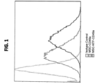
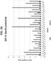

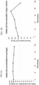
Classifications
-
- G—PHYSICS
- G01—MEASURING; TESTING
- G01N—INVESTIGATING OR ANALYSING MATERIALS BY DETERMINING THEIR CHEMICAL OR PHYSICAL PROPERTIES
- G01N33/00—Investigating or analysing materials by specific methods not covered by groups G01N1/00 - G01N31/00
- G01N33/48—Biological material, e.g. blood, urine; Haemocytometers
- G01N33/50—Chemical analysis of biological material, e.g. blood, urine; Testing involving biospecific ligand binding methods; Immunological testing
- G01N33/5005—Chemical analysis of biological material, e.g. blood, urine; Testing involving biospecific ligand binding methods; Immunological testing involving human or animal cells
- G01N33/5008—Chemical analysis of biological material, e.g. blood, urine; Testing involving biospecific ligand binding methods; Immunological testing involving human or animal cells for testing or evaluating the effect of chemical or biological compounds, e.g. drugs, cosmetics
- G01N33/5044—Chemical analysis of biological material, e.g. blood, urine; Testing involving biospecific ligand binding methods; Immunological testing involving human or animal cells for testing or evaluating the effect of chemical or biological compounds, e.g. drugs, cosmetics involving specific cell types
- G01N33/5073—Stem cells
-
- A—HUMAN NECESSITIES
- A61—MEDICAL OR VETERINARY SCIENCE; HYGIENE
- A61K—PREPARATIONS FOR MEDICAL, DENTAL OR TOILETRY PURPOSES
- A61K35/00—Medicinal preparations containing materials or reaction products thereof with undetermined constitution
- A61K35/12—Materials from mammals; Compositions comprising non-specified tissues or cells; Compositions comprising non-embryonic stem cells; Genetically modified cells
- A61K35/28—Bone marrow; Haematopoietic stem cells; Mesenchymal stem cells of any origin, e.g. adipose-derived stem cells
-
- A—HUMAN NECESSITIES
- A61—MEDICAL OR VETERINARY SCIENCE; HYGIENE
- A61P—SPECIFIC THERAPEUTIC ACTIVITY OF CHEMICAL COMPOUNDS OR MEDICINAL PREPARATIONS
- A61P21/00—Drugs for disorders of the muscular or neuromuscular system
- A61P21/02—Muscle relaxants, e.g. for tetanus or cramps
-
- A—HUMAN NECESSITIES
- A61—MEDICAL OR VETERINARY SCIENCE; HYGIENE
- A61P—SPECIFIC THERAPEUTIC ACTIVITY OF CHEMICAL COMPOUNDS OR MEDICINAL PREPARATIONS
- A61P29/00—Non-central analgesic, antipyretic or antiinflammatory agents, e.g. antirheumatic agents; Non-steroidal antiinflammatory drugs [NSAID]
-
- A—HUMAN NECESSITIES
- A61—MEDICAL OR VETERINARY SCIENCE; HYGIENE
- A61P—SPECIFIC THERAPEUTIC ACTIVITY OF CHEMICAL COMPOUNDS OR MEDICINAL PREPARATIONS
- A61P37/00—Drugs for immunological or allergic disorders
-
- A—HUMAN NECESSITIES
- A61—MEDICAL OR VETERINARY SCIENCE; HYGIENE
- A61P—SPECIFIC THERAPEUTIC ACTIVITY OF CHEMICAL COMPOUNDS OR MEDICINAL PREPARATIONS
- A61P37/00—Drugs for immunological or allergic disorders
- A61P37/02—Immunomodulators
-
- C—CHEMISTRY; METALLURGY
- C12—BIOCHEMISTRY; BEER; SPIRITS; WINE; VINEGAR; MICROBIOLOGY; ENZYMOLOGY; MUTATION OR GENETIC ENGINEERING
- C12N—MICROORGANISMS OR ENZYMES; COMPOSITIONS THEREOF; PROPAGATING, PRESERVING, OR MAINTAINING MICROORGANISMS; MUTATION OR GENETIC ENGINEERING; CULTURE MEDIA
- C12N5/00—Undifferentiated human, animal or plant cells, e.g. cell lines; Tissues; Cultivation or maintenance thereof; Culture media therefor
- C12N5/06—Animal cells or tissues; Human cells or tissues
- C12N5/0602—Vertebrate cells
- C12N5/0652—Cells of skeletal and connective tissues; Mesenchyme
- C12N5/0662—Stem cells
- C12N5/0663—Bone marrow mesenchymal stem cells (BM-MSC)
-
- G—PHYSICS
- G01—MEASURING; TESTING
- G01N—INVESTIGATING OR ANALYSING MATERIALS BY DETERMINING THEIR CHEMICAL OR PHYSICAL PROPERTIES
- G01N33/00—Investigating or analysing materials by specific methods not covered by groups G01N1/00 - G01N31/00
- G01N33/48—Biological material, e.g. blood, urine; Haemocytometers
- G01N33/50—Chemical analysis of biological material, e.g. blood, urine; Testing involving biospecific ligand binding methods; Immunological testing
- G01N33/68—Chemical analysis of biological material, e.g. blood, urine; Testing involving biospecific ligand binding methods; Immunological testing involving proteins, peptides or amino acids
- G01N33/6893—Chemical analysis of biological material, e.g. blood, urine; Testing involving biospecific ligand binding methods; Immunological testing involving proteins, peptides or amino acids related to diseases not provided for elsewhere
- G01N33/6896—Neurological disorders, e.g. Alzheimer's disease
-
- C—CHEMISTRY; METALLURGY
- C12—BIOCHEMISTRY; BEER; SPIRITS; WINE; VINEGAR; MICROBIOLOGY; ENZYMOLOGY; MUTATION OR GENETIC ENGINEERING
- C12N—MICROORGANISMS OR ENZYMES; COMPOSITIONS THEREOF; PROPAGATING, PRESERVING, OR MAINTAINING MICROORGANISMS; MUTATION OR GENETIC ENGINEERING; CULTURE MEDIA
- C12N2501/00—Active agents used in cell culture processes, e.g. differentation
- C12N2501/10—Growth factors
- C12N2501/115—Basic fibroblast growth factor (bFGF, FGF-2)
-
- C—CHEMISTRY; METALLURGY
- C12—BIOCHEMISTRY; BEER; SPIRITS; WINE; VINEGAR; MICROBIOLOGY; ENZYMOLOGY; MUTATION OR GENETIC ENGINEERING
- C12N—MICROORGANISMS OR ENZYMES; COMPOSITIONS THEREOF; PROPAGATING, PRESERVING, OR MAINTAINING MICROORGANISMS; MUTATION OR GENETIC ENGINEERING; CULTURE MEDIA
- C12N2501/00—Active agents used in cell culture processes, e.g. differentation
- C12N2501/10—Growth factors
- C12N2501/13—Nerve growth factor [NGF]; Brain-derived neurotrophic factor [BDNF]; Cilliary neurotrophic factor [CNTF]; Glial-derived neurotrophic factor [GDNF]; Neurotrophins [NT]; Neuregulins
-
- C—CHEMISTRY; METALLURGY
- C12—BIOCHEMISTRY; BEER; SPIRITS; WINE; VINEGAR; MICROBIOLOGY; ENZYMOLOGY; MUTATION OR GENETIC ENGINEERING
- C12N—MICROORGANISMS OR ENZYMES; COMPOSITIONS THEREOF; PROPAGATING, PRESERVING, OR MAINTAINING MICROORGANISMS; MUTATION OR GENETIC ENGINEERING; CULTURE MEDIA
- C12N2501/00—Active agents used in cell culture processes, e.g. differentation
- C12N2501/10—Growth factors
- C12N2501/135—Platelet-derived growth factor [PDGF]
-
- C—CHEMISTRY; METALLURGY
- C12—BIOCHEMISTRY; BEER; SPIRITS; WINE; VINEGAR; MICROBIOLOGY; ENZYMOLOGY; MUTATION OR GENETIC ENGINEERING
- C12N—MICROORGANISMS OR ENZYMES; COMPOSITIONS THEREOF; PROPAGATING, PRESERVING, OR MAINTAINING MICROORGANISMS; MUTATION OR GENETIC ENGINEERING; CULTURE MEDIA
- C12N2501/00—Active agents used in cell culture processes, e.g. differentation
- C12N2501/10—Growth factors
- C12N2501/195—Heregulin, neu differentiation factor
-
- C—CHEMISTRY; METALLURGY
- C12—BIOCHEMISTRY; BEER; SPIRITS; WINE; VINEGAR; MICROBIOLOGY; ENZYMOLOGY; MUTATION OR GENETIC ENGINEERING
- C12N—MICROORGANISMS OR ENZYMES; COMPOSITIONS THEREOF; PROPAGATING, PRESERVING, OR MAINTAINING MICROORGANISMS; MUTATION OR GENETIC ENGINEERING; CULTURE MEDIA
- C12N2501/00—Active agents used in cell culture processes, e.g. differentation
- C12N2501/50—Cell markers; Cell surface determinants
- C12N2501/58—Adhesion molecules, e.g. ICAM, VCAM, CD18 (ligand), CD11 (ligand), CD49 (ligand)
Definitions
- the present invention in some embodiments thereof, relates to a method of qualifying neurotrophic factor secreting cells based on cell surface marker expression.
- ALS Amyotrophic lateral sclerosis
- ALS drugs are developed on the basis of putative pathophysiologic mechanisms, such as anti-glutamatergic agents, drugs targeting protein misfolding and accumulation, antioxidant therapy, immunomodulatory agents, and stem cells.
- stem cell transplantation may have the most potential. Apart from the replacement of lost or damaged motor neurons, stem cell implantation therapy may benefit ALS patients by an independent effect of cytoprotection. Further, there is the potential for stem cells to differentiate into supportive interstitial cells including astrocytes and microglia which can potentially produce neurotrophic factors as well as enzymatic and paracrine mediators which antagonize neurotoxicity. Further experimental data have shown that non-neuronal cell replacement can be a strategic therapy in promoting motor neuron survival and improved neuromuscular function ( Corti S et al. Brain (2010) 133 (2): 465-481 ).
- stem cells as a cellular source in cell replacement therapy for additional neurodegenerative diseases including Parkinson's disease and multiple sclerosis has also been suggested.
- Neurotrophic factors are small, naturally occurring polypeptides that support the development and survival of neurons, and therefore have been considered in the past few years as candidates for therapy options for different neurodegenerative diseases including ALS.
- ALS animal models have shown a delay in disease onset and/or progression after administration of various neurotrophic factors.
- MSCs mesenchymal stem cells
- WO2006/134602 and WO2009/144718 teaches differentiation protocols for the generation of neurotrophic factor secreting cells from mesenchymal stem cells.
- WO2007/066338 teaches differentiation protocols for the generation of oligodendrocyte-like cells from mesenchymal stem cells.
- WO2004/046348 teaches differentiation protocols for the generation of neuronal-like cells from mesenchymal stem cells.
- WO 2014/024183 teaches additional differentiation protocols for the generation of cells which secrete neurotrophic factors.
- non-differentiated stem cells derived from adipose tissue can be selected for the treatment of skin diseases or for cosmetic applications.
- WO 2011/063005 A2 teaches that the level of CD49a expression is increased in RPE cells following expansion.
- a method of qualifying whether a cell population is a suitable therapeutic for treating Amyotrophic Lateral Sclerosis comprises:
- Disclosed herein but not claimed is a method of treating an immune or inflammatory related disease in a subject in need thereof, comprising administering to the subject a therapeutically effective amount of mesenchymal stem cells having been ex vivo differentiated into cells that secrete neurotrophic factors, wherein the immune or inflammatory related disease is not a neurodegenerative disease or myasthenia gravis, thereby treating the disease.
- mesenchymal stem cells which have been ex vivo differentiated into cells that secrete neurotrophic factors for the treatment of an immune or inflammatory related disease, wherein the immune or inflammatory related disease is not a neurodegenerative disease or myasthenia gravis, thereby treating the disease.
- the analyzing the expression of CD49a comprises analyzing the number of cells of the differentiated MSC population which express CD49a, wherein a number of cells being greater than 80 % is indicative of the cell population being suitable as a therapeutic.
- analyzing the expression of CD49a comprises analyzing the level of expression of CD49a in said differentiated MSC population, wherein an increase in the level of expression by more than 2 fold compared to the CD49 expression in an undifferentiated MSC population is indicative of the cell population being suitable as a therapeutic, wherein said differentiated MSC population and said undifferentiated MSC population are derived from the same donor.
- the MSCs are derived from the bone marrow.
- more than 95 % of the cells of said population of undifferentiated MSCs express CD73, CD90 and CD105.
- the populations of undifferentiated MSCs do not express CD3, CD14, CD19, CD34, CD45, and HLA-DR as determined by flow cytometry.
- the incubating is effected for no more than 6 days.
- the number of cells in the cell population is at least 1 ⁇ 10 6 cells.
- the number of cells being greater than 85 % is indicative of the cell population being suitable as a therapeutic.
- the method further comprises determining the amount of neurotrophic factor secreted from the cells, wherein an amount of said neurotrophic factor being above a predetermined level is further indicative of the cell population being suitable as a therapeutic.
- the neurotrophic factor is GDNF.
- the neurotrophic factor is selected from the group consisting of GDNF, VEGF and HGF.
- the predetermined level is at least 5 times greater than the amount of GDNF secreted from a non-differentiated mesenchymal stem cell obtained from the same donor.
- the differentiating medium is devoid of a phosphodiesterase inhibitor.
- the differentiating medium is devoid of triiodothyronine.
- the phosphodiesterase inhibitor comprises IBMX.
- the differentiating medium is devoid of xeno derived components.
- the differentiating medium is devoid of antibiotics.
- the method further comprises culturing said population of undifferentiated MSCs prior to said incubating, wherein said culturing is effected under conditions that do not promote cell differentiation.
- the culturing is effected for three days following seeding of said undifferentiated MSCs.
- the seeding is effected at a density of about 6000-8000 cm 2 .
- the culturing is effected in a culture medium comprising platelet lysate.
- the percentage of said platelet lysate in said culture medium is about 10 %.
- the culture medium further comprises L-glutamine, sodium pyruvate and heparin.
- the analyzing is effected by flow cytometry.
- bFGF basic fibroblast growth factor
- PDGF platelet derived growth factor
- cAMP cAMP
- the present invention in some embodiments thereof, relates to a method of qualifying neurotrophic factor secreting cells based on cell surface marker expression.
- NTFs Neurotrophic factors
- neurotrophic factors are not applicable as they do not pass the blood-brain barrier and do not distribute properly following systemic injection. Therefore, other strategies must be developed in order to take advantage of their therapeutic properties.
- MSCs mesenchymal stem cells
- the present inventors have previously developed a new one step differentiation protocol which enhances the secretion of neurotrophic factors from MSCs.
- the level of secretion of glial derived growth factor (GDNF) and brain derived neurotrophic factor (BDNF) was shown to be consistently up-regulated following the differentiation process, with GDNF being up-regulated by as much as 20 fold and BDNF by as much as three fold as compared to the corresponding non-differentiated cell population obtained from the same donor.
- GDNF glial derived growth factor
- BDNF brain derived neurotrophic factor
- the protocol involves direct differentiation of undifferentiated MSCs in a single medium comprising basic fibroblast growth factor (bFGF), platelet derived growth factor (PDGF), heregulin and cAMP.
- bFGF basic fibroblast growth factor
- PDGF platelet derived growth factor
- cAMP cAMP
- the present inventors have now discovered a unique and simple way of selecting for mesenchymal stem cell populations which have been successfully differentiated according to this protocol based on expression of a cell surface marker.
- CD49a can be used as a single marker to substantiate successful differentiation.
- the present inventors showed that the level of CD49a expression on a successfully differentiated MSC was higher than the level of CD49a expression on a non-differentiated MSC.
- a method of qualifying whether a cell population is a suitable therapeutic comprising:
- suitable therapeutic refers to the suitability of the cell population for treating neurodegenerative diseases and immune diseases (e.g. autoimmune diseases).
- cells which are suitable therapeutics are those that secrete sufficient neurotrophic factors that they are capable of having a therapeutic effect for a particular disease.
- neurodegenerative disease is used herein to describe a disease which is caused by damage to the central nervous system.
- exemplary neurodegenerative diseases which may be treated using the cells and methods according to the present invention include for example: Amyotrophic Lateral Sclerosis (ALS), Parkinson's disease, Multiple System Atrophy (MSA), Huntington's disease, Alzheimer's disease, Rett Syndrome, lysosomal storage diseases ("white matter disease” or glial/demyelination disease, as described, for example by Folkerth, J. Neuropath. Exp.
- Neuro. September 1999, 58:9
- Sanfilippo Gaucher disease, Tay Sachs disease (beta hexosaminidase deficiency), other genetic diseases, multiple sclerosis (MS), brain injury or trauma caused by ischemia, accidents, environmental insult, etc., spinal cord damage, ataxia.
- MS multiple sclerosis
- the present invention may be used to reduce and/or eliminate the effects on the central nervous system of a stroke in a patient, which is otherwise caused by lack of blood flow or ischemia to a site in the brain of the patient or which has occurred from physical injury to the brain and/or spinal cord.
- Neurodegenerative diseases also include neurodevelopmental disorders including for example, autism-spectrum disorders and related neurological diseases such as schizophrenia, among numerous others.
- Autoimmune diseases of the nervous system which may be treated using the cells described herein include for example, multiple sclerosis and myasthenia gravis, Guillain bar syndrome, Multiple system Atrophy (MSA; a sporadic, progressive, adult-onset neurodegenerative disorder associated with varying degrees of parkinsonism, autonomic dysfunction and cerebellar ataxia).
- MSA Multiple system Atrophy
- Other autoimmune diseases are described in Kraker et al., Curr Neuropharmacol. 2011 September; 9(3): 400-408 , the contents of which are incorporated herein by reference.
- the cells of the present invention show enhanced immunomodulatory effect as compared to non-differentiated bone marrow derived MSCs (see Table 5 herein below).
- the cells of the present invention may be useful in the treatment of any immune-related or inflammatory disorder.
- inflammatory disorders includes but is not limited to chronic inflammatory diseases and acute inflammatory diseases. Examples of such diseases and conditions are summarized infra.
- hypersensitivity examples include, but are not limited to, Type I hypersensitivity, Type II hypersensitivity, Type III hypersensitivity, Type IV hypersensitivity, immediate hypersensitivity, antibody mediated hypersensitivity, immune complex mediated hypersensitivity, T lymphocyte mediated hypersensitivity and DTH.
- Type I or immediate hypersensitivity such as asthma.
- Type II hypersensitivity include, but are not limited to, rheumatoid diseases, rheumatoid autoimmune diseases, rheumatoid arthritis ( Krenn V. et al., Histol Histopathol 2000 Jul;15 (3):791 ), spondylitis, ankylosing spondylitis ( Jan Voswinkel et al., Arthritis Res 2001; 3 (3): 189 ), systemic diseases, systemic autoimmune diseases, systemic lupus erythematosus ( Erikson J. et al., Immunol Res 1998;17 (1-2):49 ), sclerosis, systemic sclerosis ( Renaudineau Y. et al., Clin Diagn Lab Immunol.
- vasculitises necrotizing small vessel vasculitises, microscopic polyangiitis, Churg and Strauss syndrome, glomerulonephritis, pauci-immune focal necrotizing glomerulonephritis, crescentic glomerulonephritis ( Noel LH. Ann Med Interne (Paris). 2000 May;151 (3):178 ); antiphospholipid syndrome ( Flamholz R. et al., J Clin Apheresis 1999;14 (4):171 ); heart failure, agonist-like beta-adrenoceptor antibodies in heart failure ( Wallukat G. etal., Am J Cardiol.
- Type IV or T cell mediated hypersensitivity include, but are not limited to, rheumatoid diseases, rheumatoid arthritis ( Tisch R, McDevitt HO. Proc Natl Acad Sci U S A 1994 Jan 18;91 (2):437 ), systemic diseases, systemic autoimmune diseases, systemic lupus erythematosus ( Datta SK., Lupus 1998;7 (9):591 ), glandular diseases, glandular autoimmune diseases, pancreatic diseases, pancreatic autoimmune diseases, Type 1 diabetes ( Castano L. and Eisenbarth GS. Ann. Rev. Immunol. 8:647 ); thyroid diseases, autoimmune thyroid diseases, Graves' disease ( Sakata S.
- delayed type hypersensitivity examples include, but are not limited to, contact dermatitis and drug eruption.
- T lymphocyte mediating hypersensitivity examples include, but are not limited to, helper T lymphocytes and cytotoxic T lymphocytes.
- helper T lymphocyte-mediated hypersensitivity examples include, but are not limited to, T h 1 lymphocyte mediated hypersensitivity and T h 2 lymphocyte mediated hypersensitivity.
- cardiovascular diseases include, but are not limited to, cardiovascular diseases, rheumatoid diseases, glandular diseases, gastrointestinal diseases, cutaneous diseases, hepatic diseases, neurological diseases, muscular diseases, nephric diseases, diseases related to reproduction, connective tissue diseases and systemic diseases.
- autoimmune cardiovascular diseases include, but are not limited to atherosclerosis ( Matsuura E. et al., Lupus. 1998;7 Suppl 2:S135 ), myocardial infarction ( Vaarala O. Lupus. 1998;7 Suppl 2:S132 ), thrombosis ( Tincani A. et al., Lupus 1998;7 Suppl 2:S107-9 ), Wegener's granulomatosis, Takayasu's arteritis, Kawasaki syndrome ( Praprotnik S. et al., Wien Klin Klin Klin Klinschr 2000 Aug 25;112 (15-16):660 ), anti-factor VIII autoimmune disease ( Lacroix-Desmazes S.
- autoimmune rheumatoid diseases include, but are not limited to rheumatoid arthritis ( Krenn V. et al., Histol Histopathol 2000 Jul;15 (3):791 ; Tisch R, McDevitt HO. Proc Natl Acad Sci units S A 1994 Jan 18;91 (2):437 ) and ankylosing spondylitis ( Jan Voswinkel et al., Arthritis Res 2001; 3 (3): 189 ).
- autoimmune glandular diseases include, but are not limited to, pancreatic disease, Type I diabetes, thyroid disease, Graves' disease, thyroiditis, spontaneous autoimmune thyroiditis, Hashimoto's thyroiditis, idiopathic myxedema, ovarian autoimmunity, autoimmune anti-sperm infertility, autoimmune prostatitis and Type I autoimmune polyglandular syndrome.
- Diseases include, but are not limited to autoimmune diseases of the pancreas, Type 1 diabetes ( Castano L. and Eisenbarth GS. Ann. Rev. Immunol. 8:647 ; Zimmet P. Diabetes Res Clin Pract 1996 Oct;34 Suppl:S125 ), autoimmune thyroid diseases, Graves' disease ( Orgiazzi J.
- autoimmune gastrointestinal diseases include, but are not limited to, chronic inflammatory intestinal diseases ( Garcia Herola A. et al., Gastroenterol Hepatol. 2000 Jan;23 (1):16 ), celiac disease ( Landau YE. and Shoenfeld Y. Harefuah 2000 Jan 16;138 (2):122 ), colitis, ileitis and Crohn's disease.
- autoimmune cutaneous diseases include, but are not limited to, autoimmune bullous skin diseases, such as, but are not limited to, pemphigus vulgaris, bullous pemphigoid and pemphigus foliaceus.
- autoimmune hepatic diseases include, but are not limited to, hepatitis, autoimmune chronic active hepatitis ( Franco A. et al., Clin Immunol Immunopathol 1990 Mar;54 (3):382 ), primary biliary cirrhosis ( Jones DE. Clin Sci (Colch) 1996 Nov;91 (5):551 ; Strassburg CP. et al., Eur J Gastroenterol Hepatol. 1999 Jun;11 (6):595 ) and autoimmune hepatitis ( Manns MP. J Hepatol 2000 Aug;33 (2):326 ).
- autoimmune neurological diseases include, but are not limited to, multiple sclerosis ( Cross AH. et al., J Neuroimmunol 2001 Jan 1;112 (1-2):1 ), Alzheimer's disease ( Oron L. et al., J Neural Transm Suppl. 1997;49:77 ), myasthenia gravis ( Infante AJ. And Kraig E, Int Rev Immunol 1999;18 (1-2):83 ; Oshima M. et al., Eur J Immunol 1990 Dec;20 (12):2563 ), neuropathies, motor neuropathies ( Kornberg AJ. J Clin Neurosci.
- autoimmune muscular diseases include, but are not limited to, myositis, autoimmune myositis and primary Sjogren's syndrome ( Feist E. et al., Int Arch Allergy Immunol 2000 Sep;123 (1):92 ) and smooth muscle autoimmune disease ( Zauli D. et al., Biomed Pharmacother 1999 Jun;53 (5-6):234 ).
- autoimmune nephric diseases include, but are not limited to, nephritis and autoimmune interstitial nephritis ( Kelly CJ. J Am Soc Nephrol 1990 Aug;1 (2):140 ).
- autoimmune diseases related to reproduction include, but are not limited to, repeated fetal loss ( Tincani A. et al., Lupus 1998;7 Suppl 2:S107-9 ).
- autoimmune connective tissue diseases include, but are not limited to, ear diseases, autoimmune ear diseases ( Yoo TJ. et al., Cell Immunol 1994 Aug;157 (1):249 ) and autoimmune diseases of the inner ear ( Gloddek B. et al., Ann N Y Acad Sci 1997 Dec 29;830:266 ).
- autoimmune systemic diseases include, but are not limited to, systemic lupus erythematosus ( Erikson J. et al., Immunol Res 1998;17 (1-2):49 ) and systemic sclerosis ( Renaudineau Y. etal., Clin Diagn Lab Immunol. 1999 Mar;6 (2):156 ); Chan OT. et al., Immunol Rev 1999 Jun;169:107 ).
- infectious diseases include, but are not limited to, chronic infectious diseases, subacute infectious diseases, acute infectious diseases, viral diseases, bacterial diseases, protozoan diseases, parasitic diseases, fungal diseases, mycoplasma diseases and prion diseases.
- diseases associated with transplantation of a graft include, but are not limited to, graft rejection, chronic graft rejection, subacute graft rejection, hyperacute graft rejection, acute graft rejection and graft versus host disease.
- allergic diseases include, but are not limited to, asthma, hives, urticaria, pollen allergy, dust mite allergy, venom allergy, cosmetics allergy, latex allergy, chemical allergy, drug allergy, insect bite allergy, animal dander allergy, stinging plant allergy, poison ivy allergy and food allergy.
- cancer examples include but are not limited to carcinoma, lymphoma, blastoma, sarcoma, and leukemia.
- cancerous diseases include but are not limited to: Myeloid leukemia such as Chronic myelogenous leukemia. Acute myelogenous leukemia with maturation. Acute promyelocytic leukemia, Acute nonlymphocytic leukemia with increased basophils, Acute monocytic leukemia. Acute myelomonocytic leukemia with eosinophilia; Malignant lymphoma, such as Birkitt's Non-Hodgkin's; Lymphoctyic leukemia, such as Acute lumphoblastic leukemia.
- Chronic lymphocytic leukemia Myeloproliferative diseases, such as Solid tumors Benign Meningioma, Mixed tumors of salivary gland, Colonic adenomas; Adenocarcinomas, such as Small cell lung cancer, Kidney, Uterus, Prostate, Bladder, Ovary, Colon, Sarcomas, Liposarcoma, myxoid, Synovial sarcoma, Rhabdomyosarcoma (alveolar), Extraskeletel myxoid chonodrosarcoma, Ewing's tumor; other include Testicular and ovarian dysgerminoma, Retinoblastoma, Wilms' tumor, Neuroblastoma, Malignant melanoma, Mesothelioma, breast, skin, prostate, and ovarian.
- Adenocarcinomas such as Small cell lung cancer, Kidney, Uterus, Prostate, Bladder, Ovary, Colon, Sarcomas, Lipos
- the method described herein is for qualifying whether the cell populations are suitable for treating ALS.
- mesenchymal stem cell or “MSC” is used interchangeably for adult cells which are not terminally differentiated, which can divide to yield cells that are either stem cells, or which, irreversibly differentiate to give rise to cells of a mesenchymal (chrondocyte, osteocyte and adipocyte) cell lineage.
- the mesenchymal stem cells of the present invention may be of an autologous (e.g. syngeneic) or allogeneic source.
- the undifferentiated MSCs typically express particular markers on their cell surface.
- the undifferentiated MSCs express CD105, CD73 and CD90 on the cell surface (e.g. >95% positive) and lack expression (e.g. ⁇ 2% positive) of CD3, CD14, CD19, CD34, CD45, and HLA-DR as determined by flow cytometry.
- Exemplary antibodies that may be used to verify the presence of mesenchymal stem cells include CD44 FITC conjugated, BD Biosciences, CD73 PE conjugated (BD Pharmingen), CD73 PE conjugated, BD Biosciences, CD90 PE-Cy5 conjugated (eBioscience) CD90 PE conjugated, BD Biosciences CD105 PE conjugated (Beckman Coulter) CD3 PerCP conjugated, BD Biosciences, CD14 FITC conjugated (eBioscience) CD14 FITC conjugated, BD Biosciences CD19 PE-Cy5 conjugated (eBioscience) CD19 FITC conjugated, BD Biosciences CD34 FITC conjugated BD Biosciences (Beckman Coulter), CD45 PE conjugated (eBioscience) CD45 PerCP conjugated, BD Biosciences and HLA-DR PE-Cy5 conjugated (BD Pharmingen). HLA-DR PerCP conjugated, BD Biosciences.
- Another method for verifying the presence of mesenchymal stem cells is by showing that the cells are capable of differentiating into multi-lineages such as for example adipocytes, osteocytes and chondrocytes. This may be effected for example using Human Mesenchymal Stem Cell Functional Identification Kit (R&D Systems).
- the mesenchymal stem cells are not genetically manipulated (i.e. transformed with an expression construct) to generate the cells and cell populations described herein.
- the cells of the present invention may be derived from any stem cell, although preferably not embryonic stem (ES) cells.
- stem cell although preferably not embryonic stem (ES) cells.
- Mesenchymal stem cells may be isolated from various tissues including but not limited to bone marrow, peripheral blood, blood, placenta and adipose tissue.
- a method of isolating mesenchymal stem cells from peripheral blood is described by Kassis et al [Bone Marrow Transplant. 2006 May; 37(10):967-76 ].
- a method of isolating mesenchymal stem cells from placental tissue is described by Brooke G et al. [Br J Haematol. 2009 Feb; 144 (4):571-9 ].
- the mesenchymal stem cells are human.
- Bone marrow can be isolated from the iliac crest or the sternum of an individual by aspiration.
- Low-density BM mononuclear cells (BMMNC) may be separated by FICOLL-PAQUE density gradient centrifugation.
- a cell population comprising the mesenchymal stem cells (e.g. BMMNC) may be cultured in a proliferating medium capable of maintaining and/or expanding the cells in the presence of platelet lysate.
- the populations are plated on plastic surfaces (e.g. in a flask) and mesenchymal stem cells are isolated by removing non-adherent cells.
- mesenchymal stem cell may be isolated by FACS using mesenchymal stem cell markers.
- the cells may be expanded by culturing in a proliferation medium capable of maintaining and/or expanding the isolated cells ex vivo in the presence of platelet lysate.
- the proliferation medium may be DMEM, alpha-MEM or DMEM/F12.
- the glucose concentration in the medium is about 0.5 - 3 grams/litre.
- the culturing may be effected on any suitable surface including plastic dishes and bioreactors suitable for culturing mesenchymal stem cells.
- Platelet lysate may be prepared using any method known in the art.
- Platelet Rich Plasma PRP
- PRP containing bags may be stored at -80 0 C and thawed in a 37 °C water bath. After thawing, the Platelet Rich Plasma is typically centrifuged to remove platelet particles and membranes. The Platelet lysate supernatant may then be collected and frozen at -80 °C until use. The Platelet lysate is tested for Endotoxin, Haemoglobin, pH, Total protein, Albumin, Osmolality Sterility and Mycoplasma.
- the proliferation medium may comprise additional components, including for example L-glutamine, sodium pyruvate and heparin.
- the platelet lysate is also obtained from human cells.
- the proliferation/growth medium is devoid of xeno contaminants i.e. free of animal derived components such as serum, animal derived growth factors and albumin.
- the culturing is performed in the absence of xeno contaminants.
- the cells are differentiated in a differentiating medium to generating cells useful for treating diseases.
- the cells are reseeded in a fresh proliferation/growth medium (e.g. at a density of about 6000-8000 cells per cm 2 ) for 1 day, 2 days, 3 days, 4 days or 5 days prior to addition of the differentiation medium.
- a fresh proliferation/growth medium e.g. at a density of about 6000-8000 cells per cm 2
- undifferentiated MSCs refers to MSCs that have not been cultured in a medium that induces differentiation.
- the MSCs are contacted directly with the differentiation medium without any intervening pre-differentiation steps.
- the undifferentiated MSCs of the present invention are incubated in a medium comprising fibroblast growth factor (FGF), platelet derived growth factor (PDGF), heregulin and c-AMP.
- FGF fibroblast growth factor
- PDGF platelet derived growth factor
- c-AMP c-AMP
- the undifferentiated MSCs of the present invention are not pre-incubated in the presence of epidermal growth factor (EGF) and/or N2 supplement prior to this step and following the expansion step.
- EGF epidermal growth factor
- An exemplary concentration of bFGF which is contemplated for the differentiation medium of embodiments of this invention is optionally between 5-50 ng/ml, optionally between 10-40 ng/ml, optionally between 10-25 ng/ml.
- An exemplary concentration of PDGF-AA which is contemplated for the differentiation medium of embodiments of this invention is optionally between 1-30 ng/ml, optionally between 1-20 ng/ml, optionally between 1-10 ng/ml, optionally between 2.5-10 ng/ml.
- An exemplary concentration of heregulin ⁇ 1 which is contemplated for the differentiation medium of embodiments of this invention is optionally between 5-100 ng/ml, 10-90 ng/ml, optionally between 25-75 ng/ml and optionally between 40-60 ng/ml.
- An exemplary concentration of dbc-AMP which is contemplated for the differentiation medium of embodiments of this invention is optionally between 0.5-10 mM, optionally between 0.5-5mM and optionally between 0.5 and 2.5 mM.
- the differentiating medium of this aspect of the present invention is devoid of a phosphodiesterase inhibitor (e.g. IBMX) i.e. the culturing is performed in the absence of a phosphodiesterase inhibitor.
- a phosphodiesterase inhibitor e.g. IBMX
- the differentiating medium of this aspect of the present invention is devoid of triiodothyronine i.e. the culturing is performed in the absence of triiodothyronine.
- any of these embodiments and subembodiments may be combined, so that for example the differentiating medium may optionally be devoid of both a phosphodiesterase inhibitor and triiodothyronine.
- the MSCs are differentiated in the above described differentiating medium for at least one day, at least two days or at least 3 days.
- the differentiating stage is not performed for more than five days.
- the differentiating media used according to this aspect of the present invention are preferably xeno-free (devoid of serum) and devoid of any antibiotics i.e. the culturing is performed in the absence of xeno-contaminants.
- HBSS Hanks balanced salt solution
- DMEM Dulbecco Modified Eagle Medium
- PBS PBS
- Hypothermic storage mediums are also contemplated (e.g. Hypothermosol).
- the cells obtained are analyzed for the expression of CD49a, wherein an amount of CD49a above a predetermined level indicative of the cell population being suitable as the therapeutic.
- the number of the cells in the sample is about 0.5 ⁇ 10 6 cells.
- the number of cells obtained from a single donor is generally between 20 ⁇ 10 6 cells - 100 ⁇ 10 7 cells.
- the number of cells may be about 20 ⁇ 10 6 cells, about 100 ⁇ 10 6 cells, about 200 ⁇ 10 6 cells are differentiated, about 300 ⁇ 10 6 cells, about 400 ⁇ 10 6 cells are differentiated, about 500 ⁇ 10 6 cells are differentiated, about 600 ⁇ 10 6 cells are differentiated, about 700 ⁇ 10 6 cells, about 800 ⁇ 10 6 cells, about 900 ⁇ 10 6 cells or about 100 ⁇ 10 7 cells.
- CD49a also referred to as Integrin alpha 1 refers to the alpha 1 subunit of integrin receptor cell surface membrane protein that binds to the extracellular matrix. This protein heterodimerizes with the beta 1 subunit to form a cell-surface receptor for collagen and laminin. The heterodimeric receptor is involved in cell-cell adhesion.
- the human protein sequence of CD49a is set forth in Uniprot No. P56199, NP_852478 and its mRNA sequence is set forth in NM_181501.
- CD49a forms a heterodimer on the surface of cells together with the CD49 beta1
- the method of the present invention can also be effected by determining the amount of CD49beta1 on the surface of the differentiated cells.
- Methods for analyzing expression of CD49a or CD49beta1 typically involve the use of antibodies which specifically recognize the antigen.
- Commercially available antibodies that recognize CD49a include for example those manufactured by R and D systems, Santa Cruz (Cat# SC-81733PE) or Biolegend (e.g. catalogue number 328303).
- the analyzing may be carried out using any method known in the art including flow cytometry, Western Blot, HPLC, in situ-PCR immunocytochemistry, mass spectrometry, radioimmunoassay, etc. According to a particular embodiment, the analyzing is effected using an antibody which specifically recognizes the protein.
- the CD49a or CD49b1 antibody is attached to a fluorescent moiety and analyzed using a fluorescence-activated cell sorter (FACS).
- FACS fluorescence-activated cell sorter
- flow cytometry refers to an assay in which the proportion of a material (e.g. mesenchymal stem cells comprising a particular marker) in a sample is determined by labeling the material (e.g., by binding a labeled antibody to the material), causing a fluid stream containing the material to pass through a beam of light, separating the light emitted from the sample into constituent wavelengths by a series of filters and mirrors, and detecting the light.
- a material e.g. mesenchymal stem cells comprising a particular marker
- the cell populations need to be removed from the culture plate.
- agents that may be used to disperse the cells include, but are not limited to collagenase, dispase, accutase, trypsin (e.g. trypsin-EDTA, non-animal substitutes of trypsin such as TrypLE TM ), papain.
- trypsin e.g. trypsin-EDTA, non-animal substitutes of trypsin such as TrypLE TM
- trituration may also be performed to increase the dispersal of the cells.
- An exemplary concentration of trypsin that may be used is 0.005-0.5 % trypsin-EDTA.
- the cells may be incubated with the dispersing agent for about 5-30 minutes, at a temperature of about 37 °C.
- the cells are typically resuspended in a suitable medium including for example phosphate buffered saline (PBS), Hanks balanced salt solution (HBSS), Dulbecco Modified Eagle Medium (DMEM) RPMI, PBS etc.
- PBS phosphate buffered saline
- HBSS Hanks balanced salt solution
- DMEM Dulbecco Modified Eagle Medium
- the amount of CD49a should be increased above a statistically significant level as compared to non-differentiated MSCs of the same donor and from the same organ.
- At least 80 % of the cells of the population should express CD49a, more preferably at least 85 % of the cells of the population should express CD49a, more preferably at least 90 % of the cells of the population should express CD49a, more preferably at least 95 % of the cells of the population should express CD49a.
- the level of CD49a expression (e.g. the mean fluorescent intensity) should be increased by at least two fold, more preferably at least 3 fold, more preferably at least 4 fold and even more preferably by at least 5 fold as compared to non-differentiated MSCs of the same donor and from the same organ.
- cell populations may be obtained which are more than 80 % positive for CD49a.
- cell populations may be obtained which are 85 % positive for CD49a, 90 % positive for CD49a, 91 % positive for CD49a, 92 % positive for CD49a, 93 % positive for CD49a, 94 % positive for CD49a, 95 % positive for CD49a, 96 % positive for CD49a, 97 % positive for CD49a, 98 % positive for CD49a, 99 % positive for CD49a and even 100 % positive for CD49a.
- the cells may be analyzed for expression of additional cell surface markers such as CD44.
- Cells which have a decrease in expression by at least 1.5 or at least 2 fold or more of CD44 may be qualified as being useful as a therapeutic.
- the cells may be qualified or characterized in additional ways including for example karyotype analysis, morphology, cell number and viability, gram staining and sterility.
- the cells may be analyzed for their level of neurotrophic factor (NTF) secretion.
- NTF neurotrophic factor
- NTFs For analysis of secreted NTFs, supernatant is collected from cultures of MSCs or of NTF-secreting cells at the end of the differentiation procedure described above, and cells are harvested and counted.
- the amount of NTFs such as Glial Derived Neurotrophic Factor, (GDNF) or Brain Derived Neurotrophic Factor (BDNF) in the cell's culture supernatants may be quantified by using a GDNF or BDNF ELISA assay (GDNF DuoSet DY212; BDNF DuoSet DY248; R&D Systems) according to the manufacturer's protocol, for example and without limitation.
- the amount of IGF-1 can be quantified using an IGF ELISA assay (IGF-1 DuoSet Cat No. DY291; R&D System), for example and without limitation.
- the amount of VEGF can be quantified using a VEGF ELISA assay (VEGF DuoSet R&D systems, Cat: DY293B) for example and without limitation.
- the amount of HGF can be quantified using an HGF ELISA assay (HGF DuoSet R&D systems, Cat: DY294) for example and without limitation.
- the amount of GDNF secreted by the cells of the present invention is increased by at least 2 fold, at least 3 fold, at least 4 fold, at least 5 fold, at least 6 fold, at least 7 fold, at least 8 fold the secretion of the same population of mesenchymal stem cells without differentiation.
- the specific productivity of GDNF is from about 200-2000 pg/10 6 cells.
- At least 50 %, at least 60 %, at least 70 %, at least 80 %, at least 90 % or more of a population of the differentiated cells of the present invention secrete BDNF.
- the amount of BDNF secreted by the cells of the present invention is increased by at least 1.5 fold, at least 2 fold, at least 2.5 fold, at least 3 fold the secretion of the same population of mesenchymal stem cells without differentiation.
- the specific productivity of BDNF is from about 500-8000 pg/10 6 cells.
- the cells of the present invention differ from non-differentiated bone marrow derived mesenchymal stem cells in a variety of different ways.
- the cells of the present invention secrete at least 5 fold more GDF-15 than non-differentiated MSCs as measured by an ELISA assay for GDF-15 (e.g. R&D Systems, Cat # DY957 or equivalent).
- an ELISA assay for GDF-15 e.g. R&D Systems, Cat # DY957 or equivalent.
- the cells of the present invention secrete at least 10 fold, 20 fold or even 30 fold more IL-8 than non-differentiated MSCs as measured by an ELISA assay for IL-8 (e.g. R&D Systems, Cat # DY208-05 or equivalent).
- an ELISA assay for IL-8 e.g. R&D Systems, Cat # DY208-05 or equivalent.
- the cells of the present invention comprise at least 2 fold, 4 fold, 6 fold, 8 fold or even 10 fold the amount of any one of the polypeptides 1-82, listed in Table 2.
- the cells of the present invention comprise at least 2 fold, 4 fold, 6 fold, 8 fold or even 10 fold the amount of each of the polypeptides 1-82, listed in Table 2.
- the cells of the present invention comprise at least 2 fold, 4 fold, 6 fold, 8 fold or even 10 fold less of at least one of the polypeptides 83-122, listed in Table 2.
- the cells of the present invention comprise at least 2 fold, 4 fold, 6 fold, 8 fold or even 10 fold less of each of the polypeptides 83-122, listed in Table 2.
- the cells of the present invention may be distinguished from non-differentiated MSCs according to expression of particular genes. This may be measured by analyzing the amount of mRNA there is present in the cells encoded by the gene.
- the cells of the present invention express at least 6 fold, 8 fold or even 10 fold the amount of any one of the genes 1-41, listed in Table 3. According to another embodiment, the cells of the present invention express at least 6 fold, 8 fold, 10 fold, 20 fold, or even 30 fold the amount of each of the genes 1-41, listed in Table 3. The cells of the present invention express at least 6 fold, 8 fold, 10 fold, 20 fold or even 30 fold less of at least one of the genes 42-56, listed in Table 3. The cells of the present invention express at least 6 fold, 8 fold, 10 fold or even 20 fold less of each of the genes 42-56, listed in Table 3.
- the cells of the present invention differ from other bone marrow mesenchymal stem cell-derived NTF secreting cells (e.g. those disclosed in WO2009/144718 - those cells are referred to herein as 2 step protocol NTFs).
- the cells of the present invention comprise at least 2 fold, 4 fold, 6 fold, 8 fold or even 10 fold the amount of any one of the polypeptides 1 and/or 9, listed in Table 4 as compared to 2 step protocol NTFs.
- the cells of the present invention comprise at least 2 fold, 4 fold, 6 fold, 8 fold or even 10 fold less of at least one of the polypeptides 2-8, listed in Table 4 as compared to 2 step protocol NTFs.
- the cells of the present invention comprise at least 2 fold, 4 fold, 6 fold, 8 fold or even 10 fold less of each of the polypeptides 2-8, listed in Table 4 as compared to 2 step protocol NTFs.
- the cells of the present invention express at least 2 fold, 4 fold or even 6 fold the amount of any one of the genes 1-82, listed in Table 5, as compared to 2 step protocol NTFs. According to another embodiment, the cells of the present invention express at least 2 fold, 4 fold or even 6 fold the amount of each of the genes 1-82 listed in Table 5 as compared to 2 step protocol NTFs. The cells of the present invention express at least 2 fold, 4 fold or even 6 fold less of at least one of the genes 83-126, listed in Table 5 as compared to 2 step protocol NTFs. The cells of the present invention express at least 2 fold, 4 fold or even 6 fold less of each of the genes 83-126, listed in Table 5 as compared to 2 step protocol NTFs.
- the cells may be labeled accordingly and preserved according to methods known in the art (e.g. frozen or cryopreserved) or may be directly administered to the subject.
- the cells of this aspect of the present invention may be useful in treating immune or inflammatory related diseases.
- a method of treating an immune or inflammatory related disease in a subject in need thereof comprising administering to the subject a therapeutically effective amount of mesenchymal stem cells which have been ex vivo differentiated to secrete neurotrophic factors, thereby treating the disease.
- the immune or inflammatory related disease is not a neurodegenerative disease.
- the immune or inflammatory related disease is not an immune disease of the nervous system.
- the immune or inflammatory related disease is not myasthenia gravis.
- the cells can be administered either per se or, preferably as a part of a pharmaceutical composition that further comprises a pharmaceutically acceptable carrier.
- a "pharmaceutical composition” refers to a preparation of one or more of the chemical conjugates described herein, with other chemical components such as pharmaceutically suitable carriers and excipients.
- the purpose of a pharmaceutical composition is to facilitate administration of a compound to a subject.
- the term "pharmaceutically acceptable carrier” refers to a carrier or a diluent that does not cause significant irritation to a subject and does not abrogate the biological activity and properties of the administered compound.
- carriers are propylene glycol; saline; emulsions; buffers; culture medium such as DMEM or RPMI; hypothermic storage medium containing components that scavenge free radicals, provide pH buffering, oncotic/osmotic support, energy substrates and ionic concentrations that balance the intracellular state at low temperatures; and mixtures of organic solvents with water.
- the pharmaceutical carrier preserves the number of cells (e.g. is not reduced by more than 90 %) in the composition for at least 24 hours, at least 48 hours or even at least 96 hours.
- excipient refers to an inert substance added to a pharmaceutical composition to further facilitate administration of a compound and maintain cells viability at a pre-determined temperature for a suitable period of time before transplantation/injection.
- excipients include albumin, plasma, serum and cerebrospinal fluid (CSF), antioxidants such as N-Acetylcysteine (NAC) or resveratrol.
- the pharmaceutical carrier is an aqueous solution of buffer or a culture medium such as DMEM.
- the therapeutically effective amount or dose can be estimated initially from in vitro and cell culture assays.
- a dose is formulated in an animal model to achieve a desired concentration or titer. Such information can be used to more accurately determine useful doses in humans.
- Toxicity and therapeutic efficacy of the active ingredients described herein can be determined by standard pharmaceutical procedures in vitro, in cell cultures or experimental animals.
- the dosage may vary depending upon the dosage form employed and the route of administration utilized. The exact formulation, route of administration and dosage can be chosen by the individual physician in view of the patient's condition, (see e.g., Fingl, et al., 1975, in "The Pharmacological Basis of Therapeutics", Ch. 1 p.1 .
- the active ingredients of the pharmaceutical composition may be formulated in aqueous solutions, preferably in physiologically compatible buffers such as Hank's solution, Ringer's solution, or physiological salt buffer and additional agents as described herein above.
- physiologically compatible buffers such as Hank's solution, Ringer's solution, or physiological salt buffer and additional agents as described herein above.
- Dosage amount and interval may be adjusted individually to levels of the active ingredient which are sufficient to effectively cause an immunomodulatory effect. Dosages necessary to achieve the desired effect will depend on individual characteristics and route of administration.
- dosing of cells can be of a single or a plurality of administrations, with course of treatment lasting from several days to several weeks or months depending when diminution of the disease state is achieved.
- the amount of a composition to be administered will, of course, be dependent on the individual being treated, the severity of the affliction, the manner of administration, the judgment of the prescribing physician, etc.
- the dosage and timing of administration will be responsive to a careful and continuous monitoring of the individual changing condition.
- the cells of the present invention may be prepackaged in unit dosage forms in a syringe ready for use.
- the syringe may be labeled with the name of the cells and their source.
- the labeling may also comprise information related to the function of the cells (e.g. the amount of neurotrophic factor secreted therefrom).
- the syringe may be packaged in a packaging which is also labeled with information regarding the cells.
- compositions, method or structure may include additional ingredients, steps and/or parts, but only if the additional ingredients, steps and/or parts do not materially alter the basic and novel characteristics of the claimed composition, method or structure.
- a compound or “at least one compound” may include a plurality of compounds, including mixtures thereof.
- range format is merely for convenience and brevity and should not be construed as an inflexible limitation on the scope of the invention. Accordingly, the description of a range should be considered to have specifically disclosed all the possible subranges as well as individual numerical values within that range. For example, description of a range such as from 1 to 6 should be considered to have specifically disclosed subranges such as from 1 to 3, from 1 to 4, from 1 to 5, from 2 to 4, from 2 to 6, from 3 to 6 etc., as well as individual numbers within that range, for example, 1, 2, 3, 4, 5, and 6. This applies regardless of the breadth of the range.
- method refers to manners, means, techniques and procedures for accomplishing a given task including, but not limited to, those manners, means, techniques and procedures either known to, or readily developed from known manners, means, techniques and procedures by practitioners of the chemical, pharmacological, biological, biochemical and medical arts.
- treating includes abrogating, substantially inhibiting, slowing or reversing the progression of a condition, substantially ameliorating clinical or aesthetical symptoms of a condition or substantially preventing the appearance of clinical or aesthetical symptoms of a condition.
- Bone marrow aspiration Fresh bone marrow was aspirated according to the routine Medical Center procedure from the patient's iliac-crest under local anesthesia and sedation by an anesthetist. Bone marrow (30-60 ml) was aspirated using aspiration needles into heparin containing tubes.
- This step involves separation of mononuclear cells (MNC) from total bone marrow.
- the Human Multipotent Mesenchymal stromal cells (MSC), estimated to comprise 0.01% of total bone marrow MNC, are enriched in-vitro from MNC, by virtue of their ability to adhere to plastic.
- Bone marrow aspirate was diluted 1:1 (v:v) in Hank's Balanced Salt Solution (HBSS), and MNC were separated from total bone marrow cells by Ficoll density gradient centrifugation.
- HBSS Hank's Balanced Salt Solution
- MNC were counted and cell number and viability were determined by the Trypan Blue dye exclusion test.
- the yield of MNC recovered after density gradient centrifugation varied between donors and depends on the volume of bone marrow collected.
- the yield of MNC recovered from 30-50 ml of bone marrow aspirate of ALS patients ranged between 70-400 ⁇ 10 6 MNC and was sufficient for isolating the number of MSC necessary for the entire production process.
- the medium used for seeding the primary bone marrow mononuclear cells and propagating the MSCs throughout the production process was designated Platelets Growth medium (PM).
- PM Platelets Growth medium
- the PM medium was used throughout the MSC production process (Passage 0 - Passage 4) [P0-P4] and contained low glucose DMEM, L-Glutamine sodium pyruvate, heparin and platelet lysate.
- MNCs were seeded at a density of 100,000-400,000 cells/cm 2 in flasks in PM/flask and incubated overnight in a 37 °C/5% CO 2 humidified incubator. The next day, the cell culture was examined under the microscope. At this stage, non-adherent, mononuclear cell were floating in the culture supernatant and plastic-adherent MSC were attached to the flask surface. The culture supernatant containing the non- adherent mononuclear cells was removed, and the adherent cells were gently washed with DMEM. The DMEM was discarded and fresh PM was added to each flask containing the plastic adherent MSC cells. The process phase from MNC seeding to MSC harvesting was designated Passage 0 (P0).
- P0 Passage 0
- the P0 cells were incubated in a 37°C/5% CO 2 humidified incubator and PM was replaced twice a week, with fresh PM, until the culture was sub-confluent.
- MSC Propagation of MSC: Primary cultures of MSC were grown in-vitro as a single cell layer attached to a plastic substrate. Once the available substrate surface was covered by cells (a confluent culture), growth slowed and then ceased. Thus, in order to keep the cells healthy and actively growing, it was necessary to subculture them at regular intervals, when the culture was sub-confluent. Each subculture cycle is designated Passage. The MSC culture was passaged at a density of 500-2,000 cells/cm 2 .
- the culture supernatant was removed from the flask and Trypsin (TrypLE TM Select, Invitrogen) was added to each flask.
- Trypsin TrypLE TM Select, Invitrogen
- the flask was incubated for several minutes at 37 °C and the resulting cell suspension was collected from the flask into centrifuge tubes and DMEM was added to each flask for diluting the Trypsin and collecting the remaining cells.
- the cell suspension was centrifuged re-suspended in PM, counted and reseeded at a density of 500-2,000 cells/cm 2 in new culture vessels. The cultures were then incubated in a 37°C/5% CO 2 humidified incubator.
- MSC were seeded for induction of differentiation in PM at a concentration of about 6,000-8,000 cells/cm 2 .
- differentiation medium S2M
- cAMP dibutyryl cyclic AMP
- hbFGF human Basic Fibroblast Growth Factor
- PDGF-AA human platelet derived growth factor
- the culture was maintained in differentiation medium for 3 days until harvesting.
- MSC-NTF cells were harvested 24 hours before the end of differentiation (Day 2) and/or at the end of differentiation (Day 3). MSC cell were harvested from the same donor or patient at the same passage at the same time.
- Cells were suspended in PBS at a concentration of 0.5-1 ⁇ 10 6 cells/tube and stained for 30 minutes on ice with a mouse monoclonal Antibody to Integrin ⁇ 1 (IgG1, clone TS2/7.1.1, Santa Cruz Cat# SC-81733PE).
- the isotype control was a Mouse IgG1 k - PE conjugated, isotype control (clone MOPC-21, Cat# 555749 BD Biosciences).
- Cells were analyzed by Flow Cytometry (Cytomics FC 500, Beckman Coulter, Inc.) and the data analyzed using the CXP software (Beckman Coulter, Inc.).
- Integrin alpha 1 (CD49a) was studied on the surface of MSC and MSC cells induced to secrete neurotropic factors (MSC-NTF cells).
- MFI Mean Fluorescence Intensity
- MFI Mean Fluorescence Intensity
- GDF-15 Growth / differentiation factor-15
- IL-8 Interleukin 8
- Proteolysis The protein were extracted from the cell pellets in 9M Urea, 400 mM Ammonium bicarbonate and 10 mM DTT and two cycles of sonication. 20 ⁇ g protein from each sample were reduced with 2.8 mM DTT (60°C for 30 min), modified with 8.8 mM iodoacetamide in 400 mM ammonium bicarbonate (in the dark, room temperature for 30 min) and digested in 2 M Urea, 25 mM ammonium bicarbonate with modified trypsin (Promega) at a 1:50 enzyme-to-substrate ratio, overnight at 37 °C. An additional second trypsinization was done for 4 hours.
- the peptides were resolved by reverse-phase chromatography on 0.075 X 180-mm fused silica capillaries (J&W) packed with Reprosil reversed phase material (Dr Maisch GmbH, Germany). The peptides were eluted with linear 180 minutes gradient of 5 to 28 % 5 minutes gradient of 28 to 95 % and 25 minutes at 95 % acetonitrile with 0.1% formic acid in water at flow rates of 0.15 ⁇ l/min.
- Mass spectrometry was performed by Q Exactive plus mass spectrometer (Thermo) in a positive mode using repetitively full MS scan followed by collision induces dissociation (CID) of the 10 most dominant ions selected from the first MS scan.
- the mass spectrometry data from three biological repeats was analyzed using the MaxQuant software 1.4.1.2 (Mathias Mann's group) vs. the human section of the Uniprot database with 1% FDR.
- the data was quantified by label free analysis using the same software.
- the intensity data was transformed to log 2 in order to get a normal distribution.
- Genearray analyses were run using the Expression Array Gene ST 2.0 GeneChip ® Human Gene 2.0 ST Array (Affymetrix).
- RNA Protect Qiagen
- Total RNA was extracted using the RNeasy Plus Mini kit (Qiagen, cat#74134).
- RNA Quality was measured using TapeStation (Agilent).
- 250ng of RNA were labeled using GeneChip ® WT PLUS Reagent Kit (Affymetrix, cat# 902280), following manufacturer manual (Affymetrix cat# 703174 Rev. 2). Briefly, cDNA was synthesized from the RNA using random primers, while adding a T7 promoter tail. cRNA was then generated by in vitro transcription using T7-RNA-Polymerase. Single-stranded cDNA was synthesized, then fragmented and end-labeled.
- the experiment compared the untreated MSC control and MSC-NTF cells induced to differentiate by the one step protocol described for Example 1. Samples from three unrelated subjects were analyzed for each condition. The overall difference between individuals was found to be smaller than between conditions.
- MSC-NTF cells of ALS patients secreted significant amounts of IL-8 (an average of 81 ⁇ 43 ng/ml/ ⁇ 10 6 cells) as compared to MSC of the same patient prior to differentiation, a 170 fold average increase ( Figure 2B ).
- Bone marrow derived MSCs from ALS patients were analyzed via proteomics both prior to and following differentiation using the protocol described in the materials and methods.
- Bone marrow derived MSCs from ALS patients were analyzed via Genearray both prior to and following differentiation using the protocol described in the materials and methods.
- Table 3 herein below provides a list of exemplary genes that were significantly up or down regulated following differentiation.
- Table 3 Fold Change (linear) ANOVA p-value Gene Symbol Description 1 87.97 0.000045 SLC 16A6 solute carrier family 16, member 6 (monocarboxylic acid transporter 7); NULL 2 66.16 0.000142 IL8 interleukin 8 3 48.62 0.000019 MMP13 matrix metallopeptidase 13 (collagenase 3) 4 47.05 0.000048 BMP2 bone morphogenetic protein 2 5 37.07 0.002339 CXCL6 chemokine (C-X-C motif) ligand 6 6 30.74 0.000005 RASD1 RAS, dexamethasone-induced 1 7 29.67 0.000006 IL11 interleukin 11 8 28.33 4.29E-07 PCSK1 proprotein convertase subtilisin/kexin type 1 9 27.52 0.001772 TFPI2 tissue factor pathway inhibitor 2 10
- Differentiation protocol 1 As described in Example 1 - this is referred to herein as the one-step protocol.
- Differentiation protocol 2 As described in WO2009/144718 - this is referred to herein as the two-step protocol.
- human MSC (12,000 cells/cm 2 ) were first placed in DMEM supplemented with SPN, 2mM L-Glutamine (Biological industries), 20 ng/ml human epidermal growth factor (hEGF), 20 ng/ml human basic fibroblast growth factor (hbFGF) (R&D Systems) and N2 supplement (Invitrogen).
- DMEM dibutyryl cyclic AMP
- IBMX isobutylmethylxanthine
- PDGF human platelet derived growth factor
- hbFGF 20 ng/ml hbFGF
- Proteomics Performed as described in Example 2.
- the experiment compared MSC-NTF cells induced to differentiate by the one step protocol or by the two step protocol. Samples from three unrelated subjects were analyzed for each condition. The overall difference between individuals was found to be smaller than between conditions. The overall difference between each differentiation protocol and the control was found to be greater than between the protocols.
- Table 5 herein below provides a list of exemplary genes that were significantly up or down regulated following differentiation.
- Table 5 Trans cript Clust er ID Protocol2 Bi-weight Avg Signal (log2) Protocol 1 Bi-weight Avg Signal (log2) Fold Change (linear) (Protocol2 vs. Protocol 1) ANOVA p-value (Protocol2 vs. Protocol 1) FDR p-value (Protocol2 vs.
- Protocol 1 Gene Symbol 1 16913 537 9.31 6.4 7.54 0.002082 0.394067 LBP 2 17080 516 11.59 8.91 6.44 0.000435 0.26432 ENPP2 3 16908 197 11.76 9.29 5.51 0.00794 0.474763 IGFBP 5 4 16919 242 10.33 7.91 5.34 0.004 0.439362 MAFB 5 17075 789 8 5.74 4.77 0.000235 0.227658 SCAR A5 6 16716 371 10.36 8.2 4.49 0.004162 0.440384 CH25 H 7 16691 327 7.75 5.67 4.23 0.004869 0.444802 NGF 8 16816 034 11.72 9.65 4.21 0.00035 0.263671 NPIPA 1 9 16795 943 6.39 4.34 4.13 0.005322 0.450478 TC2N 1 0 16944 010 9.12 7.14 3.93 0.00011 0.198375 BOC 1 1 16782 003 6.6 4.62 3.93 0.01791 0.583076 TRDV
- MSCs Mesenchymal stem cells
- the immunomodulatory properties of MSC and MSC-NTF were compared using in-vitro assays measuring their effect on T-cells activation by determining the number of CD4 positive cells and by T-cell cytokine production using ELISA assays.
- PBMC Peripheral blood mono-nuclear cells
- MSC-NTF cells were induced to differentiate using the one step protocol described in Example 1.
- PBMC were co-cultured with either MSC or with MSC-NTF cells in 12-well plates in culture medium containing RPMI and 10% FBS.
- PBMC were activated using PHA 10 ⁇ g/ml.
- Activated PBMC were cultured alone or co-cultured with either MSC or MSC-NTF cells.
- the non-adherent PBMC were harvested by gentle pipetting and the culture supernatant was collected for cytokine analysis (IL-10, and IFN- ⁇ ) by ELISA.
- the non-adherent PBMCs were analyzed by flow-cytometry for the levels of CD4 positive T-cells.
- MSC-NTF cells led to a reduction of CD4 positive cells to half their number in the control culture in the absence of MSC-NTF cells (from 26.4 to 13.8%).
- MSC-NTF cells freshly harvested MSC-NTF cells (the population having been analyzed to ensure that more than 80 % of the cells thereof expressed CD49a) were re-suspended in culture medium, packed in syringes used for administration to patients and incubated at 2-8 °C for up to 4 days.
- cells were sampled and counted and viable cell concentration and viability were assessed.
- the cells were also re-seeded and cultured for three additional days in culture medium at 37 °C to evaluate Delayed Onset Cell Death. Recovery of viable cells and viability was established at each time point.
- Viability and viable MSC-NTF cell concentration was shown to be maintained for up to 96 hours when packed in syringes as used for administration to patients in clinical trials. Viable cell concentration was practically unchanged for the first 72 hours and only decreased to about 96 % of time 0 after 96 hours in the syringes ( Figure 3A ). Furthermore incubation of cells for three additional days to evaluate Delayed Onset Cell Death confirmed that the cells maintain stability and viability for at least 72 hours. At 96 hours there is a decline to 86 % of the number of viable cells recovered ( Figure 3B ). Based on the recovery of viable cells in the syringes and following 3 days in culture, it appears that cell stability is maintained for up to 96 hours. Cells were shown to maintain their characteristic phenotype and neurotrophic factor secretion properties throughout the 96 hours stability period.
Landscapes
- Health & Medical Sciences (AREA)
- Life Sciences & Earth Sciences (AREA)
- Engineering & Computer Science (AREA)
- Biomedical Technology (AREA)
- Chemical & Material Sciences (AREA)
- Immunology (AREA)
- General Health & Medical Sciences (AREA)
- Cell Biology (AREA)
- Biotechnology (AREA)
- Bioinformatics & Cheminformatics (AREA)
- Hematology (AREA)
- Organic Chemistry (AREA)
- Medicinal Chemistry (AREA)
- Developmental Biology & Embryology (AREA)
- Molecular Biology (AREA)
- Urology & Nephrology (AREA)
- Zoology (AREA)
- Microbiology (AREA)
- Biochemistry (AREA)
- Wood Science & Technology (AREA)
- Genetics & Genomics (AREA)
- Pharmacology & Pharmacy (AREA)
- Veterinary Medicine (AREA)
- Public Health (AREA)
- Animal Behavior & Ethology (AREA)
- Physics & Mathematics (AREA)
- Rheumatology (AREA)
- Pathology (AREA)
- Food Science & Technology (AREA)
- General Physics & Mathematics (AREA)
- Analytical Chemistry (AREA)
- General Chemical & Material Sciences (AREA)
- Nuclear Medicine, Radiotherapy & Molecular Imaging (AREA)
- Chemical Kinetics & Catalysis (AREA)
- General Engineering & Computer Science (AREA)
- Neurology (AREA)
- Toxicology (AREA)
- Tropical Medicine & Parasitology (AREA)
- Proteomics, Peptides & Aminoacids (AREA)
- Neurosurgery (AREA)
Claims (5)
- Procédé de qualification du fait qu'une population cellulaire est un agent thérapeutique approprié ou non pour le traitement de la sclérose latérale amyotrophique (ALS) comprenant :(a) la mise en culture d'une population de cellules souches mésenchymateuses (MSC) non différenciées dans un milieu de culture comprenant un lysat de plaquettes dans des conditions qui ne favorisent pas une différenciation cellulaire ;(b) l'incubation de ladite population de MSC dans un milieu de différenciation comprenant 5 à 50 ng/ml de facteur de croissance des fibroblastes basique (bFGF), 1 à 30 ng/ml de facteur de croissance issu des plaquettes (PDGF), 5 à 100 ng/ml d'héréguline et 0,5 à 10 mM de cAMP pendant au moins deux jours pour obtenir une population de MSC différenciées ; et(c) l'analyse du nombre de cellules de la population de MSC différenciées qui expriment CD49a, dans lequel lorsqu'au moins 80 % des cellules de la population de MSC différenciées expriment CD49a, ceci indique que la population cellulaire est appropriée en tant qu'agent thérapeutique ; oul'analyse du taux d'expression de CD49a dans ladite population de MSC différenciées, dans lequel une augmentation du niveau d'expression de plus de 2 fois par comparaison avec l'expression de CD49a dans une population de MSC non différenciées indique que la population cellulaire est appropriée en tant qu'agent thérapeutique,ladite population de MSC différenciées et ladite population de MSC non différenciées étant issues du même donneur.
- Procédé selon la revendication 1, lesdites MSC étant issues de la moelle osseuse.
- Procédé selon la revendication 1, ladite incubation étant effectuée pendant pas plus de 6 jours.
- Population isolée de cellules souches mésenchymateuses pour une utilisation dans le traitement de la sclérose latérale amyotrophique (ALS) ayant été différenciées ex vivo en cellules qui sécrètent des facteurs neutrophiques par :(a) mise en culture d'une population de cellules souches mésenchymateuses (MSC) non différenciées dans un milieu de culture comprenant un lysat de plaquettes dans des conditions qui ne favorisent pas une différenciation cellulaire ;(b) incubation de ladite population de MSC dans un milieu de différenciation comprenant 5 à 50 ng/ml de facteur de croissance des fibroblastes basique (bFGF), 1 à 30 ng/ml de facteur de croissance issu des plaquettes (PDGF), 5 à 100 ng/ml d'héréguline et 0,5 à 10 mM de cAMP pendant au moins deux jours pour obtenir une population de MSC différenciées, au moins 80 % des cellules de la population de MSC différenciées exprimant CD49a.
- Population isolée de cellules selon la revendication 4, au moins 90 % des cellules de la population exprimant CD49a.
Applications Claiming Priority (2)
| Application Number | Priority Date | Filing Date | Title |
|---|---|---|---|
| US201461938172P | 2014-02-11 | 2014-02-11 | |
| PCT/IL2015/050159 WO2015121859A1 (fr) | 2014-02-11 | 2015-02-11 | Procédé de qualification de cellules |
Publications (2)
| Publication Number | Publication Date |
|---|---|
| EP3105587A1 EP3105587A1 (fr) | 2016-12-21 |
| EP3105587B1 true EP3105587B1 (fr) | 2023-09-06 |
Family
ID=52682791
Family Applications (1)
| Application Number | Title | Priority Date | Filing Date |
|---|---|---|---|
| EP15710010.8A Active EP3105587B1 (fr) | 2014-02-11 | 2015-02-11 | Procédé de qualification de cellules |
Country Status (8)
| Country | Link |
|---|---|
| US (1) | US10564149B2 (fr) |
| EP (1) | EP3105587B1 (fr) |
| JP (1) | JP6628253B2 (fr) |
| BR (1) | BR112016018489A2 (fr) |
| CA (1) | CA2937305C (fr) |
| HK (1) | HK1232296A1 (fr) |
| IL (1) | IL246943B (fr) |
| WO (1) | WO2015121859A1 (fr) |
Families Citing this family (11)
| Publication number | Priority date | Publication date | Assignee | Title |
|---|---|---|---|---|
| ES2813407T3 (es) | 2012-08-06 | 2021-03-23 | Brainstorm Cell Therapeutics Ltd | Métodos para generar células madre mesenquimales que secretan factores neurotróficos |
| EP3105587B1 (fr) | 2014-02-11 | 2023-09-06 | Brainstorm Cell Therapeutics Ltd. | Procédé de qualification de cellules |
| US11707488B2 (en) | 2015-08-28 | 2023-07-25 | Rohto Pharmaceutical Co., Ltd. | ROR1-positive mesenchymal stem cells and method for preparing same, pharmaceutical composition containing ROR1-positive mesenchymal stem cells and method for preparing same, and method for preventing or treating disease using ROR1-positive mesenchymal stem cells |
| JP6153653B2 (ja) * | 2015-12-07 | 2017-06-28 | 株式会社Cells Power | 幹細胞培養方法 |
| AU2017235446A1 (en) * | 2016-03-16 | 2018-11-08 | Cell Medicine, Inc. | Mesenchymal stem cells with enhanced efficacy |
| KR20190029696A (ko) | 2016-07-18 | 2019-03-20 | 브레인스톰 셀 세라퓨틱스 리미티드 | 근위축성 측색 경화증 (als)의 치료 방법 |
| CN108559729B (zh) * | 2018-02-05 | 2022-01-14 | 上海科技大学 | Fam49基因及其编码蛋白的应用 |
| CA3095716A1 (fr) * | 2018-04-10 | 2019-10-17 | Brainstorm Cell Therapeutics Ltd. | Exosomes specifiques a un type de cellules et utilisation associee |
| EP3850108A4 (fr) * | 2018-10-18 | 2022-08-03 | Quadrant Biosciences Inc. | Caractérisation moléculaire et fonctionnelle de la maladie de parkinson à un stade précoce et traitements associés |
| WO2020086667A1 (fr) | 2018-10-25 | 2020-04-30 | American University | Procédé pour favoriser la différenciation des adipocytes et le traitement d'une maladie liée à l'obésité |
| CN111979186B (zh) * | 2020-08-21 | 2022-04-08 | 遵义医科大学附属医院 | 一种快速高效体外扩增人间充质干细胞的方法及应用 |
Citations (2)
| Publication number | Priority date | Publication date | Assignee | Title |
|---|---|---|---|---|
| EP1479767A1 (fr) * | 2002-02-06 | 2004-11-24 | Sanbio, Inc. | Methode de differenciation/d'induction de cellules interstitielles de moelle osseuse en cellules nerveuses et en cellules musculaires du squelette par transfert du gene notch |
| WO2014011407A2 (fr) * | 2012-07-12 | 2014-01-16 | Imstem Biotechnology, Inc. | Cellules souches de type mésenchymateuses issues de cellules souches embryonnaires humaines, leurs procédés et leurs utilisations |
Family Cites Families (33)
| Publication number | Priority date | Publication date | Assignee | Title |
|---|---|---|---|---|
| NL154600B (nl) | 1971-02-10 | 1977-09-15 | Organon Nv | Werkwijze voor het aantonen en bepalen van specifiek bindende eiwitten en hun corresponderende bindbare stoffen. |
| NL154598B (nl) | 1970-11-10 | 1977-09-15 | Organon Nv | Werkwijze voor het aantonen en bepalen van laagmoleculire verbindingen en van eiwitten die deze verbindingen specifiek kunnen binden, alsmede testverpakking. |
| NL154599B (nl) | 1970-12-28 | 1977-09-15 | Organon Nv | Werkwijze voor het aantonen en bepalen van specifiek bindende eiwitten en hun corresponderende bindbare stoffen, alsmede testverpakking. |
| US3901654A (en) | 1971-06-21 | 1975-08-26 | Biological Developments | Receptor assays of biologically active compounds employing biologically specific receptors |
| US3853987A (en) | 1971-09-01 | 1974-12-10 | W Dreyer | Immunological reagent and radioimmuno assay |
| US3867517A (en) | 1971-12-21 | 1975-02-18 | Abbott Lab | Direct radioimmunoassay for antigens and their antibodies |
| NL171930C (nl) | 1972-05-11 | 1983-06-01 | Akzo Nv | Werkwijze voor het aantonen en bepalen van haptenen, alsmede testverpakkingen. |
| US3850578A (en) | 1973-03-12 | 1974-11-26 | H Mcconnell | Process for assaying for biologically active molecules |
| US3935074A (en) | 1973-12-17 | 1976-01-27 | Syva Company | Antibody steric hindrance immunoassay with two antibodies |
| US3996345A (en) | 1974-08-12 | 1976-12-07 | Syva Company | Fluorescence quenching with immunological pairs in immunoassays |
| US4034074A (en) | 1974-09-19 | 1977-07-05 | The Board Of Trustees Of Leland Stanford Junior University | Universal reagent 2-site immunoradiometric assay using labelled anti (IgG) |
| US3984533A (en) | 1975-11-13 | 1976-10-05 | General Electric Company | Electrophoretic method of detecting antigen-antibody reaction |
| US4098876A (en) | 1976-10-26 | 1978-07-04 | Corning Glass Works | Reverse sandwich immunoassay |
| US4879219A (en) | 1980-09-19 | 1989-11-07 | General Hospital Corporation | Immunoassay utilizing monoclonal high affinity IgM antibodies |
| US5011771A (en) | 1984-04-12 | 1991-04-30 | The General Hospital Corporation | Multiepitopic immunometric assay |
| US4666828A (en) | 1984-08-15 | 1987-05-19 | The General Hospital Corporation | Test for Huntington's disease |
| US4683202A (en) | 1985-03-28 | 1987-07-28 | Cetus Corporation | Process for amplifying nucleic acid sequences |
| US4801531A (en) | 1985-04-17 | 1989-01-31 | Biotechnology Research Partners, Ltd. | Apo AI/CIII genomic polymorphisms predictive of atherosclerosis |
| US5272057A (en) | 1988-10-14 | 1993-12-21 | Georgetown University | Method of detecting a predisposition to cancer by the use of restriction fragment length polymorphism of the gene for human poly (ADP-ribose) polymerase |
| US5192659A (en) | 1989-08-25 | 1993-03-09 | Genetype Ag | Intron sequence analysis method for detection of adjacent and remote locus alleles as haplotypes |
| US5281521A (en) | 1992-07-20 | 1994-01-25 | The Trustees Of The University Of Pennsylvania | Modified avidin-biotin technique |
| JP3948971B2 (ja) | 2001-08-30 | 2007-07-25 | サンバイオ,インコーポレイティド | Notch遺伝子の導入を用いて骨髄間質細胞を神経細胞及び骨格筋細胞に分化・誘導する方法 |
| IL152905A0 (en) | 2002-11-17 | 2003-06-24 | Univ Ramot | Dopaminergic markers induction in neuronal-like cells isolated from adult human bone marrow stromal cells: implications for novel gene therapy strategy for parkinsons disease |
| DK1893747T3 (en) * | 2005-06-16 | 2014-12-01 | Univ Ramot | ISOLATED CELLS AND POPULATIONS INCLUDING SUCH CELLS FOR TREATING CNS DISEASES |
| WO2007066338A1 (fr) | 2005-12-08 | 2007-06-14 | Ramot At Tel Aviv University Ltd. | Cellules de type oligodendrocytes isolées et populations les comprenant pour le traitement de maladies du système nerveux central |
| ES2688945T3 (es) | 2008-05-28 | 2018-11-07 | Ramot At Tel-Aviv University Ltd. | Células madre mesenquimales para el tratamiento de enfermedades del SNC |
| US20100184033A1 (en) | 2008-07-16 | 2010-07-22 | West Michael D | Methods to accelerate the isolation of novel cell strains from pluripotent stem cells and cells obtained thereby |
| GB0814249D0 (en) * | 2008-08-04 | 2008-09-10 | Cellerix Sa | Uses of mesenchymal stem cells |
| IL301479A (en) * | 2009-11-17 | 2023-05-01 | Astellas Inst For Regenerative Medicine | Methods for preparing human RPE cells and pharmaceutical preparations of human RPE cells |
| ES2600321T3 (es) * | 2011-08-04 | 2017-02-08 | Ethianum Betriebsgesellschaft Mbh & Co. Kg | Medios para la regeneración del hígado |
| DE102011121982B3 (de) * | 2011-12-22 | 2013-04-18 | Dietmar Hopp Stiftung GmbH | Mesenchymale Stammzellen aus Fettgewebe (ADSC) zur Rekonstruktion von Haut und Weichteilgewebe |
| ES2813407T3 (es) | 2012-08-06 | 2021-03-23 | Brainstorm Cell Therapeutics Ltd | Métodos para generar células madre mesenquimales que secretan factores neurotróficos |
| EP3105587B1 (fr) | 2014-02-11 | 2023-09-06 | Brainstorm Cell Therapeutics Ltd. | Procédé de qualification de cellules |
-
2015
- 2015-02-11 EP EP15710010.8A patent/EP3105587B1/fr active Active
- 2015-02-11 JP JP2016548691A patent/JP6628253B2/ja active Active
- 2015-02-11 BR BR112016018489A patent/BR112016018489A2/pt not_active Application Discontinuation
- 2015-02-11 CA CA2937305A patent/CA2937305C/fr not_active Expired - Fee Related
- 2015-02-11 WO PCT/IL2015/050159 patent/WO2015121859A1/fr active Application Filing
- 2015-02-11 US US15/113,105 patent/US10564149B2/en active Active
-
2016
- 2016-07-25 IL IL246943A patent/IL246943B/en active IP Right Grant
-
2017
- 2017-06-16 HK HK17106015.9A patent/HK1232296A1/zh unknown
Patent Citations (2)
| Publication number | Priority date | Publication date | Assignee | Title |
|---|---|---|---|---|
| EP1479767A1 (fr) * | 2002-02-06 | 2004-11-24 | Sanbio, Inc. | Methode de differenciation/d'induction de cellules interstitielles de moelle osseuse en cellules nerveuses et en cellules musculaires du squelette par transfert du gene notch |
| WO2014011407A2 (fr) * | 2012-07-12 | 2014-01-16 | Imstem Biotechnology, Inc. | Cellules souches de type mésenchymateuses issues de cellules souches embryonnaires humaines, leurs procédés et leurs utilisations |
Non-Patent Citations (1)
| Title |
|---|
| DEZAWA M ET AL: "Specific induction of neuronal cells from bone marrow stromal cells and application for autologous transplantation", THE JOURNAL OF CLINICAL INVESTIGATION, B M J GROUP, GB, vol. 113, no. 12, 1 June 2004 (2004-06-01), pages 1701 - 1710, XP002311058, ISSN: 0021-9738 * |
Also Published As
| Publication number | Publication date |
|---|---|
| BR112016018489A2 (pt) | 2017-08-08 |
| HK1232296A1 (zh) | 2018-01-05 |
| US10564149B2 (en) | 2020-02-18 |
| CA2937305A1 (fr) | 2015-08-20 |
| EP3105587A1 (fr) | 2016-12-21 |
| IL246943B (en) | 2020-05-31 |
| JP6628253B2 (ja) | 2020-01-08 |
| CA2937305C (fr) | 2020-12-15 |
| WO2015121859A1 (fr) | 2015-08-20 |
| JP2017511876A (ja) | 2017-04-27 |
| WO2015121859A8 (fr) | 2015-10-08 |
| US20160334392A1 (en) | 2016-11-17 |
| IL246943A0 (en) | 2016-09-29 |
Similar Documents
| Publication | Publication Date | Title |
|---|---|---|
| EP3105587B1 (fr) | Procédé de qualification de cellules | |
| Barcia et al. | What makes umbilical cord tissue‐derived mesenchymal stromal cells superior immunomodulators when compared to bone marrow derived mesenchymal stromal cells? | |
| Gao et al. | Effects of mesenchymal stem cells from human induced pluripotent stem cells on differentiation, maturation, and function of dendritic cells | |
| KR101993027B1 (ko) | 줄기 세포 마이크로입자 | |
| CN107429232B (zh) | 免疫调节性提高的细胞及其使用和生产方法 | |
| Delorme et al. | The human nose harbors a niche of olfactory ectomesenchymal stem cells displaying neurogenic and osteogenic properties | |
| Miharada et al. | Cripto regulates hematopoietic stem cells as a hypoxic-niche-related factor through cell surface receptor GRP78 | |
| ES2927175T3 (es) | Método de producción de micropartículas | |
| EP3450548A1 (fr) | Cellule souche mésenchymateuse exprimant au moins un marqueur de surface cellulaire choisi dans le groupe constitué par cd201, cd46, cd56, cd147 et cd165 et procédé pour sa préparation, composition pharmaceutique contenant la cellule souche mésenchymateuse et procédé pour sa préparation | |
| US20100105132A1 (en) | Human Mesenchymal stem cells and preparation thereof | |
| US20120107413A1 (en) | Pre-natal mesenchymal stem cells | |
| EP3169772B1 (fr) | Génération d'une banque de cellules stromales mésenchymateuses provenant des cellules mononucléaires en pool de donneurs de moelle osseuse multiples | |
| WO2012043651A1 (fr) | Procédé de production de globules sanguins myéloïdes | |
| US20150250824A1 (en) | Methods and compositions for expansion of stem cells and other cells | |
| Xu et al. | PBX1 promotes development of natural killer cells by binding directly to the Nfil3 promoter | |
| CN115261318A (zh) | 产生自然杀伤细胞的方法 | |
| AU2013389474B2 (en) | Transplantation adjuvant in cell therapy using neural progenitor cells | |
| Cabezas et al. | In vitro preconditioning of equine adipose mesenchymal stem cells with prostaglandin E2, substance P and their combination changes the cellular protein secretomics and improves their immunomodulatory competence without compromising stemness | |
| US20230149466A1 (en) | Immunotherapeutic methods and compositions for targeting cancer fibroblasts | |
| Gonçalves et al. | Generation and characterization of transgenic mouse mesenchymal stem cell lines expressing h IGF-1 or h G-CSF | |
| US20220235326A1 (en) | Enhancement of fibroblast therapeutic activity by rna | |
| Tolstova et al. | The effect of TLR3 priming conditions on MSC immunosuppressive properties | |
| Zhang et al. | miR-21 contributes to human amniotic membrane-derived mesenchymal stem cell growth and human amniotic membrane-derived mesenchymal stem cell-induced immunoregulation | |
| de Pedro et al. | Interferon-gamma and TNF-alpha synergistically enhance the immunomodulatory capacity of Endometrial-Derived Mesenchymal Stromal Cell secretomes by differential microRNA and extracellular vesicle release | |
| Ebrahim et al. | Human pancreatic islet-derived stromal cells reveal combined features of mesenchymal stromal cells and pancreatic stellate cells |
Legal Events
| Date | Code | Title | Description |
|---|---|---|---|
| PUAI | Public reference made under article 153(3) epc to a published international application that has entered the european phase |
Free format text: ORIGINAL CODE: 0009012 |
|
| STAA | Information on the status of an ep patent application or granted ep patent |
Free format text: STATUS: REQUEST FOR EXAMINATION WAS MADE |
|
| 17P | Request for examination filed |
Effective date: 20160729 |
|
| AK | Designated contracting states |
Kind code of ref document: A1 Designated state(s): AL AT BE BG CH CY CZ DE DK EE ES FI FR GB GR HR HU IE IS IT LI LT LU LV MC MK MT NL NO PL PT RO RS SE SI SK SM TR |
|
| AX | Request for extension of the european patent |
Extension state: BA ME |
|
| DAX | Request for extension of the european patent (deleted) | ||
| REG | Reference to a national code |
Ref country code: HK Ref legal event code: DE Ref document number: 1232296 Country of ref document: HK |
|
| STAA | Information on the status of an ep patent application or granted ep patent |
Free format text: STATUS: EXAMINATION IS IN PROGRESS |
|
| 17Q | First examination report despatched |
Effective date: 20200703 |
|
| STAA | Information on the status of an ep patent application or granted ep patent |
Free format text: STATUS: EXAMINATION IS IN PROGRESS |
|
| STAA | Information on the status of an ep patent application or granted ep patent |
Free format text: STATUS: EXAMINATION IS IN PROGRESS |
|
| GRAP | Despatch of communication of intention to grant a patent |
Free format text: ORIGINAL CODE: EPIDOSNIGR1 |
|
| STAA | Information on the status of an ep patent application or granted ep patent |
Free format text: STATUS: GRANT OF PATENT IS INTENDED |
|
| INTG | Intention to grant announced |
Effective date: 20230406 |
|
| GRAS | Grant fee paid |
Free format text: ORIGINAL CODE: EPIDOSNIGR3 |
|
| P01 | Opt-out of the competence of the unified patent court (upc) registered |
Effective date: 20230620 |
|
| GRAA | (expected) grant |
Free format text: ORIGINAL CODE: 0009210 |
|
| STAA | Information on the status of an ep patent application or granted ep patent |
Free format text: STATUS: THE PATENT HAS BEEN GRANTED |
|
| RAP3 | Party data changed (applicant data changed or rights of an application transferred) |
Owner name: BRAINSTORM CELL THERAPEUTICS LTD. |
|
| RIN1 | Information on inventor provided before grant (corrected) |
Inventor name: LEVY, YOSEF Inventor name: GOTHELF, YAEL |
|
| AK | Designated contracting states |
Kind code of ref document: B1 Designated state(s): AL AT BE BG CH CY CZ DE DK EE ES FI FR GB GR HR HU IE IS IT LI LT LU LV MC MK MT NL NO PL PT RO RS SE SI SK SM TR |
|
| REG | Reference to a national code |
Ref country code: GB Ref legal event code: FG4D |
|
| REG | Reference to a national code |
Ref country code: CH Ref legal event code: EP |
|
| REG | Reference to a national code |
Ref country code: IE Ref legal event code: FG4D |
|
| REG | Reference to a national code |
Ref country code: DE Ref legal event code: R096 Ref document number: 602015085547 Country of ref document: DE |
|
| REG | Reference to a national code |
Ref country code: LT Ref legal event code: MG9D |
|
| REG | Reference to a national code |
Ref country code: NL Ref legal event code: MP Effective date: 20230906 |
|
| PG25 | Lapsed in a contracting state [announced via postgrant information from national office to epo] |
Ref country code: GR Free format text: LAPSE BECAUSE OF FAILURE TO SUBMIT A TRANSLATION OF THE DESCRIPTION OR TO PAY THE FEE WITHIN THE PRESCRIBED TIME-LIMIT Effective date: 20231207 |
|
| PG25 | Lapsed in a contracting state [announced via postgrant information from national office to epo] |
Ref country code: SE Free format text: LAPSE BECAUSE OF FAILURE TO SUBMIT A TRANSLATION OF THE DESCRIPTION OR TO PAY THE FEE WITHIN THE PRESCRIBED TIME-LIMIT Effective date: 20230906 Ref country code: RS Free format text: LAPSE BECAUSE OF FAILURE TO SUBMIT A TRANSLATION OF THE DESCRIPTION OR TO PAY THE FEE WITHIN THE PRESCRIBED TIME-LIMIT Effective date: 20230906 Ref country code: NO Free format text: LAPSE BECAUSE OF FAILURE TO SUBMIT A TRANSLATION OF THE DESCRIPTION OR TO PAY THE FEE WITHIN THE PRESCRIBED TIME-LIMIT Effective date: 20231206 Ref country code: LV Free format text: LAPSE BECAUSE OF FAILURE TO SUBMIT A TRANSLATION OF THE DESCRIPTION OR TO PAY THE FEE WITHIN THE PRESCRIBED TIME-LIMIT Effective date: 20230906 Ref country code: LT Free format text: LAPSE BECAUSE OF FAILURE TO SUBMIT A TRANSLATION OF THE DESCRIPTION OR TO PAY THE FEE WITHIN THE PRESCRIBED TIME-LIMIT Effective date: 20230906 Ref country code: HR Free format text: LAPSE BECAUSE OF FAILURE TO SUBMIT A TRANSLATION OF THE DESCRIPTION OR TO PAY THE FEE WITHIN THE PRESCRIBED TIME-LIMIT Effective date: 20230906 Ref country code: GR Free format text: LAPSE BECAUSE OF FAILURE TO SUBMIT A TRANSLATION OF THE DESCRIPTION OR TO PAY THE FEE WITHIN THE PRESCRIBED TIME-LIMIT Effective date: 20231207 Ref country code: FI Free format text: LAPSE BECAUSE OF FAILURE TO SUBMIT A TRANSLATION OF THE DESCRIPTION OR TO PAY THE FEE WITHIN THE PRESCRIBED TIME-LIMIT Effective date: 20230906 |
|
| REG | Reference to a national code |
Ref country code: AT Ref legal event code: MK05 Ref document number: 1609190 Country of ref document: AT Kind code of ref document: T Effective date: 20230906 |
|
| PG25 | Lapsed in a contracting state [announced via postgrant information from national office to epo] |
Ref country code: NL Free format text: LAPSE BECAUSE OF FAILURE TO SUBMIT A TRANSLATION OF THE DESCRIPTION OR TO PAY THE FEE WITHIN THE PRESCRIBED TIME-LIMIT Effective date: 20230906 |
|
| PG25 | Lapsed in a contracting state [announced via postgrant information from national office to epo] |
Ref country code: IS Free format text: LAPSE BECAUSE OF FAILURE TO SUBMIT A TRANSLATION OF THE DESCRIPTION OR TO PAY THE FEE WITHIN THE PRESCRIBED TIME-LIMIT Effective date: 20240106 |
|
| PG25 | Lapsed in a contracting state [announced via postgrant information from national office to epo] |
Ref country code: AT Free format text: LAPSE BECAUSE OF FAILURE TO SUBMIT A TRANSLATION OF THE DESCRIPTION OR TO PAY THE FEE WITHIN THE PRESCRIBED TIME-LIMIT Effective date: 20230906 |
|
| PG25 | Lapsed in a contracting state [announced via postgrant information from national office to epo] |
Ref country code: ES Free format text: LAPSE BECAUSE OF FAILURE TO SUBMIT A TRANSLATION OF THE DESCRIPTION OR TO PAY THE FEE WITHIN THE PRESCRIBED TIME-LIMIT Effective date: 20230906 |
|
| PG25 | Lapsed in a contracting state [announced via postgrant information from national office to epo] |
Ref country code: SM Free format text: LAPSE BECAUSE OF FAILURE TO SUBMIT A TRANSLATION OF THE DESCRIPTION OR TO PAY THE FEE WITHIN THE PRESCRIBED TIME-LIMIT Effective date: 20230906 Ref country code: RO Free format text: LAPSE BECAUSE OF FAILURE TO SUBMIT A TRANSLATION OF THE DESCRIPTION OR TO PAY THE FEE WITHIN THE PRESCRIBED TIME-LIMIT Effective date: 20230906 Ref country code: IS Free format text: LAPSE BECAUSE OF FAILURE TO SUBMIT A TRANSLATION OF THE DESCRIPTION OR TO PAY THE FEE WITHIN THE PRESCRIBED TIME-LIMIT Effective date: 20240106 Ref country code: ES Free format text: LAPSE BECAUSE OF FAILURE TO SUBMIT A TRANSLATION OF THE DESCRIPTION OR TO PAY THE FEE WITHIN THE PRESCRIBED TIME-LIMIT Effective date: 20230906 Ref country code: EE Free format text: LAPSE BECAUSE OF FAILURE TO SUBMIT A TRANSLATION OF THE DESCRIPTION OR TO PAY THE FEE WITHIN THE PRESCRIBED TIME-LIMIT Effective date: 20230906 Ref country code: CZ Free format text: LAPSE BECAUSE OF FAILURE TO SUBMIT A TRANSLATION OF THE DESCRIPTION OR TO PAY THE FEE WITHIN THE PRESCRIBED TIME-LIMIT Effective date: 20230906 Ref country code: AT Free format text: LAPSE BECAUSE OF FAILURE TO SUBMIT A TRANSLATION OF THE DESCRIPTION OR TO PAY THE FEE WITHIN THE PRESCRIBED TIME-LIMIT Effective date: 20230906 Ref country code: SK Free format text: LAPSE BECAUSE OF FAILURE TO SUBMIT A TRANSLATION OF THE DESCRIPTION OR TO PAY THE FEE WITHIN THE PRESCRIBED TIME-LIMIT Effective date: 20230906 Ref country code: PT Free format text: LAPSE BECAUSE OF FAILURE TO SUBMIT A TRANSLATION OF THE DESCRIPTION OR TO PAY THE FEE WITHIN THE PRESCRIBED TIME-LIMIT Effective date: 20240108 |
|
| PGFP | Annual fee paid to national office [announced via postgrant information from national office to epo] |
Ref country code: DE Payment date: 20240219 Year of fee payment: 10 Ref country code: GB Payment date: 20240219 Year of fee payment: 10 |