EP3083692B1 - Methods of treating her2-positive cancers using pd-1 axis binding antagonists and anti-her2 antibodies - Google Patents
Methods of treating her2-positive cancers using pd-1 axis binding antagonists and anti-her2 antibodies Download PDFInfo
- Publication number
- EP3083692B1 EP3083692B1 EP14833308.1A EP14833308A EP3083692B1 EP 3083692 B1 EP3083692 B1 EP 3083692B1 EP 14833308 A EP14833308 A EP 14833308A EP 3083692 B1 EP3083692 B1 EP 3083692B1
- Authority
- EP
- European Patent Office
- Prior art keywords
- antibody
- her2
- cancer
- sequence
- domain
- Prior art date
- Legal status (The legal status is an assumption and is not a legal conclusion. Google has not performed a legal analysis and makes no representation as to the accuracy of the status listed.)
- Active
Links
- 230000027455 binding Effects 0.000 title claims description 311
- 206010028980 Neoplasm Diseases 0.000 title claims description 135
- 239000005557 antagonist Substances 0.000 title description 155
- 238000000034 method Methods 0.000 title description 115
- 101100314454 Caenorhabditis elegans tra-1 gene Proteins 0.000 title 1
- 241000282414 Homo sapiens Species 0.000 claims description 166
- 239000000427 antigen Substances 0.000 claims description 152
- 210000004027 cell Anatomy 0.000 claims description 150
- 108091007433 antigens Proteins 0.000 claims description 145
- 102000036639 antigens Human genes 0.000 claims description 145
- 108090000765 processed proteins & peptides Proteins 0.000 claims description 96
- 102000004196 processed proteins & peptides Human genes 0.000 claims description 90
- 229920001184 polypeptide Polymers 0.000 claims description 86
- 125000003275 alpha amino acid group Chemical group 0.000 claims description 76
- 201000011510 cancer Diseases 0.000 claims description 76
- 239000000203 mixture Substances 0.000 claims description 74
- 238000006467 substitution reaction Methods 0.000 claims description 69
- 238000011282 treatment Methods 0.000 claims description 67
- 108010074708 B7-H1 Antigen Proteins 0.000 claims description 62
- 239000012634 fragment Substances 0.000 claims description 60
- 102100030086 Receptor tyrosine-protein kinase erbB-2 Human genes 0.000 claims description 59
- 101001012157 Homo sapiens Receptor tyrosine-protein kinase erbB-2 Proteins 0.000 claims description 58
- 230000035772 mutation Effects 0.000 claims description 48
- 239000012636 effector Substances 0.000 claims description 44
- 229940127089 cytotoxic agent Drugs 0.000 claims description 27
- 239000003112 inhibitor Substances 0.000 claims description 23
- 239000002246 antineoplastic agent Substances 0.000 claims description 21
- 108091008874 T cell receptors Proteins 0.000 claims description 20
- 102000016266 T-Cell Antigen Receptors Human genes 0.000 claims description 20
- 230000004044 response Effects 0.000 claims description 18
- 238000011460 HER2-targeted therapy Methods 0.000 claims description 17
- 229960000575 trastuzumab Drugs 0.000 claims description 15
- 206010006187 Breast cancer Diseases 0.000 claims description 13
- 125000000539 amino acid group Chemical group 0.000 claims description 12
- 239000003937 drug carrier Substances 0.000 claims description 12
- BCFGMOOMADDAQU-UHFFFAOYSA-N lapatinib Chemical compound O1C(CNCCS(=O)(=O)C)=CC=C1C1=CC=C(N=CN=C2NC=3C=C(Cl)C(OCC=4C=C(F)C=CC=4)=CC=3)C2=C1 BCFGMOOMADDAQU-UHFFFAOYSA-N 0.000 claims description 12
- 229960001612 trastuzumab emtansine Drugs 0.000 claims description 12
- 230000002459 sustained effect Effects 0.000 claims description 11
- 208000026310 Breast neoplasm Diseases 0.000 claims description 10
- 206010009944 Colon cancer Diseases 0.000 claims description 10
- 229960002087 pertuzumab Drugs 0.000 claims description 10
- 239000002136 L01XE07 - Lapatinib Substances 0.000 claims description 9
- 230000037361 pathway Effects 0.000 claims description 9
- 101000946860 Homo sapiens T-cell surface glycoprotein CD3 epsilon chain Proteins 0.000 claims description 7
- 102100035794 T-cell surface glycoprotein CD3 epsilon chain Human genes 0.000 claims description 7
- 229960004891 lapatinib Drugs 0.000 claims description 7
- 208000005718 Stomach Neoplasms Diseases 0.000 claims description 6
- 206010017758 gastric cancer Diseases 0.000 claims description 6
- 201000011549 stomach cancer Diseases 0.000 claims description 6
- 208000008839 Kidney Neoplasms Diseases 0.000 claims description 5
- 206010058467 Lung neoplasm malignant Diseases 0.000 claims description 5
- 206010033128 Ovarian cancer Diseases 0.000 claims description 5
- 206010061535 Ovarian neoplasm Diseases 0.000 claims description 5
- 206010061902 Pancreatic neoplasm Diseases 0.000 claims description 5
- 206010060862 Prostate cancer Diseases 0.000 claims description 5
- 208000000236 Prostatic Neoplasms Diseases 0.000 claims description 5
- 206010038389 Renal cancer Diseases 0.000 claims description 5
- 208000029742 colonic neoplasm Diseases 0.000 claims description 5
- 201000010982 kidney cancer Diseases 0.000 claims description 5
- 201000005202 lung cancer Diseases 0.000 claims description 5
- 208000020816 lung neoplasm Diseases 0.000 claims description 5
- 208000015486 malignant pancreatic neoplasm Diseases 0.000 claims description 5
- 201000002528 pancreatic cancer Diseases 0.000 claims description 5
- 208000008443 pancreatic carcinoma Diseases 0.000 claims description 5
- 206010005003 Bladder cancer Diseases 0.000 claims description 4
- 206010014733 Endometrial cancer Diseases 0.000 claims description 4
- 206010014759 Endometrial neoplasm Diseases 0.000 claims description 4
- 208000007097 Urinary Bladder Neoplasms Diseases 0.000 claims description 4
- 238000002513 implantation Methods 0.000 claims description 4
- 201000005112 urinary bladder cancer Diseases 0.000 claims description 4
- 208000000461 Esophageal Neoplasms Diseases 0.000 claims description 3
- 206010030155 Oesophageal carcinoma Diseases 0.000 claims description 3
- 201000004101 esophageal cancer Diseases 0.000 claims description 3
- 102000017420 CD3 protein, epsilon/gamma/delta subunit Human genes 0.000 claims 4
- 108050005493 CD3 protein, epsilon/gamma/delta subunit Proteins 0.000 claims 4
- 102000008096 B7-H1 Antigen Human genes 0.000 claims 3
- 101100519207 Mus musculus Pdcd1 gene Proteins 0.000 description 157
- 210000001744 T-lymphocyte Anatomy 0.000 description 80
- 102100024216 Programmed cell death 1 ligand 1 Human genes 0.000 description 68
- 108060003951 Immunoglobulin Proteins 0.000 description 53
- 102000018358 immunoglobulin Human genes 0.000 description 53
- 108090000623 proteins and genes Proteins 0.000 description 50
- 235000001014 amino acid Nutrition 0.000 description 47
- 208000037265 diseases, disorders, signs and symptoms Diseases 0.000 description 40
- 102100024213 Programmed cell death 1 ligand 2 Human genes 0.000 description 35
- 108010021625 Immunoglobulin Fragments Proteins 0.000 description 34
- 102000008394 Immunoglobulin Fragments Human genes 0.000 description 34
- 230000006870 function Effects 0.000 description 32
- 108700030875 Programmed Cell Death 1 Ligand 2 Proteins 0.000 description 31
- 235000018102 proteins Nutrition 0.000 description 31
- 102000004169 proteins and genes Human genes 0.000 description 31
- 239000003795 chemical substances by application Substances 0.000 description 30
- 210000004408 hybridoma Anatomy 0.000 description 30
- 241001465754 Metazoa Species 0.000 description 25
- 230000000694 effects Effects 0.000 description 25
- NFGXHKASABOEEW-UHFFFAOYSA-N 1-methylethyl 11-methoxy-3,7,11-trimethyl-2,4-dodecadienoate Chemical compound COC(C)(C)CCCC(C)CC=CC(C)=CC(=O)OC(C)C NFGXHKASABOEEW-UHFFFAOYSA-N 0.000 description 24
- 239000013598 vector Substances 0.000 description 24
- -1 benzodopa Chemical class 0.000 description 23
- 240000004808 Saccharomyces cerevisiae Species 0.000 description 22
- 235000014680 Saccharomyces cerevisiae Nutrition 0.000 description 22
- 238000009472 formulation Methods 0.000 description 22
- 241000894007 species Species 0.000 description 22
- 238000003556 assay Methods 0.000 description 21
- 201000010099 disease Diseases 0.000 description 21
- 238000005516 engineering process Methods 0.000 description 21
- 238000004519 manufacturing process Methods 0.000 description 21
- 229940024606 amino acid Drugs 0.000 description 19
- 150000001413 amino acids Chemical class 0.000 description 19
- 208000035475 disorder Diseases 0.000 description 19
- 241001529936 Murinae Species 0.000 description 17
- 108091007491 NSP3 Papain-like protease domains Proteins 0.000 description 17
- 230000004071 biological effect Effects 0.000 description 17
- 230000012010 growth Effects 0.000 description 17
- 239000003814 drug Substances 0.000 description 16
- 238000001727 in vivo Methods 0.000 description 15
- 230000001965 increasing effect Effects 0.000 description 15
- 230000002401 inhibitory effect Effects 0.000 description 15
- 229960003301 nivolumab Drugs 0.000 description 15
- 210000003819 peripheral blood mononuclear cell Anatomy 0.000 description 15
- 230000002829 reductive effect Effects 0.000 description 15
- 230000011664 signaling Effects 0.000 description 15
- 241000588724 Escherichia coli Species 0.000 description 14
- 241000699666 Mus <mouse, genus> Species 0.000 description 14
- 241000699670 Mus sp. Species 0.000 description 14
- 102100034922 T-cell surface glycoprotein CD8 alpha chain Human genes 0.000 description 14
- 230000010056 antibody-dependent cellular cytotoxicity Effects 0.000 description 14
- 238000013459 approach Methods 0.000 description 14
- 230000002708 enhancing effect Effects 0.000 description 14
- 238000000338 in vitro Methods 0.000 description 14
- 230000003993 interaction Effects 0.000 description 14
- 238000001990 intravenous administration Methods 0.000 description 14
- 150000007523 nucleic acids Chemical class 0.000 description 14
- SHZGCJCMOBCMKK-UHFFFAOYSA-N D-mannomethylose Natural products CC1OC(O)C(O)C(O)C1O SHZGCJCMOBCMKK-UHFFFAOYSA-N 0.000 description 13
- PNNNRSAQSRJVSB-SLPGGIOYSA-N Fucose Natural products C[C@H](O)[C@@H](O)[C@H](O)[C@H](O)C=O PNNNRSAQSRJVSB-SLPGGIOYSA-N 0.000 description 13
- SHZGCJCMOBCMKK-DHVFOXMCSA-N L-fucopyranose Chemical compound C[C@@H]1OC(O)[C@@H](O)[C@H](O)[C@@H]1O SHZGCJCMOBCMKK-DHVFOXMCSA-N 0.000 description 13
- 102000052116 epidermal growth factor receptor activity proteins Human genes 0.000 description 13
- 108700015053 epidermal growth factor receptor activity proteins Proteins 0.000 description 13
- YOHYSYJDKVYCJI-UHFFFAOYSA-N n-[3-[[6-[3-(trifluoromethyl)anilino]pyrimidin-4-yl]amino]phenyl]cyclopropanecarboxamide Chemical compound FC(F)(F)C1=CC=CC(NC=2N=CN=C(NC=3C=C(NC(=O)C4CC4)C=CC=3)C=2)=C1 YOHYSYJDKVYCJI-UHFFFAOYSA-N 0.000 description 13
- 101001117317 Homo sapiens Programmed cell death 1 ligand 1 Proteins 0.000 description 12
- 229940079593 drug Drugs 0.000 description 12
- 230000013595 glycosylation Effects 0.000 description 12
- 238000006206 glycosylation reaction Methods 0.000 description 12
- 230000001976 improved effect Effects 0.000 description 12
- 230000008685 targeting Effects 0.000 description 12
- 230000001225 therapeutic effect Effects 0.000 description 12
- DCXYFEDJOCDNAF-REOHCLBHSA-N L-asparagine Chemical compound OC(=O)[C@@H](N)CC(N)=O DCXYFEDJOCDNAF-REOHCLBHSA-N 0.000 description 11
- 208000015914 Non-Hodgkin lymphomas Diseases 0.000 description 11
- 206010035226 Plasma cell myeloma Diseases 0.000 description 11
- 238000011374 additional therapy Methods 0.000 description 11
- 210000004978 chinese hamster ovary cell Anatomy 0.000 description 11
- 238000010367 cloning Methods 0.000 description 11
- 125000002496 methyl group Chemical group [H]C([H])([H])* 0.000 description 11
- 108020004707 nucleic acids Proteins 0.000 description 11
- 102000039446 nucleic acids Human genes 0.000 description 11
- MTCFGRXMJLQNBG-REOHCLBHSA-N (2S)-2-Amino-3-hydroxypropansäure Chemical compound OC[C@H](N)C(O)=O MTCFGRXMJLQNBG-REOHCLBHSA-N 0.000 description 10
- DHMQDGOQFOQNFH-UHFFFAOYSA-N Glycine Chemical compound NCC(O)=O DHMQDGOQFOQNFH-UHFFFAOYSA-N 0.000 description 10
- ROHFNLRQFUQHCH-YFKPBYRVSA-N L-leucine Chemical compound CC(C)C[C@H](N)C(O)=O ROHFNLRQFUQHCH-YFKPBYRVSA-N 0.000 description 10
- COLNVLDHVKWLRT-QMMMGPOBSA-N L-phenylalanine Chemical compound OC(=O)[C@@H](N)CC1=CC=CC=C1 COLNVLDHVKWLRT-QMMMGPOBSA-N 0.000 description 10
- QIVBCDIJIAJPQS-VIFPVBQESA-N L-tryptophane Chemical compound C1=CC=C2C(C[C@H](N)C(O)=O)=CNC2=C1 QIVBCDIJIAJPQS-VIFPVBQESA-N 0.000 description 10
- 108010076504 Protein Sorting Signals Proteins 0.000 description 10
- QIVBCDIJIAJPQS-UHFFFAOYSA-N Tryptophan Natural products C1=CC=C2C(CC(N)C(O)=O)=CNC2=C1 QIVBCDIJIAJPQS-UHFFFAOYSA-N 0.000 description 10
- KZSNJWFQEVHDMF-UHFFFAOYSA-N Valine Natural products CC(C)C(N)C(O)=O KZSNJWFQEVHDMF-UHFFFAOYSA-N 0.000 description 10
- 239000000556 agonist Substances 0.000 description 10
- 239000002254 cytotoxic agent Substances 0.000 description 10
- 231100000599 cytotoxic agent Toxicity 0.000 description 10
- 230000003013 cytotoxicity Effects 0.000 description 10
- 231100000135 cytotoxicity Toxicity 0.000 description 10
- 230000003247 decreasing effect Effects 0.000 description 10
- 230000004927 fusion Effects 0.000 description 10
- 230000001404 mediated effect Effects 0.000 description 10
- 239000008194 pharmaceutical composition Substances 0.000 description 10
- 230000008569 process Effects 0.000 description 10
- 238000000746 purification Methods 0.000 description 10
- 210000001266 CD8-positive T-lymphocyte Anatomy 0.000 description 9
- 108020004414 DNA Proteins 0.000 description 9
- 102000004190 Enzymes Human genes 0.000 description 9
- 108090000790 Enzymes Proteins 0.000 description 9
- 101000611936 Homo sapiens Programmed cell death protein 1 Proteins 0.000 description 9
- AYFVYJQAPQTCCC-GBXIJSLDSA-N L-threonine Chemical compound C[C@@H](O)[C@H](N)C(O)=O AYFVYJQAPQTCCC-GBXIJSLDSA-N 0.000 description 9
- 241000829100 Macaca mulatta polyomavirus 1 Species 0.000 description 9
- 235000004279 alanine Nutrition 0.000 description 9
- 150000001875 compounds Chemical class 0.000 description 9
- 230000007423 decrease Effects 0.000 description 9
- 229940088598 enzyme Drugs 0.000 description 9
- 239000001963 growth medium Substances 0.000 description 9
- 201000000050 myeloid neoplasm Diseases 0.000 description 9
- 230000035755 proliferation Effects 0.000 description 9
- 238000002560 therapeutic procedure Methods 0.000 description 9
- 241000894006 Bacteria Species 0.000 description 8
- AOJJSUZBOXZQNB-TZSSRYMLSA-N Doxorubicin Chemical compound O([C@H]1C[C@@](O)(CC=2C(O)=C3C(=O)C=4C=CC=C(C=4C(=O)C3=C(O)C=21)OC)C(=O)CO)[C@H]1C[C@H](N)[C@H](O)[C@H](C)O1 AOJJSUZBOXZQNB-TZSSRYMLSA-N 0.000 description 8
- QNAYBMKLOCPYGJ-REOHCLBHSA-N L-alanine Chemical compound C[C@H](N)C(O)=O QNAYBMKLOCPYGJ-REOHCLBHSA-N 0.000 description 8
- ZDXPYRJPNDTMRX-VKHMYHEASA-N L-glutamine Chemical compound OC(=O)[C@@H](N)CCC(N)=O ZDXPYRJPNDTMRX-VKHMYHEASA-N 0.000 description 8
- KDXKERNSBIXSRK-YFKPBYRVSA-N L-lysine Chemical compound NCCCC[C@H](N)C(O)=O KDXKERNSBIXSRK-YFKPBYRVSA-N 0.000 description 8
- OUYCCCASQSFEME-QMMMGPOBSA-N L-tyrosine Chemical compound OC(=O)[C@@H](N)CC1=CC=C(O)C=C1 OUYCCCASQSFEME-QMMMGPOBSA-N 0.000 description 8
- KDXKERNSBIXSRK-UHFFFAOYSA-N Lysine Natural products NCCCCC(N)C(O)=O KDXKERNSBIXSRK-UHFFFAOYSA-N 0.000 description 8
- 241000124008 Mammalia Species 0.000 description 8
- 230000006044 T cell activation Effects 0.000 description 8
- 230000003213 activating effect Effects 0.000 description 8
- 230000004913 activation Effects 0.000 description 8
- 125000003295 alanine group Chemical group N[C@@H](C)C(=O)* 0.000 description 8
- 230000004075 alteration Effects 0.000 description 8
- 238000004113 cell culture Methods 0.000 description 8
- 230000001419 dependent effect Effects 0.000 description 8
- AAKJLRGGTJKAMG-UHFFFAOYSA-N erlotinib Chemical group C=12C=C(OCCOC)C(OCCOC)=CC2=NC=NC=1NC1=CC=CC(C#C)=C1 AAKJLRGGTJKAMG-UHFFFAOYSA-N 0.000 description 8
- 230000028993 immune response Effects 0.000 description 8
- 230000002163 immunogen Effects 0.000 description 8
- 238000003780 insertion Methods 0.000 description 8
- 230000037431 insertion Effects 0.000 description 8
- 239000002609 medium Substances 0.000 description 8
- 230000004048 modification Effects 0.000 description 8
- 238000012986 modification Methods 0.000 description 8
- 229960002621 pembrolizumab Drugs 0.000 description 8
- 238000010188 recombinant method Methods 0.000 description 8
- 230000019491 signal transduction Effects 0.000 description 8
- 230000009261 transgenic effect Effects 0.000 description 8
- CKLJMWTZIZZHCS-REOHCLBHSA-N L-aspartic acid Chemical compound OC(=O)[C@@H](N)CC(O)=O CKLJMWTZIZZHCS-REOHCLBHSA-N 0.000 description 7
- HNDVDQJCIGZPNO-YFKPBYRVSA-N L-histidine Chemical compound OC(=O)[C@@H](N)CC1=CN=CN1 HNDVDQJCIGZPNO-YFKPBYRVSA-N 0.000 description 7
- ZDZOTLJHXYCWBA-VCVYQWHSSA-N N-debenzoyl-N-(tert-butoxycarbonyl)-10-deacetyltaxol Chemical compound O([C@H]1[C@H]2[C@@](C([C@H](O)C3=C(C)[C@@H](OC(=O)[C@H](O)[C@@H](NC(=O)OC(C)(C)C)C=4C=CC=CC=4)C[C@]1(O)C3(C)C)=O)(C)[C@@H](O)C[C@H]1OC[C@]12OC(=O)C)C(=O)C1=CC=CC=C1 ZDZOTLJHXYCWBA-VCVYQWHSSA-N 0.000 description 7
- 239000002202 Polyethylene glycol Substances 0.000 description 7
- MTCFGRXMJLQNBG-UHFFFAOYSA-N Serine Natural products OCC(N)C(O)=O MTCFGRXMJLQNBG-UHFFFAOYSA-N 0.000 description 7
- 239000004473 Threonine Substances 0.000 description 7
- 239000002253 acid Substances 0.000 description 7
- 230000004540 complement-dependent cytotoxicity Effects 0.000 description 7
- 239000000562 conjugate Substances 0.000 description 7
- 239000003623 enhancer Substances 0.000 description 7
- 239000013604 expression vector Substances 0.000 description 7
- 229940072221 immunoglobulins Drugs 0.000 description 7
- JZZFDCXSFTVOJY-UHFFFAOYSA-N n-[4-(3-chloro-4-fluoroanilino)-7-(3-morpholin-4-ylpropoxy)quinazolin-6-yl]prop-2-enamide;hydron;dichloride Chemical compound Cl.Cl.C1=C(Cl)C(F)=CC=C1NC1=NC=NC2=CC(OCCCN3CCOCC3)=C(NC(=O)C=C)C=C12 JZZFDCXSFTVOJY-UHFFFAOYSA-N 0.000 description 7
- 150000002482 oligosaccharides Chemical class 0.000 description 7
- 229920001223 polyethylene glycol Polymers 0.000 description 7
- 238000002360 preparation method Methods 0.000 description 7
- 150000003839 salts Chemical class 0.000 description 7
- 235000004400 serine Nutrition 0.000 description 7
- 150000003384 small molecules Chemical class 0.000 description 7
- RCINICONZNJXQF-MZXODVADSA-N taxol Chemical compound O([C@@H]1[C@@]2(C[C@@H](C(C)=C(C2(C)C)[C@H](C([C@]2(C)[C@@H](O)C[C@H]3OC[C@]3([C@H]21)OC(C)=O)=O)OC(=O)C)OC(=O)[C@H](O)[C@@H](NC(=O)C=1C=CC=CC=1)C=1C=CC=CC=1)O)C(=O)C1=CC=CC=C1 RCINICONZNJXQF-MZXODVADSA-N 0.000 description 7
- 210000004881 tumor cell Anatomy 0.000 description 7
- GHASVSINZRGABV-UHFFFAOYSA-N Fluorouracil Chemical compound FC1=CNC(=O)NC1=O GHASVSINZRGABV-UHFFFAOYSA-N 0.000 description 6
- 101000851370 Homo sapiens Tumor necrosis factor receptor superfamily member 9 Proteins 0.000 description 6
- AGPKZVBTJJNPAG-WHFBIAKZSA-N L-isoleucine Chemical compound CC[C@H](C)[C@H](N)C(O)=O AGPKZVBTJJNPAG-WHFBIAKZSA-N 0.000 description 6
- FBOZXECLQNJBKD-ZDUSSCGKSA-N L-methotrexate Chemical compound C=1N=C2N=C(N)N=C(N)C2=NC=1CN(C)C1=CC=C(C(=O)N[C@@H](CCC(O)=O)C(O)=O)C=C1 FBOZXECLQNJBKD-ZDUSSCGKSA-N 0.000 description 6
- 108010038807 Oligopeptides Proteins 0.000 description 6
- 102000015636 Oligopeptides Human genes 0.000 description 6
- 229930012538 Paclitaxel Natural products 0.000 description 6
- 229920001213 Polysorbate 20 Polymers 0.000 description 6
- 102100040678 Programmed cell death protein 1 Human genes 0.000 description 6
- 102100033479 RAF proto-oncogene serine/threonine-protein kinase Human genes 0.000 description 6
- 108010003723 Single-Domain Antibodies Proteins 0.000 description 6
- AYFVYJQAPQTCCC-UHFFFAOYSA-N Threonine Natural products CC(O)C(N)C(O)=O AYFVYJQAPQTCCC-UHFFFAOYSA-N 0.000 description 6
- 102100036856 Tumor necrosis factor receptor superfamily member 9 Human genes 0.000 description 6
- 239000004480 active ingredient Substances 0.000 description 6
- 239000002671 adjuvant Substances 0.000 description 6
- 230000008901 benefit Effects 0.000 description 6
- 229950002826 canertinib Drugs 0.000 description 6
- 150000001720 carbohydrates Chemical group 0.000 description 6
- 230000000295 complement effect Effects 0.000 description 6
- 238000012258 culturing Methods 0.000 description 6
- 230000002950 deficient Effects 0.000 description 6
- 229910052731 fluorine Inorganic materials 0.000 description 6
- 229960002949 fluorouracil Drugs 0.000 description 6
- 108020001507 fusion proteins Proteins 0.000 description 6
- 102000037865 fusion proteins Human genes 0.000 description 6
- XGALLCVXEZPNRQ-UHFFFAOYSA-N gefitinib Chemical compound C=12C=C(OCCCN3CCOCC3)C(OC)=CC2=NC=NC=1NC1=CC=C(F)C(Cl)=C1 XGALLCVXEZPNRQ-UHFFFAOYSA-N 0.000 description 6
- 229960002885 histidine Drugs 0.000 description 6
- YLMAHDNUQAMNNX-UHFFFAOYSA-N imatinib methanesulfonate Chemical compound CS(O)(=O)=O.C1CN(C)CCN1CC1=CC=C(C(=O)NC=2C=C(NC=3N=C(C=CN=3)C=3C=NC=CC=3)C(C)=CC=2)C=C1 YLMAHDNUQAMNNX-UHFFFAOYSA-N 0.000 description 6
- 238000002843 lactate dehydrogenase assay Methods 0.000 description 6
- 210000004698 lymphocyte Anatomy 0.000 description 6
- 239000003550 marker Substances 0.000 description 6
- 229960000485 methotrexate Drugs 0.000 description 6
- 229910052757 nitrogen Inorganic materials 0.000 description 6
- 229920001542 oligosaccharide Polymers 0.000 description 6
- 229960001592 paclitaxel Drugs 0.000 description 6
- 238000002823 phage display Methods 0.000 description 6
- YBYRMVIVWMBXKQ-UHFFFAOYSA-N phenylmethanesulfonyl fluoride Chemical compound FS(=O)(=O)CC1=CC=CC=C1 YBYRMVIVWMBXKQ-UHFFFAOYSA-N 0.000 description 6
- 239000000256 polyoxyethylene sorbitan monolaurate Substances 0.000 description 6
- 235000010486 polyoxyethylene sorbitan monolaurate Nutrition 0.000 description 6
- 230000002062 proliferating effect Effects 0.000 description 6
- 230000002285 radioactive effect Effects 0.000 description 6
- 238000003127 radioimmunoassay Methods 0.000 description 6
- 230000010076 replication Effects 0.000 description 6
- 210000002966 serum Anatomy 0.000 description 6
- QFJCIRLUMZQUOT-HPLJOQBZSA-N sirolimus Chemical compound C1C[C@@H](O)[C@H](OC)C[C@@H]1C[C@@H](C)[C@H]1OC(=O)[C@@H]2CCCCN2C(=O)C(=O)[C@](O)(O2)[C@H](C)CC[C@H]2C[C@H](OC)/C(C)=C/C=C/C=C/[C@@H](C)C[C@@H](C)C(=O)[C@H](OC)[C@H](O)/C(C)=C/[C@@H](C)C(=O)C1 QFJCIRLUMZQUOT-HPLJOQBZSA-N 0.000 description 6
- 239000000126 substance Substances 0.000 description 6
- 238000013518 transcription Methods 0.000 description 6
- 230000035897 transcription Effects 0.000 description 6
- 230000004614 tumor growth Effects 0.000 description 6
- 230000003612 virological effect Effects 0.000 description 6
- XRYJULCDUUATMC-CYBMUJFWSA-N 4-[4-[[(1r)-1-phenylethyl]amino]-7h-pyrrolo[2,3-d]pyrimidin-6-yl]phenol Chemical compound N([C@H](C)C=1C=CC=CC=1)C(C=1C=2)=NC=NC=1NC=2C1=CC=C(O)C=C1 XRYJULCDUUATMC-CYBMUJFWSA-N 0.000 description 5
- 239000004475 Arginine Substances 0.000 description 5
- DCXYFEDJOCDNAF-UHFFFAOYSA-N Asparagine Natural products OC(=O)C(N)CC(N)=O DCXYFEDJOCDNAF-UHFFFAOYSA-N 0.000 description 5
- 102100038078 CD276 antigen Human genes 0.000 description 5
- 208000001333 Colorectal Neoplasms Diseases 0.000 description 5
- 102100039498 Cytotoxic T-lymphocyte protein 4 Human genes 0.000 description 5
- 102100024746 Dihydrofolate reductase Human genes 0.000 description 5
- 241000196324 Embryophyta Species 0.000 description 5
- LYCAIKOWRPUZTN-UHFFFAOYSA-N Ethylene glycol Chemical compound OCCO LYCAIKOWRPUZTN-UHFFFAOYSA-N 0.000 description 5
- 241000233866 Fungi Species 0.000 description 5
- WHUUTDBJXJRKMK-UHFFFAOYSA-N Glutamic acid Natural products OC(=O)C(N)CCC(O)=O WHUUTDBJXJRKMK-UHFFFAOYSA-N 0.000 description 5
- 102100026120 IgG receptor FcRn large subunit p51 Human genes 0.000 description 5
- 239000005551 L01XE03 - Erlotinib Substances 0.000 description 5
- NKANXQFJJICGDU-QPLCGJKRSA-N Tamoxifen Chemical compound C=1C=CC=CC=1C(/CC)=C(C=1C=CC(OCCN(C)C)=CC=1)/C1=CC=CC=C1 NKANXQFJJICGDU-QPLCGJKRSA-N 0.000 description 5
- 241000700605 Viruses Species 0.000 description 5
- 238000001042 affinity chromatography Methods 0.000 description 5
- 239000003242 anti bacterial agent Substances 0.000 description 5
- 229940088710 antibiotic agent Drugs 0.000 description 5
- 239000000611 antibody drug conjugate Substances 0.000 description 5
- 229940049595 antibody-drug conjugate Drugs 0.000 description 5
- ODKSFYDXXFIFQN-UHFFFAOYSA-N arginine Natural products OC(=O)C(N)CCCNC(N)=N ODKSFYDXXFIFQN-UHFFFAOYSA-N 0.000 description 5
- 235000009582 asparagine Nutrition 0.000 description 5
- 229960001230 asparagine Drugs 0.000 description 5
- 230000001580 bacterial effect Effects 0.000 description 5
- 230000022534 cell killing Effects 0.000 description 5
- 238000012217 deletion Methods 0.000 description 5
- 230000037430 deletion Effects 0.000 description 5
- 108020001096 dihydrofolate reductase Proteins 0.000 description 5
- 238000010494 dissociation reaction Methods 0.000 description 5
- 230000005593 dissociations Effects 0.000 description 5
- 229960003668 docetaxel Drugs 0.000 description 5
- 230000004064 dysfunction Effects 0.000 description 5
- 238000001943 fluorescence-activated cell sorting Methods 0.000 description 5
- HNDVDQJCIGZPNO-UHFFFAOYSA-N histidine Natural products OC(=O)C(N)CC1=CN=CN1 HNDVDQJCIGZPNO-UHFFFAOYSA-N 0.000 description 5
- 102000048362 human PDCD1 Human genes 0.000 description 5
- 230000036039 immunity Effects 0.000 description 5
- 230000005847 immunogenicity Effects 0.000 description 5
- 230000008676 import Effects 0.000 description 5
- 108020004201 indoleamine 2,3-dioxygenase Proteins 0.000 description 5
- 102000006639 indoleamine 2,3-dioxygenase Human genes 0.000 description 5
- 208000015181 infectious disease Diseases 0.000 description 5
- 238000002347 injection Methods 0.000 description 5
- 239000007924 injection Substances 0.000 description 5
- 230000002147 killing effect Effects 0.000 description 5
- 239000003446 ligand Substances 0.000 description 5
- 210000004962 mammalian cell Anatomy 0.000 description 5
- 125000001360 methionine group Chemical group N[C@@H](CCSC)C(=O)* 0.000 description 5
- 239000013612 plasmid Substances 0.000 description 5
- 229940068977 polysorbate 20 Drugs 0.000 description 5
- 238000001959 radiotherapy Methods 0.000 description 5
- 102000005962 receptors Human genes 0.000 description 5
- 108020003175 receptors Proteins 0.000 description 5
- 238000011160 research Methods 0.000 description 5
- 238000012552 review Methods 0.000 description 5
- 230000028327 secretion Effects 0.000 description 5
- 238000001542 size-exclusion chromatography Methods 0.000 description 5
- 230000000638 stimulation Effects 0.000 description 5
- WYWHKKSPHMUBEB-UHFFFAOYSA-N tioguanine Chemical compound N1C(N)=NC(=S)C2=C1N=CN2 WYWHKKSPHMUBEB-UHFFFAOYSA-N 0.000 description 5
- 210000001519 tissue Anatomy 0.000 description 5
- 239000003053 toxin Substances 0.000 description 5
- 231100000765 toxin Toxicity 0.000 description 5
- 108700012359 toxins Proteins 0.000 description 5
- OUYCCCASQSFEME-UHFFFAOYSA-N tyrosine Natural products OC(=O)C(N)CC1=CC=C(O)C=C1 OUYCCCASQSFEME-UHFFFAOYSA-N 0.000 description 5
- 239000003981 vehicle Substances 0.000 description 5
- XRASPMIURGNCCH-UHFFFAOYSA-N zoledronic acid Chemical compound OP(=O)(O)C(P(O)(O)=O)(O)CN1C=CN=C1 XRASPMIURGNCCH-UHFFFAOYSA-N 0.000 description 5
- IJGRMHOSHXDMSA-UHFFFAOYSA-N Atomic nitrogen Chemical compound N#N IJGRMHOSHXDMSA-UHFFFAOYSA-N 0.000 description 4
- 238000011725 BALB/c mouse Methods 0.000 description 4
- 101710185679 CD276 antigen Proteins 0.000 description 4
- 206010011968 Decreased immune responsiveness Diseases 0.000 description 4
- 238000012413 Fluorescence activated cell sorting analysis Methods 0.000 description 4
- 108700028146 Genetic Enhancer Elements Proteins 0.000 description 4
- 239000004471 Glycine Substances 0.000 description 4
- 102100039620 Granulocyte-macrophage colony-stimulating factor Human genes 0.000 description 4
- 101001117312 Homo sapiens Programmed cell death 1 ligand 2 Proteins 0.000 description 4
- 101000801234 Homo sapiens Tumor necrosis factor receptor superfamily member 18 Proteins 0.000 description 4
- 108010091358 Hypoxanthine Phosphoribosyltransferase Proteins 0.000 description 4
- 101710177940 IgG receptor FcRn large subunit p51 Proteins 0.000 description 4
- 102000006496 Immunoglobulin Heavy Chains Human genes 0.000 description 4
- 108010019476 Immunoglobulin Heavy Chains Proteins 0.000 description 4
- 108010078049 Interferon alpha-2 Proteins 0.000 description 4
- 108010002350 Interleukin-2 Proteins 0.000 description 4
- 102000000588 Interleukin-2 Human genes 0.000 description 4
- 241000235649 Kluyveromyces Species 0.000 description 4
- LRQKBLKVPFOOQJ-YFKPBYRVSA-N L-norleucine Chemical compound CCCC[C@H]([NH3+])C([O-])=O LRQKBLKVPFOOQJ-YFKPBYRVSA-N 0.000 description 4
- KZSNJWFQEVHDMF-BYPYZUCNSA-N L-valine Chemical compound CC(C)[C@H](N)C(O)=O KZSNJWFQEVHDMF-BYPYZUCNSA-N 0.000 description 4
- 108090000157 Metallothionein Proteins 0.000 description 4
- 241000699660 Mus musculus Species 0.000 description 4
- OVRNDRQMDRJTHS-UHFFFAOYSA-N N-acelyl-D-glucosamine Natural products CC(=O)NC1C(O)OC(CO)C(O)C1O OVRNDRQMDRJTHS-UHFFFAOYSA-N 0.000 description 4
- MBLBDJOUHNCFQT-LXGUWJNJSA-N N-acetylglucosamine Natural products CC(=O)N[C@@H](C=O)[C@@H](O)[C@H](O)[C@H](O)CO MBLBDJOUHNCFQT-LXGUWJNJSA-N 0.000 description 4
- FAPWRFPIFSIZLT-UHFFFAOYSA-M Sodium chloride Chemical compound [Na+].[Cl-] FAPWRFPIFSIZLT-UHFFFAOYSA-M 0.000 description 4
- 229940123237 Taxane Drugs 0.000 description 4
- IQFYYKKMVGJFEH-XLPZGREQSA-N Thymidine Chemical compound O=C1NC(=O)C(C)=CN1[C@@H]1O[C@H](CO)[C@@H](O)C1 IQFYYKKMVGJFEH-XLPZGREQSA-N 0.000 description 4
- 108060008682 Tumor Necrosis Factor Proteins 0.000 description 4
- 102000000852 Tumor Necrosis Factor-alpha Human genes 0.000 description 4
- 102100033728 Tumor necrosis factor receptor superfamily member 18 Human genes 0.000 description 4
- 102100022153 Tumor necrosis factor receptor superfamily member 4 Human genes 0.000 description 4
- 230000001154 acute effect Effects 0.000 description 4
- 108700025316 aldesleukin Proteins 0.000 description 4
- WQZGKKKJIJFFOK-PHYPRBDBSA-N alpha-D-galactose Chemical compound OC[C@H]1O[C@H](O)[C@H](O)[C@@H](O)[C@H]1O WQZGKKKJIJFFOK-PHYPRBDBSA-N 0.000 description 4
- 210000000612 antigen-presenting cell Anatomy 0.000 description 4
- 230000009286 beneficial effect Effects 0.000 description 4
- 230000015572 biosynthetic process Effects 0.000 description 4
- 230000000903 blocking effect Effects 0.000 description 4
- 201000008275 breast carcinoma Diseases 0.000 description 4
- 230000010261 cell growth Effects 0.000 description 4
- 230000004663 cell proliferation Effects 0.000 description 4
- 125000000151 cysteine group Chemical class N[C@@H](CS)C(=O)* 0.000 description 4
- 238000011161 development Methods 0.000 description 4
- 239000000539 dimer Substances 0.000 description 4
- 229950009791 durvalumab Drugs 0.000 description 4
- 229960001433 erlotinib Drugs 0.000 description 4
- 210000003527 eukaryotic cell Anatomy 0.000 description 4
- 102000005396 glutamine synthetase Human genes 0.000 description 4
- 108020002326 glutamine synthetase Proteins 0.000 description 4
- 206010073071 hepatocellular carcinoma Diseases 0.000 description 4
- 229940088597 hormone Drugs 0.000 description 4
- 239000005556 hormone Substances 0.000 description 4
- 102000048776 human CD274 Human genes 0.000 description 4
- 230000036737 immune function Effects 0.000 description 4
- 230000003053 immunization Effects 0.000 description 4
- 230000016784 immunoglobulin production Effects 0.000 description 4
- 238000009169 immunotherapy Methods 0.000 description 4
- 230000005764 inhibitory process Effects 0.000 description 4
- NOESYZHRGYRDHS-UHFFFAOYSA-N insulin Chemical compound N1C(=O)C(NC(=O)C(CCC(N)=O)NC(=O)C(CCC(O)=O)NC(=O)C(C(C)C)NC(=O)C(NC(=O)CN)C(C)CC)CSSCC(C(NC(CO)C(=O)NC(CC(C)C)C(=O)NC(CC=2C=CC(O)=CC=2)C(=O)NC(CCC(N)=O)C(=O)NC(CC(C)C)C(=O)NC(CCC(O)=O)C(=O)NC(CC(N)=O)C(=O)NC(CC=2C=CC(O)=CC=2)C(=O)NC(CSSCC(NC(=O)C(C(C)C)NC(=O)C(CC(C)C)NC(=O)C(CC=2C=CC(O)=CC=2)NC(=O)C(CC(C)C)NC(=O)C(C)NC(=O)C(CCC(O)=O)NC(=O)C(C(C)C)NC(=O)C(CC(C)C)NC(=O)C(CC=2NC=NC=2)NC(=O)C(CO)NC(=O)CNC2=O)C(=O)NCC(=O)NC(CCC(O)=O)C(=O)NC(CCCNC(N)=N)C(=O)NCC(=O)NC(CC=3C=CC=CC=3)C(=O)NC(CC=3C=CC=CC=3)C(=O)NC(CC=3C=CC(O)=CC=3)C(=O)NC(C(C)O)C(=O)N3C(CCC3)C(=O)NC(CCCCN)C(=O)NC(C)C(O)=O)C(=O)NC(CC(N)=O)C(O)=O)=O)NC(=O)C(C(C)CC)NC(=O)C(CO)NC(=O)C(C(C)O)NC(=O)C1CSSCC2NC(=O)C(CC(C)C)NC(=O)C(NC(=O)C(CCC(N)=O)NC(=O)C(CC(N)=O)NC(=O)C(NC(=O)C(N)CC=1C=CC=CC=1)C(C)C)CC1=CN=CN1 NOESYZHRGYRDHS-UHFFFAOYSA-N 0.000 description 4
- 229910052740 iodine Inorganic materials 0.000 description 4
- 229960005386 ipilimumab Drugs 0.000 description 4
- 210000003292 kidney cell Anatomy 0.000 description 4
- HPJKCIUCZWXJDR-UHFFFAOYSA-N letrozole Chemical compound C1=CC(C#N)=CC=C1C(N1N=CN=C1)C1=CC=C(C#N)C=C1 HPJKCIUCZWXJDR-UHFFFAOYSA-N 0.000 description 4
- 239000003094 microcapsule Substances 0.000 description 4
- 239000002773 nucleotide Substances 0.000 description 4
- 125000003729 nucleotide group Chemical group 0.000 description 4
- 229960001972 panitumumab Drugs 0.000 description 4
- COLNVLDHVKWLRT-UHFFFAOYSA-N phenylalanine Natural products OC(=O)C(N)CC1=CC=CC=C1 COLNVLDHVKWLRT-UHFFFAOYSA-N 0.000 description 4
- BASFCYQUMIYNBI-UHFFFAOYSA-N platinum Chemical compound [Pt] BASFCYQUMIYNBI-UHFFFAOYSA-N 0.000 description 4
- 229920000642 polymer Polymers 0.000 description 4
- 238000003752 polymerase chain reaction Methods 0.000 description 4
- 238000000159 protein binding assay Methods 0.000 description 4
- ZAHRKKWIAAJSAO-UHFFFAOYSA-N rapamycin Natural products COCC(O)C(=C/C(C)C(=O)CC(OC(=O)C1CCCCN1C(=O)C(=O)C2(O)OC(CC(OC)C(=CC=CC=CC(C)CC(C)C(=O)C)C)CCC2C)C(C)CC3CCC(O)C(C3)OC)C ZAHRKKWIAAJSAO-UHFFFAOYSA-N 0.000 description 4
- 230000004043 responsiveness Effects 0.000 description 4
- 229960002930 sirolimus Drugs 0.000 description 4
- 239000000243 solution Substances 0.000 description 4
- 238000010561 standard procedure Methods 0.000 description 4
- 208000024891 symptom Diseases 0.000 description 4
- 238000011830 transgenic mouse model Methods 0.000 description 4
- 238000011277 treatment modality Methods 0.000 description 4
- 229940121358 tyrosine kinase inhibitor Drugs 0.000 description 4
- 239000005483 tyrosine kinase inhibitor Substances 0.000 description 4
- 229960005486 vaccine Drugs 0.000 description 4
- 239000004474 valine Substances 0.000 description 4
- 229910052720 vanadium Inorganic materials 0.000 description 4
- 229950000578 vatalanib Drugs 0.000 description 4
- YCOYDOIWSSHVCK-UHFFFAOYSA-N vatalanib Chemical compound C1=CC(Cl)=CC=C1NC(C1=CC=CC=C11)=NN=C1CC1=CC=NC=C1 YCOYDOIWSSHVCK-UHFFFAOYSA-N 0.000 description 4
- 229910052727 yttrium Inorganic materials 0.000 description 4
- 229960004276 zoledronic acid Drugs 0.000 description 4
- HDTRYLNUVZCQOY-UHFFFAOYSA-N α-D-glucopyranosyl-α-D-glucopyranoside Natural products OC1C(O)C(O)C(CO)OC1OC1C(O)C(O)C(O)C(CO)O1 HDTRYLNUVZCQOY-UHFFFAOYSA-N 0.000 description 3
- STQGQHZAVUOBTE-UHFFFAOYSA-N 7-Cyan-hept-2t-en-4,6-diinsaeure Natural products C1=2C(O)=C3C(=O)C=4C(OC)=CC=CC=4C(=O)C3=C(O)C=2CC(O)(C(C)=O)CC1OC1CC(N)C(O)C(C)O1 STQGQHZAVUOBTE-UHFFFAOYSA-N 0.000 description 3
- UHOVQNZJYSORNB-UHFFFAOYSA-N Benzene Chemical compound C1=CC=CC=C1 UHOVQNZJYSORNB-UHFFFAOYSA-N 0.000 description 3
- WVDDGKGOMKODPV-UHFFFAOYSA-N Benzyl alcohol Chemical compound OCC1=CC=CC=C1 WVDDGKGOMKODPV-UHFFFAOYSA-N 0.000 description 3
- 108091003079 Bovine Serum Albumin Proteins 0.000 description 3
- 102100027207 CD27 antigen Human genes 0.000 description 3
- 101150013553 CD40 gene Proteins 0.000 description 3
- 108010021064 CTLA-4 Antigen Proteins 0.000 description 3
- 229940045513 CTLA4 antagonist Drugs 0.000 description 3
- 201000009030 Carcinoma Diseases 0.000 description 3
- 102100032231 Caveolae-associated protein 2 Human genes 0.000 description 3
- 241000282693 Cercopithecidae Species 0.000 description 3
- 108010047041 Complementarity Determining Regions Proteins 0.000 description 3
- 241000699800 Cricetinae Species 0.000 description 3
- 241000699802 Cricetulus griseus Species 0.000 description 3
- CMSMOCZEIVJLDB-UHFFFAOYSA-N Cyclophosphamide Chemical compound ClCCN(CCCl)P1(=O)NCCCO1 CMSMOCZEIVJLDB-UHFFFAOYSA-N 0.000 description 3
- PMATZTZNYRCHOR-CGLBZJNRSA-N Cyclosporin A Chemical compound CC[C@@H]1NC(=O)[C@H]([C@H](O)[C@H](C)C\C=C\C)N(C)C(=O)[C@H](C(C)C)N(C)C(=O)[C@H](CC(C)C)N(C)C(=O)[C@H](CC(C)C)N(C)C(=O)[C@@H](C)NC(=O)[C@H](C)NC(=O)[C@H](CC(C)C)N(C)C(=O)[C@H](C(C)C)NC(=O)[C@H](CC(C)C)N(C)C(=O)CN(C)C1=O PMATZTZNYRCHOR-CGLBZJNRSA-N 0.000 description 3
- 108010036949 Cyclosporine Proteins 0.000 description 3
- UHDGCWIWMRVCDJ-CCXZUQQUSA-N Cytarabine Chemical compound O=C1N=C(N)C=CN1[C@H]1[C@@H](O)[C@H](O)[C@@H](CO)O1 UHDGCWIWMRVCDJ-CCXZUQQUSA-N 0.000 description 3
- 102000004127 Cytokines Human genes 0.000 description 3
- 108090000695 Cytokines Proteins 0.000 description 3
- WQZGKKKJIJFFOK-QTVWNMPRSA-N D-mannopyranose Chemical compound OC[C@H]1OC(O)[C@@H](O)[C@@H](O)[C@@H]1O WQZGKKKJIJFFOK-QTVWNMPRSA-N 0.000 description 3
- 101150074155 DHFR gene Proteins 0.000 description 3
- 229940122558 EGFR antagonist Drugs 0.000 description 3
- 102100025137 Early activation antigen CD69 Human genes 0.000 description 3
- 102000013128 Endothelin B Receptor Human genes 0.000 description 3
- 108010090557 Endothelin B Receptor Proteins 0.000 description 3
- 241000206602 Eukaryota Species 0.000 description 3
- 108010087819 Fc receptors Proteins 0.000 description 3
- 102000009109 Fc receptors Human genes 0.000 description 3
- PEDCQBHIVMGVHV-UHFFFAOYSA-N Glycerine Chemical compound OCC(O)CO PEDCQBHIVMGVHV-UHFFFAOYSA-N 0.000 description 3
- 241000238631 Hexapoda Species 0.000 description 3
- 101000914511 Homo sapiens CD27 antigen Proteins 0.000 description 3
- 101000934374 Homo sapiens Early activation antigen CD69 Proteins 0.000 description 3
- 102100029098 Hypoxanthine-guanine phosphoribosyltransferase Human genes 0.000 description 3
- 108010054477 Immunoglobulin Fab Fragments Proteins 0.000 description 3
- 102000001706 Immunoglobulin Fab Fragments Human genes 0.000 description 3
- 108010047761 Interferon-alpha Proteins 0.000 description 3
- 102000006992 Interferon-alpha Human genes 0.000 description 3
- 102000000589 Interleukin-1 Human genes 0.000 description 3
- 108010002352 Interleukin-1 Proteins 0.000 description 3
- 102000003816 Interleukin-13 Human genes 0.000 description 3
- 108090000176 Interleukin-13 Proteins 0.000 description 3
- 241001138401 Kluyveromyces lactis Species 0.000 description 3
- XUJNEKJLAYXESH-REOHCLBHSA-N L-Cysteine Chemical compound SC[C@H](N)C(O)=O XUJNEKJLAYXESH-REOHCLBHSA-N 0.000 description 3
- ODKSFYDXXFIFQN-BYPYZUCNSA-P L-argininium(2+) Chemical compound NC(=[NH2+])NCCC[C@H]([NH3+])C(O)=O ODKSFYDXXFIFQN-BYPYZUCNSA-P 0.000 description 3
- 102100034671 L-lactate dehydrogenase A chain Human genes 0.000 description 3
- 239000005517 L01XE01 - Imatinib Substances 0.000 description 3
- 239000002147 L01XE04 - Sunitinib Substances 0.000 description 3
- 108010088350 Lactate Dehydrogenase 5 Proteins 0.000 description 3
- ROHFNLRQFUQHCH-UHFFFAOYSA-N Leucine Natural products CC(C)CC(N)C(O)=O ROHFNLRQFUQHCH-UHFFFAOYSA-N 0.000 description 3
- 108010061593 Member 14 Tumor Necrosis Factor Receptors Proteins 0.000 description 3
- 102000018697 Membrane Proteins Human genes 0.000 description 3
- 108010052285 Membrane Proteins Proteins 0.000 description 3
- 102000003792 Metallothionein Human genes 0.000 description 3
- 206010027476 Metastases Diseases 0.000 description 3
- OVRNDRQMDRJTHS-RTRLPJTCSA-N N-acetyl-D-glucosamine Chemical compound CC(=O)N[C@H]1C(O)O[C@H](CO)[C@@H](O)[C@@H]1O OVRNDRQMDRJTHS-RTRLPJTCSA-N 0.000 description 3
- 229930193140 Neomycin Natural products 0.000 description 3
- 108091028043 Nucleic acid sequence Proteins 0.000 description 3
- 241000283973 Oryctolagus cuniculus Species 0.000 description 3
- ONIBWKKTOPOVIA-UHFFFAOYSA-N Proline Natural products OC(=O)C1CCCN1 ONIBWKKTOPOVIA-UHFFFAOYSA-N 0.000 description 3
- DNIAPMSPPWPWGF-UHFFFAOYSA-N Propylene glycol Chemical compound CC(O)CO DNIAPMSPPWPWGF-UHFFFAOYSA-N 0.000 description 3
- 102100024952 Protein CBFA2T1 Human genes 0.000 description 3
- IIDJRNMFWXDHID-UHFFFAOYSA-N Risedronic acid Chemical compound OP(=O)(O)C(P(O)(O)=O)(O)CC1=CC=CN=C1 IIDJRNMFWXDHID-UHFFFAOYSA-N 0.000 description 3
- 230000018199 S phase Effects 0.000 description 3
- 229930006000 Sucrose Natural products 0.000 description 3
- CZMRCDWAGMRECN-UGDNZRGBSA-N Sucrose Chemical compound O[C@H]1[C@H](O)[C@@H](CO)O[C@@]1(CO)O[C@@H]1[C@H](O)[C@@H](O)[C@H](O)[C@@H](CO)O1 CZMRCDWAGMRECN-UGDNZRGBSA-N 0.000 description 3
- 101150006914 TRP1 gene Proteins 0.000 description 3
- NAVMQTYZDKMPEU-UHFFFAOYSA-N Targretin Chemical compound CC1=CC(C(CCC2(C)C)(C)C)=C2C=C1C(=C)C1=CC=C(C(O)=O)C=C1 NAVMQTYZDKMPEU-UHFFFAOYSA-N 0.000 description 3
- FOCVUCIESVLUNU-UHFFFAOYSA-N Thiotepa Chemical compound C1CN1P(N1CC1)(=S)N1CC1 FOCVUCIESVLUNU-UHFFFAOYSA-N 0.000 description 3
- HDTRYLNUVZCQOY-WSWWMNSNSA-N Trehalose Natural products O[C@@H]1[C@@H](O)[C@@H](O)[C@@H](CO)O[C@@H]1O[C@@H]1[C@H](O)[C@@H](O)[C@@H](O)[C@@H](CO)O1 HDTRYLNUVZCQOY-WSWWMNSNSA-N 0.000 description 3
- 102100028785 Tumor necrosis factor receptor superfamily member 14 Human genes 0.000 description 3
- 101710165473 Tumor necrosis factor receptor superfamily member 4 Proteins 0.000 description 3
- 102100040245 Tumor necrosis factor receptor superfamily member 5 Human genes 0.000 description 3
- 229940127174 UCHT1 Drugs 0.000 description 3
- 108010079206 V-Set Domain-Containing T-Cell Activation Inhibitor 1 Proteins 0.000 description 3
- 102100038929 V-set domain-containing T-cell activation inhibitor 1 Human genes 0.000 description 3
- 241000251539 Vertebrata <Metazoa> Species 0.000 description 3
- IEDXPSOJFSVCKU-HOKPPMCLSA-N [4-[[(2S)-5-(carbamoylamino)-2-[[(2S)-2-[6-(2,5-dioxopyrrolidin-1-yl)hexanoylamino]-3-methylbutanoyl]amino]pentanoyl]amino]phenyl]methyl N-[(2S)-1-[[(2S)-1-[[(3R,4S,5S)-1-[(2S)-2-[(1R,2R)-3-[[(1S,2R)-1-hydroxy-1-phenylpropan-2-yl]amino]-1-methoxy-2-methyl-3-oxopropyl]pyrrolidin-1-yl]-3-methoxy-5-methyl-1-oxoheptan-4-yl]-methylamino]-3-methyl-1-oxobutan-2-yl]amino]-3-methyl-1-oxobutan-2-yl]-N-methylcarbamate Chemical compound CC[C@H](C)[C@@H]([C@@H](CC(=O)N1CCC[C@H]1[C@H](OC)[C@@H](C)C(=O)N[C@H](C)[C@@H](O)c1ccccc1)OC)N(C)C(=O)[C@@H](NC(=O)[C@H](C(C)C)N(C)C(=O)OCc1ccc(NC(=O)[C@H](CCCNC(N)=O)NC(=O)[C@@H](NC(=O)CCCCCN2C(=O)CCC2=O)C(C)C)cc1)C(C)C IEDXPSOJFSVCKU-HOKPPMCLSA-N 0.000 description 3
- 150000007513 acids Chemical class 0.000 description 3
- 230000009824 affinity maturation Effects 0.000 description 3
- SHGAZHPCJJPHSC-YCNIQYBTSA-N all-trans-retinoic acid Chemical compound OC(=O)\C=C(/C)\C=C\C=C(/C)\C=C\C1=C(C)CCCC1(C)C SHGAZHPCJJPHSC-YCNIQYBTSA-N 0.000 description 3
- HDTRYLNUVZCQOY-LIZSDCNHSA-N alpha,alpha-trehalose Chemical compound O[C@@H]1[C@@H](O)[C@H](O)[C@@H](CO)O[C@@H]1O[C@@H]1[C@H](O)[C@@H](O)[C@H](O)[C@@H](CO)O1 HDTRYLNUVZCQOY-LIZSDCNHSA-N 0.000 description 3
- 230000003321 amplification Effects 0.000 description 3
- 230000003110 anti-inflammatory effect Effects 0.000 description 3
- 230000000890 antigenic effect Effects 0.000 description 3
- 230000003115 biocidal effect Effects 0.000 description 3
- 210000004369 blood Anatomy 0.000 description 3
- 239000008280 blood Substances 0.000 description 3
- GXJABQQUPOEUTA-RDJZCZTQSA-N bortezomib Chemical compound C([C@@H](C(=O)N[C@@H](CC(C)C)B(O)O)NC(=O)C=1N=CC=NC=1)C1=CC=CC=C1 GXJABQQUPOEUTA-RDJZCZTQSA-N 0.000 description 3
- 239000000872 buffer Substances 0.000 description 3
- 235000014633 carbohydrates Nutrition 0.000 description 3
- 230000030833 cell death Effects 0.000 description 3
- 238000004587 chromatography analysis Methods 0.000 description 3
- 230000001684 chronic effect Effects 0.000 description 3
- ACSIXWWBWUQEHA-UHFFFAOYSA-N clodronic acid Chemical compound OP(O)(=O)C(Cl)(Cl)P(O)(O)=O ACSIXWWBWUQEHA-UHFFFAOYSA-N 0.000 description 3
- 238000002648 combination therapy Methods 0.000 description 3
- 238000011284 combination treatment Methods 0.000 description 3
- 229920001577 copolymer Polymers 0.000 description 3
- 238000004132 cross linking Methods 0.000 description 3
- 235000018417 cysteine Nutrition 0.000 description 3
- 230000016396 cytokine production Effects 0.000 description 3
- 238000002784 cytotoxicity assay Methods 0.000 description 3
- 231100000263 cytotoxicity test Toxicity 0.000 description 3
- STQGQHZAVUOBTE-VGBVRHCVSA-N daunorubicin Chemical compound O([C@H]1C[C@@](O)(CC=2C(O)=C3C(=O)C=4C=CC=C(C=4C(=O)C3=C(O)C=21)OC)C(C)=O)[C@H]1C[C@H](N)[C@H](O)[C@H](C)O1 STQGQHZAVUOBTE-VGBVRHCVSA-N 0.000 description 3
- CYQFCXCEBYINGO-IAGOWNOFSA-N delta1-THC Chemical compound C1=C(C)CC[C@H]2C(C)(C)OC3=CC(CCCCC)=CC(O)=C3[C@@H]21 CYQFCXCEBYINGO-IAGOWNOFSA-N 0.000 description 3
- 229960004679 doxorubicin Drugs 0.000 description 3
- 238000000684 flow cytometry Methods 0.000 description 3
- 230000033581 fucosylation Effects 0.000 description 3
- 229930182830 galactose Natural products 0.000 description 3
- 229960002584 gefitinib Drugs 0.000 description 3
- 229960003297 gemtuzumab ozogamicin Drugs 0.000 description 3
- 229940080856 gleevec Drugs 0.000 description 3
- 208000005017 glioblastoma Diseases 0.000 description 3
- 230000036541 health Effects 0.000 description 3
- 229940022353 herceptin Drugs 0.000 description 3
- 238000004191 hydrophobic interaction chromatography Methods 0.000 description 3
- 229960003685 imatinib mesylate Drugs 0.000 description 3
- RAXXELZNTBOGNW-UHFFFAOYSA-N imidazole Natural products C1=CNC=N1 RAXXELZNTBOGNW-UHFFFAOYSA-N 0.000 description 3
- 230000001900 immune effect Effects 0.000 description 3
- 238000002649 immunization Methods 0.000 description 3
- 230000001939 inductive effect Effects 0.000 description 3
- 210000003734 kidney Anatomy 0.000 description 3
- 230000000670 limiting effect Effects 0.000 description 3
- 208000014018 liver neoplasm Diseases 0.000 description 3
- 238000005259 measurement Methods 0.000 description 3
- 230000007246 mechanism Effects 0.000 description 3
- GLVAUDGFNGKCSF-UHFFFAOYSA-N mercaptopurine Chemical compound S=C1NC=NC2=C1NC=N2 GLVAUDGFNGKCSF-UHFFFAOYSA-N 0.000 description 3
- KKZJGLLVHKMTCM-UHFFFAOYSA-N mitoxantrone Chemical compound O=C1C2=C(O)C=CC(O)=C2C(=O)C2=C1C(NCCNCCO)=CC=C2NCCNCCO KKZJGLLVHKMTCM-UHFFFAOYSA-N 0.000 description 3
- 239000000178 monomer Substances 0.000 description 3
- 108010093470 monomethyl auristatin E Proteins 0.000 description 3
- 229940035032 monophosphoryl lipid a Drugs 0.000 description 3
- 229960004927 neomycin Drugs 0.000 description 3
- 208000002154 non-small cell lung carcinoma Diseases 0.000 description 3
- 239000000041 non-steroidal anti-inflammatory agent Substances 0.000 description 3
- 229940021182 non-steroidal anti-inflammatory drug Drugs 0.000 description 3
- 238000003199 nucleic acid amplification method Methods 0.000 description 3
- 229960003347 obinutuzumab Drugs 0.000 description 3
- 210000001672 ovary Anatomy 0.000 description 3
- WRUUGTRCQOWXEG-UHFFFAOYSA-N pamidronate Chemical compound NCCC(O)(P(O)(O)=O)P(O)(O)=O WRUUGTRCQOWXEG-UHFFFAOYSA-N 0.000 description 3
- WVUNYSQLFKLYNI-AATRIKPKSA-N pelitinib Chemical compound C=12C=C(NC(=O)\C=C\CN(C)C)C(OCC)=CC2=NC=C(C#N)C=1NC1=CC=C(F)C(Cl)=C1 WVUNYSQLFKLYNI-AATRIKPKSA-N 0.000 description 3
- 210000005259 peripheral blood Anatomy 0.000 description 3
- 239000011886 peripheral blood Substances 0.000 description 3
- 229950010773 pidilizumab Drugs 0.000 description 3
- 229920000136 polysorbate Polymers 0.000 description 3
- 229950008882 polysorbate Drugs 0.000 description 3
- 230000002265 prevention Effects 0.000 description 3
- 238000012545 processing Methods 0.000 description 3
- 210000001236 prokaryotic cell Anatomy 0.000 description 3
- 235000013930 proline Nutrition 0.000 description 3
- 230000009467 reduction Effects 0.000 description 3
- 230000001105 regulatory effect Effects 0.000 description 3
- 229960004641 rituximab Drugs 0.000 description 3
- HXCHCVDVKSCDHU-PJKCJEBCSA-N s-[(2r,3s,4s,6s)-6-[[(2r,3s,4s,5r,6r)-5-[(2s,4s,5s)-5-(ethylamino)-4-methoxyoxan-2-yl]oxy-4-hydroxy-6-[[(2s,5z,9r,13e)-9-hydroxy-12-(methoxycarbonylamino)-13-[2-(methyltrisulfanyl)ethylidene]-11-oxo-2-bicyclo[7.3.1]trideca-1(12),5-dien-3,7-diynyl]oxy]-2-m Chemical compound C1[C@H](OC)[C@@H](NCC)CO[C@H]1O[C@H]1[C@H](O[C@@H]2C\3=C(NC(=O)OC)C(=O)C[C@@](C/3=C/CSSSC)(O)C#C\C=C/C#C2)O[C@H](C)[C@@H](NO[C@@H]2O[C@H](C)[C@@H](SC(=O)C=3C(=C(OC)C(O[C@H]4[C@@H]([C@H](OC)[C@@H](O)[C@H](C)O4)O)=C(I)C=3C)OC)[C@@H](O)C2)[C@@H]1O HXCHCVDVKSCDHU-PJKCJEBCSA-N 0.000 description 3
- 108010038379 sargramostim Proteins 0.000 description 3
- 238000012216 screening Methods 0.000 description 3
- 239000007787 solid Substances 0.000 description 3
- 206010041823 squamous cell carcinoma Diseases 0.000 description 3
- 239000005720 sucrose Substances 0.000 description 3
- 235000000346 sugar Nutrition 0.000 description 3
- 238000001356 surgical procedure Methods 0.000 description 3
- 230000004083 survival effect Effects 0.000 description 3
- 229960003989 tocilizumab Drugs 0.000 description 3
- 230000005030 transcription termination Effects 0.000 description 3
- 238000012546 transfer Methods 0.000 description 3
- 229940074410 trehalose Drugs 0.000 description 3
- 230000005851 tumor immunogenicity Effects 0.000 description 3
- 208000029729 tumor suppressor gene on chromosome 11 Diseases 0.000 description 3
- 241000701161 unidentified adenovirus Species 0.000 description 3
- 238000011144 upstream manufacturing Methods 0.000 description 3
- 229960004528 vincristine Drugs 0.000 description 3
- OGWKCGZFUXNPDA-UHFFFAOYSA-N vincristine Natural products C1C(CC)(O)CC(CC2(C(=O)OC)C=3C(=CC4=C(C56C(C(C(OC(C)=O)C7(CC)C=CCN(C67)CC5)(O)C(=O)OC)N4C=O)C=3)OC)CN1CCC1=C2NC2=CC=CC=C12 OGWKCGZFUXNPDA-UHFFFAOYSA-N 0.000 description 3
- OGWKCGZFUXNPDA-XQKSVPLYSA-N vincristine Chemical compound C([N@]1C[C@@H](C[C@]2(C(=O)OC)C=3C(=CC4=C([C@]56[C@H]([C@@]([C@H](OC(C)=O)[C@]7(CC)C=CCN([C@H]67)CC5)(O)C(=O)OC)N4C=O)C=3)OC)C[C@@](C1)(O)CC)CC1=C2NC2=CC=CC=C12 OGWKCGZFUXNPDA-XQKSVPLYSA-N 0.000 description 3
- YBJHBAHKTGYVGT-ZKWXMUAHSA-N (+)-Biotin Chemical compound N1C(=O)N[C@@H]2[C@H](CCCCC(=O)O)SC[C@@H]21 YBJHBAHKTGYVGT-ZKWXMUAHSA-N 0.000 description 2
- FDKXTQMXEQVLRF-ZHACJKMWSA-N (E)-dacarbazine Chemical compound CN(C)\N=N\c1[nH]cnc1C(N)=O FDKXTQMXEQVLRF-ZHACJKMWSA-N 0.000 description 2
- IAKHMKGGTNLKSZ-INIZCTEOSA-N (S)-colchicine Chemical compound C1([C@@H](NC(C)=O)CC2)=CC(=O)C(OC)=CC=C1C1=C2C=C(OC)C(OC)=C1OC IAKHMKGGTNLKSZ-INIZCTEOSA-N 0.000 description 2
- WNXJIVFYUVYPPR-UHFFFAOYSA-N 1,3-dioxolane Chemical compound C1COCO1 WNXJIVFYUVYPPR-UHFFFAOYSA-N 0.000 description 2
- GZCWLCBFPRFLKL-UHFFFAOYSA-N 1-prop-2-ynoxypropan-2-ol Chemical compound CC(O)COCC#C GZCWLCBFPRFLKL-UHFFFAOYSA-N 0.000 description 2
- HZAXFHJVJLSVMW-UHFFFAOYSA-N 2-Aminoethan-1-ol Chemical compound NCCO HZAXFHJVJLSVMW-UHFFFAOYSA-N 0.000 description 2
- FPQQSJJWHUJYPU-UHFFFAOYSA-N 3-(dimethylamino)propyliminomethylidene-ethylazanium;chloride Chemical compound Cl.CCN=C=NCCCN(C)C FPQQSJJWHUJYPU-UHFFFAOYSA-N 0.000 description 2
- QGJZLNKBHJESQX-UHFFFAOYSA-N 3-Epi-Betulin-Saeure Natural products C1CC(O)C(C)(C)C2CCC3(C)C4(C)CCC5(C(O)=O)CCC(C(=C)C)C5C4CCC3C21C QGJZLNKBHJESQX-UHFFFAOYSA-N 0.000 description 2
- OSJPPGNTCRNQQC-UWTATZPHSA-N 3-phospho-D-glyceric acid Chemical compound OC(=O)[C@H](O)COP(O)(O)=O OSJPPGNTCRNQQC-UWTATZPHSA-N 0.000 description 2
- CLOUCVRNYSHRCF-UHFFFAOYSA-N 3beta-Hydroxy-20(29)-Lupen-3,27-oic acid Natural products C1CC(O)C(C)(C)C2CCC3(C)C4(C(O)=O)CCC5(C)CCC(C(=C)C)C5C4CCC3C21C CLOUCVRNYSHRCF-UHFFFAOYSA-N 0.000 description 2
- AOJJSUZBOXZQNB-VTZDEGQISA-N 4'-epidoxorubicin Chemical compound O([C@H]1C[C@@](O)(CC=2C(O)=C3C(=O)C=4C=CC=C(C=4C(=O)C3=C(O)C=21)OC)C(=O)CO)[C@H]1C[C@H](N)[C@@H](O)[C@H](C)O1 AOJJSUZBOXZQNB-VTZDEGQISA-N 0.000 description 2
- TVZGACDUOSZQKY-LBPRGKRZSA-N 4-aminofolic acid Chemical compound C1=NC2=NC(N)=NC(N)=C2N=C1CNC1=CC=C(C(=O)N[C@@H](CCC(O)=O)C(O)=O)C=C1 TVZGACDUOSZQKY-LBPRGKRZSA-N 0.000 description 2
- GANZODCWZFAEGN-UHFFFAOYSA-N 5-mercapto-2-nitro-benzoic acid Chemical compound OC(=O)C1=CC(S)=CC=C1[N+]([O-])=O GANZODCWZFAEGN-UHFFFAOYSA-N 0.000 description 2
- VVIAGPKUTFNRDU-UHFFFAOYSA-N 6S-folinic acid Natural products C1NC=2NC(N)=NC(=O)C=2N(C=O)C1CNC1=CC=C(C(=O)NC(CCC(O)=O)C(O)=O)C=C1 VVIAGPKUTFNRDU-UHFFFAOYSA-N 0.000 description 2
- QTBSBXVTEAMEQO-UHFFFAOYSA-M Acetate Chemical compound CC([O-])=O QTBSBXVTEAMEQO-UHFFFAOYSA-M 0.000 description 2
- 102000013563 Acid Phosphatase Human genes 0.000 description 2
- 108010051457 Acid Phosphatase Proteins 0.000 description 2
- 229920000936 Agarose Polymers 0.000 description 2
- 108010088751 Albumins Proteins 0.000 description 2
- 102000009027 Albumins Human genes 0.000 description 2
- OGSPWJRAVKPPFI-UHFFFAOYSA-N Alendronic Acid Chemical compound NCCCC(O)(P(O)(O)=O)P(O)(O)=O OGSPWJRAVKPPFI-UHFFFAOYSA-N 0.000 description 2
- 102000002260 Alkaline Phosphatase Human genes 0.000 description 2
- 108020004774 Alkaline Phosphatase Proteins 0.000 description 2
- 101710154825 Aminoglycoside 3'-phosphotransferase Proteins 0.000 description 2
- 102100034608 Angiopoietin-2 Human genes 0.000 description 2
- 102000004452 Arginase Human genes 0.000 description 2
- 108700024123 Arginases Proteins 0.000 description 2
- BFYIZQONLCFLEV-DAELLWKTSA-N Aromasine Chemical compound O=C1C=C[C@]2(C)[C@H]3CC[C@](C)(C(CC4)=O)[C@@H]4[C@@H]3CC(=C)C2=C1 BFYIZQONLCFLEV-DAELLWKTSA-N 0.000 description 2
- 206010003445 Ascites Diseases 0.000 description 2
- CIWBSHSKHKDKBQ-JLAZNSOCSA-N Ascorbic acid Chemical compound OC[C@H](O)[C@H]1OC(=O)C(O)=C1O CIWBSHSKHKDKBQ-JLAZNSOCSA-N 0.000 description 2
- 102100029822 B- and T-lymphocyte attenuator Human genes 0.000 description 2
- 102100022005 B-lymphocyte antigen CD20 Human genes 0.000 description 2
- MLDQJTXFUGDVEO-UHFFFAOYSA-N BAY-43-9006 Chemical compound C1=NC(C(=O)NC)=CC(OC=2C=CC(NC(=O)NC=3C=C(C(Cl)=CC=3)C(F)(F)F)=CC=2)=C1 MLDQJTXFUGDVEO-UHFFFAOYSA-N 0.000 description 2
- 241000194108 Bacillus licheniformis Species 0.000 description 2
- DWRXFEITVBNRMK-UHFFFAOYSA-N Beta-D-1-Arabinofuranosylthymine Natural products O=C1NC(=O)C(C)=CN1C1C(O)C(O)C(CO)O1 DWRXFEITVBNRMK-UHFFFAOYSA-N 0.000 description 2
- DIZWSDNSTNAYHK-XGWVBXMLSA-N Betulinic acid Natural products CC(=C)[C@@H]1C[C@H]([C@H]2CC[C@]3(C)[C@H](CC[C@@H]4[C@@]5(C)CC[C@H](O)C(C)(C)[C@@H]5CC[C@@]34C)[C@@H]12)C(=O)O DIZWSDNSTNAYHK-XGWVBXMLSA-N 0.000 description 2
- 229940122361 Bisphosphonate Drugs 0.000 description 2
- 108010006654 Bleomycin Proteins 0.000 description 2
- 241000701822 Bovine papillomavirus Species 0.000 description 2
- 102100038077 CD226 antigen Human genes 0.000 description 2
- GAGWJHPBXLXJQN-UORFTKCHSA-N Capecitabine Chemical compound C1=C(F)C(NC(=O)OCCCCC)=NC(=O)N1[C@H]1[C@H](O)[C@H](O)[C@@H](C)O1 GAGWJHPBXLXJQN-UORFTKCHSA-N 0.000 description 2
- 108010019670 Chimeric Antigen Receptors Proteins 0.000 description 2
- KRKNYBCHXYNGOX-UHFFFAOYSA-K Citrate Chemical compound [O-]C(=O)CC(O)(CC([O-])=O)C([O-])=O KRKNYBCHXYNGOX-UHFFFAOYSA-K 0.000 description 2
- XZMCDFZZKTWFGF-UHFFFAOYSA-N Cyanamide Chemical compound NC#N XZMCDFZZKTWFGF-UHFFFAOYSA-N 0.000 description 2
- 241000701022 Cytomegalovirus Species 0.000 description 2
- SRBFZHDQGSBBOR-IOVATXLUSA-N D-xylopyranose Chemical compound O[C@@H]1COC(O)[C@H](O)[C@H]1O SRBFZHDQGSBBOR-IOVATXLUSA-N 0.000 description 2
- 229940124087 DNA topoisomerase II inhibitor Drugs 0.000 description 2
- 108010092160 Dactinomycin Proteins 0.000 description 2
- 229920002307 Dextran Polymers 0.000 description 2
- BWGNESOTFCXPMA-UHFFFAOYSA-N Dihydrogen disulfide Chemical compound SS BWGNESOTFCXPMA-UHFFFAOYSA-N 0.000 description 2
- 241000255925 Diptera Species 0.000 description 2
- 239000006144 Dulbecco’s modified Eagle's medium Substances 0.000 description 2
- KCXVZYZYPLLWCC-UHFFFAOYSA-N EDTA Chemical compound OC(=O)CN(CC(O)=O)CCN(CC(O)=O)CC(O)=O KCXVZYZYPLLWCC-UHFFFAOYSA-N 0.000 description 2
- 102000001301 EGF receptor Human genes 0.000 description 2
- 108060006698 EGF receptor Proteins 0.000 description 2
- HTIJFSOGRVMCQR-UHFFFAOYSA-N Epirubicin Natural products COc1cccc2C(=O)c3c(O)c4CC(O)(CC(OC5CC(N)C(=O)C(C)O5)c4c(O)c3C(=O)c12)C(=O)CO HTIJFSOGRVMCQR-UHFFFAOYSA-N 0.000 description 2
- 108010008165 Etanercept Proteins 0.000 description 2
- VWUXBMIQPBEWFH-WCCTWKNTSA-N Fulvestrant Chemical compound OC1=CC=C2[C@H]3CC[C@](C)([C@H](CC4)O)[C@@H]4[C@@H]3[C@H](CCCCCCCCCS(=O)CCCC(F)(F)C(F)(F)F)CC2=C1 VWUXBMIQPBEWFH-WCCTWKNTSA-N 0.000 description 2
- 101150074355 GS gene Proteins 0.000 description 2
- WQZGKKKJIJFFOK-GASJEMHNSA-N Glucose Natural products OC[C@H]1OC(O)[C@H](O)[C@@H](O)[C@@H]1O WQZGKKKJIJFFOK-GASJEMHNSA-N 0.000 description 2
- 102100031132 Glucose-6-phosphate isomerase Human genes 0.000 description 2
- 108010070600 Glucose-6-phosphate isomerase Proteins 0.000 description 2
- 244000068988 Glycine max Species 0.000 description 2
- 235000010469 Glycine max Nutrition 0.000 description 2
- 102000003886 Glycoproteins Human genes 0.000 description 2
- 108090000288 Glycoproteins Proteins 0.000 description 2
- 102000001398 Granzyme Human genes 0.000 description 2
- 108060005986 Granzyme Proteins 0.000 description 2
- 102100034458 Hepatitis A virus cellular receptor 2 Human genes 0.000 description 2
- 101710083479 Hepatitis A virus cellular receptor 2 homolog Proteins 0.000 description 2
- 241000282412 Homo Species 0.000 description 2
- 101000864344 Homo sapiens B- and T-lymphocyte attenuator Proteins 0.000 description 2
- 101000897405 Homo sapiens B-lymphocyte antigen CD20 Proteins 0.000 description 2
- 101000884298 Homo sapiens CD226 antigen Proteins 0.000 description 2
- 101000851181 Homo sapiens Epidermal growth factor receptor Proteins 0.000 description 2
- 101001043809 Homo sapiens Interleukin-7 receptor subunit alpha Proteins 0.000 description 2
- 101000831007 Homo sapiens T-cell immunoreceptor with Ig and ITIM domains Proteins 0.000 description 2
- 101000914514 Homo sapiens T-cell-specific surface glycoprotein CD28 Proteins 0.000 description 2
- 101000666896 Homo sapiens V-type immunoglobulin domain-containing suppressor of T-cell activation Proteins 0.000 description 2
- 102000013463 Immunoglobulin Light Chains Human genes 0.000 description 2
- 108010065825 Immunoglobulin Light Chains Proteins 0.000 description 2
- 108010067060 Immunoglobulin Variable Region Proteins 0.000 description 2
- 102000017727 Immunoglobulin Variable Region Human genes 0.000 description 2
- 102000004877 Insulin Human genes 0.000 description 2
- 108090001061 Insulin Proteins 0.000 description 2
- 102000008070 Interferon-gamma Human genes 0.000 description 2
- 108010074328 Interferon-gamma Proteins 0.000 description 2
- 108010050904 Interferons Proteins 0.000 description 2
- 102000014150 Interferons Human genes 0.000 description 2
- 102000051628 Interleukin-1 receptor antagonist Human genes 0.000 description 2
- 108700021006 Interleukin-1 receptor antagonist Proteins 0.000 description 2
- 108010065805 Interleukin-12 Proteins 0.000 description 2
- 102000013462 Interleukin-12 Human genes 0.000 description 2
- 102000004889 Interleukin-6 Human genes 0.000 description 2
- 108090001005 Interleukin-6 Proteins 0.000 description 2
- 102100021593 Interleukin-7 receptor subunit alpha Human genes 0.000 description 2
- 241000764238 Isis Species 0.000 description 2
- 108020003285 Isocitrate lyase Proteins 0.000 description 2
- 244000285963 Kluyveromyces fragilis Species 0.000 description 2
- 241000235058 Komagataella pastoris Species 0.000 description 2
- FFEARJCKVFRZRR-BYPYZUCNSA-N L-methionine Chemical compound CSCC[C@H](N)C(O)=O FFEARJCKVFRZRR-BYPYZUCNSA-N 0.000 description 2
- 239000005411 L01XE02 - Gefitinib Substances 0.000 description 2
- 239000005511 L01XE05 - Sorafenib Substances 0.000 description 2
- 102000017578 LAG3 Human genes 0.000 description 2
- JLERVPBPJHKRBJ-UHFFFAOYSA-N LY 117018 Chemical compound C1=CC(O)=CC=C1C1=C(C(=O)C=2C=CC(OCCN3CCCC3)=CC=2)C2=CC=C(O)C=C2S1 JLERVPBPJHKRBJ-UHFFFAOYSA-N 0.000 description 2
- 101150030213 Lag3 gene Proteins 0.000 description 2
- 241000712899 Lymphocytic choriomeningitis mammarenavirus Species 0.000 description 2
- 239000004472 Lysine Substances 0.000 description 2
- 230000027311 M phase Effects 0.000 description 2
- 102100030301 MHC class I polypeptide-related sequence A Human genes 0.000 description 2
- 102100028198 Macrophage colony-stimulating factor 1 receptor Human genes 0.000 description 2
- 108700018351 Major Histocompatibility Complex Proteins 0.000 description 2
- 102000029749 Microtubule Human genes 0.000 description 2
- 108091022875 Microtubule Proteins 0.000 description 2
- 208000034578 Multiple myelomas Diseases 0.000 description 2
- NWIBSHFKIJFRCO-WUDYKRTCSA-N Mytomycin Chemical compound C1N2C(C(C(C)=C(N)C3=O)=O)=C3[C@@H](COC(N)=O)[C@@]2(OC)[C@@H]2[C@H]1N2 NWIBSHFKIJFRCO-WUDYKRTCSA-N 0.000 description 2
- NQTADLQHYWFPDB-UHFFFAOYSA-N N-Hydroxysuccinimide Chemical compound ON1C(=O)CCC1=O NQTADLQHYWFPDB-UHFFFAOYSA-N 0.000 description 2
- BTYYWOYVBXILOJ-UHFFFAOYSA-N N-{4-[(3-bromophenyl)amino]quinazolin-6-yl}but-2-ynamide Chemical compound C12=CC(NC(=O)C#CC)=CC=C2N=CN=C1NC1=CC=CC(Br)=C1 BTYYWOYVBXILOJ-UHFFFAOYSA-N 0.000 description 2
- FTFRZXFNZVCRSK-UHFFFAOYSA-N N4-(3-chloro-4-fluorophenyl)-N6-(1-methyl-4-piperidinyl)pyrimido[5,4-d]pyrimidine-4,6-diamine Chemical compound C1CN(C)CCC1NC1=NC=C(N=CN=C2NC=3C=C(Cl)C(F)=CC=3)C2=N1 FTFRZXFNZVCRSK-UHFFFAOYSA-N 0.000 description 2
- 238000011789 NOD SCID mouse Methods 0.000 description 2
- 108700020796 Oncogene Proteins 0.000 description 2
- 101710160107 Outer membrane protein A Proteins 0.000 description 2
- 108091007960 PI3Ks Proteins 0.000 description 2
- 229910019142 PO4 Inorganic materials 0.000 description 2
- 208000037581 Persistent Infection Diseases 0.000 description 2
- ISWSIDIOOBJBQZ-UHFFFAOYSA-N Phenol Natural products OC1=CC=CC=C1 ISWSIDIOOBJBQZ-UHFFFAOYSA-N 0.000 description 2
- 102000003993 Phosphatidylinositol 3-kinases Human genes 0.000 description 2
- 108090000430 Phosphatidylinositol 3-kinases Proteins 0.000 description 2
- 108091000080 Phosphotransferase Proteins 0.000 description 2
- 239000004372 Polyvinyl alcohol Substances 0.000 description 2
- 241000288906 Primates Species 0.000 description 2
- 101710089372 Programmed cell death protein 1 Proteins 0.000 description 2
- NBBJYMSMWIIQGU-UHFFFAOYSA-N Propionic aldehyde Chemical compound CCC=O NBBJYMSMWIIQGU-UHFFFAOYSA-N 0.000 description 2
- 108010029485 Protein Isoforms Proteins 0.000 description 2
- 102000001708 Protein Isoforms Human genes 0.000 description 2
- REFJWTPEDVJJIY-UHFFFAOYSA-N Quercetin Chemical compound C=1C(O)=CC(O)=C(C(C=2O)=O)C=1OC=2C1=CC=C(O)C(O)=C1 REFJWTPEDVJJIY-UHFFFAOYSA-N 0.000 description 2
- 101710141955 RAF proto-oncogene serine/threonine-protein kinase Proteins 0.000 description 2
- 239000012980 RPMI-1640 medium Substances 0.000 description 2
- 241000700159 Rattus Species 0.000 description 2
- 108020004511 Recombinant DNA Proteins 0.000 description 2
- 208000015634 Rectal Neoplasms Diseases 0.000 description 2
- 108020005091 Replication Origin Proteins 0.000 description 2
- 206010039491 Sarcoma Diseases 0.000 description 2
- 241000607720 Serratia Species 0.000 description 2
- VYPSYNLAJGMNEJ-UHFFFAOYSA-N Silicium dioxide Chemical compound O=[Si]=O VYPSYNLAJGMNEJ-UHFFFAOYSA-N 0.000 description 2
- 206010041067 Small cell lung cancer Diseases 0.000 description 2
- CDBYLPFSWZWCQE-UHFFFAOYSA-L Sodium Carbonate Chemical compound [Na+].[Na+].[O-]C([O-])=O CDBYLPFSWZWCQE-UHFFFAOYSA-L 0.000 description 2
- VMHLLURERBWHNL-UHFFFAOYSA-M Sodium acetate Chemical compound [Na+].CC([O-])=O VMHLLURERBWHNL-UHFFFAOYSA-M 0.000 description 2
- 241000256251 Spodoptera frugiperda Species 0.000 description 2
- 229940126547 T-cell immunoglobulin mucin-3 Drugs 0.000 description 2
- 102100024834 T-cell immunoreceptor with Ig and ITIM domains Human genes 0.000 description 2
- 102100027213 T-cell-specific surface glycoprotein CD28 Human genes 0.000 description 2
- CYQFCXCEBYINGO-UHFFFAOYSA-N THC Natural products C1=C(C)CCC2C(C)(C)OC3=CC(CCCCC)=CC(O)=C3C21 CYQFCXCEBYINGO-UHFFFAOYSA-N 0.000 description 2
- 241001116498 Taxus baccata Species 0.000 description 2
- 238000012338 Therapeutic targeting Methods 0.000 description 2
- 102000006601 Thymidine Kinase Human genes 0.000 description 2
- 108020004440 Thymidine kinase Proteins 0.000 description 2
- DKJJVAGXPKPDRL-UHFFFAOYSA-N Tiludronic acid Chemical compound OP(O)(=O)C(P(O)(O)=O)SC1=CC=C(Cl)C=C1 DKJJVAGXPKPDRL-UHFFFAOYSA-N 0.000 description 2
- 239000000317 Topoisomerase II Inhibitor Substances 0.000 description 2
- 102100038282 V-type immunoglobulin domain-containing suppressor of T-cell activation Human genes 0.000 description 2
- 102000005789 Vascular Endothelial Growth Factors Human genes 0.000 description 2
- 108010019530 Vascular Endothelial Growth Factors Proteins 0.000 description 2
- JXLYSJRDGCGARV-WWYNWVTFSA-N Vinblastine Natural products O=C(O[C@H]1[C@](O)(C(=O)OC)[C@@H]2N(C)c3c(cc(c(OC)c3)[C@]3(C(=O)OC)c4[nH]c5c(c4CCN4C[C@](O)(CC)C[C@H](C3)C4)cccc5)[C@@]32[C@H]2[C@@]1(CC)C=CCN2CC3)C JXLYSJRDGCGARV-WWYNWVTFSA-N 0.000 description 2
- IBXPAFBDJCXCDW-MHFPCNPESA-A [Na+].[Na+].[Na+].[Na+].[Na+].[Na+].[Na+].[Na+].[Na+].[Na+].[Na+].[Na+].[Na+].[Na+].[Na+].[Na+].[Na+].Cc1cn([C@H]2C[C@H](O)[C@@H](COP([S-])(=O)O[C@H]3C[C@@H](O[C@@H]3COP([O-])(=S)O[C@H]3C[C@@H](O[C@@H]3COP([O-])(=S)O[C@H]3C[C@@H](O[C@@H]3COP([O-])(=S)O[C@H]3C[C@@H](O[C@@H]3COP([O-])(=S)O[C@H]3C[C@@H](O[C@@H]3COP([O-])(=S)O[C@H]3C[C@@H](O[C@@H]3COP([O-])(=S)O[C@H]3C[C@@H](O[C@@H]3COP([O-])(=S)O[C@H]3C[C@@H](O[C@@H]3COP([O-])(=S)O[C@H]3C[C@@H](O[C@@H]3COP([O-])(=S)O[C@H]3C[C@@H](O[C@@H]3COP([O-])(=S)O[C@H]3C[C@@H](O[C@@H]3COP([O-])(=S)O[C@H]3C[C@@H](O[C@@H]3COP([O-])(=S)O[C@H]3C[C@@H](O[C@@H]3COP([O-])(=S)O[C@H]3C[C@@H](O[C@@H]3COP([O-])(=S)O[C@H]3C[C@@H](O[C@@H]3COP([O-])(=S)O[C@H]3C[C@@H](O[C@@H]3COP([O-])(=S)O[C@H]3C[C@@H](O[C@@H]3CO)n3cc(C)c(=O)[nH]c3=O)n3ccc(N)nc3=O)n3cc(C)c(=O)[nH]c3=O)n3ccc(N)nc3=O)n3ccc(N)nc3=O)n3ccc(N)nc3=O)n3cnc4c(N)ncnc34)n3cnc4c3nc(N)[nH]c4=O)n3ccc(N)nc3=O)n3cnc4c3nc(N)[nH]c4=O)n3cc(C)c(=O)[nH]c3=O)n3cnc4c3nc(N)[nH]c4=O)n3ccc(N)nc3=O)n3cnc4c3nc(N)[nH]c4=O)n3ccc(N)nc3=O)n3ccc(N)nc3=O)n3cnc4c(N)ncnc34)O2)c(=O)[nH]c1=O Chemical compound [Na+].[Na+].[Na+].[Na+].[Na+].[Na+].[Na+].[Na+].[Na+].[Na+].[Na+].[Na+].[Na+].[Na+].[Na+].[Na+].[Na+].Cc1cn([C@H]2C[C@H](O)[C@@H](COP([S-])(=O)O[C@H]3C[C@@H](O[C@@H]3COP([O-])(=S)O[C@H]3C[C@@H](O[C@@H]3COP([O-])(=S)O[C@H]3C[C@@H](O[C@@H]3COP([O-])(=S)O[C@H]3C[C@@H](O[C@@H]3COP([O-])(=S)O[C@H]3C[C@@H](O[C@@H]3COP([O-])(=S)O[C@H]3C[C@@H](O[C@@H]3COP([O-])(=S)O[C@H]3C[C@@H](O[C@@H]3COP([O-])(=S)O[C@H]3C[C@@H](O[C@@H]3COP([O-])(=S)O[C@H]3C[C@@H](O[C@@H]3COP([O-])(=S)O[C@H]3C[C@@H](O[C@@H]3COP([O-])(=S)O[C@H]3C[C@@H](O[C@@H]3COP([O-])(=S)O[C@H]3C[C@@H](O[C@@H]3COP([O-])(=S)O[C@H]3C[C@@H](O[C@@H]3COP([O-])(=S)O[C@H]3C[C@@H](O[C@@H]3COP([O-])(=S)O[C@H]3C[C@@H](O[C@@H]3COP([O-])(=S)O[C@H]3C[C@@H](O[C@@H]3COP([O-])(=S)O[C@H]3C[C@@H](O[C@@H]3CO)n3cc(C)c(=O)[nH]c3=O)n3ccc(N)nc3=O)n3cc(C)c(=O)[nH]c3=O)n3ccc(N)nc3=O)n3ccc(N)nc3=O)n3ccc(N)nc3=O)n3cnc4c(N)ncnc34)n3cnc4c3nc(N)[nH]c4=O)n3ccc(N)nc3=O)n3cnc4c3nc(N)[nH]c4=O)n3cc(C)c(=O)[nH]c3=O)n3cnc4c3nc(N)[nH]c4=O)n3ccc(N)nc3=O)n3cnc4c3nc(N)[nH]c4=O)n3ccc(N)nc3=O)n3ccc(N)nc3=O)n3cnc4c(N)ncnc34)O2)c(=O)[nH]c1=O IBXPAFBDJCXCDW-MHFPCNPESA-A 0.000 description 2
- 230000002159 abnormal effect Effects 0.000 description 2
- RJURFGZVJUQBHK-UHFFFAOYSA-N actinomycin D Natural products CC1OC(=O)C(C(C)C)N(C)C(=O)CN(C)C(=O)C2CCCN2C(=O)C(C(C)C)NC(=O)C1NC(=O)C1=C(N)C(=O)C(C)=C2OC(C(C)=CC=C3C(=O)NC4C(=O)NC(C(N5CCCC5C(=O)N(C)CC(=O)N(C)C(C(C)C)C(=O)OC4C)=O)C(C)C)=C3N=C21 RJURFGZVJUQBHK-UHFFFAOYSA-N 0.000 description 2
- OIRDTQYFTABQOQ-KQYNXXCUSA-N adenosine Chemical compound C1=NC=2C(N)=NC=NC=2N1[C@@H]1O[C@H](CO)[C@@H](O)[C@H]1O OIRDTQYFTABQOQ-KQYNXXCUSA-N 0.000 description 2
- 238000012867 alanine scanning Methods 0.000 description 2
- 229960005310 aldesleukin Drugs 0.000 description 2
- 229960000548 alemtuzumab Drugs 0.000 description 2
- 125000000217 alkyl group Chemical group 0.000 description 2
- 229940100198 alkylating agent Drugs 0.000 description 2
- 239000002168 alkylating agent Substances 0.000 description 2
- 229960003437 aminoglutethimide Drugs 0.000 description 2
- ROBVIMPUHSLWNV-UHFFFAOYSA-N aminoglutethimide Chemical compound C=1C=C(N)C=CC=1C1(CC)CCC(=O)NC1=O ROBVIMPUHSLWNV-UHFFFAOYSA-N 0.000 description 2
- 229960003896 aminopterin Drugs 0.000 description 2
- 238000004458 analytical method Methods 0.000 description 2
- YBBLVLTVTVSKRW-UHFFFAOYSA-N anastrozole Chemical compound N#CC(C)(C)C1=CC(C(C)(C#N)C)=CC(CN2N=CN=C2)=C1 YBBLVLTVTVSKRW-UHFFFAOYSA-N 0.000 description 2
- 239000004037 angiogenesis inhibitor Substances 0.000 description 2
- 229940121369 angiogenesis inhibitor Drugs 0.000 description 2
- 230000000340 anti-metabolite Effects 0.000 description 2
- 230000000692 anti-sense effect Effects 0.000 description 2
- 230000007503 antigenic stimulation Effects 0.000 description 2
- 229940100197 antimetabolite Drugs 0.000 description 2
- 239000002256 antimetabolite Substances 0.000 description 2
- 229940041181 antineoplastic drug Drugs 0.000 description 2
- 230000006907 apoptotic process Effects 0.000 description 2
- 125000000613 asparagine group Chemical group N[C@@H](CC(N)=O)C(=O)* 0.000 description 2
- 235000003704 aspartic acid Nutrition 0.000 description 2
- 229940120638 avastin Drugs 0.000 description 2
- 210000003719 b-lymphocyte Anatomy 0.000 description 2
- 244000052616 bacterial pathogen Species 0.000 description 2
- 239000011324 bead Substances 0.000 description 2
- IQFYYKKMVGJFEH-UHFFFAOYSA-N beta-L-thymidine Natural products O=C1NC(=O)C(C)=CN1C1OC(CO)C(O)C1 IQFYYKKMVGJFEH-UHFFFAOYSA-N 0.000 description 2
- OQFSQFPPLPISGP-UHFFFAOYSA-N beta-carboxyaspartic acid Natural products OC(=O)C(N)C(C(O)=O)C(O)=O OQFSQFPPLPISGP-UHFFFAOYSA-N 0.000 description 2
- QZPQTZZNNJUOLS-UHFFFAOYSA-N beta-lapachone Chemical compound C12=CC=CC=C2C(=O)C(=O)C2=C1OC(C)(C)CC2 QZPQTZZNNJUOLS-UHFFFAOYSA-N 0.000 description 2
- QGJZLNKBHJESQX-FZFNOLFKSA-N betulinic acid Chemical compound C1C[C@H](O)C(C)(C)[C@@H]2CC[C@@]3(C)[C@]4(C)CC[C@@]5(C(O)=O)CC[C@@H](C(=C)C)[C@@H]5[C@H]4CC[C@@H]3[C@]21C QGJZLNKBHJESQX-FZFNOLFKSA-N 0.000 description 2
- 229960000397 bevacizumab Drugs 0.000 description 2
- 229960002938 bexarotene Drugs 0.000 description 2
- 150000004663 bisphosphonates Chemical class 0.000 description 2
- OYVAGSVQBOHSSS-UAPAGMARSA-O bleomycin A2 Chemical class N([C@H](C(=O)N[C@H](C)[C@@H](O)[C@H](C)C(=O)N[C@@H]([C@H](O)C)C(=O)NCCC=1SC=C(N=1)C=1SC=C(N=1)C(=O)NCCC[S+](C)C)[C@@H](O[C@H]1[C@H]([C@@H](O)[C@H](O)[C@H](CO)O1)O[C@@H]1[C@H]([C@@H](OC(N)=O)[C@H](O)[C@@H](CO)O1)O)C=1N=CNC=1)C(=O)C1=NC([C@H](CC(N)=O)NC[C@H](N)C(N)=O)=NC(N)=C1C OYVAGSVQBOHSSS-UAPAGMARSA-O 0.000 description 2
- 229960001467 bortezomib Drugs 0.000 description 2
- 229940098773 bovine serum albumin Drugs 0.000 description 2
- 210000004899 c-terminal region Anatomy 0.000 description 2
- 229930195731 calicheamicin Natural products 0.000 description 2
- 229940022399 cancer vaccine Drugs 0.000 description 2
- 238000009566 cancer vaccine Methods 0.000 description 2
- YCIMNLLNPGFGHC-UHFFFAOYSA-N catechol Chemical compound OC1=CC=CC=C1O YCIMNLLNPGFGHC-UHFFFAOYSA-N 0.000 description 2
- 229960000590 celecoxib Drugs 0.000 description 2
- RZEKVGVHFLEQIL-UHFFFAOYSA-N celecoxib Chemical compound C1=CC(C)=CC=C1C1=CC(C(F)(F)F)=NN1C1=CC=C(S(N)(=O)=O)C=C1 RZEKVGVHFLEQIL-UHFFFAOYSA-N 0.000 description 2
- 238000005119 centrifugation Methods 0.000 description 2
- 229960003115 certolizumab pegol Drugs 0.000 description 2
- 229960005395 cetuximab Drugs 0.000 description 2
- 238000012412 chemical coupling Methods 0.000 description 2
- 238000002512 chemotherapy Methods 0.000 description 2
- 229960001265 ciclosporin Drugs 0.000 description 2
- 229960004316 cisplatin Drugs 0.000 description 2
- DQLATGHUWYMOKM-UHFFFAOYSA-L cisplatin Chemical compound N[Pt](N)(Cl)Cl DQLATGHUWYMOKM-UHFFFAOYSA-L 0.000 description 2
- 229960002286 clodronic acid Drugs 0.000 description 2
- 230000001447 compensatory effect Effects 0.000 description 2
- 239000000356 contaminant Substances 0.000 description 2
- 229940111134 coxibs Drugs 0.000 description 2
- 239000003431 cross linking reagent Substances 0.000 description 2
- 239000013078 crystal Substances 0.000 description 2
- 235000012754 curcumin Nutrition 0.000 description 2
- VFLDPWHFBUODDF-FCXRPNKRSA-N curcumin Chemical compound C1=C(O)C(OC)=CC(\C=C\C(=O)CC(=O)\C=C\C=2C=C(OC)C(O)=CC=2)=C1 VFLDPWHFBUODDF-FCXRPNKRSA-N 0.000 description 2
- 239000003255 cyclooxygenase 2 inhibitor Substances 0.000 description 2
- 229960004397 cyclophosphamide Drugs 0.000 description 2
- 230000001461 cytolytic effect Effects 0.000 description 2
- OPTASPLRGRRNAP-UHFFFAOYSA-N cytosine Chemical compound NC=1C=CNC(=O)N=1 OPTASPLRGRRNAP-UHFFFAOYSA-N 0.000 description 2
- 230000001472 cytotoxic effect Effects 0.000 description 2
- 229960003901 dacarbazine Drugs 0.000 description 2
- 230000006378 damage Effects 0.000 description 2
- 229960000975 daunorubicin Drugs 0.000 description 2
- 239000003405 delayed action preparation Substances 0.000 description 2
- 238000001212 derivatisation Methods 0.000 description 2
- 230000001627 detrimental effect Effects 0.000 description 2
- 229960003957 dexamethasone Drugs 0.000 description 2
- UREBDLICKHMUKA-CXSFZGCWSA-N dexamethasone Chemical compound C1CC2=CC(=O)C=C[C@]2(C)[C@]2(F)[C@@H]1[C@@H]1C[C@@H](C)[C@@](C(=O)CO)(O)[C@@]1(C)C[C@@H]2O UREBDLICKHMUKA-CXSFZGCWSA-N 0.000 description 2
- 238000000502 dialysis Methods 0.000 description 2
- 239000010432 diamond Substances 0.000 description 2
- VFLDPWHFBUODDF-UHFFFAOYSA-N diferuloylmethane Natural products C1=C(O)C(OC)=CC(C=CC(=O)CC(=O)C=CC=2C=C(OC)C(O)=CC=2)=C1 VFLDPWHFBUODDF-UHFFFAOYSA-N 0.000 description 2
- 230000029087 digestion Effects 0.000 description 2
- PZXJOHSZQAEJFE-UHFFFAOYSA-N dihydrobetulinic acid Natural products C1CC(O)C(C)(C)C2CCC3(C)C4(C)CCC5(C(O)=O)CCC(C(C)C)C5C4CCC3C21C PZXJOHSZQAEJFE-UHFFFAOYSA-N 0.000 description 2
- 238000010790 dilution Methods 0.000 description 2
- 239000012895 dilution Substances 0.000 description 2
- 230000003292 diminished effect Effects 0.000 description 2
- AUZONCFQVSMFAP-UHFFFAOYSA-N disulfiram Chemical compound CCN(CC)C(=S)SSC(=S)N(CC)CC AUZONCFQVSMFAP-UHFFFAOYSA-N 0.000 description 2
- 150000004662 dithiols Chemical class 0.000 description 2
- 229940121647 egfr inhibitor Drugs 0.000 description 2
- 230000002255 enzymatic effect Effects 0.000 description 2
- 229960001904 epirubicin Drugs 0.000 description 2
- VJJPUSNTGOMMGY-MRVIYFEKSA-N etoposide Chemical compound COC1=C(O)C(OC)=CC([C@@H]2C3=CC=4OCOC=4C=C3[C@@H](O[C@H]3[C@@H]([C@@H](O)[C@@H]4O[C@H](C)OC[C@H]4O3)O)[C@@H]3[C@@H]2C(OC3)=O)=C1 VJJPUSNTGOMMGY-MRVIYFEKSA-N 0.000 description 2
- 229960004945 etoricoxib Drugs 0.000 description 2
- MNJVRJDLRVPLFE-UHFFFAOYSA-N etoricoxib Chemical compound C1=NC(C)=CC=C1C1=NC=C(Cl)C=C1C1=CC=C(S(C)(=O)=O)C=C1 MNJVRJDLRVPLFE-UHFFFAOYSA-N 0.000 description 2
- 229940087476 femara Drugs 0.000 description 2
- 239000012091 fetal bovine serum Substances 0.000 description 2
- IJJVMEJXYNJXOJ-UHFFFAOYSA-N fluquinconazole Chemical compound C=1C=C(Cl)C=C(Cl)C=1N1C(=O)C2=CC(F)=CC=C2N=C1N1C=NC=N1 IJJVMEJXYNJXOJ-UHFFFAOYSA-N 0.000 description 2
- OVBPIULPVIDEAO-LBPRGKRZSA-N folic acid Chemical compound C=1N=C2NC(N)=NC(=O)C2=NC=1CNC1=CC=C(C(=O)N[C@@H](CCC(O)=O)C(O)=O)C=C1 OVBPIULPVIDEAO-LBPRGKRZSA-N 0.000 description 2
- 235000008191 folinic acid Nutrition 0.000 description 2
- 239000011672 folinic acid Substances 0.000 description 2
- VVIAGPKUTFNRDU-ABLWVSNPSA-N folinic acid Chemical compound C1NC=2NC(N)=NC(=O)C=2N(C=O)C1CNC1=CC=C(C(=O)N[C@@H](CCC(O)=O)C(O)=O)C=C1 VVIAGPKUTFNRDU-ABLWVSNPSA-N 0.000 description 2
- CHPZKNULDCNCBW-UHFFFAOYSA-N gallium nitrate Chemical compound [Ga+3].[O-][N+]([O-])=O.[O-][N+]([O-])=O.[O-][N+]([O-])=O CHPZKNULDCNCBW-UHFFFAOYSA-N 0.000 description 2
- 238000001502 gel electrophoresis Methods 0.000 description 2
- 238000001415 gene therapy Methods 0.000 description 2
- 239000008103 glucose Substances 0.000 description 2
- ZDXPYRJPNDTMRX-UHFFFAOYSA-N glutamine Natural products OC(=O)C(N)CCC(N)=O ZDXPYRJPNDTMRX-UHFFFAOYSA-N 0.000 description 2
- 108020004445 glyceraldehyde-3-phosphate dehydrogenase Proteins 0.000 description 2
- 102000006602 glyceraldehyde-3-phosphate dehydrogenase Human genes 0.000 description 2
- 201000010536 head and neck cancer Diseases 0.000 description 2
- 208000014829 head and neck neoplasm Diseases 0.000 description 2
- 229920001519 homopolymer Polymers 0.000 description 2
- 102000045108 human EGFR Human genes 0.000 description 2
- JYGXADMDTFJGBT-VWUMJDOOSA-N hydrocortisone Chemical compound O=C1CC[C@]2(C)[C@H]3[C@@H](O)C[C@](C)([C@@](CC4)(O)C(=O)CO)[C@@H]4[C@@H]3CCC2=C1 JYGXADMDTFJGBT-VWUMJDOOSA-N 0.000 description 2
- 229910052739 hydrogen Inorganic materials 0.000 description 2
- 238000012872 hydroxylapatite chromatography Methods 0.000 description 2
- FDGQSTZJBFJUBT-UHFFFAOYSA-N hypoxanthine Chemical compound O=C1NC=NC2=C1NC=N2 FDGQSTZJBFJUBT-UHFFFAOYSA-N 0.000 description 2
- 229960001101 ifosfamide Drugs 0.000 description 2
- HOMGKSMUEGBAAB-UHFFFAOYSA-N ifosfamide Chemical compound ClCCNP1(=O)OCCCN1CCCl HOMGKSMUEGBAAB-UHFFFAOYSA-N 0.000 description 2
- 210000000987 immune system Anatomy 0.000 description 2
- 230000006058 immune tolerance Effects 0.000 description 2
- 239000002955 immunomodulating agent Substances 0.000 description 2
- 230000001771 impaired effect Effects 0.000 description 2
- 230000006872 improvement Effects 0.000 description 2
- 238000011534 incubation Methods 0.000 description 2
- CGIGDMFJXJATDK-UHFFFAOYSA-N indomethacin Chemical compound CC1=C(CC(O)=O)C2=CC(OC)=CC=C2N1C(=O)C1=CC=C(Cl)C=C1 CGIGDMFJXJATDK-UHFFFAOYSA-N 0.000 description 2
- 108091008042 inhibitory receptors Proteins 0.000 description 2
- 229940125396 insulin Drugs 0.000 description 2
- 229940117681 interleukin-12 Drugs 0.000 description 2
- 229940100601 interleukin-6 Drugs 0.000 description 2
- 238000007912 intraperitoneal administration Methods 0.000 description 2
- 229940084651 iressa Drugs 0.000 description 2
- UWKQSNNFCGGAFS-XIFFEERXSA-N irinotecan Chemical compound C1=C2C(CC)=C3CN(C(C4=C([C@@](C(=O)OC4)(O)CC)C=4)=O)C=4C3=NC2=CC=C1OC(=O)N(CC1)CCC1N1CCCCC1 UWKQSNNFCGGAFS-XIFFEERXSA-N 0.000 description 2
- 229960003881 letrozole Drugs 0.000 description 2
- 229960001691 leucovorin Drugs 0.000 description 2
- 201000007270 liver cancer Diseases 0.000 description 2
- 210000005229 liver cell Anatomy 0.000 description 2
- DHMTURDWPRKSOA-RUZDIDTESA-N lonafarnib Chemical compound C1CN(C(=O)N)CCC1CC(=O)N1CCC([C@@H]2C3=C(Br)C=C(Cl)C=C3CCC3=CC(Br)=CN=C32)CC1 DHMTURDWPRKSOA-RUZDIDTESA-N 0.000 description 2
- 230000004904 long-term response Effects 0.000 description 2
- 201000005249 lung adenocarcinoma Diseases 0.000 description 2
- RLSSMJSEOOYNOY-UHFFFAOYSA-N m-cresol Chemical compound CC1=CC=CC(O)=C1 RLSSMJSEOOYNOY-UHFFFAOYSA-N 0.000 description 2
- 230000003211 malignant effect Effects 0.000 description 2
- 230000035800 maturation Effects 0.000 description 2
- 229950008001 matuzumab Drugs 0.000 description 2
- HAWPXGHAZFHHAD-UHFFFAOYSA-N mechlorethamine Chemical compound ClCCN(C)CCCl HAWPXGHAZFHHAD-UHFFFAOYSA-N 0.000 description 2
- 229960004961 mechlorethamine Drugs 0.000 description 2
- 238000002483 medication Methods 0.000 description 2
- 201000001441 melanoma Diseases 0.000 description 2
- 239000012528 membrane Substances 0.000 description 2
- 210000004379 membrane Anatomy 0.000 description 2
- 229960001428 mercaptopurine Drugs 0.000 description 2
- 108020004999 messenger RNA Proteins 0.000 description 2
- 230000004060 metabolic process Effects 0.000 description 2
- 229910052751 metal Inorganic materials 0.000 description 2
- 239000002184 metal Substances 0.000 description 2
- 230000009401 metastasis Effects 0.000 description 2
- MYWUZJCMWCOHBA-VIFPVBQESA-N methamphetamine Chemical compound CN[C@@H](C)CC1=CC=CC=C1 MYWUZJCMWCOHBA-VIFPVBQESA-N 0.000 description 2
- 229930182817 methionine Natural products 0.000 description 2
- 229960004452 methionine Drugs 0.000 description 2
- 239000010445 mica Substances 0.000 description 2
- 229910052618 mica group Inorganic materials 0.000 description 2
- HPNSFSBZBAHARI-UHFFFAOYSA-N micophenolic acid Natural products OC1=C(CC=C(C)CCC(O)=O)C(OC)=C(C)C2=C1C(=O)OC2 HPNSFSBZBAHARI-UHFFFAOYSA-N 0.000 description 2
- 210000004688 microtubule Anatomy 0.000 description 2
- 229960001156 mitoxantrone Drugs 0.000 description 2
- 238000002156 mixing Methods 0.000 description 2
- 239000003607 modifier Substances 0.000 description 2
- 238000010172 mouse model Methods 0.000 description 2
- 238000002703 mutagenesis Methods 0.000 description 2
- 231100000350 mutagenesis Toxicity 0.000 description 2
- 229960000951 mycophenolic acid Drugs 0.000 description 2
- HPNSFSBZBAHARI-RUDMXATFSA-N mycophenolic acid Chemical compound OC1=C(C\C=C(/C)CCC(O)=O)C(OC)=C(C)C2=C1C(=O)OC2 HPNSFSBZBAHARI-RUDMXATFSA-N 0.000 description 2
- LBWFXVZLPYTWQI-IPOVEDGCSA-N n-[2-(diethylamino)ethyl]-5-[(z)-(5-fluoro-2-oxo-1h-indol-3-ylidene)methyl]-2,4-dimethyl-1h-pyrrole-3-carboxamide;(2s)-2-hydroxybutanedioic acid Chemical compound OC(=O)[C@@H](O)CC(O)=O.CCN(CC)CCNC(=O)C1=C(C)NC(\C=C/2C3=CC(F)=CC=C3NC\2=O)=C1C LBWFXVZLPYTWQI-IPOVEDGCSA-N 0.000 description 2
- 239000002105 nanoparticle Substances 0.000 description 2
- 210000000822 natural killer cell Anatomy 0.000 description 2
- QZGIWPZCWHMVQL-UIYAJPBUSA-N neocarzinostatin chromophore Chemical compound O1[C@H](C)[C@H](O)[C@H](O)[C@@H](NC)[C@H]1O[C@@H]1C/2=C/C#C[C@H]3O[C@@]3([C@@H]3OC(=O)OC3)C#CC\2=C[C@H]1OC(=O)C1=C(O)C=CC2=C(C)C=C(OC)C=C12 QZGIWPZCWHMVQL-UIYAJPBUSA-N 0.000 description 2
- MQYXUWHLBZFQQO-UHFFFAOYSA-N nepehinol Natural products C1CC(O)C(C)(C)C2CCC3(C)C4(C)CCC5(C)CCC(C(=C)C)C5C4CCC3C21C MQYXUWHLBZFQQO-UHFFFAOYSA-N 0.000 description 2
- 230000007935 neutral effect Effects 0.000 description 2
- 235000015097 nutrients Nutrition 0.000 description 2
- 238000011275 oncology therapy Methods 0.000 description 2
- 229960001756 oxaliplatin Drugs 0.000 description 2
- DWAFYCQODLXJNR-BNTLRKBRSA-L oxaliplatin Chemical compound O1C(=O)C(=O)O[Pt]11N[C@@H]2CCCC[C@H]2N1 DWAFYCQODLXJNR-BNTLRKBRSA-L 0.000 description 2
- 229940046231 pamidronate Drugs 0.000 description 2
- 244000052769 pathogen Species 0.000 description 2
- 230000001717 pathogenic effect Effects 0.000 description 2
- 230000001575 pathological effect Effects 0.000 description 2
- AQIXEPGDORPWBJ-UHFFFAOYSA-N pentan-3-ol Chemical compound CCC(O)CC AQIXEPGDORPWBJ-UHFFFAOYSA-N 0.000 description 2
- 229940031734 peptide cancer vaccine Drugs 0.000 description 2
- 210000001322 periplasm Anatomy 0.000 description 2
- NBIIXXVUZAFLBC-UHFFFAOYSA-K phosphate Chemical compound [O-]P([O-])([O-])=O NBIIXXVUZAFLBC-UHFFFAOYSA-K 0.000 description 2
- 239000010452 phosphate Substances 0.000 description 2
- 229910052698 phosphorus Inorganic materials 0.000 description 2
- 102000020233 phosphotransferase Human genes 0.000 description 2
- 229910052697 platinum Inorganic materials 0.000 description 2
- 108700002563 poly ICLC Proteins 0.000 description 2
- 229920002451 polyvinyl alcohol Polymers 0.000 description 2
- 239000001267 polyvinylpyrrolidone Substances 0.000 description 2
- 229920000036 polyvinylpyrrolidone Polymers 0.000 description 2
- 235000013855 polyvinylpyrrolidone Nutrition 0.000 description 2
- 208000017805 post-transplant lymphoproliferative disease Diseases 0.000 description 2
- 229960005205 prednisolone Drugs 0.000 description 2
- OIGNJSKKLXVSLS-VWUMJDOOSA-N prednisolone Chemical compound O=C1C=C[C@]2(C)[C@H]3[C@@H](O)C[C@](C)([C@@](CC4)(O)C(=O)CO)[C@@H]4[C@@H]3CCC2=C1 OIGNJSKKLXVSLS-VWUMJDOOSA-N 0.000 description 2
- 229960004618 prednisone Drugs 0.000 description 2
- XOFYZVNMUHMLCC-ZPOLXVRWSA-N prednisone Chemical compound O=C1C=C[C@]2(C)[C@H]3C(=O)C[C@](C)([C@@](CC4)(O)C(=O)CO)[C@@H]4[C@@H]3CCC2=C1 XOFYZVNMUHMLCC-ZPOLXVRWSA-N 0.000 description 2
- 125000002924 primary amino group Chemical group [H]N([H])* 0.000 description 2
- 239000000047 product Substances 0.000 description 2
- 229940087463 proleukin Drugs 0.000 description 2
- 230000000069 prophylactic effect Effects 0.000 description 2
- XJMOSONTPMZWPB-UHFFFAOYSA-M propidium iodide Chemical compound [I-].[I-].C12=CC(N)=CC=C2C2=CC=C(N)C=C2[N+](CCC[N+](C)(CC)CC)=C1C1=CC=CC=C1 XJMOSONTPMZWPB-UHFFFAOYSA-M 0.000 description 2
- QELSKZZBTMNZEB-UHFFFAOYSA-N propylparaben Chemical compound CCCOC(=O)C1=CC=C(O)C=C1 QELSKZZBTMNZEB-UHFFFAOYSA-N 0.000 description 2
- 239000003528 protein farnesyltransferase inhibitor Substances 0.000 description 2
- 230000005180 public health Effects 0.000 description 2
- RXWNCPJZOCPEPQ-NVWDDTSBSA-N puromycin Chemical compound C1=CC(OC)=CC=C1C[C@H](N)C(=O)N[C@H]1[C@@H](O)[C@H](N2C3=NC=NC(=C3N=C2)N(C)C)O[C@@H]1CO RXWNCPJZOCPEPQ-NVWDDTSBSA-N 0.000 description 2
- 229960004622 raloxifene Drugs 0.000 description 2
- GZUITABIAKMVPG-UHFFFAOYSA-N raloxifene Chemical compound C1=CC(O)=CC=C1C1=C(C(=O)C=2C=CC(OCCN3CCCCC3)=CC=2)C2=CC=C(O)C=C2S1 GZUITABIAKMVPG-UHFFFAOYSA-N 0.000 description 2
- 229940099538 rapamune Drugs 0.000 description 2
- 206010038038 rectal cancer Diseases 0.000 description 2
- 201000001275 rectum cancer Diseases 0.000 description 2
- GHMLBKRAJCXXBS-UHFFFAOYSA-N resorcinol Chemical compound OC1=CC=CC(O)=C1 GHMLBKRAJCXXBS-UHFFFAOYSA-N 0.000 description 2
- 230000000717 retained effect Effects 0.000 description 2
- 229960000371 rofecoxib Drugs 0.000 description 2
- RZJQGNCSTQAWON-UHFFFAOYSA-N rofecoxib Chemical compound C1=CC(S(=O)(=O)C)=CC=C1C1=C(C=2C=CC=CC=2)C(=O)OC1 RZJQGNCSTQAWON-UHFFFAOYSA-N 0.000 description 2
- 229960002530 sargramostim Drugs 0.000 description 2
- 229940095743 selective estrogen receptor modulator Drugs 0.000 description 2
- 239000000333 selective estrogen receptor modulator Substances 0.000 description 2
- 230000035945 sensitivity Effects 0.000 description 2
- 238000000926 separation method Methods 0.000 description 2
- 238000013207 serial dilution Methods 0.000 description 2
- 208000000587 small cell lung carcinoma Diseases 0.000 description 2
- 239000001632 sodium acetate Substances 0.000 description 2
- 235000017281 sodium acetate Nutrition 0.000 description 2
- 239000011780 sodium chloride Substances 0.000 description 2
- 238000002415 sodium dodecyl sulfate polyacrylamide gel electrophoresis Methods 0.000 description 2
- 210000000952 spleen Anatomy 0.000 description 2
- 208000017572 squamous cell neoplasm Diseases 0.000 description 2
- 230000000087 stabilizing effect Effects 0.000 description 2
- 238000010186 staining Methods 0.000 description 2
- PVYJZLYGTZKPJE-UHFFFAOYSA-N streptonigrin Chemical compound C=1C=C2C(=O)C(OC)=C(N)C(=O)C2=NC=1C(C=1N)=NC(C(O)=O)=C(C)C=1C1=CC=C(OC)C(OC)=C1O PVYJZLYGTZKPJE-UHFFFAOYSA-N 0.000 description 2
- 238000007920 subcutaneous administration Methods 0.000 description 2
- 150000008163 sugars Chemical class 0.000 description 2
- 239000013589 supplement Substances 0.000 description 2
- 230000020382 suppression by virus of host antigen processing and presentation of peptide antigen via MHC class I Effects 0.000 description 2
- 238000002198 surface plasmon resonance spectroscopy Methods 0.000 description 2
- 229940034785 sutent Drugs 0.000 description 2
- 229960001603 tamoxifen Drugs 0.000 description 2
- 229940120982 tarceva Drugs 0.000 description 2
- DKPFODGZWDEEBT-QFIAKTPHSA-N taxane Chemical class C([C@]1(C)CCC[C@@H](C)[C@H]1C1)C[C@H]2[C@H](C)CC[C@@H]1C2(C)C DKPFODGZWDEEBT-QFIAKTPHSA-N 0.000 description 2
- 229940063683 taxotere Drugs 0.000 description 2
- NRUKOCRGYNPUPR-QBPJDGROSA-N teniposide Chemical compound COC1=C(O)C(OC)=CC([C@@H]2C3=CC=4OCOC=4C=C3[C@@H](O[C@H]3[C@@H]([C@@H](O)[C@@H]4O[C@@H](OC[C@H]4O3)C=3SC=CC=3)O)[C@@H]3[C@@H]2C(OC3)=O)=C1 NRUKOCRGYNPUPR-QBPJDGROSA-N 0.000 description 2
- 238000012360 testing method Methods 0.000 description 2
- 229940124597 therapeutic agent Drugs 0.000 description 2
- FYSNRJHAOHDILO-UHFFFAOYSA-N thionyl chloride Chemical compound ClS(Cl)=O FYSNRJHAOHDILO-UHFFFAOYSA-N 0.000 description 2
- 229960001196 thiotepa Drugs 0.000 description 2
- 229940104230 thymidine Drugs 0.000 description 2
- 229960003087 tioguanine Drugs 0.000 description 2
- PLHJCIYEEKOWNM-HHHXNRCGSA-N tipifarnib Chemical compound CN1C=NC=C1[C@](N)(C=1C=C2C(C=3C=C(Cl)C=CC=3)=CC(=O)N(C)C2=CC=1)C1=CC=C(Cl)C=C1 PLHJCIYEEKOWNM-HHHXNRCGSA-N 0.000 description 2
- 229960005267 tositumomab Drugs 0.000 description 2
- 231100000331 toxic Toxicity 0.000 description 2
- 230000002588 toxic effect Effects 0.000 description 2
- 238000001890 transfection Methods 0.000 description 2
- 230000009466 transformation Effects 0.000 description 2
- 229960001727 tretinoin Drugs 0.000 description 2
- 210000003171 tumor-infiltrating lymphocyte Anatomy 0.000 description 2
- 229910052721 tungsten Inorganic materials 0.000 description 2
- 229940094060 tykerb Drugs 0.000 description 2
- 238000000108 ultra-filtration Methods 0.000 description 2
- 229950005972 urelumab Drugs 0.000 description 2
- 229960000241 vandetanib Drugs 0.000 description 2
- 229960003048 vinblastine Drugs 0.000 description 2
- JXLYSJRDGCGARV-XQKSVPLYSA-N vincaleukoblastine Chemical compound C([C@@H](C[C@]1(C(=O)OC)C=2C(=CC3=C([C@]45[C@H]([C@@]([C@H](OC(C)=O)[C@]6(CC)C=CCN([C@H]56)CC4)(O)C(=O)OC)N3C)C=2)OC)C[C@@](C2)(O)CC)N2CCC2=C1NC1=CC=CC=C21 JXLYSJRDGCGARV-XQKSVPLYSA-N 0.000 description 2
- XLYOFNOQVPJJNP-UHFFFAOYSA-N water Substances O XLYOFNOQVPJJNP-UHFFFAOYSA-N 0.000 description 2
- 229920003169 water-soluble polymer Polymers 0.000 description 2
- 238000001262 western blot Methods 0.000 description 2
- BMKDZUISNHGIBY-ZETCQYMHSA-N (+)-dexrazoxane Chemical compound C([C@H](C)N1CC(=O)NC(=O)C1)N1CC(=O)NC(=O)C1 BMKDZUISNHGIBY-ZETCQYMHSA-N 0.000 description 1
- DNXHEGUUPJUMQT-UHFFFAOYSA-N (+)-estrone Natural products OC1=CC=C2C3CCC(C)(C(CC4)=O)C4C3CCC2=C1 DNXHEGUUPJUMQT-UHFFFAOYSA-N 0.000 description 1
- WMBWREPUVVBILR-WIYYLYMNSA-N (-)-Epigallocatechin-3-o-gallate Chemical compound O([C@@H]1CC2=C(O)C=C(C=C2O[C@@H]1C=1C=C(O)C(O)=C(O)C=1)O)C(=O)C1=CC(O)=C(O)C(O)=C1 WMBWREPUVVBILR-WIYYLYMNSA-N 0.000 description 1
- NNJPGOLRFBJNIW-HNNXBMFYSA-N (-)-demecolcine Chemical compound C1=C(OC)C(=O)C=C2[C@@H](NC)CCC3=CC(OC)=C(OC)C(OC)=C3C2=C1 NNJPGOLRFBJNIW-HNNXBMFYSA-N 0.000 description 1
- ZIUSSTSXXLLKKK-KOBPDPAPSA-N (1e,4z,6e)-5-hydroxy-1,7-bis(4-hydroxy-3-methoxyphenyl)hepta-1,4,6-trien-3-one Chemical compound C1=C(O)C(OC)=CC(\C=C\C(\O)=C\C(=O)\C=C\C=2C=C(OC)C(O)=CC=2)=C1 ZIUSSTSXXLLKKK-KOBPDPAPSA-N 0.000 description 1
- DIGQNXIGRZPYDK-WKSCXVIASA-N (2R)-6-amino-2-[[2-[[(2S)-2-[[2-[[(2R)-2-[[(2S)-2-[[(2R,3S)-2-[[2-[[(2S)-2-[[2-[[(2S)-2-[[(2S)-2-[[(2R)-2-[[(2S,3S)-2-[[(2R)-2-[[(2S)-2-[[(2S)-2-[[(2S)-2-[[2-[[(2S)-2-[[(2R)-2-[[2-[[2-[[2-[(2-amino-1-hydroxyethylidene)amino]-3-carboxy-1-hydroxypropylidene]amino]-1-hydroxy-3-sulfanylpropylidene]amino]-1-hydroxyethylidene]amino]-1-hydroxy-3-sulfanylpropylidene]amino]-1,3-dihydroxypropylidene]amino]-1-hydroxyethylidene]amino]-1-hydroxypropylidene]amino]-1,3-dihydroxypropylidene]amino]-1,3-dihydroxypropylidene]amino]-1-hydroxy-3-sulfanylpropylidene]amino]-1,3-dihydroxybutylidene]amino]-1-hydroxy-3-sulfanylpropylidene]amino]-1-hydroxypropylidene]amino]-1,3-dihydroxypropylidene]amino]-1-hydroxyethylidene]amino]-1,5-dihydroxy-5-iminopentylidene]amino]-1-hydroxy-3-sulfanylpropylidene]amino]-1,3-dihydroxybutylidene]amino]-1-hydroxy-3-sulfanylpropylidene]amino]-1,3-dihydroxypropylidene]amino]-1-hydroxyethylidene]amino]-1-hydroxy-3-sulfanylpropylidene]amino]-1-hydroxyethylidene]amino]hexanoic acid Chemical compound C[C@@H]([C@@H](C(=N[C@@H](CS)C(=N[C@@H](C)C(=N[C@@H](CO)C(=NCC(=N[C@@H](CCC(=N)O)C(=NC(CS)C(=N[C@H]([C@H](C)O)C(=N[C@H](CS)C(=N[C@H](CO)C(=NCC(=N[C@H](CS)C(=NCC(=N[C@H](CCCCN)C(=O)O)O)O)O)O)O)O)O)O)O)O)O)O)O)N=C([C@H](CS)N=C([C@H](CO)N=C([C@H](CO)N=C([C@H](C)N=C(CN=C([C@H](CO)N=C([C@H](CS)N=C(CN=C(C(CS)N=C(C(CC(=O)O)N=C(CN)O)O)O)O)O)O)O)O)O)O)O)O DIGQNXIGRZPYDK-WKSCXVIASA-N 0.000 description 1
- ZADWXFSZEAPBJS-SNVBAGLBSA-N (2r)-2-amino-3-(1-methylindol-3-yl)propanoic acid Chemical group C1=CC=C2N(C)C=C(C[C@@H](N)C(O)=O)C2=C1 ZADWXFSZEAPBJS-SNVBAGLBSA-N 0.000 description 1
- DPVHGFAJLZWDOC-PVXXTIHASA-N (2r,3s,4s,5r,6r)-2-(hydroxymethyl)-6-[(2r,3r,4s,5s,6r)-3,4,5-trihydroxy-6-(hydroxymethyl)oxan-2-yl]oxyoxane-3,4,5-triol;dihydrate Chemical compound O.O.O[C@@H]1[C@@H](O)[C@H](O)[C@@H](CO)O[C@@H]1O[C@@H]1[C@H](O)[C@@H](O)[C@H](O)[C@@H](CO)O1 DPVHGFAJLZWDOC-PVXXTIHASA-N 0.000 description 1
- RIWLPSIAFBLILR-WVNGMBSFSA-N (2s)-1-[(2s)-2-[[(2s,3s)-2-[[(2s)-2-[[(2s,3r)-2-[[(2r,3s)-2-[[(2s)-2-[[2-[[2-[acetyl(methyl)amino]acetyl]amino]acetyl]amino]-3-methylbutanoyl]amino]-3-methylpentanoyl]amino]-3-hydroxybutanoyl]amino]pentanoyl]amino]-3-methylpentanoyl]amino]-5-(diaminomethy Chemical compound CC(=O)N(C)CC(=O)NCC(=O)N[C@@H](C(C)C)C(=O)N[C@H]([C@@H](C)CC)C(=O)N[C@@H]([C@@H](C)O)C(=O)N[C@@H](CCC)C(=O)N[C@@H]([C@@H](C)CC)C(=O)N[C@@H](CCCN=C(N)N)C(=O)N1CCC[C@H]1C(=O)NCC RIWLPSIAFBLILR-WVNGMBSFSA-N 0.000 description 1
- RDJGLLICXDHJDY-NSHDSACASA-N (2s)-2-(3-phenoxyphenyl)propanoic acid Chemical compound OC(=O)[C@@H](C)C1=CC=CC(OC=2C=CC=CC=2)=C1 RDJGLLICXDHJDY-NSHDSACASA-N 0.000 description 1
- YXTKHLHCVFUPPT-YYFJYKOTSA-N (2s)-2-[[4-[(2-amino-5-formyl-4-oxo-1,6,7,8-tetrahydropteridin-6-yl)methylamino]benzoyl]amino]pentanedioic acid;(1r,2r)-1,2-dimethanidylcyclohexane;5-fluoro-1h-pyrimidine-2,4-dione;oxalic acid;platinum(2+) Chemical compound [Pt+2].OC(=O)C(O)=O.[CH2-][C@@H]1CCCC[C@H]1[CH2-].FC1=CNC(=O)NC1=O.C1NC=2NC(N)=NC(=O)C=2N(C=O)C1CNC1=CC=C(C(=O)N[C@@H](CCC(O)=O)C(O)=O)C=C1 YXTKHLHCVFUPPT-YYFJYKOTSA-N 0.000 description 1
- FLWWDYNPWOSLEO-HQVZTVAUSA-N (2s)-2-[[4-[1-(2-amino-4-oxo-1h-pteridin-6-yl)ethyl-methylamino]benzoyl]amino]pentanedioic acid Chemical compound C=1N=C2NC(N)=NC(=O)C2=NC=1C(C)N(C)C1=CC=C(C(=O)N[C@@H](CCC(O)=O)C(O)=O)C=C1 FLWWDYNPWOSLEO-HQVZTVAUSA-N 0.000 description 1
- QZNNVYOVQUKYSC-JEDNCBNOSA-N (2s)-2-amino-3-(1h-imidazol-5-yl)propanoic acid;hydron;chloride Chemical compound Cl.OC(=O)[C@@H](N)CC1=CN=CN1 QZNNVYOVQUKYSC-JEDNCBNOSA-N 0.000 description 1
- KYBXNPIASYUWLN-WUCPZUCCSA-N (2s)-5-hydroxypyrrolidine-2-carboxylic acid Chemical compound OC1CC[C@@H](C(O)=O)N1 KYBXNPIASYUWLN-WUCPZUCCSA-N 0.000 description 1
- CGMTUJFWROPELF-YPAAEMCBSA-N (3E,5S)-5-[(2S)-butan-2-yl]-3-(1-hydroxyethylidene)pyrrolidine-2,4-dione Chemical compound CC[C@H](C)[C@@H]1NC(=O)\C(=C(/C)O)C1=O CGMTUJFWROPELF-YPAAEMCBSA-N 0.000 description 1
- VEEGZPWAAPPXRB-BJMVGYQFSA-N (3e)-3-(1h-imidazol-5-ylmethylidene)-1h-indol-2-one Chemical compound O=C1NC2=CC=CC=C2\C1=C/C1=CN=CN1 VEEGZPWAAPPXRB-BJMVGYQFSA-N 0.000 description 1
- TVIRNGFXQVMMGB-OFWIHYRESA-N (3s,6r,10r,13e,16s)-16-[(2r,3r,4s)-4-chloro-3-hydroxy-4-phenylbutan-2-yl]-10-[(3-chloro-4-methoxyphenyl)methyl]-6-methyl-3-(2-methylpropyl)-1,4-dioxa-8,11-diazacyclohexadec-13-ene-2,5,9,12-tetrone Chemical compound C1=C(Cl)C(OC)=CC=C1C[C@@H]1C(=O)NC[C@@H](C)C(=O)O[C@@H](CC(C)C)C(=O)O[C@H]([C@H](C)[C@@H](O)[C@@H](Cl)C=2C=CC=CC=2)C/C=C/C(=O)N1 TVIRNGFXQVMMGB-OFWIHYRESA-N 0.000 description 1
- XRBSKUSTLXISAB-XVVDYKMHSA-N (5r,6r,7r,8r)-8-hydroxy-7-(hydroxymethyl)-5-(3,4,5-trimethoxyphenyl)-5,6,7,8-tetrahydrobenzo[f][1,3]benzodioxole-6-carboxylic acid Chemical compound COC1=C(OC)C(OC)=CC([C@@H]2C3=CC=4OCOC=4C=C3[C@H](O)[C@@H](CO)[C@@H]2C(O)=O)=C1 XRBSKUSTLXISAB-XVVDYKMHSA-N 0.000 description 1
- XRBSKUSTLXISAB-UHFFFAOYSA-N (7R,7'R,8R,8'R)-form-Podophyllic acid Natural products COC1=C(OC)C(OC)=CC(C2C3=CC=4OCOC=4C=C3C(O)C(CO)C2C(O)=O)=C1 XRBSKUSTLXISAB-UHFFFAOYSA-N 0.000 description 1
- AESVUZLWRXEGEX-DKCAWCKPSA-N (7S,9R)-7-[(2S,4R,5R,6R)-4-amino-5-hydroxy-6-methyloxan-2-yl]oxy-6,9,11-trihydroxy-9-(2-hydroxyacetyl)-4-methoxy-8,10-dihydro-7H-tetracene-5,12-dione iron(3+) Chemical compound [Fe+3].COc1cccc2C(=O)c3c(O)c4C[C@@](O)(C[C@H](O[C@@H]5C[C@@H](N)[C@@H](O)[C@@H](C)O5)c4c(O)c3C(=O)c12)C(=O)CO AESVUZLWRXEGEX-DKCAWCKPSA-N 0.000 description 1
- HMLGSIZOMSVISS-ONJSNURVSA-N (7r)-7-[[(2z)-2-(2-amino-1,3-thiazol-4-yl)-2-(2,2-dimethylpropanoyloxymethoxyimino)acetyl]amino]-3-ethenyl-8-oxo-5-thia-1-azabicyclo[4.2.0]oct-2-ene-2-carboxylic acid Chemical compound N([C@@H]1C(N2C(=C(C=C)CSC21)C(O)=O)=O)C(=O)\C(=N/OCOC(=O)C(C)(C)C)C1=CSC(N)=N1 HMLGSIZOMSVISS-ONJSNURVSA-N 0.000 description 1
- JXVAMODRWBNUSF-KZQKBALLSA-N (7s,9r,10r)-7-[(2r,4s,5s,6s)-5-[[(2s,4as,5as,7s,9s,9ar,10ar)-2,9-dimethyl-3-oxo-4,4a,5a,6,7,9,9a,10a-octahydrodipyrano[4,2-a:4',3'-e][1,4]dioxin-7-yl]oxy]-4-(dimethylamino)-6-methyloxan-2-yl]oxy-10-[(2s,4s,5s,6s)-4-(dimethylamino)-5-hydroxy-6-methyloxan-2 Chemical compound O([C@@H]1C2=C(O)C=3C(=O)C4=CC=CC(O)=C4C(=O)C=3C(O)=C2[C@@H](O[C@@H]2O[C@@H](C)[C@@H](O[C@@H]3O[C@@H](C)[C@H]4O[C@@H]5O[C@@H](C)C(=O)C[C@@H]5O[C@H]4C3)[C@H](C2)N(C)C)C[C@]1(O)CC)[C@H]1C[C@H](N(C)C)[C@H](O)[C@H](C)O1 JXVAMODRWBNUSF-KZQKBALLSA-N 0.000 description 1
- INAUWOVKEZHHDM-PEDBPRJASA-N (7s,9s)-6,9,11-trihydroxy-9-(2-hydroxyacetyl)-7-[(2r,4s,5s,6s)-5-hydroxy-6-methyl-4-morpholin-4-yloxan-2-yl]oxy-4-methoxy-8,10-dihydro-7h-tetracene-5,12-dione;hydrochloride Chemical compound Cl.N1([C@H]2C[C@@H](O[C@@H](C)[C@H]2O)O[C@H]2C[C@@](O)(CC=3C(O)=C4C(=O)C=5C=CC=C(C=5C(=O)C4=C(O)C=32)OC)C(=O)CO)CCOCC1 INAUWOVKEZHHDM-PEDBPRJASA-N 0.000 description 1
- RCFNNLSZHVHCEK-IMHLAKCZSA-N (7s,9s)-7-(4-amino-6-methyloxan-2-yl)oxy-6,9,11-trihydroxy-9-(2-hydroxyacetyl)-4-methoxy-8,10-dihydro-7h-tetracene-5,12-dione;hydrochloride Chemical compound [Cl-].O([C@H]1C[C@@](O)(CC=2C(O)=C3C(=O)C=4C=CC=C(C=4C(=O)C3=C(O)C=21)OC)C(=O)CO)C1CC([NH3+])CC(C)O1 RCFNNLSZHVHCEK-IMHLAKCZSA-N 0.000 description 1
- NOPNWHSMQOXAEI-PUCKCBAPSA-N (7s,9s)-7-[(2r,4s,5s,6s)-4-(2,3-dihydropyrrol-1-yl)-5-hydroxy-6-methyloxan-2-yl]oxy-6,9,11-trihydroxy-9-(2-hydroxyacetyl)-4-methoxy-8,10-dihydro-7h-tetracene-5,12-dione Chemical compound N1([C@H]2C[C@@H](O[C@@H](C)[C@H]2O)O[C@H]2C[C@@](O)(CC=3C(O)=C4C(=O)C=5C=CC=C(C=5C(=O)C4=C(O)C=32)OC)C(=O)CO)CCC=C1 NOPNWHSMQOXAEI-PUCKCBAPSA-N 0.000 description 1
- FPVKHBSQESCIEP-UHFFFAOYSA-N (8S)-3-(2-deoxy-beta-D-erythro-pentofuranosyl)-3,6,7,8-tetrahydroimidazo[4,5-d][1,3]diazepin-8-ol Natural products C1C(O)C(CO)OC1N1C(NC=NCC2O)=C2N=C1 FPVKHBSQESCIEP-UHFFFAOYSA-N 0.000 description 1
- IEXUMDBQLIVNHZ-YOUGDJEHSA-N (8s,11r,13r,14s,17s)-11-[4-(dimethylamino)phenyl]-17-hydroxy-17-(3-hydroxypropyl)-13-methyl-1,2,6,7,8,11,12,14,15,16-decahydrocyclopenta[a]phenanthren-3-one Chemical compound C1=CC(N(C)C)=CC=C1[C@@H]1C2=C3CCC(=O)C=C3CC[C@H]2[C@H](CC[C@]2(O)CCCO)[C@@]2(C)C1 IEXUMDBQLIVNHZ-YOUGDJEHSA-N 0.000 description 1
- LKJPYSCBVHEWIU-KRWDZBQOSA-N (R)-bicalutamide Chemical compound C([C@@](O)(C)C(=O)NC=1C=C(C(C#N)=CC=1)C(F)(F)F)S(=O)(=O)C1=CC=C(F)C=C1 LKJPYSCBVHEWIU-KRWDZBQOSA-N 0.000 description 1
- WHTVZRBIWZFKQO-AWEZNQCLSA-N (S)-chloroquine Chemical compound ClC1=CC=C2C(N[C@@H](C)CCCN(CC)CC)=CC=NC2=C1 WHTVZRBIWZFKQO-AWEZNQCLSA-N 0.000 description 1
- AGNGYMCLFWQVGX-AGFFZDDWSA-N (e)-1-[(2s)-2-amino-2-carboxyethoxy]-2-diazonioethenolate Chemical compound OC(=O)[C@@H](N)CO\C([O-])=C\[N+]#N AGNGYMCLFWQVGX-AGFFZDDWSA-N 0.000 description 1
- 108091032973 (ribonucleotides)n+m Proteins 0.000 description 1
- NWUYHJFMYQTDRP-UHFFFAOYSA-N 1,2-bis(ethenyl)benzene;1-ethenyl-2-ethylbenzene;styrene Chemical compound C=CC1=CC=CC=C1.CCC1=CC=CC=C1C=C.C=CC1=CC=CC=C1C=C NWUYHJFMYQTDRP-UHFFFAOYSA-N 0.000 description 1
- FONKWHRXTPJODV-DNQXCXABSA-N 1,3-bis[2-[(8s)-8-(chloromethyl)-4-hydroxy-1-methyl-7,8-dihydro-3h-pyrrolo[3,2-e]indole-6-carbonyl]-1h-indol-5-yl]urea Chemical compound C1([C@H](CCl)CN2C(=O)C=3NC4=CC=C(C=C4C=3)NC(=O)NC=3C=C4C=C(NC4=CC=3)C(=O)N3C4=CC(O)=C5NC=C(C5=C4[C@H](CCl)C3)C)=C2C=C(O)C2=C1C(C)=CN2 FONKWHRXTPJODV-DNQXCXABSA-N 0.000 description 1
- HJTAZXHBEBIQQX-UHFFFAOYSA-N 1,5-bis(chloromethyl)naphthalene Chemical compound C1=CC=C2C(CCl)=CC=CC2=C1CCl HJTAZXHBEBIQQX-UHFFFAOYSA-N 0.000 description 1
- OWEGMIWEEQEYGQ-UHFFFAOYSA-N 100676-05-9 Natural products OC1C(O)C(O)C(CO)OC1OCC1C(O)C(O)C(O)C(OC2C(OC(O)C(O)C2O)CO)O1 OWEGMIWEEQEYGQ-UHFFFAOYSA-N 0.000 description 1
- ABEXEQSGABRUHS-UHFFFAOYSA-N 16-methylheptadecyl 16-methylheptadecanoate Chemical compound CC(C)CCCCCCCCCCCCCCCOC(=O)CCCCCCCCCCCCCCC(C)C ABEXEQSGABRUHS-UHFFFAOYSA-N 0.000 description 1
- APXRHPDHORGIEB-UHFFFAOYSA-N 1H-pyrazolo[4,3-d]pyrimidine Chemical class N1=CN=C2C=NNC2=C1 APXRHPDHORGIEB-UHFFFAOYSA-N 0.000 description 1
- BTOTXLJHDSNXMW-POYBYMJQSA-N 2,3-dideoxyuridine Chemical compound O1[C@H](CO)CC[C@@H]1N1C(=O)NC(=O)C=C1 BTOTXLJHDSNXMW-POYBYMJQSA-N 0.000 description 1
- BOMZMNZEXMAQQW-UHFFFAOYSA-N 2,5,11-trimethyl-6h-pyrido[4,3-b]carbazol-2-ium-9-ol;acetate Chemical compound CC([O-])=O.C[N+]1=CC=C2C(C)=C(NC=3C4=CC(O)=CC=3)C4=C(C)C2=C1 BOMZMNZEXMAQQW-UHFFFAOYSA-N 0.000 description 1
- FALRKNHUBBKYCC-UHFFFAOYSA-N 2-(chloromethyl)pyridine-3-carbonitrile Chemical compound ClCC1=NC=CC=C1C#N FALRKNHUBBKYCC-UHFFFAOYSA-N 0.000 description 1
- QXLQZLBNPTZMRK-UHFFFAOYSA-N 2-[(dimethylamino)methyl]-1-(2,4-dimethylphenyl)prop-2-en-1-one Chemical compound CN(C)CC(=C)C(=O)C1=CC=C(C)C=C1C QXLQZLBNPTZMRK-UHFFFAOYSA-N 0.000 description 1
- JKMHFZQWWAIEOD-UHFFFAOYSA-N 2-[4-(2-hydroxyethyl)piperazin-1-yl]ethanesulfonic acid Chemical compound OCC[NH+]1CCN(CCS([O-])(=O)=O)CC1 JKMHFZQWWAIEOD-UHFFFAOYSA-N 0.000 description 1
- QCXJFISCRQIYID-IAEPZHFASA-N 2-amino-1-n-[(3s,6s,7r,10s,16s)-3-[(2s)-butan-2-yl]-7,11,14-trimethyl-2,5,9,12,15-pentaoxo-10-propan-2-yl-8-oxa-1,4,11,14-tetrazabicyclo[14.3.0]nonadecan-6-yl]-4,6-dimethyl-3-oxo-9-n-[(3s,6s,7r,10s,16s)-7,11,14-trimethyl-2,5,9,12,15-pentaoxo-3,10-di(propa Chemical compound C[C@H]1OC(=O)[C@H](C(C)C)N(C)C(=O)CN(C)C(=O)[C@@H]2CCCN2C(=O)[C@H](C(C)C)NC(=O)[C@H]1NC(=O)C1=C(N=C2C(C(=O)N[C@@H]3C(=O)N[C@H](C(N4CCC[C@H]4C(=O)N(C)CC(=O)N(C)[C@@H](C(C)C)C(=O)O[C@@H]3C)=O)[C@@H](C)CC)=C(N)C(=O)C(C)=C2O2)C2=C(C)C=C1 QCXJFISCRQIYID-IAEPZHFASA-N 0.000 description 1
- 125000000979 2-amino-2-oxoethyl group Chemical group [H]C([*])([H])C(=O)N([H])[H] 0.000 description 1
- CPJAOFOWDGRJQD-NJVNFBHUSA-N 2-aminoacetic acid;(2s)-2-amino-3-phenylpropanoic acid;(2s)-2,5-diamino-5-oxopentanoic acid Chemical compound NCC(O)=O.OC(=O)[C@@H](N)CCC(N)=O.OC(=O)[C@@H](N)CC1=CC=CC=C1 CPJAOFOWDGRJQD-NJVNFBHUSA-N 0.000 description 1
- VNBAOSVONFJBKP-UHFFFAOYSA-N 2-chloro-n,n-bis(2-chloroethyl)propan-1-amine;hydrochloride Chemical compound Cl.CC(Cl)CN(CCCl)CCCl VNBAOSVONFJBKP-UHFFFAOYSA-N 0.000 description 1
- JIZRGGUCOQKGQD-UHFFFAOYSA-N 2-nitrothiophene Chemical group [O-][N+](=O)C1=CC=CS1 JIZRGGUCOQKGQD-UHFFFAOYSA-N 0.000 description 1
- YIMDLWDNDGKDTJ-QLKYHASDSA-N 3'-deamino-3'-(3-cyanomorpholin-4-yl)doxorubicin Chemical compound N1([C@H]2C[C@@H](O[C@@H](C)[C@H]2O)O[C@H]2C[C@@](O)(CC=3C(O)=C4C(=O)C=5C=CC=C(C=5C(=O)C4=C(O)C=32)OC)C(=O)CO)CCOCC1C#N YIMDLWDNDGKDTJ-QLKYHASDSA-N 0.000 description 1
- NDMPLJNOPCLANR-UHFFFAOYSA-N 3,4-dihydroxy-15-(4-hydroxy-18-methoxycarbonyl-5,18-seco-ibogamin-18-yl)-16-methoxy-1-methyl-6,7-didehydro-aspidospermidine-3-carboxylic acid methyl ester Natural products C1C(CC)(O)CC(CC2(C(=O)OC)C=3C(=CC4=C(C56C(C(C(O)C7(CC)C=CCN(C67)CC5)(O)C(=O)OC)N4C)C=3)OC)CN1CCC1=C2NC2=CC=CC=C12 NDMPLJNOPCLANR-UHFFFAOYSA-N 0.000 description 1
- PWMYMKOUNYTVQN-UHFFFAOYSA-N 3-(8,8-diethyl-2-aza-8-germaspiro[4.5]decan-2-yl)-n,n-dimethylpropan-1-amine Chemical compound C1C[Ge](CC)(CC)CCC11CN(CCCN(C)C)CC1 PWMYMKOUNYTVQN-UHFFFAOYSA-N 0.000 description 1
- UZFPOOOQHWICKY-UHFFFAOYSA-N 3-[13-[1-[1-[8,12-bis(2-carboxyethyl)-17-(1-hydroxyethyl)-3,7,13,18-tetramethyl-21,24-dihydroporphyrin-2-yl]ethoxy]ethyl]-18-(2-carboxyethyl)-8-(1-hydroxyethyl)-3,7,12,17-tetramethyl-22,23-dihydroporphyrin-2-yl]propanoic acid Chemical compound N1C(C=C2C(=C(CCC(O)=O)C(C=C3C(=C(C)C(C=C4N5)=N3)CCC(O)=O)=N2)C)=C(C)C(C(C)O)=C1C=C5C(C)=C4C(C)OC(C)C1=C(N2)C=C(N3)C(C)=C(C(O)C)C3=CC(C(C)=C3CCC(O)=O)=NC3=CC(C(CCC(O)=O)=C3C)=NC3=CC2=C1C UZFPOOOQHWICKY-UHFFFAOYSA-N 0.000 description 1
- 125000004180 3-fluorophenyl group Chemical group [H]C1=C([H])C(*)=C([H])C(F)=C1[H] 0.000 description 1
- CLPFFLWZZBQMAO-UHFFFAOYSA-N 4-(5,6,7,8-tetrahydroimidazo[1,5-a]pyridin-5-yl)benzonitrile Chemical compound C1=CC(C#N)=CC=C1C1N2C=NC=C2CCC1 CLPFFLWZZBQMAO-UHFFFAOYSA-N 0.000 description 1
- AKJHMTWEGVYYSE-AIRMAKDCSA-N 4-HPR Chemical compound C=1C=C(O)C=CC=1NC(=O)/C=C(\C)/C=C/C=C(C)C=CC1=C(C)CCCC1(C)C AKJHMTWEGVYYSE-AIRMAKDCSA-N 0.000 description 1
- QFVHZQCOUORWEI-UHFFFAOYSA-N 4-[(4-anilino-5-sulfonaphthalen-1-yl)diazenyl]-5-hydroxynaphthalene-2,7-disulfonic acid Chemical compound C=12C(O)=CC(S(O)(=O)=O)=CC2=CC(S(O)(=O)=O)=CC=1N=NC(C1=CC=CC(=C11)S(O)(=O)=O)=CC=C1NC1=CC=CC=C1 QFVHZQCOUORWEI-UHFFFAOYSA-N 0.000 description 1
- DODQJNMQWMSYGS-QPLCGJKRSA-N 4-[(z)-1-[4-[2-(dimethylamino)ethoxy]phenyl]-1-phenylbut-1-en-2-yl]phenol Chemical compound C=1C=C(O)C=CC=1C(/CC)=C(C=1C=CC(OCCN(C)C)=CC=1)/C1=CC=CC=C1 DODQJNMQWMSYGS-QPLCGJKRSA-N 0.000 description 1
- NFBCSWGEYDCCDW-UHFFFAOYSA-N 4-n-(3-methylphenyl)quinazoline-4,6-diamine Chemical compound CC1=CC=CC(NC=2C3=CC(N)=CC=C3N=CN=2)=C1 NFBCSWGEYDCCDW-UHFFFAOYSA-N 0.000 description 1
- OBKXEAXTFZPCHS-UHFFFAOYSA-N 4-phenylbutyric acid Chemical compound OC(=O)CCCC1=CC=CC=C1 OBKXEAXTFZPCHS-UHFFFAOYSA-N 0.000 description 1
- 101710169336 5'-deoxyadenosine deaminase Proteins 0.000 description 1
- RONQPWQYDRPRGG-UHFFFAOYSA-N 5,6-bis(4-fluoroanilino)isoindole-1,3-dione Chemical compound C1=CC(F)=CC=C1NC(C(=C1)NC=2C=CC(F)=CC=2)=CC2=C1C(=O)NC2=O RONQPWQYDRPRGG-UHFFFAOYSA-N 0.000 description 1
- IDPUKCWIGUEADI-UHFFFAOYSA-N 5-[bis(2-chloroethyl)amino]uracil Chemical compound ClCCN(CCCl)C1=CNC(=O)NC1=O IDPUKCWIGUEADI-UHFFFAOYSA-N 0.000 description 1
- NMUSYJAQQFHJEW-KVTDHHQDSA-N 5-azacytidine Chemical compound O=C1N=C(N)N=CN1[C@H]1[C@H](O)[C@H](O)[C@@H](CO)O1 NMUSYJAQQFHJEW-KVTDHHQDSA-N 0.000 description 1
- 229940117976 5-hydroxylysine Drugs 0.000 description 1
- MJZJYWCQPMNPRM-UHFFFAOYSA-N 6,6-dimethyl-1-[3-(2,4,5-trichlorophenoxy)propoxy]-1,6-dihydro-1,3,5-triazine-2,4-diamine Chemical compound CC1(C)N=C(N)N=C(N)N1OCCCOC1=CC(Cl)=C(Cl)C=C1Cl MJZJYWCQPMNPRM-UHFFFAOYSA-N 0.000 description 1
- WYXSYVWAUAUWLD-SHUUEZRQSA-N 6-azauridine Chemical compound O[C@@H]1[C@H](O)[C@@H](CO)O[C@H]1N1C(=O)NC(=O)C=N1 WYXSYVWAUAUWLD-SHUUEZRQSA-N 0.000 description 1
- 229960005538 6-diazo-5-oxo-L-norleucine Drugs 0.000 description 1
- YCWQAMGASJSUIP-YFKPBYRVSA-N 6-diazo-5-oxo-L-norleucine Chemical compound OC(=O)[C@@H](N)CCC(=O)C=[N+]=[N-] YCWQAMGASJSUIP-YFKPBYRVSA-N 0.000 description 1
- ZGXJTSGNIOSYLO-UHFFFAOYSA-N 88755TAZ87 Chemical compound NCC(=O)CCC(O)=O ZGXJTSGNIOSYLO-UHFFFAOYSA-N 0.000 description 1
- FUXVKZWTXQUGMW-FQEVSTJZSA-N 9-Aminocamptothecin Chemical compound C1=CC(N)=C2C=C(CN3C4=CC5=C(C3=O)COC(=O)[C@]5(O)CC)C4=NC2=C1 FUXVKZWTXQUGMW-FQEVSTJZSA-N 0.000 description 1
- SHGAZHPCJJPHSC-ZVCIMWCZSA-N 9-cis-retinoic acid Chemical compound OC(=O)/C=C(\C)/C=C/C=C(/C)\C=C\C1=C(C)CCCC1(C)C SHGAZHPCJJPHSC-ZVCIMWCZSA-N 0.000 description 1
- HDZZVAMISRMYHH-UHFFFAOYSA-N 9beta-Ribofuranosyl-7-deazaadenin Natural products C1=CC=2C(N)=NC=NC=2N1C1OC(CO)C(O)C1O HDZZVAMISRMYHH-UHFFFAOYSA-N 0.000 description 1
- 208000002008 AIDS-Related Lymphoma Diseases 0.000 description 1
- 102000007469 Actins Human genes 0.000 description 1
- 108010085238 Actins Proteins 0.000 description 1
- 208000024893 Acute lymphoblastic leukemia Diseases 0.000 description 1
- 208000010507 Adenocarcinoma of Lung Diseases 0.000 description 1
- 102100036664 Adenosine deaminase Human genes 0.000 description 1
- 241000256118 Aedes aegypti Species 0.000 description 1
- 241000256173 Aedes albopictus Species 0.000 description 1
- WQVFQXXBNHHPLX-ZKWXMUAHSA-N Ala-Ala-His Chemical compound C[C@H](N)C(=O)N[C@@H](C)C(=O)N[C@@H](Cc1cnc[nH]1)C(O)=O WQVFQXXBNHHPLX-ZKWXMUAHSA-N 0.000 description 1
- YYSWCHMLFJLLBJ-ZLUOBGJFSA-N Ala-Ala-Ser Chemical compound C[C@H](N)C(=O)N[C@@H](C)C(=O)N[C@@H](CO)C(O)=O YYSWCHMLFJLLBJ-ZLUOBGJFSA-N 0.000 description 1
- YYAVDNKUWLAFCV-ACZMJKKPSA-N Ala-Ser-Gln Chemical compound [H]N[C@@H](C)C(=O)N[C@@H](CO)C(=O)N[C@@H](CCC(N)=O)C(O)=O YYAVDNKUWLAFCV-ACZMJKKPSA-N 0.000 description 1
- 101710187573 Alcohol dehydrogenase 2 Proteins 0.000 description 1
- 101710133776 Alcohol dehydrogenase class-3 Proteins 0.000 description 1
- 102100021266 Alpha-(1,6)-fucosyltransferase Human genes 0.000 description 1
- 102100023635 Alpha-fetoprotein Human genes 0.000 description 1
- CEIZFXOZIQNICU-UHFFFAOYSA-N Alternaria alternata Crofton-weed toxin Natural products CCC(C)C1NC(=O)C(C(C)=O)=C1O CEIZFXOZIQNICU-UHFFFAOYSA-N 0.000 description 1
- 229930183010 Amphotericin Natural products 0.000 description 1
- QGGFZZLFKABGNL-UHFFFAOYSA-N Amphotericin A Natural products OC1C(N)C(O)C(C)OC1OC1C=CC=CC=CC=CCCC=CC=CC(C)C(O)C(C)C(C)OC(=O)CC(O)CC(O)CCC(O)C(O)CC(O)CC(O)(CC(O)C2C(O)=O)OC2C1 QGGFZZLFKABGNL-UHFFFAOYSA-N 0.000 description 1
- 206010061424 Anal cancer Diseases 0.000 description 1
- 108010048036 Angiopoietin-2 Proteins 0.000 description 1
- 108090000644 Angiozyme Proteins 0.000 description 1
- 206010002556 Ankylosing Spondylitis Diseases 0.000 description 1
- 108020000948 Antisense Oligonucleotides Proteins 0.000 description 1
- 229940088872 Apoptosis inhibitor Drugs 0.000 description 1
- 101100339431 Arabidopsis thaliana HMGB2 gene Proteins 0.000 description 1
- PTVGLOCPAVYPFG-CIUDSAMLSA-N Arg-Gln-Asp Chemical compound [H]N[C@@H](CCCNC(N)=N)C(=O)N[C@@H](CCC(N)=O)C(=O)N[C@@H](CC(O)=O)C(O)=O PTVGLOCPAVYPFG-CIUDSAMLSA-N 0.000 description 1
- 108010078554 Aromatase Proteins 0.000 description 1
- 208000036487 Arthropathies Diseases 0.000 description 1
- PTNFNTOBUDWHNZ-GUBZILKMSA-N Asn-Arg-Met Chemical compound [H]N[C@@H](CC(N)=O)C(=O)N[C@@H](CCCNC(N)=N)C(=O)N[C@@H](CCSC)C(O)=O PTNFNTOBUDWHNZ-GUBZILKMSA-N 0.000 description 1
- MECFLTFREHAZLH-ACZMJKKPSA-N Asn-Glu-Cys Chemical compound C(CC(=O)O)[C@@H](C(=O)N[C@@H](CS)C(=O)O)NC(=O)[C@H](CC(=O)N)N MECFLTFREHAZLH-ACZMJKKPSA-N 0.000 description 1
- KHCNTVRVAYCPQE-CIUDSAMLSA-N Asn-Lys-Asn Chemical compound [H]N[C@@H](CC(N)=O)C(=O)N[C@@H](CCCCN)C(=O)N[C@@H](CC(N)=O)C(O)=O KHCNTVRVAYCPQE-CIUDSAMLSA-N 0.000 description 1
- 108010024976 Asparaginase Proteins 0.000 description 1
- 102000015790 Asparaginase Human genes 0.000 description 1
- 241000228212 Aspergillus Species 0.000 description 1
- 241000351920 Aspergillus nidulans Species 0.000 description 1
- 241000228245 Aspergillus niger Species 0.000 description 1
- BSYNRYMUTXBXSQ-UHFFFAOYSA-N Aspirin Chemical compound CC(=O)OC1=CC=CC=C1C(O)=O BSYNRYMUTXBXSQ-UHFFFAOYSA-N 0.000 description 1
- 241001203868 Autographa californica Species 0.000 description 1
- 241000713842 Avian sarcoma virus Species 0.000 description 1
- 108090001008 Avidin Proteins 0.000 description 1
- NOWKCMXCCJGMRR-UHFFFAOYSA-N Aziridine Chemical class C1CN1 NOWKCMXCCJGMRR-UHFFFAOYSA-N 0.000 description 1
- 208000010839 B-cell chronic lymphocytic leukemia Diseases 0.000 description 1
- 208000003950 B-cell lymphoma Diseases 0.000 description 1
- 239000012664 BCL-2-inhibitor Substances 0.000 description 1
- 235000014469 Bacillus subtilis Nutrition 0.000 description 1
- 229940123711 Bcl2 inhibitor Drugs 0.000 description 1
- VGGGPCQERPFHOB-MCIONIFRSA-N Bestatin Chemical compound CC(C)C[C@H](C(O)=O)NC(=O)[C@@H](O)[C@H](N)CC1=CC=CC=C1 VGGGPCQERPFHOB-MCIONIFRSA-N 0.000 description 1
- 241000255789 Bombyx mori Species 0.000 description 1
- 241000409811 Bombyx mori nucleopolyhedrovirus Species 0.000 description 1
- 241000283690 Bos taurus Species 0.000 description 1
- 208000003174 Brain Neoplasms Diseases 0.000 description 1
- VOVIALXJUBGFJZ-KWVAZRHASA-N Budesonide Chemical compound C1CC2=CC(=O)C=C[C@]2(C)[C@@H]2[C@@H]1[C@@H]1C[C@H]3OC(CCC)O[C@@]3(C(=O)CO)[C@@]1(C)C[C@@H]2O VOVIALXJUBGFJZ-KWVAZRHASA-N 0.000 description 1
- MBABCNBNDNGODA-LTGLSHGVSA-N Bullatacin Natural products O=C1C(C[C@H](O)CCCCCCCCCC[C@@H](O)[C@@H]2O[C@@H]([C@@H]3O[C@H]([C@@H](O)CCCCCCCCCC)CC3)CC2)=C[C@H](C)O1 MBABCNBNDNGODA-LTGLSHGVSA-N 0.000 description 1
- KGGVWMAPBXIMEM-JQFCFGFHSA-N Bullatacinone Natural products O=C(C[C@H]1C(=O)O[C@H](CCCCCCCCCC[C@H](O)[C@@H]2O[C@@H]([C@@H]3O[C@@H]([C@@H](O)CCCCCCCCCC)CC3)CC2)C1)C KGGVWMAPBXIMEM-JQFCFGFHSA-N 0.000 description 1
- KGGVWMAPBXIMEM-ZRTAFWODSA-N Bullatacinone Chemical compound O1[C@@H]([C@@H](O)CCCCCCCCCC)CC[C@@H]1[C@@H]1O[C@@H]([C@H](O)CCCCCCCCCC[C@H]2OC(=O)[C@H](CC(C)=O)C2)CC1 KGGVWMAPBXIMEM-ZRTAFWODSA-N 0.000 description 1
- 108010037003 Buserelin Proteins 0.000 description 1
- COVZYZSDYWQREU-UHFFFAOYSA-N Busulfan Chemical compound CS(=O)(=O)OCCCCOS(C)(=O)=O COVZYZSDYWQREU-UHFFFAOYSA-N 0.000 description 1
- 102100032367 C-C motif chemokine 5 Human genes 0.000 description 1
- 102100025248 C-X-C motif chemokine 10 Human genes 0.000 description 1
- 102100036170 C-X-C motif chemokine 9 Human genes 0.000 description 1
- 239000002126 C01EB10 - Adenosine Substances 0.000 description 1
- OYPRJOBELJOOCE-UHFFFAOYSA-N Calcium Chemical compound [Ca] OYPRJOBELJOOCE-UHFFFAOYSA-N 0.000 description 1
- KLWPJMFMVPTNCC-UHFFFAOYSA-N Camptothecin Natural products CCC1(O)C(=O)OCC2=C1C=C3C4Nc5ccccc5C=C4CN3C2=O KLWPJMFMVPTNCC-UHFFFAOYSA-N 0.000 description 1
- 241000222120 Candida <Saccharomycetales> Species 0.000 description 1
- 241000222122 Candida albicans Species 0.000 description 1
- 239000005461 Canertinib Substances 0.000 description 1
- 241000282465 Canis Species 0.000 description 1
- 241000282472 Canis lupus familiaris Species 0.000 description 1
- GAGWJHPBXLXJQN-UHFFFAOYSA-N Capecitabine Natural products C1=C(F)C(NC(=O)OCCCCC)=NC(=O)N1C1C(O)C(O)C(C)O1 GAGWJHPBXLXJQN-UHFFFAOYSA-N 0.000 description 1
- SHHKQEUPHAENFK-UHFFFAOYSA-N Carboquone Chemical compound O=C1C(C)=C(N2CC2)C(=O)C(C(COC(N)=O)OC)=C1N1CC1 SHHKQEUPHAENFK-UHFFFAOYSA-N 0.000 description 1
- 229920002134 Carboxymethyl cellulose Polymers 0.000 description 1
- AOCCBINRVIKJHY-UHFFFAOYSA-N Carmofur Chemical compound CCCCCCNC(=O)N1C=C(F)C(=O)NC1=O AOCCBINRVIKJHY-UHFFFAOYSA-N 0.000 description 1
- DLGOEMSEDOSKAD-UHFFFAOYSA-N Carmustine Chemical compound ClCCNC(=O)N(N=O)CCCl DLGOEMSEDOSKAD-UHFFFAOYSA-N 0.000 description 1
- 102000047934 Caspase-3/7 Human genes 0.000 description 1
- 108700037887 Caspase-3/7 Proteins 0.000 description 1
- 108010076667 Caspases Proteins 0.000 description 1
- 102000011727 Caspases Human genes 0.000 description 1
- 108090000994 Catalytic RNA Proteins 0.000 description 1
- 102000053642 Catalytic RNA Human genes 0.000 description 1
- 238000003734 CellTiter-Glo Luminescent Cell Viability Assay Methods 0.000 description 1
- 206010008342 Cervix carcinoma Diseases 0.000 description 1
- JWBOIMRXGHLCPP-UHFFFAOYSA-N Chloditan Chemical compound C=1C=CC=C(Cl)C=1C(C(Cl)Cl)C1=CC=C(Cl)C=C1 JWBOIMRXGHLCPP-UHFFFAOYSA-N 0.000 description 1
- 241000282552 Chlorocebus aethiops Species 0.000 description 1
- MKQWTWSXVILIKJ-LXGUWJNJSA-N Chlorozotocin Chemical compound OC[C@@H](O)[C@@H](O)[C@H](O)[C@H](C=O)NC(=O)N(N=O)CCCl MKQWTWSXVILIKJ-LXGUWJNJSA-N 0.000 description 1
- 108010066551 Cholestenone 5 alpha-Reductase Proteins 0.000 description 1
- 102000011413 Chondroitinases and Chondroitin Lyases Human genes 0.000 description 1
- 108010023736 Chondroitinases and Chondroitin Lyases Proteins 0.000 description 1
- 108090000746 Chymosin Proteins 0.000 description 1
- PTOAARAWEBMLNO-KVQBGUIXSA-N Cladribine Chemical compound C1=NC=2C(N)=NC(Cl)=NC=2N1[C@H]1C[C@H](O)[C@@H](CO)O1 PTOAARAWEBMLNO-KVQBGUIXSA-N 0.000 description 1
- FBRAWBYQGRLCEK-AVVSTMBFSA-N Clobetasone butyrate Chemical compound C1CC2=CC(=O)C=C[C@]2(C)[C@]2(F)[C@@H]1[C@@H]1C[C@H](C)[C@@](C(=O)CCl)(OC(=O)CCC)[C@@]1(C)CC2=O FBRAWBYQGRLCEK-AVVSTMBFSA-N 0.000 description 1
- 108091026890 Coding region Proteins 0.000 description 1
- 108020004705 Codon Proteins 0.000 description 1
- 102000029816 Collagenase Human genes 0.000 description 1
- 108060005980 Collagenase Proteins 0.000 description 1
- 206010052360 Colorectal adenocarcinoma Diseases 0.000 description 1
- 108020004635 Complementary DNA Proteins 0.000 description 1
- 108091035707 Consensus sequence Proteins 0.000 description 1
- ITRJWOMZKQRYTA-RFZYENFJSA-N Cortisone acetate Chemical compound C1CC2=CC(=O)CC[C@]2(C)[C@@H]2[C@@H]1[C@@H]1CC[C@@](C(=O)COC(=O)C)(O)[C@@]1(C)CC2=O ITRJWOMZKQRYTA-RFZYENFJSA-N 0.000 description 1
- 229920000742 Cotton Polymers 0.000 description 1
- 229940046168 CpG oligodeoxynucleotide Drugs 0.000 description 1
- 229930188224 Cryptophycin Natural products 0.000 description 1
- 229930105110 Cyclosporin A Natural products 0.000 description 1
- FBPFZTCFMRRESA-FSIIMWSLSA-N D-Glucitol Natural products OC[C@H](O)[C@H](O)[C@@H](O)[C@H](O)CO FBPFZTCFMRRESA-FSIIMWSLSA-N 0.000 description 1
- FBPFZTCFMRRESA-KVTDHHQDSA-N D-Mannitol Chemical compound OC[C@@H](O)[C@@H](O)[C@H](O)[C@H](O)CO FBPFZTCFMRRESA-KVTDHHQDSA-N 0.000 description 1
- QNAYBMKLOCPYGJ-UWTATZPHSA-N D-alanine Chemical compound C[C@@H](N)C(O)=O QNAYBMKLOCPYGJ-UWTATZPHSA-N 0.000 description 1
- QNAYBMKLOCPYGJ-UHFFFAOYSA-N D-alpha-Ala Natural products CC([NH3+])C([O-])=O QNAYBMKLOCPYGJ-UHFFFAOYSA-N 0.000 description 1
- FBPFZTCFMRRESA-JGWLITMVSA-N D-glucitol Chemical compound OC[C@H](O)[C@@H](O)[C@H](O)[C@H](O)CO FBPFZTCFMRRESA-JGWLITMVSA-N 0.000 description 1
- VVNCNSJFMMFHPL-VKHMYHEASA-N D-penicillamine Chemical compound CC(C)(S)[C@@H](N)C(O)=O VVNCNSJFMMFHPL-VKHMYHEASA-N 0.000 description 1
- 239000012624 DNA alkylating agent Substances 0.000 description 1
- 229940123780 DNA topoisomerase I inhibitor Drugs 0.000 description 1
- 108010019673 Darbepoetin alfa Proteins 0.000 description 1
- WEAHRLBPCANXCN-UHFFFAOYSA-N Daunomycin Natural products CCC1(O)CC(OC2CC(N)C(O)C(C)O2)c3cc4C(=O)c5c(OC)cccc5C(=O)c4c(O)c3C1 WEAHRLBPCANXCN-UHFFFAOYSA-N 0.000 description 1
- XXGMIHXASFDFSM-UHFFFAOYSA-N Delta9-tetrahydrocannabinol Natural products CCCCCc1cc2OC(C)(C)C3CCC(=CC3c2c(O)c1O)C XXGMIHXASFDFSM-UHFFFAOYSA-N 0.000 description 1
- NNJPGOLRFBJNIW-UHFFFAOYSA-N Demecolcine Natural products C1=C(OC)C(=O)C=C2C(NC)CCC3=CC(OC)=C(OC)C(OC)=C3C2=C1 NNJPGOLRFBJNIW-UHFFFAOYSA-N 0.000 description 1
- 102000007260 Deoxyribonuclease I Human genes 0.000 description 1
- 108010008532 Deoxyribonuclease I Proteins 0.000 description 1
- 108010002156 Depsipeptides Proteins 0.000 description 1
- 239000004375 Dextrin Substances 0.000 description 1
- 229920001353 Dextrin Polymers 0.000 description 1
- AUGQEEXBDZWUJY-ZLJUKNTDSA-N Diacetoxyscirpenol Chemical compound C([C@]12[C@]3(C)[C@H](OC(C)=O)[C@@H](O)[C@H]1O[C@@H]1C=C(C)CC[C@@]13COC(=O)C)O2 AUGQEEXBDZWUJY-ZLJUKNTDSA-N 0.000 description 1
- AUGQEEXBDZWUJY-UHFFFAOYSA-N Diacetoxyscirpenol Natural products CC(=O)OCC12CCC(C)=CC1OC1C(O)C(OC(C)=O)C2(C)C11CO1 AUGQEEXBDZWUJY-UHFFFAOYSA-N 0.000 description 1
- 108090000204 Dipeptidase 1 Proteins 0.000 description 1
- 206010061818 Disease progression Diseases 0.000 description 1
- ZQZFYGIXNQKOAV-OCEACIFDSA-N Droloxifene Chemical compound C=1C=CC=CC=1C(/CC)=C(C=1C=C(O)C=CC=1)\C1=CC=C(OCCN(C)C)C=C1 ZQZFYGIXNQKOAV-OCEACIFDSA-N 0.000 description 1
- CYQFCXCEBYINGO-DLBZAZTESA-N Dronabinol Natural products C1=C(C)CC[C@H]2C(C)(C)OC3=CC(CCCCC)=CC(O)=C3[C@H]21 CYQFCXCEBYINGO-DLBZAZTESA-N 0.000 description 1
- 241000255601 Drosophila melanogaster Species 0.000 description 1
- 206010059866 Drug resistance Diseases 0.000 description 1
- 229930193152 Dynemicin Natural products 0.000 description 1
- 206010013935 Dysmenorrhoea Diseases 0.000 description 1
- 238000002965 ELISA Methods 0.000 description 1
- AFMYMMXSQGUCBK-UHFFFAOYSA-N Endynamicin A Natural products C1#CC=CC#CC2NC(C=3C(=O)C4=C(O)C=CC(O)=C4C(=O)C=3C(O)=C3)=C3C34OC32C(C)C(C(O)=O)=C(OC)C41 AFMYMMXSQGUCBK-UHFFFAOYSA-N 0.000 description 1
- SAMRUMKYXPVKPA-VFKOLLTISA-N Enocitabine Chemical compound O=C1N=C(NC(=O)CCCCCCCCCCCCCCCCCCCCC)C=CN1[C@H]1[C@@H](O)[C@H](O)[C@@H](CO)O1 SAMRUMKYXPVKPA-VFKOLLTISA-N 0.000 description 1
- 241000588914 Enterobacter Species 0.000 description 1
- 241000588921 Enterobacteriaceae Species 0.000 description 1
- 102000009024 Epidermal Growth Factor Human genes 0.000 description 1
- 102400001368 Epidermal growth factor Human genes 0.000 description 1
- 101800003838 Epidermal growth factor Proteins 0.000 description 1
- YQYJSBFKSSDGFO-UHFFFAOYSA-N Epihygromycin Natural products OC1C(O)C(C(=O)C)OC1OC(C(=C1)O)=CC=C1C=C(C)C(=O)NC1C(O)C(O)C2OCOC2C1O YQYJSBFKSSDGFO-UHFFFAOYSA-N 0.000 description 1
- OBMLHUPNRURLOK-XGRAFVIBSA-N Epitiostanol Chemical compound C1[C@@H]2S[C@@H]2C[C@]2(C)[C@H]3CC[C@](C)([C@H](CC4)O)[C@@H]4[C@@H]3CC[C@H]21 OBMLHUPNRURLOK-XGRAFVIBSA-N 0.000 description 1
- 108010074604 Epoetin Alfa Proteins 0.000 description 1
- 241000283086 Equidae Species 0.000 description 1
- 241000588698 Erwinia Species 0.000 description 1
- 241000588722 Escherichia Species 0.000 description 1
- 241001522878 Escherichia coli B Species 0.000 description 1
- 241001302584 Escherichia coli str. K-12 substr. W3110 Species 0.000 description 1
- 229930189413 Esperamicin Natural products 0.000 description 1
- LFQSCWFLJHTTHZ-UHFFFAOYSA-N Ethanol Chemical compound CCO LFQSCWFLJHTTHZ-UHFFFAOYSA-N 0.000 description 1
- JOYRKODLDBILNP-UHFFFAOYSA-N Ethyl urethane Chemical compound CCOC(N)=O JOYRKODLDBILNP-UHFFFAOYSA-N 0.000 description 1
- DBVJJBKOTRCVKF-UHFFFAOYSA-N Etidronic acid Chemical compound OP(=O)(O)C(O)(C)P(O)(O)=O DBVJJBKOTRCVKF-UHFFFAOYSA-N 0.000 description 1
- 241000282326 Felis catus Species 0.000 description 1
- 102000003972 Fibroblast growth factor 7 Human genes 0.000 description 1
- 108090000385 Fibroblast growth factor 7 Proteins 0.000 description 1
- 108010029961 Filgrastim Proteins 0.000 description 1
- CITFYDYEWQIEPX-UHFFFAOYSA-N Flavanol Natural products O1C2=CC(OCC=C(C)C)=CC(O)=C2C(=O)C(O)C1C1=CC=C(O)C=C1 CITFYDYEWQIEPX-UHFFFAOYSA-N 0.000 description 1
- WJOHZNCJWYWUJD-IUGZLZTKSA-N Fluocinonide Chemical compound C1([C@@H](F)C2)=CC(=O)C=C[C@]1(C)[C@]1(F)[C@@H]2[C@@H]2C[C@H]3OC(C)(C)O[C@@]3(C(=O)COC(=O)C)[C@@]2(C)C[C@@H]1O WJOHZNCJWYWUJD-IUGZLZTKSA-N 0.000 description 1
- WHZRCUIISKRTJL-YTZKRAOUSA-N Fluocortolone caproate Chemical compound C1([C@@H](F)C2)=CC(=O)C=C[C@]1(C)[C@@H]1[C@@H]2[C@@H]2C[C@@H](C)[C@H](C(=O)COC(=O)CCCCC)[C@@]2(C)C[C@@H]1O WHZRCUIISKRTJL-YTZKRAOUSA-N 0.000 description 1
- 241000700662 Fowlpox virus Species 0.000 description 1
- 102100020997 Fractalkine Human genes 0.000 description 1
- 230000037057 G1 phase arrest Effects 0.000 description 1
- WMBWREPUVVBILR-UHFFFAOYSA-N GCG Natural products C=1C(O)=C(O)C(O)=CC=1C1OC2=CC(O)=CC(O)=C2CC1OC(=O)C1=CC(O)=C(O)C(O)=C1 WMBWREPUVVBILR-UHFFFAOYSA-N 0.000 description 1
- 206010017993 Gastrointestinal neoplasms Diseases 0.000 description 1
- 208000031852 Gastrointestinal stromal cancer Diseases 0.000 description 1
- 108010010803 Gelatin Proteins 0.000 description 1
- JRZJKWGQFNTSRN-UHFFFAOYSA-N Geldanamycin Natural products C1C(C)CC(OC)C(O)C(C)C=C(C)C(OC(N)=O)C(OC)CCC=C(C)C(=O)NC2=CC(=O)C(OC)=C1C2=O JRZJKWGQFNTSRN-UHFFFAOYSA-N 0.000 description 1
- 108700023863 Gene Components Proteins 0.000 description 1
- 108700039691 Genetic Promoter Regions Proteins 0.000 description 1
- WQWMZOIPXWSZNE-WDSKDSINSA-N Gln-Asp-Gly Chemical compound [H]N[C@@H](CCC(N)=O)C(=O)N[C@@H](CC(O)=O)C(=O)NCC(O)=O WQWMZOIPXWSZNE-WDSKDSINSA-N 0.000 description 1
- YYOBUPFZLKQUAX-FXQIFTODSA-N Glu-Asn-Glu Chemical compound [H]N[C@@H](CCC(O)=O)C(=O)N[C@@H](CC(N)=O)C(=O)N[C@@H](CCC(O)=O)C(O)=O YYOBUPFZLKQUAX-FXQIFTODSA-N 0.000 description 1
- 108010073178 Glucan 1,4-alpha-Glucosidase Proteins 0.000 description 1
- 102100022624 Glucoamylase Human genes 0.000 description 1
- 102000030595 Glucokinase Human genes 0.000 description 1
- 108010021582 Glucokinase Proteins 0.000 description 1
- SXRSQZLOMIGNAQ-UHFFFAOYSA-N Glutaraldehyde Chemical compound O=CCCCC=O SXRSQZLOMIGNAQ-UHFFFAOYSA-N 0.000 description 1
- 108010069236 Goserelin Proteins 0.000 description 1
- BLCLNMBMMGCOAS-URPVMXJPSA-N Goserelin Chemical compound C([C@@H](C(=O)N[C@H](COC(C)(C)C)C(=O)N[C@@H](CC(C)C)C(=O)N[C@@H](CCCN=C(N)N)C(=O)N1[C@@H](CCC1)C(=O)NNC(N)=O)NC(=O)[C@H](CO)NC(=O)[C@H](CC=1C2=CC=CC=C2NC=1)NC(=O)[C@H](CC=1NC=NC=1)NC(=O)[C@H]1NC(=O)CC1)C1=CC=C(O)C=C1 BLCLNMBMMGCOAS-URPVMXJPSA-N 0.000 description 1
- 241000219146 Gossypium Species 0.000 description 1
- 201000005569 Gout Diseases 0.000 description 1
- 102100039619 Granulocyte colony-stimulating factor Human genes 0.000 description 1
- 239000007995 HEPES buffer Substances 0.000 description 1
- 208000031886 HIV Infections Diseases 0.000 description 1
- 208000037357 HIV infectious disease Diseases 0.000 description 1
- 108700010013 HMGB1 Proteins 0.000 description 1
- 101150021904 HMGB1 gene Proteins 0.000 description 1
- 101100082540 Haemophilus influenzae (strain ATCC 51907 / DSM 11121 / KW20 / Rd) pcp gene Proteins 0.000 description 1
- 206010019233 Headaches Diseases 0.000 description 1
- HTTJABKRGRZYRN-UHFFFAOYSA-N Heparin Chemical compound OC1C(NC(=O)C)C(O)OC(COS(O)(=O)=O)C1OC1C(OS(O)(=O)=O)C(O)C(OC2C(C(OS(O)(=O)=O)C(OC3C(C(O)C(O)C(O3)C(O)=O)OS(O)(=O)=O)C(CO)O2)NS(O)(=O)=O)C(C(O)=O)O1 HTTJABKRGRZYRN-UHFFFAOYSA-N 0.000 description 1
- 241000700721 Hepatitis B virus Species 0.000 description 1
- 208000009889 Herpes Simplex Diseases 0.000 description 1
- 102000005548 Hexokinase Human genes 0.000 description 1
- 108700040460 Hexokinases Proteins 0.000 description 1
- 102100037907 High mobility group protein B1 Human genes 0.000 description 1
- 101000819490 Homo sapiens Alpha-(1,6)-fucosyltransferase Proteins 0.000 description 1
- 101000924533 Homo sapiens Angiopoietin-2 Proteins 0.000 description 1
- 101000797762 Homo sapiens C-C motif chemokine 5 Proteins 0.000 description 1
- 101000858088 Homo sapiens C-X-C motif chemokine 10 Proteins 0.000 description 1
- 101000947172 Homo sapiens C-X-C motif chemokine 9 Proteins 0.000 description 1
- 101000854520 Homo sapiens Fractalkine Proteins 0.000 description 1
- 101000746373 Homo sapiens Granulocyte-macrophage colony-stimulating factor Proteins 0.000 description 1
- 101001041117 Homo sapiens Hyaluronidase PH-20 Proteins 0.000 description 1
- 101001019455 Homo sapiens ICOS ligand Proteins 0.000 description 1
- 101000599852 Homo sapiens Intercellular adhesion molecule 1 Proteins 0.000 description 1
- 101000917826 Homo sapiens Low affinity immunoglobulin gamma Fc region receptor II-a Proteins 0.000 description 1
- 101000917824 Homo sapiens Low affinity immunoglobulin gamma Fc region receptor II-b Proteins 0.000 description 1
- 101001023379 Homo sapiens Lysosome-associated membrane glycoprotein 1 Proteins 0.000 description 1
- 101000984753 Homo sapiens Serine/threonine-protein kinase B-raf Proteins 0.000 description 1
- 101000946843 Homo sapiens T-cell surface glycoprotein CD8 alpha chain Proteins 0.000 description 1
- 108091006905 Human Serum Albumin Proteins 0.000 description 1
- 102000008100 Human Serum Albumin Human genes 0.000 description 1
- 241000701109 Human adenovirus 2 Species 0.000 description 1
- 241000701024 Human betaherpesvirus 5 Species 0.000 description 1
- 108010003272 Hyaluronate lyase Proteins 0.000 description 1
- 102000001974 Hyaluronidases Human genes 0.000 description 1
- VSNHCAURESNICA-UHFFFAOYSA-N Hydroxyurea Chemical compound NC(=O)NO VSNHCAURESNICA-UHFFFAOYSA-N 0.000 description 1
- 206010020751 Hypersensitivity Diseases 0.000 description 1
- UGQMRVRMYYASKQ-UHFFFAOYSA-N Hypoxanthine nucleoside Natural products OC1C(O)C(CO)OC1N1C(NC=NC2=O)=C2N=C1 UGQMRVRMYYASKQ-UHFFFAOYSA-N 0.000 description 1
- 102100034980 ICOS ligand Human genes 0.000 description 1
- MPBVHIBUJCELCL-UHFFFAOYSA-N Ibandronate Chemical compound CCCCCN(C)CCC(O)(P(O)(O)=O)P(O)(O)=O MPBVHIBUJCELCL-UHFFFAOYSA-N 0.000 description 1
- HEFNNWSXXWATRW-UHFFFAOYSA-N Ibuprofen Chemical compound CC(C)CC1=CC=C(C(C)C(O)=O)C=C1 HEFNNWSXXWATRW-UHFFFAOYSA-N 0.000 description 1
- XDXDZDZNSLXDNA-TZNDIEGXSA-N Idarubicin Chemical compound C1[C@H](N)[C@H](O)[C@H](C)O[C@H]1O[C@@H]1C2=C(O)C(C(=O)C3=CC=CC=C3C3=O)=C3C(O)=C2C[C@@](O)(C(C)=O)C1 XDXDZDZNSLXDNA-TZNDIEGXSA-N 0.000 description 1
- XDXDZDZNSLXDNA-UHFFFAOYSA-N Idarubicin Natural products C1C(N)C(O)C(C)OC1OC1C2=C(O)C(C(=O)C3=CC=CC=C3C3=O)=C3C(O)=C2CC(O)(C(C)=O)C1 XDXDZDZNSLXDNA-UHFFFAOYSA-N 0.000 description 1
- 108010073807 IgG Receptors Proteins 0.000 description 1
- IOVUXUSIGXCREV-DKIMLUQUSA-N Ile-Leu-Phe Chemical compound CC[C@H](C)[C@H](N)C(=O)N[C@@H](CC(C)C)C(=O)N[C@H](C(O)=O)CC1=CC=CC=C1 IOVUXUSIGXCREV-DKIMLUQUSA-N 0.000 description 1
- DGAQECJNVWCQMB-PUAWFVPOSA-M Ilexoside XXIX Chemical class C[C@@H]1CC[C@@]2(CC[C@@]3(C(=CC[C@H]4[C@]3(CC[C@@H]5[C@@]4(CC[C@@H](C5(C)C)OS(=O)(=O)[O-])C)C)[C@@H]2[C@]1(C)O)C)C(=O)O[C@H]6[C@@H]([C@H]([C@@H]([C@H](O6)CO)O)O)O.[Na+] DGAQECJNVWCQMB-PUAWFVPOSA-M 0.000 description 1
- 108010009817 Immunoglobulin Constant Regions Proteins 0.000 description 1
- 102000009786 Immunoglobulin Constant Regions Human genes 0.000 description 1
- 206010061218 Inflammation Diseases 0.000 description 1
- 102100025390 Integrin beta-2 Human genes 0.000 description 1
- 102100037877 Intercellular adhesion molecule 1 Human genes 0.000 description 1
- 238000012695 Interfacial polymerization Methods 0.000 description 1
- 102000003996 Interferon-beta Human genes 0.000 description 1
- 108090000467 Interferon-beta Proteins 0.000 description 1
- 102100030694 Interleukin-11 Human genes 0.000 description 1
- 108090000978 Interleukin-4 Proteins 0.000 description 1
- UETNIIAIRMUTSM-UHFFFAOYSA-N Jacareubin Natural products CC1(C)OC2=CC3Oc4c(O)c(O)ccc4C(=O)C3C(=C2C=C1)O UETNIIAIRMUTSM-UHFFFAOYSA-N 0.000 description 1
- 208000012659 Joint disease Diseases 0.000 description 1
- 208000007766 Kaposi sarcoma Diseases 0.000 description 1
- 241000588748 Klebsiella Species 0.000 description 1
- ONIBWKKTOPOVIA-BYPYZUCNSA-N L-Proline Chemical compound OC(=O)[C@@H]1CCCN1 ONIBWKKTOPOVIA-BYPYZUCNSA-N 0.000 description 1
- SXTAYKAGBXMACB-DPVSGNNYSA-N L-methionine sulfoximine Chemical compound CS(=N)(=O)CC[C@H](N)C(O)=O SXTAYKAGBXMACB-DPVSGNNYSA-N 0.000 description 1
- 125000000510 L-tryptophano group Chemical group [H]C1=C([H])C([H])=C2N([H])C([H])=C(C([H])([H])[C@@]([H])(C(O[H])=O)N([H])[*])C2=C1[H] 0.000 description 1
- 241000481961 Lachancea thermotolerans Species 0.000 description 1
- 241000235651 Lachancea waltii Species 0.000 description 1
- GUBGYTABKSRVRQ-QKKXKWKRSA-N Lactose Natural products OC[C@H]1O[C@@H](O[C@H]2[C@H](O)[C@@H](O)C(O)O[C@@H]2CO)[C@H](O)[C@@H](O)[C@H]1O GUBGYTABKSRVRQ-QKKXKWKRSA-N 0.000 description 1
- 244000207740 Lemna minor Species 0.000 description 1
- 235000006439 Lemna minor Nutrition 0.000 description 1
- 206010024218 Lentigo maligna Diseases 0.000 description 1
- 229920001491 Lentinan Polymers 0.000 description 1
- 108010000817 Leuprolide Proteins 0.000 description 1
- HLFSDGLLUJUHTE-SNVBAGLBSA-N Levamisole Chemical compound C1([C@H]2CN3CCSC3=N2)=CC=CC=C1 HLFSDGLLUJUHTE-SNVBAGLBSA-N 0.000 description 1
- MEPSBMMZQBMKHM-UHFFFAOYSA-N Lomatiol Natural products CC(=C/CC1=C(O)C(=O)c2ccccc2C1=O)CO MEPSBMMZQBMKHM-UHFFFAOYSA-N 0.000 description 1
- GQYIWUVLTXOXAJ-UHFFFAOYSA-N Lomustine Chemical compound ClCCN(N=O)C(=O)NC1CCCCC1 GQYIWUVLTXOXAJ-UHFFFAOYSA-N 0.000 description 1
- 102100029185 Low affinity immunoglobulin gamma Fc region receptor III-B Human genes 0.000 description 1
- 235000007688 Lycopersicon esculentum Nutrition 0.000 description 1
- 108010064548 Lymphocyte Function-Associated Antigen-1 Proteins 0.000 description 1
- 206010025312 Lymphoma AIDS related Diseases 0.000 description 1
- 206010025323 Lymphomas Diseases 0.000 description 1
- 102100035133 Lysosome-associated membrane glycoprotein 1 Human genes 0.000 description 1
- 102000043136 MAP kinase family Human genes 0.000 description 1
- 108091054455 MAP kinase family Proteins 0.000 description 1
- 241000282553 Macaca Species 0.000 description 1
- 239000004907 Macro-emulsion Substances 0.000 description 1
- 101710150918 Macrophage colony-stimulating factor 1 receptor Proteins 0.000 description 1
- FYYHWMGAXLPEAU-UHFFFAOYSA-N Magnesium Chemical compound [Mg] FYYHWMGAXLPEAU-UHFFFAOYSA-N 0.000 description 1
- GUBGYTABKSRVRQ-PICCSMPSSA-N Maltose Natural products O[C@@H]1[C@@H](O)[C@H](O)[C@@H](CO)O[C@@H]1O[C@@H]1[C@@H](CO)OC(O)[C@H](O)[C@H]1O GUBGYTABKSRVRQ-PICCSMPSSA-N 0.000 description 1
- 229930195725 Mannitol Natural products 0.000 description 1
- 208000025205 Mantle-Cell Lymphoma Diseases 0.000 description 1
- VJRAUFKOOPNFIQ-UHFFFAOYSA-N Marcellomycin Natural products C12=C(O)C=3C(=O)C4=C(O)C=CC(O)=C4C(=O)C=3C=C2C(C(=O)OC)C(CC)(O)CC1OC(OC1C)CC(N(C)C)C1OC(OC1C)CC(O)C1OC1CC(O)C(O)C(C)O1 VJRAUFKOOPNFIQ-UHFFFAOYSA-N 0.000 description 1
- 229930126263 Maytansine Natural products 0.000 description 1
- SBDNJUWAMKYJOX-UHFFFAOYSA-N Meclofenamic Acid Chemical compound CC1=CC=C(Cl)C(NC=2C(=CC=CC=2)C(O)=O)=C1Cl SBDNJUWAMKYJOX-UHFFFAOYSA-N 0.000 description 1
- 240000004658 Medicago sativa Species 0.000 description 1
- 235000017587 Medicago sativa ssp. sativa Nutrition 0.000 description 1
- 241000219828 Medicago truncatula Species 0.000 description 1
- 208000006395 Meigs Syndrome Diseases 0.000 description 1
- 206010027139 Meigs' syndrome Diseases 0.000 description 1
- ZRVUJXDFFKFLMG-UHFFFAOYSA-N Meloxicam Chemical compound OC=1C2=CC=CC=C2S(=O)(=O)N(C)C=1C(=O)NC1=NC=C(C)S1 ZRVUJXDFFKFLMG-UHFFFAOYSA-N 0.000 description 1
- IVDYZAAPOLNZKG-KWHRADDSSA-N Mepitiostane Chemical compound O([C@@H]1[C@]2(CC[C@@H]3[C@@]4(C)C[C@H]5S[C@H]5C[C@@H]4CC[C@H]3[C@@H]2CC1)C)C1(OC)CCCC1 IVDYZAAPOLNZKG-KWHRADDSSA-N 0.000 description 1
- XOGTZOOQQBDUSI-UHFFFAOYSA-M Mesna Chemical compound [Na+].[O-]S(=O)(=O)CCS XOGTZOOQQBDUSI-UHFFFAOYSA-M 0.000 description 1
- 206010027406 Mesothelioma Diseases 0.000 description 1
- 208000036631 Metastatic pain Diseases 0.000 description 1
- QXKHYNVANLEOEG-UHFFFAOYSA-N Methoxsalen Chemical compound C1=CC(=O)OC2=C1C=C1C=COC1=C2OC QXKHYNVANLEOEG-UHFFFAOYSA-N 0.000 description 1
- 208000019695 Migraine disease Diseases 0.000 description 1
- VFKZTMPDYBFSTM-KVTDHHQDSA-N Mitobronitol Chemical compound BrC[C@@H](O)[C@@H](O)[C@H](O)[C@H](O)CBr VFKZTMPDYBFSTM-KVTDHHQDSA-N 0.000 description 1
- 229930192392 Mitomycin Natural products 0.000 description 1
- HRHKSTOGXBBQCB-UHFFFAOYSA-N Mitomycin E Natural products O=C1C(N)=C(C)C(=O)C2=C1C(COC(N)=O)C1(OC)C3N(C)C3CN12 HRHKSTOGXBBQCB-UHFFFAOYSA-N 0.000 description 1
- 101100407308 Mus musculus Pdcd1lg2 gene Proteins 0.000 description 1
- 101100523539 Mus musculus Raf1 gene Proteins 0.000 description 1
- HRNLUBSXIHFDHP-UHFFFAOYSA-N N-(2-aminophenyl)-4-[[[4-(3-pyridinyl)-2-pyrimidinyl]amino]methyl]benzamide Chemical compound NC1=CC=CC=C1NC(=O)C(C=C1)=CC=C1CNC1=NC=CC(C=2C=NC=CC=2)=N1 HRNLUBSXIHFDHP-UHFFFAOYSA-N 0.000 description 1
- OVBPIULPVIDEAO-UHFFFAOYSA-N N-Pteroyl-L-glutaminsaeure Natural products C=1N=C2NC(N)=NC(=O)C2=NC=1CNC1=CC=C(C(=O)NC(CCC(O)=O)C(O)=O)C=C1 OVBPIULPVIDEAO-UHFFFAOYSA-N 0.000 description 1
- WVUNYSQLFKLYNI-UHFFFAOYSA-N N-[4-(3-chloro-4-fluoroanilino)-3-cyano-7-ethoxy-6-quinolinyl]-4-(dimethylamino)-2-butenamide Chemical compound C=12C=C(NC(=O)C=CCN(C)C)C(OCC)=CC2=NC=C(C#N)C=1NC1=CC=C(F)C(Cl)=C1 WVUNYSQLFKLYNI-UHFFFAOYSA-N 0.000 description 1
- OVRNDRQMDRJTHS-FMDGEEDCSA-N N-acetyl-beta-D-glucosamine Chemical compound CC(=O)N[C@H]1[C@H](O)O[C@H](CO)[C@@H](O)[C@@H]1O OVRNDRQMDRJTHS-FMDGEEDCSA-N 0.000 description 1
- 230000004988 N-glycosylation Effects 0.000 description 1
- 108010072915 NAc-Sar-Gly-Val-(d-allo-Ile)-Thr-Nva-Ile-Arg-ProNEt Proteins 0.000 description 1
- 238000005481 NMR spectroscopy Methods 0.000 description 1
- CMWTZPSULFXXJA-UHFFFAOYSA-N Naproxen Natural products C1=C(C(C)C(O)=O)C=CC2=CC(OC)=CC=C21 CMWTZPSULFXXJA-UHFFFAOYSA-N 0.000 description 1
- 206010061309 Neoplasm progression Diseases 0.000 description 1
- 206010029260 Neuroblastoma Diseases 0.000 description 1
- 241000221960 Neurospora Species 0.000 description 1
- 241000221961 Neurospora crassa Species 0.000 description 1
- 244000061176 Nicotiana tabacum Species 0.000 description 1
- 235000002637 Nicotiana tabacum Nutrition 0.000 description 1
- SYNHCENRCUAUNM-UHFFFAOYSA-N Nitrogen mustard N-oxide hydrochloride Chemical compound Cl.ClCC[N+]([O-])(C)CCCl SYNHCENRCUAUNM-UHFFFAOYSA-N 0.000 description 1
- 206010029488 Nodular melanoma Diseases 0.000 description 1
- KGTDRFCXGRULNK-UHFFFAOYSA-N Nogalamycin Natural products COC1C(OC)(C)C(OC)C(C)OC1OC1C2=C(O)C(C(=O)C3=C(O)C=C4C5(C)OC(C(C(C5O)N(C)C)O)OC4=C3C3=O)=C3C=C2C(C(=O)OC)C(C)(O)C1 KGTDRFCXGRULNK-UHFFFAOYSA-N 0.000 description 1
- 230000004989 O-glycosylation Effects 0.000 description 1
- 206010030113 Oedema Diseases 0.000 description 1
- 108020005187 Oligonucleotide Probes Proteins 0.000 description 1
- 229930187135 Olivomycin Natural products 0.000 description 1
- 102000043276 Oncogene Human genes 0.000 description 1
- 102100021079 Ornithine decarboxylase Human genes 0.000 description 1
- 108700005126 Ornithine decarboxylases Proteins 0.000 description 1
- 239000012828 PI3K inhibitor Substances 0.000 description 1
- 239000012648 POLY-ICLC Substances 0.000 description 1
- 208000002193 Pain Diseases 0.000 description 1
- 241000609499 Palicourea Species 0.000 description 1
- VREZDOWOLGNDPW-ALTGWBOUSA-N Pancratistatin Chemical compound C1=C2[C@H]3[C@@H](O)[C@H](O)[C@@H](O)[C@@H](O)[C@@H]3NC(=O)C2=C(O)C2=C1OCO2 VREZDOWOLGNDPW-ALTGWBOUSA-N 0.000 description 1
- VREZDOWOLGNDPW-MYVCAWNPSA-N Pancratistatin Natural products O=C1N[C@H]2[C@H](O)[C@H](O)[C@H](O)[C@H](O)[C@@H]2c2c1c(O)c1OCOc1c2 VREZDOWOLGNDPW-MYVCAWNPSA-N 0.000 description 1
- 108010067372 Pancreatic elastase Proteins 0.000 description 1
- 102000016387 Pancreatic elastase Human genes 0.000 description 1
- 108090000526 Papain Proteins 0.000 description 1
- 108010087702 Penicillinase Proteins 0.000 description 1
- 241000228143 Penicillium Species 0.000 description 1
- 108010057150 Peplomycin Proteins 0.000 description 1
- 108090000284 Pepsin A Proteins 0.000 description 1
- 102000057297 Pepsin A Human genes 0.000 description 1
- 102000007079 Peptide Fragments Human genes 0.000 description 1
- 108010033276 Peptide Fragments Proteins 0.000 description 1
- KHGNFPUMBJSZSM-UHFFFAOYSA-N Perforine Natural products COC1=C2CCC(O)C(CCC(C)(C)O)(OC)C2=NC2=C1C=CO2 KHGNFPUMBJSZSM-UHFFFAOYSA-N 0.000 description 1
- 240000007377 Petunia x hybrida Species 0.000 description 1
- 206010048734 Phakomatosis Diseases 0.000 description 1
- WEMYTDDMDBLPMI-DKIMLUQUSA-N Phe-Ile-Lys Chemical compound CC[C@H](C)[C@@H](C(=O)N[C@@H](CCCCN)C(=O)O)NC(=O)[C@H](CC1=CC=CC=C1)N WEMYTDDMDBLPMI-DKIMLUQUSA-N 0.000 description 1
- KIQUCMUULDXTAZ-HJOGWXRNSA-N Phe-Tyr-Tyr Chemical compound N[C@@H](Cc1ccccc1)C(=O)N[C@@H](Cc1ccc(O)cc1)C(=O)N[C@@H](Cc1ccc(O)cc1)C(O)=O KIQUCMUULDXTAZ-HJOGWXRNSA-N 0.000 description 1
- 102000001105 Phosphofructokinases Human genes 0.000 description 1
- 108010069341 Phosphofructokinases Proteins 0.000 description 1
- 102000012288 Phosphopyruvate Hydratase Human genes 0.000 description 1
- 108010022181 Phosphopyruvate Hydratase Proteins 0.000 description 1
- IIXHQGSINFQLRR-UHFFFAOYSA-N Piceatannol Natural products Oc1ccc(C=Cc2c(O)c(O)c3CCCCc3c2O)cc1O IIXHQGSINFQLRR-UHFFFAOYSA-N 0.000 description 1
- KMSKQZKKOZQFFG-HSUXVGOQSA-N Pirarubicin Chemical compound O([C@H]1[C@@H](N)C[C@@H](O[C@H]1C)O[C@H]1C[C@@](O)(CC=2C(O)=C3C(=O)C=4C=CC=C(C=4C(=O)C3=C(O)C=21)OC)C(=O)CO)[C@H]1CCCCO1 KMSKQZKKOZQFFG-HSUXVGOQSA-N 0.000 description 1
- 241000276498 Pollachius virens Species 0.000 description 1
- 229920000805 Polyaspartic acid Polymers 0.000 description 1
- 241001505332 Polyomavirus sp. Species 0.000 description 1
- 235000001855 Portulaca oleracea Nutrition 0.000 description 1
- 208000004550 Postoperative Pain Diseases 0.000 description 1
- HFVNWDWLWUCIHC-GUPDPFMOSA-N Prednimustine Chemical compound O=C([C@@]1(O)CC[C@H]2[C@H]3[C@@H]([C@]4(C=CC(=O)C=C4CC3)C)[C@@H](O)C[C@@]21C)COC(=O)CCCC1=CC=C(N(CCCl)CCCl)C=C1 HFVNWDWLWUCIHC-GUPDPFMOSA-N 0.000 description 1
- CWEZAWNPTYBADX-UHFFFAOYSA-N Procyanidin Natural products OC1C(OC2C(O)C(Oc3c2c(O)cc(O)c3C4C(O)C(Oc5cc(O)cc(O)c45)c6ccc(O)c(O)c6)c7ccc(O)c(O)c7)c8c(O)cc(O)cc8OC1c9ccc(O)c(O)c9 CWEZAWNPTYBADX-UHFFFAOYSA-N 0.000 description 1
- 101710094000 Programmed cell death 1 ligand 1 Proteins 0.000 description 1
- 102000004005 Prostaglandin-endoperoxide synthases Human genes 0.000 description 1
- 108090000459 Prostaglandin-endoperoxide synthases Proteins 0.000 description 1
- 239000004365 Protease Substances 0.000 description 1
- 229940124158 Protease/peptidase inhibitor Drugs 0.000 description 1
- 229940079156 Proteasome inhibitor Drugs 0.000 description 1
- 102100024924 Protein kinase C alpha type Human genes 0.000 description 1
- 101710109947 Protein kinase C alpha type Proteins 0.000 description 1
- 241000588769 Proteus <enterobacteria> Species 0.000 description 1
- 241000589516 Pseudomonas Species 0.000 description 1
- 101100084022 Pseudomonas aeruginosa (strain ATCC 15692 / DSM 22644 / CIP 104116 / JCM 14847 / LMG 12228 / 1C / PRS 101 / PAO1) lapA gene Proteins 0.000 description 1
- 201000001263 Psoriatic Arthritis Diseases 0.000 description 1
- 208000036824 Psoriatic arthropathy Diseases 0.000 description 1
- 206010037660 Pyrexia Diseases 0.000 description 1
- 108010011939 Pyruvate Decarboxylase Proteins 0.000 description 1
- 102000013009 Pyruvate Kinase Human genes 0.000 description 1
- 108020005115 Pyruvate Kinase Proteins 0.000 description 1
- ZVOLCUVKHLEPEV-UHFFFAOYSA-N Quercetagetin Natural products C1=C(O)C(O)=CC=C1C1=C(O)C(=O)C2=C(O)C(O)=C(O)C=C2O1 ZVOLCUVKHLEPEV-UHFFFAOYSA-N 0.000 description 1
- 102000004879 Racemases and epimerases Human genes 0.000 description 1
- 108090001066 Racemases and epimerases Proteins 0.000 description 1
- 241000700157 Rattus norvegicus Species 0.000 description 1
- 229940127361 Receptor Tyrosine Kinase Inhibitors Drugs 0.000 description 1
- 101710100968 Receptor tyrosine-protein kinase erbB-2 Proteins 0.000 description 1
- 206010070308 Refractory cancer Diseases 0.000 description 1
- 208000033464 Reiter syndrome Diseases 0.000 description 1
- 208000006265 Renal cell carcinoma Diseases 0.000 description 1
- 206010038419 Renal colic Diseases 0.000 description 1
- QNVSXXGDAPORNA-UHFFFAOYSA-N Resveratrol Natural products OC1=CC=CC(C=CC=2C=C(O)C(O)=CC=2)=C1 QNVSXXGDAPORNA-UHFFFAOYSA-N 0.000 description 1
- OWPCHSCAPHNHAV-UHFFFAOYSA-N Rhizoxin Natural products C1C(O)C2(C)OC2C=CC(C)C(OC(=O)C2)CC2CC2OC2C(=O)OC1C(C)C(OC)C(C)=CC=CC(C)=CC1=COC(C)=N1 OWPCHSCAPHNHAV-UHFFFAOYSA-N 0.000 description 1
- HWTZYBCRDDUBJY-UHFFFAOYSA-N Rhynchosin Natural products C1=C(O)C(O)=CC=C1C1=C(O)C(=O)C2=CC(O)=C(O)C=C2O1 HWTZYBCRDDUBJY-UHFFFAOYSA-N 0.000 description 1
- NSFWWJIQIKBZMJ-YKNYLIOZSA-N Roridin A Chemical compound C([C@]12[C@]3(C)[C@H]4C[C@H]1O[C@@H]1C=C(C)CC[C@@]13COC(=O)[C@@H](O)[C@H](C)CCO[C@H](\C=C\C=C/C(=O)O4)[C@H](O)C)O2 NSFWWJIQIKBZMJ-YKNYLIOZSA-N 0.000 description 1
- 239000006146 Roswell Park Memorial Institute medium Substances 0.000 description 1
- 241000714474 Rous sarcoma virus Species 0.000 description 1
- CIEYTVIYYGTCCI-UHFFFAOYSA-N SJ000286565 Natural products C1=CC=C2C(=O)C(CC=C(C)C)=C(O)C(=O)C2=C1 CIEYTVIYYGTCCI-UHFFFAOYSA-N 0.000 description 1
- 229910006124 SOCl2 Inorganic materials 0.000 description 1
- 241000235070 Saccharomyces Species 0.000 description 1
- 206010061934 Salivary gland cancer Diseases 0.000 description 1
- 241000607142 Salmonella Species 0.000 description 1
- 241000293869 Salmonella enterica subsp. enterica serovar Typhimurium Species 0.000 description 1
- 241000235347 Schizosaccharomyces pombe Species 0.000 description 1
- 241000311088 Schwanniomyces Species 0.000 description 1
- 241001123650 Schwanniomyces occidentalis Species 0.000 description 1
- 229940119182 Selectin agonist Drugs 0.000 description 1
- 229940124639 Selective inhibitor Drugs 0.000 description 1
- 229920002684 Sepharose Polymers 0.000 description 1
- QMCDMHWAKMUGJE-IHRRRGAJSA-N Ser-Phe-Val Chemical compound [H]N[C@@H](CO)C(=O)N[C@@H](CC1=CC=CC=C1)C(=O)N[C@@H](C(C)C)C(O)=O QMCDMHWAKMUGJE-IHRRRGAJSA-N 0.000 description 1
- DKGRNFUXVTYRAS-UBHSHLNASA-N Ser-Ser-Trp Chemical compound [H]N[C@@H](CO)C(=O)N[C@@H](CO)C(=O)N[C@@H](CC1=CNC2=C1C=CC=C2)C(O)=O DKGRNFUXVTYRAS-UBHSHLNASA-N 0.000 description 1
- 102100027103 Serine/threonine-protein kinase B-raf Human genes 0.000 description 1
- 102100023085 Serine/threonine-protein kinase mTOR Human genes 0.000 description 1
- 108010071390 Serum Albumin Proteins 0.000 description 1
- 102000007562 Serum Albumin Human genes 0.000 description 1
- 241000607768 Shigella Species 0.000 description 1
- BQCADISMDOOEFD-UHFFFAOYSA-N Silver Chemical compound [Ag] BQCADISMDOOEFD-UHFFFAOYSA-N 0.000 description 1
- 241000700584 Simplexvirus Species 0.000 description 1
- 208000021712 Soft tissue sarcoma Diseases 0.000 description 1
- 240000003768 Solanum lycopersicum Species 0.000 description 1
- 244000061456 Solanum tuberosum Species 0.000 description 1
- 235000002595 Solanum tuberosum Nutrition 0.000 description 1
- 241000187747 Streptomyces Species 0.000 description 1
- 206010042553 Superficial spreading melanoma stage unspecified Diseases 0.000 description 1
- 230000017274 T cell anergy Effects 0.000 description 1
- 230000020385 T cell costimulation Effects 0.000 description 1
- 230000010782 T cell mediated cytotoxicity Effects 0.000 description 1
- 230000006052 T cell proliferation Effects 0.000 description 1
- BXFOFFBJRFZBQZ-QYWOHJEZSA-N T-2 toxin Chemical compound C([C@@]12[C@]3(C)[C@H](OC(C)=O)[C@@H](O)[C@H]1O[C@H]1[C@]3(COC(C)=O)C[C@@H](C(=C1)C)OC(=O)CC(C)C)O2 BXFOFFBJRFZBQZ-QYWOHJEZSA-N 0.000 description 1
- 108091005735 TGF-beta receptors Proteins 0.000 description 1
- 108010065917 TOR Serine-Threonine Kinases Proteins 0.000 description 1
- 229940126624 Tacatuzumab tetraxetan Drugs 0.000 description 1
- WFWLQNSHRPWKFK-UHFFFAOYSA-N Tegafur Chemical compound O=C1NC(=O)C(F)=CN1C1OCCC1 WFWLQNSHRPWKFK-UHFFFAOYSA-N 0.000 description 1
- BPEGJWRSRHCHSN-UHFFFAOYSA-N Temozolomide Chemical compound O=C1N(C)N=NC2=C(C(N)=O)N=CN21 BPEGJWRSRHCHSN-UHFFFAOYSA-N 0.000 description 1
- CBPNZQVSJQDFBE-FUXHJELOSA-N Temsirolimus Chemical compound C1C[C@@H](OC(=O)C(C)(CO)CO)[C@H](OC)C[C@@H]1C[C@@H](C)[C@H]1OC(=O)[C@@H]2CCCCN2C(=O)C(=O)[C@](O)(O2)[C@H](C)CC[C@H]2C[C@H](OC)/C(C)=C/C=C/C=C/[C@@H](C)C[C@@H](C)C(=O)[C@H](OC)[C@H](O)/C(C)=C/[C@@H](C)C(=O)C1 CBPNZQVSJQDFBE-FUXHJELOSA-N 0.000 description 1
- CGMTUJFWROPELF-UHFFFAOYSA-N Tenuazonic acid Natural products CCC(C)C1NC(=O)C(=C(C)/O)C1=O CGMTUJFWROPELF-UHFFFAOYSA-N 0.000 description 1
- 241000255588 Tephritidae Species 0.000 description 1
- 239000004098 Tetracycline Substances 0.000 description 1
- COYHRQWNJDJCNA-NUJDXYNKSA-N Thr-Thr-Thr Chemical compound C[C@@H](O)[C@H](N)C(=O)N[C@@H]([C@@H](C)O)C(=O)N[C@@H]([C@@H](C)O)C(O)=O COYHRQWNJDJCNA-NUJDXYNKSA-N 0.000 description 1
- 108010034949 Thyroglobulin Proteins 0.000 description 1
- 102000009843 Thyroglobulin Human genes 0.000 description 1
- 208000024770 Thyroid neoplasm Diseases 0.000 description 1
- 102000002689 Toll-like receptor Human genes 0.000 description 1
- 241001149964 Tolypocladium Species 0.000 description 1
- 101710183280 Topoisomerase Proteins 0.000 description 1
- 239000000365 Topoisomerase I Inhibitor Substances 0.000 description 1
- IWEQQRMGNVVKQW-OQKDUQJOSA-N Toremifene citrate Chemical compound OC(=O)CC(O)(C(O)=O)CC(O)=O.C1=CC(OCCN(C)C)=CC=C1C(\C=1C=CC=CC=1)=C(\CCCl)C1=CC=CC=C1 IWEQQRMGNVVKQW-OQKDUQJOSA-N 0.000 description 1
- 101710120037 Toxin CcdB Proteins 0.000 description 1
- LUKBXSAWLPMMSZ-OWOJBTEDSA-N Trans-resveratrol Chemical compound C1=CC(O)=CC=C1\C=C\C1=CC(O)=CC(O)=C1 LUKBXSAWLPMMSZ-OWOJBTEDSA-N 0.000 description 1
- 102000004338 Transferrin Human genes 0.000 description 1
- 108090000901 Transferrin Proteins 0.000 description 1
- 102000016715 Transforming Growth Factor beta Receptors Human genes 0.000 description 1
- 102000004060 Transforming Growth Factor-beta Type II Receptor Human genes 0.000 description 1
- 108010082684 Transforming Growth Factor-beta Type II Receptor Proteins 0.000 description 1
- 102400001320 Transforming growth factor alpha Human genes 0.000 description 1
- 101800004564 Transforming growth factor alpha Proteins 0.000 description 1
- 241000223259 Trichoderma Species 0.000 description 1
- UMILHIMHKXVDGH-UHFFFAOYSA-N Triethylene glycol diglycidyl ether Chemical compound C1OC1COCCOCCOCCOCC1CO1 UMILHIMHKXVDGH-UHFFFAOYSA-N 0.000 description 1
- 102000005924 Triose-Phosphate Isomerase Human genes 0.000 description 1
- 108700015934 Triose-phosphate isomerases Proteins 0.000 description 1
- FYAMXEPQQLNQDM-UHFFFAOYSA-N Tris(1-aziridinyl)phosphine oxide Chemical compound C1CN1P(N1CC1)(=O)N1CC1 FYAMXEPQQLNQDM-UHFFFAOYSA-N 0.000 description 1
- 229940122618 Trypsin inhibitor Drugs 0.000 description 1
- 101710162629 Trypsin inhibitor Proteins 0.000 description 1
- 102000004243 Tubulin Human genes 0.000 description 1
- 108090000704 Tubulin Proteins 0.000 description 1
- 229940122429 Tubulin inhibitor Drugs 0.000 description 1
- KHPLUFDSWGDRHD-SLFFLAALSA-N Tyr-Tyr-Pro Chemical compound C1C[C@@H](N(C1)C(=O)[C@H](CC2=CC=C(C=C2)O)NC(=O)[C@H](CC3=CC=C(C=C3)O)N)C(=O)O KHPLUFDSWGDRHD-SLFFLAALSA-N 0.000 description 1
- 108091023045 Untranslated Region Proteins 0.000 description 1
- 208000006593 Urologic Neoplasms Diseases 0.000 description 1
- 208000006105 Uterine Cervical Neoplasms Diseases 0.000 description 1
- 101150117115 V gene Proteins 0.000 description 1
- 108091008605 VEGF receptors Proteins 0.000 description 1
- 244000000188 Vaccinium ovalifolium Species 0.000 description 1
- 102000009524 Vascular Endothelial Growth Factor A Human genes 0.000 description 1
- 108010073929 Vascular Endothelial Growth Factor A Proteins 0.000 description 1
- 102000009484 Vascular Endothelial Growth Factor Receptors Human genes 0.000 description 1
- 241000863480 Vinca Species 0.000 description 1
- 108020005202 Viral DNA Proteins 0.000 description 1
- 206010047741 Vulval cancer Diseases 0.000 description 1
- IXKSXJFAGXLQOQ-XISFHERQSA-N WHWLQLKPGQPMY Chemical compound C([C@@H](C(=O)N[C@@H](CC=1C2=CC=CC=C2NC=1)C(=O)N[C@@H](CC(C)C)C(=O)N[C@@H](CCC(N)=O)C(=O)N[C@@H](CC(C)C)C(=O)N1CCC[C@H]1C(=O)NCC(=O)N[C@@H](CCC(N)=O)C(=O)N[C@@H](CC(O)=O)C(=O)N1CCC[C@H]1C(=O)N[C@@H](CCSC)C(=O)N[C@@H](CC=1C=CC(O)=CC=1)C(O)=O)NC(=O)[C@@H](N)CC=1C2=CC=CC=C2NC=1)C1=CNC=N1 IXKSXJFAGXLQOQ-XISFHERQSA-N 0.000 description 1
- 208000033559 Waldenström macroglobulinemia Diseases 0.000 description 1
- PCWZKQSKUXXDDJ-UHFFFAOYSA-N Xanthotoxin Natural products COCc1c2OC(=O)C=Cc2cc3ccoc13 PCWZKQSKUXXDDJ-UHFFFAOYSA-N 0.000 description 1
- 241000235013 Yarrowia Species 0.000 description 1
- 240000008042 Zea mays Species 0.000 description 1
- 235000005824 Zea mays ssp. parviglumis Nutrition 0.000 description 1
- 235000002017 Zea mays subsp mays Nutrition 0.000 description 1
- SPJCRMJCFSJKDE-ZWBUGVOYSA-N [(3s,8s,9s,10r,13r,14s,17r)-10,13-dimethyl-17-[(2r)-6-methylheptan-2-yl]-2,3,4,7,8,9,11,12,14,15,16,17-dodecahydro-1h-cyclopenta[a]phenanthren-3-yl] 2-[4-[bis(2-chloroethyl)amino]phenyl]acetate Chemical compound O([C@@H]1CC2=CC[C@H]3[C@@H]4CC[C@@H]([C@]4(CC[C@@H]3[C@@]2(C)CC1)C)[C@H](C)CCCC(C)C)C(=O)CC1=CC=C(N(CCCl)CCCl)C=C1 SPJCRMJCFSJKDE-ZWBUGVOYSA-N 0.000 description 1
- IFJUINDAXYAPTO-UUBSBJJBSA-N [(8r,9s,13s,14s,17s)-17-[2-[4-[4-[bis(2-chloroethyl)amino]phenyl]butanoyloxy]acetyl]oxy-13-methyl-6,7,8,9,11,12,14,15,16,17-decahydrocyclopenta[a]phenanthren-3-yl] benzoate Chemical compound C([C@@H]1[C@@H](C2=CC=3)CC[C@]4([C@H]1CC[C@@H]4OC(=O)COC(=O)CCCC=1C=CC(=CC=1)N(CCCl)CCCl)C)CC2=CC=3OC(=O)C1=CC=CC=C1 IFJUINDAXYAPTO-UUBSBJJBSA-N 0.000 description 1
- IHGLINDYFMDHJG-UHFFFAOYSA-N [2-(4-methoxyphenyl)-3,4-dihydronaphthalen-1-yl]-[4-(2-pyrrolidin-1-ylethoxy)phenyl]methanone Chemical compound C1=CC(OC)=CC=C1C(CCC1=CC=CC=C11)=C1C(=O)C(C=C1)=CC=C1OCCN1CCCC1 IHGLINDYFMDHJG-UHFFFAOYSA-N 0.000 description 1
- XZSRRNFBEIOBDA-CFNBKWCHSA-N [2-[(2s,4s)-4-[(2r,4s,5s,6s)-4-amino-5-hydroxy-6-methyloxan-2-yl]oxy-2,5,12-trihydroxy-7-methoxy-6,11-dioxo-3,4-dihydro-1h-tetracen-2-yl]-2-oxoethyl] 2,2-diethoxyacetate Chemical compound O([C@H]1C[C@](CC2=C(O)C=3C(=O)C4=CC=CC(OC)=C4C(=O)C=3C(O)=C21)(O)C(=O)COC(=O)C(OCC)OCC)[C@H]1C[C@H](N)[C@H](O)[C@H](C)O1 XZSRRNFBEIOBDA-CFNBKWCHSA-N 0.000 description 1
- 108010023617 abarelix Proteins 0.000 description 1
- AIWRTTMUVOZGPW-HSPKUQOVSA-N abarelix Chemical compound C([C@@H](C(=O)N[C@H](CC(N)=O)C(=O)N[C@@H](CC(C)C)C(=O)N[C@@H](CCCCNC(C)C)C(=O)N1[C@@H](CCC1)C(=O)N[C@H](C)C(N)=O)N(C)C(=O)[C@H](CO)NC(=O)[C@@H](CC=1C=NC=CC=1)NC(=O)[C@@H](CC=1C=CC(Cl)=CC=1)NC(=O)[C@@H](CC=1C=C2C=CC=CC2=CC=1)NC(C)=O)C1=CC=C(O)C=C1 AIWRTTMUVOZGPW-HSPKUQOVSA-N 0.000 description 1
- 229960002184 abarelix Drugs 0.000 description 1
- 229960003697 abatacept Drugs 0.000 description 1
- 230000001594 aberrant effect Effects 0.000 description 1
- 229940028652 abraxane Drugs 0.000 description 1
- ZOZKYEHVNDEUCO-XUTVFYLZSA-N aceglatone Chemical compound O1C(=O)[C@H](OC(C)=O)[C@@H]2OC(=O)[C@@H](OC(=O)C)[C@@H]21 ZOZKYEHVNDEUCO-XUTVFYLZSA-N 0.000 description 1
- 229950002684 aceglatone Drugs 0.000 description 1
- 239000008351 acetate buffer Substances 0.000 description 1
- 150000001242 acetic acid derivatives Chemical class 0.000 description 1
- 229960001138 acetylsalicylic acid Drugs 0.000 description 1
- 230000002378 acidificating effect Effects 0.000 description 1
- 206010000583 acral lentiginous melanoma Diseases 0.000 description 1
- 229930183665 actinomycin Natural products 0.000 description 1
- RJURFGZVJUQBHK-IIXSONLDSA-N actinomycin D Chemical compound C[C@H]1OC(=O)[C@H](C(C)C)N(C)C(=O)CN(C)C(=O)[C@@H]2CCCN2C(=O)[C@@H](C(C)C)NC(=O)[C@H]1NC(=O)C1=C(N)C(=O)C(C)=C2OC(C(C)=CC=C3C(=O)N[C@@H]4C(=O)N[C@@H](C(N5CCC[C@H]5C(=O)N(C)CC(=O)N(C)[C@@H](C(C)C)C(=O)O[C@@H]4C)=O)C(C)C)=C3N=C21 RJURFGZVJUQBHK-IIXSONLDSA-N 0.000 description 1
- 230000009471 action Effects 0.000 description 1
- 229940037127 actonel Drugs 0.000 description 1
- 229960002964 adalimumab Drugs 0.000 description 1
- 239000000654 additive Substances 0.000 description 1
- 229960005305 adenosine Drugs 0.000 description 1
- 238000009098 adjuvant therapy Methods 0.000 description 1
- 229950004955 adozelesin Drugs 0.000 description 1
- BYRVKDUQDLJUBX-JJCDCTGGSA-N adozelesin Chemical compound C1=CC=C2OC(C(=O)NC=3C=C4C=C(NC4=CC=3)C(=O)N3C[C@H]4C[C@]44C5=C(C(C=C43)=O)NC=C5C)=CC2=C1 BYRVKDUQDLJUBX-JJCDCTGGSA-N 0.000 description 1
- 210000004100 adrenal gland Anatomy 0.000 description 1
- 229940009456 adriamycin Drugs 0.000 description 1
- 239000003463 adsorbent Substances 0.000 description 1
- 230000002411 adverse Effects 0.000 description 1
- JZMHCANOTJFLQJ-IEQBYLOXSA-A affinitac Chemical compound [Na+].[Na+].[Na+].[Na+].[Na+].[Na+].[Na+].[Na+].[Na+].[Na+].[Na+].[Na+].[Na+].[Na+].[Na+].[Na+].[Na+].[Na+].[Na+].O=C1NC(=O)C(C)=CN1[C@@H]1O[C@H](COP([O-])(=S)O[C@@H]2[C@H](O[C@H](C2)N2C(N=C(N)C=C2)=O)COP([O-])(=S)O[C@@H]2[C@H](O[C@H](C2)N2C3=C(C(NC(N)=N3)=O)N=C2)COP([O-])(=S)O[C@@H]2[C@H](O[C@H](C2)N2C(N=C(N)C=C2)=O)COP([O-])(=S)O[C@@H]2[C@H](O[C@H](C2)N2C(NC(=O)C(C)=C2)=O)COP([O-])(=S)O[C@@H]2[C@H](O[C@H](C2)N2C(N=C(N)C=C2)=O)COP([O-])(=S)O[C@@H]2[C@H](O[C@H](C2)N2C(NC(=O)C(C)=C2)=O)COP([O-])(=S)O[C@@H]2[C@H](O[C@H](C2)N2C(NC(=O)C(C)=C2)=O)COP([O-])(=S)O[C@@H]2[C@H](O[C@H](C2)N2C3=C(C(NC(N)=N3)=O)N=C2)CO)[C@@H](OP([S-])(=O)OC[C@@H]2[C@H](C[C@@H](O2)N2C3=C(C(NC(N)=N3)=O)N=C2)OP([O-])(=S)OC[C@@H]2[C@H](C[C@@H](O2)N2C3=C(C(NC(N)=N3)=O)N=C2)OP([O-])(=S)OC[C@@H]2[C@H](C[C@@H](O2)N2C(NC(=O)C(C)=C2)=O)OP([O-])(=S)OC[C@@H]2[C@H](C[C@@H](O2)N2C3=C(C(NC(N)=N3)=O)N=C2)OP([O-])(=S)OC[C@@H]2[C@H](C[C@@H](O2)N2C3=NC=NC(N)=C3N=C2)OP([O-])(=S)OC[C@@H]2[C@H](C[C@@H](O2)N2C3=C(C(NC(N)=N3)=O)N=C2)OP([O-])(=S)OC[C@@H]2[C@H](C[C@@H](O2)N2C(NC(=O)C(C)=C2)=O)OP([O-])(=S)OC[C@@H]2[C@H](C[C@@H](O2)N2C(NC(=O)C(C)=C2)=O)OP([O-])(=S)OC[C@@H]2[C@H](C[C@@H](O2)N2C(NC(=O)C(C)=C2)=O)OP([O-])(=S)OC[C@@H]2[C@H](C[C@@H](O2)N2C(N=C(N)C=C2)=O)OP([O-])(=S)OC[C@@H]2[C@H](C[C@@H](O2)N2C3=NC=NC(N)=C3N=C2)O)C1 JZMHCANOTJFLQJ-IEQBYLOXSA-A 0.000 description 1
- 238000001261 affinity purification Methods 0.000 description 1
- 230000004931 aggregating effect Effects 0.000 description 1
- 230000001270 agonistic effect Effects 0.000 description 1
- FJXOGVLKCZQRDN-PHCHRAKRSA-N alclometasone Chemical compound C([C@H]1Cl)C2=CC(=O)C=C[C@]2(C)[C@@H]2[C@@H]1[C@@H]1C[C@@H](C)[C@@](C(=O)CO)(O)[C@@]1(C)C[C@@H]2O FJXOGVLKCZQRDN-PHCHRAKRSA-N 0.000 description 1
- 229940062527 alendronate Drugs 0.000 description 1
- 229960001445 alitretinoin Drugs 0.000 description 1
- 229940045714 alkyl sulfonate alkylating agent Drugs 0.000 description 1
- 150000008052 alkyl sulfonates Chemical class 0.000 description 1
- 208000026935 allergic disease Diseases 0.000 description 1
- 230000007815 allergy Effects 0.000 description 1
- 229960003459 allopurinol Drugs 0.000 description 1
- OFCNXPDARWKPPY-UHFFFAOYSA-N allopurinol Chemical compound OC1=NC=NC2=C1C=NN2 OFCNXPDARWKPPY-UHFFFAOYSA-N 0.000 description 1
- 108010026331 alpha-Fetoproteins Proteins 0.000 description 1
- 229960000473 altretamine Drugs 0.000 description 1
- 229940037003 alum Drugs 0.000 description 1
- ILKJAFIWWBXGDU-MOGDOJJUSA-N amcinonide Chemical compound O([C@@]1([C@H](O2)C[C@@H]3[C@@]1(C[C@H](O)[C@]1(F)[C@@]4(C)C=CC(=O)C=C4CC[C@H]13)C)C(=O)COC(=O)C)C12CCCC1 ILKJAFIWWBXGDU-MOGDOJJUSA-N 0.000 description 1
- 229960003099 amcinonide Drugs 0.000 description 1
- 229960001097 amifostine Drugs 0.000 description 1
- JKOQGQFVAUAYPM-UHFFFAOYSA-N amifostine Chemical compound NCCCNCCSP(O)(O)=O JKOQGQFVAUAYPM-UHFFFAOYSA-N 0.000 description 1
- 229960002749 aminolevulinic acid Drugs 0.000 description 1
- 238000012870 ammonium sulfate precipitation Methods 0.000 description 1
- 229940009444 amphotericin Drugs 0.000 description 1
- APKFDSVGJQXUKY-INPOYWNPSA-N amphotericin B Chemical compound O[C@H]1[C@@H](N)[C@H](O)[C@@H](C)O[C@H]1O[C@H]1/C=C/C=C/C=C/C=C/C=C/C=C/C=C/[C@H](C)[C@@H](O)[C@@H](C)[C@H](C)OC(=O)C[C@H](O)C[C@H](O)CC[C@@H](O)[C@H](O)C[C@H](O)C[C@](O)(C[C@H](O)[C@H]2C(O)=O)O[C@H]2C1 APKFDSVGJQXUKY-INPOYWNPSA-N 0.000 description 1
- 229960000723 ampicillin Drugs 0.000 description 1
- AVKUERGKIZMTKX-NJBDSQKTSA-N ampicillin Chemical compound C1([C@@H](N)C(=O)N[C@H]2[C@H]3SC([C@@H](N3C2=O)C(O)=O)(C)C)=CC=CC=C1 AVKUERGKIZMTKX-NJBDSQKTSA-N 0.000 description 1
- 229960001220 amsacrine Drugs 0.000 description 1
- XCPGHVQEEXUHNC-UHFFFAOYSA-N amsacrine Chemical compound COC1=CC(NS(C)(=O)=O)=CC=C1NC1=C(C=CC=C2)C2=NC2=CC=CC=C12 XCPGHVQEEXUHNC-UHFFFAOYSA-N 0.000 description 1
- 229960004238 anakinra Drugs 0.000 description 1
- 201000007538 anal carcinoma Diseases 0.000 description 1
- 230000000202 analgesic effect Effects 0.000 description 1
- 229960002932 anastrozole Drugs 0.000 description 1
- BBDAGFIXKZCXAH-CCXZUQQUSA-N ancitabine Chemical compound N=C1C=CN2[C@@H]3O[C@H](CO)[C@@H](O)[C@@H]3OC2=N1 BBDAGFIXKZCXAH-CCXZUQQUSA-N 0.000 description 1
- 229950000242 ancitabine Drugs 0.000 description 1
- 239000003098 androgen Substances 0.000 description 1
- 229940030486 androgens Drugs 0.000 description 1
- 238000010171 animal model Methods 0.000 description 1
- 238000005571 anion exchange chromatography Methods 0.000 description 1
- 239000003957 anion exchange resin Substances 0.000 description 1
- 150000001450 anions Chemical class 0.000 description 1
- 230000002280 anti-androgenic effect Effects 0.000 description 1
- 229940046836 anti-estrogen Drugs 0.000 description 1
- 230000001833 anti-estrogenic effect Effects 0.000 description 1
- 229940044684 anti-microtubule agent Drugs 0.000 description 1
- 230000001062 anti-nausea Effects 0.000 description 1
- 230000001754 anti-pyretic effect Effects 0.000 description 1
- 230000000259 anti-tumor effect Effects 0.000 description 1
- 239000000051 antiandrogen Substances 0.000 description 1
- 229940030495 antiandrogen sex hormone and modulator of the genital system Drugs 0.000 description 1
- 210000000628 antibody-producing cell Anatomy 0.000 description 1
- 238000010913 antigen-directed enzyme pro-drug therapy Methods 0.000 description 1
- 239000013059 antihormonal agent Substances 0.000 description 1
- 229940111131 antiinflammatory and antirheumatic product propionic acid derivative Drugs 0.000 description 1
- 229940045687 antimetabolites folic acid analogs Drugs 0.000 description 1
- 239000002257 antimetastatic agent Substances 0.000 description 1
- 229940034982 antineoplastic agent Drugs 0.000 description 1
- 229940045719 antineoplastic alkylating agent nitrosoureas Drugs 0.000 description 1
- 229940045988 antineoplastic drug protein kinase inhibitors Drugs 0.000 description 1
- 239000003963 antioxidant agent Substances 0.000 description 1
- 235000006708 antioxidants Nutrition 0.000 description 1
- 239000002221 antipyretic Substances 0.000 description 1
- 239000003435 antirheumatic agent Substances 0.000 description 1
- 239000000074 antisense oligonucleotide Substances 0.000 description 1
- 238000012230 antisense oligonucleotides Methods 0.000 description 1
- 230000005975 antitumor immune response Effects 0.000 description 1
- 229950003145 apolizumab Drugs 0.000 description 1
- 239000000158 apoptosis inhibitor Substances 0.000 description 1
- NMYKBZSMOUFOJV-FJSWQEPZSA-N aprinocarsen Chemical compound O=C1NC(=O)C(C)=CN1[C@@H]1O[C@H](COP(O)(=S)O[C@@H]2[C@H](O[C@H](C2)N2C3=C(C(NC(N)=N3)=O)N=C2)COP(O)(=S)O[C@@H]2[C@H](O[C@H](C2)N2C3=C(C(NC(N)=N3)=O)N=C2)COP(O)(=S)O[C@@H]2[C@H](O[C@H](C2)N2C(NC(=O)C(C)=C2)=O)COP(O)(=S)O[C@@H]2[C@H](O[C@H](C2)N2C(N=C(N)C=C2)=O)COP(O)(=S)O[C@@H]2[C@H](O[C@H](C2)N2C3=C(C(NC(N)=N3)=O)N=C2)COP(O)(=S)O[C@@H]2[C@H](O[C@H](C2)N2C(N=C(N)C=C2)=O)COP(O)(=S)O[C@@H]2[C@H](O[C@H](C2)N2C(NC(=O)C(C)=C2)=O)COP(O)(=S)O[C@@H]2[C@H](O[C@H](C2)N2C(N=C(N)C=C2)=O)COP(O)(=S)O[C@@H]2[C@H](O[C@H](C2)N2C(NC(=O)C(C)=C2)=O)COP(O)(=S)O[C@@H]2[C@H](O[C@H](C2)N2C(NC(=O)C(C)=C2)=O)COP(O)(=S)O[C@@H]2[C@H](O[C@H](C2)N2C3=C(C(NC(N)=N3)=O)N=C2)CO)[C@@H](OP(O)(=S)OC[C@@H]2[C@H](C[C@@H](O2)N2C3=C(C(NC(N)=N3)=O)N=C2)OP(O)(=S)OC[C@@H]2[C@H](C[C@@H](O2)N2C3=NC=NC(N)=C3N=C2)OP(O)(=S)OC[C@@H]2[C@H](C[C@@H](O2)N2C3=C(C(NC(N)=N3)=O)N=C2)OP(O)(=S)OC[C@@H]2[C@H](C[C@@H](O2)N2C(NC(=O)C(C)=C2)=O)OP(O)(=S)OC[C@@H]2[C@H](C[C@@H](O2)N2C(NC(=O)C(C)=C2)=O)OP(O)(=S)OC[C@@H]2[C@H](C[C@@H](O2)N2C(NC(=O)C(C)=C2)=O)OP(O)(=S)OC[C@@H]2[C@H](C[C@@H](O2)N2C(N=C(N)C=C2)=O)OP(O)(=S)OC[C@@H]2[C@H](C[C@@H](O2)N2C3=NC=NC(N)=C3N=C2)O)C1 NMYKBZSMOUFOJV-FJSWQEPZSA-N 0.000 description 1
- 239000007864 aqueous solution Substances 0.000 description 1
- PYMYPHUHKUWMLA-UHFFFAOYSA-N arabinose Natural products OCC(O)C(O)C(O)C=O PYMYPHUHKUWMLA-UHFFFAOYSA-N 0.000 description 1
- 150000008209 arabinosides Chemical class 0.000 description 1
- 125000000637 arginyl group Chemical group N[C@@H](CCCNC(N)=N)C(=O)* 0.000 description 1
- 229940078010 arimidex Drugs 0.000 description 1
- 229940087620 aromasin Drugs 0.000 description 1
- 239000003886 aromatase inhibitor Substances 0.000 description 1
- 229940046844 aromatase inhibitors Drugs 0.000 description 1
- GOLCXWYRSKYTSP-UHFFFAOYSA-N arsenic trioxide Inorganic materials O1[As]2O[As]1O2 GOLCXWYRSKYTSP-UHFFFAOYSA-N 0.000 description 1
- 229960002594 arsenic trioxide Drugs 0.000 description 1
- 125000003118 aryl group Chemical group 0.000 description 1
- 229960005070 ascorbic acid Drugs 0.000 description 1
- 235000010323 ascorbic acid Nutrition 0.000 description 1
- 239000011668 ascorbic acid Substances 0.000 description 1
- 229950002882 aselizumab Drugs 0.000 description 1
- 229960003272 asparaginase Drugs 0.000 description 1
- DCXYFEDJOCDNAF-UHFFFAOYSA-M asparaginate Chemical compound [O-]C(=O)C(N)CC(N)=O DCXYFEDJOCDNAF-UHFFFAOYSA-M 0.000 description 1
- 208000006673 asthma Diseases 0.000 description 1
- 230000002238 attenuated effect Effects 0.000 description 1
- 230000005784 autoimmunity Effects 0.000 description 1
- 239000012822 autophagy inhibitor Substances 0.000 description 1
- 229960002756 azacitidine Drugs 0.000 description 1
- VSRXQHXAPYXROS-UHFFFAOYSA-N azanide;cyclobutane-1,1-dicarboxylic acid;platinum(2+) Chemical compound [NH2-].[NH2-].[Pt+2].OC(=O)C1(C(O)=O)CCC1 VSRXQHXAPYXROS-UHFFFAOYSA-N 0.000 description 1
- 229950011321 azaserine Drugs 0.000 description 1
- 229960002170 azathioprine Drugs 0.000 description 1
- LMEKQMALGUDUQG-UHFFFAOYSA-N azathioprine Chemical compound CN1C=NC([N+]([O-])=O)=C1SC1=NC=NC2=C1NC=N2 LMEKQMALGUDUQG-UHFFFAOYSA-N 0.000 description 1
- 150000001541 aziridines Chemical class 0.000 description 1
- 229950001863 bapineuzumab Drugs 0.000 description 1
- 208000013404 behavioral symptom Diseases 0.000 description 1
- 229960000686 benzalkonium chloride Drugs 0.000 description 1
- 229960001950 benzethonium chloride Drugs 0.000 description 1
- UREZNYTWGJKWBI-UHFFFAOYSA-M benzethonium chloride Chemical compound [Cl-].C1=CC(C(C)(C)CC(C)(C)C)=CC=C1OCCOCC[N+](C)(C)CC1=CC=CC=C1 UREZNYTWGJKWBI-UHFFFAOYSA-M 0.000 description 1
- 235000019445 benzyl alcohol Nutrition 0.000 description 1
- CADWTSSKOVRVJC-UHFFFAOYSA-N benzyl(dimethyl)azanium;chloride Chemical compound [Cl-].C[NH+](C)CC1=CC=CC=C1 CADWTSSKOVRVJC-UHFFFAOYSA-N 0.000 description 1
- SRBFZHDQGSBBOR-UHFFFAOYSA-N beta-D-Pyranose-Lyxose Natural products OC1COC(O)C(O)C1O SRBFZHDQGSBBOR-UHFFFAOYSA-N 0.000 description 1
- WQZGKKKJIJFFOK-VFUOTHLCSA-N beta-D-glucose Chemical compound OC[C@H]1O[C@@H](O)[C@H](O)[C@@H](O)[C@@H]1O WQZGKKKJIJFFOK-VFUOTHLCSA-N 0.000 description 1
- 108010051210 beta-Fructofuranosidase Proteins 0.000 description 1
- SQVRNKJHWKZAKO-UHFFFAOYSA-N beta-N-Acetyl-D-neuraminic acid Natural products CC(=O)NC1C(O)CC(O)(C(O)=O)OC1C(O)C(O)CO SQVRNKJHWKZAKO-UHFFFAOYSA-N 0.000 description 1
- 102000006635 beta-lactamase Human genes 0.000 description 1
- GUBGYTABKSRVRQ-QUYVBRFLSA-N beta-maltose Chemical compound OC[C@H]1O[C@H](O[C@H]2[C@H](O)[C@@H](O)[C@H](O)O[C@@H]2CO)[C@H](O)[C@@H](O)[C@@H]1O GUBGYTABKSRVRQ-QUYVBRFLSA-N 0.000 description 1
- 229960002537 betamethasone Drugs 0.000 description 1
- UREBDLICKHMUKA-DVTGEIKXSA-N betamethasone Chemical compound C1CC2=CC(=O)C=C[C@]2(C)[C@]2(F)[C@@H]1[C@@H]1C[C@H](C)[C@@](C(=O)CO)(O)[C@@]1(C)C[C@@H]2O UREBDLICKHMUKA-DVTGEIKXSA-N 0.000 description 1
- 229960001102 betamethasone dipropionate Drugs 0.000 description 1
- CIWBQSYVNNPZIQ-XYWKZLDCSA-N betamethasone dipropionate Chemical compound C1CC2=CC(=O)C=C[C@]2(C)[C@]2(F)[C@@H]1[C@@H]1C[C@H](C)[C@@](C(=O)COC(=O)CC)(OC(=O)CC)[C@@]1(C)C[C@@H]2O CIWBQSYVNNPZIQ-XYWKZLDCSA-N 0.000 description 1
- PLCQGRYPOISRTQ-LWCNAHDDSA-L betamethasone sodium phosphate Chemical compound [Na+].[Na+].C1CC2=CC(=O)C=C[C@]2(C)[C@]2(F)[C@@H]1[C@@H]1C[C@H](C)[C@@](C(=O)COP([O-])([O-])=O)(O)[C@@]1(C)C[C@@H]2O PLCQGRYPOISRTQ-LWCNAHDDSA-L 0.000 description 1
- 229960005354 betamethasone sodium phosphate Drugs 0.000 description 1
- 229960004311 betamethasone valerate Drugs 0.000 description 1
- SNHRLVCMMWUAJD-SUYDQAKGSA-N betamethasone valerate Chemical compound C1CC2=CC(=O)C=C[C@]2(C)[C@]2(F)[C@@H]1[C@@H]1C[C@H](C)[C@@](C(=O)CO)(OC(=O)CCCC)[C@@]1(C)C[C@@H]2O SNHRLVCMMWUAJD-SUYDQAKGSA-N 0.000 description 1
- 229960000997 bicalutamide Drugs 0.000 description 1
- 230000001588 bifunctional effect Effects 0.000 description 1
- 238000013357 binding ELISA Methods 0.000 description 1
- 238000002306 biochemical method Methods 0.000 description 1
- 230000008827 biological function Effects 0.000 description 1
- 230000033228 biological regulation Effects 0.000 description 1
- 238000005460 biophysical method Methods 0.000 description 1
- 229940126587 biotherapeutics Drugs 0.000 description 1
- 229960002685 biotin Drugs 0.000 description 1
- 235000020958 biotin Nutrition 0.000 description 1
- 239000011616 biotin Substances 0.000 description 1
- HUTDDBSSHVOYJR-UHFFFAOYSA-H bis[(2-oxo-1,3,2$l^{5},4$l^{2}-dioxaphosphaplumbetan-2-yl)oxy]lead Chemical compound [Pb+2].[Pb+2].[Pb+2].[O-]P([O-])([O-])=O.[O-]P([O-])([O-])=O HUTDDBSSHVOYJR-UHFFFAOYSA-H 0.000 description 1
- 229950008548 bisantrene Drugs 0.000 description 1
- 229960005522 bivatuzumab mertansine Drugs 0.000 description 1
- 229950006844 bizelesin Drugs 0.000 description 1
- 201000000053 blastoma Diseases 0.000 description 1
- 229960001561 bleomycin Drugs 0.000 description 1
- 230000037396 body weight Effects 0.000 description 1
- 238000010322 bone marrow transplantation Methods 0.000 description 1
- 108010006025 bovine growth hormone Proteins 0.000 description 1
- 210000004556 brain Anatomy 0.000 description 1
- 201000008274 breast adenocarcinoma Diseases 0.000 description 1
- 229960005520 bryostatin Drugs 0.000 description 1
- MJQUEDHRCUIRLF-TVIXENOKSA-N bryostatin 1 Chemical compound C([C@@H]1CC(/[C@@H]([C@@](C(C)(C)/C=C/2)(O)O1)OC(=O)/C=C/C=C/CCC)=C\C(=O)OC)[C@H]([C@@H](C)O)OC(=O)C[C@H](O)C[C@@H](O1)C[C@H](OC(C)=O)C(C)(C)[C@]1(O)C[C@@H]1C\C(=C\C(=O)OC)C[C@H]\2O1 MJQUEDHRCUIRLF-TVIXENOKSA-N 0.000 description 1
- MUIWQCKLQMOUAT-AKUNNTHJSA-N bryostatin 20 Natural products COC(=O)C=C1C[C@@]2(C)C[C@]3(O)O[C@](C)(C[C@@H](O)CC(=O)O[C@](C)(C[C@@]4(C)O[C@](O)(CC5=CC(=O)O[C@]45C)C(C)(C)C=C[C@@](C)(C1)O2)[C@@H](C)O)C[C@H](OC(=O)C(C)(C)C)C3(C)C MUIWQCKLQMOUAT-AKUNNTHJSA-N 0.000 description 1
- 229960004436 budesonide Drugs 0.000 description 1
- MBABCNBNDNGODA-LUVUIASKSA-N bullatacin Chemical compound O1[C@@H]([C@@H](O)CCCCCCCCCC)CC[C@@H]1[C@@H]1O[C@@H]([C@H](O)CCCCCCCCCC[C@@H](O)CC=2C(O[C@@H](C)C=2)=O)CC1 MBABCNBNDNGODA-LUVUIASKSA-N 0.000 description 1
- CUWODFFVMXJOKD-UVLQAERKSA-N buserelin Chemical compound CCNC(=O)[C@@H]1CCCN1C(=O)[C@H](CCCN=C(N)N)NC(=O)[C@H](CC(C)C)NC(=O)[C@@H](COC(C)(C)C)NC(=O)[C@@H](NC(=O)[C@H](CO)NC(=O)[C@H](CC=1C2=CC=CC=C2NC=1)NC(=O)[C@H](CC=1NC=NC=1)NC(=O)[C@H]1NC(=O)CC1)CC1=CC=C(O)C=C1 CUWODFFVMXJOKD-UVLQAERKSA-N 0.000 description 1
- 229960002719 buserelin Drugs 0.000 description 1
- 229960002092 busulfan Drugs 0.000 description 1
- LRHPLDYGYMQRHN-UHFFFAOYSA-N butyl alcohol Substances CCCCO LRHPLDYGYMQRHN-UHFFFAOYSA-N 0.000 description 1
- 125000000484 butyl group Chemical group [H]C([*])([H])C([H])([H])C([H])([H])C([H])([H])[H] 0.000 description 1
- ZTQSAGDEMFDKMZ-UHFFFAOYSA-N butyric aldehyde Natural products CCCC=O ZTQSAGDEMFDKMZ-UHFFFAOYSA-N 0.000 description 1
- 108700002839 cactinomycin Proteins 0.000 description 1
- 229950009908 cactinomycin Drugs 0.000 description 1
- 239000011575 calcium Substances 0.000 description 1
- 229910052791 calcium Inorganic materials 0.000 description 1
- 238000004364 calculation method Methods 0.000 description 1
- 244000309466 calf Species 0.000 description 1
- 229950009823 calusterone Drugs 0.000 description 1
- IVFYLRMMHVYGJH-PVPPCFLZSA-N calusterone Chemical compound C1C[C@]2(C)[C@](O)(C)CC[C@H]2[C@@H]2[C@@H](C)CC3=CC(=O)CC[C@]3(C)[C@H]21 IVFYLRMMHVYGJH-PVPPCFLZSA-N 0.000 description 1
- 229940112129 campath Drugs 0.000 description 1
- 229940127093 camptothecin Drugs 0.000 description 1
- VSJKWCGYPAHWDS-FQEVSTJZSA-N camptothecin Chemical compound C1=CC=C2C=C(CN3C4=CC5=C(C3=O)COC(=O)[C@]5(O)CC)C4=NC2=C1 VSJKWCGYPAHWDS-FQEVSTJZSA-N 0.000 description 1
- OMZCMEYTWSXEPZ-UHFFFAOYSA-N canertinib Chemical compound C1=C(Cl)C(F)=CC=C1NC1=NC=NC2=CC(OCCCN3CCOCC3)=C(NC(=O)C=C)C=C12 OMZCMEYTWSXEPZ-UHFFFAOYSA-N 0.000 description 1
- 229950007296 cantuzumab mertansine Drugs 0.000 description 1
- 229960004117 capecitabine Drugs 0.000 description 1
- 229960004562 carboplatin Drugs 0.000 description 1
- 229960002115 carboquone Drugs 0.000 description 1
- 239000001768 carboxy methyl cellulose Substances 0.000 description 1
- 235000010948 carboxy methyl cellulose Nutrition 0.000 description 1
- 239000008112 carboxymethyl-cellulose Substances 0.000 description 1
- 208000002458 carcinoid tumor Diseases 0.000 description 1
- 229960002438 carfilzomib Drugs 0.000 description 1
- 108010021331 carfilzomib Proteins 0.000 description 1
- BLMPQMFVWMYDKT-NZTKNTHTSA-N carfilzomib Chemical compound C([C@@H](C(=O)N[C@@H](CC(C)C)C(=O)N[C@@H](CC=1C=CC=CC=1)C(=O)N[C@@H](CC(C)C)C(=O)[C@]1(C)OC1)NC(=O)CN1CCOCC1)CC1=CC=CC=C1 BLMPQMFVWMYDKT-NZTKNTHTSA-N 0.000 description 1
- 229960003261 carmofur Drugs 0.000 description 1
- 229960005243 carmustine Drugs 0.000 description 1
- 229950007509 carzelesin Drugs 0.000 description 1
- BBZDXMBRAFTCAA-AREMUKBSSA-N carzelesin Chemical compound C1=2NC=C(C)C=2C([C@H](CCl)CN2C(=O)C=3NC4=CC=C(C=C4C=3)NC(=O)C3=CC4=CC=C(C=C4O3)N(CC)CC)=C2C=C1OC(=O)NC1=CC=CC=C1 BBZDXMBRAFTCAA-AREMUKBSSA-N 0.000 description 1
- 108010047060 carzinophilin Proteins 0.000 description 1
- 238000005277 cation exchange chromatography Methods 0.000 description 1
- 239000003729 cation exchange resin Substances 0.000 description 1
- 229950006754 cedelizumab Drugs 0.000 description 1
- 230000020411 cell activation Effects 0.000 description 1
- 230000022131 cell cycle Effects 0.000 description 1
- 230000006369 cell cycle progression Effects 0.000 description 1
- 230000032823 cell division Effects 0.000 description 1
- 230000003915 cell function Effects 0.000 description 1
- 239000006285 cell suspension Substances 0.000 description 1
- 230000003833 cell viability Effects 0.000 description 1
- 238000012054 celltiter-glo Methods 0.000 description 1
- 230000001413 cellular effect Effects 0.000 description 1
- 230000007969 cellular immunity Effects 0.000 description 1
- 230000007541 cellular toxicity Effects 0.000 description 1
- 201000010881 cervical cancer Diseases 0.000 description 1
- 208000019065 cervical carcinoma Diseases 0.000 description 1
- 239000002738 chelating agent Substances 0.000 description 1
- 239000007795 chemical reaction product Substances 0.000 description 1
- 230000002113 chemopreventative effect Effects 0.000 description 1
- 239000012829 chemotherapy agent Substances 0.000 description 1
- 229960004630 chlorambucil Drugs 0.000 description 1
- JCKYGMPEJWAADB-UHFFFAOYSA-N chlorambucil Chemical compound OC(=O)CCCC1=CC=C(N(CCCl)CCCl)C=C1 JCKYGMPEJWAADB-UHFFFAOYSA-N 0.000 description 1
- 229960003677 chloroquine Drugs 0.000 description 1
- WHTVZRBIWZFKQO-UHFFFAOYSA-N chloroquine Natural products ClC1=CC=C2C(NC(C)CCCN(CC)CC)=CC=NC2=C1 WHTVZRBIWZFKQO-UHFFFAOYSA-N 0.000 description 1
- 229960001480 chlorozotocin Drugs 0.000 description 1
- 238000011098 chromatofocusing Methods 0.000 description 1
- 239000013611 chromosomal DNA Substances 0.000 description 1
- 210000000349 chromosome Anatomy 0.000 description 1
- 229940080701 chymosin Drugs 0.000 description 1
- 229940090100 cimzia Drugs 0.000 description 1
- 229960002436 cladribine Drugs 0.000 description 1
- 238000003776 cleavage reaction Methods 0.000 description 1
- 229960004703 clobetasol propionate Drugs 0.000 description 1
- CBGUOGMQLZIXBE-XGQKBEPLSA-N clobetasol propionate Chemical compound C1CC2=CC(=O)C=C[C@]2(C)[C@]2(F)[C@@H]1[C@@H]1C[C@H](C)[C@@](C(=O)CCl)(OC(=O)CC)[C@@]1(C)C[C@@H]2O CBGUOGMQLZIXBE-XGQKBEPLSA-N 0.000 description 1
- 229960005465 clobetasone butyrate Drugs 0.000 description 1
- HJKBJIYDJLVSAO-UHFFFAOYSA-L clodronic acid disodium salt Chemical compound [Na+].[Na+].OP([O-])(=O)C(Cl)(Cl)P(O)([O-])=O HJKBJIYDJLVSAO-UHFFFAOYSA-L 0.000 description 1
- 229960000928 clofarabine Drugs 0.000 description 1
- WDDPHFBMKLOVOX-AYQXTPAHSA-N clofarabine Chemical compound C1=NC=2C(N)=NC(Cl)=NC=2N1[C@@H]1O[C@H](CO)[C@@H](O)[C@@H]1F WDDPHFBMKLOVOX-AYQXTPAHSA-N 0.000 description 1
- 238000003501 co-culture Methods 0.000 description 1
- 238000005354 coacervation Methods 0.000 description 1
- 229960001338 colchicine Drugs 0.000 description 1
- 229960002424 collagenase Drugs 0.000 description 1
- 210000001072 colon Anatomy 0.000 description 1
- 238000012875 competitive assay Methods 0.000 description 1
- 230000002860 competitive effect Effects 0.000 description 1
- 230000024203 complement activation Effects 0.000 description 1
- 239000002299 complementary DNA Substances 0.000 description 1
- 239000008139 complexing agent Substances 0.000 description 1
- 230000021615 conjugation Effects 0.000 description 1
- 238000010276 construction Methods 0.000 description 1
- 239000005289 controlled pore glass Substances 0.000 description 1
- 238000007796 conventional method Methods 0.000 description 1
- 235000005822 corn Nutrition 0.000 description 1
- BMCQMVFGOVHVNG-TUFAYURCSA-N cortisol 17-butyrate Chemical compound C1CC2=CC(=O)CC[C@]2(C)[C@@H]2[C@@H]1[C@@H]1CC[C@@](C(=O)CO)(OC(=O)CCC)[C@@]1(C)C[C@@H]2O BMCQMVFGOVHVNG-TUFAYURCSA-N 0.000 description 1
- FZCHYNWYXKICIO-FZNHGJLXSA-N cortisol 17-valerate Chemical compound C1CC2=CC(=O)CC[C@]2(C)[C@@H]2[C@@H]1[C@@H]1CC[C@@](C(=O)CO)(OC(=O)CCCC)[C@@]1(C)C[C@@H]2O FZCHYNWYXKICIO-FZNHGJLXSA-N 0.000 description 1
- ALEXXDVDDISNDU-JZYPGELDSA-N cortisol 21-acetate Chemical compound C1CC2=CC(=O)CC[C@]2(C)[C@@H]2[C@@H]1[C@@H]1CC[C@@](C(=O)COC(=O)C)(O)[C@@]1(C)C[C@@H]2O ALEXXDVDDISNDU-JZYPGELDSA-N 0.000 description 1
- 229960003290 cortisone acetate Drugs 0.000 description 1
- 230000004940 costimulation Effects 0.000 description 1
- 230000000139 costimulatory effect Effects 0.000 description 1
- 230000005574 cross-species transmission Effects 0.000 description 1
- 108010089438 cryptophycin 1 Proteins 0.000 description 1
- PSNOPSMXOBPNNV-VVCTWANISA-N cryptophycin 1 Chemical compound C1=C(Cl)C(OC)=CC=C1C[C@@H]1C(=O)NC[C@@H](C)C(=O)O[C@@H](CC(C)C)C(=O)O[C@H]([C@H](C)[C@@H]2[C@H](O2)C=2C=CC=CC=2)C/C=C/C(=O)N1 PSNOPSMXOBPNNV-VVCTWANISA-N 0.000 description 1
- 108010090203 cryptophycin 8 Proteins 0.000 description 1
- PSNOPSMXOBPNNV-UHFFFAOYSA-N cryptophycin-327 Natural products C1=C(Cl)C(OC)=CC=C1CC1C(=O)NCC(C)C(=O)OC(CC(C)C)C(=O)OC(C(C)C2C(O2)C=2C=CC=CC=2)CC=CC(=O)N1 PSNOPSMXOBPNNV-UHFFFAOYSA-N 0.000 description 1
- 229940109262 curcumin Drugs 0.000 description 1
- 239000004148 curcumin Substances 0.000 description 1
- HPXRVTGHNJAIIH-UHFFFAOYSA-N cyclohexanol Chemical compound OC1CCCCC1 HPXRVTGHNJAIIH-UHFFFAOYSA-N 0.000 description 1
- 229930182912 cyclosporin Natural products 0.000 description 1
- 229960000978 cyproterone acetate Drugs 0.000 description 1
- UWFYSQMTEOIJJG-FDTZYFLXSA-N cyproterone acetate Chemical compound C1=C(Cl)C2=CC(=O)[C@@H]3C[C@@H]3[C@]2(C)[C@@H]2[C@@H]1[C@@H]1CC[C@@](C(C)=O)(OC(=O)C)[C@@]1(C)CC2 UWFYSQMTEOIJJG-FDTZYFLXSA-N 0.000 description 1
- UFULAYFCSOUIOV-UHFFFAOYSA-N cysteamine Chemical compound NCCS UFULAYFCSOUIOV-UHFFFAOYSA-N 0.000 description 1
- XUJNEKJLAYXESH-UHFFFAOYSA-N cysteine Natural products SCC(N)C(O)=O XUJNEKJLAYXESH-UHFFFAOYSA-N 0.000 description 1
- 229960000684 cytarabine Drugs 0.000 description 1
- 229940104302 cytosine Drugs 0.000 description 1
- 231100000433 cytotoxic Toxicity 0.000 description 1
- 210000001151 cytotoxic T lymphocyte Anatomy 0.000 description 1
- 229960002806 daclizumab Drugs 0.000 description 1
- 229960000640 dactinomycin Drugs 0.000 description 1
- 229960005029 darbepoetin alfa Drugs 0.000 description 1
- 230000007812 deficiency Effects 0.000 description 1
- 230000003413 degradative effect Effects 0.000 description 1
- 230000003111 delayed effect Effects 0.000 description 1
- 230000002939 deleterious effect Effects 0.000 description 1
- YSMODUONRAFBET-UHFFFAOYSA-N delta-DL-hydroxylysine Natural products NCC(O)CCC(N)C(O)=O YSMODUONRAFBET-UHFFFAOYSA-N 0.000 description 1
- 229960005052 demecolcine Drugs 0.000 description 1
- 229940029030 dendritic cell vaccine Drugs 0.000 description 1
- 229960003662 desonide Drugs 0.000 description 1
- WBGKWQHBNHJJPZ-LECWWXJVSA-N desonide Chemical compound C1CC2=CC(=O)C=C[C@]2(C)[C@@H]2[C@@H]1[C@@H]1C[C@H]3OC(C)(C)O[C@@]3(C(=O)CO)[C@@]1(C)C[C@@H]2O WBGKWQHBNHJJPZ-LECWWXJVSA-N 0.000 description 1
- 238000001514 detection method Methods 0.000 description 1
- 229950003913 detorubicin Drugs 0.000 description 1
- 229960002344 dexamethasone sodium phosphate Drugs 0.000 description 1
- PLCQGRYPOISRTQ-FCJDYXGNSA-L dexamethasone sodium phosphate Chemical compound [Na+].[Na+].C1CC2=CC(=O)C=C[C@]2(C)[C@]2(F)[C@@H]1[C@@H]1C[C@@H](C)[C@@](C(=O)COP([O-])([O-])=O)(O)[C@@]1(C)C[C@@H]2O PLCQGRYPOISRTQ-FCJDYXGNSA-L 0.000 description 1
- 229960000605 dexrazoxane Drugs 0.000 description 1
- 235000019425 dextrin Nutrition 0.000 description 1
- NIJJYAXOARWZEE-UHFFFAOYSA-N di-n-propyl-acetic acid Natural products CCCC(C(O)=O)CCC NIJJYAXOARWZEE-UHFFFAOYSA-N 0.000 description 1
- WVYXNIXAMZOZFK-UHFFFAOYSA-N diaziquone Chemical compound O=C1C(NC(=O)OCC)=C(N2CC2)C(=O)C(NC(=O)OCC)=C1N1CC1 WVYXNIXAMZOZFK-UHFFFAOYSA-N 0.000 description 1
- 229950002389 diaziquone Drugs 0.000 description 1
- 229960001259 diclofenac Drugs 0.000 description 1
- DCOPUUMXTXDBNB-UHFFFAOYSA-N diclofenac Chemical compound OC(=O)CC1=CC=CC=C1NC1=C(Cl)C=CC=C1Cl DCOPUUMXTXDBNB-UHFFFAOYSA-N 0.000 description 1
- 235000014113 dietary fatty acids Nutrition 0.000 description 1
- RGLYKWWBQGJZGM-ISLYRVAYSA-N diethylstilbestrol Chemical compound C=1C=C(O)C=CC=1C(/CC)=C(\CC)C1=CC=C(O)C=C1 RGLYKWWBQGJZGM-ISLYRVAYSA-N 0.000 description 1
- 229960000452 diethylstilbestrol Drugs 0.000 description 1
- 230000004069 differentiation Effects 0.000 description 1
- 238000006471 dimerization reaction Methods 0.000 description 1
- 150000002016 disaccharides Chemical class 0.000 description 1
- 230000005750 disease progression Effects 0.000 description 1
- NYDXNILOWQXUOF-UHFFFAOYSA-L disodium;2-[[4-[2-(2-amino-4-oxo-1,7-dihydropyrrolo[2,3-d]pyrimidin-5-yl)ethyl]benzoyl]amino]pentanedioate Chemical compound [Na+].[Na+].C=1NC=2NC(N)=NC(=O)C=2C=1CCC1=CC=C(C(=O)NC(CCC([O-])=O)C([O-])=O)C=C1 NYDXNILOWQXUOF-UHFFFAOYSA-L 0.000 description 1
- 239000006185 dispersion Substances 0.000 description 1
- 150000002019 disulfides Chemical class 0.000 description 1
- 229960002563 disulfiram Drugs 0.000 description 1
- VHJLVAABSRFDPM-QWWZWVQMSA-N dithiothreitol Chemical compound SC[C@@H](O)[C@H](O)CS VHJLVAABSRFDPM-QWWZWVQMSA-N 0.000 description 1
- VSJKWCGYPAHWDS-UHFFFAOYSA-N dl-camptothecin Natural products C1=CC=C2C=C(CN3C4=CC5=C(C3=O)COC(=O)C5(O)CC)C4=NC2=C1 VSJKWCGYPAHWDS-UHFFFAOYSA-N 0.000 description 1
- 239000003534 dna topoisomerase inhibitor Substances 0.000 description 1
- AMRJKAQTDDKMCE-UHFFFAOYSA-N dolastatin Chemical compound CC(C)C(N(C)C)C(=O)NC(C(C)C)C(=O)N(C)C(C(C)C)C(OC)CC(=O)N1CCCC1C(OC)C(C)C(=O)NC(C=1SC=CN=1)CC1=CC=CC=C1 AMRJKAQTDDKMCE-UHFFFAOYSA-N 0.000 description 1
- 229930188854 dolastatin Natural products 0.000 description 1
- 231100000673 dose–response relationship Toxicity 0.000 description 1
- ZWAOHEXOSAUJHY-ZIYNGMLESA-N doxifluridine Chemical compound O[C@@H]1[C@H](O)[C@@H](C)O[C@H]1N1C(=O)NC(=O)C(F)=C1 ZWAOHEXOSAUJHY-ZIYNGMLESA-N 0.000 description 1
- 229950005454 doxifluridine Drugs 0.000 description 1
- 229950004203 droloxifene Drugs 0.000 description 1
- NOTIQUSPUUHHEH-UXOVVSIBSA-N dromostanolone propionate Chemical compound C([C@@H]1CC2)C(=O)[C@H](C)C[C@]1(C)[C@@H]1[C@@H]2[C@@H]2CC[C@H](OC(=O)CC)[C@@]2(C)CC1 NOTIQUSPUUHHEH-UXOVVSIBSA-N 0.000 description 1
- 229960004242 dronabinol Drugs 0.000 description 1
- 229950004683 drostanolone propionate Drugs 0.000 description 1
- 229960001850 droxicam Drugs 0.000 description 1
- OEHFRZLKGRKFAS-UHFFFAOYSA-N droxicam Chemical compound C12=CC=CC=C2S(=O)(=O)N(C)C(C2=O)=C1OC(=O)N2C1=CC=CC=N1 OEHFRZLKGRKFAS-UHFFFAOYSA-N 0.000 description 1
- 229940000406 drug candidate Drugs 0.000 description 1
- 238000012377 drug delivery Methods 0.000 description 1
- 230000009977 dual effect Effects 0.000 description 1
- 229960005501 duocarmycin Drugs 0.000 description 1
- VQNATVDKACXKTF-XELLLNAOSA-N duocarmycin Chemical compound COC1=C(OC)C(OC)=C2NC(C(=O)N3C4=CC(=O)C5=C([C@@]64C[C@@H]6C3)C=C(N5)C(=O)OC)=CC2=C1 VQNATVDKACXKTF-XELLLNAOSA-N 0.000 description 1
- 229930184221 duocarmycin Natural products 0.000 description 1
- 229960004199 dutasteride Drugs 0.000 description 1
- JWJOTENAMICLJG-QWBYCMEYSA-N dutasteride Chemical compound O=C([C@H]1CC[C@H]2[C@H]3[C@@H]([C@]4(C=CC(=O)N[C@@H]4CC3)C)CC[C@@]21C)NC1=CC(C(F)(F)F)=CC=C1C(F)(F)F JWJOTENAMICLJG-QWBYCMEYSA-N 0.000 description 1
- AFMYMMXSQGUCBK-AKMKHHNQSA-N dynemicin a Chemical compound C1#C\C=C/C#C[C@@H]2NC(C=3C(=O)C4=C(O)C=CC(O)=C4C(=O)C=3C(O)=C3)=C3[C@@]34O[C@]32[C@@H](C)C(C(O)=O)=C(OC)[C@H]41 AFMYMMXSQGUCBK-AKMKHHNQSA-N 0.000 description 1
- 229960002224 eculizumab Drugs 0.000 description 1
- FSIRXIHZBIXHKT-MHTVFEQDSA-N edatrexate Chemical compound C=1N=C2N=C(N)N=C(N)C2=NC=1CC(CC)C1=CC=C(C(=O)N[C@@H](CCC(O)=O)C(O)=O)C=C1 FSIRXIHZBIXHKT-MHTVFEQDSA-N 0.000 description 1
- 229950006700 edatrexate Drugs 0.000 description 1
- 229960000284 efalizumab Drugs 0.000 description 1
- 230000000431 effect on proliferation Effects 0.000 description 1
- 210000003162 effector t lymphocyte Anatomy 0.000 description 1
- VLCYCQAOQCDTCN-UHFFFAOYSA-N eflornithine Chemical compound NCCCC(N)(C(F)F)C(O)=O VLCYCQAOQCDTCN-UHFFFAOYSA-N 0.000 description 1
- 229920001971 elastomer Polymers 0.000 description 1
- XOPYFXBZMVTEJF-PDACKIITSA-N eleutherobin Chemical compound C(/[C@H]1[C@H](C(=CC[C@@H]1C(C)C)C)C[C@@H]([C@@]1(C)O[C@@]2(C=C1)OC)OC(=O)\C=C\C=1N=CN(C)C=1)=C2\CO[C@@H]1OC[C@@H](O)[C@@H](O)[C@@H]1OC(C)=O XOPYFXBZMVTEJF-PDACKIITSA-N 0.000 description 1
- XOPYFXBZMVTEJF-UHFFFAOYSA-N eleutherobin Natural products C1=CC2(OC)OC1(C)C(OC(=O)C=CC=1N=CN(C)C=1)CC(C(=CCC1C(C)C)C)C1C=C2COC1OCC(O)C(O)C1OC(C)=O XOPYFXBZMVTEJF-UHFFFAOYSA-N 0.000 description 1
- 229950000549 elliptinium acetate Drugs 0.000 description 1
- 229940120655 eloxatin Drugs 0.000 description 1
- 238000010828 elution Methods 0.000 description 1
- 201000008184 embryoma Diseases 0.000 description 1
- 229940073621 enbrel Drugs 0.000 description 1
- 201000003914 endometrial carcinoma Diseases 0.000 description 1
- 230000002357 endometrial effect Effects 0.000 description 1
- 239000002158 endotoxin Substances 0.000 description 1
- JOZGNYDSEBIJDH-UHFFFAOYSA-N eniluracil Chemical compound O=C1NC=C(C#C)C(=O)N1 JOZGNYDSEBIJDH-UHFFFAOYSA-N 0.000 description 1
- 229950010213 eniluracil Drugs 0.000 description 1
- 229950011487 enocitabine Drugs 0.000 description 1
- 230000006862 enzymatic digestion Effects 0.000 description 1
- 229940116977 epidermal growth factor Drugs 0.000 description 1
- 229940030275 epigallocatechin gallate Drugs 0.000 description 1
- YJGVMLPVUAXIQN-UHFFFAOYSA-N epipodophyllotoxin Natural products COC1=C(OC)C(OC)=CC(C2C3=CC=4OCOC=4C=C3C(O)C3C2C(OC3)=O)=C1 YJGVMLPVUAXIQN-UHFFFAOYSA-N 0.000 description 1
- 229950002973 epitiostanol Drugs 0.000 description 1
- 229960003388 epoetin alfa Drugs 0.000 description 1
- 229930013356 epothilone Natural products 0.000 description 1
- 150000003883 epothilone derivatives Chemical class 0.000 description 1
- 229950009760 epratuzumab Drugs 0.000 description 1
- 229940082789 erbitux Drugs 0.000 description 1
- 229950004292 erlizumab Drugs 0.000 description 1
- YSMODUONRAFBET-UHNVWZDZSA-N erythro-5-hydroxy-L-lysine Chemical compound NC[C@H](O)CC[C@H](N)C(O)=O YSMODUONRAFBET-UHNVWZDZSA-N 0.000 description 1
- ITSGNOIFAJAQHJ-BMFNZSJVSA-N esorubicin Chemical compound O([C@H]1C[C@@](O)(CC=2C(O)=C3C(=O)C=4C=CC=C(C=4C(=O)C3=C(O)C=21)OC)C(=O)CO)[C@H]1C[C@H](N)C[C@H](C)O1 ITSGNOIFAJAQHJ-BMFNZSJVSA-N 0.000 description 1
- 229950002017 esorubicin Drugs 0.000 description 1
- LJQQFQHBKUKHIS-WJHRIEJJSA-N esperamicin Chemical compound O1CC(NC(C)C)C(OC)CC1OC1C(O)C(NOC2OC(C)C(SC)C(O)C2)C(C)OC1OC1C(\C2=C/CSSSC)=C(NC(=O)OC)C(=O)C(OC3OC(C)C(O)C(OC(=O)C=4C(=CC(OC)=C(OC)C=4)NC(=O)C(=C)OC)C3)C2(O)C#C\C=C/C#C1 LJQQFQHBKUKHIS-WJHRIEJJSA-N 0.000 description 1
- 229960001842 estramustine Drugs 0.000 description 1
- FRPJXPJMRWBBIH-RBRWEJTLSA-N estramustine Chemical compound ClCCN(CCCl)C(=O)OC1=CC=C2[C@H]3CC[C@](C)([C@H](CC4)O)[C@@H]4[C@@H]3CCC2=C1 FRPJXPJMRWBBIH-RBRWEJTLSA-N 0.000 description 1
- 229940011871 estrogen Drugs 0.000 description 1
- 239000000262 estrogen Substances 0.000 description 1
- 239000000328 estrogen antagonist Substances 0.000 description 1
- JKKFKPJIXZFSSB-CBZIJGRNSA-N estrone 3-sulfate Chemical compound OS(=O)(=O)OC1=CC=C2[C@H]3CC[C@](C)(C(CC4)=O)[C@@H]4[C@@H]3CCC2=C1 JKKFKPJIXZFSSB-CBZIJGRNSA-N 0.000 description 1
- 229960000403 etanercept Drugs 0.000 description 1
- 238000012869 ethanol precipitation Methods 0.000 description 1
- QSRLNKCNOLVZIR-KRWDZBQOSA-N ethyl (2s)-2-[[2-[4-[bis(2-chloroethyl)amino]phenyl]acetyl]amino]-4-methylsulfanylbutanoate Chemical compound CCOC(=O)[C@H](CCSC)NC(=O)CC1=CC=C(N(CCCl)CCCl)C=C1 QSRLNKCNOLVZIR-KRWDZBQOSA-N 0.000 description 1
- 125000001495 ethyl group Chemical group [H]C([H])([H])C([H])([H])* 0.000 description 1
- 229940009626 etidronate Drugs 0.000 description 1
- 229960005293 etodolac Drugs 0.000 description 1
- XFBVBWWRPKNWHW-UHFFFAOYSA-N etodolac Chemical compound C1COC(CC)(CC(O)=O)C2=N[C]3C(CC)=CC=CC3=C21 XFBVBWWRPKNWHW-UHFFFAOYSA-N 0.000 description 1
- 229960005237 etoglucid Drugs 0.000 description 1
- 229960005420 etoposide Drugs 0.000 description 1
- 230000005284 excitation Effects 0.000 description 1
- 229960000255 exemestane Drugs 0.000 description 1
- 229950011548 fadrozole Drugs 0.000 description 1
- 229940043168 fareston Drugs 0.000 description 1
- 229940087861 faslodex Drugs 0.000 description 1
- 229930195729 fatty acid Natural products 0.000 description 1
- 239000000194 fatty acid Substances 0.000 description 1
- 150000004665 fatty acids Chemical class 0.000 description 1
- 229950001563 felvizumab Drugs 0.000 description 1
- ZWJINEZUASEZBH-UHFFFAOYSA-N fenamic acid Chemical class OC(=O)C1=CC=CC=C1NC1=CC=CC=C1 ZWJINEZUASEZBH-UHFFFAOYSA-N 0.000 description 1
- 229960001419 fenoprofen Drugs 0.000 description 1
- 229950003662 fenretinide Drugs 0.000 description 1
- 210000003754 fetus Anatomy 0.000 description 1
- 230000027950 fever generation Effects 0.000 description 1
- 229960004177 filgrastim Drugs 0.000 description 1
- 238000001914 filtration Methods 0.000 description 1
- 229960004039 finasteride Drugs 0.000 description 1
- DBEPLOCGEIEOCV-WSBQPABSSA-N finasteride Chemical compound N([C@@H]1CC2)C(=O)C=C[C@]1(C)[C@@H]1[C@@H]2[C@@H]2CC[C@H](C(=O)NC(C)(C)C)[C@@]2(C)CC1 DBEPLOCGEIEOCV-WSBQPABSSA-N 0.000 description 1
- 150000002206 flavan-3-ols Chemical class 0.000 description 1
- 235000011987 flavanols Nutrition 0.000 description 1
- 229960000961 floxuridine Drugs 0.000 description 1
- ODKNJVUHOIMIIZ-RRKCRQDMSA-N floxuridine Chemical compound C1[C@H](O)[C@@H](CO)O[C@H]1N1C(=O)NC(=O)C(F)=C1 ODKNJVUHOIMIIZ-RRKCRQDMSA-N 0.000 description 1
- 229960000390 fludarabine Drugs 0.000 description 1
- GIUYCYHIANZCFB-FJFJXFQQSA-N fludarabine phosphate Chemical compound C1=NC=2C(N)=NC(F)=NC=2N1[C@@H]1O[C@H](COP(O)(O)=O)[C@@H](O)[C@@H]1O GIUYCYHIANZCFB-FJFJXFQQSA-N 0.000 description 1
- 229960004369 flufenamic acid Drugs 0.000 description 1
- LPEPZBJOKDYZAD-UHFFFAOYSA-N flufenamic acid Chemical compound OC(=O)C1=CC=CC=C1NC1=CC=CC(C(F)(F)F)=C1 LPEPZBJOKDYZAD-UHFFFAOYSA-N 0.000 description 1
- 239000012530 fluid Substances 0.000 description 1
- 229960001347 fluocinolone acetonide Drugs 0.000 description 1
- FEBLZLNTKCEFIT-VSXGLTOVSA-N fluocinolone acetonide Chemical compound C1([C@@H](F)C2)=CC(=O)C=C[C@]1(C)[C@]1(F)[C@@H]2[C@@H]2C[C@H]3OC(C)(C)O[C@@]3(C(=O)CO)[C@@]2(C)C[C@@H]1O FEBLZLNTKCEFIT-VSXGLTOVSA-N 0.000 description 1
- 229960000785 fluocinonide Drugs 0.000 description 1
- 229960003973 fluocortolone Drugs 0.000 description 1
- GAKMQHDJQHZUTJ-ULHLPKEOSA-N fluocortolone Chemical compound C1([C@@H](F)C2)=CC(=O)C=C[C@]1(C)[C@@H]1[C@@H]2[C@@H]2C[C@@H](C)[C@H](C(=O)CO)[C@@]2(C)C[C@@H]1O GAKMQHDJQHZUTJ-ULHLPKEOSA-N 0.000 description 1
- 229960004437 fluocortolone caproate Drugs 0.000 description 1
- 229960005283 fluocortolone pivalate Drugs 0.000 description 1
- XZBJVIQXJHGUBE-HZMVJJPJSA-N fluocortolone pivalate Chemical compound C1([C@@H](F)C2)=CC(=O)C=C[C@]1(C)[C@@H]1[C@@H]2[C@@H]2C[C@@H](C)[C@H](C(=O)COC(=O)C(C)(C)C)[C@@]2(C)C[C@@H]1O XZBJVIQXJHGUBE-HZMVJJPJSA-N 0.000 description 1
- YLRFCQOZQXIBAB-RBZZARIASA-N fluoxymesterone Chemical compound C1CC2=CC(=O)CC[C@]2(C)[C@]2(F)[C@@H]1[C@@H]1CC[C@](C)(O)[C@@]1(C)C[C@@H]2O YLRFCQOZQXIBAB-RBZZARIASA-N 0.000 description 1
- 229960001751 fluoxymesterone Drugs 0.000 description 1
- 229960002650 fluprednidene acetate Drugs 0.000 description 1
- DEFOZIFYUBUHHU-IYQKUMFPSA-N fluprednidene acetate Chemical compound C1CC2=CC(=O)C=C[C@]2(C)[C@]2(F)[C@@H]1[C@@H]1CC(=C)[C@@](C(=O)COC(=O)C)(O)[C@@]1(C)C[C@@H]2O DEFOZIFYUBUHHU-IYQKUMFPSA-N 0.000 description 1
- 229960002390 flurbiprofen Drugs 0.000 description 1
- SYTBZMRGLBWNTM-UHFFFAOYSA-N flurbiprofen Chemical compound FC1=CC(C(C(O)=O)C)=CC=C1C1=CC=CC=C1 SYTBZMRGLBWNTM-UHFFFAOYSA-N 0.000 description 1
- 229960002074 flutamide Drugs 0.000 description 1
- MKXKFYHWDHIYRV-UHFFFAOYSA-N flutamide Chemical compound CC(C)C(=O)NC1=CC=C([N+]([O-])=O)C(C(F)(F)F)=C1 MKXKFYHWDHIYRV-UHFFFAOYSA-N 0.000 description 1
- 235000019152 folic acid Nutrition 0.000 description 1
- 239000011724 folic acid Substances 0.000 description 1
- 229960000304 folic acid Drugs 0.000 description 1
- 150000002224 folic acids Chemical class 0.000 description 1
- 230000003325 follicular Effects 0.000 description 1
- 201000003444 follicular lymphoma Diseases 0.000 description 1
- 229950004923 fontolizumab Drugs 0.000 description 1
- 229940001490 fosamax Drugs 0.000 description 1
- 229960004783 fotemustine Drugs 0.000 description 1
- YAKWPXVTIGTRJH-UHFFFAOYSA-N fotemustine Chemical compound CCOP(=O)(OCC)C(C)NC(=O)N(CCCl)N=O YAKWPXVTIGTRJH-UHFFFAOYSA-N 0.000 description 1
- 238000005194 fractionation Methods 0.000 description 1
- 229950004003 fresolimumab Drugs 0.000 description 1
- 229960002258 fulvestrant Drugs 0.000 description 1
- 230000002538 fungal effect Effects 0.000 description 1
- 101150023212 fut8 gene Proteins 0.000 description 1
- LNTHITQWFMADLM-UHFFFAOYSA-N gallic acid Chemical compound OC(=O)C1=CC(O)=C(O)C(O)=C1 LNTHITQWFMADLM-UHFFFAOYSA-N 0.000 description 1
- 229940044658 gallium nitrate Drugs 0.000 description 1
- 229950000456 galunisertib Drugs 0.000 description 1
- 229940044627 gamma-interferon Drugs 0.000 description 1
- 230000002496 gastric effect Effects 0.000 description 1
- 239000008273 gelatin Substances 0.000 description 1
- 229920000159 gelatin Polymers 0.000 description 1
- 235000019322 gelatine Nutrition 0.000 description 1
- 235000011852 gelatine desserts Nutrition 0.000 description 1
- QTQAWLPCGQOSGP-GBTDJJJQSA-N geldanamycin Chemical compound N1C(=O)\C(C)=C/C=C\[C@@H](OC)[C@H](OC(N)=O)\C(C)=C/[C@@H](C)[C@@H](O)[C@H](OC)C[C@@H](C)CC2=C(OC)C(=O)C=C1C2=O QTQAWLPCGQOSGP-GBTDJJJQSA-N 0.000 description 1
- 229960005277 gemcitabine Drugs 0.000 description 1
- SDUQYLNIPVEERB-QPPQHZFASA-N gemcitabine Chemical compound O=C1N=C(N)C=CN1[C@H]1C(F)(F)[C@H](O)[C@@H](CO)O1 SDUQYLNIPVEERB-QPPQHZFASA-N 0.000 description 1
- 229940020967 gemzar Drugs 0.000 description 1
- BRZYSWJRSDMWLG-CAXSIQPQSA-N geneticin Natural products O1C[C@@](O)(C)[C@H](NC)[C@@H](O)[C@H]1O[C@@H]1[C@@H](O)[C@H](O[C@@H]2[C@@H]([C@@H](O)[C@H](O)[C@@H](C(C)O)O2)N)[C@@H](N)C[C@H]1N BRZYSWJRSDMWLG-CAXSIQPQSA-N 0.000 description 1
- 210000004602 germ cell Anatomy 0.000 description 1
- 108060003196 globin Proteins 0.000 description 1
- 102000018146 globin Human genes 0.000 description 1
- 235000013922 glutamic acid Nutrition 0.000 description 1
- 239000004220 glutamic acid Substances 0.000 description 1
- 150000004676 glycans Chemical class 0.000 description 1
- 230000002414 glycolytic effect Effects 0.000 description 1
- 229930182470 glycoside Natural products 0.000 description 1
- 150000002343 gold Chemical class 0.000 description 1
- 229960001743 golimumab Drugs 0.000 description 1
- 229960002913 goserelin Drugs 0.000 description 1
- 239000003102 growth factor Substances 0.000 description 1
- 229940093915 gynecological organic acid Drugs 0.000 description 1
- 201000009277 hairy cell leukemia Diseases 0.000 description 1
- 231100000869 headache Toxicity 0.000 description 1
- 239000003481 heat shock protein 90 inhibitor Substances 0.000 description 1
- 108010067006 heat stable toxin (E coli) Proteins 0.000 description 1
- 210000003958 hematopoietic stem cell Anatomy 0.000 description 1
- 229960002897 heparin Drugs 0.000 description 1
- 229920000669 heparin Polymers 0.000 description 1
- 230000002440 hepatic effect Effects 0.000 description 1
- 231100000844 hepatocellular carcinoma Toxicity 0.000 description 1
- 239000000833 heterodimer Substances 0.000 description 1
- 238000005734 heterodimerization reaction Methods 0.000 description 1
- UUVWYPNAQBNQJQ-UHFFFAOYSA-N hexamethylmelamine Chemical compound CN(C)C1=NC(N(C)C)=NC(N(C)C)=N1 UUVWYPNAQBNQJQ-UHFFFAOYSA-N 0.000 description 1
- 239000003276 histone deacetylase inhibitor Substances 0.000 description 1
- HHXHVIJIIXKSOE-QILQGKCVSA-N histrelin Chemical compound CCNC(=O)[C@@H]1CCCN1C(=O)[C@H](CCCNC(N)=N)NC(=O)[C@H](CC(C)C)NC(=O)[C@H](NC(=O)[C@H](CC=1C=CC(O)=CC=1)NC(=O)[C@H](CO)NC(=O)[C@H](CC=1C2=CC=CC=C2NC=1)NC(=O)[C@H](CC=1N=CNC=1)NC(=O)[C@H]1NC(=O)CC1)CC(N=C1)=CN1CC1=CC=CC=C1 HHXHVIJIIXKSOE-QILQGKCVSA-N 0.000 description 1
- 229960003911 histrelin acetate Drugs 0.000 description 1
- 230000003054 hormonal effect Effects 0.000 description 1
- 208000033519 human immunodeficiency virus infectious disease Diseases 0.000 description 1
- 229940048921 humira Drugs 0.000 description 1
- 229960002773 hyaluronidase Drugs 0.000 description 1
- 229960000890 hydrocortisone Drugs 0.000 description 1
- 229960001067 hydrocortisone acetate Drugs 0.000 description 1
- 229960001524 hydrocortisone butyrate Drugs 0.000 description 1
- 229920001477 hydrophilic polymer Polymers 0.000 description 1
- 230000002209 hydrophobic effect Effects 0.000 description 1
- 229920001600 hydrophobic polymer Polymers 0.000 description 1
- WGCNASOHLSPBMP-UHFFFAOYSA-N hydroxyacetaldehyde Natural products OCC=O WGCNASOHLSPBMP-UHFFFAOYSA-N 0.000 description 1
- 125000002349 hydroxyamino group Chemical group [H]ON([H])[*] 0.000 description 1
- 229960001330 hydroxycarbamide Drugs 0.000 description 1
- 229960004171 hydroxychloroquine Drugs 0.000 description 1
- XXSMGPRMXLTPCZ-UHFFFAOYSA-N hydroxychloroquine Chemical compound ClC1=CC=C2C(NC(C)CCCN(CCO)CC)=CC=NC2=C1 XXSMGPRMXLTPCZ-UHFFFAOYSA-N 0.000 description 1
- 229920003063 hydroxymethyl cellulose Polymers 0.000 description 1
- 229940031574 hydroxymethyl cellulose Drugs 0.000 description 1
- KNOSIOWNDGUGFJ-UHFFFAOYSA-N hydroxysesamone Natural products C1=CC(O)=C2C(=O)C(CC=C(C)C)=C(O)C(=O)C2=C1O KNOSIOWNDGUGFJ-UHFFFAOYSA-N 0.000 description 1
- 229940044700 hylenex Drugs 0.000 description 1
- 229940015872 ibandronate Drugs 0.000 description 1
- 229960001680 ibuprofen Drugs 0.000 description 1
- 229960000908 idarubicin Drugs 0.000 description 1
- 208000008384 ileus Diseases 0.000 description 1
- 238000005417 image-selected in vivo spectroscopy Methods 0.000 description 1
- 230000014726 immortalization of host cell Effects 0.000 description 1
- 210000002865 immune cell Anatomy 0.000 description 1
- 230000008073 immune recognition Effects 0.000 description 1
- 230000037451 immune surveillance Effects 0.000 description 1
- 208000026278 immune system disease Diseases 0.000 description 1
- 229940127121 immunoconjugate Drugs 0.000 description 1
- 238000001114 immunoprecipitation Methods 0.000 description 1
- 230000004957 immunoregulator effect Effects 0.000 description 1
- DBIGHPPNXATHOF-UHFFFAOYSA-N improsulfan Chemical compound CS(=O)(=O)OCCCNCCCOS(C)(=O)=O DBIGHPPNXATHOF-UHFFFAOYSA-N 0.000 description 1
- 229950008097 improsulfan Drugs 0.000 description 1
- 238000000099 in vitro assay Methods 0.000 description 1
- 238000011065 in-situ storage Methods 0.000 description 1
- 229960000905 indomethacin Drugs 0.000 description 1
- 230000006698 induction Effects 0.000 description 1
- 230000036512 infertility Effects 0.000 description 1
- 230000008595 infiltration Effects 0.000 description 1
- 238000001764 infiltration Methods 0.000 description 1
- 230000002757 inflammatory effect Effects 0.000 description 1
- 230000004054 inflammatory process Effects 0.000 description 1
- 229960000598 infliximab Drugs 0.000 description 1
- 238000001802 infusion Methods 0.000 description 1
- 150000002484 inorganic compounds Chemical class 0.000 description 1
- 229910010272 inorganic material Inorganic materials 0.000 description 1
- 229950004101 inotuzumab ozogamicin Drugs 0.000 description 1
- 238000012739 integrated shape imaging system Methods 0.000 description 1
- 108010021315 integrin beta7 Proteins 0.000 description 1
- 229940079322 interferon Drugs 0.000 description 1
- 229960003521 interferon alfa-2a Drugs 0.000 description 1
- 229960003507 interferon alfa-2b Drugs 0.000 description 1
- 229960003130 interferon gamma Drugs 0.000 description 1
- 229940047124 interferons Drugs 0.000 description 1
- 230000003834 intracellular effect Effects 0.000 description 1
- 206010073095 invasive ductal breast carcinoma Diseases 0.000 description 1
- 201000010985 invasive ductal carcinoma Diseases 0.000 description 1
- 239000001573 invertase Substances 0.000 description 1
- 235000011073 invertase Nutrition 0.000 description 1
- 238000005342 ion exchange Methods 0.000 description 1
- 229960004768 irinotecan Drugs 0.000 description 1
- OMEUGRCNAZNQLN-UHFFFAOYSA-N isis 5132 Chemical compound O=C1NC(=O)C(C)=CN1C1OC(COP(O)(=S)OC2C(OC(C2)N2C(NC(=O)C(C)=C2)=O)COP(O)(=S)OC2C(OC(C2)N2C3=NC=NC(N)=C3N=C2)COP(O)(=S)OC2C(OC(C2)N2C(N=C(N)C=C2)=O)COP(O)(=S)OC2C(OC(C2)N2C3=C(C(NC(N)=N3)=O)N=C2)COP(O)(=S)OC2C(OC(C2)N2C(NC(=O)C(C)=C2)=O)COP(O)(=S)OC2C(OC(C2)N2C3=NC=NC(N)=C3N=C2)COP(O)(=S)OC2C(OC(C2)N2C(N=C(N)C=C2)=O)COP(O)(=S)OC2C(OC(C2)N2C3=NC=NC(N)=C3N=C2)COP(O)(=S)OC2C(OC(C2)N2C3=C(C(NC(N)=N3)=O)N=C2)COP(S)(=O)OC2C(OC(C2)N2C(NC(=O)C(C)=C2)=O)COP(O)(=S)OC2C(OC(C2)N2C3=C(C(NC(N)=N3)=O)N=C2)COP(O)(=S)OC2C(OC(C2)N2C(NC(=O)C(C)=C2)=O)COP(O)(=S)OC2C(OC(C2)N2C(N=C(N)C=C2)=O)COP(O)(=S)OC2C(OC(C2)N2C(N=C(N)C=C2)=O)COP(O)(=S)OC2C(OC(C2)N2C3=C(C(NC(N)=N3)=O)N=C2)COP(O)(=S)OC2C(OC(C2)N2C(N=C(N)C=C2)=O)COP(O)(=S)OC2C(OC(C2)N2C(N=C(N)C=C2)=O)COP(O)(=S)OC2C(OC(C2)N2C(N=C(N)C=C2)=O)COP(O)(=S)OC2C(OC(C2)N2C(NC(=O)C(C)=C2)=O)CO)C(O)C1 OMEUGRCNAZNQLN-UHFFFAOYSA-N 0.000 description 1
- 238000002955 isolation Methods 0.000 description 1
- 229960000310 isoleucine Drugs 0.000 description 1
- AGPKZVBTJJNPAG-UHFFFAOYSA-N isoleucine Natural products CCC(C)C(N)C(O)=O AGPKZVBTJJNPAG-UHFFFAOYSA-N 0.000 description 1
- 229950002252 isoxicam Drugs 0.000 description 1
- YYUAYBYLJSNDCX-UHFFFAOYSA-N isoxicam Chemical compound OC=1C2=CC=CC=C2S(=O)(=O)N(C)C=1C(=O)NC=1C=C(C)ON=1 YYUAYBYLJSNDCX-UHFFFAOYSA-N 0.000 description 1
- MWDZOUNAPSSOEL-UHFFFAOYSA-N kaempferol Natural products OC1=C(C(=O)c2cc(O)cc(O)c2O1)c3ccc(O)cc3 MWDZOUNAPSSOEL-UHFFFAOYSA-N 0.000 description 1
- 229930027917 kanamycin Natural products 0.000 description 1
- 229960000318 kanamycin Drugs 0.000 description 1
- SBUJHOSQTJFQJX-NOAMYHISSA-N kanamycin Chemical compound O[C@@H]1[C@@H](O)[C@H](O)[C@@H](CN)O[C@@H]1O[C@H]1[C@H](O)[C@@H](O[C@@H]2[C@@H]([C@@H](N)[C@H](O)[C@@H](CO)O2)O)[C@H](N)C[C@@H]1N SBUJHOSQTJFQJX-NOAMYHISSA-N 0.000 description 1
- 229930182823 kanamycin A Natural products 0.000 description 1
- DKYWVDODHFEZIM-UHFFFAOYSA-N ketoprofen Chemical compound OC(=O)C(C)C1=CC=CC(C(=O)C=2C=CC=CC=2)=C1 DKYWVDODHFEZIM-UHFFFAOYSA-N 0.000 description 1
- 229960000991 ketoprofen Drugs 0.000 description 1
- 108010045069 keyhole-limpet hemocyanin Proteins 0.000 description 1
- 229940043355 kinase inhibitor Drugs 0.000 description 1
- 229940054136 kineret Drugs 0.000 description 1
- 229950000518 labetuzumab Drugs 0.000 description 1
- 239000008101 lactose Substances 0.000 description 1
- SIUGQQMOYSVTAT-UHFFFAOYSA-N lapachol Natural products CC(=CCC1C(O)C(=O)c2ccccc2C1=O)C SIUGQQMOYSVTAT-UHFFFAOYSA-N 0.000 description 1
- CWPGNVFCJOPXFB-UHFFFAOYSA-N lapachol Chemical compound C1=CC=C2C(=O)C(=O)C(CC=C(C)C)=C(O)C2=C1 CWPGNVFCJOPXFB-UHFFFAOYSA-N 0.000 description 1
- 238000011031 large-scale manufacturing process Methods 0.000 description 1
- 229950002183 lebrikizumab Drugs 0.000 description 1
- 229960004942 lenalidomide Drugs 0.000 description 1
- GOTYRUGSSMKFNF-UHFFFAOYSA-N lenalidomide Chemical compound C1C=2C(N)=CC=CC=2C(=O)N1C1CCC(=O)NC1=O GOTYRUGSSMKFNF-UHFFFAOYSA-N 0.000 description 1
- 208000011080 lentigo maligna melanoma Diseases 0.000 description 1
- 229940115286 lentinan Drugs 0.000 description 1
- 230000003902 lesion Effects 0.000 description 1
- 208000032839 leukemia Diseases 0.000 description 1
- 229940087875 leukine Drugs 0.000 description 1
- GFIJNRVAKGFPGQ-LIJARHBVSA-N leuprolide Chemical compound CCNC(=O)[C@@H]1CCCN1C(=O)[C@H](CCCNC(N)=N)NC(=O)[C@H](CC(C)C)NC(=O)[C@@H](CC(C)C)NC(=O)[C@@H](NC(=O)[C@H](CO)NC(=O)[C@H](CC=1C2=CC=CC=C2NC=1)NC(=O)[C@H](CC=1N=CNC=1)NC(=O)[C@H]1NC(=O)CC1)CC1=CC=C(O)C=C1 GFIJNRVAKGFPGQ-LIJARHBVSA-N 0.000 description 1
- 229960004338 leuprorelin Drugs 0.000 description 1
- 229960001614 levamisole Drugs 0.000 description 1
- 239000012035 limiting reagent Substances 0.000 description 1
- 229950002950 lintuzumab Drugs 0.000 description 1
- 150000002632 lipids Chemical class 0.000 description 1
- 239000002502 liposome Substances 0.000 description 1
- 229960002247 lomustine Drugs 0.000 description 1
- 229950001750 lonafarnib Drugs 0.000 description 1
- 229960002202 lornoxicam Drugs 0.000 description 1
- OXROWJKCGCOJDO-JLHYYAGUSA-N lornoxicam Chemical compound O=C1C=2SC(Cl)=CC=2S(=O)(=O)N(C)\C1=C(\O)NC1=CC=CC=N1 OXROWJKCGCOJDO-JLHYYAGUSA-N 0.000 description 1
- YROQEQPFUCPDCP-UHFFFAOYSA-N losoxantrone Chemical compound OCCNCCN1N=C2C3=CC=CC(O)=C3C(=O)C3=C2C1=CC=C3NCCNCCO YROQEQPFUCPDCP-UHFFFAOYSA-N 0.000 description 1
- 229950008745 losoxantrone Drugs 0.000 description 1
- 101150074251 lpp gene Proteins 0.000 description 1
- 229960000994 lumiracoxib Drugs 0.000 description 1
- KHPKQFYUPIUARC-UHFFFAOYSA-N lumiracoxib Chemical compound OC(=O)CC1=CC(C)=CC=C1NC1=C(F)C=CC=C1Cl KHPKQFYUPIUARC-UHFFFAOYSA-N 0.000 description 1
- 210000004072 lung Anatomy 0.000 description 1
- 210000005265 lung cell Anatomy 0.000 description 1
- RVFGKBWWUQOIOU-NDEPHWFRSA-N lurtotecan Chemical compound O=C([C@]1(O)CC)OCC(C(N2CC3=4)=O)=C1C=C2C3=NC1=CC=2OCCOC=2C=C1C=4CN1CCN(C)CC1 RVFGKBWWUQOIOU-NDEPHWFRSA-N 0.000 description 1
- 229950002654 lurtotecan Drugs 0.000 description 1
- 230000000527 lymphocytic effect Effects 0.000 description 1
- 230000002535 lyotropic effect Effects 0.000 description 1
- 125000003588 lysine group Chemical group [H]N([H])C([H])([H])C([H])([H])C([H])([H])C([H])([H])C([H])(N([H])[H])C(*)=O 0.000 description 1
- 229910052749 magnesium Inorganic materials 0.000 description 1
- 239000011777 magnesium Substances 0.000 description 1
- 230000036210 malignancy Effects 0.000 description 1
- 239000000594 mannitol Substances 0.000 description 1
- 235000010355 mannitol Nutrition 0.000 description 1
- MQXVYODZCMMZEM-ZYUZMQFOSA-N mannomustine Chemical compound ClCCNC[C@@H](O)[C@@H](O)[C@H](O)[C@H](O)CNCCCl MQXVYODZCMMZEM-ZYUZMQFOSA-N 0.000 description 1
- 229950008612 mannomustine Drugs 0.000 description 1
- 238000013507 mapping Methods 0.000 description 1
- 229940099262 marinol Drugs 0.000 description 1
- 229950002736 marizomib Drugs 0.000 description 1
- 239000000463 material Substances 0.000 description 1
- 230000008774 maternal effect Effects 0.000 description 1
- 239000011159 matrix material Substances 0.000 description 1
- 238000001840 matrix-assisted laser desorption--ionisation time-of-flight mass spectrometry Methods 0.000 description 1
- WKPWGQKGSOKKOO-RSFHAFMBSA-N maytansine Chemical compound CO[C@@H]([C@@]1(O)C[C@](OC(=O)N1)([C@H]([C@@H]1O[C@@]1(C)[C@@H](OC(=O)[C@H](C)N(C)C(C)=O)CC(=O)N1C)C)[H])\C=C\C=C(C)\CC2=CC(OC)=C(Cl)C1=C2 WKPWGQKGSOKKOO-RSFHAFMBSA-N 0.000 description 1
- 229960003803 meclofenamic acid Drugs 0.000 description 1
- PSGAAPLEWMOORI-PEINSRQWSA-N medroxyprogesterone acetate Chemical compound C([C@@]12C)CC(=O)C=C1[C@@H](C)C[C@@H]1[C@@H]2CC[C@]2(C)[C@@](OC(C)=O)(C(C)=O)CC[C@H]21 PSGAAPLEWMOORI-PEINSRQWSA-N 0.000 description 1
- 229960002985 medroxyprogesterone acetate Drugs 0.000 description 1
- 229960003464 mefenamic acid Drugs 0.000 description 1
- HYYBABOKPJLUIN-UHFFFAOYSA-N mefenamic acid Chemical compound CC1=CC=CC(NC=2C(=CC=CC=2)C(O)=O)=C1C HYYBABOKPJLUIN-UHFFFAOYSA-N 0.000 description 1
- 229960004296 megestrol acetate Drugs 0.000 description 1
- RQZAXGRLVPAYTJ-GQFGMJRRSA-N megestrol acetate Chemical compound C1=C(C)C2=CC(=O)CC[C@]2(C)[C@@H]2[C@@H]1[C@@H]1CC[C@@](C(C)=O)(OC(=O)C)[C@@]1(C)CC2 RQZAXGRLVPAYTJ-GQFGMJRRSA-N 0.000 description 1
- 229960001929 meloxicam Drugs 0.000 description 1
- 229960001924 melphalan Drugs 0.000 description 1
- SGDBTWWWUNNDEQ-LBPRGKRZSA-N melphalan Chemical compound OC(=O)[C@@H](N)CC1=CC=C(N(CCCl)CCCl)C=C1 SGDBTWWWUNNDEQ-LBPRGKRZSA-N 0.000 description 1
- 210000003071 memory t lymphocyte Anatomy 0.000 description 1
- 229960000901 mepacrine Drugs 0.000 description 1
- 229950009246 mepitiostane Drugs 0.000 description 1
- 229960005108 mepolizumab Drugs 0.000 description 1
- 229960003151 mercaptamine Drugs 0.000 description 1
- 229960005558 mertansine Drugs 0.000 description 1
- ANZJBCHSOXCCRQ-FKUXLPTCSA-N mertansine Chemical compound CO[C@@H]([C@@]1(O)C[C@H](OC(=O)N1)[C@@H](C)[C@@H]1O[C@@]1(C)[C@@H](OC(=O)[C@H](C)N(C)C(=O)CCS)CC(=O)N1C)\C=C\C=C(C)\CC2=CC(OC)=C(Cl)C1=C2 ANZJBCHSOXCCRQ-FKUXLPTCSA-N 0.000 description 1
- 229960004635 mesna Drugs 0.000 description 1
- 230000001394 metastastic effect Effects 0.000 description 1
- 206010061289 metastatic neoplasm Diseases 0.000 description 1
- 229950005555 metelimumab Drugs 0.000 description 1
- 229960004469 methoxsalen Drugs 0.000 description 1
- 125000000956 methoxy group Chemical group [H]C([H])([H])O* 0.000 description 1
- VJRAUFKOOPNFIQ-TVEKBUMESA-N methyl (1r,2r,4s)-4-[(2r,4s,5s,6s)-5-[(2s,4s,5s,6s)-5-[(2s,4s,5s,6s)-4,5-dihydroxy-6-methyloxan-2-yl]oxy-4-hydroxy-6-methyloxan-2-yl]oxy-4-(dimethylamino)-6-methyloxan-2-yl]oxy-2-ethyl-2,5,7,10-tetrahydroxy-6,11-dioxo-3,4-dihydro-1h-tetracene-1-carboxylat Chemical compound O([C@H]1[C@@H](O)C[C@@H](O[C@H]1C)O[C@H]1[C@H](C[C@@H](O[C@H]1C)O[C@H]1C[C@]([C@@H](C2=CC=3C(=O)C4=C(O)C=CC(O)=C4C(=O)C=3C(O)=C21)C(=O)OC)(O)CC)N(C)C)[C@H]1C[C@H](O)[C@H](O)[C@H](C)O1 VJRAUFKOOPNFIQ-TVEKBUMESA-N 0.000 description 1
- 239000004292 methyl p-hydroxybenzoate Substances 0.000 description 1
- 235000010270 methyl p-hydroxybenzoate Nutrition 0.000 description 1
- HRHKSTOGXBBQCB-VFWICMBZSA-N methylmitomycin Chemical compound O=C1C(N)=C(C)C(=O)C2=C1[C@@H](COC(N)=O)[C@@]1(OC)[C@H]3N(C)[C@H]3CN12 HRHKSTOGXBBQCB-VFWICMBZSA-N 0.000 description 1
- 229960002216 methylparaben Drugs 0.000 description 1
- VQJHOPSWBGJHQS-UHFFFAOYSA-N metoprine, methodichlorophen Chemical compound CC1=NC(N)=NC(N)=C1C1=CC=C(Cl)C(Cl)=C1 VQJHOPSWBGJHQS-UHFFFAOYSA-N 0.000 description 1
- VAOCPAMSLUNLGC-UHFFFAOYSA-N metronidazole Chemical compound CC1=NC=C([N+]([O-])=O)N1CCO VAOCPAMSLUNLGC-UHFFFAOYSA-N 0.000 description 1
- 229960000282 metronidazole Drugs 0.000 description 1
- 239000004530 micro-emulsion Substances 0.000 description 1
- 239000011325 microbead Substances 0.000 description 1
- 244000005700 microbiome Species 0.000 description 1
- 239000004005 microsphere Substances 0.000 description 1
- 206010027599 migraine Diseases 0.000 description 1
- CFCUWKMKBJTWLW-BKHRDMLASA-N mithramycin Chemical compound O([C@@H]1C[C@@H](O[C@H](C)[C@H]1O)OC=1C=C2C=C3C[C@H]([C@@H](C(=O)C3=C(O)C2=C(O)C=1C)O[C@@H]1O[C@H](C)[C@@H](O)[C@H](O[C@@H]2O[C@H](C)[C@H](O)[C@H](O[C@@H]3O[C@H](C)[C@@H](O)[C@@](C)(O)C3)C2)C1)[C@H](OC)C(=O)[C@@H](O)[C@@H](C)O)[C@H]1C[C@@H](O)[C@H](O)[C@@H](C)O1 CFCUWKMKBJTWLW-BKHRDMLASA-N 0.000 description 1
- 229960005485 mitobronitol Drugs 0.000 description 1
- 229960003539 mitoguazone Drugs 0.000 description 1
- MXWHMTNPTTVWDM-NXOFHUPFSA-N mitoguazone Chemical compound NC(N)=N\N=C(/C)\C=N\N=C(N)N MXWHMTNPTTVWDM-NXOFHUPFSA-N 0.000 description 1
- VFKZTMPDYBFSTM-GUCUJZIJSA-N mitolactol Chemical compound BrC[C@H](O)[C@@H](O)[C@@H](O)[C@H](O)CBr VFKZTMPDYBFSTM-GUCUJZIJSA-N 0.000 description 1
- 229950010913 mitolactol Drugs 0.000 description 1
- 229960004857 mitomycin Drugs 0.000 description 1
- 229960000350 mitotane Drugs 0.000 description 1
- 229950007812 mocetinostat Drugs 0.000 description 1
- 238000001823 molecular biology technique Methods 0.000 description 1
- 229960001664 mometasone Drugs 0.000 description 1
- QLIIKPVHVRXHRI-CXSFZGCWSA-N mometasone Chemical compound C1CC2=CC(=O)C=C[C@]2(C)[C@]2(Cl)[C@@H]1[C@@H]1C[C@@H](C)[C@@](C(=O)CCl)(O)[C@@]1(C)C[C@@H]2O QLIIKPVHVRXHRI-CXSFZGCWSA-N 0.000 description 1
- 238000002625 monoclonal antibody therapy Methods 0.000 description 1
- 210000001616 monocyte Anatomy 0.000 description 1
- VYGYNVZNSSTDLJ-HKCOAVLJSA-N monorden Natural products CC1CC2OC2C=C/C=C/C(=O)CC3C(C(=CC(=C3Cl)O)O)C(=O)O1 VYGYNVZNSSTDLJ-HKCOAVLJSA-N 0.000 description 1
- 150000002772 monosaccharides Chemical class 0.000 description 1
- 229960001521 motavizumab Drugs 0.000 description 1
- ZTFBIUXIQYRUNT-MDWZMJQESA-N mubritinib Chemical compound C1=CC(C(F)(F)F)=CC=C1\C=C\C1=NC(COC=2C=CC(CCCCN3N=NC=C3)=CC=2)=CO1 ZTFBIUXIQYRUNT-MDWZMJQESA-N 0.000 description 1
- 208000025113 myeloid leukemia Diseases 0.000 description 1
- GNOLWGAJQVLBSM-UHFFFAOYSA-N n,n,5,7-tetramethyl-1,2,3,4-tetrahydronaphthalen-1-amine Chemical compound C1=C(C)C=C2C(N(C)C)CCCC2=C1C GNOLWGAJQVLBSM-UHFFFAOYSA-N 0.000 description 1
- ZKKVUIPXPPDIRD-UHFFFAOYSA-N n-(3-chlorophenyl)quinazolin-4-amine Chemical compound ClC1=CC=CC(NC=2C3=CC=CC=C3N=CN=2)=C1 ZKKVUIPXPPDIRD-UHFFFAOYSA-N 0.000 description 1
- NJSMWLQOCQIOPE-OCHFTUDZSA-N n-[(e)-[10-[(e)-(4,5-dihydro-1h-imidazol-2-ylhydrazinylidene)methyl]anthracen-9-yl]methylideneamino]-4,5-dihydro-1h-imidazol-2-amine Chemical compound N1CCN=C1N\N=C\C(C1=CC=CC=C11)=C(C=CC=C2)C2=C1\C=N\NC1=NCCN1 NJSMWLQOCQIOPE-OCHFTUDZSA-N 0.000 description 1
- 229950006780 n-acetylglucosamine Drugs 0.000 description 1
- YCKACRNXVWJWBX-UHFFFAOYSA-N n-phenyl-7h-pyrrolo[2,3-d]pyrimidin-4-amine Chemical class N=1C=NC=2NC=CC=2C=1NC1=CC=CC=C1 YCKACRNXVWJWBX-UHFFFAOYSA-N 0.000 description 1
- 229960004719 nandrolone Drugs 0.000 description 1
- NPAGDVCDWIYMMC-IZPLOLCNSA-N nandrolone Chemical compound O=C1CC[C@@H]2[C@H]3CC[C@](C)([C@H](CC4)O)[C@@H]4[C@@H]3CCC2=C1 NPAGDVCDWIYMMC-IZPLOLCNSA-N 0.000 description 1
- 239000002088 nanocapsule Substances 0.000 description 1
- 229960002009 naproxen Drugs 0.000 description 1
- CMWTZPSULFXXJA-VIFPVBQESA-N naproxen Chemical compound C1=C([C@H](C)C(O)=O)C=CC2=CC(OC)=CC=C21 CMWTZPSULFXXJA-VIFPVBQESA-N 0.000 description 1
- 229960005027 natalizumab Drugs 0.000 description 1
- 229930014626 natural product Natural products 0.000 description 1
- 229940086322 navelbine Drugs 0.000 description 1
- 230000027498 negative regulation of mitosis Effects 0.000 description 1
- 229960000801 nelarabine Drugs 0.000 description 1
- IXOXBSCIXZEQEQ-UHTZMRCNSA-N nelarabine Chemical compound C1=NC=2C(OC)=NC(N)=NC=2N1[C@@H]1O[C@H](CO)[C@@H](O)[C@@H]1O IXOXBSCIXZEQEQ-UHTZMRCNSA-N 0.000 description 1
- 238000009099 neoadjuvant therapy Methods 0.000 description 1
- 108010068617 neonatal Fc receptor Proteins 0.000 description 1
- 230000009826 neoplastic cell growth Effects 0.000 description 1
- 229940080607 nexavar Drugs 0.000 description 1
- 229960002653 nilutamide Drugs 0.000 description 1
- XWXYUMMDTVBTOU-UHFFFAOYSA-N nilutamide Chemical compound O=C1C(C)(C)NC(=O)N1C1=CC=C([N+]([O-])=O)C(C(F)(F)F)=C1 XWXYUMMDTVBTOU-UHFFFAOYSA-N 0.000 description 1
- 229950010203 nimotuzumab Drugs 0.000 description 1
- 229960001420 nimustine Drugs 0.000 description 1
- VFEDRRNHLBGPNN-UHFFFAOYSA-N nimustine Chemical compound CC1=NC=C(CNC(=O)N(CCCl)N=O)C(N)=N1 VFEDRRNHLBGPNN-UHFFFAOYSA-N 0.000 description 1
- 201000000032 nodular malignant melanoma Diseases 0.000 description 1
- KGTDRFCXGRULNK-JYOBTZKQSA-N nogalamycin Chemical compound CO[C@@H]1[C@@](OC)(C)[C@@H](OC)[C@H](C)O[C@H]1O[C@@H]1C2=C(O)C(C(=O)C3=C(O)C=C4[C@@]5(C)O[C@H]([C@H]([C@@H]([C@H]5O)N(C)C)O)OC4=C3C3=O)=C3C=C2[C@@H](C(=O)OC)[C@@](C)(O)C1 KGTDRFCXGRULNK-JYOBTZKQSA-N 0.000 description 1
- 229950009266 nogalamycin Drugs 0.000 description 1
- 229940085033 nolvadex Drugs 0.000 description 1
- 102000037979 non-receptor tyrosine kinases Human genes 0.000 description 1
- 108091008046 non-receptor tyrosine kinases Proteins 0.000 description 1
- 239000012740 non-selective inhibitor Substances 0.000 description 1
- 239000002736 nonionic surfactant Substances 0.000 description 1
- 230000009871 nonspecific binding Effects 0.000 description 1
- 231100000252 nontoxic Toxicity 0.000 description 1
- 230000003000 nontoxic effect Effects 0.000 description 1
- 230000001293 nucleolytic effect Effects 0.000 description 1
- 239000002777 nucleoside Substances 0.000 description 1
- 229960000435 oblimersen Drugs 0.000 description 1
- 229950005751 ocrelizumab Drugs 0.000 description 1
- 239000002751 oligonucleotide probe Substances 0.000 description 1
- CZDBNBLGZNWKMC-MWQNXGTOSA-N olivomycin Chemical class O([C@@H]1C[C@@H](O[C@H](C)[C@@H]1O)OC=1C=C2C=C3C[C@H]([C@@H](C(=O)C3=C(O)C2=C(O)C=1)O[C@H]1O[C@@H](C)[C@H](O)[C@@H](OC2O[C@@H](C)[C@H](O)[C@@H](O)C2)C1)[C@H](OC)C(=O)[C@@H](O)[C@@H](C)O)[C@H]1C[C@H](O)[C@H](OC)[C@H](C)O1 CZDBNBLGZNWKMC-MWQNXGTOSA-N 0.000 description 1
- 229960000470 omalizumab Drugs 0.000 description 1
- 229950011093 onapristone Drugs 0.000 description 1
- 229960001840 oprelvekin Drugs 0.000 description 1
- 108010046821 oprelvekin Proteins 0.000 description 1
- 229940035567 orencia Drugs 0.000 description 1
- 210000000056 organ Anatomy 0.000 description 1
- 150000007524 organic acids Chemical class 0.000 description 1
- 235000005985 organic acids Nutrition 0.000 description 1
- 239000003960 organic solvent Substances 0.000 description 1
- 201000008482 osteoarthritis Diseases 0.000 description 1
- 229940043515 other immunoglobulins in atc Drugs 0.000 description 1
- 229960002739 oxaprozin Drugs 0.000 description 1
- OFPXSFXSNFPTHF-UHFFFAOYSA-N oxaprozin Chemical compound O1C(CCC(=O)O)=NC(C=2C=CC=CC=2)=C1C1=CC=CC=C1 OFPXSFXSNFPTHF-UHFFFAOYSA-N 0.000 description 1
- LXCFILQKKLGQFO-UHFFFAOYSA-N p-hydroxybenzoic acid methyl ester Natural products COC(=O)C1=CC=C(O)C=C1 LXCFILQKKLGQFO-UHFFFAOYSA-N 0.000 description 1
- 101800000857 p40 protein Proteins 0.000 description 1
- 230000036407 pain Effects 0.000 description 1
- 229960002404 palifermin Drugs 0.000 description 1
- 229960000402 palivizumab Drugs 0.000 description 1
- VREZDOWOLGNDPW-UHFFFAOYSA-N pancratistatine Natural products C1=C2C3C(O)C(O)C(O)C(O)C3NC(=O)C2=C(O)C2=C1OCO2 VREZDOWOLGNDPW-UHFFFAOYSA-N 0.000 description 1
- 229960005184 panobinostat Drugs 0.000 description 1
- FWZRWHZDXBDTFK-ZHACJKMWSA-N panobinostat Chemical compound CC1=NC2=CC=C[CH]C2=C1CCNCC1=CC=C(\C=C\C(=O)NO)C=C1 FWZRWHZDXBDTFK-ZHACJKMWSA-N 0.000 description 1
- 229940055729 papain Drugs 0.000 description 1
- 235000019834 papain Nutrition 0.000 description 1
- 229960004662 parecoxib Drugs 0.000 description 1
- TZRHLKRLEZJVIJ-UHFFFAOYSA-N parecoxib Chemical compound C1=CC(S(=O)(=O)NC(=O)CC)=CC=C1C1=C(C)ON=C1C1=CC=CC=C1 TZRHLKRLEZJVIJ-UHFFFAOYSA-N 0.000 description 1
- 229950011485 pascolizumab Drugs 0.000 description 1
- 230000007170 pathology Effects 0.000 description 1
- HQQSBEDKMRHYME-UHFFFAOYSA-N pefloxacin mesylate Chemical compound [H+].CS([O-])(=O)=O.C1=C2N(CC)C=C(C(O)=O)C(=O)C2=CC(F)=C1N1CCN(C)CC1 HQQSBEDKMRHYME-UHFFFAOYSA-N 0.000 description 1
- 229960001218 pegademase Drugs 0.000 description 1
- 108010027841 pegademase bovine Proteins 0.000 description 1
- 229960001744 pegaspargase Drugs 0.000 description 1
- 108010001564 pegaspargase Proteins 0.000 description 1
- 229960001373 pegfilgrastim Drugs 0.000 description 1
- 108010044644 pegfilgrastim Proteins 0.000 description 1
- 229960003349 pemetrexed disodium Drugs 0.000 description 1
- 229960001639 penicillamine Drugs 0.000 description 1
- 229950009506 penicillinase Drugs 0.000 description 1
- 208000030940 penile carcinoma Diseases 0.000 description 1
- 201000008174 penis carcinoma Diseases 0.000 description 1
- 229960002340 pentostatin Drugs 0.000 description 1
- FPVKHBSQESCIEP-JQCXWYLXSA-N pentostatin Chemical compound C1[C@H](O)[C@@H](CO)O[C@H]1N1C(N=CNC[C@H]2O)=C2N=C1 FPVKHBSQESCIEP-JQCXWYLXSA-N 0.000 description 1
- QIMGFXOHTOXMQP-GFAGFCTOSA-N peplomycin Chemical compound N([C@H](C(=O)N[C@H](C)[C@@H](O)[C@H](C)C(=O)N[C@@H]([C@H](O)C)C(=O)NCCC=1SC=C(N=1)C=1SC=C(N=1)C(=O)NCCCN[C@@H](C)C=1C=CC=CC=1)[C@@H](O[C@H]1[C@H]([C@@H](O)[C@H](O)[C@H](CO)O1)O[C@@H]1[C@H]([C@@H](OC(N)=O)[C@H](O)[C@@H](CO)O1)O)C=1NC=NC=1)C(=O)C1=NC([C@H](CC(N)=O)NC[C@H](N)C(N)=O)=NC(N)=C1C QIMGFXOHTOXMQP-GFAGFCTOSA-N 0.000 description 1
- 229950003180 peplomycin Drugs 0.000 description 1
- 229940111202 pepsin Drugs 0.000 description 1
- 239000000137 peptide hydrolase inhibitor Substances 0.000 description 1
- 238000010647 peptide synthesis reaction Methods 0.000 description 1
- 229940023041 peptide vaccine Drugs 0.000 description 1
- 229930192851 perforin Natural products 0.000 description 1
- 229950010632 perifosine Drugs 0.000 description 1
- SZFPYBIJACMNJV-UHFFFAOYSA-N perifosine Chemical compound CCCCCCCCCCCCCCCCCCOP([O-])(=O)OC1CC[N+](C)(C)CC1 SZFPYBIJACMNJV-UHFFFAOYSA-N 0.000 description 1
- 230000002093 peripheral effect Effects 0.000 description 1
- 201000002628 peritoneum cancer Diseases 0.000 description 1
- 230000002085 persistent effect Effects 0.000 description 1
- 229950003203 pexelizumab Drugs 0.000 description 1
- 239000000546 pharmaceutical excipient Substances 0.000 description 1
- 125000001997 phenyl group Chemical group [H]C1=C([H])C([H])=C(*)C([H])=C1[H] 0.000 description 1
- 229950009215 phenylbutanoic acid Drugs 0.000 description 1
- 101150009573 phoA gene Proteins 0.000 description 1
- 229940043441 phosphoinositide 3-kinase inhibitor Drugs 0.000 description 1
- 239000003757 phosphotransferase inhibitor Substances 0.000 description 1
- 230000004962 physiological condition Effects 0.000 description 1
- CDRPUGZCRXZLFL-OWOJBTEDSA-N piceatannol Chemical compound OC1=CC(O)=CC(\C=C\C=2C=C(O)C(O)=CC=2)=C1 CDRPUGZCRXZLFL-OWOJBTEDSA-N 0.000 description 1
- 229960000952 pipobroman Drugs 0.000 description 1
- NJBFOOCLYDNZJN-UHFFFAOYSA-N pipobroman Chemical compound BrCCC(=O)N1CCN(C(=O)CCBr)CC1 NJBFOOCLYDNZJN-UHFFFAOYSA-N 0.000 description 1
- NUKCGLDCWQXYOQ-UHFFFAOYSA-N piposulfan Chemical compound CS(=O)(=O)OCCC(=O)N1CCN(C(=O)CCOS(C)(=O)=O)CC1 NUKCGLDCWQXYOQ-UHFFFAOYSA-N 0.000 description 1
- 229950001100 piposulfan Drugs 0.000 description 1
- 229960001221 pirarubicin Drugs 0.000 description 1
- 229960002702 piroxicam Drugs 0.000 description 1
- QYSPLQLAKJAUJT-UHFFFAOYSA-N piroxicam Chemical compound OC=1C2=CC=CC=C2S(=O)(=O)N(C)C=1C(=O)NC1=CC=CC=N1 QYSPLQLAKJAUJT-UHFFFAOYSA-N 0.000 description 1
- 229960004403 pixantrone Drugs 0.000 description 1
- PEZPMAYDXJQYRV-UHFFFAOYSA-N pixantrone Chemical compound O=C1C2=CN=CC=C2C(=O)C2=C1C(NCCN)=CC=C2NCCN PEZPMAYDXJQYRV-UHFFFAOYSA-N 0.000 description 1
- 150000003057 platinum Chemical class 0.000 description 1
- 229960003171 plicamycin Drugs 0.000 description 1
- 229960001237 podophyllotoxin Drugs 0.000 description 1
- YJGVMLPVUAXIQN-XVVDYKMHSA-N podophyllotoxin Chemical compound COC1=C(OC)C(OC)=CC([C@@H]2C3=CC=4OCOC=4C=C3[C@H](O)[C@@H]3[C@@H]2C(OC3)=O)=C1 YJGVMLPVUAXIQN-XVVDYKMHSA-N 0.000 description 1
- YVCVYCSAAZQOJI-UHFFFAOYSA-N podophyllotoxin Natural products COC1=C(O)C(OC)=CC(C2C3=CC=4OCOC=4C=C3C(O)C3C2C(OC3)=O)=C1 YVCVYCSAAZQOJI-UHFFFAOYSA-N 0.000 description 1
- 229940115270 poly iclc Drugs 0.000 description 1
- 229920000191 poly(N-vinyl pyrrolidone) Polymers 0.000 description 1
- 229920001308 poly(aminoacid) Polymers 0.000 description 1
- 229920001583 poly(oxyethylated polyols) Polymers 0.000 description 1
- 230000008488 polyadenylation Effects 0.000 description 1
- 108010054442 polyalanine Proteins 0.000 description 1
- 108010064470 polyaspartate Proteins 0.000 description 1
- 108091033319 polynucleotide Proteins 0.000 description 1
- 102000040430 polynucleotide Human genes 0.000 description 1
- 239000002157 polynucleotide Substances 0.000 description 1
- 150000008442 polyphenolic compounds Chemical class 0.000 description 1
- 235000013824 polyphenols Nutrition 0.000 description 1
- 229920001282 polysaccharide Polymers 0.000 description 1
- 239000005017 polysaccharide Substances 0.000 description 1
- 238000010837 poor prognosis Methods 0.000 description 1
- 229960004293 porfimer sodium Drugs 0.000 description 1
- 229950004406 porfiromycin Drugs 0.000 description 1
- 229960002794 prednicarbate Drugs 0.000 description 1
- FNPXMHRZILFCKX-KAJVQRHHSA-N prednicarbate Chemical compound C1CC2=CC(=O)C=C[C@]2(C)[C@@H]2[C@@H]1[C@@H]1CC[C@@](C(=O)COC(=O)CC)(OC(=O)OCC)[C@@]1(C)C[C@@H]2O FNPXMHRZILFCKX-KAJVQRHHSA-N 0.000 description 1
- 229960004694 prednimustine Drugs 0.000 description 1
- 229940063238 premarin Drugs 0.000 description 1
- 239000003755 preservative agent Substances 0.000 description 1
- 230000037452 priming Effects 0.000 description 1
- 230000000861 pro-apoptotic effect Effects 0.000 description 1
- CPTBDICYNRMXFX-UHFFFAOYSA-N procarbazine Chemical compound CNNCC1=CC=C(C(=O)NC(C)C)C=C1 CPTBDICYNRMXFX-UHFFFAOYSA-N 0.000 description 1
- 229960000624 procarbazine Drugs 0.000 description 1
- 229920002414 procyanidin Polymers 0.000 description 1
- 238000004393 prognosis Methods 0.000 description 1
- 230000001737 promoting effect Effects 0.000 description 1
- 150000005599 propionic acid derivatives Chemical class 0.000 description 1
- 239000004405 propyl p-hydroxybenzoate Substances 0.000 description 1
- 235000010232 propyl p-hydroxybenzoate Nutrition 0.000 description 1
- 229960003415 propylparaben Drugs 0.000 description 1
- 239000003207 proteasome inhibitor Substances 0.000 description 1
- 230000001681 protective effect Effects 0.000 description 1
- 239000003531 protein hydrolysate Substances 0.000 description 1
- 239000003909 protein kinase inhibitor Substances 0.000 description 1
- 238000001742 protein purification Methods 0.000 description 1
- 230000012743 protein tagging Effects 0.000 description 1
- 230000017854 proteolysis Effects 0.000 description 1
- 230000002797 proteolythic effect Effects 0.000 description 1
- WOLQREOUPKZMEX-UHFFFAOYSA-N pteroyltriglutamic acid Chemical compound C=1N=C2NC(N)=NC(=O)C2=NC=1CNC1=CC=C(C(=O)NC(CCC(=O)NC(CCC(=O)NC(CCC(O)=O)C(O)=O)C(O)=O)C(O)=O)C=C1 WOLQREOUPKZMEX-UHFFFAOYSA-N 0.000 description 1
- 150000003212 purines Chemical class 0.000 description 1
- 229950010131 puromycin Drugs 0.000 description 1
- 150000008518 pyridopyrimidines Chemical class 0.000 description 1
- 150000003230 pyrimidines Chemical class 0.000 description 1
- JOZPEVMCAKXSEY-UHFFFAOYSA-N pyrimido[5,4-d]pyrimidine Chemical class N1=CN=CC2=NC=NC=C21 JOZPEVMCAKXSEY-UHFFFAOYSA-N 0.000 description 1
- 150000004944 pyrrolopyrimidines Chemical class 0.000 description 1
- 238000003908 quality control method Methods 0.000 description 1
- 238000010791 quenching Methods 0.000 description 1
- 230000000171 quenching effect Effects 0.000 description 1
- 229960001285 quercetin Drugs 0.000 description 1
- 235000005875 quercetin Nutrition 0.000 description 1
- GPKJTRJOBQGKQK-UHFFFAOYSA-N quinacrine Chemical compound C1=C(OC)C=C2C(NC(C)CCCN(CC)CC)=C(C=CC(Cl)=C3)C3=NC2=C1 GPKJTRJOBQGKQK-UHFFFAOYSA-N 0.000 description 1
- 150000003246 quinazolines Chemical class 0.000 description 1
- 150000003252 quinoxalines Chemical class 0.000 description 1
- AECPBJMOGBFQDN-YMYQVXQQSA-N radicicol Chemical compound C1CCCC(=O)C[C@H]2[C@H](Cl)C(=O)CC(=O)[C@H]2C(=O)O[C@H](C)C[C@H]2O[C@@H]21 AECPBJMOGBFQDN-YMYQVXQQSA-N 0.000 description 1
- 229930192524 radicicol Natural products 0.000 description 1
- 238000007420 radioactive assay Methods 0.000 description 1
- 230000003439 radiotherapeutic effect Effects 0.000 description 1
- 229920005604 random copolymer Polymers 0.000 description 1
- 229960003876 ranibizumab Drugs 0.000 description 1
- 229960000424 rasburicase Drugs 0.000 description 1
- 108010084837 rasburicase Proteins 0.000 description 1
- BMKDZUISNHGIBY-UHFFFAOYSA-N razoxane Chemical compound C1C(=O)NC(=O)CN1C(C)CN1CC(=O)NC(=O)C1 BMKDZUISNHGIBY-UHFFFAOYSA-N 0.000 description 1
- 229960000460 razoxane Drugs 0.000 description 1
- 208000002574 reactive arthritis Diseases 0.000 description 1
- 230000008707 rearrangement Effects 0.000 description 1
- 238000011084 recovery Methods 0.000 description 1
- 230000007115 recruitment Effects 0.000 description 1
- 208000016691 refractory malignant neoplasm Diseases 0.000 description 1
- 230000022983 regulation of cell cycle Effects 0.000 description 1
- 229940116176 remicade Drugs 0.000 description 1
- 208000015347 renal cell adenocarcinoma Diseases 0.000 description 1
- 230000003362 replicative effect Effects 0.000 description 1
- 239000011347 resin Substances 0.000 description 1
- 229920005989 resin Polymers 0.000 description 1
- 229960003254 reslizumab Drugs 0.000 description 1
- 230000000284 resting effect Effects 0.000 description 1
- 235000021283 resveratrol Nutrition 0.000 description 1
- 229940016667 resveratrol Drugs 0.000 description 1
- 229930002330 retinoic acid Natural products 0.000 description 1
- 238000004007 reversed phase HPLC Methods 0.000 description 1
- 206010039073 rheumatoid arthritis Diseases 0.000 description 1
- OWPCHSCAPHNHAV-LMONGJCWSA-N rhizoxin Chemical compound C/C([C@H](OC)[C@@H](C)[C@@H]1C[C@H](O)[C@]2(C)O[C@@H]2/C=C/[C@@H](C)[C@]2([H])OC(=O)C[C@@](C2)(C[C@@H]2O[C@H]2C(=O)O1)[H])=C\C=C\C(\C)=C\C1=COC(C)=N1 OWPCHSCAPHNHAV-LMONGJCWSA-N 0.000 description 1
- 108091092562 ribozyme Proteins 0.000 description 1
- 229940089617 risedronate Drugs 0.000 description 1
- 229950004892 rodorubicin Drugs 0.000 description 1
- MBABCNBNDNGODA-WPZDJQSSSA-N rolliniastatin 1 Natural products O1[C@@H]([C@@H](O)CCCCCCCCCC)CC[C@H]1[C@H]1O[C@@H]([C@H](O)CCCCCCCCCC[C@@H](O)CC=2C(O[C@@H](C)C=2)=O)CC1 MBABCNBNDNGODA-WPZDJQSSSA-N 0.000 description 1
- 229960003452 romidepsin Drugs 0.000 description 1
- OHRURASPPZQGQM-GCCNXGTGSA-N romidepsin Chemical compound O1C(=O)[C@H](C(C)C)NC(=O)C(=C/C)/NC(=O)[C@H]2CSSCC\C=C\[C@@H]1CC(=O)N[C@H](C(C)C)C(=O)N2 OHRURASPPZQGQM-GCCNXGTGSA-N 0.000 description 1
- 108010091666 romidepsin Proteins 0.000 description 1
- OHRURASPPZQGQM-UHFFFAOYSA-N romidepsin Natural products O1C(=O)C(C(C)C)NC(=O)C(=CC)NC(=O)C2CSSCCC=CC1CC(=O)NC(C(C)C)C(=O)N2 OHRURASPPZQGQM-UHFFFAOYSA-N 0.000 description 1
- 229950010316 rontalizumab Drugs 0.000 description 1
- IMUQLZLGWJSVMV-UOBFQKKOSA-N roridin A Natural products CC(O)C1OCCC(C)C(O)C(=O)OCC2CC(=CC3OC4CC(OC(=O)C=C/C=C/1)C(C)(C23)C45CO5)C IMUQLZLGWJSVMV-UOBFQKKOSA-N 0.000 description 1
- 229950009092 rovelizumab Drugs 0.000 description 1
- VHXNKPBCCMUMSW-FQEVSTJZSA-N rubitecan Chemical compound C1=CC([N+]([O-])=O)=C2C=C(CN3C4=CC5=C(C3=O)COC(=O)[C@]5(O)CC)C4=NC2=C1 VHXNKPBCCMUMSW-FQEVSTJZSA-N 0.000 description 1
- 229950005374 ruplizumab Drugs 0.000 description 1
- NGWSFRIPKNWYAO-UHFFFAOYSA-N salinosporamide A Natural products N1C(=O)C(CCCl)C2(C)OC(=O)C21C(O)C1CCCC=C1 NGWSFRIPKNWYAO-UHFFFAOYSA-N 0.000 description 1
- NGWSFRIPKNWYAO-SHTIJGAHSA-N salinosporamide A Chemical compound C([C@@H]1[C@H](O)[C@]23C(=O)O[C@]2([C@H](C(=O)N3)CCCl)C)CCC=C1 NGWSFRIPKNWYAO-SHTIJGAHSA-N 0.000 description 1
- 201000003804 salivary gland carcinoma Diseases 0.000 description 1
- 229930182947 sarcodictyin Natural products 0.000 description 1
- 238000013391 scatchard analysis Methods 0.000 description 1
- 230000007017 scission Effects 0.000 description 1
- 230000003248 secreting effect Effects 0.000 description 1
- WUWDLXZGHZSWQZ-WQLSENKSSA-N semaxanib Chemical compound N1C(C)=CC(C)=C1\C=C/1C2=CC=CC=C2NC\1=O WUWDLXZGHZSWQZ-WQLSENKSSA-N 0.000 description 1
- 210000000717 sertoli cell Anatomy 0.000 description 1
- 238000004904 shortening Methods 0.000 description 1
- SQVRNKJHWKZAKO-OQPLDHBCSA-N sialic acid Chemical compound CC(=O)N[C@@H]1[C@@H](O)C[C@@](O)(C(O)=O)OC1[C@H](O)[C@H](O)CO SQVRNKJHWKZAKO-OQPLDHBCSA-N 0.000 description 1
- 229950008684 sibrotuzumab Drugs 0.000 description 1
- 230000007781 signaling event Effects 0.000 description 1
- 239000000377 silicon dioxide Substances 0.000 description 1
- 229910052709 silver Inorganic materials 0.000 description 1
- 239000004332 silver Substances 0.000 description 1
- 229940068638 simponi Drugs 0.000 description 1
- 229950003804 siplizumab Drugs 0.000 description 1
- 238000002741 site-directed mutagenesis Methods 0.000 description 1
- 229940112726 skelid Drugs 0.000 description 1
- 239000011734 sodium Substances 0.000 description 1
- 229910052708 sodium Inorganic materials 0.000 description 1
- PTLRDCMBXHILCL-UHFFFAOYSA-M sodium arsenite Chemical compound [Na+].[O-][As]=O PTLRDCMBXHILCL-UHFFFAOYSA-M 0.000 description 1
- 229910000029 sodium carbonate Inorganic materials 0.000 description 1
- MIXCUJKCXRNYFM-UHFFFAOYSA-M sodium;diiodomethanesulfonate;n-propyl-n-[2-(2,4,6-trichlorophenoxy)ethyl]imidazole-1-carboxamide Chemical compound [Na+].[O-]S(=O)(=O)C(I)I.C1=CN=CN1C(=O)N(CCC)CCOC1=C(Cl)C=C(Cl)C=C1Cl MIXCUJKCXRNYFM-UHFFFAOYSA-M 0.000 description 1
- 239000002904 solvent Substances 0.000 description 1
- 230000000392 somatic effect Effects 0.000 description 1
- 229950006551 sontuzumab Drugs 0.000 description 1
- 229960003787 sorafenib Drugs 0.000 description 1
- 239000000600 sorbitol Substances 0.000 description 1
- 125000006850 spacer group Chemical group 0.000 description 1
- 230000009870 specific binding Effects 0.000 description 1
- 238000009987 spinning Methods 0.000 description 1
- 229950006315 spirogermanium Drugs 0.000 description 1
- 230000003393 splenic effect Effects 0.000 description 1
- ICXJVZHDZFXYQC-UHFFFAOYSA-N spongistatin 1 Natural products OC1C(O2)(O)CC(O)C(C)C2CCCC=CC(O2)CC(O)CC2(O2)CC(OC)CC2CC(=O)C(C)C(OC(C)=O)C(C)C(=C)CC(O2)CC(C)(O)CC2(O2)CC(OC(C)=O)CC2CC(=O)OC2C(O)C(CC(=C)CC(O)C=CC(Cl)=C)OC1C2C ICXJVZHDZFXYQC-UHFFFAOYSA-N 0.000 description 1
- 238000012453 sprague-dawley rat model Methods 0.000 description 1
- SFVFIFLLYFPGHH-UHFFFAOYSA-M stearalkonium chloride Chemical compound [Cl-].CCCCCCCCCCCCCCCCCC[N+](C)(C)CC1=CC=CC=C1 SFVFIFLLYFPGHH-UHFFFAOYSA-M 0.000 description 1
- 210000000130 stem cell Anatomy 0.000 description 1
- 238000011146 sterile filtration Methods 0.000 description 1
- 230000003637 steroidlike Effects 0.000 description 1
- 230000004936 stimulating effect Effects 0.000 description 1
- 229960001052 streptozocin Drugs 0.000 description 1
- ZSJLQEPLLKMAKR-GKHCUFPYSA-N streptozocin Chemical compound O=NN(C)C(=O)N[C@H]1[C@@H](O)O[C@H](CO)[C@@H](O)[C@@H]1O ZSJLQEPLLKMAKR-GKHCUFPYSA-N 0.000 description 1
- 238000010254 subcutaneous injection Methods 0.000 description 1
- 239000007929 subcutaneous injection Substances 0.000 description 1
- 229940014800 succinic anhydride Drugs 0.000 description 1
- NCEXYHBECQHGNR-QZQOTICOSA-N sulfasalazine Chemical compound C1=C(O)C(C(=O)O)=CC(\N=N\C=2C=CC(=CC=2)S(=O)(=O)NC=2N=CC=CC=2)=C1 NCEXYHBECQHGNR-QZQOTICOSA-N 0.000 description 1
- NCEXYHBECQHGNR-UHFFFAOYSA-N sulfasalazine Natural products C1=C(O)C(C(=O)O)=CC(N=NC=2C=CC(=CC=2)S(=O)(=O)NC=2N=CC=CC=2)=C1 NCEXYHBECQHGNR-UHFFFAOYSA-N 0.000 description 1
- 229960001940 sulfasalazine Drugs 0.000 description 1
- 229960000894 sulindac Drugs 0.000 description 1
- MLKXDPUZXIRXEP-MFOYZWKCSA-N sulindac Chemical compound CC1=C(CC(O)=O)C2=CC(F)=CC=C2\C1=C/C1=CC=C(S(C)=O)C=C1 MLKXDPUZXIRXEP-MFOYZWKCSA-N 0.000 description 1
- 229960001796 sunitinib Drugs 0.000 description 1
- WINHZLLDWRZWRT-ATVHPVEESA-N sunitinib Chemical compound CCN(CC)CCNC(=O)C1=C(C)NC(\C=C/2C3=CC(F)=CC=C3NC\2=O)=C1C WINHZLLDWRZWRT-ATVHPVEESA-N 0.000 description 1
- 208000030457 superficial spreading melanoma Diseases 0.000 description 1
- 239000006228 supernatant Substances 0.000 description 1
- 239000004094 surface-active agent Substances 0.000 description 1
- 208000034223 susceptibility to 2 systemic lupus erythematosus Diseases 0.000 description 1
- 238000004114 suspension culture Methods 0.000 description 1
- 229950001072 tadocizumab Drugs 0.000 description 1
- 239000000454 talc Substances 0.000 description 1
- 229910052623 talc Inorganic materials 0.000 description 1
- 229950004218 talizumab Drugs 0.000 description 1
- 229960003454 tamoxifen citrate Drugs 0.000 description 1
- FQZYTYWMLGAPFJ-OQKDUQJOSA-N tamoxifen citrate Chemical compound [H+].[H+].[H+].[O-]C(=O)CC(O)(CC([O-])=O)C([O-])=O.C=1C=CC=CC=1C(/CC)=C(C=1C=CC(OCCN(C)C)=CC=1)/C1=CC=CC=C1 FQZYTYWMLGAPFJ-OQKDUQJOSA-N 0.000 description 1
- 229950007866 tanespimycin Drugs 0.000 description 1
- AYUNIORJHRXIBJ-TXHRRWQRSA-N tanespimycin Chemical compound N1C(=O)\C(C)=C\C=C/[C@H](OC)[C@@H](OC(N)=O)\C(C)=C\[C@H](C)[C@@H](O)[C@@H](OC)C[C@H](C)CC2=C(NCC=C)C(=O)C=C1C2=O AYUNIORJHRXIBJ-TXHRRWQRSA-N 0.000 description 1
- 238000002626 targeted therapy Methods 0.000 description 1
- 229940099419 targretin Drugs 0.000 description 1
- RCINICONZNJXQF-XAZOAEDWSA-N taxol® Chemical compound O([C@@H]1[C@@]2(CC(C(C)=C(C2(C)C)[C@H](C([C@]2(C)[C@@H](O)C[C@H]3OC[C@]3(C21)OC(C)=O)=O)OC(=O)C)OC(=O)[C@H](O)[C@@H](NC(=O)C=1C=CC=CC=1)C=1C=CC=CC=1)O)C(=O)C1=CC=CC=C1 RCINICONZNJXQF-XAZOAEDWSA-N 0.000 description 1
- 229950001788 tefibazumab Drugs 0.000 description 1
- 229960001674 tegafur Drugs 0.000 description 1
- WFWLQNSHRPWKFK-ZCFIWIBFSA-N tegafur Chemical compound O=C1NC(=O)C(F)=CN1[C@@H]1OCCC1 WFWLQNSHRPWKFK-ZCFIWIBFSA-N 0.000 description 1
- 229960004964 temozolomide Drugs 0.000 description 1
- 229960000235 temsirolimus Drugs 0.000 description 1
- 229960001278 teniposide Drugs 0.000 description 1
- 229960002871 tenoxicam Drugs 0.000 description 1
- WZWYJBNHTWCXIM-UHFFFAOYSA-N tenoxicam Chemical compound O=C1C=2SC=CC=2S(=O)(=O)N(C)C1=C(O)NC1=CC=CC=N1 WZWYJBNHTWCXIM-UHFFFAOYSA-N 0.000 description 1
- 229960005353 testolactone Drugs 0.000 description 1
- BPEWUONYVDABNZ-DZBHQSCQSA-N testolactone Chemical compound O=C1C=C[C@]2(C)[C@H]3CC[C@](C)(OC(=O)CC4)[C@@H]4[C@@H]3CCC2=C1 BPEWUONYVDABNZ-DZBHQSCQSA-N 0.000 description 1
- 229960002180 tetracycline Drugs 0.000 description 1
- 229930101283 tetracycline Natural products 0.000 description 1
- 235000019364 tetracycline Nutrition 0.000 description 1
- 150000003522 tetracyclines Chemical class 0.000 description 1
- ZRKFYGHZFMAOKI-QMGMOQQFSA-N tgfbeta Chemical compound C([C@H](NC(=O)[C@H](C(C)C)NC(=O)CNC(=O)[C@H](CCC(O)=O)NC(=O)[C@H](CCCNC(N)=N)NC(=O)[C@H](CC(N)=O)NC(=O)[C@H](CC(C)C)NC(=O)[C@H]([C@@H](C)O)NC(=O)[C@H](CCC(O)=O)NC(=O)[C@H]([C@@H](C)O)NC(=O)[C@H](CC(C)C)NC(=O)CNC(=O)[C@H](C)NC(=O)[C@H](CO)NC(=O)[C@H](CCC(N)=O)NC(=O)[C@@H](NC(=O)[C@H](C)NC(=O)[C@H](C)NC(=O)[C@@H](NC(=O)[C@H](CC(C)C)NC(=O)[C@@H](N)CCSC)C(C)C)[C@@H](C)CC)C(=O)N[C@@H]([C@@H](C)O)C(=O)N[C@@H](C(C)C)C(=O)N[C@@H](CC=1C=CC=CC=1)C(=O)N[C@@H](C)C(=O)N1[C@@H](CCC1)C(=O)N[C@@H]([C@@H](C)O)C(=O)N[C@@H](CC(N)=O)C(=O)N[C@@H](CCC(O)=O)C(=O)N[C@@H](C)C(=O)N[C@@H](CC=1C=CC=CC=1)C(=O)N[C@@H](CCCNC(N)=N)C(=O)N[C@@H](C)C(=O)N[C@@H](CC(C)C)C(=O)N1[C@@H](CCC1)C(=O)N1[C@@H](CCC1)C(=O)N[C@@H](CCCNC(N)=N)C(=O)N[C@@H](CCC(O)=O)C(=O)N[C@@H](CCCNC(N)=N)C(=O)N[C@@H](CO)C(=O)N[C@@H](CCCNC(N)=N)C(=O)N[C@@H](CC(C)C)C(=O)N[C@@H](CC(C)C)C(O)=O)C1=CC=C(O)C=C1 ZRKFYGHZFMAOKI-QMGMOQQFSA-N 0.000 description 1
- 235000014620 theaflavin Nutrition 0.000 description 1
- 125000003396 thiol group Chemical group [H]S* 0.000 description 1
- 125000000341 threoninyl group Chemical group [H]OC([H])(C([H])([H])[H])C([H])(N([H])[H])C(*)=O 0.000 description 1
- 229960002175 thyroglobulin Drugs 0.000 description 1
- 201000002510 thyroid cancer Diseases 0.000 description 1
- YFTWHEBLORWGNI-UHFFFAOYSA-N tiamiprine Chemical compound CN1C=NC([N+]([O-])=O)=C1SC1=NC(N)=NC2=C1NC=N2 YFTWHEBLORWGNI-UHFFFAOYSA-N 0.000 description 1
- 229950011457 tiamiprine Drugs 0.000 description 1
- 229940019375 tiludronate Drugs 0.000 description 1
- 229950009158 tipifarnib Drugs 0.000 description 1
- 208000037816 tissue injury Diseases 0.000 description 1
- 238000004448 titration Methods 0.000 description 1
- 229960003114 tixocortol pivalate Drugs 0.000 description 1
- BISFDZNIUZIKJD-XDANTLIUSA-N tixocortol pivalate Chemical compound C1CC2=CC(=O)CC[C@]2(C)[C@@H]2[C@@H]1[C@@H]1CC[C@@](C(=O)CSC(=O)C(C)(C)C)(O)[C@@]1(C)C[C@@H]2O BISFDZNIUZIKJD-XDANTLIUSA-N 0.000 description 1
- 229960002905 tolfenamic acid Drugs 0.000 description 1
- YEZNLOUZAIOMLT-UHFFFAOYSA-N tolfenamic acid Chemical compound CC1=C(Cl)C=CC=C1NC1=CC=CC=C1C(O)=O YEZNLOUZAIOMLT-UHFFFAOYSA-N 0.000 description 1
- 229940044693 topoisomerase inhibitor Drugs 0.000 description 1
- 229960000303 topotecan Drugs 0.000 description 1
- UCFGDBYHRUNTLO-QHCPKHFHSA-N topotecan Chemical compound C1=C(O)C(CN(C)C)=C2C=C(CN3C4=CC5=C(C3=O)COC(=O)[C@]5(O)CC)C4=NC2=C1 UCFGDBYHRUNTLO-QHCPKHFHSA-N 0.000 description 1
- 229950001802 toralizumab Drugs 0.000 description 1
- XFCLJVABOIYOMF-QPLCGJKRSA-N toremifene Chemical compound C1=CC(OCCN(C)C)=CC=C1C(\C=1C=CC=CC=1)=C(\CCCl)C1=CC=CC=C1 XFCLJVABOIYOMF-QPLCGJKRSA-N 0.000 description 1
- 229960005026 toremifene Drugs 0.000 description 1
- 239000011573 trace mineral Substances 0.000 description 1
- 235000013619 trace mineral Nutrition 0.000 description 1
- 230000002103 transcriptional effect Effects 0.000 description 1
- 239000012581 transferrin Substances 0.000 description 1
- 230000014621 translational initiation Effects 0.000 description 1
- 102000035160 transmembrane proteins Human genes 0.000 description 1
- 108091005703 transmembrane proteins Proteins 0.000 description 1
- 238000011269 treatment regimen Methods 0.000 description 1
- 229950007217 tremelimumab Drugs 0.000 description 1
- 229950001353 tretamine Drugs 0.000 description 1
- IUCJMVBFZDHPDX-UHFFFAOYSA-N tretamine Chemical compound C1CN1C1=NC(N2CC2)=NC(N2CC2)=N1 IUCJMVBFZDHPDX-UHFFFAOYSA-N 0.000 description 1
- 229960005294 triamcinolone Drugs 0.000 description 1
- GFNANZIMVAIWHM-OBYCQNJPSA-N triamcinolone Chemical compound O=C1C=C[C@]2(C)[C@@]3(F)[C@@H](O)C[C@](C)([C@@]([C@H](O)C4)(O)C(=O)CO)[C@@H]4[C@@H]3CCC2=C1 GFNANZIMVAIWHM-OBYCQNJPSA-N 0.000 description 1
- 229960002117 triamcinolone acetonide Drugs 0.000 description 1
- YNDXUCZADRHECN-JNQJZLCISA-N triamcinolone acetonide Chemical compound C1CC2=CC(=O)C=C[C@]2(C)[C@]2(F)[C@@H]1[C@@H]1C[C@H]3OC(C)(C)O[C@@]3(C(=O)CO)[C@@]1(C)C[C@@H]2O YNDXUCZADRHECN-JNQJZLCISA-N 0.000 description 1
- 229960004560 triaziquone Drugs 0.000 description 1
- PXSOHRWMIRDKMP-UHFFFAOYSA-N triaziquone Chemical compound O=C1C(N2CC2)=C(N2CC2)C(=O)C=C1N1CC1 PXSOHRWMIRDKMP-UHFFFAOYSA-N 0.000 description 1
- 229930013292 trichothecene Natural products 0.000 description 1
- 150000003327 trichothecene derivatives Chemical class 0.000 description 1
- 229960001670 trilostane Drugs 0.000 description 1
- KVJXBPDAXMEYOA-CXANFOAXSA-N trilostane Chemical compound OC1=C(C#N)C[C@]2(C)[C@H]3CC[C@](C)([C@H](CC4)O)[C@@H]4[C@@H]3CC[C@@]32O[C@@H]31 KVJXBPDAXMEYOA-CXANFOAXSA-N 0.000 description 1
- NOYPYLRCIDNJJB-UHFFFAOYSA-N trimetrexate Chemical compound COC1=C(OC)C(OC)=CC(NCC=2C(=C3C(N)=NC(N)=NC3=CC=2)C)=C1 NOYPYLRCIDNJJB-UHFFFAOYSA-N 0.000 description 1
- 229960001099 trimetrexate Drugs 0.000 description 1
- 229950000212 trioxifene Drugs 0.000 description 1
- 229960000875 trofosfamide Drugs 0.000 description 1
- UMKFEPPTGMDVMI-UHFFFAOYSA-N trofosfamide Chemical compound ClCCN(CCCl)P1(=O)OCCCN1CCCl UMKFEPPTGMDVMI-UHFFFAOYSA-N 0.000 description 1
- 229950010147 troxacitabine Drugs 0.000 description 1
- RXRGZNYSEHTMHC-BQBZGAKWSA-N troxacitabine Chemical compound O=C1N=C(N)C=CN1[C@H]1O[C@@H](CO)OC1 RXRGZNYSEHTMHC-BQBZGAKWSA-N 0.000 description 1
- 239000002753 trypsin inhibitor Substances 0.000 description 1
- 125000000430 tryptophan group Chemical group [H]N([H])C(C(=O)O*)C([H])([H])C1=C([H])N([H])C2=C([H])C([H])=C([H])C([H])=C12 0.000 description 1
- HDZZVAMISRMYHH-LITAXDCLSA-N tubercidin Chemical compound C1=CC=2C(N)=NC=NC=2N1[C@@H]1O[C@@H](CO)[C@H](O)[C@H]1O HDZZVAMISRMYHH-LITAXDCLSA-N 0.000 description 1
- 229950003364 tucotuzumab celmoleukin Drugs 0.000 description 1
- 108700008509 tucotuzumab celmoleukin Proteins 0.000 description 1
- 230000005909 tumor killing Effects 0.000 description 1
- 230000005751 tumor progression Effects 0.000 description 1
- 108010087967 type I signal peptidase Proteins 0.000 description 1
- 150000004917 tyrosine kinase inhibitor derivatives Chemical class 0.000 description 1
- 229950009811 ubenimex Drugs 0.000 description 1
- 241001430294 unidentified retrovirus Species 0.000 description 1
- 230000003827 upregulation Effects 0.000 description 1
- 229960001055 uracil mustard Drugs 0.000 description 1
- VBEQCZHXXJYVRD-GACYYNSASA-N uroanthelone Chemical compound C([C@@H](C(=O)N[C@H](C(=O)N[C@@H](CS)C(=O)N[C@@H](CC(N)=O)C(=O)N[C@@H](CS)C(=O)N[C@H](C(=O)N[C@@H]([C@@H](C)CC)C(=O)NCC(=O)N[C@@H](CC=1C=CC(O)=CC=1)C(=O)N[C@@H](CO)C(=O)NCC(=O)N[C@@H](CC(O)=O)C(=O)N[C@@H](CCCNC(N)=N)C(=O)N[C@@H](CS)C(=O)N[C@@H](CCC(N)=O)C(=O)N[C@@H]([C@@H](C)O)C(=O)N[C@@H](CCCNC(N)=N)C(=O)N[C@@H](CC(O)=O)C(=O)N[C@@H](CC(C)C)C(=O)N[C@@H](CCCNC(N)=N)C(=O)N[C@@H](CC=1C2=CC=CC=C2NC=1)C(=O)N[C@@H](CC=1C2=CC=CC=C2NC=1)C(=O)N[C@@H](CCC(O)=O)C(=O)N[C@@H](CC(C)C)C(=O)N[C@@H](CCCNC(N)=N)C(O)=O)C(C)C)[C@@H](C)O)NC(=O)[C@H](CO)NC(=O)[C@H](CC(O)=O)NC(=O)[C@H](CC(C)C)NC(=O)[C@H](CO)NC(=O)[C@H](CCC(O)=O)NC(=O)[C@@H](NC(=O)[C@H](CC=1NC=NC=1)NC(=O)[C@H](CCSC)NC(=O)[C@H](CS)NC(=O)[C@@H](NC(=O)CNC(=O)CNC(=O)[C@H](CC(N)=O)NC(=O)[C@H](CC(C)C)NC(=O)[C@H](CS)NC(=O)[C@H](CC=1C=CC(O)=CC=1)NC(=O)CNC(=O)[C@H](CC(O)=O)NC(=O)[C@H](CC=1C=CC(O)=CC=1)NC(=O)[C@H](CO)NC(=O)[C@H](CO)NC(=O)[C@H]1N(CCC1)C(=O)[C@H](CS)NC(=O)CNC(=O)[C@H]1N(CCC1)C(=O)[C@H](CC=1C=CC(O)=CC=1)NC(=O)[C@H](CO)NC(=O)[C@@H](N)CC(N)=O)C(C)C)[C@@H](C)CC)C1=CC=C(O)C=C1 VBEQCZHXXJYVRD-GACYYNSASA-N 0.000 description 1
- 229950004362 urtoxazumab Drugs 0.000 description 1
- 229960003824 ustekinumab Drugs 0.000 description 1
- 206010046766 uterine cancer Diseases 0.000 description 1
- 208000012991 uterine carcinoma Diseases 0.000 description 1
- 229960002004 valdecoxib Drugs 0.000 description 1
- LNPDTQAFDNKSHK-UHFFFAOYSA-N valdecoxib Chemical compound CC=1ON=C(C=2C=CC=CC=2)C=1C1=CC=C(S(N)(=O)=O)C=C1 LNPDTQAFDNKSHK-UHFFFAOYSA-N 0.000 description 1
- MSRILKIQRXUYCT-UHFFFAOYSA-M valproate semisodium Chemical compound [Na+].CCCC(C(O)=O)CCC.CCCC(C([O-])=O)CCC MSRILKIQRXUYCT-UHFFFAOYSA-M 0.000 description 1
- 229960000604 valproic acid Drugs 0.000 description 1
- 229960000653 valrubicin Drugs 0.000 description 1
- ZOCKGBMQLCSHFP-KQRAQHLDSA-N valrubicin Chemical compound O([C@H]1C[C@](CC2=C(O)C=3C(=O)C4=CC=CC(OC)=C4C(=O)C=3C(O)=C21)(O)C(=O)COC(=O)CCCC)[C@H]1C[C@H](NC(=O)C(F)(F)F)[C@H](O)[C@H](C)O1 ZOCKGBMQLCSHFP-KQRAQHLDSA-N 0.000 description 1
- 230000002792 vascular Effects 0.000 description 1
- LLDWLPRYLVPDTG-UHFFFAOYSA-N vatalanib succinate Chemical compound OC(=O)CCC(O)=O.C1=CC(Cl)=CC=C1NC(C1=CC=CC=C11)=NN=C1CC1=CC=NC=C1 LLDWLPRYLVPDTG-UHFFFAOYSA-N 0.000 description 1
- 229940099039 velcade Drugs 0.000 description 1
- 229960003862 vemurafenib Drugs 0.000 description 1
- GPXBXXGIAQBQNI-UHFFFAOYSA-N vemurafenib Chemical group CCCS(=O)(=O)NC1=CC=C(F)C(C(=O)C=2C3=CC(=CN=C3NC=2)C=2C=CC(Cl)=CC=2)=C1F GPXBXXGIAQBQNI-UHFFFAOYSA-N 0.000 description 1
- 229960004355 vindesine Drugs 0.000 description 1
- UGGWPQSBPIFKDZ-KOTLKJBCSA-N vindesine Chemical compound C([C@@H](C[C@]1(C(=O)OC)C=2C(=CC3=C([C@]45[C@H]([C@@]([C@H](O)[C@]6(CC)C=CCN([C@H]56)CC4)(O)C(N)=O)N3C)C=2)OC)C[C@@](C2)(O)CC)N2CCC2=C1N=C1[C]2C=CC=C1 UGGWPQSBPIFKDZ-KOTLKJBCSA-N 0.000 description 1
- GBABOYUKABKIAF-IELIFDKJSA-N vinorelbine Chemical compound C1N(CC=2C3=CC=CC=C3NC=22)CC(CC)=C[C@H]1C[C@]2(C(=O)OC)C1=CC([C@]23[C@H]([C@@]([C@H](OC(C)=O)[C@]4(CC)C=CCN([C@H]34)CC2)(O)C(=O)OC)N2C)=C2C=C1OC GBABOYUKABKIAF-IELIFDKJSA-N 0.000 description 1
- 229960002066 vinorelbine Drugs 0.000 description 1
- CILBMBUYJCWATM-PYGJLNRPSA-N vinorelbine ditartrate Chemical compound OC(=O)[C@H](O)[C@@H](O)C(O)=O.OC(=O)[C@H](O)[C@@H](O)C(O)=O.C1N(CC=2C3=CC=CC=C3NC=22)CC(CC)=C[C@H]1C[C@]2(C(=O)OC)C1=CC([C@]23[C@H]([C@@]([C@H](OC(C)=O)[C@]4(CC)C=CCN([C@H]34)CC2)(O)C(=O)OC)N2C)=C2C=C1OC CILBMBUYJCWATM-PYGJLNRPSA-N 0.000 description 1
- 229950004393 visilizumab Drugs 0.000 description 1
- 229960000237 vorinostat Drugs 0.000 description 1
- WAEXFXRVDQXREF-UHFFFAOYSA-N vorinostat Chemical compound ONC(=O)CCCCCCC(=O)NC1=CC=CC=C1 WAEXFXRVDQXREF-UHFFFAOYSA-N 0.000 description 1
- 229960001771 vorozole Drugs 0.000 description 1
- XLMPPFTZALNBFS-INIZCTEOSA-N vorozole Chemical compound C1([C@@H](C2=CC=C3N=NN(C3=C2)C)N2N=CN=C2)=CC=C(Cl)C=C1 XLMPPFTZALNBFS-INIZCTEOSA-N 0.000 description 1
- 201000005102 vulva cancer Diseases 0.000 description 1
- 238000002424 x-ray crystallography Methods 0.000 description 1
- 229940053867 xeloda Drugs 0.000 description 1
- 229940055760 yervoy Drugs 0.000 description 1
- 229950008250 zalutumumab Drugs 0.000 description 1
- 229950009268 zinostatin Drugs 0.000 description 1
- 229940002005 zometa Drugs 0.000 description 1
- 229960000641 zorubicin Drugs 0.000 description 1
- FBTUMDXHSRTGRV-ALTNURHMSA-N zorubicin Chemical compound O([C@H]1C[C@@](O)(CC=2C(O)=C3C(=O)C=4C=CC=C(C=4C(=O)C3=C(O)C=21)OC)C(\C)=N\NC(=O)C=1C=CC=CC=1)[C@H]1C[C@H](N)[C@H](O)[C@H](C)O1 FBTUMDXHSRTGRV-ALTNURHMSA-N 0.000 description 1
Images


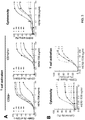
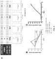


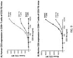

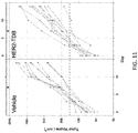
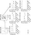
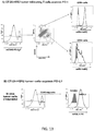
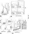

Classifications
-
- C—CHEMISTRY; METALLURGY
- C07—ORGANIC CHEMISTRY
- C07K—PEPTIDES
- C07K16/00—Immunoglobulins [IGs], e.g. monoclonal or polyclonal antibodies
- C07K16/18—Immunoglobulins [IGs], e.g. monoclonal or polyclonal antibodies against material from animals or humans
- C07K16/32—Immunoglobulins [IGs], e.g. monoclonal or polyclonal antibodies against material from animals or humans against translation products of oncogenes
-
- A—HUMAN NECESSITIES
- A61—MEDICAL OR VETERINARY SCIENCE; HYGIENE
- A61K—PREPARATIONS FOR MEDICAL, DENTAL OR TOILETRY PURPOSES
- A61K31/00—Medicinal preparations containing organic active ingredients
- A61K31/28—Compounds containing heavy metals
- A61K31/282—Platinum compounds
-
- A—HUMAN NECESSITIES
- A61—MEDICAL OR VETERINARY SCIENCE; HYGIENE
- A61K—PREPARATIONS FOR MEDICAL, DENTAL OR TOILETRY PURPOSES
- A61K31/00—Medicinal preparations containing organic active ingredients
- A61K31/33—Heterocyclic compounds
- A61K31/335—Heterocyclic compounds having oxygen as the only ring hetero atom, e.g. fungichromin
- A61K31/337—Heterocyclic compounds having oxygen as the only ring hetero atom, e.g. fungichromin having four-membered rings, e.g. taxol
-
- A—HUMAN NECESSITIES
- A61—MEDICAL OR VETERINARY SCIENCE; HYGIENE
- A61K—PREPARATIONS FOR MEDICAL, DENTAL OR TOILETRY PURPOSES
- A61K31/00—Medicinal preparations containing organic active ingredients
- A61K31/33—Heterocyclic compounds
- A61K31/555—Heterocyclic compounds containing heavy metals, e.g. hemin, hematin, melarsoprol
-
- A—HUMAN NECESSITIES
- A61—MEDICAL OR VETERINARY SCIENCE; HYGIENE
- A61K—PREPARATIONS FOR MEDICAL, DENTAL OR TOILETRY PURPOSES
- A61K39/00—Medicinal preparations containing antigens or antibodies
- A61K39/395—Antibodies; Immunoglobulins; Immune serum, e.g. antilymphocytic serum
-
- A—HUMAN NECESSITIES
- A61—MEDICAL OR VETERINARY SCIENCE; HYGIENE
- A61K—PREPARATIONS FOR MEDICAL, DENTAL OR TOILETRY PURPOSES
- A61K39/00—Medicinal preparations containing antigens or antibodies
- A61K39/395—Antibodies; Immunoglobulins; Immune serum, e.g. antilymphocytic serum
- A61K39/39533—Antibodies; Immunoglobulins; Immune serum, e.g. antilymphocytic serum against materials from animals
- A61K39/3955—Antibodies; Immunoglobulins; Immune serum, e.g. antilymphocytic serum against materials from animals against proteinaceous materials, e.g. enzymes, hormones, lymphokines
-
- A—HUMAN NECESSITIES
- A61—MEDICAL OR VETERINARY SCIENCE; HYGIENE
- A61K—PREPARATIONS FOR MEDICAL, DENTAL OR TOILETRY PURPOSES
- A61K39/00—Medicinal preparations containing antigens or antibodies
- A61K39/395—Antibodies; Immunoglobulins; Immune serum, e.g. antilymphocytic serum
- A61K39/39533—Antibodies; Immunoglobulins; Immune serum, e.g. antilymphocytic serum against materials from animals
- A61K39/39558—Antibodies; Immunoglobulins; Immune serum, e.g. antilymphocytic serum against materials from animals against tumor tissues, cells, antigens
-
- A—HUMAN NECESSITIES
- A61—MEDICAL OR VETERINARY SCIENCE; HYGIENE
- A61K—PREPARATIONS FOR MEDICAL, DENTAL OR TOILETRY PURPOSES
- A61K45/00—Medicinal preparations containing active ingredients not provided for in groups A61K31/00 - A61K41/00
- A61K45/06—Mixtures of active ingredients without chemical characterisation, e.g. antiphlogistics and cardiaca
-
- A—HUMAN NECESSITIES
- A61—MEDICAL OR VETERINARY SCIENCE; HYGIENE
- A61K—PREPARATIONS FOR MEDICAL, DENTAL OR TOILETRY PURPOSES
- A61K47/00—Medicinal preparations characterised by the non-active ingredients used, e.g. carriers or inert additives; Targeting or modifying agents chemically bound to the active ingredient
- A61K47/50—Medicinal preparations characterised by the non-active ingredients used, e.g. carriers or inert additives; Targeting or modifying agents chemically bound to the active ingredient the non-active ingredient being chemically bound to the active ingredient, e.g. polymer-drug conjugates
- A61K47/51—Medicinal preparations characterised by the non-active ingredients used, e.g. carriers or inert additives; Targeting or modifying agents chemically bound to the active ingredient the non-active ingredient being chemically bound to the active ingredient, e.g. polymer-drug conjugates the non-active ingredient being a modifying agent
- A61K47/62—Medicinal preparations characterised by the non-active ingredients used, e.g. carriers or inert additives; Targeting or modifying agents chemically bound to the active ingredient the non-active ingredient being chemically bound to the active ingredient, e.g. polymer-drug conjugates the non-active ingredient being a modifying agent the modifying agent being a protein, peptide or polyamino acid
- A61K47/64—Drug-peptide, drug-protein or drug-polyamino acid conjugates, i.e. the modifying agent being a peptide, protein or polyamino acid which is covalently bonded or complexed to a therapeutically active agent
- A61K47/643—Albumins, e.g. HSA, BSA, ovalbumin or a Keyhole Limpet Hemocyanin [KHL]
-
- A—HUMAN NECESSITIES
- A61—MEDICAL OR VETERINARY SCIENCE; HYGIENE
- A61P—SPECIFIC THERAPEUTIC ACTIVITY OF CHEMICAL COMPOUNDS OR MEDICINAL PREPARATIONS
- A61P35/00—Antineoplastic agents
-
- A—HUMAN NECESSITIES
- A61—MEDICAL OR VETERINARY SCIENCE; HYGIENE
- A61P—SPECIFIC THERAPEUTIC ACTIVITY OF CHEMICAL COMPOUNDS OR MEDICINAL PREPARATIONS
- A61P37/00—Drugs for immunological or allergic disorders
- A61P37/02—Immunomodulators
-
- A—HUMAN NECESSITIES
- A61—MEDICAL OR VETERINARY SCIENCE; HYGIENE
- A61P—SPECIFIC THERAPEUTIC ACTIVITY OF CHEMICAL COMPOUNDS OR MEDICINAL PREPARATIONS
- A61P37/00—Drugs for immunological or allergic disorders
- A61P37/02—Immunomodulators
- A61P37/04—Immunostimulants
-
- A—HUMAN NECESSITIES
- A61—MEDICAL OR VETERINARY SCIENCE; HYGIENE
- A61P—SPECIFIC THERAPEUTIC ACTIVITY OF CHEMICAL COMPOUNDS OR MEDICINAL PREPARATIONS
- A61P43/00—Drugs for specific purposes, not provided for in groups A61P1/00-A61P41/00
-
- C—CHEMISTRY; METALLURGY
- C07—ORGANIC CHEMISTRY
- C07K—PEPTIDES
- C07K16/00—Immunoglobulins [IGs], e.g. monoclonal or polyclonal antibodies
- C07K16/18—Immunoglobulins [IGs], e.g. monoclonal or polyclonal antibodies against material from animals or humans
- C07K16/28—Immunoglobulins [IGs], e.g. monoclonal or polyclonal antibodies against material from animals or humans against receptors, cell surface antigens or cell surface determinants
- C07K16/2803—Immunoglobulins [IGs], e.g. monoclonal or polyclonal antibodies against material from animals or humans against receptors, cell surface antigens or cell surface determinants against the immunoglobulin superfamily
- C07K16/2809—Immunoglobulins [IGs], e.g. monoclonal or polyclonal antibodies against material from animals or humans against receptors, cell surface antigens or cell surface determinants against the immunoglobulin superfamily against the T-cell receptor (TcR)-CD3 complex
-
- C—CHEMISTRY; METALLURGY
- C07—ORGANIC CHEMISTRY
- C07K—PEPTIDES
- C07K16/00—Immunoglobulins [IGs], e.g. monoclonal or polyclonal antibodies
- C07K16/18—Immunoglobulins [IGs], e.g. monoclonal or polyclonal antibodies against material from animals or humans
- C07K16/28—Immunoglobulins [IGs], e.g. monoclonal or polyclonal antibodies against material from animals or humans against receptors, cell surface antigens or cell surface determinants
- C07K16/2803—Immunoglobulins [IGs], e.g. monoclonal or polyclonal antibodies against material from animals or humans against receptors, cell surface antigens or cell surface determinants against the immunoglobulin superfamily
- C07K16/2818—Immunoglobulins [IGs], e.g. monoclonal or polyclonal antibodies against material from animals or humans against receptors, cell surface antigens or cell surface determinants against the immunoglobulin superfamily against CD28 or CD152
-
- C—CHEMISTRY; METALLURGY
- C07—ORGANIC CHEMISTRY
- C07K—PEPTIDES
- C07K16/00—Immunoglobulins [IGs], e.g. monoclonal or polyclonal antibodies
- C07K16/18—Immunoglobulins [IGs], e.g. monoclonal or polyclonal antibodies against material from animals or humans
- C07K16/28—Immunoglobulins [IGs], e.g. monoclonal or polyclonal antibodies against material from animals or humans against receptors, cell surface antigens or cell surface determinants
- C07K16/2803—Immunoglobulins [IGs], e.g. monoclonal or polyclonal antibodies against material from animals or humans against receptors, cell surface antigens or cell surface determinants against the immunoglobulin superfamily
- C07K16/2827—Immunoglobulins [IGs], e.g. monoclonal or polyclonal antibodies against material from animals or humans against receptors, cell surface antigens or cell surface determinants against the immunoglobulin superfamily against B7 molecules, e.g. CD80, CD86
-
- C—CHEMISTRY; METALLURGY
- C07—ORGANIC CHEMISTRY
- C07K—PEPTIDES
- C07K16/00—Immunoglobulins [IGs], e.g. monoclonal or polyclonal antibodies
- C07K16/18—Immunoglobulins [IGs], e.g. monoclonal or polyclonal antibodies against material from animals or humans
- C07K16/28—Immunoglobulins [IGs], e.g. monoclonal or polyclonal antibodies against material from animals or humans against receptors, cell surface antigens or cell surface determinants
- C07K16/2863—Immunoglobulins [IGs], e.g. monoclonal or polyclonal antibodies against material from animals or humans against receptors, cell surface antigens or cell surface determinants against receptors for growth factors, growth regulators
-
- C—CHEMISTRY; METALLURGY
- C07—ORGANIC CHEMISTRY
- C07K—PEPTIDES
- C07K16/00—Immunoglobulins [IGs], e.g. monoclonal or polyclonal antibodies
- C07K16/18—Immunoglobulins [IGs], e.g. monoclonal or polyclonal antibodies against material from animals or humans
- C07K16/28—Immunoglobulins [IGs], e.g. monoclonal or polyclonal antibodies against material from animals or humans against receptors, cell surface antigens or cell surface determinants
- C07K16/30—Immunoglobulins [IGs], e.g. monoclonal or polyclonal antibodies against material from animals or humans against receptors, cell surface antigens or cell surface determinants from tumour cells
- C07K16/3015—Breast
-
- C—CHEMISTRY; METALLURGY
- C07—ORGANIC CHEMISTRY
- C07K—PEPTIDES
- C07K16/00—Immunoglobulins [IGs], e.g. monoclonal or polyclonal antibodies
- C07K16/18—Immunoglobulins [IGs], e.g. monoclonal or polyclonal antibodies against material from animals or humans
- C07K16/28—Immunoglobulins [IGs], e.g. monoclonal or polyclonal antibodies against material from animals or humans against receptors, cell surface antigens or cell surface determinants
- C07K16/30—Immunoglobulins [IGs], e.g. monoclonal or polyclonal antibodies against material from animals or humans against receptors, cell surface antigens or cell surface determinants from tumour cells
- C07K16/3023—Lung
-
- C—CHEMISTRY; METALLURGY
- C07—ORGANIC CHEMISTRY
- C07K—PEPTIDES
- C07K16/00—Immunoglobulins [IGs], e.g. monoclonal or polyclonal antibodies
- C07K16/18—Immunoglobulins [IGs], e.g. monoclonal or polyclonal antibodies against material from animals or humans
- C07K16/28—Immunoglobulins [IGs], e.g. monoclonal or polyclonal antibodies against material from animals or humans against receptors, cell surface antigens or cell surface determinants
- C07K16/30—Immunoglobulins [IGs], e.g. monoclonal or polyclonal antibodies against material from animals or humans against receptors, cell surface antigens or cell surface determinants from tumour cells
- C07K16/303—Liver or Pancreas
-
- C—CHEMISTRY; METALLURGY
- C07—ORGANIC CHEMISTRY
- C07K—PEPTIDES
- C07K16/00—Immunoglobulins [IGs], e.g. monoclonal or polyclonal antibodies
- C07K16/18—Immunoglobulins [IGs], e.g. monoclonal or polyclonal antibodies against material from animals or humans
- C07K16/28—Immunoglobulins [IGs], e.g. monoclonal or polyclonal antibodies against material from animals or humans against receptors, cell surface antigens or cell surface determinants
- C07K16/30—Immunoglobulins [IGs], e.g. monoclonal or polyclonal antibodies against material from animals or humans against receptors, cell surface antigens or cell surface determinants from tumour cells
- C07K16/3038—Kidney, bladder
-
- C—CHEMISTRY; METALLURGY
- C07—ORGANIC CHEMISTRY
- C07K—PEPTIDES
- C07K16/00—Immunoglobulins [IGs], e.g. monoclonal or polyclonal antibodies
- C07K16/18—Immunoglobulins [IGs], e.g. monoclonal or polyclonal antibodies against material from animals or humans
- C07K16/28—Immunoglobulins [IGs], e.g. monoclonal or polyclonal antibodies against material from animals or humans against receptors, cell surface antigens or cell surface determinants
- C07K16/30—Immunoglobulins [IGs], e.g. monoclonal or polyclonal antibodies against material from animals or humans against receptors, cell surface antigens or cell surface determinants from tumour cells
- C07K16/3069—Reproductive system, e.g. ovaria, uterus, testes, prostate
-
- A—HUMAN NECESSITIES
- A61—MEDICAL OR VETERINARY SCIENCE; HYGIENE
- A61K—PREPARATIONS FOR MEDICAL, DENTAL OR TOILETRY PURPOSES
- A61K39/00—Medicinal preparations containing antigens or antibodies
- A61K2039/505—Medicinal preparations containing antigens or antibodies comprising antibodies
-
- A—HUMAN NECESSITIES
- A61—MEDICAL OR VETERINARY SCIENCE; HYGIENE
- A61K—PREPARATIONS FOR MEDICAL, DENTAL OR TOILETRY PURPOSES
- A61K39/00—Medicinal preparations containing antigens or antibodies
- A61K2039/505—Medicinal preparations containing antigens or antibodies comprising antibodies
- A61K2039/507—Comprising a combination of two or more separate antibodies
-
- A—HUMAN NECESSITIES
- A61—MEDICAL OR VETERINARY SCIENCE; HYGIENE
- A61K—PREPARATIONS FOR MEDICAL, DENTAL OR TOILETRY PURPOSES
- A61K39/00—Medicinal preparations containing antigens or antibodies
- A61K2039/54—Medicinal preparations containing antigens or antibodies characterised by the route of administration
-
- A—HUMAN NECESSITIES
- A61—MEDICAL OR VETERINARY SCIENCE; HYGIENE
- A61K—PREPARATIONS FOR MEDICAL, DENTAL OR TOILETRY PURPOSES
- A61K39/00—Medicinal preparations containing antigens or antibodies
- A61K2039/545—Medicinal preparations containing antigens or antibodies characterised by the dose, timing or administration schedule
-
- A—HUMAN NECESSITIES
- A61—MEDICAL OR VETERINARY SCIENCE; HYGIENE
- A61K—PREPARATIONS FOR MEDICAL, DENTAL OR TOILETRY PURPOSES
- A61K2300/00—Mixtures or combinations of active ingredients, wherein at least one active ingredient is fully defined in groups A61K31/00 - A61K41/00
-
- C—CHEMISTRY; METALLURGY
- C07—ORGANIC CHEMISTRY
- C07K—PEPTIDES
- C07K2317/00—Immunoglobulins specific features
- C07K2317/20—Immunoglobulins specific features characterized by taxonomic origin
- C07K2317/24—Immunoglobulins specific features characterized by taxonomic origin containing regions, domains or residues from different species, e.g. chimeric, humanized or veneered
-
- C—CHEMISTRY; METALLURGY
- C07—ORGANIC CHEMISTRY
- C07K—PEPTIDES
- C07K2317/00—Immunoglobulins specific features
- C07K2317/30—Immunoglobulins specific features characterized by aspects of specificity or valency
- C07K2317/31—Immunoglobulins specific features characterized by aspects of specificity or valency multispecific
-
- C—CHEMISTRY; METALLURGY
- C07—ORGANIC CHEMISTRY
- C07K—PEPTIDES
- C07K2317/00—Immunoglobulins specific features
- C07K2317/40—Immunoglobulins specific features characterized by post-translational modification
- C07K2317/41—Glycosylation, sialylation, or fucosylation
-
- C—CHEMISTRY; METALLURGY
- C07—ORGANIC CHEMISTRY
- C07K—PEPTIDES
- C07K2317/00—Immunoglobulins specific features
- C07K2317/50—Immunoglobulins specific features characterized by immunoglobulin fragments
- C07K2317/52—Constant or Fc region; Isotype
-
- C—CHEMISTRY; METALLURGY
- C07—ORGANIC CHEMISTRY
- C07K—PEPTIDES
- C07K2317/00—Immunoglobulins specific features
- C07K2317/50—Immunoglobulins specific features characterized by immunoglobulin fragments
- C07K2317/52—Constant or Fc region; Isotype
- C07K2317/524—CH2 domain
-
- C—CHEMISTRY; METALLURGY
- C07—ORGANIC CHEMISTRY
- C07K—PEPTIDES
- C07K2317/00—Immunoglobulins specific features
- C07K2317/50—Immunoglobulins specific features characterized by immunoglobulin fragments
- C07K2317/52—Constant or Fc region; Isotype
- C07K2317/526—CH3 domain
-
- C—CHEMISTRY; METALLURGY
- C07—ORGANIC CHEMISTRY
- C07K—PEPTIDES
- C07K2317/00—Immunoglobulins specific features
- C07K2317/50—Immunoglobulins specific features characterized by immunoglobulin fragments
- C07K2317/56—Immunoglobulins specific features characterized by immunoglobulin fragments variable (Fv) region, i.e. VH and/or VL
-
- C—CHEMISTRY; METALLURGY
- C07—ORGANIC CHEMISTRY
- C07K—PEPTIDES
- C07K2317/00—Immunoglobulins specific features
- C07K2317/70—Immunoglobulins specific features characterized by effect upon binding to a cell or to an antigen
- C07K2317/73—Inducing cell death, e.g. apoptosis, necrosis or inhibition of cell proliferation
-
- C—CHEMISTRY; METALLURGY
- C07—ORGANIC CHEMISTRY
- C07K—PEPTIDES
- C07K2317/00—Immunoglobulins specific features
- C07K2317/70—Immunoglobulins specific features characterized by effect upon binding to a cell or to an antigen
- C07K2317/76—Antagonist effect on antigen, e.g. neutralization or inhibition of binding
-
- C—CHEMISTRY; METALLURGY
- C07—ORGANIC CHEMISTRY
- C07K—PEPTIDES
- C07K2317/00—Immunoglobulins specific features
- C07K2317/90—Immunoglobulins specific features characterized by (pharmaco)kinetic aspects or by stability of the immunoglobulin
- C07K2317/92—Affinity (KD), association rate (Ka), dissociation rate (Kd) or EC50 value
-
- C—CHEMISTRY; METALLURGY
- C07—ORGANIC CHEMISTRY
- C07K—PEPTIDES
- C07K2317/00—Immunoglobulins specific features
- C07K2317/90—Immunoglobulins specific features characterized by (pharmaco)kinetic aspects or by stability of the immunoglobulin
- C07K2317/94—Stability, e.g. half-life, pH, temperature or enzyme-resistance
Definitions
- This invention relates to compositions for use in treating HER2-positive cancers by administering a PD-1 axis binding antagonist and an anti-HER2 antibody.
- the primary signal or antigen specific signal
- TCR T- cell receptor
- MHC major histocompatibility-complex
- APCs antigen-presenting cells
- T-cells can become refractory to antigen stimulation, do not mount an effective immune response, and further may result in exhaustion or tolerance to foreign antigens.
- T-cells receive both positive and negative secondary co-stimulatory signals.
- the regulation of such positive and negative signals is critical to maximize the host's protective immune responses, while maintaining immune tolerance and preventing autoimmunity.
- Negative secondary signals seem necessary for induction of T-cell tolerance, while positive signals promote T-cell activation.
- a host's immune response is a dynamic process, and co- stimulatory signals can also be provided to antigen-exposed T-cells.
- the mechanism of co-stimulation is of therapeutic interest because the manipulation of co-stimulatory signals has shown to provide a means to either enhance or terminate cell-based immune response.
- T cell dysfunction or anergy occurs concurrently with an induced and sustained expression of the inhibitory receptor, programmed death 1 polypeptide (PD-1).
- PD-1 programmed death 1 polypeptide
- therapeutic targeting of PD-1 and other molecules which signal through interactions with PD-1, such as programmed death ligand 1 (PD-L1) and programmed death ligand 2 (PD-L2) are an area of intense interest.
- PD-L1 is overexpressed in many cancers and is often associated with poor prognosis ( Okazaki T et al., Intern. Immun. 2007 19(7):813 ) ( Thompson RH et al., Cancer Res 2006, 66(7):3381 ).
- the majority of tumor infiltrating T lymphocytes predominantly express PD-1, in contrast to T lymphocytes in normal tissues and peripheral blood T lymphocytes indicating that up-regulation of PD-1 on tumor-reactive T cells can contribute to impaired antitumor immune responses ( Blood 2009 114(8):1537 ).
- Therapeutic targeting PD-1 and other molecules which signal through interactions with PD-1 are an area of intense interest.
- PD-1 programmed death ligand 1
- PD-L2 programmed death ligand 2
- An optimal therapeutic treatment may combine blockade of PD-1 receptor/ligand interaction with an agent that directly inhibits tumor growth.
- compositions for use in treating or delaying progression of a HER2-positive cancer in an individual characterized in that cancer cells in the individual express PD-L1, and that said composition is selected from the group consisting of:
- the antigen-binding fragment is selected from the group consisting of Fab, Fab', F(ab') 2 , and Fv.
- the PD-L1 binding antagonist is selected from the group consisting of: YW243.55.S70, MPDL3280A, MDX-1105, and MEDI4736.
- the anti-PD-Ll antibody comprises a heavy chain comprising HVR-H1 sequence of SEQ ID NO:19, HVR-H2 sequence of SEQ ID NO:20, and HVR-H3 sequence of SEQ ID NO:21; and/or a light chain comprising HVR-L1 sequence of SEQ ID NO:22, HVR-L2 sequence of SEQ ID NO:23, and HVR-L3 sequence of SEQ ID NO:24.
- the anti-PD-Ll antibody comprises a heavy chain variable region comprising the amino acid sequence of SEQ ID NO:25 or 26 and/or a light chain variable region comprising the amino acid sequence of SEQ ID NO:4.
- the anti-PD-L1 antibody comprises the three heavy chain HVR sequences of antibody YW243.55.S70 and/or the three light chain HVR sequences of antibody YW24355.S70 described in WO 2010/077634 and U.S. Patent No. 8,217,149 .
- the anti-PD-Ll antibody comprises the heavy chain variable region sequence of antibody YW243.55.S70 and/or the light chain variable region sequence of antibody YW24355.S70.
- the anti-HER2 antibody is trastuzumab (HERCEPTIN®, Genentech) or pertuzumab (PERJETA®, Genentech).
- the anti-HER2 antibody comprises a heavy chain comprising HVR-H1 sequence of SEQ ID NO:38, HVR-H2 sequence of SEQ ID NO:50, and HVR-H3 sequence of SEQ ID NO:40; and/or a light chain comprising HVR-L1 sequence of SEQ ID NO:41, HVR-L2 sequence of SEQ ID NO:42, and HVR-L3 sequence of SEQ ID NO:43.
- the anti-HER2 antibody comprises a heavy chain variable region comprising the amino acid sequence of SEQ ID NO:34 and/or a light chain variable region comprising the amino acid sequence of SEQ ID NO:35. In some embodiments, the anti-HER2 antibody comprises a heavy chain comprising the amino acid sequence of SEQ ID NO:36 and/or a light chain comprising the amino acid sequence of SEQ ID NO:37. In some embodiments that can be combined with any other embodiments, the anti-HER2 antibody is not trastuzumab or pertuzumab.
- the bispecific antibody comprises a first antigen binding domain that binds to HER2, and a second antigen binding domain that binds to CD3.
- the second antigen binding domain binds to a human CD3 polypeptide.
- the CD3 polypeptide is a human CD3 ⁇ polypeptide or a human CD3 ⁇ polypeptide.
- the second antigen binding domain binds to a human CD3 ⁇ polypeptide or a human CD3 ⁇ polypeptide in native T-cell receptor (TCR) complex in association with other TCR subunits.
- the first antigen binding domain comprises a heavy chain variable region (V H HER2) and a light chain variable region (V L HER2)
- the second antigen binding domain comprises a heavy chain variable region (V H CD3) and a light chain variable region (V L CD3).
- the first antigen binding domain comprises a heavy chain variable region (V H HER2) comprising HVR-H1 sequence of SEQ ID NO:38, HVR-H2 sequence of SEQ ID NO:50, and HVR-H3 sequence of SEQ ID NO:40; and/or a light chain variable region (V L HER2) comprising HVR-L1 sequence of SEQ ID NO:41, HVR-L2 sequence of SEQ ID NO:42, and HVR-L3 sequence of SEQ ID NO:43.
- V H HER2 heavy chain variable region
- V L HER2 light chain variable region
- the first antigen binding domain comprises a heavy chain variable region (V H HER2) comprising the amino acid sequence of SEQ ID NO:34 and/or a light chain variable region (V L HER2) comprising the amino acid sequence of SEQ ID NO:35.
- the bispecific antibody is a single-chain bispecific antibody comprising the first antigen binding domain and the second antigen binding domain.
- the single-chain bispecific antibody comprises variable regions, as arranged from N-terminus to C-terminus, selected from the group consisting of (1) V H HER2-V L HER2-V H CD3-V L CD3, (2) V H CD3-V L CD3-V H HER2-V L HER2, (3) V H CD3-V L CD3-V L HER2-V H HER2, (4) V H HER2-V L HER2-V L CD3-V H CD3, (5) V L HER2-V H HER2-V H CD3-V L CD3, or (6) V L CD3-V H CD3-V H HER2-V L HER2.
- the bispecific antibody comprises a first antigen binding domain that binds to HER2 and a second antigen binding domain that binds to CD3, wherein the first antigen binding domain comprises one or more heavy chain constant domains, wherein the one or more heavy chain constant domains are selected from a first CH1 (CH1 1 ) domain, a first CH2 (CH2 1 ) domain, a first CH3 (CH3 1 ) domain; and wherein the second antigen binding domain comprises one or more heavy chain constant domains, wherein the one or more heavy chain constant domains are selected from a second CH1 (CH1 2 ) domain, second CH2 (CH2 2 ) domain, and a second CH3 (CH3 2 ) domain.
- the CH3 1 and CH3 2 domains each comprise a protuberance or cavity, and wherein the protuberance or cavity in the CH3 1 domain is positionable in the cavity or protuberance, respectively, in the CH3 2 domain.
- the CH3 1 and CH3 2 domains meet at an interface between said protuberance and cavity.
- the CH2 1 and CH2 2 domains each comprise a protuberance or cavity, and wherein the protuberance or cavity in the CH2 1 domain is positionable in the cavity or protuberance, respectively, in the CH2 2 domain.
- the CH2 1 and CH2 2 domains meet at an interface between said protuberance and cavity.
- the antibody described herein comprises an aglycosylation site mutation.
- the aglycosylation site mutation is a substitution mutation.
- the substitution mutation is at amino acid residue N297, L234, L235, and/or D265 (EU numbering).
- the substitution mutation is selected from the group consisting of N297G, N297A, L234A, L235A, and D265A.
- the substitution mutation is a D265A mutation and an N297G mutation.
- the aglycosylation site mutation reduces effector function of the antibody.
- the PD-1 axis binding antagonist e.g., an anti-PD-1 antibody, an anti-PD-Ll antibody, or an anti-PD-L2 antibody
- the PD-1 axis binding antagonist is a human IgG1 having Asn to Ala substitution at position 297 according to EU numbering.
- the HER-2 positive cancer is breast cancer, lung cancer, ovarian cancer, gastric cancer, bladder cancer, pancreatic cancer, endometrial cancer, colon cancer, kidney cancer, esophageal cancer, prostate cancer, or other cancers described herein.
- the individual has cancer or has been diagnosed with cancer.
- the cancer cells in the individual express PD-L1.
- the individual has cancer that is resistant to a HER2 targeted therapy.
- the individual is refractory to a HER2 targeted therapy.
- the HER2 targeted therapy is a treatment with an anti-HER2 antibody or an inhibitor of the HER2 pathway.
- the HER2 targeted therapy is a treatment with trastuzumab (HERCEPTIN®, Genentech), pertuzumab (PERJETA®, Genentech), ado-trastuzumab emtansine (KADCYLA®, Genentech), or lapatinib.
- trastuzumab HERCEPTIN®, Genentech
- pertuzumab PERJETA®, Genentech
- KADCYLA® Genentech
- lapatinib lapatinib.
- the treatment or administration of the human PD-1 axis binding antagonist and the anti-HER2 antibody results in a sustained response in the individual after cessation of the treatment.
- the anti-HER2 antibody is administered before the PD-1 axis binding antagonist, simultaneous with the PD-1 axis binding antagonist, or after the PD-1 axis binding antagonist.
- the PD-1 axis binding antagonist and the anti-HER2 antibody are in the same composition.
- the PD-1 axis binding antagonist and the anti-HER2 antibody are in separate compositions.
- the PD-1 axis binding antagonist and/or the anti-HER2 antibody is administered intravenously, intramuscularly, subcutaneously, topically, orally, transdermally, intraperitoneally, intraorbitally, by implantation, by inhalation, intrathecally, intraventricularly, or intranasally.
- the treatment further comprises administering a chemotherapeutic agent for treating or delaying progression of cancer in an individual.
- the individual has been treated with a chemotherapeutic agent before the combination treatment with the PD-1 axis binding antagonist and the anti-HER2 antibody.
- the individual treated with the combination of the PD-1 axis binding antagonist and/or the anti-HER2 antibody is refractory to a chemotherapeutic agent treatment.
- Some embodiments of the methods, uses, compositions, and kits described throughout the application further comprise administering a chemotherapeutic agent for treating or delaying progression of cancer.
- CD8 T cells in the individual have enhanced priming, activation, proliferation and/or cytolytic activity relative to prior to the administration of the combination.
- the number of CD8 T cells is elevated relative to prior to administration of the combination.
- the CD8 T cell is an antigen-specific CD8 T cell.
- Treg function is suppressed relative to prior to the administration of the combination.
- T cell exhaustion is decreased relative to prior to the administration of the combination.
- the inventors of this application demonstrated that PD-L1 expressed by cancer cells can inhibit the activity of T cell recruiting antibodies and this inhibition can be reversed by an anti-PD-Ll antibody.
- the data in the application show that the combination of a HER2 T cell dependent bispecific antibody (HER2-TDB) with anti-PD-Ll immune therapy resulted in enhanced inhibition of tumor growth, increased response rates and durable responses.
- HER2-TDB HER2 T cell dependent bispecific antibody
- provided herein are methods, compositions and uses for treating or delaying progression of cancer in an individual comprising administering an effective amount of a human PD-1 axis binding antagonist and an anti-HER2 antibody.
- provided herein are methods, compositions and uses for enhancing immune function in an individual having cancer comprising administering an effective amount of a human PD-1 axis binding antagonist and an anti-HER2 antibody.
- PD-1 axis binding antagonist refers to a molecule that inhibits the interaction of a PD-1 axis binding partner with either one or more of its binding partner, so as to remove T-cell dysfunction resulting from signaling on the PD-1 signaling axis - with a result being to restore or enhance T-cell function (e.g., proliferation, cytokine production, target cell killing).
- a PD-1 axis binding antagonist includes a PD-1 binding antagonist, a PD-L1 binding antagonist and a PD-L2 binding antagonist.
- PD-1 binding antagonist refers to a molecule that decreases, blocks, inhibits, abrogates or interferes with signal transduction resulting from the interaction of PD-1 with one or more of its binding partners, such as PD-L1, PD-L2.
- the PD-1 binding antagonist is a molecule that inhibits the binding of PD-1 to one or more of its binding partners.
- the PD-1 binding antagonist inhibits the binding of PD-1 to PD-L1 and/or PD-L2.
- PD-1 binding antagonists include anti-PD-1 antibodies, antigen binding fragments thereof, immunoadhesins, fusion proteins, oligopeptides and other molecules that decrease, block, inhibit, abrogate or interfere with signal transduction resulting from the interaction of PD-1 with PD-L1 and/or PD-L2.
- a PD-1 binding antagonist reduces the negative co-stimulatory signal mediated by or through cell surface proteins expressed on T lymphocytes mediated signaling through PD-1 so as render a dysfunctional T-cell less dysfunctional (e.g., enhancing effector responses to antigen recognition).
- the PD-1 binding antagonist is an anti-PD-1 antibody.
- a PD-1 binding antagonist is MDX-1106 (nivolumab) described herein.
- a PD-1 binding antagonist is MK-3475 (lambrolizumab) described herein.
- a PD-1 binding antagonist is CT-011 (pidilizumab) described herein.
- a PD-1 binding antagonist is AMP-224 described herein.
- PD-L1 binding antagonist refers to a molecule that decreases, blocks, inhibits, abrogates or interferes with signal transduction resulting from the interaction of PD-L1 with either one or more of its binding partners, such as PD-1, B7-1.
- a PD-L1 binding antagonist is a molecule that inhibits the binding of PD-L1 to its binding partners.
- the PD-L1 binding antagonist inhibits binding of PD-L1 to PD-1 and/or B7-1.
- the PD-L1 binding antagonists include anti-PD-Ll antibodies, antigen binding fragments thereof, immunoadhesins, fusion proteins, oligopeptides and other molecules that decrease, block, inhibit, abrogate or interfere with signal transduction resulting from the interaction of PD-L1 with one or more of its binding partners, such as PD-1, B7-1.
- a PD-L1 binding antagonist reduces the negative co-stimulatory signal mediated by or through cell surface proteins expressed on T lymphocytes mediated signaling through PD-L1 so as to render a dysfunctional T-cell less dysfunctional (e.g., enhancing effector responses to antigen recognition).
- a PD-L1 binding antagonist is an anti-PD-Ll antibody.
- an anti-PD-Ll antibody is YW243.55.S70 described herein.
- an anti-PD-L1 antibody is MDX-1105 described herein.
- an anti-PD-L1 antibody is MPDL3280A described herein.
- an anti-PD-Ll antibody is MEDI4736 described herein.
- PD-L2 binding antagonist refers to a molecule that decreases, blocks, inhibits, abrogates or interferes with signal transduction resulting from the interaction of PD-L2 with either one or more of its binding partners, such as PD-1.
- a PD-L2 binding antagonist is a molecule that inhibits the binding of PD-L2 to one or more of its binding partners.
- the PD-L2 binding antagonist inhibits binding of PD-L2 to PD-1.
- the PD-L2 antagonists include anti-PD-L2 antibodies, antigen binding fragments thereof, immunoadhesins, fusion proteins, oligopeptides and other molecules that decrease, block, inhibit, abrogate or interfere with signal transduction resulting from the interaction of PD-L2 with either one or more of its binding partners, such as PD-1.
- a PD-L2 binding antagonist reduces the negative co-stimulatory signal mediated by or through cell surface proteins expressed on T lymphocytes mediated signaling through PD-L2 so as render a dysfunctional T-cell less dysfunctional (e.g., enhancing effector responses to antigen recognition).
- a PD-L2 binding antagonist is an immunoadhesin.
- disfunction in the context of immune dysfunction, refers to a state of reduced immune responsiveness to antigenic stimulation.
- the term includes the common elements of both exhaustion and/or anergy in which antigen recognition may occur, but the ensuing immune response is ineffective to control infection or tumor growth.
- disfunctional also includes refractory or unresponsive to antigen recognition, specifically, impaired capacity to translate antigen recognition into down-stream T-cell effector functions, such as proliferation, cytokine production (e.g., IL-2) and/or target cell killing.
- the term "anergy” refers to the state of unresponsiveness to antigen stimulation resulting from incomplete or insufficient signals delivered through the T-cell receptor (e.g. increase in intracellular Ca +2 in the absence of ras-activation). T cell anergy can also result upon stimulation with antigen in the absence of co-stimulation, resulting in the cell becoming refractory to subsequent activation by the antigen even in the context of costimulation.
- the unresponsive state can often be overriden by the presence of Interleukin-2. Anergic T-cells do not undergo clonal expansion and/or acquire effector functions.
- exhaustion refers to T cell exhaustion as a state of T cell dysfunction that arises from sustained TCR signaling that occurs during many chronic infections and cancer. It is distinguished from anergy in that it arises not through incomplete or deficient signaling, but from sustained signaling. It is defined by poor effector function, sustained expression of inhibitory receptors and a transcriptional state distinct from that of functional effector or memory T cells. Exhaustion prevents optimal control of infection and tumors. Exhaustion can result from both extrinsic negative regulatory pathways (e.g., immunoregulatory cytokines) as well as cell intrinsic negative regulatory (costimulatory) pathways (PD-1, B7-H3, B7-H4, etc.).
- extrinsic negative regulatory pathways e.g., immunoregulatory cytokines
- costimulatory costimulatory
- Enhancing T-cell function means to induce, cause or stimulate a T-cell to have a sustained or amplified biological function, or renew or reactivate exhausted or inactive T-cells.
- Examples of enhancing T-cell function include: increased secretion of ⁇ -interferon from CD8 + T-cells, increased proliferation, increased antigen responsiveness (e.g., viral, pathogen, or tumor clearance) relative to such levels before the intervention.
- the level of enhancement is as least 50%, alternatively 60%, 70%, 80%, 90%, 100%, 120%, 150%, 200%. The manner of measuring this enhancement is known to one of ordinary skill in the art.
- T cell dysfunctional disorder is a disorder or condition of T-cells characterized by decreased responsiveness to antigenic stimulation.
- a T-cell dysfunctional disorder is a disorder that is specifically associated with inappropriate increased signaling through PD-1.
- a T-cell dysfunctional disorder is one in which T-cells are anergic or have decreased ability to secrete cytokines, proliferate, or execute cytolytic activity.
- the decreased responsiveness results in ineffective control of a pathogen or tumor expressing an immunogen.
- T cell dysfunctional disorders characterized by T-cell dysfunction include unresolved acute infection, chronic infection and tumor immunity.
- Tumor immunity refers to the process in which tumors evade immune recognition and clearance. Thus, as a therapeutic concept, tumor immunity is "treated” when such evasion is attenuated, and the tumors are recognized and attacked by the immune system. Examples of tumor recognition include tumor binding, tumor shrinkage and tumor clearance.
- Immunogenecity refers to the ability of a particular substance to provoke an immune response. Tumors are immunogenic and enhancing tumor immunogenicity aids in the clearance of the tumor cells by the immune response. Examples of enhancing tumor immunogenicity include treatment with a PD-1 axis binding antagonist and an anti-HER2 antibody.
- sustained response refers to the sustained effect on reducing tumor growth after cessation of a treatment.
- the tumor size may remain to be the same or smaller as compared to the size at the beginning of the administration phase.
- the sustained response has a duration at least the same as the treatment duration, at least 1.5X, 2.0X, 2.5X, or 3.0X length of the treatment duration.
- pharmaceutical formulation refers to a preparation which is in such form as to permit the biological activity of the active ingredient to be effective, and which contains no additional components which are unacceptably toxic to a subject to which the formulation would be administered. Such formulations are sterile. "Pharmaceutically acceptable" excipients (vehicles, additives) are those which can reasonably be administered to a subject mammal to provide an effective dose of the active ingredient employed.
- treatment refers to clinical intervention designed to alter the natural course of the individual or cell being treated during the course of clinical pathology. Desirable effects of treatment include decreasing the rate of disease progression, ameliorating or palliating the disease state, and remission or improved prognosis.
- an individual is successfully "treated” if one or more symptoms associated with cancer are mitigated or eliminated, including, but are not limited to, reducing the proliferation of (or destroying) cancerous cells, decreasing symptoms resulting from the disease, increasing the quality of life of those suffering from the disease, decreasing the dose of other medications required to treat the disease, and/or prolonging survival of individuals.
- delaying progression of a disease means to defer, hinder, slow, retard, stabilize, and/or postpone development of the disease (such as cancer). This delay can be of varying lengths of time, depending on the history of the disease and/or individual being treated. As is evident to one skilled in the art, a sufficient or significant delay can, in effect, encompass prevention, in that the individual does not develop the disease. For example, a late stage cancer, such as development of metastasis, may be delayed.
- an "effective amount” is at least the minimum amount required to effect a measurable improvement or prevention of a particular disorder.
- An effective amount herein may vary according to factors such as the disease state, age, sex, and weight of the patient, and the ability of the antibody to elicit a desired response in the individual.
- An effective amount is also one in which any toxic or detrimental effects of the treatment are outweighed by the therapeutically beneficial effects.
- beneficial or desired results include results such as eliminating or reducing the risk, lessening the severity, or delaying the onset of the disease, including biochemical, histological and/or behavioral symptoms of the disease, its complications and intermediate pathological phenotypes presenting during development of the disease.
- beneficial or desired results include clinical results such as decreasing one or more symptoms resulting from the disease, increasing the quality of life of those suffering from the disease, decreasing the dose of other medications required to treat the disease, enhancing effect of another medication such as via targeting, delaying the progression of the disease, and/or prolonging survival.
- an effective amount of the drug may have the effect in reducing the number of cancer cells; reducing the tumor size; inhibiting ( i.e., slow to some extent or desirably stop) cancer cell infiltration into peripheral organs; inhibit ( i.e., slow to some extent and desirably stop) tumor metastasis; inhibiting to some extent tumor growth; and/or relieving to some extent one or more of the symptoms associated with the disorder.
- an effective amount can be administered in one or more administrations.
- an effective amount of drug, compound, or pharmaceutical composition is an amount sufficient to accomplish prophylactic or therapeutic treatment either directly or indirectly.
- an effective amount of a drug, compound, or pharmaceutical composition may or may not be achieved in conjunction with another drug, compound, or pharmaceutical composition.
- an "effective amount" may be considered in the context of administering one or more therapeutic agents, and a single agent may be considered to be given in an effective amount if, in conjunction with one or more other agents, a desirable result may be or is achieved.
- conjunction with refers to administration of one treatment modality in addition to another treatment modality.
- in conjunction with refers to administration of one treatment modality before, during, or after administration of the other treatment modality to the individual.
- a “disorder” is any condition that would benefit from treatment including, but not limited to, chronic and acute disorders or diseases including those pathological conditions which predispose the mammal to the disorder in question.
- cell proliferative disorder and “proliferative disorder” refer to disorders that are associated with some degree of abnormal cell proliferation.
- the cell proliferative disorder is cancer.
- the cell proliferative disorder is a tumor.
- Tumor refers to all neoplastic cell growth and proliferation, whether malignant or benign, and all pre-cancerous and cancerous cells and tissues.
- cancer refers to all neoplastic cell growth and proliferation, whether malignant or benign, and all pre-cancerous and cancerous cells and tissues.
- cancer refers to all neoplastic cell growth and proliferation, whether malignant or benign, and all pre-cancerous and cancerous cells and tissues.
- cancer cancer
- cancer cancer
- cancer cancer
- cancer cancer
- cancer cancer
- cancer cancer
- cancer and “cancerous” refer to or describe the physiological condition in mammals that is typically characterized by unregulated cell growth.
- examples of cancer include but are not limited to, carcinoma, lymphoma, blastoma, sarcoma, and leukemia or lymphoid malignancies.
- cancers include, but not limited to, squamous cell cancer (e.g., epithelial squamous cell cancer), lung cancer including small-cell lung cancer, non-small cell lung cancer, adenocarcinoma of the lung and squamous carcinoma of the lung, cancer of the peritoneum, hepatocellular cancer, gastric or stomach cancer including gastrointestinal cancer and gastrointestinal stromal cancer, pancreatic cancer, glioblastoma, cervical cancer, ovarian cancer, liver cancer, bladder cancer, cancer of the urinary tract, hepatoma, breast cancer, colon cancer, rectal cancer, colorectal cancer, endometrial or uterine carcinoma, salivary gland carcinoma, kidney or renal cancer, prostate cancer, vulval cancer, thyroid cancer, hepatic carcinoma, anal carcinoma, penile carcinoma, melanoma, superficial spreading melanoma, lentigo maligna melanoma, acral lentiginous melanomas, nodular melan
- cancers that are amenable to treatment by the antibodies of the invention include breast cancer, colorectal cancer, rectal cancer, non-small cell lung cancer, glioblastoma, non-Hodgkins lymphoma (NHL), renal cell cancer, prostate cancer, liver cancer, pancreatic cancer, soft-tissue sarcoma, kaposi's sarcoma, carcinoid carcinoma, head and neck cancer, ovarian cancer, mesothelioma, and multiple myeloma.
- the cancer is selected from: small cell lung cancer, gliblastoma, neuroblastomas, melanoma, breast carcinoma, gastric cancer, colorectal cancer (CRC), and hepatocellular carcinoma.
- the cancer is selected from: non-small cell lung cancer, colorectal cancer, glioblastoma and breast carcinoma, including metastatic forms of those cancers.
- Cytotoxic agent refers to any agent that is detrimental to cells (e.g., causes cell death, inhibits proliferation, or otherwise hinders a cellular function). Cytotoxic agents include, but are not limited to, radioactive isotopes (e.g., At 211 , I 131 , I 125 , Y 90 , Re 186 , Re 188 , Sm 153 , Bi 212 , P 32 , Pb 212 and radioactive isotopes of Lu); chemotherapeutic agents; growth inhibitory agents; enzymes and fragments thereof such as nucleolytic enzymes; and toxins such as small molecule toxins or enzymatically active toxins of bacterial, fungal, plant or animal origin, including fragments and/or variants thereof.
- radioactive isotopes e.g., At 211 , I 131 , I 125 , Y 90 , Re 186 , Re 188 , Sm 153 , Bi 212 , P 32 , Pb
- Exemplary cytotoxic agents can be selected from anti-microtubule agents, platinum coordination complexes, alkylating agents, antibiotic agents, topoisomerase II inhibitors, antimetabolites, topoisomerase I inhibitors, hormones and hormonal analogues, signal transduction pathway inhibitors, non-receptor tyrosine kinase angiogenesis inhibitors, immunotherapeutic agents, proapoptotic agents, inhibitors of LDH-A, inhibitors of fatty acid biosynthesis, cell cycle signalling inhibitors, HDAC inhibitors, proteasome inhibitors, and inhibitors of cancer metabolism.
- the cytotoxic agent is a taxane.
- the taxane is paclitaxel or docetaxel.
- the cytotoxic agent is a platinum agent. In one embodiment the cytotoxic agent is an antagonist of EGFR. In one embodiment the antagonist of EGFR is N-(3-ethynylphenyl)-6,7-bis(2-methoxyethoxy)quinazolin-4-amine ( e.g., erlotinib). In one embodiment the cytotoxic agent is a RAF inhibitor. In one embodiment, the RAF inhibitor is a BRAF and/or CRAF inhibitor. In one embodiment the RAF inhibitor is vemurafenib. In one embodiment the cytotoxic agent is a PI3K inhibitor.
- “Chemotherapeutic agent” includes compounds useful in the treatment of cancer.
- chemotherapeutic agents include erlotinib (TARCEVA®, Genentech/OSI Pharm.), bortezomib (VELCADE®, Millennium Pharm.), disulfiram, epigallocatechin gallate , salinosporamide A, carfilzomib, 17-AAG (geldanamycin), radicicol, lactate dehydrogenase A (LDH-A), fulvestrant (FASLODEX®, AstraZeneca), sunitib (SUTENT®, Pfizer/Sugen), letrozole (FEMARA®, Novartis), imatinib mesylate (GLEEVEC®, Novartis), finasunate (VATALANIB®, Novartis), oxaliplatin (ELOXATIN®, Sanofi), 5-FU (5-fluorouracil), leucovorin, Rapamycin (Sirol
- dynemicin including dynemicin A; bisphosphonates, such as clodronate; an esperamicin; as well as neocarzinostatin chromophore and related chromoprotein enediyne antibiotic chromophores), aclacinomysins, actinomycin, authramycin, azaserine, bleomycins, cactinomycin, carabicin, caminomycin, carzinophilin, chromomycinis, dactinomycin, daunorubicin, detorubicin, 6-diazo-5-oxo-L-norleucine, ADRIAMYCIN® (doxorubicin), morpholino-doxorubicin, cyanomorpholino-doxorubicin, 2-pyrrolino-doxorubicin and deoxydoxorubicin), epirubicin, e
- Chemotherapeutic agent also includes (i) anti-hormonal agents that act to regulate or inhibit hormone action on tumors such as anti-estrogens and selective estrogen receptor modulators (SERMs), including, for example, tamoxifen (including NOLVADEX®; tamoxifen citrate), raloxifene, droloxifene, iodoxyfene , 4-hydroxytamoxifen, trioxifene, keoxifene, LY117018, onapristone, and FARESTON® (toremifine citrate); (ii) aromatase inhibitors that inhibit the enzyme aromatase, which regulates estrogen production in the adrenal glands, such as, for example, 4(5)-imidazoles, aminoglutethimide, MEGASE® (megestrol acetate), AROMASIN® (exemestane; Pfizer), formestanie, fadrozole, RIVISOR® (vorozole), FEMARA® (
- Chemotherapeutic agent also includes antibodies such as alemtuzumab (Campath), bevacizumab (AVASTIN®, Genentech); cetuximab (ERBITUX®, Imclone); panitumumab (VECTIBIX®, Amgen), rituximab (RITUXAN®, Genentech/Biogen Idec), pertuzumab (OMNITARG®, 2C4, Genentech), trastuzumab (HERCEPTIN®, Genentech), tositumomab (Bexxar, Corixia), and the antibody drug conjugate, gemtuzumab ozogamicin (MYLOTARG®, Wyeth).
- antibodies such as alemtuzumab (Campath), bevacizumab (AVASTIN®, Genentech); cetuximab (ERBITUX®, Imclone); panitumumab (VECTIBIX®, Amgen), rituximab
- Additional humanized monoclonal antibodies with therapeutic potential as agents in combination with the compounds of the invention include: apolizumab, aselizumab, atlizumab, bapineuzumab, bivatuzumab mertansine, cantuzumab mertansine, cedelizumab, certolizumab pegol, cidfusituzumab, cidtuzumab, daclizumab, eculizumab, efalizumab, epratuzumab, erlizumab, felvizumab, fontolizumab, gemtuzumab ozogamicin, inotuzumab ozogamicin, ipilimumab, labetuzumab, lintuzumab, matuzumab, mepolizumab, motavizumab, motovizumab, natalizumab, nimotuzumab, nolovizum
- Chemotherapeutic agent also includes "EGFR inhibitors,” which refers to compounds that bind to or otherwise interact directly with EGFR and prevent or reduce its signaling activity, and is alternatively referred to as an "EGFR antagonist.”
- EGFR inhibitors refers to compounds that bind to or otherwise interact directly with EGFR and prevent or reduce its signaling activity
- Examples of such agents include antibodies and small molecules that bind to EGFR.
- antibodies which bind to EGFR include MAb 579 (ATCC CRL HB 8506), MAb 455 (ATCC CRL HB8507), MAb 225 (ATCC CRL 8508), MAb 528 (ATCC CRL 8509) (see, US Patent No. 4,943 , 533 , Mendelsohn et al.
- chimerized 225 C225 or Cetuximab; ERBUTIX®
- H225 reshaped human 225
- IMC-11F8 a fully human, EGFR-targeted antibody (Imclone); antibodies that bind type II mutant EGFR ( US Patent No. 5,212,290 ); humanized and chimeric antibodies that bind EGFR as described in US Patent No.
- EMD 55900 Stragliotto et al. Eur. J. Cancer 32A:636-640 (1996 )
- EMD7200 (matuzumab) a humanized EGFR antibody directed against EGFR that competes with both EGF and TGF-alpha for EGFR binding
- EMD/Merck human EGFR antibody
- HuMax-EGFR HuMax-EGFR
- the anti-EGFR antibody may be conjugated with a cytotoxic agent, thus generating an immunoconjugate (see, e.g., EP659439A2 , Merck Patent GmbH).
- EGFR antagonists include small molecules such as compounds described in US Patent Nos: 5,616,582 , 5,457,105 , 5,475,001 , 5,654,307 , 5,679,683 , 6,084,095 , 6,265,410 , 6,455,534 , 6,521,620 , 6,596,726 , 6,713,484 , 5,770,599 , 6,140,332 , 5,866,572 , 6,399,602 , 6,344,459 , 6,602,863 , 6,391,874 , 6,344,455 , 5,760,041 , 6,002,008 , and 5,747,498 , as well as the following PCT publications: WO98/14451 , WO98/50038 , WO99/09016 , and WO99/24037 .
- EGFR antagonists include OSI-774 (CP-358774, erlotinib, TARCEVA® Genentech/OSI Pharmaceuticals); PD 183805 (CI 1033, 2-propenamide, N-[4-[(3-chloro-4-fluorophenyl)amino]-7-[3-(4-morpholinyl)propoxy]-6-quinazolinyl]-, dihydrochloride, Pfizer Inc.); ZD1839, gefitinib (IRESSA®) 4-(3'-Chloro-4'-fluoroanilino)-7-methoxy-6-(3-morpholinopropoxy)quinazoline, AstraZeneca); ZM 105180 ((6-amino-4-(3-methylphenyl-amino)-quinazoline, Zeneca); BIBX-1382 (N8-(3-chloro-4-fluoro-phenyl)-N2-(1-methyl-piperid
- Chemotherapeutic agents also include "tyrosine kinase inhibitors" including the EGFR-targeted drugs noted in the preceding paragraph; small molecule HER2 tyrosine kinase inhibitor such as TAK165 available from Takeda; CP-724,714, an oral selective inhibitor of the ErbB2 receptor tyrosine kinase (Pfizer and OSI); dual-HER inhibitors such as EKB-569 (available from Wyeth) which preferentially binds EGFR but inhibits both HER2 and EGFR-overexpressing cells; lapatinib (GSK572016; available from Glaxo-SmithKline), an oral HER2 and EGFR tyrosine kinase inhibitor; PKI-166 (available from Novartis); pan-HER inhibitors such as canertinib (CI-1033; Pharmacia); Raf-1 inhibitors such as antisense agent ISIS-5132 available from ISIS Pharmaceuticals which inhibit Raf-1 signaling; non-HER targeted
- Chemotherapeutic agents also include dexamethasone, interferons, colchicine, metoprine, cyclosporine, amphotericin, metronidazole, alemtuzumab, alitretinoin, allopurinol, amifostine, arsenic trioxide, asparaginase, BCG live, bevacuzimab, bexarotene, cladribine, clofarabine, darbepoetin alfa, denileukin, dexrazoxane, epoetin alfa, elotinib, filgrastim, histrelin acetate, ibritumomab, interferon alfa-2a, interferon alfa-2b, lenalidomide, levamisole, mesna, methoxsalen, nandrolone, nelarabine, nofetumomab, oprelvekin,
- Chemotherapeutic agents also include hydrocortisone, hydrocortisone acetate, cortisone acetate, tixocortol pivalate, triamcinolone acetonide, triamcinolone alcohol, mometasone, amcinonide, budesonide, desonide, fluocinonide, fluocinolone acetonide, betamethasone, betamethasone sodium phosphate, dexamethasone, dexamethasone sodium phosphate, fluocortolone, hydrocortisone-17-butyrate, hydrocortisone-17-valerate, aclometasone dipropionate, betamethasone valerate, betamethasone dipropionate, prednicarbate, clobetasone-17-butyrate, clobetasol-17-propionate, fluocortolone caproate, fluocortolone pivalate and fluprednidene acetate; immune selective
- celecoxib or etoricoxib proteosome inhibitor
- CCI-779 tipifarnib (R11577); orafenib, ABT510
- Bcl-2 inhibitor such as oblimersen sodium (GENASENSE®)
- pixantrone farnesyltransferase inhibitors
- SCH 6636 farnesyltransferase inhibitors
- pharmaceutically acceptable salts, acids or derivatives of any of the above as well as combinations of two or more of the above such as CHOP, an abbreviation for a combined therapy of cyclophosphamide, doxorubicin, vincristine, and prednisolone
- FOLFOX an abbreviation for a treatment regimen with oxaliplatin (ELOXATINTM) combined with 5-FU and leucovorin.
- Chemotherapeutic agents also include non-steroidal anti-inflammatory drugswith analgesic, antipyretic and anti-inflammatory effects.
- NSAIDs include non-selective inhibitors of the enzyme cyclooxygenase.
- Specific examples of NSAIDs include aspirin, propionic acid derivatives such as ibuprofen, fenoprofen, ketoprofen, flurbiprofen, oxaprozin and naproxen, acetic acid derivatives such as indomethacin, sulindac, etodolac, diclofenac, enolic acid derivatives such as piroxicam, meloxicam, tenoxicam, droxicam, lornoxicam and isoxicam, fenamic acid derivatives such as mefenamic acid, meclofenamic acid, flufenamic acid, tolfenamic acid, and COX-2 inhibitors such as celecoxib, etoricoxib, lumirac
- NSAIDs can be indicated for the symptomatic relief of conditions such as rheumatoid arthritis, osteoarthritis, inflammatory arthropathies, ankylosing spondylitis, psoriatic arthritis, Reiter's syndrome, acute gout, dysmenorrhoea, metastatic bone pain, headache and migraine, postoperative pain, mild-to-moderate pain due to inflammation and tissue injury, pyrexia, ileus, and renal colic.
- conditions such as rheumatoid arthritis, osteoarthritis, inflammatory arthropathies, ankylosing spondylitis, psoriatic arthritis, Reiter's syndrome, acute gout, dysmenorrhoea, metastatic bone pain, headache and migraine, postoperative pain, mild-to-moderate pain due to inflammation and tissue injury, pyrexia, ileus, and renal colic.
- growth inhibitory agent when used herein refers to a compound or composition which inhibits growth of a cell either in vitro or in vivo.
- growth inhibitory agent is growth inhibitory antibody that prevents or reduces proliferation of a cell expressing an antigen to which the antibody binds.
- the growth inhibitory agent may be one which significantly reduces the percentage of cells in S phase. Examples of growth inhibitory agents include agents that block cell cycle progression (at a place other than S phase), such as agents that induce G1 arrest and M-phase arrest.
- Classical M-phase blockers include the vincas (vincristine and vinblastine), taxanes, and topoisomerase II inhibitors such as doxorubicin, epirubicin, daunorubicin, etoposide, and bleomycin.
- Those agents that arrest G1 also spill over into S-phase arrest, for example, DNA alkylating agents such as tamoxifen, prednisone, dacarbazine, mechlorethamine, cisplatin, methotrexate, 5-fluorouracil, and ara-C.
- Taxanes are anticancer drugs both derived from the yew tree.
- Docetaxel (TAXOTERE®, Rhone-Poulenc Rorer), derived from the European yew, is a semisynthetic analogue of paclitaxel (TAXOL®, Bristol-Myers Squibb). Paclitaxel and docetaxel promote the assembly of microtubules from tubulin dimers and stabilize microtubules by preventing depolymerization, which results in the inhibition of mitosis in cells.
- radiation therapy is meant the use of directed gamma rays or beta rays to induce sufficient damage to a cell so as to limit its ability to function normally or to destroy the cell altogether. It will be appreciated that there will be many ways known in the art to determine the dosage and duration of treatment. Typical treatments are given as a one-time administration and typical dosages range from 10 to 200 units (Grays) per day.
- a "subject” or an “individual” for purposes of treatment refers to any animal classified as a mammal, including humans, domestic and farm animals, and zoo, sports, or pet animals, such as dogs, horses, cats, cows, etc.
- the mammal is human.
- antibody herein is used in the broadest sense and specifically covers monoclonal antibodies (including full length monoclonal antibodies), polyclonal antibodies, multispecific antibodies (e.g., bispecific antibodies), and antibody fragments so long as they exhibit the desired biological activity.
- an “isolated” antibody is one which has been identified and separated and/or recovered from a component of its natural environment. Contaminant components of its natural environment are materials which would interfere with research, diagnostic or therapeutic uses for the antibody, and may include enzymes, hormones, and other proteinaceous or nonproteinaceous solutes.
- an antibody is purified (1) to greater than 95% by weight of antibody as determined by, for example, the Lowry method, and in some embodiments, to greater than 99% by weight; (2) to a degree sufficient to obtain at least 15 residues of N-terminal or internal amino acid sequence by use of, for example, a spinning cup sequenator, or (3) to homogeneity by SDS-PAGE under reducing or nonreducing conditions using, for example, Coomassie blue or silver stain.
- Isolated antibody includes the antibody in situ within recombinant cells since at least one component of the antibody's natural environment will not be present. Ordinarily, however, isolated antibody will be prepared by at least one purification step.
- “Native antibodies” are usually heterotetrameric glycoproteins of about 150,000 daltons, composed of two identical light (L) chains and two identical heavy (H) chains. Each light chain is linked to a heavy chain by one covalent disulfide bond, while the number of disulfide linkages varies among the heavy chains of different immunoglobulin isotypes. Each heavy and light chain also has regularly spaced intrachain disulfide bridges. Each heavy chain has at one end a variable domain (V H ) followed by a number of constant domains.
- V H variable domain
- Each light chain has a variable domain at one end (V L ) and a constant domain at its other end; the constant domain of the light chain is aligned with the first constant domain of the heavy chain, and the light chain variable domain is aligned with the variable domain of the heavy chain. Particular amino acid residues are believed to form an interface between the light chain and heavy chain variable domains.
- constant domain refers to the portion of an immunoglobulin molecule having a more conserved amino acid sequence relative to the other portion of the immunoglobulin, the variable domain, which contains the antigen binding site.
- the constant domain contains the C H 1, C H 2 and C H 3 domains (collectively, CH) of the heavy chain and the CHL (or CL) domain of the light chain.
- variable region or “variable domain” of an antibody refers to the amino-terminal domains of the heavy or light chain of the antibody.
- the variable domain of the heavy chain may be referred to as "V H .”
- the variable domain of the light chain may be referred to as "V L .” These domains are generally the most variable parts of an antibody and contain the antigen-binding sites.
- variable refers to the fact that certain portions of the variable domains differ extensively in sequence among antibodies and are used in the binding and specificity of each particular antibody for its particular antigen. However, the variability is not evenly distributed throughout the variable domains of antibodies. It is concentrated in three segments called hypervariable regions (HVRs) both in the light-chain and the heavy-chain variable domains. The more highly conserved portions of variable domains are called the framework regions (FR).
- HVRs hypervariable regions
- FR framework regions
- the variable domains of native heavy and light chains each comprise four FR regions, largely adopting a beta-sheet configuration, connected by three HVRs, which form loops connecting, and in some cases forming part of, the beta-sheet structure.
- the HVRs in each chain are held together in close proximity by the FR regions and, with the HVRs from the other chain, contribute to the formation of the antigen-binding site of antibodies (see Kabat et al., Sequences of Proteins of Immunological Interest, Fifth Edition, National Institute of Health, Bethesda, Md. (1991 )).
- the constant domains are not involved directly in the binding of an antibody to an antigen, but exhibit various effector functions, such as participation of the antibody in antibody-dependent cellular toxicity.
- the "light chains” of antibodies (immunoglobulins) from any mammalian species can be assigned to one of two clearly distinct types, called kappa (" ⁇ ") and lambda (“ ⁇ "), based on the amino acid sequences of their constant domains.
- IgG immunoglobulins defined by the chemical and antigenic characteristics of their constant regions.
- antibodies can be assigned to different classes.
- immunoglobulins There are five major classes of immunoglobulins: IgA, IgD, IgE, IgG, and IgM, and several of these may be further divided into subclasses (isotypes), e.g., IgG 1 , IgG 2 , IgG 3 , IgG 4 , IgA 1 , and IgA 2 .
- the heavy chain constant domains that correspond to the different classes of immunoglobulins are called ⁇ , ⁇ , ⁇ , ⁇ , and ⁇ , respectively.
- An antibody may be part of a larger fusion molecule, formed by covalent or non-covalent association of the antibody with one or more other proteins or peptides.
- full length antibody “intact antibody” and “whole antibody” are used herein interchangeably to refer to an antibody in its substantially intact form, not antibody fragments as defined below.
- naked antibody for the purposes herein is an antibody that is not conjugated to a cytotoxic moiety or radiolabel.
- Antibody fragments comprise a portion of an intact antibody, preferably comprising the antigen binding region thereof.
- the antibody fragment described herein is an antigen-binding fragment.
- Examples of antibody fragments include Fab, Fab', F(ab') 2 , and Fv fragments; diabodies; linear antibodies; single-chain antibody molecules; and multispecific antibodies formed from antibody fragments.
- Papain digestion of antibodies produces two identical antigen-binding fragments, called “Fab” fragments, each with a single antigen-binding site, and a residual "Fc” fragment, whose name reflects its ability to crystallize readily. Pepsin treatment yields an F(ab') 2 fragment that has two antigen-combining sites and is still capable of cross-linking antigen.
- Fv is the minimum antibody fragment which contains a complete antigen-binding site.
- a two-chain Fv species consists of a dimer of one heavy- and one light-chain variable domain in tight, non-covalent association.
- scFv single-chain Fv
- one heavy- and one light-chain variable domain can be covalently linked by a flexible peptide linker such that the light and heavy chains can associate in a "dimeric" structure analogous to that in a two-chain Fv species. It is in this configuration that the three HVRs of each variable domain interact to define an antigen-binding site on the surface of the VH-VL dimer.
- the six HVRs confer antigen-binding specificity to the antibody.
- the Fab fragment contains the heavy- and light-chain variable domains and also contains the constant domain of the light chain and the first constant domain (CH1) of the heavy chain.
- Fab' fragments differ from Fab fragments by the addition of a few residues at the carboxy terminus of the heavy chain CH1 domain including one or more cysteines from the antibody hinge region.
- Fab'-SH is the designation herein for Fab' in which the cysteine residue(s) of the constant domains bear a free thiol group.
- F(ab') 2 antibody fragments originally were produced as pairs of Fab' fragments which have hinge cysteines between them. Other chemical couplings of antibody fragments are also known.
- Single-chain Fv or “scFv” antibody fragments comprise the VH and VL domains of antibody, wherein these domains are present in a single polypeptide chain.
- the scFv polypeptide further comprises a polypeptide linker between the VH and VL domains which enables the scFv to form the desired structure for antigen binding.
- diabodies refers to antibody fragments with two antigen-binding sites, which fragments comprise a heavy-chain variable domain (VH) connected to a light-chain variable domain (VL) in the same polypeptide chain (VH-VL).
- VH heavy-chain variable domain
- VL light-chain variable domain
- Diabodies may be bivalent or bispecific. Diabodies are described more fully in, for example, EP 404,097 ; WO 1993/01161 ; Hudson et al., Nat. Med. 9:129-134 (2003 ); and Hollinger et al., Proc. Natl. Acad. Sci. USA 90: 6444-6448 (1993 ). Triabodies and tetrabodies are also described in Hudson et al., Nat. Med. 9:129-134 (2003 ).
- a monoclonal antibody refers to an antibody obtained from a population of substantially homogeneous antibodies, e.g., the individual antibodies comprising the population are identical except for possible mutations, e.g., naturally occurring mutations, that may be present in minor amounts. Thus, the modifier “monoclonal” indicates the character of the antibody as not being a mixture of discrete antibodies.
- such a monoclonal antibody typically includes an antibody comprising a polypeptide sequence that binds a target, wherein the target-binding polypeptide sequence was obtained by a process that includes the selection of a single target binding polypeptide sequence from a plurality of polypeptide sequences.
- the selection process can be the selection of a unique clone from a plurality of clones, such as a pool of hybridoma clones, phage clones, or recombinant DNA clones.
- a selected target binding sequence can be further altered, for example, to improve affinity for the target, to humanize the target binding sequence, to improve its production in cell culture, to reduce its immunogenicity in vivo, to create a multispecific antibody, etc., and that an antibody comprising the altered target binding sequence is also a monoclonal antibody of this invention.
- each monoclonal antibody of a monoclonal antibody preparation is directed against a single determinant on an antigen.
- monoclonal antibody preparations are advantageous in that they are typically uncontaminated by other immunoglobulins.
- the modifier "monoclonal” indicates the character of the antibody as being obtained from a substantially homogeneous population of antibodies, and is not to be construed as requiring production of the antibody by any particular method.
- the monoclonal antibodies to be used in accordance with the invention may be made by a variety of techniques, including, for example, the hybridoma method (e.g., Kohler and Milstein, Nature, 256:495-97 (1975 ); Hongo et al., Hybridoma, 14 (3): 253-260 (1995 ), Harlow et al., Antibodies: A Laboratory Manual, (Cold Spring Harbor Laboratory Press, 2nd ed.
- the monoclonal antibodies herein specifically include "chimeric" antibodies in which a portion of the heavy and/or light chain is identical with or homologous to corresponding sequences in antibodies derived from a particular species or belonging to a particular antibody class or subclass, while the remainder of the chain(s) is identical with or homologous to corresponding sequences in antibodies derived from another species or belonging to another antibody class or subclass, as well as fragments of such antibodies, so long as they exhibit the desired biological activity (see, e.g., U.S. Pat. No. 4,816,567 ; and Morrison et al., Proc. Natl. Acad. Sci. USA 81:6851-6855 (1984 )).
- Chimeric antibodies include PRIMATTZED® antibodies wherein the antigen-binding region of the antibody is derived from an antibody produced by, e.g., immunizing macaque monkeys with the antigen of interest.
- Humanized forms of non-human (e.g., murine) antibodies are chimeric antibodies that contain minimal sequence derived from non-human immunoglobulin.
- a humanized antibody is a human immunoglobulin (recipient antibody) in which residues from a HVR of the recipient are replaced by residues from a HVR of a non-human species (donor antibody) such as mouse, rat, rabbit, or nonhuman primate having the desired specificity, affinity, and/or capacity.
- donor antibody such as mouse, rat, rabbit, or nonhuman primate having the desired specificity, affinity, and/or capacity.
- FR residues of the human immunoglobulin are replaced by corresponding non-human residues.
- humanized antibodies may comprise residues that are not found in the recipient antibody or in the donor antibody. These modifications may be made to further refine antibody performance.
- a humanized antibody will comprise substantially all of at least one, and typically two, variable domains, in which all or substantially all of the hypervariable loops correspond to those of a non-human immunoglobulin, and all or substantially all of the FRs are those of a human immunoglobulin sequence.
- the humanized antibody optionally will also comprise at least a portion of an immunoglobulin constant region (Fc), typically that of a human immunoglobulin.
- Fc immunoglobulin constant region
- a "human antibody” is one which possesses an amino acid sequence which corresponds to that of an antibody produced by a human and/or has been made using any of the techniques for making human antibodies as disclosed herein. This definition of a human antibody specifically excludes a humanized antibody comprising non-human antigen-binding residues.
- Human antibodies can be produced using various techniques known in the art, including phage-display libraries. Hoogenboom and Winter, J. Mol. Biol., 227:381 (1991 ); Marks et al., J. Mol. Biol., 222:581 (1991 ). Also available for the preparation of human monoclonal antibodies are methods described in Cole et al., Monoclonal Antibodies and Cancer Therapy, Alan R. Liss, p.
- Human antibodies can be prepared by administering the antigen to a transgenic animal that has been modified to produce such antibodies in response to antigenic challenge, but whose endogenous loci have been disabled, e.g., immunized xenomice (see, e.g., U.S. Pat. Nos. 6,075,181 and 6,150,584 regarding XENOMOUSETM technology). See also, for example, Li et al., Proc. Natl. Acad. Sci. USA, 103:3557-3562 (2006 ) regarding human antibodies generated via a human B-cell hybridoma technology.
- a “species-dependent antibody” is one which has a stronger binding affinity for an antigen from a first mammalian species than it has for a homologue of that antigen from a second mammalian species.
- the species-dependent antibody "binds specifically" to a human antigen (e.g., has a binding affinity (Kd) value of no more than about 1x10 -7 M, preferably no more than about 1x10 -8 M and preferably no more than about 1x10 -9 M) but has a binding affinity for a homologue of the antigen from a second nonhuman mammalian species which is at least about 50 fold, or at least about 500 fold, or at least about 1000 fold, weaker than its binding affinity for the human antigen.
- the species-dependent antibody can be any of the various types of antibodies as defined above, but preferably is a humanized or human antibody.
- hypervariable region when used herein refers to the regions of an antibody variable domain which are hypervariable in sequence and/or form structurally defined loops.
- antibodies comprise six HVRs; three in the VH (HI, H2, H3), and three in the VL (L1, L2, L3).
- H3 and L3 display the most diversity of the six HVRs, and H3 in particular is believed to play a unique role in conferring fine specificity to antibodies.
- HVR delineations are in use and are encompassed herein.
- the Kabat Complementarity Determining Regions are based on sequence variability and are the most commonly used ( Kabat et al., Sequences of Proteins of Immunological Interest, 5th Ed. Public Health Service, National Institutes of Health, Bethesda, Md. (1991 )). Chothia refers instead to the location of the structural loops ( Chothia and Lesk J. Mol. Biol. 196:901-917 (1987 )).
- the AbM HVRs represent a compromise between the Kabat HVRs and Chothia structural loops, and are used by Oxford Molecular's AbM antibody modeling software.
- the "contact” HVRs are based on an analysis of the available complex crystal structures. The residues from each of these HVRs are noted below. Loop Kabat AbM Chothia Contact L1 L24-L34 L24-L34 L26-L32 L30-L36 L2 L50-L56 L50-L56 L50-L52 L46-L55 L3 L89-L97 L89-L97 L91-L96 L89-L96 H1 H31-H35B H26-H35B H26-H32 H30-H35B (Kabat Numbering) H1 H31-H35 H26-H35 H26-H32 H30-H35 (Chothia Numbering) H2 H50-H65 H50-H58 H53-H55 H47-H58 H3 H95-H102 H95-H102 H96-H101 H93-H101
- HVRs may comprise "extended HVRs" as follows: 24-36 or 24-34 (L1), 46-56 or 50-56 (L2) and 89-97 or 89-96 (L3) in the VL and 26-35 (H1), 50-65 or 49-65 (H2) and 93-102, 94-102, or 95-102 (H3) in the VH.
- the variable domain residues are numbered according to Kabat et al., supra, for each of these definitions.
- Framework or "FR” residues are those variable domain residues other than the HVR residues as herein defined.
- variable domain residue numbering as in Kabat or "amino acid position numbering as in Kabat,” and variations thereof, refers to the numbering system used for heavy chain variable domains or light chain variable domains of the compilation of antibodies in Kabat et al., supra. Using this numbering system, the actual linear amino acid sequence may contain fewer or additional amino acids corresponding to a shortening of, or insertion into, a FR or HVR of the variable domain.
- a heavy chain variable domain may include a single amino acid insert (residue 52a according to Kabat) after residue 52 of H2 and inserted residues (e.g. residues 82a, 82b, and 82c, etc. according to Kabat) after heavy chain FR residue 82.
- the Kabat numbering of residues may be determined for a given antibody by alignment at regions of homology of the sequence of the antibody with a "standard" Kabat numbered sequence.
- the Kabat numbering system is generally used when referring to a residue in the variable domain (approximately residues 1-107 of the light chain and residues 1-113 of the heavy chain) (e.g., Kabat et al., Sequences of Immunological Interest. 5th Ed. Public Health Service, National Institutes of Health, Bethesda, Md. (1991 )).
- the "EU numbering system” or "EU index” is generally used when referring to a residue in an immunoglobulin heavy chain constant region (e.g., the EU index reported in Kabat et al., supra ).
- the "EU index as in Kabat” refers to the residue numbering of the human IgG1 EU antibody.
- linear antibodies refers to the antibodies described in Zapata et al. (1995 Protein Eng, 8(10):1057-1062 ). Briefly, these antibodies comprise a pair of tandem Fd segments (VH-CH1-VH-CH1) which, together with complementary light chain polypeptides, form a pair of antigen binding regions. Linear antibodies can be bispecific or monospecific.
- the term “binds”, “specifically binds to” or is “specific for” refers to measurable and reproducible interactions such as binding between a target and an antibody, which is determinative of the presence of the target in the presence of a heterogeneous population of molecules including biological molecules.
- an antibody that binds to or specifically binds to a target is an antibody that binds this target with greater affinity, avidity, more readily, and/or with greater duration than it binds to other targets.
- the extent of binding of an antibody to an unrelated target is less than about 10% of the binding of the antibody to the target as measured, e.g., by a radioimmunoassay (RIA).
- an antibody that specifically binds to a target has a dissociation constant (Kd) of ⁇ 1 ⁇ M, ⁇ 100 nM, ⁇ 10 nM, ⁇ 1 nM, or ⁇ 0.1 nM.
- Kd dissociation constant
- an antibody specifically binds to an epitope on a protein that is conserved among the protein from different species.
- specific binding can include, but does not require exclusive binding.
- a method for treating or delaying progression of cancer in an individual comprising administering to the individual an effective amount of a PD-1 axis binding antagonist and an anti-HER2 antibody.
- a method of enhancing immune function in an individual having cancer comprising administering to the individual an effective amount of a PD-1 axis binding antagonist and an anti-HER2 antibody.
- a PD-1 axis binding antagonist includes a PD-1 binding antagonist, a PD-L1 binding antagonist and a PD-L2 binding antagonist.
- PD-1 (programmed death 1) is also referred to in the art as "programmed cell death 1", PDCD1, CD279 and SLEB2.
- PD-L1 (programmed death ligand 1) is also referred to in the art as “programmed cell death 1 ligand 1", PDCD1LG1, CD274, B7-H, and PDL1.
- PD-L2 (programmed death ligand 2) is also referred to in the art as “programmed cell death 1 ligand 2", PDCD1LG2, CD273, B7-DC, Btdc, and PDL2.
- PD-1, PD-L1, and PD-L2 are human PD-1, PD-L1 and PD-L2.
- the PD-1 binding antagonist is a molecule that inhibits the binding of PD-1 to its ligand binding partners.
- the PD-1 ligand binding partners are PD-L1 and/or PD-L2.
- a PD-L1 binding antagonist is a molecule that inhibits the binding of PD-L1 to its binding partners.
- PD-L1 binding partners are PD-1 and/or B7-1.
- the PD-L2 binding antagonist is a molecule that inhibits the binding of PD-L2 to its binding partners.
- a PD-L2 binding partner is PD-1.
- the antagonist may be an antibody, an antigen binding fragment thereof, an immunoadhesin, a fusion protein, or oligopeptide.
- the PD-1 binding antagonist is an anti-PD-1 antibody (e.g., a human antibody, a humanized antibody, or a chimeric antibody).
- the anti-PD-1 antibody is selected from the group consisting of MDX-1106 (nivolumab), MK-3475 (lambrolizumab), and CT-011 (pidilizumab).
- the PD-1 binding antagonist is an immunoadhesin (e.g., an immunoadhesin comprising an extracellular or PD-1 binding portion of PD-L1 or PD-L2 fused to a constant region (e.g., an Fc region of an immunoglobulin sequence).
- the PD-1 binding antagonist is AMP-224.
- PD-L1 binding antagonist is anti-PD-L1 antibody.
- the anti-PD-L1 binding antagonist is selected from the group consisting of YW243.55.S70, MPDL3280A, MDX-1105, and MEDI4736.
- Antibody YW243.55.S70 is an anti-PD-L1 described in WO 2010/077634 .
- MDX-1105 also known as BMS-936559, is an anti-PD-L1 antibody described in WO2007/005874 .
- MEDI4736 is an anti-PD-L1 monoclonal antibody described in WO2011/066389 and US2013/034559 .
- Nivolumab also known as MDX-1106-04, MDX-1106, ONO-4538, BMS-936558, and OPDIVO®, is an anti-PD-1 antibody described in WO2006/121168 .
- Pembrolizumab also known as MK-3475, Merck 3475, lambrolizumab, KEYTRUDA®, and SCH-900475, is an anti-PD-1 antibody described in WO2009/114335 .
- CT-011 also known as hBAT, hBAT-1 or pidilizumab, is an anti-PD-1 antibody described in WO2009/101611 .
- AMP-224 also known as B7-DCIg, is a PD-L2-Fc fusion soluble receptor described in WO2010/027827 and WO2011/066342 .
- the PD-1 axis binding antagonist is an anti-PD-L1 antibody.
- the anti-PD-L1 antibody is capable of inhibiting binding between PD-L1 and PD-1 and/or between PD-L1 and B7-1.
- the anti-PD-Ll antibody is a monoclonal antibody.
- the anti-PD-L1 antibody is an antibody fragment selected from the group consisting of Fab, Fab'-SH, Fv, scFv, and (Fab') 2 fragments.
- the anti-PD-L1 antibody is a humanized antibody. In some embodiments, the anti-PD-L1 antibody is a human antibody.
- anti-PD-Ll antibodies useful for the methods of this invention and methods for making thereof are described in PCT patent application WO 2010/077634 , WO2007/005874 , WO2011/066389 , and US2013/034559 .
- the anti-PD-L1 antibodies useful in this invention including compositions containing such antibodies, may be used in combination with an anti-HER2 antibody to treat cancer.
- the anti-PD-1 antibody is MDX-1106.
- Alternative names for "MDX-1106" include MDX-1106-04, ONO-4538, BMS-936558 or Nivolumab.
- the anti-PD-1 antibody is Nivolumab (CAS Registry Number: 946414-94-4).
- an isolated anti-PD-1 antibody comprising a heavy chain variable region comprising the heavy chain variable region amino acid sequence from SEQ ID NO:1 and/or a light chain variable region comprising the light chain variable region amino acid sequence from SEQ ID NO:2.
- an isolated anti-PD-1 antibody comprising a heavy chain and/or a light chain sequence, wherein:
- the antibody in the formulation comprises at least one tryptophan (e.g., at least two, at least three, or at least four) in the heavy and/or light chain sequence.
- amino acid tryptophan is in the CDR regions, framework regions and/or constant regions of the antibody.
- the antibody comprises two or three tryptophan residues in the CDR regions.
- the antibody in the formulation is an anti-PDL1 antibody.
- PD-L1 programmeed death ligand 1
- PDL1, B7-H1, B7-4, CD274, and B7-H is a transmembrane protein, and its interaction with PD-1 inhibits T-cell activation and cytokine production.
- the anti-PDL1 antibody described herein binds to human PD-L1.
- Examples of anti-PDL1 antibodies that can be used in the methods described herein are described in PCT patent application WO 2010/077634 A1 and US 8,217,149 .
- the anti-PDL1 antibody is capable of inhibiting binding between PD-L1 and PD-1 and/or between PD-L1 and B7-1.
- the anti-PDL1 antibody is a monoclonal antibody.
- the anti-PDL1 antibody is an antibody fragment selected from the group consisting of Fab, Fab'-SH, Fv, scFv, and (Fab') 2 fragments.
- the anti-PDL1 antibody is a humanized antibody. In some embodiments, the anti-PDL1 antibody is a human antibody.
- Anti-PDL1 antibodies described in WO 2010/077634 A1 and US 8,217,149 may be used in the methods described herein.
- the anti-PDL1 antibody comprises a heavy chain variable region sequence of SEQ ID NO:3 and/or a light chain variable region sequence of SEQ ID NO:4.
- an isolated anti-PDL1 antibody comprising a heavy chain and/or a light chain sequence, wherein:
- the anti-PDL1 antibody comprises a heavy chain variable region polypeptide comprising an HVR-H1, HVR-H2 and HVR-H3 sequence, wherein: (a) the HVR-H1 sequence is GFTFSX 1 SWIH (SEQ ID NO:5); (b) the HVR-H2 sequence is AWIX 2 PYGGSX 3 YYADSVKG (SEQ ID NO:6); (c) the HVR-H3 sequence is RHWPGGFDY (SEQ ID NO:7); further wherein: X 1 is D or G; X 2 is S or L; X 3 is T or S. In one specific aspect, X 1 is D; X 2 is S and X 3 is T.
- the polypeptide further comprises variable region heavy chain framework sequences juxtaposed between the HVRs according to the formula: (HC-FR1)-(HVR-H1)-(HC-FR2)-(HVR-H2)-(HC-FR3)-(HVR-H3)-(HC-FR4).
- the framework sequences are derived from human consensus framework sequences.
- the framework sequences are VH subgroup III consensus framework.
- HC-FR1 is EVQLVESGGGLVQPGGSLRLSCAAS (SEQ ID NO:8)
- HC-FR2 is WVRQAPGKGLEWV (SEQ ID NO:9)
- HC-FR3 is RFTISADTSKNTAYLQMNSLRAEDTAVYYCAR (SEQ ID NO:10)
- HC-FR4 is WGQGTLVTVSA (SEQ ID NO:11).
- the heavy chain polypeptide is further combined with a variable region light chain comprising an HVR-L1, HVR-L2 and HVR-L3, wherein: (a) the HVR-L1 sequence is RASQX 4 X 5 X 6 TX 7 X 8 A (SEQ ID NO:12); (b) the HVR-L2 sequence is SASX 9 LX 10 S, (SEQ ID NO:13); (c) the HVR-L3 sequence is QQX 11 X 12 X 13 X 14 PX 15 T (SEQ ID NO:14); wherein: X 4 is D or V; X 5 is V or I; X 6 is S or N; X 7 is A or F; X 8 is V or L; X 9 is F or T; X 10 is Y or A; X 11 is Y, G, F, or S; X 12 is L, Y, F or W; X 13 is Y, N, A, T, G, F or I; X 14 is
- the light chain further comprises variable region light chain framework sequences juxtaposed between the HVRs according to the formula: (LC-FR1)-(HVR-L1)-(LC-FR2)-(HVR-L2)-(LC-FR3)-(HVR-L3)-(LC-FR4).
- the framework sequences are derived from human consensus framework sequences.
- the framework sequences are VL kappa I consensus framework.
- LC-FR1 is DIQMTQSPSSLSASVGDRVTITC (SEQ ID NO:15)
- LC-FR2 is WYQQKPGKAPKLLIY (SEQ ID NO:16)
- LC-FR3 is GVPSRFSGSGSGTDFTLTISSLQPEDFATYYC (SEQ ID NO:17)
- LC-FR4 is FGQGTKVEIKR (SEQ ID NO:18).
- an isolated anti-PDL1 antibody or antigen binding fragment comprising a heavy chain and a light chain variable region sequence, wherein:
- the heavy chain variable region comprises one or more framework sequences juxtaposed between the HVRs as: (HC-FR1)-(HVR-H1)-(HC-FR2)-(HVR-H2)-(HC-FR3)-(HVR-H3)-(HC-FR4), and the light chain variable regions comprises one or more framework sequences juxtaposed between the HVRs as: (LC-FR1)-(HVR-L1)-(LC-FR2)-(HVR-L2)-(LC-FR3)-(HVR-L3)-(LC-FR4).
- the framework sequences are derived from human consensus framework sequences.
- the heavy chain framework sequences are derived from a Kabat subgroup I, II, or III sequence. In a still further aspect, the heavy chain framework sequence is a VH subgroup III consensus framework. In a still further aspect, one or more of the heavy chain framework sequences are set forth as SEQ ID NOs:8, 9, 10 and 11. In a still further aspect, the light chain framework sequences are derived from a Kabat kappa I, II, II or IV subgroup sequence. In a still further aspect, the light chain framework sequences are VL kappa I consensus framework. In a still further aspect, one or more of the light chain framework sequences are set forth as SEQ ID NOs:15, 16, 17 and 18.
- the antibody further comprises a human or murine constant region.
- the human constant region is selected from the group consisting of IgG1, IgG2, IgG2, IgG3, IgG4.
- the human constant region is IgG1.
- the murine constant region is selected from the group consisting of IgG1, IgG2A, IgG2B, IgG3.
- the murine constant region if IgG2A.
- the antibody has reduced or minimal effector function.
- the minimal effector function results from an "effector-less Fc mutation" or aglycosylation.
- the effector-less Fc mutation is an N297A or D265A/N297A substitution in the constant region.
- an anti-PDL1 antibody comprising a heavy chain and a light chain variable region sequence, wherein:
- the heavy chain variable region comprises one or more framework sequences juxtaposed between the HVRs as: (HC-FR1)-(HVR-H1)-(HC-FR2)-(HVR-H2)-(HC-FR3)-(HVR-H3)-(HC-FR4), and the light chain variable regions comprises one or more framework sequences juxtaposed between the HVRs as: (LC-FR1)-(HVR-L1)-(LC-FR2)-(HVR-L2)-(LC-FR3)-(HVR-L3)-(LC-FR4).
- the framework sequences are derived from human consensus framework sequences.
- the heavy chain framework sequences are derived from a Kabat subgroup I, II, or III sequence.
- the heavy chain framework sequence is a VH subgroup III consensus framework.
- one or more of the heavy chain framework sequences are set forth as SEQ ID NOs:8, 9, 10 and 11.
- the light chain framework sequences are derived from a Kabat kappa I, II, II or IV subgroup sequence.
- the light chain framework sequences are VL kappa I consensus framework.
- one or more of the light chain framework sequences are set forth as SEQ ID NOs:15, 16, 17 and 18.
- the antibody further comprises a human or murine constant region.
- the human constant region is selected from the group consisting of IgG1, IgG2, IgG2, IgG3, IgG4.
- the human constant region is IgG1.
- the murine constant region is selected from the group consisting of IgG1, IgG2A, IgG2B, IgG3.
- the murine constant region if IgG2A.
- the antibody has reduced or minimal effector function.
- the minimal effector function results from an "effector-less Fc mutation" or aglycosylation.
- the effector-less Fc mutation is an N297A or D265A/N297A substitution in the constant region.
- an isolated anti-PDL1 antibody comprising a heavy chain and a light chain variable region sequence, wherein:
- the antibody further comprises a human or murine constant region.
- the human constant region is selected from the group consisting of IgG1, IgG2, IgG2, IgG3, IgG4.
- the human constant region is IgG1.
- the murine constant region is selected from the group consisting of IgG1, IgG2A, IgG2B, IgG3.
- the murine constant region if IgG2A.
- the antibody has reduced or minimal effector function.
- the minimal effector function results from production in prokaryotic cells.
- the minimal effector function results from an "effector-less Fc mutation" or aglycosylation.
- the effector-less Fc mutation is an N297A or D265A/N297A substitution in the constant region.
- the heavy chain variable region comprises one or more framework sequences juxtaposed between the HVRs as: (HC-FR1)-(HVR-H1)-(HC-FR2)-(HVR-H2)-(HC-FR3)-(HVR-H3)-(HC-FR4), and the light chain variable regions comprises one or more framework sequences juxtaposed between the HVRs as: (LC-FR1)-(HVR-L1)-(LC-FR2)-(HVR-L2)-(LC-FR3)-(HVR-L3)-(LC-FR4).
- the framework sequences are derived from human consensus framework sequences.
- the heavy chain framework sequences are derived from a Kabat subgroup I, II, or III sequence. In a still further aspect, the heavy chain framework sequence is a VH subgroup III consensus framework. In a still further aspect, one or more of the heavy chain framework sequences is the following: HC-FR1 EVQLVESGGGLVQPGGSLRLSCAASGFTFS (SEQ ID NO:29) HC-FR2 WVRQAPGKGLEWVA (SEQ ID NO:30) HC-FR3 RFTISADTSKNTAYLQMNSLRAEDTAVYYCAR (SEQ ID NO:10) HC-FR4 WGQGTLVTVSS (SEQ ID NO:27).
- the light chain framework sequences are derived from a Kabat kappa I, II, II or IV subgroup sequence. In a still further aspect, the light chain framework sequences are VL kappa I consensus framework. In a still further aspect, one or more of the light chain framework sequences is the following: LC-FR1 DIQMTQSPSSLSASVGDRVTITC (SEQ ID NO:15) LC-FR2 WYQQKPGKAPKLLIY (SEQ ID NO:16) LC-FR3 GVPSRFSGSGSGTDFTLTISSLQPEDFATYYC (SEQ ID NO:17) LC-FR4 FGQGTKVEIK (SEQ ID NO:28).
- the antibody further comprises a human or murine constant region.
- the human constant region is selected from the group consisting of IgG1, IgG2, IgG2, IgG3, IgG4.
- the human constant region is IgG1.
- the murine constant region is selected from the group consisting of IgG1, IgG2A, IgG2B, IgG3.
- the murine constant region if IgG2A.
- the antibody has reduced or minimal effector function.
- the minimal effector function results from an "effector-less Fc mutation" or aglycosylation.
- the effector-less Fc mutation is an N297A or D265A/N297A substitution in the constant region.
- an anti-PDL1 antibody comprising a heavy chain and a light chain variable region sequence, wherein:
- the heavy chain variable region comprises one or more framework sequences juxtaposed between the HVRs as: (HC-FR1)-(HVR-H1)-(HC-FR2)-(HVR-H2)-(HC-FR3)-(HVR-H3)-(HC-FR4), and the light chain variable regions comprises one or more framework sequences juxtaposed between the HVRs as: (LC-FR1)-(HVR-L1)-(LC-FR2)-(HVR-L2)-(LC-FR3)-(HVR-L3)-(LC-FR4).
- the framework sequences are derived from human consensus framework sequences.
- the heavy chain framework sequences are derived from a Kabat subgroup I, II, or III sequence.
- the heavy chain framework sequence is a VH subgroup III consensus framework.
- one or more of the heavy chain framework sequences are set forth as SEQ ID NOs:8, 9, 10 and WGQGTLVTVSSASTK (SEQ ID NO:31).
- the light chain framework sequences are derived from a Kabat kappa I, II, II or IV subgroup sequence. In a still further aspect, the light chain framework sequences are VL kappa I consensus framework. In a still further aspect, one or more of the light chain framework sequences are set forth as SEQ ID NOs:15, 16, 17 and 18. In a still further specific aspect, the antibody further comprises a human or murine constant region. In a still further aspect, the human constant region is selected from the group consisting of IgG1, IgG2, IgG2, IgG3, IgG4. In a still further specific aspect, the human constant region is IgG1.
- the murine constant region is selected from the group consisting of IgG1, IgG2A, IgG2B, IgG3. In a still further aspect, the murine constant region if IgG2A. In a still further specific aspect, the antibody has reduced or minimal effector function. In a still further specific aspect the minimal effector function results from an "effector-less Fc mutation" or aglycosylation. In still a further embodiment, the effector-less Fc mutation is an N297A or D265A/N297A substitution in the constant region.
- an isolated anti-PDL1 antibody comprising a heavy chain and a light chain variable region sequence, wherein:
- an isolated anti-PDL1 antibody comprising a heavy chain and a light chain variable region sequence, wherein the light chain variable region sequence has at least 85%, at least 86%, at least 87%, at least 88%, at least 89%, at least 90%, at least 91%, at least 92%, at least 93%, at least 94%, at least 95%, at least 96%, at least 97%, at least 98%, at least 99% or 100% sequence identity to the amino acid sequence of SEQ ID NO:4.
- an isolated anti-PDL1 antibody comprising a heavy chain and a light chain variable region sequence, wherein the heavy chain variable region sequence has at least 85%, at least 86%, at least 87%, at least 88%, at least 89%, at least 90%, at least 91%, at least 92%, at least 93%, at least 94%, at least 95%, at least 96%, at least 97%, at least 98%, at least 99% or 100% sequence identity to the amino acid sequence of SEQ ID NO:26.
- an isolated anti-PDL1 antibody comprising a heavy chain and a light chain variable region sequence, wherein the light chain variable region sequence has at least 85%, at least 86%, at least 87%, at least 88%, at least 89%, at least 90%, at least 91%, at least 92%, at least 93%, at least 94%, at least 95%, at least 96%, at least 97%, at least 98%, at least 99%, or 100% sequence identity to the amino acid sequence of SEQ ID NO:4 and the heavy chain variable region sequence has at least 85%, at least 86%, at least 87%, at least 88%, at least 89%, at least 90%, at least 91%, at least 92%, at least 93%, at least 94%, at least 95%, at least 96%, at least 97%, at least 98%, at least 99%, or 100% sequence identity to the amino acid sequence of SEQ ID NO:26.
- an isolated anti-PDL1 antibody comprising a heavy chain and a light chain sequence, wherein:
- an isolated anti-PDL1 antibody comprising a heavy chain and a light chain sequence, wherein the light chain sequence has at least 85%, at least 86%, at least 87%, at least 88%, at least 89%, at least 90%, at least 91%, at least 92%, at least 93%, at least 94%, at least 95%, at least 96%, at least 97%, at least 98%, or at least 99% sequence identity to the amino acid sequence of SEQ ID NO:33.
- an isolated anti-PDL1 antibody comprising a heavy chain and a light chain sequence, wherein the heavy chain sequence has at least 85%, at least 86%, at least 87%, at least 88%, at least 89%, at least 90%, at least 91%, at least 92%, at least 93%, at least 94%, at least 95%, at least 96%, at least 97%, at least 98%, or at least 99% sequence identity to the amino acid sequence of SEQ ID NO:32 or 54.
- an isolated anti-PDL1 antibody comprising a heavy chain and a light chain sequence, wherein the light chain sequence has at least 85%, at least 86%, at least 87%, at least 88%, at least 89%, at least 90%, at least 91%, at least 92%, at least 93%, at least 94%, at least 95%, at least 96%, at least 97%, at least 98%, or at least 99% sequence identity to the amino acid sequence of SEQ ID NO:33 and the heavy chain sequence has at least 85%, at least 86%, at least 87%, at least 88%, at least 89%, at least 90%, at least 91%, at least 92%, at least 93%, at least 94%, at least 95%, at least 96%, at least 97%, at least 98%, or at least 99% sequence identity to the amino acid sequence of SEQ ID NO:32 or 54.
- the isolated anti-PDL1 antibody is aglycosylated.
- Glycosylation of antibodies is typically either N-linked or O-linked.
- N-linked refers to the attachment of the carbohydrate moiety to the side chain of an asparagine residue.
- the tripeptide sequences asparagine-X-serine and asparagine-X-threonine, where X is any amino acid except proline, are the recognition sequences for enzymatic attachment of the carbohydrate moiety to the asparagine side chain.
- X is any amino acid except proline
- O-linked glycosylation refers to the attachment of one of the sugars N-aceylgalactosamine, galactose, or xylose to a hydroxyamino acid, most commonly serine or threonine, although 5-hydroxyproline or 5-hydroxylysine may also be used. Removal of glycosylation sites form an antibody is conveniently accomplished by altering the amino acid sequence such that one of the above-described tripeptide sequences (for N-linked glycosylation sites) is removed. The alteration may be made by substitution of an asparagine, serine or threonine residue within the glycosylation site another amino acid residue (e.g., glycine, alanine or a conservative substitution).
- the isolated anti-PDL1 antibody can bind to a human PD-L1, for example a human PD-L1 as shown in UniProtKB/Swiss-Prot Accession No.Q9NZQ7.1, or a variant thereof.
- nucleic acid encoding any of the antibodies described herein.
- the nucleic acid further comprises a vector suitable for expression of the nucleic acid encoding any of the previously described anti-PDL1 antibodies.
- the vector is in a host cell suitable for expression of the nucleic acid.
- the host cell is a eukaryotic cell or a prokaryotic cell.
- the eukaryotic cell is a mammalian cell, such as Chinese hamster ovary (CHO) cell.
- the antibody or antigen binding fragment thereof may be made using methods known in the art, for example, by a process comprising culturing a host cell containing nucleic acid encoding any of the previously described anti-PDL1 antibodies or antigen-binding fragment in a form suitable for expression, under conditions suitable to produce such antibody or fragment, and recovering the antibody or fragment.
- a method for treating or delaying progression of cancer in an individual comprising administering to the individual an effective amount of a PD-1 axis binding antagonist and an anti-HER2 antibody. Also provided herein is a method of enhancing immune function in an individual having cancer comprising administering to the individual an effective amount of a PD-1 axis binding antagonist and an anti-HER2 antibody.
- HER2 human epidermal growth factor receptor 2
- Alternative names for "HER2" include ERBB2, Neu, CD340, and p185.
- HER2 refers to any native HER2 from any human source.
- the term encompasses "full-length” and unprocessed HER2 as well as any form of HER2 that results from processing in the cell (e.g., mature protein).
- the term also encompasses naturally occurring variants and isoforms of HER2, e.g., splice variants or allelic variants. For example, descriptions of HER2 and sequences are provided at www.uniprot.org/uniprot/P04626.
- the anti-HER antibody binds to HER2 and inhibits cell proliferation or growth of HER2+ cancer cells. In some embodiments, the anti-HER2 antibody binds to HER2 and inhibits dimerization of HER2 with other HER receptors. In some embodiments, the anti-HER2 antibody is trastuzumab or pertuzumab.
- the antigen binding domain of an antibody that binds to a HER2 comprises a heavy chain variable region (V H HER2) comprising the amino acid sequence: EVQLVESGGGLVQPGGSLRLSCAASGFNIKDTYIHWVRQAPGKGLEWVARIYPTNG YTRYADSVKGRFTISADTSKNTAYLQMNSLRAEDTAVYYCSRWGGDGFYAMDYW GQGTLVTVSS (SEQ ID NO:34), and /or a light chain variable region (V L HER2) comprising the amino acid sequence:
- the heavy chain of the anti-HER2 antibody comprises the amino acid sequence: EVQLVESGGGLVQPGGSLRLSCAASGFNIKDTYIHWVRQAPGKGLEWVARIYPTNG YTRYADSVKGRFTISADTSKNTAYLQMNSLRAEDTAVYYCSRWGGDGFYAMDYW GQGTLVTVSSASTKGPSVFPLAPSSKSTSGGTAALGCLV
- the anti-HER2 used in the invention is a multispecific antibody or a bispecific antibody.
- the bispecific antibody comprises a first antigen binding domain that binds a HER2, and a second antigen binding domain that binds to a human CD3 polypeptide.
- T-cell co-receptor is a protein complex and is composed of four distinct chains. In mammals, the complex contains a CD3 ⁇ chain, a CD3 ⁇ chain, and two CD3 ⁇ chains. These chains associate with the T-cell receptor (TCR) and the ⁇ -chain to generate an activation signal in T lymphocytes. The TCR, ⁇ -chain, and CD3 molecules together form the TCR complex.
- TCR T-cell receptor
- CD3 refers to any native CD3 from any human source. The term encompasses "full-length” and unprocessed protein as well as any form of the protein or one or more of the CD3 chains (polypeptides) that result from processing in the cell (e.g., mature polypeptides).
- CD3 ⁇ chain, CD3 ⁇ chain, and CD3 ⁇ chains and sequences are provided at www.uniprot.org/uniprot/P04234, www.uniprot.org/uniprot/P07766, and www.uniprot.org/uniprot/P09693.
- the bispecific antibody binds to a human CD3 epsilon (CD3 ⁇ ) polypeptide. In some embodiments, the bispecific antibody binds to a human CD3 epsilon polypeptide in native T-cell receptor (TCR) complex in association with other TCR subunits. In some embodiments, the bispecific antibody binds to a human CD3 gamma (CD3 ⁇ ) polypeptide. In some embodiments, the bispecific antibody binds a human CD3 gamma polypeptide in native T-cell receptor (TCR) complex in association with other TCR subunits.
- assays are provided for identifying anti-CD3 antibodies thereof having biological activity.
- Biological activity may include, for example, binding to a CD3 polypeptide (e.g., CD3 on the surface of a T cell), or a peptide fragment thereof, either in vivo, in vitro, or ex vivo.
- biological activity may also include, for example, effector cell activation (e.g., T cell (e.g., CD8+ and/or CD4+ T cell) activation), effector cell population expansion (i.e., an increase in T cell count), target cell population reduction (i.e., a decrease in the population of cells expressing HER2 on their cell surfaces), and/or target cell killing.
- effector cell activation e.g., T cell (e.g., CD8+ and/or CD4+ T cell) activation
- effector cell population expansion i.e., an increase in T cell count
- target cell population reduction i.e., a decrease in the population of cells expressing HER2 on their cell surfaces
- target cell killing i.e., a decrease in the population of cells expressing HER2 on their cell surfaces
- Antibodies having such biological activity in vivo and/or in vitro are provided. In certain embodiments, an antibody used in the invention is tested for such biological activity.
- the antigen binding domain of a bispecific antibody that binds to a HER2 comprises a heavy chain variable region (V H HER2) comprising the amino acid sequence of SEQ ID NO:34, and a light chain variable region (V L HER2) comprising the amino acid sequence of SEQ ID NO:35.
- the antigen binding domain that binds to a HER2 comprises a heavy chain variable region comprising an HVR-H1 sequence of DTYIH (SEQ ID NO:38), an HVR-H2 sequence of RIYPTNGYTRYASDVKG (SEQ ID NO:39), and an HVR-H3 sequence of WGGDGFYAMDY (SEQ ID NO:40) and comprises a light chain comprising an HVR-L1 sequence of RASQDVNTAVA (SEQ ID NO:41), an HVR-L2 sequence of SASFLYS (SEQ ID NO:42), and an HVR-L3 sequence of QQHYTTPPT (SEQ ID NO:43).
- the antigen binding domain that binds to a HER2 comprises a heavy chain variable region comprising an HVR-H1 sequence of DTYIH (SEQ ID NO:38), an HVR-H2 sequence of RIYPTNGYTRYADSVKG (SEQ ID NO:50), and an HVR-H3 sequence of WGGDGFYAMDY (SEQ ID NO:40) and a light chain variable region comprising an HVR-L1 sequence of RASQDVNTAVA (SEQ ID NO:41), an HVR-L2 sequence of SASFLYS (SEQ ID NO:42), and an HVR-L3 sequence of QQHYTTPPT (SEQ ID NO:43).
- the antigen binding domain that binds to HER2 comprises a heavy chain variable region comprising an HVR-H1 sequence of DTYIH (SEQ ID NO:38), an HVR-H2 sequence of RIYPTNGYTRYDPKFQD (SEQ ID NO:51), and an HVR-H3 sequence of WGGDGFYAMDY (SEQ ID NO:40) and comprises a light chain variable region comprising an HVR-L1 sequence of KASQDVNTAVA (SEQ ID NO:52), an HVR-L2 sequence of SASFRYT (SEQ ID NO:53), and an HVR-L3 sequence of QQHYTTPPT (SEQ ID NO:43).
- the antigen binding domain of a bispecific antibody that binds to a CD3 comprises a heavy chain variable region (V H CD3) amino acid sequences and a light chain variable region (V L CD3) amino acid sequences as described in Zhu et al., Int J Cancer 62:319-24, 1995 and Rodrigues et al., Int J Cancer Suppl 7:45-50, 1992 .
- V H CD3 heavy chain variable region
- V L CD3 light chain variable region
- the antigen binding domain that binds to a CD3 comprises a heavy chain variable region comprising an HVR-H1 sequence of GYTMN (SEQ ID NO:44), an HVR-H2 sequence of LINPYKGVSTYNQKFKD (SEQ ID NO:45), and an HVR-H3 sequence of SGYYGDSDWYFDV (SEQ ID NO:46) and comprises a light chain comprising an HVR-L1 sequence of RASQDIRNYLN (SEQ ID NO:47), an HVR-L2 sequence of YTSRLES (SEQ ID NO:48), and an HVR-L3 sequence of QQGNTLPWT (SEQ ID NO:49).
- the antigen binding domain that binds to a human CD3 polypeptide comprises the VH and VL sequences described in WO2004/106381 , WO2005/061547 , WO2007/042261 , WO2008/119567 , and Rodrigues et al., Int J Cancer Suppl 7:45-50, 1992 .
- the first antigen binding domain of the bispecific antibody comprises one or more heavy chain constant domains, wherein the one or more heavy chain constant domains are selected from a first CH1 (CH1 1 ) domain, a first CH2 (CH2 1 ) domain, a first CH3 (CH3 1 ) domain; and the second antigen binding domain of the bispecific antibody comprises one or more heavy chain constant domains, wherein the one or more heavy chain constant domains are selected from a second CH1 (CH1 2 ) domain, second CH2 (CH2 2 ) domain, and a second CH3 (CH3 2 ) domain.
- the CH3 1 and CH3 2 domains each comprise a protuberance or cavity, and wherein the protuberance or cavity in the CH3 1 domain is positionable in the cavity or protuberance, respectively, in the CH3 2 domain.
- the CH3 1 and CH3 2 domains meet at an interface between said protuberance and cavity. Examplary sets of amino acid substitutions in CH3 1 and CH3 2 domains are shown in Table 2 herein.
- the CH2 1 and CH2 2 domains each comprise a protuberance or cavity, and wherein the protuberance or cavity in the CH2 1 domain is positionable in the cavity or protuberance, respectively, in the CH2 2 domain.
- the CH2 1 and CH2 2 domains meet at an interface between said protuberance and cavity.
- the CH3 1 and/or CH3 2 domain of an IgG contain one or more amino acid substitutions at residues selected from the group consisting of 347, 349, 350, 351, 366, 368, 370, 392, 394, 395, 398, 399, 405, 407, and 409 according to the amino acid numbering as shown in FIG. 5 of the U.S. Pat. No.
- the protuberance comprises one or more introduced residues selected from the group consisting of arginine (R) residue, phenylalanine (F) residue, tyrosine (Y) residue, and tryptophan (W) residue.
- the cavity comprises one or more introduced residues selected from the group consisting of alanine (A) residue, serine (S) residue, threonine (T) residue, and valine (V) residue.
- the CH3 and/or CH2 domains are from an IgG (e.g., IgG1 subtype, IgG2 subtype, IgG2A subtype, IgG2B subtype, IgG3, subtype, or IgG4 subtype).
- one CH3 domain of the bispecific antibody comprises amino acid substitution T366Y, and the other CH3 domain comprises amino acid substitution Y407T.
- one CH3 domain comprises amino acid substitution T366W, and the other CH3 domain comprises amino acid substitution Y407A.
- one CH3 domain comprises amino acid substitution F405A, and the other CH3 domain comprises amino acid substitution T394W.
- one CH3 domain comprises amino acid substitutions T366Y and F405A, and the other CH3 domain comprises amino acid substitutions T394W and Y407T. In some embodiments, one CH3 domain comprises amino acid substitutions T366W and F405W, and the other CH3 domain comprises amino acid substitutions T394S and Y407A. In some embodiments, one CH3 domain comprises amino acid substitutions F405W and Y407A, and the other CH3 domain comprises amino acid substitutions T366W and T394S. In some embodiments, one CH3 domain comprises amino acid substitution F405W, and the other CH3 domain comprises amino acid substitution T394S.
- the mutations are denoted by the original residue, followed by the position using the Kabat numbering system, and then the import residues. See also numbering in FIG. 5 of U.S. Pat. No. 8,216,805 .
- the bispecific antibody having a heavy chain Fc region comprises an aglycosylation site mutation.
- the aglycosylation site mutation is a substitution mutation.
- the substitution mutation is at amino acid residue N297, L234, L235, and/or D265 (EU numbering).
- the substitution mutation is selected from the group consisting of N297G, N297A, L234A, L235A, and D265A.
- the substitution mutation is a D265A mutation and an N297G mutation..
- the aglycosylation site mutation reduces effector function of the anti-HER2 antibody.
- the bispecific antibody is a single-chain bispecific antibody comprising the first antigen binding domain and the second antigen binding domain.
- the single-chain bispecific antibody comprises variable regions, as arranged from N-terminus to C-terminus, selected from the group consisting of (1) V H HER2-V L HER2-V H CD3-V L CD3, (2) V H CD3-V L CD3-V H HER2-V L HER2, (3) V H CD3-V L CD3-V L HER2-V H HER2, (4) V H HER2-V L HER2-V L CD3-V H CD3, (5) V L HER2-V H HER2-V H CD3-V L CD3, and (6) V L CD3-V H CD3-V H HER2-V L HER2.
- the antibody described herein is prepared using techniques available in the art for generating antibodies, exemplary methods of which are described in more detail in the following sections.
- the antibody is directed against an antigen of interest (i.e., PD-L1 (such as a human PD-L1), HER2, or CD3 (such as a human CD3)).
- an antigen of interest i.e., PD-L1 (such as a human PD-L1), HER2, or CD3 (such as a human CD3).
- the antigen is a biologically important polypeptide and administration of the antibody to a mammal suffering from a disorder can result in a therapeutic benefit in that mammal.
- an antibody provided herein has a dissociation constant (Kd) of ⁇ 1 ⁇ M, ⁇ 150 nM, ⁇ 100 nM, ⁇ 50 nM, ⁇ 10 nM, ⁇ 1 nM, ⁇ 0.1 nM, ⁇ 0.01 nM, or ⁇ 0.001 nM (e.g. 10 -8 M or less, e.g. from 10 -8 M to 10 -13 M, e.g., from 10 -9 M to 10 -13 M).
- Kd dissociation constant
- Kd is measured by a radiolabeled antigen binding assay (RIA) performed with the Fab version of an antibody of interest and its antigen as described by the following assay.
- Solution binding affinity of Fabs for antigen is measured by equilibrating Fab with a minimal concentration of ( 125 I)-labeled antigen in the presence of a titration series of unlabeled antigen, then capturing bound antigen with an anti-Fab antibody-coated plate (see, e.g., Chen et al., J. Mol. Biol. 293:865-881(1999 )).
- MICROTITER® multi-well plates (Thermo Scientific) are coated overnight with 5 ⁇ g/ml of a capturing anti-Fab antibody (Cappel Labs) in 50 mM sodium carbonate (pH 9.6), and subsequently blocked with 2% (w/v) bovine serum albumin in PBS for two to five hours at room temperature (approximately 23°C).
- a non-adsorbent plate (Nunc #269620)
- 100 pM or 26 pM [ 125 I]-antigen are mixed with serial dilutions of a Fab of interest.
- the Fab of interest is then incubated overnight; however, the incubation may continue for a longer period (e.g., about 65 hours) to ensure that equilibrium is reached. Thereafter, the mixtures are transferred to the capture plate for incubation at room temperature (e.g ., for one hour). The solution is then removed and the plate washed eight times with 0.1% polysorbate 20 (TWEEN-20®) in PBS. When the plates have dried, 150 ⁇ l/well of scintillant (MICROSCINT-20TM; Packard) is added, and the plates are counted on a TOPCOUNTTM gamma counter (Packard) for ten minutes. Concentrations of each Fab that give less than or equal to 20% of maximal binding are chosen for use in competitive binding assays.
- Kd is measured using surface plasmon resonance assays using a BIACORE®-2000 or a BIACORE ®-3000 (BIAcore, Inc., Piscataway, NJ) at 25°C with immobilized antigen CM5 chips at -10 response units (RU).
- CM5 carboxymethylated dextran biosensor chips
- EDC N -ethyl- N' -(3-dimethylaminopropyl)-carbodiimide hydrochloride
- NHS N- hydroxysuccinimide
- Antigen is diluted with 10 mM sodium acetate, pH 4.8, to 5 ⁇ g/ml (-0.2 ⁇ M) before injection at a flow rate of 5 ⁇ l/minute to achieve approximately 10 response units (RU) of coupled protein. Following the injection of antigen, 1 M ethanolamine is injected to block unreacted groups. For kinetics measurements, two-fold serial dilutions of Fab (0.78 nM to 500 nM) are injected in PBS with 0.05% polysorbate 20 (TWEEN-20TM) surfactant (PBST) at 25°C at a flow rate of approximately 25 ⁇ l/min.
- TWEEN-20TM polysorbate 20
- association rates (k on ) and dissociation rates (k off ) are calculated using a simple one-to-one Langmuir binding model (BIACORE® Evaluation Software version 3.2) by simultaneously fitting the association and dissociation sensorgrams.
- the equilibrium dissociation constant (Kd) is calculated as the ratio k off /k on. See, e.g., Chen et al., J. Mol. Biol. 293:865-881 (1999 ).
- Soluble antigens or fragments thereof, optionally conjugated to other molecules, can be used as immunogens for generating antibodies.
- immunogens for transmembrane molecules, such as receptors, fragments of these (e.g. the extracellular domain of a receptor) can be used as the immunogen.
- transmembrane molecules such as receptors
- fragments of these e.g. the extracellular domain of a receptor
- cells expressing the transmembrane molecule can be used as the immunogen.
- Such cells can be derived from a natural source (e.g. cancer cell lines) or may be cells which have been transformed by recombinant techniques to express the transmembrane molecule.
- Other antigens and forms thereof useful for preparing antibodies will be apparent to those in the art.
- a protein that is immunogenic in the species to be immunized e.g., keyhole limpet hemocyanin, serum albumin, bovine thy
- Animals are immunized against the antigen, immunogenic conjugates, or derivatives by combining, e.g., 100 ⁇ g or 5 ⁇ g of the protein or conjugate (for rabbits or mice, respectively) with 3 volumes of Freund's complete adjuvant and injecting the solution intradermally at multiple sites.
- the animals are boosted with 1/5 to 1/10 the original amount of peptide or conjugate in Freund's complete adjuvant by subcutaneous injection at multiple sites.
- Seven to 14 days later the animals are bled and the serum is assayed for antibody titer. Animals are boosted until the titer plateaus.
- the animal is boosted with the conjugate of the same antigen, but conjugated to a different protein and/or through a different cross-linking reagent.
- Conjugates also can be made in recombinant cell culture as protein fusions.
- aggregating agents such as alum are suitably used to enhance the immune response.
- Monoclonal antibodies can be made using the hybridoma method first described by Kohler et al., Nature, 256:495 (1975 ), and further described, e.g., in Hongo et al., Hybridoma, 14 (3): 253-260 (1995 ), Harlow et al., Antibodies: A Laboratory Manual, (Cold Spring Harbor Laboratory Press, 2nd ed. 1988 ); Hammerling et al., in: Monoclonal Antibodies and T-Cell Hybridomas 563-681 (Elsevier, N.Y., 1981 ), and Ni, Xiandai Mianyixue, 26(4):265-268 (2006 ) regarding human-human hybridomas.
- Additional methods include those described, for example, in U.S. Pat. No. 7,189,826 regarding production of monoclonal human natural IgM antibodies from hybridoma cell lines.
- Human hybridoma technology Trioma technology
- Vollmers and Brandlein Histology and Histopathology, 20(3):927-937 (2005 )
- Vollmers and Brandlein Methods and Findings in Experimental and Clinical Pharmacology, 27(3):185-91 (2005 ).
- Antibodies are raised in animals by multiple subcutaneous (sc) or intraperitoneal (ip) injections of a polypeptide of the disclosure or a fragment thereof, and an adjuvant, such as monophosphoryl lipid A (MPL)/trehalose dicrynomycolate (TDM) (Ribi Immunochem. Research, Inc., Hamilton, Mont.).
- a polypeptide of the disclosure e.g., antigen
- a polypeptide of the disclosure e.g., antigen
- Serum from immunized animals is assayed for anti-antigen antibodies, and booster immunizations are optionally administered.
- Lymphocytes from animals producing anti-antigen antibodies are isolated. Alternatively, lymphocytes may be immunized in vitro.
- Lymphocytes are then fused with myeloma cells using a suitable fusing agent, such as polyethylene glycol, to form a hybridoma cell.
- a suitable fusing agent such as polyethylene glycol
- Myeloma cells may be used that fuse efficiently, support stable high-level production of antibody by the selected antibody-producing cells, and are sensitive to a medium such as HAT medium.
- Exemplary myeloma cells include, but are not limited to, murine myeloma lines, such as those derived from MOPC-21 and MPC-11 mouse tumors available from the Salk Institute Cell Distribution Center, San Diego, Calif.
- the hybridoma cells thus prepared are seeded and grown in a suitable culture medium, e.g., a medium that contains one or more substances that inhibit the growth or survival of the unfused, parental myeloma cells.
- a suitable culture medium e.g., a medium that contains one or more substances that inhibit the growth or survival of the unfused, parental myeloma cells.
- the culture medium for the hybridomas typically will include hypoxanthine, aminopterin, and thymidine (HAT medium), which substances prevent the growth of HGPRT-deficient cells.
- serum-free hybridoma cell culture methods are used to reduce use of animal-derived serum such as fetal bovine serum, as described, for example, in Even et al., Trends in Biotechnology, 24(3), 105-108 (2006 ).
- Oligopeptides as tools for improving productivity of hybridoma cell cultures are described in Franek, Trends in Monoclonal Antibody Research, 111-122 (2005 ). Specifically, standard culture media are enriched with certain amino acids (alanine, serine, asparagine, proline), or with protein hydrolyzate fractions, and apoptosis may be significantly suppressed by synthetic oligopeptides, constituted of three to six amino acid residues. The peptides are present at millimolar or higher concentrations.
- Culture medium in which hybridoma cells are growing may be assayed for production of monoclonal antibodies that bind to an antibody.
- the binding specificity of monoclonal antibodies produced by hybridoma cells may be determined by immunoprecipitation or by an in vitro binding assay, such as radioimmunoassay (RIA) or enzyme-linked immunoadsorbent assay (ELISA).
- RIA radioimmunoassay
- ELISA enzyme-linked immunoadsorbent assay
- the binding affinity of the monoclonal antibody can be determined, for example, by Scatchard analysis. See, e.g., Munson et al., Anal. Biochem., 107:220 (1980 ).
- hybridoma cells After hybridoma cells are identified that produce antibodies of the desired specificity, affinity, and/or activity, the clones may be subcloned by limiting dilution procedures and grown by standard methods. See, e.g., Goding, supra. Suitable culture media for this purpose include, for example, D-MEM or RPMI-1640 medium.
- hybridoma cells may be grown in vivo as ascites tumors in an animal. Monoclonal antibodies secreted by the subclones are suitably separated from the culture medium, ascites fluid, or serum by conventional immunoglobulin purification procedures such as, for example, protein A-Sepharose, hydroxylapatite chromatography, gel electrophoresis, dialysis, or affinity chromatography.
- the method includes using minimal salts, such as lyotropic salts, in the binding process and preferably also using small amounts of organic solvents in the elution process.
- Antibodies may be isolated by screening combinatorial libraries for antibodies with the desired activity or activities. For example, a variety of methods are known in the art for generating phage display libraries and screening such libraries for antibodies possessing the desired binding characteristics such as the methods described in Example 3. Additional methods are reviewed, e.g., in Hoogenboom et al. in Methods in Molecular Biology 178:1-37 (O'Brien et al., ed., Human Press, Totowa, NJ, 2001 ) and further described, e.g., in the McCafferty et al., Nature 348:552-554 ; Clackson et al., Nature 352: 624-628 (1991 ); Marks et al., J. Mol. Biol.
- repertoires of VH and VL genes are separately cloned by polymerase chain reaction (PCR) and recombined randomly in phage libraries, which can then be screened for antigen-binding phage as described in Winter et al., Ann. Rev. Immunol., 12: 433-455 (1994 ).
- Phage typically display antibody fragments, either as single-chain Fv (scFv) fragments or as Fab fragments.
- scFv single-chain Fv
- Libraries from immunized sources provide high-affinity antibodies to the immunogen without the requirement of constructing hybridomas.
- naive repertoire can be cloned (e.g., from human) to provide a single source of antibodies to a wide range of non-self and also self-antigens without any immunization as described by Griffiths et al., EMBO J, 12: 725-734 (1993 ).
- naive libraries can also be made synthetically by cloning unrearranged V-gene segments from stem cells, and using PCR primers containing random sequence to encode the highly variable CDR3 regions and to accomplish rearrangement in vitro, as described by Hoogenboom and Winter, J. Mol. Biol., 227: 381-388 (1992 ).
- Patent publications describing human antibody phage libraries include, for example: US Patent No.
- Antibodies or antibody fragments isolated from human antibody libraries are considered human antibodies or human antibody fragments herein.
- an antibody herein is a chimeric antibody.
- Certain chimeric antibodies are described, e.g ., in U.S. Patent No. 4,816,567 ; and Morrison et al., Proc. Natl. Acad. Sci. USA, 81:6851-6855 (1984 )).
- a chimeric antibody comprises a non-human variable region (e.g., a variable region derived from a mouse, rat, hamster, rabbit, or non-human primate, such as a monkey) and a human constant region.
- a chimeric antibody is a "class switched" antibody in which the class or subclass has been changed from that of the parent antibody. Chimeric antibodies include antigen-binding fragments thereof.
- a chimeric antibody is a humanized antibody.
- a non-human antibody is humanized to reduce immunogenicity to humans, while retaining the specificity and affinity of the parental non-human antibody.
- a humanized antibody comprises one or more variable domains in which HVRs, e.g ., CDRs, (or portions thereof) are derived from a non-human antibody, and FRs (or portions thereof) are derived from human antibody sequences.
- HVRs e.g ., CDRs, (or portions thereof) are derived from a non-human antibody
- FRs or portions thereof
- a humanized antibody optionally will also comprise at least a portion of a human constant region.
- some FR residues in a humanized antibody are substituted with corresponding residues from a non-human antibody (e.g., the antibody from which the HVR residues are derived), e.g., to restore or improve antibody specificity or affinity.
- a non-human antibody e.g., the antibody from which the HVR residues are derived
- Human framework regions that may be used for humanization include but are not limited to: framework regions selected using the "best-fit" method (see, e.g ., Sims et al. J. Immunol. 151:2296 (1993 )); framework regions derived from the consensus sequence of human antibodies of a particular subgroup of light or heavy chain variable regions (see, e.g., Carter et al. Proc. Natl. Acad. Sci. USA, 89:4285 (1992 ); and Presta et al. J. Immunol., 151:2623 (1993 )); human mature (somatically mutated) framework regions or human germline framework regions (see, e.g., Almagro and Fransson, Front. Biosci.
- an antibody herein is a human antibody.
- Human antibodies can be produced using various techniques known in the art. Human antibodies are described generally in van Dijk and van de Winkel, Curr. Opin. Pharmacol. 5: 368-74 (2001 ) and Lonberg, Curr. Opin. Immunol. 20:450-459 (2008 ).
- Human antibodies may be prepared by administering an immunogen to a transgenic animal that has been modified to produce intact human antibodies or intact antibodies with human variable regions in response to antigenic challenge.
- Such animals typically contain all or a portion of the human immunoglobulin loci, which replace the endogenous immunoglobulin loci, or which are present extrachromosomally or integrated randomly into the animal's chromosomes. In such transgenic mice, the endogenous immunoglobulin loci have generally been inactivated.
- Human antibodies can also be made by hybridoma-based methods. Human myeloma and mouse-human heteromyeloma cell lines for the production of human monoclonal antibodies have been described. (See, e.g., Kozbor J. Immunol., 133: 3001 (1984 ); Brodeur et al., Monoclonal Antibody Production Techniques and Applications, pp. 51-63 (Marcel Dekker, Inc., New York, 1987 ); and Boerner et al., J. Immunol., 147: 86 (1991 ).) Human antibodies generated via human B-cell hybridoma technology are also described in Li et al., Proc. Natl. Acad. Sci.
- Human antibodies may also be generated by isolating Fv clone variable domain sequences selected from human-derived phage display libraries. Such variable domain sequences may then be combined with a desired human constant domain. Techniques for selecting human antibodies from antibody libraries are described below.
- Antibody fragments may be generated by traditional means, such as enzymatic digestion, or by recombinant techniques. In certain circumstances there are advantages of using antibody fragments, rather than whole antibodies. The smaller size of the fragments allows for rapid clearance, and may lead to improved access to solid tumors. For a review of certain antibody fragments, see Hudson et al. (2003) Nat. Med. 9:129-134 .
- F(ab') 2 fragments can be isolated directly from recombinant host cell culture.
- Fab and F(ab') 2 fragment with increased in vivo half-life comprising salvage receptor binding epitope residues are described in U.S. Pat. No. 5,869,046 .
- Other techniques for the production of antibody fragments will be apparent to the skilled practitioner.
- an antibody is a single chain Fv fragment (scFv). See WO 93/16185 ; U.S. Pat. Nos. 5,571,894 ; and 5,587,458 .
- Fv and scFv are the only species with intact combining sites that are devoid of constant regions; thus, they may be suitable for reduced nonspecific binding during in vivo use.
- scFv fusion proteins may be constructed to yield fusion of an effector protein at either the amino or the carboxy terminus of an scFv. See Antibody Engineering, ed. Borrebaeck, supra.
- the antibody fragment may also be a "linear antibody", e.g., as described in U.S. Pat. No. 5,641,870 , for example. Such linear antibodies may be monospecific or bispecific.
- Multispecific antibodies have binding specificities for at least two different epitopes, where the epitopes are usually from different antigens. While such molecules normally will only bind two different epitopes (i.e. bispecific antibodies, BsAbs), antibodies with additional specificities such as trispecific antibodies are encompassed by this expression when used herein.
- Bispecific antibodies can be prepared as full length antibodies or antibody fragments (e.g. F(ab') 2 bispecific antibodies).
- bispecific antibodies are known in the art. Traditional production of full length bispecific antibodies is based on the coexpression of two immunoglobulin heavy chain-light chain pairs, where the two chains have different specificities ( Millstein et al., Nature, 305:537-539 (1983 )). Because of the random assortment of immunoglobulin heavy and light chains, these hybridomas (quadromas) produce a potential mixture of 10 different antibody molecules, of which only one has the correct bispecific structure. Purification of the correct molecule, which is usually done by affinity chromatography steps, is rather cumbersome, and the product yields are low. Similar procedures are disclosed in WO 93/08829 , and in Traunecker et al., EMBO J., 10:3655-3659 (1991 ).
- bispecific antibodies One approach known in the art for making bispecific antibodies is the "knobs-into-holes" or "protuberance-into-cavity” approach (see, e.g., US Pat. No. 5,731,168 ).
- two immunoglobulin polypeptides e.g ., heavy chain polypeptides
- An interface of one immunoglobulin polypeptide interacts with a corresponding interface on the other immunoglobulin polypeptide, thereby allowing the two immunoglobulin polypeptides to associate.
- interfaces may be engineered such that a "knob” or “protuberance” (these terms may be used interchangeably herein) located in the interface of one immunoglobulin polypeptide corresponds with a "hole” or “cavity” (these terms may be used interchangeably herein) located in the interface of the other immunoglobulin polypeptide.
- the hole is of identical or similar size to the knob and suitably positioned such that when the two interfaces interact, the knob of one interface is positionable in the corresponding hole of the other interface. Without wishing to be bound to theory, this is thought to stabilize the heteromultimer and favor formation of the heteromultimer over other species, for example homomultimers.
- this approach may be used to promote the heteromultimerization of two different immunoglobulin polypeptides, creating a bispecific antibody comprising two immunoglobulin polypeptides with binding specificities for different epitopes.
- a knob may be constructed by replacing a small amino acid side chain with a larger side chain.
- a hole may be constructed by replacing a large amino acid side chain with a smaller side chain.
- Knobs or holes may exist in the original interface, or they may be introduced synthetically.
- knobs or holes may be introduced synthetically by altering the nucleic acid sequence encoding the interface to replace at least one "original" amino acid residue with at least one "import” amino acid residue. Methods for altering nucleic acid sequences may include standard molecular biology techniques well known in the art. The side chain volumes of various amino acid residues are shown in the following table.
- original residues have a small side chain volume (e.g., alanine, asparagine, aspartic acid, glycine, serine, threonine, or valine), and import residues for forming a knob are naturally occurring amino acids and may include arginine, phenylalanine, tyrosine, and tryptophan.
- original residues have a large side chain volume (e.g ., arginine, phenylalanine, tyrosine, and tryptophan), and import residues for forming a hole are naturally occurring amino acids and may include alanine, serine, threonine, and valine. Table 1.
- original residues for forming a knob or hole are identified based on the three-dimensional structure of the heteromultimer.
- Techniques known in the art for obtaining a three-dimensional structure may include X-ray crystallography and NMR.
- the interface is the CH3 domain of an immunoglobulin constant domain.
- the CH3/CH3 interface of human IgG 1 involves sixteen residues on each domain located on four anti-parallel ⁇ -strands.
- mutated residues are preferably located on the two central anti-parallel ⁇ -strands to minimize the risk that knobs can be accommodated by the surrounding solvent, rather than the compensatory holes in the partner CH3 domain.
- the mutations forming corresponding knobs and holes in two immunoglobulin polypeptides correspond to one or more pairs provided in the following table.
- Table 2 Exemplary sets of corresponding knob-and hole-forming mutations CH3 of first immunoglobulin CH3 of second immunoglobulin T366Y Y407T T366W Y407A F405A T394W Y407T T366Y T366Y:F405A T394W:Y407T T366W:F405W T394S:Y407A F405W:Y407A T366W:T394S F405W T394S Mutations are denoted by the original residue, followed by the position using the Kabat numbering system, and then the import residue (all residues are given in single-letter amino acid code). Multiple mutations are separated by a colon.
- an immunoglobulin polypeptide comprises a CH3 domain comprising one or more amino acid substitutions listed in Table 2 above.
- a bispecific antibody comprises a first immunoglobulin polypeptide comprising a CH3 domain comprising one or more amino acid substitutions listed in the left column of Table 2, and a second immunoglobulin polypeptide comprising a CH3 domain comprising one or more corresponding amino acid substitutions listed in the right column of Table 2.
- polynucleotides encoding modified immunoglobulin polypeptides with one or more corresponding knob- or hole-forming mutations may be expressed and purified using standard recombinant techniques and cell systems known in the art. See, e.g., U.S. Pat. Nos. 5,731,168 ; 5,807,706 ; 5,821,333 ; 7,642,228 ; 7,695,936 ; 8,216,805 ; U.S. Pub. No. 2013/0089553 ; and Spiess et al., Nature Biotechnology 31: 753-758, 2013 .
- Modified immunoglobulin polypeptides may be produced using prokaryotic host cells, such as E.
- knob- and hole-bearing immunoglobulin polypeptides may be expressed in host cells in co-culture and purified together as a heteromultimer, or they may be expressed in single cultures, separately purified, and assembled in vitro.
- two strains of bacterial host cells one expressing an immunoglobulin polypeptide with a knob, and the other expressing an immunoglobulin polypeptide with a hole
- the two strains may be mixed in a specific ratio, e.g., so as to achieve equal expression levels in culture.
- the two strains may be mixed in a 50:50, 60:40, or 70:30 ratio.
- the cells may be lysed together, and protein may be extracted.
- Standard techniques known in the art that allow for measuring the abundance of homo-multimeric vs. hetero-multimeric species may include size exclusion chromatography.
- each modified immunoglobulin polypeptide is expressed separately using standard recombinant techniques, and they may be assembled together in vitro.
- Assembly may be achieved, for example, by purifying each modified immunoglobulin polypeptide, mixing and incubating them together in equal mass, reducing disulfides ( e.g ., by treating with dithiothreitol), concentrating, and reoxidizing the polypeptides.
- Formed bispecific antibodies may be purified using standard techniques including cation-exchange chromatography and measured using standard techniques including size exclusion chromatography. For a more detailed description of these methods, see Speiss et al., Nat Biotechnol 31:753-8, 2013 .
- modified immunoglobulin polypeptides may be expressed separately in CHO cells and assembled in vitro using the methods described above.
- antibody variable domains with the desired binding specificities are fused to immunoglobulin constant domain sequences.
- the fusion preferably is with an immunoglobulin heavy chain constant domain, comprising at least part of the hinge, CH2, and CH3 regions. It is typical to have the first heavy-chain constant region (CH1) containing the site necessary for light chain binding, present in at least one of the fusions.
- DNAs encoding the immunoglobulin heavy chain fusions and, if desired, the immunoglobulin light chain are inserted into separate expression vectors, and are co-transfected into a suitable host organism.
- the bispecific antibodies are composed of a hybrid immunoglobulin heavy chain with a first binding specificity in one arm, and a hybrid immunoglobulin heavy chain-light chain pair (providing a second binding specificity) in the other arm. It was found that this asymmetric structure facilitates the separation of the desired bispecific compound from unwanted immunoglobulin chain combinations, as the presence of an immunoglobulin light chain in only one half of the bispecific molecule provides for a facile way of separation. This approach is disclosed in WO 94/04690 . For further details of generating bispecific antibodies see, for example, Suresh et al., Methods in Enzymology, 121:210 (1986 ).
- the interface between a pair of antibody molecules can be engineered to maximize the percentage of heterodimers which are recovered from recombinant cell culture.
- One interface comprises at least a part of the C H 3 domain of an antibody constant domain.
- one or more small amino acid side chains from the interface of the first antibody molecule are replaced with larger side chains (e.g. tyrosine or tryptophan).
- Compensatory "cavities" of identical or similar size to the large side chain(s) are created on the interface of the second antibody molecule by replacing large amino acid side chains with smaller ones (e.g. alanine or threonine). This provides a mechanism for increasing the yield of the heterodimer over other unwanted end-products such as homodimers.
- Bispecific antibodies include cross-linked or "heteroconjugate" antibodies.
- one of the antibodies in the heteroconjugate can be coupled to avidin, the other to biotin.
- Such antibodies have, for example, been proposed to target immune system cells to unwanted cells ( U.S. Pat. No. 4,676,980 ), and for treatment of HIV infection ( WO 91/00360 , WO 92/200373 , and EP 03089 ).
- Heteroconjugate antibodies may be made using any convenient cross-linking methods. Suitable cross-linking agents are well known in the art, and are disclosed in U.S. Pat. No. 4,676,980 , along with a number of cross-linking techniques.
- bispecific antibodies can be prepared using chemical linkage.
- Brennan et al., Science, 229: 81 (1985 ) describe a procedure wherein intact antibodies are proteolytically cleaved to generate F(ab') 2 fragments. These fragments are reduced in the presence of the dithiol complexing agent sodium arsenite to stabilize vicinal dithiols and prevent intermolecular disulfide formation.
- the Fab' fragments generated are then converted to thionitrobenzoate (TNB) derivatives.
- One of the Fab'-TNB derivatives is then reconverted to the Fab'-thiol by reduction with mercaptoethylamine and is mixed with an equimolar amount of the other Fab'-TNB derivative to form the bispecific antibody.
- the bispecific antibodies produced can be used as agents for the selective immobilization of enzymes.
- bispecific antibodies have been produced using leucine zippers.
- the leucine zipper peptides from the Fos and Jun proteins were linked to the Fab' portions of two different antibodies by gene fusion.
- the antibody homodimers were reduced at the hinge region to form monomers and then re-oxidized to form the antibody heterodimers. This method can also be utilized for the production of antibody homodimers.
- the fragments comprise a heavy-chain variable domain (V H ) connected to a light-chain variable domain (V L ) by a linker which is too short to allow pairing between the two domains on the same chain. Accordingly, the V H and V L domains of one fragment are forced to pair with the complementary V L and V H domains of another fragment, thereby forming two antigen-binding sites.
- V H and V L domains of one fragment are forced to pair with the complementary V L and V H domains of another fragment, thereby forming two antigen-binding sites.
- sFv single-chain Fv
- bispecific T cell engager or BiTE® approach
- BiTE® bispecific T cell engager
- This approach utilizes two antibody variable domains arranged on a single polypeptide.
- a single polypeptide chain includes two single chain Fv (scFv) fragments, each having a variable heavy chain (V H ) and a variable light chain (V L ) domain separated by a polypeptide linker of a length sufficient to allow intramolecular association between the two domains.
- This single polypeptide further includes a polypeptide spacer sequence between the two scFv fragments.
- Each scFv recognizes a different epitope, and these epitopes may be specific for different cell types, such that cells of two different cell types are brought into close proximity or tethered when each scFv is engaged with its cognate epitope.
- One particular embodiment of this approach includes a scFv recognizing a cell-surface antigen expressed by an immune cell, e.g., a CD3 polypeptide on a T cell, linked to another scFv that recognizes a cell-surface antigen expressed by a target cell, such as a malignant or tumor cell.
- the bispecific T cell engager may be expressed using any prokaryotic or eukaryotic cell expression system known in the art, e.g., a CHO cell line.
- specific purification techniques see, e.g ., EP1691833 ) may be necessary to separate monomeric bispecific T cell engagers from other multimeric species, which may have biological activities other than the intended activity of the monomer.
- a solution containing secreted polypeptides is first subjected to a metal affinity chromatography, and polypeptides are eluted with a gradient of imidazole concentrations.
- This eluate is further purified using anion exchange chromatography, and polypeptides are eluted using with a gradient of sodium chloride concentrations. Finally, this eluate is subjected to size exclusion chromatography to separate monomers from multimeric species.
- Antibodies with more than two valencies are contemplated.
- trispecific antibodies can be prepared. Tuft et al. J. Immunol. 147: 60 (1991 ).
- an antibody is a single-domain antibody.
- a single-domain antibody is a single polypeptide chain comprising all or a portion of the heavy chain variable domain or all or a portion of the light chain variable domain of an antibody.
- a single-domain antibody is a human single-domain antibody (Domantis, Inc., Waltham, Mass.; see, e.g., U.S. Pat. No. 6,248,516 B1 ).
- a single-domain antibody consists of all or a portion of the heavy chain variable domain of an antibody.
- amino acid sequence modification(s) of the antibodies described herein are contemplated.
- Amino acid sequence variants of the antibody may be prepared by introducing appropriate changes into the nucleotide sequence encoding the antibody, or by peptide synthesis. Such modifications include, for example, deletions from, and/or insertions into and/or substitutions of, residues within the amino acid sequences of the antibody. Any combination of deletion, insertion, and substitution can be made to arrive at the final construct, provided that the final construct possesses the desired characteristics.
- the amino acid alterations may be introduced in the subject antibody amino acid sequence at the time that sequence is made.
- antibody variants having one or more amino acid substitutions are used.
- Sites of interest for substitutional mutagenesis include the HVRs and FRs.
- Conservative substitutions are shown in Table 1 under the heading of "conservative substitutions.” More substantial changes are provided in Table 1 under the heading of "exemplary substitutions," and as further described below in reference to amino acid side chain classes.
- Amino acid substitutions may be introduced into an antibody of interest and the products screened for a desired activity, e.g ., retained/improved antigen binding, decreased immunogenicity, or improved ADCC or CDC. Table 3. Exemplary Substitutions.
- Amino acids may be grouped according to common side-chain properties:
- Non-conservative substitutions will entail exchanging a member of one of these classes for another class.
- substitutional variant involves substituting one or more hypervariable region residues of a parent antibody (e.g . a humanized or human antibody).
- a parent antibody e.g . a humanized or human antibody
- the resulting variant(s) selected for further study will have modifications (e.g ., improvements) in certain biological properties (e.g. , increased affinity, reduced immunogenicity) relative to the parent antibody and/or will have substantially retained certain biological properties of the parent antibody.
- An exemplary substitutional variant is an affinity matured antibody, which may be conveniently generated, e.g ., using phage display-based affinity maturation techniques such as those described herein. Briefly, one or more HVR residues are mutated and the variant antibodies displayed on phage and screened for a particular biological activity (e.g. binding affinity).
- Alterations may be made in HVRs, e.g., to improve antibody affinity. Such alterations may be made in HVR "hotspots," i.e., residues encoded by codons that undergo mutation at high frequency during the somatic maturation process (see, e.g., Chowdhury, Methods Mol. Biol. 207:179-196 (2008 )), and/or SDRs (a-CDRs), with the resulting variant VH or VL being tested for binding affinity.
- HVR "hotspots” i.e., residues encoded by codons that undergo mutation at high frequency during the somatic maturation process (see, e.g., Chowdhury, Methods Mol. Biol. 207:179-196 (2008 )
- SDRs a-CDRs
- affinity maturation diversity is introduced into the variable genes chosen for maturation by any of a variety of methods (e.g ., error-prone PCR, chain shuffling, or oligonucleotide-directed mutagenesis).
- a secondary library is then created. The library is then screened to identify any antibody variants with the desired affinity.
- Another method to introduce diversity involves HVR-directed approaches, in which several HVR residues ( e.g ., 4-6 residues at a time) are randomized. HVR residues involved in antigen binding may be specifically identified, e.g ., using alanine scanning mutagenesis or modeling. CDR-H3 and CDR-L3 in particular are often targeted.
- substitutions, insertions, or deletions may occur within one or more HVRs so long as such alterations do not substantially reduce the ability of the antibody to bind antigen.
- conservative alterations e.g ., conservative substitutions as provided herein
- Such alterations may be outside of HVR "hotspots" or SDRs.
- each HVR either is unaltered, or contains no more than one, two or three amino acid substitutions.
- a useful method for identification of residues or regions of an antibody that may be targeted for mutagenesis is called "alanine scanning mutagenesis" as described by Cunningham and Wells (1989) Science, 244:1081-1085 .
- a residue or group of target residues e.g ., charged residues such as arg, asp, his, lys, and glu
- a neutral or negatively charged amino acid e.g ., alanine or polyalanine
- Further substitutions may be introduced at the amino acid locations demonstrating functional sensitivity to the initial substitutions.
- a crystal structure of an antigen-antibody complex to identify contact points between the antibody and antigen. Such contact residues and neighboring residues may be targeted or eliminated as candidates for substitution.
- Variants may be screened to determine whether they contain the desired properties.
- Amino acid sequence insertions include amino- and/or carboxyl-terminal fusions ranging in length from one residue to polypeptides containing a hundred or more residues, as well as intrasequence insertions of single or multiple amino acid residues.
- terminal insertions include an antibody with an N-terminal methionyl residue.
- Other insertional variants of the antibody molecule include the fusion to the N- or C-terminus of the antibody to an enzyme (e.g., for ADEPT) or a polypeptide which increases the serum half-life of the antibody.
- an antibody herein is altered to increase or decrease the extent to which the antibody is glycosylated.
- Addition or deletion of glycosylation sites to an antibody may be conveniently accomplished by altering the amino acid sequence such that one or more glycosylation sites is created or removed.
- the carbohydrate attached thereto may be altered.
- Native antibodies produced by mammalian cells typically comprise a branched, biantennary oligosaccharide that is generally attached by an N-linkage to Asn297 of the CH2 domain of the Fc region. See, e.g., Wright et al. TIBTECH 15:26-32 (1997 ).
- the oligosaccharide may include various carbohydrates, e.g ., mannose, N-acetyl glucosamine (GlcNAc), galactose, and sialic acid, as well as a fucose attached to a GlcNAc in the "stem" of the biantennary oligosaccharide structure.
- modifications of the oligosaccharide in an antibody may be made in order to create antibody variants with certain improved properties.
- antibody variants are used comprising an Fc region wherein a carbohydrate structure attached to the Fc region has reduced fucose or lacks fucose, which may improve ADCC function.
- antibodies are contemplated herein that have reduced fusose relative to the amount of fucose on the same antibody produced in a wild-type CHO cell. That is, they are characterized by having a lower amount of fucose than they would otherwise have if produced by native CHO cells (e.g., a CHO cell that produce a native glycosylation pattern, such as, a CHO cell containing a native FUT8 gene).
- the antibody is one wherein less than about 50%, 40%, 30%, 20%, 10%, or 5% of the N-linked glycans thereon comprise fucose.
- the amount of fucose in such an antibody may be from 1% to 80%, from 1% to 65%, from 5% to 65% or from 20% to 40%.
- the antibody is one wherein none of the N-linked glycans thereon comprise fucose, i.e., wherein the antibody is completely without fucose, or has no fucose or is afucosylated.
- the amount of fucose is determined by calculating the average amount of fucose within the sugar chain at Asn297, relative to the sum of all glycostructures attached to Asn 297 (e. g. complex, hybrid and high mannose structures) as measured by MALDI-TOF mass spectrometry, as described in WO 2008/077546 , for example.
- Asn297 refers to the asparagine residue located at about position 297 in the Fc region (Eu numbering of Fc region residues); however, Asn297 may also be located about ⁇ 3 amino acids upstream or downstream of position 297, i.e., between positions 294 and 300, due to minor sequence variations in antibodies. Such fucosylation variants may have improved ADCC function.
- Antibody variants are further provided with bisected oligosaccharides, e.g ., in which a biantennary oligosaccharide attached to the Fc region of the antibody is bisected by GlcNAc.
- Such antibody variants may have reduced fucosylation and/or improved ADCC function. Examples of such antibody variants are described, e.g ., in WO 2003/011878 (Jean-Mairet et al. ); US Patent No. 6,602,684 (Umana et al. ); US 2005/0123546 (Umana et al .), and Ferrara et al., Biotechnology and Bioengineering, 93(5): 851-861 (2006 ).
- Antibody variants with at least one galactose residue in the oligosaccharide attached to the Fc region are also provided. Such antibody variants may have improved CDC function. Such antibody variants are described, e.g ., in WO 1997/30087 (Patel et al. ); WO 1998/58964 (Raju, S. ); and WO 1999/22764 (Raju, S. ).
- the antibody variants comprising an Fc region described herein are capable of binding to an Fc ⁇ RIII. In certain embodiments, the antibody variants comprising an Fc region described herein have ADCC activity in the presence of human effector cells or have increased ADCC activity in the presence of human effector cells compared to the otherwise same antibody comprising a human wild-type IgGlFc region.
- one or more amino acid modifications may be introduced into the Fc region of an antibody provided herein, thereby generating an Fc region variant.
- the Fc region variant may comprise a human Fc region sequence (e.g., a human IgG1, IgG2, IgG3 or IgG4 Fc region) comprising an amino acid modification (e.g . a substitution) at one or more amino acid positions.
- the invention contemplates the use of an antibody variant that possesses some but not all effector functions, which make it a desirable candidate for applications in which the half life of the antibody in vivo is important yet certain effector functions (such as complement and ADCC) are unnecessary or deleterious.
- In vitro and/or in vivo cytotoxicity assays can be conducted to confirm the reduction/depletion of CDC and/or ADCC activities.
- Fc receptor (FcR) binding assays can be conducted to ensure that the antibody lacks FcyR binding (hence likely lacking ADCC activity), but retains FcRn binding ability.
- NK cells express Fc(RIII only, whereas monocytes express Fc(RI, Fc(RII and Fc(RIII.
- FcR expression on hematopoietic cells is summarized in Table 3 on page 464 of Ravetch and Kinet, Annu. Rev. Immunol. 9:457-492 (1991 ).
- Non-limiting examples of in vitro assays to assess ADCC activity of a molecule of interest is described in U.S. Patent No. 5,500,362 (see, e.g. Hellstrom, I. et al. Proc. Nat'l Acad. Sci. USA 83:7059-7063 (1986 )) and Hellstrom, I et al., Proc.
- non-radioactive assays methods may be employed (see, for example, ACTITM non-radioactive cytotoxicity assay for flow cytometry (CellTechnology, Inc. Mountain View, CA; and CytoTox 96® non-radioactive cytotoxicity assay (Promega, Madison, WI).
- Useful effector cells for such assays include peripheral blood mononuclear cells (PBMC) and Natural Killer (NK) cells.
- ADCC activity of the molecule of interest may be assessed in vivo, e.g., in an animal model such as that disclosed in Clynes et al. Proc. Nat'l Acad. Sci. USA 95:652-656 (1998 ).
- C1q binding assays may also be carried out to confirm that the antibody is unable to bind C1q and hence lacks CDC activity. See, e.g., C1q and C3c binding ELISA in WO 2006/029879 and WO 2005/100402 .
- a CDC assay may be performed (see, for example, Gazzano-Santoro et al., J. Immunol.
- FcRn binding and in vivo clearance/half life determinations can also be performed using methods known in the art (see, e.g., Petkova, S.B. et al., Int'l. Immunol. 18(12):1759-1769 (2006 )).
- Antibodies with reduced effector function include those with substitution of one or more of Fc region residues 238, 265, 269, 270, 297, 327 and 329 ( U.S. Patent No. 6,737,056 ).
- Fc mutants include Fc mutants with substitutions at two or more of amino acid positions 265, 269, 270, 297 and 327, including the so-called "DANA" Fc mutant with substitution of residues 265 and 297 to alanine ( US Patent No. 7,332,581 ).
- an antibody variant comprises an Fc region with one or more amino acid substitutions which improve ADCC, e.g ., substitutions at positions 298, 333, and/or 334 of the Fc region (EU numbering of residues).
- the antibody comprising the following amino acid substitutions in its Fc region: S298A, E333A, and K334A.
- alterations are made in the Fc region that result in altered ( i.e., either improved or diminished) C1q binding and/or Complement Dependent Cytotoxicity (CDC), e.g., as described in US Patent No. 6,194,551 , WO 99/51642 , and Idusogie et al. J. Immunol. 164: 4178-4184 (2000 ).
- CDC Complement Dependent Cytotoxicity
- Antibodies with increased half lives and improved binding to the neonatal Fc receptor (FcRn), which is responsible for the transfer of maternal IgGs to the fetus are described in US2005/0014934A1 (Hinton et al. )).
- Those antibodies comprise an Fc region with one or more substitutions therein which improve binding of the Fc region to FcRn.
- Such Fc variants include those with substitutions at one or more of Fc region residues: 238, 256, 265, 272, 286, 303, 305, 307, 311, 312, 317, 340, 356, 360, 362, 376, 378, 380, 382, 413, 424 or 434, e.g ., substitution of Fc region residue 434 ( US Patent No. 7,371,826 ). See also Duncan & Winter, Nature 322:738-40 (1988 ); U.S. Patent No. 5,648,260 ; U.S. Patent No. 5,624,821 ; and WO 94/29351 concerning other examples of Fc region variants.
- the antibodies used in the invention can be further modified to contain additional nonproteinaceous moieties that are known in the art and readily available.
- the moieties suitable for derivatization of the antibody are water soluble polymers.
- water soluble polymers include, but are not limited to, polyethylene glycol (PEG), copolymers of ethylene glycol/propylene glycol, carboxymethylcellulose, dextran, polyvinyl alcohol, polyvinyl pyrrolidone, poly-1,3-dioxolane, poly-1,3,6-trioxane, ethylene/maleic anhydride copolymer, polyaminoacids (either homopolymers or random copolymers), and dextran or poly(n-vinyl pyrrolidone)polyethylene glycol, propropylene glycol homopolymers, prolypropylene oxide/ethylene oxide copolymers, polyoxyethylated polyols (e.g., glycerol), poly
- Polyethylene glycol propionaldehyde may have advantages in manufacturing due to its stability in water.
- the polymer may be of any molecular weight, and may be branched or unbranched.
- the number of polymers attached to the antibody may vary, and if more than one polymer are attached, they can be the same or different molecules. In general, the number and/or type of polymers used for derivatization can be determined based on considerations including, but not limited to, the particular properties or functions of the antibody to be improved, whether the antibody derivative will be used in a therapy under defined conditions, etc.
- Antibodies may also be produced using recombinant methods.
- nucleic acid encoding the antibody is isolated and inserted into a replicable vector for further cloning (amplification of the DNA) or for expression.
- DNA encoding the antibody may be readily isolated and sequenced using conventional procedures (e.g ., by using oligonucleotide probes that are capable of binding specifically to genes encoding the heavy and light chains of the antibody).
- Many vectors are available.
- the vector components generally include, but are not limited to, one or more of the following: a signal sequence, an origin of replication, one or more marker genes, an enhancer element, a promoter, and a transcription termination sequence.
- An antibody may be produced recombinantly not only directly, but also as a fusion polypeptide with a heterologous polypeptide, which is preferably a signal sequence or other polypeptide having a specific cleavage site at the N-terminus of the mature protein or polypeptide.
- a heterologous polypeptide which is preferably a signal sequence or other polypeptide having a specific cleavage site at the N-terminus of the mature protein or polypeptide.
- the heterologous signal sequence selected preferably is one that is recognized and processed ( e.g ., cleaved by a signal peptidase) by the host cell.
- the signal sequence is substituted by a prokaryotic signal sequence selected, for example, from the group of the alkaline phosphatase, penicillinase, lpp, or heat-stable enterotoxin II leaders.
- a prokaryotic signal sequence selected, for example, from the group of the alkaline phosphatase, penicillinase, lpp, or heat-stable enterotoxin II leaders.
- yeast secretion the native signal sequence may be substituted by, e.g., the yeast invertase leader, a factor leader (including Saccharomyces and Kluyveromyces ⁇ -factor leaders), or acid phosphatase leader, the C. albicans glucoamylase leader, or the signal described in WO 90/13646 .
- mammalian signal sequences as well as viral secretory leaders for example, the herpes simplex gD signal, are available.
- Both expression and cloning vectors contain a nucleic acid sequence that enables the vector to replicate in one or more selected host cells.
- this sequence is one that enables the vector to replicate independently of the host chromosomal DNA, and includes origins of replication or autonomously replicating sequences.
- origins of replication or autonomously replicating sequences are well known for a variety of bacteria, yeast, and viruses.
- the origin of replication from the plasmid pBR322 is suitable for most Gram-negative bacteria, the 2 ⁇ , plasmid origin is suitable for yeast, and various viral origins (SV40, polyoma, adenovirus, VSV or BPV) are useful for cloning vectors in mammalian cells.
- the origin of replication component is not needed for mammalian expression vectors (the SV40 origin may typically be used only because it contains the early promoter.
- Selection genes may contain a selection gene, also termed a selectable marker.
- Typical selection genes encode proteins that (a) confer resistance to antibiotics or other toxins, e.g ., ampicillin, neomycin, methotrexate, or tetracycline, (b) complement auxotrophic deficiencies, or (c) supply critical nutrients not available from complex media, e.g., the gene encoding D-alanine racemase for Bacilli.
- One example of a selection scheme utilizes a drug to arrest growth of a host cell. Those cells that are successfully transformed with a heterologous gene produce a protein conferring drug resistance and thus survive the selection regimen. Examples of such dominant selection use the drugs neomycin, mycophenolic acid and hygromycin.
- Suitable selectable markers for mammalian cells are those that enable the identification of cells competent to take up antibody-encoding nucleic acid, such as DHFR, glutamine synthetase (GS), thymidine kinase, metallothionein-I and -II, preferably primate metallothionein genes, adenosine deaminase, ornithine decarboxylase, etc.
- cells transformed with the DHFR gene are identified by culturing the transformants in a culture medium containing methotrexate (Mtx), a competitive antagonist of DHFR. Under these conditions, the DHFR gene is amplified along with any other co-transformed nucleic acid.
- Mtx methotrexate
- a Chinese hamster ovary (CHO) cell line deficient in endogenous DHFR activity e.g., ATCC CRL-9096 may be used.
- cells transformed with the GS gene are identified by culturing the transformants in a culture medium containing L-methionine sulfoximine (Msx), an inhibitor of GS. Under these conditions, the GS gene is amplified along with any other co-transformed nucleic acid.
- the GS selection/amplification system may be used in combination with the DHFR selection/amplification system described above.
- host cells particularly wild-type hosts that contain endogenous DHFR transformed or co-transformed with DNA sequences encoding an antibody of interest, wild-type DHFR gene, and another selectable marker such as aminoglycoside 3'-phosphotransferase (APH) can be selected by cell growth in medium containing a selection agent for the selectable marker such as an aminoglycosidic antibiotic, e.g ., kanamycin, neomycin, or G418. See U.S. Pat. No. 4,965,199 .
- APH aminoglycoside 3'-phosphotransferase
- a suitable selection gene for use in yeast is the trp 1 gene present in the yeast plasmid YRp7 ( Stinchcomb et al., Nature, 282:39 (1979 )).
- the trp 1 gene provides a selection marker for a mutant strain of yeast lacking the ability to grow in tryptophan, for example, ATCC No. 44076 or PEP4-1. Jones, Genetics, 85:12 (1977 ).
- the presence of the trp 1 lesion in the yeast host cell genome then provides an effective environment for detecting transformation by growth in the absence of tryptophan.
- Leu 2-deficient yeast strains (ATCC 20,622 or 38,626) are complemented by known plasmids bearing the Leu 2 gene.
- vectors derived from the 1.6 ⁇ m circular plasmid pKD1 can be used for transformation of Kluyveromyces yeasts.
- an expression system for large-scale production of recombinant calf chymosin was reported for K. lactis. Van den Berg, Bio/Technology, 8:135 (1990 ).
- Stable multi-copy expression vectors for secretion of mature recombinant human serum albumin by industrial strains of Kluyveromyces have also been disclosed. Fleer et al., Bio/Technology, 9:968-975 (1991 ).
- Expression and cloning vectors generally contain a promoter that is recognized by the host organism and is operably linked to nucleic acid encoding an antibody.
- Promoters suitable for use with prokaryotic hosts include the pho A promoter, ⁇ -lactamase and lactose promoter systems, alkaline phosphatase promoter, a tryptophan (trp) promoter system, and hybrid promoters such as the tac promoter.
- trp tryptophan
- Other known bacterial promoters are suitable. Promoters for use in bacterial systems also will contain a Shine-Dalgarno (S.D.) sequence operably linked to the DNA encoding an antibody.
- S.D. Shine-Dalgarno
- Promoter sequences are known for eukaryotes. Virtually all eukaryotic genes have an AT-rich region located approximately 25 to 30 bases upstream from the site where transcription is initiated. Another sequence found 70 to 80 bases upstream from the start of transcription of many genes is a CNCAAT region where N may be any nucleotide. At the 3' end of most eukaryotic genes is an AATAAA sequence that may be the signal for addition of the poly A tail to the 3' end of the coding sequence. All of these sequences are suitably inserted into eukaryotic expression vectors.
- suitable promoter sequences for use with yeast hosts include the promoters for 3-phosphoglycerate kinase or other glycolytic enzymes, such as enolase, glyceraldehyde-3-phosphate dehydrogenase, hexokinase, pyruvate decarboxylase, phosphofructokinase, glucose-6-phosphate isomerase, 3-phosphoglycerate mutase, pyruvate kinase, triosephosphate isomerase, phosphoglucose isomerase, and glucokinase.
- 3-phosphoglycerate kinase or other glycolytic enzymes such as enolase, glyceraldehyde-3-phosphate dehydrogenase, hexokinase, pyruvate decarboxylase, phosphofructokinase, glucose-6-phosphate isomerase, 3-phosphoglycerate mutase, pyruvate
- yeast promoters which are inducible promoters having the additional advantage of transcription controlled by growth conditions, are the promoter regions for alcohol dehydrogenase 2, isocytochrome C, acid phosphatase, degradative enzymes associated with nitrogen metabolism, metallothionein, glyceraldehyde-3-phosphate dehydrogenase, and enzymes responsible for maltose and galactose utilization.
- Suitable vectors and promoters for use in yeast expression are further described in EP 73,657 .
- Yeast enhancers also are advantageously used with yeast promoters.
- Antibody transcription from vectors in mammalian host cells can be controlled, for example, by promoters obtained from the genomes of viruses such as polyoma virus, fowlpox virus, adenovirus (such as Adenovirus 2), bovine papilloma virus, avian sarcoma virus, cytomegalovirus, a retrovirus, hepatitis-B virus, Simian Virus 40 (SV40), or from heterologous mammalian promoters, e.g., the actin promoter or an immunoglobulin promoter, from heat-shock promoters, provided such promoters are compatible with the host cell systems.
- viruses such as polyoma virus, fowlpox virus, adenovirus (such as Adenovirus 2), bovine papilloma virus, avian sarcoma virus, cytomegalovirus, a retrovirus, hepatitis-B virus, Simian Virus 40 (SV40), or from hetero
- the early and late promoters of the SV40 virus are conveniently obtained as an SV40 restriction fragment that also contains the SV40 viral origin of replication.
- the immediate early promoter of the human cytomegalovirus is conveniently obtained as a HindIII E restriction fragment.
- a system for expressing DNA in mammalian hosts using the bovine papilloma virus as a vector is disclosed in U.S. Pat. No. 4,419,446 . A modification of this system is described in U.S. Pat. No. 4,601,978 .
- Enhancer sequences are now known from mammalian genes (globin, elastase, albumin, ⁇ -fetoprotein, and insulin). Typically, however, one will use an enhancer from a eukaryotic cell virus. Examples include the SV40 enhancer on the late side of the replication origin (bp 100-270), the cytomegalovirus early promoter enhancer, the polyoma enhancer on the late side of the replication origin, and adenovirus enhancers.
- the enhancer may be spliced into the vector at a position 5' or 3' to the antibody-encoding sequence, but is preferably located at a site 5' from the promoter.
- Expression vectors used in eukaryotic host cells will also contain sequences necessary for the termination of transcription and for stabilizing the mRNA. Such sequences are commonly available from the 5' and, occasionally 3', untranslated regions of eukaryotic or viral DNAs or cDNAs. These regions contain nucleotide segments transcribed as polyadenylated fragments in the untranslated portion of the mRNA encoding antibody.
- One useful transcription termination component is the bovine growth hormone polyadenylation region. See WO94/11026 and the expression vector disclosed therein.
- Suitable host cells for cloning or expressing the DNA in the vectors herein are the prokaryote, yeast, or higher eukaryote cells described above.
- Suitable prokaryotes for this purpose include eubacteria, such as Gram-negative or Gram-positive organisms, for example, Enterobacteriaceae such as Escherichia, e.g., E. coli, Enterobacter, Erwinia, Klebsiella, Proteus, Salmonella, e.g., Salmonella typhimurium, Serratia, e.g., Serratia marcescans, and Shigella, as well as Bacilli such as B. subtilis and B. licheniformis ( e.g., B.
- E. coli cloning host is E. coli 294 (ATCC 31,446), although other strains such as E. coli B, E. coli X1776 (ATCC 31,537), and E. coli W3110 (ATCC 27,325) are suitable. These examples are illustrative rather than limiting.
- Full length antibody, antibody fusion proteins, and antibody fragments can be produced in bacteria, in particular when glycosylation and Fc effector function are not needed, such as when the therapeutic antibody is conjugated to a cytotoxic agent (e.g., a toxin) that by itself shows effectiveness in tumor cell destruction.
- a cytotoxic agent e.g., a toxin
- Full length antibodies have greater half-life in circulation. Production in E. coli is faster and more cost efficient.
- For expression of antibody fragments and polypeptides in bacteria see, e.g., U.S. Pat. No. 5,648,237 (Carter et. al.), U.S. Pat. No. 5,789,199 (Joly et al. ), U.S. Pat. No. 5,840,523 (Simmons et al.
- the antibody may be isolated from the E. coli cell paste in a soluble fraction and can be purified through, e.g., a protein A or G column depending on the isotype. Final purification can be carried out similar to the process for purifying antibody expressed e.g., in CHO cells.
- eukaryotic microbes such as filamentous fungi or yeast are suitable cloning or expression hosts for antibody-encoding vectors.
- Saccharomyces cerevisiae or common baker's yeast, is the most commonly used among lower eukaryotic host microorganisms.
- Kluyveromyces hosts such as, e.g., K. lactis, K.fragilis (ATCC 12,424), K. bulgaricus (ATCC 16,045), K. wickeramii (ATCC 24,178), K.
- waltii ATCC 56,500
- K. drosophilarum ATCC 36,906
- K. thermotolerans K. marxianus
- yarrowia EP 402,226
- Pichia pastoris EP 183,070
- Candida Trichoderma reesia
- Neurospora crassa Neurospora crassa
- Schwanniomyces such as Schwanniomyces occidentalis
- filamentous fungi such as, e.g., Neurospora, Penicillium, Tolypocladium, and Aspergillus hosts such as A. nidulans and A. niger.
- yeasts and filamentous fungi for the production of therapeutic proteins, see, e.g., Gerngross, Nat. Biotech. 22:1409-1414 (2004 ).
- Certain fungi and yeast strains may be selected in which glycosylation pathways have been "humanized,” resulting in the production of an antibody with a partially or fully human glycosylation pattern. See, e.g., Li et al., Nat. Biotech. 24:210-215 (2006 ) (describing humanization of the glycosylation pathway in Pichia pastoris ); and Gerngross et al., supra.
- Suitable host cells for the expression of glycosylated antibody are also derived from multicellular organisms (invertebrates and vertebrates). Examples of invertebrate cells include plant and insect cells. Numerous baculoviral strains and variants and corresponding permissive insect host cells from hosts such as Spodoptera frugiperda (caterpillar), Aedes aegypti (mosquito), Aedes albopictus (mosquito), Drosophila melanogaster (fruitfly), and Bombyx mori have been identified.
- a variety of viral strains for transfection are publicly available, e.g., the L-1 variant of Autographa californica NPV and the Bm-5 strain of Bombyx mori NPV, and such viruses may be used as the virus herein according to the disclosure, particularly for transfection of Spodoptera frugiperda cells.
- Plant cell cultures of cotton, corn, potato, soybean, petunia, tomato, duckweed ( Leninaceae ), alfalfa ( M. truncatula ), and tobacco can also be utilized as hosts. See, e.g., U.S. Pat. Nos. 5,959,177 , 6,040,498 , 6,420,548 , 7,125,978 , and 6,417,429 (describing PLANTIBODIESTM technology for producing antibodies in transgenic plants).
- Vertebrate cells may be used as hosts, and propagation of vertebrate cells in culture (tissue culture) has become a routine procedure.
- useful mammalian host cell lines are monkey kidney CV1 line transformed by SV40 (COS-7, ATCC CRL 1651); human embryonic kidney line (293 or 293 cells subcloned for growth in suspension culture, Graham et al., J. Gen Virol. 36:59 (1977 )); baby hamster kidney cells (BHK, ATCC CCL 10); mouse sertoli cells (TM4, Mather, Biol. Reprod.
- monkey kidney cells (CV1 ATCC CCL 70); African green monkey kidney cells (VERO-76, ATCC CRL-1587); human cervical carcinoma cells (HELA, ATCC CCL 2); canine kidney cells (MDCK, ATCC CCL 34); buffalo rat liver cells (BRL 3A, ATCC CRL 1442); human lung cells (W138, ATCC CCL 75); human liver cells (Hep G2, HB 8065); mouse mammary tumor (MMT 060562, ATCC CCL51); TRI cells ( Mather et al., Annals N.Y. Acad. Sci. 383:44-68 (1982 )); MRC 5 cells; FS4 cells; and a human hepatoma line (Hep G2).
- CHO Chinese hamster ovary
- DHFR - CHO cells Urlaub et al., Proc. Natl. Acad. Sci. USA 77:4216 (1980 )
- myeloma cell lines such as NS0 and Sp2/0.
- Yazaki and Wu Methods in Molecular Biology, Vol. 248 (B. K. C. Lo, ed., Humana Press, Totowa, N.J., 2003), pp. 255-268 .
- Host cells are transformed with the above-described expression or cloning vectors for antibody production and cultured in conventional nutrient media modified as appropriate for inducing promoters, selecting transformants, or amplifying the genes encoding the desired sequences.
- the host cells used to produce an antibody may be cultured in a variety of media.
- Commercially available media such as Ham's F10 (Sigma), Minimal Essential Medium ((MEM), (Sigma), RPMI-1640 (Sigma), and Dulbecco's Modified Eagle's Medium ((DMEM), Sigma) are suitable for culturing the host cells.
- any of these media may be supplemented as necessary with hormones and/or other growth factors (such as insulin, transferrin, or epidermal growth factor), salts (such as sodium chloride, calcium, magnesium, and phosphate), buffers (such as HEPES), nucleotides (such as adenosine and thymidine), antibiotics (such as GENTAMYCINTM drug), trace elements (defined as inorganic compounds usually present at final concentrations in the micromolar range), and glucose or an equivalent energy source. Any other necessary supplements may also be included at appropriate concentrations that would be known to those skilled in the art.
- the culture conditions such as temperature, pH, and the like, are those previously used with the host cell selected for expression, and will be apparent to the ordinarily skilled artisan.
- the antibody can be produced intracellularly, in the periplasmic space, or directly secreted into the medium. If the antibody is produced intracellularly, as a first step, the particulate debris, either host cells or lysed fragments, are removed, for example, by centrifugation or ultrafiltration. Carter et al., Bio/Technology 10:163-167 (1992 ) describe a procedure for isolating antibodies which are secreted to the periplasmic space of E. coli. Briefly, cell paste is thawed in the presence of sodium acetate (pH 3.5), EDTA, and phenylmethylsulfonylfluoride (PMSF) over about 30 min.
- sodium acetate pH 3.5
- EDTA EDTA
- PMSF phenylmethylsulfonylfluoride
- Cell debris can be removed by centrifugation.
- supernatants from such expression systems are generally first concentrated using a commercially available protein concentration filter, for example, an Amicon or Millipore Pellicon ultrafiltration unit.
- a protease inhibitor such as PMSF may be included in any of the foregoing steps to inhibit proteolysis and antibiotics may be included to prevent the growth of adventitious contaminants.
- the antibody composition prepared from the cells can be purified using, for example, hydroxylapatite chromatography, hydrophobic interaction chromatography, gel electrophoresis, dialysis, and affinity chromatography, with affinity chromatography being among one of the typically preferred purification steps.
- affinity chromatography is among one of the typically preferred purification steps.
- the suitability of protein A as an affinity ligand depends on the species and isotype of any immunoglobulin Fc domain that is present in the antibody. Protein A can be used to purify antibodies that are based on human ⁇ 1, ⁇ 2, or ⁇ 4 heavy chains ( Lindmark et al., J. Immunol. Meth. 62:1-13 (1983 )). Protein G is recommended for all mouse isotypes and for human y3 ( Guss et al., EMBO J.
- the matrix to which the affinity ligand is attached is most often agarose, but other matrices are available. Mechanically stable matrices such as controlled pore glass or poly(styrenedivinyl)benzene allow for faster flow rates and shorter processing times than can be achieved with agarose.
- the antibody comprises a C H 3 domain
- the Bakerbond ABXTM resin J. T. Baker, Phillipsburg, N.J. is useful for purification.
- Antibodies produced as described above may be subjected to one or more "biological activity" assays to select an antibody with beneficial properties from a therapeutic perspective or selecting formulations and conditions that retain biological activity of the antibody.
- the antibody may be tested for its ability to bind the antigen against which it was raised.
- methods known in the art such as ELISA, Western Blot, etc. may be used.
- the antigen binding properties of the antibody can be evaluated in an assay that detects the ability to bind to PDL1.
- the binding of the antibody may be determined by saturation binding; ELISA; and/or competition assays (e.g. RIA's), for example.
- the antibody may be subjected to other biological activity assays, e.g., in order to evaluate its effectiveness as a therapeutic. Such assays are known in the art and depend on the target antigen and intended use for the antibody.
- the biological effects of PD-L1 blockade by the antibody can be assessed in CD8+T cells, a lymphocytic choriomeningitis virus (LCMV) mouse model and/or a syngeneic tumor model e.g., as described in US Patent 8,217,149 .
- LCMV lymphocytic choriomeningitis virus
- a routine cross-blocking assay such as that described in Antibodies, A Laboratory Manual, Cold Spring Harbor Laboratory, Ed Harlow and David Lane (1988 ), can be performed.
- epitope mapping e.g. as described in Champe et al., J. Biol. Chem. 270:1388-1394 (1995 ), can be performed to determine whether the antibody binds an epitope of interest.
- compositions and formulations comprising a PD-1 axis binding antagonist and/or an antibody described herein (such as an anti-PD-Ll antibody, an anti-HER2 antibody, or a bispecific antibody that binds HER2 and CD3) and a pharmaceutically acceptable carrier.
- an antibody described herein such as an anti-PD-Ll antibody, an anti-HER2 antibody, or a bispecific antibody that binds HER2 and CD3
- a pharmaceutically acceptable carrier such as an anti-PD-Ll antibody, an anti-HER2 antibody, or a bispecific antibody that binds HER2 and CD3
- compositions and formulations as described herein can be prepared by mixing the active ingredients (such as an antibody or a polypeptide) and/or an anti-HER2 antibody having the desired degree of purity with one or more optional pharmaceutically acceptable carriers ( Remington's Pharmaceutical Sciences 16th edition, Osol, A. Ed. (1980 )), in the form of lyophilized formulations or aqueous solutions.
- active ingredients such as an antibody or a polypeptide
- anti-HER2 antibody having the desired degree of purity
- pharmaceutically acceptable carriers Remington's Pharmaceutical Sciences 16th edition, Osol, A. Ed. (1980 )
- Pharmaceutically acceptable carriers are generally nontoxic to recipients at the dosages and concentrations employed, and include, but are not limited to: buffers such as phosphate, citrate, and other organic acids; antioxidants including ascorbic acid and methionine; preservatives (such as octadecyldimethylbenzyl ammonium chloride; hexamethonium chloride; benzalkonium chloride; benzethonium chloride; phenol, butyl or benzyl alcohol; alkyl parabens such as methyl or propyl paraben; catechol; resorcinol; cyclohexanol; 3-pentanol; and m-cresol); low molecular weight (less than about 10 residues) polypeptides; proteins, such as serum albumin, gelatin, or immunoglobulins; hydrophilic polymers such as polyvinylpyrrolidone; amino acids such as glycine, glutamine, asparagine, histidine, arg
- sHASEGP soluble neutral-active hyaluronidase glycoproteins
- rHuPH20 HYLENEX®, Baxter International, Inc.
- Certain exemplary sHASEGPs and methods of use, including rHuPH20, are described in US Patent Publication Nos. 2005/0260186 and 2006/0104968 .
- a sHASEGP is combined with one or more additional glycosaminoglycanases such as chondroitinases.
- Exemplary lyophilized antibody formulations are described in US Patent No. 6,267,958 .
- Aqueous antibody formulations include those described in US Patent No. 6,171,586 and WO2006/044908 , the latter formulations including a histidine-acetate buffer.
- the anti-PDLl antibody described herein is in a formulation comprising the antibody in a concentration of about 60 mg/mL, histidine acetate in a concentration of about 20 mM, sucrose in a concentration of about 120 mM, and polysorbate (e.g., polysorbate 20) in a concentration of 0.04% (w/v), and the formulation has a pH of about 5.8.
- the anti-PDLl antibody described herein is in a formulation comprising the antibody in a concentration of about 125 mg/mL, histidine acetate in a concentration of about 20 mM, sucrose is in a concentration of about 240 mM, and polysorbate (e.g., polysorbate 20) in a concentration of 0.02% (w/v), and the formulation has a pH of about 5.5.
- the anti-HER2 antibody described herein is in a formulation comprising the antibody, ⁇ , ⁇ -trehalose dihydrate, L-histidine HCL buffer, L-histidine and a polysorbate.
- the anti-HER2 antibody described herein is in a formulation comprising the antibody in a concentration of about 22 ⁇ 2 mg/mL, histidine in a concentration of about 4.4 mM, trehalose in a concentration of about 54 mM, and polysorbate 20 in a concentration of about 0.009%, and the formulation has a pH of about 6.0.
- composition and formulation herein may also contain more than one active ingredients as necessary for the particular indication being treated, preferably those with complementary activities that do not adversely affect each other.
- active ingredients are suitably present in combination in amounts that are effective for the purpose intended.
- Active ingredients may be entrapped in microcapsules prepared, for example, by coacervation techniques or by interfacial polymerization, for example, hydroxymethylcellulose or gelatin-microcapsules and poly-(methylmethacylate) microcapsules, respectively, in colloidal drug delivery systems (for example, liposomes, albumin microspheres, microemulsions, nano-particles and nanocapsules) or in macroemulsions.
- colloidal drug delivery systems for example, liposomes, albumin microspheres, microemulsions, nano-particles and nanocapsules
- Sustained-release preparations may be prepared. Suitable examples of sustained-release preparations include semipermeable matrices of solid hydrophobic polymers containing the antibody, which matrices are in the form of shaped articles, e.g. films, or microcapsules.
- the formulations to be used for in vivo administration are generally sterile. Sterility may be readily accomplished, e.g ., by filtration through sterile filtration membranes.
- kits for treating or delaying progression of cancer in an individual comprising administering to the individual an effective amount of a PD-1 axis binding antagonist and an anti-HER2 antibody.
- the treatment results in a sustained response in the individual after cessation of the treatment.
- the methods described herein may find use in treating conditions where enhanced immunogenicity is desired such as increasing tumor immunogenicity for the treatment of cancer.
- methods of enhancing immune function in an individual having cancer comprising administering to the individual an effective amount of a PD-1 axis binding antagonist and an anti-HER2 antibody. Any of the PD-1 axis binding antagonists and the anti-HER2 antibodies known in the art or described herein may be used in the methods.
- the individual is a human. In some embodiments, the individual has HER-2 positive cancer. In some embodiments, HER-2 positive cancer is breast cancer, lung cancer, ovarian cancer, gastric cancer, bladder cancer, pancreatic cancer, endometrial cancer, colon cancer, kidney cancer, esophageal cancer, or prostate cancer. In some embodiments, the breast cancer is a breast carcinoma or a breast adenocarcinoma. In some embodiments, the breast carcinoma is an invasive ductal carcinoma. In some embodiments, the lung cancer is a lung adenocarcinoma. In some embodiments, the colon cancer is a colorectal adenocarcinoma. In some embodiments, the cancer cells in the individual express PD-L1. In some embodiments, the cancer cells in the individual express HER-2 protein at a level that is detectable (e.g., detectable using methods known in the art).
- the individual has been treated with a HER2 targeted therapy before the combination treatment with a PD-1 axis binding antagonist and an anti-HER2 antibody.
- the HER2 targeted therapy includes treatment with one or more antibodies, e.g ., trastuzumab or pertuzumab.
- the HER2 targeted therapy includes treatment with one or more antibody-drug conjugates, e.g ., ado-trastuzumab emtansine (KADCYLA®, Genentech).
- the HER2 targeted therapy includes treatment with one or more small molecules, e.g ., lapatinib.
- the individual has cancer that is resistant to one or more HER2 targeted therapies.
- resistance to HER2 targeted therapy includes recurrence of cancer or refractory cancer. Recurrence may refer to the reappearance of cancer, in the original site or a new site, after treatment.
- resistance to HER2 targeted therapy includes progression of the cancer during treatment with the HER2 targeted therapy.
- resistance to HER2 targeted therapy includes cancer that does not response to treatment.
- the cancer may be resistant at the beginning of treatment or it may become resistant during treatment. In some embodiments, the cancer is at early stage or at late stage.
- the combination therapy of the invention comprises administration of a PD-1 axis binding antagonist and an anti-HER2 antibody.
- the PD-1 axis binding antagonist and the anti-HER2 antibody may be administered in any suitable manner known in the art.
- the PD-1 axis binding antagonist and the anti-HER2 antibody may be administered sequentially (at different times) or concurrently (at the same time).
- the PD-1 axis binding antagonist is in a separate composition as the anti-HER2 antibody.
- the PD-1 axis binding antagonist is in the same composition as the anti-HER2 antibody.
- the PD-1 axis binding antagonist and the anti-HER2 antibody may be administered by the same route of administration or by different routes of administration.
- the PD-1 axis binding antagonist is administered intravenously, intramuscularly, subcutaneously, topically, orally, transdermally, intraperitoneally, intraorbitally, by implantation, by inhalation, intrathecally, intraventricularly, or intranasally.
- the anti-HER2 antibody is administered intravenously, intramuscularly, subcutaneously, topically, orally, transdermally, intraperitoneally, intraorbitally, by implantation, by inhalation, intrathecally, intraventricularly, or intranasally.
- An effective amount of the PD-1 axis binding antagonist and the anti-HER2 antibody may be administered for prevention or treatment of disease.
- the appropriate dosage of the PD-1 axis binding antagonist and/or the anti-HER2 antibody may be determined based on the type of disease to be treated, the type of the PD-1 axis binding antagonist and the anti-HER2 antibody, the severity and course of the disease, the clinical condition of the individual, the individual's clinical history and response to the treatment, and the discretion of the attending physician.
- the therapeutically effective amount of the antibody administered to human will be in the range of about 0.01 to about 50 mg/kg of patient body weight whether by one or more administrations.
- the antibody used is about 0.01 to about 45 mg/kg, about 0.01 to about 40 mg/kg, about 0.01 to about 35 mg/kg, about 0.01 to about 30 mg/kg, about 0.01 to about 25 mg/kg, about 0.01 to about 20 mg/kg, about 0.01 to about 15 mg/kg, about 0.01 to about 10 mg/kg, about 0.01 to about 5 mg/kg, or about 0.01 to about 1 mg/kg administered daily, for example.
- the antibody is administered at 15 mg/kg. However, other dosage regimens may be useful.
- an anti-PDLl antibody described herein is administered to a human at a dose of about 100 mg, about 200 mg, about 300 mg, about 400 mg, about 500 mg, about 600 mg, about 700 mg, about 800 mg, about 900 mg, about 1000 mg, about 1100 mg, about 1200 mg, about 1300 mg or about 1400 mg on day 1 of 21-day cycles.
- the dose may be administered as a single dose or as multiple doses (e.g., 2 or 3 doses), such as infusions.
- the dose of the antibody administered in a combination treatment may be reduced as compared to a single treatment. The progress of this therapy is easily monitored by conventional techniques.
- the methods may further comprise an additional therapy.
- the additional therapy may be radiation therapy, surgery (e.g., lumpectomy and a mastectomy), chemotherapy, gene therapy, DNA therapy, viral therapy, RNA therapy, immunotherapy, bone marrow transplantation, nanotherapy, monoclonal antibody therapy, or a combination of the foregoing.
- the additional therapy may be in the form of adjuvant or neoadjuvant therapy.
- the additional therapy is the administration of small molecule enzymatic inhibitor or anti-metastatic agent.
- the additional therapy is the administration of side-effect limiting agents (e.g., agents intended to lessen the occurrence and/or severity of side effects of treatment, such as anti-nausea agents, etc.).
- the additional therapy is radiation therapy. In some embodiments, the additional therapy is surgery. In some embodiments, the additional therapy is a combination of radiation therapy and surgery. In some embodiments, the additional therapy is gamma irradiation. In some embodiments, the additional therapy is therapy targeting PI3K/AKT/mTOR pathway, HSP90 inhibitor, tubulin inhibitor, apoptosis inhibitor, and/or chemopreventative agent. The additional therapy may be one or more of the chemotherapeutic agents described herein.
- Also provided herein are methods for treating or delaying progression of cancer in an individual comprising administering to the individual a human PD-1 axis binding antagonist in conjunction with another anti-cancer agent or cancer therapy.
- the method may further comprise administering an anti-HER2 antibody described herein for treating a HER2 positive cancer.
- a PD-1 axis binding antagonist may be administered in conjunction with a chemotherapy or chemotherapeutic agent. In some embodiments, a PD-1 axis binding antagonist may be administered in conjunction with a radiation therapy or radiotherapeutic agent. In some embodiments, a PD-1 axis binding antagonist may be administered in conjunction with a targeted therapy or targeted therapeutic agent. In some embodiments, a PD-1 axis binding antagonist may be administered in conjunction with an immunotherapy or immunotherapeutic agent, for example a monoclonal antibody.
- enhancing T cell stimulation by promoting an activating co-stimulatory molecule or by inhibiting a negative co-stimulatory molecule, may promote tumor cell death thereby treating or delaying progression of cancer.
- a PD-1 axis binding antagonist may be administered in conjunction with an agonist directed against an activating co-stimulatory molecule.
- an activating co-stimulatory molecule may include CD40, CD226, CD28, OX40, GITR, CD137, CD27, HVEM, or CD127.
- the agonist directed against an activating co-stimulatory molecule is an agonist antibody that binds to CD40, CD226, CD28, OX40, GITR, CD137, CD27, HVEM, or CD127.
- a PD-1 axis binding antagonist may be administered in conjunction with an antagonist directed against an inhibitory co-stimulatory molecule.
- an inhibitory co-stimulatory molecule may include CTLA-4 (also known as CD152), PD-1, TIM-3, BTLA, VISTA, LAG-3, B7-H3, B7-H4, IDO, TIGIT, MICA/B, or arginase.
- the antagonist directed against an inhibitory co-stimulatory molecule is an antagonist antibody that binds to CTLA-4, PD-1, TIM-3, BTLA, VISTA, LAG-3, B7-H3, B7-H4, IDO, TIGIT, MICA/B, or arginase.
- a PD-1 axis binding antagonist may be administered in conjunction with an antagonist directed against CTLA-4 (also known as CD152), e.g., a blocking antibody.
- a PD-1 axis binding antagonist may be administered in conjunction with ipilimumab (also known as MDX-010, MDX-101, or Yervoy®).
- a PD-1 axis binding antagonist may be administered in conjunction with tremelimumab (also known as ticilimumab or CP-675,206).
- a PD-1 axis binding antagonist may be administered in conjunction with an antagonist directed against B7-H3 (also known as CD276), e.g., a blocking antibody.
- a PD-1 axis binding antagonist may be administered in conjunction with MGA271.
- a PD-1 axis binding antagonist may be administered in conjunction with an antagonist directed against a TGF beta, e.g ., metelimumab (also known as CAT-192), fresolimumab (also known as GC1008), or LY2157299.
- TGF beta e.g ., metelimumab (also known as CAT-192), fresolimumab (also known as GC1008), or LY2157299.
- a PD-1 axis binding antagonist may be administered in conjunction with a treatment comprising adoptive transfer of a T cell (e.g., a cytotoxic T cell or CTL) expressing a chimeric antigen receptor (CAR).
- a PD-1 axis binding antagonist may be administered in conjunction with a treatment comprising adoptive transfer of a T cell comprising a dominant-negative TGF beta receptor, e.g, a dominant-negative TGF beta type II receptor.
- a PD-1 axis binding antagonist may be administered in conjunction with a treatment comprising a HERCREEM protocol (see, e.g., ClinicalTrials.gov Identifier NCT00889954).
- a PD-1 axis binding antagonist may be administered in conjunction with an agonist directed against CD137 (also known as TNFRSF9, 4-1BB, or ILA), e.g., an activating antibody.
- a PD-1 axis binding antagonist may be administered in conjunction with urelumab (also known as BMS-663513).
- a PD-1 axis binding antagonist may be administered in conjunction with an agonist directed against CD40, e.g., an activating antibody.
- a PD-1 axis binding antagonist may be administered in conjunction with CP-870893.
- a PD-1 axis binding antagonist may be administered in conjunction with an agonist directed against OX40 (also known as CD134), e.g., an activating antibody.
- a PD-1 axis binding antagonist may be administered in conjunction with an anti-OX40 antibody (e.g., AgonOX).
- a PD-1 axis binding antagonist may be administered in conjunction with an agonist directed against CD27, e.g., an activating antibody.
- a PD-1 axis binding antagonist may be administered in conjunction with CDX-1127.
- a PD-1 axis binding antagonist may be administered in conjunction with an antagonist directed against indoleamine-2,3-dioxygenase (IDO).
- IDO indoleamine-2,3-dioxygenase
- with the IDO antagonist is 1-methyl-D-tryptophan (also known as 1-D-MT).
- a PD-1 axis binding antagonist may be administered in conjunction with an antibody-drug conjugate.
- the antibody-drug conjugate comprises mertansine or monomethyl auristatin E (MMAE).
- MMAE monomethyl auristatin E
- a PD-1 axis binding antagonist may be administered in conjunction with and anti-NaPi2b antibody-MMAE conjugate (also known as DNIB0600A or RG7599).
- a PD-1 axis binding antagonist may be administered in conjunction with trastuzumab emtansine (also known as T-DM1, ado-trastuzumab emtansine, or KADCYLA®, Genentech).
- a PD-1 axis binding antagonist may be administered in conjunction with DMUC5754A.
- a PD-1 axis binding antagonist may be administered in conjunction with an antibody-drug conjugate targeting the endothelin B receptor (EDNBR), e.g., an antibody directed against EDNBR conjugated with MMAE.
- EDNBR endothelin B receptor
- a PD-1 axis binding antagonist may be administered in conjunction with an angiogenesis inhibitor.
- a PD-1 axis binding antagonist may be administered in conjunction with an antibody directed against a VEGF, e.g ., VEGF-A.
- a PD-1 axis binding antagonist may be administered in conjunction with bevacizumab (also known as AVASTIN®, Genentech).
- a PD-1 axis binding antagonist may be administered in conjunction with an antibody directed against angiopoietin 2 (also known as Ang2).
- a PD-1 axis binding antagonist may be administered in conjunction with MEDI3617.
- a PD-1 axis binding antagonist may be administered in conjunction with an antineoplastic agent.
- a PD-1 axis binding antagonist may be administered in conjunction with an agent targeting CSF-1R (also known as M-CSFR or CD115).
- a PD-1 axis binding antagonist may be administered in conjunction with anti-CSF-lR (also known as IMC-CS4).
- a PD-1 axis binding antagonist may be administered in conjunction with an interferon, for example interferon alpha or interferon gamma.
- a PD-1 axis binding antagonist may be administered in conjunction with Roferon-A (also known as recombinant Interferon alpha-2a).
- a PD-1 axis binding antagonist may be administered in conjunction with GM-CSF (also known as recombinant human granulocyte macrophage colony stimulating factor, rhu GM-CSF, sargramostim, or Leukine®).
- GM-CSF also known as recombinant human granulocyte macrophage colony stimulating factor, rhu GM-CSF, sargramostim, or Leukine®
- a PD-1 axis binding antagonist may be administered in conjunction with IL-2 (also known as aldesleukin or Proleukin®).
- a PD-1 axis binding antagonist may be administered in conjunction with IL-12.
- a PD-1 axis binding antagonist may be administered in conjunction with an antibody targeting CD20.
- the antibody targeting CD20 is obinutuzumab (also known as GA101 or Gazyva®) or rituximab.
- a PD-1 axis binding antagonist may be administered in conjunction with an antibody targeting GITR.
- the antibody targeting GITR is TRX518.
- a PD-1 axis binding antagonist may be administered in conjunction with a cancer vaccine.
- the cancer vaccine is a peptide cancer vaccine, which in some embodiments is a personalized peptide vaccine.
- the peptide cancer vaccine is a multivalent long peptide, a multi-peptide, a peptide cocktail, a hybrid peptide, or a peptide-pulsed dendritic cell vaccine (see, e.g., Yamada et al., Cancer Sci, 104:14-21, 2013 ).
- a PD-1 axis binding antagonist may be administered in conjunction with an adjuvant.
- a PD-1 axis binding antagonist may be administered in conjunction with a treatment comprising a TLR agonist, e.g ., Poly-ICLC (also known as Hiltonol®), LPS, MPL, or CpG ODN.
- a PD-1 axis binding antagonist may be administered in conjunction with tumor necrosis factor (TNF) alpha.
- TNF tumor necrosis factor
- a PD-1 axis binding antagonist may be administered in conjunction with IL-1.
- a PD-1 axis binding antagonist may be administered in conjunction with HMGB1.
- a PD-1 axis binding antagonist may be administered in conjunction with an IL-10 antagonist.
- a PD-1 axis binding antagonist may be administered in conjunction with an IL-4 antagonist. In some embodiments, a PD-1 axis binding antagonist may be administered in conjunction with an IL-13 antagonist. In some embodiments, a PD-1 axis binding antagonist may be administered in conjunction with an HVEM antagonist. In some embodiments, a PD-1 axis binding antagonist may be administered in conjunction with an ICOS agonist, e.g ., by administration of ICOS-L, or an agonistic antibody directed against ICOS. In some embodiments, a PD-1 axis binding antagonist may be administered in conjunction with a treatment targeting CX3CL1.
- a PD-1 axis binding antagonist may be administered in conjunction with a treatment targeting CXCL9. In some embodiments, a PD-1 axis binding antagonist may be administered in conjunction with a treatment targeting CXCL10. In some embodiments, a PD-1 axis binding antagonist may be administered in conjunction with a treatment targeting CCL5. In some embodiments, a PD-1 axis binding antagonist may be administered in conjunction with an LFA-1 or ICAM1 agonist. In some embodiments, a PD-1 axis binding antagonist may be administered in conjunction with a Selectin agonist.
- a PD-1 axis binding antagonist may be administered in conjunction with a targeted therapy.
- a PD-1 axis binding antagonist may be administered in conjunction with an inhibitor of B-Raf.
- a PD-1 axis binding antagonist may be administered in conjunction with vemurafenib (also known as Zelboraf®).
- a PD-1 axis binding antagonist may be administered in conjunction with dabrafenib (also known as Tafinlar®).
- a PD-1 axis binding antagonist may be administered in conjunction with erlotinib (also known as Tarceva®).
- a PD-1 axis binding antagonist may be administered in conjunction with an inhibitor of a MEK, such as MEK1 (also known as MAP2K1) or MEK2 (also known as MAP2K2).
- a PD-1 axis binding antagonist may be administered in conjunction with cobimetinib (also known as GDC-0973 or XL-518).
- a PD-1 axis binding antagonist may be administered in conjunction with trametinib (also known as Mekinist®).
- a PD-1 axis binding antagonist may be administered in conjunction with an inhibitor of K-Ras.
- a PD-1 axis binding antagonist may be administered in conjunction with an inhibitor of c-Met.
- a PD-1 axis binding antagonist may be administered in conjunction with onartuzumab (also known as MetMAb).
- a PD-1 axis binding antagonist may be administered in conjunction with an inhibitor of Alk.
- a PD-1 axis binding antagonist may be administered in conjunction with AF802 (also known as CH5424802 or alectinib).
- a PD-1 axis binding antagonist may be administered in conjunction with an inhibitor of a phosphatidylinositol 3-kinase (PI3K).
- PI3K phosphatidylinositol 3-kinase
- a PD-1 axis binding antagonist may be administered in conjunction with BKM120.
- a PD-1 axis binding antagonist may be administered in conjunction with idelalisib (also known as GS-1101 or CAL-101).
- a PD-1 axis binding antagonist may be administered in conjunction with perifosine (also known as KRX-0401).
- a PD-1 axis binding antagonist may be administered in conjunction with an inhibitor of an Akt.
- a PD-1 axis binding antagonist may be administered in conjunction with MK2206.
- a PD-1 axis binding antagonist may be administered in conjunction with GSK690693.
- a PD-1 axis binding antagonist may be administered in conjunction with GDC-0941. In some embodiments, a PD-1 axis binding antagonist may be administered in conjunction with an inhibitor of mTOR. In some embodiments, a PD-1 axis binding antagonist may be administered in conjunction with sirolimus (also known as rapamycin). In some embodiments, a PD-1 axis binding antagonist may be administered in conjunction with temsirolimus (also known as CCI-779 or Torisel®). In some embodiments, a PD-1 axis binding antagonist may be administered in conjunction with everolimus (also known as RAD001).
- a PD-1 axis binding antagonist may be administered in conjunction with ridaforolimus (also known as AP-23573, MK-8669, or deforolimus). In some embodiments, a PD-1 axis binding antagonist may be administered in conjunction with OSI-027. In some embodiments, a PD-1 axis binding antagonist may be administered in conjunction with AZD8055. In some embodiments, a PD-1 axis binding antagonist may be administered in conjunction with INK128. In some embodiments, a PD-1 axis binding antagonist may be administered in conjunction with a dual PI3K/mTOR inhibitor. In some embodiments, a PD-1 axis binding antagonist may be administered in conjunction with XL765.
- ridaforolimus also known as AP-23573, MK-8669, or deforolimus.
- a PD-1 axis binding antagonist may be administered in conjunction with OSI-027.
- a PD-1 axis binding antagonist may be administered in conjunction with AZ
- a PD-1 axis binding antagonist may be administered in conjunction with GDC-0980.
- a PD-1 axis binding antagonist may be administered in conjunction with BEZ235 (also known as NVP-BEZ235).
- BEZ235 also known as NVP-BEZ235
- a PD-1 axis binding antagonist may be administered in conjunction with BGT226.
- a PD-1 axis binding antagonist may be administered in conjunction with GSK2126458.
- a PD-1 axis binding antagonist may be administered in conjunction with PF-04691502.
- a PD-1 axis binding antagonist may be administered in conjunction with PF-05212384 (also known as PKI-587).
- an article of manufacture or a kit comprising a PD-1 axis binding antagonist and/or an anti-HER2 antibody.
- the article of manufacture or kit further comprises package insert comprising instructions for suing the PD-1 axis binding antagonist in conjunction with an anti-HER2 antibody to treat or delay progression of cancer in an individual or to enhance immune function of an individual having cancer. Any of the PD-1 axis binding antagonist and/or anti-HER antibodies described herein may be included in the article of manufacture or kits.
- the PD-1 axis binding antagonist and the anti-HER2 antibody are in the same container or separate containers.
- Suitable containers include, for example, bottles, vials, bags and syringes.
- the container may be formed from a variety of materials such as glass, plastic (such as polyvinyl chloride or polyolefin), or metal alloy (such as stainless steel or hastelloy).
- the container holds the formulation and the label on, or associated with, the container may indicate directions for use.
- the article of manufacture or kit may further include other materials desirable from a commercial and user standpoint, including other buffers, diluents, filters, needles, syringes, and package inserts with instructions for use.
- the article of manufacture further includes one or more of another agent (e.g., a chemotherapeutic agent, and anti-neoplastic agent).
- another agent e.g., a chemotherapeutic agent, and anti-neoplastic agent.
- suitable containers for the one or more agent include, for example, bottles, vials, bags and syringes.
- Example 1 HER2 T cell d ependent b ispecific antibody (HER2-TDB) for treatment of HER2 positive cancers
- HER2-TDB trastuzumab-based HER2 T cell dependent bispecific antibody
- This full-length human IgG format bispecific antibody conditionally activated T cells resulting in lysis of HER2 expressing cancer cells at low picomolar concentrations.
- HER2-TDB was able to eliminate cells refractory to currently approved HER2 therapies.
- the potent antitumor activity of HER2-TDB was demonstrated using four model systems including MMTV-huHER2 and huCD3 transgenic mice.
- HER2 huIgG1 TDB was humanized anti-HER2 4D5 (trastuzumab) ( Carter et al., Proc Natl Acad Sci USA, 89:4285-9, 1992 ) and 'hole' arm was humanized anti-CD3 UCHT1.v9 ( Zhu et al., Int J Cancer, 62:319-24, 1995 ).
- the huIgG1 bispecific antibodies were produced by two different approaches as described earlier ( Spiess et al., Nat Biotechnol., 2013 ): co-culture of bacteria expressing each of the two antibody arms, or by expressing each arm separately and then annealing them in vitro.
- a murine IgG2a isotype HER2-TDB was used in experiments with immune competent mice.
- muIgG2a equivalent knob-into-hole mutations ( Atwell et al., J Mol Biol., 270:26-35, 1997 ) were introduced into the Fc region, as well as D265A and N297G (EU numbering) to abolish effector function.
- muIgG2a HER2-TDBs the "knob” arm is murine anti-HER2 4D5 and the "hole” is either chimeric anti-murine CD3 2C11 ( Leo et al., Proc Natl Acad Sci USA, 84:1374-8, 1987 ) (4D5/2C11-TDB) or mouse anti-hu CD3 SP34 ( Pessano et al., The EMBO journal, 4:337-44, 1985 ) (4D5/SP34-TDB).
- the muIgG2a bispecific antibodies were expressed in CHO cells and assembled by in vitro assembly.
- Bispecific antibodies were purified from contaminants by hydrophobic interaction chromatography (HIC) as described elsewhere ( Speiss et al., Nat Biotechnol 31:753-8, 2013 ). The resulting material was analyzed for endotoxin levels using an Endosafe® portable test system (Charles River, USA) and when needed, the endotoxin content was reduced by washing the protein with 0.1% Triton X-114.
- HIC hydrophobic interaction chromatography
- the molecular weight of the bispecific antibody was analyzed by mass spectrometry (LC-ESI/TOF) as described before ( Jackman et al., The Journal of biological chemistry, 285:20850-9, 2010 ).
- the antibodies were also analyzed by analytical size exclusion chromatography in a ZenixTM SEC-300 column (Sepax Technologies, USA) using an Agilent 1,100 HPLC system (Agilent Technologies, USA).
- the presence of residual antibody fragments was quantified by electrohoresis using a 2100 Bioanalyzer and a Protein 230 Chip (Agilent Technologies).
- PBMCs were separated from the blood of healthy volunteers using lymphocyte separation medium (MP biomedicals, Solon, OH).
- CD8+ cells were extracted from PBMC using human CD8+ Isolation Kit from Miltenyi (#130-094-156) by negative selection.
- CD3+-depletion was done using CD3+ MicroBeads from Miltenyi (#130-050-101).
- In vitro cytotoxicity assays (Cytotoxicity Detection Kit; LDH; Roche, Mannheim, Germany) were performed as previously described ( Junttila et al., Cancer Res., 70:4481-9, 2010 ). Alternatively, in vitro cytotoxicity was monitored by flow cytometry. Target cells were labeled with CFSE (Invitrogen, #C34554). The labeled target cells and CD8+ cells were mixed with or without TDB for 4-26 hours. At the end of the incubation, the cells were lifted by trypsin and collected from the plate. The cells were resuspended in equal volume of PBS + 2% FBS + 1 mM EDTA + propidium iodine (PI).
- Flow cytometry analysis was done on a FACSCalibur in automation format. The number of live target cells was counted by gating on CFSE+/PI negative cells. The percentage of cytotoxicity was calculated as follows: % cytotoxicity (live target cell number w/o TDB - live target cell number w/TDB) / (live target cell number w/o TDB) x 100.
- Cells were stained with CD8-FITC (BD Bioscience, 555634) CD69-PE (BD Bioscience, 555531) and CD107a-Alexa-Fluor647 (eBioscience, 51-1079). Alternatively, cells were fixed and permeabilized with Cytofix/CytoPermTM solution (BD Bioscience, 554722) and stained with anti-granzyme B-Alexa-Fluor647 (BD Bioscience, 560212).
- Soluble perforin (Cell Sciences), granzyme A and granzyme B (eBioscience) were detected from growth media by ELISA according to manufacturer's protocols.
- CD8+ T cells from human peripheral blood were primed with 100 ug/ml of HER2-TDB and SKBR3 cell at 3:1 ratio for 24h. After 24 hours incubation the cell pellet was digested with Non-Enzyme Cell Dissociation Solution (Sigma, #C5789) at 37C for 10 min and CD8+ T cells recovered using Human CD8+ Microbeads (Miltenyi, #130-045-201). The primed-CD8+ T cells were used for in vitro cytotoxicity assay.
- CFSE-labeled 293 cells or 293-PDL1 cells were mixed with primed effector cells in 3:1 ratio in the presence or absence of HER2-TDB and anti-PD-Ll antibody (clone 6E11, mIgG2A, D265A and N297A). After 24 hours, cytotoxicity was measured by counting live CFSE+ target cells by flow cytometry.
- IV intravenous
- Serum samples were assayed for human IgG by ELISA, where Donkey anti-huFc coated to microtiter plate is used to capture the humanized anti HER2 antibodies in circulation and goat anti-huFc-HRP (mouse adsorbed) for detection.
- PK parameters were determined with a 2-compartment method (Model 7) using WinNonlin®, version 5.2.1 (Pharsight Corp., Mountain View, CA).
- NOD/SCID mice NOD.CB17- Prkdcscid /J, Jackson Labs West
- 0.36mg 60 day sustained release estrogen pellets (Innovative Research of America) 1 to 3 days prior to cell inoculation, subcutaneously over the opposite flank of tumor inoculation.
- 5 million MCF7 neo/HER2 and 10 million non-activated human PBMCs in HBSS-matrigel were inoculated in right 2/3 mammary fat-pad.
- the first treatments were administered 2 hours post-inoculation. All treatments were administered 1x/week by i. v. tail vein injection for a total of 3 doses.
- MMTV-huHER2 transgenic mice have been previously described ( Finkle et al., Clinical Cancer Research 10:2499-511, 2004 ).
- 0.1 million CT26-HER2 cells were injected subcutaneously to Balb/c or human CD3 ⁇ transgenic mice ( de la Hera et al., J Exp Med., 173:7-17, 1991 ).
- Treatment of mice with established tumors is indicated in the figure legends.
- a murine IgG2A version of the HER2-TDB was used in experiments with immune competent mice.
- Anti-PD-Ll antibody clone 25A1 (mIgG2A, D265A and N297A) was used for therapeutic blockade of PD-L1.
- HER2-TDB was generated using a knobs-into-holes strategy ( Jefferis, Trends in pharmacological sciences, 30:356-62, 2009 ) ( FIG. 1A ).
- the anti-CD3 arm (UCHT1.v9; hole) and the anti-HER2 arm (4D5; trastuzumab; knob) were expressed in separate E. coli cultures or, alternatively, co-cultured ( FIG. 1B ).
- the fully assembled antibody was isolated on Protein A and then purified from antibody fragments by hydrophobic interaction chromatography. Size exclusion chromatography showed a very low level of aggregation ( FIG.
- HER2-TDB is monovalent and is produced in E. coli.
- the T cell independent properties of HER2-TDB were compared to trastuzumab and trastuzumab-Fab fragments ( FIG. 1E-F ).
- the K D for CD3-arm binding affinity to Jurkat cells was 4.7 nM (not shown).
- HER2-TDB The ability of HER2-TDB to directly inhibit SKBR3 proliferation was reduced as compared to bivalent trastuzumab ( FIG. 1F ).
- Antibodies produced in E. coli are not glycosylated, which results in impaired FcyR binding, which is required to mediate antibody-dependent cell-mediated cytotoxicity (ADCC) ( Jefferis, Trends in pharmacological sciences, 30:356-62, 2009 ; Simmons et al., Journal of immunological methods, 263:133-47, 2002 ) .
- ADCC antibody-dependent cell-mediated cytotoxicity
- T cell activation was not detected when CD8+ cells were incubated with HER2-TDB or target cells that do not express human HER2 (BJAB cells, FIG. 2A ).
- BJAB cells BJAB cells
- FIG. 2A A robust T cell activation was seen when HER2+ SKBR3 cells were used as targets accompanied by release of cytotoxic granules. Soluble perforin, granzyme A and B were detected in the growth media by ELISA ( FIG. 2B ), but only when all the key components (HER2-TDB, T cells, HER2 expressing cells) were included in the reaction.
- Granule exocytosis coincided with significant HER2-TDB induced elevation of caspase 3/7 activity, apoptosis and cytotoxicity (lactate dehydrogenase (LDH) release; FIG. 2C ).
- FIG. 2D No killing of vector-transfected 3T3-cells was detected ( FIG. 2D ); in contrast, the HER2 transfected 3T3-cells were very efficiently killed. Addition of HER2-ECD or trastuzumab Fab to the killing assay efficiently inhibited the killing activity ( FIG. 2E ). To confirm T cell dependence of killing, CD3+ cells were depleted from the PBMC ( FIG. 2F ). The depletion resulted in loss of target cell killing activity.
- HER2-TDB induces T cell proliferation
- HER2-TDB activity correlates with the target cell HER2 expression level
- FIG. 5A & E ( Aguilar et al., Oncogene, 18:6050-62, 1999 )).
- MCF7 low HER2 expression
- BJAB cells no HER2 expression
- HER2-TDB is efficient in killing of HER2+ cancer cells refractory to anti-HER2 therapies
- HER2-TDB pharmacokinetic (PK) profile of HER2-TDB
- IV intravenous
- HER2-TDB does not cross react with rat CD3 or rat HER2 and displayed a biphasic disposition typical of an IgG1 with of a short distribution phase and slow elimination phase ( FIG. 7 ).
- Both the clearance and half-life of HER2-TDB were similar to trastuzumab, and within expected range of a typical IgG1 in rats.
- HER2-TDB inhibits tumor growth in vivo in immuno-compromised mice
- HER2-TDB In vivo efficacy of HER2-TDB was tested in NOD-SCID mice, which lack endogenous functional T and B cells and have reduced levels of NK, DC and macrophage cell types. MCF7-neo/HER2 cells were grafted together with non-activated human PBMCs from healthy donors to mammary fat pads of mice. Mice were dosed intravenously on a weekly schedule with 0.5 mg/kg of HER2-TDB or control-TDB, starting on the day of tumor cell inoculation. HER2-TDB prevented growth of HER2 expressing tumors ( FIG. 8A ). As expected no efficacy was detected in mice when huPBMC were omitted ( FIG. 15A ). A control TDB that shares the same CD3-arm as HER2-TDB (but has an irrelevant target arm that does not bind to MCF7-neo/HER2, human PBMC or mouse cells) had no effect on the tumor growth ( FIG. 15B ).
- HER2-TDB causes regression of large mammary tumors in huHER2 transgenic mice
- HER2-TDB inhibits growth of established tumors in immuno-competent mice
- CD3-TG Human CD3 ⁇ transgenic mice (CD3-TG, ( de la Hera et al., J Exp Med., 173:7-17, 1991 )) were used to model the activity of HER2-TDB in immuno-competent mice.
- CD3-TG T cells express both mouse and human CD3 on approximately 50% of respective Balb/c mouse or human T cells ( FIG. 9 ).
- CD3-TG T cells killed human HER2 expressing target cells in vitro ( FIG. 10 ), although killing activity of mouse splenic T cells was consistently lower compared to human peripheral T cells.
- Human HER2 transfected CT26 tumor cells were grown in the CD3-TG mice subcutaneously and established tumors were treated with weekly 0.5 mg/kg IV doses of HER2-TDB.
- HER2-TDB clearly inhibited the growth of established tumors, but the effect was transient and no complete responses were seen ( FIG. 8F ).
- the activity of HER2-TDB was dependent on T cells, since HER2-TDB had no effect in non-CD3 transgenic mice ( FIG. 11 ).
- the in vivo responses detected in Balb/c mice using 4D5/2C11 TDB were similar to the responses seen in CD3-TG mice with human specific CD3-arm based TDB ( FIG. 8F-G ).
- HER2-TDB significantly prolonged the time to tumor progression (Log-Rank p-value ⁇ 0.0001). Control-TDB with irrelevant tumor arm had no effect on tumor growth.
- the tumors were insensitive to T-DM1 ( FIG. 8G ).
- PD-L1 expression in target cells inhibits HER2-TDB activity
- the cellular composition of the CT26-HER2 tumors was further analyzed to characterize the incomplete tumor response. 10-30% of CD45+ cells in CT26-HER2 tumors were CD8+ T cells ( FIG. 12-13 ). Almost all T cells displayed markers of activation and were positive for PD-1 (80-95% CD69+, 95% PD-1+). All CD45- cells were positive for PD-L1. To test whether the PD-1/PD-L1 signaling interferes with HER2-TDB activity, human T cells were used. Upregulation of PD-1 in T cells was detected upon overnight co-culture with SKBR3 cells and HER2-TDB ( FIG. 14A ). T cells were then transferred on PD-L1 or vector transfected 293 cells.
- 293 cells express low levels of HER2, and the primed T cells efficiently killed the 293 cells, but only when the HER2-TDB was added ( FIG. 14B ).
- Expression of PD-L1 in 293 cells significantly inhibited the killing activity, but this inhibition was completely reversed by PD-L1 blocking antibody. Together these results demonstrate the therapeutic benefit of HER2-TDB and antiPD-L1 combination treatment.
- HER2-TDB The activity of HER2-TDB was characterized in this study and no evidence of T cell activation without HER2 binding was found.
- HER2-TDB treatment resulted in a robust activation of T cells, release of cytotoxic granules, and death of the HER2 expressing cells.
- no bystander effect on non-target expressing cells was detected in conditions where most HER2+ cells in the same culture were killed.
- HER2-TDB induced proliferation and polyclonal expansion of T cells which may be critical for amplification of tumor-infiltrating lymphocytes.
- HER2-TDB The potency of HER2-TDB was consistently in the low picomolar to femtomolar range. Furthermore, as few as 10-500 HER2-bound TDBs were sufficient to induce significant in vitro cytotoxicity. As few as -1000 copies of HER2 on the plasma membrane were sufficient to induce killing. These studies also demonstrated a correlation between target expression levels and in vitro sensitivity to HER2-TDB.
- HER2-TDB T cell killing activity
- T-DM1 antibody-drug conjugates
- targeted small molecule inhibitors e.g., lapatinib
- therapeutic monoclonal antibodies that block the pathway signaling
- trastuzumab The study demonstrated the potent in vivo activity of HER2-TDB using four independent model systems, including dramatic responses in MMTV-huHER2 transgenic mice.
- HuCD3 transgenic mice can be used as a novel efficacy model for the huCD3 targeting molecules.
- PD-L1 expressed by the tumor cells can inhibit the activity of T cell recruiting antibodies and that this inhibition can be reversed by antiPD-L1 antibody.
- the finding suggests a potential general resistance mechanism for T cell recruiting molecules with vast diagnostic impact.
- the finding also provides a mechanistic rationale for combination of HER2-TDB with the PD-L1 blockade, which resulted in significant enhancement of responses and durable long term cures.
Description
- This invention relates to compositions for use in treating HER2-positive cancers by administering a PD-1 axis binding antagonist and an anti-HER2 antibody.
- The provision of two distinct signals to T-cells is a widely accepted model for lymphocyte activation of resting T lymphocytes by antigen-presenting cells (APCs). Lafferty et al, Aust. J. Exp. Biol. Med. Sci 53: 27-42 (1975). This model further provides for the discrimination of self from non-self and immune tolerance. Bretscher et al, Science 169: 1042-1049 (1970); Bretscher, P.A., Proc. Nat. Acad. Sci. USA 96: 185-190 (1999); Jenkins et al, J. Exp. Med. 165: 302-319 (1987). The primary signal, or antigen specific signal, is transduced through the T- cell receptor (TCR) following recognition of foreign antigen peptide presented in the context of the major histocompatibility-complex (MHC). The second or co-stimulatory signal is delivered to T-cells by co-stimulatory molecules expressed on antigen-presenting cells (APCs), inducing T-cells to promote clonal expansion, cytokine secretion and effector function. Lenschow et al., Ann. Rev. Immunol. 14:233 (1996). In the absence of co-stimulation, T-cells can become refractory to antigen stimulation, do not mount an effective immune response, and further may result in exhaustion or tolerance to foreign antigens.
- In the two-signal model T-cells receive both positive and negative secondary co-stimulatory signals. The regulation of such positive and negative signals is critical to maximize the host's protective immune responses, while maintaining immune tolerance and preventing autoimmunity. Negative secondary signals seem necessary for induction of T-cell tolerance, while positive signals promote T-cell activation. While the simple two-signal model still provides a valid explanation for naive lymphocytes, a host's immune response is a dynamic process, and co- stimulatory signals can also be provided to antigen-exposed T-cells. The mechanism of co-stimulation is of therapeutic interest because the manipulation of co-stimulatory signals has shown to provide a means to either enhance or terminate cell-based immune response. Recently, it has been discovered that T cell dysfunction or anergy occurs concurrently with an induced and sustained expression of the inhibitory receptor, programmed
death 1 polypeptide (PD-1). As a result, therapeutic targeting of PD-1 and other molecules which signal through interactions with PD-1, such as programmed death ligand 1 (PD-L1) and programmed death ligand 2 (PD-L2) are an area of intense interest. - PD-L1 is overexpressed in many cancers and is often associated with poor prognosis (Okazaki T et al., Intern. Immun. 2007 19(7):813) (Thompson RH et al., Cancer Res 2006, 66(7):3381). Interestingly, the majority of tumor infiltrating T lymphocytes predominantly express PD-1, in contrast to T lymphocytes in normal tissues and peripheral blood T lymphocytes indicating that up-regulation of PD-1 on tumor-reactive T cells can contribute to impaired antitumor immune responses (Blood 2009 114(8):1537). This may be due to exploitation of PD-L1 signaling mediated by PD-L1 expressing tumor cells interacting with PD-1 expressing T cells to result in attenuation of T cell activation and evasion of immune surveillance (Sharpe et al., Nat Rev 2002) (Keir ME et al., 2008 Annu. Rev. Immunol. 26:677). Therefore, inhibition of the PD-L1/PD-1 interaction may enhance CD8+ T cell-mediated killing of tumors.
- Therapeutic targeting PD-1 and other molecules which signal through interactions with PD-1, such as programmed death ligand 1 (PD-L1) and programmed death ligand 2 (PD-L2) are an area of intense interest. The inhibition of PD-L1 signaling has been proposed as a means to enhance T cell immunity for the treatment of cancer (e.g., tumor immunity) and infection, including both acute and chronic (e.g., persistent) infection. An optimal therapeutic treatment may combine blockade of PD-1 receptor/ligand interaction with an agent that directly inhibits tumor growth. There remains a need for an optimal therapy for treating, stabilizing, preventing, and/or delaying development of various cancers.
- Stagg J, et al., Proc Natl Acad Sci U S A. 2011 108(17):7142-7 teaches that anti-PD-1 mAb synergizes with anti-HER2 mAb therapy in murine models.
- In one aspect, provided herein is a composition for use in treating or delaying progression of a HER2-positive cancer in an individual, characterized in that cancer cells in the individual express PD-L1, and that said composition is selected from the group consisting of:
- (a) a composition comprising a human anti-PD-Ll antibody or antigen binding fragment thereof and an optional pharmaceutically acceptable carrier, wherein said anti-PD-L1 antibody inhibits the binding of PD-L1 to both PD-1 and B7-1, wherein said treatment of cancer or said delaying progression of cancer comprises administration of said composition in combination with a second composition, wherein the second composition comprises an anti-HER2 antibody and an optional pharmaceutically acceptable carrier, and wherein the anti-HER2 antibody is a bispecific antibody that comprises a first antigen binding domain that binds to HER2, and a second antigen binding domain that binds to CD3; and
- (b) a composition comprising an anti-HER2 antibody and an optional pharmaceutically acceptable carrier, wherein said treatment of cancer or said delaying progression of cancer comprises administration of said composition in combination with a second composition, wherein the second composition comprises a human anti-PD-Ll antibody or antigen binding fragment thereof and an optional pharmaceutically acceptable carrier, wherein said anti-PD-L1 antibody inhibits the binding of PD-L1 to both PD-1 and B7-1, and wherein the anti-HER2 antibody is a bispecific antibody that comprises a first antigen binding domain that binds to HER2, and a second antigen binding domain that binds to CD3.
- In some embodiments, the antigen-binding fragment is selected from the group consisting of Fab, Fab', F(ab')2, and Fv.
- In some embodiments, the PD-L1 binding antagonist is selected from the group consisting of: YW243.55.S70, MPDL3280A, MDX-1105, and MEDI4736. In some embodiments, the anti-PD-Ll antibody comprises a heavy chain comprising HVR-H1 sequence of SEQ ID NO:19, HVR-H2 sequence of SEQ ID NO:20, and HVR-H3 sequence of SEQ ID NO:21; and/or a light chain comprising HVR-L1 sequence of SEQ ID NO:22, HVR-L2 sequence of SEQ ID NO:23, and HVR-L3 sequence of SEQ ID NO:24. In some embodiment, the anti-PD-Ll antibody comprises a heavy chain variable region comprising the amino acid sequence of SEQ ID NO:25 or 26 and/or a light chain variable region comprising the amino acid sequence of SEQ ID NO:4. In some embodiments, the anti-PD-L1 antibody comprises the three heavy chain HVR sequences of antibody YW243.55.S70 and/or the three light chain HVR sequences of antibody YW24355.S70 described in
WO 2010/077634 andU.S. Patent No. 8,217,149 . In some embodiments, the anti-PD-Ll antibody comprises the heavy chain variable region sequence of antibody YW243.55.S70 and/or the light chain variable region sequence of antibody YW24355.S70. - In some embodiments of the compositions described above and herein, the anti-HER2 antibody is trastuzumab (HERCEPTIN®, Genentech) or pertuzumab (PERJETA®, Genentech). In some embodiments, the anti-HER2 antibody comprises a heavy chain comprising HVR-H1 sequence of SEQ ID NO:38, HVR-H2 sequence of SEQ ID NO:50, and HVR-H3 sequence of SEQ ID NO:40; and/or a light chain comprising HVR-L1 sequence of SEQ ID NO:41, HVR-L2 sequence of SEQ ID NO:42, and HVR-L3 sequence of SEQ ID NO:43. In some embodiments, the anti-HER2 antibody comprises a heavy chain variable region comprising the amino acid sequence of SEQ ID NO:34 and/or a light chain variable region comprising the amino acid sequence of SEQ ID NO:35. In some embodiments, the anti-HER2 antibody comprises a heavy chain comprising the amino acid sequence of SEQ ID NO:36 and/or a light chain comprising the amino acid sequence of SEQ ID NO:37. In some embodiments that can be combined with any other embodiments, the anti-HER2 antibody is not trastuzumab or pertuzumab.
- In some embodiments, the bispecific antibody comprises a first antigen binding domain that binds to HER2, and a second antigen binding domain that binds to CD3. In some embodiments, the second antigen binding domain binds to a human CD3 polypeptide. In some embodiments, the CD3 polypeptide is a human CD3ε polypeptide or a human CD3γ polypeptide. In some embodiments, the second antigen binding domain binds to a human CD3ε polypeptide or a human CD3 γ polypeptide in native T-cell receptor (TCR) complex in association with other TCR subunits. In some embodiments, the first antigen binding domain comprises a heavy chain variable region (VHHER2) and a light chain variable region (VLHER2), and the second antigen binding domain comprises a heavy chain variable region (VHCD3) and a light chain variable region (VLCD3). In some embodiments, the first antigen binding domain comprises a heavy chain variable region (VHHER2) comprising HVR-H1 sequence of SEQ ID NO:38, HVR-H2 sequence of SEQ ID NO:50, and HVR-H3 sequence of SEQ ID NO:40; and/or a light chain variable region (VLHER2) comprising HVR-L1 sequence of SEQ ID NO:41, HVR-L2 sequence of SEQ ID NO:42, and HVR-L3 sequence of SEQ ID NO:43. In some embodiment, the first antigen binding domain comprises a heavy chain variable region (VHHER2) comprising the amino acid sequence of SEQ ID NO:34 and/or a light chain variable region (VLHER2) comprising the amino acid sequence of SEQ ID NO:35. In some embodiments, the bispecific antibody is a single-chain bispecific antibody comprising the first antigen binding domain and the second antigen binding domain. In some embodiments, the single-chain bispecific antibody comprises variable regions, as arranged from N-terminus to C-terminus, selected from the group consisting of (1) VHHER2-VLHER2-VHCD3-VLCD3, (2) VHCD3-VLCD3-VHHER2-VLHER2, (3) VHCD3-VLCD3-VLHER2-VHHER2, (4) VHHER2-VLHER2-VLCD3-VHCD3, (5) VLHER2-VHHER2-VHCD3-VLCD3, or (6) VLCD3-VHCD3-VHHER2-VLHER2.
- In some embodiments, the bispecific antibody comprises a first antigen binding domain that binds to HER2 and a second antigen binding domain that binds to CD3, wherein the first antigen binding domain comprises one or more heavy chain constant domains, wherein the one or more heavy chain constant domains are selected from a first CH1 (CH1 1 ) domain, a first CH2 (CH2 1 ) domain, a first CH3 (CH3 1 ) domain; and wherein the second antigen binding domain comprises one or more heavy chain constant domains, wherein the one or more heavy chain constant domains are selected from a second CH1 (CH1 2 ) domain, second CH2 (CH2 2 ) domain, and a second CH3 (CH3 2 ) domain. In some embodiments, at least one of the one or more heavy chain constant domains of the first antigen binding domain is paired with another heavy chain constant domain of the second antigen binding domain. In some embodiments, the CH31 and CH32 domains each comprise a protuberance or cavity, and wherein the protuberance or cavity in the CH31 domain is positionable in the cavity or protuberance, respectively, in the CH32 domain. In some embodiments, the CH31 and CH32 domains meet at an interface between said protuberance and cavity. In some embodiments, the CH21 and CH22 domains each comprise a protuberance or cavity, and wherein the protuberance or cavity in the CH21 domain is positionable in the cavity or protuberance, respectively, in the CH22 domain. In some embodiments, the CH21 and CH22 domains meet at an interface between said protuberance and cavity.
- In some embodiments, the antibody described herein comprises an aglycosylation site mutation. In some embodiments, the aglycosylation site mutation is a substitution mutation. In some embodiments, the substitution mutation is at amino acid residue N297, L234, L235, and/or D265 (EU numbering). In some embodiments, the substitution mutation is selected from the group consisting of N297G, N297A, L234A, L235A, and D265A. In some embodiments, the substitution mutation is a D265A mutation and an N297G mutation. In some embodiments, the aglycosylation site mutation reduces effector function of the antibody. In some embodiments, the PD-1 axis binding antagonist (e.g., an anti-PD-1 antibody, an anti-PD-Ll antibody, or an anti-PD-L2 antibody) is a human IgG1 having Asn to Ala substitution at position 297 according to EU numbering.
- In some embodiments, the HER-2 positive cancer is breast cancer, lung cancer, ovarian cancer, gastric cancer, bladder cancer, pancreatic cancer, endometrial cancer, colon cancer, kidney cancer, esophageal cancer, prostate cancer, or other cancers described herein. In some embodiments, the individual has cancer or has been diagnosed with cancer. In some embodiments, the cancer cells in the individual express PD-L1. In some embodiments, the individual has cancer that is resistant to a HER2 targeted therapy. In some embodiments, the individual is refractory to a HER2 targeted therapy. In some embodiments, the HER2 targeted therapy is a treatment with an anti-HER2 antibody or an inhibitor of the HER2 pathway. In some embodiments, the HER2 targeted therapy is a treatment with trastuzumab (HERCEPTIN®, Genentech), pertuzumab (PERJETA®, Genentech), ado-trastuzumab emtansine (KADCYLA®, Genentech), or lapatinib.
- In some embodiments of the compositions described above and herein, the treatment or administration of the human PD-1 axis binding antagonist and the anti-HER2 antibody results in a sustained response in the individual after cessation of the treatment. In some embodiments, the anti-HER2 antibody is administered before the PD-1 axis binding antagonist, simultaneous with the PD-1 axis binding antagonist, or after the PD-1 axis binding antagonist. In some embodiments, the PD-1 axis binding antagonist and the anti-HER2 antibody are in the same composition. In some embodiments, the PD-1 axis binding antagonist and the anti-HER2 antibody are in separate compositions.
- In some embodiments of the compositions described above and herein, the PD-1 axis binding antagonist and/or the anti-HER2 antibody is administered intravenously, intramuscularly, subcutaneously, topically, orally, transdermally, intraperitoneally, intraorbitally, by implantation, by inhalation, intrathecally, intraventricularly, or intranasally. In some embodiments of the compositions described above and herein, the treatment further comprises administering a chemotherapeutic agent for treating or delaying progression of cancer in an individual. In some embodiments, the individual has been treated with a chemotherapeutic agent before the combination treatment with the PD-1 axis binding antagonist and the anti-HER2 antibody. In some embodiments, the individual treated with the combination of the PD-1 axis binding antagonist and/or the anti-HER2 antibody is refractory to a chemotherapeutic agent treatment. Some embodiments of the methods, uses, compositions, and kits described throughout the application, further comprise administering a chemotherapeutic agent for treating or delaying progression of cancer.
- In some embodiments of the compositions described above and herein, CD8 T cells in the individual have enhanced priming, activation, proliferation and/or cytolytic activity relative to prior to the administration of the combination. In some embodiments, the number of CD8 T cells is elevated relative to prior to administration of the combination. In some embodiments, the CD8 T cell is an antigen-specific CD8 T cell. In some embodiments, Treg function is suppressed relative to prior to the administration of the combination. In some embodiments, T cell exhaustion is decreased relative to prior to the administration of the combination.
- It is to be understood that one, some, or all of the properties of the various embodiments described herein may be combined to form other embodiments of the present invention. These and other aspects of the invention will become apparent to one of skill in the art. These and other embodiments of the invention are further described by the detailed description that follows.
-
-
FIG. 1 shows the generation of a full length HER2-CD3 bispecific antibody using knobs-into-holes technology and T cell independent properties of HER2-TDB. (A) Amino acid substitutions were generated toC H3 domains of the 'knob' (α-HER2 4D5) and 'hole' (α-CD3 UCHT1.v9) heavy chains, which selectively promote heterodimerization to generate bispecific full length IgG1. (B) Overview of the TDB purification. ProA= Protein A affinity purification, HIC = hydrophobic interaction chromatography, QC = quality control, SEC = size exclusion chromatography. (C) Size exclusion chromatography demonstrated low levels of aggregate or single arms. (D) MS analysis indicated undetectable levels of homodimeric species. (E) Binding to SKBR-3 was determined by competition binding of 125I-trastuzumab Fab with non-labeled trastuzumab (black), trastuzumab-fab (blue) or bispecific HER2-TDB (red). Data points shown represent the mean of 3 measurements. (F) Direct effect on proliferation of SKBR-3 cells was analyzed after 6 days of treatment with antibodies using CellTiter-Glo® Luminescent Cell Viability Assay. Data points shown represent the mean of 3 measurements. (G) The ability of trastuzumab, trastuzumab produced in E. coli, and HER2-TDB to mediate in vitro ADCC by NK cells was measured using assay detecting LDH released from lysed cells. Timepoint 4h. Error bars = S.D. -
FIG. 2 shows that target dependent T cell mediated cytotoxicity of HER2-TDB. (A) T cell activation was detected by staining cells for CD8/CD69/Granzyme B followed by FACS analysis. Effectors CD8+ T cells, target SKBR-3, E:T ratio 3:1, time point 48h. Data presented as mean of two repeats. (B) Soluble granzymes and perforin were detected from the media using ELISA and cytotoxicity using LDH release assay. Effectors PBMC, target SKBR-3, E:T ratio 30:1, time point 18h, ABs 10ng/ml. (C) Elevated caspase activity (Caspase 3/7 glo assay) and apoptosis (Cell Death Detection ELISAplus assay) corresponded with LDH-release after treatment with 1ng/ml bispecific antibody. Effectors PBMC, targets SKBR-3, E:T ratio 10:1,time point 24h. Error bar = S.D. in panels C-F. (D) The ability of HER2-TDB to induce killing of HER2 (red) or vector-transfected (blue) 3T3 was measured using an LDH release assay. Effectors PBMC, E:T ratio 10:1, time point 19h. (E) Blocking HER2-arm binding using trastuzumab Fab (1 µg/ml, black) or soluble HER2 extracellular domain (1 µg/ml, blue) efficiently inhibited cytotoxic activity of HER2-TDB. Effectors CD8+ T cells, target BT474, E:T ratio 5:1,time point 24h. Cytotoxicity was detected using an LDH release assay. (F) Killing activity of PBMC was compared before and after depletion of CD3 positive cells with CD3 MicroBeads. Target SKBR3, E:T ratio 20:1, time point 19h. Cytotoxicity was detected using FACS assay. -
FIG. 3 shows the characteristics of T cell activation and killing induced by HER2-TDB. (A) T cell activation was detected at various timepoints by staining cells for CD8, CD69, and CD107a followed by FACS analysis. T cell activation data presented as mean of two repeats. Effectors CD8+ T cells, target SKBR-3, E:T ratio 3:1. Cytotoxicity was detected using FACS assay. Effectors CD8+, target SKBR-3, E:T ratio 3:1, Error bar = S.D. in all panels. (B) Cytotoxicity was detected using LDH release assay. Effectors CD8+ T cells, target BT474, E:T ratio indicated in the figure, time point 19h. T cell activation was measured as in panel A. -
FIG. 4 demonstrates that activation of T cells by HER2-TDB induces T cell proliferation. (A) Proliferation of T cells was measured atday 3 as dilution of CFSE in CD8+/PI- cells with cell divisions. (B-C) HER2-TDB induced T cell expansion. Purified CD8+ T cells were labeled with CFSE according to manufacturer's protocol (Invitrogen, #C34554). CFSE-labeled CD8+ T cells were incubated with target cells in the presence or absence of TDB for 19 hours. T cells were collected, washed and cultured for 2-7 days (RPMI+10% FBS, +/- 20 ng/ml IL2). Live CD8+ cell number (CD8+/PI-) and the percentage of CFSEdim cells was detected by FACS. -
FIG. 5 demonstrates that HER2-TDB activity correlates with the target cell HER2 expression level. (A) HER2-levels in different cancer cells were detected by Western blot. (B) Cytotoxicity was detected using LDH release assay. Effectors PBMCs, E:T 25:1, time point 26h. (C) MCF-7 cells were labeled with CFSE and mixed with SKBR3 and PBMC (E:T 20:1) followed by 19h treatment with HER2-TDB. Cells were stained with anti-HER2 APC and PI. The number of living SKBR3 (HER2 high, PI-) and MCF7 (CFSE+. PI-) cells were analyzed by FACS and normalized to fluorescent beads. (D) BJAB cells were labeled with CFSE and mixed with SKBR3 and PBMCs (E:T 20:1) followed by 19h treatment with HER2-TDB. Cells were stained with anti-HER2 APC and PI. The number of living SKBR3 (HER2 high, PI-) and BJAB (CFSE+ PI-) cells was normalized to fluorescent beads. (E) HER2 copy number was previously reported (Aguilar et al., Oncogene, 18:6050-62, 1999). EC50 values were calculated from dose response data inFIG. 6B . Calculation of HER2 occupancy is described in text. -
FIG. 6 shows the efficient killing of HER2+ cancer cells refractory to anti-HER2 therapies and regardless of tissue type, PI3K pathway mutation status, or sensitivity to trastuzumab or lapatinib. (A) Cytotoxicity against various cell lines was detected using LDH release assay. Effectors PBMC, E:T 10:1, time point 19h. (B) Parental and T-DM1 resistant BT474-M1 clones were treated with T-DM1 for 3 days. Cell viability was measured using Cell titer Glo. (C) Parental and T-DM1 resistant BT474-M1 clones were treated with HER2-TDB. Cytotoxicity was detected using FACS assay. Effectors CD8+ T cells, E:T ratio 3:1,time point 24h. Error bar = S.D. -
FIG. 7 shows the pharmacokinetic profile of HER2-TDB. Single intravenous doses of 10 mg/kg trastuzumab (open symbols) or HER2-TDB (black symbols) were injected into Sprague-Dawley Rats. Serum samples were assayed for test agent by ELISA. Mean +/- SD. HER2-TDB N=4, trastuzumab N=3. -
FIG. 8 demonstrates that HER2-TDB inhibits growth of HER2+ tumors. (A) In vivo efficacy of HER2-TDB was tested in NOD-SCID mice. 5x10⁶ MCF7-neo/HER2 cells were injected together with 1x10⁷ unstimulated human PBMC from two healthy donors (PBMC 1, 2). Mice (N=5-10) were treated with 0.5 mg/kg i.v. doses of HER2-TDB ondays qwk x 5, IV, starting on day 0) or vehicle (black). (C) Progression of MMTV-huHER2 transgenic tumors and maximum percentage of tumor shrinkage by HER2-TDB treatment are shown. (D) 4D5/2C11-TDB (0.5 mpk, qwk x 5, IV, starting on day 0) is effective in treatment of large (>1000 mm3) MMTV-huHER2 transgenic tumors. (E) Growth of MMTV-huHER2 transgenic tumors was not affected by control TDBs in which the CD3 arm was switched to human CD3 specific (4D5/SP34-TDB; blue), or in which the target arm was switched to irrelevant (CTRL/2C11-TDB; grey). (F) In vivo efficacy of HER2-TDB in huCD3 transgenic mice. Established CT26-HER2 tumors were treated with vehicle or with 0.5 mg/kg HER2-TDB (4D5/SP34-TDB) qwk x 3, IV, starting onday 0. (G) In vivo efficacy of HER2-TDB with mCD3 reactive 2C11 surrogate arm (4D5/2C11-TDB) was tested in Balb/c mice. Dosing was administered as described above. 15 mg/kg TDM-1 was dosedqwk x 3, IV. Control TDB is 2C11 paired with irrelevant target arm. -
FIG. 9 shows that CD3-TG T cells express both mouse and human CD3 at approximately 50% of the level of respective Balb/c mouse or human T cells. (A-B) T cells were extracted from spleens of CD3-TG (diamonds), BALB/c mice (squares) or from peripheral blood from healthy human donors (circles) and stained with mouse or human CD8 and either human CD3 (clone UCHT1; A) or mouse CD3 (clone 2C11; B). The figure is gated on CD8+ cells. -
FIG. 10 shows TDB mediated killing by CD3-TG splenic T cells. (A-B) T cells were extracted from spleens of CD3-TG (diamonds), BALB/c mice (squares) or from peripheral blood from healthy human donors (circles). In vitro killing activity of CT26-HER2 cells was tested using human CD3-specific (UCHT1.v9) HER2-TDB (A) or mouse CD3-specific (2C11) HER2-TDB (B). E:T = 20:1 Assay time: 40 hours. In vitro cytotoxicity was monitored by flow cytometry. -
FIG. 11 demonstrates that the anti-tumor activity of HER2-TDB is T cell dependent. 0.1 million CT26-HER2 antibodies were injected subcutaneously into BALB/c mice. Mice with established tumors were treated with vehicle or human CD3-specific HER2-TDB (0.5 mg/kg, qwx3, IV, n=10). -
FIG. 12 shows that T cells in CT26-HER2 tumors display the CD69 activation marker. 0.1 million CT26-HER2 were injected subcutaneously into BALB/c mice. Mice with established tumors were treated with vehicle, human CD3-specific HER2-TDB, or a CTRL-TDB with irrelevant target arm (0.5 mg/kg, qwx3, IV). Tumors were harvested 11-35 days after cell injection. Tissues were cut into small pieces and transferred into gentleMACS™ C-tubes (Miltenyi, # 130-093-237). Samples were digested with Collagenase D (1 mg/ml) and DNase I (0.2 mg/ml) in rotating incubator for 15 minutes then dissociated to achieve single cell suspension. After anti-CD 16/CD32 FcR blocking, cells were stained with the cocktails of surface markers (N = 2/group,NT = non treated). -
FIG. 13 shows the expression of PD-1 and PD-L1 by CT-26-HER2 tumor-infiltrating T cells and CT-26-HER2 tumor cells, respectively. (A) FACS analysis demonstrated that CT-26-HER2 tumor-infiltrating T cells express PD-1. (B) FACS analysis demonstrated that CT-26-HER2 tumor cells express PD-L1. -
FIG. 14 shows that the PD1/PD-L1 signaling limits the response to HER2-TDB. (A) Flow cytometric analysis revealed that stimulation of human T cells by TDB and target cells induced PD-1 expression. (B) Expression of PD-L1 in target cells (293) was sufficient to inhibit TDB-mediated T cell killing activity. Cytotoxicity of target cells was measured upon addition of primed T cells to 293 cells expressing PD-L1 (triangles), 293 cells expressing PD-L1 treated in the presence of anti-PD-Ll antibody (circles), or vector-transfected,control 293 cells (squares). (C) The combination of HER2-TDB (4D5/SP34-TDB) and anti-PD-Ll antibody significantly inhibited growth of established CT26-HER2 tumors in huCD3 transgenic mice. TDB dosing was administered as inFIG. 8F , and anti-PD-Ll antibody (25A1) was dosed 10 mg/kg tiw x 3, IP. Control TDB binds to huCD3 but has an irrelevant target arm. TTP = time to tumor progression (2x day 0 volume). (D) The combination of HER2-TDB (4D5/SP34-TDB) and anti-PD-Ll antibody resulted in complete, long-term responses by treatment of CT26-HER2 tumors in huCD3 transgenic mice. Dosing was administered as described above. CR = no detectable tumor, PR = at least 50% tumor shrinkage fromDay 0. -
FIG. 15 shows that the activity of HER2-TDB in NOD-SCID mice is dependent on human PBMCs. (A) 5x10⁶ MCF7-neo/HER2 cells were injected into mice without human PBMCs. Mice (n=7) were treated with 0.5 mg/kg i.v. doses of HER2-TDB ondays - The inventors of this application demonstrated that PD-L1 expressed by cancer cells can inhibit the activity of T cell recruiting antibodies and this inhibition can be reversed by an anti-PD-Ll antibody. The data in the application show that the combination of a HER2 T cell dependent bispecific antibody (HER2-TDB) with anti-PD-Ll immune therapy resulted in enhanced inhibition of tumor growth, increased response rates and durable responses. The inventors demonstrated the benefit of combining two immune therapies: direct polyclonal recruitment of T cell activity together with inhibiting the T cell suppressive PD-1/PD-L1 signaling results in enhanced and durable long term responses.
- In one aspect, provided herein are methods, compositions and uses for treating or delaying progression of cancer in an individual comprising administering an effective amount of a human PD-1 axis binding antagonist and an anti-HER2 antibody.
- In another aspect, provided herein are methods, compositions and uses for enhancing immune function in an individual having cancer comprising administering an effective amount of a human PD-1 axis binding antagonist and an anti-HER2 antibody.
- Before describing the invention in detail, it is to be understood that this invention is not limited to particular compositions or biological systems, which can, of course, vary. It is also to be understood that the terminology used herein is for the purpose of describing particular embodiments only, and is not intended to be limiting.
- As used in this specification and the appended claims, the singular forms "a", "an" and "the" include plural referents unless the content clearly dictates otherwise. Thus, for example, reference to "a molecule" optionally includes a combination of two or more such molecules, and the like.
- The term "about" as used herein refers to the usual error range for the respective value readily known to the skilled person in this technical field. Reference to "about" a value or parameter herein includes (and describes) embodiments that are directed to that value or parameter per se.
- It is understood that aspects and embodiments of the invention described herein include "comprising," "consisting," and "consisting essentially of" aspects and embodiments.
- The term "PD-1 axis binding antagonist" refers to a molecule that inhibits the interaction of a PD-1 axis binding partner with either one or more of its binding partner, so as to remove T-cell dysfunction resulting from signaling on the PD-1 signaling axis - with a result being to restore or enhance T-cell function (e.g., proliferation, cytokine production, target cell killing). As used herein, a PD-1 axis binding antagonist includes a PD-1 binding antagonist, a PD-L1 binding antagonist and a PD-L2 binding antagonist.
- The term "PD-1 binding antagonist" refers to a molecule that decreases, blocks, inhibits, abrogates or interferes with signal transduction resulting from the interaction of PD-1 with one or more of its binding partners, such as PD-L1, PD-L2. In some embodiments, the PD-1 binding antagonist is a molecule that inhibits the binding of PD-1 to one or more of its binding partners. In a specific aspect, the PD-1 binding antagonist inhibits the binding of PD-1 to PD-L1 and/or PD-L2. For example, PD-1 binding antagonists include anti-PD-1 antibodies, antigen binding fragments thereof, immunoadhesins, fusion proteins, oligopeptides and other molecules that decrease, block, inhibit, abrogate or interfere with signal transduction resulting from the interaction of PD-1 with PD-L1 and/or PD-L2. In one embodiment, a PD-1 binding antagonist reduces the negative co-stimulatory signal mediated by or through cell surface proteins expressed on T lymphocytes mediated signaling through PD-1 so as render a dysfunctional T-cell less dysfunctional (e.g., enhancing effector responses to antigen recognition). In some embodiments, the PD-1 binding antagonist is an anti-PD-1 antibody. In a specific aspect, a PD-1 binding antagonist is MDX-1106 (nivolumab) described herein. In another specific aspect, a PD-1 binding antagonist is MK-3475 (lambrolizumab) described herein. In another specific aspect, a PD-1 binding antagonist is CT-011 (pidilizumab) described herein. In another specific aspect, a PD-1 binding antagonist is AMP-224 described herein.
- The term "PD-L1 binding antagonist" refers to a molecule that decreases, blocks, inhibits, abrogates or interferes with signal transduction resulting from the interaction of PD-L1 with either one or more of its binding partners, such as PD-1, B7-1. In some embodiments, a PD-L1 binding antagonist is a molecule that inhibits the binding of PD-L1 to its binding partners. In a specific aspect, the PD-L1 binding antagonist inhibits binding of PD-L1 to PD-1 and/or B7-1. In some embodiments, the PD-L1 binding antagonists include anti-PD-Ll antibodies, antigen binding fragments thereof, immunoadhesins, fusion proteins, oligopeptides and other molecules that decrease, block, inhibit, abrogate or interfere with signal transduction resulting from the interaction of PD-L1 with one or more of its binding partners, such as PD-1, B7-1. In one embodiment, a PD-L1 binding antagonist reduces the negative co-stimulatory signal mediated by or through cell surface proteins expressed on T lymphocytes mediated signaling through PD-L1 so as to render a dysfunctional T-cell less dysfunctional (e.g., enhancing effector responses to antigen recognition). In some embodiments, a PD-L1 binding antagonist is an anti-PD-Ll antibody. In a specific aspect, an anti-PD-Ll antibody is YW243.55.S70 described herein. In another specific aspect, an anti-PD-L1 antibody is MDX-1105 described herein. In still another specific aspect, an anti-PD-L1 antibody is MPDL3280A described herein. In still another specific aspect, an anti-PD-Ll antibody is MEDI4736 described herein.
- The term "PD-L2 binding antagonist" refers to a molecule that decreases, blocks, inhibits, abrogates or interferes with signal transduction resulting from the interaction of PD-L2 with either one or more of its binding partners, such as PD-1. In some embodiments, a PD-L2 binding antagonist is a molecule that inhibits the binding of PD-L2 to one or more of its binding partners. In a specific aspect, the PD-L2 binding antagonist inhibits binding of PD-L2 to PD-1. In some embodiments, the PD-L2 antagonists include anti-PD-L2 antibodies, antigen binding fragments thereof, immunoadhesins, fusion proteins, oligopeptides and other molecules that decrease, block, inhibit, abrogate or interfere with signal transduction resulting from the interaction of PD-L2 with either one or more of its binding partners, such as PD-1. In one embodiment, a PD-L2 binding antagonist reduces the negative co-stimulatory signal mediated by or through cell surface proteins expressed on T lymphocytes mediated signaling through PD-L2 so as render a dysfunctional T-cell less dysfunctional (e.g., enhancing effector responses to antigen recognition). In some embodiments, a PD-L2 binding antagonist is an immunoadhesin.
- The term "dysfunction" in the context of immune dysfunction, refers to a state of reduced immune responsiveness to antigenic stimulation. The term includes the common elements of both exhaustion and/or anergy in which antigen recognition may occur, but the ensuing immune response is ineffective to control infection or tumor growth.
- The term "dysfunctional", as used herein, also includes refractory or unresponsive to antigen recognition, specifically, impaired capacity to translate antigen recognition into down-stream T-cell effector functions, such as proliferation, cytokine production (e.g., IL-2) and/or target cell killing.
- The term "anergy" refers to the state of unresponsiveness to antigen stimulation resulting from incomplete or insufficient signals delivered through the T-cell receptor (e.g. increase in intracellular Ca+2 in the absence of ras-activation). T cell anergy can also result upon stimulation with antigen in the absence of co-stimulation, resulting in the cell becoming refractory to subsequent activation by the antigen even in the context of costimulation. The unresponsive state can often be overriden by the presence of Interleukin-2. Anergic T-cells do not undergo clonal expansion and/or acquire effector functions.
- The term "exhaustion" refers to T cell exhaustion as a state of T cell dysfunction that arises from sustained TCR signaling that occurs during many chronic infections and cancer. It is distinguished from anergy in that it arises not through incomplete or deficient signaling, but from sustained signaling. It is defined by poor effector function, sustained expression of inhibitory receptors and a transcriptional state distinct from that of functional effector or memory T cells. Exhaustion prevents optimal control of infection and tumors. Exhaustion can result from both extrinsic negative regulatory pathways (e.g., immunoregulatory cytokines) as well as cell intrinsic negative regulatory (costimulatory) pathways (PD-1, B7-H3, B7-H4, etc.).
- "Enhancing T-cell function" means to induce, cause or stimulate a T-cell to have a sustained or amplified biological function, or renew or reactivate exhausted or inactive T-cells. Examples of enhancing T-cell function include: increased secretion of γ-interferon from CD8+ T-cells, increased proliferation, increased antigen responsiveness (e.g., viral, pathogen, or tumor clearance) relative to such levels before the intervention. In one embodiment, the level of enhancement is as least 50%, alternatively 60%, 70%, 80%, 90%, 100%, 120%, 150%, 200%. The manner of measuring this enhancement is known to one of ordinary skill in the art.
- A "T cell dysfunctional disorder" is a disorder or condition of T-cells characterized by decreased responsiveness to antigenic stimulation. In a particular embodiment, a T-cell dysfunctional disorder is a disorder that is specifically associated with inappropriate increased signaling through PD-1. In another embodiment, a T-cell dysfunctional disorder is one in which T-cells are anergic or have decreased ability to secrete cytokines, proliferate, or execute cytolytic activity. In a specific aspect, the decreased responsiveness results in ineffective control of a pathogen or tumor expressing an immunogen. Examples of T cell dysfunctional disorders characterized by T-cell dysfunction include unresolved acute infection, chronic infection and tumor immunity.
- "Tumor immunity " refers to the process in which tumors evade immune recognition and clearance. Thus, as a therapeutic concept, tumor immunity is "treated" when such evasion is attenuated, and the tumors are recognized and attacked by the immune system. Examples of tumor recognition include tumor binding, tumor shrinkage and tumor clearance.
- "Immunogenecity" refers to the ability of a particular substance to provoke an immune response. Tumors are immunogenic and enhancing tumor immunogenicity aids in the clearance of the tumor cells by the immune response. Examples of enhancing tumor immunogenicity include treatment with a PD-1 axis binding antagonist and an anti-HER2 antibody.
- "Sustained response" refers to the sustained effect on reducing tumor growth after cessation of a treatment. For example, the tumor size may remain to be the same or smaller as compared to the size at the beginning of the administration phase. In some embodiments, the sustained response has a duration at least the same as the treatment duration, at least 1.5X, 2.0X, 2.5X, or 3.0X length of the treatment duration.
- The term "pharmaceutical formulation" refers to a preparation which is in such form as to permit the biological activity of the active ingredient to be effective, and which contains no additional components which are unacceptably toxic to a subject to which the formulation would be administered. Such formulations are sterile. "Pharmaceutically acceptable" excipients (vehicles, additives) are those which can reasonably be administered to a subject mammal to provide an effective dose of the active ingredient employed.
- As used herein, the term "treatment" refers to clinical intervention designed to alter the natural course of the individual or cell being treated during the course of clinical pathology. Desirable effects of treatment include decreasing the rate of disease progression, ameliorating or palliating the disease state, and remission or improved prognosis. For example, an individual is successfully "treated" if one or more symptoms associated with cancer are mitigated or eliminated, including, but are not limited to, reducing the proliferation of (or destroying) cancerous cells, decreasing symptoms resulting from the disease, increasing the quality of life of those suffering from the disease, decreasing the dose of other medications required to treat the disease, and/or prolonging survival of individuals.
- As used herein, "delaying progression of a disease" means to defer, hinder, slow, retard, stabilize, and/or postpone development of the disease (such as cancer). This delay can be of varying lengths of time, depending on the history of the disease and/or individual being treated. As is evident to one skilled in the art, a sufficient or significant delay can, in effect, encompass prevention, in that the individual does not develop the disease. For example, a late stage cancer, such as development of metastasis, may be delayed.
- An "effective amount" is at least the minimum amount required to effect a measurable improvement or prevention of a particular disorder. An effective amount herein may vary according to factors such as the disease state, age, sex, and weight of the patient, and the ability of the antibody to elicit a desired response in the individual. An effective amount is also one in which any toxic or detrimental effects of the treatment are outweighed by the therapeutically beneficial effects. For prophylactic use, beneficial or desired results include results such as eliminating or reducing the risk, lessening the severity, or delaying the onset of the disease, including biochemical, histological and/or behavioral symptoms of the disease, its complications and intermediate pathological phenotypes presenting during development of the disease. For therapeutic use, beneficial or desired results include clinical results such as decreasing one or more symptoms resulting from the disease, increasing the quality of life of those suffering from the disease, decreasing the dose of other medications required to treat the disease, enhancing effect of another medication such as via targeting, delaying the progression of the disease, and/or prolonging survival. In the case of cancer or tumor, an effective amount of the drug may have the effect in reducing the number of cancer cells; reducing the tumor size; inhibiting (i.e., slow to some extent or desirably stop) cancer cell infiltration into peripheral organs; inhibit (i.e., slow to some extent and desirably stop) tumor metastasis; inhibiting to some extent tumor growth; and/or relieving to some extent one or more of the symptoms associated with the disorder. An effective amount can be administered in one or more administrations. For purposes of this invention, an effective amount of drug, compound, or pharmaceutical composition is an amount sufficient to accomplish prophylactic or therapeutic treatment either directly or indirectly. As is understood in the clinical context, an effective amount of a drug, compound, or pharmaceutical composition may or may not be achieved in conjunction with another drug, compound, or pharmaceutical composition. Thus, an "effective amount" may be considered in the context of administering one or more therapeutic agents, and a single agent may be considered to be given in an effective amount if, in conjunction with one or more other agents, a desirable result may be or is achieved.
- As used herein, "in conjunction with" refers to administration of one treatment modality in addition to another treatment modality. As such, "in conjunction with" refers to administration of one treatment modality before, during, or after administration of the other treatment modality to the individual.
- A "disorder" is any condition that would benefit from treatment including, but not limited to, chronic and acute disorders or diseases including those pathological conditions which predispose the mammal to the disorder in question.
- The terms "cell proliferative disorder" and "proliferative disorder" refer to disorders that are associated with some degree of abnormal cell proliferation. In one embodiment, the cell proliferative disorder is cancer. In one embodiment, the cell proliferative disorder is a tumor.
- "Tumor," as used herein, refers to all neoplastic cell growth and proliferation, whether malignant or benign, and all pre-cancerous and cancerous cells and tissues. The terms "cancer", "cancerous", "cell proliferative disorder", "proliferative disorder" and "tumor" are not mutually exclusive as referred to herein.
- The terms "cancer" and "cancerous" refer to or describe the physiological condition in mammals that is typically characterized by unregulated cell growth. Examples of cancer include but are not limited to, carcinoma, lymphoma, blastoma, sarcoma, and leukemia or lymphoid malignancies. More particular examples of such cancers include, but not limited to, squamous cell cancer (e.g., epithelial squamous cell cancer), lung cancer including small-cell lung cancer, non-small cell lung cancer, adenocarcinoma of the lung and squamous carcinoma of the lung, cancer of the peritoneum, hepatocellular cancer, gastric or stomach cancer including gastrointestinal cancer and gastrointestinal stromal cancer, pancreatic cancer, glioblastoma, cervical cancer, ovarian cancer, liver cancer, bladder cancer, cancer of the urinary tract, hepatoma, breast cancer, colon cancer, rectal cancer, colorectal cancer, endometrial or uterine carcinoma, salivary gland carcinoma, kidney or renal cancer, prostate cancer, vulval cancer, thyroid cancer, hepatic carcinoma, anal carcinoma, penile carcinoma, melanoma, superficial spreading melanoma, lentigo maligna melanoma, acral lentiginous melanomas, nodular melanomas, multiple myeloma and B-cell lymphoma (including low grade/follicular non-Hodgkin's lymphoma (NHL); small lymphocytic (SL) NHL; intermediate grade/follicular NHL; intermediate grade diffuse NHL; high grade immunoblastic NHL; high grade lymphoblastic NHL; high grade small non-cleaved cell NHL; bulky disease NHL; mantle cell lymphoma; AIDS-related lymphoma; and Waldenstrom's Macroglobulinemia); chronic lymphocytic leukemia (CLL); acute lymphoblastic leukemia (ALL); hairy cell leukemia; chronic myeloblastic leukemia; and post-transplant lymphoproliferative disorder (PTLD), as well as abnormal vascular proliferation associated with phakomatoses, edema (such as that associated with brain tumors), Meigs' syndrome, brain, as well as head and neck cancer, and associated metastases. In certain embodiments, cancers that are amenable to treatment by the antibodies of the invention include breast cancer, colorectal cancer, rectal cancer, non-small cell lung cancer, glioblastoma, non-Hodgkins lymphoma (NHL), renal cell cancer, prostate cancer, liver cancer, pancreatic cancer, soft-tissue sarcoma, kaposi's sarcoma, carcinoid carcinoma, head and neck cancer, ovarian cancer, mesothelioma, and multiple myeloma. In some embodiments, the cancer is selected from: small cell lung cancer, gliblastoma, neuroblastomas, melanoma, breast carcinoma, gastric cancer, colorectal cancer (CRC), and hepatocellular carcinoma. Yet, in some embodiments, the cancer is selected from: non-small cell lung cancer, colorectal cancer, glioblastoma and breast carcinoma, including metastatic forms of those cancers.
- The term "cytotoxic agent" as used herein refers to any agent that is detrimental to cells (e.g., causes cell death, inhibits proliferation, or otherwise hinders a cellular function). Cytotoxic agents include, but are not limited to, radioactive isotopes (e.g., At211, I131, I125, Y90, Re186, Re188, Sm153, Bi212, P32, Pb212 and radioactive isotopes of Lu); chemotherapeutic agents; growth inhibitory agents; enzymes and fragments thereof such as nucleolytic enzymes; and toxins such as small molecule toxins or enzymatically active toxins of bacterial, fungal, plant or animal origin, including fragments and/or variants thereof. Exemplary cytotoxic agents can be selected from anti-microtubule agents, platinum coordination complexes, alkylating agents, antibiotic agents, topoisomerase II inhibitors, antimetabolites, topoisomerase I inhibitors, hormones and hormonal analogues, signal transduction pathway inhibitors, non-receptor tyrosine kinase angiogenesis inhibitors, immunotherapeutic agents, proapoptotic agents, inhibitors of LDH-A, inhibitors of fatty acid biosynthesis, cell cycle signalling inhibitors, HDAC inhibitors, proteasome inhibitors, and inhibitors of cancer metabolism. In one embodiment the cytotoxic agent is a taxane. In one embodiment the taxane is paclitaxel or docetaxel. In one embodiment the cytotoxic agent is a platinum agent. In one embodiment the cytotoxic agent is an antagonist of EGFR. In one embodiment the antagonist of EGFR is N-(3-ethynylphenyl)-6,7-bis(2-methoxyethoxy)quinazolin-4-amine (e.g., erlotinib). In one embodiment the cytotoxic agent is a RAF inhibitor. In one embodiment, the RAF inhibitor is a BRAF and/or CRAF inhibitor. In one embodiment the RAF inhibitor is vemurafenib. In one embodiment the cytotoxic agent is a PI3K inhibitor.
- "Chemotherapeutic agent" includes compounds useful in the treatment of cancer. Examples of chemotherapeutic agents include erlotinib (TARCEVA®, Genentech/OSI Pharm.), bortezomib (VELCADE®, Millennium Pharm.), disulfiram, epigallocatechin gallate , salinosporamide A, carfilzomib, 17-AAG (geldanamycin), radicicol, lactate dehydrogenase A (LDH-A), fulvestrant (FASLODEX®, AstraZeneca), sunitib (SUTENT®, Pfizer/Sugen), letrozole (FEMARA®, Novartis), imatinib mesylate (GLEEVEC®, Novartis), finasunate (VATALANIB®, Novartis), oxaliplatin (ELOXATIN®, Sanofi), 5-FU (5-fluorouracil), leucovorin, Rapamycin (Sirolimus, RAPAMUNE®, Wyeth), Lapatinib (TYKERB®, GSK572016, Glaxo Smith Kline), Lonafamib (SCH 66336), sorafenib (NEXAVAR®, Bayer Labs), gefitinib (IRESSA®, AstraZeneca), AG1478, alkylating agents such as thiotepa and CYTOXAN® cyclosphosphamide; alkyl sulfonates such as busulfan, improsulfan and piposulfan; aziridines such as benzodopa, carboquone, meturedopa, and uredopa; ethylenimines and methylamelamines including altretamine, triethylenemelamine, triethylenephosphoramide, triethylenethiophosphoramide and trimethylomelamine; acetogenins (especially bullatacin and bullatacinone); a camptothecin (including topotecan and irinotecan); bryostatin; callystatin; CC-1065 (including its adozelesin, carzelesin and bizelesin synthetic analogs); cryptophycins (particularly cryptophycin 1 and cryptophycin 8); adrenocorticosteroids (including prednisone and prednisolone); cyproterone acetate; 5α-reductases including finasteride and dutasteride); vorinostat, romidepsin, panobinostat, valproic acid, mocetinostat dolastatin; aldesleukin, talc duocarmycin (including the synthetic analogs, KW-2189 and CB1-TM1); eleutherobin; pancratistatin; a sarcodictyin; spongistatin; nitrogen mustards such as chlorambucil, chlomaphazine, chlorophosphamide, estramustine, ifosfamide, mechlorethamine, mechlorethamine oxide hydrochloride, melphalan, novembichin, phenesterine, prednimustine, trofosfamide, uracil mustard; nitrosoureas such as carmustine, chlorozotocin, fotemustine, lomustine, nimustine, and ranimnustine; antibiotics such as the enediyne antibiotics (e.g., calicheamicin, especially calicheamicin γ1I and calicheamicin ω1I (Angew Chem. Intl. Ed. Engl. 1994 33:183-186); dynemicin, including dynemicin A; bisphosphonates, such as clodronate; an esperamicin; as well as neocarzinostatin chromophore and related chromoprotein enediyne antibiotic chromophores), aclacinomysins, actinomycin, authramycin, azaserine, bleomycins, cactinomycin, carabicin, caminomycin, carzinophilin, chromomycinis, dactinomycin, daunorubicin, detorubicin, 6-diazo-5-oxo-L-norleucine, ADRIAMYCIN® (doxorubicin), morpholino-doxorubicin, cyanomorpholino-doxorubicin, 2-pyrrolino-doxorubicin and deoxydoxorubicin), epirubicin, esorubicin, idarubicin, marcellomycin, mitomycins such as mitomycin C, mycophenolic acid, nogalamycin, olivomycins, peplomycin, porfiromycin, puromycin, quelamycin, rodorubicin, streptonigrin, streptozocin, tubercidin, ubenimex, zinostatin, zorubicin; anti-metabolites such as methotrexate and 5-fluorouracil (5-FU); folic acid analogs such as denopterin, methotrexate, pteropterin, trimetrexate; purine analogs such as fludarabine, 6-mercaptopurine, thiamiprine, thioguanine; pyrimidine analogs such as ancitabine, azacitidine, 6-azauridine, carmofur, cytarabine, dideoxyuridine, doxifluridine, enocitabine, floxuridine; androgens such as calusterone, dromostanolone propionate, epitiostanol, mepitiostane, testolactone; anti-adrenals such as aminoglutethimide, mitotane, trilostane; folic acid replenisher such as frolinic acid; aceglatone; aldophosphamide glycoside; aminolevulinic acid; eniluracil; amsacrine; bestrabucil; bisantrene; edatraxate; defofamine; demecolcine; diaziquone; elfomithine; elliptinium acetate; an epothilone; etoglucid; gallium nitrate; hydroxyurea; lentinan; lonidainine; maytansinoids such as maytansine and ansamitocins; mitoguazone; mitoxantrone; mopidamnol; nitraerine; pentostatin; phenamet; pirarubicin; losoxantrone; podophyllinic acid; 2-ethylhydrazide; procarbazine; PSK® polysaccharide complex (JHS Natural Products, Eugene, Oreg.); razoxane; rhizoxin; sizofuran; spirogermanium; tenuazonic acid; triaziquone; 2,2',2"-trichlorotriethylamine; trichothecenes (especially T-2 toxin, verracurin A, roridin A and anguidine); urethan; vindesine; dacarbazine; mannomustine; mitobronitol; mitolactol; pipobroman; gacytosine; arabinoside ("Ara-C"); cyclophosphamide; thiotepa; taxoids, e.g., TAXOL (paclitaxel; Bristol-Myers Squibb Oncology, Princeton, N.J.), ABRAXANE® (Cremophor-free), albumin-engineered nanoparticle formulations of paclitaxel (American Pharmaceutical Partners, Schaumberg, Ill.), and TAXOTERE® (docetaxel, doxetaxel; Sanofi-Aventis); chloranmbucil; GEMZAR® (gemcitabine); 6-thioguanine; mercaptopurine; methotrexate; platinum analogs such as cisplatin and carboplatin; vinblastine; etoposide (VP-16); ifosfamide; mitoxantrone; vincristine; NAVELBINE® (vinorelbine); novantrone; teniposide; edatrexate; daunomycin; aminopterin; capecitabine (XELODA®); ibandronate; CPT-11; topoisomerase inhibitor RFS 2000; difluoromethylornithine (DMFO); retinoids such as retinoic acid; and pharmaceutically acceptable salts, acids and derivatives of any of the above.
- Chemotherapeutic agent also includes (i) anti-hormonal agents that act to regulate or inhibit hormone action on tumors such as anti-estrogens and selective estrogen receptor modulators (SERMs), including, for example, tamoxifen (including NOLVADEX®; tamoxifen citrate), raloxifene, droloxifene, iodoxyfene , 4-hydroxytamoxifen, trioxifene, keoxifene, LY117018, onapristone, and FARESTON® (toremifine citrate); (ii) aromatase inhibitors that inhibit the enzyme aromatase, which regulates estrogen production in the adrenal glands, such as, for example, 4(5)-imidazoles, aminoglutethimide, MEGASE® (megestrol acetate), AROMASIN® (exemestane; Pfizer), formestanie, fadrozole, RIVISOR® (vorozole), FEMARA® (letrozole; Novartis), and ARIMIDEX® (anastrozole; AstraZeneca); (iii) anti-androgens such as flutamide, nilutamide, bicalutamide, leuprolide and goserelin; buserelin, tripterelin, medroxyprogesterone acetate, diethylstilbestrol, premarin, fluoxymesterone, all transretionic acid, fenretinide, as well as troxacitabine (a 1,3-dioxolane nucleoside cytosine analog); (iv) protein kinase inhibitors; (v) lipid kinase inhibitors; (vi) antisense oligonucleotides, particularly those which inhibit expression of genes in signaling pathways implicated in aberrant cell proliferation, such as, for example, PKC-alpha, Ralf and H-Ras; (vii) ribozymes such as VEGF expression inhibitors (e.g., ANGIOZYME®) and HER2 expression inhibitors; (viii) vaccines such as gene therapy vaccines, for example, ALLOVECTIN®, LEUVECTIN®, and VAXID®; PROLEUKIN®, rIL-2; a topoisomerase 1 inhibitor such as LURTOTECAN®; ABARELIX® rmRH; and (ix) pharmaceutically acceptable salts, acids and derivatives of any of the above.
- Chemotherapeutic agent also includes antibodies such as alemtuzumab (Campath), bevacizumab (AVASTIN®, Genentech); cetuximab (ERBITUX®, Imclone); panitumumab (VECTIBIX®, Amgen), rituximab (RITUXAN®, Genentech/Biogen Idec), pertuzumab (OMNITARG®, 2C4, Genentech), trastuzumab (HERCEPTIN®, Genentech), tositumomab (Bexxar, Corixia), and the antibody drug conjugate, gemtuzumab ozogamicin (MYLOTARG®, Wyeth). Additional humanized monoclonal antibodies with therapeutic potential as agents in combination with the compounds of the invention include: apolizumab, aselizumab, atlizumab, bapineuzumab, bivatuzumab mertansine, cantuzumab mertansine, cedelizumab, certolizumab pegol, cidfusituzumab, cidtuzumab, daclizumab, eculizumab, efalizumab, epratuzumab, erlizumab, felvizumab, fontolizumab, gemtuzumab ozogamicin, inotuzumab ozogamicin, ipilimumab, labetuzumab, lintuzumab, matuzumab, mepolizumab, motavizumab, motovizumab, natalizumab, nimotuzumab, nolovizumab, numavizumab, ocrelizumab, omalizumab, palivizumab, pascolizumab, pecfusituzumab, pectuzumab, pexelizumab, ralivizumab, ranibizumab, reslivizumab, reslizumab, resyvizumab, rovelizumab, ruplizumab, sibrotuzumab, siplizumab, sontuzumab, tacatuzumab tetraxetan, tadocizumab, talizumab, tefibazumab, tocilizumab, toralizumab, tucotuzumab celmoleukin, tucusituzumab, umavizumab, urtoxazumab, ustekinumab, visilizumab, and the anti-interleukin-12 (ABT-874/J695, Wyeth Research and Abbott Laboratories) which is a recombinant exclusively human-sequence, full-length IgG1 λ antibody genetically modified to recognize interleukin-12 p40 protein.
- Chemotherapeutic agent also includes "EGFR inhibitors," which refers to compounds that bind to or otherwise interact directly with EGFR and prevent or reduce its signaling activity, and is alternatively referred to as an "EGFR antagonist." Examples of such agents include antibodies and small molecules that bind to EGFR. Examples of antibodies which bind to EGFR include MAb 579 (ATCC CRL HB 8506), MAb 455 (ATCC CRL HB8507), MAb 225 (ATCC CRL 8508), MAb 528 (ATCC CRL 8509) (see,
US Patent No. 4,943 ,533 , Mendelsohn et al.) and variants thereof, such as chimerized 225 (C225 or Cetuximab; ERBUTIX®) and reshaped human 225 (H225) (see,WO 96/40210 US Patent No. 5,212,290 ); humanized and chimeric antibodies that bind EGFR as described inUS Patent No. 5,891,996 ; and human antibodies that bind EGFR, such as ABX-EGF or Panitumumab (seeWO98/50433 US 6,235,883 ; MDX-447 (Medarex Inc); and mAb 806 or humanized mAb 806 (Johns et al., J. Biol. Chem. 279(29):30375-30384 (2004)). The anti-EGFR antibody may be conjugated with a cytotoxic agent, thus generating an immunoconjugate (see, e.g.,EP659439A2 US Patent Nos: 5,616,582 ,5,457,105 ,5,475,001 ,5,654,307 ,5,679,683 ,6,084,095 ,6,265,410 ,6,455,534 ,6,521,620 ,6,596,726 ,6,713,484 ,5,770,599 ,6,140,332 ,5,866,572 ,6,399,602 ,6,344,459 ,6,602,863 ,6,391,874 ,6,344,455 ,5,760,041 ,6,002,008 , and5,747,498 , as well as the followingPCT publications: WO98/14451 WO98/50038 WO99/09016 WO99/24037 - Chemotherapeutic agents also include "tyrosine kinase inhibitors" including the EGFR-targeted drugs noted in the preceding paragraph; small molecule HER2 tyrosine kinase inhibitor such as TAK165 available from Takeda; CP-724,714, an oral selective inhibitor of the ErbB2 receptor tyrosine kinase (Pfizer and OSI); dual-HER inhibitors such as EKB-569 (available from Wyeth) which preferentially binds EGFR but inhibits both HER2 and EGFR-overexpressing cells; lapatinib (GSK572016; available from Glaxo-SmithKline), an oral HER2 and EGFR tyrosine kinase inhibitor; PKI-166 (available from Novartis); pan-HER inhibitors such as canertinib (CI-1033; Pharmacia); Raf-1 inhibitors such as antisense agent ISIS-5132 available from ISIS Pharmaceuticals which inhibit Raf-1 signaling; non-HER targeted TK inhibitors such as imatinib mesylate (GLEEVEC®, available from Glaxo SmithKline); multi-targeted tyrosine kinase inhibitors such as sunitinib (SUTENT®, available from Pfizer); VEGF receptor tyrosine kinase inhibitors such as vatalanib (PTK787/ZK222584, available from Novartis/Schering AG); MAPK extracellular regulated kinase I inhibitor CI-1040 (available from Pharmacia); quinazolines, such as PD 153035,4-(3-chloroanilino) quinazoline; pyridopyrimidines; pyrimidopyrimidines; pyrrolopyrimidines, such as CGP 59326, CGP 60261 and CGP 62706; pyrazolopyrimidines, 4-(phenylamino)-7H-pyrrolo[2,3-d] pyrimidines; curcumin (diferuloyl methane, 4,5-bis (4-fluoroanilino)phthalimide); tyrphostines containing nitrothiophene moieties; PD-0183805 (Warner-Lamber); antisense molecules (e.g. those that bind to HER-encoding nucleic acid); quinoxalines (
US Patent No. 5,804,396 ); tryphostins (US Patent No. 5,804,396 ); ZD6474 (Astra Zeneca); PTK-787 (Novartis/Schering AG); pan-HER inhibitors such as CI-1033 (Pfizer); Affinitac (ISIS 3521; Isis/Lilly); imatinib mesylate (GLEEVEC®); PKI 166 (Novartis); GW2016 (Glaxo SmithKline); CI-1033 (Pfizer); EKB-569 (Wyeth); Semaxinib (Pfizer); ZD6474 (AstraZeneca); PTK-787 (Novartis/Schering AG); INC-1C11 (Imclone), rapamycin (sirolimus, RAPAMUNE®); or as described in any of the following patent publications:US Patent No. 5,804,396 ;WO 1999/09016 WO 1998/43960 WO 1997/38983 WO 1999/06378 WO 1999/06396 WO 1996/30347 WO 1996/33978 WO 1996/3397 WO 1996/33980 - Chemotherapeutic agents also include dexamethasone, interferons, colchicine, metoprine, cyclosporine, amphotericin, metronidazole, alemtuzumab, alitretinoin, allopurinol, amifostine, arsenic trioxide, asparaginase, BCG live, bevacuzimab, bexarotene, cladribine, clofarabine, darbepoetin alfa, denileukin, dexrazoxane, epoetin alfa, elotinib, filgrastim, histrelin acetate, ibritumomab, interferon alfa-2a, interferon alfa-2b, lenalidomide, levamisole, mesna, methoxsalen, nandrolone, nelarabine, nofetumomab, oprelvekin, palifermin, pamidronate, pegademase, pegaspargase, pegfilgrastim, pemetrexed disodium, plicamycin, porfimer sodium, quinacrine, rasburicase, sargramostim, temozolomide, VM-26, 6-TG, toremifene, tretinoin, ATRA, valrubicin, zoledronate, and zoledronic acid, and pharmaceutically acceptable salts thereof.
- Chemotherapeutic agents also include hydrocortisone, hydrocortisone acetate, cortisone acetate, tixocortol pivalate, triamcinolone acetonide, triamcinolone alcohol, mometasone, amcinonide, budesonide, desonide, fluocinonide, fluocinolone acetonide, betamethasone, betamethasone sodium phosphate, dexamethasone, dexamethasone sodium phosphate, fluocortolone, hydrocortisone-17-butyrate, hydrocortisone-17-valerate, aclometasone dipropionate, betamethasone valerate, betamethasone dipropionate, prednicarbate, clobetasone-17-butyrate, clobetasol-17-propionate, fluocortolone caproate, fluocortolone pivalate and fluprednidene acetate; immune selective anti-inflammatory peptides (ImSAIDs) such as phenylalanine-glutamine-glycine (FEG) and its D-isomeric form (feG) (IMULAN BioTherapeutics, LLC); anti-rheumatic drugs such as azathioprine, ciclosporin (cyclosporine A), D-penicillamine, gold salts, hydroxychloroquine, leflunomideminocycline, sulfasalazine, tumor necrosis factor alpha (TNFα) blockers such as etanercept (Enbrel), infliximab (Remicade), adalimumab (Humira), certolizumab pegol (Cimzia), golimumab (Simponi), Interleukin 1 (IL-1) blockers such as anakinra (Kineret), T cell costimulation blockers such as abatacept (Orencia), Interleukin 6 (IL-6) blockers such as tocilizumab (ACTEMERA®); Interleukin 13 (IL-13) blockers such as lebrikizumab; Interferon alpha (IFN) blockers such as Rontalizumab; Beta 7 integrin blockers such as rhuMAb Beta7; IgE pathway blockers such as Anti-M1 prime; Secreted homotrimeric LTa3 and membrane bound heterotrimer LTa1/β2 blockers such as Anti-lymphotoxin alpha (LTa); radioactive isotopes (e.g., At211, I131, I125, Y90, Re186, Re188, Sm153, Bi212, P32, Pb212 and radioactive isotopes of Lu); miscellaneous investigational agents such as thioplatin, PS-341, phenylbutyrate, ET-18- OCH3, or farnesyl transferase inhibitors (L-739749, L-744832); polyphenols such as quercetin, resveratrol, piceatannol, epigallocatechine gallate, theaflavins, flavanols, procyanidins, betulinic acid and derivatives thereof; autophagy inhibitors such as chloroquine; delta-9-tetrahydrocannabinol (dronabinol, MARINOL®); beta-lapachone; lapachol; colchicines; betulinic acid; acetylcamptothecin, scopolectin, and 9-aminocamptothecin); podophyllotoxin; tegafur (UFTORAL®); bexarotene (TARGRETIN®); bisphosphonates such as clodronate (for example, BONEFOS® or OSTAC®), etidronate (DIDROCAL®), NE-58095, zoledronic acid/zoledronate (ZOMETA®), alendronate (FOSAMAX®), pamidronate (AREDIA®), tiludronate (SKELID®), or risedronate (ACTONEL®); and epidermal growth factor receptor (EGF-R); vaccines such as THERATOPE® vaccine; perifosine, COX-2 inhibitor (e.g. celecoxib or etoricoxib), proteosome inhibitor (e.g. PS341); CCI-779; tipifarnib (R11577); orafenib, ABT510; Bcl-2 inhibitor such as oblimersen sodium (GENASENSE®); pixantrone; farnesyltransferase inhibitors such as lonafarnib (SCH 6636, SARASAR™); and pharmaceutically acceptable salts, acids or derivatives of any of the above; as well as combinations of two or more of the above such as CHOP, an abbreviation for a combined therapy of cyclophosphamide, doxorubicin, vincristine, and prednisolone; and FOLFOX, an abbreviation for a treatment regimen with oxaliplatin (ELOXATIN™) combined with 5-FU and leucovorin.
- Chemotherapeutic agents also include non-steroidal anti-inflammatory drugswith analgesic, antipyretic and anti-inflammatory effects. NSAIDs include non-selective inhibitors of the enzyme cyclooxygenase. Specific examples of NSAIDs include aspirin, propionic acid derivatives such as ibuprofen, fenoprofen, ketoprofen, flurbiprofen, oxaprozin and naproxen, acetic acid derivatives such as indomethacin, sulindac, etodolac, diclofenac, enolic acid derivatives such as piroxicam, meloxicam, tenoxicam, droxicam, lornoxicam and isoxicam, fenamic acid derivatives such as mefenamic acid, meclofenamic acid, flufenamic acid, tolfenamic acid, and COX-2 inhibitors such as celecoxib, etoricoxib, lumiracoxib, parecoxib, rofecoxib, rofecoxib, and valdecoxib. NSAIDs can be indicated for the symptomatic relief of conditions such as rheumatoid arthritis, osteoarthritis, inflammatory arthropathies, ankylosing spondylitis, psoriatic arthritis, Reiter's syndrome, acute gout, dysmenorrhoea, metastatic bone pain, headache and migraine, postoperative pain, mild-to-moderate pain due to inflammation and tissue injury, pyrexia, ileus, and renal colic.
- A "growth inhibitory agent" when used herein refers to a compound or composition which inhibits growth of a cell either in vitro or in vivo. In one embodiment, growth inhibitory agent is growth inhibitory antibody that prevents or reduces proliferation of a cell expressing an antigen to which the antibody binds. In another embodiment, the growth inhibitory agent may be one which significantly reduces the percentage of cells in S phase. Examples of growth inhibitory agents include agents that block cell cycle progression (at a place other than S phase), such as agents that induce G1 arrest and M-phase arrest. Classical M-phase blockers include the vincas (vincristine and vinblastine), taxanes, and topoisomerase II inhibitors such as doxorubicin, epirubicin, daunorubicin, etoposide, and bleomycin. Those agents that arrest G1 also spill over into S-phase arrest, for example, DNA alkylating agents such as tamoxifen, prednisone, dacarbazine, mechlorethamine, cisplatin, methotrexate, 5-fluorouracil, and ara-C. Further information can be found in Mendelsohn and Israel, eds., The Molecular Basis of Cancer, . The taxanes (paclitaxel and docetaxel) are anticancer drugs both derived from the yew tree. Docetaxel (TAXOTERE®, Rhone-Poulenc Rorer), derived from the European yew, is a semisynthetic analogue of paclitaxel (TAXOL®, Bristol-Myers Squibb). Paclitaxel and docetaxel promote the assembly of microtubules from tubulin dimers and stabilize microtubules by preventing depolymerization, which results in the inhibition of mitosis in cells.
- By "radiation therapy" is meant the use of directed gamma rays or beta rays to induce sufficient damage to a cell so as to limit its ability to function normally or to destroy the cell altogether. It will be appreciated that there will be many ways known in the art to determine the dosage and duration of treatment. Typical treatments are given as a one-time administration and typical dosages range from 10 to 200 units (Grays) per day.
- A "subject" or an "individual" for purposes of treatment refers to any animal classified as a mammal, including humans, domestic and farm animals, and zoo, sports, or pet animals, such as dogs, horses, cats, cows, etc. Preferably, the mammal is human.
- The term "antibody" herein is used in the broadest sense and specifically covers monoclonal antibodies (including full length monoclonal antibodies), polyclonal antibodies, multispecific antibodies (e.g., bispecific antibodies), and antibody fragments so long as they exhibit the desired biological activity.
- An "isolated" antibody is one which has been identified and separated and/or recovered from a component of its natural environment. Contaminant components of its natural environment are materials which would interfere with research, diagnostic or therapeutic uses for the antibody, and may include enzymes, hormones, and other proteinaceous or nonproteinaceous solutes. In some embodiments, an antibody is purified (1) to greater than 95% by weight of antibody as determined by, for example, the Lowry method, and in some embodiments, to greater than 99% by weight; (2) to a degree sufficient to obtain at least 15 residues of N-terminal or internal amino acid sequence by use of, for example, a spinning cup sequenator, or (3) to homogeneity by SDS-PAGE under reducing or nonreducing conditions using, for example, Coomassie blue or silver stain. Isolated antibody includes the antibody in situ within recombinant cells since at least one component of the antibody's natural environment will not be present. Ordinarily, however, isolated antibody will be prepared by at least one purification step.
- "Native antibodies" are usually heterotetrameric glycoproteins of about 150,000 daltons, composed of two identical light (L) chains and two identical heavy (H) chains. Each light chain is linked to a heavy chain by one covalent disulfide bond, while the number of disulfide linkages varies among the heavy chains of different immunoglobulin isotypes. Each heavy and light chain also has regularly spaced intrachain disulfide bridges. Each heavy chain has at one end a variable domain (VH) followed by a number of constant domains. Each light chain has a variable domain at one end (VL) and a constant domain at its other end; the constant domain of the light chain is aligned with the first constant domain of the heavy chain, and the light chain variable domain is aligned with the variable domain of the heavy chain. Particular amino acid residues are believed to form an interface between the light chain and heavy chain variable domains.
- The term "constant domain" refers to the portion of an immunoglobulin molecule having a more conserved amino acid sequence relative to the other portion of the immunoglobulin, the variable domain, which contains the antigen binding site. The constant domain contains the
C H1,C H2 andC H3 domains (collectively, CH) of the heavy chain and the CHL (or CL) domain of the light chain. - The "variable region" or "variable domain" of an antibody refers to the amino-terminal domains of the heavy or light chain of the antibody. The variable domain of the heavy chain may be referred to as "VH." The variable domain of the light chain may be referred to as "VL." These domains are generally the most variable parts of an antibody and contain the antigen-binding sites.
- The term "variable" refers to the fact that certain portions of the variable domains differ extensively in sequence among antibodies and are used in the binding and specificity of each particular antibody for its particular antigen. However, the variability is not evenly distributed throughout the variable domains of antibodies. It is concentrated in three segments called hypervariable regions (HVRs) both in the light-chain and the heavy-chain variable domains. The more highly conserved portions of variable domains are called the framework regions (FR). The variable domains of native heavy and light chains each comprise four FR regions, largely adopting a beta-sheet configuration, connected by three HVRs, which form loops connecting, and in some cases forming part of, the beta-sheet structure. The HVRs in each chain are held together in close proximity by the FR regions and, with the HVRs from the other chain, contribute to the formation of the antigen-binding site of antibodies (see Kabat et al., Sequences of Proteins of Immunological Interest, Fifth Edition, National Institute of Health, Bethesda, Md. (1991)). The constant domains are not involved directly in the binding of an antibody to an antigen, but exhibit various effector functions, such as participation of the antibody in antibody-dependent cellular toxicity.
- The "light chains" of antibodies (immunoglobulins) from any mammalian species can be assigned to one of two clearly distinct types, called kappa ("κ") and lambda ("λ"), based on the amino acid sequences of their constant domains.
- The term IgG "isotype" or "subclass" as used herein is meant any of the subclasses of immunoglobulins defined by the chemical and antigenic characteristics of their constant regions.
- Depending on the amino acid sequences of the constant domains of their heavy chains, antibodies (immunoglobulins) can be assigned to different classes. There are five major classes of immunoglobulins: IgA, IgD, IgE, IgG, and IgM, and several of these may be further divided into subclasses (isotypes), e.g., IgG1, IgG2, IgG3, IgG4, IgA1, and IgA2. The heavy chain constant domains that correspond to the different classes of immunoglobulins are called α, γ, ε, γ, and µ, respectively. The subunit structures and three-dimensional configurations of different classes of immunoglobulins are well known and described generally in, for example, Abbas et al. Cellular and Mol. Immunology, 4th ed. (W.B. Saunders, Co., 2000). An antibody may be part of a larger fusion molecule, formed by covalent or non-covalent association of the antibody with one or more other proteins or peptides.
- The terms "full length antibody," "intact antibody" and "whole antibody" are used herein interchangeably to refer to an antibody in its substantially intact form, not antibody fragments as defined below. The terms particularly refer to an antibody with heavy chains that contain an Fc region.
- A "naked antibody" for the purposes herein is an antibody that is not conjugated to a cytotoxic moiety or radiolabel.
- "Antibody fragments" comprise a portion of an intact antibody, preferably comprising the antigen binding region thereof. In some embodiments, the antibody fragment described herein is an antigen-binding fragment. Examples of antibody fragments include Fab, Fab', F(ab')2, and Fv fragments; diabodies; linear antibodies; single-chain antibody molecules; and multispecific antibodies formed from antibody fragments.
- Papain digestion of antibodies produces two identical antigen-binding fragments, called "Fab" fragments, each with a single antigen-binding site, and a residual "Fc" fragment, whose name reflects its ability to crystallize readily. Pepsin treatment yields an F(ab')2 fragment that has two antigen-combining sites and is still capable of cross-linking antigen.
- "Fv" is the minimum antibody fragment which contains a complete antigen-binding site. In one embodiment, a two-chain Fv species consists of a dimer of one heavy- and one light-chain variable domain in tight, non-covalent association. In a single-chain Fv (scFv) species, one heavy- and one light-chain variable domain can be covalently linked by a flexible peptide linker such that the light and heavy chains can associate in a "dimeric" structure analogous to that in a two-chain Fv species. It is in this configuration that the three HVRs of each variable domain interact to define an antigen-binding site on the surface of the VH-VL dimer. Collectively, the six HVRs confer antigen-binding specificity to the antibody. However, even a single variable domain (or half of an Fv comprising only three HVRs specific for an antigen) has the ability to recognize and bind antigen, although at a lower affinity than the entire binding site.
- The Fab fragment contains the heavy- and light-chain variable domains and also contains the constant domain of the light chain and the first constant domain (CH1) of the heavy chain. Fab' fragments differ from Fab fragments by the addition of a few residues at the carboxy terminus of the heavy chain CH1 domain including one or more cysteines from the antibody hinge region. Fab'-SH is the designation herein for Fab' in which the cysteine residue(s) of the constant domains bear a free thiol group. F(ab')2 antibody fragments originally were produced as pairs of Fab' fragments which have hinge cysteines between them. Other chemical couplings of antibody fragments are also known.
- "Single-chain Fv" or "scFv" antibody fragments comprise the VH and VL domains of antibody, wherein these domains are present in a single polypeptide chain. Generally, the scFv polypeptide further comprises a polypeptide linker between the VH and VL domains which enables the scFv to form the desired structure for antigen binding. For a review of scFv, see, e.g., Pluckthün, in The Pharmacology of Monoclonal Antibodies, vol. 113, Rosenburg and Moore eds., (Springer-Verlag, New York, 1994), pp. 269-315.
- The term "diabodies" refers to antibody fragments with two antigen-binding sites, which fragments comprise a heavy-chain variable domain (VH) connected to a light-chain variable domain (VL) in the same polypeptide chain (VH-VL). By using a linker that is too short to allow pairing between the two domains on the same chain, the domains are forced to pair with the complementary domains of another chain and create two antigen-binding sites. Diabodies may be bivalent or bispecific. Diabodies are described more fully in, for example,
EP 404,097 WO 1993/01161 - The term "monoclonal antibody" as used herein refers to an antibody obtained from a population of substantially homogeneous antibodies, e.g., the individual antibodies comprising the population are identical except for possible mutations, e.g., naturally occurring mutations, that may be present in minor amounts. Thus, the modifier "monoclonal" indicates the character of the antibody as not being a mixture of discrete antibodies. In certain embodiments, such a monoclonal antibody typically includes an antibody comprising a polypeptide sequence that binds a target, wherein the target-binding polypeptide sequence was obtained by a process that includes the selection of a single target binding polypeptide sequence from a plurality of polypeptide sequences. For example, the selection process can be the selection of a unique clone from a plurality of clones, such as a pool of hybridoma clones, phage clones, or recombinant DNA clones. It should be understood that a selected target binding sequence can be further altered, for example, to improve affinity for the target, to humanize the target binding sequence, to improve its production in cell culture, to reduce its immunogenicity in vivo, to create a multispecific antibody, etc., and that an antibody comprising the altered target binding sequence is also a monoclonal antibody of this invention. In contrast to polyclonal antibody preparations, which typically include different antibodies directed against different determinants (epitopes), each monoclonal antibody of a monoclonal antibody preparation is directed against a single determinant on an antigen. In addition to their specificity, monoclonal antibody preparations are advantageous in that they are typically uncontaminated by other immunoglobulins.
- The modifier "monoclonal" indicates the character of the antibody as being obtained from a substantially homogeneous population of antibodies, and is not to be construed as requiring production of the antibody by any particular method. For example, the monoclonal antibodies to be used in accordance with the invention may be made by a variety of techniques, including, for example, the hybridoma method (e.g., Kohler and Milstein, Nature, 256:495-97 (1975); Hongo et al., Hybridoma, 14 (3): 253-260 (1995), Harlow et al., Antibodies: A Laboratory Manual, (Cold Spring Harbor Laboratory Press, 2nd ed. 1988); Hammerling et al., in: Monoclonal Antibodies and T-Cell Hybridomas 563-681 (Elsevier, N.Y., 1981)), recombinant DNA methods (see, e.g.,
U.S. Pat. No. 4,816,567 ), phage-display technologies (see, e.g., Clackson et al., Nature, 352: 624-628 (1991); Marks et al., J. Mol. Biol. 222: 581-597 (1992); Sidhu et al., J. Mol. Biol. 338(2): 299-310 (2004); Lee et al., J. Mol. Biol. 340(5): 1073-1093 (2004); Fellouse, Proc. Natl. Acad. Sci. USA 101(34): 12467-12472 (2004); and Lee et al., J. Immunol. Methods 284(1-2): 119-132 (2004), and technologies for producing human or human-like antibodies in animals that have parts or all of the human immunoglobulin loci or genes encoding human immunoglobulin sequences (see, e.g.,WO 1998/24893 WO 1996/34096 WO 1996/33735 WO 1991/10741 U.S. Pat. Nos. 5,545,807 ;5,545,806 ;5,569,825 ;5,625,126 ;5,633,425 ; and5,661,016 ; Marks et al., Bio/Technology 10: 779-783 (1992); Lonberg et al., Nature 368: 856-859 (1994); Morrison, Nature 368: 812-813 (1994); Fishwild et al., Nature Biotechnol. 14: 845-851 (1996); Neuberger, Nature Biotechnol. 14: 826 (1996); and Lonberg and Huszar, Intern. Rev. Immunol. 13: 65-93 (1995). - The monoclonal antibodies herein specifically include "chimeric" antibodies in which a portion of the heavy and/or light chain is identical with or homologous to corresponding sequences in antibodies derived from a particular species or belonging to a particular antibody class or subclass, while the remainder of the chain(s) is identical with or homologous to corresponding sequences in antibodies derived from another species or belonging to another antibody class or subclass, as well as fragments of such antibodies, so long as they exhibit the desired biological activity (see, e.g.,
U.S. Pat. No. 4,816,567 ; and Morrison et al., Proc. Natl. Acad. Sci. USA 81:6851-6855 (1984)). Chimeric antibodies include PRIMATTZED® antibodies wherein the antigen-binding region of the antibody is derived from an antibody produced by, e.g., immunizing macaque monkeys with the antigen of interest. - "Humanized" forms of non-human (e.g., murine) antibodies are chimeric antibodies that contain minimal sequence derived from non-human immunoglobulin. In one embodiment, a humanized antibody is a human immunoglobulin (recipient antibody) in which residues from a HVR of the recipient are replaced by residues from a HVR of a non-human species (donor antibody) such as mouse, rat, rabbit, or nonhuman primate having the desired specificity, affinity, and/or capacity. In some instances, FR residues of the human immunoglobulin are replaced by corresponding non-human residues. Furthermore, humanized antibodies may comprise residues that are not found in the recipient antibody or in the donor antibody. These modifications may be made to further refine antibody performance. In general, a humanized antibody will comprise substantially all of at least one, and typically two, variable domains, in which all or substantially all of the hypervariable loops correspond to those of a non-human immunoglobulin, and all or substantially all of the FRs are those of a human immunoglobulin sequence. The humanized antibody optionally will also comprise at least a portion of an immunoglobulin constant region (Fc), typically that of a human immunoglobulin. For further details, see, e.g., Jones et al., Nature 321:522-525 (1986); Riechmann et al., Nature 332:323-329 (1988); and Presta, Curr. Op. Struct. Biol. 2:593-596 (1992). See also, e.g., Vaswani and Hamilton, Ann. Allergy, Asthma & Immunol. 1:105-115 (1998); Harris, Biochem. Soc. Transactions 23:1035-1038 (1995); Hurle and Gross, Curr. Op. Biotech. 5:428-433 (1994); and
U.S. Pat. Nos. 6,982,321 and7,087,409 . - A "human antibody" is one which possesses an amino acid sequence which corresponds to that of an antibody produced by a human and/or has been made using any of the techniques for making human antibodies as disclosed herein. This definition of a human antibody specifically excludes a humanized antibody comprising non-human antigen-binding residues. Human antibodies can be produced using various techniques known in the art, including phage-display libraries. Hoogenboom and Winter, J. Mol. Biol., 227:381 (1991); Marks et al., J. Mol. Biol., 222:581 (1991). Also available for the preparation of human monoclonal antibodies are methods described in Cole et al., Monoclonal Antibodies and Cancer Therapy, Alan R. Liss, p. 77 (1985); Boerner et al., J. Immunol., 147(1):86-95 (1991). See also van Dijk and van de Winkel, Curr. Opin. Pharmacol., 5: 368-74 (2001). Human antibodies can be prepared by administering the antigen to a transgenic animal that has been modified to produce such antibodies in response to antigenic challenge, but whose endogenous loci have been disabled, e.g., immunized xenomice (see, e.g.,
U.S. Pat. Nos. 6,075,181 and6,150,584 regarding XENOMOUSE™ technology). See also, for example, Li et al., Proc. Natl. Acad. Sci. USA, 103:3557-3562 (2006) regarding human antibodies generated via a human B-cell hybridoma technology. - A "species-dependent antibody" is one which has a stronger binding affinity for an antigen from a first mammalian species than it has for a homologue of that antigen from a second mammalian species. Normally, the species-dependent antibody "binds specifically" to a human antigen (e.g., has a binding affinity (Kd) value of no more than about 1x10⁻⁷ M, preferably no more than about 1x10⁻⁸ M and preferably no more than about 1x10⁻⁹ M) but has a binding affinity for a homologue of the antigen from a second nonhuman mammalian species which is at least about 50 fold, or at least about 500 fold, or at least about 1000 fold, weaker than its binding affinity for the human antigen. The species-dependent antibody can be any of the various types of antibodies as defined above, but preferably is a humanized or human antibody.
- The term "hypervariable region," "HVR," or "HV," when used herein refers to the regions of an antibody variable domain which are hypervariable in sequence and/or form structurally defined loops. Generally, antibodies comprise six HVRs; three in the VH (HI, H2, H3), and three in the VL (L1, L2, L3). In native antibodies, H3 and L3 display the most diversity of the six HVRs, and H3 in particular is believed to play a unique role in conferring fine specificity to antibodies. See, e.g., Xu et al., Immunity 13:37-45 (2000); Johnson and Wu, in Methods in Molecular Biology 248:1-25 (Lo, ed., Human Press, Totowa, N.J., 2003). Indeed, naturally occurring camelid antibodies consisting of a heavy chain only are functional and stable in the absence of light chain. See, e.g., Hamers-Casterman et al., Nature 363:446-448 (1993); Sheriff et al., Nature Struct. Biol. 3:733-736 (1996).
- A number of HVR delineations are in use and are encompassed herein. The Kabat Complementarity Determining Regions (CDRs) are based on sequence variability and are the most commonly used (Kabat et al., Sequences of Proteins of Immunological Interest, 5th Ed. Public Health Service, National Institutes of Health, Bethesda, Md. (1991)). Chothia refers instead to the location of the structural loops (Chothia and Lesk J. Mol. Biol. 196:901-917 (1987)). The AbM HVRs represent a compromise between the Kabat HVRs and Chothia structural loops, and are used by Oxford Molecular's AbM antibody modeling software. The "contact" HVRs are based on an analysis of the available complex crystal structures. The residues from each of these HVRs are noted below.
Loop Kabat AbM Chothia Contact L1 L24-L34 L24-L34 L26-L32 L30-L36 L2 L50-L56 L50-L56 L50-L52 L46-L55 L3 L89-L97 L89-L97 L91-L96 L89-L96 H1 H31-H35B H26-H35B H26-H32 H30-H35B (Kabat Numbering) H1 H31-H35 H26-H35 H26-H32 H30-H35 (Chothia Numbering) H2 H50-H65 H50-H58 H53-H55 H47-H58 H3 H95-H102 H95-H102 H96-H101 H93-H101 - HVRs may comprise "extended HVRs" as follows: 24-36 or 24-34 (L1), 46-56 or 50-56 (L2) and 89-97 or 89-96 (L3) in the VL and 26-35 (H1), 50-65 or 49-65 (H2) and 93-102, 94-102, or 95-102 (H3) in the VH. The variable domain residues are numbered according to Kabat et al., supra, for each of these definitions.
- "Framework" or "FR" residues are those variable domain residues other than the HVR residues as herein defined.
- The term "variable domain residue numbering as in Kabat" or "amino acid position numbering as in Kabat," and variations thereof, refers to the numbering system used for heavy chain variable domains or light chain variable domains of the compilation of antibodies in Kabat et al., supra. Using this numbering system, the actual linear amino acid sequence may contain fewer or additional amino acids corresponding to a shortening of, or insertion into, a FR or HVR of the variable domain. For example, a heavy chain variable domain may include a single amino acid insert (residue 52a according to Kabat) after residue 52 of H2 and inserted residues (e.g. residues 82a, 82b, and 82c, etc. according to Kabat) after heavy chain FR residue 82. The Kabat numbering of residues may be determined for a given antibody by alignment at regions of homology of the sequence of the antibody with a "standard" Kabat numbered sequence.
- The Kabat numbering system is generally used when referring to a residue in the variable domain (approximately residues 1-107 of the light chain and residues 1-113 of the heavy chain) (e.g., Kabat et al., Sequences of Immunological Interest. 5th Ed. Public Health Service, National Institutes of Health, Bethesda, Md. (1991)). The "EU numbering system" or "EU index" is generally used when referring to a residue in an immunoglobulin heavy chain constant region (e.g., the EU index reported in Kabat et al., supra). The "EU index as in Kabat" refers to the residue numbering of the human IgG1 EU antibody.
- The expression "linear antibodies" refers to the antibodies described in Zapata et al. (1995 Protein Eng, 8(10):1057-1062). Briefly, these antibodies comprise a pair of tandem Fd segments (VH-CH1-VH-CH1) which, together with complementary light chain polypeptides, form a pair of antigen binding regions. Linear antibodies can be bispecific or monospecific.
- As use herein, the term "binds", "specifically binds to" or is "specific for" refers to measurable and reproducible interactions such as binding between a target and an antibody, which is determinative of the presence of the target in the presence of a heterogeneous population of molecules including biological molecules. For example, an antibody that binds to or specifically binds to a target (which can be an epitope) is an antibody that binds this target with greater affinity, avidity, more readily, and/or with greater duration than it binds to other targets. In one embodiment, the extent of binding of an antibody to an unrelated target is less than about 10% of the binding of the antibody to the target as measured, e.g., by a radioimmunoassay (RIA). In certain embodiments, an antibody that specifically binds to a target has a dissociation constant (Kd) of ≤ 1µM, ≤ 100 nM, ≤ 10 nM, ≤ 1 nM, or ≤ 0.1 nM. In certain embodiments, an antibody specifically binds to an epitope on a protein that is conserved among the protein from different species. In another embodiment, specific binding can include, but does not require exclusive binding.
- Provided herein is a method for treating or delaying progression of cancer in an individual comprising administering to the individual an effective amount of a PD-1 axis binding antagonist and an anti-HER2 antibody. Also provided herein is a method of enhancing immune function in an individual having cancer comprising administering to the individual an effective amount of a PD-1 axis binding antagonist and an anti-HER2 antibody. For example, a PD-1 axis binding antagonist includes a PD-1 binding antagonist, a PD-L1 binding antagonist and a PD-L2 binding antagonist. PD-1 (programmed death 1) is also referred to in the art as "
programmed cell death 1", PDCD1, CD279 and SLEB2. PD-L1 (programmed death ligand 1) is also referred to in the art as "programmed cell death 1ligand 1", PDCD1LG1, CD274, B7-H, and PDL1. PD-L2 (programmed death ligand 2) is also referred to in the art as "programmed cell death 1ligand 2", PDCD1LG2, CD273, B7-DC, Btdc, and PDL2. In some embodiments, PD-1, PD-L1, and PD-L2 are human PD-1, PD-L1 and PD-L2. - In some aspects, the PD-1 binding antagonist is a molecule that inhibits the binding of PD-1 to its ligand binding partners. In a specific aspect the PD-1 ligand binding partners are PD-L1 and/or PD-L2. In another embodiment, a PD-L1 binding antagonist is a molecule that inhibits the binding of PD-L1 to its binding partners. In a specific aspect, PD-L1 binding partners are PD-1 and/or B7-1. In another aspect, the PD-L2 binding antagonist is a molecule that inhibits the binding of PD-L2 to its binding partners. In a specific aspect, a PD-L2 binding partner is PD-1. The antagonist may be an antibody, an antigen binding fragment thereof, an immunoadhesin, a fusion protein, or oligopeptide.
- In some aspects, the PD-1 binding antagonist is an anti-PD-1 antibody (e.g., a human antibody, a humanized antibody, or a chimeric antibody). In some aspects, the anti-PD-1 antibody is selected from the group consisting of MDX-1106 (nivolumab), MK-3475 (lambrolizumab), and CT-011 (pidilizumab). In some aspects, the PD-1 binding antagonist is an immunoadhesin (e.g., an immunoadhesin comprising an extracellular or PD-1 binding portion of PD-L1 or PD-L2 fused to a constant region (e.g., an Fc region of an immunoglobulin sequence). In some aspects, the PD-1 binding antagonist is AMP-224. Within context of the invention, PD-L1 binding antagonist is anti-PD-L1 antibody. In some embodiments, the anti-PD-L1 binding antagonist is selected from the group consisting of YW243.55.S70, MPDL3280A, MDX-1105, and MEDI4736. Antibody YW243.55.S70 is an anti-PD-L1 described in
WO 2010/077634 . MDX-1105, also known as BMS-936559, is an anti-PD-L1 antibody described inWO2007/005874 . MEDI4736, is an anti-PD-L1 monoclonal antibody described inWO2011/066389 andUS2013/034559 . Nivolumab, also known as MDX-1106-04, MDX-1106, ONO-4538, BMS-936558, and OPDIVO®, is an anti-PD-1 antibody described inWO2006/121168 . Pembrolizumab, also known as MK-3475, Merck 3475, lambrolizumab, KEYTRUDA®, and SCH-900475, is an anti-PD-1 antibody described inWO2009/114335 . CT-011, also known as hBAT, hBAT-1 or pidilizumab, is an anti-PD-1 antibody described inWO2009/101611 . AMP-224, also known as B7-DCIg, is a PD-L2-Fc fusion soluble receptor described inWO2010/027827 andWO2011/066342 . - In some embodiments, the PD-1 axis binding antagonist is an anti-PD-L1 antibody. In some embodiments, the anti-PD-L1 antibody is capable of inhibiting binding between PD-L1 and PD-1 and/or between PD-L1 and B7-1. In some embodiments, the anti-PD-Ll antibody is a monoclonal antibody. In some embodiments, the anti-PD-L1 antibody is an antibody fragment selected from the group consisting of Fab, Fab'-SH, Fv, scFv, and (Fab')2 fragments. In some embodiments, the anti-PD-L1 antibody is a humanized antibody. In some embodiments, the anti-PD-L1 antibody is a human antibody.
- Examples of anti-PD-Ll antibodies useful for the methods of this invention, and methods for making thereof are described in
PCT patent application WO 2010/077634 ,WO2007/005874 ,WO2011/066389 , andUS2013/034559 . The anti-PD-L1 antibodies useful in this invention, including compositions containing such antibodies, may be used in combination with an anti-HER2 antibody to treat cancer. - In some aspects, the anti-PD-1 antibody is MDX-1106. Alternative names for "MDX-1106" include MDX-1106-04, ONO-4538, BMS-936558 or Nivolumab. In some aspects, the anti-PD-1 antibody is Nivolumab (CAS Registry Number: 946414-94-4). In a still further aspects, provided is an isolated anti-PD-1 antibody comprising a heavy chain variable region comprising the heavy chain variable region amino acid sequence from SEQ ID NO:1 and/or a light chain variable region comprising the light chain variable region amino acid sequence from SEQ ID NO:2. In a still further aspect, provided is an isolated anti-PD-1 antibody comprising a heavy chain and/or a light chain sequence, wherein:
- (a) the heavy chain sequence has at least 85%, at least 90%, at least 91%, at least 92%, at least 93%, at least 94%, at least 95%, at least 96%, at least 97%, at least 98%, at least 99% or 100% sequence identity to the heavy chain sequence:
and

- (b) the light chain sequences has at least 85%, at least 90%, at least 91%, at least 92%, at least 93%, at least 94%, at least 95%, at least 96%, at least 97%, at least 98%, at least 99% or 100% sequence identity to the light chain sequence:

- In some embodiments, the antibody in the formulation comprises at least one tryptophan (e.g., at least two, at least three, or at least four) in the heavy and/or light chain sequence. In some embodiments, amino acid tryptophan is in the CDR regions, framework regions and/or constant regions of the antibody. In some embodiments, the antibody comprises two or three tryptophan residues in the CDR regions. In some embodiments, the antibody in the formulation is an anti-PDL1 antibody. PD-L1 (programmed death ligand 1), also known as PDL1, B7-H1, B7-4, CD274, and B7-H, is a transmembrane protein, and its interaction with PD-1 inhibits T-cell activation and cytokine production. In some embodiments, the anti-PDL1 antibody described herein binds to human PD-L1. Examples of anti-PDL1 antibodies that can be used in the methods described herein are described in
PCT patent application WO 2010/077634 A1 andUS 8,217,149 . - In some embodiments, the anti-PDL1 antibody is capable of inhibiting binding between PD-L1 and PD-1 and/or between PD-L1 and B7-1. In some embodiments, the anti-PDL1 antibody is a monoclonal antibody. In some embodiments, the anti-PDL1 antibody is an antibody fragment selected from the group consisting of Fab, Fab'-SH, Fv, scFv, and (Fab')2 fragments. In some embodiments, the anti-PDL1 antibody is a humanized antibody. In some embodiments, the anti-PDL1 antibody is a human antibody.
- Anti-PDL1 antibodies described in
WO 2010/077634 A1 andUS 8,217,149 may be used in the methods described herein. In some embodiments, the anti-PDL1 antibody comprises a heavy chain variable region sequence of SEQ ID NO:3 and/or a light chain variable region sequence of SEQ ID NO:4. In a still further embodiment, provided is an isolated anti-PDL1 antibody comprising a heavy chain and/or a light chain sequence, wherein: - (a) the heavy chain sequence has at least 85%, at least 90%, at least 91%, at least 92%, at least 93%, at least 94%, at least 95%, at least 96%, at least 97%, at least 98%, at least 99% or 100% sequence identity to the heavy chain sequence:
EVQLVESGGGLVQPGGSLRLSCAASGFTFSDSWIHWVRQAPGKGLEWVAWISPYGG STYYADSVKGRFTISADTSKNTAYLQMNSLRAEDTAVYYCARRHWPGGFDYWGQG TLVTVSA (SEQ ID NO:3), and - (b) the light chain sequences has at least 85%, at least 90%, at least 91%, at least 92%, at least 93%, at least 94%, at least 95%, at least 96%, at least 97%, at least 98%, at least 99% or 100% sequence identity to the light chain sequence:


- In one embodiment, the anti-PDL1 antibody comprises a heavy chain variable region polypeptide comprising an HVR-H1, HVR-H2 and HVR-H3 sequence, wherein:
(a) the HVR-H1 sequence is GFTFSX1SWIH (SEQ ID NO:5); (b) the HVR-H2 sequence is AWIX2PYGGSX3YYADSVKG (SEQ ID NO:6); (c) the HVR-H3 sequence is RHWPGGFDY (SEQ ID NO:7); - In another aspect, the polypeptide further comprises variable region heavy chain framework sequences juxtaposed between the HVRs according to the formula: (HC-FR1)-(HVR-H1)-(HC-FR2)-(HVR-H2)-(HC-FR3)-(HVR-H3)-(HC-FR4). In yet another aspect, the framework sequences are derived from human consensus framework sequences. In a further aspect, the framework sequences are VH subgroup III consensus framework. In a still further aspect, at least one of the framework sequences is the following:
HC-FR1 is EVQLVESGGGLVQPGGSLRLSCAAS (SEQ ID NO:8) HC-FR2 is WVRQAPGKGLEWV (SEQ ID NO:9) HC-FR3 is RFTISADTSKNTAYLQMNSLRAEDTAVYYCAR (SEQ ID NO:10) HC-FR4 is WGQGTLVTVSA (SEQ ID NO:11). - In a still further aspect, the heavy chain polypeptide is further combined with a variable region light chain comprising an HVR-L1, HVR-L2 and HVR-L3, wherein:
(a) the HVR-L1 sequence is RASQX4X5X6TX7X8A (SEQ ID NO:12); (b) the HVR-L2 sequence is SASX9LX10S, (SEQ ID NO:13); (c) the HVR-L3 sequence is QQX11X12X13X14PX15T (SEQ ID NO:14); - In a still further aspect, the light chain further comprises variable region light chain framework sequences juxtaposed between the HVRs according to the formula: (LC-FR1)-(HVR-L1)-(LC-FR2)-(HVR-L2)-(LC-FR3)-(HVR-L3)-(LC-FR4). In a still further aspect, the framework sequences are derived from human consensus framework sequences. In a still further aspect, the framework sequences are VL kappa I consensus framework. In a still further aspect, at least one of the framework sequence is the following:
LC-FR1 is DIQMTQSPSSLSASVGDRVTITC (SEQ ID NO:15) LC-FR2 is WYQQKPGKAPKLLIY (SEQ ID NO:16) LC-FR3 is GVPSRFSGSGSGTDFTLTISSLQPEDFATYYC (SEQ ID NO:17) LC-FR4 is FGQGTKVEIKR (SEQ ID NO:18). - In another embodiment, provided is an isolated anti-PDL1 antibody or antigen binding fragment comprising a heavy chain and a light chain variable region sequence, wherein:
- (a) the heavy chain comprises and HVR-H1, HVR-H2 and HVR-H3, wherein further:
- (i) the HVR-H1 sequence is GFTFSX1SWIH; (SEQ ID NO:5)
- (ii) the HVR-H2 sequence is AWIX2PYGGSX3YYADSVKG (SEQ ID NO:6)
- (iii) the HVR-H3 sequence is RHWPGGFDY, and (SEQ ID NO:7)
- (b) the light chain comprises and HVR-L1, HVR-L2 and HVR-L3, wherein further:
- (i) the HVR-L1 sequence is RASQX4X5X6TX7X8A (SEQ ID NO: 12)
- (ii) the HVR-L2 sequence is SASX9LX10S; and (SEQ ID NO: 13)
- (iii) the HVR-L3 sequence is QQX11X12X13X14PX15T; (SEQ ID NO:14)
- In a further aspect, the heavy chain variable region comprises one or more framework sequences juxtaposed between the HVRs as: (HC-FR1)-(HVR-H1)-(HC-FR2)-(HVR-H2)-(HC-FR3)-(HVR-H3)-(HC-FR4), and the light chain variable regions comprises one or more framework sequences juxtaposed between the HVRs as: (LC-FR1)-(HVR-L1)-(LC-FR2)-(HVR-L2)-(LC-FR3)-(HVR-L3)-(LC-FR4). In a still further aspect, the framework sequences are derived from human consensus framework sequences. In a still further aspect, the heavy chain framework sequences are derived from a Kabat subgroup I, II, or III sequence. In a still further aspect, the heavy chain framework sequence is a VH subgroup III consensus framework. In a still further aspect, one or more of the heavy chain framework sequences are set forth as SEQ ID NOs:8, 9, 10 and 11. In a still further aspect, the light chain framework sequences are derived from a Kabat kappa I, II, II or IV subgroup sequence. In a still further aspect, the light chain framework sequences are VL kappa I consensus framework. In a still further aspect, one or more of the light chain framework sequences are set forth as SEQ ID NOs:15, 16, 17 and 18.
- In a still further specific aspect, the antibody further comprises a human or murine constant region. In a still further aspect, the human constant region is selected from the group consisting of IgG1, IgG2, IgG2, IgG3, IgG4. In a still further specific aspect, the human constant region is IgG1. In a still further aspect, the murine constant region is selected from the group consisting of IgG1, IgG2A, IgG2B, IgG3. In a still further aspect, the murine constant region if IgG2A. In a still further specific aspect, the antibody has reduced or minimal effector function. In a still further specific aspect the minimal effector function results from an "effector-less Fc mutation" or aglycosylation. In still a further embodiment, the effector-less Fc mutation is an N297A or D265A/N297A substitution in the constant region.
- In yet another aspect, provided is an anti-PDL1 antibody comprising a heavy chain and a light chain variable region sequence, wherein:
- (a) the heavy chain further comprises an HVR-H1, HVR-H2 and an HVR-H3 sequence having at least 85% sequence identity to GFTFSDSWIH (SEQ ID NO:19), AWISPYGGSTYYADSVKG (SEQ ID NO:20) and RHWPGGFDY (SEQ ID NO:21), respectively, or
- (b) the light chain further comprises an HVR-L1, HVR-L2 and an HVR-L3 sequence having at least 85% sequence identity to RASQDVSTAVA (SEQ ID NO:22), SASFLYS (SEQ ID NO:23) and QQYLYHPAT (SEQ ID NO:24), respectively.
- In another aspect, the heavy chain variable region comprises one or more framework sequences juxtaposed between the HVRs as: (HC-FR1)-(HVR-H1)-(HC-FR2)-(HVR-H2)-(HC-FR3)-(HVR-H3)-(HC-FR4), and the light chain variable regions comprises one or more framework sequences juxtaposed between the HVRs as: (LC-FR1)-(HVR-L1)-(LC-FR2)-(HVR-L2)-(LC-FR3)-(HVR-L3)-(LC-FR4). In yet another aspect, the framework sequences are derived from human consensus framework sequences. In a still further aspect, the heavy chain framework sequences are derived from a Kabat subgroup I, II, or III sequence. In a still further aspect, the heavy chain framework sequence is a VH subgroup III consensus framework. In a still further aspect, one or more of the heavy chain framework sequences are set forth as SEQ ID NOs:8, 9, 10 and 11. In a still further aspect, the light chain framework sequences are derived from a Kabat kappa I, II, II or IV subgroup sequence. In a still further aspect, the light chain framework sequences are VL kappa I consensus framework. In a still further aspect, one or more of the light chain framework sequences are set forth as SEQ ID NOs:15, 16, 17 and 18.
- In a still further specific aspect, the antibody further comprises a human or murine constant region. In a still further aspect, the human constant region is selected from the group consisting of IgG1, IgG2, IgG2, IgG3, IgG4. In a still further specific aspect, the human constant region is IgG1. In a still further aspect, the murine constant region is selected from the group consisting of IgG1, IgG2A, IgG2B, IgG3. In a still further aspect, the murine constant region if IgG2A. In a still further specific aspect, the antibody has reduced or minimal effector function. In a still further specific aspect the minimal effector function results from an "effector-less Fc mutation" or aglycosylation. In still a further embodiment, the effector-less Fc mutation is an N297A or D265A/N297A substitution in the constant region.
- In another further aspect, provided is an isolated anti-PDL1 antibody comprising a heavy chain and a light chain variable region sequence, wherein:
- (a) the heavy chain sequence has at least 85% sequence identity to the heavy chain sequence:
EVQLVESGGGLVQPGGSLRLSCAASGFTFSDSWIHWVRQAPGKGLEWVAWISPYGG STYYADSVKGRFTISADTSKNTAYLQMNSLRAEDTAVYYCARRHWPGGFDYWGQG TLVTVSS (SEQ ID NO:25), and/or - (b) the light chain sequences has at least 85% sequence identity to the light chain sequence:

- In a still further specific aspect, the antibody further comprises a human or murine constant region. In a still further aspect, the human constant region is selected from the group consisting of IgG1, IgG2, IgG2, IgG3, IgG4. In a still further specific aspect, the human constant region is IgG1. In a still further aspect, the murine constant region is selected from the group consisting of IgG1, IgG2A, IgG2B, IgG3. In a still further aspect, the murine constant region if IgG2A. In a still further specific aspect, the antibody has reduced or minimal effector function. In a still further specific aspect, the minimal effector function results from production in prokaryotic cells. In a still further specific aspect the minimal effector function results from an "effector-less Fc mutation" or aglycosylation. In still a further embodiment, the effector-less Fc mutation is an N297A or D265A/N297A substitution in the constant region.
- In a further aspect, the heavy chain variable region comprises one or more framework sequences juxtaposed between the HVRs as: (HC-FR1)-(HVR-H1)-(HC-FR2)-(HVR-H2)-(HC-FR3)-(HVR-H3)-(HC-FR4), and the light chain variable regions comprises one or more framework sequences juxtaposed between the HVRs as: (LC-FR1)-(HVR-L1)-(LC-FR2)-(HVR-L2)-(LC-FR3)-(HVR-L3)-(LC-FR4). In a still further aspect, the framework sequences are derived from human consensus framework sequences. In a still further aspect, the heavy chain framework sequences are derived from a Kabat subgroup I, II, or III sequence. In a still further aspect, the heavy chain framework sequence is a VH subgroup III consensus framework. In a still further aspect, one or more of the heavy chain framework sequences is the following:
HC-FR1 EVQLVESGGGLVQPGGSLRLSCAASGFTFS (SEQ ID NO:29) HC-FR2 WVRQAPGKGLEWVA (SEQ ID NO:30) HC-FR3 RFTISADTSKNTAYLQMNSLRAEDTAVYYCAR (SEQ ID NO:10) HC-FR4 WGQGTLVTVSS (SEQ ID NO:27). - In a still further aspect, the light chain framework sequences are derived from a Kabat kappa I, II, II or IV subgroup sequence. In a still further aspect, the light chain framework sequences are VL kappa I consensus framework. In a still further aspect, one or more of the light chain framework sequences is the following:
LC-FR1 DIQMTQSPSSLSASVGDRVTITC (SEQ ID NO:15) LC-FR2 WYQQKPGKAPKLLIY (SEQ ID NO:16) LC-FR3 GVPSRFSGSGSGTDFTLTISSLQPEDFATYYC (SEQ ID NO:17) LC-FR4 FGQGTKVEIK (SEQ ID NO:28). - In a still further specific aspect, the antibody further comprises a human or murine constant region. In a still further aspect, the human constant region is selected from the group consisting of IgG1, IgG2, IgG2, IgG3, IgG4. In a still further specific aspect, the human constant region is IgG1. In a still further aspect, the murine constant region is selected from the group consisting of IgG1, IgG2A, IgG2B, IgG3. In a still further aspect, the murine constant region if IgG2A. In a still further specific aspect, the antibody has reduced or minimal effector function. In a still further specific aspect the minimal effector function results from an "effector-less Fc mutation" or aglycosylation. In still a further embodiment, the effector-less Fc mutation is an N297A or D265A/N297A substitution in the constant region.
- In yet another aspect, provided is an anti-PDL1 antibody comprising a heavy chain and a light chain variable region sequence, wherein:
- (c) the heavy chain further comprises an HVR-H1, HVR-H2 and an HVR-H3 sequence having at least 85% sequence identity to GFTFSDSWIH (SEQ ID NO:19), AWISPYGGSTYYADSVKG (SEQ ID NO:20) and RHWPGGFDY (SEQ ID NO:21), respectively, and/or
- (d) the light chain further comprises an HVR-L1, HVR-L2 and an HVR-L3 sequence having at least 85% sequence identity to RASQDVSTAVA (SEQ ID NO:22), SASFLYS (SEQ ID NO:23) and QQYLYHPAT (SEQ ID NO:24), respectively.
- In another aspect, the heavy chain variable region comprises one or more framework sequences juxtaposed between the HVRs as: (HC-FR1)-(HVR-H1)-(HC-FR2)-(HVR-H2)-(HC-FR3)-(HVR-H3)-(HC-FR4), and the light chain variable regions comprises one or more framework sequences juxtaposed between the HVRs as: (LC-FR1)-(HVR-L1)-(LC-FR2)-(HVR-L2)-(LC-FR3)-(HVR-L3)-(LC-FR4). In yet another aspect, the framework sequences are derived from human consensus framework sequences. In a still further aspect, the heavy chain framework sequences are derived from a Kabat subgroup I, II, or III sequence. In a still further aspect, the heavy chain framework sequence is a VH subgroup III consensus framework. In a still further aspect, one or more of the heavy chain framework sequences are set forth as SEQ ID NOs:8, 9, 10 and WGQGTLVTVSSASTK (SEQ ID NO:31).
- In a still further aspect, the light chain framework sequences are derived from a Kabat kappa I, II, II or IV subgroup sequence. In a still further aspect, the light chain framework sequences are VL kappa I consensus framework. In a still further aspect, one or more of the light chain framework sequences are set forth as SEQ ID NOs:15, 16, 17 and 18. In a still further specific aspect, the antibody further comprises a human or murine constant region. In a still further aspect, the human constant region is selected from the group consisting of IgG1, IgG2, IgG2, IgG3, IgG4. In a still further specific aspect, the human constant region is IgG1. In a still further aspect, the murine constant region is selected from the group consisting of IgG1, IgG2A, IgG2B, IgG3. In a still further aspect, the murine constant region if IgG2A. In a still further specific aspect, the antibody has reduced or minimal effector function. In a still further specific aspect the minimal effector function results from an "effector-less Fc mutation" or aglycosylation. In still a further embodiment, the effector-less Fc mutation is an N297A or D265A/N297A substitution in the constant region.
- In a still further aspect, provided is an isolated anti-PDL1 antibody comprising a heavy chain and a light chain variable region sequence, wherein:
- (a) the heavy chain sequence has at least 85% sequence identity to the heavy chain sequence:
EVQLVESGGGLVQPGGSLRLSCAASGFTFSDSWIHWVRQAPGKGLEWVAWISPYGG STYYADSVKGRFTISADTSKNTAYLQMNSLRAEDTAVYYCARRHWPGGFDYWGQG TLVTVSSASTK (SEQ ID NO:26), or - (b) the light chain sequences has at least 85% sequence identity to the light chain sequence:

- In some aspects, provided is an isolated anti-PDL1 antibody comprising a heavy chain and a light chain variable region sequence, wherein the light chain variable region sequence has at least 85%, at least 86%, at least 87%, at least 88%, at least 89%, at least 90%, at least 91%, at least 92%, at least 93%, at least 94%, at least 95%, at least 96%, at least 97%, at least 98%, at least 99% or 100% sequence identity to the amino acid sequence of SEQ ID NO:4. In some aspects, provided is an isolated anti-PDL1 antibody comprising a heavy chain and a light chain variable region sequence, wherein the heavy chain variable region sequence has at least 85%, at least 86%, at least 87%, at least 88%, at least 89%, at least 90%, at least 91%, at least 92%, at least 93%, at least 94%, at least 95%, at least 96%, at least 97%, at least 98%, at least 99% or 100% sequence identity to the amino acid sequence of SEQ ID NO:26. In some aspects, provided is an isolated anti-PDL1 antibody comprising a heavy chain and a light chain variable region sequence, wherein the light chain variable region sequence has at least 85%, at least 86%, at least 87%, at least 88%, at least 89%, at least 90%, at least 91%, at least 92%, at least 93%, at least 94%, at least 95%, at least 96%, at least 97%, at least 98%, at least 99%, or 100% sequence identity to the amino acid sequence of SEQ ID NO:4 and the heavy chain variable region sequence has at least 85%, at least 86%, at least 87%, at least 88%, at least 89%, at least 90%, at least 91%, at least 92%, at least 93%, at least 94%, at least 95%, at least 96%, at least 97%, at least 98%, at least 99%, or 100% sequence identity to the amino acid sequence of SEQ ID NO:26. In some embodiments, one, two, three, four or five amino acid residues at the N-terminal of the heavy and/or light chain may be deleted, substituted or modified.
- In a still further aspect, provided is an isolated anti-PDL1 antibody comprising a heavy chain and a light chain sequence, wherein:
- (a) the heavy chain sequence has at least 85% sequence identity to the heavy chain sequence:
EVQLVESGGGLVQPGGSLRLSCAASGFTFSDSWIHWVRQAPGKGLEWVAWISPYGG STYYADSVKGRFTISADTSKNTAYLQMNSLRAEDTAVYYCARRHWPGGFDYWGQG TLVTVSSASTKGPSVFPLAPSSKSTSGGTAALGCLVKDYFPEPVTVSWNSGALTSGV HTFPAVLQSSGLYSLSSVVTVPSSSLGTQTYICNVNHKPSNTKVDKKVEPKSCDKTHT CPPCPAPELLGGPSVFLFPPKPKDTLMISRTPEVTCVVVDVSHEDPEVKFNWYVDGV EVHNAKTKPREEQYASTYRVVSVLTVLHQDWLNGKEYKCKVSNKALPAPIEKTISK AKGQPREPQVYTLPPSREEMTKNQVSLTCLVKGFYPSDIAVEWESNGQPENNYKTTP PVLDSDGSFFLYSKLTVDKSRWQQGNVFSCSVMHEALHNHYTQKSLSLSPG (SEQ ID NO:32), and/or - (b) the light chain sequences has at least 85% sequence identity to the light chain sequence:

- (a) the heavy chain sequence has at least 85% sequence identity to the heavy chain sequence:
EVQLVESGGGLVQPGGSLRLSCAASGFTFSDSWIHWVRQAPGKGLEWVAWISPYGG STYYADSVKGRFTISADTSKNTAYLQMNSLRAEDTAVYYCARRHWPGGFDYWGQG TLVTVSSASTKGPSVFPLAPSSKSTSGGTAALGCLVKDYFPEPVTVSWNSGALTSGV HTFPAVLQSSGLYSLSSVVTVPSSSLGTQTYICNVNHKPSNTKVDKKVEPKSCDKTHT CPPCPAPELLGGPSVFLFPPKPKDTLMISRTPEVTCVVVDVSHEDPEVKFNWYVDGV EVHNAKTKPREEQYASTYRVVSVLTVLHQDWLNGKEYKCKVSNKALPAPIEKTISK AKGQPREPQVYTLPPSREEMTKNQVSLTCLVKGFYPSDIAVEWESNGQPENNYKTTP PVLDSDGSFFLYSKLTVDKSRWQQGNVFSCSVMHEALHNHYTQKSLSLSPGK (SEQ ID NO:54), and/or - (b) the light chain sequences has at least 85% sequence identity to the light chain sequence:

- In some aspects, provided is an isolated anti-PDL1 antibody comprising a heavy chain and a light chain sequence, wherein the light chain sequence has at least 85%, at least 86%, at least 87%, at least 88%, at least 89%, at least 90%, at least 91%, at least 92%, at least 93%, at least 94%, at least 95%, at least 96%, at least 97%, at least 98%, or at least 99% sequence identity to the amino acid sequence of SEQ ID NO:33. In some aspects, provided is an isolated anti-PDL1 antibody comprising a heavy chain and a light chain sequence, wherein the heavy chain sequence has at least 85%, at least 86%, at least 87%, at least 88%, at least 89%, at least 90%, at least 91%, at least 92%, at least 93%, at least 94%, at least 95%, at least 96%, at least 97%, at least 98%, or at least 99% sequence identity to the amino acid sequence of SEQ ID NO:32 or 54. In some aspects, provided is an isolated anti-PDL1 antibody comprising a heavy chain and a light chain sequence, wherein the light chain sequence has at least 85%, at least 86%, at least 87%, at least 88%, at least 89%, at least 90%, at least 91%, at least 92%, at least 93%, at least 94%, at least 95%, at least 96%, at least 97%, at least 98%, or at least 99% sequence identity to the amino acid sequence of SEQ ID NO:33 and the heavy chain sequence has at least 85%, at least 86%, at least 87%, at least 88%, at least 89%, at least 90%, at least 91%, at least 92%, at least 93%, at least 94%, at least 95%, at least 96%, at least 97%, at least 98%, or at least 99% sequence identity to the amino acid sequence of SEQ ID NO:32 or 54.
- In some embodiments, the isolated anti-PDL1 antibody is aglycosylated. Glycosylation of antibodies is typically either N-linked or O-linked. N-linked refers to the attachment of the carbohydrate moiety to the side chain of an asparagine residue. The tripeptide sequences asparagine-X-serine and asparagine-X-threonine, where X is any amino acid except proline, are the recognition sequences for enzymatic attachment of the carbohydrate moiety to the asparagine side chain. Thus, the presence of either of these tripeptide sequences in a polypeptide creates a potential glycosylation site. O-linked glycosylation refers to the attachment of one of the sugars N-aceylgalactosamine, galactose, or xylose to a hydroxyamino acid, most commonly serine or threonine, although 5-hydroxyproline or 5-hydroxylysine may also be used. Removal of glycosylation sites form an antibody is conveniently accomplished by altering the amino acid sequence such that one of the above-described tripeptide sequences (for N-linked glycosylation sites) is removed. The alteration may be made by substitution of an asparagine, serine or threonine residue within the glycosylation site another amino acid residue (e.g., glycine, alanine or a conservative substitution).
- In any of the embodiments herein, the isolated anti-PDL1 antibody can bind to a human PD-L1, for example a human PD-L1 as shown in UniProtKB/Swiss-Prot Accession No.Q9NZQ7.1, or a variant thereof.
- In a still further aspect, provided is an isolated nucleic acid encoding any of the antibodies described herein. In some aspects, the nucleic acid further comprises a vector suitable for expression of the nucleic acid encoding any of the previously described anti-PDL1 antibodies. In a still further specific aspect, the vector is in a host cell suitable for expression of the nucleic acid. In a still further specific aspect, the host cell is a eukaryotic cell or a prokaryotic cell. In a still further specific aspect, the eukaryotic cell is a mammalian cell, such as Chinese hamster ovary (CHO) cell.
- The antibody or antigen binding fragment thereof, may be made using methods known in the art, for example, by a process comprising culturing a host cell containing nucleic acid encoding any of the previously described anti-PDL1 antibodies or antigen-binding fragment in a form suitable for expression, under conditions suitable to produce such antibody or fragment, and recovering the antibody or fragment.
- Provided herein is a method for treating or delaying progression of cancer in an individual comprising administering to the individual an effective amount of a PD-1 axis binding antagonist and an anti-HER2 antibody. Also provided herein is a method of enhancing immune function in an individual having cancer comprising administering to the individual an effective amount of a PD-1 axis binding antagonist and an anti-HER2 antibody.
- Provided herein are antibodies that bind to a human epidermal growth factor receptor 2 (HER2). Alternative names for "HER2" include ERBB2, Neu, CD340, and p185. The term "HER2" as used herein, refers to any native HER2 from any human source. The term encompasses "full-length" and unprocessed HER2 as well as any form of HER2 that results from processing in the cell (e.g., mature protein). The term also encompasses naturally occurring variants and isoforms of HER2, e.g., splice variants or allelic variants. For example, descriptions of HER2 and sequences are provided at www.uniprot.org/uniprot/P04626.
- In some embodiments, the anti-HER antibody binds to HER2 and inhibits cell proliferation or growth of HER2+ cancer cells. In some embodiments, the anti-HER2 antibody binds to HER2 and inhibits dimerization of HER2 with other HER receptors. In some embodiments, the anti-HER2 antibody is trastuzumab or pertuzumab.
In some embodiments, the antigen binding domain of an antibody that binds to a HER2 comprises a heavy chain variable region (VHHER2) comprising the amino acid sequence:
EVQLVESGGGLVQPGGSLRLSCAASGFNIKDTYIHWVRQAPGKGLEWVARIYPTNG YTRYADSVKGRFTISADTSKNTAYLQMNSLRAEDTAVYYCSRWGGDGFYAMDYW GQGTLVTVSS (SEQ ID NO:34), and /or a light chain variable region (VLHER2) comprising the amino acid sequence:In some embodiments, the heavy chain of the anti-HER2 antibody comprises the amino acid sequence:
EVQLVESGGGLVQPGGSLRLSCAASGFNIKDTYIHWVRQAPGKGLEWVARIYPTNG YTRYADSVKGRFTISADTSKNTAYLQMNSLRAEDTAVYYCSRWGGDGFYAMDYW GQGTLVTVSSASTKGPSVFPLAPSSKSTSGGTAALGCLVKDYFPEPVTVSWNSGALT SGVHTFPAVLQSSGLYSLSSVVTVPSSSLGTQTYICNVNHKPSNTKVDKKVEPKSCD KTHTCPPCPAPELLGGPSVFLFPPKPKDTLMISRTPEVTCVVVDVSHEDPEVKFNWYV DGVEVHNAKTKPREEQYNSTYRVVSVLTVLHQDWLNGKEYKCKVSNKALPAPIEK TISKAKGQPREPQVYTLPPSREEMTKNQVSLTCLVKGFYPSDIAVEWESNGQPENNY KTTPPVLDSDGSFFLYSKLTVDKSRWQQGNVFSCSVMHEALHNHYTQKSLSLSPG (SEQ ID NO:36), and/or the light chain of the anti-HER2 antibody comprises the amino acid sequence:In some embodiments, the anti-HER2 antibody comprises a heavy chain variable region comprising an HVR-H1 sequence of DTYIH (SEQ ID NO:38), an HVR-H2 sequence of RIYPTNGYTRYADSVKG (SEQ ID NO:50), and an HVR-H3 sequence of WGGDGFYAMDY (SEQ ID NO:40) and a light chain variable region comprising an HVR-L1 sequence of RASQDVNTAVA (SEQ ID NO:41), an HVR-L2 sequence of SASFLYS (SEQ ID NO:42), and an HVR-L3 sequence of QQHYTTPPT (SEQ ID NO:43).
- The anti-HER2 used in the invention is a multispecific antibody or a bispecific antibody. The bispecific antibody comprises a first antigen binding domain that binds a HER2, and a second antigen binding domain that binds to a human CD3 polypeptide.
- CD3 (cluster of differentiation 3) T-cell co-receptor is a protein complex and is composed of four distinct chains. In mammals, the complex contains a CD3γ chain, a CD3δ chain, and two CD3ε chains. These chains associate with the T-cell receptor (TCR) and the ζ-chain to generate an activation signal in T lymphocytes. The TCR, ζ-chain, and CD3 molecules together form the TCR complex. The term "CD3" as used herein, refers to any native CD3 from any human source. The term encompasses "full-length" and unprocessed protein as well as any form of the protein or one or more of the CD3 chains (polypeptides) that result from processing in the cell (e.g., mature polypeptides). The term also encompasses naturally occurring variants and isoforms of CD3, e.g., splice variants or allelic variants. For example, descriptions of CD3γ chain, CD3δ chain, and CD3ε chains and sequences are provided at www.uniprot.org/uniprot/P04234, www.uniprot.org/uniprot/P07766, and www.uniprot.org/uniprot/P09693.
- In some embodiments, the bispecific antibody binds to a human CD3 epsilon (CD3ε) polypeptide. In some embodiments, the bispecific antibody binds to a human CD3 epsilon polypeptide in native T-cell receptor (TCR) complex in association with other TCR subunits. In some embodiments, the bispecific antibody binds to a human CD3 gamma (CD3γ) polypeptide. In some embodiments, the bispecific antibody binds a human CD3 gamma polypeptide in native T-cell receptor (TCR) complex in association with other TCR subunits.
- In one aspect, assays are provided for identifying anti-CD3 antibodies thereof having biological activity. Biological activity may include, for example, binding to a CD3 polypeptide (e.g., CD3 on the surface of a T cell), or a peptide fragment thereof, either in vivo, in vitro, or ex vivo. In the case of a multispecific (e.g., bispecific) anti-CD3 antibody of the invention (e.g., a TDB antibody having one anti-HER2 arm and another arm that recognizes a CD3 polypeptide), biological activity may also include, for example, effector cell activation (e.g., T cell (e.g., CD8+ and/or CD4+ T cell) activation), effector cell population expansion (i.e., an increase in T cell count), target cell population reduction (i.e., a decrease in the population of cells expressing HER2 on their cell surfaces), and/or target cell killing. Antibodies having such biological activity in vivo and/or in vitro are provided. In certain embodiments, an antibody used in the invention is tested for such biological activity.
- In some embodiments, the antigen binding domain of a bispecific antibody that binds to a HER2 comprises a heavy chain variable region (VHHER2) comprising the amino acid sequence of SEQ ID NO:34, and a light chain variable region (VLHER2) comprising the amino acid sequence of SEQ ID NO:35. In some embodiments, the antigen binding domain that binds to a HER2 comprises a heavy chain variable region comprising an HVR-H1 sequence of DTYIH (SEQ ID NO:38), an HVR-H2 sequence of RIYPTNGYTRYASDVKG (SEQ ID NO:39), and an HVR-H3 sequence of WGGDGFYAMDY (SEQ ID NO:40) and comprises a light chain comprising an HVR-L1 sequence of RASQDVNTAVA (SEQ ID NO:41), an HVR-L2 sequence of SASFLYS (SEQ ID NO:42), and an HVR-L3 sequence of QQHYTTPPT (SEQ ID NO:43). In some embodiments, the antigen binding domain that binds to a HER2 comprises a heavy chain variable region comprising an HVR-H1 sequence of DTYIH (SEQ ID NO:38), an HVR-H2 sequence of RIYPTNGYTRYADSVKG (SEQ ID NO:50), and an HVR-H3 sequence of WGGDGFYAMDY (SEQ ID NO:40) and a light chain variable region comprising an HVR-L1 sequence of RASQDVNTAVA (SEQ ID NO:41), an HVR-L2 sequence of SASFLYS (SEQ ID NO:42), and an HVR-L3 sequence of QQHYTTPPT (SEQ ID NO:43). In some embodiments, the antigen binding domain that binds to HER2 comprises a heavy chain variable region comprising an HVR-H1 sequence of DTYIH (SEQ ID NO:38), an HVR-H2 sequence of RIYPTNGYTRYDPKFQD (SEQ ID NO:51), and an HVR-H3 sequence of WGGDGFYAMDY (SEQ ID NO:40) and comprises a light chain variable region comprising an HVR-L1 sequence of KASQDVNTAVA (SEQ ID NO:52), an HVR-L2 sequence of SASFRYT (SEQ ID NO:53), and an HVR-L3 sequence of QQHYTTPPT (SEQ ID NO:43).
- In some embodiments, the antigen binding domain of a bispecific antibody that binds to a CD3 comprises a heavy chain variable region (VHCD3) amino acid sequences and a light chain variable region (VLCD3) amino acid sequences as described in Zhu et al., Int J Cancer 62:319-24, 1995 and Rodrigues et al., Int J Cancer Suppl 7:45-50, 1992. In some embodiments, the antigen binding domain that binds to a CD3 comprises a heavy chain variable region comprising an HVR-H1 sequence of GYTMN (SEQ ID NO:44), an HVR-H2 sequence of LINPYKGVSTYNQKFKD (SEQ ID NO:45), and an HVR-H3 sequence of SGYYGDSDWYFDV (SEQ ID NO:46) and comprises a light chain comprising an HVR-L1 sequence of RASQDIRNYLN (SEQ ID NO:47), an HVR-L2 sequence of YTSRLES (SEQ ID NO:48), and an HVR-L3 sequence of QQGNTLPWT (SEQ ID NO:49). See CDR sequences of antibody huxCD3v9 in Rodrigues et al., Int. J. Cancer: . In some embodiments, the antigen binding domain that binds to a human CD3 polypeptide comprises the VH and VL sequences described in
WO2004/106381 ,WO2005/061547 ,WO2007/042261 ,WO2008/119567 , and Rodrigues et al., Int J Cancer Suppl 7:45-50, 1992. - In some embodiments, the first antigen binding domain of the bispecific antibody comprises one or more heavy chain constant domains, wherein the one or more heavy chain constant domains are selected from a first CH1 (CH1 1 ) domain, a first CH2 (CH2 1 ) domain, a first CH3 (CH3 1 ) domain; and the second antigen binding domain of the bispecific antibody comprises one or more heavy chain constant domains, wherein the one or more heavy chain constant domains are selected from a second CH1 (CH12) domain, second CH2 (CH22) domain, and a second CH3 (CH32) domain. In some embodiments, at least one of the one or more heavy chain constant domains of the first antigen binding domain is paired with another heavy chain constant domain of the second antigen binding domain. In some embodiments, the CH31 and CH32 domains each comprise a protuberance or cavity, and wherein the protuberance or cavity in the CH31 domain is positionable in the cavity or protuberance, respectively, in the CH32 domain. In some embodiments, the CH31 and CH32 domains meet at an interface between said protuberance and cavity. Examplary sets of amino acid substitutions in CH31 and CH32 domains are shown in Table 2 herein. In some embodiments, the CH21 and CH22 domains each comprise a protuberance or cavity, and wherein the protuberance or cavity in the CH21 domain is positionable in the cavity or protuberance, respectively, in the CH22 domain. In some embodiments, the CH21 and CH22 domains meet at an interface between said protuberance and cavity. In some embodiments, the CH31 and/or CH32 domain of an IgG contain one or more amino acid substitutions at residues selected from the group consisting of 347, 349, 350, 351, 366, 368, 370, 392, 394, 395, 398, 399, 405, 407, and 409 according to the amino acid numbering as shown in
FIG. 5 of theU.S. Pat. No. 8,216,805 . In some embodiments, the protuberance comprises one or more introduced residues selected from the group consisting of arginine (R) residue, phenylalanine (F) residue, tyrosine (Y) residue, and tryptophan (W) residue. In some embodiments, the cavity comprises one or more introduced residues selected from the group consisting of alanine (A) residue, serine (S) residue, threonine (T) residue, and valine (V) residue. In some embodiments, the CH3 and/or CH2 domains are from an IgG (e.g., IgG1 subtype, IgG2 subtype, IgG2A subtype, IgG2B subtype, IgG3, subtype, or IgG4 subtype). In some embodiments, one CH3 domain of the bispecific antibody comprises amino acid substitution T366Y, and the other CH3 domain comprises amino acid substitution Y407T. In some embodiments, one CH3 domain comprises amino acid substitution T366W, and the other CH3 domain comprises amino acid substitution Y407A. In some embodiments, one CH3 domain comprises amino acid substitution F405A, and the other CH3 domain comprises amino acid substitution T394W. In some embodiments, one CH3 domain comprises amino acid substitutions T366Y and F405A, and the other CH3 domain comprises amino acid substitutions T394W and Y407T. In some embodiments, one CH3 domain comprises amino acid substitutions T366W and F405W, and the other CH3 domain comprises amino acid substitutions T394S and Y407A. In some embodiments, one CH3 domain comprises amino acid substitutions F405W and Y407A, and the other CH3 domain comprises amino acid substitutions T366W and T394S. In some embodiments, one CH3 domain comprises amino acid substitution F405W, and the other CH3 domain comprises amino acid substitution T394S. The mutations are denoted by the original residue, followed by the position using the Kabat numbering system, and then the import residues. See also numbering inFIG. 5 ofU.S. Pat. No. 8,216,805 . - In some embodiments, the bispecific antibody having a heavy chain Fc region comprises an aglycosylation site mutation. In some embodiments, the aglycosylation site mutation is a substitution mutation. In some embodiments, the substitution mutation is at amino acid residue N297, L234, L235, and/or D265 (EU numbering). In some embodiments, the substitution mutation is selected from the group consisting of N297G, N297A, L234A, L235A, and D265A. In some embodiments, the substitution mutation is a D265A mutation and an N297G mutation.. In some embodiments, the aglycosylation site mutation reduces effector function of the anti-HER2 antibody.
- In some embodiments, the bispecific antibody is a single-chain bispecific antibody comprising the first antigen binding domain and the second antigen binding domain. In some embodiments, the single-chain bispecific antibody comprises variable regions, as arranged from N-terminus to C-terminus, selected from the group consisting of (1) VHHER2-VLHER2-VHCD3-VLCD3, (2) VHCD3-VLCD3-VHHER2-VLHER2, (3) VHCD3-VLCD3-VLHER2-VHHER2, (4) VHHER2-VLHER2-VLCD3-VHCD3, (5) VLHER2-VHHER2-VHCD3-VLCD3, and (6) VLCD3-VHCD3-VHHER2-VLHER2.
- The antibody described herein is prepared using techniques available in the art for generating antibodies, exemplary methods of which are described in more detail in the following sections.
- The antibody is directed against an antigen of interest (i.e., PD-L1 (such as a human PD-L1), HER2, or CD3 (such as a human CD3)). Preferably, the antigen is a biologically important polypeptide and administration of the antibody to a mammal suffering from a disorder can result in a therapeutic benefit in that mammal.
- In certain embodiments, an antibody provided herein has a dissociation constant (Kd) of ≤ 1µM, ≤ 150 nM, ≤ 100 nM, ≤ 50 nM, ≤ 10 nM, ≤ 1 nM, ≤ 0.1 nM, ≤ 0.01 nM, or ≤ 0.001 nM (e.g. 10-8 M or less, e.g. from 10-8 M to 10-13 M, e.g., from 10-9 M to 10-13 M).
- In one embodiment, Kd is measured by a radiolabeled antigen binding assay (RIA) performed with the Fab version of an antibody of interest and its antigen as described by the following assay. Solution binding affinity of Fabs for antigen is measured by equilibrating Fab with a minimal concentration of (125I)-labeled antigen in the presence of a titration series of unlabeled antigen, then capturing bound antigen with an anti-Fab antibody-coated plate (see, e.g., Chen et al., J. Mol. Biol. 293:865-881(1999)). To establish conditions for the assay, MICROTITER® multi-well plates (Thermo Scientific) are coated overnight with 5 µg/ml of a capturing anti-Fab antibody (Cappel Labs) in 50 mM sodium carbonate (pH 9.6), and subsequently blocked with 2% (w/v) bovine serum albumin in PBS for two to five hours at room temperature (approximately 23°C). In a non-adsorbent plate (Nunc #269620), 100 pM or 26 pM [125I]-antigen are mixed with serial dilutions of a Fab of interest. The Fab of interest is then incubated overnight; however, the incubation may continue for a longer period (e.g., about 65 hours) to ensure that equilibrium is reached. Thereafter, the mixtures are transferred to the capture plate for incubation at room temperature (e.g., for one hour). The solution is then removed and the plate washed eight times with 0.1% polysorbate 20 (TWEEN-20®) in PBS. When the plates have dried, 150 µl/well of scintillant (MICROSCINT-20™; Packard) is added, and the plates are counted on a TOPCOUNT™ gamma counter (Packard) for ten minutes. Concentrations of each Fab that give less than or equal to 20% of maximal binding are chosen for use in competitive binding assays.
- According to another embodiment, Kd is measured using surface plasmon resonance assays using a BIACORE®-2000 or a BIACORE ®-3000 (BIAcore, Inc., Piscataway, NJ) at 25°C with immobilized antigen CM5 chips at -10 response units (RU). Briefly, carboxymethylated dextran biosensor chips (CM5, BIACORE, Inc.) are activated with N-ethyl-N'-(3-dimethylaminopropyl)-carbodiimide hydrochloride (EDC) and N-hydroxysuccinimide (NHS) according to the supplier's instructions. Antigen is diluted with 10 mM sodium acetate, pH 4.8, to 5 µg/ml (-0.2 µM) before injection at a flow rate of 5 µl/minute to achieve approximately 10 response units (RU) of coupled protein. Following the injection of antigen, 1 M ethanolamine is injected to block unreacted groups. For kinetics measurements, two-fold serial dilutions of Fab (0.78 nM to 500 nM) are injected in PBS with 0.05% polysorbate 20 (TWEEN-20™) surfactant (PBST) at 25°C at a flow rate of approximately 25 µl/min. Association rates (kon) and dissociation rates (koff) are calculated using a simple one-to-one Langmuir binding model (BIACORE® Evaluation Software version 3.2) by simultaneously fitting the association and dissociation sensorgrams. The equilibrium dissociation constant (Kd) is calculated as the ratio koff/kon. See, e.g., Chen et al., J. Mol. Biol. 293:865-881 (1999). If the on-rate exceeds 106 M-1 s-1 by the surface plasmon resonance assay above, then the on-rate can be determined by using a fluorescent quenching technique that measures the increase or decrease in fluorescence emission intensity (excitation = 295 nm; emission = 340 nm, 16 nm band-pass) at 25°C of a 20 nM anti-antigen antibody (Fab form) in PBS, pH 7.2, in the presence of increasing concentrations of antigen as measured in a spectrometer, such as a stop-flow equipped spectrophometer (Aviv Instruments) or a 8000-series SLM-AMINCO™ spectrophotometer (ThermoSpectronic) with a stirred cuvette.
- Soluble antigens or fragments thereof, optionally conjugated to other molecules, can be used as immunogens for generating antibodies. For transmembrane molecules, such as receptors, fragments of these (e.g. the extracellular domain of a receptor) can be used as the immunogen. Alternatively, cells expressing the transmembrane molecule can be used as the immunogen. Such cells can be derived from a natural source (e.g. cancer cell lines) or may be cells which have been transformed by recombinant techniques to express the transmembrane molecule. Other antigens and forms thereof useful for preparing antibodies will be apparent to those in the art.
- Polyclonal antibodies are preferably raised in animals by multiple subcutaneous (sc) or intraperitoneal (ip) injections of the relevant antigen and an adjuvant. It may be useful to conjugate the relevant antigen to a protein that is immunogenic in the species to be immunized, e.g., keyhole limpet hemocyanin, serum albumin, bovine thyroglobulin, or soybean trypsin inhibitor using a bifunctional or derivatizing agent, for example, maleimidobenzoyl sulfosuccinimide ester (conjugation through cysteine residues), N-hydroxysuccinimide (through lysine residues), glutaraldehyde, succinic anhydride, SOCl2, or R1N=C=NR, where R and R1 are different alkyl groups.
- Animals are immunized against the antigen, immunogenic conjugates, or derivatives by combining, e.g., 100 µg or 5 µg of the protein or conjugate (for rabbits or mice, respectively) with 3 volumes of Freund's complete adjuvant and injecting the solution intradermally at multiple sites. One month later the animals are boosted with 1/5 to 1/10 the original amount of peptide or conjugate in Freund's complete adjuvant by subcutaneous injection at multiple sites. Seven to 14 days later the animals are bled and the serum is assayed for antibody titer. Animals are boosted until the titer plateaus. Preferably, the animal is boosted with the conjugate of the same antigen, but conjugated to a different protein and/or through a different cross-linking reagent. Conjugates also can be made in recombinant cell culture as protein fusions. Also, aggregating agents such as alum are suitably used to enhance the immune response.
- Monoclonal antibodies can be made using the hybridoma method first described by Kohler et al., Nature, 256:495 (1975), and further described, e.g., in Hongo et al., Hybridoma, 14 (3): 253-260 (1995), Harlow et al., Antibodies: A Laboratory Manual, (Cold Spring Harbor Laboratory Press, 2nd ed. 1988); Hammerling et al., in: Monoclonal Antibodies and T-Cell Hybridomas 563-681 (Elsevier, N.Y., 1981), and Ni, Xiandai Mianyixue, 26(4):265-268 (2006) regarding human-human hybridomas. Additional methods include those described, for example, in
U.S. Pat. No. 7,189,826 regarding production of monoclonal human natural IgM antibodies from hybridoma cell lines. Human hybridoma technology (Trioma technology) is described in Vollmers and Brandlein, Histology and Histopathology, 20(3):927-937 (2005) and Vollmers and Brandlein, Methods and Findings in Experimental and Clinical Pharmacology, 27(3):185-91 (2005). - For various other hybridoma techniques, see, e.g.,
US 2006/258841 ;US 2006/183887 (fully human antibodies),US 2006/059575 ;US 2005/287149 ;US 2005/100546 ;US 2005/026229 ; andU.S. Pat. Nos. 7,078,492 and7,153,507 . An exemplary protocol for producing monoclonal antibodies using the hybridoma method is described as follows. In one embodiment, a mouse or other appropriate host animal, such as a hamster, is immunized to elicit lymphocytes that produce or are capable of producing antibodies that will specifically bind to the protein used for immunization. Antibodies are raised in animals by multiple subcutaneous (sc) or intraperitoneal (ip) injections of a polypeptide of the disclosure or a fragment thereof, and an adjuvant, such as monophosphoryl lipid A (MPL)/trehalose dicrynomycolate (TDM) (Ribi Immunochem. Research, Inc., Hamilton, Mont.). A polypeptide of the disclosure (e.g., antigen) or a fragment thereof may be prepared using methods well known in the art, such as recombinant methods, some of which are further described herein. Serum from immunized animals is assayed for anti-antigen antibodies, and booster immunizations are optionally administered. Lymphocytes from animals producing anti-antigen antibodies are isolated. Alternatively, lymphocytes may be immunized in vitro. - Lymphocytes are then fused with myeloma cells using a suitable fusing agent, such as polyethylene glycol, to form a hybridoma cell. See, e.g., Goding, Monoclonal Antibodies: Principles and Practice, pp. 59-103 (Academic Press, 1986). Myeloma cells may be used that fuse efficiently, support stable high-level production of antibody by the selected antibody-producing cells, and are sensitive to a medium such as HAT medium. Exemplary myeloma cells include, but are not limited to, murine myeloma lines, such as those derived from MOPC-21 and MPC-11 mouse tumors available from the Salk Institute Cell Distribution Center, San Diego, Calif. USA, and SP-2 or X63-Ag8-653 cells available from the American Type Culture Collection, Rockville, Md. USA. Human myeloma and mouse-human heteromyeloma cell lines also have been described for the production of human monoclonal antibodies (Kozbor, J. Immunol., 133:3001 (1984); Brodeur et al., Monoclonal Antibody Production Techniques and Applications, pp. 51-63 (Marcel Dekker, Inc., New York, 1987)).
- The hybridoma cells thus prepared are seeded and grown in a suitable culture medium, e.g., a medium that contains one or more substances that inhibit the growth or survival of the unfused, parental myeloma cells. For example, if the parental myeloma cells lack the enzyme hypoxanthine guanine phosphoribosyl transferase (HGPRT or HPRT), the culture medium for the hybridomas typically will include hypoxanthine, aminopterin, and thymidine (HAT medium), which substances prevent the growth of HGPRT-deficient cells. Preferably, serum-free hybridoma cell culture methods are used to reduce use of animal-derived serum such as fetal bovine serum, as described, for example, in Even et al., Trends in Biotechnology, 24(3), 105-108 (2006).
- Oligopeptides as tools for improving productivity of hybridoma cell cultures are described in Franek, Trends in Monoclonal Antibody Research, 111-122 (2005). Specifically, standard culture media are enriched with certain amino acids (alanine, serine, asparagine, proline), or with protein hydrolyzate fractions, and apoptosis may be significantly suppressed by synthetic oligopeptides, constituted of three to six amino acid residues. The peptides are present at millimolar or higher concentrations.
- Culture medium in which hybridoma cells are growing may be assayed for production of monoclonal antibodies that bind to an antibody. The binding specificity of monoclonal antibodies produced by hybridoma cells may be determined by immunoprecipitation or by an in vitro binding assay, such as radioimmunoassay (RIA) or enzyme-linked immunoadsorbent assay (ELISA). The binding affinity of the monoclonal antibody can be determined, for example, by Scatchard analysis. See, e.g., Munson et al., Anal. Biochem., 107:220 (1980).
- After hybridoma cells are identified that produce antibodies of the desired specificity, affinity, and/or activity, the clones may be subcloned by limiting dilution procedures and grown by standard methods. See, e.g., Goding, supra. Suitable culture media for this purpose include, for example, D-MEM or RPMI-1640 medium. In addition, hybridoma cells may be grown in vivo as ascites tumors in an animal. Monoclonal antibodies secreted by the subclones are suitably separated from the culture medium, ascites fluid, or serum by conventional immunoglobulin purification procedures such as, for example, protein A-Sepharose, hydroxylapatite chromatography, gel electrophoresis, dialysis, or affinity chromatography. One procedure for isolation of proteins from hybridoma cells is described in
US 2005/176122 andU.S. Pat. No. 6,919,436 . The method includes using minimal salts, such as lyotropic salts, in the binding process and preferably also using small amounts of organic solvents in the elution process. - Antibodies may be isolated by screening combinatorial libraries for antibodies with the desired activity or activities. For example, a variety of methods are known in the art for generating phage display libraries and screening such libraries for antibodies possessing the desired binding characteristics such as the methods described in Example 3. Additional methods are reviewed, e.g., in Hoogenboom et al. in Methods in Molecular Biology 178:1-37 (O'Brien et al., ed., Human Press, Totowa, NJ, 2001) and further described, e.g., in the McCafferty et al., Nature 348:552-554; Clackson et al., Nature 352: 624-628 (1991); Marks et al., J. Mol. Biol. 222: 581-597 (1992); Marks and Bradbury, in Methods in Molecular Biology 248:161-175 (Lo, ed., Human Press, Totowa, NJ, 2003); Sidhu et al., J. Mol. Biol. 338(2): 299-310 (2004); Lee et al., J. Mol. Biol. 340(5): 1073-1093 (2004); Fellouse, Proc. Natl. Acad. Sci. USA 101(34): 12467-12472 (2004); and Lee et al., J. Immunol. Methods 284(1-2): 119-132(2004).
- In certain phage display methods, repertoires of VH and VL genes are separately cloned by polymerase chain reaction (PCR) and recombined randomly in phage libraries, which can then be screened for antigen-binding phage as described in Winter et al., Ann. Rev. Immunol., 12: 433-455 (1994). Phage typically display antibody fragments, either as single-chain Fv (scFv) fragments or as Fab fragments. Libraries from immunized sources provide high-affinity antibodies to the immunogen without the requirement of constructing hybridomas. Alternatively, the naive repertoire can be cloned (e.g., from human) to provide a single source of antibodies to a wide range of non-self and also self-antigens without any immunization as described by Griffiths et al., EMBO J, 12: 725-734 (1993). Finally, naive libraries can also be made synthetically by cloning unrearranged V-gene segments from stem cells, and using PCR primers containing random sequence to encode the highly variable CDR3 regions and to accomplish rearrangement in vitro, as described by Hoogenboom and Winter, J. Mol. Biol., 227: 381-388 (1992). Patent publications describing human antibody phage libraries include, for example:
US Patent No. 5,750,373 , andUS Patent Publication Nos. 2005/0079574 ,2005/0119455 ,2005/0266000 ,2007/0117126 ,2007/0160598 ,2007/0237764 ,2007/0292936 , and2009/0002360 . - Antibodies or antibody fragments isolated from human antibody libraries are considered human antibodies or human antibody fragments herein.
- In certain embodiments, an antibody herein is a chimeric antibody. Certain chimeric antibodies are described, e.g., in
U.S. Patent No. 4,816,567 ; and Morrison et al., Proc. Natl. Acad. Sci. USA, 81:6851-6855 (1984)). In one example, a chimeric antibody comprises a non-human variable region (e.g., a variable region derived from a mouse, rat, hamster, rabbit, or non-human primate, such as a monkey) and a human constant region. In a further example, a chimeric antibody is a "class switched" antibody in which the class or subclass has been changed from that of the parent antibody. Chimeric antibodies include antigen-binding fragments thereof. - In certain embodiments, a chimeric antibody is a humanized antibody. Typically, a non-human antibody is humanized to reduce immunogenicity to humans, while retaining the specificity and affinity of the parental non-human antibody. Generally, a humanized antibody comprises one or more variable domains in which HVRs, e.g., CDRs, (or portions thereof) are derived from a non-human antibody, and FRs (or portions thereof) are derived from human antibody sequences. A humanized antibody optionally will also comprise at least a portion of a human constant region. In some embodiments, some FR residues in a humanized antibody are substituted with corresponding residues from a non-human antibody (e.g., the antibody from which the HVR residues are derived), e.g., to restore or improve antibody specificity or affinity.
- Humanized antibodies and methods of making them are reviewed, e.g., in Almagro and Fransson, Front. Biosci. 13:1619-1633 (2008), and are further described, e.g., in Riechmann et al., Nature 332:323-329 (1988); Queen et al., Proc. Nat'l Acad. Sci. USA 86:10029-10033 (1989);
US Patent Nos. 5, 821,337 ,7,527,791 ,6,982,321 , and7,087,409 ; Kashmiri et al., Methods 36:25-34 (2005) (describing SDR (a-CDR) grafting); Padlan, Mol. Immunol. 28:489-498 (1991) (describing "resurfacing"); Dall'Acqua et al., Methods 36:43-60 (2005) (describing "FR shuffling"); and Osbourn et al., Methods 36:61-68 (2005) and Klimka et al., Br. J. Cancer, 83:252-260 (2000) (describing the "guided selection" approach to FR shuffling). - Human framework regions that may be used for humanization include but are not limited to: framework regions selected using the "best-fit" method (see, e.g., Sims et al. J. Immunol. 151:2296 (1993)); framework regions derived from the consensus sequence of human antibodies of a particular subgroup of light or heavy chain variable regions (see, e.g., Carter et al. Proc. Natl. Acad. Sci. USA, 89:4285 (1992); and Presta et al. J. Immunol., 151:2623 (1993)); human mature (somatically mutated) framework regions or human germline framework regions (see, e.g., Almagro and Fransson, Front. Biosci. 13:1619-1633 (2008)); and framework regions derived from screening FR libraries (see, e.g., Baca et al., J. Biol. Chem. 272:10678-10684 (1997) and Rosok et al., J. Biol. Chem. 271:22611-22618 (1996)).
- In certain embodiments, an antibody herein is a human antibody. Human antibodies can be produced using various techniques known in the art. Human antibodies are described generally in van Dijk and van de Winkel, Curr. Opin. Pharmacol. 5: 368-74 (2001) and Lonberg, Curr. Opin. Immunol. 20:450-459 (2008).
- Human antibodies may be prepared by administering an immunogen to a transgenic animal that has been modified to produce intact human antibodies or intact antibodies with human variable regions in response to antigenic challenge. Such animals typically contain all or a portion of the human immunoglobulin loci, which replace the endogenous immunoglobulin loci, or which are present extrachromosomally or integrated randomly into the animal's chromosomes. In such transgenic mice, the endogenous immunoglobulin loci have generally been inactivated. For review of methods for obtaining human antibodies from transgenic animals, see Lonberg, Nat. Biotech. 23:1117-1125 (2005). See also, e.g.,
U.S. Patent Nos. 6,075,181 and6,150,584 describing XENOMOUSE™ technology;U.S. Patent No. 5,770,429 describing HUMAB® technology;U.S. Patent No. 7,041,870 describing K-M MOUSE® technology, and U.S. Patent Application Publication No.US 2007/0061900 , describing VELOCIMOUSE® technology). Human variable regions from intact antibodies generated by such animals may be further modified, e.g., by combining with a different human constant region. - Human antibodies can also be made by hybridoma-based methods. Human myeloma and mouse-human heteromyeloma cell lines for the production of human monoclonal antibodies have been described. (See, e.g., Kozbor J. Immunol., 133: 3001 (1984); Brodeur et al., Monoclonal Antibody Production Techniques and Applications, pp. 51-63 (Marcel Dekker, Inc., New York, 1987); and Boerner et al., J. Immunol., 147: 86 (1991).) Human antibodies generated via human B-cell hybridoma technology are also described in Li et al., Proc. Natl. Acad. Sci. USA, 103:3557-3562 (2006). Additional methods include those described, for example, in
U.S. Patent No. 7,189,826 (describing production of monoclonal human IgM antibodies from hybridoma cell lines) and Ni, Xiandai Mianyixue, 26(4):265-268 (2006) (describing human-human hybridomas). Human hybridoma technology (Trioma technology) is also described in Vollmers and Brandlein, Histology and Histopathology, 20(3):927-937 (2005) and Vollmers and Brandlein, Methods and Findings in Experimental and Clinical Pharmacology, 27(3):185-91 (2005). - Human antibodies may also be generated by isolating Fv clone variable domain sequences selected from human-derived phage display libraries. Such variable domain sequences may then be combined with a desired human constant domain. Techniques for selecting human antibodies from antibody libraries are described below.
- Antibody fragments may be generated by traditional means, such as enzymatic digestion, or by recombinant techniques. In certain circumstances there are advantages of using antibody fragments, rather than whole antibodies. The smaller size of the fragments allows for rapid clearance, and may lead to improved access to solid tumors. For a review of certain antibody fragments, see Hudson et al. (2003) Nat. Med. 9:129-134.
- Various techniques have been developed for the production of antibody fragments. Traditionally, these fragments were derived via proteolytic digestion of intact antibodies (see, e.g., Morimoto et al., Journal of Biochemical and Biophysical Methods 24:107-117 (1992); and Brennan et al., Science, 229:81 (1985)). However, these fragments can now be produced directly by recombinant host cells. Fab, Fv and ScFv antibody fragments can all be expressed in and secreted from E. coli, thus allowing the facile production of large amounts of these fragments. Antibody fragments can be isolated from the antibody phage libraries discussed above. Alternatively, Fab'-SH fragments can be directly recovered from E. coli and chemically coupled to form F(ab')2 fragments (Carter et al., Bio/Technology 10:163-167 (1992)). According to another approach, F(ab')2 fragments can be isolated directly from recombinant host cell culture. Fab and F(ab')2 fragment with increased in vivo half-life comprising salvage receptor binding epitope residues are described in
U.S. Pat. No. 5,869,046 . Other techniques for the production of antibody fragments will be apparent to the skilled practitioner. In certain embodiments, an antibody is a single chain Fv fragment (scFv). SeeWO 93/16185 U.S. Pat. Nos. 5,571,894 ; and5,587,458 . Fv and scFv are the only species with intact combining sites that are devoid of constant regions; thus, they may be suitable for reduced nonspecific binding during in vivo use. scFv fusion proteins may be constructed to yield fusion of an effector protein at either the amino or the carboxy terminus of an scFv. See Antibody Engineering, ed. Borrebaeck, supra. The antibody fragment may also be a "linear antibody", e.g., as described inU.S. Pat. No. 5,641,870 , for example. Such linear antibodies may be monospecific or bispecific. - Multispecific antibodies have binding specificities for at least two different epitopes, where the epitopes are usually from different antigens. While such molecules normally will only bind two different epitopes (i.e. bispecific antibodies, BsAbs), antibodies with additional specificities such as trispecific antibodies are encompassed by this expression when used herein. Bispecific antibodies can be prepared as full length antibodies or antibody fragments (e.g. F(ab')2 bispecific antibodies).
- Methods for making bispecific antibodies are known in the art. Traditional production of full length bispecific antibodies is based on the coexpression of two immunoglobulin heavy chain-light chain pairs, where the two chains have different specificities (Millstein et al., Nature, 305:537-539 (1983)). Because of the random assortment of immunoglobulin heavy and light chains, these hybridomas (quadromas) produce a potential mixture of 10 different antibody molecules, of which only one has the correct bispecific structure. Purification of the correct molecule, which is usually done by affinity chromatography steps, is rather cumbersome, and the product yields are low. Similar procedures are disclosed in
WO 93/08829 - One approach known in the art for making bispecific antibodies is the "knobs-into-holes" or "protuberance-into-cavity" approach (see, e.g.,
US Pat. No. 5,731,168 ). In this approach, two immunoglobulin polypeptides (e.g., heavy chain polypeptides) each comprise an interface. An interface of one immunoglobulin polypeptide interacts with a corresponding interface on the other immunoglobulin polypeptide, thereby allowing the two immunoglobulin polypeptides to associate. These interfaces may be engineered such that a "knob" or "protuberance" (these terms may be used interchangeably herein) located in the interface of one immunoglobulin polypeptide corresponds with a "hole" or "cavity" (these terms may be used interchangeably herein) located in the interface of the other immunoglobulin polypeptide. In some embodiments, the hole is of identical or similar size to the knob and suitably positioned such that when the two interfaces interact, the knob of one interface is positionable in the corresponding hole of the other interface. Without wishing to be bound to theory, this is thought to stabilize the heteromultimer and favor formation of the heteromultimer over other species, for example homomultimers. In some embodiments, this approach may be used to promote the heteromultimerization of two different immunoglobulin polypeptides, creating a bispecific antibody comprising two immunoglobulin polypeptides with binding specificities for different epitopes. - In some embodiments, a knob may be constructed by replacing a small amino acid side chain with a larger side chain. In some embodiments, a hole may be constructed by replacing a large amino acid side chain with a smaller side chain. Knobs or holes may exist in the original interface, or they may be introduced synthetically. For example, knobs or holes may be introduced synthetically by altering the nucleic acid sequence encoding the interface to replace at least one "original" amino acid residue with at least one "import" amino acid residue. Methods for altering nucleic acid sequences may include standard molecular biology techniques well known in the art. The side chain volumes of various amino acid residues are shown in the following table. In some embodiments, original residues have a small side chain volume (e.g., alanine, asparagine, aspartic acid, glycine, serine, threonine, or valine), and import residues for forming a knob are naturally occurring amino acids and may include arginine, phenylalanine, tyrosine, and tryptophan. In some embodiments, original residues have a large side chain volume (e.g., arginine, phenylalanine, tyrosine, and tryptophan), and import residues for forming a hole are naturally occurring amino acids and may include alanine, serine, threonine, and valine.
Table 1. Properties of amino acid residues Amino acid One-letter abbreviation Massa (daltons) Volumeb (Å3) Accessible surface areac (Å2) Alanine (Ala) A 71.08 88.6 115 Arginine (Arg) R 156.20 173.4 225 Asparagine (Asn) N 114.11 117.7 160 Aspartic Acid (Asp) D 115.09 111.1 150 Cysteine (Cys) C 103.14 108.5 135 Glutamine (Gln) Q 128.14 143.9 180 Glutamic Acid (Glu) E 129.12 138.4 190 Glycine (Gly) G 57.06 60.1 75 Histidine (His) H 137.15 153.2 195 Isoleucine (Ile) I 113.17 166.7 175 Leucine (Leu) L 113.17 166.7 170 Lysine (Lys) K 128.18 168.6 200 Methionine (Met) M 131.21 162.9 185 Phenylalanine (Phe) F 147.18 189.9 210 Proline (Pro) P 97.12 122.7 145 Serine (Ser) S 87.08 89.0 115 Threonine (Thr) T 101.11 116.1 140 Tryptophan (Trp) W 186.21 227.8 255 Tyrosine (Tyr) Y 163.18 193.6 230 Valine (Val) V 99.14 140.0 155 a Molecular weight of amino acid minus that of water. Values from Handbook of Chemistry and Physics, 43rd ed. Cleveland, Chemical Rubber Publishing Co., 1961.
bValues from A.A. Zamyatnin, Prog. Biophys. Mol. Biol. 24:107-123, 1972.
cValues from C. Chothia, J. Mol. Biol. 105:1-14, 1975. The accessible surface area is defined inFigures 6-20 of this reference. - In some embodiments, original residues for forming a knob or hole are identified based on the three-dimensional structure of the heteromultimer. Techniques known in the art for obtaining a three-dimensional structure may include X-ray crystallography and NMR. In some embodiments, the interface is the CH3 domain of an immunoglobulin constant domain. In these embodiments, the CH3/CH3 interface of human IgG1 involves sixteen residues on each domain located on four anti-parallel β-strands. Without wishing to be bound to theory, mutated residues are preferably located on the two central anti-parallel β-strands to minimize the risk that knobs can be accommodated by the surrounding solvent, rather than the compensatory holes in the partner CH3 domain. In some embodiments, the mutations forming corresponding knobs and holes in two immunoglobulin polypeptides correspond to one or more pairs provided in the following table.
Table 2. Exemplary sets of corresponding knob-and hole-forming mutations CH3 of first immunoglobulin CH3 of second immunoglobulin T366Y Y407T T366W Y407A F405A T394W Y407T T366Y T366Y:F405A T394W:Y407T T366W:F405W T394S:Y407A F405W:Y407A T366W:T394S F405W T394S - In some embodiments, an immunoglobulin polypeptide comprises a CH3 domain comprising one or more amino acid substitutions listed in Table 2 above. In some embodiments, a bispecific antibody comprises a first immunoglobulin polypeptide comprising a CH3 domain comprising one or more amino acid substitutions listed in the left column of Table 2, and a second immunoglobulin polypeptide comprising a CH3 domain comprising one or more corresponding amino acid substitutions listed in the right column of Table 2.
- Following mutation of the DNA as discussed above, polynucleotides encoding modified immunoglobulin polypeptides with one or more corresponding knob- or hole-forming mutations may be expressed and purified using standard recombinant techniques and cell systems known in the art. See, e.g.,
U.S. Pat. Nos. 5,731,168 ;5,807,706 ;5,821,333 ;7,642,228 ;7,695,936 ;8,216,805 ;U.S. Pub. No. 2013/0089553 ; and Spiess et al., Nature Biotechnology 31: 753-758, 2013. Modified immunoglobulin polypeptides may be produced using prokaryotic host cells, such as E. coli, or eukaryotic host cells, such as CHO cells. Corresponding knob- and hole-bearing immunoglobulin polypeptides may be expressed in host cells in co-culture and purified together as a heteromultimer, or they may be expressed in single cultures, separately purified, and assembled in vitro. In some embodiments, two strains of bacterial host cells (one expressing an immunoglobulin polypeptide with a knob, and the other expressing an immunoglobulin polypeptide with a hole) are co-cultured using standard bacterial culturing techniques known in the art. In some embodiments, the two strains may be mixed in a specific ratio, e.g., so as to achieve equal expression levels in culture. In some embodiments, the two strains may be mixed in a 50:50, 60:40, or 70:30 ratio. After polypeptide expression, the cells may be lysed together, and protein may be extracted. Standard techniques known in the art that allow for measuring the abundance of homo-multimeric vs. hetero-multimeric species may include size exclusion chromatography. In some embodiments, each modified immunoglobulin polypeptide is expressed separately using standard recombinant techniques, and they may be assembled together in vitro. Assembly may be achieved, for example, by purifying each modified immunoglobulin polypeptide, mixing and incubating them together in equal mass, reducing disulfides (e.g., by treating with dithiothreitol), concentrating, and reoxidizing the polypeptides. Formed bispecific antibodies may be purified using standard techniques including cation-exchange chromatography and measured using standard techniques including size exclusion chromatography. For a more detailed description of these methods, see Speiss et al., Nat Biotechnol 31:753-8, 2013. In some embodiments, modified immunoglobulin polypeptides may be expressed separately in CHO cells and assembled in vitro using the methods described above. - According to a different approach, antibody variable domains with the desired binding specificities (antibody-antigen combining sites) are fused to immunoglobulin constant domain sequences. The fusion preferably is with an immunoglobulin heavy chain constant domain, comprising at least part of the hinge, CH2, and CH3 regions. It is typical to have the first heavy-chain constant region (CH1) containing the site necessary for light chain binding, present in at least one of the fusions. DNAs encoding the immunoglobulin heavy chain fusions and, if desired, the immunoglobulin light chain, are inserted into separate expression vectors, and are co-transfected into a suitable host organism. This provides for great flexibility in adjusting the mutual proportions of the three polypeptide fragments in embodiments when unequal ratios of the three polypeptide chains used in the construction provide the optimum yields. It is, however, possible to insert the coding sequences for two or all three polypeptide chains in one expression vector when the expression of at least two polypeptide chains in equal ratios results in high yields or when the ratios are of no particular significance.
- In one embodiment of this approach, the bispecific antibodies are composed of a hybrid immunoglobulin heavy chain with a first binding specificity in one arm, and a hybrid immunoglobulin heavy chain-light chain pair (providing a second binding specificity) in the other arm. It was found that this asymmetric structure facilitates the separation of the desired bispecific compound from unwanted immunoglobulin chain combinations, as the presence of an immunoglobulin light chain in only one half of the bispecific molecule provides for a facile way of separation. This approach is disclosed in
WO 94/04690 - According to another approach described in
WO96/27011 C H3 domain of an antibody constant domain. In this method, one or more small amino acid side chains from the interface of the first antibody molecule are replaced with larger side chains (e.g. tyrosine or tryptophan). Compensatory "cavities" of identical or similar size to the large side chain(s) are created on the interface of the second antibody molecule by replacing large amino acid side chains with smaller ones (e.g. alanine or threonine). This provides a mechanism for increasing the yield of the heterodimer over other unwanted end-products such as homodimers. - Bispecific antibodies include cross-linked or "heteroconjugate" antibodies. For example, one of the antibodies in the heteroconjugate can be coupled to avidin, the other to biotin. Such antibodies have, for example, been proposed to target immune system cells to unwanted cells (
U.S. Pat. No. 4,676,980 ), and for treatment of HIV infection (WO 91/00360 WO 92/200373 EP 03089 U.S. Pat. No. 4,676,980 , along with a number of cross-linking techniques. - Techniques for generating bispecific antibodies from antibody fragments have also been described in the literature. For example, bispecific antibodies can be prepared using chemical linkage. Brennan et al., Science, 229: 81 (1985) describe a procedure wherein intact antibodies are proteolytically cleaved to generate F(ab')2 fragments. These fragments are reduced in the presence of the dithiol complexing agent sodium arsenite to stabilize vicinal dithiols and prevent intermolecular disulfide formation. The Fab' fragments generated are then converted to thionitrobenzoate (TNB) derivatives. One of the Fab'-TNB derivatives is then reconverted to the Fab'-thiol by reduction with mercaptoethylamine and is mixed with an equimolar amount of the other Fab'-TNB derivative to form the bispecific antibody. The bispecific antibodies produced can be used as agents for the selective immobilization of enzymes.
- Recent progress has facilitated the direct recovery of Fab'-SH fragments from E. coli, which can be chemically coupled to form bispecific antibodies. Shalaby et al., J. Exp. Med., 175: 217-225 (1992) describe the production of a fully humanized bispecific antibody F(ab')2 molecule. Each Fab' fragment was separately secreted from E. coli and subjected to directed chemical coupling in vitro to form the bispecific antibody.
- Various techniques for making and isolating bispecific antibody fragments directly from recombinant cell culture have also been described. For example, bispecific antibodies have been produced using leucine zippers. Kostelny et al., J. Immunol., 148(5):1547-1553 (1992). The leucine zipper peptides from the Fos and Jun proteins were linked to the Fab' portions of two different antibodies by gene fusion. The antibody homodimers were reduced at the hinge region to form monomers and then re-oxidized to form the antibody heterodimers. This method can also be utilized for the production of antibody homodimers. The "diabody" technology described by Hollinger et al., Proc. Natl. Acad. Sci. USA, 90:6444-6448 (1993) has provided an alternative mechanism for making bispecific antibody fragments. The fragments comprise a heavy-chain variable domain (VH) connected to a light-chain variable domain (VL) by a linker which is too short to allow pairing between the two domains on the same chain. Accordingly, the VH and VL domains of one fragment are forced to pair with the complementary VL and VH domains of another fragment, thereby forming two antigen-binding sites. Another strategy for making bispecific antibody fragments by the use of single-chain Fv (sFv) dimers has also been reported. See Gruber et al, J. Immunol, 152:5368 (1994).
- Another technique for making bispecific antibody fragments is the "bispecific T cell engager" or BiTE® approach (see, e.g.,
WO2004/106381 ,WO2005/061547 ,WO2007/042261 , andWO2008/119567 ). This approach utilizes two antibody variable domains arranged on a single polypeptide. For example, a single polypeptide chain includes two single chain Fv (scFv) fragments, each having a variable heavy chain (VH) and a variable light chain (VL) domain separated by a polypeptide linker of a length sufficient to allow intramolecular association between the two domains. This single polypeptide further includes a polypeptide spacer sequence between the two scFv fragments. Each scFv recognizes a different epitope, and these epitopes may be specific for different cell types, such that cells of two different cell types are brought into close proximity or tethered when each scFv is engaged with its cognate epitope. One particular embodiment of this approach includes a scFv recognizing a cell-surface antigen expressed by an immune cell, e.g., a CD3 polypeptide on a T cell, linked to another scFv that recognizes a cell-surface antigen expressed by a target cell, such as a malignant or tumor cell. - As it is a single polypeptide, the bispecific T cell engager may be expressed using any prokaryotic or eukaryotic cell expression system known in the art, e.g., a CHO cell line. However, specific purification techniques (see, e.g.,
EP1691833 ) may be necessary to separate monomeric bispecific T cell engagers from other multimeric species, which may have biological activities other than the intended activity of the monomer. In one exemplary purification scheme, a solution containing secreted polypeptides is first subjected to a metal affinity chromatography, and polypeptides are eluted with a gradient of imidazole concentrations. This eluate is further purified using anion exchange chromatography, and polypeptides are eluted using with a gradient of sodium chloride concentrations. Finally, this eluate is subjected to size exclusion chromatography to separate monomers from multimeric species. - Antibodies with more than two valencies are contemplated. For example, trispecific antibodies can be prepared. Tuft et al. J. Immunol. 147: 60 (1991).
- In some embodiments, an antibody is a single-domain antibody. A single-domain antibody is a single polypeptide chain comprising all or a portion of the heavy chain variable domain or all or a portion of the light chain variable domain of an antibody. In certain embodiments, a single-domain antibody is a human single-domain antibody (Domantis, Inc., Waltham, Mass.; see, e.g.,
U.S. Pat. No. 6,248,516 B1 ). In one embodiment, a single-domain antibody consists of all or a portion of the heavy chain variable domain of an antibody. - In some embodiments, amino acid sequence modification(s) of the antibodies described herein are contemplated. For example, it may be desirable to improve the binding affinity and/or other biological properties of the antibody. Amino acid sequence variants of the antibody may be prepared by introducing appropriate changes into the nucleotide sequence encoding the antibody, or by peptide synthesis. Such modifications include, for example, deletions from, and/or insertions into and/or substitutions of, residues within the amino acid sequences of the antibody. Any combination of deletion, insertion, and substitution can be made to arrive at the final construct, provided that the final construct possesses the desired characteristics. The amino acid alterations may be introduced in the subject antibody amino acid sequence at the time that sequence is made.
- In certain embodiments, antibody variants having one or more amino acid substitutions are used. Sites of interest for substitutional mutagenesis include the HVRs and FRs. Conservative substitutions are shown in Table 1 under the heading of "conservative substitutions." More substantial changes are provided in Table 1 under the heading of "exemplary substitutions," and as further described below in reference to amino acid side chain classes. Amino acid substitutions may be introduced into an antibody of interest and the products screened for a desired activity, e.g., retained/improved antigen binding, decreased immunogenicity, or improved ADCC or CDC.
Table 3. Exemplary Substitutions. Original Residue Exemplary Substitutions Preferred Substitutions Ala (A) Val; Leu; Ile Val Arg (R) Lys; Gln; Asn Lys Asn (N) Gln; His; Asp, Lys; Arg Gln Asp (D) Glu; Asn Glu Cys (C) Ser; Ala Ser Gln (Q) Asn; Glu Asn Glu (E) Asp; Gln Asp Gly (G) Ala Ala His (H) Asn; Gln; Lys; Arg Arg Ile (I) Leu; Val; Met; Ala; Phe; Norleucine Leu Leu (L) Norleucine; Ile; Val; Met; Ala; Phe Ile Lys (K) Arg; Gln; Asn Arg Met (M) Leu; Phe; Ile Leu Phe (F) Trp; Leu; Val; Ile; Ala; Tyr Tyr Pro (P) Ala Ala Ser (S) Thr Thr Thr (T) Val; Ser Ser Trp (W) Tyr; Phe Tyr Tyr (Y) Trp; Phe; Thr; Ser Phe Val (V) Ile; Leu; Met; Phe; Ala; Norleucine Leu - Amino acids may be grouped according to common side-chain properties:
- a. hydrophobic: Norleucine, Met, Ala, Val, Leu, Ile;
- b. neutral hydrophilic: Cys, Ser, Thr, Asn, Gln;
- c. acidic: Asp, Glu;
- d. basic: His, Lys, Arg;
- e. residues that influence chain orientation: Gly, Pro;
- f. aromatic: Trp, Tyr, Phe.
- Non-conservative substitutions will entail exchanging a member of one of these classes for another class.
- One type of substitutional variant involves substituting one or more hypervariable region residues of a parent antibody (e.g. a humanized or human antibody). Generally, the resulting variant(s) selected for further study will have modifications (e.g., improvements) in certain biological properties (e.g., increased affinity, reduced immunogenicity) relative to the parent antibody and/or will have substantially retained certain biological properties of the parent antibody. An exemplary substitutional variant is an affinity matured antibody, which may be conveniently generated, e.g., using phage display-based affinity maturation techniques such as those described herein. Briefly, one or more HVR residues are mutated and the variant antibodies displayed on phage and screened for a particular biological activity (e.g. binding affinity).
- Alterations (e.g., substitutions) may be made in HVRs, e.g., to improve antibody affinity. Such alterations may be made in HVR "hotspots," i.e., residues encoded by codons that undergo mutation at high frequency during the somatic maturation process (see, e.g., Chowdhury, Methods Mol. Biol. 207:179-196 (2008)), and/or SDRs (a-CDRs), with the resulting variant VH or VL being tested for binding affinity. Affinity maturation by constructing and reselecting from secondary libraries has been described, e.g., in Hoogenboom et al. in Methods in Molecular Biology 178:1-37 (O'Brien et al., ed., Human Press, Totowa, NJ, (2001).) In some embodiments of affinity maturation, diversity is introduced into the variable genes chosen for maturation by any of a variety of methods (e.g., error-prone PCR, chain shuffling, or oligonucleotide-directed mutagenesis). A secondary library is then created. The library is then screened to identify any antibody variants with the desired affinity. Another method to introduce diversity involves HVR-directed approaches, in which several HVR residues (e.g., 4-6 residues at a time) are randomized. HVR residues involved in antigen binding may be specifically identified, e.g., using alanine scanning mutagenesis or modeling. CDR-H3 and CDR-L3 in particular are often targeted.
- In certain embodiments, substitutions, insertions, or deletions may occur within one or more HVRs so long as such alterations do not substantially reduce the ability of the antibody to bind antigen. For example, conservative alterations (e.g., conservative substitutions as provided herein) that do not substantially reduce binding affinity may be made in HVRs. Such alterations may be outside of HVR "hotspots" or SDRs. In certain embodiments of the variant VH and VL sequences provided above, each HVR either is unaltered, or contains no more than one, two or three amino acid substitutions.
- A useful method for identification of residues or regions of an antibody that may be targeted for mutagenesis is called "alanine scanning mutagenesis" as described by Cunningham and Wells (1989) Science, 244:1081-1085. In this method, a residue or group of target residues (e.g., charged residues such as arg, asp, his, lys, and glu) are identified and replaced by a neutral or negatively charged amino acid (e.g., alanine or polyalanine) to determine whether the interaction of the antibody with antigen is affected. Further substitutions may be introduced at the amino acid locations demonstrating functional sensitivity to the initial substitutions. Alternatively, or additionally, a crystal structure of an antigen-antibody complex to identify contact points between the antibody and antigen. Such contact residues and neighboring residues may be targeted or eliminated as candidates for substitution. Variants may be screened to determine whether they contain the desired properties.
- Amino acid sequence insertions include amino- and/or carboxyl-terminal fusions ranging in length from one residue to polypeptides containing a hundred or more residues, as well as intrasequence insertions of single or multiple amino acid residues. Examples of terminal insertions include an antibody with an N-terminal methionyl residue. Other insertional variants of the antibody molecule include the fusion to the N- or C-terminus of the antibody to an enzyme (e.g., for ADEPT) or a polypeptide which increases the serum half-life of the antibody.
- In certain embodiments, an antibody herein is altered to increase or decrease the extent to which the antibody is glycosylated. Addition or deletion of glycosylation sites to an antibody may be conveniently accomplished by altering the amino acid sequence such that one or more glycosylation sites is created or removed.
- Where the antibody comprises an Fc region, the carbohydrate attached thereto may be altered. Native antibodies produced by mammalian cells typically comprise a branched, biantennary oligosaccharide that is generally attached by an N-linkage to Asn297 of the CH2 domain of the Fc region. See, e.g., Wright et al. TIBTECH 15:26-32 (1997). The oligosaccharide may include various carbohydrates, e.g., mannose, N-acetyl glucosamine (GlcNAc), galactose, and sialic acid, as well as a fucose attached to a GlcNAc in the "stem" of the biantennary oligosaccharide structure. In some embodiments, modifications of the oligosaccharide in an antibody may be made in order to create antibody variants with certain improved properties.
- In one embodiment, antibody variants are used comprising an Fc region wherein a carbohydrate structure attached to the Fc region has reduced fucose or lacks fucose, which may improve ADCC function. Specifically, antibodies are contemplated herein that have reduced fusose relative to the amount of fucose on the same antibody produced in a wild-type CHO cell. That is, they are characterized by having a lower amount of fucose than they would otherwise have if produced by native CHO cells (e.g., a CHO cell that produce a native glycosylation pattern, such as, a CHO cell containing a native FUT8 gene). In certain embodiments, the antibody is one wherein less than about 50%, 40%, 30%, 20%, 10%, or 5% of the N-linked glycans thereon comprise fucose. For example, the amount of fucose in such an antibody may be from 1% to 80%, from 1% to 65%, from 5% to 65% or from 20% to 40%. In certain embodiments, the antibody is one wherein none of the N-linked glycans thereon comprise fucose, i.e., wherein the antibody is completely without fucose, or has no fucose or is afucosylated. The amount of fucose is determined by calculating the average amount of fucose within the sugar chain at Asn297, relative to the sum of all glycostructures attached to Asn 297 (e. g. complex, hybrid and high mannose structures) as measured by MALDI-TOF mass spectrometry, as described in
WO 2008/077546 , for example. Asn297 refers to the asparagine residue located at about position 297 in the Fc region (Eu numbering of Fc region residues); however, Asn297 may also be located about ± 3 amino acids upstream or downstream of position 297, i.e., betweenpositions 294 and 300, due to minor sequence variations in antibodies. Such fucosylation variants may have improved ADCC function. See, e.g., US Patent Publication Nos.US 2003/0157108 (Presta, L.);US 2004/0093621 (Kyowa Hakko Kogyo Co., Ltd). Examples of publications related to "defucosylated" or "fucose-deficient" antibody variants include:US 2003/0157108 ;WO 2000/61739 WO 2001/29246 US 2003/0115614 ;US 2002/0164328 ;US 2004/0093621 ;US 2004/0132140 ;US 2004/0110704 ;US 2004/0110282 ;US 2004/0109865 ;WO 2003/085119 ;WO 2003/084570 ;WO 2005/035586 ;WO 2005/035778 ;WO2005/053742 ;WO2002/031140 ; Okazaki et al. J. Mol. Biol. 336:1239-1249 (2004); Yamane-Ohnuki et al. Biotech. Bioeng. 87: 614 (2004). Examples of cell lines capable of producing defucosylated antibodies include Lec13 CHO cells deficient in protein fucosylation (Ripka et al. Arch. Biochem. Biophys. 249:533-545 (1986); US Pat Appl NoUS 2003/0157108 A1, Presta, L ; andWO 2004/056312 A1 , Adams et al., especially at Example 11), and knockout cell lines, such as alpha-1,6-fucosyltransferase gene, FUT8, knockout CHO cells (see, e.g., Yamane-Ohnuki et al. Biotech. Bioeng. 87: 614 (2004); Kanda, Y. et al., Biotechnol. Bioeng., 94(4):680-688 (2006); andWO2003/085107 ). - Antibody variants are further provided with bisected oligosaccharides, e.g., in which a biantennary oligosaccharide attached to the Fc region of the antibody is bisected by GlcNAc. Such antibody variants may have reduced fucosylation and/or improved ADCC function. Examples of such antibody variants are described, e.g., in
WO 2003/011878 (Jean-Mairet et al. );US Patent No. 6,602,684 (Umana et al. );US 2005/0123546 (Umana et al .), and Ferrara et al., Biotechnology and Bioengineering, 93(5): 851-861 (2006). Antibody variants with at least one galactose residue in the oligosaccharide attached to the Fc region are also provided. Such antibody variants may have improved CDC function. Such antibody variants are described, e.g., inWO 1997/30087 (Patel et al. WO 1998/58964 (Raju, S. WO 1999/22764 (Raju, S. - In certain embodiments, the antibody variants comprising an Fc region described herein are capable of binding to an FcγRIII. In certain embodiments, the antibody variants comprising an Fc region described herein have ADCC activity in the presence of human effector cells or have increased ADCC activity in the presence of human effector cells compared to the otherwise same antibody comprising a human wild-type IgGlFc region.
- In certain embodiments, one or more amino acid modifications may be introduced into the Fc region of an antibody provided herein, thereby generating an Fc region variant. The Fc region variant may comprise a human Fc region sequence (e.g., a human IgG1, IgG2, IgG3 or IgG4 Fc region) comprising an amino acid modification (e.g. a substitution) at one or more amino acid positions.
- In certain embodiments, the invention contemplates the use of an antibody variant that possesses some but not all effector functions, which make it a desirable candidate for applications in which the half life of the antibody in vivo is important yet certain effector functions (such as complement and ADCC) are unnecessary or deleterious. In vitro and/or in vivo cytotoxicity assays can be conducted to confirm the reduction/depletion of CDC and/or ADCC activities. For example, Fc receptor (FcR) binding assays can be conducted to ensure that the antibody lacks FcyR binding (hence likely lacking ADCC activity), but retains FcRn binding ability. The primary cells for mediating ADCC, NK cells, express Fc(RIII only, whereas monocytes express Fc(RI, Fc(RII and Fc(RIII. FcR expression on hematopoietic cells is summarized in Table 3 on page 464 of Ravetch and Kinet, Annu. Rev. Immunol. 9:457-492 (1991). Non-limiting examples of in vitro assays to assess ADCC activity of a molecule of interest is described in
U.S. Patent No. 5,500,362 (see, e.g. Hellstrom, I. et al. Proc. Nat'l Acad. Sci. USA 83:7059-7063 (1986)) and Hellstrom, I et al., Proc. Nat'l Acad. Sci. USA 82:1499-1502 (1985); 5,821,337 (see Bruggemann, M. et al., J. Exp. Med. 166:1351-1361 (1987)). Alternatively, non-radioactive assays methods may be employed (see, for example, ACTI™ non-radioactive cytotoxicity assay for flow cytometry (CellTechnology, Inc. Mountain View, CA; and CytoTox 96® non-radioactive cytotoxicity assay (Promega, Madison, WI). Useful effector cells for such assays include peripheral blood mononuclear cells (PBMC) and Natural Killer (NK) cells. Alternatively, or additionally, ADCC activity of the molecule of interest may be assessed in vivo, e.g., in an animal model such as that disclosed in Clynes et al. Proc. Nat'l Acad. Sci. USA 95:652-656 (1998). C1q binding assays may also be carried out to confirm that the antibody is unable to bind C1q and hence lacks CDC activity. See, e.g., C1q and C3c binding ELISA inWO 2006/029879 andWO 2005/100402 . To assess complement activation, a CDC assay may be performed (see, for example, Gazzano-Santoro et al., J. Immunol. Methods 202:163 (1996); Cragg, M.S. et al., Blood 101:1045-1052 (2003); and Cragg, M.S. and M.J. Glennie, Blood 103:2738-2743 (2004)). FcRn binding and in vivo clearance/half life determinations can also be performed using methods known in the art (see, e.g., Petkova, S.B. et al., Int'l. Immunol. 18(12):1759-1769 (2006)). - Antibodies with reduced effector function include those with substitution of one or more of Fc region residues 238, 265, 269, 270, 297, 327 and 329 (
U.S. Patent No. 6,737,056 ). Such Fc mutants include Fc mutants with substitutions at two or more of amino acid positions 265, 269, 270, 297 and 327, including the so-called "DANA" Fc mutant with substitution of residues 265 and 297 to alanine (US Patent No. 7,332,581 ). - Certain antibody variants with improved or diminished binding to FcRs are described. (See, e.g.,
U.S. Patent No. 6,737,056 ;WO 2004/056312 , and Shields et al., J. Biol. Chem. 9(2): 6591-6604 (2001).) - In certain embodiments, an antibody variant comprises an Fc region with one or more amino acid substitutions which improve ADCC, e.g., substitutions at positions 298, 333, and/or 334 of the Fc region (EU numbering of residues). In an exemplary embodiment, the antibody comprising the following amino acid substitutions in its Fc region: S298A, E333A, and K334A.
- In some embodiments, alterations are made in the Fc region that result in altered (i.e., either improved or diminished) C1q binding and/or Complement Dependent Cytotoxicity (CDC), e.g., as described in
US Patent No. 6,194,551 ,WO 99/51642 - Antibodies with increased half lives and improved binding to the neonatal Fc receptor (FcRn), which is responsible for the transfer of maternal IgGs to the fetus (Guyer et al., J. Immunol. 117:587 (1976) and Kim et al., J. Immunol. 24:249 (1994)), are described in
US2005/0014934A1 (Hinton et al. )). Those antibodies comprise an Fc region with one or more substitutions therein which improve binding of the Fc region to FcRn. Such Fc variants include those with substitutions at one or more of Fc region residues: 238, 256, 265, 272, 286, 303, 305, 307, 311, 312, 317, 340, 356, 360, 362, 376, 378, 380, 382, 413, 424 or 434, e.g., substitution of Fc region residue 434 (US Patent No. 7,371,826 ). See also Duncan & Winter, Nature 322:738-40 (1988);U.S. Patent No. 5,648,260 ;U.S. Patent No. 5,624,821 ; andWO 94/29351 - The antibodies used in the invention can be further modified to contain additional nonproteinaceous moieties that are known in the art and readily available. In certain embodiments, the moieties suitable for derivatization of the antibody are water soluble polymers. Non-limiting examples of water soluble polymers include, but are not limited to, polyethylene glycol (PEG), copolymers of ethylene glycol/propylene glycol, carboxymethylcellulose, dextran, polyvinyl alcohol, polyvinyl pyrrolidone, poly-1,3-dioxolane, poly-1,3,6-trioxane, ethylene/maleic anhydride copolymer, polyaminoacids (either homopolymers or random copolymers), and dextran or poly(n-vinyl pyrrolidone)polyethylene glycol, propropylene glycol homopolymers, prolypropylene oxide/ethylene oxide copolymers, polyoxyethylated polyols (e.g., glycerol), polyvinyl alcohol, and mixtures thereof. Polyethylene glycol propionaldehyde may have advantages in manufacturing due to its stability in water. The polymer may be of any molecular weight, and may be branched or unbranched. The number of polymers attached to the antibody may vary, and if more than one polymer are attached, they can be the same or different molecules. In general, the number and/or type of polymers used for derivatization can be determined based on considerations including, but not limited to, the particular properties or functions of the antibody to be improved, whether the antibody derivative will be used in a therapy under defined conditions, etc.
- Antibodies may also be produced using recombinant methods. For recombinant production of an anti-antigen antibody, nucleic acid encoding the antibody is isolated and inserted into a replicable vector for further cloning (amplification of the DNA) or for expression. DNA encoding the antibody may be readily isolated and sequenced using conventional procedures (e.g., by using oligonucleotide probes that are capable of binding specifically to genes encoding the heavy and light chains of the antibody). Many vectors are available. The vector components generally include, but are not limited to, one or more of the following: a signal sequence, an origin of replication, one or more marker genes, an enhancer element, a promoter, and a transcription termination sequence.
- An antibody may be produced recombinantly not only directly, but also as a fusion polypeptide with a heterologous polypeptide, which is preferably a signal sequence or other polypeptide having a specific cleavage site at the N-terminus of the mature protein or polypeptide. The heterologous signal sequence selected preferably is one that is recognized and processed (e.g., cleaved by a signal peptidase) by the host cell. For prokaryotic host cells that do not recognize and process a native antibody signal sequence, the signal sequence is substituted by a prokaryotic signal sequence selected, for example, from the group of the alkaline phosphatase, penicillinase, lpp, or heat-stable enterotoxin II leaders. For yeast secretion the native signal sequence may be substituted by, e.g., the yeast invertase leader, a factor leader (including Saccharomyces and Kluyveromyces α-factor leaders), or acid phosphatase leader, the C. albicans glucoamylase leader, or the signal described in
WO 90/13646 - Both expression and cloning vectors contain a nucleic acid sequence that enables the vector to replicate in one or more selected host cells. Generally, in cloning vectors this sequence is one that enables the vector to replicate independently of the host chromosomal DNA, and includes origins of replication or autonomously replicating sequences. Such sequences are well known for a variety of bacteria, yeast, and viruses. The origin of replication from the plasmid pBR322 is suitable for most Gram-negative bacteria, the 2µ, plasmid origin is suitable for yeast, and various viral origins (SV40, polyoma, adenovirus, VSV or BPV) are useful for cloning vectors in mammalian cells. Generally, the origin of replication component is not needed for mammalian expression vectors (the SV40 origin may typically be used only because it contains the early promoter.
- Expression and cloning vectors may contain a selection gene, also termed a selectable marker. Typical selection genes encode proteins that (a) confer resistance to antibiotics or other toxins, e.g., ampicillin, neomycin, methotrexate, or tetracycline, (b) complement auxotrophic deficiencies, or (c) supply critical nutrients not available from complex media, e.g., the gene encoding D-alanine racemase for Bacilli.
- One example of a selection scheme utilizes a drug to arrest growth of a host cell. Those cells that are successfully transformed with a heterologous gene produce a protein conferring drug resistance and thus survive the selection regimen. Examples of such dominant selection use the drugs neomycin, mycophenolic acid and hygromycin.
- Another example of suitable selectable markers for mammalian cells are those that enable the identification of cells competent to take up antibody-encoding nucleic acid, such as DHFR, glutamine synthetase (GS), thymidine kinase, metallothionein-I and -II, preferably primate metallothionein genes, adenosine deaminase, ornithine decarboxylase, etc.
- For example, cells transformed with the DHFR gene are identified by culturing the transformants in a culture medium containing methotrexate (Mtx), a competitive antagonist of DHFR. Under these conditions, the DHFR gene is amplified along with any other co-transformed nucleic acid. A Chinese hamster ovary (CHO) cell line deficient in endogenous DHFR activity (e.g., ATCC CRL-9096) may be used.
- Alternatively, cells transformed with the GS gene are identified by culturing the transformants in a culture medium containing L-methionine sulfoximine (Msx), an inhibitor of GS. Under these conditions, the GS gene is amplified along with any other co-transformed nucleic acid. The GS selection/amplification system may be used in combination with the DHFR selection/amplification system described above.
- Alternatively, host cells (particularly wild-type hosts that contain endogenous DHFR) transformed or co-transformed with DNA sequences encoding an antibody of interest, wild-type DHFR gene, and another selectable marker such as aminoglycoside 3'-phosphotransferase (APH) can be selected by cell growth in medium containing a selection agent for the selectable marker such as an aminoglycosidic antibiotic, e.g., kanamycin, neomycin, or G418. See
U.S. Pat. No. 4,965,199 . - A suitable selection gene for use in yeast is the trp1 gene present in the yeast plasmid YRp7 (Stinchcomb et al., Nature, 282:39 (1979)). The trp1 gene provides a selection marker for a mutant strain of yeast lacking the ability to grow in tryptophan, for example, ATCC No. 44076 or PEP4-1. Jones, Genetics, 85:12 (1977). The presence of the trp1 lesion in the yeast host cell genome then provides an effective environment for detecting transformation by growth in the absence of tryptophan. Similarly, Leu2-deficient yeast strains (ATCC 20,622 or 38,626) are complemented by known plasmids bearing the Leu2 gene.
- In addition, vectors derived from the 1.6 µm circular plasmid pKD1 can be used for transformation of Kluyveromyces yeasts. Alternatively, an expression system for large-scale production of recombinant calf chymosin was reported for K. lactis. Van den Berg, Bio/Technology, 8:135 (1990). Stable multi-copy expression vectors for secretion of mature recombinant human serum albumin by industrial strains of Kluyveromyces have also been disclosed. Fleer et al., Bio/Technology, 9:968-975 (1991).
- Expression and cloning vectors generally contain a promoter that is recognized by the host organism and is operably linked to nucleic acid encoding an antibody. Promoters suitable for use with prokaryotic hosts include the phoA promoter, β-lactamase and lactose promoter systems, alkaline phosphatase promoter, a tryptophan (trp) promoter system, and hybrid promoters such as the tac promoter. However, other known bacterial promoters are suitable. Promoters for use in bacterial systems also will contain a Shine-Dalgarno (S.D.) sequence operably linked to the DNA encoding an antibody.
- Promoter sequences are known for eukaryotes. Virtually all eukaryotic genes have an AT-rich region located approximately 25 to 30 bases upstream from the site where transcription is initiated. Another sequence found 70 to 80 bases upstream from the start of transcription of many genes is a CNCAAT region where N may be any nucleotide. At the 3' end of most eukaryotic genes is an AATAAA sequence that may be the signal for addition of the poly A tail to the 3' end of the coding sequence. All of these sequences are suitably inserted into eukaryotic expression vectors.
- Examples of suitable promoter sequences for use with yeast hosts include the promoters for 3-phosphoglycerate kinase or other glycolytic enzymes, such as enolase, glyceraldehyde-3-phosphate dehydrogenase, hexokinase, pyruvate decarboxylase, phosphofructokinase, glucose-6-phosphate isomerase, 3-phosphoglycerate mutase, pyruvate kinase, triosephosphate isomerase, phosphoglucose isomerase, and glucokinase.
- Other yeast promoters, which are inducible promoters having the additional advantage of transcription controlled by growth conditions, are the promoter regions for
alcohol dehydrogenase 2, isocytochrome C, acid phosphatase, degradative enzymes associated with nitrogen metabolism, metallothionein, glyceraldehyde-3-phosphate dehydrogenase, and enzymes responsible for maltose and galactose utilization. Suitable vectors and promoters for use in yeast expression are further described inEP 73,657 - Antibody transcription from vectors in mammalian host cells can be controlled, for example, by promoters obtained from the genomes of viruses such as polyoma virus, fowlpox virus, adenovirus (such as Adenovirus 2), bovine papilloma virus, avian sarcoma virus, cytomegalovirus, a retrovirus, hepatitis-B virus, Simian Virus 40 (SV40), or from heterologous mammalian promoters, e.g., the actin promoter or an immunoglobulin promoter, from heat-shock promoters, provided such promoters are compatible with the host cell systems.
- The early and late promoters of the SV40 virus are conveniently obtained as an SV40 restriction fragment that also contains the SV40 viral origin of replication. The immediate early promoter of the human cytomegalovirus is conveniently obtained as a HindIII E restriction fragment. A system for expressing DNA in mammalian hosts using the bovine papilloma virus as a vector is disclosed in
U.S. Pat. No. 4,419,446 . A modification of this system is described inU.S. Pat. No. 4,601,978 . See also Reyes et al., Nature 297:598-601 (1982) on expression of human β-interferon cDNA in mouse cells under the control of a thymidine kinase promoter from herpes simplex virus. Alternatively, the Rous Sarcoma Virus long terminal repeat can be used as the promoter. - Transcription of a DNA encoding an antibody of this invention by higher eukaryotes is often increased by inserting an enhancer sequence into the vector. Many enhancer sequences are now known from mammalian genes (globin, elastase, albumin, α-fetoprotein, and insulin). Typically, however, one will use an enhancer from a eukaryotic cell virus. Examples include the SV40 enhancer on the late side of the replication origin (bp 100-270), the cytomegalovirus early promoter enhancer, the polyoma enhancer on the late side of the replication origin, and adenovirus enhancers. See also Yaniv, Nature 297:17-18 (1982) on enhancing elements for activation of eukaryotic promoters. The enhancer may be spliced into the vector at a position 5' or 3' to the antibody-encoding sequence, but is preferably located at a site 5' from the promoter.
- Expression vectors used in eukaryotic host cells (yeast, fungi, insect, plant, animal, human, or nucleated cells from other multicellular organisms) will also contain sequences necessary for the termination of transcription and for stabilizing the mRNA. Such sequences are commonly available from the 5' and, occasionally 3', untranslated regions of eukaryotic or viral DNAs or cDNAs. These regions contain nucleotide segments transcribed as polyadenylated fragments in the untranslated portion of the mRNA encoding antibody. One useful transcription termination component is the bovine growth hormone polyadenylation region. See
WO94/11026 - Suitable host cells for cloning or expressing the DNA in the vectors herein are the prokaryote, yeast, or higher eukaryote cells described above. Suitable prokaryotes for this purpose include eubacteria, such as Gram-negative or Gram-positive organisms, for example, Enterobacteriaceae such as Escherichia, e.g., E. coli, Enterobacter, Erwinia, Klebsiella, Proteus, Salmonella, e.g., Salmonella typhimurium, Serratia, e.g., Serratia marcescans, and Shigella, as well as Bacilli such as B. subtilis and B. licheniformis (e.g., B. licheniformis 41P disclosed in
DD 266,710 published 12 Apr. 1989 - Full length antibody, antibody fusion proteins, and antibody fragments can be produced in bacteria, in particular when glycosylation and Fc effector function are not needed, such as when the therapeutic antibody is conjugated to a cytotoxic agent (e.g., a toxin) that by itself shows effectiveness in tumor cell destruction. Full length antibodies have greater half-life in circulation. Production in E. coli is faster and more cost efficient. For expression of antibody fragments and polypeptides in bacteria, see, e.g.,
U.S. Pat. No. 5,648,237 (Carter et. al.),U.S. Pat. No. 5,789,199 (Joly et al. ),U.S. Pat. No. 5,840,523 (Simmons et al. ), which describes translation initiation region (TIR) and signal sequences for optimizing expression and secretion. See also Charlton, Methods in Molecular Biology, Vol. 248 (B. K. C. Lo, ed., Humana Press, Totowa, N.J., 2003), pp. 245-254, describing expression of antibody fragments in E. coli. After expression, the antibody may be isolated from the E. coli cell paste in a soluble fraction and can be purified through, e.g., a protein A or G column depending on the isotype. Final purification can be carried out similar to the process for purifying antibody expressed e.g., in CHO cells. - In addition to prokaryotes, eukaryotic microbes such as filamentous fungi or yeast are suitable cloning or expression hosts for antibody-encoding vectors. Saccharomyces cerevisiae, or common baker's yeast, is the most commonly used among lower eukaryotic host microorganisms. However, a number of other genera, species, and strains are commonly available and useful herein, such as Schizosaccharomyces pombe; Kluyveromyces hosts such as, e.g., K. lactis, K.fragilis (ATCC 12,424), K. bulgaricus (ATCC 16,045), K. wickeramii (ATCC 24,178), K. waltii (ATCC 56,500), K. drosophilarum (ATCC 36,906), K. thermotolerans, and K. marxianus; yarrowia (
EP 402,226 EP 183,070 EP 244,234 - Certain fungi and yeast strains may be selected in which glycosylation pathways have been "humanized," resulting in the production of an antibody with a partially or fully human glycosylation pattern. See, e.g., Li et al., Nat. Biotech. 24:210-215 (2006) (describing humanization of the glycosylation pathway in Pichia pastoris); and Gerngross et al., supra.
- Suitable host cells for the expression of glycosylated antibody are also derived from multicellular organisms (invertebrates and vertebrates). Examples of invertebrate cells include plant and insect cells. Numerous baculoviral strains and variants and corresponding permissive insect host cells from hosts such as Spodoptera frugiperda (caterpillar), Aedes aegypti (mosquito), Aedes albopictus (mosquito), Drosophila melanogaster (fruitfly), and Bombyx mori have been identified. A variety of viral strains for transfection are publicly available, e.g., the L-1 variant of Autographa californica NPV and the Bm-5 strain of Bombyx mori NPV, and such viruses may be used as the virus herein according to the disclosure, particularly for transfection of Spodoptera frugiperda cells.
- Plant cell cultures of cotton, corn, potato, soybean, petunia, tomato, duckweed (Leninaceae), alfalfa (M. truncatula), and tobacco can also be utilized as hosts. See, e.g.,
U.S. Pat. Nos. 5,959,177 ,6,040,498 ,6,420,548 ,7,125,978 , and6,417,429 (describing PLANTIBODIES™ technology for producing antibodies in transgenic plants). - Vertebrate cells may be used as hosts, and propagation of vertebrate cells in culture (tissue culture) has become a routine procedure. Examples of useful mammalian host cell lines are monkey kidney CV1 line transformed by SV40 (COS-7, ATCC CRL 1651); human embryonic kidney line (293 or 293 cells subcloned for growth in suspension culture, Graham et al., J. Gen Virol. 36:59 (1977)); baby hamster kidney cells (BHK, ATCC CCL 10); mouse sertoli cells (TM4, Mather, Biol. Reprod. 23:243-251 (1980)); monkey kidney cells (CV1 ATCC CCL 70); African green monkey kidney cells (VERO-76, ATCC CRL-1587); human cervical carcinoma cells (HELA, ATCC CCL 2); canine kidney cells (MDCK, ATCC CCL 34); buffalo rat liver cells (BRL 3A, ATCC CRL 1442); human lung cells (W138, ATCC CCL 75); human liver cells (Hep G2, HB 8065); mouse mammary tumor (MMT 060562, ATCC CCL51); TRI cells (Mather et al., Annals N.Y. Acad. Sci. 383:44-68 (1982));
MRC 5 cells; FS4 cells; and a human hepatoma line (Hep G2). Other useful mammalian host cell lines include Chinese hamster ovary (CHO) cells, including DHFR- CHO cells (Urlaub et al., Proc. Natl. Acad. Sci. USA 77:4216 (1980)); and myeloma cell lines such as NS0 and Sp2/0. For a review of certain mammalian host cell lines suitable for antibody production, see, e.g., Yazaki and Wu, Methods in Molecular Biology, Vol. 248 (B. K. C. Lo, ed., Humana Press, Totowa, N.J., 2003), pp. 255-268. - Host cells are transformed with the above-described expression or cloning vectors for antibody production and cultured in conventional nutrient media modified as appropriate for inducing promoters, selecting transformants, or amplifying the genes encoding the desired sequences.
- The host cells used to produce an antibody may be cultured in a variety of media. Commercially available media such as Ham's F10 (Sigma), Minimal Essential Medium ((MEM), (Sigma), RPMI-1640 (Sigma), and Dulbecco's Modified Eagle's Medium ((DMEM), Sigma) are suitable for culturing the host cells. In addition, any of the media described in Ham et al., Meth. Enz. 58:44 (1979), Barnes et al., Anal. Biochem. 102:255 (1980),
U.S. Pat. Nos. 4,767,704 ;4,657,866 ;4,927,762 ;4,560,655 ; or5,122,469 ;WO 90/03430 WO 87/00195 U.S. Pat. Re. 30,985 may be used as culture media for the host cells. Any of these media may be supplemented as necessary with hormones and/or other growth factors (such as insulin, transferrin, or epidermal growth factor), salts (such as sodium chloride, calcium, magnesium, and phosphate), buffers (such as HEPES), nucleotides (such as adenosine and thymidine), antibiotics (such as GENTAMYCIN™ drug), trace elements (defined as inorganic compounds usually present at final concentrations in the micromolar range), and glucose or an equivalent energy source. Any other necessary supplements may also be included at appropriate concentrations that would be known to those skilled in the art. The culture conditions, such as temperature, pH, and the like, are those previously used with the host cell selected for expression, and will be apparent to the ordinarily skilled artisan. - When using recombinant techniques, the antibody can be produced intracellularly, in the periplasmic space, or directly secreted into the medium. If the antibody is produced intracellularly, as a first step, the particulate debris, either host cells or lysed fragments, are removed, for example, by centrifugation or ultrafiltration. Carter et al., Bio/Technology 10:163-167 (1992) describe a procedure for isolating antibodies which are secreted to the periplasmic space of E. coli. Briefly, cell paste is thawed in the presence of sodium acetate (pH 3.5), EDTA, and phenylmethylsulfonylfluoride (PMSF) over about 30 min. Cell debris can be removed by centrifugation. Where the antibody is secreted into the medium, supernatants from such expression systems are generally first concentrated using a commercially available protein concentration filter, for example, an Amicon or Millipore Pellicon ultrafiltration unit. A protease inhibitor such as PMSF may be included in any of the foregoing steps to inhibit proteolysis and antibiotics may be included to prevent the growth of adventitious contaminants.
- The antibody composition prepared from the cells can be purified using, for example, hydroxylapatite chromatography, hydrophobic interaction chromatography, gel electrophoresis, dialysis, and affinity chromatography, with affinity chromatography being among one of the typically preferred purification steps. The suitability of protein A as an affinity ligand depends on the species and isotype of any immunoglobulin Fc domain that is present in the antibody. Protein A can be used to purify antibodies that are based on human γ1, γ2, or γ4 heavy chains (Lindmark et al., J. Immunol. Meth. 62:1-13 (1983)). Protein G is recommended for all mouse isotypes and for human y3 (Guss et al., EMBO J. 5:15671575 (1986)). The matrix to which the affinity ligand is attached is most often agarose, but other matrices are available. Mechanically stable matrices such as controlled pore glass or poly(styrenedivinyl)benzene allow for faster flow rates and shorter processing times than can be achieved with agarose. Where the antibody comprises a
C H3 domain, the Bakerbond ABX™ resin (J. T. Baker, Phillipsburg, N.J.) is useful for purification. Other techniques for protein purification such as fractionation on an ion-exchange column, ethanol precipitation, Reverse Phase HPLC, chromatography on silica, chromatography on heparin SEPHAROSE™ chromatography on an anion or cation exchange resin (such as a polyaspartic acid column), chromatofocusing, SDS-PAGE, and ammonium sulfate precipitation are also available depending on the antibody to be recovered. - In general, various methodologies for preparing antibodies for use in research, testing, and clinical are well-established in the art, consistent with the above-described methodologies and/or as deemed appropriate by one skilled in the art for a particular antibody of interest.
- Antibodies produced as described above may be subjected to one or more "biological activity" assays to select an antibody with beneficial properties from a therapeutic perspective or selecting formulations and conditions that retain biological activity of the antibody. The antibody may be tested for its ability to bind the antigen against which it was raised. For example, methods known in the art (such as ELISA, Western Blot, etc.) may be used.
- For example, for an anti-PDLl antibody, the antigen binding properties of the antibody can be evaluated in an assay that detects the ability to bind to PDL1. In some embodiments, the binding of the antibody may be determined by saturation binding; ELISA; and/or competition assays (e.g. RIA's), for example. Also, the antibody may be subjected to other biological activity assays, e.g., in order to evaluate its effectiveness as a therapeutic. Such assays are known in the art and depend on the target antigen and intended use for the antibody. For example, the biological effects of PD-L1 blockade by the antibody can be assessed in CD8+T cells, a lymphocytic choriomeningitis virus (LCMV) mouse model and/or a syngeneic tumor model e.g., as described in
US Patent 8,217,149 . - To screen for antibodies which bind to a particular epitope on the antigen of interest (e.g., those which block binding of the anti-PDLl antibody of the example to PD-L1), a routine cross-blocking assay such as that described in Antibodies, A Laboratory Manual, Cold Spring Harbor Laboratory, Ed Harlow and David Lane (1988), can be performed. Alternatively, epitope mapping, e.g. as described in Champe et al., J. Biol. Chem. 270:1388-1394 (1995), can be performed to determine whether the antibody binds an epitope of interest.
- Also provided herein are pharmaceutical compositions and formulations comprising a PD-1 axis binding antagonist and/or an antibody described herein (such as an anti-PD-Ll antibody, an anti-HER2 antibody, or a bispecific antibody that binds HER2 and CD3) and a pharmaceutically acceptable carrier.
- Pharmaceutical compositions and formulations as described herein can be prepared by mixing the active ingredients (such as an antibody or a polypeptide) and/or an anti-HER2 antibody having the desired degree of purity with one or more optional pharmaceutically acceptable carriers (Remington's Pharmaceutical Sciences 16th edition, Osol, A. Ed. (1980)), in the form of lyophilized formulations or aqueous solutions. Pharmaceutically acceptable carriers are generally nontoxic to recipients at the dosages and concentrations employed, and include, but are not limited to: buffers such as phosphate, citrate, and other organic acids; antioxidants including ascorbic acid and methionine; preservatives (such as octadecyldimethylbenzyl ammonium chloride; hexamethonium chloride; benzalkonium chloride; benzethonium chloride; phenol, butyl or benzyl alcohol; alkyl parabens such as methyl or propyl paraben; catechol; resorcinol; cyclohexanol; 3-pentanol; and m-cresol); low molecular weight (less than about 10 residues) polypeptides; proteins, such as serum albumin, gelatin, or immunoglobulins; hydrophilic polymers such as polyvinylpyrrolidone; amino acids such as glycine, glutamine, asparagine, histidine, arginine, or lysine; monosaccharides, disaccharides, and other carbohydrates including glucose, mannose, or dextrins; chelating agents such as EDTA; sugars such as sucrose, mannitol, trehalose or sorbitol; salt-forming counter-ions such as sodium; metal complexes (e.g. Zn-protein complexes); and/or non-ionic surfactants such as polyethylene glycol (PEG). Exemplary pharmaceutically acceptable carriers herein further include insterstitial drug dispersion agents such as soluble neutral-active hyaluronidase glycoproteins (sHASEGP), for example, human soluble PH-20 hyaluronidase glycoproteins, such as rHuPH20 (HYLENEX®, Baxter International, Inc.). Certain exemplary sHASEGPs and methods of use, including rHuPH20, are described in
US Patent Publication Nos. 2005/0260186 and2006/0104968 . In one aspect, a sHASEGP is combined with one or more additional glycosaminoglycanases such as chondroitinases. - Exemplary lyophilized antibody formulations are described in
US Patent No. 6,267,958 . Aqueous antibody formulations include those described inUS Patent No. 6,171,586 andWO2006/044908 , the latter formulations including a histidine-acetate buffer. - In some embodiments, the anti-PDLl antibody described herein is in a formulation comprising the antibody in a concentration of about 60 mg/mL, histidine acetate in a concentration of about 20 mM, sucrose in a concentration of about 120 mM, and polysorbate (e.g., polysorbate 20) in a concentration of 0.04% (w/v), and the formulation has a pH of about 5.8. In some embodiments, the anti-PDLl antibody described herein is in a formulation comprising the antibody in a concentration of about 125 mg/mL, histidine acetate in a concentration of about 20 mM, sucrose is in a concentration of about 240 mM, and polysorbate (e.g., polysorbate 20) in a concentration of 0.02% (w/v), and the formulation has a pH of about 5.5. In some embodiments, the anti-HER2 antibody described herein is in a formulation comprising the antibody, α,α-trehalose dihydrate, L-histidine HCL buffer, L-histidine and a polysorbate. In some embodiments, the anti-HER2 antibody described herein is in a formulation comprising the antibody in a concentration of about 22±2 mg/mL, histidine in a concentration of about 4.4 mM, trehalose in a concentration of about 54 mM, and
polysorbate 20 in a concentration of about 0.009%, and the formulation has a pH of about 6.0. - The composition and formulation herein may also contain more than one active ingredients as necessary for the particular indication being treated, preferably those with complementary activities that do not adversely affect each other. Such active ingredients are suitably present in combination in amounts that are effective for the purpose intended.
- Active ingredients may be entrapped in microcapsules prepared, for example, by coacervation techniques or by interfacial polymerization, for example, hydroxymethylcellulose or gelatin-microcapsules and poly-(methylmethacylate) microcapsules, respectively, in colloidal drug delivery systems (for example, liposomes, albumin microspheres, microemulsions, nano-particles and nanocapsules) or in macroemulsions. Such techniques are disclosed in Remington's Pharmaceutical Sciences 16th edition, Osol, A. Ed. (1980).
- Sustained-release preparations may be prepared. Suitable examples of sustained-release preparations include semipermeable matrices of solid hydrophobic polymers containing the antibody, which matrices are in the form of shaped articles, e.g. films, or microcapsules. The formulations to be used for in vivo administration are generally sterile. Sterility may be readily accomplished, e.g., by filtration through sterile filtration membranes.
- Provided herein are methods for treating or delaying progression of cancer in an individual comprising administering to the individual an effective amount of a PD-1 axis binding antagonist and an anti-HER2 antibody. In some embodiments, the treatment results in a sustained response in the individual after cessation of the treatment. The methods described herein may find use in treating conditions where enhanced immunogenicity is desired such as increasing tumor immunogenicity for the treatment of cancer. Also provided herein are methods of enhancing immune function in an individual having cancer comprising administering to the individual an effective amount of a PD-1 axis binding antagonist and an anti-HER2 antibody. Any of the PD-1 axis binding antagonists and the anti-HER2 antibodies known in the art or described herein may be used in the methods.
- In some embodiments, the individual is a human. In some embodiments, the individual has HER-2 positive cancer. In some embodiments, HER-2 positive cancer is breast cancer, lung cancer, ovarian cancer, gastric cancer, bladder cancer, pancreatic cancer, endometrial cancer, colon cancer, kidney cancer, esophageal cancer, or prostate cancer. In some embodiments, the breast cancer is a breast carcinoma or a breast adenocarcinoma. In some embodiments, the breast carcinoma is an invasive ductal carcinoma. In some embodiments, the lung cancer is a lung adenocarcinoma. In some embodiments, the colon cancer is a colorectal adenocarcinoma. In some embodiments, the cancer cells in the individual express PD-L1. In some embodiments, the cancer cells in the individual express HER-2 protein at a level that is detectable (e.g., detectable using methods known in the art).
- In some embodiments, the individual has been treated with a HER2 targeted therapy before the combination treatment with a PD-1 axis binding antagonist and an anti-HER2 antibody. In some embodiments, the HER2 targeted therapy includes treatment with one or more antibodies, e.g., trastuzumab or pertuzumab. In some embodiments, the HER2 targeted therapy includes treatment with one or more antibody-drug conjugates, e.g., ado-trastuzumab emtansine (KADCYLA®, Genentech). In some embodiments, the HER2 targeted therapy includes treatment with one or more small molecules, e.g., lapatinib. In some embodiments, the individual has cancer that is resistant to one or more HER2 targeted therapies. In some embodiments, resistance to HER2 targeted therapy includes recurrence of cancer or refractory cancer. Recurrence may refer to the reappearance of cancer, in the original site or a new site, after treatment. In some embodiments, resistance to HER2 targeted therapy includes progression of the cancer during treatment with the HER2 targeted therapy. In some embodiments, resistance to HER2 targeted therapy includes cancer that does not response to treatment. The cancer may be resistant at the beginning of treatment or it may become resistant during treatment. In some embodiments, the cancer is at early stage or at late stage.
- In some embodiments, the combination therapy of the invention comprises administration of a PD-1 axis binding antagonist and an anti-HER2 antibody. The PD-1 axis binding antagonist and the anti-HER2 antibody may be administered in any suitable manner known in the art. For example, The PD-1 axis binding antagonist and the anti-HER2 antibody may be administered sequentially (at different times) or concurrently (at the same time). In some embodiments, the PD-1 axis binding antagonist is in a separate composition as the anti-HER2 antibody. In some embodiments, the PD-1 axis binding antagonist is in the same composition as the anti-HER2 antibody.
- The PD-1 axis binding antagonist and the anti-HER2 antibody may be administered by the same route of administration or by different routes of administration. In some embodiments, the PD-1 axis binding antagonist is administered intravenously, intramuscularly, subcutaneously, topically, orally, transdermally, intraperitoneally, intraorbitally, by implantation, by inhalation, intrathecally, intraventricularly, or intranasally. In some embodiments, the anti-HER2 antibody is administered intravenously, intramuscularly, subcutaneously, topically, orally, transdermally, intraperitoneally, intraorbitally, by implantation, by inhalation, intrathecally, intraventricularly, or intranasally. An effective amount of the PD-1 axis binding antagonist and the anti-HER2 antibody may be administered for prevention or treatment of disease. The appropriate dosage of the PD-1 axis binding antagonist and/or the anti-HER2 antibody may be determined based on the type of disease to be treated, the type of the PD-1 axis binding antagonist and the anti-HER2 antibody, the severity and course of the disease, the clinical condition of the individual, the individual's clinical history and response to the treatment, and the discretion of the attending physician.
- As a general proposition, the therapeutically effective amount of the antibody administered to human will be in the range of about 0.01 to about 50 mg/kg of patient body weight whether by one or more administrations. In some embodiments, the antibody used is about 0.01 to about 45 mg/kg, about 0.01 to about 40 mg/kg, about 0.01 to about 35 mg/kg, about 0.01 to about 30 mg/kg, about 0.01 to about 25 mg/kg, about 0.01 to about 20 mg/kg, about 0.01 to about 15 mg/kg, about 0.01 to about 10 mg/kg, about 0.01 to about 5 mg/kg, or about 0.01 to about 1 mg/kg administered daily, for example. In some embodiments, the antibody is administered at 15 mg/kg. However, other dosage regimens may be useful. In one embodiment, an anti-PDLl antibody described herein is administered to a human at a dose of about 100 mg, about 200 mg, about 300 mg, about 400 mg, about 500 mg, about 600 mg, about 700 mg, about 800 mg, about 900 mg, about 1000 mg, about 1100 mg, about 1200 mg, about 1300 mg or about 1400 mg on
day 1 of 21-day cycles. The dose may be administered as a single dose or as multiple doses (e.g., 2 or 3 doses), such as infusions. The dose of the antibody administered in a combination treatment may be reduced as compared to a single treatment. The progress of this therapy is easily monitored by conventional techniques. - In some embodiments, the methods may further comprise an additional therapy. The additional therapy may be radiation therapy, surgery (e.g., lumpectomy and a mastectomy), chemotherapy, gene therapy, DNA therapy, viral therapy, RNA therapy, immunotherapy, bone marrow transplantation, nanotherapy, monoclonal antibody therapy, or a combination of the foregoing. The additional therapy may be in the form of adjuvant or neoadjuvant therapy. In some embodiments, the additional therapy is the administration of small molecule enzymatic inhibitor or anti-metastatic agent. In some embodiments, the additional therapy is the administration of side-effect limiting agents (e.g., agents intended to lessen the occurrence and/or severity of side effects of treatment, such as anti-nausea agents, etc.). In some embodiments, the additional therapy is radiation therapy. In some embodiments, the additional therapy is surgery. In some embodiments, the additional therapy is a combination of radiation therapy and surgery. In some embodiments, the additional therapy is gamma irradiation. In some embodiments, the additional therapy is therapy targeting PI3K/AKT/mTOR pathway, HSP90 inhibitor, tubulin inhibitor, apoptosis inhibitor, and/or chemopreventative agent. The additional therapy may be one or more of the chemotherapeutic agents described herein.
- Also provided herein are methods for treating or delaying progression of cancer in an individual comprising administering to the individual a human PD-1 axis binding antagonist in conjunction with another anti-cancer agent or cancer therapy. In the embodiments described herein, the method may further comprise administering an anti-HER2 antibody described herein for treating a HER2 positive cancer.
- In some embodiments, a PD-1 axis binding antagonist may be administered in conjunction with a chemotherapy or chemotherapeutic agent. In some embodiments, a PD-1 axis binding antagonist may be administered in conjunction with a radiation therapy or radiotherapeutic agent. In some embodiments, a PD-1 axis binding antagonist may be administered in conjunction with a targeted therapy or targeted therapeutic agent. In some embodiments, a PD-1 axis binding antagonist may be administered in conjunction with an immunotherapy or immunotherapeutic agent, for example a monoclonal antibody.
- Without wishing to be bound to theory, it is thought that enhancing T cell stimulation, by promoting an activating co-stimulatory molecule or by inhibiting a negative co-stimulatory molecule, may promote tumor cell death thereby treating or delaying progression of cancer. In some embodiments, a PD-1 axis binding antagonist may be administered in conjunction with an agonist directed against an activating co-stimulatory molecule. In some embodiments, an activating co-stimulatory molecule may include CD40, CD226, CD28, OX40, GITR, CD137, CD27, HVEM, or CD127. In some embodiments, the agonist directed against an activating co-stimulatory molecule is an agonist antibody that binds to CD40, CD226, CD28, OX40, GITR, CD137, CD27, HVEM, or CD127. In some embodiments, a PD-1 axis binding antagonist may be administered in conjunction with an antagonist directed against an inhibitory co-stimulatory molecule. In some embodiments, an inhibitory co-stimulatory molecule may include CTLA-4 (also known as CD152), PD-1, TIM-3, BTLA, VISTA, LAG-3, B7-H3, B7-H4, IDO, TIGIT, MICA/B, or arginase. In some embodiments, the antagonist directed against an inhibitory co-stimulatory molecule is an antagonist antibody that binds to CTLA-4, PD-1, TIM-3, BTLA, VISTA, LAG-3, B7-H3, B7-H4, IDO, TIGIT, MICA/B, or arginase.
- In some embodiments, a PD-1 axis binding antagonist may be administered in conjunction with an antagonist directed against CTLA-4 (also known as CD152), e.g., a blocking antibody. In some embodiments, a PD-1 axis binding antagonist may be administered in conjunction with ipilimumab (also known as MDX-010, MDX-101, or Yervoy®). In some embodiments, a PD-1 axis binding antagonist may be administered in conjunction with tremelimumab (also known as ticilimumab or CP-675,206). In some embodiments, a PD-1 axis binding antagonist may be administered in conjunction with an antagonist directed against B7-H3 (also known as CD276), e.g., a blocking antibody. In some embodiments, a PD-1 axis binding antagonist may be administered in conjunction with MGA271. In some embodiments, a PD-1 axis binding antagonist may be administered in conjunction with an antagonist directed against a TGF beta, e.g., metelimumab (also known as CAT-192), fresolimumab (also known as GC1008), or LY2157299.
- In some embodiments, a PD-1 axis binding antagonist may be administered in conjunction with a treatment comprising adoptive transfer of a T cell (e.g., a cytotoxic T cell or CTL) expressing a chimeric antigen receptor (CAR). In some embodiments, a PD-1 axis binding antagonist may be administered in conjunction with a treatment comprising adoptive transfer of a T cell comprising a dominant-negative TGF beta receptor, e.g, a dominant-negative TGF beta type II receptor. In some embodiments, a PD-1 axis binding antagonist may be administered in conjunction with a treatment comprising a HERCREEM protocol (see, e.g., ClinicalTrials.gov Identifier NCT00889954).
- In some embodiments, a PD-1 axis binding antagonist may be administered in conjunction with an agonist directed against CD137 (also known as TNFRSF9, 4-1BB, or ILA), e.g., an activating antibody. In some embodiments, a PD-1 axis binding antagonist may be administered in conjunction with urelumab (also known as BMS-663513). In some embodiments, a PD-1 axis binding antagonist may be administered in conjunction with an agonist directed against CD40, e.g., an activating antibody. In some embodiments, a PD-1 axis binding antagonist may be administered in conjunction with CP-870893. In some embodiments, a PD-1 axis binding antagonist may be administered in conjunction with an agonist directed against OX40 (also known as CD134), e.g., an activating antibody. In some embodiments, a PD-1 axis binding antagonist may be administered in conjunction with an anti-OX40 antibody (e.g., AgonOX). In some embodiments, a PD-1 axis binding antagonist may be administered in conjunction with an agonist directed against CD27, e.g., an activating antibody. In some embodiments, a PD-1 axis binding antagonist may be administered in conjunction with CDX-1127. In some embodiments, a PD-1 axis binding antagonist may be administered in conjunction with an antagonist directed against indoleamine-2,3-dioxygenase (IDO). In some embodiments, with the IDO antagonist is 1-methyl-D-tryptophan (also known as 1-D-MT).
- In some embodiments, a PD-1 axis binding antagonist may be administered in conjunction with an antibody-drug conjugate. In some embodiments, the antibody-drug conjugate comprises mertansine or monomethyl auristatin E (MMAE). In some embodiments, a PD-1 axis binding antagonist may be administered in conjunction with and anti-NaPi2b antibody-MMAE conjugate (also known as DNIB0600A or RG7599). In some embodiments, a PD-1 axis binding antagonist may be administered in conjunction with trastuzumab emtansine (also known as T-DM1, ado-trastuzumab emtansine, or KADCYLA®, Genentech). In some embodiments, a PD-1 axis binding antagonist may be administered in conjunction with DMUC5754A. In some embodiments, a PD-1 axis binding antagonist may be administered in conjunction with an antibody-drug conjugate targeting the endothelin B receptor (EDNBR), e.g., an antibody directed against EDNBR conjugated with MMAE.
- In some embodiments, a PD-1 axis binding antagonist may be administered in conjunction with an angiogenesis inhibitor. In some embodiments, a PD-1 axis binding antagonist may be administered in conjunction with an antibody directed against a VEGF, e.g., VEGF-A. In some embodiments, a PD-1 axis binding antagonist may be administered in conjunction with bevacizumab (also known as AVASTIN®, Genentech). In some embodiments, a PD-1 axis binding antagonist may be administered in conjunction with an antibody directed against angiopoietin 2 (also known as Ang2). In some embodiments, a PD-1 axis binding antagonist may be administered in conjunction with MEDI3617.
- In some embodiments, a PD-1 axis binding antagonist may be administered in conjunction with an antineoplastic agent. In some embodiments, a PD-1 axis binding antagonist may be administered in conjunction with an agent targeting CSF-1R (also known as M-CSFR or CD115). In some embodiments, a PD-1 axis binding antagonist may be administered in conjunction with anti-CSF-lR (also known as IMC-CS4). In some embodiments, a PD-1 axis binding antagonist may be administered in conjunction with an interferon, for example interferon alpha or interferon gamma. In some embodiments, a PD-1 axis binding antagonist may be administered in conjunction with Roferon-A (also known as recombinant Interferon alpha-2a). In some embodiments, a PD-1 axis binding antagonist may be administered in conjunction with GM-CSF (also known as recombinant human granulocyte macrophage colony stimulating factor, rhu GM-CSF, sargramostim, or Leukine®). In some embodiments, a PD-1 axis binding antagonist may be administered in conjunction with IL-2 (also known as aldesleukin or Proleukin®). In some embodiments, a PD-1 axis binding antagonist may be administered in conjunction with IL-12. In some embodiments, a PD-1 axis binding antagonist may be administered in conjunction with an antibody targeting CD20. In some embodiments, the antibody targeting CD20 is obinutuzumab (also known as GA101 or Gazyva®) or rituximab. In some embodiments, a PD-1 axis binding antagonist may be administered in conjunction with an antibody targeting GITR. In some embodiments, the antibody targeting GITR is TRX518.
- In some embodiments, a PD-1 axis binding antagonist may be administered in conjunction with a cancer vaccine. In some embodiments, the cancer vaccine is a peptide cancer vaccine, which in some embodiments is a personalized peptide vaccine. In some embodiments the peptide cancer vaccine is a multivalent long peptide, a multi-peptide, a peptide cocktail, a hybrid peptide, or a peptide-pulsed dendritic cell vaccine (see, e.g., Yamada et al., Cancer Sci, 104:14-21, 2013). In some embodiments, a PD-1 axis binding antagonist may be administered in conjunction with an adjuvant. In some embodiments, a PD-1 axis binding antagonist may be administered in conjunction with a treatment comprising a TLR agonist, e.g., Poly-ICLC (also known as Hiltonol®), LPS, MPL, or CpG ODN. In some embodiments, a PD-1 axis binding antagonist may be administered in conjunction with tumor necrosis factor (TNF) alpha. In some embodiments, a PD-1 axis binding antagonist may be administered in conjunction with IL-1. In some embodiments, a PD-1 axis binding antagonist may be administered in conjunction with HMGB1. In some embodiments, a PD-1 axis binding antagonist may be administered in conjunction with an IL-10 antagonist. In some embodiments, a PD-1 axis binding antagonist may be administered in conjunction with an IL-4 antagonist. In some embodiments, a PD-1 axis binding antagonist may be administered in conjunction with an IL-13 antagonist. In some embodiments, a PD-1 axis binding antagonist may be administered in conjunction with an HVEM antagonist. In some embodiments, a PD-1 axis binding antagonist may be administered in conjunction with an ICOS agonist, e.g., by administration of ICOS-L, or an agonistic antibody directed against ICOS. In some embodiments, a PD-1 axis binding antagonist may be administered in conjunction with a treatment targeting CX3CL1. In some embodiments, a PD-1 axis binding antagonist may be administered in conjunction with a treatment targeting CXCL9. In some embodiments, a PD-1 axis binding antagonist may be administered in conjunction with a treatment targeting CXCL10. In some embodiments, a PD-1 axis binding antagonist may be administered in conjunction with a treatment targeting CCL5. In some embodiments, a PD-1 axis binding antagonist may be administered in conjunction with an LFA-1 or ICAM1 agonist. In some embodiments, a PD-1 axis binding antagonist may be administered in conjunction with a Selectin agonist.
- In some embodiments, a PD-1 axis binding antagonist may be administered in conjunction with a targeted therapy. In some embodiments, a PD-1 axis binding antagonist may be administered in conjunction with an inhibitor of B-Raf. In some embodiments, a PD-1 axis binding antagonist may be administered in conjunction with vemurafenib (also known as Zelboraf®). In some embodiments, a PD-1 axis binding antagonist may be administered in conjunction with dabrafenib (also known as Tafinlar®). In some embodiments, a PD-1 axis binding antagonist may be administered in conjunction with erlotinib (also known as Tarceva®). In some embodiments, a PD-1 axis binding antagonist may be administered in conjunction with an inhibitor of a MEK, such as MEK1 (also known as MAP2K1) or MEK2 (also known as MAP2K2). In some embodiments, a PD-1 axis binding antagonist may be administered in conjunction with cobimetinib (also known as GDC-0973 or XL-518). In some embodiments, a PD-1 axis binding antagonist may be administered in conjunction with trametinib (also known as Mekinist®). In some embodiments, a PD-1 axis binding antagonist may be administered in conjunction with an inhibitor of K-Ras. In some embodiments, a PD-1 axis binding antagonist may be administered in conjunction with an inhibitor of c-Met. In some embodiments, a PD-1 axis binding antagonist may be administered in conjunction with onartuzumab (also known as MetMAb). In some embodiments, a PD-1 axis binding antagonist may be administered in conjunction with an inhibitor of Alk. In some embodiments, a PD-1 axis binding antagonist may be administered in conjunction with AF802 (also known as
CH5424802 - In another aspect of the disclosure, an article of manufacture or a kit is provided comprising a PD-1 axis binding antagonist and/or an anti-HER2 antibody. In some aspects, the article of manufacture or kit further comprises package insert comprising instructions for suing the PD-1 axis binding antagonist in conjunction with an anti-HER2 antibody to treat or delay progression of cancer in an individual or to enhance immune function of an individual having cancer. Any of the PD-1 axis binding antagonist and/or anti-HER antibodies described herein may be included in the article of manufacture or kits.
- In some aspects, the PD-1 axis binding antagonist and the anti-HER2 antibody are in the same container or separate containers. Suitable containers include, for example, bottles, vials, bags and syringes. The container may be formed from a variety of materials such as glass, plastic (such as polyvinyl chloride or polyolefin), or metal alloy (such as stainless steel or hastelloy). In some aspects, the container holds the formulation and the label on, or associated with, the container may indicate directions for use. The article of manufacture or kit may further include other materials desirable from a commercial and user standpoint, including other buffers, diluents, filters, needles, syringes, and package inserts with instructions for use. In some aspects, the article of manufacture further includes one or more of another agent (e.g., a chemotherapeutic agent, and anti-neoplastic agent). Suitable containers for the one or more agent include, for example, bottles, vials, bags and syringes.
- The invention will be more fully understood by reference to the following examples. They should not, however, be construed as limiting the scope of the invention. It is understood that the examples and embodiments described herein are for illustrative purposes only and that various modifications or changes in light thereof will be suggested to persons skilled in the art and are to be included within the scope of the appended claims.
- Based on recent clinical success of tumor immunotherapies that block immune suppressive mechanisms to restore T cell function, there is a profound interest in the clinical development of T cell targeted therapies. To meet this demand, described herein is a trastuzumab-based HER2 T cell dependent bispecific antibody (HER2-TDB). This full-length human IgG format bispecific antibody conditionally activated T cells resulting in lysis of HER2 expressing cancer cells at low picomolar concentrations. Importantly, HER2-TDB was able to eliminate cells refractory to currently approved HER2 therapies. The potent antitumor activity of HER2-TDB was demonstrated using four model systems including MMTV-huHER2 and huCD3 transgenic mice. These results demonstrated inhibitory effect of PD-L1 expression on the activity of bispecific T cell recruiting antibodies. This resistance mechanism was reversed by anti-PD-Ll antibody treatment and combination of HER2-TDB and anti-PD-L1 immune therapy resulted in enhanced inhibition of tumor growth, increased response rates and durable responses.
- The 'knob' arm of HER2 huIgG1 TDB was humanized anti-HER2 4D5 (trastuzumab) (Carter et al., Proc Natl Acad Sci USA, 89:4285-9, 1992) and 'hole' arm was humanized anti-CD3 UCHT1.v9 (Zhu et al., Int J Cancer, 62:319-24, 1995). The huIgG1 bispecific antibodies were produced by two different approaches as described earlier (Spiess et al., Nat Biotechnol., 2013): co-culture of bacteria expressing each of the two antibody arms, or by expressing each arm separately and then annealing them in vitro.
- To avoid immune response towards the TDB, a murine IgG2a isotype HER2-TDB was used in experiments with immune competent mice. For expression as muIgG2a, equivalent knob-into-hole mutations (Atwell et al., J Mol Biol., 270:26-35, 1997) were introduced into the Fc region, as well as D265A and N297G (EU numbering) to abolish effector function. In muIgG2a HER2-TDBs the "knob" arm is murine anti-HER2 4D5 and the "hole" is either chimeric anti-murine CD3 2C11 (Leo et al., Proc Natl Acad Sci USA, 84:1374-8, 1987) (4D5/2C11-TDB) or mouse anti-hu CD3 SP34 (Pessano et al., The EMBO journal, 4:337-44, 1985) (4D5/SP34-TDB). The muIgG2a bispecific antibodies were expressed in CHO cells and assembled by in vitro assembly. Bispecific antibodies were purified from contaminants by hydrophobic interaction chromatography (HIC) as described elsewhere (Speiss et al., Nat Biotechnol 31:753-8, 2013). The resulting material was analyzed for endotoxin levels using an Endosafe® portable test system (Charles River, USA) and when needed, the endotoxin content was reduced by washing the protein with 0.1% Triton X-114.
- The molecular weight of the bispecific antibody was analyzed by mass spectrometry (LC-ESI/TOF) as described before (Jackman et al., The Journal of biological chemistry, 285:20850-9, 2010). The antibodies were also analyzed by analytical size exclusion chromatography in a Zenix™ SEC-300 column (Sepax Technologies, USA) using an Agilent 1,100 HPLC system (Agilent Technologies, USA). The presence of residual antibody fragments was quantified by electrohoresis using a 2100 Bioanalyzer and a Protein 230 Chip (Agilent Technologies).
- The competitive Scatchard assay was described in detail elsewhere (Ramirez-Carrozzi et al., Nature immunology, 12:1159-66, 2011).
- Breast cancer cell proliferation/viability was detected using CellTiter-Glo® Luminescent Cell Viability Assay (Promega, Madison, WI). For the assay, 5×10³ cells/well were plated in 96-well plates and incubated overnight for cell attachment before treatments.
- PBMCs were separated from the blood of healthy volunteers using lymphocyte separation medium (MP biomedicals, Solon, OH). CD8+ cells were extracted from PBMC using human CD8+ Isolation Kit from Miltenyi (#130-094-156) by negative selection. CD3+-depletion was done using CD3+ MicroBeads from Miltenyi (#130-050-101).
- In vitro cytotoxicity assays (Cytotoxicity Detection Kit; LDH; Roche, Mannheim, Germany) were performed as previously described (Junttila et al., Cancer Res., 70:4481-9, 2010). Alternatively, in vitro cytotoxicity was monitored by flow cytometry. Target cells were labeled with CFSE (Invitrogen, #C34554). The labeled target cells and CD8+ cells were mixed with or without TDB for 4-26 hours. At the end of the incubation, the cells were lifted by trypsin and collected from the plate. The cells were resuspended in equal volume of PBS + 2% FBS + 1 mM EDTA + propidium iodine (PI). Flow cytometry analysis was done on a FACSCalibur in automation format. The number of live target cells was counted by gating on CFSE+/PI negative cells. The percentage of cytotoxicity was calculated as follows: % cytotoxicity (live target cell number w/o TDB - live target cell number w/TDB) / (live target cell number w/o TDB) x 100.
- Cells were stained with CD8-FITC (BD Bioscience, 555634) CD69-PE (BD Bioscience, 555531) and CD107a-Alexa-Fluor647 (eBioscience, 51-1079). Alternatively, cells were fixed and permeabilized with Cytofix/CytoPerm™ solution (BD Bioscience, 554722) and stained with anti-granzyme B-Alexa-Fluor647 (BD Bioscience, 560212).
- Soluble perforin (Cell Sciences), granzyme A and granzyme B (eBioscience) were detected from growth media by ELISA according to manufacturer's protocols.
- Purified CD8+ T cells from human peripheral blood were primed with 100 ug/ml of HER2-TDB and SKBR3 cell at 3:1 ratio for 24h. After 24 hours incubation the cell pellet was digested with Non-Enzyme Cell Dissociation Solution (Sigma, #C5789) at 37C for 10 min and CD8+ T cells recovered using Human CD8+ Microbeads (Miltenyi, #130-045-201). The primed-CD8+ T cells were used for in vitro cytotoxicity assay. In flat-bottom 96 well plate, CFSE-labeled 293 cells or 293-PDL1 cells were mixed with primed effector cells in 3:1 ratio in the presence or absence of HER2-TDB and anti-PD-Ll antibody (clone 6E11, mIgG2A, D265A and N297A). After 24 hours, cytotoxicity was measured by counting live CFSE+ target cells by flow cytometry.
- Eight rats (n = 4/group) were randomized into two dosing groups that received a single intravenous (IV) bolus of either HER2-TDB or trastuzumab at 10 mg/kg. Samples were taken from 4 rats per group at time points through 35 days post dose. Approximately 0.2 mL of whole blood was collected via the jugular vein (under CO2/O2 anesthesia). The samples were allowed to clot and centrifuged under refrigeration (5 °C for 10 minutes at 2000 x g) to obtain serum. Serum samples were assayed for human IgG by ELISA, where Donkey anti-huFc coated to microtiter plate is used to capture the humanized anti HER2 antibodies in circulation and goat anti-huFc-HRP (mouse adsorbed) for detection. PK parameters were determined with a 2-compartment method (Model 7) using WinNonlin®, version 5.2.1 (Pharsight Corp., Mountain View, CA).
- NOD/SCID mice (NOD.CB17-Prkdcscid/J, Jackson Labs West) were implanted with 0.36mg, 60 day sustained release estrogen pellets (Innovative Research of America) 1 to 3 days prior to cell inoculation, subcutaneously over the opposite flank of tumor inoculation. On
Day - MMTV-huHER2 transgenic mice have been previously described (Finkle et al., Clinical Cancer Research 10:2499-511, 2004). For experiments with syngeneic tumors, 0.1 million CT26-HER2 cells were injected subcutaneously to Balb/c or human CD3ε transgenic mice (de la Hera et al., J Exp Med., 173:7-17, 1991). Treatment of mice with established tumors is indicated in the figure legends. To avoid immune response towards the TDB, a murine IgG2A version of the HER2-TDB was used in experiments with immune competent mice. Anti-PD-Ll antibody clone 25A1 (mIgG2A, D265A and N297A) was used for therapeutic blockade of PD-L1.
- HER2-TDB was generated using a knobs-into-holes strategy (Jefferis, Trends in pharmacological sciences, 30:356-62, 2009) (
FIG. 1A ). The anti-CD3 arm (UCHT1.v9; hole) and the anti-HER2 arm (4D5; trastuzumab; knob) were expressed in separate E. coli cultures or, alternatively, co-cultured (FIG. 1B ). The fully assembled antibody was isolated on Protein A and then purified from antibody fragments by hydrophobic interaction chromatography. Size exclusion chromatography showed a very low level of aggregation (FIG. 1C , <0.2% to 0.9%) and mass spectrometry analysis showed a main mass deconvolution peak corresponding to the heterodimer and the absence of significant amounts of either homodimer (FIG. 1D ). These results demonstrate that high quality HER2-TDB can be efficiently produced using standard expression and purification methods. - Unlike trastuzumab, HER2-TDB is monovalent and is produced in E. coli. The T cell independent properties of HER2-TDB were compared to trastuzumab and trastuzumab-Fab fragments (
FIG. 1E-F ). Target arm binding affinity of HER2-TDB by Scatchard analysis (KD =5.4nM,FIG. 1E ) was similar to monovalent trastuzumab Fab (KD=3.9nM) and lower than the affinity of bivalent trastuzumab to HER2 (KD=0.7nM). The KD for CD3-arm binding affinity to Jurkat cells was 4.7 nM (not shown). The ability of HER2-TDB to directly inhibit SKBR3 proliferation was reduced as compared to bivalent trastuzumab (FIG. 1F ). Antibodies produced in E. coli are not glycosylated, which results in impaired FcyR binding, which is required to mediate antibody-dependent cell-mediated cytotoxicity (ADCC) (Jefferis, Trends in pharmacological sciences, 30:356-62, 2009; Simmons et al., Journal of immunological methods, 263:133-47, 2002) . E. coli produced trastuzumab and HER2 TDB were unable to induce NK cell mediated ADCC (FIG. 1G ). - T cell activation was not detected when CD8+ cells were incubated with HER2-TDB or target cells that do not express human HER2 (BJAB cells,
FIG. 2A ). A robust T cell activation was seen when HER2+ SKBR3 cells were used as targets accompanied by release of cytotoxic granules. Soluble perforin, granzyme A and B were detected in the growth media by ELISA (FIG. 2B ), but only when all the key components (HER2-TDB, T cells, HER2 expressing cells) were included in the reaction. Granule exocytosis coincided with significant HER2-TDB induced elevation ofcaspase 3/7 activity, apoptosis and cytotoxicity (lactate dehydrogenase (LDH) release;FIG. 2C ). - No killing of vector-transfected 3T3-cells was detected (
FIG. 2D ); in contrast, the HER2 transfected 3T3-cells were very efficiently killed. Addition of HER2-ECD or trastuzumab Fab to the killing assay efficiently inhibited the killing activity (FIG. 2E ). To confirm T cell dependence of killing, CD3+ cells were depleted from the PBMC (FIG. 2F ). The depletion resulted in loss of target cell killing activity. - Early signs of T cell activation (CD69) appeared 4h after HER2-TDB treatment was initiated (
FIG. 3A ). However, late activation markers (extracellular CD107a) were detected later at the 24h time point. Activation of T cells was reflected in killing of HER2+ breast cancer cells (FIG. 3A ). No significant killing activity was detected at 4-12h. Robust killing was detected at 24h and killing activity increased over time. - Cytotoxicity was significantly reduced by effector cell titration (
FIG. 3B ). However even with an E:T ratio of ≤1:1 a weak LDH signal and robust activation of T cells was detected. To investigate whether HER2-TDB induces T cell proliferation, CD8+ T cells, target cells (SKBR3) and 0.1 ug/ml HER2-TDB were co-cultured, followed by T cell culture in absence of target cells and HER2-TDB. After 3days 75% of the T cells pulsed with TDB and target cells had undergone a cell division (FIG. 4 ), however the cell number did not increase. Supplementing the growth media with IL-2 (20 ng/ml) provided a survival signal to CD8+ cells, and a robust T cell proliferation was detected in the T cells, but only if they were exposed to both HER2-TDB and target cells (FIG. 4 ). - To investigate the relationship between target copy number and TDB activity, a panel of cancer cell lines with pre-determined number of HER2-receptors on the cell membrane was selected (
FIG. 5A & E , (Aguilar et al., Oncogene, 18:6050-62, 1999)). HER2 amplified/overexpressing cell lines were significantly more sensitive to the TDB mediated killing (p=0.015, t-test) and were efficiently lysed at femtomolar to low picomolar concentrations (EC50=0.8-3 pM;FIG. 5B ). Cell lines expressing low levels of HER2 were significantly less sensitive to HER2-TDB antibody (EC50=33-51 pM). As low as <1000 copies of target antigen was sufficient to support T cell killing. - Next, MCF7 (low HER2 expression) or BJAB cells (no HER2 expression) were co-targeted with HER2 amplified SKBR3 cells in the same killing assay. No killing of MCF7 cells was detectable at the EC50 for SKBR3 killing (
FIG. 5C ). No significant killing of BJAB cells was detectable at any HER2-TDB concentration (FIG. 5D ). - HER2 occupancy at EC50 for HER2-TDB was calculated using formula [D]/[D]+KD (where the D = drug and KD for HER2-TDB was 5.4 nM). In all tested cell lines, less than 1% target occupancy was sufficient for efficient killing (
FIG. 5E ), and in the case of the high HER2 expressing cell lines, the required occupancy was even lower (0.01-0.05%). The calculated absolute number of TDB bound to HER2 at the EC50 was as low as 10-150 in the low expressing cell lines. These results showcase the extreme potency of HER2-TDB and are consistent with studies of TCR triggering which suggest as few as 1-25 TCRs need to be engaged to trigger T cell responses (Irvine et al., Nature, 419:845-9, 2002; Purbhoo et al., Nature immunology, 5:524-30, 2004; Sykulev et al., Immunity, 4:565-71, 1996). - Next, cell lines that have previously been shown to express high levels of HER2 but are insensitive to the direct cellular effects of trastuzumab and lapatinib in vitro were examined (Junttila et al., Cancer Cell, 15:429-40, 2009; Junttila et al., Breast Cancer Res Treat, 2010). For some cell lines, activation of the PI3K pathway due to acquired activating mutations in the PI3K catalytic subunit (KPL4, HCC202) or by PTEN loss (HCC1596) may cause resistance. Sensitivity of the cell lines to T-DM1 has been previously reported (Junttila et al., Breast Cancer Res Treat, 2010; Lewis Phillips et al., Cancer Res., 68:9280-90, 2008). EC50 for HER2-TDB mediated killing was in the femtomolar or low picomolar range (
FIG. 6A ). In addition, HER2-TDB was effective in killing HER2+ lung cancer cells. Using two independent cell line models (BT474,FIG. 6B -C; KPL-4, not shown), acquired resistance to T-DM1 did not affect the sensitivity to HER2-TDB. - To assess the pharmacokinetic (PK) profile of HER2-TDB, Sprague-Dawley rats were administered a single intravenous (IV) dose of 10 mg/kg of either HER2-TDB or trastuzumab. HER2-TDB does not cross react with rat CD3 or rat HER2 and displayed a biphasic disposition typical of an IgG1 with of a short distribution phase and slow elimination phase (
FIG. 7 ). Both the clearance and half-life of HER2-TDB were similar to trastuzumab, and within expected range of a typical IgG1 in rats. - In vivo efficacy of HER2-TDB was tested in NOD-SCID mice, which lack endogenous functional T and B cells and have reduced levels of NK, DC and macrophage cell types. MCF7-neo/HER2 cells were grafted together with non-activated human PBMCs from healthy donors to mammary fat pads of mice. Mice were dosed intravenously on a weekly schedule with 0.5 mg/kg of HER2-TDB or control-TDB, starting on the day of tumor cell inoculation. HER2-TDB prevented growth of HER2 expressing tumors (
FIG. 8A ). As expected no efficacy was detected in mice when huPBMC were omitted (FIG. 15A ). A control TDB that shares the same CD3-arm as HER2-TDB (but has an irrelevant target arm that does not bind to MCF7-neo/HER2, human PBMC or mouse cells) had no effect on the tumor growth (FIG. 15B ). - To model the activity of HER2-TDB in immuno-competent mice, human MMTV-huHER2 transgenic mice were used (Finkle et al.; Clinical Cancer Research; 10:2499-511; 2004), and a surrogate TDB using a mouse CD3 reactive antibody clone 2C11 (Leo et al., Proc Natl Acad Sci USA, 84:1374-8, 1987) was generated. The in vitro activity of 4D5/2C11-TDB was similar to human CD3 reactive HER2-TDB (
FIG. 10 ). With the exception of one tumor, 4D5/2C11-TDB resulted in regression (FIG. 8B-C ). >50% tumor regression was detected in 57% mice and 43% mice had no detectable tumor. Responders included tumors that were >1000 mm3 at the start of the treatment (FIG. 8D ). Tumor growth was not affected by control TDBs, in which the CD3 arm was switched to human CD3 specific, or the target arm was switched to irrelevant (FIG. 8E ). - Human CD3ε transgenic mice (CD3-TG, (de la Hera et al., J Exp Med., 173:7-17, 1991)) were used to model the activity of HER2-TDB in immuno-competent mice. CD3-TG T cells express both mouse and human CD3 on approximately 50% of respective Balb/c mouse or human T cells (
FIG. 9 ). CD3-TG T cells killed human HER2 expressing target cells in vitro (FIG. 10 ), although killing activity of mouse splenic T cells was consistently lower compared to human peripheral T cells. Human HER2 transfected CT26 tumor cells were grown in the CD3-TG mice subcutaneously and established tumors were treated with weekly 0.5 mg/kg IV doses of HER2-TDB. HER2-TDB clearly inhibited the growth of established tumors, but the effect was transient and no complete responses were seen (FIG. 8F ). The activity of HER2-TDB was dependent on T cells, since HER2-TDB had no effect in non-CD3 transgenic mice (FIG. 11 ). The in vivo responses detected in Balb/c mice using 4D5/2C11 TDB were similar to the responses seen in CD3-TG mice with human specific CD3-arm based TDB (FIG. 8F-G ). Despite incomplete responses, HER2-TDB significantly prolonged the time to tumor progression (Log-Rank p-value < 0.0001). Control-TDB with irrelevant tumor arm had no effect on tumor growth. In addition, the tumors were insensitive to T-DM1 (FIG. 8G ). - The cellular composition of the CT26-HER2 tumors was further analyzed to characterize the incomplete tumor response. 10-30% of CD45+ cells in CT26-HER2 tumors were CD8+ T cells (
FIG. 12-13 ). Almost all T cells displayed markers of activation and were positive for PD-1 (80-95% CD69+, 95% PD-1+). All CD45- cells were positive for PD-L1. To test whether the PD-1/PD-L1 signaling interferes with HER2-TDB activity, human T cells were used. Upregulation of PD-1 in T cells was detected upon overnight co-culture with SKBR3 cells and HER2-TDB (FIG. 14A ). T cells were then transferred on PD-L1 or vector transfected 293 cells. 293 cells express low levels of HER2, and the primed T cells efficiently killed the 293 cells, but only when the HER2-TDB was added (FIG. 14B ). Expression of PD-L1 in 293 cells significantly inhibited the killing activity, but this inhibition was completely reversed by PD-L1 blocking antibody. Together these results demonstrate the therapeutic benefit of HER2-TDB and antiPD-L1 combination treatment. - In the next experiment using CD3-TG mice, a similar transient but significant response was seen with the HER2-TDB. In contrast to previous study, 2 complete responses were observed (
FIG. 14C ). Tumor growth was significantly slower in both of the single agent cohorts compared to the control mice, and the combination of HER2-TDB and PD-L1 blockade further improved the response (FIG. 14C ). Combination resulted in durable responses; 60% of the mice lived tumor free until the study was terminated at 80 days after the first dose (not shown). In a repeat study (FIG. 14D ), all mice responded to the combination, with 82% showing complete responses, and tumor growth was controlled by the treatment in all but one mouse in the combination cohort. In summary, combination of HER2-TDB with anti-PD-Ll immune therapy resulted in enhanced inhibition of tumor growth, increased response rates and durable responses. - The activity of HER2-TDB was characterized in this study and no evidence of T cell activation without HER2 binding was found. When target-expressing cells were present, HER2-TDB treatment resulted in a robust activation of T cells, release of cytotoxic granules, and death of the HER2 expressing cells. Importantly, no bystander effect on non-target expressing cells was detected in conditions where most HER2+ cells in the same culture were killed. HER2-TDB induced proliferation and polyclonal expansion of T cells which may be critical for amplification of tumor-infiltrating lymphocytes.
- The potency of HER2-TDB was consistently in the low picomolar to femtomolar range. Furthermore, as few as 10-500 HER2-bound TDBs were sufficient to induce significant in vitro cytotoxicity. As few as -1000 copies of HER2 on the plasma membrane were sufficient to induce killing. These studies also demonstrated a correlation between target expression levels and in vitro sensitivity to HER2-TDB.
- Finally, recruitment of T cell killing activity with HER2-TDB is dependent on HER2 expression, but independent of HER2 signaling pathway, which suggests that HER2-TDB may be efficient in treatment of tumors that are refractory to current anti-HER2 therapies. In accordance, data demonstrated equal activity in treatment of multiple trastuzumab/lapatinib resistant cell lines compared to sensitive cells. Resistance in these cells is generated by various mechanisms affecting HER2 pathway. Data presented here suggest that switching to alternative mechanism of action by using HER2-TDB may broadly enable overcoming resistance to antibody-drug conjugates (e.g., T-DM1), targeted small molecule inhibitors (e.g., lapatinib) and therapeutic monoclonal antibodies that block the pathway signaling (e.g. trastuzumab). The study demonstrated the potent in vivo activity of HER2-TDB using four independent model systems, including dramatic responses in MMTV-huHER2 transgenic mice. HuCD3 transgenic mice can be used as a novel efficacy model for the huCD3 targeting molecules. Importantly, this study discovered that PD-L1 expressed by the tumor cells can inhibit the activity of T cell recruiting antibodies and that this inhibition can be reversed by antiPD-L1 antibody. The finding suggests a potential general resistance mechanism for T cell recruiting molecules with vast diagnostic impact. The finding also provides a mechanistic rationale for combination of HER2-TDB with the PD-L1 blockade, which resulted in significant enhancement of responses and durable long term cures.
- Taken together, this study presents a new immune-therapy for HER2+ breast cancer with an alternative, extremely potent mechanism of action that is broadly effective in cells resistant to current HER2 targeted therapies. Several significant advances are provided to bispecific T cell recruiting antibodies: i) charactering a critical resistance mechanism, ii) discovering a potential diagnosic, iii) introducing a novel huCD3 transgenic efficacy model and iv) significantly improving the drug-like properties by using technology based on full length antibodies with natural architecture. The benefit of combining two immune therapies: direct polyclonal recruitment of T cell activity together with inhibiting the T cell suppressive PD-1/PD-L1 signaling) results enhanced and durable long term responses, was demonstrated.
-
- α-PDL1 Light Chain Variable Region:

- α-PDL1 Heavy Chain Variable Region:

- α-PDL1 Full Length Light Chain:

- α-PDL1 Full Length Heavy Chain:

-
- <110> GENENTECH, INC. F. HOFFMANN-LA ROCHE AG Mark S. DENNIS Allen EBENS Bryan IRVING Teemu T. JUNTTILA
- <120> METHODS OF TREATING HER2-POSITIVE
CANCERS USING PD-1 AXIS BINDING ANTAGONISTS AND ANTI-HER2 - ANTIBODIES
- <130> 146392022840
- <140> NOT YET ASSIGNED
<141> CONCURRENTLY HEREWITH - <150>
US 61/917,264
<151> 2013-12-17 - <160> 54
- <170> FastSEQ for Windows Version 4.0
- <210> 1
<211> 440
<212> PRT
<213> Artificial Sequence - <220>
<223> Synthetic Construct - <400> 1


- <210> 2
<211> 214
<212> PRT
<213> Artificial Sequence - <220>
<223> Synthetic Construct - <400> 2


- <210> 3
<211> 118
<212> PRT
<213> Artificial Sequence - <220>
<223> Synthetic Construct - <400> 3

- <210> 4
<211> 108
<212> PRT
<213> Artificial Sequence - <220>
<223> Synthetic Construct - <400> 4

- <210> 5
<211> 10
<212> PRT
<213> Artificial Sequence - <220>
<223> Synthetic Construct - <220>
<221> VARIANT
<222> 6
<223> Xaa = Asp or Gly - <400> 5

- <210> 6
<211> 18
<212> PRT
<213> Artificial Sequence - <220>
<223> Synthetic Construct - <220>
<221> VARIANT
<222> 4
<223> Xaa = Ser or Leu - <220>
<221> VARIANT
<222> 10
<223> Xaa = Thr or Ser - <400> 6

- <210> 7
<211> 9
<212> PRT
<213> Artificial Sequence - <220>
<223> Synthetic Construct - <400> 7

- <210> 8
<211> 25
<212> PRT
<213> Artificial Sequence - <220>
<223> Synthetic Construct - <400> 8


- <210> 9
<211> 13
<212> PRT
<213> Artificial Sequence - <220>
<223> Synthetic Construct - <400> 9

- <210> 10
<211> 32
<212> PRT
<213> Artificial Sequence - <220>
<223> Synthetic Construct - <400> 10

- <210> 11
<211> 11
<212> PRT
<213> Artificial Sequence - <220>
<223> Synthetic Construct - <400> 11

- <210> 12
<211> 11
<212> PRT
<213> Artificial Sequence - <220>
<223> Synthetic Construct - <220>
<221> VARIANT
<222> 5
<223> Xaa = Asp or Val - <220>
<221> VARIANT
<222> 6
<223> Xaa = Val or Ile - <220>
<221> VARIANT
<222> 7
<223> Xaa = Ser or Asn - <220>
<221> VARIANT
<222> 9
<223> Xaa = Ala or Phe - <220>
<221> VARIANT
<222> 10
<223> Xaa = Val or Leu - <400> 12

- <210> 13
<211> 7
<212> PRT
<213> Artificial Sequence - <220>
<223> Synthetic Construct - <220>
<221> VARIANT
<222> 4
<223> Xaa = Phe or Thr - <220>
<221> VARIANT
<222> 6
<223> Xaa = Tyr or Ala - <400> 13

- <210> 14
<211> 9
<212> PRT
<213> Artificial Sequence - <220>
<223> Synthetic Construct - <220>
<221> VARIANT
<222> 3
<223> Xaa = Tyr, Gly, Phe or Ser - <220>
<221> VARIANT
<222> 4
<223> Xaa = Leu, Tyr, Phe or Trp - <220>
<221> VARIANT
<222> 5
<223> Xaa = Tyr, Asn, Ala, Thr, Gly, Phe or Ile - <220>
<221> VARIANT
<222> 6
<223> Xaa = His, Val, Pro, Thr or Ile - <220>
<221> VARIANT
<222> 8
<223> Xaa = Ala, Trp, Arg, Pro or Thr - <400> 14

- <210> 15
<211> 23
<212> PRT
<213> Artificial Sequence - <220>
<223> Synthetic Construct - <400> 15

- <210> 16
<211> 15
<212> PRT
<213> Artificial Sequence - <220>
<223> Synthetic Construct - <400> 16

- <210> 17
<211> 32
<212> PRT
<213> Artificial Sequence - <220>
<223> Synthetic Construct - <400> 17

- <210> 18
<211> 11
<212> PRT
<213> Artificial Sequence - <220>
<223> Synthetic Construct - <400> 18

- <210> 19
<211> 10
<212> PRT
<213> Artificial Sequence - <220>
<223> Synthetic Construct - <400> 19

- <210> 20
<211> 18
<212> PRT
<213> Artificial Sequence - <220>
<223> Synthetic Construct - <400> 20

- <210> 21
<211> 9
<212> PRT
<213> Artificial Sequence - <220>
<223> Synthetic Construct - <400> 21

- <210> 22
<211> 11
<212> PRT
<213> Artificial Sequence - <220>
<223> Synthetic Construct - <400> 22

- <210> 23
<211> 7
<212> PRT
<213> Artificial Sequence - <220>
<223> Synthetic Construct - <400> 23

- <210> 24
<211> 9
<212> PRT
<213> Artificial Sequence - <220>
<223> Synthetic Construct - <400> 24

- <210> 25
<211> 118
<212> PRT
<213> Artificial Sequence - <220>
<223> Synthetic Construct - <400> 25

- <210> 26
<211> 122
<212> PRT
<213> Artificial Sequence - <220>
<223> Synthetic Construct - <400> 26

- <210> 27
<211> 11
<212> PRT
<213> Artificial Sequence - <220>
<223> Synthetic Construct - <400> 27

- <210> 28
<211> 10
<212> PRT
<213> Artificial Sequence - <220>
<223> Synthetic Construct - <400> 28

- <210> 29
<211> 30
<212> PRT
<213> Artificial Sequence - <220>
<223> Synthetic Construct - <400> 29

- <210> 30
<211> 14
<212> PRT
<213> Artificial Sequence - <220>
<223> Synthetic Construct - <400> 30

- <210> 31
<211> 15
<212> PRT
<213> Artificial Sequence - <220>
<223> Synthetic Construct - <400> 31

- <210> 32
<211> 447
<212> PRT
<213> Artificial Sequence - <220>
<223> Synthetic Construct - <400> 32


- <210> 33
<211> 214
<212> PRT
<213> Artificial Sequence - <220>
<223> Synthetic Construct - <400> 33

- <210> 34
<211> 120
<212> PRT
<213> Artificial Sequence - <220>
<223> Synthetic Construct - <400> 34

- <210> 35
<211> 107
<212> PRT
<213> Artificial Sequence - <220>
<223> Synthetic Construct - <400> 35

- <210> 36
<211> 449
<212> PRT
<213> Artificial Sequence - <220>
<223> Synthetic Construct - <400> 36

- <210> 37
<211> 214
<212> PRT
<213> Artificial Sequence - <220>
<223> Synthetic Construct - <400> 37

- <210> 38
<211> 5
<212> PRT
<213> Artificial Sequence - <220>
<223> Synthetic Construct - <400> 38

- <210> 39
<211> 17
<212> PRT
<213> Artificial Sequence - <220>
<223> Synthetic Construct - <400> 39

- <210> 40
<211> 11
<212> PRT
<213> Artificial Sequence - <220>
<223> Synthetic Construct - <400> 40

- <210> 41
<211> 11
<212> PRT
<213> Artificial Sequence - <220>
<223> Synthetic Construct - <400> 41

- <210> 42
<211> 7
<212> PRT
<213> Artificial Sequence - <220>
<223> Synthetic Construct - <400> 42

- <210> 43
<211> 9
<212> PRT
<213> Artificial Sequence - <220>
<223> Synthetic Construct - <400> 43

- <210> 44
<211> 5
<212> PRT
<213> Artificial Sequence - <220>
<223> Synthetic Construct - <400> 44


- <210> 45
<211> 17
<212> PRT
<213> Artificial Sequence - <220>
<223> Synthetic Construct - <400> 45

- <210> 46
<211> 13
<212> PRT
<213> Artificial Sequence - <220>
<223> Synthetic Construct - <400> 46

- <210> 47
<211> 11
<212> PRT
<213> Artificial Sequence - <220>
<223> Synthetic Construct - <400> 47

- <210> 48
<211> 7
<212> PRT
<213> Artificial Sequence - <220>
<223> Synthetic Construct - <400> 48

- <210> 49
<211> 9
<212> PRT
<213> Artificial Sequence - <220>
<223> Synthetic Construct - <400> 49

- <210> 50
<211> 17
<212> PRT
<213> Artificial Sequence - <220>
<223> Synthetic Construct - <400> 50

- <210> 51
<211> 17
<212> PRT
<213> Artificial Sequence - <220>
<223> Synthetic Construct - <400> 51

- <210> 52
<211> 11
<212> PRT
<213> Artificial Sequence - <220>
<223> Synthetic Construct - <400> 52

- <210> 53
<211> 7
<212> PRT
<213> Artificial Sequence - <220>
<223> Synthetic Construct - <400> 53

- <210> 54
<211> 448
<212> PRT
<213> Artificial Sequence - <220>
<223> Synthetic Construct - <400> 54


In a still further aspect, the light chain framework sequences are derived from a Kabat kappa I, II, II or IV subgroup sequence. In a still further aspect, the light chain framework sequences are VL kappa I consensus framework. In a still further aspect, one or more of the light chain framework sequences are set forth as SEQ ID NOs:15, 16, 17 and 18.
Claims (14)
- A composition for use in treating or delaying progression of a HER2-positive cancer in an individual, characterized in that cancer cells in the individual express PD-L1, and that said composition is selected from the group consisting of:(a) a composition comprising a human anti-PD-Ll antibody or antigen binding fragment thereof and an optional pharmaceutically acceptable carrier, wherein said anti-PD-Ll antibody inhibits the binding of PD-L1 to both PD-1 and B7-1, wherein said treatment of cancer or said delaying progression of cancer comprises administration of said composition in combination with a second composition, wherein the second composition comprises an anti-HER2 antibody and an optional pharmaceutically acceptable carrier, and wherein the anti-HER2 antibody is a bispecific antibody that comprises a first antigen binding domain that binds to HER2, and a second antigen binding domain that binds to CD3; and(b) a composition comprising an anti-HER2 antibody and an optional pharmaceutically acceptable carrier, wherein said treatment of cancer or said delaying progression of cancer comprises administration of said composition in combination with a second composition, wherein the second composition comprises a human anti-PD-Ll antibody or antigen binding fragment thereof and an optional pharmaceutically acceptable carrier, wherein said anti-PD-Ll antibody inhibits the binding of PD-L1 to both PD-1 and B7-1, and wherein the anti-HER2 antibody is a bispecific antibody that comprises a first antigen binding domain that binds to HER2, and a second antigen binding domain that binds to CD3.
- The composition for use of claim 1, wherein said anti-PD-Ll antibody is selected from the group consisting of:(a) MPDL3280A;(b) an antibody comprising a heavy chain comprising HVR-H1 sequence of SEQ ID NO:19, HVR-H2 sequence of SEQ ID NO:20, and HVR-H3 sequence of SEQ ID NO:21; and a light chain comprising HVR-L1 sequence of SEQ ID NO:22, HVR-L2 sequence of SEQ ID NO:23, and HVR-L3 sequence of SEQ ID NO:24; and(c) an antibody comprising a heavy chain variable region comprising the amino acid sequence of SEQ ID NO:25 or 26 and a light chain variable region comprising the amino acid sequence of SEQ ID NO:4.
- The composition for use of claim 1 or 2, wherein the first antigen binding domain of the anti-HER2 antibody comprises a heavy chain variable region (VHHER2) and a light chain variable region (VLHER2), and the second antigen binding domain comprises a heavy chain variable region (VHCD3) and a light chain variable region (VLCD3); wherein said heavy chain variable region (VHHER2) comprises the amino acid sequence of SEQ ID NO:34 and wherein said light chain variable region (VLHER2) comprises the amino acid sequence of SEQ ID NO:35.
- The composition for use of any one of claims 1-3, wherein the second antigen binding domain binds to a human CD3 polypeptide; optionally the CD3 polypeptide is a human CD3ε polypeptide or a human CD3γ polypeptide; and optionally the second antigen binding domain binds to a human CD3ε polypeptide or a human CD3γ polypeptide in a native T-cell receptor (TCR) complex in association with other TCR subunits.
- The composition for use of any one of claims 1-4, wherein the bispecific antibody is a single-chain bispecific antibody comprising the first antigen binding domain and the second antigen binding domain; and optionally the single-chain bispecific antibody comprises variable regions, as arranged from N-terminus to C-terminus, selected from the group consisting of (1) VHHER2-VLHER2-VHCD3-VLCD3, (2) VHCD3-VLCD3-VHHER2-VLHER2, (3) VHCD3-VLCD3-VLHER2-VHHER2, (4) VHHER2-VLHER2-VLCD3-VHCD3, (5) VLHER2-VHHER2-VHCD3-VLCD3, or (6) VLCD3-VHCD3-VHHER2-VLHER2.
- The composition for use of any one of claims 1-4, wherein (a) the first antigen binding domain of the anti-HER2 antibody comprises one or more heavy chain constant domains, wherein the one or more heavy chain constant domains are selected from a first CH1 (CH1 1 ) domain, a first CH2 (CH2 1 ) domain, a first CH3 (CH3 1 ) domain; and (b) the second antigen binding domain of the anti-HER2 antibody comprises one or more heavy chain constant domains, wherein the one or more heavy chain constant domains are selected from a second CH1 (CH1 2 ) domain, second CH2 (CH2 2 ) domain, and a second CH3 (CH3 2 ) domain;
optionally at least one of the one or more heavy chain constant domains of the first antigen binding domain is paired with another heavy chain constant domain of the second antigen binding domain;
optionally the CH31 and CH32 domains each comprise a protuberance or cavity, and wherein the protuberance or cavity in the CH31 domain is positionable in the cavity or protuberance, respectively, in the CH32 domain;
optionally the CH31 and CH32 domains meet at an interface between said protuberance and cavity;
optionally the CH21 and CH22 domains each comprise a protuberance or cavity, and wherein the protuberance or cavity in the CH21 domain is positionable in the cavity or protuberance, respectively, in the CH22 domain; and
optionally the CH21 and CH22 domains meet at an interface between said protuberance and cavity. - The composition for use of any one of claims 1-6, wherein the anti-HER2 antibody comprises an aglycosylation site mutation;
optionally the aglycosylation site mutation is a substitution mutation;
optionally the substitution mutation is at amino acid residue N297, L234, L235, and/or D265 (EU numbering);
optionally the substitution mutation is selected from the group consisting of N297G, N297A, L234A, L235A, and D265A;
optionally the substitution mutation is a D265A mutation and an N297G mutation; and optionally the aglycosylation site mutation reduces effector function of the anti-HER2 antibody. - The composition for use of any one of claims 1-7, wherein the cancer is breast cancer, lung cancer, ovarian cancer, gastric cancer, bladder cancer, pancreatic cancer, endometrial cancer, colon cancer, kidney cancer, esophageal cancer, or prostate cancer.
- The composition for use of any one of claims 1-8, wherein the individual has cancer that is resistant to a HER2 targeted therapy.
- The composition for use of any one of claims 1-8, wherein the individual is refractory to a HER2 targeted therapy.
- The composition for use of claim 9 or 10, wherein the HER2 targeted therapy is a treatment with an anti-HER2 antibody or an inhibitor of the HER2 pathway, optionally the HER2 targeted therapy is a treatment with trastuzumab, pertuzumab, ado-trastuzumab emtansine, or lapatinib.
- The composition for use of any one of claims 1-11, wherein the treatment results in a sustained response in the individual after cessation of the treatment.
- The composition for use of any one of claims 1-12, wherein the anti-HER2 antibody is administered before the anti-PD-Ll antibody or antigen binding fragment thereof, simultaneous with the anti-PD-Ll antibody or antigen binding fragment thereof, or after the anti-PD-L1 antibody or antigen binding fragment thereof.
- The composition for use of any one of claims 1-13, wherein the anti-PD-L1 antibody or antigen binding fragment thereof and/or the anti-HER2 antibody are administered intravenously, intramuscularly, subcutaneously, topically, orally, transdermally, intraperitoneally, intraorbitally, by implantation, by inhalation, intrathecally, intraventricularly, or intranasally; and optionally a chemotherapeutic agent for treating or delaying progression of cancer is further administered.
Priority Applications (1)
| Application Number | Priority Date | Filing Date | Title |
|---|---|---|---|
| EP20151180.5A EP3680254A1 (en) | 2013-12-17 | 2014-12-17 | Methods of treating her2-positive cancers using pd-1 axis binding antagonists and anti-her2 antibodies |
Applications Claiming Priority (2)
| Application Number | Priority Date | Filing Date | Title |
|---|---|---|---|
| US201361917264P | 2013-12-17 | 2013-12-17 | |
| PCT/US2014/070992 WO2015095418A1 (en) | 2013-12-17 | 2014-12-17 | Methods of treating her2-positive cancers using pd-1 axis binding antagonists and anti-her2 antibodies |
Related Child Applications (2)
| Application Number | Title | Priority Date | Filing Date |
|---|---|---|---|
| EP20151180.5A Division-Into EP3680254A1 (en) | 2013-12-17 | 2014-12-17 | Methods of treating her2-positive cancers using pd-1 axis binding antagonists and anti-her2 antibodies |
| EP20151180.5A Division EP3680254A1 (en) | 2013-12-17 | 2014-12-17 | Methods of treating her2-positive cancers using pd-1 axis binding antagonists and anti-her2 antibodies |
Publications (2)
| Publication Number | Publication Date |
|---|---|
| EP3083692A1 EP3083692A1 (en) | 2016-10-26 |
| EP3083692B1 true EP3083692B1 (en) | 2020-02-19 |
Family
ID=52293262
Family Applications (4)
| Application Number | Title | Priority Date | Filing Date |
|---|---|---|---|
| EP20151180.5A Withdrawn EP3680254A1 (en) | 2013-12-17 | 2014-12-17 | Methods of treating her2-positive cancers using pd-1 axis binding antagonists and anti-her2 antibodies |
| EP19191009.0A Pending EP3647324A1 (en) | 2013-12-17 | 2014-12-17 | Methods of treating cancers using pd-1 axis binding antagonists and taxanes |
| EP14824700.0A Active EP3083686B2 (en) | 2013-12-17 | 2014-12-17 | Methods of treating cancers using pd-1 axis binding antagonists and taxanes |
| EP14833308.1A Active EP3083692B1 (en) | 2013-12-17 | 2014-12-17 | Methods of treating her2-positive cancers using pd-1 axis binding antagonists and anti-her2 antibodies |
Family Applications Before (3)
| Application Number | Title | Priority Date | Filing Date |
|---|---|---|---|
| EP20151180.5A Withdrawn EP3680254A1 (en) | 2013-12-17 | 2014-12-17 | Methods of treating her2-positive cancers using pd-1 axis binding antagonists and anti-her2 antibodies |
| EP19191009.0A Pending EP3647324A1 (en) | 2013-12-17 | 2014-12-17 | Methods of treating cancers using pd-1 axis binding antagonists and taxanes |
| EP14824700.0A Active EP3083686B2 (en) | 2013-12-17 | 2014-12-17 | Methods of treating cancers using pd-1 axis binding antagonists and taxanes |
Country Status (27)
| Country | Link |
|---|---|
| US (9) | US10570213B2 (en) |
| EP (4) | EP3680254A1 (en) |
| JP (5) | JP2017507900A (en) |
| KR (4) | KR102630750B1 (en) |
| CN (3) | CN112353943A (en) |
| AU (3) | AU2014364601A1 (en) |
| BR (1) | BR112016013896A2 (en) |
| CA (2) | CA2933883A1 (en) |
| DK (1) | DK3083686T4 (en) |
| ES (1) | ES2763429T5 (en) |
| FI (1) | FI3083686T4 (en) |
| HK (1) | HK1225634A1 (en) |
| HR (1) | HRP20192288T4 (en) |
| HU (1) | HUE047699T2 (en) |
| IL (3) | IL296657A (en) |
| LT (1) | LT3083686T (en) |
| MX (3) | MX2016007972A (en) |
| MY (1) | MY189089A (en) |
| NZ (2) | NZ760065A (en) |
| PL (1) | PL3083686T5 (en) |
| PT (1) | PT3083686T (en) |
| RS (1) | RS59659B2 (en) |
| RU (1) | RU2719487C2 (en) |
| SG (4) | SG10202002334PA (en) |
| SI (1) | SI3083686T2 (en) |
| WO (2) | WO2015095404A2 (en) |
| ZA (1) | ZA201603753B (en) |
Families Citing this family (123)
| Publication number | Priority date | Publication date | Assignee | Title |
|---|---|---|---|---|
| TWI472339B (en) | 2008-01-30 | 2015-02-11 | Genentech Inc | Composition comprising antibody that binds to domain ii of her2 and acidic variants thereof |
| BRPI0812682A2 (en) | 2008-06-16 | 2010-06-22 | Genentech Inc | metastatic breast cancer treatment |
| PL2376535T3 (en) | 2008-12-09 | 2017-09-29 | F.Hoffmann-La Roche Ag | Anti-pd-l1 antibodies and their use to enhance t-cell function |
| JP6159724B2 (en) | 2011-08-23 | 2017-07-05 | ロシュ グリクアート アーゲー | Bispecific antibodies and tumor antigens specific for T cell activating antigens and methods of use |
| BR112015007120A2 (en) | 2012-10-08 | 2017-12-12 | Roche Glycart Ag | bispecific antibody, pharmaceutical composition, use, host cell and method of producing an antibody |
| MX2015010350A (en) | 2013-02-26 | 2015-10-29 | Roche Glycart Ag | Bispecific t cell activating antigen binding molecules. |
| EA201891502A1 (en) | 2013-02-26 | 2018-12-28 | Роше Гликарт Аг | BISPECIFIC ANTIGENSIVE-BONDING MOLECULES ACTIVATING T-CELLS |
| CN105121471A (en) | 2013-04-16 | 2015-12-02 | 豪夫迈·罗氏有限公司 | Pertuzumab variants and evaluation thereof |
| EP3003316B1 (en) * | 2013-05-31 | 2020-07-22 | Merck Sharp & Dohme Corp. | Combination therapies for cancer |
| KR20160030936A (en) | 2013-07-16 | 2016-03-21 | 제넨테크, 인크. | Methods of treating cancer using pd-1 axis binding antagonists and tigit inhibitors |
| CN111593414A (en) | 2013-08-05 | 2020-08-28 | 特韦斯特生物科学公司 | De novo synthetic gene library |
| SI3081576T1 (en) | 2013-12-12 | 2019-12-31 | Shanghai Hengrui Pharmaceutical Co., Ltd., | Pd-1 antibody, antigen-binding fragment thereof, and medical application thereof |
| PL3083689T3 (en) | 2013-12-17 | 2020-10-19 | Genentech, Inc. | Anti-cd3 antibodies and methods of use |
| EP3102605B1 (en) | 2014-02-04 | 2018-11-14 | Pfizer Inc | Combination of a pd-1 antagonist and a vegfr inhibitor for treating cancer |
| WO2015153514A1 (en) | 2014-03-31 | 2015-10-08 | Genentech, Inc. | Combination therapy comprising anti-angiogenesis agents and ox40 binding agonists |
| RU2017105849A (en) | 2014-07-25 | 2018-08-29 | Мемориал Слоан Кеттеринг Кэнсер Сентер | SPECIFIC MOLECULES BINDING HER2 AND CD3 |
| US11820832B2 (en) | 2014-07-25 | 2023-11-21 | Memorial Sloan Kettering Cancer Center | Bispecific HER2 and CD3 binding molecules |
| KR102317315B1 (en) | 2014-08-04 | 2021-10-27 | 에프. 호프만-라 로슈 아게 | Bispecific t cell activating antigen binding molecules |
| TR201907471T4 (en) * | 2014-08-28 | 2019-06-21 | Halozyme Inc | Combination therapy with a hyaluronan degrading enzyme and an immune checkpoint inhibitor. |
| WO2016040868A1 (en) | 2014-09-12 | 2016-03-17 | Genentech, Inc. | Anti-cll-1 antibodies and immunoconjugates |
| ES2835823T3 (en) | 2014-11-20 | 2021-06-23 | Hoffmann La Roche | Combination of T-cell activating bispecific antigen-binding molecules for CD3 and folate receptor 1 (FolR1) and PD-1 axis-binding antagonists |
| BR112017011536A2 (en) * | 2014-12-02 | 2018-02-27 | Celgene Corp | combination therapies |
| CR20170230A (en) | 2014-12-05 | 2017-11-07 | Genentech Inc | ANTI-CD79B ANTIBODIES AND METHODS OF USE |
| CA2977767A1 (en) | 2015-02-26 | 2016-09-01 | Merck Patent Gmbh | Pd-1 / pd-l1 inhibitors for the treatment of cancer |
| KR20170135860A (en) | 2015-03-13 | 2017-12-08 | 싸이톰스 테라퓨틱스, 인크. | Anti-PD1 antibodies, activatable anti-PD1 antibodies, and methods of using them |
| WO2016160792A1 (en) | 2015-03-30 | 2016-10-06 | Stcube & Co., Inc. | Antibodies specific to glycosylated pd-l1 and methods of use thereof |
| US11933786B2 (en) | 2015-03-30 | 2024-03-19 | Stcube, Inc. | Antibodies specific to glycosylated PD-L1 and methods of use thereof |
| US9981239B2 (en) | 2015-04-21 | 2018-05-29 | Twist Bioscience Corporation | Devices and methods for oligonucleic acid library synthesis |
| SG10201913945QA (en) | 2015-06-04 | 2020-03-30 | Crititech Inc | Taxane particles and their use |
| TW201709929A (en) | 2015-06-12 | 2017-03-16 | 宏觀基因股份有限公司 | Combination therapy for the treatment of cancer |
| JP6996983B2 (en) | 2015-06-16 | 2022-02-21 | ジェネンテック, インコーポレイテッド | Anti-CLL-1 antibody and how to use |
| FI3310814T3 (en) | 2015-06-16 | 2023-09-21 | Hoffmann La Roche | Humanized and affinity matured antibodies to fcrh5 and methods of use |
| CN107849145B (en) | 2015-06-16 | 2021-10-26 | 基因泰克公司 | anti-CD 3 antibodies and methods of use thereof |
| MY193229A (en) | 2015-06-16 | 2022-09-26 | Merck Patent Gmbh | Pd-l1 antagonist combination treatments |
| CN116327953A (en) | 2015-06-17 | 2023-06-27 | 豪夫迈·罗氏有限公司 | Methods of treating locally advanced or metastatic breast cancer using PD-1 axis binding antagonists and taxanes |
| JP7014706B2 (en) | 2015-07-13 | 2022-02-01 | サイトメックス セラピューティクス インコーポレイテッド | Anti-PD-1 antibody, activating anti-PD-1 antibody, and how to use it |
| WO2017024032A2 (en) | 2015-08-03 | 2017-02-09 | Enb Therapeutics, Llc | Compositions and methods for treating cancers associated with etbr activation |
| WO2017031242A1 (en) | 2015-08-20 | 2017-02-23 | Sutro Biopharma, Inc. | Anti-tim-3 antibodies, compositions comprising anti-tim-3 antibodies and methods of making and using anti-tim-3 antibodies |
| EA201890763A1 (en) | 2015-09-18 | 2018-08-31 | Твист Байосайенс Корпорейшн | LIBRARIES OF OPTIONAL OLEG-NUCLEIC ACIDS AND THEIR SYNTHESIS |
| AR106188A1 (en) | 2015-10-01 | 2017-12-20 | Hoffmann La Roche | ANTI-CD19 HUMANIZED HUMAN ANTIBODIES AND METHODS OF USE |
| KR20180073561A (en) | 2015-10-02 | 2018-07-02 | 에프. 호프만-라 로슈 아게 | Double specific anti-CEAXCD3 T cell activating antigen binding molecules |
| AU2016329120B2 (en) | 2015-10-02 | 2023-04-13 | F. Hoffmann-La Roche Ag | Bispecific antibodies specific for a costimulatory TNF receptor |
| WO2017062619A2 (en) | 2015-10-08 | 2017-04-13 | Macrogenics, Inc. | Combination therapy for the treatment of cancer |
| JP6990652B2 (en) | 2015-10-30 | 2022-02-03 | アフィボディ・アーベー | New polypeptide |
| MY187827A (en) * | 2015-10-30 | 2021-10-26 | Affibody Ab | New polypeptide having affinity to pd-l1 |
| EP3384077A4 (en) | 2015-12-01 | 2019-05-08 | Twist Bioscience Corporation | Functionalized surfaces and preparation thereof |
| KR20180085793A (en) | 2015-12-02 | 2018-07-27 | 주식회사 에스티큐브 | Antibodies specific for glycosylated PD-1 and methods for their use |
| WO2017099034A1 (en) * | 2015-12-07 | 2017-06-15 | 国立大学法人京都大学 | Pd-1 signal inhibitor combination therapy |
| CN108290954B (en) | 2015-12-09 | 2022-07-26 | 豪夫迈·罗氏有限公司 | Use of type II anti-CD 20 antibodies to reduce anti-drug antibody formation |
| AR107781A1 (en) | 2015-12-14 | 2018-06-06 | Macrogenics Inc | BISPECIFIC MOLECULES THAT HAVE IMMUNORREACTIVITY WITH PD-1 AND CTLA-4, AND METHODS OF USE OF THE SAME |
| CN105669864B (en) | 2015-12-23 | 2018-10-16 | 杭州尚健生物技术有限公司 | Anti-human 1 antibody of programmed death receptor and its preparation method and application |
| CN108368179B (en) | 2016-01-08 | 2022-08-23 | 豪夫迈·罗氏有限公司 | Methods of treating CEA positive cancers using PD-1 axis binding antagonists and anti-CEA/anti-CD 3 bispecific antibodies |
| CA3015528A1 (en) * | 2016-02-29 | 2017-09-08 | Genentech, Inc. | Therapeutic and diagnostic methods for cancer |
| JP6430025B2 (en) | 2016-03-15 | 2018-11-28 | 中外製薬株式会社 | Methods of treating cancer using PD-1 binding antagonists and anti-GPC3 antibodies |
| SI3433280T1 (en) | 2016-03-22 | 2023-07-31 | F. Hoffmann-La Roche Ag | Protease-activated t cell bispecific molecules |
| AU2017241440A1 (en) | 2016-03-29 | 2018-10-04 | Board Of Regents, The University Of Texas System | Dual function antibodies specific to glycosylated PD-L1 and methods of use thereof |
| EP3439635B1 (en) | 2016-04-04 | 2020-12-09 | Crititech, Inc. | Formulations for solid tumor treatment |
| US20180157397A1 (en) * | 2016-04-08 | 2018-06-07 | Maxx Media Group, LLC | System and Method for Adding Three-Dimensional Images to an Intelligent Virtual Assistant that Appear to Project Forward of or Vertically Above an Electronic Display |
| RU2018145961A (en) * | 2016-06-07 | 2020-07-14 | Макродженикс, Инк. | COMBINATION THERAPY |
| EP3484518A4 (en) | 2016-07-07 | 2020-04-22 | The Board of Trustees of the Leland Stanford Junior University | Antibody adjuvant conjugates |
| JP2019528251A (en) | 2016-07-20 | 2019-10-10 | エスティーキューブ,インコーポレイテッド | Methods of treating and treating cancer using a combination of antibodies that bind to glycosylated PD-L1 |
| EP3494139B1 (en) | 2016-08-05 | 2022-01-12 | F. Hoffmann-La Roche AG | Multivalent and multiepitopic anitibodies having agonistic activity and methods of use |
| JP6854340B2 (en) | 2016-08-22 | 2021-04-07 | ツイスト バイオサイエンス コーポレーション | Denovo Synthesized Nucleic Acid Library |
| EP3503887A4 (en) * | 2016-08-24 | 2020-05-06 | Ignyta, Inc. | Combinations for the treatment of cancer |
| JP7198666B2 (en) * | 2016-08-26 | 2023-01-04 | 哲治 奥野 | Microvascular blood flow reducing agent and its use |
| KR20220151022A (en) | 2016-09-01 | 2022-11-11 | 메이오 파운데이션 포 메디칼 에쥬케이션 앤드 리써치 | Carrier-pd-l1 binding agent compositions for treating cancers |
| KR20230011473A (en) * | 2016-09-06 | 2023-01-20 | 메이오 파운데이션 포 메디칼 에쥬케이션 앤드 리써치 | Methods of treating pd-l1 expressing cancer |
| KR102217487B1 (en) | 2016-09-21 | 2021-02-23 | 트위스트 바이오사이언스 코포레이션 | Nucleic acid-based data storage |
| JP2019534251A (en) * | 2016-09-29 | 2019-11-28 | ジェネンテック, インコーポレイテッド | Combination therapy using MEK inhibitor, PD-1 axis inhibitor, and taxane |
| EA201990285A1 (en) | 2016-09-29 | 2019-12-30 | Бейджин Ханми Фармасьютикал Ко., Лтд. | HETERODIMERIC IMMUNOGLOBULIN STRUCTURES AND METHODS FOR PRODUCING THEM |
| JP7022123B2 (en) | 2016-09-30 | 2022-02-17 | エフ・ホフマン-ラ・ロシュ・アクチェンゲゼルシャフト | Bispecific antibody against CD3 |
| JP2019530704A (en) | 2016-10-06 | 2019-10-24 | ファイザー・インコーポレイテッド | Avelumab dosing regimen for the treatment of cancer |
| US20190290614A1 (en) * | 2016-11-09 | 2019-09-26 | Beth Israel Deaconess Medical Center, Inc. | Methods for Reducing Recurrence of Tumors |
| KR20190074300A (en) * | 2016-11-15 | 2019-06-27 | 제넨테크, 인크. | Dosage for treatment with anti-CD20 / anti-CD3 bispecific antibodies |
| EP3533804A4 (en) * | 2016-11-18 | 2020-06-17 | Beijing Hanmi Pharmaceutical Co., Ltd. | Anti-pd-1/anti-her2 natural antibody structure-like bispecific antibody of heterodimeric form and preparation thereof |
| CA3047128A1 (en) | 2016-12-16 | 2018-06-21 | Twist Bioscience Corporation | Variant libraries of the immunological synapse and synthesis thereof |
| MX2019008350A (en) | 2017-01-17 | 2019-11-11 | Genentech Inc | Subcutaneous her2 antibody formulations. |
| TWI787230B (en) * | 2017-01-20 | 2022-12-21 | 法商賽諾菲公司 | Anti-tgf-beta antibodies and their use |
| US11046768B2 (en) | 2017-01-27 | 2021-06-29 | Memorial Sloan Kettering Cancer Center | Bispecific HER2 and CD3 binding molecules |
| EP3574012A1 (en) * | 2017-01-27 | 2019-12-04 | Memorial Sloan Kettering Cancer Center | Bispecific her2 and cd3 binding molecules |
| KR20190119107A (en) | 2017-02-22 | 2019-10-21 | 트위스트 바이오사이언스 코포레이션 | Nucleic Acid Based Data Storage |
| WO2018156777A1 (en) * | 2017-02-22 | 2018-08-30 | Sutro Biopharma, Inc. | Pd-1/tim-3 bi-specific antibodies, compositions thereof, and methods of making and using the same |
| WO2018160794A1 (en) * | 2017-03-01 | 2018-09-07 | Merrimack Pharmaceuticals, Inc. | Epha2-targeted docetaxel -generating liposomes in combination with an agent that impedes regulatory t cell activity for treating cancer |
| CN116531511A (en) | 2017-03-02 | 2023-08-04 | 豪夫迈·罗氏有限公司 | Adjuvant therapy of HER2 positive breast cancer |
| EP3595674A4 (en) | 2017-03-15 | 2020-12-16 | Twist Bioscience Corporation | Variant libraries of the immunological synapse and synthesis thereof |
| KR20190134994A (en) * | 2017-03-29 | 2019-12-05 | 글리코토페 게엠베하 | Multispecific Antibody Constructs That Bind to MUC1 and CD3 |
| WO2018178123A1 (en) * | 2017-03-29 | 2018-10-04 | Glycotope Gmbh | BISPECIFIC MUC-1 x PD-L1 ANTIBODIES |
| CN111315776A (en) * | 2017-03-29 | 2020-06-19 | 葛莱高托普有限公司 | PD-L1 and TA-MUC1 antibodies |
| DK3631454T3 (en) | 2017-05-30 | 2023-12-04 | Bristol Myers Squibb Co | TREATMENT OF LAYER-3 POSITIVE TUMORS |
| JP7301002B2 (en) | 2017-05-30 | 2023-06-30 | ブリストル-マイヤーズ スクイブ カンパニー | Compositions comprising anti-LAG-3 antibodies or anti-LAG-3 antibodies and anti-PD-1 or anti-PD-L1 antibodies |
| JP2020522486A (en) | 2017-06-01 | 2020-07-30 | サイトメックス セラピューティクス インコーポレイテッド | Activatable anti-PDL1 antibody and method of using the same |
| WO2018227037A1 (en) | 2017-06-09 | 2018-12-13 | Crititech, Inc. | Treatment of epithelial cysts by intracystic injection of antineoplastic particles |
| WO2018231864A1 (en) | 2017-06-12 | 2018-12-20 | Twist Bioscience Corporation | Methods for seamless nucleic acid assembly |
| CA3066744A1 (en) | 2017-06-12 | 2018-12-20 | Twist Bioscience Corporation | Methods for seamless nucleic acid assembly |
| SG10201913400QA (en) | 2017-06-14 | 2020-03-30 | Crititech Inc | Methods for treating lung disorders |
| CN109134660B (en) * | 2017-06-16 | 2022-07-01 | 上海恒润达生生物科技股份有限公司 | Methods of targeting chimeric antigen receptors of Mesothelin in combination with expression of anti-PD1 antibodies and uses thereof |
| EP3681906A4 (en) | 2017-09-11 | 2021-06-09 | Twist Bioscience Corporation | Gpcr binding proteins and synthesis thereof |
| SG11202001802YA (en) | 2017-10-03 | 2020-03-30 | Crititech Inc | Local delivery of antineoplastic particles in combination with systemic delivery of immunotherapeutic agents for the treatment of cancer |
| US10894242B2 (en) | 2017-10-20 | 2021-01-19 | Twist Bioscience Corporation | Heated nanowells for polynucleotide synthesis |
| KR20200106067A (en) | 2018-01-04 | 2020-09-10 | 트위스트 바이오사이언스 코포레이션 | DNA-based digital information storage |
| EA202091630A1 (en) | 2018-01-12 | 2020-12-04 | Инб Терапьютикс, Инк. | DEUTERATED COMPOUNDS, COMPOSITIONS AND METHODS FOR TREATING CANCER ASSOCIATED WITH ETBR ACTIVATION |
| WO2019153200A1 (en) | 2018-02-08 | 2019-08-15 | 北京韩美药品有限公司 | Anti-pd-1/anti-her2 natural antibody structure-like bispecific antibody in heterodimeric form and preparation thereof |
| AR115360A1 (en) | 2018-02-08 | 2021-01-13 | Genentech Inc | ANTIGEN BINDING MOLECULES AND METHODS OF USE |
| SG11202010496WA (en) | 2018-05-18 | 2020-12-30 | Daiichi Sankyo Co Ltd | Anti-muc1 antibody-drug conjugate |
| WO2019222706A1 (en) | 2018-05-18 | 2019-11-21 | Twist Bioscience Corporation | Polynucleotides, reagents, and methods for nucleic acid hybridization |
| US20210214683A1 (en) * | 2018-05-31 | 2021-07-15 | Crititech, Inc. | Methods for isolating tumor-specific immune cells from a subject for adoptive cell therapy and cancer vaccines |
| MX2020014091A (en) | 2018-06-23 | 2021-05-27 | Genentech Inc | Methods of treating lung cancer with a pd-1 axis binding antagonist, a platinum agent, and a topoisomerase ii inhibitor. |
| CN112955221A (en) | 2018-08-27 | 2021-06-11 | 皮里斯制药有限公司 | Combination therapy comprising a CD137/HER2 bispecific agent and a PD-1 axis inhibitor and uses thereof |
| CN109053891B (en) * | 2018-09-17 | 2021-12-21 | 苏州泓迅生物科技股份有限公司 | anti-PD-L1 antibody, and preparation method and application thereof |
| EP3938506A4 (en) * | 2019-02-26 | 2022-12-14 | Twist Bioscience Corporation | Variant nucleic acid libraries for antibody optimization |
| KR20210143766A (en) | 2019-02-26 | 2021-11-29 | 트위스트 바이오사이언스 코포레이션 | Variant Nucleic Acid Libraries for the GLP1 Receptor |
| KR20220004634A (en) | 2019-03-15 | 2022-01-11 | 볼트 바이오테라퓨틱스 인코퍼레이티드 | Immunoconjugates targeting HER2 |
| AU2020298294A1 (en) | 2019-06-21 | 2022-02-17 | Twist Bioscience Corporation | Barcode-based nucleic acid sequence assembly |
| CN110522909B (en) * | 2019-09-30 | 2024-02-20 | 安徽省立医院 | Pharmaceutical composition for synergistically enhancing HER 2-positive tumor resistance effect |
| JP2023511403A (en) * | 2020-01-23 | 2023-03-17 | バイオアトラ インコーポレイテッド | Conditionally active anti-HER2 antibodies, antibody fragments, immunoconjugates thereof, and uses thereof |
| AU2021291011A1 (en) | 2020-06-19 | 2023-01-05 | F. Hoffmann-La Roche Ag | Antibodies binding to CD3 and CD19 |
| US20230256092A1 (en) * | 2020-06-30 | 2023-08-17 | Shionogi & Co., Ltd. | Combined use of anti-ccr8 antibody and chemotherapeutic agent |
| EP3988568A1 (en) | 2020-10-21 | 2022-04-27 | Numab Therapeutics AG | Combination treatment |
| CA3196496A1 (en) * | 2020-10-23 | 2022-04-28 | Laurence David TOMS | Lag-3 antagonist therapy for lung cancer |
| IL303350A (en) | 2020-12-04 | 2023-08-01 | Macrogenics Inc | Pharmaceutical compositions of a her2/neu antibody and use of the same |
| CN114712512B (en) * | 2021-01-06 | 2023-10-31 | 兰州大学第二医院 | Novel combination therapy for the treatment of HER2 positive cancers |
| CN115400115A (en) * | 2021-05-26 | 2022-11-29 | 石药集团中奇制药技术(石家庄)有限公司 | Combinations of docetaxel albumin compositions and immune checkpoint inhibitors and uses |
| CN114395047B (en) * | 2021-12-07 | 2023-11-07 | 合肥天港免疫药物有限公司 | Bispecific antibodies and uses thereof |
Citations (2)
| Publication number | Priority date | Publication date | Assignee | Title |
|---|---|---|---|---|
| WO2012143524A2 (en) * | 2011-04-20 | 2012-10-26 | Genmab A/S | Bispecific antibodies against her2 and cd3 |
| EP2660324A1 (en) * | 2012-05-02 | 2013-11-06 | Algenics | Production of secreted therapeutic antibodies in microalgae |
Family Cites Families (190)
| Publication number | Priority date | Publication date | Assignee | Title |
|---|---|---|---|---|
| CH542802A (en) | 1967-10-31 | 1973-10-15 | Givaudan & Cie Sa | A myrcene-(iron tricarbonyl) complex and myrcene - derivatives therefrom |
| CU22545A1 (en) | 1994-11-18 | 1999-03-31 | Centro Inmunologia Molecular | OBTAINING A CHEMICAL AND HUMANIZED ANTIBODY AGAINST THE RECEPTOR OF THE EPIDERMAL GROWTH FACTOR FOR DIAGNOSTIC AND THERAPEUTIC USE |
| USRE30985E (en) | 1978-01-01 | 1982-06-29 | Serum-free cell culture media | |
| FR2413974A1 (en) | 1978-01-06 | 1979-08-03 | David Bernard | DRYER FOR SCREEN-PRINTED SHEETS |
| US4419446A (en) | 1980-12-31 | 1983-12-06 | The United States Of America As Represented By The Department Of Health And Human Services | Recombinant DNA process utilizing a papilloma virus DNA as a vector |
| NZ201705A (en) | 1981-08-31 | 1986-03-14 | Genentech Inc | Recombinant dna method for production of hepatitis b surface antigen in yeast |
| US4601978A (en) | 1982-11-24 | 1986-07-22 | The Regents Of The University Of California | Mammalian metallothionein promoter system |
| US4560655A (en) | 1982-12-16 | 1985-12-24 | Immunex Corporation | Serum-free cell culture medium and process for making same |
| US4657866A (en) | 1982-12-21 | 1987-04-14 | Sudhir Kumar | Serum-free, synthetic, completely chemically defined tissue culture media |
| US4816567A (en) | 1983-04-08 | 1989-03-28 | Genentech, Inc. | Recombinant immunoglobin preparations |
| DD266710A3 (en) | 1983-06-06 | 1989-04-12 | Ve Forschungszentrum Biotechnologie | Process for the biotechnical production of alkaline phosphatase |
| US4767704A (en) | 1983-10-07 | 1988-08-30 | Columbia University In The City Of New York | Protein-free culture medium |
| US4943533A (en) | 1984-03-01 | 1990-07-24 | The Regents Of The University Of California | Hybrid cell lines that produce monoclonal antibodies to epidermal growth factor receptor |
| US4965199A (en) | 1984-04-20 | 1990-10-23 | Genentech, Inc. | Preparation of functional human factor VIII in mammalian cells using methotrexate based selection |
| US4879231A (en) | 1984-10-30 | 1989-11-07 | Phillips Petroleum Company | Transformation of yeasts of the genus pichia |
| GB8516415D0 (en) | 1985-06-28 | 1985-07-31 | Celltech Ltd | Culture of animal cells |
| US4676980A (en) | 1985-09-23 | 1987-06-30 | The United States Of America As Represented By The Secretary Of The Department Of Health And Human Services | Target specific cross-linked heteroantibodies |
| US6548640B1 (en) | 1986-03-27 | 2003-04-15 | Btg International Limited | Altered antibodies |
| US4927762A (en) | 1986-04-01 | 1990-05-22 | Cell Enterprises, Inc. | Cell culture medium with antioxidant |
| GB8610600D0 (en) | 1986-04-30 | 1986-06-04 | Novo Industri As | Transformation of trichoderma |
| IL85035A0 (en) | 1987-01-08 | 1988-06-30 | Int Genetic Eng | Polynucleotide molecule,a chimeric antibody with specificity for human b cell surface antigen,a process for the preparation and methods utilizing the same |
| DE3883899T3 (en) | 1987-03-18 | 1999-04-22 | Sb2 Inc | CHANGED ANTIBODIES. |
| ATE135397T1 (en) | 1988-09-23 | 1996-03-15 | Cetus Oncology Corp | CELL CULTIVATION MEDIUM FOR INCREASED CELL GROWTH, TO INCREASE THE LONGEVITY AND EXPRESSION OF THE PRODUCTS |
| GB8823869D0 (en) | 1988-10-12 | 1988-11-16 | Medical Res Council | Production of antibodies |
| JP2919890B2 (en) | 1988-11-11 | 1999-07-19 | メディカル リサーチ カウンスル | Single domain ligand, receptor consisting of the ligand, method for producing the same, and use of the ligand and the receptor |
| FR2646437B1 (en) | 1989-04-28 | 1991-08-30 | Transgene Sa | NOVEL DNA SEQUENCES, THEIR APPLICATION AS A SEQUENCE ENCODING A SIGNAL PEPTIDE FOR THE SECRETION OF MATURE PROTEINS BY RECOMBINANT YEASTS, EXPRESSION CASSETTES, PROCESSED YEASTS AND PROCESS FOR PREPARING THE SAME |
| EP0402226A1 (en) | 1989-06-06 | 1990-12-12 | Institut National De La Recherche Agronomique | Transformation vectors for yeast yarrowia |
| DE3920358A1 (en) | 1989-06-22 | 1991-01-17 | Behringwerke Ag | BISPECIFIC AND OLIGO-SPECIFIC, MONO- AND OLIGOVALENT ANTI-BODY CONSTRUCTS, THEIR PRODUCTION AND USE |
| DE69029036T2 (en) | 1989-06-29 | 1997-05-22 | Medarex Inc | SPECIFIC REAGENTS FOR AIDS THERAPY |
| ATE135373T1 (en) | 1989-09-08 | 1996-03-15 | Univ Johns Hopkins | MODIFICATIONS OF THE STRUCTURE OF THE EGF RECEPTOR GENE IN HUMAN GLIOMA |
| US5959177A (en) | 1989-10-27 | 1999-09-28 | The Scripps Research Institute | Transgenic plants expressing assembled secretory antibodies |
| US6150584A (en) | 1990-01-12 | 2000-11-21 | Abgenix, Inc. | Human antibodies derived from immunized xenomice |
| EP1690935A3 (en) | 1990-01-12 | 2008-07-30 | Abgenix, Inc. | Generation of xenogeneic antibodies |
| US6075181A (en) | 1990-01-12 | 2000-06-13 | Abgenix, Inc. | Human antibodies derived from immunized xenomice |
| KR0149181B1 (en) | 1990-06-29 | 1998-08-17 | 데이비드 알, 맥지 | Melanin production by transformed microorganisms |
| US5633425A (en) | 1990-08-29 | 1997-05-27 | Genpharm International, Inc. | Transgenic non-human animals capable of producing heterologous antibodies |
| US5770429A (en) | 1990-08-29 | 1998-06-23 | Genpharm International, Inc. | Transgenic non-human animals capable of producing heterologous antibodies |
| US5661016A (en) | 1990-08-29 | 1997-08-26 | Genpharm International Inc. | Transgenic non-human animals capable of producing heterologous antibodies of various isotypes |
| KR100272077B1 (en) | 1990-08-29 | 2000-11-15 | 젠팜인터내셔날,인코포레이티드 | Transgenic non-human animals capable of producing heterologous antibodies |
| US5545806A (en) | 1990-08-29 | 1996-08-13 | Genpharm International, Inc. | Ransgenic non-human animals for producing heterologous antibodies |
| US5625126A (en) | 1990-08-29 | 1997-04-29 | Genpharm International, Inc. | Transgenic non-human animals for producing heterologous antibodies |
| US5122469A (en) | 1990-10-03 | 1992-06-16 | Genentech, Inc. | Method for culturing Chinese hamster ovary cells to improve production of recombinant proteins |
| ATE164395T1 (en) | 1990-12-03 | 1998-04-15 | Genentech Inc | METHOD FOR ENRICHMENT OF PROTEIN VARIANTS WITH MODIFIED BINDING PROPERTIES |
| US5571894A (en) | 1991-02-05 | 1996-11-05 | Ciba-Geigy Corporation | Recombinant antibodies specific for a growth factor receptor |
| CA2102511A1 (en) | 1991-05-14 | 1992-11-15 | Paul J. Higgins | Heteroconjugate antibodies for treatment of hiv infection |
| DK0590058T3 (en) | 1991-06-14 | 2004-03-29 | Genentech Inc | Humanized heregulin antibody |
| GB9114948D0 (en) | 1991-07-11 | 1991-08-28 | Pfizer Ltd | Process for preparing sertraline intermediates |
| WO1993006217A1 (en) | 1991-09-19 | 1993-04-01 | Genentech, Inc. | EXPRESSION IN E. COLI OF ANTIBODY FRAGMENTS HAVING AT LEAST A CYSTEINE PRESENT AS A FREE THIOL, USE FOR THE PRODUCTION OF BIFUNCTIONAL F(ab')2 ANTIBODIES |
| US5587458A (en) | 1991-10-07 | 1996-12-24 | Aronex Pharmaceuticals, Inc. | Anti-erbB-2 antibodies, combinations thereof, and therapeutic and diagnostic uses thereof |
| WO1993008829A1 (en) | 1991-11-04 | 1993-05-13 | The Regents Of The University Of California | Compositions that mediate killing of hiv-infected cells |
| AU661533B2 (en) | 1992-01-20 | 1995-07-27 | Astrazeneca Ab | Quinazoline derivatives |
| EP1997894B1 (en) | 1992-02-06 | 2011-03-30 | Novartis Vaccines and Diagnostics, Inc. | Biosynthetic binding protein for cancer marker |
| AU668423B2 (en) | 1992-08-17 | 1996-05-02 | Genentech Inc. | Bispecific immunoadhesins |
| WO1994011026A2 (en) | 1992-11-13 | 1994-05-26 | Idec Pharmaceuticals Corporation | Therapeutic application of chimeric and radiolabeled antibodies to human b lymphocyte restricted differentiation antigen for treatment of b cell lymphoma |
| CA2163345A1 (en) | 1993-06-16 | 1994-12-22 | Susan Adrienne Morgan | Antibodies |
| GB9314893D0 (en) | 1993-07-19 | 1993-09-01 | Zeneca Ltd | Quinazoline derivatives |
| ATE207366T1 (en) | 1993-12-24 | 2001-11-15 | Merck Patent Gmbh | IMMUNOCONJUGATES |
| US5679683A (en) | 1994-01-25 | 1997-10-21 | Warner-Lambert Company | Tricyclic compounds capable of inhibiting tyrosine kinases of the epidermal growth factor receptor family |
| IL112248A0 (en) | 1994-01-25 | 1995-03-30 | Warner Lambert Co | Tricyclic heteroaromatic compounds and pharmaceutical compositions containing them |
| IL112249A (en) | 1994-01-25 | 2001-11-25 | Warner Lambert Co | Pharmaceutical compositions containing di and tricyclic pyrimidine derivatives for inhibiting tyrosine kinases of the epidermal growth factor receptor family and some new such compounds |
| WO1996003397A1 (en) | 1994-07-21 | 1996-02-08 | Akzo Nobel N.V. | Cyclic ketone peroxide formulations |
| US5804396A (en) | 1994-10-12 | 1998-09-08 | Sugen, Inc. | Assay for agents active in proliferative disorders |
| US5789199A (en) | 1994-11-03 | 1998-08-04 | Genentech, Inc. | Process for bacterial production of polypeptides |
| US5840523A (en) | 1995-03-01 | 1998-11-24 | Genetech, Inc. | Methods and compositions for secretion of heterologous polypeptides |
| US5731168A (en) | 1995-03-01 | 1998-03-24 | Genentech, Inc. | Method for making heteromultimeric polypeptides |
| DK0817775T3 (en) | 1995-03-30 | 2001-11-19 | Pfizer | quinazoline |
| US5641870A (en) | 1995-04-20 | 1997-06-24 | Genentech, Inc. | Low pH hydrophobic interaction chromatography for antibody purification |
| US5869046A (en) | 1995-04-14 | 1999-02-09 | Genentech, Inc. | Altered polypeptides with increased half-life |
| DE69637481T2 (en) | 1995-04-27 | 2009-04-09 | Amgen Fremont Inc. | Human antibodies to IL-8 derived from immunized Xenomae |
| GB9508538D0 (en) | 1995-04-27 | 1995-06-14 | Zeneca Ltd | Quinazoline derivatives |
| GB9508565D0 (en) | 1995-04-27 | 1995-06-14 | Zeneca Ltd | Quiazoline derivative |
| EP0823941A4 (en) | 1995-04-28 | 2001-09-19 | Abgenix Inc | Human antibodies derived from immunized xenomice |
| US5747498A (en) | 1996-05-28 | 1998-05-05 | Pfizer Inc. | Alkynyl and azido-substituted 4-anilinoquinazolines |
| CA2222231A1 (en) | 1995-06-07 | 1996-12-19 | Imclone Systems Incorporated | Antibody and antibody fragments for inhibiting the growth of tumors |
| IL122855A (en) | 1995-07-06 | 2004-08-31 | Novartis Ag | N-PHENYL (ALKYL)-7H-PYRROLO [2,3-d] PYRIMIDIN-4-AMINE DERIVATIVES, THEIR PREPARATION AND PHARMACEUTICAL COMPOSITIONS CONTAINING THEM |
| US6267958B1 (en) | 1995-07-27 | 2001-07-31 | Genentech, Inc. | Protein formulation |
| US5760041A (en) | 1996-02-05 | 1998-06-02 | American Cyanamid Company | 4-aminoquinazoline EGFR Inhibitors |
| GB9603095D0 (en) | 1996-02-14 | 1996-04-10 | Zeneca Ltd | Quinazoline derivatives |
| GB9603256D0 (en) | 1996-02-16 | 1996-04-17 | Wellcome Found | Antibodies |
| PT892789E (en) | 1996-04-12 | 2002-07-31 | Warner Lambert Co | IRREVERSIVE INHIBITORS OF TYROSINE KINASES |
| US6391874B1 (en) | 1996-07-13 | 2002-05-21 | Smithkline Beecham Corporation | Fused heterocyclic compounds as protein tyrosine kinase inhibitors |
| DE69726285T2 (en) | 1996-08-30 | 2004-09-09 | Upfront Chromatography A/S, Kopenhagen | ISOLATION OF IMMUNOGLOBULINES |
| ID18494A (en) | 1996-10-02 | 1998-04-16 | Novartis Ag | PIRAZOLA DISTRIBUTION IN THE SEQUENCE AND THE PROCESS OF MAKING IT |
| EP2305027B1 (en) | 1996-12-03 | 2014-07-02 | Amgen Fremont Inc. | Transgenic mammals having human Ig loci including plural VH and Vkappa regions and antibodies produced therefrom |
| US20080318254A9 (en) | 1997-03-10 | 2008-12-25 | The Regents Of The University Of California | PSCA antibodies and hybridomas producing them |
| US6002008A (en) | 1997-04-03 | 1999-12-14 | American Cyanamid Company | Substituted 3-cyano quinolines |
| UA73073C2 (en) | 1997-04-03 | 2005-06-15 | Уайт Холдінгз Корпорейшн | Substituted 3-cyan chinolines |
| US6235883B1 (en) | 1997-05-05 | 2001-05-22 | Abgenix, Inc. | Human monoclonal antibodies to epidermal growth factor receptor |
| US20020173629A1 (en) | 1997-05-05 | 2002-11-21 | Aya Jakobovits | Human monoclonal antibodies to epidermal growth factor receptor |
| DK0980244T3 (en) | 1997-05-06 | 2003-09-29 | Wyeth Corp | Use of quinazoline compounds to treat polycystic kidney disease |
| US6171586B1 (en) | 1997-06-13 | 2001-01-09 | Genentech, Inc. | Antibody formulation |
| WO1998058964A1 (en) | 1997-06-24 | 1998-12-30 | Genentech, Inc. | Methods and compositions for galactosylated glycoproteins |
| ZA986729B (en) | 1997-07-29 | 1999-02-02 | Warner Lambert Co | Irreversible inhibitors of tyrosine kinases |
| ZA986732B (en) | 1997-07-29 | 1999-02-02 | Warner Lambert Co | Irreversible inhibitiors of tyrosine kinases |
| TW436485B (en) | 1997-08-01 | 2001-05-28 | American Cyanamid Co | Substituted quinazoline derivatives |
| US6040498A (en) | 1998-08-11 | 2000-03-21 | North Caroline State University | Genetically engineered duckweed |
| WO1999022764A1 (en) | 1997-10-31 | 1999-05-14 | Genentech, Inc. | Methods and compositions comprising glycoprotein glycoforms |
| AU1308799A (en) | 1997-11-06 | 1999-05-31 | American Cyanamid Company | Use of quinazoline derivatives as tyrosine kinase inhibitors for treating colonic polyps |
| US6610833B1 (en) | 1997-11-24 | 2003-08-26 | The Institute For Human Genetics And Biochemistry | Monoclonal human natural antibodies |
| ES2375931T3 (en) | 1997-12-05 | 2012-03-07 | The Scripps Research Institute | HUMANIZATION OF ANTIBODY MURINO. |
| JP2002510481A (en) | 1998-04-02 | 2002-04-09 | ジェネンテック・インコーポレーテッド | Antibody variants and fragments thereof |
| US6194551B1 (en) | 1998-04-02 | 2001-02-27 | Genentech, Inc. | Polypeptide variants |
| EP2261229A3 (en) | 1998-04-20 | 2011-03-23 | GlycArt Biotechnology AG | Glycosylation engineering of antibodies for improving antibody-dependent cellular cytotoxicity |
| IL143089A0 (en) | 1998-11-19 | 2002-04-21 | Warner Lambert Co | N-[4-(3-chloro-4-fluoro-phenylamino)-7-(3-morpholin-4-yl-propoxy)-quinazolin-6-yl]-acrylamide, an irreversible inhibitor of tyrosine kinases |
| JP2003512019A (en) | 1999-01-15 | 2003-04-02 | ジェネンテック・インコーポレーテッド | Polypeptide variants with altered effector functions |
| US6737056B1 (en) | 1999-01-15 | 2004-05-18 | Genentech, Inc. | Polypeptide variants with altered effector function |
| CA2368734C (en) | 1999-03-30 | 2005-08-23 | Japan Tobacco Inc. | Method for preparing monoclonal antibody |
| ES2420835T3 (en) | 1999-04-09 | 2013-08-27 | Kyowa Hakko Kirin Co., Ltd. | Procedure to control the activity of immunofunctional molecules |
| US7125978B1 (en) | 1999-10-04 | 2006-10-24 | Medicago Inc. | Promoter for regulating expression of foreign genes |
| MXPA02003456A (en) | 1999-10-04 | 2002-10-23 | Medicago Inc | Method for regulating transcription of foreign genes in the presence of nitrogen. |
| JP4668498B2 (en) | 1999-10-19 | 2011-04-13 | 協和発酵キリン株式会社 | Method for producing polypeptide |
| WO2001044463A1 (en) | 1999-12-15 | 2001-06-21 | Genentech, Inc. | Shotgun scanning, a combinatorial method for mapping functional protein epitopes |
| US7064191B2 (en) | 2000-10-06 | 2006-06-20 | Kyowa Hakko Kogyo Co., Ltd. | Process for purifying antibody |
| US6946292B2 (en) | 2000-10-06 | 2005-09-20 | Kyowa Hakko Kogyo Co., Ltd. | Cells producing antibody compositions with increased antibody dependent cytotoxic activity |
| CA2953239A1 (en) | 2000-10-06 | 2002-04-18 | Kyowa Hakko Kirin Co., Ltd. | Antibody composition-producing cell |
| US6596541B2 (en) | 2000-10-31 | 2003-07-22 | Regeneron Pharmaceuticals, Inc. | Methods of modifying eukaryotic cells |
| CN101940189A (en) | 2000-11-30 | 2011-01-12 | 米德列斯公司 | Transgenic trasnchromosomal rodents for making human antibodies |
| JP2005519580A (en) | 2001-05-16 | 2005-07-07 | アルバート アインシュタイン カレッジ オブ メディシン オブ イエシバ ユニバーシティ | Human anti-pneumococcal antibody derived from non-human animals |
| EP1423510A4 (en) | 2001-08-03 | 2005-06-01 | Glycart Biotechnology Ag | Antibody glycosylation variants having increased antibody-dependent cellular cytotoxicity |
| JP4359503B2 (en) | 2001-08-23 | 2009-11-04 | ゲンマブ エー/エス | Human antibody specific for interleukin-15 (IL-15) |
| ES2326964T3 (en) | 2001-10-25 | 2009-10-22 | Genentech, Inc. | GLICOPROTEIN COMPOSITIONS. |
| US20040093621A1 (en) | 2001-12-25 | 2004-05-13 | Kyowa Hakko Kogyo Co., Ltd | Antibody composition which specifically binds to CD20 |
| CA2481837A1 (en) | 2002-04-09 | 2003-10-16 | Kyowa Hakko Kogyo Co., Ltd. | Production process for antibody composition |
| WO2003085119A1 (en) | 2002-04-09 | 2003-10-16 | Kyowa Hakko Kogyo Co., Ltd. | METHOD OF ENHANCING ACTIVITY OF ANTIBODY COMPOSITION OF BINDING TO FcϜ RECEPTOR IIIa |
| AU2003236019A1 (en) | 2002-04-09 | 2003-10-20 | Kyowa Hakko Kirin Co., Ltd. | Drug containing antibody composition appropriate for patient suffering from Fc Gamma RIIIa polymorphism |
| ATE503829T1 (en) | 2002-04-09 | 2011-04-15 | Kyowa Hakko Kirin Co Ltd | CELL WITH REDUCED OR DELETED ACTIVITY OF A PROTEIN INVOLVED IN GDP-FUCOSE TRANSPORT |
| BR0309145A (en) | 2002-04-09 | 2005-02-01 | Kyowa Hakko Kogyo Kk | Cells from which the genome is modified |
| CA2481920A1 (en) | 2002-04-09 | 2003-10-16 | Kyowa Hakko Kogyo Co., Ltd. | Antibody composition-containing medicament |
| AU2003239966B9 (en) | 2002-06-03 | 2010-08-26 | Genentech, Inc. | Synthetic antibody phage libraries |
| US7361740B2 (en) | 2002-10-15 | 2008-04-22 | Pdl Biopharma, Inc. | Alteration of FcRn binding affinities or serum half-lives of antibodies by mutagenesis |
| PL212899B1 (en) | 2002-12-16 | 2012-12-31 | Genentech Inc | Immunoglobulin variants and uses thereof |
| EP1585767A2 (en) | 2003-01-16 | 2005-10-19 | Genentech, Inc. | Synthetic antibody phage libraries |
| EP1590434A4 (en) | 2003-01-17 | 2006-11-29 | Univ New York State Res Found | Pancreatic cancer associated antigen, antibody thereto, and diagnostic and treatment methods |
| WO2004064734A2 (en) * | 2003-01-17 | 2004-08-05 | Threshold Pharmaceuticals, Inc. | Combination therapies for the treatment of cancer |
| US7871607B2 (en) | 2003-03-05 | 2011-01-18 | Halozyme, Inc. | Soluble glycosaminoglycanases and methods of preparing and using soluble glycosaminoglycanases |
| US20060104968A1 (en) | 2003-03-05 | 2006-05-18 | Halozyme, Inc. | Soluble glycosaminoglycanases and methods of preparing and using soluble glycosaminogly ycanases |
| EP1633785B1 (en) | 2003-05-21 | 2012-11-28 | Medarex, Inc. | Human monoclonal antibodies against bacillusanthracis protective antigen |
| RU2005141512A (en) | 2003-05-31 | 2007-07-20 | Микромет Аг (De) | PHARMACEUTICAL COMPOSITIONS, INCLUDING BISPECIFIC ANTI-CD3, ANTI-CD19 ANTIBODY STRUCTURES FOR TREATMENT OF B-CELL DISORDERS |
| CA2542046A1 (en) | 2003-10-08 | 2005-04-21 | Kyowa Hakko Kogyo Co., Ltd. | Fused protein composition |
| CA2542125A1 (en) | 2003-10-09 | 2005-04-21 | Kyowa Hakko Kogyo Co., Ltd. | Process for producing antibody composition by using rna inhibiting the function of .alpha.1,6-fucosyltransferase |
| HUE031632T2 (en) | 2003-11-05 | 2017-07-28 | Roche Glycart Ag | Antigen binding molecules with increased Fc receptor binding affinity and effector function |
| ES2341252T3 (en) | 2003-11-28 | 2010-06-17 | Micromet Ag | COMPOSITIONS THAT INCLUDE POLYPEPTIDES. |
| JPWO2005053742A1 (en) | 2003-12-04 | 2007-06-28 | 協和醗酵工業株式会社 | Medicament containing antibody composition |
| US7235641B2 (en) | 2003-12-22 | 2007-06-26 | Micromet Ag | Bispecific antibodies |
| JP5128935B2 (en) | 2004-03-31 | 2013-01-23 | ジェネンテック, インコーポレイテッド | Humanized anti-TGF-β antibody |
| US7785903B2 (en) | 2004-04-09 | 2010-08-31 | Genentech, Inc. | Variable domain library and uses |
| EP2360186B1 (en) | 2004-04-13 | 2017-08-30 | F. Hoffmann-La Roche AG | Anti-P-selectin antibodies |
| TWI309240B (en) | 2004-09-17 | 2009-05-01 | Hoffmann La Roche | Anti-ox40l antibodies |
| JO3000B1 (en) | 2004-10-20 | 2016-09-05 | Genentech Inc | Antibody Formulations. |
| DK2161336T4 (en) | 2005-05-09 | 2017-04-24 | Ono Pharmaceutical Co | Human monoclonal antibodies for programmed death 1 (PD-1) and methods for treating cancer using anti-PD-1 antibodies alone or in combination with other immunotherapies |
| EP1907424B1 (en) | 2005-07-01 | 2015-07-29 | E. R. Squibb & Sons, L.L.C. | Human monoclonal antibodies to programmed death ligand 1 (pd-l1) |
| EP1940881B1 (en) | 2005-10-11 | 2016-11-30 | Amgen Research (Munich) GmbH | Compositions comprising cross-species-specific antibodies and uses thereof |
| EP1957531B1 (en) | 2005-11-07 | 2016-04-13 | Genentech, Inc. | Binding polypeptides with diversified and consensus vh/vl hypervariable sequences |
| EP1973951A2 (en) | 2005-12-02 | 2008-10-01 | Genentech, Inc. | Binding polypeptides with restricted diversity sequences |
| TW200812616A (en) | 2006-05-09 | 2008-03-16 | Genentech Inc | Binding polypeptides with optimized scaffolds |
| WO2008085562A2 (en) * | 2006-09-20 | 2008-07-17 | The Johns Hopkins University | Combinatorieal therapy of cancer and infectious diseases with anti-b7-h1 antibodies |
| US20080226635A1 (en) | 2006-12-22 | 2008-09-18 | Hans Koll | Antibodies against insulin-like growth factor I receptor and uses thereof |
| ES2695047T3 (en) | 2007-04-03 | 2018-12-28 | Amgen Research (Munich) Gmbh | Domain of specific union between species |
| CN100592373C (en) | 2007-05-25 | 2010-02-24 | 群康科技(深圳)有限公司 | Liquid crystal panel drive device and its drive method |
| WO2009089149A1 (en) | 2008-01-03 | 2009-07-16 | The Johns Hopkins University | B7-h1 (cd274) antagonists induce apoptosis of tumor cells |
| RU2531758C2 (en) * | 2008-02-11 | 2014-10-27 | Куретек Лтд. | Monoclonal antibodies for tumour treatment |
| US8168757B2 (en) | 2008-03-12 | 2012-05-01 | Merck Sharp & Dohme Corp. | PD-1 binding proteins |
| AR072999A1 (en) * | 2008-08-11 | 2010-10-06 | Medarex Inc | HUMAN ANTIBODIES THAT JOIN GEN 3 OF LYMPHOCYTARY ACTIVATION (LAG-3) AND THE USES OF THESE |
| US20110223188A1 (en) | 2008-08-25 | 2011-09-15 | Solomon Langermann | Targeted costimulatory polypeptides and methods of use to treat cancer |
| PL2376535T3 (en) | 2008-12-09 | 2017-09-29 | F.Hoffmann-La Roche Ag | Anti-pd-l1 antibodies and their use to enhance t-cell function |
| SI3279215T1 (en) | 2009-11-24 | 2020-07-31 | Medimmune Limited | Targeted binding agents against b7-h1 |
| JP2013512251A (en) | 2009-11-24 | 2013-04-11 | アンプリミューン、インコーポレーテッド | Simultaneous inhibition of PD-L1 / PD-L2 |
| BR112012017084A2 (en) * | 2010-01-12 | 2016-04-12 | Nestec Sa | methods for predicting triple-negative breast cancer response to therapy |
| EP2528598B1 (en) * | 2010-01-27 | 2016-01-13 | Takeda Pharmaceutical Company Limited | Compounds for suppressing a peripheral nerve disorder induced by an anti - cancer agent |
| EP2571577A1 (en) * | 2010-05-17 | 2013-03-27 | Bristol-Myers Squibb Company | Improved immunotherapeutic dosing regimens and combinations thereof |
| BR112014002353B1 (en) * | 2011-08-01 | 2022-09-27 | Genentech, Inc | USES OF PD-1 AXIS BINDING ANTAGONISTS AND MEK INHIBITORS, PHARMACEUTICAL COMPOSITIONS, AND KIT |
| RS58944B1 (en) | 2011-10-14 | 2019-08-30 | Hoffmann La Roche | Pertuzumab, trastuzumab, docetaxel and carboplatin for the treatment of early-stage breast cancer |
| DK2785375T3 (en) | 2011-11-28 | 2020-10-12 | Merck Patent Gmbh | ANTI-PD-L1 ANTIBODIES AND USES THEREOF |
| US9856320B2 (en) * | 2012-05-15 | 2018-01-02 | Bristol-Myers Squibb Company | Cancer immunotherapy by disrupting PD-1/PD-L1 signaling |
| SG11201407497TA (en) * | 2012-05-15 | 2014-12-30 | Diatech Oncology | Tumor cell isolation/purification process and methods for use thereof |
| EP3553086A1 (en) * | 2012-05-31 | 2019-10-16 | Sorrento Therapeutics Inc. | Antigen binding proteins that bind pd-l1 |
| US9682143B2 (en) * | 2012-08-14 | 2017-06-20 | Ibc Pharmaceuticals, Inc. | Combination therapy for inducing immune response to disease |
| US9149455B2 (en) * | 2012-11-09 | 2015-10-06 | Abraxis Bioscience, Llc | Methods of treating melanoma |
| RU2692773C2 (en) * | 2012-11-30 | 2019-06-27 | Ф.Хоффманн-Ля Рош Аг | Identification of patients in need of combined therapy with pd-l1 inhibitor |
| US20150344577A1 (en) | 2013-01-11 | 2015-12-03 | Dingfu Biotarget Co., Ltd | Agents for treating tumors, use and method thereof |
| WO2014151006A2 (en) | 2013-03-15 | 2014-09-25 | Genentech, Inc. | Biomarkers and methods of treating pd-1 and pd-l1 related conditions |
| US20150071910A1 (en) * | 2013-03-15 | 2015-03-12 | Genentech, Inc. | Biomarkers and methods of treating pd-1 and pd-l1 related conditions |
| RU2015154275A (en) * | 2013-06-03 | 2017-07-17 | Новартис Аг | COMBINATIONS OF ANTIBODY AGAINST PD-L1 AND MEK INHIBITOR AND / OR BRAF INHIBITOR |
| AR097306A1 (en) * | 2013-08-20 | 2016-03-02 | Merck Sharp & Dohme | MODULATION OF TUMOR IMMUNITY |
| CA2923859C (en) * | 2013-09-11 | 2021-11-23 | Arsia Therapeutics, Inc. | Liquid protein formulations containing viscosity-lowering agents |
| AR097584A1 (en) * | 2013-09-12 | 2016-03-23 | Hoffmann La Roche | ANTIBODY COMBINATION THERAPY AGAINST HUMAN CSF-1R AND ANTIBODIES AGAINST HUMAN PD-L1 |
| WO2015061752A1 (en) * | 2013-10-25 | 2015-04-30 | Pharmacyclics, Inc. | Treatment using bruton's tyrosine kinase inhibitors and immunotherapy |
| CA2926856A1 (en) | 2013-10-25 | 2015-04-30 | Dana-Farber Cancer Institute, Inc. | Anti-pd-l1 monoclonal antibodies and fragments thereof |
| MX2016014753A (en) † | 2014-05-15 | 2017-03-06 | Bristol Myers Squibb Co | Treatment of lung cancer using a combination of an anti-pd-1 antibody and another anti-cancer agent. |
| CN116327953A (en) | 2015-06-17 | 2023-06-27 | 豪夫迈·罗氏有限公司 | Methods of treating locally advanced or metastatic breast cancer using PD-1 axis binding antagonists and taxanes |
-
2014
- 2014-12-17 EP EP20151180.5A patent/EP3680254A1/en not_active Withdrawn
- 2014-12-17 WO PCT/US2014/070974 patent/WO2015095404A2/en active Application Filing
- 2014-12-17 SG SG10202002334PA patent/SG10202002334PA/en unknown
- 2014-12-17 IL IL296657A patent/IL296657A/en unknown
- 2014-12-17 MX MX2016007972A patent/MX2016007972A/en unknown
- 2014-12-17 EP EP19191009.0A patent/EP3647324A1/en active Pending
- 2014-12-17 MY MYPI2016001060A patent/MY189089A/en unknown
- 2014-12-17 LT LTEP14824700.0T patent/LT3083686T/en unknown
- 2014-12-17 JP JP2016539949A patent/JP2017507900A/en not_active Withdrawn
- 2014-12-17 RS RS20191594A patent/RS59659B2/en unknown
- 2014-12-17 SG SG11201604995YA patent/SG11201604995YA/en unknown
- 2014-12-17 EP EP14824700.0A patent/EP3083686B2/en active Active
- 2014-12-17 BR BR112016013896A patent/BR112016013896A2/en not_active Application Discontinuation
- 2014-12-17 CN CN202011041090.0A patent/CN112353943A/en active Pending
- 2014-12-17 SI SI201431426T patent/SI3083686T2/en unknown
- 2014-12-17 PL PL14824700.0T patent/PL3083686T5/en unknown
- 2014-12-17 HU HUE14824700A patent/HUE047699T2/en unknown
- 2014-12-17 NZ NZ760065A patent/NZ760065A/en unknown
- 2014-12-17 DK DK14824700.0T patent/DK3083686T4/en active
- 2014-12-17 CA CA2933883A patent/CA2933883A1/en not_active Abandoned
- 2014-12-17 HR HRP20192288TT patent/HRP20192288T4/en unknown
- 2014-12-17 CN CN201480073552.3A patent/CN105934253A/en active Pending
- 2014-12-17 SG SG11201604956SA patent/SG11201604956SA/en unknown
- 2014-12-17 KR KR1020227032906A patent/KR102630750B1/en active IP Right Grant
- 2014-12-17 AU AU2014364601A patent/AU2014364601A1/en not_active Abandoned
- 2014-12-17 SG SG10201808519VA patent/SG10201808519VA/en unknown
- 2014-12-17 ES ES14824700T patent/ES2763429T5/en active Active
- 2014-12-17 CN CN201480068837.8A patent/CN105828837B/en active Active
- 2014-12-17 JP JP2016539319A patent/JP6841656B2/en active Active
- 2014-12-17 WO PCT/US2014/070992 patent/WO2015095418A1/en active Application Filing
- 2014-12-17 CA CA2931812A patent/CA2931812A1/en active Pending
- 2014-12-17 FI FIEP14824700.0T patent/FI3083686T4/en active
- 2014-12-17 KR KR1020167018821A patent/KR20160089531A/en not_active Application Discontinuation
- 2014-12-17 PT PT148247000T patent/PT3083686T/en unknown
- 2014-12-17 RU RU2016126662A patent/RU2719487C2/en active
- 2014-12-17 NZ NZ720515A patent/NZ720515A/en unknown
- 2014-12-17 KR KR1020167017848A patent/KR102447878B1/en active IP Right Grant
- 2014-12-17 AU AU2014364587A patent/AU2014364587B9/en active Active
- 2014-12-17 EP EP14833308.1A patent/EP3083692B1/en active Active
- 2014-12-17 MX MX2016007887A patent/MX2016007887A/en unknown
- 2014-12-17 KR KR1020247002877A patent/KR20240017102A/en not_active Application Discontinuation
-
2016
- 2016-05-25 IL IL245857A patent/IL245857B2/en unknown
- 2016-05-27 US US15/167,125 patent/US10570213B2/en active Active
- 2016-06-02 ZA ZA2016/03753A patent/ZA201603753B/en unknown
- 2016-06-15 IL IL246228A patent/IL246228A0/en unknown
- 2016-06-16 US US15/184,966 patent/US20170008971A1/en not_active Abandoned
- 2016-06-16 MX MX2021013520A patent/MX2021013520A/en unknown
- 2016-12-09 HK HK16114034A patent/HK1225634A1/en unknown
-
2019
- 2019-04-09 JP JP2019074325A patent/JP2019142900A/en active Pending
-
2020
- 2020-01-13 US US16/741,526 patent/US20200131276A1/en not_active Abandoned
- 2020-03-02 JP JP2020034955A patent/JP2020111580A/en not_active Withdrawn
- 2020-08-19 US US16/997,646 patent/US20200392249A1/en not_active Abandoned
- 2020-09-15 AU AU2020233642A patent/AU2020233642A1/en active Pending
-
2021
- 2021-03-09 US US17/196,457 patent/US20210189004A1/en not_active Abandoned
- 2021-09-23 US US17/483,281 patent/US20220002437A1/en not_active Abandoned
-
2022
- 2022-01-04 JP JP2022000201A patent/JP2022061996A/en active Pending
- 2022-04-01 US US17/711,170 patent/US20220306766A1/en not_active Abandoned
- 2022-12-16 US US18/067,407 patent/US20230203194A1/en not_active Abandoned
-
2023
- 2023-09-29 US US18/477,924 patent/US20240076404A1/en active Pending
Patent Citations (2)
| Publication number | Priority date | Publication date | Assignee | Title |
|---|---|---|---|---|
| WO2012143524A2 (en) * | 2011-04-20 | 2012-10-26 | Genmab A/S | Bispecific antibodies against her2 and cd3 |
| EP2660324A1 (en) * | 2012-05-02 | 2013-11-06 | Algenics | Production of secreted therapeutic antibodies in microalgae |
Non-Patent Citations (6)
| Title |
|---|
| CANCER RESEARCH, vol. 73, no. 8, Suppl. 1, April 2013 (2013-04-01), 104TH ANNUAL MEETING OF THE AMERICAN-ASSOCIATION-FOR-CANCER-RESEARCH (AACR); WASHINGTON, DC, USA; APRIL 06 -10, 2013, pages 4734, ISSN: 0008-5472(print), DOI: 10.1158/1538-7445.AM2013-4734 * |
| DATABASE BIOSIS [online] BIOSCIENCES INFORMATION SERVICE, PHILADELPHIA, PA, US; April 2013 (2013-04-01), DEISTING WIBKE ET AL: "Impact of various immune escape mechanisms on redirected lysis by T cells engaged via EpCAM/CD3 bispecific BiTE (R) antibody AMG 110.", Database accession no. PREV201400244570 * |
| GRABERT RYAN C ET AL: "Human T cells armed with Her2/neu bispecific antibodies divide, are cytotoxic, and secrete cytokines with repeated stimulation.", CLINICAL CANCER RESEARCH : AN OFFICIAL JOURNAL OF THE AMERICAN ASSOCIATION FOR CANCER RESEARCH 15 JAN 2006, vol. 12, no. 2, 15 January 2006 (2006-01-15), pages 569 - 576, ISSN: 1078-0432 * |
| JÄGER MICHAEL ET AL: "The trifunctional antibody ertumaxomab destroys tumor cells that express low levels of human epidermal growth factor receptor 2.", CANCER RESEARCH 15 MAY 2009, vol. 69, no. 10, 15 May 2009 (2009-05-15), pages 4270 - 4276, ISSN: 1538-7445 * |
| JUNTTILA TEEMU T ET AL: "Antitumor Efficacy of a Bispecific Antibody That Targets HER2 and Activates T Cells", CANCER RESEARCH, vol. 74, no. 19, October 2014 (2014-10-01), pages 5561 - 5571, ISSN: 0008-5472(print) * |
| SHALABY M R ET AL: "Bispecific HER2 x CD3 antibodies enhance T-cell cytotoxicity in vitro and localize to HER2-overexpressing xenografts in nude mice.", CLINICAL IMMUNOLOGY AND IMMUNOPATHOLOGY FEB 1995, vol. 74, no. 2, February 1995 (1995-02-01), pages 185 - 192, ISSN: 0090-1229 * |
Also Published As
Similar Documents
| Publication | Publication Date | Title |
|---|---|---|
| US20240076404A1 (en) | Methods of treating cancers using pd-1 axis binding antagonists and taxanes | |
| US20200376119A1 (en) | Methods of treating cea-positive cancers using pd-1 axis binding antagonists and anti-cea/anti-cd3 bispecific antibodies | |
| US11154616B2 (en) | Methods of treating locally advanced or metastatic breast cancers using PD-1 axis binding antagonists and taxanes | |
| US20190315868A1 (en) | Therapeutic and diagnostic methods for cancer |
Legal Events
| Date | Code | Title | Description |
|---|---|---|---|
| PUAI | Public reference made under article 153(3) epc to a published international application that has entered the european phase |
Free format text: ORIGINAL CODE: 0009012 |
|
| 17P | Request for examination filed |
Effective date: 20160718 |
|
| AK | Designated contracting states |
Kind code of ref document: A1 Designated state(s): AL AT BE BG CH CY CZ DE DK EE ES FI FR GB GR HR HU IE IS IT LI LT LU LV MC MK MT NL NO PL PT RO RS SE SI SK SM TR |
|
| AX | Request for extension of the european patent |
Extension state: BA ME |
|
| DAX | Request for extension of the european patent (deleted) | ||
| STAA | Information on the status of an ep patent application or granted ep patent |
Free format text: STATUS: EXAMINATION IS IN PROGRESS |
|
| 17Q | First examination report despatched |
Effective date: 20170406 |
|
| GRAP | Despatch of communication of intention to grant a patent |
Free format text: ORIGINAL CODE: EPIDOSNIGR1 |
|
| STAA | Information on the status of an ep patent application or granted ep patent |
Free format text: STATUS: GRANT OF PATENT IS INTENDED |
|
| INTG | Intention to grant announced |
Effective date: 20190912 |
|
| GRAS | Grant fee paid |
Free format text: ORIGINAL CODE: EPIDOSNIGR3 |
|
| GRAA | (expected) grant |
Free format text: ORIGINAL CODE: 0009210 |
|
| STAA | Information on the status of an ep patent application or granted ep patent |
Free format text: STATUS: THE PATENT HAS BEEN GRANTED |
|
| AK | Designated contracting states |
Kind code of ref document: B1 Designated state(s): AL AT BE BG CH CY CZ DE DK EE ES FI FR GB GR HR HU IE IS IT LI LT LU LV MC MK MT NL NO PL PT RO RS SE SI SK SM TR |
|
| REG | Reference to a national code |
Ref country code: CH Ref legal event code: EP |
|
| REG | Reference to a national code |
Ref country code: DE Ref legal event code: R096 Ref document number: 602014061314 Country of ref document: DE |
|
| REG | Reference to a national code |
Ref country code: AT Ref legal event code: REF Ref document number: 1234881 Country of ref document: AT Kind code of ref document: T Effective date: 20200315 |
|
| REG | Reference to a national code |
Ref country code: IE Ref legal event code: FG4D |
|
| REG | Reference to a national code |
Ref country code: NL Ref legal event code: MP Effective date: 20200219 |
|
| PG25 | Lapsed in a contracting state [announced via postgrant information from national office to epo] |
Ref country code: NO Free format text: LAPSE BECAUSE OF FAILURE TO SUBMIT A TRANSLATION OF THE DESCRIPTION OR TO PAY THE FEE WITHIN THE PRESCRIBED TIME-LIMIT Effective date: 20200519 Ref country code: RS Free format text: LAPSE BECAUSE OF FAILURE TO SUBMIT A TRANSLATION OF THE DESCRIPTION OR TO PAY THE FEE WITHIN THE PRESCRIBED TIME-LIMIT Effective date: 20200219 Ref country code: FI Free format text: LAPSE BECAUSE OF FAILURE TO SUBMIT A TRANSLATION OF THE DESCRIPTION OR TO PAY THE FEE WITHIN THE PRESCRIBED TIME-LIMIT Effective date: 20200219 |
|
| REG | Reference to a national code |
Ref country code: LT Ref legal event code: MG4D |
|
| PG25 | Lapsed in a contracting state [announced via postgrant information from national office to epo] |
Ref country code: LV Free format text: LAPSE BECAUSE OF FAILURE TO SUBMIT A TRANSLATION OF THE DESCRIPTION OR TO PAY THE FEE WITHIN THE PRESCRIBED TIME-LIMIT Effective date: 20200219 Ref country code: IS Free format text: LAPSE BECAUSE OF FAILURE TO SUBMIT A TRANSLATION OF THE DESCRIPTION OR TO PAY THE FEE WITHIN THE PRESCRIBED TIME-LIMIT Effective date: 20200619 Ref country code: SE Free format text: LAPSE BECAUSE OF FAILURE TO SUBMIT A TRANSLATION OF THE DESCRIPTION OR TO PAY THE FEE WITHIN THE PRESCRIBED TIME-LIMIT Effective date: 20200219 Ref country code: HR Free format text: LAPSE BECAUSE OF FAILURE TO SUBMIT A TRANSLATION OF THE DESCRIPTION OR TO PAY THE FEE WITHIN THE PRESCRIBED TIME-LIMIT Effective date: 20200219 Ref country code: GR Free format text: LAPSE BECAUSE OF FAILURE TO SUBMIT A TRANSLATION OF THE DESCRIPTION OR TO PAY THE FEE WITHIN THE PRESCRIBED TIME-LIMIT Effective date: 20200520 Ref country code: BG Free format text: LAPSE BECAUSE OF FAILURE TO SUBMIT A TRANSLATION OF THE DESCRIPTION OR TO PAY THE FEE WITHIN THE PRESCRIBED TIME-LIMIT Effective date: 20200519 |
|
| PG25 | Lapsed in a contracting state [announced via postgrant information from national office to epo] |
Ref country code: NL Free format text: LAPSE BECAUSE OF FAILURE TO SUBMIT A TRANSLATION OF THE DESCRIPTION OR TO PAY THE FEE WITHIN THE PRESCRIBED TIME-LIMIT Effective date: 20200219 |
|
| PG25 | Lapsed in a contracting state [announced via postgrant information from national office to epo] |
Ref country code: LT Free format text: LAPSE BECAUSE OF FAILURE TO SUBMIT A TRANSLATION OF THE DESCRIPTION OR TO PAY THE FEE WITHIN THE PRESCRIBED TIME-LIMIT Effective date: 20200219 Ref country code: CZ Free format text: LAPSE BECAUSE OF FAILURE TO SUBMIT A TRANSLATION OF THE DESCRIPTION OR TO PAY THE FEE WITHIN THE PRESCRIBED TIME-LIMIT Effective date: 20200219 Ref country code: SK Free format text: LAPSE BECAUSE OF FAILURE TO SUBMIT A TRANSLATION OF THE DESCRIPTION OR TO PAY THE FEE WITHIN THE PRESCRIBED TIME-LIMIT Effective date: 20200219 Ref country code: RO Free format text: LAPSE BECAUSE OF FAILURE TO SUBMIT A TRANSLATION OF THE DESCRIPTION OR TO PAY THE FEE WITHIN THE PRESCRIBED TIME-LIMIT Effective date: 20200219 Ref country code: EE Free format text: LAPSE BECAUSE OF FAILURE TO SUBMIT A TRANSLATION OF THE DESCRIPTION OR TO PAY THE FEE WITHIN THE PRESCRIBED TIME-LIMIT Effective date: 20200219 Ref country code: SM Free format text: LAPSE BECAUSE OF FAILURE TO SUBMIT A TRANSLATION OF THE DESCRIPTION OR TO PAY THE FEE WITHIN THE PRESCRIBED TIME-LIMIT Effective date: 20200219 Ref country code: PT Free format text: LAPSE BECAUSE OF FAILURE TO SUBMIT A TRANSLATION OF THE DESCRIPTION OR TO PAY THE FEE WITHIN THE PRESCRIBED TIME-LIMIT Effective date: 20200712 Ref country code: DK Free format text: LAPSE BECAUSE OF FAILURE TO SUBMIT A TRANSLATION OF THE DESCRIPTION OR TO PAY THE FEE WITHIN THE PRESCRIBED TIME-LIMIT Effective date: 20200219 Ref country code: ES Free format text: LAPSE BECAUSE OF FAILURE TO SUBMIT A TRANSLATION OF THE DESCRIPTION OR TO PAY THE FEE WITHIN THE PRESCRIBED TIME-LIMIT Effective date: 20200219 |
|
| REG | Reference to a national code |
Ref country code: AT Ref legal event code: MK05 Ref document number: 1234881 Country of ref document: AT Kind code of ref document: T Effective date: 20200219 |
|
| REG | Reference to a national code |
Ref country code: DE Ref legal event code: R097 Ref document number: 602014061314 Country of ref document: DE |
|
| PLBE | No opposition filed within time limit |
Free format text: ORIGINAL CODE: 0009261 |
|
| STAA | Information on the status of an ep patent application or granted ep patent |
Free format text: STATUS: NO OPPOSITION FILED WITHIN TIME LIMIT |
|
| 26N | No opposition filed |
Effective date: 20201120 |
|
| PG25 | Lapsed in a contracting state [announced via postgrant information from national office to epo] |
Ref country code: IT Free format text: LAPSE BECAUSE OF FAILURE TO SUBMIT A TRANSLATION OF THE DESCRIPTION OR TO PAY THE FEE WITHIN THE PRESCRIBED TIME-LIMIT Effective date: 20200219 Ref country code: AT Free format text: LAPSE BECAUSE OF FAILURE TO SUBMIT A TRANSLATION OF THE DESCRIPTION OR TO PAY THE FEE WITHIN THE PRESCRIBED TIME-LIMIT Effective date: 20200219 |
|
| PG25 | Lapsed in a contracting state [announced via postgrant information from national office to epo] |
Ref country code: SI Free format text: LAPSE BECAUSE OF FAILURE TO SUBMIT A TRANSLATION OF THE DESCRIPTION OR TO PAY THE FEE WITHIN THE PRESCRIBED TIME-LIMIT Effective date: 20200219 Ref country code: PL Free format text: LAPSE BECAUSE OF FAILURE TO SUBMIT A TRANSLATION OF THE DESCRIPTION OR TO PAY THE FEE WITHIN THE PRESCRIBED TIME-LIMIT Effective date: 20200219 |
|
| PG25 | Lapsed in a contracting state [announced via postgrant information from national office to epo] |
Ref country code: MC Free format text: LAPSE BECAUSE OF FAILURE TO SUBMIT A TRANSLATION OF THE DESCRIPTION OR TO PAY THE FEE WITHIN THE PRESCRIBED TIME-LIMIT Effective date: 20200219 |
|
| REG | Reference to a national code |
Ref country code: BE Ref legal event code: MM Effective date: 20201231 |
|
| PG25 | Lapsed in a contracting state [announced via postgrant information from national office to epo] |
Ref country code: IE Free format text: LAPSE BECAUSE OF NON-PAYMENT OF DUE FEES Effective date: 20201217 Ref country code: LU Free format text: LAPSE BECAUSE OF NON-PAYMENT OF DUE FEES Effective date: 20201217 |
|
| PG25 | Lapsed in a contracting state [announced via postgrant information from national office to epo] |
Ref country code: TR Free format text: LAPSE BECAUSE OF FAILURE TO SUBMIT A TRANSLATION OF THE DESCRIPTION OR TO PAY THE FEE WITHIN THE PRESCRIBED TIME-LIMIT Effective date: 20200219 Ref country code: MT Free format text: LAPSE BECAUSE OF FAILURE TO SUBMIT A TRANSLATION OF THE DESCRIPTION OR TO PAY THE FEE WITHIN THE PRESCRIBED TIME-LIMIT Effective date: 20200219 Ref country code: CY Free format text: LAPSE BECAUSE OF FAILURE TO SUBMIT A TRANSLATION OF THE DESCRIPTION OR TO PAY THE FEE WITHIN THE PRESCRIBED TIME-LIMIT Effective date: 20200219 |
|
| PG25 | Lapsed in a contracting state [announced via postgrant information from national office to epo] |
Ref country code: MK Free format text: LAPSE BECAUSE OF FAILURE TO SUBMIT A TRANSLATION OF THE DESCRIPTION OR TO PAY THE FEE WITHIN THE PRESCRIBED TIME-LIMIT Effective date: 20200219 Ref country code: AL Free format text: LAPSE BECAUSE OF FAILURE TO SUBMIT A TRANSLATION OF THE DESCRIPTION OR TO PAY THE FEE WITHIN THE PRESCRIBED TIME-LIMIT Effective date: 20200219 |
|
| PG25 | Lapsed in a contracting state [announced via postgrant information from national office to epo] |
Ref country code: BE Free format text: LAPSE BECAUSE OF NON-PAYMENT OF DUE FEES Effective date: 20201231 |
|
| PGFP | Annual fee paid to national office [announced via postgrant information from national office to epo] |
Ref country code: CH Payment date: 20230101 Year of fee payment: 9 |
|
| REG | Reference to a national code |
Ref country code: DE Ref legal event code: R082 Ref document number: 602014061314 Country of ref document: DE Representative=s name: KRAUS & LEDERER PARTGMBB, DE |
|
| PGFP | Annual fee paid to national office [announced via postgrant information from national office to epo] |
Ref country code: GB Payment date: 20231121 Year of fee payment: 10 |
|
| PGFP | Annual fee paid to national office [announced via postgrant information from national office to epo] |
Ref country code: FR Payment date: 20231122 Year of fee payment: 10 Ref country code: DE Payment date: 20231121 Year of fee payment: 10 |