CN107849145B - anti-CD 3 antibodies and methods of use thereof - Google Patents
anti-CD 3 antibodies and methods of use thereof Download PDFInfo
- Publication number
- CN107849145B CN107849145B CN201680043839.0A CN201680043839A CN107849145B CN 107849145 B CN107849145 B CN 107849145B CN 201680043839 A CN201680043839 A CN 201680043839A CN 107849145 B CN107849145 B CN 107849145B
- Authority
- CN
- China
- Prior art keywords
- antibody
- cell
- amino acid
- acid sequence
- domain
- Prior art date
- Legal status (The legal status is an assumption and is not a legal conclusion. Google has not performed a legal analysis and makes no representation as to the accuracy of the status listed.)
- Active
Links
Images


Classifications
-
- A—HUMAN NECESSITIES
- A61—MEDICAL OR VETERINARY SCIENCE; HYGIENE
- A61K—PREPARATIONS FOR MEDICAL, DENTAL OR TOILETRY PURPOSES
- A61K39/00—Medicinal preparations containing antigens or antibodies
- A61K39/395—Antibodies; Immunoglobulins; Immune serum, e.g. antilymphocytic serum
- A61K39/39533—Antibodies; Immunoglobulins; Immune serum, e.g. antilymphocytic serum against materials from animals
- A61K39/39558—Antibodies; Immunoglobulins; Immune serum, e.g. antilymphocytic serum against materials from animals against tumor tissues, cells, antigens
-
- C—CHEMISTRY; METALLURGY
- C07—ORGANIC CHEMISTRY
- C07K—PEPTIDES
- C07K16/00—Immunoglobulins [IGs], e.g. monoclonal or polyclonal antibodies
- C07K16/18—Immunoglobulins [IGs], e.g. monoclonal or polyclonal antibodies against material from animals or humans
- C07K16/28—Immunoglobulins [IGs], e.g. monoclonal or polyclonal antibodies against material from animals or humans against receptors, cell surface antigens or cell surface determinants
- C07K16/2803—Immunoglobulins [IGs], e.g. monoclonal or polyclonal antibodies against material from animals or humans against receptors, cell surface antigens or cell surface determinants against the immunoglobulin superfamily
- C07K16/2809—Immunoglobulins [IGs], e.g. monoclonal or polyclonal antibodies against material from animals or humans against receptors, cell surface antigens or cell surface determinants against the immunoglobulin superfamily against the T-cell receptor (TcR)-CD3 complex
-
- A—HUMAN NECESSITIES
- A61—MEDICAL OR VETERINARY SCIENCE; HYGIENE
- A61P—SPECIFIC THERAPEUTIC ACTIVITY OF CHEMICAL COMPOUNDS OR MEDICINAL PREPARATIONS
- A61P35/00—Antineoplastic agents
-
- C—CHEMISTRY; METALLURGY
- C07—ORGANIC CHEMISTRY
- C07K—PEPTIDES
- C07K14/00—Peptides having more than 20 amino acids; Gastrins; Somatostatins; Melanotropins; Derivatives thereof
- C07K14/435—Peptides having more than 20 amino acids; Gastrins; Somatostatins; Melanotropins; Derivatives thereof from animals; from humans
- C07K14/705—Receptors; Cell surface antigens; Cell surface determinants
- C07K14/70503—Immunoglobulin superfamily
- C07K14/7051—T-cell receptor (TcR)-CD3 complex
-
- C—CHEMISTRY; METALLURGY
- C07—ORGANIC CHEMISTRY
- C07K—PEPTIDES
- C07K16/00—Immunoglobulins [IGs], e.g. monoclonal or polyclonal antibodies
- C07K16/18—Immunoglobulins [IGs], e.g. monoclonal or polyclonal antibodies against material from animals or humans
- C07K16/28—Immunoglobulins [IGs], e.g. monoclonal or polyclonal antibodies against material from animals or humans against receptors, cell surface antigens or cell surface determinants
- C07K16/30—Immunoglobulins [IGs], e.g. monoclonal or polyclonal antibodies against material from animals or humans against receptors, cell surface antigens or cell surface determinants from tumour cells
-
- A—HUMAN NECESSITIES
- A61—MEDICAL OR VETERINARY SCIENCE; HYGIENE
- A61K—PREPARATIONS FOR MEDICAL, DENTAL OR TOILETRY PURPOSES
- A61K39/00—Medicinal preparations containing antigens or antibodies
- A61K2039/505—Medicinal preparations containing antigens or antibodies comprising antibodies
-
- C—CHEMISTRY; METALLURGY
- C07—ORGANIC CHEMISTRY
- C07K—PEPTIDES
- C07K2317/00—Immunoglobulins specific features
- C07K2317/20—Immunoglobulins specific features characterized by taxonomic origin
- C07K2317/24—Immunoglobulins specific features characterized by taxonomic origin containing regions, domains or residues from different species, e.g. chimeric, humanized or veneered
-
- C—CHEMISTRY; METALLURGY
- C07—ORGANIC CHEMISTRY
- C07K—PEPTIDES
- C07K2317/00—Immunoglobulins specific features
- C07K2317/30—Immunoglobulins specific features characterized by aspects of specificity or valency
- C07K2317/31—Immunoglobulins specific features characterized by aspects of specificity or valency multispecific
-
- C—CHEMISTRY; METALLURGY
- C07—ORGANIC CHEMISTRY
- C07K—PEPTIDES
- C07K2317/00—Immunoglobulins specific features
- C07K2317/90—Immunoglobulins specific features characterized by (pharmaco)kinetic aspects or by stability of the immunoglobulin
- C07K2317/92—Affinity (KD), association rate (Ka), dissociation rate (Kd) or EC50 value
Abstract
The present invention provides anti-cluster-of-differentiation 3(CD3) antibodies and methods of use thereof.
Description
Sequence listing
This application contains a sequence listing that has been filed in ASCII format electronic edition and is hereby incorporated by reference in its entirety. The ASCII copy name, 50474-.
Technical Field
The present invention relates to anti-cluster-of-differentiation 3(CD3) antibodies and methods of use thereof.
Background
Cell proliferative disorders such as cancer are characterized by uncontrolled growth of cell subsets. They are the leading cause of death in developed countries and the second leading cause of death in developing countries, where over 1200 million new cancer cases are diagnosed each year and 700 million cancer deaths occur each year. The American Cancer Society (American Cancer Society) estimates that over fifty thousand americans will die of Cancer in 2015, accounting for almost one of every four deaths nationwide. With the growing population of the elderly, the incidence of cancer has increased at the same time, since more than two times the likelihood of cancer after the age of seventy is present. Thus, cancer care represents a significant and growing social burden.
Long-term cancer treatment methods include chemotherapy, radiation therapy, and surgery to remove solid tumors. Recently, T cell targeted therapeutic antibodies have been developed. These therapeutic antibodies include bispecific antibodies that are capable of simultaneously binding to a cell surface antigen on a T cell and a cell surface antigen on a tumor cell, such that the bound T cell contributes to the destruction of the tumor cell.
Existing bispecific antibodies currently in clinical trials for the treatment of cancer are limited by their short half-life and/or altered efficacy. Thus, there is an unmet need in the art for the development of effective bispecific antibodies for cancer therapy.
SUMMARY
The present invention relates to anti-cluster-of-differentiation 3(CD3) antibodies and methods of use thereof.
In one aspect, the invention features an anti-CD 3 antibody, wherein the anti-CD 3 antibody comprises a binding domain comprising the following six hypervariable regions (HVRs): (a) HVR-H1 comprising the amino acid sequence SEQ ID NO 1; (b) HVR-H2 comprising the amino acid sequence SEQ ID NO 2; (c) HVR-H3 comprising the amino acid sequence SEQ ID NO 3; (d) HVR-L1 comprising the amino acid sequence SEQ ID NO 4; (e) HVR-L2, comprising the amino acid sequence SEQ ID NO 5; and (f) HVR-L3, comprising amino acid sequence SEQ ID NO 6. In some embodiments, the binding domain comprises (a) a heavy chain Variable (VH) domain comprising an amino acid sequence having at least 95% sequence identity to the amino acid sequence of SEQ ID No. 7; (b) a light chain Variable (VL) domain comprising an amino acid sequence having at least 95% sequence identity to amino acid sequence SEQ ID NO: 8; or c) a VH domain as in (a) and a VL domain as in (b). In some embodiments, the VH domain comprises the amino acid sequence SEQ ID NO 7. In some embodiments, the VL domain comprises the amino acid sequence SEQ ID NO 8. In some embodiments, the binding domain comprises (a) a heavy chain Variable (VH) domain comprising an amino acid sequence having at least 95% sequence identity to the amino acid sequence of SEQ ID No. 7; and (b) a light chain Variable (VL) domain comprising an amino acid sequence having at least 95% sequence identity to the amino acid sequence of SEQ ID No. 8. In some embodiments, the VH domain comprises the amino acid sequence of SEQ ID NO :7. In some embodiments, the VL domain comprises the amino acid sequence SEQ ID NO 8. In some embodiments, the anti-CD 3 antibody binds with a K of 0.7nM or lessDHuman CD3 epsilon polypeptide (e.g., K of 0.6nM or less)DFor example, K of 0.5nM or lessDFor example, K of 0.4nM or lessDFor example, K of 0.3nM or lessDFor example, K of 0.2nM or lessDFor example, K of 0.1nM or lessD)。
In some embodiments, any of the aforementioned anti-CD 3 antibodies can be a monoclonal antibody, a human antibody, a humanized antibody, or a chimeric antibody. In some embodiments, any of the foregoing anti-CD 3 antibodies can be an antibody fragment that binds CD 3. In some embodiments, the antibody fragment is selected from the group consisting of: fab, Fab '-SH, Fv, scFv and (Fab')2And (3) fragment. In other embodiments, the anti-CD 3 antibody is a full-length antibody. In some embodiments, the anti-CD 3 antibody is an IgG antibody (e.g., an IgG1, IgG2, or IgG3 antibody). In some embodiments, the anti-CD 3 antibody is a monospecific antibody. In some embodiments, the anti-CD 3 antibody is a bispecific T cell engager Antibodies).
In some embodiments, the anti-CD 3 antibody is a multispecific antibody. In some embodiments, the multispecific antibody is a bispecific antibody, such as a Ly6G6D TDB antibody. In some embodiments, the bispecific antibody comprises a second binding domain that binds to a second biomolecule, wherein the second biomolecule is a cell surface antigen (e.g., a cell surface antigen on a target cell other than an immune effector cell). In some embodiments, the cell surface antigen is expressed at a low copy number on the target cell. For example, a cell surface antigen may be expressed in less than 35,000 copies per target cell. In some embodiments, the low-copy cell surface antigen may be expressed at about 100 copies/target cell to about 30,000 copies/target cell.
In some embodiments, the cell surface antigen is a tumor antigen. In some embodiments, the tumor antigen is selected from the group consisting of: ly6G6D (lymphocyte antigen 6 complex locus G61); ly6-D, MEGT 1); CD 20; FcRH5(Fc receptor like 5); HER 2; LYPD 1; PMEL17 (silver homolog; SILV; D12S 53E; PMEL 17; (SI); (SIL; ME 20; gp 100); ly6E (lymphocyte antigen 6 complex locus E; Ly67, RIG-E, SCA-2, TSA-1); CD 19; CD 33; CD22(B cell receptor CD22-B isoform); CD79a (CD79A, CD79a, immunoglobulin associated alpha; BMPR1B (bone morphogenetic protein receptor type IB); CD79B (CD79B, CD79 beta, 1Gb (immunoglobulin associated beta), B29); EDAR (ectodermal dysplasin protein) A receptor; GFRA1 (GDNF-Ra 1); MRP4 (multidrug resistance protein 4); RET; STEAP1 (prostate hexatransmembrane epithelial antigen); TENB2 (putative transmembrane proteoglycan); E16(LAT1, SLC7A 5); 0772P (CA125, MUC 16); MPF (MPF, MSLN, SMR, megakaryocyte enhancer, mesothelin); Napi2B (NAPI-2B, NPTIIb, SLC 2, solute vector family members 2, II type sodium receptor enhancer, Rihlb 3, RihlK 3 receptor type RNK 8653, endothelial receptor type III receptor 3 (RNK) 3), endothelial receptor type III receptor 3, IRH 863B; RNPR 3, RNCA 3, RNN 867A receptor type III), and DNA receptor type III, Hypothetical protein FLJ 20315); STEAP 2; TrpM4(BR22450, FLJ20041, TrpM4, TrpM4B, transient receptor potential cation channel subfamily M member 4); CRIPTO (CR, CR1, CRGF, CRIPTO, TDGF1, teratoma-derived growth factor); CD21(CR2 (complement receptor 2) or C3DR (C3 d/epstein-barr virus receptor) or hs.73792); FcRH2(IFGP4, IRTA4, spa 1A (phosphatase dockerin 1a containing SH2 domain), spa 1B, spa 1C); NCA; MDP; IL20R α; a shortening element; EphB 2R; ASLG 659; PSCA; a GEDA; BAFF-R (B cell activating factor receptor, BLyS receptor 3, BR 3); CXCR5 (Burkitt lymphoma receptor 1; HLA-DOB (beta subunit of MHC class II molecules); P2X5 (purinergic receptor P2X ligand-gated ion channel 5; CD72(B cell differentiation antigen CD72, Lyb-2); LY64 (lymphocyte antigen 64 (RP105), type I membrane protein of the leucine-rich repeat (LRR) family), FcRH1(Fc receptor-like protein 1); IRTA2 (immunoglobulin superfamily receptor translocation related 2); TMEF 1; TMEM46(SHISA homolog 2 (African magaina); SHISA 5; LGR5 (G protein-coupled receptor 5 rich in leucine repeat; GPR49, SS 67; LY6K (lymphocyte antigen complex locus K; LY 6K; HSJ 00135226; GPR19(G protein-coupled receptor 19; GPR 4787; GPR 4756; GPR 6; GPR 8646; GPR 867; RG 867; GPR 867; RG 847; GPR 175; RG 175; GPR 7; GPR 867; GPR 7; GPR 867; RG 847; RG 7; GPR 2; RG 7; RG 2; RG III; GPR 2; RG 7; RG III; GPR III; RG 7; RG III; GPR 7; GPR III; RG III; GPR III; RG III; GPR 7; GPR III; RG 3; RG III; RG 2; GPR III; RG 7; GPR III; GPR 7; GPR III; RG 3; GPR III; GPR 7; RG III; GPR 7; GPR III; GPR 3; GPR 7; GPR III; GPR 3; GPR 7; RG III; RG 7; GPR 7; RG 7; GPR III; RG III; GPR 7; GPR III; RG III; RG; GPR; RG III; RG 7; RG 3; RG 7; RG III; RG; GPR III; RG III; GPR 3; GPR 7; GPR III; GPR Protein, transmembrane protein 2; RNFT 2; FLJ 14627); GPR172A (G protein-coupled receptor 172A; GPCR 41; FLJ 11856; D15Ertd747 e); GPC3 (phosphatidylinositol glycan 3); CLL1 (C-type lectin-like molecule 1); B7-H4(B7 x; B7S 1); RNF43 (ring finger protein 43); CD 70; CXORF61 (chromosome X open reading frame 61); HAVCR 1; an epidermal modulator; amphiregulin; an EGFR; EGFR-L858R; EGFR-L861Q; EGFR-G719A; EGFR-G719S; EGFR-G719C; EGFR-T790M; EGFR-S768I; a lipophile; AIM-2; ALDH1a 1; α -myokinetin-4; alpha-fetoprotein; ARTC 1; B-RAF; BAGE-1; BCLX (L); BCR-ABL fusion protein (b3a 2); beta-catenin; BING-4; CALCA; CASP-5; CASP-8; CD 45; cdc 27; CDK 4; CDKN 2A; CEA; CLPP; COA-1; CPSF; cw 6; cyclin D1; cyclin-a 1; dek-can fusion protein; DKK 1; DR 1; DR 13; EFTUD 2; an elongation factor of 2; enah (hmena); EpCAM; EphA 3; ETV6-AML1 fusion protein; EZH 2; FLT 3-ITD; FN 1; g250; MN; CAIX; GAGE-1; 2; 8; GAGE-3; 4; 5; 6; 7; glypican-3; GnTVf; gp 100/Pmel 17; GPNMB; HERV-K-MEL; hsp 70-2; IDO 1; IGF2B 3; IL13R α 2; an intestinal carboxylesterase; k-ras; kallikrein 4; KIF 20A; KK-LC-1; KM-HN-1; LAGE-1; LDLR-fucosyltransferase AS fusion protein; lengsin; M-CSF; MAGE-a 1; MAGE-a 10; MAGE-a 12; MAGE-a 2; MAGE-a 3; MAGE-a 4; MAGE-a 6; MAGE-a 9; MAGE-C1; MAGE-C2; lactoglobulin-A; MART 2; MCSP; mdm-2; ME 1; Melan-A/MART-1; meloe; MMP-2; MMP-7; MUC 1; MUC5 AC; mucin; MUM-1 f; MUM-2; MUM-3; myosin class I; n-ras; NA 88-A; neo-PAP; NFYC; NY-BR-1; NY-ESO-1/LAGE-2; OA 1; OGT; OS-9; p 53; PAP; PAX 5; PBF; pml-RAR α fusion protein; PRAME; PRDX 5; PSMA; PTPRK; RAB 38/NY-MEL-1; RAGE-1; RBAF 600; RGS 5; RhoC; RNF 43; RU2 AS; SAGE; isolating protein 1; SIRT 2; SNRPD 1; SOX 10; sp 17; SSX-2; SSX-4; STEAP 1; survivin; SYT-SSX1 or-SSX 2 fusion proteins; TAG-1; TAG-2; a telomerase; TGF- β RII; TRAG-3; triose phosphate isomerase; TRP-1/gp 75; TRP-2; TRP2-INT 2; a tyrosinase enzyme; VEGF; WT 1; XAGE-1b/GAGED2 a; and SLC35D 3. In some embodiments, the tumor antigen is selected from the group consisting of: LY6G6D, CD20, FcRH5, HER2, LYPD1, PMEL17, LY6E, CD19, CD33, CD22, CD79A, CD79B, EDAR, GFRA1, MRP4, RET, Steap1, and TenB 2.
In some embodiments, the tumor antigen is Ly6G6D, which may be present on the cell surface of a target cell from about 20,000 copies per target cell to about 30,000 copies per target cell. In some embodiments, the copy number may be determined by Scatchard plot analysis.
In a related aspect, the invention features an anti-CD 3 antibody, wherein the anti-CD 3 antibody is a bispecific antibody that binds to CD3 located on immune effector cells and a cell surface antigen expressed at low copy number on target cells other than immune effector cells, wherein the bispecific antibody comprises an anti-CD 3 arm comprising a first binding domain comprising the following six HVRs: (a) HVR-H1 comprising the amino acid sequence SEQ ID NO 1; (b) HVR-H2 comprising the amino acid sequence SEQ ID NO 2; (c) HVR-H3 comprising the amino acid sequence SEQ ID NO 3; (d) HVR-L1 comprising the amino acid sequence SEQ ID NO 4; (e) HVR-L2, comprising the amino acid sequence SEQ ID NO 5; and (f) HVR-L3 comprising the amino acid sequence of SEQ ID NO 6; and an anti-cell surface antigen arm comprising a second binding domain. In some embodiments, the cell surface antigen is a tumor antigen. In some embodiments, the tumor antigen is Ly6G 6D.
In some embodiments, any of the foregoing anti-CD 3 antibodies can comprise a substitution mutation in the Fc region that reduces effector function. In some embodiments, the substitution mutation is a non-glycosylation site mutation. In some embodiments, the non-glycosylation site mutation is at amino acid residue N297, L234, L235, and/or D265(EU numbering). In some embodiments, the non-glycosylation site mutation is selected from the group consisting of: N297G, N297A, L234A, L235A and D265A. In some embodiments, the mutation is the N297G mutation. In some embodiments, the non-glycosylation site mutation reduces anti-CD 3 antibody effector function.
In some embodiments, any of the foregoing anti-CD 3 antibodies may comprise one or more heavy chain constant domains, wherein the one or more heavy chain constant domains are selected from the group consisting of first CH1(CH 1)1) Domain, first CH2(CH 2)1) Domain, first CH3(CH 3)1) Domain, second CH1(CH 1)2) Domain, second CH2(CH 2)2) A domain and a second CH3(CH 3)2) A domain. In some embodiments, at least one of the one or more heavy chain constant domains is paired with another heavy chain constant domain. In some embodiments, CH3 1And CH32Each domain comprises a protuberance or a cavity, and wherein CH31The protrusions or cavities in the structural domains may be positioned at CH3, respectively2In cavities or protrusions in the domains. In some embodiments, CH31And CH32The domains meet at the interface between the protrusion and the cavity. In some embodiments, CH21And CH22Each domain comprises a protuberance or a cavity, and wherein CH21The protrusions or cavities in the domains may be positioned at CH2, respectively2In cavities or protrusions in the domains. In some embodiments, CH21And CH22The domains meet at the interface between the protrusion and the cavity.
In some embodiments, the invention features immunoconjugates comprising any one of the foregoing anti-CD 3 antibodies conjugated to a cytotoxic agent. Also provided are compositions comprising any of the foregoing anti-CD 3 antibodies. In some embodiments, the composition further comprises a pharmaceutically acceptable carrier, excipient, or diluent. In some embodiments, the composition is a pharmaceutical composition. In some embodiments, the composition further comprises a PD-1 axis binding antagonist or an additional therapeutic agent. In another aspect, the invention features an isolated nucleic acid encoding any of the anti-CD 3 antibodies disclosed herein. In some embodiments, vectors (e.g., expression vectors) comprising nucleic acids encoding anti-CD 3 antibodies are provided (e.g., vectors for expression of anti-CD 3 antibodies).
In another aspect, the invention features a host cell comprising the foregoing nucleic acid and/or vector. In some embodiments, the host cell is a mammalian cell (e.g., a Chinese Hamster Ovary (CHO) cell). In other embodiments, the host cell is a prokaryotic cell (e.g., an e. Also provided is a method of producing any of the foregoing anti-CD 3 antibodies, the method comprising culturing a host cell that produces the anti-CD 3 antibody, and recovering the anti-CD 3 antibody from the host cell or the culture medium.
In some aspects, any of the aforementioned anti-CD 3 antibodies can be used as a medicament. In some embodiments, any of the foregoing anti-CD 3 antibodies can be used to treat or delay progression of a cell proliferative disorder or an autoimmune disorder in a subject in need thereof. In some embodiments, any of the foregoing anti-CD 3 antibodies can be used to enhance immune function in a subject having a cell proliferative disorder or an autoimmune disorder.
In some aspects, the invention features the use of any one of the aforementioned anti-CD 3 antibodies in the manufacture of a medicament for treating a cell proliferative disorder or an autoimmune disorder, or delaying progression of the disorder. In some aspects, the invention features use of any one of the aforementioned anti-CD 3 antibodies in the manufacture of a medicament for enhancing immune function in a subject having a cell proliferative disorder or an autoimmune disorder.
A further aspect of the invention is a method of treating or delaying progression of a cell proliferative disorder or an autoimmune disorder in a subject in need thereof, the method comprising administering to the subject an effective amount of any of the aforementioned anti-CD 3 antibodies. In another aspect, the invention features a method of enhancing immune function in a subject having a cell proliferative disorder or an autoimmune disorder, the method including administering to the subject any one of the aforementioned anti-CD 3 antibodies. In some embodiments, the anti-CD 3 antibody binds to (a) a CD3 molecule located on an immune effector cell and (b) a second biomolecule located on a target cell other than an immune effector cell. In some embodiments, the anti-CD 3 antibody activates immune effector cells upon binding to (a) and (b). In some embodiments, the activated immune effector cell is capable of exerting a cytotoxic effect and/or an apoptotic effect on the target cell. In some embodiments, the anti-CD 3 antibody is administered to the subject at a dose of about 0.01mg/kg to about 10 mg/kg. In some embodiments, the anti-CD 3 antibody is administered to the subject at a dose of about 0.1mg/kg to about 10 mg/kg. In some embodiments, the anti-CD 3 antibody is administered to the subject at a dose of about 1 mg/kg. In some embodiments, the anti-CD 3 antibody is administered subcutaneously, intravenously, intramuscularly, topically, orally, transdermally, intraperitoneally, intraorbitally, by implantation, by inhalation, intrathecally, intraventricularly, or intranasally. In some embodiments, the anti-CD 3 antibody is administered subcutaneously. In some embodiments, the anti-CD 3 antibody is administered intravenously.
In some embodiments, the method further comprises administering to the subject a PD-1 axis binding antagonist or an additional therapeutic agent. In some embodiments, the additional therapeutic agent is administered before or after the anti-CD 3 antibody is administered. In some embodiments, the additional therapeutic agent is administered concurrently with the anti-CD 3 antibody. In some embodiments, the PD-1 axis binding antagonist is selected from the group consisting of: PD-1 binding antagonists, PD-L1 binding antagonists, and PD-L2 binding antagonists.
In some embodiments, the PD-1 axis binding antagonist is a PD-1 binding antagonist. In some embodiments, the PD-1 binding antagonist is an anti-PD-1 antibody, antigen-binding fragment thereof, immunoadhesin, fusion protein, oligopeptide or other molecule that reduces, blocks, inhibits, eliminates or interferes with the signal transduction resulting from the interaction of PD-1 with PD-L1 and/or PD-L2. In some embodiments, the PD-1 binding antagonist is an anti-PD-1 antibody. In a specific embodiment, the PD-1 binding antagonist is MDX-1106 (nivolumab). In another specific embodiment, the PD-1 binding antagonist is MK-3475 (pembrolizumab). In another specific embodiment, the PD-1 binding antagonist is CT-011 (Piezetimibe mono-antibody). In another specific embodiment, the PD-1 binding antagonist is AMP-224. In another specific embodiment, the PD-1 binding antagonist is MED 1-0680. In another specific embodiment, the PD-1 binding antagonist is PDR 001. In another specific embodiment, the PD-1 binding antagonist is REGN 2810. In another specific embodiment, the PD-1 binding antagonist is BGB-108.
In other embodiments, the PD-1 axis binding antagonist is a PD-L1 binding antagonist. In some embodiments, the PD-L1 binding antagonist is an anti-PD-L1 antibody, antigen-binding fragment thereof, immunoadhesin, fusion protein, oligopeptide or other molecule that reduces, blocks, inhibits, eliminates or interferes with signal transduction resulting from the interaction of PD-L1 with one or more of its binding partners, such as PD-1, B7-1. In some embodiments, the PD-L1 binding antagonist is an anti-PD-L1 antibody. In yet another specific embodiment, the anti-PD-L1 antibody is MPDL3280A (atelizumab). In a specific embodiment, the anti-PD-L1 antibody is yw243.55.s 70. In another specific embodiment, the anti-PD-L1 antibody is MDX-1105. In another specific embodiment, the anti-PD-L1 antibody is MSB 0015718C. In yet another specific embodiment, the anti-PD-L1 antibody is MEDI 4736.
In other embodiments, the PD-1 axis binding antagonist is a PD-L2 binding antagonist. In some embodiments, the PD-L2 binding antagonist is an anti-PD-L2 antibody, antigen-binding fragment thereof, immunoadhesin, fusion protein, oligopeptide or other molecule that reduces, blocks, inhibits, eliminates or interferes with signal transduction resulting from the interaction of PD-L2 with one or more of its binding partners, such as PD-1. In some embodiments, the PD-L2 binding antagonist is an anti-PD-L2 antibody. In some embodiments, the PD-L2 binding antagonist is an immunoadhesin.
In some aspects, the invention features a method of treating or delaying progression of a cell proliferative disorder or an autoimmune disorder in a subject in need thereof, the method comprising administering to the subject an anti-CD 3 antibody and a PD-1 axis binding antagonist, wherein the anti-CD 3 antibody comprises an anti-CD 3 arm and an anti-Ly 6G6D arm. In some aspects, the invention features a method of enhancing immune function in a subject having a cell proliferative disorder or an autoimmune disorder, the method comprising administering to the subject an anti-CD 3 antibody and a PD-1 axis binding antagonist, wherein the anti-CD 3 antibody comprises an anti-CD 3 arm and an anti-Ly 6G6D arm (i.e., a Ly6G6D TDB antibody). In some embodiments, the anti-CD 3 arm comprises a first binding domain comprising (i) a VH domain comprising the amino acid sequence SEQ ID No. 7, and (ii) a VL domain comprising the amino acid sequence SEQ ID No. 8. In some embodiments, the methods comprise administering to the subject an anti-CD 3 antibody, wherein the anti-CD 3 antibody comprises (i) an anti-CD 3 arm, the anti-CD 3 arm having a VH domain comprising amino acid sequence SEQ ID NO:7 and a VL domain comprising amino acid sequence SEQ ID NO:8, and (ii) an anti-Ly 6G6D arm (i.e., Ly6G6D TDB antibody) and a PD-1 axis binding antagonist, i.e., an anti-PD-L1 antibody.
In other embodiments, an anti-CD 3 antibody (e.g., Ly6G6D TDB) (concurrently, as a single or multiple (e.g., 1, 2, 3, 4, 5, or 6 or more) composition (e.g., formulation)) is co-administered with one or more (e.g., 1, 2, 3, 4, 5, 6, 7, 8, 9, 10, or 11) additional therapeutic agents selected from the group consisting of: FOLFOX (OXALIPTIN)TM) In combination with 5-fluorouracil and folinic acid), capecitabine 5-fluorouracil (5-FU), Capeox (XELOX; capecitabine and oxsulplatinum), leucovorin (folinic acid), bevacizumab
 Cetuximal
 Panitumumab
 Regorafenib
 Irinotecan (CPT-11;
 and FLOX (5-fluorouracil and oxxalipitin). In other embodiments, the anti-CD 3 antibody (e.g., Ly6G6D TDB) is administered prior to one or more additional therapeutic agents, such as any one, two, three, four, five, six, seven, eight, nine, ten, or all eleven of: FOLFOX (oxaliplatin (ELOXATIN)TM) In combination with 5-fluorouracil and folinic acid), capecitabine
 5-fluorouracil (5-FU), Capeox (XELOX; capecitabine and oxaliplatin), leucovorin (folinic acid), bevacizumab
  Cetuximab
 Panitumumab
 Regorafenib
 Irinotecan (CPT-11;
 and FLOX (5-fluorouracil and oxaliplatin). In other embodiments, the anti-CD 3 antibody (e.g., Ly6G6D TDB) is administered after one or more additional therapeutic agents, such as any one, two, three, four, five, six, seven, eight, nine, ten, or all eleven of the following: FOLFOX (oxaliplatin (ELOXATIN)TM) In combination with 5-fluorouracil and folinic acid), capecitabine
 5-fluorouracil (5-FU), Capeox (XELOX; capecitabine and oxaliplatin), leucovorin (leucovorin), bevacizumab
 Cetuximab
 Panitumumab
 Regorafenib
 Irinotecan (CPT-11;
 and FLOX (5-fluorouracil and oxaliplatin).
In some embodiments, the method further comprises administering a glucocorticoid to the subject. In some embodiments, the glucocorticoid is selected from the group consisting of: dexamethasone, hydrocortisone, cortisone, prednisolone, prednisone, methylprednisolone, triamcinolone, paramethasone, betamethasone, fludrocortisone and pharmaceutically acceptable esters, salts and complexes thereof. In some embodiments, the glucocorticoid is dexamethasone. In some embodiments, the glucocorticoid is a pharmaceutically acceptable ester, salt, or complex of dexamethasone.
In some embodiments, the method further comprises administering rituximab to the subject. In some embodiments, the method further comprises administering obinutuzumab (obinutuzumab) to the subject. In some embodiments, the method further comprises administering to the subject an antibody-drug conjugate (ADC).
In any of the foregoing uses or methods, the cell proliferative disorder may be cancer. In some embodiments, the cancer is selected from the group consisting of: esophageal cancer, gastric cancer, small intestinal cancer, large intestinal cancer, colorectal cancer, breast cancer, non-small cell lung cancer, non-Hodgkin's lymphoma (NHL), B-cell lymphoma, B-cell leukemia, multiple myeloma, renal cancer, prostate cancer, liver cancer, head and neck cancer, melanoma, ovarian cancer, mesothelioma, glioblastoma, germinal center B-like (GCB) DLBCL, activated B-cell like (ABC) DLBCL, Follicular Lymphoma (FL), Mantle Cell Lymphoma (MCL), Acute Myeloid Leukemia (AML), Chronic Lymphocytic Leukemia (CLL), Marginal Zone Lymphoma (MZL), Small Lymphocytic Leukemia (SLL), Lymphoplasmacytic Leukemia (LL), Waldenstrom Macroglobulinemia (WM), Central Nervous System Lymphoma (CNSL), Burkitt Lymphoma (BL), B-cell prolymphocytic leukemia, splenic marginal zone lymphoma, splenic lymphoma (SLL), Hairy cell leukemia, non-classifiable spleen lymphoma/leukemia, diffuse red myeloid small B-cell lymphoma of spleen, hairy cell leukemia variant, Waldenstrom's macroglobulinemia, heavy chain disease, alpha heavy chain disease, gamma heavy chain disease, mu heavy chain disease, plasma cell myeloma, solitary plasmacytoma, extramedullary plasmacytoma, extralymph node marginal zone lymphoma of mucosa-associated lymphoid tissue (MALT lymphoma), lymph node marginal zone lymphoma, childhood follicular lymphoma, primary skin follicular central lymphoma, large B-cell lymphoma enriched with T-cells/histiocytes, primary DLBCL of CNS, primary DLBCL of primary leg skin DLBCL, senile EBV positive DLBCL, chronic inflammation-associated DLBCL, lymphoma-like granuloma, primary mediastinal (thymic) large B-cell lymphoma, intravascular large B-cell lymphoma, primary DLBCL, secondary DLBCL of primary kidney-cell lymphoma, secondary DLBCL of secondary lymphoid-cell lymphoma, secondary C-cell lymphoma, secondary DLBCL of secondary DLBCL, secondary DLBCL of secondary lymphoid-cell lymphoma, secondary C-cell lymphoma, secondary DLBCL, secondary C-B-cell lymphoma, secondary or secondary, ALK-positive large B-cell lymphoma, plasmablast lymphoma, large B-cell lymphoma that occurs in HHV 8-associated multicenter Castleman disease (Castleman disease), primary effusion lymphoma: b-cell lymphoma, unclassifiable, has characteristics intermediate between diffuse large B-cell lymphoma and burkitt's lymphoma and B-cell lymphoma, unclassifiable, has characteristics intermediate between diffuse large B-cell lymphoma and classical hodgkin's lymphoma.
In some embodiments, the cancer is esophageal cancer. In some embodiments, the cancer is an adenocarcinoma, e.g., a metastatic adenocarcinoma (e.g., colorectal adenocarcinoma, gastric adenocarcinoma, or pancreatic carcinoma).
In any of the foregoing uses or methods, the autoimmune disorder may be selected from the group consisting of: rheumatoid arthritis, juvenile rheumatoid arthritis, Systemic Lupus Erythematosus (SLE), wegener's disease, inflammatory bowel disease, Idiopathic Thrombocytopenic Purpura (ITP), Thrombotic Thrombocytopenic Purpura (TTP), autoimmune thrombocytopenia, multiple sclerosis, psoriasis, IgA nephropathy, IgM polyneuropathy, myasthenia gravis, vasculitis, diabetes mellitus, raynaud's syndrome, sjogren's syndrome, glomerulonephritis, optic nerve cord inflammation (NMO), and IgG neuropathy.
In another aspect, the invention features a kit including: a composition comprising any one of the aforementioned anti-CD 3 antibodies, and (b) a package insert comprising instructions for administering the composition to a subject to treat or delay progression of a cell proliferative disorder.
In any of the foregoing uses or methods, the subject may be a human.
Brief Description of Drawings
FIG. 1A shows the amino acid sequence (SEQ ID NO:8) of the light chain variable domain (VL) of anti-CD 3 antibody 38E4v 11. HVR-L1(SEQ ID NO:4), HVR-L2(SEQ ID NO:5), and HVR-L3(SEQ ID NO:6) sequences are bounded by the boxes shown.
FIG. 1B shows the amino acid sequence (SEQ ID NO:7) of the heavy chain variable domain (VH) of anti-CD 3 antibody 38E4v 11. HVR-H1(SEQ ID NO:1), HVR-H2(SEQ ID NO:2), and HVR-H3(SEQ ID NO:3) sequences are bounded by the boxes shown.
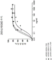
Fig. 2 is a graph showing the percentage of Ly6G 6D-transfected 293 target cells killed in an in vitro target cell killing assay as three different Ly6G6D TDBs with different anti-CD 3 arms: graph of Ly6G6D TDB antibody concentration as a function of Ly6G6D (38E4v11) TDB, Ly6G6D (40G5c) TDB, and Ly6G6D (38E4v1) TDB.
Fig. 3A is a graph showing the percentage of CD69+ CD25+ activated CD8+ T cells as a function of Ly6G6D TDB concentration for three different Ly6G6D TDBs (Ly6G6D (38E4v11) TDB, Ly6G6D (40G5c) TDB, and Ly6G6D (38E4v1) TDB), as assessed by flow cytometry. Effector cells: the target cell ratio was 5: 1.
Fig. 3B is a graph showing the percentage of CD69+ CD25+ activated CD4+ T cells as a function of Ly6G6D TDB concentration for three different Ly6G6D TDBs (Ly6G6D (38E4v11) TDB, Ly6G6D (40G5c) TDB, and Ly6G6D (38E4v1) TDB), as assessed by flow cytometry. Effector cells: the target cell ratio was 5: 1.
Detailed Description
I. Definition of
The term "about" as used herein refers to the usual error range for the corresponding value as readily known to those skilled in the art. Reference herein to "about" a value or parameter includes (and describes) embodiments that are directed to the value or parameter itself.
An "acceptor human framework" for the purposes herein is a framework comprising the amino acid sequence of a light chain variable domain (VL) framework or a heavy chain variable domain (VH) framework derived from a human immunoglobulin framework or a human consensus framework, as defined below. An acceptor human framework "derived from" a human immunoglobulin framework or a human consensus framework may comprise the same amino acid sequence thereof, or it may contain amino acid sequence variations. In some embodiments, the number of amino acid changes is 10 or less, 9 or less, 8 or less, 7 or less, 6 or less, 5 or less, 4 or less, 3 or less, or 2 or less. In some embodiments, the VL receptor human framework is identical in sequence to a VL human immunoglobulin framework sequence or a human consensus framework sequence.
"affinity" refers to the strength of the sum of non-covalent interactions between a single binding site of a molecule (e.g., an antibody) and its binding partner (e.g., an antigen). Unless otherwise indicated, no, as used herein, "binding affinity" refers to an intrinsic binding affinity that reflects a 1:1 interaction between members of a binding pair (e.g., an antibody and an antigen). The affinity of a molecule X for its partner Y can generally be determined by the dissociation constant (K) D) And (4) showing. Affinity can be measured by conventional methods known in the art, including those described herein. Measurement of binding affinityAre described below.
An "affinity matured" antibody is one that has one or more alterations that result in improved affinity of the antibody for an antigen as compared to a parent antibody that does not have the one or more alterations in one or more hypervariable regions (HVRs).
The terms "anti-CD 3 antibody" and "antibody to CD 3" refer to an antibody that is capable of binding CD3 with sufficient affinity such that the antibody is useful as a diagnostic and/or therapeutic agent in targeting CD 3. In one embodiment, the extent to which the anti-CD 3 antibody binds to an unrelated, non-CD 3 protein is less than about 10% of the binding of the antibody to CD3, as measured, for example, by Radioimmunoassay (RIA). In certain embodiments, the dissociation constant (K) of an antibody that binds to CD3D) Is ≤ 1 μ M, ≦ 100nM, ≦ 10nM, ≦ 1nM, ≦ 0.1nM, ≦ 0.01nM, or ≦ 0.001 nM (e.g., 10 nM)-8M or less, e.g. 10-8M to 10-13M, e.g. 10-9M to 10-13M). In certain embodiments, the anti-CD 3 antibody binds to an epitope in CD3 that is conserved in CD3 from different species.
The term "antibody" is used herein in the broadest sense and encompasses a variety of antibody structures, including but not limited to monoclonal antibodies, polyclonal antibodies, multispecific antibodies (e.g., bispecific antibodies), and antibody fragments so long as they exhibit the desired antigen-binding activity.
An "antibody fragment" refers to a molecule that is not an intact antibody, which comprises a portion of an intact antibody that binds to an antigen to which the intact antibody binds. Examples of antibody fragments include, but are not limited to, Fv, Fab '-SH, F (ab')2(ii) a A diabody; a linear antibody; single chain antibody molecules (e.g., scFv); and multispecific antibodies formed from antibody fragments.
"binding domain" refers to a portion of a compound or molecule that specifically binds to a target epitope, antigen, ligand, or receptor. Binding domains include, but are not limited to, antibodies (e.g., monoclonal, polyclonal, recombinant, humanized, and chimeric antibodies), antibody fragments, or portions thereof (e.g., Fab fragments)、Fab’2scFv antibodies, SMIPs, domain antibodies, diabodies, minibodies, scFv-Fc, affibodies, nanobodies, and VH and/or VL domains of antibodies), receptors, ligands, aptamers, and other molecules with defined binding partners.
A "chemotherapeutic agent" is a chemical compound used to treat cancer. Examples of chemotherapeutic agents include alkylating agents, such as thiotepa and cyclophosphamide Alkyl sulfonates such as busulfan, improsulfan and piposulfan; aziridines such as benzodidopa, carboquone, meturedopa, and uretonimine (uredopa); ethyleneimine and methylmelamine including hexamethylmelamine, triethylenemelamine, triethylenephosphoramide, triethylenethiophosphoramide, and trimethylolmelamine; polyacetic acids (especially bucindone and bucindone); delta-9-tetrahydrocannabinol (drodaminol,
 beta-lapachone; lapachol; colchicine; betulinic acid; camptothecin (including the synthetic analogue topotecan)
 CPT-11 (irinotecan, CA)
 Acetyl camptothecin, scopoletin, and 9-aminocamptothecin); bryodin; a colistin; CC-1065 (including its adozelesin, carzelesin, and bizelesin synthetic analogs); podophyllotoxin (podophylotoxin); podophyllinic acid (podophyllic acid); teniposide; cryptophycins (especially cryptophycin 1 and cryptophycin 8); dolastatin (dolastatin); duocarmine (including synthetic analogues, KW-2189 and CB1-TM 1); an pomegranate essence; (ii) coprinus atramentarius alkali; sarcandra glabra alcohol (sarcodictyin); spongistatin (spongistatin); nitrogen mustards (nitrogen en mustard) such as chlorambucil (chlorambucil), naphthalene Nitrogen mustard (chlorethazine), chlorophosphamide (chlorophosphamide), estramustine (estramustine), ifosfamide (i fosfamide), nitrogen mustard (mechlorethamine), mechlorethamine hydrochloride (mechlorethamine oxide hydrochloride), melphalan (melphalan), neomustard (novembichin), benzene mustard cholesterol (phenylesterine), prednimustine, tris cyclophosphamide (tros famide), uracil mustard (uracil mustard); nitrosoureas such as carmustine (carmustine), chlorouretocin (chlorozotocin), fotemustine (fotemustine), lomustine (lomustine), nimustine (nimustine) and ramustine (ranimustine); antibiotics such as enediynes (enediynes) antibiotics (e.g., calicheamicin, particularly calicheamicin γ 1I and calicheamicin ω Il (see, e.g., Nicolaou et al, Angew. chem Intl. Ed. Engl.,33:183-186 (1994)); CDP323, oral α -4 integrin inhibitors; daptomycin (dynemicin), including daptomycin A; esperamicin (esperamicin), and neocarzinostain (neocarzinostatin) chromophore and related chromoproteins enediynes antibiotics chromophore, aclacinomysin (aclinomycin), actinomycin (actinomycin), ansamycin (auramycin), auroramicin (auroramicin), azaserine (azaserine), bleomycin (eomycin), actinomycin (cacinomycin), carmycin (monocamycin), monochamycin (monochamycin), monochamycin (monocrotamycin), monocrotamycin (monocrotamycin), monocrotamycin (monocrotamycin), monocrotamycin (monocrotamycin), monocrotamycin (monocrotomycin), monocrotomycin (monocrotomycin), etc., and/or (e, etc., and/or (etc., and/or (such, 6-diazo-5-oxo-L-norleucine (6-diazo-5-oxo-L-norleucine), doxorubicin (doxorubicin) (including
  Morpholino-doxorubicin (morpholino-doxorubicin), cyanomorpholino-doxorubicin (cyanomorpho-doxorubicin), 2-pyrroline-doxorubicin (2-pyrrolino-doxorubicin), doxorubicin HCL liposome injection
 Liposomal doxorubicin TLC D-99
 Pegylated liposomal doxorubicin
 And deoxydoxorubicin (deoxydox oribicin)), epirubicin (epirubicin), esorubicin (esorubicin), idarubicin (id arbicin), marisulomycin (marcellomomycin), mitomycins (mitomycins) such as mitomycin C, mycophenolic acid (mycophenolic acid), norramycin (nogalamycin), olivomycin (olivomycin), pelomycin (peplomycin), pofiomycin (porfiromycin), puromycin (puromycin), triumycin (quelamycin), rodobicin (rodo rubicin), streptomycin (streptonigrogrin), streptozotocin (streptozotocin), tubercidin (streptomicin), wustitin (streptozobicin), zorubicin (streptozostatin), zotocin (zobicin); antimetabolites such as methotrexate (methotrexate), gemcitabine (gemcitabin e)
 Tegafur (tegafur)
 Capecitabine
 Epothilone (epothilone) and 5-fluorouracil (5-FU); combretastatin (combret astatin); folic acid analogs such as denopterin, methotrexate, pteropterin, trimetrexate; purine analogs such as fludarabine (fludarabine), 6-mercaptopurine (6-mercaptopurine), thiamiprine (thiamiprine), thioguanine (t-hioguanine); pyrimidine analogs such as, for example, ancitabine (ancitabine), azacitidine (azacitidine), 6-azauridine (6-azauridine), carmofur (carmofur), cytarabine (cytarabine), dideoxyuridine (dideoxyuridine), doxifluridine (doxifluridine), and isowuridine Norabine (enocitabine), floxuridine (floxuridine); androgens such as carposterone (caluster one), dromostanolone propionate (dromostanolone propionate), epithioandrostanol (epithiostan ol), mepiquat chloride (mepiquitane), testolactone (testolactone); anti-adrenal agents (anti-adr enals) such as aminoglutethimide (aminoglutethimide), mitotane (mitotane), trostane (trilostane); folic acid compensators such as folinic acid (frolicic acid); acetoglucurolactone (acegl atom); awake phosphoramide glycoside (aldophosphamide glycoside); aminolevulinic acid (a minolevulinic acid); eniluracil (eniluracil); amsacrine (amsacrine); bespoke (beslabucil); bisantrene; edatrexate (edatraxate); phosphoramide (defofamine); dimecorsine (demecolcine); diazaquinone (diaziqutone); eflornithine (elformithine); ammonium etitanium acetate; an epothilone; ethydine oxide (etoglucid); gallium nitrate (gallium nitrate); hydroxyurea (hydroxyurea); lentinan (lentinan); lonidamine (lonidainine); maytansinoids (maytansinoids) such as maytansine (maytansine) and ansamitocins (ansamitocins); mitog uazone (mitog uazone); mitoxantrone (mitoxantrone); mopidanol (mopidanmol); nitrerine (nitrarine); pentostatin (pentostatin); methionine mustard (phenamett); pirarubicin (p irarubicin); losoxantrone (losoxantrone); 2-ethyl hydrazide (2-ethyl hydrazide); procarbazine (procarbazine);
  Polysaccharide complexes (JHS Natural Products, Eurene, OR); Razoxane;, Rhizomycin (rhizoxin;), Sizopyran; (sizofuran;) Gespiramine (spirogemanium;), Alternaria tenuipes (tennuzonic acid;), Triethyleneimine; (triaquuonone;), 2 ', 2' -trichlorotriethylamine (2, 2 ', 2' -trichlorotriethylenethylamine; (trichothecene;) Trichothecenes (Trichothecenes), in particular T-2 toxin, verrucatin A (verrucarin A), bacillocin A (roridin A), and serpentine (anguine); (uratan;) vindesine; (vindesine)
 Dacarbazine (dacarbazine); mannitol mustard (mannomustine); dibromomannitol (mitobronitol); dibromodulcitol (mitolactol); pipobromine (ppilobroman); capping neomycin (glycine); cytarabine (arabine) ("Ara-C"); thiotepa; taxanes (taxoids), e.g. paclitaxel (, (b)
 Albumin-engineered nanoparticle formulations of Bristol-M layers squirb Oncology, Princeton, N.J.), paclitaxel (ABRAXANE)TM) (ii) a And docetaxel
 Rhome-Poulene Rorer, Antony, France); chlorambucil (chlorenbucil); 6-thioguanine (6-thioguanine); mercaptopurine (mercaptoprine); methotrexate; platinum analogs such as cisplatin (cissplatin), oxaliplatin (oxaliplatin) (e.g., cisplatin)
  And carboplatin (c carboplatin); vinblastine (vincas), which prevents tubulin polymerization to form microtubules, includes vinblastine
 Vincristine
 Vindesine
 And vinorelbine
 Etoposide (VP-16); ifosfamide (ifosfamide); mitoxantrone (mitoxantrone); leucovorin; nuantron (novantrone); edatrexate (edatrexate); daunomycin (daunomycin); aminopterin (aminopterin); ibandronate (ibandronate); topoisomerase inhibitor R FS 2000; difluoromethylornithine (DMFO); retinoids such as retinoic acid, including bexarotene (bexarotee)
 Bisphosphonates (bisphosphates) (e.g. bisphosphates)
 Or
Or Etidronic acid sodium salt
 NE-58095, zoledronic acid/zoledronic acid salt (zoledronic acid ac id/zoledronate)
 Alendronate (alendronate)
 Pamidronate (pamidronate)
 Tiludronate (tirudronate)
 Or risedronate (risedronate)
 And troxacitabine (troxacitabine) (1, 3-dioxolane nucleoside cell)Pyrimidine analogs); antisense oligonucleotides, in particular antisense oligonucleotides that inhibit gene expression in signaling pathways involving abnormal cell proliferation, such as, for example, PKC-alpha, Raf, H-Ras, and epidermal growth factor receptor (EGF-R) (e.g., erlotinib (Tarceva) TM) ); vaccines, e.g.
 Vaccines and gene therapy vaccines, e.g.
 A vaccine,
 A vaccine and
 a vaccine; a topoisomerase 1 inhibitor (e.g.,
 the rmRH (for example,
 BAY439006 (sorafenib; Bayer); SU-11248 (sunitinib),
 pfizer); perifosine (perifosine), CO X-2 inhibitors (such as celecoxib (celecoxib) or etoricoxib (etoricoxib)), proteosome inhibitors (e.g., PS 341); bortezomib (bortezomib)
 CCI-779; somatic rafanib (tipifarnib) (R11577); sorafenib (orafenaib), ABT 510; bcl-2 inhibitors, such as sodium orlimerson (oblimersen sodium)
 Pixantrone (pixantrone); an EGFR inhibitor; tyrosine kinase inhibitors; silk ammoniaAcid-threonine kinase inhibitors, such as rapamycin (rapamycin) (sirolimus), RAPA
 Farnesyl transferase inhibitors, such as lonafarnib (SCH 6636, SARASAR)TM) And a pharmaceutically acceptable, acid or derivative of any of the above; and combinations of two or more of the above, such as CHOP (abbreviation for cyclophosphamide, doxorubicin, vinblastine and prednisolone combination therapy) and FOLFOX (oxaliplatin)TM) Abbreviations for treatment regimens combining 5-FU and folinic acid); and combinations of two or more of the foregoing.
Chemotherapeutic agents, as defined herein, include "anti-hormonal agents" or "endocrine therapeutic agents" which are used to modulate, reduce, block or inhibit the action of hormones that can promote cancer growth. They may themselves be hormones, including but not limited to: antiestrogens and Selective Estrogen Receptor Modulators (SERMs), including, for example, tamoxifen (tamoxifen) (including Tamoxifen), raloxifene (raloxifene), droloxifene (droloxifene), 4-hydroxytamoxifene, troxifene (trioxifene), naloxifene (keoxifene), LY117018, onapristone (onapristone), and fareston. cndot toremifene (toremifene); aromatase inhibitors which inhibit aromatase which regulates estrogen production in suprarenal glands, such as, for example, 4(5) -imidazole, aminoglutethimide (amid),
 Megestrol acetate (megestrol acetate),
 Exemestane (exemestane), formestane (formestanie), fadrozole (fadrozole),
 Vorozole (vorozole),
 Letrozole (letrozole) and
 anastrozole (anastrozole); and antiandrogens such as flutamide, nilutamide, bicalutamide, leuprolide, and goserelin; and troxacitabine (a 1, 3-dioxolane nucleoside cytosine analogue); antisense oligonucleotides, particularly those that inhibit gene expression in signaling pathways involved in abnormal cell proliferation, such as, for example, PKC- α, Raf and H-Ras; ribozymes, such as VEGF expression inhibitors (e.g.
  Ribozymes) and inhibitors of HER2 expression; vaccines, such as gene therapy vaccines, e.g.
 A vaccine is provided which comprises a vaccine,
 a vaccine and
 a vaccine;
 rIL-2;
 a
a topoisomerase 1 inhibitor; rmRH; vinorelbine (Vinorelbine) and Esperamicins (see U.S. patent No. 4,675,187) and pharmaceutically acceptable salts, acids or derivatives of any of the above; and combinations of two or more of the foregoing.
The term "chimeric" antibody refers to an antibody in which a portion of the heavy and/or light chain is derived from a particular source or species, while the remainder of the heavy and/or light chain is derived from a different source or species.
As used herein, unless otherwise indicated, the term "cluster of differentiation 3" or "CD 3" refers to any native CD3 from any vertebrate source, including mammals such as primates (e.g., humans) and rodents (e.g., mice and rats), including, for example, the CD3 epsilon, CD3 gamma, CD3 alpha, and CD3 beta chains. The term encompasses "full-length," unprocessed CD3 (e.g., unprocessed or unmodified CD3 epsilon or CD3 gamma) as well as any form of CD3 resulting from processing in a cell. The term also encompasses variants of naturally occurring CD3, including, for example, splice variants or allelic variants. CD3 includes, for example, the human CD3 epsilon protein (NCBI reference sequence No. NP _000724) 207 amino acids in length and the human CD3 gamma protein (NCBI reference sequence No. NP _000064) 182 amino acids in length.
The "class" of antibodies refers to the type of constant domain or constant region that the heavy chain has. There are five major classes of antibodies: IgA, IgD, IgE, IgG and IgM, and several of these antibodies may also be divided into subclasses (isotypes), e.g. IgG1、IgG2、IgG3、IgG4、IgA1And IgA2. The heavy chain constant domains corresponding to different immunoglobulin classes are called α, δ, ε, γ and μ, respectively.
It is to be understood that the aspects and embodiments of the invention described herein include, consist of, and consist essentially of aspects and embodiments.
The term "cytotoxic agent" as used herein refers to a substance that inhibits or prevents cellular function and/or causes cell death or destruction. Cytotoxic agents include, but are not limited to, radioisotopes (e.g., At)211、I131、I125、Y90、Re186、Re188、Sm153、Bi212、P32、 Pb212And radioactive isotopes of Lu); chemistryTherapeutic agents or drugs (e.g., methotrexate), adriamycin (adriamicin), vinca alkaloids (vincristine), vinblastine (vinblastine), etoposide (etoposide)), doxorubicin, melphalan (melphalan), mitomycin c (mitomycin c), chlorambucil (chlorembil), daunomycin (daunorubicin), or other intercalating agents); a growth inhibitor; enzymes and fragments thereof, such as nucleolytic enzymes; (ii) an antibiotic; toxins, such as small molecule toxins or enzymatically active toxins of bacterial, fungal, plant or animal origin, including fragments and/or variants thereof; and various antitumor agents or anticancer agents disclosed below.
A "disorder" is any condition that would benefit from treatment, including but not limited to chronic and acute disorders or diseases, including those pathological conditions that predispose a mammal to the disorder in question.
The terms "cell proliferative disorder" and "proliferative disorder" refer to a disorder associated with a degree of abnormal cell proliferation. In one embodiment, the cell proliferative disorder is cancer. In one embodiment, the cell proliferative disorder is a tumor.
The terms "cancer" and "cancerous" refer to or describe a physiological condition in mammals that is generally characterized by unregulated cell growth. Examples of cancer include, but are not limited to, adenocarcinoma (e.g., colorectal adenocarcinoma, gastric adenocarcinoma, or pancreatic carcinoma), which can be metastatic adenocarcinoma (e.g., metastatic colorectal adenocarcinoma, metastatic gastric adenocarcinoma, or metastatic pancreatic carcinoma), a malignancy, a lymphoma, a blastoma, a sarcoma, and a leukemia or lymphoid malignancy. More specific examples of such cancers include, but are not limited to, esophageal cancer, small bowel cancer, large bowel cancer, squamous cell cancer (e.g., epithelial squamous cell cancer), lung cancer, including small-cell lung cancer, non-small cell lung cancer, adenocarcinoma of the lung and squamous carcinoma of the lung, cancer of the peritoneum, hepatocellular cancer, gastric or stomach cancer (including gastrointestinal and gastrointestinal stromal cancer, pancreatic cancer, glioblastoma, cervical cancer, ovarian cancer, liver cancer, bladder cancer, urinary tract cancer, liver cancer, breast cancer, colon cancer, rectal cancer, colorectal cancer, endometrial or uterine carcinoma, salivary gland cancer, renal or renal cancer, prostate cancer, vulval cancer, thyroid cancer, liver cancer, anal cancer, penile cancer, melanoma, superficial disseminated melanoma, lentigo melanoma, acromelasma melanoma, nodular melanoma, multiple myeloma and B-cell lymphoma (including low grade/follicular non-Hodgkin's lymphoma (NHL); Small Lymphocytic (SL), NHL, and NHL, Intermediate/follicular NHL, intermediate diffuse NHL, advanced immunocytogenic NHL, advanced lymphoblastic NHL, advanced small non-nucleated NHL, storage disease (bulk disease) NHL, mantle cell lymphoma, AIDS-related lymphoma, and Small Lymphocytic (SL) NHL; chronic Lymphocytic Leukemia (CLL), Acute Lymphoblastic Leukemia (ALL), hairy cell leukemia, chronic myeloblastic leukemia and post-transplant lymphoproliferative disorder (PTLD), as well as abnormal vascular proliferation associated with scarring nevus (phakomatases), edema (such as associated with brain tumors), Meigs (Meigs) syndrome, brain and head and neck cancers and associated metastases. In certain embodiments, cancers suitable for treatment by an antibody of the invention include adenocarcinomas (e.g., colorectal adenocarcinoma, gastric adenocarcinoma, or pancreatic carcinoma), which can be metastatic adenocarcinomas (e.g., metastatic colorectal adenocarcinoma, metastatic gastric adenocarcinoma, or metastatic pancreatic carcinoma), esophageal cancer, gastric cancer, small intestinal cancer, large intestinal cancer, colorectal cancer, breast cancer, colorectal cancer, rectal cancer, non-small cell lung cancer, glioblastoma, non-hodgkin's lymphoma (NHL), renal cell carcinoma, prostate cancer, liver cancer, pancreatic cancer, soft tissue sarcoma, Kaposi's (Kaposi) sarcoma, carcinoid carcinoma, head and neck cancer, ovarian cancer, mesothelioma, and multiple myeloma. In some embodiments, the cancer is selected from: an adenocarcinoma (e.g., colorectal adenocarcinoma, gastric adenocarcinoma, or pancreatic adenocarcinoma), which can be metastatic adenocarcinoma (e.g., metastatic colorectal adenocarcinoma, metastatic gastric adenocarcinoma, or metastatic pancreatic adenocarcinoma), esophageal cancer, gastric cancer, small-intestinal cancer, large-intestinal cancer, small-cell lung cancer, glioblastoma, neuroblastoma, melanoma, breast cancer, gastric cancer, colorectal cancer (CRC), and hepatocellular carcinoma. However, in some embodiments, the cancer is selected from: adenocarcinoma (e.g., colorectal, gastric, or pancreatic), esophageal, gastric, small-bowel, large-bowel, non-small cell lung, colorectal, glioblastoma, and breast cancer, including metastatic forms of these cancers. In other embodiments, the cancer is selected from a class of mature B cell cancers, excluding Hodgkin's lymphoma, but including germinal center B cell-like (GCB) DLBCL, activated B cell-like (ABC) DLBCL, Follicular Lymphoma (FL), Mantle Cell Lymphoma (MCL), Acute Myeloid Leukemia (AML), Chronic Lymphoid Leukemia (CLL), Marginal Zone Lymphoma (MZL), Small Lymphocytic Leukemia (SLL), Lymphoplasmacytic Leukemia (LL), Waldenstrom's Macroglobulinemia (WM), Central Nervous System Lymphoma (CNSL), Burkitt's Lymphoma (BL), B cell prolymphocytic leukemia, splenic marginal zone lymphoma, hairy cell leukemia, non-classifiable splenic lymphoma/leukemia, diffuse splenic erythrocytic small B cell lymphoma, hairy cell leukemia variant, waldenstrom's macroglobulinemia, splenic marginal zone lymphoma, hairy cell leukemia, Heavy chain disease, alpha heavy chain disease, gamma heavy chain disease, mu heavy chain disease, plasma cell myeloma, solitary plasmacytoma, extramedullary plasmacytoma, extralymph node marginal zone lymphoma of mucosa-associated lymphoid tissue (MALT lymphoma), lymph node marginal zone lymphoma, childhood follicular lymphoma, primary cutaneous follicular central lymphoma, large B-cell lymphoma rich in T-cells/tissue cells, primary DLBCL of the CNS, primary DLBCL of the leg type skin, senile EBV positive DLBCL, chronic inflammation-associated DLBCL, lymphomatoid granuloma, primary mediastinal (thymic) large B-cell lymphoma, intravascular large B-cell lymphoma, ALK positive large B-cell lymphoma, plasmablast lymphoma, large B-cell lymphoma that occurs in HHV 8-associated multicenter Castleman disease (Castleman disease), primary effusion lymphoma: b-cell lymphoma, unclassifiable, has characteristics intermediate between diffuse large B-cell lymphoma and burkitt's lymphoma and B-cell lymphoma, unclassifiable, has characteristics intermediate between diffuse large B-cell lymphoma and classical hodgkin's lymphoma.
As used herein, "tumor" refers to all neoplastic cell growth and proliferation (whether malignant or benign) as well as all pre-cancerous and cancerous cells and tissues. As referred to herein, the terms "cancer," "cancerous," "cell proliferative disorder," "proliferative disorder," and "tumor" are not mutually exclusive.
As used herein, the term "tumor antigen" is understood to mean those antigens present on tumor cells. These antigens may present extracellular portions on the cell surface, which are typically associated with the transmembrane and cytoplasmic portions of the molecule. These antigens are sometimes only presented by tumor cells and not by normal cells. Tumor antigens may be expressed only on tumor cells, as compared to normal cells, or may represent tumor-specific mutations. In this case, they are referred to as tumor-specific antigens. More common are tumor antigens presented by tumor cells and normal cells, and they are referred to as tumor-associated antigens. These tumor associated antigens may be overexpressed compared to normal cells or may bind to antibodies in tumor cells due to less compact structure of tumor tissue compared to normal tissue. Tumor antigens may exhibit inconsistent expression, or may be expressed at low copy numbers on certain types of tumor cells. In cases where the targeted tumor antigen is expressed on tumor cells at low copy number (i.e., weakly expressed), it may be desirable to use a TDB antibody of the invention that has a high affinity arm for CD3 or another molecule located on immune effector cells. In one aspect, the tumor antigen is selected from those listed in table 1 below.
"Effector function" refers to those biological activities attributable to the Fc region of an antibody, which vary with antibody isotype. Examples of antibody effector functions include: c1q binding and Complement Dependent Cytotoxicity (CDC); fc receptor binding; antibody-dependent cell-mediated cytotoxicity (ADCC); phagocytosis; down-regulation of cell surface receptors (e.g., B cell receptors); and B cell activation.
An "effective amount" of a compound, e.g., an anti-CD 3 antibody of the invention or a composition (e.g., a pharmaceutical composition) thereof, is at least the minimum amount required to achieve a desired therapeutic or prophylactic result, such as measurable improvement or prevention of a particular disorder (e.g., a cell proliferative disorder, e.g., cancer). An effective amount herein may vary depending on factors such as the disease state, age, sex, and weight of the patient and the ability of the antibody to elicit a desired response in the individual. An effective amount is also one in which the therapeutically beneficial effect outweighs any toxic or detrimental effect of the treatment. For prophylactic use, beneficial or desired results include results such as: abrogate or reduce the risk, lessen the severity, or delay the onset of a disease (including biochemical, histological, and/or behavioral symptoms of the disease, complications of the disease and intermediate pathological phenotypes present during the development of the disease). For therapeutic use, beneficial or desired results include clinical results such as: reducing one or more symptoms caused by a disease, improving the quality of life of a patient with a disease, reducing the dose of other drugs required to treat a disease, enhancing the effect of another drug, such as by targeting, delaying disease progression, and/or prolonging survival. In the case of cancer or tumors, a therapeutically effective amount of the drug may have the following effects: reducing the number of cancer cells; reducing the size of the tumor; inhibit (i.e., slow or desirably stop to some extent) cancer cell infiltration of peripheral organs; inhibit (i.e., slow to some extent and desirably prevent) tumor metastasis; inhibit tumor growth to some extent; and/or alleviate one or more symptoms associated with the condition to some extent. An effective amount may be administered in one or more administrations. For the purposes of the present invention, an effective amount of a drug, compound or pharmaceutical composition is an amount sufficient to accomplish prophylactic or therapeutic treatment, either directly or indirectly. It is understood in the clinical setting that an effective amount of a drug, compound or pharmaceutical composition may or may not be achieved in combination with another drug, compound or pharmaceutical composition. Thus, an "effective amount" may be considered in the context of administering one or more therapeutic agents, and a single agent may be considered to be given in an effective amount if the desired result is achieved or achieved in combination with one or more other agents.
The term "Fc region" is used herein to define the C-terminal region of an immunoglobulin heavy chain that contains at least a portion of a constant region. The term includes native sequence Fc regions and variant Fc regions. In one embodiment, the human IgG heavy chain Fc region extends from Cys226 or Pro230 to the carboxy terminus of the heavy chain. However, the C-terminal lysine (Lys447) of the Fc region may or may not be present. Unless otherwise specified herein, the numbering of amino acid residues in the Fc region or constant region is according to the EU numbering system, also known as the EU index, as described in Kabat et al, Sequences of Proteins of Immunological Interest, 5 th edition, Public Health Service, National Institutes of Health, Bethesda, MD, 1991.
"framework" or "FR" refers to variable domain residues other than hypervariable region (HVR) residues. The FR of a variable domain typically consists of four FR domains: FR1, FR2, FR3 and FR 4. Thus, HVR and FR sequences typically occur in the VH (or VL) in the following order: FR1-H1(L1) -FR2-H2(L2) -FR3-H3(L3) -FR 4.
The terms "full-length antibody," "intact antibody," and "whole antibody" are used interchangeably herein to refer to an antibody having a structure substantially similar to a native antibody structure or having a heavy chain comprising an Fc region as defined herein.
As used herein, "growth inhibitory agent" refers to a compound or composition that inhibits the growth of cells in vitro or in vivo. In one embodiment, the growth inhibitory agent is a growth inhibitory antibody that prevents or reduces proliferation of cells expressing the antigen to which the antibody binds. In another embodiment, the growth inhibitory agent may be one that significantly reduces the percentage of cells in S phase. Examples of growth inhibitory agents include agents that block cell cycle progression (at a place other than S phase), such as agents that induce G1 arrest and M phase arrest. Classical M-phase blockers include vinblastine (vincristine and vinblastine), taxanes, and topoisomerase II inhibitors, such as doxorubicin, epirubicin, daunorubicin, etoposide, and bleomycin. Those agents that block G1 also diffuse to S phase blocks, for example, DNA alkylating agents such as tamoxifen, prednisone, dacarbazine, mechlorethamine, cisplatin, methotrexate, 5-fluorouracil, and ara-C. More information can be found in Mendelsohn and Israel, ed. Oncogenes, and anti-cosmetic drugs "(w.b. saunders, philiadelphia, 1995), e.g., p.13. Taxanes (paclitaxel and docetaxel) are anticancer drugs derived from the yew tree. Docetaxel from European sweater Rhone-Poulenc Rorer) is paclitaxel
 Semi-synthetic analogs of Bristol-Myers Squibb). Paclitaxel and docetaxel promote microtubule assembly from microtubule protein dimers and stabilize microtubules by preventing depolymerization, which results in inhibition of mitosis in cells.
The term "HER 2 positive" cancer includes cancer cells with higher than normal levels of HER 2. Examples of HER2 positive cancers include HER2 positive breast cancer and HER2 positive gastric cancer. Optionally, the HER2 positive cancer has an Immunohistochemical (IHC) score of 2+ or 3+ and/or an In Situ Hybridization (ISH) amplification ratio ≧ 2.0.
The terms "host cell," "host cell line," and "host cell culture" are used interchangeably and refer to a cell into which an exogenous nucleic acid has been introduced, including the progeny of such a cell. Host cells include "transformants" and "transformed cells," which include primary transformed cells and progeny derived therefrom, regardless of the number of passages. Progeny may not be identical in nucleic acid content to the parent cell, but may contain mutations. Mutant progeny that have the same function or biological activity as screened or selected for in the originally transformed cell are included herein.
A "human antibody" is an antibody having an amino acid sequence corresponding to that of an antibody produced by a human or human cell or derived from a non-human source using a repertoire of human antibodies or other human antibody coding sequences. This definition of human antibodies specifically excludes humanized antibodies comprising non-human antigen binding residues. Human antibodies, including phage display libraries, can be generated using a variety of techniques known in the art. Hoogenboom and Winter, J. mol.biol., 227:381 (1991); marks et al, J.Biol., 222:581 (1991). Also available for the preparation of human monoclonal antibodies are the methods described in the following documents: cole et al, Monoclonal Antibodies and Cancer Therapy, Alan R.Liss, p.77 (1985); boerner et al, J.Immunol.,147(1):86-95 (1991). See also van Dijk and van de Winkel, curr, opin, pharmacol, 5:368-74 (2001). Human antibodies can be made by administering an antigen to a transgenic animal (e.g., immunized XENOMOUSE (xenomic)) that has been modified to produce such antibodies in response to an antigenic challenge but whose endogenous locus has been disabled (see, e.g., U.S. Pat. Nos. 6,075,181 and 6,150,584 for XENOMOUSE) TMA technique). See also, e.g., Li et al, Proc. Natl.Acad. Sci.USA,103:3557-3562(2006), for human antibodies generated via human B-cell hybridoma technology.
A "human consensus framework" is a framework representing the amino acid residues most commonly present in the selection of human immunoglobulin VL or VH framework sequences. Generally, the human immunoglobulin VL or VH sequences are selected from a subset of variable domain sequences. In general, the sequence subgroups are subgroups as in Kabat et al, Sequences of Proteins of Immunological Interest, 5 th edition, NIH Publication 91-3242, Bethesda MD (1991), volumes 1-3. In one embodiment, for VL, the subgroup is subgroup kappa I as in Kabat et al (supra). In one embodiment, for the VH, the subgroup is subgroup III as in Kabat et al (above).
A "humanized" antibody is a chimeric antibody comprising amino acid residues from non-human HVRs and amino acid residues from human FRs. In certain embodiments, a humanized antibody will comprise substantially all of at least one and typically two variable domains, wherein all or substantially all of the HVRs (e.g., CDRs) correspond to those of a non-human antibody, and all or substantially all of the FRs correspond to those of a human antibody. The humanized antibody optionally can comprise at least a portion of an antibody constant region derived from a human antibody. "humanized forms" of antibodies (e.g., non-human antibodies) refer to antibodies that have been humanized.
The term "hypervariable region" or "HVR" as used herein refers to each of the regions which are highly variable in sequence ("complementarity determining regions" or "CDRs") and/or form structurally defined loops ("hypervariable loops") and/or antibody variable domains containing antigen-contacting residues ("antigen-contacting"). In general, an antibody comprises six HVRs: three in VH (H1, H2, H3) and three in VL (L1, L2, L3). Exemplary HVRs herein include:
(a) the hypervariable loops present at amino acid residues 26-32(L1), 50-52(L2), 91-96(L3), 26-32(H1), 53-55(H2) and 96-101(H3) (Chothia and Lesk, J.mol. biol.196:901-917 (1987));
(b) CDRs present at amino acid residues 24-34(L1), 50-56(L2), 89-97(L3), 31-35b (H1), 50-65(H2) and 95-102(H3) (Kabat et al, Sequences of Proteins of Immunological interest, 5 th edition Public Health Service, National Institutes of Health, Bethesda, MD, (1991));
(c) antigen contacts present at amino acid residues 27c-36(L1), 46-55(L2), 89-96(L3), 30-35b (H1), 47-58(H2) and 93-101(H3) (MacCallum et al J.mol. biol.262:732-745, (1996)); and
(d) combinations of (a), (b), and/or (c) comprising HVR amino acid residues 46-56(L2), 47-56(L2), 48-56(L2), 49-56(L2), 26-35(H1), 26-35b (H1), 49-65 (H2), 93-102(H3), and 94-102 (H3).
Unless otherwise indicated, HVR residues and other residues in the variable domain (e.g., FR residues) are numbered herein according to Kabat et al (supra).
An "immunoconjugate" is an antibody conjugated to one or more heterologous molecules (including, but not limited to, cytotoxic agents).
A "subject" or "individual" is a mammal. Mammals include, but are not limited to, domesticated animals (e.g., cows, sheep, cats, dogs, and horses), primates (e.g., human and non-human primates such as monkeys), rabbits, and rodents (e.g., mice and rats). In certain embodiments, the "subject" or "individual" is a human.
An "isolated antibody" is an antibody that has been separated from components of the natural environment. In some embodiments, the antibody is purified to greater than 95% or 99% purity as determined by, for example, electrophoresis (e.g., SDS-PAGE, isoelectric focusing (IEF), capillary electrophoresis), or chromatography (e.g., ion exchange or reverse phase HPLC). For a review of methods for assessing antibody purity, see, e.g., Flatman et al, J.Chromatogr.B 848:79-87 (2007).
An "isolated nucleic acid" refers to a nucleic acid molecule that has been separated from components of the natural environment. Isolated nucleic acids include nucleic acid molecules contained in cells that normally contain the nucleic acid molecule, but the nucleic acid molecule is present extrachromosomally or at a chromosomal location different from its native chromosomal location.
An "isolated nucleic acid encoding an anti-CD 3 antibody" refers to one or more nucleic acid molecules encoding the heavy and light chains of an antibody (or fragments thereof), including such nucleic acid molecules in a single vector or separate vectors and such nucleic acid molecules present at one or more locations in a host cell.
Unless otherwise indicated, the terms "Ly 6G 6D" or "lymphocyte antigen 6 complex locus G61" as used herein refer to any native Ly6G6D from any vertebrate source, including mammals such as primates (e.g., humans) and chler's teeth (e.g., mice and rats), and encompasses any form of "full-length" unprocessed Ly6G6D and Ly6G6D that results from processing in a cell. The term also encompasses naturally occurring variants of Ly6G6D, including, for example, splice variants or allelic variants. Ly6G6D is also known as G6D, Ly6-D, C6orf23, megakaryocyte enhancing gene transcript 1(MEGT1) and NG25 and is disclosed in U.S. patent No. 7,951,546, which is incorporated herein by reference in its entirety as TAT201, having the amino acid sequence SEQ ID NO:92 and the nucleotide sequence DNA234441, SEQ ID NO: 36. The Ly6G6D includes, for example, a human Ly6G6D protein 133 amino acids in length (NCBI reference sequence No. NP _ 067079.2).
The term "monoclonal antibody" as used herein refers to an antibody obtained from a population of substantially homogeneous antibodies, i.e., the individual antibodies comprising the population are identical and/or bind the same epitope except for possible variant antibodies (e.g., containing naturally occurring mutations or mutations generated during the manufacture of a monoclonal antibody preparation, which variants are typically present in minor amounts). Unlike polyclonal antibody preparations, which typically contain different antibodies directed against different determinants (epitopes), each monoclonal antibody of a monoclonal antibody preparation is directed against a single determinant on the antigen. Thus, the modifier "monoclonal" indicates the nature of the antibody as obtained from a substantially homogeneous population of antibodies, and is not to be construed as requiring production of the antibody by any particular method. For example, monoclonal antibodies to be used in accordance with the present invention can be prepared by a variety of techniques including, but not limited to, hybridoma methods, recombinant DNA methods, phage display methods, and methods that utilize transgenic animals containing all or a portion of a human immunoglobulin locus, such methods, as well as other exemplary methods for preparing monoclonal antibodies, are described herein.
By "naked antibody" is meant an antibody that is not conjugated to a heterologous moiety (e.g., a cytotoxic moiety) or a radioactive label. Naked antibodies may be present in pharmaceutical formulations.
"Natural antibody" refers to a naturally occurring immunoglobulin molecule having a different structure. For example, a native IgG antibody is a heterotetrameric glycoprotein of about 150,000 daltons, consisting of two identical light chains and two identical heavy chains that are disulfide-bonded. From N-terminus to C-terminus, each heavy chain has a variable region (VH), also known as a variable heavy domain or heavy chain variable domain, followed by three constant domains (CH1, CH2 and CH 3). Similarly, from N-terminus to C-terminus, each light chain has a variable region (VL), also known as a variable light domain or light chain variable domain, followed by a Constant Light (CL) domain. The light chain of an antibody can be assigned to one of two types, called kappa and lambda, based on the amino acid sequence of its constant domain.
The term "package insert" is used to refer to instructions typically included in commercial packaging for therapeutic products that contain information about the indications, usage, dosage, administration, combination therapy, contraindications, and/or warnings concerning the use of such therapeutic products.
The term "PD-1 axis binding antagonist" refers to a molecule that inhibits the interaction of a PD-1 axis binding partner with one or more of its binding partners, so as to abrogate T cell dysfunction resulting from signaling on the PD-1 signaling axis-the result being restoration or enhancement of T cell function (e.g., proliferation, cytokine production, target cell killing). As used herein, PD-1 axis binding antagonists include PD-1 binding antagonists, PD-L1 binding antagonists, and PD-L2 binding antagonists.
The term "PD-1 binding antagonist" refers to a molecule that reduces, blocks, inhibits, eliminates, or interferes with signal transduction resulting from the interaction of PD-1 with one or more of its binding partners (such as PD-L1, PD-L2). In some embodiments, the PD-1 binding antagonist is a molecule that inhibits the binding of PD-1 to one or more of its binding partners. In particular embodiments, the PD-1 binding antagonist inhibits the binding of PD-1 to PD-L1 and/or PD-L2. For example, PD-1 binding antagonists include anti-PD-1 antibodies and antigen-binding fragments thereof, immunoadhesins, fusion proteins, oligopeptides, and other molecules that reduce, block, inhibit, eliminate, or interfere with signal transduction resulting from the interaction of PD-1 with PD-L1 and/or PD-L2. In one embodiment, the PD-1 binding antagonist reduces a negative costimulatory signal mediated by or via a cell surface protein, such as expressed on a T lymphocyte that signals mediated by PD-1, so as to cause a reduction in dysfunction of the dysfunctional T cell (e.g., enhance effector response to antigen recognition). In some embodiments, the PD-1 binding antagonist is an anti-PD-1 antibody. In a specific embodiment, the PD-1 binding antagonist is MDX-1106 (nivolumab). In another specific embodiment, the PD-1 binding antagonist is MK-3475 (pembrolizumab). In another specific embodiment, the PD-1 binding antagonist is CT-011 (Pilizumab). In another specific embodiment, the PD-1 binding antagonist is AMP-224. In another specific embodiment, the PD-1 binding antagonist is MED 1-0680. In another specific embodiment, the PD-1 binding antagonist is PDR 001. In another specific embodiment, the PD-1 binding antagonist is REGN 2810. In another specific embodiment, the PD-1 binding antagonist is BGB-108.
The term "PD-L1 binding antagonist" refers to a molecule that reduces, blocks, inhibits, eliminates or interferes with signal transduction resulting from the interaction of PD-L1 with one or more of its binding partners (such as PD-1, B7-1). In some embodiments, the PD-L1 binding antagonist is a molecule that inhibits the binding of PD-L1 to its binding partner. In particular embodiments, the PD-L1 binding antagonist inhibits the binding of PD-L1 to PD-1 and/or B7-1. In some embodiments, PD-L1 binding antagonists include anti-PD-L1 antibodies, antigen binding fragments thereof, immunoadhesins, fusion proteins, oligopeptides, and other molecules that reduce, block, inhibit, eliminate, or interfere with signal transduction resulting from the interaction of PD-L1 with one or more of its binding partners, such as PD-1, B7-1. In one embodiment, the PD-L1 binding antagonist reduces a negative costimulatory signal mediated by or via a cell surface protein such as expressed on a T lymphocyte that signals mediated by PD-L1, such that the dysfunctional T cell is reduced (e.g., an effector response to antigen recognition is enhanced). In some embodiments, the PD-L1 binding antagonist is an anti-PD-L1 antibody. In yet another specific embodiment, the anti-PD-L1 antibody is MPDL3280A (atelizumab). In a specific embodiment, the anti-PD-L1 antibody is yw243.55.s 70. In another specific embodiment, the anti-PD-L1 antibody is MDX-1105. In another specific embodiment, the anti-PD-L1 antibody is MSB 0015718C. In yet another specific embodiment, the anti-PD-L1 antibody is MEDI 4736.
The term "PD-L2 binding antagonist" refers to a molecule that reduces, blocks, inhibits, eliminates, or interferes with the signal transduction produced by the interaction of PD-L2 with one or more of its binding partners, such as PD-1. In some embodiments, the PD-L2 binding antagonist is a molecule that inhibits the binding of PD-L2 to one or more of its binding partners. In particular embodiments, the PD-L2 binding antagonist inhibits the binding of PD-L2 to PD-1. In some embodiments, PD-L2 antagonists include anti-PD-L2 antibodies, antigen-binding fragments thereof, immunoadhesins, fusion proteins, oligopeptides, and other molecules that reduce, block, inhibit, eliminate, or interfere with the signal transduction resulting from the interaction of PD-L2 with one or more of its binding partners, such as PD-1. In one embodiment, the PD-L2 binding antagonist reduces negative costimulatory signals mediated by or via a cell surface protein such as expressed on T lymphocytes mediated by PD-L2 signaling, so as to cause a reduction in dysfunction of the dysfunctional T cells (e.g., enhance effector response to antigen recognition). In some embodiments, the PD-L2 binding antagonist is an immunoadhesin.
Unless otherwise indicated, the term "protein" as used herein refers to any native protein from any vertebrate source, including mammals such as primates (e.g., humans) and rodents (e.g., mice and rats). The term encompasses "full-length," unprocessed protein as well as any form of protein that results from processing in a cell. The term also encompasses variants of naturally occurring proteins, such as splice variants or allelic variants. Proteins according to the invention include, for example, any of the proteins listed in table 1.
"percent (%) amino acid sequence identity" with respect to a reference polypeptide sequence is defined as the percentage of amino acid residues in a candidate sequence that are identical to amino acid residues in the reference polypeptide sequence after aligning the sequences and introducing gaps, if necessary, to achieve the maximum percent sequence identity, and not considering any conservative substitutions as part of the sequence identity. Alignment for the purpose of determining percent amino acid sequence identity may be accomplished in a variety of ways within the skill in the art, for example using publicly available computer software such as BLAST, BLAST-2, ALIGN, or megalign (dnastar) software. One skilled in the art can determine appropriate parameters for aligning the sequences, including any algorithms required to achieve maximum alignment over the full length of the sequences being compared. However, for purposes herein, the% amino acid sequence identity value was generated using the sequence comparison computer program ALIGN-2. The ALIGN-2 sequence comparison computer program was created by Genentech corporation and the original code has been documented with the user file in the U.S. copyright Office (Washington d.c.,20559), where it was registered under U.S. copyright registration number TXU 510087. The ALIGN-2 program is publicly available from Genentech corporation (South San Francisco, Calif.) or may be compiled from source code. The ALIGN-2 program should be compiled for use on a UNIX operating system (including the digital UNIX V4.0D). All sequence comparison parameters were set by the ALIGN-2 program and were not changed.
In the case where ALIGN-2 is used for amino acid sequence comparison, the% amino acid sequence identity (which may alternatively be said to be that a given amino acid sequence a has or comprises a certain amino acid sequence identity to, with or relative to a given amino acid sequence B) for a given amino acid sequence a is calculated as follows:
100X fraction X/Y
Wherein X is the number of amino acid residues scored as identical matches by sequence alignment program ALIGN-2 when program aligned for A and B, and wherein Y is the total number of amino acid residues in B. It will be appreciated that where the length of amino acid sequence a is not equal to the length of amino acid sequence B, the% amino acid sequence identity of a to B will not be equal to the% amino acid sequence identity of B to a. Unless specifically stated otherwise, all% amino acid sequence identity values used herein are obtained as described in the preceding paragraph using the ALIGN-2 computer program.
The term "pharmaceutical formulation" refers to the following formulation: in a form that allows the biological activity of the active ingredient contained therein to be effective, and is free of additional components having unacceptable toxicity to the subject to which the formulation is to be administered.
By "pharmaceutically acceptable carrier" is meant a component of a pharmaceutical formulation other than the active ingredient that is not toxic to the subject. Pharmaceutically acceptable carriers include, but are not limited to, buffers, excipients, stabilizers, or preservatives.
As used herein, "treatment" (and grammatical variations thereof, such as "treatment" or "treating") refers to an attempt to alter the natural course of the treated individual, and may be prophylactic or clinically intervening in the course of clinical pathology. Desirable therapeutic effects include, but are not limited to, preventing the onset or recurrence of disease, alleviating symptoms, attenuating any direct or indirect pathological consequences of the disease, preventing metastasis, reducing the rate of disease progression, improving or alleviating the disease state, and palliating or improving prognosis. In some embodiments, the antibodies of the invention are used to delay the progression of the disease or slow the progression of the disease.
As used herein, "delaying" the "progression" of a disorder or disease means delaying, impeding, slowing, delaying, stabilizing, and/or delaying the development of a disease or disorder (e.g., a cell proliferative disorder, such as cancer). This delay can be of varying lengths of time depending on the history of the disease and/or the individual being treated. As will be apparent to those skilled in the art, a sufficient or significant delay may actually encompass prevention, as the individual does not develop the disease. For example, the development of advanced cancers, such as metastases, may be delayed.
By "reduce" or "inhibit" is meant the ability to cause a reduction in the population as a whole, for example 20% or more, 50% or more, or 75%, 85%, 90%, 95% or more. In certain embodiments, reduced or inhibited refers to antibody Fc region-mediated effector functions of the antibody, such effector functions specifically including Complement Dependent Cytotoxicity (CDC), Antibody Dependent Cellular Cytotoxicity (ADCC), and antibody dependent phagocytosis (ADCP).
The term "variable region" or "variable domain" refers to a domain of an antibody heavy or light chain that is involved in binding the antibody to an antigen. The variable domains of the heavy and light chains of natural antibodies (VH and VL, respectively) typically have similar structures, with each domain comprising four conserved Framework Regions (FR) and three High Variable Regions (HVRs). (see, e.g., Kindt et al, Kuby Immunology, 6 th edition, w.h.freeman and co., page 91 (2007)). Furthermore, antibodies that bind a particular antigen can be isolated using VH or VL domains from antibodies that bind the antigen to screen libraries of complementary VL or VH domains, respectively. See, e.g., Portolano et al, J.Immunol. 150: 880-; clarkson et al, Nature 352: 624-.
The term "vector" as used herein refers to a nucleic acid molecule capable of multiplying another nucleic acid to which it is linked. The term includes vectors that are self-replicating nucleic acid structures, as well as vectors that are incorporated into the genome of a host cell into which they have been introduced. Certain vectors are capable of directing the expression of nucleic acids to which they are operably linked. Such vectors are referred to herein as "expression vectors".
As used herein, "administering" means a method of administering a dose of a compound (e.g., an anti-CD 3 antibody of the invention or a nucleic acid encoding an anti-CD 3 antibody of the invention) or composition (e.g., a pharmaceutical composition, e.g., a pharmaceutical composition comprising an anti-CD 3 antibody of the invention) to a subject. The compositions for use in the methods described herein can be prepared, for example, by: intramuscular, intravenous, intradermal, transdermal, intraarterial, intraperitoneal, intralesional, intracranial, intraarticular, intraprostatic, intrapleural, intratracheal, intranasal, intravitreal, intravaginal, intrarectal, topical, intratumoral, intraperitoneal, subcutaneous, subconjunctival, intracapsular, transmucosal, intrapericardial, intraumbilical, intraocular, oral, topical (topically), topical (locally), by inhalation, by injection, by infusion, by continuous infusion, by local perfusion bathing target cells directly, by catheter, by lavage, in the form of a cream or in the form of a lipid composition. The method of administration may vary depending on various factors, such as the compound or composition being administered and the severity of the condition, disease or disorder being treated.
Compositions and methods
In one aspect, the invention is based, in part, on an anti-CD 3 antibody. In certain embodiments, the anti-CD 3 antibody is multispecific (e.g., bispecific) and binds a second biomolecule (e.g., a cell surface antigen, e.g., a tumor antigen) in addition to CD3 or a fragment thereof. The antibodies of the invention are useful, for example, for treating a cell proliferative disorder (e.g., cancer) or an autoimmune disorder in a subject suffering from said disorder or for delaying the progression of said disorder or for enhancing immune function in a subject suffering from said disorder.
In one aspect, the invention provides isolated antibodies (e.g., CD3 epsilon and/or CD3 gamma) that bind CD 3. In some cases, the anti-CD 3 antibody binds to a human CD3 polypeptide or a cynomolgus monkey (cynomolgus monkey) CD3 polypeptide. In some cases, the human CD3 polypeptide or the cyno CD3 polypeptide is a human CD3 epsilon polypeptide (SEQ ID NO:73) or a cyno CD3 epsilon polypeptide (SEQ ID NO:74), respectively. In some cases, the human CD3 polypeptide or cynomolgus monkey CD3 polypeptide is a human CD3 gamma polypeptide (SEQ ID NO:75) or a cynomolgus monkey CD3 gamma polypeptide (SEQ ID NO:76), respectively. In some cases, the anti-CD 3 antibody binds to an epitope within a fragment of CD3 (e.g., human CD3 epsilon) consisting of amino acids 1-26(SEQ ID NO:77) or 1-27(SEQ ID NO:78) of human CD3 epsilon.
In some cases, the CD3 binding domain binds to an epitope consisting of amino acids of human CD3 epsilon selected from Gln1, Asp2, Asn4, Glu6, and Met 7. In a specific embodiment, the CD3 binding domain binds to an epitope specifically including Glu 6. In certain other embodiments, the CD 3-binding domain is configured not to bind to an epitope including Glu5, a human CD3 epsilon amino acid. In certain other embodiments, the CD 3-binding domain is configured not to bind to an epitope including human CD3 epsilon amino acids Gly3 and Glu 5.
The CD3 epitope can be determined from the CD3 binding domain of a peptide fragment that binds the epitope. Alternatively, the CD3 epitope can be determined by alanine scanning mutagenesis. In one embodiment, a 20%, 30%, 50%, 80% or more reduction in binding of the CD3 binding domain to mutant CD3 indicates that the amino acid residue of mutant CD3 in an alanine scanning mutagenesis assay is an epitope residue of the CD3 binding structure. Alternatively, the CD3 epitope can be determined by mass spectrometry. In some embodiments, the epitope is determined by crystallography (e.g., crystallography methods). In some embodiments, the CD3 epitope as determined by crystallography is determined using amino acids Q1-M7 of CD 3. In some embodiments, the CD3 epitope as determined by crystallography is determined using amino acids QDGNEEMGGITQTPYK (SEQ ID NO:79) Q1-M7 of CD 3.
In some embodiments, the CD3 epitope as determined by crystallography can be performed by: anti-CD 3 antibody Fab dissolved in 0.15M NaCl, 25mM tris pH 7.5(10mg/ml) was combined with 2-fold molar excess (1mg) of CD3 epsilon peptide and the precipitate was initially screened as a sparse matrix in a sitting-drop gas phase diffusion format. The optimized crystals can be grown from a 1:1 mixture with a stock solution containing 70% v/v methylpentanediol and 0.1M HEPES buffer pH 7.5. The stock solution can be used as a cryoprotectant. The crystals can be transferred to extremely cold temperatures by sudden immersion in liquid nitrogen.
Diffraction data from the crystal can be collected using a MAR300CCD detector under the beam of the advancing photon source 22 ID. The recorded diffraction can be integrated and scaled using procedure HKL 2000.
The structure can be phased by the molecular substitution (MR) method using the program Phaser. For example, the MR search model is a Fab subunit derived from the crystal structure of the HGFA/Fab complex (PDB coding: 2R 0L). The CD3 epsilon peptide was built into the structure based on the Fo-Fc spectrum. The structure can then be refined using the maximum probability objective function, anisotropic individual B-factor refinement method and TLS refinement method with the programs REFMAC5 and pheix to achieve convergence.
In some cases, the invention provides an anti-CD 3 antibody having a binding domain comprising at least the HVR-L3 sequence tqsfiirt (SEQ ID NO:6) and 1, 2, 3, 4 or 5 HVRs selected from: (a) HVR-H1 comprising the amino acid sequence SEQ ID NO 1; (b) HVR-H2 comprising the amino acid sequence SEQ ID NO 2; (c) HVR-H3 comprising the amino acid sequence SEQ ID NO 3; (d) HVR-L1 comprising amino acid sequence SEQ ID NO 4; and (e) HVR-L2, comprising the amino acid sequence of SEQ ID NO: 5.
In some cases, the invention provides an anti-CD 3 antibody having a binding domain comprising at least 1, 2, 3, 4, 5, or 6 HVRs selected from: (a) HVR-H1 comprising the amino acid sequence SEQ ID NO 1; (b) HVR-H2 comprising the amino acid sequence SEQ ID NO 2; (c) HVR-H3 comprising the amino acid sequence SEQ ID NO 3; (d) HVR-L1 comprising the amino acid sequence SEQ ID NO 4; (e) HVR-L2 comprising the amino acid sequence SEQ ID NO 5; and (f) HVR-L3, comprising amino acid sequence SEQ ID NO 6. In some cases, an anti-CD 3 antibody can have a VH domain comprising an amino acid sequence having at least 90% sequence identity (e.g., at least 91%, 92%, 93%, 94%, 95%, 96%, 97%, 98%, or 99% sequence identity) to sequence SEQ ID No. 7 or sequence SEQ ID No. 7 and/or a VL domain comprising an amino acid sequence having at least 90% sequence identity (e.g., at least 91%, 92%, 93%, 94%, 95%, 96%, 97%, 98%, or 99% sequence identity) to sequence SEQ ID No. 8 or sequence SEQ ID No. 8. In particular instances, the anti-CD 3 antibody may be 38E4v11 or a derivative or clonal association thereof.
In some cases, an anti-CD 3 antibody can comprise at least one (e.g., 1, 2, 3, or 4) of the heavy chain framework regions FR-H1, FR-H2, FR-H3, and FR-H4 comprising the sequences SEQ ID NOS: 9-12, respectively, and/or at least one (e.g., 1, 2, 3, or 4) of the light chain framework regions FR-L1, FR-L2, FR-L3, and FR-L4 comprising the sequences SEQ ID NOS: 13-16, respectively.
In any of the above embodiments, the anti-CD 3 antibody is humanized. In one embodiment, the anti-CD 3 antibody comprises an HVR as in any of the above embodiments, and further comprises an acceptor human framework, e.g., a human immunoglobulin framework or a human consensus framework.
In another aspect, an anti-CD 3 antibody is provided, wherein the antibody comprises a VH as in any of the embodiments provided above, and a VL as in any of the embodiments provided above, wherein one or both of the variable domain sequences comprises a post-translational modification.
In another aspect, the invention provides an antibody that binds to the same epitope as an anti-CD 3 antibody provided herein. For example, in certain embodiments, an antibody is provided that binds to the same epitope as an anti-CD 3 antibody comprising the VH sequence SEQ ID NO 7 and the VL sequence SEQ ID NO 8. In certain embodiments, antibodies are provided that bind to an epitope within a fragment of CD3 (e.g., human CD3 epsilon) consisting of amino acids 1-26 (SEQ ID NO:77) or 1-27(SEQ ID NO:78) of human CD3 epsilon.
In another aspect of the invention, the anti-CD 3 antibody according to any of the above embodiments is a monoclonal antibody. In other embodiments, the anti-CD 3 antibody is a chimeric or human antibody. In one embodiment, the anti-CD 3 antibody is an antibody fragment, such as Fv, Fab', scFv, diabody, or F(ab’)2And (3) fragment. In another embodiment, the antibody is a full length antibody, such as an intact IgG antibody (e.g., an intact IgG1 antibody) or other antibody classes or isotypes as defined herein.
In another aspect, an anti-CD 3 antibody according to any of the above embodiments can incorporate any of the features, either singly or in combination, as described in sections 1-8 below.
1. Affinity of antibody
In certain embodiments, an antibody provided herein has a dissociation constant (Kd) (e.g., 10) of ≦ 1 μ M ≦ 100nM, ≦ 10nM, ≦ 1nM, ≦ 0.1nM, ≦ 0.01nM, or ≦ 0.001nM-8M or less, e.g. 10-8M to 10-13M, e.g. 10-9M to 10-13M) 。
In one embodiment, Kd is measured by a radiolabeled antigen binding assay (RIA). In one embodiment, the RIA is performed with the Fab form of the antibody of interest and its antigen. For example, by using a minimum concentration of (in the presence of an unlabeled antigen in a titration series) 125I) The solution binding affinity of Fab for antigen was measured by equilibration of the Fab with the labeled antigen and subsequent capture of the bound antigen with anti-Fab antibody coated plates (see, e.g., Chen et al, J. mol. biol.293:865 881 (1999)). To establish the assay conditions, the The well plates (Thermo Scientific) were coated overnight with 50mM sodium carbonate (pH 9.6) containing 5. mu.g/ml capture anti-Fab antibodies (Cappel Labs) and then blocked with PBS containing 2% (w/v) bovine serum albumin for 2 to 5 hours at room temperature (approximately 23 ℃). In the non-adsorption plate (Nunc #269620), 100pM or 26pM [ alpha ], [ beta ] -amylase125I]Mixing of antigen with serial dilutions of the Fab of interest (e.g.in agreement with the evaluation of the anti-VEGF antibody Fab-12 in Presta et al, Cancer Res.57:4593-4599 (1997)). Then incubating the target Fab overnight; however, incubation may continue for a longer period of time (e.g., about 65 hours) to ensure equilibrium is reached. Thereafter, the mixture is transferred to a capture plate and incubated at room temperature (e.g., 1 hour). The solution was then removed and polymerized with 0.1% in
 PBSPyrroside 20 The plate was washed 8 times. When the plates had dried, 150. mu.l/scintillator (MICROSCINT-20) was added per wellTM(ii) a Packard) and in TOPCOUNTTMPlates were counted on a gamma counter (Packard) for 10 minutes. The concentration of each Fab that produced less than or equal to 20% of the maximum binding was selected for use in competitive binding assays.
According to another embodiment, use is made of Surface plasmon resonance assay to measure Kd. For example, the immobilized antigen CM5 chip is used at 25 ℃ in about 10 Reaction Units (RU)
 Or
Or (BIAcore, Inc., Piscataway, NJ). In one embodiment, carboxymethylated dextran biosensor chips (CM5, BIACORE, Inc.) are activated with N-ethyl-N '- (3-dimethylaminopropyl) -carbodiimide hydrochloride (EDC) and N-hydroxysuccinimide (NHS) according to the supplier's instructions. Antigen was diluted to 5. mu.g/ml (. about.0.2. mu.M) with 10mM sodium acetate, pH 4.8, and then injected at a flow rate of 5. mu.l/min to achieve about 10 Reaction Units (RU) of conjugated protein. After injection of antigen, 1M ethanolamine was injected to block unreacted groups. For kinetic measurements, Fab was injected at 25 ℃ at a flow rate of about 25. mu.l/min into a mixture containing 0.05% polysorbate 20 (TWEEN-20)TM) Two-fold serial dilutions (0.78nM to 500nM) in pbs (pbst) of surfactant. Mold type using simple one-to-one Langmuir junction
 Evaluation software version 3.2) calculation of association rates (k) by simultaneous fitting of association and dissociation sensorgramsAssociation ofOr ka) And dissociation rate (k)DissociationOr k d). Equilibrium dissociation constant (K)D) Is calculated as a ratio kDissociation/kAssociation of. See, e.g., Chen et al, J.mol.biol.293:865-881 (1999). If the association rate as determined by the above surface plasmon resonance exceeds 106M-1s-1The rate of association can then be determined by using a fluorescence quenching technique measured, for example, in a spectrometer such as an Aviv instrument or 8000 series SLM-AMINCO with a stir cuvetteTMFluorescence emission intensity of 20nM anti-antigen antibody (Fab form) in PBS (pH 7.2) at 25 ℃ in the presence of increasing concentrations of antigen measured in a spectrophotometer (ThermoSpectronic) (excitation: 295 nM; emission 340nm, 16nm bandpass) increase or decrease.
2. Antibody fragments
In certain embodiments, the antibodies provided herein are antibody fragments. Antibody fragments include, but are not limited to, Fab '-SH, F (ab')2Fv and scFv fragments and other fragments as described below. For a review of certain antibody fragments, see Hudson et al nat. Med.9: 129-. For reviews on scFv fragments, see, e.g., Pluckth ü n, eds (Springer-Verlag, New York), pp.269-315 (1994), The Pharmacology of Monoclonal Antibodies, Vol.113, Rosenburg and Moore; see also WO 93/16185; and U.S. patent nos. 5,571,894 and 5,587,458. For Fab and F (ab') containing salvage receptor binding epitope residues and having increased half-life in vivo 2See U.S. Pat. No. 5,869,046 for a discussion of fragments.
Diabodies are antibody fragments with two antigen binding sites that can be bivalent or bispecific. See, for example, EP 404,097; WO 1993/01161; hudson et al, nat. Med.9: 129-; and Hollinger et al, Proc. Natl. Acad. Sci. USA 90: 6444-. Tri-and tetrad antibodies are also described in Hudson et al, nat. Med.9:129-134 (2003).
A single domain antibody is an antibody fragment comprising all or a portion of the heavy chain variable domain or all or a portion of the light chain variable domain of an antibody. In certain embodiments, the single domain antibody is a human single domain antibody (Domantis, Inc., Waltham, MA; see, e.g., U.S. Pat. No. 6,248,516B 1).
Antibody fragments can be prepared by a variety of techniques, including but not limited to proteolytic digestion of the intact antibody and, as described herein, production by recombinant host cells (e.g., E.coli or phage).
3. Chimeric and humanized antibodies
In certain embodiments, the antibodies provided herein are chimeric antibodies. Certain chimeric antibodies are described, for example, in U.S. Pat. nos. 4,816,567; and Morrison et al, Proc. Natl. Acad. Sci. USA,81: 6851-. In one embodiment, the chimeric antibody comprises a non-human variable region (e.g., a variable region derived from a mouse, rat, hamster, rabbit, or non-human primate, such as a monkey) and a human constant region. In yet another embodiment, the chimeric antibody is a "class switch" antibody, wherein the class or subclass has been altered as compared to the class or subclass of the parent antibody. Chimeric antibodies include antigen-binding fragments thereof.
In certain embodiments, the chimeric antibody is a humanized antibody. Typically, the non-human antibodies are humanized to reduce immunogenicity to humans, while retaining the specificity and affinity of the parent non-human antibody. In general, a humanized antibody comprises one or more variable domains, wherein, for example, HVRs (or portions thereof) are derived from a non-human antibody, and FRs (or portions thereof) are derived from human antibody sequences. The humanized antibody optionally will further comprise at least a portion of a human constant region. In some embodiments, some FR residues in the humanized antibody are substituted with corresponding residues from a non-human antibody (e.g., an antibody from which the HVR residues are derived), e.g., to restore or improve antibody specificity or affinity.
Humanized antibodies and methods for their preparation are described, for example, in Almagro and Fransson, front.biosci.13:1619-1633(2008), and further described, for example, in Riechmann et al, Nature 332:323-329 (1988); queen et al, Proc.nat' l Acad. Sci. USA 86:10029-10033 (1989); U.S. Pat. nos. 5,821,337, 7,527,791, 6,982,321, and 7,087,409; kashmiri et al, Methods 36:25-34 (2005) (description Specificity Determination Region (SDR) grafting); padlan, mol.Immunol.28: 489-498(1991) (description "surface reforming"); dall' Acqua et al, Methods 36:43-60(2005) (description "FR shuffling"); and Osbourn et al, Methods 36:61-68(2005) and Klimka et al, Br.J. cancer,83:252-260(2000) (describing the "guided selection" approach to FR shuffling).
Human framework regions that may be used for humanization include, but are not limited to: framework regions selected using the "best fit" method (see, e.g., Sims et al, J.Immunol.151:2296 (1993)); framework regions derived from consensus sequences of human antibodies of a particular subgroup of light or heavy chain variable regions (see, e.g., Carter et al, Proc. Natl. Acad. Sci. USA,89:4285 (1992); and Presta et al, J.Immunol.,151:2623 (1993)); human mature (somatic mutation) framework regions or human germline framework regions (see, e.g., Almagro and Fransson, front.biosci.13:1619-1633 (2008)); and the framework regions derived from screening FR libraries (see, e.g., Baca et al, J.biol.chem.272:10678-10684(1997) and Rosok et al, J.biol.chem.271:22611-22618 (1996)).
4. Human antibodies
In certain embodiments, the antibodies provided herein are human antibodies. Human antibodies can be produced using a variety of techniques known in the art. Human antibodies are generally described in van Dijk and van de Winkel, curr. opin. pharmacol.5:368-74(2001), and Lonberg, curr. opin. immunol.20: 450-.
Human antibodies can be prepared by administering an immunogen to a transgenic animal that has been modified to produce whole human antibodies or whole antibodies with human variable regions in response to antigenic challenge. Such animals typically contain all or part of a human immunoglobulin locus, either in place of an endogenous immunoglobulin locus, or present extrachromosomally or randomly integrated into the chromosome of the animal. In such transgenic mice, the endogenous immunoglobulin locus has typically been inactivated. For a review of the method for obtaining human antibodies from transgenic animals, see Lonberg, nat. Biotech.23:1117-1125 (2005). See also, e.g., the description XENOMOUSE TMU.S. Pat. nos. 6,075,181 and 6,150,584 to technology; drawing instrumentThe above-mentioned U.S. patent numbers 5,770,429 for technology; description of K-M
 U.S. Pat. No. 7,041,870 and description of the technology
 U.S. patent application publication No. US 2007/0061900 for technology). The human variable regions from the whole antibodies produced by such animals may be further modified, for example by combination with different human constant regions.
Human antibodies can also be made by hybridoma-based methods. Human myeloma and mouse-human heteromyeloma (heteromyma) cell lines have been described for the production of human monoclonal antibodies. (see, e.g., Kozbor J. Immunol.,133:3001 (1984); Brodeur et al, Monoclonal Antibody Production Techniques and Applications, pp.51-63 (Marcel Dekker, Inc., New York,1987) and Borner et al, J. Immunol.,147:86 (1991.) further Methods produced by the human B-cell hybridoma technique are described in Li et al, Proc. Natl. Acad. Sci. USA,103: 3557. AcoE 3562(2006) include, e.g., U.S. Pat. No. 7,2006,826 (describing the Production of Monoclonal human IgM antibodies from hybridoma cell lines) and Ni, Xiandai Mianyixnixie, 26: 4: 265 (Pharman. J. Immunol., 26 (described in Pharman-J. hybridoma cell lines) and Methods described in U.S. Biomaton. Act. Scojordan. and FIGS. 7, 7. Columbo. 3. and Methods described in Voronoi. Columb. 3 and Voronoi. Columb. 3. and Methods, 27(3) 185-91 (2005).
Human antibodies can also be generated by isolating Fv clone variable domain sequences selected from human-derived phage display libraries. Such variable domain sequences can then be combined with the desired human constant domains. Techniques for selecting human antibodies from antibody libraries are described below.
5. Library-derived antibodies
Antibodies of the invention can be isolated by screening combinatorial libraries for antibodies having a desired activity or activities. For example, various methods are known in the art for generating phage display libraries and screening such libraries for antibodies with desired binding properties. Such a method can be found, for example, in Hoogenboom et al, Methods in Molecular Biology 178:1-37 (O' Brien et al, Human Press, Totowa, NJ,2001) and is further described, for example, in McCafferty et al, Nature 348: 552-; clackson et al, Nature 352: 624-; marks et al, J.mol.biol.222: 581-597 (1992); marks and Bradbury, Methods in Molecular Biology 248:161-175(Lo eds., Human Press, Totowa, NJ, 2003); sidhu et al, J. mol. biol.338(2):299-310 (2004); lee et al, J.mol.biol.340(5): 1073-; fellouse, Proc. Natl. Acad. Sci. USA 101(34): 12467-; and Lee et al, J.Immunol.methods 284(1-2): 119-132 (2004).
In some phage display methods, libraries of VH and VL genes are separately cloned by Polymerase Chain Reaction (PCR) and randomly recombined in phage libraries that can then be screened against antigen-binding phage, as described in Winter et al, Ann. Rev. Immunol., 12:433-455 (1994). Phage typically display antibody fragments either as single chain fv (scfv) fragments or as Fab fragments. Libraries from immunized sources provide high affinity antibodies to the immunogen without the need to construct hybridomas. Alternatively, natural repertoires can be cloned (e.g., from humans) to provide a single source of antibodies to a wide range of non-self antigens as well as self-antigens without any immunization, as described by Griffiths et al, EMBO J,12: 725-. Finally, natural libraries can also be prepared synthetically by: unrearranged V gene segments were cloned from stem cells and PCR primers containing random sequences were used to encode highly variable CDR3 regions and to effect rearrangement in vitro as described by Hoogenboom and j.mol. biol.,227:381-388 (1992). Patent publications describing human antibody phage libraries include, for example: U.S. Pat. No. 5,750,373, and U.S. patent publication nos. 2005/0079574, 2005/0119455, 2005/0266000, 2007/0117126, 2007/0160598, 2007/0237764, 2007/0292936, and 2009/0002360.
Antibodies or antibody fragments isolated from a human antibody library are considered human antibodies or human antibody fragments herein.
6. Multispecific antibodies
In any of the above aspects, the anti-CD 3 antibodies provided herein are multispecific antibodies, e.g., bispecific antibodies. A multispecific antibody is an antibody (e.g., a monoclonal antibody) that has binding specificity for at least two different sites. In certain embodiments, a bispecific antibody can bind two different epitopes of CD3 (e.g., CD3 epsilon or CD3 gamma). In certain embodiments, one of the binding specificities is for CD3 (e.g., CD3 epsilon or CD3 gamma) and the other is for any other antigen (e.g., a second biomolecule, such as a cell surface antigen, e.g., a tumor antigen). Thus, a bispecific anti-CD 3 antibody may have binding specificity for CD3 and a second biomolecule, such as the second biomolecule (e.g., tumor antigen) listed in table 1 and described herein and in U.S. publication No. 2010/0111856.
TABLE 1 tumor antigen targets of the bispecific anti-CD 3 antibodies of the invention


For example, in some cases, an anti-CD 3 antibody having a first binding domain comprising at least the HVR-L3 sequence tqsfl rt (SEQ ID NO:6) and 1, 2, 3, 4, or 5 HVRs selected from: (a) HVR-H1 comprising the amino acid sequence SEQ ID NO 1; (b) HVR-H2 comprising the amino acid sequence SEQ ID NO 2; (c) HVR-H3 comprising the amino acid sequence SEQ ID NO 3; (d) HVR-L1 comprising amino acid sequence SEQ ID NO 4; and (e) HVR-L2, comprising amino acid sequence SEQ ID NO 5, can have a second binding domain that binds Ly6G 6D. In some cases, the first binding domain that binds CD3 comprises at least one (e.g., 1, 2, 3, or 4) of the heavy chain framework regions FR-H1, FR-H2, FR-H3, and FR-H4 comprising the sequences SEQ ID NOS: 9-12, respectively, and/or at least one (e.g., 1, 2, 3, or 4) of the light chain framework regions FR-L1, FR-L2, FR-L3, and FR-L4 comprising the sequences SEQ ID NOS: 13-16, respectively. In some cases, a first binding domain that binds CD3 can, for example, comprise a VL domain comprising an amino acid sequence having at least 90% sequence identity (e.g., at least 91%, 92%, 93%, 94%, 95%, 96%, 97%, 98%, or 99% sequence identity) to sequence SEQ ID No. 8 or sequence SEQ ID No. 8. In some cases, a first binding domain that binds CD3 can, for example, comprise (a) a VL domain comprising an amino acid sequence having at least 90% sequence identity (e.g., at least 91%, 92%, 93%, 94%, 95%, 96%, 97%, 98%, or 99% sequence identity) to sequence SEQ ID No. 8 or sequence SEQ ID No. 8, and (b) a VH domain comprising an amino acid sequence having at least 90% sequence identity (e.g., at least 91%, 92%, 93%, 94%, 95%, 96%, 97%, 98%, or 99% sequence identity) to sequence SEQ ID No. 7 or sequence SEQ ID No. 7, such as a domain possessed by anti-CD 3 antibody 38E4v11 described herein.
In other instances, an anti-CD 3 antibody having a first binding domain comprising at least 1, 2, 3, 4, 5, or 6 HVRs selected from: (a) HVR-H1 comprising the amino acid sequence SEQ ID NO 1; (b) HVR-H2 comprising the amino acid sequence SEQ ID NO 2; (c) HVR-H3 comprising the amino acid sequence SEQ ID NO 3; (d) HVR-L1 comprising the amino acid sequence SEQ ID NO 4; (e) HVR-L2 comprising amino acid sequence SEQ ID NO 5; and (f) HVR-L3, comprising the amino acid sequence SEQ ID NO: 6, such as 38E4v11, can have a second binding domain that binds to a cell surface antigen (e.g., a tumor antigen) on a target cell other than an immune effector cell.
In some cases, the cell surface antigen may be expressed at low copy number on the target cell. For example, in some cases, a cell surface antigen may be expressed or present in less than 35,000 copies per target cell. In some embodiments, the low copy number cell surface antigen is between 100 copies/target cell and 35,000 copies/target cell; between 100 copies/target cell and 30,000 copies/target cell; between 100 copies/target cell and 25,000 copies/target cell; between 100 copies/target cell and 20,000 copies/target cell; between 100 copies/target cell and 15,000 copies/target cell; between 100 copies/target cell and 10,000 copies/target cell; between 100 copies/target cell and 5,000 copies/target cell; between 100 copies/target cell and 2,000 copies/target cell; between 100 copies/target cell and 1,000 copies/target cell; or between 100 copies/target cell and 500 copies/target cell. The copy number of the cell surface antigen can be determined, for example, using standard Scatchard plots.
In some cases, an anti-CD 3 antibody having a first binding domain comprising at least 1, 2, 3, 4, 5, or 6 HVRs selected from: (a) HVR-H1 comprising the amino acid sequence SEQ ID NO 1; (b) HVR-H2 comprising the amino acid sequence SEQ ID NO 2; (c) HVR-H3 comprising the amino acid sequence SEQ ID NO 3; (d) HVR-L1 comprising the amino acid sequence SEQ ID NO 4; (e) HVR-L2 comprising amino acid sequence SEQ ID NO 5; and (f) HVR-L3, comprising the amino acid sequence SEQ ID NO: 6, such as 38E4v11, can have a second binding domain that binds Ly6G 6D. Depending on the target cell, Ly6G6D may be expressed or present on the target cell at low copy number, e.g., from about 20,000 copies/target cell to about 30,000 copies/target cell (e.g., from about 20,000 copies/target cell to about 25,000 copies/target cell or from about 25,000 copies/target cell to about 30,000 copies/target cell). The copy number of Ly6G6D can be determined using standard Scatchard plots. In some cases, the first binding domain that binds CD3 comprises at least one (e.g., 1, 2, 3, or 4) of the heavy chain framework regions FR-H1, FR-H2, FR-H3, and FR-H4 comprising the sequences SEQ ID NOS: 9-12, respectively, and/or at least one (e.g., 1, 2, 3, or 4) of the light chain framework regions FR-L1, FR-L2, FR-L3, and FR-L4 comprising the sequences SEQ ID NOS: 13-16, respectively. In some cases, the first binding domain that binds CD3 can, for example, comprise a VL domain comprising an amino acid sequence having at least 90% sequence identity (e.g., at least 91%, 92%, 93%, 94%, 95%, 96%, 97%, 98%, or 99% sequence identity) to sequence SEQ ID No. 8 or sequence SEQ ID No. 8. In some cases, a first binding domain that binds CD3 can, for example, comprise (a) a VL domain comprising an amino acid sequence having at least 90% sequence identity (e.g., at least 91%, 92%, 93%, 94%, 95%, 96%, 97%, 98%, or 99% sequence identity) to sequence SEQ ID No. 8 or sequence SEQ ID No. 8, and (b) a VH domain comprising an amino acid sequence having at least 90% sequence identity (e.g., at least 91%, 92%, 93%, 94%, 95%, 96%, 97%, 98%, or 99% sequence identity) to sequence SEQ ID No. 7 or sequence SEQ ID No. 7, such as a domain possessed by anti-CD 3 antibody 38E4v11 described herein.
In other instances, an anti-CD 3 antibody having a first binding domain comprising at least 1, 2, 3, 4, 5, or 6 hypervariable regions (HVRs) selected from: (a) HVR-H1 comprising the amino acid sequence SEQ ID NO 1; (b) HVR-H2 comprising the amino acid sequence SEQ ID NO 2; (c) HVR-H3 comprising the amino acid sequence SEQ ID NO 3; (d) HVR-L1 comprising the amino acid sequence SEQ ID NO 4; (e) HVR-L2, comprising the amino acid sequence SEQ ID NO 5; and (f) HVR-L3, comprising amino acid sequence SEQ ID NO 6, such as 38E4v11, can have a second binding domain that binds CD 20. The second binding domain that binds CD20 may, for example, comprise at least 1, 2, 3, 4, 5, or 6 hypervariable regions (HVRs) selected from: (a) HVR-H1 comprising the amino acid sequence SEQ ID NO 17; (b) HVR-H2 comprising the amino acid sequence SEQ ID NO 18; (c) HVR-H3 comprising the amino acid sequence SEQ ID NO 19; (d) HVR-L1, comprising the amino acid sequence SEQ ID NO: 20; (e) HVR-L2 comprising the amino acid sequence SEQ ID NO 21; and (f) HVR-L3 comprising amino acid sequence SEQ ID NO:22, such as the domain of anti-CD 20 antibody 2H7.v16 (described in U.S. Pat. No. 7,799,900). In some cases, the second binding domain that binds CD20 comprises at least one (e.g., 1, 2, 3, or 4) of the heavy chain framework regions FR-H1, FR-H2, FR-H3, and FR-H4, respectively, comprising the sequences SEQ ID NOS: 23-26, and/or at least one (e.g., 1, 2, 3, or 4) of the light chain framework regions FR-L1, FR-L2, FR-L3, and FR-L4, respectively, comprising the sequences SEQ ID NOS: 27-30. In some cases, a second binding domain that binds CD20 can, for example, comprise (a) a VH domain comprising an amino acid sequence having at least 90% sequence identity (e.g., at least 91%, 92%, 93%, 94%, 95%, 96%, 97%, 98%, or 99% sequence identity) to sequence SEQ ID No. 31 or sequence SEQ ID No. 31; (b) a VL domain comprising an amino acid sequence having at least 90% sequence identity (e.g., at least 91%, 92%, 93%, 94%, 95%, 96%, 97%, 98%, or 99% sequence identity) to sequence SEQ ID No. 32 or sequence SEQ ID No. 32; or (c) a VH domain as in (a) and a VL domain as in (b), such as the domain possessed by anti-CD 20 antibody 2h7.v16 (as described in U.S. patent No. 7,799,900).
In some cases, an anti-CD 3 antibody having a first binding domain comprising at least 1, 2, 3, 4, 5, or 6 hypervariable regions (HVRs) selected from: (a) HVR-H1 comprising the amino acid sequence SEQ ID NO 1; (b) HVR-H2 comprising the amino acid sequence SEQ ID NO 2; (c) HVR-H3 comprising the amino acid sequence SEQ ID NO 3; (d) HVR-L1 comprising the amino acid sequence SEQ ID NO 4; (e) HVR-L2, comprising the amino acid sequence SEQ ID NO 5; and (f) HVR-L3, comprising the amino acid sequence SEQ ID NO:6, such as 38E4v11, may have a second binding domain that binds FcRH 5. The second binding domain that binds FcRH5 may, for example, comprise at least 1, 2, 3, 4, 5, or 6 hypervariable regions (HVRs) selected from: (a) HVR-H1 comprising the amino acid sequence SEQ ID NO 33; (b) HVR-H2 comprising the amino acid sequence SEQ ID NO: 34; (c) HVR-H3 comprising the amino acid sequence SEQ ID NO 35; (d) HVR-L1, comprising the amino acid sequence SEQ ID NO: 36; (e) HVR-L2 comprising the amino acid sequence SEQ ID NO 37; and (f) HVR-L3 comprising amino acid sequence SEQ ID NO 38, such as the domain possessed by anti-FcRH 5 antibody 1G 7. In some cases, the second binding domain that binds FcRH5 comprises at least one (e.g., 1, 2, 3, or 4) of the heavy chain framework regions FR-H1, FR-H2, FR-H3, and FR-H4 comprising the sequences SEQ ID NOs 39-42, respectively, and/or at least one (e.g., 1, 2, 3, or 4) of the light chain framework regions FR-L1, FR-L2, FR-L3, and FR-L4 comprising the sequences SEQ ID NOs 43-46, respectively. In some cases, a second binding domain that binds FcRH5 can, for example, comprise (a) a VH domain comprising an amino acid sequence having at least 90% sequence identity (e.g., at least 91%, 92%, 93%, 94%, 95%, 96%, 97%, 98%, or 99% sequence identity) to sequence SEQ ID No. 47 or sequence SEQ ID No. 47; (b) a VL domain comprising an amino acid sequence having at least 90% sequence identity (e.g., at least 91%, 92%, 93%, 94%, 95%, 96%, 97%, 98%, or 99% sequence identity) to sequence SEQ ID No. 48 or sequence SEQ ID No. 48; or (c) a VH domain as in (a) and a VL domain as in (b).
In some cases, an anti-CD 3 antibody having a first binding domain comprising at least 1, 2, 3, 4, 5, or 6 hypervariable regions (HVRs) selected from: (a) HVR-H1 comprising the amino acid sequence SEQ ID NO 1; (b) HVR-H2 comprising the amino acid sequence SEQ ID NO 2; (c) HVR-H3 comprising the amino acid sequence SEQ ID NO 3; (d) HVR-L1 comprising the amino acid sequence SEQ ID NO 4; (e) HVR-L2, comprising the amino acid sequence SEQ ID NO 5; and (f) HVR-L3, comprising amino acid sequence SEQ ID NO 6, such as 38E4v11, can have a second binding domain that binds PMEL 17.
In some cases, an anti-CD 3 antibody having a first binding domain comprising at least 1, 2, 3, 4, 5, or 6 hypervariable regions (HVRs) selected from: (a) HVR-H1 comprising the amino acid sequence SEQ ID NO 1; (b) HVR-H2 comprising the amino acid sequence SEQ ID NO 2; (c) HVR-H3 comprising the amino acid sequence SEQ ID NO 3; (d) HVR-L1 comprising the amino acid sequence SEQ ID NO 4; (e) HVR-L2, comprising the amino acid sequence SEQ ID NO 5; and (f) HVR-L3, comprising amino acid sequence SEQ ID NO 6, such as 38E4v11, can have a second binding domain that binds LY 6E.
In some cases, an anti-CD 3 antibody having a first binding domain comprising at least 1, 2, 3, 4, 5, or 6 hypervariable regions (HVRs) selected from: (a) HVR-H1 comprising the amino acid sequence SEQ ID NO 1; (b) HVR-H2 comprising the amino acid sequence SEQ ID NO 2; (c) HVR-H3 comprising the amino acid sequence SEQ ID NO 3; (d) HVR-L1 comprising the amino acid sequence SEQ ID NO 4; (e) HVR-L2, comprising the amino acid sequence SEQ ID NO 5; and (f) HVR-L3, comprising amino acid sequence SEQ ID NO 6, such as 38E4v11, can have a second binding domain that binds CD 19.
In some cases, an anti-CD 3 antibody having a first binding domain comprising at least 1, 2, 3, 4, 5, or 6 hypervariable regions (HVRs) selected from: (a) HVR-H1 comprising the amino acid sequence SEQ ID NO 1; (b) HVR-H2 comprising the amino acid sequence SEQ ID NO 2; (c) HVR-H3 comprising the amino acid sequence SEQ ID NO 3; (d) HVR-L1 comprising the amino acid sequence SEQ ID NO 4; (e) HVR-L2, comprising the amino acid sequence SEQ ID NO 5; and (f) HVR-L3, comprising amino acid sequence SEQ ID NO 6, such as 38E4v11, can have a second binding domain that binds CD 33.
In some cases, an anti-CD 3 antibody having a first binding domain comprising at least 1, 2, 3, 4, 5, or 6 hypervariable regions (HVRs) selected from: (a) HVR-H1 comprising the amino acid sequence SEQ ID NO 1; (b) HVR-H2 comprising the amino acid sequence SEQ ID NO 2; (c) HVR-H3 comprising the amino acid sequence SEQ ID NO 3; (d) HVR-L1 comprising the amino acid sequence SEQ ID NO 4; (e) HVR-L2, comprising the amino acid sequence SEQ ID NO 5; and (f) HVR-L3, comprising amino acid sequence SEQ ID NO 6, such as 38E4v11, can have a second binding domain that binds CD 22.
In some cases, an anti-CD 3 antibody having a first binding domain comprising at least 1, 2, 3, 4, 5, or 6 hypervariable regions (HVRs) selected from: (a) HVR-H1 comprising the amino acid sequence SEQ ID NO 1; (b) HVR-H2 comprising the amino acid sequence SEQ ID NO 2; (c) HVR-H3 comprising the amino acid sequence SEQ ID NO 3; (d) HVR-L1 comprising the amino acid sequence SEQ ID NO 4; (e) HVR-L2, comprising the amino acid sequence SEQ ID NO 5; and (f) HVR-L3, comprising amino acid sequence SEQ ID NO 6, such as 38E4v11, can have a second binding domain that binds CD 79A.
In some cases, an anti-CD 3 antibody having a first binding domain comprising at least 1, 2, 3, 4, 5, or 6 hypervariable regions (HVRs) selected from: (a) HVR-H1 comprising the amino acid sequence SEQ ID NO 1; (b) HVR-H2 comprising the amino acid sequence SEQ ID NO 2; (c) HVR-H3 comprising the amino acid sequence SEQ ID NO 3; (d) HVR-L1 comprising the amino acid sequence SEQ ID NO 4; (e) HVR-L2, comprising the amino acid sequence SEQ ID NO 5; and (f) HVR-L3, comprising amino acid sequence SEQ ID NO 6, such as 38E4v11, can have a second binding domain that binds CD 79B.
In some cases, an anti-CD 3 antibody having a first binding domain comprising at least 1, 2, 3, 4, 5, or 6 hypervariable regions (HVRs) selected from: (a) HVR-H1 comprising the amino acid sequence SEQ ID NO 1; (b) HVR-H2 comprising the amino acid sequence SEQ ID NO 2; (c) HVR-H3 comprising the amino acid sequence SEQ ID NO 3; (d) HVR-L1 comprising the amino acid sequence SEQ ID NO 4; (e) HVR-L2, comprising the amino acid sequence SEQ ID NO 5; and (f) HVR-L3, comprising amino acid sequence SEQ ID NO 6, such as 38E4v11, can have a second binding domain that binds EDAR.
In some cases, an anti-CD 3 antibody having a first binding domain comprising at least 1, 2, 3, 4, 5, or 6 hypervariable regions (HVRs) selected from: (a) HVR-H1 comprising the amino acid sequence SEQ ID NO 1; (b) HVR-H2 comprising the amino acid sequence SEQ ID NO 2; (c) HVR-H3 comprising the amino acid sequence SEQ ID NO 3; (d) HVR-L1 comprising the amino acid sequence SEQ ID NO 4; (e) HVR-L2, comprising the amino acid sequence SEQ ID NO 5; and (f) HVR-L3, comprising amino acid sequence SEQ ID NO 6, such as 38E4v11, can have a second binding domain that binds GFRA 1.
In some cases, an anti-CD 3 antibody having a first binding domain comprising at least 1, 2, 3, 4, 5, or 6 hypervariable regions (HVRs) selected from: (a) HVR-H1 comprising the amino acid sequence SEQ ID NO 1; (b) HVR-H2 comprising the amino acid sequence SEQ ID NO 2; (c) HVR-H3 comprising the amino acid sequence SEQ ID NO 3; (d) HVR-L1 comprising the amino acid sequence SEQ ID NO 4; (e) HVR-L2, comprising the amino acid sequence SEQ ID NO 5; and (f) HVR-L3, comprising amino acid sequence SEQ ID NO 6, such as 38E4v11, can have a second binding domain that binds MRP 4.
In some cases, an anti-CD 3 antibody having a first binding domain comprising at least 1, 2, 3, 4, 5, or 6 hypervariable regions (HVRs) selected from: (a) HVR-H1 comprising the amino acid sequence SEQ ID NO 1; (b) HVR-H2 comprising the amino acid sequence SEQ ID NO 2; (c) HVR-H3 comprising the amino acid sequence SEQ ID NO 3; (d) HVR-L1 comprising the amino acid sequence SEQ ID NO 4; (e) HVR-L2, comprising the amino acid sequence SEQ ID NO 5; and (f) HVR-L3, comprising amino acid sequence SEQ ID NO 6, such as 38E4v11, may have a second binding domain that binds RET. The second binding domain that binds RET may, for example, comprise at least 1, 2, 3, 4, 5, or 6 hypervariable regions (HVRs) selected from: (a) HVR-H1 comprising the amino acid sequence SEQ ID NO: 49; (b) HVR-H2 comprising the amino acid sequence SEQ ID NO 50; (c) HVR-H3 comprising the amino acid sequence SEQ ID NO 51; (d) HVR-L1, comprising amino acid sequence SEQ ID NO 52; (e) HVR-L2, comprising amino acid sequence SEQ ID NO 53; and (f) HVR-L3 comprising amino acid sequence SEQ ID NO 54, such as the domain possessed by anti-RET antibody 41205.v 6. In some cases, a second binding domain that binds to RET can, for example, comprise (a) a VH domain comprising an amino acid sequence having at least 90% sequence identity (e.g., at least 91%, 92%, 93%, 94%, 95%, 96%, 97%, 98%, or 99% sequence identity) to sequence SEQ ID No. 55 or sequence SEQ ID No. 55; (b) a VL domain comprising an amino acid sequence having at least 90% sequence identity (e.g., at least 91%, 92%, 93%, 94%, 95%, 96%, 97%, 98%, or 99% sequence identity) to sequence SEQ ID No. 56 or sequence SEQ ID No. 56; or (c) a VH domain as in (a) and a VL domain as in (b).
In some cases, an anti-CD 3 antibody having a first binding domain comprising at least 1, 2, 3, 4, 5, or 6 hypervariable regions (HVRs) selected from: (a) HVR-H1 comprising the amino acid sequence SEQ ID NO 1; (b) HVR-H2 comprising the amino acid sequence SEQ ID NO 2; (c) HVR-H3 comprising the amino acid sequence SEQ ID NO 3; (d) HVR-L1 comprising the amino acid sequence SEQ ID NO 4; (e) HVR-L2, comprising the amino acid sequence SEQ ID NO 5; and (f) HVR-L3, comprising amino acid sequence SEQ ID NO 6, such as 38E4v11, can have a second binding domain that binds to Steap 1.
In some cases, an anti-CD 3 antibody having a first binding domain comprising at least 1, 2, 3, 4, 5, or 6 hypervariable regions (HVRs) selected from: (a) HVR-H1 comprising the amino acid sequence SEQ ID NO 1; (b) HVR-H2 comprising the amino acid sequence SEQ ID NO 2; (c) HVR-H3 comprising the amino acid sequence SEQ ID NO 3; (d) HVR-L1 comprising the amino acid sequence SEQ ID NO 4; (e) HVR-L2, comprising the amino acid sequence SEQ ID NO 5; and (f) HVR-L3, comprising amino acid sequence SEQ ID NO 6, such as 38E4v11, can have a second binding domain that binds TenB 2.
In some embodiments, bispecific antibodies may also be used to localize cytotoxic agents to cells expressing tumor antigens, such as the tumor antigens listed in table 1 (e.g., Ly6G6D, CD20, FcRH5, HER2, LYPD1, PMEL17, Ly6E, CD19, CD33, CD22, CD79A, CD79B, EDAR, GFRA1, MRP4, RET, Steap1, or TenB 2). Bispecific antibodies can also be prepared as full length antibodies or antibody fragments.
Techniques for making multispecific antibodies include, but are not limited to, recombinant co-expression of two immunoglobulin heavy-light chain pairings with different specificities (see Milstein and Cuello, Nature 305:537(1983)), WO 93/08829, and Traunecker et al, EMBO J.10:3655(1991)), and "knob-in-hole" engineering (see, e.g., U.S. Pat. No. 5,731,168). A "knob-into-hole" engineered modification of a multispecific antibody may be used to create a first arm comprising a knob and a second arm comprising a hole to which the knob of the first arm may be coupled. In one embodiment, the knob of the multispecific antibody of the present invention may be an anti-CD 3 arm. Alternatively, in one embodiment, the knob of the multispecific antibody of the present invention may be an anti-target/antigen arm. In one embodiment, the hole of the multispecific antibody of the present invention may be an anti-CD 3 arm. Alternatively, in one embodiment, the pores of the multispecific antibodies of the present invention may be anti-target/antigen arms. Multispecific antibodies may also be engineered using immunoglobulin conversion (also known as Fab domain exchange or CrossMab formats) techniques (see, e.g., WO 2009/080253; Schaefer et al, proc. natl. acad. sci. USA,108: 11187-. Multispecific antibodies can also be made by the following techniques: engineering the electrostatic steering effect for the manufacture of antibody Fc heterodimeric molecules (WO 2009/089004a 1); crosslinking two or more antibodies or fragments (see, e.g., U.S. Pat. No. 4,676,980, and Brennan et al, Science,229:81 (1985)); bispecific antibodies were generated using leucine zippers (see, e.g., Kostelny et al, J. Immunol.,148(5):1547-1553 (1992)); bispecific antibody fragments were made using the "diabody" technique (see, e.g., Hollinger et al, Proc. Natl. Acad. Sci. USA, 90: 6444-; and the use of single chain fv (sFv) dimers (see, e.g., Gruber et al, J.Immunol.,152:5368 (1994)); and trispecific antibodies prepared as described, for example, in Tutt et al, j.immunol.147:60 (1991).
Also included herein are engineered antibodies having three or more functional antigen binding sites, including "octopus antibodies" (see, e.g., US 2006/0025576a 1).
Antibodies or fragments thereof also include "dual acting fabs" or "DAFs" that comprise an antigen binding site that binds CD3 as well as another different antigen (e.g., a second biomolecule) (see, e.g., US 2008/0069820).
7. Antibody variants
In certain embodiments, amino acid sequence variants of the anti-CD 3 antibodies of the invention (e.g., the bispecific anti-CD 3 antibodies of the invention that bind CD3 and a second biomolecule (e.g., a cell surface antigen, e.g., a tumor antigen), e.g., having high affinity (e.g., 38E4v11)), such as the TDB antibodies or variants of the invention) are contemplated. For example, it may be desirable to improve the binding affinity and/or other biological properties of an antibody. Variants of the amino acid sequence of an antibody may be prepared by introducing appropriate modifications into the nucleotide sequence encoding the antibody, or by peptide synthesis. Such modifications include, for example, deletions from and/or insertions into and/or substitutions of residues within the amino acid sequence of the antibody. Any combination of deletions, insertions, and substitutions can be made to arrive at the final construct, provided that the final construct possesses the desired characteristics, such as antigen binding.
a. Substitution, insertion and deletion variants
In certain embodiments, antibody variants are provided having one or more amino acid substitutions. Target sites for substitutional mutagenesis include HVRs and FRs. Conservative substitutions are shown under the heading "preferred substitutions" in table 2. More substantial changes are provided under the heading "exemplary substitutions" in table 2, and as further described below with respect to amino acid side chain classes. Amino acid substitutions may be introduced into the antibody of interest and the product screened for a desired activity (e.g., retained/improved antigen binding, reduced immunogenicity, or improved ADCC or CDC).
TABLE 2 exemplary and preferred amino acid substitutions


Amino acids can be grouped according to common side chain properties:
(1) hydrophobicity: norleucine, Met, Ala, Val, Leu, Ile;
(2) neutral hydrophilicity: cys, Ser, Thr, Asn, Gln;
(3) acidity: asp and Glu;
(4) alkalinity: his, Lys, Arg;
(5) residues that influence chain orientation: gly, Pro;
(6) aromatic: trp, Tyr, Phe.
Non-conservative substitutions would require the exchange of members of one of these classes for another.
One class of substitution variants involves substituting one or more hypervariable region residues of a parent antibody (e.g., a humanized or human antibody). In general, the resulting variants selected for further study will have an alteration (e.g., an improvement) in certain biological properties (e.g., increased affinity, decreased immunogenicity) relative to the parent antibody and/or will substantially retain certain biological properties of the parent antibody. Exemplary substitution variants are affinity matured antibodies, which can be conveniently generated, for example, using phage display-based affinity maturation techniques such as those described herein. Briefly, one or more HVR residues are mutated, and variant antibodies are displayed on phage and screened for a particular biological activity (e.g., binding affinity).
Alterations (e.g., substitutions) may be made in HVRs, for example, to improve antibody affinity. Such alterations can be made in HVR "hot spots", i.e., residues encoded by codons that are mutated with high frequency during somatic maturation (see, e.g., Chowdhury, Methods mol. biol.207: 179. 196(2008)) and/or antigen-contacting residues, and the resulting variant VH or VL tested for binding affinity. Affinity maturation by construction and re-selection from secondary libraries has been described, for example, in Hoogenboom et al Methods in Molecular Biology 178:1-37 (O' Brien et al eds., human Press, Totowa, NJ, (2001)). In some embodiments of affinity maturation, diversity is introduced into the variable genes selected for maturation by any of a variety of methods (e.g., error-prone PCR, strand shuffling, or oligonucleotide-directed mutagenesis). A secondary library is then created. The library is then screened to identify any antibody variants with the desired affinity. Another method of introducing diversity involves HVR targeting methods, in which several HVR residues (e.g., 4-6 residues at a time) are randomized. HVR residues involved in antigen binding can be specifically identified, for example, using alanine scanning mutagenesis or modeling. In particular, CDR-H3 and CDR-L3 are generally targeted.
In certain embodiments, substitutions, insertions, or deletions may occur within one or more HVRs, so long as such alterations do not substantially reduce the ability of the antibody to bind antigen. For example, conservative changes that do not substantially reduce binding affinity (e.g., conservative substitutions as provided herein) may be made in HVRs. For example, such changes may be outside of the antigen-contacting residues in the HVRs. In certain embodiments of the variant VH and VL sequences provided above, each HVR is not altered or contains no more than one, two, or three amino acid substitutions.
One method that can be used to identify residues or regions in antibodies that can be targeted for mutagenesis is known as "alanine scanning mutagenesis" and is described by Cunningham and Wells (1989) Science, 244: 1081-1085. In this method, a residue or group of target residues (e.g., charged residues such as arg, asp, his, lys, and glu) are identified and replaced with a neutral or negatively charged amino acid (e.g., alanine or polyalanine) to determine whether the interaction of the antibody with the antigen is affected. Further substitutions may be introduced at amino acid positions that show functional sensitivity to the initial substitution. Alternatively or additionally, the crystal structure of the antigen-antibody complex is used to identify the contact points between the antibody and the antigen. Such contact residues and adjacent residues may be targeted or eliminated as candidates for substitution. Variants can be screened to determine if they contain the desired trait.
Amino acid sequence insertions include amino-terminal and/or carboxy-terminal fusions ranging in length from one residue to polypeptides containing one hundred or more residues, as well as intrasequence insertions of single or multiple amino acid residues. Examples of terminal insertions include antibodies with an N-terminal methionyl residue. Other insertional variants of the antibody molecule include fusions of the N-or C-terminus of the antibody with an enzyme (e.g. for ADEPT) or a polypeptide that extends the serum half-life of the antibody.
b. Glycosylation variants
In certain embodiments, an anti-CD 3 antibody of the invention (e.g., a bispecific anti-CD 3 antibody of the invention that binds CD3 and a second biomolecule (e.g., a cell surface antigen, e.g., a tumor antigen), preferably with high affinity (e.g., 38E4v11), such as a TDB antibody of the invention or a variant thereof) can be altered to increase or decrease the degree of antibody glycosylation. The addition of glycosylation sites or deletion of glycosylation sites to the anti-CD 3 antibody of the invention can be conveniently accomplished by altering the amino acid sequence such that one or more glycosylation sites are created or removed.
When an antibody comprises an Fc region, the carbohydrate to which it is attached may be altered. Natural antibodies produced by mammalian cells typically comprise a branched bi-antennary oligosaccharide at Asn297, typically N-linked to the CH2 domain of the Fc region. See, e.g., Wright et al TIBTECH 15:26-32 (1997). Oligosaccharides may include various carbohydrates, such as mannose, N-acetylglucosamine (GlcNAc), galactose, and sialic acid, as well as fucose attached to GlcNAc in the "backbone" of the biantennary oligosaccharide structure. In some embodiments, the oligosaccharides in the antibodies of the invention may be modified to produce antibody variants with certain improved properties.
In one embodiment, anti-CD 3 antibody variants are provided having a carbohydrate structure that lacks (directly or indirectly) fucose attached to an Fc region. For example, the amount of fucose in such antibodies may be 1% to 80%, 1% to 65%, 5% to 65%, or 20% to 40%. The amount of fucose is determined by calculating the average amount of fucose within the sugar chain at Asn297, relative to the sum of all sugar structures (e.g. complexed, heterozygous and high mannose structures) attached to Asn297, as measured by MALDI-TOF mass spectrometry, for example as described in WO 2008/077546. Asn297 refers to an asparagine residue located at about position 297 in the Fc region (EU numbering of Fc region residues); however, Asn297 can also be located about ± 3 amino acids upstream or downstream of position 297, i.e., between positions 294 and 300, due to minor sequence variations in the antibody. Such fucosylated variants may have improved ADCC function. See, e.g., U.S. patent publication No. US 2003/0157108 (Presta, L.); US 2004/0093621(Kyowa Hakko Kogyo co., Ltd). Examples of published literature relating to "defucosylated" or "fucose-deficient" antibody variants include: US 2003/0157108; WO 2000/61739; WO 2001/29246; US 2003/0115614; US 2002/0164328; US 2004/0093621; US 2004/0132140; US 2004/0110704; US 2004/0110282; US 2004/0109865; WO 2003/085119; WO 2003/084570; WO 2005/035586; WO 2005/035778; WO 2005/053742; WO 2002/031140; okazaki et al J. mol. biol.336:1239-1249 (2004); Yamane-Ohnuki et al Biotech.Bioeng.87:614 (2004). Examples of cell lines capable of producing defucosylated antibodies include Lec13CHO cells lacking protein fucosylation (Ripka et al, Arch. biochem. Biophys.249:533-545 (1986); U.S. patent application No. US 2003/0157108A1, Presta, L; and WO 2004/056312A1, Adams et al, especially in example 11), and gene knockout cell lines such as the alpha-1, 6-fucosyltransferase gene, FUT8, gene knockout CHO cells (see, e.g., Yamane-Ohnuki et al, Biotech. Bioeng.87:614 (2004); Kanda, Y. et al, Biotechnol. Bioeng.94 (4):680-688 (2006); and WO 2003/085107).
Further provided are anti-CD 3 antibody variants having bisected oligosaccharides, for example, wherein the biantennary oligosaccharides attached to the Fc region of the antibody are bisected by GlcNAc. Such antibody variants may have reduced fucosylation and/or improved ADCC function. Examples of such antibody variants are described, for example, in WO 2003/011878(Jean-Mairet et al); U.S. Pat. No. 6,602,684(Umana et al); and US 2005/0123546(Umana et al). Also provided are antibody variants having at least one galactose residue in an oligosaccharide linked to an Fc region. Such antibody variants may have improved CDC function. Such antibody variants are described, for example, in WO 1997/30087(Patel et al); WO 1998/58964(Raju, S.); and WO 1999/22764(Raju, S.).
Fc region variants
In certain embodiments, one or more amino acid sequence modifications may be introduced into the Fc region of an anti-CD 3 antibody of the invention (e.g., a bispecific anti-CD 3 antibody of the invention that binds CD3 and a second biomolecule (e.g., a cell surface antigen, e.g., a tumor antigen), preferably with high affinity (e.g., 38E4v11), such as a TDB antibody of the invention or a variant thereof), thereby producing an Fc region variant (see, e.g., US 2012/0251531). The Fc region variant may comprise a human Fc region sequence (e.g., a human IgG1, IgG2, IgG3, or IgG4Fc region) comprising an amino acid modification (e.g., substitution) at one or more amino acid positions.
In certain embodiments, the invention encompasses anti-CD 3 antibody variants that have some, but not all, effector functions that make the antibody variants desirable candidates for applications where the in vivo half-life of the antibody is important, but where certain effector functions (such as complement and ADCC) are unnecessary or deleterious. In vitro and/or in vivo cytotoxicity assays may be performed to confirm the reduction/decrease of CDC and/or ADCC activity. For example, Fc receptor (FcR) binding assays may be performed to ensure that the antibody lacks fcyr binding (and therefore potentially lacks ADCC activity), but retains FcRn binding ability. Primary cell NK cells used to mediate ADCC express only Fc γ RIII, whereas monocytes express Fc γ RI, Fc γ RII and Fc γ RIII. The expression of FcR on hematopoietic cells is summarized in ravatch and Kinet, Annu.Rev.Immunol.9:457-492 (1991) Table 3 on page 464. Non-limiting examples of in vitro assays to assess ADCC activity of a molecule of interest are described in U.S. Pat. No. 5,500,362 (see, e.g., Hellstrom, I. et al Proc. nat' l Acad. Sci. USA 83: 7059-; 5,821,337 (see Bruggemann, M. et al, J.Exp. Med.166:1351-1361 (1987)). Alternatively, non-radioactive assay methods can be employed (see, e.g., ACTI for flow cytometry) TMNon-radioactive cytotoxicity assay (CellTechnology, inc. mountain View, CA); and Cytotox Non-radioactive cytotoxicity assay (Promega, Madison, WI). Useful effector cells for such assays include Peripheral Blood Mononuclear Cells (PBMC) and Natural Killer (NK) cells. Alternatively or additionally, the ADCC activity of the molecule of interest may be assessed in vivo, for example in an animal model such as that disclosed in Clynes et al, Proc. nat' l Acad. Sci. USA 95: 652-. A C1q binding assay may also be performed to confirm that the antibody is unable to bind C1q and therefore lacks CDC activity. See, e.g., WO 2006/029879 and WO 2005/100402 for C1q and C3C binding ELISA. To assess complement activation, CDC assays can be performed (see, e.g., Gazzano-Santoro et al, J.Immunol. methods 202:163 (1996); Cragg, M.S et al, blood.101: 1045-. FcRn binding and in vivo clearance/half-life assays can also be performed using methods known in the art (see, e.g., Petkova, s.b. et al, Int' l.immunol.18(12):1759-1769 (2006)).
Antibodies with reduced effector function include those with substitutions of one or more of residues 238, 265, 269, 270, 297, 327 and 329 of the Fc region (U.S. Pat. No. 6,737,056 and 8,219,149). Such Fc mutants include Fc mutants having substitutions at two or more of amino acid positions 265, 269, 270, 297 and 327, including so-called "DANA" Fc mutants having substitutions of residues 265 and 297 to alanine (U.S. Pat. nos. 7,332,581 and 8,219,149).
In certain embodiments, the proline at position 329 of the wild-type human Fc region in the antibody is substituted with glycine or arginine or an amino acid residue that is large enough to disrupt the proline sandwich within the Fc/Fc γ receptor interface formed between the proline 329 of Fc and the tryptophan residues Trp 87 and Trp 110 of FcgRIII (Sondermann et al nature 406, 267-273, 2000). In certain embodiments, the antibody comprises at least one additional amino acid substitution. In one embodiment, the further amino acid substitutions are S228P, E233P, L234A, L235A, L235E, N297A, N297D or P331S, and in another embodiment at least one further amino acid substitution is human L234A and L235A of the human IgG1Fc region or S228P and L235E of the human IgG4Fc region (see e.g. US 2012/0251531), and in another embodiment at least one further amino acid substitution is L234A and L235A and P329G of the human IgG1Fc region.
Certain antibody variants with improved or impaired binding to FcR have been described. (see, e.g., U.S. Pat. No. 6,737,056; WO 2004/056312, and Shields et al, J.biol. chem.9(2):6591-6604 (2001))
In certain embodiments, an antibody variant comprises an Fc region with one or more amino acid substitutions that improve ADCC, e.g., substitutions at positions 298, 333, and/or 334(EU residue numbering) of the Fc region.
In some embodiments, alterations are made in the Fc region that result in altered (i.e., improved or attenuated) C1q binding and/or Complement Dependent Cytotoxicity (CDC), for example as described in U.S. Pat. Nos. 6,194,551, WO 99/51642, and Idusogene et al J.Immunol.164:4178-4184, 2000.
Antibodies with increased half-life and improved binding to the neonatal Fc receptor (FcRn) responsible for transfer of maternal IgG to the fetus (Guyer et al, J.Immunol.117:587(1976) and Kim et al, J.Immunol.24: 249(1994)) are described in US2005/0014934A1(Hinton et al). Those antibodies comprise an Fc region having one or more substitutions therein that improve binding of the Fc region to FcRn. Such Fc variants include those having a substitution at one or more of residues 238, 256, 265, 272, 286, 303, 305, 307, 311, 312, 317, 340, 356, 360, 362, 376, 378, 380, 382, 413, 424, or 434 of the Fc region, for example, substitution of residue 434 of the Fc region (U.S. patent No. 7,371,826).
See also Duncan and Winter, Nature 322:738-40 (1988); U.S. Pat. nos. 5,648,260; U.S. Pat. nos. 5,624,821; and WO 94/29351, which relates to other examples of variants of the Fc region.
In some aspects, an anti-CD 3 antibody (e.g., a bispecific anti-CD 3 antibody) comprises an Fc region comprising the N297G mutation. In some embodiments, an anti-CD 3 antibody comprising the N297G mutation comprises an anti-CD 3 arm comprising a first binding domain comprising the following 6 HVRs: (a) HVR-H1 comprising the amino acid sequence SEQ ID NO 1; (b) HVR-H2 comprising the amino acid sequence SEQ ID NO 2; (c) HVR-H3 comprising the amino acid sequence SEQ ID NO 3; (d) HVR-L1 comprising the amino acid sequence SEQ ID NO 4; (e) HVR-L2 comprising the amino acid sequence SEQ ID NO 5; and (f) HVR-L3, comprising the amino acid sequence of SEQ ID NO 6; and an anti-Ly 6G6D arm.
In some embodiments, an anti-CD 3 antibody comprising an N297G mutation comprises an anti-CD 3 arm and an anti-Ly 6G6D arm comprising a first binding domain comprising (a) a VH domain comprising the amino acid sequence of SEQ ID NO:7, and (b) a VL domain comprising the amino acid sequence of SEQ ID NO: 8.
In some embodiments, an anti-CD 3 antibody comprising an N297G mutation comprises one or more heavy chain constant domains, wherein the one or more heavy chain constant domains are selected from the group consisting of the first CH1(CH 1)1) Domain, first CH2(CH 2)1) Domain, first CH3(CH 3)1) Domain, second CH1(CH 1)2) Domain, second CH2(CH 2)2) A domain and a second CH3(CH 3)2) A domain. In some cases, at least one of the one or more heavy chain constant domains is paired with another heavy chain constant domain. In some cases, CH31And CH32Each domain comprises a protuberance or a cavity, and wherein CH31The protrusions or cavities in the domains may be positioned at CH3, respectively2In cavities or protrusions in the domains. In some cases, CH31And CH32The domains meet at the interface between the protrusion and the cavity. In some cases, CH21And CH22Each domain comprises a protuberance or a cavity, and wherein CH2 1The protrusions or cavities in the domains may be positioned at CH2, respectively2In cavities or protrusions in the domains. In other cases, CH21And CH22The domains meet at the interface between the protrusion and the cavity. In some cases, the anti-CD 3 antibody is an IgG1 antibody.
In other embodiments, an anti-CD 3 antibody comprising an N297G mutation comprises an anti-CD 3 arm comprising a first binding domain comprising (a) a VH domain comprising the amino acid sequence SEQ ID NO:7 and (b) a VL domain comprising the amino acid sequence SEQ ID NO:8, and an anti-Ly 6G6D arm comprising the N297 3626 mutation, wherein (a) the anti-CD 3 arm comprises the T366S, L368A, Y407V, and N297G substitution mutations and (b) the anti-Ly 6G6D arm comprises the T366W and N297G substitution mutations.
d. Cysteine engineered antibody variants
In certain embodiments, it may be desirable to produce cysteine engineered antibodies, such as "mercaptomabs" (thiomabs), in which one or more residues of the antibody are substituted with a cysteine residue. In particular embodiments, the substituted residue is present at a accessible site of the antibody. By replacing those residues with cysteines, reactive thiol groups are thereby positioned at accessible sites of the antibody and can be used to conjugate the antibody to other moieties (such as a drug moiety or linker-drug moiety) to produce immunoconjugates as further described herein. In certain embodiments, any one or more of the following residues may be substituted with cysteine: v205 of the light chain (Kabat numbering); a118 of the heavy chain (EU numbering); and S400 of the heavy chain Fc region (EU numbering). Cysteine engineered antibodies can be generated as described, for example, in U.S. patent No. 7,521,541.
e. Antibody derivatives
In certain embodiments, the anti-CD 3 antibodies of the invention provided herein (e.g., the bispecific anti-CD 3 antibodies of the invention that bind CD3 and a second biomolecule (e.g., a cell surface antigen, e.g., a tumor antigen), preferably having high affinity (e.g., 38E4v11), such as the TDB antibodies of the invention or variants thereof) can be further modified to comprise additional non-proteinaceous moieties known and readily available in the art. Suitable moieties for derivatizing the antibody include, but are not limited to, water-soluble polymers. Non-limiting examples of water-soluble polymers include, but are not limited to, polyethylene glycol (PEG), ethylene glycol/propylene glycol copolymers, carboxymethylcellulose, dextrose anhydride, polyvinyl alcohol, polyvinylpyrrolidone, poly-1, 3-dioxolane, poly-1, 3, 6-trioxane, ethylene/maleic anhydride copolymers, polyamino acids (homopolymers or random copolymers), and dextrane anhydride or poly (n-vinylpyrrolidone) polyethylene glycol, propylene glycol homopolymers, propylene oxide/ethylene oxide copolymers, polyoxyethylated polyols (e.g., glycerol), polyvinyl alcohol, and mixtures thereof. Polyethylene glycol propionaldehyde can have manufacturing advantages due to its stability in water. The polymer may have any molecular weight and may be branched or unbranched. The number of polymers attached to the antibody can vary, and if more than one polymer is attached, they can be the same or different molecules. In general, the number and/or type of polymers used for derivatization can be determined based on considerations including, but not limited to, the particular properties or functions of the antibody to be improved, whether the antibody derivative will be used in therapy under defined conditions, and the like.
In another embodiment, conjugates of an antibody and a non-protein moiety that can be selectively heated by exposure to radiation are provided. In one embodiment, the non-protein moiety is a carbon nanotube (Kam et al, Proc. Natl. Acad. Sci. USA 102: 11600-. The radiation can be of any wavelength, and includes, but is not limited to, wavelengths that do not damage normal cells but will heat the non-protein portion to a temperature that kills cells adjacent to the antibody-non-protein portion.
7. Masked antibody variants
In certain embodiments, any of the anti-CD 3 antibodies described herein (e.g., a bispecific anti-CD 3 antibody, e.g., a Ly6G6D TDB antibody) can include a polypeptide mask, wherein the polypeptide mask comprises a masked portion (MM) of the amino acid sequence of at least amino acid residues 1-3 of SEQ ID NO:78 (e.g., comprises amino acid residues 1-3, 1-4, 1-5, 1-6, 1-7, 1-8, 1-9, 1-10, 1-11, 1-12, 1-13, 1-14, 1-15, 1-16, 1-17, 1-18, 1-19, 1-20, 1-21, 1-22, 1-23, 1-24, 1-25, or a combination thereof, 1-26 or 1-27) or an N-terminally cyclized glutamine derivative thereof (e.g., a polypeptide mask comprising MM containing 5-oxopyrrolidine-2-carboxylic acid (PCA). For example, in some embodiments, the MM comprises at least the first three amino acid residues of SEQ ID NO:78, except that the residue at position 1 is an N-terminally cyclized glutamine (PCA) residue, rather than a glutamine residue (e.g., the MM comprises the amino acid sequence PCA-D-G). In some embodiments, the anti-CD 3 antibody comprises at least one anti-CD 3 arm (e.g., an anti-CD 3 arm comprising a heavy chain Variable (VH) domain and a light chain Variable (VL) domain of 38E4v 11), and the polypeptide mask is conjugated to the anti-CD 3 arm (e.g., via the VH domain or VL domain of the anti-CD 3 arm). In some embodiments, the MM extends at the C-terminus up to all or a portion of the remaining sequence of SEQ ID NO: 78. For example, in some embodiments, the MM comprises the amino acid sequence of at least amino acid residues 1-5 of SEQ ID NO:78 or an N-terminally cyclized glutamine derivative thereof (e.g., a polypeptide mask comprising MM comprising at least the first five amino acid residues of SEQ ID NO:1, except that the residue at position 1 is PCA instead of glutamine). In other embodiments, the MM comprises the amino acid sequence of at least amino acid residues 1-6 of SEQ ID NO:78 or an N-terminally cyclized glutamine derivative thereof (e.g., a polypeptide mask comprising MM comprising at least the first six amino acid residues of SEQ ID NO:78, except that the residue at position 1 is PCA instead of glutamine). In some embodiments, the anti-CD 3 antibody and MM are positioned relative to each other in an N-terminal to C-terminal direction, such as (MM) - (anti-CD 3 antibody). In some cases, the MM is extended directly or indirectly at one end (i.e., the C-terminus) by a non-native CD3 polypeptide sequence, such as a Cleavable Moiety (CM) and/or Linker Moiety (LM).
As described above, a masked anti-CD 3 antibody (e.g., a masked bispecific anti-CD 3 antibody, e.g., a masked Ly6G6D TDB antibody) can comprise a polypeptide mask having both a MM and a Cleavable Moiety (CM). In some embodiments, the CM comprises an amino acid sequence that is cleavable by an enzyme, such as a protease. In other embodiments, CM provides a cysteine-cysteine disulfide bond that is cleavable by reduction. In further embodiments, the CM provides an acid-labile linker that is cleaved in the presence of an acidic pH environment. In other embodiments, CM provides a photolytic substrate that can be activated by photolysis.
Thus, a masked anti-CD 3 antibody comprising a polypeptide mask with CM (e.g., a masked Ly6G6D TDB antibody) can exist in a cleaved state or an uncleaved state. As used herein, the term cleaved state refers to the state of the anti-CD 3 antibody after CM modification, e.g., by reduction of the cysteine-cysteine disulfide bond of the CM by a protease, and/or photoactivation. The term unlysed state as used herein refers to the state of the anti-CD 3 antibody in the absence of CM cleavage, e.g., by a protease, in the absence of reduction of the cysteine-cysteine disulfide bond of CM, in the absence of an acidic pH environment (e.g., in a neutral or basic pH environment), and/or in the absence of light. It will be apparent to one of ordinary skill that, in some embodiments, a cleaved anti-CD 3 antibody may lack MM due to cleavage of CM by, for example, a protease, resulting in release of at least MM. Thus, when the masking anti-CD 3 antibody is in an uncleaved state, the masking anti-CD 3 antibody will show reduced binding to CD3 because the binding domain of the antibody is effectively masked from the CD3 target molecule. In the cleaved state, the anti-CD 3 antibody will show a higher affinity for CD3 than the antibody in the uncleaved state, since the binding domain of the antibody is no longer inhibited by the MM of the polypeptide mask.
When the CM is capable of being cleaved by an enzyme (e.g., a protease) and the masking anti-CD 3 antibody is TDB, the enzyme may be selected based on the protease co-localized in the tissue with the desired target of TDB. Various situations are known in which a target of interest is co-localized with a protease, where the substrate of the protease is known in the art. In the example of cancer, the target tissue may be cancerous tissue, in particular cancerous tissue of a solid tumor. Increased levels of proteases with known substrates in various cancers, such as solid tumors, are known in the art (see, e.g., La Rocca et al British J.of cancer.90(7):1414-1421, 2004 and Lopez-Otin et al Nat Rev cancer.7: 800-808, 2007). Exemplary CM may include, but are not limited to, substrates that are cleavable by one or more of the enzymes specified in WO 2010/081173, WO 2009/025846, WO 2010/096838 (e.g., proteases) and/or one or more of the following enzymes listed in table 3.
TABLE 3 exemplary enzymes


Alternatively or additionally, the masked anti-CD 3 antibody may comprise CM containing disulfide bonds of cysteine pairs, which can thus be cleaved by a reducing agent. These reducing agents include, but are not limited to, cellular reducing agents such as Glutathione (GSH), thioredoxin, NADPH, flavons, ascorbic acid, and the like, which can be present in large amounts in or around solid tumors.
In other embodiments, the masked anti-CD 3 antibodies can comprise CM comprising an acid labile linker (e.g., hydrazone, imino, ester, or amido) that is cleavable in the presence of an acidic pH environment, as described in PCT publication No. WO 2006/108052, which is incorporated herein by reference in its entirety. This includes, but is not limited to, an acidic pH environment that may be present in the lysosomes of the cells or in the tumor microenvironment.
The CM may be positioned relative to the anti-CD 3 antibody and MM in the N-terminal to C-terminal direction, e.g., (MM) - (CM) - (anti-CD 3 antibody).
In other embodiments, a masked anti-CD 3 antibody (e.g., a masked Ly6G6D TDB antibody) can include one or more (e.g., 2 or 3 or more) different CMs within its polypeptide mask.
As described above, a masked anti-CD 3 antibody (e.g., a masked Ly6G6D TDB antibody) can comprise a polypeptide mask having both MM and Linker Moiety (LM) or all three moieties (i.e., MM, LM, and CM). LM suitable for use in the polypeptide masks described herein are typically LMs that provide flexibility and/or length to the mask to facilitate or modulate the degree of inhibition of binding of anti-CD 3 antibodies to CD 3. Such an LM may also be referred to as a flex joint. Suitable LMs can be readily selected and can be of different suitable lengths, such as from 1 amino acid (e.g., one glycine (G) or one serine (S) residue) to 30 amino acids (e.g., an LM comprising a GS repeat sequence). LM is preferably greater than one amino acid in length (e.g., 2, 3, 4, 5, 6, 7, 8, 9, 10, 11, 12, 13, 14, 15, 16, 17, 18, 19, 20, 21, 22, 23, 24, 25, 26, 27, 28, 29, or 30 or amino acids in length). In some cases, LM can be between 5 and 24 amino acids in length, such as between 5 and 15 amino acids in length. The LM may have a high G and/or S content (i.e., a G/S-rich LM) and may include a GS repeat sequence. For example, LM may comprise glycine polymer (G) nGlycine-serine polymers (including, for example, (GS)n、(GSGGS)nAnd (GGGS)nWhere n is an integer of at least 1), glycine-alanine polymers, alanine-serine polymers, and other flexible linker combinations known in the art. Glycine and glycine-serine polymers are relatively unstructured and thus may be capable of functioning as neutral LMs, either indirectly or directly conjugating the MM component of the polypeptide mask to the anti-CD 3 antibody. Glycine enters significantly more phi-psi space than even alanine and is much less restricted than residues with longer side chains (see, e.g., scheraga. rev. computational chem.11173-142,1992). Exemplary flexible linkers include, but are not limited to, Gly-Gly-Ser-Gly-Gly, Gly-Ser-Gly-Ser-Gly, Gly-Ser-Gly-Gly-Gly, Gly-Gly-Gly-Ser-Gly, Gly-Ser-Ser-Gly, and the like. One of ordinary skill will recognize that the design of the polypeptide mask may include a LM that is fully or partially flexible. For example, the LM may include a flexible portion and one or more less flexible imparting junctionsConstruct part of a monoclonal antibody to produce a masked anti-CD 3 antibody that exhibits a desired degree of inhibition of CD3 binding, which can be assessed using, for example, an assay such as a phage binding ELISA described in detail below.
When the polypeptide mask does not include CM, the LM can be positioned relative to the anti-CD 3 antibody and MM in the N-terminal to C-terminal direction, e.g., (MM) - (LM) - (anti-CD 3 antibody). When the polypeptide mask does include CM, LM may be positioned relative to the anti-CD 3 antibody, MM and CM in the N-terminal to C-terminal direction, e.g., (MM) - (LM) - (CM) - (anti-CD 3 antibody) or (MM) - (CM) - (LM) - (anti-CD 3 antibody).
In other embodiments, a masked anti-CD 3 antibody (e.g., a masked Ly6G6D TDB antibody) can include one or more (e.g., 2 or 3 or more) different CMs within its polypeptide mask.
B. Recombinant methods and compositions
anti-CD 3 antibodies of the invention (e.g., a bispecific anti-CD 3 antibody of the invention that binds to CD3 and a second biomolecule (e.g., a cell surface antigen, e.g., a tumor antigen, preferably having high affinity (e.g., 38E4v11)), such as a TDB antibody of the invention or a variant thereof) can be produced using recombinant methods and compositions, e.g., as described in U.S. patent No. 4,816,567. In one embodiment, an isolated nucleic acid encoding an anti-CD 3 antibody described herein is provided. Such nucleic acids can encode an amino acid sequence comprising a VL of an antibody and/or an amino acid sequence comprising a VH of an antibody (e.g., a light chain and/or a heavy chain of an antibody). In yet another embodiment, one or more vectors (e.g., expression vectors) comprising such nucleic acids are provided. In yet another embodiment, host cells comprising such nucleic acids are provided. In one such embodiment, the host cell comprises (e.g., has been transformed with): (1) a vector comprising a nucleic acid encoding an amino acid sequence comprising a VL of an antibody and an amino acid sequence comprising a VH of an antibody, or (2) a first vector comprising a nucleic acid encoding an amino acid sequence comprising a VL of an antibody and a second vector comprising a nucleic acid encoding an amino acid sequence comprising a VH of an antibody. In one embodiment, the host cell is eukaryotic, such as a Chinese Hamster Ovary (CHO) cell or lymphoid cell (e.g., Y0, NS0, Sp20 cell). In one embodiment, a method of making an anti-CD 3 antibody is provided, wherein the method comprises culturing a host cell as provided above comprising a nucleic acid encoding the antibody under conditions suitable for expression of the antibody, and optionally recovering the antibody from the host cell (or host cell culture medium).
For recombinant production of anti-CD 3 antibodies, nucleic acids encoding, for example, the antibodies described above are isolated and inserted into one or more vectors for further cloning and/or expression in a host cell. Such nucleic acids can be readily isolated and sequenced using conventional procedures (e.g., by using oligonucleotide probes that are capable of specifically binding to genes encoding the heavy and light chains of an antibody).
Suitable host cells for cloning or expressing antibody-encoding vectors include prokaryotic or eukaryotic cells as described herein. For example, antibodies can be produced in bacteria, particularly when glycosylation and Fc effector function are not required. For expression of antibody fragments and polypeptides in bacteria, see, e.g., U.S. Pat. nos. 5,648,237, 5,789,199, and 5,840,523. (see also Charlton, Methods in Molecular Biology, Vol.248 (compiled by B.K.C.Lo, Humana Press, Totowa, NJ,2003), pp.245-254, describing the expression of antibody fragments in E.coli.) following expression, the antibodies can be isolated from the bacterial cell paste in a soluble fraction and can be further purified.
In addition to prokaryotes, eukaryotic microorganisms such as filamentous fungi or yeast are suitable cloning or expression hosts for antibody-encoding vectors, including fungal and yeast strains in which the glycosylation pathway has been "humanized" to produce antibodies with partially or fully human glycosylation patterns. See Gerngross, nat. Biotech.22: 1409-.
Host cells suitable for expression of glycosylated antibodies also originate from multicellular organisms (invertebrates and vertebrates). Examples of invertebrate cells include plant and insect cells. Numerous baculovirus strains have been identified that can be used in combination with insect cells, particularly for transfecting Spodoptera frugiperda (Spodoptera frugiperda) cells.
Plant cell cultures may also be used as hosts. See, e.g., U.S. Pat. Nos. 5,959,177, 6,040,498, 6,420,548, 7,125,978, and 6,417,429 (which describe PLANTIBODIIES for the production of antibodies in transgenic plantsTMA technique).
Vertebrate cells can also be used as hosts. For example, mammalian cell lines suitable for suspension growth may be used. Other examples of mammalian host cell lines that may be used are monkey kidney CV1 line transformed with SV40 (COS-7); human embryonic kidney lines (293 or 293 cells, as described, e.g., in Graham et al, J.Gen Virol.36:59 (1977)); baby hamster kidney cells (BHK); mouse Sertoli cells (TM4 cells, as described, for example, in Mather, biol. reprod.23:243-251 (1980)); monkey kidney (CV 1); african green monkey kidney cells (VERO-76); human cervical cancer cells (HELA); canine kidney cells (MDCK; Bufaro rat hepatocytes (BRL 3A); human lung cells (W138); human hepatocytes (Hep G2); mouse mammary tumor (MMT 060562); TRI cells such as, for example, Mather et al, Annals N.Y.Acad.Sci.383:44-68 (1982); MRC 5 cells; and FS4 cells other useful mammalian host cell lines include Chinese Hamster Ovary (CHO) cells, including DHFR cells -CHO cells (Urlaub et al, Proc. Natl. Acad. Sci. USA 77:4216 (1980)); and myeloma cell lines such as Y0, NS0, and Sp 2/0. For a review of certain mammalian host cell lines suitable for antibody production, see, e.g., Yazaki and Wu, Methods in Molecular Biology, Vol.248 (B.K.C.Lo eds., Humana Press, Totowa, NJ), pp.255-268 (2003).
C. Measurement of
The anti-CD 3 antibodies of the invention provided herein (e.g., bispecific anti-CD 3 antibodies of the invention that bind CD3 and a second biomolecule (e.g., a cell surface antigen, e.g., a tumor antigen), preferably with high affinity (e.g., 38E4v11), such as the TDB antibodies of the invention or variants thereof) can be identified, screened for, or characterized for their physical/chemical properties and/or biological activities by various assays known in the art.
1. Binding assays and other assays
In one aspect, the anti-CD 3 antibodies of the invention are tested for antigen binding activity, e.g., by known methods such as ELISA, western blot, and the like.
In another aspect, a competition assay may be used to identify antibodies that compete with the anti-CD 3 antibodies of the invention for binding to CD 3.
In an exemplary competition assay, immobilized CD3 is incubated in a solution comprising a first labeled antibody that binds CD3 and a second unlabeled antibody that is tested for its ability to compete with the first antibody for binding to CD 3. The second antibody may be present in the hybridoma supernatant. As a control, immobilized CD3 was incubated in a solution containing the first labeled antibody without the second unlabeled antibody. After incubation under conditions that allow the primary antibody to bind CD3, excess unbound antibody is removed and the amount of label associated with immobilized CD3 is measured. If the amount of label associated with immobilized CD3 is substantially reduced in the test sample relative to the control sample, then the second antibody is indicated to compete with the first antibody for binding to CD 3. See, for example, Harlow and Lane (1988) Antibodies: A Laboratory Manual. Ch.14(Cold Spring Harbor Laboratory, Cold Spring Harbor, NY).
2. Activity assay
In one aspect, assays for identifying biologically active anti-CD 3 antibodies thereof are provided. Biological activity may include, for example, binding to CD3 (e.g., CD3 on the surface of a T cell), or a peptide fragment thereof, in vivo, in vitro, or ex vivo. In the case of multispecific (e.g., bispecific) anti-CD 3 antibodies of the invention (e.g., TDB antibodies having one anti-CD 3 arm (e.g., 38E4v11) and one arm that recognizes a second biomolecule (e.g., a cell surface antigen, e.g., a tumor antigen), biological activity can also include, for example, effector cell activation (e.g., T cell (e.g., CD8+ and/or CD4+ T cell) activation), effector cell population expansion (i.e., increased T cell count), target cell population reduction (i.e., reduced population of cells expressing the second biomolecule on their cell surface), and/or target cell killing. Antibodies are provided that have such biological activity in vivo and/or in vitro. In certain embodiments, antibodies of the invention are tested for such biological activity, as detailed in example 2 herein below.
In addition, cells can be washed in RPMI medium containing 10% FBS supplemented with GlutaMax, penicillin & streptomycin, and approximately 20 million suspension cells added to 96-well U-bottom plates. Cells can be cultured in RPMI1640 supplemented with 10% FBS in a humidified standard cell culture incubator at 37 ℃. In a BJAB cell killing assay, 20,000 BJAB cells can be incubated with effector cells (as hupmc or purified T cells) for 24 hours in the presence of various concentrations of TDB antibody at the ratios indicated according to each assay. In an endogenous B cell killing assay, 200,000 hupmc can be incubated with various concentrations of TDB antibody for 24 hours.
After incubation, cells can be washed with FACS buffer (PBS containing 0.5% BSA, 0.05% sodium azide). The cells can then be stained in FACS buffer, washed with FACS buffer and suspended in 100 μ l of FACS buffer containing 1 μ g/ml propidium iodide. Data can be collected on a FACSCalibur flow cytometer and analyzed using FlowJo. Viable B cells can be gated out by FACS as PI-CD19+ or PI-CD20+ B cells, and absolute cell counts are obtained using FITC beads added to the reaction mixture as internal counting controls. Percent cell killing (%) can be calculated based on controls not treated with TDB. Activated T cells can be detected by surface expression of CD69 and CD25 using anti-CD 69-FITC and anti-CD 25-PE.
D. Immunoconjugates
The invention also provides immunoconjugates comprising the anti-CD 3 antibodies herein conjugated to one or more cytotoxic agents, such as a chemotherapeutic agent or drug, a growth inhibitory agent, a toxin (e.g., a protein toxin, an enzymatically active toxin of bacterial, fungal, plant, or animal origin, or a fragment thereof), or a radioisotope.
In one embodiment, the immunoconjugate is an antibody-drug conjugate (ADC) in which the antibody is conjugated to one or more drugs, including but not limited to maytansine (see U.S. Pat. nos. 5,208,020, 5,416,064, and european patent No. 0425235B 1); orlistatin, such as monomethyl orlistatin drug moieties DE and DF (MMAE and MMAF) (see U.S. Pat. nos. 5,635,483 and 5,780,588, and 7,498,298); dolastatin; calicheamicin or derivatives thereof (see U.S. Pat. Nos. 5,712,374, 5,714,586, 5,739,116, 5,767,285, 5,770,701, 5,770,710, 5,773,001, and 5,877,296; Hinman et al, Cancer Res.53:3336-3342 (1993); and Lode et al, Cancer Res.58:2925-2928 (1998)); anthracyclines, such as daunorubicin or doxorubicin (see Kratz et al, Current Med. chem.13:477-523 (2006); Jeffrey et al, Bioorganic & Med. chem.letters 16:358-362 (2006); Torgov et al, bioconj. chem.16:717-721 (2005); Nagy et al, Proc. Natl.Acad.Sci.USA 97:829-834 (2000); Dubowchik et al, Bioorg. Med. chem.letters 12: 9-1532 (2002); King et al, J.Med. chem.45:4336-4343 (2002); and U.S. Pat. No. 6,630,579); methotrexate; vindesine; taxanes such as docetaxel, paclitaxel, larotaxel, tesetaxel, and otaxel; trichothecene (trichothecene); and CC 1065.
In another embodiment, the immunoconjugate comprises an anti-CD 3 antibody as described herein conjugated to an enzymatically active toxin or fragment thereof, the enzymatically active toxins include, but are not limited to, diphtheria a chain, non-binding active fragments of diphtheria toxin, exotoxin a chain (from Pseudomonas aeruginosa), ricin a chain, abrin a chain, gelonin a chain, α -sarcin, Aleurites fordii (Aleurites fordii) protein, dianthus caryophyllus (dianthin) protein, phytolacca americana (PAPI, PAPII, and PAP-S) protein, momordica charantia (mordomoca charrantia) inhibitor, curcin, crotin, saponaria officinalis (sapaonaria officinalis) inhibitor, gelonin (gelonin), lincomycin (mitogellin), restrictocin (restrictocin), phenomycin (enomycin), enomycin (trichothecin), and trichothecene (trichothecenes).
In another embodiment, the immunoconjugate comprises an anti-CD 3 antibody as described herein conjugated to a radioactive atom to form a radioconjugate. Various radioisotopes can be used to produce the radioconjugates. Examples include At211、I131、I125、Y90、Re186、Re188、Sm153、Bi212、P32、Pb212And radioactive isotopes of Lu. Where a radioactive conjugate is used for detection, it may contain a radioactive atom (e.g. tc99m or I123) for scintigraphic studies or a spin label for Nuclear Magnetic Resonance (NMR) imaging (also known as magnetic resonance imaging, mri), such as again iodine-123, iodine-131, indium-111, fluorine-19, carbon-13, nitrogen-15, oxygen-17, gadolinium, manganese or iron.
A variety of bifunctional protein coupling agents can be used to generate conjugates of antibodies and cytotoxic agents, such as N-succinimidyl 3- (2-pyridyldithiol) propionate (SPDP), succinimidyl-4- (N-maleimidomethyl) cyclohexane-1-carboxylate (SMCC), Iminothiolane (IT), bifunctional derivatives of imidoesters (such as dimethyl adipimidate hcl), active esters (such as disuccinimidyl suberate), aldehydes (such as glutaraldehyde), bis-azido compounds (such as bis (p-azidobenzoyl) hexanediamine), bis-diazonium derivatives (such as bis (p-diazoniumbenzoyl) -ethylenediamine), diisocyanates (such as toluene 2, 6-diisocyanate), and bis-active fluorine compounds (such as 1, 5-difluoro-2, 4-dinitrobenzene). For example, a ricin immunotoxin may be prepared as described in Vitetta et al, Science 238:1098 (1987). Carbon-14 labeled 1-isothiocyanatobenzyl-3-methyldiethylene triaminepentaacetic acid (MX-DTPA) is an exemplary chelating agent for conjugating radionucleotides to antibodies. See WO 94/11026. The linker may be a "cleavable linker" that facilitates release of the cytotoxic drug in the cell. For example, acid-labile linkers, peptidase-sensitive linkers, photolabile linkers, dimethyl linkers, or disulfide-containing linkers can be used (Chari et al, Cancer Res.52: 127-.
Immunoconjugates or ADCs herein expressly encompass, but are not limited to, such conjugates prepared with crosslinking agents including, but not limited to, BMPS, EMCS, GMBS, HBVS, LC-SMCC, MBS, MPBH, SBAP, SIA, SIAB, SMCC, SMPB, SMPH, sulfo-EMCS, sulfo-GMBS, sulfo-KMUS, sulfo-MBS, sulfo-SIAB, sulfo-SMCC, and sulfo-SMPB, and SVSB (succinimidyl- (4-vinylsulfone) benzoate), which crosslinking agents are commercially available (e.g., from Pierce Biotechnology, inc., Rockford, il., u.s.a.).
E. Methods and compositions for diagnosis and detection
In certain embodiments, any of the anti-CD 3 antibodies of the invention (e.g., the bispecific anti-CD 3 antibodies of the invention that bind to CD3 and a second biomolecule (e.g., a cell surface antigen, e.g., a tumor antigen), preferably with high affinity (e.g., 38E4v11), such as the TDB antibodies of the invention or variants thereof) can be used to detect the presence of CD3 in a biological sample. The term "detecting" as used herein encompasses quantitative or qualitative detection. In certain embodiments, the biological sample comprises a cell or tissue.
In one embodiment, an anti-CD 3 antibody for use in a diagnostic or detection method is provided. In another aspect, a method of detecting the presence of CD3 in a biological sample is provided. In certain embodiments, the method comprises contacting a biological sample with an anti-CD 3 antibody as described herein under conditions that allow the anti-CD 3 antibody to bind CD3, and detecting whether a complex is formed between the anti-CD 3 antibody and CD 3. Such methods may be in vitro or in vivo.
In certain embodiments, labeled anti-CD 3 antibodies are provided. Labels include, but are not limited to, labels or moieties that are directly detectable (such as fluorescent, chromogenic, electron-dense, chemiluminescent, and radioactive labels), and moieties that are indirectly detectable, for example, via an enzymatic reaction or molecular interaction, such as an enzyme or ligand. Exemplary labels include, but are not limited to, radioisotopes32P、 14C、125I、3H and131I. fluorophores (such as rare earth chelates or luciferin and derivatives thereof), rhodamine (rhodamine) and derivatives thereof, dansyl, umbelliferone (umbelliferone), luciferase (e.g., firefly luciferase and bacterial luciferase (U.S. Pat. No. 4,737,456)), luciferin (luciferin), 2,3 dihydrophthalazinedione, horseradish peroxidase (HRP), alkaline phosphatase, beta-galactosidase, glucoamylase, lysozyme, sugar oxidases (e.g., glucose oxidase)Chemonucleases, galactose oxidase and glucose-6-phosphate dehydrogenase), heterocyclic oxidases (such as uricase (uricase) and xanthine oxidase) coupled with enzymes that employ hydrogen peroxide to oxidize dye precursors (such as HRP, lactoperoxidase (lactoperoxidase) or microperoxidase), biotin/avidin, spin labeling, phage labeling, stable free radicals, and the like.
F. Pharmaceutical preparation
Pharmaceutical formulations of the anti-CD 3 antibodies of the invention (e.g., the bispecific anti-CD 3 antibodies of the invention that bind CD3 and a second biomolecule (e.g., a cell surface antigen, e.g., a tumor antigen), preferably with high affinity (e.g., 38E4v11), such as the TDB antibodies of the invention or variants thereof) are prepared by mixing the antibody of the desired purity with one or more optional pharmaceutically acceptable carriers (Remington's Pharmaceutical Sciences 16 th edition, Osol, a. eds (1980)) in a lyophilized formulation or in an aqueous solution. Generally, pharmaceutically acceptable carriers are nontoxic to recipients at the dosages and concentrations employed, and include, but are not limited to, buffers such as phosphate, citrate, and other organic acids; antioxidants, including ascorbic acid and methionine; preservatives (such as octadecyl dimethyl benzyl ammonium chloride; hexa-hydrocarbonic quaternary ammonium chloride; benzalkonium chloride; benzethonium chloride; phenol, butanol or benzyl alcohol; alkyl parabens, such as methyl or propyl paraben; catechol; m-benzenediol; cyclohexanol; 3-pentanol; and m-cresol); low molecular weight (less than about 10 residues) polypeptides; proteins, such as serum albumin, gelatin, or immunoglobulins; hydrophilic polymers such as polyvinylpyrrolidone; amino acids such as glycine, glutamine, asparagine, histidine, arginine or lysine; monosaccharides, disaccharides, and other carbohydrates including glucose, mannose, or dextrins; chelating agents, such as EDTA; sugars such as sucrose, mannitol, trehalose, or sorbitol; salt-forming counterions, such as sodium; metal complexes (e.g., Zn-protein complexes); and/or a non-ionic surfactant, such as polyethylene glycol (PEG). Exemplary pharmaceutically acceptable herein The carrier further comprises an interstitial drug dispersant such as soluble neutral active hyaluronidase glycoprotein (sHASEGP), e.g., human soluble PH-20 hyaluronidase glycoprotein, such as rHuPH20 Baxter International, Inc.). Certain exemplary shasegps (including rHuPH20) and methods of use are described in U.S. patent publication nos. 2005/0260186 and 2006/0104968. In one aspect, the sHASEGP is combined with one or more additional glycosaminoglycanases, such as chondroitinase.
Exemplary lyophilized antibody formulations are described in U.S. Pat. No. 6,267,958. Aqueous antibody formulations include those described in U.S. Pat. No. 6,171,586 and WO2006/044908, the latter formulations including histidine-acetate buffers.
The formulations herein may also contain more than one active ingredient necessary for the particular indication being treated, preferably those active ingredients that complement each other in activity and do not adversely affect each other. For example, it may be desirable to further provide additional therapeutic agents (e.g., chemotherapeutic agents, cytotoxic agents, growth inhibitory agents, and/or anti-hormonal agents, such as those described above). Such active ingredients are suitably present in combination in amounts effective for the desired purpose.
The active ingredient may be embedded in microcapsules prepared, for example, by coacervation techniques or by interfacial polymerization, such as hydroxymethylcellulose or gelatin-microcapsules and poly (methylmethacylate) microcapsules, respectively, in colloidal drug delivery systems (e.g., liposomes, albumin microspheres, microemulsions, nanoparticles and nanocapsules) or in macroemulsions. Such techniques are disclosed in Remington's Pharmaceutical Sciences 16 th edition, Osol, A. eds (1980).
Sustained release formulations can be prepared. Suitable examples of sustained-release preparations include semipermeable matrices of solid hydrophobic polymers containing the antibody, which matrices are in the form of shaped articles, e.g., films, or microcapsules.
Formulations for in vivo administration are generally sterile. Sterility can be readily achieved, for example, by filtration through sterile filtration membranes.
G. Therapeutic methods and compositions
Any of the anti-CD 3 antibodies of the invention (e.g., the bispecific anti-CD 3 antibodies of the invention that bind CD3 and a second biomolecule (e.g., a cell surface antigen, such as a tumor antigen), preferably with high affinity (e.g., 38E4v11)) can be used in a method of treatment. The high affinity anti-CD 3 antibodies of the invention, e.g., 38E4v11, find particular use when paired with a targeting arm that binds to a cell surface antigen expressed at low copy number on tumor cells. In some embodiments, the low copy number cell surface antigen is Ly6G 6D.
In one aspect, an anti-CD 3 antibody for use as a medicament is provided. In a further aspect, anti-CD 3 antibodies are provided for use in treating or delaying the progression of a cell proliferative disorder (e.g., cancer, such as esophageal cancer or adenocarcinoma) or an autoimmune disorder (e.g., arthritis). In certain embodiments, anti-CD 3 antibodies for use in methods of treatment are provided. In certain embodiments, the invention provides an anti-CD 3 antibody for use in a method of treating an individual having a cell proliferative disorder or an autoimmune disorder, the method comprising administering to the individual an effective amount of the anti-CD 3 antibody. In one such embodiment, the method further comprises administering to the individual an effective amount of at least one additional therapeutic agent, e.g., as described below. In additional embodiments, the invention provides anti-CD 3 antibodies for use in enhancing immune function in an individual having a cell proliferative disorder or an autoimmune disorder. In certain embodiments, the present invention provides an anti-CD 3 antibody for use in a method of enhancing immune function in an individual having a cell proliferative disorder or an autoimmune disorder, the method comprising administering to the individual an effective amount of an anti-CD 3 antibody to activate effector cells (e.g., T cells, e.g., CD8+ and/or CD4+ T cells), expand (increase) a population of effector cells, decrease a population of target cells (e.g., cells expressing a second biomolecule recognized by an anti-CD 3 antibody of the present invention (such as a bispecific TDB antibody of the present invention)) and/or kill target cells (e.g., target tumor cells). An "individual" according to any of the above embodiments may be a human.
In another aspect, the invention provides the use of an anti-CD 3 antibody for the manufacture or preparation of a medicament. In one embodiment, the medicament is for treating a cell proliferative disorder (e.g., cancer, such as esophageal cancer or adenocarcinoma) or an autoimmune disorder (e.g., arthritis). In another embodiment, the medicament is for use in a method of treating a cell proliferative disorder or an autoimmune disorder, the method comprising administering to an individual having a cell proliferative disorder or an autoimmune disorder an effective amount of the medicament. In one such embodiment, the method further comprises administering to the individual an effective amount of at least one additional therapeutic agent, e.g., as described below. In further embodiments, the medicament is for activating effector cells (e.g., T cells, e.g., CD8+ and/or CD4+ T cells), expanding (increasing) a population of effector cells, decreasing a population of target cells (e.g., cells expressing a second biomolecule recognized by an anti-CD 3 antibody of the invention (such as a bispecific TDB antibody of the invention)), and/or killing target cells (e.g., target tumor cells) in an individual. In additional embodiments, the medicament is in a method for enhancing immune function in an individual having a cell proliferative disorder or an autoimmune disorder, the method comprising administering to the individual an effective amount of the medicament to activate effector cells (e.g., T cells, e.g., CD8+ and/or CD4+ T cells), expand (increase) a population of effector cells, decrease a population of target cells (e.g., cells expressing a second biomolecule recognized by an anti-CD 3 antibody of the invention, such as a bispecific TDB antibody of the invention), and/or kill target cells (e.g., target tumor cells). An "individual" according to any of the above embodiments may be a human.
In a further aspect, the invention provides methods for treating a cell proliferative disorder (e.g., cancer, such as esophageal cancer or adenocarcinoma) or an autoimmune disorder (e.g., arthritis). In one embodiment, the method comprises administering to an individual having such a cell proliferative disorder or autoimmune disorder an effective amount of an anti-CD 3 antibody. In one such embodiment, the method further comprises administering to the individual an effective amount of at least one additional therapeutic agent, e.g., as described below. An "individual" according to any of the above embodiments may be a human.
In a further aspect, the invention provides a method for enhancing immune function in an individual suffering from a cell proliferative disorder or an autoimmune disorder. In one embodiment, the method comprises administering to the individual an effective amount of an anti-CD 3 antibody to activate effector cells (e.g., T cells, e.g., CD8+ and/or CD4+ T cells), expand (increase) the population of effector cells, decrease the population of target cells (e.g., cells expressing a second biomolecule recognized by an anti-CD 3 antibody of the invention, such as a bispecific TDB antibody of the invention), and/or kill target cells (e.g., target tumor cells). In one embodiment, the "individual" is a human.
In a further aspect, the invention provides a method for treating esophageal cancer, gastric cancer, small intestinal cancer, large intestinal cancer, colorectal cancer, or adenocarcinoma (e.g., colorectal adenocarcinoma, gastric adenocarcinoma, or pancreatic cancer), which may be metastatic adenocarcinoma (e.g., metastatic colorectal adenocarcinoma, metastatic gastric adenocarcinoma, or metastatic pancreatic cancer), by administering an effective amount of an anti-CD 3 antibody, such as a bispecific TDB antibody of the invention, such as anti-Ly 6G6D targeting TDBs, such as Ly6G6D TDB with an affinity matured (e.g., high affinity) anti-CD 3 arm, such as 38E4v11 and anti-Ly 6G6D arm. In other embodiments, Ly6G6D TDB is co-administered (simultaneously, as a single or multiple compositions (e.g., formulations) with one or more additional therapeutic agents, such as any one, two, three, four, five, six, seven, eight, nine, ten, or all eleven of the following: FOLFOX (oxaliplatin (ELOXATIN))TM) In combination with 5-fluorouracil and folinic acid), capecitabine 5-fluorouracil (5-FU), Capeox (XELOX; capecitabine and oxaliplatin), formyltetrahydrofolic acid (folinic acid), bevacizumab
 Cetuximab
 Panitumumab
  Regorafenib
 Irinotecan (CPT-11;
 and FLOX (5-fluorouracil and oxerlotinib). In other embodiments, Ly6G6D TDB is administered prior to one or more additional therapeutic agents, such as any one, two, three, four, five, six, seven, eight, nine, ten, or all eleven of the following: FOLFOX (oxaliplatin (ELOXATIN)TM) In combination with 5-fluorouracil and folinic acid), capecitabine
 5-fluorouracil (5-FU), Capeox (XELOX; capecitabine and oxaliplatin), leucovorin (leucovorin), bevacizumab
 Cetuximab
 Panitumumab
 Regorafenib
 Irinotecan (CPT-11;
 and FLOX (5-fluorouracil and oxaliplatin). In other embodiments, Ly6G6D TDB is administered after one or more additional therapeutic agents, such asAny one, two, three, four, five, six, seven, eight, nine, ten or all eleven of the following: FOLFOX (oxaliplatin (ELOXATIN)TM) In combination with 5-fluorouracil and folinic acid), capecitabine
 5-fluorouracil (5-FU), Capeox (XELOX; capecitabine and oxaliplatin), leucovorin (leucovorin), bevacizumab
 Cetuximab
 Panitumumab
 Regorafenib
 Irinotecan (CPT-11;
  And FLOX (5-fluorouracil and oxaliplatin).
In another aspect, the invention provides a method for treating a hematologic cancer, such as a B cell cancer (e.g., mature B cell lymphoma), by administering an effective amount of an anti-CD 3 antibody of the invention, such as a bispecific TDB antibody of the invention, such as an anti-B cell targeting TDB, such as CD20-TDB with an anti-CD 3 arm and an anti-CD 20 arm. In further aspects of the embodiments, the mature B cell lymphoma is non-hodgkin's lymphoma (NHL). In further aspects of embodiments, the NHL is selected from the group comprising: germinal center B-cell-like (GCB) DLBCL, activated B-cell-like (ABC) DLBCL, Follicular Lymphoma (FL), Mantle Cell Lymphoma (MCL), Acute Myeloid Leukemia (AML), Chronic Lymphoid Leukemia (CLL), Marginal Zone Lymphoma (MZL), Small Lymphocytic Leukemia (SLL), Lymphoplasmacytic Leukemia (LL), Waldenstrom Macroglobulinemia (WM), Central Nervous System Lymphoma (CNSL), Burkitt Lymphoma (BL), B-cell prolymphocytic leukemia, spleen marginal zone lymphoma, hairy cell leukemia, non-classifiable splenic lymphoma/leukemia, splenic diffuse erythrocytic small B-cell lymphoma, hairy cell leukemia variants, waldenstrom macroglobulinemia, heavy chain disease, alpha heavy chain disease, gamma heavy chain disease, mu heavy chain disease, plasma cell myeloma, solitary bone plasmacytoma, and acute myelogenous leukemia, Extramedullary plasmacytoma, lymph node outer marginal zone lymphoma of mucosa-associated lymphoid tissue (MALT lymphoma), lymph node marginal zone lymphoma, childhood follicular lymphoma, primary cutaneous follicular central lymphoma, large B-cell lymphoma rich in T cells/histiocytes, primary DLBCL of CNS, primary leg-type cutaneous DLBCL, senile EBV-positive DLBCL, chronic inflammation-associated DLBCL, lymphomatoid granuloma, primary mediastinal (thymic) large B-cell lymphoma, intravascular large B-cell lymphoma, ALK-positive large B-cell lymphoma, plasmablatic lymphoma, large B-cell lymphoma found in HHV 8-associated multicenter castleman, primary effusion lymphoma: b-cell lymphoma, unclassifiable, has characteristics intermediate between diffuse large B-cell lymphoma and burkitt's lymphoma and B-cell lymphoma, unclassifiable, has characteristics intermediate between diffuse large B-cell lymphoma and classical hodgkin's lymphoma. In a preferred embodiment of the invention, the method comprises treating a cancer comprising germinal center B-cell like (GCB) DLBCL, activated B-cell like (ABC) DLBCL, Follicular Lymphoma (FL), Mantle Cell Lymphoma (MCL), Acute Myeloid Leukemia (AML), Chronic Lymphoid Leukemia (CLL), Marginal Zone Lymphoma (MZL), Small Lymphocytic Leukemia (SLL), Lymphoplasmacytic Leukemia (LL), Waldenstrom Macroglobulinemia (WM), Central Nervous System Lymphoma (CNSL), or Burkitt Lymphoma (BL).
In another aspect, the invention provides a pharmaceutical formulation comprising any of the anti-CD 3 antibodies provided herein, e.g., for use in any of the above methods of treatment. In one embodiment, the pharmaceutical formulation comprises any of the anti-CD 3 antibodies provided herein and a pharmaceutically acceptable carrier. In another embodiment, the pharmaceutical formulation comprises any of the anti-CD 3 antibodies provided herein and at least one additional therapeutic agent, e.g., as described below.
The antibodies of the invention may be used in therapy, alone or in combination with other agents. For example, an antibody of the invention can be co-administered with at least one additional therapeutic agent. In certain embodiments, the additional therapeutic agent is a chemotherapeutic agent, a growth inhibitor, a cytotoxic agent, an agent used in radiotherapy, an anti-angiogenic agent, an apoptotic agent, an anti-tubulin agent, or other agent, such as an Epithelial Growth Factor Receptor (EGFR) antagonist (e.g., a tyrosine kinase inhibitor), HER1/EGFR inhibitor (e.g., erlotinib (Tarceva)TM) Platelet derived growth factor inhibitors (e.g., Gleevec)TM(imatinib mesylate)), COX-2 inhibitors (e.g., celecoxib), interferons, cytokines, antibodies other than the anti-CD 3 antibodies of the invention, such as antibodies that bind to one or more of the following targets: ErbB2, ErbB3, ErbB4, PDGFR-beta, BlyS, APRIL, BCMA VEGF or VEGF receptor, TRAIL/Apo2, PD-1, PD-L1, PD-L2, or another biologically active or organic chemical agent.
In some embodiments, the present invention provides a method wherein the additional therapeutic agent is a glucocorticoid. In one embodiment, the glucocorticoid is dexamethasone.
Such combination therapies described above include combined administration (where two or more therapeutic agents are included in the same or separate formulations) and separate administration, in which case the administration of the antibody of the invention may be prior to, concurrent with, and/or subsequent to the administration of the additional therapeutic agent(s). In one embodiment, the administration of the anti-CD 3 antibody and the administration of the additional therapeutic agent are performed within about one month, or within about one, two, or three weeks, or within about one, two, three, four, five, or six days of each other. An anti-CD 3 antibody of the invention (e.g., a bispecific anti-CD 3 antibody of the invention that binds CD3 and a second biomolecule (e.g., a cell surface antigen, e.g., a tumor antigen), such as a TDB antibody of the invention or a variant thereof) can be used in combination with radiotherapy.
The antibody of the invention (and/or any additional therapeutic agent) may be administered by any suitable means, including parenteral, intrapulmonary and intranasal, and intralesional (if desired for topical treatment) administration. Parenteral infusion includes intramuscular, intravenous, intraarterial, intraperitoneal or subcutaneous administration. In some embodiments, the antibody is administered by subcutaneous administration. In some embodiments, an anti-CD 3 antibody administered by subcutaneous injection exhibits a lower toxic response in a patient than the same anti-CD 3 antibody administered by intravenous injection. Depending in part on whether administration is transient or chronic, administration may be by any suitable route, for example by injection, such as intravenous or subcutaneous injection. Various dosing schedules are contemplated herein, including but not limited to a single administration or multiple administrations over various time points, bolus administration, and pulse infusion.
The antibodies of the invention will be formulated, dosed and administered in a manner consistent with good medical practice. Considerations in this context include the particular disorder being treated, the particular mammal being treated, the clinical condition of the individual patient, the etiology of the disorder, the site of agent delivery, the method of administration, the schedule of administration, and other factors known to medical practitioners. The antibody need not be, but may optionally be, formulated with one or more agents currently used to prevent or treat the disorder. The effective amount of such other agents will depend on the amount of antibody present in the formulation, the type of condition being treated, and other factors discussed above. These agents are typically used at the same doses as described herein and with the administration route as described herein, or at about 1% to 99% of the dose described herein, or at any dose and by any route empirically/clinically determined to be appropriate.
For the prevention or treatment of disease, the appropriate dosage of an antibody of the invention (when used alone or in combination with one or more other additional therapeutic agents) will depend on the type of disease to be treated, the type of antibody, the severity and course of the disease, the prophylactic or therapeutic purpose for which the antibody is administered, previous therapy, the patient's clinical history and response to the antibody, and the discretion of the attending physician. The antibody is suitably administered to the patient at one time or via a series of treatments.
As a general proposal, a therapeutically effective amount of an anti-CD 3 antibody administered to a human, whether by one or more administrations, will be in the range of about 0.01mg/kg to about 100mg/kg of patient body weight. In some embodiments, for example, the antibody is administered daily at about 0.01mg/kg to about 45mg/kg, about 0.01mg/kg to about 40mg/kg, about 0.01mg/kg to about 35mg/kg, about 0.01mg/kg to about 30mg/kg, about 0.01mg/kg to about 25mg/kg, about 0.01mg/kg to about 20mg/kg, about 0.01mg/kg to about 15mg/kg, about 0.01mg/kg to about 10mg/kg, about 0.01mg/kg to about 5mg/kg, or about 0.01mg/kg to about 1 mg/kg. In one embodiment, the anti-CD 3 antibodies described herein are administered to a human at a dose of about 100mg, about 200mg, about 300mg, about 400mg, about 500mg, about 600mg, about 700mg, about 800mg, about 900mg, about 1000mg, about 1100mg, about 1200mg, about 1300mg, or about 1400mg on day 1 of a 21-day cycle. The dose may be administered as a single dose or as multiple doses (e.g., 2 or 3 doses), such as infusion. For repeated administrations over several days or longer, depending on the condition, the treatment should generally be continued until the symptoms of the disease appear to be desirably suppressed. An exemplary dose of antibody will be in the range of about 0.05mg/kg to about 10 mg/kg. Thus, one or more doses of about 0.5mg/kg, 2.0mg/kg, 4.0mg/kg, or 10mg/kg (or any combination thereof) may be administered to the patient. Such doses may be administered intermittently, e.g., weekly or every three weeks (e.g., such that the patient receives from about two to about twenty doses, or, e.g., about six doses of anti-CD 3 antibody). An initial higher loading dose may be administered followed by one or more lower doses. The progress of this therapy is readily monitored by conventional techniques and assays.
In some embodiments, the method may further comprise additional therapies. The additional therapy may be radiation therapy, surgery, chemotherapy, gene therapy, DNA therapy, viral therapy, RNA therapy, immunotherapy, bone marrow transplantation, nano-therapy, monoclonal antibody therapy, or a combination of the foregoing. The additional therapy may be in the form of adjuvant therapy or neoadjuvant therapy. In some embodiments, the additional therapy is administration of a small molecule enzyme inhibitor or an anti-metastatic agent. In some embodiments, the additional therapy is administration of a side-effect limiting agent (e.g., an agent intended to reduce the occurrence and/or severity of a therapeutic side-effect, such as an anti-nausea agent, etc.). In some embodiments, the additional therapy is radiation therapy. In some embodiments, the additional therapy is surgery. In some embodiments, the additional therapy is a combination of radiation therapy and surgery. In some embodiments, the additional therapy is gamma irradiation. In some embodiments, the additional therapy may be the administration of one or more of the above therapeutic agents alone.
H. Article of manufacture
In another aspect of the invention, an article of manufacture containing materials useful in the treatment, prevention and/or diagnosis of the conditions described above is provided. The article comprises a container and a label or package insert on or associated with the container. Suitable containers include, for example, bottles, vials, syringes, IV solution bags, and the like. The container may be formed from a variety of materials, such as glass or plastic. The container contains the composition alone or in combination with another composition effective in treating, preventing and/or diagnosing a condition, and may have a sterile access port (e.g., the container may be an intravenous solution bag or vial having a stopper pierceable by a hypodermic injection needle). At least one active agent in the composition is an antibody of the invention. The label or package insert indicates use of the composition to treat the selected condition. Further, the article of manufacture can comprise (a) a first container having a composition therein, wherein the composition comprises an antibody of the invention; and (b) a second container having a composition therein, wherein the composition comprises another cytotoxic agent or another therapeutic agent. The article of manufacture in this embodiment of the invention may also include a package insert indicating that the composition can be used to treat a particular condition. Alternatively or additionally, the article of manufacture may further comprise a second (or third) container comprising a pharmaceutically acceptable buffer, such as bacteriostatic water for injection (BWFI), phosphate buffered saline, Ringer's solution, and dextrose solution. It may further include other materials desirable from a commercial and user standpoint, including other buffers, diluents, filters, needles and syringes.
Example III
The following are examples of the methods and compositions of the present invention. It is to be understood that various other embodiments may be practiced in view of the general description provided above.
Example 1 Generation of affinity matured anti-CD 3 antibodies
NNK library construction and panning
To further improve the affinity of humanized anti-CD 3 antibody 38E4v1 (having the VH and VL sequences of SEQ ID NOS: 57-62, HVR-H1 to HVR-H3 and HVR-L1 to HVR-L3, and SEQ ID NOS: 63 and 64, respectively), phage libraries were constructed from Fab-amber-form variants 38E4v1 displayed by monovalent Fab-bearing phage having light chain HVR residues (i.e., HVR-L1, HVR-L2, and HVR-L3) or heavy chain HVR residues (i.e., HVR-H1, HVR-H2, and HVR-H3) randomized using NNK codons encoding all 20 amino acids using 32 codons (see, e.g., Brenner et al Proc. Natl. Acad. Sci. 89 (5312): 5383, 1992). The library was designed to allow for the presence of one NNK mutation in each of the three light or heavy chain HVRs. The resulting library DNA was electroporated into E.coli XL1 cells, yielding approximately 109And (4) a transformant.
The phage library was incubated in PBS containing 1% BSA and 0.05% Tween for 30 minutes and then applied to CD3 epsilon immobilized plates for a first round of panning. In the next two rounds, reduced concentrations of biotinylated CD3 epsilon antigen were used with 1000x non-biotinylated CD3 epsilon as competitors in super blocking buffer to increase the stringency of selection. The third round of panned phage was prepared for deep sequencing analysis.
Deep sequencing of 38E4v1 affinity maturation libraries
For deep sequencing, phagemid double stranded DNA was isolated from E.coli XL-1 cells harboring phagemid vectors from the initial phage library and the third round of selection. Using the purified DNA as a template DNA polymerase (New England Biolabs) was used to perform limited-cycle PCR-based amplification of the VL and VH regions. The PCR products were purified by agarose gel extraction and cleaning (Qiagen gel extraction kit). Eluted amplicon DNA was used as the basis for deep sequencing library preparation using the TRUSEQTM DNA sample preparation kit (Illumina) with standard Illumina library preparation methods. Adaptor-ligated libraries were subjected to single-cycle PCR and primed at Illumina
 Sequencing was performed using paired-end sequencing, where the insert size was 200bp or 300bp (as appropriate) to cover the entire length of the amplicon.
Deep sequencing analysis of 38E4v1 affinity maturation library
Sequencing data was analyzed using the statistical programming language R (see, e.g., R Core Team, R: A language and environment for statistical computing,2013) and the ShortRead software package (see Morgan et al, Bioinformatics 25(19): 2607-. Quality control of the identified HVR sequences was performed. The correct length of each HVR sequence was checked and allowed to carry only up to one NNK mutation and no non-NNK mutations. The position weight matrix is generated by calculating the frequency of all mutations for each random position. As previously described (Fowler et al Nature Methods 7(9):741-746,2010), the enrichment ratio for each mutation was calculated by dividing the frequency of a given mutation at a given position in the sorted sample by the frequency of the very same mutation in the unsorted sample. Single mutations of the light and heavy chain libraries with high enrichment ratios were selected for synthesis to clone into mammalian Fab expression constructs with Flag tags. Plasmids with both heavy and light chains were transfected into 293T cells for 30ml expression and Fab was purified using an anti-Flag column.
By SPR determination of Fab affinity
Use of The T200 instrument measures the binding affinity of selected Fab variants to CD3 epsilon by SPR. Briefly, the S series sensor chip CM5 was activated with 1-ethyl-3- (3-dimethylaminopropyl) carbodiimide (EDC) and N-hydroxysuccinimide (NHS) reagents and coupled with 5 μ g/ml CD3 ∈ (ph4.0) in 10mM sodium acetate to obtain 100 Reaction Units (RU) according to the supplier' S instructions except for FC1 (as reference). The unreacted groups were then blocked with 1M ethanolamine. Next, three-fold serial dilutions (flow rate: 30. mu.l/min) of Fab in HBS-P buffer (0.01M HEPES pH 7.4, 0.15M NaCl, 0.005% surfactant P20) from low (0.02 nM) to high (20nM) were injected. The binding response on Fab was corrected by subtracting RU from the blank flow cell. Recording sensorgrams and passing by
 The T200 evaluation software (version 2.0) was evaluated prior to reference and buffer subtraction. Association rates (k) were calculated using a simple one-to-one Langmuir binding modela) And dissociation rate (k)d). Equilibrium dissociation constant (K)D) Is calculated as kdRatio of/ka. The binding affinity kinetics of high affinity anti-CD 3Fab 38E4v11 with the heavy and light chain variable domain sequences shown in figure 1 were determined. Binding affinity of 38E4v11 for CD3 showed a more than 10-fold increase compared to its parent (i.e., a greater than 10-fold decrease in KD compared to 38E4v 1) (table 4).
TABLE 4.38E4v11 binding affinity kinetics relative to 38E4v1

Example 2 Generation and characterization of exemplary Ly6G6D/CD3TDB (Ly6G6D TDB)
In view of the high affinity of 38E4v11 towards CD3, we tested its use in the context of T cell targeting therapeutic antibodies, also known as "T cell dependent bispecific" (TDB) antibodies. TDB antibodies are capable of simultaneously binding to a cell surface antigen (e.g., CD3) on a T cell and a cell surface antigen on a tumor cell, such that the bound T cell contributes to the destruction of the tumor cell. However, TDB antibodies directed against low copy number cell surface antigen targets on the surface of tumor cells may exhibit reduced efficacy. Thus, TDB antibodies with high affinity arms for cell surface antigens on T cells such as CD3 are particularly useful for pairing with arms directed against low copy number targets.
To test the use of high affinity 38E4v11 in such a context, we generated Ly6G6D TDB antibodies with one high affinity arm (38E4v11) against CD3 and one arm against the cell surface antigen Ly6G6D, which generally means low copy number expression or inconsistent expression on the surface of some tumors (see, e.g., U.S. patent No. 7,951,546, which is incorporated herein by reference in its entirety). Ly6G6D TDB was produced as a full-length antibody in a knob-hole-into-hole format as human IgG1 (Atwell et al J.mol.biol.270:26-35, 1997). The half-antibodies were expressed in E.coli or Chinese Hamster Ovary (CHO) cells, purified by protein A-affinity chromatography, and the appropriate half-antibody pairs were annealed in vitro as described previously (Spiess et al. nat. Biotechnol.2013). If TDB antibody production is performed in CHO cells, the antibody includes a non-glycosylation mutation, for example at residue N297 (e.g., N297G), such that the TDB antibody is an inefficient strain and is unable to initiate antibody-dependent cell-mediated cytotoxicity (ADCC). After annealing, Ly6G6D TDB was purified by Hydrophobic Interaction Chromatography (HIC) and characterized by analytical gel filtration, mass spectrometry, and polyacrylamide gel electrophoresis. By these methods, Ly6G6D TDB with 38E4v11 as its anti-CD 3 arm, referred to herein as Ly6G6D (38E4v11) TDB, was generated. In addition, two other Ly6G6D TDBs, referred to herein as Ly6G6D (40G5c) TDB and Ly6G6D (38E4v1) TDB, were also generated (which share the same anti-Ly 6G6D arm but have anti-CD 3 arm 40G5c (having the VH and VL sequences of SEQ ID NOS: 65-70, HVR-H1 to HVR-H3 and HVR-L1 to HVR-L3 and SEQ ID NOS: 71 and 72, respectively) or anti-CD 3 arm 38E4v1 (as described above), respectively.
Three Ly6G6D TDBs (Ly6G6D (38E4v11) TDB, Ly6G6D (40G5c) TDB, and Ly6G6D (38E4v1) TDB) were then tested in an in vitro cytotoxicity assay and a T cell activation assay. For cell killing assays, 15,000 293 cells expressing human Ly6G6D antigen (293-Ly6G6D) were first labeled with the cell staining dye carboxyfluorescein succinimidyl ester (CFSE) and in the presence of various concentrations of Ly6G6D TDB antibody at 5:1 effector cells: target cell ratio, 75,000 human PBMC were used as effector cells for 24 hours. At the end of each assay, live 293 cells were gated out as PI-CFSE + cells by FACS and absolute cell counts were obtained by flow cytometry. Percent cell killing was calculated based on non-TDB treated controls. Ly6G6D (38E4v11) TDB showed enhanced cell killing at lower concentrations compared to Ly6G6D (40G5c) TDB and Ly6G6D (38E4v1) TDB (fig. 2).
When tested in an in vitro T cell activation assay for its ability to activate the cytotoxic effects of T cells, the three TDBs again showed different efficacy. In the T cell activation assay, 293-Ly6G6D cells were incubated for 24 hours with human PBMCs isolated from healthy donor whole blood by Ficoll isolation as described above in the presence of various concentrations of Ly6G6D TDB antibody. Activated CD8+ and CD4+ T cells were detected by surface expression of CD69 and CD25 by flow cytometry analysis. Compared to other Ly6G6D TDBs tested, particularly Ly6G6D (40G5c) TDBs, Ly6G6D (38E4v11) TDB showed enhanced efficacy in CD8+ T cell activation (fig. 3A) and CD4+ T cell activation (fig. 3B), with a relatively low 51nM binding affinity for CD 3.
Other embodiments
Although the foregoing invention has been described in some detail by way of illustration and example for purposes of clarity of understanding, the description and examples should not be construed as limiting the scope of the invention. The disclosures of all patent and scientific literature cited herein are expressly incorporated herein by reference in their entirety.
Sequence listing
<110> Gene Tak corporation (Genentech, Inc.)
<120> anti-CD 3 antibodies and methods of use thereof
<130> 50474-118WO2
<150> US 62/180,462
<151> 2015-06-16
<160> 79
<170> PatentIn version 3.5
<210> 1
<211> 5
<212> PRT
<213> Artificial sequence
<220>
<223> synthetic construct
<400> 1
Ser Tyr Tyr Ile His
1 5
<210> 2
<211> 17
<212> PRT
<213> Artificial sequence
<220>
<223> synthetic construct
<400> 2
Trp Ile Tyr Pro Glu Asn Asp Asn Thr Lys Tyr Asn Glu Lys Phe Lys
1 5 10 15
Asp
<210> 3
<211> 10
<212> PRT
<213> Artificial sequence
<220>
<223> synthetic construct
<400> 3
Asp Gly Tyr Ser Arg Tyr Tyr Phe Asp Tyr
1 5 10
<210> 4
<211> 17
<212> PRT
<213> Artificial sequence
<220>
<223> synthetic construct
<400> 4
Lys Ser Ser Gln Ser Leu Leu Asn Ser Arg Thr Arg Lys Asn Tyr Leu
1 5 10 15
Ala
<210> 5
<211> 7
<212> PRT
<213> Artificial sequence
<220>
<223> synthetic construct
<400> 5
Trp Thr Ser Thr Arg Lys Ser
1 5
<210> 6
<211> 8
<212> PRT
<213> Artificial sequence
<220>
<223> synthetic construct
<400> 6
Thr Gln Ser Phe Ile Leu Arg Thr
1 5
<210> 7
<211> 119
<212> PRT
<213> Artificial sequence
<220>
<223> synthetic construct
<400> 7
Glu Val Gln Leu Val Gln Ser Gly Ala Glu Val Lys Lys Pro Gly Ala
1 5 10 15
Ser Val Lys Val Ser Cys Lys Ala Ser Gly Phe Thr Phe Thr Ser Tyr
20 25 30
Tyr Ile His Trp Val Arg Gln Ala Pro Gly Gln Gly Leu Glu Trp Ile
35 40 45
Gly Trp Ile Tyr Pro Glu Asn Asp Asn Thr Lys Tyr Asn Glu Lys Phe
50 55 60
Lys Asp Arg Val Thr Ile Thr Ala Asp Thr Ser Thr Ser Thr Ala Tyr
65 70 75 80
Leu Glu Leu Ser Ser Leu Arg Ser Glu Asp Thr Ala Val Tyr Tyr Cys
85 90 95
Ala Arg Asp Gly Tyr Ser Arg Tyr Tyr Phe Asp Tyr Trp Gly Gln Gly
100 105 110
Thr Leu Val Thr Val Ser Ser
115
<210> 8
<211> 112
<212> PRT
<213> Artificial sequence
<220>
<223> synthetic construct
<400> 8
Asp Ile Val Met Thr Gln Ser Pro Asp Ser Leu Ala Val Ser Leu Gly
1 5 10 15
Glu Arg Ala Thr Ile Asn Cys Lys Ser Ser Gln Ser Leu Leu Asn Ser
20 25 30
Arg Thr Arg Lys Asn Tyr Leu Ala Trp Tyr Gln Gln Lys Pro Gly Gln
35 40 45
Ser Pro Lys Leu Leu Ile Tyr Trp Thr Ser Thr Arg Lys Ser Gly Val
50 55 60
Pro Asp Arg Phe Ser Gly Ser Gly Ser Gly Thr Asp Phe Thr Leu Thr
65 70 75 80
Ile Ser Ser Leu Gln Ala Glu Asp Val Ala Val Tyr Tyr Cys Thr Gln
85 90 95
Ser Phe Ile Leu Arg Thr Phe Gly Gln Gly Thr Lys Val Glu Ile Lys
100 105 110
<210> 9
<211> 30
<212> PRT
<213> Artificial sequence
<220>
<223> synthetic construct
<400> 9
Glu Val Gln Leu Val Gln Ser Gly Ala Glu Val Lys Lys Pro Gly Ala
1 5 10 15
Ser Val Lys Val Ser Cys Lys Ala Ser Gly Phe Thr Phe Thr
20 25 30
<210> 10
<211> 14
<212> PRT
<213> Artificial sequence
<220>
<223> synthetic construct
<400> 10
Trp Val Arg Gln Ala Pro Gly Gln Gly Leu Glu Trp Ile Gly
1 5 10
<210> 11
<211> 32
<212> PRT
<213> Artificial sequence
<220>
<223> synthetic construct
<400> 11
Arg Val Thr Ile Thr Ala Asp Thr Ser Thr Ser Thr Ala Tyr Leu Glu
1 5 10 15
Leu Ser Ser Leu Arg Ser Glu Asp Thr Ala Val Tyr Tyr Cys Ala Arg
20 25 30
<210> 12
<211> 11
<212> PRT
<213> Artificial sequence
<220>
<223> synthetic construct
<400> 12
Trp Gly Gln Gly Thr Leu Val Thr Val Ser Ser
1 5 10
<210> 13
<211> 23
<212> PRT
<213> Artificial sequence
<220>
<223> synthetic construct
<400> 13
Asp Ile Val Met Thr Gln Ser Pro Asp Ser Leu Ala Val Ser Leu Gly
1 5 10 15
Glu Arg Ala Thr Ile Asn Cys
20
<210> 14
<211> 15
<212> PRT
<213> Artificial sequence
<220>
<223> synthetic construct
<400> 14
Trp Tyr Gln Gln Lys Pro Gly Gln Ser Pro Lys Leu Leu Ile Tyr
1 5 10 15
<210> 15
<211> 32
<212> PRT
<213> Artificial sequence
<220>
<223> synthetic construct
<400> 15
Gly Val Pro Asp Arg Phe Ser Gly Ser Gly Ser Gly Thr Asp Phe Thr
1 5 10 15
Leu Thr Ile Ser Ser Leu Gln Ala Glu Asp Val Ala Val Tyr Tyr Cys
20 25 30
<210> 16
<211> 10
<212> PRT
<213> Artificial sequence
<220>
<223> synthetic construct
<400> 16
Phe Gly Gln Gly Thr Lys Val Glu Ile Lys
1 5 10
<210> 17
<211> 10
<212> PRT
<213> Artificial sequence
<220>
<223> synthetic construct
<400> 17
Gly Tyr Thr Phe Thr Ser Tyr Asn Met His
1 5 10
<210> 18
<211> 17
<212> PRT
<213> Artificial sequence
<220>
<223> synthetic construct
<400> 18
Ala Ile Tyr Pro Gly Asn Gly Asp Thr Ser Tyr Asn Gln Lys Phe Lys
1 5 10 15
Gly
<210> 19
<211> 13
<212> PRT
<213> Artificial sequence
<220>
<223> synthetic construct
<400> 19
Val Val Tyr Tyr Ser Asn Ser Tyr Trp Tyr Phe Asp Val
1 5 10
<210> 20
<211> 10
<212> PRT
<213> Artificial sequence
<220>
<223> synthetic construct
<400> 20
Arg Ala Ser Ser Ser Val Ser Tyr Met His
1 5 10
<210> 21
<211> 7
<212> PRT
<213> Artificial sequence
<220>
<223> synthetic construct
<400> 21
Ala Pro Ser Asn Leu Ala Ser
1 5
<210> 22
<211> 9
<212> PRT
<213> Artificial sequence
<220>
<223> synthetic construct
<400> 22
Gln Gln Trp Ser Phe Asn Pro Pro Thr
1 5
<210> 23
<211> 25
<212> PRT
<213> Artificial sequence
<220>
<223> synthetic construct
<400> 23
Glu Val Gln Leu Val Glu Ser Gly Gly Gly Leu Val Gln Pro Gly Gly
1 5 10 15
Ser Leu Arg Leu Ser Cys Ala Ala Ser
20 25
<210> 24
<211> 14
<212> PRT
<213> Artificial sequence
<220>
<223> synthetic construct
<400> 24
Trp Val Arg Gln Ala Pro Gly Lys Gly Leu Glu Trp Val Gly
1 5 10
<210> 25
<211> 32
<212> PRT
<213> Artificial sequence
<220>
<223> synthetic construct
<400> 25
Arg Phe Thr Ile Ser Val Asp Lys Ser Lys Asn Thr Leu Tyr Leu Gln
1 5 10 15
Met Asn Ser Leu Arg Ala Glu Asp Thr Ala Val Tyr Tyr Cys Ala Arg
20 25 30
<210> 26
<211> 11
<212> PRT
<213> Artificial sequence
<220>
<223> synthetic construct
<400> 26
Trp Gly Gln Gly Thr Leu Val Thr Val Ser Ser
1 5 10
<210> 27
<211> 23
<212> PRT
<213> Artificial sequence
<220>
<223> synthetic construct
<400> 27
Asp Ile Gln Met Thr Gln Ser Pro Ser Ser Leu Ser Ala Ser Val Gly
1 5 10 15
Asp Arg Val Thr Ile Thr Cys
20
<210> 28
<211> 15
<212> PRT
<213> Artificial sequence
<220>
<223> synthetic construct
<400> 28
Trp Tyr Gln Gln Lys Pro Gly Lys Ala Pro Lys Pro Leu Ile Tyr
1 5 10 15
<210> 29
<211> 32
<212> PRT
<213> Artificial sequence
<220>
<223> synthetic construct
<400> 29
Gly Val Pro Ser Arg Phe Ser Gly Ser Gly Ser Gly Thr Asp Phe Thr
1 5 10 15
Leu Thr Ile Ser Ser Leu Gln Pro Glu Asp Phe Ala Thr Tyr Tyr Cys
20 25 30
<210> 30
<211> 10
<212> PRT
<213> Artificial sequence
<220>
<223> synthetic construct
<400> 30
Phe Gly Gln Gly Thr Lys Val Glu Ile Lys
1 5 10
<210> 31
<211> 122
<212> PRT
<213> Artificial sequence
<220>
<223> synthetic construct
<400> 31
Glu Val Gln Leu Val Glu Ser Gly Gly Gly Leu Val Gln Pro Gly Gly
1 5 10 15
Ser Leu Arg Leu Ser Cys Ala Ala Ser Gly Tyr Thr Phe Thr Ser Tyr
20 25 30
Asn Met His Trp Val Arg Gln Ala Pro Gly Lys Gly Leu Glu Trp Val
35 40 45
Gly Ala Ile Tyr Pro Gly Asn Gly Asp Thr Ser Tyr Asn Gln Lys Phe
50 55 60
Lys Gly Arg Phe Thr Ile Ser Val Asp Lys Ser Lys Asn Thr Leu Tyr
65 70 75 80
Leu Gln Met Asn Ser Leu Arg Ala Glu Asp Thr Ala Val Tyr Tyr Cys
85 90 95
Ala Arg Val Val Tyr Tyr Ser Asn Ser Tyr Trp Tyr Phe Asp Val Trp
100 105 110
Gly Gln Gly Thr Leu Val Thr Val Ser Ser
115 120
<210> 32
<211> 106
<212> PRT
<213> Artificial sequence
<220>
<223> synthetic construct
<400> 32
Asp Ile Gln Met Thr Gln Ser Pro Ser Ser Leu Ser Ala Ser Val Gly
1 5 10 15
Asp Arg Val Thr Ile Thr Cys Arg Ala Ser Ser Ser Val Ser Tyr Met
20 25 30
His Trp Tyr Gln Gln Lys Pro Gly Lys Ala Pro Lys Pro Leu Ile Tyr
35 40 45
Ala Pro Ser Asn Leu Ala Ser Gly Val Pro Ser Arg Phe Ser Gly Ser
50 55 60
Gly Ser Gly Thr Asp Phe Thr Leu Thr Ile Ser Ser Leu Gln Pro Glu
65 70 75 80
Asp Phe Ala Thr Tyr Tyr Cys Gln Gln Trp Ser Phe Asn Pro Pro Thr
85 90 95
Phe Gly Gln Gly Thr Lys Val Glu Ile Lys
100 105
<210> 33
<211> 5
<212> PRT
<213> Artificial sequence
<220>
<223> synthetic construct
<400> 33
Arg Phe Gly Val His
1 5
<210> 34
<211> 16
<212> PRT
<213> Artificial sequence
<220>
<223> synthetic construct
<400> 34
Val Ile Trp Arg Gly Gly Ser Thr Asp Tyr Asn Ala Ala Phe Met Ser
1 5 10 15
<210> 35
<211> 12
<212> PRT
<213> Artificial sequence
<220>
<223> synthetic construct
<400> 35
His Tyr Tyr Gly Ser Ser Asp Tyr Ala Leu Asp Asn
1 5 10
<210> 36
<211> 11
<212> PRT
<213> Artificial sequence
<220>
<223> synthetic construct
<400> 36
Lys Ala Ser Gln Asp Val Ser Asn Ile Val Val
1 5 10
<210> 37
<211> 7
<212> PRT
<213> Artificial sequence
<220>
<223> synthetic construct
<400> 37
Ser Ala Ser Tyr Arg Tyr Thr
1 5
<210> 38
<211> 9
<212> PRT
<213> Artificial sequence
<220>
<223> synthetic construct
<400> 38
Gln Gln His Tyr Ser Ser Pro Tyr Thr
1 5
<210> 39
<211> 30
<212> PRT
<213> Artificial sequence
<220>
<223> synthetic construct
<400> 39
Gln Val Gln Leu Lys Gln Ser Gly Pro Gly Leu Val Gln Pro Ser Gln
1 5 10 15
Ser Leu Ser Ile Thr Cys Thr Val Ser Gly Phe Ser Leu Thr
20 25 30
<210> 40
<211> 14
<212> PRT
<213> Artificial sequence
<220>
<223> synthetic construct
<400> 40
Trp Val Arg Gln Ser Pro Gly Lys Gly Leu Glu Trp Leu Gly
1 5 10
<210> 41
<211> 32
<212> PRT
<213> Artificial sequence
<220>
<223> synthetic construct
<400> 41
Arg Leu Thr Ile Thr Lys Asp Asn Ser Lys Ser Gln Val Phe Phe Lys
1 5 10 15
Leu Asn Ser Leu Lys Val Asp Asp Thr Ala Ile Tyr Tyr Cys Ser Asn
20 25 30
<210> 42
<211> 11
<212> PRT
<213> Artificial sequence
<220>
<223> synthetic construct
<400> 42
Trp Gly Gln Gly Ile Ser Val Thr Val Ser Ser
1 5 10
<210> 43
<211> 23
<212> PRT
<213> Artificial sequence
<220>
<223> synthetic construct
<400> 43
Asp Ile Val Met Thr Gln Ser His Lys Ile Met Ser Thr Ser Val Gly
1 5 10 15
Asp Arg Val Ser Ile Thr Cys
20
<210> 44
<211> 15
<212> PRT
<213> Artificial sequence
<220>
<223> synthetic construct
<400> 44
Trp Phe Gln Gln Lys Pro Gly Gln Ser Pro Asn Leu Leu Ile Tyr
1 5 10 15
<210> 45
<211> 32
<212> PRT
<213> Artificial sequence
<220>
<223> synthetic construct
<400> 45
Gly Val Pro Asp Arg Phe Thr Gly Ser Gly Ser Gly Thr Asp Phe Thr
1 5 10 15
Phe Thr Ile Ser Ser Val Gln Ala Glu Asp Leu Ala Val Tyr Tyr Cys
20 25 30
<210> 46
<211> 10
<212> PRT
<213> Artificial sequence
<220>
<223> synthetic construct
<400> 46
Phe Gly Gly Gly Thr Lys Leu Glu Ile Lys
1 5 10
<210> 47
<211> 120
<212> PRT
<213> Artificial sequence
<220>
<223> synthetic construct
<400> 47
Gln Val Gln Leu Lys Gln Ser Gly Pro Gly Leu Val Gln Pro Ser Gln
1 5 10 15
Ser Leu Ser Ile Thr Cys Thr Val Ser Gly Phe Ser Leu Thr Arg Phe
20 25 30
Gly Val His Trp Val Arg Gln Ser Pro Gly Lys Gly Leu Glu Trp Leu
35 40 45
Gly Val Ile Trp Arg Gly Gly Ser Thr Asp Tyr Asn Ala Ala Phe Met
50 55 60
Ser Arg Leu Thr Ile Thr Lys Asp Asn Ser Lys Ser Gln Val Phe Phe
65 70 75 80
Lys Leu Asn Ser Leu Lys Val Asp Asp Thr Ala Ile Tyr Tyr Cys Ser
85 90 95
Asn His Tyr Tyr Gly Ser Ser Asp Tyr Ala Leu Asp Asn Trp Gly Gln
100 105 110
Gly Ile Ser Val Thr Val Ser Ser
115 120
<210> 48
<211> 107
<212> PRT
<213> Artificial sequence
<220>
<223> synthetic construct
<400> 48
Asp Ile Val Met Thr Gln Ser His Lys Ile Met Ser Thr Ser Val Gly
1 5 10 15
Asp Arg Val Ser Ile Thr Cys Lys Ala Ser Gln Asp Val Ser Asn Ile
20 25 30
Val Val Trp Phe Gln Gln Lys Pro Gly Gln Ser Pro Asn Leu Leu Ile
35 40 45
Tyr Ser Ala Ser Tyr Arg Tyr Thr Gly Val Pro Asp Arg Phe Thr Gly
50 55 60
Ser Gly Ser Gly Thr Asp Phe Thr Phe Thr Ile Ser Ser Val Gln Ala
65 70 75 80
Glu Asp Leu Ala Val Tyr Tyr Cys Gln Gln His Tyr Ser Ser Pro Tyr
85 90 95
Thr Phe Gly Gly Gly Thr Lys Leu Glu Ile Lys
100 105
<210> 49
<211> 6
<212> PRT
<213> Artificial sequence
<220>
<223> synthetic construct
<400> 49
Ser Asp Tyr Val Trp Asn
1 5
<210> 50
<211> 16
<212> PRT
<213> Artificial sequence
<220>
<223> synthetic construct
<400> 50
Tyr Ile His Tyr Ser Gly Gly Thr Ser Tyr Asn Pro Ser Leu Lys Ser
1 5 10 15
<210> 51
<211> 9
<212> PRT
<213> Artificial sequence
<220>
<223> synthetic construct
<400> 51
Gly Asn Tyr Asp Trp Ala Phe Ala Tyr
1 5
<210> 52
<211> 11
<212> PRT
<213> Artificial sequence
<220>
<223> synthetic construct
<400> 52
Arg Ala Ser Lys Ser Ile Ser Lys Tyr Leu Ala
1 5 10
<210> 53
<211> 7
<212> PRT
<213> Artificial sequence
<220>
<223> synthetic construct
<400> 53
Ser Gly Ser Thr Leu Gln Ser
1 5
<210> 54
<211> 9
<212> PRT
<213> Artificial sequence
<220>
<223> synthetic construct
<400> 54
Gln Gln His Asn Glu Tyr Pro Trp Thr
1 5
<210> 55
<211> 471
<212> PRT
<213> Artificial sequence
<220>
<223> synthetic construct
<400> 55
Met Lys Lys Asn Ile Ala Phe Leu Leu Ala Ser Met Phe Val Phe Ser
1 5 10 15
Ile Ala Thr Asn Ala Tyr Ala Glu Val Gln Leu Val Glu Ser Gly Pro
20 25 30
Gly Leu Val Lys Pro Ser Glu Thr Leu Ser Leu Thr Cys Thr Val Ser
35 40 45
Gly Tyr Ser Ile Thr Ser Asp Tyr Val Trp Asn Trp Ile Arg Gln Pro
50 55 60
Pro Gly Lys Gly Leu Glu Trp Ile Gly Tyr Ile His Tyr Ser Gly Gly
65 70 75 80
Thr Ser Tyr Asn Pro Ser Leu Lys Ser Arg Val Thr Ile Ser Arg Asp
85 90 95
Thr Ser Lys Asn Gln Phe Ser Leu Lys Leu Ser Ser Val Thr Ala Ala
100 105 110
Asp Thr Ala Val Tyr Tyr Cys Ala Arg Gly Asn Tyr Asp Trp Ala Phe
115 120 125
Ala Tyr Trp Gly Gln Gly Thr Leu Val Thr Val Ser Ser Ala Ser Thr
130 135 140
Lys Gly Pro Ser Val Phe Pro Leu Ala Pro Ser Ser Lys Ser Thr Ser
145 150 155 160
Gly Gly Thr Ala Ala Leu Gly Cys Leu Val Lys Asp Tyr Phe Pro Glu
165 170 175
Pro Val Thr Val Ser Trp Asn Ser Gly Ala Leu Thr Ser Gly Val His
180 185 190
Thr Phe Pro Ala Val Leu Gln Ser Ser Gly Leu Tyr Ser Leu Ser Ser
195 200 205
Val Val Thr Val Pro Ser Ser Ser Leu Gly Thr Gln Thr Tyr Ile Cys
210 215 220
Asn Val Asn His Lys Pro Ser Asn Thr Lys Val Asp Lys Lys Val Glu
225 230 235 240
Pro Lys Ser Cys Asp Lys Thr His Thr Cys Pro Pro Cys Pro Ala Pro
245 250 255
Glu Leu Leu Gly Gly Pro Ser Val Phe Leu Phe Pro Pro Lys Pro Lys
260 265 270
Asp Thr Leu Met Ile Ser Arg Thr Pro Glu Val Thr Cys Val Val Val
275 280 285
Asp Val Ser His Glu Asp Pro Glu Val Lys Phe Asn Trp Tyr Val Asp
290 295 300
Gly Val Glu Val His Asn Ala Lys Thr Lys Pro Arg Glu Glu Gln Tyr
305 310 315 320
Asn Ser Thr Tyr Arg Val Val Ser Val Leu Thr Val Leu His Gln Asp
325 330 335
Trp Leu Asn Gly Lys Glu Tyr Lys Cys Lys Val Ser Asn Lys Ala Leu
340 345 350
Pro Ala Pro Ile Glu Lys Thr Ile Ser Lys Ala Lys Gly Gln Pro Arg
355 360 365
Glu Pro Gln Val Tyr Thr Leu Pro Pro Ser Arg Glu Glu Met Thr Lys
370 375 380
Asn Gln Val Ser Leu Trp Cys Leu Val Lys Gly Phe Tyr Pro Ser Asp
385 390 395 400
Ile Ala Val Glu Trp Glu Ser Asn Gly Gln Pro Glu Asn Asn Tyr Lys
405 410 415
Thr Thr Pro Pro Val Leu Asp Ser Asp Gly Ser Phe Phe Leu Tyr Ser
420 425 430
Lys Leu Thr Val Asp Lys Ser Arg Trp Gln Gln Gly Asn Val Phe Ser
435 440 445
Cys Ser Val Met His Glu Ala Leu His Asn His Tyr Thr Gln Lys Ser
450 455 460
Leu Ser Leu Ser Pro Gly Lys
465 470
<210> 56
<211> 237
<212> PRT
<213> Artificial sequence
<220>
<223> synthetic construct
<400> 56
Met Lys Lys Asn Ile Ala Phe Leu Leu Ala Ser Met Phe Val Phe Ser
1 5 10 15
Ile Ala Thr Asn Ala Tyr Ala Asp Ile Gln Met Thr Gln Ser Pro Ser
20 25 30
Ser Leu Ser Ala Ser Val Gly Asp Arg Val Thr Ile Thr Cys Arg Ala
35 40 45
Ser Lys Ser Ile Ser Lys Tyr Leu Ala Trp Tyr Gln Gln Lys Pro Gly
50 55 60
Lys Ala Asn Lys Leu Leu Ile Ser Ser Gly Ser Thr Leu Gln Ser Gly
65 70 75 80
Val Pro Ser Arg Phe Ser Gly Ser Gly Ser Gly Thr Asp Phe Thr Leu
85 90 95
Thr Ile Ser Ser Leu Gln Pro Glu Asp Phe Ala Thr Tyr Tyr Cys Gln
100 105 110
Gln His Asn Glu Tyr Pro Trp Thr Phe Gly Gln Gly Thr Lys Val Glu
115 120 125
Ile Lys Arg Thr Val Ala Ala Pro Ser Val Phe Ile Phe Pro Pro Ser
130 135 140
Asp Glu Gln Leu Lys Ser Gly Thr Ala Ser Val Val Cys Leu Leu Asn
145 150 155 160
Asn Phe Tyr Pro Arg Glu Ala Lys Val Gln Trp Lys Val Asp Asn Ala
165 170 175
Leu Gln Ser Gly Asn Ser Gln Glu Ser Val Thr Glu Gln Asp Ser Lys
180 185 190
Asp Ser Thr Tyr Ser Leu Ser Ser Thr Leu Thr Leu Ser Lys Ala Asp
195 200 205
Tyr Glu Lys His Lys Val Tyr Ala Cys Glu Val Thr His Gln Gly Leu
210 215 220
Ser Ser Pro Val Thr Lys Ser Phe Asn Arg Gly Glu Cys
225 230 235
<210> 57
<211> 5
<212> PRT
<213> Artificial sequence
<220>
<223> synthetic construct
<400> 57
Ser Tyr Tyr Ile His
1 5
<210> 58
<211> 17
<212> PRT
<213> Artificial sequence
<220>
<223> synthetic construct
<400> 58
Trp Ile Tyr Pro Glu Asn Asp Asn Thr Lys Tyr Asn Glu Lys Phe Lys
1 5 10 15
Asp
<210> 59
<211> 10
<212> PRT
<213> Artificial sequence
<220>
<223> synthetic construct
<400> 59
Asp Gly Tyr Ser Arg Tyr Tyr Phe Asp Tyr
1 5 10
<210> 60
<211> 17
<212> PRT
<213> Artificial sequence
<220>
<223> synthetic construct
<400> 60
Lys Ser Ser Gln Ser Leu Leu Asn Ser Arg Thr Arg Lys Asn Tyr Leu
1 5 10 15
Ala
<210> 61
<211> 7
<212> PRT
<213> Artificial sequence
<220>
<223> synthetic construct
<400> 61
Trp Thr Ser Thr Arg Lys Ser
1 5
<210> 62
<211> 8
<212> PRT
<213> Artificial sequence
<220>
<223> synthetic construct
<400> 62
Lys Gln Ser Phe Ile Leu Arg Thr
1 5
<210> 63
<211> 119
<212> PRT
<213> Artificial sequence
<220>
<223> synthetic construct
<400> 63
Glu Val Gln Leu Val Gln Ser Gly Ala Glu Val Lys Lys Pro Gly Ala
1 5 10 15
Ser Val Lys Val Ser Cys Lys Ala Ser Gly Phe Thr Phe Thr Ser Tyr
20 25 30
Tyr Ile His Trp Val Arg Gln Ala Pro Gly Gln Gly Leu Glu Trp Ile
35 40 45
Gly Trp Ile Tyr Pro Glu Asn Asp Asn Thr Lys Tyr Asn Glu Lys Phe
50 55 60
Lys Asp Arg Val Thr Ile Thr Ala Asp Thr Ser Thr Ser Thr Ala Tyr
65 70 75 80
Leu Glu Leu Ser Ser Leu Arg Ser Glu Asp Thr Ala Val Tyr Tyr Cys
85 90 95
Ala Arg Asp Gly Tyr Ser Arg Tyr Tyr Phe Asp Tyr Trp Gly Gln Gly
100 105 110
Thr Leu Val Thr Val Ser Ser
115
<210> 64
<211> 112
<212> PRT
<213> Artificial sequence
<220>
<223> synthetic construct
<400> 64
Asp Ile Val Met Thr Gln Ser Pro Asp Ser Leu Ala Val Ser Leu Gly
1 5 10 15
Glu Arg Ala Thr Ile Asn Cys Lys Ser Ser Gln Ser Leu Leu Asn Ser
20 25 30
Arg Thr Arg Lys Asn Tyr Leu Ala Trp Tyr Gln Gln Lys Pro Gly Gln
35 40 45
Ser Pro Lys Leu Leu Ile Tyr Trp Thr Ser Thr Arg Lys Ser Gly Val
50 55 60
Pro Asp Arg Phe Ser Gly Ser Gly Ser Gly Thr Asp Phe Thr Leu Thr
65 70 75 80
Ile Ser Ser Leu Gln Ala Glu Asp Val Ala Val Tyr Tyr Cys Lys Gln
85 90 95
Ser Phe Ile Leu Arg Thr Phe Gly Gln Gly Thr Lys Val Glu Ile Lys
100 105 110
<210> 65
<211> 5
<212> PRT
<213> Artificial sequence
<220>
<223> synthetic construct
<400> 65
Asn Tyr Tyr Ile His
1 5
<210> 66
<211> 17
<212> PRT
<213> Artificial sequence
<220>
<223> synthetic construct
<400> 66
Trp Ile Tyr Pro Gly Asp Gly Asn Thr Lys Tyr Asn Glu Lys Phe Lys
1 5 10 15
Gly
<210> 67
<211> 10
<212> PRT
<213> Artificial sequence
<220>
<223> synthetic construct
<400> 67
Asp Ser Tyr Ser Asn Tyr Tyr Phe Asp Tyr
1 5 10
<210> 68
<211> 17
<212> PRT
<213> Artificial sequence
<220>
<223> synthetic construct
<400> 68
Lys Ser Ser Gln Ser Leu Leu Asn Ser Arg Thr Arg Lys Asn Tyr Leu
1 5 10 15
Ala
<210> 69
<211> 7
<212> PRT
<213> Artificial sequence
<220>
<223> synthetic construct
<400> 69
Trp Ala Ser Thr Arg Glu Ser
1 5
<210> 70
<211> 8
<212> PRT
<213> Artificial sequence
<220>
<223> synthetic construct
<400> 70
Thr Gln Ser Phe Ile Leu Arg Thr
1 5
<210> 71
<211> 119
<212> PRT
<213> Artificial sequence
<220>
<223> synthetic construct
<400> 71
Gln Val Gln Leu Gln Gln Ser Gly Pro Glu Leu Val Lys Pro Gly Ala
1 5 10 15
Ser Val Lys Ile Ser Cys Lys Ala Ser Gly Tyr Thr Phe Thr Asn Tyr
20 25 30
Tyr Ile His Trp Val Lys Gln Arg Pro Gly Gln Gly Leu Glu Trp Ile
35 40 45
Gly Trp Ile Tyr Pro Gly Asp Gly Asn Thr Lys Tyr Asn Glu Lys Phe
50 55 60
Lys Gly Lys Ala Thr Leu Thr Ala Asp Thr Ser Ser Ser Thr Ala Tyr
65 70 75 80
Met Gln Leu Ser Ser Leu Thr Ser Glu Asp Ser Ala Val Tyr Tyr Cys
85 90 95
Ala Arg Asp Ser Tyr Ser Asn Tyr Tyr Phe Asp Tyr Trp Gly Gln Gly
100 105 110
Thr Thr Leu Thr Val Ser Ser
115
<210> 72
<211> 112
<212> PRT
<213> Artificial sequence
<220>
<223> synthetic construct
<400> 72
Asp Ile Val Met Thr Gln Ser Pro Ser Ser Leu Ala Val Ser Ala Gly
1 5 10 15
Glu Lys Val Thr Met Ser Cys Lys Ser Ser Gln Ser Leu Leu Asn Ser
20 25 30
Arg Thr Arg Lys Asn Tyr Leu Ala Trp Tyr Gln Gln Lys Pro Gly Gln
35 40 45
Ser Pro Lys Leu Leu Ile Tyr Trp Ala Ser Thr Arg Glu Ser Gly Val
50 55 60
Pro Asp Arg Phe Thr Gly Ser Gly Ser Gly Thr Asp Phe Thr Leu Thr
65 70 75 80
Ile Ser Ser Val Gln Ala Glu Asp Leu Ala Val Tyr Tyr Cys Thr Gln
85 90 95
Ser Phe Ile Leu Arg Thr Phe Gly Gly Gly Thr Lys Leu Glu Ile Lys
100 105 110
<210> 73
<211> 207
<212> PRT
<213> Intelligent people
<400> 73
Met Gln Ser Gly Thr His Trp Arg Val Leu Gly Leu Cys Leu Leu Ser
1 5 10 15
Val Gly Val Trp Gly Gln Asp Gly Asn Glu Glu Met Gly Gly Ile Thr
20 25 30
Gln Thr Pro Tyr Lys Val Ser Ile Ser Gly Thr Thr Val Ile Leu Thr
35 40 45
Cys Pro Gln Tyr Pro Gly Ser Glu Ile Leu Trp Gln His Asn Asp Lys
50 55 60
Asn Ile Gly Gly Asp Glu Asp Asp Lys Asn Ile Gly Ser Asp Glu Asp
65 70 75 80
His Leu Ser Leu Lys Glu Phe Ser Glu Leu Glu Gln Ser Gly Tyr Tyr
85 90 95
Val Cys Tyr Pro Arg Gly Ser Lys Pro Glu Asp Ala Asn Phe Tyr Leu
100 105 110
Tyr Leu Arg Ala Arg Val Cys Glu Asn Cys Met Glu Met Asp Val Met
115 120 125
Ser Val Ala Thr Ile Val Ile Val Asp Ile Cys Ile Thr Gly Gly Leu
130 135 140
Leu Leu Leu Val Tyr Tyr Trp Ser Lys Asn Arg Lys Ala Lys Ala Lys
145 150 155 160
Pro Val Thr Arg Gly Ala Gly Ala Gly Gly Arg Gln Arg Gly Gln Asn
165 170 175
Lys Glu Arg Pro Pro Pro Val Pro Asn Pro Asp Tyr Glu Pro Ile Arg
180 185 190
Lys Gly Gln Arg Asp Leu Tyr Ser Gly Leu Asn Gln Arg Arg Ile
195 200 205
<210> 74
<211> 198
<212> PRT
<213> cynomolgus monkey
<400> 74
Met Gln Ser Gly Thr Arg Trp Arg Val Leu Gly Leu Cys Leu Leu Ser
1 5 10 15
Ile Gly Val Trp Gly Gln Asp Gly Asn Glu Glu Met Gly Ser Ile Thr
20 25 30
Gln Thr Pro Tyr Gln Val Ser Ile Ser Gly Thr Thr Val Ile Leu Thr
35 40 45
Cys Ser Gln His Leu Gly Ser Glu Ala Gln Trp Gln His Asn Gly Lys
50 55 60
Asn Lys Glu Asp Ser Gly Asp Arg Leu Phe Leu Pro Glu Phe Ser Glu
65 70 75 80
Met Glu Gln Ser Gly Tyr Tyr Val Cys Tyr Pro Arg Gly Ser Asn Pro
85 90 95
Glu Asp Ala Ser His His Leu Tyr Leu Lys Ala Arg Val Cys Glu Asn
100 105 110
Cys Met Glu Met Asp Val Met Ala Val Ala Thr Ile Val Ile Val Asp
115 120 125
Ile Cys Ile Thr Leu Gly Leu Leu Leu Leu Val Tyr Tyr Trp Ser Lys
130 135 140
Asn Arg Lys Ala Lys Ala Lys Pro Val Thr Arg Gly Ala Gly Ala Gly
145 150 155 160
Gly Arg Gln Arg Gly Gln Asn Lys Glu Arg Pro Pro Pro Val Pro Asn
165 170 175
Pro Asp Tyr Glu Pro Ile Arg Lys Gly Gln Gln Asp Leu Tyr Ser Gly
180 185 190
Leu Asn Gln Arg Arg Ile
195
<210> 75
<211> 182
<212> PRT
<213> Intelligent people
<400> 75
Met Glu Gln Gly Lys Gly Leu Ala Val Leu Ile Leu Ala Ile Ile Leu
1 5 10 15
Leu Gln Gly Thr Leu Ala Gln Ser Ile Lys Gly Asn His Leu Val Lys
20 25 30
Val Tyr Asp Tyr Gln Glu Asp Gly Ser Val Leu Leu Thr Cys Asp Ala
35 40 45
Glu Ala Lys Asn Ile Thr Trp Phe Lys Asp Gly Lys Met Ile Gly Phe
50 55 60
Leu Thr Glu Asp Lys Lys Lys Trp Asn Leu Gly Ser Asn Ala Lys Asp
65 70 75 80
Pro Arg Gly Met Tyr Gln Cys Lys Gly Ser Gln Asn Lys Ser Lys Pro
85 90 95
Leu Gln Val Tyr Tyr Arg Met Cys Gln Asn Cys Ile Glu Leu Asn Ala
100 105 110
Ala Thr Ile Ser Gly Phe Leu Phe Ala Glu Ile Val Ser Ile Phe Val
115 120 125
Leu Ala Val Gly Val Tyr Phe Ile Ala Gly Gln Asp Gly Val Arg Gln
130 135 140
Ser Arg Ala Ser Asp Lys Gln Thr Leu Leu Pro Asn Asp Gln Leu Tyr
145 150 155 160
Gln Pro Leu Lys Asp Arg Glu Asp Asp Gln Tyr Ser His Leu Gln Gly
165 170 175
Asn Gln Leu Arg Arg Asn
180
<210> 76
<211> 181
<212> PRT
<213> cynomolgus monkey
<400> 76
Met Val Gln Gly Lys Gly Leu Thr Gly Phe Ile Leu Ala Ile Ile Leu
1 5 10 15
Leu Gln Gly Ser Leu Ala Gln Ser Phe Glu Glu Asn Arg Lys Leu Asn
20 25 30
Val Tyr Asn Gln Glu Asp Gly Ser Val Leu Leu Thr Cys His Val Lys
35 40 45
Asn Thr Asn Ile Thr Trp Phe Lys Glu Gly Lys Met Ile Asp Ile Leu
50 55 60
Thr Ala His Lys Asn Lys Trp Asn Leu Gly Ser Asn Thr Lys Asp Pro
65 70 75 80
Arg Gly Val Tyr Gln Cys Lys Gly Ser Lys Asp Lys Ser Lys Thr Leu
85 90 95
Gln Val Tyr Tyr Arg Met Cys Gln Asn Cys Ile Glu Leu Asn Ala Ala
100 105 110
Thr Ile Leu Gly Phe Val Phe Ala Glu Ile Ile Ser Ile Phe Phe Leu
115 120 125
Ala Val Gly Val Tyr Phe Ile Ala Gly Gln Asp Gly Val Arg Gln Ser
130 135 140
Arg Ala Ser Asp Lys Gln Thr Leu Leu Pro Asn Asp Gln Leu Tyr Gln
145 150 155 160
Pro Leu Lys Asp Arg Glu Asp Asp Gln Tyr Ser His Leu Gln Gly Asn
165 170 175
Gln Leu Arg Met Asn
180
<210> 77
<211> 27
<212> PRT
<213> Artificial sequence
<220>
<223> synthetic construct
<400> 77
Gln Asp Gly Asn Glu Glu Met Gly Gly Ile Thr Gln Thr Pro Tyr Lys
1 5 10 15
Val Ser Ile Ser Gly Thr Thr Val Ile Leu Thr
20 25
<210> 78
<211> 28
<212> PRT
<213> Artificial sequence
<220>
<223> synthetic construct
<400> 78
Gln Asp Gly Asn Glu Glu Met Gly Gly Ile Thr Gln Thr Pro Tyr Lys
1 5 10 15
Val Ser Ile Ser Gly Thr Thr Val Ile Leu Thr Cys
20 25
<210> 79
<211> 16
<212> PRT
<213> Artificial sequence
<220>
<223> synthetic construct
<400> 79
Gln Asp Gly Asn Glu Glu Met Gly Gly Ile Thr Gln Thr Pro Tyr Lys
1 5 10 15
Claims (100)
1. An anti-cluster-of-differentiation 3(CD3) antibody, wherein the anti-CD 3 antibody comprises a binding domain comprising the following six hypervariable regions (HVRs):
(a) HVR-H1, which has an amino acid sequence shown as SEQ ID NO. 1;
(b) HVR-H2, as shown in amino acid sequence SEQ ID NO. 2;
(c) HVR-H3, having an amino acid sequence as set forth in SEQ ID NO 3;
(d) HVR-L1, as shown in amino acid sequence SEQ ID NO. 4;
(e) HVR-L2, having an amino acid sequence as set forth in SEQ ID NO 5; and
(f) HVR-L3, as shown in amino acid sequence SEQ ID NO 6.
2. The anti-CD 3 antibody of claim 1, wherein the binding domain comprises (a) a heavy chain Variable (VH) domain comprising an amino acid sequence having at least 95% sequence identity to amino acid sequence SEQ ID No. 7; (b) a light chain Variable (VL) domain comprising an amino acid sequence having at least 95% sequence identity to amino acid sequence SEQ ID NO 8; or c) a VH domain as in (a) and a VL domain as in (b).
3. The anti-CD 3 antibody of claim 2, wherein the VH domain comprises the amino acid sequence SEQ ID NO 7.
4. The anti-CD 3 antibody of claim 2, wherein the VL domain comprises the amino acid sequence SEQ ID NO 8.
5. An anti-CD 3 antibody, wherein the anti-CD 3 antibody comprises a binding domain comprising (a) a VH domain comprising the amino acid sequence SEQ ID No. 7, and (b) a VL domain comprising the amino acid sequence SEQ ID No. 8.
6. The anti-CD 3 antibody of any one of claims 1-5, wherein the anti-CD 3 antibody has a K of 0.5nM or lessDBinds to the human CD3 epsilon polypeptide.
7. The anti-CD 3 antibody of claim 6, wherein the anti-CD 3 antibody has a K of 0.3nM or lessDBinds to the human CD3 epsilon polypeptide.
8. The anti-CD 3 antibody of claim 7, wherein the anti-CD 3 antibody has a K of 0.1nM or lessDBinds to the human CD3 epsilon polypeptide.
9. The anti-CD 3 antibody of any one of claims 1-5, wherein the anti-CD 3 antibody comprises a substitution mutation in the Fc region that reduces effector function.
10. The anti-CD 3 antibody of claim 9, wherein the substitution mutation is a non-glycosylation site mutation.
11. The anti-CD 3 antibody of claim 10, wherein the aglycosylation site mutation is at amino acid residue N297, L234, L235 and/or D265 according to EU numbering.
12. The anti-CD 3 antibody of claim 11, wherein the aglycosylation site mutation is selected from the group consisting of: N297G, N297A, L234A, L235A and D265A.
13. The anti-CD 3 antibody of any one of claims 1-12, wherein the anti-CD 3 antibody is a monoclonal antibody, a human antibody, a humanized antibody, or a chimeric antibody.
14. The anti-CD 3 antibody of any one of claims 1-13, wherein the anti-CD 3 antibody is an antibody fragment that binds CD 3.
15. The anti-CD 3 antibody of claim 14, wherein the antibody fragment is selected from the group consisting of: fab, Fab '-SH, Fv, scFv and (Fab')2And (3) fragment.
16. The anti-CD 3 antibody of any one of claims 1-14, wherein the anti-CD 3 antibody is a full-length antibody.
17. The anti-CD 3 antibody of any one of claims 1-16, wherein the anti-CD 3 antibody is an IgG antibody.
18. The anti-CD 3 antibody of any one of claims 1-17, wherein the anti-CD 3 antibody is a monospecific antibody.
19. The anti-CD 3 antibody of any one of claims 1-17, wherein the anti-CD 3 antibody is a multispecific antibody.
20. The anti-CD 3 antibody of claim 19, wherein the multispecific antibody is a bispecific antibody.
21. The anti-CD 3 antibody of claim 20, wherein the bispecific antibody comprises a second binding domain that binds to a second biomolecule, wherein the second biomolecule is a cell surface antigen on a target cell other than an immune effector cell.
22. The anti-CD 3 antibody of claim 21, wherein the cell surface antigen is expressed at a low copy number on the target cell.
23. The anti-CD 3 antibody of claim 22, wherein the cell surface antigen is expressed at less than 35,000 copies per target cell.
24. The anti-CD 3 antibody of claim 22 or 23, wherein the cell surface antigen is expressed at 100 copies per target cell to 30,000 copies per target cell.
25. The anti-CD 3 antibody of any one of claims 21-24, wherein the cell surface antigen is a tumor antigen.
26. The anti-CD 3 antibody of claim 25, wherein the tumor antigen is Ly6G6D (lymphocyte antigen 6 complex locus G61).
27. The anti-CD 3 antibody of claim 26, wherein Ly6G6D is expressed at 20,000 copies per target cell to 30,000 copies per target cell.
28. An anti-CD 3 antibody, wherein the anti-CD 3 antibody is a bispecific antibody that binds to CD3 and a cell surface antigen located on an immune effector cell, the cell surface antigen being expressed at low copy number on a target cell other than the immune effector cell, wherein the bispecific antibody comprises an anti-CD 3 arm comprising a first binding domain comprising the following six HVRs:
(a) HVR-H1, which has an amino acid sequence shown as SEQ ID NO. 1;
(b) HVR-H2, as shown in amino acid sequence SEQ ID NO. 2;
(c) HVR-H3, having an amino acid sequence as set forth in SEQ ID NO 3;
(d) HVR-L1, as shown in amino acid sequence SEQ ID NO. 4;
(e) HVR-L2, having an amino acid sequence as set forth in SEQ ID NO 5; and
(f) HVR-L3, as shown in amino acid sequence SEQ ID NO 6; and
an anti-cell surface antigen arm comprising a second binding domain.
29. The anti-CD 3 antibody of claim 28, wherein the cell surface antigen is a tumor antigen.
30. The anti-CD 3 antibody of claim 29, wherein the tumor antigen is Ly6G 6D.
31. The anti-CD 3 antibody of any one of claims 1-30, wherein the anti-CD 3 antibody comprises one or more heavy chain constant domains, wherein the one or more heavy chain constant domains are selected from the group consisting of the first CH1(CH 1)1) Domain, first CH2(CH 2)1) Domain, first CH3(CH 3)1) Domain, second CH1(CH 1)2) Domain, second CH2(CH 2)2) A domain and a second CH3(CH 3)2) A domain.
32. The anti-CD 3 antibody of claim 31, wherein at least one of the one or more heavy chain constant domains is paired with another heavy chain constant domain.
33. The anti-CD 3 antibody of claim 32, the CH31And CH32Each domain comprises a protuberance or a cavity, and wherein said CH31The protrusions or cavities in a domain may be positioned at the CH3, respectively2In said cavities or protrusions in the domains.
34. The anti-CD 3 antibody of claim 33, wherein the CH31And CH32The domains meet at the interface between the protrusion and the cavity.
35. The anti-CD 3 antibody of any one of claims 31-34, wherein the CH21And CH22Each domain comprises a protuberance or a cavity, and wherein said CH21The protrusions or cavities in a domain may be positioned at the CH2, respectively2In said cavities or protrusions in the domains.
36. The anti-CD 3 antibody of claim 35, wherein the CH21And CH22The domains meet at the interface between the protrusion and the cavity.
37. An isolated nucleic acid encoding the anti-CD 3 antibody of any one of claims 1-36.
38. A vector comprising the isolated nucleic acid of claim 37.
39. A host cell comprising the vector of claim 38.
40. The host cell of claim 39, wherein the host cell is a mammalian cell.
41. The host cell of claim 40, wherein the mammalian cell is a Chinese Hamster Ovary (CHO) cell.
42. The host cell of claim 39, wherein the host cell is a prokaryotic cell.
43. The host cell of claim 42, wherein the prokaryotic cell is E.
44. A method of producing the anti-CD 3 antibody of any one of claims 1-36, the method comprising culturing the host cell of claim 39 in a culture medium.
45. The method of claim 44, wherein the method further comprises recovering the anti-CD 3 antibody from the host cell or the culture medium.
46. An immunoconjugate comprising the anti-CD 3 antibody of any one of claims 1-36 and a cytotoxic agent.
47. A composition comprising the anti-CD 3 antibody of any one of claims 1-36.
48. The composition of claim 47, further comprising a pharmaceutically acceptable carrier, excipient, or diluent.
49. The composition of claim 48, wherein the composition is a pharmaceutical composition.
50. The composition of any one of claims 47-49, wherein the composition further comprises a PD-1 axis binding antagonist or an additional therapeutic agent.
51. The anti-CD 3 antibody of any one of claims 1-36 for use as a medicament.
52. Use of the anti-CD 3 antibody of any one of claims 1-36 in the manufacture of a medicament for treating a cell proliferative disorder or an autoimmune disorder, or delaying the progression of the disorder.
53. Use of the anti-CD 3 antibody of any one of claims 1-36, in the manufacture of a medicament for enhancing immune function in a subject having a cell proliferative disorder or an autoimmune disorder.
54. The use of claim 52 or 53, wherein the cell proliferative disorder is cancer.
55. The use of claim 54, wherein the cancer is selected from the group consisting of: esophageal cancer, gastric cancer, small intestinal cancer, large intestinal cancer, colorectal cancer, breast cancer, non-small cell lung cancer, non-hodgkin's lymphoma (NHL), B-cell lymphoma, B-cell leukemia, multiple myeloma, renal cancer, prostate cancer, liver cancer, head and neck cancer, melanoma, ovarian cancer, mesothelioma, glioblastoma, germinal center B-cell-like (GCB) DLBCL, activated B-cell-like (ABC) DLBCL, Follicular Lymphoma (FL), Mantle Cell Lymphoma (MCL), Acute Myeloid Leukemia (AML), Chronic Lymphoid Leukemia (CLL), Marginal Zone Lymphoma (MZL), Small Lymphocytic Leukemia (SLL), Lymphoplasmacytic Leukemia (LL), Waldenstrom's Macroglobulinemia (WM), Central Nervous System Lymphoma (CNSL), Burkitt Lymphoma (BL), hairy cell leukemia, undifferentiated splenic lymphoma/leukemia, non-differentiated splenic lymphoma/leukemia, Heavy chain disease, plasma cell myeloma, solitary plasmacytoma, extramedullary plasmacytoma, primary cutaneous follicular central lymphoma, primary DLBCL of the CNS, primary DLBCL of the leg type, senile EBV positive DLBCL, chronic inflammation-associated DLBCL, lymphomatoid granuloma, primary thymic mediastinal lymphoma, plasmablast lymphoma and primary effusion lymphoma.
56. The use of claim 55, wherein the B-cell lymphoma is diffuse splenic red myeloid small B-cell lymphoma, large T-cell/histiocyte-rich B-cell lymphoma, primary mediastinal large B-cell lymphoma, intravascular large B-cell lymphoma, ALK-positive large B-cell lymphoma, large B-cell lymphoma that occurs in HHV 8-associated multicenter Karlmann's disease, undifferentiated B-cell lymphoma that has characteristics intermediate between diffuse large B-cell lymphoma and Burkitt's lymphoma, or undifferentiated B-cell lymphoma that has characteristics intermediate between diffuse large B-cell lymphoma and classical Hodgkin's lymphoma.
57. The use of claim 55, wherein the B cell leukemia is B cell prolymphocytic leukemia.
58. The use of claim 55, wherein the follicular lymphoma is childhood follicular lymphoma.
59. The use of claim 55, wherein the marginal zone lymphoma is spleen marginal zone lymphoma, extralymph zone lymphoma of mucosa-associated lymphoid tissue (MALT lymphoma), or lymph node marginal zone lymphoma.
60. The use of claim 59, wherein the lymph node marginal zone lymphoma is childhood lymph node marginal zone lymphoma.
61. The use of claim 55, wherein the hairy cell leukemia is a hairy cell leukemia variant.
62. The use of claim 55, wherein the heavy chain disease is an alpha heavy chain disease, a gamma heavy chain disease, or a mu heavy chain disease.
63. The use of claim 55, wherein the cancer is esophageal cancer.
64. The use of claim 54, wherein the cancer is adenocarcinoma.
65. The use of claim 64, wherein the adenocarcinoma is metastatic adenocarcinoma.
66. The use of claim 64 or 65, wherein the adenocarcinoma is colorectal adenocarcinoma, gastric adenocarcinoma, or pancreatic adenocarcinoma.
67. The use of claim 52 or 53, wherein the autoimmune disorder is selected from the group consisting of: rheumatoid arthritis, Systemic Lupus Erythematosus (SLE), wegener's disease, inflammatory bowel disease, Idiopathic Thrombocytopenic Purpura (ITP), Thrombotic Thrombocytopenic Purpura (TTP), autoimmune thrombocytopenia, multiple sclerosis, psoriasis, IgA nephropathy, IgM polyneuropathy, myasthenia gravis, vasculitis, diabetes mellitus, raynaud's syndrome, sjogren's syndrome, glomerulonephritis, neuromyelitis optica (NMO), and IgG neuropathy.
68. The use according to claim 67, wherein the rheumatoid arthritis is juvenile rheumatoid arthritis.
69. The use of any one of claims 52-68, wherein the anti-CD 3 antibody binds to (a) a CD3 molecule located on an immune effector cell and (b) a second biomolecule located on a target cell other than the immune effector cell.
70. The use of claim 69, wherein the anti-CD 3 antibody activates the immune effector cell after binding to (a) and (b).
71. The use of claim 70, wherein the activated immune effector cell is capable of exerting a cytotoxic effect and/or an apoptotic effect on the target cell.
72. The use of any one of claims 52-71, wherein the medicament is formulated for administration to the subject at a dose of 0.01mg/kg to 10 mg/kg.
73. The use of claim 72, wherein the medicament is formulated for administration to the subject at a dose of 0.1mg/kg to 10 mg/kg.
74. The use of claim 73, wherein the medicament is formulated for administration to the subject at a dose of 1 mg/kg.
75. The use of any one of claims 52-74, wherein the medicament further comprises a PD-1 axis binding antagonist and/or an additional therapeutic agent.
76. The use of any one of claims 52-75, wherein the PD-1 axis binding antagonist is selected from the group consisting of: PD-1 binding antagonists, PD-L1 binding antagonists, and PD-L2 binding antagonists.
77. The use of claim 76, wherein the PD-1 axis binding antagonist is a PD-L1 binding antagonist.
78. The use of claim 77, wherein the PD-L1 binding antagonist is selected from the group consisting of: MPDL3280A (astuzumab), MDX-1105, MEDI4736 (devoluumab), and MSB0010718C (avizumab).
79. The use of claim 76, wherein the PD-1 axis binding antagonist is a PD-1 binding antagonist.
80. The use of claim 79, wherein the PD-1 binding antagonist is selected from the group consisting of: MDX 1106 (nivolumab), MK-3475 (pembrolizumab), MEDI-0680(AMP-514), PDR001, REGN2810, and BGB-108.
81. The use of claim 76, wherein the PD-1 axis binding antagonist is a PD-L2 binding antagonist.
82. The use of claim 81, wherein the PD-L2 binding antagonist is an antibody or an immunoadhesin.
83. Use of the anti-CD 3 antibody of any one of claims 1-36 in the manufacture of a medicament for treating or delaying progression of a cell proliferative disorder or an autoimmune disorder in a subject in need thereof, wherein the anti-CD 3 antibody comprises an anti-CD 3 arm and an anti-Ly 6G6D arm, and wherein the medicament is formulated for administration in combination with a PD-1 axis binding antagonist.
84. Use of the anti-CD 3 antibody of any one of claims 1-36 in the manufacture of a medicament for enhancing immune function in a subject having a cell proliferative disorder or an autoimmune disorder, wherein the anti-CD 3 antibody comprises an anti-CD 3 arm and an anti Ly6G6D arm, and wherein the medicament is formulated for administration in combination with a PD-1 axis binding antagonist.
85. The use of claim 83 or 84, wherein:
(a) the anti-CD 3 arm comprises a first binding domain comprising:
(i) a VH domain comprising the amino acid sequence SEQ ID NO 7, and
(ii) a VL domain comprising the amino acid sequence SEQ ID NO 8; and is
(b) The PD-1 axis binding antagonist is an anti-PD-L1 antibody.
86. The use of any one of claims 83-85, wherein the cell proliferative disorder is cancer.
87. The use of claim 86, wherein the cancer is esophageal cancer.
88. The use of claim 86, wherein the cancer is adenocarcinoma.
89. The use of claim 88, wherein the adenocarcinoma is metastatic adenocarcinoma.
90. The use of claim 88 or 89, wherein the adenocarcinoma is colorectal adenocarcinoma, gastric adenocarcinoma, or pancreatic adenocarcinoma.
91. The use of any one of claims 52-90, wherein the medicament is formulated for administration in combination with a glucocorticoid.
92. The use of claim 91, wherein the glucocorticoid is dexamethasone.
93. The use of any one of claims 52-92, wherein the medicament is formulated for administration in combination with rituximab.
94. The use of any one of claims 52-92, wherein the medicament is formulated for administration in combination with obinutuzumab.
95. The use of any one of claims 52-94, wherein the medicament is formulated for administration in combination with an antibody-drug conjugate (ADC).
96. The use of any one of claims 52-95, wherein the medicament is formulated for subcutaneous, intravenous, intramuscular, topical, oral, transdermal, intraperitoneal, intraorbital, by implantation, by inhalation, intrathecal, intraventricular, or intranasal administration.
97. The use of claim 96, wherein the medicament is formulated for subcutaneous administration.
98. The use of claim 96, wherein the medicament is formulated for intravenous administration.
99. The use of any one of claims 52-98, wherein the subject is a human.
100. A kit, comprising:
(a) the composition of any one of claims 47-50; and
(b) a package insert comprising instructions for using the composition to treat or delay progression of a cell proliferative disorder or an autoimmune disorder.
Applications Claiming Priority (3)
| Application Number | Priority Date | Filing Date | Title |
|---|---|---|---|
| US201562180462P | 2015-06-16 | 2015-06-16 | |
| US62/180,462 | 2015-06-16 | ||
| PCT/US2016/034992 WO2016204966A1 (en) | 2015-06-16 | 2016-05-31 | Anti-cd3 antibodies and methods of use |
Publications (2)
| Publication Number | Publication Date |
|---|---|
| CN107849145A CN107849145A (en) | 2018-03-27 |
| CN107849145B true CN107849145B (en) | 2021-10-26 |
Family
ID=56118056
Family Applications (1)
| Application Number | Title | Priority Date | Filing Date |
|---|---|---|---|
| CN201680043839.0A Active CN107849145B (en) | 2015-06-16 | 2016-05-31 | anti-CD 3 antibodies and methods of use thereof |
Country Status (6)
| Country | Link |
|---|---|
| US (2) | US11007267B2 (en) |
| EP (2) | EP3310811B1 (en) |
| JP (3) | JP2018526972A (en) |
| CN (1) | CN107849145B (en) |
| HK (1) | HK1252857A1 (en) |
| WO (1) | WO2016204966A1 (en) |
Families Citing this family (32)
| Publication number | Priority date | Publication date | Assignee | Title |
|---|---|---|---|---|
| IL302303A (en) | 2013-12-17 | 2023-06-01 | Genentech Inc | Anti-cd3 antibodies and methods of use |
| WO2016040868A1 (en) | 2014-09-12 | 2016-03-17 | Genentech, Inc. | Anti-cll-1 antibodies and immunoconjugates |
| KR20180023952A (en) | 2015-06-16 | 2018-03-07 | 제넨테크, 인크. | Humanized and Affinity Maturation Antibodies to FcRH5 and Methods of Use |
| TW201718647A (en) | 2015-06-16 | 2017-06-01 | 建南德克公司 | Anti-CLL-1 antibodies and methods of use |
| EP3471753A1 (en) | 2016-06-20 | 2019-04-24 | Kymab Limited | Anti-pd-l1 and il-2 cytokines |
| KR20190074300A (en) | 2016-11-15 | 2019-06-27 | 제넨테크, 인크. | Dosage for treatment with anti-CD20 / anti-CD3 bispecific antibodies |
| KR102367658B1 (en) * | 2017-03-29 | 2022-02-25 | 타이페이 메디컬 유니이버시티 | Antigen specific T cells and uses thereof |
| EP3409322A1 (en) | 2017-06-01 | 2018-12-05 | F. Hoffmann-La Roche AG | Treatment method |
| CN111712261A (en) | 2018-02-08 | 2020-09-25 | 豪夫迈·罗氏有限公司 | Bispecific antigen binding molecules and methods of use |
| US10654944B2 (en) * | 2018-04-10 | 2020-05-19 | Y-Biologics Inc. | Cell engaging binding molecules |
| PE20210127A1 (en) * | 2018-05-23 | 2021-01-19 | Pfizer | CD3 SPECIFIC ANTIBODIES AND THEIR USES |
| WO2019224713A2 (en) | 2018-05-24 | 2019-11-28 | Janssen Biotech, Inc. | Monospecific and multispecific anti-tmeff2 antibodies and there uses |
| US20210380710A1 (en) * | 2018-05-29 | 2021-12-09 | WuXi Biologics Ireland Limited | A novel anti-cd3/anti-cd20 bispecific antibody |
| US20210269525A1 (en) * | 2018-06-29 | 2021-09-02 | Biocytogen Pharmaceuticals (Beijing) Co., Ltd. | ANTI-CD3e ANTIBODIES AND USES THEREOF |
| AU2019350466A1 (en) * | 2018-09-28 | 2021-04-29 | Pierre Fabre Medicament | New immunocytokines for the treatment of cancer |
| US20230124851A1 (en) | 2018-10-11 | 2023-04-20 | Inhibrx, lnc. | B7h3 single domain antibodies and therapeutic compositions thereof |
| JP2022504802A (en) | 2018-10-11 | 2022-01-13 | インヒブルクス インコーポレイテッド | 5T4 single domain antibody and therapeutic composition thereof |
| CA3115285A1 (en) | 2018-10-11 | 2020-04-16 | Inhibrx, Inc. | Pd-1 single domain antibodies and therapeutic compositions thereof |
| US20210380679A1 (en) | 2018-10-11 | 2021-12-09 | Inhibrx, Inc. | Dll3 single domain antibodies and therapeutic compositions thereof |
| JP2022515152A (en) * | 2018-12-19 | 2022-02-17 | シティ・オブ・ホープ | BAFF-R bispecific T cell engager antibody |
| CN109734810A (en) * | 2019-01-24 | 2019-05-10 | 深圳市雅臣智能生物工程有限公司 | Anti- African swine fever virus and CD dual-target pig source antibody, the preparation method and application |
| CN110054701B (en) * | 2019-04-17 | 2022-12-27 | 中国医学科学院血液病医院(血液学研究所) | Anti-tumor target drug delivery system based on mesenchymal stem cells and application thereof |
| KR20230031981A (en) | 2019-05-14 | 2023-03-07 | 프로벤션 바이오, 인코포레이티드 | Methods and compositions for preventing type 1 diabetes |
| TWI774137B (en) * | 2019-11-26 | 2022-08-11 | 大陸商上海岸邁生物科技有限公司 | Antibodies against CD3 and BCMA and bispecific binding proteins prepared therefrom |
| US11845799B2 (en) * | 2019-12-13 | 2023-12-19 | Genentech, Inc. | Anti-Ly6G6D antibodies and methods of use |
| CN115558023A (en) * | 2021-07-02 | 2023-01-03 | 安源医药科技(上海)有限公司 | anti-CD 3 antibodies and uses thereof |
| WO2023133595A2 (en) | 2022-01-10 | 2023-07-13 | Sana Biotechnology, Inc. | Methods of ex vivo dosing and administration of lipid particles or viral vectors and related systems and uses |
| WO2023150518A1 (en) | 2022-02-01 | 2023-08-10 | Sana Biotechnology, Inc. | Cd3-targeted lentiviral vectors and uses thereof |
| WO2023178357A1 (en) | 2022-03-18 | 2023-09-21 | Evolveimmune Therapeutics, Inc. | Bispecific antibody fusion molecules and methods of use thereof |
| WO2023192850A1 (en) | 2022-03-29 | 2023-10-05 | Ngm Biopharmaceuticals, Inc. | Ilt3 and cd3 binding agents and methods of use thereof |
| WO2023193015A1 (en) | 2022-04-01 | 2023-10-05 | Sana Biotechnology, Inc. | Cytokine receptor agonist and viral vector combination therapies |
| WO2024044655A1 (en) | 2022-08-24 | 2024-02-29 | Sana Biotechnology, Inc. | Delivery of heterologous proteins |
Citations (6)
| Publication number | Priority date | Publication date | Assignee | Title |
|---|---|---|---|---|
| CN1753912A (en) * | 2002-12-23 | 2006-03-29 | 惠氏公司 | Antibodies against PD-1 and uses therefor |
| EP1923072A1 (en) * | 2005-08-09 | 2008-05-21 | Takeda Pharmaceutical Company Limited | Prophylactic/therapeutic agent for cancer |
| US7951546B2 (en) * | 2001-09-18 | 2011-05-31 | Genetech, Inc. | Compositions and methods for the diagnosis and treatment of tumor |
| EP2789630A1 (en) * | 2013-04-09 | 2014-10-15 | EngMab AG | Bispecific antibodies against CD3e and ROR1 |
| CN104592391A (en) * | 2015-01-21 | 2015-05-06 | 武汉友芝友生物制药有限公司 | Construction method and application of bispecific antibody EpCAM*CD3 |
| CN104640562A (en) * | 2012-07-13 | 2015-05-20 | 酵活有限公司 | Bispecific asymmetric heterodimers comprising anti-cd3 constructs |
Family Cites Families (149)
| Publication number | Priority date | Publication date | Assignee | Title |
|---|---|---|---|---|
| US4816567A (en) | 1983-04-08 | 1989-03-28 | Genentech, Inc. | Recombinant immunoglobin preparations |
| US4675187A (en) | 1983-05-16 | 1987-06-23 | Bristol-Myers Company | BBM-1675, a new antibiotic complex |
| US4737456A (en) | 1985-05-09 | 1988-04-12 | Syntex (U.S.A.) Inc. | Reducing interference in ligand-receptor binding assays |
| US4676980A (en) | 1985-09-23 | 1987-06-30 | The United States Of America As Represented By The Secretary Of The Department Of Health And Human Services | Target specific cross-linked heteroantibodies |
| US6548640B1 (en) | 1986-03-27 | 2003-04-15 | Btg International Limited | Altered antibodies |
| IL85035A0 (en) | 1987-01-08 | 1988-06-30 | Int Genetic Eng | Polynucleotide molecule,a chimeric antibody with specificity for human b cell surface antigen,a process for the preparation and methods utilizing the same |
| EP0307434B2 (en) | 1987-03-18 | 1998-07-29 | Scotgen Biopharmaceuticals, Inc. | Altered antibodies |
| US5770701A (en) | 1987-10-30 | 1998-06-23 | American Cyanamid Company | Process for preparing targeted forms of methyltrithio antitumor agents |
| US5606040A (en) | 1987-10-30 | 1997-02-25 | American Cyanamid Company | Antitumor and antibacterial substituted disulfide derivatives prepared from compounds possessing a methyl-trithio group |
| ES2052027T5 (en) | 1988-11-11 | 2005-04-16 | Medical Research Council | IMMUNOGLOBULINE VARIABLE DOMAIN SEQUENCE CLONING. |
| DE3920358A1 (en) | 1989-06-22 | 1991-01-17 | Behringwerke Ag | BISPECIFIC AND OLIGO-SPECIFIC, MONO- AND OLIGOVALENT ANTI-BODY CONSTRUCTS, THEIR PRODUCTION AND USE |
| US5208020A (en) | 1989-10-25 | 1993-05-04 | Immunogen Inc. | Cytotoxic agents comprising maytansinoids and their therapeutic use |
| CA2026147C (en) | 1989-10-25 | 2006-02-07 | Ravi J. Chari | Cytotoxic agents comprising maytansinoids and their therapeutic use |
| US5959177A (en) | 1989-10-27 | 1999-09-28 | The Scripps Research Institute | Transgenic plants expressing assembled secretory antibodies |
| US6150584A (en) | 1990-01-12 | 2000-11-21 | Abgenix, Inc. | Human antibodies derived from immunized xenomice |
| US6075181A (en) | 1990-01-12 | 2000-06-13 | Abgenix, Inc. | Human antibodies derived from immunized xenomice |
| US5770429A (en) | 1990-08-29 | 1998-06-23 | Genpharm International, Inc. | Transgenic non-human animals capable of producing heterologous antibodies |
| ES2113940T3 (en) | 1990-12-03 | 1998-05-16 | Genentech Inc | ENRICHMENT METHOD FOR PROTEIN VARIANTS WITH ALTERED UNION PROPERTIES. |
| US5571894A (en) | 1991-02-05 | 1996-11-05 | Ciba-Geigy Corporation | Recombinant antibodies specific for a growth factor receptor |
| JP4124480B2 (en) | 1991-06-14 | 2008-07-23 | ジェネンテック・インコーポレーテッド | Immunoglobulin variants |
| WO1994004679A1 (en) | 1991-06-14 | 1994-03-03 | Genentech, Inc. | Method for making humanized antibodies |
| GB9114948D0 (en) | 1991-07-11 | 1991-08-28 | Pfizer Ltd | Process for preparing sertraline intermediates |
| JP3951062B2 (en) | 1991-09-19 | 2007-08-01 | ジェネンテック・インコーポレーテッド | Expression of antibody fragments with cysteine present at least as a free thiol in E. coli for the production of bifunctional F (ab ') 2 antibodies |
| US5587458A (en) | 1991-10-07 | 1996-12-24 | Aronex Pharmaceuticals, Inc. | Anti-erbB-2 antibodies, combinations thereof, and therapeutic and diagnostic uses thereof |
| WO1993008829A1 (en) | 1991-11-04 | 1993-05-13 | The Regents Of The University Of California | Compositions that mediate killing of hiv-infected cells |
| WO1993016185A2 (en) | 1992-02-06 | 1993-08-19 | Creative Biomolecules, Inc. | Biosynthetic binding protein for cancer marker |
| RO118524B1 (en) | 1992-11-13 | 2003-06-30 | Idec Pharmaceuticals Corp San | Method for treating a disorder related to cells b |
| US5635483A (en) | 1992-12-03 | 1997-06-03 | Arizona Board Of Regents Acting On Behalf Of Arizona State University | Tumor inhibiting tetrapeptide bearing modified phenethyl amides |
| US5780588A (en) | 1993-01-26 | 1998-07-14 | Arizona Board Of Regents | Elucidation and synthesis of selected pentapeptides |
| AU691811B2 (en) | 1993-06-16 | 1998-05-28 | Celltech Therapeutics Limited | Antibodies |
| US5773001A (en) | 1994-06-03 | 1998-06-30 | American Cyanamid Company | Conjugates of methyltrithio antitumor agents and intermediates for their synthesis |
| US5789199A (en) | 1994-11-03 | 1998-08-04 | Genentech, Inc. | Process for bacterial production of polypeptides |
| US5840523A (en) | 1995-03-01 | 1998-11-24 | Genetech, Inc. | Methods and compositions for secretion of heterologous polypeptides |
| US5731168A (en) | 1995-03-01 | 1998-03-24 | Genentech, Inc. | Method for making heteromultimeric polypeptides |
| US5869046A (en) | 1995-04-14 | 1999-02-09 | Genentech, Inc. | Altered polypeptides with increased half-life |
| US5714586A (en) | 1995-06-07 | 1998-02-03 | American Cyanamid Company | Methods for the preparation of monomeric calicheamicin derivative/carrier conjugates |
| US5712374A (en) | 1995-06-07 | 1998-01-27 | American Cyanamid Company | Method for the preparation of substantiallly monomeric calicheamicin derivative/carrier conjugates |
| US6267958B1 (en) | 1995-07-27 | 2001-07-31 | Genentech, Inc. | Protein formulation |
| GB9603256D0 (en) | 1996-02-16 | 1996-04-17 | Wellcome Found | Antibodies |
| US6171586B1 (en) | 1997-06-13 | 2001-01-09 | Genentech, Inc. | Antibody formulation |
| ES2244066T3 (en) | 1997-06-24 | 2005-12-01 | Genentech, Inc. | PROCEDURE AND COMPOSITIONS OF GALACTOSILATED GLICOPROTEINS. |
| US6040498A (en) | 1998-08-11 | 2000-03-21 | North Caroline State University | Genetically engineered duckweed |
| WO1999022764A1 (en) | 1997-10-31 | 1999-05-14 | Genentech, Inc. | Methods and compositions comprising glycoprotein glycoforms |
| US6610833B1 (en) | 1997-11-24 | 2003-08-26 | The Institute For Human Genetics And Biochemistry | Monoclonal human natural antibodies |
| EP1034298B1 (en) | 1997-12-05 | 2011-11-02 | The Scripps Research Institute | Humanization of murine antibody |
| EP1068241B1 (en) | 1998-04-02 | 2007-10-10 | Genentech, Inc. | Antibody variants and fragments thereof |
| US6194551B1 (en) | 1998-04-02 | 2001-02-27 | Genentech, Inc. | Polypeptide variants |
| DK2180007T4 (en) | 1998-04-20 | 2017-11-27 | Roche Glycart Ag | Glycosylation technique for antibodies to enhance antibody-dependent cell cytotoxicity |
| US6737056B1 (en) | 1999-01-15 | 2004-05-18 | Genentech, Inc. | Polypeptide variants with altered effector function |
| KR100940380B1 (en) | 1999-01-15 | 2010-02-02 | 제넨테크, 인크. | Polypeptide Variants with Altered Effector Function |
| DK2270147T4 (en) | 1999-04-09 | 2020-08-31 | Kyowa Kirin Co Ltd | METHOD OF MONITORING THE ACTIVITY OF IMMUNOLOGICAL FUNCTIONAL MOLECULE |
| US7125978B1 (en) | 1999-10-04 | 2006-10-24 | Medicago Inc. | Promoter for regulating expression of foreign genes |
| CA2385347C (en) | 1999-10-04 | 2009-12-15 | Medicago Inc. | Method for regulating transcription of foreign genes |
| US7504256B1 (en) | 1999-10-19 | 2009-03-17 | Kyowa Hakko Kogyo Co., Ltd. | Process for producing polypeptide |
| AU784983B2 (en) | 1999-12-15 | 2006-08-17 | Genentech Inc. | Shotgun scanning, a combinatorial method for mapping functional protein epitopes |
| JP2003531821A (en) | 1999-12-29 | 2003-10-28 | イムノージェン インコーポレーテッド | Cytotoxic drugs containing modified doxorubicin and daunorubicin and their therapeutic use |
| IL151853A0 (en) | 2000-04-11 | 2003-04-10 | Genentech Inc | Multivalent antibodies and uses therefor |
| US6946292B2 (en) | 2000-10-06 | 2005-09-20 | Kyowa Hakko Kogyo Co., Ltd. | Cells producing antibody compositions with increased antibody dependent cytotoxic activity |
| CA2424602C (en) | 2000-10-06 | 2012-09-18 | Kyowa Hakko Kogyo Co., Ltd. | Antibody composition-producing cell |
| US7064191B2 (en) | 2000-10-06 | 2006-06-20 | Kyowa Hakko Kogyo Co., Ltd. | Process for purifying antibody |
| US6596541B2 (en) | 2000-10-31 | 2003-07-22 | Regeneron Pharmaceuticals, Inc. | Methods of modifying eukaryotic cells |
| CN1487996B (en) | 2000-11-30 | 2010-06-16 | 米德列斯公司 | Transgenic transchromosomal rodents for making human antibodies |
| KR20040054669A (en) | 2001-08-03 | 2004-06-25 | 글리카트 바이오테크놀로지 아게 | Antibody glycosylation variants having increased antibody-dependent cellular cytotoxicity |
| CN100423777C (en) | 2001-10-25 | 2008-10-08 | 杰南技术公司 | Glycoprotein compositions |
| US20040093621A1 (en) | 2001-12-25 | 2004-05-13 | Kyowa Hakko Kogyo Co., Ltd | Antibody composition which specifically binds to CD20 |
| CA2481925A1 (en) | 2002-04-09 | 2003-10-16 | Kyowa Hakko Kogyo Co., Ltd. | Therapeutic agent for patients having human fc.gamma.riiia |
| AU2003236017B2 (en) | 2002-04-09 | 2009-03-26 | Kyowa Kirin Co., Ltd. | Drug containing antibody composition |
| WO2003085102A1 (en) | 2002-04-09 | 2003-10-16 | Kyowa Hakko Kogyo Co., Ltd. | Cell with depression or deletion of the activity of protein participating in gdp-fucose transport |
| CA2481657A1 (en) | 2002-04-09 | 2003-10-16 | Kyowa Hakko Kogyo Co., Ltd. | Cells of which genome is modified |
| US20040259150A1 (en) | 2002-04-09 | 2004-12-23 | Kyowa Hakko Kogyo Co., Ltd. | Method of enhancing of binding activity of antibody composition to Fcgamma receptor IIIa |
| WO2003085118A1 (en) | 2002-04-09 | 2003-10-16 | Kyowa Hakko Kogyo Co., Ltd. | Process for producing antibody composition |
| AU2003224916B2 (en) | 2002-04-10 | 2009-01-08 | Genentech, Inc. | Anti-HER2 antibody variants |
| EP1513879B1 (en) | 2002-06-03 | 2018-08-22 | Genentech, Inc. | Synthetic antibody phage libraries |
| US7361740B2 (en) | 2002-10-15 | 2008-04-22 | Pdl Biopharma, Inc. | Alteration of FcRn binding affinities or serum half-lives of antibodies by mutagenesis |
| JP4351674B2 (en) | 2002-12-16 | 2009-10-28 | ジェネンテック・インコーポレーテッド | Immunoglobulin variants and their use and use |
| EP1585767A2 (en) | 2003-01-16 | 2005-10-19 | Genentech, Inc. | Synthetic antibody phage libraries |
| US20060104968A1 (en) | 2003-03-05 | 2006-05-18 | Halozyme, Inc. | Soluble glycosaminoglycanases and methods of preparing and using soluble glycosaminogly ycanases |
| US7871607B2 (en) | 2003-03-05 | 2011-01-18 | Halozyme, Inc. | Soluble glycosaminoglycanases and methods of preparing and using soluble glycosaminoglycanases |
| US8784821B1 (en) * | 2003-05-31 | 2014-07-22 | Amgen Research (Munich) Gmbh | Human-anti-human cd3 binding molecules |
| AU2004279742A1 (en) | 2003-10-08 | 2005-04-21 | Kyowa Hakko Kirin Co., Ltd. | Fused protein composition |
| WO2005035778A1 (en) | 2003-10-09 | 2005-04-21 | Kyowa Hakko Kogyo Co., Ltd. | PROCESS FOR PRODUCING ANTIBODY COMPOSITION BY USING RNA INHIBITING THE FUNCTION OF α1,6-FUCOSYLTRANSFERASE |
| RS57466B1 (en) | 2003-11-05 | 2018-09-28 | Roche Glycart Ag | Antigen binding molecules with increased fc receptor binding affinity and effector function |
| KR101192496B1 (en) | 2003-11-06 | 2012-10-18 | 시애틀 지네틱스, 인크. | Monomethylvaline compounds capable of conjugation to ligands |
| JPWO2005053742A1 (en) | 2003-12-04 | 2007-06-28 | 協和醗酵工業株式会社 | Medicament containing antibody composition |
| EP1740615B1 (en) | 2004-03-31 | 2014-11-05 | Genentech, Inc. | Humanized anti-tgf-beta antibodies |
| US7785903B2 (en) | 2004-04-09 | 2010-08-31 | Genentech, Inc. | Variable domain library and uses |
| BR122019012028B1 (en) | 2004-04-13 | 2023-09-26 | F. Hoffmann-La Roche Ag | ANTI-P-SELECTIN ANTIBODIES, NUCLEIC ACID MOLECULE, VECTOR, AND COMPOSITION |
| TWI380996B (en) | 2004-09-17 | 2013-01-01 | Hoffmann La Roche | Anti-ox40l antibodies |
| US20100111856A1 (en) | 2004-09-23 | 2010-05-06 | Herman Gill | Zirconium-radiolabeled, cysteine engineered antibody conjugates |
| RU2412947C2 (en) | 2004-09-23 | 2011-02-27 | Дженентек, Инк. | Antibodies, constructed on cysteine basis and their conjugates |
| JO3000B1 (en) | 2004-10-20 | 2016-09-05 | Genentech Inc | Antibody Formulations. |
| EP1877099B1 (en) | 2005-04-06 | 2012-09-19 | Genzyme Corporation | Therapeutic conjugates comprising a lysosomal enzyme, polysialic acid and a targeting moiety |
| US8219149B2 (en) | 2005-06-29 | 2012-07-10 | Nokia Corporation | Mobile communication terminal |
| US7612181B2 (en) | 2005-08-19 | 2009-11-03 | Abbott Laboratories | Dual variable domain immunoglobulin and uses thereof |
| ES2577292T3 (en) | 2005-11-07 | 2016-07-14 | Genentech, Inc. | Binding polypeptides with diversified VH / VL hypervariable sequences and consensus |
| US20070237764A1 (en) | 2005-12-02 | 2007-10-11 | Genentech, Inc. | Binding polypeptides with restricted diversity sequences |
| CA2651567A1 (en) | 2006-05-09 | 2007-11-22 | Genentech, Inc. | Binding polypeptides with optimized scaffolds |
| CA2654317A1 (en) | 2006-06-12 | 2007-12-21 | Trubion Pharmaceuticals, Inc. | Single-chain multivalent binding proteins with effector function |
| US10118970B2 (en) | 2006-08-30 | 2018-11-06 | Genentech, Inc. | Multispecific antibodies |
| US20080226635A1 (en) | 2006-12-22 | 2008-09-18 | Hans Koll | Antibodies against insulin-like growth factor I receptor and uses thereof |
| WO2008119566A2 (en) | 2007-04-03 | 2008-10-09 | Micromet Ag | Cross-species-specific bispecific binders |
| CN100592373C (en) | 2007-05-25 | 2010-02-24 | 群康科技(深圳)有限公司 | Liquid crystal panel drive device and its drive method |
| US20090304719A1 (en) | 2007-08-22 | 2009-12-10 | Patrick Daugherty | Activatable binding polypeptides and methods of identification and use thereof |
| US8242247B2 (en) | 2007-12-21 | 2012-08-14 | Hoffmann-La Roche Inc. | Bivalent, bispecific antibodies |
| WO2009080831A1 (en) | 2007-12-26 | 2009-07-02 | Biotest Ag | Method of decreasing cytotoxic side-effects and improving efficacy of immunoconjugates |
| PL2235064T3 (en) | 2008-01-07 | 2016-06-30 | Amgen Inc | Method for making antibody fc-heterodimeric molecules using electrostatic steering effects |
| US8895702B2 (en) | 2008-12-08 | 2014-11-25 | City Of Hope | Development of masked therapeutic antibodies to limit off-target effects; application to anti-EGFR antibodies |
| WO2010081173A2 (en) | 2009-01-12 | 2010-07-15 | Cytomx Therapeutics, Llc | Modified antibody compositions, methods of making and using thereof |
| WO2010096838A2 (en) | 2009-02-23 | 2010-08-26 | Cytomx Therapeutics, Llc | Proproteins and methods of use thereof |
| BRPI1012560A2 (en) | 2009-04-01 | 2016-07-26 | Genentech Inc | anti-fcrh5 and immunoconjugate antibodies and methods of use |
| US9493578B2 (en) | 2009-09-02 | 2016-11-15 | Xencor, Inc. | Compositions and methods for simultaneous bivalent and monovalent co-engagement of antigens |
| WO2011035456A1 (en) | 2009-09-25 | 2011-03-31 | 上海抗体药物国家工程研究中心有限公司 | Method of acquiring proteins with high affinity by computer aided design |
| PT2519543T (en) | 2009-12-29 | 2016-10-07 | Emergent Product Dev Seattle | Heterodimer binding proteins and uses thereof |
| EP2569337A1 (en) | 2010-05-14 | 2013-03-20 | Rinat Neuroscience Corp. | Heterodimeric proteins and methods for producing and purifying them |
| CA2818635C (en) | 2010-12-06 | 2021-06-15 | National Reseach Council Of Canada | Antibodies selective for cells presenting erbb2 at high density |
| ES2676878T3 (en) | 2011-03-03 | 2018-07-25 | Zymeworks Inc. | Multivalent heteromultimer frame design and constructs |
| CN103429619B (en) | 2011-03-17 | 2017-07-28 | 雷蒙特亚特特拉维夫大学有限公司 | Bispecific and monospecific, asymmetric antibody and its preparation method |
| CN107840894A (en) | 2011-03-25 | 2018-03-27 | 格兰马克药品股份有限公司 | Heterodimer immunoglobulin |
| WO2012130831A1 (en) | 2011-03-29 | 2012-10-04 | Roche Glycart Ag | Antibody fc variants |
| CA2832389A1 (en) | 2011-04-20 | 2012-10-26 | Genmab A/S | Bispecific antibodies against her2 and cd3 |
| MX369220B (en) | 2011-05-21 | 2019-10-31 | Macrogenics Inc | Cd3-binding molecules capable of binding to human and non-human cd3. |
| GB201203442D0 (en) | 2012-02-28 | 2012-04-11 | Univ Birmingham | Immunotherapeutic molecules and uses |
| EP2841093A4 (en) | 2012-04-27 | 2016-04-06 | Cytomx Therapeutics Inc | Activatable antibodies that bind epidermal growth factor receptor and methods of use thereof |
| AU2013278075B2 (en) | 2012-06-22 | 2018-05-17 | Cytomx Therapeutics, Inc. | Anti-jagged 1/Jagged 2 cross-reactive antibodies, activatable anti-Jagged antibodies and methods of use thereof |
| US9856314B2 (en) | 2012-06-22 | 2018-01-02 | Cytomx Therapeutics, Inc. | Activatable antibodies having non-binding steric moieties and methods of using the same |
| JP6514103B2 (en) | 2012-07-06 | 2019-05-15 | ゲンマブ ビー.ブイ. | Dimer protein with triple mutation |
| AU2013302696B9 (en) | 2012-08-14 | 2018-08-09 | Ibc Pharmaceuticals, Inc. | T-cell redirecting bispecific antibodies for treatment of disease |
| CA2892059C (en) | 2012-11-21 | 2023-02-14 | Wuhan Yzy Biopharma Co., Ltd. | Bispecific antibody |
| CN104813168B (en) | 2012-11-30 | 2017-10-20 | 霍夫曼-拉罗奇有限公司 | Need the identification of the patient of PD L1 inhibitor composite treatments |
| CA2896282C (en) | 2013-01-04 | 2021-07-20 | Cytomx Therapeutics, Inc. | Compositions and methods for detecting protease activity in biological systems |
| EP2769989A1 (en) * | 2013-02-21 | 2014-08-27 | Universitätsklinikum Freiburg | Recombinant bispecific antibody that binds to the CD133 antigen on tumor cells and to the human CD3 T cell receptor |
| WO2014141152A2 (en) | 2013-03-15 | 2014-09-18 | Glaxosmithkline Intellectual Property (No.2) Limited | Low concentration antibody formulations |
| JP6694808B2 (en) | 2013-04-19 | 2020-05-20 | コバジェン アーゲーCovagen Ag | Novel bispecific binding molecule with antitumor activity |
| CA2917886A1 (en) * | 2013-07-12 | 2015-01-15 | Zymeworks Inc. | Bispecific cd3 and cd19 antigen binding constructs |
| EP3406633B1 (en) | 2013-07-25 | 2022-03-02 | Cytomx Therapeutics Inc. | Multispecific antibodies, multispecific activatable antibodies and methods of using the same |
| WO2015018527A1 (en) * | 2013-08-07 | 2015-02-12 | Affimed Therapeutics Ag | ANTIBODY BINDING SITES SPECIFIC FOR EGFRvIII |
| EP2840091A1 (en) * | 2013-08-23 | 2015-02-25 | MacroGenics, Inc. | Bi-specific diabodies that are capable of binding gpA33 and CD3 and uses thereof |
| SG11201603244VA (en) | 2013-11-04 | 2016-05-30 | Glenmark Pharmaceuticals Sa | Production of t cell retargeting hetero-dimeric immunoglobulins |
| CA2933883A1 (en) | 2013-12-17 | 2015-06-25 | Genentech, Inc. | Methods of treating her2-positive cancers using pd-1 axis binding antagonists and anti-her2 antibodies |
| IL302303A (en) | 2013-12-17 | 2023-06-01 | Genentech Inc | Anti-cd3 antibodies and methods of use |
| TWI754319B (en) | 2014-03-19 | 2022-02-01 | 美商再生元醫藥公司 | Methods and antibody compositions for tumor treatment |
| NZ726520A (en) | 2014-05-29 | 2018-12-21 | Macrogenics Inc | Tri-specific binding molecules that specifically bind to multiple cancer antigens and methods of use thereof |
| US9611325B2 (en) | 2014-07-21 | 2017-04-04 | Wuhan Yzy Biopharma Co., Ltd. | Construction and application of bispecific antibody HER2xCD3 |
| BR112017001513A2 (en) | 2014-07-25 | 2018-02-14 | Memorial Sloan Kettering Cancer Center | bispecific binding molecule, polynucleotide, vector, ex vivo cell, method of producing a bispecific binding molecule, polynucleotide mixture, pharmaceutical composition, method of treating a cancer, method of preparing a cell |
| WO2016019969A1 (en) | 2014-08-08 | 2016-02-11 | Ludwig-Maximilians-Universität München | Subcutaneously administered bispecific antibodies for use in the treatment of cancer |
| ES2809455T3 (en) | 2014-11-17 | 2021-03-04 | Regeneron Pharma | Methods for tumor treatment using CD3xCD20 bispecific antibody |
| WO2016110576A1 (en) | 2015-01-08 | 2016-07-14 | Genmab A/S | Bispecific antibodies against cd3 and cd20 |
| JP2018522833A (en) | 2015-06-12 | 2018-08-16 | イミューノメディクス、インコーポレイテッドImmunomedics, Inc. | Chimeric antigen receptor (CAR) constructs and disease treatment with T cells (CAR-T) or NK cells (CAR-NK) expressing CAR constructs |
| TW201718647A (en) * | 2015-06-16 | 2017-06-01 | 建南德克公司 | Anti-CLL-1 antibodies and methods of use |
-
2016
- 2016-05-31 WO PCT/US2016/034992 patent/WO2016204966A1/en active Application Filing
- 2016-05-31 CN CN201680043839.0A patent/CN107849145B/en active Active
- 2016-05-31 EP EP16728531.1A patent/EP3310811B1/en active Active
- 2016-05-31 JP JP2017565097A patent/JP2018526972A/en active Pending
- 2016-05-31 EP EP21169569.7A patent/EP3916018A1/en not_active Withdrawn
-
2017
- 2017-12-08 US US15/836,107 patent/US11007267B2/en active Active
-
2018
- 2018-09-21 HK HK18112188.7A patent/HK1252857A1/en unknown
-
2021
- 2021-01-14 JP JP2021003968A patent/JP2021072806A/en not_active Withdrawn
- 2021-04-14 US US17/229,999 patent/US20210244815A1/en active Pending
-
2022
- 2022-11-25 JP JP2022187918A patent/JP2023029892A/en active Pending
Patent Citations (6)
| Publication number | Priority date | Publication date | Assignee | Title |
|---|---|---|---|---|
| US7951546B2 (en) * | 2001-09-18 | 2011-05-31 | Genetech, Inc. | Compositions and methods for the diagnosis and treatment of tumor |
| CN1753912A (en) * | 2002-12-23 | 2006-03-29 | 惠氏公司 | Antibodies against PD-1 and uses therefor |
| EP1923072A1 (en) * | 2005-08-09 | 2008-05-21 | Takeda Pharmaceutical Company Limited | Prophylactic/therapeutic agent for cancer |
| CN104640562A (en) * | 2012-07-13 | 2015-05-20 | 酵活有限公司 | Bispecific asymmetric heterodimers comprising anti-cd3 constructs |
| EP2789630A1 (en) * | 2013-04-09 | 2014-10-15 | EngMab AG | Bispecific antibodies against CD3e and ROR1 |
| CN104592391A (en) * | 2015-01-21 | 2015-05-06 | 武汉友芝友生物制药有限公司 | Construction method and application of bispecific antibody EpCAM*CD3 |
Non-Patent Citations (2)
| Title |
|---|
| A tetravalent bispecific TandAb (CD19/CD3),AFM11, efficiently recruits T cells for the potent lysis of CD19C tumor cells;Uwe Reusch等;《mAbs》;20150501;第7卷(第3期);摘要,第585页右栏第3段-第586页左栏第4段 * |
| Bispecific T cell engagers for cancer immunotherapy;Amelia M. Huehls等;《Immunol Cell Biol》;20150301;第93卷(第3期);第290-296页 * |
Also Published As
| Publication number | Publication date |
|---|---|
| EP3310811A1 (en) | 2018-04-25 |
| JP2023029892A (en) | 2023-03-07 |
| US20180117152A1 (en) | 2018-05-03 |
| CN107849145A (en) | 2018-03-27 |
| US11007267B2 (en) | 2021-05-18 |
| JP2021072806A (en) | 2021-05-13 |
| US20210244815A1 (en) | 2021-08-12 |
| JP2018526972A (en) | 2018-09-20 |
| EP3916018A1 (en) | 2021-12-01 |
| HK1252857A1 (en) | 2019-06-06 |
| EP3310811B1 (en) | 2021-06-16 |
| WO2016204966A1 (en) | 2016-12-22 |
Similar Documents
| Publication | Publication Date | Title |
|---|---|---|
| CN107849145B (en) | anti-CD 3 antibodies and methods of use thereof | |
| JP7418501B2 (en) | Anti-CD3 antibody and method of use | |
| NZ758555A (en) | Atropine pharmaceutical compositions | |
| NZ758555B2 (en) | Beta-lactamase inhibitors and uses thereof | |
| NZ753232B2 (en) | Anti-cd3 antibodies and methods of use |
Legal Events
| Date | Code | Title | Description |
|---|---|---|---|
| PB01 | Publication | ||
| PB01 | Publication | ||
| SE01 | Entry into force of request for substantive examination | ||
| SE01 | Entry into force of request for substantive examination | ||
| REG | Reference to a national code |
Ref country code: HK Ref legal event code: DE Ref document number: 1252857 Country of ref document: HK |
|
| GR01 | Patent grant | ||
| GR01 | Patent grant |